US6681035B1 - Method of unsupervised cell nuclei segmentation - Google Patents

Method of unsupervised cell nuclei segmentation Download PDF

Info

Publication number
US6681035B1
US6681035B1 US09/647,269 US64726900A US6681035B1 US 6681035 B1 US6681035 B1 US 6681035B1 US 64726900 A US64726900 A US 64726900A US 6681035 B1 US6681035 B1 US 6681035B1
Authority
US
United States
Prior art keywords
boundary
nucleus
search space
image
segmentation
Prior art date
Legal status (The legal status is an assumption and is not a legal conclusion. Google has not performed a legal analysis and makes no representation as to the accuracy of the status listed.)
Expired - Fee Related
Application number
US09/647,269
Inventor
Pascal Bamford
Brian Lovell
Current Assignee (The listed assignees may be inaccurate. Google has not performed a legal analysis and makes no representation or warranty as to the accuracy of the list.)
COOPERATIVE RESEARCH CENTRE FOR SENSOR SIGNAL AND INFORMATION PROCESSING (IN THIS DEED COLLECTIVELY CALLED "CSSIP")
COOPERATIVE RESEARCH CENTRE FOR SENSOR SIGNAL AND INFORMATION PROCESSINGS (IN THIS DEED COLLECTIVELY CALLED "CSSIP"
University of Queensland UQ
CSSIP Cooperative Res Centre for Sensor Signal and Information Processing
Original Assignee
CSSIP Cooperative Res Centre for Sensor Signal and Information Processing
Priority date (The priority date is an assumption and is not a legal conclusion. Google has not performed a legal analysis and makes no representation as to the accuracy of the date listed.)
Filing date
Publication date
Application filed by CSSIP Cooperative Res Centre for Sensor Signal and Information Processing filed Critical CSSIP Cooperative Res Centre for Sensor Signal and Information Processing
Assigned to QUEENSLAND, UNIVERSITY OF reassignment QUEENSLAND, UNIVERSITY OF ASSIGNMENT OF ASSIGNORS INTEREST (SEE DOCUMENT FOR DETAILS). Assignors: LOVELL, BRIAN
Application granted granted Critical
Publication of US6681035B1 publication Critical patent/US6681035B1/en
Assigned to COOPERATIVE RESEARCH CENTRE FOR SENSOR SIGNAL AND INFORMATION PROCESSING (IN THIS DEED COLLECTIVELY CALLED "CSSIP") reassignment COOPERATIVE RESEARCH CENTRE FOR SENSOR SIGNAL AND INFORMATION PROCESSING (IN THIS DEED COLLECTIVELY CALLED "CSSIP") ASSIGNMENT OF ASSIGNORS INTEREST (SEE DOCUMENT FOR DETAILS). Assignors: BAMFORD, PASCAL CHRISTOPHER
Assigned to COOPERATIVE RESEARCH CENTRE FOR SENSOR SIGNAL AND INFORMATION PROCESSINGS (IN THIS DEED COLLECTIVELY CALLED "CSSIP" reassignment COOPERATIVE RESEARCH CENTRE FOR SENSOR SIGNAL AND INFORMATION PROCESSINGS (IN THIS DEED COLLECTIVELY CALLED "CSSIP" ASSIGNMENT OF ASSIGNORS INTEREST (SEE DOCUMENT FOR DETAILS). Assignors: QUEENSLAND, THE UNIVERSITY OF
Anticipated expiration legal-status Critical
Expired - Fee Related legal-status Critical Current

Links

Images

Classifications

    • GPHYSICS
    • G06COMPUTING; CALCULATING OR COUNTING
    • G06TIMAGE DATA PROCESSING OR GENERATION, IN GENERAL
    • G06T7/00Image analysis
    • G06T7/0002Inspection of images, e.g. flaw detection
    • G06T7/0012Biomedical image inspection
    • GPHYSICS
    • G06COMPUTING; CALCULATING OR COUNTING
    • G06TIMAGE DATA PROCESSING OR GENERATION, IN GENERAL
    • G06T7/00Image analysis
    • G06T7/10Segmentation; Edge detection
    • G06T7/12Edge-based segmentation
    • GPHYSICS
    • G06COMPUTING; CALCULATING OR COUNTING
    • G06TIMAGE DATA PROCESSING OR GENERATION, IN GENERAL
    • G06T7/00Image analysis
    • G06T7/10Segmentation; Edge detection
    • G06T7/143Segmentation; Edge detection involving probabilistic approaches, e.g. Markov random field [MRF] modelling
    • GPHYSICS
    • G06COMPUTING; CALCULATING OR COUNTING
    • G06TIMAGE DATA PROCESSING OR GENERATION, IN GENERAL
    • G06T7/00Image analysis
    • G06T7/10Segmentation; Edge detection
    • G06T7/149Segmentation; Edge detection involving deformable models, e.g. active contour models
    • GPHYSICS
    • G06COMPUTING; CALCULATING OR COUNTING
    • G06VIMAGE OR VIDEO RECOGNITION OR UNDERSTANDING
    • G06V10/00Arrangements for image or video recognition or understanding
    • G06V10/70Arrangements for image or video recognition or understanding using pattern recognition or machine learning
    • G06V10/74Image or video pattern matching; Proximity measures in feature spaces
    • G06V10/75Organisation of the matching processes, e.g. simultaneous or sequential comparisons of image or video features; Coarse-fine approaches, e.g. multi-scale approaches; using context analysis; Selection of dictionaries
    • G06V10/755Deformable models or variational models, e.g. snakes or active contours
    • GPHYSICS
    • G06COMPUTING; CALCULATING OR COUNTING
    • G06VIMAGE OR VIDEO RECOGNITION OR UNDERSTANDING
    • G06V20/00Scenes; Scene-specific elements
    • G06V20/60Type of objects
    • G06V20/69Microscopic objects, e.g. biological cells or cellular parts
    • G06V20/695Preprocessing, e.g. image segmentation
    • GPHYSICS
    • G06COMPUTING; CALCULATING OR COUNTING
    • G06TIMAGE DATA PROCESSING OR GENERATION, IN GENERAL
    • G06T2207/00Indexing scheme for image analysis or image enhancement
    • G06T2207/10Image acquisition modality
    • G06T2207/10056Microscopic image
    • GPHYSICS
    • G06COMPUTING; CALCULATING OR COUNTING
    • G06TIMAGE DATA PROCESSING OR GENERATION, IN GENERAL
    • G06T2207/00Indexing scheme for image analysis or image enhancement
    • G06T2207/30Subject of image; Context of image processing
    • G06T2207/30004Biomedical image processing
    • G06T2207/30008Bone
    • G06T2207/30012Spine; Backbone
    • GPHYSICS
    • G06COMPUTING; CALCULATING OR COUNTING
    • G06TIMAGE DATA PROCESSING OR GENERATION, IN GENERAL
    • G06T2207/00Indexing scheme for image analysis or image enhancement
    • G06T2207/30Subject of image; Context of image processing
    • G06T2207/30004Biomedical image processing
    • G06T2207/30024Cell structures in vitro; Tissue sections in vitro

Definitions

  • This invention relates to an improved method of segmenting objects in an image.
  • it relates to a method of active contours using a Viterbi search algorithm for unsupervised segmentation of cell nuclei.
  • the most common method of slide preparation for pap smear screening is the Papanicolaou process.
  • the stain used in the Papanicolaou slide preparation process does not produce a strong nucleus-cytoplasm contrast. Weak image gradients along the nuclear border and artefacts in the cytoplasm add to the difficulty of segmentation.
  • a segmentation success rate of 98.3% was reported on a data set of 4700 images by MacAuley [Development, implementation and evaluation of segmentation algorithms for the automatic classification of cervical cells; Ph.D Thesis; University of British Columbia; August 1989].
  • the images were obtained using a special sample preparation technique which included the use of Feulgin-Thionin and Orange(II) stains. These stains enhance the contrast of the cytoplasm to the background and the nucleus to cytoplasm. The improved contrast compared to the most common stain greatly simplifies the segmentation process. Even so, an error rate of 1.7% will be unacceptable in many cases.
  • the invention resides in an improved method of cell nuclei segmentation including the steps of:
  • any suitable value of ⁇ may be used in the Viterbi algorithm for conducting the first and second segmentation steps, it is preferred that one value be low and the other value be high. Preferably the low value is about 0.1 and the high value is about 0.7.
  • the cell nucleus locating step preferably includes taking a low resolution image of a sample, for example cells on a pap smear slide.
  • the scene in the low resolution image may be segmented to identify cells. This may be done with any suitable algorithm, such as a water immersion algorithm.
  • a high resolution image may then be taken of each cell for further processing.
  • the initialisation step may include locating an approximate centre of a nucleus for each high resolution cell image, for example by using a converging squares algorithm, then setting up a search space as an initial step towards an approximate determination of the contour of a nucleus in each high resolution image.
  • the invention resides in an improved method of cell nucleus segmentation from an image containing a cell nucleus including the steps of:
  • is used for step (ii) and (v).
  • a suitable value of ⁇ is in the range 0.7 to 0.8.
  • the step of comparing the update boundary to the boundary can be replaced by comparing the centroid with an updated centroid.
  • FIG. 1 is a flow chart showing the steps in cell nuclei segmentation
  • FIG. 2 is a low resolution scene of a cervical smear slide
  • FIG. 3 depicts the method of converging squares for nucleus identification
  • FIG. 4 shows the process of discretising the search space
  • FIG. 5 shows unwrapping of the search space to form a trellis
  • FIG. 6 shows the operation of the search algorithm
  • FIG. 7 shows the process of initialisation correction
  • FIG. 8 shows a plot of segmentation accuracy versus regularisation parameter
  • FIG. 9 shows a difficult segmentation
  • FIG. 1 there is shown a flow chart of the process for cell nuclei segmentation.
  • the method is described with reference to the preferred embodiment of identifying and segmenting cell nuclei in a pap smear slide. It will be appreciated that the steps may be generalised to apply to any segmentation task where very high confidence levels are required.
  • Scenes from a cervical smear slide are captured at low resolution using a CCD camera mounted upon a microscope. Locations of the cells within the image are identified using any suitable technique.
  • One technique is to use the ‘water immersion algorithm’ described by Bamford and Lovell [A water immersion algoritm for cytological image segmentation; APRS Image Segmentation Workshop, University of Technology Sydney, Sydney, December 1996, 75-79].
  • An example of a scene segmented using the water algorithm is shown in FIG. 2 .
  • the image is suitably a gray scale image having 256 gray levels with black being level 0 and white being level 255 .
  • the invention requires the identification of a region of interest within the image.
  • the shape, size and location of this region is influenced by the high-level knowledge of the required image object and sets up the image search space of the active contour.
  • the task then is to consider every possible closed contour within the search space to identify the object boundary.
  • the search space is discretised into an array of points. Each contour that lies upon the points of the search space is then considered and an associated cost is calculated which represents a balance of its smoothness, how much it lies upon pixels with high image gradient or some other function depending upon application.
  • the single contour of least cost at the completion of the algorithm becomes the chosen solution.
  • the first step is to identify a small region that can be confidently considered to be within a cell nucleus and preferably the centroid of the nucleus.
  • One algorithm suitable for this step is the converging squares algorithm. The technique commences with a square image that is known to contain a cell nucleus. Four squares are set up inside the image with each square being one row and column narrower than the original image. If the original image was 256 ⁇ 256 pixels, each square will be 255 ⁇ 255 pixels. This step is depicted in FIG. 3 a.
  • All of the pixels that lie within each square are summed to produce four numbers.
  • the square with the lowest number contains the darkest area of the image. Further processing continues on the darkest square in similar fashion, so the next four squares will be 254 ⁇ 254 pixels, and so on. The process is continued until the squares reach the smallest possible size of a nucleus or until the squares reduce to a single pixel. The final square will be the darkest area in the original image and can be confidently considered to be within a cell nucleus.
  • the process is terminated when the squares reach the approximate size of a cell nucleus.
  • the centroid of the square is a good approximation of the centroid of the nucleus.
  • the complete process is depicted in FIG. 3 .
  • Other processes for locating a centroid of a nucleus could be used, for example simple gray scale thresholding.
  • Cell nuclei are generally elliptical and of random orientation. It is therefore appropriate to select two circles as the inner and outer contours to establish a search space to locate the nucleus boundary.
  • the establishment of the search space is shown in FIG. 4 .
  • the inner circle 1 is centred on the selected centroid from the previous step.
  • An outer circle 2 is established concentric with the inner circle 1 and at a sufficient distance to surround the nucleus 3 . An appropriate separation between the inner and outer circle will be evident from a knowledge of the problem.
  • the space between the circles is discretised into N radii joining the inner and outer circles. Each radii is then divided into M points. The space is therefore discretised into N ⁇ M points, as shown in FIG. 4 .
  • search space is an entirely arbitrary process.
  • the inner and outer boundaries to the search space need not be ellipses, nor need they be concentric. However, it is convenient to follow this standard for further explanation.
  • the search space can be conceptually unwrapped at some point to form a trellis, as shown in FIG. 5 .
  • a search algorithm such as a Viterbi algorithm, can be used to find the path through the trellis that has the least cost.
  • the path through the trellis is evaluated column by column, as shown in FIG. 6 .
  • the cost is calculated for each path to the following stage by
  • E ext ( ⁇ i ) is the gray scale gradient at v i .
  • the regularisation parameter, ⁇ can take any value between 0 and 1.
  • the parameter balances the smoothing effect of the internal energy, E int , with the effect of the image energy, E ext .
  • a low value of ⁇ will cause the points of greatest gradient to be chosen along each radius. This can cause a very irregular path to be selected.
  • a high value of ⁇ will cause the algorithm to ignore the gradient information and the resulting contour will form the smoothest shape allowable by the search space (a circle).
  • the value of ⁇ is chosen to lie between the two extremes, the exact value depends upon the nature of the feature being segmented.
  • is chosen as 0.7 or 0.8.
  • the cost equation becomes a function of v i and v i+1 , with v i ⁇ 1 fixed as a node producing the path of least cost from the previous stage.
  • the node which has the minimum cost is retained and the path travelled from this node to the starting node is retraced, producing the minimum cost contour.
  • the trellis is conceptually re-wrapped and the contour becomes the nucleus boundary. In many applications the above method will result in segmentation of nuclei with a high degree of confidence.
  • the initial point selected by the contracting squares technique will lie close to the boundary of the nucleus. This can result in the calculated boundary being inaccurate in the locality of the starting point.
  • This problem is depicted in FIG. 7 a .
  • An initialisation correction method can ameliorate the problem. The method is included as a step in the flow chart of FIG. 1 . After the nucleus boundary has been evaluated the centroid of the evaluated contour is determined. This can be done from the open contour thereby avoiding the extra step of closing the contour.
  • the inner and outer circles are repositioned to the centroid and the contour is re-evaluated using the same value for ⁇ .
  • the centroid of the re-evaluated contour is determined. If the contour is the same within a given limit it indicates that the correct nucleus boundary has been found. To a first approximation, the boundary can be assumed to be correct if the centroid has not moved. If the boundary (or the centroid does move), the process is repeated until a contour is found in which the centroid does not move.
  • the above process is based upon a determination of the gray scale gradient at each pixel. Improved results can be achieved in some situations if a directional gradient is used.
  • the gradient is evaluated along each radius in the direction towards the centre of the nucleus. This results in only the light to dark transitions attracting the contour and avoids the contour being attracted by nearby dark objects.
  • FIG. 8 shows the percentage of successful segmentations as a function of ⁇ in the Viterbi algorithm.
  • the sensitivity of the algorithm to the value of ⁇ is shown in FIG. 9 for a difficult nucleus image having a dark patch of cytoplasm adjacent the nucleus.
  • the chart in FIG. 8 shows that the performance of the Viterbi algorithm varies monotonically for 0 ⁇ 0.8 and the solutions for ⁇ 1 are a strict subset of the solutions for ⁇ 2 , where ⁇ 1 ⁇ 2 .
  • the following table shows one example of a classification system using the above criteria.
  • the table shows the percentage of the total number of images that are accepted using a low ⁇ value of 0.0 for level 0 , 0.1 for level 1 , and so on. Also shown in the table is the number of known failures (visual inspection) that occur at each level.

Landscapes

  • Engineering & Computer Science (AREA)
  • Physics & Mathematics (AREA)
  • Theoretical Computer Science (AREA)
  • Computer Vision & Pattern Recognition (AREA)
  • General Physics & Mathematics (AREA)
  • Software Systems (AREA)
  • General Health & Medical Sciences (AREA)
  • Health & Medical Sciences (AREA)
  • Medical Informatics (AREA)
  • Multimedia (AREA)
  • Radiology & Medical Imaging (AREA)
  • Artificial Intelligence (AREA)
  • Molecular Biology (AREA)
  • Quality & Reliability (AREA)
  • Biomedical Technology (AREA)
  • Probability & Statistics with Applications (AREA)
  • Life Sciences & Earth Sciences (AREA)
  • Nuclear Medicine, Radiotherapy & Molecular Imaging (AREA)
  • Computing Systems (AREA)
  • Databases & Information Systems (AREA)
  • Evolutionary Computation (AREA)
  • Image Processing (AREA)
  • Image Analysis (AREA)
  • Investigating Or Analysing Biological Materials (AREA)
  • Measuring Or Testing Involving Enzymes Or Micro-Organisms (AREA)

Abstract

A method of segmentation of cell nuclei is described that uses an active contours approach based on a Viterbi search algorithm. Also disclosed is an initialisation technique that maximises the probability of a correct segmentation. The invention also includes a method to classify cell nuclei according to the difficulty of correct segmentation. Nuclei that are difficult to segment can be rejected to minimise the probability of false positives. The probability of false positives can be driven close to 0%.

Description

This invention relates to an improved method of segmenting objects in an image. In particular, it relates to a method of active contours using a Viterbi search algorithm for unsupervised segmentation of cell nuclei.
BACKGROUND TO THE INVENTION
Various techniques exist for identifying and segmenting objects in an image. Typically an accuracy rate of greater than 90% is considered to be good. However, in biological systems, particularly screening situations, an accuracy rate approaching 100% is required.
One application in which a high level of screening confidence is required is the screening of pap smears to identify cervical cancer. Obviously, false positive and false negative indications cannot be tolerated. However, a high degree of accuracy in very large data sets has proven to be difficult to achieve. It has been established by Bengtsson [The measuring of cell features; Anal Quant Cytol; 9 (3) June 1987, 212-217] and others, that segmentation of cell nuclei is the key step in devising a computer-implemented method of unsupervised analysis.
The most common method of slide preparation for pap smear screening is the Papanicolaou process. Unfortunately, the stain used in the Papanicolaou slide preparation process does not produce a strong nucleus-cytoplasm contrast. Weak image gradients along the nuclear border and artefacts in the cytoplasm add to the difficulty of segmentation.
A segmentation success rate of 98.3% was reported on a data set of 4700 images by MacAuley [Development, implementation and evaluation of segmentation algorithms for the automatic classification of cervical cells; Ph.D Thesis; University of British Columbia; August 1989]. However, the images were obtained using a special sample preparation technique which included the use of Feulgin-Thionin and Orange(II) stains. These stains enhance the contrast of the cytoplasm to the background and the nucleus to cytoplasm. The improved contrast compared to the most common stain greatly simplifies the segmentation process. Even so, an error rate of 1.7% will be unacceptable in many cases.
Previous studies on segmentation of cervical cell nuclei from the cell cytoplasm have used global thresholding techniques, edge detection and post-processing without achieving acceptable results. An example that is typical of the prior art can be found in U.S. Pat. No. 4523278. This patent describes a method and system for automatic detection of cells and determination of cell features from cytological smear preparations. The method only uses cell nuclei for evaluation and classifies the nuclei according to a feature set consisting of topological parameters of the boundaries of the nuclei.
Another, more recent technique is active contours, such as the approach used by Gunn and Nixon [A robust snake implementation: a dual active contour; IEEE Trans Pattern Anal Mach Intell 19(1) (1997) 63-68]. Their approach is to use inner and outer contours which are required to lie within and outside the object of interest. A driving force pushes the contours towards each other which enables each contour to overcome local minima in an image. The process is halted when both contours meet. Although the Gunn and Nixon approach simplifies the initialisation, it still requires the setting of three parameters and cannot guarantee a globally minimum energy contour within the space bounded by the initial inner and outer contours. The method is not suitable where very high confidence levels are desired.
OBJECT OF THE INVENTION
It is an object of the invention to provide an improved method of image segmentation.
It is a further object of the invention to provide an improved method of unsupervised cell nucleus segmentation so that selected cell nuclei may be chosen for subsequent determination of nuclei characteristics. Other objects will be evident from the following discussion.
SUMMARY OF THE INVENTION
In one form, although it need not be the only, or indeed the broadest, form the invention resides in an improved method of cell nuclei segmentation including the steps of:
(i) locating cell nuclei in a sample;
(ii) initialising the segmentation method by determining a contour for a selected cell nucleus;
(iii) conducting a first segmentation step employing a Viterbi algorithm with a first predetermined value of λ to obtain a first nucleus boundary;
(iv) conducting a second segmentation step employing a Viterbi algorithm with a second predetermined value of λ to obtain a second nucleus boundary;
(v) comparing the first and second nucleus boundary; and
(vi) storing an image of the cell nucleus if the first boundary and the second boundary are substantially the same within a predetermined limit.
Whilst any suitable value of λ may be used in the Viterbi algorithm for conducting the first and second segmentation steps, it is preferred that one value be low and the other value be high. Preferably the low value is about 0.1 and the high value is about 0.7.
The cell nucleus locating step preferably includes taking a low resolution image of a sample, for example cells on a pap smear slide. The scene in the low resolution image may be segmented to identify cells. This may be done with any suitable algorithm, such as a water immersion algorithm. A high resolution image may then be taken of each cell for further processing.
The initialisation step may include locating an approximate centre of a nucleus for each high resolution cell image, for example by using a converging squares algorithm, then setting up a search space as an initial step towards an approximate determination of the contour of a nucleus in each high resolution image.
In a further form the invention resides in an improved method of cell nucleus segmentation from an image containing a cell nucleus including the steps of:
(i) constructing a search space in the image;
(ii) searching the search space with a Viterbi algorithm using a first predetermined value of λ to find a boundary of the nucleus;
(iii) determining a centroid within the boundary;
(iv) constructing an updated search space in the image centred on the centroid;
(v) searching the updated search space with the Viterbi algorithm using the first predetermined value of λ to find an updated boundary of the nucleus;
(vi) comparing the update boundary with the boundary;
(viii) repeating steps (iii) to (vi) until the boundary and the update boundary are substantially the same; and
(ix) storing an image of the cell nucleus and the found boundary when the update boundary is substantially the same as the boundary.
Preferably a high value of λ is used for step (ii) and (v). A suitable value of λ is in the range 0.7 to 0.8.
To a first approximation, the step of comparing the update boundary to the boundary can be replaced by comparing the centroid with an updated centroid.
BRIEF DESCRIPTION OF THE DRAWINGS
Preferred embodiments of the invention are described with reference to the following figures in which:
FIG. 1 is a flow chart showing the steps in cell nuclei segmentation;
FIG. 2 is a low resolution scene of a cervical smear slide;
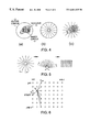
FIG. 3 depicts the method of converging squares for nucleus identification;
FIG. 4 shows the process of discretising the search space;
FIG. 5 shows unwrapping of the search space to form a trellis;
FIG. 6 shows the operation of the search algorithm;
FIG. 7 shows the process of initialisation correction;
FIG. 8 shows a plot of segmentation accuracy versus regularisation parameter; and
FIG. 9 shows a difficult segmentation.
DETAILED DESCRIPTION OF THE PREFFERED EMBODIMENT
Referring to FIG. 1 there is shown a flow chart of the process for cell nuclei segmentation. The method is described with reference to the preferred embodiment of identifying and segmenting cell nuclei in a pap smear slide. It will be appreciated that the steps may be generalised to apply to any segmentation task where very high confidence levels are required.
Scenes from a cervical smear slide are captured at low resolution using a CCD camera mounted upon a microscope. Locations of the cells within the image are identified using any suitable technique. One technique is to use the ‘water immersion algorithm’ described by Bamford and Lovell [A water immersion algoritm for cytological image segmentation; APRS Image Segmentation Workshop, University of Technology Sydney, Sydney, December 1996, 75-79]. An example of a scene segmented using the water algorithm is shown in FIG. 2.
Once the cells have been identified a high resolution image is taken of each cell for further processing. The image is suitably a gray scale image having 256 gray levels with black being level 0 and white being level 255.
In general terms, the invention requires the identification of a region of interest within the image. The shape, size and location of this region is influenced by the high-level knowledge of the required image object and sets up the image search space of the active contour. The task then is to consider every possible closed contour within the search space to identify the object boundary. In order to give an upper limit to the number of possible contour configurations, the search space is discretised into an array of points. Each contour that lies upon the points of the search space is then considered and an associated cost is calculated which represents a balance of its smoothness, how much it lies upon pixels with high image gradient or some other function depending upon application. The single contour of least cost at the completion of the algorithm becomes the chosen solution.
Now looking at the invention in detail, the first step is to identify a small region that can be confidently considered to be within a cell nucleus and preferably the centroid of the nucleus. One algorithm suitable for this step is the converging squares algorithm. The technique commences with a square image that is known to contain a cell nucleus. Four squares are set up inside the image with each square being one row and column narrower than the original image. If the original image was 256×256 pixels, each square will be 255×255 pixels. This step is depicted in FIG. 3a.
All of the pixels that lie within each square are summed to produce four numbers. The square with the lowest number contains the darkest area of the image. Further processing continues on the darkest square in similar fashion, so the next four squares will be 254×254 pixels, and so on. The process is continued until the squares reach the smallest possible size of a nucleus or until the squares reduce to a single pixel. The final square will be the darkest area in the original image and can be confidently considered to be within a cell nucleus.
In the preferred method, the process is terminated when the squares reach the approximate size of a cell nucleus. The centroid of the square is a good approximation of the centroid of the nucleus. The complete process is depicted in FIG. 3. Other processes for locating a centroid of a nucleus could be used, for example simple gray scale thresholding.
Cell nuclei are generally elliptical and of random orientation. It is therefore appropriate to select two circles as the inner and outer contours to establish a search space to locate the nucleus boundary. The establishment of the search space is shown in FIG. 4. The inner circle 1 is centred on the selected centroid from the previous step. An outer circle 2 is established concentric with the inner circle 1 and at a sufficient distance to surround the nucleus 3. An appropriate separation between the inner and outer circle will be evident from a knowledge of the problem.
The space between the circles is discretised into N radii joining the inner and outer circles. Each radii is then divided into M points. The space is therefore discretised into N×M points, as shown in FIG. 4.
The establishment of the search space is an entirely arbitrary process. The inner and outer boundaries to the search space need not be ellipses, nor need they be concentric. However, it is convenient to follow this standard for further explanation.
The search space can be conceptually unwrapped at some point to form a trellis, as shown in FIG. 5. Once the trellis is formed, a search algorithm, such as a Viterbi algorithm, can be used to find the path through the trellis that has the least cost. The path through the trellis is evaluated column by column, as shown in FIG. 6. The cost is calculated for each path to the following stage by
S ii+1, νi)=min[S i−1i, νi−1)+λE inti−1, νi, νi+1)+(1−λ)E exti)]
where E int = ( v i + 1 - 2 v i + v i - 1 v i + 1 - v i - 1 ) 2
Figure US06681035-20040120-M00001
is a measure of smoothness
Eexti) is the gray scale gradient at vi.
The regularisation parameter, λ can take any value between 0 and 1. The parameter balances the smoothing effect of the internal energy, Eint, with the effect of the image energy, Eext. A low value of λ will cause the points of greatest gradient to be chosen along each radius. This can cause a very irregular path to be selected. A high value of λ will cause the algorithm to ignore the gradient information and the resulting contour will form the smoothest shape allowable by the search space (a circle). The value of λ is chosen to lie between the two extremes, the exact value depends upon the nature of the feature being segmented.
For the reasons explained below, λ is chosen as 0.7 or 0.8. As the curvature term requires three points for calculation, the cost equation becomes a function of vi and vi+1, with vi−1 fixed as a node producing the path of least cost from the previous stage. When the final stage is reached, the node which has the minimum cost is retained and the path travelled from this node to the starting node is retraced, producing the minimum cost contour.
At each stage only the lowest cost node is kept. Thus only N, rather than MN, nodes need be retained. It will be appreciated that this process finds the minimal open contour through the search space because there are no constraints on the start or end points. It is possible to evaluate the M2 combinations of end points to find the minimum energy nodes to close the path. Alternatively, two adjacent nodes in the middle of the open contour can be selected as the start and end points for a second search.
Once the closed minimum energy contour has been established the trellis is conceptually re-wrapped and the contour becomes the nucleus boundary. In many applications the above method will result in segmentation of nuclei with a high degree of confidence.
In some cases, the initial point selected by the contracting squares technique will lie close to the boundary of the nucleus. This can result in the calculated boundary being inaccurate in the locality of the starting point. This problem is depicted in FIG. 7a. An initialisation correction method can ameliorate the problem. The method is included as a step in the flow chart of FIG. 1. After the nucleus boundary has been evaluated the centroid of the evaluated contour is determined. This can be done from the open contour thereby avoiding the extra step of closing the contour.
The inner and outer circles are repositioned to the centroid and the contour is re-evaluated using the same value for λ. The centroid of the re-evaluated contour is determined. If the contour is the same within a given limit it indicates that the correct nucleus boundary has been found. To a first approximation, the boundary can be assumed to be correct if the centroid has not moved. If the boundary (or the centroid does move), the process is repeated until a contour is found in which the centroid does not move.
The above process is based upon a determination of the gray scale gradient at each pixel. Improved results can be achieved in some situations if a directional gradient is used. The gradient is evaluated along each radius in the direction towards the centre of the nucleus. This results in only the light to dark transitions attracting the contour and avoids the contour being attracted by nearby dark objects.
As mentioned above, a suitable selection of λ is necessary for high confidence nuclei boundaries to be determined. FIG. 8 shows the percentage of successful segmentations as a function of λ in the Viterbi algorithm. The sensitivity of the algorithm to the value of λ is shown in FIG. 9 for a difficult nucleus image having a dark patch of cytoplasm adjacent the nucleus.
The chart in FIG. 8 shows that the performance of the Viterbi algorithm varies monotonically for 0<λ<0.8 and the solutions for λ1 are a strict subset of the solutions for λ2, where λ12.
It is therefore evident that similarity between contours segmented with a high value of λ, say 0.7, and a low value of λ, say 0.1, indicates a high level of confidence in the segmentation. The definition of similarity may be somewhat arbitrary, but for the purposes of the example an average deviation of 2.3 pixels or less is considered to be similar. Comparison of the segmentation at high λ and a series of low λ provides a system for grading nuclei images according to the difficulty of segmentation.
The following table shows one example of a classification system using the above criteria. The table shows the percentage of the total number of images that are accepted using a low λ value of 0.0 for level 0, 0.1 for level 1, and so on. Also shown in the table is the number of known failures (visual inspection) that occur at each level.
Level 0 1 2 3 4 5 6
% images 54.65 73.12 80.53 85.93 90.07 92.98 95.83
Failures 0 0 1 3 9 14 19
It can be seen from the table that at an acceptance level of 1, 73.12% of the images would be accepted with a 100% confidence of no failures. The confidence level can be chosen to suit the application. In the primary example of pap smear analysis, a confidence level of 1 will be chosen as false positives cannot be tolerated.
The above method has been tested for automatic segmentation of Papanicolau stained cervical cell nuclei. Using the active contour method with the Viterbi search algorithm a success rate of 99.64% correct segmentations on a data set of 20,130 images. The success rate was increased to 100% by rejecting 26.88% of the images. The images could then be confidently used for further analysis, such as detection of cervical cancer.

Claims (15)

What is claimed is:
1. An improved method of cell nucleus segmentation from an image containing a cell nucleus including the steps of:
(i) constructing a search space in the image;
(ii) searching the search space with a Viterbi algorithm using a first predetermined value of λ to find a boundary of the nucleus;
(iii) determining a centroid within the boundary;
(iv) constructing an updated search space in the image centred on the centroid;
(v) searching the updated search space with the Viterbi algorithm using the first predetermined value of λ to find an updated boundary of the nucleus;
(vi) comparing the update boundary with the boundary;
(vii) repeating steps (iii) to (vi) until the boundary and the update boundary are substantially the same;
(viii) storing an image of the cell nucleus and the found boundary when the update boundary is the same as the boundary;
(ix) searching the search space with a Viterbi algorithm using a second predetermined value of λ to find an update boundary of the nucleus; and
(x) rejecting the nucleus if the update boundary differs from the boundary by greater than a predetermined amount.
2. The method of claim 1 wherein the search space is constructed from an inner and outer curve, said inner curve being substantially within the nucleus and said outer curve being substantially outside the nucleus.
3. The method of claim 1 wherein the first λ value is greater than 0.5 but less than 0.9.
4. The method of claim 1 wherein the first λ value is in the range 0.7 to 0.8.
5. The method of claim 1 wherein the second λ value is greater than 0.0 but less than 0.5.
6. The method of claim 1 wherein second first λ value is in the range 0.0 to 0.1.
7. A method of cell nuclei segmentation including the steps of:
(i) locating cell nuclei in a sample;
(ii) initialising the segmentation method by determining a contour for a selected cell nucleus;
(iii) conducting a first segmentation step employing a Viterbi algorithm with a first predetermined value of λ to obtain a first nucleus boundary;
(iv) conducting a second segmentation step employing a Viterbi algorithm with a second predetermined value of λ to obtain a second nucleus boundary;
(v) comparing the first and second nucleus boundary; and
(vi) storing an image of the cell nucleus if the first boundary and the second boundary are substantially the same within a predetermined limit.
8. The method of claim 7 wherein the initialisation step further includes the steps of:
utilising the Viterbi algorithm at a predetermined value of λ to obtain a first result for the nucleus contour and reevaluating the centroid of the nucleus;
recalculating the Viterbi algorithm with the same value of λ to obtain a further centroid of the nucleus;
comparing the first and further centroids; and
storing a high resolution image of the nucleus if the centroids are substantially the same within predetermined limits.
9. An improved method of cell nucleus segmentation from an image containing a cell nucleus including the steps of:
(i) constructing a search space in the image;
(ii) searching the search space with a Viterbi algorithm using a first predetermined value of λ to find a boundary of the nucleus;
(iii) determining a centroid within the boundary;
(iv) constructing an updated search space in the image centred on the centroid;
(v) searching the updated search space with the Viterbi algorithm using the first predetermined value of λ to find an updated boundary of the nucleus;
(vi) comparing the updated boundary with the boundary;
(vii) repeating steps (iii) to (vi) until the boundary and the update boundary are substantially the same; and
(viii) storing an image of the cell nucleus and the found boundary when the update boundary is the same as the boundary.
10. The method of claim 9 wherein the step of constructing a search space includes the further steps of:
locating a region of at least one pixel within the nucleus;
constructing the search space between an inner and an outer curve centred on the region.
11. The method of claim 10 wherein the step of locating a region employs a method of converging squares.
12. The method of claim 10 wherein the region is approximately the minimum size of a nucleus in the image.
13. The method of claim 9 wherein the curves are ellipses.
14. The method of claim 9 including the further steps of:
searching the search space with a Viterbi algorithm using a second predetermined value of λ to find an update boundary of the nucleus; and
rejecting the nucleus if the update boundary differs from the boundary by greater than a predetermined amount.
15. The method of claim 14 wherein the second predetermined value of λ is less than the first predetermined value of λ.
US09/647,269 1998-04-03 1999-03-30 Method of unsupervised cell nuclei segmentation Expired - Fee Related US6681035B1 (en)

Applications Claiming Priority (3)

Application Number Priority Date Filing Date Title
AUPP2786 1998-04-03
AUPP2786A AUPP278698A0 (en) 1998-04-03 1998-04-03 Method of cell nuclei segmentation
PCT/AU1999/000231 WO1999052074A1 (en) 1998-04-03 1999-03-30 Method of unsupervised cell nuclei segmentation

Publications (1)

Publication Number Publication Date
US6681035B1 true US6681035B1 (en) 2004-01-20

Family

ID=3807047

Family Applications (1)

Application Number Title Priority Date Filing Date
US09/647,269 Expired - Fee Related US6681035B1 (en) 1998-04-03 1999-03-30 Method of unsupervised cell nuclei segmentation

Country Status (7)

Country Link
US (1) US6681035B1 (en)
EP (1) EP1088285B1 (en)
JP (1) JP2002528782A (en)
AU (1) AUPP278698A0 (en)
CA (1) CA2333614C (en)
DE (1) DE69922648D1 (en)
WO (1) WO1999052074A1 (en)

Cited By (13)

* Cited by examiner, † Cited by third party
Publication number Priority date Publication date Assignee Title
US20050269315A1 (en) * 2004-06-07 2005-12-08 Franco Visinoni Automatic, microwave assisted tissue histoprocessor
US20060204953A1 (en) * 2005-02-22 2006-09-14 Nikolai Ptitsyn Method and apparatus for automated analysis of biological specimen
US20070081724A1 (en) * 2005-01-31 2007-04-12 Tiantian Zhang System and method for validating an image segmentation algorithm
US20070109874A1 (en) * 2005-11-12 2007-05-17 General Electric Company Time-lapse cell cycle analysis of unstained nuclei
US20090298172A1 (en) * 2008-05-28 2009-12-03 Steven Paul Wheeler Histological specimen treatment apparatus and method
CN102103197A (en) * 2009-12-16 2011-06-22 西门子公司 Method to determine a background phase in phase image data sets
CN103489188A (en) * 2013-09-23 2014-01-01 华南理工大学 Positioning method for abnormal cell nucleus in cervix uteri LCT image
US9567651B2 (en) 2011-09-13 2017-02-14 Koninklijke Philips N.V. System and method for the detection of abnormalities in a biological sample
CN108665463A (en) * 2018-03-30 2018-10-16 哈尔滨理工大学 A kind of cervical cell image partition method generating network based on confrontation type
US10360434B2 (en) * 2016-07-25 2019-07-23 Case Western Reserve University Multi-pass adaptive voting for nuclei detection in histopathological images
US10365189B2 (en) 2015-05-07 2019-07-30 Steven Wheeler Histological specimen treatment
US10783627B2 (en) 2017-03-03 2020-09-22 Case Western Reserve University Predicting cancer recurrence using local co-occurrence of cell morphology (LoCoM)
US11232567B2 (en) 2016-06-03 2022-01-25 Koninklijke Philips N.V. Biological object detection

Families Citing this family (5)

* Cited by examiner, † Cited by third party
Publication number Priority date Publication date Assignee Title
US7327867B2 (en) * 2000-12-01 2008-02-05 Japan Science And Technology Corporation Methods for recognizing nucleus area and constructing cell lineage
AUPR647801A0 (en) 2001-07-19 2001-08-09 Cea Technologies Inc. Chromatin segmentation
US6658143B2 (en) * 2002-04-29 2003-12-02 Amersham Biosciences Corp. Ray-based image analysis for biological specimens
ES2425241T3 (en) * 2006-05-18 2013-10-14 Elekta Ltd. Methods and systems of segmentation using reparametrization of limits
DE102007013971B4 (en) * 2007-03-23 2011-07-14 Fraunhofer-Gesellschaft zur Förderung der angewandten Forschung e.V., 80686 Method and device for determining a cell contour of a cell

Citations (7)

* Cited by examiner, † Cited by third party
Publication number Priority date Publication date Assignee Title
US4175860A (en) * 1977-05-31 1979-11-27 Rush-Presbyterian-St. Luke's Medical Center Dual resolution method and apparatus for use in automated classification of pap smear and other samples
US4523278A (en) 1979-02-01 1985-06-11 Prof. Dr.-Ing. Werner H. Bloss Method of automatic detection of cells and determination of cell features from cytological smear preparations
WO1993018483A1 (en) 1992-03-02 1993-09-16 American Telephone And Telegraph Company Method and apparatus for image recognition
US5745601A (en) * 1995-07-31 1998-04-28 Neopath, Inc. Robustness of classification measurement apparatus and method
US5986771A (en) * 1997-09-19 1999-11-16 Eastman Kodak Company Method for computing a control signal for combining digital images
US5987158A (en) * 1994-09-20 1999-11-16 Neopath, Inc. Apparatus for automated identification of thick cell groupings on a biological specimen
US6581011B1 (en) * 1999-06-23 2003-06-17 Tissueinformatics, Inc. Online database that includes indices representative of a tissue population

Patent Citations (7)

* Cited by examiner, † Cited by third party
Publication number Priority date Publication date Assignee Title
US4175860A (en) * 1977-05-31 1979-11-27 Rush-Presbyterian-St. Luke's Medical Center Dual resolution method and apparatus for use in automated classification of pap smear and other samples
US4523278A (en) 1979-02-01 1985-06-11 Prof. Dr.-Ing. Werner H. Bloss Method of automatic detection of cells and determination of cell features from cytological smear preparations
WO1993018483A1 (en) 1992-03-02 1993-09-16 American Telephone And Telegraph Company Method and apparatus for image recognition
US5987158A (en) * 1994-09-20 1999-11-16 Neopath, Inc. Apparatus for automated identification of thick cell groupings on a biological specimen
US5745601A (en) * 1995-07-31 1998-04-28 Neopath, Inc. Robustness of classification measurement apparatus and method
US5986771A (en) * 1997-09-19 1999-11-16 Eastman Kodak Company Method for computing a control signal for combining digital images
US6581011B1 (en) * 1999-06-23 2003-06-17 Tissueinformatics, Inc. Online database that includes indices representative of a tissue population

Non-Patent Citations (15)

* Cited by examiner, † Cited by third party
Title
"A Robust Snake Implementation; A Dual Active Contour" by Steve R. Gunn and Mark S. Nixon Published in IEEE Transactions on Pattern Analysis and Machine Intelligence, vol. 19, No. 1, Jan. 1997.
"A Water Immersion Algorithm for Cytological Image Segmentation" by Pascal Bamford and Brian Lovell, Presented to the Cooperative Research Center for Sensor Signal and Information Processing, Department of Electrical and Computer Engineering, University of Queensland, Australia.
"The Measuring of Cell Features" by Ewert Bengtsson, Ph.D. Published in Analytical and Quantitative Cytology and Histology, vol. 9, No. 3, Jun. 1987.
Bamford et al., "A Methodology for Quality Control in Cell Nucleus Segmentation," Dec. 7-8, 1999, Digital Image Computing: Technique Application, pp. 1-5.* *
Bamford et al., "A Two-Stage Scene Segmentation Scheme for the Automatic Collection of Cervical Cell Images," 1997, IEEE TENCON, pp. 683-686.* *
Bamford et al., "Bayesian Analysis of Cell Nucleus Segmentation by a Viterbi Search Based Active Contour," Aug. 16-20, 1998, IEEE, vol. 1, pp. 133-135.* *
Bamford et al., "Improving the Robustness Cell Nucleus Segmentation," Sep. 14-17, 1998, Proc. 9th British Machine Vision Conf. pp. 518-524.* *
Herrera-Espineira et al., "Automated Segmentation of Cell Nuclei in Fine needle Aspirates of the Breast", Analytical and Quantitative Cytology and Histology, vol. 20, No. 1, Feb. 1998, pp. 30-35.
Krivanek et al., "Ovarian Ultrasound Image Analysis: Follicle Segementation," 1998, IEEE, pp. 935-944.* *
Lee et al., "A Fast and Robust Approach for Automated Segmentation of Breast Cancel Nuclei," Oct. 25-27, 1999, International Conference on Computer Graphics and Imaging, pp. 1-9.* *
MacAulay et al., "An Edge Relocation Segmentation Algorithm", Analytical and Quantitative Cytology and Histology, MacAulay and Palcic, vol. 12, No. 30, Jun. 1990, pp. 165-171.
MacAulay, et al., "A Comparison of Some Quick and Simple Threshold Selection Methods for Stained Cells", vol. 10, No. 2, Apr. 1988, vol. 10, No. 2, Apr. 1988; pp. 134-138.
Nasrabadi et al., "A Stereo Vision Technique Using Curve-Segments and Relaxation Matching," 1988, IEEE, pp. 149-151.* *
Schupp et al., "Mathematical Morphology and Active Contours for Object Extraction and Localization in Medical Images," pp. 317-321.* *
Xiao et al., An Algorithm for Automatic Tracking of Nuclear Boundaries, Analytical and Quantitative cytology and Histology, vol. 16, No. 4, Aug. 1994, pp. 240-246.

Cited By (22)

* Cited by examiner, † Cited by third party
Publication number Priority date Publication date Assignee Title
US7075045B2 (en) * 2004-06-07 2006-07-11 Milestone S.R.L. Automatic, microwave assisted tissue histoprocessor
US20050269315A1 (en) * 2004-06-07 2005-12-08 Franco Visinoni Automatic, microwave assisted tissue histoprocessor
US7907756B2 (en) * 2005-01-31 2011-03-15 Siemens Medical Solutions Usa, Inc. System and method for validating an image segmentation algorithm
US20070081724A1 (en) * 2005-01-31 2007-04-12 Tiantian Zhang System and method for validating an image segmentation algorithm
US20060204953A1 (en) * 2005-02-22 2006-09-14 Nikolai Ptitsyn Method and apparatus for automated analysis of biological specimen
US7817841B2 (en) 2005-11-12 2010-10-19 General Electric Company Time-lapse cell cycle analysis of unstained nuclei
US20070109874A1 (en) * 2005-11-12 2007-05-17 General Electric Company Time-lapse cell cycle analysis of unstained nuclei
US20090298172A1 (en) * 2008-05-28 2009-12-03 Steven Paul Wheeler Histological specimen treatment apparatus and method
US9366605B2 (en) 2008-05-28 2016-06-14 Steven Paul Wheeler Histological specimen treatment apparatus and method
CN102103197A (en) * 2009-12-16 2011-06-22 西门子公司 Method to determine a background phase in phase image data sets
US20110176714A1 (en) * 2009-12-16 2011-07-21 Rares Salomir Method to determine a background phase in phase image data sets
US8630471B2 (en) * 2009-12-16 2014-01-14 Siemens Aktiengesellschaft Method to determine a background phase in phase image data sets
CN102103197B (en) * 2009-12-16 2015-08-26 西门子公司 The defining method of background phase in phase image data group
US9567651B2 (en) 2011-09-13 2017-02-14 Koninklijke Philips N.V. System and method for the detection of abnormalities in a biological sample
CN103489188A (en) * 2013-09-23 2014-01-01 华南理工大学 Positioning method for abnormal cell nucleus in cervix uteri LCT image
US10365189B2 (en) 2015-05-07 2019-07-30 Steven Wheeler Histological specimen treatment
US10641688B2 (en) 2015-05-07 2020-05-05 Steven Wheeler Histological specimen treatment
US11885723B2 (en) 2015-05-07 2024-01-30 Steven Wheeler Histological specimen treatment
US11232567B2 (en) 2016-06-03 2022-01-25 Koninklijke Philips N.V. Biological object detection
US10360434B2 (en) * 2016-07-25 2019-07-23 Case Western Reserve University Multi-pass adaptive voting for nuclei detection in histopathological images
US10783627B2 (en) 2017-03-03 2020-09-22 Case Western Reserve University Predicting cancer recurrence using local co-occurrence of cell morphology (LoCoM)
CN108665463A (en) * 2018-03-30 2018-10-16 哈尔滨理工大学 A kind of cervical cell image partition method generating network based on confrontation type

Also Published As

Publication number Publication date
CA2333614A1 (en) 1999-10-14
EP1088285A1 (en) 2001-04-04
AUPP278698A0 (en) 1998-04-30
DE69922648D1 (en) 2005-01-20
JP2002528782A (en) 2002-09-03
EP1088285A4 (en) 2002-02-13
WO1999052074A1 (en) 1999-10-14
EP1088285B1 (en) 2004-12-15
CA2333614C (en) 2005-10-18

Similar Documents

Publication Publication Date Title
US6681035B1 (en) Method of unsupervised cell nuclei segmentation
Piccioli et al. Robust method for road sign detection and recognition
Bamford et al. Unsupervised cell nucleus segmentation with active contours
CN110148162A (en) A kind of heterologous image matching method based on composition operators
CN112766136A (en) Space parking space detection method based on deep learning
CN111105346A (en) Full-scanning microscopic image splicing method based on peak value search and gray template registration
Horzyk et al. YOLOv3 precision improvement by the weighted centers of confidence selection
GB2329014A (en) Automated identification of tubercle bacilli
CN111695373A (en) Zebra crossing positioning method, system, medium and device
AU748081B2 (en) Method of unsupervised cell nuclei segmentation
Wang et al. Big Map R-CNN for object detection in large-scale remote sensing images.
CN114998980B (en) Iris detection method and device, electronic equipment and storage medium
Wilkinson Automated and manual segmentation techniques in image analysis of microbes
Jain et al. Number plate detection using drone surveillance
CN114927236A (en) Detection method and system for multiple target images
Hassoubah et al. Saudi riyal coin detection and recognition
CN111242927B (en) Sine welding detection method based on deep learning
Bamford Segmentation of cell images with application to cervical cancer screening
Peters II Image complexity measurement for predicting target detectability
Straub et al. Concepts for internal and external evaluation of automatically delineated tree tops
Bamford et al. Improving the Robustness of Cell Nucleus Segmentation.
CN113076909B (en) Automatic cell detection method
Xu et al. Automated nuclear segmentation in skin histopathological images using multi-scale radial line scanning
CN117612164B (en) Cell division equilibrium degree detection method based on double edge detection
CN112348105B (en) Unmanned aerial vehicle image matching optimization method

Legal Events

Date Code Title Description
AS Assignment

Owner name: QUEENSLAND, UNIVERSITY OF, AUSTRALIA

Free format text: ASSIGNMENT OF ASSIGNORS INTEREST;ASSIGNOR:LOVELL, BRIAN;REEL/FRAME:014673/0923

Effective date: 19990331

AS Assignment

Owner name: COOPERATIVE RESEARCH CENTRE FOR SENSOR SIGNAL AND

Free format text: ASSIGNMENT OF ASSIGNORS INTEREST;ASSIGNOR:QUEENSLAND, THE UNIVERSITY OF;REEL/FRAME:015533/0371

Effective date: 19990331

Owner name: COOPERATIVE RESEARCH CENTRE FOR SENSOR SIGNAL AND

Free format text: ASSIGNMENT OF ASSIGNORS INTEREST;ASSIGNOR:BAMFORD, PASCAL CHRISTOPHER;REEL/FRAME:015533/0639

Effective date: 19990331

FPAY Fee payment

Year of fee payment: 4

REMI Maintenance fee reminder mailed
LAPS Lapse for failure to pay maintenance fees
STCH Information on status: patent discontinuation

Free format text: PATENT EXPIRED DUE TO NONPAYMENT OF MAINTENANCE FEES UNDER 37 CFR 1.362

FP Lapsed due to failure to pay maintenance fee

Effective date: 20120120