US20020028418A1 - System and method for 3-D digital reconstruction of an oral cavity from a sequence of 2-D images - Google Patents
System and method for 3-D digital reconstruction of an oral cavity from a sequence of 2-D images Download PDFInfo
- Publication number
- US20020028418A1 US20020028418A1 US09/842,587 US84258701A US2002028418A1 US 20020028418 A1 US20020028418 A1 US 20020028418A1 US 84258701 A US84258701 A US 84258701A US 2002028418 A1 US2002028418 A1 US 2002028418A1
- Authority
- US
- United States
- Prior art keywords
- data
- dimensional
- shape
- generating
- shading
- Prior art date
- Legal status (The legal status is an assumption and is not a legal conclusion. Google has not performed a legal analysis and makes no representation as to the accuracy of the status listed.)
- Granted
Links
Images


Classifications
-
- G—PHYSICS
- G06—COMPUTING; CALCULATING OR COUNTING
- G06T—IMAGE DATA PROCESSING OR GENERATION, IN GENERAL
- G06T17/00—Three dimensional [3D] modelling, e.g. data description of 3D objects
-
- A—HUMAN NECESSITIES
- A61—MEDICAL OR VETERINARY SCIENCE; HYGIENE
- A61C—DENTISTRY; APPARATUS OR METHODS FOR ORAL OR DENTAL HYGIENE
- A61C9/00—Impression cups, i.e. impression trays; Impression methods
- A61C9/004—Means or methods for taking digitized impressions
- A61C9/0046—Data acquisition means or methods
- A61C9/0053—Optical means or methods, e.g. scanning the teeth by a laser or light beam
-
- A—HUMAN NECESSITIES
- A61—MEDICAL OR VETERINARY SCIENCE; HYGIENE
- A61C—DENTISTRY; APPARATUS OR METHODS FOR ORAL OR DENTAL HYGIENE
- A61C9/00—Impression cups, i.e. impression trays; Impression methods
- A61C9/004—Means or methods for taking digitized impressions
Landscapes
- Physics & Mathematics (AREA)
- Engineering & Computer Science (AREA)
- Health & Medical Sciences (AREA)
- Software Systems (AREA)
- Computer Graphics (AREA)
- General Physics & Mathematics (AREA)
- Theoretical Computer Science (AREA)
- Geometry (AREA)
- Dentistry (AREA)
- Oral & Maxillofacial Surgery (AREA)
- Optics & Photonics (AREA)
- Epidemiology (AREA)
- Life Sciences & Earth Sciences (AREA)
- Animal Behavior & Ethology (AREA)
- General Health & Medical Sciences (AREA)
- Public Health (AREA)
- Veterinary Medicine (AREA)
- Apparatus For Radiation Diagnosis (AREA)
- Dental Tools And Instruments Or Auxiliary Dental Instruments (AREA)
Abstract
Description
- This application claims the benefit of U.S. Provisional Application Serial No. 60/199,913, filed Apr. 26, 2000 under 35 U.S.C. 119(e).
- This invention relates generally to digital dental imaging, and more particularly to three-dimensional dental imaging.
- Dentistry requires accurate 3-D representation of the teeth and jaw for diagnostic and treatment purposes. For example, orthodontic treatment involves the application, over time, of force systems to teeth to correct malocclusion. In order to evaluate tooth movement progress, the orthodontist monitors this movement by means of visual inspection, intra-oral measurements, fabrication of casts, photographs, and radiographs; this process is both costly and time consuming. Moreover, repeated acquisition of radiographs may result in untoward effects. Obtaining a cast of the jaw is a complex operation for the dentist, an unpleasant experience for the patient, and also may not provide all the necessary details of the jaw.
- Oral and maxillofacial radiology provides the dentist with abundant 3-D information of the jaw. Current and evolving methods include computed tomography (CT), tomosynthesis, tuned-aperture CT (TACT), and localized, or “cone-beam,” computed tomography. While oral and maxillofacial radiology is now widely accepted as a routine technique for dental examinations, the equipment is rather expensive and the resolution is frequently too low for 3-D modeling of dental structures. Furthermore, the radiation dose required to enhance both contrast and spatial resolution can be unacceptably high.
- Much effort has been focused recently on computerized diagnosis in dentistry. One solution is an expert system where cephalometric measurements are acquired manually from the analysis of radiographs and plaster models. Another solution provides a computer-vision technique for the acquisition of jaw data from inexpensive dental wafers, which is capable of obtaining imprints of the teeth. Conventional 3-D systems for dental applications commonly rely on obtaining an intermediate solid model of the jaw (cast or teeth imprints) and then capturing the 3-D information from that model. User interaction is needed in such systems to determine the 3-D coordinates of fiducial reference points on a dental cast. Other systems that measure the 3-D coordinates have been developed using either mechanical contact or a traveling light principle. Yet another conventional solution includes a range scanner based on white light to reconstruct the cast. The scanner used the subtractive light principle to create very thin shadow profiles on the cast.
- For the reasons stated above, and for other reasons stated below which will become apparent to those skilled in the art upon reading and understanding the present specification, there is a general need to replace conventional approaches in diagnosis, treatment planning, surgical simulation and prosthetic replacements. More specifically, there is a need in the art for three-dimensional (3-D) dental imagery not using expensive, low-resolution and potentially harmful radiography, intermediate physical casts. There is also a need for fabricating dental casts in a manner that does not require time consuming and non-renewable direct application of material to the dental surfaces. Moreover, there is a need for a data acquisition system that obtains sequences of calibrated video images, with respect to a common reference in 3-D space, of the upper and/or lower jaw using an intraoral cavity camera. There is also a need for methods of accurate 3-D reconstruction of the upper and/or lower jaw from the acquired sequence of intraoral cavity images. There is a further need for a shape-from-shading process that incorporates the parameters of the intraoral cavity camera. There is yet another need for a robust process for the fusion of data acquired from multiple views of the intraoral cavity camera. There is still another need for the implementation of a fast an accurate 3-D registration. There is still yet another need for specific object segmentation and recognition of individual tooth information for further analysis and simulations. There is still yet a further need to enable study and simulation of tooth movement based on finite element and deformable model methods.
- The above-mentioned shortcomings, disadvantages and problems are addressed by the present invention, which will be understood by reading and studying the following specification.
- The present invention provides a system for dentistry and orthodontics to replace traditional approaches in diagnosis, treatment planning, surgical simulation, and prosthetic replacements. The present invention provides a data acquisition system that obtains sequences of calibrated video images, with respect to a common reference in three-dimensional (3-D) space, of an upper/lower jaw using a small intra-oral camera. The present invention also provides methods for 3-D reconstruction from the acquired sequence of intra-oral images. The present invention further provides an algorithm for shape from shading that incorporates camera parameters. The present invention additionally provides a process for the fusion of data acquired from multiple views, including the implementation of an accurate and fast 3-D data registration. The present invention also provides an object segmentation and recognition system to separate and recognize individual 3-D tooth information for further analysis and simulations. The present invention in addition provides methods to enable study and simulation of tooth movement based on the finite element method and deformable model approaches. In varying embodiment, the present invention is implemented in various dental practices including implants, tooth alignment, craniofacial surgery, teledentistry, dental education and training, and the analysis and simulation of dental operations including tooth alignment, implant planing, restoration, and measurement of distances and orientation of teeth with respect to each other.
- In one aspect of the present invention, a method includes receiving a plurality of two-dimensional images of an oral cavity, and generating at least one three-dimensional image of the oral cavity from the plurality of two-dimensional images. In another aspect of the present invention, a computerized apparatus includes a digitizer providing five degrees of freedom, the digitizer having an arm, a charge coupled device camera, rigidly mounted on the arm of the digitizer, and a computer, operably coupled to the digitizer and the camera that receives coordinate measurements from the digitizer and a plurality of two-dimensional images from the camera and generates a digital three-dimensional model from the coordinate measurements and from the plurality of two-dimensional images.
- The present invention discloses the generation of a 3-D model of the jaw, not from a cast, but from the actual human jaw. The present invention disclose systems and method of data acquisition performed directly on a jaw using a small off the shelf charge coupled device camera in which the acquisition time is relatively short and is less discomforting to the patient compared to current practices. The acquired digital model is optionally stored with the patient data and optionally retrieved on demand. The model optionally is transmitted over a communication network to different remote practitioners for further assistance in diagnosis and treatment planning. Dental measurements and virtual restoration are optionally performed and analyzed from the digital model.
- The present invention describes systems, clients, servers, methods, and computer-readable media of varying scope. In addition to the aspects and advantages of the present invention described in this summary, further aspects and advantages of the invention will become apparent by reference to the drawings and by reading the detailed description that follows.
- FIG. 1 is a block diagram of the hardware and operating environment in which different embodiments of the invention can be practiced.
- FIG. 2 is a diagram illustrating a system-level overview of an embodiment of the invention.
- FIG. 3 is a flowchart of a method for dental imaging, according to an embodiment of the invention.
- FIG. 4 is a flowchart of a method for generating a 3-D image of the oral cavity from the plurality of 2-D images, according to an embodiment of the invention.
- FIG. 5 is a flowchart of a method for generating shape-from-shading data, according to an embodiment of the invention.
- FIG. 6 is a diagram of surface triangulation, according to an embodiment of the invention.
- FIG. 7 is a flowchart of a method for fusing the range data to the shape-from-shading data, according to an embodiment of the invention.
- FIG. 8 is a block diagram of an apparatus for generating a three-dimensional digital image of a jaw, according to an embodiment of the invention.
- FIG. 9 is a block diagram of an apparatus involved in calibrating a charge coupled device camera, according to an embodiment of the invention.
- In the following detailed description of embodiments of the invention, reference is made to the accompanying drawings that form a part hereof, and in which is shown by way of illustration specific embodiments in which the invention may be practiced. These embodiments are described in sufficient detail to enable those skilled in the art to practice the invention, and it is to be understood that other embodiments may be utilized and that logical, mechanical, electrical and other changes may be made without departing from the scope of the present invention. The following detailed description is, therefore, not to be taken in a limiting sense, and the scope of the present invention is defined only by the appended claims.
- The detailed description is divided into five sections. In the first section, the hardware and the operating environment in conjunction with which embodiments of the invention may be practiced are described. In the second section, a system level overview of the invention is presented. In the third section, methods for an embodiment of the invention are provided. In the fourth section, a particular object-oriented Internet-based implementation of the invention is described. Finally, in the fifth section, a conclusion of the detailed description is provided.
- FIG. 1 is a block diagram of the hardware and
operating environment 100 in which different embodiments of the invention can be practiced. The description of FIG. 1 provides an overview of computer hardware and a suitable computing environment in conjunction with which some embodiments of the present invention can be implemented. Embodiments of the present invention are described in terms of a computer executing computer-executable instructions. However, some embodiments of the present invention can be implemented entirely in computer hardware in which the computer-executable instructions are implemented in read-only memory. One embodiment of the invention can also be implemented in client/server computing environments where remote devices that are linked through a communications network perform tasks. Program modules can be located in both local and remote memory storage devices in a distributed computing environment. -
Computer 110 is operatively coupled todisplay device 112, pointingdevice 115, andkeyboard 116.Computer 110 includes aprocessor 118, commercially available from Intel®, Motorola®, Cyrix® and others, random-access memory (RAM) 120, read-only memory (ROM) 122, and one or moremass storage devices 124, and asystem bus 126, that operatively couples various system components including the system memory to theprocessing unit 118.Mass storage devices 124 are more specifically types of nonvolatile storage media and can include a hard disk drive, a floppy disk drive, an optical disk drive, and a tape cartridge drive. Thememory computer 110 through input devices such as apointing device 115 and akeyboard 116. Other input devices (not shown) can include a microphone, joystick, game pad, satellite dish, scanner, or the like. Theprocessor 118 executes computer programs stored on the computer-readable media. Embodiments of the present invention are not limited to any type ofcomputer 110. In varying embodiments,computer 110 comprises a PC-compatible computer, a MacOS®-compatible computer or a UNIX®-compatible computer. The construction and operation of such computers are well known within the art. - Furthermore,
computer 110 can be communicatively connected to theInternet 130 via acommunication device 128.Internet 130 connectivity is well known within the art. In one embodiment, acommunication device 128 is a modem that responds to communication drivers to connect to the Internet via what is known in the art as a “dial-up connection.” In another embodiment, acommunication device 128 is an Ethernet® or similar hardware (network) card connected to a local-area network (LAN) that itself is connected to the Internet via what is known in the art as a “direct connection” (e.g., T1 line, etc.). -
Computer 110 can be operated using at least one operating environment to provide a graphical user interface including a user-controllable pointer. Such operating environments include operating systems such as versions of the Microsoft Windows® and Apple MacOS® operating systems well-known in the art. Embodiments of the present invention are not limited to any particular operating environment, however, and the construction and use of such operating environments are well known within the art.Computer 110 can have at least one web browser application program executing within at least one operating environment, to permit users ofcomputer 110 to access intranet or Internet world-wide-web pages as addressed by Universal Resource Locator (URL) addresses. Such browser application programs include Netscape Navigator® and Microsoft Internet Explorer®. -
Display device 112 permits the display of information, including computer, video and other information, for viewing by a user of the computer. Embodiments of the present invention are not limited to anyparticular display device 112. Such display devices include cathode ray tube (CRT) displays (monitors), as well as flat panel displays such as liquid crystal displays (LCD's).Display device 112 is connected to thesystem bus 126. In addition to a monitor, computers typically include other peripheral input/output devices such as printers (not shown), speakers, pointing devices and a keyboard.Speakers Speakers system bus 126.Pointing device 115 permits the control of the screen pointer provided by the graphical user interface (GUI) of operating systems such as versions of Microsoft Windows®. Embodiments of the present invention are not limited to anyparticular pointing device 115. Such pointing devices include mouses, touch pads, trackballs, remote controls and point sticks. Finally,keyboard 116 permits entry of textual information intocomputer 110, as known within the art, and embodiments of the present invention are not limited to any particular type of keyboard. - The
computer 110 can operate in a networked environment using logical connections to one or more remote computers, such asremote computer 150. The logical connections are achieved by a communication device coupled to, or a part of, thecomputer 110. Embodiments of the present invention are not limited to a particular type of communications device. Theremote computer 150 can be another computer, a server, a router, a network PC, a client, a peer device or other common network node. The logical connections depicted in FIG. 1 include a local-area network (LAN) 151 and a wide-area network (WAN) 152. Such networking environments are commonplace in offices, enterprise-wide computer networks, intranets and the Internet. - When used in a LAN-networking environment, the
computer 110 andremote computer 150 are connected to thelocal network 151 through a network interface oradapter 153, which is one type of communications device. When used in a conventional WAN-networking environment, thecomputer 110 andremote computer 150 communicate with aWAN 152 through modems (not shown). The modem, which can be internal or external, is connected to thesystem bus 126. In a networked environment, program modules depicted relative to thecomputer 110, or portions thereof, can be stored in the remote memory storage device. - FIG. 2 is a block diagram that provides a system level overview of the operation of embodiments of the present invention. Embodiments of the invention are described as operating in a multi-processing, multi-threaded operating environment on a computer, such as
computer 110 in FIG. 1. -
System 200 includes a small intra-oral charge coupled device (CCD)camera 210 to enable the first stage ofdata acquisition 220. In one embodiment, thecamera 210 includes a built-in white light, rigidly mounted on a five-link 3-D digitizer arm. Thecamera 210 is calibrated and then placed inside the oral cavity. Thecamera 210 acquires a set of overlapping images 230 {Ij|j=1,2, . . . , J} for various portions of the jaw such that Uj=1Ij covers the entire jaw. Theimages 230 are preprocessed to reduce noise, sharpen edges, and removespecularity 240. Removingspecularity 240 is improves the accuracy of the reconstructed surfaces using a shape-from-shading (SFS)process 250. In one embodiment of removing specularity, changes in the reflection map are removed from a luminance image by calculating the logarithmic gradient of theimage 230 and thresholding at locations of abrupt chromaticity change. In another embodiment, median filtering is implemented to remove speckle noise from the images. Using aSFS process 250 that accounts for thecamera perspective projection 255, J sets of 3-D points are computed. To obtain accurate metric measurements,range data 262 is obtained using the five-link digitizer 260. The range data comprises reference points on the jaw.Fusion 265 of the range data and the SFS output provides accurate metric information that can be used later for orthodontic measurements and implant planning.System 200 also includes aregistration 270 technique that merges the resulting 3-D points to obtain a complete 3-D description of thejaw 275. The 3-D description 275 is transformed into patches of free form surfaces using atriangulation process 280. Thetriangulation process 280 enables optional development of a 3-Dsolid model 285 for visualization. Optionally, acast 290 is fabricated from the 3-D description 275 via rapid prototyping. Further optional processing of thedigital model 275 includestooth separation 295, force analysis, implant planning, andsurgical simulation 298. - The system level overview of the operation of an embodiment of the invention has been described in this section of the detailed description. While the invention is not limited to any
particular camera 210,image acquisition system 220,data fusion 265 andregistration process 270,triangulation process 280, and cast 290, for sake of clarity asimplified camera 210,image acquisition system 220,data fusion 265 andregistration process 270,triangulation process 280, and cast 290, has been described. - In the previous section, a system level overview of the operation of an embodiment of the invention was described. In this section, the particular methods of such an embodiment are described by reference to a series of flowcharts. Describing the methods by reference to a flowchart enables one skilled in the art to develop such programs, firmware, or hardware, including such instructions to carry out the methods on suitable computers (the processor of the computers executing the instructions from computer-readable media). Similarly, the methods performed by the server computer programs, firmware, or hardware are also composed of computer-executable instructions. Methods 300-500 and 700 are performed by a program executing on, or performed by firmware or hardware that is a part of, a computer, such as
computer 110 in FIG. 1 - FIG. 3 is a flowchart of a
method 300 for dental imaging, according to an embodiment of the invention. -
Method 300 includes receiving 310 a plurality of two-dimensional (2-D) images of an oral cavity. In varying embodiments, the oral cavity is a mammalian oral cavity and/or a human oral cavity. In other embodiments, the plurality of 2-D images are a plurality of 2-D optical images. - Thereafter,
method 300 includes generating 320 at least one three-dimensional (3-D) image of the oral cavity from the plurality of 2-D images. - Optionally,
method 300 also includes constructing 330 a physical cast of the oral cavity from the 3-D image. In varying embodiments, the physical cast is a plastic cast formed by a rapid prototyping machine, or a plaster cast. - Optionally,
method 300 includes storing 340 the 3-D image in association with patient information. For example, the 3-D image is stored in a database of patient records. - Optionally,
method 300 further includes modifying 350 the 3-D image in accordance with a proposed or suggested orthodontic or dental treatment of the patient. - In another embodiment of
method 300, the 2-D images that are received inaction 310, are beforehand generated from a common reference point in 3-D space. - FIG. 4 is a flowchart of a
method 400 for generating a 3-D image of the oral cavity from the plurality of 2-D images, according to an embodiment of the invention. -
Method 400 includes generating 410 shape-from-shading (SFS) data from the plurality of 2-D images using a shape-from-shading process, the shape-from-shading data comprising a first plurality of 3-D points. Generating SFS data is disclosed in further detail inmethod 500 in FIG. 5. -
Method 400 also includes generating 420 range data comprising a second plurality of 3-D points from the plurality of 2-D images using a range-data process. -
Method 400 further includes fusing 430 the range data to the shape-from-shading data, yielding fused data comprising a third plurality of 3-D points. Fusing 430 is disclosed in further detail inmethod 700 in FIG. 7. - Thereafter,
method 400 includes registering 440 the fused data, yielding registered data comprising a fourth plurality of 3-D points. - Subsequently,
method 400 includes triangulating 450 the registered data, yielding the at least one 3-D image of the oral cavity. - FIG. 5 is a flowchart of a
method 500 for generating shape-from-shading data, according to an embodiment of the invention. - SFS assumes that the surface orientation at a point M on a surface S is determined by the unit vector perpendicular to the plane tangent to S at M. Under the assumption of orthographic projections, the elemental change in the depth Z at an image point (x,y) can be expressed as

- The partial derivatives are called surface gradients (p,q). The normal to a surface patch is related to the surface gradient by n=(p,q,1). Assuming that surface patches are homogeneous and uniformly lit by distant light sources, the brightness E(x,y) seen at the image plane often depends only on the orientation of the surface. This dependence of brightness on surface orientation can be represented as a function R(·) defined on the Gaussian sphere. Thus, the SFS problem is formulated as finding a solution to the brightness equation: E(x,y)=R(p,q,L), where R(p,q,L) is the surface reflectance map and L is the illuminant direction.
-
Method 500 includes estimating 510 the direction of the illuminant from the plurality of 2-D images, in reference to camera intrinsic parameters. - In the present invention, a white light beam is built in the CCD camera, yielding a valid assumption that the illuminant direction is known. However, an assumption of orthographic projection is not adequate for the dental application because the camera is very close to the object. Conventional SFS methods using perspective projection ignore the camera extrinsic parameters, hence cannot provide metric information of the depth. In the present invention, the CCD camera is calibrated and the camera parameters are used in the SFS method to obtain a metric representation of the teeth and gum surfaces. To calibrate the camera, the relation between the 3D point M={X,Y,Z} and the corresponding image coordinates m={x,y} is written as; {right arrow over (sm)}={right arrow over (PM)} where s is a scalar, {right arrow over (m)} and {right arrow over (M)} are the extended vectors [mT1]T and [MT1]T, and P is called the camera calibration matrix. In general, P=A [R,t] where A is a matrix containing all the camera intrinsic parameters and R,t are the rotation matrix and translation vector. The matrix P has 12 elements but has only 11 degrees of freedom because it is defined up to a scale factor.
- The standard method of calibration is to use an object with known size and shape and extract the reference points from the object image. It can be shown that given N points (N>=6) in general positions, the camera can be calibrated. The perspective projection matrix P can be decomposed as [Bb] where B is a 3×3 matrix and b is a 3×1 vector such that:
TABLE 1 s{right arrow over (m)} = BM + b - or,
TABLE 2 M = B−1 (s{right arrow over (m)} − b) = f(s(x,y)) - This last equation represents a line in the 3D space corresponding to the visual ray passing through the optical center and the projected point m. By finding the scalar s, ƒ(s(x,y)) will define a unique 3D point M on the object. The surface normal at M is defined to be the cross product of the two gradient vectors

- The surface reflectance R(,) becomes a function of the scalars defined in The equation in table 1, i.e.,
TABLE 3 
-
Method 500 also includes determining 520 a solution to a brightness equation from the direction of the illuminant, yielding the shape-from-shading data comprising a first plurality of 3-D points. - The formulation of the SFS problem becomes finding the scalar s that solves the brightness equation g(s)=E(x,y)□R(s)=0. In one embodiment, the brightness equation is solved using a Taylor's series expansion and applying a Jacoby iterative method [ ]. After n iterations, for each point (x,y) in the image, s x,y n is given as follows
TABLE 4 
- where,
TABLE 5 
-
TABLE 6 
-
TABLE 7 
where v = p × q. - FIG. 6 is a diagram 600 of surface triangulation, according to an embodiment of the invention.
- Diagram 600 includes the
image plane 610 as a set of triangular patches each with anormal N 620, the dot product of thelight direction L 630 and thenormal N 620 is called the reflectance which determines the image intensity at the correspondingpixel location m 640.Image plane 610 is described as having 2-D coordinates x 650 andy 660. Whenpixel location m 640 is projected beyondoptical center 670, inlight direction L 630, the projection yields formM 680 havingpoints M 1 685 andM 2 687. The diagram is also described in relation to 3-D coordinatesX 690,Y 693, and Z 686. - In another embodiment of
method 500,method 500 is described in terms of the FIG. 6 by:TABLE 8 


p = M − M1 -
TABLE 9 
q = M − M2 -
TABLE 10 
- A unit normal N to the patch formed by M, M 1, and M2 is calculated as follows:
TABLE 11 
- A reflection function defined by the SFS is:
TABLE 12 
- Thus, the SFS brightness equation becomes:
TABLE 13 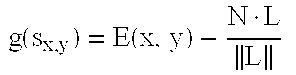
- The solution to the SFS problem is to find s x,y such that g(.) is minimized. Using Taylor expansion and Jacoby iterative methods, s(x,y) can be found by iteration as follows:
TABLE 13 
-
TABLE 15 
-
TABLE 16 
-
TABLE 17 


- The actions involved in the implementation include reading image E(x,y), light direction L, and camera parameters (B,b). Initializing s x,y=0.01. □∀x,y get p and q as shown in the equations in tables 9 and 10. Calculating N as shown in equation in table 11. Obtaining the error using the brightness equation in table 13. Estimating the new sx,y using the equations in tables 14, 15, 16, and 17. Repeating the actions of ∀, calculating and estimating, until maxx,y|g(Sx,y)|<ε where ε is a predefined positive threshold. Thereafter, recovering the surface 3-D points using equation in table 8, and constructing triangular patches as shown in FIG. 6.
- FIG. 7 is a flowchart of a
method 700 for fusing the range data to the shape-from-shading data, as in the fusingaction 430 in FIG. 4, according to an embodiment of the invention. - The most important information for reconstructing an accurate 3-D visible surface, which is missing in shape from shading, is the metric measurement. Shape from shading also suffers from the discontinuities due to highly textured surfaces and different albedo. The integration of the dense depth map obtained from SFS with sparse depth measurements obtained from a coordinate measurement machine (CMM) for the reconstruction of 3-D surfaces with accurate metric measurements has two advantages. First, the integration removes ambiguity of the 3-D visible surface discontinuities produced by shape from shading. Second, the integration complements missing metric information in the shape from shading. The integration process, as depicted in FIG. 7, includes the following stages. First, calculating 710 the error difference in the available depth measurements between the two sets of sensory data. Thereafter, approximating 720 a surface that fits this error difference. Subsequently, correcting 730 the shape from shading.
-
Method 700 includes calculating 710 the error difference in available depth measurements of the range data and the shape-from-shading data. - A multi-layer neural network is used for the surface approximation process since neural networks was shown to be more robust in terms of complexity, speed and accuracy than other computational approaches (e.g., regularization techniques). The learning algorithm applied is an extended Kalman-filter learning technique because of fast computation of weights generated by Kalman-filter. The x- and y-coordinates of the data points are the input to the network, while the error in the depth value at the point (x,y) is the desired response. The error difference between the SFS and the range measurements and their x-y coordinates are used to form the training set. The input to the network is the x-y coordinates and the output is the error difference at that coordinate.
-
Method 700 includes approximating 720 a surface the fits the error difference, yielding an approximated surface. - Once training is accomplished, the neural network provides the approximated smooth surface that contains information about the errors in the shape from shading at the locations with no range data.
-
Method 700 includes correcting 730 the shape-from-shading data from the approximated surface, yielding fused data comprising a third plurality of 3-D points. - This approximated surface is then added to the SFS. The result is the 3-D surface reconstruction that contains accurate metric information about the visible surface of the sensed 3-D object. The output of the fusion algorithm to each image is a set of 3-D points describing the teeth surfaces in this segment. To compensate for some digitizer inaccuracy in determining the camera location in space and the occasional patient movement, The present invention in some embodiments includes a 3-D registration technique to link the 3-D points of all the segments to produce one set of 3-D points describing the whole jaw surface.
- In one embodiment, methods 300-500 and 700 are implemented as a computer data signal embodied in a carrier wave, that represents a sequence of instructions which, when executed by a processor, such as
processor 118 in FIG. 1, cause the processor to perform the respective method. - In another embodiment, methods 300-500 and 700 are implemented as a computer-readable medium having computer-executable instructions to cause a computer, such as
computer 110, to perform the respective method. - Referring to FIG. 8- 9, a particular implementation of the invention is described in conjunction with the hardware and
operating environment 100, the system overview in FIG. 2 and the methods described in conjunction with FIGS. 3-5 and 7. - FIG. 8 is a block diagram of an
apparatus 800 for generating a three-dimensional digital image of a jaw, according to an embodiment of the invention. -
Apparatus 800 includes a ⅓ inch charge coupled device (CCD)color camera 810, such ascamera 210 in FIG. 2. Thecamera 810 has 768H×494V effective picture elements. In one embodiment, the camera also includes a 5.5 mm lens.Apparatus 800 also includes a 150 watt direct current regulatedwhite light source 820. Through a fiber optic bundle (not shown) that surrounds theCCD camera 810, thelight source 820 illuminates an oral cavity with a stable white light. The light intensity is manually adjustable to control a shading effect.Apparatus 800 also includes acomputer 830, such ascomputer 110 in FIG. 1, that hosts the software required for the data processing, reconstruction and visualization of the three-dimensional (3-D) jaw model, as described in methods 300-500 and 700.Computer 830 also includes a 3-D digitizer 850, capable of digitizing a working space up to 1.27 m (sphere) with a sampling rate of 1000 points/second. 3-D digitizer 850 is also shown as five-link digitizer 260 in FIG. 2. - The
CCD camera 810 is mounted on astylus 840 of the 3-D digitizer 850 and itsfocal distance 870 is adjusted such that the image will be in focus only when the stylus tip touches a tooth surface. Animage plane 860 is normal to the stylus and the stylus tip is at pixel (0,0).Apparatus 800 also includes a display 880, such asdisplay 112 in FIG. 1 to display the 3-D jaw model - The
CCD camera 810 is calibrated as shown in FIG. 9. - FIG. 9 is a block diagram of an
apparatus 900 involved in calibrating a charge coupled device camera, according to an embodiment of the invention. A coordinates measuring system is used to find thetransformation matrix T oc 950 between the optical center Moc of the camera at thecalibration time 920 and thenew location 930 while acquiring the images. This transformation matrix is used to obtain the new camera perspective projection matrix. -
Camera 910 calibration is performed once before using thecamera 910. Where thecamera 910 is stationary, thecamera 910 does not need to be re-calibrated again. Yet in some embodiments, thecamera 910 will be moving.Camera 910 movement requires the recalculation of the perspective projection matrix. However, as thecamera 910 is mounted on a coordinates measuring system, the location of the optical center Moc can be tracked as thecamera 910 moves from originaloptical center M oc 930 to newoptical center M oc 930, and thecamera 910 perspective projection can be recalculated. - The five degrees of freedom provided by an
arm 940 enables the acquisition of a sequence of intra-oral images covering the upper/lower jaw. Also, with each image, thecamera 910 location in the 3-D space is measured. The perspective projection matrix is re-adjusted and the location and direction of the first pixel in the image are included. This information is used in the data fusion and registration phase to reference the image plane in the workspace. - In a computer-readable program embodiment, programs implementing methods 300-500 and 700 can be structured in an object-orientation using an object-oriented language such as Java, Smalltalk or C++, and the programs can be structured in a procedural-orientation using a procedural language such as COBOL or C. The software components communicate in any of a number of means that are well-known to those skilled in the art, such as application program interfaces (A.P.I.) or interprocess communication techniques such as remote procedure call (R.P.C.), common object request broker architecture (CORBA), Component Object Model (COM), Distributed Component Object Model (DCOM), Distributed System Object Model (DSOM) and Remote Method Invocation (RMI). The components execute on as few as one computer as in
computer 110 in FIG. 1, or on at least as many computers as there are components. - Reconstructing the 3-D model of the human jaw includes by capturing a sequence of video images using a small intra-oral CCD camera. These images are preprocessed to remove specularity. Reference points are obtained using the coordinate measurement machine (CMM) system. The range data are fused to the shape from shading (SFS) output and then registration takes place. A cloud of points representing the jaw is obtained and, by triangulation, a solid digital model is formed. This model is optionally reproduced using a rapid prototype machine. Further analysis and orthodontics application can be performed on the digital model.
- Systems and methods of a model-based vision system for dentistry that assists in diagnosis, treatment planning and surgical simulation have been described. Although specific embodiments have been illustrated and described herein, it will be appreciated by those of ordinary skill in the art that any arrangement which is calculated to achieve the same purpose may be substituted for the specific embodiments shown. This application is intended to cover any adaptations or variations of the present invention. One of ordinary skill in the art will appreciate that the invention can be implemented in a procedural design architecture, an object-oriented architecture, or any other design architecture that provides the required relationships.
- The present invention includes an integrated computer vision system that constructs a three-dimensional (3-D) model of the patient's dental occlusion using an intra-oral video camera. A modified shape from shading (SFS) technique, using perspective projection and camera calibration, extracts the 3-D information from a sequence of two-dimensional (2-D) images of the jaw. Data fusion of range data and 3-D registration techniques develop the complete jaw model. Triangulation is then performed, and a solid 3-D model is reconstructed.
- In particular, one of skill in the art will readily appreciate that the names of the methods and apparatus are not intended to limit embodiments of the invention. Furthermore, additional methods and apparatus can be added to the components, functions can be rearranged among the components, and new components to correspond to future enhancements and physical devices used in embodiments of the invention can be introduced without departing from the scope of embodiments of the invention. One of skill in the art will readily recognize that embodiments of the invention are applicable to future communication devices, different file systems, and new data types.
- The terminology used in this application with respect to is meant to include all object-oriented, database and communication environments and alternate technologies which provide the same functionality as described herein. Therefore, it is manifestly intended that this invention be limited only by the following claims and equivalents thereof.
Claims (20)
Priority Applications (1)
| Application Number | Priority Date | Filing Date | Title |
|---|---|---|---|
| US09/842,587 US7084868B2 (en) | 2000-04-26 | 2001-04-26 | System and method for 3-D digital reconstruction of an oral cavity from a sequence of 2-D images |
Applications Claiming Priority (2)
| Application Number | Priority Date | Filing Date | Title |
|---|---|---|---|
| US19991300P | 2000-04-26 | 2000-04-26 | |
| US09/842,587 US7084868B2 (en) | 2000-04-26 | 2001-04-26 | System and method for 3-D digital reconstruction of an oral cavity from a sequence of 2-D images |
Publications (2)
| Publication Number | Publication Date |
|---|---|
| US20020028418A1 true US20020028418A1 (en) | 2002-03-07 |
| US7084868B2 US7084868B2 (en) | 2006-08-01 |
Family
ID=26895290
Family Applications (1)
| Application Number | Title | Priority Date | Filing Date |
|---|---|---|---|
| US09/842,587 Expired - Fee Related US7084868B2 (en) | 2000-04-26 | 2001-04-26 | System and method for 3-D digital reconstruction of an oral cavity from a sequence of 2-D images |
Country Status (1)
| Country | Link |
|---|---|
| US (1) | US7084868B2 (en) |
Cited By (89)
| Publication number | Priority date | Publication date | Assignee | Title |
|---|---|---|---|---|
| US20030003420A1 (en) * | 2001-06-28 | 2003-01-02 | Bego Bremer Goldschlagerei | Process for producing a dental prosthesis |
| US20030219148A1 (en) * | 2002-05-24 | 2003-11-27 | Scharlack Ronald S. | Method and system for three-dimensional modeling of object fields |
| US20040172164A1 (en) * | 2002-01-31 | 2004-09-02 | Babak Habibi | Method and apparatus for single image 3D vision guided robotics |
| US20040185422A1 (en) * | 2003-03-21 | 2004-09-23 | Sirona Dental Systems Gmbh | Data base, tooth model and restorative item constructed from digitized images of real teeth |
| US20040204787A1 (en) * | 2003-04-03 | 2004-10-14 | Avi Kopelman | Method and system for fabricating a dental coping, and a coping fabricated thereby |
| WO2005034784A2 (en) * | 2003-10-08 | 2005-04-21 | Kaltenbach & Voigt Gmbh & Co. Kg | Input device for activating and controlling functions of a number of devices of a dental treatment/work station |
| WO2005057439A1 (en) * | 2003-12-12 | 2005-06-23 | Centre De Recherche Et De Formation En Implantologie | Methods of commissioning and manufacturing customized dental implants |
| US20050214716A1 (en) * | 2001-11-08 | 2005-09-29 | Willytech Gmbh | Devices and methods for producing denture parts |
| US20060008774A1 (en) * | 2004-07-08 | 2006-01-12 | Ulrich Orth | Method of producing a dental prosthetic item |
| EP1624823A2 (en) * | 2003-05-02 | 2006-02-15 | Orametrix, Inc. | Unified workstation for virtual craniofacial diagnosis treatment planning and therapeutics |
| US20060072810A1 (en) * | 2001-05-24 | 2006-04-06 | Scharlack Ronald S | Registration of 3-D imaging of 3-D objects |
| US20060083422A1 (en) * | 2004-01-27 | 2006-04-20 | Ernst Maurice M | Three-dimensional modeling of the oral cavity |
| US20060105294A1 (en) * | 2004-11-12 | 2006-05-18 | Burger Bernd K | Method and system for designing a dental replacement |
| US20060122719A1 (en) * | 2004-12-02 | 2006-06-08 | Avi Kopelman | System and method for manufacturing a dental prosthesis and a dental prosthesis manufactured thereby |
| US20060127836A1 (en) * | 2004-12-14 | 2006-06-15 | Huafeng Wen | Tooth movement tracking system |
| US20060127852A1 (en) * | 2004-12-14 | 2006-06-15 | Huafeng Wen | Image based orthodontic treatment viewing system |
| US20060127854A1 (en) * | 2004-12-14 | 2006-06-15 | Huafeng Wen | Image based dentition record digitization |
| US20060154198A1 (en) * | 2005-01-11 | 2006-07-13 | Duane Durbin | 3D dental scanner |
| US20060211948A1 (en) * | 2005-03-18 | 2006-09-21 | International Business Machines Corporation | Dynamic technique for fitting heart pacers to individuals |
| US20060210951A1 (en) * | 2005-03-17 | 2006-09-21 | International Business Machines Corporation | Dynamic technique for fitting dentures to individuals |
| US20060215843A1 (en) * | 2005-03-16 | 2006-09-28 | International Business Machines Corporation | Dynamic technique for custom-fit hearing devices |
| US20070092854A1 (en) * | 2005-10-24 | 2007-04-26 | Powell Theodore M | Methods for manufacturing dental implant components |
| US20070253617A1 (en) * | 2006-04-27 | 2007-11-01 | Mako Surgical Corp. | Contour triangulation system and method |
| DE10215821B4 (en) * | 2002-04-10 | 2007-11-08 | Gäßler Zahntechnik GmbH | Method and device for the production of dental prostheses |
| US20080019579A1 (en) * | 2006-07-24 | 2008-01-24 | Apteryx, Inc. | Method and system for automatic intra-oral sensor locating for image acquisition |
| US20080057472A1 (en) * | 2006-08-30 | 2008-03-06 | Clement Milton A | Dental fixture implantation system and associated method |
| US20080131833A1 (en) * | 2000-11-08 | 2008-06-05 | Gerhard Weber | Surface mapping and generating devices and methods for surface mapping and surface generation |
| US20080153069A1 (en) * | 2006-12-22 | 2008-06-26 | Stephan Holzner | Method, machine-readable medium and computer concerning the manufacture of dental prostheses |
| US20080153067A1 (en) * | 2005-10-24 | 2008-06-26 | Biomet 3I, Inc. | Methods for placing an implant analog in a physical model of the patient's mouth |
| US20080181485A1 (en) * | 2006-12-15 | 2008-07-31 | Beis Jeffrey S | System and method of identifying objects |
| US20080286722A1 (en) * | 2007-05-18 | 2008-11-20 | Biomet 3I, Inc. | Method for selecting implant components |
| US20090130630A1 (en) * | 2007-11-16 | 2009-05-21 | Suttin Zachary B | Components for Use with a Surgical Guide for Dental Implant Placement |
| WO2007080563A3 (en) * | 2006-01-11 | 2009-05-22 | Densys Ltd | Three-dimensional modeling of the oral cavity |
| US20090310852A1 (en) * | 2008-06-16 | 2009-12-17 | National Cheng Kung University | Method for Constructing Three-Dimensional Model and Apparatus Thereof |
| US20100092032A1 (en) * | 2008-10-10 | 2010-04-15 | Remus Boca | Methods and apparatus to facilitate operations in image based systems |
| US20100179420A1 (en) * | 2007-06-26 | 2010-07-15 | Maurice Moshe Ernst | Supplemental scene reference surface devices for three-dimensional mapping |
| US20110129792A1 (en) * | 2008-04-15 | 2011-06-02 | Berckmans Iii Bruce | Method of creating an accurate bone and soft-tissue digital dental model |
| US20110183289A1 (en) * | 2005-06-30 | 2011-07-28 | Implant Innovations, Inc. | Method For Manufacting Dental Implant Components |
| KR20120065376A (en) * | 2009-09-04 | 2012-06-20 | 메디심 엔브이 | Method for digitizing dento-maxillofacial objects |
| US8221121B2 (en) | 2008-04-16 | 2012-07-17 | Biomet 3I, Llc | Method for pre-operative visualization of instrumentation used with a surgical guide for dental implant placement |
| US20130108981A1 (en) * | 2010-06-08 | 2013-05-02 | François Duret | Device for taking three-dimensional and temporal optical imprints in color |
| US8437535B2 (en) | 2006-09-19 | 2013-05-07 | Roboticvisiontech Llc | System and method of determining object pose |
| WO2013106430A1 (en) * | 2012-01-09 | 2013-07-18 | Old Dominion University Research Foundation | Method and system for automated dental implantation |
| AU2012216692B2 (en) * | 2005-10-24 | 2014-06-05 | Biomet 3I, Llc | Methods of locating a dental implant analog in a model of a patient's mouth |
| US8882508B2 (en) | 2010-12-07 | 2014-11-11 | Biomet 3I, Llc | Universal scanning member for use on dental implant and dental implant analogs |
| US8926328B2 (en) | 2012-12-27 | 2015-01-06 | Biomet 3I, Llc | Jigs for placing dental implant analogs in models and methods of doing the same |
| US8944816B2 (en) | 2011-05-16 | 2015-02-03 | Biomet 3I, Llc | Temporary abutment with combination of scanning features and provisionalization features |
| US20150142394A1 (en) * | 2013-11-18 | 2015-05-21 | Dassault Systemés | Computing Camera Parameters |
| US9089382B2 (en) | 2012-01-23 | 2015-07-28 | Biomet 3I, Llc | Method and apparatus for recording spatial gingival soft tissue relationship to implant placement within alveolar bone for immediate-implant placement |
| US20160125651A1 (en) * | 2014-11-04 | 2016-05-05 | James R. Glidewell Dental Ceramics, Inc. | Method and Apparatus for Generation of 3D Models with Applications in Dental Restoration Design |
| US9452032B2 (en) | 2012-01-23 | 2016-09-27 | Biomet 3I, Llc | Soft tissue preservation temporary (shell) immediate-implant abutment with biological active surface |
| US20160295191A1 (en) * | 2004-06-17 | 2016-10-06 | Align Technology, Inc. | Method and apparatus for colour imaging a three-dimensional structure |
| CN106228549A (en) * | 2016-07-14 | 2016-12-14 | 嘉兴学院 | A kind of triangle gridding tooth dividing method based on path planning |
| US9668834B2 (en) | 2013-12-20 | 2017-06-06 | Biomet 3I, Llc | Dental system for developing custom prostheses through scanning of coded members |
| US9700390B2 (en) | 2014-08-22 | 2017-07-11 | Biomet 3I, Llc | Soft-tissue preservation arrangement and method |
| US9886528B2 (en) | 2013-06-04 | 2018-02-06 | Dassault Systemes | Designing a 3D modeled object with 2D views |
| US9978177B2 (en) | 2015-12-31 | 2018-05-22 | Dassault Systemes | Reconstructing a 3D modeled object |
| US10013801B2 (en) | 2014-12-10 | 2018-07-03 | Dassault Systemes | Texturing a 3D modeled object |
| US20190122414A1 (en) * | 2017-10-23 | 2019-04-25 | Samsung Electronics Co., Ltd. | Method and apparatus for generating virtual object |
| US10449018B2 (en) | 2015-03-09 | 2019-10-22 | Stephen J. Chu | Gingival ovate pontic and methods of using the same |
| US20190328494A1 (en) * | 2016-12-20 | 2019-10-31 | Airnivol S.R.L. | Equipment and process for manufacturing orthodontic and/or prosthetic dental devices |
| KR20190126307A (en) * | 2017-03-17 | 2019-11-11 | 시로나 덴탈 시스템스 게엠베하 | Color criteria for calibrating dental color cameras |
| US10499031B2 (en) | 2016-09-12 | 2019-12-03 | Dassault Systemes | 3D reconstruction of a real object from a depth map |
| DE102018210258A1 (en) * | 2018-06-22 | 2019-12-24 | Sirona Dental Systems Gmbh | Process for the construction of a dental component |
| US10813729B2 (en) | 2012-09-14 | 2020-10-27 | Biomet 3I, Llc | Temporary dental prosthesis for use in developing final dental prosthesis |
| US10861599B2 (en) | 2017-06-21 | 2020-12-08 | SmileDirectClub LLC | Arrangements for intraoral scanning |
| US10916053B1 (en) * | 2019-11-26 | 2021-02-09 | Sdc U.S. Smilepay Spv | Systems and methods for constructing a three-dimensional model from two-dimensional images |
| US10943351B2 (en) * | 2017-06-01 | 2021-03-09 | Wuxi Ea Medical Instruments Technologies Limited | Method for segmenting 3D digital model of jaw |
| US10945810B2 (en) * | 2019-03-14 | 2021-03-16 | AlignGo, LLC | Mobile orthodontic treatment system and method |
| US10952827B2 (en) | 2014-08-15 | 2021-03-23 | Align Technology, Inc. | Calibration of an intraoral scanner |
| US11000348B2 (en) * | 2012-10-18 | 2021-05-11 | 3Shape A/S | Multiple bite configurations |
| WO2021202433A1 (en) * | 2020-03-30 | 2021-10-07 | Dimension Orthotics, LLC | Apparatus for anatomic three-dimensional scanning and automated three-dimensional cast and splint design |
| US11152106B2 (en) | 2005-07-15 | 2021-10-19 | Align Technology, Inc. | Method for manipulating a dental virtual model, method for creating physical entities based on a dental virtual model thus manipulated, and dental models thus created |
| US11147458B2 (en) | 2010-07-19 | 2021-10-19 | Align Technology, Inc. | Methods and systems for creating and interacting with three dimensional virtual models |
| US11219511B2 (en) | 2005-10-24 | 2022-01-11 | Biomet 3I, Llc | Methods for placing an implant analog in a physical model of the patient's mouth |
| US11246687B2 (en) | 2017-06-21 | 2022-02-15 | Sdc U.S. Smilepay Spv | Dental impression retake kit and methods therefor |
| US11250580B2 (en) * | 2019-09-24 | 2022-02-15 | Dentsply Sirona Inc. | Method, system and computer readable storage media for registering intraoral measurements |
| US11253409B2 (en) | 2017-06-21 | 2022-02-22 | Sdc U.S. Smilepay Spv | Systems and methods for mobile dentition scanning |
| US11337778B2 (en) | 2017-06-21 | 2022-05-24 | Sdc U.S. Smilepay Spv | Distributed system for fabricating dental aligners |
| US11382718B2 (en) * | 2017-06-21 | 2022-07-12 | Sdc U.S. Smilepay Spv | Arrangements for remote orthodontic treatment |
| US20220218449A1 (en) * | 2016-07-27 | 2022-07-14 | James R. Glidewell Dental Ceramics, Inc. | Dental cad automation using deep learning |
| US11403813B2 (en) | 2019-11-26 | 2022-08-02 | Sdc U.S. Smilepay Spv | Systems and methods for constructing a three-dimensional model from two-dimensional images |
| US11534271B2 (en) | 2019-06-25 | 2022-12-27 | James R. Glidewell Dental Ceramics, Inc. | Processing CT scan of dental impression |
| US11540906B2 (en) | 2019-06-25 | 2023-01-03 | James R. Glidewell Dental Ceramics, Inc. | Processing digital dental impression |
| US11544846B2 (en) | 2020-08-27 | 2023-01-03 | James R. Glidewell Dental Ceramics, Inc. | Out-of-view CT scan detection |
| US11559378B2 (en) | 2016-11-17 | 2023-01-24 | James R. Glidewell Dental Ceramics, Inc. | Scanning dental impressions |
| US11622843B2 (en) | 2019-06-25 | 2023-04-11 | James R. Glidewell Dental Ceramics, Inc. | Processing digital dental impression |
| US11694418B2 (en) | 2017-11-29 | 2023-07-04 | Sdc U.S. Smilepay Spv | Systems and methods for constructing a three-dimensional model from two-dimensional images |
| US11783539B2 (en) | 2019-05-17 | 2023-10-10 | SmileDirectClub LLC | Three-dimensional modeling toolkit |
Families Citing this family (46)
| Publication number | Priority date | Publication date | Assignee | Title |
|---|---|---|---|---|
| JP4052498B2 (en) | 1999-10-29 | 2008-02-27 | 株式会社リコー | Coordinate input apparatus and method |
| JP2001184161A (en) | 1999-12-27 | 2001-07-06 | Ricoh Co Ltd | Method and device for inputting information, writing input device, method for managing written data, method for controlling display, portable electronic writing device, and recording medium |
| US6803906B1 (en) | 2000-07-05 | 2004-10-12 | Smart Technologies, Inc. | Passive touch system and method of detecting user input |
| US6954197B2 (en) * | 2002-11-15 | 2005-10-11 | Smart Technologies Inc. | Size/scale and orientation determination of a pointer in a camera-based touch system |
| US7629967B2 (en) | 2003-02-14 | 2009-12-08 | Next Holdings Limited | Touch screen signal processing |
| US8456447B2 (en) | 2003-02-14 | 2013-06-04 | Next Holdings Limited | Touch screen signal processing |
| US8508508B2 (en) | 2003-02-14 | 2013-08-13 | Next Holdings Limited | Touch screen signal processing with single-point calibration |
| US7532206B2 (en) | 2003-03-11 | 2009-05-12 | Smart Technologies Ulc | System and method for differentiating between pointers used to contact touch surface |
| US9642685B2 (en) * | 2003-07-17 | 2017-05-09 | Pentron Clinical Technologies, Llc | Digital technologies for planning and carrying out dental restorative procedures |
| US8276091B2 (en) * | 2003-09-16 | 2012-09-25 | Ram Consulting | Haptic response system and method of use |
| US7274356B2 (en) | 2003-10-09 | 2007-09-25 | Smart Technologies Inc. | Apparatus for determining the location of a pointer within a region of interest |
| US7355593B2 (en) | 2004-01-02 | 2008-04-08 | Smart Technologies, Inc. | Pointer tracking across multiple overlapping coordinate input sub-regions defining a generally contiguous input region |
| US7460110B2 (en) | 2004-04-29 | 2008-12-02 | Smart Technologies Ulc | Dual mode touch system |
| US7538759B2 (en) | 2004-05-07 | 2009-05-26 | Next Holdings Limited | Touch panel display system with illumination and detection provided from a single edge |
| US8120596B2 (en) | 2004-05-21 | 2012-02-21 | Smart Technologies Ulc | Tiled touch system |
| WO2005115266A2 (en) * | 2004-05-24 | 2005-12-08 | Great Lakes Orthodontics, Ltd. | Digital manufacturing of removable oral appliances |
| US9442607B2 (en) | 2006-12-04 | 2016-09-13 | Smart Technologies Inc. | Interactive input system and method |
| EP2135155B1 (en) | 2007-04-11 | 2013-09-18 | Next Holdings, Inc. | Touch screen system with hover and click input methods |
| US7840373B2 (en) * | 2007-05-30 | 2010-11-23 | Align Technology, Inc. | System and method for selecting a location for marking placement |
| US8094137B2 (en) | 2007-07-23 | 2012-01-10 | Smart Technologies Ulc | System and method of detecting contact on a display |
| CN101802760B (en) | 2007-08-30 | 2013-03-20 | 奈克斯特控股有限公司 | Optical touch screen with improved illumination |
| KR20100075460A (en) | 2007-08-30 | 2010-07-02 | 넥스트 홀딩스 인코포레이티드 | Low profile touch panel systems |
| US8405636B2 (en) | 2008-01-07 | 2013-03-26 | Next Holdings Limited | Optical position sensing system and optical position sensor assembly |
| WO2009091438A1 (en) * | 2008-01-14 | 2009-07-23 | Great Lakes Orthodontics Ltd. | Method of designing dental devices using four-dimensional data |
| US8902193B2 (en) | 2008-05-09 | 2014-12-02 | Smart Technologies Ulc | Interactive input system and bezel therefor |
| US20100085359A1 (en) * | 2008-10-03 | 2010-04-08 | Microsoft Corporation | Surface normal reconstruction from a single image |
| US8339378B2 (en) | 2008-11-05 | 2012-12-25 | Smart Technologies Ulc | Interactive input system with multi-angle reflector |
| ES2731900T3 (en) * | 2009-03-20 | 2019-11-19 | 3Shape As | System for planning, visualization and optimization of dental restorations |
| US8662900B2 (en) * | 2009-06-04 | 2014-03-04 | Zimmer Dental Inc. | Dental implant surgical training simulation system |
| US8692768B2 (en) | 2009-07-10 | 2014-04-08 | Smart Technologies Ulc | Interactive input system |
| CN102573644B (en) | 2009-07-31 | 2015-07-01 | 牙科成像技术公司 | Panoramic dental imaging using segmentation and a master arch |
| US8180130B2 (en) * | 2009-11-25 | 2012-05-15 | Imaging Sciences International Llc | Method for X-ray marker localization in 3D space in the presence of motion |
| US9082036B2 (en) * | 2009-11-25 | 2015-07-14 | Dental Imaging Technologies Corporation | Method for accurate sub-pixel localization of markers on X-ray images |
| US8363919B2 (en) | 2009-11-25 | 2013-01-29 | Imaging Sciences International Llc | Marker identification and processing in x-ray images |
| US9082177B2 (en) * | 2009-11-25 | 2015-07-14 | Dental Imaging Technologies Corporation | Method for tracking X-ray markers in serial CT projection images |
| US9826942B2 (en) * | 2009-11-25 | 2017-11-28 | Dental Imaging Technologies Corporation | Correcting and reconstructing x-ray images using patient motion vectors extracted from marker positions in x-ray images |
| US9082182B2 (en) * | 2009-11-25 | 2015-07-14 | Dental Imaging Technologies Corporation | Extracting patient motion vectors from marker positions in x-ray images |
| US9407904B2 (en) | 2013-05-01 | 2016-08-02 | Legend3D, Inc. | Method for creating 3D virtual reality from 2D images |
| US9547937B2 (en) | 2012-11-30 | 2017-01-17 | Legend3D, Inc. | Three-dimensional annotation system and method |
| US20140272863A1 (en) * | 2013-03-15 | 2014-09-18 | Peter Kim | User Interface For Virtual Reality Surgical Training Simulator |
| US9438878B2 (en) * | 2013-05-01 | 2016-09-06 | Legend3D, Inc. | Method of converting 2D video to 3D video using 3D object models |
| JP6478984B2 (en) | 2013-09-30 | 2019-03-06 | ケアストリーム ヘルス インク | Intraoral imaging method and system using HDR imaging and removing highlights |
| CN106327505B (en) | 2015-06-26 | 2020-05-05 | 微软技术许可有限责任公司 | Machine vision processing system, apparatus, method, and computer-readable storage medium |
| US9609307B1 (en) | 2015-09-17 | 2017-03-28 | Legend3D, Inc. | Method of converting 2D video to 3D video using machine learning |
| US11007040B2 (en) | 2018-03-19 | 2021-05-18 | James R. Glidewell Dental Ceramics, Inc. | Dental CAD automation using deep learning |
| US10810759B2 (en) | 2018-11-20 | 2020-10-20 | International Business Machines Corporation | Creating a three-dimensional model from a sequence of images |
Citations (1)
| Publication number | Priority date | Publication date | Assignee | Title |
|---|---|---|---|---|
| US6192329B1 (en) * | 1998-08-12 | 2001-02-20 | Risk Analysis & Management | Method and apparatus for assessing risks of injury |
Family Cites Families (1)
| Publication number | Priority date | Publication date | Assignee | Title |
|---|---|---|---|---|
| IL120135A0 (en) * | 1997-02-03 | 1997-06-10 | Dentop Systems Ltd | A video system for three dimensional imaging and photogrammetry |
-
2001
- 2001-04-26 US US09/842,587 patent/US7084868B2/en not_active Expired - Fee Related
Patent Citations (1)
| Publication number | Priority date | Publication date | Assignee | Title |
|---|---|---|---|---|
| US6192329B1 (en) * | 1998-08-12 | 2001-02-20 | Risk Analysis & Management | Method and apparatus for assessing risks of injury |
Cited By (225)
| Publication number | Priority date | Publication date | Assignee | Title |
|---|---|---|---|---|
| US20080131833A1 (en) * | 2000-11-08 | 2008-06-05 | Gerhard Weber | Surface mapping and generating devices and methods for surface mapping and surface generation |
| US8026943B2 (en) | 2000-11-08 | 2011-09-27 | Institut Straumann Ag | Surface mapping and generating devices and methods for surface mapping and surface generation |
| US8922635B2 (en) | 2000-11-08 | 2014-12-30 | Institut Straumann Ag | Surface mapping and generating devices and methods for surface mapping and surface generation |
| US8982201B2 (en) | 2000-11-08 | 2015-03-17 | Institut Straumann Ag | Surface mapping and generating devices and methods for surface mapping and surface generation |
| US20060072810A1 (en) * | 2001-05-24 | 2006-04-06 | Scharlack Ronald S | Registration of 3-D imaging of 3-D objects |
| US20080002869A1 (en) * | 2001-05-24 | 2008-01-03 | Atlantis Components, Inc. | Registration of 3D imaging of 3D objects |
| US7362890B2 (en) | 2001-05-24 | 2008-04-22 | Astra Tech Inc. | Registration of 3-D imaging of 3-D objects |
| US7551760B2 (en) | 2001-05-24 | 2009-06-23 | Astra Tech Inc. | Registration of 3D imaging of 3D objects |
| US20030003420A1 (en) * | 2001-06-28 | 2003-01-02 | Bego Bremer Goldschlagerei | Process for producing a dental prosthesis |
| US20050214716A1 (en) * | 2001-11-08 | 2005-09-29 | Willytech Gmbh | Devices and methods for producing denture parts |
| US7899221B2 (en) * | 2001-11-08 | 2011-03-01 | Institut Straumann Ag | Devices and methods for producing denture parts |
| US8095237B2 (en) * | 2002-01-31 | 2012-01-10 | Roboticvisiontech Llc | Method and apparatus for single image 3D vision guided robotics |
| US20040172164A1 (en) * | 2002-01-31 | 2004-09-02 | Babak Habibi | Method and apparatus for single image 3D vision guided robotics |
| DE10215821B4 (en) * | 2002-04-10 | 2007-11-08 | Gäßler Zahntechnik GmbH | Method and device for the production of dental prostheses |
| US20030219148A1 (en) * | 2002-05-24 | 2003-11-27 | Scharlack Ronald S. | Method and system for three-dimensional modeling of object fields |
| WO2003100729A1 (en) * | 2002-05-24 | 2003-12-04 | Atlantis Components, Inc. | Three-dimensional modeling of object field |
| US6925198B2 (en) * | 2002-05-24 | 2005-08-02 | Ronald S. Scharlack | Method and system for three-dimensional modeling of object fields |
| EP1762821A2 (en) * | 2002-10-18 | 2007-03-14 | Willytec GmbH | Device and method for producing denture parts |
| EP1762821A3 (en) * | 2002-10-18 | 2013-05-22 | Institut Straumann AG | Device and method for producing denture parts |
| US20040185422A1 (en) * | 2003-03-21 | 2004-09-23 | Sirona Dental Systems Gmbh | Data base, tooth model and restorative item constructed from digitized images of real teeth |
| US20040204787A1 (en) * | 2003-04-03 | 2004-10-14 | Avi Kopelman | Method and system for fabricating a dental coping, and a coping fabricated thereby |
| US20060271229A1 (en) * | 2003-04-03 | 2006-11-30 | Cadent, Ltd. | Method and system for fabricating a dental coping, and a coping fabricated thereby |
| US7996099B2 (en) | 2003-04-03 | 2011-08-09 | Cadent Ltd. | Method and system for fabricating a dental coping, and a coping fabricated thereby |
| US7110844B2 (en) | 2003-04-03 | 2006-09-19 | Cadent Ltd. | Method and system for fabricating a dental prosthesis, and a prosthesis wax model |
| US10543066B2 (en) | 2003-04-03 | 2020-01-28 | Align Technology, Inc. | Method and system for fabricating a dental coping, and a coping fabricated thereby |
| US6957118B2 (en) | 2003-04-03 | 2005-10-18 | Cadent Ltd. | Method and system for fabricating a dental coping, and a coping fabricated thereby |
| US9069914B2 (en) | 2003-04-03 | 2015-06-30 | Align Technology, Inc. | Method and system for fabricating a wax model of a dental coping configured to fit a tooth preparation |
| US10905530B2 (en) | 2003-04-03 | 2021-02-02 | Align Technology, Inc. | Method and system for fabricating a dental coping, and a coping fabricated thereby |
| US20060004477A1 (en) * | 2003-04-03 | 2006-01-05 | Cadent Ltd. | Method and system for fabricating a dental coping, and a coping fabricated thereby |
| US20080208382A1 (en) * | 2003-04-03 | 2008-08-28 | Cadent Ltd. | Method and system for fabricating a dental coping, and a coping fabricated thereby |
| US9844429B2 (en) | 2003-04-03 | 2017-12-19 | Align Technology, Inc. | Method and system for fabricating a dental coping, and a coping fabricated thereby |
| US7383094B2 (en) | 2003-04-03 | 2008-06-03 | Cadent Ltd. | Method for CNC milling a wax model of a dental prosthesis or coping |
| EP1624823A2 (en) * | 2003-05-02 | 2006-02-15 | Orametrix, Inc. | Unified workstation for virtual craniofacial diagnosis treatment planning and therapeutics |
| EP1624823A4 (en) * | 2003-05-02 | 2009-09-02 | Orametrix Inc | Unified workstation for virtual craniofacial diagnosis treatment planning and therapeutics |
| US20070103270A1 (en) * | 2003-10-08 | 2007-05-10 | Kaltenbach & Voigt Gmbh | Input device for the activation and control functions of plural apparatuses of a dentist's or dental treatment station/work station |
| WO2005034784A3 (en) * | 2003-10-08 | 2005-07-21 | Kaltenbach & Voigt | Input device for activating and controlling functions of a number of devices of a dental treatment/work station |
| WO2005034784A2 (en) * | 2003-10-08 | 2005-04-21 | Kaltenbach & Voigt Gmbh & Co. Kg | Input device for activating and controlling functions of a number of devices of a dental treatment/work station |
| WO2005057439A1 (en) * | 2003-12-12 | 2005-06-23 | Centre De Recherche Et De Formation En Implantologie | Methods of commissioning and manufacturing customized dental implants |
| US20080273773A1 (en) * | 2004-01-27 | 2008-11-06 | Maurice Moshe Ernst | Three-dimensional modeling of the oral cavity |
| US7724932B2 (en) * | 2004-01-27 | 2010-05-25 | Densys Ltd | Three-dimensional modeling of the oral cavity |
| US20060083422A1 (en) * | 2004-01-27 | 2006-04-20 | Ernst Maurice M | Three-dimensional modeling of the oral cavity |
| US7330577B2 (en) * | 2004-01-27 | 2008-02-12 | Densys Ltd. | Three-dimensional modeling of the oral cavity by projecting a two-dimensional array of random patterns |
| US10812773B2 (en) | 2004-06-17 | 2020-10-20 | Align Technology, Inc. | Method and apparatus for colour imaging a three-dimensional structure |
| US10924720B2 (en) * | 2004-06-17 | 2021-02-16 | Align Technology, Inc. | Systems and methods for determining surface topology and associated color of an intraoral structure |
| US20160295191A1 (en) * | 2004-06-17 | 2016-10-06 | Align Technology, Inc. | Method and apparatus for colour imaging a three-dimensional structure |
| US10764557B2 (en) | 2004-06-17 | 2020-09-01 | Align Technology, Inc. | Method and apparatus for imaging a three-dimensional structure |
| US10750152B2 (en) | 2004-06-17 | 2020-08-18 | Align Technology, Inc. | Method and apparatus for structure imaging a three-dimensional structure |
| US10750151B2 (en) | 2004-06-17 | 2020-08-18 | Align Technology, Inc. | Method and apparatus for colour imaging a three-dimensional structure |
| US10944953B2 (en) | 2004-06-17 | 2021-03-09 | Align Technology, Inc. | Method and apparatus for colour imaging a three-dimensional structure |
| US10728519B2 (en) | 2004-06-17 | 2020-07-28 | Align Technology, Inc. | Method and apparatus for colour imaging a three-dimensional structure |
| US20060008774A1 (en) * | 2004-07-08 | 2006-01-12 | Ulrich Orth | Method of producing a dental prosthetic item |
| US7695281B2 (en) * | 2004-11-12 | 2010-04-13 | 3M Innovative Properties Company | Method and system for designing a dental replacement |
| US20060105294A1 (en) * | 2004-11-12 | 2006-05-18 | Burger Bernd K | Method and system for designing a dental replacement |
| US20060122719A1 (en) * | 2004-12-02 | 2006-06-08 | Avi Kopelman | System and method for manufacturing a dental prosthesis and a dental prosthesis manufactured thereby |
| US20070233299A1 (en) * | 2004-12-02 | 2007-10-04 | Cadent Ltd. | System and method for manufacturing a dental prosthesis and a dental prosthesis manufactured thereby |
| US10406753B2 (en) | 2004-12-02 | 2019-09-10 | Align Technology, Inc. | System and method for manufacturing a dental prosthesis and a dental prosthesis manufactured thereby |
| US11197739B2 (en) | 2004-12-02 | 2021-12-14 | Align Technology, Inc. | System and method for manufacturing a dental prosthesis and a dental prosthesis manufactured thereby |
| US20100167238A1 (en) * | 2004-12-02 | 2010-07-01 | Cadent Ltd. | System and method for manufacturing a dental prosthesis and a dental prosthesis manufactured thereby |
| US7236842B2 (en) | 2004-12-02 | 2007-06-26 | Cadent Ltd. | System and method for manufacturing a dental prosthesis and a dental prosthesis manufactured thereby |
| US7689310B2 (en) | 2004-12-02 | 2010-03-30 | Cadent Ltd. | System and method for manufacturing a dental prosthesis and a dental prosthesis manufactured thereby |
| US10059059B2 (en) | 2004-12-02 | 2018-08-28 | Align Technology, Inc. | System and method for manufacturing a dental prosthesis and a dental prosthesis manufactured thereby |
| US8909363B2 (en) | 2004-12-02 | 2014-12-09 | Align Technology, Inc. | System and method for manufacturing a dental prosthesis and a dental prosthesis manufactured thereby |
| US20060127836A1 (en) * | 2004-12-14 | 2006-06-15 | Huafeng Wen | Tooth movement tracking system |
| US20070141534A1 (en) * | 2004-12-14 | 2007-06-21 | Huafeng Wen | Image-based orthodontic treatment viewing system |
| US20070160957A1 (en) * | 2004-12-14 | 2007-07-12 | Huafeng Wen | Image based dentition record digitization |
| US20060127852A1 (en) * | 2004-12-14 | 2006-06-15 | Huafeng Wen | Image based orthodontic treatment viewing system |
| US20060127854A1 (en) * | 2004-12-14 | 2006-06-15 | Huafeng Wen | Image based dentition record digitization |
| US20060154198A1 (en) * | 2005-01-11 | 2006-07-13 | Duane Durbin | 3D dental scanner |
| US7494338B2 (en) * | 2005-01-11 | 2009-02-24 | Duane Durbin | 3D dental scanner |
| US20060215843A1 (en) * | 2005-03-16 | 2006-09-28 | International Business Machines Corporation | Dynamic technique for custom-fit hearing devices |
| US20080226089A1 (en) * | 2005-03-16 | 2008-09-18 | International Business Machines Corporation | Dynamic technique for custom-fit hearing devices |
| US20060210951A1 (en) * | 2005-03-17 | 2006-09-21 | International Business Machines Corporation | Dynamic technique for fitting dentures to individuals |
| US20080208275A1 (en) * | 2005-03-18 | 2008-08-28 | International Business Machines Corporation | Dynamic technique for fitting heart pacers to individuals |
| US20060211948A1 (en) * | 2005-03-18 | 2006-09-21 | International Business Machines Corporation | Dynamic technique for fitting heart pacers to individuals |
| US11897201B2 (en) | 2005-06-30 | 2024-02-13 | Biomet 3I, Llc | Method for manufacturing dental implant components |
| US11046006B2 (en) | 2005-06-30 | 2021-06-29 | Biomet 3I, Llc | Method for manufacturing dental implant components |
| US9108361B2 (en) | 2005-06-30 | 2015-08-18 | Biomet 3I, Llc | Method for manufacturing dental implant components |
| US20110183289A1 (en) * | 2005-06-30 | 2011-07-28 | Implant Innovations, Inc. | Method For Manufacting Dental Implant Components |
| US10022916B2 (en) | 2005-06-30 | 2018-07-17 | Biomet 3I, Llc | Method for manufacturing dental implant components |
| US8185224B2 (en) | 2005-06-30 | 2012-05-22 | Biomet 3I, Llc | Method for manufacturing dental implant components |
| US8855800B2 (en) | 2005-06-30 | 2014-10-07 | Biomet 3I, Llc | Method for manufacturing dental implant components |
| US8612037B2 (en) | 2005-06-30 | 2013-12-17 | Biomet 3I, Llc | Method for manufacturing dental implant components |
| US11152106B2 (en) | 2005-07-15 | 2021-10-19 | Align Technology, Inc. | Method for manipulating a dental virtual model, method for creating physical entities based on a dental virtual model thus manipulated, and dental models thus created |
| US8257083B2 (en) | 2005-10-24 | 2012-09-04 | Biomet 3I, Llc | Methods for placing an implant analog in a physical model of the patient's mouth |
| US20080153067A1 (en) * | 2005-10-24 | 2008-06-26 | Biomet 3I, Inc. | Methods for placing an implant analog in a physical model of the patient's mouth |
| US8011925B2 (en) | 2005-10-24 | 2011-09-06 | Biomet 3I, Llc | Methods for manufacturing dental implant components |
| US20110200970A1 (en) * | 2005-10-24 | 2011-08-18 | Biomet 3I, Llc | Methods for placing an implant analog in a physical model of the patient's mouth |
| KR101214950B1 (en) | 2005-10-24 | 2012-12-24 | 바이오메트 쓰리아이 엘엘씨 | Methods for Positioning an Implant Analog and Manufacturing a Rapid Prototype Overmold |
| WO2007050436A3 (en) * | 2005-10-24 | 2007-11-15 | Implant Innovations Inc | Methods for manufacturing dental implant components |
| US20220104922A1 (en) * | 2005-10-24 | 2022-04-07 | Biomet 3I, Llc | Methods for placing an implant analog in a physical model of the patient's mouth |
| US8998614B2 (en) | 2005-10-24 | 2015-04-07 | Biomet 3I, Llc | Methods for placing an implant analog in a physical model of the patient's mouth |
| US7661956B2 (en) | 2005-10-24 | 2010-02-16 | Biomet 3I, Llc | Methods for manufacturing dental implant components |
| US11219511B2 (en) | 2005-10-24 | 2022-01-11 | Biomet 3I, Llc | Methods for placing an implant analog in a physical model of the patient's mouth |
| US11896459B2 (en) * | 2005-10-24 | 2024-02-13 | Biomet 3I, Llc | Methods for placing an implant analog in a physical model of the patient's mouth |
| AU2006306462B2 (en) * | 2005-10-24 | 2012-07-26 | Biomet 3I, Llc | Methods for manufacturing dental implant components |
| US20070092854A1 (en) * | 2005-10-24 | 2007-04-26 | Powell Theodore M | Methods for manufacturing dental implant components |
| US10307227B2 (en) | 2005-10-24 | 2019-06-04 | Biomet 3I, Llc | Methods for placing an implant analog in a physical model of the patient's mouth |
| AU2012216692B2 (en) * | 2005-10-24 | 2014-06-05 | Biomet 3I, Llc | Methods of locating a dental implant analog in a model of a patient's mouth |
| US8690574B2 (en) | 2005-10-24 | 2014-04-08 | Biomet 3I, Llc | Methods for placing an implant analog in a physical model of the patient's mouth |
| WO2007080563A3 (en) * | 2006-01-11 | 2009-05-22 | Densys Ltd | Three-dimensional modeling of the oral cavity |
| US7623702B2 (en) * | 2006-04-27 | 2009-11-24 | Mako Surgical Corp. | Contour triangulation system and method |
| US20070253617A1 (en) * | 2006-04-27 | 2007-11-01 | Mako Surgical Corp. | Contour triangulation system and method |
| US20080019579A1 (en) * | 2006-07-24 | 2008-01-24 | Apteryx, Inc. | Method and system for automatic intra-oral sensor locating for image acquisition |
| US7844092B2 (en) | 2006-07-24 | 2010-11-30 | Apteryx, Inc. | Method and system for automatic intra-oral sensor locating for image acquisition |
| US7599538B2 (en) * | 2006-07-24 | 2009-10-06 | Apteryx, Inc. | Method and system for automatic intra-oral sensor locating for image acquisition |
| US20100007725A1 (en) * | 2006-07-24 | 2010-01-14 | Apteryx, Inc. | Method and system for automatic intra-oral sensor locating for image acquisition |
| US7530810B2 (en) | 2006-08-30 | 2009-05-12 | Clement Milton A | Dental fixture implantation system and associated method |
| US20080057472A1 (en) * | 2006-08-30 | 2008-03-06 | Clement Milton A | Dental fixture implantation system and associated method |
| US8437535B2 (en) | 2006-09-19 | 2013-05-07 | Roboticvisiontech Llc | System and method of determining object pose |
| US20080181485A1 (en) * | 2006-12-15 | 2008-07-31 | Beis Jeffrey S | System and method of identifying objects |
| US20080153069A1 (en) * | 2006-12-22 | 2008-06-26 | Stephan Holzner | Method, machine-readable medium and computer concerning the manufacture of dental prostheses |
| US9888985B2 (en) | 2007-05-18 | 2018-02-13 | Biomet 3I, Llc | Method for selecting implant components |
| US9089380B2 (en) | 2007-05-18 | 2015-07-28 | Biomet 3I, Llc | Method for selecting implant components |
| US10368963B2 (en) | 2007-05-18 | 2019-08-06 | Biomet 3I, Llc | Method for selecting implant components |
| US20080286722A1 (en) * | 2007-05-18 | 2008-11-20 | Biomet 3I, Inc. | Method for selecting implant components |
| US8206153B2 (en) | 2007-05-18 | 2012-06-26 | Biomet 3I, Inc. | Method for selecting implant components |
| US10925694B2 (en) | 2007-05-18 | 2021-02-23 | Biomet 3I, Llc | Method for selecting implant components |
| US9222768B2 (en) * | 2007-06-26 | 2015-12-29 | Maurice Moshe Ernst | Supplemental scene reference surface devices for three-dimensional mapping |
| US20100179420A1 (en) * | 2007-06-26 | 2010-07-15 | Maurice Moshe Ernst | Supplemental scene reference surface devices for three-dimensional mapping |
| US8967999B2 (en) | 2007-11-16 | 2015-03-03 | Biomet 3I, Llc | Components for use with a surgical guide for dental implant placement |
| US11207153B2 (en) | 2007-11-16 | 2021-12-28 | Biomet 3I, Llc | Components for use with a surgical guide for dental implant placement |
| US10667885B2 (en) | 2007-11-16 | 2020-06-02 | Biomet 3I, Llc | Components for use with a surgical guide for dental implant placement |
| US8777612B2 (en) | 2007-11-16 | 2014-07-15 | Biomet 3I, Llc | Components for use with a surgical guide for dental implant placement |
| US20090130630A1 (en) * | 2007-11-16 | 2009-05-21 | Suttin Zachary B | Components for Use with a Surgical Guide for Dental Implant Placement |
| US9011146B2 (en) | 2007-11-16 | 2015-04-21 | Biomet 3I, Llc | Components for use with a surgical guide for dental implant placement |
| US8870574B2 (en) | 2008-04-15 | 2014-10-28 | Biomet 3I, Llc | Method of creating an accurate bone and soft-tissue digital dental model |
| US9848836B2 (en) | 2008-04-15 | 2017-12-26 | Biomet 3I, Llc | Method of creating an accurate bone and soft-tissue digital dental model |
| US20110129792A1 (en) * | 2008-04-15 | 2011-06-02 | Berckmans Iii Bruce | Method of creating an accurate bone and soft-tissue digital dental model |
| US9204941B2 (en) | 2008-04-15 | 2015-12-08 | Biomet 3I, Llc | Method of creating an accurate bone and soft-tissue digital dental model |
| US8651858B2 (en) | 2008-04-15 | 2014-02-18 | Biomet 3I, Llc | Method of creating an accurate bone and soft-tissue digital dental model |
| US11154258B2 (en) | 2008-04-16 | 2021-10-26 | Biomet 3I, Llc | Method for pre-operative visualization of instrumentation used with a surgical guide for dental implant placement |
| US8888488B2 (en) | 2008-04-16 | 2014-11-18 | Biomet 3I, Llc | Method for pre-operative visualization of instrumentation used with a surgical guide for dental implant placement |
| US8221121B2 (en) | 2008-04-16 | 2012-07-17 | Biomet 3I, Llc | Method for pre-operative visualization of instrumentation used with a surgical guide for dental implant placement |
| US8414296B2 (en) | 2008-04-16 | 2013-04-09 | Biomet 3I, Llc | Method for pre-operative visualization of instrumentation used with a surgical guide for dental implant placement |
| US9795345B2 (en) | 2008-04-16 | 2017-10-24 | Biomet 3I, Llc | Method for pre-operative visualization of instrumentation used with a surgical guide for dental implant placement |
| US20090310852A1 (en) * | 2008-06-16 | 2009-12-17 | National Cheng Kung University | Method for Constructing Three-Dimensional Model and Apparatus Thereof |
| US8442303B2 (en) * | 2008-06-16 | 2013-05-14 | National Cheng Kung University | Constructing three-dimensional model apparatus |
| US20100092032A1 (en) * | 2008-10-10 | 2010-04-15 | Remus Boca | Methods and apparatus to facilitate operations in image based systems |
| US8559699B2 (en) | 2008-10-10 | 2013-10-15 | Roboticvisiontech Llc | Methods and apparatus to facilitate operations in image based systems |
| KR101710022B1 (en) * | 2009-09-04 | 2017-03-08 | 메디심 엔브이 | Method for digitizing dento-maxillofacial objects |
| CN102576465A (en) * | 2009-09-04 | 2012-07-11 | 麦迪西姆有限公司 | Method for digitizing dento-maxillofacial objects |
| US8824764B2 (en) * | 2009-09-04 | 2014-09-02 | Medicim N.V. | Method for digitizing dento-maxillofacial objects |
| KR20120065376A (en) * | 2009-09-04 | 2012-06-20 | 메디심 엔브이 | Method for digitizing dento-maxillofacial objects |
| US20120201443A1 (en) * | 2009-09-04 | 2012-08-09 | Medicim N.V. | Method for digitizing dento-maxillofacial objects |
| CN103153167A (en) * | 2010-06-08 | 2013-06-12 | 弗朗索瓦·迪莱特 | Device for taking three-dimensional and temporal optical imprints in colour |
| US20130108981A1 (en) * | 2010-06-08 | 2013-05-02 | François Duret | Device for taking three-dimensional and temporal optical imprints in color |
| US8520925B2 (en) * | 2010-06-08 | 2013-08-27 | François Duret | Device for taking three-dimensional and temporal optical imprints in color |
| US11284802B2 (en) | 2010-07-19 | 2022-03-29 | Align Technology, Inc. | Methods and systems for creating and interacting with three dimensional virtual models |
| US11147458B2 (en) | 2010-07-19 | 2021-10-19 | Align Technology, Inc. | Methods and systems for creating and interacting with three dimensional virtual models |
| US9662185B2 (en) | 2010-12-07 | 2017-05-30 | Biomet 3I, Llc | Universal scanning member for use on dental implant and dental implant analogs |
| US8882508B2 (en) | 2010-12-07 | 2014-11-11 | Biomet 3I, Llc | Universal scanning member for use on dental implant and dental implant analogs |
| US8944818B2 (en) | 2011-05-16 | 2015-02-03 | Biomet 3I, Llc | Temporary abutment with combination of scanning features and provisionalization features |
| US10368964B2 (en) | 2011-05-16 | 2019-08-06 | Biomet 3I, Llc | Temporary abutment with combination of scanning features and provisionalization features |
| US8944816B2 (en) | 2011-05-16 | 2015-02-03 | Biomet 3I, Llc | Temporary abutment with combination of scanning features and provisionalization features |
| US11389275B2 (en) | 2011-05-16 | 2022-07-19 | Biomet 3I, Llc | Temporary abutment with combination of scanning features and provisionalization features |
| WO2013106430A1 (en) * | 2012-01-09 | 2013-07-18 | Old Dominion University Research Foundation | Method and system for automated dental implantation |
| US10335254B2 (en) | 2012-01-23 | 2019-07-02 | Evollution IP Holdings Inc. | Method and apparatus for recording spatial gingival soft tissue relationship to implant placement within alveolar bone for immediate-implant placement |
| US9089382B2 (en) | 2012-01-23 | 2015-07-28 | Biomet 3I, Llc | Method and apparatus for recording spatial gingival soft tissue relationship to implant placement within alveolar bone for immediate-implant placement |
| US9452032B2 (en) | 2012-01-23 | 2016-09-27 | Biomet 3I, Llc | Soft tissue preservation temporary (shell) immediate-implant abutment with biological active surface |
| US9474588B2 (en) | 2012-01-23 | 2016-10-25 | Biomet 3I, Llc | Method and apparatus for recording spatial gingival soft tissue relationship to implant placement within alveolar bone for immediate-implant placement |
| US10813729B2 (en) | 2012-09-14 | 2020-10-27 | Biomet 3I, Llc | Temporary dental prosthesis for use in developing final dental prosthesis |
| US11000348B2 (en) * | 2012-10-18 | 2021-05-11 | 3Shape A/S | Multiple bite configurations |
| US8926328B2 (en) | 2012-12-27 | 2015-01-06 | Biomet 3I, Llc | Jigs for placing dental implant analogs in models and methods of doing the same |
| US10092379B2 (en) | 2012-12-27 | 2018-10-09 | Biomet 3I, Llc | Jigs for placing dental implant analogs in models and methods of doing the same |
| US9886528B2 (en) | 2013-06-04 | 2018-02-06 | Dassault Systemes | Designing a 3D modeled object with 2D views |
| US9886530B2 (en) * | 2013-11-18 | 2018-02-06 | Dassault Systems | Computing camera parameters |
| US20150142394A1 (en) * | 2013-11-18 | 2015-05-21 | Dassault Systemés | Computing Camera Parameters |
| US9668834B2 (en) | 2013-12-20 | 2017-06-06 | Biomet 3I, Llc | Dental system for developing custom prostheses through scanning of coded members |
| US10842598B2 (en) | 2013-12-20 | 2020-11-24 | Biomet 3I, Llc | Dental system for developing custom prostheses through scanning of coded members |
| US10092377B2 (en) | 2013-12-20 | 2018-10-09 | Biomet 3I, Llc | Dental system for developing custom prostheses through scanning of coded members |
| US10952827B2 (en) | 2014-08-15 | 2021-03-23 | Align Technology, Inc. | Calibration of an intraoral scanner |
| US9700390B2 (en) | 2014-08-22 | 2017-07-11 | Biomet 3I, Llc | Soft-tissue preservation arrangement and method |
| US10149744B2 (en) | 2014-11-04 | 2018-12-11 | James R. Glidewell Dental Ceramics, Inc. | Method and apparatus for generation of 3D models with applications in dental restoration design |
| US11925518B2 (en) | 2014-11-04 | 2024-03-12 | James R. Glidewell Dental Ceramics, Inc. | Method and apparatus for generation of 3D models with applications in dental restoration design |
| US20160125651A1 (en) * | 2014-11-04 | 2016-05-05 | James R. Glidewell Dental Ceramics, Inc. | Method and Apparatus for Generation of 3D Models with Applications in Dental Restoration Design |
| US9629698B2 (en) * | 2014-11-04 | 2017-04-25 | James R. Glidewell Dental Ceramics, Inc. | Method and apparatus for generation of 3D models with applications in dental restoration design |
| US11413121B2 (en) | 2014-11-04 | 2022-08-16 | James R. Glidewell Dental Ceramics, Inc. | Method and apparatus for generation of 3D models with applications in dental restoration design |
| US10013801B2 (en) | 2014-12-10 | 2018-07-03 | Dassault Systemes | Texturing a 3D modeled object |
| US11571282B2 (en) | 2015-03-09 | 2023-02-07 | Keystone Dental, Inc. | Gingival ovate pontic and methods of using the same |
| US10449018B2 (en) | 2015-03-09 | 2019-10-22 | Stephen J. Chu | Gingival ovate pontic and methods of using the same |
| US9978177B2 (en) | 2015-12-31 | 2018-05-22 | Dassault Systemes | Reconstructing a 3D modeled object |
| CN106228549A (en) * | 2016-07-14 | 2016-12-14 | 嘉兴学院 | A kind of triangle gridding tooth dividing method based on path planning |
| US20220218449A1 (en) * | 2016-07-27 | 2022-07-14 | James R. Glidewell Dental Ceramics, Inc. | Dental cad automation using deep learning |
| US10499031B2 (en) | 2016-09-12 | 2019-12-03 | Dassault Systemes | 3D reconstruction of a real object from a depth map |
| US11559378B2 (en) | 2016-11-17 | 2023-01-24 | James R. Glidewell Dental Ceramics, Inc. | Scanning dental impressions |
| US20190328494A1 (en) * | 2016-12-20 | 2019-10-31 | Airnivol S.R.L. | Equipment and process for manufacturing orthodontic and/or prosthetic dental devices |
| CN110476408A (en) * | 2017-03-17 | 2019-11-19 | 西诺德牙科设备有限公司 | For calibrating the color reference of dentistry color camera |
| KR20190126307A (en) * | 2017-03-17 | 2019-11-11 | 시로나 덴탈 시스템스 게엠베하 | Color criteria for calibrating dental color cameras |
| KR102459343B1 (en) * | 2017-03-17 | 2022-10-25 | 시로나 덴탈 시스템스 게엠베하 | Color reference member for calibrating dental color cameras |
| US11058282B2 (en) * | 2017-03-17 | 2021-07-13 | Dentsply Sirona Inc. | Colour reference for calibrating a dental colour camera |
| US20200029792A1 (en) * | 2017-03-17 | 2020-01-30 | Dentsply Sirona Inc. | Colour reference for calibrating a dental colour camera |
| JP2020512737A (en) * | 2017-03-17 | 2020-04-23 | シロナ・デンタル・システムズ・ゲゼルシャフト・ミット・ベシュレンクテル・ハフツング | Color reference for calibrating dental color cameras |
| JP7092786B2 (en) | 2017-03-17 | 2022-06-28 | シロナ・デンタル・システムズ・ゲゼルシャフト・ミット・ベシュレンクテル・ハフツング | Color reference for calibrating dental color cameras |
| US10943351B2 (en) * | 2017-06-01 | 2021-03-09 | Wuxi Ea Medical Instruments Technologies Limited | Method for segmenting 3D digital model of jaw |
| US11337778B2 (en) | 2017-06-21 | 2022-05-24 | Sdc U.S. Smilepay Spv | Distributed system for fabricating dental aligners |
| US11894131B2 (en) | 2017-06-21 | 2024-02-06 | Sdc U.S. Smilepay Spv | Arrangements for intraoral scanning |
| US11309077B2 (en) | 2017-06-21 | 2022-04-19 | SmileDirectClub LLC | Distributed processing of scan data for fabricating dental aligners |
| US11328814B2 (en) | 2017-06-21 | 2022-05-10 | Sdc U.S. Smilepay Spv | Arrangements for intraoral scanning |
| US11957530B2 (en) | 2017-06-21 | 2024-04-16 | Sdc U.S. Smilepay Spv | Distributed system for fabricating dental aligners |
| US10861599B2 (en) | 2017-06-21 | 2020-12-08 | SmileDirectClub LLC | Arrangements for intraoral scanning |
| US11382718B2 (en) * | 2017-06-21 | 2022-07-12 | Sdc U.S. Smilepay Spv | Arrangements for remote orthodontic treatment |
| US11246687B2 (en) | 2017-06-21 | 2022-02-15 | Sdc U.S. Smilepay Spv | Dental impression retake kit and methods therefor |
| US11253409B2 (en) | 2017-06-21 | 2022-02-22 | Sdc U.S. Smilepay Spv | Systems and methods for mobile dentition scanning |
| US11908572B2 (en) | 2017-06-21 | 2024-02-20 | Sdc U.S. Smilepay Spv | Arrangements for intraoral scanning |
| US10978201B2 (en) | 2017-06-21 | 2021-04-13 | Sdc U.S. Smilepay Spv | Arrangements for intraoral scanning |
| US11094414B2 (en) | 2017-06-21 | 2021-08-17 | SmileDirectClub LLC | Arrangements for intraoral scanning |
| US20190122414A1 (en) * | 2017-10-23 | 2019-04-25 | Samsung Electronics Co., Ltd. | Method and apparatus for generating virtual object |
| US11024073B2 (en) * | 2017-10-23 | 2021-06-01 | Samsung Electronics Co., Ltd. | Method and apparatus for generating virtual object |
| US11694418B2 (en) | 2017-11-29 | 2023-07-04 | Sdc U.S. Smilepay Spv | Systems and methods for constructing a three-dimensional model from two-dimensional images |
| DE102018210258A1 (en) * | 2018-06-22 | 2019-12-24 | Sirona Dental Systems Gmbh | Process for the construction of a dental component |
| US10945810B2 (en) * | 2019-03-14 | 2021-03-16 | AlignGo, LLC | Mobile orthodontic treatment system and method |
| US11484388B2 (en) | 2019-03-14 | 2022-11-01 | AlignGo, LLC | Mobile orthodontic treatment system and method |
| US11783539B2 (en) | 2019-05-17 | 2023-10-10 | SmileDirectClub LLC | Three-dimensional modeling toolkit |
| US11534271B2 (en) | 2019-06-25 | 2022-12-27 | James R. Glidewell Dental Ceramics, Inc. | Processing CT scan of dental impression |
| US11622843B2 (en) | 2019-06-25 | 2023-04-11 | James R. Glidewell Dental Ceramics, Inc. | Processing digital dental impression |
| US11540906B2 (en) | 2019-06-25 | 2023-01-03 | James R. Glidewell Dental Ceramics, Inc. | Processing digital dental impression |
| US11963841B2 (en) | 2019-06-25 | 2024-04-23 | James R. Glidewell Dental Ceramics, Inc. | Processing digital dental impression |
| US11250580B2 (en) * | 2019-09-24 | 2022-02-15 | Dentsply Sirona Inc. | Method, system and computer readable storage media for registering intraoral measurements |
| US11900538B2 (en) | 2019-11-26 | 2024-02-13 | Sdc U.S. Smilepay Spv | Systems and methods for constructing a dental arch image using a machine learning model |
| US11403813B2 (en) | 2019-11-26 | 2022-08-02 | Sdc U.S. Smilepay Spv | Systems and methods for constructing a three-dimensional model from two-dimensional images |
| US10916053B1 (en) * | 2019-11-26 | 2021-02-09 | Sdc U.S. Smilepay Spv | Systems and methods for constructing a three-dimensional model from two-dimensional images |
| US11850113B2 (en) | 2019-11-26 | 2023-12-26 | Sdc U.S. Smilepay Spv | Systems and methods for constructing a three-dimensional model from two-dimensional images |
| WO2021202433A1 (en) * | 2020-03-30 | 2021-10-07 | Dimension Orthotics, LLC | Apparatus for anatomic three-dimensional scanning and automated three-dimensional cast and splint design |
| US11544846B2 (en) | 2020-08-27 | 2023-01-03 | James R. Glidewell Dental Ceramics, Inc. | Out-of-view CT scan detection |
| US11928818B2 (en) | 2020-08-27 | 2024-03-12 | James R. Glidewell Dental Ceramics, Inc. | Out-of-view CT scan detection |
Also Published As
| Publication number | Publication date |
|---|---|
| US7084868B2 (en) | 2006-08-01 |
Similar Documents
| Publication | Publication Date | Title |
|---|---|---|
| US7084868B2 (en) | System and method for 3-D digital reconstruction of an oral cavity from a sequence of 2-D images | |
| US11751977B2 (en) | Digital dental modeling | |
| US6845175B2 (en) | Dental image processing method and system | |
| US9191648B2 (en) | Hybrid stitching | |
| US11058514B2 (en) | Method and system for dentition mesh braces removal | |
| Yamany et al. | A 3-D reconstruction system for the human jaw using a sequence of optical images | |
| US8803958B2 (en) | Global camera path optimization | |
| US7305110B2 (en) | Scanning system and calibration method for capturing precise three-dimensional information of objects | |
| US7080979B2 (en) | Method and workstation for generating virtual tooth models from three-dimensional tooth data | |
| US20100145664A1 (en) | Dental modeling system and method | |
| JP2019524367A (en) | Method and system for hybrid mesh segmentation | |
| US20220068039A1 (en) | 3d segmentation for mandible and maxilla | |
| KR20180093939A (en) | How to calibrate an X-ray image | |
| Van der Stelt et al. | Determination of projections for subtraction radiography based on image similarity measurements | |
| Yamany et al. | A robust 3-D reconstruction system for human jaw modeling | |
| Ahmed et al. | 3D reconstruction of the human jaw: A new approach and improvements | |
| Yamany et al. | 3D Reconstruction of the Human Jaw from a Sequence of Intra-Oral Images |
Legal Events
| Date | Code | Title | Description |
|---|---|---|---|
| AS | Assignment |
Owner name: UNIVERSITY OF LOUISVILLE RESEARCH FOUNDATION, INC, Free format text: ASSIGNMENT OF ASSIGNORS INTEREST;ASSIGNORS:FARAG, ALY A.;TASMAN, DAVID;YAMANY, SAMEH M.;REEL/FRAME:011772/0264 Effective date: 20010426 |
|
| CC | Certificate of correction | ||
| FPAY | Fee payment |
Year of fee payment: 4 |
|
| FEPP | Fee payment procedure |
Free format text: PAT HOLDER CLAIMS SMALL ENTITY STATUS, ENTITY STATUS SET TO SMALL (ORIGINAL EVENT CODE: LTOS); ENTITY STATUS OF PATENT OWNER: SMALL ENTITY |
|
| FPAY | Fee payment |
Year of fee payment: 8 |
|
| FEPP | Fee payment procedure |
Free format text: MAINTENANCE FEE REMINDER MAILED (ORIGINAL EVENT CODE: REM.) |
|
| LAPS | Lapse for failure to pay maintenance fees |
Free format text: PATENT EXPIRED FOR FAILURE TO PAY MAINTENANCE FEES (ORIGINAL EVENT CODE: EXP.); ENTITY STATUS OF PATENT OWNER: SMALL ENTITY |
|
| STCH | Information on status: patent discontinuation |
Free format text: PATENT EXPIRED DUE TO NONPAYMENT OF MAINTENANCE FEES UNDER 37 CFR 1.362 |
|
| FP | Lapsed due to failure to pay maintenance fee |
Effective date: 20180801 |