Identifying & Treating Puppy Strangles
Alexander Werner Resnick, VMD, DACVD, Animal Dermatology Center
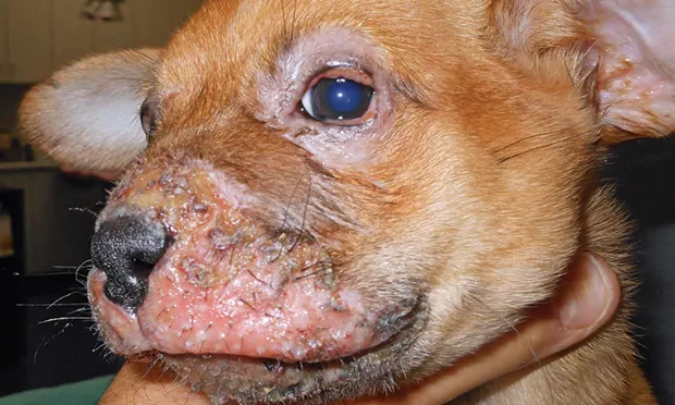
Swelling and erosions of the muzzle and periocular area of a male Chihuahua crossbreed (8 weeks of age). Submandibular lymphadenopathy and exudative otitis externa were also noted on examination.
Juvenile sterile granulomatous dermatitis and lymphadenitis (JSGDL; ie, juvenile cellulitis, juvenile pyoderma, puppy strangles) is a nodular granulomatous inflammatory disorder of unknown pathogenesis for which no microbial organisms have been identified.
Clinical Signs
JSGDL most often develops in puppies younger than 4 months of age, although occasional cases in adult dogs have been reported.1 Several puppies from the same litter may be affected. The golden retriever, dachshund, and Gordon setter may be predisposed.
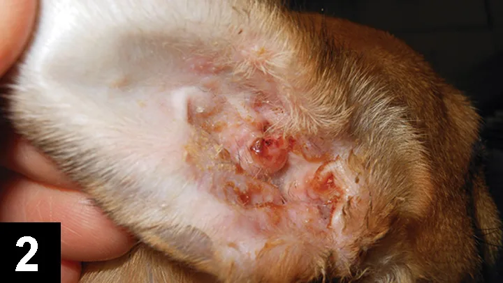
Erythematous and swollen external ear canal and pinna of the puppy in Figure 1.
The initial presenting sign is acute facial swelling and submandibular lymphadenopathy (Figure 1). Less than half of affected puppies exhibit concurrent lethargy or anorexia. Edema of the face, ear, eyelid, and lip margin can be striking. Pustular and nodular lesions follow initial swellings and can develop into erosions or crusting and produce drainage. The external ear canals and pinnae are often thickened and erythematous2 (Figure 2).
Initial lesions are sterile, although secondary bacterial infection and sepsis can develop without appropriate treatment. Joint pain and fever have been reported. Primary differentials include pyoderma and demodicosis.
Diagnosis
In a young puppy, presumptive diagnosis of JSGDL may be based on acute swelling. Examining cytologic preparations from lesions may reveal pyogranulomatous inflammation without organisms. Culture results in patients with early-stage disease will be negative. Histopathologic tissue examination can reveal granulomatous and pyogranulomatous dermatitis and lymphadenitis, again without identifiable microorganisms.
Treatment
Without aggressive treatment, lesions can progress to scarring and secondary infection. Most cases respond to oral prednisolone at 2 mg/kg q24h. As lesions resolve, the dose is tapered, usually within 2 to 4 weeks. Oral antibiotics (cephalexin 22 mg/kg q12h, amoxicillin–clavulanic acid 15 mg/kg q12h) are prescribed when secondary pyoderma is suspected. Cyclosporine can be used in chronic or poorly responsive cases.3 Frequent, gentle bathing and soaking with antiseptic solutions (2% chlorhexidine) can reduce discomfort and speed recovery. With early and aggressive treatment, especially before puppies become weakened or secondary infection develops, prognosis for full recovery is excellent. Therapy is often discontinued within 4 to 8 weeks, and relapses are uncommon to rare.
Editor’s note: This article was originally published in June 2014 as “Puppy Strangles.”