WO2017147456A1 - Multilayer device for separating blood components and uses thereof - Google Patents
Multilayer device for separating blood components and uses thereof Download PDFInfo
- Publication number
- WO2017147456A1 WO2017147456A1 PCT/US2017/019405 US2017019405W WO2017147456A1 WO 2017147456 A1 WO2017147456 A1 WO 2017147456A1 US 2017019405 W US2017019405 W US 2017019405W WO 2017147456 A1 WO2017147456 A1 WO 2017147456A1
- Authority
- WO
- WIPO (PCT)
- Prior art keywords
- filtration membrane
- unit
- multilayer device
- collection material
- plasma
- Prior art date
Links
Classifications
-
- G—PHYSICS
- G01—MEASURING; TESTING
- G01N—INVESTIGATING OR ANALYSING MATERIALS BY DETERMINING THEIR CHEMICAL OR PHYSICAL PROPERTIES
- G01N1/00—Sampling; Preparing specimens for investigation
- G01N1/28—Preparing specimens for investigation including physical details of (bio-)chemical methods covered elsewhere, e.g. G01N33/50, C12Q
- G01N1/40—Concentrating samples
- G01N1/4077—Concentrating samples by other techniques involving separation of suspended solids
-
- B—PERFORMING OPERATIONS; TRANSPORTING
- B01—PHYSICAL OR CHEMICAL PROCESSES OR APPARATUS IN GENERAL
- B01L—CHEMICAL OR PHYSICAL LABORATORY APPARATUS FOR GENERAL USE
- B01L3/00—Containers or dishes for laboratory use, e.g. laboratory glassware; Droppers
- B01L3/50—Containers for the purpose of retaining a material to be analysed, e.g. test tubes
- B01L3/502—Containers for the purpose of retaining a material to be analysed, e.g. test tubes with fluid transport, e.g. in multi-compartment structures
- B01L3/5023—Containers for the purpose of retaining a material to be analysed, e.g. test tubes with fluid transport, e.g. in multi-compartment structures with a sample being transported to, and subsequently stored in an absorbent for analysis
-
- G—PHYSICS
- G01—MEASURING; TESTING
- G01N—INVESTIGATING OR ANALYSING MATERIALS BY DETERMINING THEIR CHEMICAL OR PHYSICAL PROPERTIES
- G01N33/00—Investigating or analysing materials by specific methods not covered by groups G01N1/00 - G01N31/00
- G01N33/48—Biological material, e.g. blood, urine; Haemocytometers
- G01N33/483—Physical analysis of biological material
- G01N33/487—Physical analysis of biological material of liquid biological material
- G01N33/49—Blood
- G01N33/491—Blood by separating the blood components
-
- G—PHYSICS
- G01—MEASURING; TESTING
- G01N—INVESTIGATING OR ANALYSING MATERIALS BY DETERMINING THEIR CHEMICAL OR PHYSICAL PROPERTIES
- G01N33/00—Investigating or analysing materials by specific methods not covered by groups G01N1/00 - G01N31/00
- G01N33/48—Biological material, e.g. blood, urine; Haemocytometers
- G01N33/50—Chemical analysis of biological material, e.g. blood, urine; Testing involving biospecific ligand binding methods; Immunological testing
- G01N33/53—Immunoassay; Biospecific binding assay; Materials therefor
- G01N33/569—Immunoassay; Biospecific binding assay; Materials therefor for microorganisms, e.g. protozoa, bacteria, viruses
- G01N33/56966—Animal cells
-
- B—PERFORMING OPERATIONS; TRANSPORTING
- B01—PHYSICAL OR CHEMICAL PROCESSES OR APPARATUS IN GENERAL
- B01L—CHEMICAL OR PHYSICAL LABORATORY APPARATUS FOR GENERAL USE
- B01L2300/00—Additional constructional details
- B01L2300/06—Auxiliary integrated devices, integrated components
- B01L2300/0681—Filter
-
- B—PERFORMING OPERATIONS; TRANSPORTING
- B01—PHYSICAL OR CHEMICAL PROCESSES OR APPARATUS IN GENERAL
- B01L—CHEMICAL OR PHYSICAL LABORATORY APPARATUS FOR GENERAL USE
- B01L2300/00—Additional constructional details
- B01L2300/08—Geometry, shape and general structure
- B01L2300/0809—Geometry, shape and general structure rectangular shaped
- B01L2300/0816—Cards, e.g. flat sample carriers usually with flow in two horizontal directions
-
- B—PERFORMING OPERATIONS; TRANSPORTING
- B01—PHYSICAL OR CHEMICAL PROCESSES OR APPARATUS IN GENERAL
- B01L—CHEMICAL OR PHYSICAL LABORATORY APPARATUS FOR GENERAL USE
- B01L2300/00—Additional constructional details
- B01L2300/16—Surface properties and coatings
- B01L2300/161—Control and use of surface tension forces, e.g. hydrophobic, hydrophilic
- B01L2300/165—Specific details about hydrophobic, oleophobic surfaces
-
- G—PHYSICS
- G01—MEASURING; TESTING
- G01N—INVESTIGATING OR ANALYSING MATERIALS BY DETERMINING THEIR CHEMICAL OR PHYSICAL PROPERTIES
- G01N1/00—Sampling; Preparing specimens for investigation
- G01N1/28—Preparing specimens for investigation including physical details of (bio-)chemical methods covered elsewhere, e.g. G01N33/50, C12Q
- G01N1/286—Preparing specimens for investigation including physical details of (bio-)chemical methods covered elsewhere, e.g. G01N33/50, C12Q involving mechanical work, e.g. chopping, disintegrating, compacting, homogenising
- G01N2001/2873—Cutting or cleaving
- G01N2001/288—Filter punches
-
- G—PHYSICS
- G01—MEASURING; TESTING
- G01N—INVESTIGATING OR ANALYSING MATERIALS BY DETERMINING THEIR CHEMICAL OR PHYSICAL PROPERTIES
- G01N1/00—Sampling; Preparing specimens for investigation
- G01N1/28—Preparing specimens for investigation including physical details of (bio-)chemical methods covered elsewhere, e.g. G01N33/50, C12Q
- G01N1/40—Concentrating samples
- G01N1/4077—Concentrating samples by other techniques involving separation of suspended solids
- G01N2001/4088—Concentrating samples by other techniques involving separation of suspended solids filtration
Definitions
- This invention relates generally to separation of fluid sample components for analyte detection. More particularly, aspects of the invention are directed to a facile and accurate device for separation of various components of whole blood, including but not limited to red blood cells, white blood cells, platelets, and plasma, using a multilayer device or multilayer separation device for the separation of blood components and methods of using such device to detect analytes in the various blood components, for example, specifically on the cell surface and in the intra- and extra-cellular fluids, such as, but not limited to, chemical compounds, drugs and metabolites, nucleic acids, DNA, RNA, mRNA, miRNA, proteins, cell surface and intracellular markers, pathogens, bacteria, viruses, microorganisms, and the like.
- a multilayer device or multilayer separation device for the separation of blood components and methods of using such device to detect analytes in the various blood components, for example, specifically on the cell surface and in the intra- and extra-cellular fluids, such as, but not limited to, chemical compounds, drugs and metabolites, nucle
- Two existing dried plasma spot (DPS) cards are the NOVIPLEXTM card, which is commercially available from Novilytic LLC (Kim, J.H., et al. Anal Chem 2013, 85, 11501-11508) and the 'auto DPS card' previously reported by Sturm et al. (Bioana!ysis 2015, 7, 1987-2002).
- the functionality of the plasma collection substrate which is the collection material or cellulose paper is a key difference among the various dried spot cards.
- the auto DPS card has a wax boundary. With the wax boundary of the auto DPS card, the excess filtered plasma is retained within the boundary resulting in inaccuracy biases at the low and high ends of the hematocrit.
- the boundary of the NOVIPLEXTM card is different from the wax boundary described in the auto DPS card.
- the NOVIPLEXTM card has a disk that once saturated, the excess filtered plasma freely flows outside of or beyond the disk. This action is unlike the auto DPS card where excess plasma is trapped within the boundary.
- the conceptual design of these two cards are similar as each of them employs an on-card membrane filtration technique to separate RBC from plasma.
- the card structures and production of plasma in each card format are different and each has its own disadvantages.
- the auto DPS card may not be accurate nor efficient.
- the auto DPS card reportedly may utilize a sample support device also known as the Liquid Extraction Surface Analysis, LESATM, stage by Advion, Inc. (Ithaca, NY, USA).
- LESATM Liquid Extraction Surface Analysis
- the NOVIPLEXTM card does not require any external device for generation of plasma spots, it is not compatible for automated analysis. As a result, the sample handling process is tedious requiring a pair of tweezers to remove a small 2-mni filter disk containing the sample and manually transferring the disk for further sample extraction processes. While the yield of plasma volume by auto DPS was not determined, the NOVIPLEXTM card requires a minimum of 25 ⁇ blood to produce about 2.5 iiL plasma (Kim, J. H. et al. Anal Chem 2013, 85, 11501-1 1508).
- the yield of plasma volume is unfortunately low, i.e., 0.100 iL plasma per ⁇ blood.
- the plasma yield from the NOVIPLEXTM card is about 2.5 iiL, which is insufficient for most analyses.
- the low quantity of plasma is not due to a low initial blood sample volume, rather it is due to a limited capacity of the plasma collection substrate. Scaling up is not possible.
- the invention provides a multilayer device for collection and separation of, for example, blood components, allowing for the detection of analytes from a fluid sample applied to the multilayer device, for example whole blood.
- the multilayer device may be a dried spot card composed in a book-type form in one
- top and bottom cover hinged on one edge forming a spine and sandwiching multiple layers of membranes and materials similar to the pages of a book.
- more than one or all of the edges are temporarily connected or coupled, and any or all of the layers of the multilayer device that are connected or coupled on one or more edges, may be detached, removed, or separated from each other and from the device.
- Separation of the fluid sample allows for subsequent individual analyses of the separated components.
- a multilayer device comprising layers that may be separately removed or detached as desired.
- a multilayer device having a rectangular shape may be temporarily attached or coupled on all edges, where any or all layers of the multilayer device may be detached or removed.
- the edges of any or all of the layers may be perforated in such a manner that allows for the layer to be torn out or detached from the remaining components of the multilayer device.
- the layers may be tightly sandwiched between the top and bottom covers in such a manner that the fluid sample does not leak and each of the layers of the multilayer device remain in position until separated for removal.
- One aspect is directed to a multilayer device, comprising
- said top unit is adjacent to and connected to said bottom unit
- said filtration membrane unit comprising at least one filtration membrane
- said filtration membrane unit has a top surface and a bottom surface
- said hydrophobic membrane has a top surface and a bottom surface, where said bottom surface of said filtration membrane unit is adjacent to said top surface of said hydrophobic membrane
- said collection material has a top surface and a bottom surface
- said bottom surface of said hydrophobic membrane is adjacent to said top surface of said collection material
- said bottom surface of said collection material is adjacent to said bottom cover.
- Another aspect provides a multilayer device, comprising:
- a top unit comprising layers of: a top cover with at least one cutout, a filtration membrane unit, and a hydrophobic membrane with at least one cutout and the same number of cutouts as in the top cover; and
- a bottom unit comprising layers of: a collection material and a bottom cover without cutouts
- said filtration membrane unit comprises at least one filtration membrane, preferably two filtration membranes of decreasing pore sizes with each having, in one aspect, a shape of said cutout, said filtration membrane unit is positioned within said cutout of said top cover and adj cent to said hydrophobic membrane, where each of the layers of the top cover, filtration membrane unit, and hydrophobic membrane are aligned by the cutouts, said hydrophobic membrane is adjacent to or sandwiched between said filtration membrane unit and said collection material, said collection material is adjacent to said hydrophobic membrane, and said collection material is above said bottom cover. Since plasma is primarily water, a hydrophobic material will not absorb any plasma.
- a hydrophobic membrane that has cutouts will prevent any plasma from leaking or spreading beyond the boundaries of the cutouts, thus enabling a dried spot positioned centrally within the cutouts.
- the filtration membrane unit in one aspect may be sandwiched between the top cover and the hydrophobic membrane.
- the collection material which is an absorptive layer such as cellulose, paper, etc., in another aspect may be sandwiched between said hydrophobic membrane and said bottom cover.
- the multilayer device may be in the shape of rectangle having four edges, where each of the layers of the top unit or the bottom unit is temporarily coupled on at least one edge, forming a sufficiently tight contact to avoid any leakage or movement of layers, and each of the layers of the top unit and the bottom unit is detachable or removable.
- An alternative format of a multilayer device further includes a contact support layer adjacent to and below said collection material and adjacent to or above said bottom cover, or contact supports form a portion of a bottom cover, where contact supports of the contact support layer preferably contains raised supports where at least a portion of said raised supports fits within the cutout where a fluid sample is placed.
- a further aspect comprises said multilayer device which may also include at least one window support for a layer detached for subsequent analyses, preferably for a filtration membrane unit and/or a collection material.
- the window support may be a layer containing a window which exposes the sample for detecting an analyte of interest, said layer for subsequent analyses is attached or coupled to said window support, and said window support coupled to said layer for subsequent analyses may be removed or detached from said multilayer device for subsequent biological analyses.
- a further aspect is directed to a multilayer device comprising: a top unit comprising layers of: a top cover with at least one cutout, a filtration membrane unit, and a hydrophobic membrane with at least one cutout; and
- a bottom unit comprising layers of: a collection material and a bottom cover without cutouts
- a method of using the multilayer device comprises:
- a top unit comprising layers of a top cover with at least one cutout or open hole on or in which a fluid sample is placed, a filtration membrane unit, and a hydrophobic membrane;
- a bottom unit comprising layers of a collection material and a bottom cover without cutouts, where said top unit is adjacent to said bottom unit, where said volume may be about 10 microliters to about 100 microliters;
- the analysis of said filtration membrane unit and/or said collection material may include detecting an analyte of interest.
- a further aspect of may be directed to a method of using the multilayer device, comprising:
- the multilayer device may be a 3D-printed device or a non-3D-printed device, e.g., card stock.
- the benefits of the multilayer device and techniques described here include simplified sample collection, reduced costs, simplified shipping and storage, and a gained significant interest in various fields (Tretzei, L. et al. Analytical Methods 2015, 7, 7596-7605; Sadones, N. et al. Bioanalysis 2014, 6, 2211-2227).
- the inventive multilayer device overcomes many challenges in the art including the hematocrit effect and sampling of whole blood instead of plasma (De Kesel, P. M. et al. Bioanalysis 2013, 5, 2023-2041).
- a multilayer device is configured for facile and rapid detection analyses of analytes found in dried sample spots collected and separated by the multilayer device, such as but not limited to automated, high-thioughput analyses. Therefore, the inventive multilayer device described here was developed to be compatible for a wide range of hematocrit levels (e.g., 25%-65%), a high plasma volume yield from a whole blood fluid sample, and separate analyses of multiple components of a single fluid sample.
- the invention relates in part to an improved dried spot card .
- the invention may have utility in, for example, detecting chemical compounds, drugs, metabolites, hormones, opioids, viruses, nucleic acids, proteins, and the like, from a fluid sample, including, but not limited to, whole blood, red blood ceils, plasma, and platelets.
- a fluid sample including, but not limited to, whole blood, red blood ceils, plasma, and platelets.
- the use of the multilayer device may also be contemplated for detecting analytes of interest in fluid samples that do not necessarily require separation, such as for example, urine, saliva, tears, amniotic fluid, semen, and the like.
- FIG. 1 depict aspects of a multilayer device, including fluid sample separation and determination of the presence of chemical compounds, drugs, opioids, hormones, nucleic acids, proteins, and the like, in accordance with example embodiments.
- FIG. 1 shows a book-type multilayer device, where the numbers indicate the assembly order from 1 to 6. which are described in detail in Example 1.
- FIG. 2 shows a multilayer device containing seven layers: (1) a first filtration membrane that fits within each cutout; (2) a top cover with four cutouts; (3) a second filtration membrane that fits within each cutout and adjacent to the first filtration membrane; (4) a hydrophobic membrane containing cutouts; (5) a collection material without cutouts, and optionally having an outline of the cutout perimeter where the cutou ts are located; (6) a contact support layer; and (7) a bottom cover.
- FIG. 3 shows the results of center and peripheral sampling positions for various opioids, where the left column and the right column for each opioid represent a center position and a peripheral position, respectively.
- FIG. 4 shows on-card stability of opioids and stimulants at LLOQ (5 ng/mL) at three different storage conditions: at RT kept in a box filled with a continuous flow of nitrogen (RT + Nitrogen), at RT kept in a glassine envelope + desiccant further sealed in a Ziploc bag (RT + Air), and at -20 °C kept in a glassine envelope + desiccant further sealed in a Ziploc bag (-20 °C + Air).
- FIG. 5 shows on-card stability of opioids and stimulants at High QC (900 ng/mL) at three different storage conditions: at RT kept in a box filled with a continuous flow of nitrogen (RT + Nitrogen), at RT kept in a glassine envelope + desiccant further sealed in a Ziploc bag (RT + Air), and at -20 °C kept in a glassine envelope + desiccant further sealed in a Ziploc bag (-20 °C + Air).
- FIG. 7 shows SRM LC/MS chromatograms from fortified blood samples containing morphine (1), codeine (2), oxycodone (3), amphetamine (4), hydrocodone (5), methamphetamine (6), MDMA (7), phentermine (8), and mephedrone (9) for A) a blank sample (matrix blank without IS), B) a zero sample (matrix blank with IS, showing only analyte signals), C) LLOQ sample (matrix fortified with 5 ng/mL standards) and D) their deuterated IS.
- FIG. 8 shows the results of hematocrit levels of 25% - 65% as tested for various opioids.
- FIG. 9 shows linearity graphs for morphine (A) and fentanyl (B).
- FIG. 10 shows a comparison of volumetric sampling from 20 ⁇ - 50 ⁇ whole blood for various opioids.
- FIG. 11 shows the % RE and % CV for various opioids and for (A) 20 ⁇ , (B)
- the exemplary embodiments describe a multilayer device for separation of a fluid sample, for example, whole blood, and analyses of such fluid sample for analytes of interest.
- the multilayer device is a dried fluid spot device for micro-sampling, separating, and analyzing dried fluid spot samples.
- a multilayer device or multilayer device card comprises: (1) a filtration membrane unit with areas designated for fluid sample collection; (2) a support layer or top cover that is preferably labeled for sample identification; (3) a collection material; and (4) a support layer or bottom cover as exemplified in FIG. 2.
- a fluid sample is applied to the multilayer device, where the fluid sample may be any fluid, preferably a biological fluid, for testing for the presence of analytes.
- the fluid or fluid sample may include, but is not limited to, whole blood, red blood cells, plasma, plasma protein fraction, cerebral spinal fluid, or any fluid possibly containing an analyte of interest, and the like.
- One of skill in the art could modify components of the multilayer device accordingly to accommodate for the various fluids and desired analytes.
- the filtration membrane sizes may be altered in order to capture or separate the analytes of interest.
- Samples for dried spot cards available in the art have limitations, in particular, sample volume inconsistencies, which may negatively affect results.
- DBS dried blood spot
- Hct hematocrit
- Alternative ways to collect known volumes of finger prick blood include a 'volumetric' capillary sampling system (DBS System; Gland, Switzerland) is incorporated by reference as disclosed in the art (Leuthold, L. A. et al. Anal. Che .
- volumetric pipettes such as an EPPENDORF® pipette (Z683787 Aldrich; EPPENDORF® Research® ) plus pipette, variable volume; 0.5 ⁇ . -10 ⁇ .; SIGMA- ALDRICH® CO) may be used or a glass capillary (P2174 Sigma; Microcapillary tube DRUMMOND MICROCAPS®; volume 50 pL).
- sample collection is to be performed by untrained personnel, it will pose a critical control point to assure that accurate sample volume has been collected.
- accurate sampling volume is not necessarily required to obtain accurate and precise quantitation.
- the multilayer device described here may accommodate a wide fluid sample volume range of up to about 50 microliters (pL) of, for example, whole blood and even up to about 100 ⁇ iL while most of the commercially available or currently used devices may only handle single digit volumes of blood in microliters (e.g., 5
- a book-type or other multilayer device card that allows for an intimate contact provides the feature of a flexible sampling volume.
- Plasma consistency was observed to be independent of the Hct level in blood as presented by Li et al. (Journal of Chromatography B-Analytical Technologies in the Biomedical and Life Sciences 2015, 991, 46-52). Based on studies in the Examples section, plasma generated from whole blood having hematocrit (Hct) levels of about 30% Hct or 60% Hct has the same spreading consistency on the paper substrate and thus produces homogenous spots. Homogenous spots do not equate to equal spot dimension. It refers to homogenous saturation of plasma within a spot regardless of the spot size.
- inventive multilayer device described here is capable of handling and processing flexible volumes and a larger volume than that in the art, without adversely affecting results.
- a larger whole blood sample, which the described multilayer device provides may result in larger red blood cell and plasma yields.
- initial fluid sample volumes that are applied may vary and have a minimum volume of about 10 microliters, and may range from about 10 microliters to about 100 microliters, about 10 microliters to about 75 microliters, and about 25 microliters to about 50 microliters.
- a low plasma yield would be less than about 4 ⁇ ⁇ , e.g., less than about 2 ⁇ ⁇ .
- the multilayer device described here may provide a plasma yield greater than about 2 ⁇ ⁇ , preferably ranging from about 4 ,uL to about 38 ⁇ ,, including about 4 ,uL to about 15 ⁇ ., depending on the initially applied whole blood volume. There are several factors that may dictate the plasma yield, including the initial starting whole blood volume, the collection material, and loss of plasma due to spreading or leakage. Starting with a large initial whole blood volume results in the generation of a large plasma yield.
- the final plasma yield is also dependent on the collection material used for plasma collection - size and material type. If the collection material, preferably a cellulose or cellulose acetate paper, is thicker, a larger volume may be collected as there is more surface area.
- the plasma per whole blood of the multilayer device as tested here is about 0.303 ⁇ 0.007 uL plasma per ⁇ whole blood.
- the multilayer device allows for greater than 0.100 ⁇ _ plasma per ⁇ , whole blood. Whereas, the NOVIPLEXTM card results in 0.100 ⁇ . plasma per ⁇ . whole blood, which is significantly less than the amount achieved in the described multilayer device.
- each cutout may accommodate large volumes, i.e., about 10 microliters to about 100 microliters as necessary, the plasma yields per sample/cutout is sufficiently large to perform analysis.
- Other cards may require the combination of multiple fluid samples in order to achieve the same fluid sample volume of a single sample.
- the multilayer device is capable of processing a large and wide range of hematocrit levels.
- Hematocrit is the proportion of red blood cells in a blood sample. For example, a 20 microliter blood sample having a 30% Hct has about 6 microliters of red blood ceils, while a 45% Hct has about 9 microliters red blood cells, and a 60% Hct has about 12 microliters red blood cells with the remainder being plasma.
- Embodiments of the invention using a single or combination of two filtration membranes, preferably in one embodiment, asymmetric membranes may provide a capacity to process blood samples having a hematocrit range of about 30% to about 70%. Although lower or higher hematocrit levels may work, they become problematic with regard to the membrane filtration process and would not be as efficient.
- whole blood samples having hematocrit levels of about or greater than about 20% and about or less than about 80% may also be utilized but they are not as efficient.
- One embodiment is directed to a multilayer device for use in collecting a fluid sample, e.g., whole blood, for testing analytes of interest, such as but not limited to, chemical compounds, drugs, drug metabolites, hormones, viruses, nucleic acids, DNA, RNA, mRNA, miR A, proteins, cell surface and intracellular markers, and the like, or any analyte that is detectable by any known method or any of the means described here including, for example, spectroscopy or chromatography.
- analytes of interest such as but not limited to, chemical compounds, drugs, drug metabolites, hormones, viruses, nucleic acids, DNA, RNA, mRNA, miR A, proteins, cell surface and intracellular markers, and the like, or any analyte that is detectable by any known method or any of the means described here including, for example, spectroscopy or chromatography.
- Non-limiting examples of analytes may more specifically include opioids, cannabinoids, stimulants, performance enhancers, morphine, codeine, oxycodone, hydrocodone, amphetamine, methamphetamine, mephedrone, phentermine, 3,4- methylenedioxymethamphetamine (MDMA), fentanyl, combinations, and the like.
- opioids cannabinoids
- stimulants performance enhancers
- morphine codeine
- oxycodone hydrocodone
- amphetamine methamphetamine
- mephedrone mephedrone
- phentermine 3,4- methylenedioxymethamphetamine (MDMA)
- fentanyl combinations, and the like.
- the methods and multilayer devices described here may be used to collect and separate fluid samples for testing any analytes of interest, including but not limited to foreign substances and endogenous biomarkers.
- Another aspect of a multilayer device is its capacity to collect and analyze various components of a single fluid sample.
- Various analytes may be found in multiple locations of a single fluid sample, particularly if the fluid sample may be separated into its multiple components.
- the novel multilayer device described here can advantageously process a single fluid sample, separate components of the single fluid sample, and individually analyze the separated components of the single fluid sample. This dual functionality is particularly beneficial for expediting testing of a large number of analytes and maximizing the use of a single fluid sample.
- a further advantage of the multilayer device is that any volume of fluid sample, for example, whole blood, may be applied to the multilayer device and still result in quantitative results since a filtration membrane unit comprises at least one pre-determined size which allows for a pre-defined volume.
- a filtration membrane unit comprises at least one pre-determined size which allows for a pre-defined volume.
- flow-through elution technology quantitative analysis of the collected plasma on a collection material may be achieved.
- the plasma spot may be punched out of the collection material and analyzed with the same spot size used when creating a calibration curv e.
- accuracy of an applied sample volume is not a requirement as generally held in current technologies.
- Another embodiment is directed to a multilayer device having dual function capacities where application of, for example, a whole blood sample from a single subject, results in a layer containing collected or retained cellular components, such as but not limited to, red blood cells, white blood cells, platelets, and other cells, and a layer containing collected or retained plasma.
- the cellular components may be analyzed separately from the plasma components in a sample, where each layer may contain different analytes found in differing components of whole blood. For example, cell surface proteins and other constituents on red blood cells as well as intracellular proteins and constituents and drugs contained within red blood cells may be analyzed separately from the plasma constituents.
- the dual function capability of the multilayer device of the invention is advantageous for its efficiency - sampling and time. Since the multilayer device can accommodate multiple samples and subsequently separate the multiple samples into their cellular component and their plasma component, multiple analytical tests may be simultaneously performed.
- One advantage of a multilayer device having dual function capacities is that it facilitates the determination of a red blood cells to plasma partition coefficient of a drag or analyte of interest.
- Another advantage is the analysis of multiple classes of drugs or analytes performed simultaneously. Fluid samples from a single subject or multiple subjects may be simultaneously processed and analyzed for multiple analytes, i.e., each subject sample placed or collected in each cutout, well, or open hole of a multilayer device. For example, 4 opioids and 5 stimulants may be simultaneously tested for in a single process containing a single subject's fluid sample.
- the multiple layers of the inventive multilayer device enable the separation of, for example, blood components including red blood cells, plasma, platelets, and the like, as well as the processing of a wide range of hematocrit levels of a whole blood sample without hemolysis.
- the multilayer device described here may in another embodiment be in the format of a book, which is hinged on one side and the various layers compose the pages of a book.
- the multilayer device may be coupled or attached at more than one side or edge, such as, for example, on all sides or edges of a multilayer device, and any or all layers may be detached or removed.
- the multilayer device of the invention may be in any shape, including but not limited to a circle, an oval, a triangle, a square, a rectangle, a parallelogram, a diamond, a pentagon, a hexagon, a heptagon, an octagon, and the like.
- Cutouts or holes of the multilayer device may be in any shape, including but not limited to a circle, an oval, a triangle, a square, a rectangle, a parallelogram, a diamond, a pentagon, a hexagon, a heptagon, an octagon, and the like as long as the cutouts or holes of the top cover and the hydrophobic membrane if utilized are the same.
- a preferred shape of the multilayer device is rectangular having four edges where the long edge forms at least one edge or side on which layers of the multilayer device may be hinged, coupled, or attached.
- the dimensions of the multilayer device in the shape of a rectangle may be about 1 inch to about 4 inches, preferably about 2 inches by about 3 inches, for example, about 2 inches by 3.3 inches. How r ever, other shapes and sizes are also contemplated.
- One embodiment is directed to a multilayer device or multilayer device card comprising:
- said top unit is adjacent to and connected to said bottom unit
- said filtration membrane unit comprising at least one filtration membrane
- said filtration membrane unit has a top surface and a bottom suiface
- said hydrophobic membrane has a top surface and a bottom surface, where said bottom surface of said filtration membrane unit is adjacent to said top surface of said hydrophobic membrane
- said collection material has a top surface and a bottom surface
- said bottom surface of said hydrophobic membrane is adjacent to said top surface of said collection material.
- Another embodiment provides a multilayer device, comprising:
- a top unit comprising layers of: a top cover with at least one cutout, a filtration membrane unit, and a hydrophobic membrane with at least one cutout;
- a bottom unit comprising layers of: a collection material and a bottom cover without cutouts
- said filtration membrane unit comprises at least one filtration membrane, preferably two filtration membranes of decreasing pore sizes with each having a shape of said cutout, said filtration membrane unit is positioned within said cutout of said top cover and adjacent to said hydrophobic membrane, said hydrophobic membrane is adjacent to or sandwiched between said filtration membrane unit and said collection material, said collection material is adjacent to said hydrophobic membrane, and said collection material is above said bottom cover.
- the filtration membrane unit in one embodiment may be sandwiched between the top cover and the hydrophobic membrane.
- the collection material in another aspect may be sandwiched between said hydrophobic membrane and said bottom cover.
- the multilayer device may be in the shape of rectangle having four edges, where each of the layers of the top unit or the bottom unit is temporarily coupled on at least one edge, and each of the layers of the top unit or the bottom unit is detachable or removable.
- the filtration membrane unit may have the size and shape of the cutouts, fitting within each of the cutouts or wells of the top cover, such that each layer of the filtration membrane unit is held in place by the intimate contact of the edges of the filtration membrane unit and the walls of the cutout of the top cover and sandwiching of all of the layers in the multilayer device.
- a preferred embodiment is directed to these filtration membranes that are circular to fit within the circular cutouts of the top cover, where these circular filtration membranes or disks are easily removable for further analyses after collection and separation of samples. For example, after whole blood is applied to the cutouts of the multilayer device, the whole blood is allowed sufficient time to filter through the filtration membrane unit and collect on the collection material. Red blood cells remain on the filtration membranes or disks in one embodiment while plasma collects on the collection material.
- An alternative format of a multilayer device further includes a contact support layer adjacent to and below said collection material and adjacent to or above said bottom cover, or in another embodiment, the contact support layer is combined with the bottom cover, such that the contact supports are a part of the bottom cover, and where contact supports of the contact support layer preferably contains raised supports where at least a portion of said raised supports fits within the cutout where a fluid sample is placed.
- a further embodiment comprises said multilayer device which may also include at least one window support for a layer detached for subsequent analyses, preferably for a filtration membrane unit and/or a collection material.
- the window support may be a layer containing a window which exposes the collected or captured sample for detecting an analyte of interest, said layer for subsequent analyses is attached or coupled to said window support, and said window support coupled to said layer for subsequent analyses may be removed or detached from said multilayer device for subsequent biological analyses.
- One embodiment comprises subsequent analysis of the filtration membrane unit, where each filtration membrane, or portions thereof, may be transferred for separate analyte detection analysis by, for example, enzyme immunoassay (EIA).
- EIA enzyme immunoassay
- Another embodiment comprises subsequent analysis of the collection material, where, for example, the plasma from a whole blood sample is analyzed by liquid chromatography and/or mass spectrometry, for example solid phase extraction liquid chromatography tandem mass spectrometry (SPE-LC-MS/MS).
- An embodiment directed to a multilayer device comprises a top unit and a bottom unit, where the top unit and the bottom unit are adjacent.
- the top unit comprises a top cover, where the cover may be composed of a stiff, durable construction, such as for example card stock, an adjacent filtration membrane unit that initially comes in contact with a fluid sample of whole blood through a cutout or open hole of the top cover.
- An embodiment comprises a filtration membrane unit of at least one filtration membrane, and preferably two adjacent filtration membranes. Adjacent to and underneath the filtration membrane unit is a hydrophobic membrane.
- the bottom unit of the multilayer device comprises a collection material adjacent to a bottom cover.
- the multilayer device may be in a format that allows for a top unit to be in constant or temporary contact with the underlying bottom unit.
- the top cover may have at least one cutout or open hole in which to place a sample, while the bottom cover does not have any cutouts. Multiple cutouts are preferred to test a fluid sample from a single source, for example, whole blood from a subject, and also to include standard controls for simultaneous testing.
- An embodiment may be directed to a fluid sample which is applied to a filtration membrane unit, where the filtration membrane unit is exposed through a cutout, well, or hole of a top cover of a multilayer device.
- the filtration membrane unit may comprise at least one filtration membrane, preferably two filtration membranes positioned adjacent to each other and within the confines of the cutout of the top cover. If the shape of a cutout or hole is circular, a preferred filtration membrane unit comprises at least one circular filtration membrane disk.
- the filtration membranes may be asymmetrical, non-asymmetrical, a combination of asymmetrical and non-asymmetrical, or similar combinations of each, i.e., one or more than one asymmetrical filtration membrane or one or more than one non- asymmetrical filtration membrane.
- the filtration membrane unit is selected and composed in a manner to sufficiently filter and capture components of a fluid sample.
- a multilayer device described here comprises a filtration membrane unit that separates components of a whole blood sample, where the filtration membrane unit captures red bood ceils and allows plasma to flow or pass through the filtration membrane unit. If at least two filtration membranes are used, they are stacked such that there is an upper filtration membrane and a lower filtration membrane, and both filtration membranes have the same shape as the cutout or holes of the top cover and hydrophobic membrane if used.
- the filtration membrane unit comprises at least one filtration membrane, which filters particles that are about 1 micron to about 10 microns, about 2 microns to about 5 microns.
- a filtration membrane has a top or first surface and a bottom or second surface and a thickness sufficient to allow filtration and/or capture of desired particles, such as for example, red blood cells from a whole blood sample, and allowing other particles or fluids to filter through, for example plasma.
- the filtration membrane may have a thickness ranging from about 0.1 mm to about 0.6 mm, about or greater than about 0.15 mm, about or greater than or less than about 0.2 mm, about or greater than or less than about 0.26 mm, about or greater than or less than about 0.3 mm.
- Another embodiment is directed to a filtration membrane unit comprising two adjacent filtration membranes.
- a bottom surface of a first filtration membrane is adjacent to a top surface of a second filtration membrane, where a sample enters a top surface of a first filtration membrane and exits through a bottom surface of the first filtration membrane and enters a top surface of a second filtration membrane and exits a bottom surface of the second filtration membrane.
- a filtration membrane may be hydrophobic to avoid absorption of any plasma, but may also be hydrophilic in other circumstances, and anisotropic, functioning to filter and collect desired components of a fluid sample.
- the desired components of a whole blood sample may include, but are not limited to, red blood cells and plasma.
- the filtration membrane may comprise any material sufficient to filter and separate particles of interest.
- the filtration membrane, which allows filtration of whole blood components may be composed of, but not limited to a polar, non-polar, and intermediate polarity polymers, polyester, polysulfones, polycarbonate, polymethacrylate, or the like, or blends or combinations thereof.
- the filtration membrane functions to filter and separate components of a fluid sample.
- the sample may be separated into individual components, i.e.. red blood cells, white blood cells, platelets, and plasma, and collect, for example red blood cells for further testing, while allowing other components, such as plasma to filter through the membrane.
- red blood cells are larger in size than plasma or platelets, where red blood cells may be about 6 micrometers ( ⁇ ) - about 8 ⁇ , and white blood cells are larger than red blood cells, i.e., about 12 ⁇ - about 15 ⁇ .
- Appropriate filtration membrane pore sizes may be selected depending on the desired particle.
- a filtration membrane closest or adjacent to a hydrophobic membrane may have characteristics sufficient to collect or capture components of whole blood, for example, red blood cells.
- One advantage of the subject multilayer device is its novel capability to separate, collect, and test more than one blood component of a single subject sample or multiple subject samples of whole blood for separate analyses, where the device accommodates a large volume of and wide hematocrit percentage range of whole blood sample.
- Another embodiment is directed to filtration membranes that are asymmetric allowing for the whole blood sample to be filtered, separating different sized components within the filtration membrane unit.
- the filtration membrane unit and its filtration membranes allow for separating and capturing red blood cells from a whole blood sample and permitting plasma to flow through the filtration membrane unit and results in cell- free plasma.
- An asymmetrical filtration membrane may be used in one embodiment of a multilayer device.
- the asymmetrical filtration membrane has a top surface that allows particles of a large size and smaller to enter the membrane, while the bottom surface of the same filtration membrane has a smaller pore size, thereby eliminating any particles smaller than the top surface pore size and greater than the pore size at the bottom surface of the filtration membrane from filtering or passing through, i.e., capturing some particles or allowing particles smaller than the pore size on the bottom surface of the filtration membrane to pass.
- Another embodiment may be directed to a filtration membrane unit comprising at least one aswimetrical filtration membrane or at least two aswimetrical filtration
- asymmetrical filtration membrane may have a pore size of about 5 microns at the top sui face, and a pore size at the bottom suiface of about 2.5 microns, thereby collecting particles that are smaller than about 5 microns and larger than about 2.5 microns in a filtration membrane and gradually allowing or filtering particles that are smaller than about 2.5 microns.
- Another embodiment is directed to sequential filtration utilizing two filtration membranes in a filtration membrane unit. A portion of red blood cells and any particles that are larger than 5 microns may be captured on a top or first filtration membrane and then the remaining red blood cells and any particles that are larger than 2.5 microns and smaller than 5 microns may be captured on a bottom or second filtration membrane.
- Optimal performance of a lower filtration membrane might occur with a pore size of about one (1) micron.
- Filtration membrane pore sizes may range from about 1 micron to about 10 microns in a multilayer device of the invention.
- an upper, top filtration membrane may have a pore size of about 10 microns thereby providing preliminary filtration of larger particles and mitigating obstruction of the lower filtration membrane, which may have a pore size of about 1 micron.
- a filtration membrane unit comprising either a single filtration membrane or a dual layered filtration membrane comprising two filtration membranes of a multilayer device may process a wide variety of fluid samples.
- a first filtration membrane adjacent to a top cover and a second filtration membrane where the first uppermost filtration membrane may have a pore size ranging from about 35 microns to about 3 microns, about 5 microns, while the second, lower filtration membrane adjacent to or sandwiched by a first filtration membrane and a hydrophobic membrane may have a pore size that is generally smaller than that of the first filtration membrane.
- a preferred pore size range for the second filtration membrane may be 3 microns to about 0.2 microns, about 2.5 microns.
- the filtration membrane may each be asymmetrical or non-asymmetrical, or alternatively, one filtration membrane is asymmetrical and the other is non-asymmetrical.
- One embodiment comprises a multilayer device composed of a filtration membrane unit that is an asymmetric membrane. Another embodiment is directed to a multilayer device having a top unit, where the filtration membrane unit is composed of two filtration membranes.
- the top or uppermost filtration membrane layer is a commercially available product iPOC DXTM X asymmetrical 5 mm membrane that has a 35 ⁇ top and a 5 ⁇ bottom (International Point of Care Inc.; Toronto, Canada), or a filtration membrane with similar properties or properties sufficient to filter desired components.
- Yet another embodiment is directed to a bottom or lowermost filtration membrane that is a commercially available product iPOC DXTM S/G asymmetrical 7 mm membrane that has a 35 ⁇ top and a 2.5 ⁇ bottom (International Point of Care Inc.; Toronto, Canada), or a filtration membrane with similar properties or properties sufficient to filter desired components.
- a bottom or lowermost filtration membrane that is a commercially available product iPOC DXTM S/G asymmetrical 7 mm membrane that has a 35 ⁇ top and a 2.5 ⁇ bottom (International Point of Care Inc.; Toronto, Canada), or a filtration membrane with similar properties or properties sufficient to filter desired components.
- a hydrophobic membrane layer Adjacent to or beneath a filtration membrane unit or sandwiched between a filtration membrane unit and a collection material layer, is in one embodiment, a hydrophobic membrane layer, which assists with the complete and direct contact of a sample with the various membrane layers and accomplishment of sample spot uniformity.
- the multilayer device may be successful in separating and collecting various whole blood components without this hydrophobic membrane, particularly in those multilayer devices that have an intimate seal or connection between the layers, its inclusion results in superior outcomes.
- the hydrophobic membrane may be positioned above and adjacent to the collection material.
- the hydrophobic membrane may be a layer the same size, shape, and dimensions as the entire multilayer device and contain cutouts or holes in the same size, shape, and dimensions as the cutouts in the top cover.
- the hydrophobic membrane may be composed of any material that is hydrophobic, preferably polyester, polyester blends, polysuifone, or polycarbonate, and the like.
- the hydrophobic membrane layer may be any material or membrane that is sufficient to aid in the placement and containment of the individual layers to avoid movement or displacement, as well as, to assist in sample spot uniformity.
- the hydrophobic membrane underneath and adjacent to the filtration membrane unit that filters a whole blood component such as for example, red blood cells, may preferably be composed of a polyester or a polyester blend, more preferably, Ahlstrom HOLLYTEX® Grade 3256 nonwoven polyester (Ahlstrom Filtration; Mount Holly Springs, PA) which has a thickness of about 0.058 millimeter and a basis weight of about 23.9 g/m.
- Another layer of a useful multilayer device which is located underneath a hydrophobic membrane layer is a collection material layer that acts as a vessel for collecting filtered desired components from a small volume of initially applied fluid sample. After drying the collected sample, a dried spot formed on the collection material allows for a convenient storage means for future quantitative analyses. Other components of w r hole blood are separated from plasma in the preceding or layers above the collection material.
- the collection material functions to absorb and/or collect plasma retrieved from filtration of a whole blood sample.
- the collection material has features that allow the capture and collection of plasma, such as for example a pore size preferably in a range of about 35 microns to about 0.2 microns and a thickness of about 0.1 millimeter to about 0.6 millimeter, preferably about 0.19 mm.
- the pore size may be a factor that expresses the degree of absorptivity.
- the collection material may be composed of cellulose, paper cellulose made from cotton linter pulp, and may also be a material of but not limited to cellulose acetate,or the material used in WHATMAN 903 (WHATMAN®, Springfield Mill, United Kingdom), AHLSTROM® 226 (AHLSTROM® Corporation, Helsinki, Finland), etc.
- a preferred collection material for use in a multilayer device is AHLSTROM® 601 cellulose paper (Ahlstrom Filtration; Mount Holly Springs, PA); however, any material capable of separating and collecting, for example, plasma from a whole blood sample or having similar properties as cellulose paper may also be used.
- Cellulose paper as a collection material is preferred for its capacity to concentrate the spots within the cutout area, contribution to avoiding undesired chemicals in a collected sample (e.g., the Center for Disease Control and Prevention (CDC) tests and confirms the purity of such cellulose papers used for dried spot cards), and its stabilization properties of drugs or anaiytes of interest found in a collected sample. Collection material layers that dilute sample spots, are fragile, and have unknown stabilization properties are not ideal or useful for the invention.
- an inert atmosphere i.e., removal of oxygen
- a silica gel drying agent may be used.
- an inert atmosphere such as but not limited to. nitrogen or argon gas maintained in a leak- proof container, is not necessary during sample collection but may be used.
- the stability of the anaiytes on the multilayer device is sufficient during sample collection in the absence of an inert atmosphere.
- the presence of drying agent or inert atmosphere may be beneficial during sample transport after the sample has dried and storage including long term storage of months or years.
- a drying agent packaged with the multilayer device containing a sample does not affect the results of testing for anaiytes of interest.
- an additional storage device that is filled with an inert gas, e.g.. nitrogen gas, may be used to store the multilayer devices, in particular the filtration membrane unit and collection material layer, thereby providing chemical stability of the anaiytes in the dried spot samples.
- an inert gas e.g. nitrogen gas
- Other layers of a multilayer device described here may include those that support the multilayer device.
- the covers or support covers of such a multilayer device which sandwiches the top unit and bottom unit may be made of a stiff, durable construction, such as but not limited to, for example, card stock, polymers, plastics, nylons, polyamides,
- ABS Acrylonitrile Butadiene Styrene
- PLA Polylactic Acid
- PVA Polyvinyl Alcohol
- certain layers of a 3D-printed multilayer device may be
- the supportive cover may be composed of an upper or top cover and a lower or bottom cover, where the upper cover is the topmost layer that is adjacent to or above a filtration membrane layer, and where the bottom cover is the bottommost layer that is underneath a collection material layer or in some embodiments a contact support layer.
- the top cover preferably has at least one cutout such that a filtration membrane unit is exposed.
- Another embodiment is directed to a multilayer device comprising a top cover containing at least two cutouts exposing a filtration membrane unit.
- a preferred embodiment is directed to a top cover with four cutouts exposing a filtration membrane unit.
- the number of cutouts may be determined by the size and dimensions of the multilayer device and the number of cutouts that may be
- the top cover may have at least one cutout, at least two cutouts, at least three cutouts, and preferably at least four cutouts.
- the bottom cover does not contain any cutouts. Rather, the bottom cover is a solid construction to provide support for all of the above preceding layers on top of the bottom cover.
- the top cover may have cutouts such that the filtration membrane unit layer is directly exposed.
- a fluid sample when applied to a filtration membrane unit of a multilayer device forms a spot that is inside or within the perimeter of the top cover cutout.
- the filtration membrane unit is a layer that has dimensions that are the same as or about the same size as the cutout, may extend beyond the perimeter of the top cover cutout, or is the same as the perimeter of the entire cover support layer and multilayer device as a whole.
- a multilayer device may have a top cover with circular cutouts each having a diameter of, for example, about 5 mm and a filtration membrane unit in a similar circular shape, e.g., a disk, with a diameter that is the same as that of the top cover, i.e., about 5 mm such that the filtration membrane unit fits within the perimeter and area of the top cover cutout.
- the filtration membrane unit is a layer having dimensions that extend beyond the perimeter of the top cover cutout and to the perimeter edges of the cover.
- a filtration membrane unit comprising at least one filtration membrane layer that is in the shape of a round disk, i.e., the shape of a cutout which fits within the cutout of the top cover.
- a filtration membrane unit layer in another embodiment, may have the same shape and dimensions, where the top cover cutout exposes a portion of the filtration membrane unit.
- an outline of the cutout may be delineated on one or more layers of the multilayer device, such as for example, any or all layers of the filtration membrane unit, the hydrophobic membrane, and the collection material.
- the covers and intervening layers are preferably coupled on one side similar to the spine of a book, where each intervening layer is removably detachable.
- all of the edges of the covers are coupled, or temporarily coupled in a closed position formation.
- any or all of the edges may be perforated to allow separation and removal of any of the layers.
- One embodiment of the multilayer device is directed to a rectangular-shaped book-type card for processing a whole blood fluid sample, separating blood components, and detecting analytes.
- the layers of the product comprise a top cover and a bottom cover sandwiching the intervening layers, where the top cover has four cutouts, wells, or open holes, and the top and bottom support covers are connected or hinged on at least one side or edge of the multilayer device.
- Underneath and adjacent to the top cover is a filtration membrane unit comprising a first filtration membrane and second filtration membrane.
- a first filtration membrane of a filtration membrane unit is exposed. Otherwise, the top cover covers the first filtration membrane and underlying layers.
- Another filtration membrane or second filtration membrane is adjacent to and underneath the first or topmost filtration membrane, such that a fluid sample flows from the top surface of the first or topmost filtration membrane down and through the bottom surface of a second filtration membrane of the filtration membrane unit.
- Underneath and adjacent to the filtration membrane unit is a hydrophobic membrane layer which also has the same dimensions as the support covers. The filtration membrane unit and underlying hydrophobic membrane may be temporarily coupled together to the top cover forming a top unit.
- Such a formation allows ail of the underlying layers of the filtration membrane unit and hydrophobic membrane to be simultaneously lifted together when the top cover is lifted.
- Any or all of the layers may be removed from the book-type multilayer device for subsequent analyses.
- the filtration membrane unit and collection material layers may be temporarily attached or perforated on at least one edge or side and removed or torn at the perforation, thereby separating the layers for subsequent analyses.
- Adjacent to and underneath the hydrophobic membrane layer in the book-type multilayer device card is a collection material layer which may lack or preferably has an outline depicting the circular cutout from the top cover such that the user may observe where a sample was initially placed and contains plasma retrieved from a whole blood sample.
- Another embodiment may include a window support layer for automated analyses, preferably an online-amenable window support layer, which is coupled to the collection material, such that the window support layer has an opening or window that exposes the collection material, particularly the outlined circular cutouts where plasma separated from the whole blood fluid sample has spotted.
- the collection material may have dimensions smaller than the covers or the same dimensions as the covers.
- the window support may have an identifiable mark, such as but not limited to, a barcode including a QR code or Quick Response code which contains a sample number, a sample patient or subject identifier or name, or any other information for identifying the sample, as well as any other information including but not limited to time tracking, document management, URL (uniform resource locator), GPS (global positioning system), etc.
- the window support layer preferably online-amenable window support layer, with an opening or window may, in one embodiment, be located underneath or adjacent to the hydrophobic membrane layer.
- a collection material may have a border that is affixed or coupled to the underside of the online-amenable window layer such that the outlined circles where filtered sample spots of the collection material are exposed through the window opening of the online-amenable window support layer.
- a non-leaking surface region may be achieved by affixing a perimeter of the collection material to the underside of the window layer where the collection material perimeter makes an intimate, non-leaking physical contact extending beyond the window opening.
- the online-amenable window layer coupled to the collection material may be removed together from the multilayer device by at least one perforated edge, which also forms the spine of the book-type multilayer device.
- the online- amenable window layer with collection material may be removed from the multilayer device by tearing at the perforation without affecting the contents of the collection material.
- the online-amenable window layer comprises two layers sandwiching the collection material, where the outlined circular cutouts of the collection material are exposed through a window on each of the two layers of the online- amenable window layer.
- the collective two-layered online-amenable window layer sandwiching the collection material may be removed or detached from the multilayer device for further analyses, particularly of analytes of interest.
- Another support layer that may be used in a multilayer device is a contact support layer containing the same number of cutouts as found throughout the multilayer device construction.
- This contact support layer is raised to aid in a fluid sample making contact to all layers of the multilayer device. Even without this support layer, the filtration and separation of whole blood components can be achieved. However, the inclusion of this raised support layer ensures a physical contact of the filtration membrane, hydrophobic membrane, and collection material layers thereby contributing to the superior formation of a uniform sample spot and collected yield.
- the raised contact support layer assisted with complete filtration via physical contact of the layers.
- a contact support layer that ensures a physical contact between the filtration membrane unit and collection material may be adjacent to or underneath the collection material portion within the window or windows of the window support layer.
- This contact support layer may contain raised disks made of, for example, card stock, plastics, or any relatively rigid or similar material, where the top layer cutouts are located and also in line with the outlined circular cutouts of the collection material.
- the raised contact support may comprise a disk or raised platform of different sizes, where one may be the size of a cutout and an underlying platform may be slightly larger in diameter than the cutout size.
- An alternative formation may include an entire support layer having the same dimensions as the top and bottom covers, such that the raised portion of the support layer is aligned with the circular sample cutout locations and the remaining areas of the support layer are not raised and extend to the dimensions of the top and bottom covers.
- Another embodiment encompasses a bottom cover containing contact supports, thereby providing a dual function for the bottom cover.
- a further embodiment encompasses all of the features as described in the subject multilayer device and additionally includes another window support layer coupled or adjacent to a filtration membrane containing a different component of a whole blood sample. The collective window' support layer and filtration membrane when removed or detached from the multilayer device allows subsequent analyses for analytes of interest similarly to the collective window support layer and collection material.
- a bottom unit may comprise a window support layer, collection material, contact support layer, and a back cover, where all of the layers are temporarily coupled or removably coupled on at least one of the same edges or sides of the book-type multilayer device.
- the collective bottom unit may come into contact with the top unit when desired, or may be separated in a manner to allow the detachment or removal of any or all layers of the multilayer device.
- Another embodiment may be directed to a multilayer device, comprising:
- top unit is connected, coupled, or secured to said bottom unit, said filtration membrane unit comprising at least one filtration membrane, said filtration membrane unit has a top surface and a bottom surface, where said collection material has a top surface and a bottom surface, and said bottom surface of said filtration membrane unit is adjacent to or above said top surface of said collection material.
- the top unit may optionally contain a hydrophobic membrane beneath or adjacent to the filtration membrane unit and above or adj cent to the collection material.
- This multilayer device may be produced using cardstock or any other sturdy construction or alternatively by additive manufacturing, or 3D printing.
- Another embodiment provides a multilayer device, comprising:
- a top unit comprising layers of: a top cover with at least one cutout or hole and a filtration membrane unit within each cutout;
- a bottom unit comprising layers of: a collection material and a bottom cover,
- said filtration membrane unit comprises at least one filtration membrane, preferably two filtration membranes of decreasing pore sizes with each having a shape of said cutout or hole.
- At least one cutout or hole may be in any shape, including but not limited to a circle, an oval, a triangle, a square, a rectangle, a parallelogram, a diamond, a pentagon, a hexagon, a heptagon, an octagon, and the like as long as the cutouts or holes of the top cover and the hydrophobic membrane, if utilized, are the same
- said filtration membrane unit is, preferably, positioned within said cutout or hole of said top cover and adjacent to said bottom unit, said filtration membrane unit is adjacent to said collection material, and said collection material is above and adjacent to said bottom cover.
- the filtration membrane unit in one embodiment may be sandwiched between the top cover and the bottom unit.
- the filtration membrane may have a different shape than the cutouts or holes of the top cover and/or the hydrophobic membrane.
- the holes or cutouts of the top cover are circular; whereas, the filtration membrane unit underneath the holes or cutouts of the top cover is rectangular sufficiently sized to span beneath the at least one hole or cutout, and preferably spanning beneath all of the holes or cutouts of the top cover.
- the rectangular filtration membrane unit has a boundary or border to centralize the sample spots after application of a fluid sample to the cutouts or holes of the top cover and to prevent the filtration membrane unit from absorbing plasma.
- the filtration membrane unit containing a border or boundary may allow for the elimination of a hydrophobic membrane, but may optionally still be used for increased effectiveness. If a hydrophobic membrane is utilized, it has as many cutouts or holes as presented in the top cover, and in the same shape and sizes of the cutouts or holes in the top cover. If the hydrophobic membrane is used, it is preferably beneath and adjacent to the collection material; however, in another embodiment, the hydrophobic membrane may be above and adjacent to the collection material. The collection material in another aspect may be sandwiched between said top unit and said bottom cover.
- the multilayer device may be in the shape of rectangle having four edges, where each of the layers of the top unit or the bottom unit is temporarily coupled on at least one edge, preferably at least two edges, and more preferably at least four edges, and each of the layers of the top unit and the bottom unit is detachable or removable.
- This multilayer device may be produced by 3D printing.
- Another embodiment comprises a 3D-printed multilayer device for separating blood components, comprising:
- a top unit comprising layers of: a top cover with four (4) cutouts and at least one filtration membrane in the form of a disk fitting within each of the four cutouts; and [0095] a bottom unit comprising layers of: a collection material affixed to a window support, and a bottom cover with contact supports,
- each filtration membrane sufficient to separate blood components is positioned within each of said cutouts of said top cover and adjacent to said bottom unit, each disk of said filtration membrane is adjacent to said collection material, and said collection material is above and adjacent to said bottom cover containing raised contact supports.
- the filtration membrane in one embodiment may be sandwiched between the top cover and the bottom unit.
- the collection material may be sandwiched between said top unit and said bottom cover with contact supports.
- the multilayer device may be in the shape of a rectangle having four edges, where each of the layers of the top unit and the bottom unit is temporarily coupled on at least two edges of the multilayer device, and each of the layers of the top unit and the bottom unit is intimately contacted with its adjacent layers, and each of the layers is detachable or removable.
- One embodiment may be directed to the application of about 20 microliters of whole blood to a filtration membrane disk ( ⁇ 9 mm thickness) within each cutout of a multilayer device and incubated at room temperatuie for about 3 minutes. Once the separated blood components have dried, the multilayer device is disassembled removing the filtration membrane layer and collection material layer. The filtration membrane disk or disks are subjected to cellular analysis of the red blood cells collected thereon, and the dried plasma spots on the collection material are subjected to chemical analyses.
- the multilayer device comprises a 3D-printed top cover, bottom cover comprising contact supports, and a window support through which the contact supports may be in physical contact with a collection material layer which covers the open window.
- the top cover comprises at least one cutout, preferably 4 cutouts, in which a filtration membrane unit is placed, and the filtration membrane unit contains at least one filtration membrane disk, preferably two asymmetrical filtration membrane disks which fit within the cutouts of the top cover.
- a filtration membrane unit comprising an upper filtration membrane disk and a lower filtration membrane disk, where each filtration membrane disk has a top surface and a bottom surface, where a fluid sample is initially in contact with the top surface of the upper filtration membrane disk and bottom surface of a lower filtration membrane disk is adjacent to a collection material layer, while the filtration membrane unit is within the perimeter of the hydrophobic membrane sandwiched between the top cover and the collection material.
- An alternative embodiment contemplates a filtration membrane unit layer, i.e., not in disk form, where the filtration membrane unit layer contains a border or boundary to centralize the sample spot and prevent the filtration membrane unit from absorbing plasma thereby eliminating the need for a hydrophobic membrane.
- the bottom surface of a filtration membrane unit comprising two filtration membrane disks, may be fluidly connected to a collection material layer which is attached or affixed to a window support, where the collection material covers an open window of the window ' support.
- the window support may extend beyond the collection material layer to the edges of the top cover layer. Through the window' of the window support, contact supports of a bottom cover is placed in contact with the collection material layer.
- a 3D-printed multilayer device comprising:
- a top unit comprising layers of: a top cover with at least one hole, preferably four (4) holes; a filtration membrane unit; and a hydrophobic membrane with at least one hole, preferably four (4) holes, wherein the number of holes of said top cover is the same as the number of holes of said hydrophobic membrane; and
- a bottom unit comprising layers of: a collection material; a windo support, and a bottom cover with raised contact supports,
- said top unit is connected or intimately coupled to said bottom unit, sandwiching intermediate layers of the multilayer device, wherein said filtration membrane unit comprises two filtration membrane disks, wherein said filtration membrane unit comprises an upper filtration membrane disk and a lower filtration membrane disk, wherein each filtration membrane disk contains a top surface and a bottom surface, said bottom surface of said upper filtration membrane disk is adjacent to said top surface of said lower filtration membrane disk, wherein said filtration membrane unit separates or sufficiently separates blood components and is concentrically positioned within the perimeter of each of the four holes of said top cover and said filtration membrane unit is concentrically positioned within the perimeter of each of the four holes of said hydrophobic membrane; said hydrophobic membrane is adjacent to said top cover and said hydrophobic membrane is adjacent to said collection material, and said collection material is adjacent to said raised contact supports of said bottom cover.
- a multilayer device comprises 3D-printed layers of: a top cover, a window support, and a bottom cover.
- a multilayer device may comprise 3D-printed layers of a top cover and a bottom cover, where said top cover comprises at least one hole, preferably four holes, where a filtration membrane unit comprising at least one filtration membrane disk, preferably two filtration membrane disks, wherein said filtration membrane unit comprises an upper filtration membrane disk and a lower filtration membrane disk, wherein each filtration membrane disk contains a top surface and a bottom surface, said bottom surface of said upper filtration membrane disk is adjacent to said top surface of said lower filtration membrane disk, wherein said filtration membrane unit is concentrically positioned within the perimeter of each of the four holes of said top cover, and
- said bottom cover comprises a trough or well with a lip to secure a collection material or alternatively secure a hydrophobic membrane and said collection material, where said hydrophobic membrane is adjacent to said top cover and to said collection material, wherein said trough or well contains raised contact supports positioned in alignment with said holes of said top cover and if present, said raised contact supports are positioned in alignment with said holes of said hydrophobic membrane, said hydrophobic membrane is adjacent to said top cover and said hydrophobic membrane is adjacent to said collection material, and said collection material, and said hydrophobic membrane if present, is seemed within said trough or well such that the filtration membrane unit and collection material are fluidly connected, and said top cover is coupled, affixed, or secured to said bottom cover.
- the filtration membrane unit preferably comprises at least one filtration membrane in the form of a disk which is pre-formed and contains a pre-defined volume of a sample.
- Quantitative analysis of a collection material layer can be accomplished by using a system that utilizes flow-through elution of whole blood plasma components on a collection material layer coupled with mass spectrometry; or alternatively, punching a portion of a collection material containing plasma. As long as a punched sample spot size is the same size as those used in calibration curve samples, the spot size of a collection material layer is not of concern since a pre-defined volume of a fluid sample was applied to the filtration membrane unit, i.e., filtration membrane pre-formed disk.
- a volumetric control device such as for example, a volumetric pipette or micropipette. Since typically there can be a variation in volume of a fluid sample in the absence of such volumetric control, plasma spot sizes generated from differing volumes of blood may be analyzed by punching out a predetermined spot size of a collection material for analysis and applying the same spot size for calibration samples. So either the flow-through elution of dried plasma spots or same sized sample and calibration spots are used for techniques other than flow-through elution for subsequent analysis.
- an entire plasma spot instead of a sub-spot is to be used for analysis
- quantitative analysis can also be achieved under two conditions - using a volumetric control device such as a pipette to apply a known volume of whole blood sample and adjusting the hematocrit level of the whole blood sample. These conditions allow for a fixed size plasma spot. Therefore, an entire plasma spot can be used for quantitative analysis.
- a 3D-printed multilayer device comprising:
- a top unit comprising layers of: a top cover with at least one hole, preferably four (4) holes, and a filtration membrane unit comprising at least one filtration membrane disk, preferably two filtration membrane disks, and said filtration membrane unit positioned within each of the holes of said top cover; and
- a bottom unit comprising layers of: a collection material and a bottom cover with raised contact supports,
- said top unit is adjacent to and intimately coupled or connected to said bottom unit, sandwiching intermediate layers of the multilayer device
- said filtration membrane unit comprising two filtration membrane disks comprises an upper filtration membrane disk and a lower filtration membrane disk wherein each filtration membrane disk contains a top surface and a bottom surface, said bottom surface of said upper filtration membrane disk is adjacent to said top surface of said lower filtration membrane disk, wherein said filtration membrane unit is concentrically positioned within the perimeter of each of the holes of said top cover and said filtration membrane unit is concentrically positioned above each raised contact support, said collection material is adjacent to said collection material, said bottom cover is positioned within each of said cutouts of said top cover, said filtration membrane unit is adjacent to said collection material, and said collection material is secured and adjacent to said bottom cover containing raised contact supports.
- This 3D-printed multilayer device may be disposed of to avoid contamination between samples, or alternatively, carefully sanitized and decontaminated for multiple uses with new or unused filtration membrane disks and collection materials.
- a preferred embodiment is directed to a 3D-printed multilayer device that is manufactured using any one of a variety of methods that result in a multilayer device described here that secures or tamper-proofs the collected samples.
- a further embodiment may be directed to the use of or a method of using a multilayer device for the analyses of a fluid sample for analytes of interest.
- a method comprises applying a biological fluid sample, such as for example, whole blood, to the top surface of a filtration membrane unit, filtering the sample by allowing an amount of time sufficient for the fluid sample to pass through the filtration membrane unit and collect on the collection material, separating the filtration membrane unit from the collection material such that, for example, red blood cells are captured or collected on the filtration membrane unit and plasma is captured or collected on the collection material, and analyzing the separated and collected samples for desired target analytes, manually or in another embodiment by automated means.
- a biological fluid sample such as for example, whole blood
- the analysis may be automated and online, where the target analytes are analyzed by liquid chromatography (LC), mass spectrometry (MS), LC/MS, LC/MS/MS, and the like.
- the analytes may be extracted by direct elution and solid phase extraction (SPE).
- SPE solid phase extraction
- the fluid sample may be a small volume, less than about 70 ⁇ , about or less than about 50 ⁇ , about or less than about 25 ⁇ , and the like. The advantage of this method is that it may be perfonned without centrifugation and is a quick and accurate method.
- Another embodiment is directed to a method of using a multilayer device comprising:
- a multilayer device comprising: (i) a top unit comprising layers of: a top cover with at least one cutout or open hole on or in which a fluid sample is placed, a filtration membrane unit, and a hydrophobic membrane; and (ii) a bottom unit comprising layers of a collection material and a bottom cover without cutouts or open holes, where the filtration membrane unit is exposed through the cutout of the top c over of the multilayer device, where the top unit is adjacent to said bottom unit, where the top and bottom units may be closed in physical contact, where the fluid sample has a flexible volume ranging from about 10 microliters to about 50 microliters, where the multilayer device comprises at least one filtration membrane, a hydrophobic membrane, and a collection material;
- Another embodiment is directed to method of using a multilayer device, comprising:
- an athlete who needs to be tested for doping may obtain a multilayer device described here, and after applying a volume of whole blood using a finger prick technique to each of the cutouts and waiting for an amount of time sufficient for separation of blood components of the whole blood sample or for the blood components to dry, the athlete would appropriately store the multilayer device and return or send the multilayer device to an on-site or separate facility to perform analyses to determine if the athlete was clean, i.e., the whole blood sample was absent any unauthorized drug or biological component or doping, i.e., the whole blood sample had an unauthorized dmg or biological component present.
- the method further comprises:
- the filtration membrane unit and collection material may be separated from the multilayer device for separate analyses using techniques commonly used and known in the art. Some of these techniques include but are not limited to liquid chromatography-tandem mass spectrometry (LC/MS/MS), high performance liquid chromatography (HPLC) with UV detection, liquid chromatography-high resolution mass spectrometry (LC-HRMS), liquid chromatography-time-of-flight/mass spectrometry (LC- TOF/MS), ultra-performance liquid chromatography-tandem mass spectrometry (UPLC- MS/MS), HPLC-Diode Array Detection (HPLC -DAD), Gas chromatography-negative-ion chemical ionization mass spectrometry (GC-NICI-MS), HPLC fluorescence detector with enhanced sensitivity (HPLC-FLU), LC/MS, eiectrospray ionization-TOF (ES).
- LC/MS/MS liquid chromatography-tandem mass spectrometry
- the multilayer device may be in an opened or closed formation at the time a fluid sample or whole blood sample is applied to the filtration membrane unit through the at least one cutout of the top cover.
- a multilayer device in a closed formation occurs when a top unit comprising a top cover, a filtration membrane unit, and a hydrophobic membrane, and a bottom unit comprising layers of a collection material, optionally a window support, optionally a contact support, and a bottom cover are in contact via the hydrophobic membrane and the collection material.
- An opened formation may be one where the top unit and the bottom unit are not in contact, i.e., the hydrophobic membrane of the top unit and the collection material of the bottom unit are separated or lacking contact.
- One embodiment is directed to a multilayer device that is in the closed formation when a volume of fluid sample is applied.
- Another embodiment is directed to a multilayer device that is initially in the opened formation when a volume of fluid sample is applied and then closed beginning immediately after the application and for the duration of the initial waiting period of preferably about 3 minutes before separating the layers.
- the initial waiting period may be about or greater than about 3 minutes or any time sufficient to allow filtration of the entire fluid sample.
- One advantage of the inventive multilayer device is the absence of the use of a centrifuge to separate the different components of a fluid sample such as whole blood.
- Centrifuging a whole blood fluid sample allows for the separation of, for example, red blood cells from plasma. However, this is a cumbersome step, moreover a much larger fluid sample volume is needed.
- the multilayer device described here allows for a simple method of quickly obtaining plasma without the use of centrifugation or an excessively large fluid sample volume. Accordingly, this simple application, in one embodiment, allows for in and out of competition tests in anti-doping. Another embodiment for this application is in the use of drug discovery and development in, for example, the pharmaceutical technology.
- the flexible sample application volume useful for the subject multilayer device may be, but not limited to, about 10 microliters to about 50 microliters of whole blood, where each cutout area of a multilayer device may receive varying volumes and still result in an accurate analysis.
- the subject multilayer device described here was found capable of not only processing a large and flexible sample volume, but also collecting a large volume of, for example, plasma from the collection material.
- the collected volume may be, but not limited to, about or greater than about 4 microliters to about 15 microliters, for example, about 4.6 microliters to about 14.7 microliters.
- Table 3 demonstrates the initial whole blood volume and resulting collected plasma using a multilayer device comprising two filtration membranes, where 0.3 microliters plasma to microliters blood is about 3 -fold greater than the prior art.
- the quantity of red blood cells (RBC) collected is a direct function of the volume of whole blood applied to a multilayer device described here, such as from a finger prick which could contain a whole blood sample volume ranging from about 5 ⁇ to about 100 ul, more generally about 10 ul to about 50 ⁇ , and the hematocrit of the collected whole blood sample.
- Hematocrit is the percent of RBC in the blood, with the balance being plasma. Hematocrit can range from about 30 % to about 80 % in a whole blood sample, more generally about 30% to about 60%.
- a sample for example, of whole blood, may be obtained by first sterilizing the area to be pierced with an alcohol wipe and piercing the area of a finger or a heel with a sterile, disposable lancet, otherwise known as whole blood microsampling.
- Microsampling is a technique primarily used in mammals, such as for example, rodents, dogs, horses, and humans, that reduces the routine blood volume collected without any measurable effects to the subject regardless of the subject's size.
- micro sampling are especially relevant and important for extremely ill human, neonatal and preemie infants, where regular collection of blood samples can be deleterious to the well-being of the infant.
- the collection of multiple samples of microliter volumes for diagnostic tests from such ill infants is much less demanding and deleterious upon their bodies than taking samples of milliliter volumes as has been the custom to date.
- Small liquid samples may be collected by this technique, including but not limited to, capillary microsampling, lab-on-a-chip devices, and other small volume devices.
- Dried matrix spots may also be collected for blood (dried blood spots- DBS), plasma (dried plasma spots- DPS), or for sweat, urine, semen, amniotic fluid, tears, etc.
- Liquid plasma microsampling is a technique involving the collection of whole blood using for example a MYLAR® coated capillary tube, preferably containing a polymer plug, and centrifuging the tube to obtain the separation of plasma from red blood cells (depending on sample size, the red blood cells may amount to about 70 microliters) (Bowen, C.L., et al. (2012) Proc. Of the 60'" ASMS Conference on Mass Spectrometry. Vancouver, BC. WP 493).
- the capillary collection of whole blood may be useful in the described invention, the complete technique which requires centrifugation, is not a preferred method since it does not provide a time or cost efficient means for separation and requires the added step of centrifugation.
- hquid plasma microsampling does not provide any savings in shipping and storage costs as compared to the inventive means for collecting and separating a whole blood cell sample.
- the inventive multilayer device overcomes is the filtration of whole blood having a large and wide hematocrit range.
- the invention advantageously filters whole blood having a wide and high hematocrit level of about 30% to about 60% hematocrit levels through the novel multilayer device without causing any hemolysis. A wider hematocrit range may also be filtered without having any adverse results.
- the unique filtration membrane unit composition allows for the processing of such a wide hematocrit range.
- a novel multilayer device as described here has all of the benefits of a DBS card without any of its disadvantages.
- hematocrit bias is hypothetically non-existent and pharmacokinetics (PK) in plasma rather than blood are advantages of the inventive multilayer device.
- the multilayer device described here may be used in a variety of fields; however, the common thread is that the product may be used for a robust, efficient, and reliable bioanalysis method coupled with cliromatography and spectroscopy techniques.
- the useful applications may include, but are not limited to, drug testing, drug discovery and development, genetic testing, forensics analysis, and the like.
- extraction of analytes were accomplished by direct elution followed by solid phase extraction (SPE) and, in some embodiments, automated analysis methods for detecting the presence of analytes of interest obtained from the filtration membrane and/or collection material layers in a fluid sample including but not limited to liquid chromatography (LC), mass spectrometry (MS), LC/MS, LC/MS/MS, and the like are applied.
- LC liquid chromatography
- MS mass spectrometry
- LC/MS mass spectrometry
- LC/MS/MS LC/MS/MS
- the collection material layer containing the cellular components may also be eluted and the cellular components digested by techniques commonly known and used in the art, followed by various analyses of cell- associated proteins, peptides, DNA, R A, etc.
- the multilayer device is amenable for fully automated quantitative analysis, and in one embodiment, on-line solid phase extraction coupled either directly with mass spectrometry or via SPE LC/MS/MS where a subsequent chromatographic separation of the SPE eluate occurs prior to MS or MS/MS.
- the example embodiments may be implemented on computers and servers such as, for example, general purpose computers that may have, among other elements, a microprocessor (such as from the Intel Corporation, AMD or Motorola); volatile and nonvolatile memory; one or more mass storage devices (i.e., a hard drive); various user input devices, such as a mouse, a keyboard, or a microphone; and a video display system.
- the computers and servers may be running on any one of many operating systems including, bu t not limited to WINDOWS, UNIX, LINUX, MAC OS, or Windows (XP, VISTA, etc.). It is contemplated, however, that any suitable operating system may be used for the present invention. In almost all cases the commercial on-line robotic systems coupled with
- LC/MS/MS are completely controlled by the system software.
- a Laboratory Information Management System such as, for example, THERMO SCIENTIFICTM Watson LEVISTM is an example of an information management system for handling a large volume of data that can be acquired from the described embodiments.
- the computers and servers may be a cluster of web servers, which may each be LINUX based and supported by a load balancer that decides which of the cluster of web servers should process a request based upon the current request-load of the available server(s).
- the computers and servers may be connected via networks, including the
- networks may connect the various components over any combination of wired and wireless conduits, including copper, fiber optic, microwaves, and other forms of radio frequency, electrical and/or optical communication techniques. It should also be understood that any network may be connected to any other network in a different manner.
- the interconnections between computers and servers in system 100 are examples. Any device may communicate with any other device via one or more networks.
- the example embodiments may include additional devices and networks beyond those disclosed. Further, the functionality described as being performed by one device may be distributed and performed by two or more devices. Multiple devices may also be combined into a single device, which may perform the functionality of the combined devices.
- Any of the software components or functions described in this application may be implemented as software code or computer readable instructions that may be executed by at least one processor using any suitable computer language such as, for example, Java, C++, or Peri using, for example, conventional or object-oriented techniques.
- the software code may be stored as a series of instaictions or commands on a non-transitory computer readable medium, such as a random access memory (RAM), a read only memory (ROM), a magnetic medium such as a hard-drive or a floppy disk, or an optical medium such as a CD-ROM.
- a non-transitory computer readable medium such as a random access memory (RAM), a read only memory (ROM), a magnetic medium such as a hard-drive or a floppy disk, or an optical medium such as a CD-ROM.
- RAM random access memory
- ROM read only memory
- magnetic medium such as a hard-drive or a floppy disk
- optical medium such as a CD-ROM.
- iPOC " membrane filter sheet was punched using HARRIS Uni-Core punch of 5 mm diameter (Amazon, USA).
- the card was assembled by first placing a polyester hydrophobic membrane layer (4) on the inner side of the underside of a top cover card stock (2) followed by punching equally spaced four holes through the upper card stock or top cover and through the polyester hydrophobic membrane layer (4).
- the bottom elevated or raised supports (6) were made of two round disks of the cover support card stock material affixed on the inner surface of the bottom cover card stock (7) using an adhesive, such as for example, adhesive tape.
- a cellulose-based paper substrate or collection material (5) was then placed directly on top of the elevated or raised bottom supports or contact support layer, where the collection material with outlined sample placement circles were aligned with the cutout holes of the top cover and hydrophobic membrane (4).
- This book-type multi-layered card utilized two slightly different RBC filter membrane disks: one larger comprising a diameter of about 7 mm to about 11 mm (3) and one smaller comprising a diameter of about 4 mm to about 6 mm (1) and the final step was to place these two disks layered on the card or multilayer device.
- the larger disks (3) were inserted in between the inner side of the upper card stock and the polyester hydiophobic membrane layer while maintaining alignment with the opening holes.
- the book-type card was then held closed such that all of the layers were in contact.
- a multilayer device may utilize temporary binders such as for example, four paper clips, to achieve full and complete closure.
- the smaller filtration membrane disks were placed on the outer side of the upper card stock or top cover where the opening holes are located. A fluid biological sample could then be placed within the holes of the top cover and on the filtration membranes.
- the multilayer device card consisted of a folded card stock (0.350 mm thickness x 76.2 mm W x 50.8 mm L) which has a function to support layers of different materials.
- the upper and lower support layers or support covers of the folded card stock are shown in detached form in FIG. 2.
- the filtration membrane unit comprising two filtration membranes or iPOC DX "3 ⁇ 4 filter disks (layers 1 and 3) featured asymmetric pore dimensions.
- the upper disk or first filtration membrane (layer 1) was the thinner iPOC DXTM X membrane (35 ⁇ top, 5 ⁇ bottom) and positioned in close contact directly above the lower disk or second filtration membrane (layer 3) which was iPOC DXTM S/G membrane disk, 35 ⁇ top, 2.5 ⁇ bottom.
- the two disks can sequentially and effectively filter out the RBC from whole blood with Hct up to 60% without showing any evidence of hemolysis.
- the different sizes of the two disks (upper - 4 to 5 mm diameter and bottom - 7 to 11 mm diameter) were selected to accommodate variable sampling volumes and provide the flexible sampling volume feature for the card.
- the filtration membranes may be modified to circumvent this issue. For example, a pinhole was punctured through the upper iPOC DXTM X membrane to create a small through-hole to provide a more uniform flow across various whole blood Hct values.
- the upper card stock or top cover or support layer (layer 2) was punched to create card-stock openings which match the diameter of the filtration membrane or iPOC DXTM X membrane. The function of these openings was to securely position the first filtration membrane, iPOC X membrane, centered above the second filtration membrane, iPOC DX T!” S/G membrane.
- An AHLSTROM TM 3256 (layer 4) polyester hydrophobic membrane layer (0.058 mm thickness x 76.2 mm W x 25.4 mm L) was punched to create open holes of 5 mm diameter and held in place by an AVERY TM' 5667 easy-peel adhesive tape.
- This polyester hydrophobic membrane layer was used for 1) holding the second filtration membrane.
- visual inspection showed rapid diffusion and absoiption of blood through the first filtration membrane disk in less than 30 seconds.
- the contact support layer containing raised supports (layer 6) positioned intimately underneath the collection material paper substrate was made of a stack of two round disks obtained from card stock (11 mm diameter bottom disk and 5 mm diameter top disk) held in place and in alignment with the filtration membrane unit, iPOC DX membrane filter disks, by an AVERY 8665 adhesive tape (76.2 mm W x 25.4 mm L), but may be adhered by any means as long as the adhesive does not interfere with filtration, spotting, or analyses.
- the function of the bottom contact support is to ensure a close physical contact between the collection material paper substrate and the filtration membrane unit, more specifically the second filtration membrane, iPOC DXTM S/G membrane.
- This book-type multilayer device has the capacity to produce up to 4 plasma spots and/or controls per card as shown in FIG. 1 or FIG. 2.
- the multilayer device card was constructed to produce 3 plasma spots because the employed 4 mm clamp size has the clamp-head washing positions configured at spot position 4.
- LC/MS/MS LC/MS/MS
- SPE solid phase extraction
- LC/MS/MS determination of analytes of interest, in this case, opioids and stimulants.
- opioids and stimulants Four opioids and five stimulants having varying phvsiochemical properties were selected to test in this analysis.
- a series of standard working solutions were prepared by dilution of primary stock solutions with 3:7 methanol/water (v/v). Calibration standards were also prepared at 5 ng/niL, 10 ng/niL, 25 ng/niL, 50 ng/mL, 100 ng/mL, 250 ng/mL, 500 ng/mL, and 1,000 ng/mL. Quality control (QC) samples were prepared at 5 ng/mL, 15 ng/mL, 300 ng/mL, and 900 ng/mL.
- Hct hematocrit
- Eight-point calibrators were 5 ng/mL, 10 ng/mL, 25 ng/mL, 50 ng/mL, 100 ng-'mL, 250 ng/mL, 500 ng/mL, and 1000 ng/mL while the QC samples were 5 ng/mL at LLOQ QC, 15 ng/mL at Low QC (LQC), 300 ng/mL at Medium QC (MQC), and 900 ng/mL at High QC (HQC). After incubation, samples were set aside at room temperature for 30 min prior to DPS preparation using the book-type DXS cards or multilayer device as shown in FIG. 1.
- the collection material or cellulose paper substrate containing filtered plasma was retrieved.
- the collection material was affixed to an appropriate paper card stock, which was a Perkin Elmer 226 card with the sampling window removed or online-amenable window support layer.
- the collection material is pre-affixed to the online-amenable window support layer. Any card stock that meets the DBSA's automated configuration criteria can be used.
- Dried plasma spots were prepared by applying an aliquot (about 15 ⁇ to about
- Anaiytes of interest included morphine (and morphine- d 3 ), codeine (and codeine-di), oxycodone (and oxycodone-d 6 ), hydrocodone (and
- hydrocodone-d3) hydrocodone-d3
- amphetamine and amphetamine-ds
- methamphetamine and
- the measured weights were used to calculate the plasma volume yields.
- the multilayer device generated about 4 ⁇ , to about 15 ⁇ . of plasma volume depending on the initial whole blood volume and an average plasma volume to blood volume ratio of about 0.3, i.e., about three times that of the NOVIPLEXTM DPS card.
- Exemplary plasma volume yields are shown in Table 3.
- center and peripheral elution positions in a spot were compared. [0157] Center and peripheral sampling positions were calculated for morphine, codeine, oxycodone, hydrocodone, AH 7921 , and fentanyl in whole blood. All calculated values as exemplified in FIG.
- FIG. 5 showed on day 28, significant decompositions (ANOVA, ? ⁇ 0.0001) for oxycodone, hydrocodone, and mephedrone when stored in the presence of air ( T + Air and -20 °C ⁇ Air).
- the red line of FIG. 6 indicates the maximum acceptable criteria of ⁇ 20.0% RE and CV at the LLOQ and ⁇ ⁇ 5.0%. RE and CV at high QC. No hematocrit bias was observed at the Hct range of 30 to 60%. Compared to a previously developed card (Sturm, R., et al. Bioanafysis 2015, 7, 1987-2002), this book-type DPS card appears to provide a wider range of Hct applicability which is accomplished through a preferred design incorporating two filtration membranes or RBC filter disks, which can filter and capture RBCs.
- the book-type multilayer device were customized to have different filtration membrane sizes of a first filtration membrane, X, and a second filtration membrane, S/G, disks in order to accommodate different blood volume. Quantitation was performed with a calibration curve constructed from 20 ⁇ of blood using multilayer device cards with 5 mm and 7 mm filtration membranes, X and S/G disks, respectively. Comparable accuracy at HQC was observed among different applied whole blood volumes. Although a majority of %RE were within the acceptance limit ( ⁇ ⁇ 15%), trends of negative biases were observed from spots produced with ⁇ 20 uL blood while positive biases at spots produced from >20 ⁇ , blood. These biases can be corrected by preparing a calibration curve using a volume that is comparable to the sample applied volume. Center versus peripheral spot positions were also evaluated at 4 different application volume (15 ⁇ 20 ⁇ ⁇ , 35 ⁇ ⁇ , and 50 ⁇ ) producing different plasma sizes.
- calibration curve was built using a book-type multilayer device card with a 5 mm and 7 mm filtration membrane combination and 20 whole blood.
- the book-type multilayer device versus existing DPS cards The two closest existing DPS cards to that of the multilayer device described here are the NOVIPLEXTM card, which is commercially available from Novilytic LLC (Kim, J.H., et al. Anal Chem 2013, 85, 11501- 11508) and the 'auto DPS card' previously reported by Sturm et al. ⁇ Bioanalysis 2015, 7, 1987-2002).
- NOVIPLEXTM commercially available from Novilytic LLC
- the 'auto DPS card' previously reported by Sturm et al. ⁇ Bioanalysis 2015, 7, 1987-2002 the conceptual design of these two cards and the inventive multilayer device card is similar as each of them employs an on-card membrane filtration technique to separate RBC from plasma.
- the card structures and production of plasma in each card format are different.
- the inventive book-type multilayer device card does not require any external device.
- the NOVIPLEXTM card also does not require any external device for generation of plasma spots, the sample handling process is tedious as it requires a pair of tweezers to remove the small 2-mm disk and manually transfer the disk for further sample extraction processes as the NOVIPLEXTM card is not compatible for automated analysis.
- the yield of plasma volume by auto DPS was not determined, the NOVIPLEXTM card requires a minimum of 25 ⁇ blood to produce about 2.5 ⁇ plasma (Kim, J. H. et al. Anal Chem 2013, 85, 1 1501 - 11508). That is 0.100 LIL plasma per ⁇ blood.
- the book-type multilayer device card of the invention produces a larger amount of plasma volume (4.6 ⁇ . to 14.7
- curv e was plotted for the weight of plasma spots against plasma volume ranging from
- the elution solvent was loaded to a pre-conditioned (I mL MeOH and 1 mL desorption solvent at a 4 mL/min) SPE cartridge (HySphere TM C8HD, 7 ⁇ , 2 x 10 mm, Spark Holland).
- An LC gradient was used to elute the targeted analytes from the cartridge onto the LC column for subsequent chromatographic separation.
- both the SPE cartridge and the DBS clamps were sequentially washed with the following 3 different solvents (2 mL desorption solvent, 4 mL H?0:MeOH ACNTPA 2:4:3:1 v/v with 0.1% FA and finally 2 mL 0.1% FA H?0) at 6 mL/min in between runs.
- LC-MS/MS LC-MS/MS analysis was performed with a NEXERA® UHPLC system coupled to an LC-MS 8050 mass spectrometer (Shimadzu, MD, USA). Data processing was performed using Shimadzu's LabSoiutions software.
- the LC column was a ⁇ ® F5 (2.6 um, 2.1 x 50 mm) equipped with a guard F5 column (2.6 ⁇ , 2.1 x 5 mm) from PHENOMENEX® (Torrance, CA, USA).
- the mobile phase consisted of (A) 5 mM ammonium formate and 0.1% FA and (B) MeOH.
- the LC gradient program was: 10% B at the initial condition, 10% B at 0.25 min, 40% B at 1.70 min, " 100% B at 2.20 min, 100% B at 2.48 min, and recycled back to 10% B at 2.90 min.
- the first 0.25 min of the flow which passed through the on-line SPE caitridge was scheduled to the waste port.
- the flow rate was 0.4 mL/min while the UPLC column was maintained at 50°C.
- the mass spectrometer was operated in the positive ion electrospray ionization mode under the following instrumental conditions: interface voltage of 0.5 kV, interface temperature of 400°C, desolvation line temperature of 100°C, heat block temperature of 140°C, drying gas flow of 3 L/min N 2 , nebulizing gas flow of 2 L/min i and heating gas flow of 20 L/min N 2 .
- interface voltage 0.5 kV
- interface temperature of 400°C interface temperature of 400°C
- desolvation line temperature of 100°C heat block temperature of 140°C
- drying gas flow of 3 L/min N 2 nebulizing gas flow of 2 L/min i
- heating gas flow 20 L/min N 2 .
- the instruments used for testing for the presence of anaiytes of interest included a dried blood spot (DBS) card autosampler (DBS, Spark Holland): high pressure dispenser pump (HPD, Spark Holland); automated SPE cartridge exchange module (ACE, Spark Holland), NEXERA® ultra-high performance liquid chromatography (UHPLC, Shimadzu); and LCMS-8050 MS (Shimadzu).
- DBS dried blood spot
- HPD high pressure dispenser pump
- ACE automated SPE cartridge exchange module
- UHPLC NEXERA® ultra-high performance liquid chromatography
- Shimadzu LCMS-8050 MS
- the system was set up in such a manner that the HPD and syringe pump were independently connected to a multi-port valve with an IS loop (20 ⁇ of the deuterated internal standard control mixtures) ; another multi-port valve connected to a clamp to hold the collection material, where the clamp has a diameter of about 2 mm. which is connected to a third multi-port valve with an SPE cartridge clamp, where the third multi-port valve is also connected to an LC column, waste elimination, gradient pumps, and a computer system for online SPE-LC-HRAMS DXS extraction analyses.
- an automated system is used in collaboration with the multilayer device described here, other systems besides the Spark- Holland SPE autosampler system may be used.
- the multilayer device may also be analyzed by using another on-line automation system, such as for example, CAMAG DBS - MS 500 (worldwideweb.camag.com/en/dbs/dbs-ms_500.cfm)
- the multilayer device utilized was composed of multiple layers.
- the whole blood sample was applied to a first layer of a filtration membrane unit comprising a filtration membrane of an asymmetrical iPOC DXTM X membrane (5mm diameter of the filtration membrane disk); asymmetrical (35 ⁇ top pore size; 5 ⁇ bottom pore size) having a thickness of about 0.160 mm to about 0.200 mm cutout (5 mm diameter) of a top cover composed of a card stock having a thickness of 0.350 mm.
- the multilayer device may have 4 cutouts.
- the filtration membrane unit layers are confined (i.e., not flexible) cutout layers, which have the same dimensions as the top cover cutouts.
- the first layer of a filtration membrane unit is a second layer of a filtration membrane unit comprising a filtration membrane of an asymmetrical iPOC DXTM S/G membrane (7 mm); asymmetrical 35 ⁇ top pore size; 2.5 ⁇ bottom pore size) having a thickness of about 0.260 mm to about 0.300 mm.
- the filtration membrane unit is a hydrophobic membrane composed of an AHLSTROM® HOLLYTEX® 3256 polyester membrane with 4 cutouts (e.g., 5 mm diameter) having the same dimensions as the circular cutouts of the top cover. This hydrophobic membrane is about 0.0584 millimeter in thickness.
- the hydrophobic membrane is a collection material with outlines of the cutouts but no actual confined cutouts (i.e., flexible) allowing for a flexible collection material.
- the collection material may be composed of AHLSTROM® 601 cellulose paper having a thickness of 0.190 mm absorbed plasma from the whole blood fluid sample.
- the collection material is a raised support layer which ensured a close physical contact between the filtration membrane unit, hydrophobic membrane, and collection material.
- the raised support layer was the cutout disks obtained from card stock with a 0.7 mm thickness being held in place using an AVERY 8665® adhesive tape.
- the raised support layer was a bottom cover, which supported all of the preceding layers.
- the bottom layer was composed of a card stock of about 0.350 mm.
- the functional application of the developed multilayer device was validated and the results demonstrated good selectivity and acceptable limits of inter-day precision and accuracy at four quality control (QC) levels.
- the lowest limit of detection (LLOQ) was achieved at 5 ng/mL and linearity was observed at R 2 > 0.9964 from 5 ng niL to 1,000 ng/mL.
- the average recovery was greater than (>) 87.9%.
- the tested multilayer device also showed hematocrit-compatibility from 30% to 60% for the tested opioids and stimulants.
- a short-term stability study suggested that the multilayer device stability limited and compound-dependent when stored at room temperature in air or atmosphere.
- FIG. 7(A) was the double blank sample
- (B) was the blank sample
- (C) was the LLOQ Sample (5 ng/mL)
- (D) was the deuterated internal standard.
- FIG. 6 The precision and accuracy results for the dried plasma spot analysis using blood having a hematocrit level of 30%, 45%, and 60% are presented in FIG. 6.
- CV coefficient of variation
- RE relative error
- FIG. 7 (A) shows the LLOQ QC coefficient of variation of each hematocrit level and each analyte tested and (B) shows the relative error.
- FIG. 8 and TABLE 7 show the results of hematocrit levels from 25% to 65% as tested for morphine, codeine, oxycodone, hydrocodone, AH 7921, and fentanyl. For AH 7921 and fentanyl, recovery was found to be inversely correlated to the level of hematocrit in whole blood. While morphine, codeine, oxycodone, and hydrocodone were shown to be hematocrit compatible, AH 7921 and fentanyl were not. The same trend was observed at LQC, MQC, and HQC.
- FIG. 4 shows the
- the benefits of this multilayer device include microsampling without the assistance of a medical professional or phlebotomist, use of a centrifuge, capability to test a wider hematocrit level range for analysis of analytes of interest such as opioids and stimulants, compatibility for a fully automated on-line LC/MS/MS analysis, and a high plasma volume yield from blood (i.e., greater than yields from commercially available methods).
- the multilayer device of the invention was tested using a knowTi hypertension and ADHD drug, guanfacine, with its [ L 'C, 15 N3] internal standard. Guanfacine (C 9 H 9 CI2N 3 O) has a monoisotopic mass of 245.0123 Da, while the internal standard [ 13 C, I 5 N3]-guanfacine ( ij CCgH 9 Cl2 15 3 0) has a monoisotopic mass of 249.0067 Da. These compounds were tested using a whole blood sample with a Blood:Piasma binding ratio (Ke/p) of 1.5 and analyzed using LC-MS/MS bioanalysis following the protocol described here.
- Ke/p Blood:Piasma binding ratio
- a fluid sample such as whole blood was collected without touching any layers sandwiched between the top cover and the bottom cover, either before or after collection, particularly avoiding the filtration membrane unit exposed through a cutout of the top cover. Even after sample collection, contact with the exposed filtration membrane unit on which the fluid sample was applied should be avoided.
- a puncture site was selected and cleansed with 70 % isopropanol.
- a sterile, disposable, standard lancet was used. While maintaining the finger or heel in a down position at or belo heart level, the lancet pierced the cleansed site. The first drop of blood was wiped away with a sterile piece of gauze or the like.
- the whole blood in a volume of at least about 10 microliters to about 50 microliters was collected by a sterile, disposable capillary tube or directly applied to the top surface of the filtration membrane unit exposed through the top cover cutout. If the cutout area of a single cutout w3 ⁇ 4s less than about 10 microliters, a second drop was immediately added until a sufficient volume filled the area of the cutout.
- the whole blood samples or specimens of the top unit were positioned in contact with the bottom unit in a closed position for about 3 minutes such that at least the filtration membrane unit, hydrophobic membrane, and collection material were intimately in contact.
- the layers of the multilayer device were separated for analyte analyses. Additional time may be allowed to further dry the collected samples, preferably about 30 minutes or any amount of time for the collected samples to dry prior to analyses.
- the collection material once separated, was analyzed by LC/MS (liquid chromatography/mass spectroscopy), LC-MS/MS, DPS-SPE-LC-MS MS (Dried Plasma Spot- Solid Phase Extraction- LC/MS/MS), tandem mass spectroscopy, or similar techniques for analytes, including opioids.
- LC/MS liquid chromatography/mass spectroscopy
- LC-MS/MS liquid chromatography/mass spectroscopy
- DPS-SPE-LC-MS MS Drop Plasma Spot- Solid Phase Extraction- LC/MS/MS
- tandem mass spectroscopy or similar techniques for analytes, including opioids.
- An significant advantage of the multilayer device of the invention is its capacity to obtain multiple components from a single fluid sample and simultaneously perform multiple tests.
- the red blood cells (RBCs) and plasma were separately collected and enzyme immunoassay (EIA) and SPE-LC-MS/MS bioanalyses were separately performed, respectively.
- EIA enzyme immunoas
- the filtration membrane unit or portions thereof containing RBCs were analyzed by solid phase enzyme immunoassay (EIA) while the collection material containing plasma from the fluid sample were analyzed by SPE-LC- MS/MS.
- EIA solid phase enzyme immunoassay
- the EIA procedure involved separating a filtration membrane unit comprising at least one filtration membrane layer in the form of a disk having the size, shape, and dimensions of the cutouts, where the disks contained RBCs from the multilayer device and transferring each disk to an individual microwell plate for analyses of the RBCs for analytes of interest.
- Multiple filtr ation membranes were transferred to multiple wells of a microwell plate. A diluent was added to each well and the plate incubated (O/N; 4 °C).
- the plate was then subjected to gentle shaking to mix and diluent. Eluant was added to each well of the microwell plate and incubated for 90 mins at 37 °C. The plate was washed multiple times and an IgG-enzyme conjugate was added to each well for further incubation at 37 °C. Substrate was added and incubated at 25 °C. A stop solution was then added to each well to stop the reaction. The plate was then read at 405 ran and the presence of analytes of interest was determined.
- DPS hematocrit-compatibie dried plasma spot
- the multilayer device has a sandwich form of a card stock cover that, in top- down order, contains a filtration membrane unit of two filtration membranes (or red blood cell (RBC) membrane filters), a hydrophobic membrane made of polyester, a collection material that is a plasma collecting cellulose-based paper substrate, and a raised support to facilitate a direct and intimate contact for efficient wicking of plasma.
- a filtration membrane unit of two filtration membranes or red blood cell (RBC) membrane filters
- RBC red blood cell
- the multilayer device was used to analyze the dried plasma spots generated from whole blood from a single subject.
- a partial-spot option was employed using a 2 millimeter (mm) clamp.
- Desorption of the spots was performed with 1 milliliter (mL) elution solvent (0.1% ammonium hydroxide (NH 4 OH) and 3% methanol (MeOH) in water) at 60 °C by flow-through desorption where a loop of 20 microliter ( ⁇ ) deuterated internal standard was directly introduced to the desorption line.
- the desorption volume was loaded to a pre-conditioned SPE cartridge.
- LC gradient (A: 0.1 % formic acid/Water and B: 100% MeOH) was used to elute the anaiytes from the cartridge.
- Preliminary data also showed successful red blood cell filtration to generate hemolysis-free plasma spots at HCT levels ranging from about 25% to about 65%.
- hemolysis-free plasma spots generated from this novel multilayer device was contemplated to also be used for such assays.
- plasma spot homogeneity is independent of HC T level.
- analysis of the spots produced from various HCT levels can be carried out using partial spot analysis and thus volumetric sampling is not required.
- This multilayer device was used for flexible sampling volumes, including those ranging from about 10 ⁇ iL to about 50 ⁇ , of whole blood by adjusting the RBC filter size accordingly.
- amphetamine, [ 2 3 ⁇ 4] amphetamine, methamphetamine, [3 ⁇ 4] methamphetamine, MDMA, [ 2 H5]-MDMA, phentermine, [ H5]-phentermine, mephedrone, and [ 2 H;3 ⁇ 4]-mephedrone were purchased from CERILLIANTTM (Round Rock, TX, USA).
- AHLSTROM® Filtration, LLC (Mt Holly Springs, PA, USA) and iPOC DX TM membrane filters were donated by International Point of Care Inc. (Toronto, Ontario, Canada).
- Deuterated internal standard (IS) solution was a mixture of 5 ng/mL [ 3 ⁇ 4]-morphine, 4 ng/mL [3 ⁇ 4]-codeine, 2.5 ng/mL [ 3 ⁇ 4]-oxycodone, 2.5 ng/mL [3 ⁇ 4] hydrocodone 10 ng/mL [3 ⁇ 4] -amphetamine, 10 ng/mL [ 3 ⁇ 4] methamphetamine, 10 ng/mL [ 2 H5]-MDMA, 10 ng/mL [ 2 H5]-phentermine, and 10 ng/mL [ 2 3 ⁇ 4]-mephe drone in MeOH: 3 ⁇ 40 (3:7 v/v). All solutions were stored at -20 °C.
- the curve range covers both the therapeutic and toxic ranges for the title compounds (Regenthal, R., et al. J Clin Monit Comput 1999, 15, 529-544; Schulz, M., et al. Critical Care 2012, 16, R136-R136).
- introduction of IS can be performed in various ways as described previously (Abu-Rabie, P., et al. Analytical
- Intra-day precision and accuracy results also showed acceptable values (TABLE 1 1).
- Linearity was also tested with morphine, codeine, oxycodone, hydrocodone, and AH 7921 with a calibration range of 2 ng/mL to 1,000 ng/niL. Fentanyl was also tested and found to have linearity. The fentanyl calibration range was from 0.2 ng/mL to 100 ng/mL.
- FIG. 9 shows linearity graphs for morphine and fentanyl. Codeine, oxycodone, hydrocodone, and AH 7921 were observed as having similar linearity as that of morphine. TABLE 10 below shows the R 2 values for each tested compound.
- Selectivity and carry-over were assessed by evaluating the double blank (matrix blank without IS), blank (matrix blank with IS), and fortified samples at the LLOQ (5 ng/mL) and ULOQ (1000 ng/mL) levels from six individual matrix lots (six different human whole bloods).
- LLOQ 5 ng/mL
- ULOQ 1000 ng/mL
- two blank samples were analyzed after the ULOQ. As shown in FIG. 7, the double blank sample showed negligible carry-over IS signals (about 1% of the total IS intensity) while blank sample showed non-detectable analyte signals. Good chromatographic resolution and detection were observed at the LLOQ level for all nine analytes.
- FIG. 10 and FIG. 11 show the results of testing for flexible volumetric samplings from 20 ⁇ - 50 ⁇ .
- FIG. 11 shows that the precision and accuracy at LLOQ QC passed the required criteria at ⁇ 20% for relative error (RE) and 20% for coefficient of variance (CV) for (A) 20 ⁇ , (B) 30 ⁇ , and (C) 50 ⁇ whole blood.
- RE relative error
- CV coefficient of variance
- a dried plasma spot generated from application of whole blood to a multilayer device described here allowed red blood cell (RBC) filtration which generated plasma from a simple point-of-care sample collection and without the need for centrifugation.
- RBC red blood cell
- a multilayer device for automated analysis of analytes of interest in a whole blood fluid sample was developed and validated by employing fully automated flow-through elution coupled with on-line SPE-LC-ESI-MS/MS.
- the quantitative determination of four representative analytes of interest included opioids (Morphine, Codeine, Oxycodone, Hydrocodone) and five stimulants (Amphetamine, Methamphetamine, 3,4-Methylenedioxymethamphetamine (MDMA), Phentermine, and Mephedrone) and in one method using their corresponding deuterium labeled analogues as internal standards.
- Method validation results showed good linearity (R >0.9963) ranging from about 5 to about 1,000 ng/mL Intra-day and inter-day precision and accuracy were within the acceptable limits at four quality control (QC) levels.
- Extraction recovery was >87.9% at both the lower limit of quantitation (LLOQ) and the upper limit of quantitation (ULOQ) along with acceptable selectivity and sensitivity.
- DPS on- card short-term stability was compound-dependent and storage-dependent.
- the additional benefits of the validated book-type multilayer device include a wider applicability range of Hct (30% to 60%), automated on-line analysis compatibility, a higher plasma volume yield, and a feature of flexible sampling volume.
- a filtration membrane disk in the top unit of the multilayer device containing red blood cells and their precursors was removed from the top unit and analyzed for proteins and other constituents of the filtration membrane using immunoassay or LC-MS/MS techniques.
- a portion of the disk or an entire filtration membrane disk was covered with an ethanolic buffer solution and placed in a sonicator for a sufficient length of time to remove soluble proteins.
- Membrane-bound proteins such as, but not limited to, Band 3 or transferrin receptor were released from the filtration membrane after digestion with a protease enzyme, and the resulting peptides were analyzed quantitatively by LC-MS/MS.
- Examples of the invention may also include:
- a dried plasma spot card comprising: a card stock; and a plasma collection substrate coupled to the card stock and comprising a polyester membrane to enhance uniformity of a collected blood sample.
- concentration distribution is based on a difference between a center and peripheral position of the collected blood sample.
- the dried plasma spot card of Card I wherein the plasma collection substrate has a first end and a second end.
- the dried plasma spot card of Card 5 wherein the paper plasma collection substrate bows outward relative to the card stock such that only the first and second ends are coupled to the card stock and an inner region of the substrate is separated from the card stock.
- a method comprising: providing a dried plasma spot card that comprises: a card stock; and a plasma collection substrate coupled to the card stock and comprising a polyester membrane to enhance uniformity of a collected blood sample;
- One or more of the elements of the present system may be claimed as means for accomplishing a particular function. Where such means-plus-function elements are used to describe certain elements of a claimed system it will be understood by those of ordinary- skill in the art having the present specification, figures and claims before them, that the corresponding structure is a general purpose computer, processor, or microprocessor (as the case may be) programmed to perform the particularly recited function using functionality found in any general purpose computer without special programming and/or by implementing one or more algorithms to achieve the recited functionality.
Abstract
Description


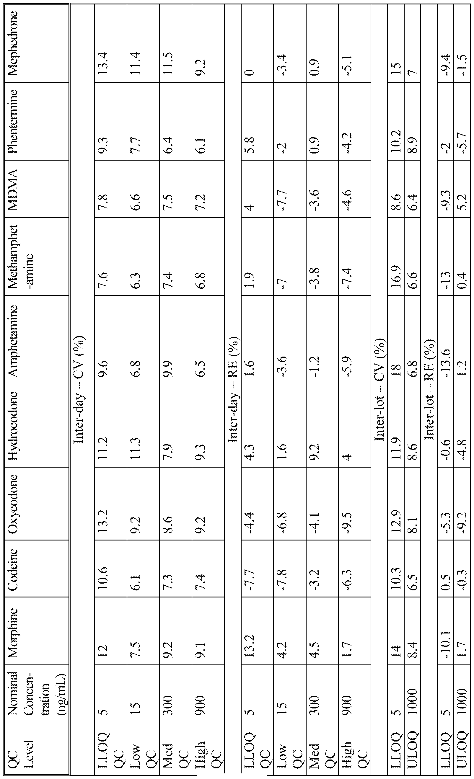


Claims
Priority Applications (10)
| Application Number | Priority Date | Filing Date | Title |
|---|---|---|---|
| US16/079,067 US11326992B2 (en) | 2016-02-24 | 2017-02-24 | Multilayer device for separating blood components and uses thereof |
| EP20203981.4A EP3800462A1 (en) | 2016-02-24 | 2017-02-24 | Multilayer device for separating blood components and uses thereof |
| DK17757324.3T DK3420334T3 (en) | 2016-02-24 | 2017-02-24 | MULTI-LAYER DEVICE FOR SEPARATION OF BLOOD COMPONENTS AND USES THEREOF |
| AU2017222642A AU2017222642C1 (en) | 2016-02-24 | 2017-02-24 | Multilayer device for separating blood components and uses thereof |
| CA3015627A CA3015627C (en) | 2016-02-24 | 2017-02-24 | Multilayer device for separating blood components and uses thereof |
| CN201780024862.XA CN109073519B (en) | 2016-02-24 | 2017-02-24 | Multi-layer device for separating blood components and use thereof |
| EP17757324.3A EP3420334B1 (en) | 2016-02-24 | 2017-02-24 | Multilayer device for separating blood components and uses thereof |
| ES17757324T ES2837380T3 (en) | 2016-02-24 | 2017-02-24 | Multilayer device to separate blood components and uses thereof |
| JP2018545317A JP6864919B2 (en) | 2016-02-24 | 2017-02-24 | Multi-layer device for separating blood components and how to use it |
| US17/738,871 US20220268675A1 (en) | 2016-02-24 | 2022-05-06 | Multilayer device for separating blood components and uses thereof |
Applications Claiming Priority (2)
| Application Number | Priority Date | Filing Date | Title |
|---|---|---|---|
| US201662299226P | 2016-02-24 | 2016-02-24 | |
| US62/299,226 | 2016-02-24 |
Related Child Applications (2)
| Application Number | Title | Priority Date | Filing Date |
|---|---|---|---|
| US16/079,067 A-371-Of-International US11326992B2 (en) | 2016-02-24 | 2017-02-24 | Multilayer device for separating blood components and uses thereof |
| US17/738,871 Continuation US20220268675A1 (en) | 2016-02-24 | 2022-05-06 | Multilayer device for separating blood components and uses thereof |
Publications (1)
| Publication Number | Publication Date |
|---|---|
| WO2017147456A1 true WO2017147456A1 (en) | 2017-08-31 |
Family
ID=59686659
Family Applications (1)
| Application Number | Title | Priority Date | Filing Date |
|---|---|---|---|
| PCT/US2017/019405 WO2017147456A1 (en) | 2016-02-24 | 2017-02-24 | Multilayer device for separating blood components and uses thereof |
Country Status (10)
| Country | Link |
|---|---|
| US (2) | US11326992B2 (en) |
| EP (2) | EP3420334B1 (en) |
| JP (1) | JP6864919B2 (en) |
| CN (1) | CN109073519B (en) |
| AU (1) | AU2017222642C1 (en) |
| CA (1) | CA3015627C (en) |
| DK (1) | DK3420334T3 (en) |
| ES (1) | ES2837380T3 (en) |
| PT (1) | PT3420334T (en) |
| WO (1) | WO2017147456A1 (en) |
Cited By (1)
| Publication number | Priority date | Publication date | Assignee | Title |
|---|---|---|---|---|
| CN108593828A (en) * | 2018-02-23 | 2018-09-28 | 李水军 | Blood plasma prepares the detection method of drug and toxic content in card |
Families Citing this family (4)
| Publication number | Priority date | Publication date | Assignee | Title |
|---|---|---|---|---|
| WO2018129056A1 (en) | 2017-01-04 | 2018-07-12 | The Research Foundation For The State University Of New York | Biomarker detection device |
| CN114761103A (en) * | 2019-10-02 | 2022-07-15 | 奥斯龙-明士克公司 | Blood component collecting and separating medium, blood component collecting and separating device comprising said medium and blood component separating and extracting method implementing said medium |
| WO2021096426A1 (en) * | 2019-11-12 | 2021-05-20 | National University Of Singapore | A multi-layered membrane and a method of preparing the same |
| KR102299968B1 (en) * | 2019-11-19 | 2021-09-09 | 경북대학교 산학협력단 | Multi-layered filter for separating blood components and disease diagnosis kit using the same |
Citations (9)
| Publication number | Priority date | Publication date | Assignee | Title |
|---|---|---|---|---|
| EP0418765A2 (en) | 1989-09-21 | 1991-03-27 | Becton, Dickinson and Company | Test device including flow control means |
| US5369007A (en) * | 1990-09-07 | 1994-11-29 | The United States Of America As Represented By The Secretary Of The Navy | Microassay on a card |
| US5733507A (en) * | 1995-06-07 | 1998-03-31 | Inphocyte, Inc. | Biological cell sample holder for use in infrared and/or Raman spectroscopy analysis holder |
| US6036659A (en) * | 1998-10-09 | 2000-03-14 | Flexsite Diagnostics, Inc. | Collection device for biological samples and methods of use |
| US6106732A (en) * | 1998-04-16 | 2000-08-22 | Binax Services, Inc. | Integral blood plasma or serum isolation, metering and transport device |
| WO2009075709A1 (en) | 2007-12-12 | 2009-06-18 | Micropoint Bioscience Inc | Rapid and efficient filtering whole blood in a capillary flow device |
| WO2012015926A2 (en) * | 2010-07-27 | 2012-02-02 | Northwestern University | Devices and methods for filtering blood plasma |
| KR20130142768A (en) * | 2012-06-20 | 2013-12-30 | (주)미코바이오메드 | Sensor strip |
| US20140295415A1 (en) | 2011-11-04 | 2014-10-02 | Diagnostics For All, Inc. | Low cost, disposable molecular diagnostic devices |
Family Cites Families (10)
| Publication number | Priority date | Publication date | Assignee | Title |
|---|---|---|---|---|
| US5273888A (en) * | 1984-01-16 | 1993-12-28 | Helena Laboratories Corporation | Chemical test kit and method for determining the presence of blood in a specimen and for verifying the effectiveness of the chemicals |
| JP2003075440A (en) * | 2001-06-19 | 2003-03-12 | Fuji Photo Film Co Ltd | Unit for biochemical analysis |
| US7407742B2 (en) | 2002-02-27 | 2008-08-05 | Sanko Junyaku Co., Ltd. | Plasma or serum separator, plasma or serum sampling method, plasma or serum separating method, test carrier and glass fiber |
| JP2004037103A (en) * | 2002-06-28 | 2004-02-05 | Fuji Photo Film Co Ltd | Method for measuring dropping characteristics of spotting device |
| JP4298555B2 (en) | 2004-03-26 | 2009-07-22 | 栄研化学株式会社 | Blood separation method and apparatus |
| CN202330105U (en) | 2011-09-30 | 2012-07-11 | 范婥民 | Preparation device for dried blood spot sample |
| JP6182547B2 (en) * | 2012-02-14 | 2017-08-16 | ウオーターズ・テクノロジーズ・コーポレイシヨン | Dry sample carrier with dissolvable sample collection area |
| EP2695656A1 (en) | 2012-08-09 | 2014-02-12 | F. Hoffmann-La Roche AG | Method and separation device for separating a filtrate from a sample liquid |
| AU2014368940B2 (en) | 2013-12-20 | 2019-10-31 | Spot Bioscience, Llc | Whole blood separation sampling apparatus |
| EP3017869A1 (en) * | 2014-11-05 | 2016-05-11 | Deutsche Sporthochschule Köln | Dried-Blood-Spot-card shipping and storage container |
-
2017
- 2017-02-24 CA CA3015627A patent/CA3015627C/en active Active
- 2017-02-24 EP EP17757324.3A patent/EP3420334B1/en active Active
- 2017-02-24 EP EP20203981.4A patent/EP3800462A1/en active Pending
- 2017-02-24 US US16/079,067 patent/US11326992B2/en active Active
- 2017-02-24 JP JP2018545317A patent/JP6864919B2/en active Active
- 2017-02-24 PT PT177573243T patent/PT3420334T/en unknown
- 2017-02-24 WO PCT/US2017/019405 patent/WO2017147456A1/en active Application Filing
- 2017-02-24 DK DK17757324.3T patent/DK3420334T3/en active
- 2017-02-24 CN CN201780024862.XA patent/CN109073519B/en active Active
- 2017-02-24 ES ES17757324T patent/ES2837380T3/en active Active
- 2017-02-24 AU AU2017222642A patent/AU2017222642C1/en active Active
-
2022
- 2022-05-06 US US17/738,871 patent/US20220268675A1/en active Pending
Patent Citations (9)
| Publication number | Priority date | Publication date | Assignee | Title |
|---|---|---|---|---|
| EP0418765A2 (en) | 1989-09-21 | 1991-03-27 | Becton, Dickinson and Company | Test device including flow control means |
| US5369007A (en) * | 1990-09-07 | 1994-11-29 | The United States Of America As Represented By The Secretary Of The Navy | Microassay on a card |
| US5733507A (en) * | 1995-06-07 | 1998-03-31 | Inphocyte, Inc. | Biological cell sample holder for use in infrared and/or Raman spectroscopy analysis holder |
| US6106732A (en) * | 1998-04-16 | 2000-08-22 | Binax Services, Inc. | Integral blood plasma or serum isolation, metering and transport device |
| US6036659A (en) * | 1998-10-09 | 2000-03-14 | Flexsite Diagnostics, Inc. | Collection device for biological samples and methods of use |
| WO2009075709A1 (en) | 2007-12-12 | 2009-06-18 | Micropoint Bioscience Inc | Rapid and efficient filtering whole blood in a capillary flow device |
| WO2012015926A2 (en) * | 2010-07-27 | 2012-02-02 | Northwestern University | Devices and methods for filtering blood plasma |
| US20140295415A1 (en) | 2011-11-04 | 2014-10-02 | Diagnostics For All, Inc. | Low cost, disposable molecular diagnostic devices |
| KR20130142768A (en) * | 2012-06-20 | 2013-12-30 | (주)미코바이오메드 | Sensor strip |
Non-Patent Citations (5)
| Title |
|---|
| ALAN J BARNES, NEXT GENERATION PLASMA COLLECTION TECHNOLOGY FOR THE CLINICAL ANALYSIS OF TEMOZOLOMIDE BY HILIC/MS/MS |
| BARNES, AJ ET AL.: "Next generation plasma collection technology for the clinical analysis of temozolomide by HILIC/MS/MS", A TECHNICAL PRODUCT REVIEW, June 2014 (2014-06-01), XP055413344, Retrieved from the Internet <URL:https://novilytic.com/wp-content/uploads/2015/05/Shimadzu-Workflow-for-Temozolomide.pdf> [retrieved on 20170412] * |
| KIM ET AL., ANAL CHEM, vol. 85, 2013, pages 11501 - 11508 |
| LI ET AL., RAPID COMMUN MASS SPECTROM, vol. 26, 2012, pages 1208 - 1212 |
| STURM ET AL., BIOANALYSIS, vol. 7, 2015, pages 1987 - 2002 |
Cited By (1)
| Publication number | Priority date | Publication date | Assignee | Title |
|---|---|---|---|---|
| CN108593828A (en) * | 2018-02-23 | 2018-09-28 | 李水军 | Blood plasma prepares the detection method of drug and toxic content in card |
Also Published As
| Publication number | Publication date |
|---|---|
| EP3420334A4 (en) | 2019-07-17 |
| JP2019512093A (en) | 2019-05-09 |
| US20220268675A1 (en) | 2022-08-25 |
| AU2017222642B2 (en) | 2021-12-09 |
| CN109073519A (en) | 2018-12-21 |
| DK3420334T3 (en) | 2020-12-14 |
| ES2837380T3 (en) | 2021-06-30 |
| CA3015627A1 (en) | 2017-08-31 |
| EP3800462A1 (en) | 2021-04-07 |
| JP6864919B2 (en) | 2021-04-28 |
| AU2017222642A1 (en) | 2018-09-13 |
| EP3420334A1 (en) | 2019-01-02 |
| CN109073519B (en) | 2021-08-17 |
| EP3420334B1 (en) | 2020-10-28 |
| AU2017222642C1 (en) | 2022-01-27 |
| PT3420334T (en) | 2020-12-17 |
| US11326992B2 (en) | 2022-05-10 |
| CA3015627C (en) | 2024-02-20 |
| US20190049353A1 (en) | 2019-02-14 |
Similar Documents
| Publication | Publication Date | Title |
|---|---|---|
| AU2017222642B2 (en) | Multilayer device for separating blood components and uses thereof | |
| Frey et al. | Emerging trends in paper spray mass spectrometry: Microsampling, storage, direct analysis, and applications | |
| Kim et al. | Simple, miniaturized blood plasma extraction method | |
| Wilhelm et al. | Therapeutic drug monitoring by dried blood spot: progress to date and future directions | |
| De Kesel et al. | Does volumetric absorptive microsampling eliminate the hematocrit bias for caffeine and paraxanthine in dried blood samples? A comparative study | |
| AU2016219358B2 (en) | Device and method for analysis of biofluids by ion generation using wetted porous material | |
| Henion et al. | Microsample analyses via DBS: challenges and opportunities | |
| Sturm et al. | Novel membrane devices and their potential utility in blood sample collection prior to analysis of dried plasma spots | |
| JP7109597B2 (en) | Simplified biological sample processing for LC-MS/MS | |
| JP6182547B2 (en) | Dry sample carrier with dissolvable sample collection area | |
| Hauser et al. | An autonomous microfluidic device for generating volume-defined dried plasma spots | |
| Lakshmy et al. | Role of dried blood spots in health and disease diagnosis in older adults | |
| JP2021535380A (en) | Microsampling detection in diabetes | |
| Ambach et al. | Determination of cocaine and metabolites in dried blood spots by LC-MS/MS | |
| US20220273209A1 (en) | Devices and methods for sample collection | |
| Shitole et al. | Technological advancement in dry blood matrix microsampling and its clinical relevance in quantitative drug analysis | |
| Sathyanarayanan et al. | Dried spot sample and its drug detection using LC-MS/MS: Trends and advances in matrix collection and bioanalytics | |
| Kaareddy et al. | Dried blood spot sampling in protein and peptide bioanalysis: optimism, experience, and the path forward | |
| Vloemans et al. | Precise sample metering method by coordinated burst action of hydrophobic burst valves applied to dried blood spot collection | |
| Oliveira et al. | Collection and Bioanalysis of Quantitative Microsamples: Technological Innovations and Practical Implications | |
| Oliveira et al. | Collection and Bioanalysis of Quantitative Microsamples | |
| CN111220431A (en) | Method for detecting active components in blood plasma and blood plasma card using same | |
| Geers et al. | Microsampling techniques for Antipsychotics; Current developments and future directions |
Legal Events
| Date | Code | Title | Description |
|---|---|---|---|
| ENP | Entry into the national phase |
Ref document number: 2018545317 Country of ref document: JP Kind code of ref document: A |
|
| WWE | Wipo information: entry into national phase |
Ref document number: 3015627 Country of ref document: CA |
|
| NENP | Non-entry into the national phase |
Ref country code: DE |
|
| ENP | Entry into the national phase |
Ref document number: 2017222642 Country of ref document: AU Date of ref document: 20170224 Kind code of ref document: A |
|
| WWE | Wipo information: entry into national phase |
Ref document number: 2017757324 Country of ref document: EP |
|
| ENP | Entry into the national phase |
Ref document number: 2017757324 Country of ref document: EP Effective date: 20180924 |
|
| 121 | Ep: the epo has been informed by wipo that ep was designated in this application |
Ref document number: 17757324 Country of ref document: EP Kind code of ref document: A1 |