WO2017122130A1 - Immune-stimulating humanized monoclonal antibodies against human interleukin-2, and fusion proteins thereof - Google Patents
Immune-stimulating humanized monoclonal antibodies against human interleukin-2, and fusion proteins thereof Download PDFInfo
- Publication number
- WO2017122130A1 WO2017122130A1 PCT/IB2017/050127 IB2017050127W WO2017122130A1 WO 2017122130 A1 WO2017122130 A1 WO 2017122130A1 IB 2017050127 W IB2017050127 W IB 2017050127W WO 2017122130 A1 WO2017122130 A1 WO 2017122130A1
- Authority
- WO
- WIPO (PCT)
- Prior art keywords
- seq
- antibody
- hcdr1
- hcdr3
- hcdr2
- Prior art date
Links
Classifications
-
- C—CHEMISTRY; METALLURGY
- C07—ORGANIC CHEMISTRY
- C07K—PEPTIDES
- C07K16/00—Immunoglobulins [IGs], e.g. monoclonal or polyclonal antibodies
- C07K16/18—Immunoglobulins [IGs], e.g. monoclonal or polyclonal antibodies against material from animals or humans
- C07K16/24—Immunoglobulins [IGs], e.g. monoclonal or polyclonal antibodies against material from animals or humans against cytokines, lymphokines or interferons
- C07K16/244—Interleukins [IL]
- C07K16/246—IL-2
-
- A—HUMAN NECESSITIES
- A61—MEDICAL OR VETERINARY SCIENCE; HYGIENE
- A61K—PREPARATIONS FOR MEDICAL, DENTAL OR TOILETRY PURPOSES
- A61K38/00—Medicinal preparations containing peptides
- A61K38/16—Peptides having more than 20 amino acids; Gastrins; Somatostatins; Melanotropins; Derivatives thereof
- A61K38/17—Peptides having more than 20 amino acids; Gastrins; Somatostatins; Melanotropins; Derivatives thereof from animals; from humans
- A61K38/19—Cytokines; Lymphokines; Interferons
- A61K38/20—Interleukins [IL]
- A61K38/2013—IL-2
-
- A—HUMAN NECESSITIES
- A61—MEDICAL OR VETERINARY SCIENCE; HYGIENE
- A61P—SPECIFIC THERAPEUTIC ACTIVITY OF CHEMICAL COMPOUNDS OR MEDICINAL PREPARATIONS
- A61P35/00—Antineoplastic agents
-
- C—CHEMISTRY; METALLURGY
- C07—ORGANIC CHEMISTRY
- C07K—PEPTIDES
- C07K14/00—Peptides having more than 20 amino acids; Gastrins; Somatostatins; Melanotropins; Derivatives thereof
- C07K14/435—Peptides having more than 20 amino acids; Gastrins; Somatostatins; Melanotropins; Derivatives thereof from animals; from humans
- C07K14/52—Cytokines; Lymphokines; Interferons
- C07K14/54—Interleukins [IL]
- C07K14/55—IL-2
-
- A—HUMAN NECESSITIES
- A61—MEDICAL OR VETERINARY SCIENCE; HYGIENE
- A61K—PREPARATIONS FOR MEDICAL, DENTAL OR TOILETRY PURPOSES
- A61K39/00—Medicinal preparations containing antigens or antibodies
- A61K2039/505—Medicinal preparations containing antigens or antibodies comprising antibodies
-
- A—HUMAN NECESSITIES
- A61—MEDICAL OR VETERINARY SCIENCE; HYGIENE
- A61K—PREPARATIONS FOR MEDICAL, DENTAL OR TOILETRY PURPOSES
- A61K2300/00—Mixtures or combinations of active ingredients, wherein at least one active ingredient is fully defined in groups A61K31/00 - A61K41/00
-
- A—HUMAN NECESSITIES
- A61—MEDICAL OR VETERINARY SCIENCE; HYGIENE
- A61K—PREPARATIONS FOR MEDICAL, DENTAL OR TOILETRY PURPOSES
- A61K38/00—Medicinal preparations containing peptides
-
- C—CHEMISTRY; METALLURGY
- C07—ORGANIC CHEMISTRY
- C07K—PEPTIDES
- C07K2317/00—Immunoglobulins specific features
- C07K2317/20—Immunoglobulins specific features characterized by taxonomic origin
- C07K2317/24—Immunoglobulins specific features characterized by taxonomic origin containing regions, domains or residues from different species, e.g. chimeric, humanized or veneered
-
- C—CHEMISTRY; METALLURGY
- C07—ORGANIC CHEMISTRY
- C07K—PEPTIDES
- C07K2317/00—Immunoglobulins specific features
- C07K2317/30—Immunoglobulins specific features characterized by aspects of specificity or valency
- C07K2317/34—Identification of a linear epitope shorter than 20 amino acid residues or of a conformational epitope defined by amino acid residues
-
- C—CHEMISTRY; METALLURGY
- C07—ORGANIC CHEMISTRY
- C07K—PEPTIDES
- C07K2317/00—Immunoglobulins specific features
- C07K2317/50—Immunoglobulins specific features characterized by immunoglobulin fragments
- C07K2317/55—Fab or Fab'
-
- C—CHEMISTRY; METALLURGY
- C07—ORGANIC CHEMISTRY
- C07K—PEPTIDES
- C07K2317/00—Immunoglobulins specific features
- C07K2317/70—Immunoglobulins specific features characterized by effect upon binding to a cell or to an antigen
- C07K2317/74—Inducing cell proliferation
-
- C—CHEMISTRY; METALLURGY
- C07—ORGANIC CHEMISTRY
- C07K—PEPTIDES
- C07K2317/00—Immunoglobulins specific features
- C07K2317/90—Immunoglobulins specific features characterized by (pharmaco)kinetic aspects or by stability of the immunoglobulin
- C07K2317/92—Affinity (KD), association rate (Ka), dissociation rate (Kd) or EC50 value
-
- C—CHEMISTRY; METALLURGY
- C07—ORGANIC CHEMISTRY
- C07K—PEPTIDES
- C07K2319/00—Fusion polypeptide
Definitions
- the present invention relates to antibodies binding to human interleukin-2 (hlL-2).
- the invention more specifically relates to humanized antibodies specifically binding a particular epitope of hlL-2 and, when bound to this epitope, displaying a unique capability of inhibiting binding of hlL-2 to CD25, and fusions between said antibodies and hlL-2.
- the invention relates to in vitro and in vivo therapeutic applications of the antibodies in combination with hlL-2, and in vitro and in vivo therapeutic applications of the fusions.
- BACKGROUND lnterleukin-2 (IL-2) is a cytokine able to potently stimulate cytotoxic lymphocytes against metastatic tumors.
- IL-2 is also able to stimulate so-called CD25 + CD4 + regulatory T cells (Treg cells) that are crucial for prevention of autoimmune disease.
- Treg cells can significantly dampen anti-tumor responses by cytotoxic lymphocytes, thus somewhat antagonizing the beneficial anti-tumor effects of IL-2.
- IL-2 can exert toxic adverse effects.
- IL-2 Immunotherapy using IL-2 has been used since the early 1980's for the immunotherapy of metastatic melanoma and metastatic renal cell carcinoma, leading to the approval by the FDA for these indications in 1996 and 1992, respectively. While IL-2 given at high doses has shown objective response rates in about 17% and complete regression in about 6-9% of patients suffering from these deadly metastatic cancers, IL-2 given at these doses frequently led to toxic adverse effects, such as hypotension, pulmonary edema, liver cell damage, gastrointestinal toxicity, vascular leakage syndrome (VLS) and general edema. Moreover, as mentioned above, IL-2 is able to stimulate immunosuppressive Treg cells, which in turn are able to dampen the activity of antitumor CD8 + T cells and NK cells.
- the present disclosure relates generally to antibodies or fragments thereof that bind to a specific epitope of human IL-2, methods for their preparation and use, including methods for treating disorders.
- the anti-IL-2 antibodies or fragments thereof disclosed herein can be used (alone or in combination with other agents or therapeutic modalities) to treat, prevent and/or diagnose disorders, such as cancerous disorders (for example solid and soft-tissue tumors, and hematological tumors), as well as infectious diseases (for example chronic infectious disorders).
- disorders such as cancerous disorders (for example solid and soft-tissue tumors, and hematological tumors), as well as infectious diseases (for example chronic infectious disorders).
- cancerous disorders for example solid and soft-tissue tumors, and hematological tumors
- infectious diseases for example chronic infectious disorders.
- the present disclosure provides an isolated antibody, or antigen-binding portion thereof, which binds human IL-2 according to SEQ ID NO: 109, wherein said antibody or antigen-binding portion thereof comprises a light chain variable region comprising LCDR1 , a LCDR2 and a LCDR3 and a heavy chain variable region comprising a HCDR1 , a HCDR2 and a HCDR3 and wherein the LCDR1 comprises SEQ ID NO: 122; wherein LCDR2 comprises SEQ ID NO: 123; wherein LCDR3 comprises SEQ ID NO: 21 ; wherein HCDR1 comprises SEQ ID NO: 1 19; wherein HCDR2 comprises SEQ ID NO: 120; and wherein HCDR3 comprises SEQ ID NO: 121.
- the isolated antibody or antigen-binding portion thereof comprises a light variable region comprising a: LCDR1 selected from the group consisting of SEQ ID NO: 19, SEQ ID NO: 31 , SEQ ID NO: 69, SEQ ID NO: 72, SEQ ID NO: 86 and SEQ ID NO: 90; LCDR2 selected from the group consisting of SEQ ID NO: 20 and SEQ ID NO: 32; LCDR3 as set forth in SEQ ID NO: 21 , and a heavy variable region comprising a: HCDR1 selected from the group consisting of SEQ ID NO: 4, and SEQ ID NO: 13;
- HCDR2 selected from the group consisting of SEQ ID NO: 2, and SEQ ID NO: 12
- HCDR3 selected from the group consisting of SEQ ID NO: 3, SEQ ID NO: 36, SEQ ID NO: 39, SEQ ID NO: 42, and SEQ ID NO: 45.
- the isolated antibody or antigen-binding portion thereof according to the first aspect, wherein the LCDR1 , LCDR2 and LCDR3 are SEQ ID NO: 19, 20 and 21 , respectively and the HCDR1 , HCDR2 and HCDR3 are SEQ ID NO: 4, 2 and 3, respectively; or LCDR1 , LCDR2 and LCDR3 are SEQ ID NO: 31 , 32 and 21 , respectively and the HCDR1 , HCDR2 and HCDR3 are SEQ ID NO: 4, 2 and 3, respectively; or LCDR1 , LCDR2 and LCDR3 are SEQ ID NO: 19, 20 and 21 and the HCDR1 , HCDR2 and HCDR3 are SEQ ID NO: 13, 12 and 3; or LCDR1 , LCDR2 and LCDR3 are SEQ ID NO: 31 , 32 and 21 and the HCDR1 , HCDR2 and HCDR3 are SEQ ID NO: 13, 12 and 3; or LCDR1 , LCDR2 and LCDR3 are SEQ ID NO:
- the isolated antibody or antigen-binding portion thereof comprises the heavy chain variable (VH) and light chain variable (VL) regions have at least 95%, such as 95%, 96%, 97%, 98%, or 99% identity to the amino acid sequences: VL, SEQ ID NO: 25; VH, SEQ ID NO: 7, or VL, SEQ ID NO: 27; VH, SEQ ID NO: 7, or VL, SEQ ID NO: 34; VH, SEQ ID NO: 7, or VL, SEQ ID NO: 25; VH, SEQ ID NO: 15, or VL, SEQ ID NO: 27; VH, SEQ ID NO: 15, or VL, SEQ ID NO: 34; VH, SEQ ID NO: 15, or VL, SEQ ID NO: 25; VH, SEQ ID NO: 17, or VL, SEQ ID NO: 27; VH, SEQ ID NO: 17, or VL, SEQ ID NO: 34; VH, SEQ ID NO: 17, or VL, SEQ ID NO: 34; VH, S
- the isolated antibody or antigen-binding portion thereof according to the first aspect has the heavy chain variable (VH) and light chain variable (VL) regions have the amino acid sequences: VL, SEQ ID NO: 25; VH, SEQ ID NO: 7, or VL, SEQ ID NO: 27; VH, SEQ ID NO: 7, or VL, SEQ ID NO: 34; VH, SEQ ID NO: 7, or VL, SEQ ID NO: 25; VH, SEQ ID NO: 15, or VL, SEQ ID NO: 27; VH, SEQ ID NO: 15, or VL, SEQ ID NO: 34; VH, SEQ ID NO: 15, or VL, SEQ ID NO: 25; VH, SEQ ID NO: 17, or VL, SEQ ID NO: 27; VH, SEQ ID NO: 17, or VL, SEQ ID NO: 34; VH, SEQ ID NO: 17, or VL, SEQ ID NO: 70; VH, SEQ ID NO: 7, or VL, SEQ ID NO: 25;
- the isolated antibody may comprise an Fc domain selected from the group consisting of SEQ ID NO: 93, SEQ ID NO: 95, SEQ ID NO: 97, SEQ ID NO: 99, SEQ ID NO: 101 , SEQ ID NO: 103 and SEQ ID NO: 105.
- the isolated antibody comprises the Fc domain according to SEQ ID NO: 93, SEQ ID NO: 101 , SEQ ID NO: 103 or SEQ ID NO: 105.
- the isolated antibody comprises the light chain according to SEQ ID NO: 124 and the heavy chain according to SEQ ID NO: 126, or the light chain according to SEQ ID NO: 128 and the heavy chain according to SEQ ID NO: 130.
- an isolated antibody or antigen-binding fragment thereof which binds to a human interleukin-2 (hlL-2) epitope which comprises the amino acids K52, P54, K55, T57, R58, T61 , F62, K63, Q94, and K96.
- hlL-2 human interleukin-2
- the isolated antibody or antigen-binding fragment thereof according to the second aspect binds specifically to the amino acids K52, P54, K55, T57, R58, T61 , F62, K63, Q94, and K96.
- the isolated antibody or antigen-binding fragment thereof according to the second aspect may bind to a human interleukin-2 (hlL-2) epitope which, in addition to the amino acids K52, P54, K55, T57, R58, T61 , F62, K63, Q94, and K96, further comprises any one or more of the amino acids N50, N53, N91 , L92, A93, and N97.
- the antibody or antigen-binding fragment thereof according to the second aspect binds specifically to the amino acids N50, K52, N53, P54, K55, T57, R58, T61 , F62, K63, N91 , L92, A93, Q94, K96, and N97.
- the antibody or antigen-binding fragment thereof comprises a light chain variable region comprising, in sequence a LCDR1 , a LCDR2 and a LCDR3, and a heavy chain variable region comprising, in sequence a HCDR1 , a HCDR2 and a HCDR3, wherein the LCDR1 , LCDR2 and LCDR3 are SEQ ID NO: 231 , 232 and 233, respectively and the HCDR1 , HCDR2 and HCDR3 are SEQ ID NO: 181 , 182 and 183, respectively; or the LCDR1 , LCDR2 and LCDR3 are SEQ ID NO: 279, 280 and 281 , respectively and the HCDR1 , HCDR2 and HCDR3 are SEQ ID NO: 213, 214 and 215, respectively; or the LCDR1 , LCDR2 and LCDR3 are SEQ ID NO: 231 , 232 and 233, respectively and the HCDR1 , HCDR2 and HCDR3
- the antibody or antigen-binding fragment thereof comprises the heavy chain variable (VH) and light chain variable (VL) regions have at least 95% identity, such as 100% identity, to the amino acid sequences VL, SEQ ID NO: 243; VH, SEQ ID NO: 193, or VL, SEQ ID NO: 391 ; VH, SEQ ID NO: 225, or VL, SEQ ID NO: 243; VH, SEQ ID NO: 225, or VL, SEQ ID NO: 275; VH, SEQ ID NO: 225, or VL, SEQ ID NO: 275; VH, SEQ ID NO: 161 , or VL, SEQ ID NO: 70; VH, SEQ ID NO: 209, or VL, SEQ ID NO: 243; VH, SEQ ID NO: 209.
- the isolated antibody comprises wherein the heavy chain and light chain regions have the amino acid sequences; heavy chain according to SEQ ID NO: 195 and light chain according to SEQ ID NO: 245, or heavy chain according to SEQ ID NO: 227 and light chain according to SEQ ID NO: 393, or heavy chain according to SEQ ID NO: 227 and light chain according to SEQ ID NO: 245, or heavy chain according to SEQ ID NO: 227 and light chain according to SEQ ID NO: 277, or heavy chain according to SEQ ID NO: 163 and light chain according to SEQ ID NO: 277, or heavy chain according to SEQ ID NO: 21 1 and light chain according to SEQ ID NO: 261 , or heavy chain according to SEQ ID NO: 21 1 and light chain according to SEQ ID NO: 277.
- a composition comprising the antibody according to the first or the second aspect of the invention, and optionally but preferably, human IL-2, is provided.
- the composition according to the third aspect comprises the human IL-2 selected from the group consisting of human IL-2 according to SEQ ID NO: 109 or aldesleukin according to SEQ ID NO: 110, preferably aldesleukin according to SEQ ID NO: 1 10.
- a fusion protein comprising an antibody according to the first or second aspect of the invention, and human IL-2.
- the antibody and the human IL-2 is joined by a linker sequence selected from the group consisting of SEQ ID NO: 397, SEQ ID NO: 398, SEQ ID NO: 399, SEQ ID NO: 400, SEQ ID NO: 401 , SEQ ID NO: 402, SEQ ID NO: 403, SEQ ID NO: 404, SEQ ID NO: 405, SEQ ID NO: 406, SEQ ID NO: 407, SEQ ID NO: 408, SEQ ID NO: 409, SEQ ID NO: 410, and SEQ ID NO: 411 , preferably SEQ ID NO: 405 or SEQ ID NO: 407.
- a linker sequence selected from the group consisting of SEQ ID NO: 397, SEQ ID NO: 398, SEQ ID NO: 399, SEQ ID NO: 400, SEQ ID NO: 401 , SEQ ID NO: 402, SEQ ID NO: 403, SEQ ID NO: 404, SEQ ID NO: 405, SEQ ID NO: 406, SEQ ID NO: 407
- the fusion protein comprises an antibody according to the first or second aspect of the invention, wherein the LCDR1 of the antibody comprises a residue Y27 and a residue D30 according to the Kabat definition, and wherein the residue Y27 is joined to residue N97 of human IL-2 with a GG linker, and wherein residue D30 is joined to residue K96 residue of human IL-2 with a linker according to SEQ ID NO: 412.
- an antibody or antigen-binding fragment thereof according to the first or second aspect of the invention, or a composition according to the third aspect of the invention, or the fusion protein according to the fourth aspect of the invention, for use as a medicament is provided.
- an antibody or antigen-binding fragment thereof according to the first or second aspect of the invention, or a composition according to the third aspect of the invention, or the fusion protein according to the fourth aspect of the invention, for use in the manufacture of a medicament is provided.
- an antibody or antigen-binding fragment thereof according to the first or second aspect of the invention, or a composition according to the third aspect of the invention, or the fusion protein according to the fourth aspect of the invention, for use in treatment of cancer is provided.
- an antibody or antigen-binding fragment thereof according to the first or second aspect of the invention, or a composition according to the third aspect of the invention, or the fusion protein according to the fourth aspect of the invention.
- a vector comprising the nucleic acid molecule capable of encoding an antibody, or fragment thereof, according to the first or second aspects of the invention, or the fusion protein according to the fourth aspect of the invention.
- a cell comprising the vector according to the eight aspect of the invention.
- a cell able to produce a human interleukin-2 (hlL-2) specific monoclonal antibody, or antigen-binding fragment thereof, according to the first or second aspect, is provided.
- hlL-2 human interleukin-2
- a monoclonal antibody-producing hybridoma cell line is provided, characterized in that said produced antibodies are those the first or second aspect of the invention, or the fusion protein according to the fourth aspect of the invention.
- the antibodies according to aspects of the invention are advantageous, e.g. because they possess one or more of the following properties.
- the resulting mAb*hll_-2 complex cannot efficiently bind human IL-2 receptor alpha (also known as CD25) anymore, effectively reducing the binding of human CD25 to mAb*hll_-2 to background levels as compared to the binding of human CD25 to free (non- complexed) hlL-2 when measured by surface plasmon resonance.
- the antibodies may display no measurable cross-reactivity to murine IL-2.
- Figure 1 provides the overview of the three-dimensional structure of Proleukin®/Fab- NARA1 complex as obtained in Example 2.
- Figure 2 provides further analysis of epitope residues.
- the X-axis lists the amino acid sequence and numbering according to SEQ ID NO: 1 10.
- the upper side of Y-axis shows the total number of atoms of NARA1-Fab that are within 4 A from corresponding residue from Proleukin® and the lower side of Y-axis shows the reduced solvent-accessible area (A 2 ) of corresponding residue from Proleukin® as a consequence of binding to NARA1- Fab, according to SEQ ID NO: 132.
- Figure 3 illustrates the most critical epitope residue recognized by the NARA1-Fab.
- Figure 4 shows the overlay of Proleukin®/NARA1-Fab complex with IL- 2/CD25/CD122/CD132 quaternary complex.
- Figure 5 displays the overlay of C helices from IL-2_C145A (PDB: 3INK), the D10 IL-2- mutein ("Superkine”: PDB: 3QB1), IL-2/CD25/CD122/CD132 (PDB: 2B5I), and Proleukin®/NARA1-Fab.
- Figure 6 shows the counts of immune cells from spleens of mice receiving phosphate- buffered saline (PBS), low dose human IL-2 (IL-2 LD), high dose human IL-2 (IL-2 HD), or IL-2/antibody complexes made by using the indicated monoclonal antibodies.
- PBS phosphate- buffered saline
- IL-2 LD low dose human IL-2
- IL-2 HD high dose human IL-2
- IL-2/antibody complexes made by using the indicated monoclonal antibodies.
- the values are shown in tables 14 and 15.
- Figure 7 shows representative bromodeoxyuridine (BrdU) profiles of CD8 + CD44 + CD122 + T cells from the spleens of mice treated as in Figure 6.
- Figure 8 shows representative BrdU profiles of CD3 " NK1.1 + NK cells from spleens of mice treated as in Figure 6.
- Figure 9 and Figure 10 is a schematic illustrating a fusion protein according to an embodiment.
- Figure 11 illustrates alignment of a fusion protein according to an embodiment.
- Figures 12 to 15 show phenotypic characterization of endogenous CD8 + T cells, CD8 + CD44 + CD122 + T cells, CD4 + T cells, CD4 + CD25 high Foxp3 + Treg cells and CD3 " NK1.1 + NK cells from spleens of mice treated as in Figure 6, according to an example.
- Figures 16 and 17 show the counts of immune cell subsets and CD8 + CD44 + T cell-to- CD4 + CD25 h ' 9h Foxp3 + Treg cell ratios obtained from spleens of mice treated as in Figure 6, according to an example.
- Figures 18 to 22 show representative BrdU profiles of CD8 + , CD8 + CD44 + CD122 + T cells, CD3 " NK1.1 + DX5 + NK cells, CD4 + T cells and CD4 + CD25 + T cells, from spleens of mice treated as in Figure 6, according to an example.
- Figure 23 shows phenotypic characterization of endogenous CD8 + T cells and NK cells from spleens of mice receiving PBS, fusion proteins L15, L20 or L25, or human IL- 2/NARA1 complexes, according to an example.
- Figure 24 shows the cell counts of the indicated immune cells obtained from spleens of mice treated as in Figure 23, according to an example.
- Figure 25 shows representative BrdU profiles of CD8 + CD44 + CD122 + T cells and CD3 " NK1.1 + NK cells from spleens of mice treated as in Figure 23, according to an example.
- Figure 26 shows CTLL-2 cell proliferation curves from in vitro experiments using CTLL-2 cells stimulated with titrated doses of the fusion proteins L15, L20 or L25, of human IL- 2/NARA1 complexes, or of human IL-2, according to an example.
- Figure 27 shows STAT5 phosphorylation levels of CTLL-2 cells stimulated with titrated doses of human IL-2/NARA1 complexes, of human IL-2, or of the fusion proteins L15, L20 or L25, according to an example.
- Table 1 is an overview of anti-IL-2 antibodies according to embodiments of the invention.
- Table 2 is an overview of IL-2 muteins according to embodiments of the invention.
- Table 3 represents structure statistics for a Proleukin®/NARA1-Fab complex.
- Table 4 is an overview of epitope and paratope according to embodiments of the invention.
- Table 5 is an overview of variable heavy regions according to embodiments of the invention.
- Table 6 is an overview of variable light regions according to embodiments of the invention.
- Table 7 comprises pi data of antibodies according to some embodiments.
- Table 8 comprises comparison variable regions and variable germline regions.
- Table 9 comprises structure- refined variable regions according to embodiments of the invention.
- Table 10 comprises information about variable light chains and variable heavy chains according to embodiments of the invention.
- Table 11 comprises light chain CDRs according to embodiments of the invention.
- Table 12 comprises heavy chain CDRs according to embodiments of the invention.
- Table 13 comprises optimized variable light chains and variable heavy chains according to embodiments of the invention.
- Table 14 is an overview of VH mutation sequences.
- Table 15 is an overview of VK mutation sequences.
- Table 16 is an overview of plasmid sequences.
- Table 17 is an overview of affinity-matured antibodies according to embodiments of the invention.
- Table 18 is a sequence overview of first set of antibodies
- Table 19 is an overview of ELISA values according to an example.
- Table 20 is an overview of EC50 values according to an example.
- Table 21 is a subset of affinity matured antibodies according to embodiments of the invention.
- Table 22 is a sequence overview of the subset of antibodies according to Table 21.
- Table 23 represents binding affinity data.
- Table 24 represents CD8 + T cell proliferation data.
- Table 25 represents CD8 + T and NK cell proliferation data.
- Table 26 and Table 27 represent cell count data.
- Table 28 is an overview of linker sequences according to embodiments of the invention.
- Table 29 is an overview of fusion proteins according to embodiments of the invention.
- Table 30 represents CD8 + T and NK cell proliferation data.
- Table 31 represents CD8 + T, NK and Treg cell count data.
- Table 32 and Table 33 represent cell count data.
- Table 34 represents ratios of cell count data.
- Table 35 and Table 36 represent light region sequences of fusion proteins.
- Table 37 is an overview of fusion proteins.
- Table 38 represents cell count data.
- Table 39 and Table 40 represent EC50 values according to an example.
- Table 41 is a sequence listing comprising sequences useful for practicing the invention. DETAILED DESCRIPTION
- the present disclosure relates to antibodies and fragments thereof that bind to human IL- 2, and affect the in vivo function of this cytokine.
- human interleukin-2 or "hlL-2” as used herein is meant human IL-2 (wildtype or wt) with UniProt ID number P60568, reproduced herein as SEQ ID NO: 109.
- variants, isoforms, and species homologs of human wildtype IL-2 are also included.
- antibodies of this disclosure may, in certain cases, cross-react with IL-2 from species other than human. In certain embodiments, the antibodies may be completely specific for one or more human IL-2 proteins and may not exhibit species or other types of non-human cross-reactivity. .
- mutein means a polypeptide wherein specific substitutions to the interleukin-2 protein have been made.
- IL-2 mutein means 1 , 2, 3, 4, or 5 or more IL-2 muteins.
- treatment using an IL-2 mutein may refer to treatment with a single IL-2 mutein, or a combination of multiple IL-2 muteins.
- human IL-2 is the IL-2 mutein disclosed in WO2012/107417A1 , having 3 mutations compared to wt hlL-2.
- Proleukin® (aldesleukin) is another example of a variant of human wt IL-2, well known to a person skilled in the art, and represented herein by SEQ ID NO: 110.
- antibody or “antibody to IL-2” and the like as used herein refers to whole antibodies that interact with (e.g., by binding, steric hindrance, stabilizing/destabilizing, spatial distribution) an IL-2 epitope and interfere with IL-2's binding to IL-2 receptor alpha (also termed CD25).
- a naturally occurring "antibody” is a glycoprotein comprising at least two heavy (H) chains and two light (L) chains inter-connected by disulfide bonds. Each heavy chain is comprised of a heavy chain variable region (abbreviated herein as VH) and a heavy chain constant region.
- VH heavy chain variable region
- the heavy chain constant region is comprised of three domains, CH1 , CH2 and CH3.
- Each light chain is comprised of a light chain variable region (abbreviated herein as VL) and a light chain constant region.
- the light chain constant region is comprised of one domain, CL.
- the VH and VL regions can be further subdivided into regions of hypervariability, termed complementarity determining regions (CDRs), interspersed with regions that are more conserved, termed framework regions (FRs).
- CDRs complementarity determining regions
- FRs framework regions
- Each VH and VL is composed of three CDRs and four FRs arranged from amino-terminus to carboxy-terminus in the following order: FR1 , CDR1 , FR2, CDR2, FR3, CDR3, FR4.
- the variable regions of the heavy and light chains contain a binding domain that interacts with an antigen.
- the constant regions of the antibodies may mediate the binding of the immunoglobulin to host tissues or factors, including various cells of the immune system (e.g., effector cells) and the first component (Clq) of the classical complement system.
- the term "antibody” includes for example, monoclonal antibodies, human antibodies, humanized antibodies, camelid antibodies, or chimeric antibodies.
- the antibodies can be of any isotype (e.g., IgG, IgE, IgM, IgD, IgA and IgY), class (e.g., lgG1 , lgG2, lgG3, lgG4, lgA1 and lgA2) or subclass.
- variable domains of both the light (VL) and heavy (VH) chain portions determine antigen recognition and specificity.
- the constant domains of the light chain (CL) and the heavy chain (CH1 , CH2 or CH3) confer important biological properties such as secretion, transplacental mobility, Fc receptor binding, complement binding, and the like.
- the numbering of the constant region domains increases as they become more distal from the antigen-binding site or amino- terminus of the antibody.
- the N-terminus is a variable region and at the C-terminus is a constant region; the CH3 and CL domains actually comprise the carboxy-terminus of the heavy and light chain, respectively.
- the term "antibody” specifically includes an IgG-scFv format.
- CDRs Complementarity Determining Regions
- the CDR amino acids in the VH are numbered 26-32 (HCDR1), 52-56 (HCDR2), and 95-102 (HCDR3); and the amino acid residues in VL are numbered 26-32 (LCDR1), 50-52 (LCDR2), and 91-96 (LCDR3).
- the CDRs consist of amino acid residues 26-35 (HCDR1), 50- 65 (HCDR2), and 95-102 (HCDR3) in human VH and amino acid residues 24-34 (LCDR1), 50-56 (LCDR2), and 89-97 (LCDR3) in human VL.
- the CDR amino acid residues in the VH are numbered approximately 26-35 (CDR1), 51-57 (CDR2) and 93-102 (CDR3), and the CDR amino acid residues in the VL are numbered approximately 27-32 (CDR1), 50-52 (CDR2), and 89-97 (CDR3) (numbering according to "Kabat").
- the CDR regions of an antibody can be determined using the program IMGT/DomainGap Align.
- epitope binding domain refers to portions of the antigen-binding portion (e.g., an antibody or epitope-binding fragment or derivative thereof), that specifically interacts with (e.g., by binding, steric hindrance, stabilizing/destabilizing, spatial distribution) a binding site on a target epitope.
- EBD also refers to one or more fragments of an antibody that retain the ability to specifically interact with (e.g., by binding, steric hindrance, stabilizing/destabilizing, spatial distribution) an IL-2 epitope and interferes with IL-2's binding to IL-2 receptor alpha (CD25).
- antibody fragments include, but are not limited to, an scFv, a Fab fragment, a monovalent fragment consisting of the VL, VH, CL and CH1 domains; a F(ab)2 fragment, a bivalent fragment comprising two Fab fragments linked by a disulfide bridge at the hinge region; a Fd fragment consisting of the VH and CH1 domains; a Fv fragment consisting of the VL and VH domains of a single arm of an antibody; a dAb fragment (Ward et al., (1989) Nature 341 :544-546), which consists of a VH domain; and an isolated complementarity determining region (CDR).
- an scFv a Fab fragment, a monovalent fragment consisting of the VL, VH, CL and CH1 domains
- F(ab)2 fragment a bivalent fragment comprising two Fab fragments linked by a disulfide bridge at the hinge region
- a Fd fragment consisting of the
- epitope refers to any determinant capable of binding with high affinity to an immunoglobulin.
- An epitope is a region of an antigen that is bound by an antibody that specifically targets that antigen, and when the antigen is a protein, includes specific amino acids that directly contact the antibody. Most often, epitopes reside on proteins, but in some instances, may reside on other kinds of molecules, such as nucleic acids.
- Epitope determinants may include chemically active surface groupings of molecules such as amino acids, sugar side chains, phosphoryl or sulfonyl groups, and may have specific three dimensional structural characteristics, and/or specific charge characteristics.
- the two domains of the Fv fragment, VL and VH are coded for by separate genes, they can be joined, using recombinant methods, by a synthetic linker that enables them to be made as a single protein chain in which the VL and VH regions pair to form monovalent molecules (known as single chain Fv (scFv); see e.g., Bird et al., (1988) Science 242:423-426; and Huston et al., (1988) Proc. Natl. Acad. Sci. 85:5879-5883).
- scFv single chain Fv
- single chain antibodies are also intended to be encompassed within the terms “fragment”, “epitope-binding fragment” or “antibody fragment”. These fragments are obtained using conventional techniques known to those of skill in the art, and the fragments are screened for utility in the same manner as are intact antibodies.
- Antibody fragments can be incorporated into single chain molecules comprising a pair of tandem Fv segments (VH-CH1-VH-CH1) which, together with complementary light chain polypeptides, form a pair of antigen-binding regions (Zapata et al., (1995) Protein Eng. 8: 1057-1062; and U.S. Pat. No. 5,641 ,870), and also include Fab fragments, F(ab') fragments, F(ab') 2 fragments, and anti-idiotypic (anti-Id) antibodies (including, e.g., anti-Id antibodies to antibodies of the invention), and epitope-binding fragments of any of the above.
- EBDs also include single domain antibodies, maxibodies, unibodies, minibodies, triabodies, tetrabodies, v-NAR and bis-scFv, as is known in the art (see, e.g., Hollinger and Hudson, (2005) Nature Biotechnology 23: 1 126-1136), bispecific single chain diabodies, or single chain diabodies designed to bind two distinct epitopes.
- EBDs also include antibody-like molecules or antibody mimetics, which include, but not limited to minibodies, maxybodies, Fn3 based protein scaffolds, Ankrin repeats (also known as DARpins), VASP polypeptides, Avian pancreatic polypeptide (aPP), Tetranectin, Affililin, Knottins, SH3 domains, PDZ domains, Tendamistat, Neocarzinostatin, Protein A domains, Lipocalins, Transferrin, and Kunitz domains that specifically bind epitopes, which are within the scope of the invention.
- Antibody fragments can be grafted into scaffolds based on polypeptides such as Fibronectin type III (Fn3) (see U.S. Pat. No. 6,703, 199, which describes fibronectin polypeptide monobodies).
- Fn3 Fibronectin type III
- isolated antibody refers to antibody that is substantially free of other antibodies having different antigenic specificities (e.g., an isolated antibody that specifically binds IL-2 is substantially free of antibodies that specifically bind antigens other than IL-2).
- An isolated antibody that specifically binds IL-2 may, however, have cross- reactivity to other antigens, such as IL-2 molecules from other species.
- an isolated antibody may be substantially free of other cellular material and/or chemicals.
- monovalent antibody refers to an antibody that binds to a single epitope on a target molecule such as IL-2.
- bivalent antibody refers to an antibody that binds to two epitopes on at least two identical IL-2 target molecules.
- the bivalent antibody may also crosslink the target IL-2 molecules to one another.
- a “bivalent antibody” also refers to an antibody that binds to two different epitopes on at least two identical IL-2 target molecules.
- multivalent antibody refers to a single binding molecule with more than one valency, where "valency” is described as the number of antigen-binding moieties present per molecule of an antibody construct. As such, the single binding molecule can bind to more than one binding site on a target molecule.
- multivalent antibodies include, but are not limited to bivalent antibodies, trivalent antibodies, tetravalent antibodies, pentavalent antibodies, and the like, as well as bispecific antibodies and biparatopic antibodies.
- the mutivalent antibody e.g., an IL-2 biparatopic antibody
- the multivalent antibody mediates biological effect or which modulates a disease or disorder in a subject (e.g., by mediating or promoting cell killing, or by modulating the amount of a substance which is bioavailable.
- multivalent antibody also refers to a single binding molecule that has more than one antigen-binding moieties for two separate IL-2 target molecules. For example, an antibody that binds to both an IL-2 target molecule and a second target molecule that is not IL-2.
- a multivalent antibody is a tetravalent antibody that has four epitope binding domains.
- a tetravalent molecule may be bispecific and bivalent for each binding site on that target molecule.
- biparatopic antibody refers to an antibody that binds to two different epitopes on a single IL-2 target.
- the term also includes an antibody, which binds to two domains of at least two IL-2 targets, e.g., a tetravalent biparatopic antibody.
- bispecific antibody refers to an antibody that binds to two or more different epitopes on at least two different targets (e.g., an IL-2 and a target that is not IL-2).
- monoclonal antibody or “monoclonal antibody composition” as used herein refers to polypeptides, including antibodies, bispecific antibodies, etc. that have substantially identical to amino acid sequence or are derived from the same genetic source. This term also includes preparations of antibody molecules of single molecular composition. A monoclonal antibody composition displays a single binding specificity and affinity for a particular epitope.
- humanized antibody or “humanized anti-IL-2 antibody” as used herein includes antibodies in which CDR sequences derived from the germline of another mammalian species, such as a mouse, have been grafted onto human framework sequences. Additional framework region modifications may be made within the human framework sequences as well as within the CDR sequences derived from the germline of another mammalian species.
- the humanized antibodies of the invention may include amino acid residues not encoded by human sequences (e.g., mutations introduced by random or site-specific mutagenesis in vitro or by somatic mutation in vivo, or a conservative substitution to promote stability or manufacturing).
- recombinant humanized antibody includes all human antibodies that are prepared, expressed, created or isolated by recombinant means, such as antibodies isolated from a host cell transformed to express the humanized antibody, e.g., from a transfectoma, and antibodies prepared, expressed, created or isolated by any other means that involve splicing of all or a portion of a human immunoglobulin gene, sequences to other DNA sequences.
- Fc region refers to a polypeptide comprising the CH3, CH2 and at least a portion of the hinge region of a constant domain of an antibody.
- an Fc region may include a CH4 domain, present in some antibody classes.
- An Fc region may comprise the entire hinge region of a constant domain of an antibody.
- the invention comprises an Fc region and a CH1 region of an antibody.
- the invention comprises an Fc region CH3 region of an antibody.
- the invention comprises an Fc region, a CH1 region and a Ckappa/lambda region from the constant domain of an antibody.
- a binding molecule of the invention comprises a constant region, e.g., a heavy chain constant region.
- a constant region is modified compared to a wild-type constant region.
- the polypeptides of the invention disclosed herein may comprise alterations or modifications to one or more of the three heavy chain constant domains (CH 1 , CH2 or CH3) and/or to the light chain constant region domain (CL).
- Example modifications include additions, deletions or substitutions of one or more amino acids in one or more domains. Such changes may be included to optimize effector function, half-life, etc.
- binding site comprises an area on an IL-2 target molecule to which an antibody or antigen-binding fragment selectively binds.
- fusion protein is a fusion of two separate proteins, with or without an additional linker sequence.
- linker sequence is an amino acid sequence used to link or join two proteins.
- antibodies specific for a particular target antigen will bind to an epitope on the target antigen in a complex mixture of proteins and/or macromolecules.
- affinity refers to the strength of interaction between antibody and antigen at single antigenic sites. Within each antigenic site, the variable region of the antibody “arm” interacts through weak non-covalent forces with the antigen at numerous sites; the more interactions, the stronger the affinity.
- high affinity for an IgG antibody or fragment thereof refers to an antibody having a KD of 10 "8 M or less, 10 "9 M or less, or 10 "10 M, or 10 "11 M or less, or 10 "12 M or less, or 10 "13 M or less for a target antigen.
- high affinity binding can 10 vary for other antibody isotypes.
- high affinity binding for an IgM isotype refers to an antibody having a KD of 10 "7 M or less, or 10 "8 M or less.
- the term "Avidity” refers to an informative measure of the overall stability or strength of the antibody-antigen complex. It is controlled by three major factors: antibody epitope affinity; the valence of both the antigen and antibody; and the structural arrangement of the interacting parts. Ultimately these factors define the specificity of the antibody, that is, the likelihood that the particular antibody is binding to a precise antigen epitope. Regions of a given polypeptide that include an epitope can be identified using any number of epitope mapping techniques, well known in the art. See, e.g., Epitope Mapping Protocols in Methods in Molecular Biology, Vol. 66 (Glenn E. Morris, Ed., 1996) Humana Press, Totowa, N.J.
- linear epitopes may be determined by e.g., concurrently synthesizing large numbers of peptides on solid supports, the peptides corresponding to portions of the protein molecule, and reacting the peptides with antibodies while the peptides are still attached to the supports.
- Such techniques are known in the art and described in, e.g., U.S. Pat. No. 4,708,871 ; Geysen et al., (1984) Proc. Natl. Acad. Sci. USA 8:3998-4002; Geysen et al., (1985) Proc. Natl. Acad. Sci. USA 82:78-182; Geysen et al., (1986) Mol. Immunol.
- conformational epitopes are readily identified by determining spatial conformation of amino acids such as by, e.g., x-ray crystallography and two-dimensional nuclear magnetic resonance. See, e.g., Epitope Mapping Protocols, supra.
- Antigenic regions of proteins can also be identified using standard antigenicity and hydropathy plots, such as those calculated using, e.g., the Omiga version 1.0 software program available from the Oxford Molecular Group. This computer program employs the Hopp/Woods method, Hopp et al., (1981) Proc. Natl. Acad. Sci USA 78:3824-3828; for determining antigenicity profiles, and the Kyte-Doolittle technique, Kyte et al., (1982) J. Mol. Biol. 157: 105-132; for hydropathy plots.
- a cell includes a plurality of cells, including mixtures thereof. All numerical designations, e.g., pH, temperature, time, concentration, and molecular weight, including ranges, are approximations which are varied (+) or (-) by increments of 0.1. It is to be understood, although not always explicitly stated that all numerical designations are preceded by the term “about.” It also is to be understood, although not always explicitly stated, that the reagents described herein are merely examples and that equivalents of such are known in the art.
- amino acid refers to either natural and/or unnatural or synthetic amino acids, and both the D and L optical isomers, amino acid analogs, and peptidomimetics.
- a peptide of three or more amino acids is commonly called an oligopeptide if the peptide chain is short. If the peptide chain is long, the peptide is commonly called a polypeptide or a protein.
- biomarker or “marker” are used interchangeably herein.
- a biomarker is a nucleic acid or polypeptide and the presence or absence of a mutation or differential expression of the polypeptide is used to determine sensitivity to any treatment comprising an anti-IL-2 antibody according to the invention.
- a protein is a biomarker for a cancer cell when it is deficient, mutated, deleted, or decreased in post-translational modification, production, expression, level, stability and/or activity, as compared to the same protein in a normal (non-cancerous) cell or control cell.
- cDNA refers to complementary DNA, i.e. mRNA molecules present in a cell or organism made into cDNA with an enzyme such as reverse transcriptase.
- a "cDNA library” is a collection of all of the mRNA molecules present in a cell or organism, all turned into cDNA molecules with the enzyme reverse transcriptase, then inserted into “vectors” (other DNA molecules that can continue to replicate after addition of foreign DNA).
- Example vectors for libraries include bacteriophage (also known as "phage"), viruses that infect bacteria, for example, lambda phage. The library can then be probed for the specific cDNA (and thus mRNA) of interest.
- cell proliferative disorders shall include dysregulation of normal physiological function characterized by abnormal cell growth and/or division or loss of function.
- Examples of “cell proliferative disorders” include, but are not limited to, hyperplasia, neoplasia, metaplasia, and various autoimmune disorders, e.g., those characterized by the dysregulation of T cell apoptosis.
- Combination refers to either a fixed combination in one dosage unit form, or a combined administration where a compound of the present invention and a combination partner (e.g. another drug as explained below, also referred to as “therapeutic agent” or “co-agent”) may be administered independently at the same time or separately within time intervals, especially where these time intervals allow that the combination partners show a cooperative, e.g. synergistic effect.
- the single components may be packaged in a kit or separately.
- One or both of the components e.g., powders or liquids
- co- administration or “combined administration” or the like as utilized herein are meant to encompass administration of the selected combination partner to a single subject in need thereof (e.g. a patient), and are intended to include treatment regimens in which the agents are not necessarily administered by the same route of administration or at the same time.
- pharmaceutical combination as used herein means a product that results from the mixing or combining of more than one active ingredient and includes both fixed and non-fixed combinations of the active ingredients.
- fixed combination means that the active ingredients, e.g. a compound of the present invention and a combination partner, are both administered to a patient simultaneously in the form of a single entity or dosage.
- non-fixed combination means that the active ingredients, e.g.
- a “gene” refers to a polynucleotide containing at least one open reading frame (ORF) that is capable of encoding a particular polypeptide or protein after being transcribed and translated.
- ORF open reading frame
- a polynucleotide sequence can be used to identify larger fragments or full- length coding sequences of the gene with which they are associated. Methods of isolating larger fragment sequences are known to those of skill in the art.
- Gene expression or alternatively a “gene product” refers to the nucleic acids or amino acids (e.g., peptide or polypeptide) generated when a gene is transcribed and translated.
- expression refers to the process by which DNA is transcribed into mRNA and/or the process by which the transcribed mRNA is subsequently translated into peptides, polypeptides or proteins. If the polynucleotide is derived from genomic DNA, expression may include splicing of the mRNA in a eukaryotic cell.
- differentially expressed refers to the differential production of the mRNA transcribed and/or translated from the gene or the protein product encoded by the gene.
- a differentially expressed gene may be overexpressed or under expressed as compared to the expression level of a normal or control cell.
- overexpression is an increase in gene expression and generally is at least 1.25 fold or, alternatively, at least 1.5 fold or, alternatively, at least 2 fold, or alternatively, at least 3 fold or alternatively, at least 4 fold expression over that detected in a normal or control counterpart cell or tissue.
- under expression is a reduction of gene expression and generally is at least 1.25 fold, or alternatively, at least 1.5 fold, or alternatively, at least 2 fold or alternatively, at least 3 fold or alternatively, at least 4 fold expression under that detected in a normal or control counterpart cell or tissue.
- the term "differentially expressed” also refers to where expression in a cancer cell or cancerous tissue is detected but expression in a control cell or normal tissue (e.g. non-cancerous cell or tissue) is undetectable.
- a high expression level of the gene can occur because of over expression of the gene or an increase in gene copy number.
- the gene can also be translated into increased protein levels because of deregulation or absence of a negative regulator.
- high expression of the gene can occur due to increased stabilization or reduced degradation of the protein, resulting in accumulation of the protein.
- the term “inhibit”, “inhibiting”, or “inhibit the growth” or “inhibiting the proliferation” of a cancer cell refers to slowing, interrupting, arresting or stopping the growth of the cancer cell, and does not necessarily indicate a total elimination of the cancer cell growth.
- an amount effective to inhibit growth of cancer cells means that the rate of growth of the cells will be at least statistically significantly different from the untreated cells. Such terms are applied herein to, for example, rates of cell proliferation.
- isolated means separated from constituents, cellular and otherwise, in which the polynucleotide, peptide, polypeptide, protein, antibody or fragment(s) thereof, are normally associated with in nature.
- an isolated polynucleotide is separated from the 3' and 5' contiguous nucleotides with which it is normally associated within its native or natural environment, e.g., on the chromosome.
- a non-naturally occurring polynucleotide, peptide, polypeptide, protein, antibody, or fragment(s) thereof does not require “isolation" to distinguish it from its naturally occurring counterpart.
- a “concentrated,” “separated” or “diluted” polynucleotide, peptide, polypeptide, protein, antibody or fragment(s) thereof is distinguishable from its naturally occurring counterpart in that the concentration or number of molecules per volume is greater in a “concentrated” version or less than in a “separated” version than that of its naturally occurring counterpart.
- Neoplastic cells As used herein, the terms “neoplastic cells,” “neoplastic disease,” “neoplasia,” “tumor,” “tumor cells,” “cancer,” and “cancer cells,” (used interchangeably) refer to cells which exhibit relatively autonomous growth, so that they exhibit an aberrant growth phenotype characterized by a significant loss of control of cell proliferation (i.e., de-regulated cell division). Neoplastic cells can be malignant or benign.
- a “metastatic cell or tissue” means that the cell can invade and destroy neighboring body structures.
- nucleic acid and “polynucleotide” are used interchangeably and refer to a polymeric form of nucleotides of any length, either deoxyribonucleotides or ribonucleotides or analogs thereof. Polynucleotides can have any three-dimensional structure and can perform any function.
- polynucleotides a gene or gene fragment (for example, a probe, primer, EST or SAGE tag), exons, introns, messenger RNA (mRNA), transfer RNA, ribosomal RNA, ribozymes, cDNA, recombinant polynucleotides, branched polynucleotides, plasmids, vectors, isolated DNA of any sequence, isolated RNA of any sequence, nucleic acid probes, siRNAs, shRNAs, RNAi agents, and primers.
- a polynucleotide can be modified or substituted at one or more base, sugar and/or phosphate, with any of various modifications or substitutions described herein or known in the art.
- a polynucleotide can comprise modified nucleotides, such as methylated nucleotides and nucleotide analogs. If present, modifications to the nucleotide structure can be imparted before or after assembly of the polymer.
- the sequence of nucleotides can be interrupted by non- nucleotide components.
- a polynucleotide can be further modified after polymerization, such as by conjugation with a labeling component.
- the term also refers to both double- and single-stranded molecules. Unless otherwise specified or required, any embodiment of this invention that is a polynucleotide encompasses both the double-stranded form and each of two complementary single-stranded forms known or predicted to make up the double-stranded form.
- polypeptide is used interchangeably with the term “protein” and in its broadest sense refers to a compound of two or more subunit amino acids, amino acid analogs, or peptidomimetics.
- the subunits can be linked by peptide bonds. In another embodiment, the subunit may be linked by other bonds, e.g., ester, ether, etc.
- a polynucleotide or polynucleotide region has a certain percentage (for example, 80%, 85%, 90%, 95%, 98% or 99%) of "sequence identity" to another sequence means that, when aligned, that percentage of bases (or amino acids) are the same in comparing the two sequences.
- This alignment and the percent homology or sequence identity can be determined using software programs known in the art, for example those described in Current Protocols in Molecular Biology, Ausubel et al., eds., (1987) Supplement 30, section 7.7.18, Table 7.7.1.
- default parameters are used for alignment.
- a preferred alignment program is BLAST, using default parameters.
- “Suppressing” or “suppression” of tumor growth indicates a reduction in tumor cell growth when contacted with an Anti-IL-2 antibody according to the invention compared to tumor growth without contact with an Anti-IL-2 antibody according to the invention compound.
- Tumor cell growth can be assessed by any means known in the art, including, but not limited to, measuring tumor size, determining whether tumor cells are proliferating using a 3H-thymidine incorporation assay, measuring glucose uptake by FDG-PET (fluorodeoxyglucose positron emission tomography) imaging, or counting tumor cells.
- "Suppressing" tumor cell growth means any or all of the following states: slowing, delaying and stopping tumor growth, as well as tumor shrinkage.
- a “subject,” “individual” or “patient” is used interchangeably herein, which refers to a vertebrate, preferably a mammal, more preferably a human. Mammals include, but are not limited to, mice, simians, humans, farm animals, sport animals, and pets.
- the present invention provides an isolated antibody, or antigen-binding portion thereof, which binds human IL-2, wherein said antibody or antigen-binding portion thereof comprises a light chain variable region comprising LCDR1 , a LCDR2 and a LCDR3 according to Table 1 1 and a heavy chain variable region comprising a HCDR1 , a HCDR2 and a HCDR3 according to Table 12.
- the isolated antibody or antigen-binding portion thereof comprises a light variable region according to Table 6 or Table 10 and a heavy variable region comprising a heavy variable region according to Table 5 or Table 10.
- the isolated antibody comprises variable light chains and variable heavy chains as set out in Table 10 or Table 13.
- the present invention provides variants of an antibody or fragment thereof that binds to human IL-2.
- the present invention provides antibodies or fragments thereof that have an amino acid sequence of the non-CDR regions of the heavy and/or light chain variable region sequence which is at least 80% identical (having at least 80% amino acid sequence identity) to the amino acid sequence of the non-CDR regions of the heavy and/or light chain variable region sequence of the parent antibody of either the heavy or the light chain e.g. of either the heavy and light variable region sequences as Table 5 and Table 6, respectively.
- antibodies or fragments thereof that have an amino acid sequence of the non-extended CDR regions of the heavy and/or light chain variable region sequence which is at least 80% identical to the amino acid sequence of the non-extended CDR regions of the heavy and/or light chain variable region sequence of the parent antibody of either the heavy or the light chain are provided by the present invention.
- amino acid sequence identity of the non-CDR regions or of the non-extended CDR regions of the heavy and/or light chain variable region sequence is at least 85%, more preferably at least 90%, and most preferably at least 95%, in particular 96%, more particular 97%, even more particular 98%, most particular 99%, including for example, 80%, 81 %, 82%, 83%, 84%, 85%, 86%, 87%, 88%, 89%, 90%, 91 %, 92%, 93%, 94%, 95%, 96%, 97%, 98%, 99%, and 100%.
- the present disclosure also provides an antibody or fragment thereof that binds to human IL-2 which further comprises a heavy and/or light constant region in particular a human heavy and/or a human light constant region.
- Human heavy constant regions may be selected from the group of human immunoglobulins consisting of IgG 1 (IGHG 1), lgG2 (IGHG2), lgG3 (IGHG3), lgG4 (IGHG4), IgAI (IGHAI), lgA2 (IGHA2), IgM (IGHM), IgD (IGHD), or IgE (IGHE), whereas the human heavy constant region IgG, in particular IgG 1 (IGHG 1) is preferred.
- Human light constant region may be selected from the group of human immunoglobulins consisting of kappa or lambda constant regions, whereas human kappa constant region is preferred.
- the antibody or fragment thereof that binds to human IL-2 comprises a human IgG 1 (IGHG 1) heavy constant domain and a human light kappa constant domain.
- antibodies of the invention may be engineered to include modifications within the Fc region, typically to alter one or more functional properties of the antibody, such as serum half-life, complement fixation, Fc receptor binding, and/or antigen-dependent cellular cytotoxicity.
- an antibody of the invention may be chemically modified (e.g., one or more Chemical moieties can be attached to the antibody) or be modified to alter its glycosylation.
- the present invention provides for antibodies that specifically bind to human IL-2 which resulting in altered half-life in vivo.
- kidney filtration kidney filtration, metabolism in the liver, degradation by proteolytic enzymes (proteases), and immunogenic responses (e.g., protein neutralization by antibodies and uptake by macrophages and dentritic cells).
- proteolytic enzymes proteolytic enzymes
- immunogenic responses e.g., protein neutralization by antibodies and uptake by macrophages and dentritic cells.
- a variety of strategies can be used to extend the half- life of the antibodies and antigen-binding fragments thereof of the present invention.
- polyethyleneglycol PEG
- PEG polyethyleneglycol
- PSA polysialic acid
- HES hydroxyethyl starch
- albumin-binding ligands and carbohydrate shields
- genetic fusion to proteins binding to serum proteins such as albumin, IgG, FcRn, and transferring
- other binding moieties that bind to serum proteins, such as nanobodies, Fabs, DARPins, avimers, affibodies, and anticalins
- genetic fusion to rPEG, albumin, domain of albumin, albumin-binding proteins, and Fc or by incorporation into nancarriers, slow release formulations, or medical devices.
- inert polymer molecules such as high molecular weight PEG can be attached to the antibodies or a fragment thereof with or without a multifunctional linker either through site-specific conjugation of the PEG to the N- or C-terminus of the antibodies or via epsilon-amino groups present on lysine residues.
- the antibody, antigen-binding fragment thereof typically is reacted with polyethylene glycol (PEG), such as a reactive ester or aldehyde derivative of PEG, under conditions in which one or more PEG groups become attached to the antibody or antibody fragment.
- PEG polyethylene glycol
- the pegylation can be carried out by an acylation reaction or an alkylation reaction with a reactive PEG molecule (or an analogous reactive water-soluble polymer).
- a reactive PEG molecule or an analogous reactive water-soluble polymer.
- polyethylene glycol is intended to encompass any of the forms of PEG that have been used to derivatize other proteins, such as mono (C1-C10)alkoxy- or aryloxy-polyethylene glycol or polyethylene glycol- maleimide.
- the antibody to be pegylated is an aglycosylated antibody. Linear or branched polymer derivatization that results in minimal loss of biological activity will be used.
- the degree of conjugation can be closely monitored by SDS-PAGE and mass spectrometry to ensure proper conjugation of PEG molecules to the antibodies. Unreacted PEG can be separated from antibody-PEG conjugates by size- exclusion or by ion-exchange chromatography. PEG-derivatized antibodies can be tested for binding activity as well as for in vivo efficacy using methods well-known to those of skill in the art, for example, by immunoassays described herein. Methods for pegylating proteins are known in the art and can be applied to the antibodies and antigen-binding fragments thereof of the invention. See for example, EP 0 154 316 by Nishimura et al. and EP 0 401 384 by Ishikawa et al., each of which is incorporated by reference.
- modified pegylation technologies include reconstituting chemically orthogonal directed engineering technology (ReCODE PEG), which incorporates chemically specified side chains into biosynthetic proteins via a reconstituted system that includes tRNA synthetase and tRNA.
- ReCODE PEG chemically orthogonal directed engineering technology
- This technology enables incorporation of more than 30 new amino acids into biosynthetic proteins in E. coli, yeast, and mammalian cells.
- the tRNA incorporates a normative amino acid any place an amber codon is positioned, converting the amber from a stop codon to one that signals incorporation of the chemically specified amino acid.
- Recombinant pegylation technology can also be used for serum halflife extension.
- This technology involves genetically fusing a 300-600 amino acid unstructured protein tail to an existing pharmaceutical protein. Because the apparent molecular weight of such an unstructured protein chain is about 15-fold larger than its actual molecular weight, the serum halflife of the protein is greatly increased. In contrast to traditional PEGylation, which requires chemical conjugation and repurification, the manufacturing process is greatly simplified and the product is homogeneous.
- PSA polymer polysialic acid
- PSA polysialic acid
- sialic acid a sugar
- polysialic acid provides a protective microenvironment on conjugation. This increases the active life of the therapeutic protein in the circulation and prevents it from being recognized by the immune system.
- the PSA polymer is naturally found in the human body. It was adopted by certain bacteria which evolved over millions of years to coat their walls with it. These naturally polysialylated bacteria were then able, by virtue of molecular mimicry, to foil the body's defense system. PSA, nature's ultimate stealth technology, can be easily produced from such bacteria in large quantities and with predetermined physical characteristics. Bacterial PSA is completely non- immunogenic, even when coupled to proteins, as it is chemically identical to PSA in the human body.
- HES hydroxyethyl starch
- Another technology include the use of hydroxyethyl starch (“HES”) derivatives linked to antibodies.
- HES is a modified natural polymer derived from waxy maize starch and can be metabolized by the body's enzymes.
- HES solutions are usually administered to substitute deficient blood volume and to improve the rheological properties of the blood. Hesylation of an antibody enables the prolongation of the circulation half-life by increasing the stability of the molecule, as well as by reducing renal clearance, resulting in an increased biological activity.
- a wide range of HES antibody conjugates can be customized.
- Antibodies having an increased half-life in vivo can also be generated introducing one or more amino acid modifications (i.e., substitutions, insertions or deletions) into an IgG constant domain, or FcRn binding fragment thereof (preferably a Fc or hinge Fc domain fragment). See, e.g., International Publication No. WO 98/23289; International Publication No. WO 97/34631 ; and U.S. Pat. No. 6,277,375, each of which is incorporated by reference.
- antibodies can be conjugated to albumin in order to make the antibody or antibody fragment more stable in vivo or have a longer half-life in vivo.
- the techniques are well-known in the art, see, e.g., International Publication Nos. WO 93/15199, WO 93/15200, and WO 01/77137; and European Patent No. EP 413,622, each of which is incorporated by reference.
- the strategies for increasing half-life is especially useful in nanobodies, fibronectin- based binders, and other antibodies or proteins for which increased in vivo half-life is desired.
- the antibody is modified to increase its biological half-life.
- Various approaches are possible. For example, one or more of the following mutations can be introduced: T252L, T254S, T256F, as described in U.S. Pat. No. 6,277,375 to Ward, which is incorporated by reference.
- the antibody can be altered within the CH1 or CL region to contain a salvage receptor binding epitope taken from two loops of a CH2 domain of an Fc region of an IgG, as described in U.S. Pat. Nos. 5,869,046 and 6, 121 ,022 by Presta et al, which is incorporated by reference.
- antibodies according to the invention comprises light and heavy chains according to Table 1.
- the present invention is also directed to cell lines that express an anti-IL-2 antibody of the invention or portion thereof. Creation and isolation of cell lines producing a antibody of the invention can be accomplished using standard techniques known in the art.
- the CHO cell line is preferred (available from public repositories such as ATCC, American Type Culture Collection, Manassas, VA).
- a wide variety of host expression systems can be used to express an antibody of the present invention including prokaryotic and eukaryotic expression systems (such as yeast, baculovirus, plant, mammalian and other animal cells, transgenic animals, and hybridoma cells), as well as phage display expression systems.
- prokaryotic and eukaryotic expression systems such as yeast, baculovirus, plant, mammalian and other animal cells, transgenic animals, and hybridoma cells
- phage display expression systems e.g., phage display expression systems.
- a suitable bacterial expression vector is pUC1 19
- a suitable eukaryotic expression vector is a modified pcDNA3.1 vector with a weakened dhfr selection system.
- Other antibody expression systems are also known in the art.
- An antibody of the invention can be prepared by recombinant expression of immunoglobulin light and heavy chain genes in a host cell, as is well known to a person skilled in the art.
- a host cell is transformed, transduced, infected or the like with one or more recombinant expression vectors carrying DNA fragments encoding the immunoglobulin light and/or heavy chains of the antibody such that the light and/or heavy chains are expressed in the host cell.
- the heavy chain and light chain may be expressed in the same or different host cells.
- the recombinant antibodies are secreted into the medium in which the host cells are cultured, from which the antibodies can be recovered or purified.
- Standard recombinant DNA methodologies are used to obtain antibody heavy and light chain genes, incorporate these genes into recombinant expression vectors, and introduce the vectors into host cells.
- Such standard recombinant DNA technologies are described, for example, in Green and Sambrook (Eds.), Molecular Cloning; A Laboratory Manual, Fourth Edition, Cold Spring Harbor, N.Y., 2012.
- the invention provides a vector, preferably (but not limited to) a plasmid, a recombinant expression vector, a yeast expression vector, or a retroviral expression vector comprising a polynucleotide encoding an anti-IL-2 antibody of the invention.
- the coding region(s) in the vector may be separated by a linker sequence of any size or content, preferably such linker, when present, is a polynucleotide encoding an internal ribosome entry site.
- a DNA encoding a partial amino acid chain are inserted into an expression vector such that the gene is operably linked to transcriptional and translational control sequences.
- the expression vector and expression control sequences are chosen to be compatible with the expression host cell used.
- the recombinant expression vector can encode a signal peptide that facilitates secretion of the anti-IL-2 antibody light and/or heavy chain from a host cell.
- the anti-IL-2 antibody light and/or heavy chain gene can be cloned into the vector such that the signal peptide is operably linked in-frame to the amino terminus of the antibody chain gene.
- the signal peptide can be an immunoglobulin signal peptide or a heterologous signal peptide.
- the expression vector(s) encoding the heavy and/or light chains is introduced into a host cell by standard techniques e.g., electroporation, calcium phosphate precipitation, DEAE-dextran transfection, transduction, infection and the like.
- electroporation e.g., electroporation, calcium phosphate precipitation, DEAE-dextran transfection, transduction, infection and the like.
- eukaryotic cells are preferred and most preferably mammalian host cells, because such cells are more likely to assemble and secrete a properly folded and immunologically active antibody.
- Preferred mammalian host cells for expressing the recombinant antibodies of the invention include Chinese Hamster Ovary (CHO cells), e.g. as described in Urlaub and Chasin, Proc. Natl. Acad. Sci. USA 77:4216-20, 1980.
- CHO cells Chinese Hamster Ovary
- the antibodies are produced by culturing the host cells for a period of time sufficient to allow for expression of the antibody in the host cells or, more preferably, secretion of the antibody into the culture medium in which the host cells are grown under appropriate conditions known in the art.
- Antibodies can be recovered from the host cell and/or the culture medium using standard purification methods.
- human IL-2 of wildtype (wt) is used. It has UniProt ID number P60568 and is reproduced as SEQ ID NO: 109.
- Another example of human IL-2 is the IL-2 mutein disclosed in WO2012/107417A1 , having 3 mutations compared to wt hlL-2.
- Aldesleukin (trade name Proleukin®) is another example of a variant of human IL-2, well known to a person skilled in the art, and represented herein by SEQ ID NO: 110.
- Other examples of IL-2 variants are no-alpha mutein and IL-2 superkine, as shown in Table 2.
- the antibodies, or antigen-binding portion thereof, as described above are combined with human I L-2 or I L-2 mutants as described above.
- the combination can be a pre-made mixture with 1 : 1 , 2: 1 or other proportion of IL- 2:antibody binding site.
- the anti-IL-2 antibody and IL-2 are administered in sequence with a first injection of antibody, and a subsequent injection of anti-IL-2 antibody/IL-2 combination.
- the anti-IL-2 antibody and IL-2 are administered in sequence with a first injection of anti-IL-2 antibody/IL-2 combination, and a subsequent injection of IL-2.
- compositions of the disclosure also can be administered in combination therapy, i.e., combined with other agents.
- the combination therapy can include an anti-IL-2 antibody or fragment thereof according to the present disclosure combined with at least one other anti-inflammatory or another chemotherapeutic agent. Examples of therapeutic agents that can be used in combination therapy are described in greater detail below in the section on combination therapies.
- pharmaceutically acceptable carrier includes any and all solvents, dispersion media, coatings, antibacterial and antifungal agents, isotonic and absorption delaying agents, and the like that are physiologically compatible.
- the carrier should be suitable for intravenous, intramuscular, subcutaneous, parenteral, spinal or epidermal administration (e.g., by injection or infusion). In one embodiment, the carrier should be suitable for subcutaneous route.
- the active compound i.e., antibody, immunoconjugate, or bispecific molecule, may be coated in a material to protect the compound from the action of acids and other natural conditions that may inactivate the compound.
- compositions of the disclosure may include one or more pharmaceutically acceptable salts.
- a "pharmaceutically acceptable salt” refers to a salt that retains the desired biological activity of the parent compound and does not impart any undesired toxicological effects (see e.g., Berge, S.M., et al. 1977, J. Pharm. Sci. 66: 1-19). Examples of such salts include acid addition salts and base addition salts.
- Acid addition salts include those derived from nontoxic inorganic acids, such as hydrochloric, nitric, phosphoric, sulfuric, hydrobromic, hydroiodic, phosphorous and the like, as well as from nontoxic organic acids such as aliphatic mono- and di-carboxylic acids, phenyl- substituted alkanoic acids, hydroxy alkanoic acids, aromatic acids, aliphatic and aromatic sulfonic acids and the like.
- nontoxic inorganic acids such as hydrochloric, nitric, phosphoric, sulfuric, hydrobromic, hydroiodic, phosphorous and the like
- nontoxic organic acids such as aliphatic mono- and di-carboxylic acids, phenyl- substituted alkanoic acids, hydroxy alkanoic acids, aromatic acids, aliphatic and aromatic sulfonic acids and the like.
- Base addition salts include those derived from alkaline earth metals, such as sodium, potassium, magnesium, calcium and the like, as well as from nontoxic organic amines, such as ⁇ , ⁇ '-dibenzylethylenediamine, N-methylglucamine, chloroprocaine, choline, diethanolamine, ethylenediamine, procaine and the like.
- a pharmaceutical composition of the disclosure also may include a pharmaceutically acceptable anti-oxidant.
- antioxidants examples include: water soluble antioxidants, such as ascorbic acid, cysteine hydrochloride, sodium bisulfate, sodium metabisulfite, sodium sulfite and the like; oil-soluble antioxidants, such as ascorbyl palmitate, butylated hydroxyanisole (BHA), butylated hydroxytoluene (BHT), lecithin, propyl gallate, alpha-tocopherol, and the like; and metal chelating agents, such as citric acid, ethylenediamine tetraacetic acid (EDTA), sorbitol, tartaric acid, phosphoric acid, and the like.
- water soluble antioxidants such as ascorbic acid, cysteine hydrochloride, sodium bisulfate, sodium metabisulfite, sodium sulfite and the like
- oil-soluble antioxidants such as ascorbyl palmitate, butylated hydroxyanisole (BHA), butylated hydroxytoluene (BHT), lecithin
- aqueous and nonaqueous carriers examples include water, ethanol, polyols (such as glycerol, propylene glycol, polyethylene glycol, and the like), and suitable mixtures thereof, vegetable oils, such as olive oil, and injectable organic esters, such as ethyl oleate.
- polyols such as glycerol, propylene glycol, polyethylene glycol, and the like
- vegetable oils such as olive oil
- injectable organic esters such as ethyl oleate.
- Proper fluidity can be maintained, for example, by the use of coating materials, such as lecithin, by the maintenance of the required particle size in the case of dispersions, and by the use of surfactants.
- These compositions may also contain adjuvants such as preservatives, wetting agents, emulsifying agents and dispersing agents.
- Prevention of presence of microorganisms may be ensured both by sterilization procedures and by the inclusion of various antibacterial and antifungal agents, for example, paraben, chlorobutanol, phenol sorbic acid, and the like. It may also be desirable to include isotonic agents, such as sugars, sodium chloride, and the like into the compositions. In addition, prolonged absorption of the injectable pharmaceutical form may be brought about by the inclusion of agents which delay absorption such as, aluminum monostearate and gelatin.
- Pharmaceutically acceptable carriers include sterile aqueous solutions or dispersions and sterile powders for the extemporaneous preparation of sterile injectable solutions or dispersion.
- sterile aqueous solutions or dispersions and sterile powders for the extemporaneous preparation of sterile injectable solutions or dispersion.
- the use of such media and agents for pharmaceutically active substances is known in the art. Except insofar as any conventional media or agent is incompatible with the active compound, use thereof in the pharmaceutical compositions of the disclosure is contemplated. Supplementary active compounds can also be incorporated into the compositions.
- compositions typically must be sterile and stable under the conditions of manufacture and storage.
- the composition can be formulated as a solution, microemulsion, liposome, or other ordered structure suitable to high drug concentration.
- the carrier can be a solvent or dispersion medium containing, for example, water, ethanol, polyol (for example, glycerol, propylene glycol, and liquid polyethylene glycol, and the like), and suitable mixtures thereof.
- the proper fluidity can be maintained, for example, by the use of a coating such as lecithin, by the maintenance of the required particle size in the case of dispersion and by the use of surfactants.
- Prolonged absorption of the injectable compositions can be brought about by including in the composition an agent that delays absorption for example, monostearate salts and gelatin.
- Solutions or suspensions used for intradermal or subcutaneous application typically include one or more of the following components: a sterile diluent such as water for injection, saline solution, fixed oils, polyethylene glycols, glycerine, propylene glycol or other synthetic solvents, antibacterial agents such as benzyl alcohol or methyl parabens, antioxidants such as ascorbic acid or sodium bisulfite, chelating agents such ethylenediaminetetraacetic acid, buffers such as acetates, citrates or phosphates, and agents for the adjustment of tonicity such as sodium chloride or dextrose.
- the pH can be adjusted with acids or bases, such as hydrochloric acid or sodium hydroxide.
- Such preparations may be enclosed in ampoules, disposables syringes or multiple dose vials made of glass or plastic.
- Sterile injectable solutions can be prepared by incorporating the active compound in the required amount in an appropriate solvent with one or a combination of ingredients enumerated above, as required, followed by sterilization microfiltration.
- dispersions are prepared by incorporating the antibodies or proteins of the disclosure into a sterile vehicle that contains a basic dispersion medium and the required other ingredients from those enumerated above.
- the methods of preparation are vacuum drying and freeze-drying (lyophilization) that yield a powder of the active ingredient plus any additional desired ingredient from a previously sterile-filtered solution thereof.
- the antibodies according to the disclosure were administered as a liquid formulation in a vial.
- the amount of drug per vial was 150 mg.
- the liquid contained 150 mg/mL antibody, 4.8 mM L-Histidine, 15.2 mM L-Histidine-HCI 220 mM Sucrose and 0.04% Polysorbate 20, at pH 6.0 ⁇ 0.5. A 20% overfill was added to permit complete removal of the intended dose.
- the antibodies of the present invention have numerous in vitro and in vivo diagnostic and therapeutic utilities involving the diagnosis and treatment of disorders with IL-2- dependent pathophysiology.
- these molecules can be administered to cells in culture, in vitro or ex vivo, or to human subjects, e.g., in vivo, to treat, prevent and to diagnose a variety disorders with IL-2-dependent pathophysiology.
- the invention provides a method of inhibiting growth of tumor cells in a subject, comprising administering to the subject a therapeutically effective amount of an anti-IL-2 antibody as disclosed herein.
- the methods are suitable for the treatment of cancer in vivo.
- the antibodies to IL-2 are administered together with IL-2, such as in combination with IL-2.
- a method of treating a subject e.g., reducing or ameliorating, a proliferative condition or disorder (e.g., a cancer), e.g., solid tumor, a soft tissue tumor, or a metastatic lesion, in a subject is provided.
- a proliferative condition or disorder e.g., a cancer
- solid tumor e.g., a soft tissue tumor, or a metastatic lesion
- metastatic lesion e.g., solid tumor, a soft tissue tumor, or a metastatic lesion
- cancerous disorders include, but are not limited to, solid tumors, soft tissue tumors, and metastatic lesions.
- solid tumors include malignancies, e.g., sarcomas, adenocarcinomas, and carcinomas, of the various organ systems, such as those affecting liver, lung, breast, lymphoid, gastrointestinal (e.g., colon), genitourinary tract (e.g., renal, urothelial cells), prostate and pharynx.
- Adenocarcinomas include malignancies such as most colon cancers, rectal cancer, renal-cell carcinoma, liver cancer, non-small cell carcinoma of the lung, cancer of the small intestine and cancer of the esophagus.
- the cancer is a melanoma, e.g., an advanced stage melanoma. Metastatic lesions of the aforementioned cancers can also be treated or prevented using the methods and compositions of the invention.
- Exemplary cancers whose growth can be inhibited using the antibodies molecules disclosed herein include cancers typically responsive to immunotherapy.
- preferred cancers for treatment include melanoma (e.g., metastatic malignant melanoma), renal cancer (e.g., clear cell carcinoma), prostate cancer (e.g., hormone refractory prostate adenocarcinoma), breast cancer, colon cancer and lung cancer (e.g., non-small cell lung cancer).
- melanoma e.g., metastatic malignant melanoma
- renal cancer e.g., clear cell carcinoma
- prostate cancer e.g., hormone refractory prostate adenocarcinoma
- breast cancer e.g., colon cancer
- lung cancer e.g., non-small cell lung cancer.
- refractory or recurrent malignancies can be treated using the antibody molecules described herein.
- cancers examples include bone cancer, pancreatic cancer, skin cancer, cancer of the head or neck, cutaneous or intraocular malignant melanoma, uterine cancer, ovarian cancer, rectal cancer, anal cancer, gastro-esophageal, stomach cancer, testicular cancer, uterine cancer, carcinoma of the fallopian tubes, carcinoma of the endometrium, carcinoma of the cervix, carcinoma of the vagina, carcinoma of the vulva, Hodgkin Disease, non-Hodgkin lymphoma, cancer of the esophagus, cancer of the small intestine, cancer of the endocrine system, cancer of the thyroid gland, cancer of the parathyroid gland, cancer of the adrenal gland, sarcoma of soft tissue, cancer of the urethra, cancer of the penis, chronic or acute leukemias including acute myeloid leukemia, chronic myeloid leukemia, acute lymphoblastic leukemia, chronic lymphocytic leukemia, solid tumors of childhood,
- the cancer is a hematological malignancy or cancer including but is not limited to a leukemia or a lymphoma.
- the anti-IL-2 therapy can be used to treat cancers and malignancies including, but not limited to, e.g., acute leukemias including but not limited to, e.g., B-cell acute lymphoid leukemia ("BALL"), T- cell acute lymphoid leukemia ("TALL”), acute lymphoid leukemia (ALL); one or more chronic leukemias including but not limited to, e.g., chronic myelogenous leukemia (CML), chronic lymphocytic leukemia (CLL); additional hematologic cancers or hematologic conditions including, but not limited to, e.g., B cell prolymphocytic leukemia, blastic plasmacytoid dendritic cell neoplasm, Burkitt's lymphoma, diffuse large B cell lymphoma, Follicular lymph
- BALL B
- the lymphoma e.g., an anaplastic large-cell lymphoma or non-Hodgkin lymphoma
- the lymphoma has, or is identified as having, an ALK translocation, e.g., an EML4-ALK fusion.
- the cancer is chosen from a lung cancer (e.g., a non-small cell lung cancer (NSCLC) (e.g., a NSCLC with squamous and/or non-squamous histology)), a melanoma (e.g., an advanced melanoma), a renal cancer (e.g., a renal cell carcinoma, e.g., clear cell renal cell carcinoma), a liver cancer, a myeloma (e.g., a multiple myeloma), a prostate cancer, a breast cancer (e.g., a breast cancer that does not express one, two or all of estrogen receptor, progesterone receptor, or Her2/neu, e.g., a triple negative breast cancer), a colorectal cancer, a pancreatic cancer, a head and neck cancer (e.g., head and neck squamous cell carcinoma (HNSCC), anal cancer, gastro- esophageal cancer, thyroid cancer
- the cancer is chosen form a carcinoma (e.g., advanced or metastatic carcinoma), melanoma or a lung carcinoma, e.g., a non-small cell lung carcinoma.
- the cancer is a lung cancer, e.g., a non-small cell lung cancer (NSCLC).
- NSCLC non-small cell lung cancer
- the lung cancer e.g., the non-small cell lung cancer
- the cancer is an inflammatory myofibroblastic tumor (IMT).
- the inflammatory myofibroblastic tumor has, or is identified as having, an ALK rearrangement or translocation, e.g., an ALK fusion, e.g., an EML4-ALK fusion.
- the cancer is NSCLC wherein the NSCLC is characterized by one or more of: aberrant activation, amplification, or a mutation of epidermal growth factor receptor (EGFR).
- the cancer is NSCLC wherein the NSCLC is characterized by harbouring an EGFR exon 20 insertion, an EGFR exon 19 deletion, EGFR L858R mutation, EGFR T790M, or any combination thereof.
- the NSCLC is characterized by harboring L858R and T790M mutations of EGFR.
- the NSCLC is characterized by harboring an EGFR exon 20 insertion and T790M mutations of EGFR.
- the NSCLC is characterized by harboring an EGFR exon 19 deletion and T790M mutations of EGFR. In some embodiments, the NSCLC is characterized by harboring EGFR mutation selected from the group consisting of an exon 20 insertion, an exon 19 deletion, L858R mutation, T790M mutation, and any combination thereof.
- the cancer is a neuroblastoma.
- the neuroblastoma has, or is identified as having, an ALK rearrangement or translocation, e.g., an ALK fusion, e.g., an EML4-ALK fusion.
- an ALK rearrangement or translocation e.g., an ALK fusion, e.g., an EML4-ALK fusion.
- the cancer is a hepatocarcinoma, e.g., an advanced hepatocarcinoma, with or without a viral infection, e.g., a chronic viral hepatitis.
- a hepatocarcinoma e.g., an advanced hepatocarcinoma
- a viral infection e.g., a chronic viral hepatitis.
- the cancer is a prostate cancer, e.g., an advanced prostate cancer.
- the cancer is a myeloma, e.g., multiple myeloma.
- the cancer is a renal cancer, e.g., a renal cell carcinoma (RCC) (e.g., a metastatic RCC or clear cell renal cell carcinoma).
- RCC renal cell carcinoma
- the cancer is a melanoma, e.g., an advanced melanoma. In one embodiment, the cancer is an advanced or unresectable melanoma that does not respond to other therapies. In other embodiments, the cancer is a melanoma with a BRAF mutation (e.g., a BRAF V600 mutation).
- a BRAF mutation e.g., a BRAF V600 mutation.
- the cancer is an inflammatory myofibroblastic tumor (IMT).
- IMT inflammatory myofibroblastic tumor
- the inflammatory myofibroblastic tumor has, or is identified as having, an ALK rearrangement or translocation, e.g., an ALK fusion, e.g., an EML4-ALK fusion.
- the cancer is a neuroblastoma.
- the neuroblastoma has, or is identified as having, an ALK rearrangement or translocation, e.g., an ALK fusion, e.g., an EML4-ALK fusion.
- an ALK rearrangement or translocation e.g., an ALK fusion, e.g., an EML4-ALK fusion.
- the antibodies, or antigen-binding portion thereof of the disclosure may be administered as the sole active ingredient or in conjunction with, e.g. as an adjuvant to or in combination to, other drugs e.g. immunomodulating agents or cytotoxic or anti-cancer agents, e.g. for the treatment or prevention of diseases mentioned above.
- other drugs e.g. immunomodulating agents or cytotoxic or anti-cancer agents, e.g. for the treatment or prevention of diseases mentioned above.
- the combination includes a STING agonist.
- the combination is used to treat a cancer, e.g., a cancer described herein e.g., a solid tumor (e.g., a breast cancer, a squamous cell carcinoma, a melanoma, an ovarian cancer, a fallopian tube carcinoma, a peritoneal carcinoma, a soft tissue sarcoma, a melanoma, a breast cancer, an esophageal cancer, a head and neck cancer, an endometrial cancer, a cervical cancer, or a basal cell carcinoma), e.g., a hematologic malignancy (e.g., a leukemia (e.g., a chronic lymphocytic leukemia (CLL), or a lymphoma (e.g., a marginal zone B-cell lymphoma, a small lymphocytic lymphoma, a follicular lymphom
- a cancer
- the STING agonist is cyclic dinucleotide, e.g., a cyclic dinucleotide comprising purine or pyrimidine nucleobases (e.g., adenosine, guanine, uracil, thymine, or cytosine nucleobases).
- the nucleobases of the cyclic dinucleotide comprise the same nucleobase or different nucleobases.
- the STING agonist comprises an adenosine or a guanosine nucleobase.
- the STING agonist comprises one adenosine nucleobase and one guanosine nucleobase. In some embodiments, the STING agonist comprises two adenosine nucleobases or two guanosine nucleobases.
- the STING agonist comprises a modified cyclic dinucleotide, e.g., comprising a modified nucleobase, a modified ribose, or a modified phosphate linkage.
- the modified cyclic dinucleotide comprises a modified phosphate linkage, e.g., a thiophosphate.
- the STING agonist comprises a cyclic dinucleotide (e.g., a modified cyclic dinucleotide) with 2', 5' or 3', 5' phosphate linkages. In some embodiments, the STING agonist comprises a cyclic dinucleotide (e.g., a modified cyclic dinucleotide) with Rp or Sp stereochemistry around the phosphate linkages.
- the STING agonist is Rp.Rp dithio 2', 3' c-di-AMP (e.g., Rp.Rp- dithio c-[A(2',5')pA(3',5')p]), or a cyclic dinucleotide analog thereof.
- the STING agonist is a compound depicted in U.S. Patent Publication No. US2015/0056224 (e.g., a compound in Figure 2c, e.g., compound 21 or compound 22).
- the STING agonist is c-[G(2',5')pG(3',5')p], a dithio ribose O- substituted derivative thereof, or a compound depicted in Fig. 4 of PCT Publication Nos. WO 2014/189805 and WO 2014/189806, each of which is incorporated by reference.
- the STING agonist is c-[A(2',5')pA(3',5')p] or a dithio ribose O- substitued derivative thereof, or is a compound depicted in Fig. 5 of PCT Publication Nos. WO 2014/189805 and WO 2014/189806.
- the STING agonist is 2'-0-propargyl-cyclic-[A(2',5')pA(3',5')p] (2'-0-propargyl- ML-CDA) or a compound depicted in Fig. 7 of PCT Publication No. WO 2014/189806, which is incorporated by reference.
- Other exemplary STING agonists are disclosed, e.g., in PCT Publication Nos. WO 2014/189805 and WO 2014/189806, and U.S. Publication No. 2015/0056225, each of which is incorporated by reference. 2.
- the combination includes an anti-PD-1 , or anti-PD-1 ligand (PD-L1) antibody molecule.
- the combination is used to treat a cancer, e.g., a cancer described herein e.g., a solid tumor (e.g., a breast cancer, a squamous cell carcinoma, a melanoma, an ovarian cancer, a fallopian tube carcinoma, a peritoneal carcinoma, a soft tissue sarcoma, a melanoma, a breast cancer, an esophageal cancer, a head and neck cancer, an endometrial cancer, a cervical cancer, or a basal cell carcinoma), e.g., a hematologic malignancy (e.g., a leukemia (e.g., a chronic lymphocytic leukemia (CLL), or a lymphoma (e.g., a marginal zone B-cell lymphoma, a small lympho
- a cancer
- the anti-PD-1 antibody molecule includes at least one or two heavy chain variable domain (optionally including a constant region), at least one or two light chain variable domain (optionally including a constant region), or both, comprising the amino acid sequence of BAP049-Clone-A, BAP049-Clone-B, BAP049-Clone-C, BAP049- Clone-D, or BAP049-Clone-E; or as described in Table 1 of US 2015/0210769, or encoded by the nucleotide sequence in Table 1 of US 2015/0210769; or a sequence substantially identical (e.g., at least 80%, 85%, 90%, 92%, 95%, 97%, 98%, 99% or higher identical) to any of the a
- the anti-PD-1 antibody molecule optionally, comprises a leader sequence from a heavy chain, a light chain, or both, as shown in Table 4 of US 2015/0210769; or a sequence substantially identical thereto.
- the anti-PD-1 antibody molecule includes at least one, two, or three complementarity determining regions (CDRs) from a heavy chain variable region and/or a light chain variable region of an antibody described herein, e.g., an antibody chosen from any of BAP049-hum01 , BAP049-hum02, BAP049-hum03, BAP049-hum04, BAP049-hum05, BAP049-hum06, BAP049-hum07, BAP049-hum08, BAP049-hum09, BAP049-hum10, BAP049-hum11 , BAP049-hum12, BAP049-hum13, BAP049-hum14, BAP049-hum15, BAP049-hum16, BAP049
- the anti-PD-1 antibody molecule includes at least one, two, or three CDRs (or collectively all of the CDRs) from a heavy chain variable region comprising an amino acid sequence shown in Table 1 of US 2015/0210769, or encoded by a nucleotide sequence shown in Table 1.
- one or more of the CDRs (or collectively all of the CDRs) have one, two, three, four, five, six or more changes, e.g., amino acid substitutions or deletions, relative to the amino acid sequence shown in Table 1 of US 2015/0210769, or encoded by a nucleotide sequence shown in Table t
- the anti-PD-1 antibody molecule includes at least one, two, or three CDRs (or collectively all of the CDRs) from a light chain variable region comprising an amino acid sequence shown in Table 1 of US 2015/0210769, or encoded by a nucleotide sequence shown in Table 1 of US 2015/0210769.
- one or more of the CDRs (or collectively all of the CDRs) have one, two, three, four, five, six or more changes, e.g., amino acid substitutions or deletions, relative to the amino acid sequence shown in Table 1 , or encoded by a nucleotide sequence shown in Table 1 of US 2015/0210769.
- the anti-PD-1 antibody molecule includes a substitution in a light chain CDR, e.g., one or more substitutions in a CDR1 , CDR2 and/or CDR3 of the light chain.
- the anti-PD-1 antibody molecule includes a substitution in the light chain CDR3 at position 102 of the light variable region, e.g., a substitution of a cysteine to tyrosine, or a cysteine to serine residue, at position 102 of the light variable region according to Table 1 of US 2015/0210769 (e.g., SEQ ID NO: 16 or 24 for murine or chimeric, unmodified; or any of SEQ ID NOs: 34, 42, 46, 54, 58, 62, 66, 70, 74, or 78 for a modified sequence).
- the anti-PD-1 antibody molecule includes at least one, two, three, four, five or six CDRs (or collectively all of the CDRs) from a heavy and light chain variable region comprising an amino acid sequence shown in Table 1 of US 2015/0210769, or encoded by a nucleotide sequence shown in Table 1 of US 2015/0210769.
- one or more of the CDRs (or collectively all of the CDRs) have one, two, three, four, five, six or more changes, e.g., amino acid substitutions or deletions, relative to the amino acid sequence shown in Table 1 of US 2015/0210769, or encoded by a nucleotide sequence shown in Table 1 of US 2015/0210769.
- the anti-PD-1 antibody molecule includes:
- VH heavy chain variable region
- VL light chain variable region
- VH comprising a VHCDR1 amino acid sequence chosen from SEQ ID NO: 1 ; a VHCDR2 amino acid sequence of SEQ ID NO: 2; and a VHCDR3 amino acid sequence of SEQ ID NO: 3; and a VL comprising a VLCDR1 amino acid sequence of SEQ ID NO: 10, a VLCDR2 amino acid sequence of SEQ ID NO: 11 , and a VLCDR3 amino acid sequence of SEQ ID NO: 32, each disclosed in Table 1 of US 2015/0210769;
- a VH comprising a VHCDR1 amino acid sequence of SEQ ID NO: 224, a VHCDR2 amino acid sequence of SEQ ID NO: 5, and a VHCDR3 amino acid sequence of SEQ ID NO: 3; and a VL comprising a VLCDR1 amino acid sequence of SEQ ID NO: 13, a VLCDR2 amino acid sequence of SEQ ID NO: 14, and a VLCDR3 amino acid sequence of SEQ ID NO: 33, each disclosed in Table 1 of US 2015/0210769; or
- a VH comprising a VHCDR1 amino acid sequence of SEQ ID NO: 224; a VHCDR2 amino acid sequence of SEQ ID NO: 2; and a VHCDR3 amino acid sequence of SEQ ID NO: 3; and a VL comprising a VLCDR1 amino acid sequence of SEQ ID NO: 10, a VLCDR2 amino acid sequence of SEQ ID NO: 1 1 , and a VLCDR3 amino acid sequence of SEQ ID NO: 32, each disclosed in Table 1 of US 2015/0210769.
- the anti-PD-1 antibody molecule comprises (i) a heavy chain variable region (VH) comprising a VHCDR1 amino acid sequence chosen from SEQ ID NO: 1 , SEQ ID NO: 4, or SEQ ID NO: 224; a VHCDR2 amino acid sequence of SEQ ID NO: 2 or SEQ ID NO: 5; and a VHCDR3 amino acid sequence of SEQ ID NO: 3; and (ii) a light chain variable region (VL) comprising a VLCDR1 amino acid sequence of SEQ ID NO: 10 or SEQ ID NO: 13, a VLCDR2 amino acid sequence of SEQ ID NO: 11 or SEQ ID NO: 14, and a VLCDR3 amino acid sequence of SEQ ID NO: 32 or SEQ ID NO: 33, each disclosed in Table 1 of US 2015/0210769.
- the PD-1 inhibitor is an anti-PD-1 antibody chosen from Nivolumab, Pembrolizumab or Pidilizumab.
- the anti-PD-1 antibody is Nivolumab.
- Alternative names for Nivolumab include MDX- 1106, MDX-1 106-04, ONO-4538, or BMS-936558.
- the anti-PD- 1 antibody is Nivolumab (CAS Registry Number: 946414-94- 4).
- Nivolumab is a fully human lgG4 monoclonal antibody which specifically blocks PD1.
- Nivolumab (clone 5C4) and other human monoclonal antibodies that specifically bind to PD1 are disclosed in US 8,008,449 and WO2006/121168.
- the inhibitor of PD-1 is Nivolumab, and having a sequence disclosed herein (or a sequence substantially identical or similar thereto, e.g., a sequence at least 85%, 90%, 95% identical or higher to the sequence specified).
- the anti-PD-1 antibody is Pembrolizumab.
- Pembrolizumab also referred to as Lambrolizumab, MK-3475, MK03475, SCH-900475 or KEYTRUDA®; Merck
- MK-3475 also referred to as Lambrolizumab, MK-3475, MK03475, SCH-900475 or KEYTRUDA®; Merck
- SCH-900475 SCH-900475 or KEYTRUDA®
- Merck is a humanized lgG4 monoclonal antibody that binds to PD-1.
- Pembrolizumab and other humanized anti-PD-1 antibodies are disclosed in Hamid, O. et al. (2013) New England Journal of Medicine 369 (2): 134 ⁇ 14, US 8,354,509 and WO2009/114335.
- the inhibitor of PD-1 is Pembrolizumab disclosed in, e.g., US 8,354,509 and WO 2009/114335, and having a sequence disclosed herein (or a sequence substantially identical or similar thereto, e.g., a sequence at least 85%, 90%, 95% identical or higher to the sequence specified).
- the anti-PD-1 antibody is Pidilizumab.
- Pidilizumab (CT-01 1 ; Cure Tech) is a humanized IgGl k monoclonal antibody that binds to PD1.
- Pidilizumab and other humanized anti-PD-1 monoclonal antibodies are disclosed in WO2009/10161 1.
- anti-PD1 antibodies include AMP 514 (Amplimmune), among others, e.g., anti- PD1 antibodies disclosed in US 8,609,089, US 2010028330, and/or US 201201 14649.
- the PD-1 inhibitor is an immunoadhesin (e.g., an immunoadhesin comprising an extracellular or PD-1 binding portion of PD-LI or PD-L2 fused to a constant region (e.g., an Fc region of an immunoglobulin sequence).
- the PD-1 inhibitor is AMP-224 (B7-DCIg; Amplimmune; e.g., disclosed in WO2010/027827 and WO201 1/066342), is a PD-L2 Fc fusion soluble receptor that blocks the interaction between PD-1 and B7-H1. .
- the combination includes a TIM-3 inhibitor.
- the combination is used to treat a cancer, e.g., a cancer described herein e.g., a solid tumor (e.g., a breast cancer, a squamous cell carcinoma, a melanoma, an ovarian cancer, a fallopian tube carcinoma, a peritoneal carcinoma, a soft tissue sarcoma, a melanoma, a breast cancer, an esophageal cancer, a head and neck cancer, an endometrial cancer, a cervical cancer, or a basal cell carcinoma), e.g., a hematologic malignancy (e.g., a leukemia (e.g., a chronic lymphocytic leukemia (CLL), or a lymphoma (e.g., a marginal zone B-cell lymphoma, a small lymphocytic lymphoma,
- a cancer e.g., a cancer described
- a combination described herein includes a TIM-3 inhibitor.
- the combination is used to treat a cancer, e.g., a cancer described herein, e.g., a solid tumor or a hematologic malignancy.
- the anti-TIM-3 antibody molecule includes at least one or two heavy chain variable domain (optionally including a constant region), at least one or two light chain variable domain (optionally including a constant region), or both, comprising the amino acid sequence of ABTIM3, ABTIM3-hum01 , ABTIM3-hum02, ABTIM3-hum03, ABTIM3-hum04, ABTIM3-hum05, ABTIM3-hum06, ABTIM3-hum07, ABTIM3-hum08, ABTIM3-hum09, ABTIM3-hum10, ABTIM3-hum11 , ABTIM3-hum12, ABTIM3-hum13, ABTIM3-hum14, ABTIM3-hum15, ABTIM3-hum16, ABTIM3-hum17, ABTIM3-hum18, ABTIM3-hum19, ABTIM3-hum20, ABTIM3-hum21 , ABTIM3-hum22, ABTIM3-hum23; or as described in Table
- the anti-TIM-3 antibody molecule optionally, comprises a leader sequence from a heavy chain, a light chain, or both, as shown in US 2015/0218274; or a sequence substantially identical thereto.
- the anti-TIM-3 antibody molecule includes at least one, two, or three complementarity determining regions (CDRs) from a heavy chain variable region and/or a light chain variable region of an antibody described herein, e.g., an antibody chosen from any of ABTIM3, ABTIM3-hum01 , ABTIM3-hum02, ABTIM3-hum03, ABTIM3-hum04, ABTIM3-hum05, ABTIM3-hum06, ABTIM3-hum07, ABTIM3-hum08, ABTIM3-hum09, ABTIM3-hum10, ABTIM3-hum11 , ABTIM3-hum12, ABTIM3-hum13, ABTIM3-hum14, ABTIM3-hum15, ABTIM3-hum16, AB
- the anti-TIM-3 antibody molecule includes at least one, two, or three CDRs (or collectively all of the CDRs) from a heavy chain variable region comprising an amino acid sequence shown in Tables 1-4 of US 2015/0218274, or encoded by a nucleotide sequence shown in Tables 1-4 of US 2015/0218274.
- one or more of the CDRs (or collectively all of the CDRs) have one, two, three, four, five, six or more changes, e.g., amino acid substitutions or deletions, relative to the amino acid sequence shown in Tables 1-4 of US 2015/0218274, or encoded by a nucleotide sequence shown in Table 1-4 of US 2015/0218274.
- the anti-TIM-3 antibody molecule includes at least one, two, or three CDRs (or collectively all of the CDRs) from a light chain variable region comprising an amino acid sequence shown in Tables 1-4 of US 2015/0218274, or encoded by a nucleotide sequence shown in Tables 1-4 of US 2015/0218274.
- one or more of the CDRs (or collectively all of the CDRs) have one, two, three, four, five, six or more changes, e.g., amino acid substitutions or deletions, relative to the amino acid sequence shown in Tables 1-4 of US 2015/0218274, or encoded by a nucleotide sequence shown in Tables 1-4 of US 2015/0218274.
- the anti-TIM-3 antibody molecule includes a substitution in a light chain CDR, e.g., one or more substitutions in a CDR1 , CDR2 and/or CDR3 of the light chain.
- the anti-TIM-3 antibody molecule includes at least one, two, three, four, five or six CDRs (or collectively all of the CDRs) from a heavy and light chain variable region comprising an amino acid sequence shown in Tables 1-4 of US 2015/0218274, or encoded by a nucleotide sequence shown in Tables 1-4 of US 2015/0218274.
- one or more of the CDRs (or collectively all of the CDRs) have one, two, three, four, five, six or more changes, e.g., amino acid substitutions or deletions, relative to the amino acid sequence shown in Tables 1-4 of US 2015/0218274, or encoded by a nucleotide sequence shown in Tables 1-4 of US 2015/0218274.
- the anti-TIM-3 antibody molecule includes:
- VH heavy chain variable region
- VL light chain variable region
- VH comprising a VHCDR1 amino acid sequence chosen from SEQ ID NO: 3; a VHCDR2 amino acid sequence of SEQ ID NO: 4; and a VHCDR3 amino acid sequence of SEQ ID NO: 5; and a VL comprising a VLCDR1 amino acid sequence of SEQ ID NO: 6, a VLCDR2 amino acid sequence of SEQ ID NO: 7, and a VLCDR3 amino acid sequence of SEQ ID NO: 8, each disclosed in Tables 1-4 of US 2015/0218274;
- VH comprising a VHCDR1 amino acid sequence chosen from SEQ ID NO: 9; a VHCDR2 amino acid sequence of SEQ ID NO: 25; and a VHCDR3 amino acid sequence of SEQ ID NO: 5; and a VL comprising a VLCDR1 amino acid sequence of SEQ ID NO: 12, a VLCDR2 amino acid sequence of SEQ ID NO: 13, and a VLCDR3 amino acid sequence of SEQ ID NO: 14, each disclosed in Tables 1-4 of US 2015/0218274;
- VH comprising a VHCDR1 amino acid sequence chosen from SEQ ID NO: 3; a VHCDR2 amino acid sequence of SEQ ID NO: 24; and a VHCDR3 amino acid sequence of SEQ ID NO: 5; and a VL comprising a VLCDR1 amino acid sequence of SEQ ID NO: 6, a VLCDR2 amino acid sequence of SEQ ID NO: 7, and a VLCDR3 amino acid sequence of SEQ ID NO: 8, each disclosed in Tables 1-4 of US 2015/0218274;
- VH comprising a VHCDR1 amino acid sequence chosen from SEQ ID NO: 9; a VHCDR2 amino acid sequence of SEQ ID NO: 31 ; and a VHCDR3 amino acid sequence of SEQ ID NO: 5; and a VL comprising a VLCDR1 amino acid sequence of SEQ ID NO: 12, a VLCDR2 amino acid sequence of SEQ ID NO: 13, and a VLCDR3 amino acid sequence of SEQ ID NO: 14, each disclosed in Tables 1-4 of US 2015/0218274; or
- a VH comprising a VHCDR1 amino acid sequence chosen from SEQ ID NO: 3; a VHCDR2 amino acid sequence of SEQ ID NO: 30; and a VHCDR3 amino acid sequence of SEQ ID NO: 5; and a VL comprising a VLCDR1 amino acid sequence of SEQ ID NO: 6, a VLCDR2 amino acid sequence of SEQ ID NO: 7, and a VLCDR3 amino acid sequence of SEQ ID NO: 8, each disclosed in Tables 1-4 of US 2015/0218274.
- Exemplary anti-TIM-3 antibodies are disclosed in U.S. Patent No.: 8,552, 156, WO 201 1/155607, EP 2581113 and U.S Publication No.: 2014/044728.
- the combination includes a LAG-3 inhibitor.
- the combination is used to treat a cancer, e.g., a cancer described herein e.g., a solid tumor (e.g., a breast cancer, a squamous cell carcinoma, a melanoma, an ovarian cancer, a fallopian tube carcinoma, a peritoneal carcinoma, a soft tissue sarcoma, a melanoma, a breast cancer, an esophageal cancer, a head and neck cancer, an endometrial cancer, a cervical cancer, or a basal cell carcinoma), e.g., a hematologic malignancy (e.g., a leukemia (e.g., a chronic lymphocytic leukemia (CLL), or a lymphoma (e.g., a marginal zone B-cell lymphoma, a small lymphocytic lymphoma, a follicular lymphom
- a cancer
- a combination described herein includes a LAG-3 inhibitor.
- the combination is used to treat a cancer, e.g., a cancer described herein, e.g., a solid tumor or a hematologic malignancy.
- the anti-LAG-3antibody molecule includes at least one or two heavy chain variable domain (optionally including a constant region), at least one or two light chain variable domain (optionally including a constant region), or both, comprising the amino acid sequence of any of BAP050-hum01 , BAP050-hum02, BAP050-hum03, BAP050-hum04, BAP050-hum05, BAP050-hum06, BAP050-hum07, BAP050-hum08, BAP050-hum09, BAP050-hum10, BAP050-hum11 , BAP050-hum12, BAP050-hum13, BAP050-hum14, BAP050-hum15, BAP050-hum16, BAP050-hum17, BAP050-hum18, BAP050-hum19, BAP050-hum20, huBAP050(Ser) (e.g., BAP050-hum01-Ser, BAP050- hum02-Ser, BAP050-hum03-Ser, BAP
- the anti-LAG-3 antibody molecule includes at least one, two, or three complementarity determining regions (CDRs) from a heavy chain variable region and/or a light chain variable region of an antibody described herein, e.g., an antibody chosen from any of BAP050-hum01 , BAP050-hum02, BAP050-hum03, BAP050-hum04, BAP050-hum05, BAP050-hum06, BAP050-hum07, BAP050-hum08, BAP050-hum09, BAP050-hum10, BAP050-hum1 1 , BAP050-hum12, BAP050-hum13, BAP050-hum14, BAP050-hum15, BAP050-hum16, BAP050-hum17, BAP050-hum18, BAP050-hum19, BAP050-hum20, huBAP050(Ser) (e.g., BAP050-hum01-Ser, BAP050-hum02-Ser, BAP050-hum03
- the anti-LAG-3 antibody molecule includes at least one, two, or three CDRs (or collectively all of the CDRs) from a heavy chain variable region comprising an amino acid sequence shown in Table 1 of US 2015/0259420, or encoded by a nucleotide sequence shown in Table 1 of US 2015/0259420.
- one or more of the CDRs (or collectively all of the CDRs) have one, two, three, four, five, six or more changes, e.g., amino acid substitutions or deletions, relative to the amino acid sequence shown in Table 1 of US 2015/0259420, or encoded by a nucleotide sequence shown in Table 1 of US 2015/0259420.
- the anti-LAG-3 antibody molecule includes at least one, two, or three CDRs (or collectively all of the CDRs) from a light chain variable region comprising an amino acid sequence shown in Table 1 of US 2015/0259420, or encoded by a nucleotide sequence shown in Table 1 of US 2015/0259420.
- one or more of the CDRs (or collectively all of the CDRs) have one, two, three, four, five, six or more changes, e.g., amino acid substitutions or deletions, relative to the amino acid sequence shown in Table 1 of US 2015/0259420, or encoded by a nucleotide sequence shown in Table 1 of US 2015/0259420.
- the anti-PD- L1 antibody molecule includes a substitution in a light chain CDR, e.g., one or more substitutions in a CDR1 , CDR2 and/or CDR3 of the light chain.
- the anti-LAG-3 antibody molecule includes at least one, two, three, four, five or six CDRs (or collectively all of the CDRs) from a heavy and light chain variable region comprising an amino acid sequence shown in Table 1 of US 2015/0259420, or encoded by a nucleotide sequence shown in Table 1 of US 2015/0259420.
- one or more of the CDRs (or collectively all of the CDRs) have one, two, three, four, five, six or more changes, e.g., amino acid substitutions or deletions, relative to the amino acid sequence shown in Table 1 , or encoded by a nucleotide sequence shown in Table 1 of US 2015/0259420.
- the anti-LAG-3 antibody molecule includes:
- VH heavy chain variable region
- VL light chain variable region
- the anti-LAG-3 antibody molecule includes: (i) a heavy chain variable region (VH) including a VHCDR1 amino acid sequence chosen from SEQ ID NO: 1 , SEQ ID NO: 4 or SEQ ID NO: 286; a VHCDR2 amino acid sequence of SEQ ID NO: 5, and a VHCDR3 amino acid sequence of SEQ ID NO: 3, each disclosed in Table 1 of US 2015/0259420; and
- VH heavy chain variable region
- VL light chain variable region
- the anti-LAG-3 antibody molecule comprises the VHCDR1 amino acid sequence of SEQ ID NO: 1. In another embodiment, the anti-LAG-3 antibody molecule comprises the VHCDR1 amino acid sequence of SEQ ID NO: 4. In yet another embodiment, the anti-LAG-3 antibody molecule comprises the VHCDR1 amino acid sequence of SEQ ID NO: 286, each disclosed in Table 1 of US 2015/0259420. In some embodiments, the anti-LAG-3 antibody is BMS-986016. BMS-986016 (also referred to as BMS986016; Bristol-Myers Squibb) is a monoclonal antibody that binds to LAG-3. BMS-986016 and other humanized anti-LAG-3 antibodies are disclosed in US 201 1/0150892, WO2010/019570, and WO2014/008218. 5. Exemplary CTLA-4 Inhibitors
- the combination includes a CTLA-4 inhibitor.
- the combination is used to treat a cancer, e.g., a cancer described herein e.g., a solid tumor (e.g., a breast cancer, a squamous cell carcinoma, a melanoma, an ovarian cancer, a fallopian tube carcinoma, a peritoneal carcinoma, a soft tissue sarcoma, a melanoma, a breast cancer, an esophageal cancer, a head and neck cancer, an endometrial cancer, a cervical cancer, or a basal cell carcinoma), e.g., a hematologic malignancy (e.g., a leukemia (e.g., a chronic lymphocytic leukemia (CLL), or a lymphoma (e.g., a marginal zone B-cell lymphoma, a small lymphocytic lymphoma, a follicular lymphom
- a cancer
- a combination described herein includes a CTLA-4 inhibitor.
- the combination is used to treat a cancer, e.g., a cancer described herein, e.g., a solid tumor or a hematologic malignancy.
- Exemplary anti-CTLA4 antibodies include Tremelimumab (lgG2 monoclonal antibody available from Pfizer, formerly known as ticilimumab, CP-675,206); and Ipilimumab (CTLA-4 antibody, also known as MDX-010, CAS No. 477202-00-9).
- the combination includes an anti-PD-1 antibody molecule, e.g., as described herein, and an anti-CTLA-4 antibody, e.g., ipilimumab.
- an anti-CTLA-4 antibody e.g., ipilimumab.
- Exemplary doses that can be use include a dose of anti-PD-1 antibody molecule of about 1 to 10 mg/kg, e.g., 3 mg/kg, and a dose of an anti-CTLA-4 antibody, e.g., ipilimumab, of about 3 mg/kg.
- Other exemplary anti-CTLA-4 antibodies are disclosed, e.g., in U.S. Pat. No. 5,81 1 ,097, which is incorporated by reference.
- the combination includes a GITR modulator, such as an agonist or antagonist.
- the GITR modulator is an antagonist.
- the combination is used to treat a cancer, e.g., a cancer described herein e.g., a solid tumor (e.g., a breast cancer, a squamous cell carcinoma, a melanoma, an ovarian cancer, a fallopian tube carcinoma, a peritoneal carcinoma, a soft tissue sarcoma, a melanoma, a breast cancer, an esophageal cancer, a head and neck cancer, an endometrial cancer, a cervical cancer, or a basal cell carcinoma), e.g., a hematologic malignancy (e.g., a leukemia (e.g., a chronic lymphocytic leukemia (CLL), or a lymphoma (e.g., a marginal zone B-cell lymphoma
- a cancer described herein
- Exemplary GITR modulators include, e.g., GITR fusion proteins and anti-GITR antibodies (e.g., bivalent anti-GITR antibodies), such as, a GITR fusion protein described in U.S. Patent No.: 6, 111 ,090, European Patent No.: 0920505B1 , U.S Patent No.: 8,586,023, PCT Publication Nos.: WO 2010/003118 and 2011/090754, or an anti-GITR antibody described, e.g., in U.S. Patent No.: 7,025,962, European Patent No.: 1947183B1 , U.S. Patent No.: 7,812, 135, U.S. Patent No.: 8,388,967, U.S.
- anti-GITR antibodies e.g., bivalent anti-GITR antibodies
- Example 1 Generation and screening of mouse anti-human IL-2 antibody NARA1
- a reference antibody, designated NARA1 was derived, isolated and structurally characterized according to methods well known to a person skilled in the art.
- mice were immunized with human (h) IL-2 (34-8029, eBioscience) in Freund's adjuvant (F-5881 , Sigma) on days 0, 14 (subcutaneously) and 28 (intravenously). Serum was collected before the first immunization and 9-11 days after every immunization in order to check for anti-hlL-2 antibody titers. On day 35, mice were euthanized and spleen cells were collected following standard procedures. Splenocytes were mixed with myeloma cells at a 5: 1 ratio with polyethylene glycol 1500 (10783641001 , Roche).
- a feeder layer obtained from peritoneal lavage of Balb/c mice was used to grow clones in IMDM selective media (21980, Life Technologies) supplemented with 10% ultra-low IgG FBS (16250, Life Technologies), 50 ⁇ mercaptoethanol (313050, Life technologies), 1 : 100 Insulin-Transferrin-Selenium (41400-045, Life Technologies), 2% IL-6-conditioned media, penicillin-streptomycin (15240, Life Technologies), gentamycin (15750, Life Technologies), and hypoxanthine-aminopterin-thymidine (HAT, H037, Sigma-Aldrich) for several days.
- IMDM selective media (21980, Life Technologies) supplemented with 10% ultra-low IgG FBS (16250, Life Technologies), 50 ⁇ mercaptoethanol (313050, Life technologies), 1 : 100 Insulin-Transferrin-Selenium (41400-045, Life Technologies), 2% IL-6-conditioned media, penicillin-streptomycin (15240
- Polyclonals were then screened for hlL-2 binding using a direct binding ELISA and for specificity using a competition ELISA, and diluted to obtain monoclonal clones.
- HAT media was replaced by hypoxanthine- thymidine media (HT, 41065, Life Technologies).
- Monoclonals were then concentrated using 100 kDa centrifugal filter units according to supplier's recommendations (UFC9100, Merck Millipore).
- NARA1 was purified using Protein G agarose (20398. ThermoFisher Scientific) according to supplier's recommendations.
- the full length heavy chain of NARA1 is SEQ ID NO: 115 and the full length light chain amino acid sequence of NARA1 is SEQ ID NO: 1 17.
- variable regions, VH and VL amino acid sequences of NARA1 are SEQ ID NO: 11 1 (variable heavy) and SEQ ID NO: 113 (variable light).
- Full length light and heavy chain nucleotide coding sequences of NARA1 are SEQ ID NO: 1 16 (heavy chain coding sequence, including leader sequence) and SEQ ID NO: 1 18 (light chain coding sequence, including leader sequence).
- Variable light and heavy chains nucleotide coding sequences of NARA1 are SEQ ID NO: 1 12 (variable heavy coding sequence) and SEQ ID NO: 1 14 (variable light coding sequence).
- NARA1 The CDR regions of NARA1 are delineated using the Kabat system (Kabat, E. A., et al. 1991 , Sequences of Proteins of Immunological Interest, Fifth Edition, U.S. Department of Health and Human Services, NIH Publication No. 91-3242, see also Zhao&Lu 2009, Molecular Immunology 47:694-700).
- HCDR1 HCDR2, HCDR3, LCDR1 , LCDR2, LCDR3 respectively.
- the CDR regions of NARA1 are: HCDR1 according to SEQ ID NO: 4, HCDR2 according to SEQ ID NO: 2, HCDR3 according to SEQ ID NO: 3, LCDR1 according to SEQ ID NO: 19, LCDR2 according to SEQ ID NO: 20, LCDR3 according to SEQ ID NO: 21.
- Proleukin® is a valid model for structural analysis of hlL- 2.
- X-ray crystallography was used to solve the atomic-resolution structure of the complex mentioned above.
- X-ray crystallography is a technology that has become routinely and widely used to generate structural data for biomolecules including antibodies and their complexes with antigens (Adams et al, (2013) Annual Review Biophysics 42:265-287; Garman, (2014) Science 343: 1102-1108; Joachimiak, (2009) Current Opinion Structural Biology 19:573-584.)
- Proleukin® The antigen, Proleukin®, is commercially available as lyophilized powder together with excipients (every 1 mg Proleukin® is mixed with approximately 50mg mannitol, 0.18mg sodium dodecyl sulfate, 0.173mg sodium dihydrogen phosphate, and 0.89mg disodium hydrogen phosphate). Before used for complex formation, Proleukin® was purified by reverse-phase HPLC to remove the excipients.
- NARA1-Fab The Fab fragment of NARA1 (NARA1-Fab) was generated by papain cleavage of the full- length antibody followed by Protein A chromatography. Briefly, 6.5ml full-length NARA1 (9mg/ml in 50mM citrate buffer with 90mM sodium chloride at pH 7.0) was mixed with 5mM DTT and 590ug Papain (Roche). The cleavage reaction was kept at room temperature for 16h and stopped by addition of 15ul 56mM E64 solution (Roche).
- the cleavage solution was then diluted 10 times with 25mM Tris, 25mM NaCI, pH 8.0 and loaded onto a 5ml Protein A column (GE Healthcare) equilibrate with 5 column volume of 25mM Tris, 25mM NaCI, pH 8.0 and Fab fragment was in the loading-through fraction and Fc fragment was bound to the Protein A column.
- Proleukin® powder after HPLC was dissolved in H 2 0 at the concentration of 5.5 mg/ml. 6.6mg Proleukin®, in excess, was added to 1 1.5mg NARA1 Fab fragment solution drop by drop. Centrifugation was used to remove the excess Proleukin® that was precipitated under current condition.
- the complex was then purified by gel filtration with Superdex 200 10x300 (GE Healthcare) with running buffer of 25mM Tris, 25mM NaCI, pH 7.4.
- Proleukin®/NARA1-Fab complex after gel filtration was concentrated to 14mg/ml and was screened by vapor diffusion method as sitting drops.
- the protein solution was mixed 1 : 1 with reservoir buffer to a total size of 0.4ul.
- the experiments were set up with Phoenix robotic system (Art Robbins Instruments), stored in a Rocklmager hotel (Formulatrix) at 19°C, and imaged automatically. Crystals were harvested 4 days after screening under condition of 20% w/v polyethylene Glycol 3350 and 0.2M sodium nitrate. Crystals were cryo-protected with reservoir buffer containing 10% glycerol and flashed frozen in liquid nitrogen prior to data collection. Diffraction data were collected at the Swiss Light Source (Villigen, Switzerland) at beam-line PX-II with a Pilatus pixel detector using x-ray radiation wavelength of 0.99998 A.
- the dataset was processed with XDS and XSCALE (version Dec. 6 th , 2010) and the structure was resolved with molecular replacement method with the program PHASER by using Protein Data Bank entry "3INK” as search model for IL-2 and Protein Data Bank entry "3TTI” as search model for Fab fragment. Iterative model building and refinement were performed with the programs Coot (Crystallographic Object-Oriented Toolkit) and AUTOBUSTER (Bricogne et al., 2011). All figures were generated with the program PyMOL (Molecular Graphics System; DeLano Scientific: Palo Alto, CA; http://www.pymol.org).
- Epitope residues are defined as those residues from Proleukin® that are within 4A distance from any atom in Fab fragment of NARA1 and are further confirmed by CCP4 program CONTACT and AREAIMOL (Collaborative Computational Project, Number 4, version 6.4.0). Similarly paratope residues are defined as those residues from NARA1- Fab that are within 4A distance from any atom in Proleukin®.
- Epitope and paratope analysi Figure 1 provides the overview of the three-dimension structure of Proleukin®/Fab- NARA1 complex as obtained in Example 1.
- Light chain of Fab fragment of NARA1 is designated A
- heavy chain of Fab fragment of NARA1 is shown as B
- epitope residues recognized by NARA1-Fab are designated D
- Proleukin® is designated C and the mutation, C145S, is highlighted.
- Figure 2 provides further analysis of epitope residues.
- the X-axis lists the amino acid sequence and numbering according to SEQ ID No 110.
- the upper side of Y-axis demonstrates the total number of atoms of NARA1-Fab that are within 4 A from corresponding residue from Proleukin® and the lower side of Y-axis demonstrates the reduced solvent-accessible area (A 2 ) after binding to NARA1-Fab.
- Proleukin® used in Example 1 contains mutation of C145S. As shown in Figure 1 , C145S is far away from the epitope region. In addition the superposition of Ca atoms between Proleukin® in Example 1 with Ca atoms from wt hlL-2 in complex with CD25, CD122, and CD132 (PDB: 2B5I) shows r.m.s.d of 0.447 A, which indicates that the mutation does not disturb the over-all structure. Hence Proleukin® with C145S mutation is a valid model for structural analysis for wt hlL-2. hlL-2 is 4-helix bundle protein and the 4 helices are named from N-terminus to C- terminus as A, B, C, and D, respectively.
- the epitope recognized by NARA1-Fab as shown in Figure 1 is a conformational epitope and spans two regions as shown in Figure 2: one region (N50-K63) comprises a loop and a short helix and connects helix A and B, and the other region (N91-N97) comprises a loop and connects helix B and C.
- Arg58 as shown in Figure 2 is the most critical epitope residue for binding with NARA1-Fab, as this residue alone has 42 interacting atoms from NARA1-Fab and accounts for 17.7% of total reduced solvent- accessible surface area as a consequence of binding to NARA1-Fab. Furthermore Arg58, as shown in Figure 3, forms two strong salt-bridges with Glu35 in HCDR1 and with Aspl OO from LCDR3, respectively. Arg58 also makes ⁇ -action interaction with the aromatic ring of Tyr100 from LCDR3.
- Residues K52, P54, K55, T57, T61 , F62, K63, Q94, and K96 are also considered important for the binding to NARA1-Fab, since they all show equal to/more than 5 interacting atoms from NARA1-Fab and larger than 30A 2 reduced solvent-accessible area as shown in Figure 2.
- Figure 3 illustrates Arg58 as the most critical epitope residue recognized the NARA1- Fab.
- A represents Proleukin®
- B represents heavy chain
- C represents light chain. The involved residues are shown as sticks.
- Figure 4 shows the overlay of Proleukin®/NARA1-Fab complex with IL- 2/CD25/CD122/CD132 quaternary complex.
- the quaternary complex structure comes from PDB entry "2B5I” with cartoon D in pale cyan representing wt hlL-2, cartoon B in red representing CD122, cartoon C in blue representing CD132, and surface A in green representing CD25.
- cyan cartoon D overlayed with wt hlL-2 represents Proleukin®
- cartoon E in magenta represents heavy chain
- cartoon F in yellow represents the light chain.
- C helix of Proleukin® in complex with NARA1-Fab adopts conformation that is similar to that in quaternary complex
- Figure 5 displays the overlay of C helices from IL-2_C145A (PDB: 3INK), Superkine (PDB: 3QB1), IL-2/CD25/CD122/CD132 (PDB: 2B5I), and Proleukin®/NARA1-Fab.
- Example 3 Humanization of mouse monoclonal antibody NARA1 Humanizing the anti-human IL-2 mouse antibody NARA1 including selection of human acceptor frameworks, back mutations, and mutations that substantially retain and/or improve the binding properties of human CDR-grafted acceptor frameworks is described herein.
- humanization The process of humanization is well described in the art (Jones, et al 1986, Queen, et al 1989, Riechmann, et al 1988, Verhoeyen, Milstein and Winter 1988).
- the term humanization describes as the transfer of the antigen-binding site of a non-human antibody, e.g. a murine derived antibody, to a human acceptor framework, e.g. a human germline sequence (Retter, et al 2005).
- a human acceptor framework e.g. a human germline sequence
- the antigen-binding site comprises the complementary determining regions (CDRs) (Chothia and Lesk 1987, Kabat, et al 1991) and positions outside the CDR, i.e. in the framework region of the variable domains (VL and VH) that directly or indirectly affect binding.
- CDRs complementary determining regions
- Framework residues that may directly affect binding can, for example, be found in the so called “outer” loop region located between CDR2 and CDR3.
- Residues that indirectly affect binding are for example found at so called Vernier Zones (Foote and Winter 1992). They are thought to support CDR conformation. Those positions outside the CDRs are taken into account when choosing a suitable acceptor framework to minimize the number of deviations of the final humanized antibody to the human germline acceptor sequence in the framework regions.
- PTM post-translational modification
- glycosylation i.e. NxS/T, x any but P
- oxidation of free cysteines e.g. NG
- isomerization e.g. DG
- Non-targeted or random antibody variant libraries can be generated for example by error- prone PCR (Cadwell and Joyce 1994) and provide a very simple, but sometimes limited approach.
- Another strategy is the CDR directed diversification of an antibody candidate.
- One or more positions in one or more CDRs can be targeted specifically using for example degenerated oligos (Thompson, et al 1996), trinucloetide mutagenesis (TRIM) (Kayushin, et al 1996) or any other approach known to the art.
- Elements of the expression vector include a promoter (Cytomegalovirus (CMV) enhancer-promoter), a signal sequence to facilitate secretion, a polyadenylation signal and transcription terminator (Bovine Growth Hormone (BGH) gene), an element allowing episomal replication and replication in prokaryotes (e.g. SV40 origin and ColE1 or others known in the art) and elements to allow selection (ampicillin resistance gene and zeocin marker).
- CMV Cytomegalovirus
- BGH Bovine Growth Hormone
- Human Embryonic Kidney cells constitutively expressing the SV40 large T antigen are one of the preferred host cell lines for transient expression of humanized and/or optimized IgG proteins.
- Transfection is performed using PEI (Polyethylenimine, MW 25.000 linear, Polysciences, USA Cat.No. 23966) as transfection reagent.
- the PEI stock solution is prepared by carefully dissolving 1 g of PEI in 900 ml cell culture grade water at room temperature (RT). To facilitate dissolution of PEI, the solution is acidified by addition of HCI to pH 3-5, followed by neutralization with NaOH to a final pH of 7.05.
- HEK 293T cells are cultivated using a Novartis proprietary serum-free culture medium for transfection and propagation of the cells, and ExCell VPRO serum-free culture medium (SAFC Biosciences, USA, Cat.No. 24561 C) as production/feed medium. Cells prepared for transient transfections are cultivated in suspension culture.
- the eluate was collected in 3ml fractions; fractions were pooled and adjusted at pH 7 with 1 M Tris HCI pH10. The pools were pooled and sterile filtered (Millipore Steriflip, 0.22 um), the OD 280 nm was measured in a Spectrophotometer ND-1000 (NanoDrop), and the protein concentration was calculated based on the sequence data.
- the eluate was tested for aggregation (SEC-MALS) and purity (SDS-PAGE, LAL and MS).
- VK1 , VK2 and VK3 three humanized variable light (kappa) regions; VK1 , VK2 and VK3, were generated as highlighted in Table 6.
- VH3 was chosen because the germline refined sequence by using structure information helped to increase the % of identity with the human template till 85% from 77%.
- VK3 was chosen because of the increase in pi from 4.7 to 5.0. (VK1 is already 5.3 and VK2 we are not going to increase the pi by using the structure information so we decided for VK3.)
- any Fc domain can be used to generate further antibodies, as known to a person skilled in the art.
- Particularly contemplated Fc domains are non Fc modified human lgG1 according to SEQ ID NO: 93, human lgG2 according to SEQ ID NO: 95, human lgG3 according to SEQ ID NO: 97, human lgG4 according to SEQ ID NO: 99, human lgG1 Fc modified with LALA mutation according to SEQ ID NO: 101 , human lgG1 Fc modified with N297A mutation according to SEQ ID NO: 103, human lgG1 Fc modified with DAPA mutation according to SEQ ID NO: 105.
- the Fc domain is a human lgG1 according to SEQ ID NO: 93, and according to an even more preferred embodiment, the Fc domain is a human lgG1 Fc modified with N297A mutation according to SEQ ID NO: 103.
- the full light chain sequence of antibody 104343 is according to SEQ ID NO: 124 and full the heavy chain sequence is according to SEQ ID NO: 126.
- the full light chain sequence of antibody 104348 is according to SEQ ID NO: 128 and the full heavy chain sequence is according to SEQ ID NO: 130.
- VH1_D98E SEQ ID NO: 4 SEQ ID NO: 2 SEQ ID NO: 36 VH1_G99A SEQ ID NO: 4 SEQ ID NO: 2 SEQ ID NO: 39
- VH1_D98Q SEQ ID NO: 4 SEQ ID NO: 2 SEQ ID NO: 42
- VH1_D98S SEQ ID NO: 4 SEQ ID NO: 2 SEQ ID NO: 45
- VH3_D98E SEQ ID NO: 4 SEQ ID NO: 2 SEQ ID NO: 36
- VH3_G99A SEQ ID NO: 4 SEQ ID NO: 2 SEQ ID NO: 39
- VH3_D98Q SEQ ID NO: 4 SEQ ID NO: 2 SEQ ID NO: 42
- VH3_D98S SEQ ID NO: 4 SEQ ID NO: 2 SEQ ID NO: 45
- VH3s_D98E SEQ ID NO: 13 SEQ ID NO: 12 SEQ ID NO: 36
- VH3s_G99A SEQ ID NO: 13 SEQ ID NO: 12 SEQ ID NO: 39
- VH3s_D98Q SEQ ID NO: 13 SEQ ID NO: 12 SEQ ID NO: 42
- VH3s_D98S SEQ ID NO: 13 SEQ ID NO: 12 SEQ ID NO: 45
- VH5_D98E SEQ ID NO: 4 SEQ ID NO: 2 SEQ ID NO: 36
- VH5_G99A SEQ ID NO: 4 SEQ ID NO: 2 SEQ ID NO: 39
- VH5_D98Q SEQ ID NO: 4 SEQ ID NO: 2 SEQ ID NO: 42
- VH5_D98S SEQ ID NO: 4 SEQ ID NO: 2 SEQ ID NO: 45
- the VH5 mutation D98E was tolerated while D98S and D98Q were surprisingly not tolerated.
- the mutation G99A was also tolerated.
- the mutation D28Q was tolerated, while surprisingly the mutation G29A was not tolerated.
- substituting the VH D98 amino acid, and/or the VL D28 amino acid, with amino acids A, G or T could also be tolerated.
- substituting the VH G99 amino acid, or the VL G29 amino acid, with amino acids T or S could also be tolerated.
- variable heavy and light regions Based on these optimized variable heavy and light regions, twelve optimized antibodies were generated using a human lgG1 Fc domain with the N297A point mutation, and variable light and heavy regions according to the overview in Table 13.
- any Fc domain can be used to generate further antibodies, as known to a person skilled in the art.
- Particularly contemplated Fc domains are non Fc modified human lgG1 according to SEQ ID NO: 93, human lgG2 according to SEQ ID NO: 95, human lgG3 according to SEQ ID NO: 97, human lgG4 according to SEQ ID NO: 99, human lgG1 Fc modified with LALA mutation according to SEQ ID NO: 101 , human lgG1 Fc modified with N297A mutation according to SEQ ID NO: 103, human lgG1 Fc modified with DAPA mutation according to SEQ ID NO: 105.
- the Fc domain is a human lgG1 according to SEQ ID NO: 93, and according to an even more preferred embodiment, the Fc domain is a human lgG1 Fc modified with N297A mutation according to SEQ ID NO: 103.
- the full heavy chain sequence of antibody 104348_VH5D98E_VK1 D28Q is according to SEQ ID NO: 229 and the full light chain sequence of is according to SEQ ID NO: 395
- Example 6 Affinity maturation A humanized NARA1 (104348_VH5D98E_VK1 D28Q) was used as a starting point for an affinity maturation process based on multiple steps starting with the cloning and expression of the parental VH (SEQ ID NO: 49) and VK (SEQ ID NO: 70) as Fab on the surface of yeast and the determination of the optimal and sub-optimal binding concentrations of biotinylated Proleukin®.
- Yeast was incubated for 1 hour at room temperature (RT) on a rotator, washed two times with 1 ml FACS buffer as described above and pellets re-suspended in 200 ⁇ FACS buffer containing anti-APP mouse monoclonal antibody. After 30 minutes incubation at RT on a rotator, yeast was washed twice with 1 ml FACS buffer and pellets re-suspended in 200 ⁇ labelling buffer (Allophycocyanin (APC)-conjugated Streptavidin / Phycoerythrin (PE)-labelled anti-mouse antibody/FACS buffer).
- APC Allophycocyanin
- PE Phycoerythrin
- the cells were washed twice with 1 ml FACS buffer and re- suspended in 500 ⁇ cold FACS buffer and filtered through the cap of the FACS tube. Samples were kept in the dark until FACS analysis.
- the gating-strategy of the FACS analysis was chosen in such a way that PE signals (level of Fab expression on the surface of the yeast) and APC signals (binding of biotinylated Proleukin®) were measured of single yeast cells (singlets).
- Binding of biotinylated Proleukin® to the Fab on the surface of the yeast could be visualized as events in the FACS-plots that were positive in both PE-signal and APC-signal (data not shown).
- incubation of yeast with high amounts of biotinylated Proleukin® (1 ⁇ -12 ⁇ ) resulted in the detection of large number of events positive for both PE and APC. This concentration range was considered as the optimal concentration range.
- Incubation of yeast with 4nM and 1.3nM biotinylated Proleukin® resulted in similar levels of PE signal but a dramatic drop in APC signal indicating that less biotinylated Proleukin® was bound to the Fab on the surface of the yeast.
- VH SEQ ID NO: 49
- VK SEQ ID NO: 70
- yeast cells positive for both PE and APC were FACS sorted in a 15ml Falcon tube containing 1 ml CM Glucose Broth minus Uracil. This selection allows for the enrichment of yeast expressing a functional Fab on their surface. After expansion of the FACS sorted yeast from the first round, a second round of selection was applied. Yeast (1 E+7) expressing VHp/VKp, VHep/VKp and VHp/VKep were incubated without biotinylated Proleukin®, 10nM biotinylated Proleukin® (optimal concentration) or 2nM biotinylated Proleukin® (sub-optimal concentration) and FACS stained as described above.
- both the VHp/VKp and the VHp/VKep expressing yeast displayed a low number of events that were positive for both PE and APC whereas yeast expressing VHep/VKp displayed similar levels of PE but much higher levels of APC.
- all yeast expressing the VHep/VKp library incubated with 2nM biotinylated Proleukin®, positive for both PE and APC was FACS sorted as described previously. The same was done for the yeast library expressing VHp/VKep incubated with 10nM biotinylated Proleukin®.
- Plasmids were extracted from the yeast by pre- incubation of the yeast-pellet with Zymolase (Zymoresearch, Catalog #E1004) followed by plasmid recovery using mini-prep spin columns (Qiagen, Catalog #27106). Recovered plasmids were electroporated into bacteria and grown on selection plates. Single colonies were picked and grown overnight in 96-well plates followed by plasmid isolation using the Nucleospin 96 Plasmid Core Kit according to the manufacturers protocol (Macherey-Nagel, Catalog #740616.24). Plasmids were sequenced and analyzed. The VH mutations are summarized in Table 14
- VHp/WT VH5_D98E SEQ ID NO: 49
- VH-N58Y SEQ ID NO: 225 Table 15. VK mutations
- a transfection matrix was designed of plasmids encoding either wild type or mutated versions of VH and VK generating 35 unique antibodies, as seen in Table 17.
- the plasmids were transfected into HEK293F cells grown in 6-well plates using Fugene® HD (Promega, Catalog #E231 1) according to the manufacturers' protocol.
- Supernatants were harvested three days post transfection and the antibody titer (internal process control, IPC) in the supernatant was determined using proteinA/HPLC (Holenstein F. et al. (2015) Automated harvesting and 2-step purification of unclarified mammalian cell- culture broths containing antibodies. J ChromatogrA., 1418, pp. 103-9.).
- hlL-2 binding efficiency of the antibodies present in the supernatants was determined using ELISA.
- ELISA plates Maxisorp 96-well black microtiter plate
- TBST 1x TBS/0.05% Tween 20
- plate-washer BioTek
- Plates were blocked by adding 350 ⁇ / ⁇ blocking buffer (TBS/1 X casein (Vector laboratories 10X solution #SP-5020), filtered 0.22 ⁇ ) and incubated for 2 hours at RT under gentle agitation. After removal of the blocking buffer, plates were washed once with TBST using the plate washer.
- EC50 values were calculated using the titers determined by proteinA/HPLC, according to standard methods well known to a person skilled in the art. The results are summarized in Table 20.
- Table 21 were selected, expressed and purified (Holenstein et al. 2015) and further characterized. Table 21. Selected antibodies
- ELISA plates (Corning) were coated with human IL-2 (Proleukin®) at 10 ⁇ g/mL in PBS overnight at 4°C. The plates were washed 6 times with PBST, then blocked in 1 % BSA/PBST for 2 hours. After blocking and washing, the anti-human IL-2 antibodies were added on the plate in an 11 -point dilution series in 1 % BSA/PBST and incubated for 2 hours.
- human IL-2 Proleukin®
- the plates were washed again, followed by the incubation with the detection antibodies, either anti-mouse IgG-biotinylated (NARA1) or anti-human IgG- biotinylated (humanized antibodies) in an 1 : 10'000 dilution in 1 %BSA/PBST for 2 hours.
- the plates were then washed again and incubated with Streptavidin-Horseradish Peroxidase in 1 % BSA/PBS for 45 minutes.
- the substrate R&D Systems
- the plates were read by a microplate reader at 450nm with a wavelength correction set to 540nm.
- a SET assay well known to a person skilled in the art, was conducted to determine and compare the affinity (K D ) of five humanized anti-IL-2 antibodies and the anti-IL-2 (NARAI)-mouse lgG2a to IL-2 protein.
- anti-IL-2-hslgG1 104340 chimeric NARA1 antibody: 4.25 mg/ml, 150 kDa, 28.33 ⁇
- anti-IL-2-hslgG1 104343 3.69 mg/ml, 150 kDa, 24.60 ⁇
- anti-IL-2-hslgG1 104347 4.03 mg/ml, 150 kDa, 26.87 ⁇
- anti-IL-2-hslgG1 104348 2.71 mg/ml, 150 kDa, 18.07 ⁇
- anti-IL-2-hslgG1 104349 3.29 mg/ml, 150 kDa, 21.93 ⁇
- MSD SECTOR Imager 6000 controlled by Discovery Workbench software Data processing with XLfit, a MS Excel add-in software
- Bovine serum albumin (BSA), VWR Cat#422351 S
- PBS Phosphate-buffered saline
- TBS Meso Scale Discovery Cat#R92TC-1
- TBS Meso Scale Discovery Cat#R92TC-1
- Blocking buffer 1x PBS + 5% (w/v) BSA
- a 22 serial 2n dilution of the antigen was prepared in sample buffer. A constant concentration of the antibody was added. The antigen and antibody concentrations are listed below.
- a volume of 60 ⁇ of each antigen:antibody mix was distributed in duplicates to a 384-well polypropylene microtiter plate (PP MTP). Sample buffer served as negative control and a sample containing no antigen as positive control (B max ). The plate was sealed and incubated overnight (o/n) at room temperature (RT).
- RT room temperature
- a 384-well standard MSD array plate was coated o/n with 2 ⁇ g/ml of IL-2. After three times washing with washing buffer, the plate was blocked with 50 ⁇ /well blocking buffer for 1 hour at RT.
- the activity of the humanized anti-IL-2 antibodies was compared to NARA1 in a PBMC derived-CD8 T cell proliferation assay.
- Human CD8 T cells isolated by negative magnetic separation after Ficoll from Buffy-coat were plated at 100000 cells/well in complete RPMI medium supplemented with 5% human serum. Cells were stimulated for 48 hours at 37°C with the anti-IL-2 antibody alone (O ⁇ g/ml) or with IL-2 (Proleukin®; O. ⁇ g/ml) plus anti-IL-2 antibodies (O ⁇ g/ml) at a 2: 1 molar ratio. Cells were pulsed for the last 16 hours with 3H-Thymidine, harvested and proliferation was measured with ⁇ -counter. The experiment was run in triplicates and the counts of the antibody alone were equivalent to the background signal level of unstimulated cells.
- humanized antibodies have a similar ability to NARA1 for induction of human CD8 T cell proliferation in vitro.
- the activity of some humanized anti-IL-2 antibodies was evaluated in 7-day proliferation assay in human PBMC derived-CD8 T and NK cells.
- Human PBMCs purified from buffy coats by Ficoll density gradient centrifugation were subjected to magnetic bead negative selection to isolate CD8+ T and NK cells.
- Cells were labelled with the CellTrace Violet Proliferation Kit and plated in 96 U-bottom plate at 50000 cells/well in RPMI 1640 Medium supplemented 10% fetal calf serum.
- Counts of CD8 + T cells, CD4 + T cells, and NK cells were determined in WT C57BL/6 mice receiving IL-2/anti-IL-2 mAb complex as described below. In parallel, the proliferation levels of CD8+ T cells and NK cells were evaluated using bromodeoxyuridine (BrdU).
- 104340 chimeric NARA1 antibody
- 104343, 104347, 104348, 104349, 104341 The following antibodies were used: 104340 (chimeric NARA1 antibody), 104343, 104347, 104348, 104349, 104341.
- Proleukin® IL-2 was used.
- single cell suspensions of LNs and spleens were prepared according to standard protocols and 2*10 6 cells were stained for flow cytometry analysis using PBS with 2% fetal calf serum (FCS), 2 mM EDTA and fluorochrome-conjugated antibodies (see below).
- FCS fetal calf serum
- fluorochrome-conjugated antibodies see below.
- the first staining was done in order to identify and quantify CD4 + CD25 + forkhead box P3 (FoxP3) + T regulatory cells.
- single cell suspensions were stained using the FoxP3 staining buffer and following the supplier's recommendations (eBiosciences, 00-5523-00) and using fluorochrome- conjugated antibodies to the following markers: CD25, CD8a, CD4, CD3, FoxP3.
- a second staining was performed in order to identify and quantify cell proliferation of particular cell subsets whereby a fluorochrome-conjugated anti-BrdU antibody was used to stain cells that had proliferated.
- the BrdU stain was performed using the FITC BrdU kit and following the supplier's recommendations (BD Pharmingen, 51-2354 AK) and using fluorochrome-conjugated antibodies to the following markers: CD44, CD8a, CD4, NK1.1 , CD3, CD122, Brdu.
- the antibodies 104343, 104347, 104348, 104349 and 104341 in complex with IL-2 can stimulate CD8 and NK cells in vivo.
- Figure 6 shows the number of immune cells obtained in the spleen of mice. Plotted values are seen in Table 23 and Table 24.
- Figure 7 shows representative BrdU profiles of CD8 + CD44 + CD122 + T cells in the spleen of mice.
- Figure 8 shows representative BrdU profiles of CD3 " NK1.1 + NK cells in the spleen of mice.
- Example 10 Generation of IL-2/anti-IL-2 mAb fusion protein using a linker
- the structure of the complex was analyzed by using modeling software like PyMOL or MOE and it has been observed that the C-terminal end of hlL-2 is on opposite side of the antibody's antigen-binding site, thus linkers are needed for the connection.
- the resulting fusion protein is composed of IL-2 followed by a linker region followed by an antibody heavy chain region.
- the linker region has to cover a distance that is of least 60 Angstroms (A), thus different linker's length and also composition may be designed and tested for optimal connection.
- the fusion molecule can also be IL-2 followed by the linker followed by the light chain of an antibody.
- the linker region has to cover a distance of at least 50 Angstroms (A), thus also here several linker's length and composition may be designed and tested for finding the optimal candidates.
- the sequence of the antibody heavy or light chain can be any of the antibodies that were generated using a human lgG1 Fc domain, as represented by SEQ ID NO: 93, and the variable domains as reported in Table 10.
- the IL-2 sequence can be wt IL-2, represented by SEQ ID NO: 109 or aldesleukin, represented by SEQ ID NO: 110.
- the linker sequences that can be used to connect the C-terminal end of IL-2 and the N- terminal residue of the antibody heavy or light chains are reported in Table 28. Table 28. Example of Linker's length and composition
- Example 1 Generation of IL-2/anti-IL-2 mAb fusion protein
- the structure of the complex NARA1/NL-2 has been used to guide the embedding of IL- 2 into the heavy or light chain of NARA1 antibody.
- the LCDR1 of the antibody and the region connecting hlL-2 helix B and helix C between residue K96 and N97, can be identified as regions to be used for further engineering.
- the LCDR1 was opened between Y27d and D28 and between G29 and D30, numbering according to Kabat definition.
- hlL-2 was opened between K96 and N97. See Figure 9. Residues D28G29 of LCDR1 of the antibody were replaced by Gs.
- hlL-2 new N-terminal end N97 is connected via a GGG linker to the new C-terminal end of LCDR1 Y27d.
- hlL-2 C-terminal end K96 is connected via a GGGG linker to LCDR1 N-terminal D30. See Figure 10. The two original N- and C-terminal ends of hlL-2 are connected together, i.e. IL-2-T153 C-terminal residue is directly fused to IL-2 N-terminal residue P22.
- Figure 1 1 is a schematic overview of how the IL-2 and antibody VL sequences are fused together.
- the resulting fusion protein was designated 107351.
- This engineering procedure allowed hlL-2 to be completely embedded into the light chain of the NARA1 antibody.
- This embedding can be done by using any of the humanized sequences as reported in Table 10.
- the linker's sequence and length used to embed hlL-2 into the light chain may be a repetition of 1 to 10 of Glycine (G), a fusion may be tested also in which LCDR1 Y27d and hlL-2 N97, LCDR1 D30 and hlL-2 K96 are directly connected without any linker sequence in between.
- the numbering of hlL-2 refers to the full length sequence as reported in SEQ ID: 109.
- the corresponding residues in aldesleukin, represented by SEQ ID NO: 1 10, can also be used for the embedding procedure.
- Example 12 Activity of IL-2/anti-IL-2 mAb fusion protein
- IL-2/anti-IL-2 mAb fusion proteins were evaluated in 7-day proliferation assay in human PBMC derived-CD8 T and NK cells.
- Human PBMCs purified from buffy coats by Ficoll density gradient centrifugation were subjected to magnetic bead negative selection to isolate CD8+ T and NK cells.
- Cells were labelled with the CellTrace Violet Proliferation Kit and plated in 96 U-bottom plate at 50000 cells/well in RPMI 1640 Medium supplemented 10% fetal calf serum.
- the IL-2/anti-IL-2 mAb fusion proteins using linker sequences of 25 or 35 residues have limited to no activity on NK cell proliferation and CD8 T cell proliferation, respectively.
- the IL-2/anti-IL-2 mAb fusion protein grafted to the light chain of a humanized NARA1 antibody is as potent as IL-2 alone to activate CD8 T cell proliferation and more potent than IL-2 to stimulate NK cell proliferation.
- mice received one single dose of 107351 (360 vg/kg, 720 ⁇ g/kg or 1440 vg/kg) or PBS and were sacrificed 96h post-injection. Splenocytes were analyzed by flow cytometry following standard protocols, and the results are summarized in Table 31.
- the fusion protein 107351 can induce in a dose-dependent manner a robust expansion of CD8+ and NK cells with very limited activation of regulatory T cells in vivo.
- Example 13 Evaluation of IL-2/anti-IL-2 mAb Complex in vivo (humanized and affinity-matured humanized anti-hlL-2 antibodies)
- NARA1 NARA1 , 104348, 106260, 108923,108924, 108925, 108926, 108929, and 108930.
- Proleukin® IL-2 was used.
- mice received 2 injections at days 1 and 3 of hlL-2 at 1.5 ⁇ g and hlL-2/monoclonal antibody (15 ⁇ g, corresponding to a 1 :1 molar ratio).
- mice were sacrificed and spleens and lymph nodes (LNs) were analyzed by flow cytometry. To do so, single cell suspensions of LNs and spleens were prepared according to standard protocols and 2-3*10 6 cells were stained for flow cytometry analysis using PBS with 2% fetal calf serum (FCS), 2 mM EDTA and fluorochrome-conjugated antibodies.
- FCS fetal calf serum
- the stain was done in order to identify and quantify CD4 + CD25 + forkhead box P3 (FoxP3) + T regulatory cells (Tregs). To this end, single cell suspensions were stained using the FoxP3 staining buffer and following the supplier's recommendations (eBiosciences, 00- 5523-00) and using fluorochrome-conjugated antibodies to the following markers: CD25, CD8a, CD44, CD122, NK1.1 , DX5, CD4, CD3, FoxP3.
- mice received one single injection of hlL-2 at 1.5 ⁇ g and hlL- 2/monoclonal antibody (15 ⁇ g, corresponding to a 1 : 1 molar ratio).
- BrdU was given in the drinking water at 0.8 mg/ml The following day (24 hours after injection and BrdU), stain was performed in order to identify and quantify cell proliferation of particular cell immune subsets whereby a fluorochrome-conjugated anti-BrdU antibody was used to stain cells that had proliferated.
- the BrdU stain was performed using the FITC BrdU kit and following the supplier's recommendations (BD Pharmingen, 51-2354 AK) and using fluorochrome-conjugated antibodies to the following markers: CD44, CD8a, CD4, NK1.1 , CD3, CD122, Brdu, CD4. Data was collected using a Becton Dickinson LSR Fortessa flow cytometer, well known to a person skilled in the art.
- the antibodies 104348, 106260, 108923, 108924, 108925, 108926, 108929 and 108930 in complex with IL-2 can preferentially stimulate CD8 + T cells and NK cells in vivo.
- Figures 12-15 show phenotypic characterization of endogenous CD8 + , CD8 + CD44 + CD122 + , CD4 + T cells, Treg cells and NK cells.
- Figures 16-17 show the number of immune cell subsets obtained in the spleen of mice; plotted values of Table 32 , Table 33, and Table 34.
- Figures 18-22 show representative BrdU profiles of CD8 + , CD8 + CD44 + CD122 + T cells, CD3 " NK1.1 + DX5 + NK cells, CD4 + and CD4 + CD25 + T cells, in the spleen of mice.
- humanized antibodies 104348, 106260 and the affinity matured humanized antibodies 108923, 108924, 108925, 108926, 108929 and 108930 in complex with IL-2 enhance the proliferation levels of CD8 + T cells (especially of CD8 + CD44 + CD122 + T cells) and NK cells in vivo over CD4 + and CD4 + CD25 + T cells.
- DNA sequences coding for human IL-2 (pORF-hlL-2 palsmid, Invivogen, porf-hlL-2) and the light chain of NARA1 were subcloned by cut and paste and ligated using standard cloning techniques (PCR amplification/assembly using phusion polymerase, Finnzymes, F-530S) adding within the primers the linkers aimed for (e.g 15, 20 or 25 amino acid linkers as shown in tables Table 35, Table 36, and Table 37).
- the resulting PCR products were inserted by cut and paste into expression vectors suitable for secretion in mammalian cells.
- the heavy chain of NARA1 and light chain of NARA1 fused to hlL-2 were cloned into individual expression vectors to allow co-transfection.
- Elements of the expression vector include a promoter (Cytomegalovirus (CMV) enhancer-promoter), a signal sequence to facilitate secretion, a polyadenylation signal and transcription terminator (Bovine Growth Hormone (BGH) gene), an element allowing episomal replication and replication in prokaryotes (e.g. SV40 origin and ColE1 or others known in the art) and elements to allow selection (ampicillin resistance gene and zeocin marker).
- CMV Cytomegalovirus
- BGH BGH gene
- CHO cells are one of the preferred host cell lines for transient expression of IgG proteins.
- Transfection is performed using PEI (Polyethylenimine, MW 25.000 linear, Polysciences, USA Cat. No. 23966) as transfection reagent.
- the PEI stock solution is prepared by carefully dissolving 1 g of PEI in 900 ml cell culture grade water at room temperature (RT). To facilitate dissolution of PEI, the solution is acidified by addition of HCI to pH 3-5, followed by neutralization with NaOH to a final pH of 7. Finally, the volume is adjusted to 1 L and the solution is filtered through a 0.22 ⁇ filter, aliquoted and frozen at -80°C until further use.
- CHO cells are cultivated using Power CHO-2D medium and ProCHO-4 medium (serum-free culture mediums for propagation and transfection respectively, 12-770Q and 12-029Q Lonza) with sodium hypoxanthine and thymidine (HT, 41065012, Invitrogen), L-Glutamine (17-605C, Lonza) and antibiotic- antimycotic (15240062, ThermoFisher) as supplements.
- Cells prepared for transient transfections are cultivated in suspension culture on a shaker at 150rpm. Cells in the seed cultures should be maintained in the exponential growth phase (cell densities between 1.5x10 5 and 3x10 6 /ml_) and display a viability of >98% for transfection.
- the transfection mix is then added to the cells and the cultivation of cells is continued for 3 to 4 hours. Finally, the remaining 50% of total production volume are achieved by addition of Power CHO-2D serum-free culture medium.
- the cell cultivation is continued for four to six days post transfection.
- the culture is harvested by centrifugation at 4700 rpm for 45 minutes at 4°C (Heraeus ®, Multifuge 3 S-R, Thermo Scientific, Rockford, IL).
- the cell supernatant recovered is sterile filtered through a stericup filter (0.22 ⁇ ) and stored at 4°C with 2.5 mM ETDA and sodium azide (0.01 %) until further processing.
- NARA1 (AA) L20 IL-2-(G4S) 4 - L SEQ ID NO: 417 H1 SEQ ID NO: 1 15 (NARA1) (AA)
- Example 16 Evaluation of the NARA1 -based fusion proteins In Vivo
- Percentages and counts of CD8 + T cells, CD4 + T cells, and NK cells were determined in WT C57BL/6 mice receiving L15, L20, L25, or IL-2/NARA1 as described below.
- the proliferation levels of CD8+ T cells and NK cells were evaluated using bromodeoxyuridine (BrdU).
- NARA1 NARA1 , L15, L20 and L25.
- Recombinant human IL-2 Teceleukin ® was used.
- mice received 3 consecutive injections of hlL-2 at 2 ⁇ g (20 ⁇ 00 IU) with hlL-2/monoclonal antibody (NARA1 , 10 ⁇ g, corresponding to a 2:1 molar ratio), L15, L20, L25 (the corresponding IL-2 amount in term of activity) or PBS.
- NARA1 hlL-2/monoclonal antibody
- L15, L20, L25 the corresponding IL-2 amount in term of activity
- PBS hlL-2/monoclonal antibody
- single cell suspensions of LNs and spleens were prepared according to standard protocols and 2*10 6 cells were stained for flow cytometry analysis using PBS with 2% fetal calf serum (FCS), 2 mM EDTA and fluorochrome-conjugated antibodies (see below). Two different stains were performed: The first staining was done in order to identify and quantify CD4 + CD25 + forkhead box P3 (FoxP3) + T regulatory cells. To this end, single cell suspensions were stained using the FoxP3 staining buffer and following the supplier's recommendations (eBiosciences, 00-5523-00) and using fluorochrome- conjugated antibodies to the following markers: CD25, CD8a, CD4, CD3, FoxP3.
- a second staining was performed in order to identify and quantify cell proliferation of particular cell subsets whereby a fluorochrome-conjugated anti-BrdU antibody was used to stain cells that had proliferated.
- the BrdU stain was performed using the FITC BrdU kit and following the supplier's recommendations (BD Pharmingen, 51-2354 AK) and using fluorochrome-conjugated antibodies to the following markers: CD44, CD8a, CD4, NK1.1 , CD3, CD122, Brdu.
- the fusion proteins L15, L20 and L25 can stimulate CD8 + T cells and NK cells in vivo.
- Figure 23 shows phenotypic characterization of endogenous CD8 + T cells and NK cells.
- Figure 24 shows the number of immune cells obtained in the spleen of mice, plotted values of Table 38.
- Figure 25 shows representative BrdU profiles of CD8 + CD44 + CD122 + T cells and CD3 " NK1.1 + NK cells in the spleen of mice.
- NARA1 -based fusion proteins are able to preferentially stimulate CD8 + T cells and NK cells, similar to the parental NARA1 antibody in combination with human IL-2.
- Example 17 Evaluation of the NARA1 -based fusion proteins In Vitro
- NARA1 -based fusion proteins The activity of NARA1 -based fusion proteins was compared to NARA1 and IL-2 alone in a cell proliferation assay using CTLL-2 murine cell lines responsive to human IL-2.
- CTLL-2 cells were seeded into 96-well plates (10000 cells/well) and stimulated using, hlL-2, or L-2/NARA1 complex (2:1 molar ratio), L15, L20 or L25 (the corresponding IL-2 amount) and proliferation was assessed after 48 hours of incubation at 37°C. Proliferation was assessed by adding WST-1 (Sigma-Aldrich) for the least 4 hours to the cells, followed by reading at 450 nm on an iMark microplate reader.
- WST-1 Sigma-Aldrich
- NARA1-based fusion proteins have the ability to induce CTLL-2 cell proliferation in vitro but to a lower extent compared to hlL-2 and NL-2/NARA1.
- Figure 26 shows the proliferation curves obtained.
- NARA1-based fusion proteins were compared to NARA1 and IL-2 alone in a STAT5 phosphorylation (P-STAT5) assay using CTLL-2 murine cell lines responsive to human IL-2.
- CTLL-2 (200000 cells/well) were seeded in 96-well plates and stimulated using hlL-2 L-2/NARA1 complex (at 2: 1 molar ratio), L15, L20 or L25 (the corresponding IL-2 amount).
- Phosphorylation of STAT5 was assessed after 15 minutes of stimulation and detected using intracellular staining with pSTAT5-specific mAbs (BD Biosciences).
- Figure 27 provides the phosphorylation levels obtained.
- the in vitro effect obtained on cells expressing high CD25 levels such as CTLL-2 cells is lower when stimulating the cells with L15, L20 and L25 than with IL-2 or IL-2/NARA1. This difference is more prominent for a short time (such as 15 minutes stimulation for P-STAT5).
- L15 IL-2 might dissociate easier to NARA1 allowing the stimulation of cells in a short time.
- IL-2 might remain bound longer to NARA1 preventing the stimulation of CTLL-2 cells.
- SEQ ID NO: 8 DNA VH CAAGTG CAG CTG GTG CAGTCTG G CG CTG AAGTG AAG
- SEQID NO: 10 DNAVH CAGGTGCAGCTGGTGGAGAGCGGCGGCGGCGTGGTG
- SEQID NO: 16 DNAVH CAGGTGCAATTGGTGGAAAGCGGCGGAGGCGTGGTG
- SEQ ID NO: 18 DNA VH GAAGTGCAGCTGGTGCAGTCTGGCGCTGAAGTGAAG
- SEQ ID NO: 26 DNA VL GCCATCAGACTGACCCAGAGCCCCTCCAGCTTCTCCGC
- CTCCACCG G CG ACAG AGTG ACCATCACATG CAAG G CC TCCCAGTCCGTG G ACTACG ACG GCG ACTCCTACATG A ACTG GTATCAG CAG AAG CCCG G CAAG G CCCCTAAG CT G CTG ATCTACG CCG CCTCCAACCTG G AATCCG G CGTG CCCTCCCG GTTCTCCG G CTCTG G CTCTG G CACCG ACTT CACCCTGACCATCTCCAGCCTGCAGTCCGAGGACTTCG CCACCTACTACTGCCAGCAGTCCAACGAGGACCCCTAC ACCTTCG G CG G AG G CACCAAAGTG G AAATCAAG
- SEQ ID NO: 28 DNA VL G ACATCGTG CTG ACACAG AG CCCTCTGTCCCTG CCCGT
- SEQ ID NO: 30 DNA VL GAGATCGTGCTGACCCAGAGCCCCGCCACCCTGAGCG
Abstract
Description


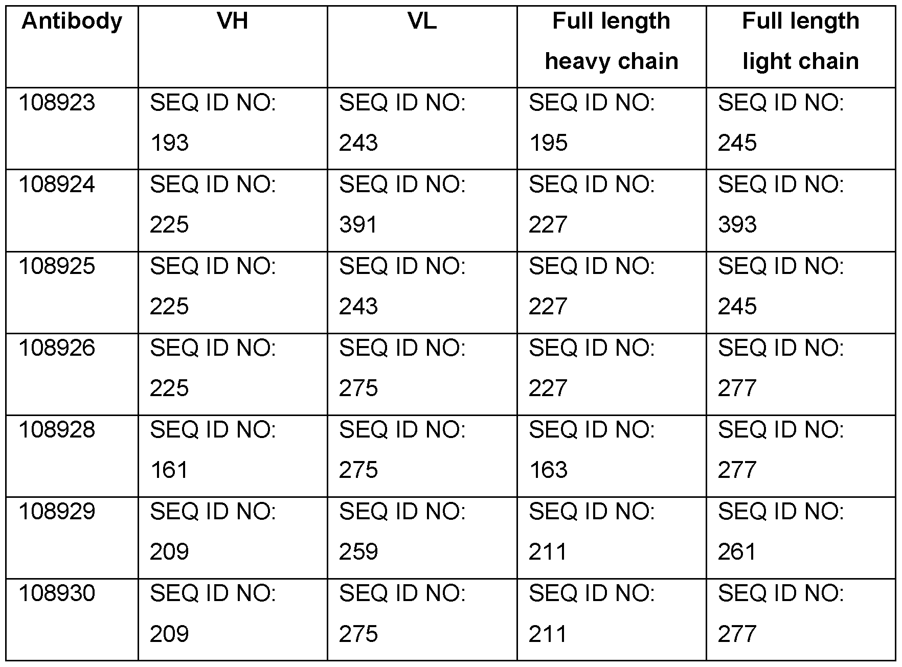


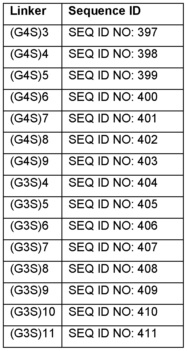


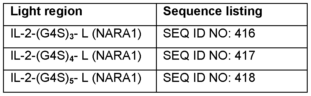


Claims
Priority Applications (14)
| Application Number | Priority Date | Filing Date | Title |
|---|---|---|---|
| CA3009001A CA3009001A1 (en) | 2016-01-11 | 2017-01-11 | Immune-stimulating humanized monoclonal antibodies against human interleukin-2, and fusion proteins thereof |
| US16/069,146 US10889643B2 (en) | 2016-01-11 | 2017-01-11 | Immune-stimulating humanized monoclonal antibodies against human interleukin-2, and fusion proteins thereof |
| AU2017208133A AU2017208133B2 (en) | 2016-01-11 | 2017-01-11 | Immune-stimulating humanized monoclonal antibodies against human interleukin-2, and fusion proteins thereof |
| CN202211356161.5A CN116003593A (en) | 2016-01-11 | 2017-01-11 | Immunostimulatory humanized monoclonal antibodies directed against human interleukin-2 and fusion proteins thereof |
| KR1020187023058A KR20180100224A (en) | 2016-01-11 | 2017-01-11 | Immune-stimulated humanized monoclonal antibodies against human interleukin-2, and fusion proteins thereof |
| BR112018014150A BR112018014150A2 (en) | 2016-01-11 | 2017-01-11 | immunostimulating humanized monoclonal antibodies to human interleukin-2, and fusion proteins thereof |
| EP17701362.0A EP3402819A1 (en) | 2016-01-11 | 2017-01-11 | Immune-stimulating humanized monoclonal antibodies against human interleukin-2, and fusion proteins thereof |
| EA201891582A EA039865B1 (en) | 2016-01-11 | 2017-01-11 | Immune-stimulating humanized monoclonal antibody against human interleukin-2 and fusion protein |
| JP2018536134A JP6993699B2 (en) | 2016-01-11 | 2017-01-11 | Immunostimulatory humanized monoclonal antibody against human interleukin-2 and its fusion protein |
| MX2018008592A MX2018008592A (en) | 2016-01-11 | 2017-01-11 | Immune-stimulating humanized monoclonal antibodies against human interleukin-2, and fusion proteins thereof. |
| CN201780006001.9A CN108473569B (en) | 2016-01-11 | 2017-01-11 | Immunostimulatory humanized monoclonal antibodies against human interleukin-2 and fusion proteins thereof |
| IL260218A IL260218B2 (en) | 2016-01-11 | 2017-01-11 | Immune-stimulating humanized monoclonal antibodies against human interleukin-2, and fusion proteins thereof |
| HK18116440.2A HK1257518A1 (en) | 2016-01-11 | 2018-12-21 | Immune-stimulating humanized monoclonal antibodies against human interleukin-2, and fusion proteins thereof |
| US17/124,930 US11851484B2 (en) | 2016-01-11 | 2020-12-17 | Immune-stimulating humanized monoclonal antibodies against human interleukin-2, and fusion proteins thereof |
Applications Claiming Priority (2)
| Application Number | Priority Date | Filing Date | Title |
|---|---|---|---|
| US201662277113P | 2016-01-11 | 2016-01-11 | |
| US62/277,113 | 2016-01-11 |
Related Child Applications (2)
| Application Number | Title | Priority Date | Filing Date |
|---|---|---|---|
| US16/069,146 A-371-Of-International US10889643B2 (en) | 2016-01-11 | 2017-01-11 | Immune-stimulating humanized monoclonal antibodies against human interleukin-2, and fusion proteins thereof |
| US17/124,930 Continuation US11851484B2 (en) | 2016-01-11 | 2020-12-17 | Immune-stimulating humanized monoclonal antibodies against human interleukin-2, and fusion proteins thereof |
Publications (2)
| Publication Number | Publication Date |
|---|---|
| WO2017122130A1 true WO2017122130A1 (en) | 2017-07-20 |
| WO2017122130A9 WO2017122130A9 (en) | 2018-02-01 |
Family
ID=57882113
Family Applications (1)
| Application Number | Title | Priority Date | Filing Date |
|---|---|---|---|
| PCT/IB2017/050127 WO2017122130A1 (en) | 2016-01-11 | 2017-01-11 | Immune-stimulating humanized monoclonal antibodies against human interleukin-2, and fusion proteins thereof |
Country Status (14)
| Country | Link |
|---|---|
| US (2) | US10889643B2 (en) |
| EP (1) | EP3402819A1 (en) |
| JP (1) | JP6993699B2 (en) |
| KR (1) | KR20180100224A (en) |
| CN (2) | CN108473569B (en) |
| AU (1) | AU2017208133B2 (en) |
| BR (1) | BR112018014150A2 (en) |
| CA (1) | CA3009001A1 (en) |
| EA (1) | EA039865B1 (en) |
| HK (1) | HK1257518A1 (en) |
| IL (1) | IL260218B2 (en) |
| MA (1) | MA43859A (en) |
| MX (1) | MX2018008592A (en) |
| WO (1) | WO2017122130A1 (en) |
Cited By (10)
| Publication number | Priority date | Publication date | Assignee | Title |
|---|---|---|---|---|
| WO2019142149A3 (en) * | 2018-01-22 | 2019-09-26 | Janssen Biotech, Inc. | Methods of treating cancers with antagonistic anti-pd-1 antibodies |
| WO2019168791A3 (en) * | 2018-02-28 | 2019-11-21 | The Board Of Trustees Of The Leland Stanford Junior University | Single-chain il-2/antibody fusions that selectively activate regulatory t cells |
| CN111825763A (en) * | 2020-07-15 | 2020-10-27 | 扬州大学 | Influenza virus HA protein stem specific monoclonal antibody and preparation method and application thereof |
| US10894830B2 (en) | 2015-11-03 | 2021-01-19 | Janssen Biotech, Inc. | Antibodies specifically binding PD-1, TIM-3 or PD-1 and TIM-3 and their uses |
| WO2022200525A1 (en) | 2021-03-26 | 2022-09-29 | Innate Pharma | Multispecific proteins comprising an nkp46-binding site, a cancer antgienge binding site fused to a cytokine for nk cell engaging |
| WO2022258691A1 (en) | 2021-06-09 | 2022-12-15 | Innate Pharma | Multispecific proteins binding to nkg2d, a cytokine receptor, a tumour antigen and cd16a |
| WO2022258662A1 (en) | 2021-06-09 | 2022-12-15 | Innate Pharma | Multispecific proteins binding to nkp46, a cytokine receptor, a tumour antigen and cd16a |
| WO2022258678A1 (en) | 2021-06-09 | 2022-12-15 | Innate Pharma | Multispecific proteins binding to nkp30, a cytokine receptor, a tumour antigen and cd16a |
| US11773160B1 (en) | 2022-08-05 | 2023-10-03 | Anaveon AG | Immune-stimulating IL-2 fusion proteins |
| US11851485B2 (en) | 2020-02-16 | 2023-12-26 | Aulos Bioscience, Inc. | Engineered anti-IL-2 antibodies |
Families Citing this family (11)
| Publication number | Priority date | Publication date | Assignee | Title |
|---|---|---|---|---|
| JOP20200094A1 (en) | 2014-01-24 | 2017-06-16 | Dana Farber Cancer Inst Inc | Antibody molecules to pd-1 and uses thereof |
| TWI777174B (en) * | 2014-03-14 | 2022-09-11 | 瑞士商諾華公司 | Antibody molecules to lag-3 and uses thereof |
| US11492386B2 (en) * | 2016-02-01 | 2022-11-08 | Eli Lilly And Company | Parathyroid hormone-anti-RANKL antibody fusion compounds |
| CA3038679A1 (en) * | 2016-09-28 | 2018-04-05 | Xoma (Us) Llc | Antibodies that bind interleukin-2 and uses thereof |
| WO2018170288A1 (en) | 2017-03-15 | 2018-09-20 | Pandion Therapeutics, Inc. | Targeted immunotolerance |
| AU2018273914A1 (en) | 2017-05-24 | 2019-11-14 | Pandion Operations, Inc. | Targeted immunotolerance |
| US10946068B2 (en) | 2017-12-06 | 2021-03-16 | Pandion Operations, Inc. | IL-2 muteins and uses thereof |
| US10174092B1 (en) | 2017-12-06 | 2019-01-08 | Pandion Therapeutics, Inc. | IL-2 muteins |
| US20210238558A1 (en) * | 2018-06-01 | 2021-08-05 | Medicenna Therapeutics, Inc. | Uses and methods for oncolytic virus targeting of il-4/il-13 and fusions thereof |
| BR112021023345A2 (en) | 2019-05-20 | 2022-02-01 | Pandion Operations Inc | Targeted immunotolerance in madcam |
| CN117700535A (en) * | 2024-02-05 | 2024-03-15 | 北京纳百生物科技有限公司 | Cat-panleukopenia virus VP2 protein monoclonal antibody and application thereof |
Citations (65)
| Publication number | Priority date | Publication date | Assignee | Title |
|---|---|---|---|---|
| EP0154316A2 (en) | 1984-03-06 | 1985-09-11 | Takeda Chemical Industries, Ltd. | Chemically modified lymphokine and production thereof |
| US4618486A (en) | 1983-01-21 | 1986-10-21 | Monocarb Ab | Monoclonal antibody formulation for diagnostic use |
| US4708871A (en) | 1983-03-08 | 1987-11-24 | Commonwealth Serum Laboratories Commission | Antigenically active amino acid sequences |
| EP0401384A1 (en) | 1988-12-22 | 1990-12-12 | Kirin-Amgen, Inc. | Chemically modified granulocyte colony stimulating factor |
| EP0413622A1 (en) | 1989-08-03 | 1991-02-20 | Rhone-Poulenc Sante | Albumin derivatives with therapeutic functions |
| WO1993015200A1 (en) | 1992-01-31 | 1993-08-05 | Rhone-Poulenc Rorer S.A. | Antithrombotic polypeptides as antagonists of the binding of vwf to platelets or to subendothelium |
| WO1993015199A1 (en) | 1992-01-31 | 1993-08-05 | Rhone-Poulenc Rorer S.A. | Novel biologically active polypeptides, preparation thereof and pharmaceutical composition containing said polypeptides |
| US5641870A (en) | 1995-04-20 | 1997-06-24 | Genentech, Inc. | Low pH hydrophobic interaction chromatography for antibody purification |
| WO1997034631A1 (en) | 1996-03-18 | 1997-09-25 | Board Of Regents, The University Of Texas System | Immunoglobin-like domains with increased half lives |
| WO1998023289A1 (en) | 1996-11-27 | 1998-06-04 | The General Hospital Corporation | MODULATION OF IgG BINDING TO FcRn |
| US5811097A (en) | 1995-07-25 | 1998-09-22 | The Regents Of The University Of California | Blockade of T lymphocyte down-regulation associated with CTLA-4 signaling |
| US5869046A (en) | 1995-04-14 | 1999-02-09 | Genentech, Inc. | Altered polypeptides with increased half-life |
| WO1999020758A1 (en) | 1997-10-21 | 1999-04-29 | Human Genome Sciences, Inc. | Human tumor necrosis factor receptor-like proteins tr11, tr11sv1, and tr11sv2 |
| WO1999040196A1 (en) | 1998-02-09 | 1999-08-12 | Genentech, Inc. | Novel tumor necrosis factor receptor homolog and nucleic acids encoding the same |
| US6111090A (en) | 1996-08-16 | 2000-08-29 | Schering Corporation | Mammalian cell surface antigens; related reagents |
| US6121022A (en) | 1995-04-14 | 2000-09-19 | Genentech, Inc. | Altered polypeptides with increased half-life |
| US6171586B1 (en) | 1997-06-13 | 2001-01-09 | Genentech, Inc. | Antibody formulation |
| WO2001003720A2 (en) | 1999-07-12 | 2001-01-18 | Genentech, Inc. | Promotion or inhibition of angiogenesis and cardiovascularization by tumor necrosis factor ligand/receptor homologs |
| US6277375B1 (en) | 1997-03-03 | 2001-08-21 | Board Of Regents, The University Of Texas System | Immunoglobulin-like domains with increased half-lives |
| WO2001077137A1 (en) | 2000-04-12 | 2001-10-18 | Human Genome Sciences, Inc. | Albumin fusion proteins |
| US6689607B2 (en) | 1997-10-21 | 2004-02-10 | Human Genome Sciences, Inc. | Human tumor, necrosis factor receptor-like proteins TR11, TR11SV1 and TR11SV2 |
| WO2004016286A2 (en) | 2002-08-16 | 2004-02-26 | Abbott Biotechnology Ltd. | Pharmaceutical anti-tnf-alpha antibody formulation |
| US6703199B1 (en) | 1997-06-12 | 2004-03-09 | Research Corporation Technologies, Inc. | Artificial antibody polypeptides |
| WO2004060319A2 (en) | 2002-12-30 | 2004-07-22 | 3M Innovative Properties Company | Immunostimulatory combinations |
| WO2005007190A1 (en) | 2003-07-11 | 2005-01-27 | Schering Corporation | Agonists or antagonists of the clucocorticoid-induced tumour necrosis factor receptor (gitr) or its ligand for the treatment of immune disorders, infections and cancer |
| WO2005055808A2 (en) | 2003-12-02 | 2005-06-23 | Genzyme Corporation | Compositions and methods to diagnose and treat lung cancer |
| WO2005115451A2 (en) | 2004-04-30 | 2005-12-08 | Isis Innovation Limited | Methods for generating improved immune response |
| WO2006044908A2 (en) | 2004-10-20 | 2006-04-27 | Genentech, Inc. | Antibody formulation in histidine-acetate buffer |
| WO2006083289A2 (en) | 2004-06-04 | 2006-08-10 | Duke University | Methods and compositions for enhancement of immunity by in vivo depletion of immunosuppressive cell activity |
| WO2006121168A1 (en) | 2005-05-09 | 2006-11-16 | Ono Pharmaceutical Co., Ltd. | Human monoclonal antibodies to programmed death 1(pd-1) and methods for treating cancer using anti-pd-1 antibodies alone or in combination with other immunotherapeutics |
| US20060286103A1 (en) | 2005-06-15 | 2006-12-21 | Parag Kolhe | Stable antibody formulation |
| WO2007095337A2 (en) | 2006-02-15 | 2007-08-23 | Imclone Systems Incorporated | Antibody formulation |
| WO2007095643A2 (en) * | 2006-02-16 | 2007-08-23 | Nascent Biologics, Inc. | Methods for improving immune function and methods for prevention or treatment of disease in a mammalian subject |
| WO2007133822A1 (en) | 2006-01-19 | 2007-11-22 | Genzyme Corporation | Gitr antibodies for the treatment of cancer |
| EP1866339A2 (en) | 2005-03-25 | 2007-12-19 | TolerRx, Inc | Gitr binding molecules and uses therefor |
| EP0920505B1 (en) | 1996-08-16 | 2008-06-04 | Schering Corporation | Mammalian cell surface antigens; related reagents |
| WO2009101611A1 (en) | 2008-02-11 | 2009-08-20 | Curetech Ltd. | Monoclonal antibodies for tumor treatment |
| WO2009114335A2 (en) | 2008-03-12 | 2009-09-17 | Merck & Co., Inc. | Pd-1 binding proteins |
| US7618632B2 (en) | 2003-05-23 | 2009-11-17 | Wyeth | Method of treating or ameliorating an immune cell associated pathology using GITR ligand antibodies |
| WO2010003118A1 (en) | 2008-07-02 | 2010-01-07 | Trubion Pharmaceuticals, Inc. | Tgf-b antagonist multi-target binding proteins |
| US20100028330A1 (en) | 2002-12-23 | 2010-02-04 | Medimmune Limited | Methods of upmodulating adaptive immune response using anti-pd1 antibodies |
| WO2010019570A2 (en) | 2008-08-11 | 2010-02-18 | Medarex, Inc. | Human antibodies that bind lymphocyte activation gene-3 (lag-3), and uses thereof |
| WO2010027827A2 (en) | 2008-08-25 | 2010-03-11 | Amplimmune, Inc. | Targeted costimulatory polypeptides and methods of use to treat cancer |
| WO2011028683A1 (en) | 2009-09-03 | 2011-03-10 | Schering Corporation | Anti-gitr antibodies |
| WO2011051726A2 (en) | 2009-10-30 | 2011-05-05 | Isis Innovation Ltd | Treatment of obesity |
| WO2011066342A2 (en) | 2009-11-24 | 2011-06-03 | Amplimmune, Inc. | Simultaneous inhibition of pd-l1/pd-l2 |
| WO2011090754A1 (en) | 2009-12-29 | 2011-07-28 | Emergent Product Development Seattle, Llc | Polypeptide heterodimers and uses thereof |
| WO2011155607A1 (en) | 2010-06-11 | 2011-12-15 | 協和発酵キリン株式会社 | Anti-tim-3 antibody |
| US20120114649A1 (en) | 2008-08-25 | 2012-05-10 | Amplimmune, Inc. Delaware | Compositions of pd-1 antagonists and methods of use |
| WO2012107417A1 (en) | 2011-02-10 | 2012-08-16 | Roche Glycart Ag | Mutant interleukin-2 polypeptides |
| US8354509B2 (en) | 2007-06-18 | 2013-01-15 | Msd Oss B.V. | Antibodies to human programmed death receptor PD-1 |
| WO2013039954A1 (en) | 2011-09-14 | 2013-03-21 | Sanofi | Anti-gitr antibodies |
| US8586023B2 (en) | 2008-09-12 | 2013-11-19 | Mie University | Cell capable of expressing exogenous GITR ligand |
| US8591886B2 (en) | 2007-07-12 | 2013-11-26 | Gitr, Inc. | Combination therapies employing GITR binding molecules |
| WO2014008218A1 (en) | 2012-07-02 | 2014-01-09 | Bristol-Myers Squibb Company | Optimization of antibodies that bind lymphocyte activation gene-3 (lag-3), and uses thereof |
| WO2014012479A1 (en) | 2012-07-18 | 2014-01-23 | Shanghai Birdie Biotech, Inc. | Compounds for targeted immunotherapy |
| US20140072566A1 (en) | 2012-06-08 | 2014-03-13 | National Cancer Center | Novel epitope for switching to th2 cell and use thereof |
| WO2014189806A1 (en) | 2013-05-18 | 2014-11-27 | Aduro Biotech, Inc. | Compositions and methods for inhibiting "stimulator of interferon gene" dependent signalling |
| WO2014189805A1 (en) | 2013-05-18 | 2014-11-27 | Auro Biotech, Inc. | Compositions and methods for activating "stimulator of interferon gene"-dependent signalling |
| WO2015026684A1 (en) | 2013-08-20 | 2015-02-26 | Merck Sharp & Dohme Corp. | Modulation of tumor immunity |
| US20150056225A1 (en) | 2012-04-17 | 2015-02-26 | University Of Washington Through Its Center For Commercialization | HLA Class II Deficient Cells, HLA Class I Deficient Cells Capable of Expressing HLA Class II Proteins, and Uses Thereof |
| US20150210769A1 (en) | 2014-01-24 | 2015-07-30 | Novartis Ag | Antibody molecules to pd-1 and uses thereof |
| US20150218274A1 (en) | 2014-01-31 | 2015-08-06 | Novartis Ag | Antibody molecules to tim-3 and uses thereof |
| US20150259420A1 (en) | 2014-03-14 | 2015-09-17 | Novartis Ag | Antibody molecules to lag-3 and uses thereof |
| WO2016005950A1 (en) * | 2014-07-10 | 2016-01-14 | Novartis Ag | Immune-stimulating monoclonal antibodies against human interleukin-2 |
Family Cites Families (6)
| Publication number | Priority date | Publication date | Assignee | Title |
|---|---|---|---|---|
| DK174501B1 (en) | 1983-12-23 | 2003-04-28 | Hoffmann La Roche | Process for Preparation of Interleukin-2 |
| EP1888644A1 (en) | 2005-06-01 | 2008-02-20 | Micromet AG | Anti-il2 antibodies |
| CN104072614B (en) | 2005-07-08 | 2017-04-26 | 生物基因Ma公司 | Anti-alpha[v]beta[6] antibodies and uses thereof |
| RS59652B1 (en) | 2010-08-12 | 2020-01-31 | Lilly Co Eli | Anti-n3pglu amyloid beta peptide antibodies and uses thereof |
| WO2013157105A1 (en) | 2012-04-18 | 2013-10-24 | 公立大学法人大阪市立大学 | Mucin 5ac specific human antibody and use thereof |
| WO2015109212A1 (en) | 2014-01-17 | 2015-07-23 | Pfizer Inc. | Anti-il-2 antibodies and compositions and uses thereof |
-
2017
- 2017-01-11 AU AU2017208133A patent/AU2017208133B2/en active Active
- 2017-01-11 CN CN201780006001.9A patent/CN108473569B/en active Active
- 2017-01-11 EA EA201891582A patent/EA039865B1/en unknown
- 2017-01-11 US US16/069,146 patent/US10889643B2/en active Active
- 2017-01-11 IL IL260218A patent/IL260218B2/en unknown
- 2017-01-11 MX MX2018008592A patent/MX2018008592A/en unknown
- 2017-01-11 JP JP2018536134A patent/JP6993699B2/en active Active
- 2017-01-11 KR KR1020187023058A patent/KR20180100224A/en unknown
- 2017-01-11 MA MA043859A patent/MA43859A/en unknown
- 2017-01-11 CN CN202211356161.5A patent/CN116003593A/en active Pending
- 2017-01-11 BR BR112018014150A patent/BR112018014150A2/en unknown
- 2017-01-11 CA CA3009001A patent/CA3009001A1/en active Pending
- 2017-01-11 WO PCT/IB2017/050127 patent/WO2017122130A1/en active Application Filing
- 2017-01-11 EP EP17701362.0A patent/EP3402819A1/en active Pending
-
2018
- 2018-12-21 HK HK18116440.2A patent/HK1257518A1/en unknown
-
2020
- 2020-12-17 US US17/124,930 patent/US11851484B2/en active Active
Patent Citations (77)
| Publication number | Priority date | Publication date | Assignee | Title |
|---|---|---|---|---|
| US4618486A (en) | 1983-01-21 | 1986-10-21 | Monocarb Ab | Monoclonal antibody formulation for diagnostic use |
| US4708871A (en) | 1983-03-08 | 1987-11-24 | Commonwealth Serum Laboratories Commission | Antigenically active amino acid sequences |
| EP0154316A2 (en) | 1984-03-06 | 1985-09-11 | Takeda Chemical Industries, Ltd. | Chemically modified lymphokine and production thereof |
| EP0401384A1 (en) | 1988-12-22 | 1990-12-12 | Kirin-Amgen, Inc. | Chemically modified granulocyte colony stimulating factor |
| EP0413622A1 (en) | 1989-08-03 | 1991-02-20 | Rhone-Poulenc Sante | Albumin derivatives with therapeutic functions |
| WO1993015200A1 (en) | 1992-01-31 | 1993-08-05 | Rhone-Poulenc Rorer S.A. | Antithrombotic polypeptides as antagonists of the binding of vwf to platelets or to subendothelium |
| WO1993015199A1 (en) | 1992-01-31 | 1993-08-05 | Rhone-Poulenc Rorer S.A. | Novel biologically active polypeptides, preparation thereof and pharmaceutical composition containing said polypeptides |
| US5869046A (en) | 1995-04-14 | 1999-02-09 | Genentech, Inc. | Altered polypeptides with increased half-life |
| US6121022A (en) | 1995-04-14 | 2000-09-19 | Genentech, Inc. | Altered polypeptides with increased half-life |
| US5641870A (en) | 1995-04-20 | 1997-06-24 | Genentech, Inc. | Low pH hydrophobic interaction chromatography for antibody purification |
| US5811097A (en) | 1995-07-25 | 1998-09-22 | The Regents Of The University Of California | Blockade of T lymphocyte down-regulation associated with CTLA-4 signaling |
| WO1997034631A1 (en) | 1996-03-18 | 1997-09-25 | Board Of Regents, The University Of Texas System | Immunoglobin-like domains with increased half lives |
| EP1947183B1 (en) | 1996-08-16 | 2013-07-17 | Merck Sharp & Dohme Corp. | Mammalian cell surface antigens; related reagents |
| US6111090A (en) | 1996-08-16 | 2000-08-29 | Schering Corporation | Mammalian cell surface antigens; related reagents |
| EP0920505B1 (en) | 1996-08-16 | 2008-06-04 | Schering Corporation | Mammalian cell surface antigens; related reagents |
| US7025962B1 (en) | 1996-08-16 | 2006-04-11 | Schering Corporation | Mammalian cell surface antigens; related reagents |
| WO1998023289A1 (en) | 1996-11-27 | 1998-06-04 | The General Hospital Corporation | MODULATION OF IgG BINDING TO FcRn |
| US6277375B1 (en) | 1997-03-03 | 2001-08-21 | Board Of Regents, The University Of Texas System | Immunoglobulin-like domains with increased half-lives |
| US6703199B1 (en) | 1997-06-12 | 2004-03-09 | Research Corporation Technologies, Inc. | Artificial antibody polypeptides |
| US6171586B1 (en) | 1997-06-13 | 2001-01-09 | Genentech, Inc. | Antibody formulation |
| US6689607B2 (en) | 1997-10-21 | 2004-02-10 | Human Genome Sciences, Inc. | Human tumor, necrosis factor receptor-like proteins TR11, TR11SV1 and TR11SV2 |
| WO1999020758A1 (en) | 1997-10-21 | 1999-04-29 | Human Genome Sciences, Inc. | Human tumor necrosis factor receptor-like proteins tr11, tr11sv1, and tr11sv2 |
| WO1999040196A1 (en) | 1998-02-09 | 1999-08-12 | Genentech, Inc. | Novel tumor necrosis factor receptor homolog and nucleic acids encoding the same |
| WO2001003720A2 (en) | 1999-07-12 | 2001-01-18 | Genentech, Inc. | Promotion or inhibition of angiogenesis and cardiovascularization by tumor necrosis factor ligand/receptor homologs |
| WO2001077137A1 (en) | 2000-04-12 | 2001-10-18 | Human Genome Sciences, Inc. | Albumin fusion proteins |
| WO2004016286A2 (en) | 2002-08-16 | 2004-02-26 | Abbott Biotechnology Ltd. | Pharmaceutical anti-tnf-alpha antibody formulation |
| US20100028330A1 (en) | 2002-12-23 | 2010-02-04 | Medimmune Limited | Methods of upmodulating adaptive immune response using anti-pd1 antibodies |
| WO2004060319A2 (en) | 2002-12-30 | 2004-07-22 | 3M Innovative Properties Company | Immunostimulatory combinations |
| US7618632B2 (en) | 2003-05-23 | 2009-11-17 | Wyeth | Method of treating or ameliorating an immune cell associated pathology using GITR ligand antibodies |
| WO2005007190A1 (en) | 2003-07-11 | 2005-01-27 | Schering Corporation | Agonists or antagonists of the clucocorticoid-induced tumour necrosis factor receptor (gitr) or its ligand for the treatment of immune disorders, infections and cancer |
| WO2005055808A2 (en) | 2003-12-02 | 2005-06-23 | Genzyme Corporation | Compositions and methods to diagnose and treat lung cancer |
| WO2005115451A2 (en) | 2004-04-30 | 2005-12-08 | Isis Innovation Limited | Methods for generating improved immune response |
| WO2006083289A2 (en) | 2004-06-04 | 2006-08-10 | Duke University | Methods and compositions for enhancement of immunity by in vivo depletion of immunosuppressive cell activity |
| WO2006044908A2 (en) | 2004-10-20 | 2006-04-27 | Genentech, Inc. | Antibody formulation in histidine-acetate buffer |
| EP1866339A2 (en) | 2005-03-25 | 2007-12-19 | TolerRx, Inc | Gitr binding molecules and uses therefor |
| US8388967B2 (en) | 2005-03-25 | 2013-03-05 | Gitr, Inc. | Methods for inducing or enhancing an immune response by administering agonistic GITR-binding antibodies |
| US7812135B2 (en) | 2005-03-25 | 2010-10-12 | Tolerrx, Inc. | GITR-binding antibodies |
| WO2006121168A1 (en) | 2005-05-09 | 2006-11-16 | Ono Pharmaceutical Co., Ltd. | Human monoclonal antibodies to programmed death 1(pd-1) and methods for treating cancer using anti-pd-1 antibodies alone or in combination with other immunotherapeutics |
| US8008449B2 (en) | 2005-05-09 | 2011-08-30 | Medarex, Inc. | Human monoclonal antibodies to programmed death 1 (PD-1) and methods for treating cancer using anti-PD-1 antibodies alone or in combination with other immunotherapeutics |
| US20060286103A1 (en) | 2005-06-15 | 2006-12-21 | Parag Kolhe | Stable antibody formulation |
| WO2007133822A1 (en) | 2006-01-19 | 2007-11-22 | Genzyme Corporation | Gitr antibodies for the treatment of cancer |
| WO2007095337A2 (en) | 2006-02-15 | 2007-08-23 | Imclone Systems Incorporated | Antibody formulation |
| WO2007095643A2 (en) * | 2006-02-16 | 2007-08-23 | Nascent Biologics, Inc. | Methods for improving immune function and methods for prevention or treatment of disease in a mammalian subject |
| US8354509B2 (en) | 2007-06-18 | 2013-01-15 | Msd Oss B.V. | Antibodies to human programmed death receptor PD-1 |
| US8591886B2 (en) | 2007-07-12 | 2013-11-26 | Gitr, Inc. | Combination therapies employing GITR binding molecules |
| WO2009101611A1 (en) | 2008-02-11 | 2009-08-20 | Curetech Ltd. | Monoclonal antibodies for tumor treatment |
| WO2009114335A2 (en) | 2008-03-12 | 2009-09-17 | Merck & Co., Inc. | Pd-1 binding proteins |
| WO2010003118A1 (en) | 2008-07-02 | 2010-01-07 | Trubion Pharmaceuticals, Inc. | Tgf-b antagonist multi-target binding proteins |
| US20110150892A1 (en) | 2008-08-11 | 2011-06-23 | Medarex, Inc. | Human antibodies that bind lymphocyte activation gene-3 (lag-3) and uses thereof |
| WO2010019570A2 (en) | 2008-08-11 | 2010-02-18 | Medarex, Inc. | Human antibodies that bind lymphocyte activation gene-3 (lag-3), and uses thereof |
| WO2010027827A2 (en) | 2008-08-25 | 2010-03-11 | Amplimmune, Inc. | Targeted costimulatory polypeptides and methods of use to treat cancer |
| US8609089B2 (en) | 2008-08-25 | 2013-12-17 | Amplimmune, Inc. | Compositions of PD-1 antagonists and methods of use |
| US20120114649A1 (en) | 2008-08-25 | 2012-05-10 | Amplimmune, Inc. Delaware | Compositions of pd-1 antagonists and methods of use |
| US8586023B2 (en) | 2008-09-12 | 2013-11-19 | Mie University | Cell capable of expressing exogenous GITR ligand |
| US8709424B2 (en) | 2009-09-03 | 2014-04-29 | Merck Sharp & Dohme Corp. | Anti-GITR antibodies |
| WO2011028683A1 (en) | 2009-09-03 | 2011-03-10 | Schering Corporation | Anti-gitr antibodies |
| WO2011051726A2 (en) | 2009-10-30 | 2011-05-05 | Isis Innovation Ltd | Treatment of obesity |
| WO2011066342A2 (en) | 2009-11-24 | 2011-06-03 | Amplimmune, Inc. | Simultaneous inhibition of pd-l1/pd-l2 |
| WO2011090754A1 (en) | 2009-12-29 | 2011-07-28 | Emergent Product Development Seattle, Llc | Polypeptide heterodimers and uses thereof |
| US20140044728A1 (en) | 2010-06-11 | 2014-02-13 | Kyushu University, National University Corporation | Anti-tim-3 antibody |
| WO2011155607A1 (en) | 2010-06-11 | 2011-12-15 | 協和発酵キリン株式会社 | Anti-tim-3 antibody |
| EP2581113A1 (en) | 2010-06-11 | 2013-04-17 | Kyowa Hakko Kirin Co., Ltd. | Anti-tim-3 antibody |
| US8552156B2 (en) | 2010-06-11 | 2013-10-08 | Kyowa Hakko Kirin Co., Ltd | Anti-TIM-3 antibody |
| WO2012107417A1 (en) | 2011-02-10 | 2012-08-16 | Roche Glycart Ag | Mutant interleukin-2 polypeptides |
| WO2013039954A1 (en) | 2011-09-14 | 2013-03-21 | Sanofi | Anti-gitr antibodies |
| US20150056225A1 (en) | 2012-04-17 | 2015-02-26 | University Of Washington Through Its Center For Commercialization | HLA Class II Deficient Cells, HLA Class I Deficient Cells Capable of Expressing HLA Class II Proteins, and Uses Thereof |
| US20140072566A1 (en) | 2012-06-08 | 2014-03-13 | National Cancer Center | Novel epitope for switching to th2 cell and use thereof |
| WO2014008218A1 (en) | 2012-07-02 | 2014-01-09 | Bristol-Myers Squibb Company | Optimization of antibodies that bind lymphocyte activation gene-3 (lag-3), and uses thereof |
| WO2014012479A1 (en) | 2012-07-18 | 2014-01-23 | Shanghai Birdie Biotech, Inc. | Compounds for targeted immunotherapy |
| WO2014189805A1 (en) | 2013-05-18 | 2014-11-27 | Auro Biotech, Inc. | Compositions and methods for activating "stimulator of interferon gene"-dependent signalling |
| US20150056224A1 (en) | 2013-05-18 | 2015-02-26 | Aduro Biotech, Inc. | Compositions and methods for activating stimulator of interferon gene-dependent signalling |
| WO2014189806A1 (en) | 2013-05-18 | 2014-11-27 | Aduro Biotech, Inc. | Compositions and methods for inhibiting "stimulator of interferon gene" dependent signalling |
| WO2015026684A1 (en) | 2013-08-20 | 2015-02-26 | Merck Sharp & Dohme Corp. | Modulation of tumor immunity |
| US20150210769A1 (en) | 2014-01-24 | 2015-07-30 | Novartis Ag | Antibody molecules to pd-1 and uses thereof |
| US20150218274A1 (en) | 2014-01-31 | 2015-08-06 | Novartis Ag | Antibody molecules to tim-3 and uses thereof |
| US20150259420A1 (en) | 2014-03-14 | 2015-09-17 | Novartis Ag | Antibody molecules to lag-3 and uses thereof |
| WO2016005950A1 (en) * | 2014-07-10 | 2016-01-14 | Novartis Ag | Immune-stimulating monoclonal antibodies against human interleukin-2 |
Non-Patent Citations (40)
| Title |
|---|
| ADAMS ET AL., ANNUAL REVIEW BIOPHYSICS, vol. 42, 2013, pages 265 - 287 |
| AI-LAZIKANI ET AL., JMB, vol. 273, 1997, pages 927 - 948 |
| AUSUBEL ET AL.: "Current Protocols in Molecular Biology", vol. SUPPL.30, 1987, article "section 7.7.18, Table 7.7.1." |
| BENATUIL L. ET AL.: "An improved yeast transformation method for the generation of very large human antibody libraries", PROTEIN ENG DES SEL., vol. 23, no. 4, 2010, pages 155 - 9, XP002637051, DOI: doi:10.1093/PROTEIN/GZQ002 |
| BERGE, S.M. ET AL., J. PHARM. SCI., vol. 66, 1977, pages 1 - 19 |
| BIRD ET AL., SCIENCE, vol. 242, 1988, pages 423 - 426 |
| BODER E.; WITTRUP K.: "Yeast surface display for screening combinatorial polypeptide libraries", NAT BIOTECHNOL., vol. 15, no. 6, 1997, pages 553 - 7, XP002945515, DOI: doi:10.1038/nbt0697-553 |
| CADWELL R.; JOYCE G.: "Mutagenic PCR.", PCR METHODS APPL., vol. 3, no. 6, 1994, pages 136 - 40 |
| CLELAND ET AL., CRIT. REVIEWS. THER. DRUG CARRIER SYSTEMS, vol. 10, no. 4, 1993, pages 307 - 377 |
| COLANDENE ET AL., J. PHARM. SCI, vol. 96, 2007, pages 1598 - 1608 |
| DAUGHERTY; MRSNY, ADVANCED DRUG DELIVERY REVIEWS, vol. 58, 2006, pages 686 - 706 |
| GARMAN, SCIENCE, vol. 343, 2014, pages 1102 - 1108 |
| GEYSEN ET AL., MOL. IMMUNOL., vol. 23, 1986, pages 709 - 715 |
| GEYSEN ET AL., PROC. NATL. ACAD. SCI. USA, vol. 8, 1984, pages 3998 - 4002 |
| GEYSEN ET AL., PROC. NATL. ACAD. SCI. USA, vol. 82, 1985, pages 78 - 182 |
| GLENN E. MORRIS: "Epitope Mapping Protocols in Methods in Molecular Biology", vol. 66, 1996, HUMANA PRESS |
| GREEN AND SAMBROOK: "Molecular Cloning; A Laboratory Manual, 4th ed.", 2012, COLD SPRING HARBOR |
| HAMID, O. ET AL., NEW ENGLAND JOURNAL OF MEDICINE, vol. 369, no. 2, 2013, pages 134 - 44 |
| HOLENSTEIN F. ET AL.: "Automated harvesting and 2-step purification of unclarified mammalian cell-culture broths containing antibodies", J CHROMATOGR A., vol. 1418, 2015, pages 103 - 9 |
| HOLLINGER; HUDSON, NATURE BIOTECHNOLOGY, vol. 23, 2005, pages 1126 - 1136 |
| HOPP ET AL., PROC. NATL. ACAD. SCI USA, vol. 78, 1981, pages 3824 - 3828 |
| HUSTON ET AL., PROC. NATL. ACAD. SCI., vol. 85, 1988, pages 5879 - 5883 |
| J. TOMALA ET AL: "In Vivo Expansion of Activated Naive CD8+ T Cells and NK Cells Driven by Complexes of IL-2 and Anti-IL-2 Monoclonal Antibody As Novel Approach of Cancer Immunotherapy", THE JOURNAL OF IMMUNOLOGY, vol. 183, no. 8, 15 October 2009 (2009-10-15), US, pages 4904 - 4912, XP055369385, ISSN: 0022-1767, DOI: 10.4049/jimmunol.0900284 * |
| JAKUB TOMALA ET AL: "Chimera of IL-2 Linked to Light Chain of anti-IL-2 mAb Mimics IL-2/anti-IL-2 mAb Complexes Both Structurally and Functionally", ACS CHEMICAL BIOLOGY, vol. 8, no. 5, 17 May 2013 (2013-05-17), US, pages 871 - 876, XP055369414, ISSN: 1554-8929, DOI: 10.1021/cb3007242 * |
| JOACHIMIAK, CURRENT OPINION STRUCTURAL BIOLOGY, vol. 19, 2009, pages 573 - 584 |
| KABAT ET AL.: "Sequences of Proteins of Immunological Interest, 5th ed.", 1991, NATIONAL INSTITUTES OF HEALTH |
| KABAT, E. A. ET AL.: "Sequences of Proteins of Immunological Interest, 5th ed.", 1991, U.S. DEPARTMENT OF HEALTH AND HUMAN SERVICES |
| KYTE ET AL., J. MOL. BIOL., vol. 157, 1982, pages 105 - 132 |
| LEFRANC, M.-P. ET AL., DEV. COMP. IMMUNOL., vol. 27, 2003, pages 55 - 77 |
| LEFRANC, M.-P., THE IMMUNOLOGIST, vol. 7, 1999, pages 132 - 136 |
| LEVIN ET AL., NATURE, vol. 484, 2012, pages 529 - 533 |
| ROSALIA ET AL., CURRENT OPINION IN CHEMICAL BIOLOGY, vol. 23, 2014, pages 39 - 46 |
| S. LETOURNEAU ET AL: "IL-2/anti-IL-2 antibody complexes show strong biological activity by avoiding interaction with IL-2 receptor subunit CD25", PROCEEDINGS OF THE NATIONAL ACADEMY OF SCIENCES, vol. 107, no. 5, 19 January 2010 (2010-01-19), pages 2171 - 2176, XP055182715, ISSN: 0027-8424, DOI: 10.1073/pnas.0909384107 * |
| SCHULMAN, AM. J. RESPIR. CRIT. CARE MED., vol. 164, 2001, pages S6 - S11 |
| URLAUB; CHASIN, PROC. NATL. ACAD. SCI. USA, vol. 77, 1980, pages 4216 - 20 |
| WANG ET AL., SCIENCE, vol. 310, 2005, pages 1159 - 1163 |
| WARD ET AL., NATURE, vol. 341, 1989, pages 544 - 546 |
| WEI WANG, INT. J. PHARMACEUTCS, vol. 185, 1999, pages 129 - 88 |
| ZAPATA ET AL., PROTEIN ENG., vol. 8, 1995, pages 1057 - 1062 |
| ZHAO; LU, MOLECULAR IMMUNOLOGY, vol. 47, 2009, pages 694 - 700 |
Cited By (11)
| Publication number | Priority date | Publication date | Assignee | Title |
|---|---|---|---|---|
| US10894830B2 (en) | 2015-11-03 | 2021-01-19 | Janssen Biotech, Inc. | Antibodies specifically binding PD-1, TIM-3 or PD-1 and TIM-3 and their uses |
| WO2019142149A3 (en) * | 2018-01-22 | 2019-09-26 | Janssen Biotech, Inc. | Methods of treating cancers with antagonistic anti-pd-1 antibodies |
| WO2019168791A3 (en) * | 2018-02-28 | 2019-11-21 | The Board Of Trustees Of The Leland Stanford Junior University | Single-chain il-2/antibody fusions that selectively activate regulatory t cells |
| US11851485B2 (en) | 2020-02-16 | 2023-12-26 | Aulos Bioscience, Inc. | Engineered anti-IL-2 antibodies |
| CN111825763A (en) * | 2020-07-15 | 2020-10-27 | 扬州大学 | Influenza virus HA protein stem specific monoclonal antibody and preparation method and application thereof |
| CN111825763B (en) * | 2020-07-15 | 2022-04-19 | 扬州大学 | Influenza virus HA protein stem specific monoclonal antibody and preparation method and application thereof |
| WO2022200525A1 (en) | 2021-03-26 | 2022-09-29 | Innate Pharma | Multispecific proteins comprising an nkp46-binding site, a cancer antgienge binding site fused to a cytokine for nk cell engaging |
| WO2022258691A1 (en) | 2021-06-09 | 2022-12-15 | Innate Pharma | Multispecific proteins binding to nkg2d, a cytokine receptor, a tumour antigen and cd16a |
| WO2022258662A1 (en) | 2021-06-09 | 2022-12-15 | Innate Pharma | Multispecific proteins binding to nkp46, a cytokine receptor, a tumour antigen and cd16a |
| WO2022258678A1 (en) | 2021-06-09 | 2022-12-15 | Innate Pharma | Multispecific proteins binding to nkp30, a cytokine receptor, a tumour antigen and cd16a |
| US11773160B1 (en) | 2022-08-05 | 2023-10-03 | Anaveon AG | Immune-stimulating IL-2 fusion proteins |
Also Published As
| Publication number | Publication date |
|---|---|
| CN116003593A (en) | 2023-04-25 |
| JP6993699B2 (en) | 2022-02-03 |
| WO2017122130A9 (en) | 2018-02-01 |
| CN108473569A (en) | 2018-08-31 |
| US10889643B2 (en) | 2021-01-12 |
| EA039865B1 (en) | 2022-03-22 |
| JP2019511200A (en) | 2019-04-25 |
| CA3009001A1 (en) | 2017-07-20 |
| IL260218B (en) | 2022-12-01 |
| HK1257518A1 (en) | 2019-10-25 |
| IL260218A (en) | 2018-07-31 |
| EP3402819A1 (en) | 2018-11-21 |
| US20210246200A1 (en) | 2021-08-12 |
| BR112018014150A2 (en) | 2018-12-11 |
| CN108473569B (en) | 2022-11-22 |
| MX2018008592A (en) | 2018-12-10 |
| AU2017208133B2 (en) | 2023-12-21 |
| US20190016797A1 (en) | 2019-01-17 |
| MA43859A (en) | 2018-11-21 |
| AU2017208133A1 (en) | 2018-07-05 |
| KR20180100224A (en) | 2018-09-07 |
| US11851484B2 (en) | 2023-12-26 |
| EA201891582A1 (en) | 2018-12-28 |
| IL260218B2 (en) | 2023-04-01 |
Similar Documents
| Publication | Publication Date | Title |
|---|---|---|
| AU2017208133B2 (en) | Immune-stimulating humanized monoclonal antibodies against human interleukin-2, and fusion proteins thereof | |
| JP7069261B2 (en) | CD73-specific binding molecule and its use | |
| JP7345578B2 (en) | Novel anti-PD-L1 antibody | |
| CN110606891B (en) | Antibody molecule aiming at human CLDN18.2, antigen binding fragment and medical application thereof | |
| TWI777924B (en) | Anti-bcma antibodies, bispecific antigen binding molecules that bind bcma and cd3, and uses thereof | |
| ES2824167T3 (en) | Anti-CD3 antibodies, anti-CD123 antibodies, and bispecific antibodies that specifically bind to CD3 and / or CD123 | |
| CN116333133A (en) | anti-GPRC 5D antibodies, bispecific antigen binding molecules binding GPRC5D and CD3 and uses thereof | |
| CN113825771B (en) | Antibodies against claudin 18A2 and uses thereof | |
| CN112707965A (en) | Antibody targeting CLDN18.2 and preparation method and application thereof | |
| US20230295301A1 (en) | Anti-ctla-4 antibody and use thereof | |
| CN110172099B (en) | anti-LAG-3 humanized monoclonal antibody molecule, antigen binding fragment and medical application thereof | |
| CN113321734A (en) | anti-CD 47/anti-PD-L1 antibodies and uses thereof | |
| WO2022100613A1 (en) | Bispecific antibody for claudin 18a2 and cd3 and application of bispecific antibody | |
| WO2023001303A1 (en) | Pharmaceutical composition and use | |
| CN108822212B (en) | Monoclonal antibody for selectively identifying oncogenic mutant KIT ITD and application thereof | |
| KR20230047009A (en) | Cytotoxicity-inducing thrapeutic agent for using in cancer treatment |
Legal Events
| Date | Code | Title | Description |
|---|---|---|---|
| 121 | Ep: the epo has been informed by wipo that ep was designated in this application |
Ref document number: 17701362 Country of ref document: EP Kind code of ref document: A1 |
|
| ENP | Entry into the national phase |
Ref document number: 3009001 Country of ref document: CA |
|
| WWE | Wipo information: entry into national phase |
Ref document number: 260218 Country of ref document: IL |
|
| ENP | Entry into the national phase |
Ref document number: 2017208133 Country of ref document: AU Date of ref document: 20170111 Kind code of ref document: A |
|
| ENP | Entry into the national phase |
Ref document number: 2018536134 Country of ref document: JP Kind code of ref document: A |
|
| WWE | Wipo information: entry into national phase |
Ref document number: MX/A/2018/008592 Country of ref document: MX |
|
| NENP | Non-entry into the national phase |
Ref country code: DE |
|
| REG | Reference to national code |
Ref country code: BR Ref legal event code: B01A Ref document number: 112018014150 Country of ref document: BR |
|
| WWE | Wipo information: entry into national phase |
Ref document number: 201891582 Country of ref document: EA |
|
| ENP | Entry into the national phase |
Ref document number: 20187023058 Country of ref document: KR Kind code of ref document: A |
|
| WWE | Wipo information: entry into national phase |
Ref document number: 1020187023058 Country of ref document: KR |
|
| WWE | Wipo information: entry into national phase |
Ref document number: 2017701362 Country of ref document: EP |
|
| ENP | Entry into the national phase |
Ref document number: 2017701362 Country of ref document: EP Effective date: 20180813 |
|
| ENP | Entry into the national phase |
Ref document number: 112018014150 Country of ref document: BR Kind code of ref document: A2 Effective date: 20180711 |