WO2016166360A1 - Bispecific antibody constructs for cdh3 and cd3 - Google Patents
Bispecific antibody constructs for cdh3 and cd3 Download PDFInfo
- Publication number
- WO2016166360A1 WO2016166360A1 PCT/EP2016/058482 EP2016058482W WO2016166360A1 WO 2016166360 A1 WO2016166360 A1 WO 2016166360A1 EP 2016058482 W EP2016058482 W EP 2016058482W WO 2016166360 A1 WO2016166360 A1 WO 2016166360A1
- Authority
- WO
- WIPO (PCT)
- Prior art keywords
- seq
- depicted
- cdr
- cancer
- antibody construct
- Prior art date
Links
- 101100004988 Caenorhabditis elegans cdh-3 gene Proteins 0.000 title 1
- 241000282414 Homo sapiens Species 0.000 claims abstract description 288
- 101000762242 Homo sapiens Cadherin-15 Proteins 0.000 claims abstract description 273
- 230000027455 binding Effects 0.000 claims abstract description 260
- 210000004027 cell Anatomy 0.000 claims abstract description 254
- 238000000034 method Methods 0.000 claims abstract description 107
- 102000048587 human CDH3 Human genes 0.000 claims abstract description 82
- 210000001744 T-lymphocyte Anatomy 0.000 claims abstract description 78
- 230000008569 process Effects 0.000 claims abstract description 30
- 239000013598 vector Substances 0.000 claims abstract description 24
- 108091033319 polynucleotide Proteins 0.000 claims abstract description 21
- 102000040430 polynucleotide Human genes 0.000 claims abstract description 21
- 239000002157 polynucleotide Substances 0.000 claims abstract description 21
- 238000004519 manufacturing process Methods 0.000 claims abstract description 20
- 101000714553 Homo sapiens Cadherin-3 Proteins 0.000 claims description 190
- 206010028980 Neoplasm Diseases 0.000 claims description 121
- 201000011510 cancer Diseases 0.000 claims description 76
- 125000003275 alpha amino acid group Chemical group 0.000 claims description 71
- 241000282553 Macaca Species 0.000 claims description 66
- 230000014509 gene expression Effects 0.000 claims description 37
- 239000008194 pharmaceutical composition Substances 0.000 claims description 34
- 238000011282 treatment Methods 0.000 claims description 30
- 206010041823 squamous cell carcinoma Diseases 0.000 claims description 16
- 208000005718 Stomach Neoplasms Diseases 0.000 claims description 10
- 206010017758 gastric cancer Diseases 0.000 claims description 10
- 230000002496 gastric effect Effects 0.000 claims description 10
- 208000002154 non-small cell lung carcinoma Diseases 0.000 claims description 10
- 201000011549 stomach cancer Diseases 0.000 claims description 10
- 208000029729 tumor suppressor gene on chromosome 11 Diseases 0.000 claims description 10
- 206010006187 Breast cancer Diseases 0.000 claims description 9
- 206010041067 Small cell lung cancer Diseases 0.000 claims description 9
- 208000000587 small cell lung carcinoma Diseases 0.000 claims description 9
- 208000026310 Breast neoplasm Diseases 0.000 claims description 8
- 206010009944 Colon cancer Diseases 0.000 claims description 8
- 206010058467 Lung neoplasm malignant Diseases 0.000 claims description 8
- 230000002265 prevention Effects 0.000 claims description 8
- 230000001684 chronic effect Effects 0.000 claims description 7
- 208000029742 colonic neoplasm Diseases 0.000 claims description 7
- 241000288950 Callithrix jacchus Species 0.000 claims description 5
- 241000282567 Macaca fascicularis Species 0.000 claims description 5
- 208000015914 Non-Hodgkin lymphomas Diseases 0.000 claims description 5
- 206010033128 Ovarian cancer Diseases 0.000 claims description 5
- 206010061535 Ovarian neoplasm Diseases 0.000 claims description 5
- 241000288960 Saguinus oedipus Species 0.000 claims description 5
- 241000282696 Saimiri sciureus Species 0.000 claims description 5
- 206010039491 Sarcoma Diseases 0.000 claims description 5
- 208000021712 Soft tissue sarcoma Diseases 0.000 claims description 5
- 208000024770 Thyroid neoplasm Diseases 0.000 claims description 5
- 208000006105 Uterine Cervical Neoplasms Diseases 0.000 claims description 5
- 201000010881 cervical cancer Diseases 0.000 claims description 5
- 201000007270 liver cancer Diseases 0.000 claims description 5
- 208000014018 liver neoplasm Diseases 0.000 claims description 5
- 201000001441 melanoma Diseases 0.000 claims description 5
- 201000002510 thyroid cancer Diseases 0.000 claims description 5
- 206010000830 Acute leukaemia Diseases 0.000 claims description 4
- 208000006468 Adrenal Cortex Neoplasms Diseases 0.000 claims description 4
- 208000032791 BCR-ABL1 positive chronic myelogenous leukemia Diseases 0.000 claims description 4
- 206010005003 Bladder cancer Diseases 0.000 claims description 4
- 206010005949 Bone cancer Diseases 0.000 claims description 4
- 208000018084 Bone neoplasm Diseases 0.000 claims description 4
- 208000003174 Brain Neoplasms Diseases 0.000 claims description 4
- 206010006143 Brain stem glioma Diseases 0.000 claims description 4
- 208000017897 Carcinoma of esophagus Diseases 0.000 claims description 4
- 206010007953 Central nervous system lymphoma Diseases 0.000 claims description 4
- 206010008342 Cervix carcinoma Diseases 0.000 claims description 4
- 208000010833 Chronic myeloid leukaemia Diseases 0.000 claims description 4
- 206010014733 Endometrial cancer Diseases 0.000 claims description 4
- 206010014759 Endometrial neoplasm Diseases 0.000 claims description 4
- 201000008808 Fibrosarcoma Diseases 0.000 claims description 4
- 208000022072 Gallbladder Neoplasms Diseases 0.000 claims description 4
- 206010073069 Hepatic cancer Diseases 0.000 claims description 4
- 208000017604 Hodgkin disease Diseases 0.000 claims description 4
- 208000010747 Hodgkins lymphoma Diseases 0.000 claims description 4
- 206010061252 Intraocular melanoma Diseases 0.000 claims description 4
- 208000008839 Kidney Neoplasms Diseases 0.000 claims description 4
- 208000031422 Lymphocytic Chronic B-Cell Leukemia Diseases 0.000 claims description 4
- 206010027406 Mesothelioma Diseases 0.000 claims description 4
- 208000034578 Multiple myelomas Diseases 0.000 claims description 4
- 208000033761 Myelogenous Chronic BCR-ABL Positive Leukemia Diseases 0.000 claims description 4
- 206010029260 Neuroblastoma Diseases 0.000 claims description 4
- 206010061902 Pancreatic neoplasm Diseases 0.000 claims description 4
- 208000007913 Pituitary Neoplasms Diseases 0.000 claims description 4
- 201000005746 Pituitary adenoma Diseases 0.000 claims description 4
- 206010061538 Pituitary tumour benign Diseases 0.000 claims description 4
- 206010035226 Plasma cell myeloma Diseases 0.000 claims description 4
- 206010060862 Prostate cancer Diseases 0.000 claims description 4
- 208000000236 Prostatic Neoplasms Diseases 0.000 claims description 4
- 208000015634 Rectal Neoplasms Diseases 0.000 claims description 4
- 206010038389 Renal cancer Diseases 0.000 claims description 4
- 208000006265 Renal cell carcinoma Diseases 0.000 claims description 4
- 201000000582 Retinoblastoma Diseases 0.000 claims description 4
- 208000000453 Skin Neoplasms Diseases 0.000 claims description 4
- 208000024313 Testicular Neoplasms Diseases 0.000 claims description 4
- 206010057644 Testis cancer Diseases 0.000 claims description 4
- 206010046458 Urethral neoplasms Diseases 0.000 claims description 4
- 208000007097 Urinary Bladder Neoplasms Diseases 0.000 claims description 4
- 208000002495 Uterine Neoplasms Diseases 0.000 claims description 4
- 201000005969 Uveal melanoma Diseases 0.000 claims description 4
- 201000009036 biliary tract cancer Diseases 0.000 claims description 4
- 208000020790 biliary tract neoplasm Diseases 0.000 claims description 4
- 208000025997 central nervous system neoplasm Diseases 0.000 claims description 4
- 208000024207 chronic leukemia Diseases 0.000 claims description 4
- 208000030381 cutaneous melanoma Diseases 0.000 claims description 4
- 230000002183 duodenal effect Effects 0.000 claims description 4
- 201000003914 endometrial carcinoma Diseases 0.000 claims description 4
- 201000010175 gallbladder cancer Diseases 0.000 claims description 4
- 201000003911 head and neck carcinoma Diseases 0.000 claims description 4
- 201000010982 kidney cancer Diseases 0.000 claims description 4
- 201000005296 lung carcinoma Diseases 0.000 claims description 4
- 208000015486 malignant pancreatic neoplasm Diseases 0.000 claims description 4
- 201000002575 ocular melanoma Diseases 0.000 claims description 4
- 201000008968 osteosarcoma Diseases 0.000 claims description 4
- 201000002528 pancreatic cancer Diseases 0.000 claims description 4
- 208000008443 pancreatic carcinoma Diseases 0.000 claims description 4
- 208000021310 pituitary gland adenoma Diseases 0.000 claims description 4
- 208000016800 primary central nervous system lymphoma Diseases 0.000 claims description 4
- 206010038038 rectal cancer Diseases 0.000 claims description 4
- 201000001275 rectum cancer Diseases 0.000 claims description 4
- 201000000849 skin cancer Diseases 0.000 claims description 4
- 201000003708 skin melanoma Diseases 0.000 claims description 4
- 210000000813 small intestine Anatomy 0.000 claims description 4
- 208000017572 squamous cell neoplasm Diseases 0.000 claims description 4
- 201000003120 testicular cancer Diseases 0.000 claims description 4
- 206010044412 transitional cell carcinoma Diseases 0.000 claims description 4
- 201000005112 urinary bladder cancer Diseases 0.000 claims description 4
- 206010046766 uterine cancer Diseases 0.000 claims description 4
- 238000012258 culturing Methods 0.000 claims description 3
- 102100024153 Cadherin-15 Human genes 0.000 claims 2
- 102100036360 Cadherin-3 Human genes 0.000 description 189
- 108090000623 proteins and genes Proteins 0.000 description 140
- 102000004169 proteins and genes Human genes 0.000 description 121
- 235000018102 proteins Nutrition 0.000 description 114
- 108090000765 processed proteins & peptides Proteins 0.000 description 103
- 235000001014 amino acid Nutrition 0.000 description 81
- 108091007433 antigens Proteins 0.000 description 79
- 102000036639 antigens Human genes 0.000 description 79
- 239000000427 antigen Substances 0.000 description 78
- 239000000203 mixture Substances 0.000 description 72
- 102000017420 CD3 protein, epsilon/gamma/delta subunit Human genes 0.000 description 68
- 108050005493 CD3 protein, epsilon/gamma/delta subunit Proteins 0.000 description 68
- 229940024606 amino acid Drugs 0.000 description 66
- 150000001413 amino acids Chemical class 0.000 description 65
- 239000003814 drug Substances 0.000 description 58
- 102000004196 processed proteins & peptides Human genes 0.000 description 58
- 210000004978 chinese hamster ovary cell Anatomy 0.000 description 55
- 238000003556 assay Methods 0.000 description 46
- 229940079593 drug Drugs 0.000 description 45
- 239000012636 effector Substances 0.000 description 44
- 125000005647 linker group Chemical group 0.000 description 43
- 229920001184 polypeptide Polymers 0.000 description 43
- 241001465754 Metazoa Species 0.000 description 42
- 231100000491 EC50 Toxicity 0.000 description 39
- 208000037265 diseases, disorders, signs and symptoms Diseases 0.000 description 39
- 238000009472 formulation Methods 0.000 description 38
- 108060003951 Immunoglobulin Proteins 0.000 description 35
- 102000018358 immunoglobulin Human genes 0.000 description 35
- 210000003819 peripheral blood mononuclear cell Anatomy 0.000 description 34
- 241000699666 Mus <mouse, genus> Species 0.000 description 33
- 238000006467 substitution reaction Methods 0.000 description 33
- 201000010099 disease Diseases 0.000 description 31
- 238000002784 cytotoxicity assay Methods 0.000 description 29
- 231100000263 cytotoxicity test Toxicity 0.000 description 29
- 239000012634 fragment Substances 0.000 description 29
- 239000011230 binding agent Substances 0.000 description 28
- 210000002381 plasma Anatomy 0.000 description 27
- -1 small molecule compounds Chemical class 0.000 description 27
- 238000012360 testing method Methods 0.000 description 27
- 102000000905 Cadherin Human genes 0.000 description 26
- 108050007957 Cadherin Proteins 0.000 description 26
- 230000001472 cytotoxic effect Effects 0.000 description 25
- 230000000694 effects Effects 0.000 description 25
- 108010047041 Complementarity Determining Regions Proteins 0.000 description 24
- 238000001943 fluorescence-activated cell sorting Methods 0.000 description 24
- 239000000178 monomer Substances 0.000 description 24
- 150000007523 nucleic acids Chemical group 0.000 description 23
- 241000894007 species Species 0.000 description 22
- 238000013459 approach Methods 0.000 description 21
- 210000004369 blood Anatomy 0.000 description 21
- 239000008280 blood Substances 0.000 description 21
- 230000006870 function Effects 0.000 description 21
- 230000004048 modification Effects 0.000 description 21
- 238000012986 modification Methods 0.000 description 21
- 239000000539 dimer Substances 0.000 description 20
- 125000000539 amino acid group Chemical group 0.000 description 19
- 239000003795 chemical substances by application Substances 0.000 description 19
- 230000004927 fusion Effects 0.000 description 19
- 210000004602 germ cell Anatomy 0.000 description 19
- DHMQDGOQFOQNFH-UHFFFAOYSA-N Glycine Chemical compound NCC(O)=O DHMQDGOQFOQNFH-UHFFFAOYSA-N 0.000 description 18
- ROHFNLRQFUQHCH-YFKPBYRVSA-N L-leucine Chemical compound CC(C)C[C@H](N)C(O)=O ROHFNLRQFUQHCH-YFKPBYRVSA-N 0.000 description 18
- 241001529936 Murinae Species 0.000 description 18
- 238000004458 analytical method Methods 0.000 description 18
- 238000006243 chemical reaction Methods 0.000 description 18
- 238000011534 incubation Methods 0.000 description 18
- 241000282412 Homo Species 0.000 description 17
- 230000000875 corresponding effect Effects 0.000 description 17
- 238000011161 development Methods 0.000 description 17
- 230000018109 developmental process Effects 0.000 description 17
- 102000039446 nucleic acids Human genes 0.000 description 17
- 108020004707 nucleic acids Proteins 0.000 description 17
- 230000001225 therapeutic effect Effects 0.000 description 17
- 239000011651 chromium Substances 0.000 description 16
- 230000001965 increasing effect Effects 0.000 description 16
- 239000002502 liposome Substances 0.000 description 16
- 239000003755 preservative agent Substances 0.000 description 16
- 239000000243 solution Substances 0.000 description 16
- NFGXHKASABOEEW-UHFFFAOYSA-N 1-methylethyl 11-methoxy-3,7,11-trimethyl-2,4-dodecadienoate Chemical compound COC(C)(C)CCCC(C)CC=CC(C)=CC(=O)OC(C)C NFGXHKASABOEEW-UHFFFAOYSA-N 0.000 description 15
- 230000004071 biological effect Effects 0.000 description 15
- LOKCTEFSRHRXRJ-UHFFFAOYSA-I dipotassium trisodium dihydrogen phosphate hydrogen phosphate dichloride Chemical compound P(=O)(O)(O)[O-].[K+].P(=O)(O)([O-])[O-].[Na+].[Na+].[Cl-].[K+].[Cl-].[Na+] LOKCTEFSRHRXRJ-UHFFFAOYSA-I 0.000 description 15
- 229940000406 drug candidate Drugs 0.000 description 15
- 239000002953 phosphate buffered saline Substances 0.000 description 15
- 108091006905 Human Serum Albumin Proteins 0.000 description 14
- KDXKERNSBIXSRK-YFKPBYRVSA-N L-lysine Chemical compound NCCCC[C@H](N)C(O)=O KDXKERNSBIXSRK-YFKPBYRVSA-N 0.000 description 14
- KDXKERNSBIXSRK-UHFFFAOYSA-N Lysine Natural products NCCCCC(N)C(O)=O KDXKERNSBIXSRK-UHFFFAOYSA-N 0.000 description 14
- 241000288906 Primates Species 0.000 description 14
- 229910052804 chromium Inorganic materials 0.000 description 14
- 102000008100 Human Serum Albumin Human genes 0.000 description 13
- WHUUTDBJXJRKMK-VKHMYHEASA-N L-glutamic acid Chemical compound OC(=O)[C@@H](N)CCC(O)=O WHUUTDBJXJRKMK-VKHMYHEASA-N 0.000 description 13
- 239000000872 buffer Substances 0.000 description 13
- 238000000684 flow cytometry Methods 0.000 description 13
- 239000000463 material Substances 0.000 description 13
- 210000001519 tissue Anatomy 0.000 description 13
- MTCFGRXMJLQNBG-REOHCLBHSA-N (2S)-2-Amino-3-hydroxypropansäure Chemical compound OC[C@H](N)C(O)=O MTCFGRXMJLQNBG-REOHCLBHSA-N 0.000 description 12
- AYFVYJQAPQTCCC-GBXIJSLDSA-N L-threonine Chemical compound C[C@@H](O)[C@H](N)C(O)=O AYFVYJQAPQTCCC-GBXIJSLDSA-N 0.000 description 12
- 241000283984 Rodentia Species 0.000 description 12
- 239000003963 antioxidant agent Substances 0.000 description 12
- 235000006708 antioxidants Nutrition 0.000 description 12
- 230000009089 cytolysis Effects 0.000 description 12
- 238000005516 engineering process Methods 0.000 description 12
- 239000000047 product Substances 0.000 description 12
- 239000003981 vehicle Substances 0.000 description 12
- 241000699670 Mus sp. Species 0.000 description 11
- 239000002202 Polyethylene glycol Substances 0.000 description 11
- 108010003723 Single-Domain Antibodies Proteins 0.000 description 11
- FAPWRFPIFSIZLT-UHFFFAOYSA-M Sodium chloride Chemical compound [Na+].[Cl-] FAPWRFPIFSIZLT-UHFFFAOYSA-M 0.000 description 11
- 230000002776 aggregation Effects 0.000 description 11
- 238000004220 aggregation Methods 0.000 description 11
- 230000008901 benefit Effects 0.000 description 11
- 150000001875 compounds Chemical class 0.000 description 11
- 230000013595 glycosylation Effects 0.000 description 11
- 238000006206 glycosylation reaction Methods 0.000 description 11
- 229940072221 immunoglobulins Drugs 0.000 description 11
- 238000000338 in vitro Methods 0.000 description 11
- 238000001727 in vivo Methods 0.000 description 11
- 238000002347 injection Methods 0.000 description 11
- 239000007924 injection Substances 0.000 description 11
- 230000003993 interaction Effects 0.000 description 11
- 125000003729 nucleotide group Chemical group 0.000 description 11
- 229920001223 polyethylene glycol Polymers 0.000 description 11
- 210000001266 CD8-positive T-lymphocyte Anatomy 0.000 description 10
- DCXYFEDJOCDNAF-REOHCLBHSA-N L-asparagine Chemical compound OC(=O)[C@@H](N)CC(N)=O DCXYFEDJOCDNAF-REOHCLBHSA-N 0.000 description 10
- CKLJMWTZIZZHCS-REOHCLBHSA-N L-aspartic acid Chemical compound OC(=O)[C@@H](N)CC(O)=O CKLJMWTZIZZHCS-REOHCLBHSA-N 0.000 description 10
- COLNVLDHVKWLRT-QMMMGPOBSA-N L-phenylalanine Chemical compound OC(=O)[C@@H](N)CC1=CC=CC=C1 COLNVLDHVKWLRT-QMMMGPOBSA-N 0.000 description 10
- 108091028043 Nucleic acid sequence Proteins 0.000 description 10
- KZSNJWFQEVHDMF-UHFFFAOYSA-N Valine Natural products CC(C)C(N)C(O)=O KZSNJWFQEVHDMF-UHFFFAOYSA-N 0.000 description 10
- 230000015572 biosynthetic process Effects 0.000 description 10
- 210000004408 hybridoma Anatomy 0.000 description 10
- 239000007788 liquid Substances 0.000 description 10
- 125000002496 methyl group Chemical group [H]C([H])([H])* 0.000 description 10
- 230000035772 mutation Effects 0.000 description 10
- 239000013642 negative control Substances 0.000 description 10
- 230000004044 response Effects 0.000 description 10
- 239000004094 surface-active agent Substances 0.000 description 10
- 108010021625 Immunoglobulin Fragments Proteins 0.000 description 9
- 102000008394 Immunoglobulin Fragments Human genes 0.000 description 9
- ROHFNLRQFUQHCH-UHFFFAOYSA-N Leucine Natural products CC(C)CC(N)C(O)=O ROHFNLRQFUQHCH-UHFFFAOYSA-N 0.000 description 9
- DNIAPMSPPWPWGF-UHFFFAOYSA-N Propylene glycol Chemical compound CC(O)CO DNIAPMSPPWPWGF-UHFFFAOYSA-N 0.000 description 9
- 230000003013 cytotoxicity Effects 0.000 description 9
- 231100000135 cytotoxicity Toxicity 0.000 description 9
- 239000002773 nucleotide Substances 0.000 description 9
- 210000002966 serum Anatomy 0.000 description 9
- 235000000346 sugar Nutrition 0.000 description 9
- 229940124597 therapeutic agent Drugs 0.000 description 9
- 230000004614 tumor growth Effects 0.000 description 9
- 102100029758 Cadherin-4 Human genes 0.000 description 8
- 108020004414 DNA Proteins 0.000 description 8
- PEDCQBHIVMGVHV-UHFFFAOYSA-N Glycerine Chemical compound OCC(O)CO PEDCQBHIVMGVHV-UHFFFAOYSA-N 0.000 description 8
- 101000714537 Homo sapiens Cadherin-2 Proteins 0.000 description 8
- OUYCCCASQSFEME-QMMMGPOBSA-N L-tyrosine Chemical compound OC(=O)[C@@H](N)CC1=CC=C(O)C=C1 OUYCCCASQSFEME-QMMMGPOBSA-N 0.000 description 8
- 241000282577 Pan troglodytes Species 0.000 description 8
- 229930006000 Sucrose Natural products 0.000 description 8
- CZMRCDWAGMRECN-UGDNZRGBSA-N Sucrose Chemical compound O[C@H]1[C@H](O)[C@@H](CO)O[C@@]1(CO)O[C@@H]1[C@H](O)[C@@H](O)[C@H](O)[C@@H](CO)O1 CZMRCDWAGMRECN-UGDNZRGBSA-N 0.000 description 8
- 108091008874 T cell receptors Proteins 0.000 description 8
- 102000016266 T-Cell Antigen Receptors Human genes 0.000 description 8
- 230000002411 adverse Effects 0.000 description 8
- 230000000890 antigenic effect Effects 0.000 description 8
- 150000001720 carbohydrates Chemical class 0.000 description 8
- 238000002474 experimental method Methods 0.000 description 8
- 238000002955 isolation Methods 0.000 description 8
- 206010061289 metastatic neoplasm Diseases 0.000 description 8
- 239000000546 pharmaceutical excipient Substances 0.000 description 8
- 238000002360 preparation method Methods 0.000 description 8
- 239000000523 sample Substances 0.000 description 8
- 229960001153 serine Drugs 0.000 description 8
- 108020001568 subdomains Proteins 0.000 description 8
- 239000005720 sucrose Substances 0.000 description 8
- 239000006228 supernatant Substances 0.000 description 8
- 238000002560 therapeutic procedure Methods 0.000 description 8
- XLYOFNOQVPJJNP-UHFFFAOYSA-N water Substances O XLYOFNOQVPJJNP-UHFFFAOYSA-N 0.000 description 8
- 102000009027 Albumins Human genes 0.000 description 7
- 108010088751 Albumins Proteins 0.000 description 7
- 241000894006 Bacteria Species 0.000 description 7
- 102100036364 Cadherin-2 Human genes 0.000 description 7
- 102100029761 Cadherin-5 Human genes 0.000 description 7
- 201000009030 Carcinoma Diseases 0.000 description 7
- 108091026890 Coding region Proteins 0.000 description 7
- 241000196324 Embryophyta Species 0.000 description 7
- 101000794580 Homo sapiens Cadherin-4 Proteins 0.000 description 7
- 101000794587 Homo sapiens Cadherin-5 Proteins 0.000 description 7
- 101000946889 Homo sapiens Monocyte differentiation antigen CD14 Proteins 0.000 description 7
- 239000004472 Lysine Substances 0.000 description 7
- MTCFGRXMJLQNBG-UHFFFAOYSA-N Serine Natural products OCC(N)C(O)=O MTCFGRXMJLQNBG-UHFFFAOYSA-N 0.000 description 7
- 230000000259 anti-tumor effect Effects 0.000 description 7
- 238000001990 intravenous administration Methods 0.000 description 7
- 150000002500 ions Chemical class 0.000 description 7
- 229960003646 lysine Drugs 0.000 description 7
- 235000018977 lysine Nutrition 0.000 description 7
- 230000001404 mediated effect Effects 0.000 description 7
- 230000001394 metastastic effect Effects 0.000 description 7
- 210000000056 organ Anatomy 0.000 description 7
- 229920005862 polyol Polymers 0.000 description 7
- 150000003077 polyols Chemical class 0.000 description 7
- 229920000136 polysorbate Polymers 0.000 description 7
- 230000002335 preservative effect Effects 0.000 description 7
- 238000000746 purification Methods 0.000 description 7
- 235000004400 serine Nutrition 0.000 description 7
- 239000011780 sodium chloride Substances 0.000 description 7
- 150000008163 sugars Chemical class 0.000 description 7
- 238000001890 transfection Methods 0.000 description 7
- 210000004881 tumor cell Anatomy 0.000 description 7
- 239000004475 Arginine Substances 0.000 description 6
- 102100025805 Cadherin-1 Human genes 0.000 description 6
- 108091007854 Cdh1/Fizzy-related Proteins 0.000 description 6
- VYZAMTAEIAYCRO-UHFFFAOYSA-N Chromium Chemical compound [Cr] VYZAMTAEIAYCRO-UHFFFAOYSA-N 0.000 description 6
- FBPFZTCFMRRESA-FSIIMWSLSA-N D-Glucitol Natural products OC[C@H](O)[C@H](O)[C@@H](O)[C@H](O)CO FBPFZTCFMRRESA-FSIIMWSLSA-N 0.000 description 6
- FBPFZTCFMRRESA-KVTDHHQDSA-N D-Mannitol Chemical compound OC[C@@H](O)[C@@H](O)[C@H](O)[C@H](O)CO FBPFZTCFMRRESA-KVTDHHQDSA-N 0.000 description 6
- FBPFZTCFMRRESA-JGWLITMVSA-N D-glucitol Chemical compound OC[C@H](O)[C@@H](O)[C@H](O)[C@H](O)CO FBPFZTCFMRRESA-JGWLITMVSA-N 0.000 description 6
- WQZGKKKJIJFFOK-GASJEMHNSA-N Glucose Natural products OC[C@H]1OC(O)[C@H](O)[C@@H](O)[C@@H]1O WQZGKKKJIJFFOK-GASJEMHNSA-N 0.000 description 6
- 101000581981 Homo sapiens Neural cell adhesion molecule 1 Proteins 0.000 description 6
- 102000002265 Human Growth Hormone Human genes 0.000 description 6
- 108010000521 Human Growth Hormone Proteins 0.000 description 6
- 239000000854 Human Growth Hormone Substances 0.000 description 6
- QNAYBMKLOCPYGJ-REOHCLBHSA-N L-alanine Chemical compound C[C@H](N)C(O)=O QNAYBMKLOCPYGJ-REOHCLBHSA-N 0.000 description 6
- AGPKZVBTJJNPAG-WHFBIAKZSA-N L-isoleucine Chemical compound CC[C@H](C)[C@H](N)C(O)=O AGPKZVBTJJNPAG-WHFBIAKZSA-N 0.000 description 6
- 125000000393 L-methionino group Chemical group [H]OC(=O)[C@@]([H])(N([H])[*])C([H])([H])C(SC([H])([H])[H])([H])[H] 0.000 description 6
- 229930195725 Mannitol Natural products 0.000 description 6
- 102100035877 Monocyte differentiation antigen CD14 Human genes 0.000 description 6
- 108091007491 NSP3 Papain-like protease domains Proteins 0.000 description 6
- 241000700159 Rattus Species 0.000 description 6
- 239000004473 Threonine Substances 0.000 description 6
- 235000004279 alanine Nutrition 0.000 description 6
- 229960003767 alanine Drugs 0.000 description 6
- ODKSFYDXXFIFQN-UHFFFAOYSA-N arginine Natural products OC(=O)C(N)CCCNC(N)=N ODKSFYDXXFIFQN-UHFFFAOYSA-N 0.000 description 6
- 229960003121 arginine Drugs 0.000 description 6
- 210000003719 b-lymphocyte Anatomy 0.000 description 6
- 239000011324 bead Substances 0.000 description 6
- WQZGKKKJIJFFOK-VFUOTHLCSA-N beta-D-glucose Chemical compound OC[C@H]1O[C@@H](O)[C@H](O)[C@@H](O)[C@@H]1O WQZGKKKJIJFFOK-VFUOTHLCSA-N 0.000 description 6
- 238000005119 centrifugation Methods 0.000 description 6
- 208000035475 disorder Diseases 0.000 description 6
- 210000003527 eukaryotic cell Anatomy 0.000 description 6
- 238000011156 evaluation Methods 0.000 description 6
- 230000001747 exhibiting effect Effects 0.000 description 6
- 239000012537 formulation buffer Substances 0.000 description 6
- 230000012010 growth Effects 0.000 description 6
- 238000002372 labelling Methods 0.000 description 6
- 230000000670 limiting effect Effects 0.000 description 6
- 239000000594 mannitol Substances 0.000 description 6
- 235000010355 mannitol Nutrition 0.000 description 6
- 238000005259 measurement Methods 0.000 description 6
- 239000002609 medium Substances 0.000 description 6
- 239000012528 membrane Substances 0.000 description 6
- 238000002823 phage display Methods 0.000 description 6
- YBYRMVIVWMBXKQ-UHFFFAOYSA-N phenylmethanesulfonyl fluoride Chemical compound FS(=O)(=O)CC1=CC=CC=C1 YBYRMVIVWMBXKQ-UHFFFAOYSA-N 0.000 description 6
- 238000013391 scatchard analysis Methods 0.000 description 6
- 239000000600 sorbitol Substances 0.000 description 6
- 235000010356 sorbitol Nutrition 0.000 description 6
- 239000000126 substance Substances 0.000 description 6
- 230000009466 transformation Effects 0.000 description 6
- 241000699800 Cricetinae Species 0.000 description 5
- WHUUTDBJXJRKMK-UHFFFAOYSA-N Glutamic acid Natural products OC(=O)C(N)CCC(O)=O WHUUTDBJXJRKMK-UHFFFAOYSA-N 0.000 description 5
- 239000004471 Glycine Substances 0.000 description 5
- 238000011993 High Performance Size Exclusion Chromatography Methods 0.000 description 5
- 102000003839 Human Proteins Human genes 0.000 description 5
- 108090000144 Human Proteins Proteins 0.000 description 5
- 102100026120 IgG receptor FcRn large subunit p51 Human genes 0.000 description 5
- ONIBWKKTOPOVIA-BYPYZUCNSA-N L-Proline Chemical compound OC(=O)[C@@H]1CCCN1 ONIBWKKTOPOVIA-BYPYZUCNSA-N 0.000 description 5
- 102100027347 Neural cell adhesion molecule 1 Human genes 0.000 description 5
- 239000004698 Polyethylene Substances 0.000 description 5
- ONIBWKKTOPOVIA-UHFFFAOYSA-N Proline Natural products OC(=O)C1CCCN1 ONIBWKKTOPOVIA-UHFFFAOYSA-N 0.000 description 5
- 239000006146 Roswell Park Memorial Institute medium Substances 0.000 description 5
- AYFVYJQAPQTCCC-UHFFFAOYSA-N Threonine Natural products CC(O)C(N)C(O)=O AYFVYJQAPQTCCC-UHFFFAOYSA-N 0.000 description 5
- 230000004913 activation Effects 0.000 description 5
- 238000012867 alanine scanning Methods 0.000 description 5
- 239000007864 aqueous solution Substances 0.000 description 5
- 210000000170 cell membrane Anatomy 0.000 description 5
- 239000003153 chemical reaction reagent Substances 0.000 description 5
- 210000001151 cytotoxic T lymphocyte Anatomy 0.000 description 5
- 238000001514 detection method Methods 0.000 description 5
- 239000000975 dye Substances 0.000 description 5
- 238000002296 dynamic light scattering Methods 0.000 description 5
- 230000002349 favourable effect Effects 0.000 description 5
- 239000012530 fluid Substances 0.000 description 5
- 230000028993 immune response Effects 0.000 description 5
- 229940127121 immunoconjugate Drugs 0.000 description 5
- 230000005764 inhibitory process Effects 0.000 description 5
- 238000003780 insertion Methods 0.000 description 5
- 230000037431 insertion Effects 0.000 description 5
- 238000002826 magnetic-activated cell sorting Methods 0.000 description 5
- 230000007246 mechanism Effects 0.000 description 5
- 238000002844 melting Methods 0.000 description 5
- 230000008018 melting Effects 0.000 description 5
- 238000002703 mutagenesis Methods 0.000 description 5
- 231100000350 mutagenesis Toxicity 0.000 description 5
- 238000007911 parenteral administration Methods 0.000 description 5
- 229920000642 polymer Polymers 0.000 description 5
- 230000003389 potentiating effect Effects 0.000 description 5
- 235000013930 proline Nutrition 0.000 description 5
- 229960002429 proline Drugs 0.000 description 5
- XJMOSONTPMZWPB-UHFFFAOYSA-M propidium iodide Chemical group [I-].[I-].C12=CC(N)=CC=C2C2=CC=C(N)C=C2[N+](CCC[N+](C)(CC)CC)=C1C1=CC=CC=C1 XJMOSONTPMZWPB-UHFFFAOYSA-M 0.000 description 5
- 238000012216 screening Methods 0.000 description 5
- 239000002904 solvent Substances 0.000 description 5
- 239000003381 stabilizer Substances 0.000 description 5
- 238000010561 standard procedure Methods 0.000 description 5
- 239000000725 suspension Substances 0.000 description 5
- 235000008521 threonine Nutrition 0.000 description 5
- 230000009261 transgenic effect Effects 0.000 description 5
- DCXYFEDJOCDNAF-UHFFFAOYSA-N Asparagine Natural products OC(=O)C(N)CC(N)=O DCXYFEDJOCDNAF-UHFFFAOYSA-N 0.000 description 4
- 102100031266 Chromodomain-helicase-DNA-binding protein 3 Human genes 0.000 description 4
- 108020004705 Codon Proteins 0.000 description 4
- SRBFZHDQGSBBOR-IOVATXLUSA-N D-xylopyranose Chemical compound O[C@@H]1COC(O)[C@H](O)[C@H]1O SRBFZHDQGSBBOR-IOVATXLUSA-N 0.000 description 4
- KCXVZYZYPLLWCC-UHFFFAOYSA-N EDTA Chemical compound OC(=O)CN(CC(O)=O)CCN(CC(O)=O)CC(O)=O KCXVZYZYPLLWCC-UHFFFAOYSA-N 0.000 description 4
- 101000777071 Homo sapiens Chromodomain-helicase-DNA-binding protein 3 Proteins 0.000 description 4
- 101710177940 IgG receptor FcRn large subunit p51 Proteins 0.000 description 4
- 108700005091 Immunoglobulin Genes Proteins 0.000 description 4
- 108010067060 Immunoglobulin Variable Region Proteins 0.000 description 4
- 102000017727 Immunoglobulin Variable Region Human genes 0.000 description 4
- HNDVDQJCIGZPNO-YFKPBYRVSA-N L-histidine Chemical compound OC(=O)[C@@H](N)CC1=CN=CN1 HNDVDQJCIGZPNO-YFKPBYRVSA-N 0.000 description 4
- FFEARJCKVFRZRR-BYPYZUCNSA-N L-methionine Chemical compound CSCC[C@H](N)C(O)=O FFEARJCKVFRZRR-BYPYZUCNSA-N 0.000 description 4
- 108700018351 Major Histocompatibility Complex Proteins 0.000 description 4
- 241000283973 Oryctolagus cuniculus Species 0.000 description 4
- ISWSIDIOOBJBQZ-UHFFFAOYSA-N Phenol Chemical compound OC1=CC=CC=C1 ISWSIDIOOBJBQZ-UHFFFAOYSA-N 0.000 description 4
- 108010076504 Protein Sorting Signals Proteins 0.000 description 4
- 239000012980 RPMI-1640 medium Substances 0.000 description 4
- 230000010782 T cell mediated cytotoxicity Effects 0.000 description 4
- 108700019146 Transgenes Proteins 0.000 description 4
- 241000700605 Viruses Species 0.000 description 4
- 238000012452 Xenomouse strains Methods 0.000 description 4
- 239000002253 acid Substances 0.000 description 4
- 230000002378 acidificating effect Effects 0.000 description 4
- 230000009824 affinity maturation Effects 0.000 description 4
- 239000002246 antineoplastic agent Substances 0.000 description 4
- 230000003078 antioxidant effect Effects 0.000 description 4
- 210000001106 artificial yeast chromosome Anatomy 0.000 description 4
- 235000009582 asparagine Nutrition 0.000 description 4
- 229960001230 asparagine Drugs 0.000 description 4
- 239000000969 carrier Substances 0.000 description 4
- 230000006037 cell lysis Effects 0.000 description 4
- HVYWMOMLDIMFJA-DPAQBDIFSA-N cholesterol Chemical compound C1C=C2C[C@@H](O)CC[C@]2(C)[C@@H]2[C@@H]1[C@@H]1CC[C@H]([C@H](C)CCCC(C)C)[C@@]1(C)CC2 HVYWMOMLDIMFJA-DPAQBDIFSA-N 0.000 description 4
- 238000010367 cloning Methods 0.000 description 4
- 230000007423 decrease Effects 0.000 description 4
- 210000003162 effector t lymphocyte Anatomy 0.000 description 4
- 239000000839 emulsion Substances 0.000 description 4
- 230000002255 enzymatic effect Effects 0.000 description 4
- 238000000605 extraction Methods 0.000 description 4
- 230000002068 genetic effect Effects 0.000 description 4
- ZDXPYRJPNDTMRX-UHFFFAOYSA-N glutamine Natural products OC(=O)C(N)CCC(N)=O ZDXPYRJPNDTMRX-UHFFFAOYSA-N 0.000 description 4
- 235000004554 glutamine Nutrition 0.000 description 4
- RWSXRVCMGQZWBV-WDSKDSINSA-N glutathione Chemical compound OC(=O)[C@@H](N)CCC(=O)N[C@@H](CS)C(=O)NCC(O)=O RWSXRVCMGQZWBV-WDSKDSINSA-N 0.000 description 4
- HNDVDQJCIGZPNO-UHFFFAOYSA-N histidine Natural products OC(=O)C(N)CC1=CN=CN1 HNDVDQJCIGZPNO-UHFFFAOYSA-N 0.000 description 4
- 210000003292 kidney cell Anatomy 0.000 description 4
- 201000005202 lung cancer Diseases 0.000 description 4
- 208000020816 lung neoplasm Diseases 0.000 description 4
- 210000004698 lymphocyte Anatomy 0.000 description 4
- RLSSMJSEOOYNOY-UHFFFAOYSA-N m-cresol Chemical compound CC1=CC=CC(O)=C1 RLSSMJSEOOYNOY-UHFFFAOYSA-N 0.000 description 4
- 238000013507 mapping Methods 0.000 description 4
- 239000003550 marker Substances 0.000 description 4
- 229930182817 methionine Natural products 0.000 description 4
- 235000006109 methionine Nutrition 0.000 description 4
- 239000011325 microbead Substances 0.000 description 4
- 239000003094 microcapsule Substances 0.000 description 4
- 210000000822 natural killer cell Anatomy 0.000 description 4
- 230000003287 optical effect Effects 0.000 description 4
- 230000001575 pathological effect Effects 0.000 description 4
- 229940068965 polysorbates Drugs 0.000 description 4
- 230000004481 post-translational protein modification Effects 0.000 description 4
- 230000008707 rearrangement Effects 0.000 description 4
- 239000012146 running buffer Substances 0.000 description 4
- 238000007920 subcutaneous administration Methods 0.000 description 4
- 230000020382 suppression by virus of host antigen processing and presentation of peptide antigen via MHC class I Effects 0.000 description 4
- 208000024891 symptom Diseases 0.000 description 4
- 231100000041 toxicology testing Toxicity 0.000 description 4
- 238000005406 washing Methods 0.000 description 4
- 108091032973 (ribonucleotides)n+m Proteins 0.000 description 3
- UHOVQNZJYSORNB-UHFFFAOYSA-N Benzene Chemical compound C1=CC=CC=C1 UHOVQNZJYSORNB-UHFFFAOYSA-N 0.000 description 3
- 241000282693 Cercopithecidae Species 0.000 description 3
- WQZGKKKJIJFFOK-QTVWNMPRSA-N D-mannopyranose Chemical compound OC[C@H]1OC(O)[C@@H](O)[C@@H](O)[C@@H]1O WQZGKKKJIJFFOK-QTVWNMPRSA-N 0.000 description 3
- 102000004190 Enzymes Human genes 0.000 description 3
- 108090000790 Enzymes Proteins 0.000 description 3
- 241000233866 Fungi Species 0.000 description 3
- 241000238631 Hexapoda Species 0.000 description 3
- 108010093488 His-His-His-His-His-His Proteins 0.000 description 3
- 125000000998 L-alanino group Chemical group [H]N([*])[C@](C([H])([H])[H])([H])C(=O)O[H] 0.000 description 3
- ODKSFYDXXFIFQN-BYPYZUCNSA-P L-argininium(2+) Chemical compound NC(=[NH2+])NCCC[C@H]([NH3+])C(O)=O ODKSFYDXXFIFQN-BYPYZUCNSA-P 0.000 description 3
- 125000000510 L-tryptophano group Chemical group [H]C1=C([H])C([H])=C2N([H])C([H])=C(C([H])([H])[C@@]([H])(C(O[H])=O)N([H])[*])C2=C1[H] 0.000 description 3
- 206010025323 Lymphomas Diseases 0.000 description 3
- 102000043131 MHC class II family Human genes 0.000 description 3
- 108091054438 MHC class II family Proteins 0.000 description 3
- 241000124008 Mammalia Species 0.000 description 3
- 206010027476 Metastases Diseases 0.000 description 3
- 101100004990 Mus musculus Cdh3 gene Proteins 0.000 description 3
- 108050000637 N-cadherin Proteins 0.000 description 3
- 230000004989 O-glycosylation Effects 0.000 description 3
- 229920001213 Polysorbate 20 Polymers 0.000 description 3
- 108010029485 Protein Isoforms Proteins 0.000 description 3
- 102000001708 Protein Isoforms Human genes 0.000 description 3
- 108010086890 R-cadherin Proteins 0.000 description 3
- 108020004511 Recombinant DNA Proteins 0.000 description 3
- 240000004808 Saccharomyces cerevisiae Species 0.000 description 3
- 235000014680 Saccharomyces cerevisiae Nutrition 0.000 description 3
- 102000007562 Serum Albumin Human genes 0.000 description 3
- 108010071390 Serum Albumin Proteins 0.000 description 3
- 230000009471 action Effects 0.000 description 3
- 238000001042 affinity chromatography Methods 0.000 description 3
- 238000010171 animal model Methods 0.000 description 3
- 239000000611 antibody drug conjugate Substances 0.000 description 3
- 229940049595 antibody-drug conjugate Drugs 0.000 description 3
- 125000000637 arginyl group Chemical group N[C@@H](CCCNC(N)=N)C(=O)* 0.000 description 3
- 125000000613 asparagine group Chemical group N[C@@H](CC(N)=O)C(=O)* 0.000 description 3
- 210000000481 breast Anatomy 0.000 description 3
- 239000004067 bulking agent Substances 0.000 description 3
- 239000011575 calcium Substances 0.000 description 3
- 235000014633 carbohydrates Nutrition 0.000 description 3
- 125000003178 carboxy group Chemical group [H]OC(*)=O 0.000 description 3
- 238000004113 cell culture Methods 0.000 description 3
- 238000012512 characterization method Methods 0.000 description 3
- 239000002738 chelating agent Substances 0.000 description 3
- 239000003638 chemical reducing agent Substances 0.000 description 3
- 238000004587 chromatography analysis Methods 0.000 description 3
- 210000000349 chromosome Anatomy 0.000 description 3
- 238000012790 confirmation Methods 0.000 description 3
- 230000008878 coupling Effects 0.000 description 3
- 238000010168 coupling process Methods 0.000 description 3
- 238000005859 coupling reaction Methods 0.000 description 3
- 235000018417 cysteine Nutrition 0.000 description 3
- XUJNEKJLAYXESH-UHFFFAOYSA-N cysteine Natural products SCC(N)C(O)=O XUJNEKJLAYXESH-UHFFFAOYSA-N 0.000 description 3
- 125000000151 cysteine group Chemical group N[C@@H](CS)C(=O)* 0.000 description 3
- 231100000433 cytotoxic Toxicity 0.000 description 3
- 229940127089 cytotoxic agent Drugs 0.000 description 3
- 230000034994 death Effects 0.000 description 3
- 231100000517 death Toxicity 0.000 description 3
- 230000002950 deficient Effects 0.000 description 3
- 238000012217 deletion Methods 0.000 description 3
- 230000037430 deletion Effects 0.000 description 3
- 230000001419 dependent effect Effects 0.000 description 3
- 238000001212 derivatisation Methods 0.000 description 3
- 239000008121 dextrose Substances 0.000 description 3
- 239000003085 diluting agent Substances 0.000 description 3
- 238000010790 dilution Methods 0.000 description 3
- 239000012895 dilution Substances 0.000 description 3
- 238000009826 distribution Methods 0.000 description 3
- 231100000673 dose–response relationship Toxicity 0.000 description 3
- 238000012377 drug delivery Methods 0.000 description 3
- 239000003623 enhancer Substances 0.000 description 3
- 150000002148 esters Chemical class 0.000 description 3
- 239000013604 expression vector Substances 0.000 description 3
- 125000002485 formyl group Chemical group [H]C(*)=O 0.000 description 3
- 238000004108 freeze drying Methods 0.000 description 3
- 108020001507 fusion proteins Proteins 0.000 description 3
- 102000037865 fusion proteins Human genes 0.000 description 3
- 238000010353 genetic engineering Methods 0.000 description 3
- 239000008103 glucose Substances 0.000 description 3
- 125000000291 glutamic acid group Chemical group N[C@@H](CCC(O)=O)C(=O)* 0.000 description 3
- 235000011187 glycerol Nutrition 0.000 description 3
- 238000013415 human tumor xenograft model Methods 0.000 description 3
- 210000000987 immune system Anatomy 0.000 description 3
- 230000002163 immunogen Effects 0.000 description 3
- 230000016784 immunoglobulin production Effects 0.000 description 3
- 230000006698 induction Effects 0.000 description 3
- 230000001939 inductive effect Effects 0.000 description 3
- 238000001802 infusion Methods 0.000 description 3
- 230000003834 intracellular effect Effects 0.000 description 3
- 210000003734 kidney Anatomy 0.000 description 3
- 210000000265 leukocyte Anatomy 0.000 description 3
- 150000002632 lipids Chemical class 0.000 description 3
- 210000004185 liver Anatomy 0.000 description 3
- 210000004072 lung Anatomy 0.000 description 3
- 230000003211 malignant effect Effects 0.000 description 3
- HEBKCHPVOIAQTA-UHFFFAOYSA-N meso ribitol Natural products OCC(O)C(O)C(O)CO HEBKCHPVOIAQTA-UHFFFAOYSA-N 0.000 description 3
- 229910021645 metal ion Inorganic materials 0.000 description 3
- 208000037819 metastatic cancer Diseases 0.000 description 3
- 208000011575 metastatic malignant neoplasm Diseases 0.000 description 3
- 239000011859 microparticle Substances 0.000 description 3
- 239000003068 molecular probe Substances 0.000 description 3
- 230000007935 neutral effect Effects 0.000 description 3
- 231100000252 nontoxic Toxicity 0.000 description 3
- 230000003000 nontoxic effect Effects 0.000 description 3
- 239000002245 particle Substances 0.000 description 3
- 239000000825 pharmaceutical preparation Substances 0.000 description 3
- 239000013612 plasmid Substances 0.000 description 3
- 239000000256 polyoxyethylene sorbitan monolaurate Substances 0.000 description 3
- 235000010486 polyoxyethylene sorbitan monolaurate Nutrition 0.000 description 3
- 230000035755 proliferation Effects 0.000 description 3
- 230000009257 reactivity Effects 0.000 description 3
- 102000005962 receptors Human genes 0.000 description 3
- 108020003175 receptors Proteins 0.000 description 3
- 238000010188 recombinant method Methods 0.000 description 3
- PYWVYCXTNDRMGF-UHFFFAOYSA-N rhodamine B Chemical compound [Cl-].C=12C=CC(=[N+](CC)CC)C=C2OC2=CC(N(CC)CC)=CC=C2C=1C1=CC=CC=C1C(O)=O PYWVYCXTNDRMGF-UHFFFAOYSA-N 0.000 description 3
- 238000009781 safety test method Methods 0.000 description 3
- 238000013207 serial dilution Methods 0.000 description 3
- 238000001179 sorption measurement Methods 0.000 description 3
- 238000003860 storage Methods 0.000 description 3
- 150000005846 sugar alcohols Chemical class 0.000 description 3
- 238000013268 sustained release Methods 0.000 description 3
- 239000012730 sustained-release form Substances 0.000 description 3
- 238000002626 targeted therapy Methods 0.000 description 3
- 239000012443 tonicity enhancing agent Substances 0.000 description 3
- 238000012546 transfer Methods 0.000 description 3
- 230000014616 translation Effects 0.000 description 3
- OUYCCCASQSFEME-UHFFFAOYSA-N tyrosine Natural products OC(=O)C(N)CC1=CC=C(O)C=C1 OUYCCCASQSFEME-UHFFFAOYSA-N 0.000 description 3
- 125000001493 tyrosinyl group Chemical group [H]OC1=C([H])C([H])=C(C([H])=C1[H])C([H])([H])C([H])(N([H])[H])C(*)=O 0.000 description 3
- 229910052720 vanadium Inorganic materials 0.000 description 3
- 230000003612 virological effect Effects 0.000 description 3
- HDTRYLNUVZCQOY-UHFFFAOYSA-N α-D-glucopyranosyl-α-D-glucopyranoside Natural products OC1C(O)C(O)C(CO)OC1OC1C(O)C(O)C(O)C(CO)O1 HDTRYLNUVZCQOY-UHFFFAOYSA-N 0.000 description 2
- JNYAEWCLZODPBN-JGWLITMVSA-N (2r,3r,4s)-2-[(1r)-1,2-dihydroxyethyl]oxolane-3,4-diol Chemical class OC[C@@H](O)[C@H]1OC[C@H](O)[C@H]1O JNYAEWCLZODPBN-JGWLITMVSA-N 0.000 description 2
- VYEWZWBILJHHCU-OMQUDAQFSA-N (e)-n-[(2s,3r,4r,5r,6r)-2-[(2r,3r,4s,5s,6s)-3-acetamido-5-amino-4-hydroxy-6-(hydroxymethyl)oxan-2-yl]oxy-6-[2-[(2r,3s,4r,5r)-5-(2,4-dioxopyrimidin-1-yl)-3,4-dihydroxyoxolan-2-yl]-2-hydroxyethyl]-4,5-dihydroxyoxan-3-yl]-5-methylhex-2-enamide Chemical compound N1([C@@H]2O[C@@H]([C@H]([C@H]2O)O)C(O)C[C@@H]2[C@H](O)[C@H](O)[C@H]([C@@H](O2)O[C@@H]2[C@@H]([C@@H](O)[C@H](N)[C@@H](CO)O2)NC(C)=O)NC(=O)/C=C/CC(C)C)C=CC(=O)NC1=O VYEWZWBILJHHCU-OMQUDAQFSA-N 0.000 description 2
- IOOMXAQUNPWDLL-UHFFFAOYSA-N 2-[6-(diethylamino)-3-(diethyliminiumyl)-3h-xanthen-9-yl]-5-sulfobenzene-1-sulfonate Chemical compound C=12C=CC(=[N+](CC)CC)C=C2OC2=CC(N(CC)CC)=CC=C2C=1C1=CC=C(S(O)(=O)=O)C=C1S([O-])(=O)=O IOOMXAQUNPWDLL-UHFFFAOYSA-N 0.000 description 2
- WRMNZCZEMHIOCP-UHFFFAOYSA-N 2-phenylethanol Chemical compound OCCC1=CC=CC=C1 WRMNZCZEMHIOCP-UHFFFAOYSA-N 0.000 description 2
- 206010069754 Acquired gene mutation Diseases 0.000 description 2
- 229920000936 Agarose Polymers 0.000 description 2
- IKYJCHYORFJFRR-UHFFFAOYSA-N Alexa Fluor 350 Chemical compound O=C1OC=2C=C(N)C(S(O)(=O)=O)=CC=2C(C)=C1CC(=O)ON1C(=O)CCC1=O IKYJCHYORFJFRR-UHFFFAOYSA-N 0.000 description 2
- WEJVZSAYICGDCK-UHFFFAOYSA-N Alexa Fluor 430 Chemical compound CC[NH+](CC)CC.CC1(C)C=C(CS([O-])(=O)=O)C2=CC=3C(C(F)(F)F)=CC(=O)OC=3C=C2N1CCCCCC(=O)ON1C(=O)CCC1=O WEJVZSAYICGDCK-UHFFFAOYSA-N 0.000 description 2
- ZAINTDRBUHCDPZ-UHFFFAOYSA-M Alexa Fluor 546 Chemical compound [H+].[Na+].CC1CC(C)(C)NC(C(=C2OC3=C(C4=NC(C)(C)CC(C)C4=CC3=3)S([O-])(=O)=O)S([O-])(=O)=O)=C1C=C2C=3C(C(=C(Cl)C=1Cl)C(O)=O)=C(Cl)C=1SCC(=O)NCCCCCC(=O)ON1C(=O)CCC1=O ZAINTDRBUHCDPZ-UHFFFAOYSA-M 0.000 description 2
- CIWBSHSKHKDKBQ-JLAZNSOCSA-N Ascorbic acid Chemical compound OC[C@H](O)[C@H]1OC(=O)C(O)=C1O CIWBSHSKHKDKBQ-JLAZNSOCSA-N 0.000 description 2
- 108090001008 Avidin Proteins 0.000 description 2
- 210000005236 CD8+ effector T cell Anatomy 0.000 description 2
- 241000283707 Capra Species 0.000 description 2
- 102100030886 Complement receptor type 1 Human genes 0.000 description 2
- 208000011231 Crohn disease Diseases 0.000 description 2
- 101150074155 DHFR gene Proteins 0.000 description 2
- QSJXEFYPDANLFS-UHFFFAOYSA-N Diacetyl Chemical compound CC(=O)C(C)=O QSJXEFYPDANLFS-UHFFFAOYSA-N 0.000 description 2
- BWGNESOTFCXPMA-UHFFFAOYSA-N Dihydrogen disulfide Chemical compound SS BWGNESOTFCXPMA-UHFFFAOYSA-N 0.000 description 2
- 241000255925 Diptera Species 0.000 description 2
- 238000002965 ELISA Methods 0.000 description 2
- 102000018651 Epithelial Cell Adhesion Molecule Human genes 0.000 description 2
- 108010066687 Epithelial Cell Adhesion Molecule Proteins 0.000 description 2
- 241000588724 Escherichia coli Species 0.000 description 2
- 229920001917 Ficoll Polymers 0.000 description 2
- GHASVSINZRGABV-UHFFFAOYSA-N Fluorouracil Chemical compound FC1=CNC(=O)NC1=O GHASVSINZRGABV-UHFFFAOYSA-N 0.000 description 2
- 241000287828 Gallus gallus Species 0.000 description 2
- 206010056740 Genital discharge Diseases 0.000 description 2
- SXRSQZLOMIGNAQ-UHFFFAOYSA-N Glutaraldehyde Chemical compound O=CCCCC=O SXRSQZLOMIGNAQ-UHFFFAOYSA-N 0.000 description 2
- 108010024636 Glutathione Proteins 0.000 description 2
- 102000005744 Glycoside Hydrolases Human genes 0.000 description 2
- 108010031186 Glycoside Hydrolases Proteins 0.000 description 2
- 101000690301 Homo sapiens Aldo-keto reductase family 1 member C4 Proteins 0.000 description 2
- 101000727061 Homo sapiens Complement receptor type 1 Proteins 0.000 description 2
- 101001116548 Homo sapiens Protein CBFA2T1 Proteins 0.000 description 2
- MHAJPDPJQMAIIY-UHFFFAOYSA-N Hydrogen peroxide Chemical compound OO MHAJPDPJQMAIIY-UHFFFAOYSA-N 0.000 description 2
- DGAQECJNVWCQMB-PUAWFVPOSA-M Ilexoside XXIX Chemical compound C[C@@H]1CC[C@@]2(CC[C@@]3(C(=CC[C@H]4[C@]3(CC[C@@H]5[C@@]4(CC[C@@H](C5(C)C)OS(=O)(=O)[O-])C)C)[C@@H]2[C@]1(C)O)C)C(=O)O[C@H]6[C@@H]([C@H]([C@@H]([C@H](O6)CO)O)O)O.[Na+] DGAQECJNVWCQMB-PUAWFVPOSA-M 0.000 description 2
- 102000009786 Immunoglobulin Constant Regions Human genes 0.000 description 2
- 108010009817 Immunoglobulin Constant Regions Proteins 0.000 description 2
- 108010054477 Immunoglobulin Fab Fragments Proteins 0.000 description 2
- 102000001706 Immunoglobulin Fab Fragments Human genes 0.000 description 2
- 108010002350 Interleukin-2 Proteins 0.000 description 2
- 244000285963 Kluyveromyces fragilis Species 0.000 description 2
- 241001138401 Kluyveromyces lactis Species 0.000 description 2
- DEFJQIDDEAULHB-IMJSIDKUSA-N L-alanyl-L-alanine Chemical compound C[C@H](N)C(=O)N[C@@H](C)C(O)=O DEFJQIDDEAULHB-IMJSIDKUSA-N 0.000 description 2
- LRQKBLKVPFOOQJ-YFKPBYRVSA-N L-norleucine Chemical compound CCCC[C@H]([NH3+])C([O-])=O LRQKBLKVPFOOQJ-YFKPBYRVSA-N 0.000 description 2
- QIVBCDIJIAJPQS-VIFPVBQESA-N L-tryptophane Chemical compound C1=CC=C2C(C[C@H](N)C(O)=O)=CNC2=C1 QIVBCDIJIAJPQS-VIFPVBQESA-N 0.000 description 2
- KZSNJWFQEVHDMF-BYPYZUCNSA-N L-valine Chemical compound CC(C)[C@H](N)C(O)=O KZSNJWFQEVHDMF-BYPYZUCNSA-N 0.000 description 2
- 108060001084 Luciferase Proteins 0.000 description 2
- 239000005089 Luciferase Substances 0.000 description 2
- 101100268515 Mus musculus Serpina1b gene Proteins 0.000 description 2
- 101100268517 Mus musculus Serpina1d gene Proteins 0.000 description 2
- 101100268518 Mus musculus Serpina1e gene Proteins 0.000 description 2
- 230000004988 N-glycosylation Effects 0.000 description 2
- 125000000729 N-terminal amino-acid group Chemical group 0.000 description 2
- 241000221960 Neurospora Species 0.000 description 2
- 108091034117 Oligonucleotide Proteins 0.000 description 2
- 229910019142 PO4 Inorganic materials 0.000 description 2
- WCUXLLCKKVVCTQ-UHFFFAOYSA-M Potassium chloride Chemical compound [Cl-].[K+] WCUXLLCKKVVCTQ-UHFFFAOYSA-M 0.000 description 2
- 108010007127 Pulmonary Surfactant-Associated Protein D Proteins 0.000 description 2
- 102100027845 Pulmonary surfactant-associated protein D Human genes 0.000 description 2
- RADKZDMFGJYCBB-UHFFFAOYSA-N Pyridoxal Chemical compound CC1=NC=C(CO)C(C=O)=C1O RADKZDMFGJYCBB-UHFFFAOYSA-N 0.000 description 2
- 241000242739 Renilla Species 0.000 description 2
- 238000011579 SCID mouse model Methods 0.000 description 2
- VYPSYNLAJGMNEJ-UHFFFAOYSA-N Silicium dioxide Chemical compound O=[Si]=O VYPSYNLAJGMNEJ-UHFFFAOYSA-N 0.000 description 2
- 241000256251 Spodoptera frugiperda Species 0.000 description 2
- 230000006044 T cell activation Effects 0.000 description 2
- NYTOUQBROMCLBJ-UHFFFAOYSA-N Tetranitromethane Chemical compound [O-][N+](=O)C([N+]([O-])=O)([N+]([O-])=O)[N+]([O-])=O NYTOUQBROMCLBJ-UHFFFAOYSA-N 0.000 description 2
- HDTRYLNUVZCQOY-WSWWMNSNSA-N Trehalose Natural products O[C@@H]1[C@@H](O)[C@@H](O)[C@@H](CO)O[C@@H]1O[C@@H]1[C@H](O)[C@@H](O)[C@@H](O)[C@@H](CO)O1 HDTRYLNUVZCQOY-WSWWMNSNSA-N 0.000 description 2
- QIVBCDIJIAJPQS-UHFFFAOYSA-N Tryptophan Natural products C1=CC=C2C(CC(N)C(O)=O)=CNC2=C1 QIVBCDIJIAJPQS-UHFFFAOYSA-N 0.000 description 2
- YJQCOFNZVFGCAF-UHFFFAOYSA-N Tunicamycin II Natural products O1C(CC(O)C2C(C(O)C(O2)N2C(NC(=O)C=C2)=O)O)C(O)C(O)C(NC(=O)C=CCCCCCCCCC(C)C)C1OC1OC(CO)C(O)C(O)C1NC(C)=O YJQCOFNZVFGCAF-UHFFFAOYSA-N 0.000 description 2
- 229910052770 Uranium Inorganic materials 0.000 description 2
- 244000000188 Vaccinium ovalifolium Species 0.000 description 2
- 241000251539 Vertebrata <Metazoa> Species 0.000 description 2
- TVXBFESIOXBWNM-UHFFFAOYSA-N Xylitol Natural products OCCC(O)C(O)C(O)CCO TVXBFESIOXBWNM-UHFFFAOYSA-N 0.000 description 2
- 238000010521 absorption reaction Methods 0.000 description 2
- 239000008351 acetate buffer Substances 0.000 description 2
- YRKCREAYFQTBPV-UHFFFAOYSA-N acetylacetone Chemical compound CC(=O)CC(C)=O YRKCREAYFQTBPV-UHFFFAOYSA-N 0.000 description 2
- 230000021736 acetylation Effects 0.000 description 2
- 238000006640 acetylation reaction Methods 0.000 description 2
- 238000013019 agitation Methods 0.000 description 2
- 108010056243 alanylalanine Proteins 0.000 description 2
- 208000026935 allergic disease Diseases 0.000 description 2
- HDTRYLNUVZCQOY-LIZSDCNHSA-N alpha,alpha-trehalose Chemical compound O[C@@H]1[C@@H](O)[C@H](O)[C@@H](CO)O[C@@H]1O[C@@H]1[C@H](O)[C@@H](O)[C@H](O)[C@@H](CO)O1 HDTRYLNUVZCQOY-LIZSDCNHSA-N 0.000 description 2
- WQZGKKKJIJFFOK-PHYPRBDBSA-N alpha-D-galactose Chemical compound OC[C@H]1O[C@H](O)[C@H](O)[C@@H](O)[C@H]1O WQZGKKKJIJFFOK-PHYPRBDBSA-N 0.000 description 2
- 230000004075 alteration Effects 0.000 description 2
- 230000009435 amidation Effects 0.000 description 2
- 238000007112 amidation reaction Methods 0.000 description 2
- 150000001412 amines Chemical class 0.000 description 2
- 210000004102 animal cell Anatomy 0.000 description 2
- 239000003242 anti bacterial agent Substances 0.000 description 2
- 239000004599 antimicrobial Substances 0.000 description 2
- PYMYPHUHKUWMLA-UHFFFAOYSA-N arabinose Natural products OCC(O)C(O)C(O)C=O PYMYPHUHKUWMLA-UHFFFAOYSA-N 0.000 description 2
- 125000003118 aryl group Chemical group 0.000 description 2
- 235000003704 aspartic acid Nutrition 0.000 description 2
- QVGXLLKOCUKJST-UHFFFAOYSA-N atomic oxygen Chemical compound [O] QVGXLLKOCUKJST-UHFFFAOYSA-N 0.000 description 2
- 239000008228 bacteriostatic water for injection Substances 0.000 description 2
- 239000002585 base Substances 0.000 description 2
- WPYMKLBDIGXBTP-UHFFFAOYSA-N benzoic acid Chemical compound OC(=O)C1=CC=CC=C1 WPYMKLBDIGXBTP-UHFFFAOYSA-N 0.000 description 2
- SRBFZHDQGSBBOR-UHFFFAOYSA-N beta-D-Pyranose-Lyxose Natural products OC1COC(O)C(O)C1O SRBFZHDQGSBBOR-UHFFFAOYSA-N 0.000 description 2
- 102000005936 beta-Galactosidase Human genes 0.000 description 2
- 108010005774 beta-Galactosidase Proteins 0.000 description 2
- OQFSQFPPLPISGP-UHFFFAOYSA-N beta-carboxyaspartic acid Natural products OC(=O)C(N)C(C(O)=O)C(O)=O OQFSQFPPLPISGP-UHFFFAOYSA-N 0.000 description 2
- 230000001588 bifunctional effect Effects 0.000 description 2
- 230000008827 biological function Effects 0.000 description 2
- 230000033228 biological regulation Effects 0.000 description 2
- 229920001222 biopolymer Polymers 0.000 description 2
- 230000000903 blocking effect Effects 0.000 description 2
- 238000004820 blood count Methods 0.000 description 2
- 238000009583 bone marrow aspiration Methods 0.000 description 2
- 108010018828 cadherin 5 Proteins 0.000 description 2
- RYYVLZVUVIJVGH-UHFFFAOYSA-N caffeine Chemical compound CN1C(=O)N(C)C(=O)C2=C1N=CN2C RYYVLZVUVIJVGH-UHFFFAOYSA-N 0.000 description 2
- 150000001718 carbodiimides Chemical class 0.000 description 2
- 210000003679 cervix uteri Anatomy 0.000 description 2
- 230000008859 change Effects 0.000 description 2
- 238000002512 chemotherapy Methods 0.000 description 2
- 235000012000 cholesterol Nutrition 0.000 description 2
- 229940107161 cholesterol Drugs 0.000 description 2
- 238000003776 cleavage reaction Methods 0.000 description 2
- 230000015271 coagulation Effects 0.000 description 2
- 238000005345 coagulation Methods 0.000 description 2
- 239000002299 complementary DNA Substances 0.000 description 2
- 238000005094 computer simulation Methods 0.000 description 2
- 239000000562 conjugate Substances 0.000 description 2
- 239000000470 constituent Substances 0.000 description 2
- 238000010276 construction Methods 0.000 description 2
- 239000000356 contaminant Substances 0.000 description 2
- 238000013270 controlled release Methods 0.000 description 2
- 238000007796 conventional method Methods 0.000 description 2
- 229920001577 copolymer Polymers 0.000 description 2
- 238000004132 cross linking Methods 0.000 description 2
- 239000003431 cross linking reagent Substances 0.000 description 2
- 230000009260 cross reactivity Effects 0.000 description 2
- 239000013078 crystal Substances 0.000 description 2
- 239000012228 culture supernatant Substances 0.000 description 2
- 230000001086 cytosolic effect Effects 0.000 description 2
- 230000022811 deglycosylation Effects 0.000 description 2
- 230000002939 deleterious effect Effects 0.000 description 2
- 230000000368 destabilizing effect Effects 0.000 description 2
- 239000003599 detergent Substances 0.000 description 2
- XBDQKXXYIPTUBI-UHFFFAOYSA-N dimethylselenoniopropionate Natural products CCC(O)=O XBDQKXXYIPTUBI-UHFFFAOYSA-N 0.000 description 2
- 239000002552 dosage form Substances 0.000 description 2
- 239000003937 drug carrier Substances 0.000 description 2
- 229940126534 drug product Drugs 0.000 description 2
- 230000009881 electrostatic interaction Effects 0.000 description 2
- 239000003995 emulsifying agent Substances 0.000 description 2
- 230000007613 environmental effect Effects 0.000 description 2
- 210000003743 erythrocyte Anatomy 0.000 description 2
- 230000007717 exclusion Effects 0.000 description 2
- 239000000284 extract Substances 0.000 description 2
- 238000010579 first pass effect Methods 0.000 description 2
- MHMNJMPURVTYEJ-UHFFFAOYSA-N fluorescein-5-isothiocyanate Chemical compound O1C(=O)C2=CC(N=C=S)=CC=C2C21C1=CC=C(O)C=C1OC1=CC(O)=CC=C21 MHMNJMPURVTYEJ-UHFFFAOYSA-N 0.000 description 2
- 229960002949 fluorouracil Drugs 0.000 description 2
- 229930182830 galactose Natural products 0.000 description 2
- 210000001035 gastrointestinal tract Anatomy 0.000 description 2
- 239000000499 gel Substances 0.000 description 2
- 229920000159 gelatin Polymers 0.000 description 2
- 235000019322 gelatine Nutrition 0.000 description 2
- 229960002989 glutamic acid Drugs 0.000 description 2
- 125000000404 glutamine group Chemical group N[C@@H](CCC(N)=O)C(=O)* 0.000 description 2
- 229960003180 glutathione Drugs 0.000 description 2
- 239000005090 green fluorescent protein Substances 0.000 description 2
- 230000036541 health Effects 0.000 description 2
- 238000010438 heat treatment Methods 0.000 description 2
- 230000002440 hepatic effect Effects 0.000 description 2
- 125000000487 histidyl group Chemical group [H]N([H])C(C(=O)O*)C([H])([H])C1=C([H])N([H])C([H])=N1 0.000 description 2
- 102000054751 human RUNX1T1 Human genes 0.000 description 2
- 230000002209 hydrophobic effect Effects 0.000 description 2
- 125000002887 hydroxy group Chemical group [H]O* 0.000 description 2
- 150000002463 imidates Chemical class 0.000 description 2
- RAXXELZNTBOGNW-UHFFFAOYSA-N imidazole Natural products C1=CNC=N1 RAXXELZNTBOGNW-UHFFFAOYSA-N 0.000 description 2
- 210000002865 immune cell Anatomy 0.000 description 2
- 230000005847 immunogenicity Effects 0.000 description 2
- 230000001976 improved effect Effects 0.000 description 2
- 230000006872 improvement Effects 0.000 description 2
- 230000036512 infertility Effects 0.000 description 2
- 239000004615 ingredient Substances 0.000 description 2
- 230000002401 inhibitory effect Effects 0.000 description 2
- NOESYZHRGYRDHS-UHFFFAOYSA-N insulin Chemical compound N1C(=O)C(NC(=O)C(CCC(N)=O)NC(=O)C(CCC(O)=O)NC(=O)C(C(C)C)NC(=O)C(NC(=O)CN)C(C)CC)CSSCC(C(NC(CO)C(=O)NC(CC(C)C)C(=O)NC(CC=2C=CC(O)=CC=2)C(=O)NC(CCC(N)=O)C(=O)NC(CC(C)C)C(=O)NC(CCC(O)=O)C(=O)NC(CC(N)=O)C(=O)NC(CC=2C=CC(O)=CC=2)C(=O)NC(CSSCC(NC(=O)C(C(C)C)NC(=O)C(CC(C)C)NC(=O)C(CC=2C=CC(O)=CC=2)NC(=O)C(CC(C)C)NC(=O)C(C)NC(=O)C(CCC(O)=O)NC(=O)C(C(C)C)NC(=O)C(CC(C)C)NC(=O)C(CC=2NC=NC=2)NC(=O)C(CO)NC(=O)CNC2=O)C(=O)NCC(=O)NC(CCC(O)=O)C(=O)NC(CCCNC(N)=N)C(=O)NCC(=O)NC(CC=3C=CC=CC=3)C(=O)NC(CC=3C=CC=CC=3)C(=O)NC(CC=3C=CC(O)=CC=3)C(=O)NC(C(C)O)C(=O)N3C(CCC3)C(=O)NC(CCCCN)C(=O)NC(C)C(O)=O)C(=O)NC(CC(N)=O)C(O)=O)=O)NC(=O)C(C(C)CC)NC(=O)C(CO)NC(=O)C(C(C)O)NC(=O)C1CSSCC2NC(=O)C(CC(C)C)NC(=O)C(NC(=O)C(CCC(N)=O)NC(=O)C(CC(N)=O)NC(=O)C(NC(=O)C(N)CC=1C=CC=CC=1)C(C)C)CC1=CN=CN1 NOESYZHRGYRDHS-UHFFFAOYSA-N 0.000 description 2
- 230000002452 interceptive effect Effects 0.000 description 2
- 238000005342 ion exchange Methods 0.000 description 2
- 238000006317 isomerization reaction Methods 0.000 description 2
- 230000002147 killing effect Effects 0.000 description 2
- 239000003446 ligand Substances 0.000 description 2
- 239000012669 liquid formulation Substances 0.000 description 2
- 210000005229 liver cell Anatomy 0.000 description 2
- 210000005265 lung cell Anatomy 0.000 description 2
- 125000003588 lysine group Chemical group [H]N([H])C([H])([H])C([H])([H])C([H])([H])C([H])([H])C([H])(N([H])[H])C(*)=O 0.000 description 2
- 239000011777 magnesium Substances 0.000 description 2
- 238000002595 magnetic resonance imaging Methods 0.000 description 2
- 210000004962 mammalian cell Anatomy 0.000 description 2
- 239000011159 matrix material Substances 0.000 description 2
- 108020004999 messenger RNA Proteins 0.000 description 2
- 229910052751 metal Inorganic materials 0.000 description 2
- 239000002184 metal Substances 0.000 description 2
- MYWUZJCMWCOHBA-VIFPVBQESA-N methamphetamine Chemical compound CN[C@@H](C)CC1=CC=CC=C1 MYWUZJCMWCOHBA-VIFPVBQESA-N 0.000 description 2
- 230000011987 methylation Effects 0.000 description 2
- 238000007069 methylation reaction Methods 0.000 description 2
- LXCFILQKKLGQFO-UHFFFAOYSA-N methylparaben Chemical compound COC(=O)C1=CC=C(O)C=C1 LXCFILQKKLGQFO-UHFFFAOYSA-N 0.000 description 2
- 239000004005 microsphere Substances 0.000 description 2
- 238000002156 mixing Methods 0.000 description 2
- 238000010369 molecular cloning Methods 0.000 description 2
- 238000006384 oligomerization reaction Methods 0.000 description 2
- 238000011275 oncology therapy Methods 0.000 description 2
- 230000002611 ovarian Effects 0.000 description 2
- 230000003647 oxidation Effects 0.000 description 2
- 238000007254 oxidation reaction Methods 0.000 description 2
- 238000010525 oxidative degradation reaction Methods 0.000 description 2
- 239000001301 oxygen Substances 0.000 description 2
- 229910052760 oxygen Inorganic materials 0.000 description 2
- 210000000496 pancreas Anatomy 0.000 description 2
- 230000007170 pathology Effects 0.000 description 2
- 210000001322 periplasm Anatomy 0.000 description 2
- COLNVLDHVKWLRT-UHFFFAOYSA-N phenylalanine Natural products OC(=O)C(N)CC1=CC=CC=C1 COLNVLDHVKWLRT-UHFFFAOYSA-N 0.000 description 2
- OJUGVDODNPJEEC-UHFFFAOYSA-N phenylglyoxal Chemical compound O=CC(=O)C1=CC=CC=C1 OJUGVDODNPJEEC-UHFFFAOYSA-N 0.000 description 2
- 235000021317 phosphate Nutrition 0.000 description 2
- 230000026731 phosphorylation Effects 0.000 description 2
- 238000006366 phosphorylation reaction Methods 0.000 description 2
- 239000002504 physiological saline solution Substances 0.000 description 2
- 229920000747 poly(lactic acid) Polymers 0.000 description 2
- 229920000728 polyester Polymers 0.000 description 2
- 229920000570 polyether Polymers 0.000 description 2
- 235000010482 polyoxyethylene sorbitan monooleate Nutrition 0.000 description 2
- 229920001451 polypropylene glycol Polymers 0.000 description 2
- 229940068977 polysorbate 20 Drugs 0.000 description 2
- 229920000053 polysorbate 80 Polymers 0.000 description 2
- 239000001267 polyvinylpyrrolidone Substances 0.000 description 2
- 235000013855 polyvinylpyrrolidone Nutrition 0.000 description 2
- 229920000036 polyvinylpyrrolidone Polymers 0.000 description 2
- 239000011148 porous material Substances 0.000 description 2
- 230000000750 progressive effect Effects 0.000 description 2
- 210000001236 prokaryotic cell Anatomy 0.000 description 2
- 230000000069 prophylactic effect Effects 0.000 description 2
- QELSKZZBTMNZEB-UHFFFAOYSA-N propylparaben Chemical compound CCCOC(=O)C1=CC=C(O)C=C1 QELSKZZBTMNZEB-UHFFFAOYSA-N 0.000 description 2
- 210000002307 prostate Anatomy 0.000 description 2
- 230000004845 protein aggregation Effects 0.000 description 2
- 238000001742 protein purification Methods 0.000 description 2
- BBEAQIROQSPTKN-UHFFFAOYSA-N pyrene Chemical compound C1=CC=C2C=CC3=CC=CC4=CC=C1C2=C43 BBEAQIROQSPTKN-UHFFFAOYSA-N 0.000 description 2
- NGVDGCNFYWLIFO-UHFFFAOYSA-N pyridoxal 5'-phosphate Chemical compound CC1=NC=C(COP(O)(O)=O)C(C=O)=C1O NGVDGCNFYWLIFO-UHFFFAOYSA-N 0.000 description 2
- 230000002285 radioactive effect Effects 0.000 description 2
- 238000002708 random mutagenesis Methods 0.000 description 2
- 238000011084 recovery Methods 0.000 description 2
- 230000007115 recruitment Effects 0.000 description 2
- 230000009467 reduction Effects 0.000 description 2
- 230000001105 regulatory effect Effects 0.000 description 2
- 238000011160 research Methods 0.000 description 2
- YGSDEFSMJLZEOE-UHFFFAOYSA-N salicylic acid Chemical compound OC(=O)C1=CC=CC=C1O YGSDEFSMJLZEOE-UHFFFAOYSA-N 0.000 description 2
- 230000007017 scission Effects 0.000 description 2
- 230000003248 secreting effect Effects 0.000 description 2
- 230000028327 secretion Effects 0.000 description 2
- 230000035945 sensitivity Effects 0.000 description 2
- 238000002741 site-directed mutagenesis Methods 0.000 description 2
- 150000003384 small molecules Chemical class 0.000 description 2
- 235000015424 sodium Nutrition 0.000 description 2
- 238000002415 sodium dodecyl sulfate polyacrylamide gel electrophoresis Methods 0.000 description 2
- DAEPDZWVDSPTHF-UHFFFAOYSA-M sodium pyruvate Chemical compound [Na+].CC(=O)C([O-])=O DAEPDZWVDSPTHF-UHFFFAOYSA-M 0.000 description 2
- GEHJYWRUCIMESM-UHFFFAOYSA-L sodium sulfite Chemical compound [Na+].[Na+].[O-]S([O-])=O GEHJYWRUCIMESM-UHFFFAOYSA-L 0.000 description 2
- 230000037439 somatic mutation Effects 0.000 description 2
- 238000004611 spectroscopical analysis Methods 0.000 description 2
- 230000006641 stabilisation Effects 0.000 description 2
- 238000011105 stabilization Methods 0.000 description 2
- 230000000087 stabilizing effect Effects 0.000 description 2
- 230000001954 sterilising effect Effects 0.000 description 2
- 238000004659 sterilization and disinfection Methods 0.000 description 2
- UCSJYZPVAKXKNQ-HZYVHMACSA-N streptomycin Chemical compound CN[C@H]1[C@H](O)[C@@H](O)[C@H](CO)O[C@H]1O[C@@H]1[C@](C=O)(O)[C@H](C)O[C@H]1O[C@@H]1[C@@H](NC(N)=N)[C@H](O)[C@@H](NC(N)=N)[C@H](O)[C@H]1O UCSJYZPVAKXKNQ-HZYVHMACSA-N 0.000 description 2
- 239000000758 substrate Substances 0.000 description 2
- 238000002198 surface plasmon resonance spectroscopy Methods 0.000 description 2
- 230000004083 survival effect Effects 0.000 description 2
- 238000003786 synthesis reaction Methods 0.000 description 2
- 230000008685 targeting Effects 0.000 description 2
- MPLHNVLQVRSVEE-UHFFFAOYSA-N texas red Chemical compound [O-]S(=O)(=O)C1=CC(S(Cl)(=O)=O)=CC=C1C(C1=CC=2CCCN3CCCC(C=23)=C1O1)=C2C1=C(CCC1)C3=[N+]1CCCC3=C2 MPLHNVLQVRSVEE-UHFFFAOYSA-N 0.000 description 2
- 238000010257 thawing Methods 0.000 description 2
- 125000000341 threoninyl group Chemical group [H]OC([H])(C([H])([H])[H])C([H])(N([H])[H])C(*)=O 0.000 description 2
- 210000001685 thyroid gland Anatomy 0.000 description 2
- 231100000331 toxic Toxicity 0.000 description 2
- 230000002588 toxic effect Effects 0.000 description 2
- 230000001988 toxicity Effects 0.000 description 2
- 231100000419 toxicity Toxicity 0.000 description 2
- 239000003053 toxin Substances 0.000 description 2
- 231100000765 toxin Toxicity 0.000 description 2
- 238000013518 transcription Methods 0.000 description 2
- 230000035897 transcription Effects 0.000 description 2
- 238000013519 translation Methods 0.000 description 2
- 239000013638 trimer Substances 0.000 description 2
- LENZDBCJOHFCAS-UHFFFAOYSA-N tris Chemical compound OCC(N)(CO)CO LENZDBCJOHFCAS-UHFFFAOYSA-N 0.000 description 2
- MEYZYGMYMLNUHJ-UHFFFAOYSA-N tunicamycin Natural products CC(C)CCCCCCCCCC=CC(=O)NC1C(O)C(O)C(CC(O)C2OC(C(O)C2O)N3C=CC(=O)NC3=O)OC1OC4OC(CO)C(O)C(O)C4NC(=O)C MEYZYGMYMLNUHJ-UHFFFAOYSA-N 0.000 description 2
- 238000000108 ultra-filtration Methods 0.000 description 2
- 239000004474 valine Substances 0.000 description 2
- 239000000080 wetting agent Substances 0.000 description 2
- 239000000811 xylitol Substances 0.000 description 2
- 235000010447 xylitol Nutrition 0.000 description 2
- HEBKCHPVOIAQTA-SCDXWVJYSA-N xylitol Chemical compound OC[C@H](O)[C@@H](O)[C@H](O)CO HEBKCHPVOIAQTA-SCDXWVJYSA-N 0.000 description 2
- 229960002675 xylitol Drugs 0.000 description 2
- FXYPGCIGRDZWNR-UHFFFAOYSA-N (2,5-dioxopyrrolidin-1-yl) 3-[[3-(2,5-dioxopyrrolidin-1-yl)oxy-3-oxopropyl]disulfanyl]propanoate Chemical compound O=C1CCC(=O)N1OC(=O)CCSSCCC(=O)ON1C(=O)CCC1=O FXYPGCIGRDZWNR-UHFFFAOYSA-N 0.000 description 1
- KIUKXJAPPMFGSW-DNGZLQJQSA-N (2S,3S,4S,5R,6R)-6-[(2S,3R,4R,5S,6R)-3-Acetamido-2-[(2S,3S,4R,5R,6R)-6-[(2R,3R,4R,5S,6R)-3-acetamido-2,5-dihydroxy-6-(hydroxymethyl)oxan-4-yl]oxy-2-carboxy-4,5-dihydroxyoxan-3-yl]oxy-5-hydroxy-6-(hydroxymethyl)oxan-4-yl]oxy-3,4,5-trihydroxyoxane-2-carboxylic acid Chemical compound CC(=O)N[C@H]1[C@H](O)O[C@H](CO)[C@@H](O)[C@@H]1O[C@H]1[C@H](O)[C@@H](O)[C@H](O[C@H]2[C@@H]([C@@H](O[C@H]3[C@@H]([C@@H](O)[C@H](O)[C@H](O3)C(O)=O)O)[C@H](O)[C@@H](CO)O2)NC(C)=O)[C@@H](C(O)=O)O1 KIUKXJAPPMFGSW-DNGZLQJQSA-N 0.000 description 1
- HKZAAJSTFUZYTO-LURJTMIESA-N (2s)-2-[[2-[[2-[[2-[(2-aminoacetyl)amino]acetyl]amino]acetyl]amino]acetyl]amino]-3-hydroxypropanoic acid Chemical compound NCC(=O)NCC(=O)NCC(=O)NCC(=O)N[C@@H](CO)C(O)=O HKZAAJSTFUZYTO-LURJTMIESA-N 0.000 description 1
- AUXMWYRZQPIXCC-KNIFDHDWSA-N (2s)-2-amino-4-methylpentanoic acid;(2s)-2-aminopropanoic acid Chemical compound C[C@H](N)C(O)=O.CC(C)C[C@H](N)C(O)=O AUXMWYRZQPIXCC-KNIFDHDWSA-N 0.000 description 1
- XMQUEQJCYRFIQS-YFKPBYRVSA-N (2s)-2-amino-5-ethoxy-5-oxopentanoic acid Chemical compound CCOC(=O)CC[C@H](N)C(O)=O XMQUEQJCYRFIQS-YFKPBYRVSA-N 0.000 description 1
- KYBXNPIASYUWLN-WUCPZUCCSA-N (2s)-5-hydroxypyrrolidine-2-carboxylic acid Chemical compound OC1CC[C@@H](C(O)=O)N1 KYBXNPIASYUWLN-WUCPZUCCSA-N 0.000 description 1
- WHBMMWSBFZVSSR-GSVOUGTGSA-N (R)-3-hydroxybutyric acid Chemical compound C[C@@H](O)CC(O)=O WHBMMWSBFZVSSR-GSVOUGTGSA-N 0.000 description 1
- AGBQKNBQESQNJD-SSDOTTSWSA-N (R)-lipoic acid Chemical compound OC(=O)CCCC[C@@H]1CCSS1 AGBQKNBQESQNJD-SSDOTTSWSA-N 0.000 description 1
- NWUYHJFMYQTDRP-UHFFFAOYSA-N 1,2-bis(ethenyl)benzene;1-ethenyl-2-ethylbenzene;styrene Chemical compound C=CC1=CC=CC=C1.CCC1=CC=CC=C1C=C.C=CC1=CC=CC=C1C=C NWUYHJFMYQTDRP-UHFFFAOYSA-N 0.000 description 1
- IIZPXYDJLKNOIY-JXPKJXOSSA-N 1-palmitoyl-2-arachidonoyl-sn-glycero-3-phosphocholine Chemical compound CCCCCCCCCCCCCCCC(=O)OC[C@H](COP([O-])(=O)OCC[N+](C)(C)C)OC(=O)CCC\C=C/C\C=C/C\C=C/C\C=C/CCCCC IIZPXYDJLKNOIY-JXPKJXOSSA-N 0.000 description 1
- 101710194912 18 kDa protein Proteins 0.000 description 1
- 125000003287 1H-imidazol-4-ylmethyl group Chemical group [H]N1C([H])=NC(C([H])([H])[*])=C1[H] 0.000 description 1
- VGIRNWJSIRVFRT-UHFFFAOYSA-N 2',7'-difluorofluorescein Chemical compound OC(=O)C1=CC=CC=C1C1=C2C=C(F)C(=O)C=C2OC2=CC(O)=C(F)C=C21 VGIRNWJSIRVFRT-UHFFFAOYSA-N 0.000 description 1
- NHJVRSWLHSJWIN-UHFFFAOYSA-N 2,4,6-trinitrobenzenesulfonic acid Chemical compound OS(=O)(=O)C1=C([N+]([O-])=O)C=C([N+]([O-])=O)C=C1[N+]([O-])=O NHJVRSWLHSJWIN-UHFFFAOYSA-N 0.000 description 1
- 150000003923 2,5-pyrrolediones Chemical class 0.000 description 1
- BHANCCMWYDZQOR-UHFFFAOYSA-N 2-(methyldisulfanyl)pyridine Chemical compound CSSC1=CC=CC=N1 BHANCCMWYDZQOR-UHFFFAOYSA-N 0.000 description 1
- QKNYBSVHEMOAJP-UHFFFAOYSA-N 2-amino-2-(hydroxymethyl)propane-1,3-diol;hydron;chloride Chemical compound Cl.OCC(N)(CO)CO QKNYBSVHEMOAJP-UHFFFAOYSA-N 0.000 description 1
- MSWZFWKMSRAUBD-GASJEMHNSA-N 2-amino-2-deoxy-D-galactopyranose Chemical compound N[C@H]1C(O)O[C@H](CO)[C@H](O)[C@@H]1O MSWZFWKMSRAUBD-GASJEMHNSA-N 0.000 description 1
- 125000000979 2-amino-2-oxoethyl group Chemical group [H]C([*])([H])C(=O)N([H])[H] 0.000 description 1
- HTCSFFGLRQDZDE-UHFFFAOYSA-N 2-azaniumyl-2-phenylpropanoate Chemical compound OC(=O)C(N)(C)C1=CC=CC=C1 HTCSFFGLRQDZDE-UHFFFAOYSA-N 0.000 description 1
- FKJSFKCZZIXQIP-UHFFFAOYSA-N 2-bromo-1-(4-bromophenyl)ethanone Chemical compound BrCC(=O)C1=CC=C(Br)C=C1 FKJSFKCZZIXQIP-UHFFFAOYSA-N 0.000 description 1
- JQPFYXFVUKHERX-UHFFFAOYSA-N 2-hydroxy-2-cyclohexen-1-one Natural products OC1=CCCCC1=O JQPFYXFVUKHERX-UHFFFAOYSA-N 0.000 description 1
- 238000005084 2D-nuclear magnetic resonance Methods 0.000 description 1
- VJINKBZUJYGZGP-UHFFFAOYSA-N 3-(1-aminopropylideneamino)propyl-trimethylazanium Chemical compound CCC(N)=NCCC[N+](C)(C)C VJINKBZUJYGZGP-UHFFFAOYSA-N 0.000 description 1
- BIGBDMFRWJRLGJ-UHFFFAOYSA-N 3-benzyl-1,5-didiazoniopenta-1,4-diene-2,4-diolate Chemical compound [N-]=[N+]=CC(=O)C(C(=O)C=[N+]=[N-])CC1=CC=CC=C1 BIGBDMFRWJRLGJ-UHFFFAOYSA-N 0.000 description 1
- ONZQYZKCUHFORE-UHFFFAOYSA-N 3-bromo-1,1,1-trifluoropropan-2-one Chemical compound FC(F)(F)C(=O)CBr ONZQYZKCUHFORE-UHFFFAOYSA-N 0.000 description 1
- QHSXWDVVFHXHHB-UHFFFAOYSA-N 3-nitro-2-[(3-nitropyridin-2-yl)disulfanyl]pyridine Chemical compound [O-][N+](=O)C1=CC=CN=C1SSC1=NC=CC=C1[N+]([O-])=O QHSXWDVVFHXHHB-UHFFFAOYSA-N 0.000 description 1
- QFVHZQCOUORWEI-UHFFFAOYSA-N 4-[(4-anilino-5-sulfonaphthalen-1-yl)diazenyl]-5-hydroxynaphthalene-2,7-disulfonic acid Chemical compound C=12C(O)=CC(S(O)(=O)=O)=CC2=CC(S(O)(=O)=O)=CC=1N=NC(C1=CC=CC(=C11)S(O)(=O)=O)=CC=C1NC1=CC=CC=C1 QFVHZQCOUORWEI-UHFFFAOYSA-N 0.000 description 1
- NLPWSMKACWGINL-UHFFFAOYSA-N 4-azido-2-hydroxybenzoic acid Chemical compound OC(=O)C1=CC=C(N=[N+]=[N-])C=C1O NLPWSMKACWGINL-UHFFFAOYSA-N 0.000 description 1
- SJQRQOKXQKVJGJ-UHFFFAOYSA-N 5-(2-aminoethylamino)naphthalene-1-sulfonic acid Chemical compound C1=CC=C2C(NCCN)=CC=CC2=C1S(O)(=O)=O SJQRQOKXQKVJGJ-UHFFFAOYSA-N 0.000 description 1
- 229940117976 5-hydroxylysine Drugs 0.000 description 1
- ZMERMCRYYFRELX-UHFFFAOYSA-N 5-{[2-(iodoacetamido)ethyl]amino}naphthalene-1-sulfonic acid Chemical compound C1=CC=C2C(S(=O)(=O)O)=CC=CC2=C1NCCNC(=O)CI ZMERMCRYYFRELX-UHFFFAOYSA-N 0.000 description 1
- QTBSBXVTEAMEQO-UHFFFAOYSA-M Acetate Chemical compound CC([O-])=O QTBSBXVTEAMEQO-UHFFFAOYSA-M 0.000 description 1
- 241000256118 Aedes aegypti Species 0.000 description 1
- 241000256173 Aedes albopictus Species 0.000 description 1
- 241000243290 Aequorea Species 0.000 description 1
- RZZMZYZXNJRPOJ-BJDJZHNGSA-N Ala-Ile-Lys Chemical compound CC[C@H](C)[C@@H](C(=O)N[C@@H](CCCCN)C(=O)O)NC(=O)[C@H](C)N RZZMZYZXNJRPOJ-BJDJZHNGSA-N 0.000 description 1
- IPWKGIFRRBGCJO-IMJSIDKUSA-N Ala-Ser Chemical compound C[C@H]([NH3+])C(=O)N[C@@H](CO)C([O-])=O IPWKGIFRRBGCJO-IMJSIDKUSA-N 0.000 description 1
- 108010011170 Ala-Trp-Arg-His-Pro-Gln-Phe-Gly-Gly Proteins 0.000 description 1
- 102100027211 Albumin Human genes 0.000 description 1
- 239000012103 Alexa Fluor 488 Substances 0.000 description 1
- 239000012109 Alexa Fluor 568 Substances 0.000 description 1
- 239000012110 Alexa Fluor 594 Substances 0.000 description 1
- 239000012112 Alexa Fluor 633 Substances 0.000 description 1
- 239000012115 Alexa Fluor 660 Substances 0.000 description 1
- 239000012116 Alexa Fluor 680 Substances 0.000 description 1
- 239000012099 Alexa Fluor family Substances 0.000 description 1
- 102000002260 Alkaline Phosphatase Human genes 0.000 description 1
- 108020004774 Alkaline Phosphatase Proteins 0.000 description 1
- GUBGYTABKSRVRQ-XLOQQCSPSA-N Alpha-Lactose Chemical compound O[C@@H]1[C@@H](O)[C@@H](O)[C@@H](CO)O[C@H]1O[C@@H]1[C@@H](CO)O[C@H](O)[C@H](O)[C@H]1O GUBGYTABKSRVRQ-XLOQQCSPSA-N 0.000 description 1
- 108010032595 Antibody Binding Sites Proteins 0.000 description 1
- 241000219194 Arabidopsis Species 0.000 description 1
- OMLWNBVRVJYMBQ-YUMQZZPRSA-N Arg-Arg Chemical compound NC(N)=NCCC[C@H](N)C(=O)N[C@@H](CCCN=C(N)N)C(O)=O OMLWNBVRVJYMBQ-YUMQZZPRSA-N 0.000 description 1
- NPDLYUOYAGBHFB-WDSKDSINSA-N Asn-Arg Chemical compound NC(=O)C[C@H](N)C(=O)N[C@H](C(O)=O)CCCN=C(N)N NPDLYUOYAGBHFB-WDSKDSINSA-N 0.000 description 1
- IIFDPDVJAHQFSR-WHFBIAKZSA-N Asn-Glu Chemical compound NC(=O)C[C@H](N)C(=O)N[C@H](C(O)=O)CCC(O)=O IIFDPDVJAHQFSR-WHFBIAKZSA-N 0.000 description 1
- QJMCHPGWFZZRID-BQBZGAKWSA-N Asn-Lys Chemical compound NCCCC[C@@H](C(O)=O)NC(=O)[C@@H](N)CC(N)=O QJMCHPGWFZZRID-BQBZGAKWSA-N 0.000 description 1
- 241000228212 Aspergillus Species 0.000 description 1
- 241000351920 Aspergillus nidulans Species 0.000 description 1
- 241000228245 Aspergillus niger Species 0.000 description 1
- 241001203868 Autographa californica Species 0.000 description 1
- 239000005711 Benzoic acid Substances 0.000 description 1
- 108060000903 Beta-catenin Proteins 0.000 description 1
- 102000015735 Beta-catenin Human genes 0.000 description 1
- BVKZGUZCCUSVTD-UHFFFAOYSA-M Bicarbonate Chemical compound OC([O-])=O BVKZGUZCCUSVTD-UHFFFAOYSA-M 0.000 description 1
- 102000004506 Blood Proteins Human genes 0.000 description 1
- 108010017384 Blood Proteins Proteins 0.000 description 1
- 241000255789 Bombyx mori Species 0.000 description 1
- 241000409811 Bombyx mori nucleopolyhedrovirus Species 0.000 description 1
- BTBUEUYNUDRHOZ-UHFFFAOYSA-N Borate Chemical compound [O-]B([O-])[O-] BTBUEUYNUDRHOZ-UHFFFAOYSA-N 0.000 description 1
- 108091003079 Bovine Serum Albumin Proteins 0.000 description 1
- 102100032367 C-C motif chemokine 5 Human genes 0.000 description 1
- 125000001433 C-terminal amino-acid group Chemical group 0.000 description 1
- 101150093947 CD3E gene Proteins 0.000 description 1
- 101710097534 Cadherin-3 Proteins 0.000 description 1
- OYPRJOBELJOOCE-UHFFFAOYSA-N Calcium Chemical compound [Ca] OYPRJOBELJOOCE-UHFFFAOYSA-N 0.000 description 1
- 241000288943 Callitrichinae Species 0.000 description 1
- 241000222120 Candida <Saccharomycetales> Species 0.000 description 1
- 241000282465 Canis Species 0.000 description 1
- 241000282461 Canis lupus Species 0.000 description 1
- 208000024172 Cardiovascular disease Diseases 0.000 description 1
- 102000014914 Carrier Proteins Human genes 0.000 description 1
- 241000282513 Cebidae Species 0.000 description 1
- 231100000023 Cell-mediated cytotoxicity Toxicity 0.000 description 1
- 206010057250 Cell-mediated cytotoxicity Diseases 0.000 description 1
- 241001227713 Chiron Species 0.000 description 1
- 229920002101 Chitin Polymers 0.000 description 1
- GHXZTYHSJHQHIJ-UHFFFAOYSA-N Chlorhexidine Chemical compound C=1C=C(Cl)C=CC=1NC(N)=NC(N)=NCCCCCCN=C(N)N=C(N)NC1=CC=C(Cl)C=C1 GHXZTYHSJHQHIJ-UHFFFAOYSA-N 0.000 description 1
- 241000282552 Chlorocebus aethiops Species 0.000 description 1
- 241000251730 Chondrichthyes Species 0.000 description 1
- 108091035707 Consensus sequence Proteins 0.000 description 1
- 229920000742 Cotton Polymers 0.000 description 1
- 241000699802 Cricetulus griseus Species 0.000 description 1
- 208000014997 Crohn colitis Diseases 0.000 description 1
- 229920000858 Cyclodextrin Polymers 0.000 description 1
- 108090000695 Cytokines Proteins 0.000 description 1
- 102000004127 Cytokines Human genes 0.000 description 1
- HMFHBZSHGGEWLO-SOOFDHNKSA-N D-ribofuranose Chemical compound OC[C@H]1OC(O)[C@H](O)[C@@H]1O HMFHBZSHGGEWLO-SOOFDHNKSA-N 0.000 description 1
- 239000012623 DNA damaging agent Substances 0.000 description 1
- 102000052510 DNA-Binding Proteins Human genes 0.000 description 1
- 108700020911 DNA-Binding Proteins Proteins 0.000 description 1
- 101000634404 Datura stramonium Tropinone reductase 1 Proteins 0.000 description 1
- 108010053770 Deoxyribonucleases Proteins 0.000 description 1
- 102000016911 Deoxyribonucleases Human genes 0.000 description 1
- 239000004375 Dextrin Substances 0.000 description 1
- 229920001353 Dextrin Polymers 0.000 description 1
- 102100024746 Dihydrofolate reductase Human genes 0.000 description 1
- 241000255601 Drosophila melanogaster Species 0.000 description 1
- 206010058314 Dysplasia Diseases 0.000 description 1
- 238000004435 EPR spectroscopy Methods 0.000 description 1
- 101150029707 ERBB2 gene Proteins 0.000 description 1
- 102100025137 Early activation antigen CD69 Human genes 0.000 description 1
- LVGKNOAMLMIIKO-UHFFFAOYSA-N Elaidinsaeure-aethylester Natural products CCCCCCCCC=CCCCCCCCC(=O)OCC LVGKNOAMLMIIKO-UHFFFAOYSA-N 0.000 description 1
- 101710124086 Envelope protein UL45 Proteins 0.000 description 1
- CTKXFMQHOOWWEB-UHFFFAOYSA-N Ethylene oxide/propylene oxide copolymer Chemical compound CCCOC(C)COCCO CTKXFMQHOOWWEB-UHFFFAOYSA-N 0.000 description 1
- 239000001116 FEMA 4028 Substances 0.000 description 1
- 102000001690 Factor VIII Human genes 0.000 description 1
- 108010054218 Factor VIII Proteins 0.000 description 1
- 108010087819 Fc receptors Proteins 0.000 description 1
- 102000009109 Fc receptors Human genes 0.000 description 1
- 108010008177 Fd immunoglobulins Proteins 0.000 description 1
- 241000724791 Filamentous phage Species 0.000 description 1
- 229940123457 Free radical scavenger Drugs 0.000 description 1
- 229930091371 Fructose Natural products 0.000 description 1
- 239000005715 Fructose Substances 0.000 description 1
- RFSUNEUAIZKAJO-ARQDHWQXSA-N Fructose Chemical compound OC[C@H]1O[C@](O)(CO)[C@@H](O)[C@@H]1O RFSUNEUAIZKAJO-ARQDHWQXSA-N 0.000 description 1
- 108010010803 Gelatin Proteins 0.000 description 1
- 239000001828 Gelatine Substances 0.000 description 1
- 108700039691 Genetic Promoter Regions Proteins 0.000 description 1
- TUTIHHSZKFBMHM-WHFBIAKZSA-N Glu-Asn Chemical compound OC(=O)CC[C@H](N)C(=O)N[C@@H](CC(N)=O)C(O)=O TUTIHHSZKFBMHM-WHFBIAKZSA-N 0.000 description 1
- JZNWSCPGTDBMEW-UHFFFAOYSA-N Glycerophosphorylethanolamin Natural products NCCOP(O)(=O)OCC(O)CO JZNWSCPGTDBMEW-UHFFFAOYSA-N 0.000 description 1
- 244000068988 Glycine max Species 0.000 description 1
- 235000010469 Glycine max Nutrition 0.000 description 1
- 244000299507 Gossypium hirsutum Species 0.000 description 1
- 102000001398 Granzyme Human genes 0.000 description 1
- 108060005986 Granzyme Proteins 0.000 description 1
- 108010043121 Green Fluorescent Proteins Proteins 0.000 description 1
- 102000004144 Green Fluorescent Proteins Human genes 0.000 description 1
- 102000009465 Growth Factor Receptors Human genes 0.000 description 1
- 108010009202 Growth Factor Receptors Proteins 0.000 description 1
- HVLSXIKZNLPZJJ-TXZCQADKSA-N HA peptide Chemical compound C([C@@H](C(=O)N[C@@H](CC(O)=O)C(=O)N[C@@H](C(C)C)C(=O)N1[C@@H](CCC1)C(=O)N[C@@H](CC(O)=O)C(=O)N[C@@H](CC=1C=CC(O)=CC=1)C(=O)N[C@@H](C)C(O)=O)NC(=O)[C@H]1N(CCC1)C(=O)[C@@H](N)CC=1C=CC(O)=CC=1)C1=CC=C(O)C=C1 HVLSXIKZNLPZJJ-TXZCQADKSA-N 0.000 description 1
- HTTJABKRGRZYRN-UHFFFAOYSA-N Heparin Chemical compound OC1C(NC(=O)C)C(O)OC(COS(O)(=O)=O)C1OC1C(OS(O)(=O)=O)C(O)C(OC2C(C(OS(O)(=O)=O)C(OC3C(C(O)C(O)C(O3)C(O)=O)OS(O)(=O)=O)C(CO)O2)NS(O)(=O)=O)C(C(O)=O)O1 HTTJABKRGRZYRN-UHFFFAOYSA-N 0.000 description 1
- SQUHHTBVTRBESD-UHFFFAOYSA-N Hexa-Ac-myo-Inositol Natural products CC(=O)OC1C(OC(C)=O)C(OC(C)=O)C(OC(C)=O)C(OC(C)=O)C1OC(C)=O SQUHHTBVTRBESD-UHFFFAOYSA-N 0.000 description 1
- 101000797762 Homo sapiens C-C motif chemokine 5 Proteins 0.000 description 1
- 101000984015 Homo sapiens Cadherin-1 Proteins 0.000 description 1
- 101000934374 Homo sapiens Early activation antigen CD69 Proteins 0.000 description 1
- 101000878605 Homo sapiens Low affinity immunoglobulin epsilon Fc receptor Proteins 0.000 description 1
- 108010001336 Horseradish Peroxidase Proteins 0.000 description 1
- PMMYEEVYMWASQN-DMTCNVIQSA-N Hydroxyproline Chemical compound O[C@H]1CN[C@H](C(O)=O)C1 PMMYEEVYMWASQN-DMTCNVIQSA-N 0.000 description 1
- 241000282596 Hylobatidae Species 0.000 description 1
- 206010020751 Hypersensitivity Diseases 0.000 description 1
- JWBXCSQZLLIOCI-GUBZILKMSA-N Ile-Leu Chemical compound CC[C@H](C)[C@H](N)C(=O)N[C@H](C(O)=O)CC(C)C JWBXCSQZLLIOCI-GUBZILKMSA-N 0.000 description 1
- BCXBIONYYJCSDF-CIUDSAMLSA-N Ile-Val Chemical compound CC[C@H](C)[C@H](N)C(=O)N[C@@H](C(C)C)C(O)=O BCXBIONYYJCSDF-CIUDSAMLSA-N 0.000 description 1
- 102000006496 Immunoglobulin Heavy Chains Human genes 0.000 description 1
- 108010019476 Immunoglobulin Heavy Chains Proteins 0.000 description 1
- 206010061218 Inflammation Diseases 0.000 description 1
- 102000004877 Insulin Human genes 0.000 description 1
- 108090001061 Insulin Proteins 0.000 description 1
- 238000012695 Interfacial polymerization Methods 0.000 description 1
- LPHGQDQBBGAPDZ-UHFFFAOYSA-N Isocaffeine Natural products CN1C(=O)N(C)C(=O)C2=C1N(C)C=N2 LPHGQDQBBGAPDZ-UHFFFAOYSA-N 0.000 description 1
- 241000235649 Kluyveromyces Species 0.000 description 1
- 241000235058 Komagataella pastoris Species 0.000 description 1
- LKDRXBCSQODPBY-AMVSKUEXSA-N L-(-)-Sorbose Chemical compound OCC1(O)OC[C@H](O)[C@@H](O)[C@@H]1O LKDRXBCSQODPBY-AMVSKUEXSA-N 0.000 description 1
- ZDXPYRJPNDTMRX-VKHMYHEASA-N L-glutamine Chemical compound OC(=O)[C@@H](N)CCC(N)=O ZDXPYRJPNDTMRX-VKHMYHEASA-N 0.000 description 1
- 102000003855 L-lactate dehydrogenase Human genes 0.000 description 1
- 108700023483 L-lactate dehydrogenases Proteins 0.000 description 1
- 125000000174 L-prolyl group Chemical group [H]N1C([H])([H])C([H])([H])C([H])([H])[C@@]1([H])C(*)=O 0.000 description 1
- 125000002842 L-seryl group Chemical group O=C([*])[C@](N([H])[H])([H])C([H])([H])O[H] 0.000 description 1
- 239000002146 L01XE16 - Crizotinib Substances 0.000 description 1
- 241000481961 Lachancea thermotolerans Species 0.000 description 1
- 241000235651 Lachancea waltii Species 0.000 description 1
- GUBGYTABKSRVRQ-QKKXKWKRSA-N Lactose Natural products OC[C@H]1O[C@@H](O[C@H]2[C@H](O)[C@@H](O)C(O)O[C@@H]2CO)[C@H](O)[C@@H](O)[C@H]1O GUBGYTABKSRVRQ-QKKXKWKRSA-N 0.000 description 1
- 102100038007 Low affinity immunoglobulin epsilon Fc receptor Human genes 0.000 description 1
- 235000007688 Lycopersicon esculentum Nutrition 0.000 description 1
- 231100000070 MTS assay Toxicity 0.000 description 1
- 238000000719 MTS assay Methods 0.000 description 1
- 231100000002 MTT assay Toxicity 0.000 description 1
- 238000000134 MTT assay Methods 0.000 description 1
- 239000004907 Macro-emulsion Substances 0.000 description 1
- JLVVSXFLKOJNIY-UHFFFAOYSA-N Magnesium ion Chemical compound [Mg+2] JLVVSXFLKOJNIY-UHFFFAOYSA-N 0.000 description 1
- 101710175625 Maltose/maltodextrin-binding periplasmic protein Proteins 0.000 description 1
- 102000018697 Membrane Proteins Human genes 0.000 description 1
- 108010052285 Membrane Proteins Proteins 0.000 description 1
- 108091062154 Mir-205 Proteins 0.000 description 1
- 101000957678 Mus musculus Cytochrome P450 7B1 Proteins 0.000 description 1
- 101100268516 Mus musculus Serpina1c gene Proteins 0.000 description 1
- OVRNDRQMDRJTHS-CBQIKETKSA-N N-Acetyl-D-Galactosamine Chemical compound CC(=O)N[C@H]1[C@@H](O)O[C@H](CO)[C@H](O)[C@@H]1O OVRNDRQMDRJTHS-CBQIKETKSA-N 0.000 description 1
- OVRNDRQMDRJTHS-UHFFFAOYSA-N N-acelyl-D-glucosamine Natural products CC(=O)NC1C(O)OC(CO)C(O)C1O OVRNDRQMDRJTHS-UHFFFAOYSA-N 0.000 description 1
- MBLBDJOUHNCFQT-UHFFFAOYSA-N N-acetyl-D-galactosamine Natural products CC(=O)NC(C=O)C(O)C(O)C(O)CO MBLBDJOUHNCFQT-UHFFFAOYSA-N 0.000 description 1
- OVRNDRQMDRJTHS-FMDGEEDCSA-N N-acetyl-beta-D-glucosamine Chemical compound CC(=O)N[C@H]1[C@H](O)O[C@H](CO)[C@@H](O)[C@@H]1O OVRNDRQMDRJTHS-FMDGEEDCSA-N 0.000 description 1
- MBLBDJOUHNCFQT-LXGUWJNJSA-N N-acetylglucosamine Natural products CC(=O)N[C@@H](C=O)[C@@H](O)[C@H](O)[C@H](O)CO MBLBDJOUHNCFQT-LXGUWJNJSA-N 0.000 description 1
- ZDZOTLJHXYCWBA-VCVYQWHSSA-N N-debenzoyl-N-(tert-butoxycarbonyl)-10-deacetyltaxol Chemical compound O([C@H]1[C@H]2[C@@](C([C@H](O)C3=C(C)[C@@H](OC(=O)[C@H](O)[C@@H](NC(=O)OC(C)(C)C)C=4C=CC=CC=4)C[C@]1(O)C3(C)C)=O)(C)[C@@H](O)C[C@H]1OC[C@]12OC(=O)C)C(=O)C1=CC=CC=C1 ZDZOTLJHXYCWBA-VCVYQWHSSA-N 0.000 description 1
- 238000005481 NMR spectroscopy Methods 0.000 description 1
- 206010061309 Neoplasm progression Diseases 0.000 description 1
- 244000061176 Nicotiana tabacum Species 0.000 description 1
- 235000002637 Nicotiana tabacum Nutrition 0.000 description 1
- 208000031662 Noncommunicable disease Diseases 0.000 description 1
- 108700026244 Open Reading Frames Proteins 0.000 description 1
- 229940123973 Oxygen scavenger Drugs 0.000 description 1
- 241000282579 Pan Species 0.000 description 1
- 241000282520 Papio Species 0.000 description 1
- 206010034016 Paronychia Diseases 0.000 description 1
- 229930182555 Penicillin Natural products 0.000 description 1
- JGSARLDLIJGVTE-MBNYWOFBSA-N Penicillin G Chemical compound N([C@H]1[C@H]2SC([C@@H](N2C1=O)C(O)=O)(C)C)C(=O)CC1=CC=CC=C1 JGSARLDLIJGVTE-MBNYWOFBSA-N 0.000 description 1
- KHGNFPUMBJSZSM-UHFFFAOYSA-N Perforine Natural products COC1=C2CCC(O)C(CCC(C)(C)O)(OC)C2=NC2=C1C=CO2 KHGNFPUMBJSZSM-UHFFFAOYSA-N 0.000 description 1
- 240000007377 Petunia x hybrida Species 0.000 description 1
- RFCVXVPWSPOMFJ-STQMWFEESA-N Phe-Leu Chemical compound CC(C)C[C@@H](C(O)=O)NC(=O)[C@@H](N)CC1=CC=CC=C1 RFCVXVPWSPOMFJ-STQMWFEESA-N 0.000 description 1
- FSXRLASFHBWESK-HOTGVXAUSA-N Phe-Tyr Chemical compound C([C@H](N)C(=O)N[C@@H](CC=1C=CC(O)=CC=1)C(O)=O)C1=CC=CC=C1 FSXRLASFHBWESK-HOTGVXAUSA-N 0.000 description 1
- 108091000080 Phosphotransferase Proteins 0.000 description 1
- 241000276498 Pollachius virens Species 0.000 description 1
- 229920000805 Polyaspartic acid Polymers 0.000 description 1
- 229920002560 Polyethylene Glycol 3000 Polymers 0.000 description 1
- 229920002562 Polyethylene Glycol 3350 Polymers 0.000 description 1
- 229920001030 Polyethylene Glycol 4000 Polymers 0.000 description 1
- 229920000954 Polyglycolide Polymers 0.000 description 1
- 239000004721 Polyphenylene oxide Substances 0.000 description 1
- 229920001214 Polysorbate 60 Polymers 0.000 description 1
- 229940124158 Protease/peptidase inhibitor Drugs 0.000 description 1
- 241001343656 Ptilosarcus Species 0.000 description 1
- 239000012979 RPMI medium Substances 0.000 description 1
- 241000700157 Rattus norvegicus Species 0.000 description 1
- 101000957679 Rattus norvegicus 25-hydroxycholesterol 7-alpha-hydroxylase Proteins 0.000 description 1
- 108010008281 Recombinant Fusion Proteins Proteins 0.000 description 1
- 102000007056 Recombinant Fusion Proteins Human genes 0.000 description 1
- 108700008625 Reporter Genes Proteins 0.000 description 1
- JVWLUVNSQYXYBE-UHFFFAOYSA-N Ribitol Natural products OCC(C)C(O)C(O)CO JVWLUVNSQYXYBE-UHFFFAOYSA-N 0.000 description 1
- PYMYPHUHKUWMLA-LMVFSUKVSA-N Ribose Natural products OC[C@@H](O)[C@@H](O)[C@@H](O)C=O PYMYPHUHKUWMLA-LMVFSUKVSA-N 0.000 description 1
- 101000848007 Saccharomyces cerevisiae (strain ATCC 204508 / S288c) Thioredoxin-1 Proteins 0.000 description 1
- 241000235347 Schizosaccharomyces pombe Species 0.000 description 1
- 241001123650 Schwanniomyces occidentalis Species 0.000 description 1
- 238000012300 Sequence Analysis Methods 0.000 description 1
- PPQRSMGDOHLTBE-UWVGGRQHSA-N Ser-Phe Chemical compound OC[C@H](N)C(=O)N[C@H](C(O)=O)CC1=CC=CC=C1 PPQRSMGDOHLTBE-UWVGGRQHSA-N 0.000 description 1
- XZKQVQKUZMAADP-IMJSIDKUSA-N Ser-Ser Chemical compound OC[C@H](N)C(=O)N[C@@H](CO)C(O)=O XZKQVQKUZMAADP-IMJSIDKUSA-N 0.000 description 1
- BQCADISMDOOEFD-UHFFFAOYSA-N Silver Chemical compound [Ag] BQCADISMDOOEFD-UHFFFAOYSA-N 0.000 description 1
- VMHLLURERBWHNL-UHFFFAOYSA-M Sodium acetate Chemical compound [Na+].CC([O-])=O VMHLLURERBWHNL-UHFFFAOYSA-M 0.000 description 1
- DWAQJAXMDSEUJJ-UHFFFAOYSA-M Sodium bisulfite Chemical compound [Na+].OS([O-])=O DWAQJAXMDSEUJJ-UHFFFAOYSA-M 0.000 description 1
- 240000003768 Solanum lycopersicum Species 0.000 description 1
- 244000061456 Solanum tuberosum Species 0.000 description 1
- 235000002595 Solanum tuberosum Nutrition 0.000 description 1
- 101800001707 Spacer peptide Proteins 0.000 description 1
- UQZIYBXSHAGNOE-USOSMYMVSA-N Stachyose Natural products O(C[C@H]1[C@@H](O)[C@H](O)[C@H](O)[C@@H](O[C@@]2(CO)[C@H](O)[C@@H](O)[C@@H](CO)O2)O1)[C@@H]1[C@H](O)[C@@H](O)[C@@H](O)[C@H](CO[C@@H]2[C@@H](O)[C@@H](O)[C@@H](O)[C@H](CO)O2)O1 UQZIYBXSHAGNOE-USOSMYMVSA-N 0.000 description 1
- PJANXHGTPQOBST-VAWYXSNFSA-N Stilbene Natural products C=1C=CC=CC=1/C=C/C1=CC=CC=C1 PJANXHGTPQOBST-VAWYXSNFSA-N 0.000 description 1
- NINIDFKCEFEMDL-UHFFFAOYSA-N Sulfur Chemical compound [S] NINIDFKCEFEMDL-UHFFFAOYSA-N 0.000 description 1
- 230000024932 T cell mediated immunity Effects 0.000 description 1
- 208000000389 T-cell leukemia Diseases 0.000 description 1
- 208000028530 T-cell lymphoblastic leukemia/lymphoma Diseases 0.000 description 1
- 241000255588 Tephritidae Species 0.000 description 1
- DSGIVWSDDRDJIO-ZXXMMSQZSA-N Thr-Thr Chemical compound C[C@@H](O)[C@H](N)C(=O)N[C@@H]([C@@H](C)O)C(O)=O DSGIVWSDDRDJIO-ZXXMMSQZSA-N 0.000 description 1
- 241001149964 Tolypocladium Species 0.000 description 1
- 108091023040 Transcription factor Proteins 0.000 description 1
- 102000040945 Transcription factor Human genes 0.000 description 1
- 102100023935 Transmembrane glycoprotein NMB Human genes 0.000 description 1
- 241000223259 Trichoderma Species 0.000 description 1
- 239000007983 Tris buffer Substances 0.000 description 1
- JAQGKXUEKGKTKX-HOTGVXAUSA-N Tyr-Tyr Chemical compound C([C@H](N)C(=O)N[C@@H](CC=1C=CC(O)=CC=1)C(O)=O)C1=CC=C(O)C=C1 JAQGKXUEKGKTKX-HOTGVXAUSA-N 0.000 description 1
- 108091023045 Untranslated Region Proteins 0.000 description 1
- 102000008790 VE-cadherin Human genes 0.000 description 1
- 231100000480 WST assay Toxicity 0.000 description 1
- 241000235013 Yarrowia Species 0.000 description 1
- 240000008042 Zea mays Species 0.000 description 1
- 235000005824 Zea mays ssp. parviglumis Nutrition 0.000 description 1
- 235000002017 Zea mays subsp mays Nutrition 0.000 description 1
- HCHKCACWOHOZIP-UHFFFAOYSA-N Zinc Chemical compound [Zn] HCHKCACWOHOZIP-UHFFFAOYSA-N 0.000 description 1
- JLCPHMBAVCMARE-UHFFFAOYSA-N [3-[[3-[[3-[[3-[[3-[[3-[[3-[[3-[[3-[[3-[[3-[[5-(2-amino-6-oxo-1H-purin-9-yl)-3-[[3-[[3-[[3-[[3-[[3-[[5-(2-amino-6-oxo-1H-purin-9-yl)-3-[[5-(2-amino-6-oxo-1H-purin-9-yl)-3-hydroxyoxolan-2-yl]methoxy-hydroxyphosphoryl]oxyoxolan-2-yl]methoxy-hydroxyphosphoryl]oxy-5-(5-methyl-2,4-dioxopyrimidin-1-yl)oxolan-2-yl]methoxy-hydroxyphosphoryl]oxy-5-(6-aminopurin-9-yl)oxolan-2-yl]methoxy-hydroxyphosphoryl]oxy-5-(6-aminopurin-9-yl)oxolan-2-yl]methoxy-hydroxyphosphoryl]oxy-5-(6-aminopurin-9-yl)oxolan-2-yl]methoxy-hydroxyphosphoryl]oxy-5-(6-aminopurin-9-yl)oxolan-2-yl]methoxy-hydroxyphosphoryl]oxyoxolan-2-yl]methoxy-hydroxyphosphoryl]oxy-5-(5-methyl-2,4-dioxopyrimidin-1-yl)oxolan-2-yl]methoxy-hydroxyphosphoryl]oxy-5-(4-amino-2-oxopyrimidin-1-yl)oxolan-2-yl]methoxy-hydroxyphosphoryl]oxy-5-(5-methyl-2,4-dioxopyrimidin-1-yl)oxolan-2-yl]methoxy-hydroxyphosphoryl]oxy-5-(5-methyl-2,4-dioxopyrimidin-1-yl)oxolan-2-yl]methoxy-hydroxyphosphoryl]oxy-5-(6-aminopurin-9-yl)oxolan-2-yl]methoxy-hydroxyphosphoryl]oxy-5-(6-aminopurin-9-yl)oxolan-2-yl]methoxy-hydroxyphosphoryl]oxy-5-(4-amino-2-oxopyrimidin-1-yl)oxolan-2-yl]methoxy-hydroxyphosphoryl]oxy-5-(4-amino-2-oxopyrimidin-1-yl)oxolan-2-yl]methoxy-hydroxyphosphoryl]oxy-5-(4-amino-2-oxopyrimidin-1-yl)oxolan-2-yl]methoxy-hydroxyphosphoryl]oxy-5-(6-aminopurin-9-yl)oxolan-2-yl]methoxy-hydroxyphosphoryl]oxy-5-(4-amino-2-oxopyrimidin-1-yl)oxolan-2-yl]methyl [5-(6-aminopurin-9-yl)-2-(hydroxymethyl)oxolan-3-yl] hydrogen phosphate Polymers Cc1cn(C2CC(OP(O)(=O)OCC3OC(CC3OP(O)(=O)OCC3OC(CC3O)n3cnc4c3nc(N)[nH]c4=O)n3cnc4c3nc(N)[nH]c4=O)C(COP(O)(=O)OC3CC(OC3COP(O)(=O)OC3CC(OC3COP(O)(=O)OC3CC(OC3COP(O)(=O)OC3CC(OC3COP(O)(=O)OC3CC(OC3COP(O)(=O)OC3CC(OC3COP(O)(=O)OC3CC(OC3COP(O)(=O)OC3CC(OC3COP(O)(=O)OC3CC(OC3COP(O)(=O)OC3CC(OC3COP(O)(=O)OC3CC(OC3COP(O)(=O)OC3CC(OC3COP(O)(=O)OC3CC(OC3COP(O)(=O)OC3CC(OC3COP(O)(=O)OC3CC(OC3COP(O)(=O)OC3CC(OC3COP(O)(=O)OC3CC(OC3CO)n3cnc4c(N)ncnc34)n3ccc(N)nc3=O)n3cnc4c(N)ncnc34)n3ccc(N)nc3=O)n3ccc(N)nc3=O)n3ccc(N)nc3=O)n3cnc4c(N)ncnc34)n3cnc4c(N)ncnc34)n3cc(C)c(=O)[nH]c3=O)n3cc(C)c(=O)[nH]c3=O)n3ccc(N)nc3=O)n3cc(C)c(=O)[nH]c3=O)n3cnc4c3nc(N)[nH]c4=O)n3cnc4c(N)ncnc34)n3cnc4c(N)ncnc34)n3cnc4c(N)ncnc34)n3cnc4c(N)ncnc34)O2)c(=O)[nH]c1=O JLCPHMBAVCMARE-UHFFFAOYSA-N 0.000 description 1
- 230000001594 aberrant effect Effects 0.000 description 1
- 230000002159 abnormal effect Effects 0.000 description 1
- 230000005856 abnormality Effects 0.000 description 1
- 238000002835 absorbance Methods 0.000 description 1
- 239000003070 absorption delaying agent Substances 0.000 description 1
- RRNJROHIFSLGRA-JEDNCBNOSA-N acetic acid;(2s)-2,6-diaminohexanoic acid Chemical compound CC(O)=O.NCCCC[C@H](N)C(O)=O RRNJROHIFSLGRA-JEDNCBNOSA-N 0.000 description 1
- 150000007513 acids Chemical class 0.000 description 1
- 239000008186 active pharmaceutical agent Substances 0.000 description 1
- 239000013543 active substance Substances 0.000 description 1
- 230000001154 acute effect Effects 0.000 description 1
- 239000012082 adaptor molecule Substances 0.000 description 1
- 238000011374 additional therapy Methods 0.000 description 1
- 239000002671 adjuvant Substances 0.000 description 1
- 230000001476 alcoholic effect Effects 0.000 description 1
- 150000001299 aldehydes Chemical class 0.000 description 1
- 108700025316 aldesleukin Proteins 0.000 description 1
- 229910001508 alkali metal halide Inorganic materials 0.000 description 1
- 150000008045 alkali metal halides Chemical class 0.000 description 1
- 125000000217 alkyl group Chemical group 0.000 description 1
- 230000007815 allergy Effects 0.000 description 1
- HMFHBZSHGGEWLO-UHFFFAOYSA-N alpha-D-Furanose-Ribose Natural products OCC1OC(O)C(O)C1O HMFHBZSHGGEWLO-UHFFFAOYSA-N 0.000 description 1
- AGBQKNBQESQNJD-UHFFFAOYSA-N alpha-Lipoic acid Natural products OC(=O)CCCCC1CCSS1 AGBQKNBQESQNJD-UHFFFAOYSA-N 0.000 description 1
- 125000003368 amide group Chemical group 0.000 description 1
- BFNBIHQBYMNNAN-UHFFFAOYSA-N ammonium sulfate Chemical compound N.N.OS(O)(=O)=O BFNBIHQBYMNNAN-UHFFFAOYSA-N 0.000 description 1
- 229910052921 ammonium sulfate Inorganic materials 0.000 description 1
- 238000012870 ammonium sulfate precipitation Methods 0.000 description 1
- 235000011130 ammonium sulphate Nutrition 0.000 description 1
- 239000003957 anion exchange resin Substances 0.000 description 1
- 150000001450 anions Chemical class 0.000 description 1
- 230000000844 anti-bacterial effect Effects 0.000 description 1
- 230000001093 anti-cancer Effects 0.000 description 1
- 229940124650 anti-cancer therapies Drugs 0.000 description 1
- 229940088710 antibiotic agent Drugs 0.000 description 1
- 229940125644 antibody drug Drugs 0.000 description 1
- 238000009175 antibody therapy Methods 0.000 description 1
- 230000010056 antibody-dependent cellular cytotoxicity Effects 0.000 description 1
- 238000011319 anticancer therapy Methods 0.000 description 1
- 229940121375 antifungal agent Drugs 0.000 description 1
- 239000003429 antifungal agent Substances 0.000 description 1
- 230000009831 antigen interaction Effects 0.000 description 1
- 230000024306 antigen processing and presentation of peptide antigen Effects 0.000 description 1
- 239000008365 aqueous carrier Substances 0.000 description 1
- 239000013011 aqueous formulation Substances 0.000 description 1
- 239000003125 aqueous solvent Substances 0.000 description 1
- 108010068380 arginylarginine Proteins 0.000 description 1
- 206010003119 arrhythmia Diseases 0.000 description 1
- 210000001367 artery Anatomy 0.000 description 1
- 210000004507 artificial chromosome Anatomy 0.000 description 1
- 235000010323 ascorbic acid Nutrition 0.000 description 1
- 229960005070 ascorbic acid Drugs 0.000 description 1
- 239000011668 ascorbic acid Substances 0.000 description 1
- 230000005784 autoimmunity Effects 0.000 description 1
- 230000001580 bacterial effect Effects 0.000 description 1
- 230000006399 behavior Effects 0.000 description 1
- 230000009286 beneficial effect Effects 0.000 description 1
- 229960000686 benzalkonium chloride Drugs 0.000 description 1
- 235000010233 benzoic acid Nutrition 0.000 description 1
- 229960004365 benzoic acid Drugs 0.000 description 1
- CADWTSSKOVRVJC-UHFFFAOYSA-N benzyl(dimethyl)azanium;chloride Chemical compound [Cl-].C[NH+](C)CC1=CC=CC=C1 CADWTSSKOVRVJC-UHFFFAOYSA-N 0.000 description 1
- MSWZFWKMSRAUBD-UHFFFAOYSA-N beta-D-galactosamine Natural products NC1C(O)OC(CO)C(O)C1O MSWZFWKMSRAUBD-UHFFFAOYSA-N 0.000 description 1
- WHGYBXFWUBPSRW-FOUAGVGXSA-N beta-cyclodextrin Chemical compound OC[C@H]([C@H]([C@@H]([C@H]1O)O)O[C@H]2O[C@@H]([C@@H](O[C@H]3O[C@H](CO)[C@H]([C@@H]([C@H]3O)O)O[C@H]3O[C@H](CO)[C@H]([C@@H]([C@H]3O)O)O[C@H]3O[C@H](CO)[C@H]([C@@H]([C@H]3O)O)O[C@H]3O[C@H](CO)[C@H]([C@@H]([C@H]3O)O)O3)[C@H](O)[C@H]2O)CO)O[C@@H]1O[C@H]1[C@H](O)[C@@H](O)[C@@H]3O[C@@H]1CO WHGYBXFWUBPSRW-FOUAGVGXSA-N 0.000 description 1
- 235000011175 beta-cyclodextrine Nutrition 0.000 description 1
- 229960004853 betadex Drugs 0.000 description 1
- 108091008324 binding proteins Proteins 0.000 description 1
- 230000003115 biocidal effect Effects 0.000 description 1
- 229920002988 biodegradable polymer Polymers 0.000 description 1
- 239000004621 biodegradable polymer Substances 0.000 description 1
- 238000001574 biopsy Methods 0.000 description 1
- 108091006004 biotinylated proteins Proteins 0.000 description 1
- 229960003008 blinatumomab Drugs 0.000 description 1
- 210000004204 blood vessel Anatomy 0.000 description 1
- 108091005948 blue fluorescent proteins Proteins 0.000 description 1
- 210000001124 body fluid Anatomy 0.000 description 1
- 239000010839 body fluid Substances 0.000 description 1
- 230000037396 body weight Effects 0.000 description 1
- 210000001185 bone marrow Anatomy 0.000 description 1
- 229940098773 bovine serum albumin Drugs 0.000 description 1
- 239000007975 buffered saline Substances 0.000 description 1
- 239000006172 buffering agent Substances 0.000 description 1
- DQXBYHZEEUGOBF-UHFFFAOYSA-N but-3-enoic acid;ethene Chemical compound C=C.OC(=O)CC=C DQXBYHZEEUGOBF-UHFFFAOYSA-N 0.000 description 1
- 239000006227 byproduct Substances 0.000 description 1
- 229960001948 caffeine Drugs 0.000 description 1
- VJEONQKOZGKCAK-UHFFFAOYSA-N caffeine Natural products CN1C(=O)N(C)C(=O)C2=C1C=CN2C VJEONQKOZGKCAK-UHFFFAOYSA-N 0.000 description 1
- 229910052791 calcium Inorganic materials 0.000 description 1
- 239000001506 calcium phosphate Substances 0.000 description 1
- 229910000389 calcium phosphate Inorganic materials 0.000 description 1
- 235000011010 calcium phosphates Nutrition 0.000 description 1
- 230000017484 calcium-dependent cell-cell adhesion Effects 0.000 description 1
- BPKIGYQJPYCAOW-FFJTTWKXSA-I calcium;potassium;disodium;(2s)-2-hydroxypropanoate;dichloride;dihydroxide;hydrate Chemical compound O.[OH-].[OH-].[Na+].[Na+].[Cl-].[Cl-].[K+].[Ca+2].C[C@H](O)C([O-])=O BPKIGYQJPYCAOW-FFJTTWKXSA-I 0.000 description 1
- 208000035269 cancer or benign tumor Diseases 0.000 description 1
- 230000000711 cancerogenic effect Effects 0.000 description 1
- 150000001244 carboxylic acid anhydrides Chemical class 0.000 description 1
- 125000002057 carboxymethyl group Chemical group [H]OC(=O)C([H])([H])[*] 0.000 description 1
- 230000015556 catabolic process Effects 0.000 description 1
- 238000006555 catalytic reaction Methods 0.000 description 1
- 238000005277 cation exchange chromatography Methods 0.000 description 1
- 239000003729 cation exchange resin Substances 0.000 description 1
- 150000001768 cations Chemical group 0.000 description 1
- 239000006143 cell culture medium Substances 0.000 description 1
- 230000011712 cell development Effects 0.000 description 1
- 230000024245 cell differentiation Effects 0.000 description 1
- 230000010261 cell growth Effects 0.000 description 1
- 230000022534 cell killing Effects 0.000 description 1
- 230000004663 cell proliferation Effects 0.000 description 1
- 230000017455 cell-cell adhesion Effects 0.000 description 1
- 230000005890 cell-mediated cytotoxicity Effects 0.000 description 1
- 230000001413 cellular effect Effects 0.000 description 1
- 210000001175 cerebrospinal fluid Anatomy 0.000 description 1
- 229960001602 ceritinib Drugs 0.000 description 1
- VERWOWGGCGHDQE-UHFFFAOYSA-N ceritinib Chemical compound CC=1C=C(NC=2N=C(NC=3C(=CC=CC=3)S(=O)(=O)C(C)C)C(Cl)=CN=2)C(OC(C)C)=CC=1C1CCNCC1 VERWOWGGCGHDQE-UHFFFAOYSA-N 0.000 description 1
- 208000019065 cervical carcinoma Diseases 0.000 description 1
- 229960005395 cetuximab Drugs 0.000 description 1
- 230000003196 chaotropic effect Effects 0.000 description 1
- 238000010382 chemical cross-linking Methods 0.000 description 1
- 238000002144 chemical decomposition reaction Methods 0.000 description 1
- 238000001311 chemical methods and process Methods 0.000 description 1
- 239000002962 chemical mutagen Substances 0.000 description 1
- VDQQXEISLMTGAB-UHFFFAOYSA-N chloramine T Chemical compound [Na+].CC1=CC=C(S(=O)(=O)[N-]Cl)C=C1 VDQQXEISLMTGAB-UHFFFAOYSA-N 0.000 description 1
- 229960003260 chlorhexidine Drugs 0.000 description 1
- VIMWCINSBRXAQH-UHFFFAOYSA-M chloro-(2-hydroxy-5-nitrophenyl)mercury Chemical compound OC1=CC=C([N+]([O-])=O)C=C1[Hg]Cl VIMWCINSBRXAQH-UHFFFAOYSA-M 0.000 description 1
- VXIVSQZSERGHQP-UHFFFAOYSA-N chloroacetamide Chemical compound NC(=O)CCl VXIVSQZSERGHQP-UHFFFAOYSA-N 0.000 description 1
- FOCAUTSVDIKZOP-UHFFFAOYSA-N chloroacetic acid Chemical compound OC(=O)CCl FOCAUTSVDIKZOP-UHFFFAOYSA-N 0.000 description 1
- 229940106681 chloroacetic acid Drugs 0.000 description 1
- 208000006990 cholangiocarcinoma Diseases 0.000 description 1
- 238000011098 chromatofocusing Methods 0.000 description 1
- 150000001860 citric acid derivatives Chemical class 0.000 description 1
- 231100000096 clonogenic assay Toxicity 0.000 description 1
- 238000009643 clonogenic assay Methods 0.000 description 1
- 238000005354 coacervation Methods 0.000 description 1
- 238000000576 coating method Methods 0.000 description 1
- 239000003086 colorant Substances 0.000 description 1
- 238000004040 coloring Methods 0.000 description 1
- 238000004891 communication Methods 0.000 description 1
- 230000006854 communication Effects 0.000 description 1
- 230000001609 comparable effect Effects 0.000 description 1
- 230000000052 comparative effect Effects 0.000 description 1
- 230000002860 competitive effect Effects 0.000 description 1
- 230000024203 complement activation Effects 0.000 description 1
- 230000000295 complement effect Effects 0.000 description 1
- 239000008139 complexing agent Substances 0.000 description 1
- 238000002591 computed tomography Methods 0.000 description 1
- 108091036078 conserved sequence Proteins 0.000 description 1
- 239000005289 controlled pore glass Substances 0.000 description 1
- 235000005822 corn Nutrition 0.000 description 1
- 230000002596 correlated effect Effects 0.000 description 1
- 239000003246 corticosteroid Substances 0.000 description 1
- 229960001334 corticosteroids Drugs 0.000 description 1
- 238000011262 co‐therapy Methods 0.000 description 1
- KTEIFNKAUNYNJU-GFCCVEGCSA-N crizotinib Chemical compound O([C@H](C)C=1C(=C(F)C=CC=1Cl)Cl)C(C(=NC=1)N)=CC=1C(=C1)C=NN1C1CCNCC1 KTEIFNKAUNYNJU-GFCCVEGCSA-N 0.000 description 1
- 229960005061 crizotinib Drugs 0.000 description 1
- 239000002577 cryoprotective agent Substances 0.000 description 1
- 238000012866 crystallographic experiment Methods 0.000 description 1
- ATDGTVJJHBUTRL-UHFFFAOYSA-N cyanogen bromide Chemical compound BrC#N ATDGTVJJHBUTRL-UHFFFAOYSA-N 0.000 description 1
- 150000003999 cyclitols Chemical class 0.000 description 1
- OILAIQUEIWYQPH-UHFFFAOYSA-N cyclohexane-1,2-dione Chemical compound O=C1CCCCC1=O OILAIQUEIWYQPH-UHFFFAOYSA-N 0.000 description 1
- 230000001461 cytolytic effect Effects 0.000 description 1
- 239000000824 cytostatic agent Substances 0.000 description 1
- 239000002254 cytotoxic agent Substances 0.000 description 1
- 230000007402 cytotoxic response Effects 0.000 description 1
- 230000006378 damage Effects 0.000 description 1
- 230000003247 decreasing effect Effects 0.000 description 1
- 230000007547 defect Effects 0.000 description 1
- 230000006735 deficit Effects 0.000 description 1
- 238000006731 degradation reaction Methods 0.000 description 1
- 239000003405 delayed action preparation Substances 0.000 description 1
- YSMODUONRAFBET-UHFFFAOYSA-N delta-DL-hydroxylysine Natural products NCC(O)CCC(N)C(O)=O YSMODUONRAFBET-UHFFFAOYSA-N 0.000 description 1
- 238000004925 denaturation Methods 0.000 description 1
- 230000036425 denaturation Effects 0.000 description 1
- 238000000432 density-gradient centrifugation Methods 0.000 description 1
- 235000019425 dextrin Nutrition 0.000 description 1
- 238000000502 dialysis Methods 0.000 description 1
- 235000014113 dietary fatty acids Nutrition 0.000 description 1
- 238000000113 differential scanning calorimetry Methods 0.000 description 1
- 230000004069 differentiation Effects 0.000 description 1
- 238000002050 diffraction method Methods 0.000 description 1
- 108020001096 dihydrofolate reductase Proteins 0.000 description 1
- 238000007865 diluting Methods 0.000 description 1
- 238000006471 dimerization reaction Methods 0.000 description 1
- FSXRLASFHBWESK-UHFFFAOYSA-N dipeptide phenylalanyl-tyrosine Natural products C=1C=C(O)C=CC=1CC(C(O)=O)NC(=O)C(N)CC1=CC=CC=C1 FSXRLASFHBWESK-UHFFFAOYSA-N 0.000 description 1
- 150000002016 disaccharides Chemical class 0.000 description 1
- 239000002612 dispersion medium Substances 0.000 description 1
- 238000010494 dissociation reaction Methods 0.000 description 1
- 230000005593 dissociations Effects 0.000 description 1
- 238000004090 dissolution Methods 0.000 description 1
- 239000012153 distilled water Substances 0.000 description 1
- PMMYEEVYMWASQN-UHFFFAOYSA-N dl-hydroxyproline Natural products OC1C[NH2+]C(C([O-])=O)C1 PMMYEEVYMWASQN-UHFFFAOYSA-N 0.000 description 1
- 239000003534 dna topoisomerase inhibitor Substances 0.000 description 1
- 229960003668 docetaxel Drugs 0.000 description 1
- 108010067396 dornase alfa Proteins 0.000 description 1
- 231100000371 dose-limiting toxicity Toxicity 0.000 description 1
- 238000001647 drug administration Methods 0.000 description 1
- 238000009510 drug design Methods 0.000 description 1
- 229940088679 drug related substance Drugs 0.000 description 1
- 230000009977 dual effect Effects 0.000 description 1
- 239000003792 electrolyte Substances 0.000 description 1
- 238000004520 electroporation Methods 0.000 description 1
- 230000008030 elimination Effects 0.000 description 1
- 238000003379 elimination reaction Methods 0.000 description 1
- 238000010828 elution Methods 0.000 description 1
- 210000004696 endometrium Anatomy 0.000 description 1
- 238000012407 engineering method Methods 0.000 description 1
- 108010048367 enhanced green fluorescent protein Proteins 0.000 description 1
- 230000002708 enhancing effect Effects 0.000 description 1
- YQGOJNYOYNNSMM-UHFFFAOYSA-N eosin Chemical compound [Na+].OC(=O)C1=CC=CC=C1C1=C2C=C(Br)C(=O)C(Br)=C2OC2=C(Br)C(O)=C(Br)C=C21 YQGOJNYOYNNSMM-UHFFFAOYSA-N 0.000 description 1
- 210000000981 epithelium Anatomy 0.000 description 1
- 238000011067 equilibration Methods 0.000 description 1
- 239000006167 equilibration buffer Substances 0.000 description 1
- YSMODUONRAFBET-UHNVWZDZSA-N erythro-5-hydroxy-L-lysine Chemical compound NC[C@H](O)CC[C@H](N)C(O)=O YSMODUONRAFBET-UHNVWZDZSA-N 0.000 description 1
- IINNWAYUJNWZRM-UHFFFAOYSA-L erythrosin B Chemical compound [Na+].[Na+].[O-]C(=O)C1=CC=CC=C1C1=C2C=C(I)C(=O)C(I)=C2OC2=C(I)C([O-])=C(I)C=C21 IINNWAYUJNWZRM-UHFFFAOYSA-L 0.000 description 1
- 210000003238 esophagus Anatomy 0.000 description 1
- 239000003797 essential amino acid Substances 0.000 description 1
- 235000020776 essential amino acid Nutrition 0.000 description 1
- 238000012869 ethanol precipitation Methods 0.000 description 1
- BEFDCLMNVWHSGT-UHFFFAOYSA-N ethenylcyclopentane Chemical compound C=CC1CCCC1 BEFDCLMNVWHSGT-UHFFFAOYSA-N 0.000 description 1
- LVGKNOAMLMIIKO-QXMHVHEDSA-N ethyl oleate Chemical compound CCCCCCCC\C=C/CCCCCCCC(=O)OCC LVGKNOAMLMIIKO-QXMHVHEDSA-N 0.000 description 1
- 229940093471 ethyl oleate Drugs 0.000 description 1
- 239000005038 ethylene vinyl acetate Substances 0.000 description 1
- 238000001704 evaporation Methods 0.000 description 1
- 230000017188 evasion or tolerance of host immune response Effects 0.000 description 1
- 230000029142 excretion Effects 0.000 description 1
- 238000010195 expression analysis Methods 0.000 description 1
- 210000001723 extracellular space Anatomy 0.000 description 1
- 229960000301 factor viii Drugs 0.000 description 1
- 229930195729 fatty acid Natural products 0.000 description 1
- 239000000194 fatty acid Substances 0.000 description 1
- 239000000945 filler Substances 0.000 description 1
- 238000001914 filtration Methods 0.000 description 1
- 239000013020 final formulation Substances 0.000 description 1
- 239000000796 flavoring agent Substances 0.000 description 1
- GVEPBJHOBDJJJI-UHFFFAOYSA-N fluoranthrene Natural products C1=CC(C2=CC=CC=C22)=C3C2=CC=CC3=C1 GVEPBJHOBDJJJI-UHFFFAOYSA-N 0.000 description 1
- GNBHRKFJIUUOQI-UHFFFAOYSA-N fluorescein Chemical compound O1C(=O)C2=CC=CC=C2C21C1=CC=C(O)C=C1OC1=CC(O)=CC=C21 GNBHRKFJIUUOQI-UHFFFAOYSA-N 0.000 description 1
- 239000007850 fluorescent dye Substances 0.000 description 1
- 235000013355 food flavoring agent Nutrition 0.000 description 1
- 238000005194 fractionation Methods 0.000 description 1
- 230000008014 freezing Effects 0.000 description 1
- 238000007710 freezing Methods 0.000 description 1
- 230000005714 functional activity Effects 0.000 description 1
- 125000000524 functional group Chemical group 0.000 description 1
- FBPFZTCFMRRESA-GUCUJZIJSA-N galactitol Chemical compound OC[C@H](O)[C@@H](O)[C@@H](O)[C@H](O)CO FBPFZTCFMRRESA-GUCUJZIJSA-N 0.000 description 1
- 210000005095 gastrointestinal system Anatomy 0.000 description 1
- 238000001502 gel electrophoresis Methods 0.000 description 1
- 239000008273 gelatin Substances 0.000 description 1
- 229940014259 gelatin Drugs 0.000 description 1
- 235000011852 gelatine desserts Nutrition 0.000 description 1
- 238000003209 gene knockout Methods 0.000 description 1
- 230000004077 genetic alteration Effects 0.000 description 1
- 231100000118 genetic alteration Toxicity 0.000 description 1
- 230000001295 genetical effect Effects 0.000 description 1
- 229940063135 genotropin Drugs 0.000 description 1
- 239000011521 glass Substances 0.000 description 1
- 208000005017 glioblastoma Diseases 0.000 description 1
- 229930195712 glutamate Natural products 0.000 description 1
- 229940049906 glutamate Drugs 0.000 description 1
- 235000013922 glutamic acid Nutrition 0.000 description 1
- 239000004220 glutamic acid Substances 0.000 description 1
- 229940047135 glycate Drugs 0.000 description 1
- 230000036252 glycation Effects 0.000 description 1
- 229930182470 glycoside Natural products 0.000 description 1
- 150000002338 glycosides Chemical class 0.000 description 1
- HHLFWLYXYJOTON-UHFFFAOYSA-N glyoxylic acid Chemical compound OC(=O)C=O HHLFWLYXYJOTON-UHFFFAOYSA-N 0.000 description 1
- ZRALSGWEFCBTJO-UHFFFAOYSA-N guanidine group Chemical group NC(=N)N ZRALSGWEFCBTJO-UHFFFAOYSA-N 0.000 description 1
- 229940093915 gynecological organic acid Drugs 0.000 description 1
- 208000014829 head and neck neoplasm Diseases 0.000 description 1
- 229960002897 heparin Drugs 0.000 description 1
- 229920000669 heparin Polymers 0.000 description 1
- 206010073071 hepatocellular carcinoma Diseases 0.000 description 1
- 239000000833 heterodimer Substances 0.000 description 1
- 230000001744 histochemical effect Effects 0.000 description 1
- 230000013632 homeostatic process Effects 0.000 description 1
- 239000005556 hormone Substances 0.000 description 1
- 229940088597 hormone Drugs 0.000 description 1
- 102000046699 human CD14 Human genes 0.000 description 1
- 102000047933 human CDH1 Human genes 0.000 description 1
- 102000048721 human CDH2 Human genes 0.000 description 1
- 108700005872 human Fv Proteins 0.000 description 1
- 102000056549 human Fv Human genes 0.000 description 1
- 102000049018 human NCAM1 Human genes 0.000 description 1
- 230000008348 humoral response Effects 0.000 description 1
- 235000003642 hunger Nutrition 0.000 description 1
- 229920002674 hyaluronan Polymers 0.000 description 1
- 229960003160 hyaluronic acid Drugs 0.000 description 1
- 239000000017 hydrogel Substances 0.000 description 1
- 229960002163 hydrogen peroxide Drugs 0.000 description 1
- 229920001477 hydrophilic polymer Polymers 0.000 description 1
- 125000002349 hydroxyamino group Chemical group [H]ON([H])[*] 0.000 description 1
- 238000012872 hydroxylapatite chromatography Methods 0.000 description 1
- 230000033444 hydroxylation Effects 0.000 description 1
- 238000005805 hydroxylation reaction Methods 0.000 description 1
- 229920003063 hydroxymethyl cellulose Polymers 0.000 description 1
- 229940031574 hydroxymethyl cellulose Drugs 0.000 description 1
- 229960002591 hydroxyproline Drugs 0.000 description 1
- 206010020718 hyperplasia Diseases 0.000 description 1
- 238000003384 imaging method Methods 0.000 description 1
- 230000001900 immune effect Effects 0.000 description 1
- 230000008105 immune reaction Effects 0.000 description 1
- 208000026278 immune system disease Diseases 0.000 description 1
- 230000003053 immunization Effects 0.000 description 1
- 238000002649 immunization Methods 0.000 description 1
- 108010023260 immunoglobulin Fv Proteins 0.000 description 1
- 238000003364 immunohistochemistry Methods 0.000 description 1
- 230000036046 immunoreaction Effects 0.000 description 1
- 238000012744 immunostaining Methods 0.000 description 1
- 238000009169 immunotherapy Methods 0.000 description 1
- 230000001771 impaired effect Effects 0.000 description 1
- 238000010348 incorporation Methods 0.000 description 1
- 239000011261 inert gas Substances 0.000 description 1
- 208000015181 infectious disease Diseases 0.000 description 1
- 230000004054 inflammatory process Effects 0.000 description 1
- 230000028709 inflammatory response Effects 0.000 description 1
- 230000004941 influx Effects 0.000 description 1
- 239000003112 inhibitor Substances 0.000 description 1
- 230000000977 initiatory effect Effects 0.000 description 1
- 229960000367 inositol Drugs 0.000 description 1
- CDAISMWEOUEBRE-GPIVLXJGSA-N inositol Chemical compound O[C@H]1[C@H](O)[C@@H](O)[C@H](O)[C@H](O)[C@@H]1O CDAISMWEOUEBRE-GPIVLXJGSA-N 0.000 description 1
- 238000007689 inspection Methods 0.000 description 1
- 229940125396 insulin Drugs 0.000 description 1
- 230000010354 integration Effects 0.000 description 1
- 239000000543 intermediate Substances 0.000 description 1
- 230000000968 intestinal effect Effects 0.000 description 1
- 238000001361 intraarterial administration Methods 0.000 description 1
- 230000007154 intracellular accumulation Effects 0.000 description 1
- 230000004068 intracellular signaling Effects 0.000 description 1
- 210000003093 intracellular space Anatomy 0.000 description 1
- 238000000185 intracerebroventricular administration Methods 0.000 description 1
- 238000007918 intramuscular administration Methods 0.000 description 1
- 238000007912 intraperitoneal administration Methods 0.000 description 1
- 238000007919 intrasynovial administration Methods 0.000 description 1
- 238000007913 intrathecal administration Methods 0.000 description 1
- UWKQSNNFCGGAFS-XIFFEERXSA-N irinotecan Chemical compound C1=C2C(CC)=C3CN(C(C4=C([C@@](C(=O)OC4)(O)CC)C=4)=O)C=4C3=NC2=CC=C1OC(=O)N(CC1)CCC1N1CCCCC1 UWKQSNNFCGGAFS-XIFFEERXSA-N 0.000 description 1
- 229960004768 irinotecan Drugs 0.000 description 1
- 229910052742 iron Inorganic materials 0.000 description 1
- 229960000310 isoleucine Drugs 0.000 description 1
- AGPKZVBTJJNPAG-UHFFFAOYSA-N isoleucine Natural products CCC(C)C(N)C(O)=O AGPKZVBTJJNPAG-UHFFFAOYSA-N 0.000 description 1
- 108010078274 isoleucylvaline Proteins 0.000 description 1
- 239000000644 isotonic solution Substances 0.000 description 1
- 239000007951 isotonicity adjuster Substances 0.000 description 1
- 230000000155 isotopic effect Effects 0.000 description 1
- 238000005304 joining Methods 0.000 description 1
- 125000000468 ketone group Chemical group 0.000 description 1
- 239000000832 lactitol Substances 0.000 description 1
- 235000010448 lactitol Nutrition 0.000 description 1
- VQHSOMBJVWLPSR-JVCRWLNRSA-N lactitol Chemical compound OC[C@H](O)[C@@H](O)[C@@H]([C@H](O)CO)O[C@@H]1O[C@H](CO)[C@H](O)[C@H](O)[C@H]1O VQHSOMBJVWLPSR-JVCRWLNRSA-N 0.000 description 1
- 229960003451 lactitol Drugs 0.000 description 1
- 239000008101 lactose Substances 0.000 description 1
- 229910052747 lanthanoid Inorganic materials 0.000 description 1
- 150000002602 lanthanoids Chemical class 0.000 description 1
- 239000000787 lecithin Substances 0.000 description 1
- 235000010445 lecithin Nutrition 0.000 description 1
- 229940067606 lecithin Drugs 0.000 description 1
- 231100000518 lethal Toxicity 0.000 description 1
- 230000001665 lethal effect Effects 0.000 description 1
- 150000002614 leucines Chemical class 0.000 description 1
- 208000032839 leukemia Diseases 0.000 description 1
- 235000019136 lipoic acid Nutrition 0.000 description 1
- 238000007477 logistic regression Methods 0.000 description 1
- DLBFLQKQABVKGT-UHFFFAOYSA-L lucifer yellow dye Chemical compound [Li+].[Li+].[O-]S(=O)(=O)C1=CC(C(N(C(=O)NN)C2=O)=O)=C3C2=CC(S([O-])(=O)=O)=CC3=C1N DLBFLQKQABVKGT-UHFFFAOYSA-L 0.000 description 1
- 210000001165 lymph node Anatomy 0.000 description 1
- 230000001926 lymphatic effect Effects 0.000 description 1
- 208000019420 lymphoid neoplasm Diseases 0.000 description 1
- 239000008176 lyophilized powder Substances 0.000 description 1
- 229960005357 lysine acetate Drugs 0.000 description 1
- 239000012139 lysis buffer Substances 0.000 description 1
- 229920002521 macromolecule Polymers 0.000 description 1
- 229910001425 magnesium ion Inorganic materials 0.000 description 1
- 230000005291 magnetic effect Effects 0.000 description 1
- 239000006249 magnetic particle Substances 0.000 description 1
- 238000012423 maintenance Methods 0.000 description 1
- 230000014759 maintenance of location Effects 0.000 description 1
- 208000026037 malignant tumor of neck Diseases 0.000 description 1
- 230000010534 mechanism of action Effects 0.000 description 1
- 230000004060 metabolic process Effects 0.000 description 1
- 230000009401 metastasis Effects 0.000 description 1
- WSFSSNUMVMOOMR-NJFSPNSNSA-N methanone Chemical compound O=[14CH2] WSFSSNUMVMOOMR-NJFSPNSNSA-N 0.000 description 1
- 229960004452 methionine Drugs 0.000 description 1
- YCXSYMVGMXQYNT-UHFFFAOYSA-N methyl 3-[(4-azidophenyl)disulfanyl]propanimidate Chemical compound COC(=N)CCSSC1=CC=C(N=[N+]=[N-])C=C1 YCXSYMVGMXQYNT-UHFFFAOYSA-N 0.000 description 1
- RMAHPRNLQIRHIJ-UHFFFAOYSA-N methyl carbamimidate Chemical compound COC(N)=N RMAHPRNLQIRHIJ-UHFFFAOYSA-N 0.000 description 1
- 235000010270 methyl p-hydroxybenzoate Nutrition 0.000 description 1
- 239000004292 methyl p-hydroxybenzoate Substances 0.000 description 1
- NEGQCMNHXHSFGU-UHFFFAOYSA-N methyl pyridine-2-carboximidate Chemical compound COC(=N)C1=CC=CC=N1 NEGQCMNHXHSFGU-UHFFFAOYSA-N 0.000 description 1
- 229960002216 methylparaben Drugs 0.000 description 1
- 108091070501 miRNA Proteins 0.000 description 1
- 239000004530 micro-emulsion Substances 0.000 description 1
- 239000002679 microRNA Substances 0.000 description 1
- 230000000813 microbial effect Effects 0.000 description 1
- 244000005700 microbiome Species 0.000 description 1
- 238000000386 microscopy Methods 0.000 description 1
- 231100000782 microtubule inhibitor Toxicity 0.000 description 1
- 108091005601 modified peptides Proteins 0.000 description 1
- 239000003607 modifier Substances 0.000 description 1
- 210000001616 monocyte Anatomy 0.000 description 1
- 229910000402 monopotassium phosphate Inorganic materials 0.000 description 1
- 150000002772 monosaccharides Chemical class 0.000 description 1
- PJUIMOJAAPLTRJ-UHFFFAOYSA-N monothioglycerol Chemical compound OCC(O)CS PJUIMOJAAPLTRJ-UHFFFAOYSA-N 0.000 description 1
- 238000002887 multiple sequence alignment Methods 0.000 description 1
- 229950006780 n-acetylglucosamine Drugs 0.000 description 1
- 239000002088 nanocapsule Substances 0.000 description 1
- 239000002105 nanoparticle Substances 0.000 description 1
- 210000003739 neck Anatomy 0.000 description 1
- 108010068617 neonatal Fc receptor Proteins 0.000 description 1
- 201000011519 neuroendocrine tumor Diseases 0.000 description 1
- 239000002547 new drug Substances 0.000 description 1
- FEMOMIGRRWSMCU-UHFFFAOYSA-N ninhydrin Chemical compound C1=CC=C2C(=O)C(O)(O)C(=O)C2=C1 FEMOMIGRRWSMCU-UHFFFAOYSA-N 0.000 description 1
- 239000012457 nonaqueous media Substances 0.000 description 1
- 239000000346 nonvolatile oil Substances 0.000 description 1
- 229940063137 norditropin Drugs 0.000 description 1
- 235000015097 nutrients Nutrition 0.000 description 1
- 229940063149 nutropin Drugs 0.000 description 1
- 229920002113 octoxynol Polymers 0.000 description 1
- 239000003921 oil Substances 0.000 description 1
- 235000019198 oils Nutrition 0.000 description 1
- 239000004006 olive oil Substances 0.000 description 1
- 235000008390 olive oil Nutrition 0.000 description 1
- 231100000590 oncogenic Toxicity 0.000 description 1
- 230000002246 oncogenic effect Effects 0.000 description 1
- 150000007524 organic acids Chemical class 0.000 description 1
- 235000005985 organic acids Nutrition 0.000 description 1
- 150000002895 organic esters Chemical class 0.000 description 1
- 229940043515 other immunoglobulins in atc Drugs 0.000 description 1
- 210000001672 ovary Anatomy 0.000 description 1
- 230000002018 overexpression Effects 0.000 description 1
- 229960001756 oxaliplatin Drugs 0.000 description 1
- DWAFYCQODLXJNR-BNTLRKBRSA-L oxaliplatin Chemical compound O1C(=O)C(=O)O[Pt]11N[C@@H]2CCCC[C@H]2N1 DWAFYCQODLXJNR-BNTLRKBRSA-L 0.000 description 1
- JMANVNJQNLATNU-UHFFFAOYSA-N oxalonitrile Chemical compound N#CC#N JMANVNJQNLATNU-UHFFFAOYSA-N 0.000 description 1
- 230000001590 oxidative effect Effects 0.000 description 1
- YFZOUMNUDGGHIW-UHFFFAOYSA-M p-chloromercuribenzoic acid Chemical compound OC(=O)C1=CC=C([Hg]Cl)C=C1 YFZOUMNUDGGHIW-UHFFFAOYSA-M 0.000 description 1
- 229960001972 panitumumab Drugs 0.000 description 1
- FJKROLUGYXJWQN-UHFFFAOYSA-N papa-hydroxy-benzoic acid Natural products OC(=O)C1=CC=C(O)C=C1 FJKROLUGYXJWQN-UHFFFAOYSA-N 0.000 description 1
- 230000006320 pegylation Effects 0.000 description 1
- 239000008188 pellet Substances 0.000 description 1
- 230000000149 penetrating effect Effects 0.000 description 1
- 230000035515 penetration Effects 0.000 description 1
- 229940049954 penicillin Drugs 0.000 description 1
- 239000000137 peptide hydrolase inhibitor Substances 0.000 description 1
- 238000010647 peptide synthesis reaction Methods 0.000 description 1
- 229930192851 perforin Natural products 0.000 description 1
- 230000000737 periodic effect Effects 0.000 description 1
- 150000002978 peroxides Chemical class 0.000 description 1
- 230000002688 persistence Effects 0.000 description 1
- 229940127557 pharmaceutical product Drugs 0.000 description 1
- WVDDGKGOMKODPV-ZQBYOMGUSA-N phenyl(114C)methanol Chemical compound O[14CH2]C1=CC=CC=C1 WVDDGKGOMKODPV-ZQBYOMGUSA-N 0.000 description 1
- 108010073101 phenylalanylleucine Proteins 0.000 description 1
- NBIIXXVUZAFLBC-UHFFFAOYSA-K phosphate Chemical compound [O-]P([O-])([O-])=O NBIIXXVUZAFLBC-UHFFFAOYSA-K 0.000 description 1
- 239000010452 phosphate Substances 0.000 description 1
- WTJKGGKOPKCXLL-RRHRGVEJSA-N phosphatidylcholine Chemical compound CCCCCCCCCCCCCCCC(=O)OC[C@H](COP([O-])(=O)OCC[N+](C)(C)C)OC(=O)CCCCCCCC=CCCCCCCCC WTJKGGKOPKCXLL-RRHRGVEJSA-N 0.000 description 1
- 150000008104 phosphatidylethanolamines Chemical class 0.000 description 1
- 150000003904 phospholipids Chemical class 0.000 description 1
- HMFAQQIORZDPJG-UHFFFAOYSA-N phosphono 2-chloroacetate Chemical compound OP(O)(=O)OC(=O)CCl HMFAQQIORZDPJG-UHFFFAOYSA-N 0.000 description 1
- 150000003013 phosphoric acid derivatives Chemical class 0.000 description 1
- 102000020233 phosphotransferase Human genes 0.000 description 1
- 230000035479 physiological effects, processes and functions Effects 0.000 description 1
- 230000036470 plasma concentration Effects 0.000 description 1
- 229920003023 plastic Polymers 0.000 description 1
- 239000004033 plastic Substances 0.000 description 1
- 229920001983 poloxamer Polymers 0.000 description 1
- 229920001993 poloxamer 188 Polymers 0.000 description 1
- 229940044519 poloxamer 188 Drugs 0.000 description 1
- 229920001200 poly(ethylene-vinyl acetate) Polymers 0.000 description 1
- 230000008488 polyadenylation Effects 0.000 description 1
- 108010054442 polyalanine Proteins 0.000 description 1
- 108010064470 polyaspartate Proteins 0.000 description 1
- 239000004633 polyglycolic acid Substances 0.000 description 1
- 229920002338 polyhydroxyethylmethacrylate Polymers 0.000 description 1
- 239000004626 polylactic acid Substances 0.000 description 1
- 230000037048 polymerization activity Effects 0.000 description 1
- 239000000244 polyoxyethylene sorbitan monooleate Substances 0.000 description 1
- 229950008882 polysorbate Drugs 0.000 description 1
- 229940068968 polysorbate 80 Drugs 0.000 description 1
- 238000002600 positron emission tomography Methods 0.000 description 1
- 230000001124 posttranscriptional effect Effects 0.000 description 1
- 239000011736 potassium bicarbonate Substances 0.000 description 1
- 235000015497 potassium bicarbonate Nutrition 0.000 description 1
- 229910000028 potassium bicarbonate Inorganic materials 0.000 description 1
- 239000001103 potassium chloride Substances 0.000 description 1
- 235000011164 potassium chloride Nutrition 0.000 description 1
- TYJJADVDDVDEDZ-UHFFFAOYSA-M potassium hydrogencarbonate Chemical compound [K+].OC([O-])=O TYJJADVDDVDEDZ-UHFFFAOYSA-M 0.000 description 1
- 125000002924 primary amino group Chemical group [H]N([H])* 0.000 description 1
- 238000012545 processing Methods 0.000 description 1
- 230000002384 proinvasive effect Effects 0.000 description 1
- 229940087463 proleukin Drugs 0.000 description 1
- 230000002035 prolonged effect Effects 0.000 description 1
- 230000001737 promoting effect Effects 0.000 description 1
- 235000019260 propionic acid Nutrition 0.000 description 1
- 235000010232 propyl p-hydroxybenzoate Nutrition 0.000 description 1
- 239000004405 propyl p-hydroxybenzoate Substances 0.000 description 1
- 229960003415 propylparaben Drugs 0.000 description 1
- 238000000159 protein binding assay Methods 0.000 description 1
- 108020001580 protein domains Proteins 0.000 description 1
- 230000029983 protein stabilization Effects 0.000 description 1
- 238000001243 protein synthesis Methods 0.000 description 1
- 230000017854 proteolysis Effects 0.000 description 1
- 229960003581 pyridoxal Drugs 0.000 description 1
- 235000008164 pyridoxal Nutrition 0.000 description 1
- 239000011674 pyridoxal Substances 0.000 description 1
- 235000007682 pyridoxal 5'-phosphate Nutrition 0.000 description 1
- 239000011589 pyridoxal 5'-phosphate Substances 0.000 description 1
- 229960001327 pyridoxal phosphate Drugs 0.000 description 1
- IUVKMZGDUIUOCP-BTNSXGMBSA-N quinbolone Chemical compound O([C@H]1CC[C@H]2[C@H]3[C@@H]([C@]4(C=CC(=O)C=C4CC3)C)CC[C@@]21C)C1=CCCC1 IUVKMZGDUIUOCP-BTNSXGMBSA-N 0.000 description 1
- 230000005855 radiation Effects 0.000 description 1
- 239000002516 radical scavenger Substances 0.000 description 1
- 238000003127 radioimmunoassay Methods 0.000 description 1
- 238000001959 radiotherapy Methods 0.000 description 1
- 230000008929 regeneration Effects 0.000 description 1
- 238000011069 regeneration method Methods 0.000 description 1
- 231100000812 repeated exposure Toxicity 0.000 description 1
- 230000010076 replication Effects 0.000 description 1
- 229920005989 resin Polymers 0.000 description 1
- 239000011347 resin Substances 0.000 description 1
- 238000004007 reversed phase HPLC Methods 0.000 description 1
- 230000002441 reversible effect Effects 0.000 description 1
- 238000012552 review Methods 0.000 description 1
- HEBKCHPVOIAQTA-ZXFHETKHSA-N ribitol Chemical compound OC[C@H](O)[C@H](O)[C@H](O)CO HEBKCHPVOIAQTA-ZXFHETKHSA-N 0.000 description 1
- 238000002702 ribosome display Methods 0.000 description 1
- XMVJITFPVVRMHC-UHFFFAOYSA-N roxarsone Chemical group OC1=CC=C([As](O)(O)=O)C=C1[N+]([O-])=O XMVJITFPVVRMHC-UHFFFAOYSA-N 0.000 description 1
- 102200055464 rs113488022 Human genes 0.000 description 1
- 231100000279 safety data Toxicity 0.000 description 1
- 229960004889 salicylic acid Drugs 0.000 description 1
- 238000005185 salting out Methods 0.000 description 1
- 150000003839 salts Chemical class 0.000 description 1
- CDAISMWEOUEBRE-UHFFFAOYSA-N scyllo-inosotol Natural products OC1C(O)C(O)C(O)C(O)C1O CDAISMWEOUEBRE-UHFFFAOYSA-N 0.000 description 1
- 238000002864 sequence alignment Methods 0.000 description 1
- 238000012163 sequencing technique Methods 0.000 description 1
- 125000003607 serino group Chemical group [H]N([H])[C@]([H])(C(=O)[*])C(O[H])([H])[H] 0.000 description 1
- 210000000717 sertoli cell Anatomy 0.000 description 1
- 230000011664 signaling Effects 0.000 description 1
- 239000000377 silicon dioxide Substances 0.000 description 1
- 229910052709 silver Inorganic materials 0.000 description 1
- 239000004332 silver Substances 0.000 description 1
- 210000003491 skin Anatomy 0.000 description 1
- 230000000391 smoking effect Effects 0.000 description 1
- 239000011734 sodium Substances 0.000 description 1
- 229910052708 sodium Inorganic materials 0.000 description 1
- 239000001632 sodium acetate Substances 0.000 description 1
- 235000017281 sodium acetate Nutrition 0.000 description 1
- IHQKEDIOMGYHEB-UHFFFAOYSA-M sodium dimethylarsinate Chemical compound [Na+].C[As](C)([O-])=O IHQKEDIOMGYHEB-UHFFFAOYSA-M 0.000 description 1
- 229940079827 sodium hydrogen sulfite Drugs 0.000 description 1
- 235000010267 sodium hydrogen sulphite Nutrition 0.000 description 1
- 229940054269 sodium pyruvate Drugs 0.000 description 1
- 229940001482 sodium sulfite Drugs 0.000 description 1
- 235000010265 sodium sulphite Nutrition 0.000 description 1
- GNBVPFITFYNRCN-UHFFFAOYSA-M sodium thioglycolate Chemical compound [Na+].[O-]C(=O)CS GNBVPFITFYNRCN-UHFFFAOYSA-M 0.000 description 1
- 229940046307 sodium thioglycolate Drugs 0.000 description 1
- AKHNMLFCWUSKQB-UHFFFAOYSA-L sodium thiosulfate Chemical compound [Na+].[Na+].[O-]S([O-])(=O)=S AKHNMLFCWUSKQB-UHFFFAOYSA-L 0.000 description 1
- 229940001474 sodium thiosulfate Drugs 0.000 description 1
- 235000019345 sodium thiosulphate Nutrition 0.000 description 1
- 239000007787 solid Substances 0.000 description 1
- 235000010199 sorbic acid Nutrition 0.000 description 1
- 239000004334 sorbic acid Substances 0.000 description 1
- 229940075582 sorbic acid Drugs 0.000 description 1
- 125000006850 spacer group Chemical group 0.000 description 1
- 230000009870 specific binding Effects 0.000 description 1
- 230000003595 spectral effect Effects 0.000 description 1
- 238000009987 spinning Methods 0.000 description 1
- 210000000952 spleen Anatomy 0.000 description 1
- 230000002269 spontaneous effect Effects 0.000 description 1
- UQZIYBXSHAGNOE-XNSRJBNMSA-N stachyose Chemical compound O[C@H]1[C@H](O)[C@@H](CO)O[C@@]1(CO)O[C@@H]1[C@H](O)[C@@H](O)[C@H](O)[C@@H](CO[C@@H]2[C@@H]([C@@H](O)[C@@H](O)[C@@H](CO[C@@H]3[C@@H]([C@@H](O)[C@@H](O)[C@@H](CO)O3)O)O2)O)O1 UQZIYBXSHAGNOE-XNSRJBNMSA-N 0.000 description 1
- 238000010186 staining Methods 0.000 description 1
- 230000037351 starvation Effects 0.000 description 1
- 238000011146 sterile filtration Methods 0.000 description 1
- 239000008174 sterile solution Substances 0.000 description 1
- PJANXHGTPQOBST-UHFFFAOYSA-N stilbene Chemical compound C=1C=CC=CC=1C=CC1=CC=CC=C1 PJANXHGTPQOBST-UHFFFAOYSA-N 0.000 description 1
- 235000021286 stilbenes Nutrition 0.000 description 1
- 230000000638 stimulation Effects 0.000 description 1
- 229960005322 streptomycin Drugs 0.000 description 1
- KDYFGRWQOYBRFD-UHFFFAOYSA-L succinate(2-) Chemical compound [O-]C(=O)CCC([O-])=O KDYFGRWQOYBRFD-UHFFFAOYSA-L 0.000 description 1
- 229910052717 sulfur Inorganic materials 0.000 description 1
- 239000011593 sulfur Substances 0.000 description 1
- 230000008093 supporting effect Effects 0.000 description 1
- 238000001356 surgical procedure Methods 0.000 description 1
- 239000000375 suspending agent Substances 0.000 description 1
- 238000004114 suspension culture Methods 0.000 description 1
- 230000002459 sustained effect Effects 0.000 description 1
- 210000000225 synapse Anatomy 0.000 description 1
- 231100000378 teratogenic Toxicity 0.000 description 1
- 230000003390 teratogenic effect Effects 0.000 description 1
- 230000002381 testicular Effects 0.000 description 1
- WGTODYJZXSJIAG-UHFFFAOYSA-N tetramethylrhodamine chloride Chemical compound [Cl-].C=12C=CC(N(C)C)=CC2=[O+]C2=CC(N(C)C)=CC=C2C=1C1=CC=CC=C1C(O)=O WGTODYJZXSJIAG-UHFFFAOYSA-N 0.000 description 1
- 229940126585 therapeutic drug Drugs 0.000 description 1
- RTKIYNMVFMVABJ-UHFFFAOYSA-L thimerosal Chemical compound [Na+].CC[Hg]SC1=CC=CC=C1C([O-])=O RTKIYNMVFMVABJ-UHFFFAOYSA-L 0.000 description 1
- 229940033663 thimerosal Drugs 0.000 description 1
- 229960002663 thioctic acid Drugs 0.000 description 1
- 229940035024 thioglycerol Drugs 0.000 description 1
- 125000003396 thiol group Chemical group [H]S* 0.000 description 1
- 230000000699 topical effect Effects 0.000 description 1
- 229940044693 topoisomerase inhibitor Drugs 0.000 description 1
- FGMPLJWBKKVCDB-UHFFFAOYSA-N trans-L-hydroxy-proline Natural products ON1CCCC1C(O)=O FGMPLJWBKKVCDB-UHFFFAOYSA-N 0.000 description 1
- 238000010361 transduction Methods 0.000 description 1
- 230000026683 transduction Effects 0.000 description 1
- 230000001052 transient effect Effects 0.000 description 1
- 108091007466 transmembrane glycoproteins Proteins 0.000 description 1
- 230000032258 transport Effects 0.000 description 1
- ODLHGICHYURWBS-LKONHMLTSA-N trappsol cyclo Chemical compound CC(O)COC[C@H]([C@H]([C@@H]([C@H]1O)O)O[C@H]2O[C@@H]([C@@H](O[C@H]3O[C@H](COCC(C)O)[C@H]([C@@H]([C@H]3O)O)O[C@H]3O[C@H](COCC(C)O)[C@H]([C@@H]([C@H]3O)O)O[C@H]3O[C@H](COCC(C)O)[C@H]([C@@H]([C@H]3O)O)O[C@H]3O[C@H](COCC(C)O)[C@H]([C@@H]([C@H]3O)O)O3)[C@H](O)[C@H]2O)COCC(O)C)O[C@@H]1O[C@H]1[C@H](O)[C@@H](O)[C@@H]3O[C@@H]1COCC(C)O ODLHGICHYURWBS-LKONHMLTSA-N 0.000 description 1
- 229960000575 trastuzumab Drugs 0.000 description 1
- QORWJWZARLRLPR-UHFFFAOYSA-H tricalcium bis(phosphate) Chemical compound [Ca+2].[Ca+2].[Ca+2].[O-]P([O-])([O-])=O.[O-]P([O-])([O-])=O QORWJWZARLRLPR-UHFFFAOYSA-H 0.000 description 1
- ITMCEJHCFYSIIV-UHFFFAOYSA-N triflic acid Chemical compound OS(=O)(=O)C(F)(F)F ITMCEJHCFYSIIV-UHFFFAOYSA-N 0.000 description 1
- 230000001960 triggered effect Effects 0.000 description 1
- 238000005829 trimerization reaction Methods 0.000 description 1
- GPRLSGONYQIRFK-MNYXATJNSA-N triton Chemical compound [3H+] GPRLSGONYQIRFK-MNYXATJNSA-N 0.000 description 1
- 229960000281 trometamol Drugs 0.000 description 1
- 230000036326 tumor accumulation Effects 0.000 description 1
- 230000005740 tumor formation Effects 0.000 description 1
- 230000005909 tumor killing Effects 0.000 description 1
- 230000005751 tumor progression Effects 0.000 description 1
- 108010003137 tyrosyltyrosine Proteins 0.000 description 1
- 238000002604 ultrasonography Methods 0.000 description 1
- 230000003827 upregulation Effects 0.000 description 1
- 238000005353 urine analysis Methods 0.000 description 1
- 235000015112 vegetable and seed oil Nutrition 0.000 description 1
- 239000008158 vegetable oil Substances 0.000 description 1
- 210000003462 vein Anatomy 0.000 description 1
- 229960003862 vemurafenib Drugs 0.000 description 1
- GPXBXXGIAQBQNI-UHFFFAOYSA-N vemurafenib Chemical compound CCCS(=O)(=O)NC1=CC=C(F)C(C(=O)C=2C3=CC(=CN=C3NC=2)C=2C=CC(Cl)=CC=2)=C1F GPXBXXGIAQBQNI-UHFFFAOYSA-N 0.000 description 1
- 108700026220 vif Genes Proteins 0.000 description 1
- 239000008215 water for injection Substances 0.000 description 1
- 230000003442 weekly effect Effects 0.000 description 1
- 238000002424 x-ray crystallography Methods 0.000 description 1
- 238000012447 xenograft mouse model Methods 0.000 description 1
- 210000005253 yeast cell Anatomy 0.000 description 1
- 108091005957 yellow fluorescent proteins Proteins 0.000 description 1
- 239000011701 zinc Substances 0.000 description 1
- 229910052725 zinc Inorganic materials 0.000 description 1
Classifications
-
- C—CHEMISTRY; METALLURGY
- C07—ORGANIC CHEMISTRY
- C07K—PEPTIDES
- C07K16/00—Immunoglobulins [IGs], e.g. monoclonal or polyclonal antibodies
- C07K16/18—Immunoglobulins [IGs], e.g. monoclonal or polyclonal antibodies against material from animals or humans
- C07K16/28—Immunoglobulins [IGs], e.g. monoclonal or polyclonal antibodies against material from animals or humans against receptors, cell surface antigens or cell surface determinants
- C07K16/2803—Immunoglobulins [IGs], e.g. monoclonal or polyclonal antibodies against material from animals or humans against receptors, cell surface antigens or cell surface determinants against the immunoglobulin superfamily
- C07K16/2809—Immunoglobulins [IGs], e.g. monoclonal or polyclonal antibodies against material from animals or humans against receptors, cell surface antigens or cell surface determinants against the immunoglobulin superfamily against the T-cell receptor (TcR)-CD3 complex
-
- A—HUMAN NECESSITIES
- A61—MEDICAL OR VETERINARY SCIENCE; HYGIENE
- A61P—SPECIFIC THERAPEUTIC ACTIVITY OF CHEMICAL COMPOUNDS OR MEDICINAL PREPARATIONS
- A61P35/00—Antineoplastic agents
-
- A—HUMAN NECESSITIES
- A61—MEDICAL OR VETERINARY SCIENCE; HYGIENE
- A61P—SPECIFIC THERAPEUTIC ACTIVITY OF CHEMICAL COMPOUNDS OR MEDICINAL PREPARATIONS
- A61P35/00—Antineoplastic agents
- A61P35/02—Antineoplastic agents specific for leukemia
-
- C—CHEMISTRY; METALLURGY
- C07—ORGANIC CHEMISTRY
- C07K—PEPTIDES
- C07K16/00—Immunoglobulins [IGs], e.g. monoclonal or polyclonal antibodies
- C07K16/18—Immunoglobulins [IGs], e.g. monoclonal or polyclonal antibodies against material from animals or humans
- C07K16/28—Immunoglobulins [IGs], e.g. monoclonal or polyclonal antibodies against material from animals or humans against receptors, cell surface antigens or cell surface determinants
- C07K16/30—Immunoglobulins [IGs], e.g. monoclonal or polyclonal antibodies against material from animals or humans against receptors, cell surface antigens or cell surface determinants from tumour cells
- C07K16/3053—Skin, nerves, brain
-
- A—HUMAN NECESSITIES
- A61—MEDICAL OR VETERINARY SCIENCE; HYGIENE
- A61K—PREPARATIONS FOR MEDICAL, DENTAL OR TOILETRY PURPOSES
- A61K39/00—Medicinal preparations containing antigens or antibodies
- A61K2039/505—Medicinal preparations containing antigens or antibodies comprising antibodies
-
- C—CHEMISTRY; METALLURGY
- C07—ORGANIC CHEMISTRY
- C07K—PEPTIDES
- C07K2317/00—Immunoglobulins specific features
- C07K2317/30—Immunoglobulins specific features characterized by aspects of specificity or valency
- C07K2317/31—Immunoglobulins specific features characterized by aspects of specificity or valency multispecific
-
- C—CHEMISTRY; METALLURGY
- C07—ORGANIC CHEMISTRY
- C07K—PEPTIDES
- C07K2317/00—Immunoglobulins specific features
- C07K2317/30—Immunoglobulins specific features characterized by aspects of specificity or valency
- C07K2317/33—Crossreactivity, e.g. for species or epitope, or lack of said crossreactivity
-
- C—CHEMISTRY; METALLURGY
- C07—ORGANIC CHEMISTRY
- C07K—PEPTIDES
- C07K2317/00—Immunoglobulins specific features
- C07K2317/50—Immunoglobulins specific features characterized by immunoglobulin fragments
- C07K2317/56—Immunoglobulins specific features characterized by immunoglobulin fragments variable (Fv) region, i.e. VH and/or VL
- C07K2317/565—Complementarity determining region [CDR]
-
- C—CHEMISTRY; METALLURGY
- C07—ORGANIC CHEMISTRY
- C07K—PEPTIDES
- C07K2317/00—Immunoglobulins specific features
- C07K2317/60—Immunoglobulins specific features characterized by non-natural combinations of immunoglobulin fragments
- C07K2317/62—Immunoglobulins specific features characterized by non-natural combinations of immunoglobulin fragments comprising only variable region components
- C07K2317/622—Single chain antibody (scFv)
-
- C—CHEMISTRY; METALLURGY
- C07—ORGANIC CHEMISTRY
- C07K—PEPTIDES
- C07K2317/00—Immunoglobulins specific features
- C07K2317/70—Immunoglobulins specific features characterized by effect upon binding to a cell or to an antigen
- C07K2317/73—Inducing cell death, e.g. apoptosis, necrosis or inhibition of cell proliferation
- C07K2317/732—Antibody-dependent cellular cytotoxicity [ADCC]
-
- C—CHEMISTRY; METALLURGY
- C07—ORGANIC CHEMISTRY
- C07K—PEPTIDES
- C07K2317/00—Immunoglobulins specific features
- C07K2317/90—Immunoglobulins specific features characterized by (pharmaco)kinetic aspects or by stability of the immunoglobulin
- C07K2317/92—Affinity (KD), association rate (Ka), dissociation rate (Kd) or EC50 value
-
- C—CHEMISTRY; METALLURGY
- C07—ORGANIC CHEMISTRY
- C07K—PEPTIDES
- C07K2317/00—Immunoglobulins specific features
- C07K2317/90—Immunoglobulins specific features characterized by (pharmaco)kinetic aspects or by stability of the immunoglobulin
- C07K2317/94—Stability, e.g. half-life, pH, temperature or enzyme-resistance
Definitions
- the present invention relates to a bispecific antibody construct comprising a first human binding domain which binds to human CDH3 on the surface of a target cell and a second binding domain which binds to human CD3 on the surface of a T cell.
- the invention provides a polynucleotide encoding the antibody construct, a vector comprising said polynucleotide and a host cell transformed or transfected with said polynucleotide or vector.
- the invention provides a process for the production of the antibody construct of the invention, a medical use of said antibody construct and a kit comprising said antibody construct.
- cadherins encompasses more than 100 members in humans including the so called classical cadherins P-cadherin, E-cadherin, N-cadherin and R-cadherin, each of which is active in an individual set of tissues (Takeichi M. Development, 102:639-55(1988), van Roy F ., Nature Rev., V14: 121-134 (2014)).
- Cadherins inter alia play an essential role in the development of adult tissues and organs as well as in the homeostasis of various tissues (Conacci-Sorrell M, et al., J Clin Invest, 109:987-91, (2002)).
- Cadherins are transmembrane glycoproteins that regulate cell-cell adhesion processes by the formation of calcium dependend junctions and by converting mechanical stimuli into electrochemical activity, a process referred to as mechanotrunsduction (Gumbiner .1. Cell. Biol., 148:399-404 (2000); Yagi, et al., Genes Dev., 14: 1 169-1 180 (2000, Parades et al. Biochimica et Biophysica Acta 1826, 297-31 1 (2012)).
- Placental cadherin also known as Calcium dependent cell-cell adhesion protein 3 (CDH3)
- CDH3 ECD is a 1 18 kDa protein with a large extracelluar domain (ECD) of about 800 amino acids.
- the CDH3 ECD comprises five cadherin repeats, herein denominated as (extracellular) domains 1- 5 / Doml-Dom5 / ' DI-D5, each consisting of about 1 10 amino acids.
- the proteins with the highest sequence homology to P-Cadherin are E-cadherin with a 53% sequence homology and N-cadherin with a 39% homology.
- P-cadherin The expression level of P-cadherin is considered to be low in healthy adult individuals and to be limited to the basal or lower layers of stratified epithelia, including prostate and skin, breast myoepithelial cells (Takeichi M. .1 Cell Biol 103:2649-58, (1986) and Shimoyama Y, et al., Cancer Res, 49:2128-33(1989)). Although not being lethal in gene knockout mice, loss of P-cadherin function has been shown to be associated with developmental defects, as well as hyperplasia and dysplasia of the mammary epithelium. (G.L. Radice et al. J. Cell Biol. 139: 1025-1032 (1997)).
- P-cadherin In contrast to the low gene expression level in healthy individuals, expression of P-cadherin is upregulated in the context of some diseases including, e.g. immune diseases like Crohn's disease and colitis (Hardy, et al., Gut 50:513-519 (2002)). in addition, P-cadherin is considered to play a significant role for the pro-invasive nature of cancer cells (Furukawa, et al., Microscopy Res. Technique 38 (4):343-352 (1997), Parades et al. Clin Cancer Res. 11(16), 5869-5877 (2005), Parades et al. Biochimica et Biophysica Acta 1826, 297-311 (2012)).
- neoplasms rank second amongst the death-causing diseases in the category of non-communicable diseases, causing about 8.3 Million deaths in 2013 worldwide (GBD 2013 Lancet 2015; 385: 117-71).
- the absolute number of cancer cases has increased by 45.6 % since 1990 which can be attributed to the fact that the world population is increasing, that people get older and that the prevalence of established risk factors (e.g. smoking, overweight, physical inactivity) is also increasing (GBD 2013 Lancet 2015; 385: 117-71 , Torre LA et al. CA Cancer .1 Clin. 2015; 65:87-108).
- men lung cancer is the leading cause of death with an estimated number of about 1,1 Million cases per year followed by liver and stomach cancer.
- Chemotherapeutic agents encompass nucleotide analogs like 5-flurouracil (5-FU), DNA damaging agents such as oxaliplatin and topoisomerase inhibitors like irinotecan or microtubule inhibitors such as docetaxel, which all lead to inhibition of tumor cell proliferation.
- Targeted therapies include for example small molecule compounds which selectively inhibit mutated oncogenic kinases like the BRAF V600E selective compound vemurafenib (Garbe C. et al Recent Results Cancer Res.
- the overexpression of CDH3 in tumor cells provides the basis for a new approach to treat cancers, by using a bispecific antibody which recognizes CDH3 overexpressing tumor cells and kills them by redirection of cytotoxic T cells which are recognized be CD binding moiety of this antibody.
- CDH3 In contrast to the elevated expression of CDH3 in tumor tissue its expression in normal tissue is low or not detectable (Milic et al., Imai et al, Taniuchi et al, Rocha et al, Dasgupta et al, Han et al, Patel., Kim et al, Sugiyma et al, Jarzab et al., human protein atlas). Taken together this provides evidence that CDH3 is a suitable target for the proposed bispecific antibody approach.
- Some of the antibodies known in the art block the function of CDH3, for example by interfering with its adhesion properties such as the anti CDH3 antibody PF-03732010 (Zhang CC et al. Clin Cancer Res. 2010; 16:5177-88). I n vitro this antibody leads to the disruption of 3D speroids with changes in intracellular signaling such as dissociation of ⁇ -catenin at nanomolar concentrations, but did not inhibit proliferation. In vivo inhibition of tumor growth and the formation of metastasis as well as prolonged survival were observed in preclinical models at doses of 1 Omg/kg and in a manner, which depends on the expression level of CDH3.
- ADCC antibody-dependend cell cytotoxicity
- antibodies can be conjugated to cytotoxic or cytostatic agents (e.g. a chemotherapeutic agent, a toxin, a radioactive isotope or the like) resulting in antibody-drug conjugates (ADCs), or broadly, immunoconjugates.
- cytotoxic or cytostatic agents e.g. a chemotherapeutic agent, a toxin, a radioactive isotope or the like
- ADCs antibody-drug conjugates
- Immunoconjugates allow for the targeted delivery of a drug moiety to a tumor, and intracellular accumulation thereof.
- the efficacy of such immunoconjugates with regard to the killing of tumor cells strongly depends on various parameters like the internalization behavior of the target molecule which is usually located in the cell membrane with an extracellular domain, as well as the mode of action of the respective immunoconjugate. Overall, coupling to an immunoconjugate has been shown to significantly enhance the anti-tumor efficacy of various antibodies.
- bispecific molecules binding to the target molecule as well as to T cells have shown promising results circumventing many of the above mentioned drawbacks. These drug molecules do not rely on the complete blocking of the function of a target or on the proliferation state of a target cell since they do utilize the very effective natural killing effect of T cells.
- bispecific molecules are the bispecific anti-target x anti-CD3 single chain antibody constants, which have previously been shown to mediate a T cell-related killing of the target cell with a very high efficiency, see e.g. Blinatumomab.
- the efficiency of such molecules depends on multiple parameters on the target binding domain as well as on the T cell recruiting part of the bispecific molecule. Especially on the target binding part of the bispecific scFv molecule are unpredictable since they vary very strongly depending on the nature of the target molecule including its ternary as well as quaternary structure. Amongst the efficiency defining parameters the binding kinetics between the target- and the bispecific binding molecule as well as the exact binding region, the so called binding epitope play major roles.
- Anti-C DH3 monoclonal antibodies - as holds true generally for any other monoclonal antibodies - function by way of highly specific recognition of their target molecules. They recognize only a single site, or epitope, on their target CDH3 molecule. In addition, many antibodies have been found to exert their function in a species-specific manner. This species specificity however, inherent not only to CDH3 monoclonal antibodies (and fragments thereof), but to monoclonal antibodies in general, is a significant impediment to their development as therapeutic agents for the treatment of human diseases. In order to obtain market approval any new candidate medication must pass through rigorous testing. This testing is subdivided into preclinical and clinical phases; whereas the latter is performed in human patients, the former is performed in animals.
- Drug candidates can be tested for safety in animals in the following three ways, (i) in a relevant species, i.e. a species where the drug candidates can recognize the ortholog antigens, (ii) in a transgenic animal containing the human antigens and (iii) by use of a surrogate for the drug candidate that can bind the ortholog antigens present in the animal. Limitations of transgenic animals are that this technology is typically limited to rodents.
- the approach of choice for safety testing is the use of a relevant species, preferably a primate and, due to genetic similarity, a Chimpanzee.
- Chimpanzees are considered as endangered species and due to their human-like nature, the use of such animals for drug safety testing has been banned in Europe and is highly restricted elsewhere.
- T cell-engaging bispecific single chain antibodies described in the art have great therapeutic potential for the treatment of malignant diseases, most of these bispecific molecules are limited in that they are species specific and recognize only human antigen, and likely the primate i.e. macaque counterpart.
- bispecific molecules are further limited in that they fail to exert their desired function across species borders, such that they may recognize human and primate homologes but fail to exert, for instance, T cell-mediated cytotoxicity.
- the present invention provides, in a first aspect, for a bispecific antibody construct comprising a first preferably human binding domain which binds to an epitope cluster of human CDH3 on the surface of a target cell and comprising a second preferably human binding domain which binds to human CD3 on the surface of a T cell, wherein the epitope cluster of human CDH3 is comprised within amino acid positions 291-363 (SEQ ID NO: 36) of human CDH3.
- the bispecific antibody construct is characterized in that the first binding domain also binds to macaque CDH3, preferably to Macaca fascicularis CDH3.
- both the CD3 and the CDH3 binding domain of the CDH3xCD3 bispecific antibody constructs of the invention are cross-species specific and functional, i.e. reactive with the human and macaque antigens exerting a comparable effect of T cell-mediated cytotoxicity, it can be used both for preclinical evaluation of safety, activity and/or pharmacokinetic profile of these binding domains in primates and - in the identical form - as drug in humans.
- the cross-species specificity of the first and second binding domain of the antibody constructs of the invention is identical.
- the need to construct a surrogate CDH3xCD3 bispecific antibody construct for testing in a phylogenetic distant (from humans) species disappears.
- the identical molecule can be used in animal preclinical testing as is intended to be administered to humans in clinical testing as well as following market approval and therapeutic drug administration.
- the ability to use the same molecule for preclinical animal testing as in later administration to humans virtually eliminates, or at least greatly reduces, the danger that the data obtained in preclinical animal testing have limited applicability to the human case.
- obtaining preclinical safety data in animals using the same molecule as will actually be administered to humans does much to ensure the applicability of the data to a human-relevant scenario.
- said surrogate molecules have to be molecularly adapted to the animal test system used for preclinical safety assessment.
- the molecule to be used in human therapy in fact differs in sequence and also likely in structure from the surrogate molecule used in preclinical testing in pharmacokinetic parameters and/or biological activity, with the consequence that data obtained in preclinical animal testing have limited applicability I transferability to the human case.
- the use of surrogate molecules requires the construction, production, purification and characterization of a completely new construct.
- a major advantage of the preferably human, CDH3xCD3 bispecific antibody construct of the invention exhibiting cross - species specificity and functionality (i.e. reactivity) described herein is that the identical molecule can be used for therapeutic agents in humans and in preclinical animal testing.
- CDH3xCD3 bispecific antibody construct of the invention it is also no longer necessary to adapt the test animal to the drug candidate intended for administration to humans, such as e.g. the creation of transgenic animals.
- The, preferably human, CDH3xCD3 bispecific antibody construct of the invention exhibiting cross-species specificity and reactivity according to the uses and the methods of invention can be directly used for preclinical testing in non-chimpanzee primates like macaques, without any genetic manipulation of the animals.
- approaches in which the test animal is adapted to the drug candidate always bear the risk that the results obtained in the preclinical safety testing are less representative and predictive for humans due to the modification of the animal.
- the proteins encoded by the transgenes are often highly over-expressed.
- data obtained for the biological activity of an antibody against this protein antigen may be limited in their predictive value for humans in which the protein is expressed at much lower, more physiological levels.
- a further advantage of the preferably human, CDH3xCD3 bispecific antibody construct of the invention is the ability of extracting multiple blood samples when using it as part of animal preclinical testing, for example in the course of pharmacokinetic animal studies. Multiple blood extractions can be much more readily obtained with a non-chimpanzee primate than with lower animals, e.g. a mouse.
- the extraction of multiple blood samples allows continuous testing of blood parameters for the determination of the biological effects induced by the CDH3xCD3 bispecific antibody construct of the invention.
- the extraction of multiple blood samples enables the researcher to evaluate the pharmacokinetic profile of the, preferably human, CDH3xCD3 bispecific antibody construct of the invention as defined herein. I n addition, potential side effects, which may be induced by said CDH3xCD3 bispecific antibody construct of the invention reflected in blood parameters can be measured in different blood samples extracted during the course of the administration of said antibody.
- CDH3xCD3 bispecific antibody construct of the invention as defined herein exhibiting cross-species specificity may be briefly summarized as follows: First, the CDH3xCD3 bispecific antibody construct of the invention as defined herein used in preclinical testing is the same as the one used in human therapy. Thus, it is no longer necessary to develop two independent molecules, which may differ in their pharmacokinetic properties and biological activity. This is highly advantage aus in that e.g. the pharmacokinetic results are more directly transferable and applicable to the human setting than e.g. in conventional surrogate approaches.
- CDH3xCD3 bispecific antibody construct of the invention as defined herein for the preparation of therapeutics in human is less cost- and labor-intensive than surrogate approaches.
- CDH3xCD3 bispecific antibody construct of the invention as defined herein can be used for preclinical testing not only in one primate species, but in a series of different primate species, thereby limiting the risk of potential species differences between primates and human.
- the generation of an immune reaction against said binding molecules is minimalized when administered to human patients.
- Induction of an immune response with antibodies specific for a drug candidate derived from a non-human species as e.g. a mouse leading to the development of human-anti-mouse antibodies (HAMAs) against therapeutic molecules of murine origin is excluded.
- the therapeutic use of the CDH3xCD3 bispecific antibody construct of the invention provides a novel and inventive therapeutic approach for cancer, preferably solid tumors, more preferably carcinomas and other cancer indications as listed below.
- the CDH3xCD3 bispecific antibody construct of the invention provides an advantageaus tool in order to kill CDH3 -expressing human cancer cells.
- the cytotoxic activity of the CDH3xCD3 bispecific antibody constructs of the invention provides an advantageous mode of action compared with classical (monospecific) IgG molecules or ADCs.
- a species cross reactivity to a relevant animal species is essential and the interspecies affinity gap should be kept under a 10 fold affinity difference to ensure a predictive animal model setup.
- Macaque monkeys (and especially cynomolgus) are considered to be among the most relevant species for efficacy and toxicity testing with the highest predictive value.
- antibody constucts fulfill the efficiency criteria the respective cytotoxicity assays have to be developed and qualified.
- Antibody constructs of the present invention preferably do not bind to the CHD3 extracellular domain Dl (positions 108-215 of SEQ ID NO: 1), they preferably do not bind to the CHD3 extracellular domain D4 (positions 441-546 of SEQ I NO: 1), and they preferably do not bind to the CHD3 extracellular domain D5 (positions 547-650 of SEQ I D NO: I).
- the bispecific antibody construct is characterized in that the first binding domain binds to an epitope which is comprised within amino acid positions 291-327 (SEQ I D NO: 34) of human CDH3.
- a preferred antibody construct according to the invention can also be defined as a bispecific antibody construct comprising a first preferably human binding domain which binds to an epitope of human ( I " M l 3 on the surface of a target cell and a second preferably human binding domain which binds to human CD3 on the surface of a T cell, wherein the antibody construct binds to the same epitope as or competes for binding to t DID with the antibody denominated CDH3-1 1, CDH3-12, CDH3-13 or CDH3-14, i.e., the antibody comprising:
- the bispecific antibody construct is characterized in that the first binding domain binds to an epitope which is comprised within amino acid positions 328-
- a preferred antibody construct according to the invention can also be defined as a bispecific antibody construct comprising a first preferably human binding domain which binds to an epitope of human CDH3 on the surface of a target cell and a second preferably human binding domain which binds to human C D on the surface of a T cell, wherein the antibody construct binds to the same epitope as or competes for binding to CDH3 with the antibody denominated CDH3-24, i.e., the antibody comprising a VH region as depicted in SEQ I D NO: 285 and a VL region as depicted in SEQ ID NO: 286.
- Another preferred antibody construct according to the invention can also be defined as a bispecific antibody construct comprising a first preferably human binding domain which binds to an epitope of human CDH3 on the surface of a target cell and a second preferably human binding domain which binds to human CD3 on the surface of a T cell, wherein the antibody construct binds to the same epitope as or competes for binding to CDH3 with the antibody denominated CDH3-25, CDH3-26 or CDH3-27, i.e., the antibody comprising:
- an antibody construct competes for binding with another given antibody construct can be measured in a competition assay such as a competitive EL ISA or a cell-based competition assay.
- Avidin-coupled microparticles can also be used. Similar to an avidin-coated EL ISA plate, when reacted with a biotinylated protein, each of these beads can be used as a substrate on which an assay can be performed. Antigen is coated onto a bead and then precoated with the first antibody. The second antibody is added and any additional binding is determined. Read-out occurs via flow cytometry.
- the bispecific antibody construct is characterized in that the first binding domain binds to an epitope which is comprised within amino acid positions 328-363 (SEQ I D NO: 35) of human CDH3 and to an epitope which is comprised within amino acid positions 404-440 (SEQ I D NO: 390) of human CDH3.
- antibody constructs of the present invention preferably do not bind to an epitope which is comprised within amino acid positions 216-252 or 253-290 of human CDH3 as depicted in SEQ ID NO: 1.
- the antibody constructs of the present invention preferably do not bind to an epitope which is comprised within amino acid positions 364-403 of human CDH3 as depicted in SEQ ID NO: 1.
- the antibody constructs binding specifically to an epitope which is comprised within amino acid positions 328-363 (SEQ ID NO: 35) preferably do not bind to the CU 3 extracellular domain D2 of human CDH3 (positions 216-327 of SEQ ID NO: 1).
- One advantage of the present invention is the provision of a bispecific antibody construct comprising a binding domain which binds to CD3 and a binding domain capable of binding to CDH3, whereas both binding domains exhibit a cross-species specificity to human and macaque CDH3.
- the CDH3xCD3 bispecific antibody constructs of the invention not only specifically bind to the human and the macaque CDH3 homologes of CDH3 and CD3, but also exert T cell-mediated cytotoxicity, in human and macaque C Dl 13 assay systems.
- the present invention provides CDH3xCD3 bispecific antibody constructs that show T cell-mediated cytotoxicity in human and macaque. This advantage is achieved by CDH3xCD3 bispecific antibody constructs that bind to an epitope cluster comprised within amino acid positions 291-363 (SEQ I D NO: 36) of human CDH3. It must be noted that as used herein, the singular forms "a”, “an”, and “the” include plural references unless the context clearly indicates otherwise.
- reagent includes one or more of such different reagents and reference to “the method” includes reference to equivalent steps and methods known to those of ordinary skill in the art that could be modified or substituted for the methods described herein.
- the term “at least” preceding a series of elements is to be understood to refer to every element in the series. Those skilled in the art will recognize, or be able to ascertain using no more than routine experimentation, many equivalents to the specific embodiments of the invention described herein. Such equivalents are intended to be encompassed by the present invention.
- the term “and/or” wherever used herein includes the meaning of "and”, “or” and “all or any other combination of the elements connected by said term”.
- an antibody construct refers to a molecule in which the structure and/or function is/are based on the structure and/or function of an antibody, e.g., of a full-length or whole immunoglobulin molecule.
- An antibody construct is hence capable of binding to its specific target or antigen.
- an antibody construct according to the invention comprises the minimum structural requirements of an antibody which allow for the target binding. This minimum requirement may e.g. be defined by the presence of at least the three light chain CDRs (i.e. CDR1, CDR2 and CDR3 of the VL region) and/or the three heavy chain CDRs (i.e. CDR1, CDR2 and CDR3 of the V ' H region), preferably of all six CDRs.
- the antibodies on which the constructs according to the invention are based include for example monoclonal, recombinant, chimeric, deimmunized, humanized and human antibodies.
- antibody constructs are full-length or whole antibodies including camelid antibodies and other immunoglobulin antibodies generated by biotechnological or protein engineering methods or processes. These full-length antibodies may be for example monoclonal, recombinant, chimeric, deimmunized, humanized and human antibodies. Also within the definition of “antibody constructs” are fragments of full-length antibodies, such as VH. VH H, VL, (s)dAb, Fv, Fd, Fab, Fab', F(ab')2 or "r lgG" ("half antibody”).
- Antibody constructs according to the invention may also be modified fragments of antibodies, also called antibody variants, such as scFv, di-scFv or bi(s)-scFv, scFv-Fc, scFv-zipper, scFab, Fab2, Fab3, diabodies, single chain diabodies, tandem diabodies (Tandab's), tandem di-scFv, tandem tri-scFv, possiblyminibodies” exemplified by a structure which is as follows: (VH-VL-CH3)2, (scFv-CH3)2 , ((scFv)2-CH3 + CH3), ((scFv)2-CH3) or (scFv-CH3-scFv)_, multibodies such as triabodies or tetrabodies, and single domain antibodies such as nanobodies or single variable domain antibodies comprising merely one variable domain, which might be VH H, VH or VL, that specifically bind an antigen
- antibody constructs includes monovalent, bivalent and polyvalent / ' multivalent constructs and, thus, monospecific constructs, specifically binding to only one antigenic structure, as well as bispecific and polyspecific / multispecific constructs, which specifically bind more than one antigenic structure, e.g. two, three or more, through distinct binding domains.
- antibody constructs includes molecules consisting of only one polypeptide chain as well as molecules consisting of more than one polypeptide chain, which chains can be either identical (homodimers, homotrimers or homo oligomers) or different (heterodimer, heterotrimer or heterooligomer).
- variable region e.g., at least one CDR
- a non-immune cell selection e.g., an in vitro phage display, protein chip or any other method in which candidate sequences can be tested for their ability to bind to an antigen.
- a "recombinant antibody” is an antibody made through the use of recombinant DNA technology or genetic engineering.
- mAb monoclonal antibody
- monoclonal antibody construct refers to an antibody obtained from a population of substantially homogeneous antibodies, i.e., the individual antibodies comprising the population are identical except for possible naturally occurring mutations and/or post-translation modifications (e.g., isomerizations, amidations) that may be present in minor amounts.
- Monoclonal antibodies are highly specific, being directed against a single antigenic site or determinant on the antigen, in contrast to conventional (polyclonal) antibody preparations which typically include different antibodies directed against different determinants (or epitopes).
- the monoclonal antibodies are advantageous in that they are synthesized by the hybridoma culture, hence uncontaminated by other immunoglobulins .
- the modifier "monoclonal” indicates the character of the antibody as being obtained from a substantially homogeneous population of antibodies, and is not to be construed as requiring production of the antibody by any particular method.
- any technique providing antibodies produced by continuous cell line cultures can be used.
- monoclonal antibodies to be used may be made by the hybridoma method first described by Koehler et al , Nature, 256: 495 (1975), or may be made by recombinant DNA methods (see, e.g., U.S. Patent No.
- Examples for further techniques to produce human monoclonal antibodies include the trioma technique, the human B- cell hybridoma technique (Kozbor, Immunology Today 4 (1983), 72) and the EBV-hybridoma technique (Cole et al., Monoclonal Antibodies and Cancer Therapy, Alan R. Liss, Inc. (1985), 77- 96).
- Hybridomas can then be screened using standard methods, such as enzyme-linked immunosorbent assay (ELISA) and surface plasmon resonance (BIACORETM) analysis, to identify one or more hybridomas that produce an antibody that specifically binds with a specified antigen.
- ELISA enzyme-linked immunosorbent assay
- BIACORETM surface plasmon resonance
- Any form of the relevant antigen may be used as the immunogen, e.g., recombinant antigen, naturally occurring forms, any variants or fragments thereof, as well as an antigenic peptide thereof.
- BIAcore Surface plasmon resonance as employed in the BIAcore system can be used to increase the efficiency of phage antibodies which bind to an epitope of a target antigen, such as CDH3 or CDS epsilon (Schier, Human Antibodies Hybridomas 7 (1996), 97-105; Malmborg, J. Immunol. Methods 183 (1995), 7- 13).
- a target antigen such as CDH3 or CDS epsilon
- Another exemplary method of making monoclonal antibodies includes screening protein expression libraries, e.g., phage display or ribosome display libraries.
- Phage display is described, for example, in Ladner et al , U.S. Patent No. 5,223,409; Smith (1985) Science 228: 1315-1317, Clackson et al , Nature, 352: 624-628 (1991) and Marks et al , J. Mol. Biol., 222: 581-597 (1991).
- the relevant antigen can be used to immunize a non-human animal, e.g., a rodent (such as a mouse, hamster, rabbit or rat).
- a rodent such as a mouse, hamster, rabbit or rat.
- the non- human animal includes at least a part of a human immunoglobulin gene.
- antigen-specific monoclonal antibodies derived from the genes with the desired specificity may be produced and selected.
- a monoclonal antibody can also be obtained from a non-human animal, and then modified, e.g., humanized, deimmunized, rendered chimeric etc., using recombinant DNA techniques known in the art.
- modified antibody constructs include humanized variants of non-human antibodies, "affinity matured" antibodies (see, e.g. Hawkins et al. .1. Mol. Biol.
- affinity maturation is the process by which B cells produce antibodies with increased affinity for antigen during the course of an immune response. With repeated exposures to the same antigen, a host will produce antibodies of successively greater affinities.
- the in vitro affinity maturation is based on the principles of mutation and selection. The in vitro affinity maturation has successfully been used to optimize antibodies, antibody constructs, and antibody fragments. Random mutations inside the CDRs are introduced using radiation, chemical mutagens or error-prone PC R. In addition, the genetical diversity can be increased by chain shuffling. Two or three rounds of mutation and selection using display methods like phage display usually results in antibody fragments with affinities in the low nanomolar range.
- a preferred type of an amino acid substitutional varianation of the antibody constructs involves substituting one or more hypervariable region residues of a parent antibody (e. g. a humanized or human antibody).
- a parent antibody e. g. a humanized or human antibody
- the resulting variant(s) selected for further development will have improved biological properties relative to the parent antibody from which they are generated.
- a convenient way for generating such substitutional variants involves affinity maturation using phage display. Briefly, several hypervariable region sites (e. g. 6-7 sites) are mutated to generate all possible amino acid substitutions at each site.
- the antibody variants thus generated are displayed in a monovalent fashion from filamentous phage particles as fusions to the gene I I I product of M13 packaged within each particle.
- the phage-displayed variants are then screened for their biological activity (e. g. binding affinity) as herein disclosed.
- alanine scanning mutagenesis can be performed to identify hypervariable region residues contributing significantly to antigen binding.
- Such contact residues and neighbouring residues are candidates for substitution according to the techniques elaborated herein.
- the monoclonal antibodies and antibody constructs of the present invention specifically include "chimeric" antibodies (immunoglobulins) in which a portion of the heavy and/or light chain is identical with or homologous to corresponding sequences in antibodies derived from a particular species or belonging to a particular antibody class or subclass, while the remainder of the chain(s) is/are identical with or homologous to corresponding sequences in antibodies derived from another species or belonging to another antibody class or subclass, as well as fragments of such antibodies, so long as they exhibit the desired biological activity (U.S. Patent No. 4,816,567; Morrison et al., Proc. Natl. Acad. Sci. USA, 81 : 6851-6855 (1984)).
- chimeric antibodies immunoglobulins
- Chimeric antibodies of interest herein include "primitized" antibodies comprising variable domain antigen-binding sequences derived from a non-human primate (e.g., Old World Monkey, Ape etc.) and human constant region sequences.
- a non- human primate e.g., Old World Monkey, Ape etc.
- human constant region sequences e.g., human constant region sequences.
- a variety of approaches for making chimeric antibodies have been described. See e.g., Morrison et al, Proc. Natl. Acad. ScL U.S.A. 81 :6851 , 1985; Takeda ei al. Nature 314:452, 1985, Cabilly et al, U.S. Patent No. 4,816,567; Boss et al , U.S. Patent No. 4,816,397; Tanaguchi et al , EP 0171496; IIP 0173494; and GB 2177096.
- An antibody, antibody construct or antibody fragment may also be modified by specific deletion of human T cell epitopes (a method called "deimmunization") by the methods disclosed for example in WO 98/52976 or WO 00/34317. Briefly, the heavy and light chain variable domains of an antibody can be analyzed for peptides that bind to MHC class II; these peptides represent potential T cell epitopes (as defined in WO 98/52976 and WO 00/34317).
- peptide threading For detection of potential T cell epitopes, a computer modeling approach termed "peptide threading" can be applied, and in addition a database of human MHC class II binding peptides can be searched for motifs present in the VH and VL sequences, as described in WO 98/52976 and WO 00/34317. These motifs bind to any of the 18 major MHC class II DR allotypes, and thus constitute potential T cell epitopes.
- Potential T cell epitopes detected can be eliminated by substituting small numbers of amino acid residues in the variable domains, or preferably, by single amino acid substitutions. Typically, conservative substitutions are made. Often, but not exclusively, an amino acid common to a position in human germline antibody sequences may be used.
- Humanized antibodies are antibodies or immunoglobulins of mostly human sequences, which contain (a) minimal sequence(s) derived from non-human immunoglobulin.
- humanized antibodies are human immunoglobulins (recipient antibody) in which residues from a hypervariable region (also CDR) of the recipient are replaced by residues from a hypervariable region of a non-human (e.g. , rodent) species (donor antibody) such as mouse, rat, hamster or rabbit having the desired specificity, affinity, and capacity.
- Fv framework region (FR) residues of the human immunoglobulin are replaced by corresponding non-human residues.
- "humanized antibodies” as used herein may also comprise residues which are found neither in the recipient antibody nor the donor antibody. These modifications are made to further refine and optimize antibody performance.
- the humanized antibody may also comprise at least a portion of an immunoglobulin constant region (Fc), typically that of a human immunoglobulin.
- Fc immunoglobulin constant region
- Humanized antibodies or fragments thereof can be generated by replacing sequences of the Fv variable domain that are not directly involved in antigen binding with equivalent sequences from human Fv variable domains.
- Exemplary methods for generating humanized antibodies or fragments thereof are provided by Morrison (1985) Science 229: 1202-1207; by Oi et al. (1986) BioTechniques 4:214; and by US 5,585,089; US 5,693,761 ; US 5,693,762; US 5,859,205; and US 6,407,213. Those methods include isolating, manipulating, and expressing the nucleic acid sequences that encode all or part of immunoglobulin Fv variable domains from at least one of a heavy or light chain.
- nucleic acids may be obtained from a hybridoma producing an antibody against a predetermined target, as described above, as well as from other sources.
- the recombinant ON A encoding the humanized antibody molecule can then be cloned into an appropriate expression vector.
- Humanized antibodies may also be produced using transgenic animals such as mice that express human heavy and light chain genes, but are incapable of expressing the endogenous mouse immunoglobulin heavy and light chain genes.
- Winter describes an exemplary CDR grafting method that may be used to prepare the humanized antibodies described herein (U.S. Patent No. 5,225,539). All of the CDRs of a particular human antibody may be replaced with at least a portion of a non-human CDR, or only some of the CDRs may be replaced with non-human CDRs. It is only necessary to replace the number of CDRs required for binding of the humanized antibody to a predetermined antigen.
- a humanized antibody can be optimized by the introduction of conservative substitutions, consensus sequence substitutions, germline substitutions and/or back mutations.
- Such altered immunoglobulin molecules can be made by any of several techniques known in the art, (e.g., Teng et al , Proc. Natl. Acad. Sci. U.S.A., 80: 7308-7312, 1983; Kozbor et al, Immunology Today, 4: 7279, 1983; Olsson et al , Meth. Enzymol., 92: 3-16, 1982, and EP 239 400).
- human antibody includes antibodies, antibody constructs and binding domains having antibody regions such as variable and constant regions or domains which correspond substantially to human germline immunoglobulin sequences known in the art, including, for example, those described by Kabat et al. (1991) (loc. cit.).
- the human antibodies, antibody constructs or binding domains of the invention may include amino acid residues not encoded by human germline immunoglobulin sequences ⁇ e.g., mutations introduced by random or site-specific mutagenesis in vitro or by somatic mutation in vivo), for example in the CDRs, and in particular, in CDR3.
- human antibodies, antibody constructs or binding domains can have at least one, two, three, four, five, or more positions replaced with an amino acid residue that is not encoded by the human germline immunoglobulin sequence.
- the definition of human antibodies, antibody constructs and binding domains as used herein also contemplates fully human antibodies, which include only non-artificially and/or genetically altered human sequences of antibodies as those can be derived by using technologies or systems such as the Xenomouse.
- the antibody constructs of the invention are “isolated” or “substantially pure” antibody constructs.
- “Isolated” or “substantially pure” when used to describe the antibody construct disclosed herein means an antibody construct that has been identified, separated and/or recovered from a component of its production environment.
- the antibody construct is free or substantially free of association with all other components from its production environment. Contaminant components of its production environment, such as that resulting from recombinant transfected cells, are materials that would typically interfere with diagnostic or therapeutic uses for the polypeptide, and may include enzymes, hormones, and other proteinaceous or non- proteinaceous solutes.
- the antibody constructs may e.g constitute at least about 5%, or at least about 50% by weight of the total protein in a given sample. It is understood that the isolated protein may constitute from 5% to 99.9% by weight of the total protein content, depending on the circumstances.
- the polypeptide may be made at a significantly higher concentration through the use of an inducible promoter or high expression promoter, such that it is made at increased concentration levels.
- the definition includes the production of an antibody construct in a wide variety of organisms and/or host cells that are known in the art.
- the antibody construct will be purified (1) to a degree sufficient to obtain at least 15 residues of N- terminal or internal amino acid sequence by use of a spinning cup sequenator, or (2) to homogeneity by SDS-PAGE under non-reducing or reducing conditions using Coomassie blue or, preferably, silver stain. Ordinarily, however, an isolated antibody construct will be prepared by at least one purification step.
- binding domain characterizes in connection with the present invention a domain which (specifically) binds to / interacts with / recognizes a given target epitope or a given target site on the target molecules (antigens) CDH3 and CDS, respectively.
- the structure and function of the first binding domain (recognizing CDH3), and preferably also the structure and/or function of the second binding domain (CD3), is/are based on the structure and/or function of an antibody, e.g. of a full-length or whole immunoglobulin molecule.
- the first binding domain is characterized by the presence of three light chain CDRs (i.e.
- the second binding domain preferably also comprises the minimum structural requirements of an antibody which allow for the target binding. More preferably, the second binding domain comprises at least three light chain CDRs (i.e. CDRl, CDR2 and CDR3 of the VL region) and/or three heavy chain CDRs (i.e. CDR l , CDR2 and CDR3 of the VH region). It is envisaged that the first and/or second binding domain is produced by or obtainable by phage-display or library screening methods rather than by grafting CDR sequences from a pre-existing (monoclonal) antibody into a scaffold.
- binding domains are preferably in the form of polypeptides.
- polypeptides may include proteinaceous parts and non-proteinaceous parts (e.g. chemical linkers or chemical cross-linking agents such as glutaraldehyde) .
- Proteins (including fragments thereof, preferably biologically active fragments, and peptides, usually having less than 30 amino acids) comprise two or more amino acids coupled to each other via a covalent peptide bond (resulting in a chain of amino acids).
- polypeptide as used herein describes a group of molecules, which usually consist of more than 30 amino acids.
- Polypeptides may further form multimers such as dimers, trimers and higher oligomers, i.e., consisting of more than one polypeptide molecule.
- Polypeptide molecules forming such dimers, trimers etc. may be identical or non-identical.
- the corresponding higher order structures of such multimers are, consequently, termed homo- or heterodimers, homo- or heterotrimers etc.
- An example for a hereteromultimer is an antibody molecule, which, in its naturally occurring form, consists of two identical light polypeptide chains and two identical heavy polypeptide chains.
- the terms "peptide”, “polypeptide” and “protein” also refer to naturally modified peptides / polypeptides / proteins wherein the modification is effected e.g.
- a "peptide”, “polypeptide” or “protein” when referred to herein may also be chemically modified such as pegylated. Such modifications are well known in the art and described herein below.
- a binding domain may typically comprise an antibody light chain variable region (VL) and an antibody heavy chain variable region (VH); however, it does not have to comprise both.
- Fd fragments for example, have two VH regions and often retain some antigen- binding function of the intact antigen-binding domain.
- Examples of (modified) antigen-binding antibody fragments include (1) a Fab fragment, a monovalent fragment having the VL, VH, CI and CHI domains; (2) a F(ab')2 fragment, a bivalent fragment having two Fab fragments linked by a disulfide bridge at the hinge region; (3) an Fd fragment having the two VH and CHI domains; (4) an Fv fragment having the VL and VH domains of a single arm of an antibody, (5) a dAb fragment (Ward et al., (1989) Nature 341 :544-546), which has a VH domain; (6) an isolated complementarity determining region (CDR), and (7) a single chain Fv (scFv) , the latter being preferred (for example, derived from an scFV-library).
- a Fab fragment a monovalent fragment having the VL, VH, CI and CHI domains
- F(ab')2 fragment a bivalent fragment having two Fab
- Examples for embodiments of antibody constructs according to the invention are e.g. described in WO 00/006605, WO 2005/040220, WO 2008/1 19567, WO 2010/037838, WO 2013/026837, WO 2013/026833, US 2014/0308285, US 2014/0302037, W 02014/144722, WO 2014/151910, and WO 2015/048272.
- the binding domain which binds to CDH3 and or the binding domain which binds to C D is/are human binding domains.
- Antibodies and antibody constructs comprising at least one human binding domain avoid some of the problems associated with antibodies or antibody constructs that possess non-human such as rodent (e.g. murine, rat, hamster or rabbit) variable and/or constant regions. The presence of such rodent derived proteins can lead to the rapid clearance of the antibodies or antibody constructs or can lead to the generation of an immune response against the antibody or antibody construct by a patient.
- rodent derived proteins can lead to the rapid clearance of the antibodies or antibody constructs or can lead to the generation of an immune response against the antibody or antibody construct by a patient.
- human or fully human antibodies / antibody constructs can be generated through the introduction of human antibody function into a rodent so that the rodent produces fully human antibodies.
- Fully human antibodies or antibody constructs can be expected to provide a substantial advantage in the treatment of chronic and recurring human diseases, such as inflammation, autoimmunity, and cancer, which require repeated compound administrations.
- One approach towards this goal was to engineer mouse strains deficient in mouse antibody production with large fragments of the human Ig loci in anticipation that such mice would produce a large repertoire of human antibodies in the absence of mouse antibodies.
- Large human Ig fragments would preserve the large variable gene diversity as well as the proper regulation of antibody production and expression.
- the reproduced human antibody repertoire in these mouse strains should yield high affinity antibodies against any antigen of interest, including human antigens.
- antigen-specific human mAbs with the desired specificity could be readily produced and selected.
- This general strategy was demonstrated in connection with the generation of the first XenoMouse mouse strains (see Green et al. Nature Genetics 7: 13-21 (1994)).
- the XenoMouse strains were engineered with yeast artificial chromosomes (YACs) containing 245 kb and 190 kb-sized germline configuration fragments of the human heavy chain locus and kappa light chain locus, respectively, which contained core variable and constant region sequences.
- YACs yeast artificial chromosomes
- the human Ig containing YACs proved to be compatible with the mouse system for both rearrangement and expression of antibodies and were capable of substituting for the inactivated mouse Ig genes.
- minilocus an exogenous Ig locus is mimicked through the inclusion of pieces (individual genes) from the I locus.
- VH genes one or more DH genes, one or more .1 H genes, a mu constant region, and a second constant region (preferably a gamma constant region) are formed into a construct for insertion into an animal. This approach is described in U.S.
- Kirin has also demonstrated the generation of human antibodies from mice in which, through microcell fusion, large pieces of chromosomes, or entire chromosomes, have been introduced. See European Patent Application Nos. 773 288 and 843 961. Xenerex Biosciences is developing a technology for the potential generation of human antibodies. In this technology, St I D mice are reconstituted with human lymphatic cells, e.g., B and/or T cells. Mice are then immunized with an antigen and can generate an immune response against the antigen. See U.S. Pat. Nos. 5,476,996; 5,698,767; and 5,958,765.
- HAMA Human anti-mouse antibody
- HACA human anti-chimeric antibody
- binding domain interacts or specifically interacts with one or more, preferably at least two, more preferably at least three and most preferably at least four amino acids of an epitope located on the target protein or antigen (CDH3/ CD3).
- epitope refers to a site on an antigen to which a binding domain, such as an antibody or immunoglobulin or derivative or fragment of an antibody or of an immunoglobulin, specifically binds.
- a binding domain such as an antibody or immunoglobulin or derivative or fragment of an antibody or of an immunoglobulin
- An “epitope” is antigenic and thus the term epitope is sometimes also referred to herein as “antigenic structure” or “antigenic determinant”.
- the binding domain is an "antigen interaction site”. Said binding/interaction is also understood to define a "specific recognition”.
- Epitopes can be formed both by contiguous amino acids or non-contiguous amino acids juxtaposed by tertiary folding of a protein.
- a “linear epitope” is an epitope where an amino acid primary sequence comprises the recognized epitope.
- a linear epitope typically includes at least 3 or at least 4, and more usually, at least 5 or at least 6 or at least 7, for example, about 8 to about 10 amino acids in a unique sequence, and can also be longer and comprise at least 15 or 20 amino acids, at least 25 or 30 amino acids, or even more.
- a “ conformational epitope”, in contrast to a linear epitope, is an epitope wherein the primary sequence of the amino acids comprising the epitope is not the sole defining component of the epitope recognized (e.g., an epitope wherein the primary sequence of amino acids is not necessarily recognized by the binding domain).
- a conformational epitope comprises an increased number of amino acids relative to a linear epitope.
- the binding domain recognizes a three-dimensional structure of the antigen, preferably a peptide or protein or fragment thereof (in the context of the present invention, the antigen for one of the binding domains is comprised within the CDH3 protein).
- a protein molecule folds to form a three-dimensional structure
- certain amino acids and/or the polypeptide backbone forming the conformational epitope become juxtaposed enabling the antibody to recognize the epitope.
- Methods of determining the conformation of epitopes include, but are not limited to, x-ray crystallography, two-dimensional nuclear magnetic resonance (2D-NMR) spectroscopy and site-directed spin labelling and electron paramagnetic resonance (EPR) spectroscopy.
- 2D-NMR two-dimensional nuclear magnetic resonance
- EPR electron paramagnetic resonance
- the provided examples describe a further method to characterize a given binding domain, which includes a test whether the given binding domain binds to one or more epitope(s) of a given protein, in particular CDH3.
- epitope cluster denotes epitopes lying in a defined contiguous stretch of an antigen.
- An epitope cluster can comprise one, two or more epitopes.
- An antibody construct can also bind to an epitope within an epitope cluster and in addition to a further epitope outside of this cluster, which could then correspond to a discontinuous epitope.
- a discontinuous epitope is usually characterized in that it encompasses amino acid stretches of the antigen that are not contiguous.
- an antibody construct could bind to the extracellular subdomains D3A and D3C, but not to D3B.
- the concept of "epitope clustering" is also used in the characterization of the features of the antibody constructs of the invention.
- the epitope clusters and the epitopes that were defined in the context of the present invention - in the extracellular domain of CDH3 are described above and depicted in Figure 1.
- an extracellular domain (D1-D5) or a sub-domain (A, B, C) thereof in the human CDH3 protein is exchanged with the respective extracellular domain (D1-D5) or a sub-domain (A, B, C) thereof of a non-human and non-primate (e.g. chicken or mouse) CDH3 antigen (resulting in a construct comprising human CDH3, wherein one human extracellular domain or sub-domain thereof is replaced with its counterpart non-human extracellular domain or sub-domain thereof), a decrease in the binding of the binding domain will occur.
- a non-human and non-primate e.g. chicken or mouse
- Said decrease is preferably at least 10%, 20%, 30%, 40%, 50%; more preferably at least 60%, 70%, 80%, 90%, 95% or even 100% in comparison to the respective epitope cluster in the human CDH3 protein, whereby binding to the respective extracellular domain (D1-D5) or sub-domain thereof in the human C D I I 3 protein is set to be 100%o.
- the aforementioned human C DH3 non-human CDH3 chimeras are expressed in CHO cells. It is also envisaged that the human C DH3 non-human CDH3 chimeras are fused with a transmembrane domain and/or cytoplasmic domain of a different membrane -bound protein such as EpCAM.
- Alanine is used because of its non-bulky, chemically inert, methyl functional group that nevertheless mimics the secondary structure references that many of the other amino acids possess. Sometimes bulky amino acids such as valine or leucine can be used in cases where conservation of the size of mutated residues is desired. Alanine scanning is a mature technology which has been used for a long period of time.
- binding domain exhibits appreciable affinity for the epitope or epitope cluster on a particular protein or antigen (here: CDH3 and CD3, respectively) and, generally, does not exhibit significant re activity with proteins or antigens other than CDH3 or CD3.
- Appreciable affinity includes binding with an affinity of about 10 ⁇ 6 M (KD) or stronger.
- binding is considered specific when the binding affinity is about 10 "!2 to i(T 8 M, 10 "!2 to 10 "9 M, 1(T 12 to 10 "10 M, 10 "1! to 10 ⁇ 8 M, preferably of about i0 "1 ! to 10 "9 M.
- a binding domain specifically reacts with or binds to a target can be tested readily by, inter alia, comparing the reaction of said binding domain with a target protein or antigen with the reaction of said binding domain with proteins or antigens other than CDH3 or CD3.
- a binding domain of the invention does not essentially or substantially bind to proteins or antigens other than CDH3 or C D 3 (i.e., the first binding domain is not capable of binding to proteins other than CDH3 and the second binding domain is not capable of binding to proteins other than C 7 ) 3 ).
- a binding domain of the present invention does not bind a protein or antigen other than CDH3 or CD3, i.e. , does not show reactivity of more than 30%, preferably not more than 20%, more preferably not more than 10%o, particularly preferably not more than 9%, 8%>, 7%>, 6%i or 5% with proteins or antigens other than CDH3 or CD3, whereby binding to CDH3 or CD3, respectively, is set to be 100%.
- Specific binding is believed to be effected by specific motifs in the amino acid sequence of the binding domain and the antigen. Thus, binding is achieved as a result of their primary, secondary and/or tertiary structure as well as the result of secondary modifications of said structures.
- the specific interaction of the antigen-interaction-site with its specific antigen may result in a simple binding of said site to the antigen.
- the specific interaction of the antigen-interaction-site with its specific antigen may alternatively or additionally result in the initiation of a signal, e.g. due to the induction of a change of the conformation of the antigen, an oligomerization of the antigen, etc.
- the present invention provides a bispecific antibody construct comprising a first preferably human binding domain which binds to human CDH3 on the surface of a target cell and a second preferably human binding domain which binds to human CD3 on the surface of a T cell, wherein the first binding domain comprises a VH region comprising CDR-H1, CDR-H2 and CDR- 113 and a V I region comprising CDR-L1, CDR-L2 and CDR-L3 selected from the group consisting of: a) CDR-H1 as depicted in SEQ ID NO: 149, CDR-H2 as depicted in SEQ ID NO: 150, CDR-H3 as depicted in SEQ ID NO: 151, CDR-L1 as depicted in SEQ ID NO: 152, CDR-L2 as depicted in SEQ ID NO: 153 and CDR-L3 as depicted in SEQ ID NO: 154; b) CDR-H1 as depicted in SEQ ID NO:
- CDR-L2 as depicted in SEQ ID NO: 183 and CDR-L3 as depicted in SEQ ID NO: 184; e) CDR-H1 as depicted in SEQ ID NO: 189, ( DR-H2 as depicted in SEQ ID NO: 190, CDR-H3 as depicted in SEQ ID NO: 191, CDR-LI as depicted in SEQ ID NO: 192, CDR-L2 as depicted in SEQ ID NO: 193 and CDR-L3 as depicted in SEQ ID
- the present invention provides a bispecific antibody construct comprising a first preferably human binding domain which binds to human CDH3 on the surface of a target cell and a second preferably human binding domain which binds to human CD3 on the surface of a T cell, wherein the first binding domain comprises a VH region comprising CDR-Hl, CDR-H2 and CDR- H3 and a VL region comprising CDR-Ll, R- 1.2 and CDR-L3 selected from the group consisting of: a) CDR-H 1 as depicted in SEQ ID NO: 279, CDR-H2 as depicted in SEQ ID NO: 280, CDR-H3 as depicted in SEQ ID NO: 281, CDR-Ll as depicted in SEQ ID NO: 282, CDR-1.2 as depicted in SEQ ID NO: 283 and CDR-L3 as depicted in SEQ I D NO: 284; b) CDR-H 1 as depicted in SEQ ID NO: 2
- variable refers to the portions of the antibody or immunoglobulin domains that exhibit variability in their sequence and that are involved in determining the specificity and binding affinity of a particular antibody (i.e., the "variable domain(s)").
- VH variable heavy chain
- VL variable light chain
- the CH domain most proximal to VH is designated as CHI .
- Each light (L) chain is linked to a heavy (H) chain by one covalent disulfide bond, while the two H chains are linked to each other by one or more disulfide bonds depending on the H chain isotype.
- Variability is not evenly distributed throughout the variable domains of antibodies; it is concentrated in sub-domains of each of the heavy and light chain variable regions. These sub- domains are called “hypervariable regions” or “ complementarity determining regions” (CDRs).
- CDRs complementarity determining regions
- the more conserved (i.e., non-hypervariable) portions of the variable domains are called the "framework" regions (FRM or FR) and provide a scaffold for the six CDRs in three dimensional space to form an antigen-binding surface.
- variable domains of naturally occurring heavy and light chains each comprise four FRM regions (FR1, FR2, FR3, and FR4), largely adopting a ⁇ - sheet configuration, connected by three hypervariable regions, which form loops connecting, and in some cases forming part of, the ⁇ -sheet structure.
- the hypervariable regions in each chain are held together in close proximity by the FRM and, with the hypervariable regions from the other chain, contribute to the formation of the antigen-binding site (see Kabat et al , loc. cit.).
- the constant domains are not directly involved in antigen binding, but exhibit various effector functions, such as, for example, antibody-dependent, cell-mediated cytotoxicity and complement activation.
- CDR refers to the complementarity determining region of which three make up the binding character of a light chain variable region (CDR-L 1 , CDR-L2 and CDR-L3) and three make up the binding character of a heavy chain variable region (CDR-H1, CDR-H2 and CDR-H3).
- CDRs contain most of the residues responsible for specific interactions of the antibody with the antigen and hence contribute to the functional activity of an antibody molecule: they are the main determinants of antigen specificity.
- CDRs may therefore be referred to by Kabat, Chothia, contact or any other boundary definitions, including the numbering system described herein. Despite differing boundaries, each of these systems has some degree of overlap in what constitutes the so called "hypervariable regions" within the variable sequences. CDR definitions according to these systems may therefore differ in length and boundary areas with respect to the adjacent framework region. See for example Kabat (an approach based on cross-species sequence variability), Chothia (an approach based on crystallographic studies of antigen-antibody complexes), and/or MacCallum (Kabat et al., loc. cit.; Chothia et al, J. Mol.
- CDRs form a loop structure that can be classified as a canonical structure.
- canonical structure refers to the main chain conformation that is adopted by the antigen binding (CDR) loops. From comparative structural studies, it has been found that five of the six antigen binding loops have only a limited repertoire of available conformations. Each canonical structure can be characterized by the torsion angles of the polypeptide backbone. Correspondent loops between antibodies may, therefore, have very similar three dimensional structures, despite high amino acid sequence variability in most parts of the loops (Chothia and Lesk, .1. Mol.
- canonical structure may also include considerations as to the linear sequence of the antibody, for example, as catalogued by Kabat (Kabat et al., loc. cit).
- Kabat numbering scheme system
- a given antibody sequence may be placed into a canonical class which allows for, among other things, identifying appropriate chassis sequences (e.g., based on a desire to include a variety of canonical structures in a library).
- Kabat numbering of antibody amino acid sequences and structural considerations as described by Chothia et al., loc. cit. and their implications for construing canonical aspects of antibody structure are described in the literature.
- the subunit structures and three-dimensional configurations of different classes of immunoglobulins are well known in the art. For a review of the antibody structure, see Antibodies: A Laboratory Manual, Cold Spring Harbor Laboratory, eds. Harlow et al., 1988.
- the CDR3 of the light chain and, particularly, the ( OR 3 of the heavy chain may constitute the most important determinants in antigen binding within the light and heavy chain variable regions.
- the heavy chain CDR3 appears to constitute the major area of contact between the antigen and the antibody.
- CDR3 is typically the greatest source of molecular diversity within the antibody-binding site.
- H3 for example, can be as short as two amino acid residues or greater than 26 amino acids.
- the sequence of antibody genes after assembly and somatic mutation is highly varied, and these varied genes are estimated to encode 10 10 different antibody molecules (Immunoglobulin Genes, 2 nd ed., eds. Jonio et al., Academic Press, San Diego, CA, 1995). Accordingly, the immune system provides a repertoire of immunoglobulins .
- the term "repertoire” refers to at least one nucleotide sequence derived wholly or partially from at least one sequence encoding at least one immunoglobulin.
- the sequence(s) may be generated by rearrangement in vivo of the V, D, and .1 segments of heavy chains, and the V and J segments of light chains.
- sequence(s) can be generated from a cell in response to which rearrangement occurs, e.g., in vitro stimulation.
- part or all of the sequence(s) may be obtained by ON A splicing, nucleotide synthesis, mutagenesis, and other methods, see, e.g., U.S. Patent 5,565,332.
- a repertoire may include only one sequence or may include a plurality of sequences, including ones in a genetically diverse collection.
- the first binding domain of the antibody construct of the invention comprises a VH region selected from the group consisting of VH regions as depicted in as depicted in SEQ I D O: 155, SEQ ID NO: 165, SEQ ID NO: 175, SEQ I D NO: 185, SEQ I D NO: 195, SEQ I D NO:
- the first binding domain comprises a VL region selected from the group consisting of V L regions as depicted in SEQ I D NO: 156, SEQ I D NO: 166, SEQ I D NO: 176, SEQ I D NO: 186, SEQ I D NO: 196, SEQ I D NO:
- SEQ I D NO: 216 SEQ I D NO: 226, SEQ I D NO: 236, and SEQ I D NO: 246.
- the first binding domain comprises a V H region and a VL region selected from the group consisting of pairs of a VH region and a VL region as depicted in SEQ I D NO: 155+156, SEQ I D NO: 165+166, SEQ I D NO: 175+176, SEQ I D NO: 185+186, SEQ ID NO: 195+196, SEQ I D NO: 205+206, SEQ I D NO: 215+216, SEQ I D NO: 225+226, SEQ ID NO: 235+236, and SEQ I D NO: 245+246.
- the first binding domain of the antibody construct of the invention comprises a VH region selected from the group consisting of VH regions as depicted in SEQ I D NO: 285, SEQ I D NO: 295, SEQ I D NO: 305, SEQ ID NO: 315, SEQ I D NO: 325, SEQ I D NO: 335, SEQ ID NO: 345. and SEQ I D NO: 355.
- the first binding domain comprises a VL region selected from the group consisting of V I regions as depicted in SEQ I D NO: 286, SEQ I D NO: 296, SEQ I D NO: 306, SEQ I D NO: 316, SEQ I D NO: 326, SEQ I D NO: 336, SEQ I D NO: 346, and SEQ ID NO: 356.
- the first binding domain comprises a V H region and a VL region selected from the group consisting of pairs of a V ' H region and a VL region as depicted in SEQ I D NO: 285+286, SEQ I D NO: 295+296, SEQ ID NO: 305+306, SEQ ID NO: 315+316, SEQ I D NO: 325+326, SEQ I D NO: 335+336, SEQ I D NO: 345+346, and SEQ I D NO: 355+356.
- bispecific refers to an antibody construct which is "at least bispecific", i.e., it comprises at least a first binding domain and a second binding domain, wherein the first binding domain binds to one antigen or target (here: CDH3), and the second binding domain binds to another antigen or target (here: CD3). Accordingly, antibody constructs according to the invention comprise specificities for at least two different antigens or targets.
- bispecific antibody construct of the invention also encompasses multispecific antibody constructs such as trispecific antibody constructs, the latter ones including three binding domains, or constructs having more than three (e.g. four, five...) specificites.
- bispecific antibody constructs according to the invention are (at least) bispecific, they do not occur naturally and they are markedly different from naturally occurring products.
- a "bispecific" antibody construct or immunoglobulin is hence an artificial hybrid antibody or immunoglobulin having at least two distinct binding sites with different specificities.
- Bispecific antibodies can be produced by a variety of methods including fusion of hybridomas or linking of Fab' fragments. See, e.g., Songsivilai & Lachmann, Clin. Exp. Immunol. 79:315-321 (1990).
- the at least two binding domains and the variable domains of the antibody construct of the present invention may or may not comprise peptide linkers (spacer peptides).
- peptide linker defines in accordance with the present invention an amino acid sequence by which the amino acid sequences of one (variable and/or binding) domain and another (variable and/or binding) domain of the antibody construct of the invention are linked with each other.
- An essential technical feature of such peptide linker is that it does not comprise any polymerization activity.
- suitable peptide linkers are those described in U.S. Patents 4,751, 180 and 4,935,233 or WO 88/09344.
- this linker is preferably of a length and sequence sufficient to ensure that each of the first and second domains can, independently from one another, retain their differential binding specificities.
- those peptide linkers are preferred which comprise only a few number of amino acid residues, e.g. 12 amino acid residues or less.
- peptide linker of 12, 11, 10, 9, 8, 7, 6 or 5 amino acid residues are preferred.
- An envisaged peptide linker with less than 5 amino acids comprises 4, 3, 2 or one amino acid(s), wherein Gly-rich linkers are preferred.
- a particularly preferred "single" amino acid in context of said "peptide linker” is Gly. Accordingly, said peptide linker may consist of the single amino acid Gly.
- Another preferred embodiment of a peptide linker is characterized by the amino acid sequence Gly-Gly-Gly-Gly-Ser, i.e. Gly ⁇ Ser (SEQ ID NO: 393), or polymers thereof, i.e. (Gl tSerJx, where x is an integer of 1 or greater.
- the characteristics of said peptide linker, which comprise the absence of the promotion of secondary structures are known in the art and are described e.g. in Dall'Acqua et al. (Biochem.
- the invention hence provides a preferred embodiment wherein the antibody construct is in a format selected from the group consisting of (scFv)2, scFv-single domain mAb, diabodies and oligomers of any of the afermentioned formats.
- the term "is in a format” does not exclude that the construct can be further modified, e.g. by attachment or fusion to other moieties, as described herein.
- the antibody construct of the invention is a "bispecific single chain antibody construct", more prefereably a bispecific "single chain Fv" (scFv).
- V I and VH are coded for by separate genes, they can be joined, using recombinant methods, by a synthetic linker that enables them to be made as a single protein chain in which the V L and V H regions pair to form a monovalent molecule; see e.g., Huston et al. (1988) Proc. Natl. Acad. Sci USA 85:5879-5883).
- V L and V H regions pair to form a monovalent molecule
- a single-chain variable fragment is hence a fusion protein of the variable region of the heavy chain (VH) and of the light chain (VL) of immunoglobulins, usually connected with a short linker peptide of about ten to about 25 amino acids, preferably about 15 to 20 amino acids.
- the linker is usually rich in glycine for flexibility, as well as serine or threonine for solubility, and can either connect the N-terminus of the VH with the C -terminus of the VL, or vice versa.
- This protein retains the specificity of the original immunoglobulin, despite removal of the constant regions and introduction of the linker.
- Bispecific single chain molecules are known in the art and are described in WO 99/54440, Mack, .1.
- Bivalent (also called divalent) or bispecific single -chain variable fragments (bi-scFvs or di-scFvs having the format (scFv)i can be engineered by linking two scFv molecules. If these two scFv molecules have the same binding specificity, the resulting (scFv)2 molecule will preferably be called bivalent (i.e. it has two valences for the same target epitope). If the two scFv molecules have different binding specificities, the resulting (scFv)i molecule will preferably be called bispecific.
- the linking can be done by producing a single peptide chain with two VH regions and two VL regions, yielding tandem scFvs (see e.g.
- the heavy chain (VH) and the light chain (VL) of a binding domain are not directly connected via a peptide linker as described above, but the binding domains are formed as described for the diabody.
- the VH of the CD3 binding domain may be fused to the VL of the CDH3 binding domain via a peptide linker, and the VH of the CD I I 3 binding domain is fused to the VL of the CD3 binding domain via such peptide linker.
- Single domain antibodies comprise merely one (monomelic) antibody variable domain which is able to bind selectively to a specific antigen, independently of other V regions or domains.
- the first single domain antibodies were engineered from havy chain antibodies found in camelids, and these are called VHH fragments.
- Cartilaginous fishes also have heavy chain antibodies (IgNAR) from which single domain antibodies called VN A R fragments can be obtained.
- IgNAR heavy chain antibodies
- An alternative approach is to split the dim eric variable domains from common immunoglobulins e.g. from humans or rodents into monomers, hence obtaining V H or VL as a single domain Ab.
- nanobodies derived from light chains have also been shown to bind specifically to target epitopes. Examples of single domain antibodies are called sdAb, nanobodies or single variable domain antibodies.
- a (single domain mAb) 2 is hence a monoclonal antibody construct composed of (at least) two single domain monoclonal antibodies, which are individually selected from the group comprising V H , VL, VHH and VN A R.
- the linker is preferably in the form of a peptide linker.
- an "scFv-single domain mAb" is a monoclonal antibody construct composed of at least one single domain antibody as described above and one scFv molecule as described above. Again, the linker is preferably in the form of a peptide linker.
- the first binding domain comprises an amino acid sequence selected from the group consisting of those sequences as depicted in SEQ ID NO: 157, SEQ I D NO: 167, SEQ I D NO: 177, SEQ I D NO: 187, SEQ I D NO: 197, SEQ I D NO: 207, SEQ I D NO: 217, SEQ I D NO: 227, SEQ ID NO: 237, and SEQ ID NO: 247.
- the first binding domain comprises an amino acid sequence selected from the group consisting of those sequences as depicted in SEQ I D NO: 287, SEQ I D NO: 297, SEQ I D NO: 307, SEQ I D NO: 317, SEQ I D NO: 327, SEQ I D NO: 337, SEQ I D NO: 347, and SEQ I D NO: 357. It is also envisaged that the antibody construct of the invention has, in addition to its function to bind to the target molecules CDH3 and CD3, a further function.
- the antibody construct is a tri functional or multifunctional antibody construct by targeting target cells through binding to CDH3, mediating cytotoxic T cell activity through CD3 binding and providing a further function such as a fully functional Fc constant domain mediating antibody-dependent cellular cytotoxicity through recruitment of effector cells like NK cells, a label (fluorescent etc.), a therapeutic agent such as a toxin or radionuclide, and/or means to enhance serum half-life, etc.
- Examples for means to extend serum half-life of the antibody constructs of the invention include peptides, proteins or domains of proteins, which are fused or otherwise attached to the antibody constructs.
- the group of peptides, proteins or protein domains includes peptides binding to other proteins with preferred pharmacokinetic profile in the human body such as serum albumin (see WO 2009/127691).
- serum albumin see WO 2009/127691.
- An alternative concept of such half-life extending (HLE) peptides includes peptides binding to the neonatal Fc receptor (FcRn, see WO 2007/098420), which are also used in some of the constructs of the present invention.
- the concept of attaching larger domains of proteins or complete proteins includes e.g.
- Fc domains constant region of immunoglobulins
- Such variants of Fc domains may be optimized/modified in order to allow the desired pairing of dimers or mulimers, to abolish Fc receptor binding (e.g. the Fey receptor) or for other reasons.
- HLE molecules are composed of only one single polypeptide chain, they have the advantage that (i) there is no need for two separate expression systems, and (ii) they can be isolated having a high degree of purity, due to the absence of a "dummy chain".
- a further concept known in the art to extend the half-life of small protein compounds in the human body is the pegylation of those compounds such as the antibody construct of the present invention.
- the bispecific antibody constructs according to the invention may be linked (e.g. via peptide bond) with a fusion partner (such as a protein or polypeptide or peptide), e.g. for the purpose of extending the construct' s serum half-life.
- a fusion partner such as a protein or polypeptide or peptide
- fusion partners can be selected from human serum albumin ("HSA” or "H ALB " ) as wells as sequence variants thereof, peptides binding to H SA, peptides binding to FcRn (“FcRn BP”), or constructs comprising an (antibody derived) Fc region.
- HSA human serum albumin
- FcRn BP FcRn BP
- constructs comprising an (antibody derived) Fc region Exemplary sequences of these fusion partners are depticed in SEQ ID NOs: 406-421 and 437-444.
- the fusion partners may be linked to the N-terminus or to the C -terminus of the bispecific antibody constructs according to the invention, either directly (e.g. via peptide bond) or through a peptide linker such as (GGGGS) n (wherein "n” is an integer of 2 or greater, e.g. 2 or 3 or 4).
- Suitable peptide linkers are depticed in SEQ ID NOs: 392-400.
- the antibody construct denominated CDH3-13 full-length sequence of the bispecific molecule as depicted in SEQ ID NO: 178) was linked in frame with a selection of fusion proteins or fusion peptides (see e.g.
- SEQ ID NOs: 437-444 for the purpose of extending the construct' s serum half- life.
- the respective sequences of these fusion constructs are depticed in SEQ ID NOs: 379-389.
- the sequences of these fusion partners to the "naked" bispecific antibody construct may as well be linked (C- or N-terminally, in the way it corresponds and it is shown for CDH3-13) to any other of the antibody constructs disclosed herein.
- the bispecific antibody construct of the invention furthermore comprises a polypeptide as depticed in SEQ ID NO: 437 or an albumin, preferably a human albumin or a variant thereof (having improved properties such as affinities to FcRn receptor and extended plasma half-life), most preferably an albumin as depicted in SEQ I NO: 443 or 444.
- these moieties are preferably fused in frame to the C -terminus of the bispecific antibody construct.
- Example 15 furthermore shows an unexpected advantage that the C -terminal fusion of an albumin to a bispecific antibody construct of the invention involves.
- bispecific T cell engaging molecules comprising a binding domain specific for the human and Callithrix jacchus, Saguinus oedipus or Saimiri sciureus C D3>: chain, wherein the epitope is part of an amino acid sequence comprised in the group consisting of SEQ ID NOs: 2, 4, 6, or 8 of WO 2008/119567 and comprises at least the amino acid sequence Gln-Asp-Gly-Asn-Glu, it was observed that those molecules when used in very high concentrations - showed T cell cytotoxicity even in the absence of target cells.
- Such high concentration issues may become relevant for specific administration routes or in combination with specific target settings and required compound concentrations.
- Preferred examples of such second binding domains which bind to human CD3 on the surface of a T cell are described herein below and depticted in SEQ I D NOs: 445-537.
- T cell cytotoxicity is avoided, see Figure 13.
- the activation of T cells in the presence of a high concentration of T cell engaging bispecific antibody constructs and in the absence of target cells may be explained by dimerization or multimerization of the antibody constucts via the I D binding domain.
- Such di- or multimerization is sterically impaired by the fusion of an albumin or variant thereof to the C terminus of the antibody construct, while maintaining the characteristics of the antibody construct for its T cell engaging mode of action.
- a preferred antibody construct according to the present invention comprises in an N- to C- terminal order:
- the first binding domain having an amino acid sequence selected from the group consisting of SEQ ID NO: 157, SEQ ID NO: 167, SEQ ID NO: 177, SEQ ID NO: 187, SEQ ID NO: 197, SEQ ID NO: 207, SEQ ID NO: 217, SEQ ID NO: 227, SEQ ID NO: 237, SEQ ID NO: 247, SEQ ID NO: 287, SEQ ID NO: 297, SEQ I NO: 307, SEQ I D NO: 317, SEQ ID NO: 327, SEQ ID NO: 337, SEQ ID NO: 347, and SEQ ID NO: 357;
- a peptide linker having an amino acid sequence selected from the group consisting of SEQ I D NOs: 392-400;
- the second binding domain having an amino acid sequence selected from the group consisting of SEQ I D NO: 453, SEQ I D NO: 462, SEQ ID NO: 471, SEQ ID NO: 480, SEQ ID NO: 489, SEQ ID NO: 498, SEQ ID NO: 507, SEQ ID NO: 516, SEQ ID NO: 525, SEQ ID NO: 534, and SEQ I D NO: 537; and
- ⁇ optionally a peptide linker having an amino acid sequence selected from the group consisting of SEQ ID NOs: 392-400;
- the bispecific antibody construct of the invention comprises (in addition to the two binding domains) a third domain which comprises two polypeptide monomers, each comprising a hinge, a CH2 and a C I 13 domain, wherein said two polypeptides (or polypeptide monomers) are fused to each other via a peptide linker.
- said third domain comprises in an N- to C -terminal order: hinge-CH2-CH3-linker-hinge-CH2-CH3.
- Preferred amino acid sequences for said third domain are depicted in SEQ ID NOs: 414-421.
- Each of said polypeptide monomers preferably has an amino acid sequence that is selected from the group consisting of SEQ I D NOs: 406-413, or that is at least 90% identical to those sequences.
- the first and second binding domains of the bispecific antibody construct of the invention are fused to the third domain via a peptide linker which is for example selected from the group consisting of any one of SEQ I D NOs: 392-400, preferably from the group consisting of any one of SEQ ID NOs: 392, 393, 395, 396, 397, 399, and 400.
- a "hinge” is an IgG hinge region. This region can be identified by analogy using the Kabat numbering, see Kabat positions 223-243. In line with the above, the minimal requirement for a "hinge” are the amino acid residues corresponding to the IgGl sequence stretch of D231 to P243 according to the Kabat numbering.
- the terms CH2 and CH3 refer to the immunoglobulin heavy chain constant regions 2 and 3. These regions can as well be identified by analogy using the Kabat numbering, see Kabat positions 244-360 for CH2 and Kabat positions 361-478 for C 113.
- Fc monomer refers to the last two heavy chain constant regions of IgA, IgD, and IgG, and the last three heavy chain constant regions of IgE and IgM.
- the Fc monomer can also include the flexible hinge N-terminal to these domains.
- the Fc monomer may include the .1 chain.
- the Fc portion comprises immunoglobulin domains CH2 and CH3 and the hinge between the first two domains and CH2.
- the boundaries of the Fc portion of an immunoglobulin may vary, an example for a human IgG heavy chain Fc portion comprising a functional hinge, ( 112 and CH3 domain can be defined e.g. to comprise residues D231 (of the hinge domain) to P476 (of the C -terminus of the ( 113 domain), or D231 to 1,476, respectively, for Ig( 14, wherein the numbering is according to Kabat.
- the antibody construct of the invention may hence comprise in an N- to C -terminal order:
- a peptide linker having an amino acid sequence selected from the group consisting of SEQ ID NOs: 392, 393, 395, 396, 397, 399, and 400;
- the first polypeptide monomer of the third domain comprising a hinge, a CH2 and a ( 113 domain);
- the antibody construct of the invention comprises in an N- to C -terminal order:
- the first binding domain having an amino acid sequence selected from the group consisting of SEQ ID NO: 157, SEQ ID NO: 167, SEQ ID NO: 177, SEQ ID NO: 187, SEQ ID NO: 197, SEQ ID NO: 207, SEQ ID NO: 217, SEQ ID NO: 227, SEQ ID NO: 237, SEQ ID NO: 247, SEQ ID NO: 287, SEQ ID NO: 297, SEQ ID NO: 307, SEQ ID NO: 317, SEQ ID NO: 327, SEQ I D NO: 337, SEQ I D NO: 347, and SEQ ID NO: 357;
- the second binding domain having an amino acid sequence selected from the group consisting of SEQ I NO: 453, SEQ I D NO: 462, SEQ I D NO: 471, SEQ ID NO: 480, SEQ I D NO: 489, SEQ ID NO: 498, SEQ ID NO: 507, SEQ ID NO: 516, SEQ ID NO: 525, SEQ ID NO: 534, and SEQ ID NO: 537;
- the antibody construct of the present invention comprises or consists of a polypeptide selected from the group consisting of those depicted in SEQ ID NO: 422, SEQ ID NO: 423, SEQ ID NO: 424, SEQ ID NO: 425, SEQ ID NO: 426, SEQ ID NO: 427, SEQ ID NO: 428, SEQ ID NO: 429, SEQ ID NO: 430, SEQ ID NO: 431, SEQ ID NO: 432, SEQ I NO: 433, SEQ ID NO: 434, and SEQ I NO: 435.
- Example 15 showing one of the above described Fc constructs and its anti-tumor activity in a mouse xenograft model.
- Covalent modifications of the antibody constructs are also included within the scope of this invention, and are generally, but not always, done post-translationally.
- several types of covalent modifications of the antibody construct are introduced into the molecule by reacting specific amino acid residues of the antibody construct with an organic derivatizing agent that is capable of reacting with selected side chains or the N- or C -terminal residues.
- Cysteinyl residues most commonly are reacted with a-haloacetates (and corresponding amines), such as chloroacetic acid or chloroacetamide, to give carboxymethyl or carboxyamidomethyl derivatives. Cysteinyl residues also are derivatized by reaction with bromotrifluoroacetone, a- bromo- -(5-imidozoyl)propionic acid, chloroacetyl phosphate, N-alkylmaleimides, 3-nitro-2- pyridyl disulfide, methyl 2-pyridyl disulfide, p-chloromercuribenzoate, 2-chloromercuri-4- nitrophenol, or chloro-7-nitrobenzo-2-oxa- 1 ,3 -diazole.
- Histidyl residues are derivatized by reaction with di ethy lpyroc arbonate at pH 5.5-7.0 because this agent is relatively specific for the histidyl side chain.
- Para-bromophenacyl bromide also is useful; the reaction is preferably performed in 0.1 M sodium cacodylate at pH 6.0.
- Lysinyl and amino terminal residues are reacted with succinic or other carboxylic acid anhydrides. Derivatization with these agents has the effect of reversing the charge of the lysinyl residues.
- Suitable reagents for derivatizing alpha-amino-containing residues include imidoesters such as methyl picolinimidate; pyridoxal phosphate; pyridoxal; chloroborohydride ; trinitrobenzenesulfonic acid; O-methylisourea; 2,4-pentanedione; and transaminase-catalyzed reaction with glyoxylate.
- Arginyl residues are modified by reaction with one or several conventional reagents, among them phenylglyoxal, 2 , 3 -butanedione , 1 ,2-cyclohexanedione, and ninhydrin.
- arginine residues requires that the reaction be performed in alkaline conditions because of the high pKa of the guanidine functional group. Furthermore, these reagents may react with the groups of lysine as well as the arginine epsilon-amino group.
- tyrosyl residues may be made, with particular interest in introducing spectral labels into tyrosyl residues by reaction with aromatic diazonium compounds or tetranitromethane .
- aromatic diazonium compounds or tetranitromethane Most commonly, N-acetylimidizole and tetranitromethane are used to form O- acetyl tyrosyl species and 3-nitro derivatives, respectively.
- Tyrosyl residues are iodinated using 125 I or 13! i to prepare labeled proteins for use in radioimmunoassay, the chloramine T method described above being suitable.
- aspartyl and glutamyl residues are converted to asparaginyl and glutaminyl residues by reaction with ammonium ions.
- Derivatization with bifunctional agents is useful for crosslinking the antibody constructs of the present invention to a water-insoluble support matrix or surface for use in a variety of methods.
- Commonly used crosslinking agents include, e.g., 1 , 1 -bis(diazoacetyl)-2-phenylethane, glutaraldehyde, N-hydroxy sue cinimide esters, for example, esters with 4-azidosalicylic acid, homobifunctional imidoesters, including disuccinimidyl esters such as 3,3'- dithiobis(succinimidylpropionate), and bifunctional maleimides such as bis-N-maleimido- 1,8- octane.
- Derivatizing agents such as methyl-3-[(p-azidophenyl)dithio]propioimidate yield photoactivatable intermediates that are capable of forming crosslinks in the presence of light.
- reactive water-insoluble matrices such as cyanogen bromide-activated carbohydrates and the reactive substrates as described in U.S. Pat. Nos. 3,969,287; 3,691,016; 4, 195,128; 4,247,642; 4,229,537; and 4,330,440 are employed for protein immobilization.
- Glutaminyl and asparaginyl residues are frequently deamidated to the corresponding glutamyl and aspartyl residues, respectively. Alternatively, these residues are deamidated under mildly acidic conditions. Either form of these residues falls within the scope of this invention.
- glycosylation patterns can depend on both the sequence of the protein (e.g., the presence or absence of particular glycosylation amino acid residues, discussed below), or the host cell or organism in which the protein is produced. Particular expression systems are discussed below.
- N-linked refers to the attachment of the carbohydrate moiety to the side chain of an asparagine residue.
- the tri-peptide sequences asparagine -X-serine and asparagine -X-threonine , where X is any amino acid except proline, are the recognition sequences for enzymatic attachment of the carbohydrate moiety to the asparagine side chain.
- O-linked glycosylation refers to the attachment of one of the sugars N- ac etyl galactosamine , galactose, or xylose, to a hydroxyamino acid, most commonly serine or threonine, although 5 -hydroxyproline or 5 -hydroxy lysine may also be used.
- Addition of glycosylation sites to the antibody construct is conveniently accomplished by altering the amino acid sequence such that it contains one or more of the above-described tri-peptide sequences (for N-linked glycosylation sites).
- the alteration may also be made by the addition of, or substitution by, one or more serine or threonine residues to the starting sequence (for O-linked glycosylation sites).
- the amino acid sequence of an antibody construct is preferably altered through changes at the DNA level, particularly by mutating the DNA encoding the polypeptide at preselected bases such that codons are generated that will translate into the desired amino acids.
- Another means of increasing the number of carbohydrate moieties on the antibody construct is by chemical or enzymatic coupling of glycosides to the protein. These procedures are advantageous in that they do not require production of the protein in a host cell that has glycosylation capabilities for N- and O-linked glycosylation.
- the sugar(s) may be attached to (a) arginine and histidine, (b) free carboxyl groups, (c) free sulfhydryl groups such as those of cysteine, (d) free hydroxyl groups such as those of serine, threonine, or hydroxyproline, (e) aromatic residues such as those of phenylalanine, tyrosine, or tryptophan, or (f) the amide group of glutamine.
- Removal of carbohydrate moieties present on the starting antibody construct may be accomplished chemically or enzymatically.
- Chemical deglycosylation requires exposure of the protein to the compound trifluoromethanesulfonic acid, or an equivalent compound. This treatment results in the cleavage of most or all sugars except the linking sugar (N-acetylglucosamine or N- acetylgalactosamine), while leaving the polypeptide intact.
- Chemical deglycosylation is described by Hakimuddin et al , 1987, Arch. Biochem. Biophys. 259:52 and by Edge et al , 1981, Anal. Biochem. 1 18: 131.
- Enzymatic cleavage of carbohydrate moieties on polypeptides can be achieved by the use of a variety of endo- and exo-glycosidases as described by Thotakura et al, 1987, Meth. Enzymoi. 138:350. Glycosylation at potential glycosylation sites may be prevented by the use of the compound tunicamycin as described by Dusk in et al, 1982, J. Biol. Chem. 257:3105. Tunicamycin blocks the formation of protein-N-glycoside linkages.
- another type of covalent modification of the antibody construct comprises linking the antibody construct to various non-proteinaceous polymers, including, but not limited to, various polyols such as polyethylene glycol, polypropylene glycol, polyoxyalkylenes, or copolymers of polyethylene glycol and polypropylene glycol, in the manner set forth in U.S. Patent Nos. 4,640,835; 4,496,689; 4,301,144; 4,670,417; 4,791, 192 or 4,179,337.
- amino acid substitutions may be made in various positions within the antibody construct, e.g. in order to facilitate the addition of polymers such as PEG.
- the covalent modification of the antibody constructs of the invention comprises the addition of one or more labels.
- the labelling group may be coupled to the antibody construct via spacer arms of various lengths to reduce potential steric hindrance.
- Various methods for labelling proteins are known in the art and can be used in performing the present invention.
- the term "label” or “labelling group” refers to any detectable label.
- labels fall into a variety of classes, depending on the assay in which they are to be detected - the following examples include, but are not limited to: a) isotopic labels, which may be radioactive or heavy isotopes, such as radioisotopes or radionuclides (e.g., M l. !
- optical dyes including, but not limited to, chromophores, phosphors and fluorophores
- fluorescent groups e.g., FITC, rhodamine, lanthanide phosphors
- chemiluminescent groups e.g., FITC, rhodamine, lanthanide phosphors
- fluorophores which can be either "small molecule” fluores or proteinaceous fluores enzymatic groups (e.g.
- biotinylated groups predetermined polypeptide epitopes recognized by a secondary reporter (e.g., leucine zipper pair sequences, binding sites for secondary antibodies, metal binding domains, epitope tags, etc.)
- a secondary reporter e.g., leucine zipper pair sequences, binding sites for secondary antibodies, metal binding domains, epitope tags, etc.
- fluorescent label any molecule that may be detected via its inherent fluorescent properties. Suitable fluorescent labels include, but are not limited to, fluorescein, rhodamine, tetramethylrhodamine, eosin, erythrosin, coumarin, methyl-coumarins, pyrene, Malacite green, stilbene, Lucifer Yellow, Cascade BlueJ, Texas Red, IAEDANS, EDANS, BODIPY FL, LC Red 640, Cy 5, Cy 5.5, LC Red 705, Oregon green, the Alexa-Fluor dyes (Alexa Fluor 350, Alexa Fluor 430, Alexa Fluor 488, Alexa Fluor 546, Alexa Fluor 568, Alexa Fluor 594, Alexa Fluor 633, Alexa Fluor 660, Alexa Fluor 680), Cascade Blue, Cascade Yellow and R-phycoerythrin (PE) (Molecular Probes, Eugene, OR), FITC, Rhodamine, and
- Suitable optical dyes including fluorophores, are described in Molecular Probes Handbook by Richard P. Haugland.
- Suitable proteinaceous fluorescent labels also include, but are not limited to, green fluorescent protein, including a Renilla, Ptilosarcus, or Aequorea species of GFP (Chalfie et al , 1994, Science 263:802-805), EGFP (Clontech Laboratories, Inc., Genbank Accession Number U55762), blue fluorescent protein (BFP, Quantum Biotechnologies, Inc. 1801 de Maisonneuve Blvd. West, 8th Floor, Montreal, Quebec, Canada 113 H 1J9; Stauber, 1998, Biotechniques 24:462-471 ; Heim et al , 1996, Curr. Biol.
- EYFP enhanced yellow fluorescent protein
- luciferase Rhoplasminogen activatories, Inc.
- ⁇ galactosidase Nolan et al., 1988, Proc. Natl. Acad. Sci. U.S.A. 85:2603-2607
- Renilla W092/15673, WO95/07463, WO98/14605, W098/26277, WO99/49019, U.S. Patent Nos.
- Leucine zipper domains are peptides that promote oligomerization of the proteins in which they are found. Leucine zippers were originally identified in several DNA-binding proteins (Landschulz et al, 1988, Science 240: 1759), and have since been found in a variety of different proteins. Among the known leucine zippers are naturally occurring peptides and derivatives thereof that dimerize or trimerize.
- leucine zipper domains suitable for producing soluble oligomeric proteins are described in PCT application WO 94/10308, and the leucine zipper derived from lung surfactant protein D (SPD) described in Hoppe et al., 1994, FEBS Letters 344: 191.
- SPD lung surfactant protein D
- the use of a modified leucine zipper that allows for stable trimerization of a heterologous protein fused thereto is described in Fanslow et al , 1994, Semin. Immunol. 6:267-78.
- recombinant fusion proteins comprising CDH3 antibody fragment or derivative fused to a leucine zipper peptide are expressed in suitable host cells, and the soluble oligomeric CDH3 antibody fragments or derivatives that form are recovered from the culture supernatant.
- the antibody construct of the invention may also comprise additional domains, which are e.g. helpful in the isolation of the molecule or relate to an adapted pharmacokinetic profile of the molecule.
- Domains helpful for the isolation of an antibody construct may be selected from peptide motives or secondarily introduced moieties, which can be captured in an isolation method, e.g. an isolation column.
- additional domains comprise peptide motives known as Myc-tag, HAT -tag, HA-tag, TAP -tag, GST-tag, chitin binding domain (CBD-tag), maltose binding protein (MBP-tag), Flag-tag, Strep-tag and variants thereof (e.g. StrepII-tag) and His-tag.
- All herein disclosed antibody constructs characterized by the identified CD s are preferred to comprise a His-tag domain, which is generally known as a repeat of consecutive H is residues in the amino acid sequence of a molecule, preferably of five, and more preferably of six H is residues (hexa-histidine, see SEQ ID NO: 436).
- the His-tag may be located e.g. at the N- or C- terminus of the antibody construct, preferably it is located at the C -terminus.
- a hexa-histidine tag (HHHHHH) is linked via peptide bond to the C -terminus of the antibody construct according to the invention.
- the first binding domain of the antibody construct of the present invention binds to human CDH3 on the surface of a target cell.
- the amino acid sequence of human CDH3 is represented by SEQ I D NO: 1. It is understood that the term "on the surface", in the context of the present invention, means that the binding domain specifically binds to an epitope or epitope cluster comprised within the CDH3 extracellular domain (CDH3 ECD).
- the first binding domain according to the invention hence preferably binds to CDH3 when it is expressed by naturally expressing cells or cell lines, and/or by cells or cell lines transformed or (stably / transiently) transfected with CDH3.
- the first binding domain also binds to CDH3 when CDH3 is used as a "target” or “ligand” molecule in an in vitro binding assay such as BIAcore or Scatchard.
- the "target cell” can be any prokaryotic or eukaryotic cell expressing CDH3 on its surface; preferably the target cell is a cell that is part of the human or animal body, such as a tumor or cancer cell.
- CDH3 EC D refers to a form of CDH3 which is essentially free of transmembrane and cytoplasmic domains of CDH3.
- transmembrane domain identified for the CDH3 polypeptide of the present invention is identified pursuant to criteria routinely employed in the art for identifying that type of hydrophobic domain.
- the exact boundaries of a transmembrane domain may vary but most likely by no more than about 5 amino acids at either end of the domain specifically mentioned herein.
- a preferred human CDH3 ECD is shown in SEQ ID NO: 3.
- the affinity of the first binding domain for human CDH3 is preferably ⁇ 15 nM, more preferably ⁇ I0 nM, even more preferably ⁇ 5 nM, even more preferably ⁇ 1 nM, even more preferably ⁇ 0.5 nM, even more preferably ⁇ 0.1 nM, and most preferably ⁇ 0.05 nM.
- the affinity can be measured for example in a BIAcore assay or in a Scatchard assay, e.g. as described in the Examples. Other methods of determining the affinity are well-known to the skilled person.
- T cells or T lymphocytes are a type of lymphocyte (itself a type of white blood cell) that play a central role in cell-mediated immunity. There are several subsets of T cells, each with a distinct function. T cells can be distinguished from other lymphocytes, such as B cells and NK cells, by the presence of a T cell receptor (TC ) on the cell surface.
- TC T cell receptor
- the TCR is responsible for recognizing antigens bound to major histocompatibility complex (MHC) molecules and is composed of two different protein chains. In 95% of the T cells, the TCR consists of an alpha (a) and beta ( ⁇ ) chain.
- the T lymphocyte When the TCR engages with antigenic peptide and MHC (peptide / MHC complex), the T lymphocyte is activated through a series of biochemical events mediated by associated enzymes, co-receptors, specialized adaptor molecules, and activated or released transcription factors
- the CD3 receptor complex is a protein complex and is composed of four chains. In mammals, the complex contains a CD3y (gamma) chain, a CD35 (delta) chain, and two CD3e (epsilon) chains. These chains associate with the T cell receptor (TCR) and the so-called ⁇ (zeta) chain to form the T cell receptor CD3 complex and to generate an activation signal in T lymphocytes.
- the CD3y (gamma), CD35 (delta), and C D3>: (epsilon) chains are highly related cell-surface proteins of the immunoglobulin superfamily containing a single extracellular immunoglobulin domain.
- the intracellular tails of the C I " ) 3 molecules contain a single conserved motif known as an immunoreceptor tyrosine-based activation motif or IT AM for short, which is essential for the signaling capacity of the TCR.
- IT AM immunoreceptor tyrosine-based activation motif
- the CD3 epsilon molecule is a polypeptide which in humans is encoded by the CD3E gene which resides on chromosome 11.
- the most preferred CD3 binding epitope corresponds to amino acid residues 1-27 of the human CD3 epsilon extracellular domain.
- the redirected lysis of target cells via the recruitment of T cells by a multispecific, at least bispecific, antibody construct involves cytolytic synapse formation and delivery of perforin and granzymes.
- the engaged T cells are capable of serial target cell lysis, and are not affected by immune escape mechanisms interfering with peptide antigen processing and presentation, or clonal T cell differentiation; see, for example, WO 2007/042261.
- Cytotoxicity mediated by CDH3/CD3 bispecific antibody constructs can be measured in various ways.
- Effector cells can be e.g. stimulated enriched (human) CD8 positive T cells or unstimulated (human) peripheral blood mononuclear cells (PBMC). If the target cells are of macaque origin or express or are transfected with macaque CDH3, the effector cells should also be of macaque origin such as a macaque T cell line, e.g. 4119LnPx. The target cells should express (at least the extracellular domain of) CDH3, e.g. human or macaque CDH3.
- Target cells can be a cell line (such as CHO) which is stably or transiently transfected with CDH3, e.g.
- the target cells can be a CDH3 positive natural expresser cell line.
- EC50 values are expected to be lower with target cell lines expressing higher levels of CDH3 on the cell surface.
- the effector to target cell (E:T) ratio is usually about 10: 1, but can also vary.
- Cytotoxic activity of CDH3/CD3 bispecific antibody constructs can be measured in a 51 -chromium release assay (incubation time of about 18 hours) or in a in a FACS-based cytotoxicity assay (incubation time of about 48 hours). Modifications of the assay incubation time (cytotoxic reaction) are also possible.
- cytotoxic activity mediated by CDH3/CD3 bispecific antibody constructs of the present invention is preferably measured in a cell-based cytotoxicity assay. It may also be measured in a 51 -chromium release assay. It is represented by the EC50 value, which corresponds to the half maximal effective concentration (concentration of the antibody construct which induces a cytotoxic response halfway between the baseline and maximum).
- the EC50 value of the CDH3xCD3 bispecific antibody constructs is ⁇ 5000 pg/ml or ⁇ 4000 pg/ml, more preferably ⁇ 3000 pg/ml or ⁇ 2000 pg/ml, even more preferably ⁇ 1000 pg/ml or ⁇ 500 pg/ml, even more preferably ⁇ 400 pg/ml or ⁇ 300 pg/ml, even more preferably ⁇ 200 pg/ml, even more preferably ⁇ 100 pg/ml, even more preferably ⁇ 50 pg/ml, even more preferably ⁇ 20 pg/ml or ⁇ 10 pg/ml, and most preferably ⁇ 5 pg/ml.
- EC50 values can be measured in different assays.
- the skilled person is aware that an EC50 value can be expected to be lower when stimulated / enriched CD8+ T cells are used as effector cells, compared with unstimulated PBMC. It can furthermore be expected that the EC50 values are lower when the target cells express a high number of the target antigen compared with a low target expression rat.
- the EC50 value of the CDH3xCD3 bispecific antibody construct is preferably ⁇ 1000 pg/ml, more preferably ⁇ 500 pg/ml, even more preferably ⁇ 250 pg/'ml, even more preferably ⁇ 100 pg/ml, even more preferably ⁇ 50 pg'ml, even more preferably ⁇ 10 pg/ml, and most preferably ⁇ 5 pg/ml.
- the EC50 value of the CDH3xCD3 bispecific antibody construct is preferably ⁇ 5000 pg/ml or ⁇ 4000 pg'ml (in particular when the target cells are a CDH3 positive natural expresser cell line such as A431), more preferably ⁇ 2000 pg/ml (in particular when the target cells are CDH3 transfected cells such as CHO cells), more preferably ⁇ 1000 pg/ml or ⁇ 500 pg/'ml, even more preferably ⁇ 200 pg ' ml, even more preferably ⁇ 150 pg/'ml, even more preferably ⁇ 100 pg/'ml, and most preferably ⁇ 50 pg/ ' ml, or lower.
- the EC50 value of the CDH3xCD3 bispecific antibody construct is preferably ⁇ 2000 pg/'ml or ⁇ 1500 pg/ml, more preferably ⁇ 1000 pg/ml or ⁇ 500 pg/ml, even more preferably ⁇ 300 pg/ml or ⁇ 250 pg/ml, even more preferably ⁇ 100 pg/'ml, and most preferably ⁇ 50 pg/ml.
- the CDH3/CD3 bispecific antibody constructs of the present invention do not induce / mediate lysis or do not essentially induce / mediate lysis of CDH3 negative cells such as CHO cells.
- the term "do not induce lysis”, “do not essentially induce lysis”, “do not mediate lysis” or “do not essentially mediate lysis” means that an antibody construct of the present invention does not induce or mediate lysis of more than 30%, preferably not more than 20%, more preferably not more than 10%, particularly preferably not more than 9%, 8%, 7%, 6% or 5% of CDH3 negative cells, whereby lysis of a CDH3 positive cell line such as A431 is set to be 100%.
- Potency gap The difference in cytotoxic activity between the monomeric and the dimeric isoform of individual CDH3/CD3 bispecific antibody constructs is referred to as "potency gap".
- This potency gap can e.g. be calculated as ratio between EC50 values of the molecule's monomeric and dimeric form.
- Potency gaps of the CDH3/CD3 bispecific antibody constructs of the present invention are preferably ⁇ 5, more preferably ⁇ 4, even more preferably ⁇ 3, even more preferably ⁇ 2 and most preferably ⁇ 1.
- the potency gap for CDH3-13 was determined to be 0.5
- the potency gap for CDH3-13xCD3-HLE (Fc) was determined to be 0.9
- the potency gap for CDH3- 25 was determined to be 0.7
- the potency gap for CDH3-25xCD3-HALB was as well determined to be 0.7.
- the first and/or the second (or any further) binding domain(s) of the antibody construct of the invention is/are preferably cross-species specific for members of the mammalian order of primates.
- Cross-species specific CD3 binding domains are, for example, described in WO 2008/119567.
- the first and/or second binding domain in addition to binding to human CDH3 and human CD3, respectively, will also bind to CDH3/ C D of primates including (but not limited to) new world primates (such as Callithrix jacchus, Saguinus Oedipus or Saimiri sciureus), old world primates (such baboons and macaques), gibbons, and non-human homininae.
- the first binding domain binds to human CDH3 and further binds to macaque CDH3, such as CDH3 of Macaca fascicularis (SEQ ID NO: 5), and more preferably, to macaque CDH3 ECD.
- the affinity of the first binding domain for macaque CDH3 is preferably ⁇ 15 nM, more preferably ⁇ 10 nM, even more preferably ⁇ 5 nM, even more preferably ⁇ 1 nM, even more preferably ⁇ 0.5 nM, even more preferably ⁇ 0.1 nM, and most preferably ⁇ 0.05 nM or even ⁇ 0.01 nM.
- the affinity gap of the antibody constructs according to the invention for binding macaque CDH3 versus human CDH3 [ma CDH3:hu CDH3] is between 0.1 and 10, more preferably between 0.2 and 5, even more preferably between 0.3 and 2.5, even more preferably between 0.4 and 2, and most preferably between 0.5 and 1.
- the second binding domain binds to human and Callithrix jacchus, Saguinus Oedipus or Saimiri sciureus CD3 epsilon.
- the second binding domain binds to an extracellular epitope of these CD3 epsilon chains. It is also envisaged that the second binding domain binds to an extracellular epitope of the human and the Macaca CD3 epsilon chain.
- the most preferred epitope of CD3 epsilon is comprised within amino acid residues 1-27 of the human CD3 epsilon extracellular domain.
- the epitope comprises at least the amino acid sequence Gln-Asp-Gly-Asn-Glu.
- Callithrix jacchus and Saguinus oedipus are both new world primate belonging to the family of Callitrichidae, while Saimiri sciureus is a new world primate belonging to the family of Cebidae.
- the second binding domain which binds to human CD3 on the surface of a T cell comprises a VL region comprising CDR-L1, CDR-L2 and CDR-L3 selected from:
- CDR-L1 as depicted in SEQID NO: 153 of WO 2008/119567
- CDR-L2 as depicted in SEQID NO: 154 of WO 2008/119567
- CDR-L3 as depicted in SEQID NO: 155 of WO 2008/119567.
- the second binding domain which binds to human CD3 on the surface of a T cell comprises a VH region comprising CDR-H 1, CDR-H2 and C R-H selected from:
- the second binding domain which binds to human C D on the surface of a T cell comprises a VL region selected from the group consisting of a VL region as depicted in SEQ I D NO: 35, 39, 125, 129, 161 or 165 of WO 2008/119567.
- the second binding domain which binds to human CD3 on the surface of a T cell comprises a V H region selected from the group consisting of a VH region as depicted in SEQ I D NO: 15, 19, 33, 37, 51, 55, 69, 73, 87, 91, 105, 109, 123, 127, 141, 145, 159, 163, 177 or 181 of WO 2008/119567.
- the antibody construct of the present invention is characterized by the second binding domain which binds to human CD3 on the surface of a T cell comprising a VL region and a VH region selected from the group consisting of:
- the binding domains and in particular the second binding domain (which binds to human CD3 on the surface of a T cell) have the following format:
- the pairs of VH regions and VL regions are in the format of a single chain antibody (scFv).
- the VH and VL regions are arranged in the order VH-VL or VL-VH. It is preferred that the VH-region is positioned N-terminally to a linker sequence, and the VL-region is positioned C -terminally of the linker sequence.
- a preferred embodiment of the above described antibody construct of the present invention is characterized by the second binding domain which binds to human CD3 on the surface of a T cell comprising an amino acid sequence selected from the group consisting of SEQ I D NOs: 23, 25, 41, 43, 59, 61, 77, 79, 95, 97, 113, 115, 131, 133, 149, 151, 167, 169, 185 or 187 of WO 2008/119567.
- the antibody construct has an amino acid sequence selected from the group consisting of those sequences as depicted in SEQ I D NO: 158, SEQ I D NO: 168, SEQ I D NO: 178, SEQ I D NO: 188, SEQ I D NO: 198, SEQ I D NO: 208, SEQ I D NO: 218, SEQ I D NO: 228, SEQ I D NO: 238, and SEQ I D NO: 248.
- the antibody construct has an amino acid sequence selected from the group consisting of those sequences as depicted in SEQ I D NO: 379, SEQ I D NO: 380, SEQ ID NO: 381, SEQ ID NO: 382, SEQ ID NO: 383, SEQ ID NO: 384, SEQ ID NO: 385, SEQ I D NO: 386, SEQ I D NO: 387, SEQ I D NO: 388 and SEQ I D NO: 389.
- the antibody construct has an amino acid sequence selected from the group consisting of those sequences as depicted in SEQ I NO: 288, SEQ ID NO: 298, SEQ ID NO: 308, SEQ ID NO: 318, SEQ ID NO: 328, SEQ ID NO: 338, SEQ ID NO: 348, and SEQ ID NO: 358.
- Amino acid sequence modifications of the antibody constructs described herein are also contemplated. For example, it may be desirable to improve the binding affinity and/or other biological properties of the antibody construct.
- Amino acid sequence variants of the antibody constructs are prepared by introducing appropriate nucleotide changes into the antibody constructs nucleic acid, or by peptide synthesis. All of the below described amino acd sequence modifications should result in an antibody construct which still retains the desired biological activity (binding to CDH3 and to CD3) of the unmodified parental molecule.
- amino acid typically refers to an amino acid having its art recognized definition such as an amino acid selected from the group consisting of: alanine (Ala or A); arginine (Arg or R); asparagine (Asn or N); aspartic acid (Asp or D); cysteine (Cys or C); glutamine (Gin or Q); glutamic acid (GIu or E); glycine (Gly or G); histidine (His or H); isoleucine (He or I): leucine (Leu or L); lysine (Lys or K); methionine (Met or M); phenylalanine (Phe or F); pro line (Pro or P); serine (Ser or S); threonine (Thr or T); tryptophan (Trp or W); tyrosine (Tyr or Y); and valine (Val or V), although modified, synthetic, or rare amino acids may be used as desired.
- alanine Al or A
- arginine
- amino acids can be grouped as having a nonpolar side chain (e.g., Ala, Cys, He, Leu, Met, Phe, Pro, Val); a negatively charged side chain (e.g., Asp, GIu); a positively charged side chain (e.g., Arg, His, Lys); or an uncharged polar side chain (e.g., Asn, Cys, Gin, Gly, His, Met, Phe, Ser, Thr, Trp, and Tyr).
- a nonpolar side chain e.g., Ala, Cys, He, Leu, Met, Phe, Pro, Val
- a negatively charged side chain e.g., Asp, GIu
- a positively charged side chain e.g., Arg, His, Lys
- an uncharged polar side chain e.g., Asn, Cys, Gin, Gly, His, Met, Phe, Ser, Thr, Trp, and Tyr.
- Amino acid modifications include, for example, deletions from, and/or insertions into, and/or substitutions of, residues within the amino acid sequences of the antibody constructs. Any combination of deletion, insertion, and substitution is made to arrive at the final construct, provided that the final construct possesses the desired characteristics.
- the amino acid changes also may alter post-translational processes of the antibody constructs, such as changing the number or position of glycosylation sites.
- amino acids may be inserted or deleted in each of the CDRs (of course, dependent on their length), while 1, 2, 3, 4, 5, 6, 7, 8, 9, 10, 11, 12, 13, 14, 15, 16, 17, 18, 19, 20, or 25 amino acids may be inserted or deleted in each of the FRs.
- amino acid sequence insertions include amino- and/or carboxyl-terminal fusions ranging in length from 1, 2, 3, 4, 5, 6, 7, 8, 9 or 10 residues to polypeptides containing a hundred or more residues, as well as intra-sequence insertions of single or multiple amino acid residues.
- An insertional variant of the antibody construct of the invention includes the fusion to the N-terminus or to the C -terminus of the antibody construct of an enzyme or the fusion to a polypeptide which increases the serum half- life of the antibody construct.
- the sites of greatest interest for substitutional mutagenesis include the CDRs of the heavy and/or light chain, in particular the hypervariable regions, but FR alterations in the heavy and/or light chain are also contemplated.
- the substitutions are preferably conservative substitutions as described herein.
- 1, 2, 3, 4, 5, 6, 7, 8, 9, or 10 amino acids may be substituted in a CDR, while 1, 2, 3, 4, 5, 6, 7, 8, 9, 10, 1 1, 12, 13, 14, 15, 16, 17, 18, 19, 20, or 25 amino acids may be substituted in the framework regions (FRs), depending on the length of the CDR or FR.
- FRs framework regions
- a useful method for identification of certain residues or regions of the antibody constructs that are preferred locations for mutagenesis is called "alanine scanning mutagenesis" as described by Cunningham and Wells in Science, 244: 1081-1085 (1989).
- a residue or group of target residues within the antibody construct is/are identified (e.g. charged residues such as arg, asp, his, lys, and glu) and replaced by a neutral or negatively charged amino acid (most preferably alanine or polyalanine) to affect the interaction of the amino acids with the epitope.
- Those amino acid locations demonstrating functional sensitivity to the substitutions then are refined by introducing further or other variants at, or for, the sites of substitution.
- the site or region for introducing an amino acid sequence variation is predetermined, the nature of the mutation per se needs not to be predetermined.
- alanine scanning or random mutagenesis may be conducted at a target codon or region, and the expressed antibody construct variants are screened for the optimal combination of desired activity.
- Techniques for making substitution mutations at predetermined sites in the DNA having a known sequence are well known, for example, M13 primer mutagenesis and PGR mutagenesis. Screening of the mutants is done using assays of antigen binding activities, such as CDH3 or CD3 binding.
- the then-obtained "substituted" sequence is at least 60%, more preferably 65%, even more preferably 70%, particularly preferably 75%, more particularly preferably 80% identical to the "original" CDR sequence. This means that it is dependent of the length of the CDR to which degree it is identical to the "substituted" sequence.
- a CDR having 5 amino acids is preferably 80% identical to its substituted sequence in order to have at least one amino acid substituted.
- the CDRs of the antibody construct may have different degrees of identity to their substituted sequences, e.g., CDRL1 may have 80%, while ( OR 1.3 may have 90%.
- Preferred substitutions are conservative substitutions.
- any substitution including non-conservative substitution or one or more from the "exemplary substitutions” listed in Table 1, below
- the antibody construct retains its capability to bind to via the first binding domain and to CD3 or CD3 epsilon via the second binding domain and/or its CDRs have an identity to the then substituted sequence (at least 60%, more preferably 65%, even more preferably 70%, particularly preferably 75%, more particularly preferably 80% identical to the "original" CDR sequence).
- Conservative substitutions are shown in Table 1 under the heading of "preferred substitutions”. If such substitutions result in a change in biological activity, then more substantial changes, denominated "exemplary substitutions" in Table 1, or as further described below in reference to amino acid classes, may be introduced and the products screened for a desired characteristic.
- Substantial modifications in the biological properties of the antibody construct of the present invention are accomplished by selecting substitutions that differ significantly in their effect on maintaining (a) the structure of the polypeptide backbone in the area of the substitution, for example, as a sheet or helical conformation, (b) the charge or hydrophobicity of the molecule at the target site, or (c) the bulk of the side chain.
- Naturally occurring residues are divided into groups based on common side-chain properties: (1) hydrophobic: norleucine, met, ala, val, leu, ile; (2) neutral hydrophilic: cys, ser, thr; (3) acidic: asp, glu; (4) basic: asn, gin, his, lys, arg; (5) residues that influence chain orientation: gly, pro; and (6) aromatic : trp, tyr, phe.
- Non-conservative substitutions will entail exchanging a member of one of these classes for another class.
- cysteine residues not involved in maintaining the proper conformation of the antibody construct may be substituted, generally with serine, to improve the oxidative stability of the molecule and prevent aberrant crosslinking.
- cysteine bond(s) may be added to the antibody to improve its stability (particularly where the antibody is an antibody fragment such as an Fv fragment).
- sequence identity and/or similarity is determined by using standard techniques known in the art, including, but not limited to, the local sequence identity algorithm of Smith and Waterman, 1981, Adv. Appl. Math. 2:482, the sequence identity alignment algorithm of Needleman and Wunsch, 1970, J. Mol. Biol. 48:443, the search for similarity method of Pearson and Lipman, 1988, Proc. Nat. Acad. Sci. U.S.A. 85:2444, computerized implementations of these algorithms (GAP, BESTFIT, FASTA, and TFASTA in the Wisconsin Genetics Software Package, Genetics Computer Group, 575 Science Drive, Madison, Wis.), the Best Fit sequence program described by Devereux et al , 1984, Nucl. Acid Res.
- percent identity is calculated by FastDB based upon the following parameters: mismatch penalty of 1 ; gap penalty of 1 ; gap size penalty of 0.33; and joining penalty of 30, "Current Methods in Sequence Comparison and Analysis,” Macromolecule Sequencing and Synthesis, Selected Methods and Applications, pp 127-149 (1988), Alan R. Liss, Inc.
- PILEUP creates a multiple sequence alignment from a group of related sequences using progressive, pairwise alignments. It can also plot a tree showing the clustering relationships used to create the alignment. PILEUP uses a simplification of the progressive alignment method of Feng & Doolittle, 1987, J. Mol. Evol. 35:351-360; the method is similar to that described by Higgins and Sharp, 1989, CABIOS 5: 151-153.
- Useful PILEUP parameters including a default gap weight of 3.00, a default gap length weight of 0.10, and weighted end gaps.
- BLAST algorithm Another example of a useful algorithm is the BLAST algorithm, described in: Altschul et al., 1990, J. Mol. Biol. 215:403-410; Altschul et al , 1997, Nucleic Acids Res. 25:3389-3402; and Karin et al , 1993, Proc. Natl. Acad. Sci. U.S.A. 90:5873-5787.
- a particularly useful BLAST program is the WU-BLAST-2 program which was obtained from Altschul et al., 1996, Methods in Enzymology 266:460-480. WU-BLAST-2 uses several search parameters, most of which are set to the default values.
- the HSP S and HSP S2 parameters are dynamic values and are established by the program itself depending upon the composition of the particular sequence and composition of the particular database against which the sequence of interest is being searched; however, the values may be adjusted to increase sensitivity.
- Gapped BLAST uses BLOSUM-62 substitution scores; threshold T parameter set to 9; the two-hit method to trigger ungapped extensions, charges gap lengths of k a cost of 10+k; Xu set to 16, and Xg set to 40 for database search stage and to 67 for the output stage of the algorithms. Gapped alignments are triggered by a score corresponding to about 22 bits.
- the amino acid homology, similarity, or identity between individual variant CDRs are at least 60% to the sequences depicted herein, and more typically with preferably increasing homologies or identities of at least 65% or 70%, more preferably at least 75% or 80%, even more preferably at least 85%, 90%, 91%, 92%, 93%, 94%, 95%, 96%, 97%, 98%, 99%, and almost 100%.
- percent (%) nucleic acid sequence identity with respect to the nucleic acid sequence of the binding proteins identified herein is defined as the percentage of nucleotide residues in a candidate sequence that are identical with the nucleotide residues in the coding sequence of the antibody construct.
- a specific method utilizes the BLASTN module of WU- BLAST-2 set to the default parameters, with overlap span and overlap fraction set to 1 and 0.125, respectively.
- nucleic acid sequence homology, similarity, or identity between the nucleotide sequences encoding individual variant CDRs and the nucleotide sequences depicted herein are at least 60%), and more typically with preferably increasing homologies or identities of at least 65%o, 70%, 75%, 80%, 81%, 82%, 83%, 84%, 85%, 86%, 87%, 88%, 89%, 90%, 91%, 92%, 93%, 94%, 95%, 96%, 97%, 98%, or 99%, and almost 100%.
- a "variant CDR" is one with the specified homology, similarity, or identity to the parent CDR of the invention, and shares biological function, including, but not limited to, at least 60%, 65%, 70%, 75%, 80%, 81%, 82%, 83%, 84%, 85%, 86%, 87%, 88%, 89%, 90%, 91%, 92%, 93%, 94%, 95%, 96%, 97%, 98%, or 99% of the specificity and/or activity of the parent CDR.
- the bispecific antibody constructs of the present invention exhibit high monomer yields under standard research scale conditions, e.g., in a standard two-step purification process.
- the monomer yield of the antibody constructs according to the invention is
- the yield of the dim eric antibody construct isoforms and hence the monomer percentage ⁇ i.e., monomer : (monomer+dimer)) of the antibody constructs can be determined.
- the productivity of monomelic and dim eric antibody constructs and the calculated monomer percentage can e.g. be obtained in the SEC purification step of culture supernatant from standardized research-scale production in roller bottles.
- the monomer percentage of the antibody constructs is > 80%o, more preferably > 85%>, even more preferably > 90%), and most preferably
- the percentage of identity to human germline of the antibody constructs according to the invention is > 70%i or > 75%, more preferably > 80%o or > 85%>, even more preferably > 90%i, and most preferably > 95%.
- Identity to human antibody germline gene products is thought to be an important feature to reduce the risk of therapeutic proteins to elicit an immune response against the drug in the patient during treatment.
- Hwang & Foote (“Immunogenicity of engineered antibodies"; Methods 36 (2005) 3-10) demonstrate that the reduction of non-human portions of drug antibody constructs leads to a decrease of risk to induce anti-drug antibodies in the patients during treatment.
- the V-regions of VL can be aligned with the amino acid sequences of human germline V segments and J segments (http://vbase.mrc-cpe.cam.ac.uk/) using Vector NT I software and the amino acid sequence calculated by dividing the identical amino acid residues by the total number of amino acid residues of the VL in percent.
- the same can be for the VH segments (http://vbase.mrc-cpe.cam.ac.uk/) with the exception that the VH CDR3 may be excluded due to its high diversity and a lack of existing human germline VH CDR3 alignment partners.
- Recombinant techniques can then be used to increase sequence identity to human antibody germline genes.
- the antibody constructs have a preferred plasma stability (ratio of EC50 with plasma to EC50 w/o plasma) of ⁇ 5, more preferably ⁇ 4 or ⁇ 3.5, even more preferably ⁇ 3 or ⁇ 2.5, and most preferably ⁇ 2 or ⁇ 1.5 or ⁇ 1.
- the plasma stability of an antibody construct can be tested by incubation of the construct in human plasma at 37°C for 24 hours followed by EC50 determination in a 51 -chromium release cytotoxicity assay.
- the effector cells in the cytotoxicity assay can be stimulated enriched human CD8 positive T cells.
- Target cells can e.g. be CHO cells transfected with human CDH3.
- the effector to target cell (E:T) ratio can be chosen as 10: 1.
- the human plasma pool used for this purpose is derived from the blood of healthy donors collected by EDTA coated syringes. Cellular components are removed by centrifugation and the upper plasma phase is collected and subsequently pooled. As control, antibody constructs are diluted immediately prior to the cytotoxicity assay in RPMI-1640 medium. The plasma stability is calculated as ratio of EC50 (after plasma incubation) to EC50 (control). See Example 11.
- the monomer to dimer conversion of antibody constructs of the invention is low.
- the conversion can be measured under different conditions and analyzed by high performance size exclusion chromatography.
- incubation of the monomelic isoforms of the antibody constructs can be carried out for 7 days at 37°C and concentrations of e.g. 100 ug/ml or 250 ⁇ ' ⁇ in an incubator.
- concentrations e.g. 100 ug/ml or 250 ⁇ ' ⁇ in an incubator.
- the antibody constructs of the invention show a dimer percentage that is ⁇ 5%, more preferably ⁇ 4%, even more preferably ⁇ 3%, even more preferably ⁇ 2.5%, even more preferably ⁇ 2%, even more preferably ⁇ 1.5%, and most preferably ⁇ 1%. See Example 9.
- the bispecific antibody constructs of the present invention present with very low dimer conversion after a number of freeze/thaw cycles.
- the antibody construct monomer is adjusted to a concentration of 250 ⁇ / ⁇ 1 in generic formulation buffer and subjected to three freeze/thaw cycles (freezing at -80°C for 30 min followed by thawing for 30 min at room temperature), followed by high performance SEC to determine the percentage of initially monomeric antibody construct, which had been converted into dimeric antibody construct.
- the dimer percentages of the bispecific antibody constructs are ⁇ 5%, more preferably ⁇ 4%, even more preferably ⁇ 3%, even more preferably ⁇ 2.5%, even more preferably ⁇ 2%, even more preferably ⁇ 1.5%, and most preferably ⁇ 1%, for example after three freeze/thaw cycles.
- the bispecific antibody constructs of the present invention preferably show a favorable thermostability with aggregation temperatures above 50°C or above 52°C, more preferably above 54°C or above 55°C, even more preferably above 56°C or above 57°C, and most preferably above 58°C or above 59°C.
- the thermostability parameter can be determined in terms of antibody aggregation temperature as follows: Antibody solution at a concentration 250 ⁇ g/ml is transferred into a single use cuvette and placed in a Dynamic Light Scattering device. The sample is heated from 40°C to 70°C at a heating rate of 0.5°C/min with constant acquisition of the measured radius. Increase of radius indicating melting of the protein and aggregation is used to calculate the aggregation temperature of the antibody. See Example 10.
- temperature melting curves can be determined by Differential Scanning Calorimetry (DSC) to determine intrinsic biophysical protein stabilities of the antibody constructs. These experiments are performed using a MicroCal LLC (Northampton, MA, U.S. A) VP -DSC device. The energy uptake of a sample containing an antibody construct is recorded from 20°C to 90°C compared to a sample containing only the formulation buffer. The antibody constructs are adjusted to a final concentration of 250 ⁇ g/ml e.g. in SEC running buffer. For recording of the respective melting curve, the overall sample temperature is increased stepwise. At each temperature T energy uptake of the sample and the formulation buffer reference is recorded. The difference in energy uptake Cp (kcal mole/°C) of the sample minus the reference is plotted against the respective temperature. The melting temperature is defined as the temperature at the first maximum of energy uptake.
- CDH3xCD3 bispecific antibodies of the invention do not cross- react with (i.e. , do not bind to) the human CDH3 paralogues CDH1, CDH2, CDH4, and CDH5. Furthermore, it is envisaged that the CDH3xCD3 bispecific antibodies of the invention do not cross-react with (i.e. , do not bind to) the macaque / cyno CDH3 paralogues CDH1, CDH2, CDH4, and CDH5. See Example 6.
- the CDH3xCD3 bispecific antibodies of the invention are also envisaged to have a turbidity (as measured by OD340 after concentration of purified monomelic antibody to 2.5 mg/ml and over night incubation) of ⁇ 0.1, most preferably of ⁇ 0.05. See Example 12.
- the antibody construct according to the invention is stable at acidic pH.
- Recovery of the antibody construct from an ion (e.g., cation) exchange column at pH 5.5 is preferably > 30%, more preferably > 40%, more preferably > 50%, even more preferably > 60%, even more preferably > 70%, even more preferably > 80%, and most preferably > 90%.
- bispecific antibody constructs of the present invention exhibit therapeutic efficacy or anti-tumor activity. This can e.g. be assessed in a study as disclosed in Example 14.
- the tumor growth inhibition T/C [%>] is ⁇ 70 or
- ⁇ 60 more preferably ⁇ 50 or ⁇ 40, even more preferably ⁇ 30 or ⁇ 20 and most preferably ⁇ 10 or
- the invention further provides a polynucleotide / ' nucleic acid molecule encoding an antibody construct of the invention.
- a polynucleotide is a biopolymer composed of 13 or more nucleotide monomers covalently bonded in a chain.
- DNA such as cDNA
- RNA such as mRNA
- Nucleotides are organic molecules that serve as the monomers or subunits of nucleic acid molecules like DNA or RNA.
- the nucleic acid molecule or polynucleotide can be double stranded and single stranded, linear and circular. It is preferably comprised in a vector which is preferably comprised in a host cell. Said host cell is, e.g. after transformation or transfection with the vector or the polynucleotide of the invention, capable of expressing the antibody construct.
- the polynucleotide or nucleic acid molecule is operatively linked with control sequences.
- the genetic code is the set of rules by which information encoded within genetic material (nucleic acids) is translated into proteins. Biological decoding in living cells is accomplished by the ribosome which links amino acids in an order specified by mRNA, using tRNA molecules to carry amino acids and to read the ni R A three nucleotides at a time. The code defines how sequences of these nucleotide triplets, called codons, specify which amino acid will be added next during protein synthesis. With some exceptions, a three-nucleotide codon in a nucleic acid sequence specifies a single amino acid. Because the vast majority of genes are encoded with exactly the same code, this particular code is often referred to as the canonical or standard genetic code. While the genetic code determines the protein sequence for a given coding region, other genomic regions can influence when and where these proteins are produced.
- the invention provides a vector comprising a polynucleotide / nucleic acid molecule of the invention.
- a vector is a nucleic acid molecule used as a vehicle to transfer (foreign) genetic material into a cell.
- the term "vector” encompasses - but is not restricted to - plasmids, viruses, cosmids and artificial chromosomes.
- engineered vectors comprise an origin of replication, a multicloning site and a selectable marker.
- the vector itself is generally a nucleotide sequence, commonly a DNA sequence, that comprises an insert (transgene) and a larger sequence that serves as the "backbone" of the vector.
- Modern vectors may encompass additional features besides the transgene insert and a backbone: promoter, genetic marker, antibiotic resistance, reporter gene, targeting sequence, protein purification tag.
- Vectors called expression vectors specifically are for the expression of the transgene in the target cell, and generally have control sequences.
- control sequences refers to DNA sequences necessary for the expression of an operably linked coding sequence in a particular host organism.
- the control sequences that are suitable for prokaryotes include a promoter, optionally an operator sequence, and a ribosome binding site.
- Eukaryotic cells are known to utilize promoters, polyadenylation signals, and enhancers.
- a nucleic acid is "operably linked" when it is placed into a functional relationship with another nucleic acid sequence.
- DNA for a presequence or secretory leader is operably linked to DNA for a polypeptide if it is expressed as a preprotein that participates in the secretion of the polypeptide;
- a promoter or enhancer is operably linked to a coding sequence if it affects the transcription of the sequence; or
- a ribosome binding site is operably linked to a coding sequence if it is positioned so as to facilitate translation.
- "operably linked” means that the DNA sequences being linked are contiguous, and, in the case of a secretory leader, contiguous and in reading phase.
- Transfection is the process of deliberately introducing nucleic acid molecules or polynucleotides (including vectors) into target cells. The term is mostly used for non-viral methods in eukaryotic cells. Transduction is often used to describe virus-mediated transfer of nucleic acid molecules or polynucleotides . Transfection of animal cells typically involves opening transient pores or "holes" in the cell membrane, to allow the uptake of material. Transfection can be carried out using calcium phosphate, by electroporation, by cell squeezing or by mixing a cationic lipid with the material to produce liposomes, which fuse with the cell membrane and deposit their cargo inside.
- transformation is used to describe non-viral transfer of nucleic acid molecules or polynucleotides (including vectors) into bacteria, and also into non- animal eukaryotic cells, including plant cells. Transformation is hence the genetic alteration of a bacterial or non- animal eukaryotic cell resulting from the direct uptake through the cell membrane(s) from its surroundings and subsequent incorporation of exogenous genetic material (nucleic acid molecules). Transformation can be effected by artificial means. For transformation to happen, cells or bacteria must be in a state of competence, which might occur as a time-limited response to environmental conditions such as starvation and cell density.
- the invention provides a host cell transformed or transfected with the polynucleotide / nucleic acid molecule or with the vector of the invention.
- the terms "host cell” or “recipient cell” are intended to include any individual cell or cell culture that can be or has/have been recipients of vectors, exogenous nucleic acid molecules, and polynucleotides encoding the antibody construct of the present invention; and/or recipients of the antibody construct itself. The introduction of the respective material into the cell is carried out by way of transformation, transfection and the like.
- the term "host cell” is also intended to include progeny or potential progeny of a single cell.
- Suitable host cells include prokaryotic or eukaryotic cells, and also include but are not limited to bacteria, yeast cells, fungi cells, plant cells, and animal cells such as insect cells and mammalian cells, e.g., murine, rat, macaque or human.
- the antibody construct of the invention can be produced in bacteria. After expression, the antibody construct of the invention is isolated from the E.
- coli cell paste in a soluble fraction can be purified through, e.g., affinity chromatography and/or size exclusion. Final purification can be carried out similar to the process for purifying antibody expressed e.g., in CHO cells.
- eukaryotic microbes such as filamentous fungi or yeast are suitable cloning or expression hosts for the antibody construct of the invention. Saccharomyces cerevisiae, or common baker's yeast, is the most commonly used among lower eukaryotic host microorganisms. However, a number of other genera, species, and strains are commonly available and useful herein, such as Schizosaccharomyces pombe, Kluyveromyces hosts such as K.
- lactis lactis, K. fragilis (ATCC 12424), K. bulgaricus (ATCC 16045), K. wickeramii (ATCC 24178), K. waltii (ATCC 56500), K. drosophilarum (ATCC 36906), K. thermotolerans, and K.
- Suitable host cells for the expression of glycosylated antibody construct of the invention are derived from multicellular organisms.
- invertebrate cells include plant and insect cells.
- Numerous baculoviral strains and variants and corresponding permissive insect host cells from hosts such as Spodoptera frugiperda (caterpillar), Aedes aegypti (mosquito), Aedes albopictus (mosquito), Drosophila melanogaster (fruit fly), and Bombyx mori have been identified.
- a variety of viral strains for transfection are publicly available, e.g., the L- 1 variant of Autographa californica NPV and the Bm-5 strain of Bombyx mori NPV, and such viruses may be used as the virus herein according to the present invention, particularly for transfection of Spodoptera frugiperda cells.
- Plant cell cultures of cotton, corn, potato, soybean, petunia, tomato, Arabidopsis and tobacco can also be used as hosts.
- Cloning and expression vectors useful in the production of proteins in plant cell culture are known to those of skill in the art. See e.g. Hiatt et ah, Nature (1989) 342: 76-78, Owen et al. (1992) Bio/ echnology 10: 790-794, Artsaenko et al. (1995) The Plant .1 8: 745-750, and Fecker et al. (1996) Plant Mol Biol 32: 979-986.
- vertebrate cells have been greatest in vertebrate cells, and propagation of vertebrate cells in culture (tissue culture) has become a routine procedure.
- useful mammalian host cell lines are monkey kidney CV1 line transformed by SV40 (COS-7, ATCC CR L 1651); human embryonic kidney line (293 or 293 cells subcloned for growth in suspension culture, Graham et al. , J. Gen Virol. 36 : 59 (1977)); baby hamster kidney cells (BHK, ATCC CCL 10); Chinese hamster ovary cehV-DHFR (C HO. Urlaub et al, Proc. Natl. Acad. Sci. USA 77: 4216 (1980)); mouse Sertoli cells (TM4, Mather, Biol. Reprod.
- monkey kidney cells CVI ATCC CCL 70); African green monkey kidney cells (VERO-76, ATCC CRL1587); human cervical carcinoma cells (HELA, ATCC CCL 2); canine kidney cells ( M IX , ATCC CCL 34); buffalo rat liver cells (BRL 3 A, ATCC CRL 1442); human lung cells (W138, ATCC CCL 75); human liver cells (Hep G2.1413 8065); mouse mammary tumor (MMT 060562, ATCC CCL5 1); TR I cells (Mather et ah, Annals N. V Acad. Sci. (1982) 383: 44-68); MRC 5 cells; FS4 cells; and a human hepatoma line (Hep G2).
- the invention provides a process for the production of an antibody construct of the invention, said process comprising culturing a host cell of the invention under conditions allowing the expression of the antibody construct of the invention and recovering the produced antibody construct from the culture.
- the term “culturing” refers to the in vitro maintenance, differentiation, growth, proliferation and/or propagation of cells under suitable conditions in a medium.
- the term “expression” includes any step involved in the production of an antibody construct of the invention including, but not limited to, transcription, post-transcriptional modification, translation, post- translational modification, and secretion.
- the antibody construct can be produced intracellularly, in the periplasmic space, or directly secreted into the medium. If the antibody construct is produced intracellularly, as a first step, the particulate debris, either host cells or lysed fragments, are removed, for example, by centrifugation or ultrafiltration. Carter et ah, Bio/Technology 10: 163- 167 (1992) describe a procedure for isolating antibodies which are secreted to the periplasmic space of E. coli. Briefly, cell paste is thawed in the presence of sodium acetate (pH 3.5), EOT A, and phenylmethylsulfonylfluoride (PMSF) over about 30 min.
- sodium acetate pH 3.5
- EOT A phenylmethylsulfonylfluoride
- Cell debris can be removed by centrifugation.
- supernatants from such expression systems are generally first concentrated using a commercially available protein concentration filter, for example, an Amicon or Millipore Pellicon ultrafiltration unit.
- a protease inhibitor such as PMSF may be included in any of the foregoing steps to inhibit proteolysis and antibiotics may be included to prevent the growth of adventitious contaminants.
- the antibody construct of the invention prepared from the host cells can be recovered or purified using, for example, hydroxylapatite chromatography, gel electrophoresis, dialysis, and affinity chromatography.
- the matrix to which the affinity ligand is attached is most often agarose, but other matrices are available.
- Mechanically stable matrices such as controlled pore glass or poly (styrenedivinyl) benzene allow for faster flow rates and shorter processing times than can be achieved with agarose.
- the invention provides a pharmaceutical composition comprising an antibody construct of the invention or an antibody construct produced according to the process of the invention.
- the term "pharmaceutical composition” relates to a composition which is suitable for administration to a patient, preferably a human patient.
- the particularly preferred pharmaceutical composition of this invention comprises one or a plurality of the antibody construct(s) of the invention, preferably in a therapeutically effective amount.
- the pharmaceutical composition further comprises suitable formulations of one or more (pharmaceutically effective) carriers, stabilizers, excipients, diluents, solubilizers, surfactants, emulsifiers, preservatives and/or adjuvants.
- Acceptable constituents of the composition are preferably nontoxic to recipients at the dosages and concentrations employed.
- Pharmaceutical compositions of the invention include, but are not limited to, liquid, frozen, and lyophilized compositions.
- compositions may comprise a pharmaceutically acceptable carrier.
- pharmaceutically acceptable carrier means any and all aqueous and non-aqueous solutions, sterile solutions, solvents, buffers, e.g. phosphate buffered saline (PBS) solutions, water, suspensions, emulsions, such as oil/water emulsions, various types of wetting agents, liposomes, dispersion media and coatings, which are compatible with pharmaceutical administration, in particular with parenteral administration.
- PBS phosphate buffered saline
- compositions comprising the antibody construct of the invention and further one or more excipients such as those illustratively described in this section and elsewhere herein.
- Excipients can be used in the invention in this regard for a wide variety of purposes, such as adjusting physical, chemical, or biological properties of formulations, such as adjustment of viscosity, and or processes of the invention to improve effectiveness and or to stabilize such formulations and processes against degradation and spoilage due to, for instance, stresses that occur during manufacturing, shipping, storage, pre -use preparation, administration, and thereafter.
- the pharmaceutical composition may contain formulation materials for the purpose of modifying, maintaining or preserving, e.g. , the pH, osmolality, viscosity, clarity, color, isotonicity, odor, sterility, stability, rate of dissolution or release, adsorption or penetration of the composition (see, REMINGTON'S PHARMACEUTICAL SCIENCES, 18" Edition, ( AM.
- suitable formulation materials may include, but are not limited to: ⁇ amino acids such as glycine, alanine, glutamine, asparagine, threonine, proline, 2- phenylalanine, including charged amino acids, preferably lysine, lysine acetate, arginine, glutamate and/or histidine
- antimicrobials such as antibacterial and antifungal agents
- antioxidants such as ascorbic acid, methionine, sodium sulfite or sodium hydrogen-sulfite
- ⁇ buffers, buffer systems and buffering agents which are used to maintain the composition at physiological pH or at a slightly lower pH, typically within a pH range of from about 5 to about 8 or 9
- examples of buffers are borate, bicarbonate, Tris-HCl, citrates, phosphates or other organic acids, succinate, phosphate, histidine and acetate; for example Tris buffer of about pH 7.0-8.5, or acetate buffer of about pH 4.0-5.5;
- non- aqueous solvents such as propylene glycol, polyethylene glycol, vegetable oils such as olive oil, and injectable organic esters such as ethyl oleate;
- aqueous carriers including water, alcoholic/aqueous solutions, emulsions or suspensions, including saline and buffered media;
- biodegradable polymers such as polyesters
- ⁇ bulking agents such as mannitol or glycine
- chelating agents such as ethylenediamine tetraacetic acid (EDTA);
- complexing agents such as caffeine, polyvinylpyrrolidone , beta-cyclodextrin or hydroxypropyl-beta-cyclodextrin
- carbohydrates may be non-reducing sugars, preferably trehalose, sucrose, octasulfate, sorbitol or xylitol;
- sulfur containing reducing agents such as glutathione, thioctic acid, sodium thioglycolate, thioglycerol, [alpha] -monothioglycerol, and sodium thio sulfate
- hydrophilic polymers such as polyvinylpyrrolidone
- salt- forming counter-ions such as sodium
- preservatives such as antimicrobials, anti-oxidants, chelating agents, inert gases and the like; examples are: benzalkonium chloride, benzoic acid, salicylic acid, thimerosal, phenethyl alcohol, methylparaben, propylparaben, chlorhexidine, sorbic acid or hydrogen peroxide);
- metal complexes such as Zn-protein complexes
- solvents and co-solvents such as glycerin, propylene glycol or polyethylene glycol
- sugars and sugar alcohols such as trehalose, sucrose, octasulfate, mannitol, sorbitol or xylitol stachyose, mannose, sorbose, xylose, ribose, myoinisitose, galactose, lactitol, ribitol, myoinisitol, galactitol, glycerol, cyclitols (e.g., inositol), polyethylene glycol; and polyhydric sugar alcohols;
- sugar alcohols such as trehalose, sucrose, octasulfate, mannitol, sorbitol or xylitol stachyose, mannose, sorbose, xylose, ribose, myoinisitose, galactose, lactitol, ribitol, myoinisitol, gal
- surfactants or wetting agents such as pluronics, PEG, sorbitan esters, polysorbates such as polysorbate 20, polysorbate, triton, tromethamine, lecithin, cholesterol, tyloxapal
- surfactants may be detergents, preferably with a molecular weight of >1.2 KD and/or a polyether, preferably with a molecular weight of >3 KD
- non-limiting examples for preferred detergents are Tween 20, Tween 40, Tween 60, Tween 80 and Tween 85
- non- limiting examples for preferred polyethers are PEG 3000, PEG 3350, PEG 4000 and PEG 5000;
- stability enhancing agents such as sucrose or sorbitol
- tonicity enhancing agents such as alkali metal halides, preferably sodium or potassium chloride, mannitol sorbitol;
- parenteral delivery vehicles including sodium chloride solution, Ringer's dextrose, dextrose and sodium chloride, lactated Ringer's, or fixed oils;
- intravenous delivery vehicles including fluid and nutrient replenishers, electrolyte replenishers (such as those based on Ringer's dextrose).
- amino acid can act as a buffer, a stabilizer and/or an antioxidant
- mannitol can act as a bulking agent and/or a tonicity enhancing agent
- sodium chloride can act as delivery vehicle and/or tonicity enhancing agent; etc.
- composition of the invention might comprise, in addition to the polypeptide of the invention defined herein, further biologically active agents, depending on the intended use of the composition.
- agents might be drugs acting on the gastro-intestinal system, drugs acting as cytostatica, drugs preventing hyperurikemia, drugs inhibiting immunoreactions (e.g. corticosteroids), drugs modulating the inflammatory response, drugs acting on the circulatory system and/or agents such as cytokines known in the art.
- the antibody construct of the present invention is applied in a co-therapy, i.e., in combination with another anticancer medicament.
- the optimal pharmaceutical composition will be determined by one skilled in the art depending upon, for example, the intended route of administration, delivery format and desired dosage. See, for example, REMINGTON'S PHARMACEUTICAL SCIENCES, supra. In certain embodiments, such compositions may influence the physical state, stability, rate of in vivo release and rate of in vivo clearance of the antibody construct of the invention.
- the primary vehicle or carrier in a pharmaceutical composition may be either aqueous or non- aqueous in nature.
- a suitable vehicle or carrier may be water for injection, physiological saline solution or artificial cerebrospinal fluid, possibly supplemented with other materials common in compositions for parenteral administration.
- Neutral buffered saline or saline mixed with serum albumin are further exemplary vehicles.
- the antibody construct of the invention compositions may be prepared for storage by mixing the selected composition having the desired degree of purity with optional formulation agents (REMINGTON'S PHARMACEUTICAL SCIENCES, supra) in the form of a lyophilized cake or an aqueous solution.
- the antibody construct of the invention may be formulated as a lyophilizate using appropriate excipients such as sucrose.
- the therapeutic compositions for use in this invention may be provided in the form of a pyrogen-free, parenterally acceptable aqueous solution comprising the desired antibody construct of the invention in a pharmaceutically acceptable vehicle.
- a particularly suitable vehicle for parenteral injection is sterile distilled water in which the antibody construct of the invention is formulated as a sterile, isotonic solution, properly preserved.
- the preparation can involve the formulation of the desired molecule with an agent, such as injectable microspheres, bio-erodible particles, polymeric compounds (such as polylactic acid or polyglycolic acid), beads or liposomes, that may provide controlled or sustained release of the product which can be delivered via depot injection.
- hyaluronic acid may also be used, having the effect of promoting sustained duration in the circulation.
- implantable drug delivery devices may be used to introduce the desired antibody construct.
- sustained- or controlled-delivery / release formulations include formulations involving the antibody construct of the invention in sustained- or controlled-delivery / release formulations.
- Techniques for formulating a variety of other sustained- or controlled- delivery means, such as liposome carriers, bio-erodible microparticles or porous beads and depot injections, are also known to those skilled in the art. See, for example, International Patent Application No. PCT7US93/00829, which describes controlled release of porous polymeric microparticles for delivery of pharmaceutical compositions.
- Sustained-release preparations may include semipermeable polymer matrices in the form of shaped articles, e.g., films, or microcapsules.
- Sustained release matrices may include polyesters, hydrogels, polylactides (as disclosed in U.S. Pat. No. 3,773,919 and European Patent Application Publication No. II 058481), copolymers of L-glutamic acid and gamma ethyl-L-glutamate (Sidman et al., 1983, Biopolymers 2:547-556), poly (2-hydroxyethyl-methacrylate) (Langer et al., 1981, .1. Biomed. Mater. Res. 15: 167-277 and Langer, 1982, Chem. Tech.
- Sustained release compositions may also include liposomes that can be prepared by any of several methods known in the art. See, e.g., Eppstein et al., 1985, Proc. Natl. Acad. Sci. U.S.A. 82:3688-3692; European Patent Application Publication Nos. EP 036,676; EP 088,046 and EP 143,949.
- the antibody construct may also be entrapped in microcapsules prepared, for example, by coacervation techniques or by interfacial polymerization (for example, hydroxymethylcellulose or gelatine -microcapsules and poly (methylmethacylate) microcapsules, respectively), in colloidal drug delivery systems (for example, liposomes, albumin microspheres, microemulsions, nanoparticles and nanocapsules), or in macroemulsions.
- colloidal drug delivery systems for example, liposomes, albumin microspheres, microemulsions, nanoparticles and nanocapsules
- compositions used for in vivo administration are typically provided as sterile preparations. Sterilization can be accomplished by filtration through sterile filtration membranes. When the composition is lyophilized, sterilization using this method may be conducted either prior to or following lyophilization and reconstitution.
- Compositions for parenteral administration can be stored in lyophilized form or in a solution. Parenteral compositions generally are placed into a container having a sterile access port, for example, an intravenous solution bag or vial having a stopper pierceable by a hypodermic injection needle.
- Another aspect of the invention includes self-buffering antibody construct of the invention formulations, which can be used as pharmaceutical compositions, as described in international patent application WO 06138181A2 (PCT/US2006/022599).
- PCT/US2006/022599 A variety of expositions are available on protein stabilization and formulation materials and methods useful in this regard, such as Arakawa et al., "Solvent interactions in pharmaceutical formulations," Pharm Res. 8(3): 285-91 (1991); Kendrick et al., “Physical stabilization of proteins in aqueous solution” in: RATIONAL DESIGN OF STABLE PROTEIN FORMULATIONS: THEORY AND PRACTICE, Carpenter and Manning, eds. Pharmaceutical Biotechnology.
- Salts may be used in accordance with certain embodiments of the invention to, for example, adjust the ionic strength and/or the isotonicity of a formulation and/or to improve the solubility and/or physical stability of a protein or other ingredient of a composition in accordance with the invention.
- ions can stabilize the native state of proteins by binding to charged residues on the protein's surface and by shielding charged and polar groups in the protein and reducing the strength of their electrostatic interactions, attractive, and repulsive interactions. Ions also can stabilize the denatured state of a protein by binding to, in particular, the denatured peptide linkages (— CONH) of the protein. Furthermore, ionic interaction with charged and polar groups in a protein also can reduce intermolecular electrostatic interactions and, thereby, prevent or reduce protein aggregation and insolubility. Ionic species differ significantly in their effects on proteins. A number of categorical rankings of ions and their effects on proteins have been developed that can be used in formulating pharmaceutical compositions in accordance with the invention.
- Hofmeister series which ranks ionic and polar non-ionic solutes by their effect on the conformational stability of proteins in solution.
- Stabilizing solutes are referred to as “kosmotropic”.
- Destabilizing solutes are referred to as “chaotropic”.
- Kosmotropes commonly are used at high concentrations (e.g., >1 molar ammonium sulfate) to precipitate proteins from solution (“salting-out”).
- Chaotropes commonly are used to denture and/or to solubilize proteins ("salting-in”). The relative effectiveness of ions to "salt-in” and “salt-out” defines their position in the Hofmeister series.
- Free amino acids can be used in the antibody construct of the invention formulations in accordance with various embodiments of the invention as bulking agents, stabilizers, and antioxidants, as well as other standard uses. Lysine, proline, serine, and alanine can be used for stabilizing proteins in a formulation. Glycine is useful in lyophilization to ensure correct cake structure and properties. Arginine may be useful to inhibit protein aggregation, in both liquid and lyophilized formulations. Methionine is useful as an antioxidant.
- Polyols include sugars, e.g., mannitol, sucrose, and sorbitol and polyhydric alcohols such as, for instance, glycerol and propylene glycol, and, for purposes of discussion herein, polyethylene glycol (PEG) and related substances.
- Polyols are kosmotropic. They are useful stabilizing agents in both liquid and lyophilized formulations to protect proteins from physical and chemical degradation processes. Polyols also are useful for adjusting the tonicity of formulations.
- polyols useful in select embodiments of the invention is mannitol, commonly used to ensure structural stability of the cake in lyophilized formulations. It ensures structural stability to the cake.
- a lyoprotectant e.g., sucrose.
- Sorbitol and sucrose are among preferred agents for adjusting tonicity and as stabilizers to protect against freeze-thaw stresses during transport or the preparation of bulks during the manufacturing process.
- Reducing sugars which contain free aldehyde or ketone groups, such as glucose and lactose, can glycate surface lysine and arginine residues. Therefore, they generally are not among preferred polyols for use in accordance with the invention.
- sugars that form such reactive species such as sucrose, which is hydrolyzed to fructose and glucose under acidic conditions, and consequently engenders gly cation, also is not among preferred polyols of the invention in this regard.
- PEG is useful to stabilize proteins and as a cryoprotectant and can be used in the invention in this regard.
- Embodiments of the antibody construct of the invention formulations further comprise surfactants.
- Protein molecules may be susceptible to adsorption on surfaces and to denaturation and consequent aggregation at air-liquid, solid-liquid, and liquid-liquid interfaces. These effects generally scale inversely with protein concentration. These deleterious interactions generally scale inversely with protein concentration and typically are exacerbated by physical agitation, such as that generated during the shipping and handling of a product.
- Surfactants routinely are used to prevent, minimize, or reduce surface adsorption.
- Useful surfactants in the invention in this regard include polysorbate 20, polysorbate 80, other fatty acid esters of sorbitan polyethoxylates, and poloxamer 188.
- Surfactants also are commonly used to control protein conformational stability. The use of surfactants in this regard is protein-specific since, any given surfactant typically will stabilize some proteins and destabilize others.
- Polysorbates are susceptible to oxidative degradation and often, as supplied, contain sufficient quantities of peroxides to cause oxidation of protein residue side -chains, especially methionine. Consequently, polysorbates should be used carefully, and when used, should be employed at their lowest effective concentration. In this regard, polysorbates exemplify the general rule that excipients should be used in their lowest effective concentrations.
- Embodiments of the antibody construct of the invention formulations further comprise one or more antioxidants.
- antioxidants can be used as well to prevent oxidative degradation of proteins.
- useful antioxidants in this regard are reducing agents, oxygen/free-radical scavengers, and chelating agents.
- Antioxidants for use in therapeutic protein formulations in accordance with the invention preferably are water-soluble and maintain their activity throughout the shelf life of a product. EOT A is a preferred antioxidant in accordance with the invention in this regard. Antioxidants can damage proteins.
- antioxidants for use in the invention are selected to, among other things, eliminate or sufficiently reduce the possibility of themselves damaging proteins in the formulation.
- Formulations in accordance with the invention may include metal ions that are protein co-factors and that are necessary to form protein coordination complexes, such as zinc necessary to form certain insulin suspensions. Metal ions also can inhibit some processes that degrade proteins. However, metal ions also catalyze physical and chemical processes that degrade proteins. Magnesium ions (10-120 mM) can be used to inhibit isomerization of aspartic acid to isoaspartic acid.
- Ca ⁇ 2 ions (up to 100 mM) can increase the stability of human deoxyribonuclease.
- Mg ⁇ 2 , Mn +2 , and Zn +2 can destabilize rhDNase.
- Ca ⁇ 2 and Sr 2 can stabilize Factor VIII, it can be destabilized by Mg ' , Mn +2 and Zn +2 , Cu +i and Fe *2 , and its aggregation can be increased by ⁇ 3 ions.
- Embodiments of the antibody construct of the invention formulations further comprise one or more preservatives.
- Preservatives are necessary when developing multi-dose parenteral formulations that involve more than one extraction from the same container. Their primary function is to inhibit microbial growth and ensure product sterility throughout the shelf-life or term of use of the drug product. Commonly used preservatives include benzyl alcohol, phenol and m-cresol. Although preservatives have a long history of use with small-molecule parenterals, the development of protein formulations that includes preservatives can be challenging. Preservatives almost always have a destabilizing effect (aggregation) on proteins, and this has become a major factor in limiting their use in multi-dose protein formulations.
- the effective preservative concentration in the drug product must be optimized. This requires testing a given preservative in the dosage form with concentration ranges that confer anti-mi crobial effectiveness without compromising protein stability.
- development of liquid formulations containing preservatives are more challenging than lyophilized formulations. Freeze-dried products can be lyophilized without the preservative and reconstituted with a preservative containing diluent at the time of use. This shortens the time for which a preservative is in contact with the protein, significantly minimizing the associated stability risks.
- preservative effectiveness and stability should be maintained over the entire product shelf-life (about 18 to 24 months). An important point to note is that preservative effectiveness should be demonstrated in the final formulation containing the active drug and all excipient components.
- the antibody constructs disclosed herein may also be formulated as immuno-liposomes.
- a "liposome” is a small vesicle composed of various types of lipids, phospholipids and/or surfactant which is useful for delivery of a drug to a mammal. The components of the liposome are commonly arranged in a bilayer formation, similar to the lipid arrangement of biological membranes. Liposomes containing the antibody construct are prepared by methods known in the art, such as described in Epstein et al, Proc. Natl. Acad. Sci. USA, 82: 3688 (1985); Hwang et al. , Proc. Natl Acad. Sci. USA, 77: 4030 (1980); US Pat. Nos.
- Liposomes with enhanced circulation time are disclosed in US Patent No. 5,013, 556.
- Particularly useful liposomes can be generated by the reverse phase evaporation method with a lipid composition comprising phosphatidylcholine, cholesterol and PEG-derivatized phosphatidylethanolamine (PEG-PE). Liposomes are extruded through filters of defined pore size to yield liposomes with the desired diameter.
- Fab' fragments of the antibody construct of the present invention can be conjugated to the liposomes as described in Martin et al. J. Biol. Chem. 257: 286-288 (1982) via a disulfide interchange reaction.
- a chemotherapeutic agent is optionally contained within the liposome. See Gabizon et al. J. National Cancer Inst. 81 (19) 1484 (1989).
- the pharmaceutical composition may be stored in sterile vials as a solution, suspension, gel, emulsion, solid, crystal, or as a dehydrated or lyophilized powder.
- Such formulations may be stored either in a ready-to-use form or in a form (e.g., lyophilized) that is reconstituted prior to administration.
- the biological activity of the pharmaceutical composition defined herein can be determined for instance by cytotoxicity assays, as described in the following examples, in WO 99/54440 or by Schlereth et al. (Cancer Immunol. Immunother. 20 (2005), 1-12).
- "Efficacy” or "f « vivo efficacy” as used herein refers to the response to therapy by the pharmaceutical composition of the invention, using e.g. standardized NCI response criteria.
- the success or in vivo efficacy of the therapy using a pharmaceutical composition of the invention refers to the effectiveness of the composition for its intended purpose, i.e. the ability of the composition to cause its desired effect, i.e. depletion of pathologic cells, e.g. tumor cells.
- the in vivo efficacy may be monitored by established standard methods for the respective disease entities including, but not limited to white blood cell counts, differentials, Fluorescence Activated Cell Sorting, bone marrow aspiration.
- various disease specific clinical chemistry parameters and other established standard methods may be used.
- computer-aided tomography, X-ray, nuclear magnetic resonance tomography e.g.
- positron-emission tomography scanning white blood cell counts, differentials, Fluorescence Activated Cell Sorting, bone marrow aspiration, lymph node biopsies/histologi es, and various lymphoma specific clinical chemistry parameters (e.g. lactate dehydrogenase) and other established standard methods may be used.
- a pharmacokinetic profile of the drug candidate i.e. a profile of the pharmacokinetic parameters that affect the ability of a particular drug to treat a given condition
- Pharmacokinetic parameters of the drug influencing the ability of a drug for treating a certain disease entity include, but are not limited to: half-life, volume of distribution, hepatic first-pass metabolism and the degree of blood serum binding.
- the efficacy of a given drug agent can be influenced by each of the parameters mentioned above.
- Hepatic first-pass metabolism is meant the propensity of a drug to be metabolized upon first contact with the liver, i.e. during its first pass through the liver.
- Volume of distribution means the degree of retention of a drug throughout the various compartments of the body, like e.g. intracellular and extracellular spaces, tissues and organs, etc. and the distribution of the drug within these compartments.
- Degree of blood serum binding means the propensity of a drug to interact with and bind to blood serum proteins, such as albumin, leading to a reduction or loss of biological activity of the drug.
- Pharmacokinetic parameters also include bioavailability, lag time (Tlag), Tmax, absorption rates, more onset and/or Cmax for a given amount of drug administered.
- Bioavailability means the amount of a drug in the blood compartment.
- Lag time means the time delay between the administration of the drug and its detection and measurability in blood or plasma.
- Tmax is the time after which maximal blood concentration of the drug is reached, and
- Cmax is the blood concentration maximally obtained with a given drug. The time to reach a blood or tissue concentration of the drug which is required for its biological effect is influenced by all parameters.
- the invention provides the antibody construct of the invention or the antibody construct produced according to the process of the invention for use in the prevention, treatment or amelioration of a cancer.
- treatment refers to both therapeutic treatment and prophylactic or preventative measures.
- Treatment includes the application or administration of the formulation to the body, an isolated tissue, or cell from a patient who has a disease/disorder, a symptom of a disease/disorder, or a predisposition toward a disease/disorder, with the purpose to cure, heal, alleviate, relieve, alter, remedy, ameliorate, improve, or affect the disease, the symptom of the disease, or the predisposition toward the disease.
- the term “amelioration” as used herein refers to any improvement of the disease state of a patient having one of the types of (metastatic) tumors or cancers as specified herein, by the administration of an antibody construct according to the invention to a subject in need thereof. Such an improvement may also be seen as a slowing or stopping of the progression of the (metastatic) tumor or cancer of the patient.
- prevention means the avoidance of the occurrence or re-occurrence of a patient having one of the types of (metastatic) tumors or cancers as specified herein, by the administration of an antibody construct according to the invention to a subject in need thereof.
- the term “disease” refers to any condition that would benefit from treatment with the antibody construct or the pharmaceutic composition described herein. This includes chronic and acute disorders or diseases including those pathological conditions that predispose the mammal to the disease in question.
- Neoplasm is an abnormal growth of tissue, usually but not always forming a mass. When also forming a mass, it is commonly referred to as a "tumor". Neoplasms or tumors can be benign, potentially malignant (pre-cancerous), or malignant. Malignant neoplasms are commonly called cancer. They usually invade and destroy the surrounding tissue and may form metastases, i.e., they spread to other parts, tissues or organs of the body. Hence, the term “metatstatic cancer” encompasses metastases to other tissues or organs than the one of the original tumor. Lymphomas and leukemias are lymphoid neoplasms. For the purposes of the present invention, they are also encompassed by the terms "tumor” or "cancer”.
- the invention provides the antibody construct of the invention or the antibody construct produced according to the process of the invention for use in the prevention, treatment or amelioration of a cancer
- the cancer is selected from the group consisting of lung carcinoma, head and neck carcinoma, a primary or secondary CNS tumor, a primary or secondary brain tumor, primary CNS lymphoma, spinal axis tumors, brain stem glioma, glioblastoma, pituitary adenoma, adrenocortical cancer, esophagus carcinoma, colon cancer, breast cancer, ovarian cancer, NSCLC (non-small cell lung cancer), SCLC (small cell lung cancer), endometrial cancer, cervical cancer, uterine cancer, transitional cell carcinoma, bone cancer, pancreatic cancer, skin cancer, cutaneous or intraocular melanoma, hepatic cancer, biliary duct cancer, gall bladder cancer, kidney cancer, rectal cancer, cancer of the anal region, stomach cancer, gastrointestinal (gastric, colorectal
- the invention provides the antibody construct of the invention or the antibody construct produced according to the process of the invention for use in the prevention, treatment or amelioration of a cancer, wherein the cancer is a (metastatic) squamous cell carcinoma.
- the invention also provides a method for the treatment or amelioration of a (metastatic) tumor or cancer, comprising the step of administering to a subject in need thereof the antibody construct of the invention or the antibody construct produced according to the process of the invention.
- the invention provides a method for the treatment or amelioration of a tumor or cancer, wherein the cancer is selected from the group consisting of lung carcinoma, head and neck carcinoma, a primary or secondary CNS tumor, a primary or secondary brain tumor, primary CNS lymphoma, spinal axis tumors, brain stem glioma, pituitary adenoma, adrenocortical cancer, esophagus carcinoma, colon cancer, breast cancer, ovarian cancer, NSCLC (non- small cell lung cancer), SCLC (small cell lung cancer), endometrial cancer, cervical cancer, uterine cancer, transitional cell carcinoma, bone cancer, pancreatic cancer, skin cancer, cutaneous or intraocular melanoma, hepatic cancer, biliary duct cancer, gall bladder cancer, kidney cancer, rectal cancer, cancer of the anal region, stomach cancer, gastrointestinal (gastric, colorectal, and duodenal) cancer, cancer of the small intestine, biliary tract cancer, cancer
- the invention provides a method for the treatment or amelioration of a tumor or cancer or a metastatic tumor or cancer, wherein the cancer is a (metastatic) squamous cell carcinoma.
- subject in need or those "in need of treatment” includes those already with the disorder, as well as those in which the disorder is to be prevented.
- subject in need or patient includes human and other mammalian subjects that receive either prophylactic or therapeutic treatment.
- the antibody construct of the invention will generally be designed for specific routes and methods of administration, for specific dosages and frequencies of administration, for specific treatments of specific diseases, with ranges of bio-availability and persistence, among other things.
- the materials of the composition are preferably formulated in concentrations that are acceptable for the site of administration. Formulations and compositions thus may be designed in accordance with the invention for delivery by any suitable route of administration.
- the routes of administration include, but are not limited to
- topical routes such as epicutaneous, inhalational, nasal, opthalmic, auricular / aural, vaginal, mucosal
- ⁇ enteral routes such as oral, gastrointestinal, sublingual, sublabial, buccal, rectal
- parenteral routes such as intravenous, intraarterial, intraosseous, intramuscular, intracerebral, intrac erebroventricular, epidural, intrathecal, subcutaneous, intraperitoneal, extra-amniotic, intraarticular, intracardiac, intradermal, intralesional, intrauterine, intravesical, intravitreal, transdermal, intranasal, transmucosal, intrasynovial, intraluminal).
- parenteral routes such as intravenous, intraarterial, intraosseous, intramuscular, intracerebral, intrac erebroventricular, epidural, intrathecal, subcutaneous, intraperitoneal, extra-amniotic, intraarticular, intracardiac, intradermal, intralesional, intrauterine, intravesical, intravitreal, transdermal, intranasal, transmucosal, intrasynovial, intraluminal).
- compositions and the antibody construct of this invention are particularly useful for parenteral administration, e.g., subcutaneous or intravenous delivery, for example by injection such as bolus injection, or by infusion such as continuous infusion.
- Pharmaceutical compositions may be administered using a medical device. Examples of medical devices for administering pharmaceutical compositions are described in U.S. Patent Nos. 4,475, 196; 4,439,196; 4,447,224; 4.447, 233; 4,486,194; 4,487,603; 4,596,556; 4,790,824; 4,941,880; 5,064,413; 5,312,335; 5,312,335; 5,383,851 ; and 5,399,163.
- the present invention provides for an uninterrupted administration of the suitable composition.
- uninterrupted or substantially uninterrupted, i.e. continuous administration may be realized by a small pump system worn by the patient for metering the influx of therapeutic agent into the body of the patient.
- the pharmaceutical composition comprising the antibody construct of the invention can be administered by using said pump systems.
- Such pump systems are generally known in the art, and commonly rely on periodic exchange of cartridges containing the therapeutic agent to be infused.
- a temporary interruption of the otherwise uninterrupted flow of therapeutic agent into the body of the patient may ensue.
- the phase of administration prior to cartridge replacement and the phase of administration following cartridge replacement would still be considered within the meaning of the pharmaceutical means and methods of the invention together make up one "uninterrupted administration" of such therapeutic agent.
- the continuous or uninterrupted administration of the antibody constructs of the invention may be intravenous or subcutaneous by way of a fluid delivery device or small pump system including a fluid driving mechanism for driving fluid out of a reservoir and an actuating mechanism for actuating the driving mechanism.
- Pump systems for subcutaneous administration may include a needle or a cannula for penetrating the skin of a patient and delivering the suitable composition into the patient's body. Said pump systems may be directly fixed or attached to the skin of the patient independently of a vein, artery or blood vessel, thereby allowing a direct contact between the pump system and the skin of the patient.
- the pump system can be attached to the skin of the patient for 24 hours up to several days.
- the pump system may be of small size with a reservoir for small volumes. As a non-limiting example, the volume of the reservoir for the suitable pharmaceutical composition to be administered can be between 0.1 and 50 ml.
- the continuous administration may also be transdermal by way of a patch worn on the skin and replaced at intervals.
- a patch worn on the skin and replaced at intervals One of skill in the art is aware of patch systems for drug delivery suitable for this purpose. It is of note that transdermal administration is especially amenable to uninterrupted administration, as exchange of a first exhausted patch can advantageously be accomplished simultaneously with the placement of a new, second patch, for example on the surface of the skin immediately adjacent to the first exhausted patch and immediately prior to removal of the first exhausted patch. Issues of flow interruption or power cell failure do not arise.
- the lyophilized material is first reconstituted in an appropriate liquid prior to administration.
- the lyophilized material may be reconstituted in, e.g., bacteriostatic water for injection (BWFI), physiological saline, phosphate buffered saline (PBS), or the same formulation the protein had been in prior to lyophilization.
- BWFI bacteriostatic water for injection
- PBS phosphate buffered saline
- the compositions of the present invention can be administered to the subject at a suitable dose which can be determined e.g. by dose escalating studies by administration of increasing doses of the antibody construct of the invention exhibiting cross-species specificity described herein to non- chimpanzee primates, for instance macaques.
- the antibody construct of the invention exhibiting cross-species specificity described herein can be advantageously used in identical form in preclinical testing in non-chimpanzee primates and as drug in humans.
- the dosage regimen will be determined by the attending physician and clinical factors. As is well known in the medical arts, dosages for any one patient depend upon many factors, including the patient's size, body surface area, age, the particular compound to be administered, sex, time and route of administration, general health, and other drugs being administered concurrently.
- the term “effective dose” or “effective dosage” is defined as an amount sufficient to achieve or at least partially achieve the desired effect.
- therapeutically effective dose is defined as an amount sufficient to cure or at least partially arrest the disease and its complications in a patient already suffering from the disease.
- Amounts or doses effective for this use will depend on the condition to be treated (the indication), the delivered antibody construct, the therapeutic context and objectives, the severity of the disease, prior therapy, the patient's clinical history and response to the therapeutic agent, the route of administration, the size (body weight, body surface or organ size) and/or condition (the age and general health) of the patient, and the general state of the patient's own immune system.
- the proper dose can be adjusted according to the judgment of the attending physician such that it can be administered to the patient once or over a series of administrations, and in order to obtain the optimal therapeutic effect.
- a typical dosage may range from about 0.1 ig/kg to up to about 30 mg/kg or more, depending on the factors mentioned above. In specific embodiments, the dosage may range from 1.0 up to about 20 mg/kg, optionally from 10 ⁇ g/kg up to about 10 mg/kg or from 100 ⁇ 3 ⁇ 4 up to about 5 mg/kg.
- a therapeutic effective amount of an antibody construct of the invention preferably results in a decrease in severity of disease symptoms, an increase in frequency or duration of disease symptom- free periods or a prevention of impairment or disability due to the disease affliction.
- a therapeutically effective amount of the antibody construct of the invention e.g.
- an anti- CDH3/anti-CD3 antibody construct preferably inhibits cell growth or tumor growth by at least about 20%, at least about 40%, at least about 50%, at least about 60%, at least about 70%, at least about 80%, or at least about 90% relative to untreated patients.
- the ability of a compound to inhibit tumor growth may be evaluated in an animal model predictive of efficacy in human tumors.
- the pharmaceutical composition can be administered as a sole therapeutic or in combination with additional therapies such as anti-cancer therapies as needed, e.g. other proteinaceous and non- proteinaceous drugs. These drugs may be administered simultaneously with the composition comprising the antibody construct of the invention as defined herein or separately before or after administration of said antibody construct in timely defined intervals and doses.
- effective and non-toxic dose refers to a tolerable dose of an inventive antibody construct which is high enough to cause depletion of pathologic cells, tumor elimination, tumor shrinkage or stabilization of disease without or essentially without major toxic effects.
- effective and non-toxic doses may be determined e.g. by dose escalation studies described in the art and should be below the dose inducing severe adverse side events (dose limiting toxicity, DLT).
- toxicity refers to the toxic effects of a drug manifested in adverse events or severe adverse events. These side events might refer to a lack of tolerability of the drug in general and/or a lack of local tolerance after administration. Toxicity could also include teratogenic or carcinogenic effects caused by the drug.
- safety means the administration of a drug without inducing severe adverse events directly after administration (local tolerance) and during a longer period of application of the drug.
- Safety can be evaluated e.g. at regular intervals during the treatment and follow-up period. Measurements include clinical evaluation, e.g. organ manifestations, and screening of laboratory abnormalities. Clinical evaluation may be carried out and deviations to normal findings recorded/coded according to NCI- CTC and/or Med OR A standards. Organ manifestations may include criteria such as
- the invention provides a kit comprising an antibody construct of the invention, an antibody construct produced according to the process of the invention, a vector of the invention, and/or a host cell of the invention.
- kit means two or more components - one of which corresponding to the antibody construct, the pharmaceutical composition, the vector or the host cell of the invention - packaged together in a container, recipient or otherwise.
- a kit can hence be described as a set of products and/or utensils that are sufficient to achieve a certain goal, which can be marketed as a single unit.
- the kit may comprise one or more recipients (such as vials, ampoules, containers, syringes, bottles, bags) of any appropriate shape, size and material (preferably waterproof, e.g. plastic or glass) containing the antibody construct or the pharmaceutical composition of the present invention in an appropriate dosage for administration (see above).
- the kit may additionally contain directions for use ⁇ e.g. in the form of a leaflet or instruction manual), means for administering the antibody construct of the present invention such as a syringe, pump, infuser or the like, means for reconstituting the antibody construct of the invention and/or means for diluting the antibody construct of the invention.
- kits for a single -dose administration unit may also contain a first recipient comprising a dried / lyophilized antibody construct and a second recipient comprising an aqueous formulation.
- kits containing single-chambered and multi-chambered pre-filled syringes are provided.
- CDH3xCD3 bispecific binders (Figure 5A: epitope cluster / extracellular sub-domain D2C; Figure SB: epitope cluster /' extracellular sub-domain D3A) were analyzed for their binding to human CDH3 transfected CHO cells, human CD3 on the human T cell line HPB-all, cyno CDH3 transfected CHO cells, cyno CD3 on the cyno CD3 -expressing T cell line HSC-F, human CDH3 positive cell line A431, and murine CDH3 transfected CHO cells (negative control). Binding was detected in all cases, except for the negative control.
- CDH3xCD3 bispecific binders (Figure SC: epitope cluster / extracellular sub-domain D2C; Figure SI): epitope cluster /' extracellular sub-domain D3A) were analyzed for their binding to human
- CDH3 paralogues CDH1, CDH2, CDH4 and CDH5. No binding to the paralogues was detected. No binding to dhfr " CHO cells (negative control) was detected.
- Effector cells stimulated enriched human CD8+ T cells.
- Target cells Human CDH3 transfected CHO cells.
- Figure 7 Cytotoxic activity of stimulated human CD8+ T cells against the human CDH3 positive epidermoid carcinoma cell line A431 in the presence of CDH3xCD3 bispecific antibodies and as measured in an 18 -hour 5 'chromium release assay. Effector cells: stimulated enriched human CD8+ T cells. Target cells: human A431 cells. Effector to target cell (E:T) ratio: 10: 1. Antibodies specific for epitope cluster / extracellular sub-domain D2C ( Figure 7 A) and D3A ( Figure 7B).
- Figure 8 Cytotoxic activity of stimulated human CD8+ T cells against the human CDH3 positive epidermoid carcinoma cell line A431 in the presence of CDH3xCD3 bispecific antibodies and as measured in an 18 -hour 5 'chromium release assay. Effector cells: stimulated enriched human CD8+ T cells. Target cells: human A431 cells. Effector to target cell (E:T) ratio: 10: 1. Antibodies specific for epitope cluster / extracellular
- Effector cells unstimulated human PBMC (CD14-/CD56-).
- Target cells human CDH3 transfected CHO cells.
- Effector to target cell (E:T) ratio 10: 1.
- Figure 9 Cytotoxic activity of unstimulated human PBMC against the human CDH3 positive epidermoid carcinoma cell line A431 in the presence of CDH3xCD3 bispecific antibodies as measured in an 48 -hour FACS-based cytotoxicity assay.
- Effector cells unstimulated human PBMC (CD14-/CD56- ).
- Target cells human A431 cells.
- Effector cells macaque CD3 positive T cell line LnPx41 19.
- Target cells macaque CDH3 transfected CHO cells.
- Anti-tumor activity of a CDH3xCD3 bispecific antibody of epitope cluster / extracellular sub- domain D2C (CDH3-13) in a human tumor xenograft model (see Example 14).
- the antibody dose- dependency prevents the formation of A-431 tumors in the presence of human PBMC s.
- chimeric extracellular domains For expression in CHO cells, the coding sequence of the above described chimeric extracellular domains was followed in frame by the coding sequence of an artificial Ser/Gly-linker followed by a domain derived from the transmembrane / intracellular domain of human EpCAM (amino acids 266-314 of the sequence as published in GenBank accession number NM_002354). All chimeric constructs comprised the N-terminal signal sequence (signal peptide) and the pro-peptide.
- cyno cynomolgus macaque
- mouse and human/mouse chimeric CDH3 the respective coding sequences of human CDH3 (SEQ I NO: 2, see also GeneBank accession number NMJ)0i793), cyno CDH3 (SEQ I D NO: 6), mouse CDH3 (SEQ ID NO: 10, see also GeneBank accession number NMJ)01037809) and of the 20 human- mouse CDH3 chimeras (see above) were cloned into a plasmid designated pEF-DHFR (pEF-DHFR is described in Kunststoff et al. Cancer Immunol Immunother 50 (2001) 141-150).
- the coding sequence of macaque CDH3 was obtained by standard cloning using a cynomolgus spleen cDNA library (BioChain) and human sequence specific oligonucleotides
- CDH3 human, murine and the chimeric constructs
- CHO cells The expression of CDH3 (human, murine and the chimeric constructs) on CHO cells was verified in a FACS assay using a monoclonal mouse IgGl anti-human CDH3 antibody which is murine cross-reactive. Bound monoclonal antibody was detected with an anti-mouse IgG Fcy-PE. As negative control, cells were incubated with PBS / 2% FCS instead of the first antibody. The samples were measured by flow cytometry. The results are shown in Figure 3. The expression of human and cyno CDH3 on CHO cells (see Example 5) was detected with PE-conjugated R&D 8 1 -P.
- Example 2 The expression of human and cyno CDH3 on CHO cells (see Example 5) was detected with PE-conjugated R&D 8 1 -P.
- Figure 4 A shows binders which recognize the extracellular domain Dl of human CDH3, and more precisely, the sub-domain DiB (loss of the FACS signal in the respective chimeric CDH3 constructs).
- the binder denominated CDH3-6 is the parental binder for CDH3-4.
- the binder denominated CDH3-10 is the parental binder for CDH3-1, CDH3-2 and CDH3-3.
- Figure 4B shows binders which recognize the extracellular domain 02 of human CDH3, and more precisely, the sub-domain D2C.
- the binder denominated CDH3-21 is the parental binder for CDH3-11, CDH3-12 and CDH3-14.
- the binder denominated ( OH 3 -23 is the parental binder for ( Oi l 3 - 13.
- Figure 4C shows binders which recognize the extracellular domain 03 of human ( OH 3, and more precisely, the sub-domain 03 A.
- binder ( 0113-32 furthermore binds to the sub-domain D3C.
- the binder denominated ( 0113-32 is the parental binder for ( OH3-25, ( 0113-26 and ( OH3-27.
- the binder denominated ( OH 3 -33 is the parental binder for ( 0113-24.
- the term "parental binder" means in this context that these binders were developed further in order to generate or to obtain optimized binders.
- 0113- 1 3 and ( 0113- 14 have as well been subjected to the epitope clustering analysis, and they have been shown to recognize the extracellular domain 02 of human ( OH 3, and more precisely, the sub-domain D2C (data not shown).
- the same analysis was furthermore carried out with binders ( 0113-24. CDH3-25, ( 0113-26 and ( 0113-27. These binders recognized the extracellular domain 03 of human ( 0113, and more precisely, the sub-domain 03 A (data not shown) .
- CDH3 human and cyno CDH3, respectively
- HALB human albumin
- CM5 Sensor Chips GE Healthcare
- the CDH3xCD3 bispecific antibody samples were loaded in five concentrations: 50 nM, 25 nM, 12.5 nM, 6.25 nM and 3.13 nM diluted in HBS-EP running buffer (GE Healthcare).
- CDH3xCD3 bispecific antibodies according to the invention showed high affinities to human CDH3 in the 1 -digit nanomolar range. Binding to macaque CDH3 was balanced, also showing affinities in similar ranges. The affinity values as well as the calculated affinity gap are shown in Table 2. CDH3-25 x F12q-HALB and CDH3-13- xI2C-HLE (Fc) were each measured in a separate assay and were shown to have
- Table 2 Affinities of CDH3xCD3 bispecific antibodies to human and macaque CDH3 as determined by Biacore analysis, as well as the calculated interspecies affinity gaps.
- CDH3-14 9.5 8.1 0.85 CDH3-24 6.1 5.2 0.85
- CDH3xCD3 bispecifi antibody affinity to human and macaque CDH3 on target antigen positive cells and determination of the interspecies affinity gap
- the affinities of CDH3xCD3 bispecific antibodies to CHO cells transfected with human or macaque CDH3 were also determined by Scatchard analysis as the most reliable method for measuring potential affinity gaps between human and macaque CDH3.
- saturation binding experiments are performed using a monovalent detection system to precisely determine monovalent binding of the CDH3xCD3 bispecific antibodies to the respective cell line.
- 2 x 10⁴ cells of the respective cell line (recombinantly human CDH3 -expressing CHO cell line, recombinantly macaque CDH3 -expressing CHO cell line) were incubated each with 50 ⁇ of a triplet dilution series (twelve dilutions at 1 :2) of the respective CDH3xCD3 bispecific antibody (until saturation is reached) starting at 10-20 nM followed by 16 h incubation at 4°C under agitation and one residual washing step. Then, the cells were incubated for another hour with 30 ⁇ of a CD3xALEXA488 conjugate solution.
- the cells were resusp ended in 150 ⁇ FAC S buffer containing 3.5 % formaldehyde, incubated for further 15 min, centrifuged, resuspended in FACS buffer and analyzed using a FACS CantoII machine and FACS Diva software. Data were generated from two independent sets of experiments, each using triplicates. Respective Scatchard analysis was calculated to extrapolate maximal binding (Bmax). The concentrations of CDH3xCD3 bispecific antibodies at half-maximal binding were determined reflecting the respective K s. Values of triplicate measurements were plotted as hyperbolic curves and as S-shaped curves to demonstrate proper concentration ranges from minimal to optimal binding.
- CDH3xCD3 bispecific antibodies of the invention are nanomolar to subnanomolar in affinity to human CDH3 and present with a small cyno/human interspecies CDH3 affinity gap of around 1.
- CDH3-25 x F12q-HALB and CDH3-13 x I2C-HLE (Fc) were each measured in a separate assay and were shown to have
- Table 3 Affinities (KD) of CDH3xCD3 bispecific antibodies as determined in cell based Scatchard analysis with the calculated affinity gap KD macaque CDH3 / KD human CDH3. Antibodies were measured in two independent experiments, each using triplicates.
- bispecific antibodies were tested by flow cytometry using ⁇ CHO cells transfected with human and cyno CDH3, respectively,
- murine CDH3 trans fected CHO cells were used as negative control.
- 200,000 cells of the respective cell lines were incubated for 30 min on ice with 50 ⁇ of purified bispecific antibody at a concentration of 5 ⁇ g/ml.
- the cells were washed twice in PBS/2% FCS and binding of the constructs was detected with an in-house mouse antibody specific for the CD3 binding part. After washing, bound mouse antibodies were detected with a goat anti mouse Fcy-PE. Samples were measured by flow cytometry. The results are shown in Figure 5A and 5B.
- the CDH3xCD3 bispecific antibodies of the invention stained CHO cells transfected with human CDH3 and with cyno CDH3, and they also stained the human CDH3 positive epidermoid carcinoma cell line A431 (natural expresser) as well as human and cyno T cells expressing CD3. Moreover, there was no staining of the negative control cells (murine CDH3 transfected CHO).
- CDH1 E-Cadherin
- CDH2 N-Cadherin
- CDH4 R- Cadherin
- CDH5 VE-Cadherin
- sequences of the paralogues as used in the present Example are identified in the sequence listing (SEQ I D NOs: 41-44). They can also be found in the following GenBank, accession numbers: NM_004360 [human CDH1]
- CDH4 sequence was obtained from Ensembl Genome Browser (ENSMMUT00000017252) and fused N-terminal in frame with the human CDH4 signal peptide (amino acids 1-19).
- the CDH3 binders of the invention were aligned as follows: Full V L including all CDRs was aligned; full VH including CDRs 1 and 2 but except CDR3 was aligned against human antibody germline genes (Vbase). More details can be found in the specification of this application. The results are shown in Table 4 below:
- Stimulated T cells enriched for C ' DX " T cells were obtained as described in the following.
- a petri dish (145 mm diameter, Greiner bio-one GmbH, Kremsmunster) was coated with a commercially available anti-CD3 specific antibody (O T3, Orthoclone) in a final concentration of 1 ⁇ for 1 hour at 37°C. Unbound protein was removed by one washing step with PBS.
- 3 - 5 x 10 7 human PBMC were added to the precoated petri dish in 120 ml of RPMI 1640 with stabilized glutamine / 10% FCS / IL-2 20 U/ml (Proleukin®, Chiron) and stimulated for 2 days.
- CD8 + cytotoxic T lymphocytes were enriched by depletion of CD4 + T cells and CD56 + NK cells using Dynal-Beads according to the manufacturer's protocol.
- Cyno CDH3- or human CDH3 -transfected CHO target cells were washed twice with PBS and labeled with 1 1.1 MBq 5i Cr in a final volume of 100 ⁇ RPMI with 50% FCS for 60 minutes at 37°C. Subsequently, the labeled target cells were washed 3 times with 5 ml RPMI and then used in the cytotoxicity assay.
- the assay was performed in a 96-well plate in a total volume of 200 ⁇ supplemented RPMI with an E:T ratio of 10: 1. A starting concentration of 0.01 - 1 ⁇ g/ml of purified bispecific antibody and threefold dilutions thereof were used. Incubation time for the assay was 18 hours.
- Cytotoxicity was determined as relative values of released chromium in the supernatant relative to the difference of maximum lysis (addition of Triton-X) and spontaneous lysis (without effector cells). All measurements were carried out in quadruplicates. Measurement of chromium activity in the supernatants was performed in a Wizard 3 ' ' gamma counter (Perkin Elmer Life Sciences GmbH, Koln, Germany). Analysis of the results was carried out with Prism 5 for Windows (version 5.0, GraphPad Software Inc., San Diego, California, USA). EC50 values calculated by the analysis program from the sigmoidal dose response curves were used for comparison of cytotoxic activity.
- CDH3xCD3 bispecific antibodies The cytotoxic activity of CDH3xCD3 bispecific antibodies according to the invention was analyzed in a 51 -chromium ( 5 i Cr) release cytotoxicity assay using CHO cells transfected with human CDH3 as target cells, and stimulated human CD8+ T cells as effector cells. The experiment was carried out as described in Example 8.1. The results are shown in Figure 6 and Table 5. The CDH3xCD3 bispecific antibodies showed potent cytotoxic activity against human CDH3 transfected CHO cells, even down to the 1 -digit picomolar range.
- CDH3-25 x F12q-HALB and CDH3-13 xI2C-HLE (Fc) were each measured in a separate assay and were shown to have an EC50 of 3.6 pM and 8.9 pM, respectively.
- Table 5 EC50 values [pg/ml] of CDH3xCD3 bispecific antibodies analyzed in a 51 -chromium ( 51 Cr) release cytotoxicity assay using CHO cells transfected with human CDH3 as target cells, and stimulated human CD8 T cells as effector cells.
- the cytotoxic activity of CDH3xCD3 bispecific antibodies was analyzed in a 51 -chromium ( 51 Cr) release cytotoxicity assay using the CDH3 -positive human epidermoid carcinoma cell line A431 as source of target cells, and stimulated human CD8+ T cells as effector cells.
- the assay was carried out as described in Example 8.1.
- CDH3xCD3 bispecific antibodies of the present invention are also potent in cytotoxic activity against natural expresser target cells ( Figure 7 and Table 6).
- CDH3-25 x F12q-HALB and C Dl 13- 13 xI2C-HLE (Fc) were each measured in a separate assay and were shown to have an EC50 of 1.2 pM and 16 pM, respectively.
- Table 6 EC50 values [pg/'ml] of CDH3xCD3 bispecific antibodies analyzed in an 18 -hour 51- chromium ( 51 Cr) release cytotoxicity assay with the CDH3 -positive human carcinoma cell line A431 as source of target cells, and stimulated enriched human CD8 T cells as effector cells.
- PBMC Human peripheral blood mononuclear cells
- PBMC Human peripheral blood mononuclear cells
- Buffy coats enriched lymphocyte preparations
- Buffy coats were supplied by a local blood bank and PBMC were prepared on the same day of blood collection.
- Dulbecco's PBS Gibco
- remaining erythrocytes were removed from PBMC via incubation with erythrocyte lysis buffer (155 mM NH 4 C1, 10 mM KHCO3, 100 ⁇ EOT A). Platelets were removed via the supernatant upon centrifugation of PBMC at 100 x g.
- Remaining lymphocytes mainly encompass B and T lymphocytes, NK. cells and monocytes.
- PBMC were kept in culture at 37°C/5% CO2 in RPM I medium (Gibco) with 10% FCS (Gibco). Depletion of CD14 + and CD56 + cells
- CD14 MicroBeads and CD56 MicroBeads (20 ⁇ 7 cells) were added and incubated for 15 min at 4 - 8°C. The cells were washed with MACS isolation buffer (1 - 2 mL/10 7 cells). After centrifugation (see above), supernatant was discarded and cells resuspended in MACS isolation buffer (500 ⁇ / ⁇ 8 cells). CD14/CD56 negative cells were then isolated using LS Columns (Miltenyi Biotec, #130-042-401). PBMC w/o CD14+/CD56+ cells were cultured in RPMI complete medium i.e.
- the fluorescent membrane dye DiOCis (DiO) (Molecular Probes, #V22886) was used to label human CDH3- or macaque CDH3 -transfected CHO cells as target cells and distinguish them from effector cells. Briefly, cells were harvested, washed once with PBS and adjusted to 10 6 cell/mL in PBS containing 2 % (v/v) FBS and the membrane dye DiO (5 ⁇ 6 cells). After incubation for 3 min at 37°C, cells were washed twice in complete RPMI medium and the cell number adjusted to 1.25 x 10 5 cells/mL. The vitality of cells was determined using 0.5 % (v/v) isotonic EosinG solution (Roth, #45380).
- This assay was designed to quantify the lysis of cyno or human CDH3 -transfected CHO cells in the presence of serial dilutions of CDH3 bispecific antibodies.
- Equal volumes of DiO-labeled target cells and effector cells i.e., PBMC w/o CD14 + cells
- E:T cell ratio 10: 1.
- 160 ⁇ of this suspension were transferred to each well of a 96-well plate.
- 40 ⁇ , of serial dilutions of the CDH3xCD3 bispecific antibodies and a negative control bispecific (an CD3 -based bispecific antibody recognizing an irrelevant target antigen) or RPMI complete medium as an additional negative control were added.
- the bispecific antibody-mediated cytotoxic reaction proceeded for 48 hours in a 7% CO2 humidified incubator. Then cells were transferred to a new 96- well plate and loss of target cell membrane integrity was monitored by adding propidium iodide (PI) at a final concentration of 1 ⁇ ' ⁇ .
- PI propidium iodide
- I is a membrane impermeable dye that normally is excluded from viable cells, whereas dead cells take it up and become identifiable by fluorescent emission.
- n number of events Using GraphPad Prism 5 software (Graph Pad Software, San Diego), the percentage of cytotoxicity was plotted against the corresponding bispecific antibody concentrations. Dose response curves were analyzed with the four parametric logistic regression models for evaluation of sigmoid dose response curves with fixed hill slope and EC50 values were calculated.
- the cytotoxic activity of CDH3xCD3 bispecific antibodies was analyzed in a FACS-based cytotoxicity assay using CHO cells transfected with human CDH3 as target cells, and unstimulated human PBMC as effector cells.
- the assay was carried out as described in Example 8.4 above.
- the cytotoxic activity of CDH3xCD3 bispecific antibodies was furthermore analyzed in a FACS- based cytotoxicity assay using the CDH3 -positive human epidermoid carcinoma cell line A431 as a source of target cells and unstimulated human PBMC as effector cells.
- the assay was carried out as described in Example 8.4 above. The results are shown in Figure 9 and Table 8.
- CDH3-25 x F12q- HALB and CDH3-13 xI2C-HLE (Fc) were each measured in a separate assay and were shown to have an EC50 of 2.3 pM and 32 pM, respectively.
- Table 8 EC50 values [pg/ml] of CDH3xCD3 bispecific antibodies oas measured in a 48 -hour FACS-based cytotoxicity assay with unstimulated human PBMC as effector cells and the human A431 cell line as source of target cells.
- EC50 values were generally higher in cytotoxicity assays with unstimulated PBMC as effector cells compared with cytotoxicity assays using stimulated human CD8+ T cells.
- cytotoxic activity of CDH3xCD3 bispecific antibodies was analyzed in a FACS-based cytotoxicity assay using CHO cells transfected with macaque (cyno) CDH3 as target cells, and a macaque T cell line as source of effector cells.
- the macaque T cell line 4119LnPx (Knappe et al. Blood 95:3256-61 (2000)) was used as source of effector cells.
- Target cell labeling of macaque CDH3-transfected CHO cells and flow cytometry based analysis of cytotoxic activity was performed as described above.
- Macaque T cells from cell line 4119LnPx were induced to efficiently kill macaque CDH3 -transfected CHO cells by CDH3xCD3 bispecific antibodies of the invention, i.e. , antibodies which bind to an epitope cluster of human CDH3 corresponding to positions 291-363 of human CDH3 and encompassing the neighboring sub- domains D2C (positions 291-327) and D3A (positions 328-363).
- CDH3xCD3 bispecific antibodies of this group although potent in cytotoxic activity against CHO cells transfected with human CDH3 - proved to exhibit a very weak cytotoxic activity against the macaque CDH3 -transfected CHO cells (see Figure IOC and Table 9).
- Antibodies of this group showed a significantly weaker potency with EC50-values in the very high 4-digit and even in the 5 -digit pg/'ml range.
- CDH3-25 x F12q-HALB and CDH3-13 xI2C-HLE (Fc) were each measured in a separate assay and were shown to have an EC50 of 1.4 pM and 4.0 pM, respectively.
- CDH3xCD3 antibodies of the invention which bind to an epitope cluster of CDH3 corresponding to positions 291-363 are hence about 5 to almost 1000 times more potent in the macaque system than the antibodies which bind to the extracellular domain Dl, and more specifically, to the CDH3 sub-domain DIB.
- Table 9 EC50 values [pg/ml] of CDH3xCD3 bispecific antibodies which bind to a CDH3 epitope cluster corresponding to positions 291-363 (rows 1-8) and of CDH3xCD3 bispecific antibodies which bind to the CDH3 epitope cluster / sub-domain DIB (rows 9 to 12) as measured in a 48 -hour FACS-based cytotoxicity assay with macaque T cell line 4119LnPx as effector cells and CHO cells transfected with macaque CDH3 as target cells.
- Column equilibration and running buffer consisted of 100 mM KH2P04 - 200 mM Na2S04 adjusted to pH 6.6.
- the antibody solution (15 ⁇ g protein) was applied to the equilibrated column and elution was carried out at a flow rate of 0.75 ml/min at a maximum pressure of 7 MPa.
- the whole run was monitored at 280, 254 and 210 nm optical absorbance. Analysis was done by peak integration of the 210 nm signal recorded in the Akta Unicorn software run evaluation sheet. Dimer content was calculated by dividing the area of the dimer peak by the total area of monomer plus dimer peak.
- the results are shown in Table 10 below.
- the CDH3xCD3 bispecific antibodies binding to epitope cluster / extracellular sub-domain D2C presented with dimer percentages of ⁇ 1%, and more precisely with dimer percentages of 0.0% after three freeze/thaw cycles as well as after 7 days of incubation at 37°C, which is considered very good.
- the dimer conversion rates of CDH3xCD3 bispecific antibodies of the epitope cluster / extracellular sub-domain D3 A reached values of ⁇ 2%, and more precisely between 0.2 and 1.8, which is considered good.
- CDH3-25 x F12q-HALB and CDH3-13 x I2C-HLE (Fc) were each measured in a separate assay and were shown to have a percentage of dimer after three freeze/thaw cycles of 1.1 and 0.84, respectively, and a percentage dimer after 7 days of incubation of 0.0 (both HLE constructs).
- Table 10 Percentage of monomeric versus dimeric CDH3xCD3 bispecific antibodies determined by High Performance Size Exclusion Chromatography (HP-SEC).
- Antibody aggregation temperature was determined as follows: 40 ⁇ of antibody solution at 250 ⁇ ' ⁇ were transferred into a single use cuvette and placed in a Wyatt Dynamic Light Scattering device DynaPro Nanostar (Wyatt). The sample was heated from 40°C to 70°C at a heating rate of 0.5°C/min with constant acquisition of the measured radius. Increase of radius indicating melting of the protein and aggregation was used by the software package delivered with the DLS device to calculate the aggregation temperature of the antibody.
- CDH3xCD3 bispecific antibodies of the invention showed very favorable thermal stability with aggregation temperatures above 54°C, as shown in Table 11 below.
- CDH3-25 x F12q-HALB was measured in a separate assay and was shown to have a thermostability of 56.3°C.
- Table 11 Thermostability of the bispecific antibodies as determined by DLS (dynamic light scattering)
- Purified bispecific antibodies were incubated at the ratio of 1 :5 in a human plasma pool at 37°C for 24 h-96 h at a final concentration of 2-20 ⁇ / ⁇ . After plasma incubation the antibodies were compared in a 51 -chromium release assay with stimulated human T cells and CDH3 -transfected CHO cells at a starting concentration of 0.01-0.1 ⁇ / ⁇ 1 and with an effector to target cell (E:T) ratio of 10: 1 (assay as described in Example 8.1 Chromium release assay with stimulated human T cells). Non-incubated, freshly thawn bispecific antibodies were includes as controls. The results are shown in Table 12.
- Table 12 EC50 values of the antibodies with and without plasma incubation and calculated plasma / control value
- Tumors were measured by caliper during the study and progress evaluated by intergroup comparison of tumor volumes (TV).
- Example 15 Anti-tumor activity of a CDH3xCD3 bispecific HLE antibod in an HCT-116 tumor model
- the assay was carried out in female NOD/SCID mice subcutaneously injected with human HC T- 116 colon carcinoma cells. Effector cells were in vitro expanded and activated human C D3 ' T cells (day 12). Treatment was started when tumors had reached a volume of ⁇ 200 mm 3 (day 17).
- the control group was a q5d vehicle -treated group with T cells.
- the antibody having SEQ I D NO: 425 was administered at concentrations of 5 mg/kg/admin (group 2) and 0.5 mg/kg/admin (group 3) every five days (q5d) via intravenous bolus injections. The results are shown in Figures 12A and 12B.
- Figure 12B differentiates the result in terms of responding animals (7/10) and non-responding animals (3/10). While the reason for non-response in 30% of the animals is not clear, Figure 12B shows that administration of the half-life extended bispecific construct at a concentration of 5 mg/kg to responding animals leads to a discontinuation of tumor growth starting from the moment of the antibody administration.
- Example 16
- Isolated PBMC from healthy human donors were cultured with increasing concentrations of CDH3-13xI2C or CDH3-13xI2C-HALB bispecific antibody constructs for 48 h (serial dilutions of 0.001 pM-20 ⁇ ).
- the expression of the activation marker CD69 on CD4+ and CD8+ T cells was determined by immunostaining and flow cytometry and antigen specific conjugates rriAb. The results are shown in Figure 13 and discussed herein above.
- Cyiio pharmacokinetic study of half-life extended CDH3xCD3 constructs Female cynomolgus monkeys received an i.v. infusion for 60 minutes with 0.015 mg/kg of a half- life extended CDH3xCD3 bispecific antibody construct (admin, volume 1 ml/kg) in buffer.
- the three HLE formats tested were P156, HALB, and HALB variant 1 (see SEQ ID NOs: 437, 443, and 444), each one fused to the C terminus of the respective construct.
- HLE constructs in the cyno model were calculated on basis of their blood plasma concentration analyzed at four (PI 56) and six (HALB, HALB variant 1) time points between 96 hours post-administration and terminination of the study.
- PI 56 blood plasma concentration
- HALB HALB variant 1 68 hours
- PK properties suggest a once weekly i.v. dosing in humans.
- CDH3 ATCTCCATCATCGTGACCGACCAGAATGACCACAAGCCCAAGTTTACCCAGGACA human
Abstract
Description


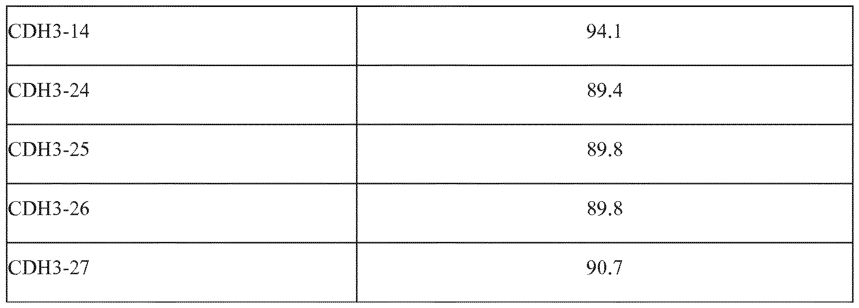


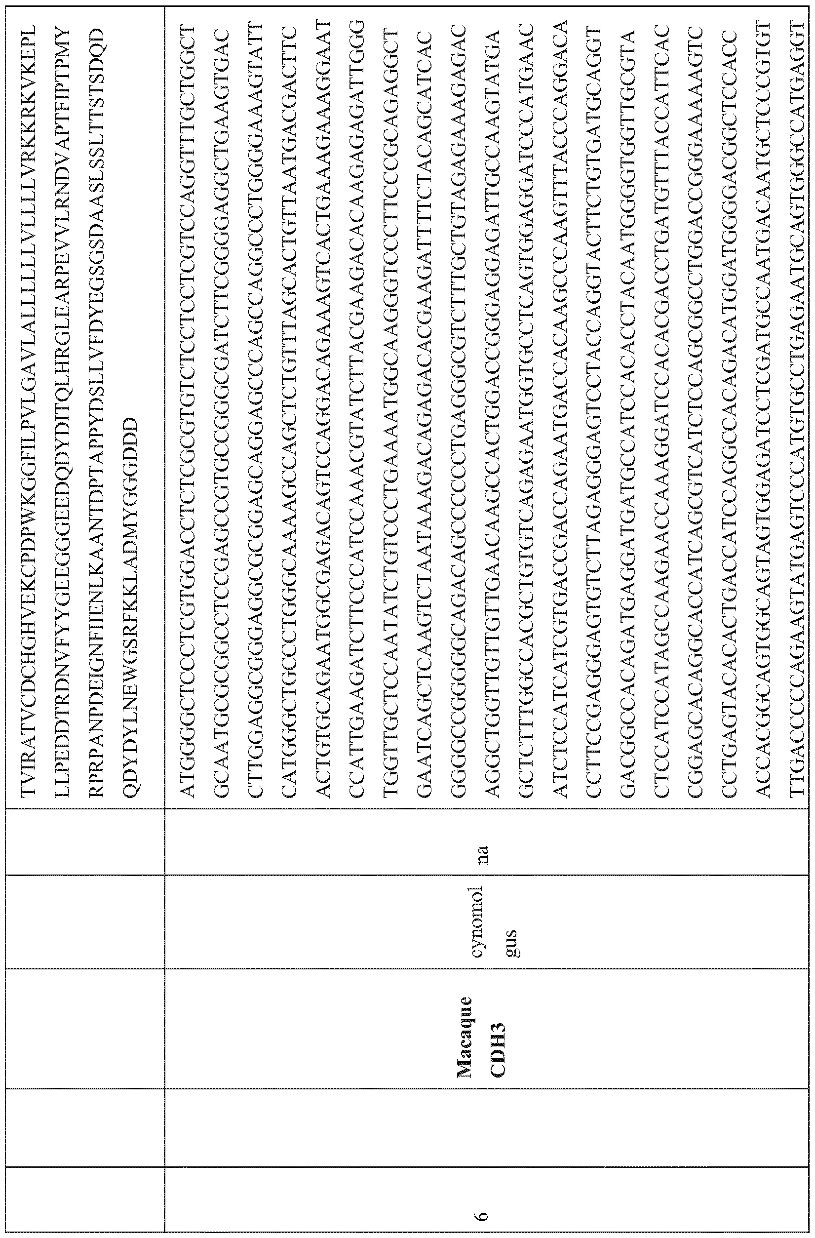


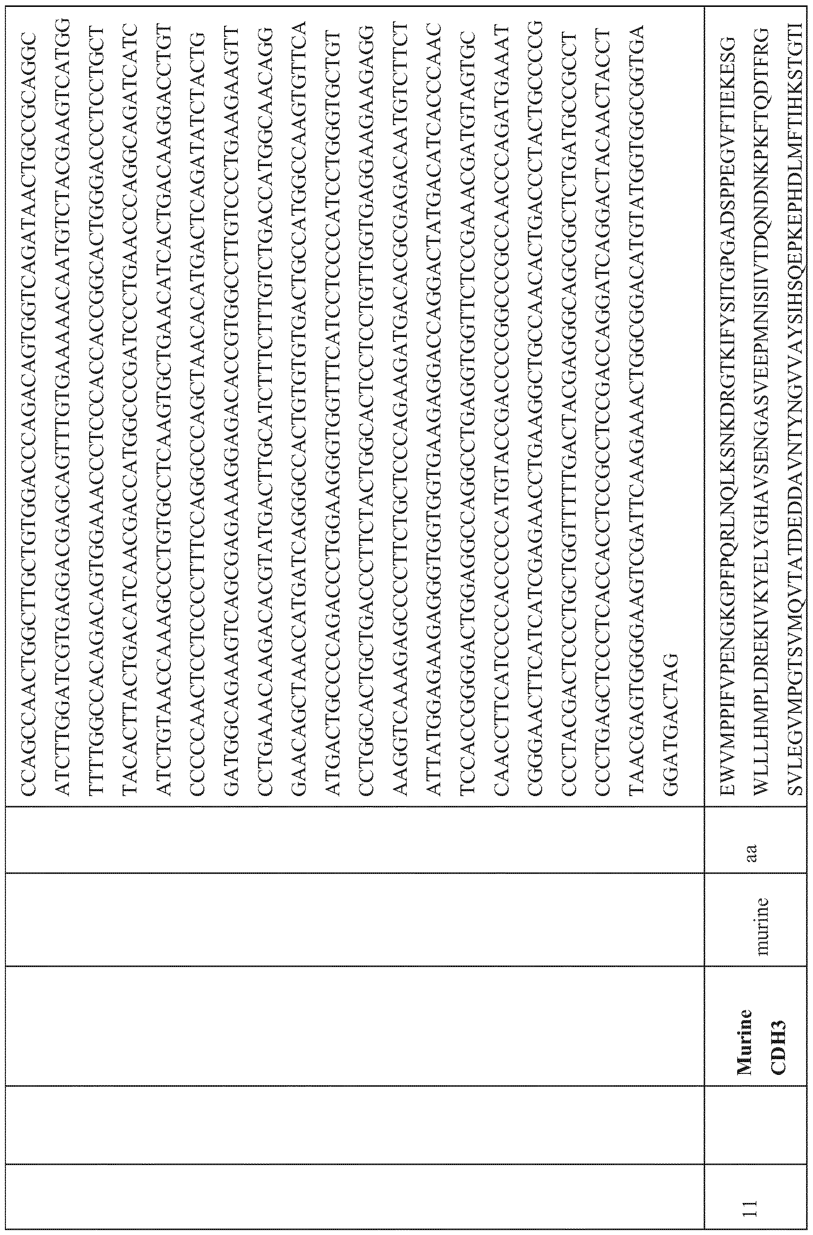
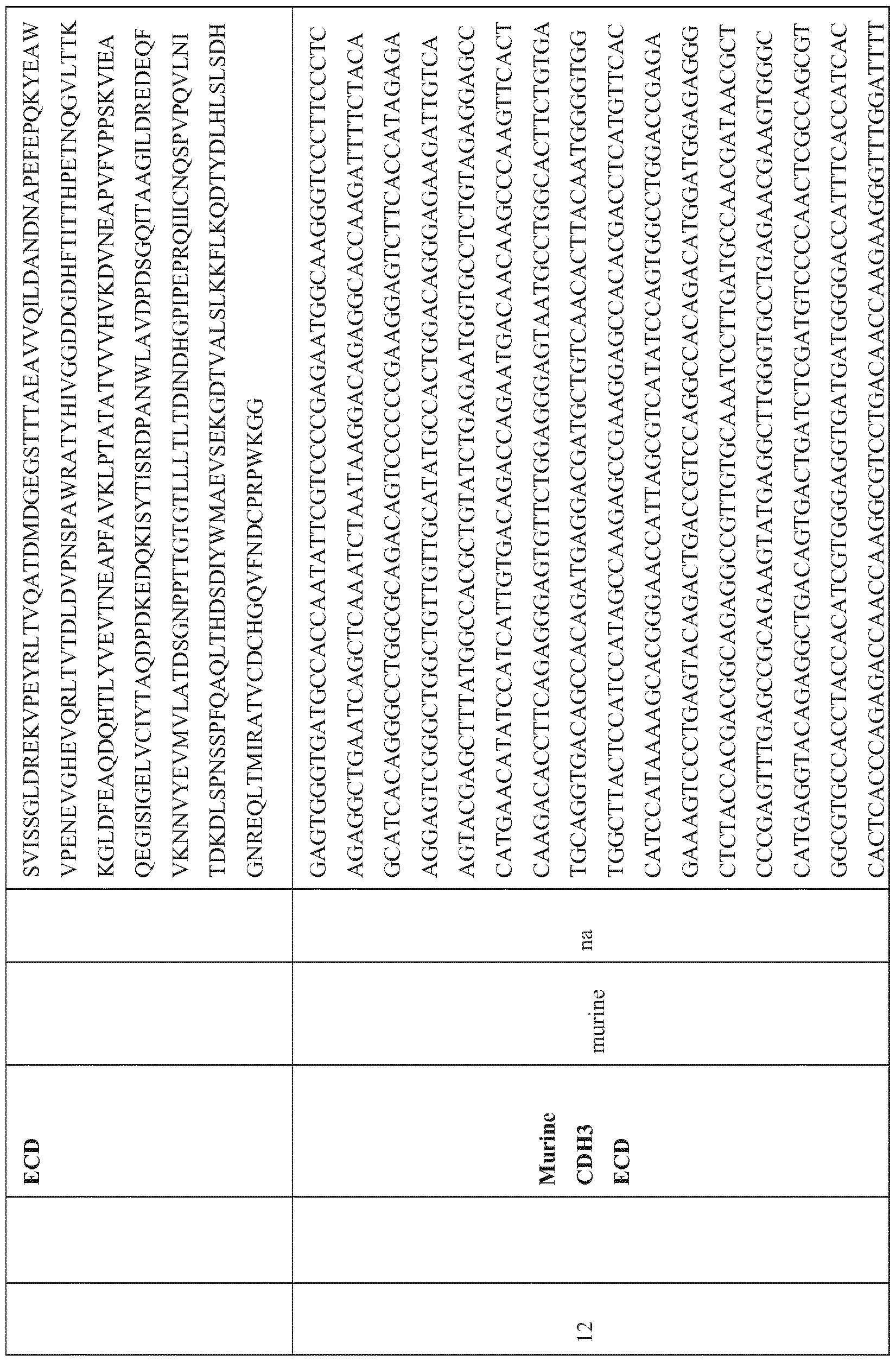


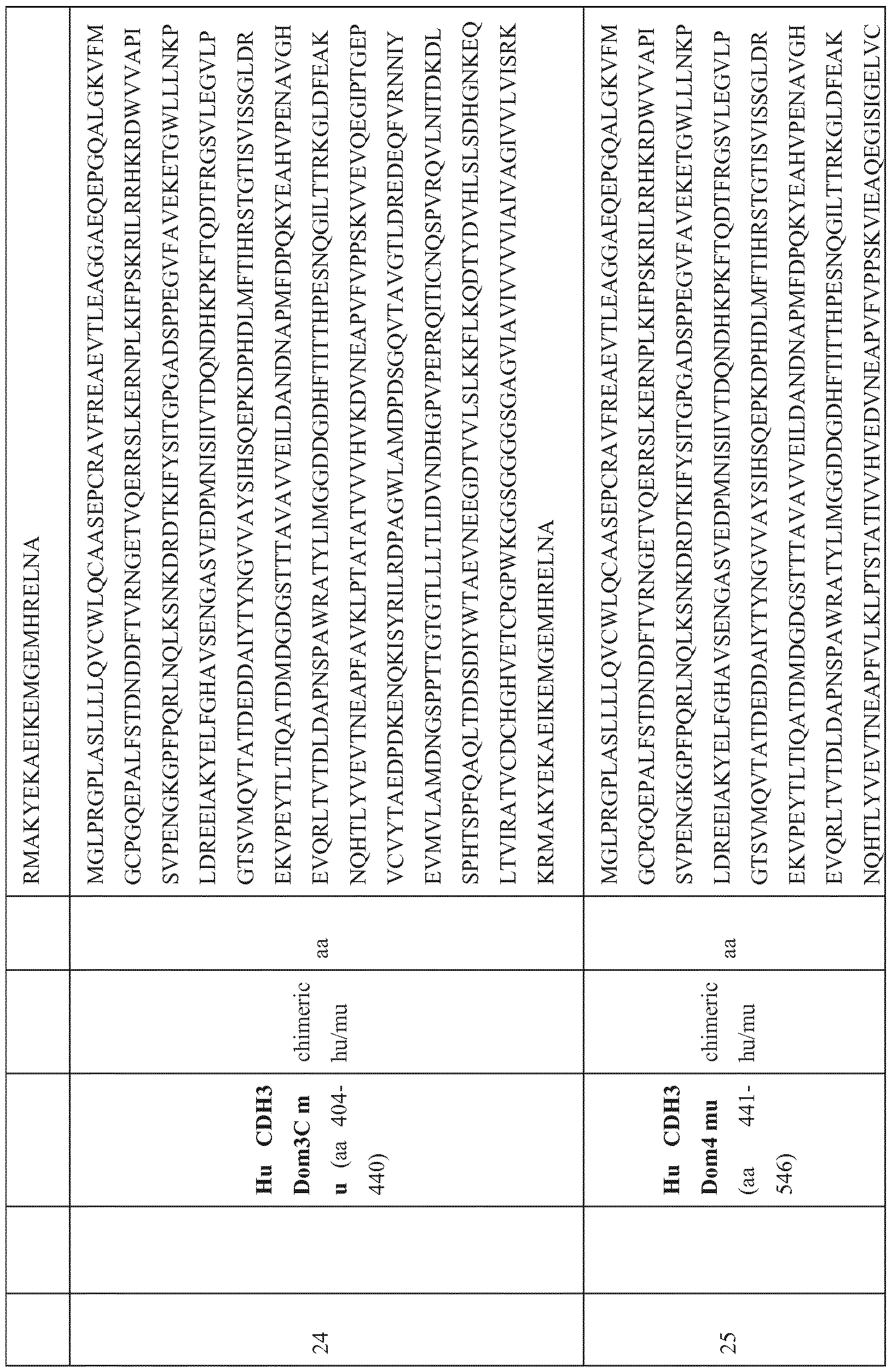


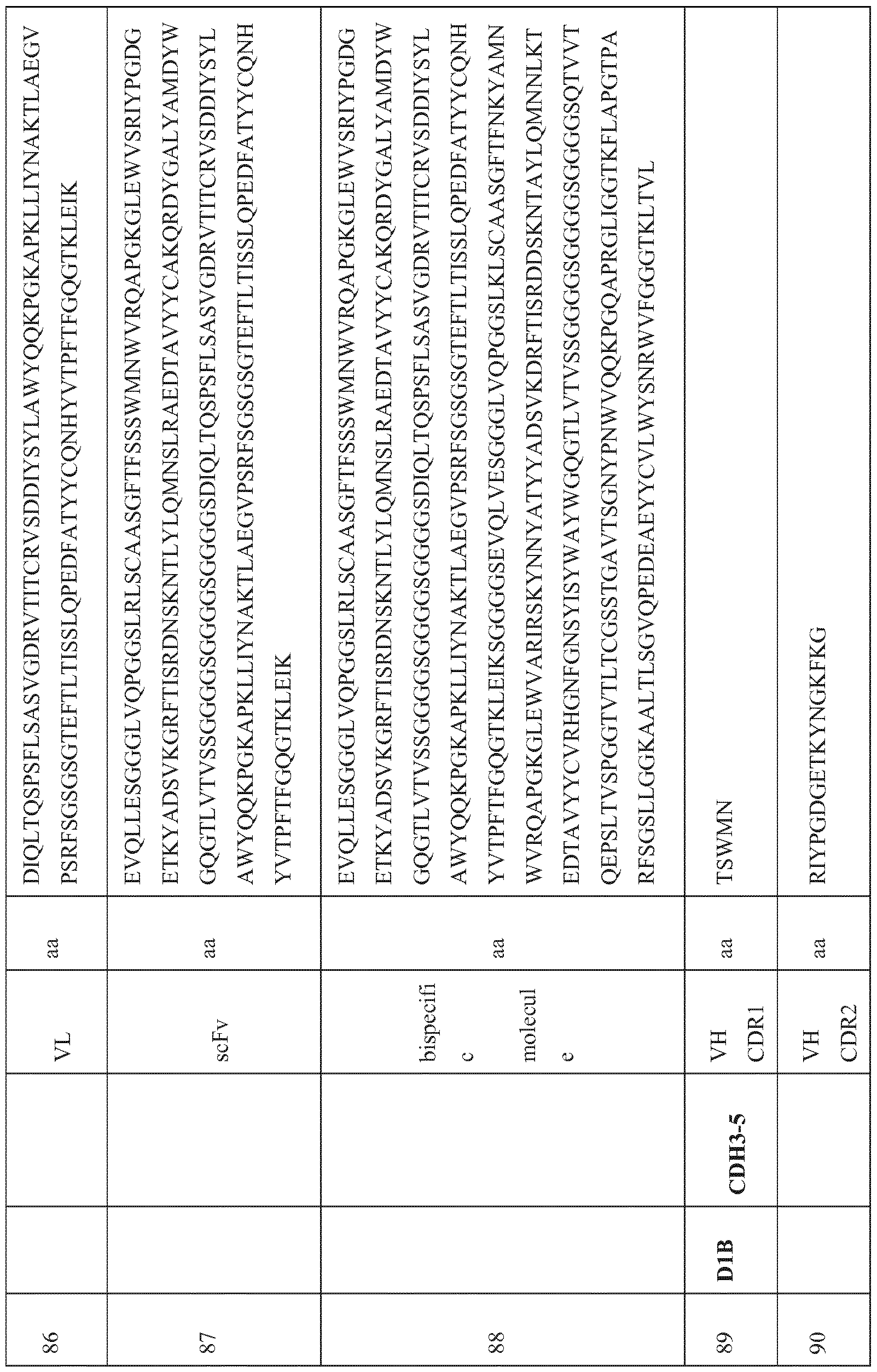


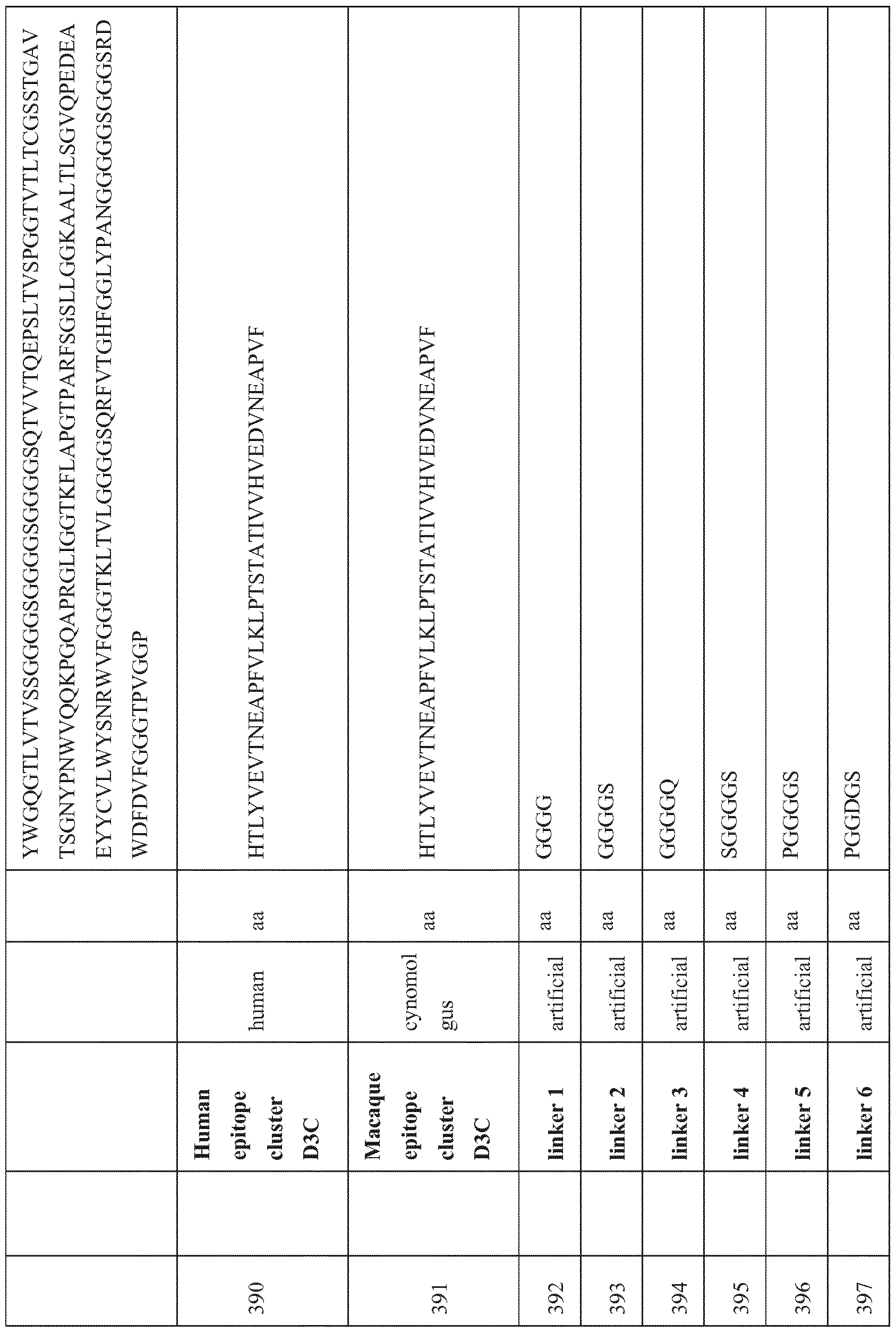

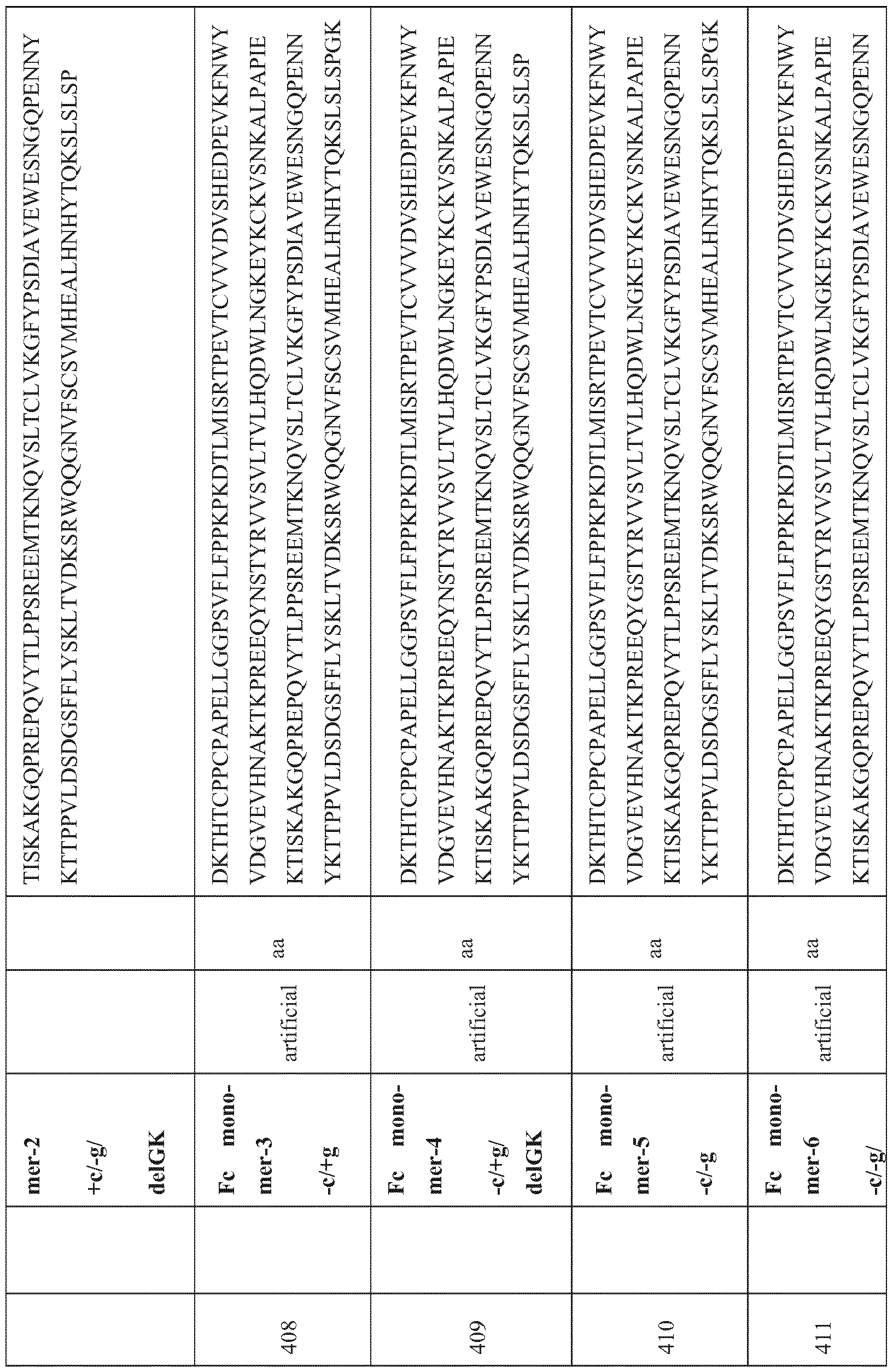


Claims
Priority Applications (22)
| Application Number | Priority Date | Filing Date | Title |
|---|---|---|---|
| EP23166533.2A EP4276116A3 (en) | 2015-04-17 | 2016-04-18 | Bispecific antibody constructs for cdh3 and cd3 |
| US15/566,444 US11028171B2 (en) | 2015-04-17 | 2016-04-18 | Bispecific antibody constructs for CDH3 and CD3 |
| EA201792193A EA201792193A1 (en) | 2015-04-17 | 2016-04-18 | CONSTRUCTIONS OF BISPECIFIC ANTIBODIES FOR CDH3 AND CD3 |
| SG11201708438WA SG11201708438WA (en) | 2015-04-17 | 2016-04-18 | Bispecific antibody constructs for cdh3 and cd3 |
| CA2982682A CA2982682A1 (en) | 2015-04-17 | 2016-04-18 | Bispecific antibody constructs for cdh3 and cd3 |
| IL255076A IL255076B2 (en) | 2015-04-17 | 2016-04-18 | Bispecific antibody constructs for cdh3 and cd3 |
| PL16716617.2T PL3283524T3 (en) | 2015-04-17 | 2016-04-18 | Bispecific antibody constructs for cdh3 and cd3 |
| MX2017013348A MX2017013348A (en) | 2015-04-17 | 2016-04-18 | Bispecific antibody constructs for cdh3 and cd3. |
| JP2017554264A JP6907124B2 (en) | 2015-04-17 | 2016-04-18 | Bispecific antibody construct against CDH3 and CD3 |
| LTEPPCT/EP2016/058482T LT3283524T (en) | 2015-04-17 | 2016-04-18 | Bispecific antibody constructs for cdh3 and cd3 |
| CN202210971153.5A CN116063541A (en) | 2015-04-17 | 2016-04-18 | Bispecific antibody constructs for CDH3 and CD3 |
| FIEP16716617.2T FI3283524T3 (en) | 2015-04-17 | 2016-04-18 | Bispecific antibody constructs for cdh3 and cd3 |
| EP16716617.2A EP3283524B1 (en) | 2015-04-17 | 2016-04-18 | Bispecific antibody constructs for cdh3 and cd3 |
| DK16716617.2T DK3283524T3 (en) | 2015-04-17 | 2016-04-18 | BISPECIFIC ANTIBODY CONSTRUCTIONS AGAINST CDH3 and CD3 |
| AU2016250023A AU2016250023B2 (en) | 2015-04-17 | 2016-04-18 | Bispecific antibody constructs for CDH3 and CD3 |
| ES16716617T ES2945313T3 (en) | 2015-04-17 | 2016-04-18 | Bispecific antibody constructs for CDH3 and CD3 |
| BR112017022326A BR112017022326A2 (en) | 2015-04-17 | 2016-04-18 | bispecific antibody constructs for cdh3 and cd3 |
| CN201680035008.9A CN107750255B (en) | 2015-04-17 | 2016-04-18 | Bispecific antibody constructs for CDH3 and CD3 |
| SI201631692T SI3283524T1 (en) | 2015-04-17 | 2016-04-18 | Bispecific antibody constructs for cdh3 and cd3 |
| KR1020177033397A KR20180023892A (en) | 2015-04-17 | 2016-04-18 | The bispecific antibody constructs for CDH3 and CD3 |
| US17/306,051 US11926666B2 (en) | 2015-04-17 | 2021-05-03 | Bispecific antibody constructs for CDH3 and CD3 |
| AU2022203450A AU2022203450A1 (en) | 2015-04-17 | 2022-05-20 | Bispecific Antibody Constructs For CDH3 And CD3 |
Applications Claiming Priority (2)
| Application Number | Priority Date | Filing Date | Title |
|---|---|---|---|
| EP15164154.5 | 2015-04-17 | ||
| EP15164154 | 2015-04-17 |
Related Child Applications (2)
| Application Number | Title | Priority Date | Filing Date |
|---|---|---|---|
| US15/566,444 A-371-Of-International US11028171B2 (en) | 2015-04-17 | 2016-04-18 | Bispecific antibody constructs for CDH3 and CD3 |
| US17/306,051 Division US11926666B2 (en) | 2015-04-17 | 2021-05-03 | Bispecific antibody constructs for CDH3 and CD3 |
Publications (1)
| Publication Number | Publication Date |
|---|---|
| WO2016166360A1 true WO2016166360A1 (en) | 2016-10-20 |
Family
ID=52997289
Family Applications (1)
| Application Number | Title | Priority Date | Filing Date |
|---|---|---|---|
| PCT/EP2016/058482 WO2016166360A1 (en) | 2015-04-17 | 2016-04-18 | Bispecific antibody constructs for cdh3 and cd3 |
Country Status (25)
| Country | Link |
|---|---|
| US (2) | US11028171B2 (en) |
| EP (2) | EP3283524B1 (en) |
| JP (1) | JP6907124B2 (en) |
| KR (1) | KR20180023892A (en) |
| CN (2) | CN107750255B (en) |
| AR (1) | AR104309A1 (en) |
| AU (2) | AU2016250023B2 (en) |
| BR (1) | BR112017022326A2 (en) |
| CA (1) | CA2982682A1 (en) |
| CL (1) | CL2017002641A1 (en) |
| DK (1) | DK3283524T3 (en) |
| EA (1) | EA201792193A1 (en) |
| ES (1) | ES2945313T3 (en) |
| FI (1) | FI3283524T3 (en) |
| HU (1) | HUE061730T2 (en) |
| IL (1) | IL255076B2 (en) |
| LT (1) | LT3283524T (en) |
| MA (1) | MA43197A (en) |
| MX (1) | MX2017013348A (en) |
| PL (1) | PL3283524T3 (en) |
| PT (1) | PT3283524T (en) |
| SG (2) | SG10201909308XA (en) |
| SI (1) | SI3283524T1 (en) |
| TW (2) | TWI772258B (en) |
| WO (1) | WO2016166360A1 (en) |
Cited By (13)
| Publication number | Priority date | Publication date | Assignee | Title |
|---|---|---|---|---|
| CN109071662A (en) * | 2016-02-03 | 2018-12-21 | 安进研发(慕尼黑)股份有限公司 | Bispecific T cell bound antibody construct |
| WO2019143636A1 (en) * | 2018-01-16 | 2019-07-25 | Lakepharma, Inc. | Bispecific antibody that binds cd3 and another target |
| CN110582297A (en) * | 2017-02-02 | 2019-12-17 | 安进研究(慕尼黑)有限责任公司 | Low pH pharmaceutical compositions comprising T cell conjugated antibody constructs |
| WO2019112978A3 (en) * | 2017-12-04 | 2020-03-26 | Coare Biotechnology, Inc. | Anti-dclk1 antibodies and chimeric antigen receptors, and compositions and methods of use thereof |
| WO2020094834A1 (en) * | 2018-11-09 | 2020-05-14 | Euchloe Bio Pte. Ltd. | Il2rbeta/common gamma chain antibodies |
| US20210130465A1 (en) * | 2017-12-29 | 2021-05-06 | Amgen Inc. | Bispecific Antibody Construct Directed to MUC17 and CD3 |
| EP3752630A4 (en) * | 2018-02-14 | 2022-01-05 | Yale University | Compositions for modulation of a trem or treml protein and methods of use |
| US11352433B2 (en) | 2016-02-03 | 2022-06-07 | Amgen Research (Munich) Gmbh | BCMA and CD3 bispecific T cell engaging antibody constructs |
| US11447567B2 (en) | 2015-07-31 | 2022-09-20 | Amgen Research (Munich) Gmbh | Antibody constructs for FLT3 and CD3 |
| WO2022204516A1 (en) * | 2021-03-26 | 2022-09-29 | The General Hospital Corporation | Humanized mab107 |
| US11655307B2 (en) | 2017-05-30 | 2023-05-23 | The Board Of Regents Of The University Of Oklahoma | Anti-doublecortin-like kinase 1 antibodies and methods of use |
| EP3973000A4 (en) * | 2019-06-07 | 2023-09-06 | Adimab, LLC | High affinity anti-cd3 antibodies, and methods for their generation and use |
| US11884720B2 (en) | 2015-07-31 | 2024-01-30 | Amgen Research (Munich) Gmbh | Antibody constructs for MSLN and CD3 |
Families Citing this family (3)
| Publication number | Priority date | Publication date | Assignee | Title |
|---|---|---|---|---|
| BRPI0919840B1 (en) * | 2008-10-01 | 2023-02-28 | Amgen Research (Munich) Gmbh | BISPECIFIC SINGLE CHAIN ANTIBODY MOLECULE, ITS USE, AND PHARMACEUTICAL COMPOSITION COMPRISING IT |
| WO2021113748A1 (en) * | 2019-12-05 | 2021-06-10 | Arbele Corp. | Composition of triaxial antibodies and method of making and using thereof |
| CN116096751A (en) * | 2020-02-25 | 2023-05-09 | 璟尚生物制药公司 | Trispecific T cell adapter |
Citations (154)
| Publication number | Priority date | Publication date | Assignee | Title |
|---|---|---|---|---|
| US233A (en) | 1837-06-14 | Improvement in plows | ||
| US556A (en) | 1838-01-09 | Machine foe | ||
| US4447A (en) | 1846-04-04 | Car- wheel | ||
| US5013A (en) | 1847-03-13 | Improvement in apparatus for the manufacture of malleable iron | ||
| US462837A (en) | 1891-11-10 | Indicating device for water-tanks | ||
| US3180193A (en) | 1963-02-25 | 1965-04-27 | Benedict David | Machines for cutting lengths of strip material |
| US3691016A (en) | 1970-04-17 | 1972-09-12 | Monsanto Co | Process for the preparation of insoluble enzymes |
| US3773919A (en) | 1969-10-23 | 1973-11-20 | Du Pont | Polylactide-drug mixtures |
| US3969287A (en) | 1972-12-08 | 1976-07-13 | Boehringer Mannheim Gmbh | Carrier-bound protein prepared by reacting the protein with an acylating or alkylating compound having a carrier-bonding group and reacting the product with a carrier |
| US4179337A (en) | 1973-07-20 | 1979-12-18 | Davis Frank F | Non-immunogenic polypeptides |
| US4195128A (en) | 1976-05-03 | 1980-03-25 | Bayer Aktiengesellschaft | Polymeric carrier bound ligands |
| US4229537A (en) | 1978-02-09 | 1980-10-21 | New York University | Preparation of trichloro-s-triazine activated supports for coupling ligands |
| US4247642A (en) | 1977-02-17 | 1981-01-27 | Sumitomo Chemical Company, Limited | Enzyme immobilization with pullulan gel |
| EP0036676A1 (en) | 1978-03-24 | 1981-09-30 | The Regents Of The University Of California | Method of making uniformly sized liposomes and liposomes so made |
| US4301144A (en) | 1979-07-11 | 1981-11-17 | Ajinomoto Company, Incorporated | Blood substitute containing modified hemoglobin |
| US4330440A (en) | 1977-02-08 | 1982-05-18 | Development Finance Corporation Of New Zealand | Activated matrix and method of activation |
| EP0058481A1 (en) | 1981-02-16 | 1982-08-25 | Zeneca Limited | Continuous release pharmaceutical compositions |
| EP0088046A2 (en) | 1982-02-17 | 1983-09-07 | Ciba-Geigy Ag | Lipids in the aqueous phase |
| US4439196A (en) | 1982-03-18 | 1984-03-27 | Merck & Co., Inc. | Osmotic drug delivery system |
| US4447224A (en) | 1982-09-20 | 1984-05-08 | Infusaid Corporation | Variable flow implantable infusion apparatus |
| US4475196A (en) | 1981-03-06 | 1984-10-02 | Zor Clair G | Instrument for locating faults in aircraft passenger reading light and attendant call control system |
| US4485045A (en) | 1981-07-06 | 1984-11-27 | Research Corporation | Synthetic phosphatidyl cholines useful in forming liposomes |
| US4486194A (en) | 1983-06-08 | 1984-12-04 | James Ferrara | Therapeutic device for administering medicaments through the skin |
| US4487603A (en) | 1982-11-26 | 1984-12-11 | Cordis Corporation | Implantable microinfusion pump system |
| US4496689A (en) | 1983-12-27 | 1985-01-29 | Miles Laboratories, Inc. | Covalently attached complex of alpha-1-proteinase inhibitor with a water soluble polymer |
| EP0133988A2 (en) | 1983-08-02 | 1985-03-13 | Hoechst Aktiengesellschaft | Regulating peptide-containing pharmaceutical preparations with retarded release, and process for their preparation |
| EP0143949A1 (en) | 1983-11-01 | 1985-06-12 | TERUMO KABUSHIKI KAISHA trading as TERUMO CORPORATION | Pharmaceutical composition containing urokinase |
| US4544545A (en) | 1983-06-20 | 1985-10-01 | Trustees University Of Massachusetts | Liposomes containing modified cholesterol for organ targeting |
| EP0171496A2 (en) | 1984-08-15 | 1986-02-19 | Research Development Corporation of Japan | Process for the production of a chimera monoclonal antibody |
| EP0173494A2 (en) | 1984-08-27 | 1986-03-05 | The Board Of Trustees Of The Leland Stanford Junior University | Chimeric receptors by DNA splicing and expression |
| EP0183070A2 (en) | 1984-10-30 | 1986-06-04 | Phillips Petroleum Company | Transformation of yeasts of the genus pichia |
| US4596556A (en) | 1985-03-25 | 1986-06-24 | Bioject, Inc. | Hypodermic injection apparatus |
| GB2177096A (en) | 1984-09-03 | 1987-01-14 | Celltech Ltd | Production of chimeric antibodies |
| US4640835A (en) | 1981-10-30 | 1987-02-03 | Nippon Chemiphar Company, Ltd. | Plasminogen activator derivatives |
| US4670417A (en) | 1985-06-19 | 1987-06-02 | Ajinomoto Co., Inc. | Hemoglobin combined with a poly(alkylene oxide) |
| WO1987005330A1 (en) | 1986-03-07 | 1987-09-11 | Michel Louis Eugene Bergh | Method for enhancing glycoprotein stability |
| EP0239400A2 (en) | 1986-03-27 | 1987-09-30 | Medical Research Council | Recombinant antibodies and methods for their production |
| EP0244234A2 (en) | 1986-04-30 | 1987-11-04 | Alko Group Ltd. | Transformation of trichoderma |
| US4751180A (en) | 1985-03-28 | 1988-06-14 | Chiron Corporation | Expression using fused genes providing for protein product |
| WO1988009344A1 (en) | 1987-05-21 | 1988-12-01 | Creative Biomolecules, Inc. | Targeted multifunctional proteins |
| US4791192A (en) | 1986-06-26 | 1988-12-13 | Takeda Chemical Industries, Ltd. | Chemically modified protein with polyethyleneglycol |
| US4790824A (en) | 1987-06-19 | 1988-12-13 | Bioject, Inc. | Non-invasive hypodermic injection device |
| US4816397A (en) | 1983-03-25 | 1989-03-28 | Celltech, Limited | Multichain polypeptides or proteins and processes for their production |
| US4935233A (en) | 1985-12-02 | 1990-06-19 | G. D. Searle And Company | Covalently linked polypeptide cell modulators |
| US4941880A (en) | 1987-06-19 | 1990-07-17 | Bioject, Inc. | Pre-filled ampule and non-invasive hypodermic injection device assembly |
| US4946778A (en) | 1987-09-21 | 1990-08-07 | Genex Corporation | Single polypeptide chain binding molecules |
| EP0402226A1 (en) | 1989-06-06 | 1990-12-12 | Institut National De La Recherche Agronomique | Transformation vectors for yeast yarrowia |
| JPH0368180B2 (en) | 1986-01-22 | 1991-10-25 | Shisutemu Mentenansu Kk | |
| JPH0368507B2 (en) | 1982-03-29 | 1991-10-28 | Tokyo Shibaura Electric Co | |
| JPH0368506B2 (en) | 1982-03-29 | 1991-10-28 | Tokyo Shibaura Electric Co | |
| US5064413A (en) | 1989-11-09 | 1991-11-12 | Bioject, Inc. | Needleless hypodermic injection device |
| WO1992003918A1 (en) | 1990-08-29 | 1992-03-19 | Genpharm International, Inc. | Transgenic non-human animals capable of producing heterologous antibodies |
| WO1992015673A1 (en) | 1991-03-11 | 1992-09-17 | The University Of Georgia Research Foundation, Inc. | Cloning and expression of renilla luciferase |
| WO1992022670A1 (en) | 1991-06-12 | 1992-12-23 | Genpharm International, Inc. | Early detection of transgenic embryos |
| WO1992022645A1 (en) | 1991-06-14 | 1992-12-23 | Genpharm International, Inc. | Transgenic immunodeficient non-human animals |
| WO1993012227A1 (en) | 1991-12-17 | 1993-06-24 | Genpharm International, Inc. | Transgenic non-human animals capable of producing heterologous antibodies |
| US5223409A (en) | 1988-09-02 | 1993-06-29 | Protein Engineering Corp. | Directed evolution of novel binding proteins |
| US5225539A (en) | 1986-03-27 | 1993-07-06 | Medical Research Council | Recombinant altered antibodies and methods of making altered antibodies |
| WO1994000569A1 (en) | 1992-06-18 | 1994-01-06 | Genpharm International, Inc. | Methods for producing transgenic non-human animals harboring a yeast artificial chromosome |
| WO1994002602A1 (en) | 1992-07-24 | 1994-02-03 | Cell Genesys, Inc. | Generation of xenogeneic antibodies |
| US5292658A (en) | 1989-12-29 | 1994-03-08 | University Of Georgia Research Foundation, Inc. Boyd Graduate Studies Research Center | Cloning and expressions of Renilla luciferase |
| WO1994010308A1 (en) | 1992-10-23 | 1994-05-11 | Immunex Corporation | Methods of preparing soluble, oligomeric proteins |
| US5312335A (en) | 1989-11-09 | 1994-05-17 | Bioject Inc. | Needleless hypodermic injection device |
| US5313193A (en) | 1990-06-29 | 1994-05-17 | Thomson-Csf | Object-marking means, method for making such means and reading device |
| WO1994025585A1 (en) | 1993-04-26 | 1994-11-10 | Genpharm International, Inc. | Transgenic non-human animals capable of producing heterologous antibodies |
| US5383851A (en) | 1992-07-24 | 1995-01-24 | Bioject Inc. | Needleless hypodermic injection device |
| WO1995007463A1 (en) | 1993-09-10 | 1995-03-16 | The Trustees Of Columbia University In The City Of New York | Uses of green fluorescent protein |
| US5476996A (en) | 1988-06-14 | 1995-12-19 | Lidak Pharmaceuticals | Human immune system in non-human animal |
| WO1996014436A1 (en) | 1994-11-04 | 1996-05-17 | Genpharm International, Inc. | Method for making recombinant yeast artificial chromosomes |
| EP0463151B1 (en) | 1990-01-12 | 1996-06-12 | Cell Genesys, Inc. | Generation of xenogeneic antibodies |
| US5545807A (en) | 1988-10-12 | 1996-08-13 | The Babraham Institute | Production of antibodies from transgenic animals |
| US5545806A (en) | 1990-08-29 | 1996-08-13 | Genpharm International, Inc. | Ransgenic non-human animals for producing heterologous antibodies |
| US5565332A (en) | 1991-09-23 | 1996-10-15 | Medical Research Council | Production of chimeric antibodies - a combinatorial approach |
| WO1996033735A1 (en) | 1995-04-27 | 1996-10-31 | Abgenix, Inc. | Human antibodies derived from immunized xenomice |
| WO1996034096A1 (en) | 1995-04-28 | 1996-10-31 | Abgenix, Inc. | Human antibodies derived from immunized xenomice |
| US5585089A (en) | 1988-12-28 | 1996-12-17 | Protein Design Labs, Inc. | Humanized immunoglobulins |
| US5591669A (en) | 1988-12-05 | 1997-01-07 | Genpharm International, Inc. | Transgenic mice depleted in a mature lymphocytic cell-type |
| US5612205A (en) | 1990-08-29 | 1997-03-18 | Genpharm International, Incorporated | Homologous recombination in mammalian cells |
| WO1997013852A1 (en) | 1995-10-10 | 1997-04-17 | Genpharm International, Inc. | Transgenic non-human animals capable of producing heterologous antibodies |
| US5625825A (en) | 1993-10-21 | 1997-04-29 | Lsi Logic Corporation | Random number generating apparatus for an interface unit of a carrier sense with multiple access and collision detect (CSMA/CD) ethernet data network |
| US5625126A (en) | 1990-08-29 | 1997-04-29 | Genpharm International, Inc. | Transgenic non-human animals for producing heterologous antibodies |
| EP0773288A2 (en) | 1995-08-29 | 1997-05-14 | Kirin Beer Kabushiki Kaisha | Chimeric animal and method for producing the same |
| US5633425A (en) | 1990-08-29 | 1997-05-27 | Genpharm International, Inc. | Transgenic non-human animals capable of producing heterologous antibodies |
| US5648260A (en) | 1987-03-18 | 1997-07-15 | Scotgen Biopharmaceuticals Incorporated | DNA encoding antibodies with altered effector functions |
| US5661016A (en) | 1990-08-29 | 1997-08-26 | Genpharm International Inc. | Transgenic non-human animals capable of producing heterologous antibodies of various isotypes |
| WO1997038731A1 (en) | 1996-04-18 | 1997-10-23 | The Regents Of The University Of California | Immunoliposomes that optimize internalization into target cells |
| US5683888A (en) | 1989-07-22 | 1997-11-04 | University Of Wales College Of Medicine | Modified bioluminescent proteins and their use |
| WO1998014605A1 (en) | 1996-10-04 | 1998-04-09 | Loma Linda University | Renilla luciferase and green fluorescent protein fusion genes |
| US5741668A (en) | 1994-02-04 | 1998-04-21 | Rutgers, The State University Of New Jersey | Expression of a gene for a modified green-fluorescent protein |
| WO1998024893A2 (en) | 1996-12-03 | 1998-06-11 | Abgenix, Inc. | TRANSGENIC MAMMALS HAVING HUMAN IG LOCI INCLUDING PLURAL VH AND Vλ REGIONS AND ANTIBODIES PRODUCED THEREFROM |
| WO1998024884A1 (en) | 1996-12-02 | 1998-06-11 | Genpharm International | Transgenic non-human animals capable of producing heterologous antibodies |
| WO1998026277A2 (en) | 1996-12-12 | 1998-06-18 | Prolume, Ltd. | Apparatus and method for detecting and identifying infectious agents |
| US5777079A (en) | 1994-11-10 | 1998-07-07 | The Regents Of The University Of California | Modified green fluorescent proteins |
| US5789650A (en) | 1990-08-29 | 1998-08-04 | Genpharm International, Inc. | Transgenic non-human animals for producing heterologous antibodies |
| US5789215A (en) | 1991-08-20 | 1998-08-04 | Genpharm International | Gene targeting in animal cells using isogenic DNA constructs |
| US5804387A (en) | 1996-02-01 | 1998-09-08 | The Board Of Trustees Of The Leland Stanford Junior University | FACS-optimized mutants of the green fluorescent protein (GFP) |
| US5814318A (en) | 1990-08-29 | 1998-09-29 | Genpharm International Inc. | Transgenic non-human animals for producing heterologous antibodies |
| WO1998052976A1 (en) | 1997-05-21 | 1998-11-26 | Biovation Limited | Method for the production of non-immunogenic proteins |
| US5859205A (en) | 1989-12-21 | 1999-01-12 | Celltech Limited | Humanised antibodies |
| US5874304A (en) | 1996-01-18 | 1999-02-23 | University Of Florida Research Foundation, Inc. | Humanized green fluorescent protein genes and methods |
| US5874299A (en) | 1990-08-29 | 1999-02-23 | Genpharm International, Inc. | Transgenic non-human animals capable of producing heterologous antibodies |
| US5877397A (en) | 1990-08-29 | 1999-03-02 | Genpharm International Inc. | Transgenic non-human animals capable of producing heterologous antibodies of various isotypes |
| US5876995A (en) | 1996-02-06 | 1999-03-02 | Bryan; Bruce | Bioluminescent novelty items |
| WO1999019477A1 (en) | 1997-10-15 | 1999-04-22 | Forssmann Wolf Georg | Cadherin derived growth factor and the application thereof |
| US5925558A (en) | 1996-07-16 | 1999-07-20 | The Regents Of The University Of California | Assays for protein kinases using fluorescent protein substrates |
| US5958765A (en) | 1995-06-07 | 1999-09-28 | Idec Pharmaceuticals Corporation | Neutralizing high affinity human monoclonal antibodies specific to RSV F-protein and methods for their manufacture and therapeutic use thereof |
| WO1999049019A2 (en) | 1998-03-27 | 1999-09-30 | Prolume, Ltd. | Luciferases, fluorescent proteins, nucleic acids encoding the luciferases and fluorescent proteins and the use thereof in diagnostics |
| WO1999054440A1 (en) | 1998-04-21 | 1999-10-28 | Micromet Gesellschaft Für Biomedizinische Forschung Mbh | CD19xCD3 SPECIFIC POLYPEPTIDES AND USES THEREOF |
| US5981175A (en) | 1993-01-07 | 1999-11-09 | Genpharm Internation, Inc. | Methods for producing recombinant mammalian cells harboring a yeast artificial chromosome |
| WO2000006605A2 (en) | 1998-07-28 | 2000-02-10 | Micromet Ag | Heterominibodies |
| US6075181A (en) | 1990-01-12 | 2000-06-13 | Abgenix, Inc. | Human antibodies derived from immunized xenomice |
| WO2000034317A2 (en) | 1998-12-08 | 2000-06-15 | Biovation Limited | Method for reducing immunogenicity of proteins |
| US6150584A (en) | 1990-01-12 | 2000-11-21 | Abgenix, Inc. | Human antibodies derived from immunized xenomice |
| US6162963A (en) | 1990-01-12 | 2000-12-19 | Abgenix, Inc. | Generation of Xenogenetic antibodies |
| WO2000076310A1 (en) | 1999-06-10 | 2000-12-21 | Abgenix, Inc. | Transgenic animals for producing specific isotypes of human antibodies via non-cognate switch regions |
| US6255458B1 (en) | 1990-08-29 | 2001-07-03 | Genpharm International | High affinity human antibodies and human antibodies against digoxin |
| US6300064B1 (en) | 1995-08-18 | 2001-10-09 | Morphosys Ag | Protein/(poly)peptide libraries |
| US6407213B1 (en) | 1991-06-14 | 2002-06-18 | Genentech, Inc. | Method for making humanized antibodies |
| WO2002097395A2 (en) | 2001-05-31 | 2002-12-05 | Chiron Corporation | P-cadherin as a target for anti-cancer therapy |
| WO2003047336A2 (en) | 2001-11-30 | 2003-06-12 | Abgenix, Inc. | TRANSGENIC ANIMALS BEARING HUMAN Igμ LIGHT CHAIN GENES |
| WO2004110345A2 (en) | 2002-10-29 | 2004-12-23 | Pharmacia Corporation | Differentially expressed genes involved in cancer, the polypeptides encoded thereby, and methods of using the same |
| WO2005040220A1 (en) | 2003-10-16 | 2005-05-06 | Micromet Ag | Multispecific deimmunized cd3-binders |
| WO2006114704A2 (en) | 2005-04-26 | 2006-11-02 | Pfizer Inc. | P-cadherin antibodies |
| WO2006138181A2 (en) | 2005-06-14 | 2006-12-28 | Amgen Inc. | Self-buffering protein formulations |
| WO2007042261A2 (en) | 2005-10-11 | 2007-04-19 | Micromet Ag | Compositions comprising cross-species-specific antibodies and uses thereof |
| WO2007098420A2 (en) | 2006-02-17 | 2007-08-30 | Syntonix Pharmaceuticals, Inc. | Peptides that block the binding of igg to fcrn |
| WO2007102525A1 (en) | 2006-02-28 | 2007-09-13 | Oncotherapy Science, Inc. | Methods for damaging cells using effector functions of anti-cdh3 antibodies |
| WO2008119567A2 (en) | 2007-04-03 | 2008-10-09 | Micromet Ag | Cross-species-specific cd3-epsilon binding domain |
| WO2009127691A1 (en) | 2008-04-17 | 2009-10-22 | Ablynx N.V. | Peptides capable of binding to serum proteins and compounds, constructs and polypeptides comprising the same |
| WO2010001585A1 (en) | 2008-06-30 | 2010-01-07 | Oncotherapy Science, Inc. | Anti-cdh3 antibodies labeled with radioisotope label and uses thereof |
| WO2010037838A2 (en) | 2008-10-01 | 2010-04-08 | Micromet Ag | Cross-species-specific single domain bispecific single chain antibody |
| WO2010054007A1 (en) | 2008-11-07 | 2010-05-14 | Fabrus Llc | Combinatorial antibody libraries and uses thereof |
| WO2010126137A1 (en) | 2009-05-01 | 2010-11-04 | 国立大学法人 東京大学 | Anti-cadherin antibody |
| WO2011051489A2 (en) | 2009-10-30 | 2011-05-05 | Novozymes Biopharma Dk A/S | Albumin variants |
| WO2011056997A1 (en) | 2009-11-04 | 2011-05-12 | Fabrus Llc | Methods for affinity maturation-based antibody optimization |
| WO2011071541A2 (en) | 2009-12-11 | 2011-06-16 | The Scripps Research Institute | Cadherin modulatory agents |
| WO2011080796A1 (en) | 2009-12-28 | 2011-07-07 | Oncotherapy Science, Inc. | Anti-cdh3 antibodies and uses thereof |
| WO2012059486A1 (en) | 2010-11-01 | 2012-05-10 | Novozymes Biopharma Dk A/S | Albumin variants |
| WO2012145183A2 (en) * | 2011-04-19 | 2012-10-26 | Pfizer Inc. | Combinations of anti-4-1bb antibodies and adcc-inducing antibodies for the treatment of cancer |
| WO2012150319A1 (en) | 2011-05-05 | 2012-11-08 | Novozymes Biopharma Dk A/S | Albumin variants |
| WO2013026833A1 (en) | 2011-08-23 | 2013-02-28 | Roche Glycart Ag | Bispecific t cell activating antigen binding molecules |
| WO2013026837A1 (en) | 2011-08-23 | 2013-02-28 | Roche Glycart Ag | Bispecific t cell activating antigen binding molecules |
| WO2013075066A2 (en) | 2011-11-18 | 2013-05-23 | Eleven Biotherapeutics, Inc. | Proteins with improved half-life and other properties |
| WO2013135896A1 (en) | 2012-03-16 | 2013-09-19 | Novozymes Biopharma Dk A/S | Albumin variants |
| WO2014072481A1 (en) | 2012-11-08 | 2014-05-15 | Novozymes Biopharma Dk A/S | Albumin variants |
| US20140221620A1 (en) * | 2011-06-24 | 2014-08-07 | Perseus Proteomics Inc. | Anti-human p-cadherin (cdh3) recombinant antibody |
| WO2014144722A2 (en) | 2013-03-15 | 2014-09-18 | Amgen Inc. | BISPECIFIC-Fc MOLECULES |
| WO2014151910A1 (en) | 2013-03-15 | 2014-09-25 | Amgen Inc. | Heterodimeric bispecific antibodies |
| US20140308285A1 (en) | 2013-03-15 | 2014-10-16 | Amgen Inc. | Heterodimeric bispecific antibodies |
| WO2015048272A1 (en) | 2013-09-25 | 2015-04-02 | Amgen Inc. | V-c-fc-v-c antibody |
| WO2016001810A1 (en) * | 2014-07-01 | 2016-01-07 | Pfizer Inc. | Bispecific heterodimeric diabodies and uses thereof |
| US9300829B2 (en) | 2014-04-04 | 2016-03-29 | Canon Kabushiki Kaisha | Image reading apparatus and correction method thereof |
| US9676293B2 (en) | 2013-04-23 | 2017-06-13 | Calsonic Kansei Corporation | Driving force controller for electric vehicle |
| US11284893B2 (en) | 2019-04-02 | 2022-03-29 | Covidien Lp | Stapling device with articulating tool assembly |
Family Cites Families (33)
| Publication number | Priority date | Publication date | Assignee | Title |
|---|---|---|---|---|
| US4447233A (en) | 1981-04-10 | 1984-05-08 | Parker-Hannifin Corporation | Medication infusion pump |
| US4816567A (en) | 1983-04-08 | 1989-03-28 | Genentech, Inc. | Recombinant immunoglobin preparations |
| ATE108965T1 (en) | 1987-12-09 | 1994-08-15 | Omron Tateisi Electronics Co | INDUCTIVE DATA TRANSMISSION SYSTEM. |
| US5013556A (en) | 1989-10-20 | 1991-05-07 | Liposome Technology, Inc. | Liposomes with enhanced circulation time |
| US5470582A (en) | 1992-02-07 | 1995-11-28 | Syntex (U.S.A.) Inc. | Controlled delivery of pharmaceuticals from preformed porous polymeric microparticles |
| US7254167B2 (en) | 1998-10-30 | 2007-08-07 | Broadcom Corporation | Constellation-multiplexed transmitter and receiver |
| US7230167B2 (en) | 2001-08-31 | 2007-06-12 | Syngenta Participations Ag | Modified Cry3A toxins and nucleic acid sequences coding therefor |
| US8486859B2 (en) | 2002-05-15 | 2013-07-16 | Bioenergy, Inc. | Use of ribose to enhance plant growth |
| US7904068B2 (en) | 2003-06-06 | 2011-03-08 | At&T Intellectual Property I, L.P. | System and method for providing integrated voice and data services utilizing wired cordless access with unlicensed spectrum and wired access with licensed spectrum |
| DE602006017460D1 (en) | 2005-03-14 | 2010-11-25 | Omron Tateisi Electronics Co | Programmable control system |
| US8234145B2 (en) | 2005-07-12 | 2012-07-31 | International Business Machines Corporation | Automatic computation of validation metrics for global logistics processes |
| BRPI0604215A (en) | 2005-08-17 | 2007-04-10 | Biosigma Sa | method for designing oligonucleotides for molecular biology techniques |
| JP2007122396A (en) | 2005-10-27 | 2007-05-17 | Hitachi Ltd | Disk array device, and method for verifying correspondence to its fault |
| US7919297B2 (en) | 2006-02-21 | 2011-04-05 | Cornell Research Foundation, Inc. | Mutants of Aspergillus niger PhyA phytase and Aspergillus fumigatus phytase |
| US7574748B2 (en) | 2006-03-07 | 2009-08-18 | Nike, Inc. | Glove with support system |
| US7990860B2 (en) | 2006-06-16 | 2011-08-02 | Harris Corporation | Method and system for rule-based sequencing for QoS |
| US8430938B1 (en) | 2006-07-13 | 2013-04-30 | The United States Of America As Represented By The Secretary Of The Navy | Control algorithm for autothermal reformer |
| KR101146588B1 (en) | 2006-08-11 | 2012-05-16 | 삼성전자주식회사 | Manufacturing method of fin structure and fin transistor adopting the fin structure |
| CN100589507C (en) | 2006-10-30 | 2010-02-10 | 华为技术有限公司 | A dial-up prompt system and method |
| US7466008B2 (en) | 2007-03-13 | 2008-12-16 | Taiwan Semiconductor Manufacturing Company, Ltd. | BiCMOS performance enhancement by mechanical uniaxial strain and methods of manufacture |
| WO2008124858A2 (en) * | 2007-04-11 | 2008-10-23 | F-Star Biotechnologische Forschungs- Und Entwicklungsges. M.B.H. | Targeted receptor |
| JP5398703B2 (en) * | 2007-05-14 | 2014-01-29 | バイオジェン・アイデック・エムエイ・インコーポレイテッド | Single-chain FC (ScFc) region, binding polypeptide comprising the same, and methods related thereto |
| US8209741B2 (en) | 2007-09-17 | 2012-06-26 | Microsoft Corporation | Human performance in human interactive proofs using partial credit |
| US8464584B2 (en) | 2007-10-19 | 2013-06-18 | Food Equipment Technologies Company, Inc. | Beverage dispenser with level measuring apparatus and display |
| DE112008003168B4 (en) | 2007-11-29 | 2022-01-05 | Schaeffler Technologies AG & Co. KG | Power transmission device, in particular for power transmission between a drive machine and an output |
| US8376279B2 (en) | 2008-01-23 | 2013-02-19 | Aurora Flight Sciences Corporation | Inflatable folding wings for a very high altitude aircraft |
| EP2404668B1 (en) | 2009-03-04 | 2014-05-07 | Nissan Motor Co., Ltd. | Exhaust gas purification catalyst and process for producing same |
| US8463191B2 (en) | 2009-04-02 | 2013-06-11 | Qualcomm Incorporated | Beamforming options with partial channel knowledge |
| JP2014015396A (en) * | 2010-10-29 | 2014-01-30 | Perseus Proteomics Inc | High-affinity anti-cdh3 antibody |
| CN103298648B (en) | 2010-12-30 | 2016-04-13 | C.劳勃.汉默斯坦两合有限公司 | Be suitable for the longitudinal adjustment apparatus comprising two pairs of guide rails of automobile seat |
| EP2977296B1 (en) * | 2014-01-29 | 2018-07-18 | NSK Ltd. | Electric power steering device |
| US10105142B2 (en) | 2014-09-18 | 2018-10-23 | Ethicon Llc | Surgical stapler with plurality of cutting elements |
| EP3322735A4 (en) * | 2015-07-15 | 2019-03-13 | Zymeworks Inc. | Drug-conjugated bi-specific antigen-binding constructs |
-
2016
- 2016-04-18 TW TW105112022A patent/TWI772258B/en active
- 2016-04-18 EP EP16716617.2A patent/EP3283524B1/en active Active
- 2016-04-18 MA MA043197A patent/MA43197A/en unknown
- 2016-04-18 SI SI201631692T patent/SI3283524T1/en unknown
- 2016-04-18 FI FIEP16716617.2T patent/FI3283524T3/en active
- 2016-04-18 AR ARP160101060A patent/AR104309A1/en unknown
- 2016-04-18 LT LTEPPCT/EP2016/058482T patent/LT3283524T/en unknown
- 2016-04-18 EP EP23166533.2A patent/EP4276116A3/en active Pending
- 2016-04-18 EA EA201792193A patent/EA201792193A1/en unknown
- 2016-04-18 SG SG10201909308X patent/SG10201909308XA/en unknown
- 2016-04-18 MX MX2017013348A patent/MX2017013348A/en unknown
- 2016-04-18 TW TW111129622A patent/TW202311292A/en unknown
- 2016-04-18 KR KR1020177033397A patent/KR20180023892A/en not_active Application Discontinuation
- 2016-04-18 DK DK16716617.2T patent/DK3283524T3/en active
- 2016-04-18 HU HUE16716617A patent/HUE061730T2/en unknown
- 2016-04-18 WO PCT/EP2016/058482 patent/WO2016166360A1/en active Application Filing
- 2016-04-18 IL IL255076A patent/IL255076B2/en unknown
- 2016-04-18 JP JP2017554264A patent/JP6907124B2/en active Active
- 2016-04-18 PT PT167166172T patent/PT3283524T/en unknown
- 2016-04-18 PL PL16716617.2T patent/PL3283524T3/en unknown
- 2016-04-18 CN CN201680035008.9A patent/CN107750255B/en active Active
- 2016-04-18 CN CN202210971153.5A patent/CN116063541A/en active Pending
- 2016-04-18 CA CA2982682A patent/CA2982682A1/en active Pending
- 2016-04-18 SG SG11201708438WA patent/SG11201708438WA/en unknown
- 2016-04-18 BR BR112017022326A patent/BR112017022326A2/en active Search and Examination
- 2016-04-18 AU AU2016250023A patent/AU2016250023B2/en active Active
- 2016-04-18 ES ES16716617T patent/ES2945313T3/en active Active
- 2016-04-18 US US15/566,444 patent/US11028171B2/en active Active
-
2017
- 2017-10-17 CL CL2017002641A patent/CL2017002641A1/en unknown
-
2021
- 2021-05-03 US US17/306,051 patent/US11926666B2/en active Active
-
2022
- 2022-05-20 AU AU2022203450A patent/AU2022203450A1/en not_active Abandoned
Patent Citations (171)
| Publication number | Priority date | Publication date | Assignee | Title |
|---|---|---|---|---|
| US233A (en) | 1837-06-14 | Improvement in plows | ||
| US556A (en) | 1838-01-09 | Machine foe | ||
| US4447A (en) | 1846-04-04 | Car- wheel | ||
| US5013A (en) | 1847-03-13 | Improvement in apparatus for the manufacture of malleable iron | ||
| US462837A (en) | 1891-11-10 | Indicating device for water-tanks | ||
| US3180193A (en) | 1963-02-25 | 1965-04-27 | Benedict David | Machines for cutting lengths of strip material |
| US3773919A (en) | 1969-10-23 | 1973-11-20 | Du Pont | Polylactide-drug mixtures |
| US3691016A (en) | 1970-04-17 | 1972-09-12 | Monsanto Co | Process for the preparation of insoluble enzymes |
| US3969287A (en) | 1972-12-08 | 1976-07-13 | Boehringer Mannheim Gmbh | Carrier-bound protein prepared by reacting the protein with an acylating or alkylating compound having a carrier-bonding group and reacting the product with a carrier |
| US4179337A (en) | 1973-07-20 | 1979-12-18 | Davis Frank F | Non-immunogenic polypeptides |
| US4195128A (en) | 1976-05-03 | 1980-03-25 | Bayer Aktiengesellschaft | Polymeric carrier bound ligands |
| US4330440A (en) | 1977-02-08 | 1982-05-18 | Development Finance Corporation Of New Zealand | Activated matrix and method of activation |
| US4247642A (en) | 1977-02-17 | 1981-01-27 | Sumitomo Chemical Company, Limited | Enzyme immobilization with pullulan gel |
| US4229537A (en) | 1978-02-09 | 1980-10-21 | New York University | Preparation of trichloro-s-triazine activated supports for coupling ligands |
| EP0036676A1 (en) | 1978-03-24 | 1981-09-30 | The Regents Of The University Of California | Method of making uniformly sized liposomes and liposomes so made |
| US4301144A (en) | 1979-07-11 | 1981-11-17 | Ajinomoto Company, Incorporated | Blood substitute containing modified hemoglobin |
| EP0058481A1 (en) | 1981-02-16 | 1982-08-25 | Zeneca Limited | Continuous release pharmaceutical compositions |
| US4475196A (en) | 1981-03-06 | 1984-10-02 | Zor Clair G | Instrument for locating faults in aircraft passenger reading light and attendant call control system |
| US4485045A (en) | 1981-07-06 | 1984-11-27 | Research Corporation | Synthetic phosphatidyl cholines useful in forming liposomes |
| US4640835A (en) | 1981-10-30 | 1987-02-03 | Nippon Chemiphar Company, Ltd. | Plasminogen activator derivatives |
| EP0088046A2 (en) | 1982-02-17 | 1983-09-07 | Ciba-Geigy Ag | Lipids in the aqueous phase |
| US4439196A (en) | 1982-03-18 | 1984-03-27 | Merck & Co., Inc. | Osmotic drug delivery system |
| JPH0368506B2 (en) | 1982-03-29 | 1991-10-28 | Tokyo Shibaura Electric Co | |
| JPH0368507B2 (en) | 1982-03-29 | 1991-10-28 | Tokyo Shibaura Electric Co | |
| US4447224A (en) | 1982-09-20 | 1984-05-08 | Infusaid Corporation | Variable flow implantable infusion apparatus |
| US4487603A (en) | 1982-11-26 | 1984-12-11 | Cordis Corporation | Implantable microinfusion pump system |
| US4816397A (en) | 1983-03-25 | 1989-03-28 | Celltech, Limited | Multichain polypeptides or proteins and processes for their production |
| US4486194A (en) | 1983-06-08 | 1984-12-04 | James Ferrara | Therapeutic device for administering medicaments through the skin |
| US4544545A (en) | 1983-06-20 | 1985-10-01 | Trustees University Of Massachusetts | Liposomes containing modified cholesterol for organ targeting |
| EP0133988A2 (en) | 1983-08-02 | 1985-03-13 | Hoechst Aktiengesellschaft | Regulating peptide-containing pharmaceutical preparations with retarded release, and process for their preparation |
| EP0143949A1 (en) | 1983-11-01 | 1985-06-12 | TERUMO KABUSHIKI KAISHA trading as TERUMO CORPORATION | Pharmaceutical composition containing urokinase |
| US4496689A (en) | 1983-12-27 | 1985-01-29 | Miles Laboratories, Inc. | Covalently attached complex of alpha-1-proteinase inhibitor with a water soluble polymer |
| EP0171496A2 (en) | 1984-08-15 | 1986-02-19 | Research Development Corporation of Japan | Process for the production of a chimera monoclonal antibody |
| EP0173494A2 (en) | 1984-08-27 | 1986-03-05 | The Board Of Trustees Of The Leland Stanford Junior University | Chimeric receptors by DNA splicing and expression |
| GB2177096A (en) | 1984-09-03 | 1987-01-14 | Celltech Ltd | Production of chimeric antibodies |
| EP0183070A2 (en) | 1984-10-30 | 1986-06-04 | Phillips Petroleum Company | Transformation of yeasts of the genus pichia |
| US4596556A (en) | 1985-03-25 | 1986-06-24 | Bioject, Inc. | Hypodermic injection apparatus |
| US4751180A (en) | 1985-03-28 | 1988-06-14 | Chiron Corporation | Expression using fused genes providing for protein product |
| US4670417A (en) | 1985-06-19 | 1987-06-02 | Ajinomoto Co., Inc. | Hemoglobin combined with a poly(alkylene oxide) |
| US4935233A (en) | 1985-12-02 | 1990-06-19 | G. D. Searle And Company | Covalently linked polypeptide cell modulators |
| JPH0368180B2 (en) | 1986-01-22 | 1991-10-25 | Shisutemu Mentenansu Kk | |
| WO1987005330A1 (en) | 1986-03-07 | 1987-09-11 | Michel Louis Eugene Bergh | Method for enhancing glycoprotein stability |
| EP0239400A2 (en) | 1986-03-27 | 1987-09-30 | Medical Research Council | Recombinant antibodies and methods for their production |
| US5225539A (en) | 1986-03-27 | 1993-07-06 | Medical Research Council | Recombinant altered antibodies and methods of making altered antibodies |
| EP0244234A2 (en) | 1986-04-30 | 1987-11-04 | Alko Group Ltd. | Transformation of trichoderma |
| US4791192A (en) | 1986-06-26 | 1988-12-13 | Takeda Chemical Industries, Ltd. | Chemically modified protein with polyethyleneglycol |
| US5648260A (en) | 1987-03-18 | 1997-07-15 | Scotgen Biopharmaceuticals Incorporated | DNA encoding antibodies with altered effector functions |
| WO1988009344A1 (en) | 1987-05-21 | 1988-12-01 | Creative Biomolecules, Inc. | Targeted multifunctional proteins |
| US4790824A (en) | 1987-06-19 | 1988-12-13 | Bioject, Inc. | Non-invasive hypodermic injection device |
| US4941880A (en) | 1987-06-19 | 1990-07-17 | Bioject, Inc. | Pre-filled ampule and non-invasive hypodermic injection device assembly |
| US4946778A (en) | 1987-09-21 | 1990-08-07 | Genex Corporation | Single polypeptide chain binding molecules |
| US5476996A (en) | 1988-06-14 | 1995-12-19 | Lidak Pharmaceuticals | Human immune system in non-human animal |
| US5698767A (en) | 1988-06-14 | 1997-12-16 | Lidak Pharmaceuticals | Human immune system in non-human animal |
| US5223409A (en) | 1988-09-02 | 1993-06-29 | Protein Engineering Corp. | Directed evolution of novel binding proteins |
| US5545807A (en) | 1988-10-12 | 1996-08-13 | The Babraham Institute | Production of antibodies from transgenic animals |
| US5591669A (en) | 1988-12-05 | 1997-01-07 | Genpharm International, Inc. | Transgenic mice depleted in a mature lymphocytic cell-type |
| US6023010A (en) | 1988-12-05 | 2000-02-08 | Genpharm International | Transgenic non-human animals depleted in a mature lymphocytic cell-type |
| US5693762A (en) | 1988-12-28 | 1997-12-02 | Protein Design Labs, Inc. | Humanized immunoglobulins |
| US5693761A (en) | 1988-12-28 | 1997-12-02 | Protein Design Labs, Inc. | Polynucleotides encoding improved humanized immunoglobulins |
| US5585089A (en) | 1988-12-28 | 1996-12-17 | Protein Design Labs, Inc. | Humanized immunoglobulins |
| EP0402226A1 (en) | 1989-06-06 | 1990-12-12 | Institut National De La Recherche Agronomique | Transformation vectors for yeast yarrowia |
| US5683888A (en) | 1989-07-22 | 1997-11-04 | University Of Wales College Of Medicine | Modified bioluminescent proteins and their use |
| US5312335A (en) | 1989-11-09 | 1994-05-17 | Bioject Inc. | Needleless hypodermic injection device |
| US5064413A (en) | 1989-11-09 | 1991-11-12 | Bioject, Inc. | Needleless hypodermic injection device |
| US5859205A (en) | 1989-12-21 | 1999-01-12 | Celltech Limited | Humanised antibodies |
| US5292658A (en) | 1989-12-29 | 1994-03-08 | University Of Georgia Research Foundation, Inc. Boyd Graduate Studies Research Center | Cloning and expressions of Renilla luciferase |
| US5418155A (en) | 1989-12-29 | 1995-05-23 | University Of Georgia Research Foundation, Inc. | Isolated Renilla luciferase and method of use thereof |
| US6150584A (en) | 1990-01-12 | 2000-11-21 | Abgenix, Inc. | Human antibodies derived from immunized xenomice |
| US5939598A (en) | 1990-01-12 | 1999-08-17 | Abgenix, Inc. | Method of making transgenic mice lacking endogenous heavy chains |
| US6114598A (en) | 1990-01-12 | 2000-09-05 | Abgenix, Inc. | Generation of xenogeneic antibodies |
| US6162963A (en) | 1990-01-12 | 2000-12-19 | Abgenix, Inc. | Generation of Xenogenetic antibodies |
| EP0463151B1 (en) | 1990-01-12 | 1996-06-12 | Cell Genesys, Inc. | Generation of xenogeneic antibodies |
| US6075181A (en) | 1990-01-12 | 2000-06-13 | Abgenix, Inc. | Human antibodies derived from immunized xenomice |
| US5313193A (en) | 1990-06-29 | 1994-05-17 | Thomson-Csf | Object-marking means, method for making such means and reading device |
| US5789650A (en) | 1990-08-29 | 1998-08-04 | Genpharm International, Inc. | Transgenic non-human animals for producing heterologous antibodies |
| US5874299A (en) | 1990-08-29 | 1999-02-23 | Genpharm International, Inc. | Transgenic non-human animals capable of producing heterologous antibodies |
| US5545806A (en) | 1990-08-29 | 1996-08-13 | Genpharm International, Inc. | Ransgenic non-human animals for producing heterologous antibodies |
| US5661016A (en) | 1990-08-29 | 1997-08-26 | Genpharm International Inc. | Transgenic non-human animals capable of producing heterologous antibodies of various isotypes |
| US5814318A (en) | 1990-08-29 | 1998-09-29 | Genpharm International Inc. | Transgenic non-human animals for producing heterologous antibodies |
| US6255458B1 (en) | 1990-08-29 | 2001-07-03 | Genpharm International | High affinity human antibodies and human antibodies against digoxin |
| US5612205A (en) | 1990-08-29 | 1997-03-18 | Genpharm International, Incorporated | Homologous recombination in mammalian cells |
| US5770429A (en) | 1990-08-29 | 1998-06-23 | Genpharm International, Inc. | Transgenic non-human animals capable of producing heterologous antibodies |
| US5721367A (en) | 1990-08-29 | 1998-02-24 | Pharming B.V. | Homologous recombination in mammalian cells |
| US5625126A (en) | 1990-08-29 | 1997-04-29 | Genpharm International, Inc. | Transgenic non-human animals for producing heterologous antibodies |
| WO1992003918A1 (en) | 1990-08-29 | 1992-03-19 | Genpharm International, Inc. | Transgenic non-human animals capable of producing heterologous antibodies |
| US5633425A (en) | 1990-08-29 | 1997-05-27 | Genpharm International, Inc. | Transgenic non-human animals capable of producing heterologous antibodies |
| US5877397A (en) | 1990-08-29 | 1999-03-02 | Genpharm International Inc. | Transgenic non-human animals capable of producing heterologous antibodies of various isotypes |
| EP0546073B1 (en) | 1990-08-29 | 1997-09-10 | GenPharm International, Inc. | production and use of transgenic non-human animals capable of producing heterologous antibodies |
| WO1992015673A1 (en) | 1991-03-11 | 1992-09-17 | The University Of Georgia Research Foundation, Inc. | Cloning and expression of renilla luciferase |
| WO1992022670A1 (en) | 1991-06-12 | 1992-12-23 | Genpharm International, Inc. | Early detection of transgenic embryos |
| WO1992022647A1 (en) | 1991-06-12 | 1992-12-23 | Genpharm International, Inc. | Early detection of transgenic emryros |
| WO1992022645A1 (en) | 1991-06-14 | 1992-12-23 | Genpharm International, Inc. | Transgenic immunodeficient non-human animals |
| US6407213B1 (en) | 1991-06-14 | 2002-06-18 | Genentech, Inc. | Method for making humanized antibodies |
| US5789215A (en) | 1991-08-20 | 1998-08-04 | Genpharm International | Gene targeting in animal cells using isogenic DNA constructs |
| US5565332A (en) | 1991-09-23 | 1996-10-15 | Medical Research Council | Production of chimeric antibodies - a combinatorial approach |
| WO1993012227A1 (en) | 1991-12-17 | 1993-06-24 | Genpharm International, Inc. | Transgenic non-human animals capable of producing heterologous antibodies |
| WO1994000569A1 (en) | 1992-06-18 | 1994-01-06 | Genpharm International, Inc. | Methods for producing transgenic non-human animals harboring a yeast artificial chromosome |
| US5399163A (en) | 1992-07-24 | 1995-03-21 | Bioject Inc. | Needleless hypodermic injection methods and device |
| US5383851A (en) | 1992-07-24 | 1995-01-24 | Bioject Inc. | Needleless hypodermic injection device |
| WO1994002602A1 (en) | 1992-07-24 | 1994-02-03 | Cell Genesys, Inc. | Generation of xenogeneic antibodies |
| WO1994010308A1 (en) | 1992-10-23 | 1994-05-11 | Immunex Corporation | Methods of preparing soluble, oligomeric proteins |
| US5981175A (en) | 1993-01-07 | 1999-11-09 | Genpharm Internation, Inc. | Methods for producing recombinant mammalian cells harboring a yeast artificial chromosome |
| WO1994025585A1 (en) | 1993-04-26 | 1994-11-10 | Genpharm International, Inc. | Transgenic non-human animals capable of producing heterologous antibodies |
| WO1995007463A1 (en) | 1993-09-10 | 1995-03-16 | The Trustees Of Columbia University In The City Of New York | Uses of green fluorescent protein |
| US5625825A (en) | 1993-10-21 | 1997-04-29 | Lsi Logic Corporation | Random number generating apparatus for an interface unit of a carrier sense with multiple access and collision detect (CSMA/CD) ethernet data network |
| US5741668A (en) | 1994-02-04 | 1998-04-21 | Rutgers, The State University Of New Jersey | Expression of a gene for a modified green-fluorescent protein |
| WO1996014436A1 (en) | 1994-11-04 | 1996-05-17 | Genpharm International, Inc. | Method for making recombinant yeast artificial chromosomes |
| US5643763A (en) | 1994-11-04 | 1997-07-01 | Genpharm International, Inc. | Method for making recombinant yeast artificial chromosomes by minimizing diploid doubling during mating |
| US5777079A (en) | 1994-11-10 | 1998-07-07 | The Regents Of The University Of California | Modified green fluorescent proteins |
| WO1996033735A1 (en) | 1995-04-27 | 1996-10-31 | Abgenix, Inc. | Human antibodies derived from immunized xenomice |
| WO1996034096A1 (en) | 1995-04-28 | 1996-10-31 | Abgenix, Inc. | Human antibodies derived from immunized xenomice |
| US5958765A (en) | 1995-06-07 | 1999-09-28 | Idec Pharmaceuticals Corporation | Neutralizing high affinity human monoclonal antibodies specific to RSV F-protein and methods for their manufacture and therapeutic use thereof |
| US6300064B1 (en) | 1995-08-18 | 2001-10-09 | Morphosys Ag | Protein/(poly)peptide libraries |
| EP0843961A1 (en) | 1995-08-29 | 1998-05-27 | Kirin Beer Kabushiki Kaisha | Chimeric animal and method for constructing the same |
| EP0773288A2 (en) | 1995-08-29 | 1997-05-14 | Kirin Beer Kabushiki Kaisha | Chimeric animal and method for producing the same |
| WO1997013852A1 (en) | 1995-10-10 | 1997-04-17 | Genpharm International, Inc. | Transgenic non-human animals capable of producing heterologous antibodies |
| US5874304A (en) | 1996-01-18 | 1999-02-23 | University Of Florida Research Foundation, Inc. | Humanized green fluorescent protein genes and methods |
| US5804387A (en) | 1996-02-01 | 1998-09-08 | The Board Of Trustees Of The Leland Stanford Junior University | FACS-optimized mutants of the green fluorescent protein (GFP) |
| US5876995A (en) | 1996-02-06 | 1999-03-02 | Bryan; Bruce | Bioluminescent novelty items |
| WO1997038731A1 (en) | 1996-04-18 | 1997-10-23 | The Regents Of The University Of California | Immunoliposomes that optimize internalization into target cells |
| US5925558A (en) | 1996-07-16 | 1999-07-20 | The Regents Of The University Of California | Assays for protein kinases using fluorescent protein substrates |
| WO1998014605A1 (en) | 1996-10-04 | 1998-04-09 | Loma Linda University | Renilla luciferase and green fluorescent protein fusion genes |
| WO1998024884A1 (en) | 1996-12-02 | 1998-06-11 | Genpharm International | Transgenic non-human animals capable of producing heterologous antibodies |
| US20030070185A1 (en) | 1996-12-03 | 2003-04-10 | Aya Jakobovits | Transgenic mammals having human Ig loci including plural Vh and Vk regions and antibodies produced therefrom |
| WO1998024893A2 (en) | 1996-12-03 | 1998-06-11 | Abgenix, Inc. | TRANSGENIC MAMMALS HAVING HUMAN IG LOCI INCLUDING PLURAL VH AND Vλ REGIONS AND ANTIBODIES PRODUCED THEREFROM |
| WO1998026277A2 (en) | 1996-12-12 | 1998-06-18 | Prolume, Ltd. | Apparatus and method for detecting and identifying infectious agents |
| WO1998052976A1 (en) | 1997-05-21 | 1998-11-26 | Biovation Limited | Method for the production of non-immunogenic proteins |
| WO1999019477A1 (en) | 1997-10-15 | 1999-04-22 | Forssmann Wolf Georg | Cadherin derived growth factor and the application thereof |
| WO1999049019A2 (en) | 1998-03-27 | 1999-09-30 | Prolume, Ltd. | Luciferases, fluorescent proteins, nucleic acids encoding the luciferases and fluorescent proteins and the use thereof in diagnostics |
| WO1999054440A1 (en) | 1998-04-21 | 1999-10-28 | Micromet Gesellschaft Für Biomedizinische Forschung Mbh | CD19xCD3 SPECIFIC POLYPEPTIDES AND USES THEREOF |
| WO2000006605A2 (en) | 1998-07-28 | 2000-02-10 | Micromet Ag | Heterominibodies |
| WO2000034317A2 (en) | 1998-12-08 | 2000-06-15 | Biovation Limited | Method for reducing immunogenicity of proteins |
| WO2000076310A1 (en) | 1999-06-10 | 2000-12-21 | Abgenix, Inc. | Transgenic animals for producing specific isotypes of human antibodies via non-cognate switch regions |
| WO2002097395A2 (en) | 2001-05-31 | 2002-12-05 | Chiron Corporation | P-cadherin as a target for anti-cancer therapy |
| WO2003047336A2 (en) | 2001-11-30 | 2003-06-12 | Abgenix, Inc. | TRANSGENIC ANIMALS BEARING HUMAN Igμ LIGHT CHAIN GENES |
| WO2004110345A2 (en) | 2002-10-29 | 2004-12-23 | Pharmacia Corporation | Differentially expressed genes involved in cancer, the polypeptides encoded thereby, and methods of using the same |
| WO2005040220A1 (en) | 2003-10-16 | 2005-05-06 | Micromet Ag | Multispecific deimmunized cd3-binders |
| WO2006114704A2 (en) | 2005-04-26 | 2006-11-02 | Pfizer Inc. | P-cadherin antibodies |
| WO2006138181A2 (en) | 2005-06-14 | 2006-12-28 | Amgen Inc. | Self-buffering protein formulations |
| WO2007042261A2 (en) | 2005-10-11 | 2007-04-19 | Micromet Ag | Compositions comprising cross-species-specific antibodies and uses thereof |
| WO2007098420A2 (en) | 2006-02-17 | 2007-08-30 | Syntonix Pharmaceuticals, Inc. | Peptides that block the binding of igg to fcrn |
| WO2007102525A1 (en) | 2006-02-28 | 2007-09-13 | Oncotherapy Science, Inc. | Methods for damaging cells using effector functions of anti-cdh3 antibodies |
| WO2008119567A2 (en) | 2007-04-03 | 2008-10-09 | Micromet Ag | Cross-species-specific cd3-epsilon binding domain |
| WO2009127691A1 (en) | 2008-04-17 | 2009-10-22 | Ablynx N.V. | Peptides capable of binding to serum proteins and compounds, constructs and polypeptides comprising the same |
| WO2010001585A1 (en) | 2008-06-30 | 2010-01-07 | Oncotherapy Science, Inc. | Anti-cdh3 antibodies labeled with radioisotope label and uses thereof |
| WO2010037838A2 (en) | 2008-10-01 | 2010-04-08 | Micromet Ag | Cross-species-specific single domain bispecific single chain antibody |
| WO2010054007A1 (en) | 2008-11-07 | 2010-05-14 | Fabrus Llc | Combinatorial antibody libraries and uses thereof |
| WO2010126137A1 (en) | 2009-05-01 | 2010-11-04 | 国立大学法人 東京大学 | Anti-cadherin antibody |
| EP2426149A1 (en) * | 2009-05-01 | 2012-03-07 | The University of Tokyo | Anti-cadherin antibody |
| WO2011051489A2 (en) | 2009-10-30 | 2011-05-05 | Novozymes Biopharma Dk A/S | Albumin variants |
| WO2011056997A1 (en) | 2009-11-04 | 2011-05-12 | Fabrus Llc | Methods for affinity maturation-based antibody optimization |
| WO2011071541A2 (en) | 2009-12-11 | 2011-06-16 | The Scripps Research Institute | Cadherin modulatory agents |
| WO2011080796A1 (en) | 2009-12-28 | 2011-07-07 | Oncotherapy Science, Inc. | Anti-cdh3 antibodies and uses thereof |
| WO2012059486A1 (en) | 2010-11-01 | 2012-05-10 | Novozymes Biopharma Dk A/S | Albumin variants |
| WO2012145183A2 (en) * | 2011-04-19 | 2012-10-26 | Pfizer Inc. | Combinations of anti-4-1bb antibodies and adcc-inducing antibodies for the treatment of cancer |
| WO2012150319A1 (en) | 2011-05-05 | 2012-11-08 | Novozymes Biopharma Dk A/S | Albumin variants |
| US20140221620A1 (en) * | 2011-06-24 | 2014-08-07 | Perseus Proteomics Inc. | Anti-human p-cadherin (cdh3) recombinant antibody |
| WO2013026833A1 (en) | 2011-08-23 | 2013-02-28 | Roche Glycart Ag | Bispecific t cell activating antigen binding molecules |
| WO2013026837A1 (en) | 2011-08-23 | 2013-02-28 | Roche Glycart Ag | Bispecific t cell activating antigen binding molecules |
| WO2013075066A2 (en) | 2011-11-18 | 2013-05-23 | Eleven Biotherapeutics, Inc. | Proteins with improved half-life and other properties |
| WO2013135896A1 (en) | 2012-03-16 | 2013-09-19 | Novozymes Biopharma Dk A/S | Albumin variants |
| WO2014072481A1 (en) | 2012-11-08 | 2014-05-15 | Novozymes Biopharma Dk A/S | Albumin variants |
| US20140302037A1 (en) | 2013-03-15 | 2014-10-09 | Amgen Inc. | BISPECIFIC-Fc MOLECULES |
| WO2014151910A1 (en) | 2013-03-15 | 2014-09-25 | Amgen Inc. | Heterodimeric bispecific antibodies |
| WO2014144722A2 (en) | 2013-03-15 | 2014-09-18 | Amgen Inc. | BISPECIFIC-Fc MOLECULES |
| US20140308285A1 (en) | 2013-03-15 | 2014-10-16 | Amgen Inc. | Heterodimeric bispecific antibodies |
| US9676293B2 (en) | 2013-04-23 | 2017-06-13 | Calsonic Kansei Corporation | Driving force controller for electric vehicle |
| WO2015048272A1 (en) | 2013-09-25 | 2015-04-02 | Amgen Inc. | V-c-fc-v-c antibody |
| US9300829B2 (en) | 2014-04-04 | 2016-03-29 | Canon Kabushiki Kaisha | Image reading apparatus and correction method thereof |
| WO2016001810A1 (en) * | 2014-07-01 | 2016-01-07 | Pfizer Inc. | Bispecific heterodimeric diabodies and uses thereof |
| US11284893B2 (en) | 2019-04-02 | 2022-03-29 | Covidien Lp | Stapling device with articulating tool assembly |
Non-Patent Citations (144)
| Title |
|---|
| "Antibody Engineering Lab Manual", SPRINGER-VERLAG, article "Protein Sequence and Structure Analysis of Antibody Variable Domains" |
| "Immunoglobulin Genes", 1995, ACADEMIC PRESS |
| "Macromolecule Sequencing and Synthesis, Selected Methods and Applications", 1988, ALAN R. LISS, INC, article "Current Methods in Sequence Comparison and Analysis", pages: 127 - 149 |
| "REMINGTON'S PHARMACEUTICAL SCIENCES", 1990, MACK PUBLISHING COMPANY |
| AHMED S. ET AL., BREAST CANCER, vol. 22, 2015, pages 101 - 16 |
| ALTSCHUL ET AL., J. MOL. BIOL., vol. 215, 1990, pages 403 - 410 |
| ALTSCHUL ET AL., METHODS IN ENZYMOLOGY, vol. 266, 1996, pages 460 - 480 |
| ALTSCHUL ET AL., NUCL. ACIDS RES., vol. 25, 1993, pages 3389 - 3402 |
| ALTSCHUL ET AL., NUCLEIC ACIDS RES., vol. 25, 1997, pages 3389 - 3402 |
| APLIN; WRISTON, CRC CRIT. REV. BIOCHEM., 1981, pages 259 - 306 |
| ARAKAWA ET AL.: "Solvent interactions in pharmaceutical formulations", PHARM RES., vol. 8, no. 3, 1991, pages 285 - 91, XP009052919, DOI: doi:10.1023/A:1015825027737 |
| ARTSAENKO ET AL., THE PLANT J, vol. 8, 1995, pages 745 - 750 |
| BAEK S ET AL., ANAT CELL BIOL., vol. 43, 2010, pages 110 - 7 |
| BRUHL, IMMUNOL., vol. 166, 2001, pages 2420 - 2426 |
| CANCER IMMUNOL. IMMUNOTHER, vol. 45, 1997, pages 193 - 197 |
| CARTER ET AL., BIO/TECHNOLOGY, vol. 10, 1992, pages 163 - 167 |
| CHALFIE ET AL., SCIENCE, vol. 263, 1994, pages 802 - 805 |
| CHEADLE ET AL., MOL IMMUNOL, vol. 29, 1992, pages 21 - 30 |
| CHOTHIA ET AL., J. MOL. BIOL, vol. 196, 1987, pages 901 - 917 |
| CHOTHIA, NATURE, vol. 342, 1989, pages 877 |
| CHOTHIA; LESK, J. MOL. BIOL., vol. 196, 1987, pages 901 |
| CLACKSON ET AL., NATURE, vol. 352, 1991, pages 624 - 628 |
| COLE ET AL.: "Monoclonal Antibodies and Cancer Therapy", 1985, ALAN R. LISS, INC., pages: 77 - 96 |
| CONACCI-SORRELL M ET AL., J CLIN INVEST, vol. 109, 2002, pages 987 - 91 |
| COOK, G.P ET AL., IMMUNOL. TODAY, vol. 16, no. 5, 1995, pages 237 - 242 |
| CUNNINGHAM; WELLS, SCIENCE, vol. 244, 1989, pages 1081 - 1085 |
| DALL'ACQUA ET AL., BIOCHEM., vol. 37, 1998, pages 9266 - 9273 |
| DASGUPTA ET AL., ORAL ONCOLOGY, vol. 42, 2006, pages 306 - 316 |
| DEVEREUX ET AL., NUCL. ACID RES., vol. 12, 1984, pages 387 - 395 |
| DUSK ET AL., J. BIOL. CHEM., vol. 257, 1982, pages 3105 |
| EDGE ET AL., ANAL. BIOCHEM., vol. 118, 1981, pages 131 |
| EPPSTEIN ET AL., PROC. NATL. ACAD. SCI. U.S.A., vol. 82, 1985, pages 3688 - 3692 |
| EPSTEIN ET AL., PROC. NATL. ACAD. SCI. USA, vol. 82, 1985, pages 3688 |
| FANSLOW ET AL., SEMIN. IMMUNOL., vol. 6, 1994, pages 267 - 78 |
| FECKER ET AL., PLANT MOL BIOL, vol. 32, 1996, pages 979 - 986 |
| FENG; DOOLITTLE, J. MOL. EVOL., vol. 35, 1987, pages 351 - 360 |
| FOTOUHI 0 ET AL., EPIGENETICS, vol. 9, 2014, pages 987 - 97 |
| FURUKAWA ET AL., MICROSCOPY RES. TECHNIQUE, vol. 38, no. 4, 1997, pages 343 - 352 |
| G.L. RADICE ET AL., J. CELL BIOL., vol. 139, 1997, pages 1025 - 1032 |
| GABIZON ET AL., J. NATIONAL CANCER INST., vol. 81, no. 19, 1989, pages 1484 |
| GAMALLO, MODERN PATHOLOGY, vol. 14, 2001, pages 650 - 654 |
| GARBE C. ET AL., RECENT RESULTS CANCER RES., vol. 201, 2014, pages 215 - 25 |
| GRAHAM ET AL., J. GEN VIROL., vol. 36, 1977, pages 59 |
| GREEN ET AL., NATURE GENETICS, vol. 7, 1994, pages 13 - 21 |
| GREEN; JAKOBOVITS, J. EXP. MED., vol. 188, 1998, pages 483 - 495 |
| GUMBINER, J. CELL. BIOL., vol. 148, 2000, pages 399 - 404 |
| HAKIMUDDIN ET AL., ARCH. BIOCHEM. BIOPHYS, vol. 259, 1987, pages 52 |
| HARDY ET AL., GUT, vol. 50, 2002, pages 513 - 519 |
| HARDY RG ET AL., GUT, vol. 50, 2002, pages 513 - 9 |
| HAWKINS ET AL., J. MOL. BIOL., vol. 254, 1992, pages 889 - 896 |
| HEIM ET AL., CURR. BIOL., vol. 6, 1996, pages 178 - 182 |
| HIATT ET AL., NATURE, vol. 342, 1989, pages 76 - 78 |
| HIGGINS; SHARP, CABIOS, vol. 5, 1989, pages 151 - 153 |
| HOLLINGER, PHILIPP ET AL., PROCEEDINGS OF THE NATIONAL ACADEMY OF SCIENCES OF THE UNITED STATES OF AMERICA, vol. 90, no. 14, July 1993 (1993-07-01), pages 6444 - 8 |
| HOPPE ET AL., FEBS LETTERS, vol. 344, 1994, pages 191 |
| HUANG W ET AL., CHIN MED J (ENGL)., vol. 127, 2014, pages 272 - 8 |
| HUSTON ET AL., PROC. NATL. ACAD. SCI USA, vol. 85, 1988, pages 5879 - 5883 |
| HWANG ET AL., PROC. NATL ACAD. SCI. USA, vol. 77, 1980, pages 4030 |
| HWANG; FOOTE: "Immunogenicity of engineered antibodies", METHODS, vol. 36, 2005, pages 3 - 10, XP004852548, DOI: doi:10.1016/j.ymeth.2005.01.001 |
| ICHIKI ET AL., J. IMMUNOL., vol. 150, 1993, pages 5408 - 5417 |
| IMAI ET AL., CLIN CANCER RES, vol. 14, no. 20, 2008, pages 6487 - 6495 |
| JAMAL-HANJANI M. ET AL., CLIN CANCER RES., vol. 21, 2015, pages 1258 - 1266 |
| JARZAB ET AL., CANCER RES, vol. 65, no. 4, 2005, pages 1587 - 1597 |
| JONES ET AL., NATURE, vol. 321, 1986, pages 522 - 525 |
| KARIN ET AL., PROC. NAT!. ACAD. SCI. U.S.A., vol. 90, 1993, pages 5873 - 5787 |
| KAUFMAN R.J., METHODS ENZYMOL., vol. 185, 1990, pages 537 - 566 |
| KENDRICK ET AL.: "RATIONAL DESIGN OF STABLE PROTEIN FORMULATIONS: THEORY AND PRACTICE", vol. 13, 2002, article "Physical stabilization of proteins in aqueous solution", pages: 61 - 84 |
| KIM ET AL., HUMAN PATHOLOGY, vol. 41, 2010, pages 877 - 885 |
| KIPRIYANOV, J. MOL. BIOL., vol. 293, 1999, pages 41 - 56 |
| KNAPPE ET AL., BLOOD, vol. 95, 2000, pages 3256 - 61 |
| KOZBOR ET AL., IMMUNOLOGY TODAY, vol. 4, 1983, pages 7279 |
| KOZBOR, IMMUNOLOGY TODAY, vol. 4, 1983, pages 72 |
| KUFER P. ET AL., TRENDS IN BIOTECHNOLOGY, vol. 22, no. 5, 2004, pages 238 - 244 |
| LANCET, vol. 385, 2013, pages 117 - 71 |
| LANDSCHULZ, SCIENCE, vol. 240, 1988, pages 1759 |
| LANGER ET AL., J. BIOMED. MATER. RES., vol. 15, 1981, pages 167 - 277 |
| LANGER, CHEM. TECH., vol. 12, 1982, pages 98 - 105 |
| LOFFLER, BLOOD, vol. 95, no. 6, 2000, pages 2098 - 2103 |
| LOWMAN ET AL., BIOCHEMISTRY, vol. 30, 1991, pages 10832 - 10837 |
| MACCALLUM ET AL., J. MOL. BIOL, vol. 262, 1996, pages 732 |
| MACK, J. IMMUNOL., vol. 158, 1997, pages 3965 - 3970 |
| MACK, PNAS, vol. 92, 1995, pages 7021 - 7025 |
| MALMBORG, J. IMMUNOL. METHODS, vol. 183, 1995, pages 7 - 13 |
| MARKS ET AL., J. MOL. BIOL., vol. 222, 1991, pages 581 - 597 |
| MARTIN ET AL., J. BIOL. CHEM., vol. 257, 1982, pages 286 - 288 |
| MARTIN; THORNTON, J. MOL. BIOL, vol. 263, 1996, pages 800 |
| MATHER ET AL., ANNALS N. Y ACAD. SCI., vol. 383, 1982, pages 44 - 68 |
| MATHER, BIOL. REPROD., vol. 23, 1980, pages 243 - 251 |
| MENDEZ ET AL., NATURE GENETICS, vol. 15, 1997, pages 146 - 156 |
| MILIC ET AL., CANCER RES, vol. 68, no. 19, 2008, pages 7760 - 7768 |
| MORRISON ET AL., PROC. NATL. ACAD. SCI. USA, vol. 81, 1984, pages 6851 - 6855 |
| MORRISON ET AL., PROC. NATL. ACAD. SCL U.S.A., vol. 81, 1985, pages 6851 |
| MORRISON KL; WEISS GA, CUR OPIN CHEM BIOL., vol. 5, no. 3, June 2001 (2001-06-01), pages 302 - 7 |
| MORRISON, SCIENCE, vol. 229, 1985, pages 1202 - 1207 |
| NEEDLEMAN; WUNSCH, J. MOL. BIOL., vol. 48, 1970, pages 443 |
| NOLAN ET AL., PROC. NATL. ACAD. SCI. U.S.A., vol. 85, 1988, pages 2603 - 2607 |
| OI ET AL., BIOTECHNIQUES, vol. 4, 1986, pages 214 |
| OLSSON ET AL., METH. ENZYMOL., vol. 92, 1982, pages 3 - 16 |
| OWEN ET AL., BIO/TECHNOLOGY, vol. 10, 1992, pages 790 - 794 |
| PADLAN, MOLECULAR IMMUNOLOGY, vol. 31, no. 3, 1993, pages 169 - 217 |
| PALL G, CURR OPIN ONCOL., vol. 27, 2015, pages 118 - 24 |
| PARADES ET AL., BIOCHIMICA ET BIOPHYSICA ACTA, vol. 1826, 2012, pages 297 - 311 |
| PARADES ET AL., CANCER RES., vol. 64, 2004, pages 8309 - 8317 |
| PARADES ET AL., CLIN CANCER RES., vol. 11, no. 16, 2005, pages 5869 - 5877 |
| PAREDES ET AL., CLIN CANCER RES, vol. 11, no. 16, 2005, pages 5869 - 5877 |
| PAREDES J ET AL., CLIN CANCER RES., vol. 11, 2005, pages 5869 - 77 |
| PATEL ET AL., INT. J. CANCER, vol. 106, 2003, pages 172 - 177 |
| PEARSON; LIPMAN, PROC. NAT. ACAD. SCI. U.S.A., vol. 85, 1988, pages 2444 |
| PERALTA SOLER A ET AL., CANCER, vol. 86, 1999, pages 1263 - 72 |
| PRESTA, CURR. OP. STRUCT. BIOL., vol. 2, pages 593 - 596 |
| RAAG; WHITLOW, FASEB, vol. 9, no. 1, 1995, pages 73 - 80 |
| RANDOLPH ET AL.: "Surfactant-protein interactions", PHARM BIOTECHNOL., vol. 13, 2002, pages 159 - 75 |
| RAUM ET AL., CANCER IMMUNOL IMMUNOTHER, vol. 50, 2001, pages 141 - 150 |
| REICHMANN ET AL., NATURE, vol. 332, 1988, pages 323 - 329 |
| SAMBROOK ET AL.: "Molecular Cloning: A Laboratory Manual", 2001, COLD SPRING HARBOR LABORATORY PRESS |
| SAMBROOK: "Molecular Cloning; A Laboratory Manual", 2001, COLD SPRING HARBOUR LABORATORY PRESS |
| SCHIER, HUMAN ANTIBODIES HYBRIDOMAS, vol. 7, 1996, pages 97 - 105 |
| SCHLERETH ET AL., CANCER IMMUNOL. IMMUNOTHER., vol. 20, 2005, pages 1 - 12 |
| SHIMOYAMA Y ET AL., CANCER RES, vol. 49, 1989, pages 2128 - 33 |
| SIDMAN ET AL., BIOPOLYMERS, vol. 2, 1983, pages 547 - 556 |
| SMITH, SCIENCE, vol. 228, 1985, pages 1315 - 1317 |
| SMITH; WATERMAN, ADV. APPL. MATH., vol. 2, 1981, pages 482 |
| SONGSIVILAI; LACHMANN, CLIN. EXP. IMMUNOL., vol. 79, 1990, pages 315 - 321 |
| STAUBER, BIOTECHNIQUES, vol. 24, 1998, pages 462 - 471 |
| STEFANSSON ET AL., J. CLIN. ONCOL., vol. 22, no. 7, 2004, pages 1242 - 1252 |
| SUGIYAMA Y ET AL., CLIN CANCER RES., vol. 9, 2003, pages 5589 - 600 |
| SUN GC ET AL., ANTICANCER AGENTS MED CHEM, 17 March 2015 (2015-03-17) |
| SUN L ET AL., AM J PATHOL., vol. 179, 2011, pages 380 - 90 |
| T. E. CREIGHTON: "Proteins: Structure and Molecular Properties", 1983, W. H. FREEMAN & CO., pages: 79 - 86 |
| TAKEDA ET AL., NATURE, vol. 314, 1985, pages 452 |
| TAKEICHI M., DEVELOPMENT, vol. 102, 1988, pages 639 - 55 |
| TAKEICHI M., J CELL BIOL, vol. 103, 1986, pages 2649 - 58 |
| TANIUCHI K ET AL., CANCER RES., vol. 65, 2005, pages 3092 - 9 |
| TENG ET AL., PROC. NATL. ACAD. SCI. U.S.A., vol. 80, 1983, pages 7308 - 7312 |
| THOTAKURA ET AL., METH. ENZYMOL., vol. 138, 1987, pages 350 |
| TOL J; PUNT CJ, CLIN THER., vol. 32, 2010, pages 437 - 53 |
| TOMLINSON ET AL., EMBO J., vol. 14, 1995, pages 4628 - 4638 |
| TOMLINSON, J. MOL. BIOL., vol. 227, 1992, pages 776 - 798 |
| TORRE LA ET AL., CA CANCER J CLIN, vol. 65, 2015, pages 87 - 108 |
| URLAUB ET AL., PROC. NATL. ACAD. SCI. USA, vol. 77, 1980, pages 4216 |
| VAN ROY F., NATURE REV., vol. V14, 2014, pages 121 - 134 |
| WARD ET AL., NATURE, vol. 341, 1989, pages 544 - 546 |
| YAGI ET AL., GENES DEV., vol. 14, 2000, pages 1169 - 1180 |
| ZHANG CC ET AL., CLIN CANCER RES., vol. 16, 2010, pages 5177 - 88 |
Cited By (15)
| Publication number | Priority date | Publication date | Assignee | Title |
|---|---|---|---|---|
| US11884720B2 (en) | 2015-07-31 | 2024-01-30 | Amgen Research (Munich) Gmbh | Antibody constructs for MSLN and CD3 |
| US11447567B2 (en) | 2015-07-31 | 2022-09-20 | Amgen Research (Munich) Gmbh | Antibody constructs for FLT3 and CD3 |
| US11352433B2 (en) | 2016-02-03 | 2022-06-07 | Amgen Research (Munich) Gmbh | BCMA and CD3 bispecific T cell engaging antibody constructs |
| CN109071662A (en) * | 2016-02-03 | 2018-12-21 | 安进研发(慕尼黑)股份有限公司 | Bispecific T cell bound antibody construct |
| US11434302B2 (en) | 2016-02-03 | 2022-09-06 | Amgen Research (Munich) Gmbh | Bispecific T cell engaging antibody constructs |
| CN110582297A (en) * | 2017-02-02 | 2019-12-17 | 安进研究(慕尼黑)有限责任公司 | Low pH pharmaceutical compositions comprising T cell conjugated antibody constructs |
| US11655307B2 (en) | 2017-05-30 | 2023-05-23 | The Board Of Regents Of The University Of Oklahoma | Anti-doublecortin-like kinase 1 antibodies and methods of use |
| WO2019112978A3 (en) * | 2017-12-04 | 2020-03-26 | Coare Biotechnology, Inc. | Anti-dclk1 antibodies and chimeric antigen receptors, and compositions and methods of use thereof |
| CN111818939A (en) * | 2017-12-04 | 2020-10-23 | 科雷控股公司 | anti-DCLK 1 antibodies and chimeric antigen receptors, and compositions and methods of use thereof |
| US20210130465A1 (en) * | 2017-12-29 | 2021-05-06 | Amgen Inc. | Bispecific Antibody Construct Directed to MUC17 and CD3 |
| WO2019143636A1 (en) * | 2018-01-16 | 2019-07-25 | Lakepharma, Inc. | Bispecific antibody that binds cd3 and another target |
| EP3752630A4 (en) * | 2018-02-14 | 2022-01-05 | Yale University | Compositions for modulation of a trem or treml protein and methods of use |
| WO2020094834A1 (en) * | 2018-11-09 | 2020-05-14 | Euchloe Bio Pte. Ltd. | Il2rbeta/common gamma chain antibodies |
| EP3973000A4 (en) * | 2019-06-07 | 2023-09-06 | Adimab, LLC | High affinity anti-cd3 antibodies, and methods for their generation and use |
| WO2022204516A1 (en) * | 2021-03-26 | 2022-09-29 | The General Hospital Corporation | Humanized mab107 |
Also Published As
Similar Documents
| Publication | Publication Date | Title |
|---|---|---|
| US11591396B2 (en) | Antibody constructs for DLL3 and CD3 | |
| US11926666B2 (en) | Bispecific antibody constructs for CDH3 and CD3 | |
| US11155629B2 (en) | Method for treating glioblastoma or glioma with antibody constructs for EGFRVIII and CD3 | |
| US20210284748A1 (en) | Antibody constructs for cd70 and cd3 | |
| AU2016302575B2 (en) | Bispecific antibody constructs binding mesothelin and CD3 |
Legal Events
| Date | Code | Title | Description |
|---|---|---|---|
| 121 | Ep: the epo has been informed by wipo that ep was designated in this application |
Ref document number: 16716617 Country of ref document: EP Kind code of ref document: A1 |
|
| WWE | Wipo information: entry into national phase |
Ref document number: 11201708438W Country of ref document: SG |
|
| ENP | Entry into the national phase |
Ref document number: 2982682 Country of ref document: CA |
|
| ENP | Entry into the national phase |
Ref document number: 2017554264 Country of ref document: JP Kind code of ref document: A |
|
| WWE | Wipo information: entry into national phase |
Ref document number: MX/A/2017/013348 Country of ref document: MX Ref document number: 255076 Country of ref document: IL |
|
| NENP | Non-entry into the national phase |
Ref country code: DE |
|
| REG | Reference to national code |
Ref country code: BR Ref legal event code: B01A Ref document number: 112017022326 Country of ref document: BR |
|
| WWE | Wipo information: entry into national phase |
Ref document number: 201792193 Country of ref document: EA |
|
| ENP | Entry into the national phase |
Ref document number: 2016250023 Country of ref document: AU Date of ref document: 20160418 Kind code of ref document: A |
|
| ENP | Entry into the national phase |
Ref document number: 20177033397 Country of ref document: KR Kind code of ref document: A |
|
| WWE | Wipo information: entry into national phase |
Ref document number: 15566444 Country of ref document: US |
|
| ENP | Entry into the national phase |
Ref document number: 112017022326 Country of ref document: BR Kind code of ref document: A2 Effective date: 20171017 |