WO2016084701A1 - Microneedle device - Google Patents
Microneedle device Download PDFInfo
- Publication number
- WO2016084701A1 WO2016084701A1 PCT/JP2015/082552 JP2015082552W WO2016084701A1 WO 2016084701 A1 WO2016084701 A1 WO 2016084701A1 JP 2015082552 W JP2015082552 W JP 2015082552W WO 2016084701 A1 WO2016084701 A1 WO 2016084701A1
- Authority
- WO
- WIPO (PCT)
- Prior art keywords
- microneedle
- drug
- skin
- microneedle device
- height
- Prior art date
Links
Images


Classifications
-
- A—HUMAN NECESSITIES
- A61—MEDICAL OR VETERINARY SCIENCE; HYGIENE
- A61K—PREPARATIONS FOR MEDICAL, DENTAL OR TOILETRY PURPOSES
- A61K9/00—Medicinal preparations characterised by special physical form
- A61K9/0012—Galenical forms characterised by the site of application
- A61K9/0019—Injectable compositions; Intramuscular, intravenous, arterial, subcutaneous administration; Compositions to be administered through the skin in an invasive manner
- A61K9/0021—Intradermal administration, e.g. through microneedle arrays, needleless injectors
-
- A—HUMAN NECESSITIES
- A61—MEDICAL OR VETERINARY SCIENCE; HYGIENE
- A61M—DEVICES FOR INTRODUCING MEDIA INTO, OR ONTO, THE BODY; DEVICES FOR TRANSDUCING BODY MEDIA OR FOR TAKING MEDIA FROM THE BODY; DEVICES FOR PRODUCING OR ENDING SLEEP OR STUPOR
- A61M37/00—Other apparatus for introducing media into the body; Percutany, i.e. introducing medicines into the body by diffusion through the skin
- A61M37/0015—Other apparatus for introducing media into the body; Percutany, i.e. introducing medicines into the body by diffusion through the skin by using microneedles
-
- B—PERFORMING OPERATIONS; TRANSPORTING
- B29—WORKING OF PLASTICS; WORKING OF SUBSTANCES IN A PLASTIC STATE IN GENERAL
- B29C—SHAPING OR JOINING OF PLASTICS; SHAPING OF MATERIAL IN A PLASTIC STATE, NOT OTHERWISE PROVIDED FOR; AFTER-TREATMENT OF THE SHAPED PRODUCTS, e.g. REPAIRING
- B29C67/00—Shaping techniques not covered by groups B29C39/00 - B29C65/00, B29C70/00 or B29C73/00
- B29C67/24—Shaping techniques not covered by groups B29C39/00 - B29C65/00, B29C70/00 or B29C73/00 characterised by the choice of material
- B29C67/242—Moulding mineral aggregates bonded with resin, e.g. resin concrete
- B29C67/243—Moulding mineral aggregates bonded with resin, e.g. resin concrete for making articles of definite length
-
- A—HUMAN NECESSITIES
- A61—MEDICAL OR VETERINARY SCIENCE; HYGIENE
- A61M—DEVICES FOR INTRODUCING MEDIA INTO, OR ONTO, THE BODY; DEVICES FOR TRANSDUCING BODY MEDIA OR FOR TAKING MEDIA FROM THE BODY; DEVICES FOR PRODUCING OR ENDING SLEEP OR STUPOR
- A61M37/00—Other apparatus for introducing media into the body; Percutany, i.e. introducing medicines into the body by diffusion through the skin
- A61M37/0015—Other apparatus for introducing media into the body; Percutany, i.e. introducing medicines into the body by diffusion through the skin by using microneedles
- A61M2037/0023—Drug applicators using microneedles
-
- A—HUMAN NECESSITIES
- A61—MEDICAL OR VETERINARY SCIENCE; HYGIENE
- A61M—DEVICES FOR INTRODUCING MEDIA INTO, OR ONTO, THE BODY; DEVICES FOR TRANSDUCING BODY MEDIA OR FOR TAKING MEDIA FROM THE BODY; DEVICES FOR PRODUCING OR ENDING SLEEP OR STUPOR
- A61M37/00—Other apparatus for introducing media into the body; Percutany, i.e. introducing medicines into the body by diffusion through the skin
- A61M37/0015—Other apparatus for introducing media into the body; Percutany, i.e. introducing medicines into the body by diffusion through the skin by using microneedles
- A61M2037/0046—Solid microneedles
-
- A—HUMAN NECESSITIES
- A61—MEDICAL OR VETERINARY SCIENCE; HYGIENE
- A61M—DEVICES FOR INTRODUCING MEDIA INTO, OR ONTO, THE BODY; DEVICES FOR TRANSDUCING BODY MEDIA OR FOR TAKING MEDIA FROM THE BODY; DEVICES FOR PRODUCING OR ENDING SLEEP OR STUPOR
- A61M37/00—Other apparatus for introducing media into the body; Percutany, i.e. introducing medicines into the body by diffusion through the skin
- A61M37/0015—Other apparatus for introducing media into the body; Percutany, i.e. introducing medicines into the body by diffusion through the skin by using microneedles
- A61M2037/0053—Methods for producing microneedles
Abstract
Provided is a microneedle device which is capable of stably increasing drug migration rate.
A microneedle device (10) which comprises a base (30), a microneedle (20) which is a projection that is capable of puncturing the skin and is formed on the base; and a medical agent (40) that is applied over the microneedle (20). The aspect ratio, namely the ratio of the height H of the microneedle (20) to the maximum width L of the bottom surface (22) of the microneedle (20), is 2.1 or more.
Description
本発明は、薬剤を経皮的に投与するための微細な突起を備えたマイクロニードルデバイスに関する。
The present invention relates to a microneedle device having fine protrusions for transdermally administering a drug.
従来から、薬剤の投与方法として、経口で投与する方法と、非経口で投与する方法が用いられている。経口で投与する方法は、薬剤の投与が容易であるが、薬剤が消化器系を経由するため、薬剤の種類によっては患部へ効果的に到達しない場合がある。
Conventionally, as a method for administering a drug, an oral administration method and a parenteral administration method have been used. The method of oral administration is easy to administer the drug, but since the drug passes through the digestive system, it may not reach the affected area effectively depending on the type of drug.
非経口で投与する方法としては、注射により投与する方法や、塗り薬や張り薬などのような経皮的に投与する方法が挙げられる。注射による投与では、体内へ直接投与できる点で効果的であるが、注射針により皮下や筋肉などに直接薬剤を注入するため、通常痛みを伴うと共に、皮膚や筋肉が損傷して患者の負担が大きい。
As a method of parenteral administration, there are a method of administration by injection, and a method of transdermal administration such as a coating agent and a patch. Administration by injection is effective in that it can be administered directly into the body, but since the drug is injected directly into the skin or muscles with an injection needle, it usually causes pain and damages the skin and muscles. large.
これに対し、塗り薬や貼り薬などの経皮的投与方法は、簡便であり、痛みを伴わず皮膚や筋肉が損傷しないため、患者の負担を低減できる。しかしながら、皮膚は異物の侵入を防御する生体バリア機能を備えており、これらの方法では、親油性の強い薬剤であれば、角質層をある程度浸透することが可能であるが、親水性の薬剤では角質層によって浸透が妨げられてしまう。
On the other hand, transdermal administration methods such as paints and patches are simple and painless, and the skin and muscles are not damaged, so the burden on the patient can be reduced. However, the skin has a biological barrier function that prevents the invasion of foreign substances. In these methods, a highly lipophilic drug can penetrate the stratum corneum to some extent. The stratum corneum impedes penetration.
そこで、近年、広範な薬剤を経皮的に投与する方法として、微細なマイクロニードルを多数備えるマイクロニードルデバイスを用いて、薬剤を経皮的に投与する方法が提案されている(例えば、特許文献1を参照)。
Therefore, in recent years, as a method for transdermally administering a wide range of drugs, a method for transdermally administering a drug using a microneedle device having a large number of fine microneedles has been proposed (for example, Patent Documents). 1).
マイクロニードルデバイスは、高さが数百μm程度の微細な針状のマイクロニードルにより、薬剤の浸透を妨げる角質層を穿刺し、表皮や真皮等に直接薬剤を投与することで、広範な薬理活性物質等の経皮投与を可能としている。また、マイクロニードルデバイスを用いた薬剤の投与方法は、マイクロニードルが注射針等に比べて非常に小さいため、投与の際に痛みをほとんど伴わず皮膚や筋肉の損傷も小さいことから、患者への負担を低減できる。
The microneedle device has a wide range of pharmacological activities by puncturing the stratum corneum that prevents the penetration of the drug with fine needle-like microneedles with a height of several hundreds of μm and administering the drug directly to the epidermis, dermis, etc. The substance can be administered transdermally. In addition, the administration method of drugs using a microneedle device is very small compared to injection needles, etc., so there is almost no pain at the time of administration, and skin and muscle damage is small. The burden can be reduced.
マイクロニードルデバイスは、マイクロニードルが短いために穿刺状態を調整することが困難であり、皮内への薬剤移行率が、期待したほどではない場合や、投与毎に変化して安定しない場合がある。
The microneedle device is difficult to adjust the puncture state because the microneedle is short, and the rate of drug transfer into the skin may not be as expected or may vary with each administration and may not be stable .
本発明は、上述した課題を解決するためになされたものであり、皮内への薬剤移行率を安定して高めることが可能なマイクロニードルデバイスを提供することを目的とする。
The present invention has been made to solve the above-described problems, and an object of the present invention is to provide a microneedle device capable of stably increasing the drug transfer rate into the skin.
上記目的を達成するマイクロニードルデバイスは、基体と、前記基体上に形成されて皮膚を穿刺可能な突起であるマイクロニードルと、前記マイクロニードルに塗布される薬剤と、を有し、前記マイクロニードルの底面の最大幅に対する高さの比率であるアスペクト比が2.1以上である。
A microneedle device that achieves the above object comprises a substrate, a microneedle that is a protrusion formed on the substrate and capable of puncturing the skin, and a drug applied to the microneedle. The aspect ratio, which is the ratio of the height to the maximum width of the bottom surface, is 2.1 or more.
上記のように構成したマイクロニードルデバイスは、アスペクト比が2.1以上であるため、マイクロニードルの穿刺能力が向上して、皮内への薬剤の移行がより確実に行われ、薬剤移行率を安定して高めることが可能である。
Since the microneedle device configured as described above has an aspect ratio of 2.1 or more, the puncture ability of the microneedle is improved, and the transfer of the drug into the skin is performed more reliably, and the drug transfer rate is increased. It is possible to increase it stably.
前記マイクロニードルに塗布される薬剤は、前記マイクロニードルの高さ方向において、前記マイクロニードルの頂部から高さの50%の範囲内に配置されるため、皮内へ入り難いマイクロニードルの根元部に薬剤が塗布されず、薬剤の移行効率を安定して高めることができる。
Since the medicine applied to the microneedle is arranged within a range of 50% of the height from the top of the microneedle in the height direction of the microneedle, it is difficult to enter the skin of the microneedle. The drug is not applied and the transfer efficiency of the drug can be stably increased.
前記アスペクト比は、5以下であるため、マイクロニードルが細くなり過ぎず、マイクロニードルの破損や変形等の発生を抑制できる。
Since the aspect ratio is 5 or less, the microneedle does not become too thin, and the occurrence of breakage or deformation of the microneedle can be suppressed.
以下、図面を参照して、本発明の実施の形態を説明する。なお、図面の寸法比率は、説明の都合上、誇張されて実際の比率とは異なる場合がある。
Hereinafter, an embodiment of the present invention will be described with reference to the drawings. In addition, the dimension ratio of drawing is exaggerated on account of description, and may differ from an actual ratio.
本実施形態に係るマイクロニードルデバイス10は、皮膚に張り付けることで、薬理活性物質である薬剤を経皮的に身体に投与するためのデバイスである。マイクロニードルデバイス10は、図1に示すように、基体30と、基体30上に形成されるマイクロニードル20と、マイクロニードル20に塗布される薬剤40とを備えている。
The microneedle device 10 according to this embodiment is a device for transdermally administering a drug that is a pharmacologically active substance to the body by sticking to the skin. As shown in FIG. 1, the microneedle device 10 includes a base 30, a microneedle 20 formed on the base 30, and a medicine 40 applied to the microneedle 20.
マイクロニードル20は、正四角錐で形成されており、基体30上の底面22から頂部21へ向かって細くなるように形成されている。なお、マイクロニードルは、頂部方向へ全体として細くなるように突出していればその形状は限定されず、例えば、角数の異なる他の角錐や、円錐で形成されてもよい。
The microneedle 20 is formed of a regular quadrangular pyramid, and is formed so as to become thinner from the bottom surface 22 on the base body 30 toward the top portion 21. The shape of the microneedle is not limited as long as it projects so as to become thin as a whole in the apex direction. For example, the microneedle may be formed of other pyramids or cones having different numbers of angles.
また、マイクロニードル20の頂部21の先端形状としては、皮膚を穿刺して角質層に貫通孔を設けることが可能であれば特に限定されない。したがって、マイクロニードル20の頂部21は、図1,2に示すような完全な頂点で形成されなくてもよく、皮膚を穿刺可能な程度の曲面または平面で形成されてもよい。
The tip shape of the top 21 of the microneedle 20 is not particularly limited as long as it can puncture the skin and provide a through-hole in the stratum corneum. Therefore, the top 21 of the microneedle 20 does not have to be formed with a perfect apex as shown in FIGS. 1 and 2 and may be formed with a curved surface or a plane that can puncture the skin.
マイクロニードル20の高さHは、真皮層の神経に到達せず、かつ皮膚を穿刺して角質層よりも内部に薬剤40を投与することが可能であることが好ましく、例えば、10μm~1500μmの範囲内である。マイクロニードル20の高さHが短すぎると、皮膚に安定的に穿刺することが困難となる可能性があり、マイクロニードル20の高さHが長すぎると、マイクロニードル20が真皮層の神経に到達し、薬剤40の投与に痛みを伴う可能性がある。なお、マイクロニードル20は、高さHが1000μm(1mm)を超える場合であっても、目的を達成できるのであれば、マイクロニードルと称する。したがって、高さHが1000μm(1mm)を超えるマイクロニードル20を備えるマイクロニードルデバイス10も、目的を達成できるのであれば、マイクロニードルデバイスと称する。
The height H of the microneedle 20 preferably does not reach the nerve of the dermis layer and can puncture the skin to administer the drug 40 to the inside of the stratum corneum, for example, 10 μm to 1500 μm. Within range. If the height H of the microneedle 20 is too short, it may be difficult to stably puncture the skin. If the height H of the microneedle 20 is too long, the microneedle 20 may become a nerve in the dermis layer. And the administration of drug 40 can be painful. Note that the microneedle 20 is referred to as a microneedle if the purpose can be achieved even when the height H exceeds 1000 μm (1 mm). Therefore, the microneedle device 10 including the microneedles 20 having a height H exceeding 1000 μm (1 mm) is also referred to as a microneedle device if the object can be achieved.
基体30に形成されるマイクロニードル20の単位面積当たりの数は、マイクロニードルデバイス10の用途等に応じて適宜選択可能であり、例えば、1本/cm2~10000本/cm2の範囲内である。
The number per unit area of the microneedles 20 formed on the substrate 30 can be appropriately selected according to the use of the microneedle device 10 and the like, for example, within a range of 1 / cm 2 to 10,000 / cm 2. is there.
マイクロニードル20の底面22の最大幅Lに対する、底面22から頂部21までの高さHの比率であるアスペクト比が、2.1以上であることが好しい。ここで、高さHは、底面22(または基体30)に対して垂直な方向への高さを意味する。最大幅Lは、底面22の最も幅が広い部位の幅を意味する。したがって、正四角錘における最大幅Lは、底面22の対角線の長さと一致する。アスペクト比が2.1以上であることで、マイクロニードル20が鋭利となり、皮膚を穿刺して角質層よりも内部に薬剤40を投与しやすくなり、薬剤40の移行率を高くかつ安定させることが可能である。また、アスペクト比は、5以下であることが好しい。アスペクト比が5を超えると、細いマイクロニードル20の強度を維持することが困難となる可能性がある。
The aspect ratio, which is the ratio of the height H from the bottom surface 22 to the top 21 with respect to the maximum width L of the bottom surface 22 of the microneedle 20, is preferably 2.1 or more. Here, the height H means a height in a direction perpendicular to the bottom surface 22 (or the base body 30). The maximum width L means the width of the widest portion of the bottom surface 22. Therefore, the maximum width L of the regular square weight matches the length of the diagonal line of the bottom surface 22. When the aspect ratio is 2.1 or more, the microneedles 20 are sharp, and it becomes easier to puncture the skin and administer the drug 40 to the inside than the stratum corneum, and the migration rate of the drug 40 can be increased and stabilized. Is possible. The aspect ratio is preferably 5 or less. If the aspect ratio exceeds 5, it may be difficult to maintain the strength of the thin microneedles 20.
マイクロニードル20の底面22の最大幅Lは、アスペクト比が2.1以上となるように、マイクロニードル20の高さHに応じて適宜設定可能である。
The maximum width L of the bottom surface 22 of the microneedle 20 can be appropriately set according to the height H of the microneedle 20 so that the aspect ratio is 2.1 or more.
薬剤40は、マイクロニードル20の根元側よりも頂部側に多く保持されることが好ましい。マイクロニードル20の薬剤40が塗布される高さ方向の範囲は、マイクロニードル20の頂部21から高さHの50%の範囲内、より好ましくは頂部21から高さHの40%の範囲内、さらに好ましくは頂部21から高さHの30%の範囲内である。このように、マイクロニードル20の頂部側に保持される薬剤40の量を多くすることで、角質層の下層に到達する薬剤40の量を多くすることができ、かつ角質層表面に付着して皮膚内へ移行されない薬剤40の量を少なくすることができる。なお、薬剤40が保持される位置は、上述の範囲に限定されず、マイクロニードル20の根元側に保持されてもよい。
It is preferable that more drug 40 is held on the top side than on the root side of the microneedle 20. The range of the height direction in which the drug 40 of the microneedle 20 is applied is within the range of 50% of the height H from the top 21 of the microneedle 20, more preferably within the range of 40% of the height H from the top 21; More preferably, it is within the range of 30% of the height H from the top 21. Thus, by increasing the amount of the drug 40 held on the top side of the microneedle 20, the amount of the drug 40 that reaches the lower layer of the stratum corneum can be increased, and it adheres to the stratum corneum surface. The amount of the drug 40 that is not transferred into the skin can be reduced. In addition, the position where the medicine 40 is held is not limited to the above-described range, and may be held on the root side of the microneedle 20.
マイクロニードル20の構成材料は、マイクロニードル20を形成することが可能であれば特に限定されず、一般的なマイクロニードルデバイス10に用いられる材料を適用できる。
The constituent material of the microneedle 20 is not particularly limited as long as the microneedle 20 can be formed, and a material used for a general microneedle device 10 can be applied.
具体的には、マイクロニードル20の構成材料として、無機材料や、樹脂材料を挙げることができる。無機材料としては、シリコン、二酸化ケイ素、セラミック、ステンレス鋼、鉄、アルミニウム、チタン、ニッケル、モリブデン、クロム、コバルト、銅、鉛、ニオブ、タンタル、ジルコニウム、錫、金、銀等の金属を挙げることができる。
Specifically, examples of the constituent material of the microneedle 20 include inorganic materials and resin materials. Inorganic materials include metals such as silicon, silicon dioxide, ceramic, stainless steel, iron, aluminum, titanium, nickel, molybdenum, chromium, cobalt, copper, lead, niobium, tantalum, zirconium, tin, gold, silver, etc. Can do.
樹脂材料としては、ポリ乳酸、ポリグリコリド、ポリ乳酸-co-ポリグリコリド、プルラン、カプロノラクトン、ポリウレタン、ポリ無水物等の生分解性ポリマーを挙げることができる。また、樹脂材料として、非分解性ポリマーであるポリカーボネート、ポリメタクリル酸、エチレンビニルアセテート、ポリテトラフルオロエチレン、ポリオキシメチレン、シクロオレフィンポリマー(COP)、シクロオレフィンコポリマー(COC)を挙げることもできる。
Examples of the resin material include biodegradable polymers such as polylactic acid, polyglycolide, polylactic acid-co-polyglycolide, pullulan, capronolactone, polyurethane, and polyanhydride. Examples of the resin material include non-degradable polymers such as polycarbonate, polymethacrylic acid, ethylene vinyl acetate, polytetrafluoroethylene, polyoxymethylene, cycloolefin polymer (COP), and cycloolefin copolymer (COC).
マイクロニードル20は、上述した材料のなかでも、撥液性を示す撥液性材料であることがより好ましい。マイクロニードルが、濡れ性が高く撥液性が低い材料で構成されている場合には、基体方向へ流れる薬剤の進行を抑制することができず、マイクロニードルの根元近傍に大量の薬剤が塗布されてしまう可能性ある。これに対し、マイクロニードル20の表面に撥液性を付与することで、マイクロニードル20に薬剤40を塗布した場合に、頂部21から基体方向へ流れる薬剤40の進行を良好に制限することができる。
The microneedle 20 is more preferably a liquid repellent material exhibiting liquid repellency among the materials described above. If the microneedle is made of a material with high wettability and low liquid repellency, the progress of the drug flowing toward the substrate cannot be suppressed, and a large amount of drug is applied near the root of the microneedle. There is a possibility. On the other hand, by imparting liquid repellency to the surface of the microneedle 20, when the drug 40 is applied to the microneedle 20, the progress of the drug 40 flowing from the top portion 21 toward the substrate can be favorably limited. .
撥液性を有する材料は、具体的には、ポリカーボネート、ポリメタクリル酸、エチレンビニルアセテート、ポリテトラフルオロエチレン、ポリオキシメチレン、シクロオレフィンポリマー(COP)、シクロオレフィンコポリマー(COC)などが挙げられる。また、フッ素処理などの追加の表面処理を行うことで、マイクロニードル20に撥液性を持たせてもよい。
Specific examples of the material having liquid repellency include polycarbonate, polymethacrylic acid, ethylene vinyl acetate, polytetrafluoroethylene, polyoxymethylene, cycloolefin polymer (COP), and cycloolefin copolymer (COC). In addition, the microneedle 20 may have liquid repellency by performing an additional surface treatment such as a fluorine treatment.
また、マイクロニードル20は、表面に薬剤40を薄く塗布できるように、表面に濡れ性を高くする表面処理が施されてもよい。濡れ性を高くする処理は、例えば、表面を粗くする処理や、プラズマ処理などが挙げられる。
Also, the microneedle 20 may be subjected to a surface treatment that increases wettability on the surface so that the drug 40 can be applied thinly on the surface. Examples of the process for increasing wettability include a process for roughening the surface and a plasma process.
マイクロニードル20は、基体30と別体で形成されてもよいが、通常、基体30と一体で形成される。具体的な形成方法については、後述する。
The microneedle 20 may be formed separately from the base body 30, but is usually formed integrally with the base body 30. A specific forming method will be described later.
基体30は、複数のマイクロニードル20を一括して皮膚に穿刺できるように、マイクロニードル20の底面22を保持している。基体30は、前述の通り、マイクロニードル20と別体であってもよいが、通常は、マイクロニードル20と一体で形成される。
The base body 30 holds the bottom surface 22 of the microneedles 20 so that a plurality of microneedles 20 can be punctured into the skin all at once. As described above, the substrate 30 may be a separate body from the microneedles 20, but is usually formed integrally with the microneedles 20.
基体30の厚みは、マイクロニードル20を保持することができ、かつマイクロニードル20を皮膚に穿刺することが可能な程度の強度を有するのであれば、特に限定されず、例えば10μm~1500μmの範囲内である。
The thickness of the substrate 30 is not particularly limited as long as the microneedles 20 can be held and has a strength that allows the microneedles 20 to be punctured into the skin. For example, the thickness of the substrate 30 is in the range of 10 μm to 1500 μm. It is.
なお、基体30の大きさ、形状等については、特に限定されず、マイクロニードルデバイス10の用途等に応じて適宜選択することができる。
Note that the size, shape, and the like of the base body 30 are not particularly limited, and can be appropriately selected according to the use of the microneedle device 10 and the like.
基体30の構成材料は、上述したマイクロニードル20に用いられる材料と同様とすることができる。なお、基体30の構成材料は、マイクロニードル20に用いられる材料と異なってもよい。
The constituent material of the substrate 30 can be the same as the material used for the microneedle 20 described above. The constituent material of the base 30 may be different from the material used for the microneedle 20.
本実施形態に係るマイクロニードルデバイス10の製造方法としては、基体30上にマイクロニードル20を形成することが可能であれば特に限定されず、一般的なマイクロニードルの製造方法と同様の方法を用いることができる。具体的には、エッチング加工、放電加工、サンドブラスト加工、レーザー加工、ホットエンボス加工、機械切削加工、鍍金加工、射出成形、インプリント成形、フォトリソグラフィー法等が挙げられる。
The manufacturing method of the microneedle device 10 according to the present embodiment is not particularly limited as long as the microneedle 20 can be formed on the substrate 30, and a method similar to a general microneedle manufacturing method is used. be able to. Specifically, an etching process, an electric discharge process, a sand blast process, a laser process, a hot emboss process, a machine cutting process, a plating process, an injection molding, an imprint molding, a photolithography method, and the like can be given.
一例として、インプリント成形によるマイクロニードルデバイス10の製造方法を具体的に説明する。まず、マイクロニードル20の形状に対応する凹部を備える型であるスタンパと、熱可塑性樹脂等のマイクロニードル20の材料から構成される基材とを準備する。次に、基材を加熱して軟化させ、軟化した基材の表面に、スタンパをプレスする。この後、基材を冷却して硬化させ、基材からスタンパを剥離させると、基材の表面に、凹部に対応する形状のマイクロニードル20が転写されて、マイクロニードルデバイス10が得られる。
As an example, a method for manufacturing the microneedle device 10 by imprint molding will be specifically described. First, a stamper that is a mold having a recess corresponding to the shape of the microneedle 20 and a base material made of a material of the microneedle 20 such as a thermoplastic resin are prepared. Next, the substrate is heated and softened, and a stamper is pressed on the surface of the softened substrate. Thereafter, when the substrate is cooled and cured and the stamper is peeled off from the substrate, the microneedle 20 having a shape corresponding to the recess is transferred to the surface of the substrate, and the microneedle device 10 is obtained.
スタンパには、一般的にマイクロニードルデバイスの製造に用いられる公知のスタンパを用いることができる。例えば、スタンパは、金属基材等の表面に機械切削加工等を施して形成されたものであってもよい。または、スタンパは、金属基材等にエッチング加工や切削加工等を施してマイクロニードルデバイス10のマスター版を形成した後、マスター版に電鋳加工等を施すことにより形成されたものであってもよい。
As the stamper, a known stamper generally used for manufacturing a microneedle device can be used. For example, the stamper may be formed by subjecting a surface of a metal base material or the like to machining or the like. Alternatively, the stamper may be formed by performing etching or cutting on a metal base material or the like to form a master plate of the microneedle device 10 and then performing electroforming or the like on the master plate. Good.
マイクロニードル20に薬剤40を塗布する方法としては、マイクロニードル20から離れた位置からノズル等を介して薬剤40を吐出することにより塗布するインクジェット法、ノズル等を介してマイクロニードル20に薬剤40を塗布するディスペンサ法、薬剤にマイクロニードル20を浸漬させて薬剤を付着させる浸漬法(ディッピング法)等が挙げられる。これらの方法の中でも、マイクロニードル20の先端領域に薬剤40を集中的に塗布するために、浸漬法が有効である。
As a method of applying the drug 40 to the microneedle 20, an ink jet method in which the drug 40 is applied by discharging the drug 40 from a position away from the microneedle 20 via a nozzle or the like, or the drug 40 is applied to the microneedle 20 via a nozzle or the like. Examples thereof include a dispenser method to be applied, a dipping method (dipping method) in which the microneedle 20 is immersed in a drug, and the drug is attached. Among these methods, the dipping method is effective in order to intensively apply the drug 40 to the tip region of the microneedle 20.
浸漬法では、図3に例示するように、マイクロニードル20の頂部21を下方に向けて、容器50内の薬剤を含む溶液41に浸漬させることにより塗布を行う。この後、マイクロニードル20を引き上げ、溶液41の溶媒を揮発させることで、マイクロニードル20上に薬剤40の層が形成される。このとき、マイクロニードル20の浸漬させる深さを調節することで、マイクロニードル20の頂部21側の表面に薬剤40を選択的に塗布することができる。
In the immersion method, as illustrated in FIG. 3, application is performed by immersing the microneedle 20 in the solution 41 containing the drug in the container 50 with the top portion 21 facing downward. Then, the layer of the medicine 40 is formed on the microneedle 20 by pulling up the microneedle 20 and volatilizing the solvent of the solution 41. At this time, the chemical | medical agent 40 can be selectively apply | coated to the surface by the side of the top part 21 of the microneedle 20 by adjusting the depth in which the microneedle 20 is immersed.
次に、本実施形態に係るマイクロニードルデバイス10の作用について説明する。
Next, the operation of the microneedle device 10 according to this embodiment will be described.
マイクロニードルデバイス10を用いた薬剤40の投与は、薬剤40が塗布されたマイクロニードル20を皮膚の角質層に穿刺して角質層に貫通孔を開け、貫通孔からマイクロニードル20を角質層の下層の表皮等に到達させることによって行われる。本実施形態に係るマイクロニードルデバイス10は、アスペクト比が2.1以上であるため、穿刺能力が高くマイクロニードル20が角質層の下層の表皮等に到達しやすくなる。このため、角質層の下層に薬剤40を効率よく到達させて無駄なく移行させることができ、皮内への薬剤移行率を安定的に高めることができる。皮内への薬剤移行率を安定的に高めることで、投与毎に薬剤投与率が変化し難くなり、常に望ましい量の薬剤を投与可能となり、薬剤の効果的な投与が可能となって安全性も高まる。
The administration of the drug 40 using the microneedle device 10 is performed by puncturing the stratum corneum of the skin with the microneedle 20 coated with the drug 40 to open a through-hole in the stratum corneum, and the microneedle 20 from the through-hole to the lower layer of the stratum corneum. This is done by reaching the skin of the skin. Since the microneedle device 10 according to the present embodiment has an aspect ratio of 2.1 or more, the puncture ability is high, and the microneedle 20 easily reaches the epidermis or the like below the stratum corneum. For this reason, the medicine 40 can efficiently reach the lower layer of the stratum corneum and can be transferred without waste, and the drug transfer rate into the skin can be stably increased. By steadily increasing the rate of drug transfer into the skin, the drug administration rate does not easily change with each administration, so that the desired amount of drug can be administered at all times, and the drug can be effectively administered and is safe. Will also increase.
また、マイクロニードル20の薬剤40が塗布される高さ方向の範囲が、マイクロニードル20の頂部21から高さHの50%の範囲内であるため、穿刺の際に角質層に付着して皮内へ移行されない薬剤40の量をより少なくすることができ、角質層の内部に到達する薬剤40の量をより多くすることが可能である。
In addition, since the range of the height direction in which the drug 40 of the microneedle 20 is applied is within the range of 50% of the height H from the top 21 of the microneedle 20, it adheres to the stratum corneum at the time of puncturing. The amount of the drug 40 that is not transferred into the inside can be reduced, and the amount of the drug 40 that reaches the inside of the stratum corneum can be increased.
また、アスペクト比が5以下であるため、マイクロニードル20が細くなり過ぎず、マイクロニードル20の破損、変形等の発生を抑制でき、皮内への薬剤移行率を安定的に高めることができる。
In addition, since the aspect ratio is 5 or less, the microneedle 20 is not too thin, the occurrence of breakage, deformation, etc. of the microneedle 20 can be suppressed, and the drug transfer rate into the skin can be stably increased.
薬剤を塗布したマイクロニードルを備えるマイクロニードルデバイスを作製し、ラットを用いた薬剤送達性を検証する試験を行った。
(実施例1、実施例2) A microneedle device including a microneedle coated with a drug was prepared, and a test for verifying drug delivery using a rat was performed.
(Example 1, Example 2)
(実施例1、実施例2) A microneedle device including a microneedle coated with a drug was prepared, and a test for verifying drug delivery using a rat was performed.
(Example 1, Example 2)
底辺の一辺の長さが167μm、高さが500μmの正四角錐のマイクロニードル(アスペクト比:2.12)を基体上に25本(5本×5本)備えるマイクロニードルデバイスを、環状ポリオレフィンコポリマーを構成材料として、インプリント成形により作製した。
A microneedle device having 25 (5 × 5) regular quadrangular pyramid microneedles (aspect ratio: 2.12) with a base length of 167 μm and a height of 500 μm on a substrate, a cyclic polyolefin copolymer As a constituent material, it was produced by imprint molding.
マイクロニードルに塗布する薬剤は、リドカイン塩酸塩であり、結着剤(バインダー)と、マイクロニードル上での薬剤の状態を観察するため蛍光色素と、作成時の可視化用としての墨汁とを混合した。墨汁は、炭素、膠および水を含んでいる。決着剤にはポリビニルピロリドン(PVP)を用い、蛍光色素にはデキストラン-テトラメチルローダミン(TMR)(分子量10000)を用いた。塗布前のリドカイン塩酸塩の溶解濃度は50%であり、塗布前のデキストラン-テトラメチルローダミンの溶解濃度は0.1%であった。溶媒には、エタノールを用いた。上述の薬剤を、浸漬法によりマイクロニードル上に塗布し、溶媒を揮発させて、実施例1および実施例2に係るマイクロニードルデバイスを得た。実施例1および実施例2に係るマイクロニードルデバイスの一部を顕微鏡により観察した写真を、図4に示す。
The drug to be applied to the microneedle is lidocaine hydrochloride, which is a mixture of a binder (binder), a fluorescent dye for observing the state of the drug on the microneedle, and ink for visualization at the time of preparation. . Ink contains carbon, glue and water. Polyvinyl pyrrolidone (PVP) was used as the final agent, and dextran-tetramethylrhodamine (TMR) (molecular weight 10,000) was used as the fluorescent dye. The dissolution concentration of lidocaine hydrochloride before coating was 50%, and the dissolution concentration of dextran-tetramethylrhodamine before coating was 0.1%. Ethanol was used as the solvent. The above-mentioned chemical | medical agent was apply | coated on the microneedle by the immersion method, the solvent was volatilized, and the microneedle device which concerns on Example 1 and Example 2 was obtained. FIG. 4 shows a photograph of a part of the microneedle device according to Example 1 and Example 2 observed with a microscope.
実施例1は、後述する評価試験の皮膚への貼付け時間を5分とし、実施例2は、後述する評価試験の貼付け時間を60分とした。
(実施例3、実施例4) In Example 1, the time for applying the evaluation test described later to the skin was 5 minutes, and in Example 2, the time for applying the evaluation test described later was 60 minutes.
(Example 3, Example 4)
(実施例3、実施例4) In Example 1, the time for applying the evaluation test described later to the skin was 5 minutes, and in Example 2, the time for applying the evaluation test described later was 60 minutes.
(Example 3, Example 4)
上述した実施例1、実施例2と同様の材料および方法で基体上に25本(5本×5本)のマイクロニードルを形成し、マイクロニードルに薬剤を薄く塗布するための表面処理を施した後、実施例1、実施例2と同様の配合の薬剤、決着材、蛍光色素および墨汁を用いて、マイクロニードルの全面へディッピング法により薬剤を塗布した。この後、薬剤を揮発させて、実施例3および実施例4に係るマイクロニードルデバイスを得た。実施例3は、後述する評価試験の貼付け時間を5分とし、実施例4は、後述する評価試験の貼付け時間を60分とした。実施例3および実施例4に係るマイクロニードルデバイスの一部を顕微鏡により観察した写真を、図5に示す。
(実施例5、実施例6) 25 (5 × 5) microneedles were formed on the substrate using the same materials and methods as in Examples 1 and 2 described above, and a surface treatment was applied to thinly apply the drug to the microneedles. Thereafter, the drug was applied to the entire surface of the microneedle by the dipping method using the drug, the fixing material, the fluorescent dye, and the ink ink having the same composition as in Example 1 and Example 2. Then, the chemical | medical agent was volatilized and the microneedle device which concerns on Example 3 and Example 4 was obtained. In Example 3, the pasting time of the evaluation test described later was 5 minutes, and in Example 4, the pasting time of the evaluation test described later was 60 minutes. A photograph of a part of the microneedle device according to Example 3 and Example 4 observed with a microscope is shown in FIG.
(Example 5, Example 6)
上述した実施例1、実施例2と同様の材料および方法で基体上に25本(5本×5本)のマイクロニードルを形成し、マイクロニードルに薬剤を薄く塗布するための表面処理を施した後、実施例1、実施例2と同様の配合の薬剤、決着材、蛍光色素および墨汁を用いて、マイクロニードルの頂部側から約150μmまで薬剤が塗布されるようにディッピング法により薬剤を塗布した。この後、薬剤を乾燥させて、実施例5および実施例6に係るマイクロニードルデバイスを得た。実施例5は、後述する評価試験の貼付け時間を5分とし、実施例6は、後述する評価試験の貼付け時間を60分とした。実施例5および実施例6に係るマイクロニードルデバイスの一部を顕微鏡により観察した写真を、図6に示す。
(比較例1、比較例2) 25 (5 × 5) microneedles were formed on the substrate using the same materials and methods as in Examples 1 and 2 described above, and a surface treatment was applied to thinly apply the drug to the microneedles. Thereafter, the drug was applied by the dipping method so that the drug was applied from the top side of the microneedle to about 150 μm using the drug, the fixing material, the fluorescent dye, and the ink ink having the same composition as in Example 1 and Example 2. . Then, the chemical | medical agent was dried, and the microneedle device which concerns on Example 5 and Example 6 was obtained. In Example 5, the pasting time of the evaluation test described later was 5 minutes, and in Example 6, the pasting time of the evaluation test described later was 60 minutes. A photograph of a part of the microneedle device according to Example 5 and Example 6 observed with a microscope is shown in FIG.
(Comparative Example 1 and Comparative Example 2)
底辺の一辺の長さが250μm、高さが500μmの正四角錐のマイクロニードル(アスペクト比:1.41)を基体上に25本(5本×5本)備えるマイクロニードルデバイスを、インプリント成形により作製した。次に、実施例1、実施例2と同様の配合の薬剤、決着材、蛍光色素および墨汁を用いて、ディッピング法によりマイクロニードル上に薬剤を塗布し、薬剤を乾燥させて、比較例1および比較例2に係るマイクロニードルデバイスを得た。比較例1は、後述する評価試験の貼付け時間を5分とし、比較例2は、後述する評価試験の貼付け時間を60分とした。比較例1および比較例2に係るマイクロニードルデバイスの一部を顕微鏡により観察した写真を、図7に示す。
[評価試験] A microneedle device having 25 (5 × 5) regular quadrangular pyramid microneedles (aspect ratio: 1.41) having a base length of 250 μm and a height of 500 μm on a substrate is formed by imprint molding. Produced. Next, using the drug, the fixing material, the fluorescent dye, and the ink ink having the same composition as in Example 1 and Example 2, the drug is applied onto the microneedle by the dipping method, and the drug is dried. A microneedle device according to Comparative Example 2 was obtained. In Comparative Example 1, the evaluation test pasting time described later was 5 minutes, and in Comparative Example 2, the evaluation test pasting time described later was 60 minutes. A photograph of a part of the microneedle device according to Comparative Example 1 and Comparative Example 2 observed with a microscope is shown in FIG.
[Evaluation test]
麻酔下で除毛したラットの腰部の皮膚に、マイクロニードルを穿刺し、所定の貼付時間を経過後、マイクロニードルデバイスを皮膚から剥離し、リドカイン塩酸塩のニードル上の残量、皮膚表面の残量、皮内移行量を高速液体クロマトグラフィー(HPLC)により測定し、皮内移行率を算出した。
A microneedle is punctured into the lumbar skin of a rat that has undergone hair removal under anesthesia, and after a predetermined application time has elapsed, the microneedle device is detached from the skin, and the remaining amount of lidocaine hydrochloride on the needle and the surface of the skin remain. The amount of intradermal transfer was measured by high performance liquid chromatography (HPLC), and the intradermal transfer rate was calculated.
[マイクロニードルの穿刺及びサンプル採取]
[Microneedle puncture and sample collection]
(1)麻酔下(イソフルラン吸入、1~4%)のラット背部をシェーバー(パナソニック製、ES795PP)で剃毛した後、除毛クリーム(レキットベンキーザー、ヴィート除毛クリーム)を5分間塗布して除毛した。
(1) After shaving the rat back under anesthesia (isoflurane inhalation, 1 to 4%) with a shaver (manufactured by Panasonic, ES795PP), apply a hair removal cream (Rekit Benkieser, Vito hair removal cream) for 5 minutes. And removed hair.
(2)除毛クリームを水洗し、アセトンで拭いた。
(2) The hair removal cream was washed with water and wiped with acetone.
(3)皮膚表面を20mm幅でマーキングし、25mmに伸張させた。表3に示す条件で穿刺後、マイクロニードルデバイスをテープ(Johonson & Johonson、Dermicel)で皮膚に固定して、そのまま所定の時間貼付けた。
(3) The skin surface was marked with a width of 20 mm and extended to 25 mm. After puncturing under the conditions shown in Table 3, the microneedle device was fixed to the skin with a tape (Johnson & Johnson, Dermicel) and applied as it was for a predetermined time.
(4)マイクロニードルを皮膚から剥離し、あらかじめ重量を測定した綿球にリン酸緩衝生理食塩水(PBS)を染み込ませ、当該綿球によって投与部位を3回拭き、さらに乾燥している綿球によって1回拭いた。
(4) The microneedle is peeled from the skin, and a cotton ball previously weighed is impregnated with phosphate buffered saline (PBS), and the administration site is wiped three times with the cotton ball, and further dried. Wipe once.
(5)投与部位の皮膚を直径2cmで摘出した。
[薬剤の送達性の測定] (5) The skin at the administration site was removed with a diameter of 2 cm.
[Measurement of drug delivery]
[薬剤の送達性の測定] (5) The skin at the administration site was removed with a diameter of 2 cm.
[Measurement of drug delivery]
(1)ラットに穿刺した後のマイクロニードルを、1mLの蒸留水に浸して一晩置き、HPLCのニードル残量サンプルとした。
(1) The microneedle after puncturing the rat was immersed in 1 mL of distilled water and allowed to stand overnight to obtain an HPLC needle remaining sample.
(2)投与部位を拭いた4つの綿球は、PBSの重量を測定したのち、3mLの蒸留水を加え、1晩おき、HPLCの皮膚表面残量サンプルとした。
(2) After measuring the weight of PBS, four cotton balls which wiped the administration site were added with 3 mL of distilled water and left overnight to obtain a sample of the remaining amount on the skin surface of HPLC.
(3)摘出した皮膚の重量を測定し、ハサミで1mm角に裁断した後、生理食塩水5mLを加え、氷冷下でホモジナイザー(ポリトロン)によりホモジナイズし、皮膚抽出液とした。皮膚抽出液の上清200μLをマイクロチューブに取り、氷冷したアセトニトリル(500μL)を加え、混和後に遠心分離(10000rpm、5min)してタンパク除去を行った。20mMリン酸カリウム緩衝液(pH5.8)と皮膚抽出液の上清各200μLをセントリカット(クラボウ、W-MO0.2μm)に加え、混和後に遠心分離(5000G、5min)し、HPLCの皮内送達量サンプルとした。
(3) The weight of the extracted skin was measured, cut into 1 mm squares with scissors, added with 5 mL of physiological saline, and homogenized with a homogenizer (Polytron) under ice cooling to obtain a skin extract. 200 μL of the supernatant of the skin extract was taken in a microtube, ice-cooled acetonitrile (500 μL) was added, and after mixing, the protein was removed by centrifugation (10000 rpm, 5 min). 200 mM each of 20 mM potassium phosphate buffer (pH 5.8) and the supernatant of the skin extract are added to the centricut (Kurabo, W-MO 0.2 μm), and after mixing, centrifuged (5000 G, 5 min) for intradermal HPLC. Delivery amount samples.
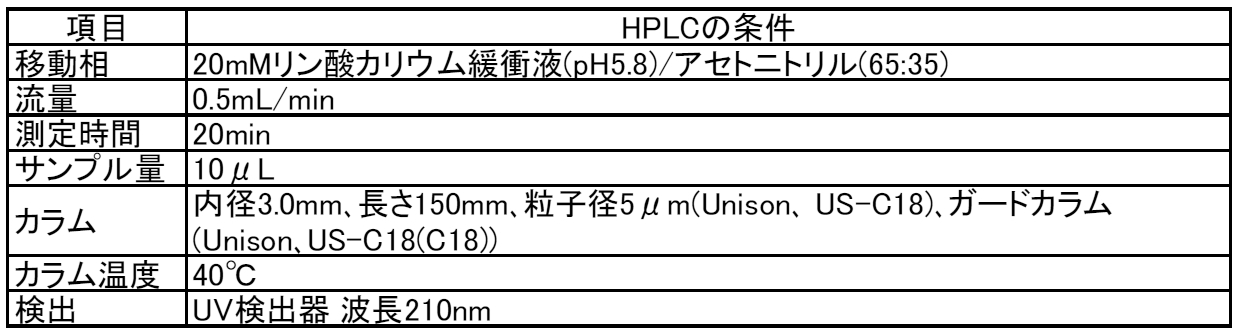
(4)各サンプルのリドカイン塩酸塩をHPLCで測定した。HPLCの条件を、表1に示す。
(4) Lidocaine hydrochloride of each sample was measured by HPLC. Table 1 shows the HPLC conditions.
(5)ニードル残量サンプル、皮膚表面残量サンプル、皮膚抽出液のリドカイン塩酸塩濃度の測定値から、ニードル残量、皮膚表面残量、および皮内移行量を算出し、ニードル残量、皮膚表面残量、および皮内移行量の合計に対するそれぞれの比率を、ニードル残量比率、皮膚表面残量比率、および皮内移行率として算出した。
[試験結果] (5) The remaining amount of needle, the remaining amount of skin surface, the remaining amount of skin surface, and the amount of intradermal transfer are calculated from the measured value of lidocaine hydrochloride concentration of skin extract, and the remaining amount of needle, skin Each ratio with respect to the total of the remaining amount of the surface and the amount transferred into the skin was calculated as the remaining amount of needle, the remaining amount of the skin surface, and the transferred rate within the skin.
[Test results]
各実施例および比較例における、リドカイン塩酸塩のニードル残量比率、皮膚表面残量比率および皮内移行率を、試験条件と共に表2に示す。
Table 2 shows the needle residual ratio, the skin surface residual ratio, and the intradermal transfer rate of lidocaine hydrochloride in each Example and Comparative Example together with the test conditions.

表2に示すように、実施例1~6および比較例1,2を比較すると、アスペクト比が2.1以上である実施例1~6の方が、アスペクト比が2.1未満の比較例1,2よりも薬剤の皮内移行率が高いことが確認された。
As shown in Table 2, when Examples 1 to 6 and Comparative Examples 1 and 2 are compared, Examples 1 to 6 having an aspect ratio of 2.1 or more are comparative examples having an aspect ratio of less than 2.1. It was confirmed that the intradermal transfer rate of the drug was higher than that of 1 and 2.
また、実施例5,6および実施例3,4を比較すると、アスペクト比が同じ場合に、薬剤がマイクロニードルの先端領域にのみ塗布されている場合(実施例5,6)の方が、全体に塗布される場合(実施例3,4)よりも皮内移行率が高いことが確認された。
Further, when Examples 5 and 6 are compared with Examples 3 and 4, when the aspect ratio is the same, the case where the drug is applied only to the tip region of the microneedle (Examples 5 and 6) is the whole. It was confirmed that the intradermal transfer rate was higher than in the case of being applied to (Examples 3 and 4).
また、実施例3~6より、貼付け時間が長いほど、薬剤の皮内移行率が高いことが確認された。
Also, from Examples 3 to 6, it was confirmed that the longer the application time, the higher the intradermal transfer rate of the drug.
なお、本発明は、上述した実施形態のみに限定されるものではなく、本発明の技術的思想内において当業者により種々変更が可能である。例えば、図8に示すように、マイクロニードル61は、薬剤を保持するための窪み62が形成されてもよい。これにより、根元部分に塗布される薬剤の量を少なくすることができるため、薬剤の投与を効率良く行うことが可能となる。
Note that the present invention is not limited to the above-described embodiment, and various modifications can be made by those skilled in the art within the technical idea of the present invention. For example, as shown in FIG. 8, the microneedle 61 may be formed with a recess 62 for holding a drug. Thereby, since the quantity of the chemical | medical agent apply | coated to a root part can be decreased, it becomes possible to administer a chemical | medical agent efficiently.
また、図9に示すように、マイクロニードル71は、頂部側が細くなるように段差72が形成されてもよい。これにより、マイクロニードル71の段差72より頂部側の表面での薬剤の保持量を多くすることができ、根元部分での薬剤の保持量を少なくすることができるため、薬剤の投与を効率良く行うことが可能となる。
Further, as shown in FIG. 9, the microneedle 71 may be formed with a step 72 so that the top side is thin. As a result, the amount of the drug held on the surface on the top side of the step 72 of the microneedle 71 can be increased, and the amount of the drug held at the root portion can be reduced, so that the drug is efficiently administered. It becomes possible.
また、図10に示すように、マイクロニードル81は、頂部側の基体83に対する傾斜角度θが、根元側よりも小さくなるように変化する変化部82が形成されてもよい。これにより、マイクロニードル81の頂部側の表面での薬剤の保持量を多くすることができ、根元部分での薬剤の保持量を少なくすることができるため、薬剤の投与を効率良く行うことが可能となる。さらに、段差が形成されないことで、マイクロニードル81を根元の近くまで穿刺しやすくなり、薬剤の投与を効率良く行うことが可能となる。また、マイクロニードルの表面の基体に対する傾斜角度が、頂部側に向かって徐々に小さくなるように形成されてもよい。このようにすれば、マイクロニードルの表面が滑らかとなり、マイクロニードルをさらに穿刺しやすくなり、薬剤の投与を効率良く行うことが可能となる。
Also, as shown in FIG. 10, the microneedle 81 may be formed with a changing portion 82 that changes so that the inclination angle θ with respect to the base 83 on the top side becomes smaller than that on the base side. As a result, the amount of drug held on the top side surface of the microneedle 81 can be increased and the amount of drug held at the root portion can be reduced, so that the drug can be efficiently administered. It becomes. Furthermore, since the step is not formed, it becomes easy to puncture the microneedle 81 to the vicinity of the root, and the drug can be efficiently administered. Further, the inclination angle of the surface of the microneedle with respect to the base body may be formed so as to gradually decrease toward the top side. In this way, the surface of the microneedle becomes smooth, it becomes easier to puncture the microneedle, and the drug can be administered efficiently.
さらに、本出願は、2014年11月28日に出願された日本特許出願番号2014-241676号に基づいており、それらの開示内容は、参照され、全体として、組み入れられている。
Furthermore, this application is based on Japanese Patent Application No. 2014-241676 filed on November 28, 2014, and the disclosures thereof are referred to and incorporated in their entirety.
10 マイクロニードルデバイス、
20,61,71,81 マイクロニードル、
21 頂部、
22 底面、
30、83 基体、
40 薬剤、
83 基体。 10 microneedle device,
20, 61, 71, 81 microneedles,
21 top,
22 Bottom,
30, 83 substrate,
40 drugs,
83 Substrate.
20,61,71,81 マイクロニードル、
21 頂部、
22 底面、
30、83 基体、
40 薬剤、
83 基体。 10 microneedle device,
20, 61, 71, 81 microneedles,
21 top,
22 Bottom,
30, 83 substrate,
40 drugs,
83 Substrate.
Claims (3)
- 基体と、
前記基体上に形成されて皮膚を穿刺可能な突起であるマイクロニードルと、
前記マイクロニードルに塗布される薬剤と、
を有し、前記マイクロニードルの底面の最大幅に対する高さの比率であるアスペクト比が2.1以上であるマイクロニードルデバイス。 A substrate;
A microneedle that is a protrusion formed on the substrate and capable of puncturing the skin;
A drug applied to the microneedle;
A microneedle device having an aspect ratio of 2.1 or more, which is a ratio of a height to a maximum width of a bottom surface of the microneedle. - 前記マイクロニードルの塗布される薬剤は、前記マイクロニードルの高さ方向において、前記マイクロニードルの頂部から高さの50%の範囲内に配置される請求項1に記載のマイクロニードルデバイス。 The microneedle device according to claim 1, wherein the drug to be applied to the microneedle is disposed within a range of 50% of the height from the top of the microneedle in the height direction of the microneedle.
- 前記アスペクト比は、5以下である請求項1または2に記載のマイクロニードルデバイス。 The microneedle device according to claim 1 or 2, wherein the aspect ratio is 5 or less.
Priority Applications (3)
| Application Number | Priority Date | Filing Date | Title |
|---|---|---|---|
| EP15863705.8A EP3225276A4 (en) | 2014-11-28 | 2015-11-19 | Microneedle device |
| JP2016561538A JPWO2016084701A1 (en) | 2014-11-28 | 2015-11-19 | Microneedle device |
| US15/606,831 US20170258712A1 (en) | 2014-11-28 | 2017-05-26 | Microneedle device |
Applications Claiming Priority (2)
| Application Number | Priority Date | Filing Date | Title |
|---|---|---|---|
| JP2014241676 | 2014-11-28 | ||
| JP2014-241676 | 2014-11-28 |
Related Child Applications (1)
| Application Number | Title | Priority Date | Filing Date |
|---|---|---|---|
| US15/606,831 Continuation US20170258712A1 (en) | 2014-11-28 | 2017-05-26 | Microneedle device |
Publications (1)
| Publication Number | Publication Date |
|---|---|
| WO2016084701A1 true WO2016084701A1 (en) | 2016-06-02 |
Family
ID=56074268
Family Applications (1)
| Application Number | Title | Priority Date | Filing Date |
|---|---|---|---|
| PCT/JP2015/082552 WO2016084701A1 (en) | 2014-11-28 | 2015-11-19 | Microneedle device |
Country Status (4)
| Country | Link |
|---|---|
| US (1) | US20170258712A1 (en) |
| EP (1) | EP3225276A4 (en) |
| JP (1) | JPWO2016084701A1 (en) |
| WO (1) | WO2016084701A1 (en) |
Cited By (9)
| Publication number | Priority date | Publication date | Assignee | Title |
|---|---|---|---|---|
| KR20180009495A (en) * | 2016-07-19 | 2018-01-29 | 부산대학교 산학협력단 | Microneedle, manufacturing method of the microneedle, and patch including the microneedle |
| KR20180009729A (en) * | 2017-09-29 | 2018-01-29 | 부산대학교 산학협력단 | Microneedle, manufacturing method of the microneedle, and patch including the microneedle |
| WO2018062114A1 (en) * | 2016-09-30 | 2018-04-05 | ニチバン株式会社 | Microneedle array drug carrying method and drug carrying device |
| WO2018124290A1 (en) * | 2016-12-28 | 2018-07-05 | コスメディ製薬株式会社 | Microneedle array coated with drug |
| KR20190047805A (en) * | 2017-10-30 | 2019-05-09 | 장은주 | Device of injecting bio-absorbing and biodegradable polymers and materials |
| KR20190088340A (en) * | 2018-01-18 | 2019-07-26 | 부산대학교 산학협력단 | Implantable microneedle and manufacturing method of the implantable microneedle |
| WO2019245345A1 (en) * | 2018-06-22 | 2019-12-26 | 주식회사 에스엔비아 | Microneedle |
| CN111870806A (en) * | 2020-07-22 | 2020-11-03 | 南方科技大学 | Magnetic control microneedle robot and preparation method, use method and application thereof |
| KR20210093188A (en) * | 2020-01-17 | 2021-07-27 | 주식회사 쿼드메디슨 | Micro-needle and manufacturing method thereof |
Families Citing this family (3)
| Publication number | Priority date | Publication date | Assignee | Title |
|---|---|---|---|---|
| CN108742718B (en) * | 2018-03-23 | 2021-04-16 | 苏州德锐特成像技术有限公司 | Self-adhesive microneedle patch capable of swelling rapidly and preparation method thereof |
| CN108478520A (en) * | 2018-04-18 | 2018-09-04 | 北京化工大学 | A kind of coating microneedle array and preparation method thereof accurately controlling drugloading rate |
| TWI687247B (en) * | 2019-05-15 | 2020-03-11 | 微邦科技股份有限公司 | Microneedle structure and biodegradable microneedle thereof |
Citations (3)
| Publication number | Priority date | Publication date | Assignee | Title |
|---|---|---|---|---|
| US20110223542A1 (en) * | 2008-02-07 | 2011-09-15 | The University Of Queensland | Patch production |
| JP2014514022A (en) * | 2011-03-07 | 2014-06-19 | スリーエム イノベイティブ プロパティズ カンパニー | Microneedle device and method |
| WO2014126052A1 (en) * | 2013-02-14 | 2014-08-21 | コスメディ製薬株式会社 | Medicine-holding microneedle array and process for producing same |
Family Cites Families (8)
| Publication number | Priority date | Publication date | Assignee | Title |
|---|---|---|---|---|
| US6663820B2 (en) * | 2001-03-14 | 2003-12-16 | The Procter & Gamble Company | Method of manufacturing microneedle structures using soft lithography and photolithography |
| US20080262416A1 (en) * | 2005-11-18 | 2008-10-23 | Duan Daniel C | Microneedle Arrays and Methods of Preparing Same |
| WO2007112309A2 (en) * | 2006-03-24 | 2007-10-04 | 3M Innovative Properties Company | Process for making microneedles, microneedle arrays, masters, and replication tools |
| JP5297595B2 (en) * | 2007-03-20 | 2013-09-25 | 凸版印刷株式会社 | Needle-like body and method for producing needle-like body |
| EP2633881A4 (en) * | 2010-10-25 | 2014-02-19 | Teijin Ltd | Microneedle |
| MX344881B (en) * | 2010-12-02 | 2017-01-11 | 3M Innovative Properties Co | Liquid crystalline polymer microneedles. |
| US11179553B2 (en) * | 2011-10-12 | 2021-11-23 | Vaxxas Pty Limited | Delivery device |
| JP2013248299A (en) * | 2012-06-01 | 2013-12-12 | Dainippon Printing Co Ltd | Microneedle device |
-
2015
- 2015-11-19 EP EP15863705.8A patent/EP3225276A4/en active Pending
- 2015-11-19 WO PCT/JP2015/082552 patent/WO2016084701A1/en active Application Filing
- 2015-11-19 JP JP2016561538A patent/JPWO2016084701A1/en active Pending
-
2017
- 2017-05-26 US US15/606,831 patent/US20170258712A1/en not_active Abandoned
Patent Citations (3)
| Publication number | Priority date | Publication date | Assignee | Title |
|---|---|---|---|---|
| US20110223542A1 (en) * | 2008-02-07 | 2011-09-15 | The University Of Queensland | Patch production |
| JP2014514022A (en) * | 2011-03-07 | 2014-06-19 | スリーエム イノベイティブ プロパティズ カンパニー | Microneedle device and method |
| WO2014126052A1 (en) * | 2013-02-14 | 2014-08-21 | コスメディ製薬株式会社 | Medicine-holding microneedle array and process for producing same |
Cited By (21)
| Publication number | Priority date | Publication date | Assignee | Title |
|---|---|---|---|---|
| KR101891398B1 (en) * | 2016-07-19 | 2018-08-23 | 부산대학교 산학협력단 | Microneedle, manufacturing method of the microneedle, and patch including the microneedle |
| KR20180009495A (en) * | 2016-07-19 | 2018-01-29 | 부산대학교 산학협력단 | Microneedle, manufacturing method of the microneedle, and patch including the microneedle |
| WO2018062114A1 (en) * | 2016-09-30 | 2018-04-05 | ニチバン株式会社 | Microneedle array drug carrying method and drug carrying device |
| JPWO2018062114A1 (en) * | 2016-09-30 | 2019-07-11 | ニチバン株式会社 | Method for carrying drug of microneedle array and drug carrying device |
| WO2018124290A1 (en) * | 2016-12-28 | 2018-07-05 | コスメディ製薬株式会社 | Microneedle array coated with drug |
| JP2018108375A (en) * | 2016-12-28 | 2018-07-12 | コスメディ製薬株式会社 | Microneedle array coated with drug |
| CN110035792A (en) * | 2016-12-28 | 2019-07-19 | 考司美德制药株式会社 | It is coated with the micropin preparation of drug containing ingredient |
| AU2017388969B2 (en) * | 2016-12-28 | 2023-04-27 | Cosmed Pharmaceutical Co., Ltd. | Microneedle array coated with drug |
| KR20180009729A (en) * | 2017-09-29 | 2018-01-29 | 부산대학교 산학협력단 | Microneedle, manufacturing method of the microneedle, and patch including the microneedle |
| KR102001305B1 (en) * | 2017-09-29 | 2019-07-17 | 부산대학교 산학협력단 | Microneedle, manufacturing method of the microneedle, and patch including the microneedle |
| KR102076394B1 (en) * | 2017-10-30 | 2020-02-11 | 장은주 | Device of injecting bio-absorbing and biodegradable polymers and materials |
| KR20190047805A (en) * | 2017-10-30 | 2019-05-09 | 장은주 | Device of injecting bio-absorbing and biodegradable polymers and materials |
| KR20190088340A (en) * | 2018-01-18 | 2019-07-26 | 부산대학교 산학협력단 | Implantable microneedle and manufacturing method of the implantable microneedle |
| KR102238033B1 (en) * | 2018-01-18 | 2021-04-08 | 주식회사 에스엔비아 | Implantable microneedle and manufacturing method of the implantable microneedle |
| US11938308B2 (en) | 2018-01-18 | 2024-03-26 | Snvia Co., Ltd. | Implantable microneedle and manufacturing method therefor |
| KR20200000379A (en) * | 2018-06-22 | 2020-01-02 | 주식회사 에스엔비아 | Microneedle |
| KR102092504B1 (en) * | 2018-06-22 | 2020-03-24 | 주식회사 에스엔비아 | Microneedle |
| WO2019245345A1 (en) * | 2018-06-22 | 2019-12-26 | 주식회사 에스엔비아 | Microneedle |
| KR20210093188A (en) * | 2020-01-17 | 2021-07-27 | 주식회사 쿼드메디슨 | Micro-needle and manufacturing method thereof |
| KR102392782B1 (en) * | 2020-01-17 | 2022-05-02 | 주식회사 쿼드메디슨 | Micro-needle and manufacturing method thereof |
| CN111870806A (en) * | 2020-07-22 | 2020-11-03 | 南方科技大学 | Magnetic control microneedle robot and preparation method, use method and application thereof |
Also Published As
| Publication number | Publication date |
|---|---|
| JPWO2016084701A1 (en) | 2017-09-07 |
| US20170258712A1 (en) | 2017-09-14 |
| EP3225276A1 (en) | 2017-10-04 |
| EP3225276A4 (en) | 2018-06-27 |
Similar Documents
| Publication | Publication Date | Title |
|---|---|---|
| WO2016084701A1 (en) | Microneedle device | |
| JP6612952B2 (en) | Hollow microneedle with beveled opening | |
| TWI643966B (en) | Metallic microneedles | |
| KR101323980B1 (en) | Microneedle device, method of using microneedle device and method of delivering microneedle device | |
| EP3021930B1 (en) | Hollow microneedle with beveled tip | |
| Ross et al. | Inkjet printing of insulin microneedles for transdermal delivery | |
| JP5868953B2 (en) | Injection mold microneedle array and manufacturing method thereof | |
| EP3021931B1 (en) | Hollow microneedle array article | |
| Chen et al. | Rapidly fabricated microneedle arrays using magnetorheological drawing lithography for transdermal drug delivery | |
| JP2009501066A (en) | Solid microneedle and manufacturing method thereof | |
| JP2006512164A (en) | Active ingredient delivery device having a composite member | |
| WO2015122838A1 (en) | Rapidly dissolvable microneedles with drug-impregnated tips | |
| JP6304431B2 (en) | Microneedle device | |
| CN106853271B (en) | Method for producing microstructure | |
| Shravanth et al. | Microneedles-based drug delivery for the treatment of psoriasis | |
| WO2018155431A1 (en) | Microneedle device | |
| JP2015016160A (en) | Microneedle | |
| KR102127123B1 (en) | Manufacturing method for micro-structure | |
| Bao et al. | A fabrication of whole-dissovable microneedles patch in larger area for transdermal drug delivery | |
| Rathor et al. | Advancement in transdermal drug delivery system: Microneedles |
Legal Events
| Date | Code | Title | Description |
|---|---|---|---|
| 121 | Ep: the epo has been informed by wipo that ep was designated in this application |
Ref document number: 15863705 Country of ref document: EP Kind code of ref document: A1 |
|
| ENP | Entry into the national phase |
Ref document number: 2016561538 Country of ref document: JP Kind code of ref document: A |
|
| NENP | Non-entry into the national phase |
Ref country code: DE |
|
| REEP | Request for entry into the european phase |
Ref document number: 2015863705 Country of ref document: EP |