WO2016073157A1 - Anti-ang2 antibodies and methods of use thereof - Google Patents
Anti-ang2 antibodies and methods of use thereof Download PDFInfo
- Publication number
- WO2016073157A1 WO2016073157A1 PCT/US2015/055672 US2015055672W WO2016073157A1 WO 2016073157 A1 WO2016073157 A1 WO 2016073157A1 US 2015055672 W US2015055672 W US 2015055672W WO 2016073157 A1 WO2016073157 A1 WO 2016073157A1
- Authority
- WO
- WIPO (PCT)
- Prior art keywords
- amino acid
- antibody
- seq
- ang2
- acid sequence
- Prior art date
Links
- 238000000034 method Methods 0.000 title claims abstract description 237
- 102100034608 Angiopoietin-2 Human genes 0.000 claims abstract description 331
- 101000924533 Homo sapiens Angiopoietin-2 Proteins 0.000 claims abstract description 327
- 102000005789 Vascular Endothelial Growth Factors Human genes 0.000 claims abstract description 275
- 108010019530 Vascular Endothelial Growth Factors Proteins 0.000 claims abstract description 275
- 230000027455 binding Effects 0.000 claims abstract description 221
- 230000009977 dual effect Effects 0.000 claims abstract description 69
- 238000011282 treatment Methods 0.000 claims abstract description 18
- 230000033115 angiogenesis Effects 0.000 claims abstract description 16
- 125000003275 alpha amino acid group Chemical group 0.000 claims description 388
- 125000000539 amino acid group Chemical group 0.000 claims description 380
- 108010073929 Vascular Endothelial Growth Factor A Proteins 0.000 claims description 277
- 108010032595 Antibody Binding Sites Proteins 0.000 claims description 141
- 230000004075 alteration Effects 0.000 claims description 124
- 210000004027 cell Anatomy 0.000 claims description 117
- 239000000427 antigen Substances 0.000 claims description 109
- 108091007433 antigens Proteins 0.000 claims description 107
- 102000036639 antigens Human genes 0.000 claims description 107
- 241000282414 Homo sapiens Species 0.000 claims description 101
- 208000037265 diseases, disorders, signs and symptoms Diseases 0.000 claims description 87
- 208000035475 disorder Diseases 0.000 claims description 69
- 108010021625 Immunoglobulin Fragments Proteins 0.000 claims description 55
- 102000008394 Immunoglobulin Fragments Human genes 0.000 claims description 55
- 108090000765 processed proteins & peptides Proteins 0.000 claims description 47
- 239000003795 chemical substances by application Substances 0.000 claims description 44
- 102000004196 processed proteins & peptides Human genes 0.000 claims description 44
- 229920001184 polypeptide Polymers 0.000 claims description 43
- 108020004705 Codon Proteins 0.000 claims description 39
- 239000012634 fragment Substances 0.000 claims description 37
- 150000007523 nucleic acids Chemical class 0.000 claims description 37
- 150000001413 amino acids Chemical class 0.000 claims description 36
- 102000039446 nucleic acids Human genes 0.000 claims description 36
- 108020004707 nucleic acids Proteins 0.000 claims description 36
- 238000012163 sequencing technique Methods 0.000 claims description 36
- 208000034038 Pathologic Neovascularization Diseases 0.000 claims description 33
- 208000022873 Ocular disease Diseases 0.000 claims description 30
- 239000013598 vector Substances 0.000 claims description 30
- FWMNVWWHGCHHJJ-SKKKGAJSSA-N 4-amino-1-[(2r)-6-amino-2-[[(2r)-2-[[(2r)-2-[[(2r)-2-amino-3-phenylpropanoyl]amino]-3-phenylpropanoyl]amino]-4-methylpentanoyl]amino]hexanoyl]piperidine-4-carboxylic acid Chemical compound C([C@H](C(=O)N[C@H](CC(C)C)C(=O)N[C@H](CCCCN)C(=O)N1CCC(N)(CC1)C(O)=O)NC(=O)[C@H](N)CC=1C=CC=CC=1)C1=CC=CC=C1 FWMNVWWHGCHHJJ-SKKKGAJSSA-N 0.000 claims description 24
- 206010064930 age-related macular degeneration Diseases 0.000 claims description 23
- -1 PDGF-B Proteins 0.000 claims description 22
- 208000002780 macular degeneration Diseases 0.000 claims description 22
- 230000002062 proliferating effect Effects 0.000 claims description 21
- 229940127089 cytotoxic agent Drugs 0.000 claims description 20
- 239000008194 pharmaceutical composition Substances 0.000 claims description 20
- 101000808011 Homo sapiens Vascular endothelial growth factor A Proteins 0.000 claims description 19
- 102000058223 human VEGFA Human genes 0.000 claims description 19
- 238000002823 phage display Methods 0.000 claims description 19
- 102100034594 Angiopoietin-1 Human genes 0.000 claims description 18
- 229940002612 prodrug Drugs 0.000 claims description 18
- 239000000651 prodrug Substances 0.000 claims description 18
- 101000924552 Homo sapiens Angiopoietin-1 Proteins 0.000 claims description 17
- 208000005590 Choroidal Neovascularization Diseases 0.000 claims description 16
- 206010060823 Choroidal neovascularisation Diseases 0.000 claims description 16
- 239000002246 antineoplastic agent Substances 0.000 claims description 16
- 230000002401 inhibitory effect Effects 0.000 claims description 16
- 230000007935 neutral effect Effects 0.000 claims description 16
- 206010038933 Retinopathy of prematurity Diseases 0.000 claims description 15
- 206010012688 Diabetic retinal oedema Diseases 0.000 claims description 14
- 208000028506 Familial Exudative Vitreoretinopathies Diseases 0.000 claims description 14
- 206010038923 Retinopathy Diseases 0.000 claims description 14
- 201000011190 diabetic macular edema Diseases 0.000 claims description 14
- 201000006902 exudative vitreoretinopathy Diseases 0.000 claims description 14
- 201000001937 osteoporosis-pseudoglioma syndrome Diseases 0.000 claims description 14
- 231100000433 cytotoxic Toxicity 0.000 claims description 12
- 230000001472 cytotoxic effect Effects 0.000 claims description 12
- 230000012010 growth Effects 0.000 claims description 12
- 101000955962 Homo sapiens Vacuolar protein sorting-associated protein 51 homolog Proteins 0.000 claims description 10
- 239000004037 angiogenesis inhibitor Substances 0.000 claims description 10
- 239000002254 cytotoxic agent Substances 0.000 claims description 10
- 231100000599 cytotoxic agent Toxicity 0.000 claims description 10
- 238000012350 deep sequencing Methods 0.000 claims description 10
- 241000588724 Escherichia coli Species 0.000 claims description 9
- 102000052216 human VPS51 Human genes 0.000 claims description 9
- 102000004127 Cytokines Human genes 0.000 claims description 8
- 108090000695 Cytokines Proteins 0.000 claims description 8
- 208000021089 Coats disease Diseases 0.000 claims description 7
- 206010010719 Conjunctival haemorrhage Diseases 0.000 claims description 7
- 206010055665 Corneal neovascularisation Diseases 0.000 claims description 7
- 206010012689 Diabetic retinopathy Diseases 0.000 claims description 7
- 206010015901 Exudative retinopathy Diseases 0.000 claims description 7
- 201000002563 Histoplasmosis Diseases 0.000 claims description 7
- 241000124008 Mammalia Species 0.000 claims description 7
- 208000025464 Norrie disease Diseases 0.000 claims description 7
- 206010073286 Pathologic myopia Diseases 0.000 claims description 7
- 208000007135 Retinal Neovascularization Diseases 0.000 claims description 7
- 208000008709 Retinal Telangiectasis Diseases 0.000 claims description 7
- 208000017442 Retinal disease Diseases 0.000 claims description 7
- 206010038926 Retinopathy hypertensive Diseases 0.000 claims description 7
- 201000005667 central retinal vein occlusion Diseases 0.000 claims description 7
- 201000000159 corneal neovascularization Diseases 0.000 claims description 7
- 206010012601 diabetes mellitus Diseases 0.000 claims description 7
- 201000001948 hypertensive retinopathy Diseases 0.000 claims description 7
- 208000028867 ischemia Diseases 0.000 claims description 7
- 201000007914 proliferative diabetic retinopathy Diseases 0.000 claims description 7
- 238000001959 radiotherapy Methods 0.000 claims description 7
- 208000004644 retinal vein occlusion Diseases 0.000 claims description 7
- 208000006542 von Hippel-Lindau disease Diseases 0.000 claims description 7
- 101100315624 Caenorhabditis elegans tyr-1 gene Proteins 0.000 claims description 6
- 238000007481 next generation sequencing Methods 0.000 claims description 5
- 108091032973 (ribonucleotides)n+m Proteins 0.000 claims description 4
- 108010048036 Angiopoietin-2 Proteins 0.000 claims description 4
- 101000595923 Homo sapiens Placenta growth factor Proteins 0.000 claims description 4
- 102100035194 Placenta growth factor Human genes 0.000 claims description 4
- 238000002819 bacterial display Methods 0.000 claims description 4
- 230000000295 complement effect Effects 0.000 claims description 4
- 239000003889 eye drop Substances 0.000 claims description 4
- 238000002513 implantation Methods 0.000 claims description 4
- 238000002818 protein evolution Methods 0.000 claims description 4
- 238000002702 ribosome display Methods 0.000 claims description 4
- 230000000202 analgesic effect Effects 0.000 claims description 3
- 230000003474 anti-emetic effect Effects 0.000 claims description 3
- 239000002111 antiemetic agent Substances 0.000 claims description 3
- 229940022399 cancer vaccine Drugs 0.000 claims description 3
- 238000009566 cancer vaccine Methods 0.000 claims description 3
- 210000004978 chinese hamster ovary cell Anatomy 0.000 claims description 3
- 239000003246 corticosteroid Substances 0.000 claims description 3
- 238000012258 culturing Methods 0.000 claims description 3
- 239000000430 cytokine receptor antagonist Substances 0.000 claims description 3
- 239000003018 immunosuppressive agent Substances 0.000 claims description 3
- 229940125721 immunosuppressive agent Drugs 0.000 claims description 3
- 108091033319 polynucleotide Proteins 0.000 claims description 3
- 102000040430 polynucleotide Human genes 0.000 claims description 3
- 239000002157 polynucleotide Substances 0.000 claims description 3
- 101001001487 Homo sapiens Phosphatidylinositol-glycan biosynthesis class F protein Proteins 0.000 claims description 2
- 102100040681 Platelet-derived growth factor C Human genes 0.000 claims description 2
- 108010017992 platelet-derived growth factor C Proteins 0.000 claims description 2
- 210000005253 yeast cell Anatomy 0.000 claims description 2
- 101100512078 Caenorhabditis elegans lys-1 gene Proteins 0.000 claims 1
- 230000014509 gene expression Effects 0.000 abstract description 21
- 230000001575 pathological effect Effects 0.000 abstract description 2
- 108091007491 NSP3 Papain-like protease domains Proteins 0.000 description 59
- 108090000623 proteins and genes Proteins 0.000 description 54
- 230000035772 mutation Effects 0.000 description 50
- 235000001014 amino acid Nutrition 0.000 description 48
- DHMQDGOQFOQNFH-UHFFFAOYSA-N Glycine Chemical group NCC(O)=O DHMQDGOQFOQNFH-UHFFFAOYSA-N 0.000 description 47
- 239000000203 mixture Substances 0.000 description 41
- OUYCCCASQSFEME-QMMMGPOBSA-N L-tyrosine Chemical compound OC(=O)[C@@H](N)CC1=CC=C(O)C=C1 OUYCCCASQSFEME-QMMMGPOBSA-N 0.000 description 40
- 229940024606 amino acid Drugs 0.000 description 39
- 102000004169 proteins and genes Human genes 0.000 description 37
- KDXKERNSBIXSRK-UHFFFAOYSA-N Lysine Chemical group NCCCCC(N)C(O)=O KDXKERNSBIXSRK-UHFFFAOYSA-N 0.000 description 35
- KDXKERNSBIXSRK-YFKPBYRVSA-N L-lysine Chemical group NCCCC[C@H](N)C(O)=O KDXKERNSBIXSRK-YFKPBYRVSA-N 0.000 description 34
- 238000006467 substitution reaction Methods 0.000 description 33
- 235000018102 proteins Nutrition 0.000 description 32
- ROHFNLRQFUQHCH-YFKPBYRVSA-N L-leucine Chemical compound CC(C)C[C@H](N)C(O)=O ROHFNLRQFUQHCH-YFKPBYRVSA-N 0.000 description 31
- 238000003556 assay Methods 0.000 description 30
- 101100481410 Mus musculus Tek gene Proteins 0.000 description 26
- 206010028980 Neoplasm Diseases 0.000 description 26
- 102000005962 receptors Human genes 0.000 description 26
- 108020003175 receptors Proteins 0.000 description 26
- 125000001360 methionine group Chemical group N[C@@H](CCSC)C(=O)* 0.000 description 25
- 239000013078 crystal Substances 0.000 description 24
- 101100481408 Danio rerio tie2 gene Proteins 0.000 description 23
- 230000000694 effects Effects 0.000 description 22
- 230000010056 antibody-dependent cellular cytotoxicity Effects 0.000 description 20
- 239000003814 drug Substances 0.000 description 20
- AOJJSUZBOXZQNB-TZSSRYMLSA-N Doxorubicin Chemical compound O([C@H]1C[C@@](O)(CC=2C(O)=C3C(=O)C=4C=CC=C(C=4C(=O)C3=C(O)C=21)OC)C(=O)CO)[C@H]1C[C@H](N)[C@H](O)[C@H](C)O1 AOJJSUZBOXZQNB-TZSSRYMLSA-N 0.000 description 19
- 238000002703 mutagenesis Methods 0.000 description 19
- 231100000350 mutagenesis Toxicity 0.000 description 19
- 230000004071 biological effect Effects 0.000 description 18
- 201000011510 cancer Diseases 0.000 description 18
- 201000010099 disease Diseases 0.000 description 18
- 239000000243 solution Substances 0.000 description 18
- 108010087819 Fc receptors Proteins 0.000 description 17
- 102000009109 Fc receptors Human genes 0.000 description 17
- 108091034117 Oligonucleotide Proteins 0.000 description 17
- 108060003951 Immunoglobulin Proteins 0.000 description 16
- 230000000903 blocking effect Effects 0.000 description 16
- 102000018358 immunoglobulin Human genes 0.000 description 16
- 238000004519 manufacturing process Methods 0.000 description 16
- JLCPHMBAVCMARE-UHFFFAOYSA-N [3-[[3-[[3-[[3-[[3-[[3-[[3-[[3-[[3-[[3-[[3-[[5-(2-amino-6-oxo-1H-purin-9-yl)-3-[[3-[[3-[[3-[[3-[[3-[[5-(2-amino-6-oxo-1H-purin-9-yl)-3-[[5-(2-amino-6-oxo-1H-purin-9-yl)-3-hydroxyoxolan-2-yl]methoxy-hydroxyphosphoryl]oxyoxolan-2-yl]methoxy-hydroxyphosphoryl]oxy-5-(5-methyl-2,4-dioxopyrimidin-1-yl)oxolan-2-yl]methoxy-hydroxyphosphoryl]oxy-5-(6-aminopurin-9-yl)oxolan-2-yl]methoxy-hydroxyphosphoryl]oxy-5-(6-aminopurin-9-yl)oxolan-2-yl]methoxy-hydroxyphosphoryl]oxy-5-(6-aminopurin-9-yl)oxolan-2-yl]methoxy-hydroxyphosphoryl]oxy-5-(6-aminopurin-9-yl)oxolan-2-yl]methoxy-hydroxyphosphoryl]oxyoxolan-2-yl]methoxy-hydroxyphosphoryl]oxy-5-(5-methyl-2,4-dioxopyrimidin-1-yl)oxolan-2-yl]methoxy-hydroxyphosphoryl]oxy-5-(4-amino-2-oxopyrimidin-1-yl)oxolan-2-yl]methoxy-hydroxyphosphoryl]oxy-5-(5-methyl-2,4-dioxopyrimidin-1-yl)oxolan-2-yl]methoxy-hydroxyphosphoryl]oxy-5-(5-methyl-2,4-dioxopyrimidin-1-yl)oxolan-2-yl]methoxy-hydroxyphosphoryl]oxy-5-(6-aminopurin-9-yl)oxolan-2-yl]methoxy-hydroxyphosphoryl]oxy-5-(6-aminopurin-9-yl)oxolan-2-yl]methoxy-hydroxyphosphoryl]oxy-5-(4-amino-2-oxopyrimidin-1-yl)oxolan-2-yl]methoxy-hydroxyphosphoryl]oxy-5-(4-amino-2-oxopyrimidin-1-yl)oxolan-2-yl]methoxy-hydroxyphosphoryl]oxy-5-(4-amino-2-oxopyrimidin-1-yl)oxolan-2-yl]methoxy-hydroxyphosphoryl]oxy-5-(6-aminopurin-9-yl)oxolan-2-yl]methoxy-hydroxyphosphoryl]oxy-5-(4-amino-2-oxopyrimidin-1-yl)oxolan-2-yl]methyl [5-(6-aminopurin-9-yl)-2-(hydroxymethyl)oxolan-3-yl] hydrogen phosphate Polymers Cc1cn(C2CC(OP(O)(=O)OCC3OC(CC3OP(O)(=O)OCC3OC(CC3O)n3cnc4c3nc(N)[nH]c4=O)n3cnc4c3nc(N)[nH]c4=O)C(COP(O)(=O)OC3CC(OC3COP(O)(=O)OC3CC(OC3COP(O)(=O)OC3CC(OC3COP(O)(=O)OC3CC(OC3COP(O)(=O)OC3CC(OC3COP(O)(=O)OC3CC(OC3COP(O)(=O)OC3CC(OC3COP(O)(=O)OC3CC(OC3COP(O)(=O)OC3CC(OC3COP(O)(=O)OC3CC(OC3COP(O)(=O)OC3CC(OC3COP(O)(=O)OC3CC(OC3COP(O)(=O)OC3CC(OC3COP(O)(=O)OC3CC(OC3COP(O)(=O)OC3CC(OC3COP(O)(=O)OC3CC(OC3COP(O)(=O)OC3CC(OC3CO)n3cnc4c(N)ncnc34)n3ccc(N)nc3=O)n3cnc4c(N)ncnc34)n3ccc(N)nc3=O)n3ccc(N)nc3=O)n3ccc(N)nc3=O)n3cnc4c(N)ncnc34)n3cnc4c(N)ncnc34)n3cc(C)c(=O)[nH]c3=O)n3cc(C)c(=O)[nH]c3=O)n3ccc(N)nc3=O)n3cc(C)c(=O)[nH]c3=O)n3cnc4c3nc(N)[nH]c4=O)n3cnc4c(N)ncnc34)n3cnc4c(N)ncnc34)n3cnc4c(N)ncnc34)n3cnc4c(N)ncnc34)O2)c(=O)[nH]c1=O JLCPHMBAVCMARE-UHFFFAOYSA-N 0.000 description 15
- 230000009824 affinity maturation Effects 0.000 description 15
- 238000001727 in vivo Methods 0.000 description 15
- 239000002773 nucleotide Substances 0.000 description 15
- 125000003729 nucleotide group Chemical group 0.000 description 15
- 238000013459 approach Methods 0.000 description 14
- 230000002860 competitive effect Effects 0.000 description 14
- 239000012636 effector Substances 0.000 description 14
- 230000006870 function Effects 0.000 description 14
- NFGXHKASABOEEW-UHFFFAOYSA-N 1-methylethyl 11-methoxy-3,7,11-trimethyl-2,4-dodecadienoate Chemical compound COC(C)(C)CCCC(C)CC=CC(C)=CC(=O)OC(C)C NFGXHKASABOEEW-UHFFFAOYSA-N 0.000 description 12
- COLNVLDHVKWLRT-QMMMGPOBSA-N L-phenylalanine Chemical compound OC(=O)[C@@H](N)CC1=CC=CC=C1 COLNVLDHVKWLRT-QMMMGPOBSA-N 0.000 description 12
- 125000000510 L-tryptophano group Chemical group [H]C1=C([H])C([H])=C2N([H])C([H])=C(C([H])([H])[C@@]([H])(C(O[H])=O)N([H])[*])C2=C1[H] 0.000 description 12
- LOKCTEFSRHRXRJ-UHFFFAOYSA-I dipotassium trisodium dihydrogen phosphate hydrogen phosphate dichloride Chemical compound P(=O)(O)(O)[O-].[K+].P(=O)(O)([O-])[O-].[Na+].[Na+].[Cl-].[K+].[Cl-].[Na+] LOKCTEFSRHRXRJ-UHFFFAOYSA-I 0.000 description 12
- 239000002953 phosphate buffered saline Substances 0.000 description 12
- 108020004414 DNA Proteins 0.000 description 11
- 238000000338 in vitro Methods 0.000 description 11
- 238000003780 insertion Methods 0.000 description 11
- 230000037431 insertion Effects 0.000 description 11
- 238000002198 surface plasmon resonance spectroscopy Methods 0.000 description 11
- 238000011374 additional therapy Methods 0.000 description 10
- 230000004540 complement-dependent cytotoxicity Effects 0.000 description 10
- 230000000875 corresponding effect Effects 0.000 description 10
- 238000002898 library design Methods 0.000 description 10
- 230000002285 radioactive effect Effects 0.000 description 10
- 238000002864 sequence alignment Methods 0.000 description 10
- 238000002560 therapeutic procedure Methods 0.000 description 10
- DCXYFEDJOCDNAF-REOHCLBHSA-N L-asparagine Chemical group OC(=O)[C@@H](N)CC(N)=O DCXYFEDJOCDNAF-REOHCLBHSA-N 0.000 description 9
- 125000004429 atom Chemical group 0.000 description 9
- 238000010494 dissociation reaction Methods 0.000 description 9
- 230000005593 dissociations Effects 0.000 description 9
- 229940079593 drug Drugs 0.000 description 9
- 238000005516 engineering process Methods 0.000 description 9
- 238000002474 experimental method Methods 0.000 description 9
- 238000002347 injection Methods 0.000 description 9
- 239000007924 injection Substances 0.000 description 9
- 108091003079 Bovine Serum Albumin Proteins 0.000 description 8
- 239000002202 Polyethylene glycol Substances 0.000 description 8
- 229940098773 bovine serum albumin Drugs 0.000 description 8
- 235000018417 cysteine Nutrition 0.000 description 8
- 238000012217 deletion Methods 0.000 description 8
- 230000037430 deletion Effects 0.000 description 8
- 229960004679 doxorubicin Drugs 0.000 description 8
- 239000003112 inhibitor Substances 0.000 description 8
- 229920001223 polyethylene glycol Polymers 0.000 description 8
- 238000012552 review Methods 0.000 description 8
- 238000012216 screening Methods 0.000 description 8
- 102000004190 Enzymes Human genes 0.000 description 7
- 108090000790 Enzymes Proteins 0.000 description 7
- ZDZOTLJHXYCWBA-VCVYQWHSSA-N N-debenzoyl-N-(tert-butoxycarbonyl)-10-deacetyltaxol Chemical compound O([C@H]1[C@H]2[C@@](C([C@H](O)C3=C(C)[C@@H](OC(=O)[C@H](O)[C@@H](NC(=O)OC(C)(C)C)C=4C=CC=CC=4)C[C@]1(O)C3(C)C)=O)(C)[C@@H](O)C[C@H]1OC[C@]12OC(=O)C)C(=O)C1=CC=CC=C1 ZDZOTLJHXYCWBA-VCVYQWHSSA-N 0.000 description 7
- 229920001213 Polysorbate 20 Polymers 0.000 description 7
- 239000000872 buffer Substances 0.000 description 7
- 229940088598 enzyme Drugs 0.000 description 7
- 230000013595 glycosylation Effects 0.000 description 7
- 238000006206 glycosylation reaction Methods 0.000 description 7
- 229940127121 immunoconjugate Drugs 0.000 description 7
- 238000011534 incubation Methods 0.000 description 7
- 230000004048 modification Effects 0.000 description 7
- 238000012986 modification Methods 0.000 description 7
- 210000003819 peripheral blood mononuclear cell Anatomy 0.000 description 7
- 239000000256 polyoxyethylene sorbitan monolaurate Substances 0.000 description 7
- 235000010486 polyoxyethylene sorbitan monolaurate Nutrition 0.000 description 7
- 230000008569 process Effects 0.000 description 7
- 238000000159 protein binding assay Methods 0.000 description 7
- 230000002829 reductive effect Effects 0.000 description 7
- 238000009877 rendering Methods 0.000 description 7
- 230000009870 specific binding Effects 0.000 description 7
- 108700012359 toxins Proteins 0.000 description 7
- MTCFGRXMJLQNBG-REOHCLBHSA-N (2S)-2-Amino-3-hydroxypropansäure Chemical group OC[C@H](N)C(O)=O MTCFGRXMJLQNBG-REOHCLBHSA-N 0.000 description 6
- 241000196324 Embryophyta Species 0.000 description 6
- PEDCQBHIVMGVHV-UHFFFAOYSA-N Glycerine Chemical compound OCC(O)CO PEDCQBHIVMGVHV-UHFFFAOYSA-N 0.000 description 6
- 108010001336 Horseradish Peroxidase Proteins 0.000 description 6
- 102100026120 IgG receptor FcRn large subunit p51 Human genes 0.000 description 6
- 125000000998 L-alanino group Chemical group [H]N([*])[C@](C([H])([H])[H])([H])C(=O)O[H] 0.000 description 6
- FBOZXECLQNJBKD-ZDUSSCGKSA-N L-methotrexate Chemical compound C=1N=C2N=C(N)N=C(N)C2=NC=1CN(C)C1=CC=C(C(=O)N[C@@H](CCC(O)=O)C(O)=O)C=C1 FBOZXECLQNJBKD-ZDUSSCGKSA-N 0.000 description 6
- AYFVYJQAPQTCCC-GBXIJSLDSA-N L-threonine Chemical group C[C@@H](O)[C@H](N)C(O)=O AYFVYJQAPQTCCC-GBXIJSLDSA-N 0.000 description 6
- 241000700159 Rattus Species 0.000 description 6
- 101100372762 Rattus norvegicus Flt1 gene Proteins 0.000 description 6
- 239000012472 biological sample Substances 0.000 description 6
- 230000015572 biosynthetic process Effects 0.000 description 6
- 238000010367 cloning Methods 0.000 description 6
- 239000000562 conjugate Substances 0.000 description 6
- XUJNEKJLAYXESH-UHFFFAOYSA-N cysteine Natural products SCC(N)C(O)=O XUJNEKJLAYXESH-UHFFFAOYSA-N 0.000 description 6
- 238000001514 detection method Methods 0.000 description 6
- 239000003937 drug carrier Substances 0.000 description 6
- 238000001802 infusion Methods 0.000 description 6
- 230000003993 interaction Effects 0.000 description 6
- 229960000485 methotrexate Drugs 0.000 description 6
- 238000003127 radioimmunoassay Methods 0.000 description 6
- RCINICONZNJXQF-MZXODVADSA-N taxol Chemical compound O([C@@H]1[C@@]2(C[C@@H](C(C)=C(C2(C)C)[C@H](C([C@]2(C)[C@@H](O)C[C@H]3OC[C@]3([C@H]21)OC(C)=O)=O)OC(=O)C)OC(=O)[C@H](O)[C@@H](NC(=O)C=1C=CC=CC=1)C=1C=CC=CC=1)O)C(=O)C1=CC=CC=C1 RCINICONZNJXQF-MZXODVADSA-N 0.000 description 6
- 239000003053 toxin Substances 0.000 description 6
- 231100000765 toxin Toxicity 0.000 description 6
- 229960005486 vaccine Drugs 0.000 description 6
- STQGQHZAVUOBTE-UHFFFAOYSA-N 7-Cyan-hept-2t-en-4,6-diinsaeure Natural products C1=2C(O)=C3C(=O)C=4C(OC)=CC=CC=4C(=O)C3=C(O)C=2CC(O)(C(C)=O)CC1OC1CC(N)C(O)C(C)O1 STQGQHZAVUOBTE-UHFFFAOYSA-N 0.000 description 5
- 241000282693 Cercopithecidae Species 0.000 description 5
- LYCAIKOWRPUZTN-UHFFFAOYSA-N Ethylene glycol Chemical compound OCCO LYCAIKOWRPUZTN-UHFFFAOYSA-N 0.000 description 5
- 101710177940 IgG receptor FcRn large subunit p51 Proteins 0.000 description 5
- 241001465754 Metazoa Species 0.000 description 5
- 241000699666 Mus <mouse, genus> Species 0.000 description 5
- 229930012538 Paclitaxel Natural products 0.000 description 5
- NKANXQFJJICGDU-QPLCGJKRSA-N Tamoxifen Chemical compound C=1C=CC=CC=1C(/CC)=C(C=1C=CC(OCCN(C)C)=CC=1)/C1=CC=CC=C1 NKANXQFJJICGDU-QPLCGJKRSA-N 0.000 description 5
- 239000002253 acid Substances 0.000 description 5
- 239000004480 active ingredient Substances 0.000 description 5
- 239000005557 antagonist Substances 0.000 description 5
- 230000001580 bacterial effect Effects 0.000 description 5
- 210000004204 blood vessel Anatomy 0.000 description 5
- 230000004663 cell proliferation Effects 0.000 description 5
- 150000001875 compounds Chemical class 0.000 description 5
- 125000000151 cysteine group Chemical group N[C@@H](CS)C(=O)* 0.000 description 5
- STQGQHZAVUOBTE-VGBVRHCVSA-N daunorubicin Chemical compound O([C@H]1C[C@@](O)(CC=2C(O)=C3C(=O)C=4C=CC=C(C=4C(=O)C3=C(O)C=21)OC)C(C)=O)[C@H]1C[C@H](N)[C@H](O)[C@H](C)O1 STQGQHZAVUOBTE-VGBVRHCVSA-N 0.000 description 5
- 239000000539 dimer Substances 0.000 description 5
- 229960003668 docetaxel Drugs 0.000 description 5
- 239000013604 expression vector Substances 0.000 description 5
- 230000004927 fusion Effects 0.000 description 5
- 210000004408 hybridoma Anatomy 0.000 description 5
- 230000001900 immune effect Effects 0.000 description 5
- 230000006872 improvement Effects 0.000 description 5
- 239000003446 ligand Substances 0.000 description 5
- 229960001592 paclitaxel Drugs 0.000 description 5
- 239000002245 particle Substances 0.000 description 5
- 239000012071 phase Substances 0.000 description 5
- 239000013641 positive control Substances 0.000 description 5
- 239000000047 product Substances 0.000 description 5
- 230000004044 response Effects 0.000 description 5
- 238000013207 serial dilution Methods 0.000 description 5
- 239000002904 solvent Substances 0.000 description 5
- 241000894007 species Species 0.000 description 5
- 208000024891 symptom Diseases 0.000 description 5
- 229940124597 therapeutic agent Drugs 0.000 description 5
- 230000001225 therapeutic effect Effects 0.000 description 5
- 229960004528 vincristine Drugs 0.000 description 5
- OGWKCGZFUXNPDA-UHFFFAOYSA-N vincristine Natural products C1C(CC)(O)CC(CC2(C(=O)OC)C=3C(=CC4=C(C56C(C(C(OC(C)=O)C7(CC)C=CCN(C67)CC5)(O)C(=O)OC)N4C=O)C=3)OC)CN1CCC1=C2NC2=CC=CC=C12 OGWKCGZFUXNPDA-UHFFFAOYSA-N 0.000 description 5
- OGWKCGZFUXNPDA-XQKSVPLYSA-N vincristine Chemical compound C([N@]1C[C@@H](C[C@]2(C(=O)OC)C=3C(=CC4=C([C@]56[C@H]([C@@]([C@H](OC(C)=O)[C@]7(CC)C=CCN([C@H]67)CC5)(O)C(=O)OC)N4C=O)C=3)OC)C[C@@](C1)(O)CC)CC1=C2NC2=CC=CC=C12 OGWKCGZFUXNPDA-XQKSVPLYSA-N 0.000 description 5
- 229960004355 vindesine Drugs 0.000 description 5
- UGGWPQSBPIFKDZ-KOTLKJBCSA-N vindesine Chemical compound C([C@@H](C[C@]1(C(=O)OC)C=2C(=CC3=C([C@]45[C@H]([C@@]([C@H](O)[C@]6(CC)C=CCN([C@H]56)CC4)(O)C(N)=O)N3C)C=2)OC)C[C@@](C2)(O)CC)N2CCC2=C1N=C1[C]2C=CC=C1 UGGWPQSBPIFKDZ-KOTLKJBCSA-N 0.000 description 5
- IJGRMHOSHXDMSA-UHFFFAOYSA-N Atomic nitrogen Chemical compound N#N IJGRMHOSHXDMSA-UHFFFAOYSA-N 0.000 description 4
- 241000894006 Bacteria Species 0.000 description 4
- 101100454807 Caenorhabditis elegans lgg-1 gene Proteins 0.000 description 4
- GHASVSINZRGABV-UHFFFAOYSA-N Fluorouracil Chemical compound FC1=CNC(=O)NC1=O GHASVSINZRGABV-UHFFFAOYSA-N 0.000 description 4
- 108010054477 Immunoglobulin Fab Fragments Proteins 0.000 description 4
- 102000001706 Immunoglobulin Fab Fragments Human genes 0.000 description 4
- NWIBSHFKIJFRCO-WUDYKRTCSA-N Mytomycin Chemical compound C1N2C(C(C(C)=C(N)C3=O)=O)=C3[C@@H](COC(N)=O)[C@@]2(OC)[C@@H]2[C@H]1N2 NWIBSHFKIJFRCO-WUDYKRTCSA-N 0.000 description 4
- 102000009524 Vascular Endothelial Growth Factor A Human genes 0.000 description 4
- 241000251539 Vertebrata <Metazoa> Species 0.000 description 4
- 239000003886 aromatase inhibitor Substances 0.000 description 4
- 229940046844 aromatase inhibitors Drugs 0.000 description 4
- 239000011230 binding agent Substances 0.000 description 4
- 210000004369 blood Anatomy 0.000 description 4
- 239000008280 blood Substances 0.000 description 4
- 210000004899 c-terminal region Anatomy 0.000 description 4
- 229930195731 calicheamicin Natural products 0.000 description 4
- HXCHCVDVKSCDHU-LULTVBGHSA-N calicheamicin Chemical compound C1[C@H](OC)[C@@H](NCC)CO[C@H]1O[C@H]1[C@H](O[C@@H]2C\3=C(NC(=O)OC)C(=O)C[C@](C/3=C/CSSSC)(O)C#C\C=C/C#C2)O[C@H](C)[C@@H](NO[C@@H]2O[C@H](C)[C@@H](SC(=O)C=3C(=C(OC)C(O[C@H]4[C@@H]([C@H](OC)[C@@H](O)[C@H](C)O4)O)=C(I)C=3C)OC)[C@@H](O)C2)[C@@H]1O HXCHCVDVKSCDHU-LULTVBGHSA-N 0.000 description 4
- 238000004113 cell culture Methods 0.000 description 4
- 230000008859 change Effects 0.000 description 4
- 238000006243 chemical reaction Methods 0.000 description 4
- 238000002425 crystallisation Methods 0.000 description 4
- 230000008025 crystallization Effects 0.000 description 4
- OPTASPLRGRRNAP-UHFFFAOYSA-N cytosine Chemical compound NC=1C=CNC(=O)N=1 OPTASPLRGRRNAP-UHFFFAOYSA-N 0.000 description 4
- 230000003247 decreasing effect Effects 0.000 description 4
- 239000006196 drop Substances 0.000 description 4
- 229960002949 fluorouracil Drugs 0.000 description 4
- 230000036541 health Effects 0.000 description 4
- 229940088597 hormone Drugs 0.000 description 4
- 239000005556 hormone Substances 0.000 description 4
- 230000005847 immunogenicity Effects 0.000 description 4
- 229940072221 immunoglobulins Drugs 0.000 description 4
- 210000003292 kidney cell Anatomy 0.000 description 4
- 210000004962 mammalian cell Anatomy 0.000 description 4
- 239000000463 material Substances 0.000 description 4
- 239000003094 microcapsule Substances 0.000 description 4
- 238000002156 mixing Methods 0.000 description 4
- 150000002482 oligosaccharides Chemical class 0.000 description 4
- 238000004091 panning Methods 0.000 description 4
- 229920000642 polymer Polymers 0.000 description 4
- 238000003752 polymerase chain reaction Methods 0.000 description 4
- 230000005180 public health Effects 0.000 description 4
- 150000003839 salts Chemical class 0.000 description 4
- 239000000126 substance Substances 0.000 description 4
- WYWHKKSPHMUBEB-UHFFFAOYSA-N tioguanine Chemical compound N1C(N)=NC(=S)C2=C1N=CN2 WYWHKKSPHMUBEB-UHFFFAOYSA-N 0.000 description 4
- 241000701447 unidentified baculovirus Species 0.000 description 4
- 230000002792 vascular Effects 0.000 description 4
- JXLYSJRDGCGARV-CFWMRBGOSA-N vinblastine Chemical compound C([C@H](C[C@]1(C(=O)OC)C=2C(=CC3=C([C@]45[C@H]([C@@]([C@H](OC(C)=O)[C@]6(CC)C=CCN([C@H]56)CC4)(O)C(=O)OC)N3C)C=2)OC)C[C@@](C2)(O)CC)N2CCC2=C1NC1=CC=CC=C21 JXLYSJRDGCGARV-CFWMRBGOSA-N 0.000 description 4
- XLYOFNOQVPJJNP-UHFFFAOYSA-N water Substances O XLYOFNOQVPJJNP-UHFFFAOYSA-N 0.000 description 4
- NDMPLJNOPCLANR-UHFFFAOYSA-N 3,4-dihydroxy-15-(4-hydroxy-18-methoxycarbonyl-5,18-seco-ibogamin-18-yl)-16-methoxy-1-methyl-6,7-didehydro-aspidospermidine-3-carboxylic acid methyl ester Natural products C1C(CC)(O)CC(CC2(C(=O)OC)C=3C(=CC4=C(C56C(C(C(O)C7(CC)C=CCN(C67)CC5)(O)C(=O)OC)N4C)C=3)OC)CN1CCC1=C2NC2=CC=CC=C12 NDMPLJNOPCLANR-UHFFFAOYSA-N 0.000 description 3
- QTBSBXVTEAMEQO-UHFFFAOYSA-N Acetic acid Chemical compound CC(O)=O QTBSBXVTEAMEQO-UHFFFAOYSA-N 0.000 description 3
- WVDDGKGOMKODPV-UHFFFAOYSA-N Benzyl alcohol Chemical compound OCC1=CC=CC=C1 WVDDGKGOMKODPV-UHFFFAOYSA-N 0.000 description 3
- 101100381481 Caenorhabditis elegans baz-2 gene Proteins 0.000 description 3
- 108091026890 Coding region Proteins 0.000 description 3
- WQZGKKKJIJFFOK-QTVWNMPRSA-N D-mannopyranose Chemical compound OC[C@H]1OC(O)[C@@H](O)[C@@H](O)[C@@H]1O WQZGKKKJIJFFOK-QTVWNMPRSA-N 0.000 description 3
- 102000001301 EGF receptor Human genes 0.000 description 3
- 108060006698 EGF receptor Proteins 0.000 description 3
- 241000238631 Hexapoda Species 0.000 description 3
- 241000282412 Homo Species 0.000 description 3
- FFEARJCKVFRZRR-BYPYZUCNSA-N L-methionine Chemical compound CSCC[C@H](N)C(O)=O FFEARJCKVFRZRR-BYPYZUCNSA-N 0.000 description 3
- 206010027476 Metastases Diseases 0.000 description 3
- 102000029749 Microtubule Human genes 0.000 description 3
- 108091022875 Microtubule Proteins 0.000 description 3
- OVRNDRQMDRJTHS-UHFFFAOYSA-N N-acelyl-D-glucosamine Natural products CC(=O)NC1C(O)OC(CO)C(O)C1O OVRNDRQMDRJTHS-UHFFFAOYSA-N 0.000 description 3
- MBLBDJOUHNCFQT-LXGUWJNJSA-N N-acetylglucosamine Natural products CC(=O)N[C@@H](C=O)[C@@H](O)[C@H](O)[C@H](O)CO MBLBDJOUHNCFQT-LXGUWJNJSA-N 0.000 description 3
- 108091028043 Nucleic acid sequence Proteins 0.000 description 3
- 108700020796 Oncogene Proteins 0.000 description 3
- 241000288906 Primates Species 0.000 description 3
- DNIAPMSPPWPWGF-UHFFFAOYSA-N Propylene glycol Chemical compound CC(O)CO DNIAPMSPPWPWGF-UHFFFAOYSA-N 0.000 description 3
- 108020004511 Recombinant DNA Proteins 0.000 description 3
- IIDJRNMFWXDHID-UHFFFAOYSA-N Risedronic acid Chemical compound OP(=O)(O)C(P(O)(O)=O)(O)CC1=CC=CN=C1 IIDJRNMFWXDHID-UHFFFAOYSA-N 0.000 description 3
- 241000283984 Rodentia Species 0.000 description 3
- 230000018199 S phase Effects 0.000 description 3
- 108010003723 Single-Domain Antibodies Proteins 0.000 description 3
- 229940123237 Taxane Drugs 0.000 description 3
- FOCVUCIESVLUNU-UHFFFAOYSA-N Thiotepa Chemical compound C1CN1P(N1CC1)(=S)N1CC1 FOCVUCIESVLUNU-UHFFFAOYSA-N 0.000 description 3
- 239000007983 Tris buffer Substances 0.000 description 3
- 108091008605 VEGF receptors Proteins 0.000 description 3
- 108010053099 Vascular Endothelial Growth Factor Receptor-2 Proteins 0.000 description 3
- 102000009484 Vascular Endothelial Growth Factor Receptors Human genes 0.000 description 3
- 102100033177 Vascular endothelial growth factor receptor 2 Human genes 0.000 description 3
- JXLYSJRDGCGARV-WWYNWVTFSA-N Vinblastine Natural products O=C(O[C@H]1[C@](O)(C(=O)OC)[C@@H]2N(C)c3c(cc(c(OC)c3)[C@]3(C(=O)OC)c4[nH]c5c(c4CCN4C[C@](O)(CC)C[C@H](C3)C4)cccc5)[C@@]32[C@H]2[C@@]1(CC)C=CCN2CC3)C JXLYSJRDGCGARV-WWYNWVTFSA-N 0.000 description 3
- 150000007513 acids Chemical class 0.000 description 3
- 239000000556 agonist Substances 0.000 description 3
- 230000002491 angiogenic effect Effects 0.000 description 3
- 239000003242 anti bacterial agent Substances 0.000 description 3
- 229940088710 antibiotic agent Drugs 0.000 description 3
- 229940049595 antibody-drug conjugate Drugs 0.000 description 3
- 150000001720 carbohydrates Chemical group 0.000 description 3
- 235000014633 carbohydrates Nutrition 0.000 description 3
- 238000012512 characterization method Methods 0.000 description 3
- 238000002648 combination therapy Methods 0.000 description 3
- 230000024203 complement activation Effects 0.000 description 3
- 238000004590 computer program Methods 0.000 description 3
- 229920001577 copolymer Polymers 0.000 description 3
- 230000001086 cytosolic effect Effects 0.000 description 3
- 238000002784 cytotoxicity assay Methods 0.000 description 3
- 231100000263 cytotoxicity test Toxicity 0.000 description 3
- 229960000975 daunorubicin Drugs 0.000 description 3
- 230000007423 decrease Effects 0.000 description 3
- CYQFCXCEBYINGO-IAGOWNOFSA-N delta1-THC Chemical compound C1=C(C)CC[C@H]2C(C)(C)OC3=CC(CCCCC)=CC(O)=C3[C@@H]21 CYQFCXCEBYINGO-IAGOWNOFSA-N 0.000 description 3
- 238000003745 diagnosis Methods 0.000 description 3
- 238000012377 drug delivery Methods 0.000 description 3
- 239000003623 enhancer Substances 0.000 description 3
- VJJPUSNTGOMMGY-MRVIYFEKSA-N etoposide Chemical compound COC1=C(O)C(OC)=CC([C@@H]2C3=CC=4OCOC=4C=C3[C@@H](O[C@H]3[C@@H]([C@@H](O)[C@@H]4O[C@H](C)OC[C@H]4O3)O)[C@@H]3[C@@H]2C(OC3)=O)=C1 VJJPUSNTGOMMGY-MRVIYFEKSA-N 0.000 description 3
- 108020001507 fusion proteins Proteins 0.000 description 3
- 102000037865 fusion proteins Human genes 0.000 description 3
- 206010073071 hepatocellular carcinoma Diseases 0.000 description 3
- RAXXELZNTBOGNW-UHFFFAOYSA-N imidazole Natural products C1=CNC=N1 RAXXELZNTBOGNW-UHFFFAOYSA-N 0.000 description 3
- 208000014018 liver neoplasm Diseases 0.000 description 3
- 238000013507 mapping Methods 0.000 description 3
- 238000005259 measurement Methods 0.000 description 3
- GLVAUDGFNGKCSF-UHFFFAOYSA-N mercaptopurine Chemical compound S=C1NC=NC2=C1NC=N2 GLVAUDGFNGKCSF-UHFFFAOYSA-N 0.000 description 3
- 229930182817 methionine Natural products 0.000 description 3
- 210000004688 microtubule Anatomy 0.000 description 3
- KKZJGLLVHKMTCM-UHFFFAOYSA-N mitoxantrone Chemical compound O=C1C2=C(O)C=CC(O)=C2C(=O)C2=C1C(NCCNCCO)=CC=C2NCCNCCO KKZJGLLVHKMTCM-UHFFFAOYSA-N 0.000 description 3
- 210000001616 monocyte Anatomy 0.000 description 3
- 239000013642 negative control Substances 0.000 description 3
- 230000009871 nonspecific binding Effects 0.000 description 3
- 229920001542 oligosaccharide Polymers 0.000 description 3
- 239000013612 plasmid Substances 0.000 description 3
- 239000002243 precursor Substances 0.000 description 3
- 238000002360 preparation method Methods 0.000 description 3
- GZUITABIAKMVPG-UHFFFAOYSA-N raloxifene Chemical compound C1=CC(O)=CC=C1C1=C(C(=O)C=2C=CC(OCCN3CCCCC3)=CC=2)C2=CC=C(O)C=C2S1 GZUITABIAKMVPG-UHFFFAOYSA-N 0.000 description 3
- 206010039073 rheumatoid arthritis Diseases 0.000 description 3
- 239000000523 sample Substances 0.000 description 3
- QFJCIRLUMZQUOT-HPLJOQBZSA-N sirolimus Chemical compound C1C[C@@H](O)[C@H](OC)C[C@@H]1C[C@@H](C)[C@H]1OC(=O)[C@@H]2CCCCN2C(=O)C(=O)[C@](O)(O2)[C@H](C)CC[C@H]2C[C@H](OC)/C(C)=C/C=C/C=C/[C@@H](C)C[C@@H](C)C(=O)[C@H](OC)[C@H](O)/C(C)=C/[C@@H](C)C(=O)C1 QFJCIRLUMZQUOT-HPLJOQBZSA-N 0.000 description 3
- JJAHTWIKCUJRDK-UHFFFAOYSA-N succinimidyl 4-(N-maleimidomethyl)cyclohexane-1-carboxylate Chemical compound C1CC(CN2C(C=CC2=O)=O)CCC1C(=O)ON1C(=O)CCC1=O JJAHTWIKCUJRDK-UHFFFAOYSA-N 0.000 description 3
- 239000006228 supernatant Substances 0.000 description 3
- 238000001356 surgical procedure Methods 0.000 description 3
- 230000004083 survival effect Effects 0.000 description 3
- 238000003786 synthesis reaction Methods 0.000 description 3
- 238000012360 testing method Methods 0.000 description 3
- 210000001519 tissue Anatomy 0.000 description 3
- LENZDBCJOHFCAS-UHFFFAOYSA-N tris Chemical compound OCC(N)(CO)CO LENZDBCJOHFCAS-UHFFFAOYSA-N 0.000 description 3
- 210000003556 vascular endothelial cell Anatomy 0.000 description 3
- 230000008728 vascular permeability Effects 0.000 description 3
- 229960003048 vinblastine Drugs 0.000 description 3
- XRASPMIURGNCCH-UHFFFAOYSA-N zoledronic acid Chemical compound OP(=O)(O)C(P(O)(O)=O)(O)CN1C=CN=C1 XRASPMIURGNCCH-UHFFFAOYSA-N 0.000 description 3
- YBJHBAHKTGYVGT-ZKWXMUAHSA-N (+)-Biotin Chemical compound N1C(=O)N[C@@H]2[C@H](CCCCC(=O)O)SC[C@@H]21 YBJHBAHKTGYVGT-ZKWXMUAHSA-N 0.000 description 2
- JWDFQMWEFLOOED-UHFFFAOYSA-N (2,5-dioxopyrrolidin-1-yl) 3-(pyridin-2-yldisulfanyl)propanoate Chemical compound O=C1CCC(=O)N1OC(=O)CCSSC1=CC=CC=N1 JWDFQMWEFLOOED-UHFFFAOYSA-N 0.000 description 2
- FDKXTQMXEQVLRF-ZHACJKMWSA-N (E)-dacarbazine Chemical compound CN(C)\N=N\c1[nH]cnc1C(N)=O FDKXTQMXEQVLRF-ZHACJKMWSA-N 0.000 description 2
- WNXJIVFYUVYPPR-UHFFFAOYSA-N 1,3-dioxolane Chemical compound C1COCO1 WNXJIVFYUVYPPR-UHFFFAOYSA-N 0.000 description 2
- HZAXFHJVJLSVMW-UHFFFAOYSA-N 2-Aminoethan-1-ol Chemical compound NCCO HZAXFHJVJLSVMW-UHFFFAOYSA-N 0.000 description 2
- LSBDFXRDZJMBSC-UHFFFAOYSA-N 2-phenylacetamide Chemical class NC(=O)CC1=CC=CC=C1 LSBDFXRDZJMBSC-UHFFFAOYSA-N 0.000 description 2
- VPFUWHKTPYPNGT-UHFFFAOYSA-N 3-(3,4-dihydroxyphenyl)-1-(5-hydroxy-2,2-dimethylchromen-6-yl)propan-1-one Chemical compound OC1=C2C=CC(C)(C)OC2=CC=C1C(=O)CCC1=CC=C(O)C(O)=C1 VPFUWHKTPYPNGT-UHFFFAOYSA-N 0.000 description 2
- AOJJSUZBOXZQNB-VTZDEGQISA-N 4'-epidoxorubicin Chemical compound O([C@H]1C[C@@](O)(CC=2C(O)=C3C(=O)C=4C=CC=C(C=4C(=O)C3=C(O)C=21)OC)C(=O)CO)[C@H]1C[C@H](N)[C@@H](O)[C@H](C)O1 AOJJSUZBOXZQNB-VTZDEGQISA-N 0.000 description 2
- VVIAGPKUTFNRDU-UHFFFAOYSA-N 6S-folinic acid Natural products C1NC=2NC(N)=NC(=O)C=2N(C=O)C1CNC1=CC=C(C(=O)NC(CCC(O)=O)C(O)=O)C=C1 VVIAGPKUTFNRDU-UHFFFAOYSA-N 0.000 description 2
- OGSPWJRAVKPPFI-UHFFFAOYSA-N Alendronic Acid Chemical compound NCCCC(O)(P(O)(O)=O)P(O)(O)=O OGSPWJRAVKPPFI-UHFFFAOYSA-N 0.000 description 2
- 208000022211 Arteriovenous Malformations Diseases 0.000 description 2
- 206010003445 Ascites Diseases 0.000 description 2
- CIWBSHSKHKDKBQ-JLAZNSOCSA-N Ascorbic acid Chemical compound OC[C@H](O)[C@H]1OC(=O)C(O)=C1O CIWBSHSKHKDKBQ-JLAZNSOCSA-N 0.000 description 2
- MLDQJTXFUGDVEO-UHFFFAOYSA-N BAY-43-9006 Chemical compound C1=NC(C(=O)NC)=CC(OC=2C=CC(NC(=O)NC=3C=C(C(Cl)=CC=3)C(F)(F)F)=CC=2)=C1 MLDQJTXFUGDVEO-UHFFFAOYSA-N 0.000 description 2
- 108010006654 Bleomycin Proteins 0.000 description 2
- 206010006187 Breast cancer Diseases 0.000 description 2
- GAGWJHPBXLXJQN-UORFTKCHSA-N Capecitabine Chemical compound C1=C(F)C(NC(=O)OCCCCC)=NC(=O)N1[C@H]1[C@H](O)[C@H](O)[C@@H](C)O1 GAGWJHPBXLXJQN-UORFTKCHSA-N 0.000 description 2
- 208000018652 Closed Head injury Diseases 0.000 description 2
- 206010009944 Colon cancer Diseases 0.000 description 2
- 241000699800 Cricetinae Species 0.000 description 2
- 241000699802 Cricetulus griseus Species 0.000 description 2
- CMSMOCZEIVJLDB-UHFFFAOYSA-N Cyclophosphamide Chemical compound ClCCN(CCCl)P1(=O)NCCCO1 CMSMOCZEIVJLDB-UHFFFAOYSA-N 0.000 description 2
- UHDGCWIWMRVCDJ-CCXZUQQUSA-N Cytarabine Chemical compound O=C1N=C(N)C=CN1[C@H]1[C@@H](O)[C@H](O)[C@@H](CO)O1 UHDGCWIWMRVCDJ-CCXZUQQUSA-N 0.000 description 2
- 108010092160 Dactinomycin Proteins 0.000 description 2
- WEAHRLBPCANXCN-UHFFFAOYSA-N Daunomycin Natural products CCC1(O)CC(OC2CC(N)C(O)C(C)O2)c3cc4C(=O)c5c(OC)cccc5C(=O)c4c(O)c3C1 WEAHRLBPCANXCN-UHFFFAOYSA-N 0.000 description 2
- 229920002307 Dextran Polymers 0.000 description 2
- BWGNESOTFCXPMA-UHFFFAOYSA-N Dihydrogen disulfide Chemical compound SS BWGNESOTFCXPMA-UHFFFAOYSA-N 0.000 description 2
- 206010061818 Disease progression Diseases 0.000 description 2
- KCXVZYZYPLLWCC-UHFFFAOYSA-N EDTA Chemical compound OC(=O)CN(CC(O)=O)CCN(CC(O)=O)CC(O)=O KCXVZYZYPLLWCC-UHFFFAOYSA-N 0.000 description 2
- 206010014733 Endometrial cancer Diseases 0.000 description 2
- 206010014759 Endometrial neoplasm Diseases 0.000 description 2
- HTIJFSOGRVMCQR-UHFFFAOYSA-N Epirubicin Natural products COc1cccc2C(=O)c3c(O)c4CC(O)(CC(OC5CC(N)C(=O)C(C)O5)c4c(O)c3C(=O)c12)C(=O)CO HTIJFSOGRVMCQR-UHFFFAOYSA-N 0.000 description 2
- 108090000386 Fibroblast Growth Factor 1 Proteins 0.000 description 2
- 102100031706 Fibroblast growth factor 1 Human genes 0.000 description 2
- 241000724791 Filamentous phage Species 0.000 description 2
- VWUXBMIQPBEWFH-WCCTWKNTSA-N Fulvestrant Chemical compound OC1=CC=C2[C@H]3CC[C@](C)([C@H](CC4)O)[C@@H]4[C@@H]3[C@H](CCCCCCCCCS(=O)CCCC(F)(F)C(F)(F)F)CC2=C1 VWUXBMIQPBEWFH-WCCTWKNTSA-N 0.000 description 2
- 241000233866 Fungi Species 0.000 description 2
- WQZGKKKJIJFFOK-GASJEMHNSA-N Glucose Natural products OC[C@H]1OC(O)[C@H](O)[C@@H](O)[C@@H]1O WQZGKKKJIJFFOK-GASJEMHNSA-N 0.000 description 2
- MHAJPDPJQMAIIY-UHFFFAOYSA-N Hydrogen peroxide Chemical compound OO MHAJPDPJQMAIIY-UHFFFAOYSA-N 0.000 description 2
- 102000006496 Immunoglobulin Heavy Chains Human genes 0.000 description 2
- 108010019476 Immunoglobulin Heavy Chains Proteins 0.000 description 2
- 208000022559 Inflammatory bowel disease Diseases 0.000 description 2
- XEEYBQQBJWHFJM-UHFFFAOYSA-N Iron Chemical compound [Fe] XEEYBQQBJWHFJM-UHFFFAOYSA-N 0.000 description 2
- QNAYBMKLOCPYGJ-REOHCLBHSA-N L-alanine Chemical compound C[C@H](N)C(O)=O QNAYBMKLOCPYGJ-REOHCLBHSA-N 0.000 description 2
- 239000005551 L01XE03 - Erlotinib Substances 0.000 description 2
- 239000002147 L01XE04 - Sunitinib Substances 0.000 description 2
- 239000004472 Lysine Substances 0.000 description 2
- 241001529936 Murinae Species 0.000 description 2
- 241000699670 Mus sp. Species 0.000 description 2
- OVRNDRQMDRJTHS-RTRLPJTCSA-N N-acetyl-D-glucosamine Chemical compound CC(=O)N[C@H]1C(O)O[C@H](CO)[C@@H](O)[C@@H]1O OVRNDRQMDRJTHS-RTRLPJTCSA-N 0.000 description 2
- 238000005481 NMR spectroscopy Methods 0.000 description 2
- 206010029113 Neovascularisation Diseases 0.000 description 2
- 241000283973 Oryctolagus cuniculus Species 0.000 description 2
- 102000004316 Oxidoreductases Human genes 0.000 description 2
- 108090000854 Oxidoreductases Proteins 0.000 description 2
- 229910019142 PO4 Inorganic materials 0.000 description 2
- ISWSIDIOOBJBQZ-UHFFFAOYSA-N Phenol Natural products OC1=CC=CC=C1 ISWSIDIOOBJBQZ-UHFFFAOYSA-N 0.000 description 2
- 208000002151 Pleural effusion Diseases 0.000 description 2
- 239000004372 Polyvinyl alcohol Substances 0.000 description 2
- NBBJYMSMWIIQGU-UHFFFAOYSA-N Propionic aldehyde Chemical compound CCC=O NBBJYMSMWIIQGU-UHFFFAOYSA-N 0.000 description 2
- 201000004681 Psoriasis Diseases 0.000 description 2
- 108010039491 Ricin Proteins 0.000 description 2
- 240000004808 Saccharomyces cerevisiae Species 0.000 description 2
- 206010039491 Sarcoma Diseases 0.000 description 2
- CDBYLPFSWZWCQE-UHFFFAOYSA-L Sodium Carbonate Chemical compound [Na+].[Na+].[O-]C([O-])=O CDBYLPFSWZWCQE-UHFFFAOYSA-L 0.000 description 2
- 108010090804 Streptavidin Proteins 0.000 description 2
- 229930006000 Sucrose Natural products 0.000 description 2
- CZMRCDWAGMRECN-UGDNZRGBSA-N Sucrose Chemical compound O[C@H]1[C@H](O)[C@@H](CO)O[C@@]1(CO)O[C@@H]1[C@H](O)[C@@H](O)[C@H](O)[C@@H](CO)O1 CZMRCDWAGMRECN-UGDNZRGBSA-N 0.000 description 2
- CYQFCXCEBYINGO-UHFFFAOYSA-N THC Natural products C1=C(C)CCC2C(C)(C)OC3=CC(CCCCC)=CC(O)=C3C21 CYQFCXCEBYINGO-UHFFFAOYSA-N 0.000 description 2
- NAVMQTYZDKMPEU-UHFFFAOYSA-N Targretin Chemical compound CC1=CC(C(CCC2(C)C)(C)C)=C2C=C1C(=C)C1=CC=C(C(O)=O)C=C1 NAVMQTYZDKMPEU-UHFFFAOYSA-N 0.000 description 2
- 241001116498 Taxus baccata Species 0.000 description 2
- DKJJVAGXPKPDRL-UHFFFAOYSA-N Tiludronic acid Chemical compound OP(O)(=O)C(P(O)(O)=O)SC1=CC=C(Cl)C=C1 DKJJVAGXPKPDRL-UHFFFAOYSA-N 0.000 description 2
- 101150117115 V gene Proteins 0.000 description 2
- 241000863480 Vinca Species 0.000 description 2
- IBXPAFBDJCXCDW-MHFPCNPESA-A [Na+].[Na+].[Na+].[Na+].[Na+].[Na+].[Na+].[Na+].[Na+].[Na+].[Na+].[Na+].[Na+].[Na+].[Na+].[Na+].[Na+].Cc1cn([C@H]2C[C@H](O)[C@@H](COP([S-])(=O)O[C@H]3C[C@@H](O[C@@H]3COP([O-])(=S)O[C@H]3C[C@@H](O[C@@H]3COP([O-])(=S)O[C@H]3C[C@@H](O[C@@H]3COP([O-])(=S)O[C@H]3C[C@@H](O[C@@H]3COP([O-])(=S)O[C@H]3C[C@@H](O[C@@H]3COP([O-])(=S)O[C@H]3C[C@@H](O[C@@H]3COP([O-])(=S)O[C@H]3C[C@@H](O[C@@H]3COP([O-])(=S)O[C@H]3C[C@@H](O[C@@H]3COP([O-])(=S)O[C@H]3C[C@@H](O[C@@H]3COP([O-])(=S)O[C@H]3C[C@@H](O[C@@H]3COP([O-])(=S)O[C@H]3C[C@@H](O[C@@H]3COP([O-])(=S)O[C@H]3C[C@@H](O[C@@H]3COP([O-])(=S)O[C@H]3C[C@@H](O[C@@H]3COP([O-])(=S)O[C@H]3C[C@@H](O[C@@H]3COP([O-])(=S)O[C@H]3C[C@@H](O[C@@H]3COP([O-])(=S)O[C@H]3C[C@@H](O[C@@H]3COP([O-])(=S)O[C@H]3C[C@@H](O[C@@H]3CO)n3cc(C)c(=O)[nH]c3=O)n3ccc(N)nc3=O)n3cc(C)c(=O)[nH]c3=O)n3ccc(N)nc3=O)n3ccc(N)nc3=O)n3ccc(N)nc3=O)n3cnc4c(N)ncnc34)n3cnc4c3nc(N)[nH]c4=O)n3ccc(N)nc3=O)n3cnc4c3nc(N)[nH]c4=O)n3cc(C)c(=O)[nH]c3=O)n3cnc4c3nc(N)[nH]c4=O)n3ccc(N)nc3=O)n3cnc4c3nc(N)[nH]c4=O)n3ccc(N)nc3=O)n3ccc(N)nc3=O)n3cnc4c(N)ncnc34)O2)c(=O)[nH]c1=O Chemical compound [Na+].[Na+].[Na+].[Na+].[Na+].[Na+].[Na+].[Na+].[Na+].[Na+].[Na+].[Na+].[Na+].[Na+].[Na+].[Na+].[Na+].Cc1cn([C@H]2C[C@H](O)[C@@H](COP([S-])(=O)O[C@H]3C[C@@H](O[C@@H]3COP([O-])(=S)O[C@H]3C[C@@H](O[C@@H]3COP([O-])(=S)O[C@H]3C[C@@H](O[C@@H]3COP([O-])(=S)O[C@H]3C[C@@H](O[C@@H]3COP([O-])(=S)O[C@H]3C[C@@H](O[C@@H]3COP([O-])(=S)O[C@H]3C[C@@H](O[C@@H]3COP([O-])(=S)O[C@H]3C[C@@H](O[C@@H]3COP([O-])(=S)O[C@H]3C[C@@H](O[C@@H]3COP([O-])(=S)O[C@H]3C[C@@H](O[C@@H]3COP([O-])(=S)O[C@H]3C[C@@H](O[C@@H]3COP([O-])(=S)O[C@H]3C[C@@H](O[C@@H]3COP([O-])(=S)O[C@H]3C[C@@H](O[C@@H]3COP([O-])(=S)O[C@H]3C[C@@H](O[C@@H]3COP([O-])(=S)O[C@H]3C[C@@H](O[C@@H]3COP([O-])(=S)O[C@H]3C[C@@H](O[C@@H]3COP([O-])(=S)O[C@H]3C[C@@H](O[C@@H]3COP([O-])(=S)O[C@H]3C[C@@H](O[C@@H]3CO)n3cc(C)c(=O)[nH]c3=O)n3ccc(N)nc3=O)n3cc(C)c(=O)[nH]c3=O)n3ccc(N)nc3=O)n3ccc(N)nc3=O)n3ccc(N)nc3=O)n3cnc4c(N)ncnc34)n3cnc4c3nc(N)[nH]c4=O)n3ccc(N)nc3=O)n3cnc4c3nc(N)[nH]c4=O)n3cc(C)c(=O)[nH]c3=O)n3cnc4c3nc(N)[nH]c4=O)n3ccc(N)nc3=O)n3cnc4c3nc(N)[nH]c4=O)n3ccc(N)nc3=O)n3ccc(N)nc3=O)n3cnc4c(N)ncnc34)O2)c(=O)[nH]c1=O IBXPAFBDJCXCDW-MHFPCNPESA-A 0.000 description 2
- 230000001594 aberrant effect Effects 0.000 description 2
- 230000002159 abnormal effect Effects 0.000 description 2
- RJURFGZVJUQBHK-UHFFFAOYSA-N actinomycin D Natural products CC1OC(=O)C(C(C)C)N(C)C(=O)CN(C)C(=O)C2CCCN2C(=O)C(C(C)C)NC(=O)C1NC(=O)C1=C(N)C(=O)C(C)=C2OC(C(C)=CC=C3C(=O)NC4C(=O)NC(C(N5CCCC5C(=O)N(C)CC(=O)N(C)C(C(C)C)C(=O)OC4C)=O)C(C)C)=C3N=C21 RJURFGZVJUQBHK-UHFFFAOYSA-N 0.000 description 2
- 230000003213 activating effect Effects 0.000 description 2
- 230000004913 activation Effects 0.000 description 2
- 239000013543 active substance Substances 0.000 description 2
- 239000000654 additive Substances 0.000 description 2
- 230000000996 additive effect Effects 0.000 description 2
- 238000001042 affinity chromatography Methods 0.000 description 2
- 235000004279 alanine Nutrition 0.000 description 2
- 238000012867 alanine scanning Methods 0.000 description 2
- 229960003437 aminoglutethimide Drugs 0.000 description 2
- ROBVIMPUHSLWNV-UHFFFAOYSA-N aminoglutethimide Chemical compound C=1C=C(N)C=CC=1C1(CC)CCC(=O)NC1=O ROBVIMPUHSLWNV-UHFFFAOYSA-N 0.000 description 2
- 239000012491 analyte Substances 0.000 description 2
- 238000004458 analytical method Methods 0.000 description 2
- 239000003098 androgen Substances 0.000 description 2
- 229940030486 androgens Drugs 0.000 description 2
- 239000002870 angiogenesis inducing agent Substances 0.000 description 2
- 229940121369 angiogenesis inhibitor Drugs 0.000 description 2
- 238000010171 animal model Methods 0.000 description 2
- 229940046836 anti-estrogen Drugs 0.000 description 2
- 230000001833 anti-estrogenic effect Effects 0.000 description 2
- 239000000611 antibody drug conjugate Substances 0.000 description 2
- 239000013059 antihormonal agent Substances 0.000 description 2
- 229940041181 antineoplastic drug Drugs 0.000 description 2
- 230000005744 arteriovenous malformation Effects 0.000 description 2
- 206010003246 arthritis Diseases 0.000 description 2
- 239000008228 bacteriostatic water for injection Substances 0.000 description 2
- 238000003287 bathing Methods 0.000 description 2
- 230000008901 benefit Effects 0.000 description 2
- QZPQTZZNNJUOLS-UHFFFAOYSA-N beta-lapachone Chemical compound C12=CC=CC=C2C(=O)C(=O)C2=C1OC(C)(C)CC2 QZPQTZZNNJUOLS-UHFFFAOYSA-N 0.000 description 2
- 230000001588 bifunctional effect Effects 0.000 description 2
- OYVAGSVQBOHSSS-UAPAGMARSA-O bleomycin A2 Chemical class N([C@H](C(=O)N[C@H](C)[C@@H](O)[C@H](C)C(=O)N[C@@H]([C@H](O)C)C(=O)NCCC=1SC=C(N=1)C=1SC=C(N=1)C(=O)NCCC[S+](C)C)[C@@H](O[C@H]1[C@H]([C@@H](O)[C@H](O)[C@H](CO)O1)O[C@@H]1[C@H]([C@@H](OC(N)=O)[C@H](O)[C@@H](CO)O1)O)C=1N=CNC=1)C(=O)C1=NC([C@H](CC(N)=O)NC[C@H](N)C(N)=O)=NC(N)=C1C OYVAGSVQBOHSSS-UAPAGMARSA-O 0.000 description 2
- GXJABQQUPOEUTA-RDJZCZTQSA-N bortezomib Chemical compound C([C@@H](C(=O)N[C@@H](CC(C)C)B(O)O)NC(=O)C=1N=CC=NC=1)C1=CC=CC=C1 GXJABQQUPOEUTA-RDJZCZTQSA-N 0.000 description 2
- FPPNZSSZRUTDAP-UWFZAAFLSA-N carbenicillin Chemical compound N([C@H]1[C@H]2SC([C@@H](N2C1=O)C(O)=O)(C)C)C(=O)C(C(O)=O)C1=CC=CC=C1 FPPNZSSZRUTDAP-UWFZAAFLSA-N 0.000 description 2
- 229960003669 carbenicillin Drugs 0.000 description 2
- YCIMNLLNPGFGHC-UHFFFAOYSA-N catechol Chemical compound OC1=CC=CC=C1O YCIMNLLNPGFGHC-UHFFFAOYSA-N 0.000 description 2
- 229960000590 celecoxib Drugs 0.000 description 2
- RZEKVGVHFLEQIL-UHFFFAOYSA-N celecoxib Chemical compound C1=CC(C)=CC=C1C1=CC(C(F)(F)F)=NN1C1=CC=C(S(N)(=O)=O)C=C1 RZEKVGVHFLEQIL-UHFFFAOYSA-N 0.000 description 2
- 230000010261 cell growth Effects 0.000 description 2
- 239000002738 chelating agent Substances 0.000 description 2
- 238000002512 chemotherapy Methods 0.000 description 2
- 229940044683 chemotherapy drug Drugs 0.000 description 2
- 229960004630 chlorambucil Drugs 0.000 description 2
- JCKYGMPEJWAADB-UHFFFAOYSA-N chlorambucil Chemical compound OC(=O)CCCC1=CC=C(N(CCCl)CCCl)C=C1 JCKYGMPEJWAADB-UHFFFAOYSA-N 0.000 description 2
- 230000002759 chromosomal effect Effects 0.000 description 2
- 230000001684 chronic effect Effects 0.000 description 2
- 208000037976 chronic inflammation Diseases 0.000 description 2
- 230000006020 chronic inflammation Effects 0.000 description 2
- 229960004316 cisplatin Drugs 0.000 description 2
- DQLATGHUWYMOKM-UHFFFAOYSA-L cisplatin Chemical compound N[Pt](N)(Cl)Cl DQLATGHUWYMOKM-UHFFFAOYSA-L 0.000 description 2
- ACSIXWWBWUQEHA-UHFFFAOYSA-N clodronic acid Chemical compound OP(O)(=O)C(Cl)(Cl)P(O)(O)=O ACSIXWWBWUQEHA-UHFFFAOYSA-N 0.000 description 2
- 238000010276 construction Methods 0.000 description 2
- 229940111134 coxibs Drugs 0.000 description 2
- COFJBSXICYYSKG-OAUVCNBTSA-N cph2u7dndy Chemical compound OS(O)(=O)=O.C([C@H](C[C@]1(C(=O)OC)C=2C(=CC3=C([C@]45[C@H]([C@@]([C@H](O)[C@]6(CC)C=CCN([C@H]56)CC4)(O)C(N)=O)N3C)C=2)OC)C[C@@](C2)(O)CC)N2CCC2=C1NC1=CC=CC=C21 COFJBSXICYYSKG-OAUVCNBTSA-N 0.000 description 2
- 238000004132 cross linking Methods 0.000 description 2
- 239000011549 crystallization solution Substances 0.000 description 2
- 239000003255 cyclooxygenase 2 inhibitor Substances 0.000 description 2
- 150000001945 cysteines Chemical class 0.000 description 2
- 229940104302 cytosine Drugs 0.000 description 2
- 229960003901 dacarbazine Drugs 0.000 description 2
- 230000006378 damage Effects 0.000 description 2
- 239000003405 delayed action preparation Substances 0.000 description 2
- 238000001212 derivatisation Methods 0.000 description 2
- 238000011161 development Methods 0.000 description 2
- 229940039227 diagnostic agent Drugs 0.000 description 2
- 239000000032 diagnostic agent Substances 0.000 description 2
- 238000009792 diffusion process Methods 0.000 description 2
- 230000029087 digestion Effects 0.000 description 2
- 230000003292 diminished effect Effects 0.000 description 2
- 230000005750 disease progression Effects 0.000 description 2
- 238000009826 distribution Methods 0.000 description 2
- AMRJKAQTDDKMCE-UHFFFAOYSA-N dolastatin Chemical compound CC(C)C(N(C)C)C(=O)NC(C(C)C)C(=O)N(C)C(C(C)C)C(OC)CC(=O)N1CCCC1C(OC)C(C)C(=O)NC(C=1SC=CN=1)CC1=CC=CC=C1 AMRJKAQTDDKMCE-UHFFFAOYSA-N 0.000 description 2
- 229930188854 dolastatin Natural products 0.000 description 2
- 229960001904 epirubicin Drugs 0.000 description 2
- 229930013356 epothilone Natural products 0.000 description 2
- 150000003883 epothilone derivatives Chemical class 0.000 description 2
- 229960001433 erlotinib Drugs 0.000 description 2
- AAKJLRGGTJKAMG-UHFFFAOYSA-N erlotinib Chemical compound C=12C=C(OCCOC)C(OCCOC)=CC2=NC=NC=1NC1=CC=CC(C#C)=C1 AAKJLRGGTJKAMG-UHFFFAOYSA-N 0.000 description 2
- 239000000328 estrogen antagonist Substances 0.000 description 2
- 102000015694 estrogen receptors Human genes 0.000 description 2
- 108010038795 estrogen receptors Proteins 0.000 description 2
- 229960005420 etoposide Drugs 0.000 description 2
- 210000003527 eukaryotic cell Anatomy 0.000 description 2
- 210000003754 fetus Anatomy 0.000 description 2
- OVBPIULPVIDEAO-LBPRGKRZSA-N folic acid Chemical compound C=1N=C2NC(N)=NC(=O)C2=NC=1CNC1=CC=C(C(=O)N[C@@H](CCC(O)=O)C(O)=O)C=C1 OVBPIULPVIDEAO-LBPRGKRZSA-N 0.000 description 2
- 235000008191 folinic acid Nutrition 0.000 description 2
- 239000011672 folinic acid Substances 0.000 description 2
- VVIAGPKUTFNRDU-ABLWVSNPSA-N folinic acid Chemical compound C1NC=2NC(N)=NC(=O)C=2N(C=O)C1CNC1=CC=C(C(=O)N[C@@H](CCC(O)=O)C(O)=O)C=C1 VVIAGPKUTFNRDU-ABLWVSNPSA-N 0.000 description 2
- 238000009472 formulation Methods 0.000 description 2
- 230000002538 fungal effect Effects 0.000 description 2
- CHPZKNULDCNCBW-UHFFFAOYSA-N gallium nitrate Chemical compound [Ga+3].[O-][N+]([O-])=O.[O-][N+]([O-])=O.[O-][N+]([O-])=O CHPZKNULDCNCBW-UHFFFAOYSA-N 0.000 description 2
- 238000001415 gene therapy Methods 0.000 description 2
- 239000003102 growth factor Substances 0.000 description 2
- UYTPUPDQBNUYGX-UHFFFAOYSA-N guanine Chemical compound O=C1NC(N)=NC2=C1N=CN2 UYTPUPDQBNUYGX-UHFFFAOYSA-N 0.000 description 2
- 210000003958 hematopoietic stem cell Anatomy 0.000 description 2
- 229920001519 homopolymer Polymers 0.000 description 2
- 238000009396 hybridization Methods 0.000 description 2
- 229910052739 hydrogen Inorganic materials 0.000 description 2
- 229960001101 ifosfamide Drugs 0.000 description 2
- HOMGKSMUEGBAAB-UHFFFAOYSA-N ifosfamide Chemical compound ClCCNP1(=O)OCCCN1CCCl HOMGKSMUEGBAAB-UHFFFAOYSA-N 0.000 description 2
- 229960003685 imatinib mesylate Drugs 0.000 description 2
- YLMAHDNUQAMNNX-UHFFFAOYSA-N imatinib methanesulfonate Chemical compound CS(O)(=O)=O.C1CN(C)CCN1CC1=CC=C(C(=O)NC=2C=C(NC=3N=C(C=CN=3)C=3C=NC=CC=3)C(C)=CC=2)C=C1 YLMAHDNUQAMNNX-UHFFFAOYSA-N 0.000 description 2
- 230000005764 inhibitory process Effects 0.000 description 2
- 150000002500 ions Chemical class 0.000 description 2
- UWKQSNNFCGGAFS-XIFFEERXSA-N irinotecan Chemical compound C1=C2C(CC)=C3CN(C(C4=C([C@@](C(=O)OC4)(O)CC)C=4)=O)C=4C3=NC2=CC=C1OC(=O)N(CC1)CCC1N1CCCCC1 UWKQSNNFCGGAFS-XIFFEERXSA-N 0.000 description 2
- 238000001155 isoelectric focusing Methods 0.000 description 2
- 210000003734 kidney Anatomy 0.000 description 2
- HPJKCIUCZWXJDR-UHFFFAOYSA-N letrozole Chemical compound C1=CC(C#N)=CC=C1C(N1N=CN=C1)C1=CC=C(C#N)C=C1 HPJKCIUCZWXJDR-UHFFFAOYSA-N 0.000 description 2
- 229960001691 leucovorin Drugs 0.000 description 2
- 210000000265 leukocyte Anatomy 0.000 description 2
- GFIJNRVAKGFPGQ-LIJARHBVSA-N leuprolide Chemical compound CCNC(=O)[C@@H]1CCCN1C(=O)[C@H](CCCNC(N)=N)NC(=O)[C@H](CC(C)C)NC(=O)[C@@H](CC(C)C)NC(=O)[C@@H](NC(=O)[C@H](CO)NC(=O)[C@H](CC=1C2=CC=CC=C2NC=1)NC(=O)[C@H](CC=1N=CNC=1)NC(=O)[C@H]1NC(=O)CC1)CC1=CC=C(O)C=C1 GFIJNRVAKGFPGQ-LIJARHBVSA-N 0.000 description 2
- 229960004338 leuprorelin Drugs 0.000 description 2
- 230000000670 limiting effect Effects 0.000 description 2
- 150000002632 lipids Chemical class 0.000 description 2
- 239000002502 liposome Substances 0.000 description 2
- 201000007270 liver cancer Diseases 0.000 description 2
- 210000005229 liver cell Anatomy 0.000 description 2
- RLSSMJSEOOYNOY-UHFFFAOYSA-N m-cresol Chemical compound CC1=CC=CC(O)=C1 RLSSMJSEOOYNOY-UHFFFAOYSA-N 0.000 description 2
- 230000003211 malignant effect Effects 0.000 description 2
- 230000008774 maternal effect Effects 0.000 description 2
- 230000035800 maturation Effects 0.000 description 2
- HAWPXGHAZFHHAD-UHFFFAOYSA-N mechlorethamine Chemical compound ClCCN(C)CCCl HAWPXGHAZFHHAD-UHFFFAOYSA-N 0.000 description 2
- 229960004961 mechlorethamine Drugs 0.000 description 2
- 229960004296 megestrol acetate Drugs 0.000 description 2
- RQZAXGRLVPAYTJ-GQFGMJRRSA-N megestrol acetate Chemical compound C1=C(C)C2=CC(=O)CC[C@]2(C)[C@@H]2[C@@H]1[C@@H]1CC[C@@](C(C)=O)(OC(=O)C)[C@@]1(C)CC2 RQZAXGRLVPAYTJ-GQFGMJRRSA-N 0.000 description 2
- 229960001924 melphalan Drugs 0.000 description 2
- SGDBTWWWUNNDEQ-LBPRGKRZSA-N melphalan Chemical compound OC(=O)[C@@H](N)CC1=CC=C(N(CCCl)CCCl)C=C1 SGDBTWWWUNNDEQ-LBPRGKRZSA-N 0.000 description 2
- 238000002844 melting Methods 0.000 description 2
- 230000008018 melting Effects 0.000 description 2
- 229960001428 mercaptopurine Drugs 0.000 description 2
- 230000009401 metastasis Effects 0.000 description 2
- 229960004857 mitomycin Drugs 0.000 description 2
- 229960001156 mitoxantrone Drugs 0.000 description 2
- 239000002105 nanoparticle Substances 0.000 description 2
- QZGIWPZCWHMVQL-UIYAJPBUSA-N neocarzinostatin chromophore Chemical compound O1[C@H](C)[C@H](O)[C@H](O)[C@@H](NC)[C@H]1O[C@@H]1C/2=C/C#C[C@H]3O[C@@]3([C@@H]3OC(=O)OC3)C#CC\2=C[C@H]1OC(=O)C1=C(O)C=CC2=C(C)C=C(OC)C=C12 QZGIWPZCWHMVQL-UIYAJPBUSA-N 0.000 description 2
- 230000003472 neutralizing effect Effects 0.000 description 2
- 210000000440 neutrophil Anatomy 0.000 description 2
- 229910052757 nitrogen Inorganic materials 0.000 description 2
- 231100000252 nontoxic Toxicity 0.000 description 2
- 230000003000 nontoxic effect Effects 0.000 description 2
- 201000008482 osteoarthritis Diseases 0.000 description 2
- 210000001672 ovary Anatomy 0.000 description 2
- 229960001756 oxaliplatin Drugs 0.000 description 2
- DWAFYCQODLXJNR-BNTLRKBRSA-L oxaliplatin Chemical compound O1C(=O)C(=O)O[Pt]11N[C@@H]2CCCC[C@H]2N1 DWAFYCQODLXJNR-BNTLRKBRSA-L 0.000 description 2
- WRUUGTRCQOWXEG-UHFFFAOYSA-N pamidronate Chemical compound NCCC(O)(P(O)(O)=O)P(O)(O)=O WRUUGTRCQOWXEG-UHFFFAOYSA-N 0.000 description 2
- AQIXEPGDORPWBJ-UHFFFAOYSA-N pentan-3-ol Chemical compound CCC(O)CC AQIXEPGDORPWBJ-UHFFFAOYSA-N 0.000 description 2
- 230000010412 perfusion Effects 0.000 description 2
- YBYRMVIVWMBXKQ-UHFFFAOYSA-N phenylmethanesulfonyl fluoride Chemical compound FS(=O)(=O)CC1=CC=CC=C1 YBYRMVIVWMBXKQ-UHFFFAOYSA-N 0.000 description 2
- NBIIXXVUZAFLBC-UHFFFAOYSA-K phosphate Chemical compound [O-]P([O-])([O-])=O NBIIXXVUZAFLBC-UHFFFAOYSA-K 0.000 description 2
- 239000010452 phosphate Substances 0.000 description 2
- BASFCYQUMIYNBI-UHFFFAOYSA-N platinum Chemical compound [Pt] BASFCYQUMIYNBI-UHFFFAOYSA-N 0.000 description 2
- 229940068977 polysorbate 20 Drugs 0.000 description 2
- 229920002451 polyvinyl alcohol Polymers 0.000 description 2
- 239000001267 polyvinylpyrrolidone Substances 0.000 description 2
- 229920000036 polyvinylpyrrolidone Polymers 0.000 description 2
- 235000013855 polyvinylpyrrolidone Nutrition 0.000 description 2
- 239000003755 preservative agent Substances 0.000 description 2
- 230000002265 prevention Effects 0.000 description 2
- QELSKZZBTMNZEB-UHFFFAOYSA-N propylparaben Chemical compound CCCOC(=O)C1=CC=C(O)C=C1 QELSKZZBTMNZEB-UHFFFAOYSA-N 0.000 description 2
- 238000000746 purification Methods 0.000 description 2
- RXWNCPJZOCPEPQ-NVWDDTSBSA-N puromycin Chemical compound C1=CC(OC)=CC=C1C[C@H](N)C(=O)N[C@H]1[C@@H](O)[C@H](N2C3=NC=NC(=C3N=C2)N(C)C)O[C@@H]1CO RXWNCPJZOCPEPQ-NVWDDTSBSA-N 0.000 description 2
- 229960004622 raloxifene Drugs 0.000 description 2
- ZAHRKKWIAAJSAO-UHFFFAOYSA-N rapamycin Natural products COCC(O)C(=C/C(C)C(=O)CC(OC(=O)C1CCCCN1C(=O)C(=O)C2(O)OC(CC(OC)C(=CC=CC=CC(C)CC(C)C(=O)C)C)CCC2C)C(C)CC3CCC(O)C(C3)OC)C ZAHRKKWIAAJSAO-UHFFFAOYSA-N 0.000 description 2
- 230000008707 rearrangement Effects 0.000 description 2
- 238000010188 recombinant method Methods 0.000 description 2
- 230000010076 replication Effects 0.000 description 2
- GHMLBKRAJCXXBS-UHFFFAOYSA-N resorcinol Chemical compound OC1=CC=CC(O)=C1 GHMLBKRAJCXXBS-UHFFFAOYSA-N 0.000 description 2
- 108091008146 restriction endonucleases Proteins 0.000 description 2
- 230000000717 retained effect Effects 0.000 description 2
- 229960004641 rituximab Drugs 0.000 description 2
- 230000003248 secreting effect Effects 0.000 description 2
- 229940095743 selective estrogen receptor modulator Drugs 0.000 description 2
- 239000000333 selective estrogen receptor modulator Substances 0.000 description 2
- 229960002930 sirolimus Drugs 0.000 description 2
- 150000003384 small molecules Chemical class 0.000 description 2
- 238000002415 sodium dodecyl sulfate polyacrylamide gel electrophoresis Methods 0.000 description 2
- 238000010532 solid phase synthesis reaction Methods 0.000 description 2
- 206010041823 squamous cell carcinoma Diseases 0.000 description 2
- PVYJZLYGTZKPJE-UHFFFAOYSA-N streptonigrin Chemical compound C=1C=C2C(=O)C(OC)=C(N)C(=O)C2=NC=1C(C=1N)=NC(C(O)=O)=C(C)C=1C1=CC=C(OC)C(OC)=C1O PVYJZLYGTZKPJE-UHFFFAOYSA-N 0.000 description 2
- 239000005720 sucrose Substances 0.000 description 2
- 229960001603 tamoxifen Drugs 0.000 description 2
- 230000008685 targeting Effects 0.000 description 2
- RCINICONZNJXQF-XAZOAEDWSA-N taxol® Chemical compound O([C@@H]1[C@@]2(CC(C(C)=C(C2(C)C)[C@H](C([C@]2(C)[C@@H](O)C[C@H]3OC[C@]3(C21)OC(C)=O)=O)OC(=O)C)OC(=O)[C@H](O)[C@@H](NC(=O)C=1C=CC=CC=1)C=1C=CC=CC=1)O)C(=O)C1=CC=CC=C1 RCINICONZNJXQF-XAZOAEDWSA-N 0.000 description 2
- 229940063683 taxotere Drugs 0.000 description 2
- 125000003396 thiol group Chemical group [H]S* 0.000 description 2
- 229960001196 thiotepa Drugs 0.000 description 2
- 229960003087 tioguanine Drugs 0.000 description 2
- UCFGDBYHRUNTLO-QHCPKHFHSA-N topotecan Chemical compound C1=C(O)C(CN(C)C)=C2C=C(CN3C4=CC5=C(C3=O)COC(=O)[C@]5(O)CC)C4=NC2=C1 UCFGDBYHRUNTLO-QHCPKHFHSA-N 0.000 description 2
- 238000012546 transfer Methods 0.000 description 2
- 230000001052 transient effect Effects 0.000 description 2
- 229930013292 trichothecene Natural products 0.000 description 2
- OUYCCCASQSFEME-UHFFFAOYSA-N tyrosine Natural products OC(=O)C(N)CC1=CC=C(O)C=C1 OUYCCCASQSFEME-UHFFFAOYSA-N 0.000 description 2
- 229940121358 tyrosine kinase inhibitor Drugs 0.000 description 2
- 239000005483 tyrosine kinase inhibitor Substances 0.000 description 2
- 241001515965 unidentified phage Species 0.000 description 2
- 230000004862 vasculogenesis Effects 0.000 description 2
- 229960002066 vinorelbine Drugs 0.000 description 2
- GBABOYUKABKIAF-IELIFDKJSA-N vinorelbine Chemical compound C1N(CC=2C3=CC=CC=C3NC=22)CC(CC)=C[C@H]1C[C@]2(C(=O)OC)C1=CC([C@]23[C@H]([C@@]([C@H](OC(C)=O)[C@]4(CC)C=CCN([C@H]34)CC2)(O)C(=O)OC)N2C)=C2C=C1OC GBABOYUKABKIAF-IELIFDKJSA-N 0.000 description 2
- 230000003612 virological effect Effects 0.000 description 2
- 229920003169 water-soluble polymer Polymers 0.000 description 2
- 229960004276 zoledronic acid Drugs 0.000 description 2
- HDTRYLNUVZCQOY-UHFFFAOYSA-N α-D-glucopyranosyl-α-D-glucopyranoside Natural products OC1C(O)C(O)C(CO)OC1OC1C(O)C(O)C(O)C(CO)O1 HDTRYLNUVZCQOY-UHFFFAOYSA-N 0.000 description 1
- DNXHEGUUPJUMQT-UHFFFAOYSA-N (+)-estrone Natural products OC1=CC=C2C3CCC(C)(C(CC4)=O)C4C3CCC2=C1 DNXHEGUUPJUMQT-UHFFFAOYSA-N 0.000 description 1
- NNJPGOLRFBJNIW-HNNXBMFYSA-N (-)-demecolcine Chemical compound C1=C(OC)C(=O)C=C2[C@@H](NC)CCC3=CC(OC)=C(OC)C(OC)=C3C2=C1 NNJPGOLRFBJNIW-HNNXBMFYSA-N 0.000 description 1
- JKHVDAUOODACDU-UHFFFAOYSA-N (2,5-dioxopyrrolidin-1-yl) 3-(2,5-dioxopyrrol-1-yl)propanoate Chemical compound O=C1CCC(=O)N1OC(=O)CCN1C(=O)C=CC1=O JKHVDAUOODACDU-UHFFFAOYSA-N 0.000 description 1
- PVGATNRYUYNBHO-UHFFFAOYSA-N (2,5-dioxopyrrolidin-1-yl) 4-(2,5-dioxopyrrol-1-yl)butanoate Chemical compound O=C1CCC(=O)N1OC(=O)CCCN1C(=O)C=CC1=O PVGATNRYUYNBHO-UHFFFAOYSA-N 0.000 description 1
- BQWBEDSJTMWJAE-UHFFFAOYSA-N (2,5-dioxopyrrolidin-1-yl) 4-[(2-iodoacetyl)amino]benzoate Chemical compound C1=CC(NC(=O)CI)=CC=C1C(=O)ON1C(=O)CCC1=O BQWBEDSJTMWJAE-UHFFFAOYSA-N 0.000 description 1
- PMJWDPGOWBRILU-UHFFFAOYSA-N (2,5-dioxopyrrolidin-1-yl) 4-[4-(2,5-dioxopyrrol-1-yl)phenyl]butanoate Chemical compound O=C1CCC(=O)N1OC(=O)CCCC(C=C1)=CC=C1N1C(=O)C=CC1=O PMJWDPGOWBRILU-UHFFFAOYSA-N 0.000 description 1
- VLARLSIGSPVYHX-UHFFFAOYSA-N (2,5-dioxopyrrolidin-1-yl) 6-(2,5-dioxopyrrol-1-yl)hexanoate Chemical compound O=C1CCC(=O)N1OC(=O)CCCCCN1C(=O)C=CC1=O VLARLSIGSPVYHX-UHFFFAOYSA-N 0.000 description 1
- IHVODYOQUSEYJJ-UHFFFAOYSA-N (2,5-dioxopyrrolidin-1-yl) 6-[[4-[(2,5-dioxopyrrol-1-yl)methyl]cyclohexanecarbonyl]amino]hexanoate Chemical compound O=C1CCC(=O)N1OC(=O)CCCCCNC(=O)C(CC1)CCC1CN1C(=O)C=CC1=O IHVODYOQUSEYJJ-UHFFFAOYSA-N 0.000 description 1
- RIWLPSIAFBLILR-WVNGMBSFSA-N (2s)-1-[(2s)-2-[[(2s,3s)-2-[[(2s)-2-[[(2s,3r)-2-[[(2r,3s)-2-[[(2s)-2-[[2-[[2-[acetyl(methyl)amino]acetyl]amino]acetyl]amino]-3-methylbutanoyl]amino]-3-methylpentanoyl]amino]-3-hydroxybutanoyl]amino]pentanoyl]amino]-3-methylpentanoyl]amino]-5-(diaminomethy Chemical compound CC(=O)N(C)CC(=O)NCC(=O)N[C@@H](C(C)C)C(=O)N[C@H]([C@@H](C)CC)C(=O)N[C@@H]([C@@H](C)O)C(=O)N[C@@H](CCC)C(=O)N[C@@H]([C@@H](C)CC)C(=O)N[C@@H](CCCN=C(N)N)C(=O)N1CCC[C@H]1C(=O)NCC RIWLPSIAFBLILR-WVNGMBSFSA-N 0.000 description 1
- MFRNYXJJRJQHNW-DEMKXPNLSA-N (2s)-2-[[(2r,3r)-3-methoxy-3-[(2s)-1-[(3r,4s,5s)-3-methoxy-5-methyl-4-[methyl-[(2s)-3-methyl-2-[[(2s)-3-methyl-2-(methylamino)butanoyl]amino]butanoyl]amino]heptanoyl]pyrrolidin-2-yl]-2-methylpropanoyl]amino]-3-phenylpropanoic acid Chemical compound CN[C@@H](C(C)C)C(=O)N[C@@H](C(C)C)C(=O)N(C)[C@@H]([C@@H](C)CC)[C@H](OC)CC(=O)N1CCC[C@H]1[C@H](OC)[C@@H](C)C(=O)N[C@H](C(O)=O)CC1=CC=CC=C1 MFRNYXJJRJQHNW-DEMKXPNLSA-N 0.000 description 1
- YXTKHLHCVFUPPT-YYFJYKOTSA-N (2s)-2-[[4-[(2-amino-5-formyl-4-oxo-1,6,7,8-tetrahydropteridin-6-yl)methylamino]benzoyl]amino]pentanedioic acid;(1r,2r)-1,2-dimethanidylcyclohexane;5-fluoro-1h-pyrimidine-2,4-dione;oxalic acid;platinum(2+) Chemical compound [Pt+2].OC(=O)C(O)=O.[CH2-][C@@H]1CCCC[C@H]1[CH2-].FC1=CNC(=O)NC1=O.C1NC=2NC(N)=NC(=O)C=2N(C=O)C1CNC1=CC=C(C(=O)N[C@@H](CCC(O)=O)C(O)=O)C=C1 YXTKHLHCVFUPPT-YYFJYKOTSA-N 0.000 description 1
- FLWWDYNPWOSLEO-HQVZTVAUSA-N (2s)-2-[[4-[1-(2-amino-4-oxo-1h-pteridin-6-yl)ethyl-methylamino]benzoyl]amino]pentanedioic acid Chemical compound C=1N=C2NC(N)=NC(=O)C2=NC=1C(C)N(C)C1=CC=C(C(=O)N[C@@H](CCC(O)=O)C(O)=O)C=C1 FLWWDYNPWOSLEO-HQVZTVAUSA-N 0.000 description 1
- CGMTUJFWROPELF-YPAAEMCBSA-N (3E,5S)-5-[(2S)-butan-2-yl]-3-(1-hydroxyethylidene)pyrrolidine-2,4-dione Chemical compound CC[C@H](C)[C@@H]1NC(=O)\C(=C(/C)O)C1=O CGMTUJFWROPELF-YPAAEMCBSA-N 0.000 description 1
- VEEGZPWAAPPXRB-BJMVGYQFSA-N (3e)-3-(1h-imidazol-5-ylmethylidene)-1h-indol-2-one Chemical compound O=C1NC2=CC=CC=C2\C1=C/C1=CN=CN1 VEEGZPWAAPPXRB-BJMVGYQFSA-N 0.000 description 1
- TVIRNGFXQVMMGB-OFWIHYRESA-N (3s,6r,10r,13e,16s)-16-[(2r,3r,4s)-4-chloro-3-hydroxy-4-phenylbutan-2-yl]-10-[(3-chloro-4-methoxyphenyl)methyl]-6-methyl-3-(2-methylpropyl)-1,4-dioxa-8,11-diazacyclohexadec-13-ene-2,5,9,12-tetrone Chemical compound C1=C(Cl)C(OC)=CC=C1C[C@@H]1C(=O)NC[C@@H](C)C(=O)O[C@@H](CC(C)C)C(=O)O[C@H]([C@H](C)[C@@H](O)[C@@H](Cl)C=2C=CC=CC=2)C/C=C/C(=O)N1 TVIRNGFXQVMMGB-OFWIHYRESA-N 0.000 description 1
- XRBSKUSTLXISAB-XVVDYKMHSA-N (5r,6r,7r,8r)-8-hydroxy-7-(hydroxymethyl)-5-(3,4,5-trimethoxyphenyl)-5,6,7,8-tetrahydrobenzo[f][1,3]benzodioxole-6-carboxylic acid Chemical compound COC1=C(OC)C(OC)=CC([C@@H]2C3=CC=4OCOC=4C=C3[C@H](O)[C@@H](CO)[C@@H]2C(O)=O)=C1 XRBSKUSTLXISAB-XVVDYKMHSA-N 0.000 description 1
- XRBSKUSTLXISAB-UHFFFAOYSA-N (7R,7'R,8R,8'R)-form-Podophyllic acid Natural products COC1=C(OC)C(OC)=CC(C2C3=CC=4OCOC=4C=C3C(O)C(CO)C2C(O)=O)=C1 XRBSKUSTLXISAB-UHFFFAOYSA-N 0.000 description 1
- AESVUZLWRXEGEX-DKCAWCKPSA-N (7S,9R)-7-[(2S,4R,5R,6R)-4-amino-5-hydroxy-6-methyloxan-2-yl]oxy-6,9,11-trihydroxy-9-(2-hydroxyacetyl)-4-methoxy-8,10-dihydro-7H-tetracene-5,12-dione iron(3+) Chemical compound [Fe+3].COc1cccc2C(=O)c3c(O)c4C[C@@](O)(C[C@H](O[C@@H]5C[C@@H](N)[C@@H](O)[C@@H](C)O5)c4c(O)c3C(=O)c12)C(=O)CO AESVUZLWRXEGEX-DKCAWCKPSA-N 0.000 description 1
- JXVAMODRWBNUSF-KZQKBALLSA-N (7s,9r,10r)-7-[(2r,4s,5s,6s)-5-[[(2s,4as,5as,7s,9s,9ar,10ar)-2,9-dimethyl-3-oxo-4,4a,5a,6,7,9,9a,10a-octahydrodipyrano[4,2-a:4',3'-e][1,4]dioxin-7-yl]oxy]-4-(dimethylamino)-6-methyloxan-2-yl]oxy-10-[(2s,4s,5s,6s)-4-(dimethylamino)-5-hydroxy-6-methyloxan-2 Chemical compound O([C@@H]1C2=C(O)C=3C(=O)C4=CC=CC(O)=C4C(=O)C=3C(O)=C2[C@@H](O[C@@H]2O[C@@H](C)[C@@H](O[C@@H]3O[C@@H](C)[C@H]4O[C@@H]5O[C@@H](C)C(=O)C[C@@H]5O[C@H]4C3)[C@H](C2)N(C)C)C[C@]1(O)CC)[C@H]1C[C@H](N(C)C)[C@H](O)[C@H](C)O1 JXVAMODRWBNUSF-KZQKBALLSA-N 0.000 description 1
- INAUWOVKEZHHDM-PEDBPRJASA-N (7s,9s)-6,9,11-trihydroxy-9-(2-hydroxyacetyl)-7-[(2r,4s,5s,6s)-5-hydroxy-6-methyl-4-morpholin-4-yloxan-2-yl]oxy-4-methoxy-8,10-dihydro-7h-tetracene-5,12-dione;hydrochloride Chemical compound Cl.N1([C@H]2C[C@@H](O[C@@H](C)[C@H]2O)O[C@H]2C[C@@](O)(CC=3C(O)=C4C(=O)C=5C=CC=C(C=5C(=O)C4=C(O)C=32)OC)C(=O)CO)CCOCC1 INAUWOVKEZHHDM-PEDBPRJASA-N 0.000 description 1
- RCFNNLSZHVHCEK-IMHLAKCZSA-N (7s,9s)-7-(4-amino-6-methyloxan-2-yl)oxy-6,9,11-trihydroxy-9-(2-hydroxyacetyl)-4-methoxy-8,10-dihydro-7h-tetracene-5,12-dione;hydrochloride Chemical compound [Cl-].O([C@H]1C[C@@](O)(CC=2C(O)=C3C(=O)C=4C=CC=C(C=4C(=O)C3=C(O)C=21)OC)C(=O)CO)C1CC([NH3+])CC(C)O1 RCFNNLSZHVHCEK-IMHLAKCZSA-N 0.000 description 1
- NOPNWHSMQOXAEI-PUCKCBAPSA-N (7s,9s)-7-[(2r,4s,5s,6s)-4-(2,3-dihydropyrrol-1-yl)-5-hydroxy-6-methyloxan-2-yl]oxy-6,9,11-trihydroxy-9-(2-hydroxyacetyl)-4-methoxy-8,10-dihydro-7h-tetracene-5,12-dione Chemical compound N1([C@H]2C[C@@H](O[C@@H](C)[C@H]2O)O[C@H]2C[C@@](O)(CC=3C(O)=C4C(=O)C=5C=CC=C(C=5C(=O)C4=C(O)C=32)OC)C(=O)CO)CCC=C1 NOPNWHSMQOXAEI-PUCKCBAPSA-N 0.000 description 1
- FPVKHBSQESCIEP-UHFFFAOYSA-N (8S)-3-(2-deoxy-beta-D-erythro-pentofuranosyl)-3,6,7,8-tetrahydroimidazo[4,5-d][1,3]diazepin-8-ol Natural products C1C(O)C(CO)OC1N1C(NC=NCC2O)=C2N=C1 FPVKHBSQESCIEP-UHFFFAOYSA-N 0.000 description 1
- IEXUMDBQLIVNHZ-YOUGDJEHSA-N (8s,11r,13r,14s,17s)-11-[4-(dimethylamino)phenyl]-17-hydroxy-17-(3-hydroxypropyl)-13-methyl-1,2,6,7,8,11,12,14,15,16-decahydrocyclopenta[a]phenanthren-3-one Chemical compound C1=CC(N(C)C)=CC=C1[C@@H]1C2=C3CCC(=O)C=C3CC[C@H]2[C@H](CC[C@]2(O)CCCO)[C@@]2(C)C1 IEXUMDBQLIVNHZ-YOUGDJEHSA-N 0.000 description 1
- LKJPYSCBVHEWIU-KRWDZBQOSA-N (R)-bicalutamide Chemical compound C([C@@](O)(C)C(=O)NC=1C=C(C(C#N)=CC=1)C(F)(F)F)S(=O)(=O)C1=CC=C(F)C=C1 LKJPYSCBVHEWIU-KRWDZBQOSA-N 0.000 description 1
- PSBDWGZCVUAZQS-UHFFFAOYSA-N (dimethylsulfonio)acetate Chemical compound C[S+](C)CC([O-])=O PSBDWGZCVUAZQS-UHFFFAOYSA-N 0.000 description 1
- AGNGYMCLFWQVGX-AGFFZDDWSA-N (e)-1-[(2s)-2-amino-2-carboxyethoxy]-2-diazonioethenolate Chemical compound OC(=O)[C@@H](N)CO\C([O-])=C\[N+]#N AGNGYMCLFWQVGX-AGFFZDDWSA-N 0.000 description 1
- FONKWHRXTPJODV-DNQXCXABSA-N 1,3-bis[2-[(8s)-8-(chloromethyl)-4-hydroxy-1-methyl-7,8-dihydro-3h-pyrrolo[3,2-e]indole-6-carbonyl]-1h-indol-5-yl]urea Chemical compound C1([C@H](CCl)CN2C(=O)C=3NC4=CC=C(C=C4C=3)NC(=O)NC=3C=C4C=C(NC4=CC=3)C(=O)N3C4=CC(O)=C5NC=C(C5=C4[C@H](CCl)C3)C)=C2C=C(O)C2=C1C(C)=CN2 FONKWHRXTPJODV-DNQXCXABSA-N 0.000 description 1
- VILFTWLXLYIEMV-UHFFFAOYSA-N 1,5-difluoro-2,4-dinitrobenzene Chemical compound [O-][N+](=O)C1=CC([N+]([O-])=O)=C(F)C=C1F VILFTWLXLYIEMV-UHFFFAOYSA-N 0.000 description 1
- DIYPCWKHSODVAP-UHFFFAOYSA-N 1-[3-(2,5-dioxopyrrol-1-yl)benzoyl]oxy-2,5-dioxopyrrolidine-3-sulfonic acid Chemical compound O=C1C(S(=O)(=O)O)CC(=O)N1OC(=O)C1=CC=CC(N2C(C=CC2=O)=O)=C1 DIYPCWKHSODVAP-UHFFFAOYSA-N 0.000 description 1
- CULQNACJHGHAER-UHFFFAOYSA-N 1-[4-[(2-iodoacetyl)amino]benzoyl]oxy-2,5-dioxopyrrolidine-3-sulfonic acid Chemical compound O=C1C(S(=O)(=O)O)CC(=O)N1OC(=O)C1=CC=C(NC(=O)CI)C=C1 CULQNACJHGHAER-UHFFFAOYSA-N 0.000 description 1
- LMDZBCPBFSXMTL-UHFFFAOYSA-N 1-ethyl-3-(3-dimethylaminopropyl)carbodiimide Chemical compound CCN=C=NCCCN(C)C LMDZBCPBFSXMTL-UHFFFAOYSA-N 0.000 description 1
- MQLACMBJVPINKE-UHFFFAOYSA-N 10-[(3-hydroxy-4-methoxyphenyl)methylidene]anthracen-9-one Chemical compound C1=C(O)C(OC)=CC=C1C=C1C2=CC=CC=C2C(=O)C2=CC=CC=C21 MQLACMBJVPINKE-UHFFFAOYSA-N 0.000 description 1
- PNDPGZBMCMUPRI-HVTJNCQCSA-N 10043-66-0 Chemical compound [131I][131I] PNDPGZBMCMUPRI-HVTJNCQCSA-N 0.000 description 1
- YBBNVCVOACOHIG-UHFFFAOYSA-N 2,2-diamino-1,4-bis(4-azidophenyl)-3-butylbutane-1,4-dione Chemical compound C=1C=C(N=[N+]=[N-])C=CC=1C(=O)C(N)(N)C(CCCC)C(=O)C1=CC=C(N=[N+]=[N-])C=C1 YBBNVCVOACOHIG-UHFFFAOYSA-N 0.000 description 1
- BTOTXLJHDSNXMW-POYBYMJQSA-N 2,3-dideoxyuridine Chemical compound O1[C@H](CO)CC[C@@H]1N1C(=O)NC(=O)C=C1 BTOTXLJHDSNXMW-POYBYMJQSA-N 0.000 description 1
- KGLPWQKSKUVKMJ-UHFFFAOYSA-N 2,3-dihydrophthalazine-1,4-dione Chemical class C1=CC=C2C(=O)NNC(=O)C2=C1 KGLPWQKSKUVKMJ-UHFFFAOYSA-N 0.000 description 1
- BOMZMNZEXMAQQW-UHFFFAOYSA-N 2,5,11-trimethyl-6h-pyrido[4,3-b]carbazol-2-ium-9-ol;acetate Chemical compound CC([O-])=O.C[N+]1=CC=C2C(C)=C(NC=3C4=CC(O)=CC=3)C4=C(C)C2=C1 BOMZMNZEXMAQQW-UHFFFAOYSA-N 0.000 description 1
- FZDFGHZZPBUTGP-UHFFFAOYSA-N 2-[[2-[bis(carboxymethyl)amino]-3-(4-isothiocyanatophenyl)propyl]-[2-[bis(carboxymethyl)amino]propyl]amino]acetic acid Chemical compound OC(=O)CN(CC(O)=O)C(C)CN(CC(O)=O)CC(N(CC(O)=O)CC(O)=O)CC1=CC=C(N=C=S)C=C1 FZDFGHZZPBUTGP-UHFFFAOYSA-N 0.000 description 1
- BGFTWECWAICPDG-UHFFFAOYSA-N 2-[bis(4-chlorophenyl)methyl]-4-n-[3-[bis(4-chlorophenyl)methyl]-4-(dimethylamino)phenyl]-1-n,1-n-dimethylbenzene-1,4-diamine Chemical compound C1=C(C(C=2C=CC(Cl)=CC=2)C=2C=CC(Cl)=CC=2)C(N(C)C)=CC=C1NC(C=1)=CC=C(N(C)C)C=1C(C=1C=CC(Cl)=CC=1)C1=CC=C(Cl)C=C1 BGFTWECWAICPDG-UHFFFAOYSA-N 0.000 description 1
- FBUTXZSKZCQABC-UHFFFAOYSA-N 2-amino-1-methyl-7h-purine-6-thione Chemical compound S=C1N(C)C(N)=NC2=C1NC=N2 FBUTXZSKZCQABC-UHFFFAOYSA-N 0.000 description 1
- QCXJFISCRQIYID-IAEPZHFASA-N 2-amino-1-n-[(3s,6s,7r,10s,16s)-3-[(2s)-butan-2-yl]-7,11,14-trimethyl-2,5,9,12,15-pentaoxo-10-propan-2-yl-8-oxa-1,4,11,14-tetrazabicyclo[14.3.0]nonadecan-6-yl]-4,6-dimethyl-3-oxo-9-n-[(3s,6s,7r,10s,16s)-7,11,14-trimethyl-2,5,9,12,15-pentaoxo-3,10-di(propa Chemical compound C[C@H]1OC(=O)[C@H](C(C)C)N(C)C(=O)CN(C)C(=O)[C@@H]2CCCN2C(=O)[C@H](C(C)C)NC(=O)[C@H]1NC(=O)C1=C(N=C2C(C(=O)N[C@@H]3C(=O)N[C@H](C(N4CCC[C@H]4C(=O)N(C)CC(=O)N(C)[C@@H](C(C)C)C(=O)O[C@@H]3C)=O)[C@@H](C)CC)=C(N)C(=O)C(C)=C2O2)C2=C(C)C=C1 QCXJFISCRQIYID-IAEPZHFASA-N 0.000 description 1
- VNBAOSVONFJBKP-UHFFFAOYSA-N 2-chloro-n,n-bis(2-chloroethyl)propan-1-amine;hydrochloride Chemical compound Cl.CC(Cl)CN(CCCl)CCCl VNBAOSVONFJBKP-UHFFFAOYSA-N 0.000 description 1
- AOPRXJXHLWYPQR-UHFFFAOYSA-N 2-phenoxyacetamide Chemical class NC(=O)COC1=CC=CC=C1 AOPRXJXHLWYPQR-UHFFFAOYSA-N 0.000 description 1
- YIMDLWDNDGKDTJ-QLKYHASDSA-N 3'-deamino-3'-(3-cyanomorpholin-4-yl)doxorubicin Chemical compound N1([C@H]2C[C@@H](O[C@@H](C)[C@H]2O)O[C@H]2C[C@@](O)(CC=3C(O)=C4C(=O)C=5C=CC=C(C=5C(=O)C4=C(O)C=32)OC)C(=O)CO)CCOCC1C#N YIMDLWDNDGKDTJ-QLKYHASDSA-N 0.000 description 1
- UAIUNKRWKOVEES-UHFFFAOYSA-N 3,3',5,5'-tetramethylbenzidine Chemical compound CC1=C(N)C(C)=CC(C=2C=C(C)C(N)=C(C)C=2)=C1 UAIUNKRWKOVEES-UHFFFAOYSA-N 0.000 description 1
- PWMYMKOUNYTVQN-UHFFFAOYSA-N 3-(8,8-diethyl-2-aza-8-germaspiro[4.5]decan-2-yl)-n,n-dimethylpropan-1-amine Chemical compound C1C[Ge](CC)(CC)CCC11CN(CCCN(C)C)CC1 PWMYMKOUNYTVQN-UHFFFAOYSA-N 0.000 description 1
- QGJZLNKBHJESQX-UHFFFAOYSA-N 3-Epi-Betulin-Saeure Natural products C1CC(O)C(C)(C)C2CCC3(C)C4(C)CCC5(C(O)=O)CCC(C(=C)C)C5C4CCC3C21C QGJZLNKBHJESQX-UHFFFAOYSA-N 0.000 description 1
- CLOUCVRNYSHRCF-UHFFFAOYSA-N 3beta-Hydroxy-20(29)-Lupen-3,27-oic acid Natural products C1CC(O)C(C)(C)C2CCC3(C)C4(C(O)=O)CCC5(C)CCC(C(=C)C)C5C4CCC3C21C CLOUCVRNYSHRCF-UHFFFAOYSA-N 0.000 description 1
- CLPFFLWZZBQMAO-UHFFFAOYSA-N 4-(5,6,7,8-tetrahydroimidazo[1,5-a]pyridin-5-yl)benzonitrile Chemical compound C1=CC(C#N)=CC=C1C1N2C=NC=C2CCC1 CLPFFLWZZBQMAO-UHFFFAOYSA-N 0.000 description 1
- AKJHMTWEGVYYSE-AIRMAKDCSA-N 4-HPR Chemical compound C=1C=C(O)C=CC=1NC(=O)/C=C(\C)/C=C/C=C(C)C=CC1=C(C)CCCC1(C)C AKJHMTWEGVYYSE-AIRMAKDCSA-N 0.000 description 1
- QFVHZQCOUORWEI-UHFFFAOYSA-N 4-[(4-anilino-5-sulfonaphthalen-1-yl)diazenyl]-5-hydroxynaphthalene-2,7-disulfonic acid Chemical compound C=12C(O)=CC(S(O)(=O)=O)=CC2=CC(S(O)(=O)=O)=CC=1N=NC(C1=CC=CC(=C11)S(O)(=O)=O)=CC=C1NC1=CC=CC=C1 QFVHZQCOUORWEI-UHFFFAOYSA-N 0.000 description 1
- DODQJNMQWMSYGS-QPLCGJKRSA-N 4-[(z)-1-[4-[2-(dimethylamino)ethoxy]phenyl]-1-phenylbut-1-en-2-yl]phenol Chemical compound C=1C=C(O)C=CC=1C(/CC)=C(C=1C=CC(OCCN(C)C)=CC=1)/C1=CC=CC=C1 DODQJNMQWMSYGS-QPLCGJKRSA-N 0.000 description 1
- ZMRMMAOBSFSXLN-UHFFFAOYSA-N 4-[4-(2,5-dioxopyrrol-1-yl)phenyl]butanehydrazide Chemical compound C1=CC(CCCC(=O)NN)=CC=C1N1C(=O)C=CC1=O ZMRMMAOBSFSXLN-UHFFFAOYSA-N 0.000 description 1
- TVZGACDUOSZQKY-LBPRGKRZSA-N 4-aminofolic acid Chemical compound C1=NC2=NC(N)=NC(N)=C2N=C1CNC1=CC=C(C(=O)N[C@@H](CCC(O)=O)C(O)=O)C=C1 TVZGACDUOSZQKY-LBPRGKRZSA-N 0.000 description 1
- LGZKGOGODCLQHG-CYBMUJFWSA-N 5-[(2r)-2-hydroxy-2-(3,4,5-trimethoxyphenyl)ethyl]-2-methoxyphenol Chemical compound C1=C(O)C(OC)=CC=C1C[C@@H](O)C1=CC(OC)=C(OC)C(OC)=C1 LGZKGOGODCLQHG-CYBMUJFWSA-N 0.000 description 1
- IDPUKCWIGUEADI-UHFFFAOYSA-N 5-[bis(2-chloroethyl)amino]uracil Chemical compound ClCCN(CCCl)C1=CNC(=O)NC1=O IDPUKCWIGUEADI-UHFFFAOYSA-N 0.000 description 1
- NMUSYJAQQFHJEW-KVTDHHQDSA-N 5-azacytidine Chemical compound O=C1N=C(N)N=CN1[C@H]1[C@H](O)[C@H](O)[C@@H](CO)O1 NMUSYJAQQFHJEW-KVTDHHQDSA-N 0.000 description 1
- FHIDNBAQOFJWCA-UAKXSSHOSA-N 5-fluorouridine Chemical compound O[C@@H]1[C@H](O)[C@@H](CO)O[C@H]1N1C(=O)NC(=O)C(F)=C1 FHIDNBAQOFJWCA-UAKXSSHOSA-N 0.000 description 1
- WYXSYVWAUAUWLD-SHUUEZRQSA-N 6-azauridine Chemical compound O[C@@H]1[C@H](O)[C@@H](CO)O[C@H]1N1C(=O)NC(=O)C=N1 WYXSYVWAUAUWLD-SHUUEZRQSA-N 0.000 description 1
- YCWQAMGASJSUIP-YFKPBYRVSA-N 6-diazo-5-oxo-L-norleucine Chemical compound OC(=O)[C@@H](N)CCC(=O)C=[N+]=[N-] YCWQAMGASJSUIP-YFKPBYRVSA-N 0.000 description 1
- 229960005538 6-diazo-5-oxo-L-norleucine Drugs 0.000 description 1
- 102100031126 6-phosphogluconolactonase Human genes 0.000 description 1
- 108010029731 6-phosphogluconolactonase Proteins 0.000 description 1
- 102100023990 60S ribosomal protein L17 Human genes 0.000 description 1
- CJIJXIFQYOPWTF-UHFFFAOYSA-N 7-hydroxycoumarin Natural products O1C(=O)C=CC2=CC(O)=CC=C21 CJIJXIFQYOPWTF-UHFFFAOYSA-N 0.000 description 1
- ZGXJTSGNIOSYLO-UHFFFAOYSA-N 88755TAZ87 Chemical compound NCC(=O)CCC(O)=O ZGXJTSGNIOSYLO-UHFFFAOYSA-N 0.000 description 1
- FUXVKZWTXQUGMW-FQEVSTJZSA-N 9-Aminocamptothecin Chemical compound C1=CC(N)=C2C=C(CN3C4=CC5=C(C3=O)COC(=O)[C@]5(O)CC)C4=NC2=C1 FUXVKZWTXQUGMW-FQEVSTJZSA-N 0.000 description 1
- HDZZVAMISRMYHH-UHFFFAOYSA-N 9beta-Ribofuranosyl-7-deazaadenin Natural products C1=CC=2C(N)=NC=NC=2N1C1OC(CO)C(O)C1O HDZZVAMISRMYHH-UHFFFAOYSA-N 0.000 description 1
- 108010066676 Abrin Proteins 0.000 description 1
- 206010001052 Acute respiratory distress syndrome Diseases 0.000 description 1
- 229930024421 Adenine Natural products 0.000 description 1
- GFFGJBXGBJISGV-UHFFFAOYSA-N Adenine Chemical compound NC1=NC=NC2=C1N=CN2 GFFGJBXGBJISGV-UHFFFAOYSA-N 0.000 description 1
- 108010088751 Albumins Proteins 0.000 description 1
- 102000009027 Albumins Human genes 0.000 description 1
- 102000002260 Alkaline Phosphatase Human genes 0.000 description 1
- 108020004774 Alkaline Phosphatase Proteins 0.000 description 1
- CEIZFXOZIQNICU-UHFFFAOYSA-N Alternaria alternata Crofton-weed toxin Natural products CCC(C)C1NC(=O)C(C(C)=O)=C1O CEIZFXOZIQNICU-UHFFFAOYSA-N 0.000 description 1
- QGZKDVFQNNGYKY-OUBTZVSYSA-N Ammonia-15N Chemical compound [15NH3] QGZKDVFQNNGYKY-OUBTZVSYSA-N 0.000 description 1
- 208000003120 Angiofibroma Diseases 0.000 description 1
- 108010048154 Angiopoietin-1 Proteins 0.000 description 1
- 102400000068 Angiostatin Human genes 0.000 description 1
- 108010079709 Angiostatins Proteins 0.000 description 1
- 108020000948 Antisense Oligonucleotides Proteins 0.000 description 1
- 239000004475 Arginine Substances 0.000 description 1
- BFYIZQONLCFLEV-DAELLWKTSA-N Aromasine Chemical compound O=C1C=C[C@]2(C)[C@H]3CC[C@](C)(C(CC4)=O)[C@@H]4[C@@H]3CC(=C)C2=C1 BFYIZQONLCFLEV-DAELLWKTSA-N 0.000 description 1
- DCXYFEDJOCDNAF-UHFFFAOYSA-N Asparagine Natural products OC(=O)C(N)CC(N)=O DCXYFEDJOCDNAF-UHFFFAOYSA-N 0.000 description 1
- 101000669426 Aspergillus restrictus Ribonuclease mitogillin Proteins 0.000 description 1
- 206010003571 Astrocytoma Diseases 0.000 description 1
- 201000001320 Atherosclerosis Diseases 0.000 description 1
- 208000037260 Atherosclerotic Plaque Diseases 0.000 description 1
- 208000023275 Autoimmune disease Diseases 0.000 description 1
- 108090001008 Avidin Proteins 0.000 description 1
- NOWKCMXCCJGMRR-UHFFFAOYSA-N Aziridine Chemical class C1CN1 NOWKCMXCCJGMRR-UHFFFAOYSA-N 0.000 description 1
- 108091008875 B cell receptors Proteins 0.000 description 1
- 102000006942 B-Cell Maturation Antigen Human genes 0.000 description 1
- 108010008014 B-Cell Maturation Antigen Proteins 0.000 description 1
- 230000003844 B-cell-activation Effects 0.000 description 1
- 108010074708 B7-H1 Antigen Proteins 0.000 description 1
- 239000012664 BCL-2-inhibitor Substances 0.000 description 1
- 108090000363 Bacterial Luciferases Proteins 0.000 description 1
- 229940123711 Bcl2 inhibitor Drugs 0.000 description 1
- VGGGPCQERPFHOB-MCIONIFRSA-N Bestatin Chemical compound CC(C)C[C@H](C(O)=O)NC(=O)[C@@H](O)[C@H](N)CC1=CC=CC=C1 VGGGPCQERPFHOB-MCIONIFRSA-N 0.000 description 1
- 102100026189 Beta-galactosidase Human genes 0.000 description 1
- DIZWSDNSTNAYHK-XGWVBXMLSA-N Betulinic acid Natural products CC(=C)[C@@H]1C[C@H]([C@H]2CC[C@]3(C)[C@H](CC[C@@H]4[C@@]5(C)CC[C@H](O)C(C)(C)[C@@H]5CC[C@@]34C)[C@@H]12)C(=O)O DIZWSDNSTNAYHK-XGWVBXMLSA-N 0.000 description 1
- 229940122361 Bisphosphonate Drugs 0.000 description 1
- 206010005003 Bladder cancer Diseases 0.000 description 1
- 201000004569 Blindness Diseases 0.000 description 1
- 241000283690 Bos taurus Species 0.000 description 1
- 208000003174 Brain Neoplasms Diseases 0.000 description 1
- 206010048962 Brain oedema Diseases 0.000 description 1
- 208000026310 Breast neoplasm Diseases 0.000 description 1
- MBABCNBNDNGODA-LTGLSHGVSA-N Bullatacin Natural products O=C1C(C[C@H](O)CCCCCCCCCC[C@@H](O)[C@@H]2O[C@@H]([C@@H]3O[C@H]([C@@H](O)CCCCCCCCCC)CC3)CC2)=C[C@H](C)O1 MBABCNBNDNGODA-LTGLSHGVSA-N 0.000 description 1
- KGGVWMAPBXIMEM-JQFCFGFHSA-N Bullatacinone Natural products O=C(C[C@H]1C(=O)O[C@H](CCCCCCCCCC[C@H](O)[C@@H]2O[C@@H]([C@@H]3O[C@@H]([C@@H](O)CCCCCCCCCC)CC3)CC2)C1)C KGGVWMAPBXIMEM-JQFCFGFHSA-N 0.000 description 1
- KGGVWMAPBXIMEM-ZRTAFWODSA-N Bullatacinone Chemical compound O1[C@@H]([C@@H](O)CCCCCCCCCC)CC[C@@H]1[C@@H]1O[C@@H]([C@H](O)CCCCCCCCCC[C@H]2OC(=O)[C@H](CC(C)=O)C2)CC1 KGGVWMAPBXIMEM-ZRTAFWODSA-N 0.000 description 1
- 108010037003 Buserelin Proteins 0.000 description 1
- COVZYZSDYWQREU-UHFFFAOYSA-N Busulfan Chemical compound CS(=O)(=O)OCCCCOS(C)(=O)=O COVZYZSDYWQREU-UHFFFAOYSA-N 0.000 description 1
- 108010059108 CD18 Antigens Proteins 0.000 description 1
- 101100454808 Caenorhabditis elegans lgg-2 gene Proteins 0.000 description 1
- 101100217502 Caenorhabditis elegans lgg-3 gene Proteins 0.000 description 1
- KLWPJMFMVPTNCC-UHFFFAOYSA-N Camptothecin Natural products CCC1(O)C(=O)OCC2=C1C=C3C4Nc5ccccc5C=C4CN3C2=O KLWPJMFMVPTNCC-UHFFFAOYSA-N 0.000 description 1
- 241000282465 Canis Species 0.000 description 1
- 241000282472 Canis lupus familiaris Species 0.000 description 1
- 101710158575 Cap-specific mRNA (nucleoside-2'-O-)-methyltransferase Proteins 0.000 description 1
- GAGWJHPBXLXJQN-UHFFFAOYSA-N Capecitabine Natural products C1=C(F)C(NC(=O)OCCCCC)=NC(=O)N1C1C(O)C(O)C(C)O1 GAGWJHPBXLXJQN-UHFFFAOYSA-N 0.000 description 1
- 101710132601 Capsid protein Proteins 0.000 description 1
- OKTJSMMVPCPJKN-UHFFFAOYSA-N Carbon Chemical compound [C] OKTJSMMVPCPJKN-UHFFFAOYSA-N 0.000 description 1
- OKTJSMMVPCPJKN-OUBTZVSYSA-N Carbon-13 Chemical compound [13C] OKTJSMMVPCPJKN-OUBTZVSYSA-N 0.000 description 1
- OKTJSMMVPCPJKN-NJFSPNSNSA-N Carbon-14 Chemical compound [14C] OKTJSMMVPCPJKN-NJFSPNSNSA-N 0.000 description 1
- SHHKQEUPHAENFK-UHFFFAOYSA-N Carboquone Chemical compound O=C1C(C)=C(N2CC2)C(=O)C(C(COC(N)=O)OC)=C1N1CC1 SHHKQEUPHAENFK-UHFFFAOYSA-N 0.000 description 1
- 229920002134 Carboxymethyl cellulose Polymers 0.000 description 1
- 201000009030 Carcinoma Diseases 0.000 description 1
- AOCCBINRVIKJHY-UHFFFAOYSA-N Carmofur Chemical compound CCCCCCNC(=O)N1C=C(F)C(=O)NC1=O AOCCBINRVIKJHY-UHFFFAOYSA-N 0.000 description 1
- DLGOEMSEDOSKAD-UHFFFAOYSA-N Carmustine Chemical compound ClCCNC(=O)N(N=O)CCCl DLGOEMSEDOSKAD-UHFFFAOYSA-N 0.000 description 1
- 102000000844 Cell Surface Receptors Human genes 0.000 description 1
- 108010001857 Cell Surface Receptors Proteins 0.000 description 1
- 206010008342 Cervix carcinoma Diseases 0.000 description 1
- JWBOIMRXGHLCPP-UHFFFAOYSA-N Chloditan Chemical compound C=1C=CC=C(Cl)C=1C(C(Cl)Cl)C1=CC=C(Cl)C=C1 JWBOIMRXGHLCPP-UHFFFAOYSA-N 0.000 description 1
- XCDXSSFOJZZGQC-UHFFFAOYSA-N Chlornaphazine Chemical compound C1=CC=CC2=CC(N(CCCl)CCCl)=CC=C21 XCDXSSFOJZZGQC-UHFFFAOYSA-N 0.000 description 1
- 241000282552 Chlorocebus aethiops Species 0.000 description 1
- MKQWTWSXVILIKJ-LXGUWJNJSA-N Chlorozotocin Chemical compound OC[C@@H](O)[C@@H](O)[C@H](O)[C@H](C=O)NC(=O)N(N=O)CCCl MKQWTWSXVILIKJ-LXGUWJNJSA-N 0.000 description 1
- 102000011413 Chondroitinases and Chondroitin Lyases Human genes 0.000 description 1
- 108010023736 Chondroitinases and Chondroitin Lyases Proteins 0.000 description 1
- 208000016216 Choristoma Diseases 0.000 description 1
- 208000032544 Cicatrix Diseases 0.000 description 1
- KRKNYBCHXYNGOX-UHFFFAOYSA-K Citrate Chemical compound [O-]C(=O)CC(O)(CC([O-])=O)C([O-])=O KRKNYBCHXYNGOX-UHFFFAOYSA-K 0.000 description 1
- 101710094648 Coat protein Proteins 0.000 description 1
- 206010009900 Colitis ulcerative Diseases 0.000 description 1
- 102100031162 Collagen alpha-1(XVIII) chain Human genes 0.000 description 1
- 208000001333 Colorectal Neoplasms Diseases 0.000 description 1
- 108091035707 Consensus sequence Proteins 0.000 description 1
- 101150073133 Cpt1a gene Proteins 0.000 description 1
- 208000011231 Crohn disease Diseases 0.000 description 1
- 239000004971 Cross linker Substances 0.000 description 1
- 108700032819 Croton tiglium crotin II Proteins 0.000 description 1
- 229930188224 Cryptophycin Natural products 0.000 description 1
- 102100025698 Cytosolic carboxypeptidase 4 Human genes 0.000 description 1
- FBPFZTCFMRRESA-FSIIMWSLSA-N D-Glucitol Natural products OC[C@H](O)[C@H](O)[C@@H](O)[C@H](O)CO FBPFZTCFMRRESA-FSIIMWSLSA-N 0.000 description 1
- IGXWBGJHJZYPQS-SSDOTTSWSA-N D-Luciferin Chemical compound OC(=O)[C@H]1CSC(C=2SC3=CC=C(O)C=C3N=2)=N1 IGXWBGJHJZYPQS-SSDOTTSWSA-N 0.000 description 1
- FBPFZTCFMRRESA-KVTDHHQDSA-N D-Mannitol Chemical compound OC[C@@H](O)[C@@H](O)[C@H](O)[C@H](O)CO FBPFZTCFMRRESA-KVTDHHQDSA-N 0.000 description 1
- FBPFZTCFMRRESA-JGWLITMVSA-N D-glucitol Chemical compound OC[C@H](O)[C@@H](O)[C@H](O)[C@H](O)CO FBPFZTCFMRRESA-JGWLITMVSA-N 0.000 description 1
- SHZGCJCMOBCMKK-UHFFFAOYSA-N D-mannomethylose Natural products CC1OC(O)C(O)C(O)C1O SHZGCJCMOBCMKK-UHFFFAOYSA-N 0.000 description 1
- 102000053602 DNA Human genes 0.000 description 1
- 239000012624 DNA alkylating agent Substances 0.000 description 1
- 229940124087 DNA topoisomerase II inhibitor Drugs 0.000 description 1
- 230000004568 DNA-binding Effects 0.000 description 1
- CYCGRDQQIOGCKX-UHFFFAOYSA-N Dehydro-luciferin Natural products OC(=O)C1=CSC(C=2SC3=CC(O)=CC=C3N=2)=N1 CYCGRDQQIOGCKX-UHFFFAOYSA-N 0.000 description 1
- XXGMIHXASFDFSM-UHFFFAOYSA-N Delta9-tetrahydrocannabinol Natural products CCCCCc1cc2OC(C)(C)C3CCC(=CC3c2c(O)c1O)C XXGMIHXASFDFSM-UHFFFAOYSA-N 0.000 description 1
- NNJPGOLRFBJNIW-UHFFFAOYSA-N Demecolcine Natural products C1=C(OC)C(=O)C=C2C(NC)CCC3=CC(OC)=C(OC)C(OC)=C3C2=C1 NNJPGOLRFBJNIW-UHFFFAOYSA-N 0.000 description 1
- 108010002156 Depsipeptides Proteins 0.000 description 1
- 201000004624 Dermatitis Diseases 0.000 description 1
- YZCKVEUIGOORGS-OUBTZVSYSA-N Deuterium Chemical group [2H] YZCKVEUIGOORGS-OUBTZVSYSA-N 0.000 description 1
- 239000004375 Dextrin Substances 0.000 description 1
- 229920001353 Dextrin Polymers 0.000 description 1
- AUGQEEXBDZWUJY-ZLJUKNTDSA-N Diacetoxyscirpenol Chemical compound C([C@]12[C@]3(C)[C@H](OC(C)=O)[C@@H](O)[C@H]1O[C@@H]1C=C(C)CC[C@@]13COC(=O)C)O2 AUGQEEXBDZWUJY-ZLJUKNTDSA-N 0.000 description 1
- AUGQEEXBDZWUJY-UHFFFAOYSA-N Diacetoxyscirpenol Natural products CC(=O)OCC12CCC(C)=CC1OC1C(O)C(OC(C)=O)C2(C)C11CO1 AUGQEEXBDZWUJY-UHFFFAOYSA-N 0.000 description 1
- 102100024746 Dihydrofolate reductase Human genes 0.000 description 1
- 102000016607 Diphtheria Toxin Human genes 0.000 description 1
- 108010053187 Diphtheria Toxin Proteins 0.000 description 1
- ZQZFYGIXNQKOAV-OCEACIFDSA-N Droloxifene Chemical compound C=1C=CC=CC=1C(/CC)=C(C=1C=C(O)C=CC=1)\C1=CC=C(OCCN(C)C)C=C1 ZQZFYGIXNQKOAV-OCEACIFDSA-N 0.000 description 1
- CYQFCXCEBYINGO-DLBZAZTESA-N Dronabinol Natural products C1=C(C)CC[C@H]2C(C)(C)OC3=CC(CCCCC)=CC(O)=C3[C@H]21 CYQFCXCEBYINGO-DLBZAZTESA-N 0.000 description 1
- 229930193152 Dynemicin Natural products 0.000 description 1
- 101150029707 ERBB2 gene Proteins 0.000 description 1
- 201000009273 Endometriosis Diseases 0.000 description 1
- 108010079505 Endostatins Proteins 0.000 description 1
- AFMYMMXSQGUCBK-UHFFFAOYSA-N Endynamicin A Natural products C1#CC=CC#CC2NC(C=3C(=O)C4=C(O)C=CC(O)=C4C(=O)C=3C(O)=C3)=C3C34OC32C(C)C(C(O)=O)=C(OC)C41 AFMYMMXSQGUCBK-UHFFFAOYSA-N 0.000 description 1
- SAMRUMKYXPVKPA-VFKOLLTISA-N Enocitabine Chemical compound O=C1N=C(NC(=O)CCCCCCCCCCCCCCCCCCCCC)C=CN1[C@H]1[C@@H](O)[C@H](O)[C@@H](CO)O1 SAMRUMKYXPVKPA-VFKOLLTISA-N 0.000 description 1
- OBMLHUPNRURLOK-XGRAFVIBSA-N Epitiostanol Chemical compound C1[C@@H]2S[C@@H]2C[C@]2(C)[C@H]3CC[C@](C)([C@H](CC4)O)[C@@H]4[C@@H]3CC[C@H]21 OBMLHUPNRURLOK-XGRAFVIBSA-N 0.000 description 1
- 241000283086 Equidae Species 0.000 description 1
- 208000000461 Esophageal Neoplasms Diseases 0.000 description 1
- 229930189413 Esperamicin Natural products 0.000 description 1
- JOYRKODLDBILNP-UHFFFAOYSA-N Ethyl urethane Chemical compound CCOC(N)=O JOYRKODLDBILNP-UHFFFAOYSA-N 0.000 description 1
- DBVJJBKOTRCVKF-UHFFFAOYSA-N Etidronic acid Chemical compound OP(=O)(O)C(O)(C)P(O)(O)=O DBVJJBKOTRCVKF-UHFFFAOYSA-N 0.000 description 1
- 101710082714 Exotoxin A Proteins 0.000 description 1
- 206010073306 Exposure to radiation Diseases 0.000 description 1
- 208000004248 Familial Primary Pulmonary Hypertension Diseases 0.000 description 1
- 241000282326 Felis catus Species 0.000 description 1
- 108090000331 Firefly luciferases Proteins 0.000 description 1
- BJGNCJDXODQBOB-UHFFFAOYSA-N Fivefly Luciferin Natural products OC(=O)C1CSC(C=2SC3=CC(O)=CC=C3N=2)=N1 BJGNCJDXODQBOB-UHFFFAOYSA-N 0.000 description 1
- PXGOKWXKJXAPGV-UHFFFAOYSA-N Fluorine Chemical compound FF PXGOKWXKJXAPGV-UHFFFAOYSA-N 0.000 description 1
- 102000010449 Folate receptor beta Human genes 0.000 description 1
- 108050001930 Folate receptor beta Proteins 0.000 description 1
- PNNNRSAQSRJVSB-SLPGGIOYSA-N Fucose Natural products C[C@H](O)[C@@H](O)[C@H](O)[C@H](O)C=O PNNNRSAQSRJVSB-SLPGGIOYSA-N 0.000 description 1
- 230000037057 G1 phase arrest Effects 0.000 description 1
- 229910052688 Gadolinium Inorganic materials 0.000 description 1
- 108010015133 Galactose oxidase Proteins 0.000 description 1
- 206010017993 Gastrointestinal neoplasms Diseases 0.000 description 1
- 108010010803 Gelatin Proteins 0.000 description 1
- 108700004714 Gelonium multiflorum GEL Proteins 0.000 description 1
- 108010073178 Glucan 1,4-alpha-Glucosidase Proteins 0.000 description 1
- 102100022624 Glucoamylase Human genes 0.000 description 1
- 239000004366 Glucose oxidase Substances 0.000 description 1
- 108010015776 Glucose oxidase Proteins 0.000 description 1
- 108010018962 Glucosephosphate Dehydrogenase Proteins 0.000 description 1
- SXRSQZLOMIGNAQ-UHFFFAOYSA-N Glutaraldehyde Chemical compound O=CCCCC=O SXRSQZLOMIGNAQ-UHFFFAOYSA-N 0.000 description 1
- 239000004471 Glycine Substances 0.000 description 1
- 108090000288 Glycoproteins Proteins 0.000 description 1
- 102000003886 Glycoproteins Human genes 0.000 description 1
- 206010018498 Goitre Diseases 0.000 description 1
- 102100021181 Golgi phosphoprotein 3 Human genes 0.000 description 1
- BLCLNMBMMGCOAS-URPVMXJPSA-N Goserelin Chemical compound C([C@@H](C(=O)N[C@H](COC(C)(C)C)C(=O)N[C@@H](CC(C)C)C(=O)N[C@@H](CCCN=C(N)N)C(=O)N1[C@@H](CCC1)C(=O)NNC(N)=O)NC(=O)[C@H](CO)NC(=O)[C@H](CC=1C2=CC=CC=C2NC=1)NC(=O)[C@H](CC=1NC=NC=1)NC(=O)[C@H]1NC(=O)CC1)C1=CC=C(O)C=C1 BLCLNMBMMGCOAS-URPVMXJPSA-N 0.000 description 1
- 108010069236 Goserelin Proteins 0.000 description 1
- 208000003807 Graves Disease Diseases 0.000 description 1
- 208000015023 Graves' disease Diseases 0.000 description 1
- 101000932590 Homo sapiens Cytosolic carboxypeptidase 4 Proteins 0.000 description 1
- 101001041117 Homo sapiens Hyaluronidase PH-20 Proteins 0.000 description 1
- 101000878605 Homo sapiens Low affinity immunoglobulin epsilon Fc receptor Proteins 0.000 description 1
- 101000610605 Homo sapiens Tumor necrosis factor receptor superfamily member 10A Proteins 0.000 description 1
- 101000851018 Homo sapiens Vascular endothelial growth factor receptor 1 Proteins 0.000 description 1
- 101000851007 Homo sapiens Vascular endothelial growth factor receptor 2 Proteins 0.000 description 1
- 108010003272 Hyaluronate lyase Proteins 0.000 description 1
- 102000001974 Hyaluronidases Human genes 0.000 description 1
- VSNHCAURESNICA-UHFFFAOYSA-N Hydroxyurea Chemical compound NC(=O)NO VSNHCAURESNICA-UHFFFAOYSA-N 0.000 description 1
- 206010020880 Hypertrophy Diseases 0.000 description 1
- MPBVHIBUJCELCL-UHFFFAOYSA-N Ibandronate Chemical compound CCCCCN(C)CCC(O)(P(O)(O)=O)P(O)(O)=O MPBVHIBUJCELCL-UHFFFAOYSA-N 0.000 description 1
- XDXDZDZNSLXDNA-TZNDIEGXSA-N Idarubicin Chemical compound C1[C@H](N)[C@H](O)[C@H](C)O[C@H]1O[C@@H]1C2=C(O)C(C(=O)C3=CC=CC=C3C3=O)=C3C(O)=C2C[C@@](O)(C(C)=O)C1 XDXDZDZNSLXDNA-TZNDIEGXSA-N 0.000 description 1
- XDXDZDZNSLXDNA-UHFFFAOYSA-N Idarubicin Natural products C1C(N)C(O)C(C)OC1OC1C2=C(O)C(C(=O)C3=CC=CC=C3C3=O)=C3C(O)=C2CC(O)(C(C)=O)C1 XDXDZDZNSLXDNA-UHFFFAOYSA-N 0.000 description 1
- JJKOTMDDZAJTGQ-DQSJHHFOSA-N Idoxifene Chemical compound C=1C=CC=CC=1C(/CC)=C(C=1C=CC(OCCN2CCCC2)=CC=1)/C1=CC=C(I)C=C1 JJKOTMDDZAJTGQ-DQSJHHFOSA-N 0.000 description 1
- DGAQECJNVWCQMB-PUAWFVPOSA-M Ilexoside XXIX Chemical class C[C@@H]1CC[C@@]2(CC[C@@]3(C(=CC[C@H]4[C@]3(CC[C@@H]5[C@@]4(CC[C@@H](C5(C)C)OS(=O)(=O)[O-])C)C)[C@@H]2[C@]1(C)O)C)C(=O)O[C@H]6[C@@H]([C@H]([C@@H]([C@H](O6)CO)O)O)O.[Na+] DGAQECJNVWCQMB-PUAWFVPOSA-M 0.000 description 1
- 102000009786 Immunoglobulin Constant Regions Human genes 0.000 description 1
- 108010009817 Immunoglobulin Constant Regions Proteins 0.000 description 1
- 108010067060 Immunoglobulin Variable Region Proteins 0.000 description 1
- 102000017727 Immunoglobulin Variable Region Human genes 0.000 description 1
- 206010061218 Inflammation Diseases 0.000 description 1
- 108010041012 Integrin alpha4 Proteins 0.000 description 1
- 102100025390 Integrin beta-2 Human genes 0.000 description 1
- 238000012695 Interfacial polymerization Methods 0.000 description 1
- 102000014150 Interferons Human genes 0.000 description 1
- 108010050904 Interferons Proteins 0.000 description 1
- ZCYVEMRRCGMTRW-AHCXROLUSA-N Iodine-123 Chemical compound [123I] ZCYVEMRRCGMTRW-AHCXROLUSA-N 0.000 description 1
- 208000008839 Kidney Neoplasms Diseases 0.000 description 1
- 201000006165 Kuhnt-Junius degeneration Diseases 0.000 description 1
- 238000012218 Kunkel's method Methods 0.000 description 1
- ODKSFYDXXFIFQN-BYPYZUCNSA-P L-argininium(2+) Chemical compound NC(=[NH2+])NCCC[C@H]([NH3+])C(O)=O ODKSFYDXXFIFQN-BYPYZUCNSA-P 0.000 description 1
- SHZGCJCMOBCMKK-DHVFOXMCSA-N L-fucopyranose Chemical compound C[C@@H]1OC(O)[C@@H](O)[C@H](O)[C@@H]1O SHZGCJCMOBCMKK-DHVFOXMCSA-N 0.000 description 1
- ZDXPYRJPNDTMRX-VKHMYHEASA-N L-glutamine Chemical compound OC(=O)[C@@H](N)CCC(N)=O ZDXPYRJPNDTMRX-VKHMYHEASA-N 0.000 description 1
- HNDVDQJCIGZPNO-YFKPBYRVSA-N L-histidine Chemical compound OC(=O)[C@@H](N)CC1=CN=CN1 HNDVDQJCIGZPNO-YFKPBYRVSA-N 0.000 description 1
- LRQKBLKVPFOOQJ-YFKPBYRVSA-N L-norleucine Chemical compound CCCC[C@H]([NH3+])C([O-])=O LRQKBLKVPFOOQJ-YFKPBYRVSA-N 0.000 description 1
- 239000005511 L01XE05 - Sorafenib Substances 0.000 description 1
- 102100038609 Lactoperoxidase Human genes 0.000 description 1
- 108010023244 Lactoperoxidase Proteins 0.000 description 1
- 229920001491 Lentinan Polymers 0.000 description 1
- ROHFNLRQFUQHCH-UHFFFAOYSA-N Leucine Natural products CC(C)CC(N)C(O)=O ROHFNLRQFUQHCH-UHFFFAOYSA-N 0.000 description 1
- 108010000817 Leuprolide Proteins 0.000 description 1
- MEPSBMMZQBMKHM-UHFFFAOYSA-N Lomatiol Natural products CC(=C/CC1=C(O)C(=O)c2ccccc2C1=O)CO MEPSBMMZQBMKHM-UHFFFAOYSA-N 0.000 description 1
- GQYIWUVLTXOXAJ-UHFFFAOYSA-N Lomustine Chemical compound ClCCN(N=O)C(=O)NC1CCCCC1 GQYIWUVLTXOXAJ-UHFFFAOYSA-N 0.000 description 1
- 102100038007 Low affinity immunoglobulin epsilon Fc receptor Human genes 0.000 description 1
- DDWFXDSYGUXRAY-UHFFFAOYSA-N Luciferin Natural products CCc1c(C)c(CC2NC(=O)C(=C2C=C)C)[nH]c1Cc3[nH]c4C(=C5/NC(CC(=O)O)C(C)C5CC(=O)O)CC(=O)c4c3C DDWFXDSYGUXRAY-UHFFFAOYSA-N 0.000 description 1
- 206010025323 Lymphomas Diseases 0.000 description 1
- 230000027311 M phase Effects 0.000 description 1
- 239000004907 Macro-emulsion Substances 0.000 description 1
- 101710125418 Major capsid protein Proteins 0.000 description 1
- 206010025654 Malignant melanoma of sites other than skin Diseases 0.000 description 1
- PWHULOQIROXLJO-UHFFFAOYSA-N Manganese Chemical compound [Mn] PWHULOQIROXLJO-UHFFFAOYSA-N 0.000 description 1
- 229930195725 Mannitol Natural products 0.000 description 1
- VJRAUFKOOPNFIQ-UHFFFAOYSA-N Marcellomycin Natural products C12=C(O)C=3C(=O)C4=C(O)C=CC(O)=C4C(=O)C=3C=C2C(C(=O)OC)C(CC)(O)CC1OC(OC1C)CC(N(C)C)C1OC(OC1C)CC(O)C1OC1CC(O)C(O)C(C)O1 VJRAUFKOOPNFIQ-UHFFFAOYSA-N 0.000 description 1
- 229930126263 Maytansine Natural products 0.000 description 1
- IVDYZAAPOLNZKG-KWHRADDSSA-N Mepitiostane Chemical compound O([C@@H]1[C@]2(CC[C@@H]3[C@@]4(C)C[C@H]5S[C@H]5C[C@@H]4CC[C@H]3[C@@H]2CC1)C)C1(OC)CCCC1 IVDYZAAPOLNZKG-KWHRADDSSA-N 0.000 description 1
- VFKZTMPDYBFSTM-KVTDHHQDSA-N Mitobronitol Chemical compound BrC[C@@H](O)[C@@H](O)[C@H](O)[C@H](O)CBr VFKZTMPDYBFSTM-KVTDHHQDSA-N 0.000 description 1
- 229930192392 Mitomycin Natural products 0.000 description 1
- HRHKSTOGXBBQCB-UHFFFAOYSA-N Mitomycin E Natural products O=C1C(N)=C(C)C(=O)C2=C1C(COC(N)=O)C1(OC)C3N(C)C3CN12 HRHKSTOGXBBQCB-UHFFFAOYSA-N 0.000 description 1
- 244000302512 Momordica charantia Species 0.000 description 1
- 235000009811 Momordica charantia Nutrition 0.000 description 1
- 102000016943 Muramidase Human genes 0.000 description 1
- 108010014251 Muramidase Proteins 0.000 description 1
- 101001033003 Mus musculus Granzyme F Proteins 0.000 description 1
- 101100407308 Mus musculus Pdcd1lg2 gene Proteins 0.000 description 1
- 101100174574 Mus musculus Pikfyve gene Proteins 0.000 description 1
- 101100084404 Mus musculus Prodh gene Proteins 0.000 description 1
- 208000010358 Myositis Ossificans Diseases 0.000 description 1
- 108010062010 N-Acetylmuramoyl-L-alanine Amidase Proteins 0.000 description 1
- NQTADLQHYWFPDB-UHFFFAOYSA-N N-Hydroxysuccinimide Chemical compound ON1C(=O)CCC1=O NQTADLQHYWFPDB-UHFFFAOYSA-N 0.000 description 1
- OVBPIULPVIDEAO-UHFFFAOYSA-N N-Pteroyl-L-glutaminsaeure Natural products C=1N=C2NC(N)=NC(=O)C2=NC=1CNC1=CC=C(C(=O)NC(CCC(O)=O)C(O)=O)C=C1 OVBPIULPVIDEAO-UHFFFAOYSA-N 0.000 description 1
- OVRNDRQMDRJTHS-FMDGEEDCSA-N N-acetyl-beta-D-glucosamine Chemical compound CC(=O)N[C@H]1[C@H](O)O[C@H](CO)[C@@H](O)[C@@H]1O OVRNDRQMDRJTHS-FMDGEEDCSA-N 0.000 description 1
- WTBIAPVQQBCLFP-UHFFFAOYSA-N N.N.N.CC(O)=O.CC(O)=O.CC(O)=O.CC(O)=O.CC(O)=O Chemical compound N.N.N.CC(O)=O.CC(O)=O.CC(O)=O.CC(O)=O.CC(O)=O WTBIAPVQQBCLFP-UHFFFAOYSA-N 0.000 description 1
- 108010072915 NAc-Sar-Gly-Val-(d-allo-Ile)-Thr-Nva-Ile-Arg-ProNEt Proteins 0.000 description 1
- 206010029164 Nephrotic syndrome Diseases 0.000 description 1
- SYNHCENRCUAUNM-UHFFFAOYSA-N Nitrogen mustard N-oxide hydrochloride Chemical compound Cl.ClCC[N+]([O-])(C)CCCl SYNHCENRCUAUNM-UHFFFAOYSA-N 0.000 description 1
- KGTDRFCXGRULNK-UHFFFAOYSA-N Nogalamycin Natural products COC1C(OC)(C)C(OC)C(C)OC1OC1C2=C(O)C(C(=O)C3=C(O)C=C4C5(C)OC(C(C(C5O)N(C)C)O)OC4=C3C3=O)=C3C=C2C(C(=O)OC)C(C)(O)C1 KGTDRFCXGRULNK-UHFFFAOYSA-N 0.000 description 1
- 101710141454 Nucleoprotein Proteins 0.000 description 1
- 241000238413 Octopus Species 0.000 description 1
- 206010030155 Oesophageal carcinoma Diseases 0.000 description 1
- 108020005187 Oligonucleotide Probes Proteins 0.000 description 1
- 229930187135 Olivomycin Natural products 0.000 description 1
- 102000043276 Oncogene Human genes 0.000 description 1
- 206010033128 Ovarian cancer Diseases 0.000 description 1
- 206010061535 Ovarian neoplasm Diseases 0.000 description 1
- 241000609499 Palicourea Species 0.000 description 1
- VREZDOWOLGNDPW-ALTGWBOUSA-N Pancratistatin Chemical compound C1=C2[C@H]3[C@@H](O)[C@H](O)[C@@H](O)[C@@H](O)[C@@H]3NC(=O)C2=C(O)C2=C1OCO2 VREZDOWOLGNDPW-ALTGWBOUSA-N 0.000 description 1
- VREZDOWOLGNDPW-MYVCAWNPSA-N Pancratistatin Natural products O=C1N[C@H]2[C@H](O)[C@H](O)[C@H](O)[C@H](O)[C@@H]2c2c1c(O)c1OCOc1c2 VREZDOWOLGNDPW-MYVCAWNPSA-N 0.000 description 1
- 206010061902 Pancreatic neoplasm Diseases 0.000 description 1
- 206010033645 Pancreatitis Diseases 0.000 description 1
- 241001111421 Pannus Species 0.000 description 1
- 108090000526 Papain Proteins 0.000 description 1
- 241001494479 Pecora Species 0.000 description 1
- 108010057150 Peplomycin Proteins 0.000 description 1
- 102000057297 Pepsin A Human genes 0.000 description 1
- 108090000284 Pepsin A Proteins 0.000 description 1
- 108091005804 Peptidases Proteins 0.000 description 1
- 102000035195 Peptidases Human genes 0.000 description 1
- 102000007079 Peptide Fragments Human genes 0.000 description 1
- 108010033276 Peptide Fragments Proteins 0.000 description 1
- 108010067902 Peptide Library Proteins 0.000 description 1
- 208000005228 Pericardial Effusion Diseases 0.000 description 1
- 206010057249 Phagocytosis Diseases 0.000 description 1
- 101100413173 Phytolacca americana PAP2 gene Proteins 0.000 description 1
- KMSKQZKKOZQFFG-HSUXVGOQSA-N Pirarubicin Chemical compound O([C@H]1[C@@H](N)C[C@@H](O[C@H]1C)O[C@H]1C[C@@](O)(CC=2C(O)=C3C(=O)C=4C=CC=C(C=4C(=O)C3=C(O)C=21)OC)C(=O)CO)[C@H]1CCCCO1 KMSKQZKKOZQFFG-HSUXVGOQSA-N 0.000 description 1
- 208000007913 Pituitary Neoplasms Diseases 0.000 description 1
- 206010035226 Plasma cell myeloma Diseases 0.000 description 1
- 108010038512 Platelet-Derived Growth Factor Proteins 0.000 description 1
- 102000010780 Platelet-Derived Growth Factor Human genes 0.000 description 1
- 206010035664 Pneumonia Diseases 0.000 description 1
- 229920002562 Polyethylene Glycol 3350 Polymers 0.000 description 1
- HFVNWDWLWUCIHC-GUPDPFMOSA-N Prednimustine Chemical compound O=C([C@@]1(O)CC[C@H]2[C@H]3[C@@H]([C@]4(C=CC(=O)C=C4CC3)C)[C@@H](O)C[C@@]21C)COC(=O)CCCC1=CC=C(N(CCCl)CCCl)C=C1 HFVNWDWLWUCIHC-GUPDPFMOSA-N 0.000 description 1
- 208000006399 Premature Obstetric Labor Diseases 0.000 description 1
- 206010036600 Premature labour Diseases 0.000 description 1
- 101710083689 Probable capsid protein Proteins 0.000 description 1
- 108700030875 Programmed Cell Death 1 Ligand 2 Proteins 0.000 description 1
- 102100024216 Programmed cell death 1 ligand 1 Human genes 0.000 description 1
- 102100024213 Programmed cell death 1 ligand 2 Human genes 0.000 description 1
- 101710089372 Programmed cell death protein 1 Proteins 0.000 description 1
- 206010060862 Prostate cancer Diseases 0.000 description 1
- 208000000236 Prostatic Neoplasms Diseases 0.000 description 1
- 239000004365 Protease Substances 0.000 description 1
- 102000009516 Protein Serine-Threonine Kinases Human genes 0.000 description 1
- 108010009341 Protein Serine-Threonine Kinases Proteins 0.000 description 1
- 102100024924 Protein kinase C alpha type Human genes 0.000 description 1
- 101710109947 Protein kinase C alpha type Proteins 0.000 description 1
- 241000589517 Pseudomonas aeruginosa Species 0.000 description 1
- 206010064911 Pulmonary arterial hypertension Diseases 0.000 description 1
- 206010037649 Pyogenic granuloma Diseases 0.000 description 1
- 241000700157 Rattus norvegicus Species 0.000 description 1
- 230000010799 Receptor Interactions Effects 0.000 description 1
- 102000007056 Recombinant Fusion Proteins Human genes 0.000 description 1
- 108010008281 Recombinant Fusion Proteins Proteins 0.000 description 1
- 206010038389 Renal cancer Diseases 0.000 description 1
- OWPCHSCAPHNHAV-UHFFFAOYSA-N Rhizoxin Natural products C1C(O)C2(C)OC2C=CC(C)C(OC(=O)C2)CC2CC2OC2C(=O)OC1C(C)C(OC)C(C)=CC=CC(C)=CC1=COC(C)=N1 OWPCHSCAPHNHAV-UHFFFAOYSA-N 0.000 description 1
- NSFWWJIQIKBZMJ-YKNYLIOZSA-N Roridin A Chemical compound C([C@]12[C@]3(C)[C@H]4C[C@H]1O[C@@H]1C=C(C)CC[C@@]13COC(=O)[C@@H](O)[C@H](C)CCO[C@H](\C=C\C=C/C(=O)O4)[C@H](O)C)O2 NSFWWJIQIKBZMJ-YKNYLIOZSA-N 0.000 description 1
- CIEYTVIYYGTCCI-UHFFFAOYSA-N SJ000286565 Natural products C1=CC=C2C(=O)C(CC=C(C)C)=C(O)C(=O)C2=C1 CIEYTVIYYGTCCI-UHFFFAOYSA-N 0.000 description 1
- 206010061934 Salivary gland cancer Diseases 0.000 description 1
- 206010039710 Scleroderma Diseases 0.000 description 1
- 229920002684 Sepharose Polymers 0.000 description 1
- 206010040047 Sepsis Diseases 0.000 description 1
- 108010071390 Serum Albumin Proteins 0.000 description 1
- 102000007562 Serum Albumin Human genes 0.000 description 1
- BQCADISMDOOEFD-UHFFFAOYSA-N Silver Chemical compound [Ag] BQCADISMDOOEFD-UHFFFAOYSA-N 0.000 description 1
- 206010041067 Small cell lung cancer Diseases 0.000 description 1
- VMHLLURERBWHNL-UHFFFAOYSA-M Sodium acetate Chemical compound [Na+].CC([O-])=O VMHLLURERBWHNL-UHFFFAOYSA-M 0.000 description 1
- 208000021712 Soft tissue sarcoma Diseases 0.000 description 1
- 241000256251 Spodoptera frugiperda Species 0.000 description 1
- 208000005718 Stomach Neoplasms Diseases 0.000 description 1
- 208000006011 Stroke Diseases 0.000 description 1
- QAOWNCQODCNURD-UHFFFAOYSA-L Sulfate Chemical compound [O-]S([O-])(=O)=O QAOWNCQODCNURD-UHFFFAOYSA-L 0.000 description 1
- 239000012505 Superdex™ Substances 0.000 description 1
- BXFOFFBJRFZBQZ-QYWOHJEZSA-N T-2 toxin Chemical compound C([C@@]12[C@]3(C)[C@H](OC(C)=O)[C@@H](O)[C@H]1O[C@H]1[C@]3(COC(C)=O)C[C@@H](C(=C1)C)OC(=O)CC(C)C)O2 BXFOFFBJRFZBQZ-QYWOHJEZSA-N 0.000 description 1
- 210000001744 T-lymphocyte Anatomy 0.000 description 1
- 102000012753 TIE-2 Receptor Human genes 0.000 description 1
- 108010090091 TIE-2 Receptor Proteins 0.000 description 1
- WFWLQNSHRPWKFK-UHFFFAOYSA-N Tegafur Chemical compound O=C1NC(=O)C(F)=CN1C1OCCC1 WFWLQNSHRPWKFK-UHFFFAOYSA-N 0.000 description 1
- CBPNZQVSJQDFBE-FUXHJELOSA-N Temsirolimus Chemical compound C1C[C@@H](OC(=O)C(C)(CO)CO)[C@H](OC)C[C@@H]1C[C@@H](C)[C@H]1OC(=O)[C@@H]2CCCCN2C(=O)C(=O)[C@](O)(O2)[C@H](C)CC[C@H]2C[C@H](OC)/C(C)=C/C=C/C=C/[C@@H](C)C[C@@H](C)C(=O)[C@H](OC)[C@H](O)/C(C)=C/[C@@H](C)C(=O)C1 CBPNZQVSJQDFBE-FUXHJELOSA-N 0.000 description 1
- CGMTUJFWROPELF-UHFFFAOYSA-N Tenuazonic acid Natural products CCC(C)C1NC(=O)C(=C(C)/O)C1=O CGMTUJFWROPELF-UHFFFAOYSA-N 0.000 description 1
- 208000024313 Testicular Neoplasms Diseases 0.000 description 1
- 206010057644 Testis cancer Diseases 0.000 description 1
- 208000024770 Thyroid neoplasm Diseases 0.000 description 1
- 101710183280 Topoisomerase Proteins 0.000 description 1
- 239000000317 Topoisomerase II Inhibitor Substances 0.000 description 1
- IWEQQRMGNVVKQW-OQKDUQJOSA-N Toremifene citrate Chemical compound OC(=O)CC(O)(C(O)=O)CC(O)=O.C1=CC(OCCN(C)C)=CC=C1C(\C=1C=CC=CC=1)=C(\CCCl)C1=CC=CC=C1 IWEQQRMGNVVKQW-OQKDUQJOSA-N 0.000 description 1
- 101710120037 Toxin CcdB Proteins 0.000 description 1
- 208000030886 Traumatic Brain injury Diseases 0.000 description 1
- HDTRYLNUVZCQOY-WSWWMNSNSA-N Trehalose Natural products O[C@@H]1[C@@H](O)[C@@H](O)[C@@H](CO)O[C@@H]1O[C@@H]1[C@H](O)[C@@H](O)[C@@H](O)[C@@H](CO)O1 HDTRYLNUVZCQOY-WSWWMNSNSA-N 0.000 description 1
- UMILHIMHKXVDGH-UHFFFAOYSA-N Triethylene glycol diglycidyl ether Chemical compound C1OC1COCCOCCOCCOCC1CO1 UMILHIMHKXVDGH-UHFFFAOYSA-N 0.000 description 1
- FYAMXEPQQLNQDM-UHFFFAOYSA-N Tris(1-aziridinyl)phosphine oxide Chemical compound C1CN1P(N1CC1)(=O)N1CC1 FYAMXEPQQLNQDM-UHFFFAOYSA-N 0.000 description 1
- 102000004243 Tubulin Human genes 0.000 description 1
- 108090000704 Tubulin Proteins 0.000 description 1
- 102100040113 Tumor necrosis factor receptor superfamily member 10A Human genes 0.000 description 1
- 201000006704 Ulcerative Colitis Diseases 0.000 description 1
- 108010092464 Urate Oxidase Proteins 0.000 description 1
- 208000007097 Urinary Bladder Neoplasms Diseases 0.000 description 1
- 208000006105 Uterine Cervical Neoplasms Diseases 0.000 description 1
- 206010046798 Uterine leiomyoma Diseases 0.000 description 1
- 244000000188 Vaccinium ovalifolium Species 0.000 description 1
- 240000001866 Vernicia fordii Species 0.000 description 1
- 229940122803 Vinca alkaloid Drugs 0.000 description 1
- 241000700605 Viruses Species 0.000 description 1
- 206010047741 Vulval cancer Diseases 0.000 description 1
- 208000013058 Weber syndrome Diseases 0.000 description 1
- 208000000208 Wet Macular Degeneration Diseases 0.000 description 1
- 108010093894 Xanthine oxidase Proteins 0.000 description 1
- 102100033220 Xanthine oxidase Human genes 0.000 description 1
- ZYVSOIYQKUDENJ-ASUJBHBQSA-N [(2R,3R,4R,6R)-6-[[(6S,7S)-6-[(2S,4R,5R,6R)-4-[(2R,4R,5R,6R)-4-[(2S,4S,5S,6S)-5-acetyloxy-4-hydroxy-4,6-dimethyloxan-2-yl]oxy-5-hydroxy-6-methyloxan-2-yl]oxy-5-hydroxy-6-methyloxan-2-yl]oxy-7-[(3S,4R)-3,4-dihydroxy-1-methoxy-2-oxopentyl]-4,10-dihydroxy-3-methyl-5-oxo-7,8-dihydro-6H-anthracen-2-yl]oxy]-4-[(2R,4R,5R,6R)-4-hydroxy-5-methoxy-6-methyloxan-2-yl]oxy-2-methyloxan-3-yl] acetate Chemical class COC([C@@H]1Cc2cc3cc(O[C@@H]4C[C@@H](O[C@@H]5C[C@@H](O)[C@@H](OC)[C@@H](C)O5)[C@H](OC(C)=O)[C@@H](C)O4)c(C)c(O)c3c(O)c2C(=O)[C@H]1O[C@H]1C[C@@H](O[C@@H]2C[C@@H](O[C@H]3C[C@](C)(O)[C@@H](OC(C)=O)[C@H](C)O3)[C@H](O)[C@@H](C)O2)[C@H](O)[C@@H](C)O1)C(=O)[C@@H](O)[C@@H](C)O ZYVSOIYQKUDENJ-ASUJBHBQSA-N 0.000 description 1
- SPJCRMJCFSJKDE-ZWBUGVOYSA-N [(3s,8s,9s,10r,13r,14s,17r)-10,13-dimethyl-17-[(2r)-6-methylheptan-2-yl]-2,3,4,7,8,9,11,12,14,15,16,17-dodecahydro-1h-cyclopenta[a]phenanthren-3-yl] 2-[4-[bis(2-chloroethyl)amino]phenyl]acetate Chemical compound O([C@@H]1CC2=CC[C@H]3[C@@H]4CC[C@@H]([C@]4(CC[C@@H]3[C@@]2(C)CC1)C)[C@H](C)CCCC(C)C)C(=O)CC1=CC=C(N(CCCl)CCCl)C=C1 SPJCRMJCFSJKDE-ZWBUGVOYSA-N 0.000 description 1
- IFJUINDAXYAPTO-UUBSBJJBSA-N [(8r,9s,13s,14s,17s)-17-[2-[4-[4-[bis(2-chloroethyl)amino]phenyl]butanoyloxy]acetyl]oxy-13-methyl-6,7,8,9,11,12,14,15,16,17-decahydrocyclopenta[a]phenanthren-3-yl] benzoate Chemical compound C([C@@H]1[C@@H](C2=CC=3)CC[C@]4([C@H]1CC[C@@H]4OC(=O)COC(=O)CCCC=1C=CC(=CC=1)N(CCCl)CCCl)C)CC2=CC=3OC(=O)C1=CC=CC=C1 IFJUINDAXYAPTO-UUBSBJJBSA-N 0.000 description 1
- IHGLINDYFMDHJG-UHFFFAOYSA-N [2-(4-methoxyphenyl)-3,4-dihydronaphthalen-1-yl]-[4-(2-pyrrolidin-1-ylethoxy)phenyl]methanone Chemical compound C1=CC(OC)=CC=C1C(CCC1=CC=CC=C11)=C1C(=O)C(C=C1)=CC=C1OCCN1CCCC1 IHGLINDYFMDHJG-UHFFFAOYSA-N 0.000 description 1
- XZSRRNFBEIOBDA-CFNBKWCHSA-N [2-[(2s,4s)-4-[(2r,4s,5s,6s)-4-amino-5-hydroxy-6-methyloxan-2-yl]oxy-2,5,12-trihydroxy-7-methoxy-6,11-dioxo-3,4-dihydro-1h-tetracen-2-yl]-2-oxoethyl] 2,2-diethoxyacetate Chemical compound O([C@H]1C[C@](CC2=C(O)C=3C(=O)C4=CC=CC(OC)=C4C(=O)C=3C(O)=C21)(O)C(=O)COC(=O)C(OCC)OCC)[C@H]1C[C@H](N)[C@H](O)[C@H](C)O1 XZSRRNFBEIOBDA-CFNBKWCHSA-N 0.000 description 1
- IEDXPSOJFSVCKU-HOKPPMCLSA-N [4-[[(2S)-5-(carbamoylamino)-2-[[(2S)-2-[6-(2,5-dioxopyrrolidin-1-yl)hexanoylamino]-3-methylbutanoyl]amino]pentanoyl]amino]phenyl]methyl N-[(2S)-1-[[(2S)-1-[[(3R,4S,5S)-1-[(2S)-2-[(1R,2R)-3-[[(1S,2R)-1-hydroxy-1-phenylpropan-2-yl]amino]-1-methoxy-2-methyl-3-oxopropyl]pyrrolidin-1-yl]-3-methoxy-5-methyl-1-oxoheptan-4-yl]-methylamino]-3-methyl-1-oxobutan-2-yl]amino]-3-methyl-1-oxobutan-2-yl]-N-methylcarbamate Chemical compound CC[C@H](C)[C@@H]([C@@H](CC(=O)N1CCC[C@H]1[C@H](OC)[C@@H](C)C(=O)N[C@H](C)[C@@H](O)c1ccccc1)OC)N(C)C(=O)[C@@H](NC(=O)[C@H](C(C)C)N(C)C(=O)OCc1ccc(NC(=O)[C@H](CCCNC(N)=O)NC(=O)[C@@H](NC(=O)CCCCCN2C(=O)CCC2=O)C(C)C)cc1)C(C)C IEDXPSOJFSVCKU-HOKPPMCLSA-N 0.000 description 1
- 108010023617 abarelix Proteins 0.000 description 1
- AIWRTTMUVOZGPW-HSPKUQOVSA-N abarelix Chemical compound C([C@@H](C(=O)N[C@H](CC(N)=O)C(=O)N[C@@H](CC(C)C)C(=O)N[C@@H](CCCCNC(C)C)C(=O)N1[C@@H](CCC1)C(=O)N[C@H](C)C(N)=O)N(C)C(=O)[C@H](CO)NC(=O)[C@@H](CC=1C=NC=CC=1)NC(=O)[C@@H](CC=1C=CC(Cl)=CC=1)NC(=O)[C@@H](CC=1C=C2C=CC=CC2=CC=1)NC(C)=O)C1=CC=C(O)C=C1 AIWRTTMUVOZGPW-HSPKUQOVSA-N 0.000 description 1
- 229960002184 abarelix Drugs 0.000 description 1
- 238000002835 absorbance Methods 0.000 description 1
- ZOZKYEHVNDEUCO-XUTVFYLZSA-N aceglatone Chemical compound O1C(=O)[C@H](OC(C)=O)[C@@H]2OC(=O)[C@@H](OC(=O)C)[C@@H]21 ZOZKYEHVNDEUCO-XUTVFYLZSA-N 0.000 description 1
- 229950002684 aceglatone Drugs 0.000 description 1
- 239000008351 acetate buffer Substances 0.000 description 1
- 230000002378 acidificating effect Effects 0.000 description 1
- 229930183665 actinomycin Natural products 0.000 description 1
- RJURFGZVJUQBHK-IIXSONLDSA-N actinomycin D Chemical compound C[C@H]1OC(=O)[C@H](C(C)C)N(C)C(=O)CN(C)C(=O)[C@@H]2CCCN2C(=O)[C@@H](C(C)C)NC(=O)[C@H]1NC(=O)C1=C(N)C(=O)C(C)=C2OC(C(C)=CC=C3C(=O)N[C@@H]4C(=O)N[C@@H](C(N5CCC[C@H]5C(=O)N(C)CC(=O)N(C)[C@@H](C(C)C)C(=O)O[C@@H]4C)=O)C(C)C)=C3N=C21 RJURFGZVJUQBHK-IIXSONLDSA-N 0.000 description 1
- 229940037127 actonel Drugs 0.000 description 1
- 230000001154 acute effect Effects 0.000 description 1
- 206010069351 acute lung injury Diseases 0.000 description 1
- 229960000643 adenine Drugs 0.000 description 1
- 239000002671 adjuvant Substances 0.000 description 1
- 238000009098 adjuvant therapy Methods 0.000 description 1
- 229950004955 adozelesin Drugs 0.000 description 1
- BYRVKDUQDLJUBX-JJCDCTGGSA-N adozelesin Chemical compound C1=CC=C2OC(C(=O)NC=3C=C4C=C(NC4=CC=3)C(=O)N3C[C@H]4C[C@]44C5=C(C(C=C43)=O)NC=C5C)=CC2=C1 BYRVKDUQDLJUBX-JJCDCTGGSA-N 0.000 description 1
- 229940009456 adriamycin Drugs 0.000 description 1
- 239000003463 adsorbent Substances 0.000 description 1
- 201000000028 adult respiratory distress syndrome Diseases 0.000 description 1
- 230000002411 adverse Effects 0.000 description 1
- 150000001299 aldehydes Chemical class 0.000 description 1
- 229940062527 alendronate Drugs 0.000 description 1
- 125000000217 alkyl group Chemical group 0.000 description 1
- 229940045714 alkyl sulfonate alkylating agent Drugs 0.000 description 1
- 150000008052 alkyl sulfonates Chemical class 0.000 description 1
- 229940100198 alkylating agent Drugs 0.000 description 1
- 239000002168 alkylating agent Substances 0.000 description 1
- SHGAZHPCJJPHSC-YCNIQYBTSA-N all-trans-retinoic acid Chemical compound OC(=O)\C=C(/C)\C=C\C=C(/C)\C=C\C1=C(C)CCCC1(C)C SHGAZHPCJJPHSC-YCNIQYBTSA-N 0.000 description 1
- 208000026935 allergic disease Diseases 0.000 description 1
- 230000000172 allergic effect Effects 0.000 description 1
- HDTRYLNUVZCQOY-LIZSDCNHSA-N alpha,alpha-trehalose Chemical compound O[C@@H]1[C@@H](O)[C@H](O)[C@@H](CO)O[C@@H]1O[C@@H]1[C@H](O)[C@@H](O)[C@H](O)[C@@H](CO)O1 HDTRYLNUVZCQOY-LIZSDCNHSA-N 0.000 description 1
- WQZGKKKJIJFFOK-PHYPRBDBSA-N alpha-D-galactose Chemical compound OC[C@H]1O[C@H](O)[C@H](O)[C@@H](O)[C@H]1O WQZGKKKJIJFFOK-PHYPRBDBSA-N 0.000 description 1
- 108010001818 alpha-sarcin Proteins 0.000 description 1
- 229960000473 altretamine Drugs 0.000 description 1
- 229960002749 aminolevulinic acid Drugs 0.000 description 1
- 229960003896 aminopterin Drugs 0.000 description 1
- BFNBIHQBYMNNAN-UHFFFAOYSA-N ammonium sulfate Chemical compound N.N.OS(O)(=O)=O BFNBIHQBYMNNAN-UHFFFAOYSA-N 0.000 description 1
- 229910052921 ammonium sulfate Inorganic materials 0.000 description 1
- 235000011130 ammonium sulphate Nutrition 0.000 description 1
- 229960001220 amsacrine Drugs 0.000 description 1
- XCPGHVQEEXUHNC-UHFFFAOYSA-N amsacrine Chemical compound COC1=CC(NS(C)(=O)=O)=CC=C1NC1=C(C=CC=C2)C2=NC2=CC=CC=C12 XCPGHVQEEXUHNC-UHFFFAOYSA-N 0.000 description 1
- 229960002932 anastrozole Drugs 0.000 description 1
- YBBLVLTVTVSKRW-UHFFFAOYSA-N anastrozole Chemical compound N#CC(C)(C)C1=CC(C(C)(C#N)C)=CC(CN2N=CN=C2)=C1 YBBLVLTVTVSKRW-UHFFFAOYSA-N 0.000 description 1
- BBDAGFIXKZCXAH-CCXZUQQUSA-N ancitabine Chemical compound N=C1C=CN2[C@@H]3O[C@H](CO)[C@@H](O)[C@@H]3OC2=N1 BBDAGFIXKZCXAH-CCXZUQQUSA-N 0.000 description 1
- 229950000242 ancitabine Drugs 0.000 description 1
- 229940045799 anthracyclines and related substance Drugs 0.000 description 1
- 230000002280 anti-androgenic effect Effects 0.000 description 1
- 230000001772 anti-angiogenic effect Effects 0.000 description 1
- 238000011122 anti-angiogenic therapy Methods 0.000 description 1
- 230000000340 anti-metabolite Effects 0.000 description 1
- 230000001062 anti-nausea Effects 0.000 description 1
- 230000000259 anti-tumor effect Effects 0.000 description 1
- 239000000051 antiandrogen Substances 0.000 description 1
- 229940030495 antiandrogen sex hormone and modulator of the genital system Drugs 0.000 description 1
- 230000009831 antigen interaction Effects 0.000 description 1
- 238000010913 antigen-directed enzyme pro-drug therapy Methods 0.000 description 1
- 229940100197 antimetabolite Drugs 0.000 description 1
- 239000002256 antimetabolite Substances 0.000 description 1
- 229940045687 antimetabolites folic acid analogs Drugs 0.000 description 1
- 239000002257 antimetastatic agent Substances 0.000 description 1
- 229940045719 antineoplastic alkylating agent nitrosoureas Drugs 0.000 description 1
- 239000003963 antioxidant agent Substances 0.000 description 1
- 235000006708 antioxidants Nutrition 0.000 description 1
- 239000000074 antisense oligonucleotide Substances 0.000 description 1
- 238000012230 antisense oligonucleotides Methods 0.000 description 1
- 230000001640 apoptogenic effect Effects 0.000 description 1
- 239000007864 aqueous solution Substances 0.000 description 1
- 150000008209 arabinosides Chemical class 0.000 description 1
- ODKSFYDXXFIFQN-UHFFFAOYSA-N arginine Natural products OC(=O)C(N)CCCNC(N)=N ODKSFYDXXFIFQN-UHFFFAOYSA-N 0.000 description 1
- 235000019568 aromas Nutrition 0.000 description 1
- 125000003118 aryl group Chemical group 0.000 description 1
- 235000010323 ascorbic acid Nutrition 0.000 description 1
- 229960005070 ascorbic acid Drugs 0.000 description 1
- 239000011668 ascorbic acid Substances 0.000 description 1
- 235000009582 asparagine Nutrition 0.000 description 1
- 229960001230 asparagine Drugs 0.000 description 1
- FZCSTZYAHCUGEM-UHFFFAOYSA-N aspergillomarasmine B Natural products OC(=O)CNC(C(O)=O)CNC(C(O)=O)CC(O)=O FZCSTZYAHCUGEM-UHFFFAOYSA-N 0.000 description 1
- 208000006673 asthma Diseases 0.000 description 1
- 108010044540 auristatin Proteins 0.000 description 1
- 229940120638 avastin Drugs 0.000 description 1
- 229960002756 azacitidine Drugs 0.000 description 1
- VSRXQHXAPYXROS-UHFFFAOYSA-N azanide;cyclobutane-1,1-dicarboxylic acid;platinum(2+) Chemical compound [NH2-].[NH2-].[Pt+2].OC(=O)C1(C(O)=O)CCC1 VSRXQHXAPYXROS-UHFFFAOYSA-N 0.000 description 1
- 229950011321 azaserine Drugs 0.000 description 1
- 150000001541 aziridines Chemical class 0.000 description 1
- 229960000686 benzalkonium chloride Drugs 0.000 description 1
- 229960001950 benzethonium chloride Drugs 0.000 description 1
- UREZNYTWGJKWBI-UHFFFAOYSA-M benzethonium chloride Chemical compound [Cl-].C1=CC(C(C)(C)CC(C)(C)C)=CC=C1OCCOCC[N+](C)(C)CC1=CC=CC=C1 UREZNYTWGJKWBI-UHFFFAOYSA-M 0.000 description 1
- WPYMKLBDIGXBTP-UHFFFAOYSA-N benzoic acid Chemical compound OC(=O)C1=CC=CC=C1 WPYMKLBDIGXBTP-UHFFFAOYSA-N 0.000 description 1
- 235000019445 benzyl alcohol Nutrition 0.000 description 1
- CADWTSSKOVRVJC-UHFFFAOYSA-N benzyl(dimethyl)azanium;chloride Chemical compound [Cl-].C[NH+](C)CC1=CC=CC=C1 CADWTSSKOVRVJC-UHFFFAOYSA-N 0.000 description 1
- WQZGKKKJIJFFOK-VFUOTHLCSA-N beta-D-glucose Chemical compound OC[C@H]1O[C@@H](O)[C@H](O)[C@@H](O)[C@@H]1O WQZGKKKJIJFFOK-VFUOTHLCSA-N 0.000 description 1
- 108010005774 beta-Galactosidase Proteins 0.000 description 1
- SQVRNKJHWKZAKO-UHFFFAOYSA-N beta-N-Acetyl-D-neuraminic acid Natural products CC(=O)NC1C(O)CC(O)(C(O)=O)OC1C(O)C(O)CO SQVRNKJHWKZAKO-UHFFFAOYSA-N 0.000 description 1
- QGJZLNKBHJESQX-FZFNOLFKSA-N betulinic acid Chemical compound C1C[C@H](O)C(C)(C)[C@@H]2CC[C@@]3(C)[C@]4(C)CC[C@@]5(C(O)=O)CC[C@@H](C(=C)C)[C@@H]5[C@H]4CC[C@@H]3[C@]21C QGJZLNKBHJESQX-FZFNOLFKSA-N 0.000 description 1
- 229960002938 bexarotene Drugs 0.000 description 1
- 229960000997 bicalutamide Drugs 0.000 description 1
- 238000013357 binding ELISA Methods 0.000 description 1
- 230000000975 bioactive effect Effects 0.000 description 1
- 230000003115 biocidal effect Effects 0.000 description 1
- 229960002685 biotin Drugs 0.000 description 1
- 235000020958 biotin Nutrition 0.000 description 1
- 239000011616 biotin Substances 0.000 description 1
- HUTDDBSSHVOYJR-UHFFFAOYSA-H bis[(2-oxo-1,3,2$l^{5},4$l^{2}-dioxaphosphaplumbetan-2-yl)oxy]lead Chemical compound [Pb+2].[Pb+2].[Pb+2].[O-]P([O-])([O-])=O.[O-]P([O-])([O-])=O HUTDDBSSHVOYJR-UHFFFAOYSA-H 0.000 description 1
- 229950008548 bisantrene Drugs 0.000 description 1
- 150000004663 bisphosphonates Chemical class 0.000 description 1
- 229950006844 bizelesin Drugs 0.000 description 1
- 201000000053 blastoma Diseases 0.000 description 1
- 229960001561 bleomycin Drugs 0.000 description 1
- 239000002981 blocking agent Substances 0.000 description 1
- 230000017531 blood circulation Effects 0.000 description 1
- 230000037396 body weight Effects 0.000 description 1
- 238000010322 bone marrow transplantation Methods 0.000 description 1
- 229960001467 bortezomib Drugs 0.000 description 1
- 208000006752 brain edema Diseases 0.000 description 1
- 201000007293 brain stem infarction Diseases 0.000 description 1
- 229960005520 bryostatin Drugs 0.000 description 1
- MJQUEDHRCUIRLF-TVIXENOKSA-N bryostatin 1 Chemical compound C([C@@H]1CC(/[C@@H]([C@@](C(C)(C)/C=C/2)(O)O1)OC(=O)/C=C/C=C/CCC)=C\C(=O)OC)[C@H]([C@@H](C)O)OC(=O)C[C@H](O)C[C@@H](O1)C[C@H](OC(C)=O)C(C)(C)[C@]1(O)C[C@@H]1C\C(=C\C(=O)OC)C[C@H]\2O1 MJQUEDHRCUIRLF-TVIXENOKSA-N 0.000 description 1
- MUIWQCKLQMOUAT-AKUNNTHJSA-N bryostatin 20 Natural products COC(=O)C=C1C[C@@]2(C)C[C@]3(O)O[C@](C)(C[C@@H](O)CC(=O)O[C@](C)(C[C@@]4(C)O[C@](O)(CC5=CC(=O)O[C@]45C)C(C)(C)C=C[C@@](C)(C1)O2)[C@@H](C)O)C[C@H](OC(=O)C(C)(C)C)C3(C)C MUIWQCKLQMOUAT-AKUNNTHJSA-N 0.000 description 1
- MBABCNBNDNGODA-LUVUIASKSA-N bullatacin Chemical compound O1[C@@H]([C@@H](O)CCCCCCCCCC)CC[C@@H]1[C@@H]1O[C@@H]([C@H](O)CCCCCCCCCC[C@@H](O)CC=2C(O[C@@H](C)C=2)=O)CC1 MBABCNBNDNGODA-LUVUIASKSA-N 0.000 description 1
- CUWODFFVMXJOKD-UVLQAERKSA-N buserelin Chemical compound CCNC(=O)[C@@H]1CCCN1C(=O)[C@H](CCCN=C(N)N)NC(=O)[C@H](CC(C)C)NC(=O)[C@@H](COC(C)(C)C)NC(=O)[C@@H](NC(=O)[C@H](CO)NC(=O)[C@H](CC=1C2=CC=CC=C2NC=1)NC(=O)[C@H](CC=1NC=NC=1)NC(=O)[C@H]1NC(=O)CC1)CC1=CC=C(O)C=C1 CUWODFFVMXJOKD-UVLQAERKSA-N 0.000 description 1
- 229960002719 buserelin Drugs 0.000 description 1
- 229960002092 busulfan Drugs 0.000 description 1
- LRHPLDYGYMQRHN-UHFFFAOYSA-N butyl alcohol Substances CCCCO LRHPLDYGYMQRHN-UHFFFAOYSA-N 0.000 description 1
- 125000000484 butyl group Chemical group [H]C([*])([H])C([H])([H])C([H])([H])C([H])([H])[H] 0.000 description 1
- ZTQSAGDEMFDKMZ-UHFFFAOYSA-N butyric aldehyde Natural products CCCC=O ZTQSAGDEMFDKMZ-UHFFFAOYSA-N 0.000 description 1
- 108700002839 cactinomycin Proteins 0.000 description 1
- 229950009908 cactinomycin Drugs 0.000 description 1
- 229950009823 calusterone Drugs 0.000 description 1
- IVFYLRMMHVYGJH-PVPPCFLZSA-N calusterone Chemical compound C1C[C@]2(C)[C@](O)(C)CC[C@H]2[C@@H]2[C@@H](C)CC3=CC(=O)CC[C@]3(C)[C@H]21 IVFYLRMMHVYGJH-PVPPCFLZSA-N 0.000 description 1
- 229940088954 camptosar Drugs 0.000 description 1
- 229940127093 camptothecin Drugs 0.000 description 1
- VSJKWCGYPAHWDS-FQEVSTJZSA-N camptothecin Chemical compound C1=CC=C2C=C(CN3C4=CC5=C(C3=O)COC(=O)[C@]5(O)CC)C4=NC2=C1 VSJKWCGYPAHWDS-FQEVSTJZSA-N 0.000 description 1
- 230000005907 cancer growth Effects 0.000 description 1
- 229960004117 capecitabine Drugs 0.000 description 1
- 238000005251 capillar electrophoresis Methods 0.000 description 1
- 239000002041 carbon nanotube Substances 0.000 description 1
- 229910021393 carbon nanotube Inorganic materials 0.000 description 1
- 229960004562 carboplatin Drugs 0.000 description 1
- 229960002115 carboquone Drugs 0.000 description 1
- 239000001768 carboxy methyl cellulose Substances 0.000 description 1
- 235000010948 carboxy methyl cellulose Nutrition 0.000 description 1
- 239000008112 carboxymethyl-cellulose Substances 0.000 description 1
- 229960003261 carmofur Drugs 0.000 description 1
- 229960005243 carmustine Drugs 0.000 description 1
- 229950007509 carzelesin Drugs 0.000 description 1
- BBZDXMBRAFTCAA-AREMUKBSSA-N carzelesin Chemical compound C1=2NC=C(C)C=2C([C@H](CCl)CN2C(=O)C=3NC4=CC=C(C=C4C=3)NC(=O)C3=CC4=CC=C(C=C4O3)N(CC)CC)=C2C=C1OC(=O)NC1=CC=CC=C1 BBZDXMBRAFTCAA-AREMUKBSSA-N 0.000 description 1
- 108010047060 carzinophilin Proteins 0.000 description 1
- 239000005018 casein Substances 0.000 description 1
- BECPQYXYKAMYBN-UHFFFAOYSA-N casein, tech. Chemical compound NCCCCC(C(O)=O)N=C(O)C(CC(O)=O)N=C(O)C(CCC(O)=N)N=C(O)C(CC(C)C)N=C(O)C(CCC(O)=O)N=C(O)C(CC(O)=O)N=C(O)C(CCC(O)=O)N=C(O)C(C(C)O)N=C(O)C(CCC(O)=N)N=C(O)C(CCC(O)=N)N=C(O)C(CCC(O)=N)N=C(O)C(CCC(O)=O)N=C(O)C(CCC(O)=O)N=C(O)C(COP(O)(O)=O)N=C(O)C(CCC(O)=N)N=C(O)C(N)CC1=CC=CC=C1 BECPQYXYKAMYBN-UHFFFAOYSA-N 0.000 description 1
- 235000021240 caseins Nutrition 0.000 description 1
- 239000006143 cell culture medium Substances 0.000 description 1
- 230000006369 cell cycle progression Effects 0.000 description 1
- 230000030833 cell death Effects 0.000 description 1
- 230000003915 cell function Effects 0.000 description 1
- 201000010881 cervical cancer Diseases 0.000 description 1
- 208000019065 cervical carcinoma Diseases 0.000 description 1
- 239000013043 chemical agent Substances 0.000 description 1
- 238000012412 chemical coupling Methods 0.000 description 1
- 239000003153 chemical reaction reagent Substances 0.000 description 1
- 229950008249 chlornaphazine Drugs 0.000 description 1
- 229960001480 chlorozotocin Drugs 0.000 description 1
- 208000006990 cholangiocarcinoma Diseases 0.000 description 1
- 229960002286 clodronic acid Drugs 0.000 description 1
- HJKBJIYDJLVSAO-UHFFFAOYSA-L clodronic acid disodium salt Chemical compound [Na+].[Na+].OP([O-])(=O)C(Cl)(Cl)P(O)([O-])=O HJKBJIYDJLVSAO-UHFFFAOYSA-L 0.000 description 1
- 230000004186 co-expression Effects 0.000 description 1
- 238000005354 coacervation Methods 0.000 description 1
- 208000029742 colonic neoplasm Diseases 0.000 description 1
- LGZKGOGODCLQHG-UHFFFAOYSA-N combretastatin Natural products C1=C(O)C(OC)=CC=C1CC(O)C1=CC(OC)=C(OC)C(OC)=C1 LGZKGOGODCLQHG-UHFFFAOYSA-N 0.000 description 1
- 206010010121 compartment syndrome Diseases 0.000 description 1
- 238000012875 competitive assay Methods 0.000 description 1
- 230000004154 complement system Effects 0.000 description 1
- 230000009827 complement-dependent cellular cytotoxicity Effects 0.000 description 1
- 238000012790 confirmation Methods 0.000 description 1
- 230000021615 conjugation Effects 0.000 description 1
- 239000000470 constituent Substances 0.000 description 1
- 239000000356 contaminant Substances 0.000 description 1
- 239000013068 control sample Substances 0.000 description 1
- 238000007796 conventional method Methods 0.000 description 1
- 230000002596 correlated effect Effects 0.000 description 1
- 239000007822 coupling agent Substances 0.000 description 1
- 230000005574 cross-species transmission Effects 0.000 description 1
- 108010089438 cryptophycin 1 Proteins 0.000 description 1
- PSNOPSMXOBPNNV-VVCTWANISA-N cryptophycin 1 Chemical compound C1=C(Cl)C(OC)=CC=C1C[C@@H]1C(=O)NC[C@@H](C)C(=O)O[C@@H](CC(C)C)C(=O)O[C@H]([C@H](C)[C@@H]2[C@H](O2)C=2C=CC=CC=2)C/C=C/C(=O)N1 PSNOPSMXOBPNNV-VVCTWANISA-N 0.000 description 1
- 108010090203 cryptophycin 8 Proteins 0.000 description 1
- PSNOPSMXOBPNNV-UHFFFAOYSA-N cryptophycin-327 Natural products C1=C(Cl)C(OC)=CC=C1CC1C(=O)NCC(C)C(=O)OC(CC(C)C)C(=O)OC(C(C)C2C(O2)C=2C=CC=CC=2)CC=CC(=O)N1 PSNOPSMXOBPNNV-UHFFFAOYSA-N 0.000 description 1
- 239000012228 culture supernatant Substances 0.000 description 1
- 208000035250 cutaneous malignant susceptibility to 1 melanoma Diseases 0.000 description 1
- HPXRVTGHNJAIIH-UHFFFAOYSA-N cyclohexanol Chemical compound OC1CCCCC1 HPXRVTGHNJAIIH-UHFFFAOYSA-N 0.000 description 1
- 229960004397 cyclophosphamide Drugs 0.000 description 1
- 229960000684 cytarabine Drugs 0.000 description 1
- 230000009089 cytolysis Effects 0.000 description 1
- 239000000824 cytostatic agent Substances 0.000 description 1
- 230000001085 cytostatic effect Effects 0.000 description 1
- 210000001151 cytotoxic T lymphocyte Anatomy 0.000 description 1
- 230000003013 cytotoxicity Effects 0.000 description 1
- 231100000135 cytotoxicity Toxicity 0.000 description 1
- 239000002619 cytotoxin Substances 0.000 description 1
- 229960000640 dactinomycin Drugs 0.000 description 1
- 125000001295 dansyl group Chemical group [H]C1=C([H])C(N(C([H])([H])[H])C([H])([H])[H])=C2C([H])=C([H])C([H])=C(C2=C1[H])S(*)(=O)=O 0.000 description 1
- 238000013480 data collection Methods 0.000 description 1
- 230000034994 death Effects 0.000 description 1
- 230000002939 deleterious effect Effects 0.000 description 1
- 229960005052 demecolcine Drugs 0.000 description 1
- 238000013461 design Methods 0.000 description 1
- 239000003599 detergent Substances 0.000 description 1
- 229950003913 detorubicin Drugs 0.000 description 1
- 229910052805 deuterium Inorganic materials 0.000 description 1
- 235000019425 dextrin Nutrition 0.000 description 1
- 229930191339 dianthin Natural products 0.000 description 1
- WVYXNIXAMZOZFK-UHFFFAOYSA-N diaziquone Chemical compound O=C1C(NC(=O)OCC)=C(N2CC2)C(=O)C(NC(=O)OCC)=C1N1CC1 WVYXNIXAMZOZFK-UHFFFAOYSA-N 0.000 description 1
- 229950002389 diaziquone Drugs 0.000 description 1
- RGLYKWWBQGJZGM-ISLYRVAYSA-N diethylstilbestrol Chemical compound C=1C=C(O)C=CC=1C(/CC)=C(\CC)C1=CC=C(O)C=C1 RGLYKWWBQGJZGM-ISLYRVAYSA-N 0.000 description 1
- 229960000452 diethylstilbestrol Drugs 0.000 description 1
- 238000002050 diffraction method Methods 0.000 description 1
- PZXJOHSZQAEJFE-UHFFFAOYSA-N dihydrobetulinic acid Natural products C1CC(O)C(C)(C)C2CCC3(C)C4(C)CCC5(C(O)=O)CCC(C(C)C)C5C4CCC3C21C PZXJOHSZQAEJFE-UHFFFAOYSA-N 0.000 description 1
- 108020001096 dihydrofolate reductase Proteins 0.000 description 1
- 125000005442 diisocyanate group Chemical group 0.000 description 1
- 239000003085 diluting agent Substances 0.000 description 1
- 238000010790 dilution Methods 0.000 description 1
- 239000012895 dilution Substances 0.000 description 1
- 238000006471 dimerization reaction Methods 0.000 description 1
- ZLFRJHOBQVVTOJ-UHFFFAOYSA-N dimethyl hexanediimidate Chemical compound COC(=N)CCCCC(=N)OC ZLFRJHOBQVVTOJ-UHFFFAOYSA-N 0.000 description 1
- 206010013023 diphtheria Diseases 0.000 description 1
- 150000002016 disaccharides Chemical class 0.000 description 1
- 239000006185 dispersion Substances 0.000 description 1
- ZWIBGKZDAWNIFC-UHFFFAOYSA-N disuccinimidyl suberate Chemical compound O=C1CCC(=O)N1OC(=O)CCCCCCC(=O)ON1C(=O)CCC1=O ZWIBGKZDAWNIFC-UHFFFAOYSA-N 0.000 description 1
- VSJKWCGYPAHWDS-UHFFFAOYSA-N dl-camptothecin Natural products C1=CC=C2C=C(CN3C4=CC5=C(C3=O)COC(=O)C5(O)CC)C4=NC2=C1 VSJKWCGYPAHWDS-UHFFFAOYSA-N 0.000 description 1
- 239000003534 dna topoisomerase inhibitor Substances 0.000 description 1
- 230000003828 downregulation Effects 0.000 description 1
- ZWAOHEXOSAUJHY-ZIYNGMLESA-N doxifluridine Chemical compound O[C@@H]1[C@H](O)[C@@H](C)O[C@H]1N1C(=O)NC(=O)C(F)=C1 ZWAOHEXOSAUJHY-ZIYNGMLESA-N 0.000 description 1
- 229950005454 doxifluridine Drugs 0.000 description 1
- 229940115080 doxil Drugs 0.000 description 1
- 229950004203 droloxifene Drugs 0.000 description 1
- NOTIQUSPUUHHEH-UXOVVSIBSA-N dromostanolone propionate Chemical compound C([C@@H]1CC2)C(=O)[C@H](C)C[C@]1(C)[C@@H]1[C@@H]2[C@@H]2CC[C@H](OC(=O)CC)[C@@]2(C)CC1 NOTIQUSPUUHHEH-UXOVVSIBSA-N 0.000 description 1
- 229960004242 dronabinol Drugs 0.000 description 1
- 229950004683 drostanolone propionate Drugs 0.000 description 1
- 229960005501 duocarmycin Drugs 0.000 description 1
- VQNATVDKACXKTF-XELLLNAOSA-N duocarmycin Chemical compound COC1=C(OC)C(OC)=C2NC(C(=O)N3C4=CC(=O)C5=C([C@@]64C[C@@H]6C3)C=C(N5)C(=O)OC)=CC2=C1 VQNATVDKACXKTF-XELLLNAOSA-N 0.000 description 1
- 229930184221 duocarmycin Natural products 0.000 description 1
- 239000000975 dye Substances 0.000 description 1
- AFMYMMXSQGUCBK-AKMKHHNQSA-N dynemicin a Chemical compound C1#C\C=C/C#C[C@@H]2NC(C=3C(=O)C4=C(O)C=CC(O)=C4C(=O)C=3C(O)=C3)=C3[C@@]34O[C@]32[C@@H](C)C(C(O)=O)=C(OC)[C@H]41 AFMYMMXSQGUCBK-AKMKHHNQSA-N 0.000 description 1
- FSIRXIHZBIXHKT-MHTVFEQDSA-N edatrexate Chemical compound C=1N=C2N=C(N)N=C(N)C2=NC=1CC(CC)C1=CC=C(C(=O)N[C@@H](CCC(O)=O)C(O)=O)C=C1 FSIRXIHZBIXHKT-MHTVFEQDSA-N 0.000 description 1
- 229950006700 edatrexate Drugs 0.000 description 1
- VLCYCQAOQCDTCN-UHFFFAOYSA-N eflornithine Chemical compound NCCCC(N)(C(F)F)C(O)=O VLCYCQAOQCDTCN-UHFFFAOYSA-N 0.000 description 1
- 229940121647 egfr inhibitor Drugs 0.000 description 1
- 238000004520 electroporation Methods 0.000 description 1
- XOPYFXBZMVTEJF-PDACKIITSA-N eleutherobin Chemical compound C(/[C@H]1[C@H](C(=CC[C@@H]1C(C)C)C)C[C@@H]([C@@]1(C)O[C@@]2(C=C1)OC)OC(=O)\C=C\C=1N=CN(C)C=1)=C2\CO[C@@H]1OC[C@@H](O)[C@@H](O)[C@@H]1OC(C)=O XOPYFXBZMVTEJF-PDACKIITSA-N 0.000 description 1
- XOPYFXBZMVTEJF-UHFFFAOYSA-N eleutherobin Natural products C1=CC2(OC)OC1(C)C(OC(=O)C=CC=1N=CN(C)C=1)CC(C(=CCC1C(C)C)C)C1C=C2COC1OCC(O)C(O)C1OC(C)=O XOPYFXBZMVTEJF-UHFFFAOYSA-N 0.000 description 1
- 229950000549 elliptinium acetate Drugs 0.000 description 1
- 229940120655 eloxatin Drugs 0.000 description 1
- 201000008184 embryoma Diseases 0.000 description 1
- 230000002124 endocrine Effects 0.000 description 1
- 201000003914 endometrial carcinoma Diseases 0.000 description 1
- 230000002357 endometrial effect Effects 0.000 description 1
- 210000002889 endothelial cell Anatomy 0.000 description 1
- JOZGNYDSEBIJDH-UHFFFAOYSA-N eniluracil Chemical compound O=C1NC=C(C#C)C(=O)N1 JOZGNYDSEBIJDH-UHFFFAOYSA-N 0.000 description 1
- 229950010213 eniluracil Drugs 0.000 description 1
- 229950011487 enocitabine Drugs 0.000 description 1
- 108010028531 enomycin Proteins 0.000 description 1
- 230000002255 enzymatic effect Effects 0.000 description 1
- 238000006911 enzymatic reaction Methods 0.000 description 1
- YJGVMLPVUAXIQN-UHFFFAOYSA-N epipodophyllotoxin Natural products COC1=C(OC)C(OC)=CC(C2C3=CC=4OCOC=4C=C3C(O)C3C2C(OC3)=O)=C1 YJGVMLPVUAXIQN-UHFFFAOYSA-N 0.000 description 1
- 229950002973 epitiostanol Drugs 0.000 description 1
- 238000011067 equilibration Methods 0.000 description 1
- 201000004101 esophageal cancer Diseases 0.000 description 1
- ITSGNOIFAJAQHJ-BMFNZSJVSA-N esorubicin Chemical compound O([C@H]1C[C@@](O)(CC=2C(O)=C3C(=O)C=4C=CC=C(C=4C(=O)C3=C(O)C=21)OC)C(=O)CO)[C@H]1C[C@H](N)C[C@H](C)O1 ITSGNOIFAJAQHJ-BMFNZSJVSA-N 0.000 description 1
- 229950002017 esorubicin Drugs 0.000 description 1
- LJQQFQHBKUKHIS-WJHRIEJJSA-N esperamicin Chemical compound O1CC(NC(C)C)C(OC)CC1OC1C(O)C(NOC2OC(C)C(SC)C(O)C2)C(C)OC1OC1C(\C2=C/CSSSC)=C(NC(=O)OC)C(=O)C(OC3OC(C)C(O)C(OC(=O)C=4C(=CC(OC)=C(OC)C=4)NC(=O)C(=C)OC)C3)C2(O)C#C\C=C/C#C1 LJQQFQHBKUKHIS-WJHRIEJJSA-N 0.000 description 1
- 150000002148 esters Chemical class 0.000 description 1
- 229960001842 estramustine Drugs 0.000 description 1
- FRPJXPJMRWBBIH-RBRWEJTLSA-N estramustine Chemical compound ClCCN(CCCl)C(=O)OC1=CC=C2[C@H]3CC[C@](C)([C@H](CC4)O)[C@@H]4[C@@H]3CCC2=C1 FRPJXPJMRWBBIH-RBRWEJTLSA-N 0.000 description 1
- 229940011871 estrogen Drugs 0.000 description 1
- 239000000262 estrogen Substances 0.000 description 1
- JKKFKPJIXZFSSB-CBZIJGRNSA-N estrone 3-sulfate Chemical compound OS(=O)(=O)OC1=CC=C2[C@H]3CC[C@](C)(C(CC4)=O)[C@@H]4[C@@H]3CCC2=C1 JKKFKPJIXZFSSB-CBZIJGRNSA-N 0.000 description 1
- QCYAXXZCQKMTMO-QFIPXVFZSA-N ethyl (2s)-2-[(2-bromo-3-oxospiro[3.5]non-1-en-1-yl)amino]-3-[4-(2,7-naphthyridin-1-ylamino)phenyl]propanoate Chemical compound N([C@@H](CC=1C=CC(NC=2C3=CN=CC=C3C=CN=2)=CC=1)C(=O)OCC)C1=C(Br)C(=O)C11CCCCC1 QCYAXXZCQKMTMO-QFIPXVFZSA-N 0.000 description 1
- QSRLNKCNOLVZIR-KRWDZBQOSA-N ethyl (2s)-2-[[2-[4-[bis(2-chloroethyl)amino]phenyl]acetyl]amino]-4-methylsulfanylbutanoate Chemical compound CCOC(=O)[C@H](CCSC)NC(=O)CC1=CC=C(N(CCCl)CCCl)C=C1 QSRLNKCNOLVZIR-KRWDZBQOSA-N 0.000 description 1
- 229940009626 etidronate Drugs 0.000 description 1
- 229960005237 etoglucid Drugs 0.000 description 1
- 229960004945 etoricoxib Drugs 0.000 description 1
- MNJVRJDLRVPLFE-UHFFFAOYSA-N etoricoxib Chemical compound C1=NC(C)=CC=C1C1=NC=C(Cl)C=C1C1=CC=C(S(C)(=O)=O)C=C1 MNJVRJDLRVPLFE-UHFFFAOYSA-N 0.000 description 1
- 229940085363 evista Drugs 0.000 description 1
- 230000005284 excitation Effects 0.000 description 1
- 230000007717 exclusion Effects 0.000 description 1
- 229960000255 exemestane Drugs 0.000 description 1
- 230000004438 eyesight Effects 0.000 description 1
- 229950011548 fadrozole Drugs 0.000 description 1
- 229940043168 fareston Drugs 0.000 description 1
- 229940087861 faslodex Drugs 0.000 description 1
- 229940087476 femara Drugs 0.000 description 1
- 229950003662 fenretinide Drugs 0.000 description 1
- 238000001914 filtration Methods 0.000 description 1
- 238000000684 flow cytometry Methods 0.000 description 1
- 229960000961 floxuridine Drugs 0.000 description 1
- ODKNJVUHOIMIIZ-RRKCRQDMSA-N floxuridine Chemical compound C1[C@H](O)[C@@H](CO)O[C@H]1N1C(=O)NC(=O)C(F)=C1 ODKNJVUHOIMIIZ-RRKCRQDMSA-N 0.000 description 1
- XRECTZIEBJDKEO-UHFFFAOYSA-N flucytosine Chemical compound NC1=NC(=O)NC=C1F XRECTZIEBJDKEO-UHFFFAOYSA-N 0.000 description 1
- 229960004413 flucytosine Drugs 0.000 description 1
- 229960000390 fludarabine Drugs 0.000 description 1
- GIUYCYHIANZCFB-FJFJXFQQSA-N fludarabine phosphate Chemical compound C1=NC=2C(N)=NC(F)=NC=2N1[C@@H]1O[C@H](COP(O)(O)=O)[C@@H](O)[C@@H]1O GIUYCYHIANZCFB-FJFJXFQQSA-N 0.000 description 1
- 239000012530 fluid Substances 0.000 description 1
- GNBHRKFJIUUOQI-UHFFFAOYSA-N fluorescein Chemical compound O1C(=O)C2=CC=CC=C2C21C1=CC=C(O)C=C1OC1=CC(O)=CC=C21 GNBHRKFJIUUOQI-UHFFFAOYSA-N 0.000 description 1
- 150000002222 fluorine compounds Chemical class 0.000 description 1
- 229960001751 fluoxymesterone Drugs 0.000 description 1
- YLRFCQOZQXIBAB-RBZZARIASA-N fluoxymesterone Chemical compound C1CC2=CC(=O)CC[C@]2(C)[C@]2(F)[C@@H]1[C@@H]1CC[C@](C)(O)[C@@]1(C)C[C@@H]2O YLRFCQOZQXIBAB-RBZZARIASA-N 0.000 description 1
- 229960002074 flutamide Drugs 0.000 description 1
- MKXKFYHWDHIYRV-UHFFFAOYSA-N flutamide Chemical compound CC(C)C(=O)NC1=CC=C([N+]([O-])=O)C(C(F)(F)F)=C1 MKXKFYHWDHIYRV-UHFFFAOYSA-N 0.000 description 1
- 235000019152 folic acid Nutrition 0.000 description 1
- 239000011724 folic acid Substances 0.000 description 1
- 229960000304 folic acid Drugs 0.000 description 1
- 150000002224 folic acids Chemical class 0.000 description 1
- 229960004421 formestane Drugs 0.000 description 1
- OSVMTWJCGUFAOD-KZQROQTASA-N formestane Chemical compound O=C1CC[C@]2(C)[C@H]3CC[C@](C)(C(CC4)=O)[C@@H]4[C@@H]3CCC2=C1O OSVMTWJCGUFAOD-KZQROQTASA-N 0.000 description 1
- 229940001490 fosamax Drugs 0.000 description 1
- 229960004783 fotemustine Drugs 0.000 description 1
- YAKWPXVTIGTRJH-UHFFFAOYSA-N fotemustine Chemical compound CCOP(=O)(OCC)C(C)NC(=O)N(CCCl)N=O YAKWPXVTIGTRJH-UHFFFAOYSA-N 0.000 description 1
- 229960002258 fulvestrant Drugs 0.000 description 1
- 230000005714 functional activity Effects 0.000 description 1
- UIWYJDYFSGRHKR-UHFFFAOYSA-N gadolinium atom Chemical compound [Gd] UIWYJDYFSGRHKR-UHFFFAOYSA-N 0.000 description 1
- 229930182830 galactose Natural products 0.000 description 1
- 201000010175 gallbladder cancer Diseases 0.000 description 1
- 201000007487 gallbladder carcinoma Diseases 0.000 description 1
- 229940044658 gallium nitrate Drugs 0.000 description 1
- 206010017758 gastric cancer Diseases 0.000 description 1
- 239000008273 gelatin Substances 0.000 description 1
- 229920000159 gelatin Polymers 0.000 description 1
- 235000019322 gelatine Nutrition 0.000 description 1
- 235000011852 gelatine desserts Nutrition 0.000 description 1
- 229960005277 gemcitabine Drugs 0.000 description 1
- SDUQYLNIPVEERB-QPPQHZFASA-N gemcitabine Chemical compound O=C1N=C(N)C=CN1[C@H]1C(F)(F)[C@H](O)[C@@H](CO)O1 SDUQYLNIPVEERB-QPPQHZFASA-N 0.000 description 1
- 229940020967 gemzar Drugs 0.000 description 1
- 210000004602 germ cell Anatomy 0.000 description 1
- 239000011521 glass Substances 0.000 description 1
- 208000005017 glioblastoma Diseases 0.000 description 1
- 239000008103 glucose Substances 0.000 description 1
- 229940116332 glucose oxidase Drugs 0.000 description 1
- 235000019420 glucose oxidase Nutrition 0.000 description 1
- ZDXPYRJPNDTMRX-UHFFFAOYSA-N glutamine Natural products OC(=O)C(N)CCC(N)=O ZDXPYRJPNDTMRX-UHFFFAOYSA-N 0.000 description 1
- 150000004676 glycans Chemical class 0.000 description 1
- 229930182470 glycoside Natural products 0.000 description 1
- 229960002913 goserelin Drugs 0.000 description 1
- 229940093915 gynecological organic acid Drugs 0.000 description 1
- 230000003779 hair growth Effects 0.000 description 1
- 201000010536 head and neck cancer Diseases 0.000 description 1
- 208000014829 head and neck neoplasm Diseases 0.000 description 1
- 201000011066 hemangioma Diseases 0.000 description 1
- 231100000844 hepatocellular carcinoma Toxicity 0.000 description 1
- 125000000623 heterocyclic group Chemical group 0.000 description 1
- UUVWYPNAQBNQJQ-UHFFFAOYSA-N hexamethylmelamine Chemical compound CN(C)C1=NC(N(C)C)=NC(N(C)C)=N1 UUVWYPNAQBNQJQ-UHFFFAOYSA-N 0.000 description 1
- 238000012165 high-throughput sequencing Methods 0.000 description 1
- HNDVDQJCIGZPNO-UHFFFAOYSA-N histidine Natural products OC(=O)C(N)CC1=CN=CN1 HNDVDQJCIGZPNO-UHFFFAOYSA-N 0.000 description 1
- 229960002773 hyaluronidase Drugs 0.000 description 1
- 229940088013 hycamtin Drugs 0.000 description 1
- 239000001257 hydrogen Substances 0.000 description 1
- 125000004435 hydrogen atom Chemical group [H]* 0.000 description 1
- 229920001477 hydrophilic polymer Polymers 0.000 description 1
- 230000002209 hydrophobic effect Effects 0.000 description 1
- 229920001600 hydrophobic polymer Polymers 0.000 description 1
- WGCNASOHLSPBMP-UHFFFAOYSA-N hydroxyacetaldehyde Natural products OCC=O WGCNASOHLSPBMP-UHFFFAOYSA-N 0.000 description 1
- 229960001330 hydroxycarbamide Drugs 0.000 description 1
- 229920003063 hydroxymethyl cellulose Polymers 0.000 description 1
- 229940031574 hydroxymethyl cellulose Drugs 0.000 description 1
- KNOSIOWNDGUGFJ-UHFFFAOYSA-N hydroxysesamone Natural products C1=CC(O)=C2C(=O)C(CC=C(C)C)=C(O)C(=O)C2=C1O KNOSIOWNDGUGFJ-UHFFFAOYSA-N 0.000 description 1
- 230000001969 hypertrophic effect Effects 0.000 description 1
- 229940015872 ibandronate Drugs 0.000 description 1
- 229960000908 idarubicin Drugs 0.000 description 1
- 229950002248 idoxifene Drugs 0.000 description 1
- 238000003384 imaging method Methods 0.000 description 1
- 150000002463 imidates Chemical class 0.000 description 1
- 230000003053 immunization Effects 0.000 description 1
- 238000002649 immunization Methods 0.000 description 1
- 230000002163 immunogen Effects 0.000 description 1
- 230000016784 immunoglobulin production Effects 0.000 description 1
- 238000009169 immunotherapy Methods 0.000 description 1
- 230000002637 immunotoxin Effects 0.000 description 1
- 229940051026 immunotoxin Drugs 0.000 description 1
- 239000002596 immunotoxin Substances 0.000 description 1
- 231100000608 immunotoxin Toxicity 0.000 description 1
- 239000007943 implant Substances 0.000 description 1
- DBIGHPPNXATHOF-UHFFFAOYSA-N improsulfan Chemical compound CS(=O)(=O)OCCCNCCCOS(C)(=O)=O DBIGHPPNXATHOF-UHFFFAOYSA-N 0.000 description 1
- 229950008097 improsulfan Drugs 0.000 description 1
- 238000000099 in vitro assay Methods 0.000 description 1
- 238000011065 in-situ storage Methods 0.000 description 1
- 201000001371 inclusion conjunctivitis Diseases 0.000 description 1
- 230000036512 infertility Effects 0.000 description 1
- 230000008595 infiltration Effects 0.000 description 1
- 238000001764 infiltration Methods 0.000 description 1
- 208000027866 inflammatory disease Diseases 0.000 description 1
- 230000002757 inflammatory effect Effects 0.000 description 1
- 230000004054 inflammatory process Effects 0.000 description 1
- 239000004615 ingredient Substances 0.000 description 1
- 229940125798 integrin inhibitor Drugs 0.000 description 1
- 108010044426 integrins Proteins 0.000 description 1
- 102000006495 integrins Human genes 0.000 description 1
- 239000000138 intercalating agent Substances 0.000 description 1
- 229940079322 interferon Drugs 0.000 description 1
- 238000001990 intravenous administration Methods 0.000 description 1
- 238000010253 intravenous injection Methods 0.000 description 1
- 238000004255 ion exchange chromatography Methods 0.000 description 1
- 229960004768 irinotecan Drugs 0.000 description 1
- 229910052742 iron Inorganic materials 0.000 description 1
- 201000010982 kidney cancer Diseases 0.000 description 1
- 230000002147 killing effect Effects 0.000 description 1
- 229940043355 kinase inhibitor Drugs 0.000 description 1
- 229940057428 lactoperoxidase Drugs 0.000 description 1
- SIUGQQMOYSVTAT-UHFFFAOYSA-N lapachol Natural products CC(=CCC1C(O)C(=O)c2ccccc2C1=O)C SIUGQQMOYSVTAT-UHFFFAOYSA-N 0.000 description 1
- CWPGNVFCJOPXFB-UHFFFAOYSA-N lapachol Chemical compound C1=CC=C2C(=O)C(=O)C(CC=C(C)C)=C(O)C2=C1 CWPGNVFCJOPXFB-UHFFFAOYSA-N 0.000 description 1
- 229950005692 larotaxel Drugs 0.000 description 1
- SEFGUGYLLVNFIJ-QDRLFVHASA-N larotaxel dihydrate Chemical compound O.O.O([C@@H]1[C@@]2(C[C@@H](C(C)=C(C2(C)C)[C@H](C([C@@]23[C@H]1[C@@]1(CO[C@@H]1C[C@@H]2C3)OC(C)=O)=O)OC(=O)C)OC(=O)[C@H](O)[C@@H](NC(=O)OC(C)(C)C)C=1C=CC=CC=1)O)C(=O)C1=CC=CC=C1 SEFGUGYLLVNFIJ-QDRLFVHASA-N 0.000 description 1
- 201000010260 leiomyoma Diseases 0.000 description 1
- 229940115286 lentinan Drugs 0.000 description 1
- 230000003902 lesion Effects 0.000 description 1
- 229960003881 letrozole Drugs 0.000 description 1
- 208000032839 leukemia Diseases 0.000 description 1
- 239000012035 limiting reagent Substances 0.000 description 1
- 239000007788 liquid Substances 0.000 description 1
- INHCSSUBVCNVSK-UHFFFAOYSA-L lithium sulfate Inorganic materials [Li+].[Li+].[O-]S([O-])(=O)=O INHCSSUBVCNVSK-UHFFFAOYSA-L 0.000 description 1
- 238000011068 loading method Methods 0.000 description 1
- 229960002247 lomustine Drugs 0.000 description 1
- DHMTURDWPRKSOA-RUZDIDTESA-N lonafarnib Chemical compound C1CN(C(=O)N)CCC1CC(=O)N1CCC([C@@H]2C3=C(Br)C=C(Cl)C=C3CCC3=CC(Br)=CN=C32)CC1 DHMTURDWPRKSOA-RUZDIDTESA-N 0.000 description 1
- 229950001750 lonafarnib Drugs 0.000 description 1
- YROQEQPFUCPDCP-UHFFFAOYSA-N losoxantrone Chemical compound OCCNCCN1N=C2C3=CC=CC(O)=C3C(=O)C3=C2C1=CC=C3NCCNCCO YROQEQPFUCPDCP-UHFFFAOYSA-N 0.000 description 1
- 229950008745 losoxantrone Drugs 0.000 description 1
- 229940076783 lucentis Drugs 0.000 description 1
- 235000019689 luncheon sausage Nutrition 0.000 description 1
- 210000004072 lung Anatomy 0.000 description 1
- 201000005249 lung adenocarcinoma Diseases 0.000 description 1
- 210000005265 lung cell Anatomy 0.000 description 1
- 108010078259 luprolide acetate gel depot Proteins 0.000 description 1
- RVFGKBWWUQOIOU-NDEPHWFRSA-N lurtotecan Chemical compound O=C([C@]1(O)CC)OCC(C(N2CC3=4)=O)=C1C=C2C3=NC1=CC=2OCCOC=2C=C1C=4CN1CCN(C)CC1 RVFGKBWWUQOIOU-NDEPHWFRSA-N 0.000 description 1
- 229950002654 lurtotecan Drugs 0.000 description 1
- 210000004698 lymphocyte Anatomy 0.000 description 1
- 239000006166 lysate Substances 0.000 description 1
- 125000003588 lysine group Chemical group [H]N([H])C([H])([H])C([H])([H])C([H])([H])C([H])([H])C([H])(N([H])[H])C(*)=O 0.000 description 1
- 239000004325 lysozyme Substances 0.000 description 1
- 229960000274 lysozyme Drugs 0.000 description 1
- 235000010335 lysozyme Nutrition 0.000 description 1
- 238000002824 mRNA display Methods 0.000 description 1
- 210000002540 macrophage Anatomy 0.000 description 1
- 238000002595 magnetic resonance imaging Methods 0.000 description 1
- 208000015486 malignant pancreatic neoplasm Diseases 0.000 description 1
- 229910052748 manganese Inorganic materials 0.000 description 1
- 239000011572 manganese Substances 0.000 description 1
- 239000000594 mannitol Substances 0.000 description 1
- 235000010355 mannitol Nutrition 0.000 description 1
- MQXVYODZCMMZEM-ZYUZMQFOSA-N mannomustine Chemical compound ClCCNC[C@@H](O)[C@@H](O)[C@H](O)[C@H](O)CNCCCl MQXVYODZCMMZEM-ZYUZMQFOSA-N 0.000 description 1
- 229950008612 mannomustine Drugs 0.000 description 1
- 229940099262 marinol Drugs 0.000 description 1
- 230000000873 masking effect Effects 0.000 description 1
- WKPWGQKGSOKKOO-RSFHAFMBSA-N maytansine Chemical compound CO[C@@H]([C@@]1(O)C[C@](OC(=O)N1)([C@H]([C@@H]1O[C@@]1(C)[C@@H](OC(=O)[C@H](C)N(C)C(C)=O)CC(=O)N1C)C)[H])\C=C\C=C(C)\CC2=CC(OC)=C(Cl)C1=C2 WKPWGQKGSOKKOO-RSFHAFMBSA-N 0.000 description 1
- 230000007246 mechanism Effects 0.000 description 1
- PSGAAPLEWMOORI-PEINSRQWSA-N medroxyprogesterone acetate Chemical compound C([C@@]12C)CC(=O)C=C1[C@@H](C)C[C@@H]1[C@@H]2CC[C@]2(C)[C@@](OC(C)=O)(C(C)=O)CC[C@H]21 PSGAAPLEWMOORI-PEINSRQWSA-N 0.000 description 1
- 229960002985 medroxyprogesterone acetate Drugs 0.000 description 1
- 201000001441 melanoma Diseases 0.000 description 1
- 210000004379 membrane Anatomy 0.000 description 1
- 239000012528 membrane Substances 0.000 description 1
- 206010027191 meningioma Diseases 0.000 description 1
- 229950009246 mepitiostane Drugs 0.000 description 1
- 210000001704 mesoblast Anatomy 0.000 description 1
- 229910052751 metal Inorganic materials 0.000 description 1
- 239000002184 metal Chemical class 0.000 description 1
- VJRAUFKOOPNFIQ-TVEKBUMESA-N methyl (1r,2r,4s)-4-[(2r,4s,5s,6s)-5-[(2s,4s,5s,6s)-5-[(2s,4s,5s,6s)-4,5-dihydroxy-6-methyloxan-2-yl]oxy-4-hydroxy-6-methyloxan-2-yl]oxy-4-(dimethylamino)-6-methyloxan-2-yl]oxy-2-ethyl-2,5,7,10-tetrahydroxy-6,11-dioxo-3,4-dihydro-1h-tetracene-1-carboxylat Chemical compound O([C@H]1[C@@H](O)C[C@@H](O[C@H]1C)O[C@H]1[C@H](C[C@@H](O[C@H]1C)O[C@H]1C[C@]([C@@H](C2=CC=3C(=O)C4=C(O)C=CC(O)=C4C(=O)C=3C(O)=C21)C(=O)OC)(O)CC)N(C)C)[C@H]1C[C@H](O)[C@H](O)[C@H](C)O1 VJRAUFKOOPNFIQ-TVEKBUMESA-N 0.000 description 1
- 125000002496 methyl group Chemical group [H]C([H])([H])* 0.000 description 1
- 239000004292 methyl p-hydroxybenzoate Substances 0.000 description 1
- 235000010270 methyl p-hydroxybenzoate Nutrition 0.000 description 1
- HRHKSTOGXBBQCB-VFWICMBZSA-N methylmitomycin Chemical compound O=C1C(N)=C(C)C(=O)C2=C1[C@@H](COC(N)=O)[C@@]1(OC)[C@H]3N(C)[C@H]3CN12 HRHKSTOGXBBQCB-VFWICMBZSA-N 0.000 description 1
- 229960002216 methylparaben Drugs 0.000 description 1
- HPNSFSBZBAHARI-UHFFFAOYSA-N micophenolic acid Natural products OC1=C(CC=C(C)CCC(O)=O)C(OC)=C(C)C2=C1C(=O)OC2 HPNSFSBZBAHARI-UHFFFAOYSA-N 0.000 description 1
- 239000004530 micro-emulsion Substances 0.000 description 1
- 108010029942 microperoxidase Proteins 0.000 description 1
- 239000004005 microsphere Substances 0.000 description 1
- 230000005012 migration Effects 0.000 description 1
- 238000013508 migration Methods 0.000 description 1
- 229960005485 mitobronitol Drugs 0.000 description 1
- 229960003539 mitoguazone Drugs 0.000 description 1
- MXWHMTNPTTVWDM-NXOFHUPFSA-N mitoguazone Chemical compound NC(N)=N\N=C(/C)\C=N\N=C(N)N MXWHMTNPTTVWDM-NXOFHUPFSA-N 0.000 description 1
- VFKZTMPDYBFSTM-GUCUJZIJSA-N mitolactol Chemical compound BrC[C@H](O)[C@@H](O)[C@@H](O)[C@H](O)CBr VFKZTMPDYBFSTM-GUCUJZIJSA-N 0.000 description 1
- 229950010913 mitolactol Drugs 0.000 description 1
- 229960000350 mitotane Drugs 0.000 description 1
- 108010010621 modeccin Proteins 0.000 description 1
- 239000003607 modifier Substances 0.000 description 1
- 230000004001 molecular interaction Effects 0.000 description 1
- 238000002625 monoclonal antibody therapy Methods 0.000 description 1
- 150000002772 monosaccharides Chemical class 0.000 description 1
- 229960000951 mycophenolic acid Drugs 0.000 description 1
- HPNSFSBZBAHARI-RUDMXATFSA-N mycophenolic acid Chemical compound OC1=C(C\C=C(/C)CCC(O)=O)C(OC)=C(C)C2=C1C(=O)OC2 HPNSFSBZBAHARI-RUDMXATFSA-N 0.000 description 1
- 201000000050 myeloid neoplasm Diseases 0.000 description 1
- NJSMWLQOCQIOPE-OCHFTUDZSA-N n-[(e)-[10-[(e)-(4,5-dihydro-1h-imidazol-2-ylhydrazinylidene)methyl]anthracen-9-yl]methylideneamino]-4,5-dihydro-1h-imidazol-2-amine Chemical compound N1CCN=C1N\N=C\C(C1=CC=CC=C11)=C(C=CC=C2)C2=C1\C=N\NC1=NCCN1 NJSMWLQOCQIOPE-OCHFTUDZSA-N 0.000 description 1
- LBWFXVZLPYTWQI-IPOVEDGCSA-N n-[2-(diethylamino)ethyl]-5-[(z)-(5-fluoro-2-oxo-1h-indol-3-ylidene)methyl]-2,4-dimethyl-1h-pyrrole-3-carboxamide;(2s)-2-hydroxybutanedioic acid Chemical compound OC(=O)[C@@H](O)CC(O)=O.CCN(CC)CCNC(=O)C1=C(C)NC(\C=C/2C3=CC(F)=CC=C3NC\2=O)=C1C LBWFXVZLPYTWQI-IPOVEDGCSA-N 0.000 description 1
- 229950006780 n-acetylglucosamine Drugs 0.000 description 1
- 239000002088 nanocapsule Substances 0.000 description 1
- 210000000822 natural killer cell Anatomy 0.000 description 1
- 229930014626 natural product Natural products 0.000 description 1
- 230000027498 negative regulation of mitosis Effects 0.000 description 1
- 238000009099 neoadjuvant therapy Methods 0.000 description 1
- 108010068617 neonatal Fc receptor Proteins 0.000 description 1
- 230000009826 neoplastic cell growth Effects 0.000 description 1
- MQYXUWHLBZFQQO-UHFFFAOYSA-N nepehinol Natural products C1CC(O)C(C)(C)C2CCC3(C)C4(C)CCC5(C)CCC(C(=C)C)C5C4CCC3C21C MQYXUWHLBZFQQO-UHFFFAOYSA-N 0.000 description 1
- 238000006386 neutralization reaction Methods 0.000 description 1
- 229960002653 nilutamide Drugs 0.000 description 1
- XWXYUMMDTVBTOU-UHFFFAOYSA-N nilutamide Chemical compound O=C1C(C)(C)NC(=O)N1C1=CC=C([N+]([O-])=O)C(C(F)(F)F)=C1 XWXYUMMDTVBTOU-UHFFFAOYSA-N 0.000 description 1
- 229960001420 nimustine Drugs 0.000 description 1
- VFEDRRNHLBGPNN-UHFFFAOYSA-N nimustine Chemical compound CC1=NC=C(CNC(=O)N(CCCl)N=O)C(N)=N1 VFEDRRNHLBGPNN-UHFFFAOYSA-N 0.000 description 1
- KGTDRFCXGRULNK-JYOBTZKQSA-N nogalamycin Chemical compound CO[C@@H]1[C@@](OC)(C)[C@@H](OC)[C@H](C)O[C@H]1O[C@@H]1C2=C(O)C(C(=O)C3=C(O)C=C4[C@@]5(C)O[C@H]([C@H]([C@@H]([C@H]5O)N(C)C)O)OC4=C3C3=O)=C3C=C2[C@@H](C(=O)OC)[C@@](C)(O)C1 KGTDRFCXGRULNK-JYOBTZKQSA-N 0.000 description 1
- 229950009266 nogalamycin Drugs 0.000 description 1
- 229940085033 nolvadex Drugs 0.000 description 1
- 208000002154 non-small cell lung carcinoma Diseases 0.000 description 1
- 239000002736 nonionic surfactant Substances 0.000 description 1
- 230000001293 nucleolytic effect Effects 0.000 description 1
- 239000002777 nucleoside Substances 0.000 description 1
- 229960000435 oblimersen Drugs 0.000 description 1
- 239000002751 oligonucleotide probe Substances 0.000 description 1
- CZDBNBLGZNWKMC-MWQNXGTOSA-N olivomycin Chemical class O([C@@H]1C[C@@H](O[C@H](C)[C@@H]1O)OC=1C=C2C=C3C[C@H]([C@@H](C(=O)C3=C(O)C2=C(O)C=1)O[C@H]1O[C@@H](C)[C@H](O)[C@@H](OC2O[C@@H](C)[C@H](O)[C@@H](O)C2)C1)[C@H](OC)C(=O)[C@@H](O)[C@@H](C)O)[C@H]1C[C@H](O)[C@H](OC)[C@H](C)O1 CZDBNBLGZNWKMC-MWQNXGTOSA-N 0.000 description 1
- 229950011093 onapristone Drugs 0.000 description 1
- 238000011275 oncology therapy Methods 0.000 description 1
- 210000000056 organ Anatomy 0.000 description 1
- 150000007524 organic acids Chemical class 0.000 description 1
- 235000005985 organic acids Nutrition 0.000 description 1
- 230000008520 organization Effects 0.000 description 1
- 229950001094 ortataxel Drugs 0.000 description 1
- BWKDAMBGCPRVPI-ZQRPHVBESA-N ortataxel Chemical compound O([C@@H]1[C@]23OC(=O)O[C@H]2[C@@H](C(=C([C@@H](OC(C)=O)C(=O)[C@]2(C)[C@@H](O)C[C@H]4OC[C@]4([C@H]21)OC(C)=O)C3(C)C)C)OC(=O)[C@H](O)[C@@H](NC(=O)OC(C)(C)C)CC(C)C)C(=O)C1=CC=CC=C1 BWKDAMBGCPRVPI-ZQRPHVBESA-N 0.000 description 1
- 230000011164 ossification Effects 0.000 description 1
- LXCFILQKKLGQFO-UHFFFAOYSA-N p-hydroxybenzoic acid methyl ester Natural products COC(=O)C1=CC=C(O)C=C1 LXCFILQKKLGQFO-UHFFFAOYSA-N 0.000 description 1
- 229940046231 pamidronate Drugs 0.000 description 1
- VREZDOWOLGNDPW-UHFFFAOYSA-N pancratistatine Natural products C1=C2C3C(O)C(O)C(O)C(O)C3NC(=O)C2=C(O)C2=C1OCO2 VREZDOWOLGNDPW-UHFFFAOYSA-N 0.000 description 1
- 201000002528 pancreatic cancer Diseases 0.000 description 1
- 208000008443 pancreatic carcinoma Diseases 0.000 description 1
- 229940055729 papain Drugs 0.000 description 1
- 235000019834 papain Nutrition 0.000 description 1
- 230000007170 pathology Effects 0.000 description 1
- 230000037361 pathway Effects 0.000 description 1
- 229960002340 pentostatin Drugs 0.000 description 1
- FPVKHBSQESCIEP-JQCXWYLXSA-N pentostatin Chemical compound C1[C@H](O)[C@@H](CO)O[C@H]1N1C(N=CNC[C@H]2O)=C2N=C1 FPVKHBSQESCIEP-JQCXWYLXSA-N 0.000 description 1
- QIMGFXOHTOXMQP-GFAGFCTOSA-N peplomycin Chemical compound N([C@H](C(=O)N[C@H](C)[C@@H](O)[C@H](C)C(=O)N[C@@H]([C@H](O)C)C(=O)NCCC=1SC=C(N=1)C=1SC=C(N=1)C(=O)NCCCN[C@@H](C)C=1C=CC=CC=1)[C@@H](O[C@H]1[C@H]([C@@H](O)[C@H](O)[C@H](CO)O1)O[C@@H]1[C@H]([C@@H](OC(N)=O)[C@H](O)[C@@H](CO)O1)O)C=1NC=NC=1)C(=O)C1=NC([C@H](CC(N)=O)NC[C@H](N)C(N)=O)=NC(N)=C1C QIMGFXOHTOXMQP-GFAGFCTOSA-N 0.000 description 1
- 229950003180 peplomycin Drugs 0.000 description 1
- 229940111202 pepsin Drugs 0.000 description 1
- 238000010647 peptide synthesis reaction Methods 0.000 description 1
- 208000008494 pericarditis Diseases 0.000 description 1
- 229950010632 perifosine Drugs 0.000 description 1
- SZFPYBIJACMNJV-UHFFFAOYSA-N perifosine Chemical compound CCCCCCCCCCCCCCCCCCOP([O-])(=O)OC1CC[N+](C)(C)CC1 SZFPYBIJACMNJV-UHFFFAOYSA-N 0.000 description 1
- 230000002093 peripheral effect Effects 0.000 description 1
- 201000002628 peritoneum cancer Diseases 0.000 description 1
- 230000008782 phagocytosis Effects 0.000 description 1
- 239000000546 pharmaceutical excipient Substances 0.000 description 1
- 108010076042 phenomycin Proteins 0.000 description 1
- 239000003757 phosphotransferase inhibitor Substances 0.000 description 1
- 210000000608 photoreceptor cell Anatomy 0.000 description 1
- 230000004962 physiological condition Effects 0.000 description 1
- 229960000952 pipobroman Drugs 0.000 description 1
- NJBFOOCLYDNZJN-UHFFFAOYSA-N pipobroman Chemical compound BrCCC(=O)N1CCN(C(=O)CCBr)CC1 NJBFOOCLYDNZJN-UHFFFAOYSA-N 0.000 description 1
- NUKCGLDCWQXYOQ-UHFFFAOYSA-N piposulfan Chemical compound CS(=O)(=O)OCCC(=O)N1CCN(C(=O)CCOS(C)(=O)=O)CC1 NUKCGLDCWQXYOQ-UHFFFAOYSA-N 0.000 description 1
- 229950001100 piposulfan Drugs 0.000 description 1
- 229960001221 pirarubicin Drugs 0.000 description 1
- 201000002511 pituitary cancer Diseases 0.000 description 1
- 229960004403 pixantrone Drugs 0.000 description 1
- PEZPMAYDXJQYRV-UHFFFAOYSA-N pixantrone Chemical compound O=C1C2=CN=CC=C2C(=O)C2=C1C(NCCN)=CC=C2NCCN PEZPMAYDXJQYRV-UHFFFAOYSA-N 0.000 description 1
- 229920003023 plastic Polymers 0.000 description 1
- 239000004033 plastic Substances 0.000 description 1
- 229910052697 platinum Inorganic materials 0.000 description 1
- 229960001237 podophyllotoxin Drugs 0.000 description 1
- YJGVMLPVUAXIQN-XVVDYKMHSA-N podophyllotoxin Chemical compound COC1=C(OC)C(OC)=CC([C@@H]2C3=CC=4OCOC=4C=C3[C@H](O)[C@@H]3[C@@H]2C(OC3)=O)=C1 YJGVMLPVUAXIQN-XVVDYKMHSA-N 0.000 description 1
- YVCVYCSAAZQOJI-UHFFFAOYSA-N podophyllotoxin Natural products COC1=C(O)C(OC)=CC(C2C3=CC=4OCOC=4C=C3C(O)C3C2C(OC3)=O)=C1 YVCVYCSAAZQOJI-UHFFFAOYSA-N 0.000 description 1
- 229920000191 poly(N-vinyl pyrrolidone) Polymers 0.000 description 1
- 229920001308 poly(aminoacid) Polymers 0.000 description 1
- 229920001583 poly(oxyethylated polyols) Polymers 0.000 description 1
- 230000008488 polyadenylation Effects 0.000 description 1
- 108010054442 polyalanine Proteins 0.000 description 1
- 201000010065 polycystic ovary syndrome Diseases 0.000 description 1
- 229920001282 polysaccharide Polymers 0.000 description 1
- 239000005017 polysaccharide Substances 0.000 description 1
- 229950004406 porfiromycin Drugs 0.000 description 1
- 230000004481 post-translational protein modification Effects 0.000 description 1
- 201000011461 pre-eclampsia Diseases 0.000 description 1
- 238000001556 precipitation Methods 0.000 description 1
- 229960004694 prednimustine Drugs 0.000 description 1
- 229960005205 prednisolone Drugs 0.000 description 1
- OIGNJSKKLXVSLS-VWUMJDOOSA-N prednisolone Chemical compound O=C1C=C[C@]2(C)[C@H]3[C@@H](O)C[C@](C)([C@@](CC4)(O)C(=O)CO)[C@@H]4[C@@H]3CCC2=C1 OIGNJSKKLXVSLS-VWUMJDOOSA-N 0.000 description 1
- 229960004618 prednisone Drugs 0.000 description 1
- XOFYZVNMUHMLCC-ZPOLXVRWSA-N prednisone Chemical compound O=C1C=C[C@]2(C)[C@H]3C(=O)C[C@](C)([C@@](CC4)(O)C(=O)CO)[C@@H]4[C@@H]3CCC2=C1 XOFYZVNMUHMLCC-ZPOLXVRWSA-N 0.000 description 1
- 229940063238 premarin Drugs 0.000 description 1
- 208000026440 premature labor Diseases 0.000 description 1
- 230000002335 preservative effect Effects 0.000 description 1
- 230000003449 preventive effect Effects 0.000 description 1
- 125000002924 primary amino group Chemical group [H]N([H])* 0.000 description 1
- 201000008312 primary pulmonary hypertension Diseases 0.000 description 1
- CPTBDICYNRMXFX-UHFFFAOYSA-N procarbazine Chemical compound CNNCC1=CC=C(C(=O)NC(C)C)C=C1 CPTBDICYNRMXFX-UHFFFAOYSA-N 0.000 description 1
- 229960000624 procarbazine Drugs 0.000 description 1
- 238000012545 processing Methods 0.000 description 1
- 230000003623 progesteronic effect Effects 0.000 description 1
- 238000004393 prognosis Methods 0.000 description 1
- 210000001236 prokaryotic cell Anatomy 0.000 description 1
- 230000000644 propagated effect Effects 0.000 description 1
- 230000000069 prophylactic effect Effects 0.000 description 1
- 238000011321 prophylaxis Methods 0.000 description 1
- 235000010232 propyl p-hydroxybenzoate Nutrition 0.000 description 1
- 239000004405 propyl p-hydroxybenzoate Substances 0.000 description 1
- 229960003415 propylparaben Drugs 0.000 description 1
- 235000019833 protease Nutrition 0.000 description 1
- 239000003528 protein farnesyltransferase inhibitor Substances 0.000 description 1
- 230000012846 protein folding Effects 0.000 description 1
- 231100000654 protein toxin Toxicity 0.000 description 1
- 230000002797 proteolythic effect Effects 0.000 description 1
- WOLQREOUPKZMEX-UHFFFAOYSA-N pteroyltriglutamic acid Chemical compound C=1N=C2NC(N)=NC(=O)C2=NC=1CNC1=CC=C(C(=O)NC(CCC(=O)NC(CCC(=O)NC(CCC(O)=O)C(O)=O)C(O)=O)C(O)=O)C=C1 WOLQREOUPKZMEX-UHFFFAOYSA-N 0.000 description 1
- 150000003212 purines Chemical class 0.000 description 1
- 229950010131 puromycin Drugs 0.000 description 1
- 150000003230 pyrimidines Chemical class 0.000 description 1
- 238000012175 pyrosequencing Methods 0.000 description 1
- 238000010791 quenching Methods 0.000 description 1
- 230000000171 quenching effect Effects 0.000 description 1
- 230000005855 radiation Effects 0.000 description 1
- 150000003254 radicals Chemical class 0.000 description 1
- 238000007420 radioactive assay Methods 0.000 description 1
- 229920005604 random copolymer Polymers 0.000 description 1
- 238000002708 random mutagenesis Methods 0.000 description 1
- 229940099538 rapamune Drugs 0.000 description 1
- 229910052761 rare earth metal Inorganic materials 0.000 description 1
- 150000002910 rare earth metals Chemical class 0.000 description 1
- BMKDZUISNHGIBY-UHFFFAOYSA-N razoxane Chemical compound C1C(=O)NC(=O)CN1C(C)CN1CC(=O)NC(=O)C1 BMKDZUISNHGIBY-UHFFFAOYSA-N 0.000 description 1
- 229960000460 razoxane Drugs 0.000 description 1
- 238000003259 recombinant expression Methods 0.000 description 1
- 238000011084 recovery Methods 0.000 description 1
- 230000009467 reduction Effects 0.000 description 1
- 230000022983 regulation of cell cycle Effects 0.000 description 1
- 230000001105 regulatory effect Effects 0.000 description 1
- 239000003488 releasing hormone Substances 0.000 description 1
- 208000037803 restenosis Diseases 0.000 description 1
- 229930002330 retinoic acid Natural products 0.000 description 1
- 238000004007 reversed phase HPLC Methods 0.000 description 1
- OWPCHSCAPHNHAV-LMONGJCWSA-N rhizoxin Chemical compound C/C([C@H](OC)[C@@H](C)[C@@H]1C[C@H](O)[C@]2(C)O[C@@H]2/C=C/[C@@H](C)[C@]2([H])OC(=O)C[C@@](C2)(C[C@@H]2O[C@H]2C(=O)O1)[H])=C\C=C\C(\C)=C\C1=COC(C)=N1 OWPCHSCAPHNHAV-LMONGJCWSA-N 0.000 description 1
- PYWVYCXTNDRMGF-UHFFFAOYSA-N rhodamine B Chemical compound [Cl-].C=12C=CC(=[N+](CC)CC)C=C2OC2=CC(N(CC)CC)=CC=C2C=1C1=CC=CC=C1C(O)=O PYWVYCXTNDRMGF-UHFFFAOYSA-N 0.000 description 1
- 229940089617 risedronate Drugs 0.000 description 1
- 229950004892 rodorubicin Drugs 0.000 description 1
- MBABCNBNDNGODA-WPZDJQSSSA-N rolliniastatin 1 Natural products O1[C@@H]([C@@H](O)CCCCCCCCCC)CC[C@H]1[C@H]1O[C@@H]([C@H](O)CCCCCCCCCC[C@@H](O)CC=2C(O[C@@H](C)C=2)=O)CC1 MBABCNBNDNGODA-WPZDJQSSSA-N 0.000 description 1
- IMUQLZLGWJSVMV-UOBFQKKOSA-N roridin A Natural products CC(O)C1OCCC(C)C(O)C(=O)OCC2CC(=CC3OC4CC(OC(=O)C=C/C=C/1)C(C)(C23)C45CO5)C IMUQLZLGWJSVMV-UOBFQKKOSA-N 0.000 description 1
- VHXNKPBCCMUMSW-FQEVSTJZSA-N rubitecan Chemical compound C1=CC([N+]([O-])=O)=C2C=C(CN3C4=CC5=C(C3=O)COC(=O)[C@]5(O)CC)C4=NC2=C1 VHXNKPBCCMUMSW-FQEVSTJZSA-N 0.000 description 1
- 201000003804 salivary gland carcinoma Diseases 0.000 description 1
- 229930182947 sarcodictyin Natural products 0.000 description 1
- 201000000306 sarcoidosis Diseases 0.000 description 1
- 238000009738 saturating Methods 0.000 description 1
- 231100000241 scar Toxicity 0.000 description 1
- 230000037387 scars Effects 0.000 description 1
- 230000028327 secretion Effects 0.000 description 1
- 239000004065 semiconductor Substances 0.000 description 1
- 230000035945 sensitivity Effects 0.000 description 1
- 238000000926 separation method Methods 0.000 description 1
- 238000007841 sequencing by ligation Methods 0.000 description 1
- 210000000717 sertoli cell Anatomy 0.000 description 1
- 210000002966 serum Anatomy 0.000 description 1
- 238000004904 shortening Methods 0.000 description 1
- SQVRNKJHWKZAKO-OQPLDHBCSA-N sialic acid Chemical compound CC(=O)N[C@@H]1[C@@H](O)C[C@@](O)(C(O)=O)OC1[C@H](O)[C@H](O)CO SQVRNKJHWKZAKO-OQPLDHBCSA-N 0.000 description 1
- 230000019491 signal transduction Effects 0.000 description 1
- 230000011664 signaling Effects 0.000 description 1
- 229910052709 silver Inorganic materials 0.000 description 1
- 239000004332 silver Substances 0.000 description 1
- 238000002741 site-directed mutagenesis Methods 0.000 description 1
- 238000001542 size-exclusion chromatography Methods 0.000 description 1
- 229940112726 skelid Drugs 0.000 description 1
- 208000000587 small cell lung carcinoma Diseases 0.000 description 1
- 229910052708 sodium Inorganic materials 0.000 description 1
- 239000011734 sodium Substances 0.000 description 1
- 239000001632 sodium acetate Substances 0.000 description 1
- 235000017281 sodium acetate Nutrition 0.000 description 1
- 229910000029 sodium carbonate Inorganic materials 0.000 description 1
- MKNJJMHQBYVHRS-UHFFFAOYSA-M sodium;1-[11-(2,5-dioxopyrrol-1-yl)undecanoyloxy]-2,5-dioxopyrrolidine-3-sulfonate Chemical compound [Na+].O=C1C(S(=O)(=O)[O-])CC(=O)N1OC(=O)CCCCCCCCCCN1C(=O)C=CC1=O MKNJJMHQBYVHRS-UHFFFAOYSA-M 0.000 description 1
- ULARYIUTHAWJMU-UHFFFAOYSA-M sodium;1-[4-(2,5-dioxopyrrol-1-yl)butanoyloxy]-2,5-dioxopyrrolidine-3-sulfonate Chemical compound [Na+].O=C1C(S(=O)(=O)[O-])CC(=O)N1OC(=O)CCCN1C(=O)C=CC1=O ULARYIUTHAWJMU-UHFFFAOYSA-M 0.000 description 1
- VUFNRPJNRFOTGK-UHFFFAOYSA-M sodium;1-[4-[(2,5-dioxopyrrol-1-yl)methyl]cyclohexanecarbonyl]oxy-2,5-dioxopyrrolidine-3-sulfonate Chemical compound [Na+].O=C1C(S(=O)(=O)[O-])CC(=O)N1OC(=O)C1CCC(CN2C(C=CC2=O)=O)CC1 VUFNRPJNRFOTGK-UHFFFAOYSA-M 0.000 description 1
- MIDXXTLMKGZDPV-UHFFFAOYSA-M sodium;1-[6-(2,5-dioxopyrrol-1-yl)hexanoyloxy]-2,5-dioxopyrrolidine-3-sulfonate Chemical compound [Na+].O=C1C(S(=O)(=O)[O-])CC(=O)N1OC(=O)CCCCCN1C(=O)C=CC1=O MIDXXTLMKGZDPV-UHFFFAOYSA-M 0.000 description 1
- 239000007787 solid Substances 0.000 description 1
- 230000000392 somatic effect Effects 0.000 description 1
- 229960003787 sorafenib Drugs 0.000 description 1
- 239000000600 sorbitol Substances 0.000 description 1
- 238000009987 spinning Methods 0.000 description 1
- 229950006315 spirogermanium Drugs 0.000 description 1
- ICXJVZHDZFXYQC-UHFFFAOYSA-N spongistatin 1 Natural products OC1C(O2)(O)CC(O)C(C)C2CCCC=CC(O2)CC(O)CC2(O2)CC(OC)CC2CC(=O)C(C)C(OC(C)=O)C(C)C(=C)CC(O2)CC(C)(O)CC2(O2)CC(OC(C)=O)CC2CC(=O)OC2C(O)C(CC(=C)CC(O)C=CC(Cl)=C)OC1C2C ICXJVZHDZFXYQC-UHFFFAOYSA-N 0.000 description 1
- 208000017572 squamous cell neoplasm Diseases 0.000 description 1
- 239000003381 stabilizer Substances 0.000 description 1
- 230000000087 stabilizing effect Effects 0.000 description 1
- 238000010561 standard procedure Methods 0.000 description 1
- SFVFIFLLYFPGHH-UHFFFAOYSA-M stearalkonium chloride Chemical compound [Cl-].CCCCCCCCCCCCCCCCCC[N+](C)(C)CC1=CC=CC=C1 SFVFIFLLYFPGHH-UHFFFAOYSA-M 0.000 description 1
- 210000000130 stem cell Anatomy 0.000 description 1
- 238000011146 sterile filtration Methods 0.000 description 1
- 230000003637 steroidlike Effects 0.000 description 1
- 150000003431 steroids Chemical class 0.000 description 1
- 201000011549 stomach cancer Diseases 0.000 description 1
- 229960001052 streptozocin Drugs 0.000 description 1
- ZSJLQEPLLKMAKR-GKHCUFPYSA-N streptozocin Chemical compound O=NN(C)C(=O)N[C@H]1[C@@H](O)O[C@H](CO)[C@@H](O)[C@@H]1O ZSJLQEPLLKMAKR-GKHCUFPYSA-N 0.000 description 1
- 238000012916 structural analysis Methods 0.000 description 1
- 239000007929 subcutaneous injection Substances 0.000 description 1
- 238000010254 subcutaneous injection Methods 0.000 description 1
- 239000000758 substrate Substances 0.000 description 1
- 235000000346 sugar Nutrition 0.000 description 1
- 150000008163 sugars Chemical class 0.000 description 1
- 229940117986 sulfobetaine Drugs 0.000 description 1
- WINHZLLDWRZWRT-ATVHPVEESA-N sunitinib Chemical compound CCN(CC)CCNC(=O)C1=C(C)NC(\C=C/2C3=CC(F)=CC=C3NC\2=O)=C1C WINHZLLDWRZWRT-ATVHPVEESA-N 0.000 description 1
- 229960001796 sunitinib Drugs 0.000 description 1
- 230000001629 suppression Effects 0.000 description 1
- 239000004094 surface-active agent Substances 0.000 description 1
- 239000000725 suspension Substances 0.000 description 1
- 230000002459 sustained effect Effects 0.000 description 1
- 229940034785 sutent Drugs 0.000 description 1
- 230000008409 synovial inflammation Effects 0.000 description 1
- 201000004595 synovitis Diseases 0.000 description 1
- 229940099419 targretin Drugs 0.000 description 1
- DKPFODGZWDEEBT-QFIAKTPHSA-N taxane Chemical class C([C@]1(C)CCC[C@@H](C)[C@H]1C1)C[C@H]2[C@H](C)CC[C@@H]1C2(C)C DKPFODGZWDEEBT-QFIAKTPHSA-N 0.000 description 1
- 229960001674 tegafur Drugs 0.000 description 1
- WFWLQNSHRPWKFK-ZCFIWIBFSA-N tegafur Chemical compound O=C1NC(=O)C(F)=CN1[C@@H]1OCCC1 WFWLQNSHRPWKFK-ZCFIWIBFSA-N 0.000 description 1
- 229960000235 temsirolimus Drugs 0.000 description 1
- NRUKOCRGYNPUPR-QBPJDGROSA-N teniposide Chemical compound COC1=C(O)C(OC)=CC([C@@H]2C3=CC=4OCOC=4C=C3[C@@H](O[C@H]3[C@@H]([C@@H](O)[C@@H]4O[C@@H](OC[C@H]4O3)C=3SC=CC=3)O)[C@@H]3[C@@H]2C(OC3)=O)=C1 NRUKOCRGYNPUPR-QBPJDGROSA-N 0.000 description 1
- 229960001278 teniposide Drugs 0.000 description 1
- MODVSQKJJIBWPZ-VLLPJHQWSA-N tesetaxel Chemical compound O([C@H]1[C@@H]2[C@]3(OC(C)=O)CO[C@@H]3CC[C@@]2(C)[C@H]2[C@@H](C3=C(C)[C@@H](OC(=O)[C@H](O)[C@@H](NC(=O)OC(C)(C)C)C=4C(=CC=CN=4)F)C[C@]1(O)C3(C)C)O[C@H](O2)CN(C)C)C(=O)C1=CC=CC=C1 MODVSQKJJIBWPZ-VLLPJHQWSA-N 0.000 description 1
- 229950009016 tesetaxel Drugs 0.000 description 1
- 201000003120 testicular cancer Diseases 0.000 description 1
- 229960005353 testolactone Drugs 0.000 description 1
- BPEWUONYVDABNZ-DZBHQSCQSA-N testolactone Chemical compound O=C1C=C[C@]2(C)[C@H]3CC[C@](C)(OC(=O)CC4)[C@@H]4[C@@H]3CCC2=C1 BPEWUONYVDABNZ-DZBHQSCQSA-N 0.000 description 1
- CNHYKKNIIGEXAY-UHFFFAOYSA-N thiolan-2-imine Chemical compound N=C1CCCS1 CNHYKKNIIGEXAY-UHFFFAOYSA-N 0.000 description 1
- RYYWUUFWQRZTIU-UHFFFAOYSA-K thiophosphate Chemical compound [O-]P([O-])([O-])=S RYYWUUFWQRZTIU-UHFFFAOYSA-K 0.000 description 1
- 201000002510 thyroid cancer Diseases 0.000 description 1
- YFTWHEBLORWGNI-UHFFFAOYSA-N tiamiprine Chemical compound CN1C=NC([N+]([O-])=O)=C1SC1=NC(N)=NC2=C1NC=N2 YFTWHEBLORWGNI-UHFFFAOYSA-N 0.000 description 1
- 229950011457 tiamiprine Drugs 0.000 description 1
- 229940019375 tiludronate Drugs 0.000 description 1
- PLHJCIYEEKOWNM-HHHXNRCGSA-N tipifarnib Chemical compound CN1C=NC=C1[C@](N)(C=1C=C2C(C=3C=C(Cl)C=CC=3)=CC(=O)N(C)C2=CC=1)C1=CC=C(Cl)C=C1 PLHJCIYEEKOWNM-HHHXNRCGSA-N 0.000 description 1
- 229950009158 tipifarnib Drugs 0.000 description 1
- 238000004448 titration Methods 0.000 description 1
- RUELTTOHQODFPA-UHFFFAOYSA-N toluene 2,6-diisocyanate Chemical compound CC1=C(N=C=O)C=CC=C1N=C=O RUELTTOHQODFPA-UHFFFAOYSA-N 0.000 description 1
- 229940044693 topoisomerase inhibitor Drugs 0.000 description 1
- 229960000303 topotecan Drugs 0.000 description 1
- 229960005026 toremifene Drugs 0.000 description 1
- XFCLJVABOIYOMF-QPLCGJKRSA-N toremifene Chemical compound C1=CC(OCCN(C)C)=CC=C1C(\C=1C=CC=CC=1)=C(\CCCl)C1=CC=CC=C1 XFCLJVABOIYOMF-QPLCGJKRSA-N 0.000 description 1
- 231100000331 toxic Toxicity 0.000 description 1
- 230000002588 toxic effect Effects 0.000 description 1
- 206010044325 trachoma Diseases 0.000 description 1
- 238000013518 transcription Methods 0.000 description 1
- 230000035897 transcription Effects 0.000 description 1
- 238000001890 transfection Methods 0.000 description 1
- 230000009261 transgenic effect Effects 0.000 description 1
- 238000013519 translation Methods 0.000 description 1
- 238000002054 transplantation Methods 0.000 description 1
- 230000008733 trauma Effects 0.000 description 1
- 238000011269 treatment regimen Methods 0.000 description 1
- 229950001353 tretamine Drugs 0.000 description 1
- IUCJMVBFZDHPDX-UHFFFAOYSA-N tretamine Chemical compound C1CN1C1=NC(N2CC2)=NC(N2CC2)=N1 IUCJMVBFZDHPDX-UHFFFAOYSA-N 0.000 description 1
- 229960001727 tretinoin Drugs 0.000 description 1
- 229960004560 triaziquone Drugs 0.000 description 1
- PXSOHRWMIRDKMP-UHFFFAOYSA-N triaziquone Chemical compound O=C1C(N2CC2)=C(N2CC2)C(=O)C=C1N1CC1 PXSOHRWMIRDKMP-UHFFFAOYSA-N 0.000 description 1
- LZAJKCZTKKKZNT-PMNGPLLRSA-N trichothecene Chemical compound C12([C@@]3(CC[C@H]2OC2C=C(CCC23C)C)C)CO1 LZAJKCZTKKKZNT-PMNGPLLRSA-N 0.000 description 1
- 150000003327 trichothecene derivatives Chemical class 0.000 description 1
- 229960001670 trilostane Drugs 0.000 description 1
- KVJXBPDAXMEYOA-CXANFOAXSA-N trilostane Chemical compound OC1=C(C#N)C[C@]2(C)[C@H]3CC[C@](C)([C@H](CC4)O)[C@@H]4[C@@H]3CC[C@@]32O[C@@H]31 KVJXBPDAXMEYOA-CXANFOAXSA-N 0.000 description 1
- NOYPYLRCIDNJJB-UHFFFAOYSA-N trimetrexate Chemical compound COC1=C(OC)C(OC)=CC(NCC=2C(=C3C(N)=NC(N)=NC3=CC=2)C)=C1 NOYPYLRCIDNJJB-UHFFFAOYSA-N 0.000 description 1
- 229960001099 trimetrexate Drugs 0.000 description 1
- 229950000212 trioxifene Drugs 0.000 description 1
- 229960000875 trofosfamide Drugs 0.000 description 1
- UMKFEPPTGMDVMI-UHFFFAOYSA-N trofosfamide Chemical compound ClCCN(CCCl)P1(=O)OCCCN1CCCl UMKFEPPTGMDVMI-UHFFFAOYSA-N 0.000 description 1
- 229950010147 troxacitabine Drugs 0.000 description 1
- RXRGZNYSEHTMHC-BQBZGAKWSA-N troxacitabine Chemical compound O=C1N=C(N)C=CN1[C@H]1O[C@@H](CO)OC1 RXRGZNYSEHTMHC-BQBZGAKWSA-N 0.000 description 1
- HDZZVAMISRMYHH-LITAXDCLSA-N tubercidin Chemical compound C1=CC=2C(N)=NC=NC=2N1[C@@H]1O[C@@H](CO)[C@H](O)[C@H]1O HDZZVAMISRMYHH-LITAXDCLSA-N 0.000 description 1
- 210000004881 tumor cell Anatomy 0.000 description 1
- 230000004614 tumor growth Effects 0.000 description 1
- 208000029729 tumor suppressor gene on chromosome 11 Diseases 0.000 description 1
- 230000007306 turnover Effects 0.000 description 1
- 150000004917 tyrosine kinase inhibitor derivatives Chemical class 0.000 description 1
- 229950009811 ubenimex Drugs 0.000 description 1
- HFTAFOQKODTIJY-UHFFFAOYSA-N umbelliferone Natural products Cc1cc2C=CC(=O)Oc2cc1OCC=CC(C)(C)O HFTAFOQKODTIJY-UHFFFAOYSA-N 0.000 description 1
- ORHBXUUXSCNDEV-UHFFFAOYSA-N umbelliferone Chemical compound C1=CC(=O)OC2=CC(O)=CC=C21 ORHBXUUXSCNDEV-UHFFFAOYSA-N 0.000 description 1
- 229960001055 uracil mustard Drugs 0.000 description 1
- 201000005112 urinary bladder cancer Diseases 0.000 description 1
- 206010046766 uterine cancer Diseases 0.000 description 1
- 208000012991 uterine carcinoma Diseases 0.000 description 1
- 229940099039 velcade Drugs 0.000 description 1
- 239000013603 viral vector Substances 0.000 description 1
- 230000004393 visual impairment Effects 0.000 description 1
- 238000012800 visualization Methods 0.000 description 1
- 229960001771 vorozole Drugs 0.000 description 1
- XLMPPFTZALNBFS-INIZCTEOSA-N vorozole Chemical compound C1([C@@H](C2=CC=C3N=NN(C3=C2)C)N2N=CN=C2)=CC=C(Cl)C=C1 XLMPPFTZALNBFS-INIZCTEOSA-N 0.000 description 1
- 201000005102 vulva cancer Diseases 0.000 description 1
- 238000001262 western blot Methods 0.000 description 1
- 229940053867 xeloda Drugs 0.000 description 1
- 229950009268 zinostatin Drugs 0.000 description 1
- 229940002005 zometa Drugs 0.000 description 1
- 229960000641 zorubicin Drugs 0.000 description 1
- FBTUMDXHSRTGRV-ALTNURHMSA-N zorubicin Chemical compound O([C@H]1C[C@@](O)(CC=2C(O)=C3C(=O)C=4C=CC=C(C=4C(=O)C3=C(O)C=21)OC)C(\C)=N\NC(=O)C=1C=CC=CC=1)[C@H]1C[C@H](N)[C@H](O)[C@H](C)O1 FBTUMDXHSRTGRV-ALTNURHMSA-N 0.000 description 1
- 150000003952 β-lactams Chemical class 0.000 description 1
Classifications
-
- C—CHEMISTRY; METALLURGY
- C07—ORGANIC CHEMISTRY
- C07K—PEPTIDES
- C07K16/00—Immunoglobulins [IGs], e.g. monoclonal or polyclonal antibodies
- C07K16/46—Hybrid immunoglobulins
- C07K16/468—Immunoglobulins having two or more different antigen binding sites, e.g. multifunctional antibodies
-
- C—CHEMISTRY; METALLURGY
- C07—ORGANIC CHEMISTRY
- C07K—PEPTIDES
- C07K16/00—Immunoglobulins [IGs], e.g. monoclonal or polyclonal antibodies
- C07K16/18—Immunoglobulins [IGs], e.g. monoclonal or polyclonal antibodies against material from animals or humans
- C07K16/22—Immunoglobulins [IGs], e.g. monoclonal or polyclonal antibodies against material from animals or humans against growth factors ; against growth regulators
-
- A—HUMAN NECESSITIES
- A61—MEDICAL OR VETERINARY SCIENCE; HYGIENE
- A61K—PREPARATIONS FOR MEDICAL, DENTAL OR TOILETRY PURPOSES
- A61K39/00—Medicinal preparations containing antigens or antibodies
- A61K2039/505—Medicinal preparations containing antigens or antibodies comprising antibodies
- A61K2039/507—Comprising a combination of two or more separate antibodies
-
- C—CHEMISTRY; METALLURGY
- C07—ORGANIC CHEMISTRY
- C07K—PEPTIDES
- C07K2317/00—Immunoglobulins specific features
- C07K2317/20—Immunoglobulins specific features characterized by taxonomic origin
- C07K2317/21—Immunoglobulins specific features characterized by taxonomic origin from primates, e.g. man
-
- C—CHEMISTRY; METALLURGY
- C07—ORGANIC CHEMISTRY
- C07K—PEPTIDES
- C07K2317/00—Immunoglobulins specific features
- C07K2317/30—Immunoglobulins specific features characterized by aspects of specificity or valency
- C07K2317/31—Immunoglobulins specific features characterized by aspects of specificity or valency multispecific
-
- C—CHEMISTRY; METALLURGY
- C07—ORGANIC CHEMISTRY
- C07K—PEPTIDES
- C07K2317/00—Immunoglobulins specific features
- C07K2317/30—Immunoglobulins specific features characterized by aspects of specificity or valency
- C07K2317/33—Crossreactivity, e.g. for species or epitope, or lack of said crossreactivity
-
- C—CHEMISTRY; METALLURGY
- C07—ORGANIC CHEMISTRY
- C07K—PEPTIDES
- C07K2317/00—Immunoglobulins specific features
- C07K2317/50—Immunoglobulins specific features characterized by immunoglobulin fragments
- C07K2317/56—Immunoglobulins specific features characterized by immunoglobulin fragments variable (Fv) region, i.e. VH and/or VL
- C07K2317/565—Complementarity determining region [CDR]
-
- C—CHEMISTRY; METALLURGY
- C07—ORGANIC CHEMISTRY
- C07K—PEPTIDES
- C07K2317/00—Immunoglobulins specific features
- C07K2317/70—Immunoglobulins specific features characterized by effect upon binding to a cell or to an antigen
- C07K2317/76—Antagonist effect on antigen, e.g. neutralization or inhibition of binding
-
- C—CHEMISTRY; METALLURGY
- C07—ORGANIC CHEMISTRY
- C07K—PEPTIDES
- C07K2317/00—Immunoglobulins specific features
- C07K2317/90—Immunoglobulins specific features characterized by (pharmaco)kinetic aspects or by stability of the immunoglobulin
- C07K2317/92—Affinity (KD), association rate (Ka), dissociation rate (Kd) or EC50 value
Landscapes
- Health & Medical Sciences (AREA)
- Chemical & Material Sciences (AREA)
- Immunology (AREA)
- Organic Chemistry (AREA)
- General Health & Medical Sciences (AREA)
- Biophysics (AREA)
- Biochemistry (AREA)
- Genetics & Genomics (AREA)
- Medicinal Chemistry (AREA)
- Molecular Biology (AREA)
- Proteomics, Peptides & Aminoacids (AREA)
- Life Sciences & Earth Sciences (AREA)
- Peptides Or Proteins (AREA)
Abstract
The invention provides anti-Ang2 antibodies, including dual-specific antibodies that specifically bind Ang2 and VEGF, and methods of using the same for treatment of pathological disorders associated with angiogenesis. The invention also provides methods of identifying improved antibody variants (e.g., with enhanced binding affinity, expression, and/or stability).
Description
ANTI-ANG2 ANTIBODIES AND METHODS OF USE THEREOF
SEQUENCE LISTING
The instant application contains a Sequence Listing which has been submitted electronically in ASCI I format and is hereby incorporated by reference in its entirety. Said ASCI I copy, created on
October 7, 2015 is named 50474_086WO3_Sequence_Listing_1 0_7_15_ST25 and is 101 ,858 bytes in size.
FIELD OF THE INVENTION
The invention relates to anti-Angiopoietin-2 (Ang2) antibodies, and methods of using the same, including for treatment of disorders associated with pathological angiogenesis. The invention also relates to methods of identifying antibody variants (e.g., with enhanced binding affinity, expression, and/or stability). BACKGROUND
Angiogenesis is a tightly-regulated process through which new blood vessels form from preexisting blood vessels. Although angiogenesis is important during development to ensure adequate blood circulation, many disorders are associated with pathological angiogenesis, such as certain ocular disorders and cell proliferative disorders. For example, in age-related macular degeneration (AMD), a condition affecting more than 1 million persons in the United States of America, choroidal
neovascularization (CNV) and vascular permeability lead to the death of photoreceptor cells and subsequent vision loss. Vascular endothelial growth factor (VEGF) is a clinically validated driver of CNV and neutralization of VEGF, for example using an anti-VEGF blocking antibody, leads to stable or improved vision by the majority of treated patients.
The growth factors Angiopoietin-1 (Ang1 ) and Angiopoietin-2 (Ang2) and their receptor tyrosine kinase Tie2 are involved in vascular stability. Signaling of Ang1 through Tie2 can stabilize vascular structures, while the binding of Ang2 to Tie2 in many cases inhibits these stabilizing signals, especially in the absence of VEGF. However, when present in conjunction with VEGF, Ang2 can promote neovascularization.
SUMMARY
The present invention relates to anti-Angiopoietin-2 (Ang2) antibodies, and methods of using the same. The present invention also relates to methods of identifying antibody variants (e.g., with enhanced binding affinity, expression, and/or stability).
In one aspect, the invention features an isolated antibody that specifically binds angiopoietin-2
(Ang2), wherein the antibody binds to an epitope on Ang2 comprising one or more amino acid residues selected from the group consisting of Cys433, Cys435, Met440, Leu441 , Cys450, and Gly451 of Ang2. In some embodiments, the epitope further comprises one or more additional amino acid residues selected from the group consisting of Phe469, Tyr475, and Ser480 of Ang2. In some embodiments, the epitope further comprises one or more additional amino acid residues selected from the group consisting of
Lys432, Ile434, Asp448, Ala449, Pro452, and Tyr476 of Ang2. In some embodiments, the epitope consists of amino acid residues Lys432, Cys433, Ile434, Cys435, Met440, Leu441 , Asp448, Ala449, Cys450, Gly451 , Pro452, Phe469, Tyr475, Tyr476, and Ser480 of Ang2.
In another aspect, the invention features an isolated antibody that specifically binds Ang2, wherein the antibody comprises a paratope comprising one or more amino acid residues selected from the group consisting of light chain variable region amino acid residues Gln27; Phe27a; Leu28, Met28, or Ala28; Ser29; Ser30; Phe31 ; Ser67; Gly68; Gly91 ; Leu92; Leu93, Lys93, or Val93; Ser94 or Pro94; and Leu96 and the heavy chain variable region amino acid residues Trp33; His35, Tyr35, or Asp35; Tyr58, Ile58, Trp58, or Leu58; Phe97; Phe98; Leu99 or Ala99; and TyM OOa. In some embodiments, the paratope consists of light chain variable region amino acid residues Gln27; Phe27a; Leu28, Met28, or Ala28; Ser29; Ser30; Phe31 ; Ser67; Gly68; Gly91 ; Leu92; Leu93, Lys93, or Val93; Ser94 or Pro94; and Leu96 and the heavy chain variable region amino acid residues Trp33; His35, Tyr35, or Asp35; Tyr58, Ile58, Trp58, or Leu58; Phe97; Phe98; Leu99 or Ala99; and Tyrl OOa.
In another aspect, the invention features an isolated antibody that specifically binds Ang2, wherein the antibody comprises the following six hypervariable regions (HVRs) : (i) an HVR-L1 comprising the amino acid sequence of RASQFX1SX2FGX3X4 (SEQ ID NO: 26), wherein X^ is Leu, Met, or Ala, X2 is Ser, Lys, or Thr, X3 is Val or Leu, and X4 is Ala, Ser, Met, Gly, Thr, or Asn; (ii) an HVR-L2 comprising the amino acid sequence of GX1X2X3LX4X5 (SEQ ID NO: 27), wherein X^ is Ala, Ser, or Gly, X2 is Arg, Ser, Leu, or Lys, X3 is Ser, Ala, or Gly, X4 is Tyr, Val, Ala, or Glu, and X5 is Ser, Gly, or Gin; (iii) an HVR-L3 comprising the amino acid sequence of X1QX2X3X4X5X6LT (SEQ ID NO: 28), wherein X^ is His, Gin, Phe, Trp, Tyr, or Met, X2 is Gly, Met, or Phe, X3 is Leu, Pro, or Ser, X4 is Val, Leu, lie, Gly, Lys, or Arg, X5 is Ser, His, Leu, or Pro, and X6 is Pro or Asp; (iv) an HVR-H1 comprising the amino acid sequence of
 is Tyr or Ala, X2 is Trp or Pro, X3 is lie, Met, or Gin, and X4 is His, Tyr, Trp, or Asp; (v) an HVR-H2 comprising the amino acid sequence of
X1X2X3X4X5X6GX7X8X9YADSVKG (SEQ ID NO: 30), wherein X^ is Gly or Lys, X2 is lie, Gly, or Trp, X3 is Thr, Val, or Asp, X4 is Pro, Leu, or Glu, X5 is Asp, Ala, or Leu, X6 is Gly or Glu, X7 is Tyr or Ala, X8 is Thr, Glu, His, or Asp; and X9 is Tyr, Leu, Trp, lie, or Lys; and (vi) an HVR-H3 comprising the amino acid sequence of
 is Phe, Tyr, or Met, X2 is Val or Thr, X3 is Phe or Pro, X4 is Phe or Pro, X5 is Leu or Ala, X6 is Tyr or Trp; X7 is Ala, Thr, Val, or Ser, and X8 is Met, Tyr, or Trp. In some embodiments, the antibody comprises the following six HVRs: (i) an HVR-L1 comprising the amino acid sequence of RASQFLSSFGVA (SEQ ID NO: 2) ; (ii) an HVR-L2 comprising the amino acid sequence of GASSLYS (SEQ ID NO: 8) ; (iii) an HVR-L3 comprising the amino acid sequence of QQGLLSPLT (SEQ ID NO: 9) ; (iv) an HVR-H1 comprising the amino acid sequence of DYWIH (SEQ ID NO: 5) ; (v) an HVR-H2 comprising the amino acid sequence of G ITPAGG YTYYADSVKG (SEQ ID NO: 6) ; and (vi) an HVR-H3 comprising the amino acid sequence of FVFFLPYAMDY (SEQ ID NO: 7). In some embodiments, the antibody comprises the following six HVRs: (i) an HVR-L1 comprising the amino acid sequence of RASQFLSSFGVG (SEQ ID NO: 32) ; (ii) an HVR-L2 comprising the amino acid sequence of GASSLYS (SEQ ID NO: 8) ; (iii) an HVR-L3 comprising the amino acid sequence of
WQGLLSPLT (SEQ ID NO: 33) ; (iv) an HVR-H1 comprising the amino acid sequence of DYWIY (SEQ ID NO: 34) ; (v) an HVR-H2 comprising the amino acid sequence of G ITPAGG YE YYADSVKG (SEQ ID NO:
35) ; and (vi) an HVR-H3 comprising the amino acid sequence of FVFFLPYVMDY (SEQ ID NO: 36). In some embodiments, the antibody comprises the following six HVRs: (i) an HVR-L1 comprising the amino acid sequence of RASOFLSSFGVA (SEQ ID NO: 2) ; (ii) an HVR-L2 comprising the amino acid sequence of GASSLYS (SEQ ID NO: 8) ; (iii) an HVR-L3 comprising the amino acid sequence of HQGLKSPLT (SEQ ID NO: 37) ; (iv) an HVR-H1 comprising the amino acid sequence of DYWIH (SEQ ID NO: 5) ; (v) an HVR- H2 comprising the amino acid sequence of GITPDGGYTYYADSVKG (SEQ ID NO: 38) ; and (vi) an HVR- H3 comprising the amino acid sequence of FVFFLPYAMDY (SEQ ID NO: 7). In some embodiments, the antibody comprises the following six HVRs: (i) an HVR-L1 comprising the amino acid sequence of RASOFLSSFGVA (SEQ ID NO: 2) ; (ii) an HVR-L2 comprising the amino acid sequence of GARSLYS (SEQ ID NO: 39) ; (iii) an HVR-L3 comprising the amino acid sequence of HQGLVSPLT (SEQ ID NO: 40) ; (iv) an HVR-H1 comprising the amino acid sequence of DYWIH (SEQ ID NO: 5) ; (v) an HVR-H2 comprising the amino acid sequence of GITPDGGYTYYADSVKG (SEQ ID NO: 38) ; and (vi) an HVR-H3 comprising the amino acid sequence of FVFFLPYAMDY (SEQ ID NO: 7). In some embodiments, the antibody further comprises the following heavy chain variable region framework regions (FRs) : (i) an FR- H1 comprising the amino acid sequence of EVQLVESGGGLVQPGGSLRLSCAASGFPIS (SEQ ID NO: 41 ) ; (ii) an FR-H2 comprising the amino acid sequence of WVRQAPGKGLEWVA (SEQ ID NO: 42) ; (iii) an FR-H3 comprising the amino acid sequence of RFTISADTSKNTAYLQMNSLRAEDTAVYYCAR (SEQ ID NO: 43) ; and (iv) an FR-H4 comprising the amino acid sequence of WGQGTLVTVSS (SEQ ID NO: 44). In some embodiments, the antibody further comprises the following heavy chain variable region framework regions (FRs) : (i) an FR-H1 comprising the amino acid sequence of
EVQLVESGGGLVQPGGSLRLSCAASGFTIM (SEQ ID NO: 45) ; (ii) an FR-H2 comprising the amino acid sequence of WVRQAPGKGLEWVA (SEQ ID NO: 42) ; (iii) an FR-H3 comprising the amino acid sequence of RFTISADTSKNTAYLQMNSLRAEDTAVYYCAR (SEQ ID NO: 43) ; and (iv) an FR-H4 comprising the amino acid sequence of WGQGTLVTVSS (SEQ ID NO: 44).
In another aspect, the invention features an isolated antibody that specifically binds Ang2, wherein the antibody comprises (a) a light chain variable region (VL) having at least 95% sequence identity to the amino acid sequence of SEQ ID NO: 1 0, 1 1 , 12, 13, 14, 15, 16, 17, 46, 48, 51 , 78, or 79; (b) a heavy chain variable region (VH) having at least 95% sequence identity to the amino acid sequence of SEQ ID NO: 18, 47, 49, or 50; or (c) a light chain variable region as in (a) and a heavy chain variable region as in (b). In some embodiments, the antibody comprises a VH sequence of SEQ ID NO: 49. In some embodiments, the antibody comprises a VL sequence of SEQ ID NO: 51 .
In another aspect, the invention features an isolated antibody that specifically binds Ang2 and vascular endothelial growth factor (VEGF), wherein the antibody binds to a region within amino acid residues 432-480 of human Ang2 polypeptide (SEQ ID NO: 1 ).
In another aspect, the invention features an isolated antibody that specifically binds Ang2 and
VEGF, wherein the antibody binds to an epitope on Ang2 comprising one or more amino acid residues selected from the group consisting of Lys432, Cys433, Ile434, Cys435, Met440, Leu441 , Asp448, Ala449, Cys450, Gly451 , Pro452, Phe469, Tyr475, Tyr476, and Ser480 of Ang2. In some embodiments, the epitope comprises three or more amino acid residues selected from the group consisting of Lys432, Cys433, Ile434, Cys435, Met440, Leu441 , Asp448, Ala449, Cys450, Gly451 , Pro452, Phe469, Tyr475,
Tyr476, and Ser480 of Ang2. In some embodiments, the epitope consists of amino acid residues Lys432, Cys433, Ile434, Cys435, Met440, Leu441 , Asp448, Ala449, Cys450, Gly451 , Pro452, Phe469, Tyr475, Tyr476, and Ser480 of Ang2.
In another aspect, the invention features an isolated antibody that specifically binds Ang2 and VEGF, wherein the antibody binds to an epitope on VEGF comprising one or more amino acid residues selected from the group consisting of Phe17, Met18, Tyr21 , Gln22, Tyr25, Lys48, Asn62, Asp63, Glu64, Gly65, Leu66, Met81 , Ile83, Lys84, Pro85, His86, Gln87, Gly88, Gln89, His90, Ile91 , Lys101 , Glu103, Cys104, and Pro106 of human VEGF. In some embodiments, the epitope comprises amino acid residues Phe17, Tyr21 , and Tyr25 of human VEGF. In some embodiments, the epitope comprises amino acid residues Phe17, Ile81 , and Gln89 of human VEGF. In some embodiments, the epitope consists of
Phe17, Met18, Tyr21 , Gln22, Tyr25, Lys48, Asn62, Asp63, Glu64, Gly65, Leu66, Met81 , Ile83, Lys84, Pro85, His86, Gln87, Gly88, Gln89, His90, Ile91 , Lys101 , Glu103, Cys104, and Pro106 of human VEGF.
In another aspect, the invention features an isolated antibody that specifically binds Ang2 and VEGF, wherein the antibody comprises a paratope that binds to Ang2, wherein the paratope comprises one or more amino acid residues selected from the group consisting of light chain variable region amino acid residues Gln27; Phe27a; Leu28, Met28, or Ala28; Ser29; Ser30; Phe31 ; Ser67; Gly68; Gly91 ; Leu92; Leu93, Lys93, or Val93; Ser94 or Pro94; and Leu96 and the heavy chain variable region amino acid residues Trp33; His35, Tyr35, or Asp35; Tyr58, Ile58, Trp58, or Leu58; Phe97; Phe98; Leu99 or Ala99; and Tyr1 00a. In some embodiments, the paratope consists of light chain variable region amino acid residues Gln27; Phe27a; Leu28, Met28, or Ala28; Ser29; Ser30; Phe31 ; Ser67; Gly68; Gly91 ;
Leu92; Leu93, Lys93, or Val93; Ser94 or Pro94; and Leu96 and the heavy chain variable region amino acid residues Trp33; His35, Tyr35, or Asp35; Tyr58, Ile58, Trp58, or Leu58; Phe97; Phe98; Leu99 or Ala99; and Tyr1 00a.
In another aspect, the invention features an isolated antibody that specifically binds Ang2 and VEGF, wherein the antibody comprises a paratope that binds to VEGF, wherein the paratope comprises one or more amino acid residues selected from the group consisting of light chain variable region amino acid residues Leu28, Met28 or Ala28; Ser29; Phe31 ; Tyr49; Ser53; and Leu92 and the heavy chain variable region amino acid residues Ser30, Gly30, or Met30; Asp31 ; Tyr32 or Ala32; Trp33; Ile51 ; Thr52; Pro52a or Glu52a; Ala53 or Asp53; Gly54; Gly55; Tyr56 or Ala56; Phe95 or Met95; Val96 or Thr96; Phe97; Phe98; Leu99 or Ala99; and TyM OOa. In some embodiments, the paratope consists of light chain variable region amino acid residues Leu28, Met28, or Ala28; Ser29; Phe31 ; Tyr49; Ser53; and Leu92 and the heavy chain variable region amino acid residues Ser30, Gly30, or Met30; Asp31 ; Tyr32 or Ala32; Trp33; Ile51 ; Thr52; Pro52a or Glu52a; Ala53 or Asp53; Gly54; Gly55; Tyr56 or Ala56; Phe95 or Met95; Val96 or Thr96; Phe97; Phe98; Leu99 or Ala99; and Tyr100a.
In another aspect, the invention features an isolated antibody that specifically binds Ang2 and
VEGF, wherein the antibody comprises a paratope that binds to VEGF and Ang2, wherein the paratope comprises one or more amino acid residues selected from the group consisting of light chain variable region amino acid residues Ser30, Phe31 , and Leu92 and the heavy chain variable region amino acid residues Leu99 or Ala99 and Pro100.
In some embodiments, the paratope consists of the light chain variable region amino acid residues Ser30, Phe31 , and Leu92 and the heavy chain variable region amino acid residues Leu99 or Ala99 and Prol OO.
In another aspect, the invention features an isolated antibody that specifically binds Ang2 and VEGF, wherein the antibody comprises the following six HVRs: (i) an HVR-L1 comprising the amino acid sequence of RASQFX1SX2FGX3X4 (SEQ ID NO: 26), wherein X^ is Leu, Met, or Ala, X2 is Ser, Lys, or Thr, X3 is Val or Leu, and X4 is Ala, Ser, Met, Gly, Thr, or Asn; (ii) an HVR-L2 comprising the amino acid sequence of GX1X2X3LX4X5 (SEQ ID NO: 27), wherein X^ is Ala, Ser, or Gly, X2 is Arg, Ser, Leu, or Lys, X3 is Ser, Ala, or Gly, X4 is Tyr, Val, Ala, or Glu, and X5 is Ser, Gly, or Gin; (iii) an HVR-L3 comprising the amino acid sequence of X1QX2X3X4X5X6LT (SEQ ID NO: 28), wherein X^ is His, Gin, Phe, Trp, Tyr, or Met, X2 is Gly, Met, or Phe, X3 is Leu, Pro, or Ser, X4 is Val, Leu, lie, Gly, Lys, or Arg, X5 is Ser, His, Leu, or Pro, and X6 is Pro or Asp; (iv) an HVR-H1 comprising the amino acid sequence of DX!X2X3X4 (SEQ ID NO: 29), wherein X^ is Tyr or Ala, X2 is Trp or Pro, X3 is lie, Met, or Gin, and X4 is His, Tyr, Trp, or Asp; (v) an HVR-H2 comprising the amino acid sequence of
 (SEQ ID NO: 30), wherein X^ is Gly or Lys, X2 is lie, Gly, or Trp, X3 is Thr, Val, or Asp, X4 is Pro, Leu, or Glu, X5 is Asp, Ala, or Leu, X6 is Gly or Glu, X7 is Tyr or Ala, X8 is Thr, Glu, His, or Asp; and X9 is Tyr, Leu, Trp, lie, or Lys; and (vi) an HVR-H3 comprising the amino acid sequence of
 (SEQ ID NO: 31 ), wherein X^ is Phe, Tyr, or Met, X2 is Val or Thr, X3 is Phe or Pro, X4 is Phe or Pro, X5 is Leu or Ala, X6 is Tyr or Trp; X7 is Ala, Thr, Val, or Ser, and X8 is Met, Tyr, or Trp. In some embodiments, the antibody comprises the following six HVRs: (i) an HVR-L1 comprising the amino acid sequence of
RASQFLSSFGVA (SEQ ID NO: 2) ; (ii) an HVR-L2 comprising the amino acid sequence of GASSLYS (SEQ ID NO: 8) ; (iii) an HVR-L3 comprising the amino acid sequence of QQGLLSPLT (SEQ ID NO: 9) ; (iv) an HVR-H1 comprising the amino acid sequence of DYWIH (SEQ ID NO: 5) ; (v) an HVR-H2 comprising the amino acid sequence of GITPAGGYTYYADSVKG (SEQ ID NO: 6) ; and (vi) an HVR-H3 comprising the amino acid sequence of FVFFLPYAMDY (SEQ ID NO: 7). In some embodiments, the antibody comprises the following six HVRs: (i) an HVR-L1 comprising the amino acid sequence of RASQFLSSFGVG (SEQ ID NO: 32) ; (ii) an HVR-L2 comprising the amino acid sequence of GASSLYS (SEQ ID NO: 8) ; (iii) an HVR-L3 comprising the amino acid sequence of WQGLLSPLT (SEQ ID NO: 33) ; (iv) an HVR-H1 comprising the amino acid sequence of DYWIY (SEQ ID NO: 34) ; (v) an HVR-H2 comprising the amino acid sequence of GITPAGGYEYYADSVKG (SEQ ID NO: 35) ; and (vi) an HVR-H3 comprising the amino acid sequence of FVFFLPYVMDY (SEQ ID NO: 36). In some embodiments, the antibody comprises the following six HVRs: (i) an HVR-L1 comprising the amino acid sequence of RASQFLSSFGVA (SEQ ID NO: 2) ; (ii) an HVR-L2 comprising the amino acid sequence of GASSLYS (SEQ ID NO: 8) ; (iii) an HVR-L3 comprising the amino acid sequence of HQGLKSPLT (SEQ ID NO: 37) ; (iv) an HVR-H1 comprising the amino acid sequence of DYWIH (SEQ ID NO: 5) ; (v) an HVR-H2 comprising the amino acid sequence of GITPDGGYTYYADSVKG (SEQ ID NO: 38) ; and (vi) an HVR-H3 comprising the amino acid sequence of FVFFLPYAMDY (SEQ ID NO: 7). In some embodiments, the antibody comprises the following six HVRs: (i) an HVR-L1 comprising the amino acid sequence of RASQFLSSFGVA (SEQ ID NO: 2) ; (ii) an HVR-L2 comprising the amino acid sequence of GARSLYS (SEQ ID NO: 39) ; (iii) an HVR-L3 comprising the amino acid sequence of HQGLVSPLT (SEQ ID NO: 40) ;
(iv) an HVR-H1 comprising the amino acid sequence of DYWIH (SEQ ID NO: 5) ; (v) an HVR-H2 comprising the amino acid sequence of GITPDGGYTYYADSVKG (SEQ ID NO: 38) ; and (vi) an HVR-H3 comprising the amino acid sequence of FVFFLPYAMDY (SEQ ID NO: 7). In some embodiments, the antibody further comprises the following heavy chain variable region FRs: (i) an FR-H1 comprising the amino acid sequence of EVQLVESGGGLVQPGGSLRLSCAASGFPIS (SEQ ID NO: 41 ) ; (ii) an FR-H2 comprising the amino acid sequence of WVRQAPGKGLEWVA (SEQ ID NO: 42) ; (iii) an FR-H3 comprising the amino acid sequence of RFTISADTSKNTAYLQMNSLRAEDTAVYYCAR (SEQ ID NO: 43) ; and (iv) an FR-H4 comprising the amino acid sequence of WGQGTLVTVSS (SEQ ID NO: 44). In some embodiments, the antibody further comprises the following heavy chain variable region FRs: (i) an FR-H1 comprising the amino acid sequence of EVQLVESGGGLVQPGGSLRLSCAASGFTIM (SEQ ID NO: 45) ; (ii) an FR-H2 comprising the amino acid sequence of WVRQAPGKGLEWVA (SEQ ID NO: 42) ; (iii) an FR-H3 comprising the amino acid sequence of RFTISADTSKNTAYLQMNSLRAEDTAVYYCAR (SEQ ID NO: 43) ; and (iv) an FR-H4 comprising the amino acid sequence of WGQGTLVTVSS (SEQ ID NO: 44).
In another aspect, the invention features an isolated antibody that specifically binds Ang2 and
VEGF, wherein the antibody comprises (a) a VL having at least 95% sequence identity to the amino acid sequence of SEQ ID NO: 10, 1 1 , 12, 13, 14, 15, 16, 17, 46, 48, 51 , 78, or 79; (b) a VH having at least 95% sequence identity to the amino acid sequence of SEQ ID NO: 18, 47, 49, or 50; or (c) a light chain variable region as in (a) and a heavy chain variable region as in (b). In some embodiments, the antibody comprises a VH sequence of SEQ ID NO: 49. In some embodiments, the antibody comprises a VL sequence of SEQ ID NO: 51 .
In another aspect, the invention features an isolated antibody that competes for binding to Ang2 with any one of the preceding antibodies.
In another aspect, the invention features an isolated antibody that competes for binding to Ang2 and VEGF with any one of the preceding antibodies.
In another aspect, the invention features an isolated antibody that binds to the same epitope as any one of the preceding antibodies.
In some embodiments, any one of the preceding antibodies can bind VEGF with a Kd of about 15 nM or lower and Ang2 with a Kd of about 15 nM or lower. In some embodiments, any one of the preceding antibodies can bind VEGF with a Kd of about 10 nM or lower and Ang2 with a Kd of about 10 nM or lower. In some embodiments, any one of the preceding antibodies can bind VEGF with a Kd of about 5 nM or lower and Ang2 with a Kd of about 5 nM or lower. In some embodiments, any one of the preceding antibodies can bind VEGF with a Kd of lower than 1 nM and Ang2 with a Kd of lower than 1 nM. In some embodiments, any one of the preceding antibodies can bind VEGF with a Kd of lower than 0.5 nM and Ang2 with a Kd of lower than 0.5 nM. In some embodiments, any one of the preceding antibodies can bind VEGF with a Kd of lower than 0.25 nM and Ang2 with a Kd of lower than 0.25 nM.
In some embodiments, any one of the preceding antibodies can inhibit or block binding of Ang2 or VEGF to its receptor. In some embodiments, the antibody inhibits or blocks binding of Ang2 or VEGF to its receptor with a IC50 value of 8 nM or lower. In some embodiments, the antibody inhibits or blocks binding of Ang2 to its receptor with an IC50 value of 50 pM to 2 nM. In some embodiments, the antibody
inhibits or blocks binding of Ang2 to its receptor with an IC50 value of 75 pM. In some embodiments, the antibody inhibits or blocks binding of VEGF to its receptor with an IC50 value of 50 pM to 2 nM. In some embodiments, the antibody inhibits or blocks binding of VEGF to its receptor with an IC50 value of 85 nM.
In some embodiments, any one of the preceding antibodies can bind to Ang2 with 50-fold greater affinity than to Ang1 . In some embodiments, the antibody can bind to Ang2 with 75-fold greater affinity than to Ang1 . In some embodiments, the antibody can bind to Ang2 with 100-fold greater affinity than to Ang1 .
In some embodiments, any one of the preceding antibodies can be a dual-specific antibody. In some embodiments, any one of the preceding antibodies can be a monoclonal antibody. In some embodiments, any one of the preceding antibodies can be an IgG antibody. In some embodiments, any one of the preceding antibodies can be an antibody fragment that specifically binds VEGF and Ang2. In some embodiments, the antibody fragment is selected from the group consisting of Fab, single chain variable fragment (scFv), Fv, Fab', Fab'-SH, F(ab')2, and diabody. In some embodiments, the antibody fragment is a Fab. In some embodiments, any one of the preceding antibodies, at least a portion of the framework sequence can be a human consensus framework sequence. In some embodiments, any one of the preceding antibodies can be a chimeric, humanized, or fully human antibody.
In another aspect, the invention features a method of producing the antibody of any of the antibodies described herein, the method comprising culturing a host cell that comprises any of the preceding vectors (e.g., expression vectors) and recovering the antibody. In some embodiments, the host cell is prokaryotic. In certain embodiments, the host cell is Escherichia coli. In other embodiments, the host cell is eukaryotic. In certain embodiments, the host cell is a 293 cell, a CHO cell, a yeast cell, or a plant cell.
In another aspect, the invention features a method of reducing or inhibiting angiogenesis in a subject having a disorder associated with pathological angiogenesis, comprising administering to the subject an effective amount of any one of the preceding antibodies, thereby reducing or inhibiting angiogenesis in the subject. In some embodiments, the disorder associated with pathological angiogenesis is an ocular disorder or a cell proliferative disorder. In certain embodiments, the disorder associated with pathological angiogenesis is an ocular disorder. In some embodiments, the ocular disorder is selected from the group consisting of retinopathy including proliferative diabetic retinopathy, choroidal neovascularization (CNV), age-related macular degeneration (AMD), diabetic and other ischemia-related retinopathies, diabetic macular edema (DME), pathologic myopia, von Hippel-Lindau disease, histoplasmosis of the eye, retinal vein occlusion (including central (CRVO) and branched (BRVO) forms), corneal neovascularization, retinal neovascularization, retinopathy of prematurity (ROP), familial exudative vitreoretinopathy (FEVR), Coats' disease, Norrie Disease, Osteoporosis-Pseudoglioma Syndrome (OPPG), subconjunctival hemorrhage, and hypertensive retinopathy. In certain embodiments, the ocular disorder is AMD.
In another aspect, the invention features a method for treating a disorder associated with pathological angiogenesis, the method comprising administering an effective amount of any one of the preceding antibodies to a subject in need of such treatment. In some embodiments, the disorder associated with pathological angiogenesis is an ocular disorder or a cell proliferative disorder. In certain
embodiments, the disorder associated with pathological angiogenesis is an ocular disorder. In some embodiments, the ocular disorder is selected from the group consisting of retinopathy including proliferative diabetic retinopathy, choroidal neovascularization (CNV), age-related macular degeneration (AMD), diabetic and other ischemia-related retinopathies, diabetic macular edema (DME), pathologic myopia, von Hippel-Lindau disease, histoplasmosis of the eye, retinal vein occlusion (including central (CRVO) and branched (BRVO) forms), corneal neovascularization, retinal neovascularization, retinopathy of prematurity (ROP), familial exudative vitreoretinopathy (FEVR), Coats' disease, Norrie Disease, Osteoporosis-Pseudoglioma Syndrome (OPPG), subconjunctival hemorrhage, and hypertensive retinopathy. In some embodiments, the ocular disorder is AMD.
In some embodiments of the preceding methods of reducing or inhibiting angiogenesis in a subject having a disorder associated with pathological angiogenesis or for treating a disorder associated with pathological angiogenesis, the method comprises administering to the subject an effective amount of a second agent, wherein the second agent is selected from the group consisting of another antibody, a chemotherapeutic agent, a cytotoxic agent, an anti-angiogenic agent, an immunosuppressive agent, a prodrug, a cytokine, a cytokine antagonist, cytotoxic radiotherapy, a corticosteroid, an anti-emetic, a cancer vaccine, an analgesic, and a growth-inhibitory agent.
In certain embodiments of the preceding methods, the antibody or antigen-binding fragment thereof is administered intravitreally, by eye drop, subcutaneously, intravenously, intramuscularly, topically, orally, transdermal^, intraperitoneally, intraorbitally, by implantation, by inhalation, intrathecally, intraventricularly, or intranasally. In some embodiments, the administration is intravitreally.
In some embodiments of the preceding methods, the subject is human.
In another aspect, the invention features a pharmaceutical composition comprising any one of the preceding antibodies. In some embodiments, the pharmaceutical composition is used for treating a disorder associated with pathological angiogenesis in a mammal. In some embodiments, the disorder associated with pathological angiogenesis is an ocular disorder or a cell proliferative disorder. In certain embodiments, the disorder associated with pathological angiogenesis is an ocular disorder. In some embodiments, the ocular disorder is selected from the group consisting of retinopathy including proliferative diabetic retinopathy, choroidal neovascularization (CNV), age-related macular degeneration (AMD), diabetic and other ischemia-related retinopathies, diabetic macular edema (DME), pathologic myopia, von Hippel-Lindau disease, histoplasmosis of the eye, retinal vein occlusion (including central
(CRVO) and branched (BRVO) forms), corneal neovascularization, retinal neovascularization, retinopathy of prematurity (ROP), familial exudative vitreoretinopathy (FEVR), Coats' disease, Norrie Disease, Osteoporosis-Pseudoglioma Syndrome (OPPG), subconjunctival hemorrhage, and hypertensive retinopathy. In some embodiments, the ocular disorder is AMD.
In another aspect, the invention features a method of identifying an amino acid residue alteration that confers enhanced binding of an antibody to a target molecule, the method comprising: (a) providing a display library comprising nucleic acids encoding candidate antibody variants, wherein each candidate antibody variant comprises an amino acid residue alteration in each HVR of the heavy chain variable region (VH) or the light chain variable region (VL) compared to a reference antibody; (b) sorting the display library based on binding of the candidate antibody variants to the target molecule to form a sorted library,
wherein the sorted library comprises candidate antibody variants with enhanced binding to the target molecule compared to the reference antibody; and (c) comparing the frequency at which each amino acid residue alteration is present in the display library and in the sorted library as determined by massively parallel sequencing, thereby determining whether each amino acid residue alteration is enriched in the sorted library compared to the display library, whereby the amino acid residue alteration is identified as conferring enhanced binding to the target molecule if it is enriched in the sorted library compared to the display library. In some embodiments, the method further comprises determining the frequency at which each amino acid alteration is present in the display library and the sorted library by massively parallel sequencing following step (b). In some embodiments, step (c) further comprises comparing the frequency at which a pair comprising a first amino acid residue alteration and a second amino acid residue alteration is present in the display library and in the sorted library, thereby determining whether the pair is enriched, depleted, or neutral in the sorted library compared to the display library. In some embodiments, the amino acid residue alteration is enriched at least 2-fold in the sorted library compared to the display library. In some embodiments, the amino acid residue alteration is enriched at least 4-fold in the sorted library compared to the display library. In some embodiments, the antibody is a dual specific antibody.
In another aspect, the invention features a method of identifying an amino acid residue alteration that allows enhanced binding of a dual specific antibody to both a first epitope and a second epitope, the method comprising: (a) providing a display library comprising nucleic acids encoding candidate antibody variants, wherein each candidate antibody variant comprises an amino acid residue alteration in each HVR of the VH or the VL compared to a reference dual specific antibody; (b) sorting the display library based on binding of the candidate antibody variants to the first epitope to form a first sorted library, wherein the first sorted library comprises candidate antibody variants with enhanced binding to the first epitope compared to the reference dual specific antibody; (c) sorting the display library based on binding of the candidate antibody variants to the second epitope to form a second sorted library, wherein the second sorted library comprises candidate antibody variants with enhanced binding to the second epitope compared to the reference dual specific antibody; and (d) comparing the frequency at which each amino acid residue alteration is present in the display library, the first sorted library, and the second sorted library as determined by massively parallel sequencing, thereby determining whether each amino acid residue alteration is enriched, depleted, or neutral in the first sorted library and the second sorted library compared to the display library, whereby the amino acid residue alteration is identified as allowing enhanced binding of the dual specific antibody to both the first epitope and the second epitope if the amino acid residue alteration is enriched in both the first sorted library and the second sorted library compared to the display library or is enriched in one of either the first sorted library or the second sorted library and is neutral in the other sorted library. In some embodiments, the method further comprises determining the frequency at which each amino acid residue alteration is present in the display library, the first sorted library, and the second sorted library by massively parallel sequencing following step (c). In some embodiments, step (d) further comprises comparing the frequency at which a pair comprising a first amino acid residue alteration and a second amino acid residue alteration is present in the display library and in the first sorted library, the second sorted library, or both, thereby determining whether the pair is enriched, depleted, or neutral in the first sorted library, second sorted library, or both, compared to the
display library. In some embodiments, the amino acid residue alteration is enriched at least 2-fold in the first sorted library or second sorted library compared to the display library. In some embodiments, the amino acid residue is enriched at least 4-fold in the sorted library, first sorted library, or second sorted library compared to the display library. In some embodiments, the first epitope and the second epitope are from the same target molecule. In some embodiments, the first epitope is from a first target molecule and the second epitope is from a second target molecule. In some embodiments, the first target molecule and the second target molecule are cytokines. In some embodiments, the first target molecule is VEGF, and the second target molecule is selected from the group consisting of Ang2, Ang1 , PDGF-B, PDGF-C, Stromal-derived growth factor- 1 , placental growth factor (PIGF), factor D, and complement factor 1 . In some embodiments, the first target molecule is VEGF and the second target molecule is Ang2.
In some embodiments of any of the preceding methods of identifying an amino acid residue alteration that confers enhanced binding of an antibody to a target molecule or that allows enhanced binding of a dual specific antibody to both a first epitope and a second epitope, the display library comprises candidate antibody variants having amino acid residue alterations at every position in each HVR of the VH or VL. In some embodiments, the display library comprises amino acid residue alterations in only the VH or the VL of the candidate antibody variants. In some embodiments, the display library comprises amino acid residue alterations in the VH and the VL of the candidate antibody variants. In some embodiments, the display library comprises a VH library and a VL library, wherein the VH library comprises candidate antibody variants with an amino acid residue alteration in each HVR of the VH, and the VL library comprises candidate antibody variants with an amino acid residue alteration in each HVR of the VL. In some embodiments, the display library is selected from the group consisting of a phage display library, a bacterial display library, a yeast display library, a mammalian display library, a ribosome display library, and an mRNA display library. In some embodiments, the display library is a phage display library. In some embodiments, the amino acid residue alteration is encoded by a degenerate codon set. In some embodiments, the degenerate codon set is an NNK or an NNS codon set, wherein N is A, C, G, or T; K"is G or T; and S is C or G. In some embodiments, the degenerate codon set is an NNK codon set.
In some embodiments of any of the preceding methods of identifying an amino acid residue alteration that confers enhanced binding of an antibody to a target molecule, the sorting of step (b) comprises contacting the display library with an immobilized target molecule. In some embodiments, the sorting of step (b) comprises contacting the display library with a soluble target molecule. In some embodiments, the antibody is a monoclonal antibody. In some embodiments, the antibody is an IgG antibody. In some embodiments, the antibody is an antibody fragment. In some embodiments, the antibody fragment is selected from the group consisting of Fab, scFv, Fv, Fab', Fab'-SH, F(ab')2, and diabody. In some embodiments, the antibody fragment is a Fab.
In some embodiments of any of the preceding methods of identifying an amino acid residue alteration that allows enhanced binding of a dual specific antibody to both a first epitope and a second epitope, the sorting of step (c) comprises contacting the display library with an immobilized first epitope or second epitope. In some embodiments, the sorting of step (c) comprises contacting the display library with a soluble first epitope or second epitope. In some embodiments, the dual specific antibody is a monoclonal antibody. In some embodiments, the dual specific antibody is an IgG antibody. In some
embodiments, the dual specific antibody is an antibody fragment. In some embodiments, the antibody fragment is selected from the group consisting of Fab, scFv, Fv, Fab', Fab'-SH, F(ab')2, and diabody. In some embodiments, the antibody fragment is a Fab.
In some embodiments of any of the preceding methods of identifying an amino acid residue alteration that confers enhanced binding of an antibody to a target molecule or that allows enhanced binding of a dual specific antibody to both a first epitope and a second epitope, the display library comprises at least 1 x 10⁶ candidate antibody variants. In some embodiments, the display library comprises at least 1 .5 x 107 candidate antibody variants. In some embodiments, the display library comprises at least 2.5 x 10⁷ candidate antibody variants.
In some embodiments of any of the preceding methods of identifying an amino acid residue alteration that confers enhanced binding of an antibody to a target molecule or that allows enhanced binding of a dual specific antibody to both a first epitope and a second epitope, the massively parallel sequencing comprises deep sequencing, ultra-deep sequencing, and/or next-generation sequencing. In some embodiments, the massively parallel sequencing comprises determining the sequence of at least 500,000 reads. In some embodiments, the massively parallel sequencing comprises determining the sequence of at least 1 ,000,000 reads.
In some embodiments of any of the preceding methods of identifying an amino acid residue alteration that confers enhanced binding of an antibody to a target molecule or that allows enhanced binding of a dual specific antibody to both a first epitope and a second epitope, the method further comprises generating an antibody that comprises an amino acid residue alteration identified by the steps of the method.
In another aspect, the invention features a method of generating a dual specific antibody that binds a first epitope with a Kd of lower than 1 nM and a second epitope with a Kd of lower than 1 nM, the method comprising:(a) providing a dual specific antibody that binds the first epitope with a Kd of greater than 1 nM and the second epitope with a Kd of greater than 1 nM; (b) identifying one or more amino acid residue alterations that allows enhanced binding of the dual specific antibody to both the first epitope and the second epitope according to any of the preceding methods, wherein the one or more amino acid residue alterations allows binding the first epitope with a Kd of lower than 1 nM and the second epitope with a Kd of lower than 1 nM; and (c) altering the amino acid sequence of the dual specific antibody based on the results of step (b), thereby generating a dual affinity antibody that binds a first epitope with a Kd of lower than 1 nM and a second epitope with a Kd of lower than 1 nM.
Brief Description of the Drawings
FIGURE 1 is a table showing the degenerate oligonucleotides used for library mutagenesis of the light chain HVRs of the anti-VEGF G6 antibody.
FIGURE 2A is a graph showing the display level of the indicated libraries on the surface of phage as determined by an enzyme-linked immunosorbancy assay (ELISA).
FIGURE 2B is a graph showing the VEGF binding for the indicated phage-displayed libraries as determined by ELISA.
FIGURE 3A is a graph showing the phage IC50 against human Ang2 (Fc.hAng2.RBD) for the
indicated DAF clones as determined by competitive ELISA.
FIGURE 3B is a graph showing the phage IC50 against human VEGF109 for the indicated DAF clones as determined by competitive ELISA.
FIGURE 3C is a table showing the phage IC50 against Fc.hAng2.RBD (abbreviated "hAng2Fc"), hVEGF109 ("hVEGF"), murine Fc.mAng2.RBD ("mAng2Fc"), and human Fc.hAngl .RBD ("hAngl Fc") for the indicated DAF clones as determined by competitive ELISA.
FIGURE 4A is a sequence alignment of the light chain variable domains of the indicated DAF clones compared to the anti-VEGF antibody G6. HVR sequences are delimited by the denoted boxes for each of the DAFs. Residues shown in white text in shaded boxes indicate residues that are different between G6 and the DAF clone.
FIGURE 4B is a sequence alignment of the heavy chain variable domains of the indicated DAF clones compared to the anti-VEGF antibody G6. HVR sequences are delimited by the denoted boxes for each of the DAFs.
FIGURE 5 is a graph summarizing the results of ELISA experiments to determine binding to the indicated antigen. Bovine serum albumin (BSA) serves as a negative control.
FIGURE 6 is a graph showing the results of a receptor-blocking competitive ELISA experiment comparing the Tie2.Fc blocking activity of the anti-Ang2 antibody Ab536 (Amgen) to the DAF clones 5A1 and 5A12 in human IgG format.
FIGURE 7 is a table showing the degenerate oligonucleotides used for library mutagenesis for the affinity maturation of clone 5A12. Soft and limited strategies of randomization were utilized, which allow for wild-type and homologous amino acids based on natural antibodies or -50% of wildtype and 50% of all other amino acids (Bostrom et al. Methods Mol. Biol. 525:353-376, 2009).
FIGURE 8A is a graph showing the phage IC50 against hAng2his8 for the indicated affinity- matured clones compared to 5A12 (labeled 5A12 amber) as determined by competitive ELISA. Affinity- matured clones were selected by screening of libraries based on the phagemid vector encoding 5A12 with an amber stop (TAG) for lower levels of display when expressed in the E. coli suppressor strain XL1 .
FIGURE 8B a graph showing the phage IC50 against hVEGF109 for the indicated affinity-matured clones compared to 5A12 (labeled 5A12 amber) as determined by competitive ELISA.
FIGURE 8C is a table showing phage IC50 against hAng2his8 (abbreviated "hAng2") and hVEGF109 (abbreviated "hVEGF") for the indicated affinity-matured clones as determined by competitive ELISA.
FIGURE 9A is a sequence alignment of the light chain variable domains of the indicated affinity- matured clones compared to the anti-Ang2/anti-VEGF DAF 5A12. HVR sequences are delimited by the denoted boxes for each of the DAFs. Residues shown in white text in shaded boxes indicate residues that are different between 5A12 and the affinity-matured clone.
FIGURE 9B is a sequence alignment of the heavy chain variable domains of the indicated affinity- matured clones compared to the anti-Ang2/anti-VEGF DAF 5A12. HVR sequences are delimited by the denoted boxes for each of the DAFs.
FIGURE 1 0 is a graph showing the results of a baculovirus ELISA experiment as a measure of non-specific binding and increased risk for fast clearance for the indicated affinity-matured 5A12 variant
clones expressed as IgG compared to the control antibodies rituximab and R5D, which are known to have acceptable and fast clearance, respectively (see, e.g., Hotzel et al. Mabs 6:753-760, 2012).
FIGURE 1 1 is a graph showing the results of a receptor-blocking competitive ELISA experiment against Tie2.Fc using the affinity-matured clone 5A12 4.2 expressed as a Fab.
FIGURE 12 is a graph showing the results of a receptor-blocking competitive ELISA experiment against the VEGF receptor Fit comparing 5A12 with affinity-matured variants 5A12 3.4 and 5A12 4.2.
FIGURES 13A and 13B are renderings of the crystal structures of the 5A12 4.2 Fab complexed either to Ang2 (Figure 13A) or VEGF (Figure 13B). The light chain (LC) is shown in light blue and the heavy chain (HC) is shown in dark blue.
FIGURES 13C and 13D are alternative views of the crystal structures of 5A12 4.2 displayed in
Figures 13A and 13B showing the residues of the paratope of the antibody that contacts Ang2 (Figure 13C, shown in left panel in orange) and the paratope of the antibody that contacts VEGF (Figure 13D, shown in right panel in red). The paratopes shown indicate residues that are 4 A away from Ang2 or VEGF, respectively. The light chain is shown in light blue and the heavy chain is shown in dark blue.
FIGURE 14A is a rendering of the crystal structure of Ang2 showing the epitopic region bound by
5A12 4.2 (dotted black lines) and the epitopic region bound by bound by Tie2 (dotted red lines). SC, shape complementarity.
FIGURE 14B is a rendering of the crystal structure of VEGF showing the epitopic region bound by 5A12 4.2 (dotted black lines) and the epitopic region bound by bound by G6 (dotted red lines).
FIGURES 14C and 14D are renderings of superimposed crystal structures of the anti-VEGF antibody G6 alone ("Apo"), G6 bound to VEGF ("G6:VEGF"), 5A12 4.2 bound to Ang2 ("5A12 4.2:Ang2"), and 5A12 4.2 bound to VEGF ("5A12 4.2:VEGF"). The positions of the indicated HVRs (H1 -H3 and L1 - L3) are shown.
FIGURES 15A and 15B is a series of renderings of the crystal structure of 5A12 4.2 bound to either Ang2 (left panel) or VEGF (right panel). The Ang2 binding by 5A12 4.2 does not substantially involve the HVR-H2 loop (yellow), while the HVR-H2 loop is involved in VEGF binding.
FIGURES 16A and 16B are heatmaps showing the enrichment ratios (ER) for all 1040 mutations in the heavy (left) and light chain (right) HVRs obtained from Ang2 (Figure 16A) and VEGF panning (Figure 16B). The line plot shows whether each position is solvent-exposed or buried as a free Fab as in the crystal structure of the 5A12 4.2 Fab in the Ang2-bound (orange line) and in the VEGF-bound form (gray line).
FIGURES 16C and 1 6D are graphs showing a comparison of the enrichment ratios obtained for single mutations of the 5A12 4.2 heavy chain variable region using a 3NNK or a 1 NNK library design panned against VEGF (Figure 16C) or Ang2 (Figure 16D).
FIGURES 17A-17B is a series of renderings of the crystal structure of 5A12 4.2 bound to either
Ang2 (Figures 17A) or VEGF (Figures 1 7B). Figures 17A shows the functional paratope for Ang2 binding, and Figure 17B shows the functional paratope for VEGF binding determined from the deep mutagenesis scanning data. The mean enrichment of all mutations at every mutated HVR position is color-coded (positions in blue indicate that mutations are depleted, indicating positions which do not tolerate mutations, while positions shown in red are those at which mutations are enriched in average) .
For every position the enrichment of mutations were calculated by the following equation : log2(FrqMutSort/FrqMutNoSort) , where FrqMutSort = 1 - FrqWTSort and FrqMutNosort = 1 - FrqWTNoSort, FrqWTsort and FrqWTUnSort is the frequency of the wild type amino acid at a given position in the phage- sorted or the unsorted library, respectively..
FIGURE 1 8 is a graph showing the distributions of mutations at different HVR position classes
(surface exposed, buried residues, and residues which are in close contact with antigen ("contact positions")) which were determined using the structural information obtained from the crystal structure of 5A12 4.2 bound to Ang2 or to VEGF. The distributions of mutations at these different position classes are shown as a violin plot. The black dots represent the enrichment ratio of individual mutations. Based on the crystal structures, residues of 5A12 4.2 which are in contact with the antigens were defined as residues which are within a 5A radius of an antigen atom. Antigen atoms are all atoms which form the amino acid chain of the antigen, excluding water, bound ions, and the like.
FIGURES 19A and 19B show heatmaps of the log2-fold enrichment ratio for mutations at the indicated VH positions from the Ang2-panned (Figure 19A) or VEGF-panned (Figure 19B) HC-3NN K library compared to the frequency of the same mutation in the unsorted HC-3NNK library. Black boxes indicate the wild-type 5A12 4.2 sequence, while grey boxes indicate mutations which have not been found in the sorted sample.
FIGURES 20A and 20B are graphs showing a comparison of the log2-fold enrichment ratio for mutations from the VEGF-panned HC-3NNK library with the corresponding enrichment ratio from the Ang2-panned HC-3NN K library (Figure 20A) or for the VEGF-panned and Ang2-panned LC-3NNK libraries (Figure 20B). Selected mutations which have been tested experimentally or which are otherwise described herein are indicated.
FIGURE 21 is a series of graphs showing comparisons between the log2-fold enrichment ratio calculated for single mutations from the deep sequencing data set with the log2-fold IC50 change observed in a phage competition assay. 25 single mutants (15 in the heavy chain and 10 in the light chain) were selected from the mutagenesis data set and phage competition ELISA was performed against Ang2 (red) or VEGF (green). For the heavy chain, the IC50 fold change was compared against enrichment ratios derived from 3NNK and 1 NNK libraries.
FIGURES 22A-22B is a series of graphs (Circos plots) showing the highest-enriched position pairs as calculated from deep sequencing data obtained from the Ang2-panned HC-3NNK (Figure 22A) and LC-3NNK (Figure 22B) libraries. For this visualization, the enrichment of mutation pairs at the same positions were summed. The circular segments represent positions in or near HVRs while the ribbons connecting two positions represent a position pair. A wider ribbon shows that the sum of enrichment at this position is larger. At the end of each ribbon the amino acids which form mutation pairs at the positions connected by the ribbon are listed. The histogram at the outer layer of the Circos plot shows the Ca-Ca distance between two mutation pairs. The ribbons of the position pairs are highlighted in pink when they contain fold-stabilizing mutations. Ribbons of the position pairs are highlighted in orange when they contain affinity-improving mutation pairs. The light chain numbering uses the Chothia numbering scheme, while the heavy chain numbering uses the Kabat numbering scheme.
FIGURES 22C-22D is a series of graphs (scatter plots) showing the correlation between the log2-
fold enrichment ratio of a given mutation in the dataset obtained from the Ang2-panned 3NNK libraries and the partner potentiation score. The melting temperature (Tm) of selected variants is shown compared to the parental Fab 5A12 4.2.
FIGURES 22E-22F are graphs showing the fold change in affinity (Kd) for Ang2 (Figure 22E) and VEGF (Figure 22F) as measured by BIACORE® surface plasmon resonance (SPR) for selected double mutation pairs and their constituent individual mutations.
FIGURE 23A is a rendering of the structure of 5A12 4.2 showing positions (spheres) in the heavy (red) or light chain (blue) of 5A12 4.2 which have been used to generate higher affinity variants based on deep mutagenesis scanning.
FIGURE 23B is a table showing dual-specific affinity matured variants of 5A12 4.2 with sub- nanomolar affinity for VEGF and Ang2 as measured by BIACORE® SPR in three independent experiments. The table also indicates the mutations that each clone contains as well as the melting temperature (Tm) compared to the parent Fab 5A12 4.2, if available.
FIGURE 23C is a series of graphs showing the results of in vitro receptor-blocking assays comparing the blocking of Ang2 binding to Tie2 (left panel) and VEGF to Flt-1 (right panel) by 5A12 4.2, the higher-affinity variants T.30M, T.28P, and T.28P-VR, and two control antibodies, G5.5 and G6.31 , which are monospecific for Ang2 and VEGF, respectively. CTL indicates the negative control antibody which does not interact with VEGF or Ang2.
FIGURE 23D is a table showing results from the in vitro receptor-blocking assays described in Figure 23C.
FIGURE 24 is a table comparing the affinity for Ang2, VEGF, and Ang1 of the indicated affinity matured clones derived from 5A12 4.2 obtained by phage display (5A12 4.2.16.2) or by deep mutagenesis scanning using 3NNK libraries (T.30M, T.28P, and T.28P-VR). The table also indicates the IC50 of the clones for inhibiting HUVEC migration.
FIGURE 25A is a schematic showing the phage panning strategy for affinity maturation of 5A12
4.2.
FIGURE 25B is a sequence alignment showing light chain variable region amino acid residue sequences of selected clones obtained from affinity maturation of 5A12 4.2. HVR sequences are delimited by the denoted boxes for each of the DAFs.
FIGURE 25C is a sequence alignment showing heavy chain variable region amino acid residue sequences of selected clones obtained from affinity maturation of 5A12 4.2. HVR sequences are delimited by the denoted boxes for each of the DAFs.
FIGURE 25D is a table showing the phage IC50 against hVEGF109 and hAng2his8 for the indicated clones obtained from affinity maturation of 5A12 4.2.
FIGURE 26A is a graph showing the results of a receptor-blocking ELISA experiment against the
VEGF receptor Fit comparing 5A12 4.2 with selected affinity matured clones 5A12 4.2.5 (#5), 5A12 4.2.9 (#9), 5A12 4.2.16 (#16), and 5A12 4.2.28 (#28). The anti-VEGF antibodies AVASTIN®, LUCENTIS®, and G6.31 were used as positive controls.
FIGURE 26B is a graph showing the results of a receptor-blocking ELISA experiment against the Ang2 receptor Tie2. G5.5, a high affinity anti-Ang2 antibody, was used as a positive control.
FIGURE 26C is a table summarizing the results of the experiments shown in Figures 26A and
26B.
FIGURE 27A is a sequence alignment showing light chain variable region amino acid residue sequences of selected clones obtained from affinity maturation of 5A12 4.2.16. HVR sequences according to the Kabat definition are underlined.
FIGURE 27B is a sequence alignment showing heavy chain variable region amino acid residue sequences of selected clones obtained from affinity maturation of 5A12 4.2.16. HVR sequences are delimited by the denoted boxes for each of the DAFs.
FIGURE 27C is a table showing the phage IC50 against hVEGF109 and hAng2his8 for the indicated clones obtained from affinity maturation of 5A12 4.2.1 6.
FIGURE 28 is a table showing the Kd of the indicated clones obtained from affinity maturation of 5A12 4.2.16 as determined by a BIACORE® SPR assay. G6.31 is an anti-VEGF antibody used as a positive control. G5.5 is an anti-Ang2 antibody used as a positive control. Asterisk (*) indicates that the off-rate constant observed was at or near the detection limit of the instrument (BIACORE® 3000) ;
therefore, a Koff of 5x10⁶ was used and reported Kd are upper limits.
FIGURE 29A is a sequence alignment showing light chain variable region amino acid residue sequences of selected clones, including 5A12 4.2, 5A12 4.2.16.2, T.28P, T.30M, and T.28P-VR. HVR sequences are delimited by the denoted boxes for each of the DAFs.
FIGURE 29B is a sequence alignment showing heavy chain variable region amino acid residue sequences of selected clones, including 5A12 4.2, 5A12 4.2.16.2, T.28P, T.30M, and T.28P-VR. HVR sequences are delimited by the denoted boxes for each of the DAFs.
FIGURES 30A and 30B are graphs showing laser-induced neovascular lesion area measured in rat eyes treated with different antibody formats against VEGF and Ang2. An anti-ragweed antibody served as a negative control. An anti-VEGF (G6.31 ) Fab and a combination of G6.31 and an anti-Ang2 (G5.5) Fab served as positive controls. "Bispecific Fab'2" is a bi-specific F(ab')2 format of G6.31 and
G5.5 linked by a single cysteine residue. The T.28P-VR variant was tested either as a monomeric Fab or as a dimeric F(ab')2.
Detailed Description of Embodiments of the Invention
I. Definitions
The term "about" as used herein refers to the usual error range for the respective value readily known to the skilled person in this technical field. Reference to "about" a value or parameter herein includes (and describes) embodiments that are directed to that value or parameter per se.
An "acceptor human framework" for the purposes herein is a framework comprising the amino acid sequence of a light chain variable domain (VL) framework or a heavy chain variable domain (VH) framework derived from a human immunoglobulin framework or a human consensus framework, as defined below. An acceptor human framework "derived from" a human immunoglobulin framework or a human consensus framework may comprise the same amino acid sequence thereof, or it may contain amino acid sequence changes. In some embodiments, the number of amino acid changes are 10 or less, 9 or less, 8 or less, 7 or less, 6 or less, 5 or less, 4 or less, 3 or less, or 2 or less. In some embodiments,
the VL acceptor human framework is identical in sequence to the VL human immunoglobulin framework sequence or human consensus framework sequence.
"Affinity" refers to the strength of the sum total of noncovalent interactions between a single binding site of a molecule (e.g., an antibody) and its binding partner (e.g., an antigen). Unless indicated otherwise, as used herein, "binding affinity" refers to intrinsic binding affinity which reflects a 1 :1 interaction between members of a binding pair (e.g., antibody and antigen). The affinity of a molecule X for its partner Y can generally be represented by the dissociation constant (Kd). Affinity can be measured by common methods known in the art, including those described herein. Specific illustrative and exemplary embodiments for measuring binding affinity are described in the following.
An "affinity-matured" antibody is one with one or more alterations in one or more HVRs thereof which result in an improvement in the affinity of the antibody for antigen, compared to a parent antibody which does not possess those alteration(s). Preferred affinity-matured antibodies will have nanomolar or even picomolar affinities for the target antigen. Affinity-matured antibodies are produced by procedures known in the art. Marks et al. Bio/Technology 10:779-83 (1992) describes affinity maturation by VH and V|_ domain shuffling. Random mutagenesis of HVR and/or framework residues is described by: Barbas et al., Proc. Nat. Acad. Sci. USA 91 :3809-13 (1 994) ; Schier et al. Gene 169:147-55 (1995) ; Yelton et al. J. Immunol. 1 55:1 994-2004 (1995) ; Jackson et al., J. Immunol. 154(7) :331 0-19 (1 995) ; and Hawkins et al. J. Mol. Biol. 226:889-96 (1992).
The term "Angiopoietin-2" or "Ang2" as used herein, refers to any native Ang2 from any vertebrate source, including mammals such as primates (e.g. humans) and rodents (e.g., mice and rats), unless otherwise indicated. The term encompasses "full-length," unprocessed Ang2 as well as any form of Ang2 that results from processing in the cell. The term also encompasses naturally occurring variants of Ang2, e.g., splice variants or allelic variants. Additional information on the human Ang2 gene can be found under NCBI Gene ID No. 285. The amino acid sequence of an exemplary human Ang2 is shown in SEQ ID NO: 19. The amino acid sequence of an exemplary full-length human Ang2 can be found, e.g., under NCBI Accession No. NP_001 1 12359 or UniProt Accession No. 015123. Ang2 is a ligand for the Tie2 receptor. Ang2 has also been found to bind integrins, for example, integrin beta 2 (Bezuidenhout et al. Inflammation 32(6) :393-401 , 2009).
The terms "anti-Ang2 antibody," an "antibody that binds to Ang2," and "antibody that specifically binds Ang2" refer to an antibody that is capable of binding Ang2 with sufficient affinity such that the antibody is useful as a diagnostic and/or therapeutic agent in targeting Ang2. In one embodiment, the extent of binding of an anti-Ang2 antibody to an unrelated, non-Ang2 protein is less than about 10% of the binding of the antibody to Ang2 as measured, e.g., by a radioimmunoassay (RIA). In certain
embodiments, an antibody that binds to Ang2 has a dissociation constant (Kd) of < 1 μΜ, < 100 nM, < 1 0 nM, < 1 nM, < 0.1 nM, < 0.01 nM, or < 0.001 nM (e.g. 10"8 M or less, e.g. from 10"8 M to 10"13 M, e.g., from 10"9 M to 10"13 M). In certain embodiments, an anti-Ang2 antibody binds to an epitope of Ang2 that is conserved among Ang2 from different species.
The terms "anti-Ang2/anti-VEGF" antibody or "antibody that specifically binds to Ang2 and VEGF" or similar terms as used herein refer to a dual-specific antibody (e.g., a Fab or an IgG) that specifically binds Ang2 and VEGF.
The term "vascular endothelial growth factor" or "VEGF" as used herein refers to the 165-amino acid human vascular endothelial cell growth factor and related 121 -, 189-, and 206- amino acid human vascular endothelial cell growth factors, as described by Leung et al. Science, 246:1306 (1 989), and Houck et al. Mol. Endocrin., 5:1806 (1 991 ), together with the naturally occurring allelic and processed forms thereof. The term "VEGF" also refers to VEGFs from non-human species such as mouse, rat or primate. Sometimes the VEGF from a specific species are indicated by terms such as hVEGF for human VEGF, mVEGF for murine VEGF, and etc. The term "VEGF" is also used to refer to truncated forms of the polypeptide comprising amino acids 8 to 109 or 1 to 109 of the 165-amino acid human vascular endothelial cell growth factor. Reference to any such forms of VEGF may be identified in the present application, e.g., by "VEGF109," "VEGF (8-1 09)," "VEGF (1 -109)" or "VEGF165." The amino acid positions for a "truncated" native VEGF are numbered as indicated in the native VEGF sequence. For example, amino acid position 17 (methionine) in truncated native VEGF is also position 17 (methionine) in native VEGF. The truncated native VEGF has binding affinity for the KDR and Flt-1 receptors comparable to native VEGF. The term "VEGF variant" as used herein refers to a VEGF polypeptide which includes one or more amino acid mutations in the native VEGF sequence. Optionally, the one or more amino acid mutations include amino acid substitution(s). For purposes of shorthand designation of VEGF variants described herein, it is noted that numbers refer to the amino acid residue position along the amino acid sequence of the putative native VEGF (provided in Leung et al., supra and Houck et al., supra.). Unless specified otherwise, the term "VEGF" as used herein indicates VEGF-A.
The terms "anti-VEGF antibody," an "antibody that binds to VEGF," and "antibody that specifically binds VEGF" refer to an antibody that is capable of binding VEGF with sufficient affinity such that the antibody is useful as a diagnostic and/or therapeutic agent in targeting VEGF. In one embodiment, the extent of binding of an anti-VEGF antibody to an unrelated, non-VEGF protein is less than about 1 0% of the binding of the antibody to VEGF as measured, e.g., by a radioimmunoassay (RIA). In certain embodiments, an antibody that binds to VEGF has a dissociation constant (Kd) of < 1 μΜ, < 100 nM, < 1 0 nM, < 1 nM, < 0.1 nM, < 0.01 nM, or < 0.001 nM (e.g. 10"8 M or less, e.g. from 10"8 M to 10"13 M, e.g., from 10"9 M to 10"13 M). In certain embodiments, an anti-VEGF antibody binds to an epitope of VEGF that is conserved among VEGF from different species.
The term "antibody" herein is used in the broadest sense and encompasses various antibody structures, including but not limited to monoclonal antibodies, polyclonal antibodies, multispecific antibodies (e.g., bispecific antibodies), and antibody fragments so long as they exhibit the desired antigen-binding activity.
An "antibody that binds to the same epitope" as a reference antibody refers to an antibody that contacts an overlapping set of amino acid residues of the antigen as compared to the reference antibody or blocks binding of the reference antibody to its antigen in a competition assay by 50% or more. The amino acid residues of an antibody that contact an antigen can be determined, for example, by determining the crystal structure of the antibody in complex with the antigen or by performing hydrogen/deuterium exchange. In some embodiments, residues of an antibody that are within 5 A of the antigen are considered to contact the antigen. In some embodiments, an antibody that binds to the same epitope as a reference antibody blocks binding of the reference antibody to its antigen in a
competition assay by 50% or more, and conversely, the reference antibody blocks binding of the antibody to its antigen in a competition assay by 50% or more. An exemplary competition assay is provided herein.
"Antibody fragments" comprise a portion of an intact antibody, preferably the antigen binding or variable region of the intact antibody. Examples of antibody fragments include Fab, Fab', F(ab')2, and Fv fragments; diabodies; linear antibodies (see U.S. Patent No. 5,641 ,870, Example 2; Zapata et al. Protein Eng. 8(10) : 1057-1062 (1995)) ; single-chain antibody molecules; and multispecific antibodies formed from antibody fragments.
Papain digestion of antibodies produces two identical antigen-binding fragments, called "Fab" fragments, and a residual "Fc" fragment, a designation reflecting the ability to crystallize readily. The Fab fragment consists of an entire L chain along with the variable region domain of the H chain (VH), and the first constant domain of one heavy chain (CH1 ). Pepsin treatment of an antibody yields a single large F(ab')2 fragment which roughly corresponds to two disulfide linked Fab fragments having divalent antigen-binding activity and is still capable of cross-linking antigen. Fab' fragments differ from Fab fragments by having additional few residues at the carboxy terminus of the CH1 domain including one or more cysteines from the antibody hinge region. Fab'-SH is the designation herein for Fab' in which the cysteine residue(s) of the constant domains bear a free thiol group. F(ab')2 antibody fragments originally were produced as pairs of Fab' fragments which have hinge cysteines between them . Other chemical couplings of antibody fragments are also known.
The term "Fc region" herein is used to define a C-terminal region of an immunoglobulin heavy chain that contains at least a portion of the constant region. The term includes native sequence Fc regions and variant Fc regions. In one embodiment, a human IgG heavy chain Fc region extends from Cys226, or from Pro230, to the carboxyl-terminus of the heavy chain. However, the C-terminal lysine (Lys447) of the Fc region may or may not be present. Unless otherwise specified herein, numbering of amino acid residues in the Fc region or constant region is according to the EU numbering system , also called the EU index, as described in Kabat et al., Sequences of Proteins of Immunological Interest, 5th Ed. Public Health Service, National Institutes of Health, Bethesda, MD (1991 ).
"Fv" consists of a dimer of one heavy- and one light-chain variable region domain in tight, non- covalent association. From the folding of these two domains emanate six hypervariable loops (3 loops each from the H and L chain) that contribute the amino acid residues for antigen binding and confer antigen binding specificity to the antibody. However, even a single variable domain (or half of an Fv comprising only three Hs specific for an antigen) has the ability to recognize and bind antigen, although often at a lower affinity than the entire binding site.
"Single-chain Fv" also abbreviated as "sFv" or "scFv" are antibody fragments that comprise the VH and VL antibody domains connected into a single polypeptide chain. Preferably, the sFv polypeptide further comprises a polypeptide linker between the VH and VL domains which enables the sFv to form the desired structure for antigen binding. For a review of sFv, see Pluckthun in The Pharmacology of Monoclonal Antibodies, vol. 1 13, Rosenburg and Moore eds., Springer-Verlag, New York, pp. 269-315 (1994).
The term "diabodies" refers to small antibody fragments prepared by constructing sFv fragments (see preceding paragraph) with short linkers (about 5-10 residues) between the VH and VL domains such that inter-chain but not intra-chain pairing of the V domains is achieved, resulting in a bivalent fragment, i.e., fragment having two antigen-binding sites. Bispecific diabodies are heterodimers of two "crossover" sFv fragments in which the VH and VL domains of the two antibodies are present on different polypeptide chains. Diabodies are described more fully in, for example, EP 404,097; WO 93/1 1 161 ; and Hollinger et al. Proc. Natl. Acad. Sci. USA, 90:6444-6448 (1993).
A "blocking" antibody or an "antagonist" antibody is one which inhibits or reduces biological activity of the antigen it binds. Certain blocking antibodies or antagonist antibodies substantially or completely inhibit the biological activity of the antigen.
The "class" of an antibody refers to the type of constant domain or constant region possessed by its heavy chain. There are five major classes of antibodies: IgA, IgD, IgE, IgG, and IgM, and several of these may be further divided into subclasses (isotypes), e.g., IgG! , lgG2, lgG3, lgG4, IgA^ and lgA2. The heavy chain constant domains that correspond to the different classes of immunoglobulins are called α, δ, ε, γ, and μ, respectively.
Antibody "effector functions" refer to those biological activities attributable to the Fc region (a native sequence Fc region or amino acid sequence variant Fc region) of an antibody, and vary with the antibody isotype. Examples of antibody effector functions include: C1 q binding and complement dependent cytotoxicity; Fc receptor binding; antibody-dependent cell-mediated cytotoxicity (ADCC) ; phagocytosis; down regulation of cell surface receptors (e.g., B cell receptor) ; and B cell activation.
"Antibody-dependent cell-mediated cytotoxicity" or "ADCC" refers to a form of cytotoxicity in which secreted Ig bound onto Fc receptors (FcRs) present on certain cytotoxic cells (e.g., Natural Killer (N K) cells, neutrophils, and macrophages) enable these cytotoxic effector cells to bind specifically to an antigen-bearing target cell and subsequently kill the target cell with cytotoxins. The antibodies "arm" the cytotoxic cells and are absolutely required for such killing. The primary cells for mediating ADCC, N K cells, express FcyRI II only, whereas monocytes express FcyRI, FcyRII, and FcyRII I. FcR expression on hematopoietic cells is summarized in Table 3 on page 464 of Ravetch and Kinet, Annu. Rev. Immunol. 9:457-92 (1 991 ). To assess ADCC activity of a molecule of interest, an in vitro ADCC assay, such as that described in US Patent No. 5,500,362 or 5,821 ,337 can be performed. Useful effector cells for such assays include peripheral blood mononuclear cells (PBMC) and Natural Killer (NK) cells. Alternatively, or additionally, ADCC activity of the molecule of interest can be assessed in vivo, e.g., in a animal model such as that disclosed in Clynes et al. Proc. Natl. Acad. Sci. USA 95:652-656 (1998).
"Fc receptor" or "FcR" describes a receptor that binds to the Fc region of an antibody. The preferred FcR is a native sequence human FcR. Moreover, a preferred FcR is one which binds an IgG antibody (a gamma receptor) and includes receptors of the FcyRI, FcyRI I, and FcyRI II subclasses, including allelic variants and alternatively spliced forms of these receptors. FcyRI I receptors include FcyRI IA (an "activating receptor") and FcyRI IB (an "inhibiting receptor"), which have similar amino acid sequences that differ primarily in the cytoplasmic domains thereof. Activating receptor FcyRIIA contains an immunoreceptor tyrosine-based activation motif (ITAM) in its cytoplasmic domain. Inhibiting receptor FcyRIIB contains an immunoreceptor tyrosine-based inhibition motif (ITIM) in its cytoplasmic domain (see
review M. in Daeron, Annu. Rev. Immunol. 15:203-234 (1997)). FcRs are reviewed in Ravetch and Kinet, (Annu. Rev. Immunol. 9:457-492 (1991 )) ; Capel et al., (Immunomethods 4:25-34 (1994)) ; and de Haas et al., (J. Lab. Clin. Med. 126:330-41 (1995)). Other FcRs, including those to be identified in the future, are encompassed by the term "FcR" herein. The term also includes the neonatal receptor, FcRn, which is responsible for the transfer of maternal IgGs to the fetus (Guyer et al., J. Immunol. 1 17:587 (1976) and Kim et al., J. Immunol. 24:249 (1994)).
"Human effector cells" are leukocytes which express one or more FcRs and perform effector functions. Preferably, the cells express at least FcyRI II and perform ADCC effector function. Examples of human leukocytes which mediate ADCC include peripheral blood mononuclear cells (PBMC), natural killer (NK) cells, monocytes, cytotoxic T cells, and neutrophils; with PBMCs and NK cells being preferred. The effector cells can be isolated from a native source, e.g., from blood.
"Complement dependent cytotoxicity" or "CDC" refers to the lysis of a target cell in the presence of complement. Activation of the classical complement pathway is initiated by the binding of the first component of the complement system (C1 q) to antibodies (of the appropriate subclass) which are bound to their cognate antigen. To assess complement activation, a CDC assay, e.g., as described in Gazzano- Santoro et al., J. Immunol. Methods 202:163 (1996), can be performed.
An "epitope" is the portion of the antigen to which the antibody specifically binds. For a polypeptide antigen, the epitope is generally a peptide portion of about 4-15 amino acid residues.
The terms "full-length antibody," "intact antibody," and "whole antibody" are used herein interchangeably to refer to an antibody having a structure substantially similar to a native antibody structure or having heavy chains that contain an Fc region as defined herein.
A "human antibody" is one which possesses an amino acid sequence which corresponds to that of an antibody produced by a human and/or has been made using any of the techniques for making human antibodies. This definition of a human antibody specifically excludes a humanized antibody comprising non-human antigen-binding residues.
A "human consensus framework" is a framework which represents the most commonly occurring amino acid residues in a selection of human immunoglobulin VL or VH framework sequences. Generally, the selection of human immunoglobulin VL or VH sequences is from a subgroup of variable domain sequences. Generally, the subgroup of sequences is a subgroup as in Kabat et al., Sequences of Proteins of Immunological Interest, Fifth Edition, N IH Publication 91 -3242, Bethesda MD (1991 ), vols. 1 -3. In one embodiment, for the VL, the subgroup is subgroup kappa I as in Kabat et al., supra. In one embodiment, for the VH, the subgroup is subgroup II I as in Kabat et al., supra.
"Humanized" forms of non-human (e.g., rodent) antibodies are chimeric antibodies that contain minimal sequence derived from the non-human antibody. For the most part, humanized antibodies are human immunoglobulins (recipient antibody) in which residues from a hypervariable region of the recipient are replaced by residues from a hypervariable region of a non-human species (donor antibody) such as mouse, rat, rabbit or non-human primate having the desired antibody specificity, affinity, and capability. In some instances, framework region (FR) residues of the human immunoglobulin are replaced by corresponding non-human residues. Furthermore, humanized antibodies can comprise residues that are not found in the recipient antibody or in the donor antibody. These modifications are
made to further refine antibody performance. In general, the humanized antibody will comprise substantially all of at least one, and typically two, variable domains, in which all or substantially all of the hypervariable loops correspond to those of a non-human immunoglobulin and all or substantially all of the FRs are those of a human immunoglobulin sequence. The humanized antibody optionally also will comprise at least a portion of an immunoglobulin constant region (Fc), typically that of a human immunoglobulin. For further details, see Jones et al., Nature 321 :522-525 (1986) ; Riechmann et al., Nature 332:323-329 (1988) ; and Presta, Curr. Op. Struct. Biol. 2:593-596 (1992).
An "immunoconjugate" is an antibody conjugated to one or more heterologous molecule(s), including but not limited to a cytotoxic agent.
The term an "isolated antibody" when used to describe the various antibodies disclosed herein, means an antibody that has been identified and separated and/or recovered from a cell or cell culture from which it was expressed. Contaminant components of its natural environment are materials that would typically interfere with diagnostic or therapeutic uses for the polypeptide, and can include enzymes, hormones, and other proteinaceous or non-proteinaceous solutes. In some embodiments, an antibody is purified to greater than 95% or 99% purity as determined by, for example, electrophoretic (e.g., SDS- PAGE, isoelectric focusing (IEF), capillary electrophoresis) or chromatographic (e.g., ion exchange or reverse phase HPLC). For a review of methods for assessment of antibody purity, see, e.g., Flatman et al., J. Chromatogr. 6848:79-87 (2007). In preferred embodiments, the antibody will be purified (1 ) to a degree sufficient to obtain at least 1 5 residues of N-terminal or internal amino acid sequence by use of a spinning cup sequenator, or (2) to homogeneity by SDS-PAGE under non-reducing or reducing conditions using Coomassie blue or, preferably, silver stain. Isolated antibody includes antibodies in situ within recombinant cells, because at least one component of the polypeptide natural environment will not be present. Ordinarily, however, isolated polypeptide will be prepared by at least one purification step.
The term "monoclonal antibody" as used herein refers to an antibody from a population of substantially homogeneous antibodies, i.e., the individual antibodies comprising the population are substantially similar and bind the same epitope(s), except for possible variants that may arise during production of the monoclonal antibody, such variants generally being present in minor amounts. Such monoclonal antibody typically includes an antibody comprising a variable region that binds a target, wherein the antibody was obtained by a process that includes the selection of the antibody from a plurality of antibodies. For example, the selection process can be the selection of a unique clone from a plurality of clones, such as a pool of hybridoma clones, phage clones or recombinant DNA clones. It should be understood that the selected antibody can be further altered, for example, to improve affinity for the target, to humanize the antibody, to improve its production in cell culture, to reduce its immunogenicity in vivo, to create a multispecific antibody, etc., and that an antibody comprising the altered variable region sequence is also a monoclonal antibody of this invention. In addition to their specificity, the monoclonal antibody preparations are advantageous in that they are typically uncontaminated by other
immunoglobulins. The modifier "monoclonal" indicates the character of the antibody as being obtained from a substantially homogeneous population of antibodies, and is not to be construed as requiring production of the antibody by any particular method. For example, the monoclonal antibodies to be used in accordance with the present invention may be made by a variety of techniques, including the
hybridoma method (e.g., Kohler et al., Nature, 256:495 (1975) ; Harlow et al., Antibodies: A Laboratory Manual, (Cold Spring Harbor Laboratory Press, 2nd ed. 1988) ; Hammerling et al., in: Monoclonal Antibodies and T-Cell Hybridomas 563-681 , (Elsevier, N.Y., 1981 ), recombinant DNA methods (see, e.g., U.S. Patent No. 4,81 6,567), phage display technologies (see, e.g., Clackson et al., Nature, 352:624-628 (1991 ) ; Marks et al., J. Mol. Biol., 222:581 -597 (1991 ) ; Sidhu et al., J. Mol. Biol. 338(2) :299-310 (2004) ; Lee et al., J. Mol. Biol. 340(5) :1073-1093 (2004) ; Fellouse, Proc. Nat. Acad. Sci. USA 101 (34) :12467- 12472 (2004) ; and Lee et al. J. Immunol. Methods 284(1 -2) :1 19-132 (2004) and technologies for producing human or human-like antibodies from animals that have parts or all of the human
immunoglobulin loci or genes encoding human immunoglobulin sequences (see, e.g., W098/24893, WO/9634096, WO/9633735, and WO/91 10741 , Jakobovits et al., Proc. Natl. Acad. Sci. USA, 90:2551 (1993) ; Jakobovits et al., Nature, 362:255-258 (1993) ; Bruggemann et al., Year in Immuno., 7:33 (1993) ; U.S. Patent Nos. 5,545,806, 5,569,825, 5,591 ,669 (all of GenPharm) ; 5,545,807; WO 97/17852, U.S. Patent Nos. 5,545,807; 5,545,806; 5,569,825; 5,625,126; 5,633,425; and 5,661 ,016, and Marks et al., Bio/Technology, 10: 779-783 (1992) ; Lonberg et al., Nature, 368: 856-859 (1 994) ; Morrison, Nature, 368: 812-813 (1994) ; Fishwild et al., Nature Biotechnology, 14: 845-851 (1996) ; Neuberger, Nature
Biotechnology, 14: 826 (1996) ; and Lonberg and Huszar, Intern. Rev. Immunol., 13: 65-93 (1995).
The monoclonal antibodies herein specifically include chimeric, humanized, fully human, and affinity-matured antibodies. Chimeric antibodies are antibodies in which a portion of the heavy and/or light chain is identical with or homologous to corresponding sequences in antibodies derived from a particular species or belonging to a particular antibody class or subclass, while the remainder of the chain(s) is identical with or homologous to corresponding sequences in antibodies derived from another species or belonging to another antibody class or subclass, as well as fragments of such antibodies, so long as they exhibit the desired biological activity (U.S. Pat. No. 4,816,567; and Morrison et al., Proc. Natl. Acad. Sci. USA, 81 :6851 -6855 (1984)). Chimeric antibodies of interest herein include "primatized" antibodies comprising variable domain antigen-binding sequences derived from a non-human primate (e.g. Old World Monkey, Ape etc) and human constant region sequences.
The term "multispecific antibody" is used in the broadest sense and specifically covers an antibody comprising a heavy chain variable domain (VH) and a light chain variable domain (VL), where the VHVL unit has polyepitopic specificity (i.e., is capable of binding to two different epitopes on one biological molecule or each epitope on a different biological molecule). Such multispecific antibodies include, but are not limited to, full-length antibodies, antibodies having two or more VL and VH domains, antibody fragments such as Fab, Fv, dsFv, scFv, diabodies, bispecific diabodies and triabodies, antibody fragments that have been linked covalently or non-covalently. "Polyepitopic specificity" refers to the ability to specifically bind to two or more different epitopes on the same or different target(s). "Dual specificity" or "bispecificity" refers to the ability to specifically bind to two different epitopes on the same or different target(s). However, in contrast to bispecific antibodies, dual-specific antibodies have two antigen-binding arms that are identical in amino acid sequence and each Fab arm is capable of recognizing two antigens. Dual-specificity allows the antibodies to interact with high affinity with two different antigens as a single Fab or IgG molecule. According to one embodiment, the multispecific antibody in an lgG1 form binds to each epitope with an affinity of 5μΜ to 0.001 pM, 3μΜ to 0.001 pM, 1 μΜ
to 0.001 pM, 0.5μΜ to 0.001 pM or 0.1 μΜ to 0.001 pM. "Monospecific" refers to the ability to bind only one epitope.
A "naked antibody" refers to an antibody that is not conjugated to a heterologous moiety (e.g., a cytotoxic moiety) or radiolabel. The naked antibody may be present in a pharmaceutical composition.
By "paratope" is meant the part of an antibody which selectively binds the epitope of an antigen.
The paratope is typically a region of about 15-22 amino acid residues of the antibody's Fv region and may contain amino acids from the antibody's VH and VL chains.
With regard to the binding of a antibody to a target molecule, the term "specific binding" or "specifically binds to" or is "specific for" a particular polypeptide or an epitope on a particular polypeptide target means binding that is measurably different from a non-specific interaction. Specific binding can be measured, for example, by determining binding of a molecule compared to binding of a control molecule. For example, specific binding can be determined by competition with a control molecule that is similar to the target, for example, an excess of non-labeled target. In this case, specific binding is indicated if the binding of the labeled target to a probe is competitively inhibited by excess unlabeled target. The term "specific binding" or "specifically binds to" or is "specific for" a particular polypeptide or an epitope on a particular polypeptide target as used herein can be exhibited, for example, by a molecule having a Kd for the target of 10"4 M or lower, alternatively 10"5 M or lower, alternatively 10"6 M or lower, alternatively 10"7 M or lower, alternatively 10"8 M or lower, alternatively 1 0"9 M or lower, alternatively 1 0"10 M or lower, alternatively 1 0"1 1 M or lower, alternatively 10"12 M or lower or a Kd in the range of 10"4 M to 10"6 M or 10"6 M to 10"10 M or 10"7 M to 10"9 M. As will be appreciated by the skilled artisan, affinity and Kd values are inversely related. A high affinity for an antigen is measured by a low Kd value. In one embodiment, the term "specific binding" refers to binding where a molecule binds to a particular polypeptide or epitope on a particular polypeptide without substantially binding to any other polypeptide or polypeptide epitope.
The term "variable" refers to the fact that certain segments of the variable domains differ extensively in sequence among antibodies. The variable or "V" domain mediates antigen binding and defines specificity of a particular antibody for its particular antigen. However, the variability is not evenly distributed across the 1 10-amino acid span of the variable domains. Instead, the V regions consist of relatively invariant stretches called framework regions (FRs) of 15-30 amino acids separated by shorter regions of extreme variability called "hypervariable regions" that are each 9-12 amino acids long. The term "hypervariable region" or "HVR" when used herein refers to the amino acid residues of an antibody which are responsible for antigen-binding. The hypervariable region generally comprises amino acid residues from e.g., around about residues 24-35 (L1 ), 50-58 (L2) and 89-97 (L3) in the VL, and around about residues 26-35 (H1 ), 49-65 (H2) and 93-102 (H3) in the VH (in one embodiment, H1 is around about residues 31 -35) ; Kabat et al. Sequences of Proteins of Immunological Interest, 5th Ed. Public Health Service, National Institutes of Health, Bethesda, MD. (1991 )) and/or those residues from a "hypervariable loop" (e.g., residues 26-32 (L1 ), 50-52 (L2), and 91 -96 (L3) in the VL, and 26-32 (H1 ), 53-55 (H2), and 96- 101 (H3) in the VH; Chothia and Lesk, J. Mol. Biol. 196:901 -917 (1 987). The variable domains of native heavy and light chains each comprise four FRs, largely adopting a beta-sheet configuration, connected by three hypervariable regions, which form loops connecting, and in some cases forming part of, the beta- sheet structure. The hypervariable regions in each chain are held together in close proximity by the FRs
and, with the hypervariable regions from the other chain, contribute to the formation of the antigen-binding site of antibodies (see Kabat et al. Sequences of Proteins of Immunological Interest, 5th Ed. Public Health Service, National Institutes of Health, Bethesda, MD. (1991 )). Accordingly, the HVR and FR sequences generally appear in the following sequence in VH (or VL) : FR1 -H1 (L1 )-FR2-H2(L2)-FR3-H3(L3)-FR4. The constant domains are not involved directly in binding an antibody to an antigen, but exhibit various effector functions, such as participation of the antibody in antibody dependent cellular cytotoxicity (ADCC).
The term "variable domain residue numbering as in Kabat" or "amino acid position numbering as in Kabat," and variations thereof, refers to the numbering system used for heavy chain variable domains or light chain variable domains of the compilation of antibodies in Kabat et al., supra. Using this numbering system, the actual linear amino acid sequence may contain fewer or additional amino acids corresponding to a shortening of, or insertion into, a FR or HVR of the variable domain. For example, a heavy chain variable domain may include a single amino acid insert (residue 52a according to Kabat) after residue 52 of H2 and inserted residues (e.g. residues 82a, 82b, and 82c, etc. according to Kabat) after heavy chain FR residue 82. The Kabat numbering of residues may be determined for a given antibody by alignment at regions of homology of the sequence of the antibody with a "standard" Kabat numbered sequence.
The Kabat numbering system is generally used when referring to a residue in the variable domain (approximately residues 1 -107 of the light chain and residues 1 -1 13 of the heavy chain) (e.g, Kabat et al., Sequences of Immunological Interest. 5th Ed. Public Health Service, National Institutes of Health,
Bethesda, Md. (1991 )). The "EU numbering system" or "EU index" is generally used when referring to a residue in an immunoglobulin heavy chain constant region (e.g., the EU index reported in Kabat et al., supra). The "EU index as in Kabat" refers to the residue numbering of the human lgG1 EU antibody. Unless stated otherwise herein, references to residue numbers in the variable domain of antibodies means residue numbering by the Kabat numbering system . Unless stated otherwise herein, references to residue numbers in the constant domain of antibodies means residue numbering by the EU numbering system (e.g., see United States Provisional Application No. 60/640,323, Figures for EU numbering).
As used herein, "administering" is meant a method of giving a dosage of a compound (e.g., an anti-Ang2 antibody of the invention, a nucleic acid encoding an anti-Ang2 antibody of the invention, a dual-specific anti-Ang2 and anti-VEGF antibody of the invention, or a nucleic acid encoding a bispecific anti-Ang2 and anti-VEGF antibody of the invention) or a composition (e.g., a pharmaceutical composition, e.g., a pharmaceutical composition including an anti-Ang2 antibody of the invention or including a bispecific anti-Ang2 and anti-VEGF antibody of the invention) to a subject. The compositions utilized in the methods described herein can be administered, for example, intravitreally (e.g., by intravitreal injection), by eye drop, intramuscularly, intravenously, intradermal^, percutaneously, intraarterially, intraperitoneally, intralesionally, intracranially, intraarticularly, intraprostatically, intrapleural^, intratracheally, intrathecal^, intranasally, intravaginally, intrarectally, topically, intratumorally, peritoneally, subcutaneously, subconjunctival^, intravesicularly, mucosally, intrapericardially, intraumbilically, intraocularly, intraorbitally, orally, topically, transdermal^, by inhalation, by injection, by implantation, by infusion, by continuous infusion, by localized perfusion bathing target cells directly, by catheter, by
lavage, in cremes, or in lipid compositions. The compositions utilized in the methods described herein can also be administered systemically or locally. The method of administration can vary depending on various factors (e.g., the compound or composition being administered and the severity of the condition, disease, or disorder being treated).
A "disorder" is any condition that would benefit from treatment with the antibody. For example, a disorder may involve abnormal or pathological angiogenesis (e.g., excessive, inappropriate, or uncontrolled angiogenesis) or vascular permeability. A disorder can be chronic or acute.
"Angiogenesis" refers to the process through which new blood vessels form from pre-existing blood vessels. Angiogenesis is distinct from vasculogenesis, which is the de novo formation of endothelial cells from mesoderm cell precursors. Disorders associated with pathological angiogenesis can be treated by compositions and methods of the invention. These disorders include both nonneoplastic disorders and cell proliferative disorders. Cell proliferative disorders include but are not limited those described below. Non-neoplastic disorders include but are not limited to ocular conditions (non- limiting ocular conditions include, for example, retinopathy including proliferative diabetic retinopathy, choroidal neovascularization (CNV), age-related macular degeneration (AMD), diabetic and other ischemia-related retinopathies, diabetic macular edema (DME), pathologic myopia, von Hippel-Lindau disease, histoplasmosis of the eye, retinal vein occlusion (including central (CRVO) and branched (BRVO) forms), corneal neovascularization, retinal neovascularization, retinopathy of prematurity (ROP), familial exudative vitreoretinopathy (FEVR), Coats' disease, Norrie Disease, Osteoporosis-Pseudoglioma Syndrome (OPPG), subconjunctival hemorrhage, and hypertensive retinopathy), undesired or aberrant hypertrophy, arthritis, rheumatoid arthritis (RA), psoriasis, psoriatic plaques, sarcoidosis, atherosclerosis, atherosclerotic plaques, vascular restenosis, arteriovenous malformations (AVM), meningioma, hemangioma, angiofibroma, thyroid hyperplasias (including Grave's disease), corneal and other tissue transplantation, chronic inflammation, lung inflammation, acute lung injury/ARDS, sepsis, primary pulmonary hypertension, malignant pulmonary effusions, cerebral edema (e.g., associated with acute stroke/closed head injury/ trauma), synovial inflammation, pannus formation in RA, myositis ossificans, hypertropic bone formation, osteoarthritis (OA), refractory ascites, polycystic ovarian disease, endometriosis, 3rd spacing of fluid diseases (pancreatitis, compartment syndrome, burns, bowel disease), uterine fibroids, premature labor, chronic inflammation such as IBD (Crohn's disease and ulcerative colitis), renal allograft rejection, inflammatory bowel disease, nephrotic syndrome, undesired or aberrant tissue mass growth (non-cancer), hemophilic joints, hypertrophic scars, inhibition of hair growth, Osler- Weber syndrome, pyogenic granuloma retrolental fibroplasias, scleroderma, trachoma, vascular adhesions, synovitis, dermatitis, preeclampsia, ascites, pericardial effusion (such as that associated with pericarditis), and pleural effusion.
An "anti-angiogenesis agent" or "angiogenesis inhibitor" refers to a small molecular weight substance, a polynucleotide, a polypeptide, an isolated protein, a recombinant protein, an antibody, or conjugates or fusion proteins thereof, that inhibits angiogenesis, vasculogenesis, or undesirable vascular permeability, either directly or indirectly. It should be understood that the anti-angiogenesis agent includes those agents that bind and block the angiogenic activity of the angiogenic factor or its receptor. For example, an anti-angiogenesis agent is an antibody or other antagonist to an angiogenic agent as
defined above, e.g., antibodies to VEGF-A or to the VEGF-A receptor (e.g., KDR receptor or Flt-1 receptor), anti-PDGFR inhibitors such as GLEEVEC™ (Imatinib Mesylate). Anti-angiogenesis agents also include native angiogenesis inhibitors, e.g., angiostatin, endostatin, etc. See, for example, Klagsbrun and D'Amore, Annu. Rev. Physiol., 53:217-39 (1991 ) ; Streit and Detmar, Oncogene, 22:3172-31 79 (2003) (e.g., Table 3 listing anti-angiogenic therapy in malignant melanoma) ; Ferrara & Alitalo, Nature Medicine 5(12) :1359-1364 (1999) ; Tonini et al., Oncogene, 22:6549-6556 (2003) (e.g., Table 2 listing known anti- angiogenic factors) ; and, Sato Int. J. Clin. Oncol., 8:200-206 (2003) (e.g., Table 1 lists anti-angiogenic agents used in clinical trials).
The terms "cancer" and "cancerous" refer to or describe the physiological condition in mammals that is typically characterized by unregulated cell growth/proliferation. Examples of cancer include but are not limited to, carcinoma, lymphoma, blastoma, sarcoma, and leukemia. More particular examples of such cancers include squamous cell cancer, small-cell lung cancer, pituitary cancer, esophageal cancer, astrocytoma, soft tissue sarcoma, non-small cell lung cancer, adenocarcinoma of the lung, squamous carcinoma of the lung, cancer of the peritoneum, hepatocellular cancer, gastrointestinal cancer, pancreatic cancer, glioblastoma, cervical cancer, ovarian cancer, liver cancer, bladder cancer, hepatoma, breast cancer, colon cancer, colorectal cancer, endometrial or uterine carcinoma, salivary gland carcinoma, kidney cancer, liver cancer, prostate cancer, vulval cancer, thyroid cancer, hepatocellular carcinoma, brain cancer, endometrial cancer, testis cancer, cholangiocarcinoma, gallbladder carcinoma, gastric cancer, melanoma, and various types of head and neck cancer.
The terms "cell proliferative disorder" and "proliferative disorder" refer to disorders that are associated with some degree of abnormal cell proliferation. In one embodiment, the cell proliferative disorder is cancer.
The terms "cancer", "cancerous", "cell proliferative disorder", "proliferative disorder" and "tumor" are not mutually exclusive as referred to herein.
A "chemotherapeutic agent" is a chemical compound useful in the treatment of cancer. Examples of chemotherapeutic agents include alkylating agents such as thiotepa and cyclosphosphamide
(CYTOXAN®) ; alkyl sulfonates such as busulfan, improsulfan and piposulfan; aziridines such as benzodopa, carboquone, meturedopa, and uredopa; ethylenimines and methylamelamines including altretamine, triethylenemelamine, triethylenephosphoramide, triethylenethiophosphoramide and trimethylomelamine; acetogenins (especially bullatacin and bullatacinone) ; delta-9-tetrahydrocannabinol (dronabinol, MARINOL®) ; beta-lapachone; lapachol; colchicines; betulinic acid; a camptothecin (including the synthetic analogue topotecan (HYCAMTIN®), CPT-1 1 (irinotecan, CAMPTOSAR®),
acetylcamptothecin, scopolectin, and 9-aminocamptothecin) ; bryostatin; callystatin; CC-1065 (including its adozelesin, carzelesin and bizelesin synthetic analogues) ; podophyllotoxin; podophyllinic acid; teniposide; cryptophycins (particularly cryptophycin 1 and cryptophycin 8) ; dolastatin; duocarmycin (including the synthetic analogues, KW-2189 and CB1 -TM1 ) ; eleutherobin; pancratistatin; a sarcodictyin; spongistatin; nitrogen mustards such as chlorambucil, chlornaphazine, chlorophosphamide, estramustine, ifosfamide, mechlorethamine, mechlorethamine oxide hydrochloride, melphalan, novembichin, phenesterine, prednimustine, trofosfamide, uracil mustard; nitrosoureas such as carmustine, chlorozotocin, fotemustine, lomustine, nimustine, and ranimnustine; antibiotics such as the enediyne antibiotics (e.g., calicheamicin,
especially calicheamicin gammal I and calicheamicin omegall (see, e.g., Nicolaou et al., Angew. Chem Intl. Ed. Engl., 33: 183-1 86 (1994)) ; CDP323, an oral alpha-4 integrin inhibitor; dynemicin, including dynemicin A; an esperamicin; as well as neocarzinostatin chromophore and related chromoprotein enediyne antibiotic chromophores), aclacinomysins, actinomycin, authramycin, azaserine, bleomycins, cactinomycin, carabicin, caminomycin, carzinophilin, chromomycins, dactinomycin, daunorubicin, detorubicin, 6-diazo-5-oxo-L-norleucine, doxorubicin (including ADRIAMYCIN®, morpholino-doxorubicin, cyanomorpholino-doxorubicin, 2-pyrrolino-doxorubicin, doxorubicin HCI liposome injection (DOXIL®), liposomal doxorubicin TLC D-99 (MYOCET®), peglylated liposomal doxorubicin (CAELYX®), and deoxydoxorubicin), epirubicin, esorubicin, idarubicin, marcellomycin, mitomycins such as mitomycin C, mycophenolic acid, nogalamycin, olivomycins, peplomycin, porfiromycin, puromycin, quelamycin, rodorubicin, streptonigrin, streptozocin, tubercidin, ubenimex, zinostatin, zorubicin; anti-metabolites such as methotrexate, gemcitabine (GEMZAR®), tegafur (UFTORAL®), capecitabine (XELODA®), an epothilone, and 5-fluorouracil (5-FU) ; combretastatin; folic acid analogues such as denopterin, methotrexate, pteropterin, trimetrexate; purine analogs such as fludarabine, 6-mercaptopurine, thiamiprine, thioguanine; pyrimidine analogs such as ancitabine, azacitidine, 6-azauridine, carmofur, cytarabine, dideoxyuridine, doxifluridine, enocitabine, floxuridine; androgens such as calusterone, dromostanolone propionate, epitiostanol, mepitiostane, testolactone; anti-adrenals such as
aminoglutethimide, mitotane, trilostane; folic acid replenisher such as frolinic acid; aceglatone;
aldophosphamide glycoside; aminolevulinic acid; eniluracil; amsacrine; bestrabucil; bisantrene;
edatraxate; defofamine; demecolcine; diaziquone; elformithine; elliptinium acetate; an epothilone;
etoglucid; gallium nitrate; hydroxyurea; lentinan; lonidainine; maytansinoids such as maytansine and ansamitocins; mitoguazone; mitoxantrone; mopidanmol; nitraerine; pentostatin; phenamet; pirarubicin; losoxantrone; 2-ethylhydrazide; procarbazine; PSK® polysaccharide complex (JHS Natural Products, Eugene, Oreg.) ; razoxane; rhizoxin; sizofuran; spirogermanium ; tenuazonic acid; triaziquone; 2,2', 2'- trichlorotriethylamine; trichothecenes (especially T-2 toxin, verracurin A, roridin A and anguidine) ;
urethan; vindesine (ELDISINE®, FILDESIN®) ; dacarbazine; mannomustine; mitobronitol; mitolactol; pipobroman; gacytosine; arabinoside ("Ara-C") ; thiotepa; taxoid, e.g., paclitaxel (TAXOL®, Bristol-Myers Squibb Oncology, Princeton, N.J.), albumin-engineered nanoparticle formulation of paclitaxel
(ABRAXAN E™), and docetaxel (TAXOTERE®, Rhome-Poulene Rorer, Antony, France) ; chloranbucil; 6- thioguanine; mercaptopurine; methotrexate; platinum agents such as cisplatin, oxaliplatin (e.g.,
ELOXATIN®), and carboplatin; vincas, which prevent tubulin polymerization from forming microtubules, including vinblastine (VELBAN®), vincristine (ONCOVIN®), vindesine (ELDISINE®, FILDESIN®), and vinorelbine (NAVELBIN E®) ; etoposide (VP-16) ; ifosfamide; mitoxantrone; leucovorin; novantrone;
edatrexate; daunomycin; aminopterin; ibandronate; topoisomerase inhibitor RFS 2000;
difluoromethylornithine (DMFO) ; retinoids such as retinoic acid, including bexarotene (TARGRETIN®) ; bisphosphonates such as clodronate (for example, BONEFOS® or OSTAC®), etidronate (DIDROCAL®), NE-58095, zoledronic acid/zoledronate (ZOMETA®), alendronate (FOSAMAX®), pamidronate
(AREDIA®), tiludronate (SKELID®), or risedronate (ACTONEL®) ; troxacitabine (a 1 ,3-dioxolane nucleoside cytosine analog) ; antisense oligonucleotides, particularly those that inhibit expression of genes in signaling pathways implicated in aberrant cell proliferation, such as, for example, PKC-alpha,
Raf, H-Ras, and epidermal growth factor receptor (EGF-R) (e.g., erlotinib (Tarceva™)) ; and VEGF-A that reduce cell proliferation; vaccines such as THERATOPE® vaccine and gene therapy vaccines, for example, ALLOVECTIN® vaccine, LEUVECTIN® vaccine, and VAXID® vaccine; topoisomerase 1 inhibitor (e.g., LURTOTECAN®) ; rm RH (e.g., ABARELIX®) ; BAY439006 (sorafenib; Bayer) ; SU-1 1248 (sunitinib, SUTENT®, Pfizer) ; perifosine, COX-2 inhibitor (e.g. celecoxib or etoricoxib), proteosome inhibitor (e.g. PS341 ) ; bortezomib (VELCADE®) ; CCI-779; tipifarnib (R1 1577) ; orafenib, ABT510; Bcl-2 inhibitor such as oblimersen sodium (GENASENSE®) ; pixantrone; EGFR inhibitors; tyrosine kinase inhibitors; serine-threonine kinase inhibitors such as rapamycin (sirolimus, RAPAMUNE®) ;
farnesyltransferase inhibitors such as lonafarnib (SCH 6636, SARASAR™) ; and pharmaceutically acceptable salts, acids or derivatives of any of the above; as well as combinations of two or more of the above such as CHOP, an abbreviation for a combined therapy of cyclophosphamide, doxorubicin, vincristine, and prednisolone; and FOLFOX, an abbreviation for a treatment regimen with oxaliplatin (ELOXATIN™) combined with 5-FU and leucovorin, and pharmaceutically acceptable salts, acids or derivatives of any of the above; as well as combinations of two or more of the above.
Chemotherapeutic agents as defined herein include "anti-hormonal agents" or "endocrine therapeutics" which act to regulate, reduce, block, or inhibit the effects of hormones that can promote the growth of cancer. They may be hormones themselves, including, but not limited to: anti-estrogens with mixed agonist/antagonist profile, including, tamoxifen (NOLVADEX®), 4-hydroxytamoxifen, toremifene (FARESTON®), idoxifene, droloxifene, raloxifene (EVISTA®), trioxifene, keoxifene, and selective estrogen receptor modulators (SERMs) such as SERM3; pure anti-estrogens without agonist properties, such as fulvestrant (FASLODEX®), and EM800 (such agents may block estrogen receptor (ER) dimerization, inhibit DNA binding, increase ER turnover, and/or suppress ER levels) ; aromatase inhibitors, including steroidal aromatase inhibitors such as formestane and exemestane (AROMAS IN®), and nonsteroidal aromatase inhibitors such as anastrazole (AR I M ID EX®), letrozole (FEMARA®) and aminoglutethimide, and other aromatase inhibitors include vorozole (RIVISOR®), megestrol acetate
(MEGASE®), fadrozole, and 4(5)-imidazoles; lutenizing hormone-releasing hormone agonists, including leuprolide (LU PRON® and ELIGARD®), goserelin, buserelin, and tripterelin; sex steroids, including progestines such as megestrol acetate and medroxyprogesterone acetate, estrogens such as
diethylstilbestrol and premarin, and androgens/retinoids such as fluoxymesterone, all transretionic acid and fenretinide; onapristone; anti-progesterones; estrogen receptor down-regulators (ERDs) ; anti- androgens such as flutamide, nilutamide and bicalutamide; and pharmaceutically acceptable salts, acids or derivatives of any of the above; as well as combinations of two or more of the above.
The term "cytotoxic agent" as used herein refers to a substance that inhibits or prevents a cellular function and/or causes cell death or destruction. Cytotoxic agents include, but are not limited to, radioactive isotopes (e.g., At21 1 , I131 , I125, Y90, Re186, Re188, Sm153, Bi212, P32, Pb212 and radioactive isotopes of Lu) ; chemotherapeutic agents or drugs (e.g., methotrexate, adriamicin, vinca alkaloids (vincristine, vinblastine, etoposide), doxorubicin, melphalan, mitomycin C, chlorambucil, daunorubicin or other intercalating agents) ; growth inhibitory agents; enzymes and fragments thereof such as nucleolytic enzymes; antibiotics; toxins such as small molecule toxins or enzymatically active toxins of bacterial,
fungal, plant or animal origin, including fragments and/or variants thereof; and the various antitumor or anticancer agents disclosed herein.
An "effective amount" of an agent, e.g., a pharmaceutical formulation, refers to an amount effective, at dosages and for periods of time necessary, to achieve the desired therapeutic or prophylactic result.
A "growth inhibitory agent" when used herein refers to a compound or composition which inhibits growth of a cell either in vitro or in vivo. Thus, the growth inhibitory agent may be one which significantly reduces the percentage of cells in S phase. Examples of growth inhibitory agents include agents that block cell cycle progression (at a place other than S phase), such as agents that induce G1 arrest and M- phase arrest. Classical M-phase blockers include the vincas (vincristine and vinblastine), taxanes, and topoisomerase II inhibitors such as doxorubicin, epirubicin, daunorubicin, etoposide, and bleomycin. Those agents that arrest G1 also spill over into S-phase arrest, for example, DNA alkylating agents such as tamoxifen, prednisone, dacarbazine, mechlorethamine, cisplatin, methotrexate, 5-fluorouracil, and ara- C. Further information can be found in Mendelsohn and Israel, eds., The Molecular Basis of Cancer, Chapter 1 , entitled "Cell cycle regulation, oncogenes, and antineoplastic drugs" by Murakami et al. (W.B. Saunders, Philadelphia, 1995), e.g., p. 13. The taxanes (paclitaxel and docetaxel) are anticancer drugs both derived from the yew tree. Docetaxel (TAXOTERE®, Rhone-Poulenc Rorer), derived from the European yew, is a semisynthetic analogue of paclitaxel (TAXOL®, Bristol-Myers Squibb). Paclitaxel and docetaxel promote the assembly of microtubules from tubulin dimers and stabilize microtubules by preventing depolymerization, which results in the inhibition of mitosis in cells.
As used herein, "codon set" refers to a set of different nucleotide triplet sequences used to encode desired variant amino acids. A set of oligonucleotides can be synthesized, for example, by solid phase synthesis, including sequences that represent all possible combinations of nucleotide triplets provided by the codon set and that will encode the desired group of amino acids. A standard form of codon designation is that of the IUB code, which is known in the art and described herein. A codon set typically is represented by 3 capital letters in italics, e.g., NNK, NNS, XYZ, DVK, and the like (e.g., NNK codon refers to N = A/T/G/C at positions 1 and 2 in the codon and K= G/T at equimolar ratio in position 3 to encode all 20 natural amino acids). Synthesis of oligonucleotides with selected nucleotide
"degeneracy" at certain positions is well known in that art, for example the TRIM approach (Knappek et al., J. Mol. Biol. 296:57-86, 1999) ; Garrard and Henner, Gene 128:103, 1 993). Such sets of
oligonucleotides having certain codon sets can be synthesized using commercial nucleic acid synthesizers (available from , for example, Applied Biosystems, Foster City, CA), or can be obtained commercially (for example, from Life Technologies, Rockville, MD). Therefore, a set of oligonucleotides synthesized having a particular codon set will typically include a plurality of oligonucleotides with different sequences, the differences established by the codon set within the overall sequence. Oligonucleotides, as used according to the invention, have sequences that allow for hybridization to a variable domain nucleic acid template and also can, but do not necessarily, include restriction enzyme sites useful for, for example, cloning purposes.
An "isolated nucleic acid" refers to a nucleic acid molecule that has been separated from a component of its natural environment. An isolated nucleic acid includes a nucleic acid molecule
contained in cells that ordinarily contain the nucleic acid molecule, but the nucleic acid molecule is present extrachromosomally or at a chromosomal location that is different from its natural chromosomal location.
The term "control sequences" refers to DNA sequences necessary for the expression of an operably linked coding sequence in a particular host organism. The control sequences that are suitable for prokaryotes, for example, include a promoter, optionally an operator sequence, and a ribosome binding site. Eukaryotic cells are known to utilize promoters, polyadenylation signals, and enhancers.
The terms "host cell," "host cell line," and "host cell culture" are used interchangeably and refer to cells into which exogenous nucleic acid has been introduced, including the progeny of such cells. Host cells include "transformants" and "transformed cells," which include the primary transformed cell and progeny derived therefrom without regard to the number of passages. Progeny may not be completely identical in nucleic acid content to a parent cell, but may contain mutations. Mutant progeny that have the same function or biological activity as screened or selected for in the originally transformed cell are included herein.
Nucleic acid is "operably linked" when it is placed into a functional relationship with another nucleic acid sequence. For example, DNA for a presequence or secretory leader is operably linked to DNA for a polypeptide if it is expressed as a preprotein that participates in the secretion of the polypeptide; a promoter or enhancer is operably linked to a coding sequence if it affects the transcription of the sequence; or a ribosome binding site is operably linked to a coding sequence if it is positioned so as to facilitate translation. Generally, "operably linked" means that the DNA sequences being linked are contiguous, and, in the case of a secretory leader, contiguous and in reading phase. However, enhancers do not have to be contiguous. Linking is accomplished by ligation at convenient restriction sites. If such sites do not exist, the synthetic oligonucleotide adaptors or linkers are used in accordance with conventional practice.
"Percent (%) amino acid sequence identity" with respect to the polypeptide sequences identified herein is defined as the percentage of amino acid residues in a candidate sequence that are identical with the amino acid residues in the polypeptide being compared, after aligning the sequences and introducing gaps, if necessary, to achieve the maximum percent sequence identity, and not considering any conservative substitutions as part of the sequence identity. Alignment for purposes of determining percent amino acid sequence identity can be achieved in various ways that are within the skill in the art, for instance, using publicly available computer software such as BLAST, BLAST-2, ALIGN or Megalign (DNASTAR) software. Those skilled in the art can determine appropriate parameters for measuring alignment, including any algorithms needed to achieve maximal alignment over the full-length of the sequences being compared. For purposes herein, however, % amino acid sequence identity values are generated using the sequence comparison computer program ALIGN-2. The ALIGN-2 sequence comparison computer program was authored by Genentech, Inc. and the source code has been filed with user documentation in the U.S. Copyright Office, Washington D.C., 20559, where it is registered under U.S. Copyright Registration No. TXU510087. The ALIGN-2 program is publicly available through Genentech, Inc., South San Francisco, California. The ALIGN-2 program should be compiled for use on
a UN IX operating system, preferably digital UN IX V4.0D. All sequence comparison parameters are set by the ALIGN-2 program and do not vary.
In situations where ALIGN-2 is employed for amino acid sequence comparisons, the % amino acid sequence identity of a given amino acid sequence A to, with, or against a given amino acid sequence B (which can alternatively be phrased as a given amino acid sequence A that has or comprises a certain % amino acid sequence identity to, with, or against a given amino acid sequence B) is calculated as follows:
100 times the fraction X/Y where X is the number of amino acid residues scored as identical matches by the sequence alignment program ALIGN-2 in that program's alignment of A and B, and where Y is the total number of amino acid residues in B. It will be appreciated that where the length of amino acid sequence A is not equal to the length of amino acid sequence B, the % amino acid sequence identity of A to B will not equal the % amino acid sequence identity of B to A. Unless specifically stated otherwise, all % amino acid sequence identity values used herein are obtained as described in the immediately preceding paragraph using the ALIGN-2 computer program .
The amino acid sequences described herein are contiguous amino acid sequences unless otherwise specified.
The term "package insert" is used to refer to instructions customarily included in commercial packages of therapeutic products, that contain information about the indications, usage, dosage, administration, combination therapy, contraindications and/or warnings concerning the use of such therapeutic products.
The term "pharmaceutical composition" refers to a preparation which is in such form as to permit the biological activity of an active ingredient contained therein to be effective, and which contains no additional components which are unacceptably toxic to a subject to which the formulation would be administered.
A "pharmaceutically acceptable carrier" refers to an ingredient in a pharmaceutical formulation, other than an active ingredient, which is nontoxic to a subject., A pharmaceutically acceptable carrier includes, but is not limited to, a buffer, excipient, stabilizer, or preservative.
The term "prodrug" as used in this application refers to a precursor or derivative form of a pharmaceutically active substance that is less cytotoxic to tumor cells compared to the parent drug and is capable of being enzymatically activated or converted into the more active parent form . See, e.g., Wilman, "Prodrugs in Cancer Chemotherapy" Biochemical Society Transactions, 14, pp. 375-382, 615th Meeting Belfast (1986) and Stella et al., "Prodrugs: A Chemical Approach to Targeted Drug Delivery," Directed Drug Delivery, Borchardt et al., (ed.), pp. 247-267, Humana Press (1985) . The prodrugs of this invention include, but are not limited to, phosphate-containing prodrugs, thiophosphate-containing prodrugs, sulfate-containing prodrugs, peptide-containing prodrugs, D-amino acid-modified prodrugs, glycosylated prodrugs, β-lactam-containing prodrugs, optionally substituted phenoxyacetamide-containing prodrugs or optionally substituted phenylacetamide-containing prodrugs, 5-fluorocytosine and other 5- fluorouridine prodrugs which can be converted into the more active cytotoxic free drug. Examples of
cytotoxic drugs that can be derivatized into a prodrug form for use in this invention include, but are not limited to, those chemotherapeutic agents described above.
The term "precancerous" refers to a condition or a growth that typically precedes or develops into a cancer.
By "reduce or inhibit" is meant the ability to cause an overall decrease preferably of 20% or greater, more preferably of 50% or greater, and most preferably of 75%, 85%, 90%, 95%, or greater. Reduce or inhibit can refer to the symptoms of the disorder being treated, the presence or size of metastases, the size of the primary tumor, or the size or number of the blood vessels in angiogenic disorders.
A "subject" is a vertebrate, preferably a mammal, more preferably a human. Mammals include, but are not limited to, farm animals (such as cows, and sheep), sport animals, pets (such as cats, dogs and horses), primates (e.g., humans and non-human primates such as monkeys), and rodents (e.g., mice and rats).
The term "therapeutically effective amount" refers to an amount of an antibody or antibody fragment to treat a disease or disorder in a subject. In the case of an allergic, inflammatory, or autoimmune disease (e.g., asthma, arthritis, etc.), the therapeutically effective amount of the antibody or antibody fragment (e.g., an anti-Ang2 antibody, including bispecific anti-Ang2 antibodies that bind to Ang2 and a second biological molecule, e.g., VEGF, such as anti-Ang2/Anti-VEGF antibodies) may ameliorate or treat the disease, or prevent, reduce, ameliorate, or treat symptoms associated with the disease. In the case of a proliferative disease (e.g., a cancerous tumor), the therapeutically effective amount of the antibody or antibody fragment may reduce the number of cancer cells; reduce the primary tumor size; inhibit (i.e., slow to some extent and preferably stop) cancer cell infiltration into peripheral organs; inhibit (i.e., slow to some extent and preferably stop) tumor metastasis; inhibit, to some extent, tumor growth; and/or relieve to some extent one or more of the symptoms associated with the disorder. To the extent the antibody or antibody fragment may prevent growth and/or kill existing cancer cells, it may be cytostatic and/or cytotoxic. For cancer therapy, efficacy in vivo can, for example, be measured by assessing the duration of survival, time to disease progression (TTP), duration of disease free survival (DFS), duration of progression free survival (PFS), the response rates (RR), duration of response, and/or quality of life.
As used herein, "treatment" (and grammatical variations thereof such as "treat" or "treating") refers to clinical intervention in an attempt to alter the natural course of the individual being treated, and can be performed either for prophylaxis or during the course of clinical pathology. Desirable effects of treatment include, but are not limited to, preventing occurrence or recurrence of disease, alleviation of symptoms, diminishment of any direct or indirect pathological consequences of the disease, preventing metastasis, decreasing the rate of disease progression, amelioration or palliation of the disease state, and remission or improved prognosis. In some embodiments, antibodies of the invention are used to delay development of a disease or to slow the progression of a disease.
"Tumor", as used herein, refers to all neoplastic cell growth and proliferation, whether malignant or benign, and all pre-cancerous and cancerous cells and tissues.
The term "vector," as used herein, is intended to refer to a nucleic acid molecule capable of transporting another nucleic acid to which it has been linked. One type of vector is a "plasmid", which refers to a circular double stranded DNA loop into which additional DNA segments may be ligated.
Another type of vector is a phage vector. Another type of vector is a viral vector, wherein additional DNA segments may be ligated into the viral genome. Certain vectors are capable of autonomous replication in a host cell into which they are introduced (e.g., bacterial vectors having a bacterial origin of replication and episomal mammalian vectors). Other vectors (e.g., non-episomal mammalian vectors) can be integrated into the genome of a host cell upon introduction into the host cell, and thereby are replicated along with the host genome. Moreover, certain vectors are capable of directing the expression of genes to which they are operatively linked. Such vectors are referred to herein as "recombinant expression vectors" (or simply, "recombinant vectors" or "expression vectors"). In general, expression vectors of utility in recombinant DNA techniques are often in the form of plasmids. In the present specification, "plasmid" and "vector" may be used interchangeably.
As used herein, "library" refers to a plurality of antibody or antibody fragment sequences, or the nucleic acids that encode these sequences, the sequences being different in the variant amino acid(s) or combinations thereof that are introduced into these sequences.
A "mutation" is a deletion, insertion, or substitution of a nucleotide(s) relative to a reference nucleotide sequence, such as a wild type sequence.
"Phage display" is a technique by which variant polypeptides are displayed as fusion proteins to at least a portion of coat protein on the surface of phage, e.g., filamentous phage, particles. A utility of phage display lies in the fact that large libraries of randomized protein variants can be rapidly and efficiently sorted for those sequences that bind to a target antigen with high affinity. Display of peptide and protein libraries on phage has been used for screening millions of polypeptides for those with specific binding properties. Polyvalent phage display methods have been used for displaying small random peptides and small proteins through fusions to either gene m or gene VI I I of filamentous phage. See, for example, Wells et al., Curr. Opin. Struct. Biol., 3:355-362, 1 992, and references cited therein. In monovalent phage display, the members of a protein or peptide library are each fused to a gene I I I protein or a portion thereof, and expressed at low levels in the presence of wild type gene II I protein so that phage particles display one copy (or none) of the fusion proteins. Avidity effects are reduced relative to polyvalent phage so that sorting is on the basis of intrinsic ligand affinity, and phagemid vectors are used, which simplify DNA manipulations. See, for example, Lowman et al., Methods: A companion to Methods in Enzymology, 3:205-0216, 1991 .
A "variant" or "mutant" of a starting or reference polypeptide (e.g., a reference antibody or its variable domain(s)/HVR(s)), is a polypeptide that 1 ) has an amino acid sequence different from that of the starting or reference polypeptide and 2) was derived from the starting or reference polypeptide through either natural or artificial (man-made) mutagenesis. Such variants include, for example, deletions from , and/or insertions into and/or substitutions of, residues within the amino acid sequence of the polypeptide of interest, referred to herein as "amino acid residue alterations." Thus, a variant HVR refers to a HVR comprising a variant sequence with respect to a starting or reference polypeptide sequence (such as that of a source antibody or antigen binding fragment). An amino acid residue alteration, in this context, refers
to an amino acid different from the amino acid at the corresponding position in a starting or reference polypeptide sequence (such as that of a reference antibody or fragment thereof). Any combination of deletion, insertion, and substitution may be made to arrive at the final variant or mutant construct, provided that the final construct possesses the desired functional characteristics. The amino acid changes also may alter post-translational processes of the polypeptide, such as changing the number or position of glycosylation sites.
A "wild type" or "reference" sequence or the sequence of a "wild type" or "reference" protein/polypeptide, such as an HVR or variable domain of a reference antibody, may be the reference sequence from which variant polypeptides are derived through the introduction of mutations. In general, the "wild type" sequence for a given protein is the sequence that is most common in nature. Similarly, a "wild type" gene sequence is the sequence for that gene which is most commonly found in nature.
Mutations may be introduced into a "wild type" gene (and thus the protein it encodes) either through natural processes or through man-induced means. The products of such processes are "variant" or "mutant" forms of the original "wild type" protein or gene.
A "reference antibody," as used herein, refers to an antibody or fragment thereof whose antigen- binding sequence serves as the template sequence upon which diversification according to the criteria described herein is performed. An antigen-binding sequence generally includes an antibody variable region, preferably at least one HVR, preferably including framework regions.
By "massively parallel sequencing" or "massive parallel sequencing," also known in the art as "next-generation sequencing," or "second generation sequencing," is meant any high-throughput nucleic acid sequencing approach. These approaches typically involve parallel sequencing of a large number (e.g., thousands, millions, or billions) of spatially separated, clonally amplified DNA templates or single DNA molecules. See, for example, Metzker, Nature Reviews Genetics 1 1 : 31 -36, 2010.
"Enriched," as used herein, means that an entity (e.g., an amino acid residue alteration) is present at a higher frequency in a sorted library as compared to a corresponding reference library (e.g., an unsorted library, or a library that has been sorted for a different or non-relevent antigen). In contrast, "depleted" means that an entity (for example, an amino acid residue alteration) is present at a lower frequency in a sorted library as compared to a corresponding reference library (e.g., an unsorted library, or a library that has been sorted for a different or non-relevent antigen). The term "neutral," when used in reference to methods of identifying amino acid residue variants, means that an entity is neither enriched nor depleted, in other words, it is present at approximately the same frequency in a sorted library as compared to a corresponding reference library (e.g., an unsorted library, or a library that has been sorted for a different or non-relevent antigen). II. Compositions and Methods
The invention provides novel antibodies that bind to Ang2 (including novel bispecific anti-Ang2 antibodies that bind to Ang2 and a second biological molecule, e.g., VEGF, such as anti-Ang2/Anti-VEGF antibodies). Antibodies of the invention are useful, for example, for reducing angiogenesis and for treating or delaying the progression of a disorder associated with pathological angiogenesis (e.g., ocular disorders or cell proliferative disorders).
A. Exemplary Anti-Ang2 and Anti-Ang2/Anti- VEGF Antibodies
The invention provides anti-Ang2 antibodies useful for, e.g., treatment of conditions involving pathological angiogenesis (e.g., ocular disorders and cell proliferative disorders). In some instances, the invention provides bispecific anti-Ang2 antibodies that bind to Ang2 and a second biological molecule, e.g., VEGF. In some instances, the invention provides anti-Ang2 antibodies that are bispecific (e.g., anti- Ang2/anti-VEGF antibodies). In some instances, anti-Ang2/anti-VEGF antibodies are dual-specific.
In one example, the anti-Ang2 antibodies bind to an epitope on Ang2 including one or more amino acid residues (e.g., 1 , 2, 3, 4, 5, or 6 amino acid residues) selected from the group consisting of Cys433, Cys435, Met440, Leu441 , Cys450, and Gly451 of Ang2. For example, in some instances, the anti-Ang2 antibodies bind to an epitope on Ang2 including Cys433, Cys435, Met440, Leu441 , Cys450, or Gly451 .
In some instances the anti-Ang2 antibodies bind to an epitope on Ang2 including Cys433 and Cys435; Cys433 and Met440; Cys433 and Leu441 ; Cys433 and Cys450; Cys433 and Gly451 ; Cys435 and Met440; Cys435 and Leu441 , Cys435 and Cys450, Cys435 and Gly451 ; Met440 and Leu441 ;
Met440 and Cys450; Met440 and Gly451 ; Leu441 and Cys450; Leu441 and Gly451 ; or Cys450 and Gly451 . In some instances, the anti-Ang2 antibodies bind to an epitope on Ang2 including three or more, four or more, five or more, or all six residues. In some instances, the anti-Ang2 antibodies bind to an epitope on Ang2 including Cys433, Cys435, Met440, Leu441 , and Cys450. In some instances, the anti- Ang2 antibodies bind to an epitope on Ang2 including Cys433, Cys435, Met440, Leu441 , Cys450, and Gly451 .
In some instances, any of the above anti-Ang2 antibodies may bind to an epitope on Ang2 that further includes one or more additional amino acid residues (e.g., 1 , 2, or 3 amino acid residues) selected from the group consisting of Phe469, Tyr475, and Ser480 of Ang2. For example, in some instances, the anti-Ang2 antibodies bind to an epitope on Ang2 that further includes Phe469, Tyr475, or Ser480. In some instances, the anti-Ang2 antibodies bind to an epitope on Ang2 further including Phe469 and Tyr475; Phe469 and Ser480; or Tyr475 and Ser480. In some instances, the anti-Ang2 antibodies bind to an epitope on Ang2 further including Phe469, Tyr475, and Ser480.
In some instances, any of the above anti-Ang2 antibodies may bind to an epitope on Ang2 that further includes one or more amino acid residues selected from the group consisting of Cys433, Cys435, Met440, Leu441 , Cys450, and Gly451 of Ang2, the epitope on Ang2 further includes one or more additional amino acid residues (e.g., 1 , 2, 3, 4, 5, or 6 amino acid residues) selected from the group consisting of Lys432, Ile434, Asp448, Ala449, Pro452, and Tyr476 of Ang2. For instance, in some instances, the anti-Ang2 antibodies bind to an epitope on Ang2 that further includes Lys432, Ile434, Asp448, Ala449, Pro452, or Tyr476. In another example, the anti-Ang2 antibodies bind to an epitope on Ang2 that consists of amino acid residues Lys432, Cys433, Ile434, Cys435, Met440, Leu441 , Asp448, Ala449, Cys450, Gly451 , Pro452, Phe469, Tyr475, Tyr476, and Ser480 of Ang2.
In some instances, any of the above anti-Ang2 antibodies includes a paratope that includes one or more amino acid residues (e.g., 1 , 2, 3, 4, 5, 6, 7, 8, 9, 10, 1 1 , 12, 13, 14, 15, 16, 17, 18, 19, or 20 amino acid residues) selected from the group consisting of light chain variable region amino acid residues
Gln27; Phe27a; Leu28, Met28, or Ala28; Ser29; Ser30; Phe31 ; Ser67; Gly68; Gly91 ; Leu92; Leu93, Lys93, or Val93; Ser94 or Pro94; and Leu96 and the heavy chain variable region amino acid residues Trp33; His35, Tyr35, or Asp35; Tyr58, Ile58, Trp58, or Leu58; Phe97; Phe98; Leu99 or Ala99; and Tyrl OOa.
For example, in some instances, the anti-Ang2 antibody includes a paratope that includes light chain variable region amino acid residues Gln27, Phe27a, Leu28, Ser29, Ser30, Phe31 , Ser67, Gly68, Gly91 , Leu92, Leu93, Ser94, or Leu96 or the heavy chain variable region amino acid residues Trp33, His35, Tyr58, Phe97, Phe98, Leu99, or Tyrl OOa. In other instances, the anti-Ang2 antibody includes a paratope that includes light chain variable region amino acid residues Gln27, Phe27a, Leu28, Ser29, Ser30, Phe31 , Ser67, Gly68, Gly91 , Leu92, Lys93, Ser94, or Leu96 or the heavy chain variable region amino acid residues Trp33, His35, Tyr58, Phe97, Phe98, Leu99, or Tyrl OOa. In other instances, the anti- Ang2 antibody includes a paratope that includes light chain variable region amino acid residues Gln27, Phe27a, Leu28, Ser29, Ser30, Phe31 , Ser67, Gly68, Gly91 , Leu92, Val93, Ser94, or Leu96 or the heavy chain variable region amino acid residues Trp33, His35, Tyr58, Phe97, Phe98, Leu99, or Tyrl OOa. In other instances, the anti-Ang2 antibody includes a paratope that includes light chain variable region amino acid residues Gln27, Phe27a, Met28, Ser29, Ser30, Phe31 , Ser67, Gly68, Gly91 , Leu92, Lys93, Ser94, or Leu96 or the heavy chain variable region amino acid residues Trp33, His35, Tyr58, Phe97, Phe98, Leu99, or Tyrl OOa. In other instances, the anti-Ang2 antibody includes a paratope that includes light chain variable region amino acid residues Gln27, Phe27a, Ala28, Ser29, Ser30, Phe31 , Ser67, Gly68, Gly91 , Leu92, Lys93, Ser94, or Leu96 or the heavy chain variable region amino acid residues Trp33, His35, Tyr58, Phe97, Phe98, Leu99, or Tyrl OOa. In other instances, the anti-Ang2 antibody includes a paratope that includes light chain variable region amino acid residues Gln27, Phe27a, Leu28, Ser29, Ser30, Phe31 , Ser67, Gly68, Gly91 , Leu92, Lys93, Pro94, or Leu96 or the heavy chain variable region amino acid residues Trp33, His35, Tyr58, Phe97, Phe98, Leu99, or Tyrl OOa. In other instances, the anti-Ang2 antibody includes a paratope that includes light chain variable region amino acid residues Gln27, Phe27a, Leu28, Ser29, Ser30, Phe31 , Ser67, Gly68, Gly91 , Leu92, Lys93, Ser94, or Leu96 or the heavy chain variable region amino acid residues Trp33, Asp35, Tyr58, Phe97, Phe98, Leu99, or Tyr1 00a. In other instances, the anti-Ang2 antibody includes a paratope that includes light chain variable region amino acid residues Gln27, Phe27a, Leu28, Ser29, Ser30, Phe31 , Ser67, Gly68, Gly91 , Leu92, Lys93, Ser94, or Leu96 or the heavy chain variable region amino acid residues Trp33, His35, Ile58, Phe97,
Phe98, Leu99, or Tyrl OOa. In other instances, the anti-Ang2 antibody includes a paratope that includes light chain variable region amino acid residues Gln27, Phe27a, Leu28, Ser29, Ser30, Phe31 , Ser67, Gly68, Gly91 , Leu92, Lys93, Ser94, or Leu96 or the heavy chain variable region amino acid residues Trp33, His35, Trp58, Phe97, Phe98, Leu99, or Tyrl OOa. In other instances, the anti-Ang2 antibody includes a paratope that includes light chain variable region amino acid residues Gln27, Phe27a, Leu28, Ser29, Ser30, Phe31 , Ser67, Gly68, Gly91 , Leu92, Lys93, Ser94, or Leu96 or the heavy chain variable region amino acid residues Trp33, His35, Leu58, Phe97, Phe98, Leu99, or Tyrl OOa. In other instances, the anti-Ang2 antibody includes a paratope that includes light chain variable region amino acid residues Gln27, Phe27a, Leu28, Ser29, Ser30, Phe31 , Ser67, Gly68, Gly91 , Leu92, Lys93, Ser94, or Leu96 or the heavy chain variable region amino acid residues Trp33, His35, Tyr58, Phe97, Phe98, Ala99, or Tyrl OOa.
In yet other instance, the anti-Ang2 antibody includes a paratope that includes light chain variable region amino acid residues Gln27, Phe27a, Leu28, Ser29, Ser30, Phe31 , Ser67, Gly68, Gly91 , Leu92, Leu93, Ser94, or Leu96 or the heavy chain variable region amino acid residues Trp33, Tyr35, Tyr58, Phe97, Phe98, Leu99, or Tyrl OOa.
For example, in some instances, these antibodies have a paratope that includes light chain variable region amino acid residues Gln27, Phe27a, Leu28, Ser29, Ser30, Phe31 , Ser67, Gly68, Gly91 , Leu92, Leu93, Ser94, and Leu96. In other instances, these antibodies have a paratope that includes light chain variable region amino acid residues Gln27, Phe27a, Leu28, Ser29, Ser30, Phe31 , Ser67, Gly68, Gly91 , Leu92, Lys93, Ser94, and Leu96. In other instances, these antibodies have a paratope that includes light chain variable region amino acid residues Gln27, Phe27a, Met28, Ser29, Ser30, Phe31 , Ser67, Gly68, Gly91 , Leu92, Lys93, Ser94, and Leu96. In other instances, these antibodies have a paratope that includes light chain variable region amino acid residues Gln27, Phe27a, Ala28, Ser29, Ser30, Phe31 , Ser67, Gly68, Gly91 , Leu92, Lys93, Ser94, and Leu96. In other instances, these antibodies have a paratope that includes light chain variable region amino acid residues Gln27, Phe27a, Leu28, Ser29, Ser30, Phe31 , Ser67, Gly68, Gly91 , Leu92, Lys93, Pro94, and Leu96. In yet other instances these antibodies have a paratope that includes light chain variable region amino acid residues Gln27, Phe27a, Leu28, Ser29, Ser30, Phe31 , Ser67, Gly68, Gly91 , Leu92, Val93, Ser94, and Leu96.
In yet other instances, these antibodies have a paratope that includes heavy chain variable region amino acid residues Trp33, His35, Tyr58, Phe97, Phe98, Leu99, and Tyrl OOa. In yet another instance, these antibodies have a paratope that includes heavy chain variable region amino acid residues Trp33, Tyr35, Tyr58, Phe97, Phe98, Leu99, and Tyrl OOa. In yet other instances, these antibodies have a paratope that includes heavy chain variable region amino acid residues Trp33, Asp35, Tyr58, Phe97, Phe98, Leu99, and Tyr1 00a. In some instances, these antibodies have a paratope that includes heavy chain variable region amino acid residues Trp33, His35, Ile58, Phe97, Phe98, Leu99, and Tyrl OOa. In some instances, these antibodies have a paratope that includes heavy chain variable region amino acid residues Trp33, His35, Trp58, Phe97, Phe98, Leu99, and Tyrl OOa. In other instances, these antibodies have a paratope that includes heavy chain variable region amino acid residues Trp33, His35, Leu58, Phe97, Phe98, Leu99, and Tyrl OOa. In some instances, these antibodies have a paratope that includes heavy chain variable region amino acid residues Trp33, His35, Trp58, Phe97, Phe98, Ala99, and Tyr100a. It is to be understood that any of the preceding antibodies may include a paratope that consists of the listed amino acid residues.
In some instances, any of the above anti-Ang2 antibodies includes at least one, two, three, four, five, or six HVRs selected from (a) an HVR-L1 comprising the amino acid sequence of
RASQFX1SX2FGX3X4 (SEQ ID NO: 26), wherein is Leu, Met, or Ala, X2 is Ser, Lys, or Thr, X3 is Val or Leu, and X4 is Ala, Ser, Met, Gly, Thr, or Asn; (b) an HVR-L2 comprising the amino acid sequence of
GX1X2X3LX4X5 (SEQ ID NO: 27), wherein is Ala, Ser, or Gly, X2 is Arg, Ser, Leu, or Lys, X3 is Ser, Ala, or Gly, X4 is Tyr, Val, Ala, or Glu, and X5 is Ser, Gly, or Gin; (c) an HVR-L3 comprising the amino acid sequence of X^X^X^sXeLT (SEQ ID NO: 28), wherein is His, Gin, Phe, Trp, Tyr, or Met, X2 is Gly, Met, or Phe, X3 is Leu, Pro, or Ser, X4 is Val, Leu, lie, Gly, Lys, or Arg, X5 is Ser, His, Leu, or Pro, and X6 is Pro or Asp; (d) an HVR-H1 comprising the amino acid sequence of DX!X2X3X4 (SEQ ID NO: 29),
wherein X^ is Tyr or Ala, X2 is Trp or Pro, X3 is lie, Met, or Gin, and X4 is His, Tyr, Trp, or Asp; (e) an HVR-H2 comprising the amino acid sequence of X1X2X3X4X5X6GX7X8X9YADSVKG (SEQ ID NO: 30), wherein X^ is Gly or Lys, X2 is lie, Gly, or Trp, X3 is Thr, Val, or Asp, X4 is Pro, Leu, or Glu, X5 is Asp, Ala, or Leu, X6 is Gly or Glu, X7 is Tyr or Ala, X8 is Thr, Glu, His, or Asp; and X9 is Tyr, Leu, Trp, lie, or Lys; and/or (f) an HVR-H3 comprising the amino acid sequence of X1X2X3X4X5PX6X7X8DY (SEQ ID NO: 31 ), wherein X^ is Phe, Tyr, or Met, X2 is Val or Thr, X3 is Phe or Pro, X4 is Phe or Pro, X5 is Leu or Ala, X6 is Tyr or Trp; X7 is Ala, Thr, Val, or Ser, and X8 is Met, Tyr, or Trp.
For example, in some instances the anti-Ang2 antibodies include (a) an HVR-L1 comprising the amino acid sequence of RASQFX1SX2FGX3X4 (SEQ ID NO: 26), wherein X^ is Leu, Met, or Ala, X2 is Ser, Lys, or Thr, X3 is Val or Leu, and X4 is Ala, Ser, Met, Gly, Thr, or Asn ; (b) an HVR-L2 comprising the amino acid sequence of GX1X2X3LX4X5 (SEQ ID NO: 27), wherein X^ is Ala, Ser, or Gly, X2 is Arg, Ser, Leu, or Lys, X3 is Ser, Ala, or Gly, X4 is Tyr, Val, Ala, or Glu, and X5 is Ser, Gly, or Gin; (c) an HVR-L3 comprising the amino acid sequence of X^X^X^XeLT (SEQ ID NO: 28), wherein X^ is His, Gin, Phe, Trp, Tyr, or Met, X2 is Gly, Met, or Phe, X3 is Leu, Pro, or Ser, X4 is Val, Leu, lie, Gly, Lys, or Arg, X5 is Ser, His, Leu, or Pro, and X6 is Pro or Asp; (d) an HVR-H1 comprising the amino acid sequence of
 is Tyr or Ala, X2 is Trp or Pro, X3 is lie, Met, or Gin, and X4 is His, Tyr, Trp, or Asp; (e) an HVR-H2 comprising the amino acid sequence of
X1X2X3X4X5X6GX7X8X9YADSVKG (SEQ ID NO: 30), wherein X^ is Gly or Lys, X2 is lie, Gly, or Trp, X3 is Thr, Val, or Asp, X4 is Pro, Leu, or Glu, X5 is Asp, Ala, or Leu, X6 is Gly or Glu, X7 is Tyr or Ala, X8 is Thr, Glu, His, or Asp; and X9 is Tyr, Leu, Trp, lie, or Lys; and (f) an HVR-H3 comprising the amino acid sequence of
 is Phe, Tyr, or Met, X2 is Val or Thr, X3 is Phe or Pro, X4 is Phe or Pro, X5 is Leu or Ala, X6 is Tyr or Trp; X7 is Ala, Thr, Val, or Ser, and X8 is Met, Tyr, or Trp.
In some instances, the anti-Ang2 antibodies include at least one, two, three, four, five, or six HVRs selected from (a) an HVR-L1 comprising the amino acid sequence of RASQFLSSFGVA (SEQ ID NO: 2) ; (b) an HVR-L2 comprising the amino acid sequence of GASSLYS (SEQ ID NO: 8) ; (c) an HVR-L3 comprising the amino acid sequence of QQGLLSPLT (SEQ ID NO: 9) ; (d) an HVR-H1 comprising the amino acid sequence of DYWIH (SEQ ID NO: 5) ; (e) an HVR-H2 comprising the amino acid sequence of G ITPAGG YTYYADSVKG (SEQ ID NO: 6) ; and/or (f) an HVR-H3 comprising the amino acid sequence of FVFFLPYAMDY (SEQ ID NO: 7). For example, the anti-Ang2 antibodies may include (a) an HVR-L1 comprising the amino acid sequence of RASQFLSSFGVA (SEQ ID NO: 2) ; (b) an HVR-L2 comprising the amino acid sequence of GASSLYS (SEQ ID NO: 8) ; (c) an HVR-L3 comprising the amino acid sequence of QQGLLSPLT (SEQ ID NO: 9) ; (d) an HVR-H1 comprising the amino acid sequence of DYWIH (SEQ ID NO: 5) ; (e) an HVR-H2 comprising the amino acid sequence of G ITPAGG YTYYADSVKG (SEQ ID NO: 6) ; and (f) an HVR-H3 comprising the amino acid sequence of FVFFLPYAMDY (SEQ ID NO: 7).
In other instances, the anti-Ang2 antibodies include at least one, two, three, four, five, or six HVRs selected from (a) an HVR-L1 comprising the amino acid sequence of RASQFLSSFGVG (SEQ ID NO: 32) ; (b) an HVR-L2 comprising the amino acid sequence of GASSLYS (SEQ ID NO: 8) ; (c) an HVR- L3 comprising the amino acid sequence of WQGLLSPLT (SEQ ID NO: 33) ; (d) an HVR-H1 comprising the amino acid sequence of DYWIY (SEQ ID NO: 34) ; (e) an HVR-H2 comprising the amino acid
sequence of G ITPAGGYEYYADSVKG (SEQ ID NO: 35) ; and/or (f) an HVR-H3 comprising the amino acid sequence of FVFFLPYVMDY (SEQ ID NO: 36). For example, the anti-Ang2 antibodies may include (a) an HVR-L1 comprising the amino acid sequence of RASQFLSSFGVG (SEQ ID NO: 32) ; (b) an HVR-L2 comprising the amino acid sequence of GASSLYS (SEQ ID NO: 8) ; (c) an HVR-L3 comprising the amino acid sequence of WQGLLSPLT (SEQ ID NO: 33) ; (d) an HVR-H 1 comprising the amino acid sequence of DYW IY (SEQ ID NO: 34) ; (e) an HVR-H2 comprising the amino acid sequence of
GITPAGGYEYYADSVKG (SEQ ID NO: 35) ; and (f) an HVR-H3 comprising the amino acid sequence of FVFFLPYVMDY (SEQ ID NO: 36).
In some instances, any of the preceding anti-Ang2 antibodies includes at least one, two, three, or four heavy chain variable region FRs selected from (a) an FR-H1 comprising the amino acid sequence of EVQLVESGGGLVQPGGSLRLSCAASGFTIS (SEQ ID NO: 52) ; (b) an FR-H2 comprising the amino acid sequence of WVRQAPGKGLEWVA (SEQ ID NO: 42) ; (c) an FR-H3 comprising the amino acid sequence of RFTISADTSKNTAYLQMNSLRAEDTAVYYCAR (SEQ ID NO: 43) ; and/or (d) an FR-H4 comprising the amino acid sequence of WGQGTLVTVSS (SEQ ID NO: 44). For example, in some instances, the anti- Ang2 antibodies include (a) an FR-H1 comprising the amino acid sequence of
EVQLVESGGGLVQPGGSLRLSCAASGFPIS (SEQ ID NO: 41 ) ; (b) an FR-H2 comprising the amino acid sequence of WVRQAPGKGLEWVA (SEQ ID NO: 42) ; (c) an FR-H3 comprising the amino acid sequence of RFTISADTSKNTAYLQMNSLRAEDTAVYYCAR (SEQ ID NO: 43) ; and (d) an FR-H4 comprising the amino acid sequence of WGQGTLVTVSS (SEQ ID NO: 44).
In yet other instances, the anti-Ang2 antibodies include at least one, two, three, four, five, or six
HVRs selected from (a) an HVR-L1 comprising the amino acid sequence of RASQFLSSFGVA (SEQ ID NO: 2) ; (b) an HVR-L2 comprising the amino acid sequence of GASSLYS (SEQ ID NO: 8) ; (c) an HVR-L3 comprising the amino acid sequence of HQGLKSPLT (SEQ ID NO: 37) ; (d) an HVR-H1 comprising the amino acid sequence of DYWIH (SEQ ID NO: 5) ; (e) an HVR-H2 comprising the amino acid sequence of GITPDGGYTYYADSVKG (SEQ ID NO: 38) ; and/or (f) an HVR-H3 comprising the amino acid sequence of FVFFLPYAMDY (SEQ ID NO: 7). For example, the anti-Ang2 antibodies may include (a) an HVR-L1 comprising the amino acid sequence of RASQFLSSFGVA (SEQ ID NO: 2) ; (b) an HVR-L2 comprising the amino acid sequence of GASSLYS (SEQ ID NO: 8) ; (c) an HVR-L3 comprising the amino acid sequence of HQGLKSPLT (SEQ ID NO: 37) ; (d) an HVR-H1 comprising the amino acid sequence of DYWIH (SEQ ID NO: 5) ; (e) an HVR-H2 comprising the amino acid sequence of GITPDGGYTYYADSVKG (SEQ ID NO: 38) ; and (f) an HVR-H3 comprising the amino acid sequence of FVFFLPYAMDY (SEQ ID NO: 7).
In yet other instances, the anti-Ang2 antibodies include at least one, two, three, four, five, or six HVRs selected from (a) an HVR-L1 comprising the amino acid sequence of RASQFLSSFGVA (SEQ ID NO: 2) ; (b) an HVR-L2 comprising the amino acid sequence of GARSLYS (SEQ ID NO: 39) ; (c) an HVR- L3 comprising the amino acid sequence of HQGLVSPLT (SEQ ID NO: 40) ; (d) an HVR-H1 comprising the amino acid sequence of DYWIH (SEQ ID NO: 5) ; (e) an HVR-H2 comprising the amino acid sequence of GITPDGGYTYYADSVKG (SEQ ID NO: 38) ; and/or (f) an HVR-H3 comprising the amino acid sequence of FVFFLPYAMDY (SEQ ID NO: 7). For example, the anti-Ang2 antibodies may include (a) an HVR-L1 comprising the amino acid sequence of RASQFLSSFGVA (SEQ ID NO: 2) ; (b) an HVR-L2 comprising the amino acid sequence of GARSLYS (SEQ ID NO: 39) ; (c) an HVR-L3 comprising the amino acid sequence
of HQGLVSPLT (SEQ ID NO: 40) ; (d) an HVR-H1 comprising the amino acid sequence of DYWIH (SEQ ID NO: 5) ; (e) an HVR-H2 comprising the amino acid sequence of GITPDGGYTYYADSVKG (SEQ ID NO: 38) ; and (f) an HVR-H3 comprising the amino acid sequence of FVFFLPYAMDY (SEQ ID NO: 7).
In some instances, any of the preceding anti-Ang2 antibodies includes at least one, two, three, or four heavy chain variable region FRs selected from (a) an FR-H1 comprising the amino acid sequence of EVQLVESGGGLVQPGGSLRLSCAASGFPIS (SEQ ID NO: 41 ) ; (b) an FR-H2 comprising the amino acid sequence of WVRQAPGKGLEWVA (SEQ ID NO: 42) ; (c) an FR-H3 comprising the amino acid sequence of RFTISADTSKNTAYLQMNSLRAEDTAVYYCAR (SEQ ID NO: 43) ; and/or (d) an FR-H4 comprising the amino acid sequence of WGQGTLVTVSS (SEQ ID NO: 44). For example, in some instances, the anti- Ang2 antibodies include (a) an FR-H1 comprising the amino acid sequence of
EVQLVESGGGLVQPGGSLRLSCAASGFPIS (SEQ ID NO: 41 ) ; (b) an FR-H2 comprising the amino acid sequence of WVRQAPGKGLEWVA (SEQ ID NO: 42) ; (c) an FR-H3 comprising the amino acid sequence of RFTISADTSKNTAYLQMNSLRAEDTAVYYCAR (SEQ ID NO: 43) ; and (d) an FR-H4 comprising the amino acid sequence of WGQGTLVTVSS (SEQ ID NO: 44).
In other instances, any of the preceding anti-Ang2 antibodies includes at least one, two, three, or four heavy chain variable region FRs selected from (a) an FR-H1 comprising the amino acid sequence of EVQLVESGGGLVQPGGSLRLSCAASGFTIM (SEQ ID NO: 45) ; (b) an FR-H2 comprising the amino acid sequence of WVRQAPGKGLEWVA (SEQ ID NO: 42) ; (c) an FR-H3 comprising the amino acid sequence of RFTISADTSKNTAYLQMNSLRAEDTAVYYCAR (SEQ ID NO: 43) ; and/or (d) an FR-H4 comprising the amino acid sequence of WGQGTLVTVSS (SEQ ID NO: 44). For example, in some instances, the anti- Ang2 antibodies include (a) an FR-H1 comprising the amino acid sequence of
EVQLVESGGGLVQPGGSLRLSCAASGFTIM (SEQ ID NO: 45) ; (b) an FR-H2 comprising the amino acid sequence of WVRQAPGKGLEWVA (SEQ ID NO: 42) ; (c) an FR-H3 comprising the amino acid sequence of RFTISADTSKNTAYLQMNSLRAEDTAVYYCAR (SEQ ID NO: 43) ; and (d) an FR-H4 comprising the amino acid sequence of WGQGTLVTVSS (SEQ ID NO: 44).
The anti-Ang2 antibodies may also include a light chain variable region having at least 80% (e.g., at least 81 %, 82%, 83%, 84%, 85%, 86%, 87%, 88%, or 89%), at least 90% (e.g., 91 %, 92%, 93%, or 94%), or at least 95% (e.g., 96%, 97%, 98%, or 99%) sequence identity to, or the sequence of, SEQ ID NO: 10, 1 1 , 12, 13, 14, 15, 16, 1 7, 46, 48, 51 , 78, or 79. In some instances, for example, the antibody includes a light chain variable region having at least 80% sequence identity to SEQ ID NO: 1 1 . In some instances, for example, the antibody includes a light chain variable region having at least 80% sequence identity to SEQ ID NO: 46. In other instances, the antibody includes a light chain variable region having at least 80% sequence identity to SEQ ID NO: 48. In yet other instances, the antibody includes a light chain variable region having at least 80% sequence identity to SEQ ID NO: 51 . In yet other instances, the antibody includes a light chain variable region having at least 80% sequence identity to SEQ ID NO: 78. In yet other instances, the antibody includes a light chain variable region having at least 80% sequence identity to SEQ ID NO: 79.
The anti-Ang2 antibodies may also include a light chain variable region having at least 80% (e.g., at least 81 %, 82%, 83%, 84%, 85%, 86%, 87%, 88%, or 89%), at least 90% (e.g., 91 %, 92%, 93%, or 94%), or at least 95% (e.g., 96%, 97%, 98%, or 99%) sequence identity to, or the sequence of, SEQ ID
NO: 71 , 72, 73, 74, 75, 76, or 77. In some instances, for example, the antibody includes a light chain variable region having at least 80% sequence identity to SEQ ID NO: 71 . In some instances, the antibody includes a light chain variable region having at least 80% sequence identity to SEQ ID NO: 72. In some instances, the antibody includes a light chain variable region having at least 80% sequence identity to SEQ ID NO: 73. In some instances, the antibody includes a light chain variable region having at least 80% sequence identity to SEQ ID NO: 74. In some instances, the antibody includes a light chain variable region having at least 80% sequence identity to SEQ ID NO: 75. In some instances, the antibody includes a light chain variable region having at least 80% sequence identity to SEQ ID NO: 76. In some instances, the antibody includes a light chain variable region having at least 80% sequence identity to SEQ ID NO: 77. Any of the preceding antibodies may have a heavy chain variable region having at least 80% sequence identity to SEQ ID NO: 49.
The anti-Ang2 antibodies may also include a heavy chain variable region having at least 80% (e.g., at least 81 %, 82%, 83%, 84%, 85%, 86%, 87%, 88%, or 89%), at least 90% (e.g., 91 %, 92%, 93%, or 94%), or at least 95% (e.g., 96%, 97%, 98%, or 99%) sequence identity to, or the sequence of, SEQ ID NO: 18, 47, 49, or 50. In some instances, for example, the antibody includes a heavy chain variable region having at least 80% sequence identity to SEQ ID NO: 18. In other instances, the antibody includes a heavy chain variable region having at least 80% sequence identity to SEQ ID NO: 47. In other instances, the antibody includes a heavy chain variable region having at least 80% sequence identity to SEQ ID NO: 49. In yet other instances, the antibody includes a heavy chain variable region having at least 80% sequence identity to SEQ ID NO: 50.
The anti-Ang2 antibodies may also include a heavy chain variable region having at least 80% (e.g., at least 81 %, 82%, 83%, 84%, 85%, 86%, 87%, 88%, or 89%), at least 90% (e.g., 91 %, 92%, 93%, or 94%), or at least 95% (e.g., 96%, 97%, 98%, or 99%) sequence identity to, or the sequence of, SEQ ID NO: 53, 54, 55, 56, 57, 58, 59, 60, 61 , 62, 63, 64, 65, 66, 67, 68, 89 or 70. In some instances, for example, the antibody includes a heavy chain variable region having at least 80% sequence identity to SEQ ID NO: 53. In other instances, the antibody includes a heavy chain variable region having at least 80% sequence identity to SEQ ID NO: 54. In other instances, the antibody includes a heavy chain variable region having at least 80% sequence identity to SEQ ID NO: 55. In other instances, the antibody includes a heavy chain variable region having at least 80% sequence identity to SEQ ID NO: 56. In other instances, the antibody includes a heavy chain variable region having at least 80% sequence identity to SEQ ID NO: 57. In other instances, the antibody includes a heavy chain variable region having at least 80% sequence identity to SEQ ID NO: 58. In other instances, the antibody includes a heavy chain variable region having at least 80% sequence identity to SEQ ID NO: 59. In other instances, the antibody includes a heavy chain variable region having at least 80% sequence identity to SEQ ID NO: 60. In other instances, the antibody includes a heavy chain variable region having at least 80% sequence identity to SEQ ID NO: 61 . In other instances, the antibody includes a heavy chain variable region having at least 80% sequence identity to SEQ ID NO: 62. In other instances, the antibody includes a heavy chain variable region having at least 80% sequence identity to SEQ ID NO: 63. In other instances, the antibody includes a heavy chain variable region having at least 80% sequence identity to SEQ ID NO: 64. In other instances, the antibody includes a heavy chain variable region having at least 80% sequence identity to
SEQ ID NO: 65. In other instances, the antibody includes a heavy chain variable region having at least 80% sequence identity to SEQ ID NO: 66. In other instances, the antibody includes a heavy chain variable region having at least 80% sequence identity to SEQ ID NO: 67. In other instances, the antibody includes a heavy chain variable region having at least 80% sequence identity to SEQ ID NO: 68. In other instances, the antibody includes a heavy chain variable region having at least 80% sequence identity to SEQ ID NO: 69. In other instances, the antibody includes a heavy chain variable region having at least 80% sequence identity to SEQ ID NO: 70. Any of the preceding antibodies may include a light chain variable region having at least 80% sequence identity to SEQ ID NO: 48.
For example, the anti-Ang2 antibodies may also include (a) a light chain variable region having at least 80% (e.g., at least 81 %, 82%, 83%, 84%, 85%, 86%, 87%, 88%, or 89%), at least 90% (e.g., 91 %, 92%, 93%, or 94%), or at least 95% (e.g., 96%, 97%, 98%, or 99%) sequence identity to, or the sequence of, SEQ ID NOs: 10, 1 1 , 12, 13, 14, 15, 16, 17, 46, 48, 51 , 78, or 79 and (b) a heavy chain variable region having at least 80% (e.g., at least 81 %, 82%, 83%, 84%, 85%, 86%, 87%, 88%, or 89%), at least 90% (e.g., 91 %, 92%, 93%, or 94%), or at least 95% (e.g., 96%, 97%, 98%, or 99%) sequence identity to, or the sequence of, SEQ ID NO: 18, 47, 49, or 50. In some instances, the anti-Ang2 antibodies include a heavy chain variable region having the sequence of SEQ ID NO: 18 and a light chain variable region having the sequence of SEQ ID NO: 1 1 , such as possessed by the anti-Ang2 antibody 5A12 4.2. In some instances, the anti-Ang2 antibodies include a heavy chain variable region having the sequence of SEQ ID NO: 47 and a light chain variable region having the sequence of SEQ ID NO: 46, such as possessed by the anti-Ang2 antibody 5A12 4.2.16.2 (also referred to as DAF16.2). In other instances, the anti-Ang2 antibodies include a heavy chain variable region having the sequence of SEQ ID NO: 49 and a light chain variable region having the sequence of SEQ ID NO: 48, such as possessed by the anti- Ang2 antibody T.28P. In other instances, the anti-Ang2 antibodies include a heavy chain variable region having the sequence of SEQ ID NO: 49 and a light chain variable region having the sequence of SEQ ID NO: 51 , such as possessed by the anti-Ang2 antibody T.28P-VR. In yet other instances, the anti-Ang2 antibodies include a heavy chain variable region having the sequence of SEQ ID NO: 50 and a light chain variable region having the sequence of SEQ ID NO: 48, such as possessed by the anti-Ang2 antibody T.30M. In yet other instances, the anti-Ang2 antibodies include a heavy chain variable region having the sequence of SEQ ID NO: 49 and a light chain variable region having the sequence of SEQ ID NO: 78, such as possessed by the anti-Ang2 antibody "5A12.4". In yet other instances, the anti-Ang2 antibodies include a heavy chain variable region having the sequence of SEQ ID NO: 49 and a light chain variable region having the sequence of SEQ ID NO: 79, such as possessed by the anti-Ang2 antibody "5A12.5".
In another example, the anti-Ang2 antibodies may also include (a) a light chain variable region having at least 80% (e.g., at least 81 %, 82%, 83%, 84%, 85%, 86%, 87%, 88%, or 89%), at least 90% (e.g., 91 %, 92%, 93%, or 94%), or at least 95% (e.g., 96%, 97%, 98%, or 99%) sequence identity to, or the sequence of, SEQ ID NO: 71 , 72, 73, 74, 75, 76, or 77 and (b) a heavy chain variable region having at least 80% (e.g., at least 81 %, 82%, 83%, 84%, 85%, 86%, 87%, 88%, or 89%), at least 90% (e.g., 91 %, 92%, 93%, or 94%), or at least 95% (e.g., 96%, 97%, 98%, or 99%) sequence identity to, or the sequence of, SEQ ID NO: 49, 53, 54, 55, 56, 57, 58, 59, 60, 61 , 62, 63, 64, 65, 66, 67, 68, 89, or 70.
For example, in some instances, the anti-Ang2 antibodies include a heavy chain variable region having the sequence of SEQ ID NO: 53 and a light chain variable region having the sequence of SEQ ID NO: 48. In some instances, the anti-Ang2 antibodies include a heavy chain variable region having the sequence of SEQ ID NO: 54 and a light chain variable region having the sequence of SEQ ID NO: 48. In some instances, the anti-Ang2 antibodies include a heavy chain variable region having the sequence of SEQ ID NO: 55 and a light chain variable region having the sequence of SEQ ID NO: 48. In some instances, the anti-Ang2 antibodies include a heavy chain variable region having the sequence of SEQ ID NO: 56 and a light chain variable region having the sequence of SEQ ID NO: 48. In some instances, the anti-Ang2 antibodies include a heavy chain variable region having the sequence of SEQ ID NO: 57 and a light chain variable region having the sequence of SEQ ID NO: 48. In some instances, the anti-Ang2 antibodies include a heavy chain variable region having the sequence of SEQ ID NO: 58 and a light chain variable region having the sequence of SEQ ID NO: 48. In some instances, the anti-Ang2 antibodies include a heavy chain variable region having the sequence of SEQ ID NO: 59 and a light chain variable region having the sequence of SEQ ID NO: 48. In some instances, the anti-Ang2 antibodies include a heavy chain variable region having the sequence of SEQ ID NO: 60 and a light chain variable region having the sequence of SEQ ID NO: 48. In some instances, the anti-Ang2 antibodies include a heavy chain variable region having the sequence of SEQ ID NO: 61 and a light chain variable region having the sequence of SEQ ID NO: 48. In some instances, the anti-Ang2 antibodies include a heavy chain variable region having the sequence of SEQ ID NO: 62 and a light chain variable region having the sequence of SEQ ID NO: 48. In some instances, the anti-Ang2 antibodies include a heavy chain variable region having the sequence of SEQ ID NO: 63 and a light chain variable region having the sequence of SEQ ID NO: 48. In some instances, the anti-Ang2 antibodies include a heavy chain variable region having the sequence of SEQ ID NO: 64 and a light chain variable region having the sequence of SEQ ID NO: 48. In some instances, the anti-Ang2 antibodies include a heavy chain variable region having the sequence of SEQ ID NO: 65 and a light chain variable region having the sequence of SEQ ID NO: 48. In some instances, the anti-Ang2 antibodies include a heavy chain variable region having the sequence of SEQ ID NO: 66 and a light chain variable region having the sequence of SEQ ID NO: 48. In some instances, the anti-Ang2 antibodies include a heavy chain variable region having the sequence of SEQ ID NO: 67 and a light chain variable region having the sequence of SEQ ID NO: 48. In some instances, the anti-Ang2 antibodies include a heavy chain variable region having the sequence of SEQ ID NO: 68 and a light chain variable region having the sequence of SEQ ID NO: 48. In some instances, the anti-Ang2 antibodies include a heavy chain variable region having the sequence of SEQ ID NO: 69 and a light chain variable region having the sequence of SEQ ID NO: 48. In some instances, the anti-Ang2 antibodies include a heavy chain variable region having the sequence of SEQ ID NO: 70 and a light chain variable region having the sequence of SEQ ID NO: 48.
In yet additional examples, in some instances, the anti-Ang2 antibodies include a heavy chain variable region having the sequence of SEQ ID NO: 47 and a light chain variable region having the sequence of SEQ ID NO: 71 . In some instances, the anti-Ang2 antibodies include a heavy chain variable region having the sequence of SEQ ID NO: 47 and a light chain variable region having the sequence of SEQ ID NO: 72. In some instances, the anti-Ang2 antibodies include a heavy chain variable region
having the sequence of SEQ ID NO: 47 and a light chain variable region having the sequence of SEQ ID NO: 73. In some instances, the anti-Ang2 antibodies include a heavy chain variable region having the sequence of SEQ ID NO: 47 and a light chain variable region having the sequence of SEQ ID NO: 74. In some instances, the anti-Ang2 antibodies include a heavy chain variable region having the sequence of SEQ ID NO: 47 and a light chain variable region having the sequence of SEQ ID NO: 75. In some instances, the anti-Ang2 antibodies include a heavy chain variable region having the sequence of SEQ ID NO: 47 and a light chain variable region having the sequence of SEQ ID NO: 76. In some instances, the anti-Ang2 antibodies include a heavy chain variable region having the sequence of SEQ ID NO: 47 and a light chain variable region having the sequence of SEQ ID NO: 77.
Antibodies of the invention may also specifically bind both Ang2 and VEGF. In one example, the anti-Ang2/anti-VEGF antibodies bind an epitope on Ang2 including one or more amino acid residues (e.g., 1 , 2, 3, 4, 5, 6, 7, 8, 9, 10, 1 1 , 12, 13, 14, or 15 amino acid residues) selected from the group consisting of Lys432, Cys433, Ile434, Cys435, Met440, Leu441 , Asp448, Ala449, Cys450, Gly451 , Pro452, Phe469, Tyr475, Tyr476, and Ser480 of Ang2. For example, in some instances, the anti- Ang2/anti-VEGF antibodies bind an epitope on Ang2 that includes Lys432, Cys433, Ile434, Cys435,
Met440, Leu441 , Asp448, Ala449, Cys450, Gly451 , Pro452, Phe469, Tyr475, Tyr476, or Ser480 of Ang2. In some instances the anti-Ang2/anti-VEGF antibodies bind an epitope on Ang2 that consists of Lys432, Cys433, Ile434, Cys435, Met440, Leu441 , Asp448, Ala449, Cys450, Gly451 , Pro452, Phe469, Tyr475, Tyr476, and Ser480 of Ang2.
In one example, the anti-Ang2/anti-VEGF antibodies bind an epitope on VEGF including one or more amino acid residues (e.g. 1 , 2, 3, 4, 5, 6, 7, 8, 9, 10, 1 1 , 12, 13, 14, 1 5, 16, 1 7, 18, 19, 20, 21 , 22, 23, 24, or 25 amino acid residues) selected from the group consisting of Phe17, Met18, Tyr21 , Gln22, Tyr25, Lys48, Asn62, Asp63, Glu64, Gly65, Leu66, Met81 , Ile83, Lys84, Pro85, His86, Gln87, Gly88, Gln89, His90, Ile91 , Lys101 , Glu103, Cys104, and Pro106 of human VEGF. For example, in some instances, the anti-Ang2/anti-VEGF antibodies bind an epitope on VEGF that includes Phe17, Met18, Tyr21 , Gln22, Tyr25, Lys48, Asn62, Asp63, Glu64, Gly65, Leu66, Met81 , Ile83, Lys84, Pro85, His86, Gln87, Gly88, Gln89, His90, Ile91 , Lys101 , Glu1 03, Cys104, or Pro106 of human VEGF. In other instances, the anti-Ang2/anti-VEGF antibodies bind an epitope on VEGF that includes Phe17, Tyr21 , and Tyr25 of human VEGF. In other instances, the anti-Ang2/anti-VEGF antibodies bind an epitope on VEGF that includes Phe17, Ile81 , and Gln89 of human VEGF. In other instances, the anti-Ang2/anti-VEGF antibodies bind an epitope on VEGF that includes Phe17, Met18, Tyr21 , Gln22, Tyr25, Lys48, Asn62, Asp63, Glu64, Gly65, Leu66, Met81 , Ile83, Lys84, Pro85, His86, Gln87, Gly88, Gln89, His90, Ile91 , Lys101 , Glu103, Cys104, or Pro106 of human VEGF. In other instances, the anti-Ang2/anti-VEGF antibodies bind an epitope on VEGF that consists of Phe17, Met18, Tyr21 , Gln22, Tyr25, Lys48, Asn62, Asp63, Glu64, Gly65, Leu66, Met81 , Ile83, Lys84, Pro85, His86, Gln87, Gly88, Gln89, His90, Ile91 , Lys101 , Glu103, Cys104, and Pro106 of human VEGF.
In some instances, any of the above anti-Ang2/anti-VEGF antibodies includes a paratope that binds Ang2 that includes one or more amino acid residues (e.g., 1 , 2, 3, 4, 5, 6, 7, 8, 9, 10, 1 1 , 12, 13, 14, 15, 16, 1 7, 18, 1 9, or 20 amino acid residues) selected from the group consisting of light chain variable region amino acid residues Gln27; Phe27a; Leu28, Met28, or Ala28; Ser29; Ser30; Phe31 ; Ser67; Gly68;
Gly91 ; Leu92; Leu93, Lys93, or Val93; Ser94 or Pro94; and Leu96 and the heavy chain variable region amino acid residues Trp33; His35, Tyr35 or Asp35; Tyr58, Ile58, Trp58, or Leu58; Phe97; Phe98; Leu99 or Ala99; and Tyrl OOa.
For example, in some instances, the anti-Ang2/anti-VEGF antibody includes a paratope that binds Ang2 that includes light chain variable region amino acid residues Gln27, Phe27a, Leu28, Ser29, Ser30, Phe31 , Ser67, Gly68, Gly91 , Leu92, Leu93, Ser94, or Leu96 or the heavy chain variable region amino acid residues Trp33, His35, Tyr58, Phe97, Phe98, Leu99, or Tyrl OOa. In other instances the anti- Ang2/anti-VEGF antibody includes a paratope that binds Ang2 that includes light chain variable region amino acid residues Gln27, Phe27a, Leu28, Ser29, Ser30, Phe31 , Ser67, Gly68, Gly91 , Leu92, Lys93, Ser94, or Leu96 or the heavy chain variable region amino acid residues Trp33, His35, Tyr58, Phe97, Phe98, Leu99, or Tyrl OOa. In other instances, the anti-Ang2/anti-VEGF antibody includes a paratope that binds Ang2 that includes light chain variable region amino acid residues Gln27, Phe27a, Leu28, Ser29, Ser30, Phe31 , Ser67, Gly68, Gly91 , Leu92, Val93, Ser94, or Leu96 or the heavy chain variable region amino acid residues Trp33, His35, Tyr58, Phe97, Phe98, Leu99, or Tyrl OOa. In other instances, the anti-Ang2/anti-VEGF antibody includes a paratope that binds Ang2 that includes light chain variable region amino acid residues Gln27, Phe27a, Met28, Ser29, Ser30, Phe31 , Ser67, Gly68, Gly91 , Leu92, Lys93, Ser94, or Leu96 or the heavy chain variable region amino acid residues Trp33, His35, Tyr58, Phe97, Phe98, Leu99, or Tyrl OOa. In other instances, the anti-Ang2/anti-VEGF antibody includes a paratope that binds Ang2 that includes light chain variable region amino acid residues Gln27, Phe27a, Ala28, Ser29, Ser30, Phe31 , Ser67, Gly68, Gly91 , Leu92, Lys93, Ser94, or Leu96 or the heavy chain variable region amino acid residues Trp33, His35, Tyr58, Phe97, Phe98, Leu99, or Tyr100a. In other instances, the anti-Ang2/anti-VEGF antibody includes a paratope that binds Ang2 that includes light chain variable region amino acid residues Gln27, Phe27a, Leu28, Ser29, Ser30, Phe31 , Ser67, Gly68, Gly91 , Leu92, Lys93, Pro94, or Leu96 or the heavy chain variable region amino acid residues Trp33, His35, Tyr58, Phe97, Phe98, Leu99, or Tyrl OOa. In other instances, the anti-Ang2/anti-VEGF antibody includes a paratope that binds Ang2 that includes light chain variable region amino acid residues Gln27, Phe27a, Leu28, Ser29, Ser30, Phe31 , Ser67, Gly68, Gly91 , Leu92, Lys93, Ser94, or Leu96 or the heavy chain variable region amino acid residues Trp33, Asp35, Tyr58, Phe97, Phe98, Leu99, or Tyrl OOa. In other instances, the anti-Ang2/anti-VEGF antibody includes a paratope that binds Ang2 that includes light chain variable region amino acid residues Gln27, Phe27a, Leu28, Ser29, Ser30, Phe31 , Ser67, Gly68, Gly91 , Leu92, Lys93, Ser94, or Leu96 or the heavy chain variable region amino acid residues Trp33, His35, Ile58, Phe97, Phe98, Leu99, or Tyrl OOa. In other instances, the anti-Ang2/anti-VEGF antibody includes a paratope that binds Ang2 that includes light chain variable region amino acid residues Gln27, Phe27a, Leu28, Ser29, Ser30, Phe31 , Ser67, Gly68, Gly91 , Leu92, Lys93, Ser94, or Leu96 or the heavy chain variable region amino acid residues Trp33, His35, Trp58, Phe97, Phe98, Leu99, or Tyrl OOa. In other instances, the anti-Ang2/anti-VEGF antibody includes a paratope that binds Ang2 that includes light chain variable region amino acid residues Gln27, Phe27a, Leu28, Ser29, Ser30, Phe31 , Ser67, Gly68, Gly91 , Leu92, Lys93, Ser94, or Leu96 or the heavy chain variable region amino acid residues Trp33, His35, Leu58, Phe97, Phe98, Leu99, or Tyrl OOa. In other instances, the anti-Ang2/anti-VEGF antibody includes a paratope that binds Ang2 that includes light chain variable region amino acid residues Gln27, Phe27a,
Leu28, Ser29, Ser30, Phe31 , Ser67, Gly68, Gly91 , Leu92, Lys93, Ser94, or Leu96 or the heavy chain variable region amino acid residues Trp33, His35, Tyr58, Phe97, Phe98, Ala99, or Tyrl OOa. In yet other instances, the anti-Ang2/anti-VEGF antibody includes a paratope that binds Ang2 that includes light chain variable region amino acid residues Gln27, Phe27a, Leu28, Ser29, Ser30, Phe31 , Ser67, Gly68, Gly91 , Leu92, Leu93, Ser94, or Leu96 or the heavy chain variable region amino acid residues Trp33, Tyr35,
Tyr58, Phe97, Phe98, Leu99, or Tyrl OOa. It is to be understood that any of the preceding antibodies may include a paratope that binds to Ang2 that consists of the listed amino acid residues.
For example, in some instances, the anti-Ang2/anti-VEGF antibody includes a paratope that binds Ang2 that includes light chain variable region amino acid residues Gln27, Phe27a, Leu28, Ser29, Ser30, Phe31 , Ser67, Gly68, Gly91 , Leu92, Leu93, Ser94, and Leu96. In other instances, these antibodies include a paratope that binds Ang2 that includes light chain variable region amino acid residues Gln27, Phe27a, Leu28, Ser29, Ser30, Phe31 , Ser67, Gly68, Gly91 , Leu92, Lys93, Ser94, and Leu96. In other instances, these antibodies include a paratope that binds Ang2 that includes light chain variable region amino acid residues Gln27, Phe27a, Met28, Ser29, Ser30, Phe31 , Ser67, Gly68, Gly91 , Leu92, Lys93, Ser94, and Leu96. In other instances, these antibodies include a paratope that binds Ang2 that includes light chain variable region amino acid residues Gln27, Phe27a, Ala28, Ser29, Ser30, Phe31 , Ser67, Gly68, Gly91 , Leu92, Lys93, Ser94, and Leu96. In other instances, these antibodies include a paratope that binds Ang2 that includes light chain variable region amino acid residues Gln27, Phe27a, Leu28, Ser29, Ser30, Phe31 , Ser67, Gly68, Gly91 , Leu92, Lys93, Pro94, and Leu96. In yet other instances, these antibodies include a paratope that binds Ang2 that includes light chain variable region amino acid residues Gln27, Phe27a, Leu28, Ser29, Ser30, Phe31 , Ser67, Gly68, Gly91 , Leu92, Val93, Ser94, and Leu96.
In yet other instances, these antibodies include a paratope that binds Ang2 that includes heavy chain variable region amino acid residues Trp33, His35, Tyr58, Phe97, Phe98, Leu99, and Tyrl OOa. In yet another instance, these antibodies include a paratope that binds Ang2 that includes heavy chain variable region amino acid residues Trp33, Tyr35, Tyr58, Phe97, Phe98, Leu99, and Tyrl OOa. In yet other instances, these antibodies include a paratope that binds Ang2 that includes heavy chain variable region amino acid residues Trp33, Asp35, Tyr58, Phe97, Phe98, Leu99, and Tyrl OOa. In some instances, these antibodies include a paratope that binds Ang2 that includes heavy chain variable region amino acid residues Trp33, His35, Ile58, Phe97, Phe98, Leu99, and Tyrl OOa. In some instances, these antibodies include a paratope that binds Ang2 that includes heavy chain variable region amino acid residues Trp33, His35, Trp58, Phe97, Phe98, Leu99, and Tyrl OOa. In other instances, these antibodies include a paratope that binds Ang2 that includes heavy chain variable region amino acid residues Trp33, His35, Leu58, Phe97, Phe98, Leu99, and Tyrl OOa. In some instances, these antibodies include a paratope that binds Ang2 that includes heavy chain variable region amino acid residues Trp33, His35, Trp58, Phe97, Phe98, Ala99, and Tyrl OOa.
In one example, the anti-Ang2/anti-VEGF antibodies have a paratope that binds to VEGF and includes one or more amino acid residues (e.g., 1 , 2, 3, 4, 5, 6, 7, 8, 9, 10, 1 1 , 12, 13, 14, 15, 16, 17, 18, 19, 20, 21 , 22, or 23 amino acid residues) selected from the group consisting of light chain variable region amino acid residues Leu28, Met28, or Ala28; Ser29; Phe31 ; Tyr49; Ser53; and Leu92 and the heavy
chain variable region amino acid residues Ser30, Gly 30, or Met30; Asp31 ; Tyr32 or Ala32; Trp33; Ile51 ; Thr52; Pro52a or Glu52a; Ala53 or Asp53; Gly54; Gly55; Tyr56 or Ala56; Phe95 or Met95; Val96 or Thr96; Phe97; Phe98; Leu99 or Ala99; and TyM OOa. In one instance, the anti-Ang2/anti-VEGF antibodies include a paratope that binds to VEGF that includes light chain variable region amino acid residues Leu28, Ser29, Phe31 , Tyr49, Ser53, or Leu92 or the heavy chain variable region amino acid residues Ser30, Asp31 , Tyr32, Trp33, Ile51 , Thr52, Pro52a, Ala53, Gly54, Gly55, Tyr56, Phe95, Val96, Phe97, Phe98, Leu99, or TyM OOa. In another instance, the anti-Ang2/anti-VEGF antibodies include a paratope that binds to VEGF that includes light chain variable region amino acid residues Leu28, Ser29, Phe31 , Tyr49, Ser53, or Leu92 or the heavy chain variable region amino acid residues Ser30, Asp31 , Tyr32, Trp33, Ile51 , Thr52, Pro52a, Asp53, Gly54, Gly55, Tyr56, Phe95, Val96, Phe97, Phe98, Leu99, or TyM OOa. In another instance, the anti-Ang2/anti-VEGF antibodies include a paratope that binds to VEGF that includes light chain variable region amino acid residues Leu28, Ser29, Phe31 , Tyr49, Ser53, or Leu92 or the heavy chain variable region amino acid residues Met30, Asp31 , Tyr32, Trp33, Ile51 , Thr52, Pro52a, Asp53, Gly54, Gly55, Tyr56, Phe95, Val96, Phe97, Phe98, Leu99, or Tyrl OOa. In another instance, the anti-Ang2/anti-VEGF antibodies include a paratope that binds to VEGF that includes light chain variable region amino acid residues Met28, Ser29, Phe31 , Tyr49, Ser53, or Leu92 or the heavy chain variable region amino acid residues Ser30, Asp31 , Tyr32, Trp33, Ile51 , Thr52, Pro52a, Ala53, Gly54, Gly55, Tyr56, Phe95, Val96, Phe97, Phe98, Leu99, or TyM 00a. In yet another instance, the anti- Ang2/anti-VEGF antibodies include a paratope that binds to VEGF that includes light chain variable region amino acid residues Ala28, Ser29, Phe31 , Tyr49, Ser53, or Leu92 or the heavy chain variable region amino acid residues Ser30, Asp31 , Tyr32, Trp33, Ile51 , Thr52, Pro52a, Ala53, Gly54, Gly55, Tyr56, Phe95, Val96, Phe97, Phe98, Leu99, or Tyrl OOa. In one instance, the anti-Ang2/anti-VEGF antibodies include a paratope that binds to VEGF that includes light chain variable region amino acid residues Leu28, Ser29, Phe31 , Tyr49, Ser53, or Leu92 or the heavy chain variable region amino acid residues Gly30, Asp31 , Tyr32, Trp33, Ile51 , Thr52, Pro52a, Ala53, Gly54, Gly55, Tyr56, Phe95, Val96, Phe97, Phe98, Leu99, or Tyrl OOa. In one instance, the anti-Ang2/anti-VEGF antibodies include a paratope that binds to VEGF that includes light chain variable region amino acid residues Leu28, Ser29, Phe31 , Tyr49, Ser53, or Leu92 or the heavy chain variable region amino acid residues Ser30, Asp31 , Ala32, Trp33, Ile51 , Thr52, Pro52a, Ala53, Gly54, Gly55, Tyr56, Phe95, Val96, Phe97, Phe98, Leu99, or Tyr100a. In one instance, the anti-Ang2/anti-VEGF antibodies include a paratope that binds to VEGF that includes light chain variable region amino acid residues Leu28, Ser29, Phe31 , Tyr49, Ser53, or Leu92 or the heavy chain variable region amino acid residues Ser30, Asp31 , Tyr32, Trp33, Ile51 , Thr52, Glu52a, Ala53, Gly54, Gly55, Tyr56, Phe95, Val96, Phe97, Phe98, Leu99, or Tyrl OOa. In another instance, the anti-Ang2/anti-VEGF antibodies include a paratope that binds to VEGF that includes light chain variable region amino acid residues Leu28, Ser29, Phe31 , Tyr49, Ser53, or Leu92 or the heavy chain variable region amino acid residues Ser30, Asp31 , Tyr32, Trp33, Ile51 , Thr52, Pro52a, Ala53, Gly54, Gly55, Ala56, Phe95, Val96, Phe97, Phe98, Leu99, or Tyrl OOa. In one instance, the anti-Ang2/anti-VEGF antibodies include a paratope that binds to VEGF that includes light chain variable region amino acid residues Leu28, Ser29, Phe31 , Tyr49, Ser53, or Leu92 or the heavy chain variable region amino acid residues Ser30, Asp31 , Tyr32, Trp33, Ile51 , Thr52, Pro52a, Ala53, Gly54, Gly55, Tyr56, Met95, Val96,
Phe97, Phe98, Leu99, or Tyrl OOa. In another instance, the anti-Ang2/anti-VEGF antibodies include a paratope that binds to VEGF that includes light chain variable region amino acid residues Leu28, Ser29, Phe31 , Tyr49, Ser53, or Leu92 or the heavy chain variable region amino acid residues Ser30, Asp31 , Tyr32, Trp33, Ile51 , Thr52, Pro52a, Ala53, Gly54, Gly55, Tyr56, Phe95, Thr96, Phe97, Phe98, Leu99, or Tyr100a. In one instance, the anti-Ang2/anti-VEGF antibodies include a paratope that binds to VEGF that includes light chain variable region amino acid residues Leu28, Ser29, Phe31 , Tyr49, Ser53, or Leu92 or the heavy chain variable region amino acid residues Ser30, Asp31 , Tyr32, Trp33, Ile51 , Thr52, Pro52a, Ala53, Gly54, Gly55, Tyr56, Phe95, Val96, Phe97, Phe98, Ala99, or Tyrl OOa.
In some instances, the anti-Ang2/anti-VEGF antibodies include a paratope that binds to VEGF that includes light chain variable region amino acid residues Leu28, Ser29, Phe31 , Tyr49, Ser53, and Leu92. In other instances, the anti-Ang2/anti-VEGF antibodies include a paratope that binds to VEGF that includes light chain variable region amino acid residues Met28, Ser29, Phe31 , Tyr49, Ser53, and Leu92. In yet other instances, the anti-Ang2/anti-VEGF antibodies include a paratope that binds to VEGF that includes light chain variable region amino acid residues Ala28, Ser29, Phe31 , Tyr49, Ser53, and Leu92. In some instances, the anti-Ang2/anti-VEGF antibodies include a paratope that binds to VEGF that includes heavy chain variable region amino acid residues Ser30, Asp31 , Tyr32, Trp33, Ile51 , Thr52, Pro52a, Asp53, Gly54, Gly55, Tyr56, Phe95, Val96, Phe97, Phe98, Leu99, and Tyrl OOa. In other instances, the anti-Ang2/anti-VEGF antibodies include a paratope that binds to VEGF that includes heavy chain variable region amino acid residues Met30, Asp31 , Tyr32, Trp33, Ile51 , Thr52, Pro52a, Asp53, Gly54, Gly55, Tyr56, Phe95, Val96, Phe97, Phe98, Leu99, and Tyrl OOa. In some instances, the anti- Ang2/anti-VEGF antibodies include a paratope that binds to VEGF that includes heavy chain variable region amino acid residues Gly30, Asp31 , Tyr32, Trp33, Ile51 , Thr52, Pro52a, Asp53, Gly54, Gly55, Tyr56, Phe95, Val96, Phe97, Phe98, Leu99, and Tyrl OOa. In some instances, the anti-Ang2/anti-VEGF antibodies include a paratope that binds to VEGF that includes heavy chain variable region amino acid residues Ser30, Asp31 , Ala32, Trp33, Ile51 , Thr52, Pro52a, Asp53, Gly54, Gly55, Tyr56, Phe95, Val96, Phe97, Phe98, Leu99, and Tyrl OOa. In some instances, the anti-Ang2/anti-VEGF antibodies include a paratope that binds to VEGF that includes heavy chain variable region amino acid residues Ser30, Asp31 , Tyr32, Trp33, Ile51 , Thr52, Glu52a, Asp53, Gly54, Gly55, Tyr56, Phe95, Val96, Phe97, Phe98, Leu99, and Tyrl OOa. In some instances, the anti-Ang2/anti-VEGF antibodies include a paratope that binds to VEGF that includes heavy chain variable region amino acid residues Ser30, Asp31 , Tyr32, Trp33, Ile51 , Thr52, Pro52a, Asp53, Gly54, Gly55, Ala56, Phe95, Val96, Phe97, Phe98, Leu99, and Tyr100a. In some instances, the anti-Ang2/anti-VEGF antibodies include a paratope that binds to VEGF that includes heavy chain variable region amino acid residues Ser30, Asp31 , Tyr32, Trp33, Ile51 , Thr52, Pro52a, Asp53, Gly54, Gly55, Tyr56, Met95, Val96, Phe97, Phe98, Leu99, and Tyrl OOa. In some instances, the anti-Ang2/anti-VEGF antibodies include a paratope that binds to VEGF that includes heavy chain variable region amino acid residues Ser30, Asp31 , Tyr32, Trp33, Ile51 , Thr52, Pro52a, Asp53, Gly54, Gly55, Tyr56, Phe95, Thr96, Phe97, Phe98, Leu99, and Tyrl OOa. In some instances, the anti- Ang2/anti-VEGF antibodies include a paratope that binds to VEGF that includes heavy chain variable region amino acid residues Ser30, Asp31 , Tyr32, Trp33, Ile51 , Thr52, Pro52a, Asp53, Gly54, Gly55, Tyr56, Phe95, Val96, Phe97, Phe98, Ala99, and Tyrl OOa.
In another instance, the anti-Ang2/anti-VEGF antibodies include a paratope that binds to VEGF that includes light chain variable region amino acid residues Leu28, Ser29, Phe31 , Tyr49, Ser53, and Leu92 and the heavy chain variable region amino acid residues Ser30, Asp31 , Tyr32, Trp33, Ile51 , Thr52, Pro52a, Ala53, Gly54, Gly55, Tyr56, Phe95, Val96, Phe97, Phe98, Leu99, and TyM OOa. In another instance, the anti-Ang2/anti-VEGF antibodies include a paratope that binds to VEGF that includes light chain variable region amino acid residues Leu28, Ser29, Phe31 , Tyr49, Ser53, and Leu92 and the heavy chain variable region amino acid residues Ser30, Asp31 , Tyr32, Trp33, Ile51 , Thr52, Pro52a, Asp53, Gly54, Gly55, Tyr56, Phe95, Val96, Phe97, Phe98, Leu99, and TyM OOa. In another instance, the anti-Ang2/anti-VEGF antibodies include a paratope that binds to VEGF that includes light chain variable region amino acid residues Leu28, Ser29, Phe31 , Tyr49, Ser53, and Leu92 and the heavy chain variable region amino acid residues Met30, Asp31 , Tyr32, Trp33, Ile51 , Thr52, Pro52a, Asp53, Gly54, Gly55, Tyr56, Phe95, Val96, Phe97, Phe98, Leu99, and Tyrl OOa. In another instance, the anti- Ang2/anti-VEGF antibodies include a paratope that binds to VEGF that includes light chain variable region amino acid residues Met28, Ser29, Phe31 , Tyr49, Ser53, and Leu92 and the heavy chain variable region amino acid residues Ser30, Asp31 , Tyr32, Trp33, Ile51 , Thr52, Pro52a, Ala53, Gly54, Gly55,
Tyr56, Phe95, Val96, Phe97, Phe98, Leu99, and Tyrl OOa. In another instance, the anti-Ang2/anti-VEGF antibodies include a paratope that binds to VEGF that includes light chain variable region amino acid residues Ala28, Ser29, Phe31 , Tyr49, Ser53, and Leu92 and the heavy chain variable region amino acid residues Ser30, Asp31 , Tyr32, Trp33, Ile51 , Thr52, Pro52a, Ala53, Gly54, Gly55, Tyr56, Phe95, Val96, Phe97, Phe98, Leu99, and Tyrl OOa. In another instance, the anti-Ang2/anti-VEGF antibodies include a paratope that binds to VEGF that includes light chain variable region amino acid residues Leu28, Ser29, Phe31 , Tyr49, Ser53, and Leu92 and the heavy chain variable region amino acid residues Gly30, Asp31 , Tyr32, Trp33, Ile51 , Thr52, Pro52a, Ala53, Gly54, Gly55, Tyr56, Phe95, Val96, Phe97, Phe98, Leu99, and Tyrl OOa. In another instance, the anti-Ang2/anti-VEGF antibodies include a paratope that binds to VEGF that includes light chain variable region amino acid residues Leu28, Ser29, Phe31 , Tyr49, Ser53, and Leu92 and the heavy chain variable region amino acid residues Ser30, Asp31 , Ala32, Trp33, Ile51 , Thr52, Pro52a, Ala53, Gly54, Gly55, Tyr56, Phe95, Val96, Phe97, Phe98, Leu99, and Tyrl OOa. In another instance, the anti-Ang2/anti-VEGF antibodies include a paratope that binds to VEGF that includes light chain variable region amino acid residues Leu28, Ser29, Phe31 , Tyr49, Ser53, and Leu92 and the heavy chain variable region amino acid residues Ser30, Asp31 , Tyr32, Trp33, Ile51 , Thr52, Glu52a, Ala53, Gly54, Gly55, Tyr56, Phe95, Val96, Phe97, Phe98, Leu99, and Tyrl OOa. In another instance, the anti-Ang2/anti-VEGF antibodies include a paratope that binds to VEGF that includes light chain variable region amino acid residues Leu28, Ser29, Phe31 , Tyr49, Ser53, and Leu92 and the heavy chain variable region amino acid residues Ser30, Asp31 , Tyr32, Trp33, Ile51 , Thr52, Pro52a, Ala53, Gly54, Gly55, Ala56, Phe95, Val96, Phe97, Phe98, Leu99, and Tyrl OOa. In another instance, the anti- Ang2/anti-VEGF antibodies include a paratope that binds to VEGF that includes light chain variable region amino acid residues Leu28, Ser29, Phe31 , Tyr49, Ser53, and Leu92 and the heavy chain variable region amino acid residues Ser30, Asp31 , Tyr32, Trp33, Ile51 , Thr52, Pro52a, Ala53, Gly54, Gly55, Tyr56, Met95, Val96, Phe97, Phe98, Leu99, and Tyrl OOa. In another instance, the anti-Ang2/anti-VEGF antibodies include a paratope that binds to VEGF that includes light chain variable region amino acid
residues Leu28, Ser29, Phe31 , Tyr49, Ser53, and Leu92 and the heavy chain variable region amino acid residues Ser30, Asp31 , Tyr32, Trp33, Ile51 , Thr52, Pro52a, Ala53, Gly54, Gly55, Tyr56, Phe95, Thr96, Phe97, Phe98, Leu99, and TyM OOa. In another instance, the anti-Ang2/anti-VEGF antibodies include a paratope that binds to VEGF that includes light chain variable region amino acid residues Leu28, Ser29, Phe31 , Tyr49, Ser53, and Leu92 and the heavy chain variable region amino acid residues Ser30, Asp31 , Tyr32, Trp33, Ile51 , Thr52, Pro52a, Ala53, Gly54, Gly55, Tyr56, Phe95, Val96, Phe97, Phe98, Ala99, and Tyr100a. It is to be understood that in any of the preceding antibodies, the paratope that binds to VEGF may consist of the listed amino residues.
In another instance, the anti-Ang2/anti-VEGF antibodies include a paratope that binds to VEGF that consists of of light chain variable region amino acid residues Leu28, Ser29, Phe31 , Tyr49, Ser53, and Leu92 and the heavy chain variable region amino acid residues Ser30, Asp31 , Tyr32, Trp33, Ile51 , Thr52, Pro52a, Ala53, Gly54, Gly55, Tyr56, Phe95, Val96, Phe97, Phe98, Leu99, and TyM OOa.
In another example, the anti-Ang2/anti-VEGF antibodies have a paratope that binds to VEGF and Ang2 and includes one or more amino acid residues (e.g., 1 , 2, 3, 4, or 5 amino acid residues) selected from the group consisting of light chain variable region amino acid residues Ser30, Phe31 , and Leu92 and the heavy chain variable region amino acid residues Leu99 or Ala99 and Pro100. For example, in some instances, the anti-Ang2/anti-VEGF antibodies include a paratope that binds to VEGF and Ang2 that includes light chain variable region amino acid residues Ser30, Phe31 , or Leu92 or the heavy chain variable region amino acid residues Leu99 or Pro1 00. For example, in some instances, the anti- Ang2/anti-VEGF antibodies include a paratope that binds to VEGF and Ang2 that includes light chain variable region amino acid residues Ser30, Phe31 , or Leu92 or the heavy chain variable region amino acid residues Ala99 or Prol OO. In another instance, the anti-Ang2/anti-VEGF antibodies includes a paratope that binds to VEGF and Ang2 that includes light chain variable region amino acid residues Ser30, Phe31 , and Leu92 and the heavy chain variable region amino acid residues Leu99 and Pro100. In another instance, the anti-Ang2/anti-VEGF antibodies includes a paratope that binds to VEGF and Ang2 that includes light chain variable region amino acid residues Ser30, Phe31 , and Leu92 and the heavy chain variable region amino acid residues Ala99 and Prol OO. In another instance, the anti- Ang2/anti-VEGF antibodies includes a paratope that binds to VEGF and Ang2 that consists of light chain variable region amino acid residues Ser30, Phe31 , and Leu92 and the heavy chain variable region amino acid residues Leu99 and Prol OO. In another instance, the anti-Ang2/anti-VEGF antibodies includes a paratope that binds to VEGF and Ang2 that consists of light chain variable region amino acid residues Ser30, Phe31 , and Leu92 and the heavy chain variable region amino acid residues Ala99 and Pro100.
The anti-Ang2/anti-VEGF antibodies of the invention may include at least one, two, three, four, five, or six HVRs selected from (a) an HVR-L1 comprising the amino acid sequence of
RASQFX1SX2FGX3X4 (SEQ ID NO: 26), wherein is Leu, Met, or Ala, X2 is Ser, Lys, or Thr, X3 is Val or Leu, and X4 is Ala, Ser, Met, Gly, Thr, or Asn; (b) an HVR-L2 comprising the amino acid sequence of GX1X2X3LX4X5 (SEQ ID NO: 27), wherein is Ala, Ser, or Gly, X2 is Arg, Ser, Leu, or Lys, X3 is Ser, Ala, or Gly, X4 is Tyr, Val, Ala, or Glu, and X5 is Ser, Gly, or Gin; (c) an HVR-L3 comprising the amino acid sequence of X^X^X^sXeLT (SEQ ID NO: 28), wherein is His, Gin, Phe, Trp, Tyr, or Met, X2 is Gly, Met, or Phe, X3 is Leu, Pro, or Ser, X4 is Val, Leu, lie, Gly, Lys, or Arg, X5 is Ser, His, Leu, or Pro, and X6
is Pro or Asp; (d) an HVR-H1 comprising the amino acid sequence of DX1X2X3X4 (SEQ ID NO: 29), wherein X^ is Tyr or Ala, X2 is Trp or Pro, X3 is lie, Met, or Gin, and X4 is His, Tyr, Trp, or Asp; (e) an HVR-H2 comprising the amino acid sequence of X1X2X3X4X5X6GX7X8X9YADSVKG (SEQ ID NO: 30), wherein X^ is Gly or Lys, X2 is lie, Gly, or Trp, X3 is Thr, Val, or Asp, X4 is Pro, Leu, or Glu, X5 is Asp, Ala, or Leu, X6 is Gly or Glu, X7 is Tyr or Ala, X8 is Thr, Glu, His, or Asp; and X9 is Tyr, Leu, Trp, lie, or Lys; and/or (f) an HVR-H3 comprising the amino acid sequence of X1X2X3X4X5PX6X7X8DY (SEQ ID NO: 31 ), wherein X^ is Phe, Tyr, or Met, X2 is Val or Thr, X3 is Phe or Pro, X4 is Phe or Pro, X5 is Leu or Ala, X6 is Tyr or Trp; X7 is Ala, Thr, Val, or Ser, and X8 is Met, Tyr, or Trp.
For example, in some instances the anti-Ang2/anti-VEGF antibodies include (a) an HVR-L1 comprising the amino acid sequence of RASQFX1SX2FGX3X4 (SEQ ID NO: 26), wherein X^ is Leu, Met, or Ala, X2 is Ser, Lys, or Thr, X3 is Val or Leu, and X4 is Ala, Ser, Met, Gly, Thr, or Asn; (b) an HVR-L2 comprising the amino acid sequence of GX1X2X3LX4X5 (SEQ ID NO: 27), wherein X^ is Ala, Ser, or Gly, X2 is Arg, Ser, Leu, or Lys, X3 is Ser, Ala, or Gly, X4 is Tyr, Val, Ala, or Glu, and X5 is Ser, Gly, or Gin; (c) an HVR-L3 comprising the amino acid sequence of X1QX2X3X4X5X6LT (SEQ ID NO: 28), wherein X^ is His, Gin, Phe, Trp, Tyr, or Met, X2 is Gly, Met, or Phe, X3 is Leu, Pro, or Ser, X4 is Val, Leu, lie, Gly, Lys, or Arg, X5 is Ser, His, Leu, or Pro, and X6 is Pro or Asp; (d) an HVR-H1 comprising the amino acid sequence of
 is Tyr or Ala, X2 is Trp or Pro, X3 is lie, Met, or Gin, and X4 is His, Tyr, Trp, or Asp; (e) an HVR-H2 comprising the amino acid sequence of
X1X2X3X4X5X6GX7X8X9YADSVKG (SEQ ID NO: 30), wherein X, is Gly or Lys, X2 is lie, Gly, or Trp, X3 is Thr, Val, or Asp, X4 is Pro, Leu, or Glu, X5 is Asp, Ala, or Leu, X6 is Gly or Glu, X7 is Tyr or Ala, X8 is Thr, Glu, His, or Asp; and X9 is Tyr, Leu, Trp, lie, or Lys; and (f) an HVR-H3 comprising the amino acid sequence of X1X2X3X4X5PX6X7X8DY (SEQ ID NO: 31 ), wherein X^ is Phe, Tyr, or Met, X2 is Val or Thr, X3 is Phe or Pro, X4 is Phe or Pro, X5 is Leu or Ala, X6 is Tyr or Trp; X7 is Ala, Thr, Val, or Ser, and X8 is Met, Tyr, or Trp.
In some instances, the anti-Ang2/anti-VEGF antibodies include at least one, two, three, four, five, or six HVRs selected from (a) an HVR-L1 comprising the amino acid sequence of RASQFLSSFGVA (SEQ ID NO: 2) ; (b) an HVR-L2 comprising the amino acid sequence of GASSLYS (SEQ ID NO: 8) ; (c) an HVR-L3 comprising the amino acid sequence of QQGLLSPLT (SEQ ID NO: 9) ; (d) an HVR-H1 comprising the amino acid sequence of DYWIH (SEQ ID NO: 5) ; (e) an HVR-H2 comprising the amino acid sequence of G ITPAGGYTYYADSVKG (SEQ ID NO: 6) ; and/or (f) an HVR-H3 comprising the amino acid sequence of FVFFLPYAMDY (SEQ ID NO: 7). For example, the anti-Ang2/anti-VEGF antibodies may include (a) an HVR-L1 comprising the amino acid sequence of RASQFLSSFGVA (SEQ ID NO: 2) ; (b) an HVR-L2 comprising the amino acid sequence of GASSLYS (SEQ ID NO: 8) ; (c) an HVR-L3 comprising the amino acid sequence of QQGLLSPLT (SEQ ID NO: 9) ; (d) an HVR-H1 comprising the amino acid sequence of DYWIH (SEQ ID NO: 5) ; (e) an HVR-H2 comprising the amino acid sequence of G ITPAGGYTYYADSVKG (SEQ ID NO: 6) ; and (f) an HVR-H3 comprising the amino acid sequence of FVFFLPYAMDY (SEQ ID NO: 7).
In other instances, the anti-Ang2/anti-VEGF antibodies include at least one, two, three, four, five, or six HVRs selected from (a) an HVR-L1 comprising the amino acid sequence of RASQFLSSFGVG (SEQ ID NO: 32) ; (b) an HVR-L2 comprising the amino acid sequence of GASSLYS (SEQ ID NO: 8) ; (c)
an HVR-L3 comprising the amino acid sequence of WQGLLSPLT (SEQ ID NO: 33) ; (d) an HVR-H1 comprising the amino acid sequence of DYWIY (SEQ ID NO: 34) ; (e) an HVR-H2 comprising the amino acid sequence of G ITPAGGYEYYADSVKG (SEQ ID NO: 35) ; and/or (f) an HVR-H3 comprising the amino acid sequence of FVFFLPYVMDY (SEQ ID NO: 36). For example, the anti-Ang2/anti-VEGF antibodies may include (a) an HVR-L1 comprising the amino acid sequence of RASQFLSSFGVG (SEQ ID NO: 32) ; (b) an HVR-L2 comprising the amino acid sequence of GASSLYS (SEQ ID NO: 8) ; (c) an HVR-L3 comprising the amino acid sequence of WQGLLSPLT (SEQ ID NO: 33) ; (d) an HVR-H1 comprising the amino acid sequence of DYWIY (SEQ ID NO: 34) ; (e) an HVR-H2 comprising the amino acid sequence of GITPAGGYEYYADSVKG (SEQ ID NO: 35) ; and (f) an HVR-H3 comprising the amino acid sequence of FVFFLPYVMDY (SEQ ID NO: 36).
In some instances, any of the preceding anti-Ang2/anti-VEGF antibodies includes at least one, two, three, or four heavy chain variable region FRs selected from (a) an FR-H1 comprising the amino acid sequence of EVQLVESGGGLVQPGGSLRLSCAASGFTIS (SEQ ID NO: 52) ; (b) an FR-H2 comprising the amino acid sequence of WVRQAPGKGLEWVA (SEQ ID NO: 42) ; (c) an FR-H3 comprising the amino acid sequence of RFTISADTSKNTAYLQMNSLRAEDTAVYYCAR (SEQ ID NO: 43) ; and/or (d) an FR-H4 comprising the amino acid sequence of WGQGTLVTVSS (SEQ ID NO: 44). For example, in some instances, the anti-Ang2/anti-VEGF antibodies include (a) an FR-H1 comprising the amino acid sequence of EVQLVESGGGLVQPGGSLRLSCAASGFPIS (SEQ ID NO: 41 ) ; (b) an FR-H2 comprising the amino acid sequence of WVRQAPGKGLEWVA (SEQ ID NO: 42) ; (c) an FR-H3 comprising the amino acid sequence of RFTISADTSKNTAYLQMNSLRAEDTAVYYCAR (SEQ ID NO: 43) ; and (d) an FR-H4 comprising the amino acid sequence of WGQGTLVTVSS (SEQ ID NO: 44).
In yet other instances, the anti-Ang2/anti-VEGF antibodies include at least one, two, three, four, five, or six HVRs selected from (a) an HVR-L1 comprising the amino acid sequence of RASQFLSSFGVA (SEQ ID NO: 2) ; (b) an HVR-L2 comprising the amino acid sequence of GASSLYS (SEQ ID NO: 8) ; (c) an HVR-L3 comprising the amino acid sequence of HQGLKSPLT (SEQ ID NO: 37) ; (d) an HVR-H 1 comprising the amino acid sequence of DYWIH (SEQ ID NO: 5) ; (e) an HVR-H2 comprising the amino acid sequence of G ITPDGGYTYYADSVKG (SEQ ID NO: 38) ; and/or (f) an HVR-H3 comprising the amino acid sequence of FVFFLPYAMDY (SEQ ID NO: 7). For example, the anti-Ang2/anti-VEGF antibodies may include (a) an HVR-L1 comprising the amino acid sequence of RASQFLSSFGVA (SEQ ID NO: 2) ; (b) an HVR-L2 comprising the amino acid sequence of GASSLYS (SEQ ID NO: 8) ; (c) an HVR-L3 comprising the amino acid sequence of HQGLKSPLT (SEQ ID NO: 37) ; (d) an HVR-H1 comprising the amino acid sequence of DYWIH (SEQ ID NO: 5) ; (e) an HVR-H2 comprising the amino acid sequence of G ITPDGGYTYYADSVKG (SEQ ID NO: 38) ; and (f) an HVR-H3 comprising the amino acid sequence of FVFFLPYAMDY (SEQ ID NO: 7).
In yet other instances, the anti-Ang2/anti-VEGF antibodies include at least one, two, three, four, five, or six HVRs selected from (a) an HVR-L1 comprising the amino acid sequence of RASQFLSSFGVA (SEQ ID NO: 2) ; (b) an HVR-L2 comprising the amino acid sequence of GARSLYS (SEQ ID NO: 39) ; (c) an HVR-L3 comprising the amino acid sequence of HQGLVSPLT (SEQ ID NO: 40) ; (d) an HVR-H 1 comprising the amino acid sequence of DYWIH (SEQ ID NO: 5) ; (e) an HVR-H2 comprising the amino acid sequence of G ITPDGGYTYYADSVKG (SEQ ID NO: 38) ; and/or (f) an HVR-H3 comprising the amino
acid sequence of FVFFLPYAMDY (SEQ ID NO: 7). For example, the anti-Ang2/anti-VEGF antibodies may include (a) an HVR-L1 comprising the amino acid sequence of RASQFLSSFGVA (SEQ ID NO: 2) ; (b) an HVR-L2 comprising the amino acid sequence of GARSLYS (SEQ ID NO: 39) ; (c) an HVR-L3 comprising the amino acid sequence of HQGLVSPLT (SEQ ID NO: 40) ; (d) an HVR-H1 comprising the amino acid sequence of DYWIH (SEQ ID NO: 5) ; (e) an HVR-H2 comprising the amino acid sequence of G ITPDGGYTYYADSVKG (SEQ ID NO: 38) ; and (f) an HVR-H3 comprising the amino acid sequence of FVFFLPYAMDY (SEQ ID NO: 7).
In some instances, any of the preceding anti-Ang2/anti-VEGF antibodies includes at least one, two, three, or four heavy chain variable region FRs selected from (a) an FR-H1 comprising the amino acid sequence of EVQLVESGGGLVQPGGSLRLSCAASGFPIS (SEQ ID NO: 41 ) ; (b) an FR-H2 comprising the amino acid sequence of WVRQAPGKGLEWVA (SEQ ID NO: 42) ; (c) an FR-H3 comprising the amino acid sequence of RFTISADTSKNTAYLQMNSLRAEDTAVYYCAR (SEQ ID NO: 43) ; and/or (d) an FR-H4 comprising the amino acid sequence of WGQGTLVTVSS (SEQ ID NO: 44). For example, in some instances, the anti-Ang2/anti-VEGF antibodies include (a) an FR-H1 comprising the amino acid sequence of EVQLVESGGGLVQPGGSLRLSCAASGFPIS (SEQ ID NO: 41 ) ; (b) an FR-H2 comprising the amino acid sequence of WVRQAPGKGLEWVA (SEQ ID NO: 42) ; (c) an FR-H3 comprising the amino acid sequence of RFTISADTSKNTAYLQMNSLRAEDTAVYYCAR (SEQ ID NO: 43) ; and (d) an FR-H4 comprising the amino acid sequence of WGQGTLVTVSS (SEQ ID NO: 44).
In other instances, any of the preceding anti-Ang2/anti-VEGF antibodies includes at least one, two, three, or four heavy chain variable region FRs selected from (a) an FR-H1 comprising the amino acid sequence of EVQLVESGGGLVQPGGSLRLSCAASGFTIM (SEQ ID NO: 45) ; (b) an FR-H2 comprising the amino acid sequence of WVRQAPGKGLEWVA (SEQ ID NO: 42) ; (c) an FR-H3 comprising the amino acid sequence of RFTISADTSKNTAYLQMNSLRAEDTAVYYCAR (SEQ ID NO: 43) ; and/or (d) an FR-H4 comprising the amino acid sequence of WGQGTLVTVSS (SEQ ID NO: 44). For example, in some instances, the anti-Ang2/anti-VEGF antibodies include (a) an FR-H1 comprising the amino acid sequence of EVQLVESGGGLVQPGGSLRLSCAASGFTIM (SEQ ID NO: 45) ; (b) an FR-H2 comprising the amino acid sequence of WVRQAPGKGLEWVA (SEQ ID NO: 42) ; (c) an FR-H3 comprising the amino acid sequence of RFTISADTSKNTAYLQMNSLRAEDTAVYYCAR (SEQ ID NO: 43) ; and (d) an FR-H4 comprising the amino acid sequence of WGQGTLVTVSS (SEQ ID NO: 44).
The anti-Ang2/anti-VEGF antibodies may also include a light chain variable region having at least
80% (e.g., at least 81 %, 82%, 83%, 84%, 85%, 86%, 87%, 88%, or 89%), at least 90% (e.g., 91 %, 92%, 93%, or 94%), or at least 95% (e.g., 96%, 97%, 98%, or 99%) sequence identity to, or the sequence of, SEQ ID NO: 10, 1 1 , 12, 13, 14, 15, 16, 17, 46, 48, 51 , 78, or 79. In some instances, for example, the antibody includes a light chain variable region having at least 80% sequence identity to SEQ ID NO: 1 1 . In some instances, for example, the antibody includes a light chain variable region having at least 80% sequence identity to SEQ ID NO: 46. In other instances, the antibody includes a light chain variable region having at least 80% sequence identity to SEQ ID NO: 48. In yet other instances, the antibody includes a light chain variable region having at least 80% sequence identity to SEQ ID NO: 51 . In yet other instances, the antibody includes a light chain variable region having at least 80% sequence identity
to SEQ ID NO: 78. In yet other instances, the antibody includes a light chain variable region having at least 80% sequence identity to SEQ ID NO: 79.
The anti-Ang2/anti-VEGF antibodies may also include a light chain variable region having at least 80% (e.g., at least 81 %, 82%, 83%, 84%, 85%, 86%, 87%, 88%, or 89%), at least 90% (e.g., 91 %, 92%, 93%, or 94%), or at least 95% (e.g., 96%, 97%, 98%, or 99%) sequence identity to, or the sequence of, SEQ ID NO: 71 , 72, 73, 74, 75, 76, or 77. In some instances, for example, the antibody includes a light chain variable region having at least 80% sequence identity to SEQ ID NO: 71 . In some instances, the antibody includes a light chain variable region having at least 80% sequence identity to SEQ ID NO: 72. In some instances, the antibody includes a light chain variable region having at least 80% sequence identity to SEQ ID NO: 73. In some instances, the antibody includes a light chain variable region having at least 80% sequence identity to SEQ ID NO: 74. In some instances, the antibody includes a light chain variable region having at least 80% sequence identity to SEQ ID NO: 75. In some instances, the antibody includes a light chain variable region having at least 80% sequence identity to SEQ ID NO: 76. In some instances, the antibody includes a light chain variable region having at least 80% sequence identity to SEQ ID NO: 77. Any of the preceding antibodies may have a heavy chain variable region having at least 80% sequence identity to SEQ ID NO: 49.
The anti-Ang2/anti-VEGF antibodies may also include a heavy chain variable region having at least 80% (e.g., at least 81 %, 82%, 83%, 84%, 85%, 86%, 87%, 88%, or 89%), at least 90% (e.g., 91 %, 92%, 93%, or 94%), or at least 95% (e.g., 96%, 97%, 98%, or 99%) sequence identity to, or the sequence of, SEQ ID NO: 18, 47, 49, or 50. In some instances, for example, the antibody includes a heavy chain variable region having at least 80% sequence identity to SEQ ID NO: 18. In other instances, the antibody includes a heavy chain variable region having at least 80% sequence identity to SEQ ID NO: 47. In other instances, the antibody includes a heavy chain variable region having at least 80% sequence identity to SEQ ID NO: 49. In yet other instances, the antibody includes a heavy chain variable region having at least 80% sequence identity to SEQ ID NO: 50.
The anti-Ang2/anti-VEGF antibodies may also include a heavy chain variable region having at least 80% (e.g., at least 81 %, 82%, 83%, 84%, 85%, 86%, 87%, 88%, or 89%), at least 90% (e.g., 91 %, 92%, 93%, or 94%), or at least 95% (e.g., 96%, 97%, 98%, or 99%) sequence identity to, or the sequence of, SEQ ID NO: 53, 54, 55, 56, 57, 58, 59, 60, 61 , 62, 63, 64, 65, 66, 67, 68, 89 or 70. In some instances, for example, the antibody includes a heavy chain variable region having at least 80% sequence identity to SEQ ID NO: 53. In other instances, the antibody includes a heavy chain variable region having at least 80% sequence identity to SEQ ID NO: 54. In other instances, the antibody includes a heavy chain variable region having at least 80% sequence identity to SEQ ID NO: 55. In other instances, the antibody includes a heavy chain variable region having at least 80% sequence identity to SEQ ID NO: 56. In other instances, the antibody includes a heavy chain variable region having at least 80% sequence identity to SEQ ID NO: 57. In other instances, the antibody includes a heavy chain variable region having at least 80% sequence identity to SEQ ID NO: 58. In other instances, the antibody includes a heavy chain variable region having at least 80% sequence identity to SEQ ID NO: 59. In other instances, the antibody includes a heavy chain variable region having at least 80% sequence identity to SEQ ID NO: 60. In other instances, the antibody includes a heavy chain variable region having at least 80% sequence identity to
SEQ ID NO: 61 . In other instances, the antibody includes a heavy chain variable region having at least 80% sequence identity to SEQ ID NO: 62. In other instances, the antibody includes a heavy chain variable region having at least 80% sequence identity to SEQ ID NO: 63. In other instances, the antibody includes a heavy chain variable region having at least 80% sequence identity to SEQ ID NO: 64. In other instances, the antibody includes a heavy chain variable region having at least 80% sequence identity to SEQ ID NO: 65. In other instances, the antibody includes a heavy chain variable region having at least 80% sequence identity to SEQ ID NO: 66. In other instances, the antibody includes a heavy chain variable region having at least 80% sequence identity to SEQ ID NO: 67. In other instances, the antibody includes a heavy chain variable region having at least 80% sequence identity to SEQ ID NO: 68. In other instances, the antibody includes a heavy chain variable region having at least 80% sequence identity to SEQ ID NO: 69. In other instances, the antibody includes a heavy chain variable region having at least 80% sequence identity to SEQ ID NO: 70. Any of the preceding antibodies may include a light chain variable region having at least 80% sequence identity to SEQ ID NO: 48.
For example, the anti-Ang2/anti-VEGF antibodies may also include (a) a light chain variable region having at least 80% (e.g., at least 81 %, 82%, 83%, 84%, 85%, 86%, 87%, 88%, or 89%), at least 90% (e.g., 91 %, 92%, 93%, or 94%), or at least 95% (e.g., 96%, 97%, 98%, or 99%) sequence identity to, or the sequence of, SEQ ID NOs: 10, 1 1 , 12, 13, 14, 15, 16, 17, 46, 48, 51 , 78, or 79 and (b) a heavy chain variable region having at least 80% (e.g., at least 81 %, 82%, 83%, 84%, 85%, 86%, 87%, 88%, or 89%, at least 90% (e.g., 91 %, 92%, 93%, 94%, or at least 95% (e.g., 96%, 97%, 98%, or 99%) sequence identity to, or the sequence of, SEQ ID NO: 18, 47, 49, or 50. In some instances, the anti-Ang2/anti- VEGF antibodies include a heavy chain variable region having the sequence of SEQ ID NO: 18 and a light chain variable region having the sequence of SEQ ID NO: 1 1 , such as possessed by the anti- Ang2/anti-VEGF antibody 5A12 4.2. In some instances, the anti-Ang2/anti-VEGF antibodies include a heavy chain variable region having the sequence of SEQ ID NO: 47 and a light chain variable region having the sequence of SEQ ID NO: 46, such as possessed by the anti-Ang2/anti-VEGF antibody 5A12 4.2.16.2 (also referred to as DAF16.2). In other instances, the anti-Ang2/anti-VEGF antibodies include a heavy chain variable region having the sequence of SEQ ID NO: 49 and a light chain variable region having the sequence of SEQ ID NO: 48, such as possessed by the anti-Ang2/anti-VEGF antibody T.28P. In other instances, the anti-Ang2/anti-VEGF antibodies include a heavy chain variable region having the sequence of SEQ ID NO: 49 and a light chain variable region having the sequence of SEQ ID NO: 51 , such as possessed by the anti-Ang2/anti-VEGF antibody T.28P-VR. In yet other instances, the anti- Ang2/anti-VEGF antibodies include a heavy chain variable region having the sequence of SEQ ID NO: 50 and a light chain variable region having the sequence of SEQ ID NO: 48, such as possessed by the anti- Ang2/anti-VEGF antibody T.30M. In yet other instances, the anti-Ang2/anti-VEGF antibodies include a heavy chain variable region having the sequence of SEQ ID NO: 49 and a light chain variable region having the sequence of SEQ ID NO: 78, such as possessed by the anti-Ang2/anti-VEGF antibody "5A12.4". In yet other instances, the anti-Ang2/anti-VEGF antibodies include a heavy chain variable region having the sequence of SEQ ID NO: 49 and a light chain variable region having the sequence of SEQ ID NO: 79, such as possessed by the the anti-Ang2/anti-VEGF antibody "5A12.5".
In another example, the anti-Ang2/anti-VEGF antibodies may also include (a) a light chain variable region having at least 80% (e.g., at least 81 %, 82%, 83%, 84%, 85%, 86%, 87%, 88%, or 89%), at least 90% (e.g., 91 %, 92%, 93%, or 94%), or at least 95% (e.g., 96%, 97%, 98%, or 99%) sequence identity to, or the sequence of, SEQ ID NO: 71 , 72, 73, 74, 75, 76, or 77 and (b) a heavy chain variable region having at least 80% (e.g., at least 81 %, 82%, 83%, 84%, 85%, 86%, 87%, 88%, or 89%), at least 90% (e.g., 91 %, 92%, 93%, or 94%), or at least 95% (e.g., 96%, 97%, 98%, or 99%) sequence identity to, or the sequence of, SEQ ID NO: 49, 53, 54, 55, 56, 57, 58, 59, 60, 61 , 62, 63, 64, 65, 66, 67, 68, 89, or 70.
For example, in some instances, the anti-Ang2/anti-VEGF antibodies include a heavy chain variable region having the sequence of SEQ ID NO: 53 and a light chain variable region having the sequence of SEQ ID NO: 48. In some instances, the anti-Ang2/anti-VEGF antibodies include a heavy chain variable region having the sequence of SEQ ID NO: 54 and a light chain variable region having the sequence of SEQ ID NO: 48. In some instances, the anti-Ang2/anti■VEGF antibodies include a heavy chain variable region having the sequence of SEQ ID NO: 55 and a light chain variable region having the sequence of SEQ ID NO: 48. In some instances, the anti-Ang2/anti■VEGF antibodies include a heavy chain variable region having the sequence of SEQ ID NO: 56 and a light chain variable region having the sequence of SEQ ID NO: 48. In some instances, the anti-Ang2/anti■VEGF antibodies include a heavy chain variable region having the sequence of SEQ ID NO: 57 and a light chain variable region having the sequence of SEQ ID NO: 48. In some instances, the anti-Ang2/anti■VEGF antibodies include a heavy chain variable region having the sequence of SEQ ID NO: 58 and a light chain variable region having the sequence of SEQ ID NO: 48. In some instances, the anti-Ang2/anti■VEGF antibodies include a heavy chain variable region having the sequence of SEQ ID NO: 59 and a light chain variable region having the sequence of SEQ ID NO: 48. In some instances, the anti-Ang2/anti■VEGF antibodies include a heavy chain variable region having the sequence of SEQ ID NO: 60 and a light chain variable region having the sequence of SEQ ID NO: 48. In some instances, the anti-Ang2/anti■VEGF antibodies include a heavy chain variable region having the sequence of SEQ ID NO: 61 and a light chain variable region having the sequence of SEQ ID NO: 48. In some instances, the anti-Ang2/anti■VEGF antibodies include a heavy chain variable region having the sequence of SEQ ID NO: 62 and a light chain variable region having the sequence of SEQ ID NO: 48. In some instances, the anti-Ang2/anti■VEGF antibodies include a heavy chain variable region having the sequence of SEQ ID NO: 63 and a light chain variable region having the sequence of SEQ ID NO: 48. In some instances, the anti-Ang2/anti■VEGF antibodies include a heavy chain variable region having the sequence of SEQ ID NO: 64 and a light chain variable region having the sequence of SEQ ID NO: 48. In some instances, the anti-Ang2/anti■VEGF antibodies include a heavy chain variable region having the sequence of SEQ ID NO: 65 and a light chain variable region having the sequence of SEQ ID NO: 48. In some instances, the anti-Ang2/anti■VEGF antibodies include a heavy chain variable region having the sequence of SEQ ID NO: 66 and a light chain variable region having the sequence of SEQ ID NO: 48. In some instances, the anti-Ang2/anti■VEGF antibodies include a heavy chain variable region having the sequence of SEQ ID NO: 67 and a light chain variable region having the sequence of SEQ ID NO: 48. In some instances, the anti-Ang2/anti■VEGF antibodies include a heavy chain variable region having the sequence of SEQ ID NO: 68 and a light chain variable region having the
sequence of SEQ ID NO: 48. In some instances, the anti-Ang2/anti-VEGF antibodies include a heavy chain variable region having the sequence of SEQ ID NO: 69 and a light chain variable region having the sequence of SEQ ID NO: 48. In some instances, the anti-Ang2/anti-VEGF antibodies include a heavy chain variable region having the sequence of SEQ ID NO: 70 and a light chain variable region having the sequence of SEQ ID NO: 48.
In yet additional examples, in some instances, the anti-Ang2/anti-VEGF antibodies include a heavy chain variable region having the sequence of SEQ ID NO: 47 and a light chain variable region having the sequence of SEQ ID NO: 71 . In some instances, the anti-Ang2/anti-VEGF antibodies include a heavy chain variable region having the sequence of SEQ ID NO: 47 and a light chain variable region having the sequence of SEQ ID NO: 72. In some instances, the anti-Ang2/anti-VEGF antibodies include a heavy chain variable region having the sequence of SEQ ID NO: 47 and a light chain variable region having the sequence of SEQ ID NO: 73. In some instances, the anti-Ang2/anti-VEGF antibodies include a heavy chain variable region having the sequence of SEQ ID NO: 47 and a light chain variable region having the sequence of SEQ ID NO: 74. In some instances, the anti-Ang2/anti-VEGF antibodies include a heavy chain variable region having the sequence of SEQ ID NO: 47 and a light chain variable region having the sequence of SEQ ID NO: 75. In some instances, the anti-Ang2/anti-VEGF antibodies include a heavy chain variable region having the sequence of SEQ ID NO: 47 and a light chain variable region having the sequence of SEQ ID NO: 76. In some instances, the anti-Ang2/anti-VEGF antibodies include a heavy chain variable region having the sequence of SEQ ID NO: 47 and a light chain variable region having the sequence of SEQ ID NO: 77.
In a further aspect of the invention, an anti-Ang2 or anti-Ang2/anti-VEGF antibody according to any of the above embodiments is a monoclonal antibody, comprising a chimeric, humanized, or human antibody. In one embodiment, an anti-Ang2 antibody is an antibody fragment, for example, a Fv, Fab, Fab', scFv, diabody, or F(ab')2 fragment. In another embodiment, the antibody is a full-length antibody, e.g., an intact IgG antibody (e.g., an intact lgG1 antibody) or other antibody class or isotype as defined herein.
In a further aspect, an anti-Ang2 or anti-Ang2/anti-VEGF antibody according to any of the above embodiments may incorporate any of the features, singly or in combination, as described in Sections 1 -7 below.
1. Antibody Affinity
In certain embodiments, an antibody provided herein has a dissociation constant (Kd) of < 1 μΜ, < 100 nM, < 1 0 nM, < 1 nM, < 0.1 nM, < 0.01 nM, or < 0.001 nM (e.g., 10"8 M or less, e.g., from 10"8 M to 10"13 M, e.g., from 10"9 M to 10"13 M). In certain embodiments, a bispecific anti-Ang2/anti-VEGF antibody (e.g., a dual-specific anti-Ang2/anti-VEGF antibody) provided herein has a dissociation constant (Kd) of < 1 μΜ, < 1 00 nM, < 10 nM, < 1 nM, < 0.1 nM, < 0.01 nM, or < 0.001 nM (e.g., 10"8 M or less, e.g., from 10"8 M to 10"13 M, e.g., from 10"9 M to 10"13 M).
In one embodiment, Kd is measured by a radiolabeled antigen binding assay (RIA). In one embodiment, an RIA is performed with the Fab version of an antibody of interest and its antigen. For example, solution binding affinity of Fabs for antigen is measured by equilibrating Fab with a minimal
concentration of (125l)-labeled antigen in the presence of a titration series of unlabeled antigen, then capturing bound antigen with an anti-Fab antibody-coated plate (see, e.g., Chen et al., J. Mol. Biol.
293:865-881 (1 999)). To establish conditions for the assay, M ICROTITER® multi-well plates (Thermo Scientific) are coated overnight with 5 pg/ml of a capturing anti-Fab antibody (Cappel Labs) in 50 mM sodium carbonate (pH 9.6), and subsequently blocked with 2% (w/v) bovine serum albumin in PBS for two to five hours at room temperature (approximately 23Ό). In a non-adsorbent plate (Nunc #269620), 100 pM or 26 pM [125l]-antigen are mixed with serial dilutions of a Fab of interest (e.g., consistent with assessment of the anti-VEGF antibody, Fab-12, in Presta et al., Cancer Res. 57:4593-4599 (1997)). The Fab of interest is then incubated overnight; however, the incubation may continue for a longer period (e.g., about 65 hours) to ensure that equilibrium is reached. Thereafter, the mixtures are transferred to the capture plate for incubation at room temperature (e.g., for one hour). The solution is then removed and the plate washed eight times with 0.1 % polysorbate 20 (TWEEN-20®) in PBS. When the plates have dried, 150 μΙ/well of scintillant (MICROSCINT-20™; Packard) is added, and the plates are counted on a TOPCOUNT™ gamma counter (Packard) for ten minutes. Concentrations of each Fab that give less than or equal to 20% of maximal binding are chosen for use in competitive binding assays.
According to another embodiment, Kd is measured using a BIACORE® surface plasmon resonance assay. For example, an assay using a BIACORE®-2000 or a BIACORE ®-3000 (BIAcore, Inc., Piscataway, NJ) is performed at 25 Ό with immobilized antigen CM5 chips at -10 response units (RU). In one embodiment, carboxymethylated dextran biosensor chips (CM5, BIACORE, Inc.) are activated with A/-ethyl-/V- (3-dimethylaminopropyl)-carbodiimide hydrochloride (EDC) and /V-hydroxysuccinimide (NHS) according to the supplier's instructions. Antigen is diluted with 10 mM sodium acetate, pH 4.8, to 5 pg/ml (-0.2 μΜ) before injection at a flow rate of 5 μΙ/minute to achieve approximately 10 response units (RU) of coupled protein. Following the injection of antigen, 1 M ethanolamine is injected to block unreacted groups. For kinetics measurements, two-fold serial dilutions of Fab (0.78 nM to 500 nM) are injected in PBS with 0.05% polysorbate 20 (TWEEN-20®) surfactant (PBST) at 25 Ό at a flow rate of approximately 25 μΙ/min. Association rates (kon) and dissociation rates (koff) are calculated using a simple one-to-one Langmuir binding model (BIACORE ® Evaluation Software version 3.2) by simultaneously fitting the association and dissociation sensorgrams. The equilibrium dissociation constant (Kd) is calculated as the ratio koff/kon- See, for example, Chen et al., J. Mol. Biol. 293:865-881 (1999). If the on-rate exceeds 106M"V1 by the surface plasmon resonance assay above, then the on-rate can be determined by using a fluorescent quenching technique that measures the increase or decrease in fluorescence emission intensity (excitation = 295 nm ; emission = 340 nm , 16-nm band-pass) at 25 Ό of a 20 nM anti-antigen antibody (Fab form) in PBS, pH 7.2, in the presence of increasing concentrations of antigen as measured in a spectrometer, such as a stop-flow equipped spectrophometer (Aviv Instruments) or a 8000-series SLM-AMINCO™ spectrophotometer (ThermoSpectronic) with a stirred cuvette.
In some embodiments, an anti-Ang2/anti-VEGF antibody of the invention binds VEGF with a Kd of about 10 nM or lower and Ang2 with a Kd of about 10 nM or lower. In other embodiments, an anti- Ang2/anti-VEGF antibody of the invention binds VEGF with a Kd of about 5 nM or lower and Ang2 with a Kd of about 5 nM or lower. In yet other embodiments, an anti-Ang2/anti-VEGF antibody of the invention binds VEGF with a Kd of about 1 nM or lower and Ang2 with a Kd of about 1 nM or lower. In some
embodiments, an anti-Ang2/anti-VEGF antibody of the invention binds VEGF with a Kd of about 0.5 nM or lower and Ang2 with a Kd of about 0.5 nM or lower. In other embodiments, an anti-Ang2/anti-VEGF antibody of the invention binds VEGF with a Kd of about 0.25 nM or lower and Ang2 with a Kd of about 0.25 nM or lower. In still further embodiments, an anti-Ang2/anti-VEGF antibody of the invention binds VEGF with a Kd of about 0.1 nM or lower and Ang2 with a Kd of about 0.1 nM or lower. In some embodiments, an anti-Ang2/anti-VEGF antibody of the invention binds VEGF with a Kd of about 50 pM or lower and Ang2 with a Kd of about 50 pM or lower.
In some embodiments, an anti-Ang2 antibody or anti-Ang2/anti-VEGF antibody of the invention binds to Ang2 with 10-fold greater affinity than to Ang1 . In other embodiments, an anti-Ang2 antibody or anti-Ang2/anti-VEGF antibody of the invention binds to Ang2 with 25-fold greater affinity than to Ang1 . In some embodiments, an anti-Ang2 antibody or anti-Ang2/anti-VEGF antibody of the invention binds to Ang2 with 50-fold greater affinity than to Ang1 . In some embodiments, an anti-Ang2 antibody or anti- Ang2/anti-VEGF antibody of the invention binds to Ang2 with 100-fold greater affinity than to Ang1 . In some embodiments, an anti-Ang2 antibody or anti-Ang2/anti-VEGF antibody of the invention binds to Ang2 with 100-fold greater affinity than to Ang1 .
2. Antibody Fragments
In certain embodiments, an antibody provided herein is an antibody fragment. Antibody fragments include, but are not limited to, Fab, Fab', Fab'-SH, F(ab')2, Fv, and scFv fragments, and other fragments described below. For a review of certain antibody fragments, see Hudson et al. Nat. Med. 9:129-134 (2003). For a review of scFv fragments, see, e.g., Pluckthun, in The Pharmacology of Monoclonal Antibodies, vol. 1 13, Rosenburg and Moore eds., (Springer-Verlag, New York), pp. 269-315 (1994) ; see also WO 93/16185; and U.S. Patent Nos. 5,571 ,894 and 5,587,458. For discussion of Fab and F(ab')2 fragments comprising salvage receptor binding epitope residues and having increased in vivo half-life, see U.S. Patent No. 5,869,046.
Diabodies are antibody fragments with two antigen-binding sites that may be bivalent or bispecific. See, for example, EP 404,097; WO 1993/01 161 ; Hudson et al. Nat. Med. 9:129-134 (2003) ; and Hollinger et al. Proc. Natl. Acad. Sci. USA 90: 6444-6448 (1993). Triabodies and tetrabodies are also described in Hudson et al. Nat. Med. 9:129-134 (2003).
Single-domain antibodies are antibody fragments comprising all or a portion of the heavy chain variable domain or all or a portion of the light chain variable domain of an antibody. In certain embodiments, a single-domain antibody is a human single-domain antibody (Domantis, Inc., Waltham , MA; see, e.g., U.S. Patent No. 6,248,51 6 B1 ).
Antibody fragments can be made by various techniques, including but not limited to proteolytic digestion of an intact antibody as well as production by recombinant host cells (e.g., E. coli or phage), as described herein.
In some embodiments, an antibody fragment provided herein is a bispecific antibody fragment (e.g., a dual-specific antibody fragment).
3. Chimeric and Humanized Antibodies
In certain embodiments, an antibody provided herein is a chimeric antibody. Certain chimeric antibodies are described, e.g., in U.S. Patent No. 4,81 6,567; and Morrison et al. Proc. Natl. Acad. Sci. USA, 81 :6851 -6855 (1984)). In one example, a chimeric antibody comprises a non-human variable region (e.g., a variable region derived from a mouse, rat, hamster, rabbit, or non-human primate, such as a monkey) and a human constant region. In a further example, a chimeric antibody is a "class switched" antibody in which the class or subclass has been changed from that of the parent antibody. Chimeric antibodies include antigen-binding fragments thereof.
In certain embodiments, a chimeric antibody is a humanized antibody. Typically, a non-human antibody is humanized to reduce immunogenicity to humans, while retaining the specificity and affinity of the parental non-human antibody. Generally, a humanized antibody comprises one or more variable domains in which HVRs (or portions thereof) are derived from a non-human antibody, and FRs (or portions thereof) are derived from human antibody sequences. A humanized antibody optionally will also comprise at least a portion of a human constant region. In some embodiments, some FR residues in a humanized antibody are substituted with corresponding residues from a non-human antibody (e.g., the antibody from which the HVR residues are derived), e.g., to restore or improve antibody specificity or affinity.
Humanized antibodies and methods of making them are reviewed, e.g., in Almagro and
Fransson, Front. Biosci. 13:1619-1633 (2008), and are further described, e.g., in Riechmann et al., Nature 332:323-329 (1988) ; Queen et al., Proc. Nat'l Acad. Sci. USA 86:1 0029-10033 (1 989) ; US Patent Nos. 5, 821 ,337, 7,527,791 , 6,982,321 , and 7,087,409; Kashmiri et al., Methods 36:25-34 (2005) (describing specificity determining region (SDR) grafting) ; Padlan, Mol. Immunol. 28:489-498 (1991 ) (describing "resurfacing") ; Dall'Acqua et al., Methods 36:43-60 (2005) (describing "FR shuffling") ; and Osbourn et al., Methods 36:61 -68 (2005) and Klimka et al., Br. J. Cancer, 83:252-260 (2000) (describing the "guided selection" approach to FR shuffling).
Human framework regions that may be used for humanization include but are not limited to: framework regions selected using the "best-fit" method (see, e.g., Sims et al. J. Immunol. 151 :2296 (1993)) ; framework regions derived from the consensus sequence of human antibodies of a particular subgroup of light or heavy chain variable regions (see, e.g., Carter et al. Proc. Natl. Acad. Sci. USA, 89:4285 (1992) ; and Presta et al. J. Immunol., 1 51 :2623 (1993)) ; human mature (somatically mutated) framework regions or human germline framework regions (see, e.g., Almagro and Fransson, Front. Biosci. 13:1619-1633 (2008)) ; and framework regions derived from screening FR libraries (see, e.g., Baca et al., J. Biol. Chem. 272:10678-10684 (1997) and Rosok et al., J. Biol. Chem. 271 :2261 1 -2261 8 (1996)). 4. Human Antibodies
In certain embodiments, an antibody provided herein is a human antibody. Human antibodies can be produced using various techniques known in the art. Human antibodies are described generally in van Dijk and van de Winkel, Curr. Opin. Pharmacol. 5: 368-74 (2001 ) and Lonberg, Curr. Opin. Immunol. 20:450-459 (2008).
Human antibodies may be generated by isolating Fv clone variable domain sequences selected from human-derived phage display libraries. Such variable domain sequences may then be combined with a desired human constant domain. Techniques for selecting human antibodies from antibody libraries are described below.
5. Library-Derived Antibodies
Antibodies of the invention may be isolated by screening combinatorial libraries for antibodies with the desired activity or activities. For example, a variety of methods are known in the art for generating phage display libraries and screening such libraries for antibodies possessing the desired binding characteristics. Such methods are reviewed, e.g., in Hoogenboom et al. in Methods in Molecular Biology 178:1 -37 (O'Brien et al., ed., Human Press, Totowa, NJ, 2001 ) and further described, e.g., in the McCafferty et al., Nature 348:552-554; Clackson et al., Nature 352: 624-628 (1991 ) ; Marks et al., J. Mol. Biol. 222: 581 -597 (1992) ; Marks and Bradbury, in Methods in Molecular Biology 248:161 -1 75 (Lo, ed., Human Press, Totowa, NJ, 2003) ; Sidhu et al., J. Mol. Biol. 338(2) : 299-310 (2004) ; Lee et al., J. Mol. Biol. 340(5) : 1073-1093 (2004) ; Fellouse, Proc. Natl. Acad. Sci. USA 101 (34) : 12467-12472 (2004) ; and Lee et al., J. Immunol. Methods 284(1 -2) : 1 19-132(2004).
In certain phage display methods, repertoires of VH and VL genes are separately cloned by polymerase chain reaction (PCR) and recombined randomly in phage libraries, which can then be screened for antigen-binding phage as described in Winter et al., Ann. Rev. Immunol., 12: 433-455 (1994). Phage typically display antibody fragments, either as single-chain Fv (scFv) fragments or as Fab fragments. Libraries from immunized sources provide high-affinity antibodies to the immunogen without the requirement of constructing hybridomas. Alternatively, the naive repertoire can be cloned (e.g., from human) to provide a single source of antibodies to a wide range of non-self and also self antigens without any immunization as described by Griffiths et al., EMBO J, 12: 725-734 (1993). Finally, naive libraries can also be made synthetically by cloning unrearranged V-gene segments from stem cells, and using
PCR primers containing random sequence to encode the highly variable CDR3 regions and to accomplish rearrangement in vitro, as described by Hoogenboom and Winter, J. Mol. Biol., 227: 381 -388 (1 992). Patent publications describing human antibody phage libraries include, for example: US Patent No. 5,750,373, and US Patent Publication Nos. 2005/0079574, 2005/01 19455, 2005/0266000,
2007/01 17126, 2007/0160598, 2007/0237764, 2007/0292936, and 2009/0002360.
Antibodies or antibody fragments isolated from human antibody libraries are considered human antibodies or human antibody fragments herein.
6. Multispecific Antibodies
In any one of the above aspects, the anti-Ang2 antibody provided herein may be a multispecific antibody, for example, a bispecific antibody. Multispecific antibodies are monoclonal antibodies that have binding specificities for at least two different sites. In certain embodiments, one of the binding specificities is for Ang2 and the other is for any other antigen, for example, VEGF. In certain embodiments, bispecific antibodies may bind to two different epitopes of Ang2. Bispecific antibodies can be prepared as full-
length antibodies or antibody fragments. In some instances, the bispecific antibody is an anti-Ang2/anti- VEGF dual-specific antibody.
Techniques for making multispecific antibodies include, but are not limited to, recombinant co- expression of two immunoglobulin heavy chain-light chain pairs having different specificities (see Milstein et al. Nature. 305: 537, 1983, WO 93/08829, and Traunecker et al. EMBO J. 10: 3655, 1991 ), and "knob- in-hole" engineering (see, e.g., U.S. Patent No. 5,731 ,1 68). Multi-specific antibodies may also be made by engineering electrostatic steering effects for making antibody Fc-heterodimeric molecules (WO 2009/089004A1 ) ; cross-linking two or more antibodies or fragments (see, e.g., US Patent No. 4,676,980, and Brennan et al. Science. 229: 81 , 1985) ; using leucine zippers to produce bispecific antibodies (see, e.g., Kostelny et al. J. Immunol. 148(5) : 1547-1553, 1 992) ; using "diabody" technology for making bispecific antibody fragments (see, e.g., Hollinger et al. Proc. Natl. Acad. Sci. USA., 90: 6444-6448, 1993) ; and using single-chain Fv (sFv) dimers (see, e.g. Gruber et al. J. Immunol. 152: 5368, 1994) ; and preparing trispecific antibodies as described, e.g., in Tutt et al. J. Immunol. 147: 60, 1991 .
Engineered antibodies with three or more functional antigen binding sites, including Octopus antibodies," are also included herein (see, e.g. US 2006/0025576 A1 ).
The antibody or fragment herein also includes a "Dual action Fab" or "DAF" comprising an antigen binding site that binds to Ang2 as well as another, different antigen, e.g., VEGF (see, e.g., US 2008/0069820). 7. Antibody Variants
In certain embodiments, amino acid sequence variants of the anti-Ang2 antibodies of the invention (e.g., bispecific anti-Ang2 antibodies of the invention that bind to Ang2 and a second biological molecule, e.g., VEGF, such as dual-specific anti-Ang2/anti-VEGF antibodies of the invention) are contemplated. For example, it may be desirable to improve the binding affinity and/or other biological properties of the antibody. Amino acid sequence variants of an antibody may be prepared by introducing appropriate modifications into the nucleotide sequence encoding the antibody, or by peptide synthesis. Such modifications include, for example, deletions from , and/or insertions into and/or substitutions of residues within the amino acid sequences of the antibody. Any combination of deletion, insertion, and substitution can be made to arrive at the final construct, provided that the final construct possesses the desired characteristics, for example, antigen-binding. a. Substitution, Insertion, and Deletion Variants
In certain embodiments, antibody variants having one or more amino acid substitutions are provided. Sites of interest for substitutional mutagenesis include the HVRs and FRs. Conservative substitutions are shown in Table 1 under the heading of "preferred substitutions." More substantial changes are provided in Table 1 under the heading of "exemplary substitutions," and as further described below in reference to amino acid side chain classes. Amino acid substitutions may be introduced into an antibody of interest and the products screened for a desired activity, for example, retained/improved antigen binding, decreased immunogenicity, or reduced ADCC or CDC.
Table 1. Exemplary and Preferred Amino Acid Substitutions

Amino acids may be grouped according to common side-chain properties:
(1 ) hydrophobic: Norleucine, Met, Ala, Val, Leu, lie;
(2) neutral hydrophilic: Cys, Ser, Thr, Asn, Gin ;
(3) acidic: Asp, Glu ;
(4) basic: H is, Lys, Arg ;
(5) residues that influence chain orientation : Gly, Pro;
(6) aromatic: Trp, Tyr, Phe.
Non-conservative substitutions will entail exchanging a member of one of these classes for another class.
One type of substitutional variant involves substituting one or more hypervariable region residues of a parent antibody (e.g. a humanized or human antibody) . Generally, the resulting variant(s) selected for further study will have modifications (e.g., improvements) in certain biological properties (e.g. ,
increased affinity, reduced immunogenicity) relative to the parent antibody and/or will have substantially retained certain biological properties of the parent antibody. An exemplary substitutional variant is an affinity-matured antibody, which may be conveniently generated, e.g., using phage display-based affinity maturation techniques such as those described herein. Briefly, one or more HVR residues are mutated and the variant antibodies displayed on phage and screened for a particular biological activity (e.g.
binding affinity).
Alterations (e.g., substitutions) may be made in HVRs, e.g., to improve antibody affinity. Such alterations may be made in HVR "hotspots," i.e., residues encoded by codons that undergo mutation at high frequency during the somatic maturation process (see, e.g., Chowdhury, Methods Mol. Biol.
207:179-196 (2008)), and/or residues that contact antigen, with the resulting variant VH or VL being tested for binding affinity. Affinity maturation by constructing and reselecting from secondary libraries has been described, e.g., in Hoogenboom et al. in Methods in Molecular Biology 178:1 -37 (O'Brien et al., ed., Human Press, Totowa, NJ, (2001 ).) In some embodiments of affinity maturation, diversity is introduced into the variable genes chosen for maturation by any of a variety of methods (e.g., error-prone PCR, chain shuffling, or oligonucleotide-directed mutagenesis). A secondary library is then created. The library is then screened to identify any antibody variants with the desired affinity. Another method to introduce diversity involves HVR-directed approaches, in which several HVR residues (e.g., 4-6 residues at a time) are randomized. HVR residues involved in antigen binding may be specifically identified, e.g., using alanine scanning mutagenesis or modeling. HVR-H3 and HVR-L3 in particular are often targeted.
In certain embodiments, substitutions, insertions, or deletions may occur within one or more
HVRs so long as such alterations do not substantially reduce the ability of the antibody to bind antigen. For example, conservative alterations (e.g., conservative substitutions as provided herein) that do not substantially reduce binding affinity may be made in HVRs. Such alterations may, for example, be outside of antigen contacting residues in the HVRs. In certain embodiments of the variant VH and VL sequences provided above, each HVR either is unaltered, or contains no more than one, two or three amino acid substitutions.
A useful method for identification of residues or regions of an antibody that may be targeted for mutagenesis is called "alanine scanning mutagenesis" as described by Cunningham and Wells (1989) Science, 244:1081 -1085. In this method, a residue or group of target residues (e.g., charged residues such as Arg, Asp, His, Lys, and Glu) are identified and replaced by a neutral or negatively charged amino acid (e.g., alanine or polyalanine) to determine whether the interaction of the antibody with antigen is affected. Further substitutions may be introduced at the amino acid locations demonstrating functional sensitivity to the initial substitutions. Alternatively, or additionally, a crystal structure of an antigen- antibody complex to identify contact points between the antibody and antigen. Such contact residues and neighboring residues may be targeted or eliminated as candidates for substitution. Variants may be screened to determine whether they contain the desired properties.
Amino acid sequence insertions include amino- and/or carboxyl-terminal fusions ranging in length from one residue to polypeptides containing a hundred or more residues, as well as intrasequence insertions of single or multiple amino acid residues. Examples of terminal insertions include an antibody with an N-terminal methionyl residue. Other insertional variants of the antibody molecule include the
fusion to the N- or C-terminus of the antibody to an enzyme (e.g. for ADEPT) or a polypeptide which increases the serum half-life of the antibody. b. Glycosylation variants
In certain embodiments, anti-Ang2 antibodies of the invention (e.g., bispecific anti-Ang2 antibodies of the invention that bind to Ang2 and a second biological molecule, e.g., VEGF, such as dual- specific anti-Ang2/anti-VEGF antibodies of the invention) can be altered to increase or decrease the extent to which the antibody is glycosylated. Addition or deletion of glycosylation sites to an anti-Ang2 antibody of the invention may be conveniently accomplished by altering the amino acid sequence such that one or more glycosylation sites is created or removed.
Where the antibody comprises an Fc region, the carbohydrate attached thereto may be altered. Native antibodies produced by mammalian cells typically comprise a branched, biantennary
oligosaccharide that is generally attached by an N-linkage to Asn297 of the CH2 domain of the Fc region. See, e.g., Wright et al. TIBTECH 15:26-32 (1997). The oligosaccharide may include various
carbohydrates, e.g., mannose, N-acetyl glucosamine (GlcNAc), galactose, and sialic acid, as well as a fucose attached to a GlcNAc in the "stem" of the biantennary oligosaccharide structure. In some embodiments, modifications of the oligosaccharide in an antibody of the invention may be made in order to create antibody variants with certain improved properties. c. Fc region variants
In certain embodiments, one or more amino acid modifications may be introduced into the Fc region of an anti-Ang2 antibody of the invention (e.g., a bispecific anti-Ang2 antibody of the invention that binds to Ang2 and a second biological molecule, e.g., VEGF, such as an anti-Ang2/anti-VEGF antibody of the invention), thereby generating an Fc region variant. The Fc region variant may comprise a human Fc region sequence (e.g., a human lgG1 , lgG2, lgG3 or lgG4 Fc region) comprising an amino acid modification (e.g., a substitution) at one or more amino acid positions.
In certain embodiments, the invention contemplates an anti-Ang2 antibody variant that possesses some but not all effector functions, which make it a desirable candidate for applications in which the half life of the antibody in vivo is important yet certain effector functions (such as complement and ADCC) are unnecessary or deleterious. In vitro and/or in vivo cytotoxicity assays can be conducted to confirm the reduction/depletion of CDC and/or ADCC activities. For example, Fc receptor (FcR) binding assays can be conducted to ensure that the antibody lacks FcyR binding (hence likely lacking ADCC activity), but retains FcRn binding ability. The primary cells for mediating ADCC, N K cells, express FcyRII I only, whereas monocytes express FcyRI, FcyRII and FcyRI I I. FcR expression on hematopoietic cells is summarized in Table 3 on page 464 of Ravetch and Kinet, Annu. Rev. Immunol. 9:457-492 (1991 ). Non- limiting examples of in vitro assays to assess ADCC activity of a molecule of interest is described in U .S. Patent No. 5,500,362 (see, e.g. Hellstrom , I. et al. Proc. Nat'l Acad. Sci. USA 83:7059-7063 (1 986)) and Hellstrom, I et al., Proc. Nat'l Acad. Sci. USA 82:1499-1502 (1985) ; 5,821 ,337 (see Bruggemann, M. et al., J. Exp. Med. 166:1351 -1361 (1987)). Alternatively, non-radioactive assays methods may be employed (see, for example, ACTI™ non-radioactive cytotoxicity assay for flow cytometry
(CellTechnology, Inc. Mountain View, CA; and CYTOTOX 96 non-radioactive cytotoxicity assay (Promega, Madison, Wl). Useful effector cells for such assays include peripheral blood mononuclear cells (PBMC) and Natural Killer (NK) cells. Alternatively, or additionally, ADCC activity of the molecule of interest may be assessed in vivo, e.g., in a animal model such as that disclosed in Clynes et al. Proc. Nat'l Acad. Sci. USA 95:652-656 (1 998). C1 q binding assays may also be carried out to confirm that the antibody is unable to bind C1 q and hence lacks CDC activity. See, e.g., C1 q and C3c binding ELISA in WO 2006/029879 and WO 2005/100402. To assess complement activation, a CDC assay may be performed (see, for example, Gazzano-Santoro et al. J. Immunol. Methods 202:163 (1996) ; Cragg, M.S. et al. Blood. 101 :1045-1052 (2003) ; and Cragg, M.S. and M.J. Glennie Blood. 103:2738-2743 (2004)). FcRn binding and in vivo clearance/half life determinations can also be performed using methods known in the art (see, e.g., Petkova, S.B. et al. Int'l. Immunol. 18(12) :1759- 769 (2006)).
Antibodies with reduced effector function include those with substitution of one or more of Fc region residues 238, 265, 269, 270, 297, 327 and 329 (U.S. Patent Nos. 6,737,056 and 8,219,149). Such Fc mutants include Fc mutants with substitutions at two or more of amino acid positions 265, 269, 270, 297 and 327, including the so-called "DANA" Fc mutant with substitution of residues 265 and 297 to alanine (US Patent No. 7,332,581 and 8,219,149).
Certain antibody variants with improved or diminished binding to FcRs are described. (See, e.g., U.S. Patent No. 6,737,056; WO 2004/056312, and Shields et al., J. Biol. Chem. 9(2) : 6591 -6604 (2001 ).) In certain embodiments, an antibody variant comprises an Fc region with one or more amino acid substitutions which improve ADCC, e.g., substitutions at positions 298, 333, and/or 334 of the Fc region (EU numbering of residues).
In some embodiments, alterations are made in the Fc region that result in altered (i.e., either improved or diminished) C1 q binding and/or Complement Dependent Cytotoxicity (CDC), e.g., as described in US Patent No. 6,194,551 , WO 99/51642, and Idusogie et al. J. Immunol. 164: 4178-4184 (2000).
Antibodies with increased half lives and improved binding to the neonatal Fc receptor (FcRn), which is responsible for the transfer of maternal IgGs to the fetus (Guyer et al., J. Immunol. 1 17:587 (1976) and Kim et al., J. Immunol. 24:249 (1994)), are described in US2005/0014934A1 (Hinton et al.). Those antibodies comprise an Fc region with one or more substitutions therein which improve binding of the Fc region to FcRn. Such Fc variants include those with substitutions at one or more of Fc region residues: 238, 256, 265, 272, 286, 303, 305, 307, 31 1 , 312, 317, 340, 356, 360, 362, 376, 378, 380, 382, 413, 424 or 434, e.g., substitution of Fc region residue 434 (US Patent No. 7,371 ,826).
See also Duncan & Winter, Nature 322:738-40 (1 988) ; U.S. Patent No. 5,648,260; U.S. Patent No. 5,624,821 ; and WO 94/29351 concerning other examples of Fc region variants. d. Cysteine engineered antibody variants
In certain embodiments, it may be desirable to create cysteine engineered antibodies, e.g., "thioMAbs," in which one or more residues of an antibody are substituted with cysteine residues. In particular embodiments, the substituted residues occur at accessible sites of the antibody. By substituting those residues with cysteine, reactive thiol groups are thereby positioned at accessible sites
of the antibody and may be used to conjugate the antibody to other moieties, such as drug moieties or linker-drug moieties, to create an immunoconjugate, as described further herein. In certain embodiments, any one or more of the following residues may be substituted with cysteine: V205 (Kabat numbering) of the light chain; A1 18 (EU numbering) of the heavy chain; and S400 (EU numbering) of the heavy chain Fc region. Cysteine engineered antibodies may be generated as described, for example, in U.S. Patent No. 7,521 ,541 . e. Antibody derivatives
In certain embodiments, an anti-Ang2 antibody of the invention (e.g., a bispecific anti-Ang2 antibody of the invention that binds to Ang2 and a second biological molecule, e.g., VEGF, such as a dual-specific anti-Ang2/anti-VEGF antibody of the invention) provided herein may be further modified to contain additional nonproteinaceous moieties that are known in the art and readily available. The moieties suitable for derivatization of the antibody include but are not limited to water soluble polymers. Non-limiting examples of water soluble polymers include, but are not limited to, polyethylene glycol (PEG), copolymers of ethylene glycol/propylene glycol, carboxymethylcellulose, dextran, polyvinyl alcohol, polyvinyl pyrrolidone, poly-1 , 3-dioxolane, poly-1 ,3,6-trioxane, ethylene/maleic anhydride copolymer, polyaminoacids (either homopolymers or random copolymers), and dextran or poly(n-vinyl pyrrolidone)polyethylene glycol, propropylene glycol homopolymers, prolypropylene oxide/ethylene oxide co-polymers, polyoxyethylated polyols (e.g., glycerol), polyvinyl alcohol, and mixtures thereof.
Polyethylene glycol propionaldehyde may have advantages in manufacturing due to its stability in water. The polymer may be of any molecular weight, and may be branched or unbranched. The number of polymers attached to the antibody may vary, and if more than one polymer are attached, they can be the same or different molecules. In general, the number and/or type of polymers used for derivatization can be determined based on considerations including, but not limited to, the particular properties or functions of the antibody to be improved, whether the antibody derivative will be used in a therapy under defined conditions, etc.
In another embodiment, conjugates of an antibody and nonproteinaceous moiety that may be selectively heated by exposure to radiation are provided. In one embodiment, the nonproteinaceous moiety is a carbon nanotube (Kam et al., Proc. Natl. Acad. Sci. USA 102: 1 1600-1 1605 (2005)). The radiation may be of any wavelength, and includes, but is not limited to, wavelengths that do not harm ordinary cells, but which heat the nonproteinaceous moiety to a temperature at which cells proximal to the antibody-nonproteinaceous moiety are killed.
B. Recombinant Methods and Compositions
Anti-Ang2 antibodies of the invention (e.g., bispecific anti-Ang2 antibodies of the invention that bind to Ang2 and a second biological molecule, e.g., VEGF, such as anti-Ang2/anti-VEGF antibodies of the invention) may be produced using recombinant methods and compositions, for example, as described in U.S. Patent No. 4,81 6,567. In one embodiment, isolated nucleic acid encoding an anti-Ang2 or anti- Ang2/anti-VEGF antibody described herein is provided. Such nucleic acid may encode an amino acid sequence comprising the VL and/or an amino acid sequence comprising the VH of the antibody (e.g., the
light and/or heavy chains of the antibody). In a further embodiment, one or more vectors (e.g., expression vectors) comprising such nucleic acid are provided. In a further embodiment, a host cell comprising such nucleic acid is provided. In one such embodiment, a host cell comprises (e.g., has been transformed with) : (1 ) a vector comprising a nucleic acid that encodes an amino acid sequence comprising the VL of the antibody and an amino acid sequence comprising the VH of the antibody, or (2) a first vector comprising a nucleic acid that encodes an amino acid sequence comprising the VL of the antibody and a second vector comprising a nucleic acid that encodes an amino acid sequence comprising the VH of the antibody. In one embodiment, the host cell is eukaryotic, e.g. a Chinese Hamster Ovary (CHO) cell or lymphoid cell (e.g., YO, NSO, Sp20 cell). In one embodiment, a method of making an anti- Ang2 antibody (e.g., a bispecific anti-Ang2 antibody of the invention that binds to Ang2 and a second biological molecule, e.g., VEGF, such as an anti-Ang2/Anti-VEGF antibody of the invention), wherein the method comprises culturing a host cell comprising a nucleic acid encoding the antibody, as provided above, under conditions suitable for expression of the antibody, and optionally recovering the antibody from the host cell (or host cell culture medium).
For recombinant production of an anti-Ang2 antibody (e.g., a bispecific anti-Ang2 antibody of the invention that binds to Ang2 and a second biological molecule, e.g., VEGF, such as an anti-Ang2/anti- VEGF antibody of the invention), nucleic acid encoding an antibody, e.g., as described above, is isolated and inserted into one or more vectors for further cloning and/or expression in a host cell. Such nucleic acid may be readily isolated and sequenced using conventional procedures (e.g., by using
oligonucleotide probes that are capable of binding specifically to genes encoding the heavy and light chains of the antibody).
Suitable host cells for cloning or expression of antibody-encoding vectors include prokaryotic or eukaryotic cells described herein. For example, antibodies may be produced in bacteria, in particular when glycosylation and Fc effector function are not needed. For expression of antibody fragments and polypeptides in bacteria, see, e.g., U.S. Patent Nos. 5,648,237, 5,789,199, and 5,840,523. (See also
Charlton, Methods in Molecular Biology, Vol. 248 (B.K.C. Lo, ed., Humana Press, Totowa, NJ, 2003), pp. 245-254, describing expression of antibody fragments in E. coli.) After expression, the antibody may be isolated from the bacterial cell paste in a soluble fraction and can be further purified.
In addition to prokaryotes, eukaryotic microbes such as filamentous fungi or yeast are suitable cloning or expression hosts for antibody-encoding vectors, including fungi and yeast strains whose glycosylation pathways have been "humanized," resulting in the production of an antibody with a partially or fully human glycosylation pattern. See Gerngross, Nat. Biotech. 22:1409-1414 (2004), and Li et al., Nat. Biotech. 24:210-215 (2006).
Suitable host cells for the expression of glycosylated antibody are also derived from multicellular organisms (invertebrates and vertebrates). Examples of invertebrate cells include plant and insect cells. Numerous baculoviral strains have been identified which may be used in conjunction with insect cells, particularly for transfection of Spodoptera frugiperda cells.
Plant cell cultures can also be utilized as hosts. See, e.g., US Patent Nos. 5,959,1 77, 6,040,498, 6,420,548, 7,125,978, and 6,41 7,429 (describing PLANTIBODIES™ technology for producing antibodies in transgenic plants).
Vertebrate cells may also be used as hosts. For example, mammalian cell lines that are adapted to grow in suspension may be useful. Other examples of useful mammalian host cell lines are monkey kidney CV1 line transformed by SV40 (COS-7) ; human embryonic kidney line (293 or 293 cells as described, e.g., in Graham et al., J. Gen Virol. 36:59 (1977)) ; baby hamster kidney cells (BHK) ; mouse Sertoli cells (TM4 cells as described, e.g., in Mather, Biol. Reprod. 23:243-251 (1980)) ; monkey kidney cells (CV1 ) ; African green monkey kidney cells (VERO-76) ; human cervical carcinoma cells (HELA) ; canine kidney cells (MDCK; buffalo rat liver cells (BRL 3A) ; human lung cells (W138) ; human liver cells (Hep G2) ; mouse mammary tumor (MMT 060562) ; TRI cells, as described, e.g., in Mather et al., Annals N. Y. Acad. Sci. 383:44-68 (1982) ; MRC 5 cells; and FS4 cells. Other useful mammalian host cell lines include Chinese hamster ovary (CHO) cells, including DHFR" CHO cells (Urlaub et al., Proc. Natl. Acad. Sci. USA 77:4216 (1980)) ; and myeloma cell lines such as Y0, NS0 and Sp2/0. For a review of certain mammalian host cell lines suitable for antibody production, see, e.g., Yazaki and Wu, Methods in Molecular Biology, Vol. 248 (B.K.C. Lo, ed., Humana Press, Totowa, NJ), pp. 255-268 (2003). C. Assays
Anti-Ang2 antibodies of the invention (e.g., bispecific anti-Ang2 antibodies of the invention that bind to Ang2 and a second biological molecule, e.g., VEGF, such as anti-Ang2/anti-VEGF antibodies of the invention) provided herein may be identified, screened for, or characterized for their physical/chemical properties and/or biological activities by various assays known in the art.
7. Binding assays and other assays
In one aspect, an anti-Ang2 antibody of the invention (e.g., a bispecific anti-Ang2 antibody of the invention that binds to Ang2 and a second biological molecule, e.g., VEGF, such as an anti-Ang2/anti- VEGF antibody of the invention) is tested for its antigen binding activity, for example, by known methods such as ELISA, Western blot, etc.
In another aspect, competition assays may be used to identify an antibody that competes with an anti-Ang2 antibody of the invention for binding to Ang2. In certain embodiments, such a competing antibody binds to the same epitope (e.g., a linear or a conformational epitope) that is bound by an anti- Ang2 antibody of the invention. Detailed exemplary methods for mapping an epitope to which an antibody binds are provided in Morris (1 996) "Epitope Mapping Protocols," in Methods in Molecular Biology vol. 66 (Humana Press, Totowa, NJ).
In an exemplary competition assay, immobilized Ang2 is incubated in a solution comprising a first labeled antibody that binds to Ang2 and a second unlabeled antibody that is being tested for its ability to compete with the first antibody for binding to Ang2. The second antibody may be present in a hybridoma supernatant. As a control, immobilized Ang2 is incubated in a solution comprising the first labeled antibody but not the second unlabeled antibody. After incubation under conditions permissive for binding of the first antibody to Ang2, excess unbound antibody is removed, and the amount of label associated with immobilized Ang2 is measured. If the amount of label associated with immobilized Ang2 is substantially reduced in the test sample relative to the control sample, then that indicates that the second antibody is competing with the first antibody for binding to Ang2. See, e.g., Harlow and Lane (1988)
Antibodies: A Laboratory Manual. Ch.14 (Cold Spring Harbor Laboratory, Cold Spring Harbor, NY). A competition assay as described above can be also be used to test whether an antibody competes for binding to VEGF with an antibody of the invention, by substituting immobilized Ang2 in the assay for immobilized VEGF.
2. Activity assays
In one aspect, assays are provided for identifying anti-Ang2 antibodies of the invention (e.g., bispecific anti-Ang2 antibodies of the invention that bind to Ang2 and a second biological molecule, e.g., VEGF, such as anti-Ang2/anti-VEGF antibodies of the invention) or fragments thereof having biological activity. Biological activity may include, for example, binding to Ang2 (e.g., Ang2 in the blood stream), or a peptide fragment thereof, either in vivo, in vitro, or ex vivo. In other embodiments, biological activity may include blocking or neutralizing Ang2, or preventing Ang2 from binding to a ligand, for example, a receptor, such as Tie2. In some aspects, biological activity may include the ability to bind to Ang2 and VEGF (e.g., Ang2 and VEGF in the bloodstream), either in vivo, in vitro (e.g., Ang2 and VEGF displayed on phage or purified Ang2 or VEGF), or ex vivo. In some embodiments, binding to Ang2 and VEGF is not simultaneous, e.g., an antibody able to bind to both Ang2 and VEGF typically only binds to one molecule of Ang2 or VEGF at a time. In certain embodiments, biological activity may include blocking or neutralizing Ang2 and VEGF, or preventing Ang2 and VEGF from binding to a ligand, for example, a receptor such as Tie2, KDR, or Flt-1 . Antibodies having such biological activity in vivo and/or in vitro are provided. In certain embodiments, an antibody of the invention is tested for such biological activity, as described in detail in the Examples herein below.
D. Immunoconjugates
The invention also provides immunoconjugates comprising an anti-Ang2 antibody of the invention (e.g., a bispecific anti-Ang2 antibody of the invention that binds to Ang2 and a second biological molecule, e.g., VEGF, such as an anti-Ang2/anti-VEGF antibody of the invention) conjugated to one or more cytotoxic agents, such as chemotherapeutic agents or drugs, growth inhibitory agents, toxins (e.g., protein toxins, enzymatically active toxins of bacterial, fungal, plant, or animal origin, or fragments thereof), or radioactive isotopes.
In one embodiment, an immunoconjugate is an antibody-drug conjugate (ADC) in which an antibody is conjugated to one or more drugs, including but not limited to a maytansinoid (see U.S. Patent Nos. 5,208,020, 5,416,064 and European Patent EP 0 425 235 B1 ) ; an auristatin such as
monomethylauristatin drug moieties DE and DF (MMAE and MMAF) (see U.S. Patent Nos. 5,635,483 and 5,780,588, and 7,498,298) ; a dolastatin ; a calicheamicin or derivative thereof (see U.S. Patent Nos. 5,712,374, 5,714,586, 5,739,1 1 6, 5,767,285, 5,770,701 , 5,770,710, 5,773,001 , and 5,877,296; Hinman et al., Cancer Res. 53:3336-3342 (1 993) ; and Lode et al., Cancer Res. 58:2925-2928 (1998)) ; an anthracycline such as daunomycin or doxorubicin (see Kratz et al., Current Med. Chem. 13:477-523 (2006) ; Jeffrey et al., Bioorganic & Med. Chem. Letters 16:358-362 (2006) ; Torgov et al., Bioconj. Chem. 16:717-721 (2005) ; Nagy et al., Proc. Natl. Acad. Sci. USA 97:829-834 (2000) ; Dubowchik et al., Bioorg. & Med. Chem. Letters 12:1 529-1532 (2002) ; King et al., J. Med. Chem. 45:4336-4343 (2002) ; and U.S.
Patent No. 6,630,579) ; methotrexate; vindesine; a taxane such as docetaxel, paclitaxel, larotaxel, tesetaxel, and ortataxel; a trichothecene; and CC1065.
In another embodiment, an immunoconjugate comprises an anti-Ang2 antibody as described herein (e.g., a bispecific anti-Ang2 antibody of the invention that binds to Ang2 and a second biological molecule, e.g., VEGF, such as an anti-Ang2/anti-VEGF antibody of the invention) conjugated to an enzymatically active toxin or fragment thereof, including but not limited to diphtheria A chain, nonbinding active fragments of diphtheria toxin, exotoxin A chain (from Pseudomonas aeruginosa), ricin A chain, abrin A chain, modeccin A chain, alpha-sarcin, Aleurites fordii proteins, dianthin proteins, Phytolaca americana proteins (PAPI, PAPII, and PAP-S), momordica charantia inhibitor, curcin, crotin, sapaonaria officinalis inhibitor, gelonin, mitogellin, restrictocin, phenomycin, enomycin, and the tricothecenes.
In another embodiment, an immunoconjugate comprises an anti-Ang2 antibody as described herein (e.g., a bispecific anti-Ang2 antibody of the invention that binds to Ang2 and a second biological molecule, e.g., VEGF, such as an anti-Ang2/anti-VEGF antibody of the invention) conjugated to a radioactive atom to form a radioconjugate. A variety of radioactive isotopes are available for the production of radioconjugates. Examples include At21 1 , I131 , I125, Y90, Re186, Re188, Sm153, Bi212, P32, Pb212 and radioactive isotopes of Lu. When the radioconjugate is used for detection, it may comprise a radioactive atom for scintigraphic studies, for example tc99m or 1123, or a spin label for nuclear magnetic resonance (NMR) imaging (also known as magnetic resonance imaging, mri), such as iodine-123 again, iodine-131 , indium-1 1 1 , fluorine-19, carbon-13, nitrogen-15, oxygen-1 7, gadolinium, manganese or iron.
Conjugates of an antibody and cytotoxic agent may be made using a variety of bifunctional protein coupling agents such as N-succinimidyl-3-(2-pyridyldithio) propionate (SPDP), succinimidyl-4-(N- maleimidomethyl) cyclohexane-1 -carboxylate (SMCC), iminothiolane (IT), bifunctional derivatives of imidoesters (such as dimethyl adipimidate HCI), active esters (such as disuccinimidyl suberate), aldehydes (such as glutaraldehyde), bis-azido compounds (such as bis (p-azidobenzoyl) hexanediamine), bis-diazonium derivatives (such as bis-(p-diazoniumbenzoyl)-ethylenediamine), diisocyanates (such as toluene 2,6-diisocyanate), and bis-active fluorine compounds (such as 1 ,5-difluoro-2,4-dinitrobenzene) . For example, a ricin immunotoxin can be prepared as described in Vitetta et al., Science 238:1098 (1987). Carbon-14-labeled 1 -isothiocyanatobenzyl-3-methyldiethylene triaminepentaacetic acid (MX- DTPA) is an exemplary chelating agent for conjugation of radionucleotide to the antibody. See
W094/1 1026. The linker may be a "cleavable linker" facilitating release of a cytotoxic drug in the cell. For example, an acid-labile linker, peptidase-sensitive linker, photolabile linker, dimethyl linker or disulfide-containing linker (Chari et al., Cancer Res. 52:127-131 (1992) ; U.S. Patent No. 5,208,020) may be used.
The immunuoconjugates or ADCs herein expressly contemplate, but are not limited to such conjugates prepared with cross-linker reagents including, but not limited to, BMPS, EMCS, GMBS, HBVS, LC-SMCC, MBS, MPBH, SBAP, SIA, SIAB, SMCC, SMPB, SM PH, sulfo-EMCS, sulfo-GMBS, sulfo- KMUS, sulfo-MBS, sulfo-SIAB, sulfo-SMCC, and sulfo-SMPB, and SVSB (succinimidyl-(4- vinylsulfone)benzoate) which are commercially available (e.g., from Pierce Biotechnology, Inc., Rockford, IL, U.S.A).
E. Methods and Compositions for Diagnostics and Detection
In certain embodiments, any of the anti-Ang2 antibodies of the invention (e.g., bispecific anti- Ang2 antibodies of the invention that bind to Ang2 and a second biological molecule, e.g., VEGF, such as dual-specific anti-Ang2/anti-VEGF antibodies of the invention) is useful for detecting the presence of Ang2 in a biological sample. In some embodiments, a bispecific anti-Ang2 antibody that binds Ang2 and VEGF is useful for detecting Ang2 and for detecting VEGF. The term "detecting" as used herein encompasses quantitative or qualitative detection. In certain embodiments, a biological sample comprises a cell or tissue.
In one instance, an anti-Ang2 antibody for use in a method of diagnosis or detection is provided. In a further aspect, a method of detecting the presence of Ang2 in a biological sample is provided. In certain embodiments, the method comprises contacting the biological sample with an anti-Ang2 antibody as described herein under conditions permissive for binding of the anti-Ang2 antibody to Ang2, and detecting whether a complex is formed between the anti-Ang2 antibody and Ang2. Such method may be an in vitro or in vivo method. In another embodiment, an anti-Ang2/anti-VEGF antibody for use in a method of diagnosis or detection is provided. In a further aspect, a method of detecting the presence of Ang2 and/or VEGF in a biological sample is provided. In certain embodiments, the method comprises contacting the biological sample with an anti-Ang2/anti-VEGF antibody as described herein under conditions permissive for binding of the anti-Ang2/anti-VEGF antibody to Ang2 and/or VEGF, and detecting whether a complex is formed between the anti-Ang2/anti-VEGF antibody and Ang2 and/or VEGF. Such method may be an in vitro or in vivo method.
In certain embodiments, labeled anti-Ang2 antibodies of the invention (e.g., bispecific anti-Ang2 antibodies of the invention that bind to Ang2 and a second biological molecule, e.g., VEGF, such as anti- Ang2/anti-VEGF antibodies of the invention) are provided. Labels include, but are not limited to, labels or moieties that are detected directly (such as fluorescent, chromophoric, electron-dense,
chemiluminescent, and radioactive labels), as well as moieties, such as enzymes or ligands, that are detected indirectly, e.g., through an enzymatic reaction or molecular interaction. Exemplary labels include, but are not limited to, the radioisotopes 32P, 14C, 125l, 3H, and 1311, fluorophores such as rare earth chelates or fluorescein and its derivatives, rhodamine and its derivatives, dansyl, umbelliferone, luceriferases, e.g., firefly luciferase and bacterial luciferase (U.S. Patent No. 4,737,456), luciferin, 2,3- dihydrophthalazinediones, horseradish peroxidase (H RP), alkaline phosphatase, β-galactosidase, glucoamylase, lysozyme, saccharide oxidases, e.g., glucose oxidase, galactose oxidase, and glucose-6- phosphate dehydrogenase, heterocyclic oxidases such as uricase and xanthine oxidase, coupled with an enzyme that employs hydrogen peroxide to oxidize a dye precursor such as HRP, lactoperoxidase, or microperoxidase, biotin/avidin, spin labels, bacteriophage labels, stable free radicals, and the like.
F. Pharmaceutical Compositions
Pharmaceutical compositions of an anti-Ang2 antibody of the invention (e.g., a bispecific anti- Ang2 antibody of the invention that binds to Ang2 and a second biological molecule, e.g., VEGF, such as a dual-specific anti-Ang2/anti-VEGF antibody of the invention) are prepared by mixing such an antibody having the desired degree of purity with one or more optional pharmaceutically acceptable carriers
(Remington's Pharmaceutical Sciences 16th edition, Osol, A. Ed. (1980)), in the form of lyophilized compositions or aqueous solutions. Pharmaceutically acceptable carriers are generally nontoxic to recipients at the dosages and concentrations employed, and include, but are not limited to: buffers such as phosphate, citrate, and other organic acids; antioxidants including ascorbic acid and methionine; preservatives (such as octadecyldimethylbenzyl ammonium chloride; hexamethonium chloride;
benzalkonium chloride; benzethonium chloride; phenol, butyl or benzyl alcohol; alkyl parabens such as methyl or propyl paraben; catechol; resorcinol; cyclohexanol; 3-pentanol ; and m-cresol) ; low molecular weight (less than about 10 residues) polypeptides; proteins, such as serum albumin, gelatin, or immunoglobulins; hydrophilic polymers such as polyvinylpyrrolidone; amino acids such as glycine, glutamine, asparagine, histidine, arginine, or lysine; monosaccharides, disaccharides, and other carbohydrates including glucose, mannose, or dextrins; chelating agents such as EDTA; sugars such as sucrose, mannitol, trehalose or sorbitol; salt-forming counter-ions such as sodium ; metal complexes (e.g. Zn-protein complexes) ; and/or non-ionic surfactants such as polyethylene glycol (PEG). Exemplary pharmaceutically acceptable carriers herein further include insterstitial drug dispersion agents such as soluble neutral-active hyaluronidase glycoproteins (sHASEGP), for example, human soluble PH-20 hyaluronidase glycoproteins, such as rHuPH20 (HYLEN EX®, Baxter International, Inc.). Certain exemplary sHASEGPs and methods of use, including rHuPH20, are described in US Patent Publication Nos. 2005/0260186 and 2006/0104968. In one aspect, a sHASEGP is combined with one or more additional glycosaminoglycanases such as chondroitinases.
Exemplary lyophilized antibody compositions are described in US Patent No. 6,267,958.
Aqueous antibody compositions include those described in US Patent No. 6,171 ,586 and
WO2006/044908, the latter compositions including a histidine-acetate buffer.
The composition herein may also contain more than one active ingredients as necessary for the particular indication being treated, preferably those with complementary activities that do not adversely affect each other. For example, it may be desirable to further provide an additional therapeutic agent (e.g., a chemotherapeutic agent, a cytotoxic agent, a growth inhibitory agent, and/or an anti-hormonal agent, such as those recited herein above). Such active ingredients are suitably present in combination in amounts that are effective for the purpose intended.
Active ingredients may be entrapped in microcapsules prepared, for example, by coacervation techniques or by interfacial polymerization, for example, hydroxymethylcellulose or gelatin-microcapsules and poly-(methylmethacylate) microcapsules, respectively, in colloidal drug delivery systems (for example, liposomes, albumin microspheres, microemulsions, nano-particles and nanocapsules) or in macroemulsions. Such techniques are disclosed in Remington's Pharmaceutical Sciences 16th edition, Osol, A. Ed. (1980).
Sustained-release preparations may be prepared. Suitable examples of sustained-release preparations include semipermeable matrices of solid hydrophobic polymers containing the antibody, which matrices are in the form of shaped articles, for example, films, or microcapsules.
The compositions to be used for in vivo administration are generally sterile. Sterility may be readily accomplished, e.g., by filtration through sterile filtration membranes.
G. Therapeutic Methods and Compositions
Any of the anti-Ang2 antibodies of the invention (e.g., bispecific anti-Ang2 antibodies of the invention that bind to Ang2 and a second biological molecule, e.g., VEGF, such as anti-Ang2/anti-VEGF antibodies of the invention) may be used in therapeutic methods.
In one aspect, an anti-Ang2 antibody (e.g., a bispecific anti-Ang2 antibody of the invention that binds to Ang2 and a second biological molecule, e.g., VEGF, such as an anti-Ang2/anti-VEGF antibody of the invention) for use as a medicament is provided. In further aspects, an anti-Ang2 antibody for use in treating or delaying progression of a disorder associated with angiogenesis (e.g., pathological angiogenesis) is provided. In certain embodiments, an anti-Ang2 antibody for use in a method of treatment is provided. In certain embodiments, the invention provides an anti-Ang2 antibody for use in a method of treating an individual having a disorder associated with pathological angiogenesis comprising administering to the individual an effective amount of the anti-Ang2 antibody. In one such embodiment, the method further comprises administering to the individual an effective amount of at least one additional agent, for example, as described below.
In another aspect, the invention provides a method of reducing or inhibiting angiogenesis in a subject having a disorder associated with pathological angiogenesis, comprising administering to the subject an effective amount of an anti-Ang2 antibody of the (e.g., a bispecific anti-Ang2 antibody of the invention that binds to Ang2 and a second biological molecule, e.g., VEGF, such as an anti-Ang2/anti- VEGF antibody of the invention) thereby reducing or inhibiting angiogenesis in the subject. In certain embodiments, the disorder associated with pathological angiogenesis is an ocular disorder (e.g.
neovascular age-related macular degeneration) or a cell proliferative disorder. In one such embodiment, the method further comprises administering to the individual an effective amount of at least one additional agent, for example, as described below.
In another aspect, the invention provides a method of treating a tumor, a cancer, or a cell proliferative disorder, the method comprising administering an effective amount of an anti-Ang2 antibody of the invention (e.g., a bispecific anti-Ang2 antibody of the invention that binds to Ang2 and a second biological molecule, e.g., VEGF, such as an anti-Ang2/anti-VEGF antibody of the invention). In one such embodiment, the method further comprises administering to the individual an effective amount of at least one additional agent, for example, as described below.
In a further aspect, the invention provides for the use of an anti-Ang2 antibody of the invention
(e.g., a bispecific anti-Ang2 antibody of the invention that binds to Ang2 and a second biological molecule, e.g., VEGF, such as an anti-Ang2/anti-VEGF antibody of the invention) for the manufacture or preparation of a medicament. In one embodiment, the medicament is for treatment of a disorder associated with pathological angiogenesis (e.g., an ocular disorder or a cell proliferative disorder). In a further embodiment, the medicament is for use in a method of treating a disorder associated with pathological angiogenesis (e.g., an ocular disorder or a cell proliferative disorder) comprising
administering to an individual having a disorder associated with pathological angiogenesis an effective amount of the medicament. In one such embodiment, the method further comprises administering to the individual an effective amount of at least one additional agent, for example, as described below. An "individual" according to any of the above embodiments may be a human.
In a further aspect, the invention provides pharmaceutical compositions comprising any of the anti-Ang2 antibodies of the invention (e.g. , bispecific anti-Ang2 antibodies of the invention that bind to Ang2 and a second biological molecule, e.g. , VEG F, such as an anti-Ang2/anti-VEG F antibody of the invention) provided herein, e.g., for use in any of the above therapeutic methods. In one embodiment, a pharmaceutical composition comprises any of the anti-Ang2 antibodies provided herein (e.g ., bispecific anti-Ang2 antibodies of the invention that bind to Ang2 and a second biological molecule, e.g., VEG F, such as anti-Ang2/anti-VEGF antibodies of the invention) and a pharmaceutically acceptable carrier. In another embodiment, a pharmaceutical composition comprises any of the anti-Ang2 antibodies provided herein (e.g ., bispecific anti-Ang2 antibodies of the invention that bind to Ang2 and a second biological molecule, e.g. , VEG F, such as anti-Ang2/anti-VEG F antibodies of the invention) and at least one additional agent, for example, as described herein.
Antibodies of the invention can be used either alone or in combination with other agents in a therapy. For instance, an anti-Ang2 antibody of the invention (e.g., a bispecific anti-Ang2 antibody of the invention that binds to Ang2 and a second biological molecule, e.g. , VEG F, such as an anti-Ang2/anti- VEGF antibody of the invention) may be co-administered with at least one additional agent. In certain embodiments, an additional agent is a chemotherapeutic agent, a cytotoxic agent, an anti-angiogenic agent, an immunosuppressive agent, a prodrug, a cytokine, a cytokine antagonist, cytotoxic radiotherapy, a corticosteroid, an anti-emetic, a cancer vaccine, an analgesic, a growth-inhibitory agent, an apoptotic agent, anti-tubulin agent, or other agent, such as a epidermal growth factor receptor (EG FR) antagonist (e.g., a tyrosine kinase inhibitor) , H ER1 /EG FR inhibitor (e.g., erlotinib (TARCEVA™), platelet derived growth factor inhibitor (e.g., G LEEVEC™ (Imatinib Mesylate)) , a COX-2 inhibitor (e.g., celecoxib) , interferon, cytokine, an antibody other than the anti-Ang2 antibody of the invention, such as an antibody that bind to one or more of the following targets Ang2, ErbB2, ErbB3, ErbB4, PDG FR-beta, BlyS, APR IL, BCMA, VEG F, or VEGF receptor(s) , TRAI L/Apo2, PD-1 , PD-L1 , PD-L2, or another bioactive or organic chemical agent.
Such combination therapies noted above encompass combined adm inistration (where two or more agents are included in the same or separate compositions) , and separate administration, in which case, administration of the antibody of the invention can occur prior to, simultaneously, and/or following, administration of the additional agent or agents. In one embodiment, administration of the anti-Ang2 antibody and administration of an additional agent occur within about one month, or within about one, two or three weeks, or within about one, two, three, four, five, or six days, of each other. Anti-Ang2 antibodies of the invention (e.g. , bispecific anti-Ang2 antibodies of the invention that bind to Ang2 and a second biological molecule, e.g. , VEGF, such as anti-Ang2/anti-VEGF antibodies of the invention) can also be used in combination with radiation therapy.
An anti-Ang2 antibody of the invention (e.g., a bispecific anti-Ang2 antibody of the invention that bind to Ang2 and a second biological molecule, e.g. , VEG F, such as an anti-Ang2/anti-VEGF antibody of the invention) , and/or any additional agent, can be adm inistered by any suitable means, including, intravitreally (e.g. , intravitreal injection or intravitreal implant) , by eye drop, intramuscularly, intravenously, intradermally, percutaneously, intraarterially, intraperitoneally, intralesionally, intracranially, intraarticularly, intraprostatically, intrapleural^, intratracheal^, intrathecally, intranasally, intravaginally, intrarectally,
topically, intratumorally, peritoneally, subcutaneously, subconjunctival^, intravesicularly, mucosally, intrapericardially, intraumbilically, intraocularly, intraorbitally, orally, topically, transdermal^, by inhalation, by injection, by implantation, by infusion, by continuous infusion, by localized perfusion bathing target cells directly, by catheter, by lavage, in cremes, or in lipid compositions. The compositions utilized in the methods described herein can also be administered systemically or locally. The method of administration can vary depending on various factors (e.g., the compound or composition being administered and the severity of the condition, disease, or disorder being treated).
Dosing can be by any suitable route, for example, by injections, such as intravitreal, intravenous or subcutaneous injections, depending in part on whether the administration is brief or chronic. Various dosing schedules including but not limited to single or multiple administrations over various time-points, bolus administration, and pulse infusion are contemplated herein.
Antibodies of the invention would be formulated, dosed, and administered in a fashion consistent with good medical practice. Factors for consideration in this context include the particular disorder being treated, the particular mammal being treated, the clinical condition of the individual patient, the cause of the disorder, the site of delivery of the agent, the method of administration, the scheduling of
administration, and other factors known to medical practitioners. The antibody need not be, but is optionally formulated with one or more agents currently used to prevent or treat the disorder in question. The effective amount of such other agents depends on the amount of antibody present in the
composition, the type of disorder or treatment, and other factors discussed above. These are generally used in the same dosages and with administration routes as described herein, or about from 1 to 99% of the dosages described herein, or in any dosage and by any route that is empirically/clinically determined to be appropriate.
For the prevention or treatment of disease, the appropriate dosage of an antibody of the invention (when used alone or in combination with one or more other additional agents) will depend on the type of disease to be treated, the type of antibody, the severity and course of the disease, whether the antibody is administered for preventive or therapeutic purposes, previous therapy, the patient's clinical history and response to the antibody, and the discretion of the attending physician. The antibody is suitably administered to the patient at one time or over a series of treatments.
As a general proposition, the therapeutically effective amount of the antibodies of the invention administered to human will be in the range of about 0.01 to about 100 mg/kg of patient body weight whether by one or more administrations. In some embodiments, the antibody used is about 0.01 to about 45 mg/kg, about 0.01 to about 40 mg/kg, about 0.01 to about 35 mg/kg, about 0.01 to about 30 mg/kg, about 0.01 to about 25 mg/kg, about 0.01 to about 20 mg/kg, about 0.01 to about 15 mg/kg, about 0.01 to about 10 mg/kg, about 0.01 to about 5 mg/kg, or about 0.01 to about 1 mg/kg administered daily, for example. In one embodiment, an anti-Ang2 antibody described herein (e.g., a bispecific anti-Ang2 antibody of the invention that bind to Ang2 and a second biological molecule, e.g., VEGF, such as an anti-Ang2/Anti-VEGF antibody of the invention) is administered to a human at a dose of about 100 mg, about 200 mg, about 300 mg, about 400 mg, about 500 mg, about 600 mg, about 700 mg, about 800 mg, about 900 mg, about 1000 mg, about 1 1 00 mg, about 1200 mg, about 1300 mg or about 1400 mg on day
1 of 21 -day cycles. The dose may be administered as a single dose or as multiple doses (e.g., 2 or 3 doses), such as infusions. For repeated administrations over several days or longer, depending on the condition, the treatment would generally be sustained until a desired suppression of disease symptoms occurs. One exemplary dosage of the antibody would be in the range from about 0.05 mg/kg to about 1 0 mg/kg. Thus, one or more doses of about 0.5 mg/kg, 2.0 mg/kg, 4.0 mg/kg, or 10 mg/kg (or any combination thereof) may be administered to the patient. Such doses may be administered intermittently, for example, every week or every three weeks (e.g., such that the patient receives from about two to about twenty, or, for example, about six doses of the anti-Ang2 antibody). An initial higher loading dose, followed by one or more lower doses may be administered. The progress of this therapy is easily monitored by conventional techniques and assays.
In some embodiments, the methods may further comprise an additional therapy. The additional therapy may be radiation therapy, surgery, chemotherapy, gene therapy, DNA therapy, viral therapy, RNA therapy, immunotherapy, bone marrow transplantation, nanotherapy, monoclonal antibody therapy, or a combination of the foregoing. The additional therapy may be in the form of adjuvant or neoadjuvant therapy. In some embodiments, the additional therapy is the administration of small molecule enzymatic inhibitor or anti-metastatic agent. In some embodiments, the additional therapy is the administration of side-effect limiting agents (e.g., agents intended to lessen the occurrence and/or severity of side effects of treatment, such as anti-nausea agents, etc.). In some embodiments, the additional therapy is radiation therapy. In some embodiments, the additional therapy is surgery. In some embodiments, the additional therapy is a combination of radiation therapy and surgery. In some embodiments, the additional therapy is gamma irradiation. In some embodiments, the additional therapy may be a separate administration of one or more of the therapeutic agents described above.
H. Articles of Manufacture
In another aspect of the invention, an article of manufacture containing materials useful for the treatment, prevention and/or diagnosis of the disorders described above is provided. The article of manufacture comprises a container and a label or package insert on or associated with the container. Suitable containers include, for example, bottles, vials, syringes, IV solution bags, etc. The containers may be formed from a variety of materials such as glass or plastic. The container holds a composition which is by itself or combined with another composition effective for treating, preventing and/or diagnosing the condition and may have a sterile access port (for example the container may be an intravenous solution bag or a vial having a stopper pierceable by a hypodermic injection needle). At least one active agent in the composition is an antibody of the invention. The label or package insert indicates that the composition is used for treating the condition of choice. Moreover, the article of manufacture may comprise (a) a first container with a composition contained therein, wherein the composition comprises an antibody of the invention; and (b) a second container with a composition contained therein, wherein the composition comprises a further cytotoxic or otherwise therapeutic agent. The article of manufacture in this embodiment of the invention may further comprise a package insert indicating that the compositions can be used to treat a particular condition. Alternatively, or additionally, the article of manufacture may further comprise a second (or third) container comprising a pharmaceutically-acceptable buffer, such as
bacteriostatic water for injection (BWFI), phosphate-buffered saline, Ringer's solution and dextrose solution. It may further include other materials desirable from a commercial and user standpoint, including other buffers, diluents, filters, needles, and syringes. /. Methods of Improving Antibodies
The invention provides methods of improving antibodies and identifying antibody variants. In some embodiments, the methods involve identifying one or more amino acid residue alterations that confers enhanced binding of an antibody to a target molecule. In other embodiments, the invention provides methods of identifying one or more amino acid residue alterations that allows enhanced binding of a dual specific antibody to both a first epitope and a second epitope. In other embodiments, the invention provides methods of improving the thermal stability, functional expression, and/or protein folding of an antibody.
For example, the invention provides a method of identifying an amino acid residue alteration that confers enhanced binding of an antibody to a target molecule that involve one or more (e.g., 1 , 2, or 3) of the following steps: (a) providing a display library that includes nucleic acids encoding candidate antibody variants, wherein each candidate antibody variant includes an amino acid residue alteration in each HVR of the VH or the VL compared to a reference antibody; (b) sorting the display library based on binding of the candidate antibody variants to the target molecule to form a sorted library, wherein the sorted library comprises candidate antibody variants with enhanced binding to the target molecule compared to the reference antibody; and/or (c) comparing the frequency at which each amino acid residue alteration is present in the display library and in the sorted library as determined by massively parallel sequencing, thereby determining whether each amino acid residue alteration is enriched in the sorted library compared to the display library, whereby the amino acid residue alteration is identified as conferring enhanced binding to the target molecule if it is enriched in the sorted library compared to the display library. In some instances, the method further includes determining the frequency at which each amino acid alteration is present in the display library and the sorted library by massively parallel sequencing following step (b). In some instances, step (c) further includes comparing the frequency at which a pair of amino acid residue alterations is present in the display library and in the sorted library, thereby determining whether the pair is enriched, depleted, or neutral in the sorted library compared to the display library. In some embodiments, the antibody is a dual specific antibody. In some instances of the preceding method, the target molecule is a polypeptide. In some embodiments, the target molecule is a cytokine. In some embodiments, the target molecule is VEGF or Ang2.
In some embodiments, an amino acid residue alteration or a pair of amino acid residue alterations may be enriched at least 2-fold in the sorted library compared to the display library. For example, an amino acid residue alteration or a pair of amino acid residue alterations may be enriched 1 .25-fold, 1 .5- fold, 2-fold, 3-fold, 4-fold, 5-fold, 6-fold, 7-fold, 8-fold, 9-fold, 10-fold, 1 1 -fold, 12-fold, 14-fold, 16-fold, or more in the sorted library compared to the display library.
The invention also provides a method of identifying an amino acid residue alteration that allows enhanced binding of a dual specific antibody to both a first epitope and a second epitope that involves one or more (e.g., 1 , 2, 3, or 4), of the following steps: (a) providing a display library that includes nucleic
acids encoding candidate antibody variants, wherein each candidate antibody variant includes an amino acid residue alteration in each HVR of the VH or the VL compared to a reference dual specific antibody; (b) sorting the display library based on binding of the candidate antibody variants to the first epitope to form a first sorted library, wherein the first sorted library includes candidate antibody variants with enhanced binding to the first epitope compared to the reference dual specific antibody; (c) sorting the display library based on binding of the candidate antibody variants to the second epitope to form a second sorted library, wherein the second sorted library includes candidate antibody variants with enhanced binding to the second epitope compared to the reference dual specific antibody; and (d) comparing the frequency at which each amino acid residue alteration is present in the display library, the first sorted library, and the second sorted library as determined by massively parallel sequencing, thereby determining whether each amino acid residue alteration is enriched, depleted, or neutral in the first sorted library and the second sorted library compared to the display library, whereby the amino acid residue alteration is identified as allowing enhanced binding of the dual specific antibody to both the first epitope and the second epitope if the amino acid residue alteration is enriched in both the first sorted library and the second sorted library compared to the display library or is enriched in one of either the first sorted library or the second sorted library and is neutral in the other sorted library. In some instances, the method further includes determining the frequency at which each amino acid residue alteration is present in the display library, the first sorted library, and the second sorted library by massively parallel sequencing following step (c). In some instances, step (d) further includes comparing the frequency at which a pair comprising a first amino acid residue alteration and a second amino acid residue alteration is present in the display library and in the first sorted library, the second sorted library, or both, thereby determining whether the pair is enriched, depleted, or neutral in the first sorted library, second sorted library, or both, compared to the display library. In some embodiments, the first epitope and the second epitope are from the same target molecule. In other embodiments, the first epitope and the second epitope are from different target molecules. In some embodiments, the first target molecule and/or the second target molecule are cytokines. In some embodiments, the first target molecule is VEGF and the second target molecule is Ang2.
In some embodiments, an amino acid residue alteration or a pair of amino acid residue alterations may be enriched at least 2-fold the first sorted library or second sorted library compared to the display library. For example, an amino acid residue alteration or a pair of amino acid residue alterations may be enriched 2-fold, 3-fold, 4-fold, 5-fold, 6-fold, 7-fold, 8-fold, 9-fold, 10-fold, 1 1 -fold, 12-fold, 14-fold, 1 6-fold, or more the first sorted library or second sorted library compared to the display library.
In any of the preceding methods, the display library may include candidate antibody variants having one or more amino acid residue alterations at one or more positions in the antibody, for example, in the constant region and/or in the VH and/or in the VL, for example, in one or more HVRs and/or FRs. An antibody variant may include as many amino acid residue alterations as desired, for example, 1 , 2, 3, 4, 5, 6, 7, 8, 9, 1 0, 1 1 , 12, 13, 14, 15, 20, 25, or more amino acid residue alterations. In some instances, the display library may include candidate antibody variants having amino acid residue alterations at every position in each HVR of the VH and/or VL. For example, in some instances, the display library may include candidate antibody variants having amino acid residue alterations at every position in each HVR
of the VH. In other instances, the display library may include candidate antibody variants having amino acid residue alterations at every position in each HVR of the VL. In some instances, the display library may include candidate antibody variants having amino acid residue alterations at every position in each HVR of the VH and the VL. In other instances, the display library may include antibody variants having amino acid residue alterations at a subset of positions in each HVR of the VH and/or VL. For example, in some instances, the display library may include candidate antibody variants having amino acid residue alterations at a subset of positions in each HVR of the VH. In other instances, the display library may include candidate antibody variants having amino acid residue alterations at a subset of positions in each HVR of the V|_. In other instances, the display library may include candidate antibody variants having amino acid residue alterations at a subset of positions in each HVR of the VH and the VL.
In some instances, the display library includes amino acid residue alterations in only the VH or the VL of the candidate antibody variants. For example, in some instances, the display library includes amino acid residue alterations in only the VH of the candidate antibody variants. In other instances, the display library includes amino acid residue alterations in only the VL of the candidate antibody variants. In some instances, the display library includes a VH library and a VL library, wherein the VH library includes candidate antibody variants with an amino acid residue alteration in each HVR of the VH, and the VL library includes candidate antibody variants with an amino acid residue alteration in each HVR of the VL.
A display library may include any suitable number of antibody variants, for example, from about 1 x 10³ to about 1 x 10¹² or more (e.g., about 1 x 10³, about 1 x 10⁴, about 1 x 10⁵, about 1 x 10⁶, about 1 x 10⁷, about 1 .5 x 107, about 2.5 x 1 07, about 1 x 10⁸, about 1 x 10⁹, about 1 x 10¹⁰, about 1 x 101 1 , about 1 x 10¹² or more) antibody variants.
In any of the preceding methods, an amino acid residue alteration may be encoded by any suitable codon set. For example, in some instances, the amino acid residue alteration is encoded by a degenerate codon set. Methods of substituting an amino acid of choice into a template nucleic acid are well established in the art, some of which are described herein. See also U.S. Patent No. 7,985,840, which is incorporated herein by reference in its entirety. For example, libraries as described above or in the Examples section below can be created by amino acid substitution with variant amino acids using the Kunkel method. See, for example, Kunkel et al., Methods Enzymol. 154:367-382, 1987.
An amino acid residue variation may be encoded by any suitable codon set. A codon set is a set of different nucleotide triplet sequences used to encode desired variant amino acids. Codon sets can be represented using symbols to designate particular nucleotides or equimolar mixtures of nucleotides as shown in below according to the IUB code.
IUB CODES
G Guanine
A Adenine
TThymine
C Cytosine
R (A or G)
Y (C or T)
M (A or C)
K (G or T)
S (C or G)
W (A or T)
H (A or C or T)
B (C or G or T)
\/ (A or C or G)
D (A or G or T)
Λ/ (A or C or G or T)
As an illustrative example, in the codon set DVK, D ean be nucleotides A or G or T;
 be A or G or C; and Kcan be G or T. This codon set can present 18 different codons and can encode amino acids Ala, Trp, Tyr, Lys, Thr, Asn, Ser, Arg, Asp, Glu, Gly, and Cys.
Oligonucleotide or primer sets can be synthesized using standard methods. A set of oligonucleotides can be synthesized, for example, by solid phase synthesis, containing sequences that represent all possible combinations of nucleotide triplets provided by the codon set and that will encode the desired group of amino acids. Synthesis of oligonucleotides with selected nucleotide "degeneracy" at certain positions is well known in that art. Such sets of nucleotides having certain codon sets can be synthesized using commercial nucleic acid synthesizers (available from , for example, Applied
Biosystems, Foster City, Calif.), or can be obtained commercially (for example, from Life Technologies, Rockville, Md.). Therefore, a set of oligonucleotides synthesized having a particular codon set will typically include a plurality of oligonucleotides with different sequences, the differences established by the codon set within the overall sequence. Oligonucleotides, as used according to the invention, have sequences that allow for hybridization to a variable domain nucleic acid template and also can include restriction enzyme sites for cloning purposes.
In any of the preceding methods, a degenerate codon set may be used to encode amino acid residue alterations. In some instances, the degenerate codon set is an NNKor an NNS codon set, wherein N is A, C, G, or T; K" is G or T; and S is C or G. In particular instances, the degenerate codon set is an NNK codon set.
It is to be understood that any suitable display approach known in the art or described herein may be used in conjunction with any of the preceding methods. For example, the methods may involve phage display, bacterial display, yeast display, mammalian display, ribosome display, and/or m RNA display. In any of the preceding methods, any suitable display library may be used. For example, the display library may be selected from the group consisting of a phage display library, a bacterial display library, a yeast display library, a mammalian display library, a ribosome display library, and an m RNA display library. In particular embodiments, the display library is a phage display library.
Fusion polypeptides of an antibody variable domain can be displayed on the surface of a cell, virus, phagemid, or other particle in a variety of formats. These formats include, for example, scFv, Fab, and multivalent forms of these fragments. The multivalent forms may be a dimer of ScFv, Fab, or Fab', herein referred to as (ScFv)2, Fab2 and F(ab')2, respectively. Methods for displaying fusion polypeptides
comprising antibody fragments, on the surface of bacteriophage, are well known in the art, for example as described in patent publication number WO 92/01047 and herein. Other patent publications, for example, WO 92/20791 ; WO 93/06213; WO 93/1 1236, and WO 93/1 9172, describe related methods. Other publications have shown the identification of antibodies with artificially rearranged V gene repertoires against a variety of antigens displayed on the surface of phage (see, e.g., Hoogenboom et al. J. Mol. Biol. 227: 381 -388, 1992; and as disclosed in WO 93/06213 and WO 93/1 1236).
In any of the preceding methods, the display library may be sorted (selected) and/or screened to identify, for example, high-affinity binders to an antigen. Sorting may be performed as described herein or by other approaches known in the art. See, for example, U .S. Patent 7,985,840. In some embodiments, sorting may involve contacting the display library with an immobilized antigen (e.g., target molecule or epitope thereof). In other embodiments, sorting may involve contacting the display library with a soluble antigen. Antibody variants that have been selected can be further screened to characterize the antibody variant in terms of binding affinity (e.g., by SPR), stability, folding, structure (e.g., by X-ray
crystallography), or other attributes.
Any of the preceding methods may involve massively parallel sequencing, for example, to determine the frequency that an amino acid residue alteration appears in a library following sorting (referred to as a sorted library) as compared to the the frequency that the amino acid residue alteration appears in an unsorted library. A wide variety of approaches for massively parallel sequencing are known in the art, and any suitable approach may be used in the methods of the invention. See, for example, Metzker, Nature Reviews Genetics 1 1 : 31 -36, 2010, which is incorporated by reference herein in its entirety. Exemplary approaches include massively parallel signature sequencing (MPSS), polony sequencing, pyrosequencing (454/Roche Diagnostics), ion semiconductor sequencing, single-molecule real-time sequencing, sequencing by synthesis, sequencing by ligation. Commercially-available massively parallel sequencing platforms are available from Roche Diagnostics and other companies. The sequencing may be deep sequencing, ultra-deep sequencing, and/or next-generation sequencing.
In any of the preceding methods, the method may involve determining the sequence of at least about 100,000 reads or more (e.g., 100,000 reads, 200,000 reads, 300,000 reads, 400,000 reads, 500,000 reads, 600,000 reads, 700,000 reads, 800,000 reads, 900,000 reads, 1 ,000,000 reads, 2x1 06 reads. 3x10⁶ reads, 4x10⁶ reads, 5x1 06 reads, 6x10⁶ reads, 7x1 06 reads, 8x10⁶ reads, 9x1 06 reads, 107 reads, 108 reads, 109 reads, 1010 reads, or more). The method may involve sequencing at any suitable depth.
It is to be understood that any of the preceding methods may be used to identify antibody variants with reduced properties (e.g., reduced binding affinity for a molecule). For instance, any of the preceding methods may be used to reduce binding affinity to a molecule for which the antibody has unwanted or off- target binding affinity, for example, an off-target protein. In some embodiments, the methods may be used to reduce binding affinity for the off-target protein while maintaining binding affinity to one or more target molecules. In such a method, the display library may be separately panned against both the target molecule and the off-target protein, and amino acid residue alterations that are depleted from the library panned against the off-target protein but are neutral or enriched in the library panned against the target molecule may be identified. Such amino acid residue alterations may be introduced into the reference
antibody in order to reduce binding affinity to the off-target protein while maintaining binding affinity to the target molecule.
In any of the preceding methods, the antibody may be a monoclonal antibody. In any of the preceding methods, the antibody may be an IgG antibody. In any of the preceding methods, the antibody may be an antibody fragment. The antibody fragment may be selected from the group consisting of Fab, scFv, Fv, Fab', Fab'-SH, F(ab')2, and diabody. In particular instances, the antibody fragment is a Fab.
In any of the preceding methods, the dual-specific antibody may be a monoclonal antibody. In any of the preceding methods, the dual-specific antibody may be an IgG antibody. In any of the preceding methods, the dual-specific antibody may be an antibody fragment. The antibody fragment may be selected from the group consisting of Fab, scFv, Fv, Fab', Fab'-SH, F(ab')2, and diabody. In particular instances, the antibody fragment is a Fab.
Any of the preceding methods may further involve generating an antibody that has been identified by the steps of the method. The methods described above may be used with any of the antibodies described herein.
The invention also provides methods of generating a dual specific antibody that binds a first epitope with a Kd of lower than 1 nM and a second epitope with a Kd of lower than 1 nM that involves one or more (e.g., 1 , 2, or 3) of the following steps: (a) providing a dual specific antibody that binds the first epitope with a Kd of greater than 1 nM and the second epitope with a Kd of greater than 1 nM; (b) identifying one or more amino acid residue alterations that allows enhanced binding of the dual specific antibody to both the first epitope and the second epitope according to any of the preceding methods, wherein the one or more amino acid residue alterations allows binding the first epitope with a Kd of lower than 1 nM and the second epitope with a Kd of lower than 1 nM ; and (c) altering the amino acid sequence of the dual specific antibody based on the results of step (b), thereby generating a dual affinity antibody that binds a first epitope with a Kd of lower than 1 nM and a second epitope with a Kd of lower than 1 nM.
III. EXAMPLES
The following are examples of methods and compositions of the invention. It is understood that various other embodiments may be practiced, given the general description provided above. Example 1. Generation of Anti-Ang2/anti-VEGF Dual action Fabs (DAFs)
A. Library Construction
The cross-species VEGF-blocking antibody G6 (Lee et al. J. Molec. Biol. 340: 1073-1093, 2004; Fuh et al. J. Biol. Chem. 281 : 2265, 2006; Liang et al. J. Biol. Chem. 281 (2) : 951 -961 , 2006) was used as a starting template to recruit the second specificity towards Ang2 by mutation in its light chain HVRs. G6 has previously been demonstrated to block VEGF binding to its receptors VEGFR1 and VEGFR2.
Phage-displayed libraries were created using oligonucelotide-directed mutagenesis as described (Sidhu et al. J. Mol Biol. 338: 299-310, 2004). The library template vector G6 Fab-C was engineered to contain a single cysteine at the C-terminus of the heavy chain variable domain 1 upper hinge region to allow bivalent phage display of G6 Fab as previously described (Lee et al. J. Immun. Methods. 284(1 -2) : 1 19- 132, 2004). G6 Fab-C contained a stop codon (TAA) embedded in HVR-L1 , which was repaired during
the mutagenesis reaction using degenerate oligonucleotides that annealed over the sequences encoding HVR-L1 libraries with length variations, HVR-L2 and HVR-L3. The library mutagenesis reactions were performed according to the method of Kunkel (Kunkel et al. Methods Enzymol. 154: 367-382, 1 987). The light chain HVR designs for the libraries are described in Figure 1 , which summarizes the degenerate codons used at each position for the different libraries. The mutagenesis products were pooled into one reaction per library and electroporated into E. coli SS320 cells and grown supplemented with K07 helper phage as described (Lee et al. J. Mol. Biol. 340(5) : 1073-1093, 2004). Approximately 101 1 cells and approximately 5-1 0 μg DNA were used in each electroporation reaction. The number of transformants ranged from 109-1010 transformants. Six libraries were generated. The display level and VEGF binding of individual library on the surface of phage was determined in ELISA binding assays (Figures 2A-2B).
B. Library Sorting and Screening
In order to select for clones that specifically bind to both Ang2 and VEGF, the libraries described above were sorted by binding selection (also referred to as panning) and then screened. The phage libraries were subjected to five rounds of binding selection for Ang2 using a purified fragment of human Ang2 (hAng2) containing the fibrinogen-like receptor-binding domain (RBD) fused to Fc (referred to herein as Fc.hAng2.RBD). A purified fragment of human Ang1 (hAngl ) containing the RBD fused to Fc (referred to herein as Fc. hAngl .RBD) was used starting in the third round to remove hAngl binders, and fifth round phage libraries were additionally panned against human VEGF (hVEGF109), as described below.
NUNC 96-well MAXISORP® plates were coated overnight with Fc.hAng2.RBD (5 μg/ml) and blocked 1 h with alternating blocking agents such as 1 % bovine serum albumin (BSA) in phosphate buffered saline (PBS) or casein. Phage solutions of 1013 phage/ml were added to the coated immunoplates in the first selection cycle. The phage concentration was decreased in each round of selection. Following incubation of the phage solutions on the immunoplates to allow binding to the immobilized antigen, the plates were repeatedly washed with PBS, 0.5% TWEEN-20®. To increase the stringency, the incubation time was decreased (4 h for 1 st round, 3 h for 2nd round, 3 h for 3rd round, 2 h for 4th round, 1 h for 5th round) and the number of washes was increased in each round of selection. Bound phage was eluted with 0.1 M HCI for 30 minutes and the eluant was neutralized with 1 .0M Tris base. The recovery of phage per antigen-coated immunoplate well was calculated and compared to that of a blocked well without coated antigen to study the enrichment of phage clones displaying Fabs that specifically bound the target antigen. Eluted phage were amplified in E. coli and used for further rounds of selection. After two rounds of panning with Fc.hAng2.RBD, six libraries were divided in two pools (short lengths L1 -3 and long lengths L4-6). Starting from the third round, phage libraries were incubated with 1 μΜ Fc. hAngl .RBD for 2 hours before applying on Fc.hAng2.RBD coated plates to remove hAngl binders. At the fifth round, phage libraries were panned against both Fc.hAng2.RBD and hVEGF109. There was over a thousand fold enrichment in both short and long length libraries at round 4 and 5. Random clones from round 4 and 5 were selected for screening and assayed using phage ELISA in which binding to hAngl , hAng2, and hVEGF were compared to binding of a non-relevant protein (BSA) to
control for non-specific binding. The VL regions of the positive clones were sequenced as described (Sidhu et al. J. Mol. Biol. 338: 299-31 0, 2004).
C. High-throughput characterization of hAng2/h VEGF binding clones
A high-throughput single spot competitive ELISA in a 96-well format (Sidhu et al. J. Mol Biol. 338:
299-310, 2004) was used to screen for high affinity clones for both Ang2 and VEGF (affinity screen), and to study the Tie2.Fc blocking profiles of these high affinity clones (receptor-blocking screen).
Briefly, MAXISORP® immunoplates were coated with 2 μg/ml hAng2.RBD.Fc overnight at 4°C and then blocked with 1 % (w/v) BSA for 1 h. Phagemid clones in E. co//' XL1 -Blue were grown in 400 μΙ of 2YT broth supplemented with carbenicillin and M13-K07 helper phage; the cultures were grown with shaking overnight at 37°C in a 96-well format. Culture supernatants containing phage were diluted fivefold in PBST (PBS with 0.05% TWEEN-20® and 0.5% (w/v) BSA) with or without the addition of 50nM Fc.hAng2.RBD or
 for the affinity screen. For the receptor-blocking screen, Fc.hAng2.RBD coated wells were incubated with or without Tie2.Fc before adding five-fold diluted phage supernatant. After incubation for 1 h at room temperature, the mixtures were transferred to the coated plates with Fc.hAng2.RBD and incubated for 10 min. The plate was washed with PBT (PBS with 0.05% TWEEN- 20®) and incubated for 30 min with anti-M13 antibody horseradish peroxidase (HRP) conjugate diluted 5000-fold to 1 nM in PBST. The plates were washed, developed with 3,3',5,5'-tetramethylbenzidine (TMB) substrate for approximately five minutes, quenched with 1 .0M H3P04, and read spectrophotometrically at 450 nm.
In the affinity screen the ratio of the absorbance in the presence of solution-phase Fc.hAng2.RBD or hVEGF109 to that in the absence of solution-phase Fc.hAng2.RBD or hVEGF109 was used as an indication of the affinity to Ang2 or VEGF, respectively. A low ratio indicates that most of the Fab-phage were bound to solution-phase Fc.hAng2.RBD or hVEGF109 in the initial incubation stage and, therefore, were unavailable for capture by immobilized Fc.hAng2.RBD. Similarly, for the blocking assay a low ratio indicated that the binding of a clone to Fc.hAng2.RBD is blocked by the interaction between
Fc.hAng2.RBD and Tie-2.Fc, indicating these clones are likely to be displaying blocking antibodies. Clones with a low ratio for both Fc.hAng2.RBD and hVEGF10g (affinity screen) and Tie2.Fc blocking (receptor-blocking screen) were selected for further characterization.
Six unique clones were identified by sequencing and propagated from a single colony by growing in 25 ml of 2YT culture supplemented with carbenicillin and K07 helper phage overnight at 30°C. Phage purified by precipitation in PEG/NaCI were first diluted serially in PBST and tested for binding to an antigen-coated plate. The dilution that gave 50-70% saturating signal was used in the solution binding assay in which phage were first incubated with increasing concentration of antigen for two hours and then transferred to antigen-coated plates for 15 minutes to capture the unbound phage. IC50 was calculated as the concentration of antigen in solution-binding stage that inhibited 50% of the phage from binding to immobilized antigen, as described previously (Lee et al. J. Mol. Biol. 340(5) : 1 073-1093, 2004). Phage IC50 against hAng2, hAngl and hVEGF was determined for each of the six unique clones (Figures 3A- 3D). The V|_ and VH amino acid sequences of each of the six unique clones identified by this method are shown in Figures 4A and 4B, respectively.
D. Expression of Library Binders in IgG Format
The variable domains of the light and heavy chains of each of the six unique hAng2/hVEGF binding clones identified above were cloned into a vector previously designed for transient human IgG expression in mammalian cells (Lee et al. J. Mol. Biol. 340(5) : 1073-1093, 2004). Human IgG was purified with protein A affinity chromatography and screened by ELISA for binding their respective antigen(s). The dual-specific IgG antibodies that were produced using the variable domain of the light and heavy chains are specific to immobilized hAng2 and hVEGF, although clones 5A1 , 5B12, and 5C7 display some residual binding to hAngl (Figure 5). These results confirm the specificity of the variable domains of these clones for hAng2 and hVEGF.
E. Confirmation of receptor-blocking activity of anti-Ang2/anti-VEGF IgG antibodies
To confirm the Tie2 receptor-blocking activity of anti-Ang2/anti-VEGF IgG antibodies, a receptor-blocking assay was performed in a competitive ELISA format where a fragment of the receptor Tie2 fused to Fc (Tie2.Fc) was immobilized on MAXISORP® immunoplates at 2 μg/ml. The solution competition binding assay used 1 0nM biotinylated hAng2 which was equilibrated with serial dilutions of purified IgG proteins for 2 h. The unbound biotin-hAng2 was captured with immobilized Tie2.Fc on MAXISORP® plates for 15 min and detected with streptavidin-conjugated HRP. All isolated antibodies blocked hAng2 from binding to Tie2.Fc. For example, isolated antibodies 5A1 and 5A12 had better Tie2 blocking activity than the previously-characterized anti-Ang2 antibody Ab536 (Amgen) (Figure 6).
Example 2: Affinity Maturation of Anti-Ang2/anti-VEGF DAF 5A12
A. DAF Affinity Maturation Library Construction and Screening
For affinity improvement, phage libraries were constructed from the anti-Ang2/anti-VEGF DAF 5A12 (described in Example 1 ) in Fab-amber format for monovalent display with selected LC (HVR-L1 , L2, or L3) or HC (HVR-H2) residues mutated using either limited or soft randomization to allow either limited diversity based on natural amino acids or approximately 50% of wildtype with approximately 50% all other amino acids, respectively using degenerate oligonucleotides synthesized with 70-10-10-10 mixtures of nucleotide bases with the wild-type nucleotide in excess (Bostrom et al. Methods Mol. Biol. 5245: 353-376, 2009) (Figure 7). The resultant library DNA was electroporated into E. coli XLI cells yielding approximately 109 transformants. Libraries were panned on immobilized hVEGF109 or His-tagged human Ang2 (hAng2his8) with subsequent rounds using target in solution. A high-throughput single spot competitive ELISA was used to screen for high affinity clones for both hAng2 and hVEGF binding, as previously described (Sidhu et al. J. Mol Biol. 338: 299-310, 2004).
Selected clones were submitted for HVR sequencing and were ranked using phage IC50 affinity,
Biacore affinity and receptor-blocking ability as well as tested for hAngl binding. Each of the affinity- matured 5A12 variant clones showed improvement in phage IC50 affinity compared to 5A12 (Figures 8A- 8C). Variant clones were used as analyte in BIACORE® surface plasmon resonance (SPR)
measurements using a CM5 sensor chip immobilized with hVEGF1 09 or hAng2his8 at 25 °C to determine monovalent affinities. For example, clone 5A12 4.2 was measured using BIACORE® SPR to have a
dissociation constand (Kd) of 5 nM for both Ang2 and VEGF, which is more than a magnitude lower than the affinity of anti-VEGF or anti-Ang2 antibodies which have been demonstrated to be efficacious in the clinic or in preclinical models (see, e.g., Rosenfeld et al. Opthamol. Clin. North Am. 19(3) : 361 -372, 2006). The VL and VH amino acid sequences of selected affinity-matured clones obtained by this method are shown in Figures 9A and 9B, respectively.
B. Antibody Characterization
The V|_ and VH of selected affinity-matured clones were cloned into vectors previously designed for transient human IgG or Fab expression in mammalian cells (Lee et al. J. Mol. Biol. 340(5) : 1073-1093, 2004). Antibodies were purified with protein A affinity chromatography and screened for specificity by ELISA using their respective antigen(s) and by baculovirus ELISA as a measure of non-specific binding (Hotzel et al. MAbs. 4(6) : 753-760, 2012). Antibodies (IgG) of affinity-matured variants, including 5A12 4.2, showed only modest non-specific baculovirus particle binding as compared to control antibodies Rituxan and R5D, which show moderate and high levels of baculovirus particle binding, respectively (Figure 10).
To confirm Tie2.Fc blocking activity of the affinity-matured variants expressed in IgG format, a receptor-blocking competitive ELISA was performed. Tie2.Fc was immobilized on MAXISORP® immunoplates (2 μg/ml) and biotinylated hAng2 was equilibrated in solution with serial dilutions of purified antibodies prior to unbound biotin-hAng2 being captured with the immobilized Tie2.Fc and detected with streptavidin-conjugated HRP. Affinity-matured variants, including 5A12 4.2, showed improved blocking of the interaction between hAng2 and Tie2 (Figure 1 1 ).
A similar receptor-blocking competitive ELISA was performed to test for blocking of the VEGF- Flt-1 receptor interaction using capture on immobilized Flt-1 receptor with solution equilibration of biotinylated VEGF165 with serial dilutions of purified antibodies (Bostrom et al. Science. 323(5921 ) : 1 610- 1614, 2009). Affinity-matured variants, for example 5A12 4.2, showed an approximate 4-fold improvement in VEGF blocking compared to 5A12 (Figure 12). Selected clones and improved variants purified as Fabs were also used as analyte in BIACORE® SPR measurements using a CM5 sensor chip immobilized with hVEGF109 or hAng2his8 at 25Ό to determine monovalent affinities. Example 3: Structural analysis of anti-Ang2/anti-VEGF DAFs
A. Protein crystallization and structure determination
Purified protein samples were prepared for the Fab and antigen constructs. A C-terminal His- tagged receptor binding domain of Ang2 (residues 277-496) was expressed extracellularly in T. ni insect cells. The supernatant was loaded onto a Ni-NTA superflow column, washed with 50 mM Tris-CI pH 8.0 and 300 mM NaCI, and eluted with 250 mM imidazole in the same buffer. Fractions containing Ang2 were further purified over a SUPERDEX® S200 size exclusion column in 20 mM Tris-CI, pH 7.5 and 300 mM NaCI. Human VEGF (residues 8-109) was expressed, refolded, and purified as previously described (Christinger et al. Prot. Struct. Fund. Genet 26: 353-357, 1996). Fab 5A12 was expressed in E. coli. The cell paste was thawed into a buffer of PBS, 25 mM EDTA and 1 mM PMSF. The mixture was
homogenized and then passed twice through a microfluidizer. Cleared lysate (12,000 rpm , 60 min) was
loaded onto a Protein G column equilibrated with PBS and eluted with 0.58% acetic acid. Peak fractions were loaded onto a SP-SEPHAROSE® column equilibrated with 20 mM MES, pH 5.5. The protein was eluted with a salt gradient of 0 to 0.25 M NaCI and peak fractions passed over a S200 column in 20 mM TrisCI, pH 7.5 and 250 mM NaCI (buffer A).
Complexes were prepared by mixing the respective components with purification over a size- exclusion chromatography column. Crystals of 5A12 4.2 in complex with Ang2 were grown by mixing a solution containing the two proteins with an equal volume of crystallization solution of 0.1 M Tris pH 8.5, 0.2 M Li2S04, 25% polyethylene glycol (PEG) 3350 and 3% sucrose as an additive. These crystallization drops were incubated in hanging drop vapor diffusion format at 19°C. Crystals of 5A12 4.2 in complex with VEGF were grown by mixing solution containing the two proteins with equal volume of crystallization solution of 0.1 M Tris pH 8.0, 2 M ammonium sulfate, and 0.3 M non-detergent sulfo-betaine 195 as an additive. These crystallization drops were incubated in sitting drop vapor diffusion format at 19°C. Single crystals were extracted, cryoprotected by passage through a solution containing the respective crystallization mother liquors with 20% glycerol, and flash frozen in liquid nitrogen for data collection.
Data were collected at the Advanced Photon Source beamlines 21 -IDG and 21 -IDF. The structures were solved by molecular replacement using prior Ang2, VEGF, and unrelated Fab structures as search models. The coordinates were refined to the following statistics and reasonable geometry criteria: Fab5A12 4.2 : Ang2 complex crystal structure
resolution: 2.27 A
Spacegroup: C2
Unit cell : 181 .7 x 109.3 x 43.9 with angles 90°, 100.5°, 90°
R/Rfree: 18.1 1 /22.29
Ramachandran outliers: 1 .4%
Fab5A12 4.2 : VEGF complex crystal structure
Resolution: 2.75 A
Spacegroup: P21212
Unit cell : 87.4 x 313.9 x 51 .1 with angles 90°, 90°, 90°
R/Rfree: 20.21 /24.47
Ramachandran outliers: 1 .6%
To classify positions as buried or solvent-exposed, the crystal structures of the Fab molecules in the hAng2 and hVEGF bound state were analyzed using the program Arealmol of the CCP4 suit (Winn et al. Acta. Cryst. D67: 235-242, 201 1 ). The resulting solvent accessible area (SAA) was compared with the maximal solvent accessible area (mSAA) (Miller et al. J. Mol. Biol. 196(3) :641 -656, 1987). Residues which had a SAA/mSAA < 0.1 were classified as buried, and residues which had have a SAA/mSAA > 0.5 were classified as solvent-exposed. The figures were generated using Pymol.
B. Structural basis for anti-Ang2/anti-VEGF DAF dual binding affinity
Using the methods described in Example 3A, the crystal structures of the 5A12 4.2 Fab in complex with the receptor-binding domains of Ang2 or VEGF were generated (Figures 13A-13D). Both antigens, Ang2 and VEGF, in the structures are highly similar to previously reported structures of Ang2 in complex with Tie2 (RMSD 0.567A over 1405 atoms, see, e.g., Barton et al. Nat. Struct. Mol. Biol. 13(6) : 524-532, 2006) and VEGF in complex with the G6 Fab (root-mean-square deviation (RMSD) 0.43A over 1 1 10 atoms, see, e.g., Fuh et al. J. Biol. Chem. 281 (10) : 6625-6631 , 2006). The epitope on Ang2 recognized by 5A12 4.2 largely overlaps with the Tie2 binding site of Ang2, potentially explaining the mechanism of Ang2 blockade (Figure 14A and 14B). The epitope of 5A12 4.2 on VEGF is nearly identical to the epitope of G6 as described, e.g., by Liang et al. J. Biol. Chem. 281 (2) : 951 -961 , 2006 (Figure 14C and 14D).
The crystal structures of the 5A12 4.2 Fab in complex with the receptor-binding domains of Ang2 or VEGF allowed a determination of the epitopes of Ang2 and VEGF that are bound by 5A12 4.2. Table 2 shows residues of the human Ang2 epitope that are contacted by the DAF 5A12 4.2 as determined by the crystal structure to have any atom within 5 A of 5A12 4.2. 5A12 4.2 buries a surface area of 638 A2 on Ang2 (Figure 14A). Tables 3A and 3B show residues of the human VEGF epitope that have any atom within 5 A of 5A12 4.2 as determined by the crystal structure. The crystal structure of 5A12 4.2 bound to VEGF shows two copies of the Fab molecule bound to either end of the symmetrical VEGF dimer. The two copies of 5A12 4.2 bury surface areas of approximately 830 A2 and 903 A2, together masking approximately 1 733 A2 of VEGF surface area.
Table 2: Epitope residues of hAng2 bound by 5A12 4.2


Table 3B: Epitope residues of VEGF bound by 5A12 4.2 Fab2
Distance Cutoff 5 A
Phe17
Met18
Tyr21
Gln22
Tyr25
Lys48
Asn62
Asp63
Residues Glu64
Gly65
Leu66
Met81
Ile83
Lys84
Pro85
His86
Distance Cutoff 5 A
Gln87
Gly88
Gln89
His90
Ile91
Cys104
Pro106
The crystal structures of the 5A12 4.2 Fab in complex with the receptor-binding domains of human Ang2 or human VEGF allowed a determination of the amino acid residues of 5A12 4.2 that are involved in binding to Ang2 or VEGF. Table 4 shows the amino acid residues of the paratope of 5A12 4.2 that is involved in binding to Ang2. Table 5 shows the amino acid residues of the paratope of 5A12 4.2 that is involved in binding to VEGF.
Table 4: Paratope residues of 5A12 4.2 that bind hAng2
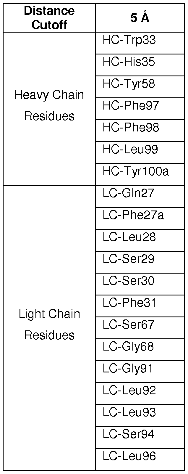

Five HVR positions of 5A12 4.2 are involved in binding both VEGF and Ang2 (HC-Leu99, HC-
Tyr100a, LC-Ser30, LC-Phe31 , and LC-Leu92). Moreover, when mapping the two paratopes on the
surface of the antigen binding region, it is evident that the spatial organization of the two paratopes is highly intertwined (Figures 13C and 13D). In order to accommodate the binding of two structurally diverse epitopes with essentially the same binding site, 5A12.4.2 utilizes a large degree of plasticity similar as previously described for a Her2/VEGF DAF (Bostrom et al. PloS One 6(4) :e17887, 201 1 ). A comparison between the HVR loop conformation of 5A12 4.2 in the VEGF bound state (5A12VEGF) with 5A12 4.2 in the Ang2 bound state (5A12Ang2) shows that most of the HVR-loops adopt the same conformations, with two notable exceptions: HVR-H3 and HVR-H2 (Figure 14C). HVR-H2, in particular, undergoes drastic conformational change in the VEGF-bound state compared to Ang2 bound state; HC-Ala53, for example, moves by 14 A (Figure 14D). When overlaying the structures of 5A12 4.2VEGF nd 5A12 4.2Ang2 with the parental G6 in the VEGF bound state (G6VEGF) or in the unbound state (G6unbound) , it is evident that conformation of HVR-H2 and HVR-H3 in 5A12 4.2 VEGF and G6VEGF are identical while the conformation of the two loops which are used to bind Ang2 resemble G6unb0und- Thus, the plasticity of the 5A12 4.2 binding site was inherited from the parental antibody G6 and not added when recruiting dual-specificity. In addition to the large conformational changes of the HVR-H2 and HVR-H3 region, some side chains in other HVR loops use different rotamers during binding of the two antigens thereby providing additional plasticity without the requirement for rearrangement of the main chain backbone. Ang2 binding by 5A12 4.2 does not substantially involve the HVR-H2 loop, while the HVR-H2 loop is involved in VEGF binding (Figures 15A and 15B). Example 4: Deep mutagenesis scanning of the Anti-Ang2/anti-VEGF DAF 5A12 4.2 to generate DAFs with sub-nanomolar affinity for both antigens
A. Deep mutagenesis scanning to identify substitutions compatible with enhanced dual affinity The intertwined paratopes of the anti-Ang2/anti-VEGF DAF 5A12 4.2 for Ang2 and VEGF and structural plasticity of the 5A12 4.2 binding site, as determined in Example 3, suggested that it could be difficult to identify mutations that are compatible with improving the binding towards both antigens. To overcome this challenge, we used deep mutagenesis scanning, which combined combinatorial library selection and deep sequencing using next-generation sequencing technology (also known as high- throughput sequencing or massive parallel sequencing), to assess the effect of all possible individual mutations of 5A12 4.2 HVRs on binding both antigens. As a comparison, 5A12 4.2 was also affinity matured in parallel using phage display of randomized libraries and selection of binding variants without deep sequencing analysis.
We used a library design in which all three HVRs of either the heavy or light chain of 5A12 4.2 were mutated simultaneously (Table 6). Each clone in this library carried three
 mutations (N = A or T or G or C; K= G or T), one in each of the three HVRs. We refer to this library design as triple- Λ/Λ// (3ΝΝΚ). This library design strategy allowed us to estimate the effect of single as well as double mutations (i.e., mutation pairs) on antibody binding function, which enabled epitasis analysis. Mutation pairs with positive cooperativity can be an important driver for the fitness (e.g., the binding affinity, expression, and/or stability) of a protein as they can, for example, allow overcoming local fitness minima and promote stability. This approach overcame a potential drawback of previous deep mutagenesis scanning approaches that involve mutating each HVR separately, which limits the resulting information to
the effect of single mutations on binding function. As a control to determine if the 3NNK library design allowed for the correct fitness assessment of single mutations, we also generated a library where each HVR of the 5A12 4.2 heavy chain was mutated separately (referred to as 1 NN K) (Table 6). Table 6: Library design overview
mutations (N = A or T or G or C; K= G or T), one in each of the three HVRs. We refer to this library design as triple- Λ/Λ// (3ΝΝΚ). This library design strategy allowed us to estimate the effect of single as well as double mutations (i.e., mutation pairs) on antibody binding function, which enabled epitasis analysis. Mutation pairs with positive cooperativity can be an important driver for the fitness (e.g., the binding affinity, expression, and/or stability) of a protein as they can, for example, allow overcoming local fitness minima and promote stability. This approach overcame a potential drawback of previous deep mutagenesis scanning approaches that involve mutating each HVR separately, which limits the resulting information to
the effect of single mutations on binding function. As a control to determine if the 3NNK library design allowed for the correct fitness assessment of single mutations, we also generated a library where each HVR of the 5A12 4.2 heavy chain was mutated separately (referred to as 1 NN K) (Table 6). Table 6: Library design overview

Enrichment ratios (ER) as a measure of antibody fitness for all mutations at all randomized positions were calculated by dividing the frequency of a mutation in sorted pools with the frequency in the unsorted libraries. This gave a comprehensive overview of the effect of single mutations (Figures 1 6A- 16B). To determine if it was possible to estimate the fitness score of single mutations from the 3NN K library design, we compared the enrichment ratios obtained from the 3NNK library design with the ratios obtained from the 1 NNK library design. Enrichment ratios obtained with the two different library designs correlated well (using a linear model, the R2 for the Ang2-panned samples is 0.73, and the R2 for the VEGF-panned samples is 0.82), demonstrating the validity of this approach (Figures 16C-16D).
B. Comparison of structural and deep mutagenesis scanning data
We compared the structural data from Example 3 with the mutagenesis data obtained from the 3NNK libraries. The ER of 1092 mutations for the two binding functions of 5A12 4.2 are plotted as a heatmap (Figures 16A-16B). Interestingly, most of the 52 HVR positions exhibited distinct profiles of enrichment (red) and depletion (blue) of the mutations with VEGF or Ang2 binding selection, indicating that the two binding functions involve differential usage of these HVR positions. The functional
paratopes, i.e., the HVR positions especially important for antigen interaction and therefore largely not tolerant to mutation, were identified by calculating the average of the enrichment ratios of all mutations at every position. The functional paratopes for both antigens were well within the structural paratopes for each antigen (compare Figures 13C-13D and 17A-17B). In contrast to the extensively overlapping structural paratopes for its two antigens (see Figures 13C-13D), the two functional paratopes had some degree of spatial separation (Figures 17A-17B). Hotspot positions (positions which do not tolerate mutation) for Ang2 binding were located mostly on the light chain (e.g., HVR-L1 , HVR-L2 and HVR-L3) with the exception of HC-Y100a, while hotspot positions for VEGF binding were located primarily on the heavy chain (e.g., HVR-H3) (Figure 18).
Structurally, the HVR-H2 loop of 5A12 is engaged with VEGF, whereas it is largely not involved in binding Ang2 (see Example 3). The differences in the different binding modes were reflected in the mutational data: the data obtained from the Ang2-panned sample suggested that CDR-H2 does tolerate mutations at several positions without effecting Ang2 binding, while the data from the VEGF-panned sample showed that mutations at several positions were not compatible with VEGF binding (Figures 15A- 15B and 19A-19B). For example, three residues in HVR-H2, HC-Gly50, HC-Gly54, and HC-Gly55, did not tolerate mutations in the VEGF-panned sample (Figure 1 9D). It has been previously suggested that these residues are required for function of the parental G6 antibody, as they might provide the flexibility for HVR-H2 to adopt the unusual conformation for VEGF binding (Fuh et al. J. Biol. Chem. 281 (10) :6625- 6631 ) (see also Figure 18).
In some instances, there are also similarities in the observed enrichment patterns for a few HVR positions between the Ang2- and VEGF-sorted samples, suggesting antigen-independent effects at these positions. Positions where both samples show mutation-sensitive results could in principle play a common structural role. For example, HC-Arg94 and HC-Ala49 are structurally buried at the base of HVR loops and do not make contact with either antigen, yet both tolerate few if any mutations (Figures 16A- 16B). HC-Arg94 forms a salt bridge with position Asp101 , which is important for CDR-H3 conformation (Morea et al. J. Mol. Biol. 275-269-294, 1998). Positions where both samples show similar toleration to most mutations (i.e., maintained binding for both antigens, for example, HC-Thr28 and LC-Ser56) tend to be surface-exposed and structurally distant from the antigen-binding area; their ability to tolerate mutations indicates relatively little involvement in antigen binding and structural restraint.
The enrichment pattern further provides insight into the mechanism of affinity maturation towards the single antigens. Several contact residues (less than 5 A from an antigen atom) were identified with either single mutations (e.g., for Ang2 binding: LC-S30aT; for VEGF binding: LC-A53D, HC-Y1 OOaW) or with a series of mutations (e.g., for Ang2 binding: LC-L93V, I,R, and K; for VEGF binding: LC-F31 H,S, T, and N) with high enrichment, suggesting that these mutations optimize the contact between the antigen and the antibody (Figures 16A-16B). The optimization of existing polar and non-polar antigen-antibody contacts is a well known mechanism for driving affinity maturation of antibodies. Further, some buried positions that are not in direct contact with the antigens contain selected substitutions that are highly enriched (e.g., for Ang2 binding LC-A34M, LC-Q89HW; for VEGF binding HC-F95Y, A100bV), demonstrating structural alteration of buried, non-contact sites in antibody affinity maturation.
Therefore, the mutagenesis data obtained using deep sequencing of 3NNK libraries were
consistent with the structural data, further validating the deep mutagenesis scanning approach.
C. Using deep scanning mutagenesis to identify residues for affinity maturation
For further guided affinity maturation of 5A12 4.2, we used the deep mutagenesis scanning results to identify substitutions which either increased the affinity towards both antigens or that improved the binding towards one antigen without decreasing the affinity toward the other antigen. An example of the latter is mutation HC-A53D, which was enriched in the VEGF-panned sample but was neither enriched nor depleted in the Ang2-panned sample (see circled positions in Figures 19C and 19D). A comparison of the enrichment of each mutation obtained in VEGF panning versus the enrichment obtained in Ang2 panning in the two 3NNK libraries suggested that several residues, including several light chain residues, could be further optimized to increase dual affinity to both VEGF and Ang2 (Figures 20A-20B).
We also determined the position of mutations which show strong enrichment. Based on the structural data, HVR positions could be classified as exposed or buried residues (see Example 3). For this analysis, mutations which were enriched 4-fold were considered strongly enriched. While solvent- accessible exposed residues were more strongly enriched than buried residues on average, more buried mutations showed strong enrichment than solvent-exposed mutations (Figure 18). This suggested that higher affinity gains could be achieved by optimizing buried positions.
To determine how the enrichment data correlated with antigen binding affinity, we selected 25 substitutions which showed increased enrichment to at least one of the antigens (Ang2 binding: HC-28P, HC-30M, HC-35Q, HC-35Y, HC-35W, HC-52D, HC-53D, HC-54E, HC-56H, HC-95Y, HC-97P, HC- 100aW, HC-100bT, HC-100bS, LC-28V, LC-30aT, LC-33L, LC-34M, LC-34N, LC-54N, LC-54T, LC-56W, LC-89W, LC-89H, LC-92S, LC-93K; VEGF binding: HC-28P, HC-30M, HC-35Q, HC-35Y, HC-35W, HC- 51 T, HC-52D, HC-53D, HC-54E, HC-56H , HC-95Y, HC-97P, HC-100aW, HC-100bT, HC-100bV, HC- 100bS, LC-28V, LC-30aT, LC-33L, LC-34M, LC-34N, LC-54N, LC-54T, LC-56W, LC-89W, LC-89H, LC- 92S, LC-93K).
We generated clones harboring each single point mutation in 5A12 4.2. To obtain an affinity estimate, the IC50 values of these clones for both antigens were determined in a phage competition assay (Figure 21 ). The results show that the ER metric serves as an excellent classifier to distinguish affinity improving mutations from mutations which have no effect on affinity or reduce the affinity (receiver operator characteristics, area under the curve 0.93). While the relationship between fold enrichment and the fold IC50 change was not linear, the fold enrichment ratios obtained from either the 3NN K or the 1 NNK libraries classified most of the mutants correctly as mutations which increase, decrease, or have neutral fitness.
D. Enrichment analysis in combination with an cooperativity analysis can be used to identify mutation pairs that enhance binding synergistically
The 3NNK library design, in which all three HVRs of the heavy or light chain were mutated simultaneously, enabled identification of pairs of mutations located in different HVRs which increased the affinity of the DAF towards one or even both antigens. Without wishing to be bound by theory, mutation
pairs could be enriched for several reasons. First, the pair could contain one mutation that by itself has a strong impact on affinity. This increases the likelihood that the second mutation in the pair is a
"hitchhiker," which does not contribute significantly to improved affinity. Second, the pair could contain a fold-stabilizing mutation. For instance, the nature of phage display allows for the enrichment of fold- stabilizing mutations which have little or no impact on antigen binding. Third, the pair could contain mutations which contribute to antigen binding at least additively or in some cases synergistically.
We calculated enrichment ratios for double mutations from all four 3NNK datasets. The enrichment ratios of double mutations were determined by calculating the enrichment ratio of all clones which carried NNK mutations at two given positions, ignoring the third NNK mutation. We calculated ER for 170,204 out of 400,428 possible mutation pairs. To filter out sampling effects, mutation pairs which had less than 10 sequence counts either in the sorted or unsorted sample were removed from the analysis. One surprising pattern was that a few distinct mutations were identified in several top enriched mutation pairs, as shown in Figures 22A and 22B. For example, in the dataset obtained from the Ang2- panned HC-3NN K library, the HC-F97P mutation formed strongly enriched mutation pairs with several distinct mutations located in the HVR-H1 and HVR-H2 loop. Further, the majority of the "mutation partners" of HC-F97P were not located in close spatial proximity to HC-F97P based on an analysis of the structure of 5A12Ang2. A similar pattern was observed for mutations LC-L93K and HC-F98P.
Without being bound to theory, two scenarios could explain the frequent presence of a particular mutation in enriched mutation pairs. In the first scenario, the mutation provides a specific improvement of the antigen binding interface and the resulting higher affinity is further improved or not diminished by various mutation partners, while in the second scenario, the mutation exhibits an advantage in fitness other than affinity during phage selection. For HC-F97P, we suspect the latter reason, because its mutation partners are mostly spatially distant, and, because when 5A12 4.2 HC-F97P affinity was tested in phage competition ELISA, no change was detected compared to 5A12 4.2. The strong selection of HC-F97P could therefore be caused by improved functional folding in E. co//' for clones carrying HC-F97P, resulting in improved functional phage display and higher enrichment during panning.
We used a modified cooperativity analysis to predict if HC-F97P and other mutations showing a similar behavior improved the stability of the 5A12 4.2 fold (see Araya et al. Proc. Natl. Acad. Sci. USA 109(42) : 16858-16863, 2012). Using a multiplicative cooperativity model, we calculated partner potentiation scores for a large subset of all possible mutation pairs from the data sets obtained from Ang2 panning. Indeed, HC-F97P, HC-F98P and LC-L93K had the highest partner potentiation score (>20) in the Ang2-panned dataset (Figures 22C and 22D). We confirmed that all three single variants showed an elevated melting temperature compared with 5A12 4.2 using differential scanning fluorimetry. Therefore, the partner potentiation score can be used to avoid selecting mutation pairs which contain a mutation more likely to be involved in protein folding and/or stability than in binding affinity. Alternatively, if the engineering goal is enhanced protein folding and/or stability (e.g., thermal stability), the partner potentiation score can be used to identify candidate mutation pairs for further testing.
In addition to enrichment and partner potentiation score, we also applied a third measure to identify affinity-improving mutation pairs: the spatial distance between the two mutations. Using distance as an additional criterion, we sought to identify pairs which had strong cooperativity in terms of their
binding function, and thus pairs that would typically not be selected from the single mutation analysis. Without wishing to be bound by theory, strong binding cooperativity could involve structural changes in conformation and/or dynamics that could be more likely to occur between adjacent residues (see, e.g., Skinner et al. Proc. Natl. Acad. Sci. USA 93(20) :10753-10757, 1 996). All tested mutations had a spatial Ca-Ca distance of 5 A or less.
Based on the preceding information, we developed a method to identify mutation pairs that act at least additively to improve dual affinity. We then calculated a partner potentiation score (Araya et al. Proc. Natl. Acad. Sci. USA 109(42) : 16858-1 6863, 201 ) for all mutation pairs in the dataset to identify fold-stabilizing mutations in silico. Mutation pairs which contained a mutation that exhibited a large partner potentiation score were then removed from the double mutation dataset. Since mutation pairs from the Ang2 panning of the heavy and light chain library showed the strongest enriched mutation pairs, three heavy chain mutation pairs (HC-W33P/I51 G, HC-S30M/I51 W, HC-H35D/G50K) and four light chain mutation pairs (LC-Q89H/V33L, LC-Q89Y/V33L, LC-Q89Y/A34M, LC-Q89W/A34G and LC-Q89H/A34S) from Ang2 panning were selected for further testing (Table 7).
Table 7: Enrichment of Mutation Pairs from Ang2 Panning of 3NNK Libraries

These mutation pair variants and their respective single mutation variants were generated for determination of binding affinity by BIACORE® SPR (Figures 22E and 22F and Table 10). All selected double mutations showed an increase in Ang2 affinity over 5A12 4.2, confirming the validity of this approach to identify double mutations with improved binding function. Surprisingly, among the selected double mutations was HC-H35D/G50K, a pair that showed a strong increase in Ang2 binding in the context of the double mutant, while the individual single mutations had a strong negative impact on Ang2 binding function. Three of the selected pairs (LC-Q89Y/V33L, LC-Q89W/A34G, and LC-Q89H/A34S) also showed an improvement in VEGF binding. Each of these mutation pairs involved two adjacent positions at the stem of the HVR-L1 and HVR-L3 loops, suggesting that improved packing in this region had a strong influence on dual specificity.
Therefore, the deep mutagenesis scanning approach using 3NNK libraries identified mutation pairs that improved binding affinity of an antibody for its antigen. In the context of dual specific
antibodies, this approach also identified mutation pairs compatible with improved binding affinity for one or both antigens.
E. Generation and characterization of 5 A 12 4.2 variants with sub-nanomolar affinity to both antigens based on deep mutagenesis scanning data
To affinity mature 5A12 4.2 to sub-nanomolar affinity for both VEGF and Ang2, we selected several amino acid alterations in the heavy and light chain of 5A12 identified by the deep mutagenesis scanning described above. Based on their enrichment, 14 mutations at 1 0 heavy chain HVR positions (T28P, S30M, H35Y/W, T52D, A53D, G54E, T57E, F97P, Y100aW, A100bT/V/S) as well as 12 mutations at seven light chain HVR positions (S30aT, V33L, A34M/N, S52R/K, Q89H/W, L92S, L93K/V/I) were chosen for further testing (Figures 20A-20B), either as single mutations or in combination with other mutations, as described below. The mutation pairs described above in Section D of this Example were also characterized as part of this analysis. These variants were generated and expressed in Fab format for further characterization. These variants were also compared with clones 5A12 4.2.16 and 5A12 4.2.16.2 (also referred to as "DAF16.2"), which were obtained by affinity maturation of 5A12 4.2 using phage display and selection without use of deep sequencing-based enrichment data (see Section F. ll below).
The affinity for Ang2 and VEGF for single and double mutations identified by deep sequencing as determined by phage IC50 is shown in Table 8. The affinity for Ang2 and VEGF for single and double mutations identified by deep sequencing as determined by BIACORE® SPR is shown in Table 9. The affinity as determined by BIACORE® SPR for Ang2 and VEGF for double mutation pairs identified as described above in Section D is shown in Table 10 (note that Table 10 shows results from two separate experiments for selected clones for Ang2 binding affinity). To generate higher affinity clones, a stepwise approach was chosen. First, several two- and three-mutation combinations of single mutations were generated and tested for the Ang2 and VEGF affinity (Table 8 and 9).
Table 8: Phage IC50 Values for 5A12 4.2 Affinity Matured Variant Clones

LC.Q89H 0.138 14.28 0.6 11.50
LC.L92S 27.68 0.07 5.06 1.36
LC.L93K 0.48 4.10 4.21 1.64
LC.Q89W 2.55 0.77 6.2 1.11
HC.T52D 39.5 0.05 0.08 86.25
HC.A53D 0.05 39.40 67 0.10
HC.G54E 24.2 0.08 0.43 16.05
HC.F95Y 11.9 0.17 160 0.04
HC.YIOOaW 0.21 9.38 115 0.06
HC.AIOObT 0.49 4.02 69 0.10
HC.AIOObV 0.1 19.70 137 0.05
HC.AIOObS 1.2 1.64 71.4 0.10
HC.F97P 160 0.01 6.85 1.01
LC.Q89H/LC.L93K("D") 3.71 0.53 0.1 69.00
LC.Q89H/HC.A100bV 5.2 0.38 22.4 0.31
LC.L93K/HC.A100bV 11.3 0.17 6.18 1.12
LC.Q89H/LC.L93K/HC.A1 OObV 51.3 0.04 0.2 34.50
HC.A53D/LC.L93K 0.015 131.33 21 0.33
HC.A53D/LC.Q89H 0.01 197.00 1 6.90
HC.A53D/LC.Q89H/LC.L93K 0.03 65.67 0.04 172.50
("T")
Table 9: BIACORE® SPR Kd Values for selected 5A124.2 Affinity Matured Clones

HC.A53D/LC.Q89H/LC.L93K ("T") 0.21 0.5 71 .7 n/a, not available
Table 10: BIACORE® SPR Kd Values (nM) of mutation pairs identified by deep mutagenesis scanning

The two highest dual affinity variants LC-Q89H/LC-L93K (referred to as "D" for simplicity) and HC-A53D/LC-Q89H/LC-L93K (referred to as "T" for simplicity) were then further dual affinity matured by the addition of several further mutations. Table 1 1 shows phage IC50 values and the fold difference relative to 5A12 4.2 for these variants. Table 12 shows the binding affinity (Kd) as determined by BIACORE® SPR for Ang2 and VEGF for selected variants.
Table 11 : Phage IC50 values of combination variants based on variants D or T


The improved variants T.28P, T.30M, and T.28P-VR further carried the HVR-H2 A53D mutation and an additional mutation in HVR-L3 (LC-L93K or L93V) (see Figures 29A-29B). Except from LC-L93K, which slightly increased the melting temperature, we did not include any additional fold-stabilizing mutants from the cooperativity analysis, as they were not compatible with dual specificity. These improved 5A12 4.2 variants showed improved dual specificity, having sub-nanomolar affinity for Ang2 as well as VEGF as measured by SPR (BIACORE®) (Figure 23B and Table 12). These improved variants were tested for their ability to block the ligand-receptor interaction using in vitro receptor blocking assays. Each of these affinity improved variants showed improved blocking activity over the parental antibody 5A12 4.2. For example, the T.28P variant a similar blocking activity as the high affinity anti-VEGF antibody G6.31 and an in-house generated high affinity anti-Ang2 antibody G5.5 (Figures 23C-23D). These variants also had better dual-specificity and increased potency in blocking VEGF-induced migration in a cell-based assay (HUVEC migration assay) than an antibody clone obtained by phage display and selection without use of deep mutagenesis scanning (5A12 4.2.16.2) (Figure 24).
Dual targeting strategies for disorders associated with pathological angiogenesis (e.g., ocular diseases such as AMD as well as cell proliferative disorders such as cancer) promise treatments with improved efficacy. Designing such dual targeting therapies holds several challenges, including the design of high affinity blockers against two targets and determination of the most efficacious ratio of the two blocking antibodies for administration. Additionally, for some conditions, including ocular disorders such as AMD, the amount of therapeutic antibody that can be delivered (e.g., by intravitreal injection) is limited. These challenges can be overcome using high affinity DAFs, which combine dual-targeting activity within a small antibody fragment. The results presented in this Example demonstrated the ability of the deep scanning mutagenesis approach described herein to identify variants that allowed engineering, for the first time, an antibody with a dual-specific binding site having sub-nanomolar affinity against two structurally-distinct antigens.
The optimized library design as well as the deep mutagenesis scanning approach described herein can also be applied for the improvement of monospecific antibodies. For example, as
demonstrated above, the library design facilitates the identification of fold-stabilizing mutations in the
HVRs which are compatible with antigen binding, allowing the optimization of HVRs for improved antibody thermostability. The identification of affinity-improving mutation pairs by the methods described herein can allow, for example, achieving a high monospecific affinity of an antibody in cases where the single mutation space has been already extensively been explored unsuccessfully.
F. Materials and Methods
I. 5A 12 4.2 Library Construction, Phage Display, and Panning
For additional affinity improvement of 5A12 4.2, phage libraries were constructed from variant 5A12 4.2 in Fab-amber format for monovalent Fab phage display with either light chain HVR residues (i.e., HVR-L1 , HVR-L2, and HVR-L3) or heavy chain HVR residues (i.e., HVR-H 1 , HVR-H2 and HVR-H3) residues randomized using the A/A/ degenerate codon that encodes for all 20 amino acids with 32 codons (see, e.g., Brenner et al. Proc. Natl. Acad. Sci. USA 89(12) : 5381 -5383, 1992). Libraries were designed to allow either one NNK mutation in each of the three light chain or heavy chain HVRs (3NNK) or only one NNK mutation in a single heavy chain HVR (1 NNK). The resultant library DNA was electroporated into E. coli XLI cells, yielding approximately 109 transformants.
Libraries were panned on immobilized hVEGF1 09 or hAng2his8 with subsequent rounds using biotinylated targets (Ang2 or VEGF) in solution and captured on streptavidin-coated plates. See Figure 25A for a schematic of the panning strategy. //. Isolation of affinity-matured variant 5A 12 4.2. 16.2 by phage display and selection
To directly screen for and isolate high affinity clones from the phage libraries, a high-throughput single-spot competitive ELISA was used to screen for high affinity clones for both hAng2 and hVEGF binding (Sidhu et al. J. Mol. Biol. 338(2) : 299-310). Selected clones were submitted for standard Sanger sequencing and clones with unique sequences were ranked by their apparent affinities using phage IC50 (Figures 25B-25D). Selected top variants (clones 5, 9, and 16) were expressed and purified as Fab for further testing in receptor-blocking ELISA experiments (Figures 26A-26C). Based on the results of the phage IC50 and the receptor blocking assays, clone 16 (5A12 4.2.1 6) was selected as a template antibody for further affinity maturation.
A second round of affinity maturation was conducted using NNK libraries based on 5A12 4.2.16 in Fab format for monovalent phage display as described above for 5A12 4.2, allowing one NNK mutation in each of the three heavy chain variable region HVRs. Panning and screening of the 5A12 4.2.16 heavy chain A/A/ libraries was performed as described above for 5A12 4.2 NN K libraries. Selected clones were sequenced by Sanger sequencing and phage IC50 affinities were determined (Figures 27A-27C).
Selected top variants (clones 2, 4, 5, 8) were expressed and purified as Fab for affinity measurements by BIACORE® using Fabs as analytes with a CM5 sensor chip immobilized with hVEGF109 or hAng2his8 at 25 Ό to determine monovalent affinities (Figure 1 9). Clone 2 (5A12 4.2.16.2) showed best dual affinity for both VEGF and Ang2, with a Kd below 0.003 nM for VEGF and a Kd of 1 .51 nM for Ang2 (Figure 28). DAF 5A12 4.2.16.2 (also referred to as DAF16.2) was chosen as a lead variant based on affinity measurements by BIACORE® SPR and a VEGF-cell based assay potency described below.
///. Deep sequencing of DAF 5A 124.2 Affinity Maturation Libraries
For deep sequencing, phagemid double stranded DNA was isolated from E. coli XL-l cells carrying phagemid vectors from selected rounds of DAF 5A12 4.2 NN K library panning described in the in Section F. I above. The unsorted library and Round 3 of the Ang2 sorted libraries and Round 2 of the VEGF sorted libraries were sequenced. These rounds were selected to allow for both a plate-based panning round, which typically removes non-functional clones from the generated library, as well as a solution-based off rate selection, which typically enriches for clones with high affinity. For VEGF, only one round of plate panning was performed, while for Ang2, two rounds were performed because the observed enrichment was comparatively low for the first round.
Purified DNA was used as template for a limited cycle PCR-based amplification of VL and VH regions using PHUSION® DNA polymerase (New England Biolabs). PCR products were purified by agarose gel extraction and clean-up (Qiagen Gel Extraction Kit). Eluted amplicon DNA was used as the basis for deep sequencing library preparation with standard lllumina library preparation methods, using a TRUSEQ™ DNA Sample Prep kit (lllumina). Adapter-ligated libraries were subjected to a single cycle of PCR and sequenced on the lllumina M ISEQ®, using paired-end sequencing with an insert size of 200bp or 300bp as appropriate to cover the entire length of the amplicon.
IV. Deep Sequencing Analysis of DAF 5A 12 4.2 Affinity Maturation Libraries
Sequencing data were analyzed using the statistical programming language R (see, e.g., R Core Team , R: A language and environment for statistical computing, 2013) and the ShortRead package (see Morgan et al. Bioinformatics 25(19) : 2607-2608). Quality control (QC) was performed on identified HVR sequences, where each HVR sequence was checked for the correct length and was allowed to carry only up to one NNK mutation and no ηοη-Λ/Λ/Κ" mutation. The obtained sequence counts for all samples before and after quality control are listed below in Table 13. Position weight matrices were generated by calculating the frequency of all mutations of every randomized position. Enrichment ratios for each mutation were calculated by dividing the frequency of a given mutation at a given position in the sorted sample with the frequency of the very same mutation in the unsorted sample, as described previously (Fowler et al. Nature Methods 7(9) : 741 -746, 2010). The log2 enrichment ratio for all randomized positions is shown in Figures 16A-16B.
Table 13: Sequence Counts for Deep Sequencing

VEGF HC-3NNK 4721 529 2398256
unsorted HC-1 NNK HVR-H1 279535 83312 168/168
Ang2 HC-1 NNK HVR-H1 1441 03 92573
VEGF HC-1 NNK HVR-H1 250058 1 15401
unsorted HC-1 NNK HVR-H2 5521 89 168783 231 /231
Ang2 HC-1 NNK HVR-H2 287387 188498
VEGF HC-1 NNK HVR-H2 491958 185947
unsorted HC-1 NNK HVR-H3 1581 86 66989 210/210
Ang2 HC-1 NNK HVR-H3 390433 256998
VEGF HC-1 NNK HVR-H3 467303 229836
The enrichment ratios of double mutations were calculated from the 3NNK library by calculating the enrichment ratio of all clones which carry NNK mutations at two given positions, ignoring the third NNK mutation. In order to filter out sampling effects, mutation pairs which had less than 10 sequence counts either in the sorted or unsorted sample were removed from the analysis.
To test the performance of the ER as a classifier for identifying affinity-improving mutations, receiver operator characteristics were used. The performance of single variants in a phage competition ELISA was used as a gold standard (IC50 mutant IC5o WiidtyPe > -5 for affinity improving mutations, IC50 mutant IC50 wiidtype < 1 -5 for mutations which do not improve the affinity).
Four different models describing cooperativity were tested (Mani et al. Proc. Natl. Acad. Sci. USA
105(9) : 3461 -3466, 2008) to determine which model is the best representative for the data.
The multiplicative model
EnrichAB = EnrichA x EnrichB
was chosen based on a simple linear regression.
The cooperativity used here is thus defined as:
Cooperativity = EnrichAB - EnrichAx EnrichB
The partner potentiation score to identify fold-stabilizing mutations are calculated as described previously (Ref) by calculating first the partner normalized cooperativity score for all available mutation pairs:
_ EnrichAB - EnrichA x EnrichB
a→b EnrichB
The partner potentiation score is the mean of the partner normalized cooperativity scores of a given mutation mutation A:
P„ _ i=l
n
The data were plotted using ggplot2 (Wickam, ggplot2: elegant graphics for data analysis (Springer New York, 2009) and Circos (Krzywinski et al. Genome Research 19(9) :1639-1645, 2009).
V. Phage IC50 determination of deep sequencing variants
Variants based on the deep sequencing information were generated by Kunkel mutagenesis on a 5A12.4.2 single strand Fab amber phagemid template. Clones were sequenced to confirm the mutations.
To determine the phage IC50 for deep sequencing variants, we performed phage solution competition ELISAs. First, the phage were propagated and purified. 10 μΙ of XL-1 bacteria infected with one of the clones for 30 min at 37Ό was plated on a carbenicillin plate. A colony was picked and grown in 2 ml of 2YT media containing 50 μg/ml carbenicillin at 37°C for 3-4 h. Helper phage K07 was added to the culture at a final concentration of 1010 plaque-forming units (pfu)/ml for another 1 h at 37°C. 20 ml of 2YT media with 50 μg/ml carbenicillin and 50 μg/ml kanamycin were added to the culture for growth overnight at 37°C. The phage was purified as described above.
Second, the concentration of purified phage that would be optimal for use in the following competition ELISA assay was determined (i.e., approximately 90% of maximal binding capacity on the coated plate). 96-well NUNC MAXISORP® plates were coated with 2 μg/ml hVEGF109 or hAng2his8 in PBS at 4Ό overnight. The wells were blocked by adding 65 μΙ 1 % BSA for 30 min followed by 40 μΙ of 1 % TWEEN-20® for another 30 minutes. Next, the wells were washed with PBS with 0.05% TWEEN-20® 5 times. Various dilutions of phages down to 0.1 optical density (O.D.)/ml in ELISA buffer (PBS with 0.5% BSA and 0.05% TWEEN-20®) were added to the wells for 15 min at room temperature. The wells were then washed with PBS with 0.05% TWEEN-20® at least three times. 75 μΙ of HRP-conjugated anti-M13 antibody (Amersham , 1 :5000 dilution in ELISA buffer) per well was added and incubated at room temperature for 30 min. The wells were washed again with PBS with 0.05% TWEEN-20® at least five times. Next, 100 μΙ/well of a 1 :1 ratio of 3,3',5,5'-tetramethylbenzidine (TMB) Peroxidase substrate and Peroxidase Solution B (H202) (Kirkegaard-Perry Laboratories (Gaithersburg, MD)) was added to the well and incubated for 5 min at room temperature. The reaction was stopped by adding 100 μΙ of 1 M phosphoric acid (H3P04) to each well and allowed to incubate for 5 min at room temperature. The optical density of the color in each well was determined using a standard ELISA plate reader at 450 nm . The dilutions of phage were plotted against the O.D. readings.
Third, a competition ELISA was performed. 96-well NUNC MAXISORP® plates were coated with 2 μg/ml hVEGF109 or hAng2his8 in PBS at 4°C overnight. The wells were blocked by adding 65 μΙ of 1 % BSA for 30 min followed by 40 μΙ of 1 % TWEEN-20® for another 30 min. The wells were washed 5 times with PBS with 0.05% TWEEN-20®. Based on the binding assay above, 50 μΙ of the dilution of phage that resulted in about 90% of maximum binding to the coated plate was incubated with 50 μΙ of various concentrations of hVEGF109 or hAng2his8 (0.1 to 10OOnM) in ELISA buffer solution for 2 h at room temperature in a well. The unbound phage was assayed by transferring 75 μΙ of the well mixture to a second 96-well plate pre-coated with hVEGF109 or hAng2his8 and incubating at room temperature for 15 min. The wells of the second plate were washed with PBS with 0.5% TWEEN-20® at least three times. 75 μΙ of HRP-conjugated anti-M13 antibody (1 :5000 dilution with ELISA buffer) per well was added and incubated at room temperature for 30 min. The wells were washed again with PBS with 0.05% TWEEN- 20® at least five times. Next, 100 μΙ/well of a 1 :1 ratio of TMB Peroxidase substrate and Peroxidase Solution B (H202) (Kirkegaard-Perry Laboratories) was added to the well and incubated for 5 min at room
temperature. The reaction was stopped by adding 100 μΙ of 1 M phosphoric acid (H3P04) to each well and allowed to incubate for 5 min at room temperature. The O.D. of the color in each well was determined using a standard ELISA plate reader at 450 nm . The concentrations of competitor hVEGF109 or hAng2his8 were plotted against the O.D. readings. The phage IC50 is the concentration of hVEGF109 or hAng2his that inhibits 50% of the phage.
VI. Expression and purification of Fab constructs of deep sequencing variants
Based on phage IC50, we chose variants with high affinity to both hVEGF and hAng2 to make Fab proteins for BIACORE® SPR affinity determination. The amber phagemid of selected variants were transformed into E coli non-amber-suppressor strain 34B8 cells. Single colonies were picked and grown in complete CRAP medium with 25 μ9/ιτιΙ carbenicillin at 30°C for at least 22 h. The expressed Fab proteins were purified using a protein G high trap column with standard purification protocol (Amersham Pharmacia). VII. Fab affinity determination by BIA CORE® SPR
To determine the binding affinity of selected Fab variants for VEGF, SPR measurement with a BIACORE® T200 instrument was performed. Briefly, a series S sensor chip CM5 was activated with 1 - ethyl-3-(3-dimethylaminopropyl) carbodiimide (EDC) and N-hydroxysuccinimide (NHS) reagents according to the supplier's instructions, and hVEGF109 was coupled to achieve 50-80 response units (RU), then following by blocking un-reacted groups with 1 M ethanolamine. 3-fold serial dilutions of Fab in HBS-P buffer (0.01 M HEPES pH 7.4, 0.15 M NaCI, 0.005% surfactant P20) from low (0.02 nM) to high (50 nM) were injected (flow rate: 30μΙ/Γη ίη) . The binding responses on hVEGF were corrected by subtracting of RU from a blank flow cell. The sensorgram was recorded and subject to reference and buffer subtraction before evaluating by BIACORE® T200 Evaluation Software (version 2.0). Association rates (kon) and dissociation rates (koff) were calculated using a simple one-to-one Langmuir binding model. The equilibrium dissociation constant (Kd) was calculated as the ratio of koff/kon.
To determine the binding affinity of selected Fab variants for Ang2 or Ang1 , SPR measurement with a BIACORE® T200 instrument was performed. Briefly, a series S sensor chip CM5 was activated with EDC and NHS reagents according to the supplier's instructions, and anti-human Fc was coupled to achieve 10000 response units (RU), then following by blocking un-reacted groups with 1 M ethanolamine. For kinetics measurements, approximately 5 nM of human Fc fusion of Ang2 or Ang1 protein was first injected at 10 μΙ/min flow rate to capture approximately 100 RU at 2 different flow cells (FC), except for FC1 (which served as a reference). Next, 3-fold serial dilutions of Fab in HBS-P buffer (0.01 M H EPES pH 7.4, 0.15M NaCI, 0.005% surfactant P20) from low (0.02 nM) to high (50 nM) were injected (flow rate: 30 μΙ/min). The binding responses on hAng2 or hAngl flow cell were corrected by subtracting of RU from a blank flow cell. The sensorgram was recorded and subject to reference and buffer subtraction before evaluating by BIACORE® T200 Evaluation Software (version 2.0). Association rates (kon) and dissociation rates (koff) were calculated using a simple one-to-one Langmuir binding model. The equilibrium dissociation constant (Kd) was calculated as the ratio of koff/kon.
VIII. Differential Scanning Fluorimetry
The melting temperature (Tm) was determined using differential scanning fluorimetry. A 100 μ9/ιηΙ purified Fab solution was mixed 1 :500 with SYPRO® Orange dye. The sample was slowly heated in a qPCR machine from 20 °C to 100°C while the fluorescence was measured at 590 nm at 0.2 °C temperature increments. The Tm is the maximum of the derivative of the obtained fluorescence melting curve.
IX. HUVEC Migration Assays
HUVEC migration assays were performed by pre-coating Falcon 24-multiwell insert systems with mouse laminin overnight and adding HUVEC cells that had been starved overnight, harvested, and reuspended in assay medium . Cells (5x10⁴) were added to the upper chamber and 20 ng/mL VEGF was added to the lower chamber to stimulate cell migration in the presence or absence of antibodies for 16 h. After fixing and scraping the upper membrane face, cells on the lower face were fixed with methanol and stained with SYTOX® Green and images were acquired using an inverted fluorescent microscope. Cell numbers were analyzed with ImageJ software and IC50 was calculated. HUVEC IC50 migration values indicate the cell activity potency of anti-VEGF antibodies on the VEGF-induced migration of these cells.
Example 5: DAF T.28P-VR shows in vivo efficacy in a preclinical model
The in vivo efficacy of the dual affinity improved DAF T.28P-VR was tested in a laser-induced choroidal neovascularization (CNV) rat model. A comparison of the size of the area in which vascular sprouting occurred after the laser-induced lesion as well as after administration of the antibodies demonstrated that dual targeting of Ang2 and VEGF resulted in reduced vascular sprouting compared to treatment with either a high-affinity anti-Ang2 Fab or a high-affinity anti-VEGF Fab alone.
Two different formats of T.28P-VR were generated. One format was a standard dual-specific Fab format as described above, while the second was a dimeric F(ab')2 format in which two monomeric DAF molecules were linked together by a single cysteine bond. While the monomeric DAF format was used to test if the efficacy of co-administered mono-specific anti-Ang2 and anti-VEGF can be reached by administration of the DAF, the dimeric DAF allowed determination of whether additional avidity led to improved efficacy. T.28P-VR was compared with either co-administered anti-Ang2 and anti-VEGF Fabs or a bi-specific F(ab')2 format in which the anti-VEGF and the anti-Ang2 Fabs were linked together by a single cysteine bond. Monomeric T.28P-VR DAF showed the same efficacy as the two co-administered high affinity monospecifc antibodies (Figure 30). However, the dimeric DAF showed a significantly greater reduction of vessel sprouting compared to the co-administered Fab or the bi-specific Fab molecule (Figure 30). This efficacy study demonstrates the feasibility of using one high affinity DAF molecule to block the activity of two soluble factors in vivo as a therapy for disorders associated with pathological angiogenesis.
Materials and Methods
The laser-induced rat choroidal neovascularization model was performed using laser
photocoagulation in Brown Norway rats under anesthesia. Pupils were dilated with 1 % tropicamide and then 6 laser spots were delivered unilaterally using an OCULIGHT® CL photocoagulator with lesions approximately 2 disc diameters from the optic nerve head with evident rupture of Bruch's membrane and lack of sub-retinal hemorrhaging. Buprenorphine was administered immediately prior to photocoagulation and q12h for 48 h. Experimental designs varied in the amount and dosage frequently of intravitreal injections following laser coagulation. Treatment groups of n=6 rats of anti-VEGF, anti-VEGF/Ang2, and non-targeting anti-gD or ragweed IgG as negative controls were included and 5 μΙ volumes were delivered via 30G Hamilton needles. At time of harvest, animals were injected with 1 m l_ of 25 mg/mL fluroscein dextran via tail vein and then animals were euthanized. The eyes were harvested and fixed with 4% paraformaldehyde in PBS for an hour prior to removal or corea, lens, and retina to flat-mount the entire retinal pigment epithelium with choroid and with eye-cup. The CNV was imaged with a Zeiss Axio Image M2 microscope and captured and processed with analysis using N IH ImageJ software. The CNV was isolated with freehand selection and the resultant pixel amount converted to area by a pixel/length ratio. Statistical analysis used the "all pairs, Tukey HSD" method within JMP9 software.
Example 6: Anti-Ang2/anti-VEGF DAF T.28P affinity maturation by phage display and deep scanning mutagenesis to generate variants with reduced off-target Ang1 binding. A. DAF 5A 12 4.2 T28P Affinity Maturation Library Construction and Panning
Affinity maturated variant T.28P had a detectable off-target Ang1 binding affinity. For additional affinity improvement to VEGF and Ang2, and reduction of affinity for Ang1 , phage libraries were constructed from variant T.28P in Fab-amber format for monovalent Fab phage display with either LC (L1 , L2, and L3) or HC (H1 , H2, and H3) HVR residues mutated using the A/A/ degenerate codon. Libraries were designed to allow one NNK mutation in each of the three LC or HC HVRs (3NNK). The resulting library DNA was electroporated into E. coliXLI cells, yielding approximately 1 09 transformants. Libraries were panned on immobilized hVEGF109, hAng2his, or hAngl .Fc, with subsequent rounds using biotinylated targets (Ang2 or VEGF) in solution and captured on streptavidin-coated plates or target directly immobilized on plates (hAngl ). B. Deep sequencing of DAF 5A 124.2 T28P Affinity Maturation Libraries
For deep sequencing, phagemid double strand DNA was isolated from E. coli XLI cells carrying phagemid vectors from selected rounds of DAF T.28P NNK library panning. Purified DNA was used as template for a limited cycle PCR-based amplification of VL and VH regions. PCR products were purified by agarose gel extraction and clean-up (Qiagen Gel Extraction Kit). Eluted amplicon DNA was used as basis for deep sequencing library preparation with standard lllumina library prep methods, using
TRUSEQ™ DNA Sample Prep (lllumina). Adapter-ligated libraries were subjected to a single cycle of PCR and sequenced on the lllumina M ISEQ®, paired-end 200bp or 300bp as appropriate to cover the entire length of the amplicon.
C. Deep Sequencing Analysis of DAF 5A 12 4.2 T28P Affinity Maturation Libraries
Sequencing data were analyzed using the statistical programming language R and the
ShortRead package. Quality control was performed on identified HVR sequences, where each HVR sequence was checked for the correct length and was allowed to carry only up to one NNK mutation and no non-WMC mutation. The obtained sequence counts for all samples before and after quality control are listed in Table 14. Position weight matrices were generated by calculating the frequency of all mutations at every randomized position. Enrichment ratios for all mutations were calculated by dividing the frequency of a given mutation at a given position in the sorted sample with the frequency of the very same mutation in the unsorted sample, as described previously (Fowler et al. Nature Methods 7(9) :741 - 746, 201 0). To identify single mutations which interfered with Ang1 binding while having a minimal impact on Ang2 or VEGF binding, the following filter was applied to all combinations of Ang1 , Ang2 and VEGF panned samples: lOg 2ERx, Angl < -1 Λ lOg 2ERx, Ang 2 > -0.5 Λ log lERx, VEGF > 0 where ERx.y is the enrichment of mutation x in panning against antigen y. Since we panned each antigen using two different panning conditions (see Table 14), there are 8 different combinations of how pannings against different antigens can be combined and filtered. Mutations which passed the filter more than once in the different combinations were selected for further characterization.
Table 14: Overview of number of sequence reads obtained from MISEQ® sequencing of the different samples and their sequence counts after QC


D. Clone generation and purification
Variant plasmids based on the deep sequencing information were generated by synthesizing the selected DNA inserts containing the single muations and subcloning either the VL or VH into a human kappa or human Fab.flag vector, respectively. Clone plasmids (LC and HC) were transiently transfected into Expi293 cells in a 30ml_ volume and resultant supernatants were harvested after 7 days of growth. Clone antibody fragments (Fab) were purified using FLAG™ tag-based affinity chromatography.
E. Characterization of deep sequencing variants with single mutation affinity by BIACORE® SPR To determine the binding affinity of selected Fab variants, SPR measurement with a BIACORE® T200 instrument was used. Briefly, series S sensor chip CM5 was activated with EDC and NHS reagents according to the supplier's instructions, and hVEGF was coupled to achieve 50-80 response units (RU), then following by blocking un-reacted groups with 1 M ethanolamine.
Using a single-cycle kinetic method, 5-fold serial dilutions of Fab in HBS-P buffer (0.01 M H EPES pH 7.4, 0.15M NaCI, 0.005% surfactant P20) from low (0.08 nM) to high (50 nM) were injected (flow rate: 30 μΙ/min) over the hVEGF-coated flow cells. The binding responses (RU) on hVEGF were corrected by subtracting of RU from a blank flow cell. The sensorgram was recorded and subject to reference and buffer subtraction before evaluating by BIACORE® T200 Evaluation Software (version 2.0). Association rates (kon) and dissociation rates (koff) were calculated using a simple one-to-one Langmuir binding model. The equilibrium dissociation constant (Kd) was calculated as the ratio koff/kon.
F. Binding kinetic determination to human Ang2 and Ang1
To determine the binding affinity of selected Fab variants, SPR measurement with a BIACORE® T200 instrument was used. Briefly, series S sensor chip CM5 was activated with EDC and NHS reagents according to the supplier's instructions, and anti-human Fc was coupled to achieve 10000 response units (RU), then following by blocking un-reacted groups with 1 M ethanolamine.
For kinetic measurements, approximately 5 nM of human Fc fusion of Ang2 or Ang1 protein was first injected at 10 μΙ/min flow rate to capture approximately 100 RU on 2 different flow cells (FC), except for FC1 (reference). 5-fold serial dilutions of Fab in HBS-P buffer (0.01 M H EPES pH 7.4, 0.15 M NaCI,
0.005% surfactant P20) from low (1 .6 nM) to high (1000 nM) were injected (flow rate: 30 μΙ/min). The binding responses (RU) on hAng2 or hAngl flow cells were corrected by subtracting of RU from a blank flow cell. The sensorgram was recorded and subject to reference and buffer subtraction before evaluating by BIACORE®T200 Evaluation Software (version 2.0). Association rates (kon) and dissociation rates (koff) were calculated using a simple one-to-one Langmuir binding model. The equilibrium dissociation constant (Kd) was calculated as the ratio k /k
E. Binding affinity of clones identified by deep mutagenesis scanning
Using the methods described above, single mutation variants of T.28P were identified by deep mutagenesis scanning based on panning against VEGF, Ang2, and Ang1 . The resulting variants maintained dual affinity for VEGF and Ang2, while having markedly reduced affinity to Ang1 (Table 15).
The SPR measurements indicated that for each of the single mutation variants, the Kd was greater than
1 μΜ. Table 15: Affinity to VEGF, Ang2, and Ang1 of single mutation variants of T.28P as determined by Biacore.


Other Embodiments
Although the foregoing invention has been described in some detail by way of illustration and example for purposes of clarity of understanding, the descriptions and examples should not be construed as limiting the scope of the invention. The disclosures of all patent and scientific literature cited herein are expressly incorporated in their entirety by reference.
Claims
1 . An isolated antibody that specifically binds angiopoietin 2 (Ang2), wherein the antibody binds to an epitope on Ang2 comprising one or more amino acid residues selected from the group consisting of Cys433, Cys435, Met440, Leu441 , Cys450, and Gly451 of Ang2.
2. The isolated antibody of claim 1 , wherein the epitope further comprises one or more additional amino acid residues selected from the group consisting of Phe469, Tyr475, and Ser480 of Ang2.
3. The isolated antibody of claim 1 or 2, wherein the epitope further comprises one or more additional amino acid residues selected from the group consisting of Lys432, Ile434, Asp448, Ala449, Pro452, and Tyr476 of Ang2.
4. The isolated antibody of claim 3, wherein the epitope consists of amino acid residues Lys432, Cys433, Ile434, Cys435, Met440, Leu441 , Asp448, Ala449, Cys450, Gly451 , Pro452, Phe469, Tyr475, Tyr476, and Ser480 of Ang2.
5. An isolated antibody that specifically binds Ang2, wherein the antibody comprises a paratope comprising one or more amino acid residues selected from the group consisting of light chain variable region amino acid residues Gln27; Phe27a; Leu28, Met28, or Ala28; Ser29; Ser30; Phe31 ; Ser67; Gly68; Gly91 ; Leu92; Leu93, Lys93, or Val93; Ser94 or Pro94; and Leu96 and the heavy chain variable region amino acid residues Trp33; His35, Tyr35, or Asp35; Tyr58, Ile58, Trp58, or Leu58; Phe97; Phe98; Leu99 or Ala99; and Tyrl OOa.
6. The isolated antibody of claim 5, wherein the paratope consists of light chain variable region amino acid residues Gln27; Phe27a; Leu28, Met28, or Ala28; Ser29; Ser30; Phe31 ; Ser67; Gly68; Gly91 ; Leu92; Leu93, Lys93, or Val93; Ser94 or Pro94; and Leu96 and the heavy chain variable region amino acid residues Trp33; His35, Tyr35, or Asp35; Tyr58, Ile58, Trp58, or Leu58; Phe97; Phe98; Leu99 or Ala99; and Tyrl OOa.
7. An isolated antibody that specifically binds Ang2, wherein the antibody comprises the following six hypervariable regions (HVRs) :
(i) an HVR-L1 comprising the amino acid sequence of RASQFX1SX2FGX3X4 (SEQ ID NO: 26), wherein is Leu, Met, or Ala, X2 is Ser, Lys, or Thr, X3 is Val or Leu, and X4 is Ala, Ser, Met, Gly, Thr, or Asn;
(ii) an HVR-L2 comprising the amino acid sequence of GX1X2X3LX4X5 (SEQ ID NO: 27), wherein is Ala, Ser, or Gly, X2 is Arg, Ser, Leu, or Lys, X3 is Ser, Ala, or Gly, X4 is Tyr, Val, Ala, or Glu, and X5 is Ser, Gly, or Gin;
(iii) an HVR-L3 comprising the amino acid sequence of X1QX2X3X4X5X6LT (SEQ ID NO: 28), wherein XT is His, Gin, Phe, Trp, Tyr, or Met, X2 is Gly, Met, or Phe, X3 is Leu, Pro, or Ser, X4 is Val, Leu, lie, Gly, Lys, or Arg, X5 is Ser, His, Leu, or Pro, and X6 is Pro or Asp;
(iv) an HVR-H1 comprising the amino acid sequence of DX1X2X3X4 (SEQ ID NO: 29), wherein is Tyr or Ala, X2 is Trp or Pro, X3 is lie, Met, or Gin, and X4 is His, Tyr, Trp, or Asp;
(v) an HVR-H2 comprising the amino acid sequence of X1X2X3X4X5X6GX7X8X9YADSVKG (SEQ ID NO: 30), wherein is Gly or Lys, X2 is lie, Gly, or Trp, X3 is Thr, Val, or Asp, X4 is Pro, Leu, or Glu, X5 is Asp, Ala, or Leu, X6 is Gly or Glu, X7 is Tyr or Ala, X8 is Thr, Glu, His, or Asp; and X9 is Tyr, Leu, Trp, lie, or Lys; and
(vi) an HVR-H3 comprising the amino acid sequence of X1X2X3X4X5PX6X7X8DY (SEQ ID NO: 31 ), wherein is Phe, Tyr, or Met, X2 is Val or Thr, X3 is Phe or Pro, X4 is Phe or Pro, X5 is Leu or Ala, X6 is Tyr or Trp; X7 is Ala, Thr, Val, or Ser, and X8 is Met, Tyr, or Trp.
8. The isolated antibody of claim 7, wherein the antibody comprises the following six HVRs:
(i) an HVR-L1 comprising the amino acid sequence of RASQFLSSFGVA (SEQ ID NO: 2) ;
(ii) an HVR-L2 comprising the amino acid sequence of GASSLYS (SEQ ID NO: 8) ;
(iii) an HVR-L3 comprising the amino acid sequence of QQGLLSPLT (SEQ ID NO: 9) ;
(iv) an HVR-H1 comprising the amino acid sequence of DYWIH (SEQ ID NO: 5) ;
(v) an HVR-H2 comprising the amino acid sequence of G ITPAGG YTYYADS VKG (SEQ ID NO: 6) ; and
(vi) an HVR-H3 comprising the amino acid sequence of FVFFLPYAMDY (SEQ ID NO: 7).
9. The isolated antibody of claim 7, wherein the antibody comprises the following six HVRs:
(i) an HVR-L1 comprising the amino acid sequence of RASQFLSSFGVG (SEQ ID NO: 32) ;
(ii) an HVR-L2 comprising the amino acid sequence of GASSLYS (SEQ ID NO: 8) ;
(iii) an HVR-L3 comprising the amino acid sequence of WQGLLSPLT (SEQ ID NO: 33) ;
(iv) an HVR-H1 comprising the amino acid sequence of DYWIY (SEQ ID NO: 34) ;
(v) an HVR-H2 comprising the amino acid sequence of G ITPAGG YE YYADS VKG (SEQ ID NO: 35) ; and
(vi) an HVR-H3 comprising the amino acid sequence of FVFFLPYVMDY (SEQ ID NO: 36).
10. The isolated antibody of claim 7, wherein the antibody comprises the following six HVRs:
(i) an HVR-L1 comprising the amino acid sequence of RASQFLSSFGVA (SEQ ID NO: 2) ;
(ii) an HVR-L2 comprising the amino acid sequence of GASSLYS (SEQ ID NO: 8) ;
(iii) an HVR-L3 comprising the amino acid sequence of HQGLKSPLT (SEQ ID NO: 37) ;
(iv) an HVR-H1 comprising the amino acid sequence of DYWIH (SEQ ID NO: 5) ;
(v) an HVR-H2 comprising the amino acid sequence of GITPDGGYTYYADSVKG (SEQ ID NO: 38) ; and
(vi) an HVR-H3 comprising the amino acid sequence of FVFFLPYAMDY (SEQ ID NO: 7).
1 1 . The isolated antibody of claim 7, wherein the antibody comprises the following six HVRs:
(i) an HVR-L1 comprising the amino acid sequence of RASQFLSSFGVA (SEQ ID NO: 2) ;
(ii) an HVR-L2 comprising the amino acid sequence of GARSLYS (SEQ ID NO: 39) ;
(iii) an HVR-L3 comprising the amino acid sequence of HQGLVSPLT (SEQ ID NO: 40) ;
(iv) an HVR-H1 comprising the amino acid sequence of DYWIH (SEQ ID NO: 5) ;
(v) an HVR-H2 comprising the amino acid sequence of GITPDGGYTYYADSVKG (SEQ ID NO: 38) ; and
(vi) an HVR-H3 comprising the amino acid sequence of FVFFLPYAMDY (SEQ ID NO: 7).
12. The isolated antibody of any one of claims 7, 10, and 1 1 , wherein the antibody further comprises the following heavy chain variable region framework regions (FRs) :
(i) an FR-H1 comprising the amino acid sequence of EVQLVESGGGLVQPGGSLRLSCAASGFPIS (SEQ ID NO: 41 ) ;
(ii) an FR-H2 comprising the amino acid sequence of WVRQAPGKGLEWVA (SEQ ID NO: 42) ;
(iii) an FR-H3 comprising the amino acid sequence of
RFTISADTSKNTAYLQMNSLRAEDTAVYYCAR (SEQ ID NO: 43) ; and
(iv) an FR-H4 comprising the amino acid sequence of WGQGTLVTVSS (SEQ ID NO: 44).
13. The isolated antibody of claim 7 or 10, wherein the antibody further comprises the following heavy chain variable region framework regions (FRs) :
(i) an FR-H1 comprising the amino acid sequence of EVQLVESGGGLVQPGGSLRLSCAASGFTIM (SEQ ID NO: 45) ;
(ii) an FR-H2 comprising the amino acid sequence of WVRQAPGKGLEWVA (SEQ ID NO: 42) ;
(iii) an FR-H3 comprising the amino acid sequence of
RFTISADTSKNTAYLQMNSLRAEDTAVYYCAR (SEQ ID NO: 43) ; and
(iv) an FR-H4 comprising the amino acid sequence of WGQGTLVTVSS (SEQ ID NO: 44).
14. An isolated antibody that specifically binds Ang2, wherein the antibody comprises (a) a light chain variable region (VL) having at least 95% sequence identity to the amino acid sequence of SEQ ID NO: 10, 1 1 , 12, 13, 14, 15, 1 6, 17, 46, 48, 51 , 78, or 79; (b) a heavy chain variable region (VH) having at least 95% sequence identity to the amino acid sequence of SEQ ID NO: 18, 47, 49, or 50; or (c) a light chain variable region as in (a) and a heavy chain variable region as in (b).
15. The isolated antibody of claim 14, comprising a VH sequence of SEQ ID NO: 49.
16. The isolated antibody of claim 14, comprising a VL sequence of SEQ ID NO: 51
17. An isolated antibody that specifically binds Ang2 and vascular endothelial growth factor (VEGF), wherein the antibody binds to a region within amino acid residues 432-480 of human Ang2 polypeptide (SEQ ID NO: 1 ).
18. An isolated antibody that specifically binds Ang2 and VEGF, wherein the antibody binds to an epitope on Ang2 comprising one or more amino acid residues selected from the group consisting of Lys432, Cys433, Ile434, Cys435, Met440, Leu441 , Asp448, Ala449, Cys450, Gly451 , Pro452, Phe469, Tyr475, Tyr476, and Ser480 of Ang2.
19. The isolated antibody of claim 18, wherein the epitope comprises three or more amino acid residues selected from the group consisting of Lys432, Cys433, Ile434, Cys435, Met440, Leu441 , Asp448, Ala449, Cys450, Gly451 , Pro452, Phe469, Tyr475, Tyr476, and Ser480 of Ang2.
20. The isolated antibody of claim 19, wherein the epitope consists of amino acid residues Lys432, Cys433, Ile434, Cys435, Met440, Leu441 , Asp448, Ala449, Cys450, Gly451 , Pro452, Phe469, Tyr475, Tyr476, and Ser480 of Ang2.
21 . An isolated antibody that specifically binds Ang2 and VEGF, wherein the antibody binds to an epitope on VEGF comprising one or more amino acid residues selected from the group consisting of Phe17, Met18, Tyr21 , Gln22, Tyr25, Lys48, Asn62, Asp63, Glu64, Gly65, Leu66, Met81 , Ile83, Lys84, Pro85, His86, Gln87, Gly88, Gln89, His90, Ile91 , Lys1 01 , Glu103, Cys104, and Pro106 of human VEGF.
22. The isolated antibody of claim 21 , wherein the epitope comprises amino acid residues Phe17, Tyr21 , and Tyr25 of human VEGF.
23. The isolated antibody of claim 21 , wherein the epitope comprises amino acid residues Phe17, Ile81 , and Gln89 of human VEGF.
24. The isolated antibody of claim 22 or 23, wherein the epitope consists of Phe17, Met18, Tyr21 , Gln22, Tyr25, Lys48, Asn62, Asp63, Glu64, Gly65, Leu66, Met81 , Ile83, Lys84, Pro85, His86, Gln87, Gly88, Gln89, His90, Ile91 , Lys101 , Glu103, Cys104, and Pro106 of human VEGF.
25. An isolated antibody that specifically binds Ang2 and VEGF, wherein the antibody comprises a paratope that binds to Ang2, wherein the paratope comprises one or more amino acid residues selected from the group consisting of light chain variable region amino acid residues Gln27; Phe27a; Leu28, Met28, or Ala28; Ser29; Ser30; Phe31 ; Ser67; Gly68; Gly91 ; Leu92; Leu93, Lys93, or Val93; Ser94 or Pro94; and Leu96 and the heavy chain variable region amino acid residues Trp33; His35, Tyr35, or Asp35; Tyr58, Ile58, Trp58, or Leu58; Phe97; Phe98; Leu99 or Ala99; and Tyr1 00a.
26. The isolated antibody of claim 25, wherein the paratope consists of light chain variable region amino acid residues Gln27; Phe27a; Leu28, Met28, or Ala28; Ser29; Ser30; Phe31 ; Ser67; Gly68; Gly91 ; Leu92; Leu93, Lys93, or Val93; Ser94 or Pro94; and Leu96 and the heavy chain variable region amino acid residues Trp33; His35, Tyr35, or Asp35; Tyr58, Ile58, Trp58, or Leu58; Phe97; Phe98; Leu99 or Ala99; and TyM OOa.
27. An isolated antibody that specifically binds Ang2 and VEGF, wherein the antibody comprises a paratope that binds to VEGF, wherein the paratope comprises one or more amino acid residues selected from the group consisting of light chain variable region amino acid residues Leu28, Met28, or Ala28; Ser29; Phe31 ; Tyr49; Ser53; and Leu92 and the heavy chain variable region amino acid residues Ser30, Gly30, or Met30; Asp31 ; Tyr32 or Ala32; Trp33; Ile51 ; Thr52; Pro52a or Glu52a; Ala53 or Asp53; Gly54; Gly55; Tyr56 or Ala56; Phe95 or Met95; Val96 or Thr96; Phe97; Phe98; Leu99 or Ala99; and Tyr100a.
28. The isolated antibody of claim 27, wherein the paratope consists of light chain variable region amino acid residues Leu28, Met28, or Ala28; Ser29; Phe31 ; Tyr49; Ser53; and Leu92 and the heavy chain variable region amino acid residues Ser30, Gly30, or Met30; Asp31 ; Tyr32 or Ala32; Trp33; Ile51 ; Thr52; Pro52a or Glu52a; Ala53 or Asp53; Gly54; Gly55; Tyr56 or Ala56; Phe95 or Met95; Val96 or Thr96; Phe97; Phe98; Leu99 or Ala99; and Tyr1 00a.
29. An isolated antibody that specifically binds Ang2 and VEGF, wherein the antibody comprises a paratope that binds to VEGF and Ang2, wherein the paratope comprises one or more amino acid residues selected from the group consisting of the light chain variable region amino acid residues Ser30, Phe31 , and Leu92 and the heavy chain variable region amino acid residues Leu99 or Ala99 and Pro100.
30. The isolated antibody of claim 29, wherein the paratope consists of the light chain variable region amino acid residues Ser30, Phe31 , and Leu92 and the heavy chain variable region amino acid residues Leu99 or Ala99 and Pro100.
31 . An isolated antibody that specifically binds Ang2 and VEGF, wherein the antibody comprises the following six HVRs:
(i) an HVR-L1 comprising the amino acid sequence of RASQFX1SX2FGX3X4 (SEQ ID NO: 26), wherein is Leu, Met, or Ala, X2 is Ser, Lys, or Thr, X3 is Val or Leu, and X4 is Ala, Ser, Met, Gly, Thr, or Asn;
(ii) an HVR-L2 comprising the amino acid sequence of GX1X2X3LX4X5 (SEQ ID NO: 27), wherein is Ala, Ser, or Gly, X2 is Arg, Ser, Leu, or Lys, X3 is Ser, Ala, or Gly, X4 is Tyr, Val, Ala, or Glu, and X5 is Ser, Gly, or Gin;
(iii) an HVR-L3 comprising the amino acid sequence of X1QX2X3X4X5X6LT (SEQ ID NO: 28), wherein XT is His, Gin, Phe, Trp, Tyr, or Met, X2 is Gly, Met, or Phe, X3 is Leu, Pro, or Ser, X4 is Val, Leu, lie, Gly, Lys, or Arg, X5 is Ser, His, Leu, or Pro, and X6 is Pro or Asp;
(iv) an HVR-H1 comprising the amino acid sequence of DX1X2X3X4 (SEQ ID NO: 29), is Tyr or Ala, X2 is Trp or Pro, X3 is lie, Met, or Gin, and X4 is His, Tyr, Trp, or Asp;
(v) an HVR-H2 comprising the amino acid sequence of X1X2X3X4X5X6GX7X8X9YADSVKG (SEQ ID NO: 30), wherein is Gly or Lys, X2 is lie, Gly, or Trp, X3 is Thr, Val, or Asp, X4 is Pro, Leu, or Glu, X5 is Asp, Ala, or Leu, X6 is Gly or Glu, X7 is Tyr or Ala, X8 is Thr, Glu, His, or Asp; and X9 is Tyr, Leu, Trp, lie, or Lys; and
(vi) an HVR-H3 comprising the amino acid sequence of X1X2X3X4X5PX6X7X8DY (SEQ ID NO: 31 ), wherein is Phe, Tyr, or Met, X2 is Val or Thr, X3 is Phe or Pro, X4 is Phe or Pro, X5 is Leu or Ala, X6 is Tyr or Trp; X7 is Ala, Thr, Val, or Ser, and X8 is Met, Tyr, or Trp.
32. The isolated antibody of claim 31 , wherein the antibody comprises the following six HVRs:
(i) an HVR-L1 comprising the amino acid sequence of RASQFLSSFGVA (SEQ ID NO: 2) ;
(ii) an HVR-L2 comprising the amino acid sequence of GASSLYS (SEQ ID NO: 8) ;
(iii) an HVR-L3 comprising the amino acid sequence of QQGLLSPLT (SEQ ID NO: 9) ;
(iv) an HVR-H1 comprising the amino acid sequence of DYWIH (SEQ ID NO: 5) ;
(v) an HVR-H2 comprising the amino acid sequence of G ITPAGG YTYYADS VKG (SEQ ID NO: 6) ; and
(vi) an HVR-H3 comprising the amino acid sequence of FVFFLPYAMDY (SEQ ID NO: 7).
33. The isolated antibody of claim 31 , wherein the antibody comprises the following six HVRs:
(i) an HVR-L1 comprising the amino acid sequence of RASQFLSSFGVG (SEQ ID NO: 32) ;
(ii) an HVR-L2 comprising the amino acid sequence of GASSLYS (SEQ ID NO: 8) ;
(iii) an HVR-L3 comprising the amino acid sequence of WQGLLSPLT (SEQ ID NO: 33) ;
(iv) an HVR-H1 comprising the amino acid sequence of DYWIY (SEQ ID NO: 34) ;
(v) an HVR-H2 comprising the amino acid sequence of G ITPAGG YE YYADS VKG (SEQ ID NO: 35) ; and
(vi) an HVR-H3 comprising the amino acid sequence of FVFFLPYVMDY (SEQ ID NO: 36).
34. The isolated antibody of claim 31 , wherein the antibody comprises the following six HVRs:
(i) an HVR-L1 comprising the amino acid sequence of RASQFLSSFGVA (SEQ ID NO: 2) ;
(ii) an HVR-L2 comprising the amino acid sequence of GASSLYS (SEQ ID NO: 8) ;
(iii) an HVR-L3 comprising the amino acid sequence of HQGLKSPLT (SEQ ID NO: 37) ;
(iv) an HVR-H1 comprising the amino acid sequence of DYWIH (SEQ ID NO: 5) ;
(v) an HVR-H2 comprising the amino acid sequence of GITPDGGYTYYADSVKG (SEQ ID NO: 38) ; and
(vi) an HVR-H3 comprising the amino acid sequence of FVFFLPYAMDY (SEQ ID NO: 7).
35. The isolated antibody of claim 31 , wherein the antibody comprises the following six HVRs:
(i) an HVR-L1 comprising the amino acid sequence of RASQFLSSFGVA (SEQ ID NO: 2) ;
(ii) an HVR-L2 comprising the amino acid sequence of GARSLYS (SEQ ID NO: 39) ;
(iii) an HVR-L3 comprising the amino acid sequence of HQGLVSPLT (SEQ ID NO: 40) ;
(iv) an HVR-H1 comprising the amino acid sequence of DYWIH (SEQ ID NO: 5) ;
(v) an HVR-H2 comprising the amino acid sequence of GITPDGGYTYYADSVKG (SEQ ID NO: 38) ; and
(vi) an HVR-H3 comprising the amino acid sequence of FVFFLPYAMDY (SEQ ID NO: 7).
36. The isolated antibody of any one of claims 31 , 34, or 35, wherein the antibody further comprises the following heavy chain variable region FRs:
(i) an FR-H1 comprising the amino acid sequence of EVQLVESGGGLVQPGGSLRLSCAASGFPIS (SEQ ID NO: 41 ) ;
(ii) an FR-H2 comprising the amino acid sequence of WVRQAPGKGLEWVA (SEQ ID NO: 42) ;
(iii) an FR-H3 comprising the amino acid sequence of
RFTISADTSKNTAYLQMNSLRAEDTAVYYCAR (SEQ ID NO: 43) ; and
(iv) an FR-H4 comprising the amino acid sequence of WGQGTLVTVSS (SEQ ID NO: 44).
37. The isolated antibody of claim 31 or 34, wherein the antibody further comprises the following heavy chain variable region FRs:
(i) an FR-H1 comprising the amino acid sequence of EVQLVESGGGLVQPGGSLRLSCAASGFTIM (SEQ ID NO: 45) ;
(ii) an FR-H2 comprising the amino acid sequence of WVRQAPGKGLEWVA (SEQ ID NO: 42) ;
(iii) an FR-H3 comprising the amino acid sequence of
RFTISADTSKNTAYLQMNSLRAEDTAVYYCAR (SEQ ID NO: 43) ; and
(iv) an FR-H4 comprising the amino acid sequence of WGQGTLVTVSS (SEQ ID NO: 44).
38. An isolated antibody that specifically binds Ang2 and VEGF, wherein the antibody comprises (a) a V|_ having at least 95% sequence identity to the amino acid sequence of SEQ ID NO: 10, 1 1 , 12, 13, 14, 15, 1 6, 17, 46, 48, 51 , 78, or 79; (b) a VH having at least 95% sequence identity to the amino acid sequence of SEQ ID NO: 1 8, 47, 49, or 50; or (c) a light chain variable region as in (a) and a heavy chain variable region as in (b).
39. The isolated antibody of claim 38, comprising a VH sequence of SEQ ID NO: 49.
40. The isolated antibody of claim 38, comprising a VL sequence of SEQ ID NO: 51 .
41 . An isolated antibody that competes for binding to Ang2 with the antibody of any one of claims 1 -
40.
42. An isolated antibody that competes for binding to Ang2 and VEGF with the antibody of any one of claims 17-40.
43. An isolated antibody that binds to the same epitope as the antibody of any one of claims 1 -40.
44. The isolated antibody of any one of claims 1 -43, wherein the antibody binds VEGF with a Kd of about 15 nM or lower and Ang2 with a Kd of about 15 nM or lower.
45. The isolated antibody of claim 44, wherein the antibody binds VEGF with a Kd of about 5 nM or lower and Ang2 with a Kd of about 5 nM or lower.
46. The isolated antibody of claim 45, wherein the antibody binds VEGF with a Kd of lower than 1 nM and Ang2 with a Kd of lower than 1 nM.
47. The isolated antibody of claim 46, wherein the antibody binds VEGF with a Kd of lower than 0.5 nM and Ang2 with a Kd of lower than 0.5 nM.
48. The isolated antibody of claim 47, wherein the antibody binds VEGF with a Kd of lower than 0.25 nM and Ang2 with a Kd of lower than 0.25 nM.
49. The isolated antibody of any one of claims 1 -48, wherein the antibody inhibits or blocks binding of Ang2 or VEGF to its receptor.
50. The isolated antibody of claim 49, wherein the antibody inhibits or blocks binding of Ang2 or VEGF to its receptor with an IC50 value of 8 nM or lower.
51 . The isolated antibody of claim 50, wherein the antibody inhibits or blocks binding of Ang2 to its receptor with an IC50 value of 50 pM to 2 nM.
52. The isolated antibody of claim 51 , wherein the antibody inhibits or blocks binding of Ang2 to its receptor with an IC50 value of 75 pM.
53. The isolated antibody of any one of claims 50-52, wherein the antibody inhibits or blocks binding of VEGF to its receptor with an IC50 value of 50 pM to 2 nM.
54. The isolated antibody of claim 53, wherein the antibody inhibits or blocks binding of VEGF to its receptor with an IC50 value of 85 nM.
55. The isolated antibody of any one of claims 1 -54, wherein the antibody binds to Ang2 with 50-fold greater affinity than to Ang1 .
56. The isolated antibody of claim 55, wherein the antibody binds to Ang2 with 75-fold greater affinity than to Ang1 .
57. The isolated antibody of claim 56, wherein the antibody binds to Ang2 with 100-fold greater affinity than to Ang1 .
58. The isolated antibody of any one of claims 1 -57, wherein the antibody is a dual-specific antibody.
59. The isolated antibody of any one of claims 1 -58, wherein the antibody is a monoclonal antibody.
60. The isolated antibody of any one of claims 1 -58, wherein the antibody is an IgG antibody.
61 . The isolated antibody of any one of claims 1 -58, wherein the antibody is an antibody fragment that specifically binds VEGF and Ang2.
62. The isolated antibody of claim 61 , wherein the antibody fragment is selected from the group consisting of Fab, single chain variable fragment (scFv), Fv, Fab', Fab'-SH, F(ab')2, and diabody.
63. The isolated antibody of claim 62, wherein the antibody fragment is a Fab.
64. The isolated antibody of any one of claims 1 -58, wherein at least a portion of the framework sequence is a human consensus framework sequence.
65. The isolated antibody of any one of claims 1 -58, wherein the antibody is a chimeric, humanized, or fully human antibody.
66. A polynucleotide encoding an isolated antibody of any one of claims 1 -65.
67. A vector comprising the polynucleotide of claim 66.
68. A host cell comprising the vector of claim 67.
69. A method of producing the antibody of any one of claims 1 -65, the method comprising culturing a host cell that comprises the vector of claim 67 and recovering the antibody.
70. The method of claim 69, wherein the host cell is prokaryotic.
71 . The method of claim 70, wherein the host cell is Escherichia coli.
72. The method of claim 69, wherein the host cell is eukaryotic.
73. The method of claim 72, wherein the host cell is a 293 cell, a CHO cell, a yeast cell, or a plant cell.
74. A method of reducing or inhibiting angiogenesis in a subject having a disorder associated with pathological angiogenesis, comprising administering to the subject an effective amount of the antibody of any one of claims 1 -65, thereby reducing or inhibiting angiogenesis in the subject.
75. The method of claim 74, wherein the disorder associated with pathological angiogenesis is an ocular disorder or a cell proliferative disorder.
76. The method of claim 75, wherein the disorder associated with pathological angiogenesis is an ocular disorder.
77. The method of claim 76, wherein the ocular disorder is selected from the group consisting of retinopathy including proliferative diabetic retinopathy, choroidal neovascularization (CNV), age-related macular degeneration (AMD), diabetic and other ischemia-related retinopathies, diabetic macular edema (DME), pathologic myopia, von Hippel-Lindau disease, histoplasmosis of the eye, retinal vein occlusion (including central (CRVO) and branched (BRVO) forms), corneal neovascularization, retinal
neovascularization, retinopathy of prematurity (ROP), familial exudative vitreoretinopathy (FEVR), Coats' disease, Norrie Disease, Osteoporosis-Pseudoglioma Syndrome (OPPG), subconjunctival hemorrhage, and hypertensive retinopathy.
78. The method of claim 77, wherein the ocular disorder is AMD.
79. A method for treating a disorder associated with pathological angiogenesis, the method comprising administering an effective amount of the antibody of any one of claims 1 -65 to a subject in need of such treatment.
80. The method of claim 79, wherein the disorder associated with pathological angiogenesis is an ocular disorder or a cell proliferative disorder.
81 . The method of claim 80, wherein the disorder associated with pathological angiogenesis is an ocular disorder.
82. The method of claim 81 , wherein the ocular disorder is selected from the group consisting of retinopathy including proliferative diabetic retinopathy, choroidal neovascularization (CNV), age-related macular degeneration (AMD), diabetic and other ischemia-related retinopathies, diabetic macular edema (DME), pathologic myopia, von Hippel-Lindau disease, histoplasmosis of the eye, retinal vein occlusion (including central (CRVO) and branched (BRVO) forms), corneal neovascularization, retinal
neovascularization, retinopathy of prematurity (ROP), familial exudative vitreoretinopathy (FEVR), Coats'
disease, Norrie Disease, Osteoporosis-Pseudoglioma Syndrome (OPPG), subconjunctival hemorrhage, and hypertensive retinopathy.
83. The method of claim 82, wherein the ocular disorder is AMD
84. The method of any one of claims 74-83, further comprising administering to the subject an effective amount of a second agent, wherein the second agent is selected from the group consisting of another antibody, a chemotherapeutic agent, a cytotoxic agent, an anti-angiogenic agent, an
immunosuppressive agent, a prodrug, a cytokine, a cytokine antagonist, cytotoxic radiotherapy, a corticosteroid, an anti-emetic, a cancer vaccine, an analgesic, and a growth-inhibitory agent.
85. The method of any one of claims 74-84, wherein the antibody or antigen-binding fragment thereof is administered intravitreally, by eye drop, subcutaneously, intravenously, intramuscularly, topically, orally, transdermal^, intraperitoneally, intraorbitally, by implantation, by inhalation, intrathecal^,
intraventricularly, or intranasally.
86. The method of claim 85, wherein the administration is intravitreally.
87. The method of any one of claims 74-86, wherein the subject is human.
88. A pharmaceutical composition comprising the antibody of any one of claims 1 -65.
89. The pharmaceutical composition of claim 88, wherein the pharmaceutical composition is used for treating a disorder associated with pathological angiogenesis in a mammal.
90. The pharmaceutical composition of claim 89, wherein the disorder associated with pathological angiogenesis is an ocular disorder or a cell proliferative disorder.
91 . The pharmaceutical composition of claim 90, wherein the disorder associated with pathological angiogenesis is an ocular disorder.
92. The pharmaceutical composition of claim 91 , wherein the ocular disorder is selected from the group consisting of retinopathy including proliferative diabetic retinopathy, choroidal neovascularization (CNV), age-related macular degeneration (AMD), diabetic and other ischemia-related retinopathies, diabetic macular edema (DME), pathologic myopia, von Hippel-Lindau disease, histoplasmosis of the eye, retinal vein occlusion (including central (CRVO) and branched (BRVO) forms), corneal
neovascularization, retinal neovascularization, retinopathy of prematurity (ROP), familial exudative vitreoretinopathy (FEVR), Coats' disease, Norrie Disease, Osteoporosis-Pseudoglioma Syndrome (OPPG), subconjunctival hemorrhage, and hypertensive retinopathy.
93. The pharmaceutical composition of claim 92, wherein the ocular disorder is AMD.
94. A method of identifying an amino acid residue alteration that confers enhanced binding of an antibody to a target molecule, the method comprising:
(a) providing a display library comprising nucleic acids encoding candidate antibody variants, wherein each candidate antibody variant comprises an amino acid residue alteration in each HVR of the heavy chain variable region (VH) or the light chain variable region (VL) compared to a reference antibody;
(b) sorting the display library based on binding of the candidate antibody variants to the target molecule to form a sorted library, wherein the sorted library comprises candidate antibody variants with enhanced binding to the target molecule compared to the reference antibody; and
(c) comparing the frequency at which each amino acid residue alteration is present in the display library and in the sorted library as determined by massively parallel sequencing, thereby determining whether each amino acid residue alteration is enriched in the sorted library compared to the display library,
whereby the amino acid residue alteration is identified as conferring enhanced binding to the target molecule if it is enriched in the sorted library compared to the display library.
95. The method of claim 94, further comprising determining the frequency at which each amino acid alteration is present in the display library and the sorted library by massively parallel sequencing following step (b).
96. The method of claim 94 or 95, wherein step (c) further comprises comparing the frequency at which a pair comprising a first amino acid residue alteration and a second amino acid residue alteration is present in the display library and in the sorted library, thereby determining whether the pair is enriched, depleted, or neutral in the sorted library compared to the display library.
97. The method of any one of claims 94-96, wherein the antibody is a dual specific antibody.
98. A method of identifying an amino acid residue alteration that allows enhanced binding of a dual specific antibody to both a first epitope and a second epitope, the method comprising:
(a) providing a display library comprising nucleic acids encoding candidate antibody variants, wherein each candidate antibody variant comprises an amino acid residue alteration in each HVR of the VH or the VL compared to a reference dual specific antibody;
(b) sorting the display library based on binding of the candidate antibody variants to the first epitope to form a first sorted library, wherein the first sorted library comprises candidate antibody variants with enhanced binding to the first epitope compared to the reference dual specific antibody;
(c) sorting the display library based on binding of the candidate antibody variants to the second epitope to form a second sorted library, wherein the second sorted library comprises candidate antibody
variants with enhanced binding to the second epitope compared to the reference dual specific antibody; and
(d) comparing the frequency at which each amino acid residue alteration is present in the display library, the first sorted library, and the second sorted library as determined by massively parallel sequencing, thereby determining whether each amino acid residue alteration is enriched, depleted, or neutral in the first sorted library and the second sorted library compared to the display library,
whereby the amino acid residue alteration is identified as allowing enhanced binding of the dual specific antibody to both the first epitope and the second epitope if the amino acid residue alteration is enriched in both the first sorted library and the second sorted library compared to the display library or is enriched in one of either the first sorted library or the second sorted library and is neutral in the other sorted library.
99. The method of claim 98, further comprising determining the frequency at which each amino acid residue alteration is present in the display library, the first sorted library, and the second sorted library by massively parallel sequencing following step (c).
100. The method of claim 98 or 99, wherein step (d) further comprises comparing the frequency at which a pair comprising a first amino acid residue alteration and a second amino acid residue alteration is present in the display library and in the first sorted library, the second sorted library, or both, thereby determining whether the pair is enriched, depleted, or neutral in the first sorted library, second sorted library, or both, compared to the display library.
101 . The method of any one of claims 98-100, wherein the first epitope and the second epitope are from the same target molecule.
102. The method of any one of claims 98-101 , wherein the first epitope is from a first target molecule and the second epitope is from a second target molecule.
103. The method of claim 102, wherein the first target molecule and the second target molecule are cytokines.
104. The method of claim 103, wherein the first target molecule is VEGF and the second target molecule is selected from the group consisting of Ang2, Ang1 , PDGF-B, PDGF-C, Stromal-derived growth factor- 1 , placental growth factor (PIGF), factor D, and complement factor 1 .
105. The method of claim 104, wherein the first target molecule is VEGF and the second target molecule is Ang2.
106. The method of any one of claims 94-105, wherein the display library comprises candidate antibody variants having amino acid residue alterations at every position in each HVR of the VH or VL.
107. The method of any one of claims 94-106, wherein the display library comprises amino acid residue alterations in only the VH or the VL of the candidate antibody variants.
108. The method of any one of claims 94-106, wherein the display library comprises amino acid residue alterations in the VH and the VL of the candidate antibody variants.
109. The method of any one of claims 94-106, wherein the display library comprises a VH library and a V|_ library, wherein the VH library comprises candidate antibody variants with an amino acid residue alteration in each HVR of the VH, and the VL library comprises candidate antibody variants with an amino acid residue alteration in each HVR of the VL.
1 10. The method of any one of claims 94-109, wherein the display library is selected from the group consisting of a phage display library, a bacterial display library, a yeast display library, a mammalian display library, a ribosome display library, and an m RNA display library.
1 1 1 . The method of claim 1 10, wherein the display library is a phage display library.
1 12. The method of any one of claims 94-1 1 1 , wherein the amino acid residue alteration is encoded by a degenerate codon set.
1 13. The method of claim 1 12, wherein the degenerate codon set is an NNK or an NNS codon set, wherein N is A, C, G, or T; K"is G or T; and S is C or G.
1 14. The method of claim 1 13, wherein the degenerate codon set is an NNK codon set.
1 15. The method of any one of claims 94-1 14, wherein the sorting of step (b) or (c) comprises contacting the display library with an immobilized target molecule or epitope.
1 16. The method of any one of claims 94-1 14, wherein the sorting of step (b) or (c) comprises contacting the display library with a soluble target molecule or epitope.
1 17. The method of any one of claims 94-1 16, wherein the display library comprises at least 1 x 10⁶ candidate antibody variants.
1 18. The method of claim 1 17, wherein the display library comprises at least 1 .5 x 107 candidate antibody variants.
1 19. The method of claim 1 18, wherein the display library comprises at least 2.5 x 10⁷ candidate antibody variants.
120. The method of any one of claims 94-1 19, wherein the massively parallel sequencing comprises deep sequencing, ultra-deep sequencing, and/or next-generation sequencing.
121 . The method of any one of claims 94-120, wherein the massively parallel sequencing comprises determining the sequence of at least 500,000 reads.
122. The method of claim 121 , wherein the massively parallel sequencing comprises determining the sequence of at least 1 ,000,000 reads.
123. The method of any one of claims 94-122, wherein the antibody or dual specific antibody is a monoclonal antibody.
124. The method of any one of claims 94-123, wherein the antibody or dual specific antibody is an IgG antibody.
125. The method of any one of claims 94-123, wherein the antibody or dual specific antibody is an antibody fragment.
126. The method of claim 125, wherein the antibody fragment is selected from the group consisting of Fab, scFv, Fv, Fab', Fab'-SH, F(ab')2, and diabody.
127. The method of claim 126, wherein the antibody fragment is a Fab.
128. The method of any one of claims 94-127, wherein the method further comprises generating an antibody that comprises an amino acid residue alteration identified by the steps of the method.
129. A method of generating a dual specific antibody that binds a first epitope with a Kd of lower than 1 nM and a second epitope with a Kd of lower than 1 nM, the method comprising:
(a) providing a dual specific antibody that binds the first epitope with a Kd of greater than 1 nM and the second epitope with a Kd of greater than 1 nM ;
(b) identifying one or more amino acid residue alterations that allows enhanced binding of the dual specific antibody to both the first epitope and the second epitope according to the method of any one of claims 98-127, wherein the one or more amino acid residue alterations allows binding the first epitope with a Kd of lower than 1 nM and the second epitope with a Kd of lower than 1 nM ; and
(c) altering the amino acid sequence of the dual specific antibody based on the results of step (b), thereby generating a dual affinity antibody that binds a first epitope with a Kd of lower than 1 nM and a second epitope with a Kd of lower than 1 nM.
Applications Claiming Priority (4)
| Application Number | Priority Date | Filing Date | Title |
|---|---|---|---|
| US201462076263P | 2014-11-06 | 2014-11-06 | |
| US62/076,263 | 2014-11-06 | ||
| US201562151131P | 2015-04-22 | 2015-04-22 | |
| US62/151,131 | 2015-04-22 |
Publications (1)
| Publication Number | Publication Date |
|---|---|
| WO2016073157A1 true WO2016073157A1 (en) | 2016-05-12 |
Family
ID=54352526
Family Applications (1)
| Application Number | Title | Priority Date | Filing Date |
|---|---|---|---|
| PCT/US2015/055672 WO2016073157A1 (en) | 2014-11-06 | 2015-10-15 | Anti-ang2 antibodies and methods of use thereof |
Country Status (1)
| Country | Link |
|---|---|
| WO (1) | WO2016073157A1 (en) |
Cited By (10)
| Publication number | Priority date | Publication date | Assignee | Title |
|---|---|---|---|---|
| WO2017053807A2 (en) | 2015-09-23 | 2017-03-30 | Genentech, Inc. | Optimized variants of anti-vegf antibodies |
| WO2018175788A1 (en) | 2017-03-22 | 2018-09-27 | Genentech, Inc. | Hydrogel cross-linked hyaluronic acid prodrug compositions and methods |
| WO2018175752A1 (en) | 2017-03-22 | 2018-09-27 | Genentech, Inc. | Optimized antibody compositions for treatment of ocular disorders |
| US11066465B2 (en) | 2015-12-30 | 2021-07-20 | Kodiak Sciences Inc. | Antibodies and conjugates thereof |
| WO2021183849A1 (en) | 2020-03-13 | 2021-09-16 | Genentech, Inc. | Anti-interleukin-33 antibodies and uses thereof |
| WO2021194913A1 (en) | 2020-03-24 | 2021-09-30 | Genentech, Inc. | Tie2-binding agents and methods of use |
| US11155610B2 (en) | 2014-06-28 | 2021-10-26 | Kodiak Sciences Inc. | Dual PDGF/VEGF antagonists |
| WO2022079161A1 (en) | 2020-10-15 | 2022-04-21 | F. Hoffmann-La Roche Ag | Non-covalent protein-hyaluronan conjugates for long-acting ocular delivery |
| WO2022253314A1 (en) * | 2021-06-04 | 2022-12-08 | 信达生物制药(苏州)有限公司 | Bispecific binding molecule binding vegf and ang2 and use thereof |
| US11912784B2 (en) | 2019-10-10 | 2024-02-27 | Kodiak Sciences Inc. | Methods of treating an eye disorder |
Citations (85)
| Publication number | Priority date | Publication date | Assignee | Title |
|---|---|---|---|---|
| US4676980A (en) | 1985-09-23 | 1987-06-30 | The United States Of America As Represented By The Secretary Of The Department Of Health And Human Services | Target specific cross-linked heteroantibodies |
| US4737456A (en) | 1985-05-09 | 1988-04-12 | Syntex (U.S.A.) Inc. | Reducing interference in ligand-receptor binding assays |
| US4816567A (en) | 1983-04-08 | 1989-03-28 | Genentech, Inc. | Recombinant immunoglobin preparations |
| EP0404097A2 (en) | 1989-06-22 | 1990-12-27 | BEHRINGWERKE Aktiengesellschaft | Bispecific and oligospecific, mono- and oligovalent receptors, production and applications thereof |
| WO1991010741A1 (en) | 1990-01-12 | 1991-07-25 | Cell Genesys, Inc. | Generation of xenogeneic antibodies |
| WO1992001047A1 (en) | 1990-07-10 | 1992-01-23 | Cambridge Antibody Technology Limited | Methods for producing members of specific binding pairs |
| WO1992020791A1 (en) | 1990-07-10 | 1992-11-26 | Cambridge Antibody Technology Limited | Methods for producing members of specific binding pairs |
| WO1993001161A1 (en) | 1991-07-11 | 1993-01-21 | Pfizer Limited | Process for preparing sertraline intermediates |
| WO1993006213A1 (en) | 1991-09-23 | 1993-04-01 | Medical Research Council | Production of chimeric antibodies - a combinatorial approach |
| US5208020A (en) | 1989-10-25 | 1993-05-04 | Immunogen Inc. | Cytotoxic agents comprising maytansinoids and their therapeutic use |
| WO1993008829A1 (en) | 1991-11-04 | 1993-05-13 | The Regents Of The University Of California | Compositions that mediate killing of hiv-infected cells |
| WO1993011236A1 (en) | 1991-12-02 | 1993-06-10 | Medical Research Council | Production of anti-self antibodies from antibody segment repertoires and displayed on phage |
| WO1993011161A1 (en) | 1991-11-25 | 1993-06-10 | Enzon, Inc. | Multivalent antigen-binding proteins |
| WO1993016185A2 (en) | 1992-02-06 | 1993-08-19 | Creative Biomolecules, Inc. | Biosynthetic binding protein for cancer marker |
| WO1993019172A1 (en) | 1992-03-24 | 1993-09-30 | Cambridge Antibody Technology Limited | Methods for producing members of specific binding pairs |
| WO1994011026A2 (en) | 1992-11-13 | 1994-05-26 | Idec Pharmaceuticals Corporation | Therapeutic application of chimeric and radiolabeled antibodies to human b lymphocyte restricted differentiation antigen for treatment of b cell lymphoma |
| WO1994029351A2 (en) | 1993-06-16 | 1994-12-22 | Celltech Limited | Antibodies |
| US5500362A (en) | 1987-01-08 | 1996-03-19 | Xoma Corporation | Chimeric antibody with specificity to human B cell surface antigen |
| US5545807A (en) | 1988-10-12 | 1996-08-13 | The Babraham Institute | Production of antibodies from transgenic animals |
| US5545806A (en) | 1990-08-29 | 1996-08-13 | Genpharm International, Inc. | Ransgenic non-human animals for producing heterologous antibodies |
| EP0425235B1 (en) | 1989-10-25 | 1996-09-25 | Immunogen Inc | Cytotoxic agents comprising maytansinoids and their therapeutic use |
| US5569825A (en) | 1990-08-29 | 1996-10-29 | Genpharm International | Transgenic non-human animals capable of producing heterologous antibodies of various isotypes |
| WO1996034096A1 (en) | 1995-04-28 | 1996-10-31 | Abgenix, Inc. | Human antibodies derived from immunized xenomice |
| WO1996033735A1 (en) | 1995-04-27 | 1996-10-31 | Abgenix, Inc. | Human antibodies derived from immunized xenomice |
| US5571894A (en) | 1991-02-05 | 1996-11-05 | Ciba-Geigy Corporation | Recombinant antibodies specific for a growth factor receptor |
| US5587458A (en) | 1991-10-07 | 1996-12-24 | Aronex Pharmaceuticals, Inc. | Anti-erbB-2 antibodies, combinations thereof, and therapeutic and diagnostic uses thereof |
| US5591669A (en) | 1988-12-05 | 1997-01-07 | Genpharm International, Inc. | Transgenic mice depleted in a mature lymphocytic cell-type |
| US5624821A (en) | 1987-03-18 | 1997-04-29 | Scotgen Biopharmaceuticals Incorporated | Antibodies with altered effector functions |
| US5625126A (en) | 1990-08-29 | 1997-04-29 | Genpharm International, Inc. | Transgenic non-human animals for producing heterologous antibodies |
| WO1997017852A1 (en) | 1995-11-15 | 1997-05-22 | Hoechst Schering Agrevo Gmbh | Synergetic herbicidal mixtures |
| US5633425A (en) | 1990-08-29 | 1997-05-27 | Genpharm International, Inc. | Transgenic non-human animals capable of producing heterologous antibodies |
| US5635483A (en) | 1992-12-03 | 1997-06-03 | Arizona Board Of Regents Acting On Behalf Of Arizona State University | Tumor inhibiting tetrapeptide bearing modified phenethyl amides |
| US5641870A (en) | 1995-04-20 | 1997-06-24 | Genentech, Inc. | Low pH hydrophobic interaction chromatography for antibody purification |
| US5648237A (en) | 1991-09-19 | 1997-07-15 | Genentech, Inc. | Expression of functional antibody fragments |
| US5661016A (en) | 1990-08-29 | 1997-08-26 | Genpharm International Inc. | Transgenic non-human animals capable of producing heterologous antibodies of various isotypes |
| US5712374A (en) | 1995-06-07 | 1998-01-27 | American Cyanamid Company | Method for the preparation of substantiallly monomeric calicheamicin derivative/carrier conjugates |
| US5714586A (en) | 1995-06-07 | 1998-02-03 | American Cyanamid Company | Methods for the preparation of monomeric calicheamicin derivative/carrier conjugates |
| US5731168A (en) | 1995-03-01 | 1998-03-24 | Genentech, Inc. | Method for making heteromultimeric polypeptides |
| US5739116A (en) | 1994-06-03 | 1998-04-14 | American Cyanamid Company | Enediyne derivatives useful for the synthesis of conjugates of methyltrithio antitumor agents |
| US5750373A (en) | 1990-12-03 | 1998-05-12 | Genentech, Inc. | Enrichment method for variant proteins having altered binding properties, M13 phagemids, and growth hormone variants |
| WO1998024893A2 (en) | 1996-12-03 | 1998-06-11 | Abgenix, Inc. | TRANSGENIC MAMMALS HAVING HUMAN IG LOCI INCLUDING PLURAL VH AND Vλ REGIONS AND ANTIBODIES PRODUCED THEREFROM |
| US5770710A (en) | 1987-10-30 | 1998-06-23 | American Cyanamid Company | Antitumor and antibacterial substituted disulfide derivatives prepared from compounds possessing a methlytrithio group |
| US5770701A (en) | 1987-10-30 | 1998-06-23 | American Cyanamid Company | Process for preparing targeted forms of methyltrithio antitumor agents |
| US5780588A (en) | 1993-01-26 | 1998-07-14 | Arizona Board Of Regents | Elucidation and synthesis of selected pentapeptides |
| US5789199A (en) | 1994-11-03 | 1998-08-04 | Genentech, Inc. | Process for bacterial production of polypeptides |
| US5821337A (en) | 1991-06-14 | 1998-10-13 | Genentech, Inc. | Immunoglobulin variants |
| US5840523A (en) | 1995-03-01 | 1998-11-24 | Genetech, Inc. | Methods and compositions for secretion of heterologous polypeptides |
| US5869046A (en) | 1995-04-14 | 1999-02-09 | Genentech, Inc. | Altered polypeptides with increased half-life |
| US5959177A (en) | 1989-10-27 | 1999-09-28 | The Scripps Research Institute | Transgenic plants expressing assembled secretory antibodies |
| WO1999051642A1 (en) | 1998-04-02 | 1999-10-14 | Genentech, Inc. | Antibody variants and fragments thereof |
| US6040498A (en) | 1998-08-11 | 2000-03-21 | North Caroline State University | Genetically engineered duckweed |
| US6171586B1 (en) | 1997-06-13 | 2001-01-09 | Genentech, Inc. | Antibody formulation |
| US6194551B1 (en) | 1998-04-02 | 2001-02-27 | Genentech, Inc. | Polypeptide variants |
| US6248516B1 (en) | 1988-11-11 | 2001-06-19 | Medical Research Council | Single domain ligands, receptors comprising said ligands methods for their production, and use of said ligands and receptors |
| US6267958B1 (en) | 1995-07-27 | 2001-07-31 | Genentech, Inc. | Protein formulation |
| US6420548B1 (en) | 1999-10-04 | 2002-07-16 | Medicago Inc. | Method for regulating transcription of foreign genes |
| US6630579B2 (en) | 1999-12-29 | 2003-10-07 | Immunogen Inc. | Cytotoxic agents comprising modified doxorubicins and daunorubicins and their therapeutic use |
| US6737056B1 (en) | 1999-01-15 | 2004-05-18 | Genentech, Inc. | Polypeptide variants with altered effector function |
| WO2004056312A2 (en) | 2002-12-16 | 2004-07-08 | Genentech, Inc. | Immunoglobulin variants and uses thereof |
| US20050014934A1 (en) | 2002-10-15 | 2005-01-20 | Hinton Paul R. | Alteration of FcRn binding affinities or serum half-lives of antibodies by mutagenesis |
| US20050079574A1 (en) | 2003-01-16 | 2005-04-14 | Genentech, Inc. | Synthetic antibody phage libraries |
| US20050119455A1 (en) | 2002-06-03 | 2005-06-02 | Genentech, Inc. | Synthetic antibody phage libraries |
| WO2005100402A1 (en) | 2004-04-13 | 2005-10-27 | F.Hoffmann-La Roche Ag | Anti-p-selectin antibodies |
| US20050260186A1 (en) | 2003-03-05 | 2005-11-24 | Halozyme, Inc. | Soluble glycosaminoglycanases and methods of preparing and using soluble glycosaminoglycanases |
| US20050266000A1 (en) | 2004-04-09 | 2005-12-01 | Genentech, Inc. | Variable domain library and uses |
| US6982321B2 (en) | 1986-03-27 | 2006-01-03 | Medical Research Council | Altered antibodies |
| US20060025576A1 (en) | 2000-04-11 | 2006-02-02 | Genentech, Inc. | Multivalent antibodies and uses therefor |
| WO2006029879A2 (en) | 2004-09-17 | 2006-03-23 | F.Hoffmann-La Roche Ag | Anti-ox40l antibodies |
| WO2006044908A2 (en) | 2004-10-20 | 2006-04-27 | Genentech, Inc. | Antibody formulation in histidine-acetate buffer |
| US20060104968A1 (en) | 2003-03-05 | 2006-05-18 | Halozyme, Inc. | Soluble glycosaminoglycanases and methods of preparing and using soluble glycosaminogly ycanases |
| US7087409B2 (en) | 1997-12-05 | 2006-08-08 | The Scripps Research Institute | Humanization of murine antibody |
| US7125978B1 (en) | 1999-10-04 | 2006-10-24 | Medicago Inc. | Promoter for regulating expression of foreign genes |
| US20070117126A1 (en) | 1999-12-15 | 2007-05-24 | Genentech, Inc. | Shotgun scanning |
| US20070160598A1 (en) | 2005-11-07 | 2007-07-12 | Dennis Mark S | Binding polypeptides with diversified and consensus vh/vl hypervariable sequences |
| US20070237764A1 (en) | 2005-12-02 | 2007-10-11 | Genentech, Inc. | Binding polypeptides with restricted diversity sequences |
| US20070292936A1 (en) | 2006-05-09 | 2007-12-20 | Genentech, Inc. | Binding polypeptides with optimized scaffolds |
| US20080069820A1 (en) | 2006-08-30 | 2008-03-20 | Genentech, Inc. | Multispecific antibodies |
| US7371826B2 (en) | 1999-01-15 | 2008-05-13 | Genentech, Inc. | Polypeptide variants with altered effector function |
| US20090002360A1 (en) | 2007-05-25 | 2009-01-01 | Innolux Display Corp. | Liquid crystal display device and method for driving same |
| US7498298B2 (en) | 2003-11-06 | 2009-03-03 | Seattle Genetics, Inc. | Monomethylvaline compounds capable of conjugation to ligands |
| US7521541B2 (en) | 2004-09-23 | 2009-04-21 | Genetech Inc. | Cysteine engineered antibodies and conjugates |
| US7527791B2 (en) | 2004-03-31 | 2009-05-05 | Genentech, Inc. | Humanized anti-TGF-beta antibodies |
| WO2009089004A1 (en) | 2008-01-07 | 2009-07-16 | Amgen Inc. | Method for making antibody fc-heterodimeric molecules using electrostatic steering effects |
| WO2011014469A1 (en) * | 2009-07-29 | 2011-02-03 | Regeneron Pharmaceuticals, Inc. | High affinity human antibodies to human angiopoietin-2 |
| US8219149B2 (en) | 2005-06-29 | 2012-07-10 | Nokia Corporation | Mobile communication terminal |
-
2015
- 2015-10-15 WO PCT/US2015/055672 patent/WO2016073157A1/en active Application Filing
Patent Citations (93)
| Publication number | Priority date | Publication date | Assignee | Title |
|---|---|---|---|---|
| US4816567A (en) | 1983-04-08 | 1989-03-28 | Genentech, Inc. | Recombinant immunoglobin preparations |
| US4737456A (en) | 1985-05-09 | 1988-04-12 | Syntex (U.S.A.) Inc. | Reducing interference in ligand-receptor binding assays |
| US4676980A (en) | 1985-09-23 | 1987-06-30 | The United States Of America As Represented By The Secretary Of The Department Of Health And Human Services | Target specific cross-linked heteroantibodies |
| US6982321B2 (en) | 1986-03-27 | 2006-01-03 | Medical Research Council | Altered antibodies |
| US5500362A (en) | 1987-01-08 | 1996-03-19 | Xoma Corporation | Chimeric antibody with specificity to human B cell surface antigen |
| US5648260A (en) | 1987-03-18 | 1997-07-15 | Scotgen Biopharmaceuticals Incorporated | DNA encoding antibodies with altered effector functions |
| US5624821A (en) | 1987-03-18 | 1997-04-29 | Scotgen Biopharmaceuticals Incorporated | Antibodies with altered effector functions |
| US5770701A (en) | 1987-10-30 | 1998-06-23 | American Cyanamid Company | Process for preparing targeted forms of methyltrithio antitumor agents |
| US5770710A (en) | 1987-10-30 | 1998-06-23 | American Cyanamid Company | Antitumor and antibacterial substituted disulfide derivatives prepared from compounds possessing a methlytrithio group |
| US5545807A (en) | 1988-10-12 | 1996-08-13 | The Babraham Institute | Production of antibodies from transgenic animals |
| US6248516B1 (en) | 1988-11-11 | 2001-06-19 | Medical Research Council | Single domain ligands, receptors comprising said ligands methods for their production, and use of said ligands and receptors |
| US5591669A (en) | 1988-12-05 | 1997-01-07 | Genpharm International, Inc. | Transgenic mice depleted in a mature lymphocytic cell-type |
| EP0404097A2 (en) | 1989-06-22 | 1990-12-27 | BEHRINGWERKE Aktiengesellschaft | Bispecific and oligospecific, mono- and oligovalent receptors, production and applications thereof |
| US5208020A (en) | 1989-10-25 | 1993-05-04 | Immunogen Inc. | Cytotoxic agents comprising maytansinoids and their therapeutic use |
| EP0425235B1 (en) | 1989-10-25 | 1996-09-25 | Immunogen Inc | Cytotoxic agents comprising maytansinoids and their therapeutic use |
| US5416064A (en) | 1989-10-25 | 1995-05-16 | Immunogen, Inc. | Cytotoxic agents comprising maytansinoids and their therapeutic use |
| US5959177A (en) | 1989-10-27 | 1999-09-28 | The Scripps Research Institute | Transgenic plants expressing assembled secretory antibodies |
| US6417429B1 (en) | 1989-10-27 | 2002-07-09 | The Scripps Research Institute | Transgenic plants expressing assembled secretory antibodies |
| WO1991010741A1 (en) | 1990-01-12 | 1991-07-25 | Cell Genesys, Inc. | Generation of xenogeneic antibodies |
| WO1992020791A1 (en) | 1990-07-10 | 1992-11-26 | Cambridge Antibody Technology Limited | Methods for producing members of specific binding pairs |
| WO1992001047A1 (en) | 1990-07-10 | 1992-01-23 | Cambridge Antibody Technology Limited | Methods for producing members of specific binding pairs |
| US5661016A (en) | 1990-08-29 | 1997-08-26 | Genpharm International Inc. | Transgenic non-human animals capable of producing heterologous antibodies of various isotypes |
| US5545806A (en) | 1990-08-29 | 1996-08-13 | Genpharm International, Inc. | Ransgenic non-human animals for producing heterologous antibodies |
| US5569825A (en) | 1990-08-29 | 1996-10-29 | Genpharm International | Transgenic non-human animals capable of producing heterologous antibodies of various isotypes |
| US5625126A (en) | 1990-08-29 | 1997-04-29 | Genpharm International, Inc. | Transgenic non-human animals for producing heterologous antibodies |
| US5633425A (en) | 1990-08-29 | 1997-05-27 | Genpharm International, Inc. | Transgenic non-human animals capable of producing heterologous antibodies |
| US5750373A (en) | 1990-12-03 | 1998-05-12 | Genentech, Inc. | Enrichment method for variant proteins having altered binding properties, M13 phagemids, and growth hormone variants |
| US5571894A (en) | 1991-02-05 | 1996-11-05 | Ciba-Geigy Corporation | Recombinant antibodies specific for a growth factor receptor |
| US5821337A (en) | 1991-06-14 | 1998-10-13 | Genentech, Inc. | Immunoglobulin variants |
| WO1993001161A1 (en) | 1991-07-11 | 1993-01-21 | Pfizer Limited | Process for preparing sertraline intermediates |
| US5648237A (en) | 1991-09-19 | 1997-07-15 | Genentech, Inc. | Expression of functional antibody fragments |
| WO1993006213A1 (en) | 1991-09-23 | 1993-04-01 | Medical Research Council | Production of chimeric antibodies - a combinatorial approach |
| US5587458A (en) | 1991-10-07 | 1996-12-24 | Aronex Pharmaceuticals, Inc. | Anti-erbB-2 antibodies, combinations thereof, and therapeutic and diagnostic uses thereof |
| WO1993008829A1 (en) | 1991-11-04 | 1993-05-13 | The Regents Of The University Of California | Compositions that mediate killing of hiv-infected cells |
| WO1993011161A1 (en) | 1991-11-25 | 1993-06-10 | Enzon, Inc. | Multivalent antigen-binding proteins |
| WO1993011236A1 (en) | 1991-12-02 | 1993-06-10 | Medical Research Council | Production of anti-self antibodies from antibody segment repertoires and displayed on phage |
| WO1993016185A2 (en) | 1992-02-06 | 1993-08-19 | Creative Biomolecules, Inc. | Biosynthetic binding protein for cancer marker |
| WO1993019172A1 (en) | 1992-03-24 | 1993-09-30 | Cambridge Antibody Technology Limited | Methods for producing members of specific binding pairs |
| WO1994011026A2 (en) | 1992-11-13 | 1994-05-26 | Idec Pharmaceuticals Corporation | Therapeutic application of chimeric and radiolabeled antibodies to human b lymphocyte restricted differentiation antigen for treatment of b cell lymphoma |
| US5635483A (en) | 1992-12-03 | 1997-06-03 | Arizona Board Of Regents Acting On Behalf Of Arizona State University | Tumor inhibiting tetrapeptide bearing modified phenethyl amides |
| US5780588A (en) | 1993-01-26 | 1998-07-14 | Arizona Board Of Regents | Elucidation and synthesis of selected pentapeptides |
| WO1994029351A2 (en) | 1993-06-16 | 1994-12-22 | Celltech Limited | Antibodies |
| US5767285A (en) | 1994-06-03 | 1998-06-16 | American Cyanamid Company | Linkers useful for the synthesis of conjugates of methyltrithio antitumor agents |
| US5877296A (en) | 1994-06-03 | 1999-03-02 | American Cyanamid Company | Process for preparing conjugates of methyltrithio antitumor agents |
| US5739116A (en) | 1994-06-03 | 1998-04-14 | American Cyanamid Company | Enediyne derivatives useful for the synthesis of conjugates of methyltrithio antitumor agents |
| US5773001A (en) | 1994-06-03 | 1998-06-30 | American Cyanamid Company | Conjugates of methyltrithio antitumor agents and intermediates for their synthesis |
| US5789199A (en) | 1994-11-03 | 1998-08-04 | Genentech, Inc. | Process for bacterial production of polypeptides |
| US5731168A (en) | 1995-03-01 | 1998-03-24 | Genentech, Inc. | Method for making heteromultimeric polypeptides |
| US5840523A (en) | 1995-03-01 | 1998-11-24 | Genetech, Inc. | Methods and compositions for secretion of heterologous polypeptides |
| US5869046A (en) | 1995-04-14 | 1999-02-09 | Genentech, Inc. | Altered polypeptides with increased half-life |
| US5641870A (en) | 1995-04-20 | 1997-06-24 | Genentech, Inc. | Low pH hydrophobic interaction chromatography for antibody purification |
| WO1996033735A1 (en) | 1995-04-27 | 1996-10-31 | Abgenix, Inc. | Human antibodies derived from immunized xenomice |
| WO1996034096A1 (en) | 1995-04-28 | 1996-10-31 | Abgenix, Inc. | Human antibodies derived from immunized xenomice |
| US5712374A (en) | 1995-06-07 | 1998-01-27 | American Cyanamid Company | Method for the preparation of substantiallly monomeric calicheamicin derivative/carrier conjugates |
| US5714586A (en) | 1995-06-07 | 1998-02-03 | American Cyanamid Company | Methods for the preparation of monomeric calicheamicin derivative/carrier conjugates |
| US6267958B1 (en) | 1995-07-27 | 2001-07-31 | Genentech, Inc. | Protein formulation |
| WO1997017852A1 (en) | 1995-11-15 | 1997-05-22 | Hoechst Schering Agrevo Gmbh | Synergetic herbicidal mixtures |
| WO1998024893A2 (en) | 1996-12-03 | 1998-06-11 | Abgenix, Inc. | TRANSGENIC MAMMALS HAVING HUMAN IG LOCI INCLUDING PLURAL VH AND Vλ REGIONS AND ANTIBODIES PRODUCED THEREFROM |
| US6171586B1 (en) | 1997-06-13 | 2001-01-09 | Genentech, Inc. | Antibody formulation |
| US7087409B2 (en) | 1997-12-05 | 2006-08-08 | The Scripps Research Institute | Humanization of murine antibody |
| US6194551B1 (en) | 1998-04-02 | 2001-02-27 | Genentech, Inc. | Polypeptide variants |
| WO1999051642A1 (en) | 1998-04-02 | 1999-10-14 | Genentech, Inc. | Antibody variants and fragments thereof |
| US6040498A (en) | 1998-08-11 | 2000-03-21 | North Caroline State University | Genetically engineered duckweed |
| US6737056B1 (en) | 1999-01-15 | 2004-05-18 | Genentech, Inc. | Polypeptide variants with altered effector function |
| US7371826B2 (en) | 1999-01-15 | 2008-05-13 | Genentech, Inc. | Polypeptide variants with altered effector function |
| US7332581B2 (en) | 1999-01-15 | 2008-02-19 | Genentech, Inc. | Polypeptide variants with altered effector function |
| US6420548B1 (en) | 1999-10-04 | 2002-07-16 | Medicago Inc. | Method for regulating transcription of foreign genes |
| US7125978B1 (en) | 1999-10-04 | 2006-10-24 | Medicago Inc. | Promoter for regulating expression of foreign genes |
| US20070117126A1 (en) | 1999-12-15 | 2007-05-24 | Genentech, Inc. | Shotgun scanning |
| US6630579B2 (en) | 1999-12-29 | 2003-10-07 | Immunogen Inc. | Cytotoxic agents comprising modified doxorubicins and daunorubicins and their therapeutic use |
| US20060025576A1 (en) | 2000-04-11 | 2006-02-02 | Genentech, Inc. | Multivalent antibodies and uses therefor |
| US20050119455A1 (en) | 2002-06-03 | 2005-06-02 | Genentech, Inc. | Synthetic antibody phage libraries |
| US7985840B2 (en) | 2002-06-03 | 2011-07-26 | Genentech, Inc | Synthetic antibody phage libraries |
| US20050014934A1 (en) | 2002-10-15 | 2005-01-20 | Hinton Paul R. | Alteration of FcRn binding affinities or serum half-lives of antibodies by mutagenesis |
| WO2004056312A2 (en) | 2002-12-16 | 2004-07-08 | Genentech, Inc. | Immunoglobulin variants and uses thereof |
| US20050079574A1 (en) | 2003-01-16 | 2005-04-14 | Genentech, Inc. | Synthetic antibody phage libraries |
| US20060104968A1 (en) | 2003-03-05 | 2006-05-18 | Halozyme, Inc. | Soluble glycosaminoglycanases and methods of preparing and using soluble glycosaminogly ycanases |
| US20050260186A1 (en) | 2003-03-05 | 2005-11-24 | Halozyme, Inc. | Soluble glycosaminoglycanases and methods of preparing and using soluble glycosaminoglycanases |
| US7498298B2 (en) | 2003-11-06 | 2009-03-03 | Seattle Genetics, Inc. | Monomethylvaline compounds capable of conjugation to ligands |
| US7527791B2 (en) | 2004-03-31 | 2009-05-05 | Genentech, Inc. | Humanized anti-TGF-beta antibodies |
| US20050266000A1 (en) | 2004-04-09 | 2005-12-01 | Genentech, Inc. | Variable domain library and uses |
| WO2005100402A1 (en) | 2004-04-13 | 2005-10-27 | F.Hoffmann-La Roche Ag | Anti-p-selectin antibodies |
| WO2006029879A2 (en) | 2004-09-17 | 2006-03-23 | F.Hoffmann-La Roche Ag | Anti-ox40l antibodies |
| US7521541B2 (en) | 2004-09-23 | 2009-04-21 | Genetech Inc. | Cysteine engineered antibodies and conjugates |
| WO2006044908A2 (en) | 2004-10-20 | 2006-04-27 | Genentech, Inc. | Antibody formulation in histidine-acetate buffer |
| US8219149B2 (en) | 2005-06-29 | 2012-07-10 | Nokia Corporation | Mobile communication terminal |
| US20070160598A1 (en) | 2005-11-07 | 2007-07-12 | Dennis Mark S | Binding polypeptides with diversified and consensus vh/vl hypervariable sequences |
| US20070237764A1 (en) | 2005-12-02 | 2007-10-11 | Genentech, Inc. | Binding polypeptides with restricted diversity sequences |
| US20070292936A1 (en) | 2006-05-09 | 2007-12-20 | Genentech, Inc. | Binding polypeptides with optimized scaffolds |
| US20080069820A1 (en) | 2006-08-30 | 2008-03-20 | Genentech, Inc. | Multispecific antibodies |
| US20090002360A1 (en) | 2007-05-25 | 2009-01-01 | Innolux Display Corp. | Liquid crystal display device and method for driving same |
| WO2009089004A1 (en) | 2008-01-07 | 2009-07-16 | Amgen Inc. | Method for making antibody fc-heterodimeric molecules using electrostatic steering effects |
| WO2011014469A1 (en) * | 2009-07-29 | 2011-02-03 | Regeneron Pharmaceuticals, Inc. | High affinity human antibodies to human angiopoietin-2 |
Non-Patent Citations (173)
| Title |
|---|
| "Remington's Pharmaceutical Sciences", 1980 |
| ALMAGRO; FRANSSON, FRONT. BIOSCI., vol. 13, 2008, pages 1619 - 1633 |
| ARAYA ET AL., PROC. NATL. ACAD. SCI. USA, vol. 109, no. 42, 2012, pages 16858 - 16863 |
| ARAYA ET AL., PROC. NATL. ACAD. SCI. USA, vol. 109, no. 42, pages 16858 - 16863 |
| BACA ET AL., J. BIOL. CHEM., vol. 272, 1997, pages 10678 - 10684 |
| BARBAS ET AL., PROC. NAT. ACAD. SCI. USA, vol. 91, 1994, pages 3809 - 13 |
| BARTON ET AL., NAT. STRUCT. MOL. BIOL, vol. 13, no. 6, 2006, pages 524 - 532 |
| BEZUIDENHOUT ET AL., INFLAMMATION, vol. 32, no. 6, 2009, pages 393 - 401 |
| BOSTROM ET AL., METHODS MOL. BIOL, vol. 525, 2009, pages 353 - 376 |
| BOSTROM ET AL., METHODS MOL. BIOL., vol. 5245, 2009, pages 353 - 376 |
| BOSTROM ET AL., PLOS ONE, vol. 6, no. 4, 2011, pages E17887 |
| BOSTROM ET AL., SCIENCE, vol. 323, no. 5921, 2009, pages 1610 - 1614 |
| BRENNAN ET AL., SCIENCE, vol. 229, 1985, pages 81 |
| BRENNER ET AL., PROC. NATL. ACAD. SCI. USA, vol. 89, no. 12, 1992, pages 5381 - 5383 |
| BRUGGEMANN ET AL., YEAR IN IMMUNO., vol. 7, 1993, pages 33 |
| BRUGGEMANN, M. ET AL., J. EXP. MED., vol. 166, 1987, pages 1351 - 1361 |
| CAPEL ET AL., IMMUNOMETHODS, vol. 4, 1994, pages 25 - 34 |
| CARTER ET AL., PROC. NATL. ACAD. SCI. USA, vol. 89, 1992, pages 4285 |
| CHARI ET AL., CANCER RES., vol. 52, 1992, pages 127 - 131 |
| CHARLTON: "Methods in Molecular Biology", vol. 248, 2003, HUMANA PRESS, pages: 245 - 254 |
| CHEN ET AL., J. MOL. BIOL., vol. 293, 1999, pages 865 - 881 |
| CHIEN N C ET AL: "SIGNIFICANT STRUCTURAL AND FUNCTIONAL CHANGE OF AN ANTIGEN-BINDING SITE BY A DISTANT AMINO ACID SUBSTITUTION PROPOSAL OF A STRUCTURAL MECHANISM", PROCEEDINGS OF THE NATIONAL ACADEMY OF SCIENCES OF THE UNITED STATES OF AMERICA, vol. 86, no. 14, 1989, pages 5532 - 5536, XP009187738, ISSN: 0027-8424 * |
| CHING CHING LEOW ET AL: "MEDI3617, a human anti-angiopoietin 2 monoclonal antibody, inhibits angiogenesis and tumor growth in human tumor xenograft models", vol. 40, no. 5, 1 May 2012 (2012-05-01), pages 1321 - 1330, XP002721374, ISSN: 1019-6439, Retrieved from the Internet <URL:http://www.spandidos-publications.com/10.3892/ijo.2012.1366> [retrieved on 20120210], DOI: 10.3892/IJO.2012.1366 * |
| CHOTHIA, J. MOL. BIOL., vol. 196, 1987, pages 901 - 917 |
| CHOWDHURY, METHODS MOL. BIOL., vol. 207, 2008, pages 179 - 196 |
| CHRISTINGER ET AL., PROT. STRUCT. FUNCT. GENET, vol. 26, 1996, pages 353 - 357 |
| CLACKSON ET AL., NATURE, vol. 352, 1991, pages 624 - 628 |
| CLYNE ET AL., PROC. NATL ACAD. SCI. USA, vol. 95, 1998, pages 652 - 656 |
| CLYNES ET AL., PROC. NATL. ACAD. SCI. USA, vol. 95, 1998, pages 652 - 656 |
| CRAGG, M.S. ET AL., BLOOD, vol. 101, 2003, pages 1045 - 1052 |
| CRAGG, M.S.; M.J. GLENNIE, BLOOD, vol. 103, 2004, pages 2738 - 2743 |
| CUNNINGHAM; WELLS, SCIENCE, vol. 244, 1989, pages 1081 - 1085 |
| D. GERALD ET AL: "Angiopoietin-2: An Attractive Target for Improved Antiangiogenic Tumor Therapy", CANCER RESEARCH, vol. 73, no. 6, 15 March 2013 (2013-03-15), US, pages 1649 - 1657, XP055236902, ISSN: 0008-5472, DOI: 10.1158/0008-5472.CAN-12-4697 * |
| DA6RON, ANNU. REV. IMMUNOL, vol. 15, 1997, pages 203 - 234 |
| DALL'ACQUA ET AL., METHODS, vol. 36, 2005, pages 43 - 60 |
| DALY CHRISTOPHER ET AL: "Angiopoietin-2 Functions as a Tie2 Agonist in Tumor Models, Where It Limits the Effects of VEGF Inhibition", CANCER RESEARCH, AMERICAN ASSOCIATION FOR CANCER RESEARCH, US, vol. 73, no. 1, 1 January 2013 (2013-01-01), pages 108 - 118, XP008161599, ISSN: 0008-5472, [retrieved on 20121113], DOI: 10.1158/0008-5472.CAN-12-2064 * |
| DE HAA ET AL., J. LAB. CLIN. MED, vol. 126, 1995, pages 330 - 41 |
| DUBOWCHIK ET AL., BIOORG. & MED. CHEM. LETTERS, vol. 12, 2002, pages 1529 - 1532 |
| DUNCAN; WINTER, NATURE, vol. 322, 1988, pages 738 - 40 |
| FELLOUSE, PROC. NAT. ACAD. SCI. USA, vol. 101, no. 34, 2004, pages 12467 - 12472 |
| FELLOUSE, PROC. NATL. ACAD. SCI. USA, vol. 101, no. 34, 2004, pages 12467 - 12472 |
| FERRARA; ALITALO, NATURE MEDICINE, vol. 5, no. 12, 1999, pages 1359 - 1364 |
| FISHWILD ET AL., NATURE BIOTECHNOLOGY, vol. 14, 1996, pages 845 - 851 |
| FLATMAN ET AL., J. CHROMATOGR. B, vol. 848, 2007, pages 79 - 87 |
| FOWLER ET AL., NATURE METHODS, vol. 7, no. 9, 2010, pages 741 - 746 |
| FUH ET AL., J. BIOL. CHEM., vol. 281, 2006, pages 2265 |
| FUH ET AL., J. BIOL. CHEM., vol. 281, no. 10, 2006, pages 6625 - 6631 |
| FUH ET AL., J. BIOL. CHEM., vol. 281, no. 10, pages 6625 - 6631 |
| GARRARD; HENNER, GENE, vol. 128, 1993, pages 103 |
| GAZZANO-SANTORO ET AL., J. IMMUNOL. METHODS, vol. 202, 1996, pages 163 |
| GERNGROSS, NAT. BIOTECH, vol. 22, 2004, pages 1409 - 1414 |
| GRAHAM ET AL., J. GEN VIROL., vol. 36, 1977, pages 59 |
| GRIFFITHS ET AL., EMBO J, vol. 12, 1993, pages 725 - 734 |
| GRUBER ET AL., J. IMMUNOL., vol. 152, 1994, pages 5368 |
| GUYER ET AL., J. IMMUNOL., vol. 117, 1976, pages 587 |
| H6TZEL ET AL., MABS, vol. 4, no. 6, 2012, pages 753 - 760 |
| HAMMERLING ET AL.: "Monoclonal Antibodies and T-Cell Hybridomas", 1981, ELSEVIER, pages: 563 - 681 |
| HARLOW ET AL.: "Antibodies: A Laboratory Manual", 1988, COLD SPRING HARBOR LABORATORY PRESS |
| HARLOW; LANE: "Antibodies: A Laboratory Manual", 1988, COLD SPRING HARBOR LABORATORY |
| HAWKINS ET AL., J. MOL. BIOL., vol. 226, 1992, pages 889 - 96 |
| HELLSTROM ET AL., PROC. NAT'L ACAD. SCI. USA, vol. 83, 1986, pages 7059 - 7063 |
| HELLSTROM, I ET AL., PROC. NAT'L ACAD. SCI. USA, vol. 82, 1985, pages 1499 - 1502 |
| HINMAN ET AL., CANCER RES., vol. 53, 1993, pages 3336 - 3342 |
| HOLLINGER ET AL., PROC. NATL. ACAD. SCI. USA, vol. 90, 1993, pages 6444 - 6448 |
| HOLLINGER ET AL., PROC. NATL. ACAD. SCI. USA., vol. 90, 1993, pages 6444 - 6448 |
| HOOGENBOOM ET AL., J. MOL. BIOL., vol. 227, 1992, pages 381 - 388 |
| HOOGENBOOM ET AL.: "Methods in Molecular Biology", vol. 178, 2001, HUMAN PRESS, pages: 1 - 37 |
| HOOGENBOOM; WINTER, J. MOL. BIOL., vol. 227, 1992, pages 381 - 388 |
| HOTZEL ET AL., MABS, vol. 6, 2012, pages 753 - 760 |
| HOUCK ET AL., MOL. ENDOCRIN., vol. 5, 1991, pages 1806 |
| HUDSON ET AL., NAT. MED, vol. 9, 2003, pages 129 - 134 |
| HUDSON ET AL., NAT. MED., vol. 9, 2003, pages 129 - 134 |
| IDUSOGIE ET AL., J. IMMUNOL., vol. 164, 2000, pages 4178 - 4184 |
| J. B. KATZ: "Mutational analysis of an autoantibody: differential binding and pathogenicity", THE JOURNAL OF EXPERIMENTAL MEDICINE, vol. 180, no. 3, 1 September 1994 (1994-09-01), US, pages 925 - 932, XP055236638, ISSN: 0022-1007, DOI: 10.1084/jem.180.3.925 * |
| JACKSON ET AL., J. IMMUNOL., vol. 154, no. 7, 1995, pages 3310 - 19 |
| JAKOBOVITS ET AL., NATURE, vol. 362, 1993, pages 255 - 258 |
| JAKOBOVITS ET AL., PROC. NATL. ACAD. SCI. USA, vol. 90, 1993, pages 2551 |
| JEFFREY ET AL., BIOORGANIC & MED. CHEM. LETTERS, vol. 16, 2006, pages 358 - 362 |
| JONES ET AL., NATURE, vol. 321, 1986, pages 522 - 525 |
| KABAT ET AL.: "Sequences of Immunological Interest", 1991, NATIONAL INSTITUTES OF HEALTH |
| KABAT ET AL.: "Sequences of Proteins of Immunological Interest", 1991, NATIONAL INSTITUTES OF HEALTH |
| KABAT ET AL.: "Sequences of Proteins of Immunological Interest", vol. 1-3, 1991, N IH PUBLICATION 91-3242 |
| KAM ET AL., PROC. NATL. ACAD. SCI. USA, vol. 102, 2005, pages 11600 - 11605 |
| KASHMIRI ET AL., METHODS, vol. 36, 2005, pages 25 - 34 |
| KIM ET AL., J. IMMUNOL., vol. 24, 1994, pages 249 |
| KIM ET AL., J. LMMUNOL., vol. 24, 1994, pages 249 |
| KING ET AL., J. MED. CHEM., vol. 45, 2002, pages 4336 - 4343 |
| KLAGSBRUN; D'AMORE, ANNU. REV. PHYSIOL., vol. 53, 1991, pages 217 - 39 |
| KLIMKA ET AL., BR. J. CANCER, vol. 83, 2000, pages 252 - 260 |
| KNAPPEK ET AL., J. MOL. BIOL., vol. 296, 1999, pages 57 - 86 |
| KOHLER ET AL., NATURE, vol. 256, 1975, pages 495 |
| KOSTELNY ET AL., J. IMMUNOL., vol. 148, no. 5, 1992, pages 1547 - 1553 |
| KRATZ ET AL., CURRENT MED. CHEM, vol. 13, 2006, pages 477 - 523 |
| KRZYWINSKI ET AL., GENOME RESEARCH, vol. 19, no. 9, 2009, pages 1639 - 1645 |
| KUNKEL ET AL., METHODS ENZYMOL., vol. 154, 1987, pages 367 - 382 |
| LEE ET AL., J. IMMUN. METHODS., vol. 284, no. 1-2, 2004, pages 119 - 132 |
| LEE ET AL., J. IMMUNOL. METHODS, vol. 284, no. 1-2, 2004, pages 119 - 132 |
| LEE ET AL., J. MOL. BIOL., vol. 340, no. 5, 2004, pages 1073 - 1093 |
| LEE ET AL., J. MOLEC. BIOL., vol. 340, 2004, pages 1073 - 1093 |
| LEUNG ET AL., SCIENCE, vol. 246, 1989, pages 1306 |
| LI ET AL., NAT. BIOTECH, vol. 24, 2006, pages 210 - 215 |
| LIANG ET AL., J. BIOL. CHEM., vol. 281, no. 2, 2006, pages 951 - 961 |
| LODE ET AL., CANCER RES., vol. 58, 1998, pages 2925 - 2928 |
| LONBERG ET AL., NATURE, vol. 368, 1994, pages 856 - 859 |
| LONBERG, CURR. OPIN. IMMUNOL., vol. 20, 2008, pages 450 - 459 |
| LONBERG; HUSZAR, INTERN. REV. IMMUNOL., vol. 13, 1995, pages 65 - 93 |
| LOWMAN ET AL., METHODS: A COMPANION TO METHODS IN ENZYMOLOGY, vol. 3, 1991, pages 205 - 0216 |
| MANI ET AL., PROC. NATL. ACAD. SCI. USA, vol. 105, no. 9, 2008, pages 3461 - 3466 |
| MARKS ET AL., BIO/TECHNOLOGY, vol. 10, 1992, pages 779 - 783 |
| MARKS ET AL., BIO/TECHNOLOGY, vol. 10, 1992, pages 779 - 83 |
| MARKS ET AL., J. MOL. BIOL., vol. 222, 1991, pages 581 - 597 |
| MARKS ET AL., J. MOL. BIOL., vol. 222, 1992, pages 581 - 597 |
| MARKS; BRADBURY: "Methods in Molecular Biology", vol. 248, 2003, HUMAN PRESS, pages: 161 - 175 |
| MATHER ET AL.: "Annals N.Y. Acad. Sci", vol. 383, 1982, pages: 44 - 68 |
| MATHER, BIOL. REPROD., vol. 23, 1980, pages 243 - 251 |
| MCCAFFERTY ET AL., NATURE, vol. 348, pages 552 - 554 |
| METZKER, NATURE REVIEWS GENETICS, vol. 11, 2010, pages 31 - 36 |
| MILLER ET AL., J. MOL. BIOL., vol. 196, no. 3, 1987, pages 641 - 656 |
| MILSTEIN ET AL., NATURE, vol. 305, 1983, pages 537 |
| MINGQING CAI ET AL: "Single chain Fv antibody against angiopoietin-2 inhibits VEGF-induced endothelial cell proliferation and migration in vitro", BIOCHEMICAL AND BIOPHYSICAL RESEARCH COMMUNICATIONS, vol. 309, no. 4, 1 October 2003 (2003-10-01), pages 946 - 951, XP055166988, ISSN: 0006-291X, DOI: 10.1016/j.bbrc.2003.08.086 * |
| MOREA ET AL., J. MOL. BIOL., vol. 275, 1998, pages 269 - 294 |
| MORGAN ET AL., BIOINFORMATICS, vol. 25, no. 19, pages 2607 - 2608 |
| MORRIS: "Methods in Molecular Biology", vol. 66, 1996, HUMANA PRESS, article "Epitope Mapping Protocols" |
| MORRISON ET AL., PROC. NATL. ACAD. SCI. USA, vol. 81, 1984, pages 6851 - 6855 |
| MORRISON, NATURE, vol. 368, 1994, pages 812 - 813 |
| MURAKAMI ET AL.: "The Molecular Basis of Cancer", 1995, W.B. SAUNDERS, article "Cell cycle regulation, oncogenes, and antineoplastic drugs", pages: 13 |
| NAGY ET AL., PROC. NATL. ACAD. SCI. USA, vol. 97, 2000, pages 829 - 834 |
| NEUBERGER, NATURE BIOTECHNOLOGY, vol. 14, 1996, pages 826 |
| NICOLAOU ET AL., ANGEW. CHEM INTL. ED. ENGL., vol. 33, 1994, pages 183 - 186 |
| OSBOURN ET AL., METHODS, vol. 36, 2005, pages 61 - 68 |
| PADLAN, MOL. IMMUNOL, vol. 28, 1991, pages 489 - 498 |
| PETKOVA, S.B. ET AL., INT'L. IMMUNOL, vol. 18, no. 12, 2006, pages 1759 - 1769 |
| PIUCKTHUN: "The Pharmacology of Monoclonal Antibodies", vol. 113, 1994, SPRINGER-VERLAG, pages: 269 - 315 |
| PLUCKTHUN: "The Pharmacology of Monoclonal Antibodies", vol. 113, 1994, SPRINGER-VERLAG, pages: 269 - 315 |
| PRESTA ET AL., CANCER RES., vol. 57, 1997, pages 4593 - 4599 |
| PRESTA ET AL., J. IMMUNOL., vol. 151, 1993, pages 2623 |
| PRESTA, CURR. OP. STRUCT. BIOL., vol. 2, 1992, pages 593 - 596 |
| QUEEN ET AL., PROC. NATL ACAD. SCI. USA, vol. 86, 1989, pages 10029 - 10033 |
| RAVETCH; KINET, ANNU. REV. IMMUNOL, vol. 9, 1991, pages 457 - 492 |
| RAVETCH; KINET, ANNU. REV. IMMUNOL., vol. 9, 1991, pages 457 - 492 |
| RAVETCH; KINET, ANNU. REV. IMMUNOL., vol. 9, 1991, pages 457 - 92 |
| RIECHMANN ET AL., NATURE, vol. 332, 1988, pages 323 - 329 |
| ROSENFELD ET AL., OPTHAMOL. CLIN. NORTH AM, vol. 19, no. 3, 2006, pages 361 - 372 |
| ROSOK ET AL., J. BIOL. CHEM., vol. 271, 1996, pages 22611 - 22618 |
| RUDIKOFF S ET AL: "Single amino acid substitution altering antigen-binding specificity", PROCEEDINGS OF THE NATIONAL ACADEMY OF SCIENCES, NATIONAL ACADEMY OF SCIENCES, US, vol. 79, 1 March 1982 (1982-03-01), pages 1979 - 1983, XP007901436, ISSN: 0027-8424, DOI: 10.1073/PNAS.79.6.1979 * |
| SATO, INT. J. CLIN. ONCOL., vol. 8, 2003, pages 200 - 206 |
| SCHIER ET AL., GENE, vol. 169, 1995, pages 147 - 55 |
| SHIELDS ET AL., J. BIOL. CHEM., vol. 9, no. 2, 2001, pages 6591 - 6604 |
| SIDHU ET AL., J. MOL BIOL., vol. 338, 2004, pages 299 - 310 |
| SIDHU ET AL., J. MOL. BIOL., vol. 338, 2004, pages 299 - 310 |
| SIDHU ET AL., J. MOL. BIOL., vol. 338, no. 2, 2004, pages 299 - 310 |
| SIDHU ET AL., J. MOL. BIOL., vol. 338, no. 2, pages 299 - 310 |
| SIMS ET AL., J. IMMUNOL., vol. 151, 1993, pages 2296 |
| SKINNER ET AL., PROC. NATL. ACAD. SCI. USA, vol. 93, no. 20, 1996, pages 10753 - 10757 |
| STELLA ET AL.: "Directed Drug Delivery", 1985, HUMANA PRESS, article "Prodrugs: A Chemical Approach to Targeted Drug Delivery", pages: 247 - 267 |
| STREIT; DETMAR, ONCOGENE, vol. 22, 2003, pages 3172 - 3179 |
| TONINI ET AL., ONCOGENE, vol. 22, 2003, pages 6549 - 6556 |
| TORGOV ET AL., BIOCONJ. CHEM, vol. 16, 2005, pages 717 - 721 |
| TRAUNECKER ET AL., EMBO J., vol. 10, 1991, pages 3655 |
| TUTT ET AL., J. IMMUNOL., vol. 147, 1991, pages 60 |
| URLAUB ET AL., PROC. NATL. ACAD. SCI. USA, vol. 77, 1980, pages 4216 |
| VAN DIJK; VAN DE WINKEL, CURR. OPIN. PHARMACOL, vol. 5, 2001, pages 368 - 74 |
| VITETTA ET AL., SCIENCE, vol. 238, 1987, pages 1098 |
| WELLS ET AL., CURR. OPIN. STRUCT. BIOL., vol. 3, 1992, pages 355 - 362 |
| WICKAM: "ggplot2: elegant graphics for data analysis", 2009, SPRINGER |
| WILMAN: "Prodrugs in Cancer Chemotherapy", BIOCHEMICAL SOCIETY TRANSACTIONS, vol. 14, 1986, pages 375 - 382 |
| WINKLER K ET AL: "Changing the antigen binding specificity by single point mutations of an anti-p24 (HIV-1) antibody", THE JOURNAL OF IMMUNOLOGY, THE AMERICAN ASSOCIATION OF IMMUNOLOGISTS, US, vol. 165, no. 8, 15 October 2000 (2000-10-15), pages 4505 - 4514, XP002579393, ISSN: 0022-1767 * |
| WINN ET AL., ACTA. CRYST., vol. D67, 2011, pages 235 - 242 |
| WINTER ET AL., ANN. REV. IMMUNOL., vol. 12, 1994, pages 433 - 455 |
| WRIGHT ET AL., TIBTECH, vol. 15, 1997, pages 26 - 32 |
| YAZAKI; WU: "Methods in Molecular Biology", vol. 248, 2003, HUMANA PRESS, pages: 255 - 268 |
| YELTON ET AL., J. IMMUNOL., vol. 155, 1995, pages 1994 - 2004 |
| ZAPATA ET AL., PROTEIN ENG, vol. 8, no. 10, 1995, pages 1057 - 1062 |
Cited By (18)
| Publication number | Priority date | Publication date | Assignee | Title |
|---|---|---|---|---|
| US11155610B2 (en) | 2014-06-28 | 2021-10-26 | Kodiak Sciences Inc. | Dual PDGF/VEGF antagonists |
| WO2017053807A3 (en) * | 2015-09-23 | 2017-05-26 | Genentech, Inc. | Optimized variants of anti-vegf antibodies |
| CN108137681A (en) * | 2015-09-23 | 2018-06-08 | 豪夫迈·罗氏有限公司 | The variant of the optimization of anti-VEGF antibodies |
| US10072075B2 (en) | 2015-09-23 | 2018-09-11 | Genentech, Inc. | Optimized variants of anti-VEGF antibodies and methods of treatment thereof by reducing or inhibiting angiogenesis |
| WO2017053807A2 (en) | 2015-09-23 | 2017-03-30 | Genentech, Inc. | Optimized variants of anti-vegf antibodies |
| US10899828B2 (en) | 2015-09-23 | 2021-01-26 | Genentech, Inc. | Optimized variants of anti-vegf antibodies and methods of use thereof in treatment |
| US10906968B2 (en) | 2015-09-23 | 2021-02-02 | Genentech, Inc. | Polynucleotides encoding optimized variants of anti-VEGF antibodies |
| US11066465B2 (en) | 2015-12-30 | 2021-07-20 | Kodiak Sciences Inc. | Antibodies and conjugates thereof |
| WO2018175788A1 (en) | 2017-03-22 | 2018-09-27 | Genentech, Inc. | Hydrogel cross-linked hyaluronic acid prodrug compositions and methods |
| WO2018175752A1 (en) | 2017-03-22 | 2018-09-27 | Genentech, Inc. | Optimized antibody compositions for treatment of ocular disorders |
| US11642415B2 (en) | 2017-03-22 | 2023-05-09 | Ascendis Pharma A/S | Hydrogel cross-linked hyaluronic acid prodrug compositions and methods |
| US11912784B2 (en) | 2019-10-10 | 2024-02-27 | Kodiak Sciences Inc. | Methods of treating an eye disorder |
| WO2021183849A1 (en) | 2020-03-13 | 2021-09-16 | Genentech, Inc. | Anti-interleukin-33 antibodies and uses thereof |
| US11760797B2 (en) | 2020-03-13 | 2023-09-19 | Genentech, Inc. | Anti-interleukin-33 antibodies and uses thereof |
| WO2021194913A1 (en) | 2020-03-24 | 2021-09-30 | Genentech, Inc. | Tie2-binding agents and methods of use |
| WO2022079161A1 (en) | 2020-10-15 | 2022-04-21 | F. Hoffmann-La Roche Ag | Non-covalent protein-hyaluronan conjugates for long-acting ocular delivery |
| WO2022081835A1 (en) | 2020-10-15 | 2022-04-21 | Genentech, Inc. | Hyaluronic acid binding derivatives of versican (vg1) for long acting delivery of therapeutics |
| WO2022253314A1 (en) * | 2021-06-04 | 2022-12-08 | 信达生物制药(苏州)有限公司 | Bispecific binding molecule binding vegf and ang2 and use thereof |
Similar Documents
| Publication | Publication Date | Title |
|---|---|---|
| USRE47761E1 (en) | Anti-axl antibodies and methods of use | |
| US10377825B2 (en) | Anti-HER2 antibodies and methods of use | |
| US10323094B2 (en) | Humanized and affinity matured antibodies to FcRH5 and methods of use | |
| US10266602B2 (en) | Compositions and methods for diagnosis and treatment of hepatic cancers | |
| WO2016073157A1 (en) | Anti-ang2 antibodies and methods of use thereof | |
| AU2008331874B2 (en) | Anti-VEGF antibodies | |
| ES2565208T3 (en) | Anti-VEGF-C antibodies and methods of use thereof | |
| US11434482B2 (en) | Methods of identifying bacteria comprising binding polypeptides | |
| AU2021236306A1 (en) | Anti-interleukin-33 antibodies and uses thereof | |
| NZ737942B2 (en) | Humanized and affinity matured antibodies to fcrh5 and methods of use |
Legal Events
| Date | Code | Title | Description |
|---|---|---|---|
| 121 | Ep: the epo has been informed by wipo that ep was designated in this application |
Ref document number: 15785024 Country of ref document: EP Kind code of ref document: A1 |
|
| NENP | Non-entry into the national phase |
Ref country code: DE |
|
| 122 | Ep: pct application non-entry in european phase |
Ref document number: 15785024 Country of ref document: EP Kind code of ref document: A1 |