WO2016044224A1 - Bi-specific anti-cgrp receptor/pac1 receptor antigen binding proteins and uses thereof - Google Patents
Bi-specific anti-cgrp receptor/pac1 receptor antigen binding proteins and uses thereof Download PDFInfo
- Publication number
- WO2016044224A1 WO2016044224A1 PCT/US2015/050115 US2015050115W WO2016044224A1 WO 2016044224 A1 WO2016044224 A1 WO 2016044224A1 US 2015050115 W US2015050115 W US 2015050115W WO 2016044224 A1 WO2016044224 A1 WO 2016044224A1
- Authority
- WO
- WIPO (PCT)
- Prior art keywords
- ips
- seq
- sequence
- antigen binding
- binding protein
- Prior art date
Links
Classifications
-
- C—CHEMISTRY; METALLURGY
- C07—ORGANIC CHEMISTRY
- C07K—PEPTIDES
- C07K16/00—Immunoglobulins [IGs], e.g. monoclonal or polyclonal antibodies
- C07K16/18—Immunoglobulins [IGs], e.g. monoclonal or polyclonal antibodies against material from animals or humans
- C07K16/28—Immunoglobulins [IGs], e.g. monoclonal or polyclonal antibodies against material from animals or humans against receptors, cell surface antigens or cell surface determinants
- C07K16/2869—Immunoglobulins [IGs], e.g. monoclonal or polyclonal antibodies against material from animals or humans against receptors, cell surface antigens or cell surface determinants against hormone receptors
-
- A—HUMAN NECESSITIES
- A61—MEDICAL OR VETERINARY SCIENCE; HYGIENE
- A61P—SPECIFIC THERAPEUTIC ACTIVITY OF CHEMICAL COMPOUNDS OR MEDICINAL PREPARATIONS
- A61P25/00—Drugs for disorders of the nervous system
- A61P25/06—Antimigraine agents
-
- A—HUMAN NECESSITIES
- A61—MEDICAL OR VETERINARY SCIENCE; HYGIENE
- A61P—SPECIFIC THERAPEUTIC ACTIVITY OF CHEMICAL COMPOUNDS OR MEDICINAL PREPARATIONS
- A61P29/00—Non-central analgesic, antipyretic or antiinflammatory agents, e.g. antirheumatic agents; Non-steroidal antiinflammatory drugs [NSAID]
-
- A—HUMAN NECESSITIES
- A61—MEDICAL OR VETERINARY SCIENCE; HYGIENE
- A61P—SPECIFIC THERAPEUTIC ACTIVITY OF CHEMICAL COMPOUNDS OR MEDICINAL PREPARATIONS
- A61P43/00—Drugs for specific purposes, not provided for in groups A61P1/00-A61P41/00
-
- C—CHEMISTRY; METALLURGY
- C07—ORGANIC CHEMISTRY
- C07K—PEPTIDES
- C07K16/00—Immunoglobulins [IGs], e.g. monoclonal or polyclonal antibodies
- C07K16/18—Immunoglobulins [IGs], e.g. monoclonal or polyclonal antibodies against material from animals or humans
-
- C—CHEMISTRY; METALLURGY
- C07—ORGANIC CHEMISTRY
- C07K—PEPTIDES
- C07K16/00—Immunoglobulins [IGs], e.g. monoclonal or polyclonal antibodies
- C07K16/18—Immunoglobulins [IGs], e.g. monoclonal or polyclonal antibodies against material from animals or humans
- C07K16/28—Immunoglobulins [IGs], e.g. monoclonal or polyclonal antibodies against material from animals or humans against receptors, cell surface antigens or cell surface determinants
-
- C—CHEMISTRY; METALLURGY
- C07—ORGANIC CHEMISTRY
- C07K—PEPTIDES
- C07K2317/00—Immunoglobulins specific features
- C07K2317/30—Immunoglobulins specific features characterized by aspects of specificity or valency
- C07K2317/31—Immunoglobulins specific features characterized by aspects of specificity or valency multispecific
-
- C—CHEMISTRY; METALLURGY
- C07—ORGANIC CHEMISTRY
- C07K—PEPTIDES
- C07K2317/00—Immunoglobulins specific features
- C07K2317/30—Immunoglobulins specific features characterized by aspects of specificity or valency
- C07K2317/35—Valency
-
- C—CHEMISTRY; METALLURGY
- C07—ORGANIC CHEMISTRY
- C07K—PEPTIDES
- C07K2317/00—Immunoglobulins specific features
- C07K2317/50—Immunoglobulins specific features characterized by immunoglobulin fragments
- C07K2317/51—Complete heavy chain or Fd fragment, i.e. VH + CH1
-
- C—CHEMISTRY; METALLURGY
- C07—ORGANIC CHEMISTRY
- C07K—PEPTIDES
- C07K2317/00—Immunoglobulins specific features
- C07K2317/50—Immunoglobulins specific features characterized by immunoglobulin fragments
- C07K2317/515—Complete light chain, i.e. VL + CL
-
- C—CHEMISTRY; METALLURGY
- C07—ORGANIC CHEMISTRY
- C07K—PEPTIDES
- C07K2317/00—Immunoglobulins specific features
- C07K2317/50—Immunoglobulins specific features characterized by immunoglobulin fragments
- C07K2317/52—Constant or Fc region; Isotype
-
- C—CHEMISTRY; METALLURGY
- C07—ORGANIC CHEMISTRY
- C07K—PEPTIDES
- C07K2317/00—Immunoglobulins specific features
- C07K2317/50—Immunoglobulins specific features characterized by immunoglobulin fragments
- C07K2317/52—Constant or Fc region; Isotype
- C07K2317/522—CH1 domain
-
- C—CHEMISTRY; METALLURGY
- C07—ORGANIC CHEMISTRY
- C07K—PEPTIDES
- C07K2317/00—Immunoglobulins specific features
- C07K2317/50—Immunoglobulins specific features characterized by immunoglobulin fragments
- C07K2317/52—Constant or Fc region; Isotype
- C07K2317/524—CH2 domain
-
- C—CHEMISTRY; METALLURGY
- C07—ORGANIC CHEMISTRY
- C07K—PEPTIDES
- C07K2317/00—Immunoglobulins specific features
- C07K2317/50—Immunoglobulins specific features characterized by immunoglobulin fragments
- C07K2317/52—Constant or Fc region; Isotype
- C07K2317/526—CH3 domain
-
- C—CHEMISTRY; METALLURGY
- C07—ORGANIC CHEMISTRY
- C07K—PEPTIDES
- C07K2317/00—Immunoglobulins specific features
- C07K2317/50—Immunoglobulins specific features characterized by immunoglobulin fragments
- C07K2317/52—Constant or Fc region; Isotype
- C07K2317/53—Hinge
-
- C—CHEMISTRY; METALLURGY
- C07—ORGANIC CHEMISTRY
- C07K—PEPTIDES
- C07K2317/00—Immunoglobulins specific features
- C07K2317/50—Immunoglobulins specific features characterized by immunoglobulin fragments
- C07K2317/55—Fab or Fab'
-
- C—CHEMISTRY; METALLURGY
- C07—ORGANIC CHEMISTRY
- C07K—PEPTIDES
- C07K2317/00—Immunoglobulins specific features
- C07K2317/50—Immunoglobulins specific features characterized by immunoglobulin fragments
- C07K2317/56—Immunoglobulins specific features characterized by immunoglobulin fragments variable (Fv) region, i.e. VH and/or VL
- C07K2317/565—Complementarity determining region [CDR]
-
- C—CHEMISTRY; METALLURGY
- C07—ORGANIC CHEMISTRY
- C07K—PEPTIDES
- C07K2317/00—Immunoglobulins specific features
- C07K2317/60—Immunoglobulins specific features characterized by non-natural combinations of immunoglobulin fragments
- C07K2317/62—Immunoglobulins specific features characterized by non-natural combinations of immunoglobulin fragments comprising only variable region components
- C07K2317/622—Single chain antibody (scFv)
-
- C—CHEMISTRY; METALLURGY
- C07—ORGANIC CHEMISTRY
- C07K—PEPTIDES
- C07K2317/00—Immunoglobulins specific features
- C07K2317/70—Immunoglobulins specific features characterized by effect upon binding to a cell or to an antigen
-
- C—CHEMISTRY; METALLURGY
- C07—ORGANIC CHEMISTRY
- C07K—PEPTIDES
- C07K2317/00—Immunoglobulins specific features
- C07K2317/70—Immunoglobulins specific features characterized by effect upon binding to a cell or to an antigen
- C07K2317/76—Antagonist effect on antigen, e.g. neutralization or inhibition of binding
-
- C—CHEMISTRY; METALLURGY
- C07—ORGANIC CHEMISTRY
- C07K—PEPTIDES
- C07K2317/00—Immunoglobulins specific features
- C07K2317/90—Immunoglobulins specific features characterized by (pharmaco)kinetic aspects or by stability of the immunoglobulin
- C07K2317/92—Affinity (KD), association rate (Ka), dissociation rate (Kd) or EC50 value
Definitions
- the present invention relates to the field of biopharmaceuticals.
- the invention relates to bispecific antigen binding proteins that are capable of specifically binding to human calcitonin gene-related peptide (CGRP) receptor and human pituitary adenylate cyclase- activating polypeptide type I (PAC1) receptor, pharmaceutical compositions comprising the bispecific antigen binding proteins, and methods of producing and using such bispecific antigen binding proteins.
- CGRP human calcitonin gene-related peptide
- PAC1 pituitary adenylate cyclase- activating polypeptide type I
- Migraine is a complex, common neurological condition that is characterized by severe, episodic attacks of headache and associated features, which may include nausea, vomiting, sensitivity to light, sound or movement. In some patients, the headache is preceded or accompanied by sensory warning signs or symptoms (i.e. auras). The headache pain may be severe and may also be unilateral in certain patients. Migraine attacks are disruptive to daily life and cost billions of dollars each year in missed work days and impaired performance (Modi and Lowder, Am. Fam. Physician, Vol.73:72-78, 2006).
- Migraine is a highly prevalent disease worldwide with approximately 15% of the European population and 12% of the United States population suffering from migraine attacks (Lipton et al, Neurology, Vol.68:343-349, 2007). Additionally, migraines have been found to be associated with a number of psychiatric and medical comorbidities such as depression and vascular disorders (Buse et al., Neurol. Neurosurg. Psychiatry, Vol.81:428-432, 2010; Bigal et al., Neurology, Vol. 72:1864-1871, 2009).
- Migraine headache is commonly treated acutely, primarily with analgesics and a class of drugs called triptans (Humphrey et al. Ann NY Acad Sci., Vol.600:587-598, 1990; Houston and Vanhoutte, Drugs, Vol.31:149-1631986).
- the triptans which are selective serotonin 5- HT1B/1D agonists, are effective drugs for acute migraine and are generally well tolerated, but are contraindicated in the presence of cardiovascular disease due to their potential for coronary vasoconstriction.
- many migraine patients do not respond favorably to triptans.
- up to a third of all people with migraine and 40% of all migraine attacks did not respond to triptans (Ferrari et al., Lancet, Vol.358:1668-1675, 2001).
- Migraine prophylaxis is an area of large unmet medical need. Approximately 40% of the migraine patient population would benefit from preventive therapy (Lipton et al., Neurology, Vol.68:343-349, 2007). However, only approximately 12% of patients receive any preventive therapy due in part to limited efficacy and significant tolerability and safety issues with available preventive therapies.
- Topiramate an anticonvulsant that blocks voltage-dependent sodium channels and certain glutamate receptors (AMPA-kainate), is the medication most often used for migraine prophylaxis in the United States.
- AMPA-kainate an anticonvulsant that blocks voltage-dependent sodium channels and certain glutamate receptors
- Topiramate is the only migraine prophylactic agent with demonstrated efficacy in both episodic and chronic migraine patients through randomized placebo-controlled trials (Diener et al., Cephalalgia, Vol.27:814-823, 2007; Silberstein et al., Headache, Vol.47:170-180, 2007). However, approximately 50% of patients fail to respond to topiramate and it is poorly tolerated.
- Calcitonin gene-related peptide belongs to the calcitonin family of peptides, which also includes calcitonin, amylin, and adrenomedullin.
- CGRP is a 37-amino acid peptide expressed in both the central and peripheral nervous systems, and has been implicated as a key mediator in the initiation and progression of migraine pain.
- CGRP In addition to its ability to act as a vasodilator, CGRP also acts as a neurotransmitter in the trigeminal ganglion and the trigeminal nucleus caudalis, facilitating synaptic transmission and pain responses (Durham et al., Curr Opin Investig Drugs, Vol.5:731-735, 2004; Zimmermann et al., Brain Res., Vol.724:238-245, 1996; Wang et al., Proc Natl Acad Sci USA., Vol.92:11480-11484, 1995; Poyner, Pharmacol.
- the CGRP receptor is a complex composed of the G-protein coupled calcitonin-like receptor (CLR) and a single transmembrane domain protein receptor activity modifying protein (RAMP1).
- CLR G-protein coupled calcitonin-like receptor
- RAMP1 transmembrane domain protein receptor activity modifying protein
- the CGRP receptor complex is located at sites that are relevant to migraine including the cerebrovasculature, the trigeminocervical complex in the brainstem, and the trigeminal ganglion (Zhang et al., J. Neurosci., Vol.27: 2693-2703, 2007; Storer et al., Br J Pharmacol., Vol.142:1171-1181, 2004; Oliver et al., J Cereb Blood Flow Metab., Vol.22:620-629, 2002).
- CGRP is a potent vasodilator and nociceptive modulator that has been associated with migraine pathophysiology: (1) it is expressed in the trigeminal system, which is implicated in the pathophysiology of migraines; (2) CGRP levels are elevated in migraineurs during an attack (Bellamy et al., Headache, Vol.46:24-33, 2006; Ashina et al., Pain, Vol.86:133-138, 2000; Gallai et al., Cephalalgia, Vol.15:384-390, 1995; Goadsby et al., Ann Neurol., Vol.28:183-187, 1990; Goadsby et al., Ann Neurol., Vol.23:193-196, 1988); (3) acute migraine therapies such as triptans restore CGRP levels to normal after treatment (Juhasz et al., Cephalalgia, Vol.25:179-183, 2005); (4) CGRP infusion
- PACAP Pituitary adenylate cyclase-activating polypeptide
- PACAP 27 The sequence of PACAP 27 corresponds to the 27 N- terminal amino acids of PACAP 38 and shares 68% identity with vasoactive intestinal polypeptide (VIP) (Pantaloni et al., J. Biol. Chem., Vol.271: 22146-22151, 1996; Pisegna and Wank, Proc. Natl. Acad. Sci. USA, Vol.90: 6345-49, 1993; Campbell and Scanes, Growth Regul., Vol.2:175–191, 1992).
- VIP vasoactive intestinal polypeptide
- PACAP receptors Three PACAP receptors have been reported: one receptor that binds PACAP with high affinity and has a much lower affinity for VIP (PAC1 receptor), and the other two receptors that recognize PACAP and VIP equally well (VPAC1 and VPAC2 receptors) (Vaudry et al., Pharmacol Rev., Vol.61:283-357, 2009). PACAP is capable of binding all three receptors with similar potency and is thus not particularly selective. VIP, on the other hand, binds with significantly higher affinity to VPAC1 and VPAC2, as compared with PAC1. In addition to endogenous agonists PACAP and VIP, maxadilan, a 65 amino acid peptide originally isolated from the sand-fly, isakily selective for PAC1 compared with VPAC1 or VPAC2.
- migraine-specific prophylactic therapies having novel mechanisms of action that are directed to targets that underlie migraine pathophysiology.
- therapeutic molecules having a dual function in antagonizing both the
- the present invention is based, in part, on the design and generation of bispecific antigen binding proteins capable of blocking both the human CGRP receptor and the human PAC1 receptor.
- bispecific antigen binding proteins comprise a first binding domain that specifically binds to human CGRP receptor and a second binding domain that specifically binds to human PAC1 receptor.
- each of the binding domains comprises variable regions from immunoglobulin light and heavy chains.
- the binding domains may be prepared from anti-CGRP receptor and anti-PAC1 receptor antibodies.
- one of the binding domains is a Fab fragment and the other binding domain is a single-chain variable fragment (scFv). In other embodiments, both binding domains are Fab fragments.
- the bispecific antigen binding proteins may also comprise an immunoglobulin constant region or Fc region, which, in some embodiments, is derived from a human immunoglobulin IgG1 or IgG2. In certain embodiments, the constant region comprises one or more amino acid substitutions that reduce glycosylation and/or effector function of the bispecific antigen binding protein.
- the bispecific antigen binding proteins are monovalent for each target.
- the bispecific antigen binding protein can be an antibody where one antigen binding domain or arm binds to the CGRP receptor and the other antigen binding domain or arm binds to the PAC1 receptor.
- the bispecific antigen binding proteins are bivalent for each target.
- one binding domain is positioned at the amino terminus of an immunoglobulin Fc region and the other binding domain is positioned at the carboxyl terminus of the Fc region such that, when dimerized, the antigen binding protein comprises two antigen binding domains that bind to the CGRP receptor and two antigen binding domains that bind to the PAC1 receptor.
- the bispecific antigen binding protein is an antibody, such as a heterodimeric antibody.
- the heterodimeric antibody may comprise a first light chain and a first heavy chain from a first antibody that specifically binds to human CGRP receptor and a second light chain and second heavy chain from a second antibody that specifically binds to human PAC1 receptor.
- the first and second heavy chains comprise one or more charge pair mutations in the constant region (e.g. CH3 domain) to promote heterodimer formation.
- the first light chain and first heavy chain (or second light chain and second heavy chain) comprise one or more charge pair mutations to facilitate correct light-heavy chain pairing.
- the first heavy chain comprises an amino acid substitution introducing a charged amino acid (e.g. glutamic acid) that has the opposite charge of the amino acid introduced into the first light chain (e.g. lysine) so that the first light chain and first heavy chain are attracted to each other.
- the charged amino acid introduced into the second light chain e.g. glutamic acid
- the bispecific antigen binding protein is comprised of an antibody against a first target (e.g. CGRP receptor or PAC1 receptor) and a scFv derived from an antibody against a second target (e.g. PAC1 receptor or CGRP receptor).
- a first target e.g. CGRP receptor or PAC1 receptor
- a scFv derived from an antibody against a second target e.g. PAC1 receptor or CGRP receptor
- the bispecific, multivalent antigen binding protein comprises (i) a light chain and a heavy chain from a first antibody and (ii) a scFv comprising a light chain variable region (VL) and a heavy chain variable region (VH) from a second antibody, wherein the scFv is fused at its amino terminus to the carboxyl terminus of the heavy chain optionally through a peptide linker to form a modified heavy chain, and wherein the first or second antibody specifically binds to human CGRP receptor and the other antibody specifically binds to human PAC1 receptor.
- VL light chain variable region
- VH heavy chain variable region
- the scFv comprises, from N-terminus to C-terminus, a VH region, a peptide linker, and a VL region. In other embodiments, the scFv comprises, from N- terminus to C-terminus, a VL region, a peptide linker, and a VH region.
- the bispecific antigen binding protein is comprised of an antibody against a first target (e.g. CGRP receptor or PAC1 receptor) and a Fab fragment derived from an antibody against a second target (e.g. PAC1 receptor or CGRP receptor).
- a first target e.g. CGRP receptor or PAC1 receptor
- a second target e.g. PAC1 receptor or CGRP receptor
- the bispecific, multivalent antigen binding protein comprises (i) a light chain from a first antibody, (ii) a heavy chain from the first antibody, wherein the heavy chain is fused at its carboxyl terminus optionally through a peptide linker to a first polypeptide comprising VL-CL domains or VH-CH1 domains of a second antibody to form a modified heavy chain, and (iii) a second polypeptide comprising VH-CH1 domains or VL-CL domains of the second antibody, wherein the first or second antibody specifically binds to human CGRP receptor and the other antibody specifically binds to human PAC1 receptor.
- the first polypeptide, which is fused to the carboxyl terminus of the heavy chain comprises VL and CL domains (i.e. a light chain) from the second antibody, and the second polypeptide comprises VH and CH1 domains (i.e. a Fd fragment) from the second antibody.
- the first polypeptide, which is fused to the carboxyl terminus of the heavy chain comprises VH and CH1 domains (i.e. a Fd fragment) from the second antibody
- the second polypeptide comprises VL and CL domains (i.e. a light chain) from the second antibody.
- the CL and CH1 domains may be switched in some embodiments between the first and second polypeptide.
- the first polypeptide, which is fused to the carboxyl terminus of the heavy chain comprises VL and CH1 domains from the second antibody, and the second polypeptide comprises VH and CL domains from the second antibody.
- the first polypeptide, which is fused to the carboxyl terminus of the heavy chain comprises VH and CL domains from the second antibody, and the second polypeptide comprises VL and CH1 domains from the second antibody.
- the present invention includes one or more nucleic acids encoding any of the bispecific antigen binding proteins described herein or components thereof, as well as vectors comprising the nucleic acids. Also encompassed within the invention is a recombinant host cell, such as a CHO cell, that expresses any of the bispecific antigen binding proteins.
- the bispecific antigen binding proteins described herein can be used in the manufacture of a pharmaceutical composition or medicament for the treatment of conditions associated with CGRP receptor and/or PAC1 receptor, such as headache, migraine, and chronic pain.
- the present invention also provides a pharmaceutical composition comprising a bispecific antigen binding protein and a pharmaceutically acceptable diluent, excipient or carrier.
- the present invention provides a method for treating or preventing headache in a patient in need thereof comprising administering to the patient an effective amount of a bispecific antigen binding protein described herein.
- the headache is migraine headache.
- the migraine can be episodic migraine or chronic migraine.
- the headache is cluster headache.
- the methods provide prophylactic treatment for these conditions.
- the present invention provides a method for treating chronic pain in a patient in need thereof comprising administering to the patient an effective amount of a bispecific antigen binding protein described herein.
- the chronic pain syndromes to be treated according to the methods of the invention can include arthritic pain, such as pain associated with osteoarthritis or rheumatoid arthritis.
- the present invention includes a bispecific antigen binding protein for use in a method for treating or preventing a condition associated with CGRP receptor and/or PAC1 receptor in a patient in need thereof.
- the condition can include headache (e.g. migraine headache or cluster headache) and chronic pain.
- the present invention also includes the use of a bispecific antigen binding protein in the preparation of a medicament for treating or preventing a condition associated with CGRP receptor and/or PAC1 receptor in a patient in need thereof.
- the condition can include headache (e.g. migraine headache or cluster headache) and chronic pain.
- FIG. 1 shows a schematic representation of three bispecific hetero immunoglobulin formats used to generate anti-CGRP receptor/PAC1 receptor bispecific antibodies.
- the Kabat- EU numbering scheme is used to denote the positions of charge pair mutations within each of the chains.
- This IgG-like bispecific antibody format is a heterotetramer comprising two different light chains and two different heavy chains.
- HC1 and LC1 refer to the heavy chain and light chain, respectively, of one Fab binding arm and HC2 and LC2 refers to the heavy chain and light chain, respectively, of the second Fab binding arm.
- HC1 and LC1 correspond to the anti-CGRP receptor binding arm
- HC2 and LC2 correspond to the anti-PAC1 binding arm.
- the two binding arms can be switched such that HC1 and LC1 correspond to the anti-PAC1 binding arm and HC2 and LC2 correspond to the anti-CGRP receptor binding arm.
- FIG. 2 depicts a schematic representation of an IgG-scFv format used to generate anti- CGRP receptor/PAC1 receptor bispecific antigen binding proteins.
- a single-chain variable fragment (scFv), which comprises variable domains from a second antibody linked together by a glycine-serine linker, is fused to the carboxyl terminus of the heavy chain of a first antibody through a peptide linker to produce a modified heavy chain.
- VH-VL orientation of the variable domains within the scFv is shown, the variable domains may also be organized in a VL-VH orientation.
- the complete molecule is a homotetramer comprising two modified heavy chains and two light chains from the first antibody.
- FIG 3 depicts a schematic representation of an IgG-Fab format used to generate anti- CGRP receptor/PAC1 receptor bispecific antigen binding proteins.
- one polypeptide chain of a Fab fragment from a second antibody e.g. the light chain (VL2-CL)
- VL2-CL the light chain
- the complete molecule is homohexamer comprising two modified heavy chains, two light chains from the first antibody, and two polypeptide chains containing the other half of the Fab fragment from the second antibody (e.g. the Fd chain (VH2- CH1)).
- Charge pair mutations can be introduced into the Fab regions of the first antibody (Fab 1) or second antibody (Fab 2) to promote correct heavy chain-light chain pairs.
- the light chain of the second antibody is shown as the fusion partner for the heavy chain of the first antibody in the schematic, the Fd region (VH-CH1) of the second antibody can be fused to the carboxyl terminus of the heavy chain with the light chain of the second antibody completing the Fab domain at the carboxyl terminus of the Fc region.
- the present invention is directed to bispecific antigen binding proteins that specifically bind to both the human CGRP receptor and the human PAC1 receptor.
- the bispecific binding proteins of the invention provide a means to simultaneously modulate both signaling cascades to ameliorate conditions associated with dysregulation of the cranial vasculature, such as cluster headache and migraine.
- the present invention provides a bispecific antigen binding protein comprising a first binding domain that specifically binds to human CGRP receptor and a second binding domain that specifically binds to human PAC1 receptor.
- the term“antigen binding protein” refers to a protein that specifically binds to one or more target antigens.
- An antigen binding protein can include an antibody and functional fragments thereof.
- A“functional antibody fragment” is a portion of an antibody that lacks at least some of the amino acids present in a full-length heavy chain and/or light chain, but which is still capable of specifically binding to an antigen.
- a functional antibody fragment includes, but is not limited to, a single-chain variable fragment (scFv), a nanobody (e.g.
- VH domain of camelid heavy chain antibodies VHH fragment, see Cortez-Retamozo et al., Cancer Research, Vol.64:2853-57, 2004
- a Fab fragment a Fab' fragment, a F(ab') 2 fragment, a Fv fragment, a Fd fragment, and a complementarity determining region (CDR) fragment
- CDR complementarity determining region
- Functional antibody fragments may compete for binding of a target antigen with an intact antibody and the fragments may be produced by the modification of intact antibodies (e.g. enzymatic or chemical cleavage) or synthesized de novo using recombinant DNA technologies or peptide synthesis.
- An antigen binding protein can also include a protein comprising one or more functional antibody fragments incorporated into a single polypeptide chain or into multiple polypeptide chains.
- antigen binding proteins can include, but are not limited to, a diabody (see, e.g., EP 404,097; WO 93/11161; and Hollinger et al., Proc. Natl. Acad. Sci.
- a peptibody one or more peptides attached to an Fc region, see WO 00/24782; a linear antibody (a pair of tandem Fd segments (VH-CH1-VH-CH1 ) which, together with complementary light chain polypeptides, form a pair of antigen binding regions, see Zapata et al., Protein Eng., Vol.8:1057-1062, 1995); a small modular immunopharmaceutical (see U.S. Patent Publication No.20030133939); and immunoglobulin fusion proteins (e.g. IgG-scFv, IgG-Fab, 2scFv-IgG, 4scFv-IgG, VH-IgG, IgG- VH, and Fab-scFv-Fc).
- immunoglobulin fusion proteins e.g. IgG-scFv, IgG-Fab, 2scFv-IgG, 4scFv-IgG, VH
- the antigen binding proteins of the present invention are“bispecific” meaning that they are capable of specifically binding to two different antigens, human CGRP receptor and human PAC1 receptor.
- an antigen binding protein “specifically binds” to a target antigen when it has a significantly higher binding affinity for, and consequently is capable of distinguishing, that antigen, compared to its affinity for other unrelated proteins, under similar binding assay conditions.
- Antigen binding proteins that specifically bind an antigen may have an equilibrium dissociation constant (K D ) ⁇ 1 x 10 -6 M. The antigen binding protein specifically binds antigen with“high affinity” when the K D is ⁇ 1 x 10 -8 M.
- the antigen binding proteins of the invention bind to human CGRP receptor and/or human PAC1 receptor with a K D of ⁇ 5 x 10 -7 M. In another embodiment, the antigen binding proteins of the invention bind to human CGRP receptor and/or human PAC1 receptor with a K D of ⁇ 1 x 10 -7 M. In yet another embodiment, the antigen binding proteins of the invention bind to human CGRP receptor and/or human PAC1 receptor with a K D of ⁇ 5 x 10 -8 M. In another embodiment, the antigen binding proteins of the invention bind to human CGRP receptor and/or human PAC1 receptor with a K D of ⁇ 1 x 10 -8 M.
- the antigen binding proteins of the invention bind to human CGRP receptor and/or human PAC1 receptor with a K D of ⁇ 5 x 10 -9 M. In other embodiments, the antigen binding proteins of the invention bind to human CGRP receptor and/or human PAC1 receptor with a K D of ⁇ 1 x 10 -9 M. In one particular embodiment, the antigen binding proteins of the invention bind to human CGRP receptor and/or human PAC1 receptor with a K D of ⁇ 5 x 10 -10 M. In another particular embodiment, the antigen binding proteins of the invention bind to human CGRP receptor and/or human PAC1 receptor with a K D of ⁇ 1 x 10 -10 M.
- affinity is determined using a variety of techniques, an example of which is an affinity ELISA assay.
- affinity is determined by a surface plasmon resonance assay (e.g., BIAcore ® -based assay). Using this methodology, the association rate constant (k a in M -1 s -1 ) and the dissociation rate constant (k d in s -1 ) can be measured.
- dissociation constant (K D in M) can then be calculated from the ratio of the kinetic rate constants (k d /k a ).
- affinity is determined by a kinetic method, such as a Kinetic Exclusion Assay (KinExA) as described in Rathanaswami et al. Analytical Biochemistry, Vol. 373:52-60, 2008. Using a KinExA assay, the equilibrium dissociation constant (K D in M) and the association rate constant (k a in M -1 s -1 ) can be measured. The dissociation rate constant (k d in s -1 ) can be calculated from these values (K D x k a ). In other embodiments, affinity is determined by an equilibrium/solution method.
- affinity is determined by a FACS binding assay.
- WO 2010/075238 and WO 2014/144632 both of which are hereby incorporated by reference in their entireties, describe suitable affinity assays for determining the affinity of a binding protein for human CGRP receptor and human PAC1 receptor.
- the antigen binding protein specifically binds to human CGRP receptor and/or human PAC1 receptor expressed by a mammalian cell (e.g., CHO, HEK 293, Jurkat), with a K D of 20 nM (2.0 x 10 -8 M) or less, K D of 10 nM (1.0 x 10 -8 M) or less, K D of 1 nM (1.0 x 10 -9 M) or less, K D of 500 pM (5.0 x 10 -10 M) or less, K D of 200 pM (2.0 x 10 -10 M) or less, K D of 150 pM (1.50 x 10 -10 M) or less, K D of 125 pM (1.25 x 10 -10 M) or less, K D of 105 pM (1.05 x 10 -10 M) or less, K D of 50 pM (5.0 x 10 -11 M) or less, or K D of 20 pM (2.0 x 10 -11 M) or less.
- the bispecific antigen binding proteins described herein exhibit desirable characteristics such as binding avidity as measured by k d (dissociation rate constant) for human CGRP receptor or human PAC1 receptor of about 10 -2 , 10 -3 , 10 -4 , 10 -5 , 10 -6 , 10 -7 , 10 -8 , 10 -9 , 10 -10 s -1 or lower (lower values indicating higher binding avidity), and/or binding affinity as measured by K D (equilibrium dissociation constant) for human CGRP receptor or human PAC1 of about 10 -9 , 10 -10 , 10 -11 , 10 -12 , 10 -13 , 10 -14 , 10 -15 , 10 -16 M or lower (lower values indicating higher binding affinity).
- K D dissociation rate constant
- the antigen binding proteins are multivalent.
- the valency of the binding protein denotes the number of individual antigen binding domains within the binding protein.
- the terms“monovalent,”“bivalent,” and“tetravalent” with reference to the antigen binding proteins of the invention refer to binding proteins with one, two, and four antigen binding domains, respectively.
- a multivalent antigen binding protein comprises two or more antigen binding domains.
- the bispecific antigen binding proteins of the invention are bivalent.
- such bispecific, bivalent antigen binding proteins contain two antigen binding domains: one antigen-binding domain binding to human CGRP receptor and one antigen-binding domain binding to human PAC1 receptor.
- the bispecific antigen binding proteins are multivalent.
- the bispecific antigen binding proteins are tetravalent comprising four antigen- binding domains: two antigen-binding domains binding to human CGRP receptor and two antigen-binding domains binding to human PAC1 receptor.
- the term“antigen binding domain,” which is used interchangeably with “binding domain,” refers to the region of the antigen binding protein that contains the amino acid residues that interact with the antigen and confer on the antigen binding protein its specificity and affinity for the antigen.
- the binding domain may be derived from the natural ligands of the human CGRP receptor and the human PAC1 receptor.
- the binding domain that specifically binds to human CGRP receptor may be derived from human ⁇ - CGRP and comprise peptide antagonists, such as the CGRP8-37 antagonist peptide and variants thereof described in Chiba et al., Am. J.
- the binding domain that specifically binds to human PAC1 receptor may be derived from PACAP38 or PACAP27 and may comprise peptide antagonists such as those described in Bourgault et al., J. Med. Chem., Vol.52: 3308-3316, 2009 and U.S. Patent No.6,017,533.
- the binding domain may be derived from an antibody or functional fragment thereof.
- the binding domains of the bispecific antigen binding proteins of the invention may comprise one or more complementarity determining regions (CDR) from the light and heavy chain variable regions of antibodies that specifically bind to human CGRP receptor or human PAC1 receptor.
- CDR complementarity determining regions
- the term“CDR” refers to the complementarity determining region (also termed “minimal recognition units” or“hypervariable region”) within antibody variable sequences.
- CDR region refers to a group of three CDRs that occur in a single variable region (i.e. the three light chain CDRs or the three heavy chain CDRs).
- the CDRs in each of the two chains typically are aligned by the framework regions to form a structure that binds specifically with a specific epitope or domain on the target protein (e.g., human CGRP receptor or human PAC1 receptor).
- target protein e.g., human CGRP receptor or human PAC1 receptor.
- CDRs complementarity determining regions
- FR framework regions
- the anti-CGRP receptor binding domain of the bispecific antigen binding proteins of the invention comprise all six CDRs of the heavy and light chain variable regions of an anti-CGRP receptor antibody and the anti-PAC1 receptor binding domain of the bispecific antigen binding proteins of the invention comprise all six CDRs of the heavy and light chain variable regions of an anti-PAC1 receptor antibody.
- the binding domains (the anti-CGRP receptor binding domain, the anti-PAC1 receptor binding domain or both) comprise a Fab, a Fab', a F(ab') 2 , a Fv, a single-chain variable fragment (scFv), or a nanobody.
- both binding domains are Fab fragments.
- one binding domain is a Fab fragment and the other binding domain is a scFv.
- Papain digestion of antibodies produces two identical antigen-binding fragments, called “Fab” fragments, each with a single antigen-binding site, and a residual“Fc” fragment which contains the immunoglobulin constant region.
- the Fab fragment contains all of the variable domain, as well as the constant domain of the light chain and the first constant domain (CH1) of the heavy chain.
- a“Fab fragment” is comprised of one immunoglobulin light chain (light chain variable region (VL) and constant region (CL)) and the CH1 region and variable region (VH) of one immunoglobulin heavy chain.
- VL light chain variable region
- CL constant region
- VH variable region
- the heavy chain of a Fab molecule cannot form a disulfide bond with another heavy chain molecule.
- the Fc fragment displays carbohydrates and is responsible for many antibody effector functions (such as binding complement and cell receptors), that distinguish one class of antibody from another.
- The“Fd fragment” comprises the VH and CH1 domains from an immunoglobulin heavy chain.
- the Fd fragment represents the heavy chain component of the Fab fragment.
- A“Fab' fragment” is a Fab fragment having at the C-terminus of the CH1 domain one or more cysteine residues from the antibody hinge region.
- A“F(ab') 2 fragment” is a bivalent fragment including two Fab' fragments linked by a disulfide bridge between the heavy chains at the hinge region.
- The“Fv” fragment is the minimum fragment that contains a complete antigen recognition and binding site from an antibody.
- This fragment consists of a dimer of one immunoglobulin heavy chain variable region (VH) and one immunoglobulin light chain variable region (VL) in tight, non-covalent association. It is in this configuration that the three CDRs of each variable region interact to define an antigen binding site on the surface of the VH-VL dimer.
- a single light chain or heavy chain variable region (or half of an Fv fragment comprising only three CDRs specific for an antigen) has the ability to recognize and bind antigen, although at a lower affinity than the entire binding site comprising both VH and VL.
- A“single-chain variable antibody fragment” or“scFv fragment” comprises the VH and VL regions of an antibody, wherein these regions are present in a single polypeptide chain, and optionally comprising a peptide linker between the VH and VL regions that enables the Fv to form the desired structure for antigen binding (see e.g., Bird et al., Science, Vol.242:423-426, 1988; and Huston et al., Proc. Natl. Acad. Sci. USA, Vol.85:5879-5883, 1988).
- A“nanobody” is the heavy chain variable region of a heavy-chain antibody. Such variable domains are the smallest fully functional antigen-binding fragment of such heavy-chain antibodies with a molecular mass of only 15 kDa. See Cortez-Retamozo et al., Cancer Research 64:2853-57, 2004. Functional heavy-chain antibodies devoid of light chains are naturally occurring in certain species of animals, such as nurse sharks, wobbegong sharks and Camelidae, such as camels, dromedaries, alpacas and llamas. The antigen-binding site is reduced to a single domain, the VHH domain, in these animals.
- Camelized VHH reportedly recombines with IgG2 and IgG3 constant regions that contain hinge, CH2, and CH3 domains and lack a CH1 domain. Camelized VHH domains have been found to bind to antigen with high affinity (Desmyter et al., J. Biol. Chem., Vol.276:26285-90, 2001) and possess high stability in solution (Ewert et al., Biochemistry, Vol.41:3628-36, 2002).
- Alternative scaffolds can be made from human variable-like domains that more closely match the shark V-NAR scaffold and may provide a framework for a long penetrating loop structure.
- the binding domains comprise an immunoglobulin heavy chain variable region (VH) and an immunoglobulin light chain variable region (VL) of an antibody or antibody fragment which specifically binds to the desired antigen.
- VH immunoglobulin heavy chain variable region
- VL immunoglobulin light chain variable region
- the anti-CGRP receptor binding domain of the bispecific antigen binding proteins of the invention comprises a VH region and VL region from an anti-CGRP receptor antibody
- the anti-PAC1 receptor binding domain comprises a VH region and VL region from an anti-PAC1 receptor antibody.
- variable region refers to the region in each of the light and heavy immunoglobulin chains which is involved directly in binding the antibody to the antigen.
- regions of variable light and heavy chains have the same general structure and each region comprises four framework (FR) regions whose sequences are widely conserved, connected by three CDRs.
- the framework regions adopt a beta-sheet conformation and the CDRs may form loops connecting the beta-sheet structure.
- the CDRs in each chain are held in their three-dimensional structure by the framework regions and form, together with the CDRs from the other chain, the antigen binding site.
- the binding domains that specifically bind to human CGRP receptor or human PAC1 receptor can be derived a) from known antibodies to these antigens or b) from new antibodies or antibody fragments obtained by de novo immunization methods using the antigen proteins or fragments thereof, by phage display, or other routine methods.
- the antibodies from which the binding domains for the bispecific antigen binding proteins are derived can be monoclonal antibodies, polyclonal antibodies, recombinant antibodies, human antibodies, or humanized antibodies. In certain embodiments, the antibodies from which the binding domains are derived are monoclonal antibodies. In these and other embodiments, the antibodies are human antibodies or humanized antibodies and can be of the IgG1-, IgG2-, IgG3-, or IgG4-type.
- “monoclonal antibody” refers to an antibody obtained from a population of substantially homogeneous antibodies, i.e., the individual antibodies comprising the population are identical except for possible naturally occurring mutations that may be present in minor amounts. Monoclonal antibodies are highly specific, being directed against an individual antigenic site or epitope, in contrast to polyclonal antibody preparations that typically include different antibodies directed against different epitopes.
- Monoclonal antibodies may be produced using any technique known in the art, e.g., by immortalizing spleen cells harvested from the transgenic animal after completion of the immunization schedule.
- the spleen cells can be immortalized using any technique known in the art, e.g., by fusing them with myeloma cells to produce hybridomas.
- Myeloma cells for use in hybridoma-producing fusion procedures preferably are non-antibody-producing, have high fusion efficiency, and enzyme deficiencies that render them incapable of growing in certain selective media which support the growth of only the desired fused cells (hybridomas).
- Examples of suitable cell lines for use in mouse fusions include Sp-20, P3-X63/Ag8, P3-X63- Ag8.653, NS1/1.Ag 41, Sp210-Ag14, FO, NSO/U, MPC-11, MPC11-X45-GTG 1.7 and S194/5XXO Bul; examples of cell lines used in rat fusions include R210.RCY3, Y3-Ag 1.2.3, IR983F and 4B210.
- Other cell lines useful for cell fusions are U-266, GM1500-GRG2, LICR- LON-HMy2 and UC729-6.
- a hybridoma cell line is produced by immunizing an animal (e.g., a transgenic animal having human immunoglobulin sequences) with a CGRP receptor or PAC1 receptor immunogen; harvesting spleen cells from the immunized animal; fusing the harvested spleen cells to a myeloma cell line, thereby generating hybridoma cells; establishing hybridoma cell lines from the hybridoma cells, and identifying a hybridoma cell line that produces an antibody that binds CGRP receptor or PAC1 receptor.
- an animal e.g., a transgenic animal having human immunoglobulin sequences
- a CGRP receptor or PAC1 receptor immunogen e.g., a transgenic animal having human immunoglobulin sequences
- Monoclonal antibodies secreted by a hybridoma cell line can be purified using any technique known in the art, such as protein A-Sepharose, hydroxylapatite chromatography, gel electrophoresis, dialysis, or affinity chromatography.
- Hybridomas or mAbs may be further screened to identify mAbs with particular properties, such as the ability to bind cells expressing CGRP receptor or PAC1 receptor, ability to block or interfere with the binding of the CGRP ligand or PACAP ligand to their respective receptors, or the ability to functionally block either of the receptors, e.g., using a cAMP assay, e.g., as described herein.
- the anti-PAC1 receptor and anti-CGRP receptor binding domains of the bispecific antigen binding proteins of the invention may be derived from humanized antibodies against the PAC1 receptor and CGRP receptor, respectively.
- A“humanized antibody” refers to an antibody in which regions (e.g. framework regions) have been modified to comprise corresponding regions from a human immunoglobulin.
- a humanized antibody can be produced from a monoclonal antibody raised initially in a non-human animal. Certain amino acid residues in this monoclonal antibody, typically from non-antigen recognizing portions of the antibody, are modified to be homologous to corresponding residues in a human antibody of corresponding isotype.
- Humanization can be performed, for example, using various methods by substituting at least a portion of a rodent variable region for the corresponding regions of a human antibody (see, e.g., United States Patent Nos.5,585,089 and 5,693,762; Jones et al., Nature, Vol.321:522-525, 1986; Riechmann et al., Nature, Vol.332:323-27, 1988;
- the CDRs of light and heavy chain variable regions of antibodies generated in another species can be grafted to consensus human FRs.
- consensus human FRs FRs from several human heavy chain or light chain amino acid sequences may be aligned to identify a consensus amino acid sequence.
- New antibodies generated against the human CGRP receptor or the human PAC1 receptor from which binding domains for the bispecific antigen binding proteins of the invention can be derived can be fully human antibodies.
- A“fully human antibody” is an antibody that comprises variable and constant regions derived from or indicative of human germ line immunoglobulin sequences.
- One specific means provided for implementing the production of fully human antibodies is the“humanization” of the mouse humoral immune system.
- Ig loci introduction of human immunoglobulin (Ig) loci into mice in which the endogenous Ig genes have been inactivated is one means of producing fully human monoclonal antibodies (mAbs) in mouse, an animal that can be immunized with any desirable antigen.
- mAbs monoclonal antibodies
- Using fully human antibodies can minimize the immunogenic and allergic responses that can sometimes be caused by administering mouse or mouse-derived mAbs to humans as therapeutic agents.
- Fully human antibodies can be produced by immunizing transgenic animals (usually mice) that are capable of producing a repertoire of human antibodies in the absence of endogenous immunoglobulin production.
- Antigens for this purpose typically have six or more contiguous amino acids, and optionally are conjugated to a carrier, such as a hapten.
- a carrier such as a hapten.
- transgenic animals are produced by incapacitating the endogenous mouse
- immunoglobulin loci encoding the mouse heavy and light immunoglobulin chains therein, and inserting into the mouse genome large fragments of human genome DNA containing loci that encode human heavy and light chain proteins.
- Partially modified animals which have less than the full complement of human immunoglobulin loci, are then cross-bred to obtain an animal having all of the desired immune system modifications.
- these transgenic animals produce antibodies that are immunospecific for the immunogen but have human rather than murine amino acid sequences, including the variable regions.
- mice described above contain a human immunoglobulin gene minilocus that encodes unrearranged human heavy (mu and gamma) and kappa light chain immunoglobulin sequences, together with targeted mutations that inactivate the endogenous mu and kappa chain loci (Lonberg et al., 1994, Nature 368:856-859).
- mice exhibit reduced expression of mouse IgM or kappa and in response to immunization, and the introduced human heavy and light chain transgenes undergo class switching and somatic mutation to generate high affinity human IgG kappa monoclonal antibodies (Lonberg et al., supra.; Lonberg and Huszar, 1995, Intern. Rev. Immunol.13: 65-93; Harding and Lonberg, 1995, Ann. N.Y Acad. Sci.764:536-546).
- HuMab mice The preparation of HuMab mice is described in detail in Taylor et al., 1992, Nucleic Acids Research 20:6287-6295; Chen et al., 1993, International Immunology 5:647-656; Tuaillon et al., 1994, J. Immunol.152:2912- 2920; Lonberg et al., 1994, Nature 368:856-859; Lonberg, 1994, Handbook of Exp.
- Human-derived antibodies can also be generated using phage display techniques.
- Phage display is described in e.g., Dower et al., WO 91/17271, McCafferty et al., WO 92/01047, and Caton and Koprowski, Proc. Natl. Acad. Sci. USA, 87:6450-6454 (1990), each of which is incorporated herein by reference in its entirety.
- the antibodies produced by phage technology are usually produced as antigen binding fragments, e.g. Fv or Fab fragments, in bacteria and thus lack effector functions.
- Effector functions can be introduced by one of two strategies:
- the fragments can be engineered either into complete antibodies for expression in mammalian cells, or into bispecific antibody fragments with a second binding site capable of triggering an effector function, if desired.
- the Fd fragment (VH-CH1) and light chain (VL-CL) of antibodies are separately cloned by PCR and recombined randomly in combinatorial phage display libraries, which can then be selected for binding to a particular antigen.
- the antibody fragments are expressed on the phage surface, and selection of Fv or Fab (and therefore the phage containing the DNA encoding the antibody fragment) by antigen binding is accomplished through several rounds of antigen binding and re-amplification, a procedure termed panning.
- Antibody fragments specific for the antigen are enriched and finally isolated.
- Phage display techniques can also be used in an approach for the humanization of rodent monoclonal antibodies, called“guided selection” (see Jespers, L. S., et al., Bio/Technology 12, 899-903 (1994)).
- the Fd fragment of the mouse monoclonal antibody can be displayed in combination with a human light chain library, and the resulting hybrid Fab library may then be selected with antigen.
- the mouse Fd fragment thereby provides a template to guide the selection.
- the selected human light chains are combined with a human Fd fragment library. Selection of the resulting library yields entirely human Fab.
- the bispecific antigen binding proteins of the invention comprise a binding domain that specifically binds to the human PAC1 receptor.
- the human PAC1 receptor (also referred to herein as“human PAC1,”“hPAC1,” and“hPAC1 receptor”) is a 468 amino acid protein designated as P41586 (PACR_HUMAN) in the UniProtKB/Swiss-Prot database and is encoded by the ADCYAP1R1 gene.
- PACAP-27 and PACAP-38 are the principal endogenous agonists of PAC1.
- the amino acid sequence of the human PAC1 receptor is set forth below:
- the anti-PAC1 binding domain of the bispecific antigen binding proteins of the invention comprises the VH region and/or the VL region or CDR regions from an anti-PAC1 receptor antibody or functional fragment thereof.
- the anti-PAC1 receptor antibody or functional fragment thereof specifically binds to human PAC1 receptor and prevents or reduces binding of the receptor to PACAP-38 and/or PACAP-27.
- the anti-PAC1 receptor antibody or functional fragment thereof specifically binds to an extracellular region of the human PAC1 receptor.
- the anti-PAC1 receptor antibody or functional fragment thereof specifically binds to the amino-terminal extracellular domain of the PAC1 receptor (i.e. amino acids 21-155 of SEQ ID NO: 339).
- the anti-PAC1 antibody or functional fragment thereof from which the anti-PAC1 binding domain of the bispecific antigen binding proteins of the invention is derived selectively inhibits the human PAC1 receptor relative to the human VPAC1 and human VPAC2 receptors.
- An“IC50” is the dose/concentration required to achieve 50% inhibition of a biological or biochemical function.
- IC50 is the concentration of a competing ligand that displaces 50% of the specific binding of the radioligand.
- the IC50 of any particular substance or antagonist can be determined by constructing a dose-response curve and examining the effect of different concentrations of the drug or antagonist on reversing agonist activity in a particular functional assay. IC50 values can be calculated for a given antagonist or drug by determining the concentration needed to inhibit half of the maximum biological response of the agonist.
- the IC50 value for any anti-PAC1 antibody or functional fragment thereof can be calculated by determining the concentration of the antibody or fragment needed to inhibit half of the maximum biological response of the PACAP ligand (PACAP-27 or PACAP-38) in activating the human PAC1 receptor in any functional assay, such as the cAMP assay described in the Examples.
- An antibody or functional fragment thereof that selectively inhibits a specific receptor is understood to be a neutralizing antibody or neutralizing fragment with respect to that receptor.
- the anti-PAC1 receptor antibody or functional fragment thereof from which the anti-PAC1 binding domain of the bispecific antigen binding proteins of the invention is derived is a neutralizing antibody or fragment of the human PAC1 receptor.
- variable regions or CDR regions of any anti-PAC1 receptor antibody or functional fragment thereof can be used to construct the anti-PAC1 binding domain of any of the bispecific antigen binding proteins described herein.
- the anti-PAC1 binding domain of the bispecific antigen binding proteins of the invention may comprise VH and/or VL regions or one or more CDRs from any of the anti-human PAC1 receptor antibodies described in WO
- the anti-PAC1 antibody from which the anti-PAC1 binding domain is derived competes for binding of the human PAC1 receptor with one or more of the human anti-PAC1 antibodies described in WO 2014/144632 or one or more of the anti-PAC1 antibodies described below.
- the term“compete” refers to the ability of an antibody or other antigen binding protein to interfere with the binding of other antibodies or binding fragments to a target (e.g. the human PAC1 receptor or the human CGRP receptor).
- the extent to which an antibody or binding fragment is able to interfere with the binding of another antibody or binding fragment to a target e.g. the human PAC1 receptor or the human CGRP receptor
- Numerous types of competitive binding assays can be used, including for example: solid phase direct or indirect
- radioimmunoassay RIA
- solid phase direct or indirect enzyme immunoassay EIA
- sandwich competition assay see, e.g., Stahli et al., 1983, Methods in Enzymology 9:242-253
- solid phase direct biotin-avidin EIA see, e.g., Kirkland et al., 1986, J. Immunol.137:3614-3619
- solid phase direct-labeled assay solid phase direct-labeled sandwich assay
- solid phase direct label RIA using I-125 label see, e.g., Morel et al., 1988, Molec.
- a competitive binding assay involves the use of purified antigen bound to a solid surface or cells bearing the antigen, an unlabeled test antibody or other antigen binding protein, and a labeled reference antibody or other antigen binding protein. Competitive inhibition is measured by determining the amount of label bound to the solid surface or cells in the presence of the test antibody or other antigen binding protein.
- Antibodies or other antigen binding proteins identified by competition assay include antibodies and antigen binding proteins binding to the same epitope as the reference antibody or antigen binding protein.
- a competing antibody or other antigen binding protein when it is present in excess, it will inhibit specific binding of a reference antibody or other antigen binding protein to a target antigen by at least 40%, 45%, 50%, 55%, 60%, 65%, 70% or 75%. In some instances, binding of the reference antibody or other antigen binding protein is inhibited by at least 80%, 85%, 90%, 95%, or 97% or more. In some embodiments, a competing antibody or binding fragment thereof reduces human PAC1 receptor binding of a reference antibody between about 40% and 100%, such as about 60% and about 100%, specifically between about 70% and 100%, and more specifically between about 80% and 100%.
- a particularly suitable quantitative assay for detecting competitive binding uses a Biacore machine which measures the extent of interactions using surface plasmon resonance technology.
- An exemplary Biacore-based competitive binding assay involves the immobilization of a reference antibody to a sensor chip. The target antigen is then contacted with the sensor chip where the target antigen is captured by the immobilized reference antibody. Test antibodies are then injected over the captured target antigen. If the injected test antibody recognizes a distinct epitope from that recognized by the immobilized antibody, then a second binding event is observed and the test antibody would be considered not to compete for binding to the target antigen with the reference antibody.
- Another suitable quantitative competition binding assay uses a FACS-based approach to measure competition between antibodies in terms of their binding to the human PAC1 receptor.
- the anti-PAC1 receptor binding domain of the bispecific antigen binding proteins may comprise one or more of the CDRs presented in Table 1A (light chain CDRs; i.e. CDRLs) and Table 1B (heavy chain CDRs, i.e. CDRHs).
- the anti- PAC1 receptor binding domain comprises one or more light chain CDRs selected from (i) a CDRL1 selected from SEQ ID NOs: 1 to 13, (ii) a CDRL2 selected from SEQ ID NOs: 14 to 19, and (iii) a CDRL3 selected from SEQ ID NOs: 20 to 27, and (iv) a CDRL of (i), (ii) and (iii) that contains one or more, e.g., one, two, three, four or more amino acid substitutions (e.g., conservative amino acid substitutions), deletions or insertions of no more than five, four, three, two, or one amino acids.
- the anti-PAC1 receptor binding domain comprises one or more heavy chain CDRs selected from (i) a CDRH1 selected from SEQ ID NOs: 55 to 65, (ii) a CDRH2 selected from SEQ ID NOs: 66 to 73, and (iii) a CDRH3 selected from SEQ ID NOs: 74 to 82, and (iv) a CDRH of (i), (ii) and (iii) that contains one or more, e.g., one, two, three, four or more amino acid substitutions (e.g., conservative amino acid substitutions), deletions or insertions of no more than five, four, three, two, or one amino acids amino acids.
- the anti-PAC1 receptor binding domain may comprise 1, 2, 3, 4, 5, or 6 variant forms of the CDRs listed in Tables 1A and 1B, each having at least 80%, 85%, 90% or 95% sequence identity to a CDR sequence listed in Tables 1A and 1B.
- the anti-PAC1 receptor binding domain includes 1, 2, 3, 4, 5, or 6 of the CDRs listed in Tables 1A and 1B, each differing by no more than 1, 2, 3, 4 or 5 amino acids from the CDRs listed in these tables.
- the anti-PAC1 receptor binding domain of the bispecific antigen binding proteins of the invention comprises a light chain variable region comprising a CDRL1, a CDRL2, and a CDRL3, wherein: (a) CDRL1, CDRL2, and CDRL3 have the sequence of SEQ ID NOs: 1, 14 and 20, respectively; (b) CDRL1, CDRL2, and CDRL3 have the sequence of SEQ ID NOs: 2, 15 and 21, respectively; (c) CDRL1, CDRL2, and CDRL3 have the sequence of SEQ ID NOs: 3, 15 and 21, respectively; (d) CDRL1, CDRL2, and CDRL3 have the sequence of SEQ ID NOs: 4, 16 and 22, respectively; (e) CDRL1, CDRL2, and CDRL3 have the sequence of SEQ ID NOs: 5, 15 and 21, respectively; (f) CDRL1, CDRL2, and CDRL3 have the sequence of SEQ ID NOs: 6, 17 and 23, respectively; (g) CDRL1, CDRL2, and CDRL3 have the sequence of SEQ ID NOs: 7, 18
- the anti-PAC1 receptor binding domain of the bispecific antigen binding proteins of the invention comprises a heavy chain variable region comprising a CDRH1, a CDRH2, and a CDRH3, wherein: (a) CDRH1, CDRH2, and CDRH3 have the sequence of SEQ ID NOs: 55, 66 and 74, respectively; (b) CDRH1, CDRH2, and CDRH3 have the sequence of SEQ ID NOs: 56, 67 and 75, respectively; (c) CDRH1, CDRH2, and CDRH3 have the sequence of SEQ ID NOs: 57, 68 and 76, respectively; (d) CDRH1, CDRH2, and CDRH3 have the sequence of SEQ ID NOs: 58, 69 and 77, respectively; (e) CDRH1, CDRH2, and CDRH3 have the sequence of SEQ ID NOs: 59, 69 and 78, respectively; (f) CDRH1, CDRH2, and CDRH3 have the sequence of SEQ
- the anti-PAC1 receptor binding domain of the bispecific antigen binding proteins of the invention comprises a light chain variable region comprising a CDRL1, a CDRL2, and a CDRL3 and a heavy chain variable region comprising a CDRH1, a CDRH2, and a CDRH3, wherein: (a) CDRL1, CDRL2, and CDRL3 have the sequence of SEQ ID NOs: 1, 14 and 20, respectively, and CDRH1, CDRH2, and CDRH3 have the sequence of SEQ ID NOs: 55, 66 and 74, respectively;
- CDRL1, CDRL2, and CDRL3 have the sequence of SEQ ID NOs: 2, 15 and 21, respectively, and CDRH1, CDRH2, and CDRH3 have the sequence of SEQ ID NOs: 56, 67 and 75, respectively;
- CDRL1, CDRL2, and CDRL3 have the sequence of SEQ ID NOs: 3, 15 and 21, respectively, and CDRH1, CDRH2, and CDRH3 have the sequence of SEQ ID NOs: 56, 67 and 75, respectively;
- CDRL1, CDRL2, and CDRL3 have the sequence of SEQ ID NOs: 4, 16 and 22, respectively, and CDRH1, CDRH2, and CDRH3 have the sequence of SEQ ID NOs: 57, 68 and 76, respectively;
- CDRL1, CDRL2, and CDRL3 have the sequence of SEQ ID NOs: 5, 15 and 21, respectively, and CDRH1, CDRH2, and CDRH3 have the sequence of SEQ ID NOs: 58, 69 and 77, respectively;
- CDRL1, CDRL2, and CDRL3 have the sequence of SEQ ID NOs: 2, 15 and 21, respectively, and CDRH1, CDRH2, and CDRH3 have the sequence of SEQ ID NOs: 59, 69 and 78, respectively;
- CDRL1, CDRL2, and CDRL3 have the sequence of SEQ ID NOs: 6, 17 and 23, respectively, and CDRH1, CDRH2, and CDRH3 have the sequence of SEQ ID NOs: 60, 70 and 79, respectively;
- CDRL1, CDRL2, and CDRL3 have the sequence of SEQ ID NOs: 7, 18 and 24, respectively, and CDRH1, CDRH2, and CDRH3 have the sequence of SEQ ID NOs: 61, 71 and 80, respectively;
- CDRL1, CDRL2, and CDRL3 have the sequence of SEQ ID NOs: 8, 17 and 25, respectively, and CDRH1, CDRH2, and CDRH3 have the sequence of SEQ ID NOs: 62, 72 and 81, respectively;
- CDRL1, CDRL2, and CDRL3 have the sequence of SEQ ID NOs: 9, 16 and 22, respectively, and CDRH1, CDRH2, and CDRH3 have the sequence of SEQ ID NOs: 57, 68 and 76, respectively;
- CDRL1, CDRL2, and CDRL3 have the sequence of SEQ ID NOs: 10, 19 and 26, respectively, and CDRH1, CDRH2, and CDRH3 have the sequence of SEQ ID NOs: 63, 73 and 82, respectively;
- CDRL1, CDRL2, and CDRL3 have the sequence of SEQ ID NOs: 11, 16 and 27, respectively, and CDRH1, CDRH2, and CDRH3 have the sequence of SEQ ID NOs: 64, 68 and 76, respectively;
- CDRL1, CDRL2, and CDRL3 have the sequence of SEQ ID NOs: 12, 16 and 22, respectively, and CDRH1, CDRH2, and CDRH3 have the sequence of SEQ ID NOs: 57, 68 and 76, respectively; or
- CDRL1, CDRL2, and CDRL3 have the sequence of SEQ ID NOs: 13, 16 and 27, respectively, and CDRH1, CDRH2, and CDRH3 have the sequence of SEQ ID NOs: 65, 68 and 76, respectively.
- the anti-PAC1 receptor binding domain of the bispecific antigen binding proteins of the invention comprises a light chain variable region comprising a CDRL1, a CDRL2, and a CDRL3 and a heavy chain variable region comprising a CDRH1, a CDRH2, and a CDRH3, wherein:
- CDRL1, CDRL2, and CDRL3 have the sequence of SEQ ID NOs: 1, 14 and 20, respectively, and CDRH1, CDRH2, and CDRH3 have the sequence of SEQ ID NOs: 55, 66 and 74, respectively;
- CDRL1, CDRL2, and CDRL3 have the sequence of SEQ ID NOs: 2, 15 and 21, respectively, and CDRH1, CDRH2, and CDRH3 have the sequence of SEQ ID NOs: 56, 67 and 75, respectively;
- CDRL1, CDRL2, and CDRL3 have the sequence of SEQ ID NOs: 3, 15 and 21, respectively, and CDRH1, CDRH2, and CDRH3 have the sequence of SEQ ID NOs: 56, 67 and 75, respectively; or
- CDRL1, CDRL2, and CDRL3 have the sequence of SEQ ID NOs: 4, 16 and 22, respectively, and CDRH1, CDRH2, and CDRH3 have the sequence of SEQ ID NOs: 57, 68 and 76, respectively.
- the anti-PAC1 receptor binding domain of the antigen binding proteins of the invention may comprise a light chain variable region selected from the group consisting of LV-01, LV-02, LV-03, LV-04, LV-05, LV-06, LV-07, LV-08, LV-09, LV-10, LV-11, LV-12, LV-13, LV-14, LV-15, LV-16, LV-17, LV-18, LV-19, LV-20, LV-21, LV-22, LV-23, LV-24, LV-25, LV-26, and LV-27, as shown in Table 1A, and/or a heavy chain variable region selected from the group consisting of HV-01, HV-02, HV-03, HV-04, HV-05, HV-06, HV-07, HV-08, HV-09, HV-10, HV-11, HV-12, HV-13, HV-14, HV-15, HV-16, HV-17, HV
- Each of the light chain variable regions listed in Table 1A may be combined with any of the heavy chain variable regions shown in Table 1B to form an anti-PAC1 receptor binding domain suitable for incorporation into the bispecific antigen binding proteins of the invention.
- Examples of such combinations include, but are not limited to: LV-01 and HV-01; LV-02 and HV-02; LV-03 and HV-01; LV-04 and HV-03; LV-05 and HV-04; LV-06 and HV-05; LV-07 and HV-06; LV-08 and HV-07; LV-09 and HV-08; LV-10 and HV-09; LV-11 and HV-10; LV- 12 and HV-11; LV-13 and HV-13; LV-13 and HV-03; LV-13 and HV-14; LV-14 and HV-12; LV-15 and HV-13; LV-15 and HV-03; LV-15 and HV-12; LV-16 and HV-03; LV-16 and HV-03
- the anti-PAC1 receptor binding domain comprises: (a) LV-01 (SEQ ID NO: 28) and HV-01 (SEQ ID NO: 83); (b) LV-03 (SEQ ID NO: 30) and HV-01 (SEQ ID NO: 83); (c) LV-04 (SEQ ID NO: 31) and HV-03 (SEQ ID NO: 85); (d) LV-06 (SEQ ID NO: 33) and HV-05 (SEQ ID NO: 87); (e) LV-08 (SEQ ID NO: 35) and HV-07 (SEQ ID NO: 89); or (f) LV-10 (SEQ ID NO: 37) and HV-09 (SEQ ID NO: 91).
- the anti-PAC1 receptor binding domain comprises a light chain variable region comprising a sequence of contiguous amino acids that differs from the sequence of a light chain variable region in Table 1A, i.e. a VL selected from LV-01, LV-02, LV-03, LV- 04, LV-05, LV-06, LV-07, LV-08, LV-09, LV-10, LV-11, LV-12, LV-13, LV-14, LV-15, LV- 16, LV-17, LV-18, LV-19, LV-20, LV-21, LV-22, LV-23, LV-24, LV-25, LV-26, and LV-27 at only 1, 2, 3, 4, 5, 6, 7, 8, 9, 10, 11, 12, 13, 14 or 15 amino acid residues, wherein each such sequence difference is independently either a deletion, insertion or substitution of one amino acid, with the deletions, insertions and/or substitutions resulting in no more than 15 amino acid changes relative to the for
- the light chain variable region in some anti-PAC1 binding domains comprises a sequence of amino acids that has at least 70%, at least 75%, at least 80%, at least 85%, at least 90%, at least 95%, at least 97% or at least 99% sequence identity to the amino acid sequences of SEQ ID NOs: 28-54 (i.e. the light chain variable regions in Table 1A).
- the anti-PAC1 receptor binding domain comprises a heavy chain variable region comprising a sequence of contiguous amino acids that differs from the sequence of a heavy chain variable region in Table 1B, i.e., a VH selected from HV-01, HV- 02, HV-03, HV-04, HV-05, HV-06, HV-07, HV-08, HV-09, HV-10, HV-11, HV-12, HV-13, HV-14, HV-15, HV-16, HV-17, HV-18, HV-19, HV-20, HV-21, HV-22, HV-23, HV-24, HV- 25, and HV-26 at only 1, 2, 3, 4, 5, 6, 7, 8, 9, 10, 11, 12, 13, 14 or 15 amino acid residues, wherein each such sequence difference is independently either a deletion, insertion or substitution of one amino acid, with the deletions, insertions and/or substitutions resulting in no more than 15 amino acid changes relative to the foregoing variable
- the heavy chain variable region in some anti-PAC1 receptor binding domains comprises a sequence of amino acids that has at least 70%, at least 75%, at least 80%, at least 85%, at least 90%, at least 95%, at least 97% or at least 99% sequence identity to the amino acid sequences of SEQ ID NOs: 83-108 (i.e. the heavy chain variable regions in Table 1B).
- identity refers to a relationship between the sequences of two or more polypeptide molecules or two or more nucleic acid molecules, as determined by aligning and comparing the sequences.
- Percent identity means the percent of identical residues between the amino acids or nucleotides in the compared molecules and is calculated based on the size of the smallest of the molecules being compared. For these calculations, gaps in alignments (if any) must be addressed by a particular mathematical model or computer program (i.e., an“algorithm”). Methods that can be used to calculate the identity of the aligned nucleic acids or polypeptides include those described in Computational Molecular Biology, (Lesk, A.
- sequence identity can be determined by standard methods that are commonly used to compare the similarity in position of the amino acids of two polypeptides.
- a computer program such as BLAST or FASTA
- two polypeptide or two polynucleotide sequences are aligned for optimal matching of their respective residues (either along the full length of one or both sequences, or along a pre-determined portion of one or both sequences).
- the programs provide a default opening penalty and a default gap penalty, and a scoring matrix such as PAM 250 (a standard scoring matrix; see Dayhoff et al., in Atlas of Protein Sequence and Structure, vol.5, supp.3 (1978)) can be used in conjunction with the computer program.
- the percent identity can then be calculated as: the total number of identical matches multiplied by 100 and then divided by the sum of the length of the longer sequence within the matched span and the number of gaps introduced into the longer sequences in order to align the two sequences.
- the sequences being compared are aligned in a way that gives the largest match between the sequences.
- the GCG program package is a computer program that can be used to determine percent identity, which package includes GAP (Devereux et al., 1984, Nucl. Acid Res.12:387; Genetics Computer Group, University of Wisconsin, Madison, WI).
- GAP is used to align the two polypeptides or two polynucleotides for which the percent sequence identity is to be determined. The sequences are aligned for optimal matching of their respective amino acid or nucleotide (the“matched span”, as determined by the algorithm).
- a gap opening penalty (which is calculated as 3x the average diagonal, wherein the“average diagonal” is the average of the diagonal of the comparison matrix being used; the“diagonal” is the score or number assigned to each perfect amino acid match by the particular comparison matrix) and a gap extension penalty (which is usually 1/10 times the gap opening penalty), as well as a comparison matrix such as PAM 250 or BLOSUM 62 are used in conjunction with the algorithm.
- a standard comparison matrix (see, Dayhoff et al., 1978, Atlas of Protein Sequence and Structure 5:345-352 for the PAM 250 comparison matrix; Henikoff et al., 1992, Proc. Natl. Acad. Sci. U.S.A.89:10915-10919 for the BLOSUM 62 comparison matrix) is also used by the algorithm.
- Certain alignment schemes for aligning two amino acid sequences may result in matching of only a short region of the two sequences, and this small aligned region may have very high sequence identity even though there is no significant relationship between the two full-length sequences. Accordingly, the selected alignment method (GAP program) can be adjusted if so desired to result in an alignment that spans at least 50 contiguous amino acids of the target polypeptide.
- the bispecific antigen binding proteins of the invention comprise a binding domain that specifically binds to the human CGRP receptor.
- the human CGRP receptor also referred to herein as“CGRPR,”“CGRP R,”“hCGRPR,” and“huCGRPR” is a heterodimer that comprises the human calcitonin receptor-like receptor (CRLR) polypeptide (Genbank Accession No.
- the anti-CGRP receptor binding domain of the bispecific antigen binding proteins of the invention comprises the VH region and/or the VL region or CDR regions from an anti-CGRP receptor antibody or functional fragment thereof.
- the anti-CGRP receptor antibody or functional fragment thereof specifically binds to human CGRP receptor and prevents or reduces binding of the receptor to CGRP.
- the anti-CGRP receptor antibody or functional fragment thereof specifically binds to residues or sequences of residues, or regions in both human CRLR and human RAMP1 polypeptides.
- the anti-CGRP receptor antibody or functional fragment thereof specifically binds to an epitope formed from amino acids in both human CRLR and human RAMP1 polypeptides.
- an“epitope” refers to any determinant capable of being specifically bound by an antibody or functional fragment thereof.
- An epitope can be contiguous or non-contiguous (e.g., (i) in a single-chain polypeptide, amino acid residues that are not contiguous to one another in the polypeptide sequence but that within in context of the molecule are bound by the antibody or functional fragment, or (ii) in a multimeric protein, e.g., comprising two or more individual components, amino acid residues present on two or more of the individual components, but that within the context of the multimeric protein are bound by the antibody or functional fragment).
- the epitope formed from amino acids in both human CRLR and human RAMP1 polypeptides comprises one or more cleavage sites for AspN protease, which cleaves peptides after aspartic acid residues and some glutamic acid residues at the amino end.
- the anti-CGRP receptor antibody or functional fragment thereof from which the anti-CGRP receptor binding domain is derived specifically binds to an extracellular domain of human CRLR polypeptide comprising the amino acid sequence of SEQ ID NO: 342.
- the anti-CGRP receptor antibody or functional fragment thereof specifically binds to an extracellular domain of human RAMP1 polypeptide comprising the amino acid sequence of SEQ ID NO: 343.
- the anti-CGRP receptor antibody or binding fragment specifically binds to an epitope within the human CRLR polypeptide comprising at least one sequence selected from SEQ ID NO: 344
- SEQ ID NO: 345 (DSIQLGVTRNKIMTAQY; corresponding to amino acids 8-24 of SEQ ID NO: 342), SEQ ID NO: 345 (DVAAGTESMQLCP; corresponding to amino acids 55-67 of SEQ ID NO: 342), SEQ ID NO: 346 (DGNWFRHPASNRTWTNYTQCNVNTH; corresponding to amino acids 86-110 of SEQ ID NO: 342), SEQ ID NO: 347 (ECYQKIMQ; corresponding to amino acids 25-32 of SEQ ID NO: 342), or SEQ ID NO: 348 (DGWLCWN; corresponding to amino acids 48-54 of SEQ ID NO: 342).
- the anti-CGRP receptor antibody or functional fragment thereof binds an epitope within a subregion of human CRLR polypeptide of SEQ ID NO: 342, wherein the epitope comprises SEQ ID NOs: 344-348, optionally in its native three-dimensional conformation.
- the anti-CGRP receptor antibody or functional fragment specifically binds to at least one epitope within the human RAMP1 polypeptide selected from SEQ ID NO: 349 (RELADCTWHMAE; corresponding to amino acids 41-52 of SEQ ID NO: 343), SEQ ID NO: 350 (DWGRTIRSYRELA; corresponding to amino acids 32-44 of SEQ ID NO: 343), SEQ ID NO: 351 (ELCLTQFQV; corresponding to amino acids 12-20 of SEQ ID NO: 343), or SEQ ID NO: 352 (DCTWHMA; corresponding to amino acids 45-51 of SEQ ID NO: 343).
- the anti-CGRP receptor antibody or functional fragment thereof binds to an epitope within a subregion of human RAMP1
- the anti-CGRP receptor antibody or functional fragment thereof from which the anti-CGRP receptor binding domain of the bispecific antigen binding proteins of the invention is derived selectively inhibits the human CGRP receptor relative to the human adrenomedullin 1(AM1), human adrenomedullin 2 (AM2), or human amylin receptors (e.g. human AMY1 receptor).
- the human AM1 receptor is comprised of a human CRLR polypeptide and a RAMP2 polypeptide
- the human AM2 receptor is comprised of a human CRLR polypeptide and a RAMP3 polypeptide.
- the human amylin (AMY) receptors are comprised of a human calcitonin receptor (CT) polypeptide and one of the RAMP1, RAMP2, or RAMP3 subunits.
- CT human calcitonin receptor
- the human AMY1 receptor is composed of the CT polypeptide and the RAMP1 polypeptide
- the human AMY2 receptor is composed of the CT polypeptide and the RAMP2 polypeptide
- the human AMY3 receptor is composed of the CT polypeptide and the RAMP3 polypeptide.
- an antibody or other binding protein that binds only RAMP1 would not be expected to selectively inhibit the CGRP receptor because the RAMP1 polypeptide is also a component of the human AMY1 receptor.
- the ability of any antibody or functional fragment thereof to selectively inhibit a particular receptor (e.g. human CGRP receptor) relative to a reference receptor (e.g. human AM1, AM2, or AMY1 receptors) can be assessed by determining the IC50 value of the antibody or functional fragment in an inhibition assay for the target and reference receptors.
- the IC50 value for any anti-CGRP receptor antibody or functional fragment thereof can be calculated by determining the concentration of the antibody or fragment needed to inhibit half of the maximum biological response of the CGRP ligand in activating the human CGRP receptor in any functional assay, such as the cAMP assay described in the Examples.
- the anti-CGRP receptor antibody or functional fragment thereof from which the anti-CGRP receptor binding domain of the bispecific antigen binding proteins of the invention is derived is a neutralizing antibody or fragment of the human CGRP receptor.
- variable regions or CDR regions of any anti-CGRP receptor antibody or functional fragment thereof can be used to construct the anti-CGRP receptor binding domain of any of the bispecific antigen binding proteins described herein.
- the anti-CGRP receptor binding domain of the bispecific antigen binding proteins of the invention may comprise VH and/or VL regions or one or more CDRs from any of the anti-human CGRP receptor antibodies described in WO 2010/075238, which is hereby incorporated by reference in its entirety.
- the anti-CGRP receptor antibody from which the anti-CGRP receptor binding domain is derived competes for binding of the human CGRP receptor with one or more of the human anti-CGRP receptor antibodies described in WO 2010/075238 or one or more of the anti-CGRP receptor antibodies described below.
- a competing antibody or binding fragment thereof reduces human CGRP receptor binding of a reference antibody between about 40% and 100%, such as about 60% and about 100%, specifically between about 70% and 100%, and more specifically between about 80% and 100%.
- a particularly suitable quantitative assay for detecting competitive binding uses a Biacore machine which measures the extent of interactions using surface plasmon resonance technology.
- Another suitable quantitative competition binding assay uses a FACS-based approach to measure competition between antibodies in terms of their binding to the human CGRP receptor.
- the anti-CGRP receptor binding domain of the bispecific antigen binding proteins may comprise one or more of the CDRs presented in Table 3A (light chain CDRs; i.e. CDRLs) and Table 3B (heavy chain CDRs, i.e. CDRHs).
- the anti- CGRP receptor binding domain comprises one or more light chain CDRs selected from (i) a CDRL1 selected from SEQ ID NOs: 109 to 119, (ii) a CDRL2 selected from SEQ ID NOs: 14, 17, 18, 120 to 126, and (iii) a CDRL3 selected from SEQ ID NOs: 127 to 135, and (iv) a CDRL of (i), (ii) and (iii) that contains one or more, e.g., one, two, three, four or more amino acid substitutions (e.g., conservative amino acid substitutions), deletions or insertions of no more than five, four, three, two, or one amino acids.
- a CDRL1 selected from SEQ ID NOs: 109 to 119
- a CDRL2 selected from SEQ ID NOs: 14, 17, 18, 120 to 126
- a CDRL3 selected from SEQ ID NOs: 127 to 135
- the anti-CGRP receptor binding domain comprises one or more heavy chain CDRs selected from (i) a CDRH1 selected from SEQ ID NOs: 159 to 166, (ii) a CDRH2 selected from SEQ ID NOs: 167 to 178, and (iii) a CDRH3 selected from SEQ ID NOs: 179 to 189, and (iv) a CDRH of (i), (ii) and (iii) that contains one or more, e.g., one, two, three, four or more amino acid substitutions (e.g., conservative amino acid substitutions), deletions or insertions of no more than five, four, three, two, or one amino acids amino acids.
- the anti-CGRP receptor binding domain may comprise 1, 2, 3, 4, 5, or 6 variant forms of the CDRs listed in Tables 3A and 3B, each having at least 80%, 85%, 90% or 95% sequence identity to a CDR sequence listed in Tables 3A and 3B.
- the anti-CGRP receptor binding domain includes 1, 2, 3, 4, 5, or 6 of the CDRs listed in Tables 3A and 3B, each differing by no more than 1, 2, 3, 4 or 5 amino acids from the CDRs listed in these tables.
- the anti-CGRP receptor binding domain of the bispecific antigen binding proteins of the invention comprises a light chain variable region comprising a CDRL1, a CDRL2, and a CDRL3, wherein: (a) CDRL1, CDRL2, and CDRL3 have the sequence of SEQ ID NOs: 109, 120 and 127, respectively; (b) CDRL1, CDRL2, and CDRL3 have the sequence of SEQ ID NOs: 110, 121 and 128, respectively; (c) CDRL1, CDRL2, and CDRL3 have the sequence of SEQ ID NOs: 111, 17 and 129, respectively; (d) CDRL1, CDRL2, and CDRL3 have the sequence of SEQ ID NOs: 109, 122 and 127, respectively; (e) CDRL1, CDRL2, and CDRL3 have the sequence of SEQ ID NOs: 112, 14 and 130, respectively; (f) CDRL1, CDRL2, and CDRL3 have the sequence of SEQ ID NOs: 113, 123 and 131,
- CDRL1, CDRL2, and CDRL3 have the sequence of SEQ ID NOs: 114, 124 and 132, respectively;
- CDRL1, CDRL2, and CDRL3 have the sequence of SEQ ID NOs: 115, 18 and 133, respectively;
- CDRL1, CDRL2, and CDRL3 have the sequence of SEQ ID NOs: 116, 124 and 132, respectively;
- CDRL1, CDRL2, and CDRL3 have the sequence of SEQ ID NOs: 117, 125 and 134, respectively;
- CDRL1, CDRL2, and CDRL3 have the sequence of SEQ ID NOs: 118, 124 and 132, respectively; or
- CDRL1, CDRL2, and CDRL3 have the sequence of SEQ ID NOs: 119, 126 and 135, respectively.
- the anti-CGRP receptor binding domain of the bispecific antigen binding proteins of the invention comprises a heavy chain variable region comprising a CDRH1, a CDRH2, and a CDRH3, wherein: (a) CDRH1, CDRH2, and CDRH3 have the sequence of SEQ ID NOs: 159, 167 and 179, respectively; (b) CDRH1, CDRH2, and CDRH3 have the sequence of SEQ ID NOs: 159, 168 and 179, respectively; (c) CDRH1, CDRH2, and CDRH3 have the sequence of SEQ ID NOs: 160, 169 and 180, respectively; (d) CDRH1, CDRH2, and CDRH3 have the sequence of SEQ ID NOs: 160, 169 and 181, respectively; (e) CDRH1, CDRH2, and CDRH3 have the sequence of SEQ ID NOs: 161, 170 and 182, respectively; (f) CDRH1, CDRH2, and CDRH3
- the anti-CGRP receptor binding domain of the bispecific antigen binding proteins of the invention comprises a light chain variable region comprising a CDRL1, a CDRL2, and a CDRL3 and a heavy chain variable region comprising a CDRH1, a CDRH2, and a CDRH3, wherein:
- CDRL1, CDRL2, and CDRL3 have the sequence of SEQ ID NOs: 109, 120 and 127, respectively, and CDRH1, CDRH2, and CDRH3 have the sequence of SEQ ID NOs: 159, 167 and 179, respectively;
- CDRL1, CDRL2, and CDRL3 have the sequence of SEQ ID NOs: 109, 120 and 127, respectively, and CDRH1, CDRH2, and CDRH3 have the sequence of SEQ ID NOs: 159, 168 and 179, respectively;
- CDRL1, CDRL2, and CDRL3 have the sequence of SEQ ID NOs: 110, 121 and 128, respectively, and CDRH1, CDRH2, and CDRH3 have the sequence of SEQ ID NOs: 160, 169 and 180, respectively;
- CDRL1, CDRL2, and CDRL3 have the sequence of SEQ ID NOs: 110, 121 and 128, respectively, and CDRH1, CDRH2, and CDRH3 have the sequence of SEQ ID NOs: 160, 169 and 181, respectively;
- CDRL1, CDRL2, and CDRL3 have the sequence of SEQ ID NOs: 111, 17 and 129, respectively, and CDRH1, CDRH2, and CDRH3 have the sequence of SEQ ID NOs: 161, 170 and 182, respectively;
- CDRL1, CDRL2, and CDRL3 have the sequence of SEQ ID NOs: 111, 17 and 129, respectively, and CDRH1, CDRH2, and CDRH3 have the sequence of SEQ ID NOs: 161, 171 and 182, respectively;
- CDRL1, CDRL2, and CDRL3 have the sequence of SEQ ID NOs: 109, 122 and 127, respectively, and CDRH1, CDRH2, and CDRH3 have the sequence of SEQ ID NOs: 159, 172 and 179, respectively;
- CDRL1, CDRL2, and CDRL3 have the sequence of SEQ ID NOs: 112, 14 and 130, respectively, and CDRH1, CDRH2, and CDRH3 have the sequence of SEQ ID NOs: 162, 173 and 183, respectively;
- CDRL1, CDRL2, and CDRL3 have the sequence of SEQ ID NOs: 113, 123 and 131, respectively, and CDRH1, CDRH2, and CDRH3 have the sequence of SEQ ID NOs: 163, 174 and 184, respectively;
- CDRL1, CDRL2, and CDRL3 have the sequence of SEQ ID NOs: 114, 124 and 132, respectively, and CDRH1, CDRH2, and CDRH3 have the sequence of SEQ ID NOs: 161, 175 and 185, respectively;
- CDRL1, CDRL2, and CDRL3 have the sequence of SEQ ID NOs: 115, 18 and 133, respectively, and CDRH1, CDRH2, and CDRH3 have the sequence of SEQ ID NOs: 164, 176 and 186, respectively;
- CDRL1, CDRL2, and CDRL3 have the sequence of SEQ ID NOs: 116, 124 and 132, respectively, and CDRH1, CDRH2, and CDRH3 have the sequence of SEQ ID NOs: 161, 175 and 185, respectively;
- CDRL1, CDRL2, and CDRL3 have the sequence of SEQ ID NOs: 117, 125 and 134, respectively, and CDRH1, CDRH2, and CDRH3 have the sequence of SEQ ID NOs: 165, 177 and 187, respectively;
- CDRL1, CDRL2, and CDRL3 have the sequence of SEQ ID NOs: 118, 124 and 132, respectively, and CDRH1, CDRH2, and CDRH3 have the sequence of SEQ ID NOs: 161, 175 and 185, respectively;
- CDRL1, CDRL2, and CDRL3 have the sequence of SEQ ID NOs: 119, 126 and 135, respectively, and CDRH1, CDRH2, and CDRH3 have the sequence of SEQ ID NOs: 166, 178 and 188, respectively; or
- CDRL1, CDRL2, and CDRL3 have the sequence of SEQ ID NOs: 110, 121 and 128, respectively, and CDRH1, CDRH2, and CDRH3 have the sequence of SEQ ID NOs: 160, 169 and 189, respectively.
- the anti-CGRP receptor binding domain of the bispecific antigen binding proteins of the invention comprises a light chain variable region comprising a CDRL1, a CDRL2, and a CDRL3 and a heavy chain variable region comprising a CDRH1, a CDRH2, and a CDRH3, wherein:
- CDRL1, CDRL2, and CDRL3 have the sequence of SEQ ID NOs: 109, 120 and 127, respectively, and CDRH1, CDRH2, and CDRH3 have the sequence of SEQ ID NOs: 159, 167 and 179, respectively;
- CDRL1, CDRL2, and CDRL3 have the sequence of SEQ ID NOs: 109, 120 and 127, respectively, and CDRH1, CDRH2, and CDRH3 have the sequence of SEQ ID NOs: 159, 168 and 179, respectively;
- CDRL1, CDRL2, and CDRL3 have the sequence of SEQ ID NOs: 110, 121 and 128, respectively, and CDRH1, CDRH2, and CDRH3 have the sequence of SEQ ID NOs: 160, 169 and 180, respectively;
- CDRL1, CDRL2, and CDRL3 have the sequence of SEQ ID NOs: 110, 121 and 128, respectively, and CDRH1, CDRH2, and CDRH3 have the sequence of SEQ ID NOs: 160, 169 and 181, respectively;
- CDRL1, CDRL2, and CDRL3 have the sequence of SEQ ID NOs: 111, 17 and 129, respectively, and CDRH1, CDRH2, and CDRH3 have the sequence of SEQ ID NOs: 161, 170 and 182, respectively; or
- CDRL1, CDRL2, and CDRL3 have the sequence of SEQ ID NOs: 111, 17 and 129, respectively, and CDRH1, CDRH2, and CDRH3 have the sequence of SEQ ID NOs: 161, 171 and 182, respectively.
- the anti-CGRP receptor binding domain of the antigen binding proteins of the invention may comprise a light chain variable region selected from the group consisting of LV-101, LV- 102, LV-103, LV-104, LV-105, LV-106, LV-107, LV-108, LV-109, LV-110, LV-111, LV-112, LV-113, LV-114, LV-115, LV-116, LV-117, LV-118, LV-119, LV-120, LV-121, LV-122, and LV-123, as shown in Table 3A, and/or a heavy chain variable region selected from the group consisting of HV-101, HV-102, HV-103, HV-104, HV-105, HV-106, HV-107, HV-108, HV- 109, HV-110, HV-111, HV-112, HV-113, HV-114, HV-115, HV-116,
- Each of the light chain variable regions listed in Table 3A may be combined with any of the heavy chain variable regions shown in Table 3B to form an anti-CGRP receptor binding domain suitable for incorporation into the bispecific antigen binding proteins of the invention.
- Examples of such combinations include, but are not limited to: LV-101 and HV-101; LV-102 and HV-102; LV-103 and HV-103; LV-104 and HV-104; LV-105 and HV-105; LV-106 and HV-106; LV-105 and HV-107; LV-106 and HV-108; LV-107 and HV-105; LV-108 and HV- 106; LV-109 and HV-105; LV-110 and HV-106; LV-107 and HV-107; LV-108 and HV-108; LV-111 and HV-109; LV-112 and HV-110; LV-111 and HV-111; LV-112 and HV-110; LV-111 and
- the anti-CGRP receptor binding domain comprises: (a) LV-101 (SEQ ID NO: 136) and HV-101 (SEQ ID NO: 190); (b) LV-103 (SEQ ID NO: 138) and HV-103 (SEQ ID NO: 192); (c) LV-105 (SEQ ID NO: 140) and HV-105 (SEQ ID NO: 194); (d) LV-105 (SEQ ID NO: 140) and HV-107 (SEQ ID NO: 196); (e) LV-107 (SEQ ID NO: 142) and HV-105 (SEQ ID NO: 194); (f) LV-109 (SEQ ID NO: 144) and HV-105 (SEQ ID NO: 194); (g) LV-107 (SEQ ID NO: 142) and HV-107 (SEQ ID NO: 196); (h) LV-111 (SEQ ID NO: 146) and HV-109 (SEQ ID NO: 198); or (i) LV-
- the anti-CGRP receptor binding domain comprises a light chain variable region comprising a sequence of contiguous amino acids that differs from the sequence of a light chain variable region in Table 3A, i.e. a VL selected from LV-101, LV-102, LV-103, LV-104, LV-105, LV-106, LV-107, LV-108, LV-109, LV-110, LV-111, LV-112, LV-113, LV- 114, LV-115, LV-116, LV-117, LV-118, LV-119, LV-120, LV-121, LV-122, and LV-123 at only 1, 2, 3, 4, 5, 6, 7, 8, 9, 10, 11, 12, 13, 14 or 15 amino acid residues, wherein each such sequence difference is independently either a deletion, insertion or substitution of one amino acid, with the deletions, insertions and/or substitutions resulting in no more than 15 amino acid changes relative to the foregoing variable domain
- the light chain variable region in some anti-CGRP binding domains comprises a sequence of amino acids that has at least 70%, at least 75%, at least 80%, at least 85%, at least 90%, at least 95%, at least 97% or at least 99% sequence identity to the amino acid sequences of SEQ ID NOs: 136-158 (i.e. the light chain variable regions in Table 3A).
- the anti-CGRP receptor binding domain comprises a heavy chain variable region comprising a sequence of contiguous amino acids that differs from the sequence of a heavy chain variable region in Table 3B, i.e., a VH selected from HV-101, HV-102, HV-103, HV-104, HV-105, HV-106, HV-107, HV-108, HV-109, HV-110, HV-111, HV-112, HV-113, HV-114, HV-115, HV-116, HV-117, HV-118, HV-119, HV-120, and HV-121 at only 1, 2, 3, 4, 5, 6, 7, 8, 9, 10, 11, 12, 13, 14 or 15 amino acid residues, wherein each such sequence difference is independently either a deletion, insertion or substitution of one amino acid, with the deletions, insertions and/or substitutions resulting in no more than 15 amino acid changes relative to the foregoing variable domain sequences.
- a VH selected from HV-101, HV
- the heavy chain variable region in some anti-CGRP receptor binding domains comprises a sequence of amino acids that has at least 70%, at least 75%, at least 80%, at least 85%, at least 90%, at least 95%, at least 97% or at least 99% sequence identity to the amino acid sequences of SEQ ID NOs: 190-210 (i.e. the heavy chain variable regions in Table 3B).
- the bispecific antigen binding proteins of the invention are antibodies.
- the term“antibody” refers to a tetrameric immunoglobulin protein comprising two light chain polypeptides (about 25 kDa each) and two heavy chain polypeptides (about 50-70 kDa each).
- the term“light chain” or“immunoglobulin light chain” refers to a polypeptide comprising, from amino terminus to carboxyl terminus, a single immunoglobulin light chain variable region (VL) and a single immunoglobulin light chain constant domain (CL).
- the immunoglobulin light chain constant domain can be kappa ( ⁇ ) or lambda ( ⁇ ).
- the term “heavy chain” or“immunoglobulin heavy chain” refers to a polypeptide comprising, from amino terminus to carboxyl terminus, a single immunoglobulin heavy chain variable region (VH), an immunoglobulin heavy chain constant domain 1 (CH1), an immunoglobulin hinge region, an immunoglobulin heavy chain constant domain 2 (CH2), an immunoglobulin heavy chain constant domain 3 (CH3), and optionally an immunoglobulin heavy chain constant domain 4 (CH4).
- Heavy chains are classified as mu ( ⁇ ), delta ( ⁇ ), gamma ( ⁇ ), alpha ( ⁇ ), and epsilon ( ⁇ ), and define the antibody's isotype as IgM, IgD, IgG, IgA, and IgE, respectively.
- the IgG-class and IgA-class antibodies are further divided into subclasses, namely, IgG1, IgG2, IgG3, and IgG4, and IgA1 and IgA2, respectively.
- the heavy chains in IgG, IgA, and IgD antibodies have three domains (CH1, CH2, and CH3), whereas the heavy chains in IgM and IgE antibodies have four domains (CH1, CH2, CH3, and CH4).
- the immunoglobulin heavy chain constant domains can be from any immunoglobulin isotype, including subtypes.
- the antibody chains are linked together via inter-polypeptide disulfide bonds between the CL domain and the CH1 domain (i.e. between the light and heavy chain) and between the hinge regions of the antibody heavy chains.
- the bispecific antigen binding proteins of the invention are heterodimeric antibodies (used interchangeably herein with“hetero immunoglobulins” or“hetero Igs”), which refer to antibodies comprising two different light chains and two different heavy chains.
- the heterodimeric antibody comprises a light chain and heavy chain from an anti-PAC1 receptor antibody and a light chain and heavy chain from an anti-CGRP receptor antibody. See Figure 1.
- the heterodimeric antibodies can comprise any immunoglobulin constant region.
- the term“constant region” as used herein refers to all domains of an antibody other than the variable region.
- the constant region is not involved directly in binding of an antigen, but exhibits various effector functions.
- antibodies are divided into particular isotypes (IgA, IgD, IgE, IgG, and IgM) and subtypes (IgG1, IgG2, IgG3, IgG4, IgA1 IgA2) depending on the amino acid sequence of the constant region of their heavy chains.
- the light chain constant region can be, for example, a kappa- or lambda-type light chain constant region, e.g., a human kappa- or lambda-type light chain constant region, which are found in all five antibody isotypes.
- a human immunoglobulin light chain constant region sequences are shown in the following table.
- the heavy chain constant region of the heterodimeric antibodies can be, for example, an alpha-, delta-, epsilon-, gamma-, or mu-type heavy chain constant region, e.g., a human alpha-, delta-, epsilon-, gamma-, or mu-type heavy chain constant region.
- the heterodimeric antibodies comprise a heavy chain constant region from an IgG1, IgG2, IgG3, or IgG4 immunoglobulin.
- the heterodimeric antibody comprises a heavy chain constant region from a human IgG1 immunoglobulin.
- the heterodimeric antibody comprises a heavy chain constant region from a human IgG2 immunoglobulin.
- Each of the variable regions disclosed in Tables 1A, 1B, 3A, and 3B may be attached to the above light and heavy chain constant regions to form complete antibody light and heavy chains, respectively. Further, each of the so generated heavy and light chain polypeptides may be combined to form a complete bispecific antibody structure, e.g. a heterodimeric antibody. It should be understood that the heavy chain and light chain variable regions provided herein can also be attached to other constant domains having different sequences than the exemplary sequences listed above.
- the light chains and/or heavy chains from each antibody can be engineered to reduce the formation of mispaired molecules.
- one approach to promote heterodimer formation over homodimer formation is the so-called“knobs-into-holes” method, which involves introducing mutations into the CH3 domains of two different antibody heavy chains at the contact interface.
- one or more bulky amino acids in one heavy chain are replaced with amino acids having short side chains (e.g. alanine or threonine) to create a“hole,” whereas one or more amino acids with large side chains (e.g.
- tyrosine or tryptophan are introduced into the other heavy chain to create a“knob.”
- a“knob” When the modified heavy chains are co-expressed, a greater percentage of heterodimers (knob-hole) are formed as compared to homodimers (hole-hole or knob-knob).
- The“knobs-into-holes” methodology is described in detail in WO 96/027011; Ridgway et al., Protein Eng., Vol.9: 617-621, 1996; and Merchant et al., Nat, Biotechnol., Vol.16: 677-681, 1998, all of which are hereby incorporated by reference in their entireties.
- Another approach for promoting heterodimer formation to the exclusion of homodimer formation entails utilizing an electrostatic steering mechanism (see Gunasekaran et al., J. Biol. Chem., Vol.285: 19637-19646, 2010, which is hereby incorporated by reference in its entirety).
- This approach involves introducing or exploiting charged residues in the CH3 domain in each heavy chain so that the two different heavy chains associate through opposite charges that cause electrostatic attraction. Homodimerization of the identical heavy chains are disfavored because the identical heavy chains have the same charge and therefore are repelled.
- This same electrostatic steering technique can be used to prevent mispairing of light chains with the non- cognate heavy chains by introducing residues having opposite charges in the correct light chain– heavy chain pair at the binding interface.
- the electrostatic steering technique and suitable charge pair mutations for promoting heterodimers and correct light chain/heavy chain pairing is described in WO2009089004 and WO2014081955, both of which are hereby incorporated by reference in their entireties.
- HC1 or HC2 may comprise one or more amino acid substitutions to replace a positively-charged amino acid with a negatively-charged amino acid.
- the CH3 domain of HC1 or the CH3 domain of HC2 comprises an amino acid sequence differing from a wild-type IgG amino acid sequence such that one or more positively- charged amino acids (e.g., lysine, histidine and arginine) in the wild-type human IgG amino acid sequence are replaced with one or more negatively-charged amino acids (e.g., aspartic acid and glutamic acid) at the corresponding position(s) in the CH3 domain.
- amino acids e.g.
- lysine at one or more positions selected from 370, 392 and 409 (EU numbering system) are replaced with a negatively-charged amino acid (e.g., aspartic acid and glutamic acid).
- a negatively-charged amino acid e.g., aspartic acid and glutamic acid.
- amino acid substitution in an amino acid sequence is typically designated herein with a one-letter abbreviation for the amino acid residue in a particular position, followed by the numerical amino acid position relative to an original sequence of interest, which is then followed by the one-letter symbol for the amino acid residue substituted in.
- “T30D” symbolizes a substitution of a threonine residue by an aspartate residue at amino acid position 30, relative to the original sequence of interest.
- “S218G” symbolizes a substitution of a serine residue by a glycine residue at amino acid position 218, relative to the original amino acid sequence of interest.
- HC1 or HC2 of the heterodimeric antibodies may comprise one or more amino acid substitutions to replace a negatively-charged amino acid with a positively- charged amino acid.
- the CH3 domain of HC1 or the CH3 domain of HC2 comprises an amino acid sequence differing from wild-type IgG amino acid sequence such that one or more negatively-charged amino acids in the wild-type human IgG amino acid sequence are replaced with one or more positively-charged amino acids at the corresponding position(s) in the CH3 domain.
- amino acids e.g., aspartic acid or glutamic acid
- a positively-charged amino acid e.g., lysine, histidine and arginine
- the heterodimeric antibody comprises a first heavy chain comprising negatively-charged amino acids at positions 392 and 409 (e.g., K392D and K409D substitutions), and a second heavy chain comprising positively-charged amino acids at positions 356 and 399 (e.g., E356K and D399K substitutions).
- the heterodimeric antibody comprises a first heavy chain comprising negatively-charged amino acids at positions 392, 409, and 370 (e.g., K392D, K409D, and K370D substitutions), and a second heavy chain comprising positively-charged amino acids at positions 356, 399, and 357 (e.g., E356K, D399K, and E357K substitutions).
- the first heavy chain is from an anti-CGRP receptor antibody and the second heavy chain is from an anti-PAC1 receptor antibody.
- the first heavy chain is from an anti-PAC1 receptor antibody and the second heavy chain is from an anti-CGRP receptor antibody.
- both the heavy and light chains may contain complimentary amino acid substitutions.
- “complimentary amino acid substitutions” refer to a substitution to a positively-charged amino acid in one chain paired with a negatively-charged amino acid substitution in the other chain.
- the heavy chain comprises at least one amino acid substitution to introduce a charged amino acid and the corresponding light chain comprises at least one amino acid substitution to introduce a charged amino acid, wherein the charged amino acid introduced into the heavy chain has the opposite charge of the amino acid introduced into the light chain.
- one or more positively-charged residues can be introduced into a first light chain (LC1) and one or more negatively- charged residues (e.g., aspartic acid or glutamic acid) can be introduced into the companion heavy chain (HC1) at the binding interface of LC1/HC1, whereas one or more negatively- charged residues (e.g., aspartic acid or glutamic acid) can be introduced into a second light chain (LC2) and one or more positively-charged residues (e.g., lysine, histidine or arginine) can be introduced into the companion heavy chain (HC2) at the binding interface of LC2/HC2.
- LC1 first light chain
- one or more negatively- charged residues e.g., aspartic acid or glutamic acid
- HC1 e.g., aspartic acid or glutamic acid
- a second light chain LC2
- one or more positively-charged residues e.g., lysine, histidine or arginine
- the electrostatic interactions will direct the LC1 to pair with HC1 and LC2 to pair with HC2, as the opposite charged residues (polarity) at the interface attract.
- the heavy/light chain pairs having the same charged residues (polarity) at an interface e.g. LC1/HC2 and LC2/HC1 will repel, resulting in suppression of the unwanted HC/LC pairings.
- the CH1 domain of the heavy chain or the CL domain of the light chain comprises an amino acid sequence differing from wild-type IgG amino acid sequence such that one or more positively-charged amino acids in wild-type IgG amino acid sequence is replaced with one or more negatively-charged amino acids.
- the CH1 domain of the heavy chain or the CL domain of the light chain comprises an amino acid sequence differing from wild-type IgG amino acid sequence such that one or more negatively- charged amino acids in wild-type IgG amino acid sequence is replaced with one or more positively-charged amino acids.
- one or more amino acids in the CH1 domain of the first and/or second heavy chain in the heterodimeric antibody at an EU position selected from F126, P127, L128, A141, L145, K147, D148, H168, F170, P171, V173, Q175, S176, S183, V185 and K213 is replaced with a charged amino acid.
- a preferred residue for substitution with a negatively- or positively- charged amino acid is S183 (EU numbering system).
- S183 is substituted with a positively-charged amino acid.
- S183 is substituted with a negatively-charged amino acid.
- S183 is substituted with a negatively-charged amino acid.
- S183 is substituted with a negatively-charged amino acid (e.g. S183E) in the first heavy chain
- S183 is substituted with a positively-charged amino acid (e.g. S183K) in the second heavy chain.
- one or more amino acids in the CL domain of the first and/or second light chain in the heterodimeric antibody at a position (EU and Kabat numbering in a kappa light chain) selected from F116, F118, S121, D122, E123, Q124, S131, V133, L135, N137, N138, Q160, S162, T164, S174 and S176 is replaced with a charged amino acid.
- one or more amino acids in the CL domain of the first and/or second light chain in the heterodimeric antibody at a position (Kabat numbering in a lambda chain) selected from T116, F118, S121, E123, E124, K129, T131, V133, L135, S137, E160, T162, S165, Q167, A174, S176 and Y178 is replaced with a charged amino acid.
- a preferred residue for substitution with a negatively- or positively- charged amino acid is S176 (EU and Kabat numbering system) of the CL domain of either a kappa or lambda light chain.
- S176 of the CL domain is replaced with a positively-charged amino acid.
- S176 of the CL domain is replaced with a negatively-charged amino acid.
- S176 is substituted with a positively-charged amino acid (e.g. S176K) in the first light chain, and S176 is substituted with a negatively-charged amino acid (e.g. S176E) in the second light chain.
- variable regions of the light and heavy chains in the heterodimeric antibody may contain one or more complimentary amino acid substitutions to introduce charged amino acids.
- the VH region of the heavy chain or the VL region of the light chain of a heterodimeric antibody comprises an amino acid sequence differing from wild-type IgG amino acid sequence such that one or more positively-charged amino acids in wild-type IgG amino acid sequence is replaced with one or more negatively-charged amino acids.
- the VH region of the heavy chain or the VL region of the light chain comprises an amino acid sequence differing from wild-type IgG amino acid sequence such that one or more negatively-charged amino acids in wild-type IgG amino acid sequence is replaced with one or more positively-charged amino acids.
- V region interface residues i.e., amino acid residues that mediate assembly of the VH and VL regions
- VH region interface residues include Kabat positions 1, 3, 35, 37, 39, 43, 44, 45, 46, 47, 50, 59, 89, 91, and 93.
- One or more of these interface residues in the VH region can be substituted with a charged (positively- or negatively-charged) amino acid.
- the amino acid at Kabat position 39 in the VH region of the first and/or second heavy chain is substituted for a positively-charged amino acid, e.g., lysine.
- the amino acid at Kabat position 39 in the VH region of the first and/or second heavy chain is substituted for a negatively-charged amino acid, e.g., glutamic acid.
- the amino acid at Kabat position 39 in the VH region of the first heavy chain is substituted for a negatively-charged amino acid (e.g. G39E), and the amino acid at Kabat position 39 in the VH region of the second heavy chain is substituted for a positively-charged amino acid (e.g. G39K).
- the amino acid at Kabat position 44 in the VH region of the first and/or second heavy chain is substituted for a positively-charged amino acid, e.g., lysine.
- the amino acid at Kabat position 44 in the VH region of the first and/or second heavy chain is substituted for a negatively-charged amino acid, e.g., glutamic acid.
- the amino acid at Kabat position 44 in the VH region of the first heavy chain is substituted for a negatively-charged amino acid (e.g. G44E), and the amino acid at Kabat position 44 in the VH region of the second heavy chain is substituted for a positively-charged amino acid (e.g. G44K).
- V region interface residues i.e., amino acid residues that mediate assembly of the VH and VL regions
- VL region interface residues include Kabat positions 32, 34, 35, 36, 38, 41, 42, 43, 44, 45, 46, 48, 49, 50, 51, 53, 54, 55, 56, 57, 58, 85, 87, 89, 90, 91, and 100.
- One or more interface residues in the VL region can be substituted with a charged amino acid, preferably an amino acid that has an opposite charge to those introduced into the VH region of the cognate heavy chain.
- the amino acid at Kabat position 100 in the VL region of the first and/or second light chain is substituted for a positively-charged amino acid, e.g., lysine.
- the amino acid at Kabat position 100 in the VL region of the first and/or second light chain is substituted for a negative-charged amino acid, e.g., glutamic acid.
- the amino acid at Kabat position 100 in the VL region of the first light chain is substituted for a positively-charged amino acid (e.g. G100K), and the amino acid at Kabat position 100 in the VL region of the second light chain is substituted for a negatively-charged amino acid (e.g. G100E).
- a heterodimeric antibody of the invention comprises a first heavy chain and a second heavy chain and a first light chain and a second light chain, wherein the first heavy chain comprises amino acid substitutions at positions 44 (Kabat), 183 (EU), 392 (EU) and 409 (EU), wherein the second heavy chain comprises amino acid substitutions at positions 44 (Kabat), 183 (EU), 356 (EU) and 399 (EU), wherein the first and second light chains comprise an amino acid substitution at positions 100 (Kabat) and 176 (EU), and wherein the amino acid substitutions introduce a charged amino acid at said positions.
- the glycine at position 44 (Kabat) of the first heavy chain is replaced with glutamic acid
- the glycine at position 44 (Kabat) of the second heavy chain is replaced with lysine
- the glycine at position 100 (Kabat) of the first light chain is replaced with lysine
- the glycine at position 100 (Kabat) of the second light chain is replaced with glutamic acid
- the serine at position 176 (EU) of the first light chain is replaced with lysine
- the serine at position 176 (EU) of the second light chain is replaced with glutamic acid
- the serine at position 183 (EU) of the first heavy chain is replaced with glutamic acid
- the lysine at position 392 (EU) of the first heavy chain is replaced with aspartic acid
- the lysine at position 409 (EU) of the first heavy chain is replaced with aspartic acid
- the serine at position 183 (EU) of the second heavy chain is replaced with lysine
- a heterodimeric antibody of the invention comprises a first heavy chain and a second heavy chain and a first light chain and a second light chain, wherein the first heavy chain comprises amino acid substitutions at positions 183 (EU), 392 (EU) and 409 (EU), wherein the second heavy chain comprises amino acid substitutions at positions 183 (EU), 356 (EU) and 399 (EU), wherein the first and second light chains comprise an amino acid
- the serine at position 176 (EU) of the first light chain is replaced with lysine
- the serine at position 176 (EU) of the second light chain is replaced with glutamic acid
- the serine at position 183 (EU) of the first heavy chain is replaced with glutamic acid
- the lysine at position 392 (EU) of the first heavy chain is replaced with aspartic acid
- the lysine at position 409 (EU) of the first heavy chain is replaced with aspartic acid
- the serine at position 183 (EU) of the second heavy chain is replaced with lysine
- the glutamic acid at position 356 (EU) of the second heavy chain is replaced with lysine
- the aspartic acid at position 399 (EU) of the second heavy chain is replaced with lysine.
- a heterodimeric antibody of the invention comprises a first heavy chain and a second heavy chain and a first light chain and a second light chain, wherein the first heavy chain comprises amino acid substitutions at positions 183 (EU), 392 (EU), 409 (EU), and 370 (EU), wherein the second heavy chain comprises amino acid substitutions at positions 183 (EU), 356 (EU), 399 (EU), and 357 (EU), wherein the first and second light chains comprise an amino acid substitution at position 176 (EU), and wherein the amino acid substitutions introduce a charged amino acid at said positions.
- the serine at position 176 (EU) of the first light chain is replaced with lysine
- the serine at position 176 (EU) of the second light chain is replaced with glutamic acid
- the serine at position 183 (EU) of the first heavy chain is replaced with glutamic acid
- the lysine at position 392 (EU) of the first heavy chain is replaced with aspartic acid
- the lysine at position 409 (EU) of the first heavy chain is replaced with aspartic acid
- the lysine at position 370 (EU) of the first heavy chain is replaced with aspartic acid
- the serine at position 183 (EU) of the second heavy chain is replaced with lysine
- the glutamic acid at position 356 (EU) of the second heavy chain is replaced with lysine
- the aspartic acid at position 399 (EU) of the second heavy chain is replaced with lysine
- the glutamic acid at position 357 (EU) of the second heavy chain is replaced with lysine.
- any of the constant domains, anti-PAC1 receptor variable regions, and anti-CGRP receptor variable regions described herein can be modified to contain one or more of the charge pair mutations described above to facilitate correct assembly of a heterodimeric antibody.
- Exemplary full-length light chain sequences and full-length heavy chain sequences from anti- PAC1 receptor antibodies containing one or more charge pair mutations suitable for use in the heterodimeric antibodies of the invention are shown in Table 6A and Table 6B, respectively.
- Table 6A Exemplary Anti-PAC1 Receptor Light Chain Sequences
- the heterodimeric antibody of the invention comprises an anti- PAC1 receptor light chain from Table 6A and an anti-PAC1 receptor heavy chain from Table 6B.
- Exemplary pairs of anti-PAC1 receptor light and heavy chains that may be incorporated into a heterodimeric antibody include, but are not limited to: LC-01 (SEQ ID NO: 211) and HC-01 (SEQ ID NO: 233); LC-02 (SEQ ID NO: 212) and HC-02 (SEQ ID NO: 234); LC-01 (SEQ ID NO: 211) and HC-03 (SEQ ID NO: 235); LC-01 (SEQ ID NO: 211) and HC-04 (SEQ ID NO: 236); LC-03 (SEQ ID NO: 213) and HC-01 (SEQ ID NO: 233); LC-03 (SEQ ID NO: 213) and HC-03 (SEQ ID NO: 235); LC-04 (SEQ ID NO: 214) and HC-05 (SEQ ID NO: 237); LC-05 (SEQ ID NO: 215) and HC-06 (SEQ ID NO: 238); LC-04 (SEQ ID NO: 214) and HC-07 (SEQ
- the anti-PAC1 receptor light chain and/or heavy chain incorporated into a heterodimeric antibody of the invention may comprise a sequence of contiguous amino acids that differs from the sequence of a light chain in Table 6A or a heavy chain in Table 6B by 1, 2, 3, 4, 5, 6, 7, 8, 9, 10, 11, 12, 13, 14, 15 or more amino acid residues, wherein each such sequence difference is independently a deletion, insertion or substitution of one amino acid.
- the anti-PAC1 receptor light chain incorporated into a heterodimeric antibody comprises a sequence of amino acids that has at least 70%, at least 75%, at least 80%, at least 85%, at least 90%, at least 95%, at least 97% or at least 99% sequence identity to the amino acid sequences of SEQ ID NOs: 211-221 (i.e. the anti-PAC1 receptor light chains in Table 6A).
- the anti-PAC1 receptor heavy chain incorporated into a heterodimeric antibody comprises a sequence of amino acids that has at least 70%, at least 75%, at least 80%, at least 85%, at least 90%, at least 95%, at least 97% or at least 99% sequence identity to the amino acid sequences of SEQ ID NOs: 233-251 (i.e. the anti-PAC1 receptor heavy chains in Table 6B).
- Exemplary full-length light chain sequences and full-length heavy chain sequences from anti-CGRP receptor antibodies containing one or more charge pair mutations suitable for use in the heterodimeric antibodies of the invention are shown in Table 7A and Table 7B, respectively.
- Table 7A Exemplary Anti-CGRP Receptor Light Chain Sequences
- the heterodimeric antibody of the invention comprises an anti- CGRP receptor light chain from Table 7A and an anti-CGRP receptor heavy chain from Table 7B.
- Exemplary pairs of anti-CGRP receptor light and heavy chains that may be incorporated into a heterodimeric antibody include, but are not limited to: LC-101 (SEQ ID NO: 271) and HC-101 (SEQ ID NO: 295); LC-102 (SEQ ID NO: 272) and HC-102 (SEQ ID NO: 296); LC- 101 (SEQ ID NO: 271) and HC-103 (SEQ ID NO: 297); LC-101 (SEQ ID NO: 271) and HC-104 (SEQ ID NO: 298); LC-103 (SEQ ID NO: 273) and HC-105 (SEQ ID NO: 299); LC-104 (SEQ ID NO: 274) and HC-106 (SEQ ID NO: 300); LC-103 (SEQ ID NO: 273) and HC-107 (SEQ ID NO:
- the anti-CGRP receptor light chain and/or heavy chain incorporated into a heterodimeric antibody of the invention may comprise a sequence of contiguous amino acids that differs from the sequence of a light chain in Table 7A or a heavy chain in Table 7B by 1, 2, 3, 4, 5, 6, 7, 8, 9, 10, 11, 12, 13, 14, 15 or more amino acid residues, wherein each such sequence difference is independently a deletion, insertion or substitution of one amino acid.
- the anti-CGRP receptor light chain incorporated into a heterodimeric antibody comprises a sequence of amino acids that has at least 70%, at least 75%, at least 80%, at least 85%, at least 90%, at least 95%, at least 97% or at least 99% sequence identity to the amino acid sequences of SEQ ID NOs: 271-282 (i.e. the anti-CGRP receptor light chains in Table 7A).
- the anti-CGRP receptor heavy chain incorporated into a heterodimeric antibody comprises a sequence of amino acids that has at least 70%, at least 75%, at least 80%, at least 85%, at least 90%, at least 95%, at least 97% or at least 99% sequence identity to the amino acid sequences of SEQ ID NOs: 295-316 (i.e. the anti-CGRP receptor heavy chains in Table 7B).
- any of the anti-PAC1 receptor light and heavy chains listed in Tables 6A and 6B may be combined with any of the anti-CGRP receptor light and heavy chains listed in Table 7A and 7B to form a bispecific, heterodimeric antibody of the invention.
- the structural features e.g.
- antibodies having an“A” designation comprise the“v1” electrostatic steering strategy shown in Figure 1
- antibodies having a“B” designation comprise the“v3” electrostatic steering strategy shown in Figure 1.
- Antibodies having a“C” or“D” designation comprise the“v4” electrostatic steering strategy shown in Figure 1, with“C” antibodies having an IgG1 constant domain and“D” antibodies having an IgG2 constant domain.
- the variable light and heavy chain designations e.g. LV-01, LV-02, LV-101, LV-102, HV-01, HV-02, HV-101, HV-102, etc.
- Table 8 are defined by amino acid sequence in Tables 1A, 1B, 3A, and 3B and nucleotide sequence in Tables 11 and 12.
- the light and heavy chain designations e.g.
- LC-01, LC-02, LC-101, LC-102, HC- 01, HC-02, HC-101, HC-102, etc.) in Table 8 are defined by amino acid and nucleotide sequence in Tables 6A, 6B, 7A, and 7B.
- Tables 6A, 6B, 7A, and 7B amino acid and nucleotide sequence
- heterodimeric antibody iPS: 326417 comprises an anti-PAC1 receptor light chain comprising the amino acid sequence of SEQ ID NO: 211 (LC-01), an anti- PAC1 receptor heavy chain comprising the amino acid sequence of SEQ ID NO: 233 (HC-01), an anti-CGRP receptor light chain comprising the amino acid sequence of SEQ ID NO: 281 (LC- 111) and an anti-CGRP receptor heavy chain comprising the amino acid sequence of SEQ ID NO: 310 (HC-116).
- Table 8 Exemplar Anti-PAC1 Receptor/Anti-CGRP Receptor Heterodimeric Antibodies
- the bispecific antigen binding protein of the invention is a heterodimeric antibody selected from the antibodies designated as iPS:326417, iPS:326626, iPS:326628, iPS:326631, iPS:326634, iPS:327870, iPS:327871, iPS:326645, iPS:326648, iPS:326651, iPS:326654, iPS:328000, iPS:328001, iPS:326661, iPS:326663, iPS:326666, iPS:326669, iPS:327017, iPS:327018, iPS:327019, iPS:327023, iPS:327024, iPS:327025, iPS:327026, iPS:327091, iPS:327092, iPS:327093, iPS:327094,
- the heterodimeric antibody is an antibody selected from the antibodies designated as iPS:327730, iPS:327680, iPS: 328001, iPS:327741, iPS:326648, iPS:327689, iPS:327111, iPS:327742, iPS:327698, iPS:327272, iPS:327717, iPS:327702, iPS:327270, iPS:327026, iPS:327112, iPS:327283, iPS:327688, or iPS:327714 as set forth in Table 8.
- the heterodimeric antibody is an antibody selected from the antibodies designated as iPS:327730, iPS:327680, iPS: 328001, iPS:327741, iPS:326648, iPS:327689, iPS:327111, iPS:327742, iPS:327698, iPS:327272, iPS:327717, iPS:327702, or iPS:327270 as set forth in Table 8.
- the heterodimeric antibody is an antibody designated as iPS:327689 or iPS:327742 as set forth in Table 8.
- inventive heterodimeric antibodies also encompass antibodies comprising the heavy chain(s) and/or light chain(s) described herein, where one, two, three, four or five amino acid residues are lacking from the N-terminus or C-terminus, or both, in relation to any one of the heavy and light chains set forth in Tables 6A, 6B, 7A, and 7B, e.g., due to post-translational modifications resulting from the type of host cell in which the antibodies are expressed.
- Chinese Hamster Ovary (CHO) cells frequently cleave off a C-terminal lysine from antibody heavy chains.
- the antigen binding proteins of the invention comprise (i) a first binding domain that specifically binds to human CGRP receptor, (ii) a second binding domain that specifically binds to human PAC1 receptor, and (iii) a human immunoglobulin Fc region, wherein one of the binding domains is positioned at the amino terminus of the Fc region and the other binding domain is positioned at the carboxyl terminus of the Fc region.
- each of the first and second binding domains comprises immunoglobulin variable regions.
- the first binding domain comprises a first light chain variable region (VL1) and a first heavy chain variable region (VH1) from an anti-CGRP receptor antibody and the second binding domain comprises a second light chain variable region (VL2) and a second heavy chain variable region (VH2) from an anti-PAC1 receptor antibody.
- Fc region refers to the C-terminal region of an
- the Fc region of an immunoglobulin generally comprises two constant domains, a CH2 domain and a CH3 domain, and optionally comprises a CH4 domain.
- the Fc region is an Fc region from an IgG1, IgG2, IgG3, or IgG4 immunoglobulin.
- the Fc region comprises CH2 and CH3 domains from a human IgG1 or human IgG2 immunoglobulin.
- the Fc region may retain effector function, such as C1q binding, complement dependent cytotoxicity (CDC), Fc receptor binding, antibody-dependent cell- mediated cytotoxicity (ADCC), and phagocytosis.
- effector function such as C1q binding, complement dependent cytotoxicity (CDC), Fc receptor binding, antibody-dependent cell- mediated cytotoxicity (ADCC), and phagocytosis.
- the Fc region may be modified to reduce or eliminate effector function as described in further detail herein.
- the binding domain positioned at the carboxyl terminus of the Fc region is a scFv.
- the scFv comprises a heavy chain variable region (VH) and light chain variable region (VL) connected by a peptide linker.
- the variable regions may be oriented within the scFv in a VH-VL or VL-VH orientation.
- the scFv comprises, from N-terminus to C-terminus, a VH region, a peptide linker, and a VL region.
- the scFv comprises, from N-terminus to C-terminus, a VL region, a peptide linker, and a VH region.
- the VH and VL regions of the scFv may contain one or more cysteine substitutions to permit disulfide bond formation between the VH and VL regions.
- cysteine clamps stabilize the two variable domains in the antigen-binding configuration.
- position 44 (Kabat numbering) in the VH region and position 100 (Kabat numbering) in the VL region are each substituted with a cysteine residue.
- the scFv is fused or otherwise connected at its amino terminus to the carboxyl terminus of the Fc region (e.g. the carboxyl terminus of the CH3 domain) through a peptide linker.
- the scFv is fused to an Fc region such that the resulting fusion protein comprises, from N-terminus to C-terminus, a CH2 domain, a CH3 domain, a first peptide linker, a VH region, a second peptide linker, and a VL region.
- the scFv is fused to an Fc region such that the resulting fusion protein comprises, from N- terminus to C-terminus, a CH2 domain, a CH3 domain, a first peptide linker, a VL region, a second peptide linker, and a VH region.
- A“fusion protein” is a protein that includes polypeptide components derived from more than one parental protein or polypeptide.
- a fusion protein is expressed from a fusion gene in which a nucleotide sequence encoding a polypeptide sequence from one protein is appended in frame with, and optionally separated by a linker from, a nucleotide sequence encoding a polypeptide sequence from a different protein.
- the fusion gene can then be expressed by a recombinant host cell to produce the single fusion protein.
- A“peptide linker” refers to an oligopeptide of about 2 to about 50 amino acids that covalently joins one polypeptide to another polypeptide.
- the peptide linkers can be used to connect the VH and VL domains within the scFv.
- the peptide linkers can also be used to connect a scFv, Fab fragment, or other functional antibody fragment to the amino terminus or carboxyl terminus of an Fc region to create bispecific antigen binding proteins as described herein.
- the peptide linkers are at least 5 amino acids in length.
- the peptide linkers are from about 5 amino acids in length to about 40 amino acids in length. In other embodiments, the peptide linkers are from about 8 amino acids in length to about 30 amino acids in length. In still other embodiments, the peptide linkers are from about 10 amino acids in length to about 20 amino acids in length.
- the peptide linker comprises amino acids from among the twenty canonical amino acids, particularly cysteine, glycine, alanine, proline, asparagine, glutamine, and /or serine.
- the peptide linker is comprised of a majority of amino acids that are sterically unhindered, such as glycine, serine, and alanine.
- linkers that are preferred in some embodiments include polyglycines, polyserines, and polyalanines, or combinations of any of these.
- Some exemplary peptide linkers include, but are not limited to, poly(Gly) 2-8 , particularly (Gly) 3 , (Gly) 4 (SEQ ID NO: 362), (Gly) 5 (SEQ ID NO: 363) and (Gly) 7 (SEQ ID NO: 364), as well as, poly(Gly) 4 Ser (SEQ ID NO: 365), poly(Gly-Ala) 2-4 and poly(Ala) 2-8 .
- Such peptide linkers include“L5” (GGGGS; or“G 4 S”; SEQ ID NO: 366),“L9” (GGGSGGGGS; or“G 3 SG 4 S”; SEQ ID NO: 367),“L10” (GGGGSGGGGS; or“(G 4 S) 2 ”; SEQ ID NO: 368),“L15” (GGGGSGGGGSGGGGS; or“(G 4 S) 3 ”; SEQ ID NO: 369), and“L25” (GGGGSGGGGSGGGGSGGGGSGGGGS; or“(G 4 S) 5 ”; SEQ ID NO:370).
- the peptide linker joining the VH and VL regions within the scFv is a L15 or (G 4 S) 3 linker (SEQ ID NO: 369).
- the peptide linker joining the carboxyl-terminal binding domain (e.g. scFv or Fab) to the C-terminus of the Fc region is a L9 or G 3 SG 4 S linker (SEQ ID NO: 367) or a L10 (G 4 S) 2 linker (SEQ ID NO: 368).
- EEEFFF (SEQ ID NO: 389); WEEEWW (SEQ ID NO: 390); and FFEEEFF (SEQ ID NO: 391).
- the binding domain positioned at the amino terminus of the Fc region is a Fab fragment fused to the amino terminus of the Fc region through a peptide linker described herein or through an immunoglobulin hinge region.
- immunoglobulin hinge region refers to the amino acid sequence connecting the CH1 domain and the CH2 domain of an immunoglobulin heavy chain.
- the hinge region of human IgG1 is generally defined as the amino acid sequence from about Glu216 or about Cys226, to about Pro230. Hinge regions of other IgG isotypes may be aligned with the IgG1 sequence by placing the first and last cysteine residues forming inter-heavy chain disulfide bonds in the same positions and are determinable to those of skill in the art.
- the amino- terminal binding domain is joined to the amino terminus of the Fc region through a human IgG1 hinge region.
- the amino-terminal binding domain is joined to the amino terminus of the Fc region through a human IgG2 hinge region.
- the amino-terminal binding domain e.g. Fab fragment
- the bispecific antigen binding protein of the invention comprises a first antibody that specifically binds to a first target (e.g. human CGRP receptor or human PAC1 receptor) where a scFv comprising variable domains from a second antibody that specifically binds to a second target (e.g.
- human CGRP receptor or human PAC1 receptor is fused to the carboxyl terminus of the heavy chain of the first antibody.
- This format is referred to herein as the“IgG-scFv” format, and one embodiment of this type of molecule is shown schematically in Figure 2.
- the present invention includes a bispecific, multivalent antigen binding protein comprising: (i) a light chain and a heavy chain from a first antibody, and (ii) a scFv comprising VL and VH regions from a second antibody, wherein the scFv is fused at its amino terminus to the carboxyl terminus of the heavy chain through a peptide linker to form a modified heavy chain, and wherein the first or second antibody specifically binds to human CGRP receptor and the other antibody specifically binds to human PAC1 receptor.
- the bispecific antigen binding protein is a homotetramer comprising two light chains and two modified heavy chains.
- modified heavy chain refers to a fusion protein comprising an immunoglobulin heavy chain, particularly a human IgG1 or human IgG2 heavy chain, and a functional antibody fragment (e.g. scFv, Fab) or portion thereof (e.g. immunoglobulin light chain or Fd fragment), wherein the fragment or portion thereof is fused at its N-terminus, optionally through a peptide linker, to the C-terminus of the heavy chain.
- a functional antibody fragment e.g. scFv, Fab
- portion thereof e.g. immunoglobulin light chain or Fd fragment
- an anti-PAC1 receptor antibody can be the first antibody (i.e. the“IgG”) or the second antibody (i.e. from which the scFv is derived).
- an anti-CGRP receptor antibody can be the first antibody (i.e. the“IgG”) or the second antibody (i.e. from which the scFv is derived).
- Any of the anti-PAC1 receptor antibody variable regions set forth in Tables 1A and 1B can be incorporated into either the IgG component or the scFv component of the bispecific antigen binding proteins of the invention.
- Any of the anti-CGRP receptor antibody variable regions set forth in Tables 3A and 3B can be incorporated into either the IgG component or the scFv component of the bispecific antigen binding proteins of the invention.
- Amino acid sequences for light chains and modified heavy chains of exemplary antigen binding proteins of the invention in the IgG-scFv format are summarized in Table 9 below.
- the molecules listed in the first half of the table comprise an anti-PAC1 receptor IgG component and an anti-CGRP receptor scFv, whereas the molecules listed in the second half of the table comprise an anti-CGRP receptor IgG component and an anti-PAC1 receptor scFv.
- Table 9 Amino Acid Sequences of Exemplary Bispecific Antigen Binding Proteins in the IgG-scFv Format
- the first antibody (i.e. the IgG component) of the IgG-scFv bispecific antigen binding proteins is an anti-PAC1 receptor antibody and the second antibody (i.e. from which the scFv is derived) is an anti-CGRP receptor antibody.
- the anti-PAC1 receptor antibody comprises a VL region from any of those described in Table 1A and a VH region from any of those described in Table 1B.
- the anti-PAC1 receptor antibody from which the IgG component is derived comprises a LV-04 (SEQ ID NO: 31) VL region and a HV-03 (SEQ ID NO: 85) VH region.
- the anti-CGRP receptor antibody may comprise a VL region from any of those described in Table 3A and a VH region from any of those described in Table 3B.
- the anti-CGRP receptor scFv comprises a LV-105 (SEQ ID NO: 140) VL region and a HV-105 (SEQ ID NO: 194) VH region.
- the anti-CGRP receptor scFv comprises a LV-105 (SEQ ID NO: 140) VL region and a HV-107 (SEQ ID NO: 196) VH region.
- the anti-CGRP receptor scFv comprises a LV-107 (SEQ ID NO: 142) VL region and a HV-105 (SEQ ID NO: 194) VH region.
- the anti-CGRP receptor scFv comprises a LV-109 (SEQ ID NO: 144) VL region and a HV-105 (SEQ ID NO: 194) VH region.
- the anti-CGRP receptor scFv comprises a LV-107 (SEQ ID NO: 142) VL region and a HV-107 (SEQ ID NO: 196) VH region.
- the modified heavy chain of the bispecific antigen binding proteins comprises a sequence selected from SEQ ID NOs: 396 to 410.
- the light chain of the bispecific antigen binding proteins comprises the sequence of SEQ ID NO: 392.
- the bispecific, multivalent antigen binding protein is an antigen binding protein designated as iPS:386738, iPS:386764, iPS:386762, iPS:386760, iPS:386758, iPS:386756, iPS:386754, iPS:386752, iPS:386750, iPS:386748, iPS:386746, iPS:386744, iPS:386742, iPS:386740, or iPS:386736 as set forth in Table 9.
- the bispecific, multivalent antigen binding protein is an antigen binding protein designated as iPS:386738, iPS:386754, iPS:386750, iPS:386748, iPS:386746, iPS:386744, iPS:386740, or iPS:386736 as set forth in Table 9.
- the bispecific, multivalent antigen binding protein is an antigen binding protein designated as iPS:386744, iPS:386746, or iPS:386748 as set forth in Table 9.
- the first antibody (i.e. the IgG component) of the IgG-scFv bispecific antigen binding proteins is an anti-CGRP receptor antibody and the second antibody (i.e. from which the scFv is derived) is an anti-PAC1 receptor antibody.
- the anti-CGRP receptor antibody comprises a VL region from any of those described in Table 3A and a VH region from any of those described in Table 3B.
- the anti-CGRP receptor antibody from which the IgG component is derived comprises a LV-105 (SEQ ID NO: 140) VL region and a HV-105 (SEQ ID NO: 194) VH region.
- the anti-CGRP receptor antibody from which the IgG component is derived comprises a LV-105 (SEQ ID NO: 140 ) VL region and a HV-107 (SEQ ID NO: 196) VH region.
- the anti-CGRP receptor antibody from which the IgG component is derived comprises a LV-107 (SEQ ID NO: 142) VL region and a HV-105 (SEQ ID NO: 194) VH region.
- the anti-CGRP receptor antibody from which the IgG component is derived comprises a LV-109 (SEQ ID NO: 144) VL region and a HV-105 (SEQ ID NO: 194) VH region.
- the anti-CGRP receptor antibody from which the IgG component is derived comprises a LV-107 (SEQ ID NO: 142) VL region and a HV-107 (SEQ ID NO: 196) VH region.
- the anti-PAC1 receptor antibody may comprise a VL region from any of those described in Table 1A and a VH region from any of those described in Table 1B.
- the anti-PAC1 receptor scFv comprises a LV-04 (SEQ ID NO: 31) VL region and a HV-03 (SEQ ID NO: 85) VH region.
- the modified heavy chain of the bispecific antigen binding proteins comprises a sequence selected from SEQ ID NOs: 411 to 425.
- the light chain of the bispecific antigen binding proteins comprises a sequence selected from SEQ ID NOs: 393 to 395.
- the modified heavy chain comprises a sequence selected from SEQ ID NOs: 411 to 416 and the light chain comprises the sequence of SEQ ID NO: 393.
- the modified heavy chain comprises a sequence selected from SEQ ID NOs: 417 to 421 and the light chain comprises the sequence of SEQ ID NO: 394. In yet another embodiment, the modified heavy chain comprises a sequence selected from SEQ ID NOs: 422 to 425 and the light chain comprises the sequence of SEQ ID NO: 395.
- the bispecific, multivalent antigen binding protein is an antigen binding protein designated as iPS:386731, iPS:386725, iPS:386717, iPS:386715, iPS:386707, iPS:386705, iPS:386723, iPS:386719, iPS:386713, iPS:386709, iPS:386727, iPS:386721, iPS:386711, iPS:386733, or iPS:386729 as set forth in Table 9.
- the bispecific, multivalent antigen binding protein is an antigen binding protein designated as iPS:386721, iPS:386723, or iPS:386733 as set forth in Table 9.
- the binding domain positioned at the carboxyl terminus of the Fc region is a Fab fragment.
- the Fab is fused or otherwise connected to the carboxyl terminus of the Fc region (e.g. the carboxyl terminus of the CH3 domain) through a peptide linker through the amino terminus of the VL region or VH region of the Fab fragment.
- the Fab is fused to an Fc region through the amino terminus of the VL region of the Fab such that the resulting fusion protein comprises, from N-terminus to C- terminus, a CH2 domain, a CH3 domain, a peptide linker, a VL region, and a CL region.
- the Fab is fused to an Fc region through the amino terminus of the VH region of the Fab such that the resulting fusion protein comprises, from N-terminus to C- terminus, a CH2 domain, a CH3 domain, a peptide linker, a VH region, and a CH1 region.
- the peptide linker joining the Fc region to the carboxyl-terminal Fab can be any of the peptide linkers described herein.
- the peptide linker joining the Fc region to the carboxyl-terminal Fab fragment is at least 5 amino acids in length.
- the peptide linker joining the Fc region to the carboxyl-terminal Fab fragment is at least 8 amino acids in length.
- the peptide linker connecting the Fc region to the carboxyl-terminal Fab fragment is a L10 (G 4 S) 2 linker (SEQ ID NO: 368). In another embodiment, the peptide linker connecting the Fc region to the carboxyl-terminal Fab fragment is a L9 or G 3 SG 4 S linker (SEQ ID NO: 367).
- the binding domain positioned at the amino terminus of the Fc region is also a Fab fragment.
- the amino-terminal Fab fragment can be fused to the amino terminus of the Fc region through a peptide linker or an immunoglobulin hinge region described herein.
- the amino-terminal Fab fragment is joined to the amino terminus of the Fc region through a human IgG1 hinge region.
- the amino-terminal Fab fragment is joined to the amino terminus of the Fc region through a human IgG2 hinge region.
- the amino- terminal Fab fragment is fused to the Fc region through the carboxyl terminus of the CH1 region of the Fab.
- the bispecific antigen binding protein of the invention comprises a first antibody that specifically binds to a first target (e.g. human CGRP receptor or human PAC1 receptor) where one polypeptide chain (e.g. the light chain (VL-CL)) of a Fab fragment from a second antibody that specifically binds to a second target (e.g. human CGRP receptor or human PAC1 receptor) is fused to the carboxyl terminus of the heavy chain of the first antibody.
- the bispecific antigen binding protein in such embodiments also comprises a polypeptide chain containing the other half of the Fab fragment from the second antibody (e.g. the Fd chain (VH- CH1)).
- the present invention includes a bispecific, multivalent antigen binding protein comprising: (i) a light chain from a first antibody, (ii) a heavy chain from the first antibody, wherein the heavy chain is fused at its carboxyl terminus through a peptide linker to a first polypeptide comprising VL-CL domains or VH-CH1 domains of a second antibody to form a modified heavy chain, and (iii) a second polypeptide comprising VH-CH1 domains or VL-CL domains of the second antibody, wherein the first or second antibody specifically binds to human CGRP receptor and the other antibody specifically binds to human PAC1 receptor.
- the bispecific antigen binding protein is a homohexamer comprising two modified heavy chains, two light chains from the first antibody, and two polypeptide chains containing the other half of the Fab fragment from the second antibody (either the Fd fragment or light chain).
- the first polypeptide, which is fused to the carboxyl terminus of the heavy chain comprises VL and CL domains from the second antibody
- the second polypeptide comprises VH and CH1 domains from the second antibody.
- the first polypeptide, which is fused to the carboxyl terminus of the heavy chain comprises VH and CH1 domains from the second antibody
- the second polypeptide comprises VL and CL domains from the second antibody.
- Charge pair mutations or complimentary amino acid substitutions as described herein can be introduced into the Fab regions of the first antibody (Fab 1) or second antibody (Fab 2) to promote correct heavy chain-light chain pairing.
- the amino acid at Kabat position 38 of the VL domain in Fab 1 is replaced with a negatively-charged amino acid (e.g. glutamic acid) and the amino acid at Kabat position 39 of the VH domain in Fab 1 is replaced with a positively-charged amino acid (e.g. lysine).
- the amino acid at Kabat position 38 of the VL domain in Fab 1 is replaced with a positively-charged amino acid (e.g.
- the amino acid at Kabat position 39 of the VH domain in Fab 1 is replaced with a negatively-charged amino acid (e.g. glutamic acid).
- the amino acid at Kabat position 38 of the VL domain in Fab 2 is replaced with a negatively-charged amino acid (e.g. glutamic acid) and the amino acid at Kabat position 39 of the VH domain in Fab 2 is replaced with a positively-charged amino acid (e.g. lysine).
- the amino acid at Kabat position 38 of the VL domain in Fab 2 is replaced with a positively-charged amino acid (e.g. lysine) and the amino acid at Kabat position 39 of the VH domain in Fab 2 is replaced with a negatively-charged amino acid (e.g. glutamic acid).
- the heavy chain from the first antibody comprises a S183E mutation (EU numbering)
- the light chain from the first antibody comprises a S176K mutation (EU numbering)
- the light chain from the second antibody comprises a S176E mutation (EU numbering)
- the Fd region from the second antibody (which is fused to the C- terminus of the heavy chain from the first antibody) comprises a S183K mutation (EU numbering).
- the heavy chain from the first antibody comprises a G44E mutation (Kabat) and S183E mutation (EU numbering)
- the light chain from the first antibody comprises a G100K mutation (Kabat) and S176K mutation (EU numbering)
- the light chain from the second antibody comprises a G100E mutation (Kabat) and S176E mutation (EU numbering)
- the Fd region from the second antibody (which is fused to the C-terminus of the heavy chain from the first antibody) comprises a G44K mutation (Kabat) and S183K mutation (EU numbering).
- the charges in the foregoing examples may be reversed so long as the charge on the corresponding light or heavy chain is also reversed so that the correct heavy/light chain pairs have opposite charges.
- correct heavy-light chain pairing may be facilitated by swapping the CH1 and CL domains in the carboxyl-terminal Fab binding domain.
- the first polypeptide, which is fused to the carboxyl terminus of the heavy chain may comprise a VL domain and CH1 domain from the second antibody, and the second polypeptide may comprise a VH domain and CL domain from the second antibody.
- the first polypeptide, which is fused to the carboxyl terminus of the heavy chain may comprise a VH domain and a CL domain from the second antibody, and the second polypeptide may comprise a VL domain and CH1 domain from the second antibody.
- an anti- PAC1 receptor antibody can be the first antibody (i.e. the“IgG”) or the second antibody (i.e. from which the C-terminal Fab is derived).
- an anti-CGRP receptor antibody can be the first antibody (i.e. the“IgG”) or the second antibody (i.e. from which the C-terminal Fab is derived).
- Any of the anti-PAC1 receptor antibody variable regions set forth in Tables 1A and 1B can be incorporated into either the IgG component or the C-terminal Fab component of the bispecific antigen binding proteins of the invention.
- Any of the anti-CGRP receptor antibody variable regions set forth in Tables 3A and 3B can be incorporated into either the IgG component or the C-terminal Fab component of the bispecific antigen binding proteins of the invention.
- Amino acid sequences for light chains, modified heavy chains, and second polypeptides of exemplary antigen binding proteins of the invention in the IgG-Fab format are summarized in Table 10 below.
- the molecules listed in the first half of the table comprise an anti-PAC1 receptor IgG component and an anti-CGRP receptor C-terminal Fab fragment, whereas the molecules listed in the second half of the table comprise an anti-CGRP receptor IgG component and an anti-PAC1 receptor C-terminal Fab fragment.
- Table 10 Amino Acid Sequences of Exemplary Bispecific Antigen Binding Proteins in the IgG-Fab Format
- the first antibody (i.e. the IgG component) of the IgG-Fab bispecific antigen binding proteins is an anti-PAC1 receptor antibody and the second antibody (i.e. from which the carboxyl-terminal Fab is derived) is an anti-CGRP receptor antibody.
- the anti-PAC1 receptor antibody comprises a VL region from any of those described in Table 1A and a VH region from any of those described in Table 1B.
- the anti-PAC1 receptor antibody from which the IgG component is derived comprises a LV-04 (SEQ ID NO: 31) VL region and a HV-03 (SEQ ID NO: 85) VH region.
- the anti-CGRP receptor antibody may comprise a VL region from any of those described in Table 3A and a VH region from any of those described in Table 3B.
- the anti-CGRP receptor Fab comprises a LV-105 (SEQ ID NO: 140) VL region and a HV-105 (SEQ ID NO: 194) VH region.
- the anti-CGRP receptor Fab comprises a LV-105 (SEQ ID NO: 140 ) VL region and a HV-107 (SEQ ID NO: 196) VH region.
- the modified heavy chain of the bispecific antigen binding proteins comprises a sequence selected from SEQ ID NOs: 428 to 439.
- the light chain of the bispecific antigen binding proteins comprises the sequence of SEQ ID NO: 214 or SEQ ID NO: 426 and the second polypeptide (which contains the other half of the carboxyl-terminal Fab fragment) comprises a sequence selected from SEQ ID NOs: 452 to 459.
- the bispecific, multivalent antigen binding protein comprises a light chain, a modified heavy chain, and a second polypeptide, wherein:
- the light chain comprises the sequence of SEQ ID NO: 214
- the modified heavy chain comprises the sequence of SEQ ID NO: 428
- the second polypeptide comprises the sequence of SEQ ID NO: 452;
- the light chain comprises the sequence of SEQ ID NO: 214
- the modified heavy chain comprises the sequence of SEQ ID NO: 429
- the second polypeptide comprises the sequence of SEQ ID NO: 453;
- the light chain comprises the sequence of SEQ ID NO: 214
- the modified heavy chain comprises the sequence of SEQ ID NO: 430
- the second polypeptide comprises the sequence of SEQ ID NO: 454;
- the light chain comprises the sequence of SEQ ID NO: 214
- the modified heavy chain comprises the sequence of SEQ ID NO: 431
- the second polypeptide comprises the sequence of SEQ ID NO: 455;
- the light chain comprises the sequence of SEQ ID NO: 214
- the modified heavy chain comprises the sequence of SEQ ID NO: 432
- the second polypeptide comprises the sequence of SEQ ID NO: 456;
- the light chain comprises the sequence of SEQ ID NO: 214
- the modified heavy chain comprises the sequence of SEQ ID NO: 433
- the second polypeptide comprises the sequence of SEQ ID NO: 457;
- the light chain comprises the sequence of SEQ ID NO: 214
- the modified heavy chain comprises the sequence of SEQ ID NO: 434
- the second polypeptide comprises the sequence of SEQ ID NO: 458;
- the light chain comprises the sequence of SEQ ID NO: 214
- the modified heavy chain comprises the sequence of SEQ ID NO: 435
- the second polypeptide comprises the sequence of SEQ ID NO: 459
- the light chain comprises the sequence of SEQ ID NO: 426
- the modified heavy chain comprises the sequence of SEQ ID NO: 436
- the second polypeptide comprises the sequence of SEQ ID NO: 456;
- the light chain comprises the sequence of SEQ ID NO: 426
- the modified heavy chain comprises the sequence of SEQ ID NO: 437
- the second polypeptide comprises the sequence of SEQ ID NO: 457;
- the light chain comprises the sequence of SEQ ID NO: 426
- the modified heavy chain comprises the sequence of SEQ ID NO: 438
- the second polypeptide comprises the sequence of SEQ ID NO: 459;
- the bispecific, multivalent antigen binding protein is an antigen binding protein designated as iPS:392513, iPS:392514, iPS:392475, iPS:392519, iPS:392515, iPS:392516, iPS:392521, iPS:392520, iPS:392517, iPS:392518, iPS:392522, or iPS:392523 as set forth in Table 10.
- the first antibody (i.e. the IgG component) of the IgG-Fab bispecific antigen binding proteins is an anti-CGRP receptor antibody and the second antibody (i.e. from which the carboxyl-terminal Fab is derived) is an anti-PAC1 receptor antibody.
- the anti-CGRP receptor antibody comprises a VL region from any of those described in Table 3A and a VH region from any of those described in Table 3B.
- the anti-CGRP receptor antibody from which the IgG component is derived comprises a LV-105 (SEQ ID NO: 140) VL region and a HV-105 (SEQ ID NO: 194) VH region.
- the anti-CGRP receptor antibody from which the IgG component is derived comprises a LV-105 (SEQ ID NO: 140 ) VL region and a HV-107 (SEQ ID NO: 196) VH region.
- the anti-PAC1 receptor antibody may comprise a VL region from any of those described in Table 1A and a VH region from any of those described in Table 1B.
- the anti-PAC1 receptor Fab comprises a LV-04 (SEQ ID NO: 31) VL region and a HV-03 (SEQ ID NO: 85) VH region.
- the modified heavy chain of the bispecific antigen binding proteins comprises a sequence selected from SEQ ID NOs: 440 to 451.
- the light chain of the bispecific antigen binding proteins comprises the sequence of SEQ ID NO: 275 or SEQ ID NO: 427 and the second polypeptide (which contains the other half of the carboxyl-terminal Fab fragment) comprises a sequence selected from SEQ ID NOs: 460 to 463.
- the bispecific, multivalent antigen binding protein comprises a light chain, a modified heavy chain, and a second polypeptide, wherein:
- the light chain comprises the sequence of SEQ ID NO: 275
- the modified heavy chain comprises the sequence of SEQ ID NO: 440
- the second polypeptide comprises the sequence of SEQ ID NO: 460;
- the light chain comprises the sequence of SEQ ID NO: 275
- the modified heavy chain comprises the sequence of SEQ ID NO: 441
- the second polypeptide comprises the sequence of SEQ ID NO: 461;
- the light chain comprises the sequence of SEQ ID NO: 275
- the modified heavy chain comprises the sequence of SEQ ID NO: 442
- the second polypeptide comprises the sequence of SEQ ID NO: 460;
- the light chain comprises the sequence of SEQ ID NO: 275
- the modified heavy chain comprises the sequence of SEQ ID NO: 443
- the second polypeptide comprises the sequence of SEQ ID NO: 461;
- the light chain comprises the sequence of SEQ ID NO: 275
- the modified heavy chain comprises the sequence of SEQ ID NO: 444
- the second polypeptide comprises the sequence of SEQ ID NO: 462;
- the light chain comprises the sequence of SEQ ID NO: 275
- the modified heavy chain comprises the sequence of SEQ ID NO: 445
- the second polypeptide comprises the sequence of SEQ ID NO: 463;
- the light chain comprises the sequence of SEQ ID NO: 275
- the modified heavy chain comprises the sequence of SEQ ID NO: 446
- the second polypeptide comprises the sequence of SEQ ID NO: 462;
- the light chain comprises the sequence of SEQ ID NO: 275
- the modified heavy chain comprises the sequence of SEQ ID NO: 447
- the second polypeptide comprises the sequence of SEQ ID NO: 463
- the light chain comprises the sequence of SEQ ID NO: 427
- the modified heavy chain comprises the sequence of SEQ ID NO: 448
- the second polypeptide comprises the sequence of SEQ ID NO: 462;
- the light chain comprises the sequence of SEQ ID NO: 427
- the modified heavy chain comprises the sequence of SEQ ID NO: 449
- the second polypeptide comprises the sequence of SEQ ID NO: 463
- the light chain comprises the sequence of SEQ ID NO: 427
- the modified heavy chain comprises the sequence of SEQ ID NO: 450
- the second polypeptide comprises the sequence of SEQ ID NO: 462;
- the bispecific, multivalent antigen binding protein is an antigen binding protein designated as iPS:392524, iPS:392525, iPS:392526, iPS:392527, iPS:392528, iPS:392529, iPS:392532, iPS:392533, iPS:392530, iPS:392531, iPS:392534, or iPS:392535 as set forth in Table 10.
- the bispecific, multivalent antigen binding protein is an antigen binding protein designated as iPS:392524, iPS:392525, iPS:392526, iPS:392527, iPS:392532, iPS:392533, iPS:392534, or iPS:392535 as set forth in Table 10.
- the heavy chain constant regions or the Fc regions of the bispecific antigen binding proteins described herein may comprise one or more amino acid substitutions that affect the glycosylation and/or effector function of the antigen binding protein.
- One of the functions of the Fc region of an immunoglobulin is to communicate to the immune system when the
- effector function immunoglobulin binds its target. This is commonly referred to as“effector function.”
- ADCC antibody-dependent cellular cytotoxicity
- ADCP antibody-dependent cellular phagocytosis
- CDC complement dependent cytotoxicity
- ADCC and ADCP are mediated through the binding of the Fc region to Fc receptors on the surface of cells of the immune system.
- CDC is mediated through the binding of the Fc with proteins of the complement system, e.g., C1q.
- the bispecific antigen binding proteins of the invention comprise one or more amino acid substitutions in the constant region to enhance effector function, including ADCC activity, CDC activity, ADCP activity, and/or the clearance or half-life of the antigen binding protein.
- Exemplary amino acid substitutions that can enhance effector function include, but are not limited to, E233L, L234I, L234Y, L235S, G236A, S239D, F243L, F243V, P247I, D280H, K290S, K290E, K290N, K290Y, R292P, E294L, Y296W, S298A, S298D, S298V, S298G, S298T, T299A, Y300L, V305I, Q311M, K326A, K326E, K326W, A330S, A330L, A330M, A330F, I332E, D333A, E333S, E333A, K334A, K334V, A339D, A339Q, P396L, or combinations of any of the foregoing.
- the bispecific antigen binding proteins of the invention comprise one or more amino acid substitutions in the constant region to reduce effector function.
- Exemplary amino acid substitutions (EU numbering) that can reduce effector function include, but are not limited to, C220S, C226S, C229S, E233P, L234A, L234V, V234A, L234F, L235A, L235E, G237A, P238S, S267E, H268Q, N297A, N297G, V309L, E318A, L328F, A330S, A331S, P331S or combinations of any of the foregoing.
- the bispecific antigen binding proteins of the invention may comprise one or more amino acid substitutions that affect the level or type of glycosylation of the binding proteins.
- Glycosylation of polypeptides is typically either N-linked or O-linked.
- N-linked refers to the attachment of the carbohydrate moiety to the side chain of an asparagine residue.
- the tri-peptide sequences asparagine-X-serine and asparagine-X-threonine, where X is any amino acid except proline, are the recognition sequences for enzymatic attachment of the carbohydrate moiety to the asparagine side chain.
- O-linked glycosylation refers to the attachment of one of the sugars N-acetylgalactosamine, galactose, or xylose, to a hydroxyamino acid, most commonly serine or threonine, although 5-hydroxyproline or 5- hydroxylysine may also be used.
- glycosylation of the bispecific antigen binding proteins described herein is increased by adding one or more glycosylation sites, e.g., to the Fc region of the binding protein.
- Addition of glycosylation sites to the antigen binding protein can be conveniently accomplished by altering the amino acid sequence such that it contains one or more of the above-described tri-peptide sequences (for N-linked glycosylation sites). The alteration may also be made by the addition of, or substitution by, one or more serine or threonine residues to the starting sequence (for O-linked glycosylation sites).
- the antigen binding protein amino acid sequence may be altered through changes at the DNA level, particularly by mutating the DNA encoding the target polypeptide at preselected bases such that codons are generated that will translate into the desired amino acids.
- the invention also encompasses production of bispecific antigen binding protein molecules with altered carbohydrate structure resulting in altered effector activity, including antigen binding proteins with absent or reduced fucosylation that exhibit improved ADCC activity.
- Various methods are known in the art to reduce or eliminate fucosylation.
- ADCC effector activity is mediated by binding of the antibody molecule to the Fc ⁇ RIII receptor, which has been shown to be dependent on the carbohydrate structure of the N-linked glycosylation at the N297 residue of the CH2 domain.
- Non-fucosylated antibodies bind this receptor with increased affinity and trigger Fc ⁇ RIII-mediated effector functions more efficiently than native, fucosylated antibodies.
- Lec13 or rat hybridoma YB2/0 cell line naturally produce antibodies with lower fucosylation levels (see Shields et al., J Biol Chem.277(30):26733-40, 2002 and Shinkawa et al., J Biol Chem.278(5):3466-73, 2003).
- An increase in the level of bisected carbohydrate e.g. through recombinantly producing antibody in cells that overexpress GnTIII enzyme, has also been determined to increase ADCC activity (see Umana et al., Nat Biotechnol.17(2):176-80, 1999).
- glycosylation of the bispecific antigen binding proteins described herein is decreased or eliminated by removing one or more glycosylation sites, e.g., from the Fc region of the binding protein. Amino acid substitutions that eliminate or alter N-linked glycosylation sites can reduce or eliminate N-linked glycosylation of the antigen binding protein.
- the bispecific antigen binding proteins described herein comprise a mutation at position N297 (EU numbering), such as N297Q, N297A, or N297G.
- the bispecific antigen binding proteins of the invention comprise a Fc region from a human IgG1 antibody with a N297G mutation.
- the Fc region of the molecules may be further engineered.
- one or more amino acids in the Fc region are substituted with cysteine to promote disulfide bond formation in the dimeric state.
- Residues corresponding to V259, A287, R292, V302, L306, V323, or I332 (EU numbering) of an IgG1 Fc region may thus be substituted with cysteine.
- specific pairs of residues are substituted with cysteine such that they preferentially form a disulfide bond with each other, thus limiting or preventing disulfide bond scrambling.
- the bispecific antigen binding proteins described herein comprise a Fc region from a human IgG1 antibody with mutations at R292C and V302C.
- the Fc region may also comprise a N297G mutation.
- Modifications of the bispecific antigen binding proteins of the invention to increase serum half-life also may desirable, for example, by incorporation of or addition of a salvage receptor binding epitope (e.g., by mutation of the appropriate region or by incorporating the epitope into a peptide tag that is then fused to the antigen binding protein at either end or in the middle, e.g., by DNA or peptide synthesis; see, e.g., WO96/32478) or adding molecules such as PEG or other water soluble polymers, including polysaccharide polymers.
- a salvage receptor binding epitope e.g., by mutation of the appropriate region or by incorporating the epitope into a peptide tag that is then fused to the antigen binding protein at either end or in the middle, e.g., by DNA or peptide synthesis; see, e.g., WO96/32478
- adding molecules such as PEG or other water soluble polymers, including polysaccharide polymers.
- the salvage receptor binding epitope preferably constitutes a region wherein any one or more amino acid residues from one or two loops of a Fc region are transferred to an analogous position in the antigen binding protein. Even more preferably, three or more residues from one or two loops of the Fc region are transferred. Still more preferred, the epitope is taken from the CH2 domain of the Fc region (e.g., an IgG Fc region) and transferred to the CH1, CH3, or VH region, or more than one such region, of the antigen binding protein. Alternatively, the epitope is taken from the CH2 domain of the Fc region and transferred to the CL region or VL region, or both, of the antigen binding protein. See International applications WO 97/34631 and WO 96/32478 for a description of Fc variants and their interaction with the salvage receptor.
- the present invention includes one or more isolated nucleic acids encoding the bispecific antigen binding proteins and components thereof described herein.
- Nucleic acid molecules of the invention include DNA and RNA in both single-stranded and double-stranded form, as well as the corresponding complementary sequences.
- DNA includes, for example, cDNA, genomic DNA, chemically synthesized DNA, DNA amplified by PCR, and combinations thereof.
- the nucleic acid molecules of the invention include full-length genes or cDNA molecules as well as a combination of fragments thereof.
- the nucleic acids of the invention are preferentially derived from human sources, but the invention includes those derived from non-human species, as well.
- amino acid sequences from an immunoglobulin or region thereof e.g. variable region, Fc region, etc.
- polypeptide of interest may be determined by direct protein
- genomic or cDNA encoding monoclonal antibodies from which the binding domains of the bispecific antigen binding proteins of the invention may be derived can be isolated and sequenced from cells producing such antibodies using conventional procedures (e.g., by using oligonucleotide probes that are capable of binding specifically to genes encoding the heavy and light chains of the monoclonal antibodies).
- An“isolated nucleic acid,” which is used interchangeably herein with“isolated polynucleotide,” is a nucleic acid that has been separated from adjacent genetic sequences present in the genome of the organism from which the nucleic acid was isolated, in the case of nucleic acids isolated from naturally- occurring sources.
- nucleic acids synthesized enzymatically from a template or chemically, such as PCR products, cDNA molecules, or oligonucleotides for example it is understood that the nucleic acids resulting from such processes are isolated nucleic acids.
- An isolated nucleic acid molecule refers to a nucleic acid molecule in the form of a separate fragment or as a component of a larger nucleic acid construct.
- the nucleic acids are substantially free from contaminating endogenous material.
- the nucleic acid molecule has preferably been derived from DNA or RNA isolated at least once in substantially pure form and in a quantity or concentration enabling identification, manipulation, and recovery of its component nucleotide sequences by standard biochemical methods (such as those outlined in Sambrook et al., Molecular Cloning: A
- sequences are preferably provided and/or constructed in the form of an open reading frame uninterrupted by internal non-translated sequences, or introns, that are typically present in eukaryotic genes. Sequences of non-translated DNA can be present 5' or 3' from an open reading frame, where the same do not interfere with manipulation or expression of the coding region. Unless specified otherwise, the left-hand end of any single-stranded polynucleotide sequence discussed herein is the 5’ end; the left-hand direction of double-stranded polynucleotide sequences is referred to as the 5’ direction.
- RNA transcripts The direction of 5' to 3' production of nascent RNA transcripts is referred to as the transcription direction; sequence regions on the DNA strand having the same sequence as the RNA transcript that are 5' to the 5' end of the RNA transcript are referred to as“upstream sequences;” sequence regions on the DNA strand having the same sequence as the RNA transcript that are 3' to the 3' end of the RNA transcript are referred to as “downstream sequences.”
- the present invention also includes nucleic acids that hybridize under moderately stringent conditions, and more preferably highly stringent conditions, to nucleic acids encoding polypeptides as described herein.
- the basic parameters affecting the choice of hybridization conditions and guidance for devising suitable conditions are set forth by Sambrook,, Fritsch, and Maniatis (1989, Molecular Cloning: A Laboratory Manual, Cold Spring Harbor Laboratory Press, Cold Spring Harbor, N.Y., chapters 9 and 11; and Current Protocols in Molecular Biology, 1995, Ausubel et al., eds., John Wiley & Sons, Inc., sections 2.10 and 6.3-6.4), and can be readily determined by those having ordinary skill in the art based on, for example, the length and/or base composition of the DNA.
- One way of achieving moderately stringent conditions involves the use of a prewashing solution containing 5 x SSC, 0.5% SDS, 1.0 mM EDTA (pH 8.0), hybridization buffer of about 50% formamide, 6 x SSC, and a hybridization temperature of about 55°C (or other similar hybridization solutions, such as one containing about 50% formamide, with a hybridization temperature of about 42°C), and washing conditions of about 60°C, in 0.5 x SSC, 0.1% SDS.
- highly stringent conditions are defined as
- hybridization conditions as above, but with washing at approximately 68°C, 0.2 x SSC, 0.1% SDS.
- SSPE (l x SSPE is 0.15M NaCl, 10 mM NaH 2 PO 4 , and 1.25 mM EDTA, pH 7.4) can be substituted for SSC (l x SSC is 0.15M NaCl and 15 mM sodium citrate) in the hybridization and wash buffers; washes are performed for 15 minutes after hybridization is complete.
- wash temperature and wash salt concentration can be adjusted as necessary to achieve a desired degree of stringency by applying the basic principles that govern hybridization reactions and duplex stability, as known to those skilled in the art and described further below (see, e.g., Sambrook et al., 1989).
- the hybrid length is assumed to be that of the hybridizing nucleic acid.
- the hybrid length can be determined by aligning the sequences of the nucleic acids and identifying the region or regions of optimal sequence complementarity.
- the hybridization temperature for hybrids anticipated to be less than 50 base pairs in length should be 5 to 10°C less than the melting temperature (Tm) of the hybrid, where Tm is determined according to the following equations.
- Tm melting temperature
- Tm (°C) 2(# of A + T bases) + 4(# of G + C bases).
- each such hybridizing nucleic acid has a length that is at least 15 nucleotides (or more preferably at least 18 nucleotides, or at least 20 nucleotides, or at least 25 nucleotides, or at least 30 nucleotides, or at least 40 nucleotides, or most preferably at least 50 nucleotides), or at least 25% (more preferably at least 50%, or at least 60%, or at least 70%, and most preferably at least 80%) of the length of the nucleic acid of the present invention to which it hybridizes, and has at least 60% sequence identity (more preferably at least 70%, at least 75%, at least 80%, at least 81%, at least 82%, at least 83%, at least 84%, at least 85%, at least 86%, at least 87%, at least 88%, at least 89%, at least 90%, at least 91%, at least 92%, at least 93%, at least 94%, at least 95%, at least 96%, at least 97%, at least
- variants of the antigen binding proteins described herein can be prepared by site-specific mutagenesis of nucleotides in the DNA encoding the polypeptide, using cassette or PCR mutagenesis or other techniques well known in the art, to produce DNA encoding the variant, and thereafter expressing the recombinant DNA in cell culture as outlined herein.
- antigen binding proteins comprising variant CDRs having up to about 100-150 residues may be prepared by in vitro synthesis using established techniques.
- the variants typically exhibit the same qualitative biological activity as the naturally occurring analogue, e.g., binding to antigen.
- variants include, for example, deletions and/or insertions and/or substitutions of residues within the amino acid sequences of the antigen binding proteins.
- amino acid changes also may alter post- translational processes of the antigen binding protein, such as changing the number or position of glycosylation sites.
- antigen binding protein variants are prepared with the intent to modify those amino acid residues which are directly involved in epitope binding.
- modification of residues which are not directly involved in epitope binding or residues not involved in epitope binding in any way is desirable, for purposes discussed herein. Mutagenesis within any of the CDR regions and/or framework regions is contemplated. Covariance analysis techniques can be employed by the skilled artisan to design useful modifications in the amino acid sequence of the antigen binding protein.
- Table 11 shows exemplary nucleic acid sequences encoding light and heavy chain variable regions of anti-PAC1 receptor antibodies
- Table 12 shows exemplary nucleic acid sequences encoding light and heavy chain variable regions of anti-CGRP receptor antibodies.
- Polynucleotides encoding the anti-PAC1 receptor and anti-CGRP receptor variable regions can be used to construct the anti-PAC1 receptor and anti-CGRP receptor binding domains, respectively, of the bispecific antigen binding proteins described herein.
- Table 11 Exemplary Anti-PAC1 Receptor Variable Region Nucleic Acid Sequences
- Isolated nucleic acids encoding anti-PAC1 receptor binding domains of the bispecific antigen binding proteins of the invention may comprise a nucleotide sequence that is at least 80% identical, at least 90% identical, at least 95% identical, or at least 98% identical to any of the nucleotide sequences listed in Table 11.
- an isolated nucleic acid encoding an anti-PAC1 receptor light chain variable region comprises a sequence selected from SEQ ID NOs: 464 to 490.
- an isolated nucleic acid encoding an anti- PAC1 receptor heavy chain variable region comprises a sequence selected from SEQ ID NOs: 491 to 516.
- Isolated nucleic acids encoding anti-CGRP receptor binding domains of the bispecific antigen binding proteins of the invention may comprise a nucleotide sequence that is at least 80% identical, at least 90% identical, at least 95% identical, or at least 98% identical to any of the nucleotide sequences listed in Table 12.
- an isolated nucleic acid encoding an anti-CGRP receptor light chain variable region comprises a sequence selected from SEQ ID NOs: 517 to 539.
- an isolated nucleic acid encoding an anti- CGRP receptor heavy chain variable region comprises a sequence selected from SEQ ID NOs: 540 to 560.
- the isolated nucleic acid encoding an anti-PAC1 receptor antibody light chain may comprise a nucleotide sequence that is at least 80% identical, at least 90% identical, at least 95% identical, or at least 98% identical to any of the nucleotide sequences listed in Table 6A.
- the isolated nucleic acid encoding an anti-PAC1 receptor light chain of a heterodimeric antibody of the invention comprises a sequence selected from SEQ ID NOs: 222 to 232.
- the isolated nucleic acid encoding an anti- PAC1 receptor antibody heavy chain may comprise a nucleotide sequence that is at least 80% identical, at least 90% identical, at least 95% identical, or at least 98% identical to any of the nucleotide sequences listed in Table 6B.
- the isolated nucleic acid encoding an anti-PAC1 receptor heavy chain of a heterodimeric antibody of the invention comprises a sequence selected from SEQ ID NOs: 252 to 270.
- the isolated nucleic acid encoding an anti-CGRP receptor antibody light chain may comprise a nucleotide sequence that is at least 80% identical, at least 90% identical, at least 95% identical, or at least 98% identical to any of the nucleotide sequences listed in Table 7A.
- the isolated nucleic acid encoding an anti-CGRP receptor light chain of a heterodimeric antibody of the invention comprises a sequence selected from SEQ ID NOs: 283 to 294.
- the isolated nucleic acid encoding an anti- CGRP receptor antibody heavy chain may comprise a nucleotide sequence that is at least 80% identical, at least 90% identical, at least 95% identical, or at least 98% identical to any of the nucleotide sequences listed in Table 7B.
- the isolated nucleic acid encoding an anti-CGRP receptor heavy chain of a heterodimeric antibody of the invention comprises a sequence selected from SEQ ID NOs: 317 to 338.
- nucleic acid sequences encoding the light chains and modified heavy chains (e.g., fusion proteins comprising a heavy chain and a scFv) of exemplary bispecific antigen binding proteins of the invention in the IgG-scFv format are listed in Table 13.
- the isolated nucleic acid encoding the light chain of the IgG-scFv molecules may comprise a nucleotide sequence that is at least 80% identical, at least 90% identical, at least 95% identical, or at least 98% identical to any of the light chain nucleotide sequences listed in Table 13.
- the isolated nucleic acid encoding the light chain of the IgG-scFv molecules comprises a sequence selected from SEQ ID NOs: 561 to 564.
- the isolated nucleic acid encoding the modified heavy chain of the IgG-scFv molecules may comprise a nucleotide sequence that is at least 80% identical, at least 90% identical, at least 95% identical, or at least 98% identical to any of the modified heavy chain nucleotide sequences listed in Table 13.
- the isolated nucleic acid encoding the modified heavy chain of the IgG-scFv molecules comprises a sequence selected from SEQ ID NOs: 565 to 594. Table 13. Nucleic Acid Sequences of Exemplary Bispecific Antigen Binding Proteins in the IgG-scFv Format
- nucleic acid sequences encoding the three components (e.g., light chains, modified heavy chains, and second polypeptides comprising the other half of the carboxyl-terminal Fab domains) of exemplary bispecific antigen binding proteins of the invention in the IgG-Fab format are listed in Table 14.
- the isolated nucleic acid encoding the light chain of the IgG- Fab molecules may comprise a nucleotide sequence that is at least 80% identical, at least 90% identical, at least 95% identical, or at least 98% identical to any of the light chain nucleotide sequences listed in Table 14.
- the isolated nucleic acid encoding the light chain of the IgG-Fab molecules comprises a sequence selected from SEQ ID NOs: 225, 287, 595, and 596.
- the isolated nucleic acid encoding the modified heavy chain of the IgG-Fab molecules may comprise a nucleotide sequence that is at least 80% identical, at least 90% identical, at least 95% identical, or at least 98% identical to any of the modified heavy chain nucleotide sequences listed in Table 14.
- the isolated nucleic acid encoding the modified heavy chain of the IgG-Fab molecules comprises a sequence selected from SEQ ID NOs: 597 to 620.
- the isolated nucleic acid encoding the second polypeptide of the IgG-Fab molecules may comprise a nucleotide sequence that is at least 80% identical, at least 90% identical, at least 95% identical, or at least 98% identical to any of the second polypeptide nucleotide sequences listed in Table 14.
- the isolated nucleic acid encoding the second polypeptide of the IgG-Fab molecules comprises a sequence selected from SEQ ID NOs: 621 to 632. Table 14. Nucleic Acid Sequences of Exemplary Bispecific Antigen Binding Proteins in the IgG-Fab Format
- nucleic acid sequences provided in Tables 6A, 6B, 7A, 7B, and 11-14 are exemplary only. As will be appreciated by those in the art, due to the degeneracy of the genetic code, an extremely large number of nucleic acids may be made, all of which encode the CDRs (and heavy and light chains or other components of the antigen binding proteins described herein) of the invention. Thus, having identified a particular amino acid sequence, those skilled in the art could make any number of different nucleic acids, by simply modifying the sequence of one or more codons in a way which does not change the amino acid sequence of the encoded protein.
- the present invention also includes vectors comprising one or more nucleic acids encoding one or more components of the bispecific antigen binding proteins of the invention (e.g. variable regions, light chains, heavy chains, modified heavy chains, and Fd fragments).
- vector refers to any molecule or entity (e.g., nucleic acid, plasmid, bacteriophage or virus) used to transfer protein coding information into a host cell.
- vectors include, but are not limited to, plasmids, viral vectors, non-episomal mammalian vectors and expression vectors, for example, recombinant expression vectors.
- expression vector refers to a recombinant DNA molecule containing a desired coding sequence and appropriate nucleic acid control sequences necessary for the expression of the operably linked coding sequence in a particular host cell.
- An expression vector can include, but is not limited to, sequences that affect or control transcription, translation, and, if introns are present, affect RNA splicing of a coding region operably linked thereto.
- Nucleic acid sequences necessary for expression in prokaryotes include a promoter, optionally an operator sequence, a ribosome binding site and possibly other sequences. Eukaryotic cells are known to utilize promoters, enhancers, and termination and polyadenylation signals.
- a secretory signal peptide sequence can also, optionally, be encoded by the expression vector, operably linked to the coding sequence of interest, so that the expressed polypeptide can be secreted by the recombinant host cell, for more facile isolation of the polypeptide of interest from the cell, if desired.
- signal peptide sequences may be appended/fused to the amino terminus of any of the polypeptides sequences listed in Tables 6A, 6B, 7A, 7B, 9 and 10.
- MDMRVPAQLLGLLLLWLRGARC (SEQ ID NO: 633) is fused to the amino terminus of any of the polypeptide sequences in Tables 6A, 6B, 7A, 7B, 9 and 10.
- a signal peptide having the amino acid sequence of MAWALLLLTLLTQGTGSWA (SEQ ID NO: 634) is fused to the amino terminus of any of the polypeptide sequences in Tables 6A, 6B, 7A, 7B, 9 and 10.
- a signal peptide having the amino acid sequence of MTCSPLLLTLLIHCTGSWA is fused to the amino terminus of any of the polypeptide sequences in Tables 6A, 6B, 7A, 7B, 9 and 10.
- Other suitable signal peptide sequences that can be fused to the amino terminus of the polypeptide sequences described herein include: MEAPAQLLFLLLLWLPDTTG (SEQ ID NO: 636), MEWTWRVLFLVAAATGAHS (SEQ ID NO: 637), METPAQLLFLLLLWLPDTTG (SEQ ID NO: 638),
- METPAQLLFLLLLWLPDTTG SEQ ID NO: 639
- MKHLWFFLLLVAAPRWVLS SEQ ID NO: 640
- MEWSWVFLFFLSVTTGVHS SEQ ID NO: 641.
- Other signal peptides are known to those of skill in the art and may be fused to any of the polypeptide chains listed in Tables 6A, 6B, 7A, 7B, 9 and 10, for example, to facilitate or optimize expression in particular host cells.
- expression vectors used in the host cells to produce the bispecific antigen proteins of the invention will contain sequences for plasmid maintenance and for cloning and expression of exogenous nucleotide sequences encoding the components of the bispecific antigen binding proteins.
- sequences will typically include one or more of the following nucleotide sequences: a promoter, one or more enhancer sequences, an origin of replication, a transcriptional termination sequence, a complete intron sequence containing a donor and acceptor splice site, a sequence encoding a leader sequence for polypeptide secretion, a ribosome binding site, a polyadenylation sequence, a polylinker region for inserting the nucleic acid encoding the polypeptide to be expressed, and a selectable marker element.
- a promoter one or more enhancer sequences
- an origin of replication a transcriptional termination sequence
- a complete intron sequence containing a donor and acceptor splice site a sequence encoding a leader sequence for polypeptide secretion
- a ribosome binding site a sequence encoding a leader sequence for polypeptide secretion
- polyadenylation sequence a polylinker region for inserting the nucleic acid encoding the polypeptide to be
- the vector may contain a“tag”-encoding sequence, i.e., an oligonucleotide molecule located at the 5' or 3' end of the polypeptide coding sequence; the oligonucleotide tag sequence encodes polyHis (such as hexaHis), FLAG, HA (hemaglutinin influenza virus), myc, or another“tag” molecule for which commercially available antibodies exist.
- This tag is typically fused to the polypeptide upon expression of the polypeptide, and can serve as a means for affinity purification or detection of the polypeptide from the host cell. Affinity purification can be accomplished, for example, by column chromatography using antibodies against the tag as an affinity matrix.
- Flanking sequences may be homologous (i.e., from the same species and/or strain as the host cell), heterologous (i.e., from a species other than the host cell species or strain), hybrid (i.e., a combination of flanking sequences from more than one source), synthetic or native.
- the source of a flanking sequence may be any prokaryotic or eukaryotic organism, any vertebrate or invertebrate organism, or any plant, provided that the flanking sequence is functional in, and can be activated by, the host cell machinery.
- Flanking sequences useful in the vectors of this invention may be obtained by any of several methods well known in the art. Typically, flanking sequences useful herein will have been previously identified by mapping and/or by restriction endonuclease digestion and can thus be isolated from the proper tissue source using the appropriate restriction endonucleases. In some cases, the full nucleotide sequence of a flanking sequence may be known. Here, the flanking sequence may be synthesized using routine methods for nucleic acid synthesis or cloning.
- flanking sequence may be obtained using polymerase chain reaction (PCR) and/or by screening a genomic library with a suitable probe such as an oligonucleotide and/or flanking sequence fragment from the same or another species.
- PCR polymerase chain reaction
- a fragment of DNA containing a flanking sequence may be isolated from a larger piece of DNA that may contain, for example, a coding sequence or even another gene or genes. Isolation may be accomplished by restriction
- An origin of replication is typically a part of those prokaryotic expression vectors purchased commercially, and the origin aids in the amplification of the vector in a host cell. If the vector of choice does not contain an origin of replication site, one may be chemically synthesized based on a known sequence, and ligated into the vector.
- the origin of replication from the plasmid pBR322 (New England Biolabs, Beverly, MA) is suitable for most gram-negative bacteria, and various viral origins (e.g., SV40, polyoma, adenovirus, vesicular stomatitus virus (VSV), or papillomaviruses such as HPV or BPV) are useful for cloning vectors in mammalian cells.
- viral origins e.g., SV40, polyoma, adenovirus, vesicular stomatitus virus (VSV), or papillomaviruses such as HPV or BPV
- the origin of replication component is not needed for mammalian expression vectors (for example, the SV40 origin is often used only because it also contains the virus early promoter).
- a transcription termination sequence is typically located 3' to the end of a polypeptide coding region and serves to terminate transcription.
- a transcription termination sequence in prokaryotic cells is a G-C rich fragment followed by a poly-T sequence. While the sequence is easily cloned from a library or even purchased commercially as part of a vector, it can also be readily synthesized using known methods for nucleic acid synthesis.
- a selectable marker gene encodes a protein necessary for the survival and growth of a host cell grown in a selective culture medium.
- Typical selection marker genes encode proteins that (a) confer resistance to antibiotics or other toxins, e.g., ampicillin, tetracycline, or kanamycin for prokaryotic host cells; (b) complement auxotrophic deficiencies of the cell; or (c) supply critical nutrients not available from complex or defined media.
- Specific selectable markers are the kanamycin resistance gene, the ampicillin resistance gene, and the tetracycline resistance gene.
- a neomycin resistance gene may also be used for selection in both prokaryotic and eukaryotic host cells.
- selectable genes may be used to amplify the gene that will be expressed.
- Amplification is the process wherein genes that are required for production of a protein critical for growth or cell survival are reiterated in tandem within the chromosomes of successive generations of recombinant cells.
- suitable selectable markers for mammalian cells include dihydrofolate reductase (DHFR) and promoterless thymidine kinase genes.
- DHFR dihydrofolate reductase
- Mammalian cell transformants are placed under selection pressure wherein only the transformants are uniquely adapted to survive by virtue of the selectable gene present in the vector. Selection pressure is imposed by culturing the transformed cells under conditions in which the
- concentration of selection agent in the medium is successively increased, thereby leading to the amplification of both the selectable gene and the DNA that encodes another gene, such as one or more components of the bispecific antigen binding proteins described herein.
- concentration of selection agent in the medium is successively increased, thereby leading to the amplification of both the selectable gene and the DNA that encodes another gene, such as one or more components of the bispecific antigen binding proteins described herein.
- increased quantities of a polypeptide are synthesized from the amplified DNA.
- a ribosome-binding site is usually necessary for translation initiation of mRNA and is characterized by a Shine-Dalgarno sequence (prokaryotes) or a Kozak sequence (eukaryotes).
- the element is typically located 3' to the promoter and 5' to the coding sequence of the polypeptide to be expressed.
- one or more coding regions may be operably linked to an internal ribosome binding site (IRES), allowing translation of two open reading frames from a single RNA transcript.
- IRS internal ribosome binding site
- the final protein product may have, in the -1 position (relative to the first amino acid of the mature protein) one or more additional amino acids incident to expression, which may not have been totally removed.
- the final protein product may have one or two amino acid residues found in the peptidase cleavage site, attached to the amino-terminus.
- use of some enzyme cleavage sites may result in a slightly truncated form of the desired polypeptide, if the enzyme cuts at such area within the mature polypeptide.
- Expression and cloning vectors of the invention will typically contain a promoter that is recognized by the host organism and operably linked to the molecule encoding the polypeptide.
- the term“operably linked” as used herein refers to the linkage of two or more nucleic acid sequences in such a manner that a nucleic acid molecule capable of directing the transcription of a given gene and/or the synthesis of a desired protein molecule is produced.
- a control sequence in a vector that is“operably linked” to a protein coding sequence is ligated thereto so that expression of the protein coding sequence is achieved under conditions compatible with the transcriptional activity of the control sequences.
- a promoter and/or enhancer sequence, including any combination of cis-acting transcriptional control elements is operably linked to a coding sequence if it stimulates or modulates the transcription of the coding sequence in an appropriate host cell or other expression system.
- Promoters are untranscribed sequences located upstream (i.e., 5') to the start codon of a structural gene (generally within about 100 to 1000 bp) that control transcription of the structural gene. Promoters are conventionally grouped into one of two classes: inducible promoters and constitutive promoters. Inducible promoters initiate increased levels of transcription from DNA under their control in response to some change in culture conditions, such as the presence or absence of a nutrient or a change in temperature. Constitutive promoters, on the other hand, uniformly transcribe a gene to which they are operably linked, that is, with little or no control over gene expression. A large number of promoters, recognized by a variety of potential host cells, are well known.
- a suitable promoter is operably linked to the DNA encoding e.g., heavy chain, light chain, modified heavy chain, or other component of the bispecific antigen binding proteins of the invention, by removing the promoter from the source DNA by restriction enzyme digestion and inserting the desired promoter sequence into the vector.
- Suitable promoters for use with yeast hosts are also well known in the art.
- Yeast enhancers are advantageously used with yeast promoters.
- Suitable promoters for use with mammalian host cells are well known and include, but are not limited to, those obtained from the genomes of viruses such as polyoma virus, fowlpox virus, adenovirus (such as Adenovirus 2), bovine papilloma virus, avian sarcoma virus, cytomegalovirus, retroviruses, hepatitis-B virus and most preferably Simian Virus 40 (SV40).
- adenovirus such as Adenovirus 2
- bovine papilloma virus such as Adenovirus 2
- bovine papilloma virus such as Adenovirus 2
- bovine papilloma virus such as Adenovirus 2
- bovine papilloma virus such as Adenovirus 2
- avian sarcoma virus such
- Additional promoters which may be of interest include, but are not limited to: SV40 early promoter (Benoist and Chambon, 1981, Nature 290:304-310); CMV promoter (Thornsen et al., 1984, Proc. Natl. Acad. U.S.A.81:659-663); the promoter contained in the 3' long terminal repeat of Rous sarcoma virus (Yamamoto et al., 1980, Cell 22:787-797); herpes thymidine kinase promoter (Wagner et al., 1981, Proc. Natl. Acad. Sci.
- elastase I gene control region that is active in pancreatic acinar cells (Swift et al., 1984, Cell 38:639-646; Ornitz et al., 1986, Cold Spring Harbor Symp. Quant.
- Biol.7: 1436-1444 the mouse mammary tumor virus control region that is active in testicular, breast, lymphoid and mast cells (Leder et al., 1986, Cell 45:485-495); the albumin gene control region that is active in liver (Pinkert et al., 1987, Genes and Devel.1 :268-276); the alpha-feto-protein gene control region that is active in liver
- Enhancers may be inserted into the vector to increase transcription of DNA encoding a component of the bispecific antigen binding proteins (e.g., light chain, heavy chain, modified heavy chain, Fd fragment) by higher eukaryotes.
- Enhancers are cis-acting elements of DNA, usually about 10-300 bp in length, that act on the promoter to increase transcription. Enhancers are relatively orientation and position independent, having been found at positions both 5' and 3' to the transcription unit.
- enhancer sequences available from mammalian genes are known (e.g., globin, elastase, albumin, alpha-feto-protein and insulin). Typically, however, an enhancer from a virus is used.
- the SV40 enhancer, the cytomegalovirus early promoter enhancer, the polyoma enhancer, and adenovirus enhancers known in the art are exemplary enhancing elements for the activation of eukaryotic promoters. While an enhancer may be positioned in the vector either 5' or 3' to a coding sequence, it is typically located at a site 5' from the promoter.
- a sequence encoding an appropriate native or heterologous signal sequence (leader sequence or signal peptide) can be incorporated into an expression vector, to promote extracellular secretion of the antibody. The choice of signal peptide or leader depends on the type of host cells in which the antibody is to be produced, and a heterologous signal sequence can replace the native signal sequence.
- signal peptides examples include the signal sequence for interleukin-7 (IL-7) described in US Patent No.4,965,195; the signal sequence for interleukin-2 receptor described in Cosman et al.,1984, Nature 312:768; the interleukin-4 receptor signal peptide described in EP Patent No.0367566; the type I interleukin-1 receptor signal peptide described in U.S. Patent No.4,968,607; the type II interleukin-1 receptor signal peptide described in EP Patent No.0460846.
- IL-7 interleukin-7
- the expression vectors that are provided may be constructed from a starting vector such as a commercially available vector. Such vectors may or may not contain all of the desired flanking sequences. Where one or more of the flanking sequences described herein are not already present in the vector, they may be individually obtained and ligated into the vector. Methods used for obtaining each of the flanking sequences are well known to one skilled in the art.
- the expression vectors can be introduced into host cells to thereby produce proteins, including fusion proteins, encoded by nucleic acids as described herein.
- nucleic acids encoding the different components of the bispecific antigen binding proteins of the invention may be inserted into the same expression vector.
- the nucleic acid encoding an anti-PAC1 receptor light chain can be cloned into the same vector as the nucleic acid encoding an anti-PAC1 receptor heavy chain.
- the two nucleic acids may be separated by an internal ribosome entry site (IRES) and under the control of a single promoter such that the light chain and heavy chain are expressed from the same mRNA transcript.
- the two nucleic acids may be under the control of two separate promoters such that the light chain and heavy chain are expressed from two separate mRNA transcripts.
- nucleic acids encoding the anti-PAC1 receptor light chain and heavy chain are cloned into one expression vector and the nucleic acids encoding the anti-CGRP receptor light chain and heavy chain are cloned into a second expression vector.
- nucleic acid encoding the light chain may be cloned into the same expression vector as the nucleic acid encoding the modified heavy chain (fusion protein comprising the heavy chain and scFv) where the two nucleic acids are under the control of a single promoter and separated by an IRES or where the two nucleic acids are under the control of two separate promoters.
- nucleic acids encoding each of the three components may be cloned into the same expression vector.
- the nucleic acid encoding the light chain of the IgG-Fab molecule and the nucleic acid encoding the second polypeptide (which comprises the other half of the C-terminal Fab domain) are cloned into one expression vector, whereas the nucleic acid encoding the modified heavy chain (fusion protein comprising a heavy chain and half of a Fab domain) is cloned into a second expression vector.
- all components of the bispecific antigen binding proteins described herein are expressed from the same host cell population. For example, even if one or more components is cloned into a separate expression vector, the host cell is co-transfected with both expression vectors such that one cell produces all components of the bispecific antigen binding proteins.
- the completed vector(s) may be inserted into a suitable host cell for amplification and/or polypeptide expression.
- the present invention encompasses an isolated host cell comprising one or more expression vectors encoding the components of the bispecific antigen binding proteins.
- host cell refers to a cell that has been transformed, or is capable of being transformed, with a nucleic acid and thereby expresses a gene of interest.
- transformation of an expression vector for an antigen binding protein into a selected host cell may be accomplished by well-known methods including transfection, infection, calcium phosphate co-precipitation, electroporation, microinjection, lipofection, DEAE-dextran mediated transfection, or other known techniques.
- the method selected will in part be a function of the type of host cell to be used.
- a host cell when cultured under appropriate conditions, synthesizes an antigen binding protein that can subsequently be collected from the culture medium (if the host cell secretes it into the medium) or directly from the host cell producing it (if it is not secreted).
- the selection of an appropriate host cell will depend upon various factors, such as desired expression levels, polypeptide modifications that are desirable or necessary for activity (such as glycosylation or phosphorylation) and ease of folding into a biologically active molecule.
- Exemplary host cells include prokaryote, yeast, or higher eukaryote cells.
- Prokaryotic host cells include eubacteria, such as Gram-negative or Gram-positive organisms, for example, Enterobacteriaceae such as Escherichia, e.g., E. coli, Enterobacter, Erwinia, Klebsiella, Proteus, Salmonella, e.g., Salmonella typhimurium, Serratia, e.g., Serratia marcescans, and Shigella, as well as Bacillus, such as B. subtilis and B. licheniformis, Pseudomonas, and Streptomyces.
- Enterobacteriaceae such as Escherichia, e.g., E. coli, Enterobacter, Erwinia, Klebsiella, Proteus
- Salmonella e.g., Salmonella typhimurium
- Serratia e.g.,
- Eukaryotic microbes such as filamentous fungi or yeast are suitable cloning or expression hosts for recombinant polypeptides.
- Saccharomyces cerevisiae or common baker's yeast, is the most commonly used among lower eukaryotic host microorganisms.
- a number of other genera, species, and strains are commonly available and useful herein, such as Pichia, e.g. P. pastoris, Schizosaccharomyces pombe; Kluyveromyces, Yarrowia; Candida; Trichoderma reesia; Neurospora crassa; Schwanniomyces, such as Schwanniomyces
- filamentous fungi such as, e.g., Neurospora, Penicillium, Tolypocladium, and Aspergillus hosts such as A. nidulans and A. niger.
- Host cells for the expression of glycosylated antigen binding proteins can be derived from multicellular organisms.
- invertebrate cells include plant and insect cells.
- Numerous baculoviral strains and variants and corresponding permissive insect host cells from hosts such as Spodoptera frugiperda (caterpillar), Aedes aegypti (mosquito), Aedes albopictus (mosquito), Drosophila melanogaster (fruitfly), and Bombyx mori have been identified.
- a variety of viral strains for transfection of such cells are publicly available, e.g., the L-1 variant of Autographa californica NPV and the Bm-5 strain of Bombyx mori NPV.
- Vertebrate host cells are also suitable hosts, and recombinant production of antigen binding proteins from such cells has become routine procedure.
- Mammalian cell lines available as hosts for expression are well known in the art and include, but are not limited to, immortalized cell lines available from the American Type Culture Collection (ATCC), including but not limited to Chinese hamster ovary (CHO) cells, including CHOK1 cells (ATCC CCL61), DXB- 11, DG-44, and Chinese hamster ovary cells/-DHFR (CHO, Urlaub et al., Proc. Natl. Acad. Sci.
- monkey kidney CV1 line transformed by SV40 (COS-7, ATCC CRL 1651); human embryonic kidney line (293 or 293 cells subcloned for growth in suspension culture, (Graham et al., J. Gen Virol.36: 59, 1977); baby hamster kidney cells (BHK, ATCC CCL 10); mouse sertoli cells (TM4, Mather, Biol.
- monkey kidney cells (CV1 ATCC CCL 70); African green monkey kidney cells (VERO-76, ATCC CRL-1587); human cervical carcinoma cells (HELA, ATCC CCL 2); canine kidney cells (MDCK, ATCC CCL 34); buffalo rat liver cells (BRL 3A, ATCC CRL 1442); human lung cells (W138, ATCC CCL 75); human hepatoma cells (Hep G2, HB 8065); mouse mammary tumor (MMT 060562, ATCC CCL51); TRI cells (Mather et al., Annals N.Y Acad.
- cell lines may be selected through determining which cell lines have high expression levels and constitutively produce bispecific antigen binding proteins with CGRP receptor and PAC1 receptor binding properties.
- a cell line from the B cell lineage that does not make its own antibody but has a capacity to make and secrete a heterologous antibody can be selected.
- CHO cells are preferred host cells in some embodiments for expressing the bispecific antigen binding proteins of the invention.
- Host cells are transformed or transfected with the above-described nucleic acids or vectors for production of bispecific antigen binding proteins and are cultured in conventional nutrient media modified as appropriate for inducing promoters, selecting transformants, or amplifying the genes encoding the desired sequences.
- novel vectors and transfected cell lines with multiple copies of transcription units separated by a selective marker are particularly useful for the expression of antigen binding proteins.
- the present invention also provides a method for preparing a bispecific antigen binding protein described herein comprising culturing a host cell comprising one or more expression vectors described herein in a culture medium under conditions permitting expression of the bispecific antigen binding protein encoded by the one or more expression vectors; and recovering the bispecific antigen binding protein from the culture medium.
- the host cells used to produce the antigen binding proteins of the invention may be cultured in a variety of media.
- Commercially available media such as Ham's F10 (Sigma), Minimal Essential Medium ((MEM), (Sigma), RPMI-1640 (Sigma), and Dulbecco's Modified Eagle's Medium ((DMEM), Sigma) are suitable for culturing the host cells.
- WO90103430; WO 87/00195; or U.S. Patent Re. No.30,985 may be used as culture media for the host cells. Any of these media may be supplemented as necessary with hormones and/or other growth factors (such as insulin, transferrin, or epidermal growth factor), salts (such as sodium chloride, calcium, magnesium, and phosphate), buffers (such as HEPES), nucleotides (such as adenosine and thymidine), antibiotics (such as Gentamycin ⁇ drug), trace elements (defined as inorganic compounds usually present at final concentrations in the micromolar range), and glucose or an equivalent energy source. Any other necessary supplements may also be included at appropriate concentrations that would be known to those skilled in the art.
- the culture conditions, such as temperature, pH, and the like, are those previously used with the host cell selected for expression, and will be apparent to the ordinarily skilled artisan.
- the bispecific antigen binding protein can be produced intracellularly, in the periplasmic space, or directly secreted into the medium. If the antigen binding protein is produced intracellularly, as a first step, the particulate debris, either host cells or lysed fragments, is removed, for example, by centrifugation or ultrafiltration.
- the bispecifc antigen binding protein can be purified using, for example, hydroxyapatite chromatography, cation or anion exchange chromatography, or preferably affinity chromatography, using the antigen(s) of interest or protein A or protein G as an affinity ligand.
- Protein A can be used to purify proteins that include polypeptides that are based on human ⁇ 1, ⁇ 2, or ⁇ 4 heavy chains (Lindmark et al., J. Immunol. Meth.62: 1-13, 1983). Protein G is recommended for all mouse isotypes and for human ⁇ 3 (Guss et al., EMBO J.5: 15671575, 1986).
- the matrix to which the affinity ligand is attached is most often agarose, but other matrices are available. Mechanically stable matrices such as controlled pore glass or poly(styrenedivinyl)benzene allow for faster flow rates and shorter processing times than can be achieved with agarose.
- the Bakerbond ABX ⁇ ⁇ resin J. T. Baker, Phillipsburg, N.J.
- Other techniques for protein purification such as ethanol precipitation, Reverse Phase HPLC, chromatofocusing, SDS-PAGE, and ammonium sulfate precipitation are also possible depending on the particular bispecific antigen binding protein to be recovered.
- the invention provides a pharmaceutical composition
- a pharmaceutical composition comprising one or a plurality of the bispecific antigen binding proteins of the invention together with pharmaceutically acceptable diluents, carriers, excipients, solubilizers, emulsifiers, preservatives, and/or adjuvants.
- Pharmaceutical compositions of the invention include, but are not limited to, liquid, frozen, and lyophilized compositions.“Pharmaceutically-acceptable” refers to molecules, compounds, and compositions that are non-toxic to human recipients at the dosages and concentrations employed and/or do not produce allergic or adverse reactions when administered to humans.
- the pharmaceutical composition may contain formulation materials for modifying, maintaining or preserving, for example, the pH, osmolarity, viscosity, clarity, color, isotonicity, odor, sterility, stability, rate of dissolution or release, adsorption or penetration of the composition.
- suitable formulation materials include, but are not limited to, amino acids (such as glycine, glutamine, asparagine, arginine or lysine); antimicrobials; antioxidants (such as ascorbic acid, sodium sulfite or sodium hydrogen-sulfite); buffers (such as borate, bicarbonate, Tris-HCl, citrates, phosphates or other organic acids);
- bulking agents such as mannitol or glycine
- chelating agents such as ethylenediamine tetraacetic acid (EDTA)
- complexing agents such as caffeine, polyvinylpyrrolidone, beta- cyclodextrin or hydroxypropyl-beta-cyclodextrin
- fillers such as caffeine, polyvinylpyrrolidone, beta- cyclodextrin or hydroxypropyl-beta-cyclodextrin
- monosaccharides such as glucose, mannose or dextrins
- proteins such as serum albumin, gelatin or immunoglobulins
- hydrophilic polymers such as polyvinylpyrrolidone); low molecular weight polypeptides; salt- forming counterions (such as sodium); preservatives (such as benzalkonium chloride, benzoic acid, salicylic acid, thimerosal, phenethyl alcohol, methylparaben, propylparaben, chlorhexidine, sorbic acid or hydrogen peroxide); solvents (such as glycerin, propylene glycol or polyethylene glycol); sugar alcohols (such as mannitol or sorbitol); suspending agents; surfactants or wetting agents (such as pluronics, PEG, sorbitan esters, polysorbates such as polysorbate 20, polysorbate 80, triton, tromethamine, lecithin, cholesterol, tyloxapal); stability enhancing agents (such as sucrose or sorbitol); tonicity enhancing agents (such as alkali metal halides, preferably sodium or potassium chloride,
- the pharmaceutical composition of the invention comprises a standard pharmaceutical carrier, such as a sterile phosphate buffered saline solution,
- bacteriostatic water and the like.
- aqueous carriers may be used, e.g., water, buffered water, 0.4% saline, 0.3% glycine and the like, and may include other proteins for enhanced stability, such as albumin, lipoprotein, globulin, etc., subjected to mild chemical modifications or the like.
- Exemplary concentrations of the bispecific antigen binding proteins in the formulation may range from about 0.1 mg/ml to about 180 mg/ml or from about 0.1 mg/mL to about 50 mg/mL, or from about 0.5 mg/mL to about 25 mg/mL, or alternatively from about 2 mg/mL to about 10 mg/mL.
- An aqueous formulation of the antigen binding protein may be prepared in a pH-buffered solution, for example, at pH ranging from about 4.5 to about 6.5, or from about 4.8 to about 5.5, or alternatively about 5.0.
- buffers that are suitable for a pH within this range include acetate (e.g.
- the buffer concentration can be from about 1 mM to about 200 mM, or from about 10 mM to about 60 mM, depending, for example, on the buffer and the desired isotonicity of the formulation.
- a tonicity agent which may also stabilize the antigen binding protein, may be included in the formulation.
- exemplary tonicity agents include polyols, such as mannitol, sucrose or trehalose.
- the aqueous formulation is isotonic, although hypertonic or hypotonic solutions may be suitable.
- concentrations of the polyol in the formulation may range from about 1% to about 15% w/v.
- a surfactant may also be added to the antigen binding protein formulation to reduce aggregation of the formulated antigen binding protein and/or minimize the formation of particulates in the formulation and/or reduce adsorption.
- exemplary surfactants include nonionic surfactants such as polysorbates (e.g. polysorbate 20 or polysorbate 80) or poloxamers (e.g. poloxamer 188).
- Exemplary concentrations of surfactant may range from about 0.001% to about 0.5%, or from about 0.005% to about 0.2%, or alternatively from about 0.004% to about 0.01% w/v.
- the formulation contains the above-identified agents (i.e. antigen binding protein, buffer, polyol and surfactant) and is essentially free of one or more agents (i.e. antigen binding protein, buffer, polyol and surfactant) and is essentially free of one or more agents (i.e. antigen binding protein, buffer, polyol and surfactant) and is essentially free of one or more agents (i.e. antigen binding protein, buffer, polyol and surfactant) and is essentially free of one or more of one or more agents (i.e. antigen binding protein, buffer, polyol and surfactant) and is essentially free of one or more agents (i.e. antigen binding protein, buffer, polyol and surfactant) and is essentially free of one or more agents (i.e. antigen binding protein, buffer, polyol and surfactant) and is essentially free of one or more agents (i.e. antigen binding protein, buffer, polyol and surfactant) and is essentially free of one or more agents (
- preservatives such as benzyl alcohol, phenol, m-cresol, chlorobutanol and benzethonium chloride.
- a preservative may be included in the formulation, e.g., at concentrations ranging from about 0.1% to about 2%, or alternatively from about 0.5% to about 1%.
- One or more other pharmaceutically acceptable carriers, excipients or stabilizers such as those described in Remington's Pharmaceutical Sciences 16th edition, Osol, A. Ed. (1980) may be included in the formulation provided that they do not adversely affect the desired
- Therapeutic formulations of the bispecific antigen binding protein are prepared for storage by mixing the bispecific antigen binding protein having the desired degree of purity with optional physiologically acceptable carriers, excipients or stabilizers (Remington's).
- Acceptable carriers, excipients, or stabilizers are nontoxic to recipients at the dosages and concentrations employed, and include buffers such as phosphate, citrate, and other organic acids; antioxidants including ascorbic acid and methionine;
- preservatives such as octadecyldimethylbenzyl ammonium chloride; hexamethonium chloride; benzalkonium chloride, benzethonium chloride; phenol, butyl or benzyl alcohol; alkyl parabens such as methyl or propyl paraben; catechol; resorcinol; cyclohexanol; 3-pentanol; and m-cresol); low molecular weight (less than about 10 residues) polypeptides; proteins, such as serum albumin, gelatin, or immunoglobulins; hydrophilic polymers such as polyvinylpyrrolidone;
- amino acids such as glycine, glutamine, asparagine, histidine, arginine, or lysine;
- chelating agents such as EDTA
- sugars such as sucrose, mannitol, trehalose or sorbitol
- salt-forming counter-ions such as sodium
- metal complexes e.g., Zn-protein
- non-ionic surfactants such as TWEENTM, PLURONICSTM or polyethylene glycol (PEG).
- a suitable formulation of the claimed invention contains an isotonic buffer such as a phosphate, acetate, or TRIS buffer in combination with a tonicity agent, such as a polyol, sorbitol, sucrose or sodium chloride, which tonicifies and stabilizes.
- a tonicity agent such as a polyol, sorbitol, sucrose or sodium chloride
- the formulation could optionally include a surfactant at 0.01% to 0.02% wt/vol, for example, to prevent aggregation or improve stability.
- the pH of the formulation may range from 4.5-6.5 or 4.5 to 5.5.
- Other exemplary descriptions of pharmaceutical formulations for antigen binding proteins may be found in US 2003/0113316 and US patent no.6,171,586, each incorporated herein by reference in its entirety.
- the formulation herein may also contain more than one active compound as necessary for the particular indication being treated, preferably those with complementary activities that do not adversely affect each other.
- active compound preferably those with complementary activities that do not adversely affect each other.
- it may be desirable to further provide an immunosuppressive agent.
- Such molecules are suitably present in combination in amounts that are effective for the purpose intended.
- the active ingredients may also be entrapped in microcapsule prepared, for example, by coacervation techniques or by interfacial polymerization, for example, hydroxymethylcellulose or gelatin-microcapsule and poly-(methylmethacylate) microcapsule, respectively, in colloidal drug delivery systems (for example, liposomes, albumin microspheres, microemulsions, nano- particles and nanocapsules) or in macroemulsions.
- colloidal drug delivery systems for example, liposomes, albumin microspheres, microemulsions, nano- particles and nanocapsules
- compositions to be used for in vivo administration must be sterile.
- the compositions of the invention may be sterilized by conventional, well known sterilization techniques. For example, sterilization is readily accomplished by filtration through sterile filtration membranes.
- the resulting solutions may be packaged for use or filtered under aseptic conditions and lyophilized, the lyophilized preparation being combined with a sterile solution prior to administration.
- a lyophilization cycle is usually composed of three steps: freezing, primary drying, and secondary drying (see Williams and Polli, Journal of Parenteral Science and Technology, Volume 38, Number 2, pages 48-59, 1984).
- freezing step the solution is cooled until it is adequately frozen.
- Bulk water in the solution forms ice at this stage.
- the ice sublimes in the primary drying stage, which is conducted by reducing chamber pressure below the vapor pressure of the ice, using a vacuum.
- sorbed or bound water is removed at the secondary drying stage under reduced chamber pressure and an elevated shelf temperature.
- the process produces a material known as a lyophilized cake. Thereafter the cake can be reconstituted prior to use.
- Excipients have been noted in some cases to act as stabilizers for freeze-dried products (see Carpenter et al., Volume 74: 225-239, 1991).
- known excipients include polyols (including mannitol, sorbitol and glycerol); sugars (including glucose and sucrose); and amino acids (including alanine, glycine and glutamic acid).
- polyols and sugars are also often used to protect polypeptides from freezing and drying-induced damage and to enhance the stability during storage in the dried state.
- sugars in particular disaccharides, are effective in both the freeze-drying process and during storage.
- Other classes of molecules including mono- and di-saccharides and polymers such as PVP, have also been reported as stabilizers of lyophilized products.
- the pharmaceutical formulation and/or medicament may be a powder suitable for reconstitution with an appropriate solution as described above.
- these include, but are not limited to, freeze dried, rotary dried or spray dried powders, amorphous powders, granules, precipitates, or particulates.
- the formulations may optionally contain stabilizers, pH modifiers, surfactants, bioavailability modifiers and combinations of these.
- Sustained-release preparations may be prepared. Suitable examples of sustained-release preparations include semipermeable matrices of solid hydrophobic polymers containing the bispecific antigen binding protein, which matrices are in the form of shaped articles, e.g., films, or microcapsule. Examples of sustained-release matrices include polyesters, hydrogels (for example, poly(2-hydroxyethyl-methacrylate), or poly(vinylalcohol)), polylactides (U.S.
- encapsulated polypeptides When encapsulated polypeptides remain in the body for a long time, they may denature or aggregate as a result of exposure to moisture at 37°C, resulting in a loss of biological activity and possible changes in immunogenicity. Rational strategies can be devised for stabilization depending on the mechanism involved. For example, if the aggregation mechanism is discovered to be intermolecular S--S bond formation through thio-disulfide interchange, stabilization may be achieved by modifying sulfhydryl residues, lyophilizing from acidic solutions, controlling moisture content, using appropriate additives, and developing specific polymer matrix compositions.
- the formulations of the invention may be designed to be short-acting, fast-releasing, long-acting, or sustained-releasing as described herein.
- the pharmaceutical formulations may also be formulated for controlled release or for slow release.
- Specific dosages may be adjusted depending on conditions of disease, the age, body weight, general health conditions, sex, and diet of the subject, dose intervals, administration routes, excretion rate, and combinations of drugs. Any of the above dosage forms containing effective amounts are well within the bounds of routine experimentation and therefore, well within the scope of the instant invention.
- the bispecific antigen binding protein is administered by any suitable means, including parenteral, subcutaneous, intraperitoneal, intrapulmonary, and intranasal, and, if desired for local treatment, intralesional administration.
- Parenteral infusions include intravenous, intraarterial, intraperitoneal, intramuscular, intradermal or subcutaneous administration.
- the bispecific antigen binding protein is suitably administered by pulse infusion, particularly with declining doses of the antigen binding protein.
- the dosing is given by injections, most preferably intravenous or subcutaneous injections, depending in part on whether the
- the antigen binding protein of the invention is administered intravenously in a physiological solution at a dose ranging between 0.01 mg/kg to 100 mg/kg at a frequency ranging from daily to weekly to monthly (e.g. every day, every other day, every third day, or 2, 3, 4, 5, or 6 times per week), preferably a dose ranging from 0.1 to 45 mg/kg, 0.1 to 15 mg/kg or 0.1 to 10 mg/kg at a frequency of once per week, once every two weeks, or once a month.
- the bispecific antigen binding proteins described herein are useful for treating or ameliorating a condition associated with CGRP receptor and/or PAC1 receptor in a patient in need thereof.
- the term“treating” or“treatment” is an intervention performed with the intention of preventing the development or altering the pathology of a disorder.
- treatment refers to both therapeutic treatment and prophylactic or preventative measures.
- Those in need of treatment include those already diagnosed with or suffering from the disorder or condition as well as those in which the disorder or condition is to be prevented.
- Treatment includes any indicia of success in the amelioration of an injury, pathology or condition, including any objective or subjective parameter such as abatement, remission, diminishing of symptoms, or making the injury, pathology or condition more tolerable to the patient, slowing in the rate of degeneration or decline, making the final point of degeneration less debilitating, or improving a patient’s physical or mental well-being.
- the treatment or amelioration of symptoms can be based on objective or subjective parameters, including the results of a physical examination, self-reporting by a patient, neuropsychiatric exams, and/or a psychiatric evaluation.
- the present invention provides a method for treating or preventing a condition associated with CGRP receptor and/or PAC1 receptor in a patient in need thereof, comprising administering to the patient an effective amount of a bispecific antigen binding protein described herein.
- the term“patient” includes human patients.
- An“effective amount” is generally an amount sufficient to reduce the severity and/or frequency of symptoms, eliminate the symptoms and/or underlying cause, prevent the occurrence of symptoms and/or their underlying cause, and/or improve or remediate the damage that results from or is associated with a particular condition (e.g. chronic pain, headache or migraine).
- the effective amount is a therapeutically effective amount or a prophylactically effective amount.
- a “therapeutically effective amount” is an amount sufficient to remedy a disease state (e.g., a headache, migraine, or chronic pain) or symptom(s), particularly a state or symptom(s) associated with the disease state, or otherwise prevent, hinder, retard or reverse the progression of the disease state or any other undesirable symptom associated with the disease in any way whatsoever (i.e. that provides“therapeutic efficacy”).
- A“prophylactically effective amount” is an amount of a pharmaceutical composition that, when administered to a subject, will have the intended prophylactic effect, e.g., preventing or delaying the onset (or reoccurrence) of the condition (e.g. headache or migraine), or reducing the likelihood of the onset (or reoccurrence) of the condition (e.g. headache, migraine, or headache symptoms).
- the full therapeutic or prophylactic effect does not necessarily occur by administration of one dose, and may occur only after administration of a series of doses. Thus, a therapeutically or prophylactically effective amount may be administered in one or more administrations
- the present invention provides a method for treating or ameliorating headache, particularly migraine headache, in a patient in need thereof comprising administering to the patient an effective amount of a bispecific antigen binding protein described herein.
- Migraine headaches are recurrent headaches lasting about 4 to about 72 hours that are characterized by unilateral, pulsating, and/or moderate to severe pain and/or pain that is exacerbated by physical activity.
- Migraine headaches are often accompanied by nausea, vomiting, and/or sensitivity to light (photophobia), sound (phonophobia), or smell.
- an aura precedes the onset of the migraine headache.
- the aura is typically a visual, sensory, language, or motor disturbance that signals the headache will soon occur.
- the methods described herein prevent, treat, or ameliorate one or more symptoms of migraine headaches with and without aura in human patients.
- CGRP receptor and PAC1 receptor Activation of the CGRP receptor and PAC1 receptor by their respective ligands induce vasodilation, particularly vasodilation of the dura vasculature. Both receptor signaling cascades have been implicated in migraine pathophysiology, and are believed to contribute to the induction of migraine through different, but related mechanisms.
- CGRP released as a result of activation of the trigeminovascular system not only induces vasodilation of the cranial vessels, but also contributes to the induction of neurogenic inflammation, which is a form of
- CGRP also acts as a neurotransmitter to transmit pain signals from the brainstem to the thalamus. While CGRP acts through the sensory system, the PAC1 receptor and its PACAP ligand operate through the parasympathetic division of the autonomic nervous system.
- the bispecific antigen binding proteins described herein have an additive or synergistic effect in treating migraine headache (e.g. reducing the frequency, duration, or severity of migraine headache) as compared to the treatment effect obtained with either an anti-CGRP receptor antagonist or an anti-PAC1 receptor antagonist alone. Without being bound by theory, it is believed that inhibiting both the CGRP receptor and the PAC1 receptor with the bispecific antigen binding proteins of the invention will provide greater efficacy in treating migraine headache than antagonizing either target alone.
- the patients to be treated according to the methods of the invention have, suffer from, or are diagnosed with episodic migraine.
- Episodic migraine is diagnosed when patients with a history of migraine (e.g. at least five lifetime attacks of migraine headache) have 14 or fewer migraine headache days per month.
- A“migraine headache day” includes any calendar day during which a patient experiences the onset, continuation, or recurrence of a“migraine headache” with or without aura lasting greater than 30 minutes.
- a “migraine headache” is a headache associated with nausea or vomiting or sensitivity to light or sound and/or a headache characterized by at least two of the following pain features: unilateral pain, throbbing pain, moderate to severe pain intensity, or pain exacerbated by physical activity.
- patients having, suffering from, or diagnosed with episodic migraine have at least four, but less than 15 migraine headache days per month on average. In related embodiments, patients having, suffering from, or diagnosed with episodic migraine have fewer than 15 headache days per month on average.
- a“headache day” is any calendar day in which the patient experiences a migraine headache as defined herein or any headache that lasts greater than 30 minutes or requires acute headache treatment.
- the patients to be treated according to the methods of the invention have, suffer from, or are diagnosed with chronic migraine.
- Chronic migraine is diagnosed when migraine patients (i.e. patients with at least five lifetime attacks of migraine headache) have 15 or more headache days per month and at least 8 of the headache days are migraine headache days.
- patients having, suffering from, or diagnosed with chronic migraine have 15 or more migraine headache days per month on average.
- administration of a bispecific antigen binding protein of the invention prevents, reduces, or delays the progression of episodic migraine in the patient to chronic migraine.
- the present invention provides a method for treating or
- Cluster headache is a condition that involves, as its most prominent feature, recurrent, severe headaches on one side of the head, typically around the eye (see Nesbitt et al., BMJ, Vol.344:e2407, 2012). Some doctors and scientists have described the pain resulting from cluster headaches as the most intense pain a human can endure— worse than giving birth, burns or broken bones. Cluster headaches often occur periodically: spontaneous remissions interrupt active periods of pain.
- Cluster headaches are often accompanied by cranial autonomic symptoms, such as tearing, nasal congestion, ptosis, pupil constriction, facial blushing, sweating, and swelling around the eye, often confined to the side of the head with the pain.
- the average age of onset of cluster headache is ⁇ 30-50 years. It is more prevalent in males with a male to female ratio of about 2.5:1 to about 3.5:1.
- SPG Sphenopalatine ganglion
- a neurostimulation system which delivers low-level (but high frequency, physiologic-blocking) electrical stimulation to the SPG, has demonstrated efficacy in relieving the acute debilitating pain of cluster headache in a recent clinical trial (see Schoenen J, et al., Cephalalgia, Vol.33(10):816-30, 2013).
- PACAP is one of the major neurotransmitters in SPG
- inhibition of PAC1 receptor signaling with a bispecific antigen binding protein described herein is expected to have efficacy in treating cluster headache in humans.
- CGRP receptor and/or PAC1 receptor signaling include, but are not limited to, chronic pain syndromes, such as arthritic pain (e.g. osteoarthritis and rheumatoid arthritis), neurogenic inflammation, tension-type headaches, hemiplegic migraine, retinal migraine, and vasomotor symptoms (e.g. hot flashes, facial flushing, sweating, and night sweats), such as those associate with menopause.
- the condition is chronic pain.
- CGRP may be involved in chronic pain syndromes other than migraine. In rodents, intrathecally-delivered CGRP induces severe pain, and CGRP levels are enhanced in a number of pain models.
- the treatment can comprise prophylactic treatment.
- Prophylactic treatment refers to treatment designed to be taken before the onset of a condition or an attack (e.g. before a migraine attack or onset of a cluster headache episode) to reduce the frequency, severity, and/or length of the symptoms (e.g.
- migraine or cluster headaches in the patient.
- the bispecific antigen binding proteins of the invention are useful for detecting CGRP receptor and/or PAC1 receptor in biological samples and identification of cells or tissues that express the CGRP receptor and/or PAC1 receptor.
- the bispecific antigen binding proteins can be used in diagnostic assays, e.g., binding assays to detect and/or quantify CGRP receptor and/or PAC1 receptor expressed in a tissue or cell.
- the bispecific antigen binding proteins described herein can be used to inhibit CGRP receptor from forming a complex with its ligand CGRP, thereby modulating the biological activity of CGRP receptor in a cell or tissue.
- the bispecific antigen binding proteins described herein can be used to inhibit PAC1 receptor from forming a complex with its ligand PACAP, thereby modulating the biological activity of PAC1 receptor in a cell or tissue.
- activities that can be modulated include, but are not limited to, inhibiting vasodilation and/or reducing neurogenic inflammation.
- the bispecific antigen binding proteins described herein can be used for diagnostic purposes to detect, diagnose, or monitor diseases and/or conditions associated with the CGRP receptor and/or the PAC1 receptor. Also provided are methods for the detection of the presence of CGRP receptor or PAC1 receptor in a sample using classical immunohistological methods known to those of skill in the art (e.g., Tijssen, 1993, Practice and Theory of Enzyme
- Diagnostic applications provided herein include use of the antigen binding proteins to detect expression of CGRP receptor and/or PAC1 receptor and binding of the ligands to either receptor.
- methods useful in the detection of the presence of the receptor include immunoassays, such as the enzyme linked immunosorbent assay (ELISA) and the
- the antigen binding protein typically will be labeled with a detectable labeling group.
- Suitable labeling groups include, but are not limited to, the following: radioisotopes or radionuclides (e.g., 3 H, 14 C, 15 N, 35 S, 90 Y, 99 Tc, 111 In, 125 I, 131 I), fluorescent groups (e.g., FITC, rhodamine, lanthanide phosphors), enzymatic groups (e.g., horseradish peroxidase, ⁇ -galactosidase, luciferase, alkaline phosphatase), chemiluminescent groups, biotinyl groups, or predetermined polypeptide epitopes recognized by a secondary reporter (e.g., leucine zipper pair sequences, binding sites for secondary antibodies, metal binding domains, epitope tags).
- the labeling group is coupled to the antigen binding protein via spacer arms of various lengths
- the bispecific antigen binding protein described herein can be used to identify a cell or cells that express CGRP receptor and/or PAC1 receptor.
- the antigen binding protein is labeled with a labeling group and the binding of the labeled antigen binding protein to CGRP receptor and/or PAC1 receptor is detected.
- the binding of the antigen binding protein to CGRP receptor and/or PAC1 receptor is detected in vivo.
- the bispecific antigen binding protein is isolated and measured using techniques known in the art. See, for example, Harlow and Lane, 1988, Antibodies: A Laboratory Manual, New York: Cold Spring Harbor (ed.1991 and periodic supplements); John E. Coligan, ed., 1993, Current Protocols In Immunology New York: John Wiley & Sons.
- Another aspect provides for detecting the presence of a test molecule that competes for binding to CGRP receptor and/or PAC1 receptor with the antigen binding proteins described herein.
- An example of one such assay would involve detecting the amount of free antigen binding protein in a solution containing an amount of CGRP receptor and/or PAC1 receptor in the presence or absence of the test molecule.
- An increase in the amount of free antigen binding protein i.e., the antigen binding protein not bound to CGRP receptor and/or PAC1 receptor
- the antigen binding protein is labeled with a labeling group.
- the test molecule is labeled and the amount of free test molecule is monitored in the presence and absence of the antigen binding protein.
- Embodiment 1 A bispecific antigen binding protein that specifically binds to human CGRP receptor and human PAC1.
- Embodiment 2 The bispecific antigen binding protein of Embodiment 1, wherein the antigen binding protein is an antibody or an antibody fragment.
- Embodiment 3 The bispecific antigen binding protein of Embodiments 1-2, wherein the antigen binding protein is an antibody, or antibody fragment, comprising all four of the variable domains found in an antibody selected from the antibodies designated iPS:326417, iPS:326626, iPS:326628, iPS:326631, iPS:326634, iPS:327870, iPS:327871, iPS:326645, iPS:326648, iPS:326651, iPS:326654, iPS:328000, iPS:328001, iPS:326661, iPS:326663, iPS:326666, iPS:326669, iPS:327017, iPS:327018, iPS:327019,
- Embodiment 4 The bispecific antigen binding protein of Embodiments 1-3, wherein the antigen binding protein is an antibody: (a) selected from the antibodies designated iPS:326417, iPS:326626, iPS:326628, iPS:326631, iPS:326634, iPS:327870, iPS:327871, iPS:326645, iPS:326648, iPS:326651, iPS:326654, iPS:328000, iPS:328001, iPS:326661, iPS:326663, iPS:326666, iPS:326669, iPS:327017, iPS:327018, iPS:327019, iPS:327023, iPS:327024, iPS:327025, iPS:327026, iPS:327091, iPS:327092, iPS:32
- Embodiment 5 The bispecific antigen binding protein of Embodiments 1-4, wherein the antigen binding protein is an antibody fragment selected from a Fab, a Fab', a F(ab')2, a Fv, a Fd, a domain antibody (dAb), a complementarity determining region (CDR) fragment, a single-chain antibody (scFv), a single chain antibody fragment, a maxibody, a diabody, a triabody, a tetrabody, a minibody, a linear antibody, a chelating recombinant antibody, an intrabody, a nanobody, a small modular immunopharmaceutical (SMIP), an antigen-binding-domain immunoglobulin fusion protein, and a camelized antibody.
- SMIP small modular immunopharmaceutical
- Embodiment 6 The bispecific antigen binding protein of Embodiments 1-5, comprising constant regions of the IgG1, IgG2, IgG3, or IgG4 immunoglobulin isotype.
- Embodiment 7 The bispecific antigen binding protein of Embodiments 1-6, comprising all twelve complementarity determining regions (CDRs) of an antibody selected from the antibodies designated iPS:326417, iPS:326626, iPS:326628, iPS:326631, iPS:326634, iPS:327870, iPS:327871, iPS:326645, iPS:326648, iPS:326651, iPS:326654, iPS:328000, iPS:328001, iPS:326661, iPS:326663, iPS:326666, iPS:326669, iPS:327017, iPS:327018, iPS:327019, iPS:327023, iPS:327024, iPS:327025, iPS:327026, iPS:327091, iPS:327092,
- Embodiment 8 The bispecific antigen binding protein of Embodiments 1-6, comprising: (a) all six complementarity determining regions (CDRs) that specifically bind CGRP receptor of an antibody selected from the antibodies designated iPS:326417, iPS:326626, iPS:326628, iPS:326631, iPS:326634, iPS:327870, iPS:327871, iPS:326645, iPS:326648, iPS:326651, iPS:326654, iPS:328000, iPS:328001, iPS:326661, iPS:326663, iPS:326666, iPS:326669, iPS:327017, iPS:327018, iPS:327019, iPS:327023, iPS:327024, iPS:327025, iPS:327026, iPS:323264
- Embodiment 9 The bispecific antigen binding protein of Embodiments 7-8, wherein one or more of the CDRs of the antigen binding protein contains a conservative amino acid substitution.
- Embodiment 10 The bispecific antigen binding protein of Embodiments 1-9, comprising a modification that reduces or eliminates effector function.
- Embodiment 11 A pharmaceutical composition comprising the antigen binding protein of any of Embodiments 1-10, and a pharmaceutically acceptable diluent, excipient or carrier.
- Embodiment 12 A recombinant host cell that expresses the bispecific antigen binding protein of any of Embodiments 1-10.
- Embodiment 13 The recombinant host cell of Embodiment 12, wherein the cell is a CHO cell.
- Embodiment 14 A method for treating a condition associated with CGRP receptor or PAC1, or both, in a patient, comprising administering to a patient an effective amount of the bispecific antigen binding protein of any of Embodiments 1-10.
- Embodiment 15 The method of Embodiment 14, wherein the condition is headache.
- Embodiment 16 The method of Embodiment 15, wherein the condition is a cluster headache.
- Embodiment 17 The method of Embodiment 15, wherein the condition is migraine.
- Embodiment 18 The method of Embodiment 17, wherein the migraine is episodic migraine.
- Embodiment 19 The method of Embodiment 17, wherein the migraine is chronic migraine.
- Embodiment 20 The method of Embodiment 14, wherein the condition is chronic pain.
- Embodiment 21 The method of any of Embodiments 14-20, wherein the method comprises prophylactic treatment.
- the hCRLR cDNA and hRAMP1 cDNA were also cloned into the pDSR ⁇ 24 vector (Kim, H. Y. et al. J. Inv. Derm. Symp. Proc. (2007) 12: 48-49) for transfections of AM-1 CHO cells (U.S. Patent Number 6,210,924). Stable transfectants expressing CGRPR were identified.
- XENOMOUSE® animals were immunized with purified soluble CGRP receptor protein and purified CGRP receptor membranes prepared from AM-1 CHO cells stably expressing CGRP receptor, using doses of 10 ⁇ g/mouse and 150 ⁇ g/mouse, respectively. Subsequent boosts were administered at doses of 10 ⁇ g/mouse of soluble CGRP receptor or 75 ⁇ g of purified CGRP receptor membranes.
- XENOMOUSE® animals were also immunized with CGRP receptor- expressing cells using doses of 3.4 x 10 6 CGRP receptor-transfected cells per mouse and subsequent boosts were of 1.7 x 10 6 CGRP receptor-transfected cells per mouse.
- Injection sites used were combinations of subcutaneous, base-of-tail, and intraperitoneal. Immunizations were performed in accordance with methods disclosed in U.S. Patent Number 7,064,244, the disclosure of which is hereby incorporated by reference.
- Adjuvants TiterMax Gold (Sigma; cat. # T2684), Alum (E.M. Sergent Pulp and Chemical Co., Clifton, NJ, cat. # 1452-250) were prepared according to manufacturers’ instructions and mixed in a 1:1 ratio of adjuvant emulsion to antigen solution. Sera were collected 4 - 6 weeks after the first injection and specific titers were determined by FACs staining of recombinant CGRP receptor-expressing 293EBNA cells.
- mice were immunized with either cells or membranes from cells expressing full length CGRP receptor, or soluble CGRP receptor extracellular domain, with a range of 11 - 17 immunizations over a period of approximately one to three and one-half months. Mice with the highest sera titer were identified and prepared for hybridoma generation. The immunizations were performed in groups of multiple mice, typically ten. Popliteal and inguinal lymph nodes and spleen tissues were typically pooled from each group for generating hybridomas. Hybridoma supernatants were screened for CGRP receptor-specific monoclonal antibodies by Fluorometric Microvolume Assay Technology (FMAT) (Applied Biosystems, Foster City, CA).
- FMAT Fluorometric Microvolume Assay Technology
- the supernatants were screened against either the AM-1 CHO huCGRP receptor cells or recombinant HEK 293 cells that were transfected with human CGRP receptor and counter-screened against parental HEK293 cells.
- Anti-CGRP receptor antibodies were sequenced using standard RT-PCR methods, as described in Examples 2-3 of WO2010/075238 (Human CGRP Receptor Binding Proteins). [0259] Generation and screening of anti-PAC1 receptor antibodies. Monoclonal anti-human PAC1 receptor antibodies were generated and sequenced as described in WO2014/144632 (Human PAC1 Antibodies), which is hereby incorporated by reference in its entirety.
- antibodies were generated through immunization of XENOMOUSE® animals with the PAC1 extracellular domain protein (DNA tagged with a T-cell epitope tag), L1.2 cells expressing full- length human PAC1, or other human PAC1 antigens using standard methods, e.g., such as those detailed in US Patent Publication No.2010/0172895.
- Hybridoma supernatants were screened for binding to PAC1 receptor as well as for functional antagonist activity in an assay detecting their ability to block generation of cAMP by activation of PAC1 with either PACAP (e.g., PACAP-27 or PACAP-38) or a selective, exogenous peptide ligand (Maxadilan), and then counter-screened against the related receptors VPAC1 and VPAC2.
- PACAP e.g., PACAP-27 or PACAP-38
- Maxadilan selective, exogenous peptide ligand
- the assay employed cell lines expressing human PAC1 (SH-SY-5Y, a human neuroblastoma cell line endogenously expressing PAC1), cynomolgus PAC1, rat PAC1, human VPAC1 and human VPAC2.
- the LANCE cAMP assay kit (PerkinElmer, Boston, MA) was used in the screening.
- the assays were performed in white 96-well plates in a total volume of 60 ⁇ L. Briefly, on the day of the assay, the frozen cells were thawed at 37°C, cells were washed once with assay buffer and 12 ⁇ L of cell suspension containing 10,000 cells mixed with Alexa-labeled anti-cAMP antibody was added into 96 half-area white plates.
- the first goal is to prevent the homodimerization of the heavy chains and encourage heterodimerization. This can be achieved through engineering the Fc region of the antibodies, using, for example, the knobs-into-holes or charge pair mutations strategies.
- the second goal is to engineer the light-heavy chain interface in such a way that the light chain is specifically associated only with its cognate heavy chain.
- The“Hetero-Ig” platform technology takes advantage of the electrostatic steering mechanism to overcome the two problems mentioned above. Specifically, charged residues are introduced or exploited to drive heavy chain heterodimerization and light- heavy chain association.
- the charge pair mutations (CPMs) in the CH3 domain of the Fc region drive the heterodimerization of the two different heavy chains through opposite charges that cause electrostatic attraction (see, e.g., WO2009089004 and U.S. Patent No.8,592,562); the two identical heavy chain combinations have identical charges and are therefore repelled.
- the correct heavy chain-light chain pairing is facilitated by CPMs at the CH1/CL binding interface (see Figure 1;“v1” &“v4”) or between the VH/VL and CH1/CL binding interfaces (see Figure 1;“v3”).
- the correct heavy chain-light chain combinations will have opposite charges and therefore be attracted to each other, whereas the incorrect heavy chain-light chain combinations will have the same charges and be repelled.
- the correctly assembled hetero-Ig will have either three (“v1”) or four CPMs (“v3” and“v4”) that drive the assembly of the preferred heterotetramer comprising two different heavy chains and two different light chains so that the heterotetramer will be the majority component generated by the expression system.
- the effector functionless IgG1 scaffold (see, e.g., U.S. Patent Publication No.2014/0343252, which is hereby incorporated by reference in its entirety) was chosen as the framework for the generation of the bispecific hetero Ig molecules.
- the effector functionless scaffold includes an IgG1 variant with a N297G mutation to knock out the glycosylation in the CH2 domain and an engineered disulfide bond introduced in the CH2 domain to improve the stability in the absence of glycosylation. The lack of glycosylation in the CH2 domain resulting from the N297G mutation leads to
- bispecific hetero Ig molecules generated as described in Example 2 were tested for their ability to inhibit ligand-induced activation of the human CGRP receptor and the human PAC1 receptor using in vitro cAMP assays as described in detail below.
- CGRP receptor activity assay employed a human neuroblastoma-derived cell line (SK-N-MC; Spengler, et al., (1973) In Vitro 8: 410) obtained from ATCC (ATCC Number HTB-10;“HTB-10 cells”). HTB-10 cells express human CRLR and human RAMP1, which form the human CGRP receptor (McLatchie et al., (1998) Nature, 393:333-339).
- SK-N-MC human neuroblastoma-derived cell line
- HTB-10 cells express human CRLR and human RAMP1, which form the human CGRP receptor (McLatchie et al., (1998) Nature, 393:333-339).
- the LANCE Ultra cAMP assay kit (PerkinElmer, Boston, MA) was used. The assays were performed in white 96-well plates in a total volume of 40 ⁇ L. Briefly, on the day of the assay, the frozen HTB-10 cells were thawed at 37°C and were washed once with assay buffer.10 ⁇ L of cell suspension containing 1000 cells was added into 96 half-area white plates. After adding 5 ⁇ L of the bispecific hetero IgG (single point or 10 points dose response curve: final concentration ranges from 1 ⁇ M to 0.5 fM), the mixture was incubated for 30 min at room temperature.
- the bispecific hetero IgG single point or 10 points dose response curve: final concentration ranges from 1 ⁇ M to 0.5 fM
- CGRP receptor agonist human ⁇ -CGRP 3nM final concentration was added and further incubated for 15 min at room temperature. After human ⁇ -CGRP stimulation, 20 ⁇ L of detection mix was added and incubated for 45 minutes at room temperature and the plates were read on EnVision instrument (PerkinElmer, Boston, MA) at an emission wavelength of 665 nm. Data were processed and analyzed by Prizm (GraphPad Software Inc.).
- PAC1 receptor activity assay employed a human neuroblastoma-derived cell line (SH-SH5Y; Biedler JL, et al., Cancer Res.38: 3751-3757, 1978) obtained from ATCC (ATCC Number CRL-2266;“CRL-2266 cells”). CRL-2266 cells express human PAC1 receptor (Monaghan et al., J Neurochem.104(1): 74–88, 2008).
- the LANCE Ultra cAMP assay kit (PerkinElmer, Boston, MA) was used. The assays were performed in white 96-well plates in a total volume of 40 ⁇ L. Briefly, on the day of the assay, the frozen CRL-2266 cells were thawed at 37°C and were washed once with assay buffer. 10 ⁇ L of cell suspension containing 2000 cells was added into 96 half-area white plates. After adding 5 ⁇ L of the bispecific hetero IgG (single point or 10 point dose response curve:
- a subset of the anti-CGRP receptor and anti-PAC1 receptor antibodies described in Example 1 were used to design bispecific antigen binding proteins having an IgG-scFv format.
- a single-chain variable fragment (scFv) containing heavy and light chain variable domains from a first antibody is fused through a peptide linker to the carboxyl-terminus of the heavy chain of a second antibody to form a modified heavy chain (see Figure 2).
- the light chain from the second antibody is expressed with the modified heavy chain.
- Assembly of the full molecule creates a tetravalent binding protein having two antigen binding domains against a first target located on the amino terminal side of a dimerized immunoglobulin Fc region and two antigen binding domains against a second target located on the carboxyl terminal side of the dimerized Fc region.
- the CGRPR/PAC1 IgG-scFv contains two antigen binding domains, one directed against CGRP receptor and the other against PAC1 receptor.
- the DNA molecules encoding CGRPR- PAC1 IgG-scFv molecules contain fragments coding for an anti-CGRPR (or anti-PAC1 receptor) antibody heavy chain (HC) in which the C-terminus is fused to an anti-PAC1 receptor (or anti- CGRP receptor) single chain variable fragment (scFv) with or without cysteine clamp, and an anti-CGRP receptor (or anti-PAC1 receptor) antibody light chain (LC).
- IgG-scFv molecules were tested for their ability to inhibit ligand-induced activation of the human CGRP receptor and the human PAC1 receptor using in vitro cAMP assays as described in Example 3. IC50 values for each of the molecules for each target receptor are shown in Table 17 below. Table 17. Inhibitory activity of bispecific IgG-scFv molecules against the human CGRP receptor and human PAC1 receptor
- the CGRP receptor binding domain could be located on either side of the Fc region without substantially affecting inhibitory potency.
- Example 5 Synthesis and Activity of CGRP Receptor/PAC1 Receptor IgG-Fab Bispecific Antigen Binding Proteins
- bispecific antigen binding proteins were prepared with a subset of the anti- CGRP receptor and anti-PAC1 receptor antibodies described in Example 1 using a third format.
- a polypeptide comprising either VL-CL domains or VH-CH1 domains from a first antibody is fused through a peptide linker to the carboxyl- terminus of the heavy chain of a second antibody to form a modified heavy chain (see Figure 3).
- a second polypeptide comprising the remaining domains of the Fab fragment from the first antibody i.e. VH-CH1 domains or VL-CL domains
- VH-CH1 domains or VL-CL domains is co-expressed with the light chain of the second antibody and the modified heavy chain to produce the complete molecule.
- assembly of the full molecule creates a tetravalent binding protein having two antigen binding domains against a first target located on the amino terminal side of a dimerized immunoglobulin Fc region and two antigen binding domains against a second target located on the carboxyl terminal side of the dimerized Fc region.
- the CGRPR/PAC1 IgG-Fab consists of two antigen binding domains, one directed against the CGRP receptor and the other against the PAC1 receptor.
- the DNA molecules encoding CGRPR-PAC1 IgG-Fab molecules contain fragments encoding an anti-CGRP receptor (or anti-PAC1 receptor) antibody light chain, an anti-CGRP receptor (or anti-PAC1 receptor) antibody heavy chain in which the C-terminus is fused to (i) an anti-PAC1 receptor (or anti- CGRP receptor) antibody light chain or (ii) an anti-PAC1 receptor (or anti-CGRP receptor) Fd (VH-CH1), and a third polypeptide comprising the other half of the Fab fragment to complete the carboxy-terminal binding domain (e.g.
- the IgG-Fab bispecific molecules contain charge pair mutations introduced into CH1 and CL domains of each Fab region (Fab 1 and Fab 2 as illustrated in Figure 3).
- the charge pairs are designed to allow preferential assembly of anti-CGRPR light chain/VHCH1(Fd) pair and anti-PAC1 light chain/VHCH1 (Fd) pair.
- the CL and CH1 regions in the carboxyl-terminal Fab i.e.
- Fab 2 were swapped such that the polypeptide fused to the carboxyl-terminal region of the heavy chain of the second antibody comprised VL and CH1 regions from the first antibody and the second polypeptide comprised VH and CL regions from the first antibody. See molecules designated iPS:392513, iPS:392514, iPS:392475, iPS: 392519, iPS:392524, iPS:392525, iPS:392526, and iPS: 392527 in Table 10.
- the DNA molecules were generated by synthesized gBlocks and cloned into the pTT5.1 vector. These expression vectors were used to transfect and express the CGRPR/PAC1 bispecific molecules in human 2936E cells. Twenty four different IgG-Fab bispecific molecules were generated. The full sequences for each molecule are set forth in Table 10.
- IgG-Fab molecules were tested for their ability to inhibit ligand-induced activation of the human CGRP receptor and the human PAC1 receptor using in vitro cAMP assays as described in Example 3. IC50 values for each of the molecules for each target receptor are shown in Table 18 below. Table 18. Inhibitory activity of bispecific IgG-Fab molecules against the human CGRP receptor and human PAC1 receptor
Abstract
Description


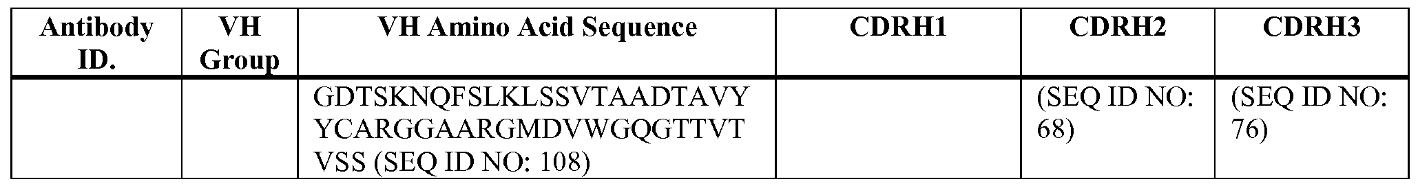


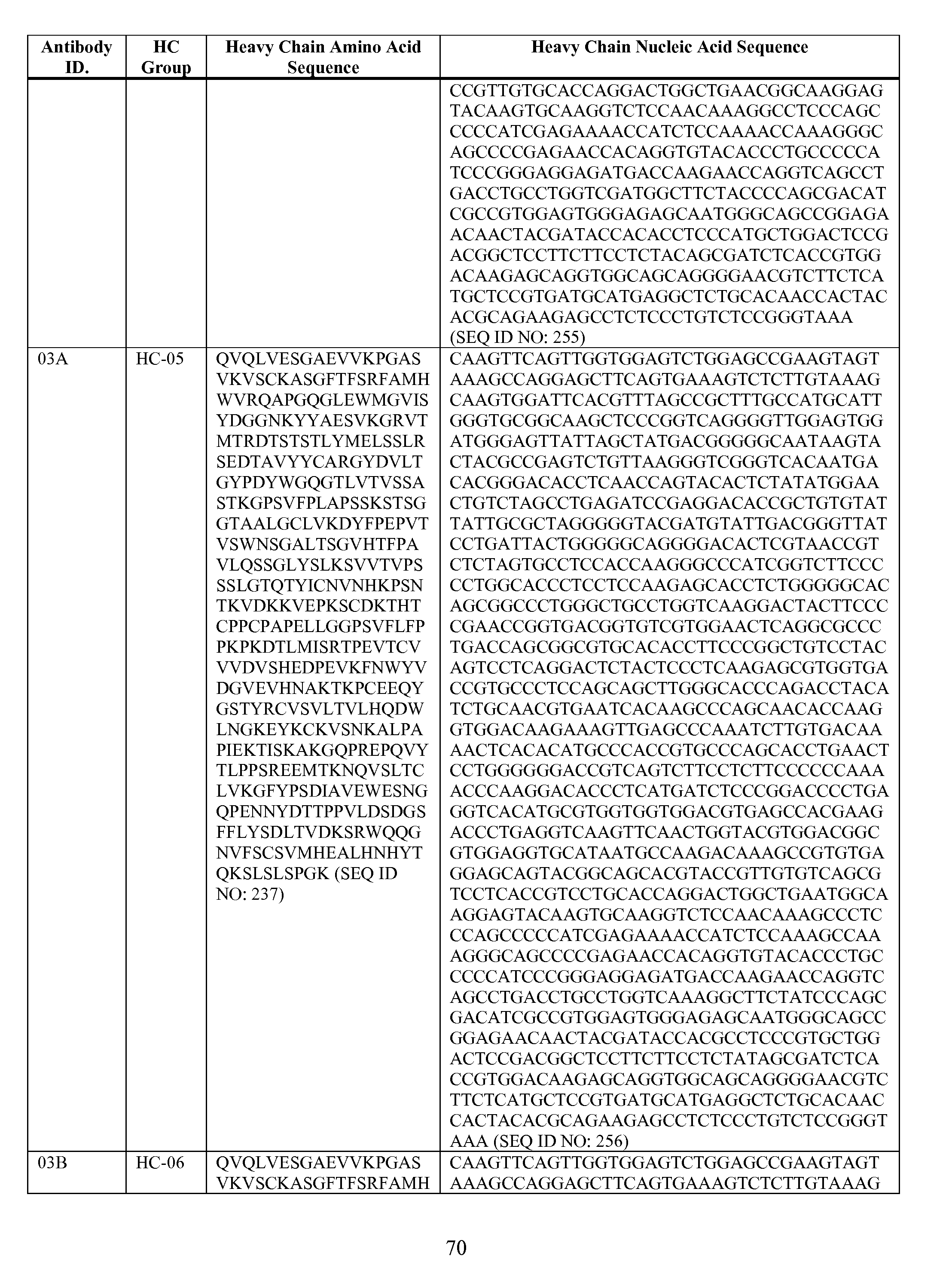


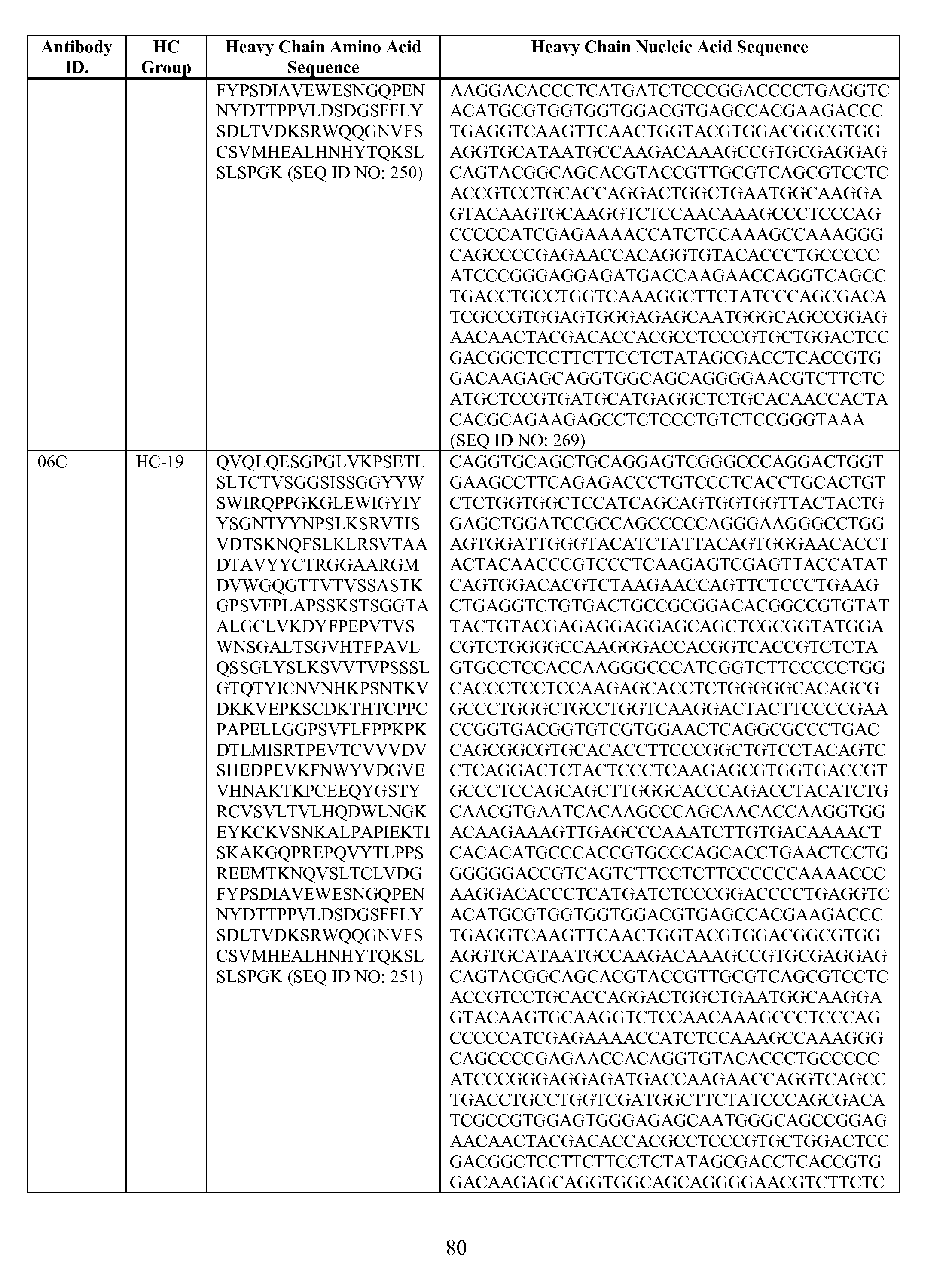


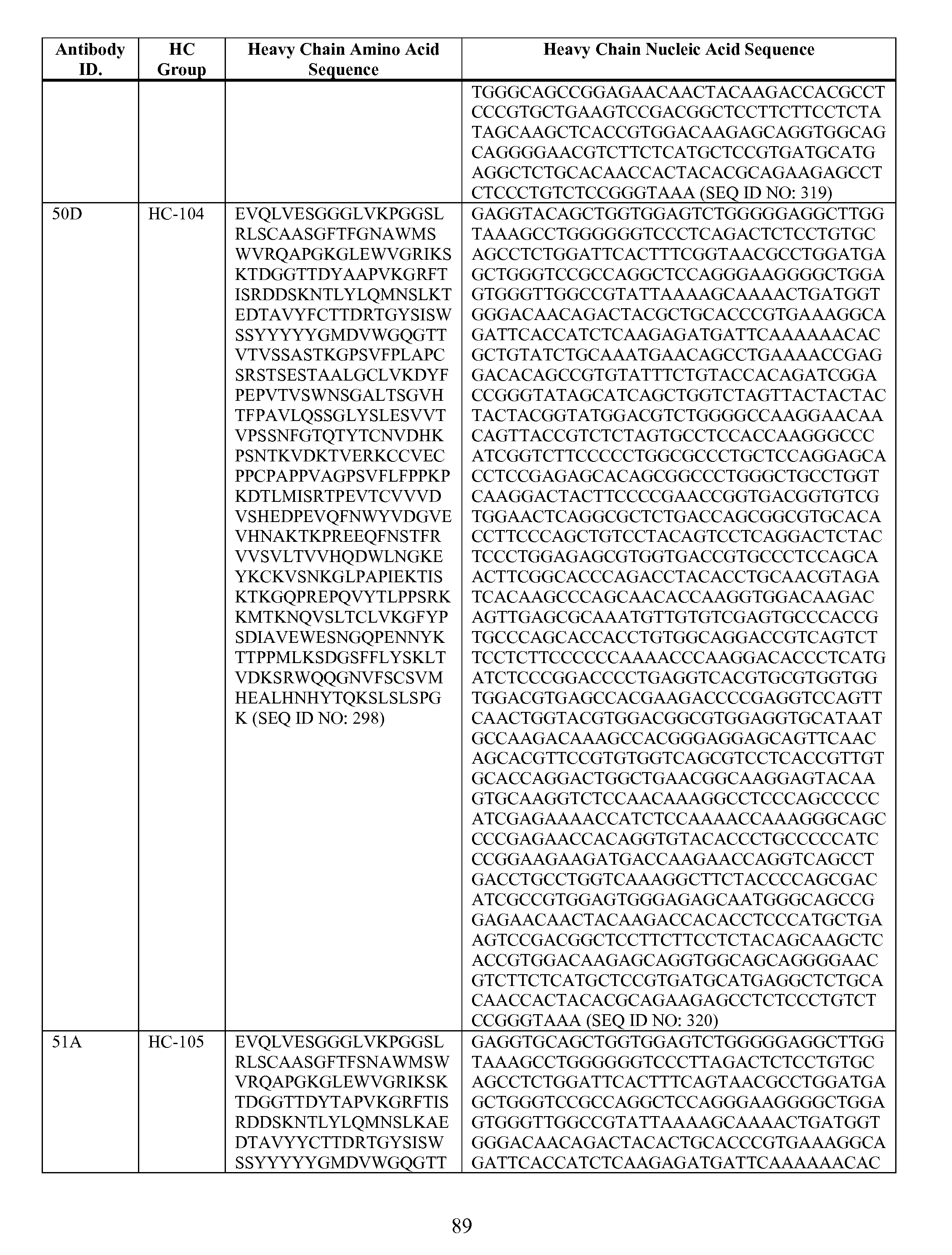


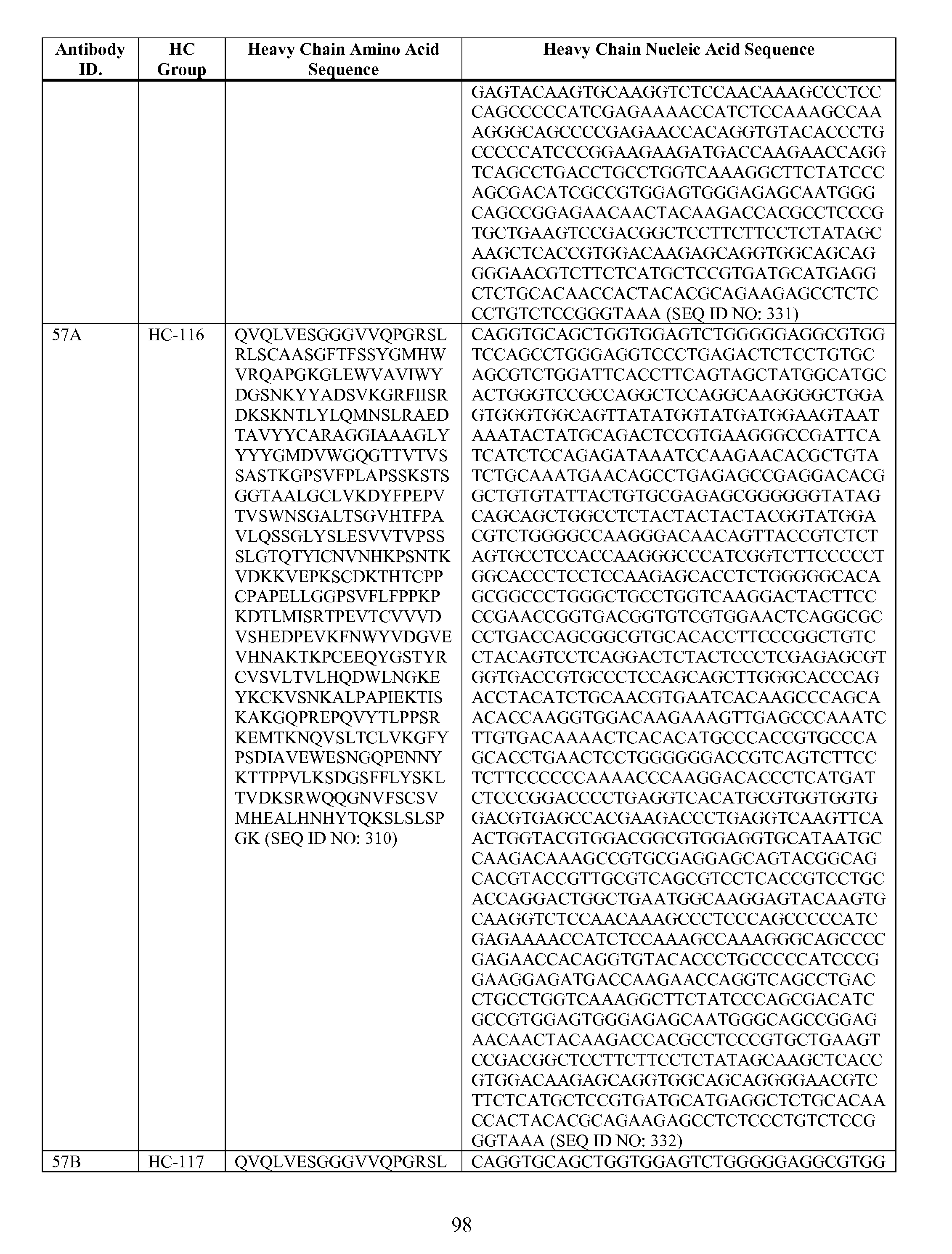


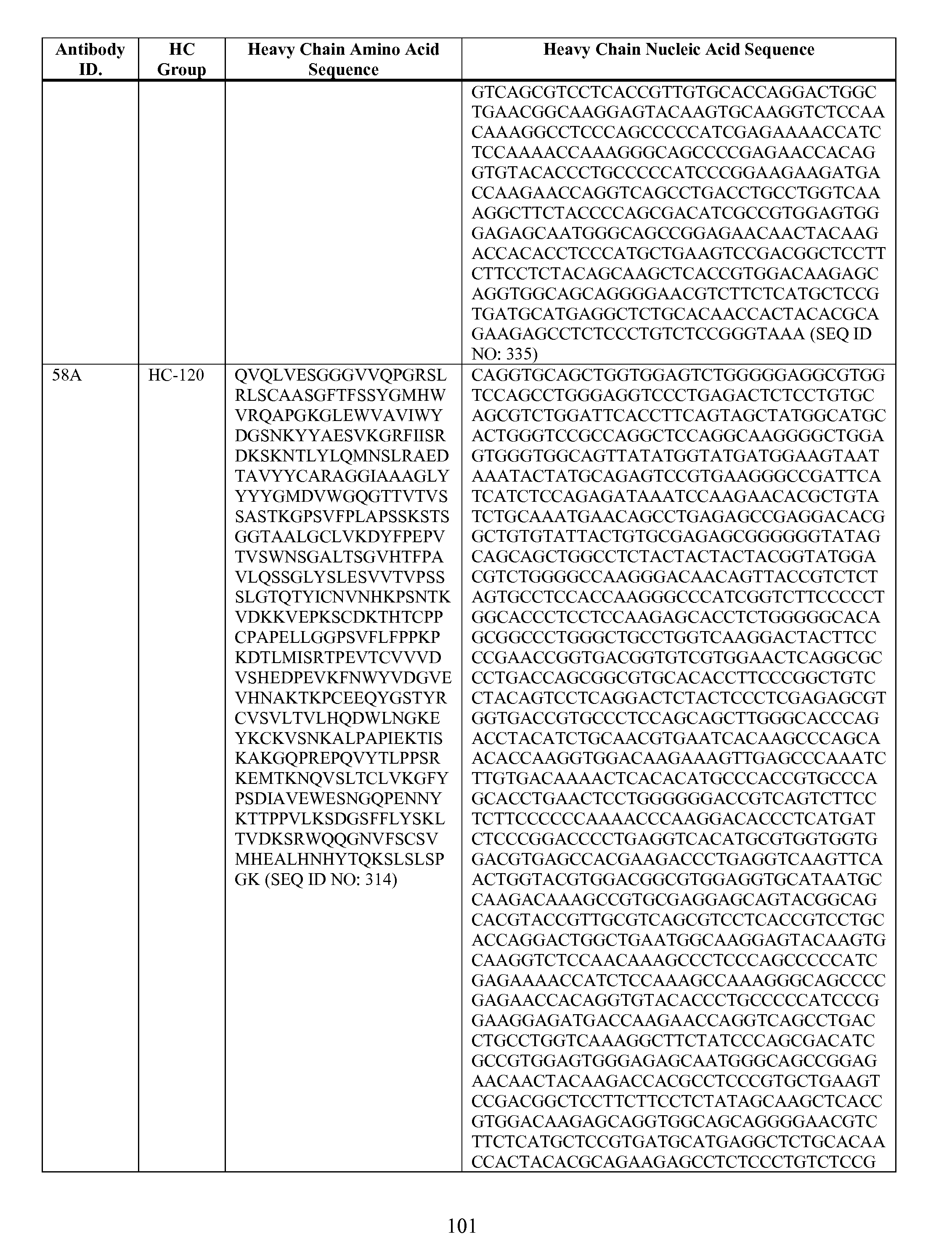


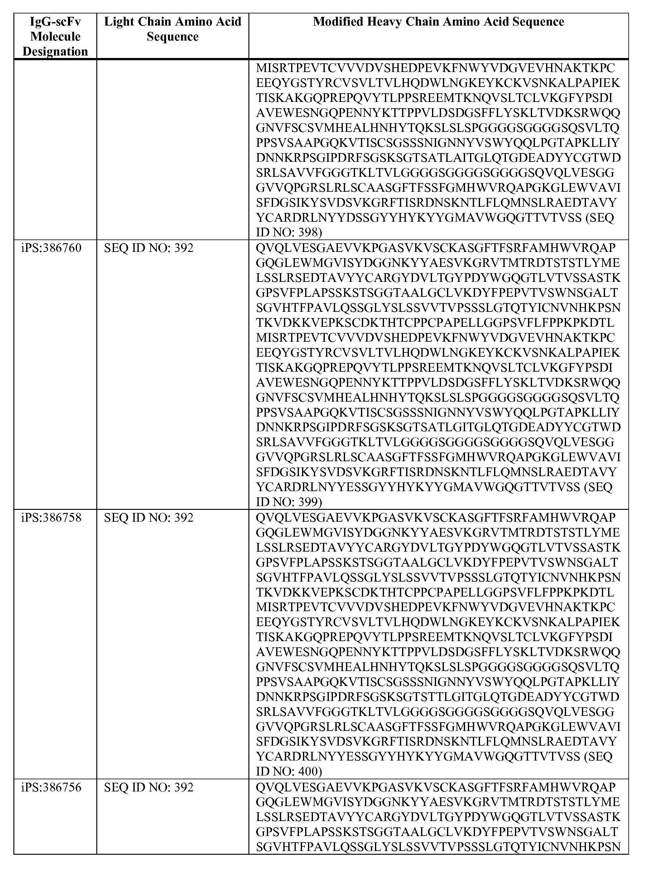


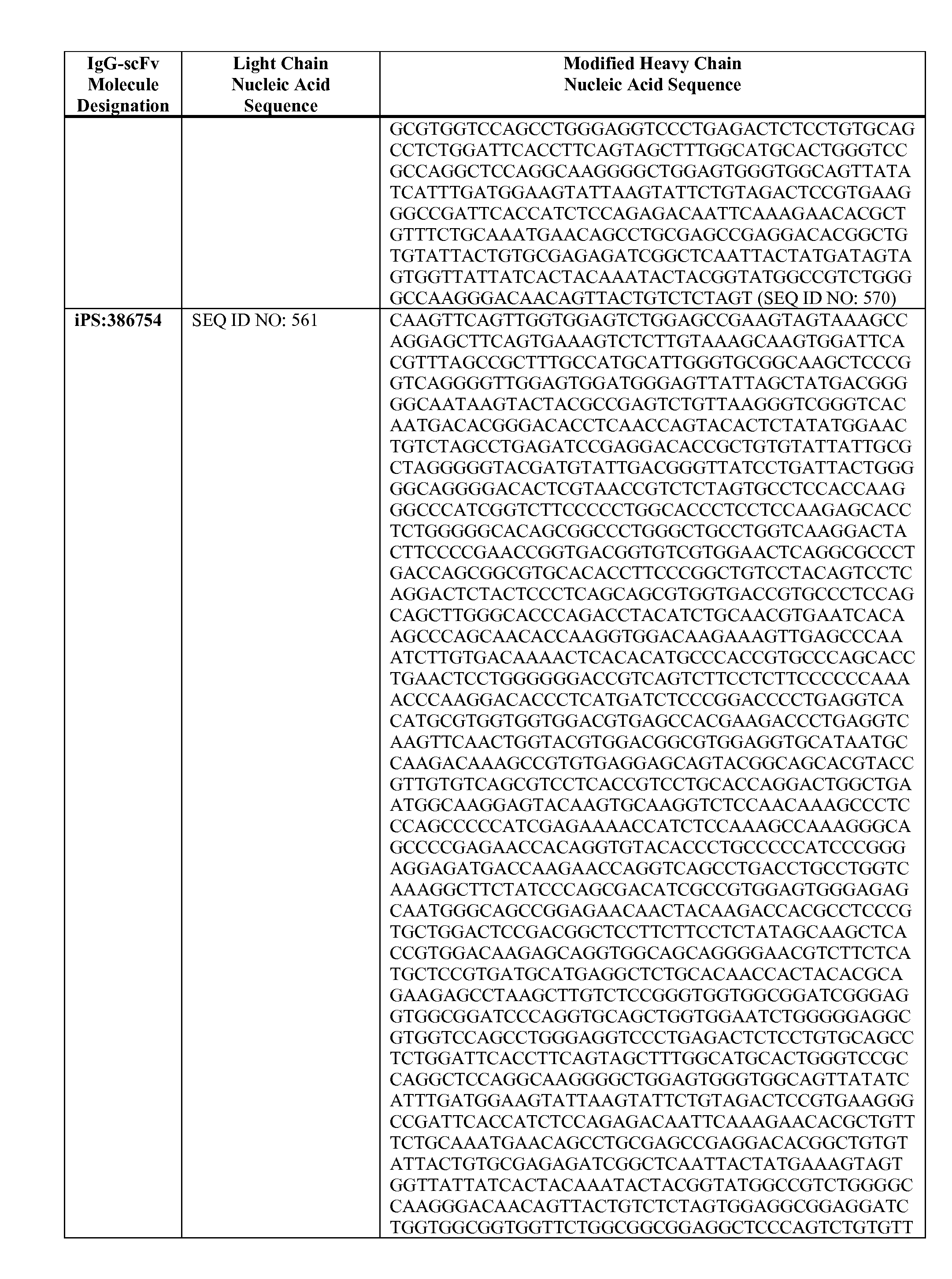


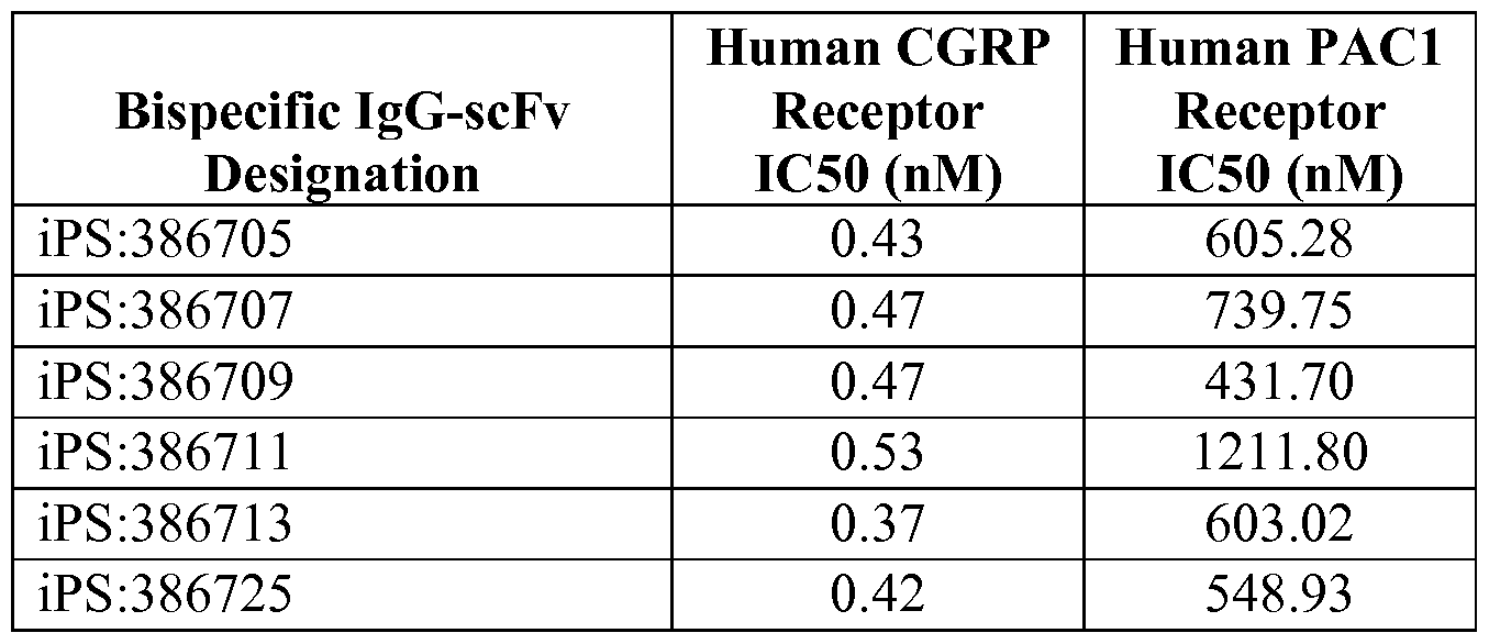


Claims
Priority Applications (15)
| Application Number | Priority Date | Filing Date | Title |
|---|---|---|---|
| JP2017514533A JP6956631B2 (en) | 2014-09-15 | 2015-09-15 | Bispecific anti-CGRP receptor / PAC1 receptor antigen-binding protein and its use |
| AU2015318008A AU2015318008B2 (en) | 2014-09-15 | 2015-09-15 | Bi-specific anti-CGRP receptor/PAC1 receptor antigen binding proteins and uses thereof |
| MX2017003247A MX2017003247A (en) | 2014-09-15 | 2015-09-15 | Bi-specific anti-cgrp receptor/pac1 receptor antigen binding proteins and uses thereof. |
| EP15778414.1A EP3194435A1 (en) | 2014-09-15 | 2015-09-15 | Bi-specific anti-cgrp receptor/pac1 receptor antigen binding proteins and uses thereof |
| US15/511,176 US10934362B2 (en) | 2014-09-15 | 2015-09-15 | Bi-specific anti-CGRP receptor/PAC1 receptor antigen binding proteins and uses thereof |
| CA2960756A CA2960756C (en) | 2014-09-15 | 2015-09-15 | Bi-specific anti-cgrp receptor/pac1 receptor antigen binding proteins and uses thereof |
| CA2998174A CA2998174A1 (en) | 2015-09-15 | 2016-09-15 | Tetravalent bispecific and tetraspecific antigen binding proteins and uses thereof |
| EP16771059.9A EP3350216A1 (en) | 2015-09-15 | 2016-09-15 | Tetravalent bispecific and tetraspecific antigen binding proteins and uses thereof |
| AU2016323440A AU2016323440B2 (en) | 2015-09-15 | 2016-09-15 | Tetravalent bispecific and tetraspecific antigen binding proteins and uses thereof |
| MA042821A MA42821A (en) | 2015-09-15 | 2016-09-15 | TETRAVALENT TETRASPECIFIC AND BISPECIFIC BISPECIFIC ANTIGEN BINDING PROTEINS AND USES THEREOF |
| PCT/US2016/052006 WO2017049004A1 (en) | 2015-09-15 | 2016-09-15 | Tetravalent bispecific and tetraspecific antigen binding proteins and uses thereof |
| US15/922,778 US20180237542A1 (en) | 2015-09-15 | 2016-09-15 | Tetravalent bispecific and tetraspecific antigen binding proteins and uses thereof |
| MX2018003183A MX2018003183A (en) | 2015-09-15 | 2016-09-15 | Tetravalent bispecific and tetraspecific antigen binding proteins and uses thereof. |
| JP2018532547A JP6932700B2 (en) | 2015-09-15 | 2016-09-15 | Tetravalent bispecific antigen-binding proteins and tetravalent quadrispecific antigen-binding proteins, and their use |
| US17/005,202 US11919964B2 (en) | 2014-09-15 | 2020-08-27 | Bi-specific anti-CGRP receptor/PAC1 receptor antigen binding proteins and uses thereof |
Applications Claiming Priority (2)
| Application Number | Priority Date | Filing Date | Title |
|---|---|---|---|
| US201462050737P | 2014-09-15 | 2014-09-15 | |
| US62/050,737 | 2014-09-15 |
Related Child Applications (3)
| Application Number | Title | Priority Date | Filing Date |
|---|---|---|---|
| US15/511,176 A-371-Of-International US10934362B2 (en) | 2014-09-15 | 2015-09-15 | Bi-specific anti-CGRP receptor/PAC1 receptor antigen binding proteins and uses thereof |
| US15/922,778 Continuation US20180237542A1 (en) | 2015-09-15 | 2016-09-15 | Tetravalent bispecific and tetraspecific antigen binding proteins and uses thereof |
| US17/005,202 Continuation US11919964B2 (en) | 2014-09-15 | 2020-08-27 | Bi-specific anti-CGRP receptor/PAC1 receptor antigen binding proteins and uses thereof |
Publications (1)
| Publication Number | Publication Date |
|---|---|
| WO2016044224A1 true WO2016044224A1 (en) | 2016-03-24 |
Family
ID=54291587
Family Applications (1)
| Application Number | Title | Priority Date | Filing Date |
|---|---|---|---|
| PCT/US2015/050115 WO2016044224A1 (en) | 2014-09-15 | 2015-09-15 | Bi-specific anti-cgrp receptor/pac1 receptor antigen binding proteins and uses thereof |
Country Status (10)
| Country | Link |
|---|---|
| US (2) | US10934362B2 (en) |
| EP (1) | EP3194435A1 (en) |
| JP (2) | JP6956631B2 (en) |
| AR (1) | AR101875A1 (en) |
| AU (1) | AU2015318008B2 (en) |
| CA (1) | CA2960756C (en) |
| MX (1) | MX2017003247A (en) |
| TW (1) | TW201620938A (en) |
| UY (1) | UY36302A (en) |
| WO (1) | WO2016044224A1 (en) |
Cited By (10)
| Publication number | Priority date | Publication date | Assignee | Title |
|---|---|---|---|---|
| WO2017016841A1 (en) | 2015-07-29 | 2017-02-02 | Unilever Plc | Hair composition |
| US9896502B2 (en) | 2014-03-21 | 2018-02-20 | Teva Pharmaceuticals International Gmbh | Antagonist antibodies directed against calcitonin gene-related peptide and methods using same |
| WO2018035084A1 (en) * | 2016-08-16 | 2018-02-22 | Epimab Biotherapeutics, Inc. | Monovalent asymmetric tandem fab bispecific antibodies |
| WO2019140216A1 (en) * | 2018-01-12 | 2019-07-18 | Amgen Inc. | Pac1 antibodies and uses thereof |
| US10392434B2 (en) | 2016-09-23 | 2019-08-27 | Teva Pharmaceuticals International Gmbh | Treating refractory migraine |
| WO2019195185A1 (en) * | 2018-04-02 | 2019-10-10 | Amgen Inc. | Erenumab compositions and uses thereof |
| US10556945B2 (en) | 2014-03-21 | 2020-02-11 | Teva Pharmaceuticals International Gmbh | Antagonist antibodies directed against calcitonin gene-related peptide and methods using same |
| WO2020264384A1 (en) * | 2019-06-28 | 2020-12-30 | Amgen Inc. | Anti-cgrp receptor/anti-pac1 receptor bispecific antigen binding proteins |
| US11421028B2 (en) | 2016-02-06 | 2022-08-23 | Epimab Biotherapeutics, Inc. | Fabs-in-tandem immunoglobulin and uses thereof |
| RU2780591C2 (en) * | 2016-08-16 | 2022-09-28 | Эпимаб Биотерапьютикс, Инк. | Monovalent asymmetric bispecific antibodies with tandem fab |
Families Citing this family (8)
| Publication number | Priority date | Publication date | Assignee | Title |
|---|---|---|---|---|
| JO3382B1 (en) * | 2008-12-23 | 2019-03-13 | Amgen Inc | Human cgrp receptor binding antibodies |
| UY35148A (en) * | 2012-11-21 | 2014-05-30 | Amgen Inc | HETERODIMERIC IMMUNOGLOBULINS |
| CA2963692A1 (en) * | 2014-10-09 | 2016-04-14 | Engmab Ag | Bispecific antibodies against cd3epsilon and ror1 |
| JOP20200116A1 (en) * | 2015-04-24 | 2017-06-16 | Amgen Inc | Methods for treating or preventing migraine headache |
| GB201820556D0 (en) * | 2018-12-17 | 2019-01-30 | Alligator Bioscience Ab | Novel polypeptides |
| MX2021014931A (en) | 2019-06-07 | 2022-01-24 | Amgen Inc | Bispecific binding constructs with selectively cleavable linkers. |
| CN113121696A (en) * | 2019-12-31 | 2021-07-16 | 周易 | Bispecific antibody formed by Fab modification induction and preparation method and application thereof |
| WO2023051786A1 (en) * | 2021-09-30 | 2023-04-06 | 江苏恒瑞医药股份有限公司 | Antigen-binding molecules that specifically bind cgrp and pacap and pharmaceutical use thereof |
Citations (63)
| Publication number | Priority date | Publication date | Assignee | Title |
|---|---|---|---|---|
| US3773919A (en) | 1969-10-23 | 1973-11-20 | Du Pont | Polylactide-drug mixtures |
| US4560655A (en) | 1982-12-16 | 1985-12-24 | Immunex Corporation | Serum-free cell culture medium and process for making same |
| WO1987000195A1 (en) | 1985-06-28 | 1987-01-15 | Celltech Limited | Animal cell culture |
| US4657866A (en) | 1982-12-21 | 1987-04-14 | Sudhir Kumar | Serum-free, synthetic, completely chemically defined tissue culture media |
| US4767704A (en) | 1983-10-07 | 1988-08-30 | Columbia University In The City Of New York | Protein-free culture medium |
| WO1990003430A1 (en) | 1988-09-23 | 1990-04-05 | Cetus Corporation | Cell culture medium for enhanced cell growth, culture longevity and product expression |
| WO1990004036A1 (en) | 1988-10-12 | 1990-04-19 | Medical Research Council | Production of antibodies from transgenic animals |
| EP0367566A1 (en) | 1988-10-31 | 1990-05-09 | Immunex Corporation | Interleukin-4 receptors |
| US4927762A (en) | 1986-04-01 | 1990-05-22 | Cell Enterprises, Inc. | Cell culture medium with antioxidant |
| US4965195A (en) | 1987-10-26 | 1990-10-23 | Immunex Corp. | Interleukin-7 |
| US4968607A (en) | 1987-11-25 | 1990-11-06 | Immunex Corporation | Interleukin-1 receptors |
| EP0404097A2 (en) | 1989-06-22 | 1990-12-27 | BEHRINGWERKE Aktiengesellschaft | Bispecific and oligospecific, mono- and oligovalent receptors, production and applications thereof |
| WO1991010741A1 (en) | 1990-01-12 | 1991-07-25 | Cell Genesys, Inc. | Generation of xenogeneic antibodies |
| WO1991017271A1 (en) | 1990-05-01 | 1991-11-14 | Affymax Technologies N.V. | Recombinant library screening methods |
| EP0460846A1 (en) | 1990-06-05 | 1991-12-11 | Immunex Corporation | Type II interleukin-1 receptors |
| WO1992001047A1 (en) | 1990-07-10 | 1992-01-23 | Cambridge Antibody Technology Limited | Methods for producing members of specific binding pairs |
| WO1992003918A1 (en) | 1990-08-29 | 1992-03-19 | Genpharm International, Inc. | Transgenic non-human animals capable of producing heterologous antibodies |
| US5122469A (en) | 1990-10-03 | 1992-06-16 | Genentech, Inc. | Method for culturing Chinese hamster ovary cells to improve production of recombinant proteins |
| WO1992022646A1 (en) | 1991-06-14 | 1992-12-23 | Dnx Corp. | Production of human hemoglobin in transgenic pigs |
| WO1993001227A1 (en) | 1991-07-08 | 1993-01-21 | University Of Massachusetts At Amherst | Thermotropic liquid crystal segmented block copolymer |
| WO1993011161A1 (en) | 1991-11-25 | 1993-06-10 | Enzon, Inc. | Multivalent antigen-binding proteins |
| WO1994002602A1 (en) | 1992-07-24 | 1994-02-03 | Cell Genesys, Inc. | Generation of xenogeneic antibodies |
| US5545806A (en) | 1990-08-29 | 1996-08-13 | Genpharm International, Inc. | Ransgenic non-human animals for producing heterologous antibodies |
| WO1996027011A1 (en) | 1995-03-01 | 1996-09-06 | Genentech, Inc. | A method for making heteromultimeric polypeptides |
| WO1996030498A1 (en) | 1995-03-29 | 1996-10-03 | Xenotech Incorporated | Production of antibodies using cre-mediated site-specific recombination |
| WO1996032478A1 (en) | 1995-04-14 | 1996-10-17 | Genentech, Inc. | Altered polypeptides with increased half-life |
| WO1996033735A1 (en) | 1995-04-27 | 1996-10-31 | Abgenix, Inc. | Human antibodies derived from immunized xenomice |
| US5585089A (en) | 1988-12-28 | 1996-12-17 | Protein Design Labs, Inc. | Humanized immunoglobulins |
| US5625126A (en) | 1990-08-29 | 1997-04-29 | Genpharm International, Inc. | Transgenic non-human animals for producing heterologous antibodies |
| US5633425A (en) | 1990-08-29 | 1997-05-27 | Genpharm International, Inc. | Transgenic non-human animals capable of producing heterologous antibodies |
| US5661016A (en) | 1990-08-29 | 1997-08-26 | Genpharm International Inc. | Transgenic non-human animals capable of producing heterologous antibodies of various isotypes |
| WO1997034631A1 (en) | 1996-03-18 | 1997-09-25 | Board Of Regents, The University Of Texas System | Immunoglobin-like domains with increased half lives |
| WO1998024893A2 (en) | 1996-12-03 | 1998-06-11 | Abgenix, Inc. | TRANSGENIC MAMMALS HAVING HUMAN IG LOCI INCLUDING PLURAL VH AND Vλ REGIONS AND ANTIBODIES PRODUCED THEREFROM |
| US5770429A (en) | 1990-08-29 | 1998-06-23 | Genpharm International, Inc. | Transgenic non-human animals capable of producing heterologous antibodies |
| US5789650A (en) | 1990-08-29 | 1998-08-04 | Genpharm International, Inc. | Transgenic non-human animals for producing heterologous antibodies |
| US5814318A (en) | 1990-08-29 | 1998-09-29 | Genpharm International Inc. | Transgenic non-human animals for producing heterologous antibodies |
| US5874299A (en) | 1990-08-29 | 1999-02-23 | Genpharm International, Inc. | Transgenic non-human animals capable of producing heterologous antibodies |
| US5877397A (en) | 1990-08-29 | 1999-03-02 | Genpharm International Inc. | Transgenic non-human animals capable of producing heterologous antibodies of various isotypes |
| US6017533A (en) | 1996-04-25 | 2000-01-25 | Shiseido Company, Ltd. | Peptides having specific affinity to pituitary adenylate cyclase activating polypeptide type 1 receptors |
| WO2000024782A2 (en) | 1998-10-23 | 2000-05-04 | Amgen Inc. | Modified peptides, comprising an fc domain, as therapeutic agents |
| US6162963A (en) | 1990-01-12 | 2000-12-19 | Abgenix, Inc. | Generation of Xenogenetic antibodies |
| US6171586B1 (en) | 1997-06-13 | 2001-01-09 | Genentech, Inc. | Antibody formulation |
| US6210924B1 (en) | 1998-08-11 | 2001-04-03 | Amgen Inc. | Overexpressing cyclin D 1 in a eukaryotic cell line |
| US6255458B1 (en) | 1990-08-29 | 2001-07-03 | Genpharm International | High affinity human antibodies and human antibodies against digoxin |
| US6300129B1 (en) | 1990-08-29 | 2001-10-09 | Genpharm International | Transgenic non-human animals for producing heterologous antibodies |
| US20030113316A1 (en) | 2001-07-25 | 2003-06-19 | Kaisheva Elizabet A. | Stable lyophilized pharmaceutical formulation of IgG antibodies |
| US20030133939A1 (en) | 2001-01-17 | 2003-07-17 | Genecraft, Inc. | Binding domain-immunoglobulin fusion proteins |
| US6713610B1 (en) | 1990-01-12 | 2004-03-30 | Raju Kucherlapati | Human antibodies derived from immunized xenomice |
| US20050037421A1 (en) | 2001-09-13 | 2005-02-17 | Institute For Antibodies Co., Ltd | Methods of constructing camel antibody libraries |
| US20050136049A1 (en) | 2001-01-17 | 2005-06-23 | Ledbetter Jeffrey A. | Binding constructs and methods for use thereof |
| WO2007054809A2 (en) * | 2005-11-14 | 2007-05-18 | Rinat Neuroscience Corp. | Antagonist antibodies directed against calcitonin gene-related peptide and methods using same |
| WO2007076336A1 (en) * | 2005-12-22 | 2007-07-05 | Eli Lilly And Company | Treatment of migraine with anti-cgrp antibodies |
| WO2008110348A1 (en) | 2007-03-12 | 2008-09-18 | Esbatech Ag | Sequence based engineering and optimization of single chain antibodies |
| US20080318207A1 (en) | 2007-06-21 | 2008-12-25 | Saint Louis University | Sequence covariance networks, methods and uses therefor |
| WO2009000099A2 (en) | 2007-06-25 | 2008-12-31 | Esbatech Ag | Methods of modifying antibodies, and modified antibodies with improved functional properties |
| US20090048122A1 (en) | 2006-03-17 | 2009-02-19 | Biogen Idec Ma Inc. | Stabilized polypeptide compositions |
| WO2009089004A1 (en) | 2008-01-07 | 2009-07-16 | Amgen Inc. | Method for making antibody fc-heterodimeric molecules using electrostatic steering effects |
| WO2010075238A1 (en) | 2008-12-23 | 2010-07-01 | Amgen Inc. | Human cgrp receptor binding proteins |
| WO2012125495A2 (en) | 2011-03-11 | 2012-09-20 | Amgen Inc. | Method of correlated mutational analysis to improve therapeutic antibodies |
| US20120294802A1 (en) * | 2011-05-20 | 2012-11-22 | Russo Andrew F | Use of anti-cgrp antibodies and antibody fragments to prevent or inhibit photophobia or light aversion in subjects in need thereof, especially migraine sufferers |
| WO2014081955A1 (en) | 2012-11-21 | 2014-05-30 | Amgen Inc. | Heterodimeric immunoglobulins |
| WO2014144632A2 (en) | 2013-03-15 | 2014-09-18 | Amgen Inc. | Human pac1 antibodies |
| US20140343252A1 (en) | 2013-03-14 | 2014-11-20 | Amgen Inc. | Interleukin-2 muteins for the expansion of t-regulatory cells |
Family Cites Families (49)
| Publication number | Priority date | Publication date | Assignee | Title |
|---|---|---|---|---|
| CA2077764C (en) | 1990-03-17 | 2002-12-10 | Nobuhiro Suzuki | Antibody to pacap and use thereof |
| ATE179210T1 (en) | 1992-03-17 | 1999-05-15 | Novartis Erfind Verwalt Gmbh | GENETICALLY PRODUCED ANTIBODIES |
| EP0618291A3 (en) | 1993-02-26 | 1997-12-03 | Takeda Chemical Industries, Ltd. | PACAP receptor protein, method for preparing said protein, and use thereof |
| US6399316B1 (en) | 1994-02-25 | 2002-06-04 | Takeda Chemical Industries, Ltd. | PACAP receptor protein, method for preparing said protein, and use thereof |
| WO1996005221A1 (en) | 1994-08-16 | 1996-02-22 | Human Genome Sciences, Inc. | Calcitonin receptor |
| US6462016B1 (en) | 1995-10-06 | 2002-10-08 | Shiseido Company, Ltd. | Method for screening agonist or antagonist of the maxadilan receptor participating in vasodilating action, and compound found by the method |
| WO1998003534A1 (en) | 1996-07-23 | 1998-01-29 | Smithkline Beecham Corporation | Calcitonin gene-related peptide receptor component factor (houdc44) |
| US6255455B1 (en) | 1996-10-11 | 2001-07-03 | The Trustees Of The University Of Pennsylvania | Rh(D)-binding proteins and magnetically activated cell sorting method for production thereof |
| HUP0104934A3 (en) | 1998-07-20 | 2002-05-28 | Sod Conseils Rech Applic | Peptide analogues of pacap, pharmaceutical compositions comprising thereof and their use |
| US6242563B1 (en) | 1998-07-20 | 2001-06-05 | Societe De Conseils De Recherches Et D'applications Scientifiques, Sas | Peptide analogues |
| US20020155533A1 (en) | 1998-11-30 | 2002-10-24 | Takeda Chemical | PACAP receptor protein, method for preparing said protein, and use thereof |
| US20020182729A1 (en) | 2001-01-11 | 2002-12-05 | Dicicco-Bloom Emanuel | Pituitary adenylate cyclase-activating polypeptide (PACAP) is an anti-mitogenic signal for selected neuronal precursors in vivo |
| FR2821080B1 (en) | 2001-02-20 | 2003-12-19 | Sanofi Synthelabo | SPECIFIC ANTIBODIES TO HUMAN ADRENOMEDULLINE, PHARMACEUTICAL COMPOSITION CONTAINING SAME, ITS THERAPEUTIC APPLICATIONS |
| EP1438325B1 (en) | 2001-09-27 | 2009-12-30 | Merck & Co., Inc. | Isolated dna molecules encoding humanized calcitonin gene-related peptide receptor, related non-human transgenic animals and assay methods |
| US7658924B2 (en) | 2001-10-11 | 2010-02-09 | Amgen Inc. | Angiopoietin-2 specific binding agents |
| AR039067A1 (en) | 2001-11-09 | 2005-02-09 | Pfizer Prod Inc | ANTIBODIES FOR CD40 |
| US7317091B2 (en) | 2002-03-01 | 2008-01-08 | Xencor, Inc. | Optimized Fc variants |
| US20040132101A1 (en) | 2002-09-27 | 2004-07-08 | Xencor | Optimized Fc variants and methods for their generation |
| US20040110170A1 (en) | 2002-05-18 | 2004-06-10 | The Regents Of The University Of California | Cloning and characterization of calcitonin gene related peptide receptors |
| ES2323170T3 (en) | 2002-08-12 | 2009-07-08 | Birkir Sveinsson | USE OF CGRP ANTAGONIST COMPOUNDS FOR THE TREATMENT OF PSORIASIS. |
| US20040176577A1 (en) | 2002-09-10 | 2004-09-09 | Eva Rojer | Immunoassays for specific determination of SCCA isoforms |
| EP1583550A2 (en) | 2003-01-16 | 2005-10-12 | D. Collen Research Foundation vzw | Inhibition of pacap signalling for the prevention and treatment of thrombocytopenia |
| WO2004097421A2 (en) | 2003-04-29 | 2004-11-11 | Bayer Healthcare Ag | Diagnostics and therapeutics for diseases associated with calcitonin receptor-like receptor (calcrl) |
| US20050129687A1 (en) | 2003-11-03 | 2005-06-16 | University Of Vermont And State Agricultural College | Use of inhibitors of PACAP receptor activity for treatment of overactive bladder and pelvic floor pain syndrome |
| EP1701979A2 (en) | 2003-12-03 | 2006-09-20 | Xencor, Inc. | Optimized antibodies that target the epidermal growth factor receptor |
| EP1718602A4 (en) | 2004-01-30 | 2007-12-12 | Peplin Biolipids Pty Ltd | Therapeutic and carrier molecules |
| CA2555877C (en) | 2004-02-11 | 2015-11-24 | Amylin Pharmaceuticals, Inc. | Hybrid polypeptides with selectable properties |
| US7423128B2 (en) | 2004-11-03 | 2008-09-09 | Amgen Fremont Inc. | Anti-properdin antibodies, and methods for making and using same |
| AU2005319382B2 (en) | 2004-12-21 | 2011-04-07 | Astrazeneca Ab | Antibodies directed to angiopoietin-2 and uses thereof |
| US10011858B2 (en) | 2005-03-31 | 2018-07-03 | Chugai Seiyaku Kabushiki Kaisha | Methods for producing polypeptides by regulating polypeptide association |
| JP4993606B2 (en) | 2005-06-16 | 2012-08-08 | 隆行 新藤 | Vascular structure stabilizer and angiogenic agent targeting RAMP2 |
| US8008453B2 (en) | 2005-08-12 | 2011-08-30 | Amgen Inc. | Modified Fc molecules |
| CA2620364A1 (en) | 2005-08-26 | 2007-03-01 | David C. Yeomans | Methods for treatment of headaches by administration of oxytocin |
| GB0521139D0 (en) | 2005-10-18 | 2005-11-23 | Univ Sheffield | Therapeutic agent |
| US8168592B2 (en) | 2005-10-21 | 2012-05-01 | Amgen Inc. | CGRP peptide antagonists and conjugates |
| KR20090039739A (en) | 2006-08-03 | 2009-04-22 | 아스트라제네카 아베 | ANTIBODIES DIRECTED TO alpha;Vbeta;6 AND USES THEREOF |
| WO2008018472A1 (en) * | 2006-08-08 | 2008-02-14 | Kyoto University | Novel monoclonal antibody and use of the same |
| GB0708002D0 (en) | 2007-04-25 | 2007-06-06 | Univ Sheffield | Antibodies |
| WO2009033489A2 (en) | 2007-09-11 | 2009-03-19 | Glostrup Hospital | Selective pacl inhibitors for use in the treatment of migraine and headaches |
| PT2265288T (en) | 2008-03-04 | 2016-07-14 | Labrys Biologics Inc | Methods of treating inflammatory pain |
| FR2934597B1 (en) | 2008-07-31 | 2013-04-19 | Univ Aix Marseille Ii | ANTIBODIES BINDING TO ADRENOMEDULLINE RECEPTORS AND USES THEREOF AS MEDICAMENT. |
| CN101434649B (en) | 2008-12-12 | 2012-04-25 | 余榕捷 | Cyclohepatapeptide, preparation thereof and use |
| US9676845B2 (en) * | 2009-06-16 | 2017-06-13 | Hoffmann-La Roche, Inc. | Bispecific antigen binding proteins |
| NZ597763A (en) | 2009-07-27 | 2014-07-25 | Nocicepta Llc | Methods for treatment of pain |
| CN107441487B (en) | 2009-08-28 | 2021-09-14 | 泰华制药国际有限公司 | Method for treating visceral pain by administering antagonist antibodies against calcitonin gene-related peptide |
| AU2010334974A1 (en) | 2009-12-22 | 2012-07-12 | Novartis Ag | Tetravalent CD47-antibody constant region fusion protein for use in therapy |
| CA2830254C (en) | 2011-03-16 | 2019-09-10 | Amgen Inc. | Fc variants |
| ES2721882T3 (en) | 2011-12-23 | 2019-08-06 | Pfizer | Constant regions of genetically engineered antibody for site-specific conjugation and procedures and uses thereof |
| BR112014028838A2 (en) | 2012-05-21 | 2020-05-12 | Genentech Inc | USES OF AN ANTIBODY, METHOD FOR MAKING AN ANTIBODY AND ANTIBODIES |
-
2015
- 2015-09-15 EP EP15778414.1A patent/EP3194435A1/en active Pending
- 2015-09-15 CA CA2960756A patent/CA2960756C/en active Active
- 2015-09-15 MX MX2017003247A patent/MX2017003247A/en unknown
- 2015-09-15 WO PCT/US2015/050115 patent/WO2016044224A1/en active Application Filing
- 2015-09-15 AU AU2015318008A patent/AU2015318008B2/en active Active
- 2015-09-15 US US15/511,176 patent/US10934362B2/en active Active
- 2015-09-15 AR ARP150102946A patent/AR101875A1/en unknown
- 2015-09-15 JP JP2017514533A patent/JP6956631B2/en active Active
- 2015-09-15 UY UY0001036302A patent/UY36302A/en not_active Application Discontinuation
- 2015-09-15 TW TW104130488A patent/TW201620938A/en unknown
-
2020
- 2020-01-29 JP JP2020012345A patent/JP2020074790A/en active Pending
- 2020-08-27 US US17/005,202 patent/US11919964B2/en active Active
Patent Citations (73)
| Publication number | Priority date | Publication date | Assignee | Title |
|---|---|---|---|---|
| US3773919A (en) | 1969-10-23 | 1973-11-20 | Du Pont | Polylactide-drug mixtures |
| US4560655A (en) | 1982-12-16 | 1985-12-24 | Immunex Corporation | Serum-free cell culture medium and process for making same |
| US4657866A (en) | 1982-12-21 | 1987-04-14 | Sudhir Kumar | Serum-free, synthetic, completely chemically defined tissue culture media |
| US4767704A (en) | 1983-10-07 | 1988-08-30 | Columbia University In The City Of New York | Protein-free culture medium |
| WO1987000195A1 (en) | 1985-06-28 | 1987-01-15 | Celltech Limited | Animal cell culture |
| US4927762A (en) | 1986-04-01 | 1990-05-22 | Cell Enterprises, Inc. | Cell culture medium with antioxidant |
| US4965195A (en) | 1987-10-26 | 1990-10-23 | Immunex Corp. | Interleukin-7 |
| US4968607A (en) | 1987-11-25 | 1990-11-06 | Immunex Corporation | Interleukin-1 receptors |
| WO1990003430A1 (en) | 1988-09-23 | 1990-04-05 | Cetus Corporation | Cell culture medium for enhanced cell growth, culture longevity and product expression |
| WO1990004036A1 (en) | 1988-10-12 | 1990-04-19 | Medical Research Council | Production of antibodies from transgenic animals |
| US5545807A (en) | 1988-10-12 | 1996-08-13 | The Babraham Institute | Production of antibodies from transgenic animals |
| EP0367566A1 (en) | 1988-10-31 | 1990-05-09 | Immunex Corporation | Interleukin-4 receptors |
| US5585089A (en) | 1988-12-28 | 1996-12-17 | Protein Design Labs, Inc. | Humanized immunoglobulins |
| US5693762A (en) | 1988-12-28 | 1997-12-02 | Protein Design Labs, Inc. | Humanized immunoglobulins |
| EP0404097A2 (en) | 1989-06-22 | 1990-12-27 | BEHRINGWERKE Aktiengesellschaft | Bispecific and oligospecific, mono- and oligovalent receptors, production and applications thereof |
| US6713610B1 (en) | 1990-01-12 | 2004-03-30 | Raju Kucherlapati | Human antibodies derived from immunized xenomice |
| US5939598A (en) | 1990-01-12 | 1999-08-17 | Abgenix, Inc. | Method of making transgenic mice lacking endogenous heavy chains |
| US6673986B1 (en) | 1990-01-12 | 2004-01-06 | Abgenix, Inc. | Generation of xenogeneic antibodies |
| US6162963A (en) | 1990-01-12 | 2000-12-19 | Abgenix, Inc. | Generation of Xenogenetic antibodies |
| WO1991010741A1 (en) | 1990-01-12 | 1991-07-25 | Cell Genesys, Inc. | Generation of xenogeneic antibodies |
| WO1991017271A1 (en) | 1990-05-01 | 1991-11-14 | Affymax Technologies N.V. | Recombinant library screening methods |
| EP0460846A1 (en) | 1990-06-05 | 1991-12-11 | Immunex Corporation | Type II interleukin-1 receptors |
| WO1992001047A1 (en) | 1990-07-10 | 1992-01-23 | Cambridge Antibody Technology Limited | Methods for producing members of specific binding pairs |
| US5545806A (en) | 1990-08-29 | 1996-08-13 | Genpharm International, Inc. | Ransgenic non-human animals for producing heterologous antibodies |
| US5661016A (en) | 1990-08-29 | 1997-08-26 | Genpharm International Inc. | Transgenic non-human animals capable of producing heterologous antibodies of various isotypes |
| US6255458B1 (en) | 1990-08-29 | 2001-07-03 | Genpharm International | High affinity human antibodies and human antibodies against digoxin |
| EP0546073A1 (en) | 1990-08-29 | 1993-06-16 | Genpharm Int | Transgenic non-human animals capable of producing heterologous antibodies. |
| US5770429A (en) | 1990-08-29 | 1998-06-23 | Genpharm International, Inc. | Transgenic non-human animals capable of producing heterologous antibodies |
| US5569825A (en) | 1990-08-29 | 1996-10-29 | Genpharm International | Transgenic non-human animals capable of producing heterologous antibodies of various isotypes |
| US5877397A (en) | 1990-08-29 | 1999-03-02 | Genpharm International Inc. | Transgenic non-human animals capable of producing heterologous antibodies of various isotypes |
| WO1992003918A1 (en) | 1990-08-29 | 1992-03-19 | Genpharm International, Inc. | Transgenic non-human animals capable of producing heterologous antibodies |
| US5625126A (en) | 1990-08-29 | 1997-04-29 | Genpharm International, Inc. | Transgenic non-human animals for producing heterologous antibodies |
| US5633425A (en) | 1990-08-29 | 1997-05-27 | Genpharm International, Inc. | Transgenic non-human animals capable of producing heterologous antibodies |
| US6300129B1 (en) | 1990-08-29 | 2001-10-09 | Genpharm International | Transgenic non-human animals for producing heterologous antibodies |
| EP0546073B1 (en) | 1990-08-29 | 1997-09-10 | GenPharm International, Inc. | production and use of transgenic non-human animals capable of producing heterologous antibodies |
| US5874299A (en) | 1990-08-29 | 1999-02-23 | Genpharm International, Inc. | Transgenic non-human animals capable of producing heterologous antibodies |
| US5789650A (en) | 1990-08-29 | 1998-08-04 | Genpharm International, Inc. | Transgenic non-human animals for producing heterologous antibodies |
| US5814318A (en) | 1990-08-29 | 1998-09-29 | Genpharm International Inc. | Transgenic non-human animals for producing heterologous antibodies |
| US5122469A (en) | 1990-10-03 | 1992-06-16 | Genentech, Inc. | Method for culturing Chinese hamster ovary cells to improve production of recombinant proteins |
| WO1992022646A1 (en) | 1991-06-14 | 1992-12-23 | Dnx Corp. | Production of human hemoglobin in transgenic pigs |
| WO1993001227A1 (en) | 1991-07-08 | 1993-01-21 | University Of Massachusetts At Amherst | Thermotropic liquid crystal segmented block copolymer |
| WO1993011161A1 (en) | 1991-11-25 | 1993-06-10 | Enzon, Inc. | Multivalent antigen-binding proteins |
| WO1994002602A1 (en) | 1992-07-24 | 1994-02-03 | Cell Genesys, Inc. | Generation of xenogeneic antibodies |
| WO1996027011A1 (en) | 1995-03-01 | 1996-09-06 | Genentech, Inc. | A method for making heteromultimeric polypeptides |
| WO1996030498A1 (en) | 1995-03-29 | 1996-10-03 | Xenotech Incorporated | Production of antibodies using cre-mediated site-specific recombination |
| WO1996032478A1 (en) | 1995-04-14 | 1996-10-17 | Genentech, Inc. | Altered polypeptides with increased half-life |
| WO1996033735A1 (en) | 1995-04-27 | 1996-10-31 | Abgenix, Inc. | Human antibodies derived from immunized xenomice |
| WO1997034631A1 (en) | 1996-03-18 | 1997-09-25 | Board Of Regents, The University Of Texas System | Immunoglobin-like domains with increased half lives |
| US6017533A (en) | 1996-04-25 | 2000-01-25 | Shiseido Company, Ltd. | Peptides having specific affinity to pituitary adenylate cyclase activating polypeptide type 1 receptors |
| US7064244B2 (en) | 1996-12-03 | 2006-06-20 | Abgenix, Inc. | Transgenic mammals having human Ig loci including plural VH and VK regions and antibodies produced therefrom |
| WO1998024893A2 (en) | 1996-12-03 | 1998-06-11 | Abgenix, Inc. | TRANSGENIC MAMMALS HAVING HUMAN IG LOCI INCLUDING PLURAL VH AND Vλ REGIONS AND ANTIBODIES PRODUCED THEREFROM |
| US6171586B1 (en) | 1997-06-13 | 2001-01-09 | Genentech, Inc. | Antibody formulation |
| US6210924B1 (en) | 1998-08-11 | 2001-04-03 | Amgen Inc. | Overexpressing cyclin D 1 in a eukaryotic cell line |
| WO2000024782A2 (en) | 1998-10-23 | 2000-05-04 | Amgen Inc. | Modified peptides, comprising an fc domain, as therapeutic agents |
| US20050136049A1 (en) | 2001-01-17 | 2005-06-23 | Ledbetter Jeffrey A. | Binding constructs and methods for use thereof |
| US20030133939A1 (en) | 2001-01-17 | 2003-07-17 | Genecraft, Inc. | Binding domain-immunoglobulin fusion proteins |
| US20030113316A1 (en) | 2001-07-25 | 2003-06-19 | Kaisheva Elizabet A. | Stable lyophilized pharmaceutical formulation of IgG antibodies |
| US20050037421A1 (en) | 2001-09-13 | 2005-02-17 | Institute For Antibodies Co., Ltd | Methods of constructing camel antibody libraries |
| WO2007054809A2 (en) * | 2005-11-14 | 2007-05-18 | Rinat Neuroscience Corp. | Antagonist antibodies directed against calcitonin gene-related peptide and methods using same |
| WO2007076336A1 (en) * | 2005-12-22 | 2007-07-05 | Eli Lilly And Company | Treatment of migraine with anti-cgrp antibodies |
| US20090048122A1 (en) | 2006-03-17 | 2009-02-19 | Biogen Idec Ma Inc. | Stabilized polypeptide compositions |
| WO2008110348A1 (en) | 2007-03-12 | 2008-09-18 | Esbatech Ag | Sequence based engineering and optimization of single chain antibodies |
| US20080318207A1 (en) | 2007-06-21 | 2008-12-25 | Saint Louis University | Sequence covariance networks, methods and uses therefor |
| WO2009000099A2 (en) | 2007-06-25 | 2008-12-31 | Esbatech Ag | Methods of modifying antibodies, and modified antibodies with improved functional properties |
| WO2009089004A1 (en) | 2008-01-07 | 2009-07-16 | Amgen Inc. | Method for making antibody fc-heterodimeric molecules using electrostatic steering effects |
| US8592562B2 (en) | 2008-01-07 | 2013-11-26 | Amgen Inc. | Method for making antibody Fc-heterodimeric molecules using electrostatic steering effects |
| WO2010075238A1 (en) | 2008-12-23 | 2010-07-01 | Amgen Inc. | Human cgrp receptor binding proteins |
| US20100172895A1 (en) | 2008-12-23 | 2010-07-08 | Boone Thomas C | Human CGRP Receptor Binding Proteins |
| WO2012125495A2 (en) | 2011-03-11 | 2012-09-20 | Amgen Inc. | Method of correlated mutational analysis to improve therapeutic antibodies |
| US20120294802A1 (en) * | 2011-05-20 | 2012-11-22 | Russo Andrew F | Use of anti-cgrp antibodies and antibody fragments to prevent or inhibit photophobia or light aversion in subjects in need thereof, especially migraine sufferers |
| WO2014081955A1 (en) | 2012-11-21 | 2014-05-30 | Amgen Inc. | Heterodimeric immunoglobulins |
| US20140343252A1 (en) | 2013-03-14 | 2014-11-20 | Amgen Inc. | Interleukin-2 muteins for the expansion of t-regulatory cells |
| WO2014144632A2 (en) | 2013-03-15 | 2014-09-18 | Amgen Inc. | Human pac1 antibodies |
Non-Patent Citations (164)
| Title |
|---|
| A.R. GENRMO,: "REMINGTON'S PHARMACEUTICAL SCIENCES, 18th Edition,", 1990, MACK PUBLISHING COMPANY |
| ADAMES ET AL., NATURE, vol. 318, 1985, pages 533 - 538 |
| ADELMAN ET AL., PAIN MED., vol. 9, 2008, pages 175 - 185 |
| ALEXANDER ET AL., MOL. CELL. BIOL., vol. 7, 1987, pages 1436 - 1444 |
| ASHINA ET AL., PAIN, vol. 86, 2000, pages 133 - 138 |
| AUSUBEL ET AL.,: "Current Protocols in Molecular Biology", 1995, JOHN WILEY & SONS, INC., article "sections 2.10 and 6.3-6.4" |
| BARNES ET AL., ANAL. BIOCHEM., vol. 102, 1980, pages 255 |
| BELLAMY ET AL., HEADACHE, vol. 46, 2006, pages 24 - 33 |
| BENOIST; CHAMBON, NATURE, vol. 290, 1981, pages 304 - 310 |
| BIEDLER JL ET AL., CANCER RES., vol. 38, 1978, pages 3751 - 3757 |
| BIGAL ET AL., HEADACHE, vol. 53, no. 8, 2013, pages 1230 - 44 |
| BIGAL ET AL., NEUROLOGY, vol. 72, 2009, pages 1864 - 1871 |
| BIRD ET AL., SCIENCE, vol. 242, pages 423 - 426 |
| BOURGAULT ET AL., J. MED. CHEM., vol. 52, 2009, pages 3308 - 3316 |
| BRANDES ET AL., JAMA, vol. 291, 2004, pages 965 - 973 |
| BRUGGERMANN ET AL., YEAR IN IMMUNOL., vol. 7, 1993, pages 33 |
| BUSE ET AL., NEUROL. NEUROSURG. PSYCHIATRY, vol. 81, 2010, pages 428 - 432 |
| CAMPBELL; SCANES, GROWTH REGUL., vol. 2, 1992, pages 175 - 191 |
| CARILLO ET AL., SIAM J. APPLIED MATH, vol. 48, 1988, pages 1073 |
| CATON; KOPROWSKI, PROC. NATL. ACAD. SCI. USA, vol. 87, 1990, pages 6450 - 6454 |
| CHEN ET AL., INTERNATIONAL IMMUNOLOGY, vol. 5, 1993, pages 647 - 656 |
| CHEN, DRUG DEVELOPMENT AND INDUSTRIAL PHARMACY, vol. 18, 1992, pages 1311 - 1354 |
| CHEUNG ET AL., VIROLOGY, vol. 176, 1990, pages 546 - 552 |
| CHIBA ET AL., AM. J. PHYSIOL., vol. 256, 1989, pages E331 - E335 |
| CHOTHIA ET AL., NATURE, vol. 342, 1989, pages 878 - 883 |
| CHOTHIA; LESK, J. MOL. BIOL., vol. 196, 1987, pages 901 - 917 |
| CHOULIER ET AL., PROTEINS, vol. 41, 2000, pages 475 - 484 |
| CONNOR ET AL., NEUROLOGY, vol. 73, 2009, pages 970 - 977 |
| CORTEZ-RETAMOZO ET AL., CANCER RESEARCH, vol. 64, 2004, pages 2853 - 57 |
| DAYHOFF ET AL.: "Atlas of Protein Sequence and Structure", vol. 5, 1978 |
| DAYHOFF ET AL.: "Atlas of Protein Sequence and Structure", vol. 5, 1978, pages: 345 - 352 |
| DEBOER ET AL., PROC. NATL. ACAD. SCI. U.S.A., vol. 80, 1983, pages 21 - 25 |
| DEMAREST ET AL., J. MOL. BIOL., vol. 335, 2004, pages 41 - 48 |
| DESMYTER ET AL., J. BIOL. CHEM., vol. 276, 2001, pages 26285 - 90 |
| DEVEREUX ET AL., NUCL. ACID RES, vol. 12, pages 387 |
| DIENER ET AL., CEPHALALGIA, vol. 27, 2007, pages 814 - 823 |
| DODICK ET AL., LANCET NEUROL., vol. 13, 2014, pages 1100 - 1107 |
| DODICK ET AL., LANCET NEUROL., vol. 13, 2014, pages 885 - 892 |
| DURHAM ET AL., CURR OPIN INVESTIG DRUGS, vol. 5, 2004, pages 731 - 735 |
| EWERT ET AL., BIOCHEMISTRY, vol. 41, 2002, pages 3628 - 36 |
| FERRARI ET AL., LANCET, vol. 358, 2001, pages 1668 - 1675 |
| FISHWILD ET AL., NATURE BIOTECHNOLOGY, vol. 14, 1996, pages 845 - 851 |
| FREDERICKS ET AL., PROTEIN ENGINEERING, DESIGN & SELECTION, vol. 17, 2004, pages 95 - 106 |
| GALLAI ET AL., CEPHALALGIA, vol. 15, 1995, pages 384 - 390 |
| GINGER S JOHNSON ET AL: "Migraine Prophylaxis: The Race is On g p y Vice President Vice President Defined Health", 13 June 2014 (2014-06-13), XP055239718, Retrieved from the Internet <URL:http://knowledgebase.definedhealth.net/wp-content/uploads/2014/06/migraine/Migraine webinar 061314.pdf> [retrieved on 20160108] * |
| GOADSBY ET AL., ANN NEUROL., vol. 23, 1988, pages 193 - 196 |
| GOADSBY ET AL., ANN NEUROL., vol. 28, 1990, pages 183 - 187 |
| GRAHAM ET AL., J. GEN VIROL., vol. 36, 1977, pages 59 |
| GRIBSKOV, M. AND DEVEREUX, J.,: "Sequence Analysis Primer", 1991 |
| GRIFFIN, A. M., AND GRIFFIN, H. G.,: "Computer Analysis of Sequence Data", 1994 |
| GROSSCHEDL ET AL., CELL, vol. 38, 1984, pages 647 - 658 |
| GUNASEKARAN ET AL., J. BIOL. CHEM., vol. 285, 2010, pages 19637 - 19646 |
| GUSS ET AL, EMBO J., vol. 5, 1986 |
| HAM ET AL., METH. ENZ, vol. 58, 1979, pages 44 |
| HAMMER ET AL., SCIENCE, vol. 253, 1987, pages 53 - 58 |
| HANAHAN, NATURE, vol. 315, 1985, pages 115 - 122 |
| HARDING; LONBERG, ANN. N.Y ACAD. SCI., vol. 764, 1995, pages 536 - 546 |
| HARLOW; LANE: "Antibodies, A Laboratory Manual", 1988, COLD SPRING HARBOR PRESS |
| HARLOW; LANE: "Antibodies: A Laboratory Manual", 1988, COLD SPRING HARBOR |
| HENIKOFF ET AL., PROC. NATL. ACAD. SCI. U.S.A., vol. 89, 1992, pages 10915 - 10919 |
| HEWITT ET AL., ABSTRACT FOR THE 14TH CONGRESS OF THE INTERNATIONAL HEADACHE SOCIETY, 2009 |
| HO ET AL., NEUROLOGY, vol. 70, 2008, pages 1304 - 1312 |
| HO ET AL., NEUROLOGY, vol. 83, 2014, pages 958 - 966 |
| HOLLINGER ET AL., PROC. NATL. ACAD. SCI. USA, vol. 90, 1993, pages 6444 - 6448 |
| HOUSTON; VANHOUTTE, DRUGS, vol. 31, 1986, pages 149 - 163 |
| HUGO ET AL., PROTEIN ENGINEERING, vol. 16, no. 5, 2003, pages 381 - 86 |
| HUMPHREY ET AL., ANN NY ACAD SCI., vol. 600, 1990, pages 587 - 598 |
| HUSTON ET AL., PROC. NATL. ACAD. SCI. USA, vol. 85, 1988, pages 5879 - 5883 |
| JAKOBOVITS ET AL., NATURE, vol. 362, 1993, pages 255 - 258 |
| JAKOBOVITS ET AL., PROC. NATL. ACAD. SCI. USA, vol. 90, 1993, pages 2551 - 2555 |
| JALKANEN ET AL., J. CELL BIOL., vol. 105, 1987, pages 3087 - 3096 |
| JALKANEN ET AL., J. CELL. BIOL., vol. 101, 1985, pages 976 - 985 |
| JES OLESEN ET AL: "Emerging migraine treatments and drug targets", TRENDS IN PHARMACOLOGICAL SCIENCES, vol. 32, no. 6, 1 June 2011 (2011-06-01), pages 352 - 359, XP028222805, ISSN: 0165-6147, [retrieved on 20110302], DOI: 10.1016/J.TIPS.2011.02.016 * |
| JESPERS, L. S. ET AL., BIO/TECHNOLOGY, vol. 12, 1994, pages 899 - 903 |
| JOHN E. COLIGAN,: "Current Protocols In Immunology", 1993, JOHN WILEY & SONS |
| JONES ET AL., NATURE, vol. 321, 1986, pages 522 - 525 |
| JUHASZ ET AL., CEPHALALGIA, vol. 25, 2005, pages 179 - 183 |
| KABAT: "Sequences of Proteins of Immunological Interest", 1987, NIH |
| KELSEY ET AL., GENES AND DEVEL., vol. 1, 1987, pages 161 - 171 |
| KIM, H. Y. ET AL., J. INV. DERM. SYMP. PROC., vol. 12, 2007, pages 48 - 49 |
| KIRKLAND ET AL., J. IMMUNOL., vol. 137, 1986, pages 3614 - 3619 |
| KOLLIAS ET AL., CELL, vol. 46, 1986, pages 89 - 94 |
| KRUMLAUF ET AL., MOL. CELL. BIOL., vol. 5, 1985, pages 1639 - 1648 |
| LANCET, vol. 372, 2008, pages 2115 - 2123 |
| LASSEN ET AL., CEPHALALGIA, vol. 22, 2002, pages 54 - 61 |
| LEDER ET AL., CELL, vol. 45, 1986, pages 485 - 495 |
| LESK, A. M.,: "Computational Molecular Biology", 1988, OXFORD UNIVERSITY PRESS |
| LINDMARK ET AL., J. IMMUNOL. METH., vol. 62, 1983, pages 1 - 13 |
| LIPTON ET AL., NEUROLOGY, vol. 68, 2007, pages 343 - 349 |
| LONBERG ET AL., NATURE, vol. 368, 1994, pages 856 - 859 |
| LONBERG: "Handbook of Exp. Pharmacology", vol. 113, 1994, pages: 49 - 101 |
| LONBERG; HUSZAR, INTERN. REV. IMMUNOL., vol. 13, 1995, pages 65 - 93 |
| MACDONALD, HEPATOLOGY, vol. 7, 1987, pages 425 - 515 |
| MASON ET AL., SCIENCE, vol. 234, 1986, pages 1372 - 1378 |
| MATHER ET AL.: "Annals N.Y Acad. Sci.", vol. 383, 1982, pages: 44 - 68 |
| MATHER, BIOL. REPROD., vol. 23, 1980, pages 243 - 251 |
| MCLATCHIE ET AL., NATURE, vol. 393, 1998, pages 333 - 339 |
| MENDEZ ET AL., NATURE GENETICS, vol. 15, 1997, pages 146 - 156 |
| MERCHANT ET AL., NAT, BIOTECHNOL., vol. 16, 1998, pages 677 - 681 |
| MODI; LOWDER, AM. FAM. PHYSICIAN, vol. 73, 2006, pages 72 - 78 |
| MOGRAM ET AL., NATURE, vol. 315, 1985, pages 338 - 340 |
| MOLDENHAUER ET AL., SCAND. J. IMMUNOL., vol. 32, 1990, pages 77 - 82 |
| MONAGHAN ET AL., J NEUROCHEM., vol. 104, no. 1, 2008, pages 74 - 88 |
| MOREL ET AL., MOLEC. IMMUNOL., vol. 25, 1988, pages 7 - 15 |
| NEEDLEMAN ET AL., J. MOL. BIOL., vol. 48, 1970, pages 443 - 453 |
| NELLES ET AL., HEADACHE, vol. 49, 2009, pages 1454 - 1465 |
| NESBITT ET AL., BMJ, vol. 344, 2012, pages E2407 |
| OLAFSEN ET AL., PROTEIN ENG DES SEL., vol. 17, 2004, pages 315 - 23 |
| OLIVER ET AL., J CEREB BLOOD FLOW METAB, vol. 22, 2002, pages 620 - 629 |
| ORNITZ ET AL., COLD SPRING HARBOR SYMP. QUANT. BIOL., vol. 50, 1986, pages 399 - 409 |
| ORTEZ-RETAMOZO ET AL., CANCER RESEARCH, vol. 64, 2004, pages 2853 - 57 |
| OSOL, A.: "Remington's Pharmaceutical Sciences 16th edition,", 1980 |
| PANTALONI ET AL., J. BIOL. CHEM., vol. 271, 1996, pages 22146 - 22151 |
| PETERSEN ET AL., BR J PHARMACOL., vol. 143, 2004, pages 1074 - 1075 |
| PINKERT ET AL., GENES AND DEVEL, vol. 1, 1987, pages 268 - 276 |
| PISEGNA; WANK, PROC. NATL. ACAD. SCI. USA, vol. 90, 1993, pages 6345 - 49 |
| POWERS ET AL., JOURNAL OF IMMUNOLOGICAL METHODS, vol. 251, 2001, pages 123 - 135 |
| POYNER, PHARMACOL. THER., vol. 56, 1992, pages 23 - 51 |
| PRINSTER ET AL., NATURE, vol. 296, 1982, pages 39 - 42 |
| RAHMANN ET AL., CEPHALALGIA, vol. 28, 2008, pages 226 - 236 |
| RATHANASWAMI ET AL., ANALYTICAL BIOCHEMISTRY, vol. 373, 2008, pages 52 - 60 |
| READHEAD ET AL., CELL, vol. 48, 1987, pages 703 - 712 |
| RIDGWAY ET AL., PROTEIN ENG., vol. 9, 1996, pages 617 - 621 |
| RIECHMANN ET AL., NATURE, vol. 332, 1988, pages 323 - 27 |
| ROTHMAN ET AL., MOL IMMUNOL., vol. 26, no. 12, 1989, pages 1 113 - 23 |
| S D SILBERSTEIN: "Emerging Target-Based Paradigms to Prevent and Treat Migraine", CLINICAL PHARMACOLOGY AND THERAPEUTICS, vol. 93, no. 1, 5 December 2012 (2012-12-05), US, pages 78 - 85, XP055227794, ISSN: 0009-9236, DOI: 10.1038/clpt.2012.198 * |
| SAMBROOK ET AL.: "Molecular Cloning: A Laboratory Manual 2nd ed.,", 1989, COLD SPRING HARBOR LABORATORY |
| SAMBROOK; FRITSCH; MANIATIS: "Molecular Cloning: A Laboratory Manual", 1989, COLD SPRING HARBOR LABORATORY PRESS, article "chapters 9 and 11;" |
| SANI, NATURE, vol. 314, 1985, pages 283 - 286 |
| SCHOENEN J ET AL., CEPHALALGIA, vol. 33, no. 10, 2013, pages 816 - 30 |
| SCHYTZ ET AL., BRAIN, vol. 132, 2009, pages 16 - 25 |
| SCHYTZ H W ET AL: "The PACAP Receptor: A Novel Target for Migraine Treatment", NEUROTHERAPEUTICS, ELSEVIER INC, US, vol. 7, no. 2, 1 April 2010 (2010-04-01), pages 191 - 196, XP027030857, ISSN: 1933-7213, [retrieved on 20100427] * |
| SHIELDS ET AL., J BIOL CHEM., vol. 277, no. 30, 2002, pages 26733 - 40 |
| SHINKAWA ET AL., J BIOL CHEM., vol. 278, no. 5, 2003, pages 3466 - 73 |
| SILBERSTEIN ET AL., ARCH NEUROL., vol. 61, 2004, pages 490 - 495 |
| SILBERSTEIN ET AL., HEADACHE, vol. 47, 2007, pages 170 - 180 |
| SMITH, D. W.,: "Biocomputing Informatics and Genome Projects", 1993, ACADEMIC PRESS |
| SPENGLER ET AL., IN VITRO, vol. 8, 1973, pages 410 |
| STAHLI ET AL., METHODS IN ENZYMOLOGY, vol. 9, 1983, pages 242 - 253 |
| STORER ET AL., BR J PHARMACOL., vol. 142, 2004, pages 1171 - 1181 |
| SWIFT ET AL., CELL, vol. 38, 1984, pages 639 - 646 |
| TAYLOR ET AL., INTERNATIONAL IMMUNOLOGY, vol. 6, 1994, pages 579 - 591 |
| TAYLOR ET AL., J. PHARMACOL. EXP. THER., vol. 319, 2006, pages 749 - 757 |
| TAYLOR ET AL., NUCLEIC ACIDS RESEARCH, vol. 20, 1992, pages 6287 - 6295 |
| THORNSEN ET AL., PROC. NATL. ACAD. U.S.A., vol. 81, 1984, pages 659 - 663 |
| TIJSSEN: "Practice and Theory of Enzyme Immunoassays", vol. 15, 1993, ELSEVIER |
| TUAILLON ET AL., J. IMMUNOL., vol. 152, 1994, pages 2912 - 2920 |
| UMANA ET AL., NAT BIOTECHNOL., vol. 17, no. 2, 1999, pages 176 - 80 |
| URLAUB ET AL., PROC. NATL. ACAD. SCI. USA, vol. 77, 1980, pages 4216 |
| VAUDRY ET AL., PHARMACOL REV., vol. 61, 2009, pages 283 - 357 |
| VERHOEYEN ET AL., SCIENCE, vol. 239, 1988, pages 1534 - 1536 |
| VILLA-KAMAROFF ET AL., PROC. NATL. ACAD. SCI. U.S.A., vol. 75, 1978, pages 3727 - 3731 |
| VON HEINJE, G.: "Sequence Analysis in Molecular Biology", 1987, ACADEMIC PRESS |
| WAGNER ET AL., PROC. NATL. ACAD. SCI. U.S.A., vol. 78, 1981, pages 1444 - 1445 |
| WANG ET AL., PROC NATL ACAD SCI USA., vol. 92, 1995, pages 11480 - 11484 |
| WARD ET AL., NATURE, vol. 341, 1989, pages 544 - 546 |
| WICK ET AL., SURGERY, vol. 139, 2006, pages 197 - 201 |
| WILLIAMS; POLLI, JOURNAL OF PARENTERAL SCIENCE AND TECHNOLOGY, vol. 38, no. 2, 1984, pages 48 - 59 |
| YAMAMOTO ET AL., CELL, vol. 22, 1980, pages 787 - 797 |
| YAMANE-OHNUKI ET AL., BIOTECHNOL BIOENG., vol. 87, no. 5, 2004, pages 614 - 22 |
| ZAPATA ET AL., PROTEIN ENG., vol. 8, 1995, pages 1057 - 1062 |
| ZHANG ET AL., J. NEUROSCI., vol. 27, 2007, pages 2693 - 2703 |
| ZIMMERMANN ET AL., BRAIN RES., vol. 724, 1996, pages 238 - 245 |
| ZOLA: "Monoclonal Antibodies: A Manual of Techniques", 1987, CRC PRESS, INC, pages: 147 - 158 |
Cited By (20)
| Publication number | Priority date | Publication date | Assignee | Title |
|---|---|---|---|---|
| US9896502B2 (en) | 2014-03-21 | 2018-02-20 | Teva Pharmaceuticals International Gmbh | Antagonist antibodies directed against calcitonin gene-related peptide and methods using same |
| US11555064B2 (en) | 2014-03-21 | 2023-01-17 | Teva Pharmaceuticals International Gmbh | Treating headache comprising administering an antibody to calcitonin gene-related peptide |
| US10519224B2 (en) | 2014-03-21 | 2019-12-31 | Teva Pharmaceuticals International Gmbh | Treating headache comprising administering an antibody to calcitonin gene-related peptide |
| US10556945B2 (en) | 2014-03-21 | 2020-02-11 | Teva Pharmaceuticals International Gmbh | Antagonist antibodies directed against calcitonin gene-related peptide and methods using same |
| WO2017016841A1 (en) | 2015-07-29 | 2017-02-02 | Unilever Plc | Hair composition |
| US11421028B2 (en) | 2016-02-06 | 2022-08-23 | Epimab Biotherapeutics, Inc. | Fabs-in-tandem immunoglobulin and uses thereof |
| WO2018035084A1 (en) * | 2016-08-16 | 2018-02-22 | Epimab Biotherapeutics, Inc. | Monovalent asymmetric tandem fab bispecific antibodies |
| CN109803682A (en) * | 2016-08-16 | 2019-05-24 | 岸迈生物科技有限公司 | Monovalent asymmetric series connection Fab bispecific antibody |
| RU2780591C2 (en) * | 2016-08-16 | 2022-09-28 | Эпимаб Биотерапьютикс, Инк. | Monovalent asymmetric bispecific antibodies with tandem fab |
| US11028161B2 (en) | 2016-09-23 | 2021-06-08 | Teva Pharmaceuticals International Gmbh | Treating refractory migraine |
| US10392434B2 (en) | 2016-09-23 | 2019-08-27 | Teva Pharmaceuticals International Gmbh | Treating refractory migraine |
| US11028160B2 (en) | 2016-09-23 | 2021-06-08 | Teva Pharmaceuticals International Gmbh | Treating refractory migraine |
| US10738110B2 (en) | 2018-01-12 | 2020-08-11 | Amgen Inc. | PAC1 antibodies and uses thereof |
| US11891435B2 (en) | 2018-01-12 | 2024-02-06 | Amgen Inc. | Polynucleotides encoding monoclonal antibodies binding to human pituitary adenylate cyclase-activating polypeptide type I receptor (PAC1) and methods of making thereof |
| US11248043B2 (en) | 2018-01-12 | 2022-02-15 | Amgen Inc. | Methods for treating a headache condition using anti-human PAC1 antibodies or antigen-binding fragments thereof |
| WO2019140216A1 (en) * | 2018-01-12 | 2019-07-18 | Amgen Inc. | Pac1 antibodies and uses thereof |
| CN111954678A (en) * | 2018-04-02 | 2020-11-17 | 美国安进公司 | Errenitumumab composition and application thereof |
| US11407838B2 (en) | 2018-04-02 | 2022-08-09 | Amgen Inc. | Erenumab compositions and uses thereof |
| WO2019195185A1 (en) * | 2018-04-02 | 2019-10-10 | Amgen Inc. | Erenumab compositions and uses thereof |
| WO2020264384A1 (en) * | 2019-06-28 | 2020-12-30 | Amgen Inc. | Anti-cgrp receptor/anti-pac1 receptor bispecific antigen binding proteins |
Also Published As
| Publication number | Publication date |
|---|---|
| JP2020074790A (en) | 2020-05-21 |
| AU2015318008A1 (en) | 2017-03-23 |
| TW201620938A (en) | 2016-06-16 |
| US20170306033A1 (en) | 2017-10-26 |
| CA2960756A1 (en) | 2016-03-24 |
| US20210047422A1 (en) | 2021-02-18 |
| JP2017533694A (en) | 2017-11-16 |
| AU2015318008B2 (en) | 2021-05-20 |
| CA2960756C (en) | 2023-08-01 |
| US10934362B2 (en) | 2021-03-02 |
| US11919964B2 (en) | 2024-03-05 |
| EP3194435A1 (en) | 2017-07-26 |
| JP6956631B2 (en) | 2021-11-02 |
| MX2017003247A (en) | 2017-11-30 |
| UY36302A (en) | 2016-04-29 |
| AR101875A1 (en) | 2017-01-18 |
Similar Documents
| Publication | Publication Date | Title |
|---|---|---|
| US11919964B2 (en) | Bi-specific anti-CGRP receptor/PAC1 receptor antigen binding proteins and uses thereof | |
| US11780914B2 (en) | PACAP antibodies and uses thereof | |
| AU2016323440B2 (en) | Tetravalent bispecific and tetraspecific antigen binding proteins and uses thereof | |
| JP2023116808A (en) | Pac1 antibodies and uses thereof | |
| US20220363770A1 (en) | Anti-cgrp receptor/anti-pac1 receptor bispecific antigen binding proteins | |
| EP3538555A1 (en) | Bispecific or biparatopic antigen binding proteins and uses thereof | |
| TWI831762B (en) | Pac1 antibodies and uses thereof |
Legal Events
| Date | Code | Title | Description |
|---|---|---|---|
| 121 | Ep: the epo has been informed by wipo that ep was designated in this application |
Ref document number: 15778414 Country of ref document: EP Kind code of ref document: A1 |
|
| ENP | Entry into the national phase |
Ref document number: 2960756 Country of ref document: CA |
|
| WWE | Wipo information: entry into national phase |
Ref document number: MX/A/2017/003247 Country of ref document: MX |
|
| ENP | Entry into the national phase |
Ref document number: 2017514533 Country of ref document: JP Kind code of ref document: A |
|
| WWE | Wipo information: entry into national phase |
Ref document number: 15511176 Country of ref document: US |
|
| NENP | Non-entry into the national phase |
Ref country code: DE |
|
| ENP | Entry into the national phase |
Ref document number: 2015318008 Country of ref document: AU Date of ref document: 20150915 Kind code of ref document: A |
|
| REEP | Request for entry into the european phase |
Ref document number: 2015778414 Country of ref document: EP |
|
| WWE | Wipo information: entry into national phase |
Ref document number: 2015778414 Country of ref document: EP |