WO2014027626A1 - MONOCLONAL IgE ANTIBODY BONDING TO SWEAT ALLERGY ANTIGEN PROTEIN - Google Patents
MONOCLONAL IgE ANTIBODY BONDING TO SWEAT ALLERGY ANTIGEN PROTEIN Download PDFInfo
- Publication number
- WO2014027626A1 WO2014027626A1 PCT/JP2013/071715 JP2013071715W WO2014027626A1 WO 2014027626 A1 WO2014027626 A1 WO 2014027626A1 JP 2013071715 W JP2013071715 W JP 2013071715W WO 2014027626 A1 WO2014027626 A1 WO 2014027626A1
- Authority
- WO
- WIPO (PCT)
- Prior art keywords
- ige
- antibody
- human
- sweat
- sweat allergy
- Prior art date
Links
Images


Classifications
-
- G—PHYSICS
- G01—MEASURING; TESTING
- G01N—INVESTIGATING OR ANALYSING MATERIALS BY DETERMINING THEIR CHEMICAL OR PHYSICAL PROPERTIES
- G01N33/00—Investigating or analysing materials by specific methods not covered by groups G01N1/00 - G01N31/00
- G01N33/48—Biological material, e.g. blood, urine; Haemocytometers
- G01N33/50—Chemical analysis of biological material, e.g. blood, urine; Testing involving biospecific ligand binding methods; Immunological testing
- G01N33/68—Chemical analysis of biological material, e.g. blood, urine; Testing involving biospecific ligand binding methods; Immunological testing involving proteins, peptides or amino acids
- G01N33/6893—Chemical analysis of biological material, e.g. blood, urine; Testing involving biospecific ligand binding methods; Immunological testing involving proteins, peptides or amino acids related to diseases not provided for elsewhere
-
- A—HUMAN NECESSITIES
- A61—MEDICAL OR VETERINARY SCIENCE; HYGIENE
- A61P—SPECIFIC THERAPEUTIC ACTIVITY OF CHEMICAL COMPOUNDS OR MEDICINAL PREPARATIONS
- A61P11/00—Drugs for disorders of the respiratory system
- A61P11/02—Nasal agents, e.g. decongestants
-
- A—HUMAN NECESSITIES
- A61—MEDICAL OR VETERINARY SCIENCE; HYGIENE
- A61P—SPECIFIC THERAPEUTIC ACTIVITY OF CHEMICAL COMPOUNDS OR MEDICINAL PREPARATIONS
- A61P17/00—Drugs for dermatological disorders
- A61P17/04—Antipruritics
-
- A—HUMAN NECESSITIES
- A61—MEDICAL OR VETERINARY SCIENCE; HYGIENE
- A61P—SPECIFIC THERAPEUTIC ACTIVITY OF CHEMICAL COMPOUNDS OR MEDICINAL PREPARATIONS
- A61P37/00—Drugs for immunological or allergic disorders
- A61P37/08—Antiallergic agents
-
- C—CHEMISTRY; METALLURGY
- C07—ORGANIC CHEMISTRY
- C07K—PEPTIDES
- C07K16/00—Immunoglobulins [IGs], e.g. monoclonal or polyclonal antibodies
- C07K16/14—Immunoglobulins [IGs], e.g. monoclonal or polyclonal antibodies against material from fungi, algea or lichens
-
- C—CHEMISTRY; METALLURGY
- C07—ORGANIC CHEMISTRY
- C07K—PEPTIDES
- C07K16/00—Immunoglobulins [IGs], e.g. monoclonal or polyclonal antibodies
- C07K16/18—Immunoglobulins [IGs], e.g. monoclonal or polyclonal antibodies against material from animals or humans
-
- C—CHEMISTRY; METALLURGY
- C07—ORGANIC CHEMISTRY
- C07K—PEPTIDES
- C07K16/00—Immunoglobulins [IGs], e.g. monoclonal or polyclonal antibodies
- C07K16/40—Immunoglobulins [IGs], e.g. monoclonal or polyclonal antibodies against enzymes
-
- G—PHYSICS
- G01—MEASURING; TESTING
- G01N—INVESTIGATING OR ANALYSING MATERIALS BY DETERMINING THEIR CHEMICAL OR PHYSICAL PROPERTIES
- G01N33/00—Investigating or analysing materials by specific methods not covered by groups G01N1/00 - G01N31/00
- G01N33/48—Biological material, e.g. blood, urine; Haemocytometers
- G01N33/50—Chemical analysis of biological material, e.g. blood, urine; Testing involving biospecific ligand binding methods; Immunological testing
- G01N33/68—Chemical analysis of biological material, e.g. blood, urine; Testing involving biospecific ligand binding methods; Immunological testing involving proteins, peptides or amino acids
- G01N33/6854—Immunoglobulins
-
- C—CHEMISTRY; METALLURGY
- C07—ORGANIC CHEMISTRY
- C07K—PEPTIDES
- C07K2317/00—Immunoglobulins specific features
- C07K2317/20—Immunoglobulins specific features characterized by taxonomic origin
- C07K2317/21—Immunoglobulins specific features characterized by taxonomic origin from primates, e.g. man
-
- C—CHEMISTRY; METALLURGY
- C07—ORGANIC CHEMISTRY
- C07K—PEPTIDES
- C07K2317/00—Immunoglobulins specific features
- C07K2317/70—Immunoglobulins specific features characterized by effect upon binding to a cell or to an antigen
- C07K2317/76—Antagonist effect on antigen, e.g. neutralization or inhibition of binding
-
- G—PHYSICS
- G01—MEASURING; TESTING
- G01N—INVESTIGATING OR ANALYSING MATERIALS BY DETERMINING THEIR CHEMICAL OR PHYSICAL PROPERTIES
- G01N2333/00—Assays involving biological materials from specific organisms or of a specific nature
- G01N2333/37—Assays involving biological materials from specific organisms or of a specific nature from fungi
-
- G—PHYSICS
- G01—MEASURING; TESTING
- G01N—INVESTIGATING OR ANALYSING MATERIALS BY DETERMINING THEIR CHEMICAL OR PHYSICAL PROPERTIES
- G01N2800/00—Detection or diagnosis of diseases
- G01N2800/20—Dermatological disorders
- G01N2800/202—Dermatitis
-
- G—PHYSICS
- G01—MEASURING; TESTING
- G01N—INVESTIGATING OR ANALYSING MATERIALS BY DETERMINING THEIR CHEMICAL OR PHYSICAL PROPERTIES
- G01N2800/00—Detection or diagnosis of diseases
- G01N2800/24—Immunology or allergic disorders
Definitions
- the present invention relates to a human IgE antibody that binds to a sweat allergy antigen, a screening and production method thereof, use for detection or measurement of a sweat allergy antigen contained in human sweat by a human IgE antibody, and a human IgE antibody contained in human sweat
- the present invention relates to the use of antibodies that bind to sweat allergens that are detected or measured, and the use of human IgE antibodies to neutralize or remove sweat allergens.
- Allergic reactions are called immediate or type I hypersensitivity reactions and are mediated by IgE antibodies.
- IgE antibody-producing B cells When an organism is first exposed to an allergen, IgE antibody-producing B cells begin to produce soluble IgE molecules. Soluble IgE molecules then bind to high affinity IgE receptors present on the surface of mast cells or basophils. When the living body encounters the same allergen again, the high affinity IgE receptor is cross-linked by the IgE antibody and the allergen, resulting in an allergic reaction in which histamine and cytokines are released from the cells.
- Anti-IgE antibodies that neutralize IgE antibodies have been put to practical use as antibodies for suppressing allergic reactions. However, human IgE antibodies, when bound to high-affinity IgE receptors, alone or further allergens are added. Since it has the property of activating mast cells and basophils by binding, it is usually considered not useful for neutralizing allergens.
- human antibodies can be produced by various methods (for example, Epstein-Barr virus (EBV) immortalization method, phage display method, fully human antibody-producing hybridoma method).
- EBV Epstein-Barr virus
- phage display method Fully human antibody-producing hybridoma method
- a human IgE monoclonal antibody was produced using, for example, a phage display method (Patent Document 1, Non-Patent Document 1).
- the present inventors purified human sweat antigen from human sweat using histamine release activity from peripheral blood basophils of patients with atopic dermatitis as an index, and mass-linked MGL_1304, a protein derived from Malassezia globosa, Identified as an antigen. Then, a human IgE antibody that specifically binds to human sweat antigen was identified.
- the present invention relates to a human IgE antibody that binds to a sweat allergy antigen protein and binds to a human high affinity IgE receptor, but does not cause degranulation when reacted with a sweat allergy antigen protein.
- the antibody fragment, the screening method and the production method thereof are provided.
- the present invention relates to a human IgE antibody that binds to a sweat allergy antigen protein and binds to a human high affinity IgE receptor, but does not cause degranulation when reacted with a sweat allergy antigen protein.
- a composition for treating sweat allergy or a disease associated with sweat allergy antigen, a composition for removing or neutralizing sweat allergy antigen, or a material for removing sweat allergy antigen comprising the antibody fragment is provided.
- the present invention relates to a human IgE antibody that binds to a sweat allergy antigen protein and binds to a human high affinity IgE receptor, but does not cause degranulation when reacted with a sweat allergy antigen protein.
- the present invention in one aspect, is a composition or kit for detecting an antibody that binds to a human sweat allergen antigen protein or a measure of the amount of an antibody that binds to a human sweat allergen antigen protein, comprising a standard substance.
- the composition or kit is a human IgE antibody that binds to a sweat allergy antigen protein and binds to a human high affinity IgE receptor, but does not cause degranulation when reacted with a sweat allergy antigen protein. I will provide a.
- A MGL_1304 gene or mite antigen gene fused with Myc-tag was expressed in COS7 cells, the culture supernatant was electrophoresed, and Western blotting was performed using an anti-Myc antibody. The figure shows the results of the Western blotting.
- B It is a figure which shows the correlation of partially purified sweat antigen (QR) and the histamine release ability of MGL_1304 recombinant protein (rMGL_1304). It is a figure which shows that atopic dermatitis patient serum can be pretreated with QRX and MGL_1304, respectively, and the binding of antibodies to each can be neutralized.
- QR partially purified sweat antigen
- rMGL_1304 histamine release ability of MGL_1304 recombinant protein
- Atopic dermatitis patient serum is reacted with a rat mast cell line expressing human high affinity IgE receptor to sensitize cells (giving sensitivity to antigen). It is a figure which shows that the reactivity (histamine release ability) with respect to QRX of the sensitized cell lose
- FIG. 3 is a view showing that ABS IgE antibody specifically binds to immobilized QRX and does not specifically bind to immobilized mite and quinu antigens.
- FIG. 4 is a diagram obtained by changing the concentration of ABS-IgE in a QRX detection system by sandwich ELISA using ABS IgE antibody and Smith2 antibody. It is a figure which shows the detection of the partial peptide of QRX, recombinant MGL_1304 protein, and recombinant MGL_1304 protein by Western blotting using ABS IgE antibody.
- M Molecular weight marker
- QRX Partially purified sweat antigen
- rMGL Full-length protein
- Lys Malassezia globosa cells were sonicated and extracted with PBS (-), passed through a 0.22 ⁇ m filter, and the residue was removed.
- FIG. 3 is a view showing that ABS IgE antibody suppresses histamine release from basophils derived from a sweat allergic patient caused by unpurified human sweat.
- untreated represents the histamine release activity when ABS IgE antibody is not added to basophils from sweat allergic patients, and ABS-IgE favors ABS IgE antibody (1 ⁇ g / ml) from sweat allergic patients. It represents histamine releasing activity when added to basophils.
- the left column shows the histamine release rate (net%) when stimulated with anti-human IgE antibody (anti-IgE), the middle column shows histamine release when stimulated with 6F7, an IgE competitive anti-IgE receptor monoclonal antibody, The right column shows the histamine release rate when stimulated with QRX.
- IgE specifically binds to the MGL_1304 recombinant protein (rMGL) in the sera of patients with allergic rhinitis (allergic rhinitis 1-5), patients with atopic dermatitis (AD1-8), and healthy individuals (Normal1-3) The quantification result of an antibody is shown.
- rMGL MGL_1304 recombinant protein
- rMGL was coated on an ELISA plate, and the amount of IgE antibody in the serum specifically bound to rMGL was measured.
- Each serum was diluted 10-fold (x10) or 20-fold (x20) and used for the test.
- the amount of bound IgE was measured by absorbance at 450 nm using an HRP-anti-human IgE antibody. Further, the absorbance was expressed in terms of pg / ml using a standard curve with ABS IgE antibody shown in FIG. 9 as a standard substance.
- bonded specifically with the partially purified sweat antigen protein (QRX) in the serum of an atopic dermatitis patient (AD1-3) and a healthy subject (Normal1-2) is shown.
- Smith2 antibody was immobilized on an ELISA plate, QRX was bound, and IgE antibody in the serum bound thereto was measured.
- Each serum was diluted 40-fold (x40) or 80-fold (x80) and used for the test.
- the amount of bound IgE was measured by absorbance at 450 nm using an HRP-anti-human IgE antibody.
- the absorbance was expressed in terms of pg / ml using a standard curve with ABS IgE antibody shown in FIG. 16 as a standard substance.
- a human IgE antibody or antibody fragment that binds to a sweat allergy antigen protein and binds to a human high-affinity IgE receptor, but does not cause degranulation even when reacted with a sweat allergy antigen protein and its production Provide a method.
- the sweat allergy antigen protein is a protein that is an antigen of sweat allergy, and is known from human sweat (for example, Yasuhiko Tanaka, et al. Allergy 56: 54-57, 2007, Tanaka A, et al. Proteins or microorganisms partially purified by Exp Dermatol 15: 283-290, 2006, WO 2009-133951): Proteins produced by Malassezia globosa (eg, Malassezia globosa (number MYA-4612) available from ATCC) It can be a protein that binds to serum from sweat allergy patients and / or Smith2 antibody (Accession No. FERM BP-11111).
- Malassezia globosa eg, Malassezia globosa (number MYA-4612) available from ATCC
- It can be a protein that binds to serum from sweat allergy patients and / or Smith2 antibody (Accession No. FERM BP-11111).
- the sweat allergy antigen protein may be a protein of about 17 kDa (for example, 14 kDa or more and 20 kDa or less) secreted by Malassezia globosa outside the cells.
- the sweat allergy antigen protein can be a protein encoded by the MGL_1304 gene (for example, a protein comprising the amino acid sequence represented by SEQ ID NO: 1).
- the amino acid sequence represented by SEQ ID NO: 1 is shown below.
- Sequence number 1 MVSLNIFSAAFVASLASAVFAAPSALERRAAPDNTVWVTSVADHCLILPRHKMSVGDSESPGNMRSFCTKPYSSKQGQLASDFWTKAHFKKTDKYVQITGCINPNVQSTLLSNDEGGQYDSNGGEGGRGNPAGSVCLGYSSYVELVEPAGNRACIRDCCYDPSDCDVSGAG
- Sweat allergens have histamine releasing activity. Histamine releasing activity can be measured according to a known method (Koro, O. et al., J. Allergy Clin. Immunol., 103, 663-670, 1999). For example, histamine release activity may be determined by contacting a sweat allergy antigen and an IgE antibody with a cell expressing an IgE receptor on the cell surface and measuring the amount of histamine secreted from the cell. Cells expressing the IgE receptor on the cell surface include cell lines having the ability to release chemical mediators such as basophils, mast cells (mast cells), and artificially produced IgE receptor genes. Etc.
- the compound has histamine releasing activity (Koro, O. et al., J. Allergy Clin. Immunol., 103, 663-670, 1999).
- Human IgE antibodies that do not cause degranulation even when reacted with sweat allergy antigen protein are, for example, cells expressing high affinity IgE receptors such as mast cells or basophils in the presence of sweat allergy antigen protein It may be a human IgE antibody that does not release intragranular substances such as histamine and enzymes to the outside of the cell when it is contacted.
- the human IgE antibody is a human antibody and may be a polyclonal antibody or a monoclonal antibody.
- the human IgE antibody or a fragment of the antibody may also be labeled with an enzyme or the like (eg, Horseradish Peroxidase, biotin, etc.).
- an antibody fragment is a part of an antibody that specifically binds to an antigen.
- Antibody fragments include Fab (fragment of gen binding), F (ab ') 2, Fab', single chain antibody (single chain Fv; hereinafter referred to as scFv), disulfide stabilized antibody (disulfide stabilized Fv; below) , DsFv), dimerized V region fragment (hereinafter referred to as Diabody), peptide containing CDR, etc. (Expert Opinion on Therapeutic Patents, Vol. 6, No. 5) 441-456, 1996).
- Antibodies and antibody fragments can be prepared by methods well known in the art (see, for example, Antibodies: A Laboratory Manual, Cold Spring Harbor Laboratory Press, 1988, http://www.gene.mie-u.ac. jp / Protocol / Original / Antibody.html, U.S. Patent No. 6331415, U.S. Patent No. 5693761, U.S. Patent No. 5225539, U.S. Patent No. 5981175, U.S. Patent No. 5612205, U.S. Patent No. 5814318, U.S. Patent No. 5545806 No., U.S. Pat. No. 7145056, U.S. Pat. No. 6,492,160, U.S. Pat. No. 5871907, U.S. Pat.
- the present inventors can purchase from ABS as a human IgE antibody that binds to sweat allergy antigen protein and binds to human high affinity IgE receptor, but does not cause degranulation when reacted with sweat allergy antigen protein.
- An IgE monoclonal antibody (clone HE1) (DIA HE1-01A / DIA HE1-1A) (also referred to as ABS IgE antibody) was found.
- a human IgE antibody or antibody fragment that binds to a sweat allergy antigen protein and binds to a human high affinity IgE receptor, but does not cause degranulation when reacted with a sweat allergy antigen protein is manufactured by ABS IgE monoclonal antibody (clone HE1) (DIA HE1-01A / DIA HE1-1A) or an antibody fragment thereof, which can be purchased from.
- ABS IgE monoclonal antibody (clone HE1) (DIA HE1-01A / DIA HE1-1A) or an antibody fragment thereof, which can be purchased from
- IgE monoclonal antibody (clone HE1) (DIA HE1-01A / DIA HE1-1A) is non-specific in, for example, Fairley JA, et al., (2007) J. Invest. Dermatol. 127: 2605-2611.
- the IgE monoclonal antibody (clone HE1) (Catalog Number: DIA HE1-01A / DIA HE1-1A) that can be purchased from ABS is an IgE monoclonal antibody derived from a healthy donor. From this fact, from the IgE antibody expression library derived from healthy people rather than sweat allergy patients, ⁇ It binds to sweat allergy antigen protein and binds to human high affinity IgE receptor, but it can react with sweat allergy antigen protein. The validity of isolating a human IgE antibody that does not cause degranulation was shown.
- the present invention binds to a sweat allergy antigen protein and binds to a human high affinity IgE receptor from an IgE antibody expression library derived from a healthy person, but reacts with a sweat allergy antigen protein.
- a method for screening a human IgE antibody that does not cause degranulation is also provided.
- human IgE monoclonal antibodies or fragments thereof can be screened using a phage display method (Steinverger P. et al., (1996) J. Biol. Chem. 271, 10967-10972, etc.). For example, using total RNA extracted from human-derived IgE-producing cells, protein is expressed in filamentous phages such as M13, a human IgE expression naive (non-immune) library is constructed, and the desired biological activity is achieved by panning. It is possible to screen for human IgE monoclonal antibodies.
- the present invention provides: a) constructing an scFv phage library (naive (non-immune) library) using RNA extracted from IgE-producing cells derived from healthy individuals; b) screening and isolating phage expressing scFv that bind to sweat allergy antigen protein from the library; c) a step of amplifying the VH and VL genes from the phage isolated in b) by PCR and preparing an IgE antibody that binds to a sweat allergy antigen protein; and d) the IgE antibody prepared in c) using the sweat allergy antigen.
- Step 1 Prepare peripheral blood mononuclear cells of healthy individuals; Step 2: mRNA is isolated and cDNA is synthesized; Step 3: Amplify VH gene and VL gene separately from cDNA by PCR; Step 4: The amplified VH gene and the VL gene are randomly linked (for example, the amplified VH gene and the VL gene are randomly generated by assembly PCR using a linker DNA encoding a linker-peptide sequence such as (GGGGS) 3.
- Step 2 Prepare peripheral blood mononuclear cells of healthy individuals
- Step 2 mRNA is isolated and cDNA is synthesized
- Step 3 Amplify VH gene and VL gene separately from cDNA by PCR
- Step 4 The amplified VH gene and the VL gene are randomly linked (for example, the amplified VH gene and the VL gene are randomly generated by assembly PCR using a linker DNA encoding a linker-peptide sequence such as (GGGGS) 3.
- Step 5 The prepared scFv gene is incorporated into a phagemid vector (eg, pCANTAB5E) to construct an scFv gene library;
- Step 6 After the prepared scFv gene library is transformed into E. coli, a helper phage is superinfected to prepare an scFv phage library;
- Step 7 The scFv phage library is reacted with the immobilized sweat allergy antigen protein, the unbound phage is removed by washing, and then the bound phage is eluted and infected with E. coli to proliferate several times.
- Step 8 From the isolated phage, the scFv VH and VL genes specific for the sweat allergy antigen protein are amplified by PCR and introduced into a plasmid vector in which the antibody constant region gene has been previously incorporated. Preparing monoclonal antibodies that are expressed in animal cells and bind to sweat allergy antigen protein; Step 9: A monoclonal antibody that binds to the prepared sweat allergy antigen protein is brought into contact with cells expressing high affinity IgE receptors such as mast cells or basophils in the presence of the sweat allergy antigen protein to release histamine.
- IgE receptors such as mast cells or basophils
- the present invention comprises carrying out the above screening method and culturing animal cells (eg, CHO cells) expressing the human IgE antibody gene isolated as a result of the above-described screening method.
- animal cells eg, CHO cells
- the present invention provides “human IgE that binds to a sweat allergy antigen protein and binds to a human high affinity IgE receptor, but does not cause degranulation even when reacted with a sweat allergy antigen protein.
- the “antibody or antibody fragment” can be a human IgE monoclonal antibody or an antibody fragment thereof isolated from a human IgE expression library constructed using RNA extracted from IgE-producing cells derived from a healthy person.
- a composition for treating sweat allergy or a disease associated with sweat allergy antigen, a composition for removing or neutralizing sweat allergy antigen, or a sweat allergy antigen removing material The present invention, in a second aspect, A sweat allergy or sweat allergy antigen containing a human IgE antibody or an antibody fragment thereof that binds to an allergen antigen protein and binds to a human high affinity IgE receptor but does not cause degranulation when reacted with a sweat allergy antigen protein.
- a composition for treating a related disease, a composition for removing or neutralizing a sweat allergy antigen, or a sweat allergy antigen removing material is provided.
- the IgE monoclonal antibody provided by the present invention is human IgE that binds to a sweat allergy antigen protein and stably binds (sensitizes) to high-affinity IgE, and mast cells sensitized by this monoclonal antibody and Does not cause basophil degranulation.
- This IgE monoclonal antibody may be the antibody provided in the first aspect of the present invention, and may be, for example, an IgE monoclonal antibody (clone HE1) that can be purchased from ABS, but is not limited thereto. Therefore, the composition containing this IgE monoclonal antibody (clone HE1) or a fragment of the antibody can be a composition for treating sweat allergy or a disease associated with sweat allergy antigen.
- the disease associated with sweat allergy antigen may be a disease associated with sweat allergy induced by an antigenic substance contained in sweat, such as atopic dermatitis, urticaria (for example, cholinergic urticaria) ), Allergic rhinitis and the like.
- the therapeutic composition provided by the present invention is appropriately formulated using the antibody provided in the first aspect of the present invention (for example, IgE monoclonal antibody (clone HE1) available from ABS).
- the therapeutic composition provided by the present invention can be formulated with a pharmaceutically acceptable carrier (including additives).
- pharmaceutically acceptable carriers include excipients (eg, dextrin, hydroxypropylcellulose, polyvinylpyrrolidone, etc.), disintegrants (eg, carboxymethylcellulose), lubricants (eg, magnesium stearate), interfaces, and the like.
- Examples include, but are not limited to, active agents (eg, sodium lauryl sulfate), solvents (eg, water, saline, soybean oil), preservatives (eg, p-hydroxybenzoate). is not.
- the dosage and administration method of the therapeutic composition can be appropriately selected by those skilled in the art depending on the age, weight and health status of the administration subject.
- human IgE antibody or its antibody fragment that binds to sweat allergy antigen protein and binds to human high affinity IgE receptor but does not cause degranulation when reacted with sweat allergy antigen protein is 0.1 ⁇ g / kg. It may be administered at a body weight to 10 mg / kg body weight, for example, at an intravenous dose of 0.1 to 100 mg per day for an adult.
- an antibody that specifically binds to a sweat allergy antigen protein for example, an IgE monoclonal antibody (clone HE1) that can be purchased from ABS) or an antibody fragment thereof can be used to neutralize the antigen activity of the sweat allergy antigen. It can also be used to remove sweat allergy antigens from affected areas.
- the allergic antigen is neutralized and / or the affected part of the sweat allergen antigen by contacting the affected part with an isotonic solution such as physiological saline containing an antibody or a fragment of the antibody that specifically binds to the sweat allergen antigen protein. Removal from can be achieved.
- the composition for removing or neutralizing sweat allergy antigens provided by the present invention uses the antibody provided in the first aspect of the present invention (for example, the IgE monoclonal antibody (clone HE1) that can be purchased from ABS).
- the antibody provided in the first aspect of the present invention for example, the IgE monoclonal antibody (clone HE1) that can be purchased from ABS).
- IgE monoclonal antibody clone HE1
- human IgE antibody or an antibody fragment thereof that binds to sweat allergy antigen protein and binds to human high affinity IgE receptor but does not cause degranulation when reacted with sweat allergy antigen protein is 0.01 ⁇ g / ml to 10 mg.
- a solution composition may be prepared containing
- an antibody that specifically binds to sweat allergy antigen protein eg, IgE monoclonal antibody (clone HE1) available from ABS
- clone HE1 an antibody that specifically binds to sweat allergy antigen protein
- An example of the removal material is a wiping sheet.
- Such a removal material can be appropriately manufactured by those skilled in the art.
- Japanese Patent No. 3642340 discloses a method for producing a removal material such as a wiping sheet by immobilizing an antibody on a fiber (woven fabric or nonwoven fabric) having an official moisture content of 7% or more.
- the carrier is silanized with glutaraldehyde or the like to introduce an aldehyde group onto the carrier surface.
- a method of covalently binding an antibody a method of immersing an untreated carrier in an antibody aqueous solution and immobilizing the antibody to the carrier by ionic bonding, an aldehyde group being introduced into a carrier having a specific functional group, and the aldehyde group and the antibody
- a method of covalently bonding an antibody to a carrier having a specific functional group, a method of ion-bonding an antibody to a carrier having a specific functional group, coating a carrier with a polymer having a specific functional group, and then introducing an aldehyde group to covalently bond the aldehyde group and the antibody Can be mentioned.
- NHR group (R is any alkyl group other than H, methyl, ethyl, propyl, and butyl), NH2 group, C6H5NH2 group, CHO group, COOH group, OH group
- the antibody may be supported on a carrier via a linker.
- linker examples include maleimide, NHS (N-Hydroxysuccinimidyl) ester, imide ester, EDC (1-Etyl-3- [3 -dimetylaminopropyl] carbodiimido) and PMPI (N- [p-Maleimidophenyl] isocyanete).
- the removal material can be stored by impregnating with water containing glycerol.
- the present invention relates to a sweat allergy for the manufacture of a composition for treating sweat allergy or a disease associated with sweat allergy antigen, a composition for removing or neutralizing sweat allergy antigen, or a material for removing sweat allergy antigen.
- Human IgE antibody that binds to antigen protein and binds to human high affinity IgE receptor, but does not cause degranulation when reacted with sweat allergy antigen protein (eg, IgE monoclonal antibody (clone HE1) available from ABS) ) Or an antibody fragment thereof.
- the present invention relates to a human IgE antibody that binds to a sweat allergy antigen protein and binds to a human high affinity IgE receptor, but does not cause degranulation when reacted with a sweat allergy antigen protein (for example, The present invention provides a method for treating sweat allergy or a disease associated with sweat allergy antigen, comprising administering an IgE monoclonal antibody (clone HE1), which can be purchased from ABS, or an antibody fragment thereof.
- clone HE1 IgE monoclonal antibody
- the present invention also relates to a human IgE antibody that binds to a sweat allergy antigen protein and binds to a human high affinity IgE receptor, but does not cause degranulation when reacted with a sweat allergy antigen protein (for example, purchased from ABS)
- a sweat allergy antigen protein for example, purchased from ABS
- a method for neutralizing or removing a sweat allergen antigen protein comprising contacting a possible IgE monoclonal antibody (clone HE1)) or a fragment thereof with a sweat allergen antigen protein.
- the present invention binds to a sweat allergy antigen protein, Amount of human sweat allergen antigen protein or human sweat allergen antigen protein containing human IgE antibody or antibody fragment thereof that binds to affinity IgE receptor but does not cause degranulation when reacted with sweat allergen antigen protein
- a measurement composition or kit is provided.
- a human IgE antibody that binds to a sweat allergy antigen protein and binds to a human high affinity IgE receptor but does not cause degranulation when reacted with a sweat allergy antigen protein is provided in the first aspect of the present invention.
- the antibody may be, for example, an IgE monoclonal antibody (clone HE1) that can be purchased from ABS, but is not limited thereto.
- the detection of the sweat allergy antigen protein and the measurement of the amount of the sweat allergy antigen can be measured by any method.
- sweat allergy antigen can be detected and the amount of sweat allergy antigen can be measured using Western blotting or ELISA. Therefore, as one embodiment, the composition for detecting human sweat allergy antigen protein or measuring the amount of human sweat allergy antigen binds to sweat allergy antigen protein for use in Western blotting or ELISA, and has a high human affinity.
- kits for detecting human sweat allergy antigen protein or measuring the amount of human sweat allergy antigen may be a kit containing reagents necessary for Western blotting or ELISA.
- reagents necessary for Western blotting include SDS-PAGE gel; nitrocellulose membrane or PVDF membrane; binds to sweat allergy antigen protein and binds to human high affinity IgE receptor, but reacts with sweat allergy antigen protein.
- Human IgE antibody that does not cause degranulation eg, IgE monoclonal antibody (clone HE1) available from ABS) or an antibody fragment thereof; blocking solution (eg, BSA solution, milk protein solution, etc.); washing solution (surfactant) Phosphate buffer containing (for example, PBS containing Tween 20)); luminescence detection reagent and the like.
- blocking solution eg, BSA solution, milk protein solution, etc.
- washing solution surfactant
- Phosphate buffer containing for example, PBS containing Tween 20
- luminescence detection reagent luminescence detection reagent and the like.
- reagents required for ELISA include plates (for example, 96-well plates); bind to sweat allergy antigen protein and bind to human high affinity IgE receptor.
- Non-raising human IgE antibody eg, IgE monoclonal antibody (clone HE1) available from ABS) or an antibody fragment thereof; blocking solution (eg, BSA solution, milk protein solution, etc.); washing solution (phosphate buffer containing surfactant) Liquid (for example, PBS containing Tween 20); chromogenic substrate (for example, TMB) and the like.
- blocking solution eg, BSA solution, milk protein solution, etc.
- washing solution phosphate buffer containing surfactant
- Liquid for example, PBS containing Tween 20
- chromogenic substrate for example, TMB
- human sweat allergy antigen protein is directly immobilized on an ELISA plate, and “is bound to sweat allergy antigen protein and binds to human high affinity IgE receptor, but reacts with sweat allergy antigen protein.
- an ELISA using a human IgE antibody or antibody fragment that does not cause degranulation as a detection antibody may be used.
- sweat allergy antigen protein binds to a human high affinity IgE receptor
- sweat allergy “A human IgE antibody or antibody fragment that does not cause degranulation even when reacted with an antigen protein” and other proteins that specifically bind to a human sweat allergy antigen protein for example, a protein comprising the amino acid sequence represented by SEQ ID NO: 1
- a sandwich ELISA using an antibody or a fragment of the antibody may be used.
- other antibodies that specifically bind to a human sweat allergy antigen protein include, but are not limited to, an amino acid sequence represented by SEQ ID NO: 1.
- Monoclonal antibodies or polyclonal antibodies that can be prepared by immunizing mammals (eg, rats, rabbits, etc.) with the following proteins are exemplified.
- Examples of the monoclonal antibodies include the following (i) to (iii).
- the present invention provides: (1) Human IgE antibody or antibody fragment that binds to sweat allergy antigen protein and binds to human high affinity IgE receptor, but does not cause degranulation when reacted with sweat allergy antigen protein (for example, purchased from ABS) IgE monoclonal antibody (clone HE1)) or antibody fragment thereof; and (2) a monoclonal antibody that can be prepared by immunizing a mammal (eg, rat, rabbit, etc.) with a protein comprising the amino acid sequence represented by SEQ ID NO: 1 (
- an ELISA kit for detecting a human sweat allergy antigen protein or measuring the amount of a human sweat allergy antigen, comprising a Smith1 antibody, a Smith2 antibody or a Smith8 antibody) or a polyclonal antibody, or a fragment thereof is provided.
- the human IgE antibody described in (1) above for example, IgE monoclonal antibody (clone HE1) available from ABS) or an antibody fragment thereof, and the amino acid sequence represented by SEQ ID NO: 1 described in (2) above
- Monoclonal antibodies eg, Smith1, Smith2, or Smith8 or polyclonal antibodies that can be prepared by immunizing mammals (eg, rats, rabbits, etc.) or polyclonal antibodies, or fragments of these antibodies, as solid-phase antibodies Either one may be used as a detection antibody.
- the Smith2 antibody may be immobilized and an IgE monoclonal antibody (clone HE1) available from ABS may be used as an antibody for detecting human sweat allergy antigen bound to the immobilized Smith2 antibody.
- the kit for detecting human sweat allergy antigen protein or measuring the amount of human sweat allergy antigen provided by the present invention in the third aspect is a protein encoded by the MGL_1304 gene (for example, SEQ ID NO: 1), a protein that is present in the culture supernatant of Malassezia globosa and binds to serum and / or Smith2 antibody from sweat allergic patients, or partially purified from human sweat by a known method. Or a protein that binds to serum from a sweat allergic patient and / or Smith2 antibody.
- a protein encoded by the MGL_1304 gene for example, a protein consisting of the amino acid sequence represented by SEQ ID NO: 1) or a protein that is present in the culture supernatant of Malassezia globosa and binds to serum and / or Smith2 antibody from a patient with sweat allergy
- a solution having a concentration the amount of sweat allergen can be accurately quantified.
- it is possible to accurately quantitate the amount of sweat allergy antigens by using solutions of multiple known concentrations of protein that binds to serum and / or Smith2 antibody from a sweat allergy patient that is present in the culture supernatant of Malassezia globosa Become.
- Western blotting and ELISA can be appropriately performed by those skilled in the art.
- a sample containing a sweat allergy antigen is electrophoresed using an SDS-PAGE gel, transferred to a PVDF membrane, and “is bound to a sweat allergy antigen protein and binds to a human high affinity IgE receptor.
- human IgE antibodies or antibody fragments thereof that do not cause degranulation even when reacted with sweat allergy antigen protein for example, IgE monoclonal antibody (clone HE1) available from ABS) and enzyme-labeled anti-human IgE antibody in this order.
- the sweat allergy antigen can be detected or quantified by enzyme activity.
- a composition or kit provided in the third aspect of the present invention uses an antibody provided in the first aspect of the present invention (for example, an IgE monoclonal antibody (clone HE1) that can be purchased from ABS), It is prepared appropriately.
- an antibody provided in the first aspect of the present invention for example, an IgE monoclonal antibody (clone HE1) that can be purchased from ABS), It is prepared appropriately.
- human IgE antibody or an antibody fragment thereof that binds to sweat allergy antigen protein and binds to human high affinity IgE receptor but does not cause degranulation when reacted with sweat allergy antigen protein is 0.01 ⁇ g / ml to 10 mg.
- a solution composition or kit may be prepared containing
- the present invention binds to a sweat allergy antigen protein for producing a composition or kit for detecting human sweat allergy antigen protein or measuring the amount of human sweat allergy antigen protein.
- a sweat allergy antigen protein for producing a composition or kit for detecting human sweat allergy antigen protein or measuring the amount of human sweat allergy antigen protein.
- human IgE antibodies eg, IgE monoclonal antibody (clone HE1) that can be purchased from ABS
- clone HE1 that can be purchased from ABS
- its antibody fragments that bind to the sex IgE receptor but do not cause degranulation when reacted with sweat allergy antigen protein To do.
- the present invention also provides, as one embodiment, “a human IgE antibody that binds to a sweat allergy antigen protein and binds to a human high affinity IgE receptor, but does not cause degranulation even when reacted with a sweat allergy antigen protein.
- an antibody fragment thereof eg, IgE monoclonal antibody (clone HE1) that can be purchased from ABS) or a fragment of the antibody is contacted with a human-derived sample, human sweat allergy antigen protein detection method, human sweat allergy
- a method for measuring the amount of an antigen protein or a method for diagnosing a sweat allergy or a disease associated with a sweat allergy antigen is provided. The method can be performed in vitro.
- the sample derived from a human may be a sample containing human sweat allergy antigen protein, for example, sweat collected from a human (for example, sweat collected from a patient with sweat allergy), skin washing liquid (for example, a sweat allergic patient's sample). Fluid that washes the skin with saline), the stratum corneum of the human surface (eg, the fovea, upper arm, neck), serum or plasma.
- human sweat allergy antigen protein for example, sweat collected from a human (for example, sweat collected from a patient with sweat allergy), skin washing liquid (for example, a sweat allergic patient's sample). Fluid that washes the skin with saline), the stratum corneum of the human surface (eg, the fovea, upper arm, neck), serum or plasma.
- composition or kit for detecting antibody binding to human sweat allergen antigen protein or measuring the amount of antibody binding to human sweat allergen antigen protein is a standard substance A composition or kit for detecting an antibody that binds to a human sweat allergen antigen protein or for measuring the amount of an antibody that binds to a human sweat allergen antigen protein, wherein the standard substance binds to the sweat allergen antigen protein,
- a composition or kit is provided that is a human IgE antibody that binds to a human high affinity IgE receptor but does not cause degranulation when reacted with a sweat allergy antigen protein.
- the standard substance is a positive control when detecting or quantifying a certain substance.
- the quantification result can be expressed in absolute concentration units (for example, pg / ml) converted to the standard substance.
- the standard substance “human IgE antibody that binds to human high affinity IgE receptor but does not cause degranulation even when reacted with sweat allergen antigen protein” The antibody may be an antibody provided in the first aspect of the invention, and a monoclonal antibody is preferable.
- a monoclonal antibody may be an IgE monoclonal antibody (clone HE1) that can be purchased from ABS, but is not limited thereto.
- Examples of antibodies that bind to the human sweat allergy antigen protein that the present invention detects or measures in the fourth aspect include human IgE antibodies, human IgG antibodies, human IgA antibodies, human IgD antibodies, and human IgM antibodies. .
- Antibodies that bind to these human sweat allergy antigen proteins can be detected or measured by any method, and can be detected or measured by, for example, ELISA or Western blotting. Accordingly, the composition or kit provided by the present invention in the fourth aspect may be for ELISA or Western blotting.
- a person skilled in the art can appropriately design an ELISA for detecting or measuring a human IgE antibody, human IgG antibody, human IgA antibody, human IgD antibody or human IgM antibody that binds to a human sweat allergy antigen protein.
- human IgG antibody, human IgA antibody, human IgD antibody, or human IgM antibody that binds to human sweat allergy antigen protein can be detected or measured by immobilizing an IgE monoclonal antibody (clone HE1), which can be purchased from ABS, and immobilizing human sweat.
- clone HE1 IgE monoclonal antibody
- an ELISA system in which an allergen antigen protein is bound thereto, and a human antibody (eg, an IgG antibody in human serum) bound to the human sweat allergen antigen protein is detected with an HRP-labeled anti-human immunoglobulin (eg, IgG) antibody
- a human antibody eg, an IgG antibody in human serum
- HRP-labeled anti-human immunoglobulin eg, IgG
- human IgE antibody binding to human sweat allergy antigen protein can be detected or measured by converting an IgE monoclonal antibody (clone HE1), which can be purchased from ABS, into a Fab fragment, Fab ′ fragment, or F (ab ′) 2 fragment.
- human sweat allergen antigen protein is bound to this, and human IgE antibody (for example, IgE antibody in human serum) bound to the human sweat allergen antigen protein specifically recognizes the Fc portion of IgE.
- human IgE antibody for example, IgE antibody in human serum
- An ELISA system that detects with an HRP-labeled anti-human IgE antibody can be used.
- the present invention provides a human composition for detecting an antibody that binds to a human sweat allergy antigen protein or for producing a composition or kit for measuring the amount of an antibody that binds to a human sweat allergy antigen protein.
- a human IgE antibody that binds to a high-affinity IgE receptor but does not cause degranulation when reacted with a sweat allergy antigen protein eg, an IgE monoclonal antibody (clone HE1) available from ABS
- an antibody fragment thereof I will provide a.
- antibodies such as human IgG or IgE that recognize mouse IgG (Human Anti-Mouse ⁇ ⁇ ⁇ Antibody: HAMA) may exist, and in such individuals, ELSIA containing mouse IgG in the composition in advance. Treatment such as removing HAMA from serum is necessary.
- a support such as an ELISA plate is used.
- human IgE binds to sweat allergy antigen protein and binds to human high affinity IgE receptor, but does not cause degranulation when reacted with sweat allergy antigen protein.
- an “antibody or antibody fragment thereof” for example, an IgE monoclonal antibody (clone HE1) that can be purchased from ABS, its Fab fragment, Fab′F fragment, or F (ab ′) 2 fragment
- HAMA need not be removed.
- a disease associated with sweat allergy or sweat allergy antigen can be diagnosed.
- an antibody eg, IgE and / or IgG
- a sample eg, serum or plasma
- the amount of antibody that binds to sweat allergen in a blood sample of a healthy person If the amount of antibody that binds is greater than the amount of antibody that binds to a sweat allergy antigen in a sample from a healthy person's blood, the subject is suffering from sweat allergy or a sweat allergy related disease, or sweat allergy Alternatively, it can be diagnosed that there is a risk of developing a disease associated with sweat allergy antigens. Moreover, the measurement result of the amount of the antibody that binds to the sweat allergy antigen can be used as an aid for diagnosis of sweat allergy based on clinical findings or a disease associated with the sweat allergy antigen.
- the concentration of antibodies that bind to sweat allergy antigens in blood-derived samples is higher than when the concentration of the antibodies is low. It can be diagnosed as having high effectiveness of treatment, excluding sweat allergy antigen protein, and / or improving sweat allergy.
- the IgE monoclonal antibody (clone HE1), which can be purchased from ABS, was tested together as a standard substance and a standard curve was created.
- the antibody concentration of the examiner or healthy person can be converted to the concentration of IgE monoclonal antibody (clone HE1) that can be purchased from ABS (for example, pg / ml).
- concentration-converted value eg, pg / ml
- IgE monoclonal antibody clone HE1
- the primary measure of ELISA is usually absorbance, but because absorbance is not constant from test to test, it is virtually impossible to compare results between tests performed at different times.
- a human IgE antibody that binds to a known concentration of sweat allergy antigen protein and binds to a human high affinity IgE receptor, but does not cause degranulation when reacted with a sweat allergy antigen protein (for example, ABS)
- IgE monoclonal antibody clone HE1
- Such advantages of the composition or kit provided by the fourth embodiment of the present invention are useful for determining the therapeutic effect of, for example, when a desensitization treatment is performed.
- the amount of IgE antibody binding to the sweat allergy antigen is reduced and / or the amount of IgG4 antibody binding to the sweat allergy antigen before treatment. Can be determined that the desensitization treatment is effective.
- FERM BP-11111 A composition comprising a sweat allergy antigen protein (detecting IgE antibody contained in bound serum or plasma with anti-human IgE antibody) For example, partially purified sweat antigen (QRX)); anti-human IgE antibody; and IgE monoclonal antibody (claws) that can be purchased from ABS at known concentrations as standards.
- a sweat allergy antigen protein detecting IgE antibody contained in bound serum or plasma with anti-human IgE antibody
- QRX partially purified sweat antigen
- anti-human IgE antibody anti-human IgE antibody
- IgE monoclonal antibody claws
- HE1 HE1
- ELISA kit in this case, Smith2 antibody is immobilized on an ELISA plate, sweat allergy antigen protein (eg, MGL_1304 protein) is bound to the immobilized Smith2 antibody and purchased from ABS at a known concentration
- the serum or plasma from the subject is added in parallel with the IgE monoclonal antibody (clone HE1), and the IgE antibody contained in the bound serum or plasma is detected with the anti-human IgE antibody.) Is mentioned.
- a composition or kit provided in the fourth aspect of the present invention uses an antibody provided in the first aspect of the present invention (for example, an IgE monoclonal antibody (clone HE1) that can be purchased from ABS), It is prepared appropriately.
- an antibody provided in the first aspect of the present invention for example, an IgE monoclonal antibody (clone HE1) that can be purchased from ABS), It is prepared appropriately.
- human IgE antibody or an antibody fragment thereof that binds to sweat allergy antigen protein and binds to human high affinity IgE receptor but does not cause degranulation when reacted with sweat allergy antigen protein is 0.01 ⁇ g / ml to 10 mg.
- a solution composition or kit may be prepared containing
- the present invention also provides, as one embodiment, in the detection or measurement of the amount of an antibody that binds to a human sweat allergy antigen protein, “which binds to a sweat allergy antigen protein and binds to a human high affinity IgE receptor.
- an antibody that binds to a human sweat allergy antigen protein “which binds to a sweat allergy antigen protein and binds to a human high affinity IgE receptor.
- human IgE antibody or its antibody fragment eg, IgE monoclonal antibody (clone HE1) available from ABS
- the use can be in vitro use.
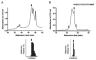
- Example 1 Purification of partially purified sweat antigen (QRX) 1-1. Preparation of concentrated sweat After removing insoluble matter from human sweat through 100 ⁇ m and 70 ⁇ m mesh filters (Nylon Cell Strainer, Falcon), the precipitate was further removed with a 0.22 ⁇ m filter (Bottle Top Filter, 1 L, Corning) . Filtered sweat 4 L was concentrated to about 150 mL by ultrafiltration (3000 M.W.cut), and used as a material for sweat antigen purification.
- QRX partially purified sweat antigen
- basophils of patients with atopic dermatitis prepared in HEPES buffer containing each appropriately diluted fraction and 5 mmol / L glucose, 0.03 w / v% HSA, 2 mmol / L CaCl 2 , 1 mmol / L MgCl 2 Fractions were mixed 1: 1 and incubated at 37 ° C. for 40 minutes. The supernatant and sediment blood cells were separated by centrifugation, and 0.2 mol / L perchloric acid was added to each to denature the protein. Then, the histamine concentration in the supernatant obtained by centrifugation was determined by HPLC (Shimano LC solution). It was measured. The ratio of the amount of histamine in the supernatant to the total amount of histamine was defined as histamine releasing activity.
- the amount of histamine was measured according to the method described in the literature (Koro, O. et al., J. Allergy Clin. Immunol., 103, 663-670, 1999).
- Example 1-3 Separation by reverse phase column 18 mL of the fraction (QR) obtained in Example 1-2 was diluted 10-fold with pure water, and TFA having a final concentration of 0.1 v / v% was added. This was loaded onto a reverse-phase column (SOURCE 15RPC ST 4.6 / 100 (GE Healthcare Bioscience)) and eluted from 0.1 v / v% TFA / distilled water with a concentration gradient of 0.1 v / v% TFA / acetonitrile. As a chromatographic apparatus, AKTA explorer (GE Healthcare Bioscience) was used.
- Example 1-4 Separation by gel filtration chromatography The fraction obtained in Example 1-3 was freeze-dried, redissolved in PBS, loaded into Superdex 75 PC 3.2 / 30 (GE Healthcare Bioscience), and PBS (-) Fractions were eluted. Smart system (GE Healthcare Bioscience) was used as a chromatographic apparatus for purification.
- the histamine release test was performed on each eluted fraction in the same manner as in Example 1-2.
- QRX fraction an elution position in the range of 15 to 60 kD was collected as a fraction showing histamine releasing activity (1.2 mL), and this was hereinafter referred to as QRX fraction.
- Example 2 Purification of Partially Purified Sweat Antigen (QRX) and Mass Spectrometry Partially purified sweat antigen (QRX) was purified using an Aqua 5 ⁇ -C18-200A HPLC column (Phenomenex) (0.1 v / v% TFA). / Eluted from distilled water with a concentration gradient of 0.1 v / v% TFA / 100% acetonitrile). The fraction showing histamine releasing activity was collected and further purified with a Jupiter 5 ⁇ -C18-300A HPLC column (manufactured by Phenomenex) (0.1 v / v% TFA / 80% from 0.1 v / v% TFA / distilled water).
- Example 3 Preparation of recombinant protein of MGL_1304 and reactive Malassezia globosa with atopic dermatitis patient IgE was purchased from ATCC (MYA-4612). MRNA extracted from Malassezia globosa is reverse transcribed into cDNA and PCR method (sense primer: 5'-GGGGTACCGTATCCCTCAACATTTTCTCAGCTGC-3 '(SEQ ID NO: 2); antisense primer: 5'-CCCAAGCTTTTAGCAGTCGTACTTGCCGGGGATG-3' (SEQ ID NO: 3), (94 ° C 5 min / 60 ° C 1 min / 72 ° C 1 min) x 1 cycle, (94 ° C 1 min / 60 ° C 1 min / 72 ° C 1 min) x 30 cycles, (94 ° C 5 min / 60 ° C 1 min / 72 ° C 10 min) ⁇ 1 cycle) was used to amplify the cDNA encoding MGL
- coli JM109 After culturing at 15 ° C. for 24 hours, the obtained Escherichia coli was dissolved in xTractor buffer, and the recombinant protein was purified with a cobalt column. Only Trigger Factor (TF), TF-MGL_1304 fusion protein (TF-MGL_1304), and the fusion protein removed with TF (rMGL_1304) were prepared. The obtained protein was subjected to acrylamide gel electrophoresis, stained directly with CBB (Fig. 2 left), transferred to a PVDF membrane and anti-His tag antibody (in Fig. 2), atopic dermatitis patient serum (Fig. 2 right). Immunoblotting was performed.
- TF Trigger Factor
- TF-MGL_1304 fusion protein removed with TF
- rMGL_1304 the fusion protein removed with TF
- rMGL_1304 It was shown that IgE from atopic dermatitis patients binds to rMGL_1304 (FIG. 2).
- the prepared rMGL_1304 was reacted with peripheral blood basophils (FIG. 3, AD1, AD2, AD3) of atopic dermatitis patients and healthy human peripheral blood basophils (FIG. 3, HC1), and a histamine release test was performed.
- MGL_1304 was shown to cause histamine release specifically in patients with atopic dermatitis.
- the above-mentioned cDNA encoding MGL_1304 or tick cDNA was incorporated into a pSecTag2 / Hygro vector containing Myc-tag (Invitrogen) and transfected into COS7 cells.
- COS7 cell-cultured spermatozoa transfected with these DNAs were subjected to acrylamide gel electrophoresis, transferred to a PVDF membrane, and immunoblotted with an anti-Myc tag antibody. It was shown that the protein corresponding to each cDNA was contained in the culture supernatant (FIG. 4A). Furthermore, the culture supernatant was reacted with peripheral blood basophils of atopic dermatitis patients, and a histamine release test was performed. Also, the same basophils were reacted with sweat antigen (QR), which was obtained by concentrating human sweat and partially purified by anion exchange chromatography and reversed-phase column chromatography, and conducting a histamine release test. Comparison was made (FIG. 4B). MGL_1304 protein (rMGL_1304) produced by COS7 cells was shown to cause histamine release similar to partially purified human sweat antigen (QR).
- QR sweat antigen
- Example 4 Reactivity of MGL_1304 Recombinant Protein and Atopic Dermatitis Patient IgE It was examined whether MGL_1304 has almost the same properties as the partially purified sweat antigen (QRX) used so far.
- QRX sweat antigen
- Several recombinant mite antigens (Der f1), QRX, and MGL_1304 were electrophoresed and transferred to a PVDF membrane. Immunoblotting was performed using atopic patient serum (AD serum) pretreated with QRX or MGL_1304 prepared in Example 3, or AD serum not pretreated. The sera of patients with atopic dermatitis used are for 3 people.
- Pretreatment with MGL_1304 inhibited IgE binding to QRX, and pretreatment with QRX inhibited IgE binding to MGL_1304 (FIG. 5).
- Atopic dermatitis patient sera (AD1-AD4) were pretreated with TF or TF-MGL_1304 and sensitized to a rat cell line expressing human IGE receptor (A subunit), and anti-human IgE antibody (anti -IgE), QRX, and mite extract (Mite-Df) measured when degranulation was performed. It was shown that the reactivity to QRX stimulation disappeared by removing MGL_1304-specific IgE by pretreatment with MGL_1304 prepared in Example 3 (FIG. 6).
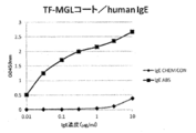
- Example 5 Binding of various human IgEs to QRX QRX prepared in Example 1 was coated on an ELISA plate (50 ng / well).
- QRX was detected in a concentration-dependent manner only when ABS human monoclonal IgE antibody was used (FIG. 7). It was shown that ABS human monoclonal IgE antibody binds to QRX, and QRX can be detected quantitatively.
- Example 6 Binding of ABS IgE antibody to rMGL_1304
- the TF-MGL_1304 fusion protein prepared by forced expression in E. coli in Example 3 was coated on an ELISA plate (150 ng / well). It was examined by ELISA whether ABS IgE antibody and CHEMICON human myeloma IgE antibody could detect TF-MGL_1304 fusion protein in a concentration-dependent manner. Each human IgE antibody bound to the TF-MGL_1304 fusion protein was detected using an HRP-labeled anti-human IgE antibody. As a result, ABS IgE antibody detected TF-MGL_1304 fusion protein in a concentration-dependent manner (FIG. 8).
- ABS IgE antibody binds to MGL_1304 protein and can detect MGL_1304 protein quantitatively.
- CHEMICON human myeloma IgE antibody did not sufficiently detect the TF-MGL_1304 fusion protein (FIG. 8).
- TF-MGL_1304 fusion protein was coated on an ELISA plate (150 ng / well).
- concentration of ABS IgE antibody so as to include a wider range (0.02 to 20 ng / ml)
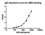
- the immobilized recombinant MGL_1304 protein was detected by ELISA, and a standard curve was prepared.
- the ABS IgE antibody bound to the TF-MGL_1304 fusion protein was detected using an HRP-labeled anti-human IgE antibody. As a result, the binding between the TF-MGL_1304 fusion protein and the ABS IgE antibody was quantitative (FIG. 9). Since TF-MGL_1304 fusion protein is a sweat allergy antigen (sweat antigen) as shown in Example 1-3, ABS IgE antibody is used as a standard for quantifying IgE antibody binding to sweat antigen. It was shown that it can be used.
- Example 7 Binding of ABS IgE antibody to other antigens Partially purified sweat antigen (QRX) prepared in Example 1 was coated on an ELISA plate (50 ng / well). In addition, mite antigen and quinu antigen prepared by expression in E. coli were coated on ELISA plates (50 ng / well each). Whether each antigen was detectable by ABS IgE antibody was examined using ELISA. The ABS IgE antibody bound to each antigen was detected using an HRP-labeled anti-human IgE antibody. As a result, ABS IgE antibody specifically detected partially purified sweat antigen (QRX) (FIG. 10).
- QRX partially purified sweat antigen
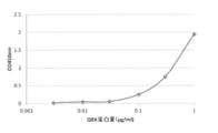
- Example 8 Comparative Detection of QRX Detection of ABS IgE Antibody and Smith2 Antibody Partially purified sweat antigen (QRX) prepared in Example 1 was coated on an ELISA plate (50 ng / well). The concentrations of the ABS IgE antibody for detecting QRX and the Smith2 antibody (Accession No. FERM BP-11111) were compared. The ABS IgE antibody bound to QRX was detected using an HRP-labeled anti-human IgE antibody, and the Smith2 antibody bound to QRX was detected using an HRP-labeled anti-mouse IgG antibody. As a result, ABS IgE antibody was able to detect QRX immobilized from 0.01 ⁇ g / ml.
- Example 9 ELISA using ABS IgE antibody and Smith2 antibody From the results of the above examples, it was shown that the ABS IgE antibody quantitatively binds to the sweat allergy antigen (sweat antigen), so that the sweat antigen is highly sensitive when used together with the Smith2 antibody that binds to the sweat antigen. Attempted to construct ELISA to detect in First, an ELISA using Smith2 antibody as an immobilized antibody and ABS IgE antibody as a detection antibody was tested (hereinafter, this ELISA system is also referred to as ELISA system I).
- Smith2 antibody was immobilized on an ELISA plate (10, 30, 100 ⁇ g / ml, 50 ⁇ l / well), followed by addition of QRX (1 ⁇ g / ml, 100 ⁇ l / well) or vehicle. Furthermore, ABS IgE antibody (1 ⁇ g / ml, 100 ⁇ l / well) and HRP-labeled anti-human IgE antibody (manufactured by KPL) were added, and the absorbance (450 nm) was measured. As a result, the absorbance (back ground) when QRX was not added was about 0, whereas when QRX was added, the absorbance was about 3.5 (FIG. 12).
- ABS IgE antibody was immobilized on an ELISA plate (1, 3, 10 ⁇ g / ml, 50 ⁇ l / well), followed by QRX (1 ⁇ g / ml, 100 ⁇ l / well) or vehicle.
- QRX 1 ⁇ g / ml, 100 ⁇ l / well
- Smith2 antibody (10 ⁇ g / ml, 100 ⁇ l / well) and HRP-labeled anti-mouse IgG antibody (manufactured by KPL) were added, and the absorbance (450 nm) was measured.
- ELISA system I was shown to be able to detect QRX at a concentration approximately 300 times lower than that of ELISA system in which QRX is directly immobilized on a plate and detected with an ABS IgE antibody (FIG. 14). Using this ELISA system I, it was examined whether the amount of sweat antigen (QRX) contained in the stratum corneum of the human body surface could be measured.
- QRX sweat antigen
- QRX in the stratum corneum is obtained by peeling off a 2.5 cm x 2.5 cm square adhesive tape on the body surface, 500 ⁇ l of 10 times diluted RIPA buffer (Pierce: 25 mM Tris ⁇ HCl pH) (7.6, 150 mM NaCl, 1% NP-40, 1% sodium deoxycholate, 0.1% SDS).
- RIPA buffer 25 mM Tris ⁇ HCl pH
- QRX contained 1.01 ⁇ 2.64 ng / ml in the elbow, 0.13 ⁇ 0.078 ng / ml in the upper arm, and 1.01 ⁇ 1.38 ng / ml in the cervix (FIG. 15).
- Example 10 Western blot using ABS IgE antibody Using Western blotting, the ABS IgE antibody recognizes QRX and recombinant MGL_1304 protein (enzyme-treated TF-MGL_1304 fusion protein prepared in Example 3 to remove Trigger Factor (TF)). Confirmed (FIG. 17). The results of Western blotting using both Native-PAGE and SDS-PAGE electrophoresis showed that ABS IgE antibody recognizes both QRX and MGL_1304 proteins.
- TF Trigger Factor
- MGL_1304 protein Polypeptide corresponding to amino acid sequence 1-50 (SEQ ID NO: 4) of MGL_1304 protein; ii) P2: a polypeptide corresponding to the amino acid sequence 46-100 (SEQ ID NO: 5) of the MGL_1304 protein; iii) P3: polypeptide corresponding to amino acid sequence 96-140 (SEQ ID NO: 6) of MGL_1304 protein; iv) P4: polypeptide corresponding to amino acid sequence 136-183 (SEQ ID NO: 7) of MGL_1304 protein; v) P1- 3: a polypeptide corresponding to the amino acid sequence 1-140 (SEQ ID NO: 8) of the MGL_1304 protein; and vi) P2-4: A polypeptide corresponding to the amino acid sequence 46-183 (SEQ ID NO: 9) of the MGL_1304 protein was forcibly expressed to examine whether or not the ABS IgE antibody recognize
- Example 11 Sweat antigen neutralizing action of ABS IgE antibody
- ABS IgE antibody inhibits histamine release induced by adding QRX to basophils from sweat allergic patients.
- ABS IgE antibody suppressed histamine release induced by QRX (FIG. 18).
- ABS IgE antibody suppressed the histamine release activity of unpurified human sweat.
- Example 12 Change in reactivity to sweat antigen by sensitizing cells with ABS IgE antibody
- ABS IgE binds to human high affinity IgE receptor expressed in mast cells or basophils (sensitization)
- rat mast cells expressing human high-affinity IgE receptor are treated with serum from atopic dermatitis patients or ABS.
- QRX As a result, when sensitized with serum from patients with atopic dermatitis, degranulation was induced by stimulation with QRX (see FIG.
- ABS-IgE anti-human IgE antibody
- Histamine release was observed in both donor-derived basophils (see FIG. 21, untreated).
- QRX a partially purified sweat antigen
- mouse monoclonal antibody 6F7 which activates IgE receptor by cross-linking IgE receptor not bound by IgE, was added, and moderate histamine release was observed in basophils from both donors. (See FIG. 21, unprocessed). Therefore, it was considered that basophils from both donors bound IgE to a part of their IgE receptors. Therefore, when lactic acid treatment was performed and IgE was separated from the IgE receptor and stimulated in the same manner, the degree of histamine release by the anti-IgE antibody was reduced, and histamine release by QRX disappeared (see FIG. 21, lactic acid treatment). Subsequently, these cells were further sensitized with ABS IgE antibody and stimulated in the same manner.
- Lactic acid treatment dissociated IgE from many high-affinity IgE receptors, and resensitization with IgE again resulted in many high-affinity IgE receptors binding to IgE. It was confirmed that the histamine release rate by 6F7 of the anti-high affinity IgE receptor monoclonal antibody increased by lactic acid treatment and decreased or disappeared by IgE resensitization. These results indicate that ABS IgE antibody binds to the IgE receptor on the cell but does not cause mast cell degranulation even when sweat allergen is present.
- Example 13 Measurement of anti-sweat antigen-specific IgE antibody in patient serum (1) Specific to sweat antigen in serum of atopic dermatitis (AD), allergic rhinitis and normal (Normal) using ELISA (FIG. 9) solid-phased rTF-MGL prepared in Example 6 It was investigated whether or not IgE antibody binding to can be detected.
- RTF or rTF-MGL was coated on a 96-well ELISA plate at 3 ⁇ g / ml and 50 ⁇ l / well and allowed to stand at 4 ° C. overnight. After washing twice, it was blocked with 2% BSA (room temperature, 1 hour) and washed twice. 100 ⁇ l / well of serum diluted 10-fold (x10) or 20-fold (x20) with 1% BSA was added and left at room temperature for 1 hour. After washing 3 times, a solution containing an HRP-labeled anti-human IgE antibody was added at 100 ⁇ l / well and allowed to stand for 1 hour. After washing 3 times, color was developed using TMB, and the absorbance (450 nm) was measured.
- the absorbance obtained from the well coated with rTF was subtracted from the absorbance obtained from the well coated with rTF-MGL to obtain the binding amount of MGL-specific IgE.
- a standard curve (FIG. 9) of the ABS IgE antibody shown in Example 6 was simultaneously prepared.
- IgE specifically binding to sweat antigen (MGL) was detected in the serum of patients with atopic dermatitis and allergic rhinitis using rTF or rTF-MGL solid-phase ELISA.
- the concentration of IgE antibody could be quantified using the antibody as a standard substance (FIG. 22).
- IgE binding to sweat allergy antigen protein (rMGL) in the serum of healthy individuals was extremely low or below the detection limit (100 pg / ml) (FIG. 22).
- ABS IgE antibody as a standard substance, it is possible to diagnose sweat allergy in patients with atopic dermatitis and allergic rhinitis.
- Example 14 Measurement of anti-sweat antigen-specific IgE antibody in patient serum (2) Using the ELISA (FIG. 16) that binds human IgE antibody to QRX bound to solid-phased Smith2 antibody prepared in Example 9, atopic dermatitis patients (AD1-3) and healthy individuals (Normal1- The IgE antibody specifically binding to the sweat antigen in the serum of 2) was quantified.
- ELISA ELISA
- Smith2 antibody (10 ⁇ g / ml, 100 ⁇ l / well) was placed in a 96-well ELISA plate, left overnight at 4 ° C., washed twice, then blocked with 2% BSA (room temperature, 1 hour), and washed twice. Subsequently, QRX (1 ⁇ g / ml, 100 ⁇ l / well) was added and washed twice. 100 ⁇ l / well of serum diluted 40-fold (x40) or 80-fold (x80) with 1% BSA was added and left at room temperature for 90 minutes. After washing 3 times, a solution containing an HRP-labeled anti-human IgE antibody was added at 100 ⁇ l / well and allowed to stand for 1 hour. After washing 3 times, color was developed using TMB, and the absorbance (450 nm) was measured. In addition, a standard curve (FIG. 16) was prepared simultaneously by performing ELISA for the ABS IgE antibody shown in Example 9.
Abstract
Description
本発明は、汗アレルギー抗原に結合するヒトIgE抗体、そのスクリーニングおよび製造方法、ヒトIgE抗体によるヒト汗中に含まれる汗アレルギー抗原の検出または測定への使用、ヒトIgE抗体によるヒト汗中に含まれる汗アレルギー抗原に結合する抗体の検出または測定への使用、ヒトIgE抗体の汗アレルギー抗原の中和または除去への使用等に関する。 This application claims priority based on Japanese Patent Application No. 2012-181051 filed on Aug. 17, 2012, the entire contents of which are incorporated herein by reference.
The present invention relates to a human IgE antibody that binds to a sweat allergy antigen, a screening and production method thereof, use for detection or measurement of a sweat allergy antigen contained in human sweat by a human IgE antibody, and a human IgE antibody contained in human sweat The present invention relates to the use of antibodies that bind to sweat allergens that are detected or measured, and the use of human IgE antibodies to neutralize or remove sweat allergens.
アレルギー反応を抑制するための抗体としては、IgE抗体を中和する抗IgE抗体が実用化されているが、ヒトIgE抗体は、高親和性IgE受容体に結合すると、単独またはそこにさらにアレルゲンが結合することでマスト細胞、好塩基球を活性化する性質があるため、通常は、アレルゲンの中和に有用でないと考えられている。 Allergic reactions are called immediate or type I hypersensitivity reactions and are mediated by IgE antibodies. When an organism is first exposed to an allergen, IgE antibody-producing B cells begin to produce soluble IgE molecules. Soluble IgE molecules then bind to high affinity IgE receptors present on the surface of mast cells or basophils. When the living body encounters the same allergen again, the high affinity IgE receptor is cross-linked by the IgE antibody and the allergen, resulting in an allergic reaction in which histamine and cytokines are released from the cells.
Anti-IgE antibodies that neutralize IgE antibodies have been put to practical use as antibodies for suppressing allergic reactions. However, human IgE antibodies, when bound to high-affinity IgE receptors, alone or further allergens are added. Since it has the property of activating mast cells and basophils by binding, it is usually considered not useful for neutralizing allergens.
ヒトIgEモノクローナル抗体についても、例えば、ファージディスプレイ法を用いて製造されたという報告がある(特許文献1、非特許文献1)。 On the other hand, the production of human antibodies is no longer a difficult technique, and human antibodies can be produced by various methods (for example, Epstein-Barr virus (EBV) immortalization method, phage display method, fully human antibody-producing hybridoma method). .
There is also a report that a human IgE monoclonal antibody was produced using, for example, a phage display method (
本発明は、第1の側面(aspect)において、汗アレルギー抗原蛋白質に結合し、ヒト高親和性IgE受容体に結合するが、汗アレルギー抗原蛋白質と反応させても脱顆粒を起こさないヒトIgE抗体または抗体断片およびその製造方法を提供する。 1. A human IgE antibody or antibody fragment that binds to a sweat allergy antigen protein and binds to a human high affinity IgE receptor but does not cause degranulation even when reacted with a sweat allergy antigen protein . In the aspect of the present invention, a human IgE antibody or antibody fragment that binds to a sweat allergy antigen protein and binds to a human high-affinity IgE receptor, but does not cause degranulation even when reacted with a sweat allergy antigen protein and its production Provide a method.
配列番号1:MVSLNIFSAAFVASLASAVFAAPSALERRAAPDNTVWVTSVADHCLILPRHKMSVGDSESPGNMRSFCTKPYSSKQGQLASDFWTKAHFKKTDKYVQITGCINPNVQSTLLSNDEGGQYDSNGGEGGRGNPAGSVCLGYSSYVELVEPAGNRACIRCCYDPSDCDVSQDEAGCETVIPGKYDC In the present specification, the sweat allergy antigen protein is a protein that is an antigen of sweat allergy, and is known from human sweat (for example, Yasuhiko Tanaka, et al. Allergy 56: 54-57, 2007, Tanaka A, et al. Proteins or microorganisms partially purified by Exp Dermatol 15: 283-290, 2006, WO 2009-133951): Proteins produced by Malassezia globosa (eg, Malassezia globosa (number MYA-4612) available from ATCC) It can be a protein that binds to serum from sweat allergy patients and / or Smith2 antibody (Accession No. FERM BP-11111). The sweat allergy antigen protein may be a protein of about 17 kDa (for example, 14 kDa or more and 20 kDa or less) secreted by Malassezia globosa outside the cells. For example, the sweat allergy antigen protein can be a protein encoded by the MGL_1304 gene (for example, a protein comprising the amino acid sequence represented by SEQ ID NO: 1). The amino acid sequence represented by SEQ ID NO: 1 is shown below.
Sequence number 1: MVSLNIFSAAFVASLASAVFAAPSALERRAAPDNTVWVTSVADHCLILPRHKMSVGDSESPGNMRSFCTKPYSSKQGQLASDFWTKAHFKKTDKYVQITGCINPNVQSTLLSNDEGGQYDSNGGEGGRGNPAGSVCLGYSSYVELVEPAGNRACIRDCCYDPSDCDVSGAG
例えば、ヒスタミン量を測定し、全ヒスタミン量に対する遊離ヒスタミン量が3~97%の範囲の場合にヒスタミン遊離活性を有すると判定することができる(Koro, O. et al., J. Allergy Clin. Immunol., 103, 663-670, 1999)。 Sweat allergens have histamine releasing activity. Histamine releasing activity can be measured according to a known method (Koro, O. et al., J. Allergy Clin. Immunol., 103, 663-670, 1999). For example, histamine release activity may be determined by contacting a sweat allergy antigen and an IgE antibody with a cell expressing an IgE receptor on the cell surface and measuring the amount of histamine secreted from the cell. Cells expressing the IgE receptor on the cell surface include cell lines having the ability to release chemical mediators such as basophils, mast cells (mast cells), and artificially produced IgE receptor genes. Etc.
For example, when the amount of histamine is measured and the amount of free histamine relative to the total amount of histamine is in the range of 3 to 97%, it can be determined that the compound has histamine releasing activity (Koro, O. et al., J. Allergy Clin. Immunol., 103, 663-670, 1999).
ヒトIgE抗体は、ヒト抗体であり、ポリクローナル抗体またはモノクローナル抗体であってもよい。ヒトIgE抗体またはその抗体の断片は、また、酵素等(例えば、Horseradish Peroxidase、ビオチン等)で標識されてもよい。 Human IgE antibodies that do not cause degranulation even when reacted with sweat allergy antigen protein are, for example, cells expressing high affinity IgE receptors such as mast cells or basophils in the presence of sweat allergy antigen protein It may be a human IgE antibody that does not release intragranular substances such as histamine and enzymes to the outside of the cell when it is contacted.
The human IgE antibody is a human antibody and may be a polyclonal antibody or a monoclonal antibody. The human IgE antibody or a fragment of the antibody may also be labeled with an enzyme or the like (eg, Horseradish Peroxidase, biotin, etc.).
一つの実施態様として、汗アレルギー抗原蛋白質に結合し、ヒト高親和性IgE受容体に結合するが、汗アレルギー抗原蛋白質と反応させても脱顆粒を起こさないヒトIgE抗体または抗体断片は、ABS社から購入できるIgEモノクローナル抗体(クローンHE1)(DIA HE1-01A/DIA HE1-1A)またはその抗体断片であり得る。なお、IgEモノクローナル抗体(クローンHE1)(DIA HE1-01A/DIA HE1-1A)は、例えば、Fairley J.A., et al., (2007) J. Invest. Dermatol. 127:2605-2611において、Non-specific human IgE (http://www.diatec.com, Oslo, Norway)として使用されている。
ABS社から購入できるIgEモノクローナル抗体(クローンHE1)(Catalog Number:DIA HE1-01A/DIA HE1-1A)は、健常人ドナー由来のIgEモノクローナル抗体である。
この事実から、汗アレルギー患者ではなくて健常人由来のIgE抗体発現ライブラリから、「汗アレルギー抗原蛋白質に結合し、ヒト高親和性IgE受容体に結合するが、汗アレルギー抗原蛋白質と反応させても脱顆粒を起こさないヒトIgE抗体」を単離できる妥当性が示された。よって、一つの実施態様として、本発明は、健常人由来のIgE抗体発現ライブラリから、汗アレルギー抗原蛋白質に結合し、ヒト高親和性IgE受容体に結合するが、汗アレルギー抗原蛋白質と反応させても脱顆粒を起こさないヒトIgE抗体をスクリーニングする方法を提供する。 The present inventors can purchase from ABS as a human IgE antibody that binds to sweat allergy antigen protein and binds to human high affinity IgE receptor, but does not cause degranulation when reacted with sweat allergy antigen protein. An IgE monoclonal antibody (clone HE1) (DIA HE1-01A / DIA HE1-1A) (also referred to as ABS IgE antibody) was found.
In one embodiment, a human IgE antibody or antibody fragment that binds to a sweat allergy antigen protein and binds to a human high affinity IgE receptor, but does not cause degranulation when reacted with a sweat allergy antigen protein is manufactured by ABS IgE monoclonal antibody (clone HE1) (DIA HE1-01A / DIA HE1-1A) or an antibody fragment thereof, which can be purchased from In addition, IgE monoclonal antibody (clone HE1) (DIA HE1-01A / DIA HE1-1A) is non-specific in, for example, Fairley JA, et al., (2007) J. Invest. Dermatol. 127: 2605-2611. Used as human IgE (http://www.diatec.com, Oslo, Norway).
The IgE monoclonal antibody (clone HE1) (Catalog Number: DIA HE1-01A / DIA HE1-1A) that can be purchased from ABS is an IgE monoclonal antibody derived from a healthy donor.
From this fact, from the IgE antibody expression library derived from healthy people rather than sweat allergy patients, `` It binds to sweat allergy antigen protein and binds to human high affinity IgE receptor, but it can react with sweat allergy antigen protein. The validity of isolating a human IgE antibody that does not cause degranulation was shown. Therefore, as one embodiment, the present invention binds to a sweat allergy antigen protein and binds to a human high affinity IgE receptor from an IgE antibody expression library derived from a healthy person, but reacts with a sweat allergy antigen protein. A method for screening a human IgE antibody that does not cause degranulation is also provided.
例えば、ヒト由来のIgE産生細胞から抽出されたtotal RNAを使用してM13などの繊維状ファージに蛋白質を発現させ、ヒトIgE発現ナイーブ(非免疫)ライブラリを構築し、パニングにより目的の生物活性を有するヒトIgEモノクローナル抗体をスクリーニングすることができる。 It has been reported that human IgE monoclonal antibodies or fragments thereof can be screened using a phage display method (Steinverger P. et al., (1996) J. Biol. Chem. 271, 10967-10972, etc.).
For example, using total RNA extracted from human-derived IgE-producing cells, protein is expressed in filamentous phages such as M13, a human IgE expression naive (non-immune) library is constructed, and the desired biological activity is achieved by panning. It is possible to screen for human IgE monoclonal antibodies.
a)健常人由来のIgE産生細胞から抽出されたRNAを使用してscFvファージライブラリ(ナイーブ(非免疫)ライブラリ)を構築する工程;
b)汗アレルギー抗原蛋白質に結合するscFvを発現するファージをライブラリからスクリーニングして単離する工程;
c)b)で単離されたファージからVHおよびVL遺伝子をPCRで増幅し、汗アレルギー抗原蛋白質に結合するIgE抗体を調製する工程;および
d)c)で調製されたIgE抗体を汗アレルギー抗原蛋白質の存在下でマスト細胞または好塩基球に反応させ、脱顆粒を起こさないIgE抗体を単離する工程
を含む、汗アレルギー抗原蛋白質に結合し、ヒト高親和性IgE受容体に結合するが、汗アレルギー抗原蛋白質と反応させても脱顆粒を起こさないヒトIgE抗体をスクリーニングする方法を提供する。
「汗アレルギー抗原蛋白質に結合し、ヒト高親和性IgE受容体に結合するが、汗アレルギー抗原蛋白質と反応させても脱顆粒を起こさないヒトIgE抗体または抗体断片」のスクリーニング方法の具体例には、限定はされないが、以下の工程を含む:
工程1: 健常人の末梢血単核球を調製する;
工程2: mRNAを単離しcDNAを合成する;
工程3: cDNAよりPCRでVH遺伝子とVL遺伝子をそれぞれ別個に増幅する;
工程4: 増幅したVH遺伝子とVL遺伝子をランダムに連結させ(例えば、増幅したVH遺伝子とVL遺伝子とを(GGGGS)3等のリンカ―ペプチド配列をコードしたリンカーDNAを利用してアセンブリPCRによりランダムに連結させ)、scFv遺伝子を調製する;
工程5: 調製したscFv遺伝子をファージミドベクター(例えば、pCANTAB5E)に組み込み、scFv遺伝子ライブラリを構築する;
工程6: 調製したscFv遺伝子ライブラリを大腸菌に形質転換後、ヘルパーファージを重感染させ、scFvファージライブラリを調製する;
工程7: 固定化した汗アレルギー抗原蛋白質にscFvファージライブラリを反応させ、結合しなかったファージを洗浄により除去した後に、結合したファージを溶出し大腸菌に感染させて増殖させるという操作を数回行うことで、汗アレルギー抗原蛋白質に特異的なscFvを発現するファージを単離する;
工程8: 単離したファージより、汗アレルギー抗原蛋白質に特異的なscFvのVHとVLの遺伝子をPCRにより増幅し、抗体の定常領域遺伝子があらかじめ組み込まれているプラスミドベクターに導入し、CHO等の動物細胞で発現させて、汗アレルギー抗原蛋白質に結合するモノクローナル抗体を調製する;
工程9: 調製した汗アレルギー抗原蛋白質に結合するモノクローナル抗体を、汗アレルギー抗原蛋白質の存在下でマスト細胞または好塩基球等の高親和性IgE受容体を発現している細胞に接触させ、ヒスタミン遊離活性を測定し、ヒスタミン遊離活性を有しないモノクローナル抗体を「汗アレルギー抗原蛋白質に結合し、ヒト高親和性IgE受容体に結合するが、汗アレルギー抗原蛋白質と反応させても脱顆粒を起こさないヒトIgE抗体」として単離する、
方法が挙げられる。 上記実施態様において、PCRで使用されるプライマーは、当業者が適宜設計できる(Steinverger P. et al., (1996) J. Biol. Chem. 271, 10967-10972等)。
また、一つの実施態様として、本発明は、上記スクリーニング方法を実施し、その結果単離されたヒトIgE抗体の遺伝子を発現する動物細胞(例えば、CHO細胞)を培養することを含む、ヒト高親和性IgE受容体に結合するが、汗アレルギー抗原蛋白質と反応させても脱顆粒を起こさないヒトIgE抗体を製造する方法を提供する。
また、一つの実施態様として、本発明が提供する「汗アレルギー抗原蛋白質に結合し、ヒト高親和性IgE受容体に結合するが、汗アレルギー抗原蛋白質と反応させても脱顆粒を起こさないヒトIgE抗体または抗体断片」は、健常人由来のIgE産生細胞から抽出されたRNAを使用して構築されたヒトIgE発現ライブラリより単離されたヒトIgEモノクローナル抗体またはその抗体断片であり得る。 Thus, as one embodiment, the present invention provides:
a) constructing an scFv phage library (naive (non-immune) library) using RNA extracted from IgE-producing cells derived from healthy individuals;
b) screening and isolating phage expressing scFv that bind to sweat allergy antigen protein from the library;
c) a step of amplifying the VH and VL genes from the phage isolated in b) by PCR and preparing an IgE antibody that binds to a sweat allergy antigen protein; and d) the IgE antibody prepared in c) using the sweat allergy antigen. Reacts with mast cells or basophils in the presence of protein and binds to sweat allergy antigen protein, which includes isolating IgE antibodies that do not cause degranulation, and binds to human high affinity IgE receptor, Provided is a method for screening a human IgE antibody that does not cause degranulation even when reacted with a sweat allergy antigen protein.
Specific examples of screening methods for "human IgE antibodies or antibody fragments that bind to sweat allergy antigen protein and bind to human high affinity IgE receptor but do not cause degranulation when reacted with sweat allergy antigen protein" include Including, but not limited to, the following steps:
Step 1: Prepare peripheral blood mononuclear cells of healthy individuals;
Step 2: mRNA is isolated and cDNA is synthesized;
Step 3: Amplify VH gene and VL gene separately from cDNA by PCR;
Step 4: The amplified VH gene and the VL gene are randomly linked (for example, the amplified VH gene and the VL gene are randomly generated by assembly PCR using a linker DNA encoding a linker-peptide sequence such as (GGGGS) 3. To prepare the scFv gene;
Step 5: The prepared scFv gene is incorporated into a phagemid vector (eg, pCANTAB5E) to construct an scFv gene library;
Step 6: After the prepared scFv gene library is transformed into E. coli, a helper phage is superinfected to prepare an scFv phage library;
Step 7: The scFv phage library is reacted with the immobilized sweat allergy antigen protein, the unbound phage is removed by washing, and then the bound phage is eluted and infected with E. coli to proliferate several times. Isolating phage expressing scFv specific for sweat allergy antigen protein;
Step 8: From the isolated phage, the scFv VH and VL genes specific for the sweat allergy antigen protein are amplified by PCR and introduced into a plasmid vector in which the antibody constant region gene has been previously incorporated. Preparing monoclonal antibodies that are expressed in animal cells and bind to sweat allergy antigen protein;
Step 9: A monoclonal antibody that binds to the prepared sweat allergy antigen protein is brought into contact with cells expressing high affinity IgE receptors such as mast cells or basophils in the presence of the sweat allergy antigen protein to release histamine. Measures the activity of a monoclonal antibody that does not have histamine-releasing activity `` Human binds to sweat allergy antigen protein and binds to human high affinity IgE receptor, but does not cause degranulation when reacted with sweat allergy antigen protein Isolated as `` IgE antibody '',
A method is mentioned. In the above embodiment, primers used in PCR can be appropriately designed by those skilled in the art (Steinverger P. et al., (1996) J. Biol. Chem. 271, 10967-10972, etc.).
Further, as one embodiment, the present invention comprises carrying out the above screening method and culturing animal cells (eg, CHO cells) expressing the human IgE antibody gene isolated as a result of the above-described screening method. Provided is a method for producing a human IgE antibody that binds to an affinity IgE receptor but does not cause degranulation when reacted with a sweat allergy antigen protein.
Further, as one embodiment, the present invention provides “human IgE that binds to a sweat allergy antigen protein and binds to a human high affinity IgE receptor, but does not cause degranulation even when reacted with a sweat allergy antigen protein. The “antibody or antibody fragment” can be a human IgE monoclonal antibody or an antibody fragment thereof isolated from a human IgE expression library constructed using RNA extracted from IgE-producing cells derived from a healthy person.
本発明は、第2の側面(aspect)において、汗アレルギー抗原蛋白質に結合し、ヒト高親和性IgE受容体に結合するが、汗アレルギー抗原蛋白質と反応させても脱顆粒を起こさないヒトIgE抗体またはその抗体断片を含む、汗アレルギーもしくは汗アレルギー抗原が関連する疾患の治療用組成物、汗アレルギー抗原除去もしくは中和用組成物または汗アレルギー抗原除去材を提供する。 2. A composition for treating sweat allergy or a disease associated with sweat allergy antigen, a composition for removing or neutralizing sweat allergy antigen, or a sweat allergy antigen removing material The present invention, in a second aspect, A sweat allergy or sweat allergy antigen containing a human IgE antibody or an antibody fragment thereof that binds to an allergen antigen protein and binds to a human high affinity IgE receptor but does not cause degranulation when reacted with a sweat allergy antigen protein. A composition for treating a related disease, a composition for removing or neutralizing a sweat allergy antigen, or a sweat allergy antigen removing material is provided.
よって、このIgEモノクローナル抗体(クローンHE1)またはその抗体の断片を含む組成物は、汗アレルギーまたは汗アレルギー抗原が関連する疾患の治療用組成物となり得る。 The IgE monoclonal antibody provided by the present invention is human IgE that binds to a sweat allergy antigen protein and stably binds (sensitizes) to high-affinity IgE, and mast cells sensitized by this monoclonal antibody and Does not cause basophil degranulation. This IgE monoclonal antibody may be the antibody provided in the first aspect of the present invention, and may be, for example, an IgE monoclonal antibody (clone HE1) that can be purchased from ABS, but is not limited thereto.
Therefore, the composition containing this IgE monoclonal antibody (clone HE1) or a fragment of the antibody can be a composition for treating sweat allergy or a disease associated with sweat allergy antigen.
上記治療用組成物の投与量、投与方法は、投与対象の年齢、体重、健康状態によって、当業者により適宜選択され得る。例えば、汗アレルギー抗原蛋白質に結合し、ヒト高親和性IgE受容体に結合するが、汗アレルギー抗原蛋白質と反応させても脱顆粒を起こさないヒトIgE抗体またはその抗体断片は、0. 1μg/kg体重~10mg/kg体重で投与されてもよく、例えば、成人1日当たり0.1~100mgで静脈内投与されてもよい。 The therapeutic composition provided by the present invention is appropriately formulated using the antibody provided in the first aspect of the present invention (for example, IgE monoclonal antibody (clone HE1) available from ABS). For example, the therapeutic composition provided by the present invention can be formulated with a pharmaceutically acceptable carrier (including additives). Examples of pharmaceutically acceptable carriers include excipients (eg, dextrin, hydroxypropylcellulose, polyvinylpyrrolidone, etc.), disintegrants (eg, carboxymethylcellulose), lubricants (eg, magnesium stearate), interfaces, and the like. Examples include, but are not limited to, active agents (eg, sodium lauryl sulfate), solvents (eg, water, saline, soybean oil), preservatives (eg, p-hydroxybenzoate). is not.
The dosage and administration method of the therapeutic composition can be appropriately selected by those skilled in the art depending on the age, weight and health status of the administration subject. For example, human IgE antibody or its antibody fragment that binds to sweat allergy antigen protein and binds to human high affinity IgE receptor but does not cause degranulation when reacted with sweat allergy antigen protein is 0.1 μg / kg. It may be administered at a body weight to 10 mg / kg body weight, for example, at an intravenous dose of 0.1 to 100 mg per day for an adult.
例えば、汗アレルギー抗原蛋白質に特異的に結合する抗体またはその抗体の断片を含む生理食塩水等の等張液を患部に接触させることにより、汗アレルギー抗原が中和および/または汗アレルギー抗原の患部からの除去が達成され得る。
本発明により提供される汗アレルギー抗原除去もしくは中和用組成物は、本発明の第1の側面で提供される抗体(例えば、ABS社から購入できるIgEモノクローナル抗体(クローンHE1))を使用して、適宜調製される。例えば、汗アレルギー抗原蛋白質に結合し、ヒト高親和性IgE受容体に結合するが、汗アレルギー抗原蛋白質と反応させても脱顆粒を起こさないヒトIgE抗体またはその抗体断片を0.01μg/ml~10mg/mlで含んで、溶液組成物を調製してもよい。 Further, an antibody that specifically binds to a sweat allergy antigen protein (for example, an IgE monoclonal antibody (clone HE1) that can be purchased from ABS) or an antibody fragment thereof can be used to neutralize the antigen activity of the sweat allergy antigen. It can also be used to remove sweat allergy antigens from affected areas.
For example, the allergic antigen is neutralized and / or the affected part of the sweat allergen antigen by contacting the affected part with an isotonic solution such as physiological saline containing an antibody or a fragment of the antibody that specifically binds to the sweat allergen antigen protein. Removal from can be achieved.
The composition for removing or neutralizing sweat allergy antigens provided by the present invention uses the antibody provided in the first aspect of the present invention (for example, the IgE monoclonal antibody (clone HE1) that can be purchased from ABS). Are appropriately prepared. For example, human IgE antibody or an antibody fragment thereof that binds to sweat allergy antigen protein and binds to human high affinity IgE receptor but does not cause degranulation when reacted with sweat allergy antigen protein is 0.01 μg / ml to 10 mg. A solution composition may be prepared containing
例えば、特許第3642340号には、公定水分率が7%以上である繊維(織布または不織布等)に抗体を固定化して、拭き取りシート等の除去材を製造する方法が開示されている。
抗体の繊維等の担体に固定化する方法は、例えば、担体をγ-アミノプロピルトリエトキシシランなどを用いてシラン化した後、グルタールアルデヒドなどで担体表面にアルデヒド基を導入し、アルデヒド基と抗体とを共有結合させる方法、未処理の担体を抗体の水溶液中に浸漬してイオン結合により抗体を担体に固定する方法、特定の官能基を有する担体にアルデヒド基を導入し、アルデヒド基と抗体とを共有結合させる方法、特定の官能基を有する担体に抗体をイオン結合させる方法、特定の官能基を有するポリマーで担体をコーティングしたのちにアルデヒド基を導入し、アルデヒド基と抗体とを共有結合させる方法を挙げることができる。ここで、上記の特定の官能基としては、NHR基(RはH以外のメチル、エチル、プロピル、ブチルのうちいずれかのアルキル基)、NH2基、C6H5NH2基、CHO基、COOH基、OH基を挙げることができる。また、抗体は、リンカーを介して担体に担持されていてもよく、使用されるリンカーの例としては、マレイミド、NHS(N-Hydroxysuccinimidyl)エステル、イミドエステル、EDC(1-Etyl-3-[3-dimetylaminopropyl]carbodiimido)、PMPI(N-[p-Maleimidophenyl]isocyanete)が挙げられる。
除去材は、グリセロールを含む水に含浸させて保管され得る。 In addition, an antibody that specifically binds to sweat allergy antigen protein (eg, IgE monoclonal antibody (clone HE1) available from ABS) is immobilized on the fiber and used as a removal material to remove sweat allergy antigen from the affected area. Also good. An example of the removal material is a wiping sheet. Such a removal material can be appropriately manufactured by those skilled in the art.
For example, Japanese Patent No. 3642340 discloses a method for producing a removal material such as a wiping sheet by immobilizing an antibody on a fiber (woven fabric or nonwoven fabric) having an official moisture content of 7% or more.
For example, after immobilizing the carrier with γ-aminopropyltriethoxysilane or the like, the carrier is silanized with glutaraldehyde or the like to introduce an aldehyde group onto the carrier surface. A method of covalently binding an antibody, a method of immersing an untreated carrier in an antibody aqueous solution and immobilizing the antibody to the carrier by ionic bonding, an aldehyde group being introduced into a carrier having a specific functional group, and the aldehyde group and the antibody A method of covalently bonding an antibody to a carrier having a specific functional group, a method of ion-bonding an antibody to a carrier having a specific functional group, coating a carrier with a polymer having a specific functional group, and then introducing an aldehyde group to covalently bond the aldehyde group and the antibody Can be mentioned. Here, as the specific functional group, NHR group (R is any alkyl group other than H, methyl, ethyl, propyl, and butyl), NH2 group, C6H5NH2 group, CHO group, COOH group, OH group Can be mentioned. The antibody may be supported on a carrier via a linker. Examples of the linker used include maleimide, NHS (N-Hydroxysuccinimidyl) ester, imide ester, EDC (1-Etyl-3- [3 -dimetylaminopropyl] carbodiimido) and PMPI (N- [p-Maleimidophenyl] isocyanete).
The removal material can be stored by impregnating with water containing glycerol.
また、本発明は、汗アレルギー抗原蛋白質に結合し、ヒト高親和性IgE受容体に結合するが、汗アレルギー抗原蛋白質と反応させても脱顆粒を起こさないヒトIgE抗体(例えば、ABS社から購入できるIgEモノクローナル抗体(クローンHE1))またはその抗体断片を汗アレルギー抗原蛋白質と接触させることを含む、汗アレルギー抗原蛋白質の中和または除去方法を提供する。 As one embodiment, the present invention relates to a human IgE antibody that binds to a sweat allergy antigen protein and binds to a human high affinity IgE receptor, but does not cause degranulation when reacted with a sweat allergy antigen protein (for example, The present invention provides a method for treating sweat allergy or a disease associated with sweat allergy antigen, comprising administering an IgE monoclonal antibody (clone HE1), which can be purchased from ABS, or an antibody fragment thereof.
The present invention also relates to a human IgE antibody that binds to a sweat allergy antigen protein and binds to a human high affinity IgE receptor, but does not cause degranulation when reacted with a sweat allergy antigen protein (for example, purchased from ABS) A method for neutralizing or removing a sweat allergen antigen protein, comprising contacting a possible IgE monoclonal antibody (clone HE1)) or a fragment thereof with a sweat allergen antigen protein.
本発明は、第3の側面(aspect)において、汗アレルギー抗原蛋白質に結合し、ヒト高親和性IgE受容体に結合するが、汗アレルギー抗原蛋白質と反応させても脱顆粒を起こさないヒトIgE抗体またはその抗体断片を含む、ヒト汗アレルギー抗原蛋白質の検出用またはヒト汗アレルギー抗原蛋白質の量の測定用組成物またはキットを提供する。
汗アレルギー抗原蛋白質に結合し、ヒト高親和性IgE受容体に結合するが、汗アレルギー抗原蛋白質と反応させても脱顆粒を起こさないヒトIgE抗体は、本発明の第1の側面で提供される抗体であり得、例えばABS社から購入できるIgEモノクローナル抗体(クローンHE1)であり得るが、これに限るものではない。 3. Composition or kit for detecting human sweat allergy antigen protein or measuring the amount of human sweat allergy antigen protein In a third aspect, the present invention binds to a sweat allergy antigen protein, Amount of human sweat allergen antigen protein or human sweat allergen antigen protein containing human IgE antibody or antibody fragment thereof that binds to affinity IgE receptor but does not cause degranulation when reacted with sweat allergen antigen protein A measurement composition or kit is provided.
A human IgE antibody that binds to a sweat allergy antigen protein and binds to a human high affinity IgE receptor but does not cause degranulation when reacted with a sweat allergy antigen protein is provided in the first aspect of the present invention. The antibody may be, for example, an IgE monoclonal antibody (clone HE1) that can be purchased from ABS, but is not limited thereto.
よって、一つの実施態様として、ヒト汗アレルギー抗原蛋白質の検出用またはヒト汗アレルギー抗原の量の測定用組成物は、ウエスタンブロッティングやELISAに用いるための、汗アレルギー抗原蛋白質に結合し、ヒト高親和性IgE受容体に結合するが、汗アレルギー抗原蛋白質と反応させても脱顆粒を起こさないヒトIgE抗体(例えば、ABS社から購入できるIgEモノクローナル抗体(クローンHE1))またはその抗体断片を含む組成物であり得る。
また、一つの実施態様として、ヒト汗アレルギー抗原蛋白質の検出用またはヒト汗アレルギー抗原の量の測定用キットは、ウエスタンブロッティングまたはELISAに必要な試薬を含んだキットであり得る。ウエスタンブロッティングに必要な試薬の例としては、SDS-PAGEゲル;ニトロセルロース膜またはPVDF膜;汗アレルギー抗原蛋白質に結合し、ヒト高親和性IgE受容体に結合するが、汗アレルギー抗原蛋白質と反応させても脱顆粒を起こさないヒトIgE抗体(例えば、ABS社から購入できるIgEモノクローナル抗体(クローンHE1))またはその抗体断片;ブロッキング溶液(例えば、BSA溶液、乳蛋白質溶液等);洗浄液(界面活性剤を含むリン酸緩衝液(例えば、Tween20を含むPBS));発光検出試薬等が挙げられる。ELISAに必要な試薬の例としては、プレート(例えば96穴プレート);汗アレルギー抗原蛋白質に結合し、ヒト高親和性IgE受容体に結合するが、汗アレルギー抗原蛋白質と反応させても脱顆粒を起こさないヒトIgE抗体(例えば、ABS社から購入できるIgEモノクローナル抗体(クローンHE1))またはその抗体断片;ブロッキング溶液(例えば、BSA溶液、乳蛋白質溶液等);洗浄液(界面活性剤を含むリン酸緩衝液(例えば、Tween20を含むPBS));発色基質(例えば、TMB)等が挙げられる。 The detection of the sweat allergy antigen protein and the measurement of the amount of the sweat allergy antigen can be measured by any method. For example, sweat allergy antigen can be detected and the amount of sweat allergy antigen can be measured using Western blotting or ELISA.
Therefore, as one embodiment, the composition for detecting human sweat allergy antigen protein or measuring the amount of human sweat allergy antigen binds to sweat allergy antigen protein for use in Western blotting or ELISA, and has a high human affinity. A composition comprising a human IgE antibody that binds to a sex IgE receptor but does not cause degranulation when reacted with a sweat allergy antigen protein (for example, an IgE monoclonal antibody (clone HE1) that can be purchased from ABS) or an antibody fragment thereof It can be.
In one embodiment, the kit for detecting human sweat allergy antigen protein or measuring the amount of human sweat allergy antigen may be a kit containing reagents necessary for Western blotting or ELISA. Examples of reagents required for Western blotting include SDS-PAGE gel; nitrocellulose membrane or PVDF membrane; binds to sweat allergy antigen protein and binds to human high affinity IgE receptor, but reacts with sweat allergy antigen protein. Human IgE antibody that does not cause degranulation (eg, IgE monoclonal antibody (clone HE1) available from ABS) or an antibody fragment thereof; blocking solution (eg, BSA solution, milk protein solution, etc.); washing solution (surfactant) Phosphate buffer containing (for example, PBS containing Tween 20)); luminescence detection reagent and the like. Examples of reagents required for ELISA include plates (for example, 96-well plates); bind to sweat allergy antigen protein and bind to human high affinity IgE receptor. Non-raising human IgE antibody (eg, IgE monoclonal antibody (clone HE1) available from ABS) or an antibody fragment thereof; blocking solution (eg, BSA solution, milk protein solution, etc.); washing solution (phosphate buffer containing surfactant) Liquid (for example, PBS containing Tween 20); chromogenic substrate (for example, TMB) and the like.
(i)2009年4月1日付で、独立行政法人産業技術総合研究所 特許生物寄託センター(あて名:日本国 茨城県つくば市東1丁目1番地1 中央第6(郵便番号305-8566))に寄託された受託番号FERM BP-11110(平成19年11月16日に寄託されたFERM P-21439より移管)であるハイブリドーマ(Mouse-Mouse hybridoma smith-1)が産生する抗体(本明細書において、Smith1抗体とも称す);
(ii)2009年4月1日付で、独立行政法人産業技術総合研究所 特許生物寄託センター(あて名:日本国 茨城県つくば市東1丁目1番地1 中央第6(郵便番号305-8566))に寄託された受託番号FERM BP-11111(平成19年11月16日に寄託されたFERM P-21440より移管)であるハイブリドーマ(Mouse-Mouse hybridoma smith-2)が産生する抗体(本明細書において、Smith2抗体とも称す);
(iii)2009年4月1日付で、独立行政法人産業技術総合研究所 特許生物寄託センター(あて名:日本国 茨城県つくば市東1丁目1番地1 中央第6(郵便番号305-8566))に寄託された受託番号FERM BP-11112(平成20年10月1日に寄託されたFERM P-21697より移管)であるハイブリドーマ(Mouse-Mouse hybridoma smith-8)が産生する抗体(本明細書において、Smith8抗体とも称す)。
よって、一つの実施態様として、本発明は、
(1)汗アレルギー抗原蛋白質に結合し、ヒト高親和性IgE受容体に結合するが、汗アレルギー抗原蛋白質と反応させても脱顆粒を起こさないヒトIgE抗体または抗体断片(例えば、ABS社から購入できるIgEモノクローナル抗体(クローンHE1))またはその抗体断片;および
(2)配列番号1で表されるアミノ酸配列からなる蛋白質を哺乳動物(例えば、ラット、ウサギ等)に免疫して調製できるモノクローナル抗体(例えば、Smith1抗体、Smith2抗体またはSmith8抗体)またはポリクローナル抗体、またはその抗体の断片を含む、ヒト汗アレルギー抗原蛋白質の検出用またはヒト汗アレルギー抗原の量の測定用ELISAキットを提供する。
上記(1)に記載のヒトIgE抗体(例えば、ABS社から購入できるIgEモノクローナル抗体(クローンHE1))またはその抗体断片、および上記(2)に記載の配列番号1で表されるアミノ酸配列からなる蛋白質を哺乳動物(例えば、ラット、ウサギ等)に免疫して調製できるモノクローナル抗体(例えば、Smith1抗体、Smith2抗体またはSmith8抗体)またはポリクローナル抗体、またはその抗体の断片は、どちらを固相化抗体として使用してもよく、どちらを検出抗体として使用してもよい。例えば、Smith2抗体を固相化し、ABS社から購入できるIgEモノクローナル抗体(クローンHE1)を固相化されたSmith2抗体に結合したヒト汗アレルギー抗原を検出させるための抗体として使用してもよい。 In the case of ELISA, for example, human sweat allergy antigen protein is directly immobilized on an ELISA plate, and “is bound to sweat allergy antigen protein and binds to human high affinity IgE receptor, but reacts with sweat allergy antigen protein. Alternatively, an ELISA using a human IgE antibody or antibody fragment that does not cause degranulation as a detection antibody may be used. For example, it binds to a sweat allergy antigen protein and binds to a human high affinity IgE receptor, but sweat allergy “A human IgE antibody or antibody fragment that does not cause degranulation even when reacted with an antigen protein” and other proteins that specifically bind to a human sweat allergy antigen protein (for example, a protein comprising the amino acid sequence represented by SEQ ID NO: 1) A sandwich ELISA using an antibody or a fragment of the antibody may be used. Examples of other antibodies that specifically bind to a human sweat allergy antigen protein (for example, a protein comprising the amino acid sequence represented by SEQ ID NO: 1) include, but are not limited to, an amino acid sequence represented by SEQ ID NO: 1. Monoclonal antibodies or polyclonal antibodies that can be prepared by immunizing mammals (eg, rats, rabbits, etc.) with the following proteins are exemplified. Examples of the monoclonal antibodies include the following (i) to (iii).
(I) Deposited at the National Institute of Advanced Industrial Science and Technology, Patent Biological Depositary Center on April 1, 2009 (address: 1st, 1st East, Tsukuba, Ibaraki, Japan, 1st 6th (zip code 305-8566)) An antibody produced by a hybridoma (Mouse-Mouse hybridoma smith-1), which is an accession number FERM BP-11110 (transferred from FERM P-21439 deposited on November 16, 2007) (in this specification, Smith1 Also called an antibody);
(Ii) Deposited at the National Institute of Advanced Industrial Science and Technology, Patent Biological Deposit Center on April 1, 2009 (address: 1st, 1st East, Tsukuba City, Ibaraki, Japan, 6th Central (Postal Code: 305-8566)) An antibody produced by a hybridoma (Mouse-Mouse hybridoma smith-2), which is a deposited accession number FERM BP-11111 (transferred from FERM P-21440 deposited on November 16, 2007) (in this specification, Smith2 Also called an antibody);
(Iii) Deposited at the National Institute of Advanced Industrial Science and Technology Patent Biological Deposit Center on April 1, 2009 (address: 1st, 1st East, Tsukuba, Ibaraki, Japan, Chuo 6 (postal code 305-8566)) An antibody (in this specification, Smith8) produced by a hybridoma (Mouse-Mouse hybridoma smith-8), which is an accession number FERM BP-11112 (transferred from FERM P-21697 deposited on October 1, 2008) Also referred to as an antibody).
Thus, as one embodiment, the present invention provides:
(1) Human IgE antibody or antibody fragment that binds to sweat allergy antigen protein and binds to human high affinity IgE receptor, but does not cause degranulation when reacted with sweat allergy antigen protein (for example, purchased from ABS) IgE monoclonal antibody (clone HE1)) or antibody fragment thereof; and (2) a monoclonal antibody that can be prepared by immunizing a mammal (eg, rat, rabbit, etc.) with a protein comprising the amino acid sequence represented by SEQ ID NO: 1 ( For example, an ELISA kit for detecting a human sweat allergy antigen protein or measuring the amount of a human sweat allergy antigen, comprising a Smith1 antibody, a Smith2 antibody or a Smith8 antibody) or a polyclonal antibody, or a fragment thereof, is provided.
The human IgE antibody described in (1) above (for example, IgE monoclonal antibody (clone HE1) available from ABS) or an antibody fragment thereof, and the amino acid sequence represented by SEQ ID NO: 1 described in (2) above Monoclonal antibodies (eg, Smith1, Smith2, or Smith8) or polyclonal antibodies that can be prepared by immunizing mammals (eg, rats, rabbits, etc.) or polyclonal antibodies, or fragments of these antibodies, as solid-phase antibodies Either one may be used as a detection antibody. For example, the Smith2 antibody may be immobilized and an IgE monoclonal antibody (clone HE1) available from ABS may be used as an antibody for detecting human sweat allergy antigen bound to the immobilized Smith2 antibody.
例えば、ウエスタンブロッティングであれば、汗アレルギー抗原を含む試料をSDS-PAGEゲルを用いて電気泳動し、PVDF膜に転写し、「汗アレルギー抗原蛋白質に結合し、ヒト高親和性IgE受容体に結合するが、汗アレルギー抗原蛋白質と反応させても脱顆粒を起こさないヒトIgE抗体またはその抗体断片」(例えば、ABS社から購入できるIgEモノクローナル抗体(クローンHE1))および酵素標識抗ヒトIgE抗体を順に反応させ、その後、酵素活性により、汗アレルギー抗原を検出または定量することができる。 Western blotting and ELISA can be appropriately performed by those skilled in the art.
For example, in Western blotting, a sample containing a sweat allergy antigen is electrophoresed using an SDS-PAGE gel, transferred to a PVDF membrane, and “is bound to a sweat allergy antigen protein and binds to a human high affinity IgE receptor. However, human IgE antibodies or antibody fragments thereof that do not cause degranulation even when reacted with sweat allergy antigen protein (for example, IgE monoclonal antibody (clone HE1) available from ABS) and enzyme-labeled anti-human IgE antibody in this order. After the reaction, the sweat allergy antigen can be detected or quantified by enzyme activity.
本発明は、第4の側面(aspect)において、標準物質を含む、ヒト汗アレルギー抗原蛋白質に結合する抗体の検出用またはヒト汗アレルギー抗原蛋白質に結合する抗体の量の測定用組成物またはキットであって、該標準物質が汗アレルギー抗原蛋白質に結合し、ヒト高親和性IgE受容体に結合するが、汗アレルギー抗原蛋白質と反応させても脱顆粒を起こさないヒトIgE抗体である、組成物またはキットを提供する。 4). Composition or kit for detecting antibody binding to human sweat allergen antigen protein or measuring the amount of antibody binding to human sweat allergen antigen protein The present invention, in the fourth aspect, is a standard substance A composition or kit for detecting an antibody that binds to a human sweat allergen antigen protein or for measuring the amount of an antibody that binds to a human sweat allergen antigen protein, wherein the standard substance binds to the sweat allergen antigen protein, A composition or kit is provided that is a human IgE antibody that binds to a human high affinity IgE receptor but does not cause degranulation when reacted with a sweat allergy antigen protein.
ヒト汗アレルギー抗原蛋白質に結合するヒトIgE抗体、ヒトIgG抗体、ヒトIgA抗体、ヒトIgD抗体またはヒトIgM抗体を検出または測定するELISAは当業者が適宜設計できる。例えば、ヒト汗アレルギー抗原蛋白質に結合するヒトIgG抗体、ヒトIgA抗体、ヒトIgD抗体またはヒトIgM抗体の検出または測定は、ABS社から購入できるIgEモノクローナル抗体(クローンHE1)を固相化し、ヒト汗アレルギー抗原蛋白質をこれに結合させ、該ヒト汗アレルギー抗原蛋白質に結合したヒト抗体(例えば、ヒト血清中のIgG抗体等)を、HRP標識抗ヒト免疫グロブリン(例えば、IgG)抗体で検出するELISA系を用いることができる。また、例えば、ヒト汗アレルギー抗原蛋白質に結合するヒトIgE抗体の検出または測定は、ABS社から購入できるIgEモノクローナル抗体(クローンHE1)をFab断片、Fab'断片、またはF(ab')2断片にして固相化し、ヒト汗アレルギー抗原蛋白質をこれに結合させ、該ヒト汗アレルギー抗原蛋白質に結合したヒトIgE抗体(例えば、ヒト血清中のIgE抗体)を、IgEのFc部分を特異的に認識するHRP標識抗ヒトIgE抗体で検出するELISA系を用いることができる。 Examples of antibodies that bind to the human sweat allergy antigen protein that the present invention detects or measures in the fourth aspect include human IgE antibodies, human IgG antibodies, human IgA antibodies, human IgD antibodies, and human IgM antibodies. . Antibodies that bind to these human sweat allergy antigen proteins can be detected or measured by any method, and can be detected or measured by, for example, ELISA or Western blotting. Accordingly, the composition or kit provided by the present invention in the fourth aspect may be for ELISA or Western blotting.
A person skilled in the art can appropriately design an ELISA for detecting or measuring a human IgE antibody, human IgG antibody, human IgA antibody, human IgD antibody or human IgM antibody that binds to a human sweat allergy antigen protein. For example, human IgG antibody, human IgA antibody, human IgD antibody, or human IgM antibody that binds to human sweat allergy antigen protein can be detected or measured by immobilizing an IgE monoclonal antibody (clone HE1), which can be purchased from ABS, and immobilizing human sweat. An ELISA system in which an allergen antigen protein is bound thereto, and a human antibody (eg, an IgG antibody in human serum) bound to the human sweat allergen antigen protein is detected with an HRP-labeled anti-human immunoglobulin (eg, IgG) antibody Can be used. In addition, for example, human IgE antibody binding to human sweat allergy antigen protein can be detected or measured by converting an IgE monoclonal antibody (clone HE1), which can be purchased from ABS, into a Fab fragment, Fab ′ fragment, or F (ab ′) 2 fragment. The human sweat allergen antigen protein is bound to this, and human IgE antibody (for example, IgE antibody in human serum) bound to the human sweat allergen antigen protein specifically recognizes the Fc portion of IgE. An ELISA system that detects with an HRP-labeled anti-human IgE antibody can be used.
例えば、本発明が第4の側面において提供するキットを用いて、被験者であるヒトの血液由来の試料(例えば、血清または血漿)中の汗アレルギー抗原に結合する抗体(例えば、IgEおよび/またはIgG(例えば、全IgGおよび/またはIgG4))の量を測定し、健常人の血液由来の試料における汗アレルギー抗原に結合する抗体の量と比較し、被験者の血液由来の試料中の汗アレルギー抗原に結合する抗体の量が健常人の血液由来の試料中の汗アレルギー抗原に結合する抗体の量より多い場合は、被験者が汗アレルギーまたは汗アレルギー抗原が関連する疾患に罹患している、または汗アレルギーまたは汗アレルギー抗原が関連する疾患を発症するリスクがあると診断できる。また、この汗アレルギー抗原に結合する抗体の量の測定結果を、臨床所見による汗アレルギーまたは汗アレルギー抗原が関連する疾患の診断の補助とできる。さらに、汗アレルギーまたは汗アレルギー抗原が関連する疾患と診断された患者の中でも、血液由来の試料中の汗アレルギー抗原に結合する抗体の濃度が高い場合は、同抗体の濃度が低い場合と比べて、汗アレルギー抗原蛋白質を除く、および/または汗アレルギーを改善する治療の有効性が高いと診断できる。
この例に関連して、被検者や健常人の抗体の定量の際に、標準物質として、ABS社から購入できるIgEモノクローナル抗体(クローンHE1)を一緒に試験し標準曲線を作成すれば、被検者や健常人の抗体の濃度をABS社から購入できるIgEモノクローナル抗体(クローンHE1)の濃度(例えば、pg/ml)で換算することができ、その値を、時期を異にして試験した被検者や健常人の抗体の濃度のABS社から購入できるIgEモノクローナル抗体(クローンHE1)の濃度換算値(例えば、pg/ml)と比較することができる。ELISAの一次的な測定値は、吸光度であることが通常であるが、吸光度は試験毎に一定ではないので、異なる時に実施された試験間の結果の比較は事実上不可能である。しかしながら、標準物質として、既知濃度の汗アレルギー抗原蛋白質に結合し、ヒト高親和性IgE受容体に結合するが、汗アレルギー抗原蛋白質と反応させても脱顆粒を起こさないヒトIgE抗体(例えば、ABS社から購入できるIgEモノクローナル抗体(クローンHE1))を使用し、各試験の結果を標準物質の濃度で換算すれば、異なる時に実施された試験間の結果を客観的に比較することが可能となる。
このような本発明の第4の実施態様が提供する組成物またはキットの利点は、例えば、減感作治療を行った時に、その治療効果を判定するのに有用である。例えば、汗アレルギー患者に、汗アレルギー抗原の減感作治療を行った結果、治療前より汗アレルギー抗原に結合するIgE抗体の量が減少し、そして/または汗アレルギー抗原に結合するIgG4抗体の量が増加した場合は、減感作治療が効果を奏していると判定できる。 By quantifying an antibody that binds to a human sweat allergy antigen protein using the composition or kit provided by the fourth aspect of the present invention, a disease associated with sweat allergy or sweat allergy antigen can be diagnosed.
For example, an antibody (eg, IgE and / or IgG) that binds to a sweat allergy antigen in a sample (eg, serum or plasma) derived from the blood of a human subject, using the kit provided by the present invention in the fourth aspect. (For example, total IgG and / or IgG4)) and compared to the amount of antibody that binds to sweat allergen in a blood sample of a healthy person, If the amount of antibody that binds is greater than the amount of antibody that binds to a sweat allergy antigen in a sample from a healthy person's blood, the subject is suffering from sweat allergy or a sweat allergy related disease, or sweat allergy Alternatively, it can be diagnosed that there is a risk of developing a disease associated with sweat allergy antigens. Moreover, the measurement result of the amount of the antibody that binds to the sweat allergy antigen can be used as an aid for diagnosis of sweat allergy based on clinical findings or a disease associated with the sweat allergy antigen. Furthermore, among patients diagnosed with sweat allergy or a disease associated with sweat allergy antigens, the concentration of antibodies that bind to sweat allergy antigens in blood-derived samples is higher than when the concentration of the antibodies is low. It can be diagnosed as having high effectiveness of treatment, excluding sweat allergy antigen protein, and / or improving sweat allergy.
In connection with this example, when quantifying the antibody of a subject or a healthy person, the IgE monoclonal antibody (clone HE1), which can be purchased from ABS, was tested together as a standard substance and a standard curve was created. The antibody concentration of the examiner or healthy person can be converted to the concentration of IgE monoclonal antibody (clone HE1) that can be purchased from ABS (for example, pg / ml). It can be compared with the concentration-converted value (eg, pg / ml) of an IgE monoclonal antibody (clone HE1) that can be purchased from ABS of the antibody concentration of the examiner or healthy person. The primary measure of ELISA is usually absorbance, but because absorbance is not constant from test to test, it is virtually impossible to compare results between tests performed at different times. However, as a standard substance, a human IgE antibody that binds to a known concentration of sweat allergy antigen protein and binds to a human high affinity IgE receptor, but does not cause degranulation when reacted with a sweat allergy antigen protein (for example, ABS) By using IgE monoclonal antibody (clone HE1) that can be purchased from the company and converting the results of each test to the concentration of the standard substance, it is possible to objectively compare the results between tests performed at different times. .
Such advantages of the composition or kit provided by the fourth embodiment of the present invention are useful for determining the therapeutic effect of, for example, when a desensitization treatment is performed. For example, as a result of a sweat allergy antigen desensitization treatment performed on a sweat allergy patient, the amount of IgE antibody binding to the sweat allergy antigen is reduced and / or the amount of IgG4 antibody binding to the sweat allergy antigen before treatment. Can be determined that the desensitization treatment is effective.
(1)ELISAプレート;汗アレルギー抗原蛋白質(例えば、MGL_1304蛋白質);抗ヒトIgE抗体;および標準物質としての既知濃度のABS社から購入できるIgEモノクローナル抗体(クローンHE1)を含む、ELISAキット(この場合、汗アレルギー抗原蛋白質(例えば、MGL_1304蛋白質)をELISAプレートに固相化し、それに既知濃度のABS社から購入できるIgEモノクローナル抗体(クローンHE1)と並行して被検者由来の血清または血漿を添加して、結合した血清または血漿中に含まれるIgE抗体を抗ヒトIgE抗体で検出する)、および
(2)ELISAプレート; Smith2抗体(受託番号FERM BP-11111);汗アレルギー抗原蛋白質を含む組成物(例えば、部分精製汗抗原(QRX));抗ヒトIgE抗体;および標準物質としての既知濃度のABS社から購入できるIgEモノクローナル抗体(クローンHE1)を含む、ELISAキット(この場合、Smith2抗体をELISAプレートに固相化し、固相化したSmith2抗体に汗アレルギー抗原蛋白質(例えば、MGL_1304蛋白質)を結合させ、それに既知濃度のABS社から購入できるIgEモノクローナル抗体(クローンHE1)と並行して被検者由来の血清または血漿を添加して、結合した血清または血漿中に含まれるIgE抗体を抗ヒトIgE抗体で検出する。)
が挙げられる。 Non-limiting examples of kits that the present invention provides in the fourth aspect include:
(1) ELISA kit; ELISA kit (in this case) containing sweat allergy antigen protein (eg MGL_1304 protein); anti-human IgE antibody; and IgE monoclonal antibody (clone HE1) available from ABS at a known concentration as a standard. Sweat allergen antigen protein (eg, MGL_1304 protein) is immobilized on an ELISA plate, and serum or plasma derived from the subject is added to the IgE monoclonal antibody (clone HE1) available from ABS at a known concentration. And (2) ELISA plate; Smith2 antibody (Accession No. FERM BP-11111); A composition comprising a sweat allergy antigen protein (detecting IgE antibody contained in bound serum or plasma with anti-human IgE antibody) For example, partially purified sweat antigen (QRX)); anti-human IgE antibody; and IgE monoclonal antibody (claws) that can be purchased from ABS at known concentrations as standards. HE1) and ELISA kit (in this case, Smith2 antibody is immobilized on an ELISA plate, sweat allergy antigen protein (eg, MGL_1304 protein) is bound to the immobilized Smith2 antibody and purchased from ABS at a known concentration) The serum or plasma from the subject is added in parallel with the IgE monoclonal antibody (clone HE1), and the IgE antibody contained in the bound serum or plasma is detected with the anti-human IgE antibody.)
Is mentioned.
1-1.濃縮汗の調製
ヒトの汗を、100μmと70μmのメッシュフィルター(Nylon製Cell Strainer, Falcon)を通して不溶物を除去した後、さらに0.22μmフィルター(Bottle Top Filter, 1L, Corning)で沈殿物を除去した。フィルター濾過済みの汗4Lを限外濾過(3000M.W.cut)で150mL程度に濃縮して、汗抗原精製のための材料とした。 Example 1: Purification of partially purified sweat antigen (QRX)
1-1. Preparation of concentrated sweat After removing insoluble matter from human sweat through 100 μm and 70 μm mesh filters (Nylon Cell Strainer, Falcon), the precipitate was further removed with a 0.22 μm filter (Bottle Top Filter, 1 L, Corning) . Filtered sweat 4 L was concentrated to about 150 mL by ultrafiltration (3000 M.W.cut), and used as a material for sweat antigen purification.
あらかじめ10mmol/L Tris-HCl(pH8.0)で平衡化させた陰イオン交換カラムMonoQ 10/100 GT(GEヘルスケアバイオサイエンス)に、pH8.0に調製した濃縮汗75mLをロードし、10mmol/L Tris-HCl(pH8.0)中、0~1.0MのNaCl濃度勾配により溶出した。精製用クロマト装置としてはAKTA explorer(GEヘルスケアバイオサイエンス)を用いた。 1-2. Separation by anion exchange column 75 mL of concentrated sweat prepared to pH 8.0 on
実施例1-2で得られた画分(QR)18mLを純水で10倍希釈して最終濃度0.1v/v%のTFAを添加した。これを、逆相カラム(SOURCE 15RPC ST 4.6/100(GEヘルスケアバイオサイエンス) にロードし、0.1v/v%TFA/蒸留水から0.1v/v%TFA/アセトニトリルの濃度勾配で溶出した。精製用クロマト装置としてはAKTA explorer(GEヘルスケアバイオサイエンス)を用いた。 1-3. Separation by reverse phase column 18 mL of the fraction (QR) obtained in Example 1-2 was diluted 10-fold with pure water, and TFA having a final concentration of 0.1 v / v% was added. This was loaded onto a reverse-phase column (SOURCE 15RPC ST 4.6 / 100 (GE Healthcare Bioscience)) and eluted from 0.1 v / v% TFA / distilled water with a concentration gradient of 0.1 v / v% TFA / acetonitrile. As a chromatographic apparatus, AKTA explorer (GE Healthcare Bioscience) was used.
実施例1-3で得られた画分は、凍結乾燥後、PBSに再溶解してSuperdex 75 PC 3.2/30(GEヘルスケアバイオサイエンス)にロードし、PBS(-)で分画溶出した。精製用クロマト装置としてはSmart system(GEヘルスケアバイオサイエンス)を用いた。 1-4. Separation by gel filtration chromatography The fraction obtained in Example 1-3 was freeze-dried, redissolved in PBS, loaded into Superdex 75 PC 3.2 / 30 (GE Healthcare Bioscience), and PBS (-) Fractions were eluted. Smart system (GE Healthcare Bioscience) was used as a chromatographic apparatus for purification.
部分精製汗抗原(QRX)を、Aqua 5μ-C18-200A HPLCカラム(Phenomenex社製)を用いて精製した(0.1v/v%TFA/蒸留水から0.1v/v%TFA/100%アセトニトリルの濃度勾配で溶出)。ヒスタミン遊離活性を示す画分を回収し、これをJupiter 5μ-C18-300A HPLCカラム(Phenomenex社製)で更に精製し(0.1v/v%TFA/蒸留水から0.1v/v%TFA/80%アセトニトリルの濃度勾配で溶出)、ヒスタミン遊離活性を示した画分(図1の矢印の分画)について質量分析(TOF-MS)を行った。TOF-MSでは、通常試料をカチオン化するが、本実験ではアニオン化することにより質量を測定した。ヒスタミン遊離活性は、Koro, O. et al., J. Allergy Clin. Immunol., 103, 663-670, 1999記載の方法に従って測定した。検出したアミノ酸配列はMGL_1304と一致した。 Example 2 Purification of Partially Purified Sweat Antigen (QRX) and Mass Spectrometry Partially purified sweat antigen (QRX) was purified using an Aqua 5μ-C18-200A HPLC column (Phenomenex) (0.1 v / v% TFA). / Eluted from distilled water with a concentration gradient of 0.1 v / v% TFA / 100% acetonitrile). The fraction showing histamine releasing activity was collected and further purified with a Jupiter 5μ-C18-300A HPLC column (manufactured by Phenomenex) (0.1 v / v% TFA / 80% from 0.1 v / v% TFA / distilled water). Elution was performed with a concentration gradient of acetonitrile), and mass spectrometry (TOF-MS) was performed on the fraction that showed histamine release activity (the fraction indicated by the arrow in FIG. 1). In TOF-MS, the sample is usually cationized. In this experiment, the mass was measured by anionization. Histamine release activity was measured according to the method described in Koro, O. et al., J. Allergy Clin. Immunol., 103, 663-670, 1999. The detected amino acid sequence was consistent with MGL_1304.
Malassezia globosaをATCCから購入した(MYA-4612)。 Malassezia globosaから抽出したmRNAをcDNAに逆転写し、PCR法(senseプライマー: 5'-GGGGTACCGTATCCCTCAACATTTTCTCAGCTGC-3'(配列番号2); antisenseプライマー: 5'-CCCAAGCTTTTAGCAGTCGTACTTGCCGGGGATG-3'(配列番号3), (94℃ 5 min/60℃ 1 min/72℃ 1 min)×1サイクル、(94℃ 1 min/60℃ 1 min/72℃ 1 min)×30サイクル、(94℃ 5 min/60℃ 1 min/72℃ 10 min)×1サイクル)を用いてMGL_1304をコードするcDNAを増幅し、pColdTFベクター(タカラバイオ製)に組み込み、大腸菌JM109に形質転換を行った。15℃で24時間培養し、得られた大腸菌をxTractorバッファーで溶解し、コバルトカラムで組み換え蛋白質を精製した。Trigger Factorのみ(TF)、TF-MGL_1304融合蛋白(TF-MGL_1304)、融合蛋白を酵素処理してTFを取り除いたもの(rMGL_1304)を作製した。得られた蛋白をアクリルアミドゲル電気泳動し、そのままCBB染色(図2左)を行い、またPVDF膜に転写して抗Hisタグ抗体(図2中)、アトピー性皮膚炎患者血清(図2右)で免疫ブロットを行った。アトピー性皮膚炎患者IgEはrMGL_1304に結合することが示された(図2)。
作製したrMGL_1304を、アトピー性皮膚炎患者末梢血好塩基球(図3、AD1、AD2、AD3)、健常人末梢血好塩基球(図3、HC1)と反応させ、ヒスタミン遊離試験を行った。MGL_1304はアトピー性皮膚炎患者特異的にヒスタミン遊離を起こすことが示された。
また、上述したMGL_1304をコードするcDNAまたはダニcDNAを、Myc-tagを含むpSecTag2/Hygroベクター(Invitrogen社製)に組み込み、COS7細胞にトランスフェクションした。これらのDNAをトランスフェクションしたCOS7細胞の培養上精をアクリルアミドゲル電気泳動し、PVDF膜に転写して抗Mycタグ抗体で免疫ブロットを行った。培養上清中には各々のcDNAに対応する蛋白質が含まれることが示された(図4A)。さらにその培養上清をアトピー性皮膚炎患者末梢血好塩基球と反応させ、ヒスタミン遊離試験を行った。また、同じ好塩基球をヒト汗を濃縮、陰イオン交換クロマトグラフィーおよび逆相カラムクロマトグラフィーにより部分精製した汗抗原(QR)と反応させ、ヒスタミン遊離試験を行い、培養上清によるヒスタミン遊離率と比較した(図4B)。
COS7細胞により産生されたMGL_1304蛋白(rMGL_1304)は、部分精製したヒト汗抗原(QR)と同様のヒスタミン遊離を起こすことが示された。 Example 3: Preparation of recombinant protein of MGL_1304 and reactive Malassezia globosa with atopic dermatitis patient IgE was purchased from ATCC (MYA-4612). MRNA extracted from Malassezia globosa is reverse transcribed into cDNA and PCR method (sense primer: 5'-GGGGTACCGTATCCCTCAACATTTTCTCAGCTGC-3 '(SEQ ID NO: 2); antisense primer: 5'-CCCAAGCTTTTAGCAGTCGTACTTGCCGGGGATG-3' (SEQ ID NO: 3), (94 °
The prepared rMGL_1304 was reacted with peripheral blood basophils (FIG. 3, AD1, AD2, AD3) of atopic dermatitis patients and healthy human peripheral blood basophils (FIG. 3, HC1), and a histamine release test was performed. MGL_1304 was shown to cause histamine release specifically in patients with atopic dermatitis.
Further, the above-mentioned cDNA encoding MGL_1304 or tick cDNA was incorporated into a pSecTag2 / Hygro vector containing Myc-tag (Invitrogen) and transfected into COS7 cells. COS7 cell-cultured spermatozoa transfected with these DNAs were subjected to acrylamide gel electrophoresis, transferred to a PVDF membrane, and immunoblotted with an anti-Myc tag antibody. It was shown that the protein corresponding to each cDNA was contained in the culture supernatant (FIG. 4A). Furthermore, the culture supernatant was reacted with peripheral blood basophils of atopic dermatitis patients, and a histamine release test was performed. Also, the same basophils were reacted with sweat antigen (QR), which was obtained by concentrating human sweat and partially purified by anion exchange chromatography and reversed-phase column chromatography, and conducting a histamine release test. Comparison was made (FIG. 4B).
MGL_1304 protein (rMGL_1304) produced by COS7 cells was shown to cause histamine release similar to partially purified human sweat antigen (QR).
MGL_1304がこれまで用いてきた部分精製汗抗原(QRX)とほぼ同一の性質を有しているかを検討した。
(1)
組み換えダニ抗原(Der f1)、QRX、MGL_1304を電気泳動し、PVDF膜に転写したものを複数用意した。QRXまたは実施例3で作製したMGL_1304で前処理したアトピー患者血清(AD serum)、または前処理をしなかったAD serumをそれぞれ用いて、免疫ブロットを行った。用いたアトピー性皮膚炎患者血清は3名分である。MGL_1304による前処理はQRXへのIgE結合を、QRXによる前処理はMGL_1304に対するIgEの結合を、それぞれ阻害した(図5)。
(2)
アトピー性皮膚炎患者血清(AD1~AD4)をTFまたはTF-MGL_1304で前処理し、ヒトIGE受容体(Aサブユニット)を発現させたラット細胞株に感作して、抗ヒトIgE抗体(anti-IgE)、QRX、ダニ抽出物(Mite-Df)刺激を行ったときの脱顆粒を測定したものである。実施例3で作製したMGL_1304前処理してMGL_1304特異的IgEを取り除くことで、QRX刺激への反応性が消失することが示された(図6)。 Example 4 Reactivity of MGL_1304 Recombinant Protein and Atopic Dermatitis Patient IgE It was examined whether MGL_1304 has almost the same properties as the partially purified sweat antigen (QRX) used so far.
(1)
Several recombinant mite antigens (Der f1), QRX, and MGL_1304 were electrophoresed and transferred to a PVDF membrane. Immunoblotting was performed using atopic patient serum (AD serum) pretreated with QRX or MGL_1304 prepared in Example 3, or AD serum not pretreated. The sera of patients with atopic dermatitis used are for 3 people. Pretreatment with MGL_1304 inhibited IgE binding to QRX, and pretreatment with QRX inhibited IgE binding to MGL_1304 (FIG. 5).
(2)
Atopic dermatitis patient sera (AD1-AD4) were pretreated with TF or TF-MGL_1304 and sensitized to a rat cell line expressing human IGE receptor (A subunit), and anti-human IgE antibody (anti -IgE), QRX, and mite extract (Mite-Df) measured when degranulation was performed. It was shown that the reactivity to QRX stimulation disappeared by removing MGL_1304-specific IgE by pretreatment with MGL_1304 prepared in Example 3 (FIG. 6).
実施例1で調製したQRXをELISAプレートにコーティングした(50 ng/well)。ABS社human monoclonal IgE (non-immune)抗体(κ,monoclonal hybridoma由来:ABS社 DIA HE1-01A/DIA HE1-1A)(ABS社IgE抗体などとも称す)、CHEMICON社human myeloma IgE抗体(CHEMICON社=Millipore社 AG30P)、AbD serotec社human myeloma IgE抗体(λ:AbD serotec社 PHP142)、GenWay Biotech社human myeloma IgE抗体(κ:GenWay Biotech社 11-511-248640)の各human IgE抗体について、濃度依存的にQRXが検出されることをELISAにより検討した。なお、QRXに結合した各human IgE抗体は、HRP標識抗ヒトIgE抗体を用いて検出した。
その結果、ABS社human monoclonal IgE抗体を用いたときのみ、濃度依存的にQRXが検出された(図7)。ABS社human monoclonal IgE抗体がQRXに結合し、QRXを定量的に検出できることが示された。 Example 5: Binding of various human IgEs to QRX QRX prepared in Example 1 was coated on an ELISA plate (50 ng / well). ABS human monoclonal IgE (non-immune) antibody (derived from κ, monoclonal hybridoma: ABS DIA HE1-01A / DIA HE1-1A) (also called ABS IgE antibody), CHEMICON human myeloma IgE antibody (CHEMICON = Millipore AG30P), AbD serotec human myeloma IgE antibody (λ: AbD serotec PHP142), GenWay Biotech human myeloma IgE antibody (κ: GenWay Biotech 11-511-248640) It was examined by ELISA that QRX was detected. Each human IgE antibody bound to QRX was detected using an HRP-labeled anti-human IgE antibody.
As a result, QRX was detected in a concentration-dependent manner only when ABS human monoclonal IgE antibody was used (FIG. 7). It was shown that ABS human monoclonal IgE antibody binds to QRX, and QRX can be detected quantitatively.
実施例3で大腸菌に強制発現させて調製したTF-MGL_1304融合蛋白をELISAプレートにコーティングした(150 ng/well)。ABS社IgE抗体およびCHEMICON社human myeloma IgE抗体が、濃度依存的にTF-MGL_1304融合蛋白を検出できるか否かをELISAにより検討した。なお、TF-MGL_1304融合蛋白質に結合した各ヒトIgE抗体は、HRP標識抗ヒトIgE抗体を用いて検出した。
その結果、ABS社IgE抗体は、濃度依存的にTF-MGL_1304融合蛋白を検出した(図8)。ABS社IgE抗体がMGL_1304蛋白質に結合し、MGL_1304蛋白質を定量的に検出できることが示された。一方、CHEMICON社human myeloma IgE抗体は十分にTF-MGL_1304融合蛋白を検出しなかった(図8)。
TF-MGL_1304融合蛋白をELISAプレートにコーティングした(150 ng/well)。ABS社IgE抗体の濃度をさらに広い範囲を含むように変えて(0.02~20ng/ml)、固相化された組換MGL_1304蛋白質をELISAにより検出し、標準曲線を作成した。なお、TF-MGL_1304融合蛋白質に結合したABS社IgE抗体は、HRP標識抗ヒトIgE抗体を用いて検出した。
その結果、TF-MGL_1304融合蛋白質とABS社IgE抗体の結合は定量的であった(図9)。TF-MGL_1304融合蛋白質は実施例1-3で示されるように、汗アレルギーの抗原(汗抗原)であるので、ABS社IgE抗体は、汗抗原に結合するIgE抗体の定量の際の標準品として使用できることが示された。 Example 6: Binding of ABS IgE antibody to rMGL_1304 The TF-MGL_1304 fusion protein prepared by forced expression in E. coli in Example 3 was coated on an ELISA plate (150 ng / well). It was examined by ELISA whether ABS IgE antibody and CHEMICON human myeloma IgE antibody could detect TF-MGL_1304 fusion protein in a concentration-dependent manner. Each human IgE antibody bound to the TF-MGL_1304 fusion protein was detected using an HRP-labeled anti-human IgE antibody.
As a result, ABS IgE antibody detected TF-MGL_1304 fusion protein in a concentration-dependent manner (FIG. 8). It was shown that ABS IgE antibody binds to MGL_1304 protein and can detect MGL_1304 protein quantitatively. On the other hand, the CHEMICON human myeloma IgE antibody did not sufficiently detect the TF-MGL_1304 fusion protein (FIG. 8).
TF-MGL_1304 fusion protein was coated on an ELISA plate (150 ng / well). By changing the concentration of ABS IgE antibody so as to include a wider range (0.02 to 20 ng / ml), the immobilized recombinant MGL_1304 protein was detected by ELISA, and a standard curve was prepared. The ABS IgE antibody bound to the TF-MGL_1304 fusion protein was detected using an HRP-labeled anti-human IgE antibody.
As a result, the binding between the TF-MGL_1304 fusion protein and the ABS IgE antibody was quantitative (FIG. 9). Since TF-MGL_1304 fusion protein is a sweat allergy antigen (sweat antigen) as shown in Example 1-3, ABS IgE antibody is used as a standard for quantifying IgE antibody binding to sweat antigen. It was shown that it can be used.
実施例1で調製した部分精製汗抗原(QRX)をELISAプレートにコーティングした(50 ng/well)。また、大腸菌に発現させて調製したダニ抗原およびキヌ抗原をそれぞれELISAプレートにコーティングした(各 50 ng/well)。それぞれの抗原をABS社IgE抗体が検出できるか否かを、ELISAを用いて検討した。なお、各抗原に結合したABS社IgE抗体は、HRP標識抗ヒトIgE抗体を用いて検出した。
その結果、ABS社IgE抗体は、特異的に部分精製汗抗原(QRX)を検出した(図10)。 Example 7: Binding of ABS IgE antibody to other antigens Partially purified sweat antigen (QRX) prepared in Example 1 was coated on an ELISA plate (50 ng / well). In addition, mite antigen and quinu antigen prepared by expression in E. coli were coated on ELISA plates (50 ng / well each). Whether each antigen was detectable by ABS IgE antibody was examined using ELISA. The ABS IgE antibody bound to each antigen was detected using an HRP-labeled anti-human IgE antibody.
As a result, ABS IgE antibody specifically detected partially purified sweat antigen (QRX) (FIG. 10).
実施例1で調製した部分精製汗抗原(QRX)をELISAプレートにコーティングした(50ng/well)。QRXを検出するABS社IgE抗体とSmith2抗体(受託番号FERM BP-11111)の濃度を比較した。なお、QRXに結合したABS社IgE抗体はHRP標識抗ヒトIgE抗体を用いて、QRXに結合したSmith2抗体はHRP標識抗マウスIgG抗体を用いて検出した。
その結果、ABS社IgE抗体は0.01μg/mlより固相化されたQRXを検出することができ、1μg/mlで吸光度が約3となりQRXの検出が飽和した一方、Smith2抗体は100μg/mlでQRXを検出することができたものの、吸光度は約0.5であった。この結果から、ABS社IgE抗体がSmith2抗体に比較し、QRXを検出する感度が非常に高いことが示された(図11)。 Example 8 Comparative Detection of QRX Detection of ABS IgE Antibody and Smith2 Antibody Partially purified sweat antigen (QRX) prepared in Example 1 was coated on an ELISA plate (50 ng / well). The concentrations of the ABS IgE antibody for detecting QRX and the Smith2 antibody (Accession No. FERM BP-11111) were compared. The ABS IgE antibody bound to QRX was detected using an HRP-labeled anti-human IgE antibody, and the Smith2 antibody bound to QRX was detected using an HRP-labeled anti-mouse IgG antibody.
As a result, ABS IgE antibody was able to detect QRX immobilized from 0.01 μg / ml. Absorbance was about 3 at 1 μg / ml and QRX detection was saturated, while Smith2 antibody was detected at 100 μg / ml. Although QRX could be detected, the absorbance was about 0.5. From this result, it was shown that ABS IgE antibody has a very high sensitivity for detecting QRX compared to Smith2 antibody (FIG. 11).
上記実施例の結果より、ABS社IgE抗体が汗アレルギーの抗原(汗抗原)に定量的に結合することが示されたので、汗抗原に結合するSmith2抗体と共に用いることで、汗抗原を高感度で検出するELISAの構築を試みた。
まず、Smith2抗体を固相化抗体とし、ABS社IgE抗体を検出抗体として使用するELISAを試験した(以下、このELISA系をELISA系Iとも称す)。具体的には、Smith2抗体をELISAプレートに固相化し(10、30、100μg/ml、 50μl/well)、続いて、QRX(1μg/ml、100 μl/well)またはvehicleを添加した。さらに、ABS社IgE抗体(1μg/ml、100μl/well)、HRP標識抗ヒトIgE抗体(KPL社製)をそれぞれ添加して、吸光度(450nm)を測定した。その結果、QRXを添加しない場合の吸光度(back ground)が約0であった一方、QRXを添加した場合では吸光度が約3.5であった(図12)。
次に、ABS社IgE抗体を固相化抗体とし、Smith2抗体を検出抗体として使用するELISAを試験した(以下、このELISA系をELISA系IIとも称す)。具体的には、ABS社IgE抗体をELISAプレートに固相化し(1、3、10μg/ml、50μl/well)、続いて、QRX(1μg/ml、 100μl/well)またはvehicleを添加した。さらに、Smith2抗体(10 μg/ml、100μl/well)、HRP標識抗マウスIgG抗体(KPL社製)をそれぞれ添加して、吸光度(450nm)を測定した。その結果、QRXを添加しない場合の吸光度(back ground)は約0.5であった一方、QRXを添加した場合では吸光度が約3.5であった(図12)。
これらの結果より、Smith2抗体を固相化抗体とし、ABS社IgE抗体を検出抗体として使用した前者のELISA系(ELISA系I)が後者のELISA系(ELISA系II)より優れたELISA系であると考えられたので、前者のELISA系(ELISA系I)が、QRXを濃度依存的に検出できるか否かを検討した。その結果、ELISA系IはQRXを濃度依存的に検出した(図13)。
また、ELISA系Iは、QRXを直接プレートに固相化し、それをABS社IgE抗体で検出するELISA系に比較し、約300倍低濃度のQRXを検出できることが示された(図14)。
このELISA系Iを用いて、ヒト体表の角層中に含まれる汗抗原(QRX)の量を測定できるか検討した。角層中のQRXは、体表に2.5cm×2.5cm角の糊付きテープを貼付してはがしたものを、1枚当たり500μlの10倍希釈RIPA buffer(Pierce社:25 mM Tris・HCl pH 7.6、150 mM NaCl、1% NP-40、1% sodium deoxycholate、0.1% SDS)で溶解して回収した。
その結果、肘窩では1.01±2.64ng/ml、上腕では0.13±0.078ng/ml、頚部では1.01±1.38ng/mlのQRXが含まれていることが示された(図15)。
さらに、ABS社IgE抗体の濃度を変えて、QRXへの結合量が変化するかを検討したところ、ABS社IgE抗体の濃度依存的に吸光度が変化した(図16)。これにより、ABS社IgE抗体を標準物質として使用すれば、QRXに結合する抗体の濃度が未知である試料に対してELISAを実施した場合にQRXに結合するIgE抗体の濃度を吸光度ではなく、ABS社IgE抗体の絶対的濃度単位(例えば、pg/ml)として測定できることが示された。 Example 9: ELISA using ABS IgE antibody and Smith2 antibody
From the results of the above examples, it was shown that the ABS IgE antibody quantitatively binds to the sweat allergy antigen (sweat antigen), so that the sweat antigen is highly sensitive when used together with the Smith2 antibody that binds to the sweat antigen. Attempted to construct ELISA to detect in
First, an ELISA using Smith2 antibody as an immobilized antibody and ABS IgE antibody as a detection antibody was tested (hereinafter, this ELISA system is also referred to as ELISA system I). Specifically, Smith2 antibody was immobilized on an ELISA plate (10, 30, 100 μg / ml, 50 μl / well), followed by addition of QRX (1 μg / ml, 100 μl / well) or vehicle. Furthermore, ABS IgE antibody (1 μg / ml, 100 μl / well) and HRP-labeled anti-human IgE antibody (manufactured by KPL) were added, and the absorbance (450 nm) was measured. As a result, the absorbance (back ground) when QRX was not added was about 0, whereas when QRX was added, the absorbance was about 3.5 (FIG. 12).
Next, an ELISA using ABS IgE antibody as a solid-phased antibody and Smith2 antibody as a detection antibody was tested (hereinafter, this ELISA system is also referred to as ELISA system II). Specifically, ABS IgE antibody was immobilized on an ELISA plate (1, 3, 10 μg / ml, 50 μl / well), followed by QRX (1 μg / ml, 100 μl / well) or vehicle. Furthermore, Smith2 antibody (10 μg / ml, 100 μl / well) and HRP-labeled anti-mouse IgG antibody (manufactured by KPL) were added, and the absorbance (450 nm) was measured. As a result, the absorbance (back ground) when QRX was not added was about 0.5, whereas when QRX was added, the absorbance was about 3.5 (FIG. 12).
From these results, the former ELISA system (ELISA system I) using Smith2 antibody as a solid phase antibody and ABS IgE antibody as a detection antibody is superior to the latter ELISA system (ELISA system II). Therefore, we examined whether the former ELISA system (ELISA system I) can detect QRX in a concentration-dependent manner. As a result, ELISA system I detected QRX in a concentration-dependent manner (FIG. 13).
In addition, ELISA system I was shown to be able to detect QRX at a concentration approximately 300 times lower than that of ELISA system in which QRX is directly immobilized on a plate and detected with an ABS IgE antibody (FIG. 14).
Using this ELISA system I, it was examined whether the amount of sweat antigen (QRX) contained in the stratum corneum of the human body surface could be measured. QRX in the stratum corneum is obtained by peeling off a 2.5 cm x 2.5 cm square adhesive tape on the body surface, 500 µl of 10 times diluted RIPA buffer (Pierce: 25 mM Tris · HCl pH) (7.6, 150 mM NaCl, 1% NP-40, 1% sodium deoxycholate, 0.1% SDS).
As a result, it was shown that QRX contained 1.01 ± 2.64 ng / ml in the elbow, 0.13 ± 0.078 ng / ml in the upper arm, and 1.01 ± 1.38 ng / ml in the cervix (FIG. 15).
Furthermore, when the concentration of the ABS IgE antibody was changed to examine whether the amount of binding to QRX changed, the absorbance changed depending on the concentration of the ABS IgE antibody (FIG. 16). As a result, if ABS IgE antibody is used as a standard substance, the concentration of IgE antibody that binds to QRX when the ELISA is performed on a sample whose concentration of antibody that binds to QRX is unknown is not ABS It was shown that it can be measured as an absolute concentration unit (for example, pg / ml) of the company IgE antibody.
ABS社IgE抗体が、QRXおよび組換MGL_1304蛋白質(実施例3で調製したTF-MGL_1304融合蛋白を酵素処理してTrigger Factor(TF)を取り除いたもの)を認識することを、ウエスタンブロッティングを用いて確認した(図17)。
Native-PAGEおよびSDS-PAGEの両方の電気泳動を用いたウエスタンブロッティングの結果から、ABS社IgE抗体が、QRXおよびMGL_1304蛋白質の両方を認識することが示された。
また、MGL_1304蛋白質の以下の部分ペプチド:
i)P1:MGL_1304蛋白のアミノ酸配列1-50(配列番号4)に相当するポリペプチド;
ii)P2: MGL_1304蛋白のアミノ酸配列46-100(配列番号5)に相当するポリペプチド;
iii)P3: MGL_1304蛋白のアミノ酸配列96-140(配列番号6)に相当するポリペプチド;iv)P4: MGL_1304蛋白のアミノ酸配列136-183(配列番号7)に相当するポリペプチド;v)P1-3: MGL_1304蛋白のアミノ酸配列1-140(配列番号8)に相当するポリペプチド;および
vi)P2-4:MGL_1304蛋白のアミノ酸配列46-183(配列番号9)に相当するポリペプチド、を強制発現させ、ABS社IgE抗体が認識するか否かを検討した。その結果、ABS社IgE抗体は、主にP3、P4領域に結合することが示された(図17)。 Example 10: Western blot using ABS IgE antibody
Using Western blotting, the ABS IgE antibody recognizes QRX and recombinant MGL_1304 protein (enzyme-treated TF-MGL_1304 fusion protein prepared in Example 3 to remove Trigger Factor (TF)). Confirmed (FIG. 17).
The results of Western blotting using both Native-PAGE and SDS-PAGE electrophoresis showed that ABS IgE antibody recognizes both QRX and MGL_1304 proteins.
In addition, the following partial peptides of MGL_1304 protein:
i) P1: Polypeptide corresponding to amino acid sequence 1-50 (SEQ ID NO: 4) of MGL_1304 protein;
ii) P2: a polypeptide corresponding to the amino acid sequence 46-100 (SEQ ID NO: 5) of the MGL_1304 protein;
iii) P3: polypeptide corresponding to amino acid sequence 96-140 (SEQ ID NO: 6) of MGL_1304 protein; iv) P4: polypeptide corresponding to amino acid sequence 136-183 (SEQ ID NO: 7) of MGL_1304 protein; v) P1- 3: a polypeptide corresponding to the amino acid sequence 1-140 (SEQ ID NO: 8) of the MGL_1304 protein; and
vi) P2-4: A polypeptide corresponding to the amino acid sequence 46-183 (SEQ ID NO: 9) of the MGL_1304 protein was forcibly expressed to examine whether or not the ABS IgE antibody recognizes it. As a result, it was shown that the ABS IgE antibody mainly binds to the P3 and P4 regions (FIG. 17).
汗アレルギー患者由来の好塩基球にQRXを添加することで惹起されるヒスタミン遊離をABS社IgE抗体が抑制するか否かを試験した。
その結果、ABS社IgE抗体は、QRXにより惹起されるヒスタミン遊離を抑制した(図18)。
また、未精製のヒト汗のヒスタミン遊離活性をABS社IgE抗体が抑制するか否かを試験した。患者より汗を採取し、各汗中のMGL1304の濃度を1μg/mlに調整した汗サンプルを汗アレルギー患者由来の好塩基球に添加し、ABS社IgE抗体を添加した時(ABS社IgE抗体の最終濃度:1μg/ml)と添加しなかった時で比較した。
その結果、QRXの他、5人のヒトより得られた未精製の汗についても、ABS社IgE抗体を添加することでアトピー性皮膚炎患者好塩基球によるヒスタミン遊離が抑制された(図19)。 Example 11: Sweat antigen neutralizing action of ABS IgE antibody
We tested whether ABS IgE antibody inhibits histamine release induced by adding QRX to basophils from sweat allergic patients.
As a result, ABS IgE antibody suppressed histamine release induced by QRX (FIG. 18).
Moreover, it was tested whether ABS IgE antibody suppressed the histamine release activity of unpurified human sweat. When sweat is collected from a patient, and a sweat sample in which the concentration of MGL1304 in each sweat is adjusted to 1 μg / ml is added to a basophil derived from a sweat allergic patient, and an ABS IgE antibody is added (ABS IgE antibody (Final concentration: 1 μg / ml) and no addition.
As a result, histamine release by basophils from patients with atopic dermatitis was suppressed by adding ABS IgE antibody to unpurified sweat obtained from 5 humans in addition to QRX (FIG. 19). .
ABS社IgEがマスト細胞または好塩基球に発現するヒト高親和性IgE受容体に結合(感作)すると、マスト細胞または好塩基球が汗アレルギー抗原蛋白質に対する反応性を生じるか否かを検討するため、ヒト高親和性IgE受容体を発現するラット肥満細胞をアトピー性皮膚炎患者の血清またはABS社IgE抗体で感作し、QRXの刺激によりヒスタミンが遊離されるか否かを試験した。
その結果、アトピー性皮膚炎患者の血清で感作したときには、QRXで刺激することで脱顆粒が惹起されたが(図20,AD serum参照)、ABS社IgE抗体で感作した細胞は、QRXで刺激しても脱顆粒しなかった(図20,ABS-IgE参照)。
また、2人のヒトの血液試料より好塩基球を調製し、IgE受容体に結合しているIgEを架橋してIgE受容体を活性化する抗ヒトIgE抗体(anti-IgE)を添加したところ、どちらのドナー由来の好塩基球ともヒスタミン遊離が認められた(図21、未処理参照)。部分精製した汗抗原であるQRXを添加した場合はドナー1由来の好塩基球から弱いヒスタミン遊離が認められ、ドナー2由来の好塩基球からはヒスタミン遊離は認められなかった。また、IgEが結合していないIgE受容体を架橋することでIgE受容体を活性化するマウスモノクローナル抗体6F7を添加したところ、どちらのドナー由来の好塩基球とも中等度のヒスタミン遊離が認められた(図21、未処理参照)。よって、どちらのドナー由来の好塩基球もそのIgE受容体の一部にIgEが結合していると考えられた。そこで、乳酸処理し、IgEをIgE受容体から乖離させて同様に刺激したところ、抗IgE抗体によるヒスタミン遊離の程度は低下し、QRXによるヒスタミン遊離は消失した(図21、乳酸処理参照)。続いて、これらの細胞をさらにABS社IgE抗体で感作して同様に刺激したところ、どちらのドナー由来の好塩基球とも抗ヒトIgE抗体刺激には反応したが、QRX刺激ではヒスタミン遊離が認められなかった。また、6F7刺激によるヒスタミン遊離は消失または著しく低下した(図21、IgE再感作参照)。なお、好塩基球を乳酸処理しても抗IgE抗体によるヒスタミン遊離率がゼロにならないのは、細胞表面上の一部の高親和性IgE受容体にまだIgEが結合しているものが残存しているためで、好塩基球ドナー2をIgEで再感作しても抗ヒトIgE抗体刺激によるヒスタミン遊離率(net %)がさらに低下したのは、細胞の処理により自発的なヒスタミン遊離率が増加し、一律にnetの遊離率が低下したことによる。乳酸処理により多くの高親和性IgE受容体からIgEが解離し、IgEによる再感作により再び多くの高親和性IgE受容体がIgEを結合した状態になったことは、これらの処理によりIgE競合性抗高親和性IgE受容体モノクローナル抗体の6F7によるヒスタミン遊離率が乳酸処理により増加し、IgE再感作により低下または消失したことで確認された。
これらの結果は、ABS社IgE抗体が細胞上のIgE受容体に結合するがそこに汗アレルギー抗原が存在しても、マスト細胞の脱顆粒を惹起しないことを示す。 Example 12: Change in reactivity to sweat antigen by sensitizing cells with ABS IgE antibody ABS IgE binds to human high affinity IgE receptor expressed in mast cells or basophils (sensitization) Then, to examine whether mast cells or basophils are reactive with sweat allergy antigen protein, rat mast cells expressing human high-affinity IgE receptor are treated with serum from atopic dermatitis patients or ABS. Sensitized with IgE antibody and tested for whether histamine is released by stimulation of QRX.
As a result, when sensitized with serum from patients with atopic dermatitis, degranulation was induced by stimulation with QRX (see FIG. 20, AD serum), but cells sensitized with ABS IgE antibody were No granulation even when stimulated with (see FIG. 20, ABS-IgE).
In addition, basophils were prepared from blood samples of two humans, and anti-human IgE antibody (anti-IgE) that activates IgE receptor by cross-linking IgE binding to IgE receptor was added. Histamine release was observed in both donor-derived basophils (see FIG. 21, untreated). When QRX, a partially purified sweat antigen, was added, weak histamine release was observed from donor 1-derived basophils, and no histamine release was observed from donor 2-derived basophils. In addition, mouse monoclonal antibody 6F7, which activates IgE receptor by cross-linking IgE receptor not bound by IgE, was added, and moderate histamine release was observed in basophils from both donors. (See FIG. 21, unprocessed). Therefore, it was considered that basophils from both donors bound IgE to a part of their IgE receptors. Therefore, when lactic acid treatment was performed and IgE was separated from the IgE receptor and stimulated in the same manner, the degree of histamine release by the anti-IgE antibody was reduced, and histamine release by QRX disappeared (see FIG. 21, lactic acid treatment). Subsequently, these cells were further sensitized with ABS IgE antibody and stimulated in the same manner. Both donor-derived basophils responded to anti-human IgE antibody stimulation, but QRX stimulation showed histamine release. I couldn't. In addition, histamine release by 6F7 stimulation disappeared or significantly decreased (see FIG. 21, IgE resensitization). Note that the histamine release rate by anti-IgE antibody does not become zero even when basophils are treated with lactic acid because some high-affinity IgE receptors on the cell surface still bind IgE. Therefore, even when
These results indicate that ABS IgE antibody binds to the IgE receptor on the cell but does not cause mast cell degranulation even when sweat allergen is present.
実施例6で作製した、rTF-MGLを固相化したELISA(図9)を用いて、アトピー性皮膚炎(AD)、アレルギー性鼻炎および健常人(Normal)の血清中の汗抗原に特異的に結合するIgE抗体を検出できるか否かを検討した。 Example 13: Measurement of anti-sweat antigen-specific IgE antibody in patient serum (1)
Specific to sweat antigen in serum of atopic dermatitis (AD), allergic rhinitis and normal (Normal) using ELISA (FIG. 9) solid-phased rTF-MGL prepared in Example 6 It was investigated whether or not IgE antibody binding to can be detected.
実施例9で作製した、固相化したSmith2抗体に結合したQRXにヒトIgE抗体を結合させるELISA(図16)を用いて、アトピー性皮膚炎患者(AD1-3)、および健常人(Normal1-2)の血清中の汗抗原に特異的に結合するIgE抗体を定量した。 Example 14: Measurement of anti-sweat antigen-specific IgE antibody in patient serum (2)
Using the ELISA (FIG. 16) that binds human IgE antibody to QRX bound to solid-phased Smith2 antibody prepared in Example 9, atopic dermatitis patients (AD1-3) and healthy individuals (Normal1- The IgE antibody specifically binding to the sweat antigen in the serum of 2) was quantified.
Claims (25)
- 汗アレルギー抗原蛋白質に結合し、ヒト高親和性IgE受容体に結合するが、汗アレルギー抗原蛋白質と反応させても脱顆粒を起こさないヒトIgE抗体またはその抗体断片を含む、汗アレルギー抗原除去または中和用組成物。 Sweat allergy antigen removal or medium containing human IgE antibody or its antibody fragment that binds to sweat allergy antigen protein and binds to human high affinity IgE receptor but does not cause degranulation when reacted with sweat allergy antigen protein Japanese composition.
- 汗アレルギー治療用である、請求項1に記載の組成物。 The composition according to claim 1, which is used for treating sweat allergy.
- アトピー性皮膚炎またはアレルギー性鼻炎治療用である、請求項2に記載の組成物。 The composition according to claim 2, which is used for treating atopic dermatitis or allergic rhinitis.
- 汗アレルギー抗原蛋白質に結合し、ヒト高親和性IgE受容体に結合するが、汗アレルギー抗原蛋白質と反応させても脱顆粒を起こさないヒトIgE抗体またはその抗体断片を含む、汗アレルギー抗原除去材。 A sweat allergy antigen removing material comprising a human IgE antibody or an antibody fragment thereof that binds to a sweat allergy antigen protein and binds to a human high affinity IgE receptor but does not cause degranulation when reacted with a sweat allergy antigen protein.
- ヒト汗アレルギー抗原蛋白質が、配列番号1で示されるアミノ酸配列からなるMGL_1304蛋白質である、請求項1-4のいずれかに記載の組成物または除去材。 The composition or removing material according to any one of claims 1 to 4, wherein the human sweat allergy antigen protein is MGL_1304 protein comprising the amino acid sequence represented by SEQ ID NO: 1.
- 汗アレルギー抗原蛋白質に結合し、ヒト高親和性IgE受容体に結合するが、汗アレルギー抗原蛋白質と反応させても脱顆粒を起こさないヒトIgE抗体が、健常人由来のIgE産生細胞から抽出されたRNAを使用して構築されたヒトIgE発現ライブラリより単離されたヒトIgEモノクローナル抗体である、請求項1-5のいずれかに記載の組成物または除去材。 Human IgE antibodies that bind to sweat allergy antigen protein and bind to human high-affinity IgE receptor but do not cause degranulation when reacted with sweat allergy antigen protein were extracted from IgE-producing cells derived from healthy individuals The composition or removal material according to any one of claims 1 to 5, which is a human IgE monoclonal antibody isolated from a human IgE expression library constructed using RNA.
- 汗アレルギー抗原蛋白質に結合し、ヒト高親和性IgE受容体に結合するが、汗アレルギー抗原蛋白質と反応させても脱顆粒を起こさないヒトIgE抗体が、IgEモノクローナル抗体(クローンHE1)である、請求項1-5のいずれかに記載の組成物または除去材。 A human IgE antibody that binds to a sweat allergy antigen protein and binds to a human high affinity IgE receptor but does not cause degranulation when reacted with a sweat allergy antigen protein is an IgE monoclonal antibody (clone HE1). Item 6. The composition or removal material according to any one of Items 1-5.
- 汗アレルギー抗原蛋白質に結合し、ヒト高親和性IgE受容体に結合するが、汗アレルギー抗原蛋白質と反応させても脱顆粒を起こさないヒトIgE抗体またはその抗体断片を含む、ヒト汗アレルギー抗原蛋白質の検出用またはヒト汗アレルギー抗原蛋白質の量の測定用組成物またはキット。 A human sweat allergy antigen protein containing a human IgE antibody or an antibody fragment thereof that binds to a sweat allergy antigen protein and binds to a human high affinity IgE receptor but does not cause degranulation when reacted with a sweat allergy antigen protein. A composition or kit for detection or measurement of the amount of human sweat allergy antigen protein.
- Smith2抗体(受託番号FERM BP-11111)を更に含む、請求項8に記載の組成物またはキット。 The composition or kit according to claim 8, further comprising a Smith2 antibody (Accession No. FERM BP-11111).
- Smith2抗体(受託番号FERM BP-11111)を固相化抗体として含むELISAキットである、請求項8または請求項9に記載の組成物またはキット。 The composition or kit according to claim 8 or 9, which is an ELISA kit comprising a Smith2 antibody (accession number FERM BP-11111) as a solid-phased antibody.
- MGL_1304蛋白質を標準物質として含む、請求項8-10のいずれかに記載の組成物またはキット。 The composition or kit according to any one of claims 8 to 10, comprising MGL_1304 protein as a standard substance.
- 汗アレルギー抗原蛋白質に結合し、ヒト高親和性IgE受容体に結合するが、汗アレルギー抗原蛋白質と反応させても脱顆粒を起こさないヒトIgE抗体が、健常人由来のIgE産生細胞から抽出されたRNAを使用して構築されたヒトIgE発現ライブラリより単離されたヒトIgEモノクローナル抗体である、請求項8-11のいずれかに記載の組成物またはキット。 Human IgE antibodies that bind to sweat allergy antigen protein and bind to human high-affinity IgE receptor but do not cause degranulation when reacted with sweat allergy antigen protein were extracted from IgE-producing cells derived from healthy individuals The composition or kit according to any one of claims 8 to 11, which is a human IgE monoclonal antibody isolated from a human IgE expression library constructed using RNA.
- 汗アレルギー抗原蛋白質に結合し、ヒト高親和性IgE受容体に結合するが、汗アレルギー抗原蛋白質と反応させても脱顆粒を起こさないヒトIgE抗体が、IgEモノクローナル抗体(クローンHE1)である、請求項8-11のいずれかに記載の組成物またはキット。 A human IgE antibody that binds to a sweat allergy antigen protein and binds to a human high affinity IgE receptor but does not cause degranulation when reacted with a sweat allergy antigen protein is an IgE monoclonal antibody (clone HE1). Item 118. The composition or kit according to any one of Items 8-11.
- 標準物質を含む、ヒト汗アレルギー抗原蛋白質に結合する抗体の検出用またはヒト汗アレルギー抗原蛋白質に結合する抗体の量の測定用組成物またはキットであって、該標準物質がヒトIgEモノクローナル抗体である、組成物またはキット。 A composition or kit for detecting an antibody that binds to a human sweat allergen antigen protein or to measure the amount of an antibody that binds to a human sweat allergen antigen protein, comprising a standard substance, wherein the standard substance is a human IgE monoclonal antibody , Composition or kit.
- ヒト汗アレルギー抗原蛋白質が、配列番号1で示されるアミノ酸配列からなるMGL_1304蛋白質である、請求項14に記載の組成物またはキット。 The composition or kit according to claim 14, wherein the human sweat allergy antigen protein is MGL_1304 protein comprising the amino acid sequence represented by SEQ ID NO: 1.
- ヒト汗アレルギー抗原蛋白質に結合する抗体が、IgEである、請求項14または請求項15に記載の組成物またはキット。 The composition or kit according to claim 14 or 15, wherein the antibody that binds to a human sweat allergy antigen protein is IgE.
- 配列番号1で示されるアミノ酸配列からなるMGL_1304蛋白質を更に含む、請求項13-16のいずれかに記載の組成物またはキット。 The composition or kit according to any of claims 13 to 16, further comprising an MGL_1304 protein consisting of the amino acid sequence represented by SEQ ID NO: 1.
- 配列番号1で示されるアミノ酸配列からなるMGL_1304蛋白質が固相化されたELISAプレートまたはヒト由来の試料を固相化するためのELISAプレートを含むELISAキットである、請求項14-17のいずれかに記載の組成物またはキット。 18. An ELISA kit comprising an ELISA plate on which an MGL_1304 protein consisting of the amino acid sequence represented by SEQ ID NO: 1 is immobilized, or an ELISA kit for immobilizing a human-derived sample. A composition or kit as described.
- 該標準物質であるヒトIgEモノクローナル抗体が、健常人由来のIgE産生細胞から抽出されたRNAを使用して構築されたヒトIgE発現ライブラリより単離されたヒトIgEモノクローナル抗体である、請求項14-18のいずれかに記載の組成物またはキット。 The human IgE monoclonal antibody, which is the standard substance, is a human IgE monoclonal antibody isolated from a human IgE expression library constructed using RNA extracted from IgE-producing cells derived from healthy individuals. 18. The composition or kit according to any one of 18.
- 該標準物質であるヒトIgEモノクローナル抗体が、IgEモノクローナル抗体(クローンHE1)である、請求項14-18のいずれかに記載の組成物またはキット。 The composition or kit according to any one of claims 14 to 18, wherein the human IgE monoclonal antibody which is the standard substance is an IgE monoclonal antibody (clone HE1).
- 汗アレルギー診断用である、請求項14-20のいずれかに記載の組成物またはキット。 21. The composition or kit according to any one of claims 14 to 20, which is for sweat allergy diagnosis.
- アトピー性皮膚炎またはアレルギー性鼻炎の診断用である、請求項21に記載の組成物またはキット。 The composition or kit according to claim 21, which is used for diagnosis of atopic dermatitis or allergic rhinitis.
- 減感作治療の治療効果の判定用である、請求項14-20のいずれかに記載の組成物またはキット。 21. The composition or kit according to any one of claims 14 to 20, which is used for determining the therapeutic effect of a desensitization treatment.
- a)健常人由来のIgE産生細胞から抽出されたRNAを使用してscFvファージライブラリ(ナイーブ(非免疫)ライブラリ)を構築する工程;
b)汗アレルギー抗原蛋白質に結合するscFvを発現するファージをライブラリからスクリーニングして単離する工程;
c)b)で単離されたファージからVHおよびVL遺伝子をPCRで増幅し、汗アレルギー抗原蛋白質に結合するIgE抗体を調製する工程;および
d)c)で調製されたIgE抗体を汗アレルギー抗原蛋白質の存在下でマスト細胞または好塩基球に反応させ、脱顆粒を起こさないIgE抗体を単離する工程
を含む、汗アレルギー抗原蛋白質に結合し、ヒト高親和性IgE受容体に結合するが、汗アレルギー抗原蛋白質と反応させても脱顆粒を起こさないヒトIgE抗体をスクリーニングする方法。 a) constructing an scFv phage library (naive (non-immune) library) using RNA extracted from IgE-producing cells derived from healthy individuals;
b) screening and isolating phage expressing scFv that bind to sweat allergy antigen protein from the library;
c) a step of amplifying the VH and VL genes from the phage isolated in b) by PCR and preparing an IgE antibody that binds to a sweat allergy antigen protein; and d) the IgE antibody prepared in c) using the sweat allergy antigen. Reacts with mast cells or basophils in the presence of protein and binds to sweat allergy antigen protein, which includes isolating IgE antibodies that do not cause degranulation, and binds to human high affinity IgE receptor, A method of screening for a human IgE antibody that does not cause degranulation even when reacted with a sweat allergy antigen protein. - 請求項24に記載のスクリーニング方法を行うことにより単離されたヒトIgE抗体の遺伝子を発現する動物細胞を培養することを含む、ヒト高親和性IgE受容体に結合するが、汗アレルギー抗原蛋白質と反応させても脱顆粒を起こさないヒトIgE抗体を製造する方法。 A human high-affinity IgE receptor that binds to human high-affinity IgE receptor, comprising culturing animal cells that express the human IgE antibody gene isolated by performing the screening method according to claim 24. A method for producing a human IgE antibody that does not cause degranulation even when reacted.
Priority Applications (3)
| Application Number | Priority Date | Filing Date | Title |
|---|---|---|---|
| JP2014530543A JP6385005B2 (en) | 2012-08-17 | 2013-08-09 | Monoclonal IgE antibody binding to sweat allergy antigen protein |
| EP13879589.3A EP2886554A4 (en) | 2012-08-17 | 2013-08-09 | MONOCLONAL IgE ANTIBODY BONDING TO SWEAT ALLERGY ANTIGEN PROTEIN |
| US14/421,821 US9989538B2 (en) | 2012-08-17 | 2013-08-09 | Monoclonal IgE antibody that binds to sweat allergy antigen protein |
Applications Claiming Priority (2)
| Application Number | Priority Date | Filing Date | Title |
|---|---|---|---|
| JP2012-181051 | 2012-08-17 | ||
| JP2012181051 | 2012-08-17 |
Publications (1)
| Publication Number | Publication Date |
|---|---|
| WO2014027626A1 true WO2014027626A1 (en) | 2014-02-20 |
Family
ID=50685583
Family Applications (1)
| Application Number | Title | Priority Date | Filing Date |
|---|---|---|---|
| PCT/JP2013/071715 WO2014027626A1 (en) | 2012-08-17 | 2013-08-09 | MONOCLONAL IgE ANTIBODY BONDING TO SWEAT ALLERGY ANTIGEN PROTEIN |
Country Status (4)
| Country | Link |
|---|---|
| US (1) | US9989538B2 (en) |
| EP (1) | EP2886554A4 (en) |
| JP (1) | JP6385005B2 (en) |
| WO (1) | WO2014027626A1 (en) |
Families Citing this family (2)
| Publication number | Priority date | Publication date | Assignee | Title |
|---|---|---|---|---|
| EP2881466B1 (en) | 2012-06-28 | 2018-08-29 | Hiroshima University | Novel histamine liberator contained in human sweat |
| CN107621548A (en) * | 2017-09-15 | 2018-01-23 | 深圳市因诺赛生物科技有限公司 | Sequestered and the total IgE measure diagnostic kits of cell mating type and preparation method in people's whole blood |
Citations (17)
| Publication number | Priority date | Publication date | Assignee | Title |
|---|---|---|---|---|
| US5225539A (en) | 1986-03-27 | 1993-07-06 | Medical Research Council | Recombinant altered antibodies and methods of making altered antibodies |
| US5545806A (en) | 1990-08-29 | 1996-08-13 | Genpharm International, Inc. | Ransgenic non-human animals for producing heterologous antibodies |
| US5612205A (en) | 1990-08-29 | 1997-03-18 | Genpharm International, Incorporated | Homologous recombination in mammalian cells |
| US5693761A (en) | 1988-12-28 | 1997-12-02 | Protein Design Labs, Inc. | Polynucleotides encoding improved humanized immunoglobulins |
| US5733743A (en) | 1992-03-24 | 1998-03-31 | Cambridge Antibody Technology Limited | Methods for producing members of specific binding pairs |
| US5814318A (en) | 1990-08-29 | 1998-09-29 | Genpharm International Inc. | Transgenic non-human animals for producing heterologous antibodies |
| US5871907A (en) | 1991-05-15 | 1999-02-16 | Medical Research Council | Methods for producing members of specific binding pairs |
| US5981175A (en) | 1993-01-07 | 1999-11-09 | Genpharm Internation, Inc. | Methods for producing recombinant mammalian cells harboring a yeast artificial chromosome |
| US6331415B1 (en) | 1983-04-08 | 2001-12-18 | Genentech, Inc. | Methods of producing immunoglobulins, vectors and transformed host cells for use therein |
| US6492160B1 (en) | 1991-05-15 | 2002-12-10 | Cambridge Antibody Technology Limited | Methods for producing members of specific binding pairs |
| WO2003084991A1 (en) * | 2002-04-08 | 2003-10-16 | Japan Science And Technology Agency | Atopic dermatitis-inducing proteins |
| WO2005005474A1 (en) * | 2003-07-09 | 2005-01-20 | Shionogi & Co., Ltd. | Atopic dermatitis inducer |
| JP3642340B2 (en) | 2003-03-28 | 2005-04-27 | ダイキン工業株式会社 | Hazardous substance removing method, air purifying filter, wiping sheet and other harmful substance removing material used therefor, and storage method thereof |
| US7145056B2 (en) | 1995-03-29 | 2006-12-05 | Abgenix, Inc. | Production of antibodies using cre-mediated site-specific recombination |
| JP2008069118A (en) * | 2006-09-15 | 2008-03-27 | Hiroshima Univ | Sweat antigen adsorbent |
| WO2009133951A1 (en) | 2008-05-02 | 2009-11-05 | 国立大学法人 広島大学 | Anti-perspiration antigen monoclonal antibody |
| JP2010516747A (en) | 2007-01-29 | 2010-05-20 | バルティオン テクニリーネン トゥトキムスケスクス | Method for producing a novel reagent mainly composed of IgE |
Family Cites Families (2)
| Publication number | Priority date | Publication date | Assignee | Title |
|---|---|---|---|---|
| KR100465382B1 (en) * | 1995-12-12 | 2005-04-14 | 다카라 바이오 가부시키가이샤 | Antigenic protein originating in malassezia |
| EP2881466B1 (en) | 2012-06-28 | 2018-08-29 | Hiroshima University | Novel histamine liberator contained in human sweat |
-
2013
- 2013-08-09 EP EP13879589.3A patent/EP2886554A4/en not_active Ceased
- 2013-08-09 WO PCT/JP2013/071715 patent/WO2014027626A1/en active Application Filing
- 2013-08-09 JP JP2014530543A patent/JP6385005B2/en active Active
- 2013-08-09 US US14/421,821 patent/US9989538B2/en active Active
Patent Citations (17)
| Publication number | Priority date | Publication date | Assignee | Title |
|---|---|---|---|---|
| US6331415B1 (en) | 1983-04-08 | 2001-12-18 | Genentech, Inc. | Methods of producing immunoglobulins, vectors and transformed host cells for use therein |
| US5225539A (en) | 1986-03-27 | 1993-07-06 | Medical Research Council | Recombinant altered antibodies and methods of making altered antibodies |
| US5693761A (en) | 1988-12-28 | 1997-12-02 | Protein Design Labs, Inc. | Polynucleotides encoding improved humanized immunoglobulins |
| US5545806A (en) | 1990-08-29 | 1996-08-13 | Genpharm International, Inc. | Ransgenic non-human animals for producing heterologous antibodies |
| US5612205A (en) | 1990-08-29 | 1997-03-18 | Genpharm International, Incorporated | Homologous recombination in mammalian cells |
| US5814318A (en) | 1990-08-29 | 1998-09-29 | Genpharm International Inc. | Transgenic non-human animals for producing heterologous antibodies |
| US5871907A (en) | 1991-05-15 | 1999-02-16 | Medical Research Council | Methods for producing members of specific binding pairs |
| US6492160B1 (en) | 1991-05-15 | 2002-12-10 | Cambridge Antibody Technology Limited | Methods for producing members of specific binding pairs |
| US5733743A (en) | 1992-03-24 | 1998-03-31 | Cambridge Antibody Technology Limited | Methods for producing members of specific binding pairs |
| US5981175A (en) | 1993-01-07 | 1999-11-09 | Genpharm Internation, Inc. | Methods for producing recombinant mammalian cells harboring a yeast artificial chromosome |
| US7145056B2 (en) | 1995-03-29 | 2006-12-05 | Abgenix, Inc. | Production of antibodies using cre-mediated site-specific recombination |
| WO2003084991A1 (en) * | 2002-04-08 | 2003-10-16 | Japan Science And Technology Agency | Atopic dermatitis-inducing proteins |
| JP3642340B2 (en) | 2003-03-28 | 2005-04-27 | ダイキン工業株式会社 | Hazardous substance removing method, air purifying filter, wiping sheet and other harmful substance removing material used therefor, and storage method thereof |
| WO2005005474A1 (en) * | 2003-07-09 | 2005-01-20 | Shionogi & Co., Ltd. | Atopic dermatitis inducer |
| JP2008069118A (en) * | 2006-09-15 | 2008-03-27 | Hiroshima Univ | Sweat antigen adsorbent |
| JP2010516747A (en) | 2007-01-29 | 2010-05-20 | バルティオン テクニリーネン トゥトキムスケスクス | Method for producing a novel reagent mainly composed of IgE |
| WO2009133951A1 (en) | 2008-05-02 | 2009-11-05 | 国立大学法人 広島大学 | Anti-perspiration antigen monoclonal antibody |
Non-Patent Citations (12)
| Title |
|---|
| "Antibodies: A Laboratory Manual", 1988, COLD SPRING HARBOR LABORATORY PRESS |
| BIOPORTO DIAGNOSTICS: "HUMAN IGE (NON IMMUNE)", 2011, XP055240837 * |
| DATABASE PROTEIN [online] "HYPOTHETICAL PROTEIN MGL_1304 [MALASSEZIA GLOBOSA CBS 7966]", XP055241792, retrieved from NCBI Database accession no. XP_001732036 * |
| EXPERT OPINION ON THERAPEUTIC PATENTS, vol. 6, no. 5, 1996, pages 441 - 456 |
| FAIRLEY J.A. ET AL., J. INVEST. DERMATOL., vol. 127, 2007, pages 2605 - 2611 |
| HIRAGUN T ET AL: "FUNGAL PROTEIN MG_1304 IN SWEAT IS AN ALLERGEN FOR ATOPIC DERMATITIS PATIENTS.", J. ALLERGY CLIN IMMUNOL.,, vol. 132, no. 3, May 2013 (2013-05-01), pages 608 - 615, XP055183565 * |
| KORO, O. ET AL., J. ALLERGY CLIN. IMMUNOL., vol. 103, 1999, pages 663 - 670 |
| See also references of EP2886554A4 |
| SHINDO H. ET AL: "HISTAMINE RELEASE-NEUTRALIZATION ASSAY FOR SERA OF PATIETNS WITH ATOPIC DERMATITIS AND/OR CHOLINERGIC URTICARIA IS USEFUL TO SCREEN TYPE I HYPERSENSITIVITY AGAINST SWEAT ANTIGENS.", ARCH DERMATOL RES, vol. 304, no. 8, April 2012 (2012-04-01), pages 647 - 654, XP035114555 * |
| STEINVERGER P. ET AL., J. BIOL. CHEM., vol. 271, 1996, pages 10967 - 10972 |
| TANAKA A ET AL., EXP DERMATOL, vol. 15, 2006, pages 283 - 290 |
| TOSHIHIKO TANAKA ET AL., ALLERGY, vol. 56, 2007, pages 54 - 57 |
Also Published As
| Publication number | Publication date |
|---|---|
| US9989538B2 (en) | 2018-06-05 |
| US20150212097A1 (en) | 2015-07-30 |
| JP6385005B2 (en) | 2018-09-05 |
| JPWO2014027626A1 (en) | 2016-07-25 |
| EP2886554A4 (en) | 2016-09-07 |
| EP2886554A1 (en) | 2015-06-24 |
Similar Documents
| Publication | Publication Date | Title |
|---|---|---|
| Zou et al. | Detection of anti-MICA antibodies in patients awaiting kidney transplantation, during the post-transplant course, and in eluates from rejected kidney allografts by Luminex flow cytometry | |
| Kohr et al. | Autoimmunity against the β2 adrenergic receptor and muscarinic-2 receptor in complex regional pain syndrome | |
| US9709576B2 (en) | Histamine releaser contained in human sweat | |
| KR101760465B1 (en) | Novel Antibody Specific for Clec14a and Uses Thereof | |
| JP5941615B2 (en) | Method for immunological measurement of human CXCL1 protein | |
| CN110684740B (en) | Monoclonal antibody of anti-human ubiquitin carboxyl terminal hydrolase-1 (UCH-L1) and application thereof | |
| SG174558A1 (en) | Method for detecting substance in biological sample | |
| JP2006524486A (en) | Binding targets of thyrotropin receptor and use thereof | |
| CN110914685A (en) | Use of REP proteins as protein antigens in diagnostic assays | |
| SG174938A1 (en) | Method for detecting antibody against sith-1 in biological sample | |
| CN114058595B (en) | Hybridoma cell strain secreting anti-LAG 3 monoclonal antibody and application thereof | |
| JP6385005B2 (en) | Monoclonal IgE antibody binding to sweat allergy antigen protein | |
| CN112851819B (en) | Bispecific antibody combined with mouse PD-L1 and TGF-beta, and preparation method and application thereof | |
| JPH04504664A (en) | Autoantibodies that speed up chemical reactions | |
| CN109593130B (en) | Hepatitis B virus pre-S1 antigen specific single-chain antibody and application thereof | |
| JPWO2003008584A1 (en) | Humanized anti-human IgE receptor antibody and antibody fragment | |
| Skriapa et al. | Expression of extracellular domains of muscle specific kinase (MuSK) and use as immunoadsorbents for the development of an antigen-specific therapy | |
| CA3207924A1 (en) | Methods for detecting neurofilament light chain in plasma and cerebrospinal fluid | |
| KR20210068408A (en) | Antibodies to Soluble BCMA | |
| EP2329267B1 (en) | Mannose-6-phosphate-binding antibodies and their uses | |
| DK2077280T3 (en) | Carbohydrate Deficient Transferrin (CDT) -specific antibodies, their preparation and use | |
| EP4194054A1 (en) | Camelid antibodies for use in therapy and diagnosis | |
| KR102281512B1 (en) | Composition for diagnosing inflammatory diseases comprising agents detecting the level of acetylation of transforming growth factor β-induced protein | |
| CN117024586B (en) | Antibody composition and method for detecting concentration of YKL-40 in blood | |
| JP7260058B2 (en) | Target analysis kit and analysis method using it |
Legal Events
| Date | Code | Title | Description |
|---|---|---|---|
| 121 | Ep: the epo has been informed by wipo that ep was designated in this application |
Ref document number: 13879589 Country of ref document: EP Kind code of ref document: A1 |
|
| ENP | Entry into the national phase |
Ref document number: 2014530543 Country of ref document: JP Kind code of ref document: A |
|
| WWE | Wipo information: entry into national phase |
Ref document number: 14421821 Country of ref document: US |
|
| NENP | Non-entry into the national phase |
Ref country code: DE |
|
| WWE | Wipo information: entry into national phase |
Ref document number: 2013879589 Country of ref document: EP |