WO2013146992A1 - Method for producing pluripotent stem cells derived from dental pulp - Google Patents
Method for producing pluripotent stem cells derived from dental pulp Download PDFInfo
- Publication number
- WO2013146992A1 WO2013146992A1 PCT/JP2013/059201 JP2013059201W WO2013146992A1 WO 2013146992 A1 WO2013146992 A1 WO 2013146992A1 JP 2013059201 W JP2013059201 W JP 2013059201W WO 2013146992 A1 WO2013146992 A1 WO 2013146992A1
- Authority
- WO
- WIPO (PCT)
- Prior art keywords
- cells
- cell
- culture
- dental pulp
- membrane
- Prior art date
Links
Images


Classifications
-
- C—CHEMISTRY; METALLURGY
- C12—BIOCHEMISTRY; BEER; SPIRITS; WINE; VINEGAR; MICROBIOLOGY; ENZYMOLOGY; MUTATION OR GENETIC ENGINEERING
- C12N—MICROORGANISMS OR ENZYMES; COMPOSITIONS THEREOF; PROPAGATING, PRESERVING, OR MAINTAINING MICROORGANISMS; MUTATION OR GENETIC ENGINEERING; CULTURE MEDIA
- C12N5/00—Undifferentiated human, animal or plant cells, e.g. cell lines; Tissues; Cultivation or maintenance thereof; Culture media therefor
- C12N5/06—Animal cells or tissues; Human cells or tissues
- C12N5/0602—Vertebrate cells
- C12N5/0607—Non-embryonic pluripotent stem cells, e.g. MASC
-
- A—HUMAN NECESSITIES
- A61—MEDICAL OR VETERINARY SCIENCE; HYGIENE
- A61K—PREPARATIONS FOR MEDICAL, DENTAL OR TOILETRY PURPOSES
- A61K35/00—Medicinal preparations containing materials or reaction products thereof with undetermined constitution
- A61K35/12—Materials from mammals; Compositions comprising non-specified tissues or cells; Compositions comprising non-embryonic stem cells; Genetically modified cells
- A61K35/32—Bones; Osteocytes; Osteoblasts; Tendons; Tenocytes; Teeth; Odontoblasts; Cartilage; Chondrocytes; Synovial membrane
-
- A—HUMAN NECESSITIES
- A61—MEDICAL OR VETERINARY SCIENCE; HYGIENE
- A61P—SPECIFIC THERAPEUTIC ACTIVITY OF CHEMICAL COMPOUNDS OR MEDICINAL PREPARATIONS
- A61P19/00—Drugs for skeletal disorders
- A61P19/08—Drugs for skeletal disorders for bone diseases, e.g. rachitism, Paget's disease
-
- A—HUMAN NECESSITIES
- A61—MEDICAL OR VETERINARY SCIENCE; HYGIENE
- A61P—SPECIFIC THERAPEUTIC ACTIVITY OF CHEMICAL COMPOUNDS OR MEDICINAL PREPARATIONS
- A61P37/00—Drugs for immunological or allergic disorders
- A61P37/02—Immunomodulators
- A61P37/06—Immunosuppressants, e.g. drugs for graft rejection
-
- A—HUMAN NECESSITIES
- A61—MEDICAL OR VETERINARY SCIENCE; HYGIENE
- A61P—SPECIFIC THERAPEUTIC ACTIVITY OF CHEMICAL COMPOUNDS OR MEDICINAL PREPARATIONS
- A61P43/00—Drugs for specific purposes, not provided for in groups A61P1/00-A61P41/00
-
- C—CHEMISTRY; METALLURGY
- C12—BIOCHEMISTRY; BEER; SPIRITS; WINE; VINEGAR; MICROBIOLOGY; ENZYMOLOGY; MUTATION OR GENETIC ENGINEERING
- C12N—MICROORGANISMS OR ENZYMES; COMPOSITIONS THEREOF; PROPAGATING, PRESERVING, OR MAINTAINING MICROORGANISMS; MUTATION OR GENETIC ENGINEERING; CULTURE MEDIA
- C12N5/00—Undifferentiated human, animal or plant cells, e.g. cell lines; Tissues; Cultivation or maintenance thereof; Culture media therefor
- C12N5/06—Animal cells or tissues; Human cells or tissues
- C12N5/0602—Vertebrate cells
- C12N5/0652—Cells of skeletal and connective tissues; Mesenchyme
- C12N5/0662—Stem cells
- C12N5/0664—Dental pulp stem cells, Dental follicle stem cells
-
- C—CHEMISTRY; METALLURGY
- C12—BIOCHEMISTRY; BEER; SPIRITS; WINE; VINEGAR; MICROBIOLOGY; ENZYMOLOGY; MUTATION OR GENETIC ENGINEERING
- C12N—MICROORGANISMS OR ENZYMES; COMPOSITIONS THEREOF; PROPAGATING, PRESERVING, OR MAINTAINING MICROORGANISMS; MUTATION OR GENETIC ENGINEERING; CULTURE MEDIA
- C12N2533/00—Supports or coatings for cell culture, characterised by material
- C12N2533/50—Proteins
- C12N2533/52—Fibronectin; Laminin
-
- C—CHEMISTRY; METALLURGY
- C12—BIOCHEMISTRY; BEER; SPIRITS; WINE; VINEGAR; MICROBIOLOGY; ENZYMOLOGY; MUTATION OR GENETIC ENGINEERING
- C12N—MICROORGANISMS OR ENZYMES; COMPOSITIONS THEREOF; PROPAGATING, PRESERVING, OR MAINTAINING MICROORGANISMS; MUTATION OR GENETIC ENGINEERING; CULTURE MEDIA
- C12N2533/00—Supports or coatings for cell culture, characterised by material
- C12N2533/50—Proteins
- C12N2533/54—Collagen; Gelatin
-
- C—CHEMISTRY; METALLURGY
- C12—BIOCHEMISTRY; BEER; SPIRITS; WINE; VINEGAR; MICROBIOLOGY; ENZYMOLOGY; MUTATION OR GENETIC ENGINEERING
- C12N—MICROORGANISMS OR ENZYMES; COMPOSITIONS THEREOF; PROPAGATING, PRESERVING, OR MAINTAINING MICROORGANISMS; MUTATION OR GENETIC ENGINEERING; CULTURE MEDIA
- C12N2535/00—Supports or coatings for cell culture characterised by topography
- C12N2535/10—Patterned coating
Definitions
- the present invention relates to pluripotent stem cells obtained from dental pulp, and more specifically, a method for producing dental pulp-derived pluripotent stem cells having the ability to differentiate into chondrocytes and osteoblasts and having the ability to suppress T cell proliferation. About.
- stem cells capable of differentiating into multiple lines of cells can be obtained from various tissues.
- Mesenchymal stem cells isolated from bone marrow are one of them, and have the ability to differentiate into various cells such as bone cells, cardiomyocytes, chondrocytes, and adipocytes (Patent Documents 1 to 4).
- Application of mesenchymal stem cells to regenerative medicine of various tissues has been attempted using this differentiation potential. For example, in order to regenerate myocardial cells necrotized by myocardial infarction, administration of mesenchymal stem cells to patients with myocardial infarction has been attempted, and it has been reported that the cardiac function of the patient has been improved (Non-patent Document 1). ).
- mesenchymal stem cells can be used as a drug that can control an immune response via T cells by being administered in vivo and suppress rejection at the time of transplantation (Patent Document 5).
- Patent Document 5 Focusing on the effect of suppressing such rejection of mesenchymal stem cells, in the United States, the therapeutic effect of human mesenchymal stem cells on graft-versus-host disease (GVHD) that occurs during bone marrow transplantation is examined. Clinical trials have been conducted (Non-Patent Documents 2 and 3). These clinical trials use allogeneic human mesenchymal stem cells for patients.
- GVHD graft-versus-host disease
- Mesenchymal stem cells Mesenchymal stem cells, angiogenesis, autoimmune disease, inflammatory response (Alzheimer's disease, Parkinson's disease, stroke, brain cell injury, psoriasis, chronic dermatitis, contact dermatitis, osteoarthritis, rheumatoid arthritis and other arthritis, inflammatory Enteropathy, chronic hepatitis), cancer, allergic disease, sepsis, trauma (burn, surgery, transplantation), inflammation of various tissues / organs (cornea, lens, pigment epithelium, retina, brain, spinal cord, pregnant uterus) , Ovary, testis, adrenal gland) has also been proposed (Patent Document 6). However, in this document, the function of mesenchymal stem cells is only examined in vitro, and the effects of mesenchymal stem cells in vivo are not evaluated.
- Mesenchymal stem cells isolated from bone marrow have a high proliferation ability in an artificial medium, and can be stored and supplied in a frozen state like other cultured cells (Patent Document 7). Therefore, in clinical trials of allogeneic human mesenchymal stem cells for GVHD, mesenchymal stem cells are supplied to medical institutions in a frozen state and are thawed and used immediately before administration to patients.
- mesenchymal stem cells can be obtained not only in the bone marrow but also from various tissues such as adipose tissue (Patent Document 8), placenta tissue or umbilical cord tissue (Patent Document 9).
- mesenchymal stem cells include dental pulp (Patent Documents 10 to 13, 20), dental follicle (Patent Document 14), dental sac (Patent Documents 15 and 20), and tooth papilla (Patent Document 16).
- Patent Document 17 Periodontal ligament
- the pulp is a fibrillar connective tissue that fills the dental pulp cavity of the tooth, and is classified into a crown pulp and a root pulp according to the location of the pulp.
- Stem cells obtained from the dental pulp of the deciduous tooth have a higher proliferative capacity and higher expression of FGF2, TGF- ⁇ , collagen I and collagen III than those obtained from the dental pulp of the permanent tooth. It has also been reported that the properties differ from those of stem cells present in the dental pulp (Patent Document 19). In addition, there is a report showing that pluripotent stem cells derived from dental pulp are different in nature from mesenchymal stem cells derived from bone marrow regarding induction of differentiation into osteoblasts (Patent Document 20).
- one object of the present invention is to provide a method for obtaining a population of cells in which human pulp derived pluripotent stem cells occupy a high proportion.
- a further object of the present invention is to provide a method for obtaining a collection of cells in which human pulp derived pluripotent stem cells having both the ability to differentiate into chondrocytes and osteoblasts and the ability to suppress T cell proliferation occupy a high proportion. Is to provide.
- the present inventor has found that cells obtained from dental pulp are cultured in the presence of supporting cells, and have high proliferative ability to differentiate into chondrocytes and osteoblasts and T cells.
- the present inventors have found that it is a pluripotent stem cell having both proliferation-inhibiting ability and completed the present invention based on this. That is, the present invention provides the following.
- a method for producing pluripotent stem cell-enriched human dental pulp-derived cells (A)
- the pulp-derived cells contained in the dental pulp suspension are membranes having micropores capable of blocking the passage of the supporting cells in the supporting cell culture vessel containing the supporting cells with suppressed cell proliferation ability.
- the recovered cell is a membrane having micropores capable of preventing passage of the support cells in a support cell culture vessel containing support cells with suppressed cell proliferation ability, the lower surface of the membrane Culturing without contacting the supporting cells directly on the membrane that is supported in the supporting cell culture vessel so as not to contact the supporting cells; (D) The method according to 1 above, further comprising the step of collecting the cells grown on the membrane at least once.
- the culture is performed using Dulbecco's modified Eagle medium containing 20% fetal bovine serum and 4 mM L-alanyl-L-glutamine, and having a glucose concentration of 5.5 to 5.7 mM. Any one of the production methods 1 to 6 above.
- Cells are collected by the production method of any one of 1 to 8, and the collected cells are added to the culture vessel at a density of 1000 to 20000 cells / cm 2 , and the cells occupy the bottom surface of the culture vessel
- a method for producing pluripotent stem cell-enriched human dental pulp-derived cells further comprising a step of culturing until the ratio reaches 70 to 100%.
- An immunosuppressant comprising the cell according to any one of 11 to 14 above.
- a T cell proliferation inhibitor comprising the cell of any one of 11 to 14 above.
- a therapeutic agent for graft-versus-host disease comprising the cell according to any one of 11 to 14 above.
- human dental pulp-derived cells with an increased proportion of pluripotent stem cells can be efficiently obtained.
- the obtained human bone marrow-derived pluripotent stem cells have the ability to differentiate into chondrocytes and osteoblasts and have the ability to suppress T cell proliferation.
- pulp refers to a connective tissue including blood vessels, nerves and lymphatic vessels, and a dentin blast cell layer capable of depositing and repairing dentin from the inside at the margin. This refers to the fibrillar connective tissue that fills the pulp cavity. Further, the pulp can be classified into a crown pulp and a root pulp according to the site of existence, but the term pulp in the present invention means at least one of the crown pulp and the root pulp.
- the tooth extraction body for acquiring the pulp is preferably used within 24 hours after extraction, more preferably within 12 hours after extraction.
- the pulp extracted from the extracted body is shredded with a scissors or other instrument and then treated with a proteolytic enzyme.
- the proteolytic enzyme used at this time is preferably a mixed solution of collagenase type II and dispase, and the concentrations thereof are preferably 1 to 2 mg / mL and 3000 to 7000 kg / mL, more preferably about 1.5 mg / mL, respectively. mL and about 5000 units / mL.
- the treatment temperature with the proteolytic enzyme is preferably 35 to 37 ° C., and the treatment time is 1 to 3 hours.
- a pulp suspension is obtained by loosening the pulp after the proteolytic enzyme treatment by pipetting or the like.
- the pulp suspension is preferably centrifuged once to precipitate cells and the like.
- the precipitated cells are resuspended in the medium after removing the supernatant.
- the pulp suspension obtained in this way includes pulp tissue as well as cells released from the pulp.
- the pulp-derived cell contained in the dental pulp suspension refers to a combination of cells released from the dental pulp contained in the dental pulp suspension and tissue fragments of the dental pulp.
- pluripotent stem cell-enriched human dental pulp-derived cells are cells derived from human dental pulp, and the proportion of pluripotent stem cells (number of cells) compared to cells directly collected from human dental pulp. This is a collection of cells derived from human dental pulp with an increased ratio, and includes isolated pluripotent stem cells finally obtained from human dental pulp through selection by culture or the like.
- the term "supporting cell” refers to a cell that is used to promote the proliferation of cells contained in the dental pulp when the dental pulp is cultured.
- Supporting cells have a function of promoting proliferation of pluripotent stem cells by supplementing nutrients or special growth factors that are insufficient in the medium.
- Supporting cells are treated in advance so that their cell proliferation ability is suppressed.
- treatment methods include treatment with an agent that inhibits DNA replication in cells, X-ray irradiation, and the like.
- Mitomycin C can be suitably used as a drug that inhibits DNA replication in cells.
- the cells that can be used as feeder cells are not particularly limited as long as they can promote the proliferation of pluripotent stem cells contained in the dental pulp, but NIH3T3 cells, BALB / 3T3 cells, Swiss3T3, mesenchymal stem cells, etc. are preferable. NIH3T3 cells are particularly preferred.
- mesenchymal stem cells that can be used as supporting cells are mesenchymal stem cells derived from human bone marrow obtained by the technique disclosed in US Pat. No. 5,486,359.
- the feeder cell culture vessel used for culturing feeder cells is not particularly limited as long as it can cultivate mammals, but is preferably a vessel that allows cells to adhere and culture.
- feeder cells added to the feeder cell culture vessel adhere to the bottom surface of the vessel.
- the shape of the feeder cell culture vessel is not particularly limited, but a flat-bottomed dish is preferable. Examples of such flat-bottomed dish-type culture containers include commercially available 24-plates, 12-well plates, and 6-well plates.
- the bottom surface of the support cell culture container is made of fibronectin, collagen (collagen type I, type IV, etc.), cell-adhesive glycoproteins such as laminin, or cells of these cell-adhesive glycoproteins so that the cells can be easily fixed. It is preferably coated with a peptide containing an adhesion active site (RGD sequence).
- a membrane having micropores and capable of preventing the passage of feeder cells is attached to the inside of the feeder cell culture vessel.
- the membrane is attached to the inside of the support cell culture vessel so that a culture space for the support cells can be secured below the bottom surface of the culture vessel, and the lower side thereof is not in contact with the support cells.
- As the material of the film polyethylene terephthalate and polycarbonate are preferable, and polyethylene terephthalate is particularly preferable.
- the micropores of this membrane are of a pore size that prevents the passage of feeder cells (and preferably more pluripotent stem cells) but allows the liquid component contained as a solute in the medium to pass through.
- the pore diameter of the fine holes is preferably 0.1 to 1.5 ⁇ m, more preferably 0.2 to 1.2 ⁇ m, and still more preferably 0.4 to 1.0 ⁇ m.
- Supporting cells and pluripotent stem cells
- the component passes through the membrane.
- the membrane is preferably coated beforehand with fibronectin, collagen (collagen type I, type IV, etc.), laminin or the like.
- Cultivation is performed by adding the above pulp suspension onto the membrane.
- the medium is added to the container as necessary so that the cells contained in the pulp suspension added on the membrane are completely covered with the medium. That is, culturing is performed in a state where supporting cells (but not in contact with the membrane) are present on the lower side of the membrane and pulp suspension is present on the upper side.
- components such as growth factors derived from supporting cells are also supplied to the upper side of the membrane through the micropores of the membrane.
- components secreted from the tissue pieces contained in the pulp suspension are also supplied to the medium.
- the medium used for dental pulp culture is preferably Dulbecco's modified Eagle medium containing 10-25% fetal calf serum, 3-5 mM L-alanyl-L-glutamine and 5-7 mM D-glucose, more preferably Is Dulbecco's modified Eagle medium containing 20% fetal calf serum, 4 mM L-alanyl-L-glutamine and 5.5-5.7 mM D-glucose.
- Table 1 shows an example of the detailed composition of the medium. Each component can be replaced by its equivalent.
- the culture started by adding the above-described dental pulp suspension onto the membrane attached to the inside of the support cell culture vessel is referred to as primary culture (P0 culture) of dental pulp-derived cells.
- P0 culture is preferably performed until a colony formed by cell growth on the membrane can be visually observed.
- the supporting cells do not have to be the same, and may be performed while exchanging the supporting cells, for example, by transferring the above-mentioned membrane together with the cells and tissue pieces to a new supporting cell culture vessel. . It is preferable to perform such transfer when the feeder cells come off from the bottom surface of the feeder cell culture vessel during the culture.
- the membrane is washed (with a suitable medium that is the same as or different from that used for the culture or with a washing solution such as a buffer solution that does not cause adverse effects on the cells) to remove floating cells and tissue fragments.
- a suitable medium that is the same as or different from that used for the culture or with a washing solution such as a buffer solution that does not cause adverse effects on the cells
- the colonized cells are detached from the surface of the membrane and collected.
- Cell recovery is performed in the same manner as in the general recovery of cultured cells that adhere to containers. In other words, trypsin or other proteolytic enzyme is added, and the colonized cells are detached from the membrane and suspended. It can be performed by stopping the enzyme reaction by a conventional method.
- the collected cells are added to the membrane and cultured again in a support cell culture vessel under the same conditions as in P0 culture (but not including dental pulp tissue fragments). In this specification, this culture is referred to as “pre-expansion culture” of dental pulp-derived cells.
- pluripotent stem cells proliferate on the membrane most actively among dental pulp-derived cells. Therefore, by collecting the cells grown on the membrane, the pluripotent stem cell enrichment, which is a collection of cells in which the proportion of pluripotent stem cells is significantly increased compared to other cells originally contained in human dental pulp Human dental pulp-derived cells can be obtained.
- the proportion of pluripotent stem cells in the recovered pluripotent stem cell-enriched human dental pulp-derived cells is the number of pluripotent stem cells. Since it grows rapidly due to its rapid growth rate, isolated pluripotent stem cells can be obtained after a certain number of cultures.
- the cell suspension obtained by recovering colonies formed on the membrane in the previous pre-culture is added to the new membrane in a new feeder cell culture vessel. Done.
- pluripotent stem cell-enriched human dental pulp-derived cells Once sufficient pluripotent stem cell-enriched human dental pulp-derived cells have been secured, they can be subjected to expanded culture, which can be performed using pluripotent stem cell-enriched human dental pulp with the required number of cells. It can be repeated until the derived cells are secured.
- expanded culture refers to culture performed in the absence of supporting cells in order to increase the number of pluripotent stem cell-enriched human dental pulp-derived cells (particularly pluripotent stem cells).
- pluripotent stem cells are actively proliferated even in the absence of supporting cells when the density of the cells in the culture vessel at the start of cell culture is a certain level or higher.
- Such cell density is often at least 500 cells / cm 2 , preferably 1000 cells / cm 2 or more, more preferably 3000 cells / cm 2 or more, and even more preferably 5000 cells / cm 2 or more.
- the cell density referred to here can be obtained by dividing the number of viable cells contained in the cell suspension seeded in the culture vessel at the start of expansion culture by the bottom area of the culture vessel.
- the culture vessel used for expansion culture can be cultured with mammals fixed, and its bottom surface is made of fibronectin, collagen (collagen type I, type IV, etc.), laminin, etc. so that cells can be fixed easily. It is preferably coated with a cell adhesion glycoprotein or a peptide containing a cell adhesion active site (RGD sequence) of these cell adhesion glycoproteins.
- a commercially available container for animal cell culture can be used.
- the shape of the culture vessel used in the expansion culture is not particularly limited as long as the cells can be cultured with the cells fixed, and a flat-bottom dish type is preferable, but a roller bottle type may also be used.
- the cells should have a cell density at the start of culture of preferably 1000 to 20000 cells / cm 2 , preferably 3000 to 15000 cells / cm 2 , more preferably 5000 to 10,000 cells / cm 2. It is seeded in a culture vessel and cultured until the proportion of cells on the bottom of the culture vessel is preferably 70 to 100%, more preferably 80 to 100%, still more preferably 90 to 100%, or preferably Is cultured for 5 to 10 days, more preferably 6 to 8 days, and even more preferably 7 days. The cultured cells are collected from the culture vessel by treating with trypsin or the like, and repeatedly cultured by expansion culture until the number of collected cells reaches a desired amount.
- pluripotent stem cells proliferate most actively, and as a result, cells obtained by expanded culture are substantially composed only of pluripotent stem cells.
- the pluripotent stem cells obtained by expansion culture preferably exhibit a homogeneous spindle-like shape when observed in a state where they adhere to the culture vessel.
- the term “pluripotent stem cell” refers to a cell that has proliferative ability and has the ability to differentiate into at least two types of cells.
- the human dental pulp-derived pluripotent stem cells obtained by the present invention preferably have the ability to differentiate into chondrocytes and osteoblasts.
- the human dental pulp-derived pluripotent stem cells obtained by the present invention are generally positive for CD29, CD44, CD73, CD90, CD105, and CD166 in the expression pattern of surface antigen markers, similar to human mesenchymal stem cells.
- CD34 and CD45 are negative.
- human dental pulp-derived pluripotent stem cells divide 40 times or more from the beginning of the culture (see Examples). Therefore, for example, it is theoretically possible to obtain about 1 ⁇ 10 12 or more cells from one pluripotent stem cell.
- pluripotent stem cells having high mitotic potential may be relatively easy to obtain a tooth extraction body as a material.
- pluripotent stem cells such as bone marrow-derived mesenchymal stem cells, It is a powerful source of sex stem cells.
- the number of expansion cultures is preferably 12 times or less, More preferably, it is stopped 10 times or less, most preferably 5 times or less.
- the dental pulp-derived pluripotent stem cells obtained by proliferation according to the present invention have the ability to differentiate into osteoblasts and chondrocytes. Therefore, pluripotent stem cell-enriched human dental pulp-derived cells (including finally isolated human dental pulp-derived pluripotent stem cells) are used to promote their regeneration in diseases that lead to bone and cartilage tissue destruction. Can be used as a therapeutic agent. Moreover, since the said pluripotent stem cell has T cell proliferation inhibitory activity and by extension, an immunosuppressive action, a pluripotent stem cell enriched human dental pulp origin cell can also be used as a T cell proliferation inhibitor and an immunosuppressant.
- the target disease is, for example, an autoimmune disease (Guillain Barre syndrome, autoimmune pancreatitis, insulin-dependent diabetes mellitus, rheumatoid arthritis, systemic lupus erythematosus, etc.) , Graft-versus-host disease, etc.
- an autoimmune disease Guillain Barre syndrome, autoimmune pancreatitis, insulin-dependent diabetes mellitus, rheumatoid arthritis, systemic lupus erythematosus, etc.
- graft-versus-host disease etc.
- the pluripotent stem cell-enriched human dental pulp-derived cells in the present invention are intravenously infused or locally injected in a suspension state when used as a therapeutic agent.
- the pluripotent stem cell-enriched human dental pulp-derived cells in the present invention can be produced in large quantities using a plurality of tooth extraction bodies obtained from a plurality of providers and stored frozen.
- a manufacturer such as a pharmaceutical company can manufacture a large quantity of these cells, store them frozen, and supply them as a therapeutic drug upon request from a medical institution.
- Frozen pluripotent stem cell-enriched human dental pulp-derived cells are thawed in a medical institution and administered to a patient.
- FBS Invitrogen
- DMEM Low Glucose Invitrogen
- Mitomycin C SIGMA
- FMEM Invitrogen
- L-alanyl-L-glutamine were added to DMEM Low Glucose (Invitrogen) to a final concentration of 20% and 4 mM, respectively, and this was used as DMEM (20% FBS) medium.
- NIH3T3 cells cryopreserved in liquid nitrogen were removed and thawed in a thermostat set at 37 ° C.
- DMEM (10% FBS) medium was added to suspend the cells and centrifuged (1500 rpm, 5 minutes). Discard the supernatant, suspend the cells in DMEM (10% FBS) medium, seed them in a 75 cm 2 culture flask, and in the presence of 5% CO 2 at 37 ° C until the cell density reaches 80-90%.
- DMEM (10% FBS) medium was added to the culture flask and 5% CO2. 2 in the presence of 37 ° C. After removing the medium, the cells were washed with D-PBS, 1 mL of 0.25% trypsin-EDTA solution was added, and the cells were allowed to stand at 37 ° C. for 5 to 10 minutes. After confirming cell detachment, DMEM (10% FBS ) The medium was added to stop the reaction, the cells were suspended, and the number of viable cells was counted with a hemocytometer.
- D-PBS Dulbecco's phosphate buffer
- mitomycin C solution was added to the culture flask and 5% CO2. 2 in the presence of 37 ° C. After removing the medium, the cells were washed with D-PBS, 1 mL of 0.25% trypsin-EDTA solution was added, and the cells were allowed to stand at 37 ° C. for 5 to 10 minutes. After confirming cell detachment, DMEM (10%
- the feeder cells were taken out of liquid nitrogen, thawed, added with DMEM (20% FBS) medium and suspended, and then centrifuged (1500 rpm, 10 minutes) to precipitate. Then, the feeder cells were suspended in DMEM (20% FBS) medium at a concentration of 4 ⁇ 10 4 cells / mL, and 1 well was added to the bottom well of a cell culture insert companion plate 12 well (12 well companion plate, BD Biosciences). Each time, 500 ⁇ L was added to fix the cells to the bottom of the well.
- the tooth extraction body obtained by obtaining informed consent was lightly washed with Ringer's solution and then transferred to a 10 cm petri dish. Saline was added to the petri dish to wash the tooth extraction body, then 0.5% chlorhexidine solution was added and shaken to sterilize the surface of the tooth extraction body.
- the extracted tooth was then placed in a sterile environment and washed with sterile saline until 0.5% chlorhexidine was sufficiently removed. After removing the physiological saline, the extracted tooth was divided using a sterilized dental pliers and tweezers to expose the pulp. The dental pulp was excised with surgical scissors, transferred to a centrifuge tube, and further cut into pieces using surgical scissors. Next, after adding 150 ⁇ L each of dispase solution and collagenase solution to the centrifuge tube, the tissue was sufficiently loosened by pipetting and left at 37 ° C. for 1 to 2 hours.
- DMEM (20% FBS) medium was added to stop the enzyme reaction, and the cells were precipitated by centrifugation (1500 rpm, 10 minutes). The supernatant was removed, 5 mL of DMEM (20% FBS) medium was added to the resulting precipitate to suspend the cells, etc., and centrifuged again (1500 rpm, 10 minutes) to precipitate the cells. To the precipitate, 500 ⁇ L of DMEM (20% FBS) medium was added and sufficiently suspended by pipetting to obtain a suspension of dental pulp-derived cells containing tissue fragments.
- Fibronectin is 1 ⁇ g / cm 2 on a porous membrane of a 12-well insert (BD Falcon Cell Culture Insert, BD Biosciences) having a polyethylene terephthalate porous membrane with 0.4 ⁇ m pores on the bottom (track etched membrane). And left at 37 ° C. for more than 30 minutes, and the porous membrane was coated with fibronectin. Fibronectin is Horwitz B. et al., Preparation of fibronectin for therapeutic administration. In: Mosher It was prepared using the method described in DF, editor. New York Academic Press Inc 441-445 (1989).
- the 12-well insert whose colonies were visually confirmed was transferred to another companion plate, and 1 mL of PBS was added thereto to wash the cells adhering to the 12-well insert, and floating cells, tissue sections, and the like were removed.
- 500 ⁇ L of a 0.25% trypsin-EDTA solution was added to the 12-well insert, and allowed to stand at 37 ° C. for 5 to 10 minutes to detach the cells fixed on the porous membrane of the 12-well insert.
- 300 ⁇ L of DMEM (20% FBS) medium was added to stop the reaction and the cells were suspended, and the cell suspension was collected in a centrifuge tube.
- DMEM (20% FBS) medium was added to the 12-well insert to suspend the remaining cells, which were collected in the previous centrifuge tube.
- the collected cells were centrifuged (1500 rpm, 5 minutes) to precipitate, the supernatant was removed, and 1 mL of DMEM (20% FBS) medium was added to suspend the cells to obtain a cell suspension.
- Pre-expansion culture of pulp-derived cells Prepare a 6-well companion plate with feeder cells added to the bottom well, leave a 6-well insert coated with fibronectin, and add the cell suspension obtained in P0 culture to the 6-well insert.
- Pre-expansion culture was started at 37 ° C. in the presence of 5% CO 2 .
- the setting of the culture container at this time is schematically shown in FIG. Cultivation was continued until colonies were visually confirmed on the 6-well inserts, changing the DMEM (20% FBS) medium every 3-4 days.
- DMEM (20% FBS) medium every 3-4 days.
- the cells were washed with PBS, and 500 ⁇ L of a 0.25% trypsin-EDTA solution was added. Allow to stand at 37 ° C for 5-10 minutes. After confirming cell detachment, add 500 ⁇ L of DMEM (20% FBS) medium to stop the reaction and suspend the cells. Transfer the cell suspension to a centrifuge tube. It was collected. Further, the well was washed with 500 ⁇ L of DMEM (20% FBS) medium, and the washing solution was collected in the centrifuge tube. The collected cells were centrifuged (1500 rpm, 5 minutes) to precipitate, the supernatant was removed, and 500 ⁇ L of DMEM (20% FBS) medium was added to suspend the cells.
- DMEM (10% FBS) medium was added to stop the reaction and the cells were suspended, and the cells were collected in a 15 mL centrifuge tube. The collected cells were centrifuged (1500 rpm, 5 minutes) to precipitate, the supernatant was removed, and DMEM (10% FBS) medium was added to suspend the cells.
- BICELL was suspended in FBS solution containing 10% (v / v) DMSO at a density of 1 ⁇ 10 6 cells / mL, dispensed 0.5-2 mL into cryotubes, and refrigerated at 4 ° C in advance. (Japan freezer) and frozen at -80 ° C. About 24 hours after freezing, the cells were transferred to gas phase liquid nitrogen and stored.
- the number of cells increased 2.67 ⁇ 10 4 times that at the start of the expansion. Therefore, the total number of cell divisions at the end of the third expansion culture was about 14.7 times from the start of the expansion culture, and the average number of cell divisions for each expansion culture during this period was about 4.9 times.
- the cell proliferation ability slightly decreased, and the number of cells increased 5.51 ⁇ 10 6 times in the 7th expansion culture from the 4th to the 10th. Therefore, the total number of cell divisions from the start of the fourth expansion culture to the end of the tenth expansion culture is about 22.4, and the average number of cell divisions during each expansion culture is about 3.2 times. It was.
- the total number of cell divisions from the start of expansion culture was about 37.1, and the number of cells at the end of the tenth expansion culture theoretically increased to about 1.47 ⁇ 10 11 times that at the start of expansion culture. After the 11th expansion culture, the cell proliferation ability further decreased, and the cell division almost stopped in the 16th expansion culture. In 6 expansion cultures from the 11th to the 16th, the number of cells increased by 1.47 ⁇ 10 2 times.
- the total number of cell divisions from the start of the 11th expansion culture to the end of the 16th expansion culture was about 7.2 times, and the average number of cell divisions during each expansion culture was about 1.2 times.
- the total number of cell divisions from the start of expansion culture was about 44.3, and the number of cells at the end of the 16th expansion culture theoretically increased to about 2.16 ⁇ 10 13 times that at the start of expansion culture.
- the number of cell divisions in the P0 culture and the pre-expansion culture was unknown because the number of cells could not be measured accurately, but when assuming that about 1000 cells were included in a colony that could be visually confirmed, P0 In cell culture and pre-expansion culture, the number of cell divisions required for colony formation is 5 times, and the number of cell divisions from the start of P0 culture to the end of the 16th expansion culture is estimated to reach 54 or more. It was done. This result indicates that the pulp-derived cells obtained by the above method have an extremely high proliferation ability.
- Phosphate buffer saline pH 7.4 contains BSA, powder (Sigma) dissolved in pure water, passed through 0.22 ⁇ m filter, PBS-B [0.138M sodium chloride, 0.0027M potassium chloride, 1% (w / v) 0.01 M phosphate buffered saline (pH 7.4) containing bovine serum albumin] was prepared.
- IgG from Human (SIGMA) is dissolved in physiological saline (Otsuka Pharmaceutical) and passed through a 0.22 ⁇ m filter, 25 mg / mL An IgG solution was prepared.
- a blocking solution was prepared by adding 0.4 mL of 25 mg / mL IgG solution to 4.6 mL of PBS-B.
- 0.4 mL of Streptavidin APC was added to 2.4 mL of PBS-B to prepare a Streptavidin APC dilution.
- a diluted antibody solution of the labeled antibody was prepared as follows. 2 ⁇ L of Biotin-labeled mouse IgG1 isotype control (CALTAG) was added to 18 ⁇ L of PBS-B to obtain a Biotin-labeled mouse IgG1 dilution (IgG1-Biotin dilution). 2 ⁇ L of Biotin-labeled mouse anti-human CD90 antibody (BD) was added to 38 ⁇ L of PBS-B to prepare an anti-CD90-Biotin dilution.
- CATAG Biotin-labeled mouse IgG1 isotype control
- BD Biotin-labeled mouse anti-human CD90 antibody
- Cell staining method Thaw the above cryopreserved cells (1 ⁇ 10 6 cells / mL), add 10 mL DMEM (10% FBS) medium, shake gently, and then centrifuge (1500 rpm, 5 minutes) to precipitate the cells . The supernatant was removed, 10 mL of DMEM (10% FBS) medium was added, the cells were suspended, and centrifuged again (1500 rpm, 5 minutes) to precipitate the cells. The cells were then suspended in DMEM (10% FBS) medium, seeded in culture flasks at a density of 5000-10000 cells / cm 2 and cultured until the cells were 90-100% confluent.
- the cells were washed with PBS, 0.25% trypsin-EDTA was added, and the cells were allowed to stand at 37 ° C. for 5 to 10 minutes to detach the cells.
- DMEM (10% FBS) medium was added to suspend the cells, and the number of viable cells was measured.
- Pulp-derived cells (2 ⁇ 10 7 cells) were collected in a 50 mL centrifuge tube, and PBS-B was added to make a total volume of 28 mL. The cells were centrifuged (1,500 rpm, 5 minutes) to precipitate, and the supernatant was removed. The cells were then suspended in 700 ⁇ L of blocking solution and left on ice for 20 minutes. Each diluted antibody solution was added to 13 5 mL reaction tubes (Nos.
- Tube (2) is tubes (6), (7) and (9)
- tube (3) is (10) and (12)
- tube (4) is tubes (11) and (13)
- tube (5 ) Is a negative control for tube (8).
- Tube (1) is unstained cells.
- Cartilage differentiation ability is reported by Kiani C., et al. Cell Res. 12 19-32 (2002), Aung With reference to the description of A., et al. Arthritis Rheum. 63 148-58 (2011), the following method was used.
- fibronectin To the porous membrane of a 12-well insert (BD Falcon Cell Culture Insert, BD Biosciences) having a polyethylene terephthalate porous membrane with 0.4 ⁇ m diameter pores (track etched membrane) on the bottom, fibronectin to 1 ⁇ g / cm 2 Then, the mixture was allowed to stand at 37 ° C. for 30 minutes or more, and the porous membrane was coated with fibronectin. This was set in a 12-well companion plate.
- DMEM (10% FBS) medium was added to suspend the cells, and the number of viable cells was measured.
- Pulp-derived cells are suspended in NH ChondroDiff Medium (Miltenyi Biotec, hereinafter referred to as “NH medium”), a cartilage differentiation-inducing medium, at a concentration of 3 ⁇ 10 3 cells / mL, and 1 mL each is seeded in the above 12-well insert. Differentiated culture was performed for 4 weeks while changing the medium every 4 days. This was designated as a cartilage differentiation induction group.
- pulp-derived cells were cultured using DMEM (10% FBS) medium, and this was used as a control group.
- RNA concentration was measured.
- TaqMan TM RNA-to-CT TM Prepare PCR reaction solution using 1-Step kit (Life Technologies), and use 25 ng of total RNA of each group as a template, [(48 °C / 15min) ⁇ 1 cycle, (95 °C / 10min) ⁇ 1 cycle, ( Real-time RT-PCR was performed under PCR conditions of 95 ° C / 15sec, 62 ° C / 1min) ⁇ 35 cycles] to amplify the aggrecan gene and ⁇ -actin gene.
- an aggrecan probe (Applied Biosystems / Assay ID: Hs00202971_m1) and a ⁇ -actin probe (Applied Biosystems / Assay ID: Hs99999903_m1) were used, respectively.
- the Ct value (Threashold cycle) of aggrecan was 33.8 in the control group and 28.2 in the cartilage differentiation induction group, and it was found that the Ct value of aggrecan was lower in the cartilage differentiation induction group than in the control group.
- This result shows that the expression level of aggrecan, a major molecule that composes the extracellular matrix of cartilage, increased in the cartilage differentiation-inducing group, and that dental pulp-derived cells have the ability to differentiate into chondrocytes. Is shown.
- the dental pulp-derived cells prepared by the same method as used for the above-mentioned measurement of cartilage differentiation ability are seeded in a 48-well culture plate at a cell density (5000 to 10,000 cells / cm 2 ) in DMEM (10% FBS) medium. Cultured overnight.
- the cells are divided into two groups, and one group of media is bone containing dexamethasone, L-glutamine, ascorbate, penicillin / streptomycin, MCGS, ⁇ -glycerophosphate in the basic medium for osteoblast differentiation (Lonza).
- the medium was replaced with an osteoblast differentiation induction medium supplemented with an additive factor set for blast differentiation (Lonza), and differentiation culture was performed for 3 weeks while changing the medium every 3 to 4 days.
- This group was designated as an osteoblast induction group.
- the medium was replaced with fresh DMEM (10% FBS) medium, and cultured for 3 weeks while changing the medium every 3 to 4 days.
- This group was used as a control group.
- the cells were washed once with PBS, then 0.2 mL PBS and 0.2 mL 2M hydrochloric acid were added to each well and allowed to stand at 37 ° C. for 1 hour to release calcium accumulated in the cells from the cells. .
- the released calcium concentration was quantified using Calcium E-Test Wako (Wako Pure Chemical Industries). As a result, the free calcium concentration was 1.53 mg / dL in the control group, whereas it was as high as 24.88 mg / dL in the osteoblast differentiation induction group.
- the dental pulp-derived cells prepared by the same technique as used for the above-mentioned measurement of cartilage differentiation ability are seeded in a 24-well culture plate at a cell density (5000 to 10,000 cells / cm 2 ) in DMEM (10% FBS) medium. The cells were cultured overnight to allow the cells to adhere to the plate. Cells were divided into two groups, and one group of media was added to the basal medium for adipocyte induction (Lonza) with indomethacin (final concentration 60 ⁇ M), IBMX (final concentration 0.5 mM), hydrocortisone (final concentration 0.5 ⁇ M) The medium was replaced with an adipocyte differentiation induction medium, and differentiation culture was performed for 5 weeks while changing the medium every 3 to 4 days. This group was designated as an adipocyte induction group. In the other group, the medium was replaced with fresh DMEM (10% FBS) medium and cultured for 5 weeks while changing the medium every 3 to 4 days. This group was used as a control group.
- TaqMan TM RNA-to-CT TM Prepare a PCR reaction solution using 1-Step kit (Life Technologies), and use 25 ng of total RNA from each group as a template, [(48 °C / 15min) ⁇ 1 cycle, (95 °C / 10min) ⁇ 1 cycle, ( Real-time RT-PCR was performed under PCR conditions of 95 ° C / 15sec, 60 ° C / 1min) ⁇ 40 cycles] to amplify the Lipo Protein Lipase gene and ⁇ -actin gene.
- Lipo Protein Lipase probe (Applied Biosystems / Assay ID: Hs00173425_m1) and ⁇ -actin probe (Applied Biosystems / Assay ID: Hs99999903_m1) were used as PCR primers, respectively.
- the Ct value (Threashold cycle) of Lipo Protein Lipase was 39.3 in the control group and 36.0 in the fat differentiation induction group, and the Ct value of LPL was slightly lower in the fat differentiation induction group than in the control group did.
- DMEM (10% FBS) medium Dissolve 5 mg of PHA-P (SIGMA) in 5 mL of DMEM (10% FBS) medium, sterilize it with a 0.22 ⁇ m filter, dilute it 25-fold with DMEM (10% FBS) medium, and add PHA-P-containing medium. It was. [Methyl - 3 H] thymidine (Moravek Ltd.) was diluted 20-fold in DMEM (10% FBS) medium, which was used as a thymidine solution.
- DMEM (10% FBS) medium 100 ⁇ L each of DMEM (10% FBS) medium was added to 6 wells of a 96-well plate. This was made into the cell non-addition group. Same as used in the above-mentioned measurement of cartilage differentiation ability, suspended in DMEM (10% FBS) medium at concentrations of 1 ⁇ 10 5 cells / mL, 2 ⁇ 10 5 cells / mL and 4 ⁇ 10 5 cells / mL 100 ⁇ L each of the pulp-derived cell suspension prepared by the technique was added to 6 wells of a 96-well plate to obtain 1 ⁇ 10 4 cell group, 2 ⁇ 10 4 cell group, and 4 ⁇ 10 4 cell group, respectively. The cells were allowed to stand at 37 ° C.
- thymidine solution was added to all wells, mixed gently using a plate mixer, and further cultured at 37 ° C. for 4 hours in the presence of 5% CO 2 .
- the cells were collected on a filter mat using a cell harvester (Molecular Device), and the cells were dried on the filter mat.
- the part from which the cells were collected from the dried filter mat was cut out and placed in a glass vial, and 0.8 mL of a liquid scintillation cocktail (UltimaGold MV, Perkin Elmer) was added and mixed with a vortex mixer.
- the amount of 3 H thymidine incorporated into the cells was measured by measuring the radiation dose emitted from the glass vial with a liquid scintillation system (LS6500, Beckmann).
- the average value (average value A) of the measured values of 3 wells added with 50 ⁇ L of PHA-P-containing medium, and 3 wells added with 50 ⁇ L of DMEM (10% FBS) medium instead of PHA-P-containing medium The average value of the measured values (average value B) was calculated, and (average value A) ⁇ (average value B) was taken as the 3 H thymidine incorporation in each group.
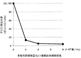
- the uptake of 3 H-thymidine in the cell non-addition group as 100%, 1 ⁇ 10 4 cells group, the uptake of 3 H-thymidine in 2 ⁇ 10 4 cells group and 4 ⁇ 10 4 cell group, to cell non-addition group It was calculated as a ratio (control%).
- peripheral blood mononuclear cells are composed of immune cells such as T cells
- this result indicates that dental pulp-derived cells have an inhibitory effect on the proliferation of immune cells such as T cells, and thus have an immunosuppressive effect. That is, this result indicates that dental pulp-derived cells can be used as a therapeutic agent for autoimmune disease, graft-versus-host disease, etc. as a T cell proliferation inhibitor and immunosuppressant.
- the pulp-derived cells obtained by the above expansion culture are pluripotent stem cells having active proliferation ability and the ability to differentiate into two types of cells, chondrocytes and osteoblasts.
- this pluripotent stem cell has a feature of showing little ability to differentiate into adipocytes and having the ability to suppress T cell proliferation.
- the present invention is useful for providing dental pulp-derived pluripotent stem cells having the ability to differentiate into chondrocytes and osteoblasts and having the ability to suppress T cell proliferation.
Abstract
Description
(1)多能性幹細胞富化ヒト歯髄由来細胞の製造方法であって,
(a)歯髄懸濁物に含まれる歯髄由来細胞を,細胞増殖能が抑制された支持細胞を含んだ支持細胞培養用容器中で,該支持細胞の通過を阻止できる微細孔を有する膜であって下側面が該支持細胞と接触しないように該支持細胞培養用容器内に支持されたものである膜の上で,該支持細胞と直接接触させることなく培養するステップと,
(b)該膜上で増殖した細胞を回収するステップとを含む,製造方法。
(2)上記1の製造方法であって,
(c)該回収された細胞を,細胞増殖能が抑制された支持細胞を含んだ支持細胞培養用容器中で,該支持細胞の通過を阻止できる微細孔を有する膜であって下側面が該支持細胞と接触しないように該支持細胞培養用容器内に支持されたものである膜の上で,該支持細胞と直接接触させることなく培養するステップと,
(d)該膜上で増殖した細胞を回収するステップとを,更に少なくとも1回行うことを含む,上記1の製造方法。
(3)該膜が,フィブロネクチン又はコラーゲンでコートされたものである,上記1又は2の方法。
(4)該微細孔の孔径が,平均0.1~1.5μmである,上記1~3の何れかの製造方法,
(5)該支持細胞が,マイトマイシンCによって細胞増殖能が抑制された哺乳動物細胞である,上記1~4の何れかの製造方法。
(6)該哺乳動物細胞が,NIH3T3細胞である,上記5の製造方法。
(7)該培養が,10~25%ウシ胎児血清及び3~5mMのL-アラニル-L-グルタミンを含むダルベッコの修正イーグル培地であって,グルコース濃度が5~7mMである培地を用いて行われるものである,上記1~6の何れかの製造方法。
(8)該培養が,20%ウシ胎児血清及び4mMのL-アラニル-L-グルタミンを含むダルベッコの修正イーグル培地であって,グルコース濃度が5.5~5.7mMである培地を用いて行われるものである,上記1~6の何れかの製造方法。
(9)上記1~8の何れかの製造方法により細胞を回収し,回収された細胞を,1000~20000細胞/cm2の密度で培養容器に加え,該培養容器の底面における該細胞の占める割合が70~100%となるまで培養するステップを更に含む,多能性幹細胞富化ヒト歯髄由来細胞の製造方法。
(10)上記1~8の何れかの方法により細胞を回収し,回収された細胞を,5000~10000細胞/cm2の密度で培養容器に加え,該培養容器の底面における該細胞の占める割合が90~100%となるまで培養するステップを更に含む,多能性幹細胞富化ヒト歯髄由来細胞胞の製造方法。
(11)上記1~10の何れかの方法で得られる,多能性幹細胞富化ヒト歯髄由来細胞。
(12)表面抗原マーカーが,CD29,CD44,CD73,CD90,CD105及びCD166が陽性であり,且つCD34及びCD45が陰性である,上記11の細胞。
(13)軟骨分化能及び骨芽分化能を有し,且つT細胞増殖抑制能を有するものである,上記12の細胞。
(14)10回以上の分裂能を有する,上記11~13の何れかの細胞。
(15)上記11~14の何れかの細胞を含んでなる免疫抑制剤。
(16)上記11~14の何れかの細胞を含んでなるT細胞増殖抑制剤。
(17)上記11~14の何れかの細胞を含んでなる移植片対宿主病治療剤。 In the research for the above purpose, the present inventor has found that cells obtained from dental pulp are cultured in the presence of supporting cells, and have high proliferative ability to differentiate into chondrocytes and osteoblasts and T cells. The present inventors have found that it is a pluripotent stem cell having both proliferation-inhibiting ability and completed the present invention based on this. That is, the present invention provides the following.
(1) A method for producing pluripotent stem cell-enriched human dental pulp-derived cells,
(A) The pulp-derived cells contained in the dental pulp suspension are membranes having micropores capable of blocking the passage of the supporting cells in the supporting cell culture vessel containing the supporting cells with suppressed cell proliferation ability. Culturing without contacting the supporting cells directly on a membrane that is supported in the supporting cell culture vessel so that the lower surface does not contact the supporting cells;
(B) collecting the cells grown on the membrane.
(2) The manufacturing method according to 1 above,
(C) The recovered cell is a membrane having micropores capable of preventing passage of the support cells in a support cell culture vessel containing support cells with suppressed cell proliferation ability, the lower surface of the membrane Culturing without contacting the supporting cells directly on the membrane that is supported in the supporting cell culture vessel so as not to contact the supporting cells;
(D) The method according to 1 above, further comprising the step of collecting the cells grown on the membrane at least once.
(3) The method according to 1 or 2 above, wherein the membrane is coated with fibronectin or collagen.
(4) The production method according to any one of 1 to 3 above, wherein the average pore diameter of the micropores is 0.1 to 1.5 μm,
(5) The method according to any one of (1) to (4), wherein the supporting cells are mammalian cells whose cell growth ability is suppressed by mitomycin C.
(6) The production method according to 5 above, wherein the mammalian cells are NIH3T3 cells.
(7) The culture is carried out using Dulbecco's modified Eagle medium containing 10 to 25% fetal bovine serum and 3 to 5 mM L-alanyl-L-glutamine, and having a glucose concentration of 5 to 7 mM. Any one of the
(8) The culture is performed using Dulbecco's modified Eagle medium containing 20% fetal bovine serum and 4 mM L-alanyl-L-glutamine, and having a glucose concentration of 5.5 to 5.7 mM. Any one of the
(9) Cells are collected by the production method of any one of 1 to 8, and the collected cells are added to the culture vessel at a density of 1000 to 20000 cells / cm 2 , and the cells occupy the bottom surface of the culture vessel A method for producing pluripotent stem cell-enriched human dental pulp-derived cells, further comprising a step of culturing until the ratio reaches 70 to 100%.
(10) Cells are collected by any of the
(11) A pluripotent stem cell-rich human dental pulp-derived cell obtained by any one of the
(12) The cell according to the above 11, wherein the surface antigen marker is positive for CD29, CD44, CD73, CD90, CD105 and CD166, and negative for CD34 and CD45.
(13) The 12 cells as described above, which have cartilage differentiation ability and osteoblast differentiation ability and have T cell proliferation inhibitory ability.
(14) The cell according to any one of the above 11 to 13, which has a division ability of 10 times or more.
(15) An immunosuppressant comprising the cell according to any one of 11 to 14 above.
(16) A T cell proliferation inhibitor comprising the cell of any one of 11 to 14 above.
(17) A therapeutic agent for graft-versus-host disease comprising the cell according to any one of 11 to 14 above.

DMEM Low Glucose(Invitrogen社)にFBS(Invitrogen社)とL-アラニル-L-グルタミンを各々終濃度が10%と4mMとなるように添加し,これをDMEM(10%FBS)培地とした。マイトマイシンC(SIGMA社)を注射用水で0.2mg/mLの濃度となるように溶解し,これをマイトマイシンC溶液とした。また,DMEM Low Glucose(Invitrogen社)にFBS(Invitrogen社)とL-アラニル-L-グルタミンを各々終濃度が20%と4mMとなるように添加し,これをDMEM(20%FBS)培地とした。 [Preparation of supporting cells]
FBS (Invitrogen) and L-alanyl-L-glutamine were added to DMEM Low Glucose (Invitrogen) to a final concentration of 10% and 4 mM, respectively, and this was used as DMEM (10% FBS) medium. Mitomycin C (SIGMA) was dissolved in water for injection to a concentration of 0.2 mg / mL, and this was used as a mitomycin C solution. In addition, FMEM (Invitrogen) and L-alanyl-L-glutamine were added to DMEM Low Glucose (Invitrogen) to a final concentration of 20% and 4 mM, respectively, and this was used as DMEM (20% FBS) medium. .
4mLのD-PBSに,12mgのコラゲナーゼタイプII(CALBIOCHEM社)を加えて混和した後,0.22μmフィルターに通し,これをコラゲナーゼ溶液とした。1mLのD-PBSに,10000 単位のディスパーゼ(合同酒精)を加えて混和した後,0.22μmフィルターに通し,これをディスパーゼ溶液とした。 [Isolation of dental pulp]
To 4 mL of D-PBS, 12 mg of collagenase type II (CALBIOCHEM) was added and mixed, and then passed through a 0.22 μm filter to make a collagenase solution. 10000 units of dispase (joint spirit) was added to 1 mL of D-PBS, mixed, and then passed through a 0.22 μm filter to make a dispase solution.
0.4μm径の孔を有するポリエチレンテレフタレート製の多孔性膜(track etched membrane)を底面に有する12ウェルインサート(BD Falcon Cell Culture Insert, BD Biosciences社)の多孔性膜にフィブロネクチンを1μg/cm2になるように添加し,37℃で30分以上静置し,多孔性膜をフィブロネクチンでコーティングした。フィブロネクチンは,Horwitz
B. et al., Preparation of fibronectin for therapeutic administration. In: Mosher
DF, editor.New York Academic Press Inc 441-445 (1989)に記載された方法を用いて調製した。 [Primary culture of dental pulp-derived cells (P0 culture)]
Fibronectin is 1 μg / cm 2 on a porous membrane of a 12-well insert (BD Falcon Cell Culture Insert, BD Biosciences) having a polyethylene terephthalate porous membrane with 0.4 μm pores on the bottom (track etched membrane). And left at 37 ° C. for more than 30 minutes, and the porous membrane was coated with fibronectin. Fibronectin is Horwitz
B. et al., Preparation of fibronectin for therapeutic administration. In: Mosher
It was prepared using the method described in DF, editor. New York Academic Press Inc 441-445 (1989).
ボトムウェルに支持細胞を添加した6ウェルコンパニオンプレートを準備し,これにフィブロネクチンコートした6ウェルインサートを静置し,この6ウェルインサートに,P0培養で得られた細胞懸濁液を添加して,5%CO2存在下,37℃でプレ拡大培養を開始した。このときの培養容器のセッティングを模式的に図1に示した。DMEM(20%FBS)培地を3~4日毎に交換しながら,6ウェルインサート上にコロニーが目視により確認されるまで,培養を継続した。この間に,ボトムウェルの支持細胞が,顕著にボトムウェルから剥離するようになった場合は,ボトムウェルの底面に支持細胞を固着させた6ウェルコンパニオンプレートを新たに準備し,これに6ウェルインサートを移して培養を継続した。 [Pre-expansion culture of pulp-derived cells]
Prepare a 6-well companion plate with feeder cells added to the bottom well, leave a 6-well insert coated with fibronectin, and add the cell suspension obtained in P0 culture to the 6-well insert. Pre-expansion culture was started at 37 ° C. in the presence of 5% CO 2 . The setting of the culture container at this time is schematically shown in FIG. Cultivation was continued until colonies were visually confirmed on the 6-well inserts, changing the DMEM (20% FBS) medium every 3-4 days. In the meantime, if the supporting cells in the bottom well become detached from the bottom well, a 6-well companion plate with the supporting cells fixed to the bottom of the bottom well is newly prepared, and a 6-well insert is added thereto. The culture was continued.
上記の細胞の懸濁液に含まれる生細胞数を血球計算盤で測定した後,5000~10000細胞/cm2の密度となるように,培養フラスコに細胞を播種してDMEM(10%FBS)培地を用いて拡大培養を開始した。但し,生細胞数が少なく拡大培養が開始できない場合は,拡大培養を開始するために必要な数の細胞が得られるまでプレ拡大培養を繰り返した。拡大培養開始時に,培養フラスコに播種した細胞を観察したところ,ほぼ全ての細胞が培養フラスコの底面に固着して紡錘状の形状を示し,均質な細胞が分離されたことが分かった。 [Expansion culture of pulp-derived cells]
After measuring the number of viable cells in the above cell suspension with a hemocytometer, seed the cells in a culture flask so that the density is 5000 to 10000 cells / cm 2 and then DMEM (10% FBS) Expansion culture was started using the medium. However, when the number of living cells was small and expansion culture could not be started, pre-expansion culture was repeated until the required number of cells was obtained to start expansion culture. At the start of expansion culture, the cells seeded in the culture flask were observed, and it was found that almost all cells adhered to the bottom surface of the culture flask and showed a spindle-shaped shape, so that homogeneous cells were separated.
第2回拡大培養において,90~100%コンフルエントになるまで培養した細胞の一部を,下記の手法により凍結保存した。PBSで細胞を洗浄した後,0.25%トリプシン-EDTAを添加し,37℃に5~10分間静置して細胞を剥離させた。DMEM(10%FBS)培地を添加して反応を停止させるとともに細胞を懸濁し,15mL遠沈管に細胞を回収した。回収した細胞を遠心(1500rpm, 5分)して沈殿させ,上清を除去した。10%(v/v)DMSOを含むFBS溶液で細胞を1×106個/mLの密度で懸濁し,0.5~2mLずつクライオチューブに分注した後,予め4℃で冷蔵しておいたBICELL(日本フリーザー)に入れ,-80℃にて凍結した。凍結約24時間後に,細胞を気相式液体窒素に移し保管した。 [Cryopreservation of dental pulp-derived cells]
In the second expansion culture, some of the cells cultured until they became 90-100% confluent were stored frozen by the following method. After washing the cells with PBS, 0.25% trypsin-EDTA was added, and the cells were allowed to stand at 37 ° C. for 5 to 10 minutes to detach the cells. DMEM (10% FBS) medium was added to stop the reaction and the cells were suspended, and the cells were collected in a 15 mL centrifuge tube. The collected cells were centrifuged (1500 rpm, 5 minutes) to precipitate, and the supernatant was removed. BICELL was suspended in FBS solution containing 10% (v / v) DMSO at a density of 1 × 10 6 cells / mL, dispensed 0.5-2 mL into cryotubes, and refrigerated at 4 ° C in advance. (Japan freezer) and frozen at -80 ° C. About 24 hours after freezing, the cells were transferred to gas phase liquid nitrogen and stored.
上記の拡大培養において継代培養を16回繰り返し,継代培養終了時毎に生細胞数を測定して,細胞の分裂回数を算出することにより,細胞の増殖能を測定した(図2)。細胞の分裂回数は,各継代培養の開始時の生細胞数と培養終了時の生細胞数から下記の式により算出した。このとき,拡大培養開始時の培養を第1回拡大培養とし,以後,継代培養を繰り返す毎に,第2回拡大培養,第3回拡大培養とし,最後の拡大培養を第16回拡大培養とした。
細胞の分裂回数=log2(培養終了時の細胞数/培養開始時の細胞数) [Measurement of proliferative ability of dental pulp-derived cells]
In the expansion culture, the subculture was repeated 16 times, and the number of viable cells was measured at the end of the subculture and the number of cell divisions was calculated to measure the cell proliferation ability (FIG. 2). The number of cell divisions was calculated from the number of viable cells at the start of each subculture and the number of viable cells at the end of the culture according to the following formula. At this time, the culture at the start of the expansion culture is designated as the first expansion culture, and thereafter, every time the subculture is repeated, the second expansion culture and the third expansion culture are designated, and the final expansion culture is designated as the 16th expansion culture. It was.
Number of cell divisions = log 2 (number of cells at the end of culture / number of cells at the start of culture)
歯髄由来細胞は,ヒト間葉系幹細胞と同じく均質な紡錘状の形状を示した。そこで,ヒト間葉系幹細胞で陽性であることが知られている表面抗原マーカーであるCD29, CD44, CD73,CD90,CD105及びCD166,及び間葉系幹細胞で陰性であることが知られている表面抗原マーカーであるCD34とCD45の発現の有無を,FACSを用いて調べた。 [Measurement of surface antigen]
The pulp-derived cells showed a homogeneous spindle-like shape, similar to human mesenchymal stem cells. Therefore, CD29, CD44, CD73, CD90, CD105 and CD166, which are surface antigen markers known to be positive in human mesenchymal stem cells, and surfaces known to be negative in mesenchymal stem cells The presence or absence of expression of antigen markers CD34 and CD45 was examined using FACS.
IgG溶液を調製した。4.6mLのPBS-Bに,0.4mLの25mg/mL IgG溶液を加えて,ブロッキング溶液を調製した。2.4mLのPBS-Bに,0.4m LのStreptavidin APC(CALTAG社)を加えて,Streptavidin APC希釈液とした。 Phosphate buffer saline pH 7.4, contains BSA, powder (Sigma) dissolved in pure water, passed through 0.22μm filter, PBS-B [0.138M sodium chloride, 0.0027M potassium chloride, 1% (w / v) 0.01 M phosphate buffered saline (pH 7.4) containing bovine serum albumin] was prepared. IgG from Human (SIGMA) is dissolved in physiological saline (Otsuka Pharmaceutical) and passed through a 0.22 μm filter, 25 mg / mL
An IgG solution was prepared. A blocking solution was prepared by adding 0.4 mL of 25 mg / mL IgG solution to 4.6 mL of PBS-B. 0.4 mL of Streptavidin APC (CALTAG) was added to 2.4 mL of PBS-B to prepare a Streptavidin APC dilution.
BD社),FITC標識抗ヒトCD34抗体(anti-CD34-FITC, BD社),Biotin標識抗ヒトCD44抗体(anti-CD44-Biotin, BD社),FITC標識抗ヒトCD45抗体(anti-CD45-FITC, IMMUNOTECH社),PE標識抗ヒトCD73抗体(anti-CD73-PE,
BD社),PE標識抗ヒトCD105抗体(anti-CD105-R-PE, Ancell社),FITC標識マウスIgG1(anti-IgG1-FITC, IMMUNOTECH社),PE標識マウスIgG1アイソタイプコントロール(IgG1-PE, IMMUNOTECH社)及びBiotin標識マウスIgG2bアイソタイプコントロール(IgG2b-Biotin, BD社)は,市販のものをそのまま希釈抗体液として用いた。 A diluted antibody solution of the labeled antibody was prepared as follows. 2 μL of Biotin-labeled mouse IgG1 isotype control (CALTAG) was added to 18 μL of PBS-B to obtain a Biotin-labeled mouse IgG1 dilution (IgG1-Biotin dilution). 2 μL of Biotin-labeled mouse anti-human CD90 antibody (BD) was added to 38 μL of PBS-B to prepare an anti-CD90-Biotin dilution. To 46.5 μL PBS-B, 3.5 μL Biotin-labeled mouse anti-human CD166 antibody (Fitzgerald) was added to prepare an anti-CD166-Biotin dilution. FITC-labeled anti-human CD29 antibody (anti-CD29-FITC,
BD), FITC-labeled anti-human CD34 antibody (anti-CD34-FITC, BD), Biotin-labeled anti-human CD44 antibody (anti-CD44-Biotin, BD), FITC-labeled anti-human CD45 antibody (anti-CD45-FITC) , IMMUNOTECH), PE-labeled anti-human CD73 antibody (anti-CD73-PE,
BD), PE-labeled anti-human CD105 antibody (anti-CD105-R-PE, Ancell), FITC-labeled mouse IgG1 (anti-IgG1-FITC, IMMUNOTECH), PE-labeled mouse IgG1 isotype control (IgG1-PE, IMMUNOTECH) ) And Biotin-labeled mouse IgG2b isotype control (IgG2b-Biotin, BD) were used as diluted antibody solutions as they were.
上記凍結保存した細胞(1×10⁶個/mL)を解凍し,10mLのDMEM(10%FBS)培地を加えて軽く振り混ぜた後,遠心(1500rpm, 5分)して細胞を沈殿させた。上清を除去し,10mLのDMEM(10%FBS)培地を添加して細胞を懸濁し,再度遠心(1500rpm, 5分)して細胞を沈殿させた。次いで,細胞をDMEM(10%FBS)培地に懸濁し,5000~10000細胞/cm2の密度となるように,培養フラスコに播種し,細胞が90~100%コンフルエントになるまで培養した。PBSで細胞を洗浄し,0.25%トリプシン-EDTAを添加し,37℃に5~10分間静置して細胞を剥離させた。DMEM(10%FBS)培地を添加して細胞を懸濁し,生細胞数を測定した。歯髄由来細胞(2×107 個)を50mL遠心管に採取し,PBS-Bを加えて全量を28mLにした。細胞を遠心(1,500rpm, 5分)して沈殿させ,上清を除去した。次いで,細胞をブロッキング溶液700μLに懸濁し,氷上で20分間静置した。13本の5mL反応チューブ(番号(1)~(13))に,各希釈抗体液を表2に示すとおり添加した。次いで,各チューブに,ブロッキング溶液に懸濁させた細胞の懸濁液を50μLずつ添加して軽く振り混ぜ,氷上で20分間静置させて,試液に含まれる抗体と細胞表面に発現する表面抗原マーカーとを結合させた。次いで,PBS-Bを3mLずつ添加して混和した後,細胞を遠心(1500rpm, 5分)して沈殿させて上清を除去し,細胞に結合しない抗体を除去した。この抗体の除去操作を1回繰り返した。 (Cell staining method)
Thaw the above cryopreserved cells (1 × 10 6 cells / mL), add 10 mL DMEM (10% FBS) medium, shake gently, and then centrifuge (1500 rpm, 5 minutes) to precipitate the cells . The supernatant was removed, 10 mL of DMEM (10% FBS) medium was added, the cells were suspended, and centrifuged again (1500 rpm, 5 minutes) to precipitate the cells. The cells were then suspended in DMEM (10% FBS) medium, seeded in culture flasks at a density of 5000-10000 cells / cm 2 and cultured until the cells were 90-100% confluent. The cells were washed with PBS, 0.25% trypsin-EDTA was added, and the cells were allowed to stand at 37 ° C. for 5 to 10 minutes to detach the cells. DMEM (10% FBS) medium was added to suspend the cells, and the number of viable cells was measured. Pulp-derived cells (2 × 10 7 cells) were collected in a 50 mL centrifuge tube, and PBS-B was added to make a total volume of 28 mL. The cells were centrifuged (1,500 rpm, 5 minutes) to precipitate, and the supernatant was removed. The cells were then suspended in 700 μL of blocking solution and left on ice for 20 minutes. Each diluted antibody solution was added to 13 5 mL reaction tubes (Nos. (1) to (13)) as shown in Table 2. Next, add 50 μL of the cell suspension suspended in the blocking solution to each tube, shake gently, let it stand on ice for 20 minutes, and the antibody contained in the test solution and the surface antigen expressed on the cell surface. Combined with the marker. Next, 3 mL of PBS-B was added and mixed, and the cells were centrifuged (1500 rpm, 5 minutes) to precipitate and the supernatant was removed to remove antibodies that did not bind to the cells. This antibody removal operation was repeated once.


チューブ番号(1)~(13)の細胞について,APC,FITC及びPEの各蛍光色素の量をFACS
CantoTM(BD社)で測定し,陰性コントロールとの比較により,表面抗原に特異的に結合した抗体を介して細胞表面に結合した蛍光色素の量を求めた。なお,チューブ(2)はチューブ(6),(7)及び(9),チューブ(3)は(10)及び(12),チューブ(4)はチューブ(11)及び(13),チューブ(5)はチューブ(8)の陰性コントロールである。チューブ(1)は非染色細胞である。
その結果,チューブ番号(6),(8),及び(10)~(13)について,細胞表面に結合した蛍光色素の量は,陰性コントロールより多く,チューブ番号(7)と(9)については,細胞表面に結合した蛍光色素の量は,陰性コントロールと同等であった。これらの結果は,歯髄由来細胞が,ヒト間葉系幹細胞と同様に,CD29,CD44,CD73,DC90,CD105及びCD166が陽性であり,CD34とCD45が陰性であることを示す。 (Measurement and analysis)
For the cells with tube numbers (1) to (13), the amount of each APC, FITC, and PE fluorescent dye is FACS.
The amount of fluorescent dye bound to the cell surface via an antibody specifically bound to the surface antigen was determined by measurement with Canto ™ (BD) and comparison with a negative control. Tube (2) is tubes (6), (7) and (9), tube (3) is (10) and (12), tube (4) is tubes (11) and (13), tube (5 ) Is a negative control for tube (8). Tube (1) is unstained cells.
As a result, for tube numbers (6), (8), and (10)-(13), the amount of fluorescent dye bound to the cell surface was greater than for the negative control, and for tube numbers (7) and (9) The amount of fluorescent dye bound to the cell surface was equivalent to the negative control. These results indicate that dental pulp-derived cells are positive for CD29, CD44, CD73, DC90, CD105, and CD166, and negative for CD34 and CD45, similar to human mesenchymal stem cells.
軟骨分化能は,Kiani C., et al. Cell Res. 12 19-32 (2002),Aung
A., et al. Arthritis Rheum. 63 148-58 (2011)の記載を参考にして,下記の方法で調べた。 [Measurement of cartilage differentiation ability]
Cartilage differentiation ability is reported by Kiani C., et al. Cell Res. 12 19-32 (2002), Aung
With reference to the description of A., et al. Arthritis Rheum. 63 148-58 (2011), the following method was used.
1-Step キット(Life Technologies)を用いてPCR反応液を調製し,各群の全RNA 25ngを鋳型とし,〔(48℃/15min)×1サイクル,(95℃/10min)×1サイクル, (95℃/15sec, 62℃/1min)×35サイクル〕のPCR条件下でリアルタイムRT-PCRを行い,アグリカン遺伝子とβアクチン遺伝子を増幅させた。PCRプライマーには,アグリカンプローブ(Applied Biosystems社製/Assay ID:Hs00202971_m1)及びβアクチンプローブ(Applied Biosystems社製/ Assay ID:Hs99999903_m1)をそれぞれ用いた。 After incubation, total RNA is extracted from each group of cells using the RNeasy Plus Mini Kit (QIAGEN) to prepare a total RNA extract, and then included in the total RNA extract using a multimode plate reader (Molecular Device). RNA concentration was measured. After measuring RNA concentration, TaqMan TM RNA-to-CT TM
Prepare PCR reaction solution using 1-Step kit (Life Technologies), and use 25 ng of total RNA of each group as a template, [(48 ℃ / 15min) × 1 cycle, (95 ℃ / 10min) × 1 cycle, ( Real-time RT-PCR was performed under PCR conditions of 95 ° C / 15sec, 62 ° C / 1min) × 35 cycles] to amplify the aggrecan gene and β-actin gene. As the PCR primers, an aggrecan probe (Applied Biosystems / Assay ID: Hs00202971_m1) and a β-actin probe (Applied Biosystems / Assay ID: Hs99999903_m1) were used, respectively.
骨芽分化能は,Pittenger MF., et al., Science. 284, 143-7 (1999),Colter DC., et al., Proc Natl Acad Sci USA. 98, 7841-5(2001)の記載を参考にして,下記の方法で調べた。 (Measurement of osteoblast differentiation ability)
The osteoblast differentiation ability is described in Pittenger MF., Et al., Science. 284, 143-7 (1999), Colter DC., Et al., Proc Natl Acad Sci USA. 98, 7841-5 (2001). The following method was used for reference.
脂肪分化能は,Gimble JM., et al., J Cell Biochem. 58. 393-402 (1995) の記載を参考にして,下記の方法で調べた。 [Measurement of fat differentiation capacity]
Adipogenic ability was examined by the following method with reference to the description of Gimble JM., Et al., J Cell Biochem. 58. 393-402 (1995).
1-Step キット(Life Technologies)を用いてPCR反応液を調製し,各群の全RNA 25ngを鋳型とし,〔(48℃/15min)×1サイクル,(95℃/10min)×1サイクル, (95℃/15sec, 60℃/1min)×40 サイクル〕のPCR条件下でリアルタイムRT-PCRを行い,Lipo Protein Lipase遺伝子とβアクチン遺伝子を増幅させた。PCRプライマーには,Lipo Protein Lipaseプローブ(Applied Biosystems社製/Assay ID:Hs00173425_m1)及びβアクチンプローブ(Applied Biosystems社製/ Assay ID:Hs99999903_m1)をそれぞれ用いた。 Next, changes in the expression level of Lipo Protein Lipase, one of the adipocyte differentiation markers, were examined. Extract total RNA from each group of cells using the RNeasy Plus Mini Kit (QIAGEN) to prepare a total RNA extract, and then use a multimode plate reader (Molecular Devices) to determine the RNA concentration contained in the total RNA extract. It was measured. After measuring RNA concentration, TaqMan TM RNA-to-CT TM
Prepare a PCR reaction solution using 1-Step kit (Life Technologies), and use 25 ng of total RNA from each group as a template, [(48 ℃ / 15min) × 1 cycle, (95 ℃ / 10min) × 1 cycle, ( Real-time RT-PCR was performed under PCR conditions of 95 ° C / 15sec, 60 ° C / 1min) × 40 cycles] to amplify the Lipo Protein Lipase gene and β-actin gene. Lipo Protein Lipase probe (Applied Biosystems / Assay ID: Hs00173425_m1) and β-actin probe (Applied Biosystems / Assay ID: Hs99999903_m1) were used as PCR primers, respectively.
ヒト末梢血単核球(Lonza社)を37℃の恒温槽で解凍し,DMEM培地10mLで懸濁した後,遠心(1500rpm, 5分)して沈殿させ,上清を除去した。DMEM(10%FBS)培地を加えて細胞を懸濁し,トリパンブルー染色により生細胞数を測定した後,細胞濃度が6×10⁶個/mLとなるように細胞を懸濁させ,これをPBMC懸濁液とした。PHA-P(SIGMA社)5mgをDMEM(10%FBS)培地5mLに溶解させ,0.22μmフィルターで除菌した後,DMEM(10%FBS)培地で25倍希釈し,これをPHA-P含有培地とした。[メチル-3H]チミジン(Moravek社)をDMEM(10%FBS)培地で20倍希釈し,これをチミジン溶液とした。 [Measurement of ability to suppress T cell proliferation]
Human peripheral blood mononuclear cells (Lonza) were thawed in a constant temperature bath at 37 ° C., suspended in 10 mL of DMEM medium, centrifuged (1500 rpm, 5 minutes), precipitated, and the supernatant was removed. Suspend cells by adding DMEM (10% FBS) medium, measure the number of viable cells by trypan blue staining, and then suspend the cells so that the cell concentration is 6 × 10 6 cells / mL. A suspension was formed. Dissolve 5 mg of PHA-P (SIGMA) in 5 mL of DMEM (10% FBS) medium, sterilize it with a 0.22 μm filter, dilute it 25-fold with DMEM (10% FBS) medium, and add PHA-P-containing medium. It was. [Methyl - 3 H] thymidine (Moravek Ltd.) was diluted 20-fold in DMEM (10% FBS) medium, which was used as a thymidine solution.
上記の結果から,上記拡大培養により得られた歯髄由来細胞は,活発な増殖能を有し,且つ軟骨細胞及び骨芽細胞と2種類の細胞に分化する能力を有する多能性幹細胞であることが確認された。また,この多能性幹細胞は,脂肪細胞への分化能をほとんど示さず,T細胞増殖抑制能を有するという特徴を備えることが判明した。 [Summary]
From the above results, the pulp-derived cells obtained by the above expansion culture are pluripotent stem cells having active proliferation ability and the ability to differentiate into two types of cells, chondrocytes and osteoblasts. Was confirmed. In addition, it was found that this pluripotent stem cell has a feature of showing little ability to differentiate into adipocytes and having the ability to suppress T cell proliferation.
2.12ウェルコンパニオンプレート又は6ウェルコンパニオンプレート
3.フィブロネクチンコートした多孔性膜
4.歯髄由来細胞
5.支持細胞
6.ボトムウェル
1. 12 well insert or 6
Claims (17)
- 多能性幹細胞富化ヒト歯髄由来細胞の製造方法であって,
(a)歯髄懸濁物に含まれる歯髄由来細胞を,細胞増殖能が抑制された支持細胞を含んだ支持細胞培養用容器中で,該支持細胞の通過を阻止できる微細孔を有する膜であって下側面が該支持細胞と接触しないように該支持細胞培養用容器に支持されたものである膜の上で,該支持細胞と直接接触させることなく培養するステップと,
(b)該膜上で増殖した細胞を回収するステップとを含む,製造方法。 A method for producing pluripotent stem cell-rich human dental pulp-derived cells, comprising:
(A) The pulp-derived cells contained in the dental pulp suspension are membranes having micropores capable of blocking the passage of the supporting cells in the supporting cell culture vessel containing the supporting cells with suppressed cell proliferation ability. Culturing without contacting the supporting cells directly on the membrane that is supported by the supporting cell culture vessel so that the lower surface does not contact the supporting cells;
(B) collecting the cells grown on the membrane. - 請求項1の製造方法であって,
(c)該回収された細胞を,細胞増殖能が抑制された支持細胞を含んだ支持細胞培養用容器中で,該支持細胞の通過を阻止できる微細孔を有する膜であって下側面が該支持細胞と接触しないように該支持細胞培養用容器に支持されたものである膜の上で,該支持細胞と直接接触させることなく培養するステップと,
(d)該膜上で増殖した細胞を回収するステップとを,更に少なくとも1回行うことを含む,請求項1の製造方法。 The manufacturing method according to claim 1,
(C) The recovered cell is a membrane having micropores capable of preventing passage of the support cells in a support cell culture vessel containing support cells with suppressed cell proliferation ability, the lower surface of the membrane Culturing on a membrane that is supported in the feeder cell culture vessel so as not to contact the feeder cells without directly contacting the feeder cells;
(D) The step of collecting the cells grown on the membrane is further performed at least once. - 該膜が,フィブロネクチン又はコラーゲンでコートされたものである,請求項1又は2の方法。 The method according to claim 1 or 2, wherein the membrane is coated with fibronectin or collagen.
- 該微細孔の孔径が,平均0.1~1.5μmである,請求項1~3の何れかの製造方法, The manufacturing method according to any one of claims 1 to 3, wherein the average pore diameter of the micropores is 0.1 to 1.5 µm.
- 該支持細胞が,マイトマイシンCによって細胞増殖能が抑制された哺乳動物細胞である,請求項1~4の何れかの製造方法。 The production method according to any one of claims 1 to 4, wherein the supporting cells are mammalian cells whose cell growth ability is suppressed by mitomycin C.
- 該哺乳動物細胞が,NIH3T3細胞である,請求項5の製造方法。 The production method according to claim 5, wherein the mammalian cells are NIH3T3 cells.
- 該培養が,10~25%ウシ胎児血清及び3~5mMのL-アラニル-L-グルタミンを含むダルベッコの修正イーグル培地であって,グルコース濃度が5~7mMである培地を用いて行われるものである,請求項1~6の何れかの製造方法。 The culture is performed using Dulbecco's modified Eagle's medium containing 10-25% fetal bovine serum and 3-5 mM L-alanyl-L-glutamine, wherein the glucose concentration is 5-7 mM. The production method according to any one of claims 1 to 6.
- 該培養が,20%ウシ胎児血清及び4mMのL-アラニル-L-グルタミンを含むダルベッコの修正イーグル培地であって,グルコース濃度が5.5~5.7mMである培地を用いて行われるものである,請求項1~6の何れかの製造方法。 The culture is performed using Dulbecco's modified Eagle medium containing 20% fetal bovine serum and 4 mM L-alanyl-L-glutamine, wherein the glucose concentration is 5.5 to 5.7 mM. The production method according to any one of claims 1 to 6.
- 請求項1~8の何れかの製造方法により細胞を回収し,回収された細胞を,1000~20000細胞/cm2の密度で培養容器に加え,該培養容器の底面における該細胞の占める割合が70~100%となるまで培養するステップを更に含む,多能性幹細胞富化ヒト歯髄由来細胞の製造方法。 Cells are collected by the production method according to any one of claims 1 to 8, and the collected cells are added to the culture vessel at a density of 1000 to 20000 cells / cm 2 , and the proportion of the cells in the bottom surface of the culture vessel is A method for producing pluripotent stem cell-enriched human dental pulp-derived cells, further comprising a step of culturing to 70 to 100%.
- 請求項1~8の何れかの方法により細胞を回収し,回収された細胞を,5000~10000細胞/cm2の密度で培養容器に加え,該培養容器の底面における該細胞の占める割合が90~100%となるまで培養するステップを更に含む,多能性幹細胞富化ヒト歯髄由来細胞胞の製造方法。 Cells are collected by the method according to any one of claims 1 to 8, and the collected cells are added to the culture container at a density of 5000 to 10,000 cells / cm 2 , and the proportion of the cells in the bottom surface of the culture container is 90%. A method for producing pluripotent stem cell-enriched human dental pulp-derived cell vesicles, further comprising a step of culturing until ˜100%.
- 請求項1~10の何れかの方法で得られる,多能性幹細胞富化ヒト歯髄由来細胞。 A pluripotent stem cell-enriched human dental pulp-derived cell obtained by the method according to any one of claims 1 to 10.
- 表面抗原マーカーが、CD29,CD44,CD73,CD90,CD105及びCD166が陽性であり、且つCD34及びCD45が陰性である、上記11の細胞。 The above 11 cells, wherein the surface antigen markers are positive for CD29, CD44, CD73, CD90, CD105 and CD166, and negative for CD34 and CD45.
- 軟骨分化能及び骨芽分化能を有し,且つT細胞増殖抑制能を有するものである,請求項12の細胞。 The cell according to claim 12, which has a cartilage differentiation ability and an osteoblast differentiation ability and has a T cell proliferation inhibitory ability.
- 10回以上の分裂能を有する,請求項11~13の何れかの細胞。 The cell according to any one of claims 11 to 13, which has a division ability of 10 times or more.
- 請求項11~14の何れかの細胞を含んでなる免疫抑制剤。 An immunosuppressive agent comprising the cell according to any one of claims 11 to 14.
- 請求項11~14の何れかの細胞を含んでなるT細胞増殖抑制剤。 A T cell proliferation inhibitor comprising the cell according to any one of claims 11 to 14.
- 請求項11~14の何れかの細胞を含んでなる移植片対宿主病治療剤。 A graft-versus-host disease therapeutic agent comprising the cell according to any one of claims 11 to 14.
Priority Applications (3)
| Application Number | Priority Date | Filing Date | Title |
|---|---|---|---|
| US14/388,464 US10041039B2 (en) | 2012-03-29 | 2013-03-28 | Method for producing pluripotent stem cells derived from dental pulp |
| JP2014508009A JP6193214B2 (en) | 2012-03-29 | 2013-03-28 | Method for producing dental pulp-derived pluripotent stem cells |
| EP13768121.9A EP2832850B1 (en) | 2012-03-29 | 2013-03-28 | Method for producing pluripotent stem cells derived from dental pulp |
Applications Claiming Priority (2)
| Application Number | Priority Date | Filing Date | Title |
|---|---|---|---|
| JP2012-077404 | 2012-03-29 | ||
| JP2012077404 | 2012-03-29 |
Publications (1)
| Publication Number | Publication Date |
|---|---|
| WO2013146992A1 true WO2013146992A1 (en) | 2013-10-03 |
Family
ID=49260240
Family Applications (1)
| Application Number | Title | Priority Date | Filing Date |
|---|---|---|---|
| PCT/JP2013/059201 WO2013146992A1 (en) | 2012-03-29 | 2013-03-28 | Method for producing pluripotent stem cells derived from dental pulp |
Country Status (4)
| Country | Link |
|---|---|
| US (1) | US10041039B2 (en) |
| EP (1) | EP2832850B1 (en) |
| JP (1) | JP6193214B2 (en) |
| WO (1) | WO2013146992A1 (en) |
Cited By (7)
| Publication number | Priority date | Publication date | Assignee | Title |
|---|---|---|---|---|
| CN103752174A (en) * | 2014-01-24 | 2014-04-30 | 北京理工大学 | Antibacterial hydrophilic hollow fiber assembly for water treatment |
| WO2016076434A1 (en) * | 2014-11-14 | 2016-05-19 | Jcrファーマ株式会社 | Muscular dystrophy therapeutic agent containing pluripotent stem cells derived from dental pulp |
| JP2017158488A (en) * | 2016-03-10 | 2017-09-14 | 東洋紡株式会社 | Cell recovery method |
| JPWO2016181885A1 (en) * | 2015-05-14 | 2018-03-01 | 株式会社セルテクノロジー | Method for producing frozen mesenchymal cells and method for producing therapeutic material for transplantation |
| WO2018074381A1 (en) * | 2016-10-18 | 2018-04-26 | 国立大学法人大阪大学 | Kit for preparing disease-treating agent, disease-treating agent and method for preparing disease-treating agent |
| CN109432128A (en) * | 2018-12-07 | 2019-03-08 | 卡替(上海)生物技术股份有限公司 | Application of the dental pulp mescenchymal stem cell in curing psoriasis |
| CN113577107A (en) * | 2021-05-21 | 2021-11-02 | 施松涛 | Preparation method and application of stem cells |
Families Citing this family (1)
| Publication number | Priority date | Publication date | Assignee | Title |
|---|---|---|---|---|
| CN112469815A (en) * | 2018-07-31 | 2021-03-09 | Jcr制药股份有限公司 | Method for producing dental pulp-derived cells |
Citations (21)
| Publication number | Priority date | Publication date | Assignee | Title |
|---|---|---|---|---|
| US5486359A (en) | 1990-11-16 | 1996-01-23 | Osiris Therapeutics, Inc. | Human mesenchymal stem cells |
| US5827740A (en) | 1996-07-30 | 1998-10-27 | Osiris Therapeutics, Inc. | Adipogenic differentiation of human mesenchymal stem cells |
| US6328960B1 (en) | 1998-03-18 | 2001-12-11 | Osiris Therapeutics, Inc. | Mesenchymal stem cells for prevention and treatment of immune responses in transplantation |
| WO2002007679A2 (en) | 2000-07-21 | 2002-01-31 | The Government Of The United States Of America, Represented By The Secretary, Department Of Health And Human Services | Adult human dental pulp stem cells in vitro and in vivo |
| US6387369B1 (en) | 1997-07-14 | 2002-05-14 | Osiris Therapeutics, Inc. | Cardiac muscle regeneration using mesenchymal stem cells |
| JP2004129549A (en) | 2002-10-09 | 2004-04-30 | Yasuo Kitagawa | Selective growth method for mesenchymal stem cell from fat-derived cell group |
| JP2004201612A (en) | 2002-12-26 | 2004-07-22 | Minoru Ueda | Undifferentiated multipotential cell and method for preparing related tissue or tooth by using the same |
| JP2004210713A (en) | 2002-12-27 | 2004-07-29 | Asahi Kasei Corp | Medical cell preparation derived from placenta |
| US6835377B2 (en) | 1998-05-13 | 2004-12-28 | Osiris Therapeutics, Inc. | Osteoarthritis cartilage regeneration |
| JP2005516616A (en) | 2002-02-06 | 2005-06-09 | シュティフトゥング カエザール | Tooth-derived pluripotent embryonic-like stem cells and use thereof |
| WO2005093044A1 (en) | 2004-03-22 | 2005-10-06 | Osiris Therapeutics, Inc. | Mesenchymal stem cells and uses therefor |
| JP2006238875A (en) | 2005-02-04 | 2006-09-14 | National Institute Of Advanced Industrial & Technology | Stem cell from human dental papilla and method for utilizing the same |
| JP2008507962A (en) | 2004-07-28 | 2008-03-21 | カリンチ,フランチェスコ | Stem cells obtained from pulp of deciduous or permanent teeth and tooth germ capable of producing human bone tissue |
| JP2008295420A (en) | 2007-06-04 | 2008-12-11 | Niigata Univ | Human periodontal ligament cell line, osteoblast dfferentiated from the cell line and artificial bone produced from the osteoblast |
| WO2009057537A1 (en) | 2007-11-02 | 2009-05-07 | Jcr Pharmaceuticals Co., Ltd. | Pharmaceutical composition containing human mesenchymal stem cell |
| WO2009072527A1 (en) | 2007-12-05 | 2009-06-11 | National University Corporation Nagoya University | Composition for autotransplantation or allotransplantation using dental pulp stem cell, and use of the composition |
| JP2009527223A (en) | 2006-02-20 | 2009-07-30 | テスラブ エッセ.エッレ.エッレ. | Method for collecting and selecting embryonic-like stem cell clusters from human adult dental sac tissue |
| JP2010252778A (en) | 2009-04-02 | 2010-11-11 | Shizui Bank Kk | Method for separating dental pulp-derived mesenchymal stem cell, separated stem cell and differentiation to hepatocyte lineage using them |
| JP2010268715A (en) | 2009-05-20 | 2010-12-02 | Nagoya Univ | Use of gene expression group characteristic of dental pulp stem cell of milk tooth |
| JP2011177140A (en) | 2010-03-03 | 2011-09-15 | Nippon Dental Univ | Serum-free medium for stem cell culture |
| JP2011525798A (en) * | 2008-06-26 | 2011-09-29 | ウニベルシタ デグリ ストゥディ ディ ウディネ | Dental pulp-like cells (DPMSC) and methods of isolation and use thereof |
Family Cites Families (3)
| Publication number | Priority date | Publication date | Assignee | Title |
|---|---|---|---|---|
| JP2006280234A (en) | 2005-03-31 | 2006-10-19 | Hitachi Medical Corp | Method for proliferating or differentiating cell |
| US9309491B2 (en) * | 2007-05-29 | 2016-04-12 | Corning Incorporated | Cell culture apparatus for co-culture of cells |
| JP2012507258A (en) * | 2008-10-30 | 2012-03-29 | 国立大学法人京都大学 | Method for producing induced pluripotent stem cells |
-
2013
- 2013-03-28 US US14/388,464 patent/US10041039B2/en not_active Expired - Fee Related
- 2013-03-28 WO PCT/JP2013/059201 patent/WO2013146992A1/en active Application Filing
- 2013-03-28 EP EP13768121.9A patent/EP2832850B1/en not_active Not-in-force
- 2013-03-28 JP JP2014508009A patent/JP6193214B2/en active Active
Patent Citations (21)
| Publication number | Priority date | Publication date | Assignee | Title |
|---|---|---|---|---|
| US5486359A (en) | 1990-11-16 | 1996-01-23 | Osiris Therapeutics, Inc. | Human mesenchymal stem cells |
| US5827740A (en) | 1996-07-30 | 1998-10-27 | Osiris Therapeutics, Inc. | Adipogenic differentiation of human mesenchymal stem cells |
| US6387369B1 (en) | 1997-07-14 | 2002-05-14 | Osiris Therapeutics, Inc. | Cardiac muscle regeneration using mesenchymal stem cells |
| US6328960B1 (en) | 1998-03-18 | 2001-12-11 | Osiris Therapeutics, Inc. | Mesenchymal stem cells for prevention and treatment of immune responses in transplantation |
| US6835377B2 (en) | 1998-05-13 | 2004-12-28 | Osiris Therapeutics, Inc. | Osteoarthritis cartilage regeneration |
| WO2002007679A2 (en) | 2000-07-21 | 2002-01-31 | The Government Of The United States Of America, Represented By The Secretary, Department Of Health And Human Services | Adult human dental pulp stem cells in vitro and in vivo |
| JP2005516616A (en) | 2002-02-06 | 2005-06-09 | シュティフトゥング カエザール | Tooth-derived pluripotent embryonic-like stem cells and use thereof |
| JP2004129549A (en) | 2002-10-09 | 2004-04-30 | Yasuo Kitagawa | Selective growth method for mesenchymal stem cell from fat-derived cell group |
| JP2004201612A (en) | 2002-12-26 | 2004-07-22 | Minoru Ueda | Undifferentiated multipotential cell and method for preparing related tissue or tooth by using the same |
| JP2004210713A (en) | 2002-12-27 | 2004-07-29 | Asahi Kasei Corp | Medical cell preparation derived from placenta |
| WO2005093044A1 (en) | 2004-03-22 | 2005-10-06 | Osiris Therapeutics, Inc. | Mesenchymal stem cells and uses therefor |
| JP2008507962A (en) | 2004-07-28 | 2008-03-21 | カリンチ,フランチェスコ | Stem cells obtained from pulp of deciduous or permanent teeth and tooth germ capable of producing human bone tissue |
| JP2006238875A (en) | 2005-02-04 | 2006-09-14 | National Institute Of Advanced Industrial & Technology | Stem cell from human dental papilla and method for utilizing the same |
| JP2009527223A (en) | 2006-02-20 | 2009-07-30 | テスラブ エッセ.エッレ.エッレ. | Method for collecting and selecting embryonic-like stem cell clusters from human adult dental sac tissue |
| JP2008295420A (en) | 2007-06-04 | 2008-12-11 | Niigata Univ | Human periodontal ligament cell line, osteoblast dfferentiated from the cell line and artificial bone produced from the osteoblast |
| WO2009057537A1 (en) | 2007-11-02 | 2009-05-07 | Jcr Pharmaceuticals Co., Ltd. | Pharmaceutical composition containing human mesenchymal stem cell |
| WO2009072527A1 (en) | 2007-12-05 | 2009-06-11 | National University Corporation Nagoya University | Composition for autotransplantation or allotransplantation using dental pulp stem cell, and use of the composition |
| JP2011525798A (en) * | 2008-06-26 | 2011-09-29 | ウニベルシタ デグリ ストゥディ ディ ウディネ | Dental pulp-like cells (DPMSC) and methods of isolation and use thereof |
| JP2010252778A (en) | 2009-04-02 | 2010-11-11 | Shizui Bank Kk | Method for separating dental pulp-derived mesenchymal stem cell, separated stem cell and differentiation to hepatocyte lineage using them |
| JP2010268715A (en) | 2009-05-20 | 2010-12-02 | Nagoya Univ | Use of gene expression group characteristic of dental pulp stem cell of milk tooth |
| JP2011177140A (en) | 2010-03-03 | 2011-09-15 | Nippon Dental Univ | Serum-free medium for stem cell culture |
Non-Patent Citations (20)
| Title |
|---|
| AUNG A. ET AL., ARTHRITIS RHEUM., vol. 63, 2011, pages 148 - 58 |
| COLTER DC. ET AL., PROC NATL ACAD SCI USA., vol. 98, 2001, pages 7841 - 5 |
| GIMBLE JM. ET AL., J CELL BIOCHEM, vol. 58, 1995, pages 393 - 402 |
| HAYNES, T.E. ET AL.: "Glutamine prevents hydrogen peroxide-induced enterocyte death", FASEB J., vol. 18, no. 4, 2004, pages A476, 339.5, XP008174902 * |
| HORWITZ B. ET AL.: "Preparation of fibronectin for therapeutic administration", 1989, ACADEMIC PRESS INC, pages: 441 - 445 |
| KATRITSIS DG, CATHETER CARDIOVASC INTERV., vol. 65, no. 3, 2005, pages 321 - 9 |
| KIANI C. ET AL., CELL RES., vol. 12, 2002, pages 19 - 32 |
| MINAMOTO,Y. ET AL.: "Development of a serum- free and heat-sterilizable medium and continuous high-density cell culture", CYTOTECHNOLOGY, vol. 5, 1991, pages S35 - 51, XP000996331 * |
| MORITO,A. ET AL.: "Effects of basic fibroblast growth factor on the development of the stem cell properties of human dental pulp cells", ARCH.HISTOL.CYTOL., vol. 72, no. 1, 2009, pages 51 - 64, XP055169045 * |
| NORIHIKO SUDA ET AL.: "The Basic Approach to Lesion Sterilization and Tissue Regeneration Therapy(LSTR) : Effects of Fibronectin and Type I Collagen on Human Pulpal Cell Adhesion", NIHON SHIKA HOZONGAKU ZASSHI, vol. 40, no. 4, 1997, pages 954 - 963, XP008174914 * |
| OU,DONG-BO ET AL.: "THREE-DIMENSIONAL CO-CULTURE FACILITATES THE DIFFERENTIATION Of embryonic stem cells into mature cardiomyocytes", J.CELL.BIOCHEM., vol. 112, 2011, pages 3555 - 3562, XP055169055 * |
| PIERDOMENICO,L. ET AL.: "Multipotent mesenchymal stem cells with immunosuppressive activity can be easily isolated from dental pulp", TRANSPLANTATION, vol. 80, no. 6, 2005, pages 836 - 842, XP055169053 * |
| PITTENGER MF. ET AL., SCIENCE, vol. 284, 1999, pages 143 - 7 |
| PORTMANN-LANZ,C.B. ET AL.: "Placental mesenchymal stem cells as potential autologous graft for pre- and perinatal neuroregeneration", AM.J.OBSTER.GYNECOL., vol. 194, 2006, pages 664 - 673, XP005315891 * |
| RINGDEN O., TRANSPLANTATION, vol. 81, no. 10, 2006, pages 1390 - 7 |
| RYO KOSAKI ET AL.: "Baiyo Ketsumakuka Sen'iga Saibo ni Okeru Mitomycin C Shorigo no Zoshoku Inshi Oyobi Protease no Hatsugen Kaiseki", DAI 104 KAI ANNUAL MEETING OF THE JAPANESE OPHTHALMOLOGICAL SOCIETY KOEN SHOROKU, vol. 038, 2000, pages 131, XP008175088 * |
| See also references of EP2832850A4 |
| YALVAC,M. ET AL.: "Isolation and characterization of stem cells derived from human third molar tooth germs of young adults: implications in neo-vascularization, osteo-, adipo- and neurogenesis", PHARMACOGENOM.J., vol. 10, 2010, pages 105 - 113, XP055169050 * |
| YU SUGAWARA ET AL.: "Kesshoban Yurai Zoshoku Inshi (PDGF) ni Okeru Shizui Kansaibo Zoshoku Sokushin Kiko no Kaimei", J.ORAL BIOSCI., vol. 53, 2011, pages 149, PL-55, XP008174931 * |
| ZHOU H., BIOL BLOOD MARROW TRANSPLANT., vol. 16, no. 3, 2007, pages 403 - 12 |
Cited By (10)
| Publication number | Priority date | Publication date | Assignee | Title |
|---|---|---|---|---|
| CN103752174A (en) * | 2014-01-24 | 2014-04-30 | 北京理工大学 | Antibacterial hydrophilic hollow fiber assembly for water treatment |
| WO2016076434A1 (en) * | 2014-11-14 | 2016-05-19 | Jcrファーマ株式会社 | Muscular dystrophy therapeutic agent containing pluripotent stem cells derived from dental pulp |
| JPWO2016076434A1 (en) * | 2014-11-14 | 2017-10-05 | Jcrファーマ株式会社 | A therapeutic agent for muscular dystrophy containing dental pulp-derived pluripotent stem cells |
| JPWO2016181885A1 (en) * | 2015-05-14 | 2018-03-01 | 株式会社セルテクノロジー | Method for producing frozen mesenchymal cells and method for producing therapeutic material for transplantation |
| JP2017158488A (en) * | 2016-03-10 | 2017-09-14 | 東洋紡株式会社 | Cell recovery method |
| WO2018074381A1 (en) * | 2016-10-18 | 2018-04-26 | 国立大学法人大阪大学 | Kit for preparing disease-treating agent, disease-treating agent and method for preparing disease-treating agent |
| JPWO2018074381A1 (en) * | 2016-10-18 | 2019-08-08 | 国立大学法人大阪大学 | Disease therapeutic agent preparation kit, disease therapeutic agent, and method for preparing disease therapeutic agent |
| JP7108541B2 (en) | 2016-10-18 | 2022-07-28 | ロート製薬株式会社 | Kit for preparing therapeutic agent for disease, therapeutic agent for disease, and method for preparing therapeutic agent for disease |
| CN109432128A (en) * | 2018-12-07 | 2019-03-08 | 卡替(上海)生物技术股份有限公司 | Application of the dental pulp mescenchymal stem cell in curing psoriasis |
| CN113577107A (en) * | 2021-05-21 | 2021-11-02 | 施松涛 | Preparation method and application of stem cells |
Also Published As
| Publication number | Publication date |
|---|---|
| EP2832850A4 (en) | 2015-09-02 |
| US10041039B2 (en) | 2018-08-07 |
| EP2832850A1 (en) | 2015-02-04 |
| US20150050731A1 (en) | 2015-02-19 |
| JP6193214B2 (en) | 2017-09-06 |
| JPWO2013146992A1 (en) | 2015-12-14 |
| EP2832850B1 (en) | 2017-09-06 |
Similar Documents
| Publication | Publication Date | Title |
|---|---|---|
| JP6193214B2 (en) | Method for producing dental pulp-derived pluripotent stem cells | |
| Bao et al. | C-Kit Positive cardiac stem cells and bone marrow–derived mesenchymal stem cells synergistically enhance angiogenesis and improve cardiac function after myocardial infarction in a paracrine manner | |
| Kuhbier et al. | Isolation, characterization, differentiation, and application of adipose-derived stem cells | |
| JP6588005B2 (en) | Pharmaceutical composition used to stimulate cartilage formation | |
| JP2022024047A (en) | Methods of enhancing fibroblast therapeutic activity | |
| EP2422622B1 (en) | Methods of using adipose tissue-derived cells in the treatment of cardiovascular conditions | |
| EP2561066B1 (en) | Adherent stromal cells derived from plancentas of multiple donors and uses thereof | |
| ES2613930T3 (en) | Procedure of culturing cells from adipose tissue and their applications | |
| US8455245B2 (en) | ABCB5 positive mesenchymal stem cells as immunomodulators | |
| US20040092011A1 (en) | Adipocytic differentiated adipose derived adult stem cells and uses thereof | |
| AU2013250089B2 (en) | Method for manufacturing stem cell having appropriate size for intravascular administration | |
| JP6643999B2 (en) | Cell culture method | |
| WO2015004609A2 (en) | Adherent cells from placenta and use thereof in treatment of injured tendons | |
| Ma et al. | Reconstruction of cartilage with clonal mesenchymal stem cell-acellular dermal matrix in cartilage defect model in nonhuman primates | |
| CA2837898C (en) | Method of treating the effects of stroke | |
| JP2018528782A (en) | Method for proliferating mesenchymal stem cells (MSC) used for transplantation | |
| JP6890549B2 (en) | Method for producing mesenchymal stem cells | |
| JPWO2017090509A1 (en) | Liver disease therapeutic agent containing adipose tissue-derived stromal cells and method for producing the same | |
| JP6644702B2 (en) | A therapeutic agent for muscular dystrophy containing pluripotent stem cells derived from dental pulp | |
| EP2845898A1 (en) | Method for culturing neural crest stem cells, and use thereof | |
| Persinal-Medina et al. | Xeno-free approach for the expansion of human adipose derived mesenchymal stem cells for ocular therapies | |
| CN110840914B (en) | Method for alleviating or improving vascular disorders using cell therapeutic agent | |
| AU2013204421B2 (en) | ABCB5 positive mesenchymal stem cells as immunomodulators | |
| Matta | Xenotransplantation of Human Umbilical Cord Perivascular Cells in a Femoral Defect | |
| Rosocha et al. | Mesenchymal Stem Cells: Potential for Tissue Regeneration |
Legal Events
| Date | Code | Title | Description |
|---|---|---|---|
| 121 | Ep: the epo has been informed by wipo that ep was designated in this application |
Ref document number: 13768121 Country of ref document: EP Kind code of ref document: A1 |
|
| ENP | Entry into the national phase |
Ref document number: 2014508009 Country of ref document: JP Kind code of ref document: A |
|
| REEP | Request for entry into the european phase |
Ref document number: 2013768121 Country of ref document: EP |
|
| WWE | Wipo information: entry into national phase |
Ref document number: 2013768121 Country of ref document: EP |
|
| WWE | Wipo information: entry into national phase |
Ref document number: 14388464 Country of ref document: US |
|
| NENP | Non-entry into the national phase |
Ref country code: DE |