WO2013138334A1 - Adjuvant and vaccine compositions - Google Patents
Adjuvant and vaccine compositions Download PDFInfo
- Publication number
- WO2013138334A1 WO2013138334A1 PCT/US2013/030515 US2013030515W WO2013138334A1 WO 2013138334 A1 WO2013138334 A1 WO 2013138334A1 US 2013030515 W US2013030515 W US 2013030515W WO 2013138334 A1 WO2013138334 A1 WO 2013138334A1
- Authority
- WO
- WIPO (PCT)
- Prior art keywords
- adjuvant
- lecithin
- vaccine
- polymer
- antigen
- Prior art date
Links
Classifications
-
- A—HUMAN NECESSITIES
- A61—MEDICAL OR VETERINARY SCIENCE; HYGIENE
- A61K—PREPARATIONS FOR MEDICAL, DENTAL OR TOILETRY PURPOSES
- A61K39/00—Medicinal preparations containing antigens or antibodies
- A61K39/39—Medicinal preparations containing antigens or antibodies characterised by the immunostimulating additives, e.g. chemical adjuvants
-
- A—HUMAN NECESSITIES
- A61—MEDICAL OR VETERINARY SCIENCE; HYGIENE
- A61K—PREPARATIONS FOR MEDICAL, DENTAL OR TOILETRY PURPOSES
- A61K39/00—Medicinal preparations containing antigens or antibodies
- A61K2039/555—Medicinal preparations containing antigens or antibodies characterised by a specific combination antigen/adjuvant
- A61K2039/55511—Organic adjuvants
- A61K2039/55555—Liposomes; Vesicles, e.g. nanoparticles; Spheres, e.g. nanospheres; Polymers
-
- A—HUMAN NECESSITIES
- A61—MEDICAL OR VETERINARY SCIENCE; HYGIENE
- A61K—PREPARATIONS FOR MEDICAL, DENTAL OR TOILETRY PURPOSES
- A61K39/00—Medicinal preparations containing antigens or antibodies
- A61K2039/555—Medicinal preparations containing antigens or antibodies characterised by a specific combination antigen/adjuvant
- A61K2039/55511—Organic adjuvants
- A61K2039/55577—Saponins; Quil A; QS21; ISCOMS
Definitions
- compositions and methods for preparing and delivering vaccine to a patient or animal in need thereof and in particular, to compositions and methods for preparing novel adjuvant compositions and delivering vaccines that include these novel adjuvant compositions to a patient or animal in need thereof.
- Mucosal delivery of vaccines has been underutilized because of the problems associated with effectively delivering the vaccine antigens to the mucosal surface and to the underlying mucosal lymphoid tissue. Since mucosal surfaces are the port of entry of the majority of the infectious agents (Sabin, A. B., Vaccination at the portal of entry of infectious agents. Dev Biol Stand 33:3-9, 1976) it is important to the health of an animal to have developed a strong protective antibody and cell- mediated immune response at the portal of entry. This is best done with an adjuvant and delivery system that targets vaccine antigens to either the mucous membranes of the oral cavity, gut, nose, rectum, or vagina.
- oral administration of a vaccine against a gut pathogen may engender a stronger immune response against such pathogens by eliciting the production of secretory immunoglobulin A antibodies at the mucosal site. This happens when the vaccine is presented to the gut-associated lymphoid tissue (O'Hagen, D, Oral Delivery of Vaccines: Formulation and Clinical Pharmacokinetic Considerations 1992, Clin. Pharmacokinet. 22 (1): 1-10).
- administration of vaccine against an upper respiratory pathogen may be most effective if delivered to the mucosal-associated lymphoid tissue in the oral cavity or nasal passages.
- Vaccinating large numbers of animals such as cattle, swine and poultry, is extremely labor intensive and expensive. Each individual animal has to be handled at the time of vaccination in order to inject the animal with the vaccine. Most often the vaccine must be administered to the animal at least twice, and sometimes three or more times. It would be advantageous in terms of time and expense if the vaccine could be administered, simultaneously, with feed or water to a large number of animals.
- Another advantage of targeting the vaccine to mucosal surfaces is that the vaccine can stimulate a protective immune response in the presence of circulating antibody that interferes with parenterally injected vaccines (Periwal, SB, et. al, Orally administered microencapsulated reovirus can bypass suckled, neutralizing maternal antibody that inhibits active immunization of neonates. J Virol 1997 (Apr 71(4): 2844-50)).
- Adjuvant systems to enhance an animal's immune response to a vaccine antigen are well known.
- systems for the delivery of vaccine and drugs to mucosal surfaces are known. Different methods have been described to protect the vaccine antigen and drugs from degradation by stomach acid and digestive enzymes and to adsorb the antigen to the mucosal surface. Often these adjuvants and delivery systems include mixing the antigen with one or more components.
- Exemplary adjuvants include the following: [0009] U.S. Pat. No. 4,917,892, Speaker et al, issued Apr. 17, 1990, describes a topical delivery system comprising a viscous carrier containing a dissolved or dispersed active agent and active agent microencapsulated within a semi permeable anisotropic salt film which is the emulsion reaction product of a) a partially lipophilic, partially hydrophilic, polyfunctional Lewis acid or salt thereof in aqueous medium, such as carboxymethylcellulose, an alkali metal salt of polyacrylic acid or cross linked polyacrylic acid/polyoxyethylene, with b) a Lewis base or salt thereof in a water-immiscible, slightly polar organic solvent for the base, such as benzalkonium chloride, and piperidine.
- U.S. Pat. No. 5,132,117, Speaker et al. discloses a microcapsule with an aqueous core, capsular, ionic stabilized anisotropic Lewis salt membrane formed from the interfacial reaction product of an emulsion of an aqueous solution of a water-soluble, hydrophilic polymeric Lewis acid or salt thereof with a non-aqueous solution of a lipophilic Lewis base or salt thereof.
- the Lewis base may be stearylamine, piperidine, or benzalkonium chloride and the Lewis acid may be carboxymethylcellulose, polyacrylic acid, or polyacrylic acid/polyoxyethylene copolymer, for example.
- U.S. Pat. No. 4,740,365, Yukimatsu et al., issued Apr. 26, 1988 describes a sustained-release preparation applicable to mucous membranes in the oral cavity.
- the preparation consists of an active ingredient in a mixture of a polymer component (A) comprising one or more polymers selected from polyvinylpyrrolidone, polyvinyl alcohol, polyethylene glycol, alginic acid or a salt thereof, and an alternating copolymer of maleic anhydride and methyl vinyl ether and a polymer component (B) comprising one or more polymers selected from polyacrylic acid and a salt thereof.
- Polymer component (A) and (B) are in a ratio of 95:5 to 5:95 by weight.
- the preparation is layered with the active ingredient and may have optional conventional carriers and additives.
- the hydrogel pellets are preferably synthesized by polymerizing methacrylic acid, in the presence of methylene bis-acrylamide and ammonium persulfate and sodium bisulfite.
- the alginate gel may contain a polymer coating such a poly-l-lysine to enhance stability and to add a positive charge to the surface.
- U.S. Pat. No. 4,944,942 Brown et al., issued Jul. 31, 1990, describes an intranasal vaccine for horses, which may comprise polyacrylic acid cross linked polyallyl sucrose combined with polyoxyethylene sorbitan mono-oleate and sorbitan monolaurate, preferably at 7.5 to 15 volume percent based on the total volume of the formulation, as an adjuvant.
- the microparticle is formed by coagulation of an aqueous polymeric dispersion through the use of electrolytes, pH changes, organic solvents in low concentrations, or temperature changes to form polymer matrices encapsulating biological materials.
- Such an enteric coating may be composed of acrylic polymers and copolymers.
- U.S. Pat. No. 5,811,128, Tice et al., issued Sep. 22, 1998 describes a method, and compositions for delivering a bioactive agent to an animal entailing the steps of encapsulating effective amounts of the agent in a biocompatible excipient to form microcapsules having a size less than approximately ten micrometers and administering effective amounts of the microcapsules to the animal.
- a pulsatile response is obtained, as well as mucosal and systemic immunity.
- the biocompatible excipient is selected from the group consisting of poly (DL-lactide-co-glycolide), poly (lactide), poly (glycolide), copolyoxalates, polycaprolactone, polyorthoesters and poly (beta-hydroxybutyric acid), polyanhydrides and mixtures thereof.
- U.S. Pat. No. 5,084,269, Kullenberg, issued Jan. 28, 1992 describes an adjuvant, comprised of lecithin in combination with a carrier which may be selected from the group consisting of non-edible oil such as mineral oil and edible triglyceride oils such as soybean oil, for an injectable vaccine.
- a carrier which may be selected from the group consisting of non-edible oil such as mineral oil and edible triglyceride oils such as soybean oil, for an injectable vaccine.
- U.S. Pat. No. 5,451,411, Gombotz et al discloses alginate beads as a site specific oral delivery system for cationic therapeutic agents designed to target the agents to the luminal side of the small intestine.
- Enhanced bioactivity of therapeutic agents released from the alginate is attributed to the ability of polyacrylic acid to shield the agents from interaction with lower molecular weight fragments of acid treated alginate.
- U.S. Pat. No. 5,567,433, Collins, issued Oct. 22, 1996 discloses a method of producing liposomes useful for encapsulating and delivering a wide variety of biologically active materials. The method involves the formation of a liposome dispersion in the absence of an organic solvent or detergent, one or several cycles of freezing and thawing, and dehydration to form a lipid powder. The powder is hydrated in the presence of a biologically active material to encapsulate it in the liposomes.
- U.S. Pat. No. 5,091,188, Haynes, issued Feb. 25, 1992 discloses water-insoluble drugs rendered injectable by formulation as aqueous suspensions of phospholipid-coated microcrystals.
- the present invention concerns an adjuvant composition that includes lecithin and a polymer that is preferably an acrylic polymer or copolymer.
- An exemplary acrylic polymer is a polyacrylic acid polymer. Any lecithin is contemplated herein, including individual phospholipid components of lecithin or any combination thereof.
- the present invention also concerns lecithin and polymer adjuvant compositions that include one or more additives that facilitate an immune response, including glycosides, sterols, ISCOMS, mu myl dipeptide and analogues, pluronic polyols, trehalose dimycolate, amine containing compounds, cytokines, calcium and lipopolysaccharide derivatives.
- Exemplary additives are glycosides and sterols, where the glycoside can be Quil A and the sterol can be cholesterol.
- the present invention also includes an adjuvant composition that consists of only a lecithin and polymer, and does not include additional lipid components.
- Typical polymers are acrylic polymer or copolymer.
- the adjuvant consists of lecithin and polyacrylic acid polymer.
- the present invention also includes an adjuvant composition that consists of a lecithin and polymer adjuvant composition in combination with one or more glycosides and/or one or more sterols.
- the adjuvant composition consists of lecithin, polymer, a glycoside and a sterol, where the glycoside can be a saponin or any fraction thereof and the sterol can be, for example, cholesterol.
- the polymer is an acrylic polymer or copolymer, for example, polyacrylic acid polymer.
- the lecithin and polymer adjuvants herein form a matrix or net-like structure which is effective in trapping or encapsulating vaccine antigen.
- the lecithin and polymer adjuvant combination form an "oil-free" net- like structure, being composed predominately (and in some cases entirely) by phospholipids and acrylic polymer.
- the lecithin and polymer adjuvant includes additives directed toward further facilitating the adjuvant's capacity to elicit an immune response.
- the strong mucoadhesive and adsorptive properties of the polymer and lecithin combination enhances the adsorption of vaccine antigen onto mucosal surfaces.
- the lecithin composition enhances absorption (Swenson, ES and WJ Curatolo, ⁇ Means to Enhance Penetration (2) Intestinal permeability enhancement for proteins, peptides and other polar drugs: mechanisms and potential toxicity. Advanced Drug Delivery Reviews. 1992. 8:39-92) that helps bring the antigen in contact with the underlying lymphoid tissue.
- Embodiments herein provide a significant improvement over conventional vaccines for delivery of an antigen to a mucosal surface, particularly where the adjuvant does not include the significant proportion or ratio of polymer, as shown in the inventive embodiments herein.
- the adjuvant compositions of this invention make it possible to vaccinate via a mucosal surface, such as oral cavity, gut, nasal, rectal, or vaginal surfaces.
- the vaccine may be administered by pill or tablet form, a paste form or in fluid form using a dropper or needleless syringe.
- This adjuvant composition allows a method of vaccination via food and/or water.
- the adjuvant compositions herein facilitate robust mucosal immunity, an advancement over conventional administration techniques for a number of antigens.
- composition can be used traditionally as an injectable.
- an adjuvant composition comprising: hydrating lecithin and a polymer in saline or water; and mixing the lecithin and polymer to form an adjuvant.
- the lecithin and the polymer can be mixed by placing the lecithin and the polymer in a blender.
- the lecithin and the polymer can be mixed in the presence of surfactants.
- the lecithin and polymer are mixed in the presence of other additives, for example: a glycoside and/or sterol.
- the method further includes the step of microwaving or autoclaving the adjuvant. In some embodiments, the method further includes the step of not filtering the adjuvant.
- from about 0.001-10% by weight dry lecithin and from about 0.001 - 10% by weight polymer are hydrated. In some embodiment, from about 0.001-10% by weight dry lecithin and from about 0.001 - 10% by weight polymer are hydrated. In some embodiment, from about 0.001-10% by weight dry lecithin and from about 0.001 - 10% by weight polymer are hydrated. In some embodiment, from about 0.001-10% by weight dry lecithin and from about 0.001 - 10% by weight polymer are hydrated.
- the polymer is also dry and the lecithin and polymer are mixed dry prior to hydration.
- the method further includes the step of adding an antigen.
- the antigen is added during the hydration step.
- the antigen is added to the adjuvant.
- the lecithin and the polymer can be mixed in the presence of an oil.
- Adjuvants of the invention can be mixed by placing the lecithin and the polymer in a microfluidizer.
- Alternative implementations include adding a calcium based compound to the adjuvants described herein where a DNA based antigen is implemented in the vaccine.
- Figure 1 is a bar graph showing mean HAI titer for various antigen concentrations and adjuvants.
- Figure 2A and Figure 2B are micrographs at 30,000X magnification of an embodiment adjuvant composition of the present invention (2A) and a competitor's adjuvant composition (2B).
- Figure 3 shows data from HAI GMT serum titers for various adjuvant embodiments of the present invention.
- Figure 4 shows the results of Adenovirus vector-based FMD vaccine alone or in combination with adjuvant embodiments described herein.
- Figure 5 Figure 6 and Figure 7 are cell viability bar graphs having a starting cell density of 1 x 10 6 /ml, percent reduction of Alamar Blue at 42 hrs (5), 50 hrs (6) and 68 hrs (7) from treatment.
- Figure 8 Figure 9 and Figure 10 are cell viability bar graphs having a starting cell density of 10 x 10 6 /ml, percent reduction of Alamar Blue at 42 hrs (8), 50 hrs (9) and 68 hrs (10) from treatment.
- Figure 11 and Figure 12 show Log 10 TCID 50 of adjuvant options with Ad5bIFNalpha-adjuvant mixtures after incubation (1 1) and Log 10 TCID 50 of adjuvant options with Ad501 -adjuvant mixtures after incubation) (12).
- Figure 13A and Figure 13B are graphs showing group average of IL-
- Figure 14 is a graph showing average IL-4 expression at 48 hours normalized to HPRT.
- Figure 15A and 15B are graphs showing a group average of IFN-D expression at 25 and 48 hours normalized to ARBP or HPRT.
- Figure 16A and 16B are graphs showing a group average of TNF- alpha expression at 24 and 48 hours normalized to HPRT.
- Figure 17 is a graph showing average IL-4 expression at 24 and 48 hours treatment with LAP + OVA or LAP/QAC + OVA normalized to HPRT.
- the present invention provides a vaccine adjuvant which, when admixed with an antigen or hapten and administered into a human or animal, will induce a more intense immune response to the antigen than when the antigen is administered alone.
- the present invention also provides vaccines comprising an antigen or group of antigens and a novel adjuvant herein described which comprises a combination of lecithin and a polymer.
- the present invention also specifically provides methods of making and using the foregoing adjuvants and vaccines.
- Such adjuvants offer the advantage of allowing application of a vaccine directly to a mucosal surface.
- the vaccine stimulates a protective immune response which helps prevent interference from circulating maternal antibodies that may be present in a newborn or infant, for example.
- Direct administration of a vaccine herein to a mucosal surface i.e., mucosal vaccination, provides mucosal immunity and systemic immunity, an advantage over most systemic only based vaccines.
- embodiments of the present invention provide unexpected improvement for immunogenic response by improving the vaccine's contact time on the mucosal surface.
- Antigen is herein defined as a compound which, when introduced into an animal or a human, will result in the formation of antibodies and cell- mediated immunity.
- adjuvant is herein defined as a compound or compounds that, when used in combination with specific vaccine antigens in formulations, augment or otherwise alter or modify the resultant immune responses.
- Vaccine is herein defined as a composition of antigenic moieties, usually consisting of modified-live (attenuated) or inactivated infectious agents, or some part of the infectious agents, that is administered, most often with an adjuvant, into the body to produce active immunity.
- the antigen can be any desired antigen falling within the definition set forth above. Antigens are commercially available or one of skill in the art is capable of producing them.
- the antigenic moiety making up the vaccine can be either a modified-live or killed microorganism, or a natural product purified from a microorganism or other cell including, but not limited to, tumor cells, a synthetic product, a genetically engineered protein, peptide, polysaccharide or similar product, or an allergen.
- the antigenic moiety can also be a subunit of a protein, peptide, polysaccharide or similar product.
- the antigen may also be the genetic antigens, i.e., the DNA or NA that engenders an immune response.
- the viral or bacterial products can be components which the organism produced by enzymatic cleavage or can be components of the organism that were produced by recombinant DNA techniques that are well known to those of ordinary skill in the art. Because of the nature of the invention and its mode of delivery it is very conceivable that the invention would also function as a delivery system for drugs, such as hormones, antibiotics and antivirals.
- the lecithin can be any lecithin or, for instance, lecithin lipoidal material, such as phospholipids, lysophospholipids, glycolipids and neutral lipids that comprise the typical composition of lecithin.
- Lecithins are molecules that, when completely hydrolyzed, yield two molecules of fatty acid, and one molecule each of glycerol, phosphoric acid, and a basic nitrogenous compound, such as choline.
- the fatty acids obtained from lecithins on hydrolysis are usually, but not limited to, oleic, palmitic, and stearic acids.
- the phosphoric acid may be attached to the glycerol in either an a- or the ⁇ -position, forming a-glycerophosphoric acid or ⁇ - glycerophosphoric acid, respectively, and producing the corresponding series of lecithins which are known as a- and ⁇ -lecithins.
- lecithin is obtained by extraction processes from egg yolk, brain tissue, or soybeans. Ovolecithin (vitelin) from eggs and vegilecithin from soybeans, as well as purified lecithin from calf s brains have been used as emulsifiers, antioxidants, and stabilizers in foods and pharmaceutical preparations. Commercial lecithin may be obtained from a variety of sources. One of ordinary skill in the art would be able to determine an appropriate lecithin for a desired application.
- the polymer is preferably an acrylic polymer, which is any polymer or copolymer that contains an acrylic moiety.
- suitable acrylic polymers include, but are not limited to polyacrylic acid, methacrylic acid, methacrylate, acrylamide, acrylate, acryinitrile, and alkyl-esters of poly acrylic acid.
- acrylic copolymers are poly (acrylamide-co butyl, methacrylate), acrylic-methacrylic acid, acrylic-acrylamide and poly (methacrylate). Commercial polymers may be obtained from a variety of sources.
- acrylic polymers may benefit from the inclusion of a cross linker, such as a polyalkenyl polyether, an alkyl sucrose, or an allyl ether of penta-erythirtol, for example, which is effective in binding the polymers.
- a cross linker such as a polyalkenyl polyether, an alkyl sucrose, or an allyl ether of penta-erythirtol, for example, which is effective in binding the polymers.
- An exemplary acrylic polymer for use in this invention is polyacrylic acid with or without a polyalkenyl polyether cross linker.
- One of ordinary skill in the art would be able to determine an appropriate acrylic polymer for a desired application.
- one of ordinary skill in the art would be able to determine an appropriate cross linker for a given acrylic polymer.
- non-acrylic polymers examples include polyvinyl acetate phthalate, cellulose acetate phthalate, methylcellulose,
- polyethylene glycol polyvinyl alcohol, and polyoxyethylene.
- the method of manufacturing the adjuvant of this invention first involves hydrating the lecithin and polymer by suspending from about 0.0001-10% by weight/volume dry lecithin and from about 0.0001-10% by weight polymer in saline or water. In some cases the polymer is also dry prior to suspension in saline or water.
- the preferred concentrations of lecithin and polymer are 0.001-1.0% each by weight/volume.
- the two components may be mixed together using conventional methods, such as, for example, a Waring Blender, emulsification equipment or a microfiuidizer.
- Surfactants emulsifiers
- Suitable synthetic detergents are well known to those of ordinary skill in the art.
- appropriate surfactants include polyoxyethylene sorbitan monooleate, sorbitan monolaurate, sodium stearate, non-ionic ether-linked surfactants such as Laureth®4 and Laureth®23, alkyl sulfate surfactants, alkyl alkoxylated sulfate surfactants,
- alkylbenzenesulphonates alkanesulphonates, olefinsulphonates, sulphonated polycarboxylic acids, alkyl glycerol sulfonates, fatty acyl glycerol sulfonates, fatty oleyl glycerol sulfates, alkyl phenol ethylene oxide ether sulfates, paraffin sulfonates, alkyl phosphates, isothionates such as the acyl isothionates, N-acyl taurates, fatty acid amides of methyl tauride, alkyl succinamates and sulfosuccinates, mono- and diesters of sulfosuccinate, N-acyl sarcosinates, sulfates of
- alkylpolysaccharides branched primary alkyl sulfates, alkyl polyethoxy
- carboxylates and fatty acids esterified with isethionic acid and neutralized with sodium hydroxide. Further examples are given in Surface Active Agents and Detergents (Vol. I and II by Schwartz, Perry and Berch), the disclosure of which is expressly incorporated herein by reference.
- Suitable nonionic detergent surfactants are generally disclosed in U.S. Pat. No. 3,929,678, Laughlin et al., issued Dec. 30, 1975, at column 13, line 14 through column 16, line 6, incorporated herein by reference. If included, the emulsifier should be added in a concentration ranging from about 0.001-0.05% by volume of the mixture.
- Lecithin and polymer embodiments herein may also be used in combination with other additives such as, but not limited to, glycosides such as saponins, fractions of saponins, or synthesized components of saponins, sterols, ISCOMS, muramyl dipeptide and analogues, pluronic polyols, trehalose dimycolate, amine containing compounds, cytokines, calcium and lipopolysaccharide derivatives.
- additional additives may be present in a concentration of up to about 10% by weight of the composition, for example, less than about 1% by weight of the composition.
- additives of the invention are included in adjuvant embodiments herein in an amount to cause an induction of an immune response.
- additives herein provide additional stimulation of TH1 (T helper cell 1), an important mediator in mucosal immunity.
- Typical additives for inclusion with lecithin/polymer adjuvants described herein include one or more of a glycoside and/or a sterol.
- the glycosides are saponins, fractions of saponins or synthesized components of saponins.
- Exemplary saponins can be derived from plant sources including, but not limited to Quilloja Saponaria Molina, Polygala senega, and Astragalus species.
- the saponin is a triterpensaponin, such as Quil A (a saponin preparation isolated from the South American tree Quillaja Saponaria Molina).
- Purified fractions of Quil A include QS7 and QS21 (also known as QA7 and QA21). Nonetheless, any saponins are contemplated as useful herein.
- Preferred sterols are cholesterol, lanosterol, lumisterol, stigmasterol and sitosterol.
- Quil A is combined with cholesterol and added to adjuvant embodiments described herein. This particular additive combination provides an unexpected enhancement in immune response over similarly prepared lecithin and polymer adjuvants described herein.
- QS7 and/or QS21 can be substituted and/or added to the Quil A.
- Typical additives for inclusion with lecithin/polymer adjuvants described herein also include calcium compounds, for example, calcium phosphate, as described in US Patent Application S/N 12/125,577, incorporated herein by reference for all purposes. Calcium additives are most appropriate for DNA virus based antigens, although it is envisioned that other antigen types can be used. In some instances, calcium compounds can be combined with Quil A and/or cholesterol, and included in a lecithin/polymer based adjuvant.
- the invention may also include one or more probiotics.
- Probiotics are bacteria or microorganisms that are beneficial to the health of the individual or animal. Examples of commonly used probiotics include, but are not limited to, various beneficial strains of Lactobacillus, Bifidobacterium, Streptococcus, etc. If present, each of the organisms should be administered in a concentration ranging from about 10 3 to 108 CFU each (per vaccination).
- dyes can be added at very minor levels as can diluents such as alcohol, buffers, stabilizers, wetting agents, dissolving agents, colors, etc. With the exception of diluents such as alcohols which are used at higher levels, the levels of these minors are generally not more than 0.001% to 1.0% by weight.
- the adjuvant components may be sterilized by autoclaving prior to the hydration step. It has also been found that autoclaving and/or microwaving the components may improve their suspending ability.
- the vaccine antigen may be added after formation of the adjuvant, or at the time of hydration of the adjuvant components. If in tablet form, the antigen may be mixed with dry components of the adjuvant invention along with other excipients necessary for tablet formation. Examples of appropriate types of vaccine antigens include killed or attenuated bacterial, viral, parasitic, or subunits of these organisms, or genomic vaccine antigens, for example, DNA.
- the relative concentration of the components, including the antigen, may be determined by testing the formulations in animals starting with a low dose of the formulation and then increasing the dosage while monitoring the immune response. The following considerations should be made when determining an optimal dose, e.g., breed, age, size and the presence or absence of interfering maternal antibodies.
- a concentration of an attenuated viral vaccine will comprise about
- the concentration of killed antigen or subunit antigen may range from nanogram to milligram quantities of antigen with about 1 microgram to 1 milligram preferred.
- the acrylic polymer and lecithin are combined, a matrix, or netlike structure is formed.
- the net-like structure is "oil free," i.e., not having additional oils or lipids added to the adjuvant.
- the ratio of lecithin to polymer include ratios between 1:1000 and 1000:1. In some embodiments, the ratios of lecithin to polymer include ratios between 1:10 and 10 to 1.
- the relative proportions of lecithin and acrylic polymer may be found to be important to the efficiency of delivery of different antigens, i.e., bacterial, viral, parasitic or sub-units of these organisms.
- the optimum ratio may be determined by the conventional means of testing the different ratios of lecithin to polymer with the desired antigen in animals.
- the adjuvant composition can be used for the delivery of vaccine antigens such as whole killed or attenuated virus, bacteria, or parasite vaccine antigens or sub-unit(s) of such organisms to mucosal surfaces, such as oral cavity, gut, nasal, vaginal and rectal surfaces. Electron microscopic evaluation shows that there exists a physical and/or chemical affinity between lecithin and polymer. This affinity or association appears as a matrix, or net-like structure. Without intending to be bound by any particular theory, it is believed that a structure such as this can function as a means of physically trapping or encapsulating vaccine antigen.
- Such binding of antigen is further enhanced by the electrical charge and the hydrophilic and hydrophobic properties of lecithin and the acrylic polymers of this invention.
- a polymer of different electrical charge may be selected depending on the anionic or cationic properties of the antigen.
- a polymer and lecithin of different hydrophobicity may be selected depending on the lipophilic or hydrophilic properties of the antigen.
- the antigen can be coupled to the lecithin- acrylic polymer matrix with a cross-linker such as glutaraldehyde in a concentration of from about 1 to 50 mM, for example, about 15 mM. Further, the antigen can be coupled using water-soluble carbodiimide in a concentration ranging from about 0.05-0.5 M, for example, about 0.1 M, or a coupling method using a cross-linker such as glutaraldehyde in a concentration of from about 1 to 50 mM, for example, about 15 mM. Further, the antigen can be coupled using water-soluble carbodiimide in a concentration ranging from about 0.05-0.5 M, for example, about 0.1 M, or a coupling method using a
- heterofunctional reagent such as N-hydroxysuccinimidyl 3-(2-pyridyldithio) propionate (SPDP) in a concentration ranging from about 0.1-1.0 mM, and preferably about 0.2 mM.
- SPDP N-hydroxysuccinimidyl 3-(2-pyridyldithio) propionate
- Other appropriate coupling agents include mixed anhydride and bisdiazotized benzidene.
- the cross-linker is used to improve the binding affinities of the components of the adjuvant composition, for example, where the components are not electrically attracted to each other.
- the strong mucoadhesive and adsorptive properties of the polymer e.g., acrylic acid and lecithin combination also make it an excellent mechanism to aid in the adsorption of vaccine antigen onto mucosal surfaces.
- the adjuvant delivery system's absorption enhancement properties help bring the vaccine antigen in contact with mucosal associated lymphoid tissue.
- an immune response is engendered that will aid in the protection of an animal from infections and/or disease process.
- a robust mucosal immune response is critical since most infectious disease- causing organisms gain entry to the animal at mucosal surfaces.
- the invention can also be used as an adjuvant for injectable vaccines and provides improvement for facilitating an immune response over other conventional injectable adjuvant materials.
- the vaccine comprising the adjuvant is delivered to a mucosal surface by direct application, ingestion through the oral cavity, insertion, injection, and through other conventional means known in the art.
- the adjuvant may also be administered as a conventional injectable, which is typically either a liquid solutions or suspension.
- the adjuvant/vaccine composition of this invention is generally included in the carrier composition in a concentration ranging from about 0.0001-10% by weight/volume (w/v) in case of a beverage carrier and weight/weight (w/w) in case of a food carrier, for example, about 0.01-1.0% w/v or w/w respectively.
- the adjuvant/vaccine composition should be present in a concentration ranging from about 0.02-2.0% by weight, for example, about 0.1-0.5%» by weight.
- the adjuvant/vaccine may also be administered in other conventional solid dosage forms, such as in tablets, capsules, granules, troches, and vaginal or rectal suppositories. If administered in a solid dosage form, the adjuvant/vaccine composition should constitute between 0.0001 - 10%> by weight of the dosage form, for example, about 0.01-1.0% by weight.
- compositions of this invention may contain suitable excipients and auxiliaries which facilitate processing of the active compounds into preparations which can be used pharmaceutically.
- Oral dosage forms encompass tablets, capsules, and granules.
- Preparations which can be administered rectally include suppositories.
- Other dosage forms include suitable solutions for administration parenterally or orally, and compositions which can be administered buccally or sublingually.
- the pharmaceutical preparations of the present invention are manufactured in a manner which is itself well known in the art.
- the pharmaceutical preparations may be made by means of conventional mixing, granulating, disolving, lyophilizing processes.
- the processes to be used will depend ultimately on the physical properties of the active ingredient used.
- Suitable excipients are, in particular, fillers such as sugars for example, lactose or sucrose mannitol or sorbitol, cellulose preparations and/or calcium phosphates, for example, tricalcium phosphate or calcium hydrogen phosphate, as well as binders such as starch, paste, using, for example, maize starch, wheat starch, rice starch, potato starch, gelatin, gum tragacanth, methyl cellulose, hydroxypropylmethylcellulose, sodium carboxymethylcellulose, and/or polyvinyl pyrrolidone.
- fillers such as sugars for example, lactose or sucrose mannitol or sorbitol
- cellulose preparations and/or calcium phosphates for example, tricalcium phosphate or calcium hydrogen phosphate
- binders such as starch, paste, using, for example, maize starch, wheat starch, rice starch, potato starch, gelatin, gum tragacanth, methyl cellulose,
- disintegrating agents may be added, such as the above- mentioned starches as well as carboxymethyl starch, cross-linked polyvinyl pyrrolidone, agar, or alginic acid or a salt thereof, such as sodium alginate.
- Auxiliaries are flow-regulating agents and lubricants, for example, such as silica, talc, stearic acid or salts thereof, such as magnesium stearate or calcium stearate and/or polyethylene glycol.
- Oral dosage forms may be provided with suitable coatings which, if desired, may be resistant to gastric juices.
- concentrated sugar solutions may be used, which may optionally contain gum arabic, talc, polyvinylpyrrolidone, polyethylene glycol and/or titanium dioxide, lacquer solutions and suitable organic solvents or solvent mixtures.
- suitable cellulose preparations such as acetylcellulose phthalate or
- hydroxypropylmethylcellulose phthalate, dyestuffs and pigments may be added to the tablet coatings, for example, for identification or in order to characterize different combination of compound doses.
- Other pharmaceutical preparations which can be used orally include push-fit capsules made of gelatin, as well as soft, sealed capsules made of gelatin and a plasticizer such as glycerol or sorbitol.
- the push-fit capsules can contain the active compounds in the form of granules which may be mixed with fillers such as lactose, binders such as starches, and/or lubricants such as talc or magnesium stearate and, optionally, stabilizers.
- the active compounds are preferably dissolved or suspended in suitable liquids, such as fatty oils, liquid paraffin, or liquid polyethylene glycols. In addition stabilizers may be added.
- Possible pharmaceutical preparations which can be used rectally include, for example, suppositories, which consist of a combination of the active compounds with the suppository base.
- Suitable suppository bases are, for example, natural or synthetic triglycerides, paraffin hydrocarbons, polyethylene glycols, or higher alkanols.
- gelatin rectal capsules which consist of a combination of the active compounds with a base.
- Possible base material includes, for example, liquid triglycerides, polyethylene glycols, or paraffin hydrocarbons.
- Suitable formulations for parenteral administration include aqueous solutions of active compounds in water-soluble or water-dispersible form.
- suspensions of the active compounds as appropriate oily injection suspensions may be administered.
- Suitable lipophilic solvents or vehicles include fatty oils for example, sesame oil, or synthetic fatty acid esters, for example, ethyl oleate or triglycerides.
- Aqueous injection suspensions may contain substances which increase the viscosity of the suspension, including for example, sodium
- compositions may also comprise adjuvants such as preserving, wetting, emulsifying, and dispensing agents. They may also be sterilized, for example, by filtration through a bacteria-retaining filter, or by incorporating sterilizing agents into the compositions. They can also be manufactured in the form of sterile solid compositions which can be dissolved or suspended in sterile water, saline, or other injectable medium prior to administration.
- active ingredients may be administered by a variety of specialized delivery drug techniques which are known to those of skill in the art, such as portable infusion pumps.
- the lecithin/polymer adjuvant serves multiple functions when it is delivered orally in food and water: 1) it protects the vaccine antigen from
- the lecithin/acrylic polymer complex functions as a system to deliver and adsorb the antigen to the mucosal surface. Once adsorbed onto the mucosal surface and absorbed, an immune response is engendered.
- the combination of polymer and lecithin unexpectedly provides an improved vaccine delivery system for vaccine antigens. It is apparent that the invention is also an improved delivery system for drugs, such as hormones, antibiotics, probiotics and antivirals.
- the current invention provides a more simple and efficient method of incorporation of antigen into a delivery system with no, or minimal damage, to vaccine epitopes.
- the vaccine formulation can be done at low cost and can be easily commercialized as a feed or water additive or as an oral paste or tablet. It is to be understood that these formulations also would be effective in delivering antigen onto other mucosal surfaces, such as nasal, rectal and vaginal surfaces, and would be effective as an adjuvant for an injectable vaccine.
- the hydrophobic properties that aid in the adsorption of the adjuvant and vaccine antigen to mucosal surfaces also provides a means of applying to animal feeds, whether it be plant foliage or seeds, both which have a hydrophobic wax surface.
- a and cholesterol provides an unexpected enhancement of an adjuvant to induce an immune response. This benefit is present for multiple delivery routes, which includes when the adjuvant is delivered as an injectable.
- composition with which the current invention is concerned differs from the prior art in that it comprises a mixture of lecithin and, in some embodiments, an acrylic polymer or copolymer.
- the invention provides certain advantages over other vaccine delivery systems described in the prior art. It is not prepared under harsh conditions that adversely affect the substance such as the use of organic solvents. It does not require elevated temperatures to manufacture and does not require a stabilization step.
- the invention provides a simpler method of incorporation of antigen with minimal damage to vaccine epitopes. Using this simpler method of manufacturing results in low cost and ease of commercialization.
- the following examples are intended to further illustrate the invention and its preferred embodiments. They are not intended to limit the invention in any manner.
- An experimental vaccine was made comprising bovine serum albumin Fraction 5 (BSA) as a non-living model antigen, lecithin, and an acrylic acid polymer.
- BSA bovine serum albumin Fraction 5
- a second vaccine was made comprising only BSA.
- the lecithin and acrylic polymer were suspended together in 150 milliliters (ml) phosphate buffered saline (PBS), each at a concentration of 4 milligrams (mg) per milliliter (ml).
- PBS phosphate buffered saline
- the components were first dispersed by stirring with a magnetic stir bar and then mixed further in a Waring Blender using an emulsification head. The mixture was then autoclaved to sterilize the adjuvant mixture.
- Bovine serum albumin was dissolved in PBS at a concentration of 2 mg/ml and filter sterilized.
- One part lecithin/acrylic polymer adjuvant was then combined with one part of BSA.
- Merthiolate (0.01%) was added as a preservative.
- the final concentration of the vaccine components was 2 mg/ml of the lecithin/acrylic polymer and 1 mg/ml of BSA.
- CF-1 female mice approximately 18 grams, from Charles River Laboratories (Willmington, Mich.), were injected subcutaneously in the groin area with 0.1 ml of vaccine (0.1 mg. of BSA dose) on days 0 and 21. Mice were bled on day 45, 24 days after the second vaccination. Mice were bled by cutting the brachial artery following euthanasia by cervical dislocation.
- mice were prepared in PBS.
- the vaccines comprised the antigen, BSA Fraction 5, at a concentration of 400 micrograms ⁇ g) per ml.
- Vaccine 1 contained no adjuvant only BSA.
- Vaccine 2 was comprised of BSA mixed with 3 mg/ml of lecithin.
- Vaccine 3 was comprised of BSA mixed with 3 mg/ml of the acrylic polymer.
- Vaccine 4 was comprised of BSA mixed with 3 mg/ml of lecithin and 3 mg/ml of acrylic polymer. Mixing was first done with a laboratory bench top magnetic stir bar and then in a Waring blender using an emulsification head.
- Lactobacillus culture was added to all vaccines just prior to vaccination. The final concentration of Lactobacillus was 0.01 ⁇ g/ml of vaccine.
- mice were euthanized and bled by the brachial artery.
- End-point anti-BSA serum IgG antibody titers were determined by ELISA. A 1/100 starting dilution of serum was used due to non-specific background color development at dilutions less than 1/100. Results are recorded in Table 2: TABLE 2
- Anti-BSA IgG antibody titers were over five times higher when a combination of lecithin and acrylic polymer was used as adjuvant than when acrylic polymer was used alone and 323 times higher than when lecithin was used alone. This demonstrates that the combination of lecithin and acrylic polymer is far more effective at delivering the antigen orally to the mucosal surface for uptake by lymphoid tissue than either lecithin or acrylic polymer alone.
- the mice that did not seroconvert may have had a secretory IgA antibody response. Indeed, oral vaccination, and mucosal vaccination in general, stimulates IgA secreting cells at mucosal surfaces.
- Two experimental vaccines for delivery by the intranasal route were prepared in PBS comprising the antigen, BSA, at a concentration of 500 ⁇ g/ml.
- One vaccine was comprised of BSA alone.
- the second vaccine was comprised of BSA adjuvanted with a combination of 3 mg/ml of lecithin and 3 mg/ml of the acrylic polymer.
- the lecithin and acrylic polymer were first mixed with a laboratory bench top magnetic stir bar and then in a Waring blender using an emulsification head. BSA was then added and mixed again using the emulsification head. Mice were vaccinated on days 0 and 20. Forty ⁇ containing 20 ⁇ g of BSA antigen were placed on the nose while the mouth was held shut.
- mice were euthanized and bled by cutting the brachial artery.
- Anti-BSA antibody titers were determined by ELISA. The starting dilution of serum was at 1/100 due to non-specific background color development at lower dilutions. Results are shown in Table 4:
- mice (0/11) vaccinated with BSA alone seroconverted None of the mice (0/11) vaccinated with BSA alone seroconverted.
- 7 of 12 mice, or 58% developed serum anti-BSA IgG antibody titers as high as 1/3200 following intranasal vaccination with BSA in combination with the invention comprised of lecithin and acrylic polymer.
- the fact that not all mice seroconverted suggests that not enough, or perhaps none of the vaccine was inhaled by those mice that did not have an antibody titer greater than 1/100. Indeed, some, perhaps most, of the vaccine was observed to run off the nose or was blown off the nose when the mouse exhaled. Still, the results of this study show that the invention, comprised of lecithin and acrylic polymer, functions effectively as an adjuvant for the intranasal delivery of a vaccine antigen.
- the adjuvant invention comprising a combination of 2 mg/ml of lecithin and 2 mg/ml of acrylic polymer was used as a diluent for modified-live pseudorabies virus (ML-PRV) for swine.
- This adjuvant diluent and a control diluent consisting of sterile water were used to rehydrate lyophilized (ML-PRV).
- the ML- PRV was rehydrated immediately prior to vaccination. Groups of 10 weaned piglets, 6 weeks of age, were vaccinated on days 0 and 21. Blood serum was collected on days 2, 20, 28, and 48 for serological testing for anti-PRV serum neutralizing antibodies.
- the anti-PRV antibody responses of piglets in the different vaccine groups are shown in Table 5.
- the invention comprising a lecithin and acrylic polymer combination functions as an adjuvant for a ML-virus vaccine adjuvant, in this case swine ML Pseudorabies vaccine virus.
- ML-virus vaccine adjuvant in this case swine ML Pseudorabies vaccine virus.
- the virus neutralizing anti-PRV antibody titer to ML-PRV which by itself is a very good antigen without an adjuvant and is used commercially without an adjuvant, was over twice as high when the lecithin acrylic polymer was used instead of water.
- Lecithin and acrylic copolymer adjuvant as described and prepared herein was analyzed for its effectiveness at supporting the immunization of swine against HlNl and H2N2 viral antigens.
- Inventive adjuvants herein were compared to a commercially available adjuvant, 5% Amphigen®, to identify the capacity of adjuvants as described herein to support viral antigen based vaccines.
- Each adjuvant was combined with HlNl and H3N2 antigens (derived from a released lyophilized commercial product (FluSureTM, Pfizer Animal Group). Test groups for vaccination included 29 to 35 day old piglets.
- Tl 5% Amphigen, positive control
- T2 Quil A alone adjuvant
- T3 lecithin and acrylic copolymer of the invention, intramuscular
- T4 lecithin and acrylic copolymer of the invention, intranasal
- T5 was a control group that was un-vaccinated.
- Tl, T2 and T3 received one intramuscular dose on day 0, T4 received one intranasal dose on day 0.
- Geometric Mean Titers A titer of 5 was assigned to negative titers for calculation of geometric mean.
- SIV - Lyophilized swine influenza H1N1, H3N2
- virus killed viral vaccine FluSureTM, Pfizer Animal Health
- lecithin and acrylic copolymer adjuvants of the invention induce significant serological responses to HlNl and H3N2 swine influenza viruses in piglets.
- the responses were similar to conventional commercial adjuvants and significantly better than a Quil A alone adjuvant.
- the data in Example 6 further show the utility of the lecithin/acrylic polymer adjuvants of the invention and show that additives alone, for example Quil A, provide for a minimal immunological response under identical conditions.
- Adjuvant was prepared as described above, except in this Example, 15 mg/ml lecithin was combined with 10 mg/ml acrylic polymer. Each vaccine dose included 1.25 mg of adjuvant (as compared to 5 mg of Amphigen).
- All adjuvant samples further received 50 ⁇ g, 5 ⁇ g or 0.5 ⁇ g AIV-HA antigen.
- Ingredients described above were combined as discussed herein to provide 3 different antigen concentrations for each lecithin acrylic polymer/additive sample and Amphigen seample. All samples were stored at 4°C.
- polymer/additive adjuvant provided an adequate platform for analyzing the adjuvant's capacity to elicit an immune response, which was comparable to a full dose Amphigen based adjuvant.
- Results from the Example show that additive based adjuvants of the invention support a cell line based antigen, i.e., a cell line that expresses the antigen, and provided excellent immunogenicity in chickens.
- This data further supports the conclusion that Quil A and cholesterol when used in combination with other adjuvant based embodiments described herein have surprising utility in the context of the present invention.
- Inventive adjuvant compositions were as prepared and described in previous Examples. Adjuvant samples were visualized by transmission electron microscopy. Adjuvants included lecithin and polymer, no additives, and were sterilized by autoclaving. An illustrative micrograph at 30,000X magnification is shown in Figure 2A.
- the adjuvant prepared via the methods of US Patent No. 5,716,637 show an expected emulsion structure of lipid droplets in an aqueous phase.
- the micrograph shown in Figure 2 A illustrates that adjuvants of the present invention have a significantly different physical structure or distribution than the adjuvants described in Anselem.
- Adjuvants of the present invention show a diffuse net-like structure with significant polymer content combining with the lecithin (phospholipids) to provide the unexpected structure of the present invention. This is a surprising given the significant difference is structure between the two adjuvant compositions.
- Lecithin and acrylic copolymer adjuvant as described and prepared herein was analyzed alone and in combination with calcium phosphate (CaP0 4 ) to determine effectiveness at supporting the immunization of chicks against an avian influenza DNA antigen (H5N9 AIV FLA DNA).
- CaP0 4 calcium phosphate
- Inventive adjuvants herein were compared to a commercially available adjuvant, having the same antigen. The commercially available adjuvant was also tested with CaP0 4 .
- Chick treatment groups were immunized on day 0 and day 16.
- Serum testing was performed on each treatment group and HAI titers determined using standard assays.
- Figure 3 shows data from HAI GMT serum titers for adjuvant only, no antigen (Amphigen®); antigen only, no adjuvant; Amphigen®; Amphigen® with CaP0 4 ; Lecithin and Copolymer; and Lecithin, Copolymer and CaP0 4 .
- the results in Figure 3 indicate that the lecithin, copolymer and calcium phosphate group vaccine provided significantly higher levels of immunity than lecithin and copolymer alone or Amphigen with or without calcium phosphate.
- Figure 4 shows the results of the experimental Adenovirus vector- based FMD vaccine alone or in combination with one of two adjuvants: a DNA plasmid called plCLC or Adjuvant E (Adjuplex/Vetplex with Quil A and
- Adjuvant E increased potency by at least 5-fold whereas the plCLC adjuvant was less effective. This data further supports the conclusion that Quil A and cholesterol when used in combination with other adjuvant based embodiments described herein have surprising utility in the context of the present invention.
- the overarching goal of this research was to produce and evaluate Foot-and-mouth disease (FMD) single or combinational vaccines comprising replication-defective recombinant human adenovirus carrying (a) the FMD VP1 capsid and 3C protease coding regions and/or (b) bovine or porcine type 1 interferon genes.
- FMD Foot-and-mouth disease
- This strategy is being used to develop next generation molecular FMD licensed vaccines for stock piling by the National Veterinary Stockpile Program for use as an FMD countermeasure in emergency outbreaks.
- Adjuplex-LAP did not have any virucidal effect on Ad5bIFNa virus at the recommended or 4-fold concentration level.
- the incubation temperatures and times for these studies were 20° C, or 39 C, and 1 or 24 hours, respectively.
- Similar results were not observed for Adjuplex-LE. No titer reductions were observed at 39° C and one hour incubation; however when the incubation was increased to 24 hours, there were significant reductions in virus titers at both concentrations. Results from this study would suggest that Adjuplex-LE adjuvant produced a virucidal effect on Ad5-bIFNa virus at both the recommended and twofold higher concentration levels.
- Ad5-01 virus Similar results were obtained with Ad5-01 virus when it was mixed with both Adjuplex LAP and LE, respectively, at the recommended quantity and when the amount was increased two- or four-fold. Furthermore, there were no significant titer reductions even when the mixtures were incubated at 39° C for 24 hours. The 39° C incubation temperature was selected because that is the average cattle rectal body temperature. It is recommended that Adjuplex LAP should be the adjuvant of choice to be combined with the Ad5-bIFNa, Ad5-01 and other Ad5 based FMD sub unit vaccines for clinical evaluation in cattle and pigs.
- Foot-and-mouth disease is an economically important and highly contagious viral disease of cloven-hoofed livestock and wildlife including cattle, swine, sheep, goats, and deer.
- FMD Foot-and-mouth disease
- FMD vaccines which are based on conventional chemically inactivated vaccines emulsified with adjuvants. Failure to completely inactivate the vaccine has led to outbreaks of the disease. There is no approved diagnostic test available to reliably differentiate vaccinated from infected animals. Furthermore vaccinated animals can become disease carriers following contact with FMD virus. These disadvantages of inactivated whole FMD vaccine have made FMD-free countries to be reluctant to vaccinate their livestock during outbreaks.
- FMD vaccines In order to overcome some of the problems associated with convectional FMD vaccines, many approaches have been utilized to develop alternative FMD vaccines, including construction of modified live-virus, biosynthetic proteins, synthetic peptides, naked DNA vectors, and recombinant viruses.
- the use of human adenovirus as a vector for FMD vaccines has been met with variable results, sometimes resulting in incomplete protection or failure of vaccinated animals to develop a neutralizing antibody.
- Adjuplex-LAP is a mucosal vaccine adjuvant as well as an adjuvant for parenterally administered vaccines.
- the adjuvant is a lecithin
- mucoadhesive matrix that facilitates the adsorption of vaccine antigens to mucosal surfaces and subsequent absorption and presentation of antigen to cells of the immune system.
- Both adjuvant components are used in the pharmaceutical and biological industries and thus have value as a delivery and adjuvant system for vaccine antigens.
- Adjuplex-LE is a 5%, or less, oil-in-water emulsion, the oil droplets which are covered by lecithin derived phospholipid vesicles.
- the lipid vesicles act as a carrier for vaccine antigens and make them accessible to cells of the immune system.
- the lipid vesicles on the surface of the oil droplets is also a safety feature making the oil less irritable or not irritable at all to tissue at the injection site.
- the adjuvant is also non-virucidal. Therefore the formulation can be, and is, used to adjuvant modified-live virus vaccines.
- the adjuvant can be mixed directly with vaccine antigens without further emulsification or the antigens can be added at the time of emulsification.
- the redox indicator Alamar BlueTM (AB), a fluorescent dye, which has been used in mammalian cell culture cytotoxicity assays.
- AB is a safe, nontoxic aqueous dye, which is used to assess cell viability and cell proliferation because it is stable in cell culture. It has also been shown to be a rapid and simple non- radioactive assay alternative to the [ 3 H] thymidine incorporation assay. AB both fluoresces and changes color in response to chemical reduction, and the extent of the conversion is a reflection of cell viability.
- AB assay is a simple, one-step procedure. Alamar BlueTM assay was set up to study the cytotoxicity effects of two vaccine adjuvants 293 cells.
- Alamar BlueTM assay was set up to study the cytotoxicity effect of vaccine adjuvants on 293 cells.
- Alamar BlueTM is a safe, nontoxic aqueous dye which is used to assess cell viability and cell proliferation.
- Human embryonic kidney (293) cells were obtained from Dr. Patrick Hearing, Department of Microbiology, Stony Brook University, Stony Brook, NY, and were propagated in minimum essential medium (MEM) containing 10% fetal bovine serum (FBS), 1% antibiotic-antimycotic solution, and 1% MEM non essential amino acid (NEAA). 293 cells of passages 15 and 36 were used for transfection, propagation of viruses, virus titrations and performing of cytotoxicity assays.
- MEM minimum essential medium
- FBS fetal bovine serum
- NEAA MEM non essential amino acid
- the pAd5bIFNa and pAd501 were linearized by digestion with restriction enzyme Pad and transfected in 293 cells using LipofectaminTM 2000.
- Ad5bIFNa and Ad501 viruses (vaccine vectors)
- the 2 viruses were harvested with the appearance of the initial plaques, which were then grown in large quantities in 293 cells, and purified utilizing a nonlinear followed by a linear CsCl gradient centrifugation.
- AB was aliquoted and stored at -80° C. Prior to each experiment, AB was brought to room temperature and vortexed. Exposure of AB to light was minimized throughout the experiments.
- Table 10 shows the addition of diluted Ad5bIFNa, Ad501, Adjuplex- LAP and Adjuplex-LE to the cells. Briefly, 0.1 ml of each of the dilutions of
- Ad5bIFNa and Ad501 (1:10, 1:100, 1:100) was added to each well respectively in triplicates.
- 0.1 ml of each of the dilutions of the adjuvants (1:20, 1:200, 1:2000, and 1 :20,000) was added to each well respectively in triplicates.
- the two treatments were incubated at 37 C in a 5% C0 2 atmosphere for approximately 18 hours, after which 20 ⁇ AB was added to each well. The treatments were returned to the incubator.
- Optical densities (OD) at 570 nm and 600 nm were measured with the ELx808 ultra microplate reader (BioTek Instruments, Inc., Winooski, VT) at approximately 42 hours, 50 hours, and 68 hours (total culture times).
- the OD data were analyzed as follows: (a), determine % difference in reduction of AB (between media growth control wells and treatment wells); this will indicate the amount inhibition (or stimulation) of cell growth in treatment wells with respect to media control wells, and (b). determine % reduction of AB in media control and in treatment wells; this will indicate the amount of cell growth in media control and treatment wells. Treatments with higher % reduction than media controls are considered to stimulate cell growth. Treatments with lower % reduction than media controls are considered cytotoxic. Treatments with the same % reduction are neither cytotoxic nor stimulatory.
- ( ⁇ ' ⁇ 2) Absorbance of negative control wells which contain medium plus alamarBlueTM but to which no cells have been added at 600 nm.
- the AB assay was used to determine viability of 293 cells to vaccine adjuvants.
- the TCID 50 assay was employed to ascertain and measure if there were virucidal effects of Adjuplex-LAP and Adjuplex-LE adjuvants on Ad5bIFNa and Ad501 viruses.
- 293 cells were harvested from a T-150 flask of fresh 293 cells, and counted on a hemocytometer. A dilution of the cell suspension at 1 X 10 5 /ml in MEM containing 2% FBS, 1 % antibiotic-antimycotic solution, and 1 % MEM- NEAA was made, and at least 10 ml was prepared for each 96 flat-bottomed well tissue culture plate.
- MEM supplemented with 2% FBS, 1 % antibiotic-antimycotic solution, and 1% MEM-NEAA was used to make virus dilutions of 10 _1 to 10 "13 .
- the 10 fold dilutions were made in 5 ml sterile, disposable tubes.
- the cells were infected by adding 0.1 ml per well of each virus- adjuvant dilution immediately after the dilutions were made.
- the media-virus mixture served as a control.
- the media consisted of MEM supplemented with 2% FBS, 1% antibiotic-antimycotic solution, and 1% MEM-NEAA. The ratios tested were as follows:
- Treatment A 25 ⁇ Media + ⁇ , Ad5bIFNa
- Treatment B 25 ⁇ 1, LAP + ⁇ 00 ⁇ 1, Ad5bIFNa
- Treatment C ⁇ 00 ⁇ 1 Media + ⁇ Ad5bIFNa
- Treatment D ⁇ LE + ⁇ . Ad5bIFN ⁇ x
- Treatment A 25 ⁇ Media + ⁇ Ad5bIFNa
- Treatment B 25 ⁇ LAP + ⁇ Ad5bIFNa
- Treatment C 1 ⁇ , Media + 1 ⁇ , Ad5bIFNa
- Treatment D 1 ⁇ . LE + 1 ⁇ Ad5bIFNa
- Treatment A 100 ⁇ Media + ⁇ Ad5bIFNa
- Treatment B 100 ⁇ LAP (4X) + ⁇ 00 ⁇ 1, d5bIFNa
- Treatment C 100 LE + ⁇ Ad5bIFNa
- Treatment D 200 ⁇ Media + 100 ⁇ , Ad5bIFNa
- Treatment E 200 LE (2X) + 100 Ad5bIFNa
- Treatment A 100 ⁇ Media + ⁇ Ad5bIFN ⁇ x
- Treatment B 100 ⁇ LAP (4X) + ⁇ Ad5bIFNa
- Treatment C 100 ⁇ LE + ⁇ Ad5bIFNa
- Treatment D 200 ⁇ , Media + 100 ⁇ Ad5bIFNa
- Treatment E .200 ⁇ , LE (2X) + 100 ⁇ Ad5bIFNa
- each of the two adjuvants was mixed with Ad501 according to the manufacturer recommended ratios (LAP: virus ratio, 1 :4; LE: virus ratio, 1 :1).
- LAP virus ratio, 4:1; LE: virus ratio, 2:1
- LAP virus ratio, 4:1; LE: virus ratio, 2:1
- the media-virus mixture served as a control.
- the media consisted of MEM supplemented with 2% FBS, 1% antibiotic-antimycotic solution, and 1% MEM-NEAA. The ratios tested were as follows:
- Treatment A 25 ⁇ Media + ⁇ Ad501
- Treatment B 25 ⁇ , LAP + ⁇ Ad501
- Treatment C ⁇ Media + ⁇ Ad501
- Treatment D ⁇ LE + ⁇ Ad501
- Treatment A 25 ⁇ 1 Media + ⁇ Ad501
- Treatment B 25 ⁇ _, LAP + 100 ⁇ L Ad501
- Treatment C ⁇ , Media + ⁇ , Ad501
- Treatment D ⁇ 00 ⁇ 1, LE + ⁇ , Ad501
- Treatment A 100 ⁇ Media + ⁇ Ad501
- Treatment B 100 ⁇ , LAP (4X) + 100 ⁇ Ad501
- Treatment C 100 LE + ⁇ Ad501
- Treatment D 200 ⁇ Media + 100 ⁇ Ad501
- Treatment E 200 ⁇ , LE (2X) + 100 ⁇ Ad501
- Treatment A 100 ⁇ Media + ⁇ Ad501
- Treatment B 100 ⁇ , LAP (4X) + ⁇ Ad501
- Treatment C 100 ⁇ , LE + ⁇ 00 ⁇ 1, Ad501
- Treatment D 200 ⁇ , Media + 100 ⁇ AdO 1
- Treatment E .200 ⁇ , LE (2X) + ⁇ 00 ⁇ ⁇ 1 Ad501
- One 96-well plate was used for infection per treatment. Immediately after the dilutions were made, the cells were infected by adding 0.1 ml per well of each virus-adjuvant dilution. 0.1 ml of the virus-adjuvant suspension with the highest dilution was dispensed in column 1 wells to infect the cells in the 8 wells of this column.
- TCID 5 o/ml titer was calculated using the KARBER statistical method. Compare the TCID 50 /ml between the treatments in each experiment to determine if there was a difference. A difference in titers between treatments in each experiment that is greater than 0.7 log was considered to be significant, which means the adjuvant in that experiment was virucidal to the tested virus.
- Alamar BlueTM was used to measure 293 cell viability at two densities There was cell clumping at a density of 10 X 10 6 /ml for all treatments. The l X 10 6 /ml density was better than the higher density.
- Ad5bIFNa produced the higher % AB reduction than the media controls at 42 and 50 hours post exposure (Fig. 5 and 6). However, there was a % AB reduction at 68 hours post exposure (Fig 7). Similar results were obtained at a cell density of 10 X 10 6 /ml (Figs 8-10).
- Ad5bIFNa virus (without adjuvants) titer expressed in LoglO TCID 50 10.5
- Ad501 virus (without adjuvants) titer expressed in LoglO TCID 50 11.8
- Alamar Blue is a redox indicator of viable cell number. At cell
- the % difference in reduction of AB (between media growth control wells and treatment wells) indicated the amount inhibition (or stimulation) of cell growth in treatment wells with respect to media control wells.
- Treatments with lower % reduction than media controls are considered cytotoxic. Treatments with the same % reduction are neither cytotoxic nor stimulatory.
- the 1 X 10 6 /ml cell concentration would be the best concentration because there was no cell clumping observed at a cell concentration of 1 X 10 6 /ml.
- the 1 :100 dilution would be the optimal dilution of Ad5bIFNa virus for 293 cells.
- Adjuplex LAP, LE, and emulsigen adjuvants at dilutions of 1:20 gave the highest percent reduction of AB at 42 and 50 hours post exposure to 293 cells.
- the virus-adjuvant mixtures were incubated with 4- and 2-fold in the LAP and LE contents, respectively, for 24 hours (previously it was one hour), there was no reduction in titer of the virus-adjuvant LAP mixture. However, there was a 1.9 log 10 reduction in titer of the virus-LE mixture. This reduction was significant (35). The titer reduction 3.3 log 10 was even greater when the amount of LE in the virus-adjuvant mixture was not increased.
- the 39° C incubation temperature was selected because that is the
- FMD sub unit vaccines for clinical evaluation in cattle.
- a further purpose was to evaluate if Chinese Hamster Ovary (CHO)
- Formulation CHO-HA-10 - CHO cells transfected to express HA with a freeze/thaw application
- Formulation CHO-HA-10 - CHO cells transfected to express HA prepared fresh
- T3 Control cells ⁇ LAP/QAC ⁇ 3 H5N2 10 5 5 ELD 50 0 through 6 T4 CHO-HA-10 cells*
- swab were collected from each bird. Oropharyngeal and cloacal swabs were placed
- HAI hemagglutination inhibition
- RNA extraction for the RT-rtPCR assay was conducted in the laboratory at 2321 30 Road, Brainard, California. The RT-rtPCR assay was conducted in the laboratory at 521 West Industrial Lake Drive, Lincoln, Iowa.
- Table 15 lists the results of the avian influenza H5 HAI serological testing. Following first vaccination on Day -25 on the BEDA 1197-06-06 IACUC Request Study, one of three birds in treatment group T4 and two of three birds in treatment group T5 had detectable H5 serological titers of either 8 or 16. On Day 0, all birds in treatment groups T4 and T5 had seroconverted to H5 with titers ranging from 8 to 128. No birds in treatment groups Tl through T3 had detectable H5 serological titers on either sampling day. These detectable titers were considered substantial due to the fact that the CHO cells were transfected with H5
- Table 16 lists the geometric mean titer (GMT) results of the oropharyngeal swab testing. No viral RNA was detected on Day 0 from any treatment groups. For Days 1 through 6, virus levels were detected by RT-rtPCR in all three Tl birds (negative controls) with the peak mean titer occurring on Day 1 (GMT of 7.00x10 7 viral copy number) and a secondary peak titer occurring on Day 4 (GMT of 3.37xl0 7 viral copy number). The viral copy number of the three Tl birds declined to a GMT of 7.55x10 4 by Day 6.
- GTT geometric mean titer
- Table 17 lists the geometric mean titer (GMT) results of the cloacal swab testing. For Days 2 through 6, virus levels were detected by RT-rtPCR in all three Tl birds (negative controls) with the peak mean titer occurring on Day 5 (GMT of 4.78x10 6 viral copy number). In general, the shedding was more variable than that detected in oropharyngeal swabs from the same birds. In birds that were administered only non-transfected CHO cells (T3) or CHO cells transfected to express H5 HA (T4 and T5), generally lower levels of viral copy number were detected on Days 2 through 6 when compared to treatment group Tl ; however, the results varied greatly from bird to bird and day to day. This suggests that measuring vaccine effects on fecal shedding via cloacal swabs may be difficult with
- ⁇ ⁇ CHO-HA-10 cells - CHO cells transfected to express HA ⁇ ⁇ CHO-HA-10 cells - CHO cells transfected to express HA.
- ⁇ ⁇ CHO-HA-10 cells - CHO cells transfected to express HA ⁇ ⁇ CHO-HA-10 cells - CHO cells transfected to express HA.
- Control cells - CHO cells not expressing HA Control cells - CHO cells not expressing HA.
- ⁇ ⁇ CHO-HA-10 cells - CHO cells transfected to express HA ⁇ ⁇ CHO-HA-10 cells - CHO cells transfected to express HA.
- Trt Group Bird RT-rtPCR* (Mean viral copy number)** on cloacal swabs
- Control cells - CHO cells not expressing HA Control cells - CHO cells not expressing HA.
- ⁇ ⁇ CHO-HA-10 cells - CHO cells transfected to express HA ⁇ ⁇ CHO-HA-10 cells - CHO cells transfected to express HA.
- Vaccines prepared with LAP or LAP/QAC adjuvants stimulate
- T-helper 1 (Thl) or T- helper 2 (Th2)
- Thl T-helper 1
- Th2 T- helper 2
- Thl cellular
- Th2 humoral
- LAP/QAC-adjuvanted vaccines can provide clues about the mechanism of immune
- cytokine genes TNF-a, IL-4, IFN- ⁇ .
- cytokine genes TNF-a, IL-4, IFN- ⁇ .
- HPRT hypoxanthine guanine phosphoribosyl transferase
- ARBP acidic ribosomal phosphoprotein PO
Abstract
Methods are provided for preparing and delivering an adjuvant for vaccines including lecithin, polymer and one or more additives. The polymer is preferably polyacrylic acid-based. The additive is preferably one or more of a glycoside and a sterol. The method of preparation includes hydrating lecithin and a polymer in saline or water and mixing the lecithin and polymer to form the adjuvant. Additives can be included prior to or after hydration of the lecithin and polymer.
Description
ADJUVANT AND VACCINE COMPOSITIONS
[0001] This application is being filed on 12 March 2013, as a PCT
International patent application, and claims priority to U.S. Provisional Patent Application No. 61/609,783, filed March 12, 2012, the disclosure of which is hereby incorporated by reference herein in its entirety.
FIELD
[0002] Provided herein are compositions and methods for preparing and delivering vaccine to a patient or animal in need thereof and in particular, to compositions and methods for preparing novel adjuvant compositions and delivering vaccines that include these novel adjuvant compositions to a patient or animal in need thereof.
BACKGROUND
[0003] Mucosal delivery of vaccines has been underutilized because of the problems associated with effectively delivering the vaccine antigens to the mucosal surface and to the underlying mucosal lymphoid tissue. Since mucosal surfaces are the port of entry of the majority of the infectious agents (Sabin, A. B., Vaccination at the portal of entry of infectious agents. Dev Biol Stand 33:3-9, 1976) it is important to the health of an animal to have developed a strong protective antibody and cell- mediated immune response at the portal of entry. This is best done with an adjuvant and delivery system that targets vaccine antigens to either the mucous membranes of the oral cavity, gut, nose, rectum, or vagina. Because this is not commonly done with an injectable vaccine, it would be advantageous to have a vaccine adjuvant delivery composition that would adsorb the vaccine onto the mucosal surface, and then, following absorption, be brought in contact with mucosal-associated lymphoid tissue.
[0004] For example, oral administration of a vaccine against a gut pathogen may engender a stronger immune response against such pathogens by eliciting the production of secretory immunoglobulin A antibodies at the mucosal site. This
happens when the vaccine is presented to the gut-associated lymphoid tissue (O'Hagen, D, Oral Delivery of Vaccines: Formulation and Clinical Pharmacokinetic Considerations 1992, Clin. Pharmacokinet. 22 (1): 1-10). Likewise, administration of vaccine against an upper respiratory pathogen may be most effective if delivered to the mucosal-associated lymphoid tissue in the oral cavity or nasal passages. Interestingly, administration of antigens induces a mucosal immune response not only at the site of antigen application, for example the oral mucosa, but also at other mucosal sites such as the nasal mucosal (Mestecky, JI, The Common Mucosal Immune System and Current Strategies for Induction of Immune Responses in External Secretions. J Clin Immunol. 7 (4): 265-76).
[0005] Vaccinating large numbers of animals, such as cattle, swine and poultry, is extremely labor intensive and expensive. Each individual animal has to be handled at the time of vaccination in order to inject the animal with the vaccine. Most often the vaccine must be administered to the animal at least twice, and sometimes three or more times. It would be advantageous in terms of time and expense if the vaccine could be administered, simultaneously, with feed or water to a large number of animals.
[0006] Another advantage of targeting the vaccine to mucosal surfaces is that the vaccine can stimulate a protective immune response in the presence of circulating antibody that interferes with parenterally injected vaccines (Periwal, SB, et. al, Orally administered microencapsulated reovirus can bypass suckled, neutralizing maternal antibody that inhibits active immunization of neonates. J Virol 1997 (Apr 71(4): 2844-50)).
[0007] Adjuvant systems to enhance an animal's immune response to a vaccine antigen are well known. Likewise, systems for the delivery of vaccine and drugs to mucosal surfaces are known. Different methods have been described to protect the vaccine antigen and drugs from degradation by stomach acid and digestive enzymes and to adsorb the antigen to the mucosal surface. Often these adjuvants and delivery systems include mixing the antigen with one or more components.
[0008] Exemplary adjuvants include the following:
[0009] U.S. Pat. No. 4,917,892, Speaker et al, issued Apr. 17, 1990, describes a topical delivery system comprising a viscous carrier containing a dissolved or dispersed active agent and active agent microencapsulated within a semi permeable anisotropic salt film which is the emulsion reaction product of a) a partially lipophilic, partially hydrophilic, polyfunctional Lewis acid or salt thereof in aqueous medium, such as carboxymethylcellulose, an alkali metal salt of polyacrylic acid or cross linked polyacrylic acid/polyoxyethylene, with b) a Lewis base or salt thereof in a water-immiscible, slightly polar organic solvent for the base, such as benzalkonium chloride, and piperidine. U.S. Pat. No. 5,132,117, Speaker et al., issued Jul. 21, 1992, discloses a microcapsule with an aqueous core, capsular, ionic stabilized anisotropic Lewis salt membrane formed from the interfacial reaction product of an emulsion of an aqueous solution of a water-soluble, hydrophilic polymeric Lewis acid or salt thereof with a non-aqueous solution of a lipophilic Lewis base or salt thereof. The Lewis base may be stearylamine, piperidine, or benzalkonium chloride and the Lewis acid may be carboxymethylcellulose, polyacrylic acid, or polyacrylic acid/polyoxyethylene copolymer, for example.
[0010] U.S. Pat. No. 4,740,365, Yukimatsu et al., issued Apr. 26, 1988 describes a sustained-release preparation applicable to mucous membranes in the oral cavity. The preparation consists of an active ingredient in a mixture of a polymer component (A) comprising one or more polymers selected from polyvinylpyrrolidone, polyvinyl alcohol, polyethylene glycol, alginic acid or a salt thereof, and an alternating copolymer of maleic anhydride and methyl vinyl ether and a polymer component (B) comprising one or more polymers selected from polyacrylic acid and a salt thereof. Polymer component (A) and (B) are in a ratio of 95:5 to 5:95 by weight. The preparation is layered with the active ingredient and may have optional conventional carriers and additives.
[0011] U.S. Pat. No. 5,451,411, Gombotz et al., issued Sep. 19, 1995, describes a delivery system for a cationic therapeutic agent whereupon alginate has been cross-linked in the presence of the therapeutic agent and polyacrylic acid to obtain a sustained release composition for oral delivery.
[0012] U.S. Pat. No. 5,352,448, Bowersock et al., issued Oct. 4, 1994, describes an oral vaccine formulation comprising an enzymatically degradable antigen in a hydrogel matrix for stimulation of an immune response in gut- associated lymphoid tissues. The hydrogel pellets are preferably synthesized by polymerizing methacrylic acid, in the presence of methylene bis-acrylamide and ammonium persulfate and sodium bisulfite.
[0013] U.S. Pat. No. 5,674,495, Bowersock et al., issued Oct. 7, 1997, describes a vaccine composition for oral administration comprising an alginate gel in the form of discrete particles. The alginate gel may contain a polymer coating such a poly-l-lysine to enhance stability and to add a positive charge to the surface.
[0014] U.S. Pat. No. 4,944,942, Brown et al., issued Jul. 31, 1990, describes an intranasal vaccine for horses, which may comprise polyacrylic acid cross linked polyallyl sucrose combined with polyoxyethylene sorbitan mono-oleate and sorbitan monolaurate, preferably at 7.5 to 15 volume percent based on the total volume of the formulation, as an adjuvant.
[0015] U.S. Pat. No. 5,500,161, Andrianov et al., issued Mar. 19, 1996, describes a method for the preparation of microparticles, and the product thereof, that includes dispersing a substantially water insoluble non-ionic or ionic polymer in a aqueous solution in which the substance to be delivered is also dissolved, dispersed or suspended, and then coagulating the polymer together with the substance by impact forces to form a microparticle. Alternatively, the microparticle is formed by coagulation of an aqueous polymeric dispersion through the use of electrolytes, pH changes, organic solvents in low concentrations, or temperature changes to form polymer matrices encapsulating biological materials.
[0016] U.S. Pat. No. 6,015,576, See et al., issued Jan. 18, 2000, describes a method that comprises orally administering lyophilized multilamellar liposomes containing the antigen wherein the liposome preparation is contained in a pill form or within an enterically coated capsule. Such an enteric coating may be composed of acrylic polymers and copolymers.
[0017] U.S. Pat. No. 5,811,128, Tice et al., issued Sep. 22, 1998, describes a method, and compositions for delivering a bioactive agent to an animal entailing the
steps of encapsulating effective amounts of the agent in a biocompatible excipient to form microcapsules having a size less than approximately ten micrometers and administering effective amounts of the microcapsules to the animal. A pulsatile response is obtained, as well as mucosal and systemic immunity. The biocompatible excipient is selected from the group consisting of poly (DL-lactide-co-glycolide), poly (lactide), poly (glycolide), copolyoxalates, polycaprolactone, polyorthoesters and poly (beta-hydroxybutyric acid), polyanhydrides and mixtures thereof.
[0018] U.S. Pat. No. 5,565,209, Rijke, issued Oct. 15, 1996, describes oil- free vaccines comprising polyoxypropylene-polyoxyethylene polyols and an acrylic acid polymer as adjuvant constituents for injectable vaccines.
[0019] U.S. Pat. No. 5,084,269, Kullenberg, issued Jan. 28, 1992, describes an adjuvant, comprised of lecithin in combination with a carrier which may be selected from the group consisting of non-edible oil such as mineral oil and edible triglyceride oils such as soybean oil, for an injectable vaccine.
[0020] U.S. Pat. No. 5,026,543, Rijke, issued Jun. 25, 1991, discloses oil- free vaccines which contain polyoxypropylene-polyoxyethylene polyols as well as an acrylic acid polymer as adjuvanting constituents.
[0021] U.S. Pat. No. 5,451,411, Gombotz et al, issued Sep. 19, 1995, discloses alginate beads as a site specific oral delivery system for cationic therapeutic agents designed to target the agents to the luminal side of the small intestine. Enhanced bioactivity of therapeutic agents released from the alginate is attributed to the ability of polyacrylic acid to shield the agents from interaction with lower molecular weight fragments of acid treated alginate.
[0022] U.S. Pat. No. 5,567,433, Collins, issued Oct. 22, 1996, discloses a method of producing liposomes useful for encapsulating and delivering a wide variety of biologically active materials. The method involves the formation of a liposome dispersion in the absence of an organic solvent or detergent, one or several cycles of freezing and thawing, and dehydration to form a lipid powder. The powder is hydrated in the presence of a biologically active material to encapsulate it in the liposomes.
[0023] U.S. Pat. No. 5,091,188, Haynes, issued Feb. 25, 1992, discloses water-insoluble drugs rendered injectable by formulation as aqueous suspensions of phospholipid-coated microcrystals.
SUMMARY [0024] The present invention concerns an adjuvant composition that includes lecithin and a polymer that is preferably an acrylic polymer or copolymer. An exemplary acrylic polymer is a polyacrylic acid polymer. Any lecithin is contemplated herein, including individual phospholipid components of lecithin or any combination thereof. In some embodiments, the present invention also concerns lecithin and polymer adjuvant compositions that include one or more additives that facilitate an immune response, including glycosides, sterols, ISCOMS, mu myl dipeptide and analogues, pluronic polyols, trehalose dimycolate, amine containing compounds, cytokines, calcium and lipopolysaccharide derivatives. Exemplary additives are glycosides and sterols, where the glycoside can be Quil A and the sterol can be cholesterol.
[0025] The present invention also includes an adjuvant composition that consists of only a lecithin and polymer, and does not include additional lipid components. Typical polymers are acrylic polymer or copolymer. In one particular embodiment the adjuvant consists of lecithin and polyacrylic acid polymer.
[0026] The present invention also includes an adjuvant composition that consists of a lecithin and polymer adjuvant composition in combination with one or more glycosides and/or one or more sterols. In some embodiments the adjuvant composition consists of lecithin, polymer, a glycoside and a sterol, where the glycoside can be a saponin or any fraction thereof and the sterol can be, for example, cholesterol. In some embodiments the polymer is an acrylic polymer or copolymer, for example, polyacrylic acid polymer.
[0027] In general, the lecithin and polymer adjuvants herein form a matrix or net-like structure which is effective in trapping or encapsulating vaccine antigen. In some cases, the lecithin and polymer adjuvant combination form an "oil-free" net- like structure, being composed predominately (and in some cases entirely) by
phospholipids and acrylic polymer. In other cases, the lecithin and polymer adjuvant includes additives directed toward further facilitating the adjuvant's capacity to elicit an immune response.
[0028] The strong mucoadhesive and adsorptive properties of the polymer and lecithin combination enhances the adsorption of vaccine antigen onto mucosal surfaces. Further, the lecithin composition enhances absorption (Swenson, ES and WJ Curatolo, ©Means to Enhance Penetration (2) Intestinal permeability enhancement for proteins, peptides and other polar drugs: mechanisms and potential toxicity. Advanced Drug Delivery Reviews. 1992. 8:39-92) that helps bring the antigen in contact with the underlying lymphoid tissue. Embodiments herein provide a significant improvement over conventional vaccines for delivery of an antigen to a mucosal surface, particularly where the adjuvant does not include the significant proportion or ratio of polymer, as shown in the inventive embodiments herein.
[0029] The adjuvant compositions of this invention make it possible to vaccinate via a mucosal surface, such as oral cavity, gut, nasal, rectal, or vaginal surfaces. The vaccine may be administered by pill or tablet form, a paste form or in fluid form using a dropper or needleless syringe. This adjuvant composition allows a method of vaccination via food and/or water. In addition, the adjuvant compositions herein facilitate robust mucosal immunity, an advancement over conventional administration techniques for a number of antigens.
[0030] In an alternative embodiment the composition can be used traditionally as an injectable.
[0031] Thus, there is provided a method for preparing an adjuvant composition comprising: hydrating lecithin and a polymer in saline or water; and mixing the lecithin and polymer to form an adjuvant.
[0032] In some embodiments, the lecithin and the polymer can be mixed by placing the lecithin and the polymer in a blender.
[0033] Advantageously, the lecithin and the polymer can be mixed in the presence of surfactants. In some instances the lecithin and polymer are mixed in the presence of other additives, for example: a glycoside and/or sterol.
[0034] In some embodiments, the method further includes the step of microwaving or autoclaving the adjuvant. In some embodiments, the method further includes the step of not filtering the adjuvant.
[0035] In one embodiment, from about 0.001-10% by weight dry lecithin and from about 0.001 - 10% by weight polymer are hydrated. In some
implementations the polymer is also dry and the lecithin and polymer are mixed dry prior to hydration. In this implementation, the method further includes the step of adding an antigen. Advantageously, the antigen is added during the hydration step. In another advantageous embodiment, the antigen is added to the adjuvant.
[0036] The lecithin and the polymer can be mixed in the presence of an oil.
[0037] Adjuvants of the invention can be mixed by placing the lecithin and the polymer in a microfluidizer.
[0038] Alternative implementations include adding a calcium based compound to the adjuvants described herein where a DNA based antigen is implemented in the vaccine.
[0039] Further features and advantages of the present invention will be set forth in, or apparent from, the detailed description which follows.
BRIEF DESCRIPTION OF THE FIGURES
[0040] Figure 1 is a bar graph showing mean HAI titer for various antigen concentrations and adjuvants.
[0041] Figure 2A and Figure 2B are micrographs at 30,000X magnification of an embodiment adjuvant composition of the present invention (2A) and a competitor's adjuvant composition (2B).
[0042] Figure 3 shows data from HAI GMT serum titers for various adjuvant embodiments of the present invention.
[0043] Figure 4 shows the results of Adenovirus vector-based FMD vaccine alone or in combination with adjuvant embodiments described herein.
[0044] Figure 5, Figure 6 and Figure 7 are cell viability bar graphs having a starting cell density of 1 x 10⁶/ml, percent reduction of Alamar Blue at 42 hrs (5), 50 hrs (6) and 68 hrs (7) from treatment.
[0045] Figure 8, Figure 9 and Figure 10 are cell viability bar graphs having a starting cell density of 10 x 106/ml, percent reduction of Alamar Blue at 42 hrs (8), 50 hrs (9) and 68 hrs (10) from treatment.
[0046] Figure 11 and Figure 12 show Log 10 TCID50 of adjuvant options with Ad5bIFNalpha-adjuvant mixtures after incubation (1 1) and Log 10 TCID50 of adjuvant options with Ad501 -adjuvant mixtures after incubation) (12).
[0047] Figure 13A and Figure 13B are graphs showing group average of IL-
4 expression at 24 and 48 hours normalized to ARBP.
[0048] Figure 14 is a graph showing average IL-4 expression at 48 hours normalized to HPRT.
[0049] Figure 15A and 15B are graphs showing a group average of IFN-D expression at 25 and 48 hours normalized to ARBP or HPRT.
[0050] Figure 16A and 16B are graphs showing a group average of TNF- alpha expression at 24 and 48 hours normalized to HPRT.
[0051] Figure 17 is a graph showing average IL-4 expression at 24 and 48 hours treatment with LAP + OVA or LAP/QAC + OVA normalized to HPRT.
DETAILED DESCRIPTION
[0052] The present invention provides a vaccine adjuvant which, when admixed with an antigen or hapten and administered into a human or animal, will induce a more intense immune response to the antigen than when the antigen is administered alone. The present invention also provides vaccines comprising an antigen or group of antigens and a novel adjuvant herein described which comprises a combination of lecithin and a polymer. As will appear, the present invention also specifically provides methods of making and using the foregoing adjuvants and vaccines.
[0053] Such adjuvants offer the advantage of allowing application of a vaccine directly to a mucosal surface. In doing so, the vaccine stimulates a protective immune response which helps prevent interference from circulating maternal antibodies that may be present in a newborn or infant, for example. Direct administration of a vaccine herein to a mucosal surface, i.e., mucosal vaccination,
provides mucosal immunity and systemic immunity, an advantage over most systemic only based vaccines. Unlike other vaccines developed for mucosal vaccination, embodiments of the present invention provide unexpected improvement for immunogenic response by improving the vaccine's contact time on the mucosal surface.
[0054] "Antigen" is herein defined as a compound which, when introduced into an animal or a human, will result in the formation of antibodies and cell- mediated immunity.
[0055] "Adjuvant" is herein defined as a compound or compounds that, when used in combination with specific vaccine antigens in formulations, augment or otherwise alter or modify the resultant immune responses.
[0056] "Vaccine" is herein defined as a composition of antigenic moieties, usually consisting of modified-live (attenuated) or inactivated infectious agents, or some part of the infectious agents, that is administered, most often with an adjuvant, into the body to produce active immunity.
[0057] The antigen can be any desired antigen falling within the definition set forth above. Antigens are commercially available or one of skill in the art is capable of producing them. The antigenic moiety making up the vaccine can be either a modified-live or killed microorganism, or a natural product purified from a microorganism or other cell including, but not limited to, tumor cells, a synthetic product, a genetically engineered protein, peptide, polysaccharide or similar product, or an allergen. The antigenic moiety can also be a subunit of a protein, peptide, polysaccharide or similar product. The antigen may also be the genetic antigens, i.e., the DNA or NA that engenders an immune response. Representative of the antigens that can be used according to the present invention include, but are not limited to, natural, recombinant or synthetic products derived from viruses, bacteria, fungi, parasites and other infectious agents in addition to autoimmune diseases, hormones, or tumor antigens which might be used in prophylactic or therapeutic vaccines and allergens. The viral or bacterial products can be components which the organism produced by enzymatic cleavage or can be components of the organism that were produced by recombinant DNA techniques that are well known to those of
ordinary skill in the art. Because of the nature of the invention and its mode of delivery it is very conceivable that the invention would also function as a delivery system for drugs, such as hormones, antibiotics and antivirals.
[0058] The lecithin can be any lecithin or, for instance, lecithin lipoidal material, such as phospholipids, lysophospholipids, glycolipids and neutral lipids that comprise the typical composition of lecithin. Lecithins are molecules that, when completely hydrolyzed, yield two molecules of fatty acid, and one molecule each of glycerol, phosphoric acid, and a basic nitrogenous compound, such as choline. The fatty acids obtained from lecithins on hydrolysis are usually, but not limited to, oleic, palmitic, and stearic acids. The phosphoric acid may be attached to the glycerol in either an a- or the β-position, forming a-glycerophosphoric acid or β- glycerophosphoric acid, respectively, and producing the corresponding series of lecithins which are known as a- and β-lecithins.
[0059] Commercial lecithin is obtained by extraction processes from egg yolk, brain tissue, or soybeans. Ovolecithin (vitelin) from eggs and vegilecithin from soybeans, as well as purified lecithin from calf s brains have been used as emulsifiers, antioxidants, and stabilizers in foods and pharmaceutical preparations. Commercial lecithin may be obtained from a variety of sources. One of ordinary skill in the art would be able to determine an appropriate lecithin for a desired application.
[0060] The polymer is preferably an acrylic polymer, which is any polymer or copolymer that contains an acrylic moiety. Examples of suitable acrylic polymers include, but are not limited to polyacrylic acid, methacrylic acid, methacrylate, acrylamide, acrylate, acryinitrile, and alkyl-esters of poly acrylic acid. Examples of acrylic copolymers are poly (acrylamide-co butyl, methacrylate), acrylic-methacrylic acid, acrylic-acrylamide and poly (methacrylate). Commercial polymers may be obtained from a variety of sources.
[0061] In some embodiments, acrylic polymers may benefit from the inclusion of a cross linker, such as a polyalkenyl polyether, an alkyl sucrose, or an allyl ether of penta-erythirtol, for example, which is effective in binding the polymers. An exemplary acrylic polymer for use in this invention is polyacrylic acid
with or without a polyalkenyl polyether cross linker. One of ordinary skill in the art would be able to determine an appropriate acrylic polymer for a desired application. Likewise, one of ordinary skill in the art would be able to determine an appropriate cross linker for a given acrylic polymer.
[0062] Examples of non-acrylic polymers that are suitable for use herein are polyvinyl acetate phthalate, cellulose acetate phthalate, methylcellulose,
polyethylene glycol, polyvinyl alcohol, and polyoxyethylene.
[0063] The method of manufacturing the adjuvant of this invention first involves hydrating the lecithin and polymer by suspending from about 0.0001-10% by weight/volume dry lecithin and from about 0.0001-10% by weight polymer in saline or water. In some cases the polymer is also dry prior to suspension in saline or water. The preferred concentrations of lecithin and polymer are 0.001-1.0% each by weight/volume. The two components may be mixed together using conventional methods, such as, for example, a Waring Blender, emulsification equipment or a microfiuidizer. Surfactants (emulsifiers) may be added to aid in the mixing or emulsification of the lecithin and polymer. Suitable synthetic detergents are well known to those of ordinary skill in the art. Examples of appropriate surfactants include polyoxyethylene sorbitan monooleate, sorbitan monolaurate, sodium stearate, non-ionic ether-linked surfactants such as Laureth®4 and Laureth®23, alkyl sulfate surfactants, alkyl alkoxylated sulfate surfactants,
alkylbenzenesulphonates, alkanesulphonates, olefinsulphonates, sulphonated polycarboxylic acids, alkyl glycerol sulfonates, fatty acyl glycerol sulfonates, fatty oleyl glycerol sulfates, alkyl phenol ethylene oxide ether sulfates, paraffin sulfonates, alkyl phosphates, isothionates such as the acyl isothionates, N-acyl taurates, fatty acid amides of methyl tauride, alkyl succinamates and sulfosuccinates, mono- and diesters of sulfosuccinate, N-acyl sarcosinates, sulfates of
alkylpolysaccharides, branched primary alkyl sulfates, alkyl polyethoxy
carboxylates, and fatty acids esterified with isethionic acid and neutralized with sodium hydroxide. Further examples are given in Surface Active Agents and Detergents (Vol. I and II by Schwartz, Perry and Berch), the disclosure of which is expressly incorporated herein by reference. Suitable nonionic detergent surfactants
are generally disclosed in U.S. Pat. No. 3,929,678, Laughlin et al., issued Dec. 30, 1975, at column 13, line 14 through column 16, line 6, incorporated herein by reference. If included, the emulsifier should be added in a concentration ranging from about 0.001-0.05% by volume of the mixture.
[0064] Lecithin and polymer embodiments herein may also be used in combination with other additives such as, but not limited to, glycosides such as saponins, fractions of saponins, or synthesized components of saponins, sterols, ISCOMS, muramyl dipeptide and analogues, pluronic polyols, trehalose dimycolate, amine containing compounds, cytokines, calcium and lipopolysaccharide derivatives. The addition of another additive may aid in the stimulation of an immune response and in particular a mucosal immune response. If included, additional additives may be present in a concentration of up to about 10% by weight of the composition, for example, less than about 1% by weight of the composition. In most cases, additives of the invention are included in adjuvant embodiments herein in an amount to cause an induction of an immune response. In some instances, additives herein provide additional stimulation of TH1 (T helper cell 1), an important mediator in mucosal immunity.
[0065] Typical additives for inclusion with lecithin/polymer adjuvants described herein include one or more of a glycoside and/or a sterol. In some embodiments, the glycosides are saponins, fractions of saponins or synthesized components of saponins. Exemplary saponins can be derived from plant sources including, but not limited to Quilloja Saponaria Molina, Polygala senega, and Astragalus species. In one instance the saponin is a triterpensaponin, such as Quil A (a saponin preparation isolated from the South American tree Quillaja Saponaria Molina). Purified fractions of Quil A include QS7 and QS21 (also known as QA7 and QA21). Nonetheless, any saponins are contemplated as useful herein. Preferred sterols are cholesterol, lanosterol, lumisterol, stigmasterol and sitosterol.
[0066] In some instances, Quil A is combined with cholesterol and added to adjuvant embodiments described herein. This particular additive combination provides an unexpected enhancement in immune response over similarly prepared
lecithin and polymer adjuvants described herein. Note that QS7 and/or QS21 can be substituted and/or added to the Quil A.
[0067] Typical additives for inclusion with lecithin/polymer adjuvants described herein also include calcium compounds, for example, calcium phosphate, as described in US Patent Application S/N 12/125,577, incorporated herein by reference for all purposes. Calcium additives are most appropriate for DNA virus based antigens, although it is envisioned that other antigen types can be used. In some instances, calcium compounds can be combined with Quil A and/or cholesterol, and included in a lecithin/polymer based adjuvant.
[0068] The invention may also include one or more probiotics. Probiotics are bacteria or microorganisms that are beneficial to the health of the individual or animal. Examples of commonly used probiotics include, but are not limited to, various beneficial strains of Lactobacillus, Bifidobacterium, Streptococcus, etc. If present, each of the organisms should be administered in a concentration ranging from about 10 3 to 108 CFU each (per vaccination).
[0069] In addition to all of the above, as is well understood by those skilled in the art, other minors can be employed to make the composition more
pharmaceutically and/or cosmetically elegant. For example, dyes can be added at very minor levels as can diluents such as alcohol, buffers, stabilizers, wetting agents, dissolving agents, colors, etc. With the exception of diluents such as alcohols which are used at higher levels, the levels of these minors are generally not more than 0.001% to 1.0% by weight.
[0070] If desired, the adjuvant components may be sterilized by autoclaving prior to the hydration step. It has also been found that autoclaving and/or microwaving the components may improve their suspending ability. The vaccine antigen may be added after formation of the adjuvant, or at the time of hydration of the adjuvant components. If in tablet form, the antigen may be mixed with dry components of the adjuvant invention along with other excipients necessary for tablet formation. Examples of appropriate types of vaccine antigens include killed or attenuated bacterial, viral, parasitic, or subunits of these organisms, or genomic vaccine antigens, for example, DNA.
[0071] Applicant believes that the capacity to be autoclaved/microwaved provides a significant benefit over other conventional adjuvants which require filtration. Initially, structural analysis of embodiments herein before and after autoclaving illustrated that the structure of the autoclaved composition(s) was not significantly affected. However, the more costly filtration method for sterilizing an adjuvant removes and/or modifies structural aspects of the composition. As such, the embodiments herein are relatively less costly and avoid structural alterations found in other adjuvant materials that require filtration. This is an unexpected finding of the present formulations.
[0072] The relative concentration of the components, including the antigen, may be determined by testing the formulations in animals starting with a low dose of the formulation and then increasing the dosage while monitoring the immune response. The following considerations should be made when determining an optimal dose, e.g., breed, age, size and the presence or absence of interfering maternal antibodies.
[0073] A concentration of an attenuated viral vaccine will comprise about
103 to 109 TCID50 per animal. In some embodiments, the amount will be from about 104 to 107 TCID50 per animal. The concentration of killed antigen or subunit antigen may range from nanogram to milligram quantities of antigen with about 1 microgram to 1 milligram preferred.
[0074] When the acrylic polymer and lecithin are combined, a matrix, or netlike structure is formed. In some instances the net-like structure is "oil free," i.e., not having additional oils or lipids added to the adjuvant. The ratio of lecithin to polymer include ratios between 1:1000 and 1000:1. In some embodiments, the ratios of lecithin to polymer include ratios between 1:10 and 10 to 1. The relative proportions of lecithin and acrylic polymer may be found to be important to the efficiency of delivery of different antigens, i.e., bacterial, viral, parasitic or sub-units of these organisms. The optimum ratio may be determined by the conventional means of testing the different ratios of lecithin to polymer with the desired antigen in animals.
[0075] The adjuvant composition can be used for the delivery of vaccine antigens such as whole killed or attenuated virus, bacteria, or parasite vaccine antigens or sub-unit(s) of such organisms to mucosal surfaces, such as oral cavity, gut, nasal, vaginal and rectal surfaces. Electron microscopic evaluation shows that there exists a physical and/or chemical affinity between lecithin and polymer. This affinity or association appears as a matrix, or net-like structure. Without intending to be bound by any particular theory, it is believed that a structure such as this can function as a means of physically trapping or encapsulating vaccine antigen. Such binding of antigen is further enhanced by the electrical charge and the hydrophilic and hydrophobic properties of lecithin and the acrylic polymers of this invention. To facilitate this, a polymer of different electrical charge may be selected depending on the anionic or cationic properties of the antigen. Likewise a polymer and lecithin of different hydrophobicity may be selected depending on the lipophilic or hydrophilic properties of the antigen.
[0076] If necessary or desired, the antigen can be coupled to the lecithin- acrylic polymer matrix with a cross-linker such as glutaraldehyde in a concentration of from about 1 to 50 mM, for example, about 15 mM. Further, the antigen can be coupled using water-soluble carbodiimide in a concentration ranging from about 0.05-0.5 M, for example, about 0.1 M, or a coupling method using a
heterofunctional reagent such as N-hydroxysuccinimidyl 3-(2-pyridyldithio) propionate (SPDP) in a concentration ranging from about 0.1-1.0 mM, and preferably about 0.2 mM. Other appropriate coupling agents include mixed anhydride and bisdiazotized benzidene. The cross-linker is used to improve the binding affinities of the components of the adjuvant composition, for example, where the components are not electrically attracted to each other.
[0077] The strong mucoadhesive and adsorptive properties of the polymer, e.g., acrylic acid and lecithin combination also make it an excellent mechanism to aid in the adsorption of vaccine antigen onto mucosal surfaces. The adjuvant delivery system's absorption enhancement properties help bring the vaccine antigen in contact with mucosal associated lymphoid tissue. Thus, an immune response is engendered that will aid in the protection of an animal from infections and/or disease
process. A robust mucosal immune response is critical since most infectious disease- causing organisms gain entry to the animal at mucosal surfaces. The invention can also be used as an adjuvant for injectable vaccines and provides improvement for facilitating an immune response over other conventional injectable adjuvant materials.
[0078] The vaccine comprising the adjuvant is delivered to a mucosal surface by direct application, ingestion through the oral cavity, insertion, injection, and through other conventional means known in the art. Alternatively, the adjuvant may also be administered as a conventional injectable, which is typically either a liquid solutions or suspension. When administered in a food or beverage carrier, the adjuvant/vaccine composition of this invention is generally included in the carrier composition in a concentration ranging from about 0.0001-10% by weight/volume (w/v) in case of a beverage carrier and weight/weight (w/w) in case of a food carrier, for example, about 0.01-1.0% w/v or w/w respectively. When administered in an injectable, the adjuvant/vaccine composition should be present in a concentration ranging from about 0.02-2.0% by weight, for example, about 0.1-0.5%» by weight.
[0079] The adjuvant/vaccine may also be administered in other conventional solid dosage forms, such as in tablets, capsules, granules, troches, and vaginal or rectal suppositories. If administered in a solid dosage form, the adjuvant/vaccine composition should constitute between 0.0001 - 10%> by weight of the dosage form, for example, about 0.01-1.0% by weight.
[0080] In addition to the active compounds, the pharmaceutical
compositions of this invention may contain suitable excipients and auxiliaries which facilitate processing of the active compounds into preparations which can be used pharmaceutically. Oral dosage forms encompass tablets, capsules, and granules.
Preparations which can be administered rectally include suppositories. Other dosage forms include suitable solutions for administration parenterally or orally, and compositions which can be administered buccally or sublingually.
[0081] The pharmaceutical preparations of the present invention are manufactured in a manner which is itself well known in the art. For example the pharmaceutical preparations may be made by means of conventional mixing,
granulating, disolving, lyophilizing processes. The processes to be used will depend ultimately on the physical properties of the active ingredient used.
[0082] Suitable excipients are, in particular, fillers such as sugars for example, lactose or sucrose mannitol or sorbitol, cellulose preparations and/or calcium phosphates, for example, tricalcium phosphate or calcium hydrogen phosphate, as well as binders such as starch, paste, using, for example, maize starch, wheat starch, rice starch, potato starch, gelatin, gum tragacanth, methyl cellulose, hydroxypropylmethylcellulose, sodium carboxymethylcellulose, and/or polyvinyl pyrrolidone. If desired, disintegrating agents may be added, such as the above- mentioned starches as well as carboxymethyl starch, cross-linked polyvinyl pyrrolidone, agar, or alginic acid or a salt thereof, such as sodium alginate.
Auxiliaries are flow-regulating agents and lubricants, for example, such as silica, talc, stearic acid or salts thereof, such as magnesium stearate or calcium stearate and/or polyethylene glycol. Oral dosage forms may be provided with suitable coatings which, if desired, may be resistant to gastric juices.
[0083] For this purpose concentrated sugar solutions may be used, which may optionally contain gum arabic, talc, polyvinylpyrrolidone, polyethylene glycol and/or titanium dioxide, lacquer solutions and suitable organic solvents or solvent mixtures. In order to produce coatings resistant to gastric juices, solutions of suitable cellulose preparations such as acetylcellulose phthalate or
hydroxypropylmethylcellulose phthalate, dyestuffs and pigments may be added to the tablet coatings, for example, for identification or in order to characterize different combination of compound doses.
[0084] Other pharmaceutical preparations which can be used orally include push-fit capsules made of gelatin, as well as soft, sealed capsules made of gelatin and a plasticizer such as glycerol or sorbitol. The push-fit capsules can contain the active compounds in the form of granules which may be mixed with fillers such as lactose, binders such as starches, and/or lubricants such as talc or magnesium stearate and, optionally, stabilizers. In soft capsules, the active compounds are preferably dissolved or suspended in suitable liquids, such as fatty oils, liquid paraffin, or liquid polyethylene glycols. In addition stabilizers may be added.
Possible pharmaceutical preparations which can be used rectally include, for example, suppositories, which consist of a combination of the active compounds with the suppository base. Suitable suppository bases are, for example, natural or synthetic triglycerides, paraffin hydrocarbons, polyethylene glycols, or higher alkanols. In addition, it is also possible to use gelatin rectal capsules which consist of a combination of the active compounds with a base. Possible base material includes, for example, liquid triglycerides, polyethylene glycols, or paraffin hydrocarbons.
[0085] Suitable formulations for parenteral administration include aqueous solutions of active compounds in water-soluble or water-dispersible form. In addition, suspensions of the active compounds as appropriate oily injection suspensions may be administered. Suitable lipophilic solvents or vehicles include fatty oils for example, sesame oil, or synthetic fatty acid esters, for example, ethyl oleate or triglycerides. Aqueous injection suspensions may contain substances which increase the viscosity of the suspension, including for example, sodium
carboxymethyl cellulose, sorbitol and/or dextran. Such compositions may also comprise adjuvants such as preserving, wetting, emulsifying, and dispensing agents. They may also be sterilized, for example, by filtration through a bacteria-retaining filter, or by incorporating sterilizing agents into the compositions. They can also be manufactured in the form of sterile solid compositions which can be dissolved or suspended in sterile water, saline, or other injectable medium prior to administration.
[0086] In addition to administration with conventional carriers, active ingredients may be administered by a variety of specialized delivery drug techniques which are known to those of skill in the art, such as portable infusion pumps.
[0087] The lecithin/polymer adjuvant serves multiple functions when it is delivered orally in food and water: 1) it protects the vaccine antigen from
degradation by the stomach acid and digestive enzymes; 2) transports the antigen to the mucosal surfaces; 3) facilitates adsorption of the antigen onto the mucosal surfaces; 4) enhances absorption of the antigen; and 5) enhances the immune response to the antigen due to the adjuvant properties of the two components. In the case of delivery to nasal, oral cavity, vaginal and rectal mucosa, the lecithin/acrylic
polymer complex functions as a system to deliver and adsorb the antigen to the mucosal surface. Once adsorbed onto the mucosal surface and absorbed, an immune response is engendered.
[0088] The combination of polymer and lecithin unexpectedly provides an improved vaccine delivery system for vaccine antigens. It is apparent that the invention is also an improved delivery system for drugs, such as hormones, antibiotics, probiotics and antivirals. The current invention provides a more simple and efficient method of incorporation of antigen into a delivery system with no, or minimal damage, to vaccine epitopes. The vaccine formulation can be done at low cost and can be easily commercialized as a feed or water additive or as an oral paste or tablet. It is to be understood that these formulations also would be effective in delivering antigen onto other mucosal surfaces, such as nasal, rectal and vaginal surfaces, and would be effective as an adjuvant for an injectable vaccine. In addition, the hydrophobic properties that aid in the adsorption of the adjuvant and vaccine antigen to mucosal surfaces also provides a means of applying to animal feeds, whether it be plant foliage or seeds, both which have a hydrophobic wax surface.
[0089] The combination of polymer, lecithin and additives (in particular Quil
A and cholesterol) provides an unexpected enhancement of an adjuvant to induce an immune response. This benefit is present for multiple delivery routes, which includes when the adjuvant is delivered as an injectable.
[0090] The composition with which the current invention is concerned differs from the prior art in that it comprises a mixture of lecithin and, in some embodiments, an acrylic polymer or copolymer. The invention provides certain advantages over other vaccine delivery systems described in the prior art. It is not prepared under harsh conditions that adversely affect the substance such as the use of organic solvents. It does not require elevated temperatures to manufacture and does not require a stabilization step. The invention provides a simpler method of incorporation of antigen with minimal damage to vaccine epitopes. Using this simpler method of manufacturing results in low cost and ease of commercialization.
[0091] The following examples are intended to further illustrate the invention and its preferred embodiments. They are not intended to limit the invention in any manner.
EXAMPLE 1
Vaccine Plus Adjuvant Effectiveness
[0092] An experimental vaccine was made comprising bovine serum albumin Fraction 5 (BSA) as a non-living model antigen, lecithin, and an acrylic acid polymer. A second vaccine was made comprising only BSA.
[0093] The lecithin and acrylic polymer were suspended together in 150 milliliters (ml) phosphate buffered saline (PBS), each at a concentration of 4 milligrams (mg) per milliliter (ml). The components were first dispersed by stirring with a magnetic stir bar and then mixed further in a Waring Blender using an emulsification head. The mixture was then autoclaved to sterilize the adjuvant mixture. Bovine serum albumin was dissolved in PBS at a concentration of 2 mg/ml and filter sterilized. One part lecithin/acrylic polymer adjuvant was then combined with one part of BSA. Merthiolate (0.01%) was added as a preservative. The final concentration of the vaccine components was 2 mg/ml of the lecithin/acrylic polymer and 1 mg/ml of BSA.
[0094] CF-1 female mice, approximately 18 grams, from Charles River Laboratories (Willmington, Mich.), were injected subcutaneously in the groin area with 0.1 ml of vaccine (0.1 mg. of BSA dose) on days 0 and 21. Mice were bled on day 45, 24 days after the second vaccination. Mice were bled by cutting the brachial artery following euthanasia by cervical dislocation.
[0095] Blood serum Immunoglobulin G (IgG) anti-BSA antibody titers were determined by an enzyme linked immunosorbant assay (ELISA). Results are shown in Table 1.
TABLE 1

[0096] Results show that the adjuvant comprising a combination of lecithin and acrylic polymer does indeed enhance the immune response to an antigen. EXAMPLE 2
Comparison of Individual Vaccine Adjuvants Administered Orally
[0097] Experimental vaccines, for delivery by the oral route, were prepared in PBS. The vaccines comprised the antigen, BSA Fraction 5, at a concentration of 400 micrograms ^g) per ml. Vaccine 1 contained no adjuvant only BSA. Vaccine 2 was comprised of BSA mixed with 3 mg/ml of lecithin. Vaccine 3 was comprised of BSA mixed with 3 mg/ml of the acrylic polymer. Vaccine 4 was comprised of BSA mixed with 3 mg/ml of lecithin and 3 mg/ml of acrylic polymer. Mixing was first done with a laboratory bench top magnetic stir bar and then in a Waring blender using an emulsification head. Lactobacillus culture was added to all vaccines just prior to vaccination. The final concentration of Lactobacillus was 0.01 μg/ml of vaccine. On days 0, 4, 29 and 33 the groups of CF-1 female mice from Charles- River Laboratories and weighing approximately 18 grams, were administered 0.5 ml of vaccine orally by feeding needle. On day 53, 20 days post fourth vaccination, mice were euthanized and bled by the brachial artery. End-point anti-BSA serum IgG antibody titers were determined by ELISA. A 1/100 starting dilution of serum was used due to non-specific background color development at dilutions less than 1/100. Results are recorded in Table 2:
TABLE 2
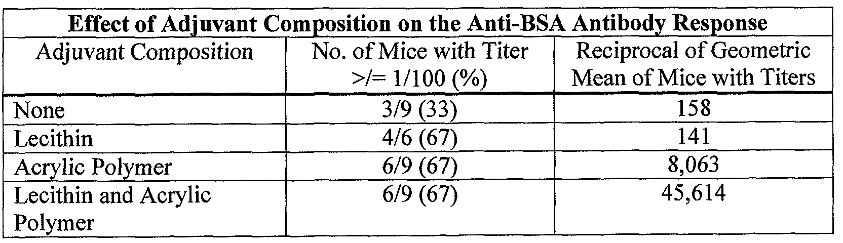
[0098] Anti-BSA IgG antibody titers were over five times higher when a combination of lecithin and acrylic polymer was used as adjuvant than when acrylic polymer was used alone and 323 times higher than when lecithin was used alone. This demonstrates that the combination of lecithin and acrylic polymer is far more effective at delivering the antigen orally to the mucosal surface for uptake by lymphoid tissue than either lecithin or acrylic polymer alone. Although, not all of the mice showed a serum anti-BSA IgG antibody response the results clearly show a synergistic adjuvant effect of lecithin combined with the acrylic polymer. However, the mice that did not seroconvert may have had a secretory IgA antibody response. Indeed, oral vaccination, and mucosal vaccination in general, stimulates IgA secreting cells at mucosal surfaces.
EXAMPLE 3
Second Test of Lecithin/Polymer Adjuvant by the Oral Route
[0099] Two vaccines were prepared in PBS that comprised the antigen, BSA
Fraction 5, at a concentration of 400 g per ml. One vaccine contained no adjuvant only BSA. The other vaccine was comprised of BSA adjuvanted with 3 mg/ml of lecithin and 3 mg/ml of acrylic polymer. The vaccine was assembled as described in Example 2. On days 0, 4, 27, and 31 groups of CF-1 female mice from Charles- River Laboratories and weighing approximately 18 grams, were administered 0.5 ml of vaccine orally by feeding needle. On day 52, 21 days post vaccination, mice were euthanized and bled by the brachial artery. End-point anti-BSA serum IgG antibody titers were determined by ELISA. A 1/100 starting dilution of serum was used due
to non-specific background color development at dilutions less than 1/100. Results are recorded in Table 3:
TABLE 3

[00101] In a separate study, 4/10 mice that received this same vaccine had a geometric mean titer of 1/1,345 six weeks after only a single vaccination. This demonstrates the potential of the adjuvant composition, when once optimized, to engender an immune response of long duration.
EXAMPLE 4
Administration of Vaccine Intranasally
[00102] Two experimental vaccines for delivery by the intranasal route were prepared in PBS comprising the antigen, BSA, at a concentration of 500 μg/ml. One vaccine was comprised of BSA alone. The second vaccine was comprised of BSA adjuvanted with a combination of 3 mg/ml of lecithin and 3 mg/ml of the acrylic polymer. The lecithin and acrylic polymer were first mixed with a laboratory bench top magnetic stir bar and then in a Waring blender using an emulsification head. BSA was then added and mixed again using the emulsification head. Mice were vaccinated on days 0 and 20. Forty μΐ containing 20 μg of BSA antigen were placed on the nose while the mouth was held shut. The vaccine entered the nose when the mouse inhaled. On day 41, 21 days post second vaccination, the mice were euthanized and bled by cutting the brachial artery. Anti-BSA antibody titers were determined by ELISA. The starting dilution of serum was at 1/100 due to non-
specific background color development at lower dilutions. Results are shown in Table 4:
TABLE 4

EXAMPLE 5
Use of Adjuvant with Vaccine in Swine
[00104] The adjuvant invention comprising a combination of 2 mg/ml of lecithin and 2 mg/ml of acrylic polymer was used as a diluent for modified-live pseudorabies virus (ML-PRV) for swine. This adjuvant diluent and a control diluent consisting of sterile water were used to rehydrate lyophilized (ML-PRV). The ML- PRV was rehydrated immediately prior to vaccination. Groups of 10 weaned piglets, 6 weeks of age, were vaccinated on days 0 and 21. Blood serum was collected on days 2, 20, 28, and 48 for serological testing for anti-PRV serum neutralizing
antibodies. The anti-PRV antibody responses of piglets in the different vaccine groups are shown in Table 5.
TABLE 5

EXAMPLE 6
Lecithin and Acrylic Copolymer Adjuvant Supports HlNl and H3N2 Vaccine Responses
[00106] Lecithin and acrylic copolymer adjuvant as described and prepared herein was analyzed for its effectiveness at supporting the immunization of swine against HlNl and H2N2 viral antigens. Inventive adjuvants herein were compared to a commercially available adjuvant, 5% Amphigen®, to identify the capacity of adjuvants as described herein to support viral antigen based vaccines. Each adjuvant was combined with HlNl and H3N2 antigens (derived from a released lyophilized commercial product (FluSure™, Pfizer Animal Group). Test groups for vaccination included 29 to 35 day old piglets.
[00107] Four treatment groups were observed (each group having 15 piglets, except the negative control group which had 5 piglets): Tl, 5% Amphigen, positive control; T2, Quil A alone adjuvant; T3, lecithin and acrylic copolymer of the invention, intramuscular; and T4, lecithin and acrylic copolymer of the invention, intranasal. T5 was a control group that was un-vaccinated. Tl, T2 and T3 received one intramuscular dose on day 0, T4 received one intranasal dose on day 0.
[00108] Blood samples were obtained from each animal participating in the study on days 0, 21 and 35. Table 6 shows data from H1N1 titer, Table 7 shows data from the H3N2 titer: TABLE 6: Number of Piglets with H1N1 Swine Influenza Titers and Geometric Mean Titers

No. of piglets with H1N1 Titers - Piglets with titers of 10 or higher were determined as positive.
Geometric Mean Titers - A titer of 5 was assigned to negative titers for calculation of geometric mean.
SIV - Lyophilized swine influenza (H1N1, H3N2) virus killed viral vaccine (FluSure™, Pfizer Animal Health).
LAP - Lecithin acrylic polymer as described in Examples 1-5.
TABLE 7: Number of piglets with H3N2 Swine Influenza Titers and Geometric Mean Titers

[00109] Referring to Tables 6 and 7, the results indicate lecithin and acrylic copolymer adjuvants of the invention induce significant serological responses to HlNl and H3N2 swine influenza viruses in piglets. The responses were similar to conventional commercial adjuvants and significantly better than a Quil A alone adjuvant. The data in Example 6 further show the utility of the lecithin/acrylic polymer adjuvants of the invention and show that additives alone, for example Quil A, provide for a minimal immunological response under identical conditions.
EXAMPLE 7
Immune Response to Vaccine Enhanced by Inclusion of Additives
[00110] The immunogenecity of a lecithin, acrylic polymer, Quil A and cholesterol adjuvant of the invention was tested against Amphigen, a commercial
adjuvant. The study was performed to determine the effectiveness of eliciting an enhanced immune response through inclusion of cholesterol and Quil A in adjuvant embodiments described herein.
[00111] Adjuvant was prepared as described above, except in this Example, 15 mg/ml lecithin was combined with 10 mg/ml acrylic polymer. Each vaccine dose included 1.25 mg of adjuvant (as compared to 5 mg of Amphigen).
[00112] The adjuvant samples were spiked with 0.5 mg/ml of Quil
A/cholesterol. All adjuvant samples further received 50μg, 5μg or 0.5 μg AIV-HA antigen. Ingredients described above were combined as discussed herein to provide 3 different antigen concentrations for each lecithin acrylic polymer/additive sample and Amphigen seample. All samples were stored at 4°C.
[00113] Twenty-nine to thirty-five day old piglets were vaccinated and bleeds taken on days one, fourteen and twenty eight. Titers were determined for each condition and mean HAI titer determined.
[00114] As shown in Figure 1, the study showed a surprising increase in mean HAI titer with cholesterol and Quil A included in the adjuvant and therefore vaccine preparation. In each case, the increase in titer was antigen concentration dependent and showed a significant increase comparable to corresponding antigen spiked Amphigen samples. It is noted that the amount of lecithin and acrylic polymer was limited (1.25 mg) to provide a more sensitive platform for identifying effects of the inventive adjuvants described herein. The low dose lecithin/acrylic
polymer/additive adjuvant provided an adequate platform for analyzing the adjuvant's capacity to elicit an immune response, which was comparable to a full dose Amphigen based adjuvant. EXAMPLE 8
Lecithin and Acrylic Polymer Adjuvant is Facilitated By Inclusion of Additives
[00115] The immunogenecity of a lecithin, acrylic polymer, cholesterol, and Quil A adjuvant was tested in chickens. Cell line based antigen was tested in this manner in order to determine utility of adjuvants (additive based) in accordance with the present invention.
[00116] Adjuvant was prepared using 33.5 μg/dose Quil A, 32.4 μg/dose cholesterol, 800 g/dose lecithin and 500 μg/dose acrylic copolymer 974PNF. Vaccine antigen was a stably transfected cell line using a H5 HA expressing plasmid (CHO-HA-10). In some vaccines, an antigen stabilizing agent was added to the material, i.e., Nicotiana tabacum cell line lysate.
[00117] Sixty five pathogen-free Leghorn chickens (10 day old) were obtained and quarantined for nine days prior to start-up of the experiment. Birds were split into eleven groups (six birds per group for groups Tl through T10 and five birds for Tl 1). Each group was housed together. Treatment group Tl 1 birds were a baseline group and used to obtain a Day 0 bleed. Study design is provided in Table 8:
TABLE 8: Example 8 study design
No. Treatment Processing No. of Vacination Bleed Group Method Birds Days*
Days Dose Route
Tl CHO lys. Mixing 6 0, 14 0.5 ml SQ 14, 28 NT-1 ext
Adjuvant
T2 CHOHA0102 Mixing 6 on 0, 14 0.5 ml SQ 14, 28 Adjuvant zero
5 on 14
T3 CHOHA0102 Microfluid. 6 0, 14 0.5 ml SQ 14, 28 Adjuvant Non- Clarified
T4 CHOHA0102 Microfluid. 6 0, 14 0.5 ml SQ 14, 28 Adjuvant Clarified
T5 CHOHA0102 Silverson 6 0, 14 0.5 ml SQ 14, 28 Adjuvant Non- Clarified
T6 CHOHA0102 Mixing 6 0, 14 0.5 ml SQ 14, 28 NT-1 ext
Adjuvant
T7 CHOHA0102 Microfluid. 6 0, 14 0.5 ml SQ 14, 28 NT-1 ext Non- Adjuvant Clarified
T8 CHOHA0102 Microfluid. 6 0, 14 0.5 ml SQ 14, 28 NT-1 ext Clarified
Adjuvant
T9 CHOHA0102 Silverson 6 0, 14 0.5 ml SQ 14, 28
NT-1 ext Non-
Adjuvant Clarified
T10 H5N9 AIV Mixing 6 0, 14 0.5 ml SQ 14, 28
Adjuvant
Ti l Baseline NA 5 NA NA NA 0 Bleed
Bleed days - serum evaluated by hemagglutination inhibition with A/Turkey/Wisconsin/68 (H5N9)
CHO lysate - Non-transfected CHO cells
NT-1 extract -Nicotiana tabacum, lyophilized, non-transformed clarified cell lysate Adjuvant - Lecithin, acrylic copolymer, Quil A, and cholesterol
SQ - subcutaneous administration
CHOHA0102 - CHO cells transfected with H5 hemagglutinin
H5N9 AIV - Inactivated H5N9 avian influenza virus A/Turkey/Wisconsin/68
NA - not applicable
[00118] As shown in Table 9, all vaccinated treatment groups (T2-T9) were clearly different from the negative control (Tl). Although slight differences were observed, little differentiation was discemable between treatment groups that did or did not receive the Nicotiana tabacum cell line lysate or between the various processing methods.
TABLE 9: Seroconversion rates and avian influenza hemagglutination inhibition eometric mean titer results

Adjuvant
T7 CHOHA0102 Microfluid. 3/6 5/6 10.1 18.0
NT-1 ext Non- Adjuvant Clarified
T8 CHOHA0102 Microfluid. 3/6 6/6 9.0 20.2
NT-1 ext Clarified
Adjuvant
Τ9 CHOHA0102 Silverson 2/6 5/6 5.7 25.4
NT-1 ext Non- Adjuvant Clarified
T10 H5N9 AIV Mixing 3/6 6/6 8.0 228.1
Adjuvant
[00119] Results from the Example show that additive based adjuvants of the invention support a cell line based antigen, i.e., a cell line that expresses the antigen, and provided excellent immunogenicity in chickens. This data further supports the conclusion that Quil A and cholesterol when used in combination with other adjuvant based embodiments described herein have surprising utility in the context of the present invention.
EXAMPLE 9:
Micrograph Data: Embodiments of the Present Invention Have Net-Like Structure
[00120] Inventive adjuvant compositions were as prepared and described in previous Examples. Adjuvant samples were visualized by transmission electron microscopy. Adjuvants included lecithin and polymer, no additives, and were sterilized by autoclaving. An illustrative micrograph at 30,000X magnification is shown in Figure 2A.
[00121] For comparison, an illustrative emulsion as prepared by the methods described in US Patent No. 5,716,637 (Anselem et al.), and visualized via transmission electron microscopy at the same magnification (30,000X) is shown in Figure 2B. As described in the description of the Anselem patent, the adjuvant was micro filtered and not autoclaved.
[00122] The adjuvant prepared via the methods of US Patent No. 5,716,637, show an expected emulsion structure of lipid droplets in an aqueous phase. In
contrast, the micrograph shown in Figure 2 A illustrates that adjuvants of the present invention have a significantly different physical structure or distribution than the adjuvants described in Anselem. Adjuvants of the present invention show a diffuse net-like structure with significant polymer content combining with the lecithin (phospholipids) to provide the unexpected structure of the present invention. This is a surprising given the significant difference is structure between the two adjuvant compositions.
EXAMPLE 10
Calcium Phosphate Facilitates Immunity of DNA-Based Vaccine
[00123] Lecithin and acrylic copolymer adjuvant as described and prepared herein was analyzed alone and in combination with calcium phosphate (CaP04) to determine effectiveness at supporting the immunization of chicks against an avian influenza DNA antigen (H5N9 AIV FLA DNA). Inventive adjuvants herein were compared to a commercially available adjuvant, having the same antigen. The commercially available adjuvant was also tested with CaP04.
[00124] Chick treatment groups were immunized on day 0 and day 16.
Serum testing was performed on each treatment group and HAI titers determined using standard assays.
[00125] Figure 3 shows data from HAI GMT serum titers for adjuvant only, no antigen (Amphigen®); antigen only, no adjuvant; Amphigen®; Amphigen® with CaP04; Lecithin and Copolymer; and Lecithin, Copolymer and CaP04. The results in Figure 3 indicate that the lecithin, copolymer and calcium phosphate group vaccine provided significantly higher levels of immunity than lecithin and copolymer alone or Amphigen with or without calcium phosphate.
[00126] This combination of lecithin, copolymer and calcium phosphate shows an unexpected capacity to transform poorly immunogenic DNA vaccines into highly effective vaccines.
EXAMPLE 11
Enhanced Potency of FMD Vaccine
[00127] Experimental design: Second generation human adenovirus type 5 (Ad5) vector for FMDV serotype A24 Cruzeiro administered to pigs subcutaneously at two sites with or without adjuvant. The animals were challenged 21 days post- vaccination at doses 10-fold higher than recommended.
[00128] Figure 4 shows the results of the experimental Adenovirus vector- based FMD vaccine alone or in combination with one of two adjuvants: a DNA plasmid called plCLC or Adjuvant E (Adjuplex/Vetplex with Quil A and
Cholesterol). Adjuvant E increased potency by at least 5-fold whereas the plCLC adjuvant was less effective. This data further supports the conclusion that Quil A and cholesterol when used in combination with other adjuvant based embodiments described herein have surprising utility in the context of the present invention.
EXAMPLE 12
Effect of Vaccine Adjuvants on Adeno-Vector Viruses
[00129] The overarching goal of this research was to produce and evaluate Foot-and-mouth disease (FMD) single or combinational vaccines comprising replication-defective recombinant human adenovirus carrying (a) the FMD VP1 capsid and 3C protease coding regions and/or (b) bovine or porcine type 1 interferon genes. This strategy is being used to develop next generation molecular FMD licensed vaccines for stock piling by the National Veterinary Stockpile Program for use as an FMD countermeasure in emergency outbreaks.
[00130] One research area of this project was the addition of vaccine adjuvants to the adeno-based vector vaccines for enhancement of their
immunogenecity and efficacy. Two adjuvants (Adjuplex-LAP and Adjuplex-LE) were evaluated in vitro for virucidal effects on the vaccine vectors under different time and temperature conditions. The conclusion drawn from this study was that Adjuplex LAP did not have any virucidal effect on Ad5bIFNa virus at the recommended or 4-fold concentration level. The incubation temperatures and times
for these studies were 20° C, or 39 C, and 1 or 24 hours, respectively. However, similar results were not observed for Adjuplex-LE. No titer reductions were observed at 39° C and one hour incubation; however when the incubation was increased to 24 hours, there were significant reductions in virus titers at both concentrations. Results from this study would suggest that Adjuplex-LE adjuvant produced a virucidal effect on Ad5-bIFNa virus at both the recommended and twofold higher concentration levels.
[00131] Similar results were obtained with Ad5-01 virus when it was mixed with both Adjuplex LAP and LE, respectively, at the recommended quantity and when the amount was increased two- or four-fold. Furthermore, there were no significant titer reductions even when the mixtures were incubated at 39° C for 24 hours. The 39° C incubation temperature was selected because that is the average cattle rectal body temperature. It is recommended that Adjuplex LAP should be the adjuvant of choice to be combined with the Ad5-bIFNa, Ad5-01 and other Ad5 based FMD sub unit vaccines for clinical evaluation in cattle and pigs.
[00132] Foot-and-mouth disease (FMD) is an economically important and highly contagious viral disease of cloven-hoofed livestock and wildlife including cattle, swine, sheep, goats, and deer. The recent re-emergence of FMD in both developing and developed nations has refocused world's attention on universe control strategies particularly in the USA.
[00133] In many countries the control and eradication of FMD are by immunization of susceptible animals using commercially available FMD vaccines, which are based on conventional chemically inactivated vaccines emulsified with adjuvants. Failure to completely inactivate the vaccine has led to outbreaks of the disease. There is no approved diagnostic test available to reliably differentiate vaccinated from infected animals. Furthermore vaccinated animals can become disease carriers following contact with FMD virus. These disadvantages of inactivated whole FMD vaccine have made FMD-free countries to be reluctant to vaccinate their livestock during outbreaks.
[00134] In order to overcome some of the problems associated with convectional FMD vaccines, many approaches have been utilized to develop
alternative FMD vaccines, including construction of modified live-virus, biosynthetic proteins, synthetic peptides, naked DNA vectors, and recombinant viruses. The use of human adenovirus as a vector for FMD vaccines has been met with variable results, sometimes resulting in incomplete protection or failure of vaccinated animals to develop a neutralizing antibody.
[00135] In this study, two adjuvants viz Adjuplex-LAP and Adjuplex-LE were investigated in vitro as possible adjuvants for Ad5 FMD subunit vaccines. This was a preliminary investigation prior to in vivo studies. Experiments were set up to determine both their cytotoxic effect on 293 cells and their virucidal effects on Ad5-FMD viruses.
[00136] Adjuplex-LAP is a mucosal vaccine adjuvant as well as an adjuvant for parenterally administered vaccines. The adjuvant is a lecithin
phospholipid/acrylic polymer combination. The combination forms a
mucoadhesive matrix that facilitates the adsorption of vaccine antigens to mucosal surfaces and subsequent absorption and presentation of antigen to cells of the immune system. Both adjuvant components are used in the pharmaceutical and biological industries and thus have value as a delivery and adjuvant system for vaccine antigens.
[00137] Adjuplex-LE is a 5%, or less, oil-in-water emulsion, the oil droplets which are covered by lecithin derived phospholipid vesicles. The lipid vesicles act as a carrier for vaccine antigens and make them accessible to cells of the immune system. The lipid vesicles on the surface of the oil droplets is also a safety feature making the oil less irritable or not irritable at all to tissue at the injection site. The adjuvant is also non-virucidal. Therefore the formulation can be, and is, used to adjuvant modified-live virus vaccines. The adjuvant can be mixed directly with vaccine antigens without further emulsification or the antigens can be added at the time of emulsification.
[00138] The redox indicator Alamar Blue™ (AB), a fluorescent dye, which has been used in mammalian cell culture cytotoxicity assays. AB is a safe, nontoxic aqueous dye, which is used to assess cell viability and cell proliferation because it is stable in cell culture. It has also been shown to be a rapid and simple non-
radioactive assay alternative to the [3H] thymidine incorporation assay. AB both fluoresces and changes color in response to chemical reduction, and the extent of the conversion is a reflection of cell viability. AB assay is a simple, one-step procedure. Alamar Blue™ assay was set up to study the cytotoxicity effects of two vaccine adjuvants 293 cells.
[00139] There were two objectives: (a) evaluate the relative 293 cell cytotoxity of vaccine adjuvants in combination with Ad5 -bovine interferon alpha (Ad5bIFN a) and Ad501 vectors, and (b) establish if the vaccine adjuvants are virucidal for Ad5bIFNa vector.
[00140] Alamar Blue™ assay was set up to study the cytotoxicity effect of vaccine adjuvants on 293 cells. Alamar Blue™ is a safe, nontoxic aqueous dye which is used to assess cell viability and cell proliferation.
MATERIALS AND METHODS
[00141] 293 Cells
[00142] Human embryonic kidney (293) cells were obtained from Dr. Patrick Hearing, Department of Microbiology, Stony Brook University, Stony Brook, NY, and were propagated in minimum essential medium (MEM) containing 10% fetal bovine serum (FBS), 1% antibiotic-antimycotic solution, and 1% MEM non essential amino acid (NEAA). 293 cells of passages 15 and 36 were used for transfection, propagation of viruses, virus titrations and performing of cytotoxicity assays.
[00143] Ad5bIFNa ndAdSOl plasmids
[00144] The two plasmids were provided by Dr. Laszlo Zsak from US Department of Homeland Security, Targeted Advanced Development, Plum Island Animal Diseases Center, Orient Point, NY.
[00145] Transfection of Ad5bIFNa and Ad501 plasmids in 293 cells
[00146] The pAd5bIFNa and pAd501 were linearized by digestion with restriction enzyme Pad and transfected in 293 cells using Lipofectamin™ 2000.
[00147] Production and Purification of Ad5bIFNa and Ad501 viruses (vaccine vectors)
[00148] The 2 viruses were harvested with the appearance of the initial plaques, which were then grown in large quantities in 293 cells, and purified utilizing a nonlinear followed by a linear CsCl gradient centrifugation.
[00149] Dilutions of vaccine viruses (Ad5bIFNa and Ad501 )
[00150] 1 :10, 1 :100 and 1:100 dilutions of both Ad5bIFNa and Ad501 vaccine viruses were prepared respectively in EMEM containing 2% FBS, 1% antibiotic-antimycotic solution, and 1% MEM-NEAA.
[00151] Dilutions of adjuvants (Adjuplex-LAP and Adjuplex-LE)
[00152] 1 :20, 1 :200, 1 :2000, and 1 :20,000 dilutions of both Adjuplex-LAP and Adjuplex-LE adjuvants were prepared respectively in EMEM containing 2% FBS, 1% antibiotic-antimycotic solution, and 1% MEM-NEAA.
[00153] Alamar Blue (AB)
[00154] AB was aliquoted and stored at -80° C. Prior to each experiment, AB was brought to room temperature and vortexed. Exposure of AB to light was minimized throughout the experiments.
[00155] Alamar Blue (AB) cytotoxicity assay (Cell viability assay)
[00156] Cell viability of 293 cells was assessed by AB cytotoxicity assay. 293 cells in EMEM containing 2% FBS, 1% antibiotic-antimycotic solution, and 1% MEM-NEAA were seeded at a density of 1 X 10⁶ viable cells/ml (1 X 10⁵/well) in a 96 well Treatment (BD Falcon* Primaria* Tissue culture Treatments, Fisher Scientific Company, Suwanee, GA). In a second plate, cells were seeded at a density of 10 X 106 viable cells/ml (1 X 10⁶/well) to determine the optimal cell concentration for the cytotoxicity study.
[00157] Table 10 shows the addition of diluted Ad5bIFNa, Ad501, Adjuplex- LAP and Adjuplex-LE to the cells. Briefly, 0.1 ml of each of the dilutions of
Ad5bIFNa and Ad501 (1:10, 1:100, 1:100) was added to each well respectively in triplicates. 0.1 ml of each of the dilutions of the adjuvants (1:20, 1:200, 1:2000, and 1 :20,000) was added to each well respectively in triplicates. EMEM containing 2% FBS, 1% antibiotic-antimycotic solution, and 1% MEM-NEAA was added to wells Al, A2, A3, Dl, D2 and D3 as negative controls.
[00158] The two treatments were incubated at 37 C in a 5% C02 atmosphere for approximately 18 hours, after which 20 μΐ AB was added to each well. The treatments were returned to the incubator.
[00159] Optical densities (OD) at 570 nm and 600 nm were measured with the ELx808 ultra microplate reader (BioTek Instruments, Inc., Winooski, VT) at approximately 42 hours, 50 hours, and 68 hours (total culture times).
[00160] The OD data were analyzed as follows: (a), determine % difference in reduction of AB (between media growth control wells and treatment wells); this will indicate the amount inhibition (or stimulation) of cell growth in treatment wells with respect to media control wells, and (b). determine % reduction of AB in media control and in treatment wells; this will indicate the amount of cell growth in media control and treatment wells. Treatments with higher % reduction than media controls are considered to stimulate cell growth. Treatments with lower % reduction than media controls are considered cytotoxic. Treatments with the same % reduction are neither cytotoxic nor stimulatory.
[00161] Calculation of Alomar Blue (AB) reduction
[00162] Percent reduction of AB was calculated using the manufacturer's formula (33). In monitoring AB reduction spectrophotometrically, reduction is expressed as a percentage (% reduced).
[00163] The calculation of % Reduced is as follows when the samples are read at
λΐ = 570 nm
λ2 = 600 nm
% Reduced = ίεοχ λΖ) (A λΐ) - (εοχ λΐ) (Α λ2) X 100
(sred λΐ) (Α' λ2) - (ered λ2) (Α' λΐ)
Where:
(sred λΐ) = 155,677 (Molar extinction coefficient of reduced alamarBlue™ at 570 nm)
(ered λ2) = 14,652 (Molar extinction coefficient of reduced alamarBlue™ at 600 nm)
(εοχ λΐ) = 80,586 (Molar extinction coefficient of oxidized alamarBlue™ at 570 nm)
(εοχ λ2) = 117,216 (Molar extinction coefficient of oxidized alamarBlue™ at 600 nm)
(A λΐ) = Absorbance of test wells at 570 nm
(A λ2) = Absorbance of test wells at 600 nm
(Α'λΐ) = Absorbance of negative control wells which contain medium plus alamarBlue™ but to which no cells have been added at 570 nm.
(Α'λ2) = Absorbance of negative control wells which contain medium plus alamarBlue™ but to which no cells have been added at 600 nm.
[00164] In reporting alamarBlue™ reduction by monitoring absorbance, data are expressed as percent alamarBlue™ reduced as a function of time of incubation.
The AB assay was used to determine viability of 293 cells to vaccine adjuvants.
[00165] Virucidal Assay (TCID so Assay)
[00166] The TCID50 assay was employed to ascertain and measure if there were virucidal effects of Adjuplex-LAP and Adjuplex-LE adjuvants on Ad5bIFNa and Ad501 viruses.
[00167] 293 cells were harvested from a T-150 flask of fresh 293 cells, and counted on a hemocytometer. A dilution of the cell suspension at 1 X 10⁵/ml in MEM containing 2% FBS, 1 % antibiotic-antimycotic solution, and 1 % MEM- NEAA was made, and at least 10 ml was prepared for each 96 flat-bottomed well tissue culture plate.
[00168] Using a 12-channel pipette and a multichannel pipetter basin, 100 μΐ of the cell dilution (104 cells/per well) was seeded into 96-welI tissue culture plates, and covered. They were incubated at 37° C in a C02 incubator until used.
[00169] Frozen vials of Ad5bIFNa and Ad501 viruses were thawed and kept on ice at all times.
[00170] MEM supplemented with 2% FBS, 1 % antibiotic-antimycotic solution, and 1% MEM-NEAA was used to make virus dilutions of 10_1 to 10"13. The 10 fold dilutions were made in 5 ml sterile, disposable tubes.
[00171] The cells were infected by adding 0.1 ml per well of each virus- adjuvant dilution immediately after the dilutions were made.
[00172] AB assay with a cell concentration of 10 X 106/ml was also set up.
[00173] Preparation of Antigen-Adjuvant Mixtures
[00174] Ad5bIFNa virus-Adjuvant Mixtures
[00175] Each of the two adjuvants was mixed with Ad5bIFNa according to the manufacturer recommended ratios (LAP: virus ratio, 1 :4; LE: virus ratio, 1 :1). In addition to the recommended ratios, additional ratios (LAP: antigen ratio, 4:1; LE: antigen ratio, 2:1) were also tested to increase the chances of having virucidal effects. Each mixture was made in a sterile 1 ml microtube and vortexed three times to ensure adequate mixing before use, and was incubated according to the appropriate conditions.
[00176] The media-virus mixture served as a control. The media consisted of MEM supplemented with 2% FBS, 1% antibiotic-antimycotic solution, and 1% MEM-NEAA. The ratios tested were as follows:
[00177] Experiment 1
[00178] Treatment A: 25 μΐ Media + ΙΟΟμΙ, Ad5bIFNa
[00179] Treatment B: 25μ1, LAP + \00μ1, Ad5bIFNa
[00180] Treatment C: \00μ1 Media + ΙΟΟμί Ad5bIFNa
[00181] Treatment D: ΙΟΟμί LE + ΙΟΟμΙ. Ad5bIFN<x
[00182] The mixtures were not incubated before dilutions were made.
[00183] Experiment 2
[00184] Treatment A: 25 μΐ Media + ΙΟΟμί Ad5bIFNa
[00185] Treatment B: 25μΙ LAP + ΙΟΟμί Ad5bIFNa
[00186] Treatment C: 1 ΟΟμΙ, Media + 1 ΟΟμΙ, Ad5bIFNa
[00187] Treatment D: 1 ΟΟμΙ. LE + 1 ΟΟμί Ad5bIFNa
[00188] The mixtures were incubated at room temperature (20 - 25° C) for one hour before dilutions were made.
[00189] Experiment 3
[00190] Treatment A: 100 μΐ Media + ΙΟΟμί Ad5bIFNa
[00191] Treatment B: 100 μΕ LAP (4X) + \00μ1, d5bIFNa
[00192] Treatment C: 100 LE + ΙΟΟμί Ad5bIFNa
[00193] Treatment D: 200 μΐ Media + 100 μΐ, Ad5bIFNa
[00194] Treatment E: 200 LE (2X) + 100 Ad5bIFNa
[00195] The mixtures were incubated at 39° C for one hour before dilutions were made.
[00196] Experiment 4
[00197] Treatment A: 100 μΐ Media + ΙΟΟμΕ Ad5bIFN<x
[00198] Treatment B: 100 μΐ LAP (4X) + ΙΟΟμί Ad5bIFNa
[00199] Treatment C: 100 μΐ LE + ΙΟΟμί Ad5bIFNa
[00200] Treatment D: 200 μΐ, Media + 100 μΕ Ad5bIFNa
[00201] Treatment E: .200 μΐ, LE (2X) + 100 μΐ Ad5bIFNa
[00202] The mixtures were incubated at 39° C for 24 hours before dilutions were made.
[00203] Ad501 -Adjuvant Mixtures
[00204] Each of the two adjuvants was mixed with Ad501 according to the manufacturer recommended ratios (LAP: virus ratio, 1 :4; LE: virus ratio, 1 :1). In addition to the recommended ratios, additional ratios (LAP: virus ratio, 4:1; LE: virus ratio, 2:1) were also tested to increase the chances of having virucidal effects. Each mixture was made in a sterile 1 ml microtube and vortexed three times to ensure adequate mixing before use.
[00205] The media-virus mixture served as a control. The media consisted of MEM supplemented with 2% FBS, 1% antibiotic-antimycotic solution, and 1% MEM-NEAA. The ratios tested were as follows:
[00206] Experiment 1
[00207] Treatment A: 25 ΐ Media + ΙΟΟμί Ad501
[00208] Treatment B: 25μ\, LAP + ΙΟΟμί Ad501
[00209] Treatment C: ΙΟΟμί Media + ΙΟΟμί Ad501
[00210] Treatment D: ΙΟΟμί LE + ΙΟΟμΕ Ad501
[00211] The mixtures were not incubated before being dilutions were made.
[00212] Experiment 2:
[00213] Treatment A: 25μ1 Media + ΙΟΟμί Ad501
[00214] Treatment B: 25μΙ_, LAP + 100μL Ad501
[00215] Treatment C: ΙΟΟμΙ, Media + ΙΟΟμΙ, Ad501
[00216] Treatment D: \00μ1, LE + ΙΟΟμΙ, Ad501
[00217] The mixtures were incubated at room temperature (20 - 25° C) for one hour dilutions were made.
[00218] Experiment 3
[00219] Treatment A: 100 μΐ Media + ΙΟΟμΙ Ad501
[00220] Treatment B: 100 μΐ, LAP (4X) + 100 Ε Ad501
[00221] Treatment C: 100 LE + ΙΟΟμί Ad501
[00222] Treatment D: 200 μί Media + 100 μί Ad501
[00223] Treatment E: 200 μΐ, LE (2X) + 100 μΕ Ad501
[00224] The mixtures were incubated at 39° C for one hour before dilutions were made.
[00225] Experiment 4
[00226] Treatment A: 100 μΐ Media + ΙΟΟμί Ad501
[00227] Treatment B: 100 μΐ, LAP (4X) + ΙΟΟμί Ad501
[00228] Treatment C: 100 μ\, LE + \00μ1, Ad501
[00229] Treatment D: 200 μΐ, Media + 100 μί AdO 1
[00230] Treatment E: .200 μ\, LE (2X) + \00 μ~1 Ad501
[00231] The mixtures were incubated at 39° C for 24 hours before dilutions were made.
[00232] Preparation of virus-adjuvant dilutions
[00233] From each treatment, serial 10 fold dilutions of adjuvant-virus mixtures were prepared in 5 ml sterile, disposable tubes using MEM supplemented with 2% FBS, 1% antibiotic-antimycotic solution, and 1% MEM-NEAA to prepare adjuvant-virus dilutions of 10"1 to 10"13. 0.9 ml MEM supplemented with 2% FBS, 1% antibiotic-antimycotic solution, and 1% MEM-NEAA was dispensed into the first two tubes. To each of the eleven other tubes was added 1.8 ml.
[00234] 0.1 ml of each adjuvant-virus mixture from each treatment was added to the first tube and mixed by votexing. Filtered pipette tips were changed between dilutions. 0.1 ml of the 10"1 dilution was withdrawn and transferred to the second
tube containing MEM supplemented with 2% FBS, 1% antibiotic-antimycotic solution, and 1% MEM-NEAA.
[00235] 0.2 ml of the 10"2 dilution was withdrawn and transferred to the third tube containing MEM supplemented with 2% FBS, 1% antibiotic-antimycotic solution, and 1% MEM-NEAA, and this became 10"3 virus dilution. The above steps were repeated to prepare the next virus dilutions (10"4 to 10"13).
[00236] Infection of Cells with Virus-Adjuvant Dilutions
[00237] One 96-well plate was used for infection per treatment. Immediately after the dilutions were made, the cells were infected by adding 0.1 ml per well of each virus-adjuvant dilution. 0.1 ml of the virus-adjuvant suspension with the highest dilution was dispensed in column 1 wells to infect the cells in the 8 wells of this column.
[00238] The cells in the 8 wells of the next column; column 2 of the 96-well were infected with 0.1 ml of the next adjuvant-virus dilution (10~12). The cells in the 8 wells of columns 3 through 11 were infected with 0.1 ml of the remaining virus-adjuvant dilutions (10~n-10"13). Pipette tips were changed between dilutions.
[00239] To test the cell viability and as a negative control (no adjuvant-virus control), 0.1 ml/well of MEM containing 2% FBS, 1% antibiotic-antimycotic solution and 1% MEM-NEAA was added to each well in column 12. Each plate was covered and incubated at 37° C in a C02 incubator for 10 days. Cells in each plate were observed daily by an inverted microscope for cytopathic effects (CPE) over the next 10 days. Observable CPE per column were counted and recorded. Final reading of each plate was done on the 10th day post incubation to determine the titer. A well was counted as positive even if only a small spot or a few cells showed CPE. The negative control wells were used for comparison. The test was valid if the negative controls did not show any CPE or cell growth problems, and the lowest dilution showed 100% infection (8/8) while the highest dilutions showed 0% infection (0/8).
[00240] The results were recorded for day 10, scoring wells "+" (CPE positive) or "-" (CPE negative) using the CPE scoring form in appendix A, and the ratio of positive wells per column was determined, and recorded it as in appendix A.
After recording the assay data, the plates were placed in a biohazard bag and autoclaved and discarded as biohazardous waste.
[00241] For each plate (treatment), TCID5o/ml titer was calculated using the KARBER statistical method. Compare the TCID50/ml between the treatments in each experiment to determine if there was a difference. A difference in titers between treatments in each experiment that is greater than 0.7 log was considered to be significant, which means the adjuvant in that experiment was virucidal to the tested virus.
RESULTS
[00242] Alomar Blue (AB) cytotoxicity assay (Cell viability assay)
[00243] Alamar Blue™ was used to measure 293 cell viability at two densities There was cell clumping at a density of 10 X 106 /ml for all treatments. The l X 106 /ml density was better than the higher density.
[00244] Cell viability assay on exposure to v4d5bIFNa
[00245] At a cell concentration of 1 X 10⁶ /ml, the 1 : 100 dilution of
Ad5bIFNa produced the higher % AB reduction than the media controls at 42 and 50 hours post exposure (Fig. 5 and 6). However, there was a % AB reduction at 68 hours post exposure (Fig 7). Similar results were obtained at a cell density of 10 X 106 /ml (Figs 8-10).
[00246] Cell viability assay on exposure to Adjuplex LAP
[00247] Cell exposure at a cell density of 1 X 10⁶ /ml to Adjuplex LAP at a dilution of 1 :20 gave the higher % AB reduction than the media controls at 42 and 50 hours post exposure (Fig. 5 and 6) but with a % AB reduction at 68 hours post exposure (Fig 7). Similar results were obtained at a cell density of 10 X 106 /ml (Figs 8-10).
[00248] Cell viability assay on exposure to Adjuplex LE
[00249] When 293 cells at a cell density 1 X 10⁶ /ml were treated with Adjuplex LE at a dilution of 1:20, higher % AB reductions were obtained compared to the media controls at 42 and 50 hours post exposure (Fig. 5 and 6) but with a % AB reduction at 68 hours post exposure (Fig 7). Similar results were obtained at a cell density of 10 X 106 /ml (Figs 8-10).
[00250] Cell viability assay on exposure to Emulsigen
[00251] Treatment of 293 cells at a cell density 1 X 10⁶ /ml with Emulsigen at 1:20 dilution resulted in higher % AB reductions relative to the media controls at 42 and 50 hours post exposure (Fig. 5 and 6) but with lower % AB reductions at 68 hours post exposure (Fig 7). Similar results were obtained at a cell density of 10 X 106 /ml (Figs 8-10).
[00252] Virucidal assay (TCID5o Assay)
[00253] Ad5bIFNa vims-Adjuvant Mixtures
[00254] Experiment 1
[00255] The loglO titers of treatments in this group were 10.0, 9.9, 9.5, and
9.6 (Table 11, Fig. 11). The differences between them were lower than log 0.7 (34), therefore there were no significant differences within this treatment group (34).
This indicates that the two adjuvants did not produce any virucidal effect on the virus when combined at room temperature.
[00256] Experiment !
[00257] As in the first experiment, leaving the virus-adjuvant mixtures at 24°
C for one hour of incubation, the two adjuvants did not produce any virucidal effect on the virus because the differences between loglO titers were less than loglO 0.7
(34), (Table 11, Fig. 11).
[00258] Experiment 3
[00259] The adjuvants in these treatment groups did not exhibit any virucidal effect on the virus when incubated for one hour at 39° C (Table 1 1, Fig. 11).
[00260] Experiment 4
[00261] Incubating the two first treatment groups at 39° C for 24 hours did not produce any virucidal effect by Adjuplex-LAP adjuvant on the virus (Table 1 1, Fig.11). However, treatments at these incubation time and temperature conditions significantly reduced titers by 3.3-and 1.9-log 10 TCID50 (34) when compared to the control treatment groups at the recommended amount and when the amount of Adjuplex LE was doubled, respectively.
[00262] Ad501 Vims-Adjuvant Mixtures
[00263] Experiment 1
[00264] There were no significant differences in virus titers of treatments A and B. The reduction in titer between treatments C and D was 0.2 log 10 TCID50 and this was not significant (34) (Table 12, Fig.12). Virus-adjuvant mixtures in this experiment were not incubated.
[00265] Experiment 2
[00266] There was no significant titer reduction between treatments A and B incubated for 1 hour at 20° C. The adjuvants did not produce any significant titer reduction (34) in treatments C and D (Table 12, Fig.12).
[00267] Experiment 3
[00268] In a similar fashion, all the groups in this experiment did not show any significant differences in loglO titer reduction.
[00269] Experiment 4
[00270] There was a significant reduction (1.0 log 10) in titers between treatments A and B. The titer reduction (0.3 log) between treatments 3 and 4 was not significant (34) (Table 12, Fig.12).
Table 10: Alamar Blue Cytoxicity Assay Setup
Plate 1: 1X10 5 cells per well

Table 11: Ad5bIFNa virus-Adjuvant Mixture Titers Expressed in LoglO TCID50
Experiment Treatment Incubation LoglO ΤΟΤ½
Number Temperature 0 C Time (hrs)
1 A. 25 μΐ Media + Ιθθμΐ Ad5bIFNa None 10.0
B. 25 μΐ LAP + ΙΟΟμΙ Ad5bIFNa None 9.9
C. 100 μΐ LE + ΙΟΟμΙ Ad5bIFNa None 9.5
D. 100 μΐ Media + ΙΟΟμΙ Ad5bIFNa None 9.6
2 A. 25 μΐ Media + ΙΟΟμΙ Ad5bIFNa 20 1 10.6
B. 25 μΐ LAP + ΙΟΟμΙ Ad5bIFNa 20 1 10.5
C. ΙΟΟμΙ Media + ΙΟΟμΙ Ad5bIFNa 20 1 10.4
D. ΙΟΟμΙ LE + ΙΟΟμΙ Ad5bIFNa 20 1 10.3
3 A. 100 μΐ Media + ΙΟΟμΙ Ad5bIFNa 39 1 10.0
B. 100 μΐ LAP (4X)* + ΙΟΟμΙ Ad5bIFNa 39 1 9.9
C. 100 μΐ LE + ΙΟΟμΙ Ad5bIFNa 39 1 9.9
D. 200 μΐ Media + ΙΟΟμΙ Ad5bIFNa 39 1 9.8
E. 200 μΐ LE (2X)** + 100 μΐ Ad5bIFNa 39 1 9.9
4 A. 100 μΐ Media + 100 μΐ Ad5bIFN<x 39 24 9.9
B. 100 μΐ LAP (4X)* + 100 μΐ Ad5bIFNa 39 24 9.9
3. 100 μΐ LE + ΙΟΟμΙ Ad5bIFNa 39 24 6.6
4. 200 μΐ Media + 100 μΐ Ad5bIFNa 39 24 7.5
5. 200 μΐ LE (2X)** + 100 μΐ Ad5bIFNa 39 24 5.6
Ad5bIFNa virus (without adjuvants) titer expressed in LoglO TCID50 = 10.5
* = Indicates four-fold in the amount of recommended concentration
** = Indicates twice the amount of recommended concentration
5 Table 12: Ad5Ql virus-Adjuvant Mixture Titers Expressed in LoglO TCID50
Experiment Treatment Incubation LoglO TCIDS0
Number Temperature °C Time (hrs)
1 A. 25 μΐ Media + ΙΟΟμΙ Ad501 None 10.6
B. 25 μ1 ίΑΡ + 100μ1 Ad501 None 10.8
C. 100 μΐ Media + ΙΟΟμΙ Ad501 None 9.6
D. 100 μ1 ίΕ + ΙΟΟμΙ Ad501 None 9.8
2 A. 25 μΐ Media + ΙΟΟμΙ Ad501 20 1 10.8
B. 25 μ1 ίΑΡ + 100μ^5Ο1 20 1 1 1.1
C. ΙΟΟμΙ Media + ΙΟΟμΙ Ad501 20 1 10.4
D. 100μ1 ίΕ + 100 ^5Ο1 20 1 10.6
A. 100 μΐ Media + ΙΟΟμΙ Ad501 39 1 10.0
B. 100 μΐ LAP (4X)* + ΙΟΟμΙ Ad501 39 1 10.1
C. 200μ1 Media + ΙΟΟμΙ Ad501 39 1 9.5
D. 200μ1 LE (2X)** + ΙΟΟμΙ Ad501 39 1 9.5
A. 100 μΐ Media + 100 μΐ Ad501 39 24 9.9
B. 100 μΐ LAP (4X)* + ΙΟΟμΙ Ad501 39 24 10.9
C. 100 μΐ LE + ΙΟΟμΙ Ad501 39 24 10.0
D. 200 μΐ Media + 100 μΐ Ad501 39 24 8.3
E. 200 μΐ LE (2X)** + 100 μΐ Ad501 39 24 8.0
Ad501 virus (without adjuvants) titer expressed in LoglO TCID50 = 11.8
* = Indicates four-fold in the amount of recommended concentration
** = Indicates twice the amount of recommended concentration DISCUSSION
[00271] Alamar Blue is a redox indicator of viable cell number. At cell
density of 10 X 106/ml, cell clumping was observed, which indicated that there were too many cells in each well at this concentration. There was no clumping at a cell concentration of 1 X 10⁶/ml. Based on this observation, the cell concentration 1 X
106/ml was employed in the study. In this study AB was employed to measure 293 cell viability at two cell concentrations. Optical densities were measured at 570 nm and 600 nm at approximately 42 hours (total culture time), 50 hours, and 68 hours.
The OD data obtained were analyzed using the formula provided by the AB
manufacturer. The % difference in reduction of AB (between media growth control wells and treatment wells) indicated the amount inhibition (or stimulation) of cell growth in treatment wells with respect to media control wells.
[00272] Treatments with higher % reductions than media controls are
considered to stimulate cell growth. Treatments with lower % reduction than media controls are considered cytotoxic. Treatments with the same % reduction are neither cytotoxic nor stimulatory.
[00273] A dilution of 1 : 100 of Ad5bIFNa virus gave the higher % reduction of AB at 42 and 50 hours post exposure to the 293 cells at both 1 X 10⁶/ml and 10 X
106/ml. The 1 X 10⁶/ml cell concentration would be the best concentration because there was no cell clumping observed at a cell concentration of 1 X 10⁶/ml. The
1 :100 dilution would be the optimal dilution of Ad5bIFNa virus for 293 cells. Adjuplex LAP, LE, and emulsigen adjuvants at dilutions of 1:20 gave the highest percent reduction of AB at 42 and 50 hours post exposure to 293 cells.
[00274] When LAP and LE were mixed at room temperature with Ad5bIFNa virus and immediately assayed for virus titer, there was no significant reduction in log titer. A similar result was obtained when the virus-adjuvant mixtures were incubated for one hour.
[00275] There was no significant virus titer reduction in the virus-adjuvant mixtures when incubated for one hour at room temperature. The adjuvant contents of LAP and LE of the mixtures were increased four- fold and two fold, respectively, in order to increase the possibility of these adjuvants having a virucidal effect on the viruses. However, the reduction in titer was very little. When the incubation temperature was raised to 39° C but incubation time remaining the same, there was no appreciable reduction in titer.
[00276] The virus-adjuvant mixtures were incubated with 4- and 2-fold in the LAP and LE contents, respectively, for 24 hours (previously it was one hour), there was no reduction in titer of the virus-adjuvant LAP mixture. However, there was a 1.9 log 10 reduction in titer of the virus-LE mixture. This reduction was significant (35). The titer reduction 3.3 log 10 was even greater when the amount of LE in the virus-adjuvant mixture was not increased.
[00277] The conclusion drawn from this study was that adjuplex LAP did not have any virucidal effect on Ad5bIFNa virus at the recommended amount and even when the amount was increased by 4. The incubation temperatures and times for this observation were 20° C, 39° C, 1 and 24 hours. However, the same could not be said for adjuplex LE because although there were no titer reductions when the incubation temperature and time were 39° C and one hour, when the time of incubation was increased to 24 hours there were significant reductions in virus titers at both concentrations. Results from this study would suggest that Adjuplex-LE adjuvant produced a virucidal effect on Ad5bIFNa virus when the adjuvant concentration in the mixture was increased two-fold., and also at the recommended concentration.
[00278] Similar results were obtained for Ad501 virus was mixed with both
adjuplex LAP and LE at the recommended quantity and when the amount was
increased by 4 fold and 2 fold, respectively. Furthermore, there were no significant
titer reductions even when the mixtures were incubated at 39° C for 24 hours.
[00279] The 39° C incubation temperature was selected because that is the
average rectal temperature of cattle. It is recommended that adjuplex LAP should
be the adjuvant to be combined with the Ad5bIFNa, Ad501 and other Ad5 based
FMD sub unit vaccines for clinical evaluation in cattle.
EXAMPLE 13
Vaccination of Chickens with H5 HA-Transfected Cells and its Effect on Detectable
Shedding of Low Pathogenic Avian Influenza Virus Following Challenge by Realtime Reverse Transcriptase Polymerase Chain Reaction
OBJECTIVE
[00280] The objective of this study was to challenge chickens vaccinated with
H5N2 low pathogenic avian influenza (LPAI) virus and evaluate effect by
measuring virus shed from the oropharynx and the cloaca by real-time reverse
transcriptase polymerase chain reaction.
BACKGROUND
[00281] A further purpose was to evaluate if Chinese Hamster Ovary (CHO)
cells, stably transfected with H5 HA expressing plasmid, would stimulate an
immune response in birds. Following one vaccination, seroconversion to H5 HA
was noted in a few birds. The birds were subsequently administered a second dose.
Since the birds seroconverted following one dose, the birds were challenged with
H5N2 LPAI virus to determine if the detectable serological response had any effect
on virus shed.
TEST / CONTROL ARTICLES
1. Generic Name: Media / Lecithin acrylic copolymer plus Quil A cholesterol
Formulation: Media - DMEM, fetal calf serum, non-essential amino acids, L-glutamine
Generic Name: Control Cells / Lecithin acrylic copolymer plus Quil A cholesterol
Formulation: Control cells - CHO cells not expressing HA
Generic Name: CHO-HA-10 cells freeze/thaw / Lecithin acrylic copolymer plus Quil A cholesterol
Formulation: CHO-HA-10 - CHO cells transfected to express HA with a freeze/thaw application
Generic Name: CHO-HA-10 cells fresh / Lecithin acrylic copolymer plus Quil A cholesterol
Formulation: CHO-HA-10 - CHO cells transfected to express HA prepared fresh
CHALLENGE ORGANISM
Description: LPAI* virus isolate A/TK/CA/209092/02 (H5N2)
Origin: National Veterinary Services Laboratory
Dosage: Challenge dose of 105 5 ELD5o* * per 0.1 ml dose
Route of Infection : Intranasal
*LPAI - Low pathogenic avian influenza
**ELD50 - Embryo lethal dose 50
Table 13: STUDY ANIMALS
Table 14: STUDY DESIGN
Trt Treatment Group Bird LPAI IN* Challenge Sample**
Antigen Adjuvant Numbers Day 0 Days
No.
Tl None None 3 H5N2 105 5 ELD50 0 through 6
T2 Media# LAP/QAC† 3 H5N2 10s 5 ELD50 0 through 6
T3 Control cells†† LAP/QAC† 3 H5N2 105 5 ELD50 0 through 6
T4 CHO-HA-10 cells*
LAP/QAC1" 3 H5N2 10s 5 ELD50 0 through 6 freeze/thaw
CHO-HA-10 cells1
T5 LAP/QAC† 3 H5N2 105 5 ELD50 0 through 6 fresh
*LPAI IN - Low pathogenic avian influenza virus isolate A/TK7CA/209092/02
(H5N2), intranasal administration with 0.1 ml.
**Sample Days - Oropharyngeal and cloacal samples collected. Samples were
evaluated by real-time reverse transcriptase polymerase chain reaction. On Day 0, blood samples were also collected that were evaluated by hemagglutination
inhibition.
*H5N2 105 5 ELDso - Low pathogenic avian influenza isolate
A/T /CA/209092/02 administered intranasally at a dose of 10s 5 embryo lethal
dose 50 per 0.1 ml challenge inoculum.
""Media - DMEM, fetal calf serum, non-essential amino acids, L-glutamine.
†LAP/QAC - Lecithin acrylic copolymer plus Quil A cholesterol.
††Control cells - CHO cells not expressing HA.
^HO-HA-IO cells - CHO cells transfected to express HA. PROCEDURES
[00282] Prior to Day 0
[00283] Leghorn specific-pathogen-free chicks used in the study were derived
from IACUC request BEDA 1 197-06-06. Birds were placed in five isolators, three
birds per isolator with each isolator housing a treatment group.
[00284] Day 0
[00285] On Day 0, a blood sample, an oropharyngeal swab, and a cloacal
swab were collected from each bird. Oropharyngeal and cloacal swabs were placed
into one ml of culture medium and frozen at approximately -80° C until processed
for viral detection.
[00286] Also on Day 0, all birds were exposed by the intranasal route to 0.1
ml of challenge inoculum of LPAI H5N2 isolate according to the table under
STUDY DESIGN. The titer of the inoculum was 10s 5 ELD50 per 0.1 -ml dose.
[00287] Days 1 through 6
[00288] On Days 1 through 6, an oropharyngeal swab and a cloacal swab
were collected from each bird in all treatment groups and processed as described
previously.
[00289] All birds were euthanized and disposed of according to standard
operating procedures following sample collection on Day 6.
Serum Testing
[00290] Serum samples collected from birds on Day 0 were analyzed for hemagglutination inhibition (HAI) titers against H5 avian influenza virus
(AvTurkey/Wisconsin/68 [H5N9]). Results of serological testing on serum samples collected from birds while on the BEDA 1197-06-06 IACUC Request Study have been incorporated into this report.
Virus Detection
[00291] Oropharyngeal swabs collected on Days 0 through 6 and cloacal swabs collected on Days 2 through 6 from birds in treatment groups Tl, T3, T4, and T5 were analyzed for viral RNA by RT-rtPCR. Samples collected from birds in treatment group T2 were not analyzed and cloacal samples collected on Days 0 and 1 from the remaining treatment groups were also not analyzed. RNA extraction for the RT-rtPCR assay was conducted in the laboratory at 2321 30 Road, Brainard, Nebraska. The RT-rtPCR assay was conducted in the laboratory at 521 West Industrial Lake Drive, Lincoln, Nebraska.
Deviations to the Protocol
[00292] According to the Benchmark Biolabs IACUC proposal (BEDA 1197- 06-06) under the direction of standard operating procedure AC-019-01, first vaccination was to take place when birds were four to six weeks of age. Birds were approximately 26 days of age at time of first vaccination. This deviation had no impact on the study.
[00293] According to the Benchmark Biolabs IACUC proposal (BEDA 1197- 06-06) under the direction of standard operating procedure AC-019-01, the media used in treatment group 2 (T2) was to contain fetal calf serum. However, due to the cost of serum and the fact that the cells used for inoculation in T3, T4, and T5 groups were rinsed, serum was not added as it was determined that the addition or elimination of serum would not effect the antibody response to the test antigen in this study. Therefore, this deviation had no impact on the study.
[00294] Leghorn specific-pathogen-free chicks were used in the study and were derived from IACUC request BEDA 1197-06-06. Birds were to be placed in five isolators, three birds per isolator; however, birds were placed into individual
isolators by treatment group rather than by placement of birds from different treatment groups in each isolator. In addition, the documentation for placement into the isolators was not done. This deviation impacted the study in that there was no effort to control for isolator effect on individual treatment groups which could have had undetected consequences on one or more treatment groups.
DATA ANALYSIS
[00295] Descriptive statistics were conducted on data collected. RESULTS AND DISCUSSION
[00296] Table 15. Avian influenza hemagglutination inhibition (HAI) assay titers
[00297] Table 16. Geometric mean titers for real-time reverse transcriptase polymerase chain reaction assay results on oropharyngeal swab samples
[00298] Table 17. Geometric mean titers for real-time reverse transcriptase polymerase chain reaction assay results on cloacal swab samples
[00299] Table 18. Real-time reverse transcriptase polymerase chain reaction assay results on oro-pharyngeal swab samples
[00300] Table 19. Real-time reverse transcriptase polymerase chain reaction assay results on cloacal swab samples
[00301] Table 15 lists the results of the avian influenza H5 HAI serological testing. Following first vaccination on Day -25 on the BEDA 1197-06-06 IACUC Request Study, one of three birds in treatment group T4 and two of three birds in treatment group T5 had detectable H5 serological titers of either 8 or 16. On Day 0, all birds in treatment groups T4 and T5 had seroconverted to H5 with titers ranging from 8 to 128. No birds in treatment groups Tl through T3 had detectable H5 serological titers on either sampling day. These detectable titers were considered substantial due to the fact that the CHO cells were transfected with H5
(A/Chicken/Scotland 59 [H5N1]) that was heterologous to the H5 antigen in the serological assay (A/Turkey/Wisconsin 68 [H5N9]).
[00302] Table 16 lists the geometric mean titer (GMT) results of the oropharyngeal swab testing. No viral RNA was detected on Day 0 from any
treatment groups. For Days 1 through 6, virus levels were detected by RT-rtPCR in all three Tl birds (negative controls) with the peak mean titer occurring on Day 1 (GMT of 7.00x10⁷ viral copy number) and a secondary peak titer occurring on Day 4 (GMT of 3.37xl07 viral copy number). The viral copy number of the three Tl birds declined to a GMT of 7.55x10⁴ by Day 6. In birds that were administered only non-transfected CHO cells (T3) or CHO cells transfected to express H5 HA (T4 and T5), similar levels of viral copy number were detected on Days 1 through 6 when compared to treatment group Tl . This indicates that vaccination with either fresh H5 -transfected CHO cells or frozen and thawed H5 -transfected CHO cells had no effect on A/TK/CA/209092/02 (H5N2) oropharyngeal viral shedding.
[00303] Table 17 lists the geometric mean titer (GMT) results of the cloacal swab testing. For Days 2 through 6, virus levels were detected by RT-rtPCR in all three Tl birds (negative controls) with the peak mean titer occurring on Day 5 (GMT of 4.78x10⁶ viral copy number). In general, the shedding was more variable than that detected in oropharyngeal swabs from the same birds. In birds that were administered only non-transfected CHO cells (T3) or CHO cells transfected to express H5 HA (T4 and T5), generally lower levels of viral copy number were detected on Days 2 through 6 when compared to treatment group Tl ; however, the results varied greatly from bird to bird and day to day. This suggests that measuring vaccine effects on fecal shedding via cloacal swabs may be difficult with
A TK/CA/209092/02 (H5N2) and the effect of vaccination in this trial could not be evaluated.
CONCLUSION
[00304] It is concluded that vaccination with fresh H5 -transfected CHO cells or frozen and thawed H5-transfected CHO cells induced detectable titers in a heterologous H5 antigen serological assay but had no effect on A/TK/CA/209092/02 (H5N2) oropharyngeal viral shedding. Fecal shedding of A/TK/CA/209092/02 (H5N2) could not be evaluated due to highly variable shedding detected in cloacal swabs.
[00305] It is also concluded that Quil A and cholesterol when used in combination with other adjuvant based embodiments described herein have surprising utility in the context of the present invention.
Table 15. Avian influenza hemagglutination inhibition (HAI) assay titers

*H5 HAI titer Day -25 - Hemagglutination inhibition titer to H5N9 avian influenza virus.
**H5 HAI titer Day 0 - Hemagglutination inhibition titer to H5N9 avian influenza virus.
*Media - DMEM, non-essential amino acids, L-glutamine. LAP/QAC - Lecithin acrylic copolymer plus Quil A cholesterol. †Control cells - CHO cells not expressing HA.
††CHO-HA-10 cells - CHO cells transfected to express HA.
Table 16. Geometric mean titers for real-time reverse transcriptase polym chain reaction assay results on oropharyngeal swab samples


††CHO-HA-10 cells - CHO cells transfected to express HA.
Table 17. Geometric mean titers for real-time reverse transcriptase polymerase


**GMT viral copy number - Geometric mean titer viral copy number. Samples where no copies were detected were factored as 1 for calculation of GMT. *Media - DMEM, non-essential amino acids, L-glutamine.
LAP/QAC - Lecithin acrylic copolymer plus Quil A cholesterol.
†Control cells - CHO cells not expressing HA.
††CHO-HA-10 cells - CHO cells transfected to express HA.
Table 18. Real-time reverse transcriptase polymerase chain reaction assay results oropharyngeal swab samples
Trt. Group Bird RT-rtPCR* (Mean viral copy number)** on oropharyngeal swabs number Day O Day l Day 2 Day 3 Day 4 Day 5 Day 6
Tl 67 0.00x10° 2.07xl08 7.87xl06 1.67x10' 2.54x10' 6.48x10⁶ 1.91x10* Antigen - 68 0.00x10" 3.70x10' 7.94x10⁶ 7.03x10' 2.00x10' 3.48x10⁶ 2.38x10" none 69
Adjuvant - 9.22xl06 2.36xl06 7.56xl07 I.30xl07 9.47xl04 none 0.00x10° 4.48x10⁷
70 Not done Not done Not done Not done Not done Not done Not done
T2 71 Not done Not done Not done Not done Not done Not done Not done Media* 72
LAP/QAC** Not done Not done Not done Not done Not done Not done Not done
T3 73 0.00x10" 1.20x10' 7.44x10s 1.63x10' 1.70x10' 2.19xl06 4.03xl06 Control 74 0.00x10° 9.39x10⁶ 4.92xl06 1.25x10' 5.66x10" l.lOxlO7 1.40xl06 cells1" 75
LAP/QAC** 0.00x10° 1.72xl06 3.63xl09 1.48xl06 1.59xl07 2.26x10⁶ 1.92x10s
T4 76 0.00x10° 3.04x10' 5.57x10⁶ 1.50x10' 6.04x10' 2.86x10⁶ 1.43x10"
CHO-HA-10 77 0.00x10° 1.87x10' 3.04x10" 3.86x10" 6.16x10' 6.67x10⁶ 1.05x10' cells1^ freeze 78
thaw 5.55xl06 4.65xl06 2.38xl07 1.08xl07 5.63xl06 LAP/QAC** 0.00x10° 1.43xl06
T5 79 0.00x10° 4.41xl06 7.06xl07 3.05xl06 1.61x10' 1.07xl07 4.65xl06
CHO-HA-10 80 0.00x10° 1.51x10' 5.32xl06 1.07x10' 1.23xl08 1.86x10"' 4.11x10" cells†t fresh 81
LAP/QAC** 0.00x10° 1.18xl07 1.45xl06 3.42xl07 4.43xl08 2.45xl06 1.34xl06
*RT-rtPCR - Real-time reverse transcriptase polymerase chain reaction assay.
**Mean titer viral copy number - Samples where no copies were detected were factored as
0 for calculation of means.
"Media - DMEM, non-essential amino acids, L-glutamine.
**LAP/QAC - Lecithin acrylic copolymer plus Quil A cholesterol.
†Control cells - CHO cells not expressing HA.
††CHO-HA-10 cells - CHO cells transfected to express HA
Table 19. Real-time reverse transcriptase polymerase chain reaction assay results on
cloacal swab samples
Trt. Group Bird RT-rtPCR* (Mean viral copy number)** on cloacal swabs
number Day O Day l Day 2 Day 3 Day 4 Day 5 Day 6
Tl 67 Not done Not done 2.81xl08 1.29xl06 4.42x10' 6.58x10s 4.99x10' Antigen - 68 Not done Not done 4.34xl04 5.51xl04 6.07xl04 3.94xl04 1.37xl04 none 69
Adjuvant - none Not done Not done 1.34xl04 3.41xl04 6.74x10⁴ 4.21xl06 1.23x10⁷
70 Not done Not done Not done Not done Not done Not done Not done
T2 71 Not done Not done Not done Not done Not done Not done Not done Media* 72
LAP/QAC** Not done Not done Not done Not done Not done Not done Not done
T3 73 Not done Not done 4.81xl04 1.48xl04 8.33xl04 1.89xl04 0.00x10° Control 74 Not done Not done 0.00x10° 0.00x10° 1.75x10" 8.54x10" 5.43x10' cells† 75
LAP/QAC** Not done Not done 0.00x10° 7.96x10³ 9.93xl04 2.33x10s 5.95xl06
T4 76 Not done Not done 2.65x10⁴ 2.61xl04 2.42xl05 8.58xl04 0.00x10°
CHO-HA-10 77 Not done Not done 0.00x10° 0.00x10° 4.69xl04 2.46x10s 0.00x10° cells1"1 freeze 78
thaw
LAP/QAC** Not done Not done 4.34xl04 4.57x10⁷ 5.51xl07 6.69x10⁷ 7.41xl06
T5 79 Not done Not done 2.66x10⁴ 3.83xios 2.02x10" 2.85x10" 1.30x10"
CHO-HA-10 80 Not done Not done 5.84xl04 1.50x10" 4.23x10" 3.90x10" 1.08x10' cells†t fresh 81
LAP/QAC** Not done Not done 2.34xl04 2.38xl04 4.33xl06 2.44x10" 7.34x10"
*RT-rtPCR - Real-time reverse transcriptase polymerase chain reaction assay.
**Mean titer viral copy number - Samples where no copies were detected were
factored as 0 for calculation of means.
"Media - DMEM, non-essential amino acids, L-glutamine.
LAP/QAC - Lecithin acrylic copolymer plus Quil A cholesterol.
†Control cells - CHO cells not expressing HA.
††CHO-HA-10 cells - CHO cells transfected to express HA.
EXAMPLE 14
SUMMARY
[00306] Vaccines prepared with LAP or LAP/QAC adjuvants stimulate
serologic responses in poultry, mice, swine, and elicit protection from live NDV
challenge in poultry; however, the mechanism of immune response development to
these vaccines has not been studied. The present study was designed to investigate
the cytokine profiles of splenic lymphocytes from mice after a prime and boost
regimen with LAP or LAP/QAC adjuvant, with and without antigen. After in vitro
stimulation, splenic lymphocytes from some animals vaccinated with LAP + OVA
or LAP/QAC + OVA produced increased levels of IL-4 mR A relative to media
controls. Large variations in IL-4 mRNA expression were observed between
animals in each treatments group, suggesting that further optimization of sampling
times is required. Samples were also evaluated for relative expression if IFN-γ and
TNF-a; however, increase in expression of these cytokine mRNAs were not
observed.
INTRODUCTION
[00307] Cytokine responses that develop after vaccination with LAP or
LAP/QAC adjuvants, alone or in combination with antigen, are not well
characterized. Polarized cytokine responses, classified as T-helper 1 (Thl) or T- helper 2 (Th2), are initiated within hours of vaccination. The nature of the cytokine
profile predicts whether the immune response favors cellular (Thl) or humoral (Th2)
immunity. Identification of the cytokines produced after vaccination with LAP or
LAP/QAC-adjuvanted vaccines can provide clues about the mechanism of immune
induction. Methods developed through this study facilitate investigation into Thl
and Th2 cytokine responses associated with LAP-and LAP/QAC-adjuvanted vaccines.
"Folate 1, Stwdy Design

*Lecit in Acrylic Polymer
ΐ Lecithin Acrylic Polymer /Qutl A Cholesterol

■*JL«c5thir»Aii!¾lic-F0lyjner':'
**Qull A Cholesterol
1 600 ug CP; 400 ug Carbopol 934P
2 25 jjg Quit A; 25 |jg Cholesterol
METHODS
[00308] Study animals were subjected to vaccines at days 0 and 14, and spleens were harvested 48 hours after the second vaccination (day 15) (Table 20). Concentrations of treatment group vaccines are shown in Table 21. Splenic lymphocytes from each animal were isolated by gradient centrifugation and cultured in vitro with media, ConA ( 10 μιη/ml), LAP (6.2 g/ml), LAP/QAC (6.2/0.062 μg ml), or OVA (10 μ /ι 1). Additional in vitro treatments (culture with LAP + OVA and LAP/QAC + OVA) were performed if there were sufficient numbers of lymphocytes. For the additional treatments, OVA was added to cells first, and LAP
or LAP/QAC was added to cells last. At approximately 24 and 48 hours in culture, samples were collected and RNA was isolated. RNA samples were analyzed by real time RT-PCR assays for expression of cytokine genes (TNF-a, IL-4, IFN-γ). Levels of cytokine gene expression were normalized to expression of one housekeeping gene: hypoxanthine guanine phosphoribosyl transferase (HPRT) or acidic ribosomal phosphoprotein PO (ARBP). Relative levels of cytokine gene expression were calculated, using the normalized values, for in vitro treatments with respect to the media controls. A positive result is indicated by a relative expression ratio greater than 2, which shows cytokine gene expression for in vitro treated samples that is at least twice the level of expression observed in media control samples.
RESULTS
[00309] Relative expression of IL-4 mRNA was evaluated for samples from all treatment groups after 24 to 48 hours in culture (Figure 13 A, B). Samples collected after 24 hours in culture with treatments as listed above (see Methods) using ARBP as the housekeeping gene for normalization purposes. The highest relative expression levels of IL-4 were observed in samples from vaccine treatment groups T4 (LAP + OVA) and T5 (LAP/QAC + OVA). Samples from vaccine treatment groups Tl (no vaccine), T3 (LAP/QAC), and T6 (OVA) were also evaluated at 48 hours in culture using ARBP as the housekeeping gene. Increased IL-4 expression was observed for some animals in each group after 48 hours in culture with LP A, LAP/QAC or OVA. Unexpectedly, ConA-treated samples did not have increased levels of IL-4 expression with respect to media controls; however, increases observed in samples from T4 and T5 indicate that cells were viable and were able to produce cytokines.
[00310] In order to determine whether relative expression ratios would be greater if an alternative housekeeping gene were used, samples collected after 48 hours in culture were assayed using HPRT as the housekeeping gene for
normalization purposes (Figure 14). Increased relative expression of IL-4 was observed for vaccine treatment groups T4 (LAP + OVA) and T5 (LAP/QAC + OVA) after in vitro stimulation with LAP. In vitro treatment with LAP/QAC stimulated IL-4 mRNA production in samples from T2 (LAP) and T5 (LAP/QAC
+OVA). Increases in relative IL-4 mR A expression were also seen in cells from vaccine treatment groups Tl (no vaccine), T2 (LAP), and T5 (LAP/QAC + OVA) after incubation with OVA.
[00311] Relative expression levels of IFN-γ and TNF-a were also evaluated for in vitro stimulated samples (Figure 15 A, B; Figure 16 A, B). No significant increases in these cytokines were observed. Positive control samples (ConA stimulated) were evaluated for selected samples; however, these control samples did not have increased levels of either cytokine in comparison to negative control samples (media treated).
[00312] Samples from some animals were treated in vitro with LAP + OVA or LAP/QAC + OVA and relative expression of IL-4 mRNA was evaluated (Figure 17). Increases in IL-4 expression were observed for 48 hour samples from one animal (1-3) from treatment group Tl after in vitro stimulation with LAP + OVA and LAP/QAC + OVA.
[00313] Analysis of samples treated in vitro with LAP + OVA or LAP/QAC were also analyzed for relative expression of IFNy with HPRT as the housekeeping gene. Relative expression ratios for these samples were all less than 1, indicating that IFNy expression levels in treated cells were lower than the levels observed for media controls (data not shown). CONCLUSION
[00314] The highest average relative expression ratios were observed for IL- 4/ARBP in samples collected at 24 hours of culture. Samples from treatment groups T4 (LAP + OVA) and T5 (LAP/QAC + OVA) showed the greatest increases in relative expression of IL-4 message (Figure 1A, B). Analysis of samples from individual animals showed that there were large variations in IL-4 expression ratios among animals in these treatment groups. Cells from one animal in treatment group Tl had increased IL-4 mRNA expression after 48 hours in vitro stimulation with LAP + OVA and LAP/QAC + OVA.
[00315] Positive control samples (ConA stimulated) did not show increased cytokine expression as expected. Increases observed in IL-4 expression in some
samples indicate that cells were capable of producing cytokines; however, further optimization of assay conditions is needed to determine culture times at which cytokine expression is at a maximum.
[00316] No significant increases in relative expression levels of IFN-γ or TNF-a were observed. Because ConA positive controls did not show increased expression of IFN-γ or TNF-a, further optimization of assays for detection of these cytokines may be warranted.
[00317] Having described the invention with reference to particular compositions, theories of effectiveness, and the like, it will be apparent to those of skill in the art that it is not intended that the invention be limited by such illustrative embodiments or mechanisms, and that modifications can be made without departing from the scope or spirit of the invention, as defined by the appended claims. It is intended that all such obvious modifications and variations be included within the scope of the present invention as defined in the appended claims. The claims are meant to cover the claimed components and steps in any sequence which is effective to meet the objectives there intended, unless the context specifically indicates to the contrary.
Claims
1. An adjuvant composition comprising a lecithin, a polymer, a glycoside and a sterol wherein the lecithin to polymer ratio is 1:10 to 10:1 by weight and wherein the weight of the glycoside and sterol combined is less than 1% of the weight of the lecithin and polymer combined.
2. The adjuvant composition of claim 1 wherein the glycoside is Quil A and the sterol is cholesterol.
3. The adjuvant composition of claim 2 wherein the polymer is an acrylic polymer selected from the group consisting of polyacrylic acid, methacrylic acid, methacrylate, acrylamide, acrylate, poly (acrylamide-co butyl, methacrylate), acrylic-methacrylic acid, acrylic-acrylamide, poly (methacrylate) and alkyl-esters of poly acrylic acid.
4. The adjuvant composition of claim 2 further comprising an antigen.
5. The adjuvant composition of claim 2 wherein the antigen is DNA based.
6. The adjuvant composition of claim 5 further comprising calcium phosphate.
7. An adjuvant composition consisting of a lecithin, a polymer, a glycoside and a sterol.
8. The adjuvant composition of claim 7 wherein the polymer is an acrylic polymer, the glycoside is Quil A and the sterol is cholesterol.
9. The adjuvant composition of claim 7 further consisting of a DNA based antigen wherein the adjuvant composition and DNA based antigen provide a vaccine.
10. The adjuvant composition of claim 9 further consisting of calcium phosphate.
11. A method for preparing a vaccine comprising:
combining a dry lecithin, an acrylic polymer, a glycoside and a sterol, wherein the dry lecithin to acrylic polymer are at a ratio of 1:10 to 10:1 by weight; suspending the combined lecithin, acrylic polymer, glycoside and sterol in saline or water; and
mixing the lecithin, acrylic polymer, glycoside and sterol sufficiently to form a netlike structure;
wherein an antigen is added before or after suspension of the dry lecithin, acrylic polymer, glycoside and sterol such that a vaccine is formed and wherein the vaccine is used to elicit an immune response against an infectious agent, cancer, hormone, or autoimmune disease.
12. The method of claim 11 wherein the antigen is DNA based and further comprising addition of calcium phosphate prior to or after mixing of the lecithin, acrylic polymer, glycoside and sterol.
13. The method of claim 11 wherein the glycoside is Quil A and the sterol is cholesterol.
14. The method of claim 11 wherein the combined weight of glycoside and sterol is less than 1% of the weight of the lecithin and acrylic polymer.
Priority Applications (11)
| Application Number | Priority Date | Filing Date | Title |
|---|---|---|---|
| US14/385,144 US20150044242A1 (en) | 2012-03-12 | 2013-03-12 | Adjuvant and Vaccine Compositions |
| AU2013204906A AU2013204906B2 (en) | 2012-03-12 | 2013-03-12 | Adjuvant and vaccine compositions |
| CA2866170A CA2866170A1 (en) | 2012-03-12 | 2013-03-12 | Adjuvant and vaccine compositions |
| EP13761420.2A EP2825196A4 (en) | 2012-03-12 | 2013-03-12 | Adjuvant and vaccine compositions |
| EP23153466.0A EP4218808A3 (en) | 2012-03-12 | 2013-03-12 | Adjuvant and vaccine compositions |
| US15/875,860 US20180140696A1 (en) | 2012-03-12 | 2018-01-19 | Adjuvant and vaccine compositions |
| US16/838,879 US20200230235A1 (en) | 2012-03-12 | 2020-04-02 | Adjuvant and vaccine compositions |
| US17/219,154 US20210393772A1 (en) | 2012-03-12 | 2021-03-31 | Adjuvant and vaccine compositions |
| US17/238,601 US20210244813A1 (en) | 2012-03-12 | 2021-04-23 | Adjuvant and vaccine compositions |
| US17/656,750 US20220362376A1 (en) | 2012-03-12 | 2022-03-28 | Adjuvant and vaccine compositions |
| US18/151,323 US20230158140A1 (en) | 2012-03-12 | 2023-01-06 | Adjuvant and vaccine compositions |
Applications Claiming Priority (2)
| Application Number | Priority Date | Filing Date | Title |
|---|---|---|---|
| US201261609783P | 2012-03-12 | 2012-03-12 | |
| US61/609,783 | 2012-03-12 |
Related Child Applications (2)
| Application Number | Title | Priority Date | Filing Date |
|---|---|---|---|
| US14/385,144 A-371-Of-International US20150044242A1 (en) | 2012-03-12 | 2013-03-12 | Adjuvant and Vaccine Compositions |
| US15/875,860 Continuation US20180140696A1 (en) | 2012-03-12 | 2018-01-19 | Adjuvant and vaccine compositions |
Publications (1)
| Publication Number | Publication Date |
|---|---|
| WO2013138334A1 true WO2013138334A1 (en) | 2013-09-19 |
Family
ID=49161723
Family Applications (1)
| Application Number | Title | Priority Date | Filing Date |
|---|---|---|---|
| PCT/US2013/030515 WO2013138334A1 (en) | 2012-03-12 | 2013-03-12 | Adjuvant and vaccine compositions |
Country Status (5)
| Country | Link |
|---|---|
| US (7) | US20150044242A1 (en) |
| EP (2) | EP4218808A3 (en) |
| AU (1) | AU2013204906B2 (en) |
| CA (3) | CA3170184A1 (en) |
| WO (1) | WO2013138334A1 (en) |
Cited By (4)
| Publication number | Priority date | Publication date | Assignee | Title |
|---|---|---|---|---|
| CN105169386A (en) * | 2015-07-29 | 2015-12-23 | 浙江大学 | Novel and universal type matrix vaccine adjuvant, and preparation method and application thereof |
| WO2016086222A1 (en) | 2014-11-26 | 2016-06-02 | VaxLiant, LLC | Adjuvant compositions and related methods |
| US10195261B2 (en) | 2014-11-26 | 2019-02-05 | VaxLiant, LLC | Adjuvant compositions and related methods |
| US10265395B2 (en) | 2015-03-24 | 2019-04-23 | VaxLiant, LLC | Adjuvant compositions and related methods |
Families Citing this family (10)
| Publication number | Priority date | Publication date | Assignee | Title |
|---|---|---|---|---|
| JP7018941B2 (en) * | 2016-06-17 | 2022-02-14 | ベーリンガー インゲルハイム アニマル ヘルス ユーエスエイ インコーポレイテッド | A novel immunogenic formulation containing a linear or branched polyacrylic acid polymer adjuvant |
| CA3079828A1 (en) * | 2017-11-01 | 2019-05-09 | Merck Sharp & Dohme Corp. | Stable formulations of cytomegalovirus |
| CN111954541A (en) | 2018-01-25 | 2020-11-17 | Acm生物实验室私人有限公司 | Polymersomes comprising soluble encapsulated antigens, methods of making and uses thereof |
| CN112996539A (en) | 2018-09-12 | 2021-06-18 | Acm生物实验室私人有限公司 | Polymersomes comprising covalently bound antigens, methods of making and uses thereof |
| EP4007597A2 (en) | 2019-08-01 | 2022-06-08 | ACM Biolabs Pte Ltd | A method of eliciting an immune response by administering a population of polymersomes having an associated antigen together with a population of polymersomes having an associated adjuvant as well as compositions comprising the two populations of polymersomes |
| US20230256082A1 (en) | 2020-04-24 | 2023-08-17 | Acm Biolabs Pte Ltd | Vaccine against human-pathogenic coronaviruses |
| EP4259197A1 (en) | 2020-12-11 | 2023-10-18 | ACM Biolabs Pte Ltd | Modulating th1/th2 immune response by administering two populations of polymersomes having an associated antigen and an associated adjuvant |
| CA3204785A1 (en) | 2021-02-02 | 2022-08-11 | Madhavan Nallani | Sole use of polymersome associated adjuvant for stimulating an immune response |
| AU2022258552A1 (en) | 2021-04-12 | 2023-10-19 | Acm Biolabs Pte Ltd | Polymersomes comprising a soluble encapsulated polynucleotide and an ionizable lipid as well as methods of making and uses thereof |
| WO2023161378A1 (en) | 2022-02-23 | 2023-08-31 | Acm Biolabs Pte Ltd | Polymer-lipid hybrid nanoparticles comprising a lipid and a block copolymer as well as methods of making and uses thereof |
Citations (5)
| Publication number | Priority date | Publication date | Assignee | Title |
|---|---|---|---|---|
| US3929678A (en) | 1974-08-01 | 1975-12-30 | Procter & Gamble | Detergent composition having enhanced particulate soil removal performance |
| US5091188A (en) | 1990-04-26 | 1992-02-25 | Haynes Duncan H | Phospholipid-coated microcrystals: injectable formulations of water-insoluble drugs |
| WO2002102305A2 (en) | 2001-06-19 | 2002-12-27 | Advanced Bioadjuvants, Llc | Adjuvant composition for mucosal and injection delivered vaccines |
| US20080292663A1 (en) | 2007-05-22 | 2008-11-27 | Gerber Jay D | Adjuvant compositions and methods for delivering vaccines |
| US20090324641A1 (en) | 2008-06-27 | 2009-12-31 | Pfizer Inc. | Novel adjuvant compositions |
Family Cites Families (21)
| Publication number | Priority date | Publication date | Assignee | Title |
|---|---|---|---|---|
| DE3580384D1 (en) | 1984-04-09 | 1990-12-13 | Toyo Boseki | PREPARATION WITH DELAYED RELEASE FOR APPLICATION ON THE ORIGINAL SLIME. |
| US5811128A (en) | 1986-10-24 | 1998-09-22 | Southern Research Institute | Method for oral or rectal delivery of microencapsulated vaccines and compositions therefor |
| US5084269A (en) | 1986-11-06 | 1992-01-28 | Kullenberg Fred W | Adjuvant for dose treatment with antigens |
| EP0283085B1 (en) | 1987-03-17 | 1992-11-11 | Akzo N.V. | Adjuvant mixture |
| US5565209A (en) | 1987-03-17 | 1996-10-15 | Akzo Nobel N.V. | Adjuvant mixture |
| US4944942A (en) | 1987-08-27 | 1990-07-31 | Mobay Corporation | Intranasal vaccination of horses with inactivated microorganisms or antigenic material |
| US4917892A (en) | 1988-06-28 | 1990-04-17 | Temple University | Encapsulated topical delivery system |
| NZ230747A (en) * | 1988-09-30 | 1992-05-26 | Bror Morein | Immunomodulating matrix comprising a complex of at least one lipid and at least one saponin; certain glycosylated triterpenoid saponins derived from quillaja saponaria molina |
| US5132117A (en) | 1990-01-11 | 1992-07-21 | Temple University | Aqueous core microcapsules and method for their preparation |
| US5352448A (en) | 1992-07-20 | 1994-10-04 | Purdue Research Foundatioin | Oral administration of antigens |
| US5576016A (en) | 1993-05-18 | 1996-11-19 | Pharmos Corporation | Solid fat nanoemulsions as drug delivery vehicles |
| US5500161A (en) | 1993-09-21 | 1996-03-19 | Massachusetts Institute Of Technology And Virus Research Institute | Method for making hydrophobic polymeric microparticles |
| US5451411A (en) | 1993-10-15 | 1995-09-19 | University Of Washington | Methods and compositions for the oral delivery of therapeutic agents |
| DE69409361T2 (en) | 1993-11-05 | 1998-11-12 | Amgen Inc | PRODUCTION OF LIPOSOMES AND METHOD FOR ENCODING SUBSTANCES |
| US5674495A (en) | 1995-02-27 | 1997-10-07 | Purdue Research Foundation | Alginate-based vaccine compositions |
| US6015576A (en) | 1997-08-29 | 2000-01-18 | Bio-Sphere Technology, Inc. | Method for inducing a systemic immune response to an antigen |
| US6241995B1 (en) * | 1997-08-08 | 2001-06-05 | University Of Saskatchewan | Polygala senega compositions and methods of use |
| US5972339A (en) * | 1997-11-13 | 1999-10-26 | The General Hospital Corporation | Method of eliciting anti-HIV-1 helper T cell responses |
| GB9817052D0 (en) * | 1998-08-05 | 1998-09-30 | Smithkline Beecham Biolog | Vaccine |
| DE602005025342D1 (en) * | 2004-05-28 | 2011-01-27 | Glaxosmithkline Biolog Sa | VACCINE COMPOSITIONS WITH VIRUSOMES AND A SAPONIN ADJUVANS |
| CA2650091A1 (en) * | 2006-04-21 | 2008-05-22 | Dow Agrosciences Llc | Vaccine for avian influenza and methods of use |
-
2013
- 2013-03-12 EP EP23153466.0A patent/EP4218808A3/en active Pending
- 2013-03-12 EP EP13761420.2A patent/EP2825196A4/en not_active Ceased
- 2013-03-12 US US14/385,144 patent/US20150044242A1/en not_active Abandoned
- 2013-03-12 CA CA3170184A patent/CA3170184A1/en active Pending
- 2013-03-12 CA CA2866170A patent/CA2866170A1/en not_active Abandoned
- 2013-03-12 WO PCT/US2013/030515 patent/WO2013138334A1/en active Application Filing
- 2013-03-12 CA CA3081072A patent/CA3081072A1/en not_active Abandoned
- 2013-03-12 AU AU2013204906A patent/AU2013204906B2/en active Active
-
2018
- 2018-01-19 US US15/875,860 patent/US20180140696A1/en not_active Abandoned
-
2020
- 2020-04-02 US US16/838,879 patent/US20200230235A1/en not_active Abandoned
-
2021
- 2021-03-31 US US17/219,154 patent/US20210393772A1/en not_active Abandoned
- 2021-04-23 US US17/238,601 patent/US20210244813A1/en not_active Abandoned
-
2022
- 2022-03-28 US US17/656,750 patent/US20220362376A1/en not_active Abandoned
-
2023
- 2023-01-06 US US18/151,323 patent/US20230158140A1/en active Pending
Patent Citations (5)
| Publication number | Priority date | Publication date | Assignee | Title |
|---|---|---|---|---|
| US3929678A (en) | 1974-08-01 | 1975-12-30 | Procter & Gamble | Detergent composition having enhanced particulate soil removal performance |
| US5091188A (en) | 1990-04-26 | 1992-02-25 | Haynes Duncan H | Phospholipid-coated microcrystals: injectable formulations of water-insoluble drugs |
| WO2002102305A2 (en) | 2001-06-19 | 2002-12-27 | Advanced Bioadjuvants, Llc | Adjuvant composition for mucosal and injection delivered vaccines |
| US20080292663A1 (en) | 2007-05-22 | 2008-11-27 | Gerber Jay D | Adjuvant compositions and methods for delivering vaccines |
| US20090324641A1 (en) | 2008-06-27 | 2009-12-31 | Pfizer Inc. | Novel adjuvant compositions |
Non-Patent Citations (2)
| Title |
|---|
| SABIN, A. B.: "Vaccination at the portal of entry of infectious agents", DEV BIOL STAND, vol. 33, 1976, pages 3 - 9 |
| See also references of EP2825196A4 |
Cited By (9)
| Publication number | Priority date | Publication date | Assignee | Title |
|---|---|---|---|---|
| WO2016086222A1 (en) | 2014-11-26 | 2016-06-02 | VaxLiant, LLC | Adjuvant compositions and related methods |
| JP2017537106A (en) * | 2014-11-26 | 2017-12-14 | バクスリアント・リミテッド・ライアビリティ・カンパニーVaxliant, Llc | Adjuvant compositions and related methods |
| EP3223846A4 (en) * | 2014-11-26 | 2018-07-25 | Vaxliant LLC | Adjuvant compositions and related methods |
| US10195261B2 (en) | 2014-11-26 | 2019-02-05 | VaxLiant, LLC | Adjuvant compositions and related methods |
| AU2015353376B2 (en) * | 2014-11-26 | 2021-03-25 | Huvepharma, Inc. | Adjuvant compositions and related methods |
| US11154604B2 (en) | 2014-11-26 | 2021-10-26 | Huvepharma, Inc. | Adjuvant compositions and related methods |
| US10265395B2 (en) | 2015-03-24 | 2019-04-23 | VaxLiant, LLC | Adjuvant compositions and related methods |
| EP3273950A4 (en) * | 2015-03-24 | 2019-04-24 | Vaxliant, LLC | Adjuvant compositions and related methods |
| CN105169386A (en) * | 2015-07-29 | 2015-12-23 | 浙江大学 | Novel and universal type matrix vaccine adjuvant, and preparation method and application thereof |
Also Published As
| Publication number | Publication date |
|---|---|
| AU2013204906B2 (en) | 2016-01-07 |
| US20200230235A1 (en) | 2020-07-23 |
| EP4218808A2 (en) | 2023-08-02 |
| US20220362376A1 (en) | 2022-11-17 |
| US20210244813A1 (en) | 2021-08-12 |
| US20150044242A1 (en) | 2015-02-12 |
| CA2866170A1 (en) | 2013-09-19 |
| US20230158140A1 (en) | 2023-05-25 |
| US20180140696A1 (en) | 2018-05-24 |
| US20210393772A1 (en) | 2021-12-23 |
| AU2013204906A1 (en) | 2013-09-26 |
| EP2825196A1 (en) | 2015-01-21 |
| CA3170184A1 (en) | 2013-09-19 |
| CA3081072A1 (en) | 2013-09-19 |
| EP2825196A4 (en) | 2015-08-26 |
| EP4218808A3 (en) | 2023-08-09 |
Similar Documents
| Publication | Publication Date | Title |
|---|---|---|
| US20210244813A1 (en) | Adjuvant and vaccine compositions | |
| AU2002312407B2 (en) | Adjuvant composition for mucosal and injection delivered vaccines | |
| CN104001169B (en) | Novel adjunvant composition | |
| US20080292663A1 (en) | Adjuvant compositions and methods for delivering vaccines | |
| CN101123982B (en) | Lipid and nitrous oxide combination as adjuvant for the enhancement of the efficacy of vaccines | |
| JP2017537106A (en) | Adjuvant compositions and related methods | |
| EP1154792B1 (en) | Tuberculosis vaccine formulation comprising monoglycerides or fatty acids as adjuvant | |
| CN110974953A (en) | Immunologic adjuvant and application thereof | |
| Morein et al. | New ISCOMs meet unsettled vaccine demands | |
| Pandey et al. | Veterinary vaccines | |
| TW201125579A (en) | Heterologous prime-boost immunization regimen | |
| Bowersock et al. | Controlled release vaccines in veterinary medicine | |
| Jia et al. | Proof of concept in utilizing the peptidoglycan skeleton of pathogenic bacteria as antigen delivery platform for enhanced immune response | |
| RU2279474C1 (en) | Strain of infection bovine rhinotracheitis (ibr) virus for production of vaccine and diagnostic preparations | |
| WO2023211281A1 (en) | Antiviral vaccine composition | |
| Bavananthasivam | Induction of Immunity against Marek's Disease using Toll-Like Receptor Ligands | |
| Li | Immunization with synthetic nanoparticles to generate mucosal CD8 T Cell responses |
Legal Events
| Date | Code | Title | Description |
|---|---|---|---|
| WWE | Wipo information: entry into national phase |
Ref document number: 2013204906 Country of ref document: AU |
|
| 121 | Ep: the epo has been informed by wipo that ep was designated in this application |
Ref document number: 13761420 Country of ref document: EP Kind code of ref document: A1 |
|
| ENP | Entry into the national phase |
Ref document number: 2866170 Country of ref document: CA |
|
| NENP | Non-entry into the national phase |
Ref country code: DE |
|
| WWE | Wipo information: entry into national phase |
Ref document number: 2013761420 Country of ref document: EP |