WO2013061328A2 - Method of treating cancer - Google Patents
Method of treating cancer Download PDFInfo
- Publication number
- WO2013061328A2 WO2013061328A2 PCT/IL2012/050422 IL2012050422W WO2013061328A2 WO 2013061328 A2 WO2013061328 A2 WO 2013061328A2 IL 2012050422 W IL2012050422 W IL 2012050422W WO 2013061328 A2 WO2013061328 A2 WO 2013061328A2
- Authority
- WO
- WIPO (PCT)
- Prior art keywords
- agent
- seq
- proteasome
- cancer
- cells
- Prior art date
Links
Classifications
-
- C—CHEMISTRY; METALLURGY
- C12—BIOCHEMISTRY; BEER; SPIRITS; WINE; VINEGAR; MICROBIOLOGY; ENZYMOLOGY; MUTATION OR GENETIC ENGINEERING
- C12N—MICROORGANISMS OR ENZYMES; COMPOSITIONS THEREOF; PROPAGATING, PRESERVING, OR MAINTAINING MICROORGANISMS; MUTATION OR GENETIC ENGINEERING; CULTURE MEDIA
- C12N15/00—Mutation or genetic engineering; DNA or RNA concerning genetic engineering, vectors, e.g. plasmids, or their isolation, preparation or purification; Use of hosts therefor
- C12N15/09—Recombinant DNA-technology
- C12N15/11—DNA or RNA fragments; Modified forms thereof; Non-coding nucleic acids having a biological activity
- C12N15/113—Non-coding nucleic acids modulating the expression of genes, e.g. antisense oligonucleotides; Antisense DNA or RNA; Triplex- forming oligonucleotides; Catalytic nucleic acids, e.g. ribozymes; Nucleic acids used in co-suppression or gene silencing
- C12N15/1137—Non-coding nucleic acids modulating the expression of genes, e.g. antisense oligonucleotides; Antisense DNA or RNA; Triplex- forming oligonucleotides; Catalytic nucleic acids, e.g. ribozymes; Nucleic acids used in co-suppression or gene silencing against enzymes
-
- A—HUMAN NECESSITIES
- A61—MEDICAL OR VETERINARY SCIENCE; HYGIENE
- A61K—PREPARATIONS FOR MEDICAL, DENTAL OR TOILETRY PURPOSES
- A61K31/00—Medicinal preparations containing organic active ingredients
- A61K31/70—Carbohydrates; Sugars; Derivatives thereof
- A61K31/7088—Compounds having three or more nucleosides or nucleotides
- A61K31/713—Double-stranded nucleic acids or oligonucleotides
-
- A—HUMAN NECESSITIES
- A61—MEDICAL OR VETERINARY SCIENCE; HYGIENE
- A61P—SPECIFIC THERAPEUTIC ACTIVITY OF CHEMICAL COMPOUNDS OR MEDICINAL PREPARATIONS
- A61P35/00—Antineoplastic agents
-
- C—CHEMISTRY; METALLURGY
- C12—BIOCHEMISTRY; BEER; SPIRITS; WINE; VINEGAR; MICROBIOLOGY; ENZYMOLOGY; MUTATION OR GENETIC ENGINEERING
- C12N—MICROORGANISMS OR ENZYMES; COMPOSITIONS THEREOF; PROPAGATING, PRESERVING, OR MAINTAINING MICROORGANISMS; MUTATION OR GENETIC ENGINEERING; CULTURE MEDIA
- C12N15/00—Mutation or genetic engineering; DNA or RNA concerning genetic engineering, vectors, e.g. plasmids, or their isolation, preparation or purification; Use of hosts therefor
- C12N15/09—Recombinant DNA-technology
- C12N15/11—DNA or RNA fragments; Modified forms thereof; Non-coding nucleic acids having a biological activity
- C12N15/113—Non-coding nucleic acids modulating the expression of genes, e.g. antisense oligonucleotides; Antisense DNA or RNA; Triplex- forming oligonucleotides; Catalytic nucleic acids, e.g. ribozymes; Nucleic acids used in co-suppression or gene silencing
- C12N15/1135—Non-coding nucleic acids modulating the expression of genes, e.g. antisense oligonucleotides; Antisense DNA or RNA; Triplex- forming oligonucleotides; Catalytic nucleic acids, e.g. ribozymes; Nucleic acids used in co-suppression or gene silencing against oncogenes or tumor suppressor genes
-
- C—CHEMISTRY; METALLURGY
- C12—BIOCHEMISTRY; BEER; SPIRITS; WINE; VINEGAR; MICROBIOLOGY; ENZYMOLOGY; MUTATION OR GENETIC ENGINEERING
- C12Q—MEASURING OR TESTING PROCESSES INVOLVING ENZYMES, NUCLEIC ACIDS OR MICROORGANISMS; COMPOSITIONS OR TEST PAPERS THEREFOR; PROCESSES OF PREPARING SUCH COMPOSITIONS; CONDITION-RESPONSIVE CONTROL IN MICROBIOLOGICAL OR ENZYMOLOGICAL PROCESSES
- C12Q1/00—Measuring or testing processes involving enzymes, nucleic acids or microorganisms; Compositions therefor; Processes of preparing such compositions
- C12Q1/02—Measuring or testing processes involving enzymes, nucleic acids or microorganisms; Compositions therefor; Processes of preparing such compositions involving viable microorganisms
- C12Q1/025—Measuring or testing processes involving enzymes, nucleic acids or microorganisms; Compositions therefor; Processes of preparing such compositions involving viable microorganisms for testing or evaluating the effect of chemical or biological compounds, e.g. drugs, cosmetics
-
- C—CHEMISTRY; METALLURGY
- C12—BIOCHEMISTRY; BEER; SPIRITS; WINE; VINEGAR; MICROBIOLOGY; ENZYMOLOGY; MUTATION OR GENETIC ENGINEERING
- C12Q—MEASURING OR TESTING PROCESSES INVOLVING ENZYMES, NUCLEIC ACIDS OR MICROORGANISMS; COMPOSITIONS OR TEST PAPERS THEREFOR; PROCESSES OF PREPARING SUCH COMPOSITIONS; CONDITION-RESPONSIVE CONTROL IN MICROBIOLOGICAL OR ENZYMOLOGICAL PROCESSES
- C12Q1/00—Measuring or testing processes involving enzymes, nucleic acids or microorganisms; Compositions therefor; Processes of preparing such compositions
- C12Q1/68—Measuring or testing processes involving enzymes, nucleic acids or microorganisms; Compositions therefor; Processes of preparing such compositions involving nucleic acids
-
- G—PHYSICS
- G01—MEASURING; TESTING
- G01N—INVESTIGATING OR ANALYSING MATERIALS BY DETERMINING THEIR CHEMICAL OR PHYSICAL PROPERTIES
- G01N33/00—Investigating or analysing materials by specific methods not covered by groups G01N1/00 - G01N31/00
- G01N33/48—Biological material, e.g. blood, urine; Haemocytometers
- G01N33/50—Chemical analysis of biological material, e.g. blood, urine; Testing involving biospecific ligand binding methods; Immunological testing
- G01N33/5005—Chemical analysis of biological material, e.g. blood, urine; Testing involving biospecific ligand binding methods; Immunological testing involving human or animal cells
- G01N33/5008—Chemical analysis of biological material, e.g. blood, urine; Testing involving biospecific ligand binding methods; Immunological testing involving human or animal cells for testing or evaluating the effect of chemical or biological compounds, e.g. drugs, cosmetics
- G01N33/5011—Chemical analysis of biological material, e.g. blood, urine; Testing involving biospecific ligand binding methods; Immunological testing involving human or animal cells for testing or evaluating the effect of chemical or biological compounds, e.g. drugs, cosmetics for testing antineoplastic activity
-
- C—CHEMISTRY; METALLURGY
- C12—BIOCHEMISTRY; BEER; SPIRITS; WINE; VINEGAR; MICROBIOLOGY; ENZYMOLOGY; MUTATION OR GENETIC ENGINEERING
- C12N—MICROORGANISMS OR ENZYMES; COMPOSITIONS THEREOF; PROPAGATING, PRESERVING, OR MAINTAINING MICROORGANISMS; MUTATION OR GENETIC ENGINEERING; CULTURE MEDIA
- C12N2310/00—Structure or type of the nucleic acid
- C12N2310/10—Type of nucleic acid
- C12N2310/14—Type of nucleic acid interfering N.A.
-
- G—PHYSICS
- G01—MEASURING; TESTING
- G01N—INVESTIGATING OR ANALYSING MATERIALS BY DETERMINING THEIR CHEMICAL OR PHYSICAL PROPERTIES
- G01N2333/00—Assays involving biological materials from specific organisms or of a specific nature
- G01N2333/435—Assays involving biological materials from specific organisms or of a specific nature from animals; from humans
- G01N2333/46—Assays involving biological materials from specific organisms or of a specific nature from animals; from humans from vertebrates
- G01N2333/47—Assays involving proteins of known structure or function as defined in the subgroups
- G01N2333/4701—Details
- G01N2333/4703—Regulators; Modulating activity
-
- G—PHYSICS
- G01—MEASURING; TESTING
- G01N—INVESTIGATING OR ANALYSING MATERIALS BY DETERMINING THEIR CHEMICAL OR PHYSICAL PROPERTIES
- G01N2500/00—Screening for compounds of potential therapeutic value
- G01N2500/04—Screening involving studying the effect of compounds C directly on molecule A (e.g. C are potential ligands for a receptor A, or potential substrates for an enzyme A)
-
- G—PHYSICS
- G01—MEASURING; TESTING
- G01N—INVESTIGATING OR ANALYSING MATERIALS BY DETERMINING THEIR CHEMICAL OR PHYSICAL PROPERTIES
- G01N2500/00—Screening for compounds of potential therapeutic value
- G01N2500/10—Screening for compounds of potential therapeutic value involving cells
Definitions
- the present invention in some embodiments thereof, relates to a method of treating diseases for which inhibiting a proteasome is advantageous by using agents, which specifically target the 19S proteasomal complex.
- Proteasomal protein degradation is crucial in maintaining cellular integrity and in regulating key cellular processes including cell cycle, proliferation and cell death.
- Proteasomal degradation is mediated mainly by two proteasomal complexes; the 26S proteasome, that consists of the 20S catalytic domain and two 19S regulatory particles (RP) and the 20S proteasome in isolation (1, 2).
- RP 19S regulatory particles
- UPS ubiquitin- proteasome system
- a protein is targeted for degradation by specific modification by a set of enzymes that conjugates a poly-ubiquitin chain to the protein.
- the poly- ubiquitinated substrate is then recognized by specific subunits of the 19S RP of the 26S proteasome where it is de-ubiquitinated, unfolded by the ATPases and translocated into the 20S catalytic chamber for degradation.
- IDPs intrinsically disordered proteins
- the 20S proteasome has been also shown to be activated by the REG (US) family members inducing the degradation of SRC-3, p21 and other proteins.
- the UPS as a regulator of cell death has been a plausible target for drug development for many pathologies specifically cancer (14-16).
- Various tumors have been shown to express high levels of proteasomal subunits and higher proteasomal activity (17-19).
- a number of studies suggests that cancer cells exhibit high sensitivity to proteasomal inhibition (20). Sensitivity to proteasomal inhibition was specifically shown efficient in lymphoid malignancies, particularly multiple myeloma where the proteasomal inhibitor Brotezamide (VELCADE, PS-341) was approved for therapy (21- 23).
- Proteasome inhibitors were also shown efficient in various screens of solid and hematologic tumors (24, 25) and currently there is an increasing number of cancers that are in the process of clinical trials with different proteasomal inhibitors (26).
- Proteasomal inhibitors such as Brotezamide, MG132, and ⁇ -lapachone inhibit the 20S proteasome (27) therefore these drugs inhibit the entire proteasomal activity including the 26S (that is comprised of the 20S and 19S) and the 20S proteasomal complexes.
- ATPase subunits of the 19S regulatory complex in the proteasome enhances spindle poison-mediated cell killing in cancer cells.
- a method of treating a disease for which inhibiting a proteasome is advantageous comprising administering to the subject a therapeutically effective amount of an agent which down-regulates an expression and/or activity of proteasome 26S subunit, non-ATPase selected from the group consisting of PSMD1, PSMD6 and PSMD11, thereby treating the disease.
- a method of treating ovarian or breast cancer comprising administering to the subject a therapeutically effective amount of an agent which down- regulates an expression and/or activity of proteasome 26S subunit, non-ATPase, ATPase selected from the group consisting of PSMD1, PSMD6 and PSMD11, thereby treating the disease.
- a method of treating cancer comprising administering to the subject a therapeutically effective amount of a pharmaceutical composition comprising an agent which induces a dissociation of the 26S proteasomal complex into a 20S component and a 19S component to thereby inhibit 26S proteasomal activity, wherein the pharmaceutical composition is devoid of a chemotherapeutic agent, thereby treating the cancer.
- a method of treating a disease for which inhibiting a proteasome is advantageous comprising administering to the subject a therapeutically effective amount of a polynucleotide agent comprising a nucleic acid sequence selected from the group consisting of SEQ ID NO:2, SEQ ID NO: 14, SEQ ID NO: 15, SEQ ID NO: 16 and SEQ ID NO: 17.
- a pharmaceutical agent comprising an agent which down-regulates an amount or activity of proteasome 26S subunit, non-ATPase selected from the group consisting of PSMD1, PSMD6 and PSMD11 and a pharmaceutically acceptable carrier.
- a method of selecting an agent useful for treating a disease for which inhibiting a proteasome is advantageous comprising:
- the agent comprises a polynucleotide agent directed against a 19S component of the 26S proteasomal complex.
- the disease is cancer.
- the disease is an inflammatory disease.
- the disease is a neurodegenerative disease.
- the cancer comprises breast cancer or ovarian cancer.
- the agent comprises a polynucleotide agent directed against a polynucleotide encoding the PSMD.
- the agent comprises a small molecule agent.
- the polynucleotide agent comprises an siRNA.
- the siRNA comprises a nucleic acid sequence selected from the group consisting of SEQ ID NO: 2, SEQ ID NO: 14, SEQ ID NO: 15, SEQ ID NO: 16 and SEQ ID NO: 17.
- the polynucleotide agent comprises a nucleic acid sequence as set forth in SEQ ID NO: 3.
- the pharmaceutical agent is devoid of a chemotherapeutic agent.
- the agent comprises a polynucleotide agent directed against a polynucleotide encoding the PSMD.
- the polynucleotide agent comprises an siRNA.
- the siRNA comprises a nucleic acid sequence selected from the group consisting of SEQ ID NO: 2, SEQ ID NO: 14, SEQ ID NO: 15, SEQ ID NO: 16 and SEQ ID NO: 17.
- the analyzing is effected on the protein level.
- the analyzing is effected on the polynucleotide level.
- the disease is cancer.
- the disease is an inflammatory disease.
- the disease is a neurodegenerative disease.
- the cancer comprises breast cancer or ovarian cancer.
- an agent which down-regulates an expression and/or activity of a proteasome 26S subunit, non-ATPase selected from the group consisting of PSMDl, PSMD6 and PSMDl 1 for use in treating a disease for which inhibiting a proteasome is advantageous.
- an agent which down-regulates an expression and/or activity of a proteasome 26S subunit, non-ATPase, selected from the group consisting of PSMDl, PSMD6 and PSMDl 1 for treating ovarian or breast cancer.
- a pharmaceutical composition comprising an agent which induces a dissociation of the 26S proteasomal complex into a 20S component and a 19S component to thereby inhibit 26S proteasomal activity, wherein the pharmaceutical composition is devoid of a chemotherapeutic agent for treating cancer.
- a polynucleotide agent comprising a nucleic acid sequence selected from the group consisting of SEQ ID NO:2, SEQ ID NO: 14, SEQ ID NO: 15, SEQ ID NO: 16 and SEQ ID NO: 17 for treating a disease for which inhibiting a proteasome is advantageous.
- FIGs. 1A-C illustrate that the H-RAS V12 transformed NIH3T3 cells contain high level of 26S proteasome.
- A NIH3T3 cells were transformed with retroviruses containing pBabe H-Ras V12 vectors, and visualized by microscopy. The cells displayed transformed phenotype with spindle-shaped and highly refractile morphology B, 26S and 20S proteasomal complex activity and level were analyzed by native gel electrophoresis in both naive NIH3T3 and Ras transformed cells.
- Psma4 antibody To analyze the proteasome level we used the Psma4 antibody. Psma4 is a component of the 20S proteasome.
- FIGs. 2A-C illustrate that Psmdl knockdown gives rise to low 26S proteasome level.
- A Schematic description of the experimental strategy. Psmdl KD was induced by the addition of 1 ⁇ g/ml doxycycline to MCF10A stably expressing the doxycycline- induced knockdown of Psmdl shRNA, B. the levels of Psmdl protein under these conditions were determined to validate efficacy of the knockdown process.
- C The level of the 26S proteasome was determined by native gel analysis and in gel proteasome activity protocol 72 hours following the induction of the Psmdl knockdown. The 26S complex is either double capped, namely each of the both ends of the 20S proteasome is occupied by a 19S complex (DC-26S) or single capped (SC-26S).
- FIGs. 3A-C illustrate that Psmdl knockdown decreased the 26S proteasome level in the Ras transformed cells.
- A Schematic description of the experimental strategy. MCF10A stably expressing the doxycycline-induced knockdown of Psmdl shRNA was further subjected to transformation with H-Ras V12. Psmdl KD was induced by the addition of 1 ⁇ g/ml doxy for 72 hours and the activity of the proteasomal complex was analyzed.
- B Equal amount of protein were loaded on SDS-PAGE and analyzed for protein content in the MCF10A naive and H-Ras V12 transformed cells in the presence or absence of induction of Psmdl KD for 72 hours.
- C The level of the 26S proteasome activity was determined as described in Figure 2C.
- FIGs. 4A-E illustrate that the Ras transformed MCF10A cells markedly undergo apoptosis by Psmdl KD.
- A. Cell proliferation rate was analyzed using the XTT assay and confirmed by direct cell counting. Naive MCF10A cells infected with a lenti vector based Psmdl knockdown were either doxycycline treated to induce Psmdl shRNA expression or left untreated and cell proliferation was daily followed.
- B Similar to A but this time the Ras transformed cells were analyzed.
- C Cells were visualized 72 hours following induction of Psmdl KD and representative pictures are shown.
- D. Cells were analyzed by FACS to evaluate cell death level by quantifying the subGl fraction. E.
- FIGs. 5A-F illustrate that MDA-MB-231 breast cancer cells are highly sensitive to Psmdl KD.
- A. The drug resistant MDA-MB-231 breast cancer cells were infected with the lentivector expressing Psmdl shRNA under doxycycline induction. Cell proliferation was quantified as described in Figure 4A.
- C. Cells were visualized in response to Psmdl knockdown.
- D Cell cycle distribution of MDA-MB-231 cells was quantified by FACS analysis in the presence or absence of doxycycline treatment to induce Psmdl shRNA expression.
- E. As in panel D but naive MCFIOA cells were analyzed.
- F. Cells under different treatments were extracted and the level of proteins of interested was examined by immunoblotting.
- FIG. 6 illustrates that transient Psmdl knockdown is sufficient to induce MDA- MB-231 cell death.
- MDA-MB-231 stably expressing the doxycycline-induced knockdown of Psmdl shRNA was incubated for 72 hours in the presence or absence (control) of 1 ⁇ g/ml doxy for the induction of Psmdl knockdown. After 72 hours the doxycycline was either removed or kept to follow cell proliferation rate for additional 7 days.
- FIGs. 7A-C illustrate that OVCAR-3 cells are highly sensitive to Psmdl knockdown.
- A SK-OV-3 cells transduced with the lentivector expressing Psmdl shRNA under doxycycline induction was established and its rate of proliferation was followed for 8 days in the presence or absence of doxycycline.
- B OVCAR-3 cells were used to follow the effect of Psmdl knockdown on its growth rate as described in panel A.
- C Microscopy pictures of the treated cells are shown.
- FIGs. 8A-B illustrate the sensitivity of human colorectal cancer cells (HCT116) or human non small cell lung carcinoma (H1299) cells to Psmdl knockdown.
- FIGs. 9A-C illustrate that PSMD1 knockdown kills carcinoma cancer cells.
- U20S, osteocarcinoma cells were infected with lentivirus vector encoding the PSMD1 shRNA under doxycycline induction.
- A. under doxycycline induction the level of PSMD1 was reduced.
- B. After 5 days cells induced by doxycycline were mostly dead, as microscopically visualized.
- C As tested by XTT, induction of PSMD1 expression by doxycycline killed essentially all the cells following 6 days.
- FIGs. 10A-B illustrate that killing cancer cells by PSMD1 knockdown is p53 independent.
- FIG. 11 is a graph illustrating that PSMD11 (also named Rpn 6) knockdown kills cancer cells.
- PSMD11 also named Rpn 6
- Two lentivectors each expressing a different shRNA that targeted against PSMD11 were tested. Both were as effective in cancer cell (HCT116) killing as PSDMl shRNA.
- HCT116 cancer cell killing
- shRNA was expressed directly without doxycycline induction.
- FIG. 12 is a graph illustrating that PSMD6 (also named Rpn 7) knockdown kills HCT116 cancer cells.
- FIG. 13 is a photograph showing the results of Western blot analysis. The results show that downregulation of Psmd6 results in the decrease of the 26S proteasome subunit with the concurrent increase in the 20S subunit.
- FIG. 14 is a scheme showing the different targets of the 19S particle that were targeted in order to kill cancer cells.
- the present invention in some embodiments thereof, relates to a method of treating diseases for which the specific downregulation of the 26S proteasomal complex is advantageous by using agents that can target the subunits of the 19S proteasomal particle which are involved in 26S proteasome assembly, ultimately resulting in the downregulation of the 26S proteasome.
- Proteasomes are very large intracellular complexes active in the degradation of proteins.
- the major complex is the 26S particle that is formed by association of the 20S catalytic particle with one or two 19S regulatory particles (RP), although it is known that the 20S particle may also work independently of the 19S RP.
- the 26S proteasome degrades polyubiquinated substrates that are mostly components of the cycling cells, thus selective downregulation of the 26S proteasome formation without the disruption of the activity of the 20S could be beneficial in the induction of cancer cell death but not non-cancer cells.
- the present inventors propose to use this strategy in cancer therapy of patients, including those that carry tumors that are refractory to presently available drugs.
- a method of treating a disease for which inhibiting a proteasome comprises administering to the subject a therapeutically effective amount of an agent which downregulates the amount or activity of the 26S proteasomal complex without disrupting the 20S catalytic domain.
- the agent disrupts the 26S proteasomal complex by either inhibiting 26S assembly or inducing 26S dissociation into its components - the 20S and the 19S.
- the disassembly and/or dissociation can be achieved by targeting subunits of the 19S regulatory particle (e.g. Psmdl, Psmd2 (Ref Seq No: NM_002808 - SEQ ID NO: 4; NP_002799), Psmd3 (Ref Seq No: NM_002809 - SEQ ID NO: 5; NP_002800), Psmd4 (Ref Seq No: NM_002810 - SEQ ID NO: 6; NP_002801), Psmd6 (Ref Seq No: NM_014814 - SEQ ID NO: 7; NP_055629), Psmd7 (Ref Seq No: NM_002811 - SEQ ID NO: 8; NP_002802), Psmd8 (Ref Seq No: NM_002812 - SEQ ID NO: 9; NP_002803), Psmdl 1 (Ref Seq No
- the 19S subunit is not Psmdl 3.
- the agents have at least a 5 fold higher binding affinity for a subunit of the 19S regulatory particle than a subunit of the 20S catalytic domain.
- the agents have at least a 5 fold higher inhibitory activity for a subunit of the 19S regulatory particle than for a subunit of the 20S catalytic domain.
- the agent is one which down-regulates an amount or activity of proteasome 26S subunit, non-ATPase, 1 (Psmdl).
- the agent is one which down-regulates an amount or activity of Psmd6.
- the agent is one which down-regulates an amount or activity of PSmdl 1.
- the agent has at least a 5 fold higher binding affinity for Psmdl than another subunit of the 19S regulatory particle.
- proteasome 26S subunit, non-ATPase, 1 refers to the protein present in the 19S regulatory unit of the 26S proteasome. In humans, it is encoded by the gene at chromosome location chr2:231,921,578- 232,037,541, Ensembl gene No. ENSGOOOOO 173692, the mRNA transcript being as disclosed in Ref Seq Nos. NM_001191037.1 - SEQ ID NO: 11 and NM_002807.3 - SEQ ID NO: 12. There are two splice variants of this gene - resulting in two proteins provided by Swiss Prot. No. Q99460-1 and Q99460-2.
- Downregulation of PSMD1 can be effected on the genomic and/or the transcript level using a variety of molecules which interfere with transcription and/or translation [e.g., RNA silencing agents (e.g., antisense, siRNA, shRNA), Ribozyme and DNAzyme], or on the protein level using e.g., antagonists, enzymes that cleave the polypeptide and the like.
- RNA silencing agents e.g., antisense, siRNA, shRNA
- an agent capable of downregulating Psmdl is an antibody or antibody fragment which specifically binding Psmdl protein.
- the antibody specifically binds at least one epitope of Psmdl.
- epitope refers to any antigenic determinant on an antigen to which the paratope of an antibody binds.
- Epitopic determinants usually consist of chemically active surface groupings of molecules such as amino acids or carbohydrate side chains and usually have specific three dimensional structural characteristics, as well as specific charge characteristics.
- antibody as used in this invention includes intact molecules as well as functional fragments thereof, such as Fab, F(ab')2, and Fv that are capable of binding to macrophages.
- These functional antibody fragments are defined as follows: (1) Fab, the fragment which contains a monovalent antigen-binding fragment of an antibody molecule, can be produced by digestion of whole antibody with the enzyme papain to yield an intact light chain and a portion of one heavy chain; (2) Fab', the fragment of an antibody molecule that can be obtained by treating whole antibody with pepsin, followed by reduction, to yield an intact light chain and a portion of the heavy chain; two Fab' fragments are obtained per antibody molecule; (3) (Fab')2, the fragment of the antibody that can be obtained by treating whole antibody with the enzyme pepsin without subsequent reduction; F(ab')2 is a dimer of two Fab' fragments held together by two disulfide bonds; (4) Fv, defined as a genetically engineered fragment containing the variable region of the light chain and the variable region of
- Antibody fragments according to some embodiments of the invention can be prepared by proteolytic hydrolysis of the antibody or by expression in E. coli or mammalian cells (e.g. Chinese hamster ovary cell culture or other protein expression systems) of DNA encoding the fragment.
- Antibody fragments can be obtained by pepsin or papain digestion of whole antibodies by conventional methods.
- antibody fragments can be produced by enzymatic cleavage of antibodies with pepsin to provide a 5S fragment denoted F(ab')2.
- This fragment can be further cleaved using a thiol reducing agent, and optionally a blocking group for the sulfhydryl groups resulting from cleavage of disulfide linkages, to produce 3.5S Fab' monovalent fragments.
- a thiol reducing agent optionally a blocking group for the sulfhydryl groups resulting from cleavage of disulfide linkages
- an enzymatic cleavage using pepsin produces two monovalent Fab' fragments and an Fc fragment directly.
- cleaving antibodies such as separation of heavy chains to form monovalent light-heavy chain fragments, further cleavage of fragments, or other enzymatic, chemical, or genetic techniques may also be used, so long as the fragments bind to the antigen that is recognized by the intact antibody.
- Fv fragments comprise an association of VH and VL chains. This association may be noncovalent, as described in Inbar et al. [Proc. Nat'l Acad. Sci. USA 69:2659-62 (19720]. Alternatively, the variable chains can be linked by an intermolecular disulfide bond or cross-linked by chemicals such as glutaraldehyde. Preferably, the Fv fragments comprise VH and VL chains connected by a peptide linker.
- sFv single-chain antigen binding proteins
- the structural gene is inserted into an expression vector, which is subsequently introduced into a host cell such as E. coli.
- the recombinant host cells synthesize a single polypeptide chain with a linker peptide bridging the two V domains.
- Methods for producing sFvs are described, for example, by [Whitlow and Filpula, Methods 2: 97- 105 (1991); Bird et al., Science 242:423-426 (1988); Pack et al., Bio/Technology 11: 1271-77 (1993); and U.S. Pat. No. 4,946,778, which is hereby incorporated by reference in its entirety.
- CDR peptides (“minimal recognition units") can be obtained by constructing genes encoding the CDR of an antibody of interest. Such genes are prepared, for example, by using the polymerase chain reaction to synthesize the variable region from RNA of antibody-producing cells. See, for example, Larrick and Fry [Methods, 2: 106-10 (1991)].
- Humanized forms of non-human (e.g., murine) antibodies are chimeric molecules of immunoglobulins, immunoglobulin chains or fragments thereof (such as Fv, Fab, Fab', F(ab').sub.2 or other antigen-binding subsequences of antibodies) which contain minimal sequence derived from non-human immunoglobulin.
- Humanized antibodies include human immunoglobulins (recipient antibody) in which residues form a complementary determining region (CDR) of the recipient are replaced by residues from a CDR of a non-human species (donor antibody) such as mouse, rat or rabbit having the desired specificity, affinity and capacity.
- CDR complementary determining region
- donor antibody such as mouse, rat or rabbit having the desired specificity, affinity and capacity.
- Fv framework residues of the human immunoglobulin are replaced by corresponding non-human residues.
- Humanized antibodies may also comprise residues which are found neither in the recipient antibody nor in the imported CDR or framework sequences.
- the humanized antibody will comprise substantially all of at least one, and typically two, variable domains, in which all or substantially all of the CDR regions correspond to those of a non-human immunoglobulin and all or substantially all of the FR regions are those of a human immunoglobulin consensus sequence.
- the humanized antibody optimally also will comprise at least a portion of an immunoglobulin constant region (Fc), typically that of a human immunoglobulin [Jones et al., Nature, 321:522-525 (1986); Riechmann et al., Nature, 332:323-329 (1988); and Presta, Curr. Op. Struct. Biol., 2:593-596 (1992)].
- Fc immunoglobulin constant region
- a humanized antibody has one or more amino acid residues introduced into it from a source which is non-human. These non-human amino acid residues are often referred to as import residues, which are typically taken from an import variable domain. Humanization can be essentially performed following the method of Winter and co-workers [Jones et al., Nature, 321:522-525 (1986); Riechmann et al., Nature 332:323-327 (1988); Verhoeyen et al., Science, 239: 1534-1536 (1988)], by substituting rodent CDRs or CDR sequences for the corresponding sequences of a human antibody. Accordingly, such humanized antibodies are chimeric antibodies (U.S. Pat. No.
- humanized antibodies are typically human antibodies in which some CDR residues and possibly some FR residues are substituted by residues from analogous sites in rodent antibodies.
- Human antibodies can also be produced using various techniques known in the art, including phage display libraries [Hoogenboom and Winter, J. Mol. Biol., 227:381 (1991); Marks et al., J. Mol. Biol., 222:581 (1991)].
- the techniques of Cole et al. and Boerner et al. are also available for the preparation of human monoclonal antibodies (Cole et al., Monoclonal Antibodies and Cancer Therapy, Alan R. Liss, p. 77 (1985) and Boerner et al., J. Immunol., 147(l):86-95 (1991)].
- human antibodies can be made by introduction of human immunoglobulin loci into transgenic animals, e.g., mice in which the endogenous immunoglobulin genes have been partially or completely inactivated. Upon challenge, human antibody production is observed, which closely resembles that seen in humans in all respects, including gene rearrangement, assembly, and antibody repertoire. This approach is described, for example, in U.S. Pat. Nos.
- the antibody may be modified such that it is capable of working within the cell.
- the antibody may be a single-chain antibody (scFv), comprise a modification of immunoglobulin VL domains for hyperstability may be selected such that it is resistant to the more reducing intracellular environment, may be expressed as a fusion protein with maltose binding protein or other stable intracellular proteins.
- the antibody may be expressed in the cells of interest (i.e. using an expression construct, as further described herein below).
- downregulation of PSMD1 may also be effected on the genomic and/or transcript level.
- these methods rely on the use of polynucleotides which are capable of hybridizing to the DNA or mRNA encoding PSMD1 under physiological conditions.
- polynucleotide refers to a single-stranded or double-stranded oligomer or polymer of ribonucleic acid (RNA), deoxyribonucleic acid (DNA) or mimetics thereof.
- RNA ribonucleic acid
- DNA deoxyribonucleic acid
- the length of the polynucleotide of the present invention is optionally of 100 nucleotides or less, optionally of 90 nucleotides or less, optionally 80 nucleotides or less, optionally 70 nucleotides or less, optionally 60 nucleotides or less, optionally 50 nucleotides or less, optionally 40 nucleotides or less, optionally 30 nucleotides or less, e.g., 29 nucleotides, 28 nucleotides, 27 nucleotides, 26 nucleotides, 25 nucleotides, 24 nucleotides, 23 nucleotides, 22 nucleotides, 21 nucleotides, 20 nucleotides, 19 nucleotides, 18 nucleotides, 17 nucleotides, 16 nucleotides, 15 nucleotides, optionally between 12 and 24 nucleotides, optionally between 5-15, optionally, between 5-25, most preferably, about 20-25 nucleotides
- the polynucleotides (including oligonucleotides) designed according to the teachings of the present invention can be generated according to any oligonucleotide synthesis method known in the art, including both enzymatic syntheses or solid-phase syntheses.
- Equipment and reagents for executing solid-phase synthesis are commercially available from, for example, Applied Biosystems. Any other means for such synthesis may also be employed; the actual synthesis of the oligonucleotides is well within the capabilities of one skilled in the art and can be accomplished via established methodologies as detailed in, for example: Sambrook, J. and Russell, D. W. (2001), "Molecular Cloning: A Laboratory Manual”; Ausubel, R. M. et al, eds.
- RNA molecule comprising an RNA molecule can be also generated using an expression vector as is further described hereinbelow.
- the polynucleotide of the present invention is a modified polynucleotide.
- Polynucleotides can be modified using various methods known in the art.
- the oligonucleotides or polynucleotides of the present invention may comprise heterocylic nucleosides consisting of purines and the pyrimidines bases, bonded in a 3'-to-5' phosphodiester linkage.
- oligonucleotides or polynucleotides are those modified either in backbone, internucleoside linkages, or bases, as is broadly described hereinunder.
- oligonucleotides or polynucleotides useful according to this aspect of the present invention include oligonucleotides or polynucleotides containing modified backbones or non-natural internucleoside linkages. Oligonucleotides or polynucleotides having modified backbones include those that retain a phosphorus atom in the backbone, as disclosed in U.S. Pat.
- Preferred modified oligonucleotide or polynucleotide backbones include, for example: phosphorothioates; chiral phosphorothioates; phosphorodithioates; phosphotriesters; aminoalkyl phosphotriesters; methyl and other alkyl phosphonates, including 3'-alkylene phosphonates and chiral phosphonates; phosphinates; phosphoramidates, including 3'-amino phosphoramidate and aminoalkylphosphoramidates ; thionophosphoramidates ; thionoalkylphosphonates ; thionoalkylphosphotriesters; and boranophosphates having normal 3'-5' linkages, 2'-5' linked analogues of these, and those having inverted polarity wherein the adjacent pairs of nucleoside units are linked 3'-5' to 5'-3' or 2'-5' to 5'-2'.
- modified oligonucleotide or polynucleotide backbones that do not include a phosphorus atom therein have backbones that are formed by short-chain alkyl or cycloalkyl internucleoside linkages, mixed heteroatom and alkyl or cycloalkyl internucleoside linkages, or one or more short-chain heteroatomic or heterocyclic internucleoside linkages.
- morpholino linkages formed in part from the sugar portion of a nucleoside
- siloxane backbones sulfide, sulfoxide, and sulfone backbones
- formacetyl and thioformacetyl backbones methylene formacetyl and thioformacetyl backbones
- alkene-containing backbones sulfamate backbones
- sulfonate and sulfonamide backbones amide backbones; and others having mixed N, O, S and CH 2 component parts, as disclosed in U.S. Pat.
- oligonucleotides or polynucleotides which may be used according to the present invention are those modified in both sugar and the internucleoside linkage, i.e., the backbone of the nucleotide units is replaced with novel groups. The base units are maintained for complementation with the appropriate polynucleotide target.
- An example of such an oligonucleotide mimetic includes a peptide nucleic acid (PNA).
- PNA oligonucleotide refers to an oligonucleotide where the sugar-backbone is replaced with an amide-containing backbone, in particular an aminoethylglycine backbone.
- oligonucleotides/polynucleotide agents f the present invention may be phosphorothioated, 2-o-methyl protected and/or LNA modified.
- Oligonucleotides or polynucleotides of the present invention may also include base modifications or substitutions.
- "unmodified” or “natural” bases include the purine bases adenine (A) and guanine (G) and the pyrimidine bases thymine (T), cytosine (C), and uracil (U).
- Modified bases include but are not limited to other synthetic and natural bases, such as: 5-methylcytosine (5-me-C); 5-hydroxymethyl cytosine; xanthine; hypoxanthine; 2-aminoadenine; 6-methyl and other alkyl derivatives of adenine and guanine; 2-propyl and other alkyl derivatives of adenine and guanine; 2- thiouracil, 2-thiothymine, and 2-thiocytosine; 5-halouracil and cytosine; 5-propynyl uracil and cytosine; 6-azo uracil, cytosine, and thymine; 5-uracil (pseudouracil); 4- thiouracil; 8-halo, 8-amino, 8-thiol, 8-thioalkyl, 8-hydroxyl, and other 8-substituted adenines and guanines; 5-halo, particularly 5-bromo, 5-trifluoromethyl, 5-
- modified bases include those disclosed in: U.S. Pat. No. 3,687,808; Kroschwitz, J. I., ed. (1990),”The Concise Encyclopedia Of Polymer Science And Engineering," pages 858-859, John Wiley & Sons; Englisch et al. (1991), “Angewandte Chemie,” International Edition, 30, 613; and Sanghvi, Y. S., “Antisense Research and Applications,” Chapter 15, pages 289-302, S. T. Crooke and B. Lebleu, eds., CRC Press, 1993.
- Such modified bases are particularly useful for increasing the binding affinity of the oligomeric compounds of the invention.
- 5- substituted pyrimidines include 5- substituted pyrimidines, 6-azapyrimidines, and N-2, N-6, and O-6-substituted purines, including 2-aminopropyladenine, 5-propynyluracil, and 5-propynylcytosine.
- 5- methylcytosine substitutions have been shown to increase nucleic acid duplex stability by 0.6-1.2°C (Sanghvi, Y. S. et al. (1993), "Antisense Research and Applications," pages 276-278, CRC Press, Boca Raton), and are presently preferred base substitutions, even more particularly when combined with 2'-0-methoxyethyl sugar modifications.
- the modified polynucleotide of the present invention may also be partially 2'- oxymethylated, or more preferably, is fully 2'-oxymethylated.
- downregulation of PSMD1 is achieved by RNA silencing.
- RNA silencing refers to a group of regulatory mechanisms [e.g. RNA interference (RNAi), transcriptional gene silencing (TGS), post- transcriptional gene silencing (PTGS), quelling, co-suppression, and translational repression] mediated by RNA molecules which result in the inhibition or "silencing" of the expression of a corresponding protein-coding gene.
- RNA silencing has been observed in many types of organisms, including plants, animals, and fungi.
- RNA silencing agent refers to an RNA which is capable of specifically inhibiting or “silencing" the expression of a target gene.
- the RNA silencing agent is capable of preventing complete processing (e.g, the full translation and/or expression) of an mRNA molecule through a post-transcriptional silencing mechanism.
- RNA silencing agents include noncoding RNA molecules, for example RNA duplexes comprising paired strands, as well as precursor RNAs from which such small non-coding RNAs can be generated.
- Exemplary RNA silencing agents include dsRNAs such as siRNAs, miRNAs and shRNAs.
- the RNA silencing agent is capable of inducing RNA interference.
- the RNA silencing agent is capable of mediating translational repression.
- the RNA silencing agent is specific to the target RNA and does not cross inhibit or silence a gene or a splice variant which exhibits 99% or less global homology to the target gene, e.g., less than 98%, 97%, 96%, 95%, 94%, 93%, 92%, 91%, 90%, 89%, 88%, 87%, 86%, 85%, 84%, 83%, 82%, 81% global homology to the target gene.
- RNA interference refers to the process of sequence- specific post-transcriptional gene silencing in animals mediated by short interfering RNAs (siRNAs).
- siRNAs short interfering RNAs
- the corresponding process in plants is commonly referred to as post-transcriptional gene silencing or RNA silencing and is also referred to as quelling in fungi.
- the process of post-transcriptional gene silencing is thought to be an evolutionarily-conserved cellular defense mechanism used to prevent the expression of foreign genes and is commonly shared by diverse flora and phyla.
- Such protection from foreign gene expression may have evolved in response to the production of double- stranded RNAs (dsRNAs) derived from viral infection or from the random integration of transposon elements into a host genome via a cellular response that specifically destroys homologous single- stranded RNA or viral genomic RNA.
- dsRNAs double- stranded RNAs
- RNA-induced silencing complex RISC
- some embodiments of the invention contemplates use of dsRNA to downregulate protein expression from mRNA.
- the dsRNA is greater than 30 bp.
- the use of long dsRNAs i.e. dsRNA greater than 30 bp
- the use of long dsRNAs can provide numerous advantages in that the cell can select the optimal silencing sequence alleviating the need to test numerous siRNAs; long dsRNAs will allow for silencing libraries to have less complexity than would be necessary for siRNAs; and, perhaps most importantly, long dsRNA could prevent viral escape mutations when used as therapeutics.
- the invention contemplates introduction of long dsRNA (over 30 base transcripts) for gene silencing in cells where the interferon pathway is not activated (e.g. embryonic cells and oocytes) see for example Billy et al., PNAS 2001, Vol 98, pages 14428-14433. and Diallo et al, Oligonucleotides, October 1, 2003, 13(5): 381-392. doi: 10.1089/154545703322617069.
- long dsRNA over 30 base transcripts
- the invention also contemplates introduction of long dsRNA specifically designed not to induce the interferon and PKR pathways for down-regulating gene expression.
- Shinagwa and Ishii [Genes & Dev. 17 (11): 1340-1345, 2003] have developed a vector, named pDECAP, to express long double-strand RNA from an RNA polymerase II (Pol II) promoter. Because the transcripts from pDECAP lack both the 5'-cap structure and the 3'-poly(A) tail that facilitate ds-RNA export to the cytoplasm, long ds-RNA from pDECAP does not induce the interferon response.
- siRNAs small inhibitory RNAs
- siRNA refers to small inhibitory RNA duplexes (generally between 18-30 basepairs) that induce the RNA interference (RNAi) pathway.
- RNAi RNA interference
- siRNAs are chemically synthesized as 21mers with a central 19 bp duplex region and symmetric 2-base 3'-overhangs on the termini, although it has been recently described that chemically synthesized RNA duplexes of 25-30 base length can have as much as a 100- fold increase in potency compared with 21mers at the same location.
- RNA silencing agent of some embodiments of the invention may also be a short hairpin RNA (shRNA).
- RNA agent refers to an RNA agent having a stem-loop structure, comprising a first and second region of complementary sequence, the degree of complementarity and orientation of the regions being sufficient such that base pairing occurs between the regions, the first and second regions being joined by a loop region, the loop resulting from a lack of base pairing between nucleotides (or nucleotide analogs) within the loop region.
- the number of nucleotides in the loop is a number between and including 3 to 23, or 5 to 15, or 7 to 13, or 4 to 9, or 9 to 11. Some of the nucleotides in the loop can be involved in base-pair interactions with other nucleotides in the loop.
- oligonucleotide sequences that can be used to form the loop include 5'-UUCAAGAGA-3' (Brummelkamp, T. R. et al. (2002) Science 296: 550) and 5'-UUUGUGUAG-3' (Castanotto, D. et al. (2002) RNA 8: 1454). It will be recognized by one of skill in the art that the resulting single chain oligonucleotide forms a stem- loop or hairpin structure comprising a double- stranded region capable of interacting with the RNAi machinery.
- Exemplary antisense oligonucleotides generated from shRNAs include:
- TAAGCATTCCCAATATGAG (SEQ ID NO: 2) for targeting to Psmdl;
- AATAAACCTGGACAGTTCCTG (SEQ ID NO: 14) for targeting to Psmd6
- AATGACCTATATGATTCCAGC SEQ ID NO: 15
- ATAATACCCGACTGCATGTCC SEQ ID NO: 16 for targeting to Psmdl 1;
- ATAATTTCCTTTCCACGTCGG (SEQ ID NO: 17) for targeting to Psmdl 1.
- the RNA silencing agent may be a miRNA.
- miRNAs are small RNAs made from genes encoding primary transcripts of various sizes. They have been identified in both animals and plants.
- the primary transcript (termed the “pri-miRNA") is processed through various nucleolytic steps to a shorter precursor miRNA, or "pre-miRNA.”
- the pre-miRNA is present in a folded form so that the final (mature) miRNA is present in a duplex, the two strands being referred to as the miRNA (the strand that will eventually basepair with the target)
- the pre-miRNA is a substrate for a form of dicer that removes the miRNA duplex from the precursor, after which, similarly to siRNAs, the duplex can be taken into the RISC complex.
- miRNAs can be transgenically expressed and be effective through expression of a precursor form, rather than the entire primary form (Parizotto et al. (2004) Genes & Development 18:2237-2242 and Guo et al. (2005) Plant Cell 17: 1376- 1386).
- miRNAs bind to transcript sequences with only partial complementarity (Zeng et al., 2002, Molec. Cell 9: 1327-1333) and repress translation without affecting steady-state RNA levels (Lee et al., 1993, Cell 75:843-854; Wightman et al., 1993, Cell 75:855-862). Both miRNAs and siRNAs are processed by Dicer and associate with components of the RNA-induced silencing complex (Hutvagner et al., 2001, Science 293:834-838; Grishok et al., 2001, Cell 106: 23-34; Ketting et al., 2001, Genes Dev.
- RNA silencing agents suitable for use with some embodiments of the invention can be effected as follows. First, the Psmdl mRNA sequence is scanned downstream of the AUG start codon for AA dinucleotide sequences. Occurrence of each AA and the 3' adjacent 19 nucleotides is recorded as potential siRNA target sites.
- siRNA target sites are selected from the open reading frame, as untranslated regions (UTRs) are richer in regulatory protein binding sites. UTR-binding proteins and/or translation initiation complexes may interfere with binding of the siRNA endonuclease complex [Tuschl ChemBiochem. 2:239-245]. It will be appreciated though, that siRNAs directed at untranslated regions may also be effective, as demonstrated for GAPDH wherein siRNA directed at the 5' UTR mediated about 90 % decrease in cellular GAPDH mRNA and completely abolished protein level.
- potential target sites are compared to an appropriate genomic database (e.g., human, mouse, rat etc.) using any sequence alignment software, such as the BLAST software available from the NCBI server (www.ncbi.nlm.nih.gov/BLAST/). Putative target sites which exhibit significant homology to other coding sequences are filtered out.
- an appropriate genomic database e.g., human, mouse, rat etc.
- sequence alignment software such as the BLAST software available from the NCBI server (www.ncbi.nlm.nih.gov/BLAST/).
- Qualifying target sequences are selected as template for siRNA synthesis.
- Preferred sequences are those including low G/C content as these have proven to be more effective in mediating gene silencing as compared to those with G/C content higher than 55 %.
- Several target sites are preferably selected along the length of the target gene for evaluation.
- a negative control is preferably used in conjunction.
- Negative control siRNA preferably include the same nucleotide composition as the siRNAs but lack significant homology to the genome.
- a scrambled nucleotide sequence of the siRNA is preferably used, provided it does not display any significant homology to any other gene.
- a suitable Psmdl siRNA can be the siRNA Sense: CTCATATTGGGAATGCTTA (SEQ ID NO: 1); anti Sense: TAAGCATTCCCAATATGAG (SEQ ID NO: 2).
- RNA silencing agent of some embodiments of the invention need not be limited to those molecules containing only RNA, but further encompasses chemically- modified nucleotides and non-nucleotides.
- the RNA silencing agent provided herein can be functionally associated with a cell-penetrating peptide.
- a "cell- penetrating peptide” is a peptide that comprises a short (about 12-30 residues) amino acid sequence or functional motif that confers the energy-independent (i.e., non- endocytotic) translocation properties associated with transport of the membrane- permeable complex across the plasma and/or nuclear membranes of a cell.
- the cell- penetrating peptide used in the membrane-permeable complex of some embodiments of the invention preferably comprises at least one non-functional cysteine residue, which is either free or derivatized to form a disulfide link with a double-stranded ribonucleic acid that has been modified for such linkage.
- Representative amino acid motifs conferring such properties are listed in U.S. Pat. No. 6,348,185, the contents of which are expressly incorporated herein by reference.
- the cell-penetrating peptides of some embodiments of the invention preferably include, but are not limited to, penetratin, transportan, plsl, TAT(48-60), pVEC, MTS, and MAP.
- dsRNA may be effected by administering the ds molecule itself or via an expression construct which encodes a single hairpin mRNA. This molecule is eventually processed in the cell to form a dsRNA product. Use of expression constructs is further described herein below.
- DNAzyme molecule capable of specifically cleaving an mRNA transcript or DNA sequence of the PSMDl.
- DNAzymes are single- stranded polynucleotides which are capable of cleaving both single and double stranded target sequences (Breaker, R.R. and Joyce, G. Chemistry and Biology 1995;2:655; Santoro, S.W. & Joyce, G.F. Proc. Natl, Acad. Sci. USA 1997;943:4262)
- a general model (the " 10-23" model) for the DNAzyme has been proposed.
- DNAzymes have a catalytic domain of 15 deoxyribonucleotides, flanked by two substrate-recognition domains of seven to nine deoxyribonucleotides each.
- This type of DNAzyme can effectively cleave its substrate RNA at purine:pyrimidine junctions (Santoro, S.W. & Joyce, G.F. Proc. Natl, Acad. Sci. USA 199; for rev of DNAzymes see Khachigian, LM [Curr Opin Mol Ther 4: 119-21 (2002)].
- DNAzymes recognizing single and double- stranded target cleavage sites have been disclosed in U.S. Pat. No. 6,326,174 to Joyce et al. DNAzymes of similar design directed against the human Urokinase receptor were recently observed to inhibit Urokinase receptor expression, and successfully inhibit colon cancer cell metastasis in vivo (Itoh et al , 20002, Abstract 409, Ann Meeting Am Soc Gen Ther www.asgt.org). In another application, DNAzymes complementary to bcr-abl oncogenes were successful in inhibiting the oncogenes expression in leukemia cells, and lessening relapse rates in autologous bone marrow transplant in cases of CML and ALL.
- Downregulation of PSMDl can also be effected by using an antisense polynucleotide capable of specifically hybridizing with an mRNA transcript encoding the PSMDl protein.
- the first aspect is delivery of the oligonucleotide into the cytoplasm of the appropriate cells, while the second aspect is design of an oligonucleotide which specifically binds the designated mRNA within cells in a way which inhibits translation thereof.
- antisense oligonucleotides suitable for the treatment of cancer have been successfully used [Holmund et al., Curr Opin Mol Ther 1:372-85 (1999)], while treatment of hematological malignancies via antisense oligonucleotides targeting c-myb gene, p53 and Bcl-2 had entered clinical trials and had been shown to be tolerated by patients [Gerwitz Curr Opin Mol Ther 1:297-306 (1999)]. More recently, antisense-mediated suppression of human heparanase gene expression has been reported to inhibit pleural dissemination of human cancer cells in a mouse model [Uno et al., Cancer Res 61:7855-60 (2001)].
- Another agent capable of downregulating PSMD1 is a ribozyme molecule capable of specifically cleaving an mRNA transcript encoding a PSMD1.
- Ribozymes are being increasingly used for the sequence- specific inhibition of gene expression by the cleavage of mRNAs encoding proteins of interest [Welch et al., Curr Opin Biotechnol. 9:486-96 (1998)].
- the possibility of designing ribozymes to cleave any specific target RNA has rendered them valuable tools in both basic research and therapeutic applications.
- ribozymes have been exploited to target viral RNAs in infectious diseases, dominant oncogenes in cancers and specific somatic mutations in genetic disorders [Welch et al., Clin Diagn Virol. 10: 163-71 (1998)]. Most notably, several ribozyme gene therapy protocols for HIV patients are already in Phase 1 trials. More recently, ribozymes have been used for transgenic animal research, gene target validation and pathway elucidation.
- TFOs triplex forming oligonuclotides
- the triplex-forming oligonucleotide has the sequence correspondence: oligo 3'-A G G T duplex 5'-A G C T duplex 3'-T C G A
- triplex-forming oligonucleotides preferably are at least 15, more preferably 25, still more preferably 30 or more nucleotides in length, up to 50 or 100 bp.
- Transfection of cells for example, via cationic liposomes
- TFOs Transfection of cells (for example, via cationic liposomes) with TFOs, and formation of the triple helical structure with the target DNA induces steric and functional changes, blocking transcription initiation and elongation, allowing the introduction of desired sequence changes in the endogenous DNA and resulting in the specific downregulation of gene expression.
- Examples of such suppression of gene expression in cells treated with TFOs include knockout of episomal supFGl and endogenous HPRT genes in mammalian cells (Vasquez et al., Nucl Acids Res.
- TFOs designed according to the abovementioned principles can induce directed mutagenesis capable of effecting DNA repair, thus providing both downregulation and upregulation of expression of endogenous genes (Seidman and Glazer, J Clin Invest 2003;112:487-94).
- Detailed description of the design, synthesis and administration of effective TFOs can be found in U.S. Patent Application Nos. 2003 017068 and 2003 0096980 to Froehler et al, and 2002 0128218 and 2002 0123476 to Emanuele et al, and U.S. Pat. No. 5,721,138 to Lawn.
- the downregulating agents described hereinabove may be administered to the subject per se or may be expressed in the cells of the subject using gene therapy (i.e. with expression constructs).
- DNA sequences are typically inserted into expression vectors to enable expression of the recombinant polypeptide or mRNA.
- the expression vector of the present invention includes additional sequences which render this vector suitable for replication and integration in prokaryotes, eukaryotes, or preferably both (e.g., shuttle vectors).
- Typical cloning vectors contain transcription and translation initiation sequences (e.g., promoters, enhances) and transcription and translation terminators (e.g., polyadenylation signals).
- the expression vector of the present invention may typically contain other specialized elements intended to increase the level of expression of cloned nucleic acids or to facilitate the identification of cells that carry the recombinant DNA.
- a number of animal viruses contain DNA sequences that promote the extra chromosomal replication of the viral genome in permissive cell types. Plasmids bearing these viral replicons are replicated episomally as long as the appropriate factors are provided by genes either carried on the plasmid or with the genome of the host cell.
- the vector may or may not include a eukaryotic replicon. If a eukaryotic replicon is present, then the vector is amplifiable in eukaryotic cells using the appropriate selectable marker. If the vector does not comprise a eukaryotic replicon, no episomal amplification is possible. Instead, the recombinant DNA integrates into the genome of the engineered cell, where the promoter directs expression of the desired nucleic acid.
- mammalian expression vectors include, but are not limited to, pcDNA3, pcDNA3.1(+/-), pGL3, pZeoSV2(+/-), pSecTag2, pDisplay, pEF/myc/cyto, pCMV/myc/cyto, pCR3.1, pSinRep5, DH26S, DHBB, pNMTl, pNMT41, pNMT81, which are available from Invitrogen, pCI which is available from Promega, pMbac, pPbac, pBK-RSV and pBK-CMV which are available from Strategene, pTRES which is available from Clontech, and their derivatives.
- Expression vectors containing regulatory elements from eukaryotic viruses such as retroviruses can be also used.
- SV40 vectors include pSVT7 and pMT2.
- Vectors derived from bovine papilloma virus include pBV-lMTHA, and vectors derived from Epstein Bar virus include pHEBO, and p205.
- exemplary vectors include pMSG, pAV009/A + , pMTO10/A + , pMAMneo-5, baculovirus pDSVE, and any other vector allowing expression of proteins under the direction of the SV-40 early promoter, SV-40 later promoter, metallothionein promoter, murine mammary tumor virus promoter, Rous sarcoma virus promoter, polyhedrin promoter, or other promoters shown effective for expression in eukaryotic cells.
- Recombinant viral vectors may also be used to synthesize the polynucleotides of the present invention.
- Viruses are very specialized infectious agents that have evolved, in many cases, to elude host defense mechanisms. Typically, viruses infect and propagate in specific cell types.
- the targeting specificity of viral vectors utilizes its natural specificity to specifically target predetermined cell types and thereby introduce a recombinant gene into the infected cell.
- Bone marrow cells can be targeted using the human T cell leukemia virus type I (HTLV-I).
- lipid-based systems include transfection with viral or non-viral constructs, such as adenovirus, lentivirus, Herpes simplex I virus, or adeno-associated virus (AAV) and lipid-based systems.
- viral or non-viral constructs such as adenovirus, lentivirus, Herpes simplex I virus, or adeno-associated virus (AAV) and lipid-based systems.
- Useful lipids for lipid- mediated transfer of the gene are, for example, DOTMA, DOPE, and DC-Choi [Tonkinson et al., Cancer Investigation, 14(1): 54-65 (1996)].
- the most preferred constructs for use in gene therapy are viruses, most preferably adenoviruses, AAV, lentiviruses, or retroviruses.
- the virus is a lentivirus.
- a viral construct such as a retroviral construct includes at least one transcriptional promoter/enhancer or locus-defining element(s), or other elements that control gene expression by other means such as alternate splicing, nuclear RNA export, or post-translational modification of messenger.
- Such vector constructs also include a packaging signal, long terminal repeats (LTRs) or portions thereof, and positive and negative strand primer binding sites appropriate to the virus used, unless it is already present in the viral construct.
- LTRs long terminal repeats
- such a construct typically includes a signal sequence for secretion of the peptide from a host cell in which it is placed.
- the signal sequence for this purpose is a mammalian signal sequence or the signal sequence of the polypeptide variants of the present invention.
- the construct may also include a signal that directs polyadenylation, as well as one or more restriction sites and a translation termination sequence.
- a signal that directs polyadenylation will typically include a 5' LTR, a tRNA binding site, a packaging signal, an origin of second-strand DNA synthesis, and a 3' LTR or a portion thereof.
- Other vectors can be used that are non- viral, such as cationic lipids, polylysine, and dendrimers.
- nucleic acids by viral infection offers several advantages over other methods such as lipofection and electroporation, since higher transfection efficiency can be obtained due to the infectious nature of viruses.
- agents which down-regulate the activity of PSMD1 are also contemplated by the present invention including small molecules.
- Small molecules can be, for example, naturally occurring compounds (e.g., compounds derived from plant extracts, microbial broths, and the like) or synthetic organic or organometallic compounds having molecular weights of less than about 10,000 daltons, preferably less than about 5,000 daltons, and most preferably less than about 1,500 daltons.
- the agents described herein down-regulate expression and/or activity of PSMD1 and lead to inhibition of the 26S proteasome.
- proteasome-mediated degradation of vital cell cycle regulatory proteins is an essential component of tumor development, a possible way of arresting or limiting tumor development is by inhibition of the proteasome.
- Proteasome inhibition leads to the stabilization of these substrates, and, as a result, cell-cycle arrest occurs and the cells ultimately undergo apoptosis.
- Transformed cells seem to be particularly sensitive to any disturbances of the cell cycle and/or the coordinated production and degradation of all proteins involved in this process, including proteasome inhibitor-induced growth retardation. Consequently, proteasome inhibitors are being actively explored for the treatment of a variety of hematologic malignant neoplasms and solid tumors.
- the disease in which inhibiting a proteasome is advantageous is cancer.
- the cancer is breast cancer or ovarian cancer.
- angiogenesis The formation of new blood vessels, angiogenesis, is critical for the progression of many diseases, including cancer metastases, diabetic retinopathy, and rheumatoid arthritis.
- Many factors associated with angiogenesis eg, cell adhesion molecules, cytokines, and growth factors, are regulated through the proteasome, and, hence, alteration of its activity will affect the degree of vessel formation.
- Oikawa et al [Biochem Biophys Res Commun. 1998;246:243-248] demonstrated that a particular proteasome inhibitor, lactacystin significantly reduced angiogenesis, suggesting that it, or related compounds, could be beneficial in disease states that rely on the formation of new blood vessels.
- the disease in which inhibiting a proteasome is advantageous is an angiogenesis associated disease.
- proteasome is intimately linked to the production of the majority of the class I antigens. It is therefore conceivable that excessive inhibition of the proteasome might also increase the chance of viral infections such as HIV.
- NF-kappa B Through its regulation of NF-kappa B, the proteasome is central to the processing of many pro-inflammatory signals. Once released from its inhibitory complex through proteasome degradation of I kappa B, NF-kappa B induces the activation of numerous cytokines and cell adhesion molecules that orchestrate the inflammatory response.
- the present invention contemplates use of the proteasome inhibitors of the present invention for the treatment of inflammatory diseases including but not limited to asthma, ischemia and reperfusion injury, multiple sclerosis, rheumatoid arthritis, psoriasis, autoimmune thyroid disease, cachexia, Crohn disease, hepatitis B, inflammatory bowel disease, sepsis, systemic lupus erythematosus, transplantation rejection and related immunology and autoimmune encephalomyelitis.
- inflammatory diseases including but not limited to asthma, ischemia and reperfusion injury, multiple sclerosis, rheumatoid arthritis, psoriasis, autoimmune thyroid disease, cachexia, Crohn disease, hepatitis B, inflammatory bowel disease, sepsis, systemic lupus erythematosus, transplantation rejection and related immunology and autoimmune encephalomyelitis.
- blocking proteasome activity reduces neuron and astrocyte degeneration and neutrophil infiltration and therefore can be potential therapy for stroke and neurodegenerative diseases including Parkinson's disease, Alzheimer disease, and amyotrophic lateral sclerosis (ALS).
- ALS amyotrophic lateral sclerosis
- the 19S-specific proteasome inhibitors of this aspect of the present invention may be provided per se or as part of a pharmaceutical composition, where it is mixed with suitable carriers or excipients.
- a "pharmaceutical composition” refers to a preparation of one or more of the active ingredients described herein with other chemical components such as physiologically suitable carriers and excipients.
- the purpose of a pharmaceutical composition is to facilitate administration of a compound to an organism.
- active ingredient refers to the proteasome inhibitors accountable for the biological effect.
- physiologically acceptable carrier and “pharmaceutically acceptable carrier” which may be interchangeably used refer to a carrier or a diluent that does not cause significant irritation to an organism and does not abrogate the biological activity and properties of the administered compound.
- An adjuvant is included under these phrases.
- excipient refers to an inert substance added to a pharmaceutical composition to further facilitate administration of an active ingredient.
- excipients include calcium carbonate, calcium phosphate, various sugars and types of starch, cellulose derivatives, gelatin, vegetable oils and polyethylene glycols.
- Suitable routes of administration may, for example, include oral, rectal, transmucosal, especially transnasal, intestinal or parenteral delivery, including intramuscular, subcutaneous and intramedullary injections as well as intrathecal, direct intraventricular, intracardiac, e.g., into the right or left ventricular cavity, into the common coronary artery, intravenous, inrtaperitoneal, intranasal, or intraocular injections.
- compositions of the present invention may be manufactured by processes well known in the art, e.g., by means of conventional mixing, dissolving, granulating, dragee-making, levigating, emulsifying, encapsulating, entrapping or lyophilizing processes.
- compositions for use in accordance with the present invention thus may be formulated in conventional manner using one or more physiologically acceptable carriers comprising excipients and auxiliaries, which facilitate processing of the active ingredients into preparations which, can be used pharmaceutically. Proper formulation is dependent upon the route of administration chosen.
- the active ingredients of the pharmaceutical composition may be formulated in aqueous solutions, preferably in physiologically compatible buffers such as Hank's solution, Ringer's solution, or physiological salt buffer.
- physiologically compatible buffers such as Hank's solution, Ringer's solution, or physiological salt buffer.
- penetrants appropriate to the barrier to be permeated are used in the formulation. Such penetrants are generally known in the art.
- the pharmaceutical composition can be formulated readily by combining the active compounds with pharmaceutically acceptable carriers well known in the art.
- Such carriers enable the pharmaceutical composition to be formulated as tablets, pills, dragees, capsules, liquids, gels, syrups, slurries, suspensions, and the like, for oral ingestion by a patient.
- Pharmacological preparations for oral use can be made using a solid excipient, optionally grinding the resulting mixture, and processing the mixture of granules, after adding suitable auxiliaries if desired, to obtain tablets or dragee cores.
- Suitable excipients are, in particular, fillers such as sugars, including lactose, sucrose, mannitol, or sorbitol; cellulose preparations such as, for example, maize starch, wheat starch, rice starch, potato starch, gelatin, gum tragacanth, methyl cellulose, hydroxypropylmethyl-cellulose, sodium carbomethylcellulose; and/or physiologically acceptable polymers such as polyvinylpyrrolidone (PVP).
- disintegrating agents may be added, such as cross-linked polyvinyl pyrrolidone, agar, or alginic acid or a salt thereof such as sodium alginate.
- Dragee cores are provided with suitable coatings.
- suitable coatings For this purpose, concentrated sugar solutions may be used which may optionally contain gum arabic, talc, polyvinyl pyrrolidone, carbopol gel, polyethylene glycol, titanium dioxide, lacquer solutions and suitable organic solvents or solvent mixtures.
- Dyestuffs or pigments may be added to the tablets or dragee coatings for identification or to characterize different combinations of active compound doses.
- compositions that can be used orally include push-fit capsules made of gelatin as well as soft, sealed capsules made of gelatin and a plasticizer, such as glycerol or sorbitol.
- the push-fit capsules may contain the active ingredients in admixture with filler such as lactose, binders such as starches, lubricants such as talc or magnesium stearate and, optionally, stabilizers.
- the active ingredients may be dissolved or suspended in suitable liquids, such as fatty oils, liquid paraffin, or liquid polyethylene glycols.
- stabilizers may be added. All formulations for oral administration should be in dosages suitable for the chosen route of administration.
- compositions may take the form of tablets or lozenges formulated in conventional manner.
- the active ingredients for use according to the present invention are conveniently delivered in the form of an aerosol spray presentation from a pressurized pack or a nebulizer with the use of a suitable propellant, e.g., dichlorodifluoromethane, trichlorofluoromethane, dichloro-tetrafluoroethane or carbon dioxide.
- a suitable propellant e.g., dichlorodifluoromethane, trichlorofluoromethane, dichloro-tetrafluoroethane or carbon dioxide.
- the dosage unit may be determined by providing a valve to deliver a metered amount.
- Capsules and cartridges of, e.g., gelatin for use in a dispenser may be formulated containing a powder mix of the compound and a suitable powder base such as lactose or starch.
- compositions described herein may be formulated for parenteral administration, e.g., by bolus injection or continuous infusion.
- Formulations for injection may be presented in unit dosage form, e.g., in ampoules or in multidose containers with optionally, an added preservative.
- the compositions may be suspensions, solutions or emulsions in oily or aqueous vehicles, and may contain formulatory agents such as suspending, stabilizing and/or dispersing agents.
- Pharmaceutical compositions for parenteral administration include aqueous solutions of the active preparation in water-soluble form. Additionally, suspensions of the active ingredients may be prepared as appropriate oily or water based injection suspensions.
- Suitable lipophilic solvents or vehicles include fatty oils such as sesame oil, or synthetic fatty acids esters such as ethyl oleate, triglycerides or liposomes.
- Aqueous injection suspensions may contain substances, which increase the viscosity of the suspension, such as sodium carboxymethyl cellulose, sorbitol or dextran.
- the suspension may also contain suitable stabilizers or agents which increase the solubility of the active ingredients to allow for the preparation of highly concentrated solutions.
- the active ingredient may be in powder form for constitution with a suitable vehicle, e.g., sterile, pyrogen-free water based solution, before use.
- a suitable vehicle e.g., sterile, pyrogen-free water based solution
- compositions of the present invention may also be formulated in rectal compositions such as suppositories or retention enemas, using, e.g., conventional suppository bases such as cocoa butter or other glycerides.
- compositions suitable for use in context of the present invention include compositions wherein the active ingredients are contained in an amount effective to achieve the intended purpose. More specifically, a therapeutically effective amount means an amount of active ingredients (proteasome inhibitor) effective to prevent, alleviate or ameliorate symptoms of a disorder (e.g., cancer) or prolong the survival of the subject being treated.
- a therapeutically effective amount means an amount of active ingredients (proteasome inhibitor) effective to prevent, alleviate or ameliorate symptoms of a disorder (e.g., cancer) or prolong the survival of the subject being treated.
- the therapeutically effective amount or dose can be estimated initially from in vitro and cell culture assays.
- a dose can be formulated in animal models to achieve a desired concentration or titer, as further detailed below. Such information can be used to more accurately determine useful doses in humans.
- Toxicity and therapeutic efficacy of the active ingredients described herein can be determined by standard pharmaceutical procedures in vitro, in cell cultures or experimental animals, as further detailed below.
- the data obtained from these in vitro and cell culture assays and animal studies can be used in formulating a range of dosage for use in human.
- the dosage may vary depending upon the dosage form employed and the route of administration utilized.
- the exact formulation, route of administration and dosage can be chosen by the individual physician in view of the patient's condition. (See e.g., Fingl, et al., 1975, in "The Pharmacological Basis of Therapeutics", Ch. 1 p. l).
- Dosage amount and interval may be adjusted individually to ensure blood or tissue levels of the active ingredient are sufficient to induce or suppress the biological effect (minimal effective concentration, MEC).
- MEC minimum effective concentration
- the MEC will vary for each preparation, but can be estimated from in vitro data. Dosages necessary to achieve the MEC will depend on individual characteristics and route of administration. Detection assays can be used to determine plasma concentrations.
- dosing can be of a single or a plurality of administrations, with course of treatment lasting from several days to several weeks or until cure is effected or diminution of the disease state is achieved.
- compositions to be administered will, of course, be dependent on the subject being treated, the severity of the affliction, the manner of administration, the judgment of the prescribing physician, etc.
- a transgenic mouse model for cancer e.g., breast cancer
- the erb model Shah N., et al., 1999, Cancer Lett. 146: 15-2; Weistein EJ., et al., 2000, Mol. Med. 6: 4-16
- MTV/myc model Stewart TA et al., 1984, Cell, 38: 627- 637
- the c-myc model Leder A., et al., 1986, Cell, 45:485-495
- v-Ha-ras or c-neu model Elson A and Leder P, 1995, J. Biol. Chem. 270: 26116-22
- Athymic mice can be injected with human or animal (e.g., mouse) cancerous cells such as those derived from breast cancer, ovarian cancer, prostate cancer or thyroid cancer, and following the formation of cancerous tumors the mice can be subjected to intra-tumor and/or systemic administration of an agent capable of downregulating PSMD1 expression level and/or activity.
- human or animal e.g., mouse
- cancerous cells such as those derived from breast cancer, ovarian cancer, prostate cancer or thyroid cancer
- Human breast cancer cell lines - MDA-MB-453 (ATCC No. HTB-131), MDA- MB-231 (ATCC No. HTB-26), BT474 (ATCC No. HTB-20), MCF-7 (ATCC No. HTB-22), MDA-MB-468.
- Human ovarian cancer cell lines - SKOV3 (ATCC No. HTB-77), OVCAR-3 HTB-161), OVCAR-4, OVCAR-5, OVCAR-8 and IGROV1;
- Mouse lung carcinoma LL/2 (LLC1) cells (Lewis lung carcinoma) - These cells are derived from a mouse bearing a tumor resulting from an implantation of primary Lewis lung carcinoma. The cells are tumorigenic in C57BL mice, express H- 2b antigen and are widely used as a model for metastasis and for studying the mechanisms of cancer chemotherapeutic agents (Bertram JS, et al., 1980, Cancer Lett. 11: 63-73; Sharma S, et al. 1999, J. Immunol. 163: 5020-5028).
- Mouse B16-F10 cells (Melanoma) - The cells are derived from mouse (C57BL/6J) bearing melanoma (Briles EB, et al., 1978, J. Natl. Cancer Inst. 60: 1217- 1222).
- the cancerous cells can be cultured in a tissue culture medium such as the DMEM with 4 mM L-glutamine adjusted to contain 1.5 g/L sodium bicarbonate and 4.5 g/L glucose, supplemented with 10 % fetal calf serum (FCS), according to known procedures (e.g., as described in the ATCC protocols).
- a tissue culture medium such as the DMEM with 4 mM L-glutamine adjusted to contain 1.5 g/L sodium bicarbonate and 4.5 g/L glucose, supplemented with 10 % fetal calf serum (FCS), according to known procedures (e.g., as described in the ATCC protocols).
- Tumor formation in animal models by administration of cancerous cells - Athymic nu/nu mice can be purchased from the Jackson Laboratory (Bar Harbor, ME). Tumors can be formed by subcutaneous (s.c.) injection of cancerous cells (e.g., 2 x 10 6 cells in 100 ⁇ of PBS per mouse). Tumors are then allowed to grow in vivo for several days (e.g., 6-14 days) until they reach a detectable size of about 0.5 cm in diameter.
- injection of cancerous cells to an animal model can be at the organ from which the cell line is derived (e.g., mammary tissue for breast cancer, ovary for ovarian cancer) or can be injected at an irrelevant tissue such as the rear leg of the mouse.
- the organ from which the cell line is derived e.g., mammary tissue for breast cancer, ovary for ovarian cancer
- an irrelevant tissue such as the rear leg of the mouse.
- the agents may be administered to the animal model bearing the tumor either locally at the site of tumor or systemically, by intravenous injection of infusion, via, e.g., the tail vein.
- the time of administration may vary from immediately following injection of the cancerous cell line (e.g., by systemic administration) or at predetermined time periods following the appearance of the solid tumor (e.g., to the site of tumor formation, every 3 days for 3-10 times as described in Ugen KE et al., Cancer Gene Ther. 2006 Jun 9; [Epub ahead of print]).
- Administration may be effected using a nucleic acid construct designed to express the active agent (e.g., a viral vector), naked pDNA and/or liposomes, as follows.
- a nucleic acid construct designed to express the active agent e.g., a viral vector
- naked pDNA and/or liposomes as follows.
- Tumor sizes are measured two to three times a week. Tumor volumes are calculated using the length and width of the tumor (in millimeters). The effect of the treatment can be evaluated by comparing the tumor volume using statistical analyses such as Student's t test. In addition, histological analyses can be performed using markers typical for each type of cancer.
- the agents of the present invention are not formulated together with or co-administered with chemo therapeutic agents.
- the agents of the present invention are coadministered or co-formulated with other known chemotherapeutic agents and/or anti- inflammatory agents.
- they may be administered with other known therapies, including but not limited to chemotherapy, radiotherapy, phototherapy and photodynamic therapy, surgery, nutritional therapy, ablative therapy, combined radiotherapy and chemotherapy, brachiotherapy, proton beam therapy, immunotherapy, cellular therapy and photon beam radiosurgical therapy.
- chemotherapeutic agents which may be co- delivered/coformulated with the agents of the present invention include but are not limited to Acivicin; Aclarubicin; Acodazole Hydrochloride; Acronine; Adriamycin; Adozelesin; Aldesleukin; Altretamine; Ambomycin; Ametantrone Acetate;
- Aminoglutethimide Amsacrine; Anastrozole; Anthramycin; Asparaginase; Asperlin;
- Carboplatin Carmustine; Carubicin Hydrochloride; Carzelesin; Cedefingol;
- Chlorambucil Cirolemycin; Cisplatin; Cladribine; Crisnatol Mesylate;
- Cyclophosphamide Cytarabine; dacarbazine; Dactinomycin; Daunorubicin
- Droloxifene Citrate Dromostanolone Propionate
- Duazomycin Dromostanolone Propionate
- Edatrexate Eflornithine
- Fluorouracil Fluorouracil; Flurocitabine; Fosquidone; Fostriecin Sodium; Gemcitabine; Gemcitabine
- Interferon Alfa-2a Interferon Alfa-2b; Interferon Alfa-nl; Interferon Alfa-n3;
- Interferon Beta- I a Interferon Gamma- I b
- Iproplatin Irinotecan Hydrochloride; Lanreotide Acetate; Letrozole; Leuprolide Acetate; Liarozole Hydrochloride;
- Metoprine Meturedepa; Mitindomide; Mitocarcin; Mitocromin; Mitogillin; Mitomalcin; Mitomycin; Mitosper; Mitotane; Mitoxantrone Hydrochloride;
- Pipobroman Piposulfan; Piroxantrone Hydrochloride; Plicamycin; Plomestane;
- Porfimer Sodium Porfiromycin; Prednimustine; Procarbazine Hydrochloride; Puromycin; Puromycin Hydrochloride; Pyrazofurin; Riboprine; Rogletimide; Safingol;
- Toremifene Citrate Trestolone Acetate; Triciribine Phosphate; Trimetrexate; Trimetrexate Glucuronate; Triptorelin; Tubulozole Hydrochloride; Uracil Mustard;
- Uredepa Vapreotide; Verteporfin; Vinblastine Sulfate; Vincristine Sulfate; Vindesine;
- Vindesine Sulfate Vinepidine Sulfate; Vinglycinate Sulfate; Vinleurosine Sulfate;
- Vinorelbine Tartrate Vinrosidine Sulfate; Vinzolidine Sulfate; Vorozole; Zeniplatin;
- Zinostatin Zinostatin; Zorubicin Hydrochloride.
- Additional antineoplastic agents include those disclosed in Chapter 52, Antineoplastic Agents (Paul Calabresi and Bruce A. Chabner), and the introduction thereto, 1202-1263, of Goodman and Gilman's "The
- the additional chemotherapeutic agent is not a spindle poison.
- a spindle poison also known as a spindle toxin, is a poison which disrupts cell division by affecting the protein threads which connect the centromere regions of chromosomes, known as spindles.
- Some spindle poisons include Mebendazole,
- anti inflammatory drugs examples include but are not limited to
- Alclofenac Alclometasone Dipropionate
- Algestone Acetonide Algestone Acetonide
- Alpha Amylase Alclofenac
- Cicloprofen Cintazone; Cliprofen; Clobetasol Propionate; Clobetasone Butyrate;
- Clopirac Cloticasone Propionate; Cormethasone Acetate; Cortodoxone; Deflazacort;
- Desonide Desoximetasone
- Dexamethasone Dipropionate Diclofenac Potassium
- Diclofenac Sodium Diflorasone Diacetate; Diflumidone Sodium; Diflunisal; Difluprednate; Diftalone; Dimethyl Sulfoxide; Drocinonide; Endrysone; Enlimomab;
- Fenclofenac Fenclorac; Fendosal; Fenpipalone; Fentiazac; Flazalone; Fluazacort; Flufenamic Acid; Flumizole; Flunisolide Acetate; Flunixin; Flunixin Meglumine;
- Fluocortin Butyl Fluorometholone Acetate; Fluquazone; Flurbiprofen; Fluretofen;
- Isoflupredone Acetate Isoxepac; Isoxicam; Ketoprofen; Lofemizole Hydrochloride;
- Meclorisone Dibutyrate Mefenamic Acid
- Mesalamine Mesalamine
- Meseclazone Meclorisone Dibutyrate
- Methylprednisolone Suleptanate; Momiflumate; Nabumetone; Naproxen; Naproxen Sodium; Naproxol; Nimazone; Olsalazine Sodium; Orgotein; Orpanoxin; Oxaprozin;
- Oxyphenbutazone Paranyline Hydrochloride; Pentosan Polysulfate Sodium;
- Piroxicam Olamine Pirprofen; Prednazate; Prifelone; Prodolic Acid; Proquazone;
- Proxazole Proxazole Citrate; Rimexolone; Romazarit; Salcolex; Salnacedin; Salsalate; Sanguinarium Chloride; Seclazone; Sermetacin; Sudoxicam; Sulindac; Suprofen;
- Talmetacin Talniflumate; Talosalate; Tebufelone; Tenidap; Tenidap Sodium;
- compositions of the present invention may, if desired, be presented in a pack or dispenser device, such as an FDA approved kit, which may contain one or more unit dosage forms containing the active ingredient.
- the pack may, for example, comprise metal or plastic foil, such as a blister pack.
- the pack or dispenser device may be accompanied by instructions for administration.
- the pack or dispenser may also be accommodated by a notice associated with the container in a form prescribed by a governmental agency regulating the manufacture, use or sale of pharmaceuticals, which notice is reflective of approval by the agency of the form of the compositions or human or veterinary administration.
- Such notice for example, may be of labeling approved by the U.S. Food and Drug Administration for prescription drugs or of an approved product insert.
- compositions comprising a preparation of the invention formulated in a compatible pharmaceutical carrier may also be prepared, placed in an appropriate container, and labeled for treatment of an indicated condition, as is further detailed above.
- treating refers to inhibiting, preventing or arresting the development of a pathology (disease, disorder or condition) and/or causing the reduction, remission, or regression of a pathology.
- pathology disease, disorder or condition
- Those of skill in the art will understand that various methodologies and assays can be used to assess the development of a pathology, and similarly, various methodologies and assays may be used to assess the reduction, remission or regression of a pathology.
- the term "preventing” refers to keeping a disease, disorder or condition from occurring in a subject who may be at risk for the disease, but has not yet been diagnosed as having the disease.
- the term "subject” includes mammals, preferably human beings at any age which suffer from the pathology. Preferably, this term encompasses individuals who are at risk to develop the pathology.
- the 26S proteasomal complex may be in an isolated form or may be inside a cell population. According to one embodiment, the cells of the cell population are viable. According to another embodiment, the complex is located inside a protein extract.
- the method may be carried out by screening for agents which are capable of decreasing the level of the 19S regulatory particle and not the 20S subunit. More specifically, the method may be carried out by analyzing whether an agent is capable of down-regulating an amount of PSMD1 in the cell. It will be appreciated that the amount or activity of PSMDl (or any other protein in the 19S regulatory particle) may be analyzed on the protein level using method known in the art including but not limited to Western blot, radioimmunoassay, enzyme linked immunosorbent assay, FACs analysis and immunohistochemical analysis. The amount or activity of PSMDl may be analyzed on the polynucleotide level using method known in the art including but not limited to Northern blot analysis, RT-PCR and microarray chips.
- the candidate agents that may be tested as potential proteasome inhibitors according to the method of the present invention include, but are not limited to, nucleic acids, e.g., polynucleotides, ribozymes, siRNA and antisense molecules (including without limitation RNA, DNA, RNA/DNA hybrids, peptide nucleic acids, and polynucleotide analogs having altered backbone and/or bass structures or other chemical modifications); proteins, polypeptides (e.g. peptides), carbohydrates, lipids and "small molecule" drug candidates.
- nucleic acids e.g., polynucleotides, ribozymes, siRNA and antisense molecules (including without limitation RNA, DNA, RNA/DNA hybrids, peptide nucleic acids, and polynucleotide analogs having altered backbone and/or bass structures or other chemical modifications
- proteins e.g. peptides
- carbohydrates e.g. lipids and "small molecule" drug candidates.
- Small molecules can be, for example, naturally occurring compounds (e.g., compounds derived from plant extracts, microbial broths, and the like) or synthetic organic or organometallic compounds having molecular weights of less than about 10,000 daltons, preferably less than about 5,000 daltons, and most preferably less than about 1,500 daltons.
- agent may be tested using additional assays such as the ones described herein above. Once therapeutic activity is confirmed, the agent may be synthesized in larger amounts for further testing and formulation.
- compositions, method or structure may include additional ingredients, steps and/or parts, but only if the additional ingredients, steps and/or parts do not materially alter the basic and novel characteristics of the claimed composition, method or structure.
- a compound or “at least one compound” may include a plurality of compounds, including mixtures thereof.
- MCF10A cells were cultured in DMEM:F12 medium with 5 % horse serum (Gibco), 2 mM glutamine, 20 ng/ml epidermal growth factor, 10 ⁇ g/ml insulin, 0.5 mg/ml hydrocortisone, 100 ng/ml cholera toxin,100 units/ml penicillin and 100 ⁇ g/ml streptomycin.
- OVCAR3 cells were cultured in RPMI medium (Gibco) with 15% FCS, 10 ⁇ g /ml insulin and the antibiotics as above.
- HEK 293T, NIH3T3 and MDA- MB-231 were cultured in DMEM and 8% FCS with antibiotics as mentioned above. All cells were maintained at 37 °C in a humidified incubator with 5.6 % C0 2 .
- Plasmids and viral production A lentiviral Tet- inducible TRIPZ vector with shRNAmir against 26S proteasome subunit Psmdl was purchased from Open Biosystems (Thermo Scientific) and used to down regulate 26S levels in MCF-IOA, MDA-MB-231, SK-OV-3 and OVCAR3 cells. Transducing lentiviral particles were produced in HEK 293T cells according to the manufacturer's protocol. Cell lines were selected with 2 ⁇ g/ml puromycin or left non-selected before subjection to experiments.
- Retrovirus particles were produced by transducing HEK 293T cells with either pBabe empty or pBabe H-Ras V12 and psi helper plasmid by the calcium phosphate method for the NIH3T3 infection or by transduction with retroviral pBabe H- Ras V12 vector or an empty vector into the Phoenix packaging cell line that was used for retrovirus production.
- the MCF10A cells were subsequently infected with retroviral particles when the cultures were 60-80% confluent and allowed to recover for 24 h after infection before antibiotic selection in media containing puromycin.
- Psmdl knockdown was induced by the addition of doxycycline (1 ⁇ g/ml) in the presence of puromycin (2 ⁇ g/ml). After 96 hours, floating and attached cells were collected and combined together, washed twice with PBS and fixed in 70 % ethanol. The cells were further washed with PBS and resuspended in 50 ⁇ g/ml RNase A and 25 ⁇ g/ml propidium iodide in PBS. In each assay, triplicates of 30,000-50,000 cells were collected by LSRII instrument and analyzed with the BD FACSDiva software (BD Biosciences).
- Cell proliferation Cells (2 xlO 3 ) were seeded in a well of 96- well plate in the presence of puromycin (2 ⁇ g/ml) with or without doxycycline (1 ⁇ g/ml). Cell proliferation was analyzed using the XTT assay (Biological Industries) and spectrophotometrically quantified. The results obtained by the XTT assay were confirmed by direct counting of cells over the period of Psmdl KD induction. In the recovery experiment, MDA-MB-231 cells (2 x 10 ) were seeded in a well of 96- well plate in the presence of puromycin (2 ⁇ g /ml) and doxycycline (1 ⁇ g /ml).
- the cells were washed twice with the culture medium and supplemented with fresh medium with puromycin but without doxycycline for an additional seven days.
- the ability of cells to recover from Psmdl KD was compared to growth characteristics of cells that continued to be supplemented with 1 ⁇ g /ml doxycycline after the washing step or were not induced at all.
- Anchorage-independent growth Cells (3 xlO 4 ) were added to 0.5 ml of growth medium with 0.3 % low-melting point agar (BioRad) and layered onto 0.5 ml of 0.5 % agar beds in twelve- well plates in the presence of puromycin (2 ⁇ g/ml) with or without doxycycline (1 ⁇ g/ml). Colonies were photographed after one week.
- Colony-formation assay Cells were seeded at a density of 30 cells/cm 2 and cultured in the presence of puromycin (2 ⁇ g/ml) with or without doxycycline (1 ⁇ g/ml) for 14 days. Colonies were fixed with 70 % isopropanol for 10 minutes followed by staining with 0.1 % crystal violet.
- Protein extraction and immunoblot analysis Cells were collected and lysed in RIPA buffer containing (50 m Tris-HCl, pH 8, 150 mM NaCl, 1% Nonidet P-40 (v/v), 0.5% deoxycholate (v/v), 0.1% SDS (v/v), ImM DTT and Sigma protease inhibitor mixture (P8340) diluted 1: 100) as previously described (29), the extract was subjected to ultracentrifugation (13,000g, 15min) and the supernatant was used as the protein extract.
- RIPA buffer containing 50 m Tris-HCl, pH 8, 150 mM NaCl, 1% Nonidet P-40 (v/v), 0.5% deoxycholate (v/v), 0.1% SDS (v/v), ImM DTT and Sigma protease inhibitor mixture (P8340) diluted 1: 100) as previously described (29), the extract was subjected to ultracentrifugation (13,000g, 15min) and the supernatant was used as the protein extract
- Protein concentrations were determined by Bradford assay (Bio-Rad) and equal amount was mixed with Laemmli sample buffer [4% SDS, 20% glycerol, 10% 2- mercaptoethanol and 0.125M Tris-HCl], heated at 95°C for 5 minutes and loaded on a 10 % polyacrylamide-SDS gel. Following electrophoresis, proteins were transferred to cellulose nitrate 0.45 mm membranes (Schleicher & Schuell).
- the antibodies used were: Rabbit anti PSMA4, PSMD1 (Acris), Fibronectin, mouse anti c-Myc, c-Jun (Santa- Cruz), Goat anti NQOl C19 R20 (Santa-cruz), anti-Hsc-70(14-16) (30), Mouse anti Actin (Sigma) RNR-R2 (Santa-Cruz). Anti p53 (1801), anti p21 (Santa-Cruz). Secondary antibodies were HRP-linked Goat anti-mouse and Rabbit (Jackson ImmunoResearch). Signals were detected using the Ez-ECL kit (Biological Industries). And detected by ImageQuant LAS 4000 (GE).
- Nondenaturing PAGE Proteasomal samples were loaded on a nondenaturing 4 % polyacrylamide gel using the protocol previously described (28, 31). Gels were either overlaid with Suc-LLVY-AMC (50 ⁇ ) for assessment of proteasomal activity by ImageQuant LAS 4000 (GE) or transferred to nitrocellulose membranes where immunobloting with anti PSMA4 antibody was conducted.
- Cancer cells exhibit high proteasomal activity (25, 32) but its mechanism has not been resolved.
- the present inventors examined the possibility that the observed high proteasome activity is the result of an increase at the 26S proteasome level.
- NIH3T3 cells were transformed by H-Ras V12 active mutant oncogene.
- the cells displayed a spindle-shaped and highly refractile morphology, with long protrusions and grew in a disorganized fashion with few cell-cell interactions, possibly reflecting a reorganization of the cytoskeleton (Figure 1A).
- the transformed cells contained higher levels of the 26S proteasome both at the level of immunoblot assessment and activity (Figure IB).
- the present inventors chose to knockdown PSMD1 one of the subunits of the 19S complex unique to the 26S proteasome ( Figure 2A).
- lenti viral vectors were utilized with shRNAmir against human 26S proteasome subunit Psmdl and control shRNAmir.
- PSDM1- shRNAmir a pTRIPZ lenti virus shRNAmir was utilized which is a tetracycline inducible knockdown vector (Open Biosystems, clone number V2THS_170797 Sense- CTCATATTGGGAATGCTTA (SEQ ID NO: 1) antisense- TAAGCATTCCCAATATGAG (SEQ ID NO: 2).
- the full length sequence is set forth in SEQ ID NO: 3
- Transducing lentivirus particles were produced according to the manufacturer's protocol.
- MCF10A an immortalized, non-transformed epithelial cell line derived from human fibrocystic mammary tissue was utilized. These cells are near diploid and as such considered normal human mammary epithelial cells (33). Utilization of human cells is essential since the present strategy knocks down specifically human Psmdl. Knocking down the Psmdl (Rpn2) subunit resulted in a sharp reduction of Psmdl but not of the control Psmd4 (Figure 2B). Remarkably, Psmdl knockdown was sufficient to markedly decrease the level of the 26S proteasome ( Figure 2C) suggesting that the level of the 26S complex can be artificially modulated.
- Naive MCF10A cells were infected with the viral vector encoding a doxycycline-induced shRNAmir that targets the Psmdl proteasomal subunit. Expression of the Psmdl shRNA was induced by the addition of doxycycline to the medium for 48 hours. MCF10A stably expressing the Psmdl shRNA were further transduced with a viral vector expressing the H-Ras V12 active transforming mutant (Figure 3A). Following the transformation process, the level of Psmdl increased dramatically ( Figure 3B). Fibronectin a marker of epithelial mesenchymal transition (EMT) and transformation in MCF10A cells was also increased.
- EMT epithelial mesenchymal transition
- H-Ras transformed cells are highly sensitive to 26S KD Having demonstrated that the level of the 26S complex is down-regulated in the H-Ras V12 transformed cells, the present inventors next examined the growth rates of the control MCFIOA and H-Ras V12 transformed cells following 26S knock-down. In the control MCFIOA cells, although the 26S knockdown exhibited a certain lag in growth, the knockdown did not inhibit cell growth ( Figure 4A). Remarkably, the H-Ras V12 transformed cells on the other hand were very sensitive to 26S knockdown and stopped proliferating (Figure 4B).
- the Ras-transformed cells lost the epithelial cell morphology, as analyzed by microscopy and exhibited a highly refractile, fibroblastic morphology and growth in a disorganized fashion (Figure 4C).
- Examining the cellular morphology of MCFIOA cells revealed that the effect of the 26S KD is specifically evident in the H-Ras V12 transformed MCFIOA.
- FACS analysis revealed that the rate of cell death, as quantified by the level of the sub-Gl fraction, is about twice higher in the 26S knockdown MCFIOA cells that are H-Ras V12 transformed (Figure 4D).
- Transformed cells in addition to loss of contact inhibition (cells can grow over one another) acquire anchorage independence capacity (cells form colonies in soft agar). Growth in soft agar is considered the most stringent assay for detecting malignant transformation of cells. Accordingly, the present inventors examined the colony formation capacity of the 26S knockdown cells in soft agar. In contrast to the H-Ras V12 transformed MCF10A cells that formed colonies, the 26S knock-down transformed cells did not grow under this condition (Figure 4E). These results suggest that 26S KD preferentially and efficiently inhibits growth of transformed cells.
- TNBC triple negative breast cancer
- the MBA-MD-231 are TNBC cells that can serve as a good in vitro model for aggressive tumors that are resistant to most currently available breast cancer drugs (38, 39).
- the present inventors examined the ability of MBA-MD-231 cells to proliferate following the induction of 26S knockdown and compared it to MCF10 a non-transformed cell line.
- OVCAR-3 cell line are resistant to clinically relevant concentrations of adriamycin, melphalan and cisplatin making the OVCAR-3 an appropriate in vitro model system to study drug resistance in ovarian cancer (40, 41).
- OVCAR-3 line was established from the malignant ascites of a patient with progressive adenocarcinoma of the ovary. The cells form colonies in soft agar and have an abnormal karyotype. It has been shown that HBX8/Rsfl gene is amplified in this aggressive ovarian cancer cells (42).
- SK-OV-3 Another interesting cell line is SK-OV-3 that was derived from the ascitic fluid from a 64 year old Caucasian female with an ovarian tumor.
- SK-OV-3 cells are resistant to tumor necrosis factor and to several cytotoxic drugs including diphtheria toxin, cis-platinum and adriamycin, forms moderately well differentiated adenocarcinoma consistent with ovarian primary cells.
- the HBX8/Rsfl gene is not amplified in this lower grade ovarian cancer cells, but upon forced expression of HBX8/Rsfl the cells become more aggressive (43).
- SK-OV-3 and OVCAR-3 cell lines were infected with the viral vector containing Psmdl shRNA under Tet on promoter. Induction of the Psmdl knockdown by doxycycline treatment resulted in partial death of SK-OV-3 cell line ( Figure 7A), whereas OVCAR-3 growth was blocked and cells died after 3 days ( Figure 7B). Light microscopy confirmed that only a few cells with apoptotic appearance are found in the culture ( Figure 7C). These data suggest that the aggressive drug resistant ovarian cancer cells are very sensitive to 26S KD.
- PSMD1 knockdown inhibits tumor formation in nude mice
- Control (naive) OVCAR-3 cells or stably positive to inducible Psmdl shRNA expressing lentivirus were established.
- 1.3xl0 6 cells were injected subcutaneously (sc) into the bilateral subscapular areas of nude athymic BALB/c mice. The left side was injected with the control cells whereas the right side with the PSMD1 shRNA positive cells. 10 mice were injected and divided into 2 groups. One group was given 3% sucrose in the water and the other 3% sucrose with 2 mg/ml of doxycycline in the water. The water was changed every 3 days. After 6 weeks the tumor size was analyzed.
- the table shows that in the 26S KD cells no tumor developed in doxycycline treated cells whereas the naive OVCAR-3 developed into large tumors in 4 out of 5
- HCT116 Human colorectal cancer cell line
- HI 299 human non- small cell lung carcinoma
- PSMD1 knockdown kills cancer cells from different origins
- the present inventors examined whether downregulation of PSMD1 is effective against carcinoma cells.
- the osteocarcinoma cell line, U20S was used.
- PSMD1 knockdown kills cancer cells that are p53 negative as well
- the tumor suppressor p53 gene is often mutated in many cancers. Therefore it was important to show that the present approach is effective against tumor cells that are p53 positive.
- PSMD 11 also named Rpn6
- PSMD 6 Rpn7
- mice were injected subcutaneously (sc) with 3xl0 6 HCT 116 cells in 3 groups (A,B,C) each of 5 mice (Table 2).
- Group A was not infected with a virus.
- the HCT116 cells were co-injected with the Psmdl shRNA lentivector (not inducible).
- group C the Psmdl shRNA lentivector was injected 7 days following the injection of the cells.
- the amount of proteasome subunits was measured by Western blot. In order to analyze the amount of 20S subunit, an antibody that detected the alpha4 subunit of the 20S proteaseome (Psma4) was used. Activity of the proteasomes in the gel was determined by the cleavage of the Suc-LLVY-AMC peptide (Glickman, M. H., Rubin, D. M., Fried, V. A., and Finley, D. (1998) Mol Cell Biol 18(6), 3149-3162).
- Navon A Goldberg AL. Proteins are unfolded on the surface of the ATPase ring before transport into the proteasome. Mol Cell 2001; 8: 1339-49.
Abstract
A method of treating cancer is disclosed. The method comprises administering to the subject a therapeutically effective amount of a pharmaceutical composition comprising an agent which induces a dissociation of the 26S proteasomal complex into a 20S component and a 19S component to thereby inhibit 26S proteasomal activity, wherein the pharmaceutical agent is devoid of a chemotherapeutic agent.
Description
METHODS OF TREATING CANCER
FIELD AND BACKGROUND OF THE INVENTION
The present invention, in some embodiments thereof, relates to a method of treating diseases for which inhibiting a proteasome is advantageous by using agents, which specifically target the 19S proteasomal complex.
Proteasomal protein degradation is crucial in maintaining cellular integrity and in regulating key cellular processes including cell cycle, proliferation and cell death. Proteasomal degradation is mediated mainly by two proteasomal complexes; the 26S proteasome, that consists of the 20S catalytic domain and two 19S regulatory particles (RP) and the 20S proteasome in isolation (1, 2). In the well-characterized ubiquitin- proteasome system (UPS) a protein is targeted for degradation by specific modification by a set of enzymes that conjugates a poly-ubiquitin chain to the protein. The poly- ubiquitinated substrate is then recognized by specific subunits of the 19S RP of the 26S proteasome where it is de-ubiquitinated, unfolded by the ATPases and translocated into the 20S catalytic chamber for degradation. Recently a ubiquitin-independent proteasomal degradation pathway has been described whereby intrinsically disordered proteins (IDPs) such as p53, c-FOS, BimEL (8-10) and others can be degraded by the 20S proteasome in a process that does not involve active ubiquitin tagging. The 20S proteasome has been also shown to be activated by the REG (US) family members inducing the degradation of SRC-3, p21 and other proteins. Thus, there are at least two distinct proteasomal protein degradation pathways each regulated by the distinct 26S and 20S proteasomal complexes.
The UPS as a regulator of cell death has been a tempting target for drug development for many pathologies specifically cancer (14-16). Various tumors have been shown to express high levels of proteasomal subunits and higher proteasomal activity (17-19). A number of studies suggests that cancer cells exhibit high sensitivity to proteasomal inhibition (20). Sensitivity to proteasomal inhibition was specifically shown efficient in lymphoid malignancies, particularly multiple myeloma where the proteasomal inhibitor Brotezamide (VELCADE, PS-341) was approved for therapy (21- 23). Proteasome inhibitors were also shown efficient in various screens of solid and hematologic tumors (24, 25) and currently there is an increasing number of cancers that
are in the process of clinical trials with different proteasomal inhibitors (26). Proteasomal inhibitors such as Brotezamide, MG132, and β-lapachone inhibit the 20S proteasome (27) therefore these drugs inhibit the entire proteasomal activity including the 26S (that is comprised of the 20S and 19S) and the 20S proteasomal complexes.
Hiroshi Y et al [Mol Cancer Ther 2006;5(l):29-38] teaches that targeting the
ATPase subunits of the 19S regulatory complex in the proteasome enhances spindle poison-mediated cell killing in cancer cells.
Other background art includes Byrne A. et al. ECR 2010; 316:258-271 and International Patent Application WO2009117196.
SUMMARY OF THE INVENTION
According to an aspect of some embodiments of the present invention there is provided a method of treating a disease for which inhibiting a proteasome is advantageous, the method comprising administering to the subject a therapeutically effective amount of an agent which down-regulates an expression and/or activity of proteasome 26S subunit, non-ATPase selected from the group consisting of PSMD1, PSMD6 and PSMD11, thereby treating the disease.
According to an aspect of some embodiments of the present invention there is provided a method of treating ovarian or breast cancer, the method comprising administering to the subject a therapeutically effective amount of an agent which down- regulates an expression and/or activity of proteasome 26S subunit, non-ATPase, ATPase selected from the group consisting of PSMD1, PSMD6 and PSMD11, thereby treating the disease.
According to an aspect of some embodiments of the present invention there is provided a method of treating cancer, the method comprising administering to the subject a therapeutically effective amount of a pharmaceutical composition comprising an agent which induces a dissociation of the 26S proteasomal complex into a 20S component and a 19S component to thereby inhibit 26S proteasomal activity, wherein the pharmaceutical composition is devoid of a chemotherapeutic agent, thereby treating the cancer.
According to an aspect of some embodiments of the present invention there is provided a method of treating a disease for which inhibiting a proteasome is
advantageous, the method comprising administering to the subject a therapeutically effective amount of a polynucleotide agent comprising a nucleic acid sequence selected from the group consisting of SEQ ID NO:2, SEQ ID NO: 14, SEQ ID NO: 15, SEQ ID NO: 16 and SEQ ID NO: 17.
According to an aspect of some embodiments of the present invention there is provided a pharmaceutical agent comprising an agent which down-regulates an amount or activity of proteasome 26S subunit, non-ATPase selected from the group consisting of PSMD1, PSMD6 and PSMD11 and a pharmaceutically acceptable carrier.
According to an aspect of some embodiments of the present invention there is provided a method of selecting an agent useful for treating a disease for which inhibiting a proteasome is advantageous, the method comprising:
(a) contacting the agent with a 26S proteasomal complex of a cell;
(b) analyzing an amount of dissociation of the 26S proteasomal complex into a 19S regulatory particle (RP) and a 20S subunit, wherein an upregulation of the dissociation compared to an amount of dissociation of the 26S proteasomal complex in an absence of the agent, is indicative of an agent useful for treating a disease for which inhibiting a proteasome is advantageous.
According to some embodiments of the invention, the agent comprises a polynucleotide agent directed against a 19S component of the 26S proteasomal complex.
According to some embodiments of the invention, the disease is cancer.
According to some embodiments of the invention, the disease is an inflammatory disease.
According to some embodiments of the invention, the disease is a neurodegenerative disease.
According to some embodiments of the invention, the cancer comprises breast cancer or ovarian cancer.
According to some embodiments of the invention, the agent comprises a polynucleotide agent directed against a polynucleotide encoding the PSMD.
According to some embodiments of the invention, the agent comprises a small molecule agent.
According to some embodiments of the invention, the polynucleotide agent comprises an siRNA.
According to some embodiments of the invention, the siRNA comprises a nucleic acid sequence selected from the group consisting of SEQ ID NO: 2, SEQ ID NO: 14, SEQ ID NO: 15, SEQ ID NO: 16 and SEQ ID NO: 17.
According to some embodiments of the invention, the polynucleotide agent comprises a nucleic acid sequence as set forth in SEQ ID NO: 3.
According to some embodiments of the invention, the pharmaceutical agent is devoid of a chemotherapeutic agent.
According to some embodiments of the invention, the agent comprises a polynucleotide agent directed against a polynucleotide encoding the PSMD.
According to some embodiments of the invention, the polynucleotide agent comprises an siRNA.
According to some embodiments of the invention, the siRNA comprises a nucleic acid sequence selected from the group consisting of SEQ ID NO: 2, SEQ ID NO: 14, SEQ ID NO: 15, SEQ ID NO: 16 and SEQ ID NO: 17.
According to some embodiments of the invention, the analyzing is effected on the protein level.
According to some embodiments of the invention, the analyzing is effected on the polynucleotide level.
According to some embodiments of the invention, the disease is cancer.
According to some embodiments of the invention, the disease is an inflammatory disease.
According to some embodiments of the invention, the disease is a neurodegenerative disease.
According to some embodiments of the invention, the cancer comprises breast cancer or ovarian cancer.
According to an aspect of some embodiments of the present invention there is provided an agent which down-regulates an expression and/or activity of a proteasome 26S subunit, non-ATPase selected from the group consisting of PSMDl, PSMD6 and PSMDl 1 for use in treating a disease for which inhibiting a proteasome is advantageous.
According to an aspect of some embodiments of the present invention there is provided an agent which down-regulates an expression and/or activity of a proteasome
26S subunit, non-ATPase, selected from the group consisting of PSMDl, PSMD6 and PSMDl 1 for treating ovarian or breast cancer.
According to an aspect of some embodiments of the present invention there is provided a pharmaceutical composition comprising an agent which induces a dissociation of the 26S proteasomal complex into a 20S component and a 19S component to thereby inhibit 26S proteasomal activity, wherein the pharmaceutical composition is devoid of a chemotherapeutic agent for treating cancer.
According to an aspect of some embodiments of the present invention there is provided a polynucleotide agent comprising a nucleic acid sequence selected from the group consisting of SEQ ID NO:2, SEQ ID NO: 14, SEQ ID NO: 15, SEQ ID NO: 16 and SEQ ID NO: 17 for treating a disease for which inhibiting a proteasome is advantageous.
Unless otherwise defined, all technical and/or scientific terms used herein have the same meaning as commonly understood by one of ordinary skill in the art to which the invention pertains. Although methods and materials similar or equivalent to those described herein can be used in the practice or testing of embodiments of the invention, exemplary methods and/or materials are described below. In case of conflict, the patent specification, including definitions, will control. In addition, the materials, methods, and examples are illustrative only and are not intended to be necessarily limiting.
BRIEF DESCRIPTION OF THE DRAWINGS
Some embodiments of the invention are herein described, by way of example only, with reference to the accompanying drawings. With specific reference now to the drawings in detail, it is stressed that the particulars shown are by way of example and for purposes of illustrative discussion of embodiments of the invention. In this regard, the description taken with the drawings makes apparent to those skilled in the art how embodiments of the invention may be practiced.
In the drawings:
FIGs. 1A-C illustrate that the H-RAS V12 transformed NIH3T3 cells contain high level of 26S proteasome. A, NIH3T3 cells were transformed with retroviruses
containing pBabe H-Ras V12 vectors, and visualized by microscopy. The cells displayed transformed phenotype with spindle-shaped and highly refractile morphology B, 26S and 20S proteasomal complex activity and level were analyzed by native gel electrophoresis in both naive NIH3T3 and Ras transformed cells. To analyze the proteasome level we used the Psma4 antibody. Psma4 is a component of the 20S proteasome. C, expression of the proteasomal subunits PSMA4, a 20S component, and PSMD1, a 19S component, was quantified by immunoblot (IB). The level of the examined proteins did not alter significantly in the H-Ras V12 transformation process, whereas as expected higher level of Ras was detected in the Ras transformed cells.
FIGs. 2A-C illustrate that Psmdl knockdown gives rise to low 26S proteasome level. A. Schematic description of the experimental strategy. Psmdl KD was induced by the addition of 1 μg/ml doxycycline to MCF10A stably expressing the doxycycline- induced knockdown of Psmdl shRNA, B. the levels of Psmdl protein under these conditions were determined to validate efficacy of the knockdown process. C. The level of the 26S proteasome was determined by native gel analysis and in gel proteasome activity protocol 72 hours following the induction of the Psmdl knockdown. The 26S complex is either double capped, namely each of the both ends of the 20S proteasome is occupied by a 19S complex (DC-26S) or single capped (SC-26S).
FIGs. 3A-C illustrate that Psmdl knockdown decreased the 26S proteasome level in the Ras transformed cells. A. Schematic description of the experimental strategy. MCF10A stably expressing the doxycycline-induced knockdown of Psmdl shRNA was further subjected to transformation with H-Ras V12. Psmdl KD was induced by the addition of 1 μg/ml doxy for 72 hours and the activity of the proteasomal complex was analyzed. B. Equal amount of protein were loaded on SDS-PAGE and analyzed for protein content in the MCF10A naive and H-Ras V12 transformed cells in the presence or absence of induction of Psmdl KD for 72 hours. C. The level of the 26S proteasome activity was determined as described in Figure 2C.
FIGs. 4A-E illustrate that the Ras transformed MCF10A cells markedly undergo apoptosis by Psmdl KD. A. Cell proliferation rate was analyzed using the XTT assay and confirmed by direct cell counting. Naive MCF10A cells infected with a lenti vector based Psmdl knockdown were either doxycycline treated to induce Psmdl shRNA expression or left untreated and cell proliferation was daily followed. B. Similar to A
but this time the Ras transformed cells were analyzed. C. Cells were visualized 72 hours following induction of Psmdl KD and representative pictures are shown. D. Cells were analyzed by FACS to evaluate cell death level by quantifying the subGl fraction. E. Soft agar colony formation assay was performed and the data obtained 2 weeks after culturing of the H-Ras V12 MCFIOA transformed cells in the presence or absence of induction of Psmdl KD (addition of 1 μg/ml doxy), are shown. No colonies were obtained under Psmdl KD.
FIGs. 5A-F illustrate that MDA-MB-231 breast cancer cells are highly sensitive to Psmdl KD. A. The drug resistant MDA-MB-231 breast cancer cells were infected with the lentivector expressing Psmdl shRNA under doxycycline induction. Cell proliferation was quantified as described in Figure 4A. B. MCFIOA cells were used as a control. C. Cells were visualized in response to Psmdl knockdown. D. Cell cycle distribution of MDA-MB-231 cells was quantified by FACS analysis in the presence or absence of doxycycline treatment to induce Psmdl shRNA expression. E. As in panel D but naive MCFIOA cells were analyzed. F. Cells under different treatments were extracted and the level of proteins of interested was examined by immunoblotting.
FIG. 6 illustrates that transient Psmdl knockdown is sufficient to induce MDA- MB-231 cell death. MDA-MB-231 stably expressing the doxycycline-induced knockdown of Psmdl shRNA was incubated for 72 hours in the presence or absence (control) of 1 μg/ml doxy for the induction of Psmdl knockdown. After 72 hours the doxycycline was either removed or kept to follow cell proliferation rate for additional 7 days.
FIGs. 7A-C illustrate that OVCAR-3 cells are highly sensitive to Psmdl knockdown. A. SK-OV-3 cells transduced with the lentivector expressing Psmdl shRNA under doxycycline induction was established and its rate of proliferation was followed for 8 days in the presence or absence of doxycycline. B. OVCAR-3 cells were used to follow the effect of Psmdl knockdown on its growth rate as described in panel A. C. Microscopy pictures of the treated cells are shown.
FIGs. 8A-B illustrate the sensitivity of human colorectal cancer cells (HCT116) or human non small cell lung carcinoma (H1299) cells to Psmdl knockdown.
FIGs. 9A-C illustrate that PSMD1 knockdown kills carcinoma cancer cells. U20S, osteocarcinoma cells, were infected with lentivirus vector encoding the PSMD1 shRNA under doxycycline induction. A. under doxycycline induction the level of PSMD1 was reduced. B. After 5 days cells induced by doxycycline were mostly dead, as microscopically visualized. C. As tested by XTT, induction of PSMD1 expression by doxycycline killed essentially all the cells following 6 days.
FIGs. 10A-B illustrate that killing cancer cells by PSMD1 knockdown is p53 independent. A. HCT116 cells, expressing a wild type p53, were dead within a few days after PSMD1 expression. B. H1299, a cancer cells negative for p53 expression, were also killed under the same conditions.
FIG. 11 is a graph illustrating that PSMD11 (also named Rpn 6) knockdown kills cancer cells. Two lentivectors each expressing a different shRNA that targeted against PSMD11 were tested. Both were as effective in cancer cell (HCT116) killing as PSDMl shRNA. In this experiment shRNA was expressed directly without doxycycline induction.
FIG. 12 is a graph illustrating that PSMD6 (also named Rpn 7) knockdown kills HCT116 cancer cells.
FIG. 13 is a photograph showing the results of Western blot analysis. The results show that downregulation of Psmd6 results in the decrease of the 26S proteasome subunit with the concurrent increase in the 20S subunit.
FIG. 14 is a scheme showing the different targets of the 19S particle that were targeted in order to kill cancer cells.
DESCRIPTION OF SPECIFIC EMBODIMENTS OF THE INVENTION
The present invention, in some embodiments thereof, relates to a method of treating diseases for which the specific downregulation of the 26S proteasomal complex is advantageous by using agents that can target the subunits of the 19S proteasomal particle which are involved in 26S proteasome assembly, ultimately resulting in the downregulation of the 26S proteasome.
Before explaining at least one embodiment of the invention in detail, it is to be understood that the invention is not necessarily limited in its application to the details set forth in the following description or exemplified by the Examples. The invention is capable of other embodiments or of being practiced or carried out in various ways.
Proteasomes are very large intracellular complexes active in the degradation of proteins. The major complex is the 26S particle that is formed by association of the 20S catalytic particle with one or two 19S regulatory particles (RP), although it is known that the 20S particle may also work independently of the 19S RP. The 26S proteasome degrades polyubiquinated substrates that are mostly components of the cycling cells, thus selective downregulation of the 26S proteasome formation without the disruption of the activity of the 20S could be beneficial in the induction of cancer cell death but not non-cancer cells.
To this aim a component of the 19S RP necessary for its assembly with the 20S was targeted and its expression reduced. The present inventors found that knock-down of a particular gene (Psmdl) in the 19S regulatory complex leads to the downregulation of both the amount and the activity of the 26S particle, as illustrated in Figure 2C. Remarkably, they found that this approach allowed for selective and highly effective killing of tumor cells compared with non-transformed cells (Figures 4A-E).
Whilst reducing the present invention to practice, the present inventors showed that down-regulation of the 19S subunit of the proteasome selectively reduced 26S proteasomal activity, and was effective at killing MBA-MD-231 breast cancer cells (Figures 5A-F) and OVCAR-3 ovarian cancer cells (Figures 7A-C), both of which serve as good in vitro models for aggressive tumors that are resistant to most currently available cancer drugs.
Accordingly, the present inventors propose to use this strategy in cancer therapy of patients, including those that carry tumors that are refractory to presently available drugs.
Thus, according to one aspect of the present invention there is provided a method of treating a disease for which inhibiting a proteasome is advantageous. The method comprises administering to the subject a therapeutically effective amount of an agent which downregulates the amount or activity of the 26S proteasomal complex without disrupting the 20S catalytic domain.
According to one embodiment, the agent disrupts the 26S proteasomal complex by either inhibiting 26S assembly or inducing 26S dissociation into its components - the 20S and the 19S.
The disassembly and/or dissociation can be achieved by targeting subunits of the 19S regulatory particle (e.g. Psmdl, Psmd2 (Ref Seq No: NM_002808 - SEQ ID NO: 4; NP_002799), Psmd3 (Ref Seq No: NM_002809 - SEQ ID NO: 5; NP_002800), Psmd4 (Ref Seq No: NM_002810 - SEQ ID NO: 6; NP_002801), Psmd6 (Ref Seq No: NM_014814 - SEQ ID NO: 7; NP_055629), Psmd7 (Ref Seq No: NM_002811 - SEQ ID NO: 8; NP_002802), Psmd8 (Ref Seq No: NM_002812 - SEQ ID NO: 9; NP_002803), Psmdl 1 (Ref Seq No: NM_002815 - SEQ ID NO: 13; NP_002806) or Psmdl2 (Ref Seq No: NM_002816.3 - SEQ ID NO: 10; NP_002807.1).
According to one embodiment, the 19S subunit is not Psmdl 3.
Preferably the agents have at least a 5 fold higher binding affinity for a subunit of the 19S regulatory particle than a subunit of the 20S catalytic domain.
Preferably the agents have at least a 5 fold higher inhibitory activity for a subunit of the 19S regulatory particle than for a subunit of the 20S catalytic domain.
According to a particular embodiment, the agent is one which down-regulates an amount or activity of proteasome 26S subunit, non-ATPase, 1 (Psmdl).
According to a particular embodiment, the agent is one which down-regulates an amount or activity of Psmd6.
According to a particular embodiment, the agent is one which down-regulates an amount or activity of PSmdl 1.
According to this embodiment, the agent has at least a 5 fold higher binding affinity for Psmdl than another subunit of the 19S regulatory particle.
As used herein the term "proteasome 26S subunit, non-ATPase, 1 (Psmdl)" refers to the protein present in the 19S regulatory unit of the 26S proteasome. In humans, it is encoded by the gene at chromosome location chr2:231,921,578- 232,037,541, Ensembl gene No. ENSGOOOOO 173692, the mRNA transcript being as disclosed in Ref Seq Nos. NM_001191037.1 - SEQ ID NO: 11 and NM_002807.3 - SEQ ID NO: 12. There are two splice variants of this gene - resulting in two proteins provided by Swiss Prot. No. Q99460-1 and Q99460-2.
Downregulation of PSMD1 (or any one of the targets listed above) can be effected on the genomic and/or the transcript level using a variety of molecules which interfere with transcription and/or translation [e.g., RNA silencing agents (e.g., antisense, siRNA, shRNA), Ribozyme and DNAzyme], or on the protein level using e.g., antagonists, enzymes that cleave the polypeptide and the like.
Following is a list of agents capable of downregulating expression level and/or activity of Psmdl.
One example, of an agent capable of downregulating Psmdl is an antibody or antibody fragment which specifically binding Psmdl protein. Preferably, the antibody specifically binds at least one epitope of Psmdl. As used herein, the term "epitope" refers to any antigenic determinant on an antigen to which the paratope of an antibody binds.
Epitopic determinants usually consist of chemically active surface groupings of molecules such as amino acids or carbohydrate side chains and usually have specific three dimensional structural characteristics, as well as specific charge characteristics.
The term "antibody" as used in this invention includes intact molecules as well as functional fragments thereof, such as Fab, F(ab')2, and Fv that are capable of binding to macrophages. These functional antibody fragments are defined as follows: (1) Fab, the fragment which contains a monovalent antigen-binding fragment of an antibody molecule, can be produced by digestion of whole antibody with the enzyme papain to yield an intact light chain and a portion of one heavy chain; (2) Fab', the fragment of an antibody molecule that can be obtained by treating whole antibody with pepsin, followed by reduction, to yield an intact light chain and a portion of the heavy chain; two Fab' fragments are obtained per antibody molecule; (3) (Fab')2, the fragment of the antibody that can be obtained by treating whole antibody with the enzyme pepsin without subsequent reduction; F(ab')2 is a dimer of two Fab' fragments held together by two disulfide bonds; (4) Fv, defined as a genetically engineered fragment containing the variable region of the light chain and the variable region of the heavy chain expressed as two chains; and (5) Single chain antibody ("SCA"), a genetically engineered molecule containing the variable region of the light chain and the variable region of the heavy chain, linked by a suitable polypeptide linker as a genetically fused single chain molecule.
Methods of producing polyclonal and monoclonal antibodies as well as fragments thereof are well known in the art (See for example, Harlow and Lane, Antibodies: A Laboratory Manual, Cold Spring Harbor Laboratory, New York, 1988, incorporated herein by reference).
Antibody fragments according to some embodiments of the invention can be prepared by proteolytic hydrolysis of the antibody or by expression in E. coli or mammalian cells (e.g. Chinese hamster ovary cell culture or other protein expression systems) of DNA encoding the fragment. Antibody fragments can be obtained by pepsin or papain digestion of whole antibodies by conventional methods. For example, antibody fragments can be produced by enzymatic cleavage of antibodies with pepsin to provide a 5S fragment denoted F(ab')2. This fragment can be further cleaved using a thiol reducing agent, and optionally a blocking group for the sulfhydryl groups resulting from cleavage of disulfide linkages, to produce 3.5S Fab' monovalent fragments. Alternatively, an enzymatic cleavage using pepsin produces two monovalent Fab' fragments and an Fc fragment directly. These methods are described, for example, by Goldenberg, U.S. Pat. Nos. 4,036,945 and 4,331,647, and references contained therein, which patents are hereby incorporated by reference in their entirety. See also Porter, R. R. [Biochem. J. 73: 119-126 (1959)]. Other methods of cleaving antibodies, such as separation of heavy chains to form monovalent light-heavy chain fragments, further cleavage of fragments, or other enzymatic, chemical, or genetic techniques may also be used, so long as the fragments bind to the antigen that is recognized by the intact antibody.
Fv fragments comprise an association of VH and VL chains. This association may be noncovalent, as described in Inbar et al. [Proc. Nat'l Acad. Sci. USA 69:2659-62 (19720]. Alternatively, the variable chains can be linked by an intermolecular disulfide bond or cross-linked by chemicals such as glutaraldehyde. Preferably, the Fv fragments comprise VH and VL chains connected by a peptide linker. These single-chain antigen binding proteins (sFv) are prepared by constructing a structural gene comprising DNA sequences encoding the VH and VL domains connected by an oligonucleotide. The structural gene is inserted into an expression vector, which is subsequently introduced into a host cell such as E. coli. The recombinant host cells synthesize a single polypeptide chain with a linker peptide bridging the two V domains. Methods for
producing sFvs are described, for example, by [Whitlow and Filpula, Methods 2: 97- 105 (1991); Bird et al., Science 242:423-426 (1988); Pack et al., Bio/Technology 11: 1271-77 (1993); and U.S. Pat. No. 4,946,778, which is hereby incorporated by reference in its entirety.
Another form of an antibody fragment is a peptide coding for a single complementarity-determining region (CDR). CDR peptides ("minimal recognition units") can be obtained by constructing genes encoding the CDR of an antibody of interest. Such genes are prepared, for example, by using the polymerase chain reaction to synthesize the variable region from RNA of antibody-producing cells. See, for example, Larrick and Fry [Methods, 2: 106-10 (1991)].
Humanized forms of non-human (e.g., murine) antibodies are chimeric molecules of immunoglobulins, immunoglobulin chains or fragments thereof (such as Fv, Fab, Fab', F(ab').sub.2 or other antigen-binding subsequences of antibodies) which contain minimal sequence derived from non-human immunoglobulin. Humanized antibodies include human immunoglobulins (recipient antibody) in which residues form a complementary determining region (CDR) of the recipient are replaced by residues from a CDR of a non-human species (donor antibody) such as mouse, rat or rabbit having the desired specificity, affinity and capacity. In some instances, Fv framework residues of the human immunoglobulin are replaced by corresponding non-human residues. Humanized antibodies may also comprise residues which are found neither in the recipient antibody nor in the imported CDR or framework sequences. In general, the humanized antibody will comprise substantially all of at least one, and typically two, variable domains, in which all or substantially all of the CDR regions correspond to those of a non-human immunoglobulin and all or substantially all of the FR regions are those of a human immunoglobulin consensus sequence. The humanized antibody optimally also will comprise at least a portion of an immunoglobulin constant region (Fc), typically that of a human immunoglobulin [Jones et al., Nature, 321:522-525 (1986); Riechmann et al., Nature, 332:323-329 (1988); and Presta, Curr. Op. Struct. Biol., 2:593-596 (1992)].
Methods for humanizing non-human antibodies are well known in the art.
Generally, a humanized antibody has one or more amino acid residues introduced into it from a source which is non-human. These non-human amino acid residues are often
referred to as import residues, which are typically taken from an import variable domain. Humanization can be essentially performed following the method of Winter and co-workers [Jones et al., Nature, 321:522-525 (1986); Riechmann et al., Nature 332:323-327 (1988); Verhoeyen et al., Science, 239: 1534-1536 (1988)], by substituting rodent CDRs or CDR sequences for the corresponding sequences of a human antibody. Accordingly, such humanized antibodies are chimeric antibodies (U.S. Pat. No. 4,816,567), wherein substantially less than an intact human variable domain has been substituted by the corresponding sequence from a non-human species. In practice, humanized antibodies are typically human antibodies in which some CDR residues and possibly some FR residues are substituted by residues from analogous sites in rodent antibodies.
Human antibodies can also be produced using various techniques known in the art, including phage display libraries [Hoogenboom and Winter, J. Mol. Biol., 227:381 (1991); Marks et al., J. Mol. Biol., 222:581 (1991)]. The techniques of Cole et al. and Boerner et al. are also available for the preparation of human monoclonal antibodies (Cole et al., Monoclonal Antibodies and Cancer Therapy, Alan R. Liss, p. 77 (1985) and Boerner et al., J. Immunol., 147(l):86-95 (1991)]. Similarly, human antibodies can be made by introduction of human immunoglobulin loci into transgenic animals, e.g., mice in which the endogenous immunoglobulin genes have been partially or completely inactivated. Upon challenge, human antibody production is observed, which closely resembles that seen in humans in all respects, including gene rearrangement, assembly, and antibody repertoire. This approach is described, for example, in U.S. Pat. Nos. 5,545,807; 5,545,806; 5,569,825; 5,625,126; 5,633,425; 5,661,016, and in the following scientific publications: Marks et al., Bio/Technology 10,: 779-783 (1992); Lonberg et al., Nature 368: 856-859 (1994); Morrison, Nature 368 812-13 (1994); Fishwild et al., Nature Biotechnology 14, 845-51 (1996); Neuberger, Nature Biotechnology 14: 826 (1996); and Lonberg and Huszar, Intern. Rev. Immunol. 13, 65-93 (1995).
It will be appreciated that since the PSMD1 protein is an intracellular protein the antibody may be modified such that it is capable of working within the cell. The antibody may be a single-chain antibody (scFv), comprise a modification of immunoglobulin VL domains for hyperstability may be selected such that it is resistant
to the more reducing intracellular environment, may be expressed as a fusion protein with maltose binding protein or other stable intracellular proteins.
In order to allow the antibody to work intracellularly, the antibody may be expressed in the cells of interest (i.e. using an expression construct, as further described herein below).
As mentioned, downregulation of PSMD1 may also be effected on the genomic and/or transcript level. Typically, these methods rely on the use of polynucleotides which are capable of hybridizing to the DNA or mRNA encoding PSMD1 under physiological conditions.
The term "polynucleotide" refers to a single-stranded or double-stranded oligomer or polymer of ribonucleic acid (RNA), deoxyribonucleic acid (DNA) or mimetics thereof. This term includes polynucleotides and/or oligonucleotides derived from naturally occurring nucleic acids molecules (e.g., RNA or DNA), synthetic polynucleotide and/or oligonucleotide molecules composed of naturally occurring bases, sugars, and covalent internucleoside linkages (e.g., backbone), as well as synthetic polynucleotides and/or oligonucleotides having non-naturally occurring portions, which function similarly to respective naturally occurring portions.
The length of the polynucleotide of the present invention is optionally of 100 nucleotides or less, optionally of 90 nucleotides or less, optionally 80 nucleotides or less, optionally 70 nucleotides or less, optionally 60 nucleotides or less, optionally 50 nucleotides or less, optionally 40 nucleotides or less, optionally 30 nucleotides or less, e.g., 29 nucleotides, 28 nucleotides, 27 nucleotides, 26 nucleotides, 25 nucleotides, 24 nucleotides, 23 nucleotides, 22 nucleotides, 21 nucleotides, 20 nucleotides, 19 nucleotides, 18 nucleotides, 17 nucleotides, 16 nucleotides, 15 nucleotides, optionally between 12 and 24 nucleotides, optionally between 5-15, optionally, between 5-25, most preferably, about 20-25 nucleotides.
The polynucleotides (including oligonucleotides) designed according to the teachings of the present invention can be generated according to any oligonucleotide synthesis method known in the art, including both enzymatic syntheses or solid-phase syntheses. Equipment and reagents for executing solid-phase synthesis are commercially available from, for example, Applied Biosystems. Any other means for such synthesis may also be employed; the actual synthesis of the oligonucleotides is
well within the capabilities of one skilled in the art and can be accomplished via established methodologies as detailed in, for example: Sambrook, J. and Russell, D. W. (2001), "Molecular Cloning: A Laboratory Manual"; Ausubel, R. M. et al, eds. (1994, 1989), "Current Protocols in Molecular Biology," Volumes I-III, John Wiley & Sons, Baltimore, Maryland; Perbal, B. (1988), "A Practical Guide to Molecular Cloning," John Wiley & Sons, New York; and Gait, M. J., ed. (1984), "Oligonucleotide Synthesis"; utilizing solid-phase chemistry, e.g. cyanoethyl phosphoramidite followed by deprotection, desalting, and purification by, for example, an automated trityl-on method or HPLC.
It will be appreciated that a polynucleotide comprising an RNA molecule can be also generated using an expression vector as is further described hereinbelow.
Preferably, the polynucleotide of the present invention is a modified polynucleotide. Polynucleotides can be modified using various methods known in the art.
For example, the oligonucleotides or polynucleotides of the present invention may comprise heterocylic nucleosides consisting of purines and the pyrimidines bases, bonded in a 3'-to-5' phosphodiester linkage.
Preferably used oligonucleotides or polynucleotides are those modified either in backbone, internucleoside linkages, or bases, as is broadly described hereinunder.
Specific examples of preferred oligonucleotides or polynucleotides useful according to this aspect of the present invention include oligonucleotides or polynucleotides containing modified backbones or non-natural internucleoside linkages. Oligonucleotides or polynucleotides having modified backbones include those that retain a phosphorus atom in the backbone, as disclosed in U.S. Pat. Nos.: 4,469,863; 4,476,301; 5,023,243; 5,177,196; 5,188,897; 5,264,423; 5,276,019; 5,278,302; 5,286,717; 5,321,131; 5,399,676; 5,405,939; 5,453,496; 5,455,233; 5,466,677; 5,476,925; 5,519,126; 5,536,821; 5,541,306; 5,550,111; 5,563,253; 5,571,799; 5,587,361; and 5,625,050.
Preferred modified oligonucleotide or polynucleotide backbones include, for example: phosphorothioates; chiral phosphorothioates; phosphorodithioates; phosphotriesters; aminoalkyl phosphotriesters; methyl and other alkyl phosphonates, including 3'-alkylene phosphonates and chiral phosphonates; phosphinates;
phosphoramidates, including 3'-amino phosphoramidate and aminoalkylphosphoramidates ; thionophosphoramidates ; thionoalkylphosphonates ; thionoalkylphosphotriesters; and boranophosphates having normal 3'-5' linkages, 2'-5' linked analogues of these, and those having inverted polarity wherein the adjacent pairs of nucleoside units are linked 3'-5' to 5'-3' or 2'-5' to 5'-2'. Various salts, mixed salts, and free acid forms of the above modifications can also be used.
Alternatively, modified oligonucleotide or polynucleotide backbones that do not include a phosphorus atom therein have backbones that are formed by short-chain alkyl or cycloalkyl internucleoside linkages, mixed heteroatom and alkyl or cycloalkyl internucleoside linkages, or one or more short-chain heteroatomic or heterocyclic internucleoside linkages. These include those having morpholino linkages (formed in part from the sugar portion of a nucleoside); siloxane backbones; sulfide, sulfoxide, and sulfone backbones; formacetyl and thioformacetyl backbones; methylene formacetyl and thioformacetyl backbones; alkene-containing backbones; sulfamate backbones; methyleneimino and methylenehydrazino backbones; sulfonate and sulfonamide backbones; amide backbones; and others having mixed N, O, S and CH2 component parts, as disclosed in U.S. Pat. Nos.: 5,034,506; 5,166,315; 5,185,444; 5,214,134 5,216,141; 5,235,033; 5,264,562; 5,264,564; 5,405,938; 5,434,257; 5,466,677 5,470,967; 5,489,677; 5,541,307; 5,561,225; 5,596,086; 5,602,240; 5,610,289 5,602,240; 5,608,046; 5,610,289; 5,618,704; 5,623,070; 5,663,312; 5,633,360 5,677,437; and 5,677,439.
Other oligonucleotides or polynucleotides which may be used according to the present invention are those modified in both sugar and the internucleoside linkage, i.e., the backbone of the nucleotide units is replaced with novel groups. The base units are maintained for complementation with the appropriate polynucleotide target. An example of such an oligonucleotide mimetic includes a peptide nucleic acid (PNA). A PNA oligonucleotide refers to an oligonucleotide where the sugar-backbone is replaced with an amide-containing backbone, in particular an aminoethylglycine backbone. The bases are retained and are bound directly or indirectly to aza-nitrogen atoms of the amide portion of the backbone. United States patents that teach the preparation of PNA compounds include, but are not limited to, U.S. Pat. Nos. 5,539,082; 5,714,331; and 5,719,262; each of which is herein incorporated by reference. Other backbone
modifications which may be used in the present invention are disclosed in U.S. Pat. No. 6,303,374.
Additionally, or alternatively the oligonucleotides/polynucleotide agents f the present invention may be phosphorothioated, 2-o-methyl protected and/or LNA modified.
Oligonucleotides or polynucleotides of the present invention may also include base modifications or substitutions. As used herein, "unmodified" or "natural" bases include the purine bases adenine (A) and guanine (G) and the pyrimidine bases thymine (T), cytosine (C), and uracil (U). "Modified" bases include but are not limited to other synthetic and natural bases, such as: 5-methylcytosine (5-me-C); 5-hydroxymethyl cytosine; xanthine; hypoxanthine; 2-aminoadenine; 6-methyl and other alkyl derivatives of adenine and guanine; 2-propyl and other alkyl derivatives of adenine and guanine; 2- thiouracil, 2-thiothymine, and 2-thiocytosine; 5-halouracil and cytosine; 5-propynyl uracil and cytosine; 6-azo uracil, cytosine, and thymine; 5-uracil (pseudouracil); 4- thiouracil; 8-halo, 8-amino, 8-thiol, 8-thioalkyl, 8-hydroxyl, and other 8-substituted adenines and guanines; 5-halo, particularly 5-bromo, 5-trifluoromethyl, and other 5- substituted uracils and cytosines; 7-methylguanine and 7-methyladenine; 8-azaguanine and 8-azaadenine; 7-deazaguanine and 7-deazaadenine; and 3-deazaguanine and 3- deazaadenine. Additional modified bases include those disclosed in: U.S. Pat. No. 3,687,808; Kroschwitz, J. I., ed. (1990),"The Concise Encyclopedia Of Polymer Science And Engineering," pages 858-859, John Wiley & Sons; Englisch et al. (1991), "Angewandte Chemie," International Edition, 30, 613; and Sanghvi, Y. S., "Antisense Research and Applications," Chapter 15, pages 289-302, S. T. Crooke and B. Lebleu, eds., CRC Press, 1993. Such modified bases are particularly useful for increasing the binding affinity of the oligomeric compounds of the invention. These include 5- substituted pyrimidines, 6-azapyrimidines, and N-2, N-6, and O-6-substituted purines, including 2-aminopropyladenine, 5-propynyluracil, and 5-propynylcytosine. 5- methylcytosine substitutions have been shown to increase nucleic acid duplex stability by 0.6-1.2°C (Sanghvi, Y. S. et al. (1993), "Antisense Research and Applications," pages 276-278, CRC Press, Boca Raton), and are presently preferred base substitutions, even more particularly when combined with 2'-0-methoxyethyl sugar modifications.
The modified polynucleotide of the present invention may also be partially 2'- oxymethylated, or more preferably, is fully 2'-oxymethylated.
According to one embodiment, downregulation of PSMD1 is achieved by RNA silencing.
As used herein, the phrase "RNA silencing" refers to a group of regulatory mechanisms [e.g. RNA interference (RNAi), transcriptional gene silencing (TGS), post- transcriptional gene silencing (PTGS), quelling, co-suppression, and translational repression] mediated by RNA molecules which result in the inhibition or "silencing" of the expression of a corresponding protein-coding gene. RNA silencing has been observed in many types of organisms, including plants, animals, and fungi.
As used herein, the term "RNA silencing agent" refers to an RNA which is capable of specifically inhibiting or "silencing" the expression of a target gene. In certain embodiments, the RNA silencing agent is capable of preventing complete processing (e.g, the full translation and/or expression) of an mRNA molecule through a post-transcriptional silencing mechanism. RNA silencing agents include noncoding RNA molecules, for example RNA duplexes comprising paired strands, as well as precursor RNAs from which such small non-coding RNAs can be generated. Exemplary RNA silencing agents include dsRNAs such as siRNAs, miRNAs and shRNAs. In one embodiment, the RNA silencing agent is capable of inducing RNA interference. In another embodiment, the RNA silencing agent is capable of mediating translational repression.
According to an embodiment of the invention, the RNA silencing agent is specific to the target RNA and does not cross inhibit or silence a gene or a splice variant which exhibits 99% or less global homology to the target gene, e.g., less than 98%, 97%, 96%, 95%, 94%, 93%, 92%, 91%, 90%, 89%, 88%, 87%, 86%, 85%, 84%, 83%, 82%, 81% global homology to the target gene.
RNA interference refers to the process of sequence- specific post-transcriptional gene silencing in animals mediated by short interfering RNAs (siRNAs). The corresponding process in plants is commonly referred to as post-transcriptional gene silencing or RNA silencing and is also referred to as quelling in fungi. The process of post-transcriptional gene silencing is thought to be an evolutionarily-conserved cellular defense mechanism used to prevent the expression of foreign genes and is commonly
shared by diverse flora and phyla. Such protection from foreign gene expression may have evolved in response to the production of double- stranded RNAs (dsRNAs) derived from viral infection or from the random integration of transposon elements into a host genome via a cellular response that specifically destroys homologous single- stranded RNA or viral genomic RNA.
The presence of long dsRNAs in cells stimulates the activity of a ribonuclease III enzyme referred to as dicer. Dicer is involved in the processing of the dsRNA into short pieces of dsRNA known as short interfering RNAs (siRNAs). Short interfering RNAs derived from dicer activity are typically about 21 to about 23 nucleotides in length and comprise about 19 base pair duplexes. The RNAi response also features an endonuclease complex, commonly referred to as an RNA-induced silencing complex (RISC), which mediates cleavage of single- stranded RNA having sequence complementary to the antisense strand of the siRNA duplex. Cleavage of the target RNA takes place in the middle of the region complementary to the antisense strand of the siRNA duplex.
Accordingly, some embodiments of the invention contemplates use of dsRNA to downregulate protein expression from mRNA.
According to one embodiment, the dsRNA is greater than 30 bp. The use of long dsRNAs (i.e. dsRNA greater than 30 bp) has been very limited owing to the belief that these longer regions of double stranded RNA will result in the induction of the interferon and PKR response. However, the use of long dsRNAs can provide numerous advantages in that the cell can select the optimal silencing sequence alleviating the need to test numerous siRNAs; long dsRNAs will allow for silencing libraries to have less complexity than would be necessary for siRNAs; and, perhaps most importantly, long dsRNA could prevent viral escape mutations when used as therapeutics.
Various studies demonstrate that long dsRNAs can be used to silence gene expression without inducing the stress response or causing significant off-target effects - see for example [Strat et al., Nucleic Acids Research, 2006, Vol. 34, No. 13 3803-3810; Bhargava A et al. Brain Res. Protoc. 2004;13: 115-125; Diallo M., et al., Oligonucleotides. 2003;13:381-392; Paddison P.J., et al., Proc. Natl Acad. Sci. USA. 2002;99: 1443-1448; Tran N., et al., FEBS Lett. 2004;573: 127-134].
In particular, the invention according to some embodiments thereof contemplates introduction of long dsRNA (over 30 base transcripts) for gene silencing in cells where the interferon pathway is not activated (e.g. embryonic cells and oocytes) see for example Billy et al., PNAS 2001, Vol 98, pages 14428-14433. and Diallo et al, Oligonucleotides, October 1, 2003, 13(5): 381-392. doi: 10.1089/154545703322617069.
The invention according to some embodiments thereof also contemplates introduction of long dsRNA specifically designed not to induce the interferon and PKR pathways for down-regulating gene expression. For example, Shinagwa and Ishii [Genes & Dev. 17 (11): 1340-1345, 2003] have developed a vector, named pDECAP, to express long double-strand RNA from an RNA polymerase II (Pol II) promoter. Because the transcripts from pDECAP lack both the 5'-cap structure and the 3'-poly(A) tail that facilitate ds-RNA export to the cytoplasm, long ds-RNA from pDECAP does not induce the interferon response.
Another method of evading the interferon and PKR pathways in mammalian systems is by introduction of small inhibitory RNAs (siRNAs) either via transfection or endogenous expression.
The term "siRNA" refers to small inhibitory RNA duplexes (generally between 18-30 basepairs) that induce the RNA interference (RNAi) pathway. Typically, siRNAs are chemically synthesized as 21mers with a central 19 bp duplex region and symmetric 2-base 3'-overhangs on the termini, although it has been recently described that chemically synthesized RNA duplexes of 25-30 base length can have as much as a 100- fold increase in potency compared with 21mers at the same location. The observed increased potency obtained using longer RNAs in triggering RNAi is theorized to result from providing Dicer with a substrate (27mer) instead of a product (21mer) and that this improves the rate or efficiency of entry of the siRNA duplex into RISC.
It has been found that position of the 3'-overhang influences potency of an siRNA and asymmetric duplexes having a 3'-overhang on the antisense strand are generally more potent than those with the 3'-overhang on the sense strand (Rose et al., 2005). This can be attributed to asymmetrical strand loading into RISC, as the opposite efficacy patterns are observed when targeting the antisense transcript.
The strands of a double- stranded interfering RNA (e.g., an siRNA) may be connected to form a hairpin or stem-loop structure (e.g., an shRNA). Thus, as
mentioned the RNA silencing agent of some embodiments of the invention may also be a short hairpin RNA (shRNA).
The term "shRNA", as used herein, refers to an RNA agent having a stem-loop structure, comprising a first and second region of complementary sequence, the degree of complementarity and orientation of the regions being sufficient such that base pairing occurs between the regions, the first and second regions being joined by a loop region, the loop resulting from a lack of base pairing between nucleotides (or nucleotide analogs) within the loop region. The number of nucleotides in the loop is a number between and including 3 to 23, or 5 to 15, or 7 to 13, or 4 to 9, or 9 to 11. Some of the nucleotides in the loop can be involved in base-pair interactions with other nucleotides in the loop. Examples of oligonucleotide sequences that can be used to form the loop include 5'-UUCAAGAGA-3' (Brummelkamp, T. R. et al. (2002) Science 296: 550) and 5'-UUUGUGUAG-3' (Castanotto, D. et al. (2002) RNA 8: 1454). It will be recognized by one of skill in the art that the resulting single chain oligonucleotide forms a stem- loop or hairpin structure comprising a double- stranded region capable of interacting with the RNAi machinery.
Exemplary antisense oligonucleotides generated from shRNAs include:
TAAGCATTCCCAATATGAG (SEQ ID NO: 2) for targeting to Psmdl;
AATAAACCTGGACAGTTCCTG (SEQ ID NO: 14) for targeting to Psmd6; AATGACCTATATGATTCCAGC (SEQ ID NO: 15) for targeting to Psmd6;
ATAATACCCGACTGCATGTCC (SEQ ID NO: 16) for targeting to Psmdl 1;
ATAATTTCCTTTCCACGTCGG (SEQ ID NO: 17) for targeting to Psmdl 1.
According to another embodiment the RNA silencing agent may be a miRNA. miRNAs are small RNAs made from genes encoding primary transcripts of various sizes. They have been identified in both animals and plants. The primary transcript (termed the "pri-miRNA") is processed through various nucleolytic steps to a shorter precursor miRNA, or "pre-miRNA." The pre-miRNA is present in a folded form so that the final (mature) miRNA is present in a duplex, the two strands being referred to as the miRNA (the strand that will eventually basepair with the target) The pre-miRNA is a substrate for a form of dicer that removes the miRNA duplex from the precursor, after which, similarly to siRNAs, the duplex can be taken into the RISC complex. It has been demonstrated that miRNAs can be transgenically expressed and be effective through
expression of a precursor form, rather than the entire primary form (Parizotto et al. (2004) Genes & Development 18:2237-2242 and Guo et al. (2005) Plant Cell 17: 1376- 1386).
Unlike, siRNAs, miRNAs bind to transcript sequences with only partial complementarity (Zeng et al., 2002, Molec. Cell 9: 1327-1333) and repress translation without affecting steady-state RNA levels (Lee et al., 1993, Cell 75:843-854; Wightman et al., 1993, Cell 75:855-862). Both miRNAs and siRNAs are processed by Dicer and associate with components of the RNA-induced silencing complex (Hutvagner et al., 2001, Science 293:834-838; Grishok et al., 2001, Cell 106: 23-34; Ketting et al., 2001, Genes Dev. 15:2654-2659; Williams et al., 2002, Proc. Natl. Acad. Sci. USA 99:6889- 6894; Hammond et al., 2001, Science 293: 1146-1150; Mourlatos et al., 2002, Genes Dev. 16:720-728). A recent report (Hutvagner et al., 2002, Sciencexpress 297:2056- 2060) hypothesizes that gene regulation through the miRNA pathway versus the siRNA pathway is determined solely by the degree of complementarity to the target transcript. It is speculated that siRNAs with only partial identity to the mRNA target will function in translational repression, similar to an miRNA, rather than triggering RNA degradation.
Synthesis of RNA silencing agents suitable for use with some embodiments of the invention can be effected as follows. First, the Psmdl mRNA sequence is scanned downstream of the AUG start codon for AA dinucleotide sequences. Occurrence of each AA and the 3' adjacent 19 nucleotides is recorded as potential siRNA target sites. Preferably, siRNA target sites are selected from the open reading frame, as untranslated regions (UTRs) are richer in regulatory protein binding sites. UTR-binding proteins and/or translation initiation complexes may interfere with binding of the siRNA endonuclease complex [Tuschl ChemBiochem. 2:239-245]. It will be appreciated though, that siRNAs directed at untranslated regions may also be effective, as demonstrated for GAPDH wherein siRNA directed at the 5' UTR mediated about 90 % decrease in cellular GAPDH mRNA and completely abolished protein level.
Second, potential target sites are compared to an appropriate genomic database (e.g., human, mouse, rat etc.) using any sequence alignment software, such as the BLAST software available from the NCBI server (www.ncbi.nlm.nih.gov/BLAST/).
Putative target sites which exhibit significant homology to other coding sequences are filtered out.
Qualifying target sequences are selected as template for siRNA synthesis. Preferred sequences are those including low G/C content as these have proven to be more effective in mediating gene silencing as compared to those with G/C content higher than 55 %. Several target sites are preferably selected along the length of the target gene for evaluation. For better evaluation of the selected siRNAs, a negative control is preferably used in conjunction. Negative control siRNA preferably include the same nucleotide composition as the siRNAs but lack significant homology to the genome. Thus, a scrambled nucleotide sequence of the siRNA is preferably used, provided it does not display any significant homology to any other gene.
For example, a suitable Psmdl siRNA can be the siRNA Sense: CTCATATTGGGAATGCTTA (SEQ ID NO: 1); anti Sense: TAAGCATTCCCAATATGAG (SEQ ID NO: 2).
As mentioned, the RNA silencing agent of some embodiments of the invention need not be limited to those molecules containing only RNA, but further encompasses chemically- modified nucleotides and non-nucleotides.
In some embodiments, the RNA silencing agent provided herein can be functionally associated with a cell-penetrating peptide." As used herein, a "cell- penetrating peptide" is a peptide that comprises a short (about 12-30 residues) amino acid sequence or functional motif that confers the energy-independent (i.e., non- endocytotic) translocation properties associated with transport of the membrane- permeable complex across the plasma and/or nuclear membranes of a cell. The cell- penetrating peptide used in the membrane-permeable complex of some embodiments of the invention preferably comprises at least one non-functional cysteine residue, which is either free or derivatized to form a disulfide link with a double-stranded ribonucleic acid that has been modified for such linkage. Representative amino acid motifs conferring such properties are listed in U.S. Pat. No. 6,348,185, the contents of which are expressly incorporated herein by reference. The cell-penetrating peptides of some embodiments of the invention preferably include, but are not limited to, penetratin, transportan, plsl, TAT(48-60), pVEC, MTS, and MAP.
Administration of dsRNA may be effected by administering the ds molecule itself or via an expression construct which encodes a single hairpin mRNA. This molecule is eventually processed in the cell to form a dsRNA product. Use of expression constructs is further described herein below.
Another agent capable of downregulating PSMDl is a DNAzyme molecule capable of specifically cleaving an mRNA transcript or DNA sequence of the PSMDl. DNAzymes are single- stranded polynucleotides which are capable of cleaving both single and double stranded target sequences (Breaker, R.R. and Joyce, G. Chemistry and Biology 1995;2:655; Santoro, S.W. & Joyce, G.F. Proc. Natl, Acad. Sci. USA 1997;943:4262) A general model (the " 10-23" model) for the DNAzyme has been proposed. " 10-23" DNAzymes have a catalytic domain of 15 deoxyribonucleotides, flanked by two substrate-recognition domains of seven to nine deoxyribonucleotides each. This type of DNAzyme can effectively cleave its substrate RNA at purine:pyrimidine junctions (Santoro, S.W. & Joyce, G.F. Proc. Natl, Acad. Sci. USA 199; for rev of DNAzymes see Khachigian, LM [Curr Opin Mol Ther 4: 119-21 (2002)].
Examples of construction and amplification of synthetic, engineered DNAzymes recognizing single and double- stranded target cleavage sites have been disclosed in U.S. Pat. No. 6,326,174 to Joyce et al. DNAzymes of similar design directed against the human Urokinase receptor were recently observed to inhibit Urokinase receptor expression, and successfully inhibit colon cancer cell metastasis in vivo (Itoh et al , 20002, Abstract 409, Ann Meeting Am Soc Gen Ther www.asgt.org). In another application, DNAzymes complementary to bcr-abl oncogenes were successful in inhibiting the oncogenes expression in leukemia cells, and lessening relapse rates in autologous bone marrow transplant in cases of CML and ALL.
Downregulation of PSMDl can also be effected by using an antisense polynucleotide capable of specifically hybridizing with an mRNA transcript encoding the PSMDl protein.
Design of antisense molecules which can be used to efficiently downregulate a PSMDl must be effected while considering two aspects important to the antisense approach. The first aspect is delivery of the oligonucleotide into the cytoplasm of the appropriate cells, while the second aspect is design of an oligonucleotide which
specifically binds the designated mRNA within cells in a way which inhibits translation thereof.
The prior art teaches of a number of delivery strategies which can be used to efficiently deliver oligonucleotides into a wide variety of cell types [see, for example, Luft J Mol Med 76: 75-6 (1998); Kronenwett et al. Blood 91: 852-62 (1998); Rajur et al. Bioconjug Chem 8: 935-40 (1997); Lavigne et al. Biochem Biophys Res Commun 237: 566-71 (1997) and Aoki et al. (1997) Biochem Biophys Res Commun 231: 540-5 (1997)].
In addition, algorithms for identifying those sequences with the highest predicted binding affinity for their target mRNA based on a thermodynamic cycle that accounts for the energetics of structural alterations in both the target mRNA and the oligonucleotide are also available [see, for example, Walton et al. Biotechnol Bioeng 65: 1-9 (1999)].
Such algorithms have been successfully used to implement an antisense approach in cells. For example, the algorithm developed by Walton et al. enabled scientists to successfully design antisense oligonucleotides for rabbit beta-globin (RBG) and mouse tumor necrosis factor-alpha (TNF alpha) transcripts. The same research group has more recently reported that the antisense activity of rationally selected oligonucleotides against three model target mRNAs (human lactate dehydrogenase A and B and rat gpl30) in cell culture as evaluated by a kinetic PCR technique proved effective in almost all cases, including tests against three different targets in two cell types with phosphodiester and phosphorothioate oligonucleotide chemistries.
In addition, several approaches for designing and predicting efficiency of specific oligonucleotides using an in vitro system were also published (Matveeva et al., Nature Biotechnology 16: 1374 - 1375 (1998)].
Several clinical trials have demonstrated safety, feasibility and activity of antisense oligonucleotides. For example, antisense oligonucleotides suitable for the treatment of cancer have been successfully used [Holmund et al., Curr Opin Mol Ther 1:372-85 (1999)], while treatment of hematological malignancies via antisense oligonucleotides targeting c-myb gene, p53 and Bcl-2 had entered clinical trials and had been shown to be tolerated by patients [Gerwitz Curr Opin Mol Ther 1:297-306 (1999)].
More recently, antisense-mediated suppression of human heparanase gene expression has been reported to inhibit pleural dissemination of human cancer cells in a mouse model [Uno et al., Cancer Res 61:7855-60 (2001)].
Thus, the current consensus is that recent developments in the field of antisense technology which, as described above, have led to the generation of highly accurate antisense design algorithms and a wide variety of oligonucleotide delivery systems, enable an ordinarily skilled artisan to design and implement antisense approaches suitable for downregulating expression of known sequences without having to resort to undue trial and error experimentation.
Another agent capable of downregulating PSMD1 is a ribozyme molecule capable of specifically cleaving an mRNA transcript encoding a PSMD1. Ribozymes are being increasingly used for the sequence- specific inhibition of gene expression by the cleavage of mRNAs encoding proteins of interest [Welch et al., Curr Opin Biotechnol. 9:486-96 (1998)]. The possibility of designing ribozymes to cleave any specific target RNA has rendered them valuable tools in both basic research and therapeutic applications. In the therapeutics area, ribozymes have been exploited to target viral RNAs in infectious diseases, dominant oncogenes in cancers and specific somatic mutations in genetic disorders [Welch et al., Clin Diagn Virol. 10: 163-71 (1998)]. Most notably, several ribozyme gene therapy protocols for HIV patients are already in Phase 1 trials. More recently, ribozymes have been used for transgenic animal research, gene target validation and pathway elucidation.
An additional method of regulating the expression of the PSMD1 gene in cells is via triplex forming oligonuclotides (TFOs). Recent studies have shown that TFOs can be designed which can recognize and bind to polypurine/polypirimidine regions in double- stranded helical DNA in a sequence-specific manner. These recognition rules are outlined by Maher III, L. J., et al., Science,1989;245:725-730; Moser, H. E., et al., Science, 1987;238:645-630; Beal, P. A., et al, Science,1992;251: 1360-1363; Cooney, M., et al., Science,1988;241:456-459; and Hogan, M. E., et al., EP Publication 375408. Modification of the oligonuclotides, such as the introduction of intercalators and backbone substitutions, and optimization of binding conditions (pH and cation concentration) have aided in overcoming inherent obstacles to TFO activity such as charge repulsion and instability, and it was recently shown that synthetic
oligonucleotides can be targeted to specific sequences (for a recent review see Seidman and Glazer, J Clin Invest 2003; 112:487-94).
In general, the triplex-forming oligonucleotide has the sequence correspondence: oligo 3'-A G G T duplex 5'-A G C T duplex 3'-T C G A
However, it has been shown that the A- AT and G-GC triplets have the greatest triple helical stability (Reither and Jeltsch, BMC Biochem, 2002, Sept 12, Epub). The same authors have demonstrated that TFOs designed according to the A- AT and G-GC rule do not form non-specific triplexes, indicating that the triplex formation is indeed sequence specific.
Thus for any given sequence in the psmd-1 regulatory region a triplex forming sequence may be devised. Triplex-forming oligonucleotides preferably are at least 15, more preferably 25, still more preferably 30 or more nucleotides in length, up to 50 or 100 bp.
Transfection of cells (for example, via cationic liposomes) with TFOs, and formation of the triple helical structure with the target DNA induces steric and functional changes, blocking transcription initiation and elongation, allowing the introduction of desired sequence changes in the endogenous DNA and resulting in the specific downregulation of gene expression. Examples of such suppression of gene expression in cells treated with TFOs include knockout of episomal supFGl and endogenous HPRT genes in mammalian cells (Vasquez et al., Nucl Acids Res. 1999;27: 1176-81, and Puri, et al, J Biol Chem, 2001;276:28991-98), and the sequence- and target specific downregulation of expression of the Ets2 transcription factor, important in prostate cancer etiology (Carbone, et al, Nucl Acid Res. 2003;31:833-43), and the pro-inflammatory ICAM-1 gene (Besch et al, J Biol Chem, 2002;277:32473- 79). In addition, Vuyisich and Beal have recently shown that sequence specific TFOs can bind to dsRNA, inhibiting activity of dsRNA-dependent enzymes such as RNA- dependent kinases (Vuyisich and Beal, Nuc. Acids Res 2000;28:2369-74).
Additionally, TFOs designed according to the abovementioned principles can induce directed mutagenesis capable of effecting DNA repair, thus providing both downregulation and upregulation of expression of endogenous genes (Seidman and
Glazer, J Clin Invest 2003;112:487-94). Detailed description of the design, synthesis and administration of effective TFOs can be found in U.S. Patent Application Nos. 2003 017068 and 2003 0096980 to Froehler et al, and 2002 0128218 and 2002 0123476 to Emanuele et al, and U.S. Pat. No. 5,721,138 to Lawn.
As mentioned, the downregulating agents described hereinabove may be administered to the subject per se or may be expressed in the cells of the subject using gene therapy (i.e. with expression constructs).
DNA sequences are typically inserted into expression vectors to enable expression of the recombinant polypeptide or mRNA. The expression vector of the present invention includes additional sequences which render this vector suitable for replication and integration in prokaryotes, eukaryotes, or preferably both (e.g., shuttle vectors). Typical cloning vectors contain transcription and translation initiation sequences (e.g., promoters, enhances) and transcription and translation terminators (e.g., polyadenylation signals).
In addition to the elements already described, the expression vector of the present invention may typically contain other specialized elements intended to increase the level of expression of cloned nucleic acids or to facilitate the identification of cells that carry the recombinant DNA. For example, a number of animal viruses contain DNA sequences that promote the extra chromosomal replication of the viral genome in permissive cell types. Plasmids bearing these viral replicons are replicated episomally as long as the appropriate factors are provided by genes either carried on the plasmid or with the genome of the host cell.
The vector may or may not include a eukaryotic replicon. If a eukaryotic replicon is present, then the vector is amplifiable in eukaryotic cells using the appropriate selectable marker. If the vector does not comprise a eukaryotic replicon, no episomal amplification is possible. Instead, the recombinant DNA integrates into the genome of the engineered cell, where the promoter directs expression of the desired nucleic acid.
Examples of mammalian expression vectors include, but are not limited to, pcDNA3, pcDNA3.1(+/-), pGL3, pZeoSV2(+/-), pSecTag2, pDisplay, pEF/myc/cyto, pCMV/myc/cyto, pCR3.1, pSinRep5, DH26S, DHBB, pNMTl, pNMT41, pNMT81, which are available from Invitrogen, pCI which is available from Promega, pMbac,
pPbac, pBK-RSV and pBK-CMV which are available from Strategene, pTRES which is available from Clontech, and their derivatives.
Expression vectors containing regulatory elements from eukaryotic viruses such as retroviruses can be also used. SV40 vectors include pSVT7 and pMT2. Vectors derived from bovine papilloma virus include pBV-lMTHA, and vectors derived from Epstein Bar virus include pHEBO, and p205. Other exemplary vectors include pMSG, pAV009/A+, pMTO10/A+, pMAMneo-5, baculovirus pDSVE, and any other vector allowing expression of proteins under the direction of the SV-40 early promoter, SV-40 later promoter, metallothionein promoter, murine mammary tumor virus promoter, Rous sarcoma virus promoter, polyhedrin promoter, or other promoters shown effective for expression in eukaryotic cells.
Recombinant viral vectors may also be used to synthesize the polynucleotides of the present invention. Viruses are very specialized infectious agents that have evolved, in many cases, to elude host defense mechanisms. Typically, viruses infect and propagate in specific cell types. The targeting specificity of viral vectors utilizes its natural specificity to specifically target predetermined cell types and thereby introduce a recombinant gene into the infected cell. Bone marrow cells can be targeted using the human T cell leukemia virus type I (HTLV-I).
Currently preferred in vivo nucleic acid transfer techniques include transfection with viral or non-viral constructs, such as adenovirus, lentivirus, Herpes simplex I virus, or adeno-associated virus (AAV) and lipid-based systems. Useful lipids for lipid- mediated transfer of the gene are, for example, DOTMA, DOPE, and DC-Choi [Tonkinson et al., Cancer Investigation, 14(1): 54-65 (1996)]. The most preferred constructs for use in gene therapy are viruses, most preferably adenoviruses, AAV, lentiviruses, or retroviruses. According to a particular embodiment, the virus is a lentivirus. A viral construct such as a retroviral construct includes at least one transcriptional promoter/enhancer or locus-defining element(s), or other elements that control gene expression by other means such as alternate splicing, nuclear RNA export, or post-translational modification of messenger. Such vector constructs also include a packaging signal, long terminal repeats (LTRs) or portions thereof, and positive and negative strand primer binding sites appropriate to the virus used, unless it is already present in the viral construct. In addition, such a construct typically includes a signal
sequence for secretion of the peptide from a host cell in which it is placed. Preferably the signal sequence for this purpose is a mammalian signal sequence or the signal sequence of the polypeptide variants of the present invention. Optionally, the construct may also include a signal that directs polyadenylation, as well as one or more restriction sites and a translation termination sequence. By way of example, such constructs will typically include a 5' LTR, a tRNA binding site, a packaging signal, an origin of second-strand DNA synthesis, and a 3' LTR or a portion thereof. Other vectors can be used that are non- viral, such as cationic lipids, polylysine, and dendrimers.
Various methods can be used to introduce the expression vector of the present invention into cells. Such methods are generally described in Sambrook et al., Molecular Cloning: A Laboratory Manual, Cold Springs Harbor Laboratory, New York (1989, 1992), in Ausubel et al., Current Protocols in Molecular Biology, John Wiley and Sons, Baltimore, Md. (1989), Chang et al., Somatic Gene Therapy, CRC Press, Ann Arbor, Mich. (1995), Vega et al., Gene Targeting, CRC Press, Ann Arbor Mich. (1995), Vectors: A Survey of Molecular Cloning Vectors and Their Uses, Butterworths, Boston Mass. (1988) and Gilboa et at. [Biotechniques 4 (6): 504-512, 1986] and include, for example, stable or transient transfection, lipofection, electroporation and infection with recombinant viral vectors. In addition, see U.S. Pat. Nos. 5,464,764 and 5,487,992 for positive-negative selection methods.
Introduction of nucleic acids by viral infection offers several advantages over other methods such as lipofection and electroporation, since higher transfection efficiency can be obtained due to the infectious nature of viruses.
Other agents which down-regulate the activity of PSMD1 are also contemplated by the present invention including small molecules.
"Small molecules" can be, for example, naturally occurring compounds (e.g., compounds derived from plant extracts, microbial broths, and the like) or synthetic organic or organometallic compounds having molecular weights of less than about 10,000 daltons, preferably less than about 5,000 daltons, and most preferably less than about 1,500 daltons.
The agents described herein down-regulate expression and/or activity of PSMD1 and lead to inhibition of the 26S proteasome.
Because unregulated, proteasome-mediated degradation of vital cell cycle regulatory proteins is an essential component of tumor development, a possible way of arresting or limiting tumor development is by inhibition of the proteasome. Proteasome inhibition leads to the stabilization of these substrates, and, as a result, cell-cycle arrest occurs and the cells ultimately undergo apoptosis. Transformed cells seem to be particularly sensitive to any disturbances of the cell cycle and/or the coordinated production and degradation of all proteins involved in this process, including proteasome inhibitor-induced growth retardation. Consequently, proteasome inhibitors are being actively explored for the treatment of a variety of hematologic malignant neoplasms and solid tumors.
Thus, according to one embodiment, the disease in which inhibiting a proteasome is advantageous is cancer.
Examples of cancers that may be treated using the 19S-specific proteasome inhibitors of this aspect of the present invention include, but are not limited to adrenocortical carcinoma, hereditary; bladder cancer; breast cancer; breast cancer, ductal; breast cancer, invasive intraductal; breast cancer, sporadic; breast cancer, susceptibility to; breast cancer, type 4; breast cancer, type 4; breast cancer- 1; breast cancer-3; breast- ovarian cancer; triple negative breast cancer, Burkitt's lymphoma; cervical carcinoma; colorectal adenoma; colorectal cancer; colorectal cancer, hereditary nonpolyposis, type 1; colorectal cancer, hereditary nonpolyposis, type 2; colorectal cancer, hereditary nonpolyposis, type 3; colorectal cancer, hereditary nonpolyposis, type 6; colorectal cancer, hereditary nonpolyposis, type 7; dermatofibrosarcoma protuberans; endometrial carcinoma; esophageal cancer; gastric cancer, fibrosarcoma, glioblastoma multiforme; glomus tumors, multiple; hepatoblastoma; hepatocellular cancer; hepatocellular carcinoma; leukemia, acute lymphoblastic; leukemia, acute myeloid; leukemia, acute myeloid, with eosinophilia; leukemia, acute nonlymphocytic; leukemia, chronic myeloid; Li-Fraumeni syndrome; liposarcoma, lung cancer; lung cancer, small cell; lymphoma, non-Hodgkin's; lynch cancer family syndrome II; male germ cell tumor; mast cell leukemia; medullary thyroid; meduUoblastoma; melanoma, malignant melanoma, meningioma; multiple endocrine neoplasia; multiple myeloma, myeloid malignancy, predisposition to; myxosarcoma, neuroblastoma; osteosarcoma; osteocarcinoma, ovarian cancer; ovarian cancer, serous; ovarian carcinoma; ovarian sex
cord tumors; pancreatic cancer; pancreatic endocrine tumors; paraganglioma, familial nonchromaffin; pilomatricoma; pituitary tumor, invasive; prostate adenocarcinoma; prostate cancer; renal cell carcinoma, papillary, familial and sporadic; retinoblastoma; rhabdoid predisposition syndrome, familial; rhabdoid tumors; rhabdomyosarcoma; small-cell cancer of lung; soft tissue sarcoma, squamous cell carcinoma, basal cell carcinoma, head and neck; T-cell acute lymphoblastic leukemia; Turcot syndrome with glioblastoma; tylosis with esophageal cancer; uterine cervix carcinoma, Wilms' tumor, type 2; and Wilms' tumor, type 1, and the like.
According to a specific embodiment, the cancer is breast cancer or ovarian cancer.
The formation of new blood vessels, angiogenesis, is critical for the progression of many diseases, including cancer metastases, diabetic retinopathy, and rheumatoid arthritis. Many factors associated with angiogenesis, eg, cell adhesion molecules, cytokines, and growth factors, are regulated through the proteasome, and, hence, alteration of its activity will affect the degree of vessel formation. Oikawa et al [Biochem Biophys Res Commun. 1998;246:243-248] demonstrated that a particular proteasome inhibitor, lactacystin significantly reduced angiogenesis, suggesting that it, or related compounds, could be beneficial in disease states that rely on the formation of new blood vessels.
Thus, according to another embodiment, the disease in which inhibiting a proteasome is advantageous is an angiogenesis associated disease.
The proteasome is intimately linked to the production of the majority of the class I antigens. It is therefore conceivable that excessive inhibition of the proteasome might also increase the chance of viral infections such as HIV.
Through its regulation of NF-kappa B, the proteasome is central to the processing of many pro-inflammatory signals. Once released from its inhibitory complex through proteasome degradation of I kappa B, NF-kappa B induces the activation of numerous cytokines and cell adhesion molecules that orchestrate the inflammatory response. Thus, the present invention contemplates use of the proteasome inhibitors of the present invention for the treatment of inflammatory diseases including but not limited to asthma, ischemia and reperfusion injury, multiple sclerosis, rheumatoid arthritis, psoriasis, autoimmune thyroid disease, cachexia, Crohn disease, hepatitis B,
inflammatory bowel disease, sepsis, systemic lupus erythematosus, transplantation rejection and related immunology and autoimmune encephalomyelitis.
In addition, it has been shown that blocking proteasome activity reduces neuron and astrocyte degeneration and neutrophil infiltration and therefore can be potential therapy for stroke and neurodegenerative diseases including Parkinson's disease, Alzheimer disease, and amyotrophic lateral sclerosis (ALS).
The 19S-specific proteasome inhibitors of this aspect of the present invention may be provided per se or as part of a pharmaceutical composition, where it is mixed with suitable carriers or excipients.
As used herein a "pharmaceutical composition" refers to a preparation of one or more of the active ingredients described herein with other chemical components such as physiologically suitable carriers and excipients. The purpose of a pharmaceutical composition is to facilitate administration of a compound to an organism.
Herein the term "active ingredient" refers to the proteasome inhibitors accountable for the biological effect.
Hereinafter, the phrases "physiologically acceptable carrier" and "pharmaceutically acceptable carrier" which may be interchangeably used refer to a carrier or a diluent that does not cause significant irritation to an organism and does not abrogate the biological activity and properties of the administered compound. An adjuvant is included under these phrases.
Herein the term "excipient" refers to an inert substance added to a pharmaceutical composition to further facilitate administration of an active ingredient. Examples, without limitation, of excipients include calcium carbonate, calcium phosphate, various sugars and types of starch, cellulose derivatives, gelatin, vegetable oils and polyethylene glycols.
Techniques for formulation and administration of drugs may be found in "Remington's Pharmaceutical Sciences," Mack Publishing Co., Easton, PA, latest edition, which is incorporated herein by reference.
Suitable routes of administration may, for example, include oral, rectal, transmucosal, especially transnasal, intestinal or parenteral delivery, including intramuscular, subcutaneous and intramedullary injections as well as intrathecal, direct intraventricular, intracardiac, e.g., into the right or left ventricular cavity, into the
common coronary artery, intravenous, inrtaperitoneal, intranasal, or intraocular injections.
Alternately, one may administer the pharmaceutical composition in a local rather than systemic manner, for example, via injection of the pharmaceutical composition directly into a tissue region of a patient.
Pharmaceutical compositions of the present invention may be manufactured by processes well known in the art, e.g., by means of conventional mixing, dissolving, granulating, dragee-making, levigating, emulsifying, encapsulating, entrapping or lyophilizing processes.
Pharmaceutical compositions for use in accordance with the present invention thus may be formulated in conventional manner using one or more physiologically acceptable carriers comprising excipients and auxiliaries, which facilitate processing of the active ingredients into preparations which, can be used pharmaceutically. Proper formulation is dependent upon the route of administration chosen.
For injection, the active ingredients of the pharmaceutical composition may be formulated in aqueous solutions, preferably in physiologically compatible buffers such as Hank's solution, Ringer's solution, or physiological salt buffer. For transmucosal administration, penetrants appropriate to the barrier to be permeated are used in the formulation. Such penetrants are generally known in the art.
For oral administration, the pharmaceutical composition can be formulated readily by combining the active compounds with pharmaceutically acceptable carriers well known in the art. Such carriers enable the pharmaceutical composition to be formulated as tablets, pills, dragees, capsules, liquids, gels, syrups, slurries, suspensions, and the like, for oral ingestion by a patient. Pharmacological preparations for oral use can be made using a solid excipient, optionally grinding the resulting mixture, and processing the mixture of granules, after adding suitable auxiliaries if desired, to obtain tablets or dragee cores. Suitable excipients are, in particular, fillers such as sugars, including lactose, sucrose, mannitol, or sorbitol; cellulose preparations such as, for example, maize starch, wheat starch, rice starch, potato starch, gelatin, gum tragacanth, methyl cellulose, hydroxypropylmethyl-cellulose, sodium carbomethylcellulose; and/or physiologically acceptable polymers such as polyvinylpyrrolidone (PVP). If desired,
disintegrating agents may be added, such as cross-linked polyvinyl pyrrolidone, agar, or alginic acid or a salt thereof such as sodium alginate.
Dragee cores are provided with suitable coatings. For this purpose, concentrated sugar solutions may be used which may optionally contain gum arabic, talc, polyvinyl pyrrolidone, carbopol gel, polyethylene glycol, titanium dioxide, lacquer solutions and suitable organic solvents or solvent mixtures. Dyestuffs or pigments may be added to the tablets or dragee coatings for identification or to characterize different combinations of active compound doses.
Pharmaceutical compositions that can be used orally, include push-fit capsules made of gelatin as well as soft, sealed capsules made of gelatin and a plasticizer, such as glycerol or sorbitol. The push-fit capsules may contain the active ingredients in admixture with filler such as lactose, binders such as starches, lubricants such as talc or magnesium stearate and, optionally, stabilizers. In soft capsules, the active ingredients may be dissolved or suspended in suitable liquids, such as fatty oils, liquid paraffin, or liquid polyethylene glycols. In addition, stabilizers may be added. All formulations for oral administration should be in dosages suitable for the chosen route of administration.
For buccal administration, the compositions may take the form of tablets or lozenges formulated in conventional manner.
For administration by nasal inhalation, the active ingredients for use according to the present invention are conveniently delivered in the form of an aerosol spray presentation from a pressurized pack or a nebulizer with the use of a suitable propellant, e.g., dichlorodifluoromethane, trichlorofluoromethane, dichloro-tetrafluoroethane or carbon dioxide. In the case of a pressurized aerosol, the dosage unit may be determined by providing a valve to deliver a metered amount. Capsules and cartridges of, e.g., gelatin for use in a dispenser may be formulated containing a powder mix of the compound and a suitable powder base such as lactose or starch.
The pharmaceutical composition described herein may be formulated for parenteral administration, e.g., by bolus injection or continuous infusion. Formulations for injection may be presented in unit dosage form, e.g., in ampoules or in multidose containers with optionally, an added preservative. The compositions may be suspensions, solutions or emulsions in oily or aqueous vehicles, and may contain formulatory agents such as suspending, stabilizing and/or dispersing agents.
Pharmaceutical compositions for parenteral administration include aqueous solutions of the active preparation in water-soluble form. Additionally, suspensions of the active ingredients may be prepared as appropriate oily or water based injection suspensions. Suitable lipophilic solvents or vehicles include fatty oils such as sesame oil, or synthetic fatty acids esters such as ethyl oleate, triglycerides or liposomes. Aqueous injection suspensions may contain substances, which increase the viscosity of the suspension, such as sodium carboxymethyl cellulose, sorbitol or dextran. Optionally, the suspension may also contain suitable stabilizers or agents which increase the solubility of the active ingredients to allow for the preparation of highly concentrated solutions.
Alternatively, the active ingredient may be in powder form for constitution with a suitable vehicle, e.g., sterile, pyrogen-free water based solution, before use.
The pharmaceutical composition of the present invention may also be formulated in rectal compositions such as suppositories or retention enemas, using, e.g., conventional suppository bases such as cocoa butter or other glycerides.
Pharmaceutical compositions suitable for use in context of the present invention include compositions wherein the active ingredients are contained in an amount effective to achieve the intended purpose. More specifically, a therapeutically effective amount means an amount of active ingredients (proteasome inhibitor) effective to prevent, alleviate or ameliorate symptoms of a disorder (e.g., cancer) or prolong the survival of the subject being treated.
Determination of a therapeutically effective amount is well within the capability of those skilled in the art, especially in light of the detailed disclosure provided herein.
For any preparation used in the methods of the invention, the therapeutically effective amount or dose can be estimated initially from in vitro and cell culture assays. For example, a dose can be formulated in animal models to achieve a desired concentration or titer, as further detailed below. Such information can be used to more accurately determine useful doses in humans.
Toxicity and therapeutic efficacy of the active ingredients described herein can be determined by standard pharmaceutical procedures in vitro, in cell cultures or experimental animals, as further detailed below. The data obtained from these in vitro and cell culture assays and animal studies can be used in formulating a range of dosage
for use in human. The dosage may vary depending upon the dosage form employed and the route of administration utilized. The exact formulation, route of administration and dosage can be chosen by the individual physician in view of the patient's condition. (See e.g., Fingl, et al., 1975, in "The Pharmacological Basis of Therapeutics", Ch. 1 p. l).
Dosage amount and interval may be adjusted individually to ensure blood or tissue levels of the active ingredient are sufficient to induce or suppress the biological effect (minimal effective concentration, MEC). The MEC will vary for each preparation, but can be estimated from in vitro data. Dosages necessary to achieve the MEC will depend on individual characteristics and route of administration. Detection assays can be used to determine plasma concentrations.
Depending on the severity and responsiveness of the condition to be treated, dosing can be of a single or a plurality of administrations, with course of treatment lasting from several days to several weeks or until cure is effected or diminution of the disease state is achieved.
The amount of a composition to be administered will, of course, be dependent on the subject being treated, the severity of the affliction, the manner of administration, the judgment of the prescribing physician, etc.
As mentioned, various animal models may be used to test the efficacy of the agent of the present invention. A transgenic mouse model for cancer (e.g., breast cancer) such as the erb model (Shah N., et al., 1999, Cancer Lett. 146: 15-2; Weistein EJ., et al., 2000, Mol. Med. 6: 4-16) or MTV/myc model (Stewart TA et al., 1984, Cell, 38: 627- 637), the c-myc model (Leder A., et al., 1986, Cell, 45:485-495), v-Ha-ras or c-neu model (Elson A and Leder P, 1995, J. Biol. Chem. 270: 26116-22) can be used to test the ability of the agents of the present invention to suppress tumor growth in vivo.
For the formation of solid tumors, athymic mice can be injected with human or animal (e.g., mouse) cancerous cells such as those derived from breast cancer, ovarian cancer, prostate cancer or thyroid cancer, and following the formation of cancerous tumors the mice can be subjected to intra-tumor and/or systemic administration of an agent capable of downregulating PSMD1 expression level and/or activity.
The following cell lines (provided with their ATCC Accession numbers) can be used for each type of cancer model:
For breast cancer:
Human breast cancer cell lines - MDA-MB-453 (ATCC No. HTB-131), MDA- MB-231 (ATCC No. HTB-26), BT474 (ATCC No. HTB-20), MCF-7 (ATCC No. HTB-22), MDA-MB-468.
For ovarian cancer:
Human ovarian cancer cell lines - SKOV3 (ATCC No. HTB-77), OVCAR-3 HTB-161), OVCAR-4, OVCAR-5, OVCAR-8 and IGROV1;
For prostate cancer:
Human prostate cancer cell lines - DU-145 (ATCC No. HTB-81), PC-3 (ATCC No.CRL-1435);
For thyroid cancer:
Human derived thyroid cancer cell lines - FTC-133, Kl, K2, NPA87, K5, WR082-1, AR089-1, DR081-1;
For lung cancer:
Mouse lung carcinoma LL/2 (LLC1) cells (Lewis lung carcinoma) - These cells are derived from a mouse bearing a tumor resulting from an implantation of primary Lewis lung carcinoma. The cells are tumorigenic in C57BL mice, express H- 2b antigen and are widely used as a model for metastasis and for studying the mechanisms of cancer chemotherapeutic agents (Bertram JS, et al., 1980, Cancer Lett. 11: 63-73; Sharma S, et al. 1999, J. Immunol. 163: 5020-5028).
For melanoma:
Mouse B16-F10 cells (Melanoma) - The cells are derived from mouse (C57BL/6J) bearing melanoma (Briles EB, et al., 1978, J. Natl. Cancer Inst. 60: 1217- 1222).
The cancerous cells can be cultured in a tissue culture medium such as the DMEM with 4 mM L-glutamine adjusted to contain 1.5 g/L sodium bicarbonate and 4.5 g/L glucose, supplemented with 10 % fetal calf serum (FCS), according to known procedures (e.g., as described in the ATCC protocols).
Tumor formation in animal models by administration of cancerous cells - Athymic nu/nu mice (e.g., female mice) can be purchased from the Jackson Laboratory (Bar Harbor, ME). Tumors can be formed by subcutaneous (s.c.) injection of cancerous cells (e.g., 2 x 10⁶ cells in 100 μΐ of PBS per mouse). Tumors are then allowed to grow
in vivo for several days (e.g., 6-14 days) until they reach a detectable size of about 0.5 cm in diameter. It will be appreciated that injection of cancerous cells to an animal model can be at the organ from which the cell line is derived (e.g., mammary tissue for breast cancer, ovary for ovarian cancer) or can be injected at an irrelevant tissue such as the rear leg of the mouse.
To test the effect of the agents of the present invention on inhibition of tumor growth, the agents may be administered to the animal model bearing the tumor either locally at the site of tumor or systemically, by intravenous injection of infusion, via, e.g., the tail vein. The time of administration may vary from immediately following injection of the cancerous cell line (e.g., by systemic administration) or at predetermined time periods following the appearance of the solid tumor (e.g., to the site of tumor formation, every 3 days for 3-10 times as described in Ugen KE et al., Cancer Gene Ther. 2006 Jun 9; [Epub ahead of print]).
Administration may be effected using a nucleic acid construct designed to express the active agent (e.g., a viral vector), naked pDNA and/or liposomes, as follows.
Tumor sizes are measured two to three times a week. Tumor volumes are calculated using the length and width of the tumor (in millimeters). The effect of the treatment can be evaluated by comparing the tumor volume using statistical analyses such as Student's t test. In addition, histological analyses can be performed using markers typical for each type of cancer.
According to one embodiment, the agents of the present invention are not formulated together with or co-administered with chemo therapeutic agents.
According to another embodiment, the agents of the present invention are coadministered or co-formulated with other known chemotherapeutic agents and/or anti- inflammatory agents. In addition, they may be administered with other known therapies, including but not limited to chemotherapy, radiotherapy, phototherapy and photodynamic therapy, surgery, nutritional therapy, ablative therapy, combined radiotherapy and chemotherapy, brachiotherapy, proton beam therapy, immunotherapy, cellular therapy and photon beam radiosurgical therapy.
Examples of other chemotherapeutic agents which may be co- delivered/coformulated with the agents of the present invention include but are not limited to Acivicin; Aclarubicin; Acodazole Hydrochloride; Acronine; Adriamycin;
Adozelesin; Aldesleukin; Altretamine; Ambomycin; Ametantrone Acetate;
Aminoglutethimide; Amsacrine; Anastrozole; Anthramycin; Asparaginase; Asperlin;
Azacitidine; Azetepa; Azotomycin; Batimastat; Benzodepa; Bicalutamide; Bisantrene
Hydrochloride; Bisnafide Dimesylate; Bizelesin; Bleomycin Sulfate; Brequinar Sodium; Bropirimine; Busulfan; Cactinomycin; Calusterone; Caracemide; Carbetimer;
Carboplatin; Carmustine; Carubicin Hydrochloride; Carzelesin; Cedefingol;
Chlorambucil; Cirolemycin; Cisplatin; Cladribine; Crisnatol Mesylate;
Cyclophosphamide; Cytarabine; Dacarbazine; Dactinomycin; Daunorubicin
Hydrochloride; Decitabine; Dexormaplatin; Dezaguanine; Dezaguanine Mesylate; Diaziquone; Docetaxel; Doxorubicin; Doxorubicin Hydrochloride; Droloxifene;
Droloxifene Citrate; Dromostanolone Propionate; Duazomycin; Edatrexate; Eflornithine
Hydrochloride; Elsamitrucin; Enloplatin; Enpromate; Epipropidine; Epirubicin
Hydrochloride; Erbulozole; Esorubicin Hydrochloride; Estramustine; Estramustine
Phosphate Sodium; Etanidazole; Etoposide; Etoposide Phosphate; Etoprine; Fadrozole Hydrochloride; Fazarabine; Fenretinide; Floxuridine; Fludarabine Phosphate;
Fluorouracil; Flurocitabine; Fosquidone; Fostriecin Sodium; Gemcitabine; Gemcitabine
Hydrochloride; Hydroxyurea; Idarubicin Hydrochloride; Ifosfamide; Ilmofosine;
Interferon Alfa-2a; Interferon Alfa-2b; Interferon Alfa-nl; Interferon Alfa-n3;
Interferon Beta- I a; Interferon Gamma- I b; Iproplatin; Irinotecan Hydrochloride; Lanreotide Acetate; Letrozole; Leuprolide Acetate; Liarozole Hydrochloride;
Lometrexol Sodium; Lomustine; Losoxantrone Hydrochloride; Masoprocol;
Maytansine; Mechlorethamine Hydrochloride; Megestrol Acetate; Melengestrol
Acetate; Melphalan; Menogaril; Mercaptopurine; Methotrexate; Methotrexate Sodium;
Metoprine; Meturedepa; Mitindomide; Mitocarcin; Mitocromin; Mitogillin; Mitomalcin; Mitomycin; Mitosper; Mitotane; Mitoxantrone Hydrochloride;
Mycophenolic Acid; Nocodazole; Nogalamycin; Ormaplatin; Oxisuran; Paclitaxel;
Pegaspargase; Peliomycin; Pentamustine; Peplomycin Sulfate; Perfosfamide;
Pipobroman; Piposulfan; Piroxantrone Hydrochloride; Plicamycin; Plomestane;
Porfimer Sodium; Porfiromycin; Prednimustine; Procarbazine Hydrochloride; Puromycin; Puromycin Hydrochloride; Pyrazofurin; Riboprine; Rogletimide; Safingol;
Safingol Hydrochloride; Semustine; Simtrazene; Sparfosate Sodium; Sparsomycin;
Spirogermanium Hydrochloride; Spiromustine; Spiroplatin; Streptonigrin; Streptozocin;
Sulofenur; Talisomycin; Taxol; Tecogalan Sodium; Tegafur; Teloxantrone
Hydrochloride; Temoporfin; Teniposide; Teroxirone; Testolactone; Thiamiprine;
Thioguanine; Thiotepa; Tiazofuirin; Tirapazamine; Topotecan Hydrochloride;
Toremifene Citrate; Trestolone Acetate; Triciribine Phosphate; Trimetrexate; Trimetrexate Glucuronate; Triptorelin; Tubulozole Hydrochloride; Uracil Mustard;
Uredepa; Vapreotide; Verteporfin; Vinblastine Sulfate; Vincristine Sulfate; Vindesine;
Vindesine Sulfate; Vinepidine Sulfate; Vinglycinate Sulfate; Vinleurosine Sulfate;
Vinorelbine Tartrate; Vinrosidine Sulfate; Vinzolidine Sulfate; Vorozole; Zeniplatin;
Zinostatin; Zorubicin Hydrochloride. Additional antineoplastic agents include those disclosed in Chapter 52, Antineoplastic Agents (Paul Calabresi and Bruce A. Chabner), and the introduction thereto, 1202-1263, of Goodman and Gilman's "The
Pharmacological Basis of Therapeutics", Eighth Edition, 1990, McGraw-Hill, Inc.
(Health Professions Division).
According to a particular agent, the additional chemotherapeutic agent is not a spindle poison.
A spindle poison, also known as a spindle toxin, is a poison which disrupts cell division by affecting the protein threads which connect the centromere regions of chromosomes, known as spindles. Some spindle poisons include Mebendazole,
Colchicine, Griseofulvin, Vinca Alkaloids and Paclitaxel (Taxol)
Examples of anti inflammatory drugs that can be administered in combination with the agent or polynucleotide encoding same include but are not limited to
Alclofenac; Alclometasone Dipropionate; Algestone Acetonide; Alpha Amylase;
Amcinafal; Amcinafide; Amfenac Sodium; Amiprilose Hydrochloride; Anakinra;
Anirolac; Anitrazafen; Apazone; Balsalazide Disodium; Bendazac; Benoxaprofen; Benzydamine Hydrochloride; Bromelains; Broperamole; Budesonide; Carprofen;
Cicloprofen; Cintazone; Cliprofen; Clobetasol Propionate; Clobetasone Butyrate;
Clopirac; Cloticasone Propionate; Cormethasone Acetate; Cortodoxone; Deflazacort;
Desonide; Desoximetasone; Dexamethasone Dipropionate; Diclofenac Potassium;
Diclofenac Sodium; Diflorasone Diacetate; Diflumidone Sodium; Diflunisal; Difluprednate; Diftalone; Dimethyl Sulfoxide; Drocinonide; Endrysone; Enlimomab;
Enolicam Sodium; Epirizole; Etodolac; Etofenamate; Felbinac; Fenamole; Fenbufen;
Fenclofenac; Fenclorac; Fendosal; Fenpipalone; Fentiazac; Flazalone; Fluazacort;
Flufenamic Acid; Flumizole; Flunisolide Acetate; Flunixin; Flunixin Meglumine;
Fluocortin Butyl; Fluorometholone Acetate; Fluquazone; Flurbiprofen; Fluretofen;
Fluticasone Propionate; Furaprofen; Furobufen; Halcinonide; Halobetasol Propionate;
Halopredone Acetate; Ibufenac; Ibuprofen; Ibuprofen Aluminum; Ibuprofen Piconol; Ilonidap; Indomethacin; Indomethacin Sodium; Indoprofen; Indoxole; Intrazole;
Isoflupredone Acetate; Isoxepac; Isoxicam; Ketoprofen; Lofemizole Hydrochloride;
Lomoxicam; Loteprednol Etabonate; Meclofenamate Sodium; Meclofenamic Acid;
Meclorisone Dibutyrate; Mefenamic Acid; Mesalamine; Meseclazone;
Methylprednisolone Suleptanate; Momiflumate; Nabumetone; Naproxen; Naproxen Sodium; Naproxol; Nimazone; Olsalazine Sodium; Orgotein; Orpanoxin; Oxaprozin;
Oxyphenbutazone; Paranyline Hydrochloride; Pentosan Polysulfate Sodium;
Phenbutazone Sodium Glycerate; Pirfenidone; Piroxicam; Piroxicam Cinnamate;
Piroxicam Olamine; Pirprofen; Prednazate; Prifelone; Prodolic Acid; Proquazone;
Proxazole; Proxazole Citrate; Rimexolone; Romazarit; Salcolex; Salnacedin; Salsalate; Sanguinarium Chloride; Seclazone; Sermetacin; Sudoxicam; Sulindac; Suprofen;
Talmetacin; Talniflumate; Talosalate; Tebufelone; Tenidap; Tenidap Sodium;
Tenoxicam; Tesicam; Tesimide; Tetrydamine; Tiopinac; Tixocortol Pivalate; Tolmetin;
Tolmetin Sodium; Triclonide; Triflumidate; Zidometacin; Zomepirac Sodium.
Compositions of the present invention may, if desired, be presented in a pack or dispenser device, such as an FDA approved kit, which may contain one or more unit dosage forms containing the active ingredient. The pack may, for example, comprise metal or plastic foil, such as a blister pack. The pack or dispenser device may be accompanied by instructions for administration. The pack or dispenser may also be accommodated by a notice associated with the container in a form prescribed by a governmental agency regulating the manufacture, use or sale of pharmaceuticals, which notice is reflective of approval by the agency of the form of the compositions or human or veterinary administration. Such notice, for example, may be of labeling approved by the U.S. Food and Drug Administration for prescription drugs or of an approved product insert. Compositions comprising a preparation of the invention formulated in a compatible pharmaceutical carrier may also be prepared, placed in an appropriate container, and labeled for treatment of an indicated condition, as is further detailed above.
The term "treating" refers to inhibiting, preventing or arresting the development of a pathology (disease, disorder or condition) and/or causing the reduction, remission, or regression of a pathology. Those of skill in the art will understand that various methodologies and assays can be used to assess the development of a pathology, and similarly, various methodologies and assays may be used to assess the reduction, remission or regression of a pathology.
As used herein, the term "preventing" refers to keeping a disease, disorder or condition from occurring in a subject who may be at risk for the disease, but has not yet been diagnosed as having the disease.
As used herein, the term "subject" includes mammals, preferably human beings at any age which suffer from the pathology. Preferably, this term encompasses individuals who are at risk to develop the pathology.
It will be appreciated that since inhibition of PSMD- 1 was shown to bring about death of cancerous cells, the present inventors also contemplate a method for screening for agents which are useful for treating a disease for which inhibiting a proteasome is advantageous. The method comprises:
(a) contacting the agent with a 26S proteasomal complex of a cell;
(b) analyzing an amount of dissociation of the 26S proteasomal complex into a 19S regulatory particle (RP) and a 20S subunit, wherein an upregulation of the dissociation and/or disassembly compared to an amount of dissociation and/or disassembly of the 26S proteasomal complex in an absence of the agent, is indicative of an agent useful for treating a disease for which inhibiting a proteasome is advantageous.
The 26S proteasomal complex may be in an isolated form or may be inside a cell population. According to one embodiment, the cells of the cell population are viable. According to another embodiment, the complex is located inside a protein extract.
Since the present inventors have shown that knock-down of PSMD1 (a protein of the 19S regulatory particle) enhances the dissociation of the 26S proteasomal complex, the method may be carried out by screening for agents which are capable of decreasing the level of the 19S regulatory particle and not the 20S subunit. More specifically, the method may be carried out by analyzing whether an agent is capable of down-regulating an amount of PSMD1 in the cell.
It will be appreciated that the amount or activity of PSMDl (or any other protein in the 19S regulatory particle) may be analyzed on the protein level using method known in the art including but not limited to Western blot, radioimmunoassay, enzyme linked immunosorbent assay, FACs analysis and immunohistochemical analysis. The amount or activity of PSMDl may be analyzed on the polynucleotide level using method known in the art including but not limited to Northern blot analysis, RT-PCR and microarray chips.
The candidate agents that may be tested as potential proteasome inhibitors according to the method of the present invention include, but are not limited to, nucleic acids, e.g., polynucleotides, ribozymes, siRNA and antisense molecules (including without limitation RNA, DNA, RNA/DNA hybrids, peptide nucleic acids, and polynucleotide analogs having altered backbone and/or bass structures or other chemical modifications); proteins, polypeptides (e.g. peptides), carbohydrates, lipids and "small molecule" drug candidates. "Small molecules" can be, for example, naturally occurring compounds (e.g., compounds derived from plant extracts, microbial broths, and the like) or synthetic organic or organometallic compounds having molecular weights of less than about 10,000 daltons, preferably less than about 5,000 daltons, and most preferably less than about 1,500 daltons.
Following identification of a suitable agent it may be tested using additional assays such as the ones described herein above. Once therapeutic activity is confirmed, the agent may be synthesized in larger amounts for further testing and formulation.
As used herein the term "about" refers to ± 10 %
The terms "comprises", "comprising", "includes", "including", "having" and their conjugates mean "including but not limited to".
The term "consisting of means "including and limited to".
The term "consisting essentially of" means that the composition, method or structure may include additional ingredients, steps and/or parts, but only if the additional ingredients, steps and/or parts do not materially alter the basic and novel characteristics of the claimed composition, method or structure.
As used herein, the singular form "a", "an" and "the" include plural references unless the context clearly dictates otherwise. For example, the term "a compound" or
"at least one compound" may include a plurality of compounds, including mixtures thereof.
It is appreciated that certain features of the invention, which are, for clarity, described in the context of separate embodiments, may also be provided in combination in a single embodiment. Conversely, various features of the invention, which are, for brevity, described in the context of a single embodiment, may also be provided separately or in any suitable sub-combination or as suitable in any other described embodiment of the invention. Certain features described in the context of various embodiments are not to be considered essential features of those embodiments, unless the embodiment is inoperative without those elements.
Various embodiments and aspects of the present invention as delineated hereinabove and as claimed in the claims section below find experimental support in the following examples. EXAMPLES
Reference is now made to the following examples, which together with the above descriptions illustrate some embodiments of the invention in a non limiting fashion.
Generally, the nomenclature used herein and the laboratory procedures utilized in the present invention include molecular, biochemical, microbiological and recombinant DNA techniques. Such techniques are thoroughly explained in the literature. See, for example, "Molecular Cloning: A laboratory Manual" Sambrook et al., (1989); "Current Protocols in Molecular Biology" Volumes I-III Ausubel, R. M., ed. (1994); Ausubel et al., "Current Protocols in Molecular Biology", John Wiley and Sons, Baltimore, Maryland (1989); Perbal, "A Practical Guide to Molecular Cloning", John Wiley & Sons, New York (1988); Watson et al., "Recombinant DNA", Scientific American Books, New York; Birren et al. (eds) "Genome Analysis: A Laboratory Manual Series", Vols. 1-4, Cold Spring Harbor Laboratory Press, New York (1998); methodologies as set forth in U.S. Pat. Nos. 4,666,828; 4,683,202; 4,801,531; 5,192,659 and 5,272,057; "Cell Biology: A Laboratory Handbook", Volumes I-III Cellis, J. E., ed. (1994); "Culture of Animal Cells - A Manual of Basic Technique" by Freshney, Wiley- Liss, N. Y. (1994), Third Edition; "Current Protocols in Immunology" Volumes I-III Coligan J. E., ed. (1994); Stites et al. (eds), "Basic and Clinical Immunology" (8th
Edition), Appleton & Lange, Norwalk, CT (1994); Mishell and Shiigi (eds), "Selected Methods in Cellular Immunology", W. H. Freeman and Co., New York (1980); available immunoassays are extensively described in the patent and scientific literature, see, for example, U.S. Pat. Nos. 3,791,932; 3,839,153; 3,850,752; 3,850,578; 3,853,987; 3,867,517; 3,879,262; 3,901,654; 3,935,074; 3,984,533; 3,996,345; 4,034,074; 4,098,876; 4,879,219; 5,011,771 and 5,281,521; "Oligonucleotide Synthesis" Gait, M. J., ed. (1984); "Nucleic Acid Hybridization" Hames, B. D., and Higgins S. J., eds. (1985); "Transcription and Translation" Hames, B. D., and Higgins S. J., eds. (1984); "Animal Cell Culture" Freshney, R. I., ed. (1986); "Immobilized Cells and Enzymes" IRL Press, (1986); "A Practical Guide to Molecular Cloning" Perbal, B., (1984) and "Methods in Enzymology" Vol. 1-317, Academic Press; "PCR Protocols: A Guide To Methods And Applications", Academic Press, San Diego, CA (1990); Marshak et al., "Strategies for Protein Purification and Characterization - A Laboratory Course Manual" CSHL Press (1996); all of which are incorporated by reference as if fully set forth herein. Other general references are provided throughout this document. The procedures therein are believed to be well known in the art and are provided for the convenience of the reader. All the information contained therein is incorporated herein by reference. General Materials and methods:
Cells: MCF10A cells were cultured in DMEM:F12 medium with 5 % horse serum (Gibco), 2 mM glutamine, 20 ng/ml epidermal growth factor, 10 μg/ml insulin, 0.5 mg/ml hydrocortisone, 100 ng/ml cholera toxin,100 units/ml penicillin and 100 μ g/ml streptomycin. OVCAR3 cells were cultured in RPMI medium (Gibco) with 15% FCS, 10 μg /ml insulin and the antibiotics as above. HEK 293T, NIH3T3 and MDA- MB-231 were cultured in DMEM and 8% FCS with antibiotics as mentioned above. All cells were maintained at 37 °C in a humidified incubator with 5.6 % C02.
Plasmids and viral production: A lentiviral Tet- inducible TRIPZ vector with shRNAmir against 26S proteasome subunit Psmdl was purchased from Open Biosystems (Thermo Scientific) and used to down regulate 26S levels in MCF-IOA, MDA-MB-231, SK-OV-3 and OVCAR3 cells. Transducing lentiviral particles were
produced in HEK 293T cells according to the manufacturer's protocol. Cell lines were selected with 2 μg/ml puromycin or left non-selected before subjection to experiments.
Production of retrovirus particles was performed by transducing HEK 293T cells with either pBabe empty or pBabe H-Ras V12 and psi helper plasmid by the calcium phosphate method for the NIH3T3 infection or by transduction with retroviral pBabe H- Ras V12 vector or an empty vector into the Phoenix packaging cell line that was used for retrovirus production. The MCF10A cells were subsequently infected with retroviral particles when the cultures were 60-80% confluent and allowed to recover for 24 h after infection before antibiotic selection in media containing puromycin.
Induction of Psmdl knockdown and reduction in the 26S proteasomal complexes: Cells of interest were infected with the lentivirus particles (infections conditions vary between cell lines). Selection with puromycin was employed for a week (the concentration has to be determined based on the cell type). To induce the knockdown, the cells were treated with doxycycline between 0.5-1.5 mg/ml. A reduction of the 26S proteasomal complex was observed after 48 hours (see figure 2). The efficiency of the reduction in the 26S proteasomal complex can be analyzed by subjecting the cellular extract to nondenaturing PAGE as previously described (28). This enables visualization of the reduction in the 26S proteasomal complex and not only the subunit expression on the protein level (as examined by the SDS-PAGE). Flow cytometry: Cells were seeded at a density of 5 xlO^ per 9 cm dish and
Psmdl knockdown was induced by the addition of doxycycline (1 μg/ml) in the presence of puromycin (2 μg/ml). After 96 hours, floating and attached cells were collected and combined together, washed twice with PBS and fixed in 70 % ethanol. The cells were further washed with PBS and resuspended in 50 μg/ml RNase A and 25 μg/ml propidium iodide in PBS. In each assay, triplicates of 30,000-50,000 cells were collected by LSRII instrument and analyzed with the BD FACSDiva software (BD Biosciences).
Cell proliferation: Cells (2 xlO3) were seeded in a well of 96- well plate in the presence of puromycin (2 μg/ml) with or without doxycycline (1 μg/ml). Cell proliferation was analyzed using the XTT assay (Biological Industries) and spectrophotometrically quantified. The results obtained by the XTT assay were confirmed by direct counting of cells over the period of Psmdl KD induction.
In the recovery experiment, MDA-MB-231 cells (2 x 10 ) were seeded in a well of 96- well plate in the presence of puromycin (2 μg /ml) and doxycycline (1 μg /ml). After 72 hours, the cells were washed twice with the culture medium and supplemented with fresh medium with puromycin but without doxycycline for an additional seven days. The ability of cells to recover from Psmdl KD was compared to growth characteristics of cells that continued to be supplemented with 1 μg /ml doxycycline after the washing step or were not induced at all.
Anchorage-independent growth: Cells (3 xlO4) were added to 0.5 ml of growth medium with 0.3 % low-melting point agar (BioRad) and layered onto 0.5 ml of 0.5 % agar beds in twelve- well plates in the presence of puromycin (2 μg/ml) with or without doxycycline (1 μg/ml). Colonies were photographed after one week.
Colony-formation assay: Cells were seeded at a density of 30 cells/cm2 and cultured in the presence of puromycin (2 μg/ml) with or without doxycycline (1 μg/ml) for 14 days. Colonies were fixed with 70 % isopropanol for 10 minutes followed by staining with 0.1 % crystal violet.
Protein extraction and immunoblot analysis: Cells were collected and lysed in RIPA buffer containing (50 m Tris-HCl, pH 8, 150 mM NaCl, 1% Nonidet P-40 (v/v), 0.5% deoxycholate (v/v), 0.1% SDS (v/v), ImM DTT and Sigma protease inhibitor mixture (P8340) diluted 1: 100) as previously described (29), the extract was subjected to ultracentrifugation (13,000g, 15min) and the supernatant was used as the protein extract. Protein concentrations were determined by Bradford assay (Bio-Rad) and equal amount was mixed with Laemmli sample buffer [4% SDS, 20% glycerol, 10% 2- mercaptoethanol and 0.125M Tris-HCl], heated at 95°C for 5 minutes and loaded on a 10 % polyacrylamide-SDS gel. Following electrophoresis, proteins were transferred to cellulose nitrate 0.45 mm membranes (Schleicher & Schuell). The antibodies used were: Rabbit anti PSMA4, PSMD1 (Acris), Fibronectin, mouse anti c-Myc, c-Jun (Santa- Cruz), Goat anti NQOl C19 R20 (Santa-cruz), anti-Hsc-70(14-16) (30), Mouse anti Actin (Sigma) RNR-R2 (Santa-Cruz). Anti p53 (1801), anti p21 (Santa-Cruz). Secondary antibodies were HRP-linked Goat anti-mouse and Rabbit (Jackson ImmunoResearch). Signals were detected using the Ez-ECL kit (Biological Industries). And detected by ImageQuant LAS 4000 (GE).
Nondenaturing PAGE: Proteasomal samples were loaded on a nondenaturing 4 % polyacrylamide gel using the protocol previously described (28, 31). Gels were either overlaid with Suc-LLVY-AMC (50μΜ) for assessment of proteasomal activity by ImageQuant LAS 4000 (GE) or transferred to nitrocellulose membranes where immunobloting with anti PSMA4 antibody was conducted.
EXAMPLE 1
H-Ras V12 transformation induces elevation in the 26S proteasomal complex
Cancer cells exhibit high proteasomal activity (25, 32) but its mechanism has not been resolved. The present inventors examined the possibility that the observed high proteasome activity is the result of an increase at the 26S proteasome level. To this end NIH3T3 cells were transformed by H-Ras V12 active mutant oncogene. The cells displayed a spindle-shaped and highly refractile morphology, with long protrusions and grew in a disorganized fashion with few cell-cell interactions, possibly reflecting a reorganization of the cytoskeleton (Figure 1A). Remarkably, the transformed cells contained higher levels of the 26S proteasome both at the level of immunoblot assessment and activity (Figure IB). The observed increase at the level of the proteasome is not the result of an increase at the level of subunit expression (Figure 1C). Similar results were obtained with H-Ras transformation of MCF10A cells (see below). These data suggest that in H-Ras transformed cells the 26S proteasome complex is either more stable or more efficiently assembled.
EXAMPLE 2
Selective down regulation of the 26S proteasome complex The observation that the level of the 26S proteasomes is increased in the transformation process led the present inventors to hypothesize that the actual level of this complex is critical for the transformation to take place and therefore specific inhibition of the 26S proteasomal complex could be utilized to selectively block/kill transformed cells. Currently there are no specific inhibitors of the 26S proteasome that allow proper functioning of the 20S proteasome. Keeping active 20S proteasome is important given the findings that 20S proteasome is functional in the cells as well. Given these considerations it was decided to down-regulate the 26S proteasome by the
knockdown of the 19S regulatory complex. The present inventors chose to knockdown PSMD1 one of the subunits of the 19S complex unique to the 26S proteasome (Figure 2A). To this end, lenti viral vectors were utilized with shRNAmir against human 26S proteasome subunit Psmdl and control shRNAmir. To be able to control the expression of PSDM1- shRNAmir a pTRIPZ lenti virus shRNAmir was utilized which is a tetracycline inducible knockdown vector (Open Biosystems, clone number V2THS_170797 Sense- CTCATATTGGGAATGCTTA (SEQ ID NO: 1) antisense- TAAGCATTCCCAATATGAG (SEQ ID NO: 2). The full length sequence is set forth in SEQ ID NO: 3
TGCTGTTGACAGTGAGCGAGCTCATATTGGGAATGCTTATTAGTGAAGCCAC AGATGTAATAAGCATTCCCAATATGAGCCTGCCTACTGCCTCGGA.
Transducing lentivirus particles were produced according to the manufacturer's protocol. MCF10A, an immortalized, non-transformed epithelial cell line derived from human fibrocystic mammary tissue was utilized. These cells are near diploid and as such considered normal human mammary epithelial cells (33). Utilization of human cells is essential since the present strategy knocks down specifically human Psmdl. Knocking down the Psmdl (Rpn2) subunit resulted in a sharp reduction of Psmdl but not of the control Psmd4 (Figure 2B). Remarkably, Psmdl knockdown was sufficient to markedly decrease the level of the 26S proteasome (Figure 2C) suggesting that the level of the 26S complex can be artificially modulated.
EXAMPLE 3
Down regulation of26S complex in H-Ras transformed cells
Naive MCF10A cells were infected with the viral vector encoding a doxycycline-induced shRNAmir that targets the Psmdl proteasomal subunit. Expression of the Psmdl shRNA was induced by the addition of doxycycline to the medium for 48 hours. MCF10A stably expressing the Psmdl shRNA were further transduced with a viral vector expressing the H-Ras V12 active transforming mutant (Figure 3A). Following the transformation process, the level of Psmdl increased dramatically (Figure 3B). Fibronectin a marker of epithelial mesenchymal transition (EMT) and transformation in MCF10A cells was also increased. Interestingly, upon addition of doxycycline to the medium for 72 hours a sharp reduction in the level of
Psmdl was obtained. Also, under these conditions a sharp reduction at the level of fibronectin was revealed, whereas the level of some key regulatory proteins was increased. These proteins have been previously shown to be increased following proteasomal inhibition and may be involved in cellular apoptosis such as Myc (34, 35) and c-Jun (36). Next, 26S activity was measured in a native gel. It was found that in the H-Ras transformed cells there are higher levels of the 26S proteasomes (Figure 3C). Remarkably, the levels of the functional 26S proteasomal complexes significantly decreased in the control and H-Ras transformed MCFIOA cells following addition of doxycycline. These data suggest that the 26S complex in the transformed cells can be down regulated by the present experimental strategy.
EXAMPLE 4
H-Ras transformed cells are highly sensitive to 26S KD Having demonstrated that the level of the 26S complex is down-regulated in the H-Ras V12 transformed cells, the present inventors next examined the growth rates of the control MCFIOA and H-Ras V12 transformed cells following 26S knock-down. In the control MCFIOA cells, although the 26S knockdown exhibited a certain lag in growth, the knockdown did not inhibit cell growth (Figure 4A). Remarkably, the H-Ras V12 transformed cells on the other hand were very sensitive to 26S knockdown and stopped proliferating (Figure 4B). Unlike the control cells, the Ras-transformed cells lost the epithelial cell morphology, as analyzed by microscopy and exhibited a highly refractile, fibroblastic morphology and growth in a disorganized fashion (Figure 4C). Examining the cellular morphology of MCFIOA cells revealed that the effect of the 26S KD is specifically evident in the H-Ras V12 transformed MCFIOA. FACS analysis revealed that the rate of cell death, as quantified by the level of the sub-Gl fraction, is about twice higher in the 26S knockdown MCFIOA cells that are H-Ras V12 transformed (Figure 4D).
Transformed cells, in addition to loss of contact inhibition (cells can grow over one another) acquire anchorage independence capacity (cells form colonies in soft agar). Growth in soft agar is considered the most stringent assay for detecting malignant transformation of cells. Accordingly, the present inventors examined the colony formation capacity of the 26S knockdown cells in soft agar. In contrast to the H-Ras
V12 transformed MCF10A cells that formed colonies, the 26S knock-down transformed cells did not grow under this condition (Figure 4E). These results suggest that 26S KD preferentially and efficiently inhibits growth of transformed cells.
EXAMPLE 5
Drug resistant MBA-MD-231 breast cancer cells are highly sensitive to 26S KD
In breast cancer, approximately 10-20 % of the cases are considered triple negative breast cancer (TNBC) as they lack or express at low levels the three receptors; estrogen, progesterone and Her2 (37). These cancers have lower prognosis, and show resistance to many commonly used therapies including endocrine therapies, HER2 targeting drugs and in some cases standard chemotherapy (38, 39). The MBA-MD-231 are TNBC cells that can serve as a good in vitro model for aggressive tumors that are resistant to most currently available breast cancer drugs (38, 39). The present inventors examined the ability of MBA-MD-231 cells to proliferate following the induction of 26S knockdown and compared it to MCF10 a non-transformed cell line. Both lines were stably transduced with the lenti-vector expressing Psmdl shRNA under the Tet on promoter. Induction of 26S knockdown by doxycycline addition in cells completely inhibited MBA-MD-231 proliferation (Figure 5 A) whereas the growth of the control MCF10 cells displayed biphasic growth cure (Figure 5B). The early phase, (up to 5 days), of growth was retarded, but later normal growth rate was resumed (see also Figure 4A), suggesting that the effect on non-tumorgenic breast epithelial cells was transient.
Light microscopy visualization revealed that 72 hours after induction of Psmdl shRNA expression, MDA-MB-231 cells lost their normal morphology and appeared as cell aggregates (Figure 5C). This type of cell aggregation may indicate that the cells undergo excessive death. To examine cell death a FACS analysis was conducted to quantify the subGl apoptotic fraction. As a control, the same experimental conditions were employed on the MCF10A cells. The subGl population was about 50 % in MDA- MB-231 cells, which is two fold higher than the level obtained with the MCF10A cells. Finally Western blot analysis not only validated a reduction in the Psmdl protein level, but also in the proliferating marker such as RNR-R2 enzyme and an increase in the level
of the cell cycle growth inhibitor p21 (Figure 5F). These data suggest that the drug resistant MDA-MB-231 cells are extremely sensitive to 26S KD.
EXAMPLE 6
Transient Psmdl knockdown is sufficient to induce MDA-MB-231 cell death
Two models may explain the finding that MCF10A cells, but not MDA-MB-231 cells, recovered from 26S knockdown in only a few days. One that Psmdl shRNA effect is transient but cell fate to survive (MCF10A) or to die (MDA-MB-231 cells) is determined at this early stage, and the other that only the normal cells have the capacity to recover from the 26S KD after initial stress. To address these possibilities expression of Psmdl shRNA was induced for only a short period by removing the inducer doxycycline. After 3 days of induction of Psmdl knockdown with doxycycline in MDA-MB-231, cells were washed and medium devoid of doxycycline was added. The cells that transiently expressed Psmdl knockdown (26S KD) were compared to the stably expressing Psmdl KD and control (Figure 6). Strikingly, the transient expression of Psmdl KD was sufficient to inhibit cell growth and the cells did not show any recovery during the time frame analyzed (10 days). These data suggest that the entry of the MDA-MB-231 cells into a death program is irreversible and is dictated very early after 26S knockdown induction.
EXAMPLE 7
Drug resistant OVCAR-3 cells are highly sensitive to 26S KD
To confirm the finding that aggressive and drug resistant tumor cell lines are more prone to death under 26S knockdown, ovarian cancer cells were used. The OVCAR-3 cell line are resistant to clinically relevant concentrations of adriamycin, melphalan and cisplatin making the OVCAR-3 an appropriate in vitro model system to study drug resistance in ovarian cancer (40, 41). OVCAR-3 line was established from the malignant ascites of a patient with progressive adenocarcinoma of the ovary. The cells form colonies in soft agar and have an abnormal karyotype. It has been shown that HBX8/Rsfl gene is amplified in this aggressive ovarian cancer cells (42). Another interesting cell line is SK-OV-3 that was derived from the ascitic fluid from a 64 year old Caucasian female with an ovarian tumor. SK-OV-3 cells are resistant to tumor
necrosis factor and to several cytotoxic drugs including diphtheria toxin, cis-platinum and adriamycin, forms moderately well differentiated adenocarcinoma consistent with ovarian primary cells. The HBX8/Rsfl gene is not amplified in this lower grade ovarian cancer cells, but upon forced expression of HBX8/Rsfl the cells become more aggressive (43).
SK-OV-3 and OVCAR-3 cell lines were infected with the viral vector containing Psmdl shRNA under Tet on promoter. Induction of the Psmdl knockdown by doxycycline treatment resulted in partial death of SK-OV-3 cell line (Figure 7A), whereas OVCAR-3 growth was blocked and cells died after 3 days (Figure 7B). Light microscopy confirmed that only a few cells with apoptotic appearance are found in the culture (Figure 7C). These data suggest that the aggressive drug resistant ovarian cancer cells are very sensitive to 26S KD.
EXAMPLE 8
PSMD1 knockdown inhibits tumor formation in nude mice
Control (naive) OVCAR-3 cells or stably positive to inducible Psmdl shRNA expressing lentivirus were established. 1.3xl06 cells were injected subcutaneously (sc) into the bilateral subscapular areas of nude athymic BALB/c mice. The left side was injected with the control cells whereas the right side with the PSMD1 shRNA positive cells. 10 mice were injected and divided into 2 groups. One group was given 3% sucrose in the water and the other 3% sucrose with 2 mg/ml of doxycycline in the water. The water was changed every 3 days. After 6 weeks the tumor size was analyzed.
RESULTS
The results are summarized in Table 1, herein below.
Table 1

EXAMPLE 9
Sensitivity of Human colorectal cancer cell line (HCT116) or human non small cell lung carcinoma (H1299) to Rpn2 shRNA
Human colorectal cancer cell line (HCT116) or human non- small cell lung carcinoma (HI 299) were seeded on 96 well plates (1000 cells per well) and were induced to express Rpn2 shRNA (26S KD) by 1 mg/ml of doxycycline. Cell growth over time was evaluated by XTT assay.
RESULTS
The number of cells was counted both in the presence and the absence of the shRNA. The results are provided in Figures 8A-B.
EXAMPLE 10
PSMD1 knockdown kills cancer cells from different origins
In this example, the present inventors examined whether downregulation of PSMD1 is effective against carcinoma cells. To this end, the osteocarcinoma cell line, U20S was used.
RESULTS
The data presented in Figure 9 illustrates that this cell line is highly susceptible to 26S proteasomal disassembly and is easily killed following reduction of PSMD1.
EXAMPLE 11
PSMD1 knockdown kills cancer cells that are p53 negative as well
The tumor suppressor p53 gene is often mutated in many cancers. Therefore it was important to show that the present approach is effective against tumor cells that are p53 positive.
RESULTS
The data presented in the Figure 10 show that PSMD1 knockdown is effective regardless of p53 status.
EXAMPLE 12
19S disassembly leads to cancer cell killing
To validate the assumption that the killing is mediated by 19S disassembly and not by PSMDl, 2 other components of the 19S particle were knocked down, namely PSMD 11 (also named Rpn6) and PSMD 6 (Rpn7), using shRNAs.
PSMD6:
Vector: pLKO.l (Sigma) rflC n«mter:TRCN0000143904
Clone ID: NM_014814.1-1019slcl
Sequence:
CCGGCAGGAACTGTCCAGGTTTATTCTCGAGAATAAACCTGGACAGTTCCTG TTTTTTG (SEQ ID NO: 18)
rRC «H/nfor:TRCN0000145517
Clone ID: NM_014814.1-946slcl
Sequence:
CCGGGCTGGAATCATATAGGTCATTCTCGAGAATGACCTATATGATTCCAGC TTTTTTG (SEQ ID NO: 19)
PSMD11:
Vector: pLKO.l (Sigma)
TRC number: TRCN0000272509
Clone ID: NM_002815.2-670s21cl
Sequence:
CCGGGGACATGCAGTCGGGTATTATCTCGAGATAATACCCGACTGCATGTC CTTTTTG (SEQ ID NO: 20)
TRC number: TRCN0000003950
Clone ID: NM_002815.x-1116slcl
Sequence:
CCGGCCGACGTGGAAAGGAAATTATCTCGAGATAATTTCCTTTCCACGTCGG TTTTT (SEQ ID NO: 21)
RESULTS
Both knockdown of Rpn6 and Rpn7 were as effective as PSMD1 knockdown in killing cancer cells (Figures 11 and 12). These data suggest that 19S disassembly is important in killing the cancer cells. The different 19S particle components that were targeted are presented in Figure 14.
EXAMPLE 13
Psmdl shRNA lentivector in cancer therapy
Nude mice were injected subcutaneously (sc) with 3xl06 HCT 116 cells in 3 groups (A,B,C) each of 5 mice (Table 2). Group A was not infected with a virus. For group B, the HCT116 cells were co-injected with the Psmdl shRNA lentivector (not inducible). For group C, the Psmdl shRNA lentivector was injected 7 days following the injection of the cells.
RESULTS
The results are summarized in Table 2, herein below.
Table 2
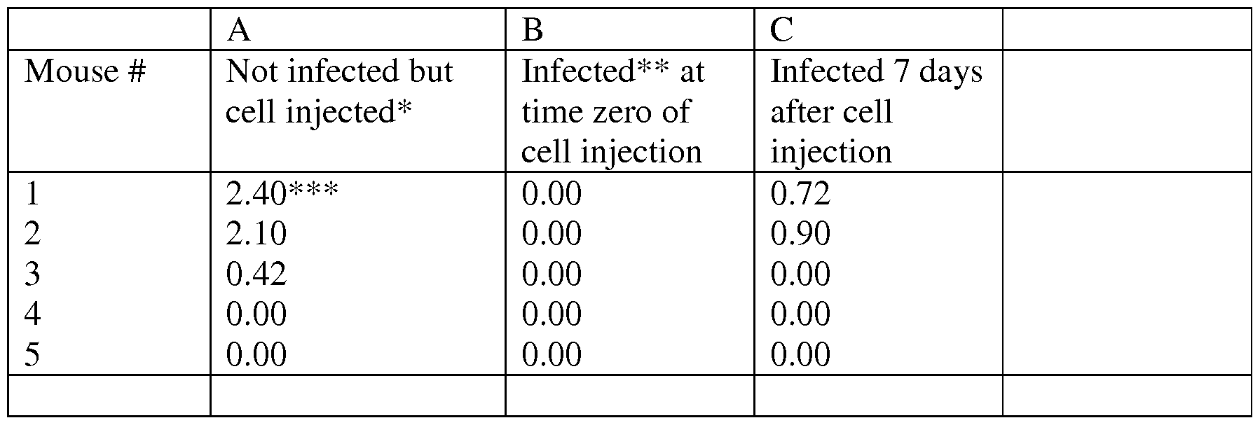
* 3xl06 cells in 0.1 ml injected SC
** lentivirus coding Psmdl shRNA produced in HEK293T cells
***tumor size in cm
EXAMPLE 14
Downregulation ofPsmd6 results in the disassembly of the 26S proteasome Downregulation of Psmd6 was effected using the shRNAs (SEQ ID NO: 18 and
19). The amount of proteasome subunits was measured by Western blot. In order to
analyze the amount of 20S subunit, an antibody that detected the alpha4 subunit of the 20S proteaseome (Psma4) was used. Activity of the proteasomes in the gel was determined by the cleavage of the Suc-LLVY-AMC peptide (Glickman, M. H., Rubin, D. M., Fried, V. A., and Finley, D. (1998) Mol Cell Biol 18(6), 3149-3162).
RESULTS
As can be seen from Figure 13, downregulation of PSmd6 resulted in a decrease in 26S activity and amount and an increase in the amount of 20S.
Although the invention has been described in conjunction with specific embodiments thereof, it is evident that many alternatives, modifications and variations will be apparent to those skilled in the art. Accordingly, it is intended to embrace all such alternatives, modifications and variations that fall within the spirit and broad scope of the appended claims.
All publications, patents and patent applications mentioned in this specification are herein incorporated in their entirety by reference into the specification, to the same extent as if each individual publication, patent or patent application was specifically and individually indicated to be incorporated herein by reference. In addition, citation or identification of any reference in this application shall not be construed as an admission that such reference is available as prior art to the present invention. To the extent that section headings are used, they should not be construed as necessarily limiting.
REFERENCES
1. Coux O, Tanaka K, Goldberg AL. Structure and functions of the 20S and 26S proteasomes. Annu Rev Biochem 1996; 65: 801-47.
2. Zwickl P, Voges D, Baumeister W. The proteasome: a macromolecular assembly designed for controlled proteolysis. Philos Trans R Soc Lond B Biol Sci 1999; 354: 1501-11.
3. Hershko A, Ciechanover A. The ubiquitin system. Annu Rev Biochem 1998; 67: 425-79.
4. Varshavsky A. The ubiquitin system. Trends Biochem Sci 1997; 22: 383-7.
5. Smith DM, Kafri G, Cheng Y, Ng D, Walz T, Goldberg AL. ATP binding to PAN or the 26S ATPases causes association with the 20S proteasome, gate opening, and translocation of unfolded proteins. Mol Cell 2005; 20: 687-98.
6. Navon A, Goldberg AL. Proteins are unfolded on the surface of the ATPase ring before transport into the proteasome. Mol Cell 2001; 8: 1339-49.
7. Hochstrasser M. Ubiquitin-dependent protein degradation. Annu Rev Genet 1996; 30: 405-39.
8. Tsvetkov P, Reuven N, Prives C, Shaul Y. The susceptibility of the p53 unstructured N-terminus to 20S proteasomal degradation programs stress response. J Biol Chem 2009.
9. Wiggins CM, Tsvetkov P, Johnson M, et al. BJJVIEL, an intrinsically disordered protein, is degraded by 20S proteasomes in the absence of poly-ubiquitylation. J Cell Sci; 124: 969-77.
10. Adler J, Reuven N, Kahana C, Shaul Y. c-Fos Proteasomal Degradation by Default and its Regulation by NQOl Determines c-Fos Serum Response Kinetics. Mol Cell Biol.
11. Shaul Y, Tsvetkov P, Reuven N. IDPs and Protein Degradation in the Cell: John Wiley & Sons, Inc.; 2010.
12. Asher G, Reuven N, Shaul Y. 20S proteasomes and protein degradation "by default". Bioessays 2006; 28: 844-9.
13. Mao I, Liu J, Li X, Luo H. REGgamma, a proteasome activator and beyond? Cell Mol Life Sci 2008; 65: 3971-80.
14. Hoeller D, Dikic I. Targeting the ubiquitin system in cancer therapy. Nature 2009; 458: 438-44.
15. Bedford L, Lowe J, Dick LR, Mayer RJ, Brownell JE. Ubiquitin-like protein conjugation and the ubiquitin-proteasome system as drug targets. Nat Rev Drug Discov; 10: 29-46.
16. Navon A, Ciechanover A. The 26 S proteasome: from basic mechanisms to drug targeting. J Biol Chem 2009; 284: 33713-8.
17. Arlt A, Bauer I, Schafmayer C, et al. Increased proteasome subunit protein expression and proteasome activity in colon cancer relate to an enhanced activation of nuclear factor E2-related factor 2 (Nrf2). Oncogene 2009; 28: 3983-96.
18. Chen L, Madura K. Increased proteasome activity, ubiquitin-conjugating enzymes, and eEFIA translation factor detected in breast cancer tissue. Cancer Res 2005; 65: 5599-606.
19. Wyke SM, Russell ST, Tisdale MJ. Induction of proteasome expression in skeletal muscle is attenuated by inhibitors of NF-kappaB activation. Br J Cancer 2004; 91: 1742-50.
20. Adams J, Kauffman M. Development of the proteasome inhibitor Velcade (Bortezomib). Cancer Invest 2004; 22: 304-11.
21. Richardson PG, Barlogie B, Berenson J, et al. A phase 2 study of bortezomib in relapsed, refractory myeloma. N Engl J Med 2003; 348: 2609-17.
22. Hideshima T, Richardson P, Chauhan D, et al. The proteasome inhibitor PS-341 inhibits growth, induces apoptosis, and overcomes drug resistance in human multiple myeloma cells. Cancer Res 2001; 61: 3071-6.
23. Chauhan D, Catley L, Li G, et al. A novel orally active proteasome inhibitor induces apoptosis in multiple myeloma cells with mechanisms distinct from
Bortezomib. Cancer Cell 2005; 8: 407-19.
24. Caravita T, de Fabritiis P, Palumbo A, Amadori S, Boccadoro M. Bortezomib: efficacy comparisons in solid tumors and hematologic malignancies. Nat Clin Pract Oncol 2006; 3: 374-87.
25. Adams J, Palombella VJ, Sausville EA, et al. Proteasome inhibitors: a novel class of potent and effective antitumor agents. Cancer Res 1999; 59: 2615-22.
26. Rajkumar SV, Richardson PG, Hideshima T, Anderson KC. Proteasome inhibition as a novel therapeutic target in human cancer. J Clin Oncol 2005; 23: 630-9.
27. Voorhees PM, Orlowski RZ. The proteasome and proteasome inhibitors in cancer therapy. Annu Rev Pharmacol Toxicol 2006; 46: 189-213.
28. Elsasser S, Schmidt M, Finley D. Characterization of the proteasome using native gel electrophoresis. Methods Enzymol 2005; 398: 353-63.
29. Tsvetkov P, Adamovich Y, Elliott E, Shaul Y. E3 ligase STUB 1/CHIP regulates NAD(P)H:quinone oxidoreductase 1 (NQOl) accumulation in aged brain, a process impaired in certain Alzheimer disease patients. J Biol Chem; 286: 8839-45.
30. Elliott E, Tsvetkov P, Ginzburg I. BAG-1 associates with Hsc70.Tau complex and regulates the proteasomal degradation of Tau protein. J Biol Chem 2007; 282: 37276-84.
31. Glickman MH, Rubin DM, Fried VA, Finley D. The regulatory particle of the Saccharomyces cerevisiae proteasome. Mol Cell Biol 1998; 18: 3149-62.
32. Schwartz AL, Ciechanover A. Targeting proteins for destruction by the ubiquitin system: implications for human pathobiology. Annu Rev Pharmacol Toxicol 2009; 49: 73-96.
33. Soule HD, Maloney TM, Wolman SR, et al. Isolation and characterization of a spontaneously immortalized human breast epithelial cell line, MCF-10. Cancer Res 1990; 50: 6075-86.
34. Tani E, Kitagawa H, Ikemoto H, Matsumoto T. Proteasome inhibitors induce Fas-mediated apoptosis by c-Myc accumulation and subsequent induction of FasL message in human glioma cells. FEBS Lett 2001; 504: 53-8.
35. Gregory MA, Hann SR. c-Myc proteolysis by the ubiquitin-proteasome pathway: stabilization of c-Myc in Burkitt's lymphoma cells. Mol Cell Biol 2000; 20: 2423-35.
36. Yang Y, Ikezoe T, Saito T, Kobayashi M, Koeffler HP, Taguchi H. Proteasome inhibitor PS-341 induces growth arrest and apoptosis of non-small cell lung cancer cells via the JNK/c-Jun/AP-1 signaling. Cancer Sci 2004; 95: 176-80.
37. Dent R, Trudeau M, Pritchard KI, et al. Triple-negative breast cancer: clinical features and patterns of recurrence. Clin Cancer Res 2007; 13: 4429-34.
38. Dawson SJ, Provenzano E, Caldas C. Triple negative breast cancers: clinical and prognostic implications. Eur J Cancer 2009; 45 Suppl 1: 27-40.
39. Cleator S, Heller W, Coombes RC. Triple-negative breast cancer: therapeutic options. Lancet Oncol 2007; 8: 235-44.
40. Hamilton TC, Young RC, McKoy WM, et al. Characterization of a human ovarian carcinoma cell line (NIH:OVCAR-3) with androgen and estrogen receptors. Cancer Res 1983; 43: 5379-89.
41. Hamilton TC, Young RC, Louie KG, et al. Characterization of a xenograft model of human ovarian carcinoma which produces ascites and intraabdominal carcinomatosis in mice. Cancer Res 1984; 44: 5286-90.
42. Shih Ie M, Sheu JJ, Santillan A, et al. Amplification of a chromatin remodeling gene, Rsf-l/HBXAP, in ovarian carcinoma. Proc Natl Acad Sci U S A 2005; 102: 14004-9.
43. Sheu JJ, Choi JH, Yildiz I, et al. The roles of human sucrose nonfermenting protein 2 homologue in the tumor-promoting functions of Rsf-1. Cancer Res 2008; 68: 4050-7.
44. Nalepa G, Rolfe M, Harper JW. Drug discovery in the ubiquitin-proteasome system. Nat Rev Drug Discov 2006; 5: 596-613.
45. Lee BH, Lee MJ, Park S, et al. Enhancement of proteasome activity by a small- molecule inhibitor of USP14. Nature 2010; 467: 179-84.
46. Bedford L, Lowe J, Dick LR, Mayer RJ, Brownell JE. Ubiquitin-like protein conjugation and the ubiquitin-proteasome system as drug targets. Nat Rev Drug Discov 2011; 10: 29-46.
47. Byrne A, McLaren RP, Mason P, et al. Knockdown of human deubiquitinase PSMD14 induces cell cycle arrest and senescence. Exp Cell Res 2010; 316: 258-71.
Claims
1. A method of treating a disease for which inhibiting a proteasome is advantageous, the method comprising administering to the subject a therapeutically effective amount of an agent which down-regulates an expression and/or activity of a proteasome 26S subunit, non-ATPase selected from the group consisting of PSMDl, PSMD6 and PSMDl 1 thereby treating the disease.
2. A method of treating ovarian or breast cancer, the method comprising administering to the subject a therapeutically effective amount of an agent which down- regulates an expression and/or activity of a proteasome 26S subunit, non-ATPase, selected from the group consisting of PSMDl, PSMD6 and PSMDl 1, thereby treating the disease.
3. A method of treating cancer, the method comprising administering to the subject a therapeutically effective amount of a pharmaceutical composition comprising an agent which induces a dissociation of the 26S proteasomal complex into a 20S component and a 19S component to thereby inhibit 26S proteasomal activity, wherein said pharmaceutical composition is devoid of a chemotherapeutic agent, thereby treating the cancer.
4. The method of claim 3, wherein said agent comprises a polynucleotide agent directed against a 19S component of said 26S proteasomal complex.
5. A method of treating a disease for which inhibiting a proteasome is advantageous, the method comprising administering to the subject a therapeutically effective amount of a polynucleotide agent comprising a nucleic acid sequence selected from the group consisting of SEQ ID NO:2, SEQ ID NO: 14, SEQ ID NO: 15, SEQ ID NO: 16 and SEQ ID NO: 17.
6. The method of claim 1 or 5, wherein the disease is cancer.
7. The method of claim 1 or 5, wherein the disease is an inflammatory disease.
8. The method of claim 1 or 5, wherein the disease is a neurodegenerative disease.
9. The method of claim 3 or 6, wherein said cancer comprises breast cancer or ovarian cancer.
10. The method of claims 1, 2 or 4, wherein the agent comprises a polynucleotide agent directed against a polynucleotide encoding said PSMD.
11. The method of claims 1, 2 or 4 wherein the agent comprises a small molecule agent.
12. The method of claim 10, wherein said polynucleotide agent comprises an siRNA.
13. The method of claim 12, wherein said siRNA comprises a nucleic acid sequence selected from the group consisting of SEQ ID NO: 2, SEQ ID NO: 14, SEQ ID NO: 15, SEQ ID NO: 16 and SEQ ID NO: 17.
14. The method of claim 10, wherein said polynucleotide agent comprises a nucleic acid sequence as set forth in SEQ ID NO: 3.
15. A pharmaceutical agent comprising an agent which down-regulates an amount or activity of proteasome 26S subunit, non-ATPase, selected from the group consisting of PSMD1, PSMD6 and PSMD 11 and a pharmaceutically acceptable carrier.
16. The pharmaceutical agent of claim 15 being devoid of a chemotherapeutic agent.
17. The pharmaceutical agent of claim 15, wherein the agent comprises a polynucleotide agent directed against a polynucleotide encoding said PSMD.
18. The pharmaceutical agent of claim 17, wherein said polynucleotide agent comprises an siRNA.
19. The pharmaceutical composition of claim 18, wherein said siRNA comprises a nucleic acid sequence selected from the group consisting of SEQ ID NO: 2, SEQ ID NO: 14, SEQ ID NO: 15, SEQ ID NO: 16 and SEQ ID NO: 17.
20. A method of selecting an agent useful for treating a disease for which inhibiting a proteasome is advantageous, the method comprising:
(a) contacting the agent with a 26S proteasomal complex of a cell;
(b) analyzing an amount of dissociation of said 26S proteasomal complex into a 19S regulatory particle (RP) and a 20S subunit, wherein an upregulation of said dissociation compared to an amount of dissociation of said 26S proteasomal complex in an absence of said agent, is indicative of an agent useful for treating a disease for which inhibiting a proteasome is advantageous.
21. The method of claim 20, wherein said analyzing is effected on the protein level.
22. The method of claim 20, wherein said analyzing is effected on the polynucleotide level.
23. The method of claim 20, wherein the disease is cancer.
24. The method of claim 20, wherein the disease is an inflammatory disease.
25. The method of claim 20, wherein the disease is a neurodegenerative disease.
26. The method of claim 23, wherein said cancer comprises breast cancer or ovarian cancer.
27. An agent which down-regulates an expression and/or activity of a proteasome 26S subunit, non-ATPase selected from the group consisting of PSMDl, PSMD6 and PSMD11 for use in treating a disease for which inhibiting a proteasome is advantageous.
28. An agent which down-regulates an expression and/or activity of a proteasome 26S subunit, non-ATPase, selected from the group consisting of PSMDl, PSMD6 and PSMDl 1 for treating ovarian or breast cancer.
29. A pharmaceutical composition comprising an agent which induces a dissociation of the 26S proteasomal complex into a 20S component and a 19S component to thereby inhibit 26S proteasomal activity, wherein said pharmaceutical composition is devoid of a chemotherapeutic agent for treating cancer.
30. A polynucleotide agent comprising a nucleic acid sequence selected from the group consisting of SEQ ID NO:2, SEQ ID NO: 14, SEQ ID NO: 15, SEQ ID NO: 16 and SEQ ID NO: 17 for treating a disease for which inhibiting a proteasome is advantageous.
Priority Applications (6)
| Application Number | Priority Date | Filing Date | Title |
|---|---|---|---|
| US14/354,580 US9145559B2 (en) | 2011-10-27 | 2012-10-25 | Methods of treating cancer |
| EP12784743.2A EP2771464B1 (en) | 2011-10-27 | 2012-10-25 | Methods of treating cancer |
| IL232266A IL232266A0 (en) | 2011-10-27 | 2014-04-27 | Methods of treating cancer |
| US14/866,957 US9579339B2 (en) | 2011-10-27 | 2015-09-27 | Methods of treating cancer |
| US15/442,784 US9840711B2 (en) | 2011-10-27 | 2017-02-27 | Methods of treating cancer |
| US15/834,110 US10155949B2 (en) | 2011-10-27 | 2017-12-07 | Methods of treating cancer |
Applications Claiming Priority (2)
| Application Number | Priority Date | Filing Date | Title |
|---|---|---|---|
| US201161551970P | 2011-10-27 | 2011-10-27 | |
| US61/551,970 | 2011-10-27 |
Related Child Applications (2)
| Application Number | Title | Priority Date | Filing Date |
|---|---|---|---|
| US14/354,580 A-371-Of-International US9145559B2 (en) | 2011-10-27 | 2012-10-25 | Methods of treating cancer |
| US14/866,957 Division US9579339B2 (en) | 2011-10-27 | 2015-09-27 | Methods of treating cancer |
Publications (4)
| Publication Number | Publication Date |
|---|---|
| WO2013061328A2 true WO2013061328A2 (en) | 2013-05-02 |
| WO2013061328A8 WO2013061328A8 (en) | 2013-06-27 |
| WO2013061328A3 WO2013061328A3 (en) | 2013-08-15 |
| WO2013061328A9 WO2013061328A9 (en) | 2013-10-03 |
Family
ID=47178254
Family Applications (1)
| Application Number | Title | Priority Date | Filing Date |
|---|---|---|---|
| PCT/IL2012/050422 WO2013061328A2 (en) | 2011-10-27 | 2012-10-25 | Method of treating cancer |
Country Status (3)
| Country | Link |
|---|---|
| US (4) | US9145559B2 (en) |
| EP (1) | EP2771464B1 (en) |
| WO (1) | WO2013061328A2 (en) |
Cited By (1)
| Publication number | Priority date | Publication date | Assignee | Title |
|---|---|---|---|---|
| US9145559B2 (en) | 2011-10-27 | 2015-09-29 | Yeda Research And Development Co. Ltd. | Methods of treating cancer |
Families Citing this family (5)
| Publication number | Priority date | Publication date | Assignee | Title |
|---|---|---|---|---|
| WO2017007858A1 (en) * | 2015-07-06 | 2017-01-12 | Whitehead Institute For Biomedical Research | Methods and compositions relating to proteasome inhibitor resistance |
| US11185555B2 (en) | 2016-04-11 | 2021-11-30 | Noah James Harrison | Method to kill pathogenic microbes in a patient |
| US11752810B2 (en) | 2020-07-28 | 2023-09-12 | Airgo Ip, Llc | Steer axle pressure management system |
| US11654928B2 (en) | 2020-07-28 | 2023-05-23 | Airgo Ip, Llc | Vehicle operational diagnostics, condition response, vehicle pairing, and blind spot detection system |
| CN113786488A (en) * | 2021-09-27 | 2021-12-14 | 山东大学第二医院 | Proteasome 19S regulatory particle subunit PSMD11 as target for preventing or treating cancer |
Citations (88)
| Publication number | Priority date | Publication date | Assignee | Title |
|---|---|---|---|---|
| US3687808A (en) | 1969-08-14 | 1972-08-29 | Univ Leland Stanford Junior | Synthetic polynucleotides |
| US3791932A (en) | 1971-02-10 | 1974-02-12 | Akzona Inc | Process for the demonstration and determination of reaction components having specific binding affinity for each other |
| US3839153A (en) | 1970-12-28 | 1974-10-01 | Akzona Inc | Process for the detection and determination of specific binding proteins and their corresponding bindable substances |
| US3850578A (en) | 1973-03-12 | 1974-11-26 | H Mcconnell | Process for assaying for biologically active molecules |
| US3850752A (en) | 1970-11-10 | 1974-11-26 | Akzona Inc | Process for the demonstration and determination of low molecular compounds and of proteins capable of binding these compounds specifically |
| US3853987A (en) | 1971-09-01 | 1974-12-10 | W Dreyer | Immunological reagent and radioimmuno assay |
| US3867517A (en) | 1971-12-21 | 1975-02-18 | Abbott Lab | Direct radioimmunoassay for antigens and their antibodies |
| US3879262A (en) | 1972-05-11 | 1975-04-22 | Akzona Inc | Detection and determination of haptens |
| US3901654A (en) | 1971-06-21 | 1975-08-26 | Biological Developments | Receptor assays of biologically active compounds employing biologically specific receptors |
| US3935074A (en) | 1973-12-17 | 1976-01-27 | Syva Company | Antibody steric hindrance immunoassay with two antibodies |
| US3984533A (en) | 1975-11-13 | 1976-10-05 | General Electric Company | Electrophoretic method of detecting antigen-antibody reaction |
| US3996345A (en) | 1974-08-12 | 1976-12-07 | Syva Company | Fluorescence quenching with immunological pairs in immunoassays |
| US4034074A (en) | 1974-09-19 | 1977-07-05 | The Board Of Trustees Of Leland Stanford Junior University | Universal reagent 2-site immunoradiometric assay using labelled anti (IgG) |
| US4036945A (en) | 1976-05-03 | 1977-07-19 | The Massachusetts General Hospital | Composition and method for determining the size and location of myocardial infarcts |
| US4098876A (en) | 1976-10-26 | 1978-07-04 | Corning Glass Works | Reverse sandwich immunoassay |
| US4331647A (en) | 1980-03-03 | 1982-05-25 | Goldenberg Milton David | Tumor localization and therapy with labeled antibody fragments specific to tumor-associated markers |
| US4469863A (en) | 1980-11-12 | 1984-09-04 | Ts O Paul O P | Nonionic nucleic acid alkyl and aryl phosphonates and processes for manufacture and use thereof |
| US4476301A (en) | 1982-04-29 | 1984-10-09 | Centre National De La Recherche Scientifique | Oligonucleotides, a process for preparing the same and their application as mediators of the action of interferon |
| US4666828A (en) | 1984-08-15 | 1987-05-19 | The General Hospital Corporation | Test for Huntington's disease |
| US4683202A (en) | 1985-03-28 | 1987-07-28 | Cetus Corporation | Process for amplifying nucleic acid sequences |
| US4801531A (en) | 1985-04-17 | 1989-01-31 | Biotechnology Research Partners, Ltd. | Apo AI/CIII genomic polymorphisms predictive of atherosclerosis |
| US4816567A (en) | 1983-04-08 | 1989-03-28 | Genentech, Inc. | Recombinant immunoglobin preparations |
| US4879219A (en) | 1980-09-19 | 1989-11-07 | General Hospital Corporation | Immunoassay utilizing monoclonal high affinity IgM antibodies |
| EP0375408A1 (en) | 1988-12-20 | 1990-06-27 | Baylor College Of Medicine | Method for making synthetic oligonucleotides which bind specifically to target sites on duplex DNA molecules, by forming a colinear triplex, the synthetic oligonucleotides and methods of use |
| US4946778A (en) | 1987-09-21 | 1990-08-07 | Genex Corporation | Single polypeptide chain binding molecules |
| US5011771A (en) | 1984-04-12 | 1991-04-30 | The General Hospital Corporation | Multiepitopic immunometric assay |
| US5023243A (en) | 1981-10-23 | 1991-06-11 | Molecular Biosystems, Inc. | Oligonucleotide therapeutic agent and method of making same |
| US5034506A (en) | 1985-03-15 | 1991-07-23 | Anti-Gene Development Group | Uncharged morpholino-based polymers having achiral intersubunit linkages |
| US5166315A (en) | 1989-12-20 | 1992-11-24 | Anti-Gene Development Group | Sequence-specific binding polymers for duplex nucleic acids |
| US5177196A (en) | 1990-08-16 | 1993-01-05 | Microprobe Corporation | Oligo (α-arabinofuranosyl nucleotides) and α-arabinofuranosyl precursors thereof |
| US5185444A (en) | 1985-03-15 | 1993-02-09 | Anti-Gene Deveopment Group | Uncharged morpolino-based polymers having phosphorous containing chiral intersubunit linkages |
| US5188897A (en) | 1987-10-22 | 1993-02-23 | Temple University Of The Commonwealth System Of Higher Education | Encapsulated 2',5'-phosphorothioate oligoadenylates |
| US5192659A (en) | 1989-08-25 | 1993-03-09 | Genetype Ag | Intron sequence analysis method for detection of adjacent and remote locus alleles as haplotypes |
| US5214134A (en) | 1990-09-12 | 1993-05-25 | Sterling Winthrop Inc. | Process of linking nucleosides with a siloxane bridge |
| US5216141A (en) | 1988-06-06 | 1993-06-01 | Benner Steven A | Oligonucleotide analogs containing sulfur linkages |
| US5235033A (en) | 1985-03-15 | 1993-08-10 | Anti-Gene Development Group | Alpha-morpholino ribonucleoside derivatives and polymers thereof |
| US5264564A (en) | 1989-10-24 | 1993-11-23 | Gilead Sciences | Oligonucleotide analogs with novel linkages |
| US5264562A (en) | 1989-10-24 | 1993-11-23 | Gilead Sciences, Inc. | Oligonucleotide analogs with novel linkages |
| US5264423A (en) | 1987-03-25 | 1993-11-23 | The United States Of America As Represented By The Department Of Health And Human Services | Inhibitors for replication of retroviruses and for the expression of oncogene products |
| US5272057A (en) | 1988-10-14 | 1993-12-21 | Georgetown University | Method of detecting a predisposition to cancer by the use of restriction fragment length polymorphism of the gene for human poly (ADP-ribose) polymerase |
| US5276019A (en) | 1987-03-25 | 1994-01-04 | The United States Of America As Represented By The Department Of Health And Human Services | Inhibitors for replication of retroviruses and for the expression of oncogene products |
| US5278302A (en) | 1988-05-26 | 1994-01-11 | University Patents, Inc. | Polynucleotide phosphorodithioates |
| US5281521A (en) | 1992-07-20 | 1994-01-25 | The Trustees Of The University Of Pennsylvania | Modified avidin-biotin technique |
| US5321131A (en) | 1990-03-08 | 1994-06-14 | Hybridon, Inc. | Site-specific functionalization of oligodeoxynucleotides for non-radioactive labelling |
| US5399676A (en) | 1989-10-23 | 1995-03-21 | Gilead Sciences | Oligonucleotides with inverted polarity |
| US5405939A (en) | 1987-10-22 | 1995-04-11 | Temple University Of The Commonwealth System Of Higher Education | 2',5'-phosphorothioate oligoadenylates and their covalent conjugates with polylysine |
| US5405938A (en) | 1989-12-20 | 1995-04-11 | Anti-Gene Development Group | Sequence-specific binding polymers for duplex nucleic acids |
| US5434257A (en) | 1992-06-01 | 1995-07-18 | Gilead Sciences, Inc. | Binding compentent oligomers containing unsaturated 3',5' and 2',5' linkages |
| US5455233A (en) | 1989-11-30 | 1995-10-03 | University Of North Carolina | Oligoribonucleoside and oligodeoxyribonucleoside boranophosphates |
| US5464764A (en) | 1989-08-22 | 1995-11-07 | University Of Utah Research Foundation | Positive-negative selection methods and vectors |
| US5466677A (en) | 1993-03-06 | 1995-11-14 | Ciba-Geigy Corporation | Dinucleoside phosphinates and their pharmaceutical compositions |
| US5470967A (en) | 1990-04-10 | 1995-11-28 | The Dupont Merck Pharmaceutical Company | Oligonucleotide analogs with sulfamate linkages |
| US5476925A (en) | 1993-02-01 | 1995-12-19 | Northwestern University | Oligodeoxyribonucleotides including 3'-aminonucleoside-phosphoramidate linkages and terminal 3'-amino groups |
| US5489677A (en) | 1990-07-27 | 1996-02-06 | Isis Pharmaceuticals, Inc. | Oligonucleoside linkages containing adjacent oxygen and nitrogen atoms |
| US5519126A (en) | 1988-03-25 | 1996-05-21 | University Of Virginia Alumni Patents Foundation | Oligonucleotide N-alkylphosphoramidates |
| US5539082A (en) | 1993-04-26 | 1996-07-23 | Nielsen; Peter E. | Peptide nucleic acids |
| US5541307A (en) | 1990-07-27 | 1996-07-30 | Isis Pharmaceuticals, Inc. | Backbone modified oligonucleotide analogs and solid phase synthesis thereof |
| US5545806A (en) | 1990-08-29 | 1996-08-13 | Genpharm International, Inc. | Ransgenic non-human animals for producing heterologous antibodies |
| US5545807A (en) | 1988-10-12 | 1996-08-13 | The Babraham Institute | Production of antibodies from transgenic animals |
| US5550111A (en) | 1984-07-11 | 1996-08-27 | Temple University-Of The Commonwealth System Of Higher Education | Dual action 2',5'-oligoadenylate antiviral derivatives and uses thereof |
| US5561225A (en) | 1990-09-19 | 1996-10-01 | Southern Research Institute | Polynucleotide analogs containing sulfonate and sulfonamide internucleoside linkages |
| US5569825A (en) | 1990-08-29 | 1996-10-29 | Genpharm International | Transgenic non-human animals capable of producing heterologous antibodies of various isotypes |
| US5571799A (en) | 1991-08-12 | 1996-11-05 | Basco, Ltd. | (2'-5') oligoadenylate analogues useful as inhibitors of host-v5.-graft response |
| US5587361A (en) | 1991-10-15 | 1996-12-24 | Isis Pharmaceuticals, Inc. | Oligonucleotides having phosphorothioate linkages of high chiral purity |
| US5596086A (en) | 1990-09-20 | 1997-01-21 | Gilead Sciences, Inc. | Modified internucleoside linkages having one nitrogen and two carbon atoms |
| US5602240A (en) | 1990-07-27 | 1997-02-11 | Ciba Geigy Ag. | Backbone modified oligonucleotide analogs |
| US5608046A (en) | 1990-07-27 | 1997-03-04 | Isis Pharmaceuticals, Inc. | Conjugated 4'-desmethyl nucleoside analog compounds |
| US5610289A (en) | 1990-07-27 | 1997-03-11 | Isis Pharmaceuticals, Inc. | Backbone modified oligonucleotide analogues |
| US5618704A (en) | 1990-07-27 | 1997-04-08 | Isis Pharmacueticals, Inc. | Backbone-modified oligonucleotide analogs and preparation thereof through radical coupling |
| US5623070A (en) | 1990-07-27 | 1997-04-22 | Isis Pharmaceuticals, Inc. | Heteroatomic oligonucleoside linkages |
| US5625126A (en) | 1990-08-29 | 1997-04-29 | Genpharm International, Inc. | Transgenic non-human animals for producing heterologous antibodies |
| US5625050A (en) | 1994-03-31 | 1997-04-29 | Amgen Inc. | Modified oligonucleotides and intermediates useful in nucleic acid therapeutics |
| US5633425A (en) | 1990-08-29 | 1997-05-27 | Genpharm International, Inc. | Transgenic non-human animals capable of producing heterologous antibodies |
| US5633360A (en) | 1992-04-14 | 1997-05-27 | Gilead Sciences, Inc. | Oligonucleotide analogs capable of passive cell membrane permeation |
| US5661016A (en) | 1990-08-29 | 1997-08-26 | Genpharm International Inc. | Transgenic non-human animals capable of producing heterologous antibodies of various isotypes |
| US5663312A (en) | 1993-03-31 | 1997-09-02 | Sanofi | Oligonucleotide dimers with amide linkages replacing phosphodiester linkages |
| US5677439A (en) | 1990-08-03 | 1997-10-14 | Sanofi | Oligonucleotide analogues containing phosphate diester linkage substitutes, compositions thereof, and precursor dinucleotide analogues |
| US5677437A (en) | 1990-07-27 | 1997-10-14 | Isis Pharmaceuticals, Inc. | Heteroatomic oligonucleoside linkages |
| US5714331A (en) | 1991-05-24 | 1998-02-03 | Buchardt, Deceased; Ole | Peptide nucleic acids having enhanced binding affinity, sequence specificity and solubility |
| US5719262A (en) | 1993-11-22 | 1998-02-17 | Buchardt, Deceased; Ole | Peptide nucleic acids having amino acid side chains |
| US5721138A (en) | 1992-12-15 | 1998-02-24 | Sandford University | Apolipoprotein(A) promoter and regulatory sequence constructs and methods of use |
| US6303374B1 (en) | 2000-01-18 | 2001-10-16 | Isis Pharmaceuticals Inc. | Antisense modulation of caspase 3 expression |
| US6326174B1 (en) | 1994-12-02 | 2001-12-04 | The Scripps Research Institute | Enzymatic DNA molecules |
| US6348185B1 (en) | 1998-06-20 | 2002-02-19 | Washington University School Of Medicine | Membrane-permeant peptide complexes for medical imaging, diagnostics, and pharmaceutical therapy |
| US20020123476A1 (en) | 1991-03-19 | 2002-09-05 | Emanuele R. Martin | Therapeutic delivery compositions and methods of use thereof |
| US20020128218A1 (en) | 1991-03-19 | 2002-09-12 | Emanuele R. Martin | Therapeutic delivery compositions and methods of use thereof |
| US20030096980A1 (en) | 1991-11-26 | 2003-05-22 | Brian Froehler | Enhanced triple-helix and double-helix formation with oligomers containing modified pyrimidines |
| WO2009117196A1 (en) | 2008-02-15 | 2009-09-24 | Pacific Arrow Limited | Blocking the metastasis of cancer cells and the uses of new compounds thereof |
Family Cites Families (5)
| Publication number | Priority date | Publication date | Assignee | Title |
|---|---|---|---|---|
| TW200512298A (en) * | 2003-09-24 | 2005-04-01 | Oncotherapy Science Inc | Method of diagnosing breast cancer |
| WO2005103254A1 (en) | 2004-04-22 | 2005-11-03 | Yissum Research Development Company Of The Hebrew University Of Jerusalem | UNIVERSAL TARGET SEQUENCES FOR siRNA GENE SILENCING |
| WO2008039937A2 (en) | 2006-09-27 | 2008-04-03 | Novarx | Blocking of gene expression in eukaryotic cells |
| WO2009099991A2 (en) | 2008-01-31 | 2009-08-13 | The Brigham And Women's Hospital, Inc. | Treatment of cancer |
| EP2771464B1 (en) | 2011-10-27 | 2018-03-21 | Yeda Research and Development Co. Ltd. | Methods of treating cancer |
-
2012
- 2012-10-25 EP EP12784743.2A patent/EP2771464B1/en not_active Not-in-force
- 2012-10-25 WO PCT/IL2012/050422 patent/WO2013061328A2/en active Application Filing
- 2012-10-25 US US14/354,580 patent/US9145559B2/en not_active Expired - Fee Related
-
2015
- 2015-09-27 US US14/866,957 patent/US9579339B2/en active Active
-
2017
- 2017-02-27 US US15/442,784 patent/US9840711B2/en not_active Expired - Fee Related
- 2017-12-07 US US15/834,110 patent/US10155949B2/en not_active Expired - Fee Related
Patent Citations (96)
| Publication number | Priority date | Publication date | Assignee | Title |
|---|---|---|---|---|
| US3687808A (en) | 1969-08-14 | 1972-08-29 | Univ Leland Stanford Junior | Synthetic polynucleotides |
| US3850752A (en) | 1970-11-10 | 1974-11-26 | Akzona Inc | Process for the demonstration and determination of low molecular compounds and of proteins capable of binding these compounds specifically |
| US3839153A (en) | 1970-12-28 | 1974-10-01 | Akzona Inc | Process for the detection and determination of specific binding proteins and their corresponding bindable substances |
| US3791932A (en) | 1971-02-10 | 1974-02-12 | Akzona Inc | Process for the demonstration and determination of reaction components having specific binding affinity for each other |
| US3901654A (en) | 1971-06-21 | 1975-08-26 | Biological Developments | Receptor assays of biologically active compounds employing biologically specific receptors |
| US3853987A (en) | 1971-09-01 | 1974-12-10 | W Dreyer | Immunological reagent and radioimmuno assay |
| US3867517A (en) | 1971-12-21 | 1975-02-18 | Abbott Lab | Direct radioimmunoassay for antigens and their antibodies |
| US3879262A (en) | 1972-05-11 | 1975-04-22 | Akzona Inc | Detection and determination of haptens |
| US3850578A (en) | 1973-03-12 | 1974-11-26 | H Mcconnell | Process for assaying for biologically active molecules |
| US3935074A (en) | 1973-12-17 | 1976-01-27 | Syva Company | Antibody steric hindrance immunoassay with two antibodies |
| US3996345A (en) | 1974-08-12 | 1976-12-07 | Syva Company | Fluorescence quenching with immunological pairs in immunoassays |
| US4034074A (en) | 1974-09-19 | 1977-07-05 | The Board Of Trustees Of Leland Stanford Junior University | Universal reagent 2-site immunoradiometric assay using labelled anti (IgG) |
| US3984533A (en) | 1975-11-13 | 1976-10-05 | General Electric Company | Electrophoretic method of detecting antigen-antibody reaction |
| US4036945A (en) | 1976-05-03 | 1977-07-19 | The Massachusetts General Hospital | Composition and method for determining the size and location of myocardial infarcts |
| US4098876A (en) | 1976-10-26 | 1978-07-04 | Corning Glass Works | Reverse sandwich immunoassay |
| US4331647A (en) | 1980-03-03 | 1982-05-25 | Goldenberg Milton David | Tumor localization and therapy with labeled antibody fragments specific to tumor-associated markers |
| US4879219A (en) | 1980-09-19 | 1989-11-07 | General Hospital Corporation | Immunoassay utilizing monoclonal high affinity IgM antibodies |
| US4469863A (en) | 1980-11-12 | 1984-09-04 | Ts O Paul O P | Nonionic nucleic acid alkyl and aryl phosphonates and processes for manufacture and use thereof |
| US5023243A (en) | 1981-10-23 | 1991-06-11 | Molecular Biosystems, Inc. | Oligonucleotide therapeutic agent and method of making same |
| US4476301A (en) | 1982-04-29 | 1984-10-09 | Centre National De La Recherche Scientifique | Oligonucleotides, a process for preparing the same and their application as mediators of the action of interferon |
| US4816567A (en) | 1983-04-08 | 1989-03-28 | Genentech, Inc. | Recombinant immunoglobin preparations |
| US5011771A (en) | 1984-04-12 | 1991-04-30 | The General Hospital Corporation | Multiepitopic immunometric assay |
| US5550111A (en) | 1984-07-11 | 1996-08-27 | Temple University-Of The Commonwealth System Of Higher Education | Dual action 2',5'-oligoadenylate antiviral derivatives and uses thereof |
| US4666828A (en) | 1984-08-15 | 1987-05-19 | The General Hospital Corporation | Test for Huntington's disease |
| US5235033A (en) | 1985-03-15 | 1993-08-10 | Anti-Gene Development Group | Alpha-morpholino ribonucleoside derivatives and polymers thereof |
| US5034506A (en) | 1985-03-15 | 1991-07-23 | Anti-Gene Development Group | Uncharged morpholino-based polymers having achiral intersubunit linkages |
| US5185444A (en) | 1985-03-15 | 1993-02-09 | Anti-Gene Deveopment Group | Uncharged morpolino-based polymers having phosphorous containing chiral intersubunit linkages |
| US4683202B1 (en) | 1985-03-28 | 1990-11-27 | Cetus Corp | |
| US4683202A (en) | 1985-03-28 | 1987-07-28 | Cetus Corporation | Process for amplifying nucleic acid sequences |
| US4801531A (en) | 1985-04-17 | 1989-01-31 | Biotechnology Research Partners, Ltd. | Apo AI/CIII genomic polymorphisms predictive of atherosclerosis |
| US5286717A (en) | 1987-03-25 | 1994-02-15 | The United States Of America As Represented By The Department Of Health And Human Services | Inhibitors for replication of retroviruses and for the expression of oncogene products |
| US5276019A (en) | 1987-03-25 | 1994-01-04 | The United States Of America As Represented By The Department Of Health And Human Services | Inhibitors for replication of retroviruses and for the expression of oncogene products |
| US5264423A (en) | 1987-03-25 | 1993-11-23 | The United States Of America As Represented By The Department Of Health And Human Services | Inhibitors for replication of retroviruses and for the expression of oncogene products |
| US4946778A (en) | 1987-09-21 | 1990-08-07 | Genex Corporation | Single polypeptide chain binding molecules |
| US5405939A (en) | 1987-10-22 | 1995-04-11 | Temple University Of The Commonwealth System Of Higher Education | 2',5'-phosphorothioate oligoadenylates and their covalent conjugates with polylysine |
| US5188897A (en) | 1987-10-22 | 1993-02-23 | Temple University Of The Commonwealth System Of Higher Education | Encapsulated 2',5'-phosphorothioate oligoadenylates |
| US5519126A (en) | 1988-03-25 | 1996-05-21 | University Of Virginia Alumni Patents Foundation | Oligonucleotide N-alkylphosphoramidates |
| US5278302A (en) | 1988-05-26 | 1994-01-11 | University Patents, Inc. | Polynucleotide phosphorodithioates |
| US5453496A (en) | 1988-05-26 | 1995-09-26 | University Patents, Inc. | Polynucleotide phosphorodithioate |
| US5216141A (en) | 1988-06-06 | 1993-06-01 | Benner Steven A | Oligonucleotide analogs containing sulfur linkages |
| US5545807A (en) | 1988-10-12 | 1996-08-13 | The Babraham Institute | Production of antibodies from transgenic animals |
| US5272057A (en) | 1988-10-14 | 1993-12-21 | Georgetown University | Method of detecting a predisposition to cancer by the use of restriction fragment length polymorphism of the gene for human poly (ADP-ribose) polymerase |
| EP0375408A1 (en) | 1988-12-20 | 1990-06-27 | Baylor College Of Medicine | Method for making synthetic oligonucleotides which bind specifically to target sites on duplex DNA molecules, by forming a colinear triplex, the synthetic oligonucleotides and methods of use |
| US5487992A (en) | 1989-08-22 | 1996-01-30 | University Of Utah Research Foundation | Cells and non-human organisms containing predetermined genomic modifications and positive-negative selection methods and vectors for making same |
| US5464764A (en) | 1989-08-22 | 1995-11-07 | University Of Utah Research Foundation | Positive-negative selection methods and vectors |
| US5192659A (en) | 1989-08-25 | 1993-03-09 | Genetype Ag | Intron sequence analysis method for detection of adjacent and remote locus alleles as haplotypes |
| US5399676A (en) | 1989-10-23 | 1995-03-21 | Gilead Sciences | Oligonucleotides with inverted polarity |
| US5264562A (en) | 1989-10-24 | 1993-11-23 | Gilead Sciences, Inc. | Oligonucleotide analogs with novel linkages |
| US5264564A (en) | 1989-10-24 | 1993-11-23 | Gilead Sciences | Oligonucleotide analogs with novel linkages |
| US5455233A (en) | 1989-11-30 | 1995-10-03 | University Of North Carolina | Oligoribonucleoside and oligodeoxyribonucleoside boranophosphates |
| US5405938A (en) | 1989-12-20 | 1995-04-11 | Anti-Gene Development Group | Sequence-specific binding polymers for duplex nucleic acids |
| US5166315A (en) | 1989-12-20 | 1992-11-24 | Anti-Gene Development Group | Sequence-specific binding polymers for duplex nucleic acids |
| US5536821A (en) | 1990-03-08 | 1996-07-16 | Worcester Foundation For Biomedical Research | Aminoalkylphosphorothioamidate oligonucleotide deratives |
| US5321131A (en) | 1990-03-08 | 1994-06-14 | Hybridon, Inc. | Site-specific functionalization of oligodeoxynucleotides for non-radioactive labelling |
| US5563253A (en) | 1990-03-08 | 1996-10-08 | Worcester Foundation For Biomedical Research | Linear aminoalkylphosphoramidate oligonucleotide derivatives |
| US5541306A (en) | 1990-03-08 | 1996-07-30 | Worcester Foundation For Biomedical Research | Aminoalkylphosphotriester oligonucleotide derivatives |
| US5470967A (en) | 1990-04-10 | 1995-11-28 | The Dupont Merck Pharmaceutical Company | Oligonucleotide analogs with sulfamate linkages |
| US5608046A (en) | 1990-07-27 | 1997-03-04 | Isis Pharmaceuticals, Inc. | Conjugated 4'-desmethyl nucleoside analog compounds |
| US5677437A (en) | 1990-07-27 | 1997-10-14 | Isis Pharmaceuticals, Inc. | Heteroatomic oligonucleoside linkages |
| US5489677A (en) | 1990-07-27 | 1996-02-06 | Isis Pharmaceuticals, Inc. | Oligonucleoside linkages containing adjacent oxygen and nitrogen atoms |
| US5623070A (en) | 1990-07-27 | 1997-04-22 | Isis Pharmaceuticals, Inc. | Heteroatomic oligonucleoside linkages |
| US5618704A (en) | 1990-07-27 | 1997-04-08 | Isis Pharmacueticals, Inc. | Backbone-modified oligonucleotide analogs and preparation thereof through radical coupling |
| US5541307A (en) | 1990-07-27 | 1996-07-30 | Isis Pharmaceuticals, Inc. | Backbone modified oligonucleotide analogs and solid phase synthesis thereof |
| US5610289A (en) | 1990-07-27 | 1997-03-11 | Isis Pharmaceuticals, Inc. | Backbone modified oligonucleotide analogues |
| US5602240A (en) | 1990-07-27 | 1997-02-11 | Ciba Geigy Ag. | Backbone modified oligonucleotide analogs |
| US5677439A (en) | 1990-08-03 | 1997-10-14 | Sanofi | Oligonucleotide analogues containing phosphate diester linkage substitutes, compositions thereof, and precursor dinucleotide analogues |
| US5177196A (en) | 1990-08-16 | 1993-01-05 | Microprobe Corporation | Oligo (α-arabinofuranosyl nucleotides) and α-arabinofuranosyl precursors thereof |
| US5661016A (en) | 1990-08-29 | 1997-08-26 | Genpharm International Inc. | Transgenic non-human animals capable of producing heterologous antibodies of various isotypes |
| US5569825A (en) | 1990-08-29 | 1996-10-29 | Genpharm International | Transgenic non-human animals capable of producing heterologous antibodies of various isotypes |
| US5625126A (en) | 1990-08-29 | 1997-04-29 | Genpharm International, Inc. | Transgenic non-human animals for producing heterologous antibodies |
| US5633425A (en) | 1990-08-29 | 1997-05-27 | Genpharm International, Inc. | Transgenic non-human animals capable of producing heterologous antibodies |
| US5545806A (en) | 1990-08-29 | 1996-08-13 | Genpharm International, Inc. | Ransgenic non-human animals for producing heterologous antibodies |
| US5214134A (en) | 1990-09-12 | 1993-05-25 | Sterling Winthrop Inc. | Process of linking nucleosides with a siloxane bridge |
| US5561225A (en) | 1990-09-19 | 1996-10-01 | Southern Research Institute | Polynucleotide analogs containing sulfonate and sulfonamide internucleoside linkages |
| US5596086A (en) | 1990-09-20 | 1997-01-21 | Gilead Sciences, Inc. | Modified internucleoside linkages having one nitrogen and two carbon atoms |
| US20020123476A1 (en) | 1991-03-19 | 2002-09-05 | Emanuele R. Martin | Therapeutic delivery compositions and methods of use thereof |
| US20020128218A1 (en) | 1991-03-19 | 2002-09-12 | Emanuele R. Martin | Therapeutic delivery compositions and methods of use thereof |
| US5714331A (en) | 1991-05-24 | 1998-02-03 | Buchardt, Deceased; Ole | Peptide nucleic acids having enhanced binding affinity, sequence specificity and solubility |
| US5571799A (en) | 1991-08-12 | 1996-11-05 | Basco, Ltd. | (2'-5') oligoadenylate analogues useful as inhibitors of host-v5.-graft response |
| US5587361A (en) | 1991-10-15 | 1996-12-24 | Isis Pharmaceuticals, Inc. | Oligonucleotides having phosphorothioate linkages of high chiral purity |
| US20030170680A1 (en) | 1991-11-26 | 2003-09-11 | Brian Froehler | Enhanced triple-helix and double-helix formation with oligomers containing modified pyrimidines |
| US20030096980A1 (en) | 1991-11-26 | 2003-05-22 | Brian Froehler | Enhanced triple-helix and double-helix formation with oligomers containing modified pyrimidines |
| US5633360A (en) | 1992-04-14 | 1997-05-27 | Gilead Sciences, Inc. | Oligonucleotide analogs capable of passive cell membrane permeation |
| US5434257A (en) | 1992-06-01 | 1995-07-18 | Gilead Sciences, Inc. | Binding compentent oligomers containing unsaturated 3',5' and 2',5' linkages |
| US5281521A (en) | 1992-07-20 | 1994-01-25 | The Trustees Of The University Of Pennsylvania | Modified avidin-biotin technique |
| US5721138A (en) | 1992-12-15 | 1998-02-24 | Sandford University | Apolipoprotein(A) promoter and regulatory sequence constructs and methods of use |
| US5476925A (en) | 1993-02-01 | 1995-12-19 | Northwestern University | Oligodeoxyribonucleotides including 3'-aminonucleoside-phosphoramidate linkages and terminal 3'-amino groups |
| US5466677A (en) | 1993-03-06 | 1995-11-14 | Ciba-Geigy Corporation | Dinucleoside phosphinates and their pharmaceutical compositions |
| US5663312A (en) | 1993-03-31 | 1997-09-02 | Sanofi | Oligonucleotide dimers with amide linkages replacing phosphodiester linkages |
| US5539082A (en) | 1993-04-26 | 1996-07-23 | Nielsen; Peter E. | Peptide nucleic acids |
| US5719262A (en) | 1993-11-22 | 1998-02-17 | Buchardt, Deceased; Ole | Peptide nucleic acids having amino acid side chains |
| US5625050A (en) | 1994-03-31 | 1997-04-29 | Amgen Inc. | Modified oligonucleotides and intermediates useful in nucleic acid therapeutics |
| US6326174B1 (en) | 1994-12-02 | 2001-12-04 | The Scripps Research Institute | Enzymatic DNA molecules |
| US6348185B1 (en) | 1998-06-20 | 2002-02-19 | Washington University School Of Medicine | Membrane-permeant peptide complexes for medical imaging, diagnostics, and pharmaceutical therapy |
| US6303374B1 (en) | 2000-01-18 | 2001-10-16 | Isis Pharmaceuticals Inc. | Antisense modulation of caspase 3 expression |
| WO2009117196A1 (en) | 2008-02-15 | 2009-09-24 | Pacific Arrow Limited | Blocking the metastasis of cancer cells and the uses of new compounds thereof |
Non-Patent Citations (170)
| Title |
|---|
| "Immobilized Cells and Enzymes", 1986, IRL PRESS |
| "Methods in Enzymology", vol. 1-317, ACADEMIC PRESS |
| "PCR Protocols: A Guide To Methods And Applications", 1990, ACADEMIC PRESS |
| "Remington's Pharmaceutical Sciences", MACK PUBLISHING CO. |
| "Vectors: A Survey of Molecular Cloning Vectors and Their Uses", 1988, BUTTERWORTHS |
| ADAMS J; KAUFFMAN M.: "Development of the proteasome inhibitor Velcade (Bortezomib", CANCER INVEST, vol. 22, 2004, pages 304 - 11 |
| ADAMS J; PALOMBELLA VJ; SAUSVILLE EA ET AL.: "Proteasome inhibitors: a novel class of potent and effective antitumor agents", CANCER RES, vol. 59, 1999, pages 2615 - 22, XP002168152 |
| ADLER J; REUVEN N; KAHANA C; SHAUL Y: "c-Fos Proteasomal Degradation by Default and its Regulation by NQO I Determines c-Fos Serum Response Kinetics", MOL CELL BIOL. |
| AOKI ET AL., BIOCHEM BIOPHYS RES COMMUN, vol. 231, 1997, pages 540 - 5 |
| ARLT A; BAUER I; SCHAFMAYER C ET AL.: "Increased proteasome subunit protein expression and proteasome activity in colon cancer relate to an enhanced activation of nuclear factor E2-related factor 2 (Nrf2", ONCOGENE, vol. 28, 2009, pages 3983 - 96, XP055345455, DOI: doi:10.1038/onc.2009.264 |
| ASHER G; REUVEN N; SHAUL Y: "20S proteasomes and protein degradation", BIOESSAYS, vol. 28, 2006, pages 844 - 9 |
| AUSUBEL ET AL.: "Current Protocols in Molecular Biology", 1989, JOHN WILEY AND SONS |
| AUSUBEL, R. M ET AL.: "Current Protocols in Molecular Biology", vol. I-III, 1989, JOHN WILEY & SONS |
| AUSUBEL, R. M.,: "Current Protocols in Molecular Biology", vol. I-III, 1994 |
| BEAL, P. A. ET AL., SCIENCE, vol. 251, 1992, pages 1360 - 1363 |
| BEDFORD L; LOWE J; DICK LR; MAYER RJ; BROWNELL JE.: "Ubiquitin-like protein conjugation and the ubiquitin-proteasome system as drug targets", NAT REV DRUG DISCOV, vol. 10, 2011, pages 29 - 46, XP055247933, DOI: doi:10.1038/nrd3321 |
| BEDFORD L; LOWE J; DICK LR; MAYER RJ; BROWNELL JE.: "Ubiquitin-like protein conjugation and the ubiquitin-proteasome system as drug targets", NAT REV DRUG DISCOV, vol. 10, pages 29 - 46, XP055247933, DOI: doi:10.1038/nrd3321 |
| BERTRAM JS ET AL., CANCER LETT., vol. 11, 1980, pages 63 - 73 |
| BESCH ET AL., J BIOL CHEM, vol. 277, 2002, pages 32473 - 79 |
| BHARGAVA A ET AL., BRAIN RES. PROTOC., vol. 13, 2004, pages 115 - 125 |
| BILLY ET AL., PNAS, vol. 98, 2001, pages 14428 - 14433 |
| BIRD ET AL., SCIENCE, vol. 242, 1988, pages 423 - 426 |
| BIRREN ET AL.: "Genome Analysis: A Laboratory Manual Series", vol. 1-4, 1998, COLD SPRING HARBOR LABORATORY PRESS |
| BOERNER ET AL., J. IMMUNOL., vol. 147, no. 1, 1991, pages 86 - 95 |
| BREAKER, R.R.; JOYCE, G., CHEMISTRY AND BIOLOGY, vol. 2, 1995, pages 655 |
| BRILES EB ET AL., J. NATL. CANCER INST., vol. 60, 1978, pages 1217 - 1222 |
| BRUMMELKAMP, T. R. ET AL., SCIENCE, vol. 296, 2002, pages 550 |
| BYRNE A. ET AL., ECR, vol. 316, 2010, pages 258 - 271 |
| BYRNE A; MCLAREN RP; MASON P ET AL.: "Knockdown of human deubiquitinase PSMD14 induces cell cycle arrest and senescence", EXP CELL RES, vol. 316, 2010, pages 258 - 71, XP055051181, DOI: doi:10.1016/j.yexcr.2009.08.018 |
| CARAVITA T; DE FABRITIIS P; PALUMBO A; AMADORI S; BOCCADORO M. BORTEZOMIB: "efficacy comparisons in solid tumors and hematologic malignancies", NAT CLIN PRACT ONCOL, vol. 3, 2006, pages 374 - 87 |
| CARBONE ET AL., NUCL ACID RES, vol. 31, 2003, pages 833 - 43 |
| CASTANOTTO, D. ET AL., RNA, vol. 8, 2002, pages 1454 |
| CELLIS, J. E.: "Cell Biology: A Laboratory Handbook", vol. I-III, 1994 |
| CHANG ET AL.: "Somatic Gene Therapy", 1995, CRC PRESS |
| CHAUHAN D; CATLEY L; LI G ET AL.: "A novel orally active proteasome inhibitor induces apoptosis in multiple myeloma cells with mechanisms distinct from Bortezomib", CANCER CELL, vol. 8, 2005, pages 407 - 19 |
| CHEN L; MADURA K.: "Increased proteasome activity, ubiquitin-conjugating enzymes, and eEFIA translation factor detected in breast cancer tissue", CANCER RES, vol. 65, 2005, pages 5599 - 606 |
| CLEATOR S; HELLER W; COOMBES RC: "Triple-negative breast cancer: therapeutic options", LANCET ONCOL, vol. 8, 2007, pages 235 - 44, XP005907670, DOI: doi:10.1016/S1470-2045(07)70074-8 |
| COLE ET AL.: "Monoclonal Antibodies and Cancer Therapy", 1985, ALAN R. LISS, pages: 77 |
| COLIGAN J.E.: "Current Protocols in Immunology", vol. I-III, 1994 |
| COONEY, M. ET AL., SCIENCE, vol. 241, 1988, pages 456 - 459 |
| DAWSON SJ; PROVENZANO E; CALDAS C.: "Triple negative breast cancers: clinical and prognostic implications.", EUR J CANCER, vol. 45, no. 1, 2009, pages 27 - 40, XP026627048, DOI: doi:10.1016/S0959-8049(09)70013-9 |
| DENT R; TRUDEAU M; PRITCHARD KI ET AL.: "Triple-negative breast cancer: clinical features and patterns of recurrence", CLIN CANCER RES, vol. 13, 2007, pages 4429 - 34 |
| DIALLO ET AL., OLIGONUCLEOTIDES, vol. 13, no. 5, 1 October 2003 (2003-10-01), pages 381 - 392 |
| DIALLO M. ET AL., OLIGONUCLEOTIDES, vol. 13, 2003, pages 381 - 392 |
| ELLIOTT E; TSVETKOV P; GINZBURG I: "BAG- associates with Hsc70.Tau complex and regulates the proteasomal degradation of Tau protein", J BIOL CHEM, vol. 282, 2007, pages 37276 - 84 |
| ELSASSER S; SCHMIDT M; FINLEY D.: "Characterization of the proteasome using native gel electrophoresis", METHODS ENZYMOL, vol. 398, 2005, pages 353 - 63 |
| ELSON A; LEDER P, J. BIOL. CHEM., vol. 270, 1995, pages 26116 - 22 |
| ENGLISCH ET AL.: "Angewandte Chemie", vol. 30, 1991, pages: 613 |
| FINGL ET AL.: "The Pharmacological Basis of Therapeutics", 1975, pages: L |
| FISHWILD ET AL., NATURE BIOTECHNOLOGY, vol. 14, 1996, pages 845 - 51 |
| FRESHNEY, R. I.,: "Animal Cell Culture", 1986 |
| FRESHNEY: "Culture of Animal Cells - A Manual of Basic Technique, Third Edition", 1994, WILEY-LISS |
| GAIT, M. J.,: "Oligonucleotide Synthesis", 1984 |
| GAIT, M. J.: "Oligonucleotide Synthesis", 1984 |
| GERWITZ CURR OPIN MOL THER, vol. 1, 1999, pages 297 - 306 |
| GILBOA, BIOTECHNIQUES, vol. 4, no. 6, 1986, pages 504 - 512 |
| GLAZER, J CLIN INVEST, vol. 112, 2003, pages 487 - 94 |
| GLICKMAN MH; RUBIN DM; FRIED VA; FINLEY D.: "The regulatory particle of the Saccharomyces cerevisiae proteasome", MOL CELL BIOL, vol. 18, 1998, pages 3149 - 62 |
| GLICKMAN, M. H.; RUBIN, D. M.; FRIED, V. A.; FINLEY, D., MOL CELL BIOL, vol. 18, no. 6, 1998, pages 3149 - 3162 |
| GOODMAN; GILMAN'S: "The Pharmacological Basis of Therapeutics, Eighth Edition,", 1990, MCGRAW-HILL, INC. |
| GREGORY MA; HANN SR.: "c-Myc proteolysis by the ubiquitin-proteasome pathway: stabilization of c-Myc in Burkitt's lymphoma cells", MOL CELL BIOL, vol. 20, 2000, pages 2423 - 35 |
| GRISHOK ET AL., CELL, vol. 106, 2001, pages 23 - 34 |
| GUO ET AL., PLANT CELL, vol. 17, 2005, pages 1376 - 1386 |
| HAMES, B. D., AND HIGGINS S. J.: "Nucleic Acid Hybridization", 1985 |
| HAMES, B. D., AND HIGGINS S. J.: "Transcription and Translation", 1984 |
| HAMILTON TC; YOUNG RC; LOUIE KG ET AL.: "Characterization of a xenograft model of human ovarian carcinoma which produces ascites and intraabdominal carcinomatosis in mice", CANCER RES, vol. 44, 1984, pages 5286 - 90 |
| HAMILTON TC; YOUNG RC; MCKOY WM ET AL.: "Characterization of a human ovarian carcinoma cell line (NIH:OVCAR-3) with androgen and estrogen receptors", CANCER RES, vol. 43, 1983, pages 5379 - 89 |
| HAMMOND ET AL., SCIENCE, vol. 293, 2001, pages 1146 - 1150 |
| HARLOW; LANE: "Antibodies: A Laboratory Manual", 1988, COLD SPRING HARBOR LABORATORY |
| HERSHKO A; CIECHANOVER A: "The ubiquitin system", ANNU REV BIOCHEM, vol. 67, 1998, pages 425 - 79, XP008013250, DOI: doi:10.1146/annurev.biochem.67.1.425 |
| HIDESHIMA T; RICHARDSON P; CHAUHAN D ET AL.: "The proteasome inhibitor PS-341 inhibits growth, induces apoptosis, and overcomes drug resistance in human multiple myeloma cells", CANCER RES, vol. 61, 2001, pages 3071 - 6 |
| HIROSHI Y ET AL., MOL CANCER THER, vol. 5, no. 1, 2006, pages 29 - 38 |
| HOCHSTRASSER M.: "Ubiquitin-dependent protein degradation", ANNU REV GENET, vol. 30, 1996, pages 405 - 39, XP000943392, DOI: doi:10.1146/annurev.genet.30.1.405 |
| HOELLER D; DIKIC I.: "Targeting the ubiquitin system in cancer therapy", NATURE, vol. 458, 2009, pages 438 - 44, XP002602464 |
| HOLMUND ET AL., CURR OPIN MOL THER, vol. 1, 1999, pages 372 - 85 |
| HOOGENBOOM; WINTER, J. MOL. BIOL., vol. 227, 1991, pages 381 |
| HUTVAGNER ET AL., SCIENCE, vol. 293, 2001, pages 834 - 838 |
| HUTVAGNER ET AL., SCIENCEXPRESS, vol. 297, 2002, pages 2056 - 2060 |
| I COUX O; TANAKA K; GOLDBERG AL.: "Structure and functions of the 20S and 26S proteasomes", ANNU REV BIOCHEM, vol. 65, 1996, pages 801 - 47, XP000925558, DOI: doi:10.1146/annurev.bi.65.070196.004101 |
| INBAR ET AL., PROC. NAT'L ACAD. SCI. USA, vol. 69, pages 2659 - 62 |
| JONES ET AL., NATURE, vol. 321, 1986, pages 522 - 525 |
| KETTING ET AL., GENES DEV., vol. 15, 2001, pages 2654 - 2659 |
| KRONENWETT ET AL., BLOOD, vol. 91, 1998, pages 852 - 62 |
| KROSCHWITZ, J. L,: "The Concise Encyclopedia Of Polymer Science And Engineering", 1990, JOHN WILEY & SONS, pages: 858 - 859 |
| LARRICK; FRY, METHODS, vol. 2, 1991, pages 106 - 10 |
| LAVIGNE ET AL., BIOCHEM BIOPHYS RES COMMUN, vol. 237, 1997, pages 566 - 71 |
| LEDER A. ET AL., CELL, vol. 45, 1986, pages 485 - 495 |
| LEE BH; LEE MJ; PARK S ET AL.: "Enhancement of proteasome activity by a small- molecule inhibitor of USP14", NATURE, vol. 467, 2010, pages 179 - 84, XP055123891, DOI: doi:10.1038/nature09299 |
| LEE ET AL., CELL, vol. 75, 1993, pages 843 - 854 |
| LONBERG ET AL., NATURE, vol. 368, 1994, pages 856 - 859 |
| LONBERG; HUSZAR, INTERN. REV. IMMUNOL., vol. 13, 1995, pages 65 - 93 |
| LUFT J MOL MED, vol. 76, 1998, pages 75 - 6 |
| MAHER III, L. J. ET AL., SCIENCE, vol. 245, 1989, pages 725 - 730 |
| MAO I; LIU J; LI X; LUO H.: "REGgamma, a proteasome activator and beyond?", CELL MOL LIFE SCI, vol. 65, 2008, pages 3971 - 80, XP019652135, DOI: doi:10.1007/s00018-008-8291-z |
| MARKS ET AL., BIO/TECHNOLOGY, vol. 10, 1992, pages 779 - 783 |
| MARKS ET AL., J. MOL. BIOL., vol. 222, 1991, pages 581 |
| MARSHAK ET AL.: "Strategies for Protein Purification and Characterization - A Laboratory Course Manual", 1996, CSHL PRESS |
| MATVEEVA ET AL., NATURE BIOTECHNOLOGY, vol. 16, 1998, pages 1374 - 1375 |
| MISHELL AND SHIIGI: "Selected Methods in Cellular Immunology", 1980, W. H. FREEMAN AND CO. |
| MORRISON, NATURE, vol. 368, 1994, pages 812 - 13 |
| MOSER, H. E. ET AL., SCIENCE, vol. 238, 1987, pages 645 - 630 |
| MOURLATOS ET AL., GENES DEV., vol. 16, 2002, pages 720 - 728 |
| NALEPA G; ROLFE M; HARPER JW.: "Drug discovery in the ubiquitin-proteasome system.", NAT REV DRUG DISCOV, vol. 5, 2006, pages 596 - 613, XP008110584, DOI: doi:10.1038/nrd2056 |
| NAVON A; CIECHANOVER A.: "The 26 S proteasome: from basic mechanisms to drug targeting", J BIOL CHEM, vol. 284, 2009, pages 33713 - 8, XP002687896, DOI: doi:10.1074/jbc.R109.018481 |
| NAVON A; GOLDBERG AL: "Proteins are unfolded on the surface of the ATPase ring before transport into the proteasome", MOL CELL, vol. 8, 2001, pages 1339 - 49 |
| NEUBERGER, NATURE BIOTECHNOLOGY, vol. 14, 1996, pages 826 |
| OIKAWA ET AL., BIOCHEM BIOPHYS RES COMMUN., vol. 246, 1998, pages 243 - 248 |
| PACK ET AL., BIO/TECHNOLOGY, vol. 11, 1993, pages 1271 - 77 |
| PADDISON P.J. ET AL., PROC. NATL ACAD. SCI. USA., vol. 99, 2002, pages 1443 - 1448 |
| PARIZOTTO ET AL., GENES & DEVELOPMENT, vol. 18, 2004, pages 2237 - 2242 |
| PAUL CALABRESI; BRUCE A. CHABNER: "Antineoplastic Agents", pages: 1202 - 1263 |
| PERBAL, B.: "A Practical Guide to Molecular Cloning", 1984 |
| PERBAL, B.: "A Practical Guide to Molecular Cloning", 1988, JOHN WILEY & SONS |
| PERBAL: "A Practical Guide to Molecular Cloning", 1988, JOHN WILEY & SONS |
| PORTER, R. R., BIOCHEM. J., vol. 73, 1959, pages 119 - 126 |
| PRESTA, CURR. OP. STRUCT. BIOL, vol. 2, 1992, pages 593 - 596 |
| PURI ET AL., J BIOL CHEM, vol. 276, 2001, pages 28991 - 98 |
| RAJKUMAR SV; RICHARDSON PG; HIDESHIMA T; ANDERSON KC.: "Proteasome inhibition as a novel therapeutic target in human cancer", J CLIN ONCOL, vol. 23, 2005, pages 630 - 9 |
| RAJUR ET AL., BIOCONJUG CHEM, vol. 8, 1997, pages 935 - 40 |
| REITHER; JELTSCH, BMC BIOCHEM, 12 September 2002 (2002-09-12) |
| RICHARDSON PG; BARLOGIE B; BERENSON J ET AL.: "A phase 2 study of bortezomib in relapsed, refractory myeloma.", N ENGL J MED, vol. 348, 2003, pages 2609 - 17 |
| RIECHMANN ET AL., NATURE, vol. 332, 1988, pages 323 - 327 |
| RIECHMANN ET AL., NATURE, vol. 332, 1988, pages 323 - 329 |
| SAMBROOK ET AL.: "Molecular Cloning: A laboratory Manual", 1989 |
| SAMBROOK ET AL.: "Molecular Cloning: A Laboratory Manual", 1989, COLD SPRINGS HARBOR LABORATORY |
| SAMBROOK, J.; RUSSELL, D. W: "Molecular Cloning: A Laboratory Manual", 2001 |
| SANGHVI, Y. S. ET AL.: "Antisense Research and Applications", 1993, CRC PRESS, pages: 276 - 278 |
| SANGHVI, Y. S.: "Antisense Research and Applications", 1993, CRC PRESS, pages: 289 - 302 |
| SANTORO, S.W.; JOYCE, G.F., PROC. NATL, ACAD. SCI. USA, pages 199 |
| SANTORO, S.W.; JOYCE, G.F., PROC. NATL, ACAD. SCI. USA, vol. 943, 1997, pages 4262 |
| SCHWARTZ AL: "Ciechanover A. Targeting proteins for destruction by the ubiquitin system: implications for human pathobiology", ANNU REV PHARMACOL TOXICOL, vol. 49, 2009, pages 73 - 96 |
| SEIDMAN; GLAZER, J CLIN INVEST, vol. 112, 2003, pages 487 - 94 |
| SHAH N. ET AL., CANCER LETT., vol. 146, 1999, pages 15 - 2 |
| SHARMA S ET AL., J. IMMUNOL., vol. 163, 1999, pages 5020 - 5028 |
| SHAUL Y; TSVETKOV P; REUVEN N: "IDPs and Protein Degradation in the Cell", 2010, JOHN WILEY & SONS, INC. |
| SHEU JJ; CHOI JH; YILDIZ I ET AL.: "The roles of human sucrose nonfermenting protein 2 homologue in the tumor-promoting functions of Rsf-1", CANCER RES, vol. 68, 2008, pages 4050 - 7 |
| SHIH IE M; SHEU JJ; SANTILLAN A ET AL.: "Amplification of a chromatin remodeling gene, Rsf-1/HBXAP, in ovarian carcinoma", PROC NATL ACAD SCI USA, vol. 102, 2005, pages 14004 - 9 |
| SHINAGWA; ISHII, GENES & DEV., vol. 17, no. 11, 2003, pages 1340 - 1345 |
| SMITH DM; KAFRI G; CHENG Y; NG D; WALZ T; GOLDBERG AL: "ATP binding to PAN or the 26S ATPases causes association with the 20S proteasome, gate opening, and translocation of unfolded proteins", MOL CELL, vol. 20, 2005, pages 687 - 98 |
| SOULE HD; MALONEY TM; WOLMAN SR ET AL.: "Isolation and characterization of a spontaneously immortalized human breast epithelial cell line, MCF-10", CANCER RES, vol. 50, 1990, pages 6075 - 86, XP055293802 |
| STEWART TA ET AL., CELL, vol. 38, 1984, pages 627 - 637 |
| STITES ET AL.: "Basic and Clinical Immunology (8th Edition),", 1994, APPLETON & LANGE |
| STRAT ET AL., NUCLEIC ACIDS RESEARCH, vol. 34, no. 13, 2006, pages 3803 - 3810 |
| TANI E; KITAGAWA H; IKEMOTO H; MATSUMOTO T: "Proteasome inhibitors induce Fas-mediated apoptosis by c-Myc accumulation and subsequent induction of FasL message in human glioma cells", FEBS LETT, vol. 504, 2001, pages 53 - 8, XP004300904, DOI: doi:10.1016/S0014-5793(01)02770-3 |
| TONKINSON ET AL., CANCER INVESTIGATION, vol. 14, no. 1, 1996, pages 54 - 65 |
| TRAN N. ET AL., FEBS LETT., vol. 573, 2004, pages 127 - 134 |
| TSVETKOV P; ADAMOVICH Y; ELLIOTT E; SHAUL Y: "E3 ligase STUB I/CHIP regulates NAD(P)H:quinone oxidoreductase 1 (NQO1) accumulation in aged brain, a process impaired in certain Alzheimer disease patients", J BIOL CHEM, vol. 286, pages 8839 - 45 |
| TSVETKOV P; REUVEN N; PRIVES C; SHAUL Y.: "The susceptibility of the p53 unstructured N-terminus to 20S proteasomal degradation programs stress response", J BIOL CHEM, 2009 |
| TUSCHL, CHEMBIOCHEM, vol. 2, pages 239 - 245 |
| UGEN KE ET AL., CANCER. GENE THER., 9 June 2006 (2006-06-09) |
| UNO ET AL., CANCER RES, vol. 61, 2001, pages 7855 - 60 |
| VARSHAVSKY A.: "The ubiquitin system", TRENDS BIOCHEM SCI, vol. 22, 1997, pages 383 - 7, XP004091998, DOI: doi:10.1016/S0968-0004(97)01122-5 |
| VASQUEZ ET AL., NUCL ACIDS RES., vol. 27, 1999, pages 1176 - 81 |
| VEGA ET AL.: "Gene Targeting", 1995, CRC PRESS |
| VERHOEYEN ET AL., SCIENCE, vol. 239, 1988, pages 1534 - 1536 |
| VOORHEES PM; ORLOWSKI RZ.: "The proteasome and proteasome inhibitors in cancer therapy", ANNU REV PHARMACOL TOXICOL, vol. 46, 2006, pages 189 - 213 |
| VUYISICH; BEAL, NUC. ACIDS RES, vol. 28, 2000, pages 2369 - 74 |
| WALTON ET AL., BIOTECHNOL BIOENG, vol. 65, 1999, pages 1 - 9 |
| WATSON ET AL.: "Recombinant DNA", SCIENTIFIC AMERICAN BOOKS |
| WEISTEIN EJ. ET AL., MOL. MED., vol. 6, 2000, pages 4 - 16 |
| WELCH ET AL., CLIN DIAGN VIROL., vol. 10, 1998, pages 163 - 71 |
| WELCH ET AL., CURR OPIN BIOTECHNOL., vol. 9, 1998, pages 486 - 96 |
| WHITLOW; FILPULA, METHODS, vol. 2, 1991, pages 97 - 105 |
| WIGGINS CM; TSVETKOV P; JOHNSON M ET AL.: "BIMEL, an intrinsically disordered protein, is degraded by 20S proteasomes in the absence of poly-ubiquitylation", J CELL SCI, vol. 124, pages 969 - 77 |
| WIGHTMAN ET AL., CELL, vol. 75, 1993, pages 85.5 - 862 |
| WILLIAMS ET AL., PROC. NATL. ACAD. SCI. USA, vol. 99, 2002, pages 6889 - 6894 |
| WYKE SM; RUSSELL ST; TISDALE MJ.: "Induction of proteasome expression in skeletal muscle is attenuated by inhibitors of NF-kappaB activation.", BR J CANCER, vol. 91, 2004, pages 1742 - 50, XP002445876 |
| YANG Y; IKEZOE T; SAITO T; KOBAYASHI M; KOEFFLER HP; TAGUCHI H.: "Proteasome inhibitor PS-341 induces growth arrest and apoptosis of non-small cell lung cancer cells via the JNK/c-Jun/AP-1 signaling", CANCER SCI, vol. 95, 2004, pages 176 - 80 |
| ZENG ET AL., MOLEC. CELL, vol. 9, 2002, pages 1327 - 1333 |
| ZWICKL P; VOGES D; BAUMEISTER W: "The proteasome: a macromolecular assembly designed for controlled proteolysis", PHILOS TRANS R SOC LOND B BIOL SCI, vol. 354, 1999, pages 1501 - 11 |
Cited By (4)
| Publication number | Priority date | Publication date | Assignee | Title |
|---|---|---|---|---|
| US9145559B2 (en) | 2011-10-27 | 2015-09-29 | Yeda Research And Development Co. Ltd. | Methods of treating cancer |
| US9579339B2 (en) | 2011-10-27 | 2017-02-28 | Yeda Research And Development Co. Ltd. | Methods of treating cancer |
| US9840711B2 (en) | 2011-10-27 | 2017-12-12 | Yeda Research And Development Co. Ltd. | Methods of treating cancer |
| US10155949B2 (en) | 2011-10-27 | 2018-12-18 | Yeda Research And Development Co. Ltd. | Methods of treating cancer |
Also Published As
| Publication number | Publication date |
|---|---|
| US20180087057A1 (en) | 2018-03-29 |
| US9840711B2 (en) | 2017-12-12 |
| US20160008390A1 (en) | 2016-01-14 |
| WO2013061328A3 (en) | 2013-08-15 |
| US20140288157A1 (en) | 2014-09-25 |
| US20170175127A1 (en) | 2017-06-22 |
| US9579339B2 (en) | 2017-02-28 |
| US9145559B2 (en) | 2015-09-29 |
| WO2013061328A8 (en) | 2013-06-27 |
| EP2771464B1 (en) | 2018-03-21 |
| EP2771464A2 (en) | 2014-09-03 |
| WO2013061328A9 (en) | 2013-10-03 |
| US10155949B2 (en) | 2018-12-18 |
Similar Documents
| Publication | Publication Date | Title |
|---|---|---|
| US10155949B2 (en) | Methods of treating cancer | |
| US20210355495A1 (en) | Methods to eliminate cancer stem cells by targeting cd47 | |
| Yoon et al. | The small GTPase Rac1 is involved in the maintenance of stemness and malignancies in glioma stem-like cells | |
| US10105384B2 (en) | Nucleic acids targeting TCTP for use in the treatment of chemo- or hormone-resistant cancers | |
| Bixel et al. | Targeting STAT3 by HO3867 induces apoptosis in ovarian clear cell carcinoma | |
| He et al. | RETRACTED ARTICLE: Long Noncoding RNA CCDC144NL-AS1 Promotes the Oncogenicity of Osteosarcoma by Acting as a Molecular Sponge for microRNA-490-3p and Thereby Increasing HMGA2 Expression | |
| Shimizu et al. | δ-catenin promotes bevacizumab-induced glioma invasion | |
| Tao et al. | B-myb is a gene implicated in cell cycle and proliferation of breast cancer | |
| El-Gazzar et al. | The role of c-FLIPL in ovarian cancer: chaperoning tumor cells from immunosurveillance and increasing their invasive potential | |
| US10626397B2 (en) | Therapeutic compositions for breast cancer containing protein kinase D1 inhibitor | |
| CA2706075A1 (en) | Cancer diagnostic and therapeutic methods that target plk4/sak | |
| JP2022174117A (en) | Composition for treating cancer comprising inhibitor of plrg1 (pleiotropic regulator 1) | |
| WO2011004379A1 (en) | Compositions and methods for treating cancer | |
| EP3121274B1 (en) | METHOD FOR PREDICTING RESPONSIVENESS TO CANCER TREATMENT USING p300-INHIBITING COMPOUND | |
| JP2021176852A (en) | Biomarker composition for diagnosing radiation-resistant cancer or for predicting prognosis of radiation therapy containing pmvk as active ingredient | |
| KR20160022273A (en) | Novel marker for diagnosing cancer and and anti-cancer drug using thereof | |
| WO2016064034A1 (en) | Composition for inhibiting cancer stem cell growth and cancer metastasis containing tspyl5 expression or activation inhibitor | |
| Alexander | Palladin Isoforms Regulate the Functional Heterogeneity of Cancer Associated Fibroblast in Pancreatic Ductal Adenocarcinoma | |
| KR20200118931A (en) | A composition for inhibiting metastasis cancer under hypoxic condition |
Legal Events
| Date | Code | Title | Description |
|---|---|---|---|
| 121 | Ep: the epo has been informed by wipo that ep was designated in this application |
Ref document number: 12784743 Country of ref document: EP Kind code of ref document: A2 |
|
| WWE | Wipo information: entry into national phase |
Ref document number: 14354580 Country of ref document: US Ref document number: 232266 Country of ref document: IL |
|
| NENP | Non-entry into the national phase |
Ref country code: DE |
|
| WWE | Wipo information: entry into national phase |
Ref document number: 2012784743 Country of ref document: EP |