WO2012127881A1 - Intraocular lens and manufacturing method therefor - Google Patents
Intraocular lens and manufacturing method therefor Download PDFInfo
- Publication number
- WO2012127881A1 WO2012127881A1 PCT/JP2012/002054 JP2012002054W WO2012127881A1 WO 2012127881 A1 WO2012127881 A1 WO 2012127881A1 JP 2012002054 W JP2012002054 W JP 2012002054W WO 2012127881 A1 WO2012127881 A1 WO 2012127881A1
- Authority
- WO
- WIPO (PCT)
- Prior art keywords
- spherical aberration
- lens
- intraocular lens
- optical
- patient
- Prior art date
Links
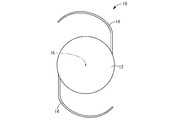
Images


Classifications
-
- A—HUMAN NECESSITIES
- A61—MEDICAL OR VETERINARY SCIENCE; HYGIENE
- A61F—FILTERS IMPLANTABLE INTO BLOOD VESSELS; PROSTHESES; DEVICES PROVIDING PATENCY TO, OR PREVENTING COLLAPSING OF, TUBULAR STRUCTURES OF THE BODY, e.g. STENTS; ORTHOPAEDIC, NURSING OR CONTRACEPTIVE DEVICES; FOMENTATION; TREATMENT OR PROTECTION OF EYES OR EARS; BANDAGES, DRESSINGS OR ABSORBENT PADS; FIRST-AID KITS
- A61F2/00—Filters implantable into blood vessels; Prostheses, i.e. artificial substitutes or replacements for parts of the body; Appliances for connecting them with the body; Devices providing patency to, or preventing collapsing of, tubular structures of the body, e.g. stents
- A61F2/02—Prostheses implantable into the body
- A61F2/14—Eye parts, e.g. lenses, corneal implants; Implanting instruments specially adapted therefor; Artificial eyes
- A61F2/16—Intraocular lenses
- A61F2/1613—Intraocular lenses having special lens configurations, e.g. multipart lenses; having particular optical properties, e.g. pseudo-accommodative lenses, lenses having aberration corrections, diffractive lenses, lenses for variably absorbing electromagnetic radiation, lenses having variable focus
- A61F2/1637—Correcting aberrations caused by inhomogeneities; correcting intrinsic aberrations, e.g. of the cornea, of the surface of the natural lens, aspheric, cylindrical, toric lenses
-
- A—HUMAN NECESSITIES
- A61—MEDICAL OR VETERINARY SCIENCE; HYGIENE
- A61F—FILTERS IMPLANTABLE INTO BLOOD VESSELS; PROSTHESES; DEVICES PROVIDING PATENCY TO, OR PREVENTING COLLAPSING OF, TUBULAR STRUCTURES OF THE BODY, e.g. STENTS; ORTHOPAEDIC, NURSING OR CONTRACEPTIVE DEVICES; FOMENTATION; TREATMENT OR PROTECTION OF EYES OR EARS; BANDAGES, DRESSINGS OR ABSORBENT PADS; FIRST-AID KITS
- A61F2/00—Filters implantable into blood vessels; Prostheses, i.e. artificial substitutes or replacements for parts of the body; Appliances for connecting them with the body; Devices providing patency to, or preventing collapsing of, tubular structures of the body, e.g. stents
- A61F2/02—Prostheses implantable into the body
- A61F2/14—Eye parts, e.g. lenses, corneal implants; Implanting instruments specially adapted therefor; Artificial eyes
- A61F2/16—Intraocular lenses
-
- B—PERFORMING OPERATIONS; TRANSPORTING
- B29—WORKING OF PLASTICS; WORKING OF SUBSTANCES IN A PLASTIC STATE IN GENERAL
- B29D—PRODUCING PARTICULAR ARTICLES FROM PLASTICS OR FROM SUBSTANCES IN A PLASTIC STATE
- B29D11/00—Producing optical elements, e.g. lenses or prisms
- B29D11/02—Artificial eyes from organic plastic material
- B29D11/023—Implants for natural eyes
-
- G—PHYSICS
- G02—OPTICS
- G02C—SPECTACLES; SUNGLASSES OR GOGGLES INSOFAR AS THEY HAVE THE SAME FEATURES AS SPECTACLES; CONTACT LENSES
- G02C7/00—Optical parts
- G02C7/02—Lenses; Lens systems ; Methods of designing lenses
- G02C7/024—Methods of designing ophthalmic lenses
- G02C7/027—Methods of designing ophthalmic lenses considering wearer's parameters
-
- A—HUMAN NECESSITIES
- A61—MEDICAL OR VETERINARY SCIENCE; HYGIENE
- A61F—FILTERS IMPLANTABLE INTO BLOOD VESSELS; PROSTHESES; DEVICES PROVIDING PATENCY TO, OR PREVENTING COLLAPSING OF, TUBULAR STRUCTURES OF THE BODY, e.g. STENTS; ORTHOPAEDIC, NURSING OR CONTRACEPTIVE DEVICES; FOMENTATION; TREATMENT OR PROTECTION OF EYES OR EARS; BANDAGES, DRESSINGS OR ABSORBENT PADS; FIRST-AID KITS
- A61F2/00—Filters implantable into blood vessels; Prostheses, i.e. artificial substitutes or replacements for parts of the body; Appliances for connecting them with the body; Devices providing patency to, or preventing collapsing of, tubular structures of the body, e.g. stents
- A61F2/02—Prostheses implantable into the body
- A61F2/14—Eye parts, e.g. lenses, corneal implants; Implanting instruments specially adapted therefor; Artificial eyes
- A61F2/16—Intraocular lenses
- A61F2/1613—Intraocular lenses having special lens configurations, e.g. multipart lenses; having particular optical properties, e.g. pseudo-accommodative lenses, lenses having aberration corrections, diffractive lenses, lenses for variably absorbing electromagnetic radiation, lenses having variable focus
-
- A—HUMAN NECESSITIES
- A61—MEDICAL OR VETERINARY SCIENCE; HYGIENE
- A61F—FILTERS IMPLANTABLE INTO BLOOD VESSELS; PROSTHESES; DEVICES PROVIDING PATENCY TO, OR PREVENTING COLLAPSING OF, TUBULAR STRUCTURES OF THE BODY, e.g. STENTS; ORTHOPAEDIC, NURSING OR CONTRACEPTIVE DEVICES; FOMENTATION; TREATMENT OR PROTECTION OF EYES OR EARS; BANDAGES, DRESSINGS OR ABSORBENT PADS; FIRST-AID KITS
- A61F2240/00—Manufacturing or designing of prostheses classified in groups A61F2/00 - A61F2/26 or A61F2/82 or A61F9/00 or A61F11/00 or subgroups thereof
- A61F2240/001—Designing or manufacturing processes
Definitions
- the present invention relates to an intraocular lens applied to the human eye, and more particularly to an intraocular lens having a novel structure capable of improving QOV (quality of appearance) and a manufacturing method thereof.
- an intraocular lens has been proposed as a treatment in that case.
- Such an intraocular lens is generally placed in a sac instead of the lens after removing and removing the lens in the sac of a human eye.
- a spherical lens power is set for an intraocular lens having a conventional structure, and an intraocular lens having a spherical lens power suitable for a patient is selected and used in consideration of a corneal curvature, an axial length, and the like. .
- Patent Document 1 describes a wavefront of wavefront aberration and an opposite sign value in the human eye of a specific population in order to reduce higher-order aberrations in the human eye.
- Intraocular lenses that provide aberrations have been proposed.
- it is difficult to say that such an intraocular lens has an efficient method for selecting a specific population, measuring wavefront aberration, setting the wavefront aberration of the opposite sign value to the intraocular lens, etc.
- the practical application was extremely difficult.
- the intraocular lens to which the present invention is directed relates to a contact lens having a different technical field.
- JP-T-2006-517676 (patent document) In 2)
- a high-order aberration that is important for improving QOV is identified by using a chart in which the degree of influence on QOV is actually measured for high-order aberrations of each order represented by Zernike polynomials. It is described to provide a correction lens that cancels out the next aberration and makes it zero.
- Patent Document 2 as described in [0097] to [0099] and the like, only specific high-order aberrations that adversely affect the appearance are selected, and the selected specific high-order aberrations are selected.
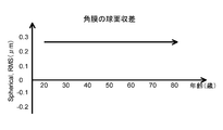
- the human eye may have spherical aberration due to, for example, the shape of the cornea even when the lens is extracted.
- the spherical aberration set in the intraocular lens of the present invention is designed in consideration of the spherical aberration remaining in the human eye from which the crystalline lens is extracted.
- the spherical aberration (RMS value) of the optical part in the intraocular lens is expressed by any of the following formulas with respect to the coma aberration (RMS value) remaining in the eye after the lens is removed. Is preferably set with an RMS value satisfying the above.
Abstract
Description
眼内レンズの球面収差≦水晶体摘出後の眼に残存するコマ収差-0.17μm Spherical aberration of the intraocular lens ≥ coma remaining in the eye after lens extraction-0.37 μm
Spherical aberration of the intraocular lens ≦ coma remaining in the eye after lens extraction−0.17 μm
-0.4≦A(μm)≦-0.1
0.003≦B(μm)≦0.004 Spherical aberration of intraocular lens = A + B × patient age −0.4 ≦ A (μm) ≦ −0.1
0.003 ≦ B (μm) ≦ 0.004
Claims (8)
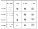
- 水晶体を摘出した患者の人眼に残存するコマ収差に対応した大きさで且つ該患者の該人眼に残存する球面収差を相殺しないで残存させる大きさの球面収差を光学部に設定する光学特性の設定工程と、
該光学特性の設定工程で設定された該球面収差を、該患者の該人眼における残余不正乱視に対する矯正光学特性として付与した光学部のレンズ形状を決定するレンズ形状の設定工程と、
該レンズ形状の設定工程で決定されたレンズ形状の光学部を形成することにより、該光学部における高次収差が光軸回りで回転対称とされた光学特性を有する眼内レンズを形成するレンズの形成工程と
を、含むことを特徴とする眼内レンズの製造方法。 Optical characteristics for setting in the optical unit spherical aberration of a size corresponding to the coma remaining in the human eye of the patient from whom the lens has been removed and remaining without canceling out the spherical aberration remaining in the human eye of the patient The setting process of
A lens shape setting step for determining a lens shape of an optical unit provided with the spherical aberration set in the optical property setting step as a correction optical property for residual irregular astigmatism in the human eye of the patient;
By forming the lens-shaped optical part determined in the lens shape setting step, a lens that forms an intraocular lens having optical characteristics in which higher-order aberrations in the optical part are rotationally symmetric about the optical axis. A method of manufacturing an intraocular lens, comprising: a forming step. - 前記光学特性の設定工程において、
前記光学部に設定される前記球面収差を、
下式の何れも満足するRMS値をもって設定する請求項1に記載の眼内レンズの製造方法。
眼内レンズの球面収差≧水晶体摘出後の眼に残存するコマ収差-0.37μm
眼内レンズの球面収差≦水晶体摘出後の眼に残存するコマ収差-0.17μm In the step of setting the optical characteristics,
The spherical aberration set in the optical unit,
The method for manufacturing an intraocular lens according to claim 1, wherein an RMS value satisfying any of the following formulas is set.
Spherical aberration of the intraocular lens ≥ coma remaining in the eye after lens extraction-0.37 μm
Spherical aberration of the intraocular lens ≦ coma remaining in the eye after lens extraction−0.17 μm - 前記光学特性の設定工程において、
前記光学部に設定される前記球面収差を、
A,Bの何れも定数とする下式を満足するRMS値をもって設定する請求項1又は2に記載の眼内レンズの製造方法。
眼内レンズの球面収差 = A + B × 患者の年齢
-0.4≦A(μm)≦-0.1
0.003≦B(μm)≦0.004 In the step of setting the optical characteristics,
The spherical aberration set in the optical unit,
The method for manufacturing an intraocular lens according to claim 1 or 2, wherein an RMS value satisfying the following formula is used, wherein both A and B are constants.
Spherical aberration of intraocular lens = A + B × patient age −0.4 ≦ A (μm) ≦ −0.1
0.003 ≦ B (μm) ≦ 0.004 - 前記光学特性の設定工程において、
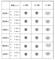
前記光学部における前記球面収差を、
前記患者と同じ年齢層の母集団における人眼の球面収差の測定データの平均値と該患者の角膜の球面収差との差をもって設定する請求項1~3の何れか1項に記載の眼内レンズの製造方法。 In the step of setting the optical characteristics,
The spherical aberration in the optical unit,
The intraocular device according to any one of claims 1 to 3, which is set by a difference between an average value of measurement data of spherical aberration of a human eye in a population of the same age group as the patient and a spherical aberration of the cornea of the patient. Lens manufacturing method. - 水晶体を摘出した患者の人眼に残存するコマ収差に対応した大きさで且つ該患者の該人眼に残存する球面収差を相殺しないで残存させる大きさの球面収差が、該患者の該人眼における残余不正乱視に対する矯正光学特性として光学部に設定されていると共に、
該光学部における高次収差が光軸回りで回転対称とされていることを特徴とする眼内レンズ。 A spherical aberration having a size corresponding to the coma remaining in the human eye of the patient from whom the lens has been removed and a size that can remain without canceling out the spherical aberration remaining in the human eye of the patient is the human eye of the patient. Is set in the optical section as a correction optical characteristic for residual irregular astigmatism in
An intraocular lens, wherein higher-order aberrations in the optical unit are rotationally symmetric about the optical axis. - 前記光学部に設定された前記球面収差が、
下式の何れも満足するRMS値をもって設定されている請求項5に記載の眼内レンズ。
眼内レンズの球面収差≧水晶体摘出後の眼に残存するコマ収差-0.37μm
眼内レンズの球面収差≦水晶体摘出後の眼に残存するコマ収差-0.17μm The spherical aberration set in the optical unit is
The intraocular lens according to claim 5, wherein the intraocular lens is set with an RMS value satisfying any of the following expressions.
Spherical aberration of the intraocular lens ≥ coma remaining in the eye after lens extraction-0.37 μm
Spherical aberration of the intraocular lens ≦ coma remaining in the eye after lens extraction−0.17 μm - 前記光学部に設定された前記球面収差が、
A,Bの何れも定数とする下式を満足するRMS値をもって設定されている請求項5又は6に記載の眼内レンズ。
眼内レンズの球面収差 = A + B × 患者の年齢
-0.4≦A(μm)≦-0.1
0.003≦B(μm)≦0.004 The spherical aberration set in the optical unit is
The intraocular lens according to claim 5 or 6, wherein the intraocular lens is set to have an RMS value that satisfies the following formula in which both A and B are constants.
Spherical aberration of intraocular lens = A + B × patient age −0.4 ≦ A (μm) ≦ −0.1
0.003 ≦ B (μm) ≦ 0.004 - 前記光学部に設定された前記球面収差が、
前記患者と同じ年齢層の母集団における人眼の球面収差の測定データの平均値と該患者の角膜の球面収差との差をもって設定されている請求項5~7の何れか1項に記載の眼内レンズ。 The spherical aberration set in the optical unit is
The method according to any one of claims 5 to 7, which is set by a difference between an average value of measurement data of spherical aberration of a human eye in a population of the same age group as the patient and spherical aberration of the cornea of the patient. Intraocular lens.
Priority Applications (16)
| Application Number | Priority Date | Filing Date | Title |
|---|---|---|---|
| EP12760887.5A EP2689748B1 (en) | 2011-03-24 | 2012-03-23 | Intraocular lens and manufacturing method therefor |
| AU2012232611A AU2012232611B2 (en) | 2011-03-24 | 2012-03-23 | Intraocular lens and manufacturing method therefor |
| ES12760887.5T ES2623757T3 (en) | 2011-03-24 | 2012-03-23 | Intraocular lens and its manufacturing procedure |
| US14/002,831 US9808340B2 (en) | 2011-03-24 | 2012-03-23 | Intraocular lens and manufacturing method thereof |
| CN201280015005.0A CN103458828B (en) | 2011-03-24 | 2012-03-23 | Intraocular lens and manufacture method thereof |
| PL16205428T PL3170475T3 (en) | 2011-03-24 | 2012-03-23 | Intraocular lens |
| KR1020137024875A KR101837379B1 (en) | 2011-03-24 | 2012-03-23 | Intraocular lens and manufacturing method therefor |
| SG2013070263A SG193526A1 (en) | 2011-03-24 | 2012-03-23 | Intraocular lens and manufacturing method thereof |
| BR112013024276-0A BR112013024276B1 (en) | 2011-03-24 | 2012-03-23 | INTRAOCULAR LENS AND METHOD OF MANUFACTURING THE SAME |
| RU2013147408/14A RU2601692C2 (en) | 2011-03-24 | 2012-03-23 | Intraocular lens and manufacturing method therefor |
| JP2013505827A JP5986985B2 (en) | 2011-03-24 | 2012-03-23 | Intraocular lens and manufacturing method thereof |
| EP16205428.2A EP3170475B1 (en) | 2011-03-24 | 2012-03-23 | Intraocular lens |
| IL228429A IL228429A0 (en) | 2011-03-24 | 2013-09-15 | Intraocular lens and manufacturing method thereof |
| HK14105449.0A HK1192135A1 (en) | 2011-03-24 | 2014-06-10 | Intraocular lens and manufacturing method therefor |
| US15/718,675 US20180014927A1 (en) | 2011-03-24 | 2017-09-28 | Intraocular lens and manufacturing method thereof |
| US16/424,051 US11020219B2 (en) | 2011-03-24 | 2019-05-28 | Intraocular lens and manufacturing method thereof |
Applications Claiming Priority (2)
| Application Number | Priority Date | Filing Date | Title |
|---|---|---|---|
| JP2011065836 | 2011-03-24 | ||
| JP2011-065836 | 2011-03-24 |
Related Child Applications (3)
| Application Number | Title | Priority Date | Filing Date |
|---|---|---|---|
| US14/002,831 A-371-Of-International US9808340B2 (en) | 2011-03-24 | 2012-03-23 | Intraocular lens and manufacturing method thereof |
| US201314002831A A-371-Of-International | 2011-03-24 | 2013-10-04 | |
| US15/718,675 Division US20180014927A1 (en) | 2011-03-24 | 2017-09-28 | Intraocular lens and manufacturing method thereof |
Publications (1)
| Publication Number | Publication Date |
|---|---|
| WO2012127881A1 true WO2012127881A1 (en) | 2012-09-27 |
Family
ID=46879052
Family Applications (1)
| Application Number | Title | Priority Date | Filing Date |
|---|---|---|---|
| PCT/JP2012/002054 WO2012127881A1 (en) | 2011-03-24 | 2012-03-23 | Intraocular lens and manufacturing method therefor |
Country Status (14)
| Country | Link |
|---|---|
| US (3) | US9808340B2 (en) |
| EP (2) | EP3170475B1 (en) |
| JP (1) | JP5986985B2 (en) |
| KR (1) | KR101837379B1 (en) |
| CN (1) | CN103458828B (en) |
| AU (1) | AU2012232611B2 (en) |
| BR (1) | BR112013024276B1 (en) |
| ES (2) | ES2709173T3 (en) |
| HK (1) | HK1192135A1 (en) |
| IL (1) | IL228429A0 (en) |
| PL (1) | PL3170475T3 (en) |
| RU (1) | RU2601692C2 (en) |
| SG (1) | SG193526A1 (en) |
| WO (1) | WO2012127881A1 (en) |
Cited By (1)
| Publication number | Priority date | Publication date | Assignee | Title |
|---|---|---|---|---|
| CN111051965A (en) * | 2017-09-01 | 2020-04-21 | 株式会社实瞳 | Ophthalmic lenses and imaging methods using ophthalmic lenses |
Families Citing this family (8)
| Publication number | Priority date | Publication date | Assignee | Title |
|---|---|---|---|---|
| WO2012127538A1 (en) * | 2011-03-24 | 2012-09-27 | 株式会社メニコン | Contact lens and manufacturing method therefor |
| EP3170475B1 (en) * | 2011-03-24 | 2018-10-31 | Kowa Company Ltd. | Intraocular lens |
| TWI588560B (en) | 2012-04-05 | 2017-06-21 | 布萊恩荷登視覺協會 | Lenses, devices, methods and systems for refractive error |
| US9201250B2 (en) | 2012-10-17 | 2015-12-01 | Brien Holden Vision Institute | Lenses, devices, methods and systems for refractive error |
| CN104768499B (en) | 2012-10-17 | 2017-06-23 | 华柏恩视觉研究中心 | For ametropic eyeglass, device, method and system |
| CN106388974A (en) * | 2016-12-09 | 2017-02-15 | 天津世纪康泰生物医学工程有限公司 | Aspherical artificial lens capable of completely correcting mesopic vision |
| US11000363B2 (en) * | 2017-05-02 | 2021-05-11 | Alcon Inc. | Accommodating intraocular lens devices, systems, and methods using an opaque frame |
| BR202018002611U2 (en) * | 2018-02-07 | 2019-08-27 | Nathan Jose De Souza | disposition applied to intraocular lens with elongated and needled haptics |
Citations (2)
| Publication number | Priority date | Publication date | Assignee | Title |
|---|---|---|---|---|
| WO2008008999A1 (en) * | 2006-07-14 | 2008-01-17 | Qualcomm Incorporated | Method and system to indicate an exception-triggering page within a microprocessor |
| JP2010029694A (en) * | 2000-05-23 | 2010-02-12 | Amo Groningen Bv | Method for providing ophthalmic lens capable of reducing eye aberration |
Family Cites Families (17)
| Publication number | Priority date | Publication date | Assignee | Title |
|---|---|---|---|---|
| US5777719A (en) * | 1996-12-23 | 1998-07-07 | University Of Rochester | Method and apparatus for improving vision and the resolution of retinal images |
| US6609793B2 (en) | 2000-05-23 | 2003-08-26 | Pharmacia Groningen Bv | Methods of obtaining ophthalmic lenses providing the eye with reduced aberrations |
| US6554425B1 (en) * | 2000-10-17 | 2003-04-29 | Johnson & Johnson Vision Care, Inc. | Ophthalmic lenses for high order aberration correction and processes for production of the lenses |
| JP4652558B2 (en) | 2000-10-18 | 2011-03-16 | 株式会社トプコン | Optical property measuring device |
| EP1334691A4 (en) | 2000-10-18 | 2009-01-07 | Topcon Corp | Optical characteristics measuring device |
| US7078665B2 (en) | 2002-07-09 | 2006-07-18 | Wavefront Sciences, Inc. | System and method of wavefront sensing for determining a location of focal spot |
| GB0303193D0 (en) * | 2003-02-12 | 2003-03-19 | Guillon Michael | Methods & lens |
| WO2004090611A2 (en) * | 2003-03-31 | 2004-10-21 | Bausch & Lomb Incorporated | Intraocular lens and method for reducing aberrations in an ocular system |
| WO2006088440A1 (en) | 2005-02-11 | 2006-08-24 | Bausch & Lomb Incorporated | Aspheric lenses and lens family |
| US7697212B2 (en) * | 2006-05-16 | 2010-04-13 | Ophthonix, Inc. | High-order aberration correction for optimization of human visual function |
| US7879089B2 (en) * | 2006-05-17 | 2011-02-01 | Alcon, Inc. | Correction of higher order aberrations in intraocular lenses |
| WO2008089999A1 (en) | 2007-01-25 | 2008-07-31 | Rodenstock Gmbh | Method for optimising a spectacle lens |
| WO2009058755A1 (en) * | 2007-10-29 | 2009-05-07 | Junzhong Liang | Methods and devices for refractive treatments of presbyopia |
| US20090160075A1 (en) * | 2007-12-21 | 2009-06-25 | Simpson Michael J | Methods for fabricating customized intraocular lenses |
| US7998198B2 (en) * | 2008-02-07 | 2011-08-16 | Novartis Ag | Accommodative IOL with dynamic spherical aberration |
| US8342683B2 (en) * | 2009-08-27 | 2013-01-01 | Novartis Ag | Optimizing optical aberrations in ophthalmic lenses |
| EP3170475B1 (en) * | 2011-03-24 | 2018-10-31 | Kowa Company Ltd. | Intraocular lens |
-
2012
- 2012-03-23 EP EP16205428.2A patent/EP3170475B1/en active Active
- 2012-03-23 CN CN201280015005.0A patent/CN103458828B/en active Active
- 2012-03-23 ES ES16205428T patent/ES2709173T3/en active Active
- 2012-03-23 KR KR1020137024875A patent/KR101837379B1/en active IP Right Grant
- 2012-03-23 AU AU2012232611A patent/AU2012232611B2/en active Active
- 2012-03-23 EP EP12760887.5A patent/EP2689748B1/en active Active
- 2012-03-23 WO PCT/JP2012/002054 patent/WO2012127881A1/en active Application Filing
- 2012-03-23 US US14/002,831 patent/US9808340B2/en active Active
- 2012-03-23 BR BR112013024276-0A patent/BR112013024276B1/en active IP Right Grant
- 2012-03-23 ES ES12760887.5T patent/ES2623757T3/en active Active
- 2012-03-23 SG SG2013070263A patent/SG193526A1/en unknown
- 2012-03-23 RU RU2013147408/14A patent/RU2601692C2/en active
- 2012-03-23 PL PL16205428T patent/PL3170475T3/en unknown
- 2012-03-23 JP JP2013505827A patent/JP5986985B2/en active Active
-
2013
- 2013-09-15 IL IL228429A patent/IL228429A0/en active IP Right Grant
-
2014
- 2014-06-10 HK HK14105449.0A patent/HK1192135A1/en unknown
-
2017
- 2017-09-28 US US15/718,675 patent/US20180014927A1/en not_active Abandoned
-
2019
- 2019-05-28 US US16/424,051 patent/US11020219B2/en active Active
Patent Citations (2)
| Publication number | Priority date | Publication date | Assignee | Title |
|---|---|---|---|---|
| JP2010029694A (en) * | 2000-05-23 | 2010-02-12 | Amo Groningen Bv | Method for providing ophthalmic lens capable of reducing eye aberration |
| WO2008008999A1 (en) * | 2006-07-14 | 2008-01-17 | Qualcomm Incorporated | Method and system to indicate an exception-triggering page within a microprocessor |
Non-Patent Citations (1)
| Title |
|---|
| See also references of EP2689748A4 * |
Cited By (2)
| Publication number | Priority date | Publication date | Assignee | Title |
|---|---|---|---|---|
| CN111051965A (en) * | 2017-09-01 | 2020-04-21 | 株式会社实瞳 | Ophthalmic lenses and imaging methods using ophthalmic lenses |
| CN111051965B (en) * | 2017-09-01 | 2022-03-25 | 株式会社实瞳 | Ophthalmic lenses and imaging methods using ophthalmic lenses |
Also Published As
| Publication number | Publication date |
|---|---|
| KR20140008409A (en) | 2014-01-21 |
| CN103458828B (en) | 2015-12-02 |
| HK1192135A1 (en) | 2014-08-15 |
| BR112013024276B1 (en) | 2020-12-15 |
| PL3170475T3 (en) | 2019-04-30 |
| JP5986985B2 (en) | 2016-09-06 |
| SG193526A1 (en) | 2013-10-30 |
| KR101837379B1 (en) | 2018-03-13 |
| BR112013024276A2 (en) | 2016-12-27 |
| JPWO2012127881A1 (en) | 2014-07-24 |
| US11020219B2 (en) | 2021-06-01 |
| EP2689748B1 (en) | 2017-02-22 |
| ES2623757T3 (en) | 2017-07-12 |
| EP3170475A1 (en) | 2017-05-24 |
| CN103458828A (en) | 2013-12-18 |
| RU2013147408A (en) | 2015-04-27 |
| US20140039616A1 (en) | 2014-02-06 |
| US20190274824A1 (en) | 2019-09-12 |
| IL228429A0 (en) | 2013-12-31 |
| ES2709173T3 (en) | 2019-04-15 |
| AU2012232611A1 (en) | 2013-09-26 |
| US9808340B2 (en) | 2017-11-07 |
| AU2012232611B2 (en) | 2016-05-26 |
| EP2689748A4 (en) | 2014-11-26 |
| US20180014927A1 (en) | 2018-01-18 |
| RU2601692C2 (en) | 2016-11-10 |
| EP3170475B1 (en) | 2018-10-31 |
| EP2689748A1 (en) | 2014-01-29 |
Similar Documents
| Publication | Publication Date | Title |
|---|---|---|
| JP5986985B2 (en) | Intraocular lens and manufacturing method thereof | |
| JP4800921B2 (en) | Custom lens for improving visual acuity and corresponding lens design method | |
| JP4459501B2 (en) | Eye lens to reduce eye aberration | |
| JP6294994B2 (en) | contact lens | |
| JP3735346B2 (en) | Method for obtaining an ophthalmic lens that reduces eye aberration | |
| JP5657266B2 (en) | Contact lenses for correcting irregular astigmatism | |
| CN102257425B (en) | Correction of peripheral defocus of an eye and control of refractive error development | |
| WO2003050594A2 (en) | Contact lens and method for fitting and design | |
| JP6632832B2 (en) | Lens design and method for minimizing visual acuity changes experienced by people with advanced myopia | |
| CN109426008B (en) | Non-toric surfaces for minimizing secondary astigmatism in contact lenses for correcting astigmatism | |
| JP3860041B2 (en) | Contact lens and contact lens design method | |
| TW202319814A (en) | An aspherical lens design with power dependent spherical aberration | |
| Benes et al. | Sylvie Petrová (2014). Anatomical Differences of Corneal Surface Parameters | |
| Plakitsi | Optical and Visual Performance with Varifocal Contact Lenses for the Presbyope |
Legal Events
| Date | Code | Title | Description |
|---|---|---|---|
| 121 | Ep: the epo has been informed by wipo that ep was designated in this application |
Ref document number: 12760887 Country of ref document: EP Kind code of ref document: A1 |
|
| ENP | Entry into the national phase |
Ref document number: 2013505827 Country of ref document: JP Kind code of ref document: A |
|
| REEP | Request for entry into the european phase |
Ref document number: 2012760887 Country of ref document: EP |
|
| ENP | Entry into the national phase |
Ref document number: 20137024875 Country of ref document: KR Kind code of ref document: A |
|
| NENP | Non-entry into the national phase |
Ref country code: DE |
|
| ENP | Entry into the national phase |
Ref document number: 2012232611 Country of ref document: AU Date of ref document: 20120323 Kind code of ref document: A |
|
| WWE | Wipo information: entry into national phase |
Ref document number: 14002831 Country of ref document: US |
|
| ENP | Entry into the national phase |
Ref document number: 2013147408 Country of ref document: RU Kind code of ref document: A |
|
| REG | Reference to national code |
Ref country code: BR Ref legal event code: B01A Ref document number: 112013024276 Country of ref document: BR |
|
| ENP | Entry into the national phase |
Ref document number: 112013024276 Country of ref document: BR Kind code of ref document: A2 Effective date: 20130920 |