WO2010141956A2 - Methods and compositions for the treatment of cancer - Google Patents
Methods and compositions for the treatment of cancer Download PDFInfo
- Publication number
- WO2010141956A2 WO2010141956A2 PCT/US2010/037662 US2010037662W WO2010141956A2 WO 2010141956 A2 WO2010141956 A2 WO 2010141956A2 US 2010037662 W US2010037662 W US 2010037662W WO 2010141956 A2 WO2010141956 A2 WO 2010141956A2
- Authority
- WO
- WIPO (PCT)
- Prior art keywords
- methyl sulfone
- cells
- cancer
- cell
- subject
- Prior art date
Links
Classifications
-
- A—HUMAN NECESSITIES
- A61—MEDICAL OR VETERINARY SCIENCE; HYGIENE
- A61K—PREPARATIONS FOR MEDICAL, DENTAL OR TOILETRY PURPOSES
- A61K31/00—Medicinal preparations containing organic active ingredients
- A61K31/095—Sulfur, selenium, or tellurium compounds, e.g. thiols
- A61K31/10—Sulfides; Sulfoxides; Sulfones
-
- A—HUMAN NECESSITIES
- A61—MEDICAL OR VETERINARY SCIENCE; HYGIENE
- A61K—PREPARATIONS FOR MEDICAL, DENTAL OR TOILETRY PURPOSES
- A61K45/00—Medicinal preparations containing active ingredients not provided for in groups A61K31/00 - A61K41/00
- A61K45/06—Mixtures of active ingredients without chemical characterisation, e.g. antiphlogistics and cardiaca
-
- A—HUMAN NECESSITIES
- A61—MEDICAL OR VETERINARY SCIENCE; HYGIENE
- A61K—PREPARATIONS FOR MEDICAL, DENTAL OR TOILETRY PURPOSES
- A61K9/00—Medicinal preparations characterised by special physical form
- A61K9/70—Web, sheet or filament bases ; Films; Fibres of the matrix type containing drug
- A61K9/7023—Transdermal patches and similar drug-containing composite devices, e.g. cataplasms
-
- A—HUMAN NECESSITIES
- A61—MEDICAL OR VETERINARY SCIENCE; HYGIENE
- A61P—SPECIFIC THERAPEUTIC ACTIVITY OF CHEMICAL COMPOUNDS OR MEDICINAL PREPARATIONS
- A61P35/00—Antineoplastic agents
-
- A—HUMAN NECESSITIES
- A61—MEDICAL OR VETERINARY SCIENCE; HYGIENE
- A61P—SPECIFIC THERAPEUTIC ACTIVITY OF CHEMICAL COMPOUNDS OR MEDICINAL PREPARATIONS
- A61P35/00—Antineoplastic agents
- A61P35/04—Antineoplastic agents specific for metastasis
Definitions
- methyl sulfone induces an irreversible non-malignant phenotype in aggressive, metastatic melanoma and breast cancer cells, which renders these cells harmless to subjects. Moreover, they inventors demonstrate that in the presence of methyl sulfone, the melanoma cells evolved into functional melanocytes. Accordingly, the instant application provides methods and compositions for the treatment of cell proliferative disorders, e.g., cancer.
- the instant invention provides methods for treating a cell proliferative disorder in a subject by administering to the subject a therapeutically effective amount of methyl sulfone, thereby treating the cell proliferative disorder.
- the cell proliferative disorder is cancer.
- the cancer is a solid tumor cancer, e.g., ovarian, brain, colon, lung, melanoma, bladder, breast or prostate cancer.
- the cell proliferative disorder is a hematological cancer, e.g., leukemia or lymphoma.
- the subject had previously received chemotherapeutic or radiation therapy which was unsuccessful or less than completely successful.
- methyl sulfone is administered systemically, locally, or targeted to the location of the cell proliferative disorder.
- the methyl sulfone is formulated in a micro or nanoparticle.
- the area comprising the solid tumor is sprayed with or bathed in methyl sulfone.
- all or a portion of the solid tumor is removed prior to treatment with methyl sulfone.
- the subject is a mammal, e.g., a human.
- the instant invention provides methods of treating cancer in a subject in need thereof, comprising the step of: separately administering to the subject a composition comprising: methyl sulfone; and a chemotherapeutic agent.
- a composition comprising: methyl sulfone; and a chemotherapeutic agent.
- the administration of methyl sulfone and the chemotherapeutic agent is simultaneous.
- the administration of the composition methyl sulfone and the chemotherapeutic agent is sequential.
- the chemotherapeutic agent is doxil, topotecan, DNA-altering drugs, carboplatin, antimetabolites, gemcitabine, drugs that prevent cell division, vincristine, anti- angiogenic agents, or pazopanib.
- the instant invention provides methods of treating a subject having a solid tumor surgically removed by spraying or bathing the area containing the tumor with methyl sulfone after removal of the tumor and prior to the completion of a surgical procedure.
- the instant invention provides methods of treating ovarian cancer in a subject by contacting the ovaries with methyl sulfone, thereby treating ovarian cancer.
- the ovaries are sprayed with methyl sulfone.
- the instant invention provides methods of preventing the spread of cancer in subject having a solid tumor surgically removed by bathing or spraying the area containing the tumor with methyl sulfone after removal of the tumor and prior to the completion of the surgery, thereby preventing the spread of cancer in the subject.
- the instant invention provides methods of inducing a cell to revert to a normal cellular phenotype from a cancer cell phenotype by contacting the cell with an effective amount of methyl sulfone, thereby inducing a phenotypic change from a cancer cell phenotype to a normal cell phenotype.
- the instant invention provides pharmaceutical compositions comprising methyl sulfone and a pharmaceutically-acceptable diluent or carrier.
- the composition comprises a gel, cream, solution, liposome or nanoparticle.
- the composition comprises a delayed release dosage form.
- the composition is formulated for aerosolization.
- the instant invention provides a patch for the treatment of skin cancer comprising methyl sulfone.
- the patch further comprises a pharmaceutically-acceptable diluent or carrier.
- kits for the treatment of a cell proliferative disorder comprising methyl sulfone and instructions for use.
- the kits may further comprise an applicator, e.g., a sponge, spray bottle, or aerosolizer.
- Figure 1 is a single frame of a movie demonstrating the change in morphology of melanoma cells after the addition of 2% methyl sulfone. Cells became apoptotic at concentrations over 2% methyl sulfone.
- Figure 2 depicts a graph demonstrating apoptosis in melanoma cells with 0-6.0 percent methyl sulfone.
- Figures 3A-D depict melanoma cells with and without 2% methyl sulfone.
- Figure 4 is a graph depicting DNA synthesis in melanoma cells with and without 2% methyl sulfone at 48 and 72 hours.
- Figures 5 A-B depict a soft agar experiment demonstrating the growth of cell colonies in the absence of methyl sulfone and the absence of colonies in when treated with 2% methyl sulfone.
- Figure 6 depicts the migration of melanoma cells through a matrix membrane after being treated with 2% methyl sulfone for 48 hours. Dark spots are membrane pores. Melanoma cells are lightly colored and triangular. In the presence of 2% methyl sulfone, melanoma cells are unable to pass through the membrane.
- Figures 7A-D depict wound healing in melanoma cells in the presence and absence of 2% methyl sulfone.
- Figures 8A-B depicts 2% methyl sulfone induced senescence in melanoma cells. Virtually non control cells were senescent.
- Figures 9A-B depict the arborization of melanoma cells that have been treated with 2% methyl sulfone for four weeks.
- Figures 9A-D depicts four different fields of the arborized cells. The dark arbors indicate the presence of melanosomes.
- Figures 10A-H depict immunofluorescensce microscopy of proteins involved in the ETM transition. Shown are melanoma cells with and without 2% methyl sulfone after seven days.
- Figures 1 IA-B depict immunofluorescence microscopy of actin filaments in melanoma cells with and without 2% methyl sulfone after 72 hours.
- Figures 12A-D depict immunofluorescence microscopy of microtubles in melanoma cells in the presence of 2% methyl sulfone over time.
- Figures 13A-C depict immunofluorescence microscopy of microtubules in melanoma cells with an without 2% methyl sulfone in the presence of 10 "7 M vinblastine (VNB). Cells were processed for immunofluorescence two hours after adding VNB.
- VNB vinblastine
- Figure 14 is a graph comparing the effect of different doses of methyl sulfone and VNB on induction of apoptosis in leukemic lymphocytes.
- CH46 is a clinical name for methyl sulfone.
- Figure 15 depicts normal lymphocytes from a healthy volunteer were treated with different doses of methyl sulfone and percent apoptotic cells were determined by flow cytometry CH46 is a clinical name for methyl sulfone.
- Figure 16 depicts a comparison of effect on apoptosis of melanoma cells by methyl methyl sulfone (MMS) and methyl ethyl sulfone (MES).
- MMS methyl methyl sulfone
- MES methyl ethyl sulfone
- Figure 17 depicts a comparison of effect on apoptosis of melanoma cells by methyl methyl sulfone (MMS) and ethyl ethyl sulfone (EES).
- MMS methyl methyl sulfone
- EES ethyl ethyl sulfone
- methyl sulfone is capable of arresting the progression of cancerous cells in a subject and can induce apoptosis at higher concentrations. Specifically, the inventors demonstrate that treatment of cancer cells with methyl sulfone induces several non-malignant phenotypes including contact inhibition, senescence and differentiation into arborized cells containing melanosomes. As described in the Examples, a comparison of induction of apoptosis in leukemic lymphocytes with lymphocytes isolated from a healthy volunteer showed that methyl sulfone induced apoptosis in more than 90% of the leukemic cells and induced apoptosis in less than 10% of T-cells from the healthy volunteer.
- Methyl sulfone is also known in the literature as dimethyl sulfone, MSM and methyl sulfonyl methane. Methyl sulfone has the molecular formula C 2 H 6 O 2 S and the chemical structure:
- Methyl sulfone is non-toxic. Toxicity studies show that methyl sulfone is as toxic as water. Therefore, the side effects associated with current chemotherapeutic treatment are not a concern with methyl sulfone. Methyl sulfone has a molecular weight 94.13 and a CAS Registry Number of 67-71-0. The approximate water solubility of methyl sulfone is 150 g/L at 20 0 C. Methyl sulfone is stable and not hygroscopic.
- Methyl sulfone has been shown to readily crosses plasma membranes to enter cells.
- cancer includes malignancies characterized by deregulated or uncontrolled cell growth, for instance carcinomas, sarcomas, leukemias, and lymphomas.
- cancer includes primary malignant tumors, e.g., those whose cells have not migrated to sites in the subject's body other than the site of the original tumor, and secondary malignant tumors, e.g., those arising from metastasis, the migration of tumor cells to secondary sites that are different from the site of the original tumor.
- Neoplasma or “neoplastic transformation” is the pathologic process that results in the formation and growth of a neoplasm, tissue mass, or tumor. Such process includes uncontrolled cell growth, including either benign or malignant tumors. Neoplasms include abnormal masses of tissue, the growth of which exceeds and is uncoordinated with that of the normal tissues and persists in the same excessive manner after cessation of the stimuli that evoked the change. Neoplasms may show a partial or complete lack of structural organization and functional coordination with the normal tissue, and usually form a distinct mass of tissue. One cause of neoplasia is dysregulation of the cell cycle machinery.
- Neoplasms tend to grow and function somewhat independently of the homeostatic mechanisms that control normal tissue growth and function. However, some neoplasms remain under the control of the homeostatic mechanisms that control normal tissue growth and function. For example, some neoplasms are estrogen sensitive and can be arrested by anti-estrogen therapy. Neoplasms can range in size from less than 1 cm to over 6 inches in diameter.
- Neoplasms tend to morphologically and functionally resemble the tissue from which they originated. For example, neoplasms arising within the islet tissue of the pancreas resemble the islet tissue, contain secretory granules, and secrete insulin. Clinical features of a neoplasm may result from the function of the tissue from which it originated. For example, excessive amounts of insulin can be produced by islet cell neoplasms resulting in hypoglycemia which, in turn, results in headaches and dizziness. However, some neoplasms show little morphological or functional resemblance to the tissue from which they originated. Some neoplasms result in such non-specific systemic effects as cachexia, increased susceptibility to infection, and fever.
- neoplasm By assessing the histology and other features of a neoplasm, it can be determined whether the neoplasm is benign or malignant. Invasion and metastasis (the spread of the neoplasm to distant sites) are definitive attributes of malignancy.
- Benign tumors are generally well circumscribed and round, have a capsule, and have a grey or white color, and a uniform texture.
- malignant tumors generally have fingerlike projections, irregular margins, are not circumscribed, and have a variable color and texture.
- Benign tumors grow by pushing on adjacent tissue as they grow. As the benign tumor enlarges it compresses adjacent tissue, sometimes causing atrophy. The junction between a benign tumor and surrounding tissue may be converted to a fibrous connective tissue capsule allowing for easy surgical removal of the benign tumor. Conversely, malignant tumors are locally invasive and grow into the adjacent tissues usually giving rise to irregular margins that are not encapsulated making it necessary to remove a wide margin of normal tissue for the surgical removal of malignant tumors. Benign neoplasms tend to grow more slowly and tend to be less autonomous than malignant tumors. Benign neoplasms tend to closely histologically resemble the tissue from which they originated.
- cancers More highly differentiated cancers, i.e., cancers that resemble the tissue from which they originated, tend to have a better prognosis than poorly differentiated cancers, while malignant tumors are more likely than benign tumors to have an aberrant function, e.g., the secretion of abnormal or excessive quantities of hormones.
- anaplasia The histological features of cancer are summarized by the term "anaplasia.”
- Malignant neoplasms often contain numerous mitotic cells. These cells are typically abnormal. Such mitotic aberrations account for some of the karyotypic abnormalities found in most cancers. Bizarre multinucleated cells are also seen in some cancers, especially those that are highly anaplastic.
- the term "anaplasia" includes histological features of cancer.
- cytologic features of anaplasia include an increased nuclear-cytoplasmic ratio (nuclear-cytoplasmic ratio can be over 50% for malignant cells), nuclear pleomorphism, clumping of the nuclear chromatin along the nuclear membrane, increased staining of the nuclear chromatin, simplified endoplasmic reticulum, increased free ribosomes, pleomorphism of mitochondria, decreased size and number of organelles, enlarged and increased numbers of nucleoli, and sometimes the presence of intermediate filaments.
- Dysplasia includes pre-malignant states in which a tissue demonstrates histologic and cytologic features intermediate between normal and anaplastic. Dysplasia is often reversible.
- carcinoma includes malignancies of epithelial or endocrine tissues, including respiratory system carcinomas, gastrointestinal system carcinomas, genitourinary system carcinomas, testicular carcinomas, breast carcinomas, prostate carcinomas, endocrine system carcinomas, melanomas, choriocarcinoma, and carcinomas of the cervix, lung, head and neck, colon, and ovary.
- carcinoma also includes carcinosarcomas, which include malignant tumors composed of carcinomatous and sarcomatous tissues.
- sarcoma includes malignant tumors of mesodermal connective tissue, e.g., tumors of bone, fat, and cartilage.
- leukemia and “lymphoma” include malignancies of the hematopoietic cells of the bone marrow. Leukemias tend to proliferate as single cells, whereas lymphomas tend to proliferate as solid tumor masses.
- leukemias include acute myeloid leukemia (AML), acute promyelocyte leukemia, chronic myelogenous leukemia, mixed-lineage leukemia, acute monoblastic leukemia, acute lymphoblastic leukemia, acute non-lymphoblastic leukemia, blastic mantle cell leukemia, myelodyplastic syndrome, T cell leukemia, B cell leukemia, and chronic lymphocytic leukemia.
- AML acute myeloid leukemia
- AML acute promyelocyte leukemia
- chronic myelogenous leukemia mixed-lineage leukemia
- acute monoblastic leukemia acute lymphoblastic leukemia
- acute non-lymphoblastic leukemia acute non-lymphoblastic leukemia
- blastic mantle cell leukemia myelodyplastic syndrome
- T cell leukemia B cell leukemia
- chronic lymphocytic leukemia chronic lymphocytic leukemia
- lymphomas examples include Hodgkin's disease, non- Hodgkin's lymphoma, B cell lymphoma, epitheliotropic lymphoma, composite lymphoma, anaplastic large cell lymphoma, gastric and non-gastric mucosa-associated lymphoid tissue lymphoma, lymphoproliferative disease, T cell lymphoma, Burkitt's lymphoma, mantle cell lymphoma, diffuse large cell lymphoma, lymphoplasmacytoid lymphoma, and multiple myeloma.
- the therapeutic methods of the present invention can be applied to cancerous cells of mesenchymal origin, such as those producing sarcomas (e.g., fibrosarcoma, myxosarcoma, liosarcoma, chondrosarcoma, osteogenic sarcoma or chordosarcoma, angiosarcoma, endotheliosardcoma, lympangiosarcoma, synovio sarcoma or mesothelisosarcoma); leukemias and lymphomas such as granulocytic leukemia, monocytic leukemia, lymphocytic leukemia, malignant lymphoma, plasmocytoma, reticulum cell sarcoma, or Hodgkin's disease; sarcomas such as leiomysarcoma or rhabdomysarcoma, tumors of epithelial origin such as squamous cell carcinoma, basal cell carcinoma
- Additional cell types amenable to treatment according to the methods described herein include those giving rise to mammary carcinomas, gastrointestinal carcinoma, such as colonic carcinomas, bladder carcinoma, prostate carcinoma, and squamous cell carcinoma of the neck and head region.
- Examples of cancers amenable to treatment according to the methods described herein include vaginal, cervical, and breast cancers.
- the language "inhibiting undesirable cell growth” is intended to include the inhibition of undesirable or inappropriate cell growth.
- the inhibition is intended to include inhibition of proliferation including rapid proliferation.
- the cell growth can result in benign masses or the inhibition of cell growth resulting in malignant tumors.
- Examples of benign conditions which result from inappropriate cell growth or angiogenesis are diabetic retinopathy, retrolental f ⁇ brioplasia, neovascular glaucoma, psoriasis, angiofibromas, rheumatoid arthritis, hemangiomas, Karposi's sarcoma, and other conditions or dysfunctions characterized by dysregulated endothelial cell division.
- inhibiting tumor growth or “inhibiting neoplasia” includes the prevention of the growth of a tumor in a subject or a reduction in the growth of a preexisting tumor in a subject.
- the inhibition also can be the inhibition of the metastasis of a tumor from one site to another.
- tumor is intended to encompass both in vitro and in vivo tumors that form in any organ or body part of the subject.
- the tumors whose growth rate is inhibited by the present invention include basal cell carcinoma, squamous cell carcinoma of both ulcerating and papillary type, metastatic skin carcinoma, osteo sarcoma, Ewing's sarcoma, veticulum cell sarcoma, myeloma, giant cell tumor, small-cell lung tumor, gallstones, islet cell tumor, primary brain tumor, acute and chronic lymphocytic and granulocytic tumors, hairy-cell tumor, adenoma, hyperplasia, medullary carcinoma, pheochromocytoma, mucosal neuromas, intestinal ganglioneuromas, hyperplastic corneal nerve tumor, marfanoid habitus tumor, Wilm's tumor, seminoma, ovarian tumor, leiomyomater tumor, cervical dysplasia and in situ carcinoma, neuroblastoma, retinoblastoma, soft tissue sarcoma, malignant carcinoid, topical skin lesion, my
- compositions suitable for administration to a subject e.g., a human.
- Such compositions typically comprise the methyl sulfone and a pharmaceutically acceptable carrier.
- pharmaceutically acceptable carrier is intended to include any and all solvents, dispersion media, coatings, antibacterial and antifungal agents, isotonic and absorption delaying agents, and the like, compatible with pharmaceutical administration.
- the use of such media and agents for pharmaceutically active substances is well known in the art. Except insofar as any conventional media or agent is incompatible with the active compound, such media can be used in the compositions of the invention. Supplementary active compounds can also be incorporated into the compositions.
- a pharmaceutical composition of the invention is formulated to be compatible with its intended route of administration.
- routes of administration include parenteral, e.g., intravenous, intradermal, subcutaneous, oral (e.g., inhalation), transdermal (topical), transmucosal, and rectal administration.
- Solutions or suspensions used for parenteral, intradermal, or subcutaneous application can include the following components: a sterile diluent such as water for injection, saline solution, fixed oils, polyethylene glycols, glycerine, propylene glycol or other synthetic solvents; antibacterial agents such as benzyl alcohol or methyl parabens; antioxidants such as ascorbic acid or sodium bisulfite; chelating agents such as ethylenediaminetetraacetic acid; buffers such as acetates, citrates or phosphates and agents for the adjustment of tonicity such as sodium chloride or dextrose. pH can be adjusted with acids or bases, such as hydrochloric acid or sodium hydroxide.
- the parenteral preparation can be enclosed in ampules
- compositions suitable for injectable use include sterile aqueous solutions (where water soluble) or dispersions and sterile powders for the extemporaneous preparation of sterile injectable solutions or dispersion.
- suitable carriers include physiological saline, bacteriostatic water, Cremophor (BASF, Parsippany, N.J.) or phosphate buffered saline (PBS).
- the composition must be sterile and should be fluid to the extent that easy syringability exists. It must be stable under the conditions of manufacture and storage and must be preserved against the contaminating action of microorganisms such as bacteria and fungi.
- the carrier can be a solvent or dispersion medium containing, for example, water, ethanol, polyol (for example, glycerol, propylene glycol, and liquid polyethylene glycol, and the like), and suitable mixtures thereof.
- the proper fluidity can be maintained, for example, by the use of a coating such as lecithin, by the maintenance of the required particle size in the case of dispersion and by the use of surfactants.
- Prevention of the action of microorganisms can be achieved by various antibacterial and antifungal agents, for example, parabens, chlorobutanol, phenol, ascorbic acid, thimerosal, and the like.
- isotonic agents for example, sugars, polyalcohols such as manitol, sorbitol, sodium chloride in the composition.
- Prolonged absorption of the injectable compositions can be brought about by including in the composition an agent which delays absorption, for example, aluminum monostearate and gelatin.
- Sterile injectable solutions can be prepared by incorporating methyl sulfone in the required amount in an appropriate solvent with one or a combination of ingredients enumerated above, as required, followed by filtered sterilization.
- dispersions are prepared by incorporating the active compound into a sterile vehicle which contains a basic dispersion medium and the required other ingredients from those enumerated above.
- the preferred methods of preparation are vacuum drying and freeze-drying which yields a powder of the active ingredient plus any additional desired ingredient from a previously sterile- filtered solution thereof.
- Oral compositions generally include an inert diluent or an edible carrier. They can be enclosed in gelatin capsules or compressed into tablets.
- the agent can be contained in enteric forms to survive the stomach or further coated or mixed to be released in a particular region of the GI tract by known methods.
- the active compound can be incorporated with excipients and used in the form of tablets, troches, or capsules.
- Oral compositions can also be prepared using a fluid carrier for use as a mouthwash, wherein the compound in the fluid carrier is applied orally and swished and expectorated or swallowed.
- Pharmaceutically compatible binding agents, and/or adjuvant materials can be included as part of the composition.
- the tablets, pills, capsules, troches and the like can contain any of the following ingredients, or compounds of a similar nature: a binder such as microcrystalline cellulose, gum tragacanth or gelatin; an excipient such as starch or lactose, a disintegrating agent such as alginic acid, Primogel, or corn starch; a lubricant such as magnesium stearate or Sterotes; a glidant such as colloidal silicon dioxide; a sweetening agent such as sucrose or saccharin; or a flavoring agent such as peppermint, methyl salicylate, or orange flavoring.
- a suitable propellant e.g., a gas such as carbon dioxide, or a nebulizer.
- Systemic administration can also be by transmucosal or transdermal means.
- penetrants appropriate to the barrier to be permeated are used in the formulation.
- penetrants are generally known in the art, and include, for example, for transmucosal administration, detergents, bile salts, and fusidic acid derivatives.
- Transmucosal administration can be accomplished through the use of nasal sprays or suppositories.
- the active compounds are formulated into ointments, salves, gels, or creams as generally known in the art.
- the compounds can also be prepared in the form of suppositories (e.g., with conventional suppository bases such as cocoa butter and other glycerides) or retention enemas for rectal delivery.
- suppositories e.g., with conventional suppository bases such as cocoa butter and other glycerides
- retention enemas for rectal delivery.
- methyl sulfone is prepared with carriers that will protect the compound against rapid elimination from the body, such as a controlled release formulation, including implants and microencapsulated delivery systems.
- Biodegradable, biocompatible polymers can be used, such as ethylene vinyl acetate, polyanhydrides, polyglycolic acid, collagen, polyorthoesters, and polylactic acid.
- Liposomal suspensions can also be used as pharmaceutically acceptable carriers. These can be prepared according to methods known to those skilled in the art, for example, as described in U.S. Pat. No. 4,522,811. Methyl sulfone can also be formulated into nanoparticles for delivery to a subject. Nanoparticles have the ability to deliver high concentrations of a chemotherapeutic agent directly to cancer cells thereby avoiding delivery to healthy cells. Specifically, methyl sulfone nanoparticles can be used to deliver methyl sulfone directly to a specific tissue or organ to treat a cell proliferative disorder such as cancer.
- Two specific exemplary targeting systems for nanoparticles are disclosed hererin.
- one system will use antibodies against specific cell surface proteins expressed in adult cancer stem cells.
- CD44 in breast cancer stem cells will be targeted with nanopartilces having CD44 specific antibodies.
- the second targeting system will use tissue-specific antibodies against non-stem cell cancer cells.
- solid tumors can be targeted with nanoparticles loaded with methyl sulfone through the tumor's leaky vascular system.
- Exemplary types of nanoparticles contemplated for use with the invention include liposomes and nanospheres.
- Dosage unit form refers to physically discrete units suited as unitary dosages for the subject to be treated; each unit containing a predetermined quantity of active compound calculated to produce the desired therapeutic effect in association with the required pharmaceutical carrier.
- the specification for the dosage unit forms of the invention are dictated by and directly dependent on the unique characteristics of the active compound and the particular therapeutic effect to be achieved, and the limitations inherent in the art of compounding such an active compound for the treatment of individuals.
- the present invention encompasses pharmaceutically acceptable topical formulations of methyl sulfone. These topical formulations are usefule for the treatment of, for example, melanoma.
- pharmaceutically acceptable topical formulation means any formulation which is pharmaceutically acceptable for intradermal administration of a compound of the invention by application of the formulation to the epidermis.
- the pharmaceutically acceptable topical formulations of the invention comprise at least methyl sulfone and a penetration enhancing agent.
- the choice of topical formulation will depend or several factors, including the condition to be treated, the physicochemical characteristics of the inventive compound and other excipients present, their stability in the formulation, available manufacturing equipment, and costs constraints.
- penetration enhancing agent means an agent capable of transporting methyl sulfone through the stratum corneum and into the epidermis or dermis.
- a wide variety of compounds have been evaluated as to their effectiveness in enhancing the rate of penetration of drugs through the skin. See, for example, Percutaneous Penetration Enhancers, Maibach H. I. and Smith H. E. (eds.), CRC Press, Inc., Boca Raton, FIa. (1995), which surveys the use and testing of various skin penetration enhancers, and Buyuktimkin et al., Chemical Means of Transdermal Drug Permeation Enhancement in Transdermal and Topical Drug Delivery Systems, Gosh T. K., Pfister W. R., Yum S. I.
- penetration agents for use with the invention include, but are not limited to, triglycerides (e.g., soybean oil), aloe compositions (e.g., aloe-vera gel), ethyl alcohol, isopropyl alcohol, octolyphenylpoly ethylene glycol, oleic acid, polyethylene glycol 400, propylene glycol, N-decylmethylsulfoxide, fatty acid esters (e.g., isopropyl myristate, methyl laurate, glycerol monooleate, and propylene glycol monooleate) and N-methylpyrrolidone.
- triglycerides e.g., soybean oil
- aloe compositions e.g., aloe-vera gel
- ethyl alcohol isopropyl alcohol
- octolyphenylpoly ethylene glycol oleic acid
- polyethylene glycol 400 propylene glycol
- a therapeutically effective amount of methyl sulfone is an amount which reduces, or eliminates the number of cancerous cells or the reduces the size of a tumor in a subject.
- dosages differ depending on the route of administration.
- 0.2 - 0.6 mg methyl sulfone/ml solvent is used for spraying, topical, transdermal, or oral administration.
- concentration of methyl sulfone could be used e.g., 1.0 - 5.0 mg/ml.
- treatment of a subject with a therapeutically effective amount of methyl sulfone can include a single treatment or, preferably, can include a series of treatments.
- a subject is treated with a solution of methyl sulfone in the range of 0.2 and 0.6 mg methyl sulfone/ml solvent, one time per week for between about 1 to 40 weeks, preferably between 5 to 20 weeks, more preferably between about 10 to 15 weeks.
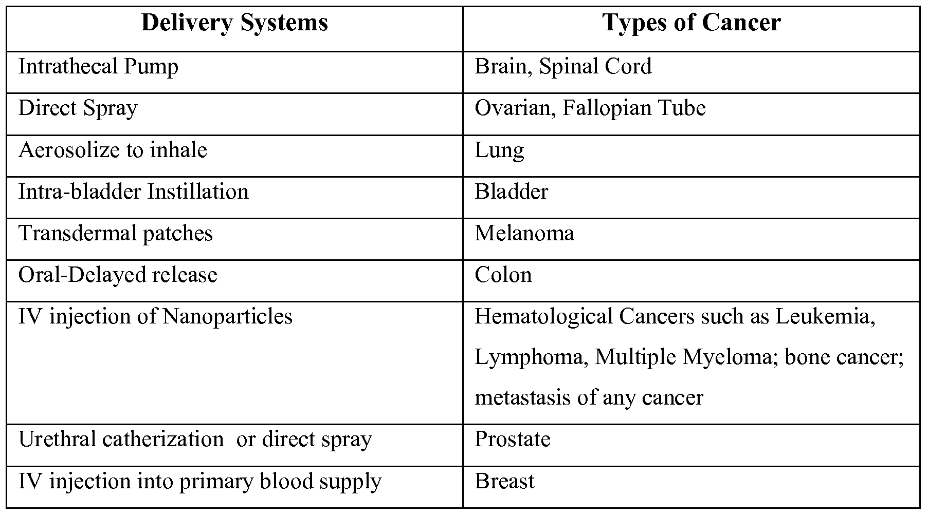
- methyl sulfone used for treatment may increase or decrease over the course of a particular treatment. Changes in dosage may result and become apparent from the results of diagnostic assays that are known in the art for diagnosing or monitoring the progression on various types of cancer. It is understood that appropriate doses of small molecule agents such as methyl suflone depends upon a number of factors within the knowledge of the ordinarily skilled physician, veterinarian, or researcher. The dose(s) of the small molecule will vary, for example, depending upon the identity, size, and condition of the subject or sample being treated, further depending upon the route by which the composition is to be administered, if applicable, and the effect which the practitioner desires the molecule to have. Moreover, one of skill in the art will realize that specific types of cancer can be effectively targeted using specific modes of administration. Exemplary modes of administration for specific types of cancer are set forth in the table below:
- the present invention provides methods of treatment of various cell proliferative disorders, including, for example, breast, ovarian, lung, skin and hematological cancers, e.g., leukemia .
- various cell proliferative disorders including, for example, breast, ovarian, lung, skin and hematological cancers, e.g., leukemia .
- the growth and of tumor cells is inhibited by contacting the cells with an methyl sulfone as described herein.
- the methods of the instant invention are effective for the treatment of cancer while significantly decreasing (or eliminating) many of the adverse effects associated with current chemotherapeutic treatment.
- methyl sulfone is non- toxic and therefore does not cause the side effects associated with most chemotherapeutic treatments. This allows for increased dosages to be administered to a subject in need of treatment.
- subject includes organisms which are capable of suffering from a cell proliferative disorder, e.g., cancer, such as human and non-human animals.
- Preferred human animals include human subjects.
- non-human animals of the invention includes all vertebrates, e.g., mammals, e.g., rodents, e.g., mice, and non-mammals, such as non- human primates, e.g., sheep, dog, cow, chickens, amphibians, reptiles, etc.
- Susceptible to a cell proliferative disorder is meant to include subjects at risk of developing a cell proliferative disorder. In one embodiment, the subject is at greater risk than the average member of a population.
- a prophylactically effective amount refers to an amount of a methyl sulfone or a pharmaceutical preparation thereof, which is effective, upon single or multiple dose administration to the subject, in preventing or treating a cell proliferative disorder.
- methyl sulfone refers to an amount of methyl sulfone or a pharmaceutical preparation thereof which, upon single or multiple dose administration to the subject to provide a therapeutic benefit to the subject.
- the therapeutic benefit is reducing or eliminating cancerous cells or tumors, or prolonging the survivability of a subject with a cell proliferative disorder.
- methods for the treatment of cancer comprising administering a therapeutically effective amount of methyl sulfone, to a subject in need thereof.
- a method for the treatment of cancer comprising administering a therapeutically effective amount of methyl sulfone, or a pharmaceutical composition comprising an inventive compound to a subject in need thereof, in such amounts and for such time as is necessary to achieve the desired result.
- the method involves the administration of a therapeutically effective amount of methyl sulfone or a pharmaceutically acceptable derivative thereof to a subject (including, but not limited to a human or animal) in need of it.
- methyl sulfone is useful for the treatment of cancer (including, but not limited to, glioblastoma, retinoblastoma, breast cancer, cervical cancer, colon and rectal cancer, leukemia, lymphoma, lung cancer (including, but not limited to small cell lung cancer), melanoma and/or skin cancer, multiple myeloma, non-Hodgkin's lymphoma, ovarian cancer, pancreatic cancer, prostate cancer and gastric cancer, bladder cancer, uterine cancer, kidney cancer, testicular cancer, stomach cancer, brain cancer, liver cancer, or esophageal cancer).
- cancer including, but not limited to, glioblastoma, retinoblastoma, breast cancer, cervical cancer, colon and rectal cancer, leukemia, lympho
- the present invention provides a methods for treating cell proliferative disorders in a subject comprising administering to a subject in need thereof a therapeutically effective amount of methyl sulfone, optionally with a pharmaceutically acceptable carrier, adjuvant or vehicle.
- Methyl sulfone can be administered in any manner known in the art such as those disclosed herein.
- a trained clinician will be able to choose the best route of administration based on the type and location of the cell proliferative disorder.
- the invention provides a prophylactic method of preventing a cell proliferative disorder, or preventing the recurrence of a cell proliferative disorder.
- a subject at risk for developing a cell proliferative disorder, or at risk of having a recurrence of a cell proliferative disorder is prophylactically administered methyl sulfone or a pharmaceutical composition comprising methyl sulfone so as to prevent the occurrence or reoccurrence of the cell proliferative disorder.
- the instant invention also provides combination treatments. Treatment with methyl sulfone or a pharmaceutical composition thereof, can be combined with chemotherapeutic, radiation or surgical treatment.
- subject is administered one or more anti-cancer agents in combination with methyl sulfone to treat a cell proliferative disorder
- the anticancer agent can be a chemotherapeutic agent or a biological agent, e.g., an anti-cancer antibody.
- subjects having surgery to remove one or more tumors are treated with a solution of methyl sulfone to ensure that the successful treatment.
- the abdominal cavity can be washed with a solution of methyl sulfone.
- methyl sulfone is capable of inducing apoptosis or senescence in cancer cells.
- Example 1 The following methods were used in Example 1 :
- Cloudman S-91 mouse melanoma cells (sub-clone M-3, CCL 53.1; American Type Culture Collection, Rockville, MD) were grown in RMPI medium supplemented with 10% fetal bovine serum (Invitrogen, Inc) and 5% penicillin- streptomycin (Invitrogen, Inc).
- Mouse breast cancer cells (66C1-1; American Type Culture Collection, Rockville, MD) that are estrogen-receptor negative were grown in DMEM medium supplemented with 10% fetal bovine serum (Invitrogen, Inc) and 5% penicillin/streptomycin (Invitrogen, Inc).
- Human T-cell leukemic lymphocytes (CEM; American Type Culture Collection, Rockville, MD) were grown in MEM medium supplemented with 7% fetal calf serum. Cultures were passaged twice a week.
- T-cell lymphocytes were obtained from a blood sample of a healthy volunteer. Blood samples were obtained with IRB approval and with the volunteer signing an Informed Consent Form. White blood cells were separated from red blood cells using a Ficoll gradient. Flow cytometry with antibodies against CD3 and CD4 was used to identify T-cell lymphocytes (Flow Cytometry Facility, University of Connecticut Health Center, Farmington, CT).
- Cell Proliferation Cells were plated onto sterile 12mm covers lips in 35 mm tissue culture plates and incubated at 37 0 C, 5% CO 2 . After 24 hours, 2% methyl sulfone in medium or medium alone was added the plates and incubated at 37 0 C, 5% CO 2 for 48 and 72 hrs. BrdU (Molecular Probes, Eugene, OR, USA; diluted 1/3 in RPMI medium) was added to each plate for 60 minutes at 37 0 C, 5% CO 2 . Coverslips were transferred to porcelain holders, washed with PBS, fixed in methanol/acetone (1 :1), washed with PBS, and incubated in 5% BSA/ PBS for 30 minutes at room temperature.
- BrdU Molecular Probes, Eugene, OR, USA; diluted 1/3 in RPMI medium
- coverslips were placed in 0.1N HCL/ 1% Triton X-IOO for 10 minutes at room temperature. After washing in PBS, cells were incubated with Alexa Fluor 488 anti-BrdU antibody (Molecular Probes, Eugene, OR; diluted 1/20 in 5% BSA/ PBS) for 60 minutes at room temperature. Cells were washed in PBS and incubated with Hoechst (diluted 1/1000 in PBS) in the dark at room temperature. After washing in PBS, cells were dipped in dH 2 O and drained onto Kim Wipes. SlowFade mounting medium (Molecular Probes, Eugene, OR) was added to a glass slide and coverslips were placed face down onto slides and sealed with clear nail polish. Cells were viewed at the Center for Cell Analysis and Modeling, University of Connecticut
- Cells were plated onto 10-well slides () that were pre-treated with sulfuric acid. After 24 hours, medium was removed and 2% methyl sulfone in medium was added to half the slides; control cells received medium alone. At 72 and 144 hours, cells were washed in 37 0 C PBS, pH 7.4, and fixed in 3.7% formaldehyde in PBS for 10 minutes. PBS was used to wash cells three times followed by a wash in 0.1% Triton X-IOO/ PBS for 5 minutes. Cells were again washed three times in PBS, then in 1% BSA/ PBS for 20 minutes.
- Rhodamine-labeled Phalloidin Molecular Probes, Eugene, OR; diluted 1/40 in 1% BSA/ PBS
- SlowFade was added to each well.
- Glass coverslips were placed over each slide and sealed with nail polish. Images were visualized using 568-nm excitation on a Perkin Elmer Ultraview RS5 spinning-disk confocal scanning system mounted on a Nikon TE2000 inverted microscope with a 10Ox 1.4 NA Plan Apo oil immersion objective. (William A. Mohler, Ph.D., Director, Spinning Disk Microscope Facility, University of Connecticut Health Center).
- Rabbit anti-E-cadherin antibody (Santa Cruz Biotech, CA) was diluted 1/100 in 5% BSA/ PBS and added to the cells on coverslips for 2 hours. Cells were washed 5 times in 0.1% Triton X-100/PBS, 3 minutes each. Secondary antibody, Alexa Fluor 488 chicken anti-rabbit was diluted 1/200 in 5% BSA/ PBS and added to coverslips for one hour in the dark. Cells were washed five times in 0.1% Triton X-100/ PBS, three minutes each. Cells were washed in PBS for one minute and placed in Hoechst (lmg/ml), diluted 1/1000, for 5 minutes. Cells were washed in PBS for one minute.
- Immunofluorescence of ⁇ -catenin was performed as described for E-cadherin. Nonspecific sites were blocked with donkey serum. Primary antibody, goat anti- ⁇ - catenin antibody (Santa Cruz Biotech, CA) was diluted 1/100 in 5% BSA/ PBS. Secondary antibody, Alexa Fluor 568 donkey anti-goat antibody was diluted 1/200 in 5% BSA/ PBS.
- N-cadherin Immunofluorescence of N-cadherin was performed as described for E- cadherin. Nonspecific sites were blocked with chicken serum. Primary antibody, rabbit anti-N-cadherin (Santa Cruz Biotech, CA) was diluted 1/100 in 5% BSA/ PBS. Secondary antibody, Alexa Fluor 488 chicken anti-rabbit antibody was diluted 1/200 in 5% BSA/ PBS.
- Rabbit anti-p27 polyclonal antibody (Santa Cruz Biotech, CA) was diluted 1/250 in 1% BSA/ PBS and added to coverslips for 1.5 hours. Cells were washed 5 times in 0.2% Triton X-100/ PBS, 3 minutes each. Secondary antibody, Alexa Fluor 488 chicken anti-rabbit antibody, was diluted 1/1000 in 1% BSA/ PBS and added to coverslips for 30 minutes in the dark. Cells were washed five times in 0.2% Triton X-100/ PBS, three minutes each. Cells were washed twice in PBS for two minutes each and placed in DAPI, diluted 1/1000 in PBS, for 5 minutes. Cells were washed in PBS for one minute.
- Cells were plated in 6- 100mm tissue culture plates, each containing 1 polylysine-coated 10-well slide, or plated onto polylysine-coated coverslips. Once cells were approximately 50% confluent, 2% methyl sulfone was added to cells for 10, 30, 60, 90, and 120 minutes. At each time point cells were placed in microtubule stabilizing buffer () for 10 minutes followed by incubation in -20 0 C methanol for 5 minutes. Cells were washed twice in 0.1% Triton X-100 in PBS at room temperature, then incubated with mouse monoclonal anti-alpha-tubulin antibody (Santa Cruz Biotech, CA; diluted 1/200 in 1% BSA/ PBS) for 1.5 hours.
- mouse monoclonal anti-alpha-tubulin antibody Santa Cruz Biotech, CA; diluted 1/200 in 1% BSA/ PBS
- Cells were washed 5 times in 0.1% Triton X-IOO/ PBS, 3 minutes each. Cells were incubated with secondary antibody (Alexa Fluor 568 rabbit anti-mouse antibody; diluted 1/100 in 1% /PBS) for 30 minutes in the dark. Cells were washed five times in 0.1% Triton X- 100/PBS, three minutes each. Cells were washed two times in PBS at 2 minute intervals and incubated with DAPI (l ⁇ g/ml in PBS) for 5 minutes at room temperature and in the dark. Cells were washed in PBS for one minute. SlowFade was added to glass slides, coverslips were placed cell side down and sealed with nail polish.
- secondary antibody Alexa Fluor 568 rabbit anti-mouse antibody
- DAPI l ⁇ g/ml in PBS
- Microtubules were viewed at the Center for Cell Analysis and Modeling, University of Connecticut Health Center, Farmington, CT, with an Axioplan CCD Microscope equipped with a 4Ox 1.3 NA FL objective lens and Photometries PXL- EEV37 high speed digital cooled CCD camera via Metamorph image acquisition and analysis software (Universal Imaging Corp., Downington, PA).
- 1% agar DNA grade; Difco Bacto Agar; Becton Dickenson and Company, Sparks, MD
- base agar 1% agar
- 1% agar was mixed in water at room temperature, melted in a microwave, and cooled to 4O 0 C before using.
- 1% agar was combined (1 :1) with 4% methyl sulfone in 2X RPMI medium with sodium bicarbonate supplemented with 10% FBS and 5% penicillin/streptomycin.
- 1% agar was combined (1 :1) with 2X RMPI medium.
- Agar +/- 2% methyl sulfone was added to appropriate 35mm tissue culture plates and stored overnight at 4 0 C. The next day top agar and cells were placed on top of the base agar. Top agar was made with 0.66% agar in water. The agar was melted in a microwave, cooled, and stored at 4O 0 C. 2X RPMI medium and 4% methyl sulfone was also stored at 40 0 C. Cells were trypsinized and counted for a final concentration of 5xlO 3 cells/ plate. Cell counts were adjusted to 2xlO 5 cells/ ml and 0.05ml of the cell suspension was added to 15ml centrifuge tubes (2 each).
- Base agar plates were removed from 4 0 C and allowed to warm to room temperature for approximately 30 minutes prior to plating.
- 2X RPMI medium and 0.66% agar were added to the tube of cell suspension, gently mixed, and placed on top of control base agar plates.
- For plating cells in 2% methyl sulfone, 4% methyl sulfone (in 2X RMPI medium) and 0.66% agar was added to the tube of cell suspension, mixed gently, and placed on top of the drug base agar plates. Cells were incubated in the 37 0 C, 5% CO 2 incubator for 10-14 days. Plates were stained with 0.005% crystal violet for 1 hour. Colonies were photographed and counted with a dissecting microscope.
- Invasion assays were performed in Transwell chambers (Corning). The 8 ⁇ m pore membranes of the upper chambers were coated with ECM gel from Engelbreth- Holm-Swarm murine sarcoma (Sigma), diluted 1 :6 with RPMI medium +/- 2% methyl sulfone, and placed in a well with RPMI medium +/- 2% methyl sulfone. Cells (IxIO 5 cells/ 200 ⁇ l) were seeded into appropriate upper chambers in RMPI medium +/- 2% methyl sulfone. After 20 hours of incubation at 37 0 C, cells were removed from top surface of the chamber and filters were fixed with 5% glutaraldeyde.
- Cell Wounding Cells were cultured to approximately 90% confluence in 35mm tissue culture plates. Methyl sulfone (2%) was added to half the plates and 48 hours later cells were wounded with a sterile plastic lOOO ⁇ l pipette tip. Cells were washed two times with medium to remove cell debris and incubated at 37 0 C over night in RMPI medium +/- 2% methyl sulfone. After 72 hours, wound edges were photographed and recorded with a Nikon TE300 inverted microscope (Nikon) equipped with a 10x 0.25 NA Plan Achromat objective lens.
- Time-series (5min long) of phase contrast images were acquired at a video rate (1 frame/3 s) with a Watec-902B CCD video camera (Watec Corp., Japan) via stream acquisition option of Metamorph image acquisition and analysis software (Universal Imaging Corp., Downington, PA).
- a Watec-902B CCD video camera Wang Corp., Japan
- Metamorph image acquisition and analysis software Universal Imaging Corp., Downington, PA.
- cells were kept at 37 0 C and 10 rnM Hepes.
- Time series and photographs of cells +/- 2% methyl sulfone were obtained every 24 hours for up to 120hrs.
- Mitochondrial Staining Live and Fixed Cells Cells were grown on 12mm round covers lips treated with polylysine to 60-
- Methyl Sulfone Induced Contact Inhibition in Melanoma Cells By 24 hours after adding 2% methyl sulfone, cells grew until they came in touch with neighboring cells. At this point cells appeared to be contact inhibited (Figure 3). Live cell video demonstrated that while control cells retained their amorphous rounded shapes and continued to migrate over neighboring cells, melanoma cells in 2% methyl sulfone stopped migration and growth, and formed a confluent monolayer of quiescent, Gl arrested cells, a hallmark for contact inhibition.
- Figure 4 demonstrates data from cells at 48 and 72 hours in the presence or absence of 2% methyl sulfone. At 48 hours, 14% of control cells were synthesizing DNA while only 0.03% of cells in 2% methyl sulfone were synthesizing DNA. At 72 hours, 21% of control cells and 0% of drug-treated cells were synthesizing DNA.
- Wound healing is a complicated process in which cells juxtaposed to a wound site must detach from neighboring cells and from a substrate (or basement membrane), migrate into the wound and then become contact inhibited once the wound is covered.
- melanoma cells treated with 2% methyl sulfone would function properly in the process of wound healing.
- control melanoma cells and cells treated with 2% methyl sulfone were grown to confluence.
- Scraping a layer of confluent cells with a plastic pipette tip formed wounds. Live cell video microscopy was used to monitor migration of cells into the wound.
- Mature melanocytes assume a morphology that is similar to neuronal cells by having a small area of cytoplasm surrounding the nucleus and long extensions called arbors.
- the primary function of melanocytes is to produce melanin and package the melanin in vesicles called melanosomes. Melanosomes are then transported to the tips of melanocytes arbors. These tips are phagocytized by keratinocytes, cells that sit near the skin's surface, and the newly acquired melanosomes form an umbrella- like shield around the nuclei of keratinocytes to protect these cells' DNA from UV- inducing mutations.
- p27 was Localized in Nuclei of Melanoma Cells Treated with Methyl Sulfone p27 is a protein associated with cell cycle arrest and senescence. Its active form is found inside the nucleus. Using immuno fluorescent microscopy, we showed that p27 is localized in the nuclei of cells treated with methyl sulfone.
- Methyl Sulfone did Not Alter Proteins Involved in the Epithelial to Mesenchymal (ETM) Transition.
- the ETM Transition may be involved in development of metastasis of some tumors.
- Proteins associated with epithelial cells include E-cadherin and ⁇ -catenin, while proteins associated with mesenchymal cells include N-cadherin and vimentin.
- One assay to test stability of microtubules is to give cells increasing concentrations of a microtubule-disassembly drug and use immunofluorescence microscopy to assess microtubule lengths (Caron et al). A higher concentration of drug will be required to disassemble the more stabilized microtubules.
- methyl sulfone stabilizes microtubules. Effects of Methyl Sulfone on Melanoma Cells Were Also Found in Aggressive and Metastatic Estrogen Receptor-negative Breast Cancer Cells We found that concentrations of methyl sulfone above 2% induced apoptosis in breast cancer cells. When breast cancer cells were treated with 2% methyl sulfone, cells became contact inhibited, DNA synthesis was markedly reduced, mitochondrial activity decreased, cell growth became anchorage-dependent, migration of cells through an extracellular matrix was inhibited, wound healing proceeded normally, and cells became senescent.
- Leukemic Lymphocytes CEM cells, leukemic T-cell lymphocytes obtained from a six-year old girl with acute lymphocytic leukemia, were maintained in Petri dishes in MEM (minimal essential medium) with 7% fetal calf serum in a 5% CO 2 incubator at 37 0 C.
- MEM minimal essential medium
- methyl sulfone was added to the medium at concentrations from 0-6.0 %.
- no methyl sulfone was added.
- Cells (2X10 6 per ml) were incubated for 20 hrs in the presence or absence of methyl sulfone, and then assayed for apoptosis using FITC-annexinV/ propidium iodide microscopy.
- Methyl sulfone was also compared to the anti-microtubule drug, vinblastine.
- Vinblastine (VNB) and its derivative, vincristine are standard treatments for acute lymphocytic leukemia, as well as other cancers.
- VNB vinblastine
- Blood was drawn from healthy volunteers and white blood cells (e.g., lymphocytes, neutrophils) were enriched using Ficoll-Plaque.
- Cells were incubated in Petri dishes containing MEM with 7% fetal calf serum in a CO 2 (5%) incubator at 37 0 C; Methyl sulfone was present at concentrations of 0, 3, 6, or 10%. After 20 hours, cells were incubated with antibodies specific to T-lymphocytes (CD3 and CD4 T-cells), B-lymphocytes (B-cells) and neutrophils, and the percent of apoptotic cells was determined by Flow Cytometry.
- T-lymphocytes CD3 and CD4 T-cells
- B-lymphocytes B-lymphocytes
- neutrophils neutrophils
- specific cell types e.g., CD3 T-cells, CD4 T-cells, B-cells, neutrophils
- CEM leukemic lymphocytes were also incubated in the presence or absence of 10% methyl sulfone.
- the goal here was to determine whether the FITC-annexinV/ propidium iodide microscopy assay described above produced similar results as the Flow Cytometry assay used in these experiments.
- melanoma cells (Cloudman M3 cell line) were plated at 10 5 cells/ml in RPMI medium. After 24 hours, medium was replaced with RPMI containing different concentrations (grams/volume) of MMS, MES and EES. After 24 hours, apoptosis was assayed using the Becton-Dickinson Annexin V-FITC/Propidium Iodide Kit and a Nikon fluorescent microscope as described by the manufacturer. Results are set forth in Figures 16 and 17.
- MMS induces significantly less apoptosis of M3 cells than either MES or EES. This is especially true at the lower concentrations, for example, less than 5% MES, EES, and MMS.
- EES/MES have a narrow differential (very small window) between killing cancer cells, but also harming normal cells.
- MMS has a wide window of concentrations that kill cancer cells without harming normal cells.
- SKP2 is a ubiquitin ligase that is active in cell nuclei and is expressed in many types of malignant cells.
- SKP2 promotes cell cycling and cell proliferation and promotes the degradation of the tumor suppressor protein p27.
- p27 is a tumor suppressor that is active when located in cell nuclei and in a non-phosphorylated form.
- p27 promotes cell cycle arrest and senescence.
- p27 non-phosphorylated active form
- p27 is located in the nucleus.
- SKP2 is in the cytoplasm in its inactive form.
- SKP2/DAPI photos In malignant cells, the opposite is true: p27 (phosphorylated and inactive) is in low levels in the cytoplasm; SKP2 (active) is in high levels the nuclei.
- Cells were plated at 1x10 5 cells/ml on 12mm round coverslips in 35mm tissue culture plates (final volume of media was 1.5 ml/35 mm plate). At 24 hours after plating, media was replaced with fresh media containing 2% methyl sulfone and cells were incubated in media/2% methyl sulfone for four weeks. Media/2% methyl sulfone was changed every Monday, Wednesday, and Friday.
- Immunofluorescence was performed at four weeks after plating to look for the presence of the following proteins: SKP2 and p27 (Santa Cruz Biotechnology).
- SKP2 and p27 serum-binding protein 2 and p27 (Santa Cruz Biotechnology).
- To begin immuno fluorescent staining cells on coverslips were transferred from tissue culture plates to porcelain holders; the porcelain holders were gently placed in a beaker with PBS (37°C) for 30 seconds to wash the cells. Cells were fixed in a solution of 4% paraformaldehyde (37°C) for ten minutes, and then washed in PBS for 5 minutes. Goat serum (Santa Cruz Biotechnology) was used in the blocking step. A large glass Petri dish was lined with parafilm.
- Coverslips were then placed cell side down on a drop (35 ⁇ l per coverslip) of goat serum and incubated at room temperature for thirty minutes. Cells were transferred to the porcelain holders and washed in 0.1% Triton X-100/PBS three times at three minutes per wash. Primary antibodies were diluted 1 : 100 in 5% BS A/PBS and centrifuged for 1 minute to remove any particulates. Coverslips were placed cell side down on 35 ⁇ l of primary antibody/coverslip in the parafilm-lined glass Petri dish and incubated for two hours at room temperature. All primary antibodies were rabbit polyclonal. Cells were transferred to the porcelain holders and washed three times in 0.1% Triton X-100/PBS for three minutes each.
- Alexa Fluor 546 goat anti-rabbit (Molecular Probes) was diluted 1 :200 in 5% BSA/PBS. Coverslips were placed cell side down on 35 ⁇ l of secondary antibody/coverslip in the parafilm-lined glass Petri dish, and incubated for one hour in the dark at room temperature. Cells were transferred to the porcelain holders and washed five times in 0.1% Triton X-100/PBS for three minutes each, followed by PBS for one minute. To stain nuclei, DAPI was diluted 1 : 1000 in PBS. Coverslips were placed cell side down on 35 ⁇ l of DAPI/coverslip in the parafilm-lined glass Petri dish and incubated at room temperature for five minutes in the dark.
- Coverslips were washed in PBS for one minute and then placed cell side down on 2.5 ⁇ l/coverslip of Slow Fade (Molecular Probes) on glass slides. Clear nail polish was applied around the coverslip and left to air dry in the dark for thirty minutes. Cells were stored overnight at 4°C. Slides were viewed on a widefield microscope equipped with a Photometries PXL-EEV37 high-speed digital camera. Images were acquired with a 4Ox objective using Molecular Devices MetaMorph software.
Abstract
Description
Claims
Priority Applications (6)
| Application Number | Priority Date | Filing Date | Title |
|---|---|---|---|
| ES10784237.9T ES2693687T3 (en) | 2009-06-05 | 2010-06-07 | Compounds and compositions for use in the treatment of cancer |
| CA2763040A CA2763040C (en) | 2009-06-05 | 2010-06-07 | Methods and compositions for the treatment of cancer |
| US13/382,263 US10363229B2 (en) | 2009-06-05 | 2010-06-07 | Methods and compositions for the treatment of cancer |
| AU2010256384A AU2010256384B2 (en) | 2009-06-05 | 2010-06-07 | Methods and compositions for the treatment of cancer |
| EP10784237.9A EP2437738B1 (en) | 2009-06-05 | 2010-06-07 | Methods and compositions for the treatment of cancer |
| AU2016262709A AU2016262709B2 (en) | 2009-06-05 | 2016-11-23 | Methods and compositions for the treatment of cancer |
Applications Claiming Priority (4)
| Application Number | Priority Date | Filing Date | Title |
|---|---|---|---|
| US18450009P | 2009-06-05 | 2009-06-05 | |
| US61/184,500 | 2009-06-05 | ||
| US33116810P | 2010-05-04 | 2010-05-04 | |
| US61/331,168 | 2010-05-04 |
Publications (2)
| Publication Number | Publication Date |
|---|---|
| WO2010141956A2 true WO2010141956A2 (en) | 2010-12-09 |
| WO2010141956A3 WO2010141956A3 (en) | 2011-04-21 |
Family
ID=43298594
Family Applications (1)
| Application Number | Title | Priority Date | Filing Date |
|---|---|---|---|
| PCT/US2010/037662 WO2010141956A2 (en) | 2009-06-05 | 2010-06-07 | Methods and compositions for the treatment of cancer |
Country Status (6)
| Country | Link |
|---|---|
| US (1) | US10363229B2 (en) |
| EP (1) | EP2437738B1 (en) |
| AU (2) | AU2010256384B2 (en) |
| CA (1) | CA2763040C (en) |
| ES (1) | ES2693687T3 (en) |
| WO (1) | WO2010141956A2 (en) |
Cited By (9)
| Publication number | Priority date | Publication date | Assignee | Title |
|---|---|---|---|---|
| WO2015095048A1 (en) * | 2013-12-16 | 2015-06-25 | Peloton Therapeutics, Inc. | Cyclic sulfone and sulfoximine analogs and uses thereof |
| US9796697B2 (en) | 2015-06-12 | 2017-10-24 | Peloton Therapeutics, Inc. | Tricyclic inhibitors of HIF-2-alpha and uses thereof |
| US9896418B2 (en) | 2013-09-09 | 2018-02-20 | Peloton Therapeutics, Inc. | Aryl ethers and uses thereof |
| US10155726B2 (en) | 2015-03-11 | 2018-12-18 | Peloton Therapeutics, Inc. | Substituted pyridines and uses thereof |
| US10278942B2 (en) | 2015-03-11 | 2019-05-07 | Peloton Therapeutics, Inc. | Compositions for use in treating pulmonary arterial hypertension |
| US10335388B2 (en) | 2015-04-17 | 2019-07-02 | Peloton Therapeutics, Inc. | Combination therapy of a HIF-2-alpha inhibitor and an immunotherapeutic agent and uses thereof |
| US10512626B2 (en) | 2015-03-11 | 2019-12-24 | Peloton Therapeautics, Inc. | Compositions for use in treating glioblastoma |
| US10807948B2 (en) | 2015-03-11 | 2020-10-20 | Peloton Therapeutics, Inc. | Aromatic compounds and uses thereof |
| USRE49948E1 (en) | 2022-02-03 | 2024-04-30 | Peloton Therapeutics, Inc. | Aryl ethers and uses thereof |
Citations (1)
| Publication number | Priority date | Publication date | Assignee | Title |
|---|---|---|---|---|
| US4522811A (en) | 1982-07-08 | 1985-06-11 | Syntex (U.S.A.) Inc. | Serial injection of muramyldipeptides and liposomes enhances the anti-infective activity of muramyldipeptides |
Family Cites Families (6)
| Publication number | Priority date | Publication date | Assignee | Title |
|---|---|---|---|---|
| DE10228680A1 (en) * | 2002-06-27 | 2004-01-22 | Holden Development Limited, Tortola | Basis for transdermal formulations (PTF) |
| US20050118291A1 (en) * | 2003-09-10 | 2005-06-02 | Mian-Ying Wang | Formulations and methods for treating breast cancer with Morinda citrifolia and methylsulfonymethane |
| EP1750690A1 (en) | 2004-05-10 | 2007-02-14 | Robert F. Hofmann | Use of targeted oxidative therapeutic formulation in treatment of cancer |
| EP1853250B1 (en) * | 2005-02-18 | 2011-11-02 | Abraxis BioScience, LLC | Combinations and modes of administration of therapeutic agents and combination therapy |
| US20100080843A1 (en) * | 2005-06-30 | 2010-04-01 | The Research Foundation of the State University Of New York at Albany | Natural and synthetic sulfur and selenium analogs and polymer conjugated forms thereof for the modulation of angiogenesis |
| US8188047B2 (en) * | 2009-03-20 | 2012-05-29 | Thomas Perez | Therapeutic and cosmetic compositions for treatment of skin |
-
2010
- 2010-06-07 WO PCT/US2010/037662 patent/WO2010141956A2/en active Application Filing
- 2010-06-07 EP EP10784237.9A patent/EP2437738B1/en active Active
- 2010-06-07 CA CA2763040A patent/CA2763040C/en active Active
- 2010-06-07 US US13/382,263 patent/US10363229B2/en active Active
- 2010-06-07 ES ES10784237.9T patent/ES2693687T3/en active Active
- 2010-06-07 AU AU2010256384A patent/AU2010256384B2/en active Active
-
2016
- 2016-11-23 AU AU2016262709A patent/AU2016262709B2/en active Active
Patent Citations (1)
| Publication number | Priority date | Publication date | Assignee | Title |
|---|---|---|---|---|
| US4522811A (en) | 1982-07-08 | 1985-06-11 | Syntex (U.S.A.) Inc. | Serial injection of muramyldipeptides and liposomes enhances the anti-infective activity of muramyldipeptides |
Non-Patent Citations (3)
| Title |
|---|
| "Percutaneous Penetration Enhancers", 1995, CRC PRESS, INC. |
| BUYUKTIMKIN ET AL.: "Chemical Means of Transdermal Drug Permeation Enhancement in Transdermal and Topical Drug Delivery Systems", 1997, INTERPHARM PRESS INC. |
| See also references of EP2437738A4 |
Cited By (15)
| Publication number | Priority date | Publication date | Assignee | Title |
|---|---|---|---|---|
| US10144711B2 (en) | 2013-09-09 | 2018-12-04 | Peloton Therapeutics, Inc. | Aryl ethers and uses thereof |
| US9896418B2 (en) | 2013-09-09 | 2018-02-20 | Peloton Therapeutics, Inc. | Aryl ethers and uses thereof |
| US9908845B2 (en) | 2013-09-09 | 2018-03-06 | Peloton Therapeutics, Inc. | Aryl ethers and uses thereof |
| US9969689B2 (en) | 2013-09-09 | 2018-05-15 | Peloton Therapeutics, Inc. | Aryl ethers and uses thereof |
| US10597366B2 (en) | 2013-09-09 | 2020-03-24 | Peloton Therapeutics, Inc. | Aryl ethers and uses thereof |
| WO2015095048A1 (en) * | 2013-12-16 | 2015-06-25 | Peloton Therapeutics, Inc. | Cyclic sulfone and sulfoximine analogs and uses thereof |
| US9884843B2 (en) | 2013-12-16 | 2018-02-06 | Peloton Therapeutics, Inc. | Cyclic sulfone and sulfoximine analogs and uses thereof |
| US10278942B2 (en) | 2015-03-11 | 2019-05-07 | Peloton Therapeutics, Inc. | Compositions for use in treating pulmonary arterial hypertension |
| US10155726B2 (en) | 2015-03-11 | 2018-12-18 | Peloton Therapeutics, Inc. | Substituted pyridines and uses thereof |
| US10512626B2 (en) | 2015-03-11 | 2019-12-24 | Peloton Therapeautics, Inc. | Compositions for use in treating glioblastoma |
| US10807948B2 (en) | 2015-03-11 | 2020-10-20 | Peloton Therapeutics, Inc. | Aromatic compounds and uses thereof |
| US10335388B2 (en) | 2015-04-17 | 2019-07-02 | Peloton Therapeutics, Inc. | Combination therapy of a HIF-2-alpha inhibitor and an immunotherapeutic agent and uses thereof |
| US10786480B2 (en) | 2015-04-17 | 2020-09-29 | Peloton Therapeutics, Inc. | Combination therapy of a HIF-2-α inhibitor and an immunotherapeutic agent and uses thereof |
| US9796697B2 (en) | 2015-06-12 | 2017-10-24 | Peloton Therapeutics, Inc. | Tricyclic inhibitors of HIF-2-alpha and uses thereof |
| USRE49948E1 (en) | 2022-02-03 | 2024-04-30 | Peloton Therapeutics, Inc. | Aryl ethers and uses thereof |
Also Published As
| Publication number | Publication date |
|---|---|
| CA2763040C (en) | 2018-05-01 |
| AU2016262709A1 (en) | 2016-12-15 |
| CA2763040A1 (en) | 2010-12-09 |
| AU2010256384A1 (en) | 2011-11-24 |
| EP2437738B1 (en) | 2018-08-08 |
| US10363229B2 (en) | 2019-07-30 |
| EP2437738A2 (en) | 2012-04-11 |
| WO2010141956A3 (en) | 2011-04-21 |
| AU2016262709B2 (en) | 2018-11-01 |
| ES2693687T3 (en) | 2018-12-13 |
| AU2010256384B2 (en) | 2016-08-25 |
| US20160250158A1 (en) | 2016-09-01 |
| EP2437738A4 (en) | 2012-11-14 |
Similar Documents
| Publication | Publication Date | Title |
|---|---|---|
| AU2016262709B2 (en) | Methods and compositions for the treatment of cancer | |
| AU730261B2 (en) | Method of treating malignant tumors with thyroxine analogues having no significant hormonal activity | |
| AU2011285724B2 (en) | Combination therapy for the treatment of prostate carcinoma | |
| US8222008B2 (en) | Methods and compositions for treating cancer | |
| WO1997046228A9 (en) | Method of treating malignant tumors with thyroxine analogues having no significant hormonal activity | |
| EP2755643B1 (en) | Compositions of jasmonate compounds and methods of use | |
| Ye et al. | Salidroside inhibits CCl4-induced liver fibrosis in mice by reducing activation and migration of HSC induced by liver sinusoidal endothelial cell-derived exosomal SphK1 | |
| KR101947785B1 (en) | Composition for regenerating normal tissue from fibrotic tissue | |
| EA025180B1 (en) | Treatment of solid tumours | |
| JP2003519655A (en) | O-methylated rapamycin derivatives for alleviation and inhibition of lymphoproliferative syndrome | |
| Bo et al. | Upregulation of the expression of Wnt5a promotes the proliferation of pancreatic cancer cells in vitro and in a nude mouse model | |
| Ríos et al. | OPC-12759 increases proliferation of cultured rat conjunctival goblet cells | |
| US11078270B2 (en) | Use of negative functional modulators of erythropoietin for therapy | |
| US6737397B1 (en) | Control of cancer growth through the interaction of [MET5]-enkephalin and the zeta receptor | |
| JP2002508321A (en) | Multicatalytic protease inhibitors for use as antitumor agents | |
| TWI504390B (en) | Use of para-quinone of formula (i) for down-regulation of wnt/β-catenin signaling pathway of melanoma cell | |
| EP3070092B1 (en) | 3-phenyl-thiazolo[3,2-a]benzimidazole derivatives as aldehyde dehydrogenase 1 (aldh-1) modulators for the treatment of breast cancer or leukemia, and for manipulating cultured aldh-1 positive breast cancer or leukemia cells | |
| Pantazis et al. | Differentiation of human malignant melanoma cells that escape apoptosis after treatment with 9-nitrocamptothecin in vitro | |
| WO2019103984A1 (en) | Compositions including fatp1, fatp3, fatp4, fatp5, and/or fatp6 inhibitors and uses thereof | |
| Mancini et al. | A conditioned medium from a human liposarcoma-derived cell line induces p53-dependent apoptosis in several tumor cell lines. | |
| KR100484014B1 (en) | How to treat malignant tumors with thyroxine analogs that do not have hormonal activity | |
| WO2009032213A1 (en) | Control of malignant cells by kinase inhibition | |
| Jablonski | The role of aquaporins in the initiation and progression of the apoptotic volume decrease |
Legal Events
| Date | Code | Title | Description |
|---|---|---|---|
| 121 | Ep: the epo has been informed by wipo that ep was designated in this application |
Ref document number: 10784237 Country of ref document: EP Kind code of ref document: A2 |
|
| WWE | Wipo information: entry into national phase |
Ref document number: 2763040 Country of ref document: CA |
|
| ENP | Entry into the national phase |
Ref document number: 2010256384 Country of ref document: AU Date of ref document: 20100607 Kind code of ref document: A |
|
| WWE | Wipo information: entry into national phase |
Ref document number: 2010784237 Country of ref document: EP |
|
| NENP | Non-entry into the national phase |
Ref country code: DE |
|
| WWE | Wipo information: entry into national phase |
Ref document number: 13382263 Country of ref document: US |