WO2009113972A2 - Temporarily stiffened mesh prostheses - Google Patents
Temporarily stiffened mesh prostheses Download PDFInfo
- Publication number
- WO2009113972A2 WO2009113972A2 PCT/US2007/061885 US2007061885W WO2009113972A2 WO 2009113972 A2 WO2009113972 A2 WO 2009113972A2 US 2007061885 W US2007061885 W US 2007061885W WO 2009113972 A2 WO2009113972 A2 WO 2009113972A2
- Authority
- WO
- WIPO (PCT)
- Prior art keywords
- mesh
- medical prosthesis
- coating
- agent
- poly
- Prior art date
Links
Classifications
-
- A—HUMAN NECESSITIES
- A61—MEDICAL OR VETERINARY SCIENCE; HYGIENE
- A61F—FILTERS IMPLANTABLE INTO BLOOD VESSELS; PROSTHESES; DEVICES PROVIDING PATENCY TO, OR PREVENTING COLLAPSING OF, TUBULAR STRUCTURES OF THE BODY, e.g. STENTS; ORTHOPAEDIC, NURSING OR CONTRACEPTIVE DEVICES; FOMENTATION; TREATMENT OR PROTECTION OF EYES OR EARS; BANDAGES, DRESSINGS OR ABSORBENT PADS; FIRST-AID KITS
- A61F2/00—Filters implantable into blood vessels; Prostheses, i.e. artificial substitutes or replacements for parts of the body; Appliances for connecting them with the body; Devices providing patency to, or preventing collapsing of, tubular structures of the body, e.g. stents
- A61F2/0063—Implantable repair or support meshes, e.g. hernia meshes
-
- A—HUMAN NECESSITIES
- A61—MEDICAL OR VETERINARY SCIENCE; HYGIENE
- A61L—METHODS OR APPARATUS FOR STERILISING MATERIALS OR OBJECTS IN GENERAL; DISINFECTION, STERILISATION OR DEODORISATION OF AIR; CHEMICAL ASPECTS OF BANDAGES, DRESSINGS, ABSORBENT PADS OR SURGICAL ARTICLES; MATERIALS FOR BANDAGES, DRESSINGS, ABSORBENT PADS OR SURGICAL ARTICLES
- A61L27/00—Materials for grafts or prostheses or for coating grafts or prostheses
- A61L27/28—Materials for coating prostheses
- A61L27/34—Macromolecular materials
-
- A—HUMAN NECESSITIES
- A61—MEDICAL OR VETERINARY SCIENCE; HYGIENE
- A61L—METHODS OR APPARATUS FOR STERILISING MATERIALS OR OBJECTS IN GENERAL; DISINFECTION, STERILISATION OR DEODORISATION OF AIR; CHEMICAL ASPECTS OF BANDAGES, DRESSINGS, ABSORBENT PADS OR SURGICAL ARTICLES; MATERIALS FOR BANDAGES, DRESSINGS, ABSORBENT PADS OR SURGICAL ARTICLES
- A61L27/00—Materials for grafts or prostheses or for coating grafts or prostheses
- A61L27/50—Materials characterised by their function or physical properties, e.g. injectable or lubricating compositions, shape-memory materials, surface modified materials
- A61L27/54—Biologically active materials, e.g. therapeutic substances
-
- A—HUMAN NECESSITIES
- A61—MEDICAL OR VETERINARY SCIENCE; HYGIENE
- A61L—METHODS OR APPARATUS FOR STERILISING MATERIALS OR OBJECTS IN GENERAL; DISINFECTION, STERILISATION OR DEODORISATION OF AIR; CHEMICAL ASPECTS OF BANDAGES, DRESSINGS, ABSORBENT PADS OR SURGICAL ARTICLES; MATERIALS FOR BANDAGES, DRESSINGS, ABSORBENT PADS OR SURGICAL ARTICLES
- A61L27/00—Materials for grafts or prostheses or for coating grafts or prostheses
- A61L27/50—Materials characterised by their function or physical properties, e.g. injectable or lubricating compositions, shape-memory materials, surface modified materials
- A61L27/56—Porous materials, e.g. foams or sponges
-
- A—HUMAN NECESSITIES
- A61—MEDICAL OR VETERINARY SCIENCE; HYGIENE
- A61L—METHODS OR APPARATUS FOR STERILISING MATERIALS OR OBJECTS IN GENERAL; DISINFECTION, STERILISATION OR DEODORISATION OF AIR; CHEMICAL ASPECTS OF BANDAGES, DRESSINGS, ABSORBENT PADS OR SURGICAL ARTICLES; MATERIALS FOR BANDAGES, DRESSINGS, ABSORBENT PADS OR SURGICAL ARTICLES
- A61L27/00—Materials for grafts or prostheses or for coating grafts or prostheses
- A61L27/50—Materials characterised by their function or physical properties, e.g. injectable or lubricating compositions, shape-memory materials, surface modified materials
- A61L27/58—Materials at least partially resorbable by the body
-
- A—HUMAN NECESSITIES
- A61—MEDICAL OR VETERINARY SCIENCE; HYGIENE
- A61L—METHODS OR APPARATUS FOR STERILISING MATERIALS OR OBJECTS IN GENERAL; DISINFECTION, STERILISATION OR DEODORISATION OF AIR; CHEMICAL ASPECTS OF BANDAGES, DRESSINGS, ABSORBENT PADS OR SURGICAL ARTICLES; MATERIALS FOR BANDAGES, DRESSINGS, ABSORBENT PADS OR SURGICAL ARTICLES
- A61L31/00—Materials for other surgical articles, e.g. stents, stent-grafts, shunts, surgical drapes, guide wires, materials for adhesion prevention, occluding devices, surgical gloves, tissue fixation devices
- A61L31/08—Materials for coatings
- A61L31/10—Macromolecular materials
-
- A—HUMAN NECESSITIES
- A61—MEDICAL OR VETERINARY SCIENCE; HYGIENE
- A61L—METHODS OR APPARATUS FOR STERILISING MATERIALS OR OBJECTS IN GENERAL; DISINFECTION, STERILISATION OR DEODORISATION OF AIR; CHEMICAL ASPECTS OF BANDAGES, DRESSINGS, ABSORBENT PADS OR SURGICAL ARTICLES; MATERIALS FOR BANDAGES, DRESSINGS, ABSORBENT PADS OR SURGICAL ARTICLES
- A61L31/00—Materials for other surgical articles, e.g. stents, stent-grafts, shunts, surgical drapes, guide wires, materials for adhesion prevention, occluding devices, surgical gloves, tissue fixation devices
- A61L31/14—Materials characterised by their function or physical properties, e.g. injectable or lubricating compositions, shape-memory materials, surface modified materials
- A61L31/146—Porous materials, e.g. foams or sponges
-
- A—HUMAN NECESSITIES
- A61—MEDICAL OR VETERINARY SCIENCE; HYGIENE
- A61L—METHODS OR APPARATUS FOR STERILISING MATERIALS OR OBJECTS IN GENERAL; DISINFECTION, STERILISATION OR DEODORISATION OF AIR; CHEMICAL ASPECTS OF BANDAGES, DRESSINGS, ABSORBENT PADS OR SURGICAL ARTICLES; MATERIALS FOR BANDAGES, DRESSINGS, ABSORBENT PADS OR SURGICAL ARTICLES
- A61L31/00—Materials for other surgical articles, e.g. stents, stent-grafts, shunts, surgical drapes, guide wires, materials for adhesion prevention, occluding devices, surgical gloves, tissue fixation devices
- A61L31/14—Materials characterised by their function or physical properties, e.g. injectable or lubricating compositions, shape-memory materials, surface modified materials
- A61L31/148—Materials at least partially resorbable by the body
-
- A—HUMAN NECESSITIES
- A61—MEDICAL OR VETERINARY SCIENCE; HYGIENE
- A61L—METHODS OR APPARATUS FOR STERILISING MATERIALS OR OBJECTS IN GENERAL; DISINFECTION, STERILISATION OR DEODORISATION OF AIR; CHEMICAL ASPECTS OF BANDAGES, DRESSINGS, ABSORBENT PADS OR SURGICAL ARTICLES; MATERIALS FOR BANDAGES, DRESSINGS, ABSORBENT PADS OR SURGICAL ARTICLES
- A61L31/00—Materials for other surgical articles, e.g. stents, stent-grafts, shunts, surgical drapes, guide wires, materials for adhesion prevention, occluding devices, surgical gloves, tissue fixation devices
- A61L31/14—Materials characterised by their function or physical properties, e.g. injectable or lubricating compositions, shape-memory materials, surface modified materials
- A61L31/16—Biologically active materials, e.g. therapeutic substances
-
- A—HUMAN NECESSITIES
- A61—MEDICAL OR VETERINARY SCIENCE; HYGIENE
- A61P—SPECIFIC THERAPEUTIC ACTIVITY OF CHEMICAL COMPOUNDS OR MEDICINAL PREPARATIONS
- A61P23/00—Anaesthetics
- A61P23/02—Local anaesthetics
-
- A—HUMAN NECESSITIES
- A61—MEDICAL OR VETERINARY SCIENCE; HYGIENE
- A61P—SPECIFIC THERAPEUTIC ACTIVITY OF CHEMICAL COMPOUNDS OR MEDICINAL PREPARATIONS
- A61P31/00—Antiinfectives, i.e. antibiotics, antiseptics, chemotherapeutics
-
- A—HUMAN NECESSITIES
- A61—MEDICAL OR VETERINARY SCIENCE; HYGIENE
- A61F—FILTERS IMPLANTABLE INTO BLOOD VESSELS; PROSTHESES; DEVICES PROVIDING PATENCY TO, OR PREVENTING COLLAPSING OF, TUBULAR STRUCTURES OF THE BODY, e.g. STENTS; ORTHOPAEDIC, NURSING OR CONTRACEPTIVE DEVICES; FOMENTATION; TREATMENT OR PROTECTION OF EYES OR EARS; BANDAGES, DRESSINGS OR ABSORBENT PADS; FIRST-AID KITS
- A61F2210/00—Particular material properties of prostheses classified in groups A61F2/00 - A61F2/26 or A61F2/82 or A61F9/00 or A61F11/00 or subgroups thereof
- A61F2210/0004—Particular material properties of prostheses classified in groups A61F2/00 - A61F2/26 or A61F2/82 or A61F9/00 or A61F11/00 or subgroups thereof bioabsorbable
-
- A—HUMAN NECESSITIES
- A61—MEDICAL OR VETERINARY SCIENCE; HYGIENE
- A61F—FILTERS IMPLANTABLE INTO BLOOD VESSELS; PROSTHESES; DEVICES PROVIDING PATENCY TO, OR PREVENTING COLLAPSING OF, TUBULAR STRUCTURES OF THE BODY, e.g. STENTS; ORTHOPAEDIC, NURSING OR CONTRACEPTIVE DEVICES; FOMENTATION; TREATMENT OR PROTECTION OF EYES OR EARS; BANDAGES, DRESSINGS OR ABSORBENT PADS; FIRST-AID KITS
- A61F2250/00—Special features of prostheses classified in groups A61F2/00 - A61F2/26 or A61F2/82 or A61F9/00 or A61F11/00 or subgroups thereof
- A61F2250/0058—Additional features; Implant or prostheses properties not otherwise provided for
- A61F2250/0067—Means for introducing or releasing pharmaceutical products into the body
-
- A—HUMAN NECESSITIES
- A61—MEDICAL OR VETERINARY SCIENCE; HYGIENE
- A61F—FILTERS IMPLANTABLE INTO BLOOD VESSELS; PROSTHESES; DEVICES PROVIDING PATENCY TO, OR PREVENTING COLLAPSING OF, TUBULAR STRUCTURES OF THE BODY, e.g. STENTS; ORTHOPAEDIC, NURSING OR CONTRACEPTIVE DEVICES; FOMENTATION; TREATMENT OR PROTECTION OF EYES OR EARS; BANDAGES, DRESSINGS OR ABSORBENT PADS; FIRST-AID KITS
- A61F2310/00—Prostheses classified in A61F2/28 or A61F2/30 - A61F2/44 being constructed from or coated with a particular material
- A61F2310/00389—The prosthesis being coated or covered with a particular material
- A61F2310/0097—Coating or prosthesis-covering structure made of pharmaceutical products, e.g. antibiotics
-
- A—HUMAN NECESSITIES
- A61—MEDICAL OR VETERINARY SCIENCE; HYGIENE
- A61L—METHODS OR APPARATUS FOR STERILISING MATERIALS OR OBJECTS IN GENERAL; DISINFECTION, STERILISATION OR DEODORISATION OF AIR; CHEMICAL ASPECTS OF BANDAGES, DRESSINGS, ABSORBENT PADS OR SURGICAL ARTICLES; MATERIALS FOR BANDAGES, DRESSINGS, ABSORBENT PADS OR SURGICAL ARTICLES
- A61L2300/00—Biologically active materials used in bandages, wound dressings, absorbent pads or medical devices
- A61L2300/40—Biologically active materials used in bandages, wound dressings, absorbent pads or medical devices characterised by a specific therapeutic activity or mode of action
- A61L2300/402—Anaestetics, analgesics, e.g. lidocaine
-
- A—HUMAN NECESSITIES
- A61—MEDICAL OR VETERINARY SCIENCE; HYGIENE
- A61L—METHODS OR APPARATUS FOR STERILISING MATERIALS OR OBJECTS IN GENERAL; DISINFECTION, STERILISATION OR DEODORISATION OF AIR; CHEMICAL ASPECTS OF BANDAGES, DRESSINGS, ABSORBENT PADS OR SURGICAL ARTICLES; MATERIALS FOR BANDAGES, DRESSINGS, ABSORBENT PADS OR SURGICAL ARTICLES
- A61L2300/00—Biologically active materials used in bandages, wound dressings, absorbent pads or medical devices
- A61L2300/40—Biologically active materials used in bandages, wound dressings, absorbent pads or medical devices characterised by a specific therapeutic activity or mode of action
- A61L2300/404—Biocides, antimicrobial agents, antiseptic agents
- A61L2300/406—Antibiotics
-
- A—HUMAN NECESSITIES
- A61—MEDICAL OR VETERINARY SCIENCE; HYGIENE
- A61L—METHODS OR APPARATUS FOR STERILISING MATERIALS OR OBJECTS IN GENERAL; DISINFECTION, STERILISATION OR DEODORISATION OF AIR; CHEMICAL ASPECTS OF BANDAGES, DRESSINGS, ABSORBENT PADS OR SURGICAL ARTICLES; MATERIALS FOR BANDAGES, DRESSINGS, ABSORBENT PADS OR SURGICAL ARTICLES
- A61L2300/00—Biologically active materials used in bandages, wound dressings, absorbent pads or medical devices
- A61L2300/40—Biologically active materials used in bandages, wound dressings, absorbent pads or medical devices characterised by a specific therapeutic activity or mode of action
- A61L2300/416—Anti-neoplastic or anti-proliferative or anti-restenosis or anti-angiogenic agents, e.g. paclitaxel, sirolimus
-
- A—HUMAN NECESSITIES
- A61—MEDICAL OR VETERINARY SCIENCE; HYGIENE
- A61L—METHODS OR APPARATUS FOR STERILISING MATERIALS OR OBJECTS IN GENERAL; DISINFECTION, STERILISATION OR DEODORISATION OF AIR; CHEMICAL ASPECTS OF BANDAGES, DRESSINGS, ABSORBENT PADS OR SURGICAL ARTICLES; MATERIALS FOR BANDAGES, DRESSINGS, ABSORBENT PADS OR SURGICAL ARTICLES
- A61L2300/00—Biologically active materials used in bandages, wound dressings, absorbent pads or medical devices
- A61L2300/40—Biologically active materials used in bandages, wound dressings, absorbent pads or medical devices characterised by a specific therapeutic activity or mode of action
- A61L2300/45—Mixtures of two or more drugs, e.g. synergistic mixtures
-
- A—HUMAN NECESSITIES
- A61—MEDICAL OR VETERINARY SCIENCE; HYGIENE
- A61L—METHODS OR APPARATUS FOR STERILISING MATERIALS OR OBJECTS IN GENERAL; DISINFECTION, STERILISATION OR DEODORISATION OF AIR; CHEMICAL ASPECTS OF BANDAGES, DRESSINGS, ABSORBENT PADS OR SURGICAL ARTICLES; MATERIALS FOR BANDAGES, DRESSINGS, ABSORBENT PADS OR SURGICAL ARTICLES
- A61L2300/00—Biologically active materials used in bandages, wound dressings, absorbent pads or medical devices
- A61L2300/60—Biologically active materials used in bandages, wound dressings, absorbent pads or medical devices characterised by a special physical form
- A61L2300/606—Coatings
-
- A—HUMAN NECESSITIES
- A61—MEDICAL OR VETERINARY SCIENCE; HYGIENE
- A61L—METHODS OR APPARATUS FOR STERILISING MATERIALS OR OBJECTS IN GENERAL; DISINFECTION, STERILISATION OR DEODORISATION OF AIR; CHEMICAL ASPECTS OF BANDAGES, DRESSINGS, ABSORBENT PADS OR SURGICAL ARTICLES; MATERIALS FOR BANDAGES, DRESSINGS, ABSORBENT PADS OR SURGICAL ARTICLES
- A61L2430/00—Materials or treatment for tissue regeneration
- A61L2430/02—Materials or treatment for tissue regeneration for reconstruction of bones; weight-bearing implants
Definitions
- the present invention relates to medical prostheses and methods of manufacturing those devices.
- the prostheses are temporarily stiffened meshes with particular coatings to provide initial stiffness and thereby permit easier surgical handling for treatment or reconstruction of soft tissue defects.
- Preferred embodiments include surgical meshes coated with one or more biodegradable polymers that can act as a stiffening agent by coating the filaments or fibers of the mesh to temporarily immobilize the contact points of those filaments or fibers and/or by increasing the stiffness of the mesh by at least 1.1 times its original stiffness.
- the devices of the invention can also provide relief from various postoperative complications associated with their implantation, insertion or surgical use. By including biologically active agents and/or drugs in the coating, the devices provide prophylaxis for and can alleviate side effects or complications associated with the surgery or use of prostheses in general.
- Prosthetic implants such as meshes, combination mesh products or other porous prostheses are commonly used to provide a physical barrier between types of tissue or extra strength to a physical defect in soft tissue.
- post-surgical complications including post-implant infection, pain, excessive scar tissue formation and shrinkage of the prosthesis or mesh.
- Excessive scar tissue formation, limited patient mobility, and chronic pain are often attributed to the size, shape, and mass of the implant and a variety of efforts have been undertaken to reduce the amount of scar tissue formation. For example, lighter meshes using smaller fibers, larger weaves, and/or larger pore sizes as well as meshes woven from both non-resorbable and resorbable materials are in use to address these concerns.
- hernias occur when muscles and ligaments tear and allow the protrusion of fat or other tissues through the abdominal wall. Hernias usually occur because of a natural weakness in the abdominal wall or from excessive strain on the abdominal wall, such as the strain from heavy lifting, substantial weight gain, persistent coughing, or difficulty with bowel movements or urination. Eighty percent of all hernias are located near the groin but can also occur below the groin (femoral), through the navel (umbilical), and along a previous incision (incisional or ventral). Almost all hernia repair surgeries are completed with the insertion of a barrier or prosthesis to prevent their reoccurrence.
- Surgical meshes that have been manipulated to improve handling, insertiona and positioning post-insertion are known in the art, but do not employ larger-pore mesh construction.
- a laparoscopic surgical mesh with extruded monofilament PET coils or rings e.g., the Bard ® Composix ® Kugel ® hernia patch
- meshes with reinforced edges have been produced (e.g., Bard ® Visilex ® ). These meshes have the same disadvantages as those with coils or rings.
- the Kugel patch ring has been reported to break under conditions of use, causing patient morbidity and mortality.
- biodegradable polymers of that mesh may be subjected to high temperatures to produce fibers and filaments suitable for weaving, it drastically limits the drugs or biologically active agents that can be included in a biodegradable layer since, under such conditions, the vast majority of biologically-active agents and drugs are unable to withstand the manufacturing temperatures involved in fiber and filament formation. If three-dimensional structures are desired for such meshes, they must undergo further processing to attain such shapes. Finally, these meshes are often more difficult for surgeons to anchor in place because the polyglactin fiber cannot withstand the suturing tension.
- the present invention overcomes these disadvantages by providing temporarily stiffened and shapeable meshes.
- the present invention is directed to a medical prosthesis comprising a mesh and one or more coating which temporarily stiffens the mesh to at least 1.1 times its original stiffness.
- the coatings on such meshes do not alter the integrity of the mesh and thus allow the mesh to remain porous.
- the coatings do not substantially alter the porosity of the mesh.
- the medical prostheses of the invention comprise a mesh with one or more coatings with at least one of the coatings comprising a stiffening agent that coats the filaments or fibers of the mesh so to temporarily immobilize the contact points of those filaments or fibers.
- the coatings on such meshes do not alter the integrity strength of the underlying mesh and thus allow the mesh to remain porous after coating.
- the meshes are capable of substantially reverting to their original stiffness under conditions of use.
- the stiffening agents of the invention can selectively, partially or fully coat the contact points of the filaments or said fibers of the mesh to create a coating.
- the contact points generally include the knots of woven meshes.
- Such coating are preferably positioned on the mesh in a templated pattern or in an array such as might be deposited with ink-jet type technology, including computer controlled deposition techniques. Additionally, the coatings can be applied on one or both sides of the mesh.
- the medical prostheses of the invention include meshes that have been formed into three-dimensional shapes with the shape being maintained by the strength imparted by the coating. Such meshes flat or substantially flat before coating and the application of the coating provides the structural support for the three-demensional shape, any shape can be formed, including curved meshes and conical shapes.
- the stiffening agents include but are not limited to hydrogels and/or biodegradable polymers. One or more biodegradable polymers can be used per individual coating layer. Preferred biodegradable polymer comprises one or more tyrosine-derived diphenol monomer units as polyarylates, polycarbonates or polyiminocarbonates.
- the medical prosthesis of the invention have at least one of the coatings that further comprises one or more drugs.
- drugs include, but are not limited to, antimicrobial agents, anesthetics, analgesics, anti-inflammatory agents, anti-scarring agents, anti-f ⁇ brotic agents and leukotriene inhibitors.
- Yet another aspect of the invention is directed to a process for coating a mesh with a stiffening agent that coats the filaments or fibers of the mesh to temporarily immobilize the contact points of the filaments or fibers of said mesh by (a) preparing a coating solution comprising a solvent and the stiffening agent; (b) spraying a mesh one or more times to provide a sufficient amount of solution on the said mesh to produce a coating having a thickness and placement sufficient to temporarily immobilize the contact points of the filaments or fibers of said mesh that coats filaments or fibers; and(c) drying the mesh to produce the desired coating.
- Still another aspect of the invention provides a method for producing a shaped or three-dimensional medical prosthesis by (a)forming a mesh into a desired shape or three- dimensional shape; (b) applying a stiffening agent to the mesh to coat the filaments or fibers of the mesh; (c) allowing the agent to dry, set or cure causing the mesh to temporarily immobilize contact points of the filaments or fibers of said mesh and thereby to retain the desired shape.
- Shapes can be formed by affixing the mesh to a mold or framework to created the desired shape, applying the agent to the mesh while so affixed, and removing the mesh from the mold or framework after the agent has dried, set or cured sufficiently to allow the mesh to retain its shape.
- the coated meshes of the invention are capable of releasing one or more drugs into surrounding bodily tissue such that the drug reduces or prevents an implant- or surgery- related complication.
- the surgical mesh coatings can include an anesthetic agent such that agent seeps into the surrounding bodily tissue, bodily fluid, or systemic fluid in a predictable manner and at rate sufficient to attenuate the pain experienced at the site of implantation.
- the surgical meshes coatings can include an antiinflammatory agent such that the anti-inflammatory agent seeps into the surrounding bodily tissue, bodily fluid or systemic fluid in a predictable manner and at a rate sufficient to reduce the swelling and inflammation associated implantation of the mesh.
- the surgical mesh coatings can include an antimicrobial agent such that the antimicrobial agent is released into the surrounding bodily tissue, bodily fluid, or systemic fluid in a predictable manner and at a therapeutically-effective dose to provide a rate of drug release sufficient to prevent colonization of the mesh (and/or surgical implantation site) by bacteria for a minimum of the period of time following surgery necessary for initial healing of the surgical incision.
- an antimicrobial agent such that the antimicrobial agent is released into the surrounding bodily tissue, bodily fluid, or systemic fluid in a predictable manner and at a therapeutically-effective dose to provide a rate of drug release sufficient to prevent colonization of the mesh (and/or surgical implantation site) by bacteria for a minimum of the period of time following surgery necessary for initial healing of the surgical incision.
- the coated surgical meshes of the invention can be formed to encapsulate a pacemaker, a defibrillator generator, an implantable access system, a neurostimulator, or any other implantable device for the purpose of securing them in position, providing pain relief, inhibiting scarring or fibrosis and/or inhibiting bacterial growth.
- Such coated meshes formed into an appropriate shape either before or after coating with the biodegradable polymers.
- the surgical meshes of the invention can deliver multiple drugs from one or more independent layers.
- the invention thus provides a method of delivering drugs at controlled rates and for set durations of time using biodegradable polymers.
- Fig 1. graphically depicts the zone of inhibition (ZOI) for polyarylate-coated meshes containing rifampin and minocycline hydrochloride that have been incubated on Staphylococcus aureus lawns for the indicated times (Example 1).
- the symbols represent the following meshes: ⁇ , P22-25 20 passes; ⁇ , P22-25 40 passes; A, P22-25 80 passes; x, P22-27.5 20 passes; *, P22-27.5 40 passes;*, P22-27.5 80 passes; and
- Fig. 2 graphically depicts cumulative bupivacaine release from multilayer polyarylate-coated meshes.
- Fig. 3 graphically depicts cumulative bupivacaine release from multilayer polyarylate-coated meshes having various loadings of bupivacaine.
- the symbols represent the following meshes: ⁇ , P22-27.5 (11 passes, 1 dip); ⁇ , P22-27.5 (11 passes, 2 dips); and A, P22-27.5 (2 passes, 2 dips).
- Fig. 4 graphically depicts the time course of dermal anesthesia from 1 x 2 cm surgically implanted, polyarylate meshes containing 7.5 mg/cm 2 bupivacaine. Meshes were implanted in rats by subcostal laparotomy, pin-prick responses were determined and are shown as % pain response inhibition (see Examples for details). The "*" indicates statistically significant response at p ⁇ 0.05 compared to the baseline pin-prick response.
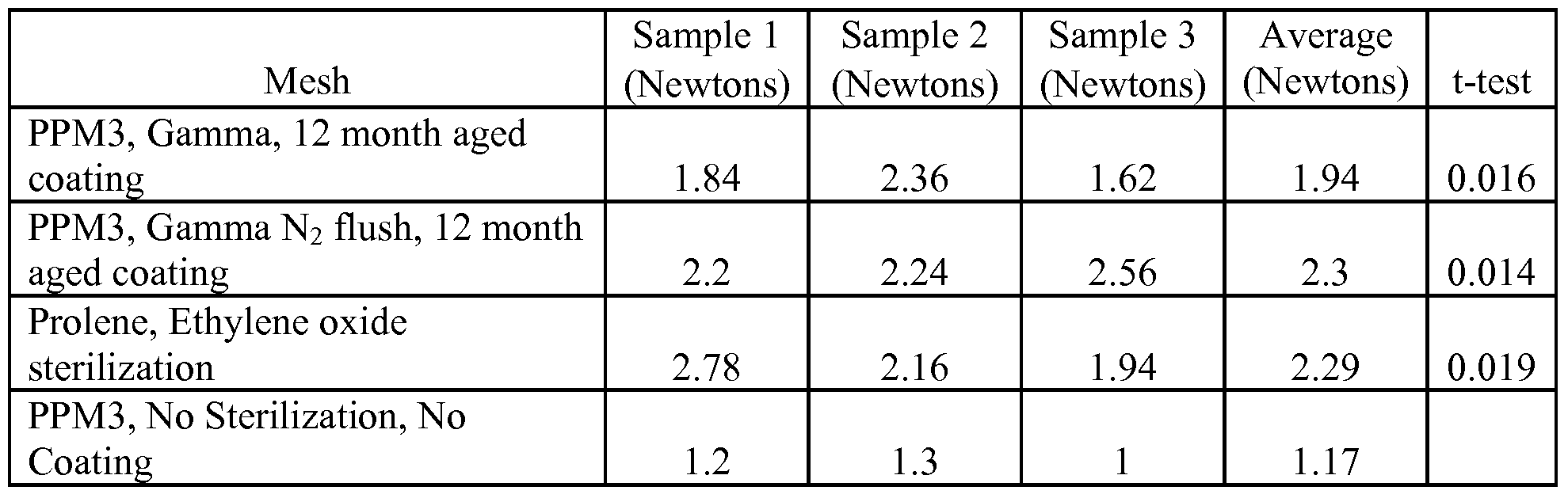
- Fig. 5 graphically depicts mesh stiffness.
- the bars represent the stiffness for (1) a PPM3 mesh without a polyarylate coating and without sterilization, (2) a ProleneTM (Ethicon) mesh sterilized with ethylene oxide, (3) a polyarylate-coated PPM3 mesh 12 months after coating and sterilized by gamma irradiation with a nitrogen flush, and (4) a polyarylate-coated PPM3 mesh 12 months after coating and sterilized by gamma irradiation.
- ProleneTM Ethicon
- Fig. 6 graphically depicts the change in mesh stiffness over time during the course of polymer degradation for a polymer-coated polypropylene mesh soaking in PBS.
- Fig. 7 depicts micrographs of a tyrosine polyarylate-coated mesh. The top left panel shows the woven nature of the mesh and the contact points of the filaments. The bottom left panel demonstrates the coating over the contact points of the mesh filaments. The right panel is a scanning electron micrograph of a coated filament.
- Fig. 8 provides an optical image of a mesh having a tyrosine polyarylate coating containing rifampin and minocycline. On the left, the optical image; on the right, a schematic thereof indicating the areas of intense orange color by the circled areas filled with diagonal lines.
- the present invention is directed to medical prostheses that comprise a mesh and one or more coating which temporarily stiffen the mesh, preferably by at least 1.1 times its original stiffness.
- the coatings on such meshes do not alter the integrity of the mesh and thus allow the mesh to remain porous.
- the coatings do not substantially alter the porosity of the mesh.
- the medical prosthesis comprises a mesh with one or more coatings with at least one of the coatings comprising a stiffening agent that coats the filaments or fibers of the mesh so to temporarily immobilize the contact points of those filaments or fibers.
- the coatings on such meshes do not alter the integrity strength of the underlying mesh, and thus allow the mesh to remain porous after coating.
- the coatings do not substantially alter the porosity of the mesh.
- the prostheses of the invention are useful for surgical repair and reconstruction of soft tissue defects.
- the mesh of the medical prostheses can be shaped into a three-dimensional structure, e.g., on a mold or other form, and a coating applied.
- the coatings (and stiffening agents) have sufficient strength to hold the mesh in that shape once the coating has dried, cured or set, as appropriate to the particular agent, and the mesh removed from the mold or form. Accordingly, the present invention provides medical prostheses that are three dimensional structures with coatings that have temporary stiffness in accordance with the invention as described herein.
- a mesh in accordance with the invention is any web or fabric with a construction of knitted, braided, woven or non- woven filaments or fibers that are interlocked in such a way to create a fabric or a fabric-like material.
- “mesh” also includes any porous prosthesis suitable for temporarily stiffening.
- Surgical meshes are well known in the art and any such mesh can be coated as described herein.
- the meshes used in the present invention are made from biocompatible materials, synthetic or natural, including but not limited to, polypropylene, polyester, polytetrafluroethylene, polyamides and combinations thereof.
- One of the advantages of the present invention is that the coatings can be used with any commercially available mesh.
- a preferred mesh is made from woven polypropylene. Pore sizes of meshes vary. For example the Bard Marlex ® mesh has pores of 379 +/- 143 micrometers or approx. 0.4 mm, whereas the Johnson and Johnson Vypro ® mesh has pores of 3058 +/- 62 micrometers or approx. 3 mm.
- the stiffening agents of the invention include hydrogels, biodegradable polymers and any other compound capable of imparting temporary stiffness to the mesh in accordance with the invention. Temporary stiffness means that, relative to the corresponding uncoated mesh material, there is an increase in stiffness when one or more coatings are applied in accordance with the invention.
- the prosthesis can be evaluated in vitro or in vivo. For example, a coating can be applied to the mesh and then the mesh left in a physiological solution for a period of time before measuring its stiffness.
- the time period of stiffness is controlled by the degradation rate (for biodegradable polymers) or absorption ability (for hydrogels). The time period can vary from days, to weeks or even a few months and is most conveniently determined in vitro.
- a hydrogel is composed of a network of water-soluble polymer chains. Hydrogels are applied as coatings and dried on the mesh.
- hydrogels Upon use, e.g., implantation in the body, the hydrogel absorbs water and become soft (hydrogels can contain over 99% water), thereby increasing the flexibility of the mesh and reverting to the original or near original stiffness of the mesh.
- hydrogels possess a degree of flexibility very similar to natural tissue, due to their significant water content.
- Common ingredients for hydrogels include e.g. polyvinyl alcohol, sodium polyacrylate, acrylate polymers and copolymers with an abundance of hydrophilic groups.
- Meshes can have one or more polymer coatings and can optionally include drugs in the coatings.
- Meshes with a single coating are useful to improve handling of the mesh during surgical implantation and use.
- Meshes with drugs can be coated with single or multiple layers, depending on the amount of drug to be delivered, the type of drug and desired release rate.
- a first coating layer can contain drug
- the second layer coating layer contains either no drug or a lower concentration of drug.
- the coated implantable surgical meshes of the invention comprise a surgical mesh and one or more biodegradable polymer coating layers with each coating layer optionally, and independently, further containing a drug.
- the physical, mechanical, chemical, and resorption characteristics of the coating enhance the clinical performance of the mesh and the surgeon's ability to implant the device without affecting the overall or primary performance characteristics of the mesh, especially when used as a permanent implant in the patient. [0035] These characteristics are accomplished by choosing a suitable coating thickness for the selected biodegradable polymer.
- the biodegradable coating deposited onto the surface of the mesh gives the mesh superior handling characteristics relative to uncoated meshes and facilitates surgical insertion because it imparts stiffness to the mesh and thereby improves handling thereof. Over time, however, the coating resorbs, or the stiffening agents degrades or softens, to leave a flexible mesh that provides greater patient comfort without loss of strength.
- the surgical mesh can be coated with the biodegradable polymer using standard techniques such as spray or dip coating to achieve a uniform coating having a thickness that provides at least 1.1 to 4.5 and more preferably 1.25 to 2 times the stiffness of the uncoated mesh.
- the coating is optimized to provide for a uniform, flexible, non-flaking layer that remains adhered to the mesh throughout the implantation and initial wound healing process.
- the polymer coating must maintain its integrity for at least 1 week.
- Optimal coating solutions are obtained by choosing a biodegradable polymer with a solubility between about .01 to about 30% in volatile solvents such as methylene chloride or other chlorinated solvents, THF, various alcohols, or combinations thereof.
- biodegradable polymers with a molecular weight between about 10,000 and about 200,000 Daltons. Such polymers degrade at rates that maintain sufficient mechanical and physical integrity over about 1 week at 37°C in an aqueous environment.
- a biodegradable polymer-coated implantable mesh is described in which the biodegradable polymer layer (i.e., the coating) has a chemical composition that provide relatively good polymer-drug miscibility.
- the polymer layer can contain between 1- 80% drug at room temperature as well as between 1-95%, 2-80%, 2-50%, 5-40%, 5-30%, 5- 25% and 10-20% drug or 1, 2, 3, 4, 5, 6, 7, 8, 9, or 10% drug as well as 5% increments from 10-95%, i.e., 10, 15, 20, 25, etc.
- the biodegradable polymer coating releases drug for at least 2 - 3 days. Such release is preferred, for example, when the drug is an analgesic to aide in localized pain management at the surgical site.
- Such loading and release characteristics can be also be obtains for drug polymer-combinations that do not have good miscibility by using multiple layering techniques.
- anesthetic and/or analgesic should be delivered to the injured tissue shortly after surgery or tissue injury.
- a drug or drugs for inclusion in the coatings of surgical meshes include, but are not limited to analgesics, anti-inflammatory agents, anesthetics, antimicrobial agents, antifungal agents, NSAIDS, other biologies (including proteins and nucleic acids) and the like. Antimicrobial and antifungal agents can prevent the mesh and/or the surrounding tissue from being colonized by bacteria.
- One or more drugs can be incorporated into the polymer coatings of the invention.
- a mesh of the invention has a coating comprising an anesthetic such that the anesthetic elutes from the implanted coated mesh to the surrounding tissue of the surgical site for between 1 and 10 days, which typically coincides with the period of acute surgical site pain.
- delivery of an antimicrobial drug via a mesh of the invention can create an inhibition zone against bacterial growth and colonization surrounding the implant during the healing process (e.g., usually about 30 days or less) and/or prevent undue f ⁇ brotic responses.
- biodegradable polymer coatings avoids the issue of drug solubility, impregnation or adherence in or to the underlying device since a coating having suitable chemical properties can be deposited onto the mesh, optionally in concert with one or more drugs, to provide for the release of relatively high concentrations of those drugs over extended periods of time.
- a clinically-efficacious amount of anesthetic drug can be loaded onto a mesh to assure sufficient drug elution and to provide surgical site, post-operative pain relief for the patient.
- the mesh should elute from about 30 mg to about 1000 mg of anesthetic over 1-10 days, including, e.g., about 30, 50, 100, 200, 400, 500, 750 or 1000 mg over that time period.
- the prosthesis should elute clinically effective amounts of anesthetic during the acute post-operative period when pain is most noticeable to the patient. This period, defined in several clinical studies, tends to be from 12 hours to 5 days after the operation, with pain greatest around 24 hours and subsiding over a period of several days thereafter. Prior to 12 hours, the patient is usually still under the influence of any local anesthetic injection given during the surgery itself. After the 5 -day period, most of the pain related to the surgery itself (i.e., incisional pain and manipulation of fascia, muscle, & nerves) has resolved to a significant extent.
- Bupivacaine has a known toxicity profile, duration of onset, and duration of action. Drug monographs recommend the daily dose not to exceed 400 mg. Those of skill in the art can determine the amount of anesthetic to include in a polymer coating or a hydrogel coating to achieve the desired amount and duration of pain relief.
- sustained release depot systems for postoperative pain relief reported in the literature is a PLGA microsphere-based sustained release formulation of bupivacaine. This formulation was developed and tested in humans for relief of subcutaneous pain as well as neural blocks. Human trials indicated that subcutaneous pain was relieved via injection of between 90 to 180 mg of bupivacaine which then eluted into the surrounding tissue over a 7-day period, with higher concentrations in the initial 24- hour period followed by a gradual taper of the concentration.
- Other depot sustained release technologies have successfully suppressed post-operative pain associated with inguinal hernia repair. For example, external pumps and PLGA microsphere formulations have each purportedly release drug for approximately 72 hours.
- a relatively hydrophilic biodegradable polymer and combines it with the anesthetic hydrochloride salt so that the anesthetic dissolves in the polymer at a concentration below the anesthetic's saturation limit.
- Such a formulation provides non-burst release of anesthetic.
- the polymer does not act as a control mechanism for release of the anesthetic, but rather acts as a binder to hold the non-dissolved, anesthetic particles together and alters the crystallization kinetics of the drug.
- a second coating layer which may or may not contain further anesthetic, is sprayed on top of the first layer.
- the anesthetic concentration is at a higher ratio of polymer to anesthetic, e.g., a concentration at which the anesthetic is soluble in the polymer layer.
- the top layer thus can serve to control the release of the drug in the bottom layer (aka depot layer) via the drug-polymer solubility ratio. Moreover, it is possible to alter the release rate of the drug by changing the thickness of the polymer layer and changing the polymer composition according to its water uptake. A polymer that absorbs a significant amount of water within 24 hours will release the contents of the depot layer rapidly. However, a polymer with limited water uptake or variable water uptake (changes as a function of its stage of degradation) will retard release of the water soluble anesthetic agent.
- Biodegradable polymers suitable for coatings of the invention include but are not limited to, polylactic acid, polyglycolic acid and copolymers and mixtures thereof such as poly(L-lactide) (PLLA), poly(D,L-lactide) (PLA);
- polyglycolic acid [polyglycolide (PGA)], poly(L-lactide-co-D,L-lactide) (PLLA/PLA), poly(L-lactide-co-glycolide) (PLLA/PGA), poly(D, L-lactide-co-glycolide) (PLA/PGA), poly(glycolide-co-trimethylene carbonate) (PGA/PTMC), poly(D,L-lactide-co- caprolactone) (PLA/PCL), poly(glycolide-co-caprolactone) (PGA/PCL), poly(oxa)ester; [0051] polyethylene oxide (PEO), polydioxanone (PDS), polypropylene fumarate, poly(ethyl glutamate-co-glutamic acid), poly(tert-butyloxy-carbonylmethyl glutamate), polycaprolactone (PCL), polycaprolactone co-butylacrylate, polyhydroxybuty
- biodegradable polymers have tyrosine-derived diphenol monomer units that are copolymerized with the appropriate chemical moiety to form a polyarylate, a polycarbonate, a polyiminocarbonate, a polyphosphonate or any other.
- the preferred biodegradable polymers are tyrosine-based polyarylates including those described in U.S. Patent Nos.
- DTE is the diphenol monomer desaminotyrosyl-tyrosine ethyl ester
- DTBn is the diphenol monomer desaminotyrosyl-tyrosine benzyl ester
- DT is the corresponding free acid form, namely desaminotyrosyl-tyrosine.
- BTE is the diphenol monomer 4-hydroxy benzoic acid-tyrosyl ethyl ester
- BT is the corresponding free acid form, namely 4-hydroxy benzoic acid-tyrosine.
- P22 is a polyarylate copolymer produced by condensation of DTE with succinate.
- P22-10, P22-15, P22-20, P22-xx, etc. represents copolymers produced by condensation of (1) a mixture of DTE and DT using the indicated percentage of DT (i.e., 10, 15, 20 and xx% DT, etc.) with (2) succinate.
- Additional preferred polyarylates are random copolymer of desaminotyrosyl- tyrosine (DT) and an desaminotyrosyl-tyrosyl ester (DT ester), wherein the copolymer comprises from about 0.001% DT to about 80% DT and the ester moiety can be a branched or unbranched alkyl, alkylaryl, or alkylene ether group having up to 18 carbon atoms, any group of which can, optionally have a polyalkylene oxide therein.
- another group of polyarylates are the same as the foregoing but the desaminotyrosyl moiety is replaced by a 4-hydroxybenzoyl moiety.
- Preferred DT or BT contents include those copolymers with from about 1% to about 30%, from about 5% to about 30% from about 10 to about 30% DT or BT.
- Preferred diacids include succinate, glutarate and glycolic acid.
- biodegradable polymers useful for the present invention are the biodegradable, resorbable polyarylates and polycarbonates disclosed in U.S. provisional application Serial No. 60/733,988, filed November 3, 2005 and in its corresponding PCT
- the most preferred polyarylates are the DTE-DT succinate family of polymers, e.g., the P22-xx family of polymers having from 0-50%, 5-50%, 5-40%, 1-30% or 10-30%
- DT including but not limited to, about 1, 2, 5, 10, 15, 20, 25, 27.5, 30, 35, 40%, 45% and
- the polyarylate polymers used in the present invention can have from 0.1-99.9 % PEG diacid to promote the degradation process as described in U.S. provisional application Serial No. 60/733,988.
- the prostheses of the invention can be used to reconstruct, reinforce, bridge, replace, repair, support, stabilize, position or strengthen any soft tissue defect.
- the prosetheses of the invention can also be used for structural reinforcement for muscle flaps, to provide vascular integrity, for ligament repair/replacement and for organ support/positioning/repositioning such as done with a bladder sling, a breast lift, or an organ bag/wrap.
- the prosetheses of the invention can be used in recontruction procedures involving soft tissue such as an orthopaedic graft support/stabilization, as supports for reconstructive surgical grafts and as supports for bone fractures.
- drugs suitable for use with the present invention include anesthetics, antibiotics (antimicrobials), anti-inflammatory agents, f ⁇ brosis-inhibiting agents, anti-scarring agents, leukotriene inhibitors/antagonists, cell growth inhibitors and the like.
- drug is used to include all types of therapeutic agents, whether small molecules or large molecules such as proteins, nucleic acids and the like. Those of skill in the art can readily determine the amount of a particular drug to include in the coatings on the meshes of the invention.
- any pharmaceutically acceptable form of the drugs of the present invention can be employed in the present invention, e.g., the free base or a pharmaceutically acceptable salt or ester thereof.
- Pharmaceutically acceptable salts include sulfate, lactate, acetate, stearate, hydrochloride, tartrate, maleate, citrate, phosphate and the like.
- non-steroidal anti-inflammatories include, but are not limited to, naproxen, ketoprofen, ibuprofen as well as diclofenac; celecoxib; sulindac; diflunisal; piroxicam; indomethacin; etodolac; meloxicam; r-flurbiprofen; mefenamic; nabumetone; tolmetin, and sodium salts of each of the foregoing; ketorolac bromethamine; ketorolac bromethamine tromethamine; choline magnesium trisalicylate; rofecoxib; valdecoxib; lumiracoxib; etoricoxib; aspirin; salicylic acid and its sodium salt; salicylate esters of alpha, beta, gamma-tocopherols and tocotrienols (and all their d, 1, and racemic isomers); and the methyl, ethyl
- anesthetics include, but are not limited to, licodaine, bupivacaine, and mepivacaine.
- Further examples of analgesics, anesthetics and narcotics include, but are not limited to acetaminophen, clonidine, benzodiazepine, the benzodiazepine antagonist flumazenil, lidocaine, tramadol, carbamazepine, meperidine, zaleplon, trimipramine maleate, buprenorphine, nalbuphine, pentazocain, fentanyl, propoxyphene, hydromorphone, methadone, morphine, levorphanol, and hydrocodone.
- antimicrobials include, but are not limited to, triclosan, chlorhexidine, rifampin, minocycline, vancomycin, gentamycine, cephalosporins and the like.
- the coatings contain rifampin and another antimicrobial agent.
- the coatings contains a cephalosporin and another antimicrobial agent.
- Preferred combinations include rifampin and minocycline, rifampin and gentamycin, and rifampin and minocycline.
- antimicrobials include aztreonam; cefotetan and its disodium salt; loracarbef; cefoxitin and its sodium salt; cefazolin and its sodium salt; cefaclor; ceftibuten and its sodium salt; ceftizoxime; ceftizoxime sodium salt; cefoperazone and its sodium salt; cefuroxime and its sodium salt; cefuroxime axetil; cefprozil; ceftazidime; cefotaxime and its sodium salt; cefadroxil; ceftazidime and its sodium salt; cephalexin; cefamandole nafate; cefepime and its hydrochloride, sulfate, and phosphate salt; cefdinir and its sodium salt; ceftriaxone and its sodium salt; cef ⁇ xime and its sodium salt; cefpodoxime proxetil; meropenem and its sodium salt; imipe
- antifungals examples include amphotericin B; pyrimethamine; flucytosine; caspofungin acetate; fluconazole; griseofulvin; terbinafm and its hydrochloride, sulfate, or phosphate salt; ketoconazole; micronazole; clotrimazole; econazole; ciclopirox; naftif ⁇ ne; and itraconazole.
- useful proteins include cell growth inhibitors such as epidermal growth factor.
- anti-nflammatory compound examples include, but are not limited to, anecortive acetate; tetrahydrocortisol, 4,9(1 l)-pregnadien-17. alpha. ,21-diol-3,20-dione and its -21-acetate salt; 11-epicortisol; 17. alpha.
- Another aspect of the invention is directed to a process for coating a mesh with a stiffening agent that coats the filaments or fibers of the mesh to temporarily immobilize contact points of the filaments or fibers of said mesh.
- the method is comprises (a) preparing a coating solution comprising a solvent and said stiffening agent; (b) spraying a mesh one or more times to provide an amount of said solution on said mesh to produce a coating having a thickness and placement sufficient to temporarily immobilize contact points of the filaments or fibers of said mesh that coats filaments or fibers; and (c) drying said mesh to produce said coating.
- An example of ratio of coating thickness to polymer coating is shown in the scanning electron micrograph of Fig. 7.
- the drug When used with a drug (or combination of drugs), the drug is included in the coating solution at the desired concentration.
- Spraying can be accomplished by known methods.
- the coating can be applied to the entire mesh or to that portion of the mesh necessary to stiffen it.
- One technique is to dip the mesh in the coating material; another is to push the mesh through rollers that transfer the coating on the mesh.
- Spraying the mesh with a microdroplets is also effective.
- Techniques for selectively coating only those areas necessary to stiffen the mesh include deposition the coating through a template that exposes only the desired areas of coverage for the coating, including dispensing the coating with micro needles or similar means. More preferably the coating can be applied using a photoresist-like mask that expose the desired portions, applying the coating over the photomask and the removing the photomask.
- Still another aspect of the invention relates to a method for producing a shaped or three-dimensional medical prosthesis mesh by forming a mesh into a desired shape or three- dimensional shape.
- This step can be accomplished with the aid of a mold or other form on which to affix and shape the mesh or by holding the mesh in a frame in the desired shape or structural configuration.
- a stiffening agent of the invention is applied to the mesh to coat filaments or fibers and the agent is allowed to dry, set or cure as appropriate, causing the the mesh stiffen and hold the desired shape.
- the mesh is then removed or released from the mold, form or device that had been holding the mesh to produce the three dimensional medical prosthesis which is capable of retaining its shape without any further structural support or aid.
- a 1% solution containing a ratio of 1 : 1 :8 rifampin:minocycline:polymer in 9: 1 tetrahydrofuran/methanol was spray-coated onto a surgical mesh by repeatedly passing the spray nozzle over each side of the mesh until each side was coated with at least 10 mg of antimicrobial-embedded polymer. Samples were dried for at least 72 hours in a vacuum oven before use.
- the polymers are the polyarylates P22-xx having xx being the % DT indicated in Table 1.
- Rxx or Mxx indicates the percentage by weight of rifampin (R) or minocycline (M) in the coating, i.e., RlOMlO means 10% rifampin and 10% minocycline hydrochloride with 80% of the indicated polymer.
- RlOMlO means 10% rifampin and 10% minocycline hydrochloride with 80% of the indicated polymer.
- Table 1 provides a list of these polyarylates with their % DT content, exact sample sizes, final coating weights and drug coating weights.
- Staphylococcus epidermidis or Staphylococcus aureus were inoculated into Triplicate Soy Broth (TSB) from a stock culture and incubated at 37°C until the turbidity reached McFarland # 0.5 standard (1-2 hours). Plates were prepared by streaking the bacteria onto on Mueller-Hinton II agar (MHA) three times, each time swabbing the plate from left to right to cover the entire plate and rotating the plate between swabbing to change direction of the streaks.
- TAB Triplicate Soy Broth
- MHA Mueller-Hinton II agar
- a pre-cut piece (1-2 cm 2 ) of spray-coated mesh was firmly pressed into the center of pre-warmed Mueller Hinton II agar plates and incubated at 37°C. Pieces were transferred every 24 h to fresh, pre-warmed Mueller Hinton II agar plates using sterile forceps. The distance from the sample to the outer edge of the inhibition zone was measured every 24 h and is reported on the bottom row in Table 2 and 3 for each sample. The top row for each sample represents difference between the diameter of the ZOI and the diagonal of the mesh.
- Table 2 shows the ZOI results for meshes placed on S. epidermidis lawns and Table 3 show s the ZOI results for meshes placed on S. aureus lawns.
- Fig. 1 shows the total ZOI on S. aureus for meshes with 10% each of minocycline hydrochlorideand rifampin in a DTE-DT succinate polyarylate coating having 25% or 27.5% DT.
- the catheter is a COOK SPECTRUM venous catheter impregnated with rifampin and minocycline hydrochloride.
- Table 4 shows that the duration of in vitro drug release increases with the hydrophilicity of the resorbable polymer. Solvent cast films were soaked in PBS and antibiotic release was monitored by HPLC.
- a first depot coating containing 540 mg of bupivacaine HCl as a 4% solution with 1% P22-27.5 polyarylate in a mixture of THF Methanol was spray coated onto a mesh.
- a second layer consisting of 425 mg of the same polyarylate alone was deposited on top of the first layer.
- FIG. 2 shows the cumulative release of bupivacaine into PBS from the multilayer polyarylate coating as a function of time. Nearly 80% of the bupivacaine had been released after 25 hours of incubation.
- Figure 3 is an example of the changes in release characteristics that can be achieved by altering both the amount of drug in the depot layer and the thickness of the outer layer. These coated surgical meshes are much stiffer than their uncoated counterparts.
- Rats with jugular cannulas for pharmacokinetic studies were surgically implanted with a 1 x 2 cm P22-27.5 polyarylate-coated mesh containing 7.5 mg of bupivacaine/cm 2 .
- baseline pin-prick responses to nociception were measured at the planned surgical incision site, and baseline blood samples were obtained.
- a hernia was created by incision into the peritoneal cavity during via subcostal laparotomy, and a Lichtenstein non- tension repair was performed using the bupivacaine-impregnated polyarylate-coated mesh.
- Blood samples were drawn at 3, 6, 24, 48, 72, 96, and 120 hours after implantation.
- the rats Prior to drawing blood, the rats were subjected to a pin prick test to assess dermal anesthesia from bupivacaine release.
- the behavioral results indicate that moderate levels of dermal anesthesia appeared from 3 to 120 hours, with the amount at 6 and 48 hours significantly above baseline (p ⁇ 0.05).
- Pharmacokinetic analysis indicates that the plasma bupivacaine levels fit a one-compartment model with first-order absorption from 0 to 24 hours.
- a polypropylene mesh was spray coated as described in the first paragraph of
- Example 2 Individual meshes were cut to 1 x 2 cm, individually packaged, and sterilized by gamma irradiation. The mesh was loaded with 7.5 mg/cm 2 of bupivacaine HCl for a total of 15 mg of bupivacaine loaded per 1 x 2 cm mesh.
- a 2.5 cm skin incision was made 0.5 cm caudal to and parallel to the last rib.
- the underlying subcutaneous space (1 cm on both sides of the incision) was loosened to accommodate the mesh.
- a 2 cm incision was made through the muscle layers along the same plane as the skin incision, penetrating the peritoneal cavity and the peritoneum was closed with 6-0 Prolene sutures in a continuous suture pattern.
- a Lichtenstein "non-tension" repair was undertaken using the mesh as the repair material.
- the mesh prepared in Section A was positioned over the incisional hernia, and sutured into the internal and external oblique muscles using 6-0 Prolene sutures.
- the subcutaneous tissue was then sutured in a continuous pattern with 6 to 8 6-0 Prolene sutures to prevent the rats from accessing the mesh, followed by 6 to 8 skin sutures.
- Total surgical time was 10 min for anesthetic induction and preparation and 20 min for the surgery.
- the rats were allowed to recover in their home cages, and monitored post- surgically until they awoke. Blood samples were drawn for determination of plasma bupivacaine levels at 3, 6, 24, 48, 72, 96, and 120 hours after surgery. The rats were assessed for guarding the incision, and the incision was assessed for signs of inflammation, swelling or other signs of infection. No rats exhibited toxicity or seizures, or were in a moribund state from infection or the release of bupivacaine.
- the nerves caudal to the incision were transected during the procedure, and therefore did not respond to pin application and were not tested.
- the post-implantation test was repeated using the same force as before surgery and with 10 pin applications, and the percent inhibition of nocifensive responding was calculated by: [1 - (test responses/ 10 base responses)] X 100.
- the data was analyzed using repeated measures ANOVA followed by post hoc analysis using the Tukey's test. The results are shown in Fig. 4.
- An optical image of the coated mesh is shown in the top left panel of Fig. 7 at a magnification that readily shows the woven nature of the mesh and the contact points of the filaments.
- a close up of a contact point is shown in the bottom left panel of Fig. 7 and demonstrates that the coating immobilizes the contact points of the mesh filaments.
- the right panel of Fig. 7 is a scanning electron micrograph of a coated filament.
- Example 8 shows an optical image of a mesh from Example 1, i.e., coated with polymer, rifampin and minocycline.
- this photograph shows the mesh on a blue background with the filaments appearing greenish with some orange and the knots (or filament contact points) appearing mostly solid orange.
- the orange color is due to the antibiotics and is more visible on the knots due to the greater surface area of the mesh in that region.
- the color differentiation is difficult to visualize in the black and white version of this photograph so on the right panel the areas of orange are indicated by circled areas filled with diagonal lines.
- Hayes, B.B. Afshari, A., Millecchia, L., Willard, P.A., Povoski, S.P., Meade,
Abstract
Description


Claims
Priority Applications (5)
| Application Number | Priority Date | Filing Date | Title |
|---|---|---|---|
| EP07756801.2A EP2114298B1 (en) | 2006-02-08 | 2007-02-08 | Temporarily stiffened mesh prostheses |
| JP2010502992A JP5367692B2 (en) | 2006-02-08 | 2007-02-08 | Temporary stiffening mesh prosthesis |
| MX2008010126A MX2008010126A (en) | 2006-02-08 | 2007-02-08 | Temporarily stiffened mesh prostheses. |
| AU2007344645A AU2007344645B2 (en) | 2006-02-08 | 2007-02-08 | Temporarily stiffened mesh prostheses |
| CA2637578A CA2637578C (en) | 2006-02-08 | 2007-02-08 | Temporarily stiffened mesh prostheses |
Applications Claiming Priority (2)
| Application Number | Priority Date | Filing Date | Title |
|---|---|---|---|
| US77182706P | 2006-02-08 | 2006-02-08 | |
| US60/771,827 | 2006-02-08 |
Publications (2)
| Publication Number | Publication Date |
|---|---|
| WO2009113972A2 true WO2009113972A2 (en) | 2009-09-17 |
| WO2009113972A3 WO2009113972A3 (en) | 2016-04-28 |
Family
ID=41065695
Family Applications (1)
| Application Number | Title | Priority Date | Filing Date |
|---|---|---|---|
| PCT/US2007/061885 WO2009113972A2 (en) | 2006-02-08 | 2007-02-08 | Temporarily stiffened mesh prostheses |
Country Status (6)
| Country | Link |
|---|---|
| US (5) | US8636753B2 (en) |
| EP (1) | EP2114298B1 (en) |
| JP (2) | JP5367692B2 (en) |
| AU (1) | AU2007344645B2 (en) |
| MX (1) | MX2008010126A (en) |
| WO (1) | WO2009113972A2 (en) |
Cited By (22)
| Publication number | Priority date | Publication date | Assignee | Title |
|---|---|---|---|---|
| WO2012064963A1 (en) * | 2010-11-12 | 2012-05-18 | Tyrx, Inc. | Anchorage devices comprising an active pharmaceutical ingredient |
| US8315700B2 (en) | 2006-02-08 | 2012-11-20 | Tyrx, Inc. | Preventing biofilm formation on implantable medical devices |
| US8317808B2 (en) | 2008-02-18 | 2012-11-27 | Covidien Lp | Device and method for rolling and inserting a prosthetic patch into a body cavity |
| US8758373B2 (en) | 2008-02-18 | 2014-06-24 | Covidien Lp | Means and method for reversibly connecting a patch to a patch deployment device |
| US8808314B2 (en) | 2008-02-18 | 2014-08-19 | Covidien Lp | Device and method for deploying and attaching an implant to a biological tissue |
| US8888811B2 (en) | 2008-10-20 | 2014-11-18 | Covidien Lp | Device and method for attaching an implant to biological tissue |
| US8906045B2 (en) | 2009-08-17 | 2014-12-09 | Covidien Lp | Articulating patch deployment device and method of use |
| EP2614843A3 (en) * | 2011-12-14 | 2015-01-21 | Covidien LP | Methods for coating medical devices |
| US9023114B2 (en) | 2006-11-06 | 2015-05-05 | Tyrx, Inc. | Resorbable pouches for implantable medical devices |
| US9034002B2 (en) | 2008-02-18 | 2015-05-19 | Covidien Lp | Lock bar spring and clip for implant deployment device |
| US9044235B2 (en) | 2008-02-18 | 2015-06-02 | Covidien Lp | Magnetic clip for implant deployment device |
| US9301826B2 (en) | 2008-02-18 | 2016-04-05 | Covidien Lp | Lock bar spring and clip for implant deployment device |
| US9393093B2 (en) | 2008-02-18 | 2016-07-19 | Covidien Lp | Clip for implant deployment device |
| US9393002B2 (en) | 2008-02-18 | 2016-07-19 | Covidien Lp | Clip for implant deployment device |
| US9398944B2 (en) | 2008-02-18 | 2016-07-26 | Covidien Lp | Lock bar spring and clip for implant deployment device |
| AU2015264956B2 (en) * | 2010-11-12 | 2017-03-09 | Medtronic, Inc. | Anchorage devices comprising an active pharmaceutical ingredient |
| US9833240B2 (en) | 2008-02-18 | 2017-12-05 | Covidien Lp | Lock bar spring and clip for implant deployment device |
| US9999424B2 (en) | 2009-08-17 | 2018-06-19 | Covidien Lp | Means and method for reversibly connecting an implant to a deployment device |
| EP3586886A1 (en) | 2015-03-31 | 2020-01-01 | Orchid Medical Pte Ltd | Elastic antimicrobial film and socket made therefrom |
| US10765500B2 (en) | 2006-02-08 | 2020-09-08 | Medtronic, Inc. | Temporarily stiffened mesh prostheses |
| US11202754B2 (en) | 2017-10-06 | 2021-12-21 | Foundry Therapeutics, Inc. | Implantable depots for the controlled release of therapeutic agents |
| US11612754B2 (en) | 2018-12-21 | 2023-03-28 | Tepha, Inc. | Resorbable nonwoven pouches for medical device implants |
Families Citing this family (98)
| Publication number | Priority date | Publication date | Assignee | Title |
|---|---|---|---|---|
| US9012506B2 (en) | 2004-09-28 | 2015-04-21 | Atrium Medical Corporation | Cross-linked fatty acid-based biomaterials |
| EP1811933B1 (en) | 2004-09-28 | 2016-03-23 | Atrium Medical Corporation | Barrier layer |
| US9000040B2 (en) | 2004-09-28 | 2015-04-07 | Atrium Medical Corporation | Cross-linked fatty acid-based biomaterials |
| US8709023B2 (en) | 2007-07-17 | 2014-04-29 | Poly-Med, Inc. | Absorbable / biodegradable composite yarn constructs and applications thereof |
| US9427423B2 (en) * | 2009-03-10 | 2016-08-30 | Atrium Medical Corporation | Fatty-acid based particles |
| US9278161B2 (en) | 2005-09-28 | 2016-03-08 | Atrium Medical Corporation | Tissue-separating fatty acid adhesion barrier |
| CA2625264C (en) | 2005-10-13 | 2015-12-15 | Synthes (U.S.A.) | Drug-impregnated sleeve for a medical implant |
| US20070141106A1 (en) * | 2005-10-19 | 2007-06-21 | Bonutti Peter M | Drug eluting implant |
| US8591531B2 (en) | 2006-02-08 | 2013-11-26 | Tyrx, Inc. | Mesh pouches for implantable medical devices |
| US7846361B2 (en) | 2006-07-20 | 2010-12-07 | Orbusneich Medical, Inc. | Bioabsorbable polymeric composition for a medical device |
| US7959942B2 (en) | 2006-10-20 | 2011-06-14 | Orbusneich Medical, Inc. | Bioabsorbable medical device with coating |
| EP2073754A4 (en) | 2006-10-20 | 2012-09-26 | Orbusneich Medical Inc | Bioabsorbable polymeric composition and medical device background |
| EP2079387B1 (en) | 2006-11-06 | 2017-04-12 | Tyrx, Inc. | Mesh pouches for implantable medical devices |
| WO2008121816A2 (en) * | 2007-03-29 | 2008-10-09 | Tyrx Pharma, Inc. | Biodegradable, polymer coverings for breast implants |
| MX2009011785A (en) * | 2007-05-02 | 2010-03-04 | Tyrx Pharma Inc | Dihydroxybenzoate polymers and uses thereof. |
| US20090036907A1 (en) * | 2007-07-30 | 2009-02-05 | Yves Bayon | Bioresorbable knit |
| US20090187197A1 (en) * | 2007-08-03 | 2009-07-23 | Roeber Peter J | Knit PTFE Articles and Mesh |
| US20090036996A1 (en) * | 2007-08-03 | 2009-02-05 | Roeber Peter J | Knit PTFE Articles and Mesh |
| US20090192528A1 (en) * | 2008-01-29 | 2009-07-30 | Biomet Biologics, Inc. | Method and device for hernia repair |
| EP2247245B1 (en) | 2008-02-18 | 2017-06-28 | Covidien LP | A device for deploying and attaching a patch to a biological tissue |
| CN105688276A (en) * | 2008-02-29 | 2016-06-22 | 史密夫和内修有限公司 | Gradient coating for biomedical applications |
| US9439801B2 (en) | 2012-06-29 | 2016-09-13 | Revent Medical, Inc. | Systems and methods for treatment of sleep apnea |
| US9242026B2 (en) | 2008-06-27 | 2016-01-26 | Sofradim Production | Biosynthetic implant for soft tissue repair |
| EP2346537B1 (en) | 2008-09-22 | 2016-11-09 | Tyrx, Inc. | Linear polyesteramides from aminophenolic esters |
| US9078712B2 (en) | 2009-04-15 | 2015-07-14 | Warsaw Orthopedic, Inc. | Preformed drug-eluting device to be affixed to an anterior spinal plate |
| CA2841499C (en) * | 2009-06-01 | 2016-07-26 | Raman Bahulekar | Compositions and methods for preventing sternal wound infections |
| JP5847706B2 (en) * | 2009-06-01 | 2016-01-27 | タイレックス・インコーポレイテッドTyrx Inc. | Compositions and methods for preventing sternum wound infections |
| US9839628B2 (en) * | 2009-06-01 | 2017-12-12 | Tyrx, Inc. | Compositions and methods for preventing sternal wound infections |
| US20110038910A1 (en) | 2009-08-11 | 2011-02-17 | Atrium Medical Corporation | Anti-infective antimicrobial-containing biomaterials |
| US9114197B1 (en) | 2014-06-11 | 2015-08-25 | Silver Bullett Therapeutics, Inc. | Coatings for the controllable release of antimicrobial metal ions |
| US8221396B2 (en) | 2009-08-27 | 2012-07-17 | Silver Bullet Therapeutics, Inc. | Bone implants for the treatment of infection |
| US8927004B1 (en) | 2014-06-11 | 2015-01-06 | Silver Bullet Therapeutics, Inc. | Bioabsorbable substrates and systems that controllably release antimicrobial metal ions |
| US9821094B2 (en) | 2014-06-11 | 2017-11-21 | Silver Bullet Therapeutics, Inc. | Coatings for the controllable release of antimicrobial metal ions |
| US10265435B2 (en) | 2009-08-27 | 2019-04-23 | Silver Bullet Therapeutics, Inc. | Bone implant and systems and coatings for the controllable release of antimicrobial metal ions |
| EP2547296A4 (en) | 2010-03-19 | 2014-08-06 | Revent Medical Inc | Systems and methods for treatment of sleep apnea |
| CA2799435A1 (en) | 2010-05-21 | 2011-11-24 | Revent Medical, Inc. | Systems and methods for treatment of sleep apnea |
| US9211175B2 (en) | 2010-07-08 | 2015-12-15 | Covidien Lp | Self-detachable medical devices |
| EP2593141B1 (en) | 2010-07-16 | 2018-07-04 | Atrium Medical Corporation | Composition and methods for altering the rate of hydrolysis of cured oil-based materials |
| FR2962646B1 (en) | 2010-07-16 | 2012-06-22 | Sofradim Production | PROSTHETIC WITH RADIO OPAQUE ELEMENT |
| US10028813B2 (en) * | 2010-07-22 | 2018-07-24 | Boston Scientific Scimed, Inc. | Coated pelvic implant device and method |
| US20120022321A1 (en) * | 2010-07-22 | 2012-01-26 | Ams Research Corporation | Coated Pelvic Implant Device and Method |
| EP2598091A4 (en) | 2010-07-26 | 2014-08-20 | Revent Medical Inc | Systems and methods for treatment of sleep apnea |
| EP3489313A1 (en) | 2010-08-25 | 2019-05-29 | Tyrx, Inc. | Novel medical device coatings |
| US9572907B2 (en) | 2010-10-01 | 2017-02-21 | Covidien Lp | Implantable polymeric films |
| US8920867B2 (en) | 2010-10-19 | 2014-12-30 | Covidien Lp | Methods of forming self-supporting films for delivery of therapeutic agents |
| US9861590B2 (en) | 2010-10-19 | 2018-01-09 | Covidien Lp | Self-supporting films for delivery of therapeutic agents |
| US8632839B2 (en) * | 2010-10-19 | 2014-01-21 | Covidien Lp | Methods of forming self-supporting films for delivery of therapeutic agents |
| EP2637608B1 (en) | 2010-11-12 | 2016-03-02 | Silver Bullet Therapeutics Inc. | Bone implant and systems that controllably releases silver |
| US9308069B2 (en) * | 2010-12-13 | 2016-04-12 | Richard Massen | Hernia mesh apparatus and method |
| US9144634B2 (en) | 2011-01-14 | 2015-09-29 | Covidien Lp | Medical device with intrapore films |
| CN103732234A (en) * | 2011-01-18 | 2014-04-16 | 斯丹姆涅恩有限公司 | Wound healing device and method |
| CA2829201C (en) * | 2011-03-09 | 2019-03-12 | Tepha, Inc. | Systems and methods for mastopexy |
| FR2972626B1 (en) | 2011-03-16 | 2014-04-11 | Sofradim Production | PROSTHETIC COMPRISING A THREE-DIMENSIONAL KNIT AND ADJUSTED |
| EP2688605A4 (en) | 2011-03-24 | 2014-09-10 | Bard Inc C R | Fixation and protection of an implanted medical device |
| JP6302835B2 (en) | 2011-06-21 | 2018-03-28 | ビーブイダブリュ ホールディング エージー | Medical devices containing boswellic acid |
| FR2977789B1 (en) | 2011-07-13 | 2013-07-19 | Sofradim Production | PROSTHETIC FOR UMBILIC HERNIA |
| FR2977790B1 (en) | 2011-07-13 | 2013-07-19 | Sofradim Production | PROSTHETIC FOR UMBILIC HERNIA |
| US9381281B2 (en) | 2011-07-20 | 2016-07-05 | Tyrx, Inc. | Drug eluting mesh to prevent infection of indwelling transdermal devices |
| US8579924B2 (en) | 2011-07-26 | 2013-11-12 | Covidien Lp | Implantable devices including a mesh and a pivotable film |
| US9782957B2 (en) | 2011-08-24 | 2017-10-10 | Covidien Lp | Medical device films |
| US9005308B2 (en) | 2011-10-25 | 2015-04-14 | Covidien Lp | Implantable film/mesh composite for passage of tissue therebetween |
| US8932621B2 (en) | 2011-10-25 | 2015-01-13 | Covidien Lp | Implantable film/mesh composite |
| US9179994B2 (en) | 2011-10-25 | 2015-11-10 | Covidien Lp | Implantable film/mesh composite |
| TWI590843B (en) | 2011-12-28 | 2017-07-11 | 信迪思有限公司 | Films and methods of manufacture |
| FR2985170B1 (en) | 2011-12-29 | 2014-01-24 | Sofradim Production | PROSTHESIS FOR INGUINAL HERNIA |
| US10206769B2 (en) | 2012-03-30 | 2019-02-19 | Covidien Lp | Implantable devices including a film providing folding characteristics |
| FR2992662B1 (en) | 2012-06-28 | 2014-08-08 | Sofradim Production | KNIT WITH PICOTS |
| FR2992547B1 (en) | 2012-06-29 | 2015-04-24 | Sofradim Production | PROSTHETIC FOR HERNIA |
| FR2994185B1 (en) | 2012-08-02 | 2015-07-31 | Sofradim Production | PROCESS FOR THE PREPARATION OF A POROUS CHITOSAN LAYER |
| FR2995788B1 (en) | 2012-09-25 | 2014-09-26 | Sofradim Production | HEMOSTATIC PATCH AND PREPARATION METHOD |
| FR2995779B1 (en) | 2012-09-25 | 2015-09-25 | Sofradim Production | PROSTHETIC COMPRISING A TREILLIS AND A MEANS OF CONSOLIDATION |
| US9066853B2 (en) * | 2013-01-15 | 2015-06-30 | Warsaw Orthopedic, Inc. | Clonidine compounds in a biodegradable fiber |
| US9585695B2 (en) | 2013-03-15 | 2017-03-07 | Woven Orthopedic Technologies, Llc | Surgical screw hole liner devices and related methods |
| CN105555328B (en) | 2013-06-21 | 2019-01-11 | 德普伊新特斯产品公司 | film and manufacturing method |
| RU2699811C1 (en) | 2014-03-07 | 2019-09-11 | Айконлаб Инк. | Multipurpose implant with specified surface structure for soft tissue reconstruction |
| US10588732B2 (en) | 2014-03-07 | 2020-03-17 | IconLab USA, Inc. | Multipurpose implant with modeled surface structure for soft tissue reconstruction |
| US9452242B2 (en) | 2014-06-11 | 2016-09-27 | Silver Bullet Therapeutics, Inc. | Enhancement of antimicrobial silver, silver coatings, or silver platings |
| CN104147642B (en) * | 2014-07-29 | 2016-07-20 | 复旦大学附属华山医院 | The preparation method of infection artificial ligament |
| US9907593B2 (en) | 2014-08-05 | 2018-03-06 | Woven Orthopedic Technologies, Llc | Woven retention devices, systems and methods |
| US8956394B1 (en) | 2014-08-05 | 2015-02-17 | Woven Orthopedic Technologies, Llc | Woven retention devices, systems and methods |
| US20160074071A1 (en) | 2014-09-16 | 2016-03-17 | Woven Orthopedic Technologies, Llc | Methods of using woven retention devices and systems |
| USD740427S1 (en) | 2014-10-17 | 2015-10-06 | Woven Orthopedic Technologies, Llc | Orthopedic woven retention device |
| EP3059255B1 (en) | 2015-02-17 | 2020-05-13 | Sofradim Production | Method for preparing a chitosan-based matrix comprising a fiber reinforcement member |
| EP3085337B1 (en) | 2015-04-24 | 2022-09-14 | Sofradim Production | Prosthesis for supporting a breast structure |
| ES2676072T3 (en) | 2015-06-19 | 2018-07-16 | Sofradim Production | Synthetic prosthesis comprising a knitted fabric and a non-porous film and method of forming it |
| US10555758B2 (en) | 2015-08-05 | 2020-02-11 | Woven Orthopedic Technologies, Llc | Tapping devices, systems and methods for use in bone tissue |
| EP3386448B1 (en) * | 2015-12-11 | 2020-09-02 | Poly-Med Inc. | Synthetic implant device replicating natural tissue structure and methods of making same |
| EP3195830B1 (en) | 2016-01-25 | 2020-11-18 | Sofradim Production | Prosthesis for hernia repair |
| WO2017216609A1 (en) * | 2016-06-15 | 2017-12-21 | Tubitak | Multifunctional hernia patch |
| US10813763B2 (en) | 2016-07-26 | 2020-10-27 | Warsaw Orthopedic, Inc. | Implantable mesh |
| US10967102B2 (en) | 2016-09-28 | 2021-04-06 | Boston Scientific Limited | Cell encapsulation membranes, devices and methods |
| EP3312325B1 (en) | 2016-10-21 | 2021-09-22 | Sofradim Production | Method for forming a mesh having a barbed suture attached thereto and the mesh thus obtained |
| WO2018107114A1 (en) | 2016-12-09 | 2018-06-14 | Woven Orthopedic Technologies, LLC. | Retention devices, lattices and related systems and methods |
| US11439797B2 (en) | 2017-02-10 | 2022-09-13 | Medtronic Advanced Energy Llc. | Surgical drain system and container |
| EP3398554A1 (en) | 2017-05-02 | 2018-11-07 | Sofradim Production | Prosthesis for inguinal hernia repair |
| ES2930317T3 (en) * | 2017-10-11 | 2022-12-09 | Link Waldemar Gmbh Co | Implantable drug eluting device comprising a microporous structure |
| EP3697458A1 (en) * | 2017-10-18 | 2020-08-26 | BVW Holding AG | Device with microstructure mediated absorption profile |
| CN110354306B (en) * | 2019-08-12 | 2021-10-01 | 天津百和至远医疗技术有限公司 | Fiber scaffold and preparation method thereof |
Citations (19)
| Publication number | Priority date | Publication date | Assignee | Title |
|---|---|---|---|---|
| US4980449A (en) | 1988-07-14 | 1990-12-25 | Rutgers, The State University | Polyiminocarbonate synthesis |
| US5099060A (en) | 1990-06-12 | 1992-03-24 | Rutgers, The State University Of New Jersey | Synthesis of amino acid-derived bioerodible polymers |
| US5216115A (en) | 1990-06-12 | 1993-06-01 | Rutgers, The State University Of New Jersey | Polyarylate containing derivatives of the natural amino acid L-tyrosine |
| US5292328A (en) | 1991-10-18 | 1994-03-08 | United States Surgical Corporation | Polypropylene multifilament warp knitted mesh and its use in surgery |
| US5587507A (en) | 1995-03-31 | 1996-12-24 | Rutgers, The State University | Synthesis of tyrosine derived diphenol monomers |
| US5658995A (en) | 1995-11-27 | 1997-08-19 | Rutgers, The State University | Copolymers of tyrosine-based polycarbonate and poly(alkylene oxide) |
| WO1999052962A1 (en) | 1998-04-13 | 1999-10-21 | Rutgers, The State University | The construction of copolymer libraries |
| US6048521A (en) | 1997-11-07 | 2000-04-11 | Rutgers, The State University | Copolymers of tyrosine-based polyarlates and poly(alkylene oxides) |
| US6120491A (en) | 1997-11-07 | 2000-09-19 | The State University Rutgers | Biodegradable, anionic polymers derived from the amino acid L-tyrosine |
| USRE37160E1 (en) | 1990-06-12 | 2001-05-01 | Rutgers, The State University | Synthesis of tyrosine derived diphenol monomers |
| WO2001049311A1 (en) | 1999-12-31 | 2001-07-12 | Rutgers, The State University | Pharmaceutical formulation composed of a polymer blend and an active compound for time-controlled release |
| WO2001049249A2 (en) | 1999-12-31 | 2001-07-12 | Rutgers, The State University | Pharmaceutical formulation for regulating the timed release of biologically active compounds based on a polymer matrix |
| US6319492B1 (en) | 1996-11-27 | 2001-11-20 | Rutgers, The State University | Copolymers of tyrosine-based polyarylates and poly(alkylene oxides) |
| US6475477B1 (en) | 1997-11-07 | 2002-11-05 | Rutgers, The State University | Radio-opaque polymer biomaterials |
| US20030138488A1 (en) | 1999-12-31 | 2003-07-24 | Kohn Joachim B | Pharmaceutical formulation for regulating the timed release of biologically active compounds based on a polymer matrix |
| US6602497B1 (en) | 1997-11-07 | 2003-08-05 | Rutgers, The State University | Strictly alternating poly(alkylene oxide ether) copolymers |
| WO2003091337A1 (en) | 2002-04-24 | 2003-11-06 | Rutgers, The State University | New polyarylates for drug delivery and tissue engineering |
| US20040138762A1 (en) | 2002-11-04 | 2004-07-15 | Sofradim Production | Prosthesis for reinforcement of tissue structures |
| US20040172048A1 (en) | 2001-03-30 | 2004-09-02 | James Browning | Surgical implant |
Family Cites Families (130)
| Publication number | Priority date | Publication date | Assignee | Title |
|---|---|---|---|---|
| US4298997A (en) | 1979-10-23 | 1981-11-10 | Rybka F James | Device for inhibiting the formation of fibrous capsular contractures in silicone breast implants and method |
| US4326532A (en) | 1980-10-06 | 1982-04-27 | Minnesota Mining And Manufacturing Company | Antithrombogenic articles |
| EP0065884B1 (en) | 1981-05-27 | 1986-08-20 | Unitika Ltd. | Urethral catheter capable of preventing urinary tract infection and process for producing the same |
| DE3329733A1 (en) | 1983-08-17 | 1985-03-07 | Johannes Dr.med. 6500 Mainz Reinmüller | PLASTIC IMPLANT |
| US4693887A (en) * | 1983-09-15 | 1987-09-15 | The Kendall Company | Microphase separated hydrogels for controlled release of bioactive materials |
| US4769038A (en) | 1986-03-18 | 1988-09-06 | C. R. Bard, Inc. | Prostheses and techniques and repair of inguinal and femoral hernias |
| US4846844A (en) | 1987-08-31 | 1989-07-11 | Eli Lilly And Company | Antimicrobial coated implants |
| US5279594A (en) | 1990-05-23 | 1994-01-18 | Jackson Richard R | Intubation devices with local anesthetic effect for medical use |
| US5417671A (en) | 1990-05-23 | 1995-05-23 | Jackson; Richard R. | Medical devices having local anesthetic effect and methods of their manufacture |
| US5295978A (en) | 1990-12-28 | 1994-03-22 | Union Carbide Chemicals & Plastics Technology Corporation | Biocompatible hydrophilic complexes and process for preparation and use |
| WO1992015286A1 (en) | 1991-02-27 | 1992-09-17 | Nova Pharmaceutical Corporation | Anti-infective and anti-inflammatory releasing systems for medical devices |
| US6013853A (en) | 1992-02-14 | 2000-01-11 | The University Of Texas System | Continuous release polymeric implant carrier |
| US5217493A (en) * | 1992-03-11 | 1993-06-08 | Board Of Regents, The University Of Texas System | Antibacterial coated medical implants |
| US5766246A (en) | 1992-05-20 | 1998-06-16 | C. R. Bard, Inc. | Implantable prosthesis and method and apparatus for loading and delivering an implantable prothesis |
| US5362754A (en) | 1992-11-12 | 1994-11-08 | Univ. Of Tx Md Anderson Cancer Center | M-EDTA pharmaceutical preparations and uses thereof |
| US5356432B1 (en) * | 1993-02-05 | 1997-02-04 | Bard Inc C R | Implantable mesh prosthesis and method for repairing muscle or tissue wall defects |
| US5433996A (en) * | 1993-02-18 | 1995-07-18 | W. L. Gore & Associates, Inc. | Laminated patch tissue repair sheet material |
| DK44193D0 (en) * | 1993-04-20 | 1993-04-20 | Euromed I S | SPECIAL CONNECTION AND ADMINISTRATIVE TO A SPECIAL CONNECTION OR SIMILAR |
| AU686206B2 (en) | 1993-07-12 | 1998-02-05 | Regents Of The University Of California, The | Soft tissue augmentation apparatus |
| US5554147A (en) | 1994-02-01 | 1996-09-10 | Caphco, Inc. | Compositions and devices for controlled release of active ingredients |
| US5564929A (en) * | 1994-08-17 | 1996-10-15 | Alpert; Edward L. | Flexible root canal prosthesis |
| US6099562A (en) | 1996-06-13 | 2000-08-08 | Schneider (Usa) Inc. | Drug coating with topcoat |
| US5624704A (en) | 1995-04-24 | 1997-04-29 | Baylor College Of Medicine | Antimicrobial impregnated catheters and other medical implants and method for impregnating catheters and other medical implants with an antimicrobial agent |
| US5609629A (en) | 1995-06-07 | 1997-03-11 | Med Institute, Inc. | Coated implantable medical device |
| DE19521642C2 (en) | 1995-06-14 | 2000-11-09 | Aesculap Ag & Co Kg | Implant, its use in surgery and process for its manufacture |
| US5877224A (en) | 1995-07-28 | 1999-03-02 | Rutgers, The State University Of New Jersey | Polymeric drug formulations |
| US5755758A (en) | 1995-11-07 | 1998-05-26 | Medtronic, Inc. | Intramuscular stimulation lead with enhanced infection resistance |
| US6558686B1 (en) | 1995-11-08 | 2003-05-06 | Baylor College Of Medicine | Method of coating medical devices with a combination of antiseptics and antiseptic coating therefor |
| US5756145A (en) | 1995-11-08 | 1998-05-26 | Baylor College Of Medicine | Durable, Resilient and effective antimicrobial coating for medical devices and method of coating therefor |
| US6461644B1 (en) | 1996-03-25 | 2002-10-08 | Richard R. Jackson | Anesthetizing plastics, drug delivery plastics, and related medical products, systems and methods |
| US6143037A (en) | 1996-06-12 | 2000-11-07 | The Regents Of The University Of Michigan | Compositions and methods for coating medical devices |
| US5676146B1 (en) * | 1996-09-13 | 2000-04-18 | Osteotech Inc | Surgical implant containing a resorbable radiopaque marker and method of locating such within a body |
| ES2238736T3 (en) | 1996-12-03 | 2005-09-01 | Osteobiologics, Inc. | BIODEGRADABLE POLYMER FILM. |
| US5997517A (en) | 1997-01-27 | 1999-12-07 | Sts Biopolymers, Inc. | Bonding layers for medical device surface coatings |
| US6511748B1 (en) | 1998-01-06 | 2003-01-28 | Aderans Research Institute, Inc. | Bioabsorbable fibers and reinforced composites produced therefrom |
| US6319264B1 (en) | 1998-04-03 | 2001-11-20 | Bionx Implants Oy | Hernia mesh |
| US6669735B1 (en) | 1998-07-31 | 2003-12-30 | Davol, Inc. | Prosthesis for surgical treatment of hernia |
| US6335029B1 (en) | 1998-08-28 | 2002-01-01 | Scimed Life Systems, Inc. | Polymeric coatings for controlled delivery of active agents |
| US20050147690A1 (en) | 1998-09-25 | 2005-07-07 | Masters David B. | Biocompatible protein particles, particle devices and methods thereof |
| US6475434B1 (en) | 1998-12-07 | 2002-11-05 | Baylor College Of Medicine | Composition and methods for preventing and removing biofilm embedded microorganisms from the surface of medical devices |
| EP2305324B1 (en) * | 1999-03-25 | 2014-09-17 | Metabolix, Inc. | Medical devices and applications of polyhydroxyalkanoate polymers |
| US6304786B1 (en) | 1999-03-29 | 2001-10-16 | Cardiac Pacemakers, Inc. | Implantable lead with dissolvable coating for improved fixation and extraction |
| US6103255A (en) | 1999-04-16 | 2000-08-15 | Rutgers, The State University | Porous polymer scaffolds for tissue engineering |
| FI19991852A (en) | 1999-09-01 | 2001-03-01 | Yli Urpo Antti | New multilayer material with a biologically active agent, and its preparation |
| US7807211B2 (en) | 1999-09-03 | 2010-10-05 | Advanced Cardiovascular Systems, Inc. | Thermal treatment of an implantable medical device |
| US7682647B2 (en) | 1999-09-03 | 2010-03-23 | Advanced Cardiovascular Systems, Inc. | Thermal treatment of a drug eluting implantable medical device |
| DE10004832A1 (en) * | 2000-01-31 | 2001-08-16 | Ethicon Gmbh | Flat implant with X-ray visible elements |
| JP2001357534A (en) * | 2000-04-10 | 2001-12-26 | Victor Co Of Japan Ltd | Information recording medium |
| US6719987B2 (en) | 2000-04-17 | 2004-04-13 | Nucryst Pharmaceuticals Corp. | Antimicrobial bioabsorbable materials |
| US7510566B2 (en) * | 2000-05-19 | 2009-03-31 | Coapt Systems, Inc. | Multi-point tissue tension distribution device and method, a chin lift variation |
| US6719991B2 (en) | 2000-06-09 | 2004-04-13 | Baylor College Of Medicine | Combination of antimicrobial agents and bacterial interference to coat medical devices |
| US6451373B1 (en) | 2000-08-04 | 2002-09-17 | Advanced Cardiovascular Systems, Inc. | Method of forming a therapeutic coating onto a surface of an implantable prosthesis |
| US7025063B2 (en) * | 2000-09-07 | 2006-04-11 | Ams Research Corporation | Coated sling material |
| US20020035378A1 (en) | 2000-09-18 | 2002-03-21 | Cameron Health, Inc. | Subcutaneous electrode for transthoracic conduction with highly maneuverable insertion tool |
| JP2004524059A (en) * | 2000-10-13 | 2004-08-12 | ブレネン メディカル、インコーポレイテッド | Coated surgical mesh |
| US6812217B2 (en) * | 2000-12-04 | 2004-11-02 | Medtronic, Inc. | Medical device and methods of use |
| EP1247537A1 (en) | 2001-04-04 | 2002-10-09 | Isotis B.V. | Coating for medical devices |
| US6656488B2 (en) | 2001-04-11 | 2003-12-02 | Ethicon Endo-Surgery, Inc. | Bioabsorbable bag containing bioabsorbable materials of different bioabsorption rates for tissue engineering |
| JP2004535866A (en) * | 2001-06-26 | 2004-12-02 | エフエーゲー テクスティールテヒニック フォルシュングス−ウンド エントヴィックルングスゲゼルシャフト エムベーハー | Fiber implant material |
| US6589591B1 (en) | 2001-07-10 | 2003-07-08 | Baylor College Of Medicine | Method for treating medical devices using glycerol and an antimicrobial agent |
| JP4249611B2 (en) | 2001-07-23 | 2009-04-02 | アルコン,インコーポレイティド | Ophthalmic drug delivery device |
| NL1019023C2 (en) | 2001-09-24 | 2003-03-25 | Mentor Medical Systems B V | Device and method for manufacturing a silicone sheath for a breast implant. |
| US6753071B1 (en) | 2001-09-27 | 2004-06-22 | Advanced Cardiovascular Systems, Inc. | Rate-reducing membrane for release of an agent |
| US8133501B2 (en) | 2002-02-08 | 2012-03-13 | Boston Scientific Scimed, Inc. | Implantable or insertable medical devices for controlled drug delivery |
| US6887270B2 (en) | 2002-02-08 | 2005-05-03 | Boston Scientific Scimed, Inc. | Implantable or insertable medical device resistant to microbial growth and biofilm formation |
| WO2003079985A2 (en) | 2002-03-18 | 2003-10-02 | Carnegie Mellon University | Method and apparatus for preparing biomimetic scaffold |
| US6968234B2 (en) | 2002-04-25 | 2005-11-22 | Medtronic, Inc. | Implantable medical device having biologically active polymeric casing |
| US6961610B2 (en) | 2002-04-25 | 2005-11-01 | Medtronic, Inc. | Branched polyethylene oxide terminated biomedical polymers and their use in biomedical devices |
| DE10222872B4 (en) * | 2002-05-23 | 2018-08-16 | Johnson & Johnson Medical Gmbh | Medical implant and method for manufacturing a medical implant |
| MXPA04011651A (en) | 2002-05-24 | 2005-03-07 | Angiotech Pharm Inc | Compositions and methods for coating medical implants. |
| US20060246103A1 (en) | 2002-07-22 | 2006-11-02 | Ralph James D | Implantable devices for the delivery of therapeutic agents to an orthopaedic surgical site |
| US6916483B2 (en) | 2002-07-22 | 2005-07-12 | Biodynamics, Llc | Bioabsorbable plugs containing drugs |
| US20050163821A1 (en) | 2002-08-02 | 2005-07-28 | Hsing-Wen Sung | Drug-eluting Biodegradable Stent and Delivery Means |
| KR20050084599A (en) | 2002-09-26 | 2005-08-26 | 안지오테크 인터내셔날 아게 | Perivascular wraps |
| US6916868B2 (en) | 2003-01-23 | 2005-07-12 | Integra Lifesciences Corporation | Selective modification of pendent functionalities of polymers |
| CA2552137A1 (en) | 2003-01-29 | 2004-08-19 | Children's Medical Center Corporation | Prevention of surgical adhesions using selective cox-2 inhibitors |
| DE10305811A1 (en) * | 2003-02-12 | 2004-08-26 | Ethicon Gmbh | Surgical implant, preferably in net form, comprises basic structure containing anabolic steroid and corticosteroid to accelerate healing and reduce scar contraction |
| US20050015102A1 (en) | 2003-02-21 | 2005-01-20 | Chefitz Allen B. | Hernia mesh-device with incorporated local anesthetic |
| US20040186528A1 (en) | 2003-03-20 | 2004-09-23 | Medtronic, Inc. | Subcutaneous implantable medical devices with anti-microbial agents for chronic release |
| US7195615B2 (en) | 2003-05-14 | 2007-03-27 | Boston Scientific Scimed, Inc. | System for providing a medical device with anti-microbial properties |
| US7617007B2 (en) | 2003-06-04 | 2009-11-10 | Synecor Llc | Method and apparatus for retaining medical implants within body vessels |
| US20040253293A1 (en) | 2003-06-16 | 2004-12-16 | Afshin Shafiee | Rate controlled release of a pharmaceutical agent in a biodegradable device |
| WO2005007019A2 (en) * | 2003-07-07 | 2005-01-27 | Ethicon, Inc. | Implantable surgical mesh having a lubricious coating |
| US8454566B2 (en) | 2003-07-10 | 2013-06-04 | Medtronic Minimed, Inc. | Methods and compositions for the inhibition of biofilms on medical devices |
| US8870814B2 (en) | 2003-07-31 | 2014-10-28 | Boston Scientific Scimed, Inc. | Implantable or insertable medical devices containing silicone copolymer for controlled delivery of therapeutic agent |
| US7242410B2 (en) | 2003-09-06 | 2007-07-10 | Good News Enterprises Limited | Color conversion method and apparatus |
| JP2007516740A (en) | 2003-11-10 | 2007-06-28 | アンジオテック インターナショナル アーゲー | Medical implants and scarring inhibitors |
| WO2005046746A2 (en) | 2003-11-10 | 2005-05-26 | Angiotech International Ag | Medical implants and fibrosis-inducing agents |
| US20050209664A1 (en) | 2003-11-20 | 2005-09-22 | Angiotech International Ag | Electrical devices and anti-scarring agents |
| EP1685085A2 (en) * | 2003-11-20 | 2006-08-02 | Angiotech International Ag | Implantable sensors and implantable pumps and anti-scarring agents |
| US20050208095A1 (en) | 2003-11-20 | 2005-09-22 | Angiotech International Ag | Polymer compositions and methods for their use |
| US8192752B2 (en) | 2003-11-21 | 2012-06-05 | Advanced Cardiovascular Systems, Inc. | Coatings for implantable devices including biologically erodable polyesters and methods for fabricating the same |
| US20050113849A1 (en) | 2003-11-26 | 2005-05-26 | Nicholas Popadiuk | Prosthetic repair device |
| US7368169B2 (en) | 2003-12-01 | 2008-05-06 | Rutgers, The State University Of New Jersey | Hydrazide compounds with angiogenic activity |
| WO2005055972A2 (en) | 2003-12-09 | 2005-06-23 | Nanon A/S | A drug delivery device and a method of producing it |
| US8691258B2 (en) | 2003-12-12 | 2014-04-08 | Medtronic, Inc. | Anti-infective medical device |
| US7211108B2 (en) * | 2004-01-23 | 2007-05-01 | Icon Medical Corp. | Vascular grafts with amphiphilic block copolymer coatings |
| US8277524B2 (en) | 2004-03-16 | 2012-10-02 | Delphi Technologies, Inc. | Reformer start-up strategy for use in a solid oxide fuel cell control system |
| WO2005097223A1 (en) * | 2004-03-26 | 2005-10-20 | Surmodics, Inc. | Composition and method for preparing biocompatible surfaces |
| US20060062825A1 (en) | 2004-04-19 | 2006-03-23 | Maria Maccecchini | Method of implanting a sterile, active agent-coated material and composition made according to same |
| US7820732B2 (en) | 2004-04-30 | 2010-10-26 | Advanced Cardiovascular Systems, Inc. | Methods for modulating thermal and mechanical properties of coatings on implantable devices |
| US20060025852A1 (en) | 2004-08-02 | 2006-02-02 | Armstrong Joseph R | Bioabsorbable self-expanding endolumenal devices |
| US20060034769A1 (en) | 2004-08-13 | 2006-02-16 | Rutgers, The State University | Radiopaque polymeric stents |
| US20060052466A1 (en) | 2004-09-03 | 2006-03-09 | Handa Yash P | Expanded and extruded thermoplastic foams made with methyl formate-based blowing agents |
| US9011831B2 (en) | 2004-09-30 | 2015-04-21 | Advanced Cardiovascular Systems, Inc. | Methacrylate copolymers for medical devices |
| JP5682991B2 (en) | 2004-10-01 | 2015-03-11 | ラムズコア, インコーポレイテッド | Convenient implantable sustained release drug formulation |
| US20060095134A1 (en) | 2004-10-28 | 2006-05-04 | Sdgi Holdings, Inc. | Materials, devices and methods for implantation of transformable implants |
| US20060115449A1 (en) | 2004-11-30 | 2006-06-01 | Advanced Cardiovascular Systems, Inc. | Bioabsorbable, biobeneficial, tyrosine-based polymers for use in drug eluting stent coatings |
| EP1819373A2 (en) | 2004-12-07 | 2007-08-22 | SurModics, Inc. | Coatings with crystallized active agent(s) |
| US9566370B2 (en) | 2004-12-23 | 2017-02-14 | Novus Scientific Ab | Mesh implant for use in reconstruction of soft tissue defects |
| JP2008543793A (en) | 2005-06-17 | 2008-12-04 | リモン セラピューティックス エルティーディー. | Therapeutic polymer pouch |
| WO2007041677A2 (en) | 2005-10-03 | 2007-04-12 | Combinatorx, Incorporated | Soft tissue implants and drug combination compositions, and use thereof |
| ES2718681T3 (en) | 2005-11-03 | 2019-07-03 | Tyrx Inc | Resorbable Phenolic Polymers |
| US8315700B2 (en) | 2006-02-08 | 2012-11-20 | Tyrx, Inc. | Preventing biofilm formation on implantable medical devices |
| US8591531B2 (en) | 2006-02-08 | 2013-11-26 | Tyrx, Inc. | Mesh pouches for implantable medical devices |
| US8636753B2 (en) | 2006-02-08 | 2014-01-28 | Tyrx, Inc. | Temporarily stiffened mesh prostheses |
| MX2008010781A (en) | 2006-02-22 | 2009-03-02 | Pactiv Corp | Expanded and extruded polyolefin foams made with methyl formate-based blowing agents. |
| WO2007133524A2 (en) | 2006-05-08 | 2007-11-22 | Sarmas Gregory Benjamin Sr | Product packaging and methods of making same |
| WO2007146119A2 (en) | 2006-06-09 | 2007-12-21 | Medivas, Llc | Biodegradable polymer adhesion barriers |
| US9023114B2 (en) | 2006-11-06 | 2015-05-05 | Tyrx, Inc. | Resorbable pouches for implantable medical devices |
| JP5398535B2 (en) | 2006-11-06 | 2014-01-29 | タイレックス・インコーポレイテッド | Absorbable pouch for implantable medical devices |
| EP2079387B1 (en) | 2006-11-06 | 2017-04-12 | Tyrx, Inc. | Mesh pouches for implantable medical devices |
| WO2008121816A2 (en) | 2007-03-29 | 2008-10-09 | Tyrx Pharma, Inc. | Biodegradable, polymer coverings for breast implants |
| MX2009011785A (en) | 2007-05-02 | 2010-03-04 | Tyrx Pharma Inc | Dihydroxybenzoate polymers and uses thereof. |
| US8932624B2 (en) | 2007-06-20 | 2015-01-13 | The Trustees Of Columbia University In The City Of New York | Bio-film resistant surfaces |
| EP2166981B1 (en) | 2007-06-21 | 2016-09-07 | Tyrx, Inc. | Phenyl ester side chains to increase polymer resorptivity |
| WO2010006249A1 (en) | 2008-07-10 | 2010-01-14 | Tyrx Pharma, Inc. | Sustained release formulations of psychoactive drugs |
| WO2010006046A1 (en) | 2008-07-10 | 2010-01-14 | Tyrx Pharma, Inc. | Nsaid delivery from polyarylates |
| EP2346537B1 (en) | 2008-09-22 | 2016-11-09 | Tyrx, Inc. | Linear polyesteramides from aminophenolic esters |
| JP6397190B2 (en) * | 2010-11-12 | 2018-09-26 | タイレックス・インコーポレイテッドTyrx Inc. | Fixing device containing active pharmaceutical ingredients |
-
2007
- 2007-02-08 US US11/672,929 patent/US8636753B2/en active Active
- 2007-02-08 JP JP2010502992A patent/JP5367692B2/en active Active
- 2007-02-08 AU AU2007344645A patent/AU2007344645B2/en active Active
- 2007-02-08 MX MX2008010126A patent/MX2008010126A/en not_active Application Discontinuation
- 2007-02-08 EP EP07756801.2A patent/EP2114298B1/en active Active
- 2007-02-08 WO PCT/US2007/061885 patent/WO2009113972A2/en active Application Filing
- 2007-11-06 US US11/936,049 patent/US20090018559A1/en not_active Abandoned
-
2013
- 2013-07-16 JP JP2013147882A patent/JP5796911B2/en active Active
- 2013-09-23 US US14/033,979 patent/US9457129B2/en active Active
-
2016
- 2016-07-26 US US15/219,780 patent/US9987116B2/en active Active
-
2018
- 2018-05-09 US US15/975,273 patent/US10765500B2/en active Active
Patent Citations (28)
| Publication number | Priority date | Publication date | Assignee | Title |
|---|---|---|---|---|
| US4980449A (en) | 1988-07-14 | 1990-12-25 | Rutgers, The State University | Polyiminocarbonate synthesis |
| USRE37160E1 (en) | 1990-06-12 | 2001-05-01 | Rutgers, The State University | Synthesis of tyrosine derived diphenol monomers |
| US5099060A (en) | 1990-06-12 | 1992-03-24 | Rutgers, The State University Of New Jersey | Synthesis of amino acid-derived bioerodible polymers |
| US5216115A (en) | 1990-06-12 | 1993-06-01 | Rutgers, The State University Of New Jersey | Polyarylate containing derivatives of the natural amino acid L-tyrosine |
| US5317077A (en) | 1990-06-12 | 1994-05-31 | Rutgers, The State University Of New Jersey | Polyarylates containing derivatives of the natural amino acid l-tyrosine |
| USRE37795E1 (en) | 1990-06-12 | 2002-07-16 | Joachim B. Kohn | Synthesis of tyrosine-derived diphenol monomers |
| US5292328A (en) | 1991-10-18 | 1994-03-08 | United States Surgical Corporation | Polypropylene multifilament warp knitted mesh and its use in surgery |
| US5587507A (en) | 1995-03-31 | 1996-12-24 | Rutgers, The State University | Synthesis of tyrosine derived diphenol monomers |
| US5670602A (en) | 1995-03-31 | 1997-09-23 | Rutgers, The State University | Synthesis of tyrosine-derived diphenol monomers |
| US5658995A (en) | 1995-11-27 | 1997-08-19 | Rutgers, The State University | Copolymers of tyrosine-based polycarbonate and poly(alkylene oxide) |
| US6319492B1 (en) | 1996-11-27 | 2001-11-20 | Rutgers, The State University | Copolymers of tyrosine-based polyarylates and poly(alkylene oxides) |
| US6475477B1 (en) | 1997-11-07 | 2002-11-05 | Rutgers, The State University | Radio-opaque polymer biomaterials |
| US7056493B2 (en) | 1997-11-07 | 2006-06-06 | Rutgers,The State University | Radio-opaque polymer biomaterials |
| US6852308B2 (en) | 1997-11-07 | 2005-02-08 | Rutgers, The State University | Radio-opaque polymeric biomaterials |
| US6048521A (en) | 1997-11-07 | 2000-04-11 | Rutgers, The State University | Copolymers of tyrosine-based polyarlates and poly(alkylene oxides) |
| US6120491A (en) | 1997-11-07 | 2000-09-19 | The State University Rutgers | Biodegradable, anionic polymers derived from the amino acid L-tyrosine |
| US6602497B1 (en) | 1997-11-07 | 2003-08-05 | Rutgers, The State University | Strictly alternating poly(alkylene oxide ether) copolymers |
| WO1999052962A1 (en) | 1998-04-13 | 1999-10-21 | Rutgers, The State University | The construction of copolymer libraries |
| US20020151668A1 (en) | 1998-04-13 | 2002-10-17 | Ken James | Construction of copolymer libraries |
| US20040254334A1 (en) | 1998-04-13 | 2004-12-16 | Rutgers, The State University | Preparation and uses for polyarylates |
| US20030138488A1 (en) | 1999-12-31 | 2003-07-24 | Kohn Joachim B | Pharmaceutical formulation for regulating the timed release of biologically active compounds based on a polymer matrix |
| US20030216307A1 (en) | 1999-12-31 | 2003-11-20 | Kohn Joachim B | Pharmaceutical formulation composed of a polymer blend and an active compound for time-controlled release |
| WO2001049249A2 (en) | 1999-12-31 | 2001-07-12 | Rutgers, The State University | Pharmaceutical formulation for regulating the timed release of biologically active compounds based on a polymer matrix |
| WO2001049311A1 (en) | 1999-12-31 | 2001-07-12 | Rutgers, The State University | Pharmaceutical formulation composed of a polymer blend and an active compound for time-controlled release |
| US20040172048A1 (en) | 2001-03-30 | 2004-09-02 | James Browning | Surgical implant |
| WO2003091337A1 (en) | 2002-04-24 | 2003-11-06 | Rutgers, The State University | New polyarylates for drug delivery and tissue engineering |
| US20050165203A1 (en) | 2002-04-24 | 2005-07-28 | Kohn Joachim B. | Polyarylates for drug delivery and tissue engineering |
| US20040138762A1 (en) | 2002-11-04 | 2004-07-15 | Sofradim Production | Prosthesis for reinforcement of tissue structures |
Non-Patent Citations (8)
Cited By (29)
| Publication number | Priority date | Publication date | Assignee | Title |
|---|---|---|---|---|
| US8315700B2 (en) | 2006-02-08 | 2012-11-20 | Tyrx, Inc. | Preventing biofilm formation on implantable medical devices |
| US10765500B2 (en) | 2006-02-08 | 2020-09-08 | Medtronic, Inc. | Temporarily stiffened mesh prostheses |
| US9023114B2 (en) | 2006-11-06 | 2015-05-05 | Tyrx, Inc. | Resorbable pouches for implantable medical devices |
| US9848955B2 (en) | 2006-11-06 | 2017-12-26 | Tyrx, Inc. | Resorbable pouches for implantable medical devices |
| US10159554B2 (en) | 2008-02-18 | 2018-12-25 | Covidien Lp | Clip for implant deployment device |
| US9833240B2 (en) | 2008-02-18 | 2017-12-05 | Covidien Lp | Lock bar spring and clip for implant deployment device |
| US8317808B2 (en) | 2008-02-18 | 2012-11-27 | Covidien Lp | Device and method for rolling and inserting a prosthetic patch into a body cavity |
| US10182898B2 (en) | 2008-02-18 | 2019-01-22 | Covidien Lp | Clip for implant deployment device |
| US8808314B2 (en) | 2008-02-18 | 2014-08-19 | Covidien Lp | Device and method for deploying and attaching an implant to a biological tissue |
| US9034002B2 (en) | 2008-02-18 | 2015-05-19 | Covidien Lp | Lock bar spring and clip for implant deployment device |
| US9044235B2 (en) | 2008-02-18 | 2015-06-02 | Covidien Lp | Magnetic clip for implant deployment device |
| US9301826B2 (en) | 2008-02-18 | 2016-04-05 | Covidien Lp | Lock bar spring and clip for implant deployment device |
| US9393093B2 (en) | 2008-02-18 | 2016-07-19 | Covidien Lp | Clip for implant deployment device |
| US9393002B2 (en) | 2008-02-18 | 2016-07-19 | Covidien Lp | Clip for implant deployment device |
| US9398944B2 (en) | 2008-02-18 | 2016-07-26 | Covidien Lp | Lock bar spring and clip for implant deployment device |
| US8758373B2 (en) | 2008-02-18 | 2014-06-24 | Covidien Lp | Means and method for reversibly connecting a patch to a patch deployment device |
| US8888811B2 (en) | 2008-10-20 | 2014-11-18 | Covidien Lp | Device and method for attaching an implant to biological tissue |
| US8906045B2 (en) | 2009-08-17 | 2014-12-09 | Covidien Lp | Articulating patch deployment device and method of use |
| US9999424B2 (en) | 2009-08-17 | 2018-06-19 | Covidien Lp | Means and method for reversibly connecting an implant to a deployment device |
| US9585988B2 (en) | 2010-11-12 | 2017-03-07 | Tyrx, Inc. | Anchorage devices comprising an active pharmaceutical ingredient |
| US10086115B2 (en) | 2010-11-12 | 2018-10-02 | Tyrx, Inc. | Anchorage devices comprising an active pharmaceutical ingredient |
| WO2012064963A1 (en) * | 2010-11-12 | 2012-05-18 | Tyrx, Inc. | Anchorage devices comprising an active pharmaceutical ingredient |
| AU2015264956B2 (en) * | 2010-11-12 | 2017-03-09 | Medtronic, Inc. | Anchorage devices comprising an active pharmaceutical ingredient |
| EP2614843A3 (en) * | 2011-12-14 | 2015-01-21 | Covidien LP | Methods for coating medical devices |
| EP3586886A1 (en) | 2015-03-31 | 2020-01-01 | Orchid Medical Pte Ltd | Elastic antimicrobial film and socket made therefrom |
| EP4129350A1 (en) | 2015-03-31 | 2023-02-08 | Foundry Therapeutics, Inc. | Elastic antimicrobial film and socket made therefrom |
| US11202754B2 (en) | 2017-10-06 | 2021-12-21 | Foundry Therapeutics, Inc. | Implantable depots for the controlled release of therapeutic agents |
| US11224570B2 (en) | 2017-10-06 | 2022-01-18 | Foundry Therapeutics, Inc. | Implantable depots for the controlled release of therapeutic agents |
| US11612754B2 (en) | 2018-12-21 | 2023-03-28 | Tepha, Inc. | Resorbable nonwoven pouches for medical device implants |
Also Published As
| Publication number | Publication date |
|---|---|
| JP5796911B2 (en) | 2015-10-21 |
| US20180256306A1 (en) | 2018-09-13 |
| US20070198040A1 (en) | 2007-08-23 |
| US10765500B2 (en) | 2020-09-08 |
| JP2010532179A (en) | 2010-10-07 |
| WO2009113972A3 (en) | 2016-04-28 |
| US20140046350A1 (en) | 2014-02-13 |
| US8636753B2 (en) | 2014-01-28 |
| MX2008010126A (en) | 2010-02-22 |
| EP2114298B1 (en) | 2022-10-19 |
| AU2007344645B2 (en) | 2012-12-13 |
| US20160331505A1 (en) | 2016-11-17 |
| JP2013212418A (en) | 2013-10-17 |
| JP5367692B2 (en) | 2013-12-11 |
| AU2007344645A1 (en) | 2009-09-17 |
| EP2114298A4 (en) | 2019-12-25 |
| US9457129B2 (en) | 2016-10-04 |
| EP2114298A2 (en) | 2009-11-11 |
| US20090018559A1 (en) | 2009-01-15 |
| US9987116B2 (en) | 2018-06-05 |
Similar Documents
| Publication | Publication Date | Title |
|---|---|---|
| US10765500B2 (en) | Temporarily stiffened mesh prostheses | |
| US8591531B2 (en) | Mesh pouches for implantable medical devices | |
| AU2007351374B2 (en) | Mesh pouches for implantable medical devices | |
| US9585988B2 (en) | Anchorage devices comprising an active pharmaceutical ingredient | |
| ES2625134T3 (en) | Mesh bags for implantable medical devices | |
| US9486560B2 (en) | Mesh pouches for implantable medical devices | |
| WO2013013123A1 (en) | Drug eluting mesh to prevent infection of indwelling transdermal devices | |
| CA2637578C (en) | Temporarily stiffened mesh prostheses | |
| AU2013200515C1 (en) | Mesh pouches for implantable medical devices | |
| AU2015264956B2 (en) | Anchorage devices comprising an active pharmaceutical ingredient |
Legal Events
| Date | Code | Title | Description |
|---|---|---|---|
| ENP | Entry into the national phase |
Ref document number: 2010502992 Country of ref document: JP Kind code of ref document: A |
|
| WWE | Wipo information: entry into national phase |
Ref document number: 2637578 Country of ref document: CA Ref document number: MX/A/2008/010126 Country of ref document: MX |
|
| NENP | Non-entry into the national phase |
Ref country code: DE |
|
| WWE | Wipo information: entry into national phase |
Ref document number: 2007344645 Country of ref document: AU |
|
| WWE | Wipo information: entry into national phase |
Ref document number: 7503/DELNP/2008 Country of ref document: IN |
|
| WWE | Wipo information: entry into national phase |
Ref document number: 2007756801 Country of ref document: EP |
|
| 121 | Ep: the epo has been informed by wipo that ep was designated in this application |
Ref document number: 07756801 Country of ref document: EP Kind code of ref document: A2 |
|
| ENP | Entry into the national phase |
Ref document number: 2007344645 Country of ref document: AU Date of ref document: 20070208 Kind code of ref document: A |