WO2007112006A2 - Delivery of highly lipophilic agents via medical devices - Google Patents
Delivery of highly lipophilic agents via medical devices Download PDFInfo
- Publication number
- WO2007112006A2 WO2007112006A2 PCT/US2007/007255 US2007007255W WO2007112006A2 WO 2007112006 A2 WO2007112006 A2 WO 2007112006A2 US 2007007255 W US2007007255 W US 2007007255W WO 2007112006 A2 WO2007112006 A2 WO 2007112006A2
- Authority
- WO
- WIPO (PCT)
- Prior art keywords
- agent
- lipophilic agent
- catheters
- lipophilic
- devices
- Prior art date
Links
- 238000000034 method Methods 0.000 claims abstract description 113
- 208000037265 diseases, disorders, signs and symptoms Diseases 0.000 claims abstract description 19
- 238000012546 transfer Methods 0.000 claims abstract description 18
- 201000010099 disease Diseases 0.000 claims abstract description 14
- 230000000149 penetrating effect Effects 0.000 claims abstract description 10
- 239000003795 chemical substances by application Substances 0.000 claims description 394
- 230000009286 beneficial effect Effects 0.000 claims description 234
- 239000003814 drug Substances 0.000 claims description 151
- 229940079593 drug Drugs 0.000 claims description 143
- 239000003112 inhibitor Substances 0.000 claims description 93
- 230000036571 hydration Effects 0.000 claims description 82
- 238000006703 hydration reaction Methods 0.000 claims description 82
- -1 antifibrins Substances 0.000 claims description 81
- CGTADGCBEXYWNE-JUKNQOCSSA-N zotarolimus Chemical group N1([C@H]2CC[C@@H](C[C@@H](C)[C@H]3OC(=O)[C@@H]4CCCCN4C(=O)C(=O)[C@@]4(O)[C@H](C)CC[C@H](O4)C[C@@H](/C(C)=C/C=C/C=C/[C@@H](C)C[C@@H](C)C(=O)[C@H](OC)[C@H](O)/C(C)=C/[C@@H](C)C(=O)C3)OC)C[C@H]2OC)C=NN=N1 CGTADGCBEXYWNE-JUKNQOCSSA-N 0.000 claims description 70
- 229950009819 zotarolimus Drugs 0.000 claims description 70
- 229920000642 polymer Polymers 0.000 claims description 67
- 238000010828 elution Methods 0.000 claims description 56
- 229960003957 dexamethasone Drugs 0.000 claims description 53
- UREBDLICKHMUKA-CXSFZGCWSA-N dexamethasone Chemical compound C1CC2=CC(=O)C=C[C@]2(C)[C@]2(F)[C@@H]1[C@@H]1C[C@@H](C)[C@@](C(=O)CO)(O)[C@@]1(C)C[C@@H]2O UREBDLICKHMUKA-CXSFZGCWSA-N 0.000 claims description 50
- 150000001875 compounds Chemical class 0.000 claims description 49
- 210000001519 tissue Anatomy 0.000 claims description 26
- 230000002792 vascular Effects 0.000 claims description 26
- 210000004369 blood Anatomy 0.000 claims description 25
- 239000008280 blood Substances 0.000 claims description 25
- 239000007943 implant Substances 0.000 claims description 20
- 238000005192 partition Methods 0.000 claims description 19
- 238000012377 drug delivery Methods 0.000 claims description 18
- MOTJMGVDPWRKOC-QPVYNBJUSA-N atrasentan Chemical compound C1([C@H]2[C@@H]([C@H](CN2CC(=O)N(CCCC)CCCC)C=2C=C3OCOC3=CC=2)C(O)=O)=CC=C(OC)C=C1 MOTJMGVDPWRKOC-QPVYNBJUSA-N 0.000 claims description 14
- VOXZDWNPVJITMN-ZBRFXRBCSA-N 17β-estradiol Chemical compound OC1=CC=C2[C@H]3CC[C@](C)([C@H](CC4)O)[C@@H]4[C@@H]3CCC2=C1 VOXZDWNPVJITMN-ZBRFXRBCSA-N 0.000 claims description 13
- 239000000126 substance Substances 0.000 claims description 13
- 210000004351 coronary vessel Anatomy 0.000 claims description 11
- 210000004165 myocardium Anatomy 0.000 claims description 11
- 230000008439 repair process Effects 0.000 claims description 11
- 238000011282 treatment Methods 0.000 claims description 11
- 239000003937 drug carrier Substances 0.000 claims description 10
- 229960005309 estradiol Drugs 0.000 claims description 10
- 238000000605 extraction Methods 0.000 claims description 10
- 239000000546 pharmaceutical excipient Substances 0.000 claims description 10
- 229940002612 prodrug Drugs 0.000 claims description 10
- 239000000651 prodrug Substances 0.000 claims description 10
- 210000004027 cell Anatomy 0.000 claims description 9
- 229930012538 Paclitaxel Natural products 0.000 claims description 8
- 230000021164 cell adhesion Effects 0.000 claims description 8
- IZEKFCXSFNUWAM-UHFFFAOYSA-N dipyridamole Chemical compound C=12N=C(N(CCO)CCO)N=C(N3CCCCC3)C2=NC(N(CCO)CCO)=NC=1N1CCCCC1 IZEKFCXSFNUWAM-UHFFFAOYSA-N 0.000 claims description 8
- 229960002768 dipyridamole Drugs 0.000 claims description 8
- 229960001592 paclitaxel Drugs 0.000 claims description 8
- 230000002265 prevention Effects 0.000 claims description 8
- RCINICONZNJXQF-MZXODVADSA-N taxol Chemical compound O([C@@H]1[C@@]2(C[C@@H](C(C)=C(C2(C)C)[C@H](C([C@]2(C)[C@@H](O)C[C@H]3OC[C@]3([C@H]21)OC(C)=O)=O)OC(=O)C)OC(=O)[C@H](O)[C@@H](NC(=O)C=1C=CC=CC=1)C=1C=CC=CC=1)O)C(=O)C1=CC=CC=C1 RCINICONZNJXQF-MZXODVADSA-N 0.000 claims description 8
- 239000002246 antineoplastic agent Substances 0.000 claims description 7
- 210000000481 breast Anatomy 0.000 claims description 7
- 229930182833 estradiol Natural products 0.000 claims description 7
- 229960000103 thrombolytic agent Drugs 0.000 claims description 7
- PMATZTZNYRCHOR-CGLBZJNRSA-N Cyclosporin A Chemical compound CC[C@@H]1NC(=O)[C@H]([C@H](O)[C@H](C)C\C=C\C)N(C)C(=O)[C@H](C(C)C)N(C)C(=O)[C@H](CC(C)C)N(C)C(=O)[C@H](CC(C)C)N(C)C(=O)[C@@H](C)NC(=O)[C@H](C)NC(=O)[C@H](CC(C)C)N(C)C(=O)[C@H](C(C)C)NC(=O)[C@H](CC(C)C)N(C)C(=O)CN(C)C1=O PMATZTZNYRCHOR-CGLBZJNRSA-N 0.000 claims description 6
- 108010036949 Cyclosporine Proteins 0.000 claims description 6
- 208000002193 Pain Diseases 0.000 claims description 6
- RJKFOVLPORLFTN-LEKSSAKUSA-N Progesterone Chemical compound C1CC2=CC(=O)CC[C@]2(C)[C@@H]2[C@@H]1[C@@H]1CC[C@H](C(=O)C)[C@@]1(C)CC2 RJKFOVLPORLFTN-LEKSSAKUSA-N 0.000 claims description 6
- MUMGGOZAMZWBJJ-DYKIIFRCSA-N Testostosterone Chemical compound O=C1CC[C@]2(C)[C@H]3CC[C@](C)([C@H](CC4)O)[C@@H]4[C@@H]3CCC2=C1 MUMGGOZAMZWBJJ-DYKIIFRCSA-N 0.000 claims description 6
- 230000000118 anti-neoplastic effect Effects 0.000 claims description 6
- 229940034982 antineoplastic agent Drugs 0.000 claims description 6
- 229960001265 ciclosporin Drugs 0.000 claims description 6
- 238000004090 dissolution Methods 0.000 claims description 6
- OVBPIULPVIDEAO-LBPRGKRZSA-N folic acid Chemical compound C=1N=C2NC(N)=NC(=O)C2=NC=1CNC1=CC=C(C(=O)N[C@@H](CCC(O)=O)C(O)=O)C=C1 OVBPIULPVIDEAO-LBPRGKRZSA-N 0.000 claims description 6
- CGIGDMFJXJATDK-UHFFFAOYSA-N indomethacin Chemical compound CC1=C(CC(O)=O)C2=CC(OC)=CC=C2N1C(=O)C1=CC=C(Cl)C=C1 CGIGDMFJXJATDK-UHFFFAOYSA-N 0.000 claims description 6
- 239000012528 membrane Substances 0.000 claims description 6
- 108020004707 nucleic acids Proteins 0.000 claims description 6
- 102000039446 nucleic acids Human genes 0.000 claims description 6
- 150000007523 nucleic acids Chemical class 0.000 claims description 6
- 230000000399 orthopedic effect Effects 0.000 claims description 6
- 230000002093 peripheral effect Effects 0.000 claims description 6
- ZQBAKBUEJOMQEX-UHFFFAOYSA-N phenyl salicylate Chemical compound OC1=CC=CC=C1C(=O)OC1=CC=CC=C1 ZQBAKBUEJOMQEX-UHFFFAOYSA-N 0.000 claims description 6
- 108091034117 Oligonucleotide Proteins 0.000 claims description 5
- 239000003242 anti bacterial agent Substances 0.000 claims description 5
- 230000003110 anti-inflammatory effect Effects 0.000 claims description 5
- 230000002927 anti-mitotic effect Effects 0.000 claims description 5
- 230000001028 anti-proliverative effect Effects 0.000 claims description 5
- 230000002785 anti-thrombosis Effects 0.000 claims description 5
- 229940088710 antibiotic agent Drugs 0.000 claims description 5
- 239000003146 anticoagulant agent Substances 0.000 claims description 5
- 229940127219 anticoagulant drug Drugs 0.000 claims description 5
- 239000003080 antimitotic agent Substances 0.000 claims description 5
- 239000003963 antioxidant agent Substances 0.000 claims description 5
- 229960004676 antithrombotic agent Drugs 0.000 claims description 5
- 229960004195 carvedilol Drugs 0.000 claims description 5
- NPAKNKYSJIDKMW-UHFFFAOYSA-N carvedilol Chemical compound COC1=CC=CC=C1OCCNCC(O)COC1=CC=CC2=NC3=CC=C[CH]C3=C12 NPAKNKYSJIDKMW-UHFFFAOYSA-N 0.000 claims description 5
- 210000002889 endothelial cell Anatomy 0.000 claims description 5
- 239000003623 enhancer Substances 0.000 claims description 5
- 229920000669 heparin Polymers 0.000 claims description 5
- 206010020718 hyperplasia Diseases 0.000 claims description 5
- 230000007246 mechanism Effects 0.000 claims description 5
- 108090000623 proteins and genes Proteins 0.000 claims description 5
- 238000011084 recovery Methods 0.000 claims description 5
- 230000002537 thrombolytic effect Effects 0.000 claims description 5
- 206010002329 Aneurysm Diseases 0.000 claims description 4
- 229930105110 Cyclosporin A Natural products 0.000 claims description 4
- OHCQJHSOBUTRHG-KGGHGJDLSA-N FORSKOLIN Chemical compound O=C([C@@]12O)C[C@](C)(C=C)O[C@]1(C)[C@@H](OC(=O)C)[C@@H](O)[C@@H]1[C@]2(C)[C@@H](O)CCC1(C)C OHCQJHSOBUTRHG-KGGHGJDLSA-N 0.000 claims description 4
- FBOZXECLQNJBKD-ZDUSSCGKSA-N L-methotrexate Chemical compound C=1N=C2N=C(N)N=C(N)C2=NC=1CN(C)C1=CC=C(C(=O)N[C@@H](CCC(O)=O)C(O)=O)C=C1 FBOZXECLQNJBKD-ZDUSSCGKSA-N 0.000 claims description 4
- JLCPHMBAVCMARE-UHFFFAOYSA-N [3-[[3-[[3-[[3-[[3-[[3-[[3-[[3-[[3-[[3-[[3-[[5-(2-amino-6-oxo-1H-purin-9-yl)-3-[[3-[[3-[[3-[[3-[[3-[[5-(2-amino-6-oxo-1H-purin-9-yl)-3-[[5-(2-amino-6-oxo-1H-purin-9-yl)-3-hydroxyoxolan-2-yl]methoxy-hydroxyphosphoryl]oxyoxolan-2-yl]methoxy-hydroxyphosphoryl]oxy-5-(5-methyl-2,4-dioxopyrimidin-1-yl)oxolan-2-yl]methoxy-hydroxyphosphoryl]oxy-5-(6-aminopurin-9-yl)oxolan-2-yl]methoxy-hydroxyphosphoryl]oxy-5-(6-aminopurin-9-yl)oxolan-2-yl]methoxy-hydroxyphosphoryl]oxy-5-(6-aminopurin-9-yl)oxolan-2-yl]methoxy-hydroxyphosphoryl]oxy-5-(6-aminopurin-9-yl)oxolan-2-yl]methoxy-hydroxyphosphoryl]oxyoxolan-2-yl]methoxy-hydroxyphosphoryl]oxy-5-(5-methyl-2,4-dioxopyrimidin-1-yl)oxolan-2-yl]methoxy-hydroxyphosphoryl]oxy-5-(4-amino-2-oxopyrimidin-1-yl)oxolan-2-yl]methoxy-hydroxyphosphoryl]oxy-5-(5-methyl-2,4-dioxopyrimidin-1-yl)oxolan-2-yl]methoxy-hydroxyphosphoryl]oxy-5-(5-methyl-2,4-dioxopyrimidin-1-yl)oxolan-2-yl]methoxy-hydroxyphosphoryl]oxy-5-(6-aminopurin-9-yl)oxolan-2-yl]methoxy-hydroxyphosphoryl]oxy-5-(6-aminopurin-9-yl)oxolan-2-yl]methoxy-hydroxyphosphoryl]oxy-5-(4-amino-2-oxopyrimidin-1-yl)oxolan-2-yl]methoxy-hydroxyphosphoryl]oxy-5-(4-amino-2-oxopyrimidin-1-yl)oxolan-2-yl]methoxy-hydroxyphosphoryl]oxy-5-(4-amino-2-oxopyrimidin-1-yl)oxolan-2-yl]methoxy-hydroxyphosphoryl]oxy-5-(6-aminopurin-9-yl)oxolan-2-yl]methoxy-hydroxyphosphoryl]oxy-5-(4-amino-2-oxopyrimidin-1-yl)oxolan-2-yl]methyl [5-(6-aminopurin-9-yl)-2-(hydroxymethyl)oxolan-3-yl] hydrogen phosphate Polymers Cc1cn(C2CC(OP(O)(=O)OCC3OC(CC3OP(O)(=O)OCC3OC(CC3O)n3cnc4c3nc(N)[nH]c4=O)n3cnc4c3nc(N)[nH]c4=O)C(COP(O)(=O)OC3CC(OC3COP(O)(=O)OC3CC(OC3COP(O)(=O)OC3CC(OC3COP(O)(=O)OC3CC(OC3COP(O)(=O)OC3CC(OC3COP(O)(=O)OC3CC(OC3COP(O)(=O)OC3CC(OC3COP(O)(=O)OC3CC(OC3COP(O)(=O)OC3CC(OC3COP(O)(=O)OC3CC(OC3COP(O)(=O)OC3CC(OC3COP(O)(=O)OC3CC(OC3COP(O)(=O)OC3CC(OC3COP(O)(=O)OC3CC(OC3COP(O)(=O)OC3CC(OC3COP(O)(=O)OC3CC(OC3COP(O)(=O)OC3CC(OC3CO)n3cnc4c(N)ncnc34)n3ccc(N)nc3=O)n3cnc4c(N)ncnc34)n3ccc(N)nc3=O)n3ccc(N)nc3=O)n3ccc(N)nc3=O)n3cnc4c(N)ncnc34)n3cnc4c(N)ncnc34)n3cc(C)c(=O)[nH]c3=O)n3cc(C)c(=O)[nH]c3=O)n3ccc(N)nc3=O)n3cc(C)c(=O)[nH]c3=O)n3cnc4c3nc(N)[nH]c4=O)n3cnc4c(N)ncnc34)n3cnc4c(N)ncnc34)n3cnc4c(N)ncnc34)n3cnc4c(N)ncnc34)O2)c(=O)[nH]c1=O JLCPHMBAVCMARE-UHFFFAOYSA-N 0.000 claims description 4
- 239000002318 adhesion promoter Substances 0.000 claims description 4
- 230000003266 anti-allergic effect Effects 0.000 claims description 4
- 229940121363 anti-inflammatory agent Drugs 0.000 claims description 4
- 239000002260 anti-inflammatory agent Substances 0.000 claims description 4
- 230000000692 anti-sense effect Effects 0.000 claims description 4
- 210000000988 bone and bone Anatomy 0.000 claims description 4
- 210000001175 cerebrospinal fluid Anatomy 0.000 claims description 4
- 230000007831 electrophysiology Effects 0.000 claims description 4
- 238000002001 electrophysiology Methods 0.000 claims description 4
- 239000003102 growth factor Substances 0.000 claims description 4
- 238000001631 haemodialysis Methods 0.000 claims description 4
- 230000000322 hemodialysis Effects 0.000 claims description 4
- WQPDUTSPKFMPDP-OUMQNGNKSA-N hirudin Chemical compound C([C@@H](C(=O)N[C@@H](CCC(O)=O)C(=O)N[C@@H](CCC(O)=O)C(=O)N[C@@H]([C@@H](C)CC)C(=O)N1[C@@H](CCC1)C(=O)N[C@@H](CCC(O)=O)C(=O)N[C@@H](CCC(O)=O)C(=O)N[C@@H](CC=1C=CC(OS(O)(=O)=O)=CC=1)C(=O)N[C@@H](CC(C)C)C(=O)N[C@@H](CCC(N)=O)C(O)=O)NC(=O)[C@H](CC(O)=O)NC(=O)CNC(=O)[C@H](CC(O)=O)NC(=O)[C@H](CC(N)=O)NC(=O)[C@H](CC=1NC=NC=1)NC(=O)[C@H](CO)NC(=O)[C@H](CCC(N)=O)NC(=O)[C@H]1N(CCC1)C(=O)[C@H](CCCCN)NC(=O)[C@H]1N(CCC1)C(=O)[C@@H](NC(=O)CNC(=O)[C@H](CCC(O)=O)NC(=O)CNC(=O)[C@@H](NC(=O)[C@@H](NC(=O)[C@H]1NC(=O)[C@H](CCC(N)=O)NC(=O)[C@H](CC(N)=O)NC(=O)[C@H](CCCCN)NC(=O)[C@H](CCC(O)=O)NC(=O)CNC(=O)[C@H](CC(O)=O)NC(=O)[C@H](CO)NC(=O)CNC(=O)[C@H](CC(C)C)NC(=O)[C@H]([C@@H](C)CC)NC(=O)[C@@H]2CSSC[C@@H](C(=O)N[C@@H](CCC(O)=O)C(=O)NCC(=O)N[C@@H](CO)C(=O)N[C@@H](CC(N)=O)C(=O)N[C@H](C(=O)N[C@H](C(NCC(=O)N[C@@H](CCC(N)=O)C(=O)NCC(=O)N[C@@H](CC(N)=O)C(=O)N[C@@H](CCCCN)C(=O)N2)=O)CSSC1)C(C)C)NC(=O)[C@H](CC(C)C)NC(=O)[C@H]1NC(=O)[C@H](CC(C)C)NC(=O)[C@H](CC(N)=O)NC(=O)[C@H](CCC(N)=O)NC(=O)CNC(=O)[C@H](CO)NC(=O)[C@H](CCC(O)=O)NC(=O)[C@H]([C@@H](C)O)NC(=O)[C@@H](NC(=O)[C@H](CC(O)=O)NC(=O)[C@@H](NC(=O)[C@H](CC=2C=CC(O)=CC=2)NC(=O)[C@@H](NC(=O)[C@@H](N)C(C)C)C(C)C)[C@@H](C)O)CSSC1)C(C)C)[C@@H](C)O)[C@@H](C)O)C1=CC=CC=C1 WQPDUTSPKFMPDP-OUMQNGNKSA-N 0.000 claims description 4
- 238000001802 infusion Methods 0.000 claims description 4
- 238000001990 intravenous administration Methods 0.000 claims description 4
- 229960000485 methotrexate Drugs 0.000 claims description 4
- 239000003961 penetration enhancing agent Substances 0.000 claims description 4
- 239000008177 pharmaceutical agent Substances 0.000 claims description 4
- 102000004169 proteins and genes Human genes 0.000 claims description 4
- NDMPLJNOPCLANR-UHFFFAOYSA-N 3,4-dihydroxy-15-(4-hydroxy-18-methoxycarbonyl-5,18-seco-ibogamin-18-yl)-16-methoxy-1-methyl-6,7-didehydro-aspidospermidine-3-carboxylic acid methyl ester Natural products C1C(CC)(O)CC(CC2(C(=O)OC)C=3C(=CC4=C(C56C(C(C(O)C7(CC)C=CCN(C67)CC5)(O)C(=O)OC)N4C)C=3)OC)CN1CCC1=C2NC2=CC=CC=C12 NDMPLJNOPCLANR-UHFFFAOYSA-N 0.000 claims description 3
- WQZGKKKJIJFFOK-GASJEMHNSA-N Glucose Natural products OC[C@H]1OC(O)[C@H](O)[C@@H](O)[C@@H]1O WQZGKKKJIJFFOK-GASJEMHNSA-N 0.000 claims description 3
- XUHXFSYUBXNTHU-UHFFFAOYSA-N Iotrolan Chemical compound IC=1C(C(=O)NC(CO)C(O)CO)=C(I)C(C(=O)NC(CO)C(O)CO)=C(I)C=1N(C)C(=O)CC(=O)N(C)C1=C(I)C(C(=O)NC(CO)C(O)CO)=C(I)C(C(=O)NC(CO)C(O)CO)=C1I XUHXFSYUBXNTHU-UHFFFAOYSA-N 0.000 claims description 3
- AMDBBAQNWSUWGN-UHFFFAOYSA-N Ioversol Chemical compound OCCN(C(=O)CO)C1=C(I)C(C(=O)NCC(O)CO)=C(I)C(C(=O)NCC(O)CO)=C1I AMDBBAQNWSUWGN-UHFFFAOYSA-N 0.000 claims description 3
- OVBPIULPVIDEAO-UHFFFAOYSA-N N-Pteroyl-L-glutaminsaeure Natural products C=1N=C2NC(N)=NC(=O)C2=NC=1CNC1=CC=C(C(=O)NC(CCC(O)=O)C(O)=O)C=C1 OVBPIULPVIDEAO-UHFFFAOYSA-N 0.000 claims description 3
- 102000002938 Thrombospondin Human genes 0.000 claims description 3
- 108060008245 Thrombospondin Proteins 0.000 claims description 3
- JXLYSJRDGCGARV-WWYNWVTFSA-N Vinblastine Natural products O=C(O[C@H]1[C@](O)(C(=O)OC)[C@@H]2N(C)c3c(cc(c(OC)c3)[C@]3(C(=O)OC)c4[nH]c5c(c4CCN4C[C@](O)(CC)C[C@H](C3)C4)cccc5)[C@@]32[C@H]2[C@@]1(CC)C=CCN2CC3)C JXLYSJRDGCGARV-WWYNWVTFSA-N 0.000 claims description 3
- 230000000340 anti-metabolite Effects 0.000 claims description 3
- 229940100197 antimetabolite Drugs 0.000 claims description 3
- 239000002256 antimetabolite Substances 0.000 claims description 3
- 230000004888 barrier function Effects 0.000 claims description 3
- 229960000304 folic acid Drugs 0.000 claims description 3
- 235000019152 folic acid Nutrition 0.000 claims description 3
- 239000011724 folic acid Substances 0.000 claims description 3
- 210000003709 heart valve Anatomy 0.000 claims description 3
- 229960002897 heparin Drugs 0.000 claims description 3
- 238000000338 in vitro Methods 0.000 claims description 3
- 229960000905 indomethacin Drugs 0.000 claims description 3
- YLPBXIKWXNRACS-UHFFFAOYSA-N iobitridol Chemical compound OCC(O)CN(C)C(=O)C1=C(I)C(NC(=O)C(CO)CO)=C(I)C(C(=O)N(C)CC(O)CO)=C1I YLPBXIKWXNRACS-UHFFFAOYSA-N 0.000 claims description 3
- 229960004108 iobitridol Drugs 0.000 claims description 3
- NBQNWMBBSKPBAY-UHFFFAOYSA-N iodixanol Chemical compound IC=1C(C(=O)NCC(O)CO)=C(I)C(C(=O)NCC(O)CO)=C(I)C=1N(C(=O)C)CC(O)CN(C(C)=O)C1=C(I)C(C(=O)NCC(O)CO)=C(I)C(C(=O)NCC(O)CO)=C1I NBQNWMBBSKPBAY-UHFFFAOYSA-N 0.000 claims description 3
- 229960004359 iodixanol Drugs 0.000 claims description 3
- NTHXOOBQLCIOLC-UHFFFAOYSA-N iohexol Chemical compound OCC(O)CN(C(=O)C)C1=C(I)C(C(=O)NCC(O)CO)=C(I)C(C(=O)NCC(O)CO)=C1I NTHXOOBQLCIOLC-UHFFFAOYSA-N 0.000 claims description 3
- 229960001025 iohexol Drugs 0.000 claims description 3
- NJKDOADNQSYQEV-UHFFFAOYSA-N iomeprol Chemical compound OCC(=O)N(C)C1=C(I)C(C(=O)NCC(O)CO)=C(I)C(C(=O)NCC(O)CO)=C1I NJKDOADNQSYQEV-UHFFFAOYSA-N 0.000 claims description 3
- 229960000780 iomeprol Drugs 0.000 claims description 3
- XQZXYNRDCRIARQ-LURJTMIESA-N iopamidol Chemical compound C[C@H](O)C(=O)NC1=C(I)C(C(=O)NC(CO)CO)=C(I)C(C(=O)NC(CO)CO)=C1I XQZXYNRDCRIARQ-LURJTMIESA-N 0.000 claims description 3
- 229960004647 iopamidol Drugs 0.000 claims description 3
- IUNJANQVIJDFTQ-UHFFFAOYSA-N iopentol Chemical compound COCC(O)CN(C(C)=O)C1=C(I)C(C(=O)NCC(O)CO)=C(I)C(C(=O)NCC(O)CO)=C1I IUNJANQVIJDFTQ-UHFFFAOYSA-N 0.000 claims description 3
- 229960000824 iopentol Drugs 0.000 claims description 3
- DGAIEPBNLOQYER-UHFFFAOYSA-N iopromide Chemical compound COCC(=O)NC1=C(I)C(C(=O)NCC(O)CO)=C(I)C(C(=O)N(C)CC(O)CO)=C1I DGAIEPBNLOQYER-UHFFFAOYSA-N 0.000 claims description 3
- 229960002603 iopromide Drugs 0.000 claims description 3
- 229960003182 iotrolan Drugs 0.000 claims description 3
- 229960004537 ioversol Drugs 0.000 claims description 3
- UUMLTINZBQPNGF-UHFFFAOYSA-N ioxilan Chemical compound OCC(O)CN(C(=O)C)C1=C(I)C(C(=O)NCCO)=C(I)C(C(=O)NCC(O)CO)=C1I UUMLTINZBQPNGF-UHFFFAOYSA-N 0.000 claims description 3
- 229960002611 ioxilan Drugs 0.000 claims description 3
- 238000007726 management method Methods 0.000 claims description 3
- 239000011159 matrix material Substances 0.000 claims description 3
- 229960000969 phenyl salicylate Drugs 0.000 claims description 3
- 229960003387 progesterone Drugs 0.000 claims description 3
- 239000000186 progesterone Substances 0.000 claims description 3
- 230000004224 protection Effects 0.000 claims description 3
- 210000000329 smooth muscle myocyte Anatomy 0.000 claims description 3
- 229960003604 testosterone Drugs 0.000 claims description 3
- 238000002560 therapeutic procedure Methods 0.000 claims description 3
- 210000003708 urethra Anatomy 0.000 claims description 3
- 208000019553 vascular disease Diseases 0.000 claims description 3
- 229960003048 vinblastine Drugs 0.000 claims description 3
- JXLYSJRDGCGARV-XQKSVPLYSA-N vincaleukoblastine Chemical compound C([C@@H](C[C@]1(C(=O)OC)C=2C(=CC3=C([C@]45[C@H]([C@@]([C@H](OC(C)=O)[C@]6(CC)C=CCN([C@H]56)CC4)(O)C(=O)OC)N3C)C=2)OC)C[C@@](C2)(O)CC)N2CCC2=C1NC1=CC=CC=C21 JXLYSJRDGCGARV-XQKSVPLYSA-N 0.000 claims description 3
- OGWKCGZFUXNPDA-UHFFFAOYSA-N vincristine Natural products C1C(CC)(O)CC(CC2(C(=O)OC)C=3C(=CC4=C(C56C(C(C(OC(C)=O)C7(CC)C=CCN(C67)CC5)(O)C(=O)OC)N4C=O)C=3)OC)CN1CCC1=C2NC2=CC=CC=C12 OGWKCGZFUXNPDA-UHFFFAOYSA-N 0.000 claims description 3
- OGWKCGZFUXNPDA-XQKSVPLYSA-N vincristine Chemical compound C([N@]1C[C@@H](C[C@]2(C(=O)OC)C=3C(=CC4=C([C@]56[C@H]([C@@]([C@H](OC(C)=O)[C@]7(CC)C=CCN([C@H]67)CC5)(O)C(=O)OC)N4C=O)C=3)OC)C[C@@](C1)(O)CC)CC1=C2NC2=CC=CC=C12 OGWKCGZFUXNPDA-XQKSVPLYSA-N 0.000 claims description 3
- 229960004528 vincristine Drugs 0.000 claims description 3
- UGGWPQSBPIFKDZ-KOTLKJBCSA-N vindesine Chemical compound C([C@@H](C[C@]1(C(=O)OC)C=2C(=CC3=C([C@]45[C@H]([C@@]([C@H](O)[C@]6(CC)C=CCN([C@H]56)CC4)(O)C(N)=O)N3C)C=2)OC)C[C@@](C2)(O)CC)N2CCC2=C1N=C1[C]2C=CC=C1 UGGWPQSBPIFKDZ-KOTLKJBCSA-N 0.000 claims description 3
- 229960004355 vindesine Drugs 0.000 claims description 3
- KWPACVJPAFGBEQ-IKGGRYGDSA-N (2s)-1-[(2r)-2-amino-3-phenylpropanoyl]-n-[(3s)-1-chloro-6-(diaminomethylideneamino)-2-oxohexan-3-yl]pyrrolidine-2-carboxamide Chemical compound C([C@@H](N)C(=O)N1[C@@H](CCC1)C(=O)N[C@@H](CCCNC(N)=N)C(=O)CCl)C1=CC=CC=C1 KWPACVJPAFGBEQ-IKGGRYGDSA-N 0.000 claims description 2
- 206010004446 Benign prostatic hyperplasia Diseases 0.000 claims description 2
- SUZLHDUTVMZSEV-UHFFFAOYSA-N Deoxycoleonol Natural products C12C(=O)CC(C)(C=C)OC2(C)C(OC(=O)C)C(O)C2C1(C)C(O)CCC2(C)C SUZLHDUTVMZSEV-UHFFFAOYSA-N 0.000 claims description 2
- 229920002307 Dextran Polymers 0.000 claims description 2
- 102000003886 Glycoproteins Human genes 0.000 claims description 2
- 108090000288 Glycoproteins Proteins 0.000 claims description 2
- 102000007625 Hirudins Human genes 0.000 claims description 2
- 108010007267 Hirudins Proteins 0.000 claims description 2
- BAQCROVBDNBEEB-UBYUBLNFSA-N Metrizamide Chemical compound CC(=O)N(C)C1=C(I)C(NC(C)=O)=C(I)C(C(=O)N[C@@H]2[C@H]([C@H](O)[C@@H](CO)OC2O)O)=C1I BAQCROVBDNBEEB-UBYUBLNFSA-N 0.000 claims description 2
- 102000023984 PPAR alpha Human genes 0.000 claims description 2
- 206010060862 Prostate cancer Diseases 0.000 claims description 2
- 208000004403 Prostatic Hyperplasia Diseases 0.000 claims description 2
- 208000000236 Prostatic Neoplasms Diseases 0.000 claims description 2
- 102000004022 Protein-Tyrosine Kinases Human genes 0.000 claims description 2
- 108090000412 Protein-Tyrosine Kinases Proteins 0.000 claims description 2
- 208000006011 Stroke Diseases 0.000 claims description 2
- 229940122388 Thrombin inhibitor Drugs 0.000 claims description 2
- 206010046788 Uterine haemorrhage Diseases 0.000 claims description 2
- 208000002223 abdominal aortic aneurysm Diseases 0.000 claims description 2
- 206010000269 abscess Diseases 0.000 claims description 2
- 239000000556 agonist Substances 0.000 claims description 2
- 230000033115 angiogenesis Effects 0.000 claims description 2
- 230000003474 anti-emetic effect Effects 0.000 claims description 2
- 230000000151 anti-reflux effect Effects 0.000 claims description 2
- 230000000767 anti-ulcer Effects 0.000 claims description 2
- 239000003472 antidiabetic agent Substances 0.000 claims description 2
- 229940125683 antiemetic agent Drugs 0.000 claims description 2
- 239000002111 antiemetic agent Substances 0.000 claims description 2
- 239000003524 antilipemic agent Substances 0.000 claims description 2
- 239000002579 antinauseant Substances 0.000 claims description 2
- 239000004019 antithrombin Substances 0.000 claims description 2
- 208000007474 aortic aneurysm Diseases 0.000 claims description 2
- KXNPVXPOPUZYGB-XYVMCAHJSA-N argatroban Chemical compound OC(=O)[C@H]1C[C@H](C)CCN1C(=O)[C@H](CCCN=C(N)N)NS(=O)(=O)C1=CC=CC2=C1NC[C@H](C)C2 KXNPVXPOPUZYGB-XYVMCAHJSA-N 0.000 claims description 2
- 229960003856 argatroban Drugs 0.000 claims description 2
- 230000003416 augmentation Effects 0.000 claims description 2
- 230000036772 blood pressure Effects 0.000 claims description 2
- 239000002775 capsule Substances 0.000 claims description 2
- 210000000845 cartilage Anatomy 0.000 claims description 2
- OHCQJHSOBUTRHG-UHFFFAOYSA-N colforsin Natural products OC12C(=O)CC(C)(C=C)OC1(C)C(OC(=O)C)C(O)C1C2(C)C(O)CCC1(C)C OHCQJHSOBUTRHG-UHFFFAOYSA-N 0.000 claims description 2
- 239000004053 dental implant Substances 0.000 claims description 2
- 238000000502 dialysis Methods 0.000 claims description 2
- 230000003073 embolic effect Effects 0.000 claims description 2
- KAQKFAOMNZTLHT-VVUHWYTRSA-N epoprostenol Chemical compound O1C(=CCCCC(O)=O)C[C@@H]2[C@@H](/C=C/[C@@H](O)CCCCC)[C@H](O)C[C@@H]21 KAQKFAOMNZTLHT-VVUHWYTRSA-N 0.000 claims description 2
- 229960001123 epoprostenol Drugs 0.000 claims description 2
- 230000010437 erythropoiesis Effects 0.000 claims description 2
- 230000007717 exclusion Effects 0.000 claims description 2
- 229960004783 fotemustine Drugs 0.000 claims description 2
- YAKWPXVTIGTRJH-UHFFFAOYSA-N fotemustine Chemical compound CCOP(=O)(OCC)C(C)NC(=O)N(CCCl)N=O YAKWPXVTIGTRJH-UHFFFAOYSA-N 0.000 claims description 2
- 239000008103 glucose Substances 0.000 claims description 2
- 230000002439 hemostatic effect Effects 0.000 claims description 2
- ZFGMDIBRIDKWMY-PASTXAENSA-N heparin Chemical compound CC(O)=N[C@@H]1[C@@H](O)[C@H](O)[C@@H](COS(O)(=O)=O)O[C@@H]1O[C@@H]1[C@@H](C(O)=O)O[C@@H](O[C@H]2[C@@H]([C@@H](OS(O)(=O)=O)[C@@H](O[C@@H]3[C@@H](OC(O)[C@H](OS(O)(=O)=O)[C@H]3O)C(O)=O)O[C@@H]2O)CS(O)(=O)=O)[C@H](O)[C@H]1O ZFGMDIBRIDKWMY-PASTXAENSA-N 0.000 claims description 2
- 229940006607 hirudin Drugs 0.000 claims description 2
- 229940126904 hypoglycaemic agent Drugs 0.000 claims description 2
- 210000004072 lung Anatomy 0.000 claims description 2
- 229940121386 matrix metalloproteinase inhibitor Drugs 0.000 claims description 2
- 239000003771 matrix metalloproteinase inhibitor Substances 0.000 claims description 2
- 102000006240 membrane receptors Human genes 0.000 claims description 2
- 108020004084 membrane receptors Proteins 0.000 claims description 2
- 229960000554 metrizamide Drugs 0.000 claims description 2
- 210000005036 nerve Anatomy 0.000 claims description 2
- 230000000926 neurological effect Effects 0.000 claims description 2
- 230000000771 oncological effect Effects 0.000 claims description 2
- 108091008725 peroxisome proliferator-activated receptors alpha Proteins 0.000 claims description 2
- 230000035935 pregnancy Effects 0.000 claims description 2
- 239000002464 receptor antagonist Substances 0.000 claims description 2
- 229940044551 receptor antagonist Drugs 0.000 claims description 2
- 230000008929 regeneration Effects 0.000 claims description 2
- 238000011069 regeneration method Methods 0.000 claims description 2
- 239000000565 sealant Substances 0.000 claims description 2
- 210000005070 sphincter Anatomy 0.000 claims description 2
- 230000000638 stimulation Effects 0.000 claims description 2
- 210000000115 thoracic cavity Anatomy 0.000 claims description 2
- 239000003868 thrombin inhibitor Substances 0.000 claims description 2
- 239000003106 tissue adhesive Substances 0.000 claims description 2
- 229940075469 tissue adhesives Drugs 0.000 claims description 2
- 239000002407 tissue scaffold Substances 0.000 claims description 2
- 230000007704 transition Effects 0.000 claims description 2
- 230000002485 urinary effect Effects 0.000 claims description 2
- 230000009441 vascular protection Effects 0.000 claims description 2
- 210000005166 vasculature Anatomy 0.000 claims description 2
- 230000000747 cardiac effect Effects 0.000 claims 2
- KKJUPNGICOCCDW-UHFFFAOYSA-N 7-N,N-Dimethylamino-1,2,3,4,5-pentathiocyclooctane Chemical compound CN(C)C1CSSSSSC1 KKJUPNGICOCCDW-UHFFFAOYSA-N 0.000 claims 1
- 208000035478 Interatrial communication Diseases 0.000 claims 1
- 230000000702 anti-platelet effect Effects 0.000 claims 1
- 208000013914 atrial heart septal defect Diseases 0.000 claims 1
- 206010003664 atrial septal defect Diseases 0.000 claims 1
- 210000005248 left atrial appendage Anatomy 0.000 claims 1
- 210000005240 left ventricle Anatomy 0.000 claims 1
- 210000004115 mitral valve Anatomy 0.000 claims 1
- 230000033764 rhythmic process Effects 0.000 claims 1
- 239000013598 vector Substances 0.000 claims 1
- 238000000576 coating method Methods 0.000 description 44
- 229960002930 sirolimus Drugs 0.000 description 42
- QFJCIRLUMZQUOT-HPLJOQBZSA-N sirolimus Chemical compound C1C[C@@H](O)[C@H](OC)C[C@@H]1C[C@@H](C)[C@H]1OC(=O)[C@@H]2CCCCN2C(=O)C(=O)[C@](O)(O2)[C@H](C)CC[C@H]2C[C@H](OC)/C(C)=C/C=C/C=C/[C@@H](C)C[C@@H](C)C(=O)[C@H](OC)[C@H](O)/C(C)=C/[C@@H](C)C(=O)C1 QFJCIRLUMZQUOT-HPLJOQBZSA-N 0.000 description 41
- 239000000203 mixture Substances 0.000 description 39
- 239000000243 solution Substances 0.000 description 38
- 239000010410 layer Substances 0.000 description 37
- LFQSCWFLJHTTHZ-UHFFFAOYSA-N Ethanol Chemical compound CCO LFQSCWFLJHTTHZ-UHFFFAOYSA-N 0.000 description 36
- 239000011248 coating agent Substances 0.000 description 36
- ZAHRKKWIAAJSAO-UHFFFAOYSA-N rapamycin Natural products COCC(O)C(=C/C(C)C(=O)CC(OC(=O)C1CCCCN1C(=O)C(=O)C2(O)OC(CC(OC)C(=CC=CC=CC(C)CC(C)C(=O)C)C)CCC2C)C(C)CC3CCC(O)C(C3)OC)C ZAHRKKWIAAJSAO-UHFFFAOYSA-N 0.000 description 36
- 229950004354 phosphorylcholine Drugs 0.000 description 29
- 239000000463 material Substances 0.000 description 28
- PYJNAPOPMIJKJZ-UHFFFAOYSA-N phosphorylcholine chloride Chemical compound [Cl-].C[N+](C)(C)CCOP(O)(O)=O PYJNAPOPMIJKJZ-UHFFFAOYSA-N 0.000 description 28
- 238000011068 loading method Methods 0.000 description 21
- XLYOFNOQVPJJNP-UHFFFAOYSA-N water Substances O XLYOFNOQVPJJNP-UHFFFAOYSA-N 0.000 description 20
- KBPLFHHGFOOTCA-UHFFFAOYSA-N 1-Octanol Chemical compound CCCCCCCCO KBPLFHHGFOOTCA-UHFFFAOYSA-N 0.000 description 18
- 230000000694 effects Effects 0.000 description 18
- 238000002474 experimental method Methods 0.000 description 15
- VLKZOEOYAKHREP-UHFFFAOYSA-N n-Hexane Chemical compound CCCCCC VLKZOEOYAKHREP-UHFFFAOYSA-N 0.000 description 12
- RTZKZFJDLAIYFH-UHFFFAOYSA-N Diethyl ether Chemical compound CCOCC RTZKZFJDLAIYFH-UHFFFAOYSA-N 0.000 description 11
- 239000011230 binding agent Substances 0.000 description 11
- 229920001296 polysiloxane Polymers 0.000 description 11
- 208000037803 restenosis Diseases 0.000 description 11
- 229920002959 polymer blend Polymers 0.000 description 9
- 239000010935 stainless steel Substances 0.000 description 9
- 229910001220 stainless steel Inorganic materials 0.000 description 9
- YMTINGFKWWXKFG-UHFFFAOYSA-N fenofibrate Chemical compound C1=CC(OC(C)(C)C(=O)OC(C)C)=CC=C1C(=O)C1=CC=C(Cl)C=C1 YMTINGFKWWXKFG-UHFFFAOYSA-N 0.000 description 8
- 229960002297 fenofibrate Drugs 0.000 description 8
- 238000004128 high performance liquid chromatography Methods 0.000 description 8
- 239000012071 phase Substances 0.000 description 8
- 229920001223 polyethylene glycol Polymers 0.000 description 8
- 229940124597 therapeutic agent Drugs 0.000 description 8
- 230000002209 hydrophobic effect Effects 0.000 description 7
- 239000002245 particle Substances 0.000 description 7
- 229920000058 polyacrylate Polymers 0.000 description 7
- 230000008569 process Effects 0.000 description 7
- 239000000047 product Substances 0.000 description 7
- OISVCGZHLKNMSJ-UHFFFAOYSA-N 2,6-dimethylpyridine Chemical compound CC1=CC=CC(C)=N1 OISVCGZHLKNMSJ-UHFFFAOYSA-N 0.000 description 6
- CSCPPACGZOOCGX-UHFFFAOYSA-N Acetone Chemical compound CC(C)=O CSCPPACGZOOCGX-UHFFFAOYSA-N 0.000 description 6
- IAYPIBMASNFSPL-UHFFFAOYSA-N Ethylene oxide Chemical compound C1CO1 IAYPIBMASNFSPL-UHFFFAOYSA-N 0.000 description 6
- JVTAAEKCZFNVCJ-REOHCLBHSA-N L-lactic acid Chemical compound C[C@H](O)C(O)=O JVTAAEKCZFNVCJ-REOHCLBHSA-N 0.000 description 6
- OKKJLVBELUTLKV-UHFFFAOYSA-N Methanol Chemical compound OC OKKJLVBELUTLKV-UHFFFAOYSA-N 0.000 description 6
- PXHVJJICTQNCMI-UHFFFAOYSA-N Nickel Chemical compound [Ni] PXHVJJICTQNCMI-UHFFFAOYSA-N 0.000 description 6
- 241000283973 Oryctolagus cuniculus Species 0.000 description 6
- QJJXYPPXXYFBGM-LFZNUXCKSA-N Tacrolimus Chemical compound C1C[C@@H](O)[C@H](OC)C[C@@H]1\C=C(/C)[C@@H]1[C@H](C)[C@@H](O)CC(=O)[C@H](CC=C)/C=C(C)/C[C@H](C)C[C@H](OC)[C@H]([C@H](C[C@H]2C)OC)O[C@@]2(O)C(=O)C(=O)N2CCCC[C@H]2C(=O)O1 QJJXYPPXXYFBGM-LFZNUXCKSA-N 0.000 description 6
- 239000000654 additive Substances 0.000 description 6
- 229920001971 elastomer Polymers 0.000 description 6
- 239000000806 elastomer Substances 0.000 description 6
- 229920001610 polycaprolactone Polymers 0.000 description 6
- 239000002861 polymer material Substances 0.000 description 6
- 229960001967 tacrolimus Drugs 0.000 description 6
- QJJXYPPXXYFBGM-SHYZHZOCSA-N tacrolimus Natural products CO[C@H]1C[C@H](CC[C@@H]1O)C=C(C)[C@H]2OC(=O)[C@H]3CCCCN3C(=O)C(=O)[C@@]4(O)O[C@@H]([C@H](C[C@H]4C)OC)[C@@H](C[C@H](C)CC(=C[C@@H](CC=C)C(=O)C[C@H](O)[C@H]2C)C)OC QJJXYPPXXYFBGM-SHYZHZOCSA-N 0.000 description 6
- 229920003171 Poly (ethylene oxide) Polymers 0.000 description 5
- 239000004952 Polyamide Substances 0.000 description 5
- 229920002732 Polyanhydride Polymers 0.000 description 5
- 239000004698 Polyethylene Substances 0.000 description 5
- 229920000954 Polyglycolide Polymers 0.000 description 5
- 229920001710 Polyorthoester Polymers 0.000 description 5
- 238000002399 angioplasty Methods 0.000 description 5
- 229920001577 copolymer Polymers 0.000 description 5
- 208000035475 disorder Diseases 0.000 description 5
- 238000001727 in vivo Methods 0.000 description 5
- 230000003902 lesion Effects 0.000 description 5
- 229920000052 poly(p-xylylene) Polymers 0.000 description 5
- 229920002647 polyamide Polymers 0.000 description 5
- 239000004632 polycaprolactone Substances 0.000 description 5
- 229920000728 polyester Polymers 0.000 description 5
- 229920000573 polyethylene Polymers 0.000 description 5
- 102000008186 Collagen Human genes 0.000 description 4
- 108010035532 Collagen Proteins 0.000 description 4
- 102000009123 Fibrin Human genes 0.000 description 4
- 108010073385 Fibrin Proteins 0.000 description 4
- BWGVNKXGVNDBDI-UHFFFAOYSA-N Fibrin monomer Chemical compound CNC(=O)CNC(=O)CN BWGVNKXGVNDBDI-UHFFFAOYSA-N 0.000 description 4
- 108010049003 Fibrinogen Proteins 0.000 description 4
- 102000008946 Fibrinogen Human genes 0.000 description 4
- 241001465754 Metazoa Species 0.000 description 4
- JGFZNNIVVJXRND-UHFFFAOYSA-N N,N-Diisopropylethylamine (DIPEA) Chemical compound CCN(C(C)C)C(C)C JGFZNNIVVJXRND-UHFFFAOYSA-N 0.000 description 4
- 239000002202 Polyethylene glycol Substances 0.000 description 4
- 229920000331 Polyhydroxybutyrate Polymers 0.000 description 4
- 229920002472 Starch Polymers 0.000 description 4
- 235000006708 antioxidants Nutrition 0.000 description 4
- 229940127218 antiplatelet drug Drugs 0.000 description 4
- 210000001367 artery Anatomy 0.000 description 4
- 229950010993 atrasentan Drugs 0.000 description 4
- 230000004071 biological effect Effects 0.000 description 4
- 230000015572 biosynthetic process Effects 0.000 description 4
- DQXBYHZEEUGOBF-UHFFFAOYSA-N but-3-enoic acid;ethene Chemical compound C=C.OC(=O)CC=C DQXBYHZEEUGOBF-UHFFFAOYSA-N 0.000 description 4
- 229920002678 cellulose Polymers 0.000 description 4
- 239000001913 cellulose Substances 0.000 description 4
- 229920001436 collagen Polymers 0.000 description 4
- 239000006185 dispersion Substances 0.000 description 4
- 239000012738 dissolution medium Substances 0.000 description 4
- 239000005038 ethylene vinyl acetate Substances 0.000 description 4
- 229950003499 fibrin Drugs 0.000 description 4
- 229940012952 fibrinogen Drugs 0.000 description 4
- 230000001861 immunosuppressant effect Effects 0.000 description 4
- 239000003018 immunosuppressive agent Substances 0.000 description 4
- 238000002513 implantation Methods 0.000 description 4
- 239000012535 impurity Substances 0.000 description 4
- 230000014759 maintenance of location Effects 0.000 description 4
- 238000004519 manufacturing process Methods 0.000 description 4
- 238000005259 measurement Methods 0.000 description 4
- 230000002107 myocardial effect Effects 0.000 description 4
- 229920003023 plastic Polymers 0.000 description 4
- 239000004033 plastic Substances 0.000 description 4
- 239000000106 platelet aggregation inhibitor Substances 0.000 description 4
- 229920001308 poly(aminoacid) Polymers 0.000 description 4
- 229920001200 poly(ethylene-vinyl acetate) Polymers 0.000 description 4
- 239000005015 poly(hydroxybutyrate) Substances 0.000 description 4
- 239000002745 poly(ortho ester) Substances 0.000 description 4
- 229920002401 polyacrylamide Polymers 0.000 description 4
- 229920000515 polycarbonate Polymers 0.000 description 4
- 239000004417 polycarbonate Substances 0.000 description 4
- 229920002635 polyurethane Polymers 0.000 description 4
- 239000004814 polyurethane Substances 0.000 description 4
- 229920000036 polyvinylpyrrolidone Polymers 0.000 description 4
- 239000001267 polyvinylpyrrolidone Substances 0.000 description 4
- 235000013855 polyvinylpyrrolidone Nutrition 0.000 description 4
- 239000002904 solvent Substances 0.000 description 4
- 239000008107 starch Substances 0.000 description 4
- 235000019698 starch Nutrition 0.000 description 4
- 230000001954 sterilising effect Effects 0.000 description 4
- 238000004659 sterilization and disinfection Methods 0.000 description 4
- 238000012360 testing method Methods 0.000 description 4
- 238000002054 transplantation Methods 0.000 description 4
- 241000282693 Cercopithecidae Species 0.000 description 3
- VYZAMTAEIAYCRO-UHFFFAOYSA-N Chromium Chemical compound [Cr] VYZAMTAEIAYCRO-UHFFFAOYSA-N 0.000 description 3
- 229920001651 Cyanoacrylate Polymers 0.000 description 3
- YMWUJEATGCHHMB-UHFFFAOYSA-N Dichloromethane Chemical compound ClCCl YMWUJEATGCHHMB-UHFFFAOYSA-N 0.000 description 3
- 229920002943 EPDM rubber Polymers 0.000 description 3
- XEKOWRVHYACXOJ-UHFFFAOYSA-N Ethyl acetate Chemical compound CCOC(C)=O XEKOWRVHYACXOJ-UHFFFAOYSA-N 0.000 description 3
- JOYRKODLDBILNP-UHFFFAOYSA-N Ethyl urethane Chemical compound CCOC(N)=O JOYRKODLDBILNP-UHFFFAOYSA-N 0.000 description 3
- 229920000219 Ethylene vinyl alcohol Polymers 0.000 description 3
- HKVAMNSJSFKALM-GKUWKFKPSA-N Everolimus Chemical compound C1C[C@@H](OCCO)[C@H](OC)C[C@@H]1C[C@@H](C)[C@H]1OC(=O)[C@@H]2CCCCN2C(=O)C(=O)[C@](O)(O2)[C@H](C)CC[C@H]2C[C@H](OC)/C(C)=C/C=C/C=C/[C@@H](C)C[C@@H](C)C(=O)[C@H](OC)[C@H](O)/C(C)=C/[C@@H](C)C(=O)C1 HKVAMNSJSFKALM-GKUWKFKPSA-N 0.000 description 3
- ZOKXTWBITQBERF-UHFFFAOYSA-N Molybdenum Chemical compound [Mo] ZOKXTWBITQBERF-UHFFFAOYSA-N 0.000 description 3
- NWIBSHFKIJFRCO-WUDYKRTCSA-N Mytomycin Chemical compound C1N2C(C(C(C)=C(N)C3=O)=O)=C3[C@@H](COC(N)=O)[C@@]2(OC)[C@@H]2[C@H]1N2 NWIBSHFKIJFRCO-WUDYKRTCSA-N 0.000 description 3
- 239000004677 Nylon Substances 0.000 description 3
- DNIAPMSPPWPWGF-UHFFFAOYSA-N Propylene glycol Chemical compound CC(O)CO DNIAPMSPPWPWGF-UHFFFAOYSA-N 0.000 description 3
- 125000001931 aliphatic group Chemical group 0.000 description 3
- 239000011651 chromium Substances 0.000 description 3
- NLCKLZIHJQEMCU-UHFFFAOYSA-N cyano prop-2-enoate Chemical class C=CC(=O)OC#N NLCKLZIHJQEMCU-UHFFFAOYSA-N 0.000 description 3
- 238000007598 dipping method Methods 0.000 description 3
- 239000000890 drug combination Substances 0.000 description 3
- 239000004715 ethylene vinyl alcohol Substances 0.000 description 3
- 229960005167 everolimus Drugs 0.000 description 3
- 230000006870 function Effects 0.000 description 3
- RZXDTJIXPSCHCI-UHFFFAOYSA-N hexa-1,5-diene-2,5-diol Chemical compound OC(=C)CCC(O)=C RZXDTJIXPSCHCI-UHFFFAOYSA-N 0.000 description 3
- 229960003444 immunosuppressant agent Drugs 0.000 description 3
- 208000015181 infectious disease Diseases 0.000 description 3
- 230000000670 limiting effect Effects 0.000 description 3
- 239000011733 molybdenum Substances 0.000 description 3
- IVPPTWCRAFCOFJ-RTBURBONSA-N n-[(1s)-1-[(4s)-2,2-dimethyl-1,3-dioxolan-4-yl]-2-[4-[4-(trifluoromethoxy)phenoxy]phenyl]sulfonylethyl]-n-hydroxyformamide Chemical compound O1C(C)(C)OC[C@@H]1[C@H](N(O)C=O)CS(=O)(=O)C(C=C1)=CC=C1OC1=CC=C(OC(F)(F)F)C=C1 IVPPTWCRAFCOFJ-RTBURBONSA-N 0.000 description 3
- 229920001778 nylon Polymers 0.000 description 3
- HGASFNYMVGEKTF-UHFFFAOYSA-N octan-1-ol;hydrate Chemical compound O.CCCCCCCCO HGASFNYMVGEKTF-UHFFFAOYSA-N 0.000 description 3
- 230000003287 optical effect Effects 0.000 description 3
- 150000003891 oxalate salts Chemical class 0.000 description 3
- 229920001432 poly(L-lactide) Polymers 0.000 description 3
- 229920006211 poly(glycolic acid-co-trimethylene carbonate) Polymers 0.000 description 3
- 229920001849 poly(hydroxybutyrate-co-valerate) Polymers 0.000 description 3
- 229920002463 poly(p-dioxanone) polymer Polymers 0.000 description 3
- 229920002627 poly(phosphazenes) Polymers 0.000 description 3
- 229920001281 polyalkylene Polymers 0.000 description 3
- 229920001748 polybutylene Polymers 0.000 description 3
- 229920001692 polycarbonate urethane Polymers 0.000 description 3
- 239000000622 polydioxanone Substances 0.000 description 3
- 229920006124 polyolefin elastomer Polymers 0.000 description 3
- 229920000166 polytrimethylene carbonate Polymers 0.000 description 3
- 239000011148 porous material Substances 0.000 description 3
- 230000002829 reductive effect Effects 0.000 description 3
- 239000000725 suspension Substances 0.000 description 3
- 208000024891 symptom Diseases 0.000 description 3
- 230000001225 therapeutic effect Effects 0.000 description 3
- 229920002725 thermoplastic elastomer Polymers 0.000 description 3
- 239000013603 viral vector Substances 0.000 description 3
- IAKHMKGGTNLKSZ-INIZCTEOSA-N (S)-colchicine Chemical compound C1([C@@H](NC(C)=O)CC2)=CC(=O)C(OC)=CC=C1C1=C2C=C(OC)C(OC)=C1OC IAKHMKGGTNLKSZ-INIZCTEOSA-N 0.000 description 2
- GVJHHUAWPYXKBD-UHFFFAOYSA-N (±)-α-Tocopherol Chemical compound OC1=C(C)C(C)=C2OC(CCCC(C)CCCC(C)CCCC(C)C)(C)CCC2=C1C GVJHHUAWPYXKBD-UHFFFAOYSA-N 0.000 description 2
- SGTNSNPWRIOYBX-UHFFFAOYSA-N 2-(3,4-dimethoxyphenyl)-5-{[2-(3,4-dimethoxyphenyl)ethyl](methyl)amino}-2-(propan-2-yl)pentanenitrile Chemical compound C1=C(OC)C(OC)=CC=C1CCN(C)CCCC(C#N)(C(C)C)C1=CC=C(OC)C(OC)=C1 SGTNSNPWRIOYBX-UHFFFAOYSA-N 0.000 description 2
- VHSHLMUCYSAUQU-UHFFFAOYSA-N 2-hydroxypropyl methacrylate Chemical compound CC(O)COC(=O)C(C)=C VHSHLMUCYSAUQU-UHFFFAOYSA-N 0.000 description 2
- XWKFPIODWVPXLX-UHFFFAOYSA-N 2-methyl-5-methylpyridine Natural products CC1=CC=C(C)N=C1 XWKFPIODWVPXLX-UHFFFAOYSA-N 0.000 description 2
- QTBSBXVTEAMEQO-UHFFFAOYSA-M Acetate Chemical compound CC([O-])=O QTBSBXVTEAMEQO-UHFFFAOYSA-M 0.000 description 2
- IJGRMHOSHXDMSA-UHFFFAOYSA-N Atomic nitrogen Chemical compound N#N IJGRMHOSHXDMSA-UHFFFAOYSA-N 0.000 description 2
- BTBUEUYNUDRHOZ-UHFFFAOYSA-N Borate Chemical compound [O-]B([O-])[O-] BTBUEUYNUDRHOZ-UHFFFAOYSA-N 0.000 description 2
- 229940127291 Calcium channel antagonist Drugs 0.000 description 2
- 229910000531 Co alloy Inorganic materials 0.000 description 2
- FEWJPZIEWOKRBE-JCYAYHJZSA-N Dextrotartaric acid Chemical compound OC(=O)[C@H](O)[C@@H](O)C(O)=O FEWJPZIEWOKRBE-JCYAYHJZSA-N 0.000 description 2
- AOJJSUZBOXZQNB-TZSSRYMLSA-N Doxorubicin Chemical compound O([C@H]1C[C@@](O)(CC=2C(O)=C3C(=O)C=4C=CC=C(C=4C(=O)C3=C(O)C=21)OC)C(=O)CO)[C@H]1C[C@H](N)[C@H](O)[C@H](C)O1 AOJJSUZBOXZQNB-TZSSRYMLSA-N 0.000 description 2
- 239000004593 Epoxy Substances 0.000 description 2
- 206010019280 Heart failures Diseases 0.000 description 2
- HTTJABKRGRZYRN-UHFFFAOYSA-N Heparin Chemical compound OC1C(NC(=O)C)C(O)OC(COS(O)(=O)=O)C1OC1C(OS(O)(=O)=O)C(O)C(OC2C(C(OS(O)(=O)=O)C(OC3C(C(O)C(O)C(O3)C(O)=O)OS(O)(=O)=O)C(CO)O2)NS(O)(=O)=O)C(C(O)=O)O1 HTTJABKRGRZYRN-UHFFFAOYSA-N 0.000 description 2
- 241000282412 Homo Species 0.000 description 2
- VEXZGXHMUGYJMC-UHFFFAOYSA-N Hydrochloric acid Chemical compound Cl VEXZGXHMUGYJMC-UHFFFAOYSA-N 0.000 description 2
- CPELXLSAUQHCOX-UHFFFAOYSA-N Hydrogen bromide Chemical compound Br CPELXLSAUQHCOX-UHFFFAOYSA-N 0.000 description 2
- 206010061218 Inflammation Diseases 0.000 description 2
- 108010007859 Lisinopril Proteins 0.000 description 2
- PCZOHLXUXFIOCF-UHFFFAOYSA-N Monacolin X Natural products C12C(OC(=O)C(C)CC)CC(C)C=C2C=CC(C)C1CCC1CC(O)CC(=O)O1 PCZOHLXUXFIOCF-UHFFFAOYSA-N 0.000 description 2
- ZDZOTLJHXYCWBA-VCVYQWHSSA-N N-debenzoyl-N-(tert-butoxycarbonyl)-10-deacetyltaxol Chemical compound O([C@H]1[C@H]2[C@@](C([C@H](O)C3=C(C)[C@@H](OC(=O)[C@H](O)[C@@H](NC(=O)OC(C)(C)C)C=4C=CC=CC=4)C[C@]1(O)C3(C)C)=O)(C)[C@@H](O)C[C@H]1OC[C@]12OC(=O)C)C(=O)C1=CC=CC=C1 ZDZOTLJHXYCWBA-VCVYQWHSSA-N 0.000 description 2
- 229910002651 NO3 Inorganic materials 0.000 description 2
- 206010028980 Neoplasm Diseases 0.000 description 2
- NHNBFGGVMKEFGY-UHFFFAOYSA-N Nitrate Chemical compound [O-][N+]([O-])=O NHNBFGGVMKEFGY-UHFFFAOYSA-N 0.000 description 2
- MWUXSHHQAYIFBG-UHFFFAOYSA-N Nitric oxide Chemical compound O=[N] MWUXSHHQAYIFBG-UHFFFAOYSA-N 0.000 description 2
- 229910019142 PO4 Inorganic materials 0.000 description 2
- VYPSYNLAJGMNEJ-UHFFFAOYSA-N Silicium dioxide Chemical compound O=[Si]=O VYPSYNLAJGMNEJ-UHFFFAOYSA-N 0.000 description 2
- 241000187391 Streptomyces hygroscopicus Species 0.000 description 2
- 241000282887 Suidae Species 0.000 description 2
- QAOWNCQODCNURD-UHFFFAOYSA-L Sulfate Chemical compound [O-]S([O-])(=O)=O QAOWNCQODCNURD-UHFFFAOYSA-L 0.000 description 2
- 108010006877 Tacrolimus Binding Protein 1A Proteins 0.000 description 2
- NKANXQFJJICGDU-QPLCGJKRSA-N Tamoxifen Chemical compound C=1C=CC=CC=1C(/CC)=C(C=1C=CC(OCCN(C)C)=CC=1)/C1=CC=CC=C1 NKANXQFJJICGDU-QPLCGJKRSA-N 0.000 description 2
- 229910052770 Uranium Inorganic materials 0.000 description 2
- 108090000435 Urokinase-type plasminogen activator Proteins 0.000 description 2
- 102000003990 Urokinase-type plasminogen activator Human genes 0.000 description 2
- 230000000996 additive effect Effects 0.000 description 2
- 229940100198 alkylating agent Drugs 0.000 description 2
- 239000002168 alkylating agent Substances 0.000 description 2
- 238000004458 analytical method Methods 0.000 description 2
- 239000007864 aqueous solution Substances 0.000 description 2
- 230000008901 benefit Effects 0.000 description 2
- 230000036765 blood level Effects 0.000 description 2
- 239000006227 byproduct Substances 0.000 description 2
- 239000000480 calcium channel blocker Substances 0.000 description 2
- 238000004364 calculation method Methods 0.000 description 2
- 201000011510 cancer Diseases 0.000 description 2
- FAKRSMQSSFJEIM-RQJHMYQMSA-N captopril Chemical compound SC[C@@H](C)C(=O)N1CCC[C@H]1C(O)=O FAKRSMQSSFJEIM-RQJHMYQMSA-N 0.000 description 2
- 238000004587 chromatography analysis Methods 0.000 description 2
- 229910052804 chromium Inorganic materials 0.000 description 2
- 239000011247 coating layer Substances 0.000 description 2
- 229910017052 cobalt Inorganic materials 0.000 description 2
- 239000010941 cobalt Substances 0.000 description 2
- GUTLYIVDDKVIGB-UHFFFAOYSA-N cobalt atom Chemical compound [Co] GUTLYIVDDKVIGB-UHFFFAOYSA-N 0.000 description 2
- 229940000425 combination drug Drugs 0.000 description 2
- 238000010276 construction Methods 0.000 description 2
- 238000013270 controlled release Methods 0.000 description 2
- 238000010219 correlation analysis Methods 0.000 description 2
- 125000000113 cyclohexyl group Chemical group [H]C1([H])C([H])([H])C([H])([H])C([H])(*)C([H])([H])C1([H])[H] 0.000 description 2
- 229930182912 cyclosporin Natural products 0.000 description 2
- 229960001259 diclofenac Drugs 0.000 description 2
- DCOPUUMXTXDBNB-UHFFFAOYSA-N diclofenac Chemical compound OC(=O)CC1=CC=CC=C1NC1=C(Cl)C=CC=C1Cl DCOPUUMXTXDBNB-UHFFFAOYSA-N 0.000 description 2
- HSUGRBWQSSZJOP-RTWAWAEBSA-N diltiazem Chemical compound C1=CC(OC)=CC=C1[C@H]1[C@@H](OC(C)=O)C(=O)N(CCN(C)C)C2=CC=CC=C2S1 HSUGRBWQSSZJOP-RTWAWAEBSA-N 0.000 description 2
- 229960004166 diltiazem Drugs 0.000 description 2
- 238000009826 distribution Methods 0.000 description 2
- 239000000975 dye Substances 0.000 description 2
- 230000008030 elimination Effects 0.000 description 2
- 238000003379 elimination reaction Methods 0.000 description 2
- 239000000839 emulsion Substances 0.000 description 2
- 125000003700 epoxy group Chemical group 0.000 description 2
- 150000002148 esters Chemical class 0.000 description 2
- 238000011156 evaluation Methods 0.000 description 2
- 239000003527 fibrinolytic agent Substances 0.000 description 2
- 239000008187 granular material Substances 0.000 description 2
- 230000012010 growth Effects 0.000 description 2
- 230000035876 healing Effects 0.000 description 2
- 229920001519 homopolymer Polymers 0.000 description 2
- JYGXADMDTFJGBT-VWUMJDOOSA-N hydrocortisone Chemical compound O=C1CC[C@]2(C)[C@H]3[C@@H](O)C[C@](C)([C@@](CC4)(O)C(=O)CO)[C@@H]4[C@@H]3CCC2=C1 JYGXADMDTFJGBT-VWUMJDOOSA-N 0.000 description 2
- 230000002519 immonomodulatory effect Effects 0.000 description 2
- 230000004054 inflammatory process Effects 0.000 description 2
- 230000002401 inhibitory effect Effects 0.000 description 2
- 238000003780 insertion Methods 0.000 description 2
- 230000037431 insertion Effects 0.000 description 2
- 210000003734 kidney Anatomy 0.000 description 2
- 150000002634 lipophilic molecules Chemical class 0.000 description 2
- RLAWWYSOJDYHDC-BZSNNMDCSA-N lisinopril Chemical compound C([C@H](N[C@@H](CCCCN)C(=O)N1[C@@H](CCC1)C(O)=O)C(O)=O)CC1=CC=CC=C1 RLAWWYSOJDYHDC-BZSNNMDCSA-N 0.000 description 2
- 210000004185 liver Anatomy 0.000 description 2
- PCZOHLXUXFIOCF-BXMDZJJMSA-N lovastatin Chemical compound C([C@H]1[C@@H](C)C=CC2=C[C@H](C)C[C@@H]([C@H]12)OC(=O)[C@@H](C)CC)C[C@@H]1C[C@@H](O)CC(=O)O1 PCZOHLXUXFIOCF-BXMDZJJMSA-N 0.000 description 2
- 150000002678 macrocyclic compounds Chemical class 0.000 description 2
- 229920002521 macromolecule Polymers 0.000 description 2
- VZCYOOQTPOCHFL-UPHRSURJSA-N maleic acid Chemical compound OC(=O)\C=C/C(O)=O VZCYOOQTPOCHFL-UPHRSURJSA-N 0.000 description 2
- 230000000873 masking effect Effects 0.000 description 2
- 229910052751 metal Inorganic materials 0.000 description 2
- 239000002184 metal Substances 0.000 description 2
- 229960004857 mitomycin Drugs 0.000 description 2
- 238000012986 modification Methods 0.000 description 2
- 230000004048 modification Effects 0.000 description 2
- 229910052750 molybdenum Inorganic materials 0.000 description 2
- 210000001616 monocyte Anatomy 0.000 description 2
- TVMXDCGIABBOFY-UHFFFAOYSA-N n-Octanol Natural products CCCCCCCC TVMXDCGIABBOFY-UHFFFAOYSA-N 0.000 description 2
- 229930014626 natural product Natural products 0.000 description 2
- 229910052759 nickel Inorganic materials 0.000 description 2
- 229910001000 nickel titanium Inorganic materials 0.000 description 2
- HYIMSNHJOBLJNT-UHFFFAOYSA-N nifedipine Chemical compound COC(=O)C1=C(C)NC(C)=C(C(=O)OC)C1C1=CC=CC=C1[N+]([O-])=O HYIMSNHJOBLJNT-UHFFFAOYSA-N 0.000 description 2
- 229960001597 nifedipine Drugs 0.000 description 2
- ZQPPMHVWECSIRJ-KTKRTIGZSA-N oleic acid Chemical compound CCCCCCCC\C=C/CCCCCCCC(O)=O ZQPPMHVWECSIRJ-KTKRTIGZSA-N 0.000 description 2
- 210000000056 organ Anatomy 0.000 description 2
- 239000012074 organic phase Substances 0.000 description 2
- 239000006072 paste Substances 0.000 description 2
- 230000035515 penetration Effects 0.000 description 2
- NBIIXXVUZAFLBC-UHFFFAOYSA-K phosphate Chemical compound [O-]P([O-])([O-])=O NBIIXXVUZAFLBC-UHFFFAOYSA-K 0.000 description 2
- 239000010452 phosphate Substances 0.000 description 2
- KASDHRXLYQOAKZ-ZPSXYTITSA-N pimecrolimus Chemical compound C/C([C@H]1OC(=O)[C@@H]2CCCCN2C(=O)C(=O)[C@]2(O)O[C@@H]([C@H](C[C@H]2C)OC)[C@@H](OC)C[C@@H](C)C/C(C)=C/[C@H](C(C[C@H](O)[C@H]1C)=O)CC)=C\[C@@H]1CC[C@@H](Cl)[C@H](OC)C1 KASDHRXLYQOAKZ-ZPSXYTITSA-N 0.000 description 2
- 229960005330 pimecrolimus Drugs 0.000 description 2
- 229920001606 poly(lactic acid-co-glycolic acid) Polymers 0.000 description 2
- 229920000647 polyepoxide Polymers 0.000 description 2
- 229920000570 polyether Polymers 0.000 description 2
- 238000006116 polymerization reaction Methods 0.000 description 2
- 229920002451 polyvinyl alcohol Polymers 0.000 description 2
- 235000019422 polyvinyl alcohol Nutrition 0.000 description 2
- 239000000843 powder Substances 0.000 description 2
- 238000002360 preparation method Methods 0.000 description 2
- 230000003449 preventive effect Effects 0.000 description 2
- 238000012545 processing Methods 0.000 description 2
- 230000035755 proliferation Effects 0.000 description 2
- 230000002035 prolonged effect Effects 0.000 description 2
- AQHHHDLHHXJYJD-UHFFFAOYSA-N propranolol Chemical compound C1=CC=C2C(OCC(O)CNC(C)C)=CC=CC2=C1 AQHHHDLHHXJYJD-UHFFFAOYSA-N 0.000 description 2
- 238000012552 review Methods 0.000 description 2
- 206010039073 rheumatoid arthritis Diseases 0.000 description 2
- YGSDEFSMJLZEOE-UHFFFAOYSA-M salicylate Chemical compound OC1=CC=CC=C1C([O-])=O YGSDEFSMJLZEOE-UHFFFAOYSA-M 0.000 description 2
- 229960001860 salicylate Drugs 0.000 description 2
- 150000003839 salts Chemical class 0.000 description 2
- 229920006395 saturated elastomer Polymers 0.000 description 2
- QZAYGJVTTNCVMB-UHFFFAOYSA-N serotonin Chemical compound C1=C(O)C=C2C(CCN)=CNC2=C1 QZAYGJVTTNCVMB-UHFFFAOYSA-N 0.000 description 2
- 239000000741 silica gel Substances 0.000 description 2
- 229910002027 silica gel Inorganic materials 0.000 description 2
- QGMRQYFBGABWDR-UHFFFAOYSA-N sodium;5-ethyl-5-pentan-2-yl-1,3-diazinane-2,4,6-trione Chemical compound [Na+].CCCC(C)C1(CC)C(=O)NC(=O)NC1=O QGMRQYFBGABWDR-UHFFFAOYSA-N 0.000 description 2
- 239000007787 solid Substances 0.000 description 2
- 238000005507 spraying Methods 0.000 description 2
- 238000004544 sputter deposition Methods 0.000 description 2
- 125000001424 substituent group Chemical group 0.000 description 2
- 230000001839 systemic circulation Effects 0.000 description 2
- 229910052715 tantalum Inorganic materials 0.000 description 2
- GUVRBAGPIYLISA-UHFFFAOYSA-N tantalum atom Chemical compound [Ta] GUVRBAGPIYLISA-UHFFFAOYSA-N 0.000 description 2
- 229940095064 tartrate Drugs 0.000 description 2
- 229920001897 terpolymer Polymers 0.000 description 2
- 150000003536 tetrazoles Chemical class 0.000 description 2
- VZCYOOQTPOCHFL-UHFFFAOYSA-N trans-butenedioic acid Natural products OC(=O)C=CC(O)=O VZCYOOQTPOCHFL-UHFFFAOYSA-N 0.000 description 2
- ITMCEJHCFYSIIV-UHFFFAOYSA-M triflate Chemical compound [O-]S(=O)(=O)C(F)(F)F ITMCEJHCFYSIIV-UHFFFAOYSA-M 0.000 description 2
- WJKHJLXJJJATHN-UHFFFAOYSA-N triflic anhydride Chemical compound FC(F)(F)S(=O)(=O)OS(=O)(=O)C(F)(F)F WJKHJLXJJJATHN-UHFFFAOYSA-N 0.000 description 2
- 229960005356 urokinase Drugs 0.000 description 2
- 239000003981 vehicle Substances 0.000 description 2
- 210000003462 vein Anatomy 0.000 description 2
- 229960001722 verapamil Drugs 0.000 description 2
- CGTADGCBEXYWNE-GTTQIJKGSA-N zotarolimus Chemical compound N1([C@H]2CC[C@@H](C[C@@H](C)[C@H]3OC(=O)[C@@H]4CCCCN4C(=O)C(=O)[C@@]4(O)[C@H](C)CC[C@H](O4)C[C@@H](\C(C)=C\C=C\C=C/[C@@H](C)C[C@@H](C)C(=O)[C@H](OC)[C@H](O)/C(C)=C/[C@@H](C)C(=O)C3)OC)C[C@H]2OC)C=NN=N1 CGTADGCBEXYWNE-GTTQIJKGSA-N 0.000 description 2
- KIUKXJAPPMFGSW-DNGZLQJQSA-N (2S,3S,4S,5R,6R)-6-[(2S,3R,4R,5S,6R)-3-Acetamido-2-[(2S,3S,4R,5R,6R)-6-[(2R,3R,4R,5S,6R)-3-acetamido-2,5-dihydroxy-6-(hydroxymethyl)oxan-4-yl]oxy-2-carboxy-4,5-dihydroxyoxan-3-yl]oxy-5-hydroxy-6-(hydroxymethyl)oxan-4-yl]oxy-3,4,5-trihydroxyoxane-2-carboxylic acid Chemical compound CC(=O)N[C@H]1[C@H](O)O[C@H](CO)[C@@H](O)[C@@H]1O[C@H]1[C@H](O)[C@@H](O)[C@H](O[C@H]2[C@@H]([C@@H](O[C@H]3[C@@H]([C@@H](O)[C@H](O)[C@H](O3)C(O)=O)O)[C@H](O)[C@@H](CO)O2)NC(C)=O)[C@@H](C(O)=O)O1 KIUKXJAPPMFGSW-DNGZLQJQSA-N 0.000 description 1
- RDJGLLICXDHJDY-NSHDSACASA-N (2s)-2-(3-phenoxyphenyl)propanoic acid Chemical compound OC(=O)[C@@H](C)C1=CC=CC(OC=2C=CC=CC=2)=C1 RDJGLLICXDHJDY-NSHDSACASA-N 0.000 description 1
- PUDHBTGHUJUUFI-SCTWWAJVSA-N (4r,7s,10s,13r,16s,19r)-10-(4-aminobutyl)-n-[(2s,3r)-1-amino-3-hydroxy-1-oxobutan-2-yl]-19-[[(2r)-2-amino-3-naphthalen-2-ylpropanoyl]amino]-16-[(4-hydroxyphenyl)methyl]-13-(1h-indol-3-ylmethyl)-6,9,12,15,18-pentaoxo-7-propan-2-yl-1,2-dithia-5,8,11,14,17-p Chemical compound C([C@H]1C(=O)N[C@H](CC=2C3=CC=CC=C3NC=2)C(=O)N[C@@H](CCCCN)C(=O)N[C@H](C(N[C@@H](CSSC[C@@H](C(=O)N1)NC(=O)[C@H](N)CC=1C=C2C=CC=CC2=CC=1)C(=O)N[C@@H]([C@@H](C)O)C(N)=O)=O)C(C)C)C1=CC=C(O)C=C1 PUDHBTGHUJUUFI-SCTWWAJVSA-N 0.000 description 1
- FPVKHBSQESCIEP-UHFFFAOYSA-N (8S)-3-(2-deoxy-beta-D-erythro-pentofuranosyl)-3,6,7,8-tetrahydroimidazo[4,5-d][1,3]diazepin-8-ol Natural products C1C(O)C(CO)OC1N1C(NC=NCC2O)=C2N=C1 FPVKHBSQESCIEP-UHFFFAOYSA-N 0.000 description 1
- OYHQOLUKZRVURQ-NTGFUMLPSA-N (9Z,12Z)-9,10,12,13-tetratritiooctadeca-9,12-dienoic acid Chemical compound C(CCCCCCC\C(=C(/C\C(=C(/CCCCC)\[3H])\[3H])\[3H])\[3H])(=O)O OYHQOLUKZRVURQ-NTGFUMLPSA-N 0.000 description 1
- WRIDQFICGBMAFQ-UHFFFAOYSA-N (E)-8-Octadecenoic acid Natural products CCCCCCCCCC=CCCCCCCC(O)=O WRIDQFICGBMAFQ-UHFFFAOYSA-N 0.000 description 1
- METKIMKYRPQLGS-GFCCVEGCSA-N (R)-atenolol Chemical compound CC(C)NC[C@@H](O)COC1=CC=C(CC(N)=O)C=C1 METKIMKYRPQLGS-GFCCVEGCSA-N 0.000 description 1
- GJJVAFUKOBZPCB-ZGRPYONQSA-N (r)-3,4-dihydro-2-methyl-2-(4,8,12-trimethyl-3,7,11-tridecatrienyl)-2h-1-benzopyran-6-ol Chemical class OC1=CC=C2OC(CC/C=C(C)/CC/C=C(C)/CCC=C(C)C)(C)CCC2=C1 GJJVAFUKOBZPCB-ZGRPYONQSA-N 0.000 description 1
- FUFLCEKSBBHCMO-UHFFFAOYSA-N 11-dehydrocorticosterone Natural products O=C1CCC2(C)C3C(=O)CC(C)(C(CC4)C(=O)CO)C4C3CCC2=C1 FUFLCEKSBBHCMO-UHFFFAOYSA-N 0.000 description 1
- SMZOUWXMTYCWNB-UHFFFAOYSA-N 2-(2-methoxy-5-methylphenyl)ethanamine Chemical compound COC1=CC=C(C)C=C1CCN SMZOUWXMTYCWNB-UHFFFAOYSA-N 0.000 description 1
- NIXOWILDQLNWCW-UHFFFAOYSA-N 2-Propenoic acid Natural products OC(=O)C=C NIXOWILDQLNWCW-UHFFFAOYSA-N 0.000 description 1
- NJNWCIAPVGRBHO-UHFFFAOYSA-N 2-hydroxyethyl-dimethyl-[(oxo-$l^{5}-phosphanylidyne)methyl]azanium Chemical group OCC[N+](C)(C)C#P=O NJNWCIAPVGRBHO-UHFFFAOYSA-N 0.000 description 1
- LQJBNNIYVWPHFW-UHFFFAOYSA-N 20:1omega9c fatty acid Natural products CCCCCCCCCCC=CCCCCCCCC(O)=O LQJBNNIYVWPHFW-UHFFFAOYSA-N 0.000 description 1
- GNSFRPWPOGYVLO-UHFFFAOYSA-N 3-hydroxypropyl 2-methylprop-2-enoate Chemical compound CC(=C)C(=O)OCCCO GNSFRPWPOGYVLO-UHFFFAOYSA-N 0.000 description 1
- XDLMVUHYZWKMMD-UHFFFAOYSA-N 3-trimethoxysilylpropyl 2-methylprop-2-enoate Chemical compound CO[Si](OC)(OC)CCCOC(=O)C(C)=C XDLMVUHYZWKMMD-UHFFFAOYSA-N 0.000 description 1
- OCKGFTQIICXDQW-ZEQRLZLVSA-N 5-[(1r)-1-hydroxy-2-[4-[(2r)-2-hydroxy-2-(4-methyl-1-oxo-3h-2-benzofuran-5-yl)ethyl]piperazin-1-yl]ethyl]-4-methyl-3h-2-benzofuran-1-one Chemical compound C1=C2C(=O)OCC2=C(C)C([C@@H](O)CN2CCN(CC2)C[C@H](O)C2=CC=C3C(=O)OCC3=C2C)=C1 OCKGFTQIICXDQW-ZEQRLZLVSA-N 0.000 description 1
- SFIUYASDNWEYDB-HHQFNNIRSA-N 6-chloro-1,1-dioxo-3,4-dihydro-2h-1$l^{6},2,4-benzothiadiazine-7-sulfonamide;(2s)-1-[(2s)-2-methyl-3-sulfanylpropanoyl]pyrrolidine-2-carboxylic acid Chemical compound SC[C@@H](C)C(=O)N1CCC[C@H]1C(O)=O.C1=C(Cl)C(S(=O)(=O)N)=CC2=C1NCNS2(=O)=O SFIUYASDNWEYDB-HHQFNNIRSA-N 0.000 description 1
- VHRSUDSXCMQTMA-PJHHCJLFSA-N 6alpha-methylprednisolone Chemical compound C([C@@]12C)=CC(=O)C=C1[C@@H](C)C[C@@H]1[C@@H]2[C@@H](O)C[C@]2(C)[C@@](O)(C(=O)CO)CC[C@H]21 VHRSUDSXCMQTMA-PJHHCJLFSA-N 0.000 description 1
- QSBYPNXLFMSGKH-UHFFFAOYSA-N 9-Heptadecensaeure Natural products CCCCCCCC=CCCCCCCCC(O)=O QSBYPNXLFMSGKH-UHFFFAOYSA-N 0.000 description 1
- 239000005541 ACE inhibitor Substances 0.000 description 1
- RZVAJINKPMORJF-UHFFFAOYSA-N Acetaminophen Chemical compound CC(=O)NC1=CC=C(O)C=C1 RZVAJINKPMORJF-UHFFFAOYSA-N 0.000 description 1
- 208000032116 Autoimmune Experimental Encephalomyelitis Diseases 0.000 description 1
- 241000283690 Bos taurus Species 0.000 description 1
- 240000008100 Brassica rapa Species 0.000 description 1
- VOVIALXJUBGFJZ-KWVAZRHASA-N Budesonide Chemical compound C1CC2=CC(=O)C=C[C@]2(C)[C@@H]2[C@@H]1[C@@H]1C[C@H]3OC(CCC)O[C@@]3(C(=O)CO)[C@@]1(C)C[C@@H]2O VOVIALXJUBGFJZ-KWVAZRHASA-N 0.000 description 1
- 239000004255 Butylated hydroxyanisole Substances 0.000 description 1
- 239000004322 Butylated hydroxytoluene Substances 0.000 description 1
- NLZUEZXRPGMBCV-UHFFFAOYSA-N Butylhydroxytoluene Chemical compound CC1=CC(C(C)(C)C)=C(O)C(C(C)(C)C)=C1 NLZUEZXRPGMBCV-UHFFFAOYSA-N 0.000 description 1
- 229940122434 Calcium sensitizer Drugs 0.000 description 1
- 241000222122 Candida albicans Species 0.000 description 1
- 241000282465 Canis Species 0.000 description 1
- 241000282472 Canis lupus familiaris Species 0.000 description 1
- 241000283707 Capra Species 0.000 description 1
- DLGOEMSEDOSKAD-UHFFFAOYSA-N Carmustine Chemical compound ClCCNC(=O)N(N=O)CCCl DLGOEMSEDOSKAD-UHFFFAOYSA-N 0.000 description 1
- 241000700198 Cavia Species 0.000 description 1
- DQEFEBPAPFSJLV-UHFFFAOYSA-N Cellulose propionate Chemical compound CCC(=O)OCC1OC(OC(=O)CC)C(OC(=O)CC)C(OC(=O)CC)C1OC1C(OC(=O)CC)C(OC(=O)CC)C(OC(=O)CC)C(COC(=O)CC)O1 DQEFEBPAPFSJLV-UHFFFAOYSA-N 0.000 description 1
- 206010008479 Chest Pain Diseases 0.000 description 1
- JZUFKLXOESDKRF-UHFFFAOYSA-N Chlorothiazide Chemical compound C1=C(Cl)C(S(=O)(=O)N)=CC2=C1NCNS2(=O)=O JZUFKLXOESDKRF-UHFFFAOYSA-N 0.000 description 1
- KJEBULYHNRNJTE-DHZHZOJOSA-N Cinalong Chemical compound COCCOC(=O)C1=C(C)NC(C)=C(C(=O)OC\C=C\C=2C=CC=CC=2)C1C1=CC=CC([N+]([O-])=O)=C1 KJEBULYHNRNJTE-DHZHZOJOSA-N 0.000 description 1
- MFYSYFVPBJMHGN-ZPOLXVRWSA-N Cortisone Chemical compound O=C1CC[C@]2(C)[C@H]3C(=O)C[C@](C)([C@@](CC4)(O)C(=O)CO)[C@@H]4[C@@H]3CCC2=C1 MFYSYFVPBJMHGN-ZPOLXVRWSA-N 0.000 description 1
- MFYSYFVPBJMHGN-UHFFFAOYSA-N Cortisone Natural products O=C1CCC2(C)C3C(=O)CC(C)(C(CC4)(O)C(=O)CO)C4C3CCC2=C1 MFYSYFVPBJMHGN-UHFFFAOYSA-N 0.000 description 1
- 229910000599 Cr alloy Inorganic materials 0.000 description 1
- CMSMOCZEIVJLDB-UHFFFAOYSA-N Cyclophosphamide Chemical compound ClCCN(CCCl)P1(=O)NCCCO1 CMSMOCZEIVJLDB-UHFFFAOYSA-N 0.000 description 1
- 206010012186 Delayed delivery Diseases 0.000 description 1
- RUPBZQFQVRMKDG-UHFFFAOYSA-M Didecyldimethylammonium chloride Chemical compound [Cl-].CCCCCCCCCC[N+](C)(C)CCCCCCCCCC RUPBZQFQVRMKDG-UHFFFAOYSA-M 0.000 description 1
- 241000208011 Digitalis Species 0.000 description 1
- MWWSFMDVAYGXBV-RUELKSSGSA-N Doxorubicin hydrochloride Chemical compound Cl.O([C@H]1C[C@@](O)(CC=2C(O)=C3C(=O)C=4C=CC=C(C=4C(=O)C3=C(O)C=21)OC)C(=O)CO)[C@H]1C[C@H](N)[C@H](O)[C@H](C)O1 MWWSFMDVAYGXBV-RUELKSSGSA-N 0.000 description 1
- KCXVZYZYPLLWCC-UHFFFAOYSA-N EDTA Chemical compound OC(=O)CN(CC(O)=O)CCN(CC(O)=O)CC(O)=O KCXVZYZYPLLWCC-UHFFFAOYSA-N 0.000 description 1
- 229940118365 Endothelin receptor antagonist Drugs 0.000 description 1
- 102000004190 Enzymes Human genes 0.000 description 1
- 108090000790 Enzymes Proteins 0.000 description 1
- 241000283086 Equidae Species 0.000 description 1
- 208000009386 Experimental Arthritis Diseases 0.000 description 1
- 229940123256 Fibroblast growth factor antagonist Drugs 0.000 description 1
- GHASVSINZRGABV-UHFFFAOYSA-N Fluorouracil Chemical compound FC1=CNC(=O)NC1=O GHASVSINZRGABV-UHFFFAOYSA-N 0.000 description 1
- 108010010803 Gelatin Proteins 0.000 description 1
- AEMRFAOFKBGASW-UHFFFAOYSA-N Glycolic acid Polymers OCC(O)=O AEMRFAOFKBGASW-UHFFFAOYSA-N 0.000 description 1
- 229920002683 Glycosaminoglycan Polymers 0.000 description 1
- 229920001499 Heparinoid Polymers 0.000 description 1
- HEFNNWSXXWATRW-UHFFFAOYSA-N Ibuprofen Chemical compound CC(C)CC1=CC=C(C(C)C(O)=O)C=C1 HEFNNWSXXWATRW-UHFFFAOYSA-N 0.000 description 1
- 229910000846 In alloy Inorganic materials 0.000 description 1
- HXEACLLIILLPRG-YFKPBYRVSA-N L-pipecolic acid Chemical group [O-]C(=O)[C@@H]1CCCC[NH2+]1 HXEACLLIILLPRG-YFKPBYRVSA-N 0.000 description 1
- GQYIWUVLTXOXAJ-UHFFFAOYSA-N Lomustine Chemical compound ClCCN(N=O)C(=O)NC1CCCCC1 GQYIWUVLTXOXAJ-UHFFFAOYSA-N 0.000 description 1
- 241000282567 Macaca fascicularis Species 0.000 description 1
- FYYHWMGAXLPEAU-UHFFFAOYSA-N Magnesium Chemical compound [Mg] FYYHWMGAXLPEAU-UHFFFAOYSA-N 0.000 description 1
- 241000124008 Mammalia Species 0.000 description 1
- 102000002274 Matrix Metalloproteinases Human genes 0.000 description 1
- 108010000684 Matrix Metalloproteinases Proteins 0.000 description 1
- SBDNJUWAMKYJOX-UHFFFAOYSA-N Meclofenamic Acid Chemical compound CC1=CC=C(Cl)C(NC=2C(=CC=CC=2)C(O)=O)=C1Cl SBDNJUWAMKYJOX-UHFFFAOYSA-N 0.000 description 1
- 229930192392 Mitomycin Natural products 0.000 description 1
- 229910001182 Mo alloy Inorganic materials 0.000 description 1
- 241000699670 Mus sp. Species 0.000 description 1
- SECXISVLQFMRJM-UHFFFAOYSA-N N-Methylpyrrolidone Chemical compound CN1CCCC1=O SECXISVLQFMRJM-UHFFFAOYSA-N 0.000 description 1
- 239000007832 Na2SO4 Substances 0.000 description 1
- CMWTZPSULFXXJA-UHFFFAOYSA-N Naproxen Natural products C1=C(C(C)C(O)=O)C=CC2=CC(OC)=CC=C21 CMWTZPSULFXXJA-UHFFFAOYSA-N 0.000 description 1
- 206010029155 Nephropathy toxic Diseases 0.000 description 1
- 229910000990 Ni alloy Inorganic materials 0.000 description 1
- ZBBHBTPTTSWHBA-UHFFFAOYSA-N Nicardipine Chemical compound COC(=O)C1=C(C)NC(C)=C(C(=O)OCCN(C)CC=2C=CC=CC=2)C1C1=CC=CC([N+]([O-])=O)=C1 ZBBHBTPTTSWHBA-UHFFFAOYSA-N 0.000 description 1
- 239000000020 Nitrocellulose Substances 0.000 description 1
- SNIOPGDIGTZGOP-UHFFFAOYSA-N Nitroglycerin Chemical compound [O-][N+](=O)OCC(O[N+]([O-])=O)CO[N+]([O-])=O SNIOPGDIGTZGOP-UHFFFAOYSA-N 0.000 description 1
- 239000000006 Nitroglycerin Substances 0.000 description 1
- 229920002292 Nylon 6 Polymers 0.000 description 1
- ZQPPMHVWECSIRJ-UHFFFAOYSA-N Oleic acid Natural products CCCCCCCCC=CCCCCCCCC(O)=O ZQPPMHVWECSIRJ-UHFFFAOYSA-N 0.000 description 1
- 239000005642 Oleic acid Substances 0.000 description 1
- 241001494479 Pecora Species 0.000 description 1
- 102100027913 Peptidyl-prolyl cis-trans isomerase FKBP1A Human genes 0.000 description 1
- 239000004642 Polyimide Substances 0.000 description 1
- 229920002685 Polyoxyl 35CastorOil Polymers 0.000 description 1
- 239000004743 Polypropylene Substances 0.000 description 1
- 239000004793 Polystyrene Substances 0.000 description 1
- 239000004372 Polyvinyl alcohol Substances 0.000 description 1
- HFVNWDWLWUCIHC-GUPDPFMOSA-N Prednimustine Chemical compound O=C([C@@]1(O)CC[C@H]2[C@H]3[C@@H]([C@]4(C=CC(=O)C=C4CC3)C)[C@@H](O)C[C@@]21C)COC(=O)CCCC1=CC=C(N(CCCl)CCCl)C=C1 HFVNWDWLWUCIHC-GUPDPFMOSA-N 0.000 description 1
- 241000700159 Rattus Species 0.000 description 1
- 229920000297 Rayon Polymers 0.000 description 1
- 241000283984 Rodentia Species 0.000 description 1
- PMZURENOXWZQFD-UHFFFAOYSA-L Sodium Sulfate Chemical compound [Na+].[Na+].[O-]S([O-])(=O)=O PMZURENOXWZQFD-UHFFFAOYSA-L 0.000 description 1
- 235000021355 Stearic acid Nutrition 0.000 description 1
- 229910000831 Steel Inorganic materials 0.000 description 1
- 241000187747 Streptomyces Species 0.000 description 1
- 241001647839 Streptomyces tsukubensis Species 0.000 description 1
- 230000006044 T cell activation Effects 0.000 description 1
- 108010051181 TNK-tissue plasminogen activator Proteins 0.000 description 1
- 108010039185 Tenecteplase Proteins 0.000 description 1
- 108090000373 Tissue Plasminogen Activator Proteins 0.000 description 1
- 102000003978 Tissue Plasminogen Activator Human genes 0.000 description 1
- 229920000690 Tyvek Polymers 0.000 description 1
- 239000004775 Tyvek Substances 0.000 description 1
- YYSFXUWWPNHNAZ-FSDZSESISA-N Umirolimus Chemical compound C1[C@@H](OC)[C@H](OCCOCC)CC[C@H]1C[C@@H](C)[C@H]1OC(=O)[C@@H]2CCCCN2C(=O)C(=O)[C@](O)(O2)[C@H](C)CC[C@H]2C[C@H](OC)\C(C)=C\C=C\C=C/[C@@H](C)C[C@@H](C)C(=O)[C@H](OC)[C@H](O)/C(C)=C/[C@@H](C)C(=O)C1 YYSFXUWWPNHNAZ-FSDZSESISA-N 0.000 description 1
- 206010053648 Vascular occlusion Diseases 0.000 description 1
- 229930003427 Vitamin E Natural products 0.000 description 1
- FJWGYAHXMCUOOM-QHOUIDNNSA-N [(2s,3r,4s,5r,6r)-2-[(2r,3r,4s,5r,6s)-4,5-dinitrooxy-2-(nitrooxymethyl)-6-[(2r,3r,4s,5r,6s)-4,5,6-trinitrooxy-2-(nitrooxymethyl)oxan-3-yl]oxyoxan-3-yl]oxy-3,5-dinitrooxy-6-(nitrooxymethyl)oxan-4-yl] nitrate Chemical compound O([C@@H]1O[C@@H]([C@H]([C@H](O[N+]([O-])=O)[C@H]1O[N+]([O-])=O)O[C@H]1[C@@H]([C@@H](O[N+]([O-])=O)[C@H](O[N+]([O-])=O)[C@@H](CO[N+]([O-])=O)O1)O[N+]([O-])=O)CO[N+](=O)[O-])[C@@H]1[C@@H](CO[N+]([O-])=O)O[C@@H](O[N+]([O-])=O)[C@H](O[N+]([O-])=O)[C@H]1O[N+]([O-])=O FJWGYAHXMCUOOM-QHOUIDNNSA-N 0.000 description 1
- LPKZGAGGBDTXOZ-UHFFFAOYSA-N [1-nitro-8-(8-nitro-8-phenyloctoxy)octyl]benzene Chemical compound C=1C=CC=CC=1C([N+](=O)[O-])CCCCCCCOCCCCCCCC([N+]([O-])=O)C1=CC=CC=C1 LPKZGAGGBDTXOZ-UHFFFAOYSA-N 0.000 description 1
- WAIPAZQMEIHHTJ-UHFFFAOYSA-N [Cr].[Co] Chemical class [Cr].[Co] WAIPAZQMEIHHTJ-UHFFFAOYSA-N 0.000 description 1
- RLNMYVSYJAGLAD-UHFFFAOYSA-N [In].[Pt] Chemical compound [In].[Pt] RLNMYVSYJAGLAD-UHFFFAOYSA-N 0.000 description 1
- 239000002253 acid Substances 0.000 description 1
- 150000007513 acids Chemical class 0.000 description 1
- 229920006243 acrylic copolymer Polymers 0.000 description 1
- 239000011149 active material Substances 0.000 description 1
- 239000013543 active substance Substances 0.000 description 1
- 239000000674 adrenergic antagonist Substances 0.000 description 1
- 229940009456 adriamycin Drugs 0.000 description 1
- 238000007605 air drying Methods 0.000 description 1
- 150000001336 alkenes Chemical class 0.000 description 1
- 229910045601 alloy Inorganic materials 0.000 description 1
- 239000000956 alloy Substances 0.000 description 1
- DTOSIQBPPRVQHS-PDBXOOCHSA-N alpha-linolenic acid Chemical compound CC\C=C/C\C=C/C\C=C/CCCCCCCC(O)=O DTOSIQBPPRVQHS-PDBXOOCHSA-N 0.000 description 1
- 235000020661 alpha-linolenic acid Nutrition 0.000 description 1
- 229960000473 altretamine Drugs 0.000 description 1
- 229960000528 amlodipine Drugs 0.000 description 1
- HTIQEAQVCYTUBX-UHFFFAOYSA-N amlodipine Chemical compound CCOC(=O)C1=C(COCCN)NC(C)=C(C(=O)OC)C1C1=CC=CC=C1Cl HTIQEAQVCYTUBX-UHFFFAOYSA-N 0.000 description 1
- 229940044094 angiotensin-converting-enzyme inhibitor Drugs 0.000 description 1
- 238000010171 animal model Methods 0.000 description 1
- 125000000129 anionic group Chemical group 0.000 description 1
- 239000000730 antalgic agent Substances 0.000 description 1
- 229940046836 anti-estrogen Drugs 0.000 description 1
- 230000001833 anti-estrogenic effect Effects 0.000 description 1
- 230000000843 anti-fungal effect Effects 0.000 description 1
- 230000000259 anti-tumor effect Effects 0.000 description 1
- 239000000043 antiallergic agent Substances 0.000 description 1
- 229940121375 antifungal agent Drugs 0.000 description 1
- 239000003429 antifungal agent Substances 0.000 description 1
- 239000000739 antihistaminic agent Substances 0.000 description 1
- 239000000074 antisense oligonucleotide Substances 0.000 description 1
- 238000012230 antisense oligonucleotides Methods 0.000 description 1
- 238000013459 approach Methods 0.000 description 1
- 125000005228 aryl sulfonate group Chemical group 0.000 description 1
- ZDQSOHOQTUFQEM-NURRSENYSA-N ascomycin Chemical compound C/C([C@H]1OC(=O)[C@@H]2CCCCN2C(=O)C(=O)[C@]2(O)O[C@@H]([C@H](C[C@H]2C)OC)[C@@H](OC)C[C@@H](C)C/C(C)=C/[C@H](C(C[C@H](O)[C@H]1C)=O)CC)=C\[C@@H]1CC[C@@H](O)[C@H](OC)C1 ZDQSOHOQTUFQEM-NURRSENYSA-N 0.000 description 1
- 238000003556 assay Methods 0.000 description 1
- 229960002274 atenolol Drugs 0.000 description 1
- 230000003143 atherosclerotic effect Effects 0.000 description 1
- VSRXQHXAPYXROS-UHFFFAOYSA-N azanide;cyclobutane-1,1-dicarboxylic acid;platinum(2+) Chemical compound [NH2-].[NH2-].[Pt+2].OC(=O)C1(C(O)=O)CCC1 VSRXQHXAPYXROS-UHFFFAOYSA-N 0.000 description 1
- MQTOSJVFKKJCRP-BICOPXKESA-N azithromycin Chemical compound O([C@@H]1[C@@H](C)C(=O)O[C@@H]([C@@]([C@H](O)[C@@H](C)N(C)C[C@H](C)C[C@@](C)(O)[C@H](O[C@H]2[C@@H]([C@H](C[C@@H](C)O2)N(C)C)O)[C@H]1C)(C)O)CC)[C@H]1C[C@@](C)(OC)[C@@H](O)[C@H](C)O1 MQTOSJVFKKJCRP-BICOPXKESA-N 0.000 description 1
- 229960004099 azithromycin Drugs 0.000 description 1
- 230000006399 behavior Effects 0.000 description 1
- 229960004916 benidipine Drugs 0.000 description 1
- QZVNQOLPLYWLHQ-ZEQKJWHPSA-N benidipine Chemical compound C1([C@H]2C(=C(C)NC(C)=C2C(=O)OC)C(=O)O[C@H]2CN(CC=3C=CC=CC=3)CCC2)=CC=CC([N+]([O-])=O)=C1 QZVNQOLPLYWLHQ-ZEQKJWHPSA-N 0.000 description 1
- 239000002876 beta blocker Substances 0.000 description 1
- 229940030611 beta-adrenergic blocking agent Drugs 0.000 description 1
- 229960002537 betamethasone Drugs 0.000 description 1
- UREBDLICKHMUKA-DVTGEIKXSA-N betamethasone Chemical compound C1CC2=CC(=O)C=C[C@]2(C)[C@]2(F)[C@@H]1[C@@H]1C[C@H](C)[C@@](C(=O)CO)(O)[C@@]1(C)C[C@@H]2O UREBDLICKHMUKA-DVTGEIKXSA-N 0.000 description 1
- 125000002619 bicyclic group Chemical group 0.000 description 1
- 230000003115 biocidal effect Effects 0.000 description 1
- 239000000560 biocompatible material Substances 0.000 description 1
- 229920002988 biodegradable polymer Polymers 0.000 description 1
- 239000004621 biodegradable polymer Substances 0.000 description 1
- SGWHIVHMTJJAGX-UHFFFAOYSA-N bis(10-methylundecyl) benzene-1,2-dicarboxylate Chemical compound CC(C)CCCCCCCCCOC(=O)C1=CC=CC=C1C(=O)OCCCCCCCCCC(C)C SGWHIVHMTJJAGX-UHFFFAOYSA-N 0.000 description 1
- 230000000903 blocking effect Effects 0.000 description 1
- 210000004204 blood vessel Anatomy 0.000 description 1
- 210000001124 body fluid Anatomy 0.000 description 1
- 239000010839 body fluid Substances 0.000 description 1
- 238000002725 brachytherapy Methods 0.000 description 1
- 239000012267 brine Substances 0.000 description 1
- 229960004436 budesonide Drugs 0.000 description 1
- 235000019282 butylated hydroxyanisole Nutrition 0.000 description 1
- 229940043253 butylated hydroxyanisole Drugs 0.000 description 1
- CZBZUDVBLSSABA-UHFFFAOYSA-N butylated hydroxyanisole Chemical compound COC1=CC=C(O)C(C(C)(C)C)=C1.COC1=CC=C(O)C=C1C(C)(C)C CZBZUDVBLSSABA-UHFFFAOYSA-N 0.000 description 1
- 235000010354 butylated hydroxytoluene Nutrition 0.000 description 1
- 229940095259 butylated hydroxytoluene Drugs 0.000 description 1
- 238000011088 calibration curve Methods 0.000 description 1
- 229940095731 candida albicans Drugs 0.000 description 1
- 229940097633 capoten Drugs 0.000 description 1
- 229960000830 captopril Drugs 0.000 description 1
- 229960004562 carboplatin Drugs 0.000 description 1
- 229940097217 cardiac glycoside Drugs 0.000 description 1
- 239000002368 cardiac glycoside Substances 0.000 description 1
- 229940082638 cardiac stimulant phosphodiesterase inhibitors Drugs 0.000 description 1
- 229960005243 carmustine Drugs 0.000 description 1
- 239000000969 carrier Substances 0.000 description 1
- 230000015556 catabolic process Effects 0.000 description 1
- 125000002091 cationic group Chemical group 0.000 description 1
- 230000004663 cell proliferation Effects 0.000 description 1
- 229920002301 cellulose acetate Polymers 0.000 description 1
- 229920006218 cellulose propionate Polymers 0.000 description 1
- 239000000919 ceramic Substances 0.000 description 1
- 238000003486 chemical etching Methods 0.000 description 1
- 238000007385 chemical modification Methods 0.000 description 1
- 238000006243 chemical reaction Methods 0.000 description 1
- AXTNPHLCOKUMDY-UHFFFAOYSA-N chromium cobalt Chemical compound [Co][Cr][Co] AXTNPHLCOKUMDY-UHFFFAOYSA-N 0.000 description 1
- 230000001684 chronic effect Effects 0.000 description 1
- HHHKFGXWKKUNCY-FHWLQOOXSA-N cilazapril Chemical compound C([C@@H](C(=O)OCC)N[C@@H]1C(N2[C@@H](CCCN2CCC1)C(O)=O)=O)CC1=CC=CC=C1 HHHKFGXWKKUNCY-FHWLQOOXSA-N 0.000 description 1
- 229960005025 cilazapril Drugs 0.000 description 1
- 229960003020 cilnidipine Drugs 0.000 description 1
- 238000004140 cleaning Methods 0.000 description 1
- 229960001338 colchicine Drugs 0.000 description 1
- 230000000052 comparative effect Effects 0.000 description 1
- 238000004590 computer program Methods 0.000 description 1
- 239000000356 contaminant Substances 0.000 description 1
- 238000007796 conventional method Methods 0.000 description 1
- 238000007887 coronary angioplasty Methods 0.000 description 1
- 208000029078 coronary artery disease Diseases 0.000 description 1
- 229960004544 cortisone Drugs 0.000 description 1
- 239000013078 crystal Substances 0.000 description 1
- 229960004397 cyclophosphamide Drugs 0.000 description 1
- 239000000824 cytostatic agent Substances 0.000 description 1
- 230000001085 cytostatic effect Effects 0.000 description 1
- 229940127089 cytotoxic agent Drugs 0.000 description 1
- POZRVZJJTULAOH-LHZXLZLDSA-N danazol Chemical compound C1[C@]2(C)[C@H]3CC[C@](C)([C@](CC4)(O)C#C)[C@@H]4[C@@H]3CCC2=CC2=C1C=NO2 POZRVZJJTULAOH-LHZXLZLDSA-N 0.000 description 1
- 229960000766 danazol Drugs 0.000 description 1
- 239000008380 degradant Substances 0.000 description 1
- 238000006731 degradation reaction Methods 0.000 description 1
- 230000001934 delay Effects 0.000 description 1
- CYQFCXCEBYINGO-IAGOWNOFSA-N delta1-THC Chemical compound C1=C(C)CC[C@H]2C(C)(C)OC3=CC(CCCCC)=CC(O)=C3[C@@H]21 CYQFCXCEBYINGO-IAGOWNOFSA-N 0.000 description 1
- 239000008121 dextrose Substances 0.000 description 1
- PFBUKDPBVNJDEW-UHFFFAOYSA-N dichlorocarbene Chemical group Cl[C]Cl PFBUKDPBVNJDEW-UHFFFAOYSA-N 0.000 description 1
- 238000009792 diffusion process Methods 0.000 description 1
- HUPFGZXOMWLGNK-UHFFFAOYSA-N diflunisal Chemical compound C1=C(O)C(C(=O)O)=CC(C=2C(=CC(F)=CC=2)F)=C1 HUPFGZXOMWLGNK-UHFFFAOYSA-N 0.000 description 1
- 229960000616 diflunisal Drugs 0.000 description 1
- 230000010339 dilation Effects 0.000 description 1
- 238000010790 dilution Methods 0.000 description 1
- 239000012895 dilution Substances 0.000 description 1
- LOKCTEFSRHRXRJ-UHFFFAOYSA-I dipotassium trisodium dihydrogen phosphate hydrogen phosphate dichloride Chemical compound P(=O)(O)(O)[O-].[K+].P(=O)(O)([O-])[O-].[Na+].[Na+].[Cl-].[K+].[Cl-].[Na+] LOKCTEFSRHRXRJ-UHFFFAOYSA-I 0.000 description 1
- KPUWHANPEXNPJT-UHFFFAOYSA-N disiloxane Chemical class [SiH3]O[SiH3] KPUWHANPEXNPJT-UHFFFAOYSA-N 0.000 description 1
- 238000006073 displacement reaction Methods 0.000 description 1
- 239000003534 dna topoisomerase inhibitor Substances 0.000 description 1
- 229960003668 docetaxel Drugs 0.000 description 1
- POULHZVOKOAJMA-UHFFFAOYSA-M dodecanoate Chemical compound CCCCCCCCCCCC([O-])=O POULHZVOKOAJMA-UHFFFAOYSA-M 0.000 description 1
- GMSCBRSQMRDRCD-UHFFFAOYSA-N dodecyl 2-methylprop-2-enoate Chemical compound CCCCCCCCCCCCOC(=O)C(C)=C GMSCBRSQMRDRCD-UHFFFAOYSA-N 0.000 description 1
- 229960002918 doxorubicin hydrochloride Drugs 0.000 description 1
- 238000009510 drug design Methods 0.000 description 1
- 239000013583 drug formulation Substances 0.000 description 1
- 238000001035 drying Methods 0.000 description 1
- 239000002308 endothelin receptor antagonist Substances 0.000 description 1
- 238000005516 engineering process Methods 0.000 description 1
- 230000007613 environmental effect Effects 0.000 description 1
- 229940088598 enzyme Drugs 0.000 description 1
- 238000011067 equilibration Methods 0.000 description 1
- 229960003745 esmolol Drugs 0.000 description 1
- AQNDDEOPVVGCPG-UHFFFAOYSA-N esmolol Chemical compound COC(=O)CCC1=CC=C(OCC(O)CNC(C)C)C=C1 AQNDDEOPVVGCPG-UHFFFAOYSA-N 0.000 description 1
- 230000032050 esterification Effects 0.000 description 1
- 238000005886 esterification reaction Methods 0.000 description 1
- 239000000328 estrogen antagonist Substances 0.000 description 1
- 229920005680 ethylene-methyl methacrylate copolymer Polymers 0.000 description 1
- 229960005293 etodolac Drugs 0.000 description 1
- XFBVBWWRPKNWHW-UHFFFAOYSA-N etodolac Chemical compound C1COC(CC)(CC(O)=O)C2=N[C]3C(CC)=CC=CC3=C21 XFBVBWWRPKNWHW-UHFFFAOYSA-N 0.000 description 1
- VJJPUSNTGOMMGY-MRVIYFEKSA-N etoposide Chemical compound COC1=C(O)C(OC)=CC([C@@H]2C3=CC=4OCOC=4C=C3[C@@H](O[C@H]3[C@@H]([C@@H](O)[C@@H]4O[C@H](C)OC[C@H]4O3)O)[C@@H]3[C@@H]2C(OC3)=O)=C1 VJJPUSNTGOMMGY-MRVIYFEKSA-N 0.000 description 1
- 229960005420 etoposide Drugs 0.000 description 1
- 210000001105 femoral artery Anatomy 0.000 description 1
- 210000003191 femoral vein Anatomy 0.000 description 1
- 229960001419 fenoprofen Drugs 0.000 description 1
- 238000000855 fermentation Methods 0.000 description 1
- 230000004151 fermentation Effects 0.000 description 1
- 235000021323 fish oil Nutrition 0.000 description 1
- 238000003818 flash chromatography Methods 0.000 description 1
- UQSQSQZYBQSBJZ-UHFFFAOYSA-M fluorosulfonate Chemical compound [O-]S(F)(=O)=O UQSQSQZYBQSBJZ-UHFFFAOYSA-M 0.000 description 1
- 229960002949 fluorouracil Drugs 0.000 description 1
- 229960002390 flurbiprofen Drugs 0.000 description 1
- SYTBZMRGLBWNTM-UHFFFAOYSA-N flurbiprofen Chemical compound FC1=CC(C(C(O)=O)C)=CC=C1C1=CC=CC=C1 SYTBZMRGLBWNTM-UHFFFAOYSA-N 0.000 description 1
- 239000006260 foam Substances 0.000 description 1
- 238000013467 fragmentation Methods 0.000 description 1
- 238000006062 fragmentation reaction Methods 0.000 description 1
- WIGCFUFOHFEKBI-UHFFFAOYSA-N gamma-tocopherol Natural products CC(C)CCCC(C)CCCC(C)CCCC1CCC2C(C)C(O)C(C)C(C)C2O1 WIGCFUFOHFEKBI-UHFFFAOYSA-N 0.000 description 1
- 239000007789 gas Substances 0.000 description 1
- 229920000159 gelatin Polymers 0.000 description 1
- 239000008273 gelatin Substances 0.000 description 1
- 235000019322 gelatine Nutrition 0.000 description 1
- 235000011852 gelatine desserts Nutrition 0.000 description 1
- SDUQYLNIPVEERB-QPPQHZFASA-N gemcitabine Chemical compound O=C1N=C(N)C=CN1[C@H]1C(F)(F)[C@H](O)[C@@H](CO)O1 SDUQYLNIPVEERB-QPPQHZFASA-N 0.000 description 1
- 229960005277 gemcitabine Drugs 0.000 description 1
- 239000011521 glass Substances 0.000 description 1
- 239000003862 glucocorticoid Substances 0.000 description 1
- 150000004676 glycans Chemical class 0.000 description 1
- 229960003711 glyceryl trinitrate Drugs 0.000 description 1
- PCHJSUWPFVWCPO-UHFFFAOYSA-N gold Chemical compound [Au] PCHJSUWPFVWCPO-UHFFFAOYSA-N 0.000 description 1
- 229910052737 gold Inorganic materials 0.000 description 1
- 239000010931 gold Substances 0.000 description 1
- 239000003966 growth inhibitor Substances 0.000 description 1
- 210000002216 heart Anatomy 0.000 description 1
- 210000005003 heart tissue Anatomy 0.000 description 1
- 239000002554 heparinoid Substances 0.000 description 1
- 229940025770 heparinoids Drugs 0.000 description 1
- IPCSVZSSVZVIGE-UHFFFAOYSA-M hexadecanoate Chemical compound CCCCCCCCCCCCCCCC([O-])=O IPCSVZSSVZVIGE-UHFFFAOYSA-M 0.000 description 1
- UUVWYPNAQBNQJQ-UHFFFAOYSA-N hexamethylmelamine Chemical compound CN(C)C1=NC(N(C)C)=NC(N(C)C)=N1 UUVWYPNAQBNQJQ-UHFFFAOYSA-N 0.000 description 1
- 230000003118 histopathologic effect Effects 0.000 description 1
- 229920002674 hyaluronan Polymers 0.000 description 1
- 229960003160 hyaluronic acid Drugs 0.000 description 1
- 229960000890 hydrocortisone Drugs 0.000 description 1
- 239000000017 hydrogel Substances 0.000 description 1
- 230000007062 hydrolysis Effects 0.000 description 1
- 238000006460 hydrolysis reaction Methods 0.000 description 1
- 150000002433 hydrophilic molecules Chemical class 0.000 description 1
- 125000002887 hydroxy group Chemical group [H]O* 0.000 description 1
- 230000003483 hypokinetic effect Effects 0.000 description 1
- 229960001680 ibuprofen Drugs 0.000 description 1
- 229960001101 ifosfamide Drugs 0.000 description 1
- HOMGKSMUEGBAAB-UHFFFAOYSA-N ifosfamide Chemical compound ClCCNP1(=O)OCCCN1CCCl HOMGKSMUEGBAAB-UHFFFAOYSA-N 0.000 description 1
- 230000008105 immune reaction Effects 0.000 description 1
- 230000001506 immunosuppresive effect Effects 0.000 description 1
- 229940125721 immunosuppressive agent Drugs 0.000 description 1
- 238000000099 in vitro assay Methods 0.000 description 1
- 238000010348 incorporation Methods 0.000 description 1
- 230000005764 inhibitory process Effects 0.000 description 1
- 238000002347 injection Methods 0.000 description 1
- 239000007924 injection Substances 0.000 description 1
- 230000007794 irritation Effects 0.000 description 1
- 230000000302 ischemic effect Effects 0.000 description 1
- QXJSBBXBKPUZAA-UHFFFAOYSA-N isooleic acid Natural products CCCCCCCC=CCCCCCCCCC(O)=O QXJSBBXBKPUZAA-UHFFFAOYSA-N 0.000 description 1
- JMMWKPVZQRWMSS-UHFFFAOYSA-N isopropanol acetate Natural products CC(C)OC(C)=O JMMWKPVZQRWMSS-UHFFFAOYSA-N 0.000 description 1
- 229940011051 isopropyl acetate Drugs 0.000 description 1
- MOYKHGMNXAOIAT-JGWLITMVSA-N isosorbide dinitrate Chemical compound [O-][N+](=O)O[C@H]1CO[C@@H]2[C@H](O[N+](=O)[O-])CO[C@@H]21 MOYKHGMNXAOIAT-JGWLITMVSA-N 0.000 description 1
- 229960000201 isosorbide dinitrate Drugs 0.000 description 1
- GWYFCOCPABKNJV-UHFFFAOYSA-N isovaleric acid Chemical compound CC(C)CC(O)=O GWYFCOCPABKNJV-UHFFFAOYSA-N 0.000 description 1
- DKYWVDODHFEZIM-UHFFFAOYSA-N ketoprofen Chemical compound OC(=O)C(C)C1=CC=CC(C(=O)C=2C=CC=CC=2)=C1 DKYWVDODHFEZIM-UHFFFAOYSA-N 0.000 description 1
- 229960000991 ketoprofen Drugs 0.000 description 1
- 229960004752 ketorolac Drugs 0.000 description 1
- OZWKMVRBQXNZKK-UHFFFAOYSA-N ketorolac Chemical compound OC(=O)C1CCN2C1=CC=C2C(=O)C1=CC=CC=C1 OZWKMVRBQXNZKK-UHFFFAOYSA-N 0.000 description 1
- 150000002596 lactones Chemical class 0.000 description 1
- 229960002437 lanreotide Drugs 0.000 description 1
- 108010021336 lanreotide Proteins 0.000 description 1
- 239000004816 latex Substances 0.000 description 1
- 229920000126 latex Polymers 0.000 description 1
- 229940070765 laurate Drugs 0.000 description 1
- ZDXUKAKRHYTAKV-UHFFFAOYSA-N lercanidipine Chemical compound COC(=O)C1=C(C)NC(C)=C(C(=O)OC(C)(C)CN(C)CCC(C=2C=CC=CC=2)C=2C=CC=CC=2)C1C1=CC=CC([N+]([O-])=O)=C1 ZDXUKAKRHYTAKV-UHFFFAOYSA-N 0.000 description 1
- 229960004294 lercanidipine Drugs 0.000 description 1
- WHXMKTBCFHIYNQ-SECBINFHSA-N levosimendan Chemical compound C[C@@H]1CC(=O)NN=C1C1=CC=C(NN=C(C#N)C#N)C=C1 WHXMKTBCFHIYNQ-SECBINFHSA-N 0.000 description 1
- 229960000692 levosimendan Drugs 0.000 description 1
- 238000012417 linear regression Methods 0.000 description 1
- KQQKGWQCNNTQJW-UHFFFAOYSA-N linolenic acid Natural products CC=CCCC=CCC=CCCCCCCCC(O)=O KQQKGWQCNNTQJW-UHFFFAOYSA-N 0.000 description 1
- 229960004488 linolenic acid Drugs 0.000 description 1
- 229960002394 lisinopril Drugs 0.000 description 1
- 229960002247 lomustine Drugs 0.000 description 1
- 238000011866 long-term treatment Methods 0.000 description 1
- 230000007774 longterm Effects 0.000 description 1
- 229960004844 lovastatin Drugs 0.000 description 1
- QLJODMDSTUBWDW-UHFFFAOYSA-N lovastatin hydroxy acid Natural products C1=CC(C)C(CCC(O)CC(O)CC(O)=O)C2C(OC(=O)C(C)CC)CC(C)C=C21 QLJODMDSTUBWDW-UHFFFAOYSA-N 0.000 description 1
- 229940127215 low-molecular weight heparin Drugs 0.000 description 1
- 239000003120 macrolide antibiotic agent Substances 0.000 description 1
- 210000002540 macrophage Anatomy 0.000 description 1
- 239000011777 magnesium Substances 0.000 description 1
- 229910052749 magnesium Inorganic materials 0.000 description 1
- FPYJFEHAWHCUMM-UHFFFAOYSA-N maleic anhydride Chemical compound O=C1OC(=O)C=C1 FPYJFEHAWHCUMM-UHFFFAOYSA-N 0.000 description 1
- OCSMOTCMPXTDND-OUAUKWLOSA-N marimastat Chemical compound CNC(=O)[C@H](C(C)(C)C)NC(=O)[C@H](CC(C)C)[C@H](O)C(=O)NO OCSMOTCMPXTDND-OUAUKWLOSA-N 0.000 description 1
- 239000003550 marker Substances 0.000 description 1
- 238000007734 materials engineering Methods 0.000 description 1
- 229960003803 meclofenamic acid Drugs 0.000 description 1
- 229940127554 medical product Drugs 0.000 description 1
- GLVAUDGFNGKCSF-UHFFFAOYSA-N mercaptopurine Chemical compound S=C1NC=NC2=C1NC=N2 GLVAUDGFNGKCSF-UHFFFAOYSA-N 0.000 description 1
- 229960001428 mercaptopurine Drugs 0.000 description 1
- 230000004060 metabolic process Effects 0.000 description 1
- 238000010327 methods by industry Methods 0.000 description 1
- 229960004584 methylprednisolone Drugs 0.000 description 1
- 229960002237 metoprolol Drugs 0.000 description 1
- IUBSYMUCCVWXPE-UHFFFAOYSA-N metoprolol Chemical compound COCCC1=CC=C(OCC(O)CNC(C)C)C=C1 IUBSYMUCCVWXPE-UHFFFAOYSA-N 0.000 description 1
- 229940099246 mevacor Drugs 0.000 description 1
- 238000003801 milling Methods 0.000 description 1
- 238000007799 mixed lymphocyte reaction assay Methods 0.000 description 1
- 238000002156 mixing Methods 0.000 description 1
- 239000000178 monomer Substances 0.000 description 1
- 201000006417 multiple sclerosis Diseases 0.000 description 1
- VWPOSFSPZNDTMJ-UCWKZMIHSA-N nadolol Chemical compound C1[C@@H](O)[C@@H](O)CC2=C1C=CC=C2OCC(O)CNC(C)(C)C VWPOSFSPZNDTMJ-UCWKZMIHSA-N 0.000 description 1
- 229960004255 nadolol Drugs 0.000 description 1
- 229960002009 naproxen Drugs 0.000 description 1
- CMWTZPSULFXXJA-VIFPVBQESA-N naproxen Chemical compound C1=C([C@H](C)C(O)=O)C=CC2=CC(OC)=CC=C21 CMWTZPSULFXXJA-VIFPVBQESA-N 0.000 description 1
- 230000008692 neointimal formation Effects 0.000 description 1
- 230000007694 nephrotoxicity Effects 0.000 description 1
- 231100000417 nephrotoxicity Toxicity 0.000 description 1
- 229960001783 nicardipine Drugs 0.000 description 1
- HLXZNVUGXRDIFK-UHFFFAOYSA-N nickel titanium Chemical compound [Ti].[Ti].[Ti].[Ti].[Ti].[Ti].[Ti].[Ti].[Ti].[Ti].[Ti].[Ni].[Ni].[Ni].[Ni].[Ni].[Ni].[Ni].[Ni].[Ni].[Ni].[Ni].[Ni].[Ni].[Ni] HLXZNVUGXRDIFK-UHFFFAOYSA-N 0.000 description 1
- 229960001420 nimustine Drugs 0.000 description 1
- VFEDRRNHLBGPNN-UHFFFAOYSA-N nimustine Chemical compound CC1=NC=C(CNC(=O)N(CCCl)N=O)C(N)=N1 VFEDRRNHLBGPNN-UHFFFAOYSA-N 0.000 description 1
- 150000002823 nitrates Chemical class 0.000 description 1
- 229920001220 nitrocellulos Polymers 0.000 description 1
- 229910052757 nitrogen Inorganic materials 0.000 description 1
- 239000012299 nitrogen atmosphere Substances 0.000 description 1
- 229960002460 nitroprusside Drugs 0.000 description 1
- 230000000269 nucleophilic effect Effects 0.000 description 1
- 230000000414 obstructive effect Effects 0.000 description 1
- QIQXTHQIDYTFRH-UHFFFAOYSA-N octadecanoic acid Chemical compound CCCCCCCCCCCCCCCCCC(O)=O QIQXTHQIDYTFRH-UHFFFAOYSA-N 0.000 description 1
- OQCDKBAXFALNLD-UHFFFAOYSA-N octadecanoic acid Natural products CCCCCCCC(C)CCCCCCCCC(O)=O OQCDKBAXFALNLD-UHFFFAOYSA-N 0.000 description 1
- 239000003921 oil Substances 0.000 description 1
- 235000019198 oils Nutrition 0.000 description 1
- 229940049964 oleate Drugs 0.000 description 1
- 235000021313 oleic acid Nutrition 0.000 description 1
- 229940012843 omega-3 fatty acid Drugs 0.000 description 1
- 235000020660 omega-3 fatty acid Nutrition 0.000 description 1
- 239000012044 organic layer Substances 0.000 description 1
- QUANRIQJNFHVEU-UHFFFAOYSA-N oxirane;propane-1,2,3-triol Chemical compound C1CO1.OCC(O)CO QUANRIQJNFHVEU-UHFFFAOYSA-N 0.000 description 1
- 230000008058 pain sensation Effects 0.000 description 1
- 230000007310 pathophysiology Effects 0.000 description 1
- FPVKHBSQESCIEP-JQCXWYLXSA-N pentostatin Chemical compound C1[C@H](O)[C@@H](CO)O[C@H]1N1C(N=CNC[C@H]2O)=C2N=C1 FPVKHBSQESCIEP-JQCXWYLXSA-N 0.000 description 1
- 229960002340 pentostatin Drugs 0.000 description 1
- 238000001050 pharmacotherapy Methods 0.000 description 1
- 125000001997 phenyl group Chemical group [H]C1=C([H])C([H])=C(*)C([H])=C1[H] 0.000 description 1
- 229960002895 phenylbutazone Drugs 0.000 description 1
- VYMDGNCVAMGZFE-UHFFFAOYSA-N phenylbutazonum Chemical compound O=C1C(CCCC)C(=O)N(C=2C=CC=CC=2)N1C1=CC=CC=C1 VYMDGNCVAMGZFE-UHFFFAOYSA-N 0.000 description 1
- 239000002953 phosphate buffered saline Substances 0.000 description 1
- 239000002571 phosphodiesterase inhibitor Substances 0.000 description 1
- 238000013310 pig model Methods 0.000 description 1
- QYSPLQLAKJAUJT-UHFFFAOYSA-N piroxicam Chemical compound OC=1C2=CC=CC=C2S(=O)(=O)N(C)C=1C(=O)NC1=CC=CC=N1 QYSPLQLAKJAUJT-UHFFFAOYSA-N 0.000 description 1
- 229960002702 piroxicam Drugs 0.000 description 1
- 239000004014 plasticizer Substances 0.000 description 1
- 229920000218 poly(hydroxyvalerate) Polymers 0.000 description 1
- 229920000747 poly(lactic acid) Polymers 0.000 description 1
- 229920002285 poly(styrene-co-acrylonitrile) Polymers 0.000 description 1
- 229920002432 poly(vinyl methyl ether) polymer Polymers 0.000 description 1
- 229920002239 polyacrylonitrile Polymers 0.000 description 1
- 229920002721 polycyanoacrylate Polymers 0.000 description 1
- 239000008389 polyethoxylated castor oil Substances 0.000 description 1
- 229920000139 polyethylene terephthalate Polymers 0.000 description 1
- 239000005020 polyethylene terephthalate Substances 0.000 description 1
- 229920001721 polyimide Polymers 0.000 description 1
- 229920000098 polyolefin Polymers 0.000 description 1
- 229920001155 polypropylene Polymers 0.000 description 1
- 229920001282 polysaccharide Polymers 0.000 description 1
- 239000005017 polysaccharide Substances 0.000 description 1
- 229920002223 polystyrene Polymers 0.000 description 1
- 229920003009 polyurethane dispersion Polymers 0.000 description 1
- 229920000915 polyvinyl chloride Polymers 0.000 description 1
- 239000004800 polyvinyl chloride Substances 0.000 description 1
- NMMVKSMGBDRONO-UHFFFAOYSA-N potassium;9-methyl-3-(1,2,4-triaza-3-azanidacyclopenta-1,4-dien-5-yl)pyrido[1,2-a]pyrimidin-4-one Chemical compound [K+].CC1=CC=CN(C2=O)C1=NC=C2C1=NN=N[N-]1 NMMVKSMGBDRONO-UHFFFAOYSA-N 0.000 description 1
- 230000003389 potentiating effect Effects 0.000 description 1
- 229960004694 prednimustine Drugs 0.000 description 1
- 229960005205 prednisolone Drugs 0.000 description 1
- OIGNJSKKLXVSLS-VWUMJDOOSA-N prednisolone Chemical compound O=C1C=C[C@]2(C)[C@H]3[C@@H](O)C[C@](C)([C@@](CC4)(O)C(=O)CO)[C@@H]4[C@@H]3CCC2=C1 OIGNJSKKLXVSLS-VWUMJDOOSA-N 0.000 description 1
- 229940088953 prinivil Drugs 0.000 description 1
- 229950003608 prinomastat Drugs 0.000 description 1
- YKPYIPVDTNNYCN-INIZCTEOSA-N prinomastat Chemical compound ONC(=O)[C@H]1C(C)(C)SCCN1S(=O)(=O)C(C=C1)=CC=C1OC1=CC=NC=C1 YKPYIPVDTNNYCN-INIZCTEOSA-N 0.000 description 1
- 229940117265 prinzide Drugs 0.000 description 1
- FYPMFJGVHOHGLL-UHFFFAOYSA-N probucol Chemical compound C=1C(C(C)(C)C)=C(O)C(C(C)(C)C)=CC=1SC(C)(C)SC1=CC(C(C)(C)C)=C(O)C(C(C)(C)C)=C1 FYPMFJGVHOHGLL-UHFFFAOYSA-N 0.000 description 1
- 229960003912 probucol Drugs 0.000 description 1
- 108090000765 processed proteins & peptides Proteins 0.000 description 1
- 125000001500 prolyl group Chemical group [H]N1C([H])(C(=O)[*])C([H])([H])C([H])([H])C1([H])[H] 0.000 description 1
- 238000011321 prophylaxis Methods 0.000 description 1
- 229960003712 propranolol Drugs 0.000 description 1
- 239000002089 prostaglandin antagonist Substances 0.000 description 1
- 238000000746 purification Methods 0.000 description 1
- UDJFFSGCRRMVFH-UHFFFAOYSA-N pyrido[2,3-d]pyrimidine Chemical compound N1=CN=CC2=CC=CN=C21 UDJFFSGCRRMVFH-UHFFFAOYSA-N 0.000 description 1
- 230000005855 radiation Effects 0.000 description 1
- 239000002964 rayon Substances 0.000 description 1
- 230000002787 reinforcement Effects 0.000 description 1
- 238000009877 rendering Methods 0.000 description 1
- 238000011160 research Methods 0.000 description 1
- 230000000979 retarding effect Effects 0.000 description 1
- 238000004007 reversed phase HPLC Methods 0.000 description 1
- 210000003752 saphenous vein Anatomy 0.000 description 1
- 108010073863 saruplase Proteins 0.000 description 1
- 239000012047 saturated solution Substances 0.000 description 1
- 238000006748 scratching Methods 0.000 description 1
- 230000002393 scratching effect Effects 0.000 description 1
- 229940116351 sebacate Drugs 0.000 description 1
- CXMXRPHRNRROMY-UHFFFAOYSA-L sebacate(2-) Chemical compound [O-]C(=O)CCCCCCCCC([O-])=O CXMXRPHRNRROMY-UHFFFAOYSA-L 0.000 description 1
- 238000000926 separation method Methods 0.000 description 1
- 229940076279 serotonin Drugs 0.000 description 1
- 150000003384 small molecules Chemical class 0.000 description 1
- 210000002460 smooth muscle Anatomy 0.000 description 1
- 229910052938 sodium sulfate Inorganic materials 0.000 description 1
- 235000011152 sodium sulphate Nutrition 0.000 description 1
- HPALAKNZSZLMCH-UHFFFAOYSA-M sodium;chloride;hydrate Chemical compound O.[Na+].[Cl-] HPALAKNZSZLMCH-UHFFFAOYSA-M 0.000 description 1
- 238000001179 sorption measurement Methods 0.000 description 1
- 239000007921 spray Substances 0.000 description 1
- 230000000087 stabilizing effect Effects 0.000 description 1
- 239000008117 stearic acid Substances 0.000 description 1
- 229930002534 steroid glycoside Natural products 0.000 description 1
- 150000008143 steroidal glycosides Chemical class 0.000 description 1
- 150000003431 steroids Chemical class 0.000 description 1
- 238000003756 stirring Methods 0.000 description 1
- 239000000758 substrate Substances 0.000 description 1
- IIACRCGMVDHOTQ-UHFFFAOYSA-N sulfamic acid Chemical class NS(O)(=O)=O IIACRCGMVDHOTQ-UHFFFAOYSA-N 0.000 description 1
- 229940124530 sulfonamide Drugs 0.000 description 1
- MLKXDPUZXIRXEP-MFOYZWKCSA-N sulindac Chemical compound CC1=C(CC(O)=O)C2=CC(F)=CC=C2\C1=C/C1=CC=C(S(C)=O)C=C1 MLKXDPUZXIRXEP-MFOYZWKCSA-N 0.000 description 1
- 229960000894 sulindac Drugs 0.000 description 1
- 229960005314 suramin Drugs 0.000 description 1
- FIAFUQMPZJWCLV-UHFFFAOYSA-N suramin Chemical compound OS(=O)(=O)C1=CC(S(O)(=O)=O)=C2C(NC(=O)C3=CC=C(C(=C3)NC(=O)C=3C=C(NC(=O)NC=4C=C(C=CC=4)C(=O)NC=4C(=CC=C(C=4)C(=O)NC=4C5=C(C=C(C=C5C(=CC=4)S(O)(=O)=O)S(O)(=O)=O)S(O)(=O)=O)C)C=CC=3)C)=CC=C(S(O)(=O)=O)C2=C1 FIAFUQMPZJWCLV-UHFFFAOYSA-N 0.000 description 1
- 230000004083 survival effect Effects 0.000 description 1
- 238000013268 sustained release Methods 0.000 description 1
- 239000012730 sustained-release form Substances 0.000 description 1
- 230000008961 swelling Effects 0.000 description 1
- 208000011580 syndromic disease Diseases 0.000 description 1
- 238000010189 synthetic method Methods 0.000 description 1
- 230000009885 systemic effect Effects 0.000 description 1
- 229940037128 systemic glucocorticoids Drugs 0.000 description 1
- 229960001603 tamoxifen Drugs 0.000 description 1
- RCINICONZNJXQF-XAZOAEDWSA-N taxol® Chemical compound O([C@@H]1[C@@]2(CC(C(C)=C(C2(C)C)[C@H](C([C@]2(C)[C@@H](O)C[C@H]3OC[C@]3(C21)OC(C)=O)=O)OC(=O)C)OC(=O)[C@H](O)[C@@H](NC(=O)C=1C=CC=CC=1)C=1C=CC=CC=1)O)C(=O)C1=CC=CC=C1 RCINICONZNJXQF-XAZOAEDWSA-N 0.000 description 1
- 229940063683 taxotere Drugs 0.000 description 1
- 229960000216 tenecteplase Drugs 0.000 description 1
- 229940126585 therapeutic drug Drugs 0.000 description 1
- 229960000187 tissue plasminogen activator Drugs 0.000 description 1
- 229930003799 tocopherol Natural products 0.000 description 1
- 239000011732 tocopherol Substances 0.000 description 1
- 125000002640 tocopherol group Chemical class 0.000 description 1
- 235000019149 tocopherols Nutrition 0.000 description 1
- 229930003802 tocotrienol Natural products 0.000 description 1
- 239000011731 tocotrienol Substances 0.000 description 1
- 229940068778 tocotrienols Drugs 0.000 description 1
- 235000019148 tocotrienols Nutrition 0.000 description 1
- 229940044693 topoisomerase inhibitor Drugs 0.000 description 1
- UCFGDBYHRUNTLO-QHCPKHFHSA-N topotecan Chemical compound C1=C(O)C(CN(C)C)=C2C=C(CN3C4=CC5=C(C3=O)COC(=O)[C@]5(O)CC)C4=NC2=C1 UCFGDBYHRUNTLO-QHCPKHFHSA-N 0.000 description 1
- 229960000303 topotecan Drugs 0.000 description 1
- 231100000331 toxic Toxicity 0.000 description 1
- 231100000816 toxic dose Toxicity 0.000 description 1
- 230000002588 toxic effect Effects 0.000 description 1
- 230000007723 transport mechanism Effects 0.000 description 1
- YWBFPKPWMSWWEA-UHFFFAOYSA-O triazolopyrimidine Chemical compound BrC1=CC=CC(C=2N=C3N=CN[N+]3=C(NCC=3C=CN=CC=3)C=2)=C1 YWBFPKPWMSWWEA-UHFFFAOYSA-O 0.000 description 1
- 150000005671 trienes Chemical class 0.000 description 1
- ZEWQUBUPAILYHI-UHFFFAOYSA-N trifluoperazine Chemical compound C1CN(C)CCN1CCCN1C2=CC(C(F)(F)F)=CC=C2SC2=CC=CC=C21 ZEWQUBUPAILYHI-UHFFFAOYSA-N 0.000 description 1
- 229960002324 trifluoperazine Drugs 0.000 description 1
- 229960001099 trimetrexate Drugs 0.000 description 1
- NOYPYLRCIDNJJB-UHFFFAOYSA-N trimetrexate Chemical compound COC1=C(OC)C(OC)=CC(NCC=2C(=C3C(N)=NC(N)=NC3=CC=2)C)=C1 NOYPYLRCIDNJJB-UHFFFAOYSA-N 0.000 description 1
- 238000000825 ultraviolet detection Methods 0.000 description 1
- 210000001635 urinary tract Anatomy 0.000 description 1
- NQPDZGIKBAWPEJ-UHFFFAOYSA-M valerate Chemical class CCCCC([O-])=O NQPDZGIKBAWPEJ-UHFFFAOYSA-M 0.000 description 1
- 229940070710 valerate Drugs 0.000 description 1
- 238000001947 vapour-phase growth Methods 0.000 description 1
- 208000021331 vascular occlusion disease Diseases 0.000 description 1
- 125000000391 vinyl group Chemical group [H]C([*])=C([H])[H] 0.000 description 1
- 229920002554 vinyl polymer Polymers 0.000 description 1
- 235000019165 vitamin E Nutrition 0.000 description 1
- 239000011709 vitamin E Substances 0.000 description 1
- 229940046009 vitamin E Drugs 0.000 description 1
Classifications
-
- A—HUMAN NECESSITIES
- A61—MEDICAL OR VETERINARY SCIENCE; HYGIENE
- A61L—METHODS OR APPARATUS FOR STERILISING MATERIALS OR OBJECTS IN GENERAL; DISINFECTION, STERILISATION OR DEODORISATION OF AIR; CHEMICAL ASPECTS OF BANDAGES, DRESSINGS, ABSORBENT PADS OR SURGICAL ARTICLES; MATERIALS FOR BANDAGES, DRESSINGS, ABSORBENT PADS OR SURGICAL ARTICLES
- A61L31/00—Materials for other surgical articles, e.g. stents, stent-grafts, shunts, surgical drapes, guide wires, materials for adhesion prevention, occluding devices, surgical gloves, tissue fixation devices
- A61L31/14—Materials characterised by their function or physical properties, e.g. injectable or lubricating compositions, shape-memory materials, surface modified materials
- A61L31/16—Biologically active materials, e.g. therapeutic substances
-
- A—HUMAN NECESSITIES
- A61—MEDICAL OR VETERINARY SCIENCE; HYGIENE
- A61L—METHODS OR APPARATUS FOR STERILISING MATERIALS OR OBJECTS IN GENERAL; DISINFECTION, STERILISATION OR DEODORISATION OF AIR; CHEMICAL ASPECTS OF BANDAGES, DRESSINGS, ABSORBENT PADS OR SURGICAL ARTICLES; MATERIALS FOR BANDAGES, DRESSINGS, ABSORBENT PADS OR SURGICAL ARTICLES
- A61L15/00—Chemical aspects of, or use of materials for, bandages, dressings or absorbent pads
- A61L15/16—Bandages, dressings or absorbent pads for physiological fluids such as urine or blood, e.g. sanitary towels, tampons
- A61L15/42—Use of materials characterised by their function or physical properties
- A61L15/44—Medicaments
-
- A—HUMAN NECESSITIES
- A61—MEDICAL OR VETERINARY SCIENCE; HYGIENE
- A61L—METHODS OR APPARATUS FOR STERILISING MATERIALS OR OBJECTS IN GENERAL; DISINFECTION, STERILISATION OR DEODORISATION OF AIR; CHEMICAL ASPECTS OF BANDAGES, DRESSINGS, ABSORBENT PADS OR SURGICAL ARTICLES; MATERIALS FOR BANDAGES, DRESSINGS, ABSORBENT PADS OR SURGICAL ARTICLES
- A61L27/00—Materials for grafts or prostheses or for coating grafts or prostheses
- A61L27/28—Materials for coating prostheses
- A61L27/34—Macromolecular materials
-
- A—HUMAN NECESSITIES
- A61—MEDICAL OR VETERINARY SCIENCE; HYGIENE
- A61L—METHODS OR APPARATUS FOR STERILISING MATERIALS OR OBJECTS IN GENERAL; DISINFECTION, STERILISATION OR DEODORISATION OF AIR; CHEMICAL ASPECTS OF BANDAGES, DRESSINGS, ABSORBENT PADS OR SURGICAL ARTICLES; MATERIALS FOR BANDAGES, DRESSINGS, ABSORBENT PADS OR SURGICAL ARTICLES
- A61L27/00—Materials for grafts or prostheses or for coating grafts or prostheses
- A61L27/50—Materials characterised by their function or physical properties, e.g. injectable or lubricating compositions, shape-memory materials, surface modified materials
- A61L27/54—Biologically active materials, e.g. therapeutic substances
-
- A—HUMAN NECESSITIES
- A61—MEDICAL OR VETERINARY SCIENCE; HYGIENE
- A61L—METHODS OR APPARATUS FOR STERILISING MATERIALS OR OBJECTS IN GENERAL; DISINFECTION, STERILISATION OR DEODORISATION OF AIR; CHEMICAL ASPECTS OF BANDAGES, DRESSINGS, ABSORBENT PADS OR SURGICAL ARTICLES; MATERIALS FOR BANDAGES, DRESSINGS, ABSORBENT PADS OR SURGICAL ARTICLES
- A61L29/00—Materials for catheters, medical tubing, cannulae, or endoscopes or for coating catheters
- A61L29/08—Materials for coatings
- A61L29/085—Macromolecular materials
-
- A—HUMAN NECESSITIES
- A61—MEDICAL OR VETERINARY SCIENCE; HYGIENE
- A61L—METHODS OR APPARATUS FOR STERILISING MATERIALS OR OBJECTS IN GENERAL; DISINFECTION, STERILISATION OR DEODORISATION OF AIR; CHEMICAL ASPECTS OF BANDAGES, DRESSINGS, ABSORBENT PADS OR SURGICAL ARTICLES; MATERIALS FOR BANDAGES, DRESSINGS, ABSORBENT PADS OR SURGICAL ARTICLES
- A61L29/00—Materials for catheters, medical tubing, cannulae, or endoscopes or for coating catheters
- A61L29/14—Materials characterised by their function or physical properties, e.g. lubricating compositions
- A61L29/16—Biologically active materials, e.g. therapeutic substances
-
- A—HUMAN NECESSITIES
- A61—MEDICAL OR VETERINARY SCIENCE; HYGIENE
- A61L—METHODS OR APPARATUS FOR STERILISING MATERIALS OR OBJECTS IN GENERAL; DISINFECTION, STERILISATION OR DEODORISATION OF AIR; CHEMICAL ASPECTS OF BANDAGES, DRESSINGS, ABSORBENT PADS OR SURGICAL ARTICLES; MATERIALS FOR BANDAGES, DRESSINGS, ABSORBENT PADS OR SURGICAL ARTICLES
- A61L31/00—Materials for other surgical articles, e.g. stents, stent-grafts, shunts, surgical drapes, guide wires, materials for adhesion prevention, occluding devices, surgical gloves, tissue fixation devices
- A61L31/08—Materials for coatings
- A61L31/10—Macromolecular materials
-
- A—HUMAN NECESSITIES
- A61—MEDICAL OR VETERINARY SCIENCE; HYGIENE
- A61P—SPECIFIC THERAPEUTIC ACTIVITY OF CHEMICAL COMPOUNDS OR MEDICINAL PREPARATIONS
- A61P9/00—Drugs for disorders of the cardiovascular system
-
- A—HUMAN NECESSITIES
- A61—MEDICAL OR VETERINARY SCIENCE; HYGIENE
- A61P—SPECIFIC THERAPEUTIC ACTIVITY OF CHEMICAL COMPOUNDS OR MEDICINAL PREPARATIONS
- A61P9/00—Drugs for disorders of the cardiovascular system
- A61P9/10—Drugs for disorders of the cardiovascular system for treating ischaemic or atherosclerotic diseases, e.g. antianginal drugs, coronary vasodilators, drugs for myocardial infarction, retinopathy, cerebrovascula insufficiency, renal arteriosclerosis
-
- A—HUMAN NECESSITIES
- A61—MEDICAL OR VETERINARY SCIENCE; HYGIENE
- A61L—METHODS OR APPARATUS FOR STERILISING MATERIALS OR OBJECTS IN GENERAL; DISINFECTION, STERILISATION OR DEODORISATION OF AIR; CHEMICAL ASPECTS OF BANDAGES, DRESSINGS, ABSORBENT PADS OR SURGICAL ARTICLES; MATERIALS FOR BANDAGES, DRESSINGS, ABSORBENT PADS OR SURGICAL ARTICLES
- A61L2300/00—Biologically active materials used in bandages, wound dressings, absorbent pads or medical devices
- A61L2300/20—Biologically active materials used in bandages, wound dressings, absorbent pads or medical devices containing or releasing organic materials
- A61L2300/22—Lipids, fatty acids, e.g. prostaglandins, oils, fats, waxes
-
- A—HUMAN NECESSITIES
- A61—MEDICAL OR VETERINARY SCIENCE; HYGIENE
- A61L—METHODS OR APPARATUS FOR STERILISING MATERIALS OR OBJECTS IN GENERAL; DISINFECTION, STERILISATION OR DEODORISATION OF AIR; CHEMICAL ASPECTS OF BANDAGES, DRESSINGS, ABSORBENT PADS OR SURGICAL ARTICLES; MATERIALS FOR BANDAGES, DRESSINGS, ABSORBENT PADS OR SURGICAL ARTICLES
- A61L2300/00—Biologically active materials used in bandages, wound dressings, absorbent pads or medical devices
- A61L2300/40—Biologically active materials used in bandages, wound dressings, absorbent pads or medical devices characterised by a specific therapeutic activity or mode of action
- A61L2300/416—Anti-neoplastic or anti-proliferative or anti-restenosis or anti-angiogenic agents, e.g. paclitaxel, sirolimus
-
- A—HUMAN NECESSITIES
- A61—MEDICAL OR VETERINARY SCIENCE; HYGIENE
- A61L—METHODS OR APPARATUS FOR STERILISING MATERIALS OR OBJECTS IN GENERAL; DISINFECTION, STERILISATION OR DEODORISATION OF AIR; CHEMICAL ASPECTS OF BANDAGES, DRESSINGS, ABSORBENT PADS OR SURGICAL ARTICLES; MATERIALS FOR BANDAGES, DRESSINGS, ABSORBENT PADS OR SURGICAL ARTICLES
- A61L2300/00—Biologically active materials used in bandages, wound dressings, absorbent pads or medical devices
- A61L2300/40—Biologically active materials used in bandages, wound dressings, absorbent pads or medical devices characterised by a specific therapeutic activity or mode of action
- A61L2300/45—Mixtures of two or more drugs, e.g. synergistic mixtures
Definitions
- the invention relates to apparatuses, systems, and methods of remote drug delivery of highly lipophilic agents utilizing medical devices, and more specifically, lipophilic agents having a transfer coefficient of at least approximately 5,000 (ug/mL) .
- cyclosporine cyclosporin A
- the compound cyclosporine has found wide use since its introduction in the fields of organ transplantation and immunomodulation, and has brought about a significant increase in the success rate for transplantation procedures.
- Recently, several classes of macrocyclic compounds having potent immunomodulatory activity have been discovered. Okuhara et al., in European Patent Application No.
- Rapamycin is a macrocyclic triene antibiotic produced by Streptomyces hygroscopicus, which was found to have antifungal activity, particularly against Candida albicans, both in vitio and in vivo (C. Vezina et al., J. Antibiot. 1975, 28, 721; S. N. Sehgal et ai., J. Antibiot. 1975, 28, 727; H. A. Baker et al., J. Antibiot. 1978, 31, 539; U.S. Patent No. 3,929,992; and U.S. Patent No. 3,993,749).
- Rapamycin alone (U.S. Patent No. 4,885,171) or in combination with picibanil (U.S. Patent No. 4,401,653) has been shown to have antitumor activity.
- rapamycin was also shown to be effective as an immunosuppressant in the experimental allergic encephalomyelitis model, a model for multiple sclerosis; in the adjuvant arthritis model, a model for rheumatoid arthritis; and was shown to effectively inhibit the formation of IgE-like antibodies (R. Maitel et at., Can. J. Physiol. Pharmacol., 1977, 55, 48).
- rapamycin The immunosuppressive effects of rapamycin have also been disclosed in FASEB, 1989, 3, 3411 as has its ability to prolong survival time of organ grafts in histoincompatible rodents (R. Morris, Med Sci. Res., 1989, 17, 877). The ability of rapamycin to inhibit T-cell activation was disclosed by M. Strauch (FASEB, 1989, 3, 3411). These and other biological effects of rapamycin are reviewed in Transplantation Reviews, 1992, 6, 39-87.
- Rapamycin has been shown to reduce neoindmal proliferation in animal models, and to reduce the rate of restenosis in humans. Evidence has been published showing that rapamycin also exhibits an anti-inflammatory effect, a characteristic which supported its selection as an agent for the treatment of rheumatoid arthritis. Because both cell proliferation and inflammation are thought to be causative factors in the formation of restenotic lesions after balloon angioplasty and stent placement, rapamycin and analogs thereof have been proposed for the prevention of restenosis. Ester and diester derivatives of rapamycin (esterification at positions 31 and 42) have been shown to be useful as antifungal agents (U.S. Patent No.
- Rapamycin was recently synthesized in its naturally occurring enantiomeric form (K. C.
- Stenting is involved in 90% of angioplasty performed today.
- the rate of restenosis ranged from 30% to 50% of the patients who were treated with balloon angioplasty.
- the recurrence rate after dilatation of in-stent restenosis may be as high as 70% in selected patient subsets, while the angiographic restenosis rate in de noyo stent placement is about 20%. Placement of the stent reduced the restenosis rate to 15% to 20%. This percentage likely represents the best results obtainable with purely mechanical stenting.
- the restenotic lesion is caused primarily by neointimal hyperplasia, which is distinctly different from atherosclerotic disease both in time-course and in histopathologic appearance.
- Restenosis is a healing process of damaged coronary arterial walls, with neointimal tissue impinging significantly on the vessel lumen.
- Vascular brachytherapy appears to be efficacious against in-stent restenotic lesions. Radiation, however, has limitations of practicality and expense, and lingering questions about safety and durability. Accordingly, it is desired to reduce the rate of restenosis by at least 50% of its current level. It is for this reason that a major effort is underway by the interventional device community to fabricate and evaluate drug-eluting stents. Such devices could have many advantages if they were successful, principally since such systems would need no auxiliary therapies, either in the form of periprocedural techniques or chronic oral pharmacotherapy.
- a medical device having an interventional component including stent devices in blood vessels, urinary tracts or other difficult to access places for the purpose of preventing restenosis, providing vessel or lumen wall support or reinforcement and for other therapeutic or restorative functions has become a common form of long-term treatment.
- interventional components are applied to a location of interest utilizing a vascular catheter, or similar transluminal device, to carry the stent to the location of interest where it is thereafter released to expand or be expanded Ln situ.
- These devices are generally designed as permanent implants which may become incorporated in the vascular or other tissue that they contact at implantation.
- Implanted interventional components including stents have also been used to carry medicinal agents, such as thrombolytic agents.
- U.S. Pat. No. 5,163,952 to Froix discloses a thermal memoried expanding plastic stent device that can be formulated to carry a medicinal agent by utilizing the material of the stent itself as an inert polymeric drug carrier.
- Drug elution rates from a drug-loaded coating containing a hydrophilic (or lipophobic) drug are usually very fast initially when the coated device contacts body fluid or blood.
- an ongoing problem for drug delivery stents is achieving therapeutic drug concentrations at a target site within the body with minimal losses and systemic side effects.
- One technique to reduce the so-called burst effect is to add a membrane containing porosigen over the coating layer containing the biologically active material, as described for example in U.S. Patent Nos.
- U.S. Patent 6,284,305 describes elastomer coated implants in which the elastomer overcoat to control release of biologically active agent from an undercoat applied to a stent.
- U.S. Patent 5,624,41 1 discloses a porous polymer on a stent to control the administration of a drug.
- WO 0187372 describes a stent coated with a polymer loaded with a combination of drugs, including rapamycin and dexamethasone. Pinchuk, in U.S. Patent No.
- 5,092,877 discloses a stent of a polymeric material that may be employed with a coating associated with the delivery of drugs.
- Other patents which are directed to devices of the class utilizing biodegradable or biosorbable polymers include Tang et al, U.S. Patent No. 4,916,193 and MacGregor, U.S. Patent No. 4,994,071.
- Sahatjian in U.S. Patent No. 5,304,121 discloses a coating applied to a stent consisting of a hydrogel polymer and a preselected drug; possible drugs include cell growth inhibitors and heparin.
- a further method of making a coated intravascular stent carrying a therapeutic material in which a polymer coating is dissolved in a solvent and the therapeutic material dispersed in the solvent and the solvent thereafter evaporated is described in Berg et al, U.S. Pat. No. 5,464,650.
- An article by Michael N. Helmus entitled “Medical Device Design--A Systems Approach: Central Venous Catheters", 22nd International Society for the Advancement of Material and Process Engineering Technical Conference (1990) relates to polymer/dnig/membrane systems for releasing heparin. Those polyraer/drug/membrane systems require two distinct layers to function. Ding et al., U.S. Patent No.
- 6,358,556 described a process for coating a stent prosthesis using a biostable hydrophobic elastomer in which biologically active species are incorporated within a cured coating.
- the amount of polymer is relatively high, for example about 70% of the drug-loaded coating.
- Previous drug eluting stents have been constructed to deliver therapeutic agents predominantly to the tissue immediately adjacent to the site of stent placement. The objective has been to control neointimal formation and allow the coronary vascular system to achieve rapid healing. Consequently, the bulk of the drug or drugs delivered is either present in the vascular tissue adjacent to the site of stent implantation, stays on the stent for prolonged periods, or is released into the blood stream.
- agents and devices which offer deep penetration of beneficial agents to tissues not immediately adjacent to the device. For example, delivery of a drug from a stent which not only delivers drug to the adjacent tissue, but also penetrates the myocardium and provides therapeutically useful doses of drug to a wide volume of tissue is particularly attractive.
- the invention relates to a system for delivering a lipophilic agent including: a medical device, a first lipophilic agent capable of penetrating a body lumen, wherein the transfer coefficient of Ae first lipophilic agent is by an amount of at least approximately 5,000 (ug/mL) " , wherein the first lipophilic agent is associated with the medical device, wherein the first lipophilic agent/medical device is placed adjacent to said body lumen, and wherein a therapeutically effective amount of the first lipophilic agent is delivered to a desired area within a subject.
- Another aspect of the invention relates to a method for improving patency in a subject involving placement of a medical device in a body lumen for treating and/or preventing adjacent diseases or maintaining patency of the body lumen including: providing a medical device in a body lumen, providing a first lipophilic agent capable of penetrating a body lumen, wherein the transfer coefficient of the first lipophilic agent is by an amount of at least approximately 5,000 (ug/mL) "1 , wherein the first lipophilic agent is associated with the medical device, placing the first lipophilic agent/medical device adjacent to a body lumen, and delivering a therapeutically effective amount of the first lipophilic agent to a desired area within a subject.
- a medical device including: a therapeutically effective amount of a first lipophilic agent associated with the medical device, wherein the first lipophilic agent is capable of penetrating a body lumen, wherein the transfer coefficient of the first lipophilic agent is by an amount of at least approximately 5,000 (ug/mL) '1 , and wherein the first Lipophilic agent/medical device is capable of being placed adjacent to a body lumen of a subjects and deliver a therapeutically effective amount of the first lipophilic agent to a desired area in a subject
- a stent including: a therapeutically effective amount of a first lipophilic agent associated with the stent, wherein the first lipophilic agent is capable of penetrating a body lumen, wherein the transfer coefficient of the first lipophilic agent is by an amount of at least approximately 5,000 (ug/mL) '1 , and wherein the first lipophilic agent/stent is capable of being placed adjacent to
- An object of embodiments of the invention is to provide increased uptake of a drug into the vessel wall with minimal loss of the drug to the more hydrophilic systemic circulation.
- a further object of embodiments of the invention is to provide a drug delivery system that reduces restenosis in percutaneous intervention of coronary arteries.
- Other objects of the invention provide a better understanding of the in vivo pharmacokinetics of drug-eluting medical devices (including DES - dr ⁇ g-eluting stents).
- Yet other object of embodiments of the invention is to provide a more highly lipophilic compound than rapamycin.
- Still yet other objects of embodiments of the invention are to improve drug transport into tissue cells of the arterial wall and improve tissue retention of the drug.
- Yet another object of embodiments of the invention is to provide a deeper penetration and wider distribution of the drug from the medical device to the adjacent tissue allowing therapeutically effect amounts of the drug to the targeted area Ln a subject
- FIG. 1 is a side view of an exemplary interventional device (stent), according to embodiments of the invention.
- FIG. 2 is a side view in elevation showing a PC-coated (phosphorylcholine-coated) stent suitable for use in this invention, according to embodiments of the invention.
- FIG. 3A is a cross-sectional view of a vessel segment in which was placed a stent coated with a polymer only, according to embodiments of the invention.
- FIG. 3B is a cross-sectional view of a vessel segment in which was placed a stent coated with a polymer plus drug, according to embodiments of the invention.
- FIG. 4 is a cross- sectional view of a stent strut having a layer of a beneficial agent and hydration inhibitor in mixture, according to embodiments of the invention.
- FIG. 5 is a cross-sectional view of a stent strut having a first layer of a beneficial agent and a second layer of a second beneficial agent acting as a hydration inhibitor, according to embodiments of the invention.
- FIG. 6 is a cross-sectional view of a stent strut having a base layer of polymer material which is loaded with a mixture of a beneficial agent and a hydration inhibitor, according to embodiments of the invention.
- FIG. 7 is a cross-sectional view of a stent strut having a base layer of a polymer material which is loaded with a beneficial agent and a second layer of a second beneficial agent acting as a hydration inhibitor, according to embodiments of the invention.
- FIG. 8 is a cross sectional view of a stent strut having layers of a first beneficial agent alternating with layers of a second beneficial agent/hydration inhibitor, according to embodiments of the invention.
- FIG. 9A is a top view of a drug-loaded coupon, according to embodiments of the invention.
- FIG. 9B is a side view of a drug-loaded coupon according to embodiments of the invention.
- FIG. 10 is a graph showing the six-hour elution profile of the beneficial agent fenofibrate and the hydration inhibitor zotarolimus (ABT-578), according to embodiments of the invention.
- FIG. 11 is a graph showing the six-hour elution profile of beneficial agent ABT-627 (atrasentan) in the presence of hydration inhibitor zotarolimus, according to embodiments of the invention.
- FIG. 12 is a graph showing the six-hour elution profile of beneficial agent dipyridamole in the presence of hydration inhibitor zotarolimus, according to embodiments of the invention.
- RG. 13 is a graph showing the six hour elution profiles of beneficial agent dexamethasone in the presence of hydration inhibitor zotarolimus, according to embodiments of the invention.
- FIG. 14 is a graph showing the six-hour elution profile of beneficial agent dexamethasone in the presence of hydration inhibitor zotarolimus on a PC-coated stent, according to embodiments of the invention.
- FIG. 15 is a graph showing the accelerated elution profiles of beneficial agent dexamethasone in the presence of hydration inhibitor zotarolimus at different zotarolimus-to- dexamethasone ratios, according to embodiments of the invention.
- FIG. 16 shows the overlay of a chromatogram from a stent loaded with only dexamethasone and a chromatogram from a stent loaded with both dexamethasone and zotarolimus at a 1-to-l ratio, according to embodiments of the invention.
- FIG. 18 is a side view in elevation showing a stent suitable for use in this invention, according to embodiments of the invention.
- FIG. 19A is a cross-sectional view of a vessel segment in which was placed a stent coated with a polymer only, according to embodiments of the invention.
- FIG. 19B is a cross-sectional view of a vessel segment in which was placed a stent coated with a polymer plus drug, according to embodiments of the invention.
- FIG. 20 is a graph illustrating the partition coefficients and solubilities of various drugs utilized in drug-eluting devices, according to embodiments of the invention.
- FIG. 21 is a graph illustrating the transfer coefficients for various drugs utilized in drug- eluting devices, according to embodiments of the invention.
- FIG. 22 is a graph illustrating the amount of drug concentration (ZoMaxxTM stent vs Cypher® stent) in rabbit tissue over 28 days, according to embodiments of the invention.
- FIG. 23 is a graph illustrating the amount of drug concentration (ZoMaxxTM stent vs Cypher® stent) in rabbit blood over 28 days, according to embodiments of the invention.
- FIG. 24 is a graph illustrating the LogP values of various drug compounds, according to embodiments of the invention.
- FIG. 25 is a graph illustrating the results of experiments performed in pigs utilizing zotarolimus, graph shows the blood, liver, kidney, artery, and myocardial concentrations of zotarolimus, according to embodiments of the invention.
- the invention relates to apparatuses, methods, and drug delivery systems for delivering a lipophilic agent to a body lumen.
- One aspect of the invention relates to a system for delivering a lipophilic agent including: a medical device, a first lipophilic agent capable of penetrating a body lumen, wherein the transfer coefficient of the first lipophilic agent is by an amount of at least approximately 5,000 (ug/mL) "1 , wherein the first lipophilic agent is associated with the medical device, wherein the first lipophilic agent/medical device is placed adjacent to said body lumen, and wherein a therapeutically effective amount of the first lipophilic agent is delivered to a desired area within a subject.
- Another aspect of the invention relates to a method for improving patency in a subject involving placement of a medical device in a body lumen for treating and/or preventing adjacent diseases or maintaining patency of the body lumen including: providing a medical device in a body lumen, providing a first lipophilic agent capable of penetrating a body lumen, wherein the transfer coefficient of the first lipophilic agent is by an amount of at least approximately 5,000 (ug/mL) '1 , wherein the first lipophilic agent is associated with the medical device, placing the first lipophilic agent/medical device adjacent to a body lumen, and delivering a therapeutically effective amount of the first lipophilic agent to a desired area within a subject.
- Yet another aspect of the invention relates to a medical device including: a therapeutically effective amount of a first lipophilic agent associated with the medical device, wherein the first lipophilic agent is capable of penetrating a body lumen, wherein the transfer coefficient of the first lipophilic agent is by an amount of at least approximately 5,000 (ug/mL) "1 , and wherein the first lipophilic agent/medical device is capable of being placed adjacent to a body lumen of a subject and deliver a therapeutically effective amount of the first lipophilic agent to a desired area in a subject.
- Still yet another aspect of the invention relates to a stent including: a therapeutically effective amount of a first lipophilic agent associated with the stent, wherein the first lipophilic agent is capable of penetrating a body lumen, wherein the transfer coefficient of the first lipophilic agent is by an amount of at least approximately 5,000 (ug/mL) "1 , and wherein the first lipophilic agent/stent is capable of being placed adjacent to a body lumen of a subject and deliver a therapeutically effective amount of the first lipophilic agent to a desired area in a subject.
- agent as used herein is synonymous with “at least one agent,” “compound,” or “at least one compound,” and means at least one drug or codrug, or a prodrug thereof.
- beneficial agent means agents that exert a therapeutically beneficial effect when delivered from suitable medical devices.
- the beneficial agent can be a polymer, a marker, such as a radiopaque dye or particles, or can be a drug, including pharmaceutical and therapeutic agents, or an agent including inorganic or organic drugs without limitation.
- the agent or drug can be in various forms such as uncharged molecules, components of molecular complexes, pharmacologically- acceptable salts such as hydrochloride, hydrobromide, sulfate, laurate, palmitate, phosphate, nitrate, borate, acetate, maleate, tartrate, oleate, and salicylate.
- An agent or drug that is water insoluble can be used in a form that is a water-soluble derivative thereof to effectively serve as a solute, and on its release from the device, is converted by enzymes, hydrolyzed by body pH, or metabolic processes to a biologically active form.
- the agents or drug formulations can have various known forms such as solutions, dispersions, pastes, particles, granules, emulsions, suspensions and powders.
- the drug or agent may or may not be mixed with polymer or a solvent as desired.
- the drug or agent can include antithrombotics, anticoagulants, antiplatelet agents, thrombolytics, antiproliferatives, antiinflammatories, agents that inhibit hyperplasia, inhibitors of smooth muscle proliferation, antibiotics, growth factor inhibitors, or cell adhesion inhibitors.
- drugs or agents include but are not limited to antineoplastics, antimitotics, antifibrins, antioxidants, agents that promote endothelial cell recovery, antiallergic substances, radiopaque agents, viral vectors, antisense compounds, oligonucleotides, cell permeation enhancers, cell adhesion promoters, nucleic acids, monoclonal antibodies, hypoglycemic agents, hypolipidemic agents, proteins, agents useful for erythropoiesis stimulation, angiogenesis agents, and combinations thereof.
- antithrombotics examples include sodium heparin, low molecular weight heparins, heparinoids, hirudin, argatroban, forskolin, vapriprost, prostacyclin and prostacylin analogues, dextran, D-phe-pro-arg-chloromethylketone (synthetic antithrombin), dipyridamole, glycoprotein Hb/HIa (platelet membrane receptor antagonist antibody), recombinant hirudin, and thrombin inhibitors such as AngiomaxTM, from Biogen, Inc., Cambridge, Mass; and thrombolytic agents, such as urokinase, e.g., AbbokinaseTM from Abbott Laboratories Inc., North Chicago, IL, recombinant urokinase and pro-urokinase from Abbott Laboratories Inc., tissue plasminogen activator (Alteplas
- cytostatic or antiproliferative agents include rapamycin and its analogs including everolimus, zotarolimus, i.e., 3S,6R,7E,9R,10R,12R,14S,15E,17E,19E,21S, 23S ) 26R,27R,34aS)-9,10 > 12,13,14,21,22,23 ⁇ 4 ⁇ 5,26,27,32,33,34,34a-Hexadecahydro-9 ⁇ 7- dihydroxy-3-[( 1 R)-2-[( 1 S,3R,4R)-3-methoxy-4-tetrazol- l-yl)cyclohexyl]- 1 -methylethyl]- 10,21 - dimethoxy-6,8, 12, 14,20,26-hexamethyl-23,27-epoxy-3H-pyrido[2, 1 - c][l,4]oxaazacyclohentriacontine-l,5,l l,
- Mevacor® from Merck & Co., Inc., Whitehouse Station, NJ.
- topoisomerase inhibitors such as etoposide and topotecan, as well as antiestrogens such as tamoxifen may be used.
- anti-inflammatories include colchicine and glucocorticoids such as betamethasone, cortisone, dexamethasone, budesonide, prednisolone, methylprednisolone and hydrocortisone.
- Non-steroidal anti-inflammatoiy agents include flurbiprofen, ibuprofen, ketoprofen, fenoprofen, naproxen, diclofenac, diflunisal, acetominophen, indomethacin, sulindac, etodolac, diclofenac, ketorolac, meclofenamic acid, piroxicam and phenylbutazone.
- antineoplastics include alkylating agents including altretamine, bendamucine, carboplatin, carmustine, cispladn, cyclophosphamide, fotemustine, ifosfamide, lomustine, nimustine, prednimustine, and treosulfin, antimitotics including vincristine, vinblastine, paclitaxel, e.g., TAXOL® by Bristol-Myers Squibb Co., Stamford, Conn., docetaxel, e.g., Taxotere® from Aventis S.A., Frankfort, Germany, antimetabolites including methotrexate, mercaptopurine, pentostatin, trimetrexate, gemcitabine, azatbioprine, and fluorouracil, and antibiotics such as doxorubicin hydrochloride, e.g., Adriamycin® from Pharmacia & Upjohn, Peapack,
- Additional drugs which may be utilized in this application include inhibitors of tyrosine kinase such as RPR-10151 IA, PPAR-alpha agonists such as TricorTM (fenofibrate) from Abbott Laboratories Inc., North Chicago, IL, endothelin receptor antagonists including astrasentan (ABT- 627) having general formula and the following structural formula
- matrix metal loproteinase inhibitors such as ABT-518 having general formula C21H22F3NO8S and having the following structural formula
- antiallergic agents such as permirolast potassium nitroprusside, phosphodiesterase inhibitors, prostaglandin inhibitors, suramin, serotonin blockers, steroids, thioprotease inhibitors, triazolopyrimidine, and nitric oxide.
- the beneficial agent includes, but is not limited to, at least one of antiulcer/antireflux agents, and antinauseants/antiemetics, and any combinations thereof.
- the beneficial agent includes, but is not limited to, at least one of phenyl salicylate, ⁇ -estradiol, testosterone, progesterone, cyclosporin A, carvediol, vindesine, folic acid, thrombospondin mimetics, estradiol, metrizamide, iopamidol, iohexol, iopromide, iobitridol, iomeprol, iopentol, ioversol, ioxilan, iodixanol, iotrolan and pro-drugs, analogs, derivatives, and any combinations thereof.
- beneficial agents are known for their preventive and treatment properties, the substances or agents are provided by way of example and are not meant to be limiting. Further, other beneficial agents that are currently available or may be developed are equally applicable for use with embodiments of the invention.
- biocompatible and “biocompatibility” when used herein are art-recognized and mean that the referent is neither itself toxic to a host (e.g., an animal or human), nor degrades (if it degrades) at a rate that produces byproducts (e.g., monomelic or oligomeric subunits or other byproducts) at toxic concentrations, causes inflammation or irritation, or induces an immune reaction, in the host. It is not necessary that any subject composition have a purity of 100% to be deemed biocompatible.
- a subject composition may comprise 99%, 98%, 97%, 96%, 95%, 90% 85%, 80%, 75% or even less of biocompatible agents, e.g., including polymers and other materials and excipients described herein, and still be biocompatible.
- biocompatible agents e.g., including polymers and other materials and excipients described herein, and still be biocompatible.
- preventing is art-recognized, and when used in relation to a condition, including a local recurrence (e.g., pain), a disease including cancer, a syndrome complex including heart failure or any other medical condition, is well understood in the art, and includes administration of a composition which reduces the frequency of, or delays the onset of, symptoms of a medical condition in a subject relative to a subject which does not receive the composition.
- a condition including a local recurrence (e.g., pain)
- a disease including cancer
- a syndrome complex including heart failure or any other medical condition
- prevention of cancer includes, for example, reducing the number of detectable cancerous growths in a population of patients receiving a prophylactic treatment relative to an untreated control population, and/or delaying the appearance of detectable cancerous growths in a treated population versus an untreated control population, e.g., by a statistically and/or clinically significant amount.
- Prevention . of an infection includes, for example, reducing the number of diagnoses of the infection in a treated population versus an untreated control population, and/or delaying the onset of symptoms of the infection in a treated population versus an untreated control population.
- Prevention of pain includes, for example, reducing the magnitude of, or alternatively delaying, pain sensations experienced by subjects in a treated population versus an untreated control population.
- polymer is intended to include a product of a polymerization reaction inclusive of homopolymers, copolymers, terpolymers, etc., whether natural or synthetic, including random, alternating, block, graft, branched, cross-linked, blends, compositions of blends and variations thereof.
- the polymer may be in true solution, saturated, or suspended as particles or supersaturated in the beneficial agent.
- the polymer can be biocompatible, or biodegradable.
- the polymeric material include phosphorylcholine linked macromolecules, including a macromolecule containing pendant phosphorylcholine groups such as poly(MPC.sub.w:LMA.sub.x:HPMA.sub.y:TSMA.sub.z), where MPC .
- TSMA trimethoxysilylpropyl methacrylate
- polycaprolactone poly-D,L-lactic acid, poly-L-lactic acid, poly(lactide-co-glycolide), poly(hydroxybutyrate), poly(hydroxybutyrate- co-valerate), polydioxanone, polyorthoester, polyanhydride, poly(glycolic acid), poly(glycolic acid- co-trimethylene carbonate), polyphosphoester, polyphosphoester urethane, poly(amino acids), cyanoacrylates, poly(trimethylene carbonate), poly(iminocarbonate), polyalkylene oxalates, polyphosphazenes, polyiminocarbonates, and aliphatic polycarbonates, fibrin, fibrinogen, cellulose, starch, collagen
- Non-limiting examples of other suitable polymers include thermoplastic elastomers in general, polyolefin elastomers, EPDM rubbers and polyamide elastomers, and biostable plastic material including acrylic polymers, and its derivatives, nylon, polyesters and expoxies.
- Other polymers include pendant phosphoryl groups as disclosed in U.S. Pat. Nos. 5,705,583 and 6,090,901 to Bowers et al. and U.S. Pat No. 6,083,257 to Taylor et al., and U.S. Pat. Nos. 5,705,583 and 6,090,901 teach phosphorylcholine polymers (including PC- 1036 and PC-2126), which are all incorporated herein by reference.
- pro-drug refers to compounds, which are transformed in vivo to the parent compound of the formula above, for example, by hydrolysis in blood.
- a thorough discussion is provided by T. Higuchi and V. Stella, "Pro-drugs as Novel Delivery systems,” Vol. 14 of the A.C.S. symposium Series, and in Edward B. Roche, ed., "Bioreversible Carriers in Drug Design.” American Pharmaceutical Association and Pergamon Press, 1987, both of which are incorporated herein by reference.
- subject refers to any warm-blooded animal and mammals including, but not limited to, humans, pigs, dogs, monkeys, cows, goats, sheep, horses, rats, mice, and guinea pigs.
- treating includes preventing a disease, disorder or condition from occurring in an animal which may be predisposed to the disease, disorder and/or condition but has not yet been diagnosed as having it; inhibiting the disease, disorder or condition, e.g., impeding its progress; and relieving the disease, disorder, or condition, e.g., causing regression of the disease, disorder and/or condition.
- Treating the disease or condition includes ameliorating at least one symptom of the particular disease or condition, even if the underlying pathophysiology is not affected, such as treating the pain of a subject by administration of an analgesic agent even though such agent does not treat the cause of the pain.
- 89 Solubility is based on a standard measure used in medicinal chemistry.
- the octanol- water partition coefficient (P) is the ratio of distribution of a compound in a mixture of 1-octanol and H2O.
- LogP is the base 10 logarithm of the partition coefficient.
- partition coefficient methodology drug was dissolved in an organic phase (n-octanol), followed by the addition of buffered water to extract the drug from the organic phase. Ultimately, the drug concentrations in both phases reach an equilibrium determined by the partition coefficient of the drug compound. For solubility testing, an initial evaluation was conducted to ensure full separation of drug particles from the saturated solution, and to avoid adsorption of drug by the experimental apparatus. The measurements were performed at multiple equilibration times (from 2 hours up to S days). Concentrations of all drug compounds were assayed by validated HPLC methods.
- Lipophilicity improves drug transport into tissue cells of the arterial wall and improves ⁇ ssue retention of the drug.
- DES drugs While most drug-eluting stents have amorphous drugs mixed in a polymer matrix, the bulk DES drug exists in either amorphous or crystalline forms. Therefore, solubility data on DES drugs was investigated for both the amorphous and crystalline forms. It was found that rapamycin can be either amorphous or crystalline, zotarolimus is amorphous, and paclitaxel has two crystal forms. Aqueous solubility of amorphous DES drugs follow the increasing order: paclitaxel, zotarolimus, rapamycin, and dexamethasone (crystalline).
- a transfer coefficient, ⁇ can be defined as P divided by S, where P equals the partition coefficient and S equals theequilibrium aqueous solubility, ( ⁇ g/ml) as shown in Fig 21.
- the aspects of the invention further include at least one pharmaceutically acceptable carrier or excipient, wherein the medical device is associated with the pharmaceutically acceptable carrier or excipient.
- the pharmaceutically acceptable carrier or excipient is a polymer.
- the pharmaceutically acceptable carrier or excipient is an agent.
- the delivery mechanism of the first lipophilic agent includes polymer hydration followed by dissolution of the first lipophilic agent, and wherein the first lipophilic agent is thereafter delivered into the body lumen.
- Another delivery mechanism includes the first lipophilic agent/polymer matrix controlling the elution rate of the first lipophilic agent to the body lumen.
- Embodiments of the invention further include at least one of the following: at least one second lipophilic agent, at least one lipophilic prodrug, at least one beneficial agent, at least one lipophilic penetration enhancer, and any combination thereof.
- the enhancer is a pharmaceutical agent.
- a further embodiment of the invention is to provide drug delivery to the myocardial wall to reduce the area or extent of ischemic or infarcted cardiac tissue.
- agents to be used for this purpose include, but are not limited to, calcium channel blockers (nifedipine, diltiazem, nicardipine and verapamil), beta-adrenergic blocking agents (nadolol, metoprolol, propranolol, atenolol and esmolol) and nitrates (nitroglycerin and isosorbide dinitrate).
- Yet another embodiment of the invention is to deliver drug to hypokinetic or akinetic regions of the myocardial wall to improve contractility of the cardiac muscle in the treatment of heart failure.
- Drug examples include, but are not limited to carvedilol, an adrenergic antagonist with nonselective beta- and al- rcceptor blocking properties, cardiac glycosides such as digitalis, and calcium sensitizers such as levosimendan. Delivery of agents to stabilize vulnerable plaque, such as inhibitors of matrix metalloproteinases (batimistat, prinomastat, marimistat and ABT-518) or the macrolide antibiotic azithromycin, may also be delivered. To maintain patency of body lumens including, but not limited to, the urethra, the delivery of chemotherapeutic agents such as alkylating agents and antimetabolites may be utilized.
- chemotherapeutic agents such as alkylating agents and antimetabolites
- the concentration of the first lipophilic agent delivered into the body lumen or desired targeted area is a therapeutically effective amount.
- concentration of the second lipophilic agent in combination with the first lipophilic agent is delivered into the body lumen or desired targeted area in a therapeutically effective amount.
- the first lipophilic agent and/or second lipophilic agent zotarolimus having the structures as follows.
- the body lumen in the application includes, but is not limited to, a vessel wall, either arterial or venous.
- the body lumen includes, but is not limited to, at least one of a vessel wall, a coronary artery, esophageal lumen, or a urethra.
- the first lipophilic agent/medical device is placed adjacent to a body lumen (coronary arteries) and a therapeutically effective amount of the first lipophilic agent is delivered into said coronary arteries and is diffused into the pericardial sac in a drug delivery system.
- the invention provides for substantially uniform dug delivery of the lipophilic agent to the myocardium and/or is useful for the treatment and/or prevention of vascular diseases in a subject.
- the lipophilic agent is continuously delivered to die epicardium and/or pericardial sac.
- Embodiments of the first lipophilic agent includes agents with partition coefficients greater than 20,000.
- the first lipophilic agent includes transfer coefficients of at least approximately 10,000 ( ⁇ g/mL) '1 .
- the first lipophilic agent includes transfer coefficient of at least approximately 15,000 ( ⁇ g/mL) '1 .
- Embodiments of the first lipophilic agent include compounds having LogP of at least approximately 4.3, as shown in Fig. 24.
- the first lipophilic agent includes partition coefficient greater than 20,000 P and the lipophilic agent includes a solubility of less than about 30 ug/ml.
- the first lipophilic agent is amorphous.
- Fig. 22 is a rabbit study comparing the drug concentration in rabbit tissue by drug elution from the ZoMaxxTM stent vs. the Cypher ® stent.
- the dosage delivery of the first lipophilic agent into die vascular tissue ranges from about 1 S ⁇ g/g to about 150 ⁇ g/g over a period of up to about 5 days. In other embodiments, the dosage delivery of the first lipophilic agent into the vascular tissue ranges from about 15 ⁇ g/g to about 80 ⁇ g/g over a period from about 5 to up to about 15 days. At no time points between 0 and 15 days, the comparator Cypher ® stent Teaches concentrations of rapamycin higher than 10 ⁇ g/g.
- the dosage delivery of the first lipophilic agent into the vascular tissue ranges from about 5 ⁇ g/g to about 60 ⁇ g/g over from 15 to up to about 28 days. Still in other embodiments, the dosage delivery of the first lipophilic agent is always greater than 5 times the dose delivery of the comparative Cypher® stent at the 28 day point.
- 108 Fig. 23 is a from the same rabbit study comparing the drug levels in rabbit blood for ZoMaxxTM stent vs. Cypher® stent.
- the blood levels of rapamycin eluted from the Cypher® stents are consistently significantly higher than the blood levels of zotarolimus eluted from the ZoMaxxTM stents.
- Fig. 25 is a graph demonstrating blood, liver, kidney, artery and myocardial concentrations of zotarolimus eluted from ZoMaxxTM stents in a pig model Zotarolimus is delivered in substantial concentrations to the arterial tissue adjacent to the stent placement at all periods out to 28 days. Unexpectedly, zotarolimus also reaches therapeutically significant concentrations in the distal myocardium, the unstent myocardium, and in the subjacent myocardium and in unstented and distal coronary arteries and maintains those concentrations throughout the 28 day course of the experiment.
- the medical device includes, but is not limited to, an endovascular medical device.
- the medical device includes intracoronary medical devices including at least one of stents, drug delivery catheters, grafts, and drug delivery balloons utilized in a subjects' vasculature.
- the stent includes peripheral stents, peripheral coronary stents, degradable coronary stents, non-degradable coronary stents, self- expanding stents, balloon-expanded stents, and esophageal stents.
- the medical device includes at least one of, but is not limited to, arterio-venous grafts, by-pass grafts, penile implants, vascular implants and grafts, intravenous catheters, small diameter grails, artificial lung catheters, electrophysiology catheters, bone pins, suture anchors, blood pressure and stent graft catheters, breast implants, benign prostatic hyperplasia and prostate cancer implants, bone repair/augmentation devices, breast implants, orthopedic joint implants, dental implants, implanted drug infusion tubes, oncological implants, pain management implants, neurological catheters, central venous access catheters, catheter cuff, vascular access catheters, urological catheters/implants, atherectomy catheters, clot extraction catheters, PTA catheters, PTCA catheters, stylets (vascular and non-vascular), drug infusion catheters, angiographic catheters, hemodialysis catheters, neurovascular balloon catheters, thoracic cavity suction drainage catheters, electrophysiology catheters, stroke therapy catheter
- the medical device includes at least one of, but is not limited to, either arterial or venous, pacemakers, vascular grafts, sphincter devices, urethral devices, bladder devices, renal devices, gastroenteral and anastomotic devices, vertebral disks, hemostatic barriers, clamps, surgical staples/sutures/screws/plates/wires/clips, glucose sensors, blood oxygenator tubing, blood oxygenator membranes, blood bags, birth control/IUDs and associated pregnancy control devices, cartilage repair devices, orthopedic fracture repairs, tissue adhesives, tissue sealants, tissue scaffolds, CSF shunts, dental fracture repair devices, intravitreal drug delivery devices, nerve regeneration conduits, electrostimulation leads, spinal/orthopedic repair devices, wound dressings, embolic protection filters, abdominal aortic aneurysm grafts and devices, neuro aneurysm treatment coils, hemodialysis devices, uterine bleeding patches, anastomotic closures, in vitro
- the first lipophilic agent/medical device is placed adjacent to a body lumen (arteries, veins or grafts) and a therapeutically effective amount of the first lipophilic agent is delivered into said arteries, veins or grafts and is diffused bto the pericardial sac in a drug delivery system.
- the invention provides for substantially uniform dug delivery of the lipophilic agent to the myocardium and/or is useful for the treatment and/or prevention of vascular diseases in a subject.
- the medical device is permanently or temporarily implanted into a subject.
- a medical device having an interventional component that is loaded with a beneficial agent that is associated with a hydration inhibitor to control the delivery of the beneficial agent in a patient.
- medical device refers broadly to any device that is deployed in a patient.
- the invention is directed to a medical device having controlled delivery of a beneficial agent for the treatment and prevention of cardio, vascular or other intraluminal diseases.
- the medical device is suitable for intraluminal delivery or implantation.
- such devices can comprise one or more interventional components.
- the interventional component is an interventional component having a first cross-sectional dimension for the purpose of delivery and a second cross-sectional dimension after deployment and can be deployed by known mechanical techniques including balloon expansion deployment techniques, or by electrical or thermal actuation, or self-expansion deployment techniques, as well known in the art.
- a stent, stent-graft or similar interventional component are disclosed in U.S. Patent No.
- the interventional component can be any type of implantable or deployable interventional component capable of being loaded with beneficial agent.
- the interventional component can be in an expanded or unexpanded state during the loading of beneficial agent.
- the underlying structure of the interventional component can be .
- interventional component can be composed any suitable material including, but not limited to, stainless steel, "MP35N,” “MP20N,” elastinite (Nitinol), tantalum, nickel-titanium alloy, platinum-indium alloy, chromium-cobalt alloy, gold, magnesium, polymer, ceramic, tissue, or combinations thereof.
- MP35N and MP20N are understood to be trade names for alloys of cobalt, nickel, chromium and molybdenum available from Standard Press Steel Co., Jenkintown, PA.
- MP35N consists of 35% cobalt, 35% nickel, 20% chromium, and 10% molybdenum.
- the interventional component can be made from bioabsorbable or biostable polymers.
- the surface of the interventional component is porous or impervious, or include one or more reservoirs or cavities formed therein for purpose of retaining beneficial agent therein as is known in the art
- the interventional component can be fabricated utilizing any number of methods known in the art.
- the interventional component can be fabricated from a hollow or formed tube that is machined using lasers, electric discharge milling, chemical etching or other known techniques.
- the interventional component can be fabricated from a sheet or formed of a wire or filament construction as known in the ait
- the interventional component is loaded with beneficial agent to be delivered therefrom when deployed within the patient.
- beneficial agent as used herein, generally refers to any compound, mixture of compounds, or composition of matter consisting of a compound, which produces a beneficial or useful result in a patient.
- the beneficial agent has a first Log? value.
- P is the calculated partition coefficient of a chemical substance, which is a measure of the way in which a compound will partition itself between the octanol and water phases in the two-phase octanol-water system, and thus an indicator of certain types of biological activity.
- P is the ratio of the concentration (in moles per liter) of the compound in the octanol phase to that in the water phase at infinite dilution.
- the solubility is usually expressed as base 10 logarithm of the partition coefficient, LogP.
- LogP and methods for calculating it are well known to those skilled in the art.
- the LogP value can be calculated by the method described in (Hansch C. and Leo A.
- LogP is a widely used parameter for correlating biological effects of organic substances. It is a property of the two-phase system in which water and 1-octanol are in equilibrium at a fixed temperature and the substance is distributed between the water-rich and octanol-rich phases. Generally, the greater the LogP value of a compound or agent, the less hydrophilic the compound or agent. It also has been determined that a compound or agent having a greater LogP value (i.e., a "relatively less hydrophilic agent”) will inhibit hydration of the second compound or agent having a lower LogP value (i.e., a "relatively more hydrophilic agent").
- a relatively less hydrophilic agent can be used as a hydration inhibitor for a relatively more hydrophilic beneficial agent, which is to be delivered from an interventional component as a beneficial agent, wherein the hydration inhibitor has a LogP value that is greater than the LogP value of the beneficial agent.
- the LogP value of the hydration inhibitor is at least 0.1 units greater than the beneficial agent and at least 0.5 units greater than the beneficial agent.
- the LogP value of the beneficial agent is less than 4.5 units, and more preferably it is less than 3.0 units. See “CRC Handbook of Chemistry and Physics," 3rd Electronic Edition, 2000.
- suitable beneficial agents for delivery of an interventional component are well known.
- various suitable beneficial agents having a LogP value include markers, such as, for example, a radiopaque dyes or particles, drugs, such as, for example, pharmaceutical and therapeutic agents, and inorganic or organic drugs without limitation.
- the agent or drug can be in various forms, components of molecular complexes, pharmacologically- acceptable salts such as hydrochloride, hydrobromide, sulfate, phosphate, nitrate, borate, acetate, maleate, tartrate and salicylate.
- the drug or agent includes antithrombotics, anticoagulants, antiplatelet agents, anti-lipid agents, thrombolytics, antiproliferatives, antiinflammatories, agents that inhibit hyperplasia, smooth muscle cell inhibitors, antibiotics, growth factor inhibitors, cell adhesion inhibitors, cell adhesion promoters, antimitotics, antifibrins, antioxidants, antineoplastics, agents that promote endothelial cell recovery, antiallergic substances, viral vectors, nucleic acids, monoclonal antibodies, antisense compounds, oligonucleotides, cell permeation enhancers, pro-drugs and combinations thereof.
- beneficial agents include but are not limited to nucleic acids that encode a pharmaceutically useful peptide or an anti-sense oligonucleotide used to control a gene of interest in a cell.
- Examples of specific beneficial agents of interest include indomethacin, phenyl salicylate, ⁇ -estradiol, vinblastine, ABT-627 (atrasentan), testosterone, progesterone, paclitaxel, cyclosporin A, vincristine, carvedilol, vindesine, dipyridamole, methotrexate, folic acid, thrombospondin mimetics, estradiol, dexamethasone, metriza ⁇ de, iopamidol, iohexol, iopromide, iobitridol, iomeprol, iopentol, ioversol, ioxilan, iodixanol, iotrolan and pro-drugs, analogs, derivatives, or combinations thereof.
- Beneficial agents can have various art known forms including solutions, dispersions, pastes, particles, granules, emulsions, suspensions and powders.
- the beneficial agent typically is associated with the hydration inhibitor as a mixture, although can be associated as a separate application, including an overcoat or layer when a beneficial agent is used as the hydration inhibitor as disclosed further below.
- beneficial agents are well known for their preventive and treatment properties, the substances or agents are provided by way of example and not limitation. Further, other beneficial agents with suitable LogP values that are currently available or can be developed are equally applicable for use with the invention. 126 Further in accordance with embodiments of the invention, an effective amount of hydration inhibitor is associated with the beneficial agent to be delivered from the interventional component so as to control delivery therefrom.
- hydration inhibitor refers to a suitable compound or agent or the like having a LogP value greater than that of the beneficial agent.
- the hydration inhibitor is thus relatively less hydrophilic than the beneficial agent, and controls delivery of the beneficial agent by retarding, inhibiting or otherwise sustaining the rate in which the beneficial agent would be delivered from the interventional component without the hydration inhibitor associated therewith. Delivery of the beneficial agent from the interventional component occurs by any of a variety of know mechanisms, including elution, diffusion, dissolution, permeation or other transport mechanisms in vivo.
- hydration inhibitor refers to an amount sufficient to inhibit hydration of the beneficial agent to be delivered from the interventional component. For example, it is well known to determine hydration as a measure of optical contact angle, wherein a contact angle of about 30° is indicative of a hydrophilic compound and a contact angle of greater than about 50° is indicative of a hydrophobic compound. Optical contact angle and methods for calculating it are well known to those skilled in the art using standard evaluation methods and is disclosed in "McGraw-Hill Encyclopedia of Chemistry," 538 (Sybil P. Parker, 2nd ed, 1993) and “Remington's Pharmaceutical Sciences,” 256-7 and 294-5 (Arthur Osol et al.
- an effective amount of hydration inhibitor is recognized to be a sufficient amount to shift the optical contact angle of the beneficial agent in association with the hydration inhibitor to at least about 50° and to at least about 70°.
- the hydration inhibitor includes beneficial agents (including markers), polymeric materials, additives and combinations thereof.
- beneficial agents including markers
- the LogP value of the second beneficial agent must be greater than the LogP value of the first beneficial agent
- beneficial agent hydration inhibitors include antioxidants, antithrombotics, anticoagulants, antiplatelet agents, anti-lipid agents, thrombolytics, antiproliferatives, ann ' -inflammatories, agents that inhibit hyperplasia, smooth muscle cell inhibitors, antibiotics, growth factor inhibitors, cell adhesion inhibitors, cell adhesion promoters, antimitotics, antifibrins, antioxidants, antineoplastics, agents that promote endothelial cell recovery, antiallergic substances, viral vectors, nucleic acids, monoclonal antibodies, antisense compounds, oligonucleotides, cell permeation enhancers, radiopaque agents markers and combinations thereof.
- Non-limiting examples of specific beneficial agent useful as hydration inhibitors include paclitaxel, rapamycin, rapamycin derivatives, pimecrolimus, everolimus, fenofibrate, carvedilol, taxoteres, tacrolimus, butylated hydroxytoluene, butylated hydroxyanisole, vitamin E, danazol, probucol, tocopherols, tocotrienols, zotarolimus, ABT-627 and analogs, derivatives, or combinations thereof. The following is the chemical structure of ABT-627
- the hydration inhibitor is associated with the beneficial agent as a mixture, in an alternative embodiment, wherein the hydration inhibitor is a second beneficial agent, the hydration inhibitor can be associated as an overcoat or encapsulating layer covering at least a portion of the first beneficial agent
- Polymeric materials suitable as hydration inhibitors are typically a product of a polymerization reaction inclusive of homopolymers, copolymers, terpolymers, etc., whether natural or synthetic, including random, alternating, block, graft, branched, cross-linked, blends, compositions of blends and variations thereof.
- the polymer can be in true solution, saturated, or suspended as particles or supersaturated in the beneficial agent
- the polymer is biocompatible, and can be biodegradable.
- polymeric materials include phosphorylcholine, polycaprolactone, poly- D,L-lactic acid, poly-L-lactic acid, ' poly ⁇ lactide-co-glycolide), poly(hydroxybutyrate), poly(hydroxybutyrate-co-valerate), polydioxanone, polyorthoester, polyanhydride, poly(glycolic acid), poly(glycolic acid-co-trimethylene carbonate), polyphosphoester, polyphosphoester urethane, poly(amino acids), cyanoacrylates, poly(trimethylene carbonate), poly(iminocarbonate), polyalkylene oxalates, polyphosphazenes, polyiminocarbonates, and aliphatic polycarbonates, fibrin, fibrinogen, cellulose, starch, collagen, Parylene®, Parylast®, poly ⁇ rethane, polycarbonate urethanes, polyethylene, polyethylene terapthalate, ethylene vinyl acetate, ethylene vinyl alcohol
- the polymeric material has a zwitterionic pendant group.
- the polymer is phosphorylcholine disclosed in U.S. Patent Nos. 5,705,583 and 6,090,901 to Bowers et al. and U.S. Patent No. 6,083,257 to Taylor et al, the disclosure of each is incorporated in entirety by reference herewith.
- the beneficial agent can include a beneficial agent and polymer mixture.
- the first beneficial agent can correspond to a beneficial agent-polymer mixture having a concentration of polymer to effect the delivery rate of the particular beneficial agent in the beneficial agent mixture.
- a beneficial agent- polymer mixture having a higher concentration of polymer would have a slower delivery rate of the beneficial agent within the lumen.
- a beneficial agent-polymer mixture having a lower concentration of polymer would cause a more rapid delivery rate of the beneficial agent.
- the delivery rate is also effected by the difference between the LogP value of the hydration inhibitor and the LogP value of lhe beneficial agent. For example, generally the greater the difference between the LogP values the greater the retardation of the beneficial agent's delivery rate as compared to the beneficial agent without hydration inhibitor.
- additives suitable as hydration inhibitors include plasticizers, small molecules and oils. Additives are drawn from compounds, polymers, and mixtures without restriction. When used with an interventional device having a polymer coating, an additive is often capable of dispersing through the polymer coating and rendering it effectively more difficult to hydrate as empirically defined as an increase in swelling time in contact with aqueous solution vs. control polymer coating.
- additives include nitrophenyl octyl ether, bisethylhexyl sebacate, diisododecylphthalate, N-methylpyrrolidone, linolenic acid, linoleic acid, stearic acid, oleic acid, and combinations thereof.
- the hydration inhibitor can be associated with the beneficial agent in any of a variety of conventional techniques. As embodied herein, and as previously noted, it is to associate the hydration inhibitor with the beneficial agent as a mixture of the compounds. The mixture can be accomplished by a physical combination in a variety of forms, including solution, suspension, solid interspersion, vapor phase deposition or any physical combination.
- An additional aspect of the invention includes the use of a base layer of polymer material to facilitate loading of a beneficial agent on the interventional component. This aspect of the invention is of particular importance if the beneficial agent is difficult or unsuitable for loading alone or in combination with a suitable binder or the like.
- the coating can include any polymeric material in which the therapeutic agent, i.e., the drug, is substantially soluble.
- the purpose of the coating is to serve as a controlled release vehicle for the therapeutic agent or as a reservoir for a therapeutic agent to be delivered at the site of a lesion.
- the coating can be polymeric and can further be hydrophilic, hydrophobic, biodegradable, or non-biodegradable.
- the material for the polymeric coating can be selected from the group consisting of polycarboxylic acids, cellulosic polymers, gelatin, polyvinylpyrrolidone, maleic anhydride polymers, polyamides, polyvinyl alcohols, polyethylene oxides, glycosaminoglycans, polysaccharides, polyesters, polyurethanes, silicones, polyorthoesters, polyanhydrides, polycarbonates, polypropylenes, polylactic acids, polyglycolic acids, polycaprolactones, polyhydroxybutyrate valerates, polyacrylamides, polyethers, and mixtures and copolymers of the foregoing.
- Coatings prepared from polymeric dispersions including polyurethane dispersions (BAYHYDROL, etc.) and acrylic acid latex dispersions can also be used with the therapeutic agents of embodiments of the invention.
- Biodegradable polymers that can be used in this invention include polymers including poly(L-lactic acid), poly(DL-lactic acid), polycaprolactone, poly(hydroxy butyrate), polyglycolide, poly(diaxanone), poly(hydroxy valerate), polyorthoester; copolymers including poly (Iactide-co- glycolide), polyhydroxy (butyrate-co-valerate), polyglycolide-co-trimethylene carbonate; polyanhydrides; polyphosphoester, polyphosphoester-urethane; polyamino acids; polycyanoacrylates; biomolecules including fibrin, fibrinogen, cellulose, starch, collagen and hyaluronic acid; and mixtures of the foregoing.
- Biostable materials that are suitable for use in this invention include polymers include polyurethane, silicones, polyesters, polyolefins, polyamides, polycaprolactam, polyimide, polyvinyl chloride, polyvinyl methyl ether, polyvinyl alcohol, acrylic polymers and copolymers, polyacrylonitrile, polystyrene copolymers of vinyl monomers with olefins (including styrene acrylonitrile copolymers, ethylene methyl methacrylate copolymers, ethylene vinyl acetate), polyethers, rayons, cellulosics (including cellulose acetate, cellulose nitrate, cellulose propionate, etc.), parylene and derivatives thereof; and mixtures and copolymers of the foregoing.
- polymers include polyurethane, silicones, polyesters, polyolefins, polyamides, polycaprolactam, polyimide, polyvinyl chloride, polyvinyl methyl
- a medical devices to which coatings are applied according to the invention can be pretreated to prepare the surfaces for application of coatings.
- stainless steel stents may be electropolished prior to coating (e.g., undercoat) application.
- Useful medical devices can be formed from Nil INOL alloy, TRIPLEX (stainless steel/tantalum/stainless steel layer) or cobalt chromium alloy.
- the coatings optionally include a polymeric material, e.g., phosphorylcholine, polycaprolactone, poly-D,L-lactic acid, poly-L-lactic acid, poly(lactide-co-glycolide), poly(hydroxybuty ⁇ ate), poly(hydroxybutyrate-co-valerate), polydioxanone, polyorthoester, polyanhydride, poly(glycolic acid), poly(glycolic acid-co-trimethylene carbonate), polyphosphoester, polyphosphoester urethane, poly(amino acids), cyanoacrylates, poly(trimethylene carbonate), poly(imin ⁇ carbonate), polyalkylene oxalates, polyphosphazenes, polyiminocarbonates, and aliphatic polycarbonates, fibrin, fibrinogen, cellulose, starch, collagen, Parylene® brand poly- para-xylylene (available from SCSCookson Industries, Indianapolis, Indiana), Paryl ASTTM brand biocompatible dielectric poly
- Patent Nos. 5,355,832 and 5,447,799 commercially available from AST Products of Billerica, MA); polyurethane, polycarbonate urethanes, polyethylene, polyethylene terephthalate, ethylene vinyl acetate, ethylene vinyl alcohol, silicone polysiloxanes, substituted polysiloxanes, polyethylene oxide, polybutylene terephthalate-co-PEG, PCL-co-PEG (i.e., polycaprolactone co-polyethylene glycol), PLA-co-PEG (i.e., polylactic acid-co- polyethylene glycol), polyacrylates, polyvinyl pyrrolidone, polyacrylamide, thermoplastic elastomers, polyolefin elastomers, EPDM rubbers, polyamide elastomers, biostable plastic, acrylic polymers, nylon, polyesters, epoxies and derivatives or blends thereof (e.g., PLLA- phosphorylcholine).
- a porous or biodegradable membrane or layer made of biocompatible materials may be coated over the beneficial agent for sustained release thereof, if desired.
- a suitable base coating capable of retaining beneficial agent therein can be applied uniformly over the surface of the prosthesis, and then selected portions of the base coating can be loaded with the beneficial agent in accordance with embodiments of the invention.
- a greater amount of beneficial agent can be loaded over a unit surface area intended to have a greater local areal density and a lower amount of beneficial agent can be loaded over a unit surface area intended to have a lower local areal density.
- the beneficial agent may be applied directly to the surface of the prosthesis.
- a binder or similar component may be used to ensure sufficient adhesion.
- this coating technique may include admixing the beneficial agent with a suitable binder or polymer to form a coating mixture, which is then coated onto the surface of the prosthesis.
- the coating mixture would be prepared in higher or lower concentrations of beneficial agent as desired, and then applied to selected portions of the prosthesis appropriately.
- the beneficial agent may be applied to the interventional component in a polymer, include drug/polymer mixture.
- the amount of polymer in the mixture is small compared to the amount of drug.
- the polymer can be about 10% of the amount of drug.
- the polymer facilitates processing or loading or enhances retention of the drug on the interventional device, but is in an amount that is not effective to substantially inhibit the hydration of the drug.
- the presence of the hydration inhibitor of suitable LogP as set forth above has the greater influence on delivery of the drug in this circumstance.
- the first and second beneficial agents may correspond to drug-polymer mixtures having different concentrations of polymer to effect different release rates of the particular drug in each beneficial agent
- the drug- polymer mixture having a higher concentration of polymer would have a slower release of the drug within the lumen.
- the drug-polymer mixture having a lower concentration of polymer would cause a more rapid release of the drug.
- beneficial agents can be released at rates appropriate for their activities and the prosthesis of the invention has multiple beneficial agents that elute off the prosthesis at desired rates.
- a cationic phosphorylcholine which has a higher affinity for anionic therapeutic agents can be blended and dispersed as a first beneficial agent and lipophilic phosphorylcholine can be blended with lipophilic drugs as the second beneficial agent to effect different release rates respectively.
- the beneficial agent(s) and hydration inhibitors can be applied to the medical device in one or more coating layers.
- alternating layers may be used to control delivery of multiple beneficial agents.
- Beneficial agents can be applied to the medical device alone or in combination with a suitable hydration inhibitor.
- Coatings that are suitable for use in this invention include, but are not limited to, any biocompatible polymeric material having suitable mechanical properties and in which the beneficial agent(s) is substantially soluble.
- Conventional coating techniques also may be utilized to coat the beneficial agent onto the surface of the prosthesis such as spraying, dipping or sputtering and still provide the desired effect if performed appropriately. With such techniques, it may be desirable or necessary to use known masking or extraction techniques to control the location and amount in which beneficial agent is loaded.
- the beneficial agent may be loaded directly onto a component (e.g., by pipetting) or alternatively, the beneficial agent is loaded onto a base material layer that is applied a surface of the component (e.g., dip loading).
- a base coating such as a binder or suitable polymer, is applied to a selected surface of the interventional component.
- a pattern may be formed on a component surface.
- Beneficial agent is then applied directly to the pattern of the base material.
- beneficial agent can be delivered at rates appropriate for the intended use or application.
- exemplary embodiments of the interventional device in accordance with the invention are shown in Figures 1-7.
- the interventional device is stent 5, having stent struts 10.
- the interventional device in the form of a stent S has a base phosphorylchoine coating in which the beneficial drug is loaded.
- Figure 3A shows a cross- sectional view of a vessel segment in which was placed a stent 5 coated with a PC polymer only
- Figure 3B shows a cross-sectional view of a vessel segment in which was placed a stent 5 coated with a polymer plus drug.
- a cross-sectional view of a stent strut 10 of the stent 5 of Figure 1 is shown in Figures 4-7.
- the stent strut 10 is loaded with a layer of beneficial agent 11 associated with a hydration inhibitor 12 as a mixture.
- the mixture is loaded on the stent strut 10 thicker on one side for increased dosage when desired.
- the beneficial agent 11 and hydration inhibitor 12 can be loaded evenly throughout or selectively at desired locations on the surface of the interventional component.
- the stent strut 10 is loaded with a layer of beneficial agent 11, which is covered by a layer of a second beneficial agent acting as a hydration inhibitor 22.
- the stent strut 10 has a base layer of a polymer material 31, preferably phosphorlycholine, wherein the polymer material is loaded with a beneficial agent 32 associated with a hydration inhibitor 12 as a mixture.
- Figure 7 depicts yet another embodiment of the invention wherein a stent strut 10 has a base layer of polymer material 31 loaded with a beneficial agent 32, and a coating of a second beneficial agent acts as a hydration inhibitor 22 to control delivery of the first beneficial agent.
- a stent strut 10 has layers 1 IA, 1 IB and 11C of a first beneficial agent alternating with layers 12A and 12B of a second beneficial agent/hydration inhibitor.
- first beneficial agent e.g., estradiol
- second beneficial agent/hydration inhibitor e.g., zotarolimus
- the LogP value of the second beneficial agent/hydration inhibitor is greater than the LogP value of the first beneficial agent, in accordance with principles of the invention.
- second beneficial agent/hydration inhibitor in layer 12A controls elution of first beneficial agent in layer 1 IA.
- Layers 12A and 12B enable midterm and late term delivery of first beneficial agent along with second beneficial agent/hydration inhibitor.
- layers 1 IA, HB, HC, 12A and 12B may optionally contain a polymer carrier or binder or other additive to facilitate processing or retention of the beneficial agent on the interventional device.
- layers 1 IA, 1 IB and 11C need not include the same beneficial agent.
- Each can include a different beneficial agent or two can include the same beneficial agent with the third including another beneficial agent
- layers 12A and 12B need not contain the same beneficial agent.
- the hydration inhibitor has a LogP value of greater than 4.5 units and the beneficial agent has a LogP value less than 3 units. In this manner, the hydration inhibitor acts as a water barrier for the less hydrophobic beneficial agent, thereby reducing the release rate of the beneficial agent.
- the less hydrophobic beneficial agent can be ABT 620 ⁇ l-Methyl-N-(3,4,5-trimethoxyphenyl)-lH-indole-5-sulfonamide ⁇ , ABT 627, ABT 518 ⁇ [S - (R*,R*)]-N-[l- ⁇ 2,2-dimethyl-l,3-dioxol-4-yl>2-[[4-[4-(trifluoro-methoxy)- phenoxy]phenyl]s-ufonyl]e%l]-N-hydroxyformamide ⁇ , dexamethasone and the like and the hydration inhibitor can be Fenofibrate, TricorTM or 3S,6R,7E,9R,10R I 12R,14S,15E ) 17E ) 19E,21S ⁇ 3S ⁇ 6R,27R,34aS)-9,10,12,13,14 ⁇ 1-22 ) 23 ) 24, 25 (
- the intervention component can include at least one reservoir or cavity therein.
- one or more of the reservoirs or cavities is loaded with a more hydrophilic first beneficial agent and then a second more hydrophobic beneficial agent can be loaded onto the first beneficial agent within the cavity or reservoir in a manner as described above.
- the interventional device can include a third beneficial agent.
- the third beneficial agent can be any of the beneficial agents disclosed above.
- the third beneficial agent covers the second beneficial agent, the third beneficial agent having a LogP value less than the second LogP for rapid release of the third beneficial agent.
- the third beneficial agent can be the same as the first, so the beneficial agent is released rapidly upon implantation followed by a controlled release of the beneficial agent.
- the invention also provides a method for manufacturing a medical device for controlled delivery of beneficial agent.
- This method comprises the steps of providing an interventional component to be deployed in a patient; loading a beneficial agent on the interventional component for delivery therefrom, the beneficial agent having a first LogP value; and associating an effective amount of a hydration inhibitor with the beneficial agent to control delivery of the beneficial agent from the interventional component, the hydration inhibitor having a second LogP value, the second LogP value being greater than the first LogP value.
- the interventional component can be constructed to include pores or reservoirs which are impregnated or filled with beneficial agent, alone or in combination with a hydration inhibitor.
- the pores can be sized or spaced apart to correspond to or limit the amount of beneficial agent contained therein in accordance with the desired local areal density pattern along the length of the interventional device, wherein larger pores or more dense spacing would be provided in such portions intended to have a greater local areal density.
- the beneficial agent can be loaded directly onto the interventional component or alternatively, the beneficial agent is loaded onto a base material layer that is applied to at least a portion of the interventional component.
- a base coating including a binder or suitable polymer, is applied to a selected surface of the interventional component such that a desired pattern is formed on the interventional component surface.
- Beneficial agent and hydration inhibitor is then applied directly to the pattern of the base material.
- controlled areal density is understood to mean a known or predetermined amount of beneficial agent or mixture of beneficial agent and hydration inhibitor, either by weight or volume, over a unit surface area of the interventional component.
- the desired pattern corresponds to the desired controlled local areal density.
- a greater amount of base material layer is applied to portions of the interventional device intended to have a greater local areal density of beneficial agent, and a lesser amount of base material is applied to portions of the interventional device intended to have a lower local areal density of beneficial agent.
- the beneficial agent can be applied directly to the surface of the interventional component.
- Conventional coating techniques also can be utilized to coat the beneficial agent onto the surface of the interventional component such as spraying, dipping or sputtering and still provide the desired effect if performed appropriately. With such techniques, it can be desirable or necessary to use known masking or extraction techniques to control the location and amount in which beneficial agent is loaded. See U.S.
- Incorporation of the compound or drug into the polymeric coating of the interventional component can be carried out by dipping the polymer-coated interventional component into a solution containing the compound or drug for a sufficient period of time (such as, for example, five minutes) and then drying the coated interventional component, preferably by means of air drying for a sufficient period of time (such as, for example, 30 minutes).
- the polymer-coated interventional component containing the compound or drug can then be delivered to the coronary vessel by deployment from a balloon catheter, for example.
- the beneficial agent and hydration inhibitor is "printed" onto the surface of the interventional component by a fluid-dispenser having a dispensing element capable of dispensing beneficial agent in discrete droplets, wherein each droplet has a controlled trajectory.
- the beneficial agent or mixture is selectively dispensed from the dispensing element to a predetermined portion of the interventional component in a raster format along a dispensing path.
- fluid-jetting technology can be used to deposit materials, such as beneficial agents and hydration inhibitors, in controlled volumes onto a substrate at a controlled location. See U.S. Provisional Patent Application Nos. 60/424,575; 60/424,577; 60/424,607; 60/424,574; and 60/424,576, all filed November 7, 2002, each is incorporated by reference herein.
- the first beneficial agent loaded onto the interventional component has a first local area] density and the second beneficial agent loaded onto the interventional component has a second local area! density.
- area density refers to the amount of beneficial agent per unit surface area of a selected portion of the interventional component.
- Local area] density refers to the dosage of beneficial agent per local surface area of the interventional component.
- the local area! density of the first beneficial agent and the local area! density of the second beneficial agent can be uniform across each respective portion to define stepped changes in local area density or can be varied across a selected portion of the interventional component to define gradients of local area density. Accordingly, a medical device is provided having an interventional component that is at least partially loaded with beneficial agent having a local area! density that is varied along a selected portion of the body of the interventional component.
- the local areal density can be varied by varying the relative rate in which beneficial agent is loaded to a selected location along the interventional component.
- the frequency in which the droplets of beneficial agent are applied along a unit length of the dispensing path to the interventional component is varied.
- the relative rate of loading beneficial agent can be varied by varying the relative movement between the dispensing element and the interventional component
- Another alternative for varying the relative rate of loading beneficial agent is to vary the amount of beneficial agent per droplet dispensed from the dispensing element.
- Other alternatives for varying the local areal density of beneficial agent loaded onto the interventional component include mixing the beneficial agent with a binder and varying the ratio of beneficial agent to binder.
- the amount of the mixture of beneficial agent and binder that is applied to the interventional component can be varied to achieve a varied local areal density of beneficial agent.
- other methods of varying the local areal density of beneficial agent known in the art can be used.
- the first surface of the interventional component is defined by a plurality of interconnecting structural members.
- the first surface can include a first selected set of structural members, e.g., a connector member, and the second surface can include a second selected set of the structural members, e.g., a ring-shaped element extending around the circumference of the interventional component.
- Another feature of the invention includes applying a layer of base material on a selected portion of the interventional component described above.
- the beneficial agent or mixture with hydration inhibitor is loaded onto the base material layer according to the methods described above.
- the base material layer can define a pattern for loading the beneficial agent onto the interventional component.
- coated stainless steel coupons were prepared. These coupons were 316L electropolished stainless steel discs (10 mm diameter). This size was chosen because the surface area of one side of the coupon is similar to the surface area of a 15-mm open cell BiodivYsio stent. The coupon was prepared by scratching a mark on one side of the coupon, to indicate the side of the coupon that will not be coated, and then cleaned. The cleaning was a two- step process in which the coupons are sonicated for 3 minutes in dichloromethylene and 3 minutes in ethanol. The coupons were allowed to dry at room temperature.
- FIG. 9A-B is a top and side view of a coated stainless steel coupon 30, having a PC-coating 20 on a electropoHshed stainless steel disc.
- beneficial agents were loaded onto coupons and elution profiles examined.
- the procedure is as follows. Twelve PC-coated coupons were loaded with each drug. The solutions of the drugs were usually 5.0 mg/mL in 100% ethanol and were filtered with a 0.45 ⁇ m filter prior to use.
- the coupons were weighed before loading with the drug solution.
- 100 ⁇ g of drug 20 ⁇ L of solution was placed (e.g., pipetted) on the center of the PC coated side of the coupon.
- the coupon was placed in a vial for 30 minutes with the lid closed to allow the drug to penetrate the coating.
- the lid was removed and the coupon was allowed to dry for an additional 90 minutes.
- the coupon was weighed, and after 15 minutes the coupon was weighed a third time. When two weightings of the coupon were the same, the coupon was considered dry.
- the loaded, dry coupons were stored in a refrigerator protected from light.
- the coupons were removed from the elution media and immersed in 5 mL of 50% ethanol, 50% water solution and sonicated for 1 hour.
- the concentration of the drug in these vials indicated the amount of the drug remaining in the coupons at the end of the elution experiments.
- Figures 10, 1 1 , 12, 13 and 14 illustrate the effect of a hydration inhibitor according to the invention on the elution of a relatively more hydrophilic beneficial agent.
- the drugs were applied to coupons; in Figure 14, stents were coated.
- the six-hour elution profile shown is where the beneficial agent is fenofibrate and the hydration inhibitor is zotarolimus. Elution was carried out as described above.
- Curve A is the elution profile of zotarolimus alone.
- Curves B and C are the profiles for fenofibrate, in combination with zotarolimus and alone, respectively.
- Curve B shows that only about 7% of the fenofibrate was released from the coupon after 6 hours. As can be seen by comparing Curves B and C, the release of fenofibrate was significantly reduced by the presence of zotarolimus.
- FIG. 1 1 illustrates the six-hour elution profile of beneficial agent ABT-627 (atrasentan) in the presence of hydration inhibitor zotarolimus.
- Curves A and C are the elution profiles of ABT- 627, in the presence of zotarolimus and alone, respectively.
- Curve B shows the elution of zotarolimus under the same conditions. Comparing Curves A and C, it is seen that the elution rate of relatively more hydrophilic ABT-627 is reduced in the presence of relatively less hydrophilic zotarolimus. After six hours, much less than 10% of ABT-627 was released in the presence of zotarolimus (Curve C), compared to 50% in the absence of zotarolimus (Curve A).
- Figure 12 illustrates the six-hour elution profile of beneficial agent dipyridamole in the presence of hydration inhibitor zotarolimus.
- Curves A and B are the elution profiles of dipyridamole, in the presence of zotarolimus and alone, respectively.
- Curve C shows the elution profile of ABT 578 under the same conditions.
- the amount of dipyridamole released from the coupons coated with zotarolimus and dipyridamole is only about 52% after six hours, compared to nearly 90% in the absence of zotarolimus.
- Figure 13 illustrates the six-hour elution profiles of beneficial agent dexamethasone in the presence of hydration inhibitor zotarolimus.
- Curves A and B are the elution profiles of dexamethasone, alone and in the presence of zotarolimus, respectively.
- Curves C and D are the elution profiles for zotarolimus, alone and in the presence of dexamethasone, respectively, under the same conditions.
- the amount of dexamethasone remaining on the coupon containing dexamethasone and zotarolimus was nearly 70% compared to only 25% on the coupon on which no zotarolimus was present.
- Figure 14 illustrates the six-hour elution profile of beneficial agent dexamethasone in the presence of hydration inhibitor zotarolimus on a PC-coated stent Loading was accomplished by dip loading, that I,s a stent was dipped into a solution containing either one or both drugs and then permitted to dry.
- Curves A and B are the elution profiles for dexamethasone in the presence of zotarolimus and alone, respectively.
- Curves C and D are the elution profiles for zotarolimus in the presence of dexamethasone and alone, respectively.
- coated stents were prepared. These were 3.0 mm X 15 mm 316L electropolished stainless steel stents. Each stent was spray coated using a filtered 20-mg/mL solution of phosphoryl choline polymer PC1036 (product of Biocompatibles Ltd., Farnham, Surrey, UK) in ethanol. The stents were initially air dried and then cured at 7O 0 C for 16 hours. They were then sent for gamma irradiation at ⁇ 25KGy.
- PC1036 product of Biocompatibles Ltd., Farnham, Surrey, UK
- beneficial agents were loaded onto stents and elution profiles examined.
- the procedure was as follows. Multiple PC-coated stents were loaded with each drug combination solution.
- the solutions of the drugs were usually in the range of 2 - 20 mg/mL of zotarolimus and 10.0 mg/mL dexamethasone in 100% ethanol, with ⁇ 10% PC 1036 added to the solution to enhance film formation.
- the stents were weighed before loading with the drug solution. To load approximately 10 ⁇ g/mL of each drug, a solution with equal amounts of zotarolimus and dexamethasone was sprayed onto the stent in a controlled fashion. The stent was allowed to dry before the stents were re- weighted to determine total drug load. The loaded, dry stents were stored in a refrigerator and were protected from light.
- stents were used to evaluate the total amount of drug loaded by the above procedure.
- the stents were immersed in 6 mL of 50% ethanol, 50% water solution and sonicated for 20 minutes.
- the concentration of the drug in the extraction solution was analyzed by HPLC.
- the stents were removed from the dissolution media and immersed in 5 mL of 50% ethanol, 50% water solution and sonicated for 20 minutes.
- the concentration of the drug in these vials indicated the amount of the drug remaining on the stents at the end of the accelerated elution experiments. In this way, drug extraction was measured.
- Figure 15 illustrates the effect of a hydration inhibitor according to the invention on the elution of a relatively more hydrophilic beneficial agent from, for example, a stent.
- Figure 15 illustrates accelerated elution profiles (generated for example, by the technique described above) of beneficial agent dexamethasone in the presence of hydration inhibitor zotarolimus at different ratios.
- Curves A and B are the accelerated elution profiles of dexamethasone. As can be seen from the table in the plot, the amount of dexamethasone is higher than zotarolimus.
- Curves C and D show the accelerated elution profiles for dexamethasone. In these curves, the ratio of zotarolimus-to-dexamethasone increases to 1 :1 and 2: 1.
- dexamethasone elution becomes increasingly slow with increasing zotarolimus concentration.
- the amount of dexamethasone remaining on a zotarolimus /dexamethasone coated stent increases as the ratio of zotarolimus-to-dexamethasone increases.
- zotarolimus acts as an elution inhibitor for the more hydrophilic dexamethasone, further supporting the conclusion that relatively less hydrophilic beneficial agents can act as hydration inhibitors of relatively more hydrophilic agents.
- beneficial agents were loaded onto stents and the stability of the two drugs was examined.
- the procedure was as follows. Multiple PC-coated stents were loaded with each drug combination from solution.
- the solutions of the drugs were usually in the range of 2 - 20 mg/mL of zotarolimus and 10.0 mg/mL dexamethasone in 100% ethanol, with ⁇ 10% PC 1036 added to the solution to enhance film formation.
- the stenls were weighed before loading with the drug solution.
- a solution with equal amounts of zotarolimus and dexamethasone was sprayed onto the stent in a controlled fashion.
- the stent was allowed to dry before the stents were re- weighed to determine total drug load.
- the loaded, dry stents were stored in a refrigerator and were protected from light.
- stents were crimped onto catheter balloons and packaged into medical product Tyvek pouches for ETO (ethylene oxide) sterilization.
- ETO ethylene oxide
- the ETO sterilization process is standard in the medical device industry to ensure product safety.
- the ETO process was performed in a high humidity, elevated temperature environment to ensure microbe and spore kill.
- Figure 16 shows the overlay of a chromatogram of a stent loaded with only dexamethasone and a chromatogram of a stent loaded with both dexamethasone and zotarolimus at a 1-to-l ratio.
- dexamethasone in the dexamethasone-only coating degraded in the ETO sterilization environment with the production of at least three impurity peaks at 8.3, 11.3, and 21.8 minutes.
- dexamethasone that was loaded in combination with zotarolimus in this same high humidity environment did not degrade.
- the impurity peaks seen in the dexamethasone- only coated stents were not present, nor were any impurity peaks evident in the chromatogram.
- Example 249 A solution of Example 1 A in isopropyl acetate (0.3 mL) was treated sequentially with diisopropylethylamine (87 L, 0.5 mmol) and lH-tetrazole (35 mg, 0.5 mmol), and thereafter stirred for 18 hours. This mixture was partitioned between water (10 mL) and ether (10 mL). The organics were washed with brine (10 mL) and dried (Na2SO4).
- diisopropylethylamine 87 L, 0.5 mmol
- lH-tetrazole 35 mg, 0.5 mmol
- Example IB 255 Collection of the slower moving band from the chromatography column using the hexane:acetone (1 :1) mobile phase in Example IB provided the designated compound.
- the immunosuppressant activity of the compounds of the present invention was compared to rapamycin and two rapamycin analogs: 40-epi-N-[2'-pyridone]-rapamycin and 40-epi-N-[4'- pyridone]-rapamycin.
- the activity was determined using the human mixed lymphocyte reaction (MLR) assay described by Kino, T. et al. in Transplantation Proceedings, XIX(5):36-39, Suppl. 6 (1987).
- MLR human mixed lymphocyte reaction
- the results of the assay demonstrate that the compounds of the invention are effective tmmunomodulators at nanomolar concentrations, as shown in Table 1.
- AUC Area under the curve
- Example 2 Area under the curve (AUC) measurements of Example 1, Example 2 and the internal standard were determined using the Sciex MacQuanTM software.
- Calibration curves were derived from peak area ratio (parent drug/internal standard) of the spiked blood standards using least squares linear regression of the ratio versus the theoretical concentration. The methods were linear for both compounds over the range of the standard curve (correlation > 0.99) with an estimated quantitation limit of 0.1 ng/mL.
- the maximum blood concentration (CMAX) and me ⁇ ne t0 reac h the maximum blood concentration (TMAX) were e read directly from the observed blood concentration-time data.
- the blood concentration data were submitted to multi-exponential curve fitting using CSTRIP to obtain estimates of pharmacokinetic parameters.
- the estimated parameters were further defined using NONLIN84.
- the area under the blood concentration-time curve from 0 to t hours (last measurable blood concentration time point) after dosing (AUCM) was calculated using the linear trapeziodal rule for the blood-time profiles.
- Example 1 and Example 2 had a surprisingly substantially shorter terminal elimination half-life (Un) when compared to rapamycin.
- Un substantially shorter terminal elimination half-life
Abstract
An apparatus and system for delivering a lipophilic agent associated with a medical device including: a medical device, a first lipophilic agent capable of penetrating a body lumen, wherein the transfer coefficients of the first lipophilic agent is by an amount that is statistically significant of at least approximately 5,000, wherein the first lipophilic agent is associated with the medical device, wherein the first lipophilic agent/medical device is placed adjacent to said body lumen, and wherein a therapeutically effective amount of the first lipophilic agent is delivered to a desired area within a subject. Furthermore, the invention relates to a method for improving patency in a subject involving placement of a medical device in a body lumen for treating and/or preventing adjacent diseases or maintaining patency of the body lumen.
Description
DELIVERY OF HIGHLY LIPOPHILIC AGENTS VU MEDICAL DEVICES
CROSS-REFERENCE TO RELATED APPLICATIONS
This application is a continuation-in-part of United States Serial No. 10/796,243 filed March 9, 2004, which claims priority to United States Serial No. 60/453,555 filed March 10, 2003 and this application is a continuation-in-part of United States Serial No. 10/977,288 filed October 29, 2004, which is a continuation-in-part of United States Serial No. 10/235,572, filed September 6, 2002, which is a continuation in part of United States Serial No. 09/950,307, filed September 10, 2001, now U.S. Pat No. 6,890,546, which is a continuation-in-part of United States Serial No. 09/433,001 , filed November 2, 1999, now United States Patent No. 6,329,386, which is a divisional of United States Serial No. 09/159,945, filed September 24, 1998, now United States Patent No. 6,015,815 and claims priority to United States Serial No. 60/060,015, filed September 26, 1997; this application also claims priority to United States Serial No. 60/664,328 filed on March 23, 2005, United States Serial No. 60/727,080 filed October 14, 2005, United States Serial No. 60/726,878 filed October 14, 2005, United States Serial No. 60/732,577 filed October 17, 2005, and United States Serial No. 60/727,196 filed October 14, 2005; the entirety of all the above of which is incorporated herein by reference.
FIELD OF THE INVENTION The invention relates to apparatuses, systems, and methods of remote drug delivery of highly lipophilic agents utilizing medical devices, and more specifically, lipophilic agents having a transfer coefficient of at least approximately 5,000 (ug/mL) .
BACKGROUND OF THE INVENTION The compound cyclosporine (cyclosporin A) has found wide use since its introduction in the fields of organ transplantation and immunomodulation, and has brought about a significant increase in the success rate for transplantation procedures. Recently, several classes of macrocyclic compounds having potent immunomodulatory activity have been discovered. Okuhara et al., in European Patent Application No. 184,162, published June 11, 1986, disclose a number of macrocyclic compounds isolated from the genus Streptomyces, including the immunosuppressant FK-506, a 23-membered macrocyclic lactone, which was isolated from a strain of 5. tsukubaensis. Other related natural products, such as FR-900520 and FR-900523, which differ from FK- 506 in their alkyl substituent at C-21, have been isolated from S. hygroscopicus yakushimnaensis. Another analog, FR-900525, produced by S. tsukubaensis, differs from FK-506 in the replacement of a pipecolic acid moiety with a proline group. Unsatisfactory side-effects associated with cyclosporine and FK-506 such as nephrotoxicity, have led to a continued search for immunosuppressant compounds having improved efficacy and safety, including an immunosuppressive agent which is effective topically, but ineffective systemically (U.S. Patent No. 5,457,111). Rapamycin is a macrocyclic triene antibiotic produced by Streptomyces hygroscopicus, which was found to have antifungal activity, particularly against Candida albicans, both in vitio and in vivo (C. Vezina et al., J. Antibiot. 1975, 28, 721; S. N. Sehgal et ai., J. Antibiot. 1975, 28, 727; H. A. Baker et al., J. Antibiot. 1978, 31, 539; U.S. Patent No. 3,929,992; and U.S. Patent No. 3,993,749).

Rapamycin
Rapamycin alone (U.S. Patent No. 4,885,171) or in combination with picibanil (U.S. Patent No. 4,401,653) has been shown to have antitumor activity. In 1977, rapamycin was also shown to be effective as an immunosuppressant in the experimental allergic encephalomyelitis model, a model for multiple sclerosis; in the adjuvant arthritis model, a model for rheumatoid arthritis; and was shown to effectively inhibit the formation of IgE-like antibodies (R. Maitel et at., Can. J. Physiol. Pharmacol., 1977, 55, 48).
The immunosuppressive effects of rapamycin have also been disclosed in FASEB, 1989, 3, 3411 as has its ability to prolong survival time of organ grafts in histoincompatible rodents (R. Morris, Med Sci. Res., 1989, 17, 877). The ability of rapamycin to inhibit T-cell activation was disclosed by M. Strauch (FASEB, 1989, 3, 3411). These and other biological effects of rapamycin are reviewed in Transplantation Reviews, 1992, 6, 39-87.
Rapamycin has been shown to reduce neoindmal proliferation in animal models, and to reduce the rate of restenosis in humans. Evidence has been published showing that rapamycin also exhibits an anti-inflammatory effect, a characteristic which supported its selection as an agent for
the treatment of rheumatoid arthritis. Because both cell proliferation and inflammation are thought to be causative factors in the formation of restenotic lesions after balloon angioplasty and stent placement, rapamycin and analogs thereof have been proposed for the prevention of restenosis. Ester and diester derivatives of rapamycin (esterification at positions 31 and 42) have been shown to be useful as antifungal agents (U.S. Patent No. 4,316,885) and as water soluble prodrugs of rapamycin (U.S. Patent No. 4,650,803). Fermentation and purification of rapamycin and 30-demethoxy rapamycin have been described in the literature (C. Vezina et al. J. Antibiot. (Tokyo), 1975, 28 (10), 721; S. N. Sehgal et al., J. Antibiot. (Tokyo), 1975, 25(10), 727; 1983, 56(4), 351 ; N. L. Pavia et al., J. Natural Products, 1991, 5-/(I), 167-177). Numerous chemical modifications of rapamycin have been attempted. These include the preparation of ester and diester derivatives of rapamycin (WO 92/05179), 27-oximes of rapamycin (EPO 467606); 42-oxo analog of rapamycin (U.S. Patent No. 5,023,262); bicyclic rapamycins (U.S. Patent No. 5,120,725); rapamycin dimers (U.S. Patent No. 5,120,727); silyl ethers of rapamycin (U.S. Patent No. 5,120,842); and arylsulfonates and sulfamates (U.S. Patent No. 5,177, 203). Rapamycin was recently synthesized in its naturally occurring enantiomeric form (K. C. Nicolaou et al., J. Am Chem. Soc, 1993, 115, 4419-4420; S. L. Schreiber, J. Am. Chem. Soc, 1993, 115, 7906- 7907; S. J. Danishefsky. J. Am. Chem. Soc., 1993, 115, 9345-9346. It has been known that rapamycin, like FK-506, binds to FKBP-12 (Siekierka, J. J.; Hung, S. H. Y.; Poe, M.; Lin, C. S.; Sigal, N. H. Nature, 1989, 341, 755-757; Harding, M. W.; Galat, A.; Uehling, D. E.; Schreiber, S. L. Nature 1989, 341, 758-760; Dumont, F. J.; Melino, M. R.; Staruch, M. J.; Koprak, S. L; Fischer, P. A.; Sigal, N. H. J. Immunol. 1990, 144, 1418-1424; Bierer, B. E.;
Schreiber, S. L.; Burakoff, S. J. Eur. J. Immunol. 1991 , 21, 439-445; Fretz, R; Albers, M. W.; Galat, A.; Standaert, R. F.; Lane, W. S.; Burakoff, S. J.; Bierer, B. E.; Schreiber, S. L. J. Am. Chem. Soc. 1991, 113, 1409-141 1). Recently it has been discovered that the rapamycin/FKBP-12 complex binds to yet another protein, which is distinct from calciπeurin, the protein that the FK-506/FKBP- 12 complex inhibits (Brown, E. J.; Albers, M. W.; Shin, T. B.; Ichikawa, K.; Keith, C. T.; Lane, W. S.; Schreiber, S. L. Nature 1994, 369, 756-758; Sabatini, D. M.; Erdjument-Bromage, H.; Lui, M.; Tempest, P.; Snyder, S. H. Cell, 1994, 78, 35-43). Percutaneous transluminal coronary angioplasty (PTCA) was developed by Andreas
Gruntzig in the 1970's. The first canine coronary dilation was performed on September 24, 1975; studies showing the use of PTCA were presented at the annual meetings of the American Heart Association the following year. Shortly thereafter, the first human patient was studied in Zurich, Switzerland, followed by the first American human patients in San Francisco and New York. While this procedure changed the practice of interventional cardiology with respect to treatment of patients with obstructive coronary artery disease, the procedure did not provide long-term solutions. Patients received only temporary abatement of the chest pain associated with vascular occlusion; repeat procedures were often necessary. It was determined that the existence of restenotic lesions severely limited the usefulness of the new procedure. In the late 1980's, stents were introduced to maintain vessel patency after angioplasty. Stenting is involved in 90% of angioplasty performed today. Before the introduction of stents, the rate of restenosis ranged from 30% to 50% of the patients who were treated with balloon angioplasty. The recurrence rate after dilatation of in-stent restenosis may be as high as 70% in selected patient subsets, while the angiographic restenosis rate in de noyo stent placement is about 20%. Placement of the stent reduced the restenosis rate to 15%
to 20%. This percentage likely represents the best results obtainable with purely mechanical stenting. The restenotic lesion is caused primarily by neointimal hyperplasia, which is distinctly different from atherosclerotic disease both in time-course and in histopathologic appearance. Restenosis is a healing process of damaged coronary arterial walls, with neointimal tissue impinging significantly on the vessel lumen. Vascular brachytherapy appears to be efficacious against in-stent restenotic lesions. Radiation, however, has limitations of practicality and expense, and lingering questions about safety and durability. Accordingly, it is desired to reduce the rate of restenosis by at least 50% of its current level. It is for this reason that a major effort is underway by the interventional device community to fabricate and evaluate drug-eluting stents. Such devices could have many advantages if they were successful, principally since such systems would need no auxiliary therapies, either in the form of periprocedural techniques or chronic oral pharmacotherapy. In surgical or other related invasive medicinal procedures, the insertion of a medical device having an interventional component including stent devices in blood vessels, urinary tracts or other difficult to access places for the purpose of preventing restenosis, providing vessel or lumen wall support or reinforcement and for other therapeutic or restorative functions has become a common form of long-term treatment. Typically, such interventional components are applied to a location of interest utilizing a vascular catheter, or similar transluminal device, to carry the stent to the location of interest where it is thereafter released to expand or be expanded Ln situ. These devices are generally designed as permanent implants which may become incorporated in the vascular or other tissue that they contact at implantation. Implanted interventional components including stents have also been used to carry
medicinal agents, such as thrombolytic agents. U.S. Pat. No. 5,163,952 to Froix discloses a thermal memoried expanding plastic stent device that can be formulated to carry a medicinal agent by utilizing the material of the stent itself as an inert polymeric drug carrier. Drug elution rates from a drug-loaded coating containing a hydrophilic (or lipophobic) drug are usually very fast initially when the coated device contacts body fluid or blood. Thus, an ongoing problem for drug delivery stents is achieving therapeutic drug concentrations at a target site within the body with minimal losses and systemic side effects. One technique to reduce the so-called burst effect is to add a membrane containing porosigen over the coating layer containing the biologically active material, as described for example in U.S. Patent Nos. 5,605,696 and 5,447,724. Polymers are also used on stents as drug release coatings, as taught for example in US 6,419,692. U.S. Patent 6,284,305 describes elastomer coated implants in which the elastomer overcoat to control release of biologically active agent from an undercoat applied to a stent. U.S. Patent 5,624,41 1 discloses a porous polymer on a stent to control the administration of a drug. WO 0187372 describes a stent coated with a polymer loaded with a combination of drugs, including rapamycin and dexamethasone. Pinchuk, in U.S. Patent No. 5,092,877, discloses a stent of a polymeric material that may be employed with a coating associated with the delivery of drugs. Other patents which are directed to devices of the class utilizing biodegradable or biosorbable polymers include Tang et al, U.S. Patent No. 4,916,193 and MacGregor, U.S. Patent No. 4,994,071. Sahatjian in U.S. Patent No. 5,304,121, discloses a coating applied to a stent consisting of a hydrogel polymer and a preselected drug; possible drugs include cell growth inhibitors and heparin. A further method of making a coated intravascular stent carrying a therapeutic material in which a polymer coating is dissolved in a solvent and the therapeutic material dispersed in the solvent and the solvent thereafter
evaporated is described in Berg et al, U.S. Pat. No. 5,464,650. An article by Michael N. Helmus entitled "Medical Device Design--A Systems Approach: Central Venous Catheters", 22nd International Society for the Advancement of Material and Process Engineering Technical Conference (1990) relates to polymer/dnig/membrane systems for releasing heparin. Those polyraer/drug/membrane systems require two distinct layers to function. Ding et al., U.S. Patent No. 6,358,556 described a process for coating a stent prosthesis using a biostable hydrophobic elastomer in which biologically active species are incorporated within a cured coating. In these coatings, the amount of polymer is relatively high, for example about 70% of the drug-loaded coating. Thus, there remains a need for improved controlled delivery of a hydrophilic beneficial agent from a medical device, wherein the medical device reduces the burst effect and allows prolonged delivery of the beneficial agent without the side effects associated with some hydrophobic coatings. Also, there exists a need for a medical device with improved control of local release of two or more beneficial agents. Further, a need exists for a medical device that is capable of releasing a beneficial agent or agents immediately or soon after delivery followed by the controlled delivery of the same or other beneficial agents over prolonged time periods. Previous drug eluting stents have been constructed to deliver therapeutic agents predominantly to the tissue immediately adjacent to the site of stent placement. The objective has been to control neointimal formation and allow the coronary vascular system to achieve rapid healing. Consequently, the bulk of the drug or drugs delivered is either present in the vascular tissue adjacent to the site of stent implantation, stays on the stent for prolonged periods, or is released into the blood stream. There is an unmet need for agents and devices which offer deep
penetration of beneficial agents to tissues not immediately adjacent to the device. For example, delivery of a drug from a stent which not only delivers drug to the adjacent tissue, but also penetrates the myocardium and provides therapeutically useful doses of drug to a wide volume of tissue is particularly attractive.
SUMMARY OF THE INVENTION The invention relates to a system for delivering a lipophilic agent including: a medical device, a first lipophilic agent capable of penetrating a body lumen, wherein the transfer coefficient of Ae first lipophilic agent is by an amount of at least approximately 5,000 (ug/mL)" , wherein the first lipophilic agent is associated with the medical device, wherein the first lipophilic agent/medical device is placed adjacent to said body lumen, and wherein a therapeutically effective amount of the first lipophilic agent is delivered to a desired area within a subject. Another aspect of the invention relates to a method for improving patency in a subject involving placement of a medical device in a body lumen for treating and/or preventing adjacent diseases or maintaining patency of the body lumen including: providing a medical device in a body lumen, providing a first lipophilic agent capable of penetrating a body lumen, wherein the transfer coefficient of the first lipophilic agent is by an amount of at least approximately 5,000 (ug/mL)"1, wherein the first lipophilic agent is associated with the medical device, placing the first lipophilic agent/medical device adjacent to a body lumen, and delivering a therapeutically effective amount of the first lipophilic agent to a desired area within a subject. Yet another aspect of the invention relates to a medical device including: a therapeutically effective amount of a first lipophilic agent associated with the medical device, wherein the first
lipophilic agent is capable of penetrating a body lumen, wherein the transfer coefficient of the first lipophilic agent is by an amount of at least approximately 5,000 (ug/mL)'1, and wherein the first Lipophilic agent/medical device is capable of being placed adjacent to a body lumen of a subjects and deliver a therapeutically effective amount of the first lipophilic agent to a desired area in a subject Still yet another aspect of the invention relates to a stent including: a therapeutically effective amount of a first lipophilic agent associated with the stent, wherein the first lipophilic agent is capable of penetrating a body lumen, wherein the transfer coefficient of the first lipophilic agent is by an amount of at least approximately 5,000 (ug/mL)'1, and wherein the first lipophilic agent/stent is capable of being placed adjacent to a body lumen of a subject and deliver a therapeutically effective amount of the first lipophilic agent to a desired area in a subject. An object of embodiments of the invention is to provide increased uptake of a drug into the vessel wall with minimal loss of the drug to the more hydrophilic systemic circulation. A further object of embodiments of the invention is to provide a drug delivery system that reduces restenosis in percutaneous intervention of coronary arteries. Other objects of the invention provide a better understanding of the in vivo pharmacokinetics of drug-eluting medical devices (including DES - drυg-eluting stents). Yet other object of embodiments of the invention is to provide a more highly lipophilic compound than rapamycin. Still yet other objects of embodiments of the invention are to improve drug transport into tissue cells of the arterial wall and improve tissue retention of the drug. Yet another object of embodiments of the invention is to provide a deeper penetration and
wider distribution of the drug from the medical device to the adjacent tissue allowing therapeutically effect amounts of the drug to the targeted area Ln a subject It is to be understood that the foregoing general description and the following detailed description are exemplary and explanatory only and are not to be viewed as being restrictive of the invention as claimed. Further advantages of this invention will be apparent after a review of the following detailed description of the disclosed embodiments which are illustrated schematically Ln the accompanying drawings and in the appended claims.
BRIEF DESCRIPTION OF THE DRAWINGS FIG. 1 is a side view of an exemplary interventional device (stent), according to embodiments of the invention. FIG. 2 is a side view in elevation showing a PC-coated (phosphorylcholine-coated) stent suitable for use in this invention, according to embodiments of the invention. FIG. 3A is a cross-sectional view of a vessel segment in which was placed a stent coated with a polymer only, according to embodiments of the invention. FIG. 3B is a cross-sectional view of a vessel segment in which was placed a stent coated with a polymer plus drug, according to embodiments of the invention. FIG. 4 is a cross- sectional view of a stent strut having a layer of a beneficial agent and hydration inhibitor in mixture, according to embodiments of the invention. FIG. 5 is a cross-sectional view of a stent strut having a first layer of a beneficial agent and a second layer of a second beneficial agent acting as a hydration inhibitor, according to embodiments of the invention.
H
FIG. 6 is a cross-sectional view of a stent strut having a base layer of polymer material which is loaded with a mixture of a beneficial agent and a hydration inhibitor, according to embodiments of the invention. FIG. 7 is a cross-sectional view of a stent strut having a base layer of a polymer material which is loaded with a beneficial agent and a second layer of a second beneficial agent acting as a hydration inhibitor, according to embodiments of the invention. FIG. 8 is a cross sectional view of a stent strut having layers of a first beneficial agent alternating with layers of a second beneficial agent/hydration inhibitor, according to embodiments of the invention. FIG. 9A is a top view of a drug-loaded coupon, according to embodiments of the invention. FIG. 9B is a side view of a drug-loaded coupon according to embodiments of the invention. FIG. 10 is a graph showing the six-hour elution profile of the beneficial agent fenofibrate and the hydration inhibitor zotarolimus (ABT-578), according to embodiments of the invention. FIG. 11 is a graph showing the six-hour elution profile of beneficial agent ABT-627 (atrasentan) in the presence of hydration inhibitor zotarolimus, according to embodiments of the invention. FIG. 12 is a graph showing the six-hour elution profile of beneficial agent dipyridamole in the presence of hydration inhibitor zotarolimus, according to embodiments of the invention. RG. 13 is a graph showing the six hour elution profiles of beneficial agent dexamethasone in the presence of hydration inhibitor zotarolimus, according to embodiments of the invention. FIG. 14 is a graph showing the six-hour elution profile of beneficial agent dexamethasone
in the presence of hydration inhibitor zotarolimus on a PC-coated stent, according to embodiments of the invention. FIG. 15 is a graph showing the accelerated elution profiles of beneficial agent dexamethasone in the presence of hydration inhibitor zotarolimus at different zotarolimus-to- dexamethasone ratios, according to embodiments of the invention. FIG. 16 shows the overlay of a chromatogram from a stent loaded with only dexamethasone and a chromatogram from a stent loaded with both dexamethasone and zotarolimus at a 1-to-l ratio, according to embodiments of the invention. FIG. 17 shows blood concentrations ± SEM (n=3) of tetrazole-containing rapamycin analogs dosed in monkeys, according to embodiments of the invention. FIG. 18 is a side view in elevation showing a stent suitable for use in this invention, according to embodiments of the invention. FIG. 19A is a cross-sectional view of a vessel segment in which was placed a stent coated with a polymer only, according to embodiments of the invention. FIG. 19B is a cross-sectional view of a vessel segment in which was placed a stent coated with a polymer plus drug, according to embodiments of the invention. FIG. 20 is a graph illustrating the partition coefficients and solubilities of various drugs utilized in drug-eluting devices, according to embodiments of the invention. FIG. 21 is a graph illustrating the transfer coefficients for various drugs utilized in drug- eluting devices, according to embodiments of the invention. FIG. 22 is a graph illustrating the amount of drug concentration (ZoMaxx™ stent vs Cypher® stent) in rabbit tissue over 28 days, according to embodiments of the invention.
FIG. 23 is a graph illustrating the amount of drug concentration (ZoMaxx™ stent vs Cypher® stent) in rabbit blood over 28 days, according to embodiments of the invention. FIG. 24 is a graph illustrating the LogP values of various drug compounds, according to embodiments of the invention. FIG. 25 is a graph illustrating the results of experiments performed in pigs utilizing zotarolimus, graph shows the blood, liver, kidney, artery, and myocardial concentrations of zotarolimus, according to embodiments of the invention.
DETAILED DESCRIPTION OF EMBODIMENTS OF THE INVENTION The invention relates to apparatuses, methods, and drug delivery systems for delivering a lipophilic agent to a body lumen. One aspect of the invention relates to a system for delivering a lipophilic agent including: a medical device, a first lipophilic agent capable of penetrating a body lumen, wherein the transfer coefficient of the first lipophilic agent is by an amount of at least approximately 5,000 (ug/mL)"1, wherein the first lipophilic agent is associated with the medical device, wherein the first lipophilic agent/medical device is placed adjacent to said body lumen, and wherein a therapeutically effective amount of the first lipophilic agent is delivered to a desired area within a subject. Another aspect of the invention relates to a method for improving patency in a subject involving placement of a medical device in a body lumen for treating and/or preventing adjacent diseases or maintaining patency of the body lumen including: providing a medical device in a body lumen, providing a first lipophilic agent capable of penetrating a body lumen, wherein the transfer coefficient of the first lipophilic agent is by an amount of at least approximately 5,000 (ug/mL)'1,
wherein the first lipophilic agent is associated with the medical device, placing the first lipophilic agent/medical device adjacent to a body lumen, and delivering a therapeutically effective amount of the first lipophilic agent to a desired area within a subject. Yet another aspect of the invention relates to a medical device including: a therapeutically effective amount of a first lipophilic agent associated with the medical device, wherein the first lipophilic agent is capable of penetrating a body lumen, wherein the transfer coefficient of the first lipophilic agent is by an amount of at least approximately 5,000 (ug/mL)"1, and wherein the first lipophilic agent/medical device is capable of being placed adjacent to a body lumen of a subject and deliver a therapeutically effective amount of the first lipophilic agent to a desired area in a subject. Still yet another aspect of the invention relates to a stent including: a therapeutically effective amount of a first lipophilic agent associated with the stent, wherein the first lipophilic agent is capable of penetrating a body lumen, wherein the transfer coefficient of the first lipophilic agent is by an amount of at least approximately 5,000 (ug/mL)"1, and wherein the first lipophilic agent/stent is capable of being placed adjacent to a body lumen of a subject and deliver a therapeutically effective amount of the first lipophilic agent to a desired area in a subject. Definitions It is noted that, as used in this specification and the appended claims, the singular forms "a," "an," and "the" include plural referents unless expressly and unequivocally limited to one referent. The term "agent" as used herein is synonymous with "at least one agent," "compound," or "at least one compound," and means at least one drug or codrug, or a prodrug thereof. The term "beneficial agent," used herein, means agents that exert a therapeutically beneficial effect when delivered from suitable medical devices. "Beneficial agent" as used herein, refers to
any compound, mixture of compounds, or composition of matter consisting of a compound, which produces a beneficial or useful result. The beneficial agent can be a polymer, a marker, such as a radiopaque dye or particles, or can be a drug, including pharmaceutical and therapeutic agents, or an agent including inorganic or organic drugs without limitation. The agent or drug can be in various forms such as uncharged molecules, components of molecular complexes, pharmacologically- acceptable salts such as hydrochloride, hydrobromide, sulfate, laurate, palmitate, phosphate, nitrate, borate, acetate, maleate, tartrate, oleate, and salicylate. An agent or drug that is water insoluble can be used in a form that is a water-soluble derivative thereof to effectively serve as a solute, and on its release from the device, is converted by enzymes, hydrolyzed by body pH, or metabolic processes to a biologically active form. Additionally, the agents or drug formulations can have various known forms such as solutions, dispersions, pastes, particles, granules, emulsions, suspensions and powders. The drug or agent may or may not be mixed with polymer or a solvent as desired. For purposes of illustration and not limitation, the drug or agent can include antithrombotics, anticoagulants, antiplatelet agents, thrombolytics, antiproliferatives, antiinflammatories, agents that inhibit hyperplasia, inhibitors of smooth muscle proliferation, antibiotics, growth factor inhibitors, or cell adhesion inhibitors. Other drugs or agents include but are not limited to antineoplastics, antimitotics, antifibrins, antioxidants, agents that promote endothelial cell recovery, antiallergic substances, radiopaque agents, viral vectors, antisense compounds, oligonucleotides, cell permeation enhancers, cell adhesion promoters, nucleic acids, monoclonal antibodies, hypoglycemic agents, hypolipidemic agents, proteins, agents useful for erythropoiesis stimulation, angiogenesis agents, and combinations thereof.
Examples of such antithrombotics, anticoagulants, antiplatelet agents, and thrombolytics include sodium heparin, low molecular weight heparins, heparinoids, hirudin, argatroban, forskolin, vapriprost, prostacyclin and prostacylin analogues, dextran, D-phe-pro-arg-chloromethylketone (synthetic antithrombin), dipyridamole, glycoprotein Hb/HIa (platelet membrane receptor antagonist antibody), recombinant hirudin, and thrombin inhibitors such as Angiomax™, from Biogen, Inc., Cambridge, Mass; and thrombolytic agents, such as urokinase, e.g., Abbokinase™ from Abbott Laboratories Inc., North Chicago, IL, recombinant urokinase and pro-urokinase from Abbott Laboratories Inc., tissue plasminogen activator (Alteplase™ from Genentech, South San Francisco, CA and tenecteplase (TNK-tPA). Examples of such cytostatic or antiproliferative agents include rapamycin and its analogs including everolimus, zotarolimus, i.e., 3S,6R,7E,9R,10R,12R,14S,15E,17E,19E,21S, 23S)26R,27R,34aS)-9,10>12,13,14,21,22,23^4^5,26,27,32,33,34,34a-Hexadecahydro-9^7- dihydroxy-3-[( 1 R)-2-[( 1 S,3R,4R)-3-methoxy-4-tetrazol- l-yl)cyclohexyl]- 1 -methylethyl]- 10,21 - dimethoxy-6,8, 12, 14,20,26-hexamethyl-23,27-epoxy-3H-pyrido[2, 1 - c][l,4]oxaazacyclohentriacontine-l,5,l l,28,29(4H,6H,31H>pentone;23,27-Epoxy-3H pyrido[2,l- c][l,4]oxaazacyclohentriacontine-l,5,l l,28,29(4H,6H,31H)-pentone, tacrolimus and pimecrolimus, angiopeptin, angiotensin converting enzyme inhibitors such as captopril, e.g, Capoten® and Capozide® from Bristol-Myers Squibb Co., Stamford, Conn., cilazapril or lisinopril, e.g., Prinivil® and Prinzide® from Merck & Co., Inc., Whitehouse Station, NJ; calcium channel blockers such as nifedipine, amlodipine, cilnidipine, lercanidipine, benidipine, trifluperazine, diltiazem and verapamil, fibroblast growth factor antagonists, fish oil (omega 3 -fatty acid), histamine antagonists, lovastatin, e.g. Mevacor® from Merck & Co., Inc., Whitehouse Station, NJ. In addition,
topoisomerase inhibitors such as etoposide and topotecan, as well as antiestrogens such as tamoxifen may be used.
72 Examples of such anti -inflammatories include colchicine and glucocorticoids such as betamethasone, cortisone, dexamethasone, budesonide, prednisolone, methylprednisolone and hydrocortisone. Non-steroidal anti-inflammatoiy agents include flurbiprofen, ibuprofen, ketoprofen, fenoprofen, naproxen, diclofenac, diflunisal, acetominophen, indomethacin, sulindac, etodolac, diclofenac, ketorolac, meclofenamic acid, piroxicam and phenylbutazone.
73 Examples of such antineoplastics include alkylating agents including altretamine, bendamucine, carboplatin, carmustine, cispladn, cyclophosphamide, fotemustine, ifosfamide, lomustine, nimustine, prednimustine, and treosulfin, antimitotics including vincristine, vinblastine, paclitaxel, e.g., TAXOL® by Bristol-Myers Squibb Co., Stamford, Conn., docetaxel, e.g., Taxotere® from Aventis S.A., Frankfort, Germany, antimetabolites including methotrexate, mercaptopurine, pentostatin, trimetrexate, gemcitabine, azatbioprine, and fluorouracil, and antibiotics such as doxorubicin hydrochloride, e.g., Adriamycin® from Pharmacia & Upjohn, Peapack, NJ, and mitomycin, e.g., Mutamycin® from Bristol-Myers Squibb Co., Stamford, Conn, agents that promote endothelial cell recovery including Estradiol.
74 Additional drugs which may be utilized in this application include inhibitors of tyrosine kinase such as RPR-10151 IA, PPAR-alpha agonists such as Tricor™ (fenofibrate) from Abbott Laboratories Inc., North Chicago, IL, endothelin receptor antagonists including astrasentan (ABT- 627) having general formula
 and the following structural formula
75

76 from Abbott Laboratories Inc., North Chicago, IL; matrix metal loproteinase inhibitors such as ABT-518 having general formula C21H22F3NO8S and having the following structural formula
77

78 from Abbott Laboratories Inc., North Chicago, IL, antiallergic agents such as permirolast potassium nitroprusside, phosphodiesterase inhibitors, prostaglandin inhibitors, suramin, serotonin blockers, steroids, thioprotease inhibitors, triazolopyrimidine, and nitric oxide.
79 When at least one beneficial agent is utilized in the invention, the beneficial agent includes, but is not limited to, at least one of antiulcer/antireflux agents, and antinauseants/antiemetics, and any combinations thereof. When at least one beneficial agent is utilized in the invention, the beneficial agent includes, but is not limited to, at least one of phenyl salicylate, β-estradiol, testosterone, progesterone, cyclosporin A, carvediol, vindesine, folic acid, thrombospondin mimetics, estradiol, metrizamide, iopamidol, iohexol, iopromide, iobitridol, iomeprol, iopentol,
ioversol, ioxilan, iodixanol, iotrolan and pro-drugs, analogs, derivatives, and any combinations thereof.
80 While the above beneficial agents are known for their preventive and treatment properties, the substances or agents are provided by way of example and are not meant to be limiting. Further, other beneficial agents that are currently available or may be developed are equally applicable for use with embodiments of the invention.
81 The terms "biocompatible " and "biocompatibility" when used herein are art-recognized and mean that the referent is neither itself toxic to a host (e.g., an animal or human), nor degrades (if it degrades) at a rate that produces byproducts (e.g., monomelic or oligomeric subunits or other byproducts) at toxic concentrations, causes inflammation or irritation, or induces an immune reaction, in the host. It is not necessary that any subject composition have a purity of 100% to be deemed biocompatible. Hence, a subject composition may comprise 99%, 98%, 97%, 96%, 95%, 90% 85%, 80%, 75% or even less of biocompatible agents, e.g., including polymers and other materials and excipients described herein, and still be biocompatible.
82 The term "preventing" is art-recognized, and when used in relation to a condition, including a local recurrence (e.g., pain), a disease including cancer, a syndrome complex including heart failure or any other medical condition, is well understood in the art, and includes administration of a composition which reduces the frequency of, or delays the onset of, symptoms of a medical condition in a subject relative to a subject which does not receive the composition. Thus, prevention of cancer includes, for example, reducing the number of detectable cancerous growths in a population of patients receiving a prophylactic treatment relative to an untreated control population, and/or delaying the appearance of detectable cancerous growths in a treated population versus an
untreated control population, e.g., by a statistically and/or clinically significant amount. Prevention . of an infection includes, for example, reducing the number of diagnoses of the infection in a treated population versus an untreated control population, and/or delaying the onset of symptoms of the infection in a treated population versus an untreated control population. Prevention of pain includes, for example, reducing the magnitude of, or alternatively delaying, pain sensations experienced by subjects in a treated population versus an untreated control population. The term "polymer" is intended to include a product of a polymerization reaction inclusive of homopolymers, copolymers, terpolymers, etc., whether natural or synthetic, including random, alternating, block, graft, branched, cross-linked, blends, compositions of blends and variations thereof. The polymer may be in true solution, saturated, or suspended as particles or supersaturated in the beneficial agent. The polymer can be biocompatible, or biodegradable. For purpose of illustration and not limitation, the polymeric material include phosphorylcholine linked macromolecules, including a macromolecule containing pendant phosphorylcholine groups such as poly(MPC.sub.w:LMA.sub.x:HPMA.sub.y:TSMA.sub.z), where MPC . is 2- methacryoyloxyethylphosphorylcholLne, LMA is lauryl methacrylate, HPMA is hydroxypropyl methacrylate and TSMA is trimethoxysilylpropyl methacrylate, polycaprolactone, poly-D,L-lactic acid, poly-L-lactic acid, poly(lactide-co-glycolide), poly(hydroxybutyrate), poly(hydroxybutyrate- co-valerate), polydioxanone, polyorthoester, polyanhydride, poly(glycolic acid), poly(glycolic acid- co-trimethylene carbonate), polyphosphoester, polyphosphoester urethane, poly(amino acids), cyanoacrylates, poly(trimethylene carbonate), poly(iminocarbonate), polyalkylene oxalates, polyphosphazenes, polyiminocarbonates, and aliphatic polycarbonates, fibrin, fibrinogen, cellulose, starch, collagen, Parylene.RTM., Parylast.RTM., polyurethane including polycarbonate urethanes,
polyethylene, polyethylene terapthalate, ethylene vinyl acetate, ethylene vinyl alcohol, silicone including polysiloxanes and substituted polysiloxanes, polyethylene oxide, polybutylene terepthalate-co-PEG, PCL-co-PEO, PLA-co-PEG, polyacrylates, polyvinyl pyrrolidone, polyacrylamide, and combinations thereof. Non-limiting examples of other suitable polymers include thermoplastic elastomers in general, polyolefin elastomers, EPDM rubbers and polyamide elastomers, and biostable plastic material including acrylic polymers, and its derivatives, nylon, polyesters and expoxies. Other polymers include pendant phosphoryl groups as disclosed in U.S. Pat. Nos. 5,705,583 and 6,090,901 to Bowers et al. and U.S. Pat No. 6,083,257 to Taylor et al., and U.S. Pat. Nos. 5,705,583 and 6,090,901 teach phosphorylcholine polymers (including PC- 1036 and PC-2126), which are all incorporated herein by reference.
84 The term "pro-drug," as used herein, refers to compounds, which are transformed in vivo to the parent compound of the formula above, for example, by hydrolysis in blood. A thorough discussion is provided by T. Higuchi and V. Stella, "Pro-drugs as Novel Delivery systems," Vol. 14 of the A.C.S. symposium Series, and in Edward B. Roche, ed., "Bioreversible Carriers in Drug Design." American Pharmaceutical Association and Pergamon Press, 1987, both of which are incorporated herein by reference.
85 The term "subject" as used herein, refers to any warm-blooded animal and mammals including, but not limited to, humans, pigs, dogs, monkeys, cows, goats, sheep, horses, rats, mice, and guinea pigs.
86 The term "treating" is art-recognized and includes preventing a disease, disorder or condition from occurring in an animal which may be predisposed to the disease, disorder and/or condition but has not yet been diagnosed as having it; inhibiting the disease, disorder or condition,
e.g., impeding its progress; and relieving the disease, disorder, or condition, e.g., causing regression of the disease, disorder and/or condition. Treating the disease or condition includes ameliorating at least one symptom of the particular disease or condition, even if the underlying pathophysiology is not affected, such as treating the pain of a subject by administration of an analgesic agent even though such agent does not treat the cause of the pain.
87 The following are working and prophetic examples of embodiments of the above aspects and are by no means limiting. Zotarolimus (ABT-578), rapamycin, siroilums (rapamycin), biolimus A-9, everolimus, paclitaxel, dexamethasone, and estradiol are all being proposed for use in drug-eluting stents to reduce restenosis in percutaneous intervention of coronary arteries. Since local drug delivery and tissue uptake are a function of drug solubility and lipophilicity, a study was conducted to determine the precise physicochemical profile for these compounds. Solubilities of the drug molecules (both crystalline and amorphous forms) and lipophilicity (LogP), were determined. The results of these studies provide a better understanding of the in vivo pharmacokinetics of drug-eluting stents.
88 Methods Of Determining Solubility And Partition Coefficients Of Various Drugs
89 "Solubility" is based on a standard measure used in medicinal chemistry. The octanol- water partition coefficient (P) is the ratio of distribution of a compound in a mixture of 1-octanol and H2O. LogP is the base 10 logarithm of the partition coefficient.
C (n-octanol )
90
C (water)
91 Compounds with LogP values greater than about 1 are considered lipophilic (greater solubility in 1-octanol versus H2O). One can use a variety of computerized protocols to perform calculated estimates of the LogP value. One such computer program is ChemDraw Pro Version 5.0 from CambridgeSoftCorporation. To calculate the LogP coefficient one can use the Crippen's fragmentation method (Crippen et. al., J. Chem. lnf, Comput. ScL 1987, 27, 21).
92 The shake-flask method was used in both the solubility and partition coefficient studies. Preliminary analyses were conducted to evaluate the optimum conditions for both methods. For the partition coefficient methodology, drug was dissolved in an organic phase (n-octanol), followed by the addition of buffered water to extract the drug from the organic phase. Ultimately, the drug concentrations in both phases reach an equilibrium determined by the partition coefficient of the drug compound. For solubility testing, an initial evaluation was conducted to ensure full separation of drug particles from the saturated solution, and to avoid adsorption of drug by the experimental apparatus. The measurements were performed at multiple equilibration times (from 2 hours up to S days). Concentrations of all drug compounds were assayed by validated HPLC methods.
93 In the measurement of the partition coefficient (octanol/water), preliminary studies were conducted to determine the optimum drug concentration for each test, and the appropriate time for establishing the partition equilibrium. All experiments were carried out by two different analysts and on multiple drug lots.
94 Results are shown in the Fig. 20 (DES = drug-el uting stents). Partition coefficients vary by over a factor of 400 between drugs. The results indicated that the "limus" drugs zotarolimus and rapamycin are the most lipophilic compounds in the group tested. Of these, zotarolimus is more than two times as lipophilic as rapamycin. The results of these experiments indicate that zotarolimus, a rapamycin analog in current clinical trials for delivery from the ZoMaxx™ coronary dnig-eluting stent (Abbott Vascular Inc.) is much less soluble in water than amorphous rapamycin, and is the most lipophilic of all DES drugs tested. This characteristic suggests preferential uptake of zotarolimus into the vessel wall, with minimal loss of the drug to the more hydrophilic systemic circulation. Lipophilicity improves drug transport into tissue cells of the arterial wall and improves
ϋssue retention of the drug.
95 Drug delivery in DES ideally occurs with predominate tissue uptake, however, drug also partitions into the blood. Consequently, high aqueous solubility may have a negative impact on local drug bioavailability. It has been determined by the above tests that lipophilicity and solubility are controlling factors in DES drug delivery.
96 While most drug-eluting stents have amorphous drugs mixed in a polymer matrix, the bulk DES drug exists in either amorphous or crystalline forms. Therefore, solubility data on DES drugs was investigated for both the amorphous and crystalline forms. It was found that rapamycin can be either amorphous or crystalline, zotarolimus is amorphous, and paclitaxel has two crystal forms. Aqueous solubility of amorphous DES drugs follow the increasing order: paclitaxel, zotarolimus, rapamycin, and dexamethasone (crystalline).
97 A transfer coefficient, τ, can be defined as P divided by S, where P equals the partition coefficient and S equals theequilibrium aqueous solubility, (μg/ml) as shown in Fig 21. These studies demonstration that the transfer coefficient for the DES drug studied are in the order zotarolimus » paclitaxel » rapamycin > dexamethasone. Where S equals the equilibrium aqueous solubility of the amorphous form of the drug were possible.
98 The aspects of the invention further include at least one pharmaceutically acceptable carrier or excipient, wherein the medical device is associated with the pharmaceutically acceptable carrier or excipient. In embodiments, the pharmaceutically acceptable carrier or excipient is a polymer. In other embodiments, the pharmaceutically acceptable carrier or excipient is an agent. When a polymer is utilized as the pharmaceutically acceptable carrier or excipient, the delivery mechanism of the first lipophilic agent includes polymer hydration followed by dissolution of the first lipophilic
agent, and wherein the first lipophilic agent is thereafter delivered into the body lumen. Another delivery mechanism includes the first lipophilic agent/polymer matrix controlling the elution rate of the first lipophilic agent to the body lumen.
99 Embodiments of the invention further include at least one of the following: at least one second lipophilic agent, at least one lipophilic prodrug, at least one beneficial agent, at least one lipophilic penetration enhancer, and any combination thereof. In embodiments when a lipophilic penetration enhancer is utilized, the enhancer is a pharmaceutical agent.
100 A further embodiment of the invention is to provide drug delivery to the myocardial wall to reduce the area or extent of ischemic or infarcted cardiac tissue. Examples of agents to be used for this purpose include, but are not limited to, calcium channel blockers (nifedipine, diltiazem, nicardipine and verapamil), beta-adrenergic blocking agents (nadolol, metoprolol, propranolol, atenolol and esmolol) and nitrates (nitroglycerin and isosorbide dinitrate). Yet another embodiment of the invention is to deliver drug to hypokinetic or akinetic regions of the myocardial wall to improve contractility of the cardiac muscle in the treatment of heart failure. Drug examples include, but are not limited to carvedilol, an adrenergic antagonist with nonselective beta- and al- rcceptor blocking properties, cardiac glycosides such as digitalis, and calcium sensitizers such as levosimendan. Delivery of agents to stabilize vulnerable plaque, such as inhibitors of matrix metalloproteinases (batimistat, prinomastat, marimistat and ABT-518) or the macrolide antibiotic azithromycin, may also be delivered. To maintain patency of body lumens including, but not limited to, the urethra, the delivery of chemotherapeutic agents such as alkylating agents and antimetabolites may be utilized.
101 The concentration of the first lipophilic agent delivered into the body lumen or desired
targeted area is a therapeutically effective amount. When a second lipophilic agent is utilized, the concentration of the second lipophilic agent in combination with the first lipophilic agent is delivered into the body lumen or desired targeted area in a therapeutically effective amount.
102 When utilized in the invention, the first lipophilic agent and/or second lipophilic agent zotarolimus having the structures as follows.
103

106 Embodiments of the first lipophilic agent includes agents with partition coefficients greater than 20,000. In embodiments of the invention, the first lipophilic agent includes transfer coefficients of at least approximately 10,000 (μg/mL)'1. In other embodiments, the first lipophilic agent includes transfer coefficient of at least approximately 15,000 (μg/mL)'1. Embodiments of the first lipophilic agent include compounds having LogP of at least approximately 4.3, as shown in Fig. 24. The first lipophilic agent includes partition coefficient greater than 20,000 P and the lipophilic agent includes a solubility of less than about 30 ug/ml. In embodiments, the first lipophilic agent is amorphous.
107 Fig. 22 is a rabbit study comparing the drug concentration in rabbit tissue by drug elution from the ZoMaxx™ stent vs. the Cypher® stent. The dosage delivery of the first lipophilic agent into die vascular tissue ranges from about 1 S μg/g to about 150 μg/g over a period of up to about 5 days. In other embodiments, the dosage delivery of the first lipophilic agent into the vascular tissue ranges from about 15 μg/g to about 80 μg/g over a period from about 5 to up to about 15 days. At no time points between 0 and 15 days, the comparator Cypher® stent Teaches concentrations of
rapamycin higher than 10 μg/g. Still in other embodiments, the dosage delivery of the first lipophilic agent into the vascular tissue ranges from about 5 μg/g to about 60 μg/g over from 15 to up to about 28 days. Still in other embodiments, the dosage delivery of the first lipophilic agent is always greater than 5 times the dose delivery of the comparative Cypher® stent at the 28 day point.
108 Fig. 23 is a from the same rabbit study comparing the drug levels in rabbit blood for ZoMaxx™ stent vs. Cypher® stent. The blood levels of rapamycin eluted from the Cypher® stents are consistently significantly higher than the blood levels of zotarolimus eluted from the ZoMaxx™ stents.
109 Fig. 25 is a graph demonstrating blood, liver, kidney, artery and myocardial concentrations of zotarolimus eluted from ZoMaxx™ stents in a pig model Zotarolimus is delivered in substantial concentrations to the arterial tissue adjacent to the stent placement at all periods out to 28 days. Unexpectedly, zotarolimus also reaches therapeutically significant concentrations in the distal myocardium, the unstent myocardium, and in the subjacent myocardium and in unstented and distal coronary arteries and maintains those concentrations throughout the 28 day course of the experiment.
1 10 In embodiments of the invention, the medical device includes, but is not limited to, an endovascular medical device. In embodiments, the medical device includes intracoronary medical devices including at least one of stents, drug delivery catheters, grafts, and drug delivery balloons utilized in a subjects' vasculature. When the medical device is a stent, the stent includes peripheral stents, peripheral coronary stents, degradable coronary stents, non-degradable coronary stents, self- expanding stents, balloon-expanded stents, and esophageal stents. In other embodiments, the medical device includes at least one of, but is not limited to, arterio-venous grafts, by-pass grafts,
penile implants, vascular implants and grafts, intravenous catheters, small diameter grails, artificial lung catheters, electrophysiology catheters, bone pins, suture anchors, blood pressure and stent graft catheters, breast implants, benign prostatic hyperplasia and prostate cancer implants, bone repair/augmentation devices, breast implants, orthopedic joint implants, dental implants, implanted drug infusion tubes, oncological implants, pain management implants, neurological catheters, central venous access catheters, catheter cuff, vascular access catheters, urological catheters/implants, atherectomy catheters, clot extraction catheters, PTA catheters, PTCA catheters, stylets (vascular and non-vascular), drug infusion catheters, angiographic catheters, hemodialysis catheters, neurovascular balloon catheters, thoracic cavity suction drainage catheters, electrophysiology catheters, stroke therapy catheters, abscess drainage catheters, biliary drainage products, dialysis catheters, central venous access catheters, and parental feeding catheters. Yet in other embodiments, the medical device includes at least one of, but is not limited to, either arterial or venous, pacemakers, vascular grafts, sphincter devices, urethral devices, bladder devices, renal devices, gastroenteral and anastomotic devices, vertebral disks, hemostatic barriers, clamps, surgical staples/sutures/screws/plates/wires/clips, glucose sensors, blood oxygenator tubing, blood oxygenator membranes, blood bags, birth control/IUDs and associated pregnancy control devices, cartilage repair devices, orthopedic fracture repairs, tissue adhesives, tissue sealants, tissue scaffolds, CSF shunts, dental fracture repair devices, intravitreal drug delivery devices, nerve regeneration conduits, electrostimulation leads, spinal/orthopedic repair devices, wound dressings, embolic protection filters, abdominal aortic aneurysm grafts and devices, neuro aneurysm treatment coils, hemodialysis devices, uterine bleeding patches, anastomotic closures, in vitro diagnostics, aneurysm exclusion devices, neuropatches, vena cava filters, urinary dilators,
endoscopic surgical and wound drainings, surgical tissue extractors, transition sheaths and diaJalors, coronary and peripheral guidewires, circulatory support systems, tympanostomy vent rubes, cerebrospinal fluid shunts, defibrillator leads, percutaneous closure devices, drainage tubes, bronchia] tubes, vascular coils, vascular protection devices, vascular intervention devices including vascular filters and distal support devices and emboli filter/entrapment aids, AV access grafts, surgical tampons, drug delivery capsule and cardiac valves. For example, such as, the first lipophilic agent/medical device is placed adjacent to a body lumen (arteries, veins or grafts) and a therapeutically effective amount of the first lipophilic agent is delivered into said arteries, veins or grafts and is diffused bto the pericardial sac in a drug delivery system. In embodiments, the invention provides for substantially uniform dug delivery of the lipophilic agent to the myocardium and/or is useful for the treatment and/or prevention of vascular diseases in a subject. The medical device is permanently or temporarily implanted into a subject. In accordance with the invention, a medical device is provided having an interventional component that is loaded with a beneficial agent that is associated with a hydration inhibitor to control the delivery of the beneficial agent in a patient. As used herein "medical device" refers broadly to any device that is deployed in a patient. In an embodiment, the invention is directed to a medical device having controlled delivery of a beneficial agent for the treatment and prevention of cardio, vascular or other intraluminal diseases. The medical device is suitable for intraluminal delivery or implantation. As is known in the art, such devices can comprise one or more interventional components. For purposes of illustration and not limitation, examples of such medical devices include stents, grafts, stent-grafts, valves, filters, coils, staples, sutures, guidewires, catheters, catheter balloons,
and the like. In an embodiment, the interventional component is an interventional component having a first cross-sectional dimension for the purpose of delivery and a second cross-sectional dimension after deployment and can be deployed by known mechanical techniques including balloon expansion deployment techniques, or by electrical or thermal actuation, or self-expansion deployment techniques, as well known in the art. For example, and as embodied herein, representative embodiments of a stent, stent-graft or similar interventional component are disclosed in U.S. Patent No. 4,733,665 to Palmaz; U.S. Patent No. 6,106,548 to Roubin et al.; U.S. Patent No. 4,580,568 to Gianturco; U.S. Patent No. 5,755,771 to Perm et al.; and U.S. Patent No. 6,033,434 to Borghi, are all incorporated herein by reference. It is recognized, however, that the interventional component can be any type of implantable or deployable interventional component capable of being loaded with beneficial agent. The interventional component can be in an expanded or unexpanded state during the loading of beneficial agent. The underlying structure of the interventional component can be . virtually any construction and the interventional component can be composed any suitable material including, but not limited to, stainless steel, "MP35N," "MP20N," elastinite (Nitinol), tantalum, nickel-titanium alloy, platinum-indium alloy, chromium-cobalt alloy, gold, magnesium, polymer, ceramic, tissue, or combinations thereof. "MP35N" and "MP20N" are understood to be trade names for alloys of cobalt, nickel, chromium and molybdenum available from Standard Press Steel Co., Jenkintown, PA. "MP35N" consists of 35% cobalt, 35% nickel, 20% chromium, and 10% molybdenum. "MP20N" consists of 50% cobalt, 20% nickel, 20% chromium and 10% molybdenum. Similarly, the interventional component can be made from bioabsorbable or biostable polymers. In some embodiments, the surface of the interventional component is porous or
impervious, or include one or more reservoirs or cavities formed therein for purpose of retaining beneficial agent therein as is known in the art
115 The interventional component can be fabricated utilizing any number of methods known in the art. For example, the interventional component can be fabricated from a hollow or formed tube that is machined using lasers, electric discharge milling, chemical etching or other known techniques. Alternatively, the interventional component can be fabricated from a sheet or formed of a wire or filament construction as known in the ait
116 In accordance with the present invention, the interventional component is loaded with beneficial agent to be delivered therefrom when deployed within the patient. "Beneficial agent" as used herein, generally refers to any compound, mixture of compounds, or composition of matter consisting of a compound, which produces a beneficial or useful result in a patient. The beneficial agent has a first Log? value.
1 17 The symbol "P" of "LogP" is the calculated partition coefficient of a chemical substance, which is a measure of the way in which a compound will partition itself between the octanol and water phases in the two-phase octanol-water system, and thus an indicator of certain types of biological activity. Specifically, P is the ratio of the concentration (in moles per liter) of the compound in the octanol phase to that in the water phase at infinite dilution. The solubility is usually expressed as base 10 logarithm of the partition coefficient, LogP. LogP and methods for calculating it are well known to those skilled in the art. The LogP value can be calculated by the method described in (Hansch C. and Leo A. "Substituent Constants for Correlation Analysis in Chemistry and Biology" Wiley, N. Y., 1979). The characteristic of being "relatively less hydrophilic" or "relatively more hydropnilic" as disclosed herein is determined according to the
LogP value calculations. A discussion of methods of measurement and accuracy considerations for LogP is found in Sangster, J., J. Phys. Chem. Ref. Data, 18, 1 1 1 1 , 1989, incorporated herein by reference. LogP values can also be calculated by the method described in Hansen C. and Leo A. "Substituent Constants for Correlation Analysis in Chemistry and Biology" Wiley, N.Y., 1979. Other discussions of LogP may be found in the following documents, incorporated herein by reference: Mackay, D., Shiu, W. Y., and Ma, K.C., Illustrated Handbook of Physical-Chemical Properties and Environmental Fate for Organic Chemicals, Lewis Publishers/CRC Press, Boca Raton, FL, 1992; Shiu, W. Y., and Mackay, D., J. Phys. Chem. Ref. Data, 15, 91 1 , 1986; Pinsuwan, S., Li, L., and Yalkowsky, S.H., J. Chem. Eng. Data, 40, 623, 1995; Solubility Data Series, International Union of Pure and Applied Chemistry, Vol. 20, Pergamon Press, Oxford, 1985; Solubility Data Series, International Union of Pure and Applied Chemistry, Vol. 38, Pergamon Press, Oxford, 1985; Miller, M.M., Ghodbane, S., Wasik, S.P., Tewari, Y.B., and Martire, D.E., J. Chem. Eng. Data, 29, 184, 1984.
LogP is a widely used parameter for correlating biological effects of organic substances. It is a property of the two-phase system in which water and 1-octanol are in equilibrium at a fixed temperature and the substance is distributed between the water-rich and octanol-rich phases. Generally, the greater the LogP value of a compound or agent, the less hydrophilic the compound or agent. It also has been determined that a compound or agent having a greater LogP value (i.e., a "relatively less hydrophilic agent") will inhibit hydration of the second compound or agent having a lower LogP value (i.e., a "relatively more hydrophilic agent"). Thus, and in accordance with embodiments of the invention, a relatively less hydrophilic agent can be used as a hydration inhibitor for a relatively more hydrophilic beneficial agent, which is to be delivered from
an interventional component as a beneficial agent, wherein the hydration inhibitor has a LogP value that is greater than the LogP value of the beneficial agent. In embodiments, the LogP value of the hydration inhibitor is at least 0.1 units greater than the beneficial agent and at least 0.5 units greater than the beneficial agent. Particularly, and in an embodiment of the invention, the LogP value of the beneficial agent is less than 4.5 units, and more preferably it is less than 3.0 units. See "CRC Handbook of Chemistry and Physics," 3rd Electronic Edition, 2000. However, it is possible for a compound to serve as a hydration inhibitor of the elution of a given beneficial agent according to embodiments of the invention when the beneficial agent's LogP value is less than that of the given hydration inhibitor. Although those skilled in the art are familiar with LogP values and the well- known methods for calculation thereof, for purpose of illustration, and not limitation, Table 1 provides a representative summary of LogP values for several suitable beneficial agents for use with embodiments of the invention. Table 1 Beneficial Agent LogP Values
121

122 A variety of suitable beneficial agents for delivery of an interventional component are well known. For example, and not limitation, various suitable beneficial agents having a LogP value include markers, such as, for example, a radiopaque dyes or particles, drugs, such as, for example, pharmaceutical and therapeutic agents, and inorganic or organic drugs without limitation. The agent or drug can be in various forms, components of molecular complexes, pharmacologically- acceptable salts such as hydrochloride, hydrobromide, sulfate, phosphate, nitrate, borate, acetate, maleate, tartrate and salicylate.
123 For purposes of illustration and not limitation, the drug or agent includes antithrombotics,
anticoagulants, antiplatelet agents, anti-lipid agents, thrombolytics, antiproliferatives, antiinflammatories, agents that inhibit hyperplasia, smooth muscle cell inhibitors, antibiotics, growth factor inhibitors, cell adhesion inhibitors, cell adhesion promoters, antimitotics, antifibrins, antioxidants, antineoplastics, agents that promote endothelial cell recovery, antiallergic substances, viral vectors, nucleic acids, monoclonal antibodies, antisense compounds, oligonucleotides, cell permeation enhancers, pro-drugs and combinations thereof. Other beneficial agents include but are not limited to nucleic acids that encode a pharmaceutically useful peptide or an anti-sense oligonucleotide used to control a gene of interest in a cell.
124 Examples of specific beneficial agents of interest include indomethacin, phenyl salicylate, β-estradiol, vinblastine, ABT-627 (atrasentan), testosterone, progesterone, paclitaxel, cyclosporin A, vincristine, carvedilol, vindesine, dipyridamole, methotrexate, folic acid, thrombospondin mimetics, estradiol, dexamethasone, metrizaπύde, iopamidol, iohexol, iopromide, iobitridol, iomeprol, iopentol, ioversol, ioxilan, iodixanol, iotrolan and pro-drugs, analogs, derivatives, or combinations thereof. Beneficial agents can have various art known forms including solutions, dispersions, pastes, particles, granules, emulsions, suspensions and powders. The beneficial agent typically is associated with the hydration inhibitor as a mixture, although can be associated as a separate application, including an overcoat or layer when a beneficial agent is used as the hydration inhibitor as disclosed further below.
125 While the foregoing beneficial agents are well known for their preventive and treatment properties, the substances or agents are provided by way of example and not limitation. Further, other beneficial agents with suitable LogP values that are currently available or can be developed are equally applicable for use with the invention.
126 Further in accordance with embodiments of the invention, an effective amount of hydration inhibitor is associated with the beneficial agent to be delivered from the interventional component so as to control delivery therefrom. The term "hydration inhibitor" as used herein refers to a suitable compound or agent or the like having a LogP value greater than that of the beneficial agent. The hydration inhibitor is thus relatively less hydrophilic than the beneficial agent, and controls delivery of the beneficial agent by retarding, inhibiting or otherwise sustaining the rate in which the beneficial agent would be delivered from the interventional component without the hydration inhibitor associated therewith. Delivery of the beneficial agent from the interventional component occurs by any of a variety of know mechanisms, including elution, diffusion, dissolution, permeation or other transport mechanisms in vivo.
127 Generally, "effective amount" of hydration inhibitor refers to an amount sufficient to inhibit hydration of the beneficial agent to be delivered from the interventional component. For example, it is well known to determine hydration as a measure of optical contact angle, wherein a contact angle of about 30° is indicative of a hydrophilic compound and a contact angle of greater than about 50° is indicative of a hydrophobic compound. Optical contact angle and methods for calculating it are well known to those skilled in the art using standard evaluation methods and is disclosed in "McGraw-Hill Encyclopedia of Chemistry," 538 (Sybil P. Parker, 2nd ed, 1993) and "Remington's Pharmaceutical Sciences," 256-7 and 294-5 (Arthur Osol et al. eds., 16th ed. 1980), herein incorporated by reference. As such, an effective amount of hydration inhibitor is recognized to be a sufficient amount to shift the optical contact angle of the beneficial agent in association with the hydration inhibitor to at least about 50° and to at least about 70°.
128 For purposes of illustration and not limitation, the hydration inhibitor includes beneficial
agents (including markers), polymeric materials, additives and combinations thereof. When a second "beneficial agent" is used as the hydration inhibitor, the LogP value of the second beneficial agent must be greater than the LogP value of the first beneficial agent Examples of such beneficial agent hydration inhibitors include antioxidants, antithrombotics, anticoagulants, antiplatelet agents, anti-lipid agents, thrombolytics, antiproliferatives, ann'-inflammatories, agents that inhibit hyperplasia, smooth muscle cell inhibitors, antibiotics, growth factor inhibitors, cell adhesion inhibitors, cell adhesion promoters, antimitotics, antifibrins, antioxidants, antineoplastics, agents that promote endothelial cell recovery, antiallergic substances, viral vectors, nucleic acids, monoclonal antibodies, antisense compounds, oligonucleotides, cell permeation enhancers, radiopaque agents markers and combinations thereof.
129 Non-limiting examples of specific beneficial agent useful as hydration inhibitors include paclitaxel, rapamycin, rapamycin derivatives, pimecrolimus, everolimus, fenofibrate, carvedilol, taxoteres, tacrolimus, butylated hydroxytoluene, butylated hydroxyanisole, vitamin E, danazol, probucol, tocopherols, tocotrienols, zotarolimus, ABT-627 and analogs, derivatives, or combinations thereof. The following is the chemical structure of ABT-627
130

132 and the chemical structure of zotarolimus is

134 A detailed discussion of ABT-627 (atrasentan) is available in PCT/US02/28776, filed
September 10, 2002, and zotarolimus in U.S. Patent Nos. 6,015,815 and 6,329,386, the disclosure of each is incorporated by reference herein.
135 Although the hydration inhibitor is associated with the beneficial agent as a mixture, in an alternative embodiment, wherein the hydration inhibitor is a second beneficial agent, the hydration inhibitor can be associated as an overcoat or encapsulating layer covering at least a portion of the
first beneficial agent
136 Polymeric materials suitable as hydration inhibitors are typically a product of a polymerization reaction inclusive of homopolymers, copolymers, terpolymers, etc., whether natural or synthetic, including random, alternating, block, graft, branched, cross-linked, blends, compositions of blends and variations thereof. The polymer can be in true solution, saturated, or suspended as particles or supersaturated in the beneficial agent The polymer is biocompatible, and can be biodegradable.
137 Examples of such polymeric materials include phosphorylcholine, polycaprolactone, poly- D,L-lactic acid, poly-L-lactic acid, ' polyζlactide-co-glycolide), poly(hydroxybutyrate), poly(hydroxybutyrate-co-valerate), polydioxanone, polyorthoester, polyanhydride, poly(glycolic acid), poly(glycolic acid-co-trimethylene carbonate), polyphosphoester, polyphosphoester urethane, poly(amino acids), cyanoacrylates, poly(trimethylene carbonate), poly(iminocarbonate), polyalkylene oxalates, polyphosphazenes, polyiminocarbonates, and aliphatic polycarbonates, fibrin, fibrinogen, cellulose, starch, collagen, Parylene®, Parylast®, polyυrethane, polycarbonate urethanes, polyethylene, polyethylene terapthalate, ethylene vinyl acetate, ethylene vinyl alcohol, silicone polysiloxanes, substituted polysiloxanes, polyethylene oxide, polybutylene terepthalate-cσ- PEG, PCL-co-PEG, PLA-co-PEG, polyacrylates, polyvinyl pyrrolidone, polyacrylamide, thermoplastic elastomers, polyolefin elastomers, EPDM rubbers, polyamide elastomers, biostable plastic, acrylic polymers, nylon, polyesters, epoxies and derivatives or combination thereof.
138 In an embodiment, the polymeric material has a zwitterionic pendant group. In some of the embodiments, the polymer is phosphorylcholine disclosed in U.S. Patent Nos. 5,705,583 and 6,090,901 to Bowers et al. and U.S. Patent No. 6,083,257 to Taylor et al, the disclosure of each is
incorporated in entirety by reference herewith.
139 As noted above, the beneficial agent can include a beneficial agent and polymer mixture. In accordance with the method of the invention, the first beneficial agent can correspond to a beneficial agent-polymer mixture having a concentration of polymer to effect the delivery rate of the particular beneficial agent in the beneficial agent mixture. For example, a beneficial agent- polymer mixture having a higher concentration of polymer would have a slower delivery rate of the beneficial agent within the lumen. In contrast, a beneficial agent-polymer mixture having a lower concentration of polymer would cause a more rapid delivery rate of the beneficial agent. The delivery rate is also effected by the difference between the LogP value of the hydration inhibitor and the LogP value of lhe beneficial agent. For example, generally the greater the difference between the LogP values the greater the retardation of the beneficial agent's delivery rate as compared to the beneficial agent without hydration inhibitor.
140 Examples of additives suitable as hydration inhibitors include plasticizers, small molecules and oils. Additives are drawn from compounds, polymers, and mixtures without restriction. When used with an interventional device having a polymer coating, an additive is often capable of dispersing through the polymer coating and rendering it effectively more difficult to hydrate as empirically defined as an increase in swelling time in contact with aqueous solution vs. control polymer coating.
141 Specific non-limiting examples of additives include nitrophenyl octyl ether, bisethylhexyl sebacate, diisododecylphthalate, N-methylpyrrolidone, linolenic acid, linoleic acid, stearic acid, oleic acid, and combinations thereof.
142 The hydration inhibitor can be associated with the beneficial agent in any of a variety of
conventional techniques. As embodied herein, and as previously noted, it is to associate the hydration inhibitor with the beneficial agent as a mixture of the compounds. The mixture can be accomplished by a physical combination in a variety of forms, including solution, suspension, solid interspersion, vapor phase deposition or any physical combination.
143 An additional aspect of the invention includes the use of a base layer of polymer material to facilitate loading of a beneficial agent on the interventional component. This aspect of the invention is of particular importance if the beneficial agent is difficult or unsuitable for loading alone or in combination with a suitable binder or the like.
144 When a coating is used in the invention, the coating can include any polymeric material in which the therapeutic agent, i.e., the drug, is substantially soluble. The purpose of the coating is to serve as a controlled release vehicle for the therapeutic agent or as a reservoir for a therapeutic agent to be delivered at the site of a lesion. The coating can be polymeric and can further be hydrophilic, hydrophobic, biodegradable, or non-biodegradable. The material for the polymeric coating can be selected from the group consisting of polycarboxylic acids, cellulosic polymers, gelatin, polyvinylpyrrolidone, maleic anhydride polymers, polyamides, polyvinyl alcohols, polyethylene oxides, glycosaminoglycans, polysaccharides, polyesters, polyurethanes, silicones, polyorthoesters, polyanhydrides, polycarbonates, polypropylenes, polylactic acids, polyglycolic acids, polycaprolactones, polyhydroxybutyrate valerates, polyacrylamides, polyethers, and mixtures and copolymers of the foregoing. Coatings prepared from polymeric dispersions including polyurethane dispersions (BAYHYDROL, etc.) and acrylic acid latex dispersions can also be used with the therapeutic agents of embodiments of the invention.
145 Biodegradable polymers that can be used in this invention include polymers including
poly(L-lactic acid), poly(DL-lactic acid), polycaprolactone, poly(hydroxy butyrate), polyglycolide, poly(diaxanone), poly(hydroxy valerate), polyorthoester; copolymers including poly (Iactide-co- glycolide), polyhydroxy (butyrate-co-valerate), polyglycolide-co-trimethylene carbonate; polyanhydrides; polyphosphoester, polyphosphoester-urethane; polyamino acids; polycyanoacrylates; biomolecules including fibrin, fibrinogen, cellulose, starch, collagen and hyaluronic acid; and mixtures of the foregoing. Biostable materials that are suitable for use in this invention include polymers include polyurethane, silicones, polyesters, polyolefins, polyamides, polycaprolactam, polyimide, polyvinyl chloride, polyvinyl methyl ether, polyvinyl alcohol, acrylic polymers and copolymers, polyacrylonitrile, polystyrene copolymers of vinyl monomers with olefins (including styrene acrylonitrile copolymers, ethylene methyl methacrylate copolymers, ethylene vinyl acetate), polyethers, rayons, cellulosics (including cellulose acetate, cellulose nitrate, cellulose propionate, etc.), parylene and derivatives thereof; and mixtures and copolymers of the foregoing. A medical devices to which coatings are applied according to the invention can be pretreated to prepare the surfaces for application of coatings. For example, stainless steel stents may be electropolished prior to coating (e.g., undercoat) application. Useful medical devices can be formed from Nil INOL alloy, TRIPLEX (stainless steel/tantalum/stainless steel layer) or cobalt chromium alloy. The coatings optionally include a polymeric material, e.g., phosphorylcholine, polycaprolactone, poly-D,L-lactic acid, poly-L-lactic acid, poly(lactide-co-glycolide), poly(hydroxybutyτate), poly(hydroxybutyrate-co-valerate), polydioxanone, polyorthoester, polyanhydride, poly(glycolic acid), poly(glycolic acid-co-trimethylene carbonate), polyphosphoester, polyphosphoester urethane, poly(amino acids), cyanoacrylates, poly(trimethylene
carbonate), poly(iminσcarbonate), polyalkylene oxalates, polyphosphazenes, polyiminocarbonates, and aliphatic polycarbonates, fibrin, fibrinogen, cellulose, starch, collagen, Parylene® brand poly- para-xylylene (available from SCSCookson Industries, Indianapolis, Indiana), Paryl AST™ brand biocompatible dielectric polymer (U.S. Patent Nos. 5,355,832 and 5,447,799, commercially available from AST Products of Billerica, MA); polyurethane, polycarbonate urethanes, polyethylene, polyethylene terephthalate, ethylene vinyl acetate, ethylene vinyl alcohol, silicone polysiloxanes, substituted polysiloxanes, polyethylene oxide, polybutylene terephthalate-co-PEG, PCL-co-PEG (i.e., polycaprolactone co-polyethylene glycol), PLA-co-PEG (i.e., polylactic acid-co- polyethylene glycol), polyacrylates, polyvinyl pyrrolidone, polyacrylamide, thermoplastic elastomers, polyolefin elastomers, EPDM rubbers, polyamide elastomers, biostable plastic, acrylic polymers, nylon, polyesters, epoxies and derivatives or blends thereof (e.g., PLLA- phosphorylcholine).
147 In any of the embodiments disclosed herein, a porous or biodegradable membrane or layer made of biocompatible materials may be coated over the beneficial agent for sustained release thereof, if desired. Alternatively, a suitable base coating capable of retaining beneficial agent therein can be applied uniformly over the surface of the prosthesis, and then selected portions of the base coating can be loaded with the beneficial agent in accordance with embodiments of the invention. A greater amount of beneficial agent can be loaded over a unit surface area intended to have a greater local areal density and a lower amount of beneficial agent can be loaded over a unit surface area intended to have a lower local areal density.
148 In yet another embodiment of the invention, the beneficial agent may be applied directly to the surface of the prosthesis. Generally a binder or similar component may be used to ensure
sufficient adhesion. For example, this coating technique may include admixing the beneficial agent with a suitable binder or polymer to form a coating mixture, which is then coated onto the surface of the prosthesis. The coating mixture would be prepared in higher or lower concentrations of beneficial agent as desired, and then applied to selected portions of the prosthesis appropriately.
149 As noted above, the beneficial agent may be applied to the interventional component in a polymer, include drug/polymer mixture. The amount of polymer in the mixture is small compared to the amount of drug. For example, the polymer can be about 10% of the amount of drug. In these embodiments, the polymer facilitates processing or loading or enhances retention of the drug on the interventional device, but is in an amount that is not effective to substantially inhibit the hydration of the drug. The presence of the hydration inhibitor of suitable LogP as set forth above has the greater influence on delivery of the drug in this circumstance.
150 In accordance with some embodiments of the invention, the first and second beneficial agents may correspond to drug-polymer mixtures having different concentrations of polymer to effect different release rates of the particular drug in each beneficial agent For example, the drug- polymer mixture having a higher concentration of polymer would have a slower release of the drug within the lumen. In contrast, the drug-polymer mixture having a lower concentration of polymer would cause a more rapid release of the drug. Alternatively, rather than providing drug-polymer mixtures having different polymer concentrations to provide different release rates, it is also possible to dispense beneficial agents within different polymers or other binders, wherein the specific polymer or binder has different diffusivity or affinity to assure delivery of the beneficial agents at different rates. Thus, in accordance with the invention, multiple beneficial agents can be released at rates appropriate for their activities and the prosthesis of the invention has multiple
beneficial agents that elute off the prosthesis at desired rates.
151 For example, a cationic phosphorylcholine which has a higher affinity for anionic therapeutic agents can be blended and dispersed as a first beneficial agent and lipophilic phosphorylcholine can be blended with lipophilic drugs as the second beneficial agent to effect different release rates respectively.
152 As discussed in greater detail below, the beneficial agent(s) and hydration inhibitors can be applied to the medical device in one or more coating layers. For example, alternating layers may be used to control delivery of multiple beneficial agents. Beneficial agents can be applied to the medical device alone or in combination with a suitable hydration inhibitor. Coatings that are suitable for use in this invention include, but are not limited to, any biocompatible polymeric material having suitable mechanical properties and in which the beneficial agent(s) is substantially soluble.
153 Conventional coating techniques also may be utilized to coat the beneficial agent onto the surface of the prosthesis such as spraying, dipping or sputtering and still provide the desired effect if performed appropriately. With such techniques, it may be desirable or necessary to use known masking or extraction techniques to control the location and amount in which beneficial agent is loaded.
154 According to some embodiments of the invention, the beneficial agent may be loaded directly onto a component (e.g., by pipetting) or alternatively, the beneficial agent is loaded onto a base material layer that is applied a surface of the component (e.g., dip loading). For example and not limitation, a base coating, such as a binder or suitable polymer, is applied to a selected surface of the interventional component. When desired, a pattern may be formed on a component surface.
Beneficial agent is then applied directly to the pattern of the base material. Thus, in accordance with the invention, beneficial agent can be delivered at rates appropriate for the intended use or application. For purposes of explanation and illustration, and not limitation, exemplary embodiments of the interventional device in accordance with the invention are shown in Figures 1-7. In accordance with one aspect of the invention, as shown in Figure 1, the interventional device is stent 5, having stent struts 10. In an embodiment the interventional device in the form of a stent S has a base phosphorylchoine coating in which the beneficial drug is loaded. Figure 3A shows a cross- sectional view of a vessel segment in which was placed a stent 5 coated with a PC polymer only, and Figure 3B shows a cross-sectional view of a vessel segment in which was placed a stent 5 coated with a polymer plus drug. To further illustrate the different embodiments of the invention, a cross-sectional view of a stent strut 10 of the stent 5 of Figure 1 is shown in Figures 4-7. In one embodiment of the invention, seen in Figure 4, the stent strut 10 is loaded with a layer of beneficial agent 11 associated with a hydration inhibitor 12 as a mixture. As embodied herein, the mixture is loaded on the stent strut 10 thicker on one side for increased dosage when desired. In other embodiments not shown, however, the beneficial agent 11 and hydration inhibitor 12 can be loaded evenly throughout or selectively at desired locations on the surface of the interventional component. In a different embodiment of the invention as shown in Figure 5, the stent strut 10 is loaded with a layer of beneficial agent 11, which is covered by a layer of a second beneficial agent acting as a hydration inhibitor 22. In yet another embodiment of the invention, shown in Figure 6, the stent strut 10 has a base layer of a polymer material 31, preferably phosphorlycholine, wherein the polymer material is loaded with a beneficial agent 32 associated with a hydration inhibitor 12 as a
mixture. Figure 7 depicts yet another embodiment of the invention wherein a stent strut 10 has a base layer of polymer material 31 loaded with a beneficial agent 32, and a coating of a second beneficial agent acts as a hydration inhibitor 22 to control delivery of the first beneficial agent.
156 Furthermore, in a different embodiment of the invention as seen in the cross sectional view of Figure 8 a stent strut 10 has layers 1 IA, 1 IB and 11C of a first beneficial agent alternating with layers 12A and 12B of a second beneficial agent/hydration inhibitor. According to this embodiment, first beneficial agent, e.g., estradiol, from layer 1 1C elutes in an initial burst. Second beneficial agent/hydration inhibitor, e.g., zotarolimus, in layer 12B controls elution of first beneficial agent from layer 1 IB. Thus, the LogP value of the second beneficial agent/hydration inhibitor is greater than the LogP value of the first beneficial agent, in accordance with principles of the invention. Similarly, second beneficial agent/hydration inhibitor in layer 12A controls elution of first beneficial agent in layer 1 IA. Layers 12A and 12B enable midterm and late term delivery of first beneficial agent along with second beneficial agent/hydration inhibitor. Depending on the beneficial agents selected, layers 1 IA, HB, HC, 12A and 12B may optionally contain a polymer carrier or binder or other additive to facilitate processing or retention of the beneficial agent on the interventional device.
137 As those skilled in the art will appreciate, many variations of this embodiment are possible, depending on the medical condition(s) being treated, number and identity of beneficial agents selected, desired order of delivery and other factors. For example, layers 1 IA, 1 IB and 11C need not include the same beneficial agent. Each can include a different beneficial agent or two can include the same beneficial agent with the third including another beneficial agent Similarly, layers 12A and 12B need not contain the same beneficial agent. Although not shown here, even more
complicated variations can be achieved by those skilled in the art using the principles disclosed herein. For example, it may be desirable to achieve a relatively early delivery of estradiol to treat surface monocytes and a delayed delivery of dexamethasone to treat tissue monocytes and macrophages.
158 In an embodiment of the invention, the hydration inhibitor has a LogP value of greater than 4.5 units and the beneficial agent has a LogP value less than 3 units. In this manner, the hydration inhibitor acts as a water barrier for the less hydrophobic beneficial agent, thereby reducing the release rate of the beneficial agent. For example and not limitation, the less hydrophobic beneficial agent can be ABT 620 { l-Methyl-N-(3,4,5-trimethoxyphenyl)-lH-indole-5-sulfonamide}, ABT 627, ABT 518 {[S - (R*,R*)]-N-[l-<2,2-dimethyl-l,3-dioxol-4-yl>2-[[4-[4-(trifluoro-methoxy)- phenoxy]phenyl]s-ufonyl]e%l]-N-hydroxyformamide }, dexamethasone and the like and the hydration inhibitor can be Fenofibrate, Tricor™ or 3S,6R,7E,9R,10RI12R,14S,15E)17E)19E,21S^3S^6R,27R,34aS)-9,10,12,13,14^1-22)23)24, 25(26,27,32,33,34^4a-Hexadecahydro-9^7-dihy(lroxy-3-[(lR)-2-[(lS,3R,4R)-3-methoxy-4- tetrazol- 1 -yl)cyclohexyl]- 1 -methylethyl]- 10,21 -dimethoxy-6,8, 12,14,20,26-hexamethyl-23,27-
159 The intervention component can include at least one reservoir or cavity therein. In accordance with another aspect of the invention, one or more of the reservoirs or cavities is loaded with a more hydrophilic first beneficial agent and then a second more hydrophobic beneficial agent can be loaded onto the first beneficial agent within the cavity or reservoir in a manner as described above.
160 In another embodiment of the invention, the interventional device can include a third beneficial agent. The third beneficial agent can be any of the beneficial agents disclosed above. In an embodiment the third beneficial agent covers the second beneficial agent, the third beneficial agent having a LogP value less than the second LogP for rapid release of the third beneficial agent. In this embodiment the third beneficial agent can be the same as the first, so the beneficial agent is released rapidly upon implantation followed by a controlled release of the beneficial agent.
161 The invention also provides a method for manufacturing a medical device for controlled delivery of beneficial agent. This method comprises the steps of providing an interventional component to be deployed in a patient; loading a beneficial agent on the interventional component for delivery therefrom, the beneficial agent having a first LogP value; and associating an effective amount of a hydration inhibitor with the beneficial agent to control delivery of the beneficial agent from the interventional component, the hydration inhibitor having a second LogP value, the second LogP value being greater than the first LogP value.
162 A number of methods can be used to load the beneficial agent onto the surface of the interventional component to provide for a controlled local area] density of beneficial agent. For example, the interventional component can be constructed to include pores or reservoirs which are impregnated or filled with beneficial agent, alone or in combination with a hydration inhibitor. The pores can be sized or spaced apart to correspond to or limit the amount of beneficial agent contained therein in accordance with the desired local areal density pattern along the length of the interventional device, wherein larger pores or more dense spacing would be provided in such portions intended to have a greater local areal density.
163 According to various embodiments of the invention, the beneficial agent can be loaded
directly onto the interventional component or alternatively, the beneficial agent is loaded onto a base material layer that is applied to at least a portion of the interventional component. For example and not limitation, a base coating, including a binder or suitable polymer, is applied to a selected surface of the interventional component such that a desired pattern is formed on the interventional component surface. Beneficial agent and hydration inhibitor is then applied directly to the pattern of the base material. Generally, "controlled areal density" is understood to mean a known or predetermined amount of beneficial agent or mixture of beneficial agent and hydration inhibitor, either by weight or volume, over a unit surface area of the interventional component. In one aspect of the invention, the desired pattern corresponds to the desired controlled local areal density. For example, a greater amount of base material layer is applied to portions of the interventional device intended to have a greater local areal density of beneficial agent, and a lesser amount of base material is applied to portions of the interventional device intended to have a lower local areal density of beneficial agent. In yet another embodiment of the invention, the beneficial agent can be applied directly to the surface of the interventional component. Conventional coating techniques also can be utilized to coat the beneficial agent onto the surface of the interventional component such as spraying, dipping or sputtering and still provide the desired effect if performed appropriately. With such techniques, it can be desirable or necessary to use known masking or extraction techniques to control the location and amount in which beneficial agent is loaded. See U.S. Patent Application No. 09/950,307, filed September 10, 2001; U.S. Patent Nos. 6,329,386 and 6,015,815; and U.S. Patent Provisional Application entitled, "Medical Device Having a Hydration Inhibitor," filed on March 10, 2003, each of which is incorporated herein by reference.
165 In yet another aspect of the invention, the beneficial agent(s) described herein can be applied to an intervention component that has been coated with a polymeric compound. Incorporation of the compound or drug into the polymeric coating of the interventional component can be carried out by dipping the polymer-coated interventional component into a solution containing the compound or drug for a sufficient period of time (such as, for example, five minutes) and then drying the coated interventional component, preferably by means of air drying for a sufficient period of time (such as, for example, 30 minutes). The polymer-coated interventional component containing the compound or drug can then be delivered to the coronary vessel by deployment from a balloon catheter, for example.
166 In another embodiment, the beneficial agent and hydration inhibitor is "printed" onto the surface of the interventional component by a fluid-dispenser having a dispensing element capable of dispensing beneficial agent in discrete droplets, wherein each droplet has a controlled trajectory. In particular, the beneficial agent or mixture is selectively dispensed from the dispensing element to a predetermined portion of the interventional component in a raster format along a dispensing path. Advantageously, fluid-jetting technology can be used to deposit materials, such as beneficial agents and hydration inhibitors, in controlled volumes onto a substrate at a controlled location. See U.S. Provisional Patent Application Nos. 60/424,575; 60/424,577; 60/424,607; 60/424,574; and 60/424,576, all filed November 7, 2002, each is incorporated by reference herein.
167 Further in accordance with the invention, the first beneficial agent loaded onto the interventional component has a first local area] density and the second beneficial agent loaded onto the interventional component has a second local area! density. As used herein, "areal density" refers to the amount of beneficial agent per unit surface area of a selected portion of the
interventional component. "Local area] density" refers to the dosage of beneficial agent per local surface area of the interventional component. The local area! density of the first beneficial agent and the local area! density of the second beneficial agent can be uniform across each respective portion to define stepped changes in local area density or can be varied across a selected portion of the interventional component to define gradients of local area density. Accordingly, a medical device is provided having an interventional component that is at least partially loaded with beneficial agent having a local area! density that is varied along a selected portion of the body of the interventional component.
168 In accordance with the invention, the local areal density can be varied by varying the relative rate in which beneficial agent is loaded to a selected location along the interventional component. To this end, the frequency in which the droplets of beneficial agent are applied along a unit length of the dispensing path to the interventional component is varied. Alternatively, the relative rate of loading beneficial agent can be varied by varying the relative movement between the dispensing element and the interventional component Another alternative for varying the relative rate of loading beneficial agent is to vary the amount of beneficial agent per droplet dispensed from the dispensing element. Other alternatives for varying the local areal density of beneficial agent loaded onto the interventional component include mixing the beneficial agent with a binder and varying the ratio of beneficial agent to binder. Alternatively, the amount of the mixture of beneficial agent and binder that is applied to the interventional component can be varied to achieve a varied local areal density of beneficial agent. However, other methods of varying the local areal density of beneficial agent known in the art can be used.
169 In accordance with another embodiment of the invention, the first surface of the
interventional component is defined by a plurality of interconnecting structural members. Accordingly, the first surface can include a first selected set of structural members, e.g., a connector member, and the second surface can include a second selected set of the structural members, e.g., a ring-shaped element extending around the circumference of the interventional component.
170 Another feature of the invention includes applying a layer of base material on a selected portion of the interventional component described above. The beneficial agent or mixture with hydration inhibitor is loaded onto the base material layer according to the methods described above. The base material layer can define a pattern for loading the beneficial agent onto the interventional component.
171 The invention will be further understood by the examples set forth below, which are provided for purpose of illustration and not limitation.
172 EXAMPLES
173 Example 1. Elution Experiments of Beneficial Agents
174 I. Coating the Coupon with PC 1036
175 Prior to any experimentation, coated stainless steel coupons were prepared. These coupons were 316L electropolished stainless steel discs (10 mm diameter). This size was chosen because the surface area of one side of the coupon is similar to the surface area of a 15-mm open cell BiodivYsio stent The coupon was prepared by scratching a mark on one side of the coupon, to indicate the side of the coupon that will not be coated, and then cleaned. The cleaning was a two- step process in which the coupons are sonicated for 3 minutes in dichloromethylene and 3 minutes in ethanol. The coupons were allowed to dry at room temperature. One side of the coupon was coated using a filtered 20-mg/mL solution of phosphoryl choline polymer PC 1036 (product of
Biocompatibles Ltd., Farnham, Surrey, UK) in ethanol. Twenty μL PC solution was placed onto the coupon using a gas tight glass syringe, ensuring that the entire surface was coated but not spilling over the sides of the coupon. The coupons were initially air dried and then cured at 7O0C for 16 hours. They were then sent for gamma irradiation at <25KGy. The resulting PC coating thickness was close to that of the stent and thick enough to accommodate the desired loaded drug dose, as graphically represented in Figure 9A-B. Figure 9A-B is a top and side view of a coated stainless steel coupon 30, having a PC-coating 20 on a electropoHshed stainless steel disc.
176 π. Loading the Coupon with Drugs of interest
177 In these experiments, beneficial agents were loaded onto coupons and elution profiles examined. In general, the procedure is as follows. Twelve PC-coated coupons were loaded with each drug. The solutions of the drugs were usually 5.0 mg/mL in 100% ethanol and were filtered with a 0.45 μm filter prior to use.
178 The coupons were weighed before loading with the drug solution. To load 100 μg of drug, 20 μL of solution was placed (e.g., pipetted) on the center of the PC coated side of the coupon. The coupon was placed in a vial for 30 minutes with the lid closed to allow the drug to penetrate the coating. The lid was removed and the coupon was allowed to dry for an additional 90 minutes. To ensure that the coupon was completely dry, the coupon was weighed, and after 15 minutes the coupon was weighed a third time. When two weightings of the coupon were the same, the coupon was considered dry. The loaded, dry coupons were stored in a refrigerator protected from light.
179 DI. Extracting Drugs from the Coupon
180 For each drug, six coupons were used to evaluate the total amount of drug loaded by the above procedure. The coupons were immersed in 5 mL of 50% ethanol, 50% water solution and
sonicated for 1 hour. The concentration of the drug in the extraction solution was analyzed by HPLC.
181 At the end of the elution experiments discussed below, the coupons were removed from the elution media and immersed in 5 mL of 50% ethanol, 50% water solution and sonicated for 1 hour. The concentration of the drug in these vials indicated the amount of the drug remaining in the coupons at the end of the elution experiments.
182 IV. Elution Process
183 Six coated coupons of each drug were used for the elution experiments. The coupons were individually placed, coating side up, in small metal cups to hold the coupon and to allow movement to a new vial at each time point. The coupons were usually placed in a vial containing 10 mL of pH 7.4 phosphate buffered saline. The vials were stored in an orbital shaker, with horizontal shaking of 100 rpm, at 37 0C for at least 30 minutes before insertion of a coupon to allow the solution to equilibrate at the desired temperature. At least nine different time points were observed as shown in Table 2. After the desired time had lapsed, the coupon holder was luted and allowed to drain. It was then placed into a pre-warmed vial corresponding to the next time point. This procedure continued until the predetermined time had elapsed. At that point, the coupons went through a drug extraction step as outlined earlier. The amount of drug in the elution samples was determined by HPLC.
184 To illustrate the effect of a relatively less hydrophilic beneficial agent/hydration inhibitor on a relatively more hydrophilic beneficial agent (i.e., a combination drugs) several different loading procedures were investigated. In particular for zotarolimus and dexamethasone combination the following were investigated.
185 Table 2. One-Day Elution Study Time and Sample Size 186 187
188 189

190 Figures 10, 1 1 , 12, 13 and 14 illustrate the effect of a hydration inhibitor according to the invention on the elution of a relatively more hydrophilic beneficial agent. In Figures 10-13, the drugs were applied to coupons; in Figure 14, stents were coated.
191 In Figure 10, the six-hour elution profile shown is where the beneficial agent is fenofibrate and the hydration inhibitor is zotarolimus. Elution was carried out as described above. Curve A is the elution profile of zotarolimus alone. Curves B and C are the profiles for fenofibrate, in combination with zotarolimus and alone, respectively. Curve B shows that only about 7% of the fenofibrate was released from the coupon after 6 hours. As can be seen by comparing Curves B and C, the release of fenofibrate was significantly reduced by the presence of zotarolimus.
192 Figure 1 1 illustrates the six-hour elution profile of beneficial agent ABT-627 (atrasentan) in
the presence of hydration inhibitor zotarolimus. Curves A and C are the elution profiles of ABT- 627, in the presence of zotarolimus and alone, respectively. Curve B shows the elution of zotarolimus under the same conditions. Comparing Curves A and C, it is seen that the elution rate of relatively more hydrophilic ABT-627 is reduced in the presence of relatively less hydrophilic zotarolimus. After six hours, much less than 10% of ABT-627 was released in the presence of zotarolimus (Curve C), compared to 50% in the absence of zotarolimus (Curve A).
193 Figure 12 illustrates the six-hour elution profile of beneficial agent dipyridamole in the presence of hydration inhibitor zotarolimus. Curves A and B are the elution profiles of dipyridamole, in the presence of zotarolimus and alone, respectively. Curve C shows the elution profile of ABT 578 under the same conditions. As can be seen by comparing Curves A and B, the amount of dipyridamole released from the coupons coated with zotarolimus and dipyridamole is only about 52% after six hours, compared to nearly 90% in the absence of zotarolimus.
194 Figure 13 illustrates the six-hour elution profiles of beneficial agent dexamethasone in the presence of hydration inhibitor zotarolimus. Curves A and B are the elution profiles of dexamethasone, alone and in the presence of zotarolimus, respectively. Curves C and D (superimposed) are the elution profiles for zotarolimus, alone and in the presence of dexamethasone, respectively, under the same conditions. As can be seen by comparing Curves A and B, the amount of dexamethasone remaining on the coupon containing dexamethasone and zotarolimus was nearly 70% compared to only 25% on the coupon on which no zotarolimus was present.
195 Figure 14 illustrates the six-hour elution profile of beneficial agent dexamethasone in the presence of hydration inhibitor zotarolimus on a PC-coated stent Loading was accomplished by
dip loading, that I,s a stent was dipped into a solution containing either one or both drugs and then permitted to dry. Curves A and B are the elution profiles for dexamethasone in the presence of zotarolimus and alone, respectively. Curves C and D are the elution profiles for zotarolimus in the presence of dexamethasone and alone, respectively. As can be seen by comparing Curves A and B, after 24 hours, almost no dexamethasone was released from the stent containing zotarolimus and dexamethasone, though about 40% of the dexamethasone was released from the stent having no zotarolimus present in the coating.
196 Example 2. Elution Experiments of Dexamethasone from Stents
197 I. Coating the Stents with PC 1036
198 Prior to any experimentation, coated stents were prepared. These were 3.0 mm X 15 mm 316L electropolished stainless steel stents. Each stent was spray coated using a filtered 20-mg/mL solution of phosphoryl choline polymer PC1036 (product of Biocompatibles Ltd., Farnham, Surrey, UK) in ethanol. The stents were initially air dried and then cured at 7O0C for 16 hours. They were then sent for gamma irradiation at <25KGy.
199 II. Loading the Stents with Drugs of interest
200 In these experiments, beneficial agents were loaded onto stents and elution profiles examined. In general, the procedure was as follows. Multiple PC-coated stents were loaded with each drug combination solution. The solutions of the drugs were usually in the range of 2 - 20 mg/mL of zotarolimus and 10.0 mg/mL dexamethasone in 100% ethanol, with ~ 10% PC 1036 added to the solution to enhance film formation.
201 The stents were weighed before loading with the drug solution. To load approximately 10 μg/mL of each drug, a solution with equal amounts of zotarolimus and dexamethasone was sprayed
onto the stent in a controlled fashion. The stent was allowed to dry before the stents were re- weighted to determine total drug load. The loaded, dry stents were stored in a refrigerator and were protected from light.
202 HI. Extracting Drugs from the Stent
203 For each drug, 3 stents were used to evaluate the total amount of drug loaded by the above procedure. The stents were immersed in 6 mL of 50% ethanol, 50% water solution and sonicated for 20 minutes. The concentration of the drug in the extraction solution was analyzed by HPLC.
204 At the end of the accelerated elution experiments discussed below, the stents were removed from the dissolution media and immersed in 5 mL of 50% ethanol, 50% water solution and sonicated for 20 minutes. The concentration of the drug in these vials indicated the amount of the drug remaining on the stents at the end of the accelerated elution experiments. In this way, drug extraction was measured.
205 IV. Accelerated Elution Process
206 An HPLC method was developed for the determination of the amount of zotarolimus and dexamethasone eluted from phosphorylcholine (PC) coated metal stents (described above) in dissolution studies using an aqueous solution of polyethylene glycol 660 buffered at pH 4 as the dissolution medium. The method is used to determine the amount of drug that has eluted from the stent into the dissolution medium at 370C at selected time points, typically in a 24-hour period. This rapid, in vitro elution test is intended for use as a quality check on the manufacturing process and a fast reliable research tool for understanding the factors controlling elution of drugs from stents.
207 Two coated stents of each drug combination ratio were used for the accelerated elution experiments. The stents were individually dropped into the 1 liter containers of the dissolution bath
apparatus containing 500 mL of dissolution medium at 37 0C. The dissolution bath stirring paddles operated at 50 rpm. An autosampler was programmed to pull samples at multiple time points (Table 4). This procedure continued until the predetermined time had elapsed. At that point, the stent went through a drug extraction step as outlined earlier. The amount of drug in the elution samples was determined by HPLC.
208 To illustrate the effect of a relatively less hydrophilic beneficial agent/hydration inhibitor on a relatively more hydrophilic beneficial agent (i.e., a combination drugs) several different loading ratios were investigated. In particular, for zotarolimus/dexamethasone (zotarolimus/dex) combinations, the following were investigated at the ratios and loading solution concentrations set forth in Table 3, below.
209 Table 3. Loading Solution Ratios

210
211 Table 4. One-Day Accelerated Elution Study Tune Points 212 213 214 215 216 217 218

219 Figure 15 illustrates the effect of a hydration inhibitor according to the invention on the elution of a relatively more hydrophilic beneficial agent from, for example, a stent.
220 In particular, Figure 15 illustrates accelerated elution profiles (generated for example, by the technique described above) of beneficial agent dexamethasone in the presence of hydration inhibitor zotarolimus at different ratios. Curves A and B are the accelerated elution profiles of dexamethasone. As can be seen from the table in the plot, the amount of dexamethasone is higher than zotarolimus. Curves C and D show the accelerated elution profiles for dexamethasone. In these curves, the ratio of zotarolimus-to-dexamethasone increases to 1 :1 and 2: 1. As can be seen by ■comparing Curves A through D, dexamethasone elution becomes increasingly slow with increasing zotarolimus concentration. Thus, the amount of dexamethasone remaining on a zotarolimus /dexamethasone coated stent increases as the ratio of zotarolimus-to-dexamethasone increases.
221 Thus zotarolimus acts as an elution inhibitor for the more hydrophilic dexamethasone, further supporting the conclusion that relatively less hydrophilic beneficial agents can act as hydration inhibitors of relatively more hydrophilic agents.
222 Example 3. Protection of Dexamethasone from Degradation by the presence of zotarolimus
223 I Dexamethasone/ zotarolimus /PC coated Stents
224 In these experiments, beneficial agents were loaded onto stents and the stability of the two drugs was examined. In general, the procedure was as follows. Multiple PC-coated stents were loaded with each drug combination from solution. The solutions of the drugs were usually in the range of 2 - 20 mg/mL of zotarolimus and 10.0 mg/mL dexamethasone in 100% ethanol, with ~ 10% PC 1036 added to the solution to enhance film formation.
225 The stenls were weighed before loading with the drug solution. To load approximately 10
μg/rαL of each drug, a solution with equal amounts of zotarolimus and dexamethasone was sprayed onto the stent in a controlled fashion. The stent was allowed to dry before the stents were re- weighed to determine total drug load. The loaded, dry stents were stored in a refrigerator and were protected from light.
226 π. ETO Sterilization of Stents
227 After drug loading, stents were crimped onto catheter balloons and packaged into medical product Tyvek pouches for ETO (ethylene oxide) sterilization. The ETO sterilization process is standard in the medical device industry to ensure product safety. The ETO process was performed in a high humidity, elevated temperature environment to ensure microbe and spore kill.
228 m. Extracting Drugs from the Stent
229 For each drug, multiple stents were used to evaluate the purity and stability of the drug .loaded by the above procedure. The stents were immersed in 6 mL of 50% ethanol, 50% water solution and sonicated for 20 minutes. The concentration and the presence of degradant-related impurities of the drug in the extraction solution were analyzed by HPLC.
230 Figure 16 shows the overlay of a chromatogram of a stent loaded with only dexamethasone and a chromatogram of a stent loaded with both dexamethasone and zotarolimus at a 1-to-l ratio. As can be seen in the figure, dexamethasone in the dexamethasone-only coating degraded in the ETO sterilization environment with the production of at least three impurity peaks at 8.3, 11.3, and 21.8 minutes. In contrast, dexamethasone that was loaded in combination with zotarolimus in this same high humidity environment did not degrade. The impurity peaks seen in the dexamethasone- only coated stents were not present, nor were any impurity peaks evident in the chromatogram.
231 This figure thus demonstrates that zotarolimus acts a hydration inhibitor for the more
hydrophilic dexamethasone, and that this inhibition has the effect of stabilizing the more hydrophilic drug dexamethasone in the presence of the less hydrophilic drug zotarolimus.
232 Preparation of CnmpniinHs nf this Invention
233 The compounds and processes of embodiments of the invention will be better understood in connection with the following synthetic schemes which illustrate the methods by which the compounds of the invention may be prepared.
234 The compounds of this invention may be prepared by a variety of synthetic routes. A representative procedure is shown in Scheme 1.
235 Scheme 1 236


X = F1 CF3
238

240 As shown in Scheme 1, conversion of the C-42 hydroxyl of rapamycin to a trifluoromethanesulfonate or fluorosulfonate leaving group provided A. Displacement of the leaving group with tetrazole in the presence of a hindered, non-nucleophilic base, such as 2,6- lutidine, or, preferably, diisopropylethyl amine provided epimers B and C, which were separated and purified by flash column chromatography.
241 Synthetic Methods
242 The foregoing may be better understood by reference to the following examples which illustrate the methods by which the compounds of the invention may be prepared and are not
intended to limit the scope of the invention as denned in the appended claims.
243 Example 1
244 42-Epi-(tetrazo!yl)-rapamycin (less polar isomer)
245 Example I A
246 A solution of rapamycin (100 mg, 0.11 mmol) in dichloromethane (0.6 mL) at -78 0C under a nitrogen atmosphere was treated sequentially with 2,6-lutidine (S3 uL, 0.46 mmol, 4.3 eq.) and trifluoromethanesulfonic anhydride (37 uL, 0.22 mmol), and stirred thereafter for 15 minutes, warmed to room temperature and eluted through a pad of silica gel (6 mL) with diethyl ether. Fractions containing the triflate were pooled and concentrated to provide the designated compound as an amber foam.
247 Example IB
248 42-Epi-(tetrazolyl)-rapamycin (less polar isomeτ)
249 A solution of Example 1 A in isopropyl acetate (0.3 mL) was treated sequentially with diisopropylethylamine (87 L, 0.5 mmol) and lH-tetrazole (35 mg, 0.5 mmol), and thereafter stirred for 18 hours. This mixture was partitioned between water (10 mL) and ether (10 mL). The organics were washed with brine (10 mL) and dried (Na2SO4). Concentration of the organics provided a sticky yellow solid which was purified by chromatography on silica gel (3.5 g, 70-230 mesh) eluting with hexane (10 mL), hexane:ether (4:1(10 mL), 3:1(10 mL), 2:1(10 mL), 1 :1(10 ΓΠL)), ether (30 mL), hexaηe:acetone (l :l(30mL)). One of the isomers was collected in the ether fractions.
250 MS (ESO m/e 966 (M)-;
251 Example 2
252 42-Epi-(tetrazolyl)-rapamycin (more polar isomer)
253 Example 2A
254 42-Epi-(tetrazolyl)-rapamycin (more polar isomer)
255 Collection of the slower moving band from the chromatography column using the hexane:acetone (1 :1) mobile phase in Example IB provided the designated compound.
256 MS (ESI) m/e 966 (M)-.
257 In vitro Assay of Biological Activity
258 The immunosuppressant activity of the compounds of the present invention was compared to rapamycin and two rapamycin analogs: 40-epi-N-[2'-pyridone]-rapamycin and 40-epi-N-[4'- pyridone]-rapamycin. The activity was determined using the human mixed lymphocyte reaction (MLR) assay described by Kino, T. et al. in Transplantation Proceedings, XIX(5):36-39, Suppl. 6 (1987). The results of the assay demonstrate that the compounds of the invention are effective tmmunomodulators at nanomolar concentrations, as shown in Table 1.
259 Table 1 260
261
262
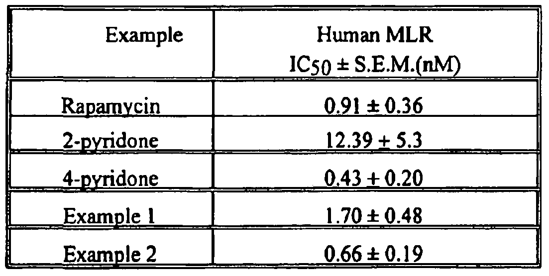 263
264 The pharmacokinetic behaviors of Example 1 and Example 2 were characterized following a single 2.5 mg/kg intravenous dose in cynomolgus monkey (n=3 per group). Each compound was prepared as 2.5 mg/mL solution in a 20% ethanol:30% propylene glycol:2% cremophor EL:48% dextrose 5% in water vehicle. The 1 mL/kg intravenous dose was administered as a slow bolus (-1-2 minutes) in a saphenous vein of the monkeys. Blood samples were obtained from a femoral artery or vein of each animal prior to dosing and 0.1 (TV only), 0.25, 0.5, 1, 1.5, 2, 4, 6, 9, 12, 24, and 30 hours after dosing. The EDTA preserved samples were thoroughly mixed and extracted for subsequent analysis.
263
264 The pharmacokinetic behaviors of Example 1 and Example 2 were characterized following a single 2.5 mg/kg intravenous dose in cynomolgus monkey (n=3 per group). Each compound was prepared as 2.5 mg/mL solution in a 20% ethanol:30% propylene glycol:2% cremophor EL:48% dextrose 5% in water vehicle. The 1 mL/kg intravenous dose was administered as a slow bolus (-1-2 minutes) in a saphenous vein of the monkeys. Blood samples were obtained from a femoral artery or vein of each animal prior to dosing and 0.1 (TV only), 0.25, 0.5, 1, 1.5, 2, 4, 6, 9, 12, 24, and 30 hours after dosing. The EDTA preserved samples were thoroughly mixed and extracted for subsequent analysis.
265 An aliquot of blood (1.0 mL) was hemolyzed with 20% methanol in water (0.5 ml) containing an internal standard. The hemolyzed samples were extracted with a mixture of ethyl acetate and hexane (1 :1 (v/v), 6.0 mL). The organic layer was evaporated to dryness with a stream of nitrogen at room temperature. Samples were reconstituted in methanol: water (1 : 1 , 150 μL). The title compounds (50 μL injection) were separated from contaminants using reverse phase HPLC with UV detection. Samples were kept cool (4 0C) through the run. All samples from each study were analyzed as a single batch on the HPLC.
266 Area under the curve (AUC) measurements of Example 1, Example 2 and the internal standard were determined using the Sciex MacQuan™ software. Calibration curves were derived from peak area ratio (parent drug/internal standard) of the spiked blood standards using least squares linear regression of the ratio versus the theoretical concentration. The methods were linear for both compounds over the range of the standard curve (correlation > 0.99) with an estimated quantitation limit of 0.1 ng/mL. The maximum blood concentration (CMAX) and me ώne t0 reach the maximum blood concentration (TMAX) were read directly from the observed blood
concentration-time data. The blood concentration data were submitted to multi-exponential curve fitting using CSTRIP to obtain estimates of pharmacokinetic parameters. The estimated parameters were further defined using NONLIN84. The area under the blood concentration-time curve from 0 to t hours (last measurable blood concentration time point) after dosing (AUCM) was calculated using the linear trapeziodal rule for the blood-time profiles. The residual area extrapolated to infinity, determined as the final measured blood concentration (Ci) divided by the terminal elimination rate constant (β), and added to AUQM to produce the total area under the curve (AUCo-
.)•
267 As shown in Figure 1 and Table 2, both Example 1 and Example 2 had a surprisingly substantially shorter terminal elimination half-life (Un) when compared to rapamycin. Thus, only the compounds of the invention provide both sufficient efficacy (Table 1) and a shorter terminal .half-life (Table 2).
268 Table 2

269 While the invention has been described, disclosed, illustrated and shown in various terms of certain embodiments or modifications which it has presumed in practice, the scope of the invention is not intended to be, nor should it be deemed to be, limited thereby and such other modifications or
embodiments as may be suggested by the teachings herein are particularly reserved especially as they fall within the breadth and scope of the claims here appended.
Claims
What is Claimed is: 1. A method for improving patency in a subject involving placement of a medical device in a body lumen for treating and/or preventing adjacent diseases or maintaining patency of the body lumen, comprising: providing a medical device in a body lumen; providing a first lipophilic agent capable of penetrating a body lumen, wherein the transfer coefficient of said first lipophilic agent is by an amount of at least approximately 5,000 (ug/mL)"1, wherein said first lipophilic agent is associated with said medical device; placing said first lipophilic agent/medical device adjacent to a body lumen; and delivering a therapeutically effective amount of said first lipophilic agent to a desired area within a subject.
2. The method according to claim 1 , further comprising at least one pharmaceutically acceptable carrier or excipient, wherein said medical device is associated with said pharmaceutically acceptable carrier or excipient.
3. The method according to claim 2, wherein said pharmaceutically acceptable carrier or excipient is a polymer.
4. The method according to claim 2, wherein said pharmaceutically acceptable carrier or excipient is an agent.
5. The method according to claim I , wherein said body lumen comprises at least one of
a vessel wall, a coronary artery, esophageal lumen, and a urethra.
6. The method according to claim 1 , wherein said first lipophilic agent/medical device is placed adjacent to said body lumen including coronary arteries, wherein a therapeutically effective amount of said first lipophilic agent is delivered into said coronary arteries and diffused into the pericardial sac in said method.
7. The method according to claim 6, wherein said method provides substantially uniform dug delivery of said lipophilic agent to the myocardium.
8. The method according to claim 1, wherein said method is useful for the treatment and/or prevention of vascular diseases in subject
9. The method according to claim 3, wherein said delivery mechanism of said first lipophilic agent includes polymer hydration followed by dissolution of said first lipophilic agent, and wherein said first lipophilic agent is thereafter delivered into said body lumen.
10. The method according to claim 3, wherein said delivery mechanism of said first lipophilic agent includes a lipophilic agent/polymer matrix which controls the elution rate of said first lipophilic agent to said body lumen.
11. The method according to claim 1 , further comprises at least one second lipophilic agent.
12. The method according to claim 1, further comprises at least one lipophilic prodrug.
13. The method according to claim 1, further comprises at least one lipophilic penetration
enhancer.
14. The method according to claim 13, wherein said lipophilic penetration enhancer is a pharmaceutical agent.
15. The method according to claim 1 , wherein the concentration of said first lipophilic agent delivered into said body lumen is a therapeutically effective amount.
16. The method according to claim 1 1 , wherein the concentration of said second lipophilic agent in combination with said first lipophilic agent is delivered into said body lumen in a therapeutically effective amount.
17. The method according to claim 1 , wherein said first lipophilic agent is zotarolimus.
18. The method according to claim 1 , further comprises at least one beneficial agent.
19. The method according to claim 1 , wherein said first lipophilic agent includes a partition coefficient greater than 20,000 P.
20. The method according to claim 1 , wherein said first lipophilic agent includes partition coefficients greater than 20,000 P and said lipophilic agent having a solubilities of less than about 30 ug/ml.
21. The method according to claim 1 , wherein said first lipophilic agent includes a LogP at least approximately 4.3.
22. The method according to claim 1 , wherein said method comprises a first lipophilic agent having solubilities of at least approximately IS μg/mL.
23. The method according to claim 1 , wherein said method comprises a first lipophilic agent having transfer coefficients of at least approximately 10,000 (μg/mL)'1.
24. The method according to claim 1 , wherein said method comprises a first lipophilic agent having transfer coefficients of at least approximately 15,000 (μg/mL)'1.
23. The method according to claim 1 , wherein the dosage delivery of said first lipophilic agent into said body lumen ranges from about 15 μg/g to about 150 μg/g over a period of up to about 5 days.
26. The method according to claim 1, wherein the dosage delivery of said first lipophilic agent into said body lumen ranges from about 15 μg/g to about 80 μg/g over a period from about 5 days to up to about 15 days.
27. The method according to claim 1, wherein the dosage delivery of said first lipophilic agent into said body lumen ranges from about 5 μg/g to about 60 μg/g over a period from 15 days up to about 28 days.
28. The method according to claim I , wherein said first lipophilic agent reaches therapeutically significant concentrations in targeted areas in said subject comprising at least one of the distal myocardium, the unstent myocardium, in said subjacent myocardium, in unslented and distal coronary arteries, and maintains those concentrations throughout a 28 day period.
29. The method according to claim 1 , wherein said medical device is permanently or temporarily implanted into a subjects' body.
30. The method according to claim 1, wherein said first lipophilic agent is in amorphous form.
31. The method according to claim 1 , further comprises a beneficial agent including at least one of antithrombotics, anticoagulants, antiplatelets agents, anti-lipid agents, thrombolytics, antiproliferatives, antiinflammatories, agents that inhibit hyperplasia, smooth muscle cell inhibitors, antibiotics, growth factor inhibitors, cell adhesion inhibitors, cell adhesion promoters, antimitotics, antifibrins, antioxidants, antineoplastics, agents that promote endothelial cell recovery, matrix metalloproteinase inhibitors, antineoplastics, antimetabolites, antiallergic substances, viraJ vectors, nucleic acids, monoclonal antibodies, inhibitors of tyrosine kinase, antisense compounds, oligonucleotides, cell permeation enhancers, and any combinations thereof.
32. The method according to claim 1, further comprises a beneficial agent including at least one of hypoglycemic agents, hypolipidemic agents, proteins, nucleic acids, agents useful for erythropoiesis stimulation, angiogenesis agents, antiulcer/antireflux agents, and antinauseants/antiemetics, PPAR-alpha agonists, and any combinations thereof.
33. The method according to claim 1 , further comprises a beneficial agent including at least one of sodium heparin, LMW heparins, heparoids, hirudin, argatroban, forskolin, vapriprost, prostacyclin and prostacylin analogues, dextran, D-phe-pro-arg- chloromethylketone (synthetic antithrombin), glycoprotein Iib/lia (platelet membrane receptor antagonist antibody), recombinant hirudin, thrombin inhibitors, indomethacin, phenyl salicylate, β-estradiol, vinblastine, ABT-627 (astrasentan), testosterone,
progesterone, paclitaxel, methotrexate, fotemustine, RPR-10151 IA, cyclosporin A, vincristine, carvediol, vindesine, dipyridamole, methotrexate, folic acid, thrombospondin mimetics, estradiol, dexamethasone, metrizamide, iopamidol, iohexol, iopromide, iobitridol, iomeprol, iopentol, ioversol, ioxilan, iodixanol, iotrolan and pro-drugs, analogs, derivatives, and any combinations thereof.
34. The method according to claim 1 , wherein said medical device is an endovascular medical device.
35. The method according to claim 1, wherein said medical device includes intracoronary medical devices selected from the group consisting of stents, drug delivery catheters, grafts, and drug delivery balloons utilized in a subjects' vasculature.
36. The method according to claim 1, wherein said medical device includes a stent selected from the group consisting of peripheral stents, peripheral coronary stents, degradable coronary stents, non-degfadable coronary stents, self-expanding stents, balloon-expanded stents, and esophageal stents.
37. The method according to claim 1, wherein said medical device is selected from the group consisting of arterio- venous grafts, by-pass grafts, penile implants, vascular implants and grafts, intravenous catheters, small diameter grafts, artificial lung catheters, electrophysiology catheters, bone pins, suture anchors, blood pressure and stent graft catheters, breast implants, benign prostatic hyperplasia and prostate cancer implants, bone repair/augmentation devices, breast implants, orthopedic joint implants, dental implants, implanted drug infusion tubes, oncological implants, pain management implants,
- 7β
neurological catheters, central venous access catheters, catheter cuff, vascular access catheters, urological catheters/implants, atherectomy catheters, clot extraction catheters, PTA catheters, PTCA catheters, stylets (vascular and non-vascular), drug infusion catheters, angiographic catheters, hemodialysis catheters, neurovascular balloon catheters, thoracic cavity suction drainage catheters, electrophysiology catheters, stroke therapy catheters, abscess drainage catheters, biliary drainage products, dialysis catheters, central venous access catheters, and parental feeding catheters.
38. The method according to claim 1 , wherein said medical device is selected from the group consisting of pacemakers, vascular grafts, sphincter devices, urethral devices, bladder devices, renal devices, gastroenteral and anastomotic devices, vertebral disks, hemostatic barriers, clamps, surgical staples/sutures/screws/plates/wires/clips, glucose sensors, blood oxygenator tubing, blood oxygenator membranes, blood bags, birth control/IUDs and associated pregnancy control devices, cartilage repair devices, orthopedic fracture repairs, tissue adhesives, tissue sealants, tissue scaffolds, CSF shunts, dental fracture repair devices, intravitreal drug delivery devices, nerve regeneration conduits, electrostimulation leads, spinal/orthopedic repair devices, wound dressings, embolic protection filters, abdominal aortic aneurysm grafts and devices, neuro aneurysm treatment coils, hemodialysis devices, uterine bleeding patches, anastomotic closures, in vitro diagnostics, aneurysm exclusion devices, neuropatches, vena cava filters, urinary dilators, endoscopic surgical and wound drainings, surgical tissue extractors, transition sheaths and dialators, coronary and peripheral guidewires, circulatory support systems, tympanostomy vent tubes, cerebro-spinal fluid shunts, defibrillator leads, percutaneous
closure devices, drainage tubes, bronchial tubes, vascular coils, vascular protection devices, vascular intervention devices including vascular filters and distal support devices and emboli filter/entrapment aids, AV access grafts, surgical tampons, drug delivery capsule and cardiac valves.
39. The method according to claim 1 , wherein said medical device is selected from the group consisting of atrial septal defect closures, electro-stimulation leads for cardiac rhythm management, tissue and mechanical prosthetic heart valves and rings, arterial- venous shunts, valve annuloplasty devices, mitral valve repair devices, left ventricle assist devices, left atrial appendage filters, cardiac sensors, pacemaker electrodes and leads.
40. The method according to claim 11 , wherein said second lipophilic agent is at least one ofzotarolimus having the following structures;

41. The method according to claim 6, wherein delivering said lipophilic agent delivered to the epicardium and/or pericardial sac is continuous.
Priority Applications (2)
| Application Number | Priority Date | Filing Date | Title |
|---|---|---|---|
| EP07753851A EP2004251A2 (en) | 2006-03-22 | 2007-03-22 | Delivery of highly lipophilic agents via medical devices |
| JP2009501580A JP5271892B2 (en) | 2006-03-22 | 2007-03-22 | Delivery of highly lipophilic drugs through medical devices |
Applications Claiming Priority (2)
| Application Number | Priority Date | Filing Date | Title |
|---|---|---|---|
| US11/386,498 US8257725B2 (en) | 1997-09-26 | 2006-03-22 | Delivery of highly lipophilic agents via medical devices |
| US11/386,498 | 2006-03-22 |
Publications (2)
| Publication Number | Publication Date |
|---|---|
| WO2007112006A2 true WO2007112006A2 (en) | 2007-10-04 |
| WO2007112006A3 WO2007112006A3 (en) | 2008-06-12 |
Family
ID=38445691
Family Applications (1)
| Application Number | Title | Priority Date | Filing Date |
|---|---|---|---|
| PCT/US2007/007255 WO2007112006A2 (en) | 2006-03-22 | 2007-03-22 | Delivery of highly lipophilic agents via medical devices |
Country Status (4)
| Country | Link |
|---|---|
| US (1) | US8257725B2 (en) |
| EP (1) | EP2004251A2 (en) |
| JP (1) | JP5271892B2 (en) |
| WO (1) | WO2007112006A2 (en) |
Cited By (12)
| Publication number | Priority date | Publication date | Assignee | Title |
|---|---|---|---|---|
| WO2007109372A2 (en) * | 2006-03-22 | 2007-09-27 | Abbott Laboratories | Delivery of highly lipophilic agents via medical devices |
| US8734825B2 (en) | 2008-09-15 | 2014-05-27 | Covidien Lp | Local delivery of water-soluble or water-insoluble therapeutic agents to the surface of body lumens |
| US9034362B2 (en) | 2008-09-15 | 2015-05-19 | The Spectranetics Corporation | Local delivery of water-soluble or water-insoluble therapeutic agents to the surface of body lumens |
| US9414777B2 (en) | 2004-07-13 | 2016-08-16 | Dexcom, Inc. | Transcutaneous analyte sensor |
| US9956385B2 (en) | 2012-06-28 | 2018-05-01 | The Spectranetics Corporation | Post-processing of a medical device to control morphology and mechanical properties |
| US9986942B2 (en) | 2004-07-13 | 2018-06-05 | Dexcom, Inc. | Analyte sensor |
| US10046093B2 (en) | 2008-09-15 | 2018-08-14 | The Spectranetics Corporation | Local delivery of water-soluble or water-insoluble therapeutic agents to the surface of body lumens |
| US10525171B2 (en) | 2014-01-24 | 2020-01-07 | The Spectranetics Corporation | Coatings for medical devices |
| US10610135B2 (en) | 2005-03-10 | 2020-04-07 | Dexcom, Inc. | System and methods for processing analyte sensor data for sensor calibration |
| US10813577B2 (en) | 2005-06-21 | 2020-10-27 | Dexcom, Inc. | Analyte sensor |
| US11160674B2 (en) | 2017-01-30 | 2021-11-02 | Japan Medical Device Technology Co., Ltd. | High performance bioabsorbable stent |
| US11248282B2 (en) | 2017-01-10 | 2022-02-15 | Fuji Light Metal Co., Ltd. | Magnesium alloy |
Families Citing this family (157)
| Publication number | Priority date | Publication date | Assignee | Title |
|---|---|---|---|---|
| US7611533B2 (en) | 1995-06-07 | 2009-11-03 | Cook Incorporated | Coated implantable medical device |
| US6302875B1 (en) | 1996-10-11 | 2001-10-16 | Transvascular, Inc. | Catheters and related devices for forming passageways between blood vessels or other anatomical structures |
| US7037332B2 (en) * | 2000-03-15 | 2006-05-02 | Orbus Medical Technologies, Inc. | Medical device with coating that promotes endothelial cell adherence |
| US8347891B2 (en) | 2002-04-08 | 2013-01-08 | Medtronic Ardian Luxembourg S.A.R.L. | Methods and apparatus for performing a non-continuous circumferential treatment of a body lumen |
| US20080213331A1 (en) | 2002-04-08 | 2008-09-04 | Ardian, Inc. | Methods and devices for renal nerve blocking |
| US7617005B2 (en) | 2002-04-08 | 2009-11-10 | Ardian, Inc. | Methods and apparatus for thermally-induced renal neuromodulation |
| US8150519B2 (en) | 2002-04-08 | 2012-04-03 | Ardian, Inc. | Methods and apparatus for bilateral renal neuromodulation |
| US20070129761A1 (en) | 2002-04-08 | 2007-06-07 | Ardian, Inc. | Methods for treating heart arrhythmia |
| US7756583B2 (en) | 2002-04-08 | 2010-07-13 | Ardian, Inc. | Methods and apparatus for intravascularly-induced neuromodulation |
| US9636174B2 (en) | 2002-04-08 | 2017-05-02 | Medtronic Ardian Luxembourg S.A.R.L. | Methods for therapeutic renal neuromodulation |
| US20070135875A1 (en) | 2002-04-08 | 2007-06-14 | Ardian, Inc. | Methods and apparatus for thermally-induced renal neuromodulation |
| DE202004021942U1 (en) | 2003-09-12 | 2013-05-13 | Vessix Vascular, Inc. | Selectable eccentric remodeling and / or ablation of atherosclerotic material |
| US8396548B2 (en) | 2008-11-14 | 2013-03-12 | Vessix Vascular, Inc. | Selective drug delivery in a lumen |
| US9125667B2 (en) | 2004-09-10 | 2015-09-08 | Vessix Vascular, Inc. | System for inducing desirable temperature effects on body tissue |
| US9713730B2 (en) | 2004-09-10 | 2017-07-25 | Boston Scientific Scimed, Inc. | Apparatus and method for treatment of in-stent restenosis |
| EP1819304B1 (en) | 2004-12-09 | 2023-01-25 | Twelve, Inc. | Aortic valve repair |
| EP2327429B1 (en) * | 2005-03-23 | 2014-09-17 | Abbott Laboratories | Delivery of highly lipophilic agents via medical devices |
| AU2006270221B2 (en) | 2005-07-15 | 2012-01-19 | Micell Technologies, Inc. | Polymer coatings containing drug powder of controlled morphology |
| WO2007011708A2 (en) | 2005-07-15 | 2007-01-25 | Micell Technologies, Inc. | Stent with polymer coating containing amorphous rapamycin |
| WO2007127363A2 (en) | 2006-04-26 | 2007-11-08 | Micell Technologies, Inc. | Coatings containing multiple drugs |
| US8019435B2 (en) | 2006-05-02 | 2011-09-13 | Boston Scientific Scimed, Inc. | Control of arterial smooth muscle tone |
| AU2007310988B2 (en) | 2006-10-18 | 2013-08-15 | Vessix Vascular, Inc. | Tuned RF energy and electrical tissue characterization for selective treatment of target tissues |
| AU2007310986B2 (en) | 2006-10-18 | 2013-07-04 | Boston Scientific Scimed, Inc. | Inducing desirable temperature effects on body tissue |
| US11426494B2 (en) | 2007-01-08 | 2022-08-30 | MT Acquisition Holdings LLC | Stents having biodegradable layers |
| JP5603598B2 (en) | 2007-01-08 | 2014-10-08 | ミセル テクノロジーズ、インコーポレイテッド | Stent with biodegradable layer |
| US20100211164A1 (en) * | 2007-04-17 | 2010-08-19 | Mcclain James B | Stents having biodegradable layers |
| AU2015200571B2 (en) * | 2007-04-17 | 2016-11-17 | Micell Technologies, Inc. | Stents having biodegradable layers |
| EP2190433A2 (en) | 2007-08-22 | 2010-06-02 | Gilead Colorado, Inc. | Therapy for complications of diabetes |
| WO2009089518A2 (en) * | 2008-01-11 | 2009-07-16 | Massachusetts Institute Of Technology | Endovascular platforms for the differential targeting of molecules to vessel wall and vessel lumen |
| SG192523A1 (en) | 2008-04-17 | 2013-08-30 | Micell Technologies Inc | Stents having bioabsorbable layers |
| CN102159257B (en) | 2008-07-17 | 2015-11-25 | 米歇尔技术公司 | Drug delivery medical device |
| US8128951B2 (en) | 2008-09-15 | 2012-03-06 | Cv Ingenuity Corp. | Local delivery of water-soluble or water-insoluble therapeutic agents to the surface of body lumens |
| AU2009295960A1 (en) | 2008-09-29 | 2010-04-01 | Cardiaq Valve Technologies, Inc. | Heart valve |
| EP2341871B1 (en) | 2008-10-01 | 2017-03-22 | Edwards Lifesciences CardiAQ LLC | Delivery system for vascular implant |
| AU2009314133B2 (en) | 2008-11-17 | 2015-12-10 | Vessix Vascular, Inc. | Selective accumulation of energy with or without knowledge of tissue topography |
| US8088091B2 (en) * | 2009-03-09 | 2012-01-03 | New Jersey Institute Of Technology | No clog shunt using a compact fluid drag path |
| CA2757276C (en) | 2009-04-01 | 2017-06-06 | Micell Technologies, Inc. | Coated stents |
| AU2010236288A1 (en) | 2009-04-15 | 2011-10-20 | Cardiaq Valve Technologies, Inc. | Vascular implant and delivery system |
| CA2759015C (en) | 2009-04-17 | 2017-06-20 | James B. Mcclain | Stents having controlled elution |
| EP2453834A4 (en) | 2009-07-16 | 2014-04-16 | Micell Technologies Inc | Drug delivery medical device |
| EP2531140B1 (en) | 2010-02-02 | 2017-11-01 | Micell Technologies, Inc. | Stent and stent delivery system with improved deliverability |
| KR20130108067A (en) | 2010-04-09 | 2013-10-02 | 베식스 바스큘라 인코포레이티드 | Power generating and control apparatus for the treatment of tissue |
| US9192790B2 (en) | 2010-04-14 | 2015-11-24 | Boston Scientific Scimed, Inc. | Focused ultrasonic renal denervation |
| CA2797110C (en) | 2010-04-22 | 2020-07-21 | Micell Technologies, Inc. | Stents and other devices having extracellular matrix coating |
| US8579964B2 (en) | 2010-05-05 | 2013-11-12 | Neovasc Inc. | Transcatheter mitral valve prosthesis |
| US8473067B2 (en) | 2010-06-11 | 2013-06-25 | Boston Scientific Scimed, Inc. | Renal denervation and stimulation employing wireless vascular energy transfer arrangement |
| WO2012009684A2 (en) | 2010-07-16 | 2012-01-19 | Micell Technologies, Inc. | Drug delivery medical device |
| US9408661B2 (en) | 2010-07-30 | 2016-08-09 | Patrick A. Haverkost | RF electrodes on multiple flexible wires for renal nerve ablation |
| US9463062B2 (en) | 2010-07-30 | 2016-10-11 | Boston Scientific Scimed, Inc. | Cooled conductive balloon RF catheter for renal nerve ablation |
| US9084609B2 (en) | 2010-07-30 | 2015-07-21 | Boston Scientific Scime, Inc. | Spiral balloon catheter for renal nerve ablation |
| US9155589B2 (en) | 2010-07-30 | 2015-10-13 | Boston Scientific Scimed, Inc. | Sequential activation RF electrode set for renal nerve ablation |
| US9358365B2 (en) | 2010-07-30 | 2016-06-07 | Boston Scientific Scimed, Inc. | Precision electrode movement control for renal nerve ablation |
| CA2810842C (en) | 2010-09-09 | 2018-06-26 | Micell Technologies, Inc. | Macrolide dosage forms |
| US8974451B2 (en) | 2010-10-25 | 2015-03-10 | Boston Scientific Scimed, Inc. | Renal nerve ablation using conductive fluid jet and RF energy |
| US9220558B2 (en) | 2010-10-27 | 2015-12-29 | Boston Scientific Scimed, Inc. | RF renal denervation catheter with multiple independent electrodes |
| US9028485B2 (en) | 2010-11-15 | 2015-05-12 | Boston Scientific Scimed, Inc. | Self-expanding cooling electrode for renal nerve ablation |
| US9089350B2 (en) | 2010-11-16 | 2015-07-28 | Boston Scientific Scimed, Inc. | Renal denervation catheter with RF electrode and integral contrast dye injection arrangement |
| US9668811B2 (en) | 2010-11-16 | 2017-06-06 | Boston Scientific Scimed, Inc. | Minimally invasive access for renal nerve ablation |
| US9326751B2 (en) | 2010-11-17 | 2016-05-03 | Boston Scientific Scimed, Inc. | Catheter guidance of external energy for renal denervation |
| US9060761B2 (en) | 2010-11-18 | 2015-06-23 | Boston Scientific Scime, Inc. | Catheter-focused magnetic field induced renal nerve ablation |
| US9192435B2 (en) | 2010-11-22 | 2015-11-24 | Boston Scientific Scimed, Inc. | Renal denervation catheter with cooled RF electrode |
| US9023034B2 (en) | 2010-11-22 | 2015-05-05 | Boston Scientific Scimed, Inc. | Renal ablation electrode with force-activatable conduction apparatus |
| US20120157993A1 (en) | 2010-12-15 | 2012-06-21 | Jenson Mark L | Bipolar Off-Wall Electrode Device for Renal Nerve Ablation |
| WO2012100095A1 (en) | 2011-01-19 | 2012-07-26 | Boston Scientific Scimed, Inc. | Guide-compatible large-electrode catheter for renal nerve ablation with reduced arterial injury |
| CA2832311A1 (en) | 2011-04-08 | 2012-11-29 | Covidien Lp | Iontophoresis drug delivery system and method for denervation of the renal sympathetic nerve and iontophoretic drug delivery |
| EP2701623B1 (en) | 2011-04-25 | 2016-08-17 | Medtronic Ardian Luxembourg S.à.r.l. | Apparatus related to constrained deployment of cryogenic balloons for limited cryogenic ablation of vessel walls |
| US9308087B2 (en) | 2011-04-28 | 2016-04-12 | Neovasc Tiara Inc. | Sequentially deployed transcatheter mitral valve prosthesis |
| US9554897B2 (en) | 2011-04-28 | 2017-01-31 | Neovasc Tiara Inc. | Methods and apparatus for engaging a valve prosthesis with tissue |
| CA2841360A1 (en) | 2011-07-15 | 2013-01-24 | Micell Technologies, Inc. | Drug delivery medical device |
| CN103813745B (en) | 2011-07-20 | 2016-06-29 | 波士顿科学西美德公司 | In order to visualize, be directed at and to melt transcutaneous device and the method for nerve |
| JP6106669B2 (en) | 2011-07-22 | 2017-04-05 | ボストン サイエンティフィック サイムド,インコーポレイテッドBoston Scientific Scimed,Inc. | A neuromodulation system having a neuromodulation element that can be placed in a helical guide |
| WO2013055826A1 (en) | 2011-10-10 | 2013-04-18 | Boston Scientific Scimed, Inc. | Medical devices including ablation electrodes |
| US10085799B2 (en) | 2011-10-11 | 2018-10-02 | Boston Scientific Scimed, Inc. | Off-wall electrode device and methods for nerve modulation |
| US9420955B2 (en) | 2011-10-11 | 2016-08-23 | Boston Scientific Scimed, Inc. | Intravascular temperature monitoring system and method |
| US9364284B2 (en) | 2011-10-12 | 2016-06-14 | Boston Scientific Scimed, Inc. | Method of making an off-wall spacer cage |
| EP2768568B1 (en) | 2011-10-18 | 2020-05-06 | Boston Scientific Scimed, Inc. | Integrated crossing balloon catheter |
| US10188772B2 (en) | 2011-10-18 | 2019-01-29 | Micell Technologies, Inc. | Drug delivery medical device |
| WO2013058962A1 (en) | 2011-10-18 | 2013-04-25 | Boston Scientific Scimed, Inc. | Deflectable medical devices |
| EP2774643A4 (en) * | 2011-11-04 | 2015-06-03 | Nipro Corp | Injection needle |
| EP2775948B1 (en) | 2011-11-08 | 2018-04-04 | Boston Scientific Scimed, Inc. | Ostial renal nerve ablation |
| US9119600B2 (en) | 2011-11-15 | 2015-09-01 | Boston Scientific Scimed, Inc. | Device and methods for renal nerve modulation monitoring |
| US9119632B2 (en) | 2011-11-21 | 2015-09-01 | Boston Scientific Scimed, Inc. | Deflectable renal nerve ablation catheter |
| EP2782584B1 (en) | 2011-11-23 | 2021-06-09 | TherapeuticsMD, Inc. | Natural combination hormone replacement formulations and therapies |
| US9301920B2 (en) | 2012-06-18 | 2016-04-05 | Therapeuticsmd, Inc. | Natural combination hormone replacement formulations and therapies |
| US8912215B2 (en) | 2011-12-13 | 2014-12-16 | Everon Biosciences, Inc. | Rapamycin composition |
| US9265969B2 (en) | 2011-12-21 | 2016-02-23 | Cardiac Pacemakers, Inc. | Methods for modulating cell function |
| WO2013096916A2 (en) | 2011-12-23 | 2013-06-27 | Vessix Vascular, Inc. | Methods and apparatuses for remodeling tissue of or adjacent to a body passage |
| US9433760B2 (en) | 2011-12-28 | 2016-09-06 | Boston Scientific Scimed, Inc. | Device and methods for nerve modulation using a novel ablation catheter with polymeric ablative elements |
| US9050106B2 (en) | 2011-12-29 | 2015-06-09 | Boston Scientific Scimed, Inc. | Off-wall electrode device and methods for nerve modulation |
| US10660703B2 (en) | 2012-05-08 | 2020-05-26 | Boston Scientific Scimed, Inc. | Renal nerve modulation devices |
| US9345573B2 (en) | 2012-05-30 | 2016-05-24 | Neovasc Tiara Inc. | Methods and apparatus for loading a prosthesis onto a delivery system |
| US20150196640A1 (en) | 2012-06-18 | 2015-07-16 | Therapeuticsmd, Inc. | Progesterone formulations having a desirable pk profile |
| US10806740B2 (en) | 2012-06-18 | 2020-10-20 | Therapeuticsmd, Inc. | Natural combination hormone replacement formulations and therapies |
| US10806697B2 (en) | 2012-12-21 | 2020-10-20 | Therapeuticsmd, Inc. | Vaginal inserted estradiol pharmaceutical compositions and methods |
| US20130338122A1 (en) | 2012-06-18 | 2013-12-19 | Therapeuticsmd, Inc. | Transdermal hormone replacement therapies |
| US10321946B2 (en) | 2012-08-24 | 2019-06-18 | Boston Scientific Scimed, Inc. | Renal nerve modulation devices with weeping RF ablation balloons |
| US9173696B2 (en) | 2012-09-17 | 2015-11-03 | Boston Scientific Scimed, Inc. | Self-positioning electrode system and method for renal nerve modulation |
| US10549127B2 (en) | 2012-09-21 | 2020-02-04 | Boston Scientific Scimed, Inc. | Self-cooling ultrasound ablation catheter |
| US10398464B2 (en) | 2012-09-21 | 2019-09-03 | Boston Scientific Scimed, Inc. | System for nerve modulation and innocuous thermal gradient nerve block |
| CN104869930B (en) | 2012-10-10 | 2020-12-25 | 波士顿科学国际有限公司 | Renal neuromodulation apparatus and methods |
| US10568891B2 (en) | 2012-12-21 | 2020-02-25 | Therapeuticsmd, Inc. | Vaginal inserted estradiol pharmaceutical compositions and methods |
| US11246875B2 (en) | 2012-12-21 | 2022-02-15 | Therapeuticsmd, Inc. | Vaginal inserted estradiol pharmaceutical compositions and methods |
| US11266661B2 (en) | 2012-12-21 | 2022-03-08 | Therapeuticsmd, Inc. | Vaginal inserted estradiol pharmaceutical compositions and methods |
| US10537581B2 (en) | 2012-12-21 | 2020-01-21 | Therapeuticsmd, Inc. | Vaginal inserted estradiol pharmaceutical compositions and methods |
| US10471072B2 (en) | 2012-12-21 | 2019-11-12 | Therapeuticsmd, Inc. | Vaginal inserted estradiol pharmaceutical compositions and methods |
| US9180091B2 (en) | 2012-12-21 | 2015-11-10 | Therapeuticsmd, Inc. | Soluble estradiol capsule for vaginal insertion |
| US10583002B2 (en) | 2013-03-11 | 2020-03-10 | Neovasc Tiara Inc. | Prosthetic valve with anti-pivoting mechanism |
| US9693821B2 (en) | 2013-03-11 | 2017-07-04 | Boston Scientific Scimed, Inc. | Medical devices for modulating nerves |
| US9956033B2 (en) | 2013-03-11 | 2018-05-01 | Boston Scientific Scimed, Inc. | Medical devices for modulating nerves |
| WO2014165264A1 (en) | 2013-03-12 | 2014-10-09 | Micell Technologies, Inc. | Bioabsorbable biomedical implants |
| US9808311B2 (en) | 2013-03-13 | 2017-11-07 | Boston Scientific Scimed, Inc. | Deflectable medical devices |
| US9681951B2 (en) | 2013-03-14 | 2017-06-20 | Edwards Lifesciences Cardiaq Llc | Prosthesis with outer skirt and anchors |
| EP2967734B1 (en) | 2013-03-15 | 2019-05-15 | Boston Scientific Scimed, Inc. | Methods and apparatuses for remodeling tissue of or adjacent to a body passage |
| US9297845B2 (en) | 2013-03-15 | 2016-03-29 | Boston Scientific Scimed, Inc. | Medical devices and methods for treatment of hypertension that utilize impedance compensation |
| US10265122B2 (en) | 2013-03-15 | 2019-04-23 | Boston Scientific Scimed, Inc. | Nerve ablation devices and related methods of use |
| US9572665B2 (en) | 2013-04-04 | 2017-02-21 | Neovasc Tiara Inc. | Methods and apparatus for delivering a prosthetic valve to a beating heart |
| EP2996629B1 (en) | 2013-05-15 | 2021-09-22 | Micell Technologies, Inc. | Bioabsorbable biomedical implants |
| EP3010437A1 (en) | 2013-06-21 | 2016-04-27 | Boston Scientific Scimed, Inc. | Renal denervation balloon catheter with ride along electrode support |
| CN105473092B (en) | 2013-06-21 | 2019-05-17 | 波士顿科学国际有限公司 | The medical instrument for renal nerve ablation with rotatable shaft |
| US9707036B2 (en) | 2013-06-25 | 2017-07-18 | Boston Scientific Scimed, Inc. | Devices and methods for nerve modulation using localized indifferent electrodes |
| WO2015002787A1 (en) | 2013-07-01 | 2015-01-08 | Boston Scientific Scimed, Inc. | Medical devices for renal nerve ablation |
| WO2015006480A1 (en) | 2013-07-11 | 2015-01-15 | Boston Scientific Scimed, Inc. | Devices and methods for nerve modulation |
| EP3019106A1 (en) | 2013-07-11 | 2016-05-18 | Boston Scientific Scimed, Inc. | Medical device with stretchable electrode assemblies |
| WO2015010074A1 (en) | 2013-07-19 | 2015-01-22 | Boston Scientific Scimed, Inc. | Spiral bipolar electrode renal denervation balloon |
| WO2015013205A1 (en) | 2013-07-22 | 2015-01-29 | Boston Scientific Scimed, Inc. | Medical devices for renal nerve ablation |
| US10695124B2 (en) | 2013-07-22 | 2020-06-30 | Boston Scientific Scimed, Inc. | Renal nerve ablation catheter having twist balloon |
| WO2015027096A1 (en) | 2013-08-22 | 2015-02-26 | Boston Scientific Scimed, Inc. | Flexible circuit having improved adhesion to a renal nerve modulation balloon |
| EP3041425B1 (en) | 2013-09-04 | 2022-04-13 | Boston Scientific Scimed, Inc. | Radio frequency (rf) balloon catheter having flushing and cooling capability |
| WO2015038947A1 (en) | 2013-09-13 | 2015-03-19 | Boston Scientific Scimed, Inc. | Ablation balloon with vapor deposited cover layer |
| US11246654B2 (en) | 2013-10-14 | 2022-02-15 | Boston Scientific Scimed, Inc. | Flexible renal nerve ablation devices and related methods of use and manufacture |
| CN105592778B (en) | 2013-10-14 | 2019-07-23 | 波士顿科学医学有限公司 | High-resolution cardiac mapping electrod-array conduit |
| US9770606B2 (en) | 2013-10-15 | 2017-09-26 | Boston Scientific Scimed, Inc. | Ultrasound ablation catheter with cooling infusion and centering basket |
| AU2014334574B2 (en) | 2013-10-15 | 2017-07-06 | Boston Scientific Scimed, Inc. | Medical device balloon |
| CN105636538B (en) | 2013-10-18 | 2019-01-15 | 波士顿科学国际有限公司 | Foley's tube with flexible wire and its correlation technique for using and manufacturing |
| CN105658163B (en) | 2013-10-25 | 2020-08-18 | 波士顿科学国际有限公司 | Embedded thermocouple in denervation flexible circuit |
| WO2015103617A1 (en) | 2014-01-06 | 2015-07-09 | Boston Scientific Scimed, Inc. | Tear resistant flex circuit assembly |
| US11000679B2 (en) | 2014-02-04 | 2021-05-11 | Boston Scientific Scimed, Inc. | Balloon protection and rewrapping devices and related methods of use |
| JP6325121B2 (en) | 2014-02-04 | 2018-05-16 | ボストン サイエンティフィック サイムド,インコーポレイテッドBoston Scientific Scimed,Inc. | Alternative placement of temperature sensors on bipolar electrodes |
| US10709490B2 (en) | 2014-05-07 | 2020-07-14 | Medtronic Ardian Luxembourg S.A.R.L. | Catheter assemblies comprising a direct heating element for renal neuromodulation and associated systems and methods |
| JP2017516768A (en) | 2014-05-22 | 2017-06-22 | セラピューティックスエムディー インコーポレーテッドTherapeuticsmd, Inc. | Natural combination hormone replacement therapy and therapy |
| CN112972871A (en) * | 2014-12-29 | 2021-06-18 | 波士顿科学国际有限公司 | Compositions, devices and methods for multi-stage release of chemotherapeutic agents |
| US10226193B2 (en) | 2015-03-31 | 2019-03-12 | Medtronic Ps Medical, Inc. | Wireless pressure measurement and monitoring for shunts |
| US9700664B2 (en) | 2015-04-01 | 2017-07-11 | Elwha Llc | Implantable heart treatment systems, devices, and methods |
| US9675812B2 (en) | 2015-04-01 | 2017-06-13 | Elwha Llc | Implantable heart treatment systems, devices, and methods |
| US9682245B2 (en) | 2015-04-01 | 2017-06-20 | Elwha Llc | Implantable heart treatment systems, devices, and methods |
| US10328087B2 (en) | 2015-07-23 | 2019-06-25 | Therapeuticsmd, Inc. | Formulations for solubilizing hormones |
| WO2017173044A1 (en) | 2016-04-01 | 2017-10-05 | Therapeuticsmd Inc. | Steroid hormone compositions in medium chain oils |
| WO2017173071A1 (en) | 2016-04-01 | 2017-10-05 | Therapeuticsmd, Inc. | Steroid hormone pharmaceutical composition |
| EP3459469A1 (en) | 2017-09-23 | 2019-03-27 | Universität Zürich | Medical occluder device |
| CN111417433A (en) | 2017-12-01 | 2020-07-14 | 心脏起搏器股份公司 | Method and system for detecting atrial contraction timing reference during ventricular filling from a ventricular implanted leadless cardiac pacemaker |
| WO2019108482A1 (en) | 2017-12-01 | 2019-06-06 | Cardiac Pacemakers, Inc. | Methods and systems for detecting atrial contraction timing fiducials and determining a cardiac interval from a ventricularly implanted leadless cardiac pacemaker |
| WO2019108830A1 (en) | 2017-12-01 | 2019-06-06 | Cardiac Pacemakers, Inc. | Leadless cardiac pacemaker with reversionary behavior |
| US11116561B2 (en) | 2018-01-24 | 2021-09-14 | Medtronic Ardian Luxembourg S.A.R.L. | Devices, agents, and associated methods for selective modulation of renal nerves |
| US10285998B1 (en) | 2018-04-04 | 2019-05-14 | The Menopause Method, Inc. | Composition and method to aid in hormone replacement therapy |
| ES2930364T3 (en) | 2018-07-09 | 2022-12-09 | Japan Medical Device Tech Co Ltd | magnesium alloy |
| EP4033999A2 (en) | 2019-09-26 | 2022-08-03 | Universität Zürich | Left atrial appendage occlusion devices |
| US11633405B2 (en) | 2020-02-07 | 2023-04-25 | Therapeuticsmd, Inc. | Steroid hormone pharmaceutical formulations |
Citations (1)
| Publication number | Priority date | Publication date | Assignee | Title |
|---|---|---|---|---|
| US6015815A (en) | 1997-09-26 | 2000-01-18 | Abbott Laboratories | Tetrazole-containing rapamycin analogs with shortened half-lives |
Family Cites Families (49)
| Publication number | Priority date | Publication date | Assignee | Title |
|---|---|---|---|---|
| ZA737247B (en) * | 1972-09-29 | 1975-04-30 | Ayerst Mckenna & Harrison | Rapamycin and process of preparation |
| US3993749A (en) * | 1974-04-12 | 1976-11-23 | Ayerst Mckenna And Harrison Ltd. | Rapamycin and process of preparation |
| US4885171A (en) * | 1978-11-03 | 1989-12-05 | American Home Products Corporation | Use of rapamycin in treatment of certain tumors |
| US4316885A (en) * | 1980-08-25 | 1982-02-23 | Ayerst, Mckenna And Harrison, Inc. | Acyl derivatives of rapamycin |
| US4401653A (en) * | 1981-03-09 | 1983-08-30 | Ayerst, Mckenna & Harrison Inc. | Combination of rapamycin and picibanil for the treatment of tumors |
| US4894366A (en) | 1984-12-03 | 1990-01-16 | Fujisawa Pharmaceutical Company, Ltd. | Tricyclo compounds, a process for their production and a pharmaceutical composition containing the same |
| EP0163829B1 (en) * | 1984-03-21 | 1989-08-23 | Nippon Telegraph And Telephone Corporation | Speech signal processing system |
| US4650803A (en) * | 1985-12-06 | 1987-03-17 | University Of Kansas | Prodrugs of rapamycin |
| US5527337A (en) * | 1987-06-25 | 1996-06-18 | Duke University | Bioabsorbable stent and method of making the same |
| US4916193A (en) * | 1987-12-17 | 1990-04-10 | Allied-Signal Inc. | Medical devices fabricated totally or in part from copolymers of recurring units derived from cyclic carbonates and lactides |
| US5092877A (en) * | 1988-09-01 | 1992-03-03 | Corvita Corporation | Radially expandable endoprosthesis |
| US4994071A (en) * | 1989-05-22 | 1991-02-19 | Cordis Corporation | Bifurcating stent apparatus and method |
| US5304121A (en) * | 1990-12-28 | 1994-04-19 | Boston Scientific Corporation | Drug delivery system making use of a hydrogel polymer coating |
| AU7998091A (en) * | 1990-05-17 | 1991-12-10 | Harbor Medical Devices, Inc. | Medical device polymer |
| JPH04230389A (en) | 1990-07-16 | 1992-08-19 | American Home Prod Corp | Rapamycin derivative |
| US5023262A (en) * | 1990-08-14 | 1991-06-11 | American Home Products Corporation | Hydrogenated rapamycin derivatives |
| US5163952A (en) * | 1990-09-14 | 1992-11-17 | Michael Froix | Expandable polymeric stent with memory and delivery apparatus and method |
| PT98990A (en) | 1990-09-19 | 1992-08-31 | American Home Prod | PROCESS FOR THE PREPARATION OF CARBOXYLIC ACID ESTERS OF RAPAMICIN |
| US5120842A (en) * | 1991-04-01 | 1992-06-09 | American Home Products Corporation | Silyl ethers of rapamycin |
| US5120725A (en) * | 1991-05-29 | 1992-06-09 | American Home Products Corporation | Bicyclic rapamycins |
| US5120727A (en) * | 1991-05-29 | 1992-06-09 | American Home Products Corporation | Rapamycin dimers |
| US6090901A (en) * | 1991-07-05 | 2000-07-18 | Biocompatibles Limited | Polymeric surface coatings |
| US5705583A (en) * | 1991-07-05 | 1998-01-06 | Biocompatibles Limited | Polymeric surface coatings |
| US5457111A (en) * | 1991-09-05 | 1995-10-10 | Abbott Laboratories | Macrocyclic immunomodulators |
| US5516781A (en) * | 1992-01-09 | 1996-05-14 | American Home Products Corporation | Method of treating restenosis with rapamycin |
| US5177203A (en) * | 1992-03-05 | 1993-01-05 | American Home Products Corporation | Rapamycin 42-sulfonates and 42-(N-carboalkoxy) sulfamates useful as immunosuppressive agents |
| US5464650A (en) * | 1993-04-26 | 1995-11-07 | Medtronic, Inc. | Intravascular stent and method |
| US5716981A (en) * | 1993-07-19 | 1998-02-10 | Angiogenesis Technologies, Inc. | Anti-angiogenic compositions and methods of use |
| US5605696A (en) * | 1995-03-30 | 1997-02-25 | Advanced Cardiovascular Systems, Inc. | Drug loaded polymeric material and method of manufacture |
| US6099562A (en) * | 1996-06-13 | 2000-08-08 | Schneider (Usa) Inc. | Drug coating with topcoat |
| US5837313A (en) * | 1995-04-19 | 1998-11-17 | Schneider (Usa) Inc | Drug release stent coating process |
| US5722984A (en) * | 1996-01-16 | 1998-03-03 | Iso Stent, Inc. | Antithrombogenic radioactive coating for an intravascular stent |
| EP0916362B1 (en) * | 1997-03-31 | 2009-07-08 | Kabushikikaisha Igaki Iryo Sekkei | Stent for vessels |
| US6273913B1 (en) * | 1997-04-18 | 2001-08-14 | Cordis Corporation | Modified stent useful for delivery of drugs along stent strut |
| WO1999010719A1 (en) * | 1997-08-29 | 1999-03-04 | The Regents Of The University Of California | Method and apparatus for hybrid coding of speech at 4kbps |
| US20030129215A1 (en) | 1998-09-24 | 2003-07-10 | T-Ram, Inc. | Medical devices containing rapamycin analogs |
| US6890546B2 (en) * | 1998-09-24 | 2005-05-10 | Abbott Laboratories | Medical devices containing rapamycin analogs |
| US8029561B1 (en) | 2000-05-12 | 2011-10-04 | Cordis Corporation | Drug combination useful for prevention of restenosis |
| US6419692B1 (en) * | 1999-02-03 | 2002-07-16 | Scimed Life Systems, Inc. | Surface protection method for stents and balloon catheters for drug delivery |
| US6610317B2 (en) * | 1999-05-27 | 2003-08-26 | Acusphere, Inc. | Porous paclitaxel matrices and methods of manufacture thereof |
| US6510407B1 (en) * | 1999-10-19 | 2003-01-21 | Atmel Corporation | Method and apparatus for variable rate coding of speech |
| GB0100760D0 (en) | 2001-01-11 | 2001-02-21 | Biocompatibles Ltd | Drug delivery from stents |
| US20030181973A1 (en) | 2002-03-20 | 2003-09-25 | Harvinder Sahota | Reduced restenosis drug containing stents |
| CA2497640C (en) | 2002-09-06 | 2012-02-07 | Abbott Laboratories | Medical device having hydration inhibitor |
| AU2005222719B2 (en) | 2004-03-19 | 2011-03-24 | Abbott Laboratories | Multiple drug delivery from a balloon and a prosthesis |
| US20050232965A1 (en) | 2004-04-15 | 2005-10-20 | Robert Falotico | Local administration of a combination of rapamycin and 17 beta-estradiol for the treatment of vulnerable plaque |
| US20050288481A1 (en) | 2004-04-30 | 2005-12-29 | Desnoyer Jessica R | Design of poly(ester amides) for the control of agent-release from polymeric compositions |
| US7820732B2 (en) | 2004-04-30 | 2010-10-26 | Advanced Cardiovascular Systems, Inc. | Methods for modulating thermal and mechanical properties of coatings on implantable devices |
| EP2327429B1 (en) * | 2005-03-23 | 2014-09-17 | Abbott Laboratories | Delivery of highly lipophilic agents via medical devices |
-
2006
- 2006-03-22 US US11/386,498 patent/US8257725B2/en not_active Expired - Lifetime
-
2007
- 2007-03-22 WO PCT/US2007/007255 patent/WO2007112006A2/en active Application Filing
- 2007-03-22 JP JP2009501580A patent/JP5271892B2/en active Active
- 2007-03-22 EP EP07753851A patent/EP2004251A2/en not_active Withdrawn
Patent Citations (2)
| Publication number | Priority date | Publication date | Assignee | Title |
|---|---|---|---|---|
| US6015815A (en) | 1997-09-26 | 2000-01-18 | Abbott Laboratories | Tetrazole-containing rapamycin analogs with shortened half-lives |
| US6329386B1 (en) | 1997-09-26 | 2001-12-11 | Abbott Laboratories | Tetrazole-containing rapamycin analogs with shortened half-lives |
Cited By (52)
| Publication number | Priority date | Publication date | Assignee | Title |
|---|---|---|---|---|
| US8257724B2 (en) | 1998-09-24 | 2012-09-04 | Abbott Laboratories | Delivery of highly lipophilic agents via medical devices |
| US10980452B2 (en) | 2004-07-13 | 2021-04-20 | Dexcom, Inc. | Analyte sensor |
| US10722152B2 (en) | 2004-07-13 | 2020-07-28 | Dexcom, Inc. | Analyte sensor |
| US11883164B2 (en) | 2004-07-13 | 2024-01-30 | Dexcom, Inc. | System and methods for processing analyte sensor data for sensor calibration |
| US11064917B2 (en) | 2004-07-13 | 2021-07-20 | Dexcom, Inc. | Analyte sensor |
| US11045120B2 (en) | 2004-07-13 | 2021-06-29 | Dexcom, Inc. | Analyte sensor |
| US9414777B2 (en) | 2004-07-13 | 2016-08-16 | Dexcom, Inc. | Transcutaneous analyte sensor |
| US10799159B2 (en) | 2004-07-13 | 2020-10-13 | Dexcom, Inc. | Analyte sensor |
| US11026605B1 (en) | 2004-07-13 | 2021-06-08 | Dexcom, Inc. | Analyte sensor |
| US9986942B2 (en) | 2004-07-13 | 2018-06-05 | Dexcom, Inc. | Analyte sensor |
| US10993642B2 (en) | 2004-07-13 | 2021-05-04 | Dexcom, Inc. | Analyte sensor |
| US10993641B2 (en) | 2004-07-13 | 2021-05-04 | Dexcom, Inc. | Analyte sensor |
| US10932700B2 (en) | 2004-07-13 | 2021-03-02 | Dexcom, Inc. | Analyte sensor |
| US10524703B2 (en) | 2004-07-13 | 2020-01-07 | Dexcom, Inc. | Transcutaneous analyte sensor |
| US10918314B2 (en) | 2004-07-13 | 2021-02-16 | Dexcom, Inc. | Analyte sensor |
| US10918315B2 (en) | 2004-07-13 | 2021-02-16 | Dexcom, Inc. | Analyte sensor |
| US10918313B2 (en) | 2004-07-13 | 2021-02-16 | Dexcom, Inc. | Analyte sensor |
| US10827956B2 (en) | 2004-07-13 | 2020-11-10 | Dexcom, Inc. | Analyte sensor |
| US10813576B2 (en) | 2004-07-13 | 2020-10-27 | Dexcom, Inc. | Analyte sensor |
| US10799158B2 (en) | 2004-07-13 | 2020-10-13 | Dexcom, Inc. | Analyte sensor |
| US10709362B2 (en) | 2004-07-13 | 2020-07-14 | Dexcom, Inc. | Analyte sensor |
| US10709363B2 (en) | 2004-07-13 | 2020-07-14 | Dexcom, Inc. | Analyte sensor |
| US10918318B2 (en) | 2005-03-10 | 2021-02-16 | Dexcom, Inc. | System and methods for processing analyte sensor data for sensor calibration |
| US11000213B2 (en) | 2005-03-10 | 2021-05-11 | Dexcom, Inc. | System and methods for processing analyte sensor data for sensor calibration |
| US10743801B2 (en) | 2005-03-10 | 2020-08-18 | Dexcom, Inc. | System and methods for processing analyte sensor data for sensor calibration |
| US10709364B2 (en) | 2005-03-10 | 2020-07-14 | Dexcom, Inc. | System and methods for processing analyte sensor data for sensor calibration |
| US11051726B2 (en) | 2005-03-10 | 2021-07-06 | Dexcom, Inc. | System and methods for processing analyte sensor data for sensor calibration |
| US10716498B2 (en) | 2005-03-10 | 2020-07-21 | Dexcom, Inc. | System and methods for processing analyte sensor data for sensor calibration |
| US10617336B2 (en) | 2005-03-10 | 2020-04-14 | Dexcom, Inc. | System and methods for processing analyte sensor data for sensor calibration |
| US10610136B2 (en) | 2005-03-10 | 2020-04-07 | Dexcom, Inc. | System and methods for processing analyte sensor data for sensor calibration |
| US10856787B2 (en) | 2005-03-10 | 2020-12-08 | Dexcom, Inc. | System and methods for processing analyte sensor data for sensor calibration |
| US10898114B2 (en) | 2005-03-10 | 2021-01-26 | Dexcom, Inc. | System and methods for processing analyte sensor data for sensor calibration |
| US10918316B2 (en) | 2005-03-10 | 2021-02-16 | Dexcom, Inc. | System and methods for processing analyte sensor data for sensor calibration |
| US10610137B2 (en) | 2005-03-10 | 2020-04-07 | Dexcom, Inc. | System and methods for processing analyte sensor data for sensor calibration |
| US10918317B2 (en) | 2005-03-10 | 2021-02-16 | Dexcom, Inc. | System and methods for processing analyte sensor data for sensor calibration |
| US10610135B2 (en) | 2005-03-10 | 2020-04-07 | Dexcom, Inc. | System and methods for processing analyte sensor data for sensor calibration |
| US10925524B2 (en) | 2005-03-10 | 2021-02-23 | Dexcom, Inc. | System and methods for processing analyte sensor data for sensor calibration |
| US10813577B2 (en) | 2005-06-21 | 2020-10-27 | Dexcom, Inc. | Analyte sensor |
| WO2007109372A2 (en) * | 2006-03-22 | 2007-09-27 | Abbott Laboratories | Delivery of highly lipophilic agents via medical devices |
| WO2007109372A3 (en) * | 2006-03-22 | 2008-10-23 | Abbott Lab | Delivery of highly lipophilic agents via medical devices |
| US10046093B2 (en) | 2008-09-15 | 2018-08-14 | The Spectranetics Corporation | Local delivery of water-soluble or water-insoluble therapeutic agents to the surface of body lumens |
| US10987452B2 (en) | 2008-09-15 | 2021-04-27 | The Spectranetics Corporation | Local delivery of water-soluble or water-insoluble therapeutic agents to the surface of body lumens |
| US10117970B2 (en) | 2008-09-15 | 2018-11-06 | The Spectranetics Corporation | Local delivery of water-soluble or water-insoluble therapeutic agents to the surface of body lumens |
| US10314948B2 (en) | 2008-09-15 | 2019-06-11 | The Spectranetics Coporation | Local delivery of water-soluble or water-insoluble therapeutic agents to the surface of body lumens |
| US9132211B2 (en) | 2008-09-15 | 2015-09-15 | The Spectranetics Corporation | Local delivery of water-soluble or water-insoluble therapeutic agents to the surface of body lumens |
| US9603973B2 (en) | 2008-09-15 | 2017-03-28 | The Spectranetics Corporation | Local delivery of water-soluble or water-insoluble therapeutic agents to the surface of body lumens |
| US9034362B2 (en) | 2008-09-15 | 2015-05-19 | The Spectranetics Corporation | Local delivery of water-soluble or water-insoluble therapeutic agents to the surface of body lumens |
| US8734825B2 (en) | 2008-09-15 | 2014-05-27 | Covidien Lp | Local delivery of water-soluble or water-insoluble therapeutic agents to the surface of body lumens |
| US9956385B2 (en) | 2012-06-28 | 2018-05-01 | The Spectranetics Corporation | Post-processing of a medical device to control morphology and mechanical properties |
| US10525171B2 (en) | 2014-01-24 | 2020-01-07 | The Spectranetics Corporation | Coatings for medical devices |
| US11248282B2 (en) | 2017-01-10 | 2022-02-15 | Fuji Light Metal Co., Ltd. | Magnesium alloy |
| US11160674B2 (en) | 2017-01-30 | 2021-11-02 | Japan Medical Device Technology Co., Ltd. | High performance bioabsorbable stent |
Also Published As
| Publication number | Publication date |
|---|---|
| JP2009530039A (en) | 2009-08-27 |
| WO2007112006A3 (en) | 2008-06-12 |
| JP5271892B2 (en) | 2013-08-21 |
| US8257725B2 (en) | 2012-09-04 |
| EP2004251A2 (en) | 2008-12-24 |
| US20060228453A1 (en) | 2006-10-12 |
Similar Documents
| Publication | Publication Date | Title |
|---|---|---|
| EP2327429B1 (en) | Delivery of highly lipophilic agents via medical devices | |
| US8257725B2 (en) | Delivery of highly lipophilic agents via medical devices | |
| US8257724B2 (en) | Delivery of highly lipophilic agents via medical devices | |
| US20060240070A1 (en) | Delivery of highly lipophilic agents via medical devices | |
| AU2003218077B2 (en) | Medical device having hydration inhibitor | |
| US7445792B2 (en) | Medical device having a hydration inhibitor | |
| EP2258415B1 (en) | Medical devices containing rapamycin analogs | |
| EP2255836B1 (en) | Medical devices containing rapamycin analogs | |
| TWI435724B (en) | Compositions, systems, kits, and methods of administering rapamycin analogs with paclitaxel using medical devices | |
| WO2008063319A2 (en) | Methods of administering rapamycin analogs with anti-inflammatories using medical devices | |
| US20090060970A1 (en) | Compositions and methods of administering paclitaxel with other drugs using medical devices |
Legal Events
| Date | Code | Title | Description |
|---|---|---|---|
| 121 | Ep: the epo has been informed by wipo that ep was designated in this application |
Ref document number: 07753851 Country of ref document: EP Kind code of ref document: A2 |
|
| WWE | Wipo information: entry into national phase |
Ref document number: 2009501580 Country of ref document: JP |
|
| NENP | Non-entry into the national phase |
Ref country code: DE |
|
| WWE | Wipo information: entry into national phase |
Ref document number: 2007753851 Country of ref document: EP |