WO2006078503A2 - Compositions for modulation of parp and methods for screening for same - Google Patents
Compositions for modulation of parp and methods for screening for same Download PDFInfo
- Publication number
- WO2006078503A2 WO2006078503A2 PCT/US2006/000748 US2006000748W WO2006078503A2 WO 2006078503 A2 WO2006078503 A2 WO 2006078503A2 US 2006000748 W US2006000748 W US 2006000748W WO 2006078503 A2 WO2006078503 A2 WO 2006078503A2
- Authority
- WO
- WIPO (PCT)
- Prior art keywords
- parp
- cells
- test compound
- cancer
- activity
- Prior art date
Links
- 238000000034 method Methods 0.000 title claims abstract description 108
- 238000012216 screening Methods 0.000 title claims abstract description 24
- 239000000203 mixture Substances 0.000 title description 34
- 229920000776 Poly(Adenosine diphosphate-ribose) polymerase Polymers 0.000 claims abstract description 230
- 150000001875 compounds Chemical class 0.000 claims abstract description 144
- 206010028980 Neoplasm Diseases 0.000 claims abstract description 142
- 230000000694 effects Effects 0.000 claims abstract description 128
- 238000012360 testing method Methods 0.000 claims abstract description 99
- 239000012190 activator Substances 0.000 claims abstract description 53
- 239000013592 cell lysate Substances 0.000 claims abstract description 17
- 210000004027 cell Anatomy 0.000 claims description 303
- 101710179684 Poly [ADP-ribose] polymerase Proteins 0.000 claims description 225
- 102100023712 Poly [ADP-ribose] polymerase 1 Human genes 0.000 claims description 225
- QZPQTZZNNJUOLS-UHFFFAOYSA-N beta-lapachone Chemical class C12=CC=CC=C2C(=O)C(=O)C2=C1OC(C)(C)CC2 QZPQTZZNNJUOLS-UHFFFAOYSA-N 0.000 claims description 190
- 201000011510 cancer Diseases 0.000 claims description 130
- 241000282414 Homo sapiens Species 0.000 claims description 37
- 239000006166 lysate Substances 0.000 claims description 30
- 241000124008 Mammalia Species 0.000 claims description 16
- -1 poly(ADP ribose) Polymers 0.000 claims description 16
- 241000251539 Vertebrata <Metazoa> Species 0.000 claims description 14
- 230000001965 increasing effect Effects 0.000 claims description 11
- 101710144590 Poly [ADP-ribose] polymerase 2 Proteins 0.000 claims description 9
- 108091026813 Poly(ADPribose) Proteins 0.000 claims description 9
- 102100023652 Poly [ADP-ribose] polymerase 2 Human genes 0.000 claims description 8
- 150000003384 small molecules Chemical class 0.000 claims description 8
- 230000003213 activating effect Effects 0.000 claims description 6
- 230000000112 colonic effect Effects 0.000 claims description 6
- 210000003819 peripheral blood mononuclear cell Anatomy 0.000 claims description 6
- 206010006187 Breast cancer Diseases 0.000 claims description 4
- 208000026310 Breast neoplasm Diseases 0.000 claims description 4
- 206010061902 Pancreatic neoplasm Diseases 0.000 claims description 4
- 230000015572 biosynthetic process Effects 0.000 claims description 4
- 208000015486 malignant pancreatic neoplasm Diseases 0.000 claims description 4
- 201000002528 pancreatic cancer Diseases 0.000 claims description 4
- 208000008443 pancreatic carcinoma Diseases 0.000 claims description 4
- 238000003786 synthesis reaction Methods 0.000 claims description 4
- 210000000069 breast epithelial cell Anatomy 0.000 claims description 3
- 210000004922 colonic epithelial cell Anatomy 0.000 claims description 3
- 230000002062 proliferating effect Effects 0.000 claims description 3
- 101100407084 Caenorhabditis elegans parp-2 gene Proteins 0.000 claims 1
- 238000011282 treatment Methods 0.000 abstract description 17
- 101000904152 Homo sapiens Transcription factor E2F1 Proteins 0.000 description 83
- 102100024026 Transcription factor E2F1 Human genes 0.000 description 83
- 230000006907 apoptotic process Effects 0.000 description 36
- 125000004432 carbon atom Chemical group C* 0.000 description 32
- 108090000623 proteins and genes Proteins 0.000 description 29
- 230000004913 activation Effects 0.000 description 28
- BAWFJGJZGIEFAR-NNYOXOHSSA-O NAD(+) Chemical compound NC(=O)C1=CC=C[N+]([C@H]2[C@@H]([C@H](O)[C@@H](COP(O)(=O)OP(O)(=O)OC[C@@H]3[C@H]([C@@H](O)[C@@H](O3)N3C4=NC=NC(N)=C4N=C3)O)O2)O)=C1 BAWFJGJZGIEFAR-NNYOXOHSSA-O 0.000 description 23
- 102000004169 proteins and genes Human genes 0.000 description 23
- 125000000217 alkyl group Chemical group 0.000 description 22
- 125000003545 alkoxy group Chemical group 0.000 description 19
- GSCPDZHWVNUUFI-UHFFFAOYSA-N 3-aminobenzamide Chemical compound NC(=O)C1=CC=CC(N)=C1 GSCPDZHWVNUUFI-UHFFFAOYSA-N 0.000 description 18
- IAZDPXIOMUYVGZ-UHFFFAOYSA-N Dimethylsulphoxide Chemical compound CS(C)=O IAZDPXIOMUYVGZ-UHFFFAOYSA-N 0.000 description 18
- 125000003342 alkenyl group Chemical group 0.000 description 17
- 229910052739 hydrogen Inorganic materials 0.000 description 17
- 230000030833 cell death Effects 0.000 description 15
- 239000004098 Tetracycline Substances 0.000 description 14
- 208000037265 diseases, disorders, signs and symptoms Diseases 0.000 description 14
- 239000001257 hydrogen Substances 0.000 description 14
- 229960002180 tetracycline Drugs 0.000 description 14
- 229930101283 tetracycline Natural products 0.000 description 14
- 235000019364 tetracycline Nutrition 0.000 description 14
- 150000003522 tetracyclines Chemical class 0.000 description 14
- 102000007272 Apoptosis Inducing Factor Human genes 0.000 description 13
- 108010033604 Apoptosis Inducing Factor Proteins 0.000 description 13
- 108020004459 Small interfering RNA Proteins 0.000 description 13
- 238000002073 fluorescence micrograph Methods 0.000 description 13
- 239000012661 PARP inhibitor Substances 0.000 description 12
- 229940121906 Poly ADP ribose polymerase inhibitor Drugs 0.000 description 12
- 230000001413 cellular effect Effects 0.000 description 12
- 108020004414 DNA Proteins 0.000 description 11
- 150000002431 hydrogen Chemical class 0.000 description 11
- 230000005731 poly ADP ribosylation Effects 0.000 description 11
- 230000006698 induction Effects 0.000 description 10
- 102000011727 Caspases Human genes 0.000 description 9
- 108010076667 Caspases Proteins 0.000 description 9
- LFQSCWFLJHTTHZ-UHFFFAOYSA-N Ethanol Chemical compound CCO LFQSCWFLJHTTHZ-UHFFFAOYSA-N 0.000 description 9
- 239000003795 chemical substances by application Substances 0.000 description 9
- 231100000135 cytotoxicity Toxicity 0.000 description 9
- 230000003013 cytotoxicity Effects 0.000 description 9
- 238000001000 micrograph Methods 0.000 description 9
- 125000003118 aryl group Chemical group 0.000 description 8
- 238000003556 assay Methods 0.000 description 8
- 229910052736 halogen Inorganic materials 0.000 description 8
- 150000002367 halogens Chemical class 0.000 description 8
- 239000002207 metabolite Substances 0.000 description 8
- 239000000243 solution Substances 0.000 description 8
- 238000001262 western blot Methods 0.000 description 8
- 230000005778 DNA damage Effects 0.000 description 7
- 231100000277 DNA damage Toxicity 0.000 description 7
- BAWFJGJZGIEFAR-NNYOXOHSSA-N NAD zwitterion Chemical compound NC(=O)C1=CC=C[N+]([C@H]2[C@@H]([C@H](O)[C@@H](COP([O-])(=O)OP(O)(=O)OC[C@@H]3[C@H]([C@@H](O)[C@@H](O3)N3C4=NC=NC(N)=C4N=C3)O)O2)O)=C1 BAWFJGJZGIEFAR-NNYOXOHSSA-N 0.000 description 7
- GLNADSQYFUSGOU-GPTZEZBUSA-J Trypan blue Chemical compound [Na+].[Na+].[Na+].[Na+].C1=C(S([O-])(=O)=O)C=C2C=C(S([O-])(=O)=O)C(/N=N/C3=CC=C(C=C3C)C=3C=C(C(=CC=3)\N=N\C=3C(=CC4=CC(=CC(N)=C4C=3O)S([O-])(=O)=O)S([O-])(=O)=O)C)=C(O)C2=C1N GLNADSQYFUSGOU-GPTZEZBUSA-J 0.000 description 7
- 201000010099 disease Diseases 0.000 description 7
- 208000035475 disorder Diseases 0.000 description 7
- 125000002887 hydroxy group Chemical group [H]O* 0.000 description 7
- 238000012544 monitoring process Methods 0.000 description 7
- 229950006238 nadide Drugs 0.000 description 7
- 239000008194 pharmaceutical composition Substances 0.000 description 7
- 230000002829 reductive effect Effects 0.000 description 7
- 230000005945 translocation Effects 0.000 description 7
- XLYOFNOQVPJJNP-UHFFFAOYSA-N water Substances O XLYOFNOQVPJJNP-UHFFFAOYSA-N 0.000 description 7
- PXBFMLJZNCDSMP-UHFFFAOYSA-N 2-Aminobenzamide Chemical compound NC(=O)C1=CC=CC=C1N PXBFMLJZNCDSMP-UHFFFAOYSA-N 0.000 description 6
- 102100025064 Cellular tumor antigen p53 Human genes 0.000 description 6
- 230000033616 DNA repair Effects 0.000 description 6
- 101000721661 Homo sapiens Cellular tumor antigen p53 Proteins 0.000 description 6
- DNIAPMSPPWPWGF-UHFFFAOYSA-N Propylene glycol Chemical compound CC(O)CO DNIAPMSPPWPWGF-UHFFFAOYSA-N 0.000 description 6
- FAPWRFPIFSIZLT-UHFFFAOYSA-M Sodium chloride Chemical compound [Na+].[Cl-] FAPWRFPIFSIZLT-UHFFFAOYSA-M 0.000 description 6
- 125000004453 alkoxycarbonyl group Chemical group 0.000 description 6
- 230000001419 dependent effect Effects 0.000 description 6
- LOKCTEFSRHRXRJ-UHFFFAOYSA-I dipotassium trisodium dihydrogen phosphate hydrogen phosphate dichloride Chemical compound P(=O)(O)(O)[O-].[K+].P(=O)(O)([O-])[O-].[Na+].[Na+].[Cl-].[K+].[Cl-].[Na+] LOKCTEFSRHRXRJ-UHFFFAOYSA-I 0.000 description 6
- 239000003814 drug Substances 0.000 description 6
- 230000006870 function Effects 0.000 description 6
- 230000012010 growth Effects 0.000 description 6
- 230000001939 inductive effect Effects 0.000 description 6
- 230000037361 pathway Effects 0.000 description 6
- 239000002953 phosphate buffered saline Substances 0.000 description 6
- 238000000116 DAPI staining Methods 0.000 description 5
- PEDCQBHIVMGVHV-UHFFFAOYSA-N Glycerine Chemical compound OCC(O)CO PEDCQBHIVMGVHV-UHFFFAOYSA-N 0.000 description 5
- 241001465754 Metazoa Species 0.000 description 5
- 238000010171 animal model Methods 0.000 description 5
- 239000000872 buffer Substances 0.000 description 5
- 125000004122 cyclic group Chemical group 0.000 description 5
- 229940079593 drug Drugs 0.000 description 5
- 239000001963 growth medium Substances 0.000 description 5
- 230000003463 hyperproliferative effect Effects 0.000 description 5
- 238000002360 preparation method Methods 0.000 description 5
- 108090000765 processed proteins & peptides Proteins 0.000 description 5
- 125000001424 substituent group Chemical group 0.000 description 5
- 210000001519 tissue Anatomy 0.000 description 5
- 238000013518 transcription Methods 0.000 description 5
- 125000003903 2-propenyl group Chemical group [H]C([*])([H])C([H])=C([H])[H] 0.000 description 4
- 102000007469 Actins Human genes 0.000 description 4
- 108010085238 Actins Proteins 0.000 description 4
- CIWBSHSKHKDKBQ-JLAZNSOCSA-N Ascorbic acid Chemical compound OC[C@H](O)[C@H]1OC(=O)C(O)=C1O CIWBSHSKHKDKBQ-JLAZNSOCSA-N 0.000 description 4
- 102000003952 Caspase 3 Human genes 0.000 description 4
- 108090000397 Caspase 3 Proteins 0.000 description 4
- 206010009944 Colon cancer Diseases 0.000 description 4
- 238000000134 MTT assay Methods 0.000 description 4
- 231100000002 MTT assay Toxicity 0.000 description 4
- TWRXJAOTZQYOKJ-UHFFFAOYSA-L Magnesium chloride Chemical compound [Mg+2].[Cl-].[Cl-] TWRXJAOTZQYOKJ-UHFFFAOYSA-L 0.000 description 4
- 241000699670 Mus sp. Species 0.000 description 4
- 108700020796 Oncogene Proteins 0.000 description 4
- 125000002252 acyl group Chemical group 0.000 description 4
- 230000005775 apoptotic pathway Effects 0.000 description 4
- 238000013459 approach Methods 0.000 description 4
- 239000000969 carrier Substances 0.000 description 4
- 238000006243 chemical reaction Methods 0.000 description 4
- 239000006185 dispersion Substances 0.000 description 4
- 239000003937 drug carrier Substances 0.000 description 4
- 239000012091 fetal bovine serum Substances 0.000 description 4
- 238000009472 formulation Methods 0.000 description 4
- 125000004404 heteroalkyl group Chemical group 0.000 description 4
- 238000010166 immunofluorescence Methods 0.000 description 4
- 238000000338 in vitro Methods 0.000 description 4
- 239000004615 ingredient Substances 0.000 description 4
- 230000005764 inhibitory process Effects 0.000 description 4
- 230000007246 mechanism Effects 0.000 description 4
- 230000001404 mediated effect Effects 0.000 description 4
- 229910052760 oxygen Inorganic materials 0.000 description 4
- 238000003757 reverse transcription PCR Methods 0.000 description 4
- 150000003839 salts Chemical class 0.000 description 4
- 239000002904 solvent Substances 0.000 description 4
- 238000010186 staining Methods 0.000 description 4
- 238000007920 subcutaneous administration Methods 0.000 description 4
- 239000000126 substance Substances 0.000 description 4
- 230000004083 survival effect Effects 0.000 description 4
- 230000001225 therapeutic effect Effects 0.000 description 4
- 230000035897 transcription Effects 0.000 description 4
- AZKSAVLVSZKNRD-UHFFFAOYSA-M 3-(4,5-dimethylthiazol-2-yl)-2,5-diphenyltetrazolium bromide Chemical compound [Br-].S1C(C)=C(C)N=C1[N+]1=NC(C=2C=CC=CC=2)=NN1C1=CC=CC=C1 AZKSAVLVSZKNRD-UHFFFAOYSA-M 0.000 description 3
- WVDDGKGOMKODPV-UHFFFAOYSA-N Benzyl alcohol Chemical compound OCC1=CC=CC=C1 WVDDGKGOMKODPV-UHFFFAOYSA-N 0.000 description 3
- 102000004190 Enzymes Human genes 0.000 description 3
- 108090000790 Enzymes Proteins 0.000 description 3
- UFHFLCQGNIYNRP-UHFFFAOYSA-N Hydrogen Chemical compound [H][H] UFHFLCQGNIYNRP-UHFFFAOYSA-N 0.000 description 3
- 238000000636 Northern blotting Methods 0.000 description 3
- HEMHJVSKTPXQMS-UHFFFAOYSA-M Sodium hydroxide Chemical compound [OH-].[Na+] HEMHJVSKTPXQMS-UHFFFAOYSA-M 0.000 description 3
- 150000001408 amides Chemical class 0.000 description 3
- 239000003242 anti bacterial agent Substances 0.000 description 3
- 230000010261 cell growth Effects 0.000 description 3
- 125000001309 chloro group Chemical group Cl* 0.000 description 3
- 208000029742 colonic neoplasm Diseases 0.000 description 3
- 230000001010 compromised effect Effects 0.000 description 3
- 210000004748 cultured cell Anatomy 0.000 description 3
- 230000006378 damage Effects 0.000 description 3
- 230000007423 decrease Effects 0.000 description 3
- 239000002612 dispersion medium Substances 0.000 description 3
- 238000002474 experimental method Methods 0.000 description 3
- 239000000284 extract Substances 0.000 description 3
- 239000012530 fluid Substances 0.000 description 3
- 230000030279 gene silencing Effects 0.000 description 3
- YMAWOPBAYDPSLA-UHFFFAOYSA-N glycylglycine Chemical compound [NH3+]CC(=O)NCC([O-])=O YMAWOPBAYDPSLA-UHFFFAOYSA-N 0.000 description 3
- 238000000099 in vitro assay Methods 0.000 description 3
- 238000001727 in vivo Methods 0.000 description 3
- 230000004054 inflammatory process Effects 0.000 description 3
- 239000003112 inhibitor Substances 0.000 description 3
- 230000002401 inhibitory effect Effects 0.000 description 3
- 238000005259 measurement Methods 0.000 description 3
- 108020004999 messenger RNA Proteins 0.000 description 3
- 125000002496 methyl group Chemical group [H]C([H])([H])* 0.000 description 3
- 230000004048 modification Effects 0.000 description 3
- 238000012986 modification Methods 0.000 description 3
- 229940127255 pan-caspase inhibitor Drugs 0.000 description 3
- 239000000843 powder Substances 0.000 description 3
- 230000002265 prevention Effects 0.000 description 3
- 230000000861 pro-apoptotic effect Effects 0.000 description 3
- 230000003362 replicative effect Effects 0.000 description 3
- 238000011160 research Methods 0.000 description 3
- 239000011780 sodium chloride Substances 0.000 description 3
- 125000005017 substituted alkenyl group Chemical group 0.000 description 3
- 125000000547 substituted alkyl group Chemical group 0.000 description 3
- 239000003826 tablet Substances 0.000 description 3
- 125000006017 1-propenyl group Chemical group 0.000 description 2
- 229920001817 Agar Polymers 0.000 description 2
- 241000894006 Bacteria Species 0.000 description 2
- 101100220616 Caenorhabditis elegans chk-2 gene Proteins 0.000 description 2
- KLWPJMFMVPTNCC-UHFFFAOYSA-N Camptothecin Natural products CCC1(O)C(=O)OCC2=C1C=C3C4Nc5ccccc5C=C4CN3C2=O KLWPJMFMVPTNCC-UHFFFAOYSA-N 0.000 description 2
- CURLTUGMZLYLDI-UHFFFAOYSA-N Carbon dioxide Chemical compound O=C=O CURLTUGMZLYLDI-UHFFFAOYSA-N 0.000 description 2
- 102100030497 Cytochrome c Human genes 0.000 description 2
- 108010075031 Cytochromes c Proteins 0.000 description 2
- 239000006144 Dulbecco’s modified Eagle's medium Substances 0.000 description 2
- WGENOABUKBFVAA-UHFFFAOYSA-N Dunnione Chemical compound C1=CC=C2C(OC(C3(C)C)C)=C3C(=O)C(=O)C2=C1 WGENOABUKBFVAA-UHFFFAOYSA-N 0.000 description 2
- KCXVZYZYPLLWCC-UHFFFAOYSA-N EDTA Chemical compound OC(=O)CN(CC(O)=O)CCN(CC(O)=O)CC(O)=O KCXVZYZYPLLWCC-UHFFFAOYSA-N 0.000 description 2
- 108010010803 Gelatin Proteins 0.000 description 2
- WQZGKKKJIJFFOK-GASJEMHNSA-N Glucose Chemical compound OC[C@H]1OC(O)[C@H](O)[C@@H](O)[C@@H]1O WQZGKKKJIJFFOK-GASJEMHNSA-N 0.000 description 2
- 108010033040 Histones Proteins 0.000 description 2
- VEXZGXHMUGYJMC-UHFFFAOYSA-N Hydrochloric acid Chemical compound Cl VEXZGXHMUGYJMC-UHFFFAOYSA-N 0.000 description 2
- 206010020751 Hypersensitivity Diseases 0.000 description 2
- 206010028851 Necrosis Diseases 0.000 description 2
- ISWSIDIOOBJBQZ-UHFFFAOYSA-N Phenol Chemical compound OC1=CC=CC=C1 ISWSIDIOOBJBQZ-UHFFFAOYSA-N 0.000 description 2
- 102000012338 Poly(ADP-ribose) Polymerases Human genes 0.000 description 2
- 108010061844 Poly(ADP-ribose) Polymerases Proteins 0.000 description 2
- 206010065874 Psoriatic conditions Diseases 0.000 description 2
- 238000012228 RNA interference-mediated gene silencing Methods 0.000 description 2
- 241000283984 Rodentia Species 0.000 description 2
- 208000006011 Stroke Diseases 0.000 description 2
- 108091023040 Transcription factor Proteins 0.000 description 2
- 102000040945 Transcription factor Human genes 0.000 description 2
- 108010040002 Tumor Suppressor Proteins Proteins 0.000 description 2
- 102000001742 Tumor Suppressor Proteins Human genes 0.000 description 2
- 208000027418 Wounds and injury Diseases 0.000 description 2
- 238000010521 absorption reaction Methods 0.000 description 2
- 230000035508 accumulation Effects 0.000 description 2
- 238000009825 accumulation Methods 0.000 description 2
- 230000009471 action Effects 0.000 description 2
- 239000002671 adjuvant Substances 0.000 description 2
- 239000008272 agar Substances 0.000 description 2
- 208000026935 allergic disease Diseases 0.000 description 2
- 230000007815 allergy Effects 0.000 description 2
- 150000001413 amino acids Chemical group 0.000 description 2
- 125000004103 aminoalkyl group Chemical group 0.000 description 2
- 230000000844 anti-bacterial effect Effects 0.000 description 2
- 230000000692 anti-sense effect Effects 0.000 description 2
- 229940121375 antifungal agent Drugs 0.000 description 2
- 239000003429 antifungal agent Substances 0.000 description 2
- 239000002246 antineoplastic agent Substances 0.000 description 2
- 229940041181 antineoplastic drug Drugs 0.000 description 2
- 230000001640 apoptogenic effect Effects 0.000 description 2
- 235000010323 ascorbic acid Nutrition 0.000 description 2
- 229960005070 ascorbic acid Drugs 0.000 description 2
- 239000011668 ascorbic acid Substances 0.000 description 2
- QVGXLLKOCUKJST-UHFFFAOYSA-N atomic oxygen Chemical compound [O] QVGXLLKOCUKJST-UHFFFAOYSA-N 0.000 description 2
- 239000011324 bead Substances 0.000 description 2
- 230000008901 benefit Effects 0.000 description 2
- 239000011230 binding agent Substances 0.000 description 2
- 238000004166 bioassay Methods 0.000 description 2
- 230000000903 blocking effect Effects 0.000 description 2
- 125000001246 bromo group Chemical group Br* 0.000 description 2
- VSJKWCGYPAHWDS-FQEVSTJZSA-N camptothecin Chemical compound C1=CC=C2C=C(CN3C4=CC5=C(C3=O)COC(=O)[C@]5(O)CC)C4=NC2=C1 VSJKWCGYPAHWDS-FQEVSTJZSA-N 0.000 description 2
- 229940127093 camptothecin Drugs 0.000 description 2
- 239000002775 capsule Substances 0.000 description 2
- 230000006721 cell death pathway Effects 0.000 description 2
- 239000006285 cell suspension Substances 0.000 description 2
- 238000005119 centrifugation Methods 0.000 description 2
- 238000002512 chemotherapy Methods 0.000 description 2
- OSASVXMJTNOKOY-UHFFFAOYSA-N chlorobutanol Chemical compound CC(C)(O)C(Cl)(Cl)Cl OSASVXMJTNOKOY-UHFFFAOYSA-N 0.000 description 2
- 238000000576 coating method Methods 0.000 description 2
- 125000004093 cyano group Chemical group *C#N 0.000 description 2
- 238000011161 development Methods 0.000 description 2
- 230000018109 developmental process Effects 0.000 description 2
- 206010012601 diabetes mellitus Diseases 0.000 description 2
- 239000003085 diluting agent Substances 0.000 description 2
- 238000010790 dilution Methods 0.000 description 2
- 239000012895 dilution Substances 0.000 description 2
- VSJKWCGYPAHWDS-UHFFFAOYSA-N dl-camptothecin Natural products C1=CC=C2C=C(CN3C4=CC5=C(C3=O)COC(=O)C5(O)CC)C4=NC2=C1 VSJKWCGYPAHWDS-UHFFFAOYSA-N 0.000 description 2
- 229940000406 drug candidate Drugs 0.000 description 2
- 230000008030 elimination Effects 0.000 description 2
- 238000003379 elimination reaction Methods 0.000 description 2
- 230000007717 exclusion Effects 0.000 description 2
- 238000000684 flow cytometry Methods 0.000 description 2
- MHMNJMPURVTYEJ-UHFFFAOYSA-N fluorescein-5-isothiocyanate Chemical compound O1C(=O)C2=CC(N=C=S)=CC=C2C21C1=CC=C(O)C=C1OC1=CC(O)=CC=C21 MHMNJMPURVTYEJ-UHFFFAOYSA-N 0.000 description 2
- 125000001153 fluoro group Chemical group F* 0.000 description 2
- 239000008273 gelatin Substances 0.000 description 2
- 229920000159 gelatin Polymers 0.000 description 2
- 235000019322 gelatine Nutrition 0.000 description 2
- 235000011852 gelatine desserts Nutrition 0.000 description 2
- 230000009368 gene silencing by RNA Effects 0.000 description 2
- 235000011187 glycerol Nutrition 0.000 description 2
- 125000005842 heteroatom Chemical group 0.000 description 2
- 238000003119 immunoblot Methods 0.000 description 2
- 238000012760 immunocytochemical staining Methods 0.000 description 2
- 238000003125 immunofluorescent labeling Methods 0.000 description 2
- 238000011534 incubation Methods 0.000 description 2
- 208000014674 injury Diseases 0.000 description 2
- 230000003993 interaction Effects 0.000 description 2
- 238000001990 intravenous administration Methods 0.000 description 2
- 239000007951 isotonicity adjuster Substances 0.000 description 2
- 231100000518 lethal Toxicity 0.000 description 2
- 230000001665 lethal effect Effects 0.000 description 2
- 239000002502 liposome Substances 0.000 description 2
- 239000007788 liquid Substances 0.000 description 2
- 229910001629 magnesium chloride Inorganic materials 0.000 description 2
- HQKMJHAJHXVSDF-UHFFFAOYSA-L magnesium stearate Chemical compound [Mg+2].CCCCCCCCCCCCCCCCCC([O-])=O.CCCCCCCCCCCCCCCCCC([O-])=O HQKMJHAJHXVSDF-UHFFFAOYSA-L 0.000 description 2
- 210000004962 mammalian cell Anatomy 0.000 description 2
- 238000004519 manufacturing process Methods 0.000 description 2
- 239000000463 material Substances 0.000 description 2
- NIQQIJXGUZVEBB-UHFFFAOYSA-N methanol;propan-2-one Chemical compound OC.CC(C)=O NIQQIJXGUZVEBB-UHFFFAOYSA-N 0.000 description 2
- OSWPMRLSEDHDFF-UHFFFAOYSA-N methyl salicylate Chemical compound COC(=O)C1=CC=CC=C1O OSWPMRLSEDHDFF-UHFFFAOYSA-N 0.000 description 2
- 244000005700 microbiome Species 0.000 description 2
- 210000003470 mitochondria Anatomy 0.000 description 2
- 230000035772 mutation Effects 0.000 description 2
- 208000010125 myocardial infarction Diseases 0.000 description 2
- 230000017074 necrotic cell death Effects 0.000 description 2
- 208000015122 neurodegenerative disease Diseases 0.000 description 2
- 239000002547 new drug Substances 0.000 description 2
- 125000000449 nitro group Chemical group [O-][N+](*)=O 0.000 description 2
- 239000000346 nonvolatile oil Substances 0.000 description 2
- 230000005937 nuclear translocation Effects 0.000 description 2
- 102000039446 nucleic acids Human genes 0.000 description 2
- 108020004707 nucleic acids Proteins 0.000 description 2
- 150000007523 nucleic acids Chemical class 0.000 description 2
- 239000002674 ointment Substances 0.000 description 2
- 239000001301 oxygen Substances 0.000 description 2
- 239000000816 peptidomimetic Substances 0.000 description 2
- 239000000546 pharmaceutical excipient Substances 0.000 description 2
- 230000000144 pharmacologic effect Effects 0.000 description 2
- 125000001997 phenyl group Chemical group [H]C1=C([H])C([H])=C(*)C([H])=C1[H] 0.000 description 2
- 229920001223 polyethylene glycol Polymers 0.000 description 2
- 229920000642 polymer Polymers 0.000 description 2
- 229940124606 potential therapeutic agent Drugs 0.000 description 2
- 230000008569 process Effects 0.000 description 2
- 102000004196 processed proteins & peptides Human genes 0.000 description 2
- 230000000644 propagated effect Effects 0.000 description 2
- 230000020607 protein poly-ADP-ribosylation Effects 0.000 description 2
- 239000011541 reaction mixture Substances 0.000 description 2
- 230000022983 regulation of cell cycle Effects 0.000 description 2
- 238000007423 screening assay Methods 0.000 description 2
- 230000035939 shock Effects 0.000 description 2
- 239000007787 solid Substances 0.000 description 2
- 125000005415 substituted alkoxy group Chemical group 0.000 description 2
- 125000003107 substituted aryl group Chemical group 0.000 description 2
- 239000000758 substrate Substances 0.000 description 2
- 229910052717 sulfur Inorganic materials 0.000 description 2
- 230000009469 supplementation Effects 0.000 description 2
- 239000000725 suspension Substances 0.000 description 2
- 238000002560 therapeutic procedure Methods 0.000 description 2
- 125000004001 thioalkyl group Chemical group 0.000 description 2
- 239000003104 tissue culture media Substances 0.000 description 2
- 230000002103 transcriptional effect Effects 0.000 description 2
- 239000003981 vehicle Substances 0.000 description 2
- 238000005406 washing Methods 0.000 description 2
- 125000004454 (C1-C6) alkoxycarbonyl group Chemical group 0.000 description 1
- IIZPXYDJLKNOIY-JXPKJXOSSA-N 1-palmitoyl-2-arachidonoyl-sn-glycero-3-phosphocholine Chemical compound CCCCCCCCCCCCCCCC(=O)OC[C@H](COP([O-])(=O)OCC[N+](C)(C)C)OC(=O)CCC\C=C/C\C=C/C\C=C/C\C=C/CCCCC IIZPXYDJLKNOIY-JXPKJXOSSA-N 0.000 description 1
- FWBHETKCLVMNFS-UHFFFAOYSA-N 4',6-Diamino-2-phenylindol Chemical compound C1=CC(C(=N)N)=CC=C1C1=CC2=CC=C(C(N)=N)C=C2N1 FWBHETKCLVMNFS-UHFFFAOYSA-N 0.000 description 1
- RXGJTUSBYWCRBK-UHFFFAOYSA-M 5-methylphenazinium methyl sulfate Chemical compound COS([O-])(=O)=O.C1=CC=C2[N+](C)=C(C=CC=C3)C3=NC2=C1 RXGJTUSBYWCRBK-UHFFFAOYSA-M 0.000 description 1
- LLHZGJVLXANLBF-UHFFFAOYSA-N 7-hydroxy-2,2-dimethyl-3,4-dihydrobenzo[h]chromene-5,6-dione Chemical compound C12=CC=CC(O)=C2C(=O)C(=O)C2=C1OC(C)(C)CC2 LLHZGJVLXANLBF-UHFFFAOYSA-N 0.000 description 1
- UXPOALLCOOMMJM-UHFFFAOYSA-N 7-methoxy-2,2-dimethyl-3,4-dihydrobenzo[h]chromene-5,6-dione Chemical compound O=C1C(=O)C=2C(OC)=CC=CC=2C2=C1CCC(C)(C)O2 UXPOALLCOOMMJM-UHFFFAOYSA-N 0.000 description 1
- IKNHVSDGNYBZKB-UHFFFAOYSA-N 8-hydroxy-2,2-dimethyl-3,4-dihydrobenzo[h]chromene-5,6-dione Chemical compound C12=CC=C(O)C=C2C(=O)C(=O)C2=C1OC(C)(C)CC2 IKNHVSDGNYBZKB-UHFFFAOYSA-N 0.000 description 1
- OFUVMXZHDYXMJK-UHFFFAOYSA-N 8-methoxy-2,2-dimethyl-3,4-dihydrobenzo[h]chromene-5,6-dione Chemical compound O=C1C(=O)C2=CC(OC)=CC=C2C2=C1CCC(C)(C)O2 OFUVMXZHDYXMJK-UHFFFAOYSA-N 0.000 description 1
- SRNWOUGRCWSEMX-TYASJMOZSA-N ADP-D-ribose Chemical group C([C@H]1O[C@H]([C@@H]([C@@H]1O)O)N1C=2N=CN=C(C=2N=C1)N)OP(O)(=O)OP(O)(=O)OC[C@H]1OC(O)[C@H](O)[C@@H]1O SRNWOUGRCWSEMX-TYASJMOZSA-N 0.000 description 1
- 208000003200 Adenoma Diseases 0.000 description 1
- 102000007698 Alcohol dehydrogenase Human genes 0.000 description 1
- 108010021809 Alcohol dehydrogenase Proteins 0.000 description 1
- 241000416162 Astragalus gummifer Species 0.000 description 1
- 206010003591 Ataxia Diseases 0.000 description 1
- 241000283690 Bos taurus Species 0.000 description 1
- 108091003079 Bovine Serum Albumin Proteins 0.000 description 1
- 208000003174 Brain Neoplasms Diseases 0.000 description 1
- 235000017647 Brassica oleracea var italica Nutrition 0.000 description 1
- 244000308180 Brassica oleracea var. italica Species 0.000 description 1
- 101100407073 Caenorhabditis elegans parp-1 gene Proteins 0.000 description 1
- OKTJSMMVPCPJKN-UHFFFAOYSA-N Carbon Chemical group [C] OKTJSMMVPCPJKN-UHFFFAOYSA-N 0.000 description 1
- 241000282693 Cercopithecidae Species 0.000 description 1
- 102000008186 Collagen Human genes 0.000 description 1
- 108010035532 Collagen Proteins 0.000 description 1
- 208000001333 Colorectal Neoplasms Diseases 0.000 description 1
- 229920002261 Corn starch Polymers 0.000 description 1
- 229920000742 Cotton Polymers 0.000 description 1
- 102000004127 Cytokines Human genes 0.000 description 1
- 108090000695 Cytokines Proteins 0.000 description 1
- FBPFZTCFMRRESA-FSIIMWSLSA-N D-Glucitol Natural products OC[C@H](O)[C@H](O)[C@@H](O)[C@H](O)CO FBPFZTCFMRRESA-FSIIMWSLSA-N 0.000 description 1
- FBPFZTCFMRRESA-KVTDHHQDSA-N D-Mannitol Chemical compound OC[C@@H](O)[C@@H](O)[C@H](O)[C@H](O)CO FBPFZTCFMRRESA-KVTDHHQDSA-N 0.000 description 1
- FBPFZTCFMRRESA-JGWLITMVSA-N D-glucitol Chemical compound OC[C@H](O)[C@@H](O)[C@H](O)[C@H](O)CO FBPFZTCFMRRESA-JGWLITMVSA-N 0.000 description 1
- 108010061982 DNA Ligases Proteins 0.000 description 1
- 102000012410 DNA Ligases Human genes 0.000 description 1
- 239000012623 DNA damaging agent Substances 0.000 description 1
- 208000027816 DNA repair disease Diseases 0.000 description 1
- 231100001074 DNA strand break Toxicity 0.000 description 1
- 102000016928 DNA-directed DNA polymerase Human genes 0.000 description 1
- 108010014303 DNA-directed DNA polymerase Proteins 0.000 description 1
- 102000004163 DNA-directed RNA polymerases Human genes 0.000 description 1
- 108090000626 DNA-directed RNA polymerases Proteins 0.000 description 1
- 208000001154 Dermoid Cyst Diseases 0.000 description 1
- 101150102539 E2F1 gene Proteins 0.000 description 1
- 102000004533 Endonucleases Human genes 0.000 description 1
- 108010042407 Endonucleases Proteins 0.000 description 1
- 241000792859 Enema Species 0.000 description 1
- 241000233866 Fungi Species 0.000 description 1
- 108010008488 Glycylglycine Proteins 0.000 description 1
- 102000006947 Histones Human genes 0.000 description 1
- 108090000144 Human Proteins Proteins 0.000 description 1
- 102000003839 Human Proteins Human genes 0.000 description 1
- 108091006905 Human Serum Albumin Proteins 0.000 description 1
- 102000008100 Human Serum Albumin Human genes 0.000 description 1
- 206010061218 Inflammation Diseases 0.000 description 1
- GUBGYTABKSRVRQ-QKKXKWKRSA-N Lactose Natural products OC[C@H]1O[C@@H](O[C@H]2[C@H](O)[C@@H](O)C(O)O[C@@H]2CO)[C@H](O)[C@@H](O)[C@H]1O GUBGYTABKSRVRQ-QKKXKWKRSA-N 0.000 description 1
- 102000003960 Ligases Human genes 0.000 description 1
- 108090000364 Ligases Proteins 0.000 description 1
- 206010024612 Lipoma Diseases 0.000 description 1
- MEPSBMMZQBMKHM-UHFFFAOYSA-N Lomatiol Natural products CC(=C/CC1=C(O)C(=O)c2ccccc2C1=O)CO MEPSBMMZQBMKHM-UHFFFAOYSA-N 0.000 description 1
- 206010058467 Lung neoplasm malignant Diseases 0.000 description 1
- 206010025219 Lymphangioma Diseases 0.000 description 1
- 244000246386 Mentha pulegium Species 0.000 description 1
- 235000016257 Mentha pulegium Nutrition 0.000 description 1
- 235000004357 Mentha x piperita Nutrition 0.000 description 1
- 229920000168 Microcrystalline cellulose Polymers 0.000 description 1
- 208000034578 Multiple myelomas Diseases 0.000 description 1
- 241000232901 Nephroma Species 0.000 description 1
- 208000007256 Nevus Diseases 0.000 description 1
- JCXJVPUVTGWSNB-UHFFFAOYSA-N Nitrogen dioxide Chemical compound O=[N]=O JCXJVPUVTGWSNB-UHFFFAOYSA-N 0.000 description 1
- 241000283973 Oryctolagus cuniculus Species 0.000 description 1
- 206010033128 Ovarian cancer Diseases 0.000 description 1
- 206010061535 Ovarian neoplasm Diseases 0.000 description 1
- 229910019142 PO4 Inorganic materials 0.000 description 1
- 108010067902 Peptide Library Proteins 0.000 description 1
- 206010035226 Plasma cell myeloma Diseases 0.000 description 1
- 229920002732 Polyanhydride Polymers 0.000 description 1
- 239000002202 Polyethylene glycol Substances 0.000 description 1
- 229920000954 Polyglycolide Polymers 0.000 description 1
- 229920001710 Polyorthoester Polymers 0.000 description 1
- 239000004793 Polystyrene Substances 0.000 description 1
- 208000006994 Precancerous Conditions Diseases 0.000 description 1
- 206010060862 Prostate cancer Diseases 0.000 description 1
- 208000000236 Prostatic Neoplasms Diseases 0.000 description 1
- 102000001253 Protein Kinase Human genes 0.000 description 1
- 241000700159 Rattus Species 0.000 description 1
- 206010038389 Renal cancer Diseases 0.000 description 1
- 208000006265 Renal cell carcinoma Diseases 0.000 description 1
- 206010063837 Reperfusion injury Diseases 0.000 description 1
- CIEYTVIYYGTCCI-UHFFFAOYSA-N SJ000286565 Natural products C1=CC=C2C(=O)C(CC=C(C)C)=C(O)C(=O)C2=C1 CIEYTVIYYGTCCI-UHFFFAOYSA-N 0.000 description 1
- VYPSYNLAJGMNEJ-UHFFFAOYSA-N Silicium dioxide Chemical compound O=[Si]=O VYPSYNLAJGMNEJ-UHFFFAOYSA-N 0.000 description 1
- DWAQJAXMDSEUJJ-UHFFFAOYSA-M Sodium bisulfite Chemical compound [Na+].OS([O-])=O DWAQJAXMDSEUJJ-UHFFFAOYSA-M 0.000 description 1
- 229920002472 Starch Polymers 0.000 description 1
- 229930006000 Sucrose Natural products 0.000 description 1
- CZMRCDWAGMRECN-UGDNZRGBSA-N Sucrose Chemical compound O[C@H]1[C@H](O)[C@@H](CO)O[C@@]1(CO)O[C@@H]1[C@H](O)[C@@H](O)[C@H](O)[C@@H](CO)O1 CZMRCDWAGMRECN-UGDNZRGBSA-N 0.000 description 1
- 206010043276 Teratoma Diseases 0.000 description 1
- 101710183280 Topoisomerase Proteins 0.000 description 1
- 229920001615 Tragacanth Polymers 0.000 description 1
- 239000007983 Tris buffer Substances 0.000 description 1
- 239000013504 Triton X-100 Substances 0.000 description 1
- 229920004890 Triton X-100 Polymers 0.000 description 1
- DFPAKSUCGFBDDF-ZQBYOMGUSA-N [14c]-nicotinamide Chemical compound N[14C](=O)C1=CC=CN=C1 DFPAKSUCGFBDDF-ZQBYOMGUSA-N 0.000 description 1
- 230000002159 abnormal effect Effects 0.000 description 1
- 238000002835 absorbance Methods 0.000 description 1
- 239000003070 absorption delaying agent Substances 0.000 description 1
- 150000001242 acetic acid derivatives Chemical class 0.000 description 1
- 239000002253 acid Substances 0.000 description 1
- 150000007513 acids Chemical class 0.000 description 1
- 239000004480 active ingredient Substances 0.000 description 1
- 239000013543 active substance Substances 0.000 description 1
- 230000001464 adherent effect Effects 0.000 description 1
- 239000000443 aerosol Substances 0.000 description 1
- 238000001042 affinity chromatography Methods 0.000 description 1
- 239000000783 alginic acid Substances 0.000 description 1
- 235000010443 alginic acid Nutrition 0.000 description 1
- 229920000615 alginic acid Polymers 0.000 description 1
- 229960001126 alginic acid Drugs 0.000 description 1
- 150000004781 alginic acids Chemical class 0.000 description 1
- 125000001931 aliphatic group Chemical group 0.000 description 1
- 125000005336 allyloxy group Chemical group 0.000 description 1
- 230000004075 alteration Effects 0.000 description 1
- 150000001412 amines Chemical class 0.000 description 1
- 238000004458 analytical method Methods 0.000 description 1
- 230000001093 anti-cancer Effects 0.000 description 1
- 230000001028 anti-proliverative effect Effects 0.000 description 1
- 230000000259 anti-tumor effect Effects 0.000 description 1
- 239000000427 antigen Substances 0.000 description 1
- 102000036639 antigens Human genes 0.000 description 1
- 108091007433 antigens Proteins 0.000 description 1
- 239000003963 antioxidant agent Substances 0.000 description 1
- 235000006708 antioxidants Nutrition 0.000 description 1
- 239000007864 aqueous solution Substances 0.000 description 1
- 230000001580 bacterial effect Effects 0.000 description 1
- 230000003385 bacteriostatic effect Effects 0.000 description 1
- 230000004888 barrier function Effects 0.000 description 1
- 235000019445 benzyl alcohol Nutrition 0.000 description 1
- WQZGKKKJIJFFOK-VFUOTHLCSA-N beta-D-glucose Chemical compound OC[C@H]1O[C@@H](O)[C@H](O)[C@@H](O)[C@@H]1O WQZGKKKJIJFFOK-VFUOTHLCSA-N 0.000 description 1
- 239000003833 bile salt Substances 0.000 description 1
- 229940093761 bile salts Drugs 0.000 description 1
- 229920000249 biocompatible polymer Polymers 0.000 description 1
- 210000004369 blood Anatomy 0.000 description 1
- 239000008280 blood Substances 0.000 description 1
- 239000001045 blue dye Substances 0.000 description 1
- 210000000481 breast Anatomy 0.000 description 1
- DQXBYHZEEUGOBF-UHFFFAOYSA-N but-3-enoic acid;ethene Chemical compound C=C.OC(=O)CC=C DQXBYHZEEUGOBF-UHFFFAOYSA-N 0.000 description 1
- 239000003560 cancer drug Substances 0.000 description 1
- 229940041514 candida albicans extract Drugs 0.000 description 1
- 150000001720 carbohydrates Chemical class 0.000 description 1
- 235000014633 carbohydrates Nutrition 0.000 description 1
- 239000001569 carbon dioxide Substances 0.000 description 1
- 229910002092 carbon dioxide Inorganic materials 0.000 description 1
- 230000003197 catalytic effect Effects 0.000 description 1
- 230000001364 causal effect Effects 0.000 description 1
- 238000000423 cell based assay Methods 0.000 description 1
- 230000022131 cell cycle Effects 0.000 description 1
- 230000025084 cell cycle arrest Effects 0.000 description 1
- 238000010822 cell death assay Methods 0.000 description 1
- 230000032823 cell division Effects 0.000 description 1
- 230000003915 cell function Effects 0.000 description 1
- 230000004663 cell proliferation Effects 0.000 description 1
- 230000003833 cell viability Effects 0.000 description 1
- 230000004700 cellular uptake Effects 0.000 description 1
- 230000008859 change Effects 0.000 description 1
- 239000002738 chelating agent Substances 0.000 description 1
- 230000003034 chemosensitisation Effects 0.000 description 1
- 229960004926 chlorobutanol Drugs 0.000 description 1
- 150000001860 citric acid derivatives Chemical class 0.000 description 1
- 239000011248 coating agent Substances 0.000 description 1
- 229940110456 cocoa butter Drugs 0.000 description 1
- 235000019868 cocoa butter Nutrition 0.000 description 1
- 229920001436 collagen Polymers 0.000 description 1
- 229940075614 colloidal silicon dioxide Drugs 0.000 description 1
- 230000005757 colony formation Effects 0.000 description 1
- 230000001332 colony forming effect Effects 0.000 description 1
- 238000013329 compounding Methods 0.000 description 1
- 238000013270 controlled release Methods 0.000 description 1
- 239000008120 corn starch Substances 0.000 description 1
- 239000006071 cream Substances 0.000 description 1
- 230000007812 deficiency Effects 0.000 description 1
- 230000002950 deficient Effects 0.000 description 1
- 230000001934 delay Effects 0.000 description 1
- 239000003599 detergent Substances 0.000 description 1
- 239000008121 dextrose Substances 0.000 description 1
- UGMCXQCYOVCMTB-UHFFFAOYSA-K dihydroxy(stearato)aluminium Chemical compound CCCCCCCCCCCCCCCCCC(=O)O[Al](O)O UGMCXQCYOVCMTB-UHFFFAOYSA-K 0.000 description 1
- 229940042399 direct acting antivirals protease inhibitors Drugs 0.000 description 1
- 238000009826 distribution Methods 0.000 description 1
- 231100000673 dose–response relationship Toxicity 0.000 description 1
- 239000000890 drug combination Substances 0.000 description 1
- 238000009510 drug design Methods 0.000 description 1
- 230000009977 dual effect Effects 0.000 description 1
- 230000000547 effect on apoptosis Effects 0.000 description 1
- 230000002500 effect on skin Effects 0.000 description 1
- 239000012636 effector Substances 0.000 description 1
- 239000007920 enema Substances 0.000 description 1
- 229940079360 enema for constipation Drugs 0.000 description 1
- 238000005516 engineering process Methods 0.000 description 1
- 230000002708 enhancing effect Effects 0.000 description 1
- 230000002255 enzymatic effect Effects 0.000 description 1
- 210000002919 epithelial cell Anatomy 0.000 description 1
- 239000005038 ethylene vinyl acetate Substances 0.000 description 1
- 230000001747 exhibiting effect Effects 0.000 description 1
- 210000002950 fibroblast Anatomy 0.000 description 1
- 239000000796 flavoring agent Substances 0.000 description 1
- 235000013355 food flavoring agent Nutrition 0.000 description 1
- 235000003599 food sweetener Nutrition 0.000 description 1
- 230000002538 fungal effect Effects 0.000 description 1
- IECPWNUMDGFDKC-MZJAQBGESA-N fusidic acid Chemical class O[C@@H]([C@@H]12)C[C@H]3\C(=C(/CCC=C(C)C)C(O)=O)[C@@H](OC(C)=O)C[C@]3(C)[C@@]2(C)CC[C@@H]2[C@]1(C)CC[C@@H](O)[C@H]2C IECPWNUMDGFDKC-MZJAQBGESA-N 0.000 description 1
- 239000007789 gas Substances 0.000 description 1
- 239000000499 gel Substances 0.000 description 1
- 239000007903 gelatin capsule Substances 0.000 description 1
- 230000009395 genetic defect Effects 0.000 description 1
- 239000011521 glass Substances 0.000 description 1
- 239000003365 glass fiber Substances 0.000 description 1
- 125000005456 glyceride group Chemical group 0.000 description 1
- 229940043257 glycylglycine Drugs 0.000 description 1
- 201000011066 hemangioma Diseases 0.000 description 1
- 201000005787 hematologic cancer Diseases 0.000 description 1
- 206010073071 hepatocellular carcinoma Diseases 0.000 description 1
- 125000001072 heteroaryl group Chemical group 0.000 description 1
- 125000000623 heterocyclic group Chemical group 0.000 description 1
- 235000001050 hortel pimenta Nutrition 0.000 description 1
- 210000005260 human cell Anatomy 0.000 description 1
- KNOSIOWNDGUGFJ-UHFFFAOYSA-N hydroxysesamone Natural products C1=CC(O)=C2C(=O)C(CC=C(C)C)=C(O)C(=O)C2=C1O KNOSIOWNDGUGFJ-UHFFFAOYSA-N 0.000 description 1
- 230000001900 immune effect Effects 0.000 description 1
- 238000010185 immunofluorescence analysis Methods 0.000 description 1
- 239000007943 implant Substances 0.000 description 1
- 238000005462 in vivo assay Methods 0.000 description 1
- 239000003701 inert diluent Substances 0.000 description 1
- 230000008595 infiltration Effects 0.000 description 1
- 238000001764 infiltration Methods 0.000 description 1
- 210000004969 inflammatory cell Anatomy 0.000 description 1
- 239000007972 injectable composition Substances 0.000 description 1
- 239000002198 insoluble material Substances 0.000 description 1
- 208000028867 ischemia Diseases 0.000 description 1
- 238000002955 isolation Methods 0.000 description 1
- 210000002510 keratinocyte Anatomy 0.000 description 1
- 239000008101 lactose Substances 0.000 description 1
- CWPGNVFCJOPXFB-UHFFFAOYSA-N lapachol Chemical compound C1=CC=C2C(=O)C(=O)C(CC=C(C)C)=C(O)C2=C1 CWPGNVFCJOPXFB-UHFFFAOYSA-N 0.000 description 1
- SIUGQQMOYSVTAT-UHFFFAOYSA-N lapachol Natural products CC(=CCC1C(O)C(=O)c2ccccc2C1=O)C SIUGQQMOYSVTAT-UHFFFAOYSA-N 0.000 description 1
- 235000010445 lecithin Nutrition 0.000 description 1
- 239000000787 lecithin Substances 0.000 description 1
- 229940067606 lecithin Drugs 0.000 description 1
- 230000003902 lesion Effects 0.000 description 1
- 150000002632 lipids Chemical class 0.000 description 1
- 239000000314 lubricant Substances 0.000 description 1
- 201000005202 lung cancer Diseases 0.000 description 1
- 208000020816 lung neoplasm Diseases 0.000 description 1
- 208000019420 lymphoid neoplasm Diseases 0.000 description 1
- 239000012139 lysis buffer Substances 0.000 description 1
- 235000019359 magnesium stearate Nutrition 0.000 description 1
- 238000012423 maintenance Methods 0.000 description 1
- 230000014759 maintenance of location Effects 0.000 description 1
- 201000001441 melanoma Diseases 0.000 description 1
- 230000001394 metastastic effect Effects 0.000 description 1
- 208000037819 metastatic cancer Diseases 0.000 description 1
- 208000011575 metastatic malignant neoplasm Diseases 0.000 description 1
- 206010061289 metastatic neoplasm Diseases 0.000 description 1
- 125000000956 methoxy group Chemical group [H]C([H])([H])O* 0.000 description 1
- STZCRXQWRGQSJD-GEEYTBSJSA-M methyl orange Chemical compound [Na+].C1=CC(N(C)C)=CC=C1\N=N\C1=CC=C(S([O-])(=O)=O)C=C1 STZCRXQWRGQSJD-GEEYTBSJSA-M 0.000 description 1
- 229940012189 methyl orange Drugs 0.000 description 1
- 235000010270 methyl p-hydroxybenzoate Nutrition 0.000 description 1
- 229960001047 methyl salicylate Drugs 0.000 description 1
- 235000019813 microcrystalline cellulose Nutrition 0.000 description 1
- 239000008108 microcrystalline cellulose Substances 0.000 description 1
- 229940016286 microcrystalline cellulose Drugs 0.000 description 1
- 230000004660 morphological change Effects 0.000 description 1
- 239000002324 mouth wash Substances 0.000 description 1
- 229940051866 mouthwash Drugs 0.000 description 1
- 239000007922 nasal spray Substances 0.000 description 1
- 239000006218 nasal suppository Substances 0.000 description 1
- 239000006199 nebulizer Substances 0.000 description 1
- 208000025351 nephroma Diseases 0.000 description 1
- 229930027945 nicotinamide-adenine dinucleotide Natural products 0.000 description 1
- 239000002687 nonaqueous vehicle Substances 0.000 description 1
- 231100000065 noncytotoxic Toxicity 0.000 description 1
- 230000004942 nuclear accumulation Effects 0.000 description 1
- 239000002773 nucleotide Substances 0.000 description 1
- 125000003729 nucleotide group Chemical group 0.000 description 1
- 231100000590 oncogenic Toxicity 0.000 description 1
- 230000002246 oncogenic effect Effects 0.000 description 1
- 238000011275 oncology therapy Methods 0.000 description 1
- 108700025694 p53 Genes Proteins 0.000 description 1
- 239000002245 particle Substances 0.000 description 1
- 230000008506 pathogenesis Effects 0.000 description 1
- 239000000137 peptide hydrolase inhibitor Substances 0.000 description 1
- 239000012071 phase Substances 0.000 description 1
- 229960003742 phenol Drugs 0.000 description 1
- 235000021317 phosphate Nutrition 0.000 description 1
- 150000003013 phosphoric acid derivatives Chemical class 0.000 description 1
- 239000002504 physiological saline solution Substances 0.000 description 1
- 239000006187 pill Substances 0.000 description 1
- 239000013612 plasmid Substances 0.000 description 1
- 239000004033 plastic Substances 0.000 description 1
- 229920003023 plastic Polymers 0.000 description 1
- 238000007747 plating Methods 0.000 description 1
- 229920001200 poly(ethylene-vinyl acetate) Polymers 0.000 description 1
- 229920000747 poly(lactic acid) Polymers 0.000 description 1
- 239000008389 polyethoxylated castor oil Substances 0.000 description 1
- 239000004633 polyglycolic acid Substances 0.000 description 1
- 239000004626 polylactic acid Substances 0.000 description 1
- 229920005862 polyol Polymers 0.000 description 1
- 150000003077 polyols Chemical class 0.000 description 1
- 229920001184 polypeptide Polymers 0.000 description 1
- 229920002223 polystyrene Polymers 0.000 description 1
- 239000013641 positive control Substances 0.000 description 1
- 230000023603 positive regulation of transcription initiation, DNA-dependent Effects 0.000 description 1
- 230000003389 potentiating effect Effects 0.000 description 1
- 238000011533 pre-incubation Methods 0.000 description 1
- 208000030087 premature aging syndrome Diseases 0.000 description 1
- 230000035755 proliferation Effects 0.000 description 1
- 230000002035 prolonged effect Effects 0.000 description 1
- 239000003380 propellant Substances 0.000 description 1
- 238000012342 propidium iodide staining Methods 0.000 description 1
- 230000001681 protective effect Effects 0.000 description 1
- 238000002731 protein assay Methods 0.000 description 1
- 108060006633 protein kinase Proteins 0.000 description 1
- 239000002534 radiation-sensitizing agent Substances 0.000 description 1
- 238000001959 radiotherapy Methods 0.000 description 1
- 238000004064 recycling Methods 0.000 description 1
- 230000009467 reduction Effects 0.000 description 1
- 230000001105 regulatory effect Effects 0.000 description 1
- 201000010174 renal carcinoma Diseases 0.000 description 1
- BOLDJAUMGUJJKM-LSDHHAIUSA-N renifolin D Natural products CC(=C)[C@@H]1Cc2c(O)c(O)ccc2[C@H]1CC(=O)c3ccc(O)cc3O BOLDJAUMGUJJKM-LSDHHAIUSA-N 0.000 description 1
- 230000002441 reversible effect Effects 0.000 description 1
- CVHZOJJKTDOEJC-UHFFFAOYSA-N saccharin Chemical compound C1=CC=C2C(=O)NS(=O)(=O)C2=C1 CVHZOJJKTDOEJC-UHFFFAOYSA-N 0.000 description 1
- 229940081974 saccharin Drugs 0.000 description 1
- 235000019204 saccharin Nutrition 0.000 description 1
- 239000000901 saccharin and its Na,K and Ca salt Substances 0.000 description 1
- 238000007790 scraping Methods 0.000 description 1
- 238000007390 skin biopsy Methods 0.000 description 1
- 235000010267 sodium hydrogen sulphite Nutrition 0.000 description 1
- 239000007790 solid phase Substances 0.000 description 1
- 239000006104 solid solution Substances 0.000 description 1
- 239000000600 sorbitol Substances 0.000 description 1
- 239000007921 spray Substances 0.000 description 1
- 230000006641 stabilisation Effects 0.000 description 1
- 238000011105 stabilization Methods 0.000 description 1
- 239000008107 starch Substances 0.000 description 1
- 235000019698 starch Nutrition 0.000 description 1
- 230000001954 sterilising effect Effects 0.000 description 1
- 238000004659 sterilization and disinfection Methods 0.000 description 1
- 238000003860 storage Methods 0.000 description 1
- 238000006467 substitution reaction Methods 0.000 description 1
- 239000005720 sucrose Substances 0.000 description 1
- 235000000346 sugar Nutrition 0.000 description 1
- 150000005846 sugar alcohols Polymers 0.000 description 1
- 150000008163 sugars Chemical class 0.000 description 1
- 239000000829 suppository Substances 0.000 description 1
- 239000002511 suppository base Substances 0.000 description 1
- 230000001629 suppression Effects 0.000 description 1
- 239000004094 surface-active agent Substances 0.000 description 1
- 239000003765 sweetening agent Substances 0.000 description 1
- 208000011580 syndromic disease Diseases 0.000 description 1
- 238000007910 systemic administration Methods 0.000 description 1
- RTKIYNMVFMVABJ-UHFFFAOYSA-L thimerosal Chemical compound [Na+].CC[Hg]SC1=CC=CC=C1C([O-])=O RTKIYNMVFMVABJ-UHFFFAOYSA-L 0.000 description 1
- 229940033663 thimerosal Drugs 0.000 description 1
- 150000003573 thiols Chemical class 0.000 description 1
- 208000037816 tissue injury Diseases 0.000 description 1
- 230000000699 topical effect Effects 0.000 description 1
- 230000005758 transcription activity Effects 0.000 description 1
- 238000001890 transfection Methods 0.000 description 1
- 238000012546 transfer Methods 0.000 description 1
- 238000013519 translation Methods 0.000 description 1
- YNJBWRMUSHSURL-UHFFFAOYSA-N trichloroacetic acid Chemical compound OC(=O)C(Cl)(Cl)Cl YNJBWRMUSHSURL-UHFFFAOYSA-N 0.000 description 1
- 230000001960 triggered effect Effects 0.000 description 1
- LENZDBCJOHFCAS-UHFFFAOYSA-N tris Chemical compound OCC(N)(CO)CO LENZDBCJOHFCAS-UHFFFAOYSA-N 0.000 description 1
- 230000004565 tumor cell growth Effects 0.000 description 1
- 230000005740 tumor formation Effects 0.000 description 1
- 238000001291 vacuum drying Methods 0.000 description 1
- 238000009777 vacuum freeze-drying Methods 0.000 description 1
- 230000003612 virological effect Effects 0.000 description 1
- 239000008215 water for injection Substances 0.000 description 1
- 239000012138 yeast extract Substances 0.000 description 1
Classifications
-
- G—PHYSICS
- G01—MEASURING; TESTING
- G01N—INVESTIGATING OR ANALYSING MATERIALS BY DETERMINING THEIR CHEMICAL OR PHYSICAL PROPERTIES
- G01N33/00—Investigating or analysing materials by specific methods not covered by groups G01N1/00 - G01N31/00
- G01N33/48—Biological material, e.g. blood, urine; Haemocytometers
- G01N33/50—Chemical analysis of biological material, e.g. blood, urine; Testing involving biospecific ligand binding methods; Immunological testing
- G01N33/5005—Chemical analysis of biological material, e.g. blood, urine; Testing involving biospecific ligand binding methods; Immunological testing involving human or animal cells
- G01N33/5008—Chemical analysis of biological material, e.g. blood, urine; Testing involving biospecific ligand binding methods; Immunological testing involving human or animal cells for testing or evaluating the effect of chemical or biological compounds, e.g. drugs, cosmetics
- G01N33/502—Chemical analysis of biological material, e.g. blood, urine; Testing involving biospecific ligand binding methods; Immunological testing involving human or animal cells for testing or evaluating the effect of chemical or biological compounds, e.g. drugs, cosmetics for testing non-proliferative effects
-
- A—HUMAN NECESSITIES
- A61—MEDICAL OR VETERINARY SCIENCE; HYGIENE
- A61P—SPECIFIC THERAPEUTIC ACTIVITY OF CHEMICAL COMPOUNDS OR MEDICINAL PREPARATIONS
- A61P35/00—Antineoplastic agents
-
- C—CHEMISTRY; METALLURGY
- C12—BIOCHEMISTRY; BEER; SPIRITS; WINE; VINEGAR; MICROBIOLOGY; ENZYMOLOGY; MUTATION OR GENETIC ENGINEERING
- C12Q—MEASURING OR TESTING PROCESSES INVOLVING ENZYMES, NUCLEIC ACIDS OR MICROORGANISMS; COMPOSITIONS OR TEST PAPERS THEREFOR; PROCESSES OF PREPARING SUCH COMPOSITIONS; CONDITION-RESPONSIVE CONTROL IN MICROBIOLOGICAL OR ENZYMOLOGICAL PROCESSES
- C12Q1/00—Measuring or testing processes involving enzymes, nucleic acids or microorganisms; Compositions therefor; Processes of preparing such compositions
- C12Q1/48—Measuring or testing processes involving enzymes, nucleic acids or microorganisms; Compositions therefor; Processes of preparing such compositions involving transferase
-
- G—PHYSICS
- G01—MEASURING; TESTING
- G01N—INVESTIGATING OR ANALYSING MATERIALS BY DETERMINING THEIR CHEMICAL OR PHYSICAL PROPERTIES
- G01N33/00—Investigating or analysing materials by specific methods not covered by groups G01N1/00 - G01N31/00
- G01N33/48—Biological material, e.g. blood, urine; Haemocytometers
- G01N33/50—Chemical analysis of biological material, e.g. blood, urine; Testing involving biospecific ligand binding methods; Immunological testing
- G01N33/5005—Chemical analysis of biological material, e.g. blood, urine; Testing involving biospecific ligand binding methods; Immunological testing involving human or animal cells
- G01N33/5008—Chemical analysis of biological material, e.g. blood, urine; Testing involving biospecific ligand binding methods; Immunological testing involving human or animal cells for testing or evaluating the effect of chemical or biological compounds, e.g. drugs, cosmetics
- G01N33/502—Chemical analysis of biological material, e.g. blood, urine; Testing involving biospecific ligand binding methods; Immunological testing involving human or animal cells for testing or evaluating the effect of chemical or biological compounds, e.g. drugs, cosmetics for testing non-proliferative effects
- G01N33/5038—Chemical analysis of biological material, e.g. blood, urine; Testing involving biospecific ligand binding methods; Immunological testing involving human or animal cells for testing or evaluating the effect of chemical or biological compounds, e.g. drugs, cosmetics for testing non-proliferative effects involving detection of metabolites per se
-
- C—CHEMISTRY; METALLURGY
- C12—BIOCHEMISTRY; BEER; SPIRITS; WINE; VINEGAR; MICROBIOLOGY; ENZYMOLOGY; MUTATION OR GENETIC ENGINEERING
- C12Q—MEASURING OR TESTING PROCESSES INVOLVING ENZYMES, NUCLEIC ACIDS OR MICROORGANISMS; COMPOSITIONS OR TEST PAPERS THEREFOR; PROCESSES OF PREPARING SUCH COMPOSITIONS; CONDITION-RESPONSIVE CONTROL IN MICROBIOLOGICAL OR ENZYMOLOGICAL PROCESSES
- C12Q1/00—Measuring or testing processes involving enzymes, nucleic acids or microorganisms; Compositions therefor; Processes of preparing such compositions
- C12Q1/68—Measuring or testing processes involving enzymes, nucleic acids or microorganisms; Compositions therefor; Processes of preparing such compositions involving nucleic acids
- C12Q1/6876—Nucleic acid products used in the analysis of nucleic acids, e.g. primers or probes
- C12Q1/6883—Nucleic acid products used in the analysis of nucleic acids, e.g. primers or probes for diseases caused by alterations of genetic material
- C12Q1/6886—Nucleic acid products used in the analysis of nucleic acids, e.g. primers or probes for diseases caused by alterations of genetic material for cancer
-
- C—CHEMISTRY; METALLURGY
- C12—BIOCHEMISTRY; BEER; SPIRITS; WINE; VINEGAR; MICROBIOLOGY; ENZYMOLOGY; MUTATION OR GENETIC ENGINEERING
- C12Q—MEASURING OR TESTING PROCESSES INVOLVING ENZYMES, NUCLEIC ACIDS OR MICROORGANISMS; COMPOSITIONS OR TEST PAPERS THEREFOR; PROCESSES OF PREPARING SUCH COMPOSITIONS; CONDITION-RESPONSIVE CONTROL IN MICROBIOLOGICAL OR ENZYMOLOGICAL PROCESSES
- C12Q1/00—Measuring or testing processes involving enzymes, nucleic acids or microorganisms; Compositions therefor; Processes of preparing such compositions
- C12Q1/68—Measuring or testing processes involving enzymes, nucleic acids or microorganisms; Compositions therefor; Processes of preparing such compositions involving nucleic acids
- C12Q1/6897—Measuring or testing processes involving enzymes, nucleic acids or microorganisms; Compositions therefor; Processes of preparing such compositions involving nucleic acids involving reporter genes operably linked to promoters
-
- G—PHYSICS
- G01—MEASURING; TESTING
- G01N—INVESTIGATING OR ANALYSING MATERIALS BY DETERMINING THEIR CHEMICAL OR PHYSICAL PROPERTIES
- G01N2333/00—Assays involving biological materials from specific organisms or of a specific nature
- G01N2333/90—Enzymes; Proenzymes
- G01N2333/91—Transferases (2.)
- G01N2333/912—Transferases (2.) transferring phosphorus containing groups, e.g. kinases (2.7)
- G01N2333/91205—Phosphotransferases in general
- G01N2333/91245—Nucleotidyltransferases (2.7.7)
- G01N2333/9125—Nucleotidyltransferases (2.7.7) with a definite EC number (2.7.7.-)
-
- G—PHYSICS
- G01—MEASURING; TESTING
- G01N—INVESTIGATING OR ANALYSING MATERIALS BY DETERMINING THEIR CHEMICAL OR PHYSICAL PROPERTIES
- G01N2500/00—Screening for compounds of potential therapeutic value
-
- G—PHYSICS
- G01—MEASURING; TESTING
- G01N—INVESTIGATING OR ANALYSING MATERIALS BY DETERMINING THEIR CHEMICAL OR PHYSICAL PROPERTIES
- G01N2510/00—Detection of programmed cell death, i.e. apoptosis
Definitions
- the present invention also provides a method of for screening for a selective activator of PARP.
- the method further comprises the step of assessing the PARP-activating effect of the test compound in normal cells containing DNA encoding PARP.
- the step of assessing the PARP- activating effect in normal cells comprising exposing the normal cells to a test compound, measuring the activity of PARP in the normal cells in the presence and in the absence of the test compound, and comparing the activity of PARP in the presence and in the absence of the test compound.
- the normal cells can be normal cells in a vertebrate, mammal, or human, normal cells derived from a vertebrate, mammal, or human, or cultured normal cells.
- Figure 1OA shows a Western blot of E2F1 in human colon cancer cell lines (DLDl) which are p53 deficient and trasfected with a tetracycline inducible promoter operably linked to an exogenous E2F1 gene.
- DLDl human colon cancer cell lines
- ⁇ -lapachone derivatives or analogs such as lapachol
- the alkenyl groups preferably have from 2 to about 15 carbon atoms, more preferably from 2 to about 10 carbon atoms, still more preferably from 2 to 6 carbon atoms. Especially preferred alkenyl groups have 3 carbon atoms (i.e., 1-propenyl or 2-propenyl), with the allyl moiety being particularly preferred. Phenyl and napthyl are generally preferred aryl groups.
- Alkoxy groups include those alkoxy groups having one or more oxygen linkage and preferably have from 1 to 15 carbon atoms, more preferably from 1 to about 6 carbon atoms.
- compounds such as ⁇ -lapachone analogs, derivatives or metabolites thereof could be used to diagnose cancer.
- Cells isolated from subjects could be cultured in the presence or absence of ⁇ -lapachone analogs, derivatives or metabolites thereof.
- Cells that have their growth rates inhibited in the ⁇ -lapachone analogs, derivatives or metabolites thereof treated cells relative to control would be pre-cancer or cancer cells. The subject could then be diagnosed with pre-cancer or cancer.
- the PARP activators of the present invention may help the development of drugs that inhibit apoptosis during various forms of tissue injury such as ischemia, reperfusion injury, mechanical injury, inflammation or immunological damage.
- a pharmaceutical composition of the present invention is formulated to be compatible with its intended route of administration.
- routes of administration include parenteral, e.g., intravenous, intradermal, subcutaneous, oral (e.g., inhalation), transdermal (i.e., topical), transmucosal, and rectal administration.
- MCF7 cells were plated in 96-well plates at a density of 10,000 cells per well. Sixteen to eighteen hours later cells were pre-treated with 5 ⁇ M 3- aminobenzamide, 10 mM NAD + , or vehicle control (all treatments in formulated in growth media: DMEM with 10% fetal bovine serum) for 1 hour at 37 0 C. Following this incubation, cells (under each pre-incubation treatment) were treated with ⁇ -lapachone at indicated concentrations for 4 hours at 37 0 C. An MTT assay was then performed as described above. 2. Immunofluorescence analysis
Abstract
The present invention relates a method for screening for a PARP activator. The screening method comprises the step of assessing the PARP-activating effect of a test compound, using cells, cell lysate, or purified PARP. The present invention also provides a method for the treatment of cancers. The treatment method comprises administering to the subject a therapeutically effective amount of a PARP activator.
Description
TITLE OF THE INVENTION
COMPOSITIONS FOR MODULATION OF PARP AND METHODS FOR SCREENMG FOR SAME
CROSS-REFERENCE TO RELATED APPLICATIONS This application claims the benefit of U.S. Provisional Application No. 60/642,353, filed Jan. 7,
2005, the contents of which are incorporated herein by reference in their entirety.
BACKGROUND OF THE INVENTION
Poly (ADP-ribose) polymerases (PARP; also known as "poly(ADP-ribose) synthetases") are a family of nuclear enzymes that use the oxidized form of nicotinamide adenine dinucleotide ("NAD+") as a substrate to synthesize ADP-ribose polymer and transfers the polymer onto other proteins ("poly ADP- ribosylation"). Many proteins can be modified by PARP, such as DNA ligases, DNA and RNA polymerases, endonucleases, histones, topoisomerases and PARP itself. (Nguewa, et at, MoI Pharmacol 64:1007-1014 (2003); Tentori, et a!., Pharmacological Research 45:73-85 (2002); Ame, et al, Bioassays 26:882-893 (2004))
18 members have been identified for the PARP family (Ame, etal, Bioassays 26:882-893 (2004)). Among them, PARP-I and PARP-2 have been shown to be responsive to DNA damage. Their catalytic activity is immediately stimulated by DNA strand breaks. PARP-I, a well-studied PARP, is an enzyme with a molecular mass of 113 kDa (De Muτcia et at, BioEssays, 13:455-462 (1991)). PARP-I is regarded as a dual regulator of cell functions: it is involved either in DNA repair or in cell death. When the DNA damage is moderate, PARP-I plays a role in the DNA repair. When the DNA injury is massive, however, excessive PARP-I activation leads to depletion of NAD+ / ATP and thereby cell death by necrosis. Indeed, excessive PARP-I activation and the consequent cell death have been linked to pathogenesis of several diseases, including stroke, myocardial infarction, diabetes, shock, neurodegenerative disorder, allergy, and several other inflammatory processes (Tentori, et at,
Pharmacological Research 45:73-85 (2002); Nguewa, et at, MoI Pharmacol 64:1007-1014 (2003)).
PARP-2, having a molecular weight of 62 IcDa, has an overlapping role for PARP-I . Knockout of both PARP-I and PARP-2 genes are lethal to mice, while PARP-I deficiency by itself is not lethal to mice, (ibid.) Because of their important roles in DNA repair or in cell death, PARP inhibitors can be used in the treatment of various diseases. On the one hand, PARP inhibitors can be used as adjuvant drugs in cancer therapy, specifically as chemosensitizing and radiosensitizing agents in chemotherapy and radiotherapy. The inhibition of PARP activity suppresses the machinery of DNA repair, of which PARP- 1 and PARP-2 are known to be key members. Thus, the suppression of DNA repair increases cell susceptibility of DNA damaging agents and inhibits strand rejoining. The accumulation of the DNA damage in turn leads to cell death by apoptosis.
On the other hand, PARP inhibitors can be used as drugs for the treatment of diseases such as diseases, including stroke, myocardial infarction, diabetes, shock, neurodegenerative disorder, allergy,
and several other inflammatory processes. PARP inhibitors can suppress the excessive PARP activation and thereby prevent the cell death caused by the depletion of NAD+ / ATP. (ibid.) β-lapachone is known to be a potent and selective anti-tumor compound. The roles of β- lapachone in the modulation of PARP is unclear yet. One study shows that PARP activity is inhibited by β-lapachone (Villamil S.F., et al. MolBiochem Parasitol. 115(2):249-56 (2001)), while another study shows that PARP activity is involved in the necrosis of U2-OS cells induced by β-lapachone (Liu T. J., et al. Toxicol Appl Pharmacol. 182(2): 116-25 (2002)). Villamil's data of inhibitory role of β-lapachone in PARP activity is inconsistent with Liu's showing of enhanced PARP activity after treatment with β- lapachone. No single drug or drug combination is curative for advanced metastatic cancer and patients typically succumb to the cancers in several years. Thus, new drugs or combinations that can prolong onset of life-threatening tumors and/or improve quality of life by further reducing tumor-load are very important. There exists a need for the isolation of other antiproliferative compounds for the treatment of cancer and other hyper-proliferative diseases. Disclosed herein are methods for screening for these compounds, and methods of modulating apoptosis using these compounds.
The references cited herein are not admitted to be prior art to the claimed invention.
SUMMARY OF THE INVENTION
The present invention relates to a method for screening for a PARP activator. The method comprises the step of assessing the PARP-activating effect of a test compound in cells containing DNA encoding PARP. The PARP can be PARP-I, PARP-2, or both PARP-I and PARP-2. In an embodiment, the step of assessing the PARP-activating effect in cells comprises exposing the cells to a test compound, measuring the activity of PARP in the cells in the presence and in the absence of the test compound, and comparing the activity of PARP in the presence and in the absence of the test compound. The PARP- activating effect can be determined by an increase in poly(ADP ribose) synthesis.
In an embodiment, the cells used in the screening are cancer cells. The cancer cells can be the cells in a cancer, the cancer cells derived from a cancer, or cultured cancer cells. The cancer can be from a vertebrate, mammal, or human. The examples of the cultured cancer cells include MCF-7 (human breast cancer cells), DLDl (human colonic cells), SW480 (human colonic cells), and Paca-2 (human pancreatic cancer cells).
The test compound can be a small molecule, and preferably an analog, derivative, or metabolite of β-lapachone.
The present invention also provides a method of for screening for a selective activator of PARP. The method further comprises the step of assessing the PARP-activating effect of the test compound in normal cells containing DNA encoding PARP. In an embodiment, the step of assessing the PARP- activating effect in normal cells comprising exposing the normal cells to a test compound, measuring the activity of PARP in the normal cells in the presence and in the absence of the test compound, and comparing the activity of PARP in the presence and in the absence of the test compound.
The normal cells can be normal cells in a vertebrate, mammal, or human, normal cells derived from a vertebrate, mammal, or human, or cultured normal cells. The examples of the cultured normal cells include MCF-IOA (nontransformed breast epithelial cells), NCM460 (normal colonic epithelial cells), PBMC (proliferating peripheral blood mononuclear cells). The method further comprises the step of selecting the test compound that has a higher PARP-activating effect in the cancer cells than in the normal cells.
The present invention further provides a method for screening for a PARP activator using cell lysate. The method comprises the step of assessing the PARP-activating effect of a test compound in the lysate of cells containing DNA encoding PARP. In an embodiment, the cells are cancer cells. The method may further comprises assessing the PARP-activating effect of the test compound in the lysate of normal cells containing DNA encoding PARP, and comparing the PARP-activating effects of the test compound in the cancer cell lysate and the normal cell lysate.
The present invention further provides a method for screening for a PARP activator using PARP. The method comprises contacting PARP with a test compound, measuring the activity of PARP in the presence and in the absence of the test compound, and comparing the activity of PARP in the presence and in the absence of the test compound. In an embodiment, the PARP is PARP-I or PARP-2. The method may further comprise selecting the test compound that increases the PARP activity. After the compound has been selected, the method may further comprise assessing the PARP-activating effect of the selected compound in cancer cells containing DNA encoding PARP, or the lysate of the cells, assessing the PARP-activating effect of the selected compound in the lysate of normal cells containing DNA encoding PARP, or the lysate, and comparing the PARP-activating effects of the selected compound in the cancer cells or the lysate and the normal cells or the lysate.
The present invention further relates to a method of treating or preventing cancer in a subject. The method comprises comprising increasing PARP activity, preferably selectively increasing PARP activity, in cancer cells of the subject. The method may comprise administering to the subject a therapeutically effective amount of a PARP activator, preferably a selective activator of PARP. The compound can be an analog, derivative, or metabolite of β-lapachone. The subject can be a vertebrate, mammal, or human.
Other features and advantages of the present invention are apparent from the additional descriptions provided herein including the different examples. The provided examples illustrate different components and methodology useful in practicing the present invention. The examples do not limit the claimed invention. Based on the present disclosure the skilled artisan can identify and employ other components and methodology useful for practicing the present invention.
BRIEF DESCRIPTION OF THE DRAWINGS
Figure IA is a graph showing the percent survival of HeLa cells in various concentrations of β- lapachone with and without 3-aminobenzamide. Figure IB is a graph showing the percent survival of DLDl cells in various concentrations of β-lapachone with and without 3-aminobenzamide.
Figure 2 shows a series of light micrographs of Trypan Blue staining of HeLa cells in various concentrations of β-lapachone with and without 3-aminobenzamide.
Figure 3 shows a series of light micrographs of Trypan Blue staining of DLDl cells in various concentrations of β-lapachone with and without 3-aminobenzamide. Figure 4A shows a fluorescence micrograph of anti-poly(ADP-ribose) antibody on HeLa cells treated with DMSO (control). Figure 4B shows a fluorescence micrograph of anti-poly(ADP-ribose) antibody on HeLa cells treated with 4 μM β-lapachone for 5 minutes. Figure 4C shows a fluorescence micrograph of anti-poly(ADP-ribose) antibody on HeLa cells treated with 4 μM β-lapachone for 10 minutes. Figure 4D shows a fluorescence micrograph of anti-poly(ADP-ribose) antibody on HeLa cells treated with 4 μM β-lapachone for 20 minutes. Figure 4E shows a fluorescence micrograph of anti- poly(ADP-ribose) antibody on HeLa cells treated with 4 μM β-lapachone for 30 minutes. Figure 4F shows a fluorescence micrograph of anti-poly(ADP-ribose) antibody on HeLa cells treated with 4 μM β- lapachone for 1 hour. Figure 4G shows a fluorescence micrograph of anti-poly(ADP-ribose) antibody on HeLa cells treated with 4 μM β-lapachone for 2 hours. Figure 5A shows a fluorescence micrograph of anti-poly(ADP-ribose) antibody on HeLa cells treated with DMSO (control). Figure 5B shows a fluorescence micrograph of anti-poly(ADP-ribose) antibody on HeLa cells treated with 4 μM β-lapachone for 10 minutes. Figure 5C shows a fluorescence micrograph of anti-poly(ADP-ribose) antibody on HeLa cells treated with 4 μM β-lapachone and 5 mM 3-aminobenzamide for 10 minutes. Figure 6A shows a fluorescence micrograph of anti-poly(ADP-ribose) antibody on DLDl cells treated with DMSO (control). Figure 6B shows a fluorescence micrograph of anti-poly(ADP-ribose) antibody on DLDl cells treated with 4 μM β-lapachone for 10 minutes. Figure 6C shows a fluorescence micrograph of anti-poly(ADP-ribose) antibody on DLDl cells treated with 4 μM β-lapachone and 5 mM 3-aminobenzamide for 10 minutes. Figure 7 is a graph showing the percent of cellular NAD+ remaining in DLDl cells treated with various concentrations of β-lapachone.
Figure 8 is a graph showing the percent survival of DLDl cells treated with various concentrations of β-lapachone with and without addition of exogenous NAD+.
Figure 9 is a graph showing the fold activation of PARP activity in various concentrations of β- lapachone in cellular lysate from DLDl cells.
Figure 1OA shows a Western blot of E2F1 in human colon cancer cell lines (DLDl) which are p53 deficient and trasfected with a tetracycline inducible promoter operably linked to an exogenous E2F1 gene.
Figure 1OB shows flow cytometry data for E2F1 tet-inducible DLDl cells incubated with tetracycline for 3 and 4 days.
Figure 1OC is a light micrograph of E2F1 tet-inducible DLDl cells, incubated with tetracycline for 3 and 4 days.
Figure 1OD is a photograph of a colony forming assay using the E2F1 tet-inducible DLDl cells.
Figure 1OE shows a Western blot of caspase-3 in E2F1 tet-inducible DLDl cells with tetracycline for various periods of time.
Figure 1OF is a bar graph showing percent apoptosis of E2F1 tet-inducible DLDl cells when incubated with the pancaspase inhibitor Z-VAD at 50 μM and tetracycline. Figure 1 IA shows an immunoblot of PAR in E2F1 tet-inducible DLDl cells with varying incubations of tetracycline.
Figure 1 IB is a light micrograph of the E2F1 tet-inducible DLDl cells stained for PAR and DAPI.
Figure 11C is a bar graph showing the percent apoptosis in the E2F1 tet-inducible DLDl cells when incubated with 3 ' -aminobenzamide and tetracycline.
Figure 12A shows a Western blot of PARP in E2F1 tet-inducible DLDl cells exposed to PARP-I siRNA.
Figure 12B is a series of light micrographs of E2F1 tet-inducible DLDl cells showing immunolocalization of PAR relative to DAPI staining. Figure 12C is a bar graph showing percent apoptosis of E2F1 tet-inducible DLDl cells when incubated with PARP siRNA and tetracycline.
Figure 12D shows a Western blot of PARP, E2F1 and actin in tet-inducible DLDl cells incubated for various periods of time with tetracycline.
Figure 12E shows RT-PCR of PARP, E2F1 and actin in tet-inducible DLDl cells. Figure 12F shows a Northern blot of PARP, E2F1 and actin in tet-inducible DLDl cells incubated for various periods of time with tetracycline.
Figure 13 shows a Western blot of PARP, E2F1 and actin in tet-inducible DLDl cells incubated with siRNA for E2F1 and PARPl .
Figure 14A is a series of light micrographs of E2F1 tet-inducible DLDl cells showing immunolocalization of cytochrome c relative to DAPI staining and PARP-I activation.
Figure 14B is a series of light micrographs of E2F1 tet-inducible DLDl cells showing immunolocalization of AIF relative to DAPI staining and PARP-I activation.
Figure 14C is a series of light micrographs of E2F1 tet-inducible DLDl cells showing immunolocalization of AIF relative to DAPI staining and control siRNA. Figure 14D is a series of light micrographs of E2F1 tet-inducible DLDl cells showing immunolocalization of AIF relative to DAPI staining and PARP-I siRNA.
Figure 15 shows a Northern blot of Atm, P73 and Apaf-1 in E2F1 tet-inducible DLDl cells incubated with tetracycline for various periods of time.
DETAILED DESCRIPTION OF THE INVENTION
The present invention relates to a method to screen for a PARP activator, and the use of the PARP activator in the prevention and treatment of cancer. In an embodiment, the PARP activator is an analog, derivative, or metabolite of β-lapachone.
1. The Method for the Screening of PARP Activator
The present invention provides a method for screening for a PARP activator comprising the step of assessing the PARP-activating effect of a test compound. As used herein, the "PARP-activating effect of a test compound" refers to the capability of a test compound to increase PARP activity. The terms "increase", "enhance", "induce" or "promote" are used interchangeably herein. Further, the terms "decrease", "reduce", "inhibit" or "prevent" are used interchangeably herein.
The PARP activity may be determined by the measurement of poly(ADP ribose) synthesis. The measurement of PARP activity is known in the art (see, e.g., Brown and Marala, J. of Pharmacol, and Toxicol. Method 47:137-141 (2002); Cheung and Zhang, Analytical Biochemistry 282:24-28 (2000); and Decker et al., Clinical Cancer Research 5:1169-1172 (1999)). The test compound that increases the PARP activity is a PARP activator. Alternatively, the activity of PARP can be measured through monitoring apoptosis in the cells in the presence and in the absence of PARP inhibitor such as 3- aminobenzamide. The PARP-activating effect of a test compound may be measured by the ratio of the PARP activity in the presence and in the absence of the test compound. In an embodiment, the PARP activity in the presence of the test compound is about 1.5 fold, about 2 fold, about 4 fold, about 10 fold, about 20 fold, about 40 fold, about 100 fold, about 200 fold, about 500 fold, about 1,000 fold, or more than 1,000 fold of the PARP activity in the absence of the test compound. A PARP activator may increase the PARP activity via various mechanisms. A PARP activator may increase the transcription, post-transcription, translation, or translocation of PARP, or the combination of the above. A PARP activator may interact with PARP directly, or with the modulator(s) of PARP, or with both. In an embodiment, methods of screening PARP activator comprising screening compounds for their ability to increase the activity or expression of PARP. The PARP-activating effect of a test compound can be assessed using various systems, such as animal model, cultured cells, cell lysate, or isolated PARP, or the combination of the above.
1.1. The assessing method using cells
In an embodiment, the method for screening for a PARP activator comprises the step of assessing the PARP-activating effect of a test compound in cells. The cells used for the assessment should contain DNA encoding PARP. The step of assessing the PARP-activating effect may comprise exposing the cells to a test compound, measuring the PARP activity in the cells in the presence and in the absence of the test compound, and comparing the PARP activity in the presence and in the absence of the test compound. The test compound that increases the PARP activity may then be selected as a PARP activator.
In another embodiment, the method of screening PARP activator comprising measuring the ability of the test compound to increase the activity or expression of PARP. In one embodiment, this method includes exposing cells expressing PARP with a test compound, and then measuring the
expression of PARP. The expression of PARP in the presence of the test compound is then compared to the expression of PARP in the absence of the test compound. If the expression of PARP in the presence of the test compound is more than the expression of PARP in the absence of the test compound, then the test compound is a PARP activator that induces or promotes PARP expression. The expression can be measured by any means known in the art, for example, Western blotting. The expression of PARP in cells not exposed with the test compound can be about 0%, about 1%, about 10%, about 20%, about 50%, or about 75% of the expression in cells exposed with the test compound.
The cells used for the assessment can be cells directly from a eukaryotic organism, preferably vertebrate, more preferably mammal, and further preferably human. Alternatively, the cells used for the assessment can be cultured cells.
The cells used for the assessment are preferably cancer cells. As used herein, "cancer cells" refer to cells that are derived from primary, metastatic, or blood-borne cancers directly from vertebrate, preferably mammal, more preferably human. The cancer cells will in most cases, but not exclusively, be characterized as displaying the so-called "transformed phenotype", harboring a genetic defect that confers upon said cells unlimited replicative potential, and additionally exhibiting the ability to grow in an anchorage-independent manner in semi-solid tissue culture medium (soft agar, e.g.) and characterized by the ability to form subcutaneous tumors when injected or implanted into immunologically compromised or sub-lethally irradiated rodents or other animal models. The cancer cells can be cells in a cancer from a vertebrate, mammal, or human. The cancer cells can also be cells in a cancer from a chimera animal, or cells derived from a cancer in a chimera animal. One example of the chimera animal is mice with human xenograft tumors (see, e.g., Calabrese et al, J. Natl. Cancer Inst. 96:56-67 (2004). Alternatively, the cancer cells may be cultured cancer cells, propagated indefinitely as adherent monolayers in sterile polystyrene plates. Examples of the cultured cancer cells include MCF-7 (human breast cancer cells), DLDl (human colonic cells), SW480 (human colonic cells), and Paca-2 (human pancreatic cancer cells).
Preferably, the PARP activator is a selective activator of PARP, which increases PARP activity in cancer cells more than in normal cell. In order to screen for the selective activator of PARP, the method of for screening for a PARP activator may further comprise the step of assessing the PARP- activating effect of the test compound in normal cells, as well as in cancer cells. The step of assessing the PARP-activating effect in normal cells may comprise exposing the normal cells to a test compound, measuring the activity of PARP in the normal cells in the presence and in the absence of the test compound, and comparing the activity of PARP in the presence and in the absence of the test compound. The test compound that increases the PARP activity in the cancer cells more than in the normal cells may then be selected as a selective activator of PARP. The selectivity of a PARP activator may be measured by the ratio of PARP-activating effects of a test compound in cancer cells and in normal cells. In an embodiment, the PARP-activating effect of a test compound in cancer cells is about 1.5 fold, about 2 fold, about 4 fold, about 10 fold, about 20 fold,
about 40 fold, about 100 fold, about 200 fold, about 500 fold, about 1,000 fold, or more than 1,000 fold of the PARP-activating effect of a test compound in cancer cells.
As used herein, "normal cells" refer to cells that have a limited replicative potential relative to cancer cells and that will cease to divide in culture after a finite number of cell divisions. These normal cells will encompass cells that do not exhibit the so-called "transformed phenotype" of cancer cells, will not grow in an anchorage-independent manner in semi-solid tissue culture medium (soft agar, e.g.) and will not form subcutaneous tumors when injected or implanted into immunologically compromised or sub-lethally irradiated rodents or other animal models. The normal cells may be cells directly isolated from tissues of vertebrates, preferably mammal, more preferably human, such as human dermal fibroblasts from skin biopsies, proliferating peripheral blood mononuclear cells (PBMC) isolated from whole blood, or human epithelial cells isolated from normal breast tissue following reduction mammoplasty. The normal cells can be normal cells in a vertebrate, mammal, or human. Alternatively, the normal cells may be cultured cell lines that have been propagated in vitro and have acquired an increased replicative potential (become "immortalized") without adopting the transformed phenotype, such as MCF-IOA (nontransformed breast epithelial cells) and NCM460 (normal colonic epithelial cells).
In the method screening for selective activator of PARP, the cancer cells and normal cells preferably share some major characteristics. For examples, see Li, et ah, PNAS 100:2674-2678 (2003).
1.2. The assessing method using cell lysate In another embodiment, the method for screening for a PARP activator comprises the step of assessing the PARP-activating effect of a test compound in lysate of cells. The cells used for the assessment should contain DNA encoding PARP. The step of assessing the PARP-activating effect may comprise exposing the cell lysate to a test compound, measuring the PARP activity in the cell lysate in the presence and in the absence of the test compound, and comparing the PARP activity in the presence and in the absence of the test compound. The compound that increases the PARP activity may then be selected as a PARP activator.
Cell lysates may be generated by various methods known by the skill of the art. For examples see Current protocols in protein science, John E. Coligan et ah, Publisher: New York: Wiley 1995-2002 Edition: (v. 1) The assessing method preferably uses the lysate of cancer cells. The cancer cells are those described in section 1.1.
Similarly, the PARP-activating effects of a test compound in cancer cell lysate and in normal cell lysate may be compared. The test compound that increases the PARP activity in the cancer cell lysate more than in the normal cell lysate may then be selected as a selective activator of PARP. The selectivity of a PARP activator may be determined as in section 1.1.
1.3. The assessing method using PARP
In another embodiment, the method for screening for a PARP activator comprises contacting PARP with a test compound, measuring the activity of PARP in the presence and in the absence of the test compound, and comparing the activity of PARP in the presence and in the absence of the test compound. The test compound that increases the PARP activity may then be selected as a PARP activator.
In an embodiment, the PARP used for assessing the test compound is PARP-I. In a preferred embodiment, the PARP-I is isolated PARP-I. In one embodiment, the PARP-I is a human protein, with the amino acid sequence shown in Table 1. Activity is measured by monitoring activity of PARP is measured by monitoring the amount of poly(ADP ribose) groups.
Table 1. Amino acid sequence of human PARP-I. (Genbank Ace. No. P09874.)
MAESSDKLYRVEYAKSGRASCKXCSESIPKDSLRMAIMVQSPMFDGKVPHWYHFSCFWKVGHS IRHPDVEVDGFSELRWDDQQKVKKTAEAGGVTGKGQDGIGSKAEKTLGDFAAEYAKSNRSTCK GCMEKIEKGQVRLSKKMVDPEKPQLGMIDRWYHPGCFVKNREELGFRPEYSASQLKGFSLLAT ΞDKEALKKQLPGVKSEGKRKGDEVDGVDEVAKKKSKKEKDKDSKLEKALKAQNDLIWNIKDEL KKVCSTNDLKELLIFNKQQVPSGESAILDRVADGMVFGALLPCEECSGQLVFKSDAYYCTGDV TAWTKCMVKTQTPNRKEWVTPKEFREISYLKKLKVKKQDRIFPPETSASVAATPPPSTASAPA AVNSSASADKPLSMYIKILTLGKLSRNKDEVKAMIEKLGGKLTGTIWKASLCISTKKEVΞKMNK KMEEVKEANIRWSEDFLQDVSASTKSLQELFLAHILSPWGAEVKAEPVEWAPRGKSGAALS KKSKGQVKEEGINKSEKRMKLTLKGGAAVDPDSGLEHSAHVLEKGGKVFSATLGLVDIVKGTN SYYKLQLLEDDKENRYWIFRSWGRVGTVIGSNKLEQMPSKEDAIEHFMKLYEEKTGNAWHSKN FTKYPKKFYPLEIDYGQDEEAVKKLTVNPGTKSKLPKPVQDLIKMIFDVESMKKAMVEYEIDL QKMPLGKLSKRQIQAAYSILSEVQQAVSQGSSDSQILDLSNRFYTLIPHDFGMKKPPLLNNAD SVQAKVEMLDNLLDIEVAYSLLRGGSDDSSKDPIDVNYEKLKTDIKWDRDSEEAEIIRKYVK NTHATTHNAYDLEVIDIFKIEREGECQRYKPFKQLHNRRLLWHGSRTTNFAGILSQGLRIAPP EAPVTGYMFGKGIYFADMVSKSANYCHTSQGDPIGLILLGEVALGNMYELKHASHISKLPKGK HSVKGLGKTTPDPSANISLDGVDVPLGTGISSGVNDTSLLYNEYIVYDIAQVNLKYLLKLKFN FKTSLW (SEQ ID NO: 1)
Once a test compound is selected as a PARP activator, the activator may be further tested for its selectivity in PARP activation. The further test may follow the procedures described above. So the method may further comprise assessing the PARP-activating effect of the activator in cancer cells containing DNA encoding PARP, or the lysate of the cancer cells, assessing the PARP-activating effect of the activator in normal cells containing DNA encoding PARP, or the lysate of the normal cells, and comparing the PARP-activating effects of the activator in the cancer cells or lysate and the normal cells or lysate. The test compound that increases the PARP activity in the cancer cells or lysate more than in the normal cells or lysate may then be selected as a selective activator of PARP. The selectivity of a PARP activator may be determined as in section 1.1.
1.4. The method for screening for cancer drug candidate
The present invention also provides a method (also referred to herein as a "screening assay") for identifying candidate or test compounds or agents that have an enhancing effect on PARP activation or expression and therefore promote cell death of cancer cells. The present invention also includes compounds identified in the screening assays described herein.
In yet another embodiment, the present invention is directed to a method for identifying a potential therapeutic agent for use in the treatment of pre-cancer or cancer," the method comprising providing cells, tissues, or animals; exposing the cells, tissues, or animals to a composition comprising a test compound, wherein the test compound enhances PARP activity or expression; and monitoring the progression of the pre-cancer or cancer; wherein, if the progression of the pre-cancer or cancer is reduced, the candidate compound is identified as a potential therapeutic agent.
In one embodiment, an assay is a cell-based assay in which a cancer cell is exposed to a test compound and the ability of the test compound to enhance activation or expression of PARP directly or indirectly and reduce the progression of pre-cancer or cancer is determined. The cell, for example, can be of mammalian or human origin, and could be a pre-cancer or cancer cell. Determining the ability of the test compound to reduce the progression of pre-cancer or cancer can be accomplished, for example, by monitoring the progression of the pre-cancer or cancer.
The present invention also provides a method for monitoring the effectiveness of treatment of a subject with a test compound or an agent which enhances the activation or expression of PARP directly or indirectly, comprising the steps of (i) obtaining a pre-administration sample from a subject prior to administration of the agent; (if) detecting the level of expression or activity of PARP in pre-cancer or cancer cells in the preadministration sample; (Uf) obtaining one or more post-administration samples from the subject; (iv) detecting the level of expression or activity of PARP pre-cancer or cancer cells in the post-administration samples; (v) comparing the level of expression or activity PARP of the pre-cancer or cancer cells in the pre-administration sample with the pre-cancer or cancer cells in the post administration sample or samples; and (vi) altering the administration of the agent to the subject accordingly.
Suitable in vitro or in vivo assays can be performed to determine the effect of a composition which enhances PARP activity or expression and whether its administration inhibits growth of pre-cancer or cancer cells. In various specific embodiments, in vitro assays may be performed with representative pre-cancer or cancer cells, to determine if a given therapeutic exerts the desired effect upon the cell type(s). Compounds for use in therapy may be tested in suitable animal model systems including, but not limited to rats, mice, cows, monkeys, rabbits, and the like, prior to testing in human subjects. Similarly, for in vivo testing, any of the animal model system known in the art may be used prior to administration to human subjects.
2. The Test Compounds
The test compound can be protein, peptide, peptidomimetic, nucleic acid, small molecule, or other drug candidates. In a preferred embodiment, the test compound is a small molecule. A "small molecule" as used herein, is meant to refer to a composition that has a molecular weight of less than about 5 kD and most preferably less than about 4 kD. Small molecules can be, e.g., nucleic acids, peptides, polypeptides, peptidomimetics, carbohydrates, lipids or other organic or inorganic molecules. Libraries of chemical and/or biological mixtures, such as fungal, bacterial, or algal extracts, are known in the art and can be screened with any of the assays of the invention.
In a preferred embodiment, the small molecule used in the present invention is β-lapachone or an analog, derivative, or metabolite thereof. In another embodiment of the invention, the activity of PARP in a cell contacted with the candidate compound is compared to the activity of PARP in a cell contacted with β-lapachone. If the activity of PARP in the presence of the candidate compound is similar to the activity of PARP in the presence of β-lapachone, then the candidate compound induces or promotes PARP activity and induces or promotes apoptosis, and is useful in the modulation of apoptosis in cells and tissues.
The increase in the expression of PARP can also be achieved with other approaches. Specifically, the methods include anti-sense and RNA interference (RNAi), along with methods of heterologously expressing PARP in a cell. Specific siRNAs and antisense nucleotides for the modulation of expression of PARP are also included in the invention.
2.1. The libraries of the test compounds
The test compounds of the present invention can be obtained using any of the numerous approaches in combinatorial library methods known in the art, including: biological libraries; spatially addressable parallel solid phase or solution phase libraries; synthetic library methods requiring deconvolution; the "one-bead one-compound" library method; and synthetic library methods using affinity chromatography selection. The biological library approach is limited to peptide libraries, while the other four approaches are applicable to peptide, non-peptide oligomer or small molecule libraries of compounds. See, e.g., Lam, Anticancer Drug Design 12:145 (1997).
Examples of methods for the synthesis of molecular libraries can be found in the art, for example in: DeWitt, etal, PNAS 90:6909 (1993); Erb, etal., PNAS 91:11422 (1994); Zuckermann, et al.,J. Med. Chem. 37:2678 (1994); Cho, et al, Science 261 :1303 (1993); Carrell, et al, Angew. Chem. Int. Ed. Engl. 33:2059 (1994); Carell, et al, Angew. Chem. Int. Ed. Engl. 33:2061 (1994); and Gallop, et al, J. Med. Chem. 37:1233 (1994).
Libraries of compounds may be presented in solution (e.g., Houghten, Biotechniques 13:412-421 (1992)), or on beads (Lam, Nature 354:82-84 (1991)), on chips (Fodor, Nature 364:555-556 (1993)), bacteria (Ladner, U.S. Patent No. 5,223,409), spores (Ladner, U.S. Patent 5,233,409), plasmids (Cull, et al, PNAS, 89:1865-1869 (1992)) or on phage (Scott and Smith, Science 249:386-390 (1990); Devlin,
Science 249:404-406 (1990); Cwirla, etal, PNAS 87:6378-6382 (1990); Felici, J. MoI. Biol. 222:301-310 (1991); Ladner, U.S. Patent No. 5,233,409.).
2.2 The Analog, derivative, or metabolite of β-lapachone As shown in the examples, β-lapachone is a PARP activator. It was proved, for the first time, that the PARP pathway as the main determinant in the apoptosis induced by β-lapachone. Furthermore, It was discovered that β-lapachone directly activates PARP enzyme.
As shown in the examples, β-lapachone enhances PARP activity both in cell lysates (Figure 9) and in intact cells (Figure 4). Cells and lysates were treated with β-lapachone, and the amount of PARP product poly(ADP-ribose) was measured. The effect of β-lapachone on enhancement of production of poly(ADP-ribose) by PARP was negated by the addition of known PARP inhibitor 3-aminobenzamide (Figures 5 and 6). β-lapachone also induces cytotoxicity in HeLa and DLDl cells (Figures 1-3). 3- aminobenzamide blocks the induction of cytotoxicity in HeLa and DLDl cells (Figures 1-3). PARP uses NAD+ as a substrate to synthesize poly(ADP) groups. An activation of PARP would cause a depletion of NAD+, which would lead to increased cytotoxicity. β-lapachone causes the rapid depletion of cellular NAD+ levels in cells (Figure 7). This depletion was inhibited by the addition of 3-aminobenzamide. Increased cytotoxicity associated with β- lapachone administration was reversible through the administration OfNAD+ (Figure 8). Similar results were found in MCF7, DLDl , HeLa and SW480 cells. This data shows that β-lapachone directly interacts with PARP, and causes cytotoxicity through enhanced PARP activity. β-lapachone specifically induces E2F1 in cancer cells, and thereby selectively induces cell death in cancer cells (Li, et at, PNAS 100:2674-2678 (2003)). As shown in the examples, β-lapachone activates PARP activity through E2F1, and inhibition of E2F1 suppresses the PARP activation induced by β-lapachone. Hence, β-lapachone is also a selective activator of PARP, which selectively increases PARP activity in cancer cells.
In one embodiment, the candidate compound is a β-lapachone analog, derivative or metabolite. As further used herein, the phrase "β-lapachone" refers to 3,4-dihydro-2,2-dimethyl-2H-naphtho [1,2- b]pyran-5,6-dione, and has the chemical structure:

Formula I
β-lapachone, or analogs, derivatives or metabolites thereof, in accordance with the present invention, can be synthesized as described in U.S. Patent No. 6,458,974, which is incorporated by reference herein in its entirety. Preferred derivatives and analogs are discussed below.
In another embodiment, analogs of β-lapachone include reduced β-lapachone (Formula Ia, in which R' and R" are each hydrogen), as well as derivatives of reduced beta-lapachone (Formula Ia, in which R' and R" are each independently hydrogen, lower alkyl, or acyl).
In yet another embodiment, β-lapachone derivatives or analogs, such as lapachol, and

Formula Ia pharmaceutical compositions and formulations thereof are part of the present invention. Such β- lapachone analogs include, without limitation, those recited in PCT International Application
PCT/US93/07878 (WO 94/04145), which is incorporated by reference herein in its entirety, and which discloses compounds of the formula:

where Ri and R2 are each independently hydrogen, substituted and unsubstituted aryl, substituted and unsubstituted alkenyl, substituted and unsubstituted alkyl and substituted or unsubstituted alkoxy. The alkyl groups preferably have from 1 to about 15 carbon atoms, more preferably from 1 to about 10 carbon atoms, still more preferably from 1 to about 6 carbon atoms. The term alkyl unless otherwise modified refers to both cyclic and noncyclic groups, although of course cyclic groups will comprise at least three carbon ring members. Straight or branched chain noncyclic alkyl groups are generally more preferred than cyclic groups. Straight chain alkyl groups are generally more preferred than branched. The alkenyl groups preferably have from 2 to about 15 carbon atoms, more preferably from 2 to about 10 carbon atoms, still more preferably from 2 to 6 carbon atoms. Especially preferred alkenyl groups have 3 carbon
atoms (i.e., 1-propenyl or 2-propenyl), with the allyl moiety being particularly preferred. Phenyl and napthyl are generally preferred aryl groups. Alkoxy groups include those alkoxy groups having one or more oxygen linkage and preferably have from 1 to 15 carbon atoms, more preferably from 1 to about 6 carbon atoms. The substituted Ri and R2 groups may be substituted at one or more available positions by one or more suitable groups such as, for example, alkyl groups such as alkyl groups having from 1 to 10 carbon atoms or from 1 to 6 carbon atoms, alkenyl groups such as alkenyl groups having from 2 to 10 carbon atoms or 2 to 6 carbon atoms, aryl groups having from six to ten carbon atoms, halogen such as fluoro, chloro and bromo, and N, O and S, including heteroalkyl, e.g., heteroalkyl having one or more hetero atom linkages (and thus including alkoxy, aminoalkyl and thioalkyl) and from 1 to 10 carbon atoms or from 1 to 6 carbon atoms.
Other β-lapachone analogs contemplated in accordance with the present invention include those described in U.S. Patent No. 6,245,807, which is incorporated by reference herein in its entirety, and which discloses β-lapachone analogs and derivatives having the structure:

Formula II where R and Ri are each independently selected from hydrogen, hydroxy, sulfhydryl, halogen, substituted alkyl, unsubstituted alkyl, substituted alkenyl, unsubstituted alkenyl, substituted aryl, unsubstituted aryl, substituted alkoxy, unsubstituted alkoxy, and salts thereof, where the dotted double bond between the ring carbons represents an optional ring double bond. Additional β-lapachone analogs and derivatives are recited in PCT International Application
PCT/USOO/10169 (WO00/61142), which is incorporated by reference herein in its entirety, and which discloses compounds of the structure:

Other β-lapachone analogs and derivatives are disclosed in U.S. Pat. No. 5,763,625, U.S. Pat. No. 5,824,700, and U.S. Pat. No. 5,969,163, as well is in scientific journal articles, such as Sabba et al, J Med Chein 27:990-994 (1984), which discloses β-lapachone with substitutions at one or more of the following positions: 2-, 8- and/or 9- positions. See also Portela et al, Biochem Phartn 51 :275-283 (1996) (substituents at the 2- and 9- positions); Maruyama et al, Chem Lett 847-850 (1977); Sun et al, Tetrahedron Lett 39:8221-8224 (1998); Goncalves et al, Molecular and Biochemical Parasitology 1:167-176 (1998) (substituents at the 2- and 3- positions); Gupta et al, Indian Journal of Chemistry 16B: 35-37 (1978); Gupta et al, Curr Sci 46:337 (1977) (substituents at the 3- and 4- positions); DiChenna et al, J Med Chem 44: 2486-2489 (2001) (monoarylamino derivatives). Each of the above-mentioned references are incorporated by reference herein in their entirety.
More preferably, β-lapachone analogs and derivatives contemplated by the present application are intended to encompass compounds having the general formula II and III:
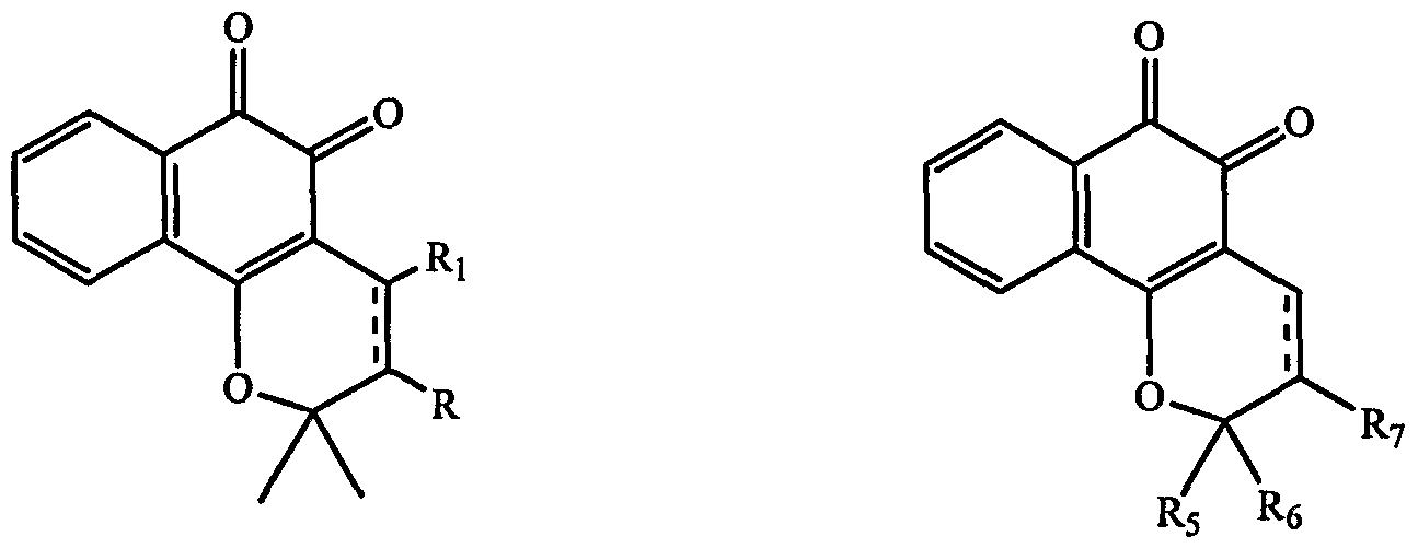
Formula II Formula III where the dotted double bond between the ring carbons represents an optional ring double bond and where R and Ri are each independently selected from hydrogen, hydroxy, sulfhydryl, halogen, substituted alkyl, unsubstituted alkyl, substituted alkenyl, unsubstituted alkenyl, substituted aryl, unsubstituted aryl, substituted alkoxy, unsubstituted alkoxy, and salts thereof. The alkyl groups preferably have from 1 to about 15 carbon atoms, more preferably from 1 to about 10 carbon atoms, still more preferably from 1 to about 6 carbon atoms. The term alkyl refers to both cyclic and noncyclic groups. Straight or branched chain noncyclic alkyl groups are generally more preferred than cyclic groups. Straight chain alkyl groups are generally more preferred than branched. The alkenyl groups preferably have from 2 to about 15 carbon atoms, more preferably from 2 to about 10 carbon atoms, still more preferably from 2 to 6 carbon atoms. Especially preferred alkenyl groups have 3 carbon atoms {i.e., 1-propenyl or 2-propenyl), with the allyl moiety being particularly preferred. Phenyl and napthyl are generally preferred aryl groups. Alkoxy groups include those alkoxy groups having one or more oxygen
linkage and preferably have from 1 to 15 carbon atoms, more preferably from 1 to about 6 carbon atoms. The substituted R and Ri groups may be substituted at one or more available positions by one or more suitable groups such as, for example, alkyl groups having from 1 to 10 carbon atoms or from 1 to 6 carbon atoms, alkenyl groups having from 2 to 10 carbon atoms or 2 to 6 carbon atoms, aryl groups having from six to ten carbon atoms, halogen such as fluoro, chloro and bromo, and N, O and S, including heteroalkyl, e.g., heteroalkyl having one or more hetero atom linkages (and thus including alkoxy, aminoalkyl and thioalkyl) and from 1 to 10 carbon atoms or from 1 to 6 carbon atoms; and where R5 and R6 may be independently selected from hydroxy, C1-Ce alkyl, Ci-C6 alkenyl, Ci-C6 alkoxy, Ci-C6 alkoxycarbonyl, --(CH2)n-aryl, ~(CH2)n-heteroaryl, ~(CH2)n-heterocycle, or ~(CH2)n-phenyl; and R7 is hydrogen, hydroxyl, Ct-C6 alkyl, Ci-C6 alkenyl, CrC6alkoxy, C1-C6 alkoxycarbonyl, ~(CH2)n-amino, — (CH2)n-aryl, ~(CH2)n-heteroaryl, -(CH2)n-heterocycle, or ~(CH2)n-phenyl, wherein n is an integer from 0 to 10.
Preferred β-lapachone analogs and derivatives also contemplated by the present invention include compounds of the following general formula IV:

Formula IV where Ri is (CH2)n-R2, where n is an integer from 0-10 and R2 is hydrogen, an alkyl, an aryl, a heteroaromatic, a heterocyclic, an aliphatic, an alkoxy, an allyloxy, a hydroxyl, an amine, a thiol, an amide, or a halogen. Analogs and derivatives also contemplated by the present invention include 4-acetoxy-β- lapachone, 4-acetoxy-3-bromo-β-lapachone, 4-keto-β-lapachone, 7-hydroxy-β-lapachone, 7-methoxy-β- lapachone, 8-hydroxy-β-lapachone, 8-methoxy-β-lapachone, 8-chloro-β-lapachone, 9-chloro-β- lapachone, 8-methyl-β-lapachone and 8,9-dimethoxy-β-lapachone.
Other β-lapachone analogs and derivatives also contemplated by the present invention include compounds of the following general formula V:
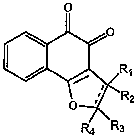
Formula V where R1-R4 are each, independently, selected from the group consisting of H, Ci-C6 alkyl, Ci-C6 alkenyl, Ci-C6 alkoxy, CpC6 alkoxycarbonyl, ~(CH2)n-aryl, --(CH2)n-heteroaryl, ~(CH2)n-heterocycle, or —
(CH2)n-phenyl; or Ri and R2 combined are a single substituent selected from the above group, and R3 and R4 combined are a single substituent selected from the above groups, in which case — is a double bond.
Preferred β-lapachone analogs and derivatives also contemplated by this invention include dunnione and 2-ethyl-6-hydroxynaphtho[2,3-b]-furan-4,5-dione. Preferred β-lapachone analogs and derivatives also contemplated by the present invention include compounds of the following general formula VI:

Formula VI where Ri is selected from H, CH3, OCH3 and NO2.
Additional preferred β-lapachone analogs useful in the methods and kits of the present invention are recited in PCT International Application PCT/US03/37219 (WO2004/045557), which is incorporated by reference herein in its entirety, and which discloses compounds represented by Formula VII:

Formula VII or pharmaceutically acceptable salts thereof, or a regioisomeric mixture thereof, wherein R1-R6 are each, independently, selected from the group consisting of H, OH, substituted and unsubstituted Ci-C6 alkyl, substituted and unsubstituted Ci-C6 alkenyl, substituted and unsubstituted Cr C6 alkoxy, substituted and unsubstituted CrC6 alkoxycarbonyl, substituted and unsubstituted Q-C6 acyl, -(CH2)n-amino, -(CH2)n-aryl, -(CH2)n-heterocycle, and -(CH2)n-phenyl; or one of Ri or R2 and one of R3 or R4; or one of R3 or R4 and one of R5 or R5 form a fused ring, wherein the ring has 4-8 ring members; R7-RlO are each, independently, hydrogen, hydroxyl, halogen, substituted or unsubstituted alkyl, substituted or unsubstituted alkoxy, nitro, cyano or amide; and n is an integer from 0 to 10.
In a preferred embodiment, Ri and R2 are alkyl, R3-R6 are, independently, H, OH, halogen, alkyl, alkoxy, substituted or unsubstituted acyl, substituted alkenyl or substituted alkyl carbonyl, and R7-Ri0 are hydrogen. In another preferred embodiment, Ri and R2 are each methyl and R3-Ri0 are each hydrogen, hi another preferred embodiment, R1-R4 are each hydrogen, R5 and R6 are each methyl and R7-Ri0 are each hydrogen.
Additional preferred β-lapachone analogs useful in the methods and kits of the present invention are recited in PCT International Application PCT/US03/37219 (WO2004/045557), which is incorporated by reference herein in its entirety, and which discloses compounds represented by Formula VIII:

Formula VIII or pharmaceutically acceptable salts thereof, or a regioisomeric mixture thereof, wherein Ri-R4 are each, independently, selected from the group consisting of H, OH, substituted and unsubstituted Ci-C6 alkyl, substituted and unsubstituted CpC6 alkenyl, substituted and unsubstituted Ci- C6 alkoxy, substituted and unsubstituted CrC6 alkoxycarbonyl, substituted and unsubstituted CrC6 acyl, -(CH2)n-amino, -(CH2)n-aryl, -(CH2)n-heterocycle, and -(CH2)n-phenyl; or one of Ri or R2 and one OfR3 or R4 form a fused ring, wherein the ring has 4-8 ring members; R5-R8 are each, independently, hydrogen,
hydroxyl, halogen, substituted or unsubstituted alkyl, substituted or unsubstituted alkoxy, nitro, cyano or amide; and n is an integer from 0 to 10. In certain embodiments of Formula VIII, Ri, R2, R3, R4, R5, Re, R7 and Rs are not each simultaneously H.
3. Methods for Diagnosing and Treating Mammalian Pre-Cancer, Cancer or Hyperproliferation Disorders The PARP activators provided by the present invention can be used as new drugs that kill cancer cells by increasing the activity of PARP. The anticancer drugs promote cell death in cells of a PARP related disorder, such as, pre-cancer or cancer cells, hyperproliferative cells or cells associated with DNA damage. Various cancers to be treated include but are not limited to lung cancer, colorectal cancer, breast cancer, pancreatic cancer, ovarian cancer, prostate cancer, renal carcinoma, hepatoma, brain cancer, melanoma, multiple myeloma, hematologic tumor, and lymphoid tumor. Hyperproliferative disorders refer to conditions in which the unregulated and/or abnormal growth of cells can lead to the development of an unwanted condition or disease, which can be cancerous or non-cancerous, for example a psoriatic condition. As used herein, the term "psoriatic condition" refers to disorders involving keratinocyte hyperproliferation, inflammatory cell infiltration, and cytokine alteration. Hyperproliferative diseases/disorders to be treated include but are not limited to epidermic and dermoid cysts, lipomas, adenomas, capillary and cutaneous hemangiomas, lymphangiomas, nevi lesions, teratomas, nephromas, myofϊbromatosis, osteoplastic tumors, and other dysplastic masses and the like. The PARP related disorder can be a DNA repair disorder, including but not limited to, Ataxia-Talangiectasia, premature aging syndrome, Li-Fraumeini syndrome and premalignant conditions, such as BRCA families. The compositions of the present invention may also be usefiil for pre-cancer, clinical conditions that bear increased risk of progression into cancer.
The present invention is also broadly drawn to the use of the candidate compounds to modulate cell death of cancer cells. Modulation of apoptosis using these test compounds can occur in vitro, in vivo, or ex vivo. Modulation can occur in cancer cells, cell lines, and primary cells.
Another embodiment of the present invention is a method for preventing or inhibiting growth of pre-cancer or cancer cells, the method comprising administering to the cells a composition, which enhances PARP activity or expression in the cell in an amount sufficient to inhibit growth of pre-cancer or cancer cells. The method can be carried out on mammalian cells, including human cells, and can be carried out in vitro or in vivo.
Another embodiment of the present invention is a method for diagnosing and treating mammalian pre-cancer or cancer in a subject, the method comprising obtaining pre-cancer or cancer cells from the subject; testing the pre-cancer or cancer cells from the subject for the presence of PARP; and administering to the subject a composition which enhances PARP activity or expression, in an amount sufficient to inhibit growth of pre-cancer or cancer cells. The composition used in this method can be a β- lapachone analog, derivative, or metabolite thereof, or an niRNA, which increases the expression of PARP.
Another embodiment of the present invention is a method for treating pre-cancer or cancer in a mammalian subject, the method comprising administering to the mammal a composition which enhances PARP activity or expression and monitoring the mammal to determine the state of the pre-cancer or cancer, wherein the composition is administered in an amount sufficient to inhibit the growth of pre- cancer or cancer cells. The composition used in this method can be a β-lapachone analog, derivative, or metabolite thereof, or an mRNA, which increases the expression of PARP.
Another embodiment of the present invention is a method for diagnosing patients who would be receptive to treatment with a composition, which, when administered to mammalian subjects with precancer or cancer, selectively enhances activation or expression of PARP and results in regression of tumor cell growth in the mammalian subjects. The method comprises obtaining cells from the patient; testing the cells for the presence of either PARP or for the presence of PARP activity; wherein the presence in the cells of either PARP or for the presence of PARP activity indicates a patient who would be receptive to treatment.
In this embodiment, compounds such as β-lapachone analogs, derivatives or metabolites thereof could be used to diagnose cancer. Cells isolated from subjects could be cultured in the presence or absence of β-lapachone analogs, derivatives or metabolites thereof. Cells that have their growth rates inhibited in the β-lapachone analogs, derivatives or metabolites thereof treated cells relative to control would be pre-cancer or cancer cells. The subject could then be diagnosed with pre-cancer or cancer.
The PARP activators of the present invention may help the development of drugs that inhibit apoptosis during various forms of tissue injury such as ischemia, reperfusion injury, mechanical injury, inflammation or immunological damage.
3.1 The Compositions for the Test Compounds and PARP Activators
As discussed above, in one aspect, the present invention provides a composition, which, when administered to mammalian subjects with pre-cancer, cancer or a hyper-proliferative disorder, selectively enhances PARP activity and results in regression of cell growth in mammalian cells and subjects. The composition can also be in the form of a pharmaceutical composition or in a kit.
The compositions of the present invention can be incorporated into pharmaceutical compositions suitable for administration. Such compositions typically comprise the substance that enhances PARP activity or expression and a pharmaceutically acceptable carrier. As used herein, "pharmaceutically acceptable carrier" is intended to include any and all solvents, dispersion media, coatings, antibacterial and antifungal agents, isotonic and absorption delaying agents, and the like, compatible with pharmaceutical administration. Suitable carriers are described in the most recent edition of Remington's Pharmaceutical Sciences, a standard reference text in the field, which is incorporated herein by reference. Preferred examples of such carriers or diluents include, but are not limited to, water, saline, finger's solutions, dextrose solution, and 5% human serum albumin. Liposomes and non-aqueous vehicles such as fixed oils may also be used. The use of such media and agents for pharmaceutically active substances is well known in the art. Except insofar as any conventional media or agent is incompatible with the
active compound, use thereof in the compositions is contemplated. Supplementary active compounds can also be incorporated into the compositions.
A pharmaceutical composition of the present invention is formulated to be compatible with its intended route of administration. Examples of routes of administration include parenteral, e.g., intravenous, intradermal, subcutaneous, oral (e.g., inhalation), transdermal (i.e., topical), transmucosal, and rectal administration. Solutions or suspensions used for parenteral, intradermal, or subcutaneous application can include the following components: a sterile diluent such as water for injection, saline solution, fixed oils, polyethylene glycols, glycerine, propylene glycol or other synthetic solvents; antibacterial agents such as benzyl alcohol or methyl parabens; antioxidants such as ascorbic acid or sodium bisulfite; chelating agents such as ethylenediaminetetraacetic acid (EDTA); buffers such as acetates, citrates or phosphates, and agents for the adjustment of tonicity such as sodium chloride or dextrose. The pH can be adjusted with acids or bases, such as hydrochloric acid or sodium hydroxide. The parenteral preparation can be enclosed in ampoules, disposable syringes or multiple dose vials made of glass or plastic. Pharmaceutical compositions suitable for injectable use include sterile aqueous solutions (where water soluble) or dispersions and sterile powders for the extemporaneous preparation of sterile injectable solutions or dispersion. For intravenous administration, suitable carriers include physiological saline, bacteriostatic water, Cremophor EL™ (BASF, Parsippany, NJ.) or phosphate buffered saline (PBS). hi all cases, the composition must be sterile and should be fluid to the extent that easy syringeability exists. It must be stable under the conditions of manufacture and storage and must be preserved against the contaminating action of microorganisms such as bacteria and fungi. The carrier can be a solvent or dispersion medium containing, for example, water, ethanol, polyol (for example, glycerol, propylene glycol, and liquid polyethylene glycol, and the like), and suitable mixtures thereof. The proper fluidity can be maintained, for example, by the use of a coating such as lecithin, by the maintenance of the required particle size in the case of dispersion and by the use of surfactants. Prevention of the action of microorganisms can be achieved by various antibacterial and antifungal agents, for example, parabens, chlorobutanol, phenol, ascorbic acid, thimerosal, and the like. In many cases, it will be preferable to include isotonic agents, for example, sugars, polyalcohols such as manitol, sorbitol, sodium chloride in the composition. Prolonged absorption of the injectable compositions can be brought about by including in the composition an agent which delays absorption, for example, aluminum monostearate and gelatin.
Sterile injectable solutions can be prepared by incorporating the active compound (eg., the substance that enhances PARP activity or expression) in the required amount in an appropriate solvent with one or a combination of ingredients enumerated above, as required, followed by filtered sterilization. Generally, dispersions are prepared by incorporating the active compound into a sterile vehicle that contains a basic dispersion medium and the required other ingredients from those enumerated above. In the case of sterile powders for the preparation of sterile injectable solutions, methods of preparation are vacuum drying and freeze-drying that yields a powder of the active ingredient plus any additional desired ingredient from a previously sterile-filtered solution thereof.
Oral compositions generally include an inert diluent or an edible carrier. They can be enclosed in gelatin capsules or compressed into tablets. For the purpose of oral therapeutic administration, the active compound can be incorporated with excipients and used in the form of tablets, troches, or capsules. Oral compositions can also be prepared using a fluid carrier for use as a mouthwash, wherein the compound in the fluid carrier is applied orally and swished and expectorated or swallowed. Pharmaceutically compatible binding agents, and/or adjuvant materials can be included as part of the composition. The tablets, pills, capsules, troches and the like can contain any of the following ingredients, or compounds of a similar nature: a binder such as microcrystalline cellulose, gum tragacanth or gelatin; an excipient such as starch or lactose, a disintegrating agent such as alginic acid, Primogel, or corn starch; a lubricant such as magnesium stearate or Sterotes; a glidant such as colloidal silicon dioxide; a sweetening agent such as sucrose or saccharin; or a flavoring agent such as peppermint, methyl salicylate, or orange flavoring.
For administration by inhalation, the compounds are delivered in the form of an aerosol spray from pressured container or dispenser which contains a suitable propellant, e.g., a gas such as carbon dioxide; or a nebulizer. Systemic administration can also be by transmucosal or transdermal means. For transmucosal or transdermal administration, penetrants appropriate to the barrier to be permeated are used in the formulation. Such penetrants are generally known in the art, and include, for example, for transmucosal administration, detergents, bile salts, and fusidic acid derivatives. Transmucosal administration can be accomplished through the use of nasal sprays or suppositories. For transdermal administration, the active compounds are formulated into ointments, salves, gels, or creams as generally known in the art.
The compounds can also be prepared in the form of suppositories (e.g., with conventional suppository bases such as cocoa butter and other glycerides) or retention enemas for rectal delivery.
In one embodiment, the active compounds are prepared with carriers that will protect the compound against rapid elimination from the body, such as a controlled release formulation, including implants and microencapsulated delivery systems. Biodegradable, biocompatible polymers can be used, such as ethylene vinyl acetate, polyanhydrides, polyglycolic acid, collagen, polyorthoesters, and polylactic acid. Methods for preparation of such formulations will be apparent to those skilled in the art. The materials can also be obtained commercially from Alza Corporation and Nova Pharmaceuticals, Inc. Liposomal suspensions (including liposomes targeted to infected cells with monoclonal antibodies to viral antigens) can also be used as pharmaceutically acceptable carriers. These can be prepared according to methods known to those skilled in the art, for example, as described in U.S. Patent No. 4,522,811.
It is especially advantageous to formulate oral or parenteral compositions in dosage unit form for ease of administration and uniformity of dosage. Dosage unit form as used herein refers to physically discrete units suited as unitary dosages for the subject to be treated; each unit containing a predetermined quantity of active compound calculated to produce the desired therapeutic effect in association with the required pharmaceutical carrier. The specification for the dosage unit forms of the present invention are dictated by and directly dependent on the unique characteristics of the active compound and the particular
therapeutic effect to be achieved, and the limitations inherent in the art of compounding such an active compound for the treatment of individuals.
The pharmaceutical compositions can be included in a container, pack, or dispenser together with instructions for administration.
EXAMPLES
Examples are provided below to further illustrate different features of the present invention. The examples also illustrate useful methodology for practicing the invention. These examples do not limit the claimed invention.
Example 1. PARP Screening 1. Cell Death Assays
Cell death was determined by the 3-(4,5-dimethylthiazol-2-yl)-2,5-diphenyl tetrazolium bromide (MTT) assay or by trypan blue exclusion, as indicated. Briefly, HeLa and DLDl cells were plated in a 96-well plate at 10,000 cells per well, cultured for 24 h in complete growth medium, then treated with various concentrations of β-lapachone for 4h. MTT was added to a final concentration of 0.5mg/ml, and incubated for 1 hr, followed by assessment of cell viability using a microplate reader at 570nm.
For the trypan blue exclusion assay, HeLa and DLDl cells were plated in 6-well plate and treated in the same way. They were harvested, and trypan blue dye solution was added to the cell suspension. Total cell counts and viable cell numbers were determined with a hemacytometer. For the PARP inhibition study, cells were pre-teated for 1 h with the PARP inhibitor 3-aminobenzamide (3-AB, 5mM), and then co-treated with inhibitor and β-lapachone for a further 4 h, followed by MTT assay or trypan blue staining.
For NAD+ supplementation experiment, MCF7 cells were plated in 96-well plates at a density of 10,000 cells per well. Sixteen to eighteen hours later cells were pre-treated with 5 μM 3- aminobenzamide, 10 mM NAD+, or vehicle control (all treatments in formulated in growth media: DMEM with 10% fetal bovine serum) for 1 hour at 370C. Following this incubation, cells (under each pre-incubation treatment) were treated with β-lapachone at indicated concentrations for 4 hours at 370C. An MTT assay was then performed as described above. 2. Immunofluorescence analysis
HeLa and DLDl cells were grown on coverslips for PARP activity experiments. Cells treated with 4 uM β-lapachone at different time points and fixed with methanol acetone (70/30, v/v) for 10 min at -200C. Coverslips were air dried and rehydrated in PBS at room temperature for 10 min. Samples were than incubated in blocking buffer(PBS, 5% FBS) for 10 minutes at room temperature in a humid chamber. Cells were incubated overnight at 40C with monoclonal anti-poly(ADP-ribose) antibody (1OH, 1 : 100 dilution). After washing, the cells were incubated for 1 hr at room temperature with a 1 :500 dilution of FITC conjugated anti-mouse antibody. For the PARP inhibition study, cells pre-teated for 1 h with the PARP inhibitor 3-aminobenzamide (3-AB, 5mM), and then co-treated with inhibitor and β-
lapachone for a further 10 minutes followed by immunofluorescence staining. Immunofluorescence was evaluated using an immunofluorescence microscope equipped with a CCD camera.
3. NAD Depletion Assay
HeLa cells were plated at 6 x 10s cells per well (35 mm, 6-well dishes). Eighteen hours after plating, cells were treated with β-lapachone (0, 2, 4 or 8 μM) in growth media (DMEM; 10% FBS) for the time periods indicated (15, 30 and 60 minutes). Following drug treatment cells were washed 2X with PBS. Cells were subsequently lysed in 200 μLNAD lysis buffer (61 mM glycyl-glycine, pH 7.4, 0.1% Triton-X-100). Cellular lysates were clarified by centrifugation at 16,000 g for 10 minutes. Aliquots of clarified lysates were transferred to 96-well plates (25 μL samples in triplicate) for to determine NAD+ concentrations.
Determination of lysate NAD levels was performed using a modification of the NAD recycling assay described by Ying et al (PNAS, 98(21):12227-32 (2001)). Briefly, the NAD reaction mixture consisted of 0. ImM 3-[4,5-dimethylthiazol-2-yl]-2,5-diphenyl-tetrazolium bromide (MTT), 0.9 mM phenazine methosulfate, 13 units/ml alcohol dehydrogenase (from yeast extract, Sigma), 100 mM nicotinamide, and 5.7% ethanol in 61 mM Gly-Gly buffer (pH 7.4). To each aliquot of cellular lysate, 200 μL of reaction mixture was added. Absorbance readings (560 run) were taken every minute for five minutes. Results were calibrated with NAD+ standards and were subsequently normalized to protein concentration as determined using the Bio-Rad protein Assay. Finally, this data was normalized to control treated cells (0 μM β-lapachone at 15 minutes) in order to plot percent cellular NAD+ levels remaining.
4. PARP Activity Assay
A cell extract was generated by scraping a 15 cm (~80% confluent) dish of MCF7 cells into 1.5 ml of PARP buffer (50 mM Tris pH 8.0, 25 mM MgCl2) containing protease inhibitors. The cell suspension was sonicated at 30% amplitude with 3 X 10 second bursts on ice. The cell lysate was subsequently clarified by centrifugation at 16,000 g for 10 minutes at 4 0C. Any remaining insoluble material was removed by passing the clarified lysate through a 5 ml syringe containing a cotton pad- filter. The protein concentration of extracts was typically between 0.6 and 1.0 μg/ml.
PARP in vitro activity was determined using a modification of the PARP in vitro assay from TREVIGEN™. Reactions were comprised of the following: -60 μg of cellular protein, 100 μM NAD, 10 μg of histone Hl , β-lapachone (at various concentrations) or DMSO control, 25 mM MgCl2, 50 mM Tris-Cl pH 8.0. Reactions were incubated for 10 minutes at room temperature and terminated by the addition of 900 μL of cold 25% trichloroacetic acid. Terminated reactions were incubated on ice for 10 minutes. TCA-precipitated proteins were isolated by passing reaction over glass-fiber filters under vacuum. Filters were subsequently washed three times with 5 ml of 5% TCA followed by 2 washes with cold ethanol. Filters were dried and transferred to scintillation cocktail. The amount Of32P labeled NAD incorporated into polyribosylated proteins was then determined by liquid scintillation measurement.
Example 2: β-lapachone induction of cell death is inhibited by the PARP inhibitor 3-aminobenzamide.
MTT assays showed that β-lapachone-induced cell death is blocked by PARP inhibitor 3- aminobenzamide (3-AB). HeLa and DLDl cells were plated in 96-well plates at 10,000 cells per well, cultured for 24 h in complete growth medium, pretreated with PARP inhibitor 3-AB (5mM) or equal volume of DMSO for 1 h, and then exposed to β-lapachone at various concentrations for a further 4 h, followed by MTT assay.
As shown in Figure 1, HeLa cell survival percentage rises from approximately 5% to 60% in HeLa cells, and to approximately 75% in DLDl cells.
Similar results were shown using Trypan Blue staining in HeLa cells (Figure 2) and DLDl cells (Figure 3).
Example 3: β-lapachone induces rapid cellular activation of PARP, which is blocked by 3- aminobenzamide. β-lapachone induces rapid activation of PARP in HeLa cells. HeLa cells were grown on coverslips for 24 h, then treated with 4 μM β-lapachone at different time points and fixed with methanol acetone (70/30, v/v) for 10 min. Samples were incubated in blocking buffer (5% FBS in PBS) for 10 minutes at room temperature in a humid chamber. Cells were incubated overnight at 40C with monoclonal anti-poly(ADP-ribose) antibody (1OH 1:100). After washing, the cells were incubated for 1 h at room temperature with FITC conjugated anti-mouse antibody (1:1,000). Immunofluorescence was evaluated using an immunofluorescence microscope equipped with a CCD camera.
As shown in Figure 4, β-lapachone increases fluorescence due to the presence of monoclonal anti-poly(ADP-ribose) antibody bound to the product of PARP, showing a β-lapachone induced activation of PARP. β-lapachone induced activation of PARP in HeLa cells is blocked by 3-AB. HeLa cells were grown on coverslips for 24 h, pretreated with 5mM PARP inhibitor 3-AB or DMSO for Ih, then exposed to 4 μM β-lapachone for 10 minutes, followed by immunofluorescence staining as described above.
As shown in Figure 5, β-lapachone induced activation of PARP in HeLa cells, as was also shown in Figure 4, is inhibited by 3-aminobenzamide, verifying that the PARP product, poly(ADP ribose) is indeed generated by PARP and its accumulation is caused by β-lapachone activation of PARP activity. Similar results were shown using the methods described above for Figure 5 in DLDl cells.
These results are shown in Figure 6.
Example 4: β-lapachone induces NAD+ depletion in cells, and reconstitution OfNAD+ to these cells decreases β-lapachone induced cytotoxicity. As shown in Figure 7, β-lapachone induces a rapid depletion of cellular NAD+ levels. Within 15 minutes, the 8 μM β-lapachone treatment reduced NAD+ levels to -20% of control. By 30 minutes the β- lapachone concentrations in excess of the IC50 (~2.8 μM) reduced cellular NAD+ levels to less than 50% of control. The rapid kinetics of β-lapachone-mediated NAD+ depletion implies that this phenomenon is
due to an enzymatic process. Furthermore, this process is inhibited by the compound 3-aminobenzamide {data not shown). Together these data suggest that a PARP activity is involved in β-lapachone-induced NAD+ depletion.
As shown in Figure 8, addition of exogenous NAD+ to cell growth media can reduce the cytotoxicity of β-lapachone treatment (IC50 values of ~2.7 and ~6.6 μM for control versus NAD+ supplementation respectively in MCF7 cells). The ability of exogenous NAD+ to protect cells from β- lapachone induced cytotoxicity is likely limited by cellular uptake of this charged molecule. Similar results were observed in experiments using DLDl, HeLa and SW480 cells. These results taken together with those in Figure 7 suggest that the depletion of cellular NAD+ levels is a contributing factor to β- lapachone induced cytotoxicity.
Example 5: β-lapachone induces activation of PARP in cell lvsates.
Figure 9 shows that the addition of β-lapachone to cellular lysate induces enhancement of PARP activity above the basal level (0 μM β-lapachone control). Furthermore, this induction of PARP activation by β-lapachone was dependent on β-lapachone concentration (dose-dependent). This observation is significant in that it shows that a PARP family member is a direct target of β-lapachone.
Example 6: Induction of E2F1 expression promotes apoptosis in DLDl and SW-480 cells.
To investigate E2F1 induced apoptosis, an inducible system was established. Human colon cancer cell lines harboring a mutated p53 gene were used to generate E2F1 -inducible cell lines
(Rodrigues, et al, PNAS, 87:7555-9 (1990)). E2F1 expression was effectively induced and tightly controlled by addition of tetracycline (Figure 10A). Under the tetracycline induction conditions, a significant percentage of DLDl cells undergoing apoptosis were detected at a given time point (10% at day 3; 15% at day 4) as determined by propidium iodide (PI) staining and flow cytometry (Figure 10B). The apoptosis was further confirmed by assaying morphological changes following expression of E2F1 (Figure 10C). Similar data was obtained in E2Fl-inducible SW-480 human colon cancer cells. In addition to proapoptotic function, E2F1 is a necessary transcription factor for cell proliferation (Johnson, D. etal, Nature 365:349-52 (1993); Wu, L. etal. Nature 414:457-62 (2001)). Under these experimental conditions, no significant changes in overall cell cycle distribution were observed. Colony formation of cancer cells was completely ablated by E2F1 induction (Figure 10D), which is consistent with the proapoptotic and tumor suppressor function of E2F1 (Yamasaki, L. et al, Cell 85:537-48 (1996); Field, S. J. etal., Cell 85:549-61 (1996)), suggesting that E2F1 induced apoptosis is different from proapoptotic activity of an oncogene.
Example 7: E2F1 activates caspase independent apoptosis.
To further investigate the mechanism of E2F1 -induced apoptosis, the role of caspase activation was investigated. Two forms of caspase-3, procaspase and cleaved caspase were detected by western blot analysis. Although the cells clearly underwent apoptosis following expression of E2F1, there was
surprisingly no activation of caspase-3, the final effector of caspase-dependent apoptosis following tetracycline induction for 0- 72 hours and controls at 24 and 48 hours (Figure 10E). Further, it was determined if E2Fl-induced, p53 independent apoptosis was affected by inhibiting caspase function with a pancaspase inhibitor. Apoptosis induced by camptothecin, a known caspase-3 activator, was used as a positive control. Addition of the pancaspase inhibitor Z-VAD at 50 μM blocked camptothecin-induced apoptosis but failed to block E2Fl-induced apoptosis (Figure 10F). Z-VAD addition alone had no cytotoxic effects. These results suggest that E2F1 induces a caspase-independent cell death pathway in p53 mutant cancer cells.
Activation of PARP-I has been implicated in caspase independent apoptosis. It was investigated whether E2F1 induced cell death involved PARP-I activation. Poly ADP ribosylation (PAR), the functional indicator of PARP-I activation, was detected by immunoblot with a specific anti-PAR antibody. As shown in Figure 1 IA, E2F1 strongly activated poly ADP ribosylation of proteins. Immunocytochemical staining of PAR further confirmed the activity of PARP-I (Figure 1 IB). The nuclear PAR stain was increased after expression of E2F1 for 24 h and reached a plateau at 48 h. To determine if poly ADP ribosylation plays a causal role in E2F1 induced apoptosis, 3'-aminobenzamide (3'-AB), a universal inhibitor of PARP activity, was used. As shown in Figure 11C, 3'-AB inhibited E2F1 induced apoptosis by more than 70% at the concentration of 5 mM (PO.0001). 3'-AB addition alone showed no effect on apoptosis over the control group. These data suggest that E2F1 activates protein poly ADP ribosylation that contributes to caspase independent apoptosis.
Example 8: E2F affects protein expression of PARP.
Protein poly ADP ribosylation is catalysed by members of the poly (ADP-ribose) polymerase family that consists of 18 genes (Ame, J. C, et al, Bioessays 26:882-93 (2004)). The PARP-I protein is the dominant member in most cells, which contributes the majority of overall poly ADP ribosylation induced by DNA damage (Davidovic, L. et al, Exp Cell Res 268:7-13 (2001)). To distinguish which member of the PARP family was involved in E2Fl-induced apoptosis, siRNA technology was utilized to reduce PARP-I protein levels. A chemically synthesized PARP-I siRNA pool was electroporated into the DLD1-E2F1 inducible cell line. The PARP-I protein began to decrease at day 2 and reached the lowest levels at day 4 and gradually recovered from day 6 as shown by western blot (Figure 12A) and immunocytochemical staining. Following expression of E2F1, the nuclear accumulation of PARP was abrogated by transfection of siPARP-1 (Figure 12B), suggesting that E2Fl-induced PARP activity is mediated by PARP-I. The E2Fl-induced apoptosis was compromised following PARP-I knockdown (PO.001) (Figure 12C). Taken together, these data suggest that E2F1 requires PARP-I and its activity to activate a caspase-independent apoptosis pathway. E2F1 is known as a transcription factor. Yet several lines of evidence suggest that the transcriptional activity of E2F1 is dispensable for its apoptotic function (Phillips, A. C, et al., Genes Dev 11:1853-63 (1997); Hsieh, J. K., et al, Genes Dev 11:1840-52 (1997)). It was next determined if E2F1 affected transcription of PARP-I. The expression of E2F1 greatly increased PARP-I protein levels
(Figure 12D), suggesting a possibility that E2F1 induced PARP-I expression by transactivation of PARP-I. However, RT-PCR and Northern blot analysis revealed that E2Fl-induced PARP-I protein elevation occurred at the protein level, not at mRNA levels (Figurel2E, Figure 12F). These results suggest that E2F1 induces PARP-I through a non-transcriptional mechanism. To investigate if endogenous E2F1 affects PARP-I protein levels, E2F1 was silenced in a non- inducible system. Silencing E2F1 with siRNA reduced endogenous PARP-I protein level while silencing PARP-I had no effect on E2F1 protein level (Figure 13), suggesting a role of E2F1 in regulating PARP-I protein levels.
Example 9: E2F1 mediates apoptosis by inducing translocation of AIF in a PARP dependent manner.
The translocation of apoptosis-inducing factor (AIF) from mitochondria to nuclei has been implicated in the apoptosis induced by PARP-I (Yu, S. W. et al, Science 297:259-63 (2002); Davidovic, L., et al, Exp Cell Res 268:7-13 (2001)). It was next determined if E2F1 -mediated increase in PARP-I protein levels triggered AIF translocation. The expression of E2F1 resulted in AIF and cytochrome c release from mitochondria and translocation of AIF into nuclei. (Figure 14A, Figure 14B). To determine if the E2F1 induced AIF translocation was primarily mediated by PARP-I, PARP-I protein levels were reduced with siRNA. While E2F1 induction drove nuclear translocation of AIF in cells transfected with control siRNA, silencing PARP-I with a specific siRNA blocked E2F1 -induced nuclear translocation of AIF (Figure 14C, Figure 14D). These results suggest that the E2F1 -PARP-I apoptosis pathway involves AIF translocation.
It has been shown that E2F1 transcriptionally regulates apoptotic genes, notably/? 73 and apafl, which contribute to transcription-dependent apoptosis pathway of E2F1 (Irwin, M. et al, Nature 407:645-8 (2000); Furukawa, Y. et al.,JBiol Chem 277:39760-8 (2002)). p73 was found to be a target gene for E2F1 in the p53 independent apoptosis pathway. Although p73 mRNA was marginally induced after E2F1 expression as detected by RT-PCR (Figure 15) effective knockdown of p73 by siRNA did not show any protective effect on E2F1 -induced apoptosis. E2F1 has also been shown to induce transcription of apafl (Furukawa, Y. et al, J Biol Chem 277, 39760-8 (2002)). However, RT-PCR data revealed no expression change in apafl in the E2F1 inducible cells (Figure 15). The reason for lack of significant induction o(p73 and apafl is unclear, and is possibly due to difference in E2F1 levels, duration, and relatively rapid cell death by PARP-I induction.
In one embodiment, modulation of PARP expression or PARP activity and the resulting modulation of apoptosis occurs via modulation of E2F expression or activity. In a preferred embodiment, E2F is E2F1. The E2F1 -PARP-I cell death pathway provides a p53 independent link between checkpoint regulators and apoptosis. E2F1 is phosphorylated by ATM and Chk2, which leads to the stabilization of E2F1 (Lin, W. C, et al, Genes Dev 15:1833-44 (2001); Bartek, J., et al., Nat Rev MoI Cell Biol 2:877-86 (2001)). Conversely, E2F1 activates ATM and Chk2 kinase, thereby forming a positive feedback loop (Rogoff, H. A. et al, MoI Cell Biol 24:296%-ll (2004); Berkovich, E. & Ginsberg, D., Oncogene 22:161-7 (2003)). It is speculated that the presence of oncogenic signals or
ATM-Chk2 activation due to DNA damage could lead to activation of the E2F1 -PARP-I pathway that may lead to apoptosis, DNA repair or cell cycle arrest depending on the magnitude of the insults, potentially serving an important function in the prevention of tumor formation. This pathway may underlie the observed tumor suppressor function of E2F1 (Yamasaki, L. et al, Cell 85:537-48 (1996); Field, S. J. et al, Cell 85:549-61 (1996)).
The interaction between E2F1 and PARP-I establishes a new link between cell cycle regulation and genome surveillance. Results suggest that the endogenous level of PARP-I protein is dependent on E2F1. It has been suggested that PARP-I enhances transcription activity of E2F1 (Simbulan-Rosenthal, C. M., etal, Oncogene 18:5015-23 (1999); Simbulan-Rosenthal, C. M. etal, Oncogene 22:8460-71 (2003)). These observations suggest a positive feedback loop between genome surveillance and cell cycle regulation. These feedback interactions may offer a highly sensitive mechanism to prevent proliferation of, and to promote elimination of cells with irreparable DNA damage.
The lack of mutations in E2F1 and PARP-I in human cancers suggests significance of this checkpoint-apoptotic pathway in mediating the anti-cancer activity of chemotherapy, offering potentials for activating this checkpoint pathway to develop efficacious novel therapies against cancers, including those bearing mutations in p53 pathways.
Other embodiments are within the following claims. While several embodiments have been shown and described, various modifications may be made without departing from the spirit and scope of the present invention.
Claims
1. A method for screening for a PARP activator comprising the step of assessing the PARP- activating effect of a test compound in cells containing DNA encoding PARP.
2. The method of claim 1 wherein the PARP is PARP-I, PARP-2, or both PARP-I and PARP- 2.
3. The method of claim 1 wherein the step of assessing the PARP-activating effect in cells comprising, exposing the cells to a test compound, measuring the activity of PARP in the cells in the presence and in the absence of the test compound, and comparing the activity of PARP in the presence and in the absence of the test compound.
4. The method of claim 1 wherein PARP-activating effect is determined by an increase in poly(ADP ribose) synthesis.
5. The method of claim 1 wherein the cells are cancer cells.
6. The method of claim 5 wherein the cancer cells are cells in a cancer of a vertebrate, mammal, or human.
7. The method of claim 5 wherein the cancer cells are derived from a cancer of a vertebrate, mammal, or human.
8. The method of claim 5 wherein the cancer cells are cultured cancer cells.
9. The method of claim 8 wherein the cultured cancer cells are selected from the group consisting of MCF-7 (human breast cancer cells), DLDl (human colonic cells), SW480 (human colonic cells), and Paca-2 (human pancreatic cancer cells).
10. The method of claim 1 wherein the test compound is a small molecule.
11. The method of claim 1 wherein the test compound is an analog, derivative, or metabolite of β-lapachone.
12. The method of claim 8 further comprising the step of assessing the PARP-activating effect of the test compound in normal cells containing DNA encoding PARP.
13. The method of claim 12 wherein the step of assessing the PARP-activating effect in normal cells comprising, exposing the normal cells to a test compound, measuring the activity of PARP in the normal cells in the presence and in the absence of the test compound, and comparing the activity of PARP in the presence and in the absence of the test compound.
14. The method of claim 12 wherein the normal cells are normal cells in a vertebrate, mammal, or human.
15. The method of claim 12 wherein the normal cells are normal cells derived from a vertebrate, mammal, or human.
16. The method of claim 12 wherein the normal cells are cultured normal cells.
17. The method of claim 16 wherein the cultured normal cells are selected from the group consisting of MCF-IOA (nontransformed breast epithelial cells), NCM460 (normal colonic epithelial cells), PBMC (proliferating peripheral blood mononuclear cells)
18. The method of claim 12 further comprising the step of selecting the test compound that has a higher PARP-activating effect in the cancer cells than in the normal cells.
19. A method for screening for a PARP activator, comprising the step of assessing the PARP- activating effect of a test compound in the lysate of cells containing DNA encoding PARP.
20. The method of claim 19 wherein the cells are cancer cells.
21. The method of claim 20 further comprising, assessing the PARP-activating effect of the test compound in the lysate of normal cells containing DNA encoding PARP, and comparing the PARP-activating effects of the test compound in the cancer cell lysate and the normal cell lysate.
22. A method for screening for a PARP activator comprising, contacting PARP with a test compound, measuring the activity of PARP in the presence and in the absence of the test compound, and comparing the activity of PARP in the presence and in the absence of the test compound.
23. The method of claim 22 wherein the PARP is PARP-I or PARP-2.
24. The method of claim 22 further comprising selecting the test compound that increases the PARP activity.
25. The method of claim 24 further comprising, assessing the PARP-activating effect of the selected compound in cancer cells containing DNA encoding PARP, or the lysate of the cells, assessing the PARP-activating effect of the selected compound in the lysate of normal cells containing DNA encoding PARP, or the lysate, and comparing the PARP-activating effects of the selected compound in the cancer cells or the lysate and the normal cells or the lysate.
26. A method of treating or preventing cancer in a subject comprising increasing PARP activity in cancer cells of the subject.
27. The method of claim 26 comprising selectively increasing PARP activity in cancer cells of the subject.
28. The method of claim 26 comprising administering to the subject a therapeutically effective amount of a PARP activator.
29. The method of claim 26 comprising administering to the subject a therapeutically effective amount of a selective activator of PARP.
30. The method of claim 29 wherein the compound is an analog, derivative, or metabolite of β- lapachone.
31. The method of claim 26, wherein the subject is a vertebrate, mammal, or human.
Priority Applications (3)
| Application Number | Priority Date | Filing Date | Title |
|---|---|---|---|
| CA002594234A CA2594234A1 (en) | 2005-01-07 | 2006-01-09 | Compositions for modulation of parp and methods for screening for same |
| EP06717893A EP1836320A2 (en) | 2005-01-07 | 2006-01-09 | Compositions for modulation of parp and methods for screening for same |
| JP2007550550A JP2008526237A (en) | 2005-01-07 | 2006-01-09 | Composition for modulation of PARP and screening method thereof |
Applications Claiming Priority (2)
| Application Number | Priority Date | Filing Date | Title |
|---|---|---|---|
| US64235305P | 2005-01-07 | 2005-01-07 | |
| US60/642,353 | 2005-01-07 |
Publications (2)
| Publication Number | Publication Date |
|---|---|
| WO2006078503A2 true WO2006078503A2 (en) | 2006-07-27 |
| WO2006078503A3 WO2006078503A3 (en) | 2007-02-15 |
Family
ID=36692723
Family Applications (1)
| Application Number | Title | Priority Date | Filing Date |
|---|---|---|---|
| PCT/US2006/000748 WO2006078503A2 (en) | 2005-01-07 | 2006-01-09 | Compositions for modulation of parp and methods for screening for same |
Country Status (5)
| Country | Link |
|---|---|
| US (1) | US20060204981A1 (en) |
| EP (1) | EP1836320A2 (en) |
| JP (1) | JP2008526237A (en) |
| CA (1) | CA2594234A1 (en) |
| WO (1) | WO2006078503A2 (en) |
Cited By (8)
| Publication number | Priority date | Publication date | Assignee | Title |
|---|---|---|---|---|
| WO2011058367A2 (en) | 2009-11-13 | 2011-05-19 | Astrazeneca Ab | Diagnostic test for predicting responsiveness to treatment with poly(adp-ribose) polymerase (parp) inhibitor |
| WO2017137761A1 (en) | 2016-02-10 | 2017-08-17 | Hilmar M Warenius | Compositions and uses thereof |
| WO2018162439A1 (en) | 2017-03-08 | 2018-09-13 | Onxeo | New predictive biomarker for the sensitivity to a treatment of cancer with a dbait molecule |
| WO2018197461A1 (en) | 2017-04-28 | 2018-11-01 | Akribes Biomedical Gmbh | A parp inhibitor in combination with a glucocorticoid and/or ascorbic acid and/or a protein growth factor for the treatment of impaired wound healing |
| WO2019175132A1 (en) | 2018-03-13 | 2019-09-19 | Onxeo | A dbait molecule against acquired resistance in the treatment of cancer |
| EP3594343A1 (en) | 2015-07-23 | 2020-01-15 | Institut Curie | Use of a combination of dbait molecule and parp inhibitors to treat cancer |
| US10799501B2 (en) | 2015-11-05 | 2020-10-13 | King's College Hospital Nhs Foundation Trust | Combination of an inhibitor of PARP with an inhibitor of GSK-3 or DOT1L |
| WO2021148581A1 (en) | 2020-01-22 | 2021-07-29 | Onxeo | Novel dbait molecule and its use |
Families Citing this family (10)
| Publication number | Priority date | Publication date | Assignee | Title |
|---|---|---|---|---|
| WO2007011962A2 (en) * | 2005-07-18 | 2007-01-25 | Bipar Sciences, Inc. | Treatment of cancer |
| CN102379884A (en) | 2006-09-05 | 2012-03-21 | 彼帕科学公司 | Inhibition of fatty acid synthesis by PARP inhibitors and methods of treatment thereof |
| CA2662517A1 (en) | 2006-09-05 | 2008-03-13 | Jerome Moore | Treatment of cancer |
| WO2008146035A1 (en) * | 2007-05-25 | 2008-12-04 | Astrazeneca Ab | Combination of chk and parp inhibitors for the treatment of cancers |
| MX2010008572A (en) * | 2008-02-04 | 2010-11-30 | Bipar Sciences Inc | Methods of diagnosing and treating parp-mediated diseases. |
| US8435961B2 (en) * | 2009-06-26 | 2013-05-07 | Massachusetts Institute Of Technology | Methods and compositions for increasing the activity of inhibitory RNA |
| US8268550B2 (en) * | 2009-06-26 | 2012-09-18 | Massachusetts Institute Of Technology | Compositions and methods for identification of PARP function, inhibitors, and activators |
| WO2010151664A2 (en) | 2009-06-26 | 2010-12-29 | Massachusetts Institute Of Technology | Compositions and methods for treating cancer and modulating stress granule formation |
| US20150198597A1 (en) * | 2012-08-08 | 2015-07-16 | Stichting Het Nederlands Kanker Instituut | Method for monitoring PARP activity in cells by PARP activation |
| KR102145344B1 (en) * | 2016-03-04 | 2020-08-19 | 가톨릭대학교 산학협력단 | Method for preparing section sample using micropillar |
Citations (12)
| Publication number | Priority date | Publication date | Assignee | Title |
|---|---|---|---|---|
| US10169A (en) | 1853-10-25 | Improvement in seed-planters | ||
| US337219A (en) | 1886-03-02 | wohlrab | ||
| US4522811A (en) | 1982-07-08 | 1985-06-11 | Syntex (U.S.A.) Inc. | Serial injection of muramyldipeptides and liposomes enhances the anti-infective activity of muramyldipeptides |
| US5223409A (en) | 1988-09-02 | 1993-06-29 | Protein Engineering Corp. | Directed evolution of novel binding proteins |
| US5233409A (en) | 1992-02-25 | 1993-08-03 | Schwab Karl W | Color analysis of organic constituents in sedimentary rocks for thermal maturity |
| WO1994004145A1 (en) | 1992-08-21 | 1994-03-03 | Dana Farber Cancer Institute | Treatment of human viral infections |
| US5763625A (en) | 1995-04-25 | 1998-06-09 | Wisconsin Alumni Research Foundation | Synthesis and use of β-lapachone analogs |
| US5824700A (en) | 1996-02-20 | 1998-10-20 | Wisconsin Alumni Research Foundation | Ortho-quinone derivatives novel synthesis therefor and their use in the inhibition of neoplastic cell growth |
| WO2000061142A1 (en) | 1999-04-14 | 2000-10-19 | Dana-Farber Cancer Institute, Inc. | Method and composition for the treatment of cancer |
| US6245807B1 (en) | 1995-08-24 | 2001-06-12 | Dana-Farber Cancer Institute | Treatment of human prostate disease |
| US6458974B1 (en) | 2001-01-25 | 2002-10-01 | Cyclis Pharmaceuticals, Inc. | Synthesis of β-lapachone and its intermediates |
| WO2004045557A2 (en) | 2002-11-18 | 2004-06-03 | Arqule, Inc. | Novel lapachone compounds and methods of use thereof |
Family Cites Families (6)
| Publication number | Priority date | Publication date | Assignee | Title |
|---|---|---|---|---|
| US6887675B1 (en) * | 1999-10-25 | 2005-05-03 | Rigel Pharmaceuticals, Inc. | Tankyrase H, compositions involved in the cell cycle and methods of use |
| US6589725B1 (en) * | 1999-10-25 | 2003-07-08 | Rigel Pharmaceuticals, Inc. | Tankyrase H, compositions involved in the cell cycle and methods of use |
| JP3859424B2 (en) * | 2000-05-02 | 2006-12-20 | 富士通株式会社 | Integrated circuit package |
| US7026472B2 (en) * | 2002-05-06 | 2006-04-11 | University Of South Florida | Methods for preventing and treating cancer using N-thiolated β-lactam compounds and analogs thereof |
| GB0317466D0 (en) * | 2003-07-25 | 2003-08-27 | Univ Sheffield | Use |
| US7439031B2 (en) * | 2003-12-15 | 2008-10-21 | The Trustees Of The University Of Pennsylvania | Method of identifying compounds that induce cell death by necrosis |
-
2006
- 2006-01-09 JP JP2007550550A patent/JP2008526237A/en not_active Withdrawn
- 2006-01-09 EP EP06717893A patent/EP1836320A2/en not_active Withdrawn
- 2006-01-09 WO PCT/US2006/000748 patent/WO2006078503A2/en active Application Filing
- 2006-01-09 US US11/328,993 patent/US20060204981A1/en not_active Abandoned
- 2006-01-09 CA CA002594234A patent/CA2594234A1/en not_active Abandoned
Patent Citations (13)
| Publication number | Priority date | Publication date | Assignee | Title |
|---|---|---|---|---|
| US10169A (en) | 1853-10-25 | Improvement in seed-planters | ||
| US337219A (en) | 1886-03-02 | wohlrab | ||
| US4522811A (en) | 1982-07-08 | 1985-06-11 | Syntex (U.S.A.) Inc. | Serial injection of muramyldipeptides and liposomes enhances the anti-infective activity of muramyldipeptides |
| US5223409A (en) | 1988-09-02 | 1993-06-29 | Protein Engineering Corp. | Directed evolution of novel binding proteins |
| US5233409A (en) | 1992-02-25 | 1993-08-03 | Schwab Karl W | Color analysis of organic constituents in sedimentary rocks for thermal maturity |
| WO1994004145A1 (en) | 1992-08-21 | 1994-03-03 | Dana Farber Cancer Institute | Treatment of human viral infections |
| US5763625A (en) | 1995-04-25 | 1998-06-09 | Wisconsin Alumni Research Foundation | Synthesis and use of β-lapachone analogs |
| US6245807B1 (en) | 1995-08-24 | 2001-06-12 | Dana-Farber Cancer Institute | Treatment of human prostate disease |
| US5824700A (en) | 1996-02-20 | 1998-10-20 | Wisconsin Alumni Research Foundation | Ortho-quinone derivatives novel synthesis therefor and their use in the inhibition of neoplastic cell growth |
| US5969163A (en) | 1996-02-20 | 1999-10-19 | Wisconsin Alumni Research Foundation | Ortho-quinone derivatives, novel synthesis therefor, and their use in the inhibition of neoplastic cell growth |
| WO2000061142A1 (en) | 1999-04-14 | 2000-10-19 | Dana-Farber Cancer Institute, Inc. | Method and composition for the treatment of cancer |
| US6458974B1 (en) | 2001-01-25 | 2002-10-01 | Cyclis Pharmaceuticals, Inc. | Synthesis of β-lapachone and its intermediates |
| WO2004045557A2 (en) | 2002-11-18 | 2004-06-03 | Arqule, Inc. | Novel lapachone compounds and methods of use thereof |
Non-Patent Citations (38)
| Title |
|---|
| CALABRESE ET AL., J NATL. CANCER INST., vol. 96, 2004, pages 56 - 67 |
| CARELL ET AL., ANGEW. CHEM. INT. ED. ENGL., vol. 33, 1994, pages 2061 |
| CHEUNG; ZHANG, ANALYTICAL BIOCHEMISTRY, vol. 282, 2000, pages 24 - 28 |
| CULL ET AL., PNAS, vol. 89, 1992, pages 1865 - 1869 |
| CWIRLA ET AL., PNAS, vol. 87, 1990, pages 6378 - 6382 |
| DAVIDOVIC, L. ET AL., EXP CELL RES, vol. 268, 2001, pages 7 - 13 |
| DE MURCIA ET AL., BIOESSAYS, vol. 13, 1991, pages 455 - 462 |
| DEVLIN, SCIENCE, vol. 249, 1990, pages 404 - 406 |
| DEWITT ET AL., PNAS, vol. 90, 1993, pages 6909 |
| DICHENNA ET AL., JMED CHEM, vol. 44, 2001, pages 2486 - 2489 |
| ERB ET AL., PNAS, vol. 91, 1994, pages 11422 |
| FELICI, J. MOL. BIOL., vol. 222, 1991, pages 301 - 310 |
| FIELD, S. ET AL., CELL, vol. 85, 1996, pages 549 - 61 |
| FIELD, S. J. ET AL., CELL, vol. 85, 1996, pages 549 - 61 |
| FODOR, NATURE, vol. 364, 1993, pages 555 - 556 |
| FURUKAWA, Y. ET AL., J BIOL CHEM, vol. 277, 2002, pages 39760 - 8 |
| GONCALVES ET AL., MOLECULAR AND BIOCHEMICAL PARASITOLOGY, vol. 1, 1998, pages 167 - 176 |
| GUPTA ET AL., CURR SCI, vol. 46, 1977, pages 337 |
| GUPTA ET AL., INDIAN JOURNAL OF CHEMISTRY, vol. 16B, 1978, pages 35 - 37 |
| HOUGHTEN, BIOTECHNIUES, vol. 13, 1992, pages 412421 |
| JOHN E. COLIGAN ET AL.: "Current protocols in protein science", vol. 1, 1995, WILEY |
| LIN, W. C. ET AL., GENES DEV, vol. 15, 2001, pages 1833 - 44 |
| LIU T. J. ET AL., TOXICOL APPL PHARMACOL., vol. 182, no. 2, 2002, pages 116 - 25 |
| MARUYAMA ET AL., CHEM LETT, 1977, pages 847 - 850 |
| PORTELA ET AL., BIOCHEM PHANN, vol. 51, 1996, pages 275 - 283 |
| RODRIGUES ET AL., PNAS, vol. 87, 1990, pages 7555 - 9 |
| ROGOFF, H. A. ET AL., MOL CELL BIOL, vol. 24, 2004, pages 2968 - 77 |
| ROSENTHAL, C. M. ET AL., ONCOGENE, vol. 18, 1999, pages 5015 - 23 |
| SABBA ET AL., J MED CHEM, vol. 27, 1984, pages 990 - 994 |
| SCIENCE, vol. 261, 1993, pages 1303 |
| SCOTT; SMITH, SCIENCE, vol. 249, 1990, pages 386 - 390 |
| SIMBULAN-ROSENTHAL, C. M. ET AL., ONCOGENE, vol. 18, 1999, pages 5015 - 23 |
| SIMBULAN-ROSENTHAL, C. M. ET AL., ONCOGENE, vol. 22, 2003, pages 8460 - 71 |
| SUN ET AL., TETRAHEDRON LETT, vol. 39, 1998, pages 8221 - 8224 |
| TENTORI ET AL., PHARMACOLOGICAL RESEARCH, vol. 45, 2002, pages 73 - 85 |
| WU, L. ET AL., NATURE, vol. 414, 2001, pages 457 - 62 |
| YAMASAKI, L. ET AL., CELL, vol. 85, 1996, pages 537 - 48 |
| YU, S. W. ET AL., SCIENCE, vol. 297, 2002, pages 259 - 63 |
Cited By (8)
| Publication number | Priority date | Publication date | Assignee | Title |
|---|---|---|---|---|
| WO2011058367A2 (en) | 2009-11-13 | 2011-05-19 | Astrazeneca Ab | Diagnostic test for predicting responsiveness to treatment with poly(adp-ribose) polymerase (parp) inhibitor |
| EP3594343A1 (en) | 2015-07-23 | 2020-01-15 | Institut Curie | Use of a combination of dbait molecule and parp inhibitors to treat cancer |
| US10799501B2 (en) | 2015-11-05 | 2020-10-13 | King's College Hospital Nhs Foundation Trust | Combination of an inhibitor of PARP with an inhibitor of GSK-3 or DOT1L |
| WO2017137761A1 (en) | 2016-02-10 | 2017-08-17 | Hilmar M Warenius | Compositions and uses thereof |
| WO2018162439A1 (en) | 2017-03-08 | 2018-09-13 | Onxeo | New predictive biomarker for the sensitivity to a treatment of cancer with a dbait molecule |
| WO2018197461A1 (en) | 2017-04-28 | 2018-11-01 | Akribes Biomedical Gmbh | A parp inhibitor in combination with a glucocorticoid and/or ascorbic acid and/or a protein growth factor for the treatment of impaired wound healing |
| WO2019175132A1 (en) | 2018-03-13 | 2019-09-19 | Onxeo | A dbait molecule against acquired resistance in the treatment of cancer |
| WO2021148581A1 (en) | 2020-01-22 | 2021-07-29 | Onxeo | Novel dbait molecule and its use |
Also Published As
| Publication number | Publication date |
|---|---|
| EP1836320A2 (en) | 2007-09-26 |
| US20060204981A1 (en) | 2006-09-14 |
| WO2006078503A3 (en) | 2007-02-15 |
| CA2594234A1 (en) | 2006-07-27 |
| JP2008526237A (en) | 2008-07-24 |
Similar Documents
| Publication | Publication Date | Title |
|---|---|---|
| US20060204981A1 (en) | Compositions for modulation of PARP and methods for screening for same | |
| Ren et al. | Overcoming the compensatory elevation of NRF2 renders hepatocellular carcinoma cells more vulnerable to disulfiram/copper-induced ferroptosis | |
| Lozano-Torres et al. | The chemistry of senescence | |
| Roepke et al. | Lack of p53 augments thymoquinone-induced apoptosis and caspase activation in human osteosarcoma cells | |
| Li | Survivin study: what is the next wave? | |
| Shankar et al. | Involvement of Bcl-2 family members, phosphatidylinositol 3'-kinase/AKT and mitochondrial p53 in curcumin (diferulolylmethane)-induced apoptosis in prostate cancer | |
| Niture et al. | Antioxidant-induced modification of INrf2 cysteine 151 and PKC-δ-mediated phosphorylation of Nrf2 serine 40 are both required for stabilization and nuclear translocation of Nrf2 and increased drug resistance | |
| Wang et al. | Down‐regulation of Notch‐1 and Jagged‐1 inhibits prostate cancer cell growth, migration and invasion, and induces apoptosis via inactivation of Akt, mTOR, and NF‐κB signaling pathways | |
| Quotti Tubi et al. | Inhibition of protein kinase CK2 with the clinical-grade small ATP-competitive compound CX-4945 or by RNA interference unveils its role in acute myeloid leukemia cell survival, p53-dependent apoptosis and daunorubicin-induced cytotoxicity | |
| Tannock et al. | Evidence against apoptosis as a major mechanism for reproductive cell death following treatment of cell lines with anti-cancer drugs | |
| Ning et al. | A highly sensitive and selective two-photon fluorescent probe for real-time sensing of cytochrome P450 1A1 in living systems | |
| Liu et al. | T63, a new 4-arylidene curcumin analogue, induces cell cycle arrest and apoptosis through activation of the reactive oxygen species–FOXO3a pathway in lung cancer cells | |
| Roxburgh et al. | Small molecules that bind the Mdm2 RING stabilize and activate p53 | |
| Grotegut et al. | Hepatocyte growth factor protects hepatoblastoma cells from chemotherapy-induced apoptosis by AKT activation | |
| Panzer et al. | The effects of chelidonine on tubulin polymerisation, cell cycle progression and selected signal transmission pathways | |
| KR20070049602A (en) | Hausp-mdm2 interaction and uses thereof | |
| Yuan et al. | PNAS-4, an early DNA damage response gene, induces S phase arrest and apoptosis by activating checkpoint kinases in lung cancer cells | |
| Li et al. | F-box protein Fbxo3 targets Smurf1 ubiquitin ligase for ubiquitination and degradation | |
| Saha et al. | USP7 targets XIAP for cancer progression: Establishment of a p53-independent therapeutic avenue for glioma | |
| Hwang et al. | Etoposide‐resistant HT‐29 human colon carcinoma cells during glucose deprivation are sensitive to piericidin A, a GRP78 down‐regulator | |
| Wang et al. | DpdtbA-induced growth inhibition in human esophageal cancer cells involved inactivation of the p53/EGFR/AKT pathway | |
| US20040209942A1 (en) | Activated checkpoint therapy and methods of use thereof | |
| Fok et al. | Tissue transglutaminase induces the release of apoptosis inducing factor and results in apoptotic death of pancreatic cancer cells | |
| Fei et al. | Cathepsin L knockdown enhances curcumin-mediated inhibition of growth, migration, and invasion of glioma cells | |
| Kaufmann et al. | Topoisomerase II and the response to antileukemic therapy |
Legal Events
| Date | Code | Title | Description |
|---|---|---|---|
| ENP | Entry into the national phase |
Ref document number: 2594234 Country of ref document: CA |
|
| WWE | Wipo information: entry into national phase |
Ref document number: 2007550550 Country of ref document: JP |
|
| NENP | Non-entry into the national phase |
Ref country code: DE |
|
| WWE | Wipo information: entry into national phase |
Ref document number: 2006717893 Country of ref document: EP |
|
| 121 | Ep: the epo has been informed by wipo that ep was designated in this application |