WO2005074604A2 - METHODS OF DETECTING Lp-PLA2 ACTIVITY - Google Patents
METHODS OF DETECTING Lp-PLA2 ACTIVITY Download PDFInfo
- Publication number
- WO2005074604A2 WO2005074604A2 PCT/US2005/003211 US2005003211W WO2005074604A2 WO 2005074604 A2 WO2005074604 A2 WO 2005074604A2 US 2005003211 W US2005003211 W US 2005003211W WO 2005074604 A2 WO2005074604 A2 WO 2005074604A2
- Authority
- WO
- WIPO (PCT)
- Prior art keywords
- pla2
- group
- sample
- antibody
- vascular disease
- Prior art date
Links
- 0 C[N+](C)(C)CCOP(O)(OC[C@@](CO*)*C(*Ic(cc1)ccc1[N+]([O-])=O)=O)=O Chemical compound C[N+](C)(C)CCOP(O)(OC[C@@](CO*)*C(*Ic(cc1)ccc1[N+]([O-])=O)=O)=O 0.000 description 4
- QMNSEAZIYAMEEB-HHHXNRCGSA-M CCCCCCCCCCCCCCCCOC[C@H](COP([O-])(OCCC[N](C)(C)C)=O)SC(C)=O Chemical compound CCCCCCCCCCCCCCCCOC[C@H](COP([O-])(OCCC[N](C)(C)C)=O)SC(C)=O QMNSEAZIYAMEEB-HHHXNRCGSA-M 0.000 description 1
- BCHGTIZFTSKZQX-AREMUKBSSA-M CCCCCCCCCCCCCCCCOC[C@H](COP([O-])(OCC[IH][N](C)(C)C)=O)SC(C)=O Chemical compound CCCCCCCCCCCCCCCCOC[C@H](COP([O-])(OCC[IH][N](C)(C)C)=O)SC(C)=O BCHGTIZFTSKZQX-AREMUKBSSA-M 0.000 description 1
Classifications
-
- C—CHEMISTRY; METALLURGY
- C12—BIOCHEMISTRY; BEER; SPIRITS; WINE; VINEGAR; MICROBIOLOGY; ENZYMOLOGY; MUTATION OR GENETIC ENGINEERING
- C12Q—MEASURING OR TESTING PROCESSES INVOLVING ENZYMES, NUCLEIC ACIDS OR MICROORGANISMS; COMPOSITIONS OR TEST PAPERS THEREFOR; PROCESSES OF PREPARING SUCH COMPOSITIONS; CONDITION-RESPONSIVE CONTROL IN MICROBIOLOGICAL OR ENZYMOLOGICAL PROCESSES
- C12Q1/00—Measuring or testing processes involving enzymes, nucleic acids or microorganisms; Compositions therefor; Processes of preparing such compositions
- C12Q1/34—Measuring or testing processes involving enzymes, nucleic acids or microorganisms; Compositions therefor; Processes of preparing such compositions involving hydrolase
- C12Q1/44—Measuring or testing processes involving enzymes, nucleic acids or microorganisms; Compositions therefor; Processes of preparing such compositions involving hydrolase involving esterase
-
- A—HUMAN NECESSITIES
- A61—MEDICAL OR VETERINARY SCIENCE; HYGIENE
- A61P—SPECIFIC THERAPEUTIC ACTIVITY OF CHEMICAL COMPOUNDS OR MEDICINAL PREPARATIONS
- A61P1/00—Drugs for disorders of the alimentary tract or the digestive system
- A61P1/02—Stomatological preparations, e.g. drugs for caries, aphtae, periodontitis
-
- A—HUMAN NECESSITIES
- A61—MEDICAL OR VETERINARY SCIENCE; HYGIENE
- A61P—SPECIFIC THERAPEUTIC ACTIVITY OF CHEMICAL COMPOUNDS OR MEDICINAL PREPARATIONS
- A61P1/00—Drugs for disorders of the alimentary tract or the digestive system
- A61P1/04—Drugs for disorders of the alimentary tract or the digestive system for ulcers, gastritis or reflux esophagitis, e.g. antacids, inhibitors of acid secretion, mucosal protectants
-
- A—HUMAN NECESSITIES
- A61—MEDICAL OR VETERINARY SCIENCE; HYGIENE
- A61P—SPECIFIC THERAPEUTIC ACTIVITY OF CHEMICAL COMPOUNDS OR MEDICINAL PREPARATIONS
- A61P11/00—Drugs for disorders of the respiratory system
- A61P11/04—Drugs for disorders of the respiratory system for throat disorders
-
- A—HUMAN NECESSITIES
- A61—MEDICAL OR VETERINARY SCIENCE; HYGIENE
- A61P—SPECIFIC THERAPEUTIC ACTIVITY OF CHEMICAL COMPOUNDS OR MEDICINAL PREPARATIONS
- A61P11/00—Drugs for disorders of the respiratory system
- A61P11/06—Antiasthmatics
-
- A—HUMAN NECESSITIES
- A61—MEDICAL OR VETERINARY SCIENCE; HYGIENE
- A61P—SPECIFIC THERAPEUTIC ACTIVITY OF CHEMICAL COMPOUNDS OR MEDICINAL PREPARATIONS
- A61P11/00—Drugs for disorders of the respiratory system
- A61P11/16—Central respiratory analeptics
-
- A—HUMAN NECESSITIES
- A61—MEDICAL OR VETERINARY SCIENCE; HYGIENE
- A61P—SPECIFIC THERAPEUTIC ACTIVITY OF CHEMICAL COMPOUNDS OR MEDICINAL PREPARATIONS
- A61P13/00—Drugs for disorders of the urinary system
- A61P13/12—Drugs for disorders of the urinary system of the kidneys
-
- A—HUMAN NECESSITIES
- A61—MEDICAL OR VETERINARY SCIENCE; HYGIENE
- A61P—SPECIFIC THERAPEUTIC ACTIVITY OF CHEMICAL COMPOUNDS OR MEDICINAL PREPARATIONS
- A61P19/00—Drugs for skeletal disorders
- A61P19/02—Drugs for skeletal disorders for joint disorders, e.g. arthritis, arthrosis
-
- A—HUMAN NECESSITIES
- A61—MEDICAL OR VETERINARY SCIENCE; HYGIENE
- A61P—SPECIFIC THERAPEUTIC ACTIVITY OF CHEMICAL COMPOUNDS OR MEDICINAL PREPARATIONS
- A61P25/00—Drugs for disorders of the nervous system
- A61P25/18—Antipsychotics, i.e. neuroleptics; Drugs for mania or schizophrenia
-
- A—HUMAN NECESSITIES
- A61—MEDICAL OR VETERINARY SCIENCE; HYGIENE
- A61P—SPECIFIC THERAPEUTIC ACTIVITY OF CHEMICAL COMPOUNDS OR MEDICINAL PREPARATIONS
- A61P27/00—Drugs for disorders of the senses
- A61P27/16—Otologicals
-
- A—HUMAN NECESSITIES
- A61—MEDICAL OR VETERINARY SCIENCE; HYGIENE
- A61P—SPECIFIC THERAPEUTIC ACTIVITY OF CHEMICAL COMPOUNDS OR MEDICINAL PREPARATIONS
- A61P29/00—Non-central analgesic, antipyretic or antiinflammatory agents, e.g. antirheumatic agents; Non-steroidal antiinflammatory drugs [NSAID]
-
- A—HUMAN NECESSITIES
- A61—MEDICAL OR VETERINARY SCIENCE; HYGIENE
- A61P—SPECIFIC THERAPEUTIC ACTIVITY OF CHEMICAL COMPOUNDS OR MEDICINAL PREPARATIONS
- A61P3/00—Drugs for disorders of the metabolism
- A61P3/06—Antihyperlipidemics
-
- A—HUMAN NECESSITIES
- A61—MEDICAL OR VETERINARY SCIENCE; HYGIENE
- A61P—SPECIFIC THERAPEUTIC ACTIVITY OF CHEMICAL COMPOUNDS OR MEDICINAL PREPARATIONS
- A61P31/00—Antiinfectives, i.e. antibiotics, antiseptics, chemotherapeutics
- A61P31/12—Antivirals
- A61P31/14—Antivirals for RNA viruses
- A61P31/18—Antivirals for RNA viruses for HIV
-
- A—HUMAN NECESSITIES
- A61—MEDICAL OR VETERINARY SCIENCE; HYGIENE
- A61P—SPECIFIC THERAPEUTIC ACTIVITY OF CHEMICAL COMPOUNDS OR MEDICINAL PREPARATIONS
- A61P41/00—Drugs used in surgical methods, e.g. surgery adjuvants for preventing adhesion or for vitreum substitution
-
- A—HUMAN NECESSITIES
- A61—MEDICAL OR VETERINARY SCIENCE; HYGIENE
- A61P—SPECIFIC THERAPEUTIC ACTIVITY OF CHEMICAL COMPOUNDS OR MEDICINAL PREPARATIONS
- A61P43/00—Drugs for specific purposes, not provided for in groups A61P1/00-A61P41/00
-
- A—HUMAN NECESSITIES
- A61—MEDICAL OR VETERINARY SCIENCE; HYGIENE
- A61P—SPECIFIC THERAPEUTIC ACTIVITY OF CHEMICAL COMPOUNDS OR MEDICINAL PREPARATIONS
- A61P9/00—Drugs for disorders of the cardiovascular system
- A61P9/04—Inotropic agents, i.e. stimulants of cardiac contraction; Drugs for heart failure
-
- A—HUMAN NECESSITIES
- A61—MEDICAL OR VETERINARY SCIENCE; HYGIENE
- A61P—SPECIFIC THERAPEUTIC ACTIVITY OF CHEMICAL COMPOUNDS OR MEDICINAL PREPARATIONS
- A61P9/00—Drugs for disorders of the cardiovascular system
- A61P9/10—Drugs for disorders of the cardiovascular system for treating ischaemic or atherosclerotic diseases, e.g. antianginal drugs, coronary vasodilators, drugs for myocardial infarction, retinopathy, cerebrovascula insufficiency, renal arteriosclerosis
-
- A—HUMAN NECESSITIES
- A61—MEDICAL OR VETERINARY SCIENCE; HYGIENE
- A61P—SPECIFIC THERAPEUTIC ACTIVITY OF CHEMICAL COMPOUNDS OR MEDICINAL PREPARATIONS
- A61P9/00—Drugs for disorders of the cardiovascular system
- A61P9/12—Antihypertensives
-
- C—CHEMISTRY; METALLURGY
- C07—ORGANIC CHEMISTRY
- C07D—HETEROCYCLIC COMPOUNDS
- C07D271/00—Heterocyclic compounds containing five-membered rings having two nitrogen atoms and one oxygen atom as the only ring hetero atoms
- C07D271/02—Heterocyclic compounds containing five-membered rings having two nitrogen atoms and one oxygen atom as the only ring hetero atoms not condensed with other rings
- C07D271/08—1,2,5-Oxadiazoles; Hydrogenated 1,2,5-oxadiazoles
-
- G—PHYSICS
- G01—MEASURING; TESTING
- G01N—INVESTIGATING OR ANALYSING MATERIALS BY DETERMINING THEIR CHEMICAL OR PHYSICAL PROPERTIES
- G01N2333/00—Assays involving biological materials from specific organisms or of a specific nature
- G01N2333/90—Enzymes; Proenzymes
- G01N2333/914—Hydrolases (3)
- G01N2333/916—Hydrolases (3) acting on ester bonds (3.1), e.g. phosphatases (3.1.3), phospholipases C or phospholipases D (3.1.4)
- G01N2333/918—Carboxylic ester hydrolases (3.1.1)
-
- G—PHYSICS
- G01—MEASURING; TESTING
- G01N—INVESTIGATING OR ANALYSING MATERIALS BY DETERMINING THEIR CHEMICAL OR PHYSICAL PROPERTIES
- G01N2800/00—Detection or diagnosis of diseases
- G01N2800/32—Cardiovascular disorders
Definitions
- This invention relates to methods for determining the activity of Lipoprotein- associated Phospholipase A2 (Lp-PLA2). Specifically, it relates to determining the activity of Lp-PLA2 by use of Lp-PLA2-specific binders and/or substrates capable of being converted into a detectable product in various formats. Furthermore, this invention relates to a hybrid-immunocapture activity assay for specifically determining the activity of Lp- PLA2.
- Lipoprotein-associated Phospholipase A2 (Lp-PLA2) is an enzymatically active 50 kD protein.
- Lp-PLA2 is a member of the phospholipase A2 family, and unlike most phospholipases, is Ca 2+ independent.
- Lp-PLA2 has been previously identified and characterized in the literature by Tew et al. (1996) Arterioscler. Thromb. Vase. Biol.
- the protein and immunoassays have been described in the patent literature WO 95/00649-Al, U.S. Patents 5,981,252; 5,968,818; and
- Lp-PLA2 is expressed by macrophages, with increased expression in atherosclerotic lesions (Hakkinin (1999) Arterioscler Thromb Vase Biol 19(12): 2909-17). Lp-PLA2 circulates bound mainly to LDL, co-purifies with LDL, and is responsible for >95% of the phospholipase activity associated with LDL (Caslake 2000).
- Lp-PLA2 lipoprotein-associated phospholipase A2
- the Cayman activity assay suffers from background signal due to substances in serum which convert the substrate independent of Lp-PLA2 activity.
- the Cayman kit relies on the detection of free thiol as part of the methodology. While the Cayman assay may work well in a laboratory setting, detecting free thiols makes the Cayman kit ill-suited for use to measure Lp-PLA2 (or PAF-AH) in human samples because of the abundant free thiols in human tissue, plasma or serum samples. In addition, existing assays may detect erroneously high activity due to the lack of specificity.
- Coronary Heart Disease Coronary vascular disease encompasses all diseases of the vasculature, including high blood pressure, coronary heart disease (CHD), stroke, congenital cardiovascular defects and congestive heart failure. Studies have shown that CHD is responsible for the majority of the CVD. The prevalence of CHD increases markedly as a function of age, with men having a higher prevalence than women within most age groups.
- the current standard of care used to identify individuals at risk for heart disease is the measurement of a lipid panel, including triglycerides, total cholesterol, low density lipoprotein (LDL)-cholesterol, and high density lipoprotein (HDL)-cholesterol (Adult Treatment Panel III).
- CRP C-reactive protein
- CHD myocardial infarctions
- Oxidized LDL unlike native LDL, has been shown to be associated with a host of pro-inflammatory and pro-atherogenic activities, which can ultimately lead to atherosclerotic plaque formation (Glass (2001) Cell 104(4): 503-16; Witztum (1994) Lancet 344(8925): 793-5).
- atherosclerosis has an inflammatory component and represents much more than simple accumulation of lipids in the vessel wall.
- the earliest manifestation of a lesion is the fatty streak, largely composed of lipid-laden macrophages known as foam cells.
- the precursors of these cells are circulating monocytes.
- the ensuing inflammatory response can further stimulate migration and proliferation of smooth muscle cells and monocytes to the site of injury, to form an intermediate lesion.
- a fibrous plaque is formed, which is characterized by a necrotic core composed of cellular debris, lipids, cholesterol, calcium salts and a fibrous cap of smooth muscle, collagen and proteoglycans. Gradual growth of this advanced lesion may eventually project into the arterial lumen, impeding the flow of blood.
- Lp-PLA2 plays a key role in the process of atherogenesis by hydrolyzing the sn-2 fatty acid of oxidatively modified LDL, resulting in the formation of lysophosphatidylcholine and oxidized free fatty acids (Macphee (1999) Biochem J 338 (Pt 2): 479-87).
- Lp-PLA2 has been previously reported as a potential risk factor for CHD.
- the predictive value of plasma levels of Lp-PLA2 for CHD has been reported in a large, prospective case-control clinical trial involving 6,595 men with hypercholesterolemia, known as the West of Scotland Coronary Prevention Study (WOSCOPS) (Packard 2000).
- Lp-PLA2 was measured in 580 CHD cases (defined by non-fatal MI, death from CHD, or a revascularization procedure) and 1,160 matched controls.
- Stroke and Peripheral Vascular Disease Stroke is a leading cause of death and disability in the industrialized world. There are approximately 700,000 strokes in the United States per year, of which 500,000 are strokes occurring in patients for the first time. These attacks are the cause of one in every fifteen deaths in the United States and leave a large number of survivors with disabilities (1.1 million in the U.S. in 1999). The total annual cost of stroke was estimated to be $53.6 billion in 2004 in the United States.
- Peripheral vascular disease is a nearly pandemic condition that has the potential to cause loss of limb, or even loss of life. PVD manifests as insufficient tissue perfusion caused by existing atherosclerosis that may be acutely compounded by either emboli or thrombi.
- Lp-PLA2 has been implicated in several other diseases including respiratory distress syndrome (Grissom (2003) Crit Care Med. 31 (3):770-5), immunoglobulin A nephropathy (Yoon (2002) Clin Genet. 62(2): 128-34 ), graft patency of femoropopliteal bypass (Unno (2002) Surgery 132(1):66-71), oral-inflammation (McManus and Pinckard (2000) Crit Rev Oral Biol Med. II (2):240-5 8 ), airway inflammation and hyperreactivity (Henderson (2000) J. Immunol.
- Lp-PLA2 Inhibitors Furthermore, several papers have been published citing the potential of Lp-PLA2 as a therapeutic target for the treatment of coronary artery disease and atherosclerosis (Caslake 2000; Macphee 2001; Carpenter (2001) FEBS Lett. 505(3):357-63.; Leach (2001) Farmaco 56(1-2): 45-50). Evidence that Lp-PLA2 is a therapeutic target for the treatment of CHD has been published in many articles describing several genuses of inhibitors of Lp-PLA2 and their use.
- azetidinone inhibitors SB-222657, SB-223777 (MacPhee 1999); reversible 2-(alkylthio)-pyrimidin-4-ones (Boyd et al. (2000) Bioorg Med Chem Lett. 10(4):395-8); natural product derived inhibitors, SB-253514 and analogues (Pinto (2000); Bioorg Med Chem Lett. 10(17):2015-7); inhibitors produced by Pseudomonas fluorescens DSM 11579, SB-253514 and analogues (Thirkettle (2000) et al. J Antibiot (Tokyo).
- Lp-PLA2 and Statins Winkler recently reported a multicenter, double-blind, randomized study evaluating the effects of fluvastatin XL versus placebo on the level of Lp-PLA2 in 89 patients with type 2 diabetes (42 fluvastatin and 47 placebo) (Winkler (2004) J Clin Endocrinol Metab. 89(3) 1153-1159).
- higher Lp-PLA2 activity was significantly associated with a history of CAD.
- Fluvastatin treatment decreased Lp-PLA2 activity by 22.8%.
- Blankenberg also reported that taking statins lowered the measurable Lp-PLA2 activity (Blankenberg (2003) J ofLipid Research 44: 1381-1386).
- One object of the present invention is to provide a method for determining lipoprotein-associated phospholipase A2 (Lp-PLA2) enzyme activity in a sample comprising the steps of contacting an immobilized binder, which specifically binds Lp- PLA2, with a sample; washing the immobilized binder to remove an enzymatically active unbound material or an interfering substance(s); contacting the bound Lp-PLA2 with a substrate converted to a detectable product in the presence of Lp-PLA2; and measuring detectable product indicative of enzymatically active Lp-PLA2 in the sample.
- Lp-PLA2 lipoprotein-associated phospholipase A2
- Another object of the present invention is to provide a kit for determining Lp-PLA2 enzyme activity in a sample comprising a binder immobilized to a solid support which specifically binds Lp-PLA2, a washing solution and a substrate converted to a detectable product in the presence of Lp-PLA2.
- a further object of the present invention is to provide a method for determining Lp- PLA2 enzyme activity in a sample comprising the steps of contacting a binder, which specifically binds Lp-PLA2, with a sample to form a binder-Lp-PLA2 complex; immobilizing the binder-Lp-PLA2 complex; washing the immobilized binder-Lp-PLA2 complex to remove an enzymatically active unbound material or an interfering substance(s); contacting the immobilized bound Lp-PLA2 with a substrate converted to a detectable product in the presence of Lp-PLA2; and measuring detectable product indicative of enzymatically active Lp-PLA2 in the sample.
- Another object of the present invention is to provide a kit for determining Lp-PLA2 enzyme activity in a sample comprising a binder which specifically binds Lp-PLA2, an immobilizing agent immobilized to a solid support, a washing solution and a substrate converted to a detectable product in the presence of Lp-PLA2.
- An additional object of the present invention is to provide a method for determining lipoprotein-associated phospholipase A2 (Lp-PLA2) enzyme activity in a sample comprising the steps of incubating the sample with a compound which reduces active thiol(s) in the sample; contacting the incubated sample with a substrate converted to a free thiol product in the presence of enzymatically active Lp-PLA2; and measuring free thiol product indicative of enzymatically active Lp-PLA2 in the sample.
- Lp-PLA2 lipoprotein-associated phospholipase A2
- Another object of the present invention is to provide a kit for determining Lp-PLA2 enzyme activity in a sample comprising a compound which reduces active thiol(s) in a sample, and a substrate converted to a free thiol product in the presence of enzymatically active Lp-PLA2.
- Figure 1 A and Figure IB display schematics of the Hybrid ImmunoCapture Assay.
- Figure 2 displays plasma Lp-PLA2 activity in HIC-ThioPAF Assay with 2c 10 as the capturing mAb.
- Figure 3 displays plasma Lp-PLA2 activity in HIC-ThioPAF Assay with B200.1 as the capturing mAb.
- Figure 4 displays plasma Lp-PLA2 activity in HIC-ThioPAF Assay with B501.1 as the capturing mAb.
- Figure 5 displays plasma Lp-PLA2 activity in HIC-MNP Assay with 2c 10 as the capturing mAb.
- Figure 6 displays plasma Lp-PLA2 activity in a commercial ThioPAF Assay.
- Figure 7 displays plasma sample background in an improved ThioPAF Assay, with DTNB but without substrate added.
- Figure 8 displays plasma Lp-PLA2 activity post incubation step in the improved ThioPAF Assay.
- This invention is directed to a method for measuring enzymatically active Lipoprotein-associated Phospholipase A2 (Lp-PLA2) in a sample comprising contacting an immobilized binder, which specifically binds Lp-PLA2, with the sample; washing the immobilized binder to remove an enzymatically active unbound material or an interfering substance(s); contacting the bound Lp-PLA2 with a substrate converted to a detectable product in the presence of Lp-PLA2; and measuring detectable product indicative of enzymatically active Lp-PLA2 in the sample.
- the sample is a serum sample, a plasma sample or an EDTA treated plasma sample.
- the immobilized binder is an antibody.
- the antibody is a monoclonal antibody, a phage display antibody, or a polyclonal antibody.
- the monoclonal antibody is 2C10, 4B4, B200, B501, 90D1E, 90E3A, 90E6C, 90G1 ID, or 90F2D.
- the monoclonal antibody is produced by hybridoma cell line 90G1 ID (ATCC HB 11724), 90F2D (ATCC HB 11725), or 143A (ATCC HB 11900), see U.S. Patent 5,847,088, the contents of which are hereby incorporated by reference.
- Antibodies which bind Lp-PLA2 are commercially available from sources such as Abeam, Inc. (Cambridge, MA) and AXXORA, LLC (San Diego, CA) and comprise another embodiment of this invention.
- the enzymatically active unbound material is a phospholipase.
- the interfering substance(s) is a free-thiol compound.
- the substrate is selected from the group consisting of
- X is selected from the group consisting of O, S, and -O(CO)-;
- R is selected from the group consisting of (CH 2 ) 4 CH 3 , (CH 2 ) 6 CH 3 , (CH 2 ) 8 CH 3 , (CH 2 ) 10 CH 3 ,
- Yi is selected from the group consisting of (CO) ⁇ - 2 and (CH2) 2 - 7 ;
- Y 2 is selected from the group consisting of CO and CH 2 ;
- X is selected from the group consisting of O, S, and -O(CO)-;
- Yi is selected from the group consisting of (CO) ⁇ - 2 and (CH 2 ) 2 - 7 ; and
- Y 2 is selected from the group consisting of CO and CH 2 ;
- MNP l-myristoyl-2-(4-nitrophenylsuccinyl) phosphatidylcholine
- X is selected from the group consisting of O, S, and -O(CO)-;
- R is selected from the group consisting of (CH 2 ) 4 CH 3 , (CH 2 ) 6 CH 3 , (CH 2 ) 8 CH 3 , (CH 2 ) ⁇ 0 CH 3 ,
- Yi is selected from the group consisting of (CO) ⁇ - 2 and (CH 2 ) 2 - ;
- Y 2 is selected from the group consisting of CO or CH 2 .
- the substrate is an oxidized derivative of (a), (b), (c), (d) or (e) above.
- the detectable product has a radioactive, colorimetric, paramagnetic or fluorescent label. Further, the detectable product is measured fluorimetrically, colorimetrically, paramagnetically or via radiation.
- a further aspect of the invention comprises comparing the measured detectable product to detectable product in a control comprising an enzymatically active Lp-PLA2 standard.
- the enzymatically active Lp-PLA2 standard is a recombinant Lp-PLA2 protein or a native Lp-PLA2 protein.
- the recombinant Lp-PLA2 protein is expressed in a baculovirus expression system or a mammalian expression system.
- the immobilized binder is bound to a multi-well plate, a magnetic bead, or a latex bead.
- This invention is also directed to a method for measuring enzymatically active Lp- PLA2 in a sample comprising: contacting a binder, which specifically binds Lp-PLA2, with the sample to form a binder-Lp-PLA2 complex; immobilizing the binder-Lp-PLA2 complex; washing the immobilized binder-Lp-PLA2 complex to remove any enzymatically active unbound material or any interfering substance(s); contacting the immobilized bound Lp-PLA2 with a substrate converted to a detectable product in the presence of Lp-PLA2; and measuring detectable product indicative of enzymatically active Lp-PLA2 in the sample.
- the sample is a serum sample, a plasma sample or an EDTA treated plasma sample.
- the binder is an antibody.
- the antibody is a monoclonal antibody, a phage display antibody, or a polyclonal antibody.
- the monoclonal antibody is 2C10, 4B4, B200, B501, 90D1E, 90E3A, 90E6C, 90G11D, or 90F2D.
- the monoclonal antibody is produced by hybridoma cell line 90G1 ID (ATCC HB 11724), 90F2D (ATCC HB 11725), or 143A (ATCC HB 11900), see US Patent 5,847,088 , the contents of which are hereby incorporated by reference.
- Antibodies which bind Lp-PLA2 (or PAF-AH) are commercially available from sources such as Abeam, Inc. (Cambridge, MA) and AXXORA, LLC (San Diego, CA) and comprise another embodiment of this invention.
- the binder-Lp-PLA2 complex is immobilized by binding to an immobilized compound.
- the immobilized compound is an antibody.
- the antibody is a monoclonal antibody, a phage display antibody, or a polyclonal antibody.
- the monoclonal antibody, the phage display antibody, or the polyclonal antibody is a rat, mouse or goat anti-Ig antibody.
- the immobilized compound is bound to a multi- well plate, a magnetic bead, or a latex bead.
- the binder is conjugated to an immobilizing agent.
- the binder conjugated to an immobilizing agent is an antibody.
- the antibody is a monoclonal antibody, a phage display antibody, or a polyclonal antibody.
- the monoclonal antibody is 2C10, 4B4, B200, B501, 90D1E, 90E3A, 90E6C, 90G1 ID, or 90F2D.
- the monoclonal antibody is produced by hybridoma cell line 90G1 ID (ATCC HB 11724), 90F2D (ATCC HB 11725), or 143A (ATCC HB 11900), see U.S. Patent 5,847,088 the contents of which are hereby incorporated by reference.
- Antibodies which bind Lp-PLA2 (or PAF-AH) are commercially available from sources such as Abeam, Inc.
- the immobilizing agent is an antibody, protein or compound capable of binding an immobilized compound.
- the antibody is a monoclonal antibody, a phage display antibody, or a polyclonal antibody.
- the monoclonal antibody, the phage display antibody, or the polyclonal antibody is a rat, mouse or goat anti-Ig antibody.
- the immobilizing agent is biotin.
- Lp-PLA2 complex binds to an immobilized compound.
- the immobilized compound is bound to a multi-well plate, a magnetic bead, or a latex bead.
- the bound compound is an antibody, protein or compound capable of binding the conjugated immobilizing agent.
- the antibody is a monoclonal antibody, a phage display antibody, or a polyclonal antibody.
- the monoclonal antibody, the phage display antibody, or the polyclonal antibody is a rat, mouse or goat anti-Ig antibody.
- the immobilizing agent is streptavidin.
- the enzymatically active unbound material is a phospholipase.
- the interfering substance(s) is a free-thiol compound.
- the substrate is selected from the group consisting of
- X is selected from the group consisting of O, S, and -O(CO)-;
- Yi is selected from the group consisting of (CO) ⁇ -2 and (CH 2 ) 2 - 7 ; and
- Y 2 is selected from the group consisting of CO and CH 2 ;
- X is selected from the group consisting of O, S, and -O(CO)-;
- R is selected from the group consisting of (CH 2 ) 4 CH 3 , (CH 2 ) 6 CH 3 , (CH 2 ) 8 CH 3 , (CH 2 ) ⁇ 0 CH 3 ,
- Yi is selected from the group consisting of (CO) 1-2 and (CH2)2- 7 ;
- Y 2 is selected from the group consisting of CO and CH2;
- MNP l-myristoyl-2-(4-nitrophenylsuccinyl) phosphatidylcholine
- X is selected from the group consisting of O, S, and -O(CO)-;
- R is selected from the group consisting of (CH 2 ) 4 CH 3 , (CH 2 ) 6 CH 3 , (CH 2 ) 8 CH 3 , (CH 2 ) ⁇ 0 CH 3 ,
- Yi is selected from the group consisting of (CO) ⁇ - 2 and (CH 2 ) 2 - 7 ; and Y 2 is selected from the group consisting of CO or CH 2 .
- the substrate is an oxidized derivative of (a), (b), (c), (d) or (e) above.
- the detectable product has a radioactive, colorimetric, paramagnetic or fluorescent label. Further, the detectable product is measured fluorimetrically, colorimetrically, paramagnetically or via radiation.
- a further aspect of the invention comprises comparing the measured detectable product to detectable product in a control comprising an enzymatically active Lp-PLA2 standard.
- the enzymatically active Lp-PLA2 standard is a recombinant Lp-PLA2 protein or a native Lp-PLA2 protein.
- the recombinant Lp-PLA2 protein is expressed in a baculovirus expression system or a mammalian expression system.
- the immobilized binder is bound to a multi-well plate, a magnetic bead, or a latex bead.
- the invention is also directed to a kit for measuring enzymatically active Lp-PLA2 in a sample comprising a binder which specifically binds Lp-PLA2 and a substrate converted to a detectable product in the presence of Lp-PLA2.
- the substrate is selected from the group consisting of
- X is selected from the group consisting of O, S, and -O(CO)-;
- R is selected from the group consisting of (CH 2 ) 4 CH 3 , (CH 2 ) 6 CH 3 , (CH 2 ) 8 CH 3 , (CH 2 ) ⁇ 0 CH 3 ,
- Yi is selected from the group consisting of (CO) ⁇ - 2 and (CH 2 ) 2 - 7 ;
- Y 2 is selected from the group consisting of CO and CH 2 ;
- X is selected from the group consisting of O, S, and -O(CO)-;
- R is selected from the group consisting of (CH 2 ) 4 CH 3 , (CH 2 ) 6 CH 3 , (CH 2 ) 8 CH 3 , (CH 2 ) ⁇ 0 CH 3 ,
- Yi is selected from the group consisting of (CO) ⁇ - 2 and (CH 2 ) 2 - ; and Y2 is selected from the group consisting of CO and CH 2 ;
- MNP l-myristoyl-2-(4-nitrophenylsuccinyl) phosphatidylcholine
- X is selected from the group consisting of O, S, and -O(CO)-;
- R is selected from the group consisting of (CH 2 ) CH 3 , (CH 2 ) 6 CH 3 , (CH 2 ) 8 CH 3 , (CH 2 ) ⁇ 0 CH 3 ,
- Yi is selected from the group consisting of (CO) ⁇ - 2 and (CH 2 )2-7;
- Y 2 is selected from the group consisting of CO or CH 2 .
- the substrate is an oxidized derivative of (a), (b), (c), (d) or (e) above.
- Another aspect of the invention is a kit comprising an enzymatically active Lp- PLA2 standard.
- the enzymatically active Lp-PLA2 standard is a recombinant Lp-PLA2 protein or a native Lp-PLA2 protein.
- the recombinant Lp-PLA2 protein is expressed in a baculovirus expression system or a mammalian expression system.
- the invention is also directed to a method for measuring enzymatically active Lp- PLA2 in a sample comprising: incubating the sample with a compound which reduces active thiol(s) in the sample; contacting the incubated sample with a substrate converted to a free thiol product in the presence of enzymatically active L ⁇ -PLA2; and measuring free thiol product indicative of enzymatically active Lp-PLA2 in the sample.
- the sample is a serum sample, a plasma sample or an EDTA treated plasma sample.
- compound which reduces active thiol(s) in the sample is DTNB.
- the sample is incubated at room temperature.
- the sample is incubated at 37°C. In a further aspect the sample is incubated from about 2 to about 120 minutes. In another aspect the sample is incubated from about 5 to about 30 minutes. In yet another aspect the substrate is selected from the group consisting of
- Yi is selected from the group consisting of (CO) ⁇ - 2 and (CH2)2- 7 ; and
- Y2 is selected from the group consisting of CO and CH 2 .
- the substrate is an oxidized derivative of (a) or (b).
- the invention further comprises comparing measured free thiol product of step (c) to free thiol product in a control comprising an enzymatically active Lp-PLA2 standard.
- the enzymatically active Lp-PLA2 standard is a recombinant Lp-PLA2 protein or a native Lp-PLA2 protein.
- the recombinant Lp-PLA2 protein is expressed in a baculovirus expression system or a mammalian expression system.
- the method above is conducted in a multi-well plate.
- the invention is also directed to a kit for measuring enzymatically active Lp-PLA2 in a sample comprising a compound which reduces active thiol(s) and a substrate converted to a detectable product in the presence of Lp-PLA2.
- the substrate is selected from the group consisting of
- Yi is selected from the group consisting of (CO) ⁇ -2 and (CH 2 ) 2 - ; and Y 2 is selected from the group consisting of CO and CH 2 .
- the substrate is an oxidized derivative of (a) or (b).
- the kit contains an enzymatically active Lp-PLA2 standard.
- the enzymatically active Lp-PLA2 standard is a recombinant Lp-PLA2 protein or a native Lp-PLA2 protein.
- the recombinant Lp-PLA2 protein is expressed in a baculovirus expression system or a mammalian expression system.
- Another aspect of the invention comprises the difference in detectable product in a sample compared to standard is due to a difference in Lp-PLA2 activity in the sample compared to the standard.
- a further aspect of the invention comprises a method for detecting vascular disease in an individual comprising utilizing the methods described above to determine the individual's Lp-PLA2 activity in a sample wherein increased activity of Lp-PLA2 in the sample is indicative of vascular disease.
- the vascular disease is selected from the group consisting of coronary vascular disease (CVD), coronary heart disease (CHD), peripheral vascular disease, peripheral arterial disease, high blood pressure, stroke, congenital cardiovascular defects and congestive heart failure.
- Another aspect of the invention comprises a method for selecting an individual for therapy to treat vascular disease comprising utilizing the methods described above to determine the individual's Lp-PLA2 activity in a sample wherein increased activity of Lp- PLA2 in the sample is indicative of an individual who will benefit from therapy to treat vascular disease.
- the vascular disease is selected from the group consisting of coronary vascular disease (CVD), coronary heart disease (CHD), peripheral vascular disease, peripheral arterial disease, high blood pressure, stroke, congenital cardiovascular defects and congestive heart failure.
- the therapy is selected from the group consisting of statins and Lp-PLA2 inhibitors.
- a further aspect of the invention comprises a method for monitoring an individual' s response to therapy to treat vascular disease comprising utilizing the methods described above to determine the individual's Lp-PLA2 activity in a sample wherein decreased activity of Lp-PLA2 in the sample is indicative of an individual who is responding favorably to therapy to treat vascular disease.
- the vascular disease is selected from the group consisting of coronary vascular disease (CVD), coronary heart disease (CHD), peripheral vascular disease, peripheral arterial disease, high blood pressure, stroke, congenital cardiovascular defects and congestive heart failure.
- the therapy is selected from the group consisting of statins and Lp- PLA2 inhibitors.
- a monoclonal antibody is produced by hybridoma cell line 90G1 ID (ATCC HB 11724), 90F2D (ATCC HB 11725), or 143A (ATCC HB 11900), see U.S. Patent 5,847,088 the contents of which are hereby incorporated by reference.
- Antibodies which bind Lp-PLA2 (or PAF-AH) are also commercially available from sources such as Abeam, Inc.
- an "antibody” as used herein refers to an intact immunoglobulin, or to an antigen- binding portion thereof that competes with the intact antibody for specific binding to a molecular species, e.g., a polypeptide of the instant invention. Antigen-binding portions may be produced by recombinant DNA techniques or by enzymatic or chemical cleavage of intact antibodies.
- Antigen-binding portions include, inter alia, Fab, Fab', F(ab') 2) Fv, dAb, and complementarity determining region (CDR) fragments, single-chain antibodies (scFv), chimeric antibodies, diabodies and polypeptides that contain at least a portion of an immunoglobulin that is sufficient to confer specific antigen binding to the polypeptide.
- a Fab fragment is a monovalent fragment consisting of the VL, VH, CL and CHI domains; a F(ab') 2 fragment is a bivalent fragment comprising two Fab fragments linked by a disulfide bridge at the hinge region; a Fd fragment consists of the VH and CHI domains; a Fv fragment consists of the VL and VH domains of a single arm of an antibody; and a dAb fragment consists of a VH domain. See, e.g., Ward et al., Nature 341: 544-546 (1989).
- bind specifically and “specific binding” as used herein it is meant the ability of the antibody to bind to a first molecular species in preference to binding to other molecular species with which the antibody and first molecular species are admixed.
- An antibody is said specifically to "recognize” a first molecular species when it can bind specifically to that first molecular species .
- a single-chain antibody (scFv) is an antibody in which VL and VH regions are paired to form a monovalent molecule via a synthetic linker that enables them to be made as a single protein chain. See, e.g., Bird et al., Science 242: 423-426 (1988); Huston et al., Proc. Natl. Acad.
- Diabodies are bivalent, bispecific antibodies in which VH and VL domains are expressed on a single polypeptide chain, but using a linker that is too short to allow for pairing between the two domains on the same chain, thereby forcing the domains to pair with complementary domains of another chain and creating two antigen binding sites.
- a linker that is too short to allow for pairing between the two domains on the same chain, thereby forcing the domains to pair with complementary domains of another chain and creating two antigen binding sites.
- One or more CDRs may be incorporated into a molecule either covalently or noncovalently to make it an immunoadhesin.
- An immunoadhesin may incorporate the CDR(s) as part of a larger polypeptide chain, may covalently link the CDR(s) to another polypeptide chain, or may incorporate the CDR(s) noncovalently.
- the CDRs permit the immunoadhesin to specifically bind to a particular antigen of interest.
- a chimeric antibody is an antibody that contains one or more regions from one antibody and one or more regions from one or more other antibodies.
- An antibody may have one or more binding sites. If there is more than one binding site, the binding sites may be identical to one another or may be different.
- a naturally occurring immunoglobulin has two identical binding sites, a single-chain antibody or Fab fragment has one binding site, while a "bispecific” or “bifunctional” antibody has two different binding sites.
- An “isolated antibody” is an antibody that (1) is not associated with naturally- associated components, including other naturally-associated antibodies, that accompany it in its native state, (2) is free of other proteins from the same species, (3) is expressed by a cell from a different species, or (4) does not occur in nature. It is known that purified proteins, including purified antibodies, may be stabilized with non-naturally-associated components.
- the non-naturally-associated component may be a protein, such as albumin (e.g., BSA) or a chemical such as polyethylene glycol (PEG).
- a “neutralizing antibody” or “an inhibitory antibody” is an antibody that inhibits the activity of a polypeptide or blocks the binding of a polypeptide to a ligand that normally binds to it.
- An “activating antibody” is an antibody that increases the activity of a polypeptide.
- epitopope includes any protein determinant capable of specific binding to an immunoglobulin or T-cell receptor. Epitopic determinants usually consist of chemically ' active surface groupings of molecules such as amino acids or sugar side chains and usually have specific three-dimensional structural characteristics, as well as specific charge characteristics.
- An antibody is said to specifically bind an antigen when the dissociation constant is less thanl ⁇ M, preferably less thanl 00 nM and most preferably less than 10 nM.
- the degree to which an antibody can discriminate as among molecular species in a mixture will depend, in part, upon the conformational relatedness of the species in the mixture; typically, the antibodies of the present invention will discriminate over adventitious binding to Lp-PLA2 polypeptides by at least two-fold, more typically by at least 5-fold, typically by more than 10-fold, 25-fold, 50-fold, 75-fold, and often by more than 100-fold, and on occasion by more than 500-fold or 1000-fold.
- the affinity or avidity of an antibody (or antibody multimer, as in the case of an IgM pentamer) of the present invention for a protein or protein fragment of the present invention will be at least about 1 x 10 "6 molar (M), typically at least about 5 x 10 "7 M, 1 x 10 "7 M, with affinities and avidities of at least 1 x 10 "8 M, 5 x 10 "9 M, 1 x 10 "10 M and up to 1 X 10 " M proving especially useful.
- the antibodies of the present invention can be naturally occurring forms, such as IgG, IgM, IgD, IgE, IgY, and IgA, from any avian, reptilian, or mammalian species.
- IgG, IgM, IgD, IgE, IgY, and IgA antibodies of the present invention are also usefully obtained from other species, including mammals such as rodents (typically mouse, but also rat, guinea pig, and hamster), lagomorphs (typically rabbits), and also larger mammals, such as sheep, goats, cows, and horses; or egg laying birds or reptiles such as chickens or alligators.
- rodents typically mouse, but also rat, guinea pig, and hamster
- lagomorphs typically rabbits
- larger mammals such as sheep, goats, cows, and horses
- egg laying birds or reptiles such as chickens or alligators.
- fortuitous immunization is not required, and the non-human mammal is typically affirmatively immunized, according to standard immunization protocols, with the polypeptide of the present invention.
- avian antibodies may be generated using techniques described in WO 00/29444, published 25 May 2000. As discussed above, virtually all fragments of 8 or more contiguous amino acids of a polypeptide of the present invention can be used effectively as immunogens when conjugated to a carrier, typically a protein such as bovine thyroglobulin, keyhole limpet hemocyanin, or bovine serum albumin, conveniently using a bifunctional linker such as those described elsewhere above, which discussion is incorporated by reference here. Immunogenicity can also be conferred by fusion of the polypeptide of the present invention to other moieties.
- a carrier typically a protein such as bovine thyroglobulin, keyhole limpet hemocyanin, or bovine serum albumin, conveniently using a bifunctional linker such as those described elsewhere above, which discussion is incorporated by reference here.
- Immunogenicity can also be conferred by fusion of the polypeptide of the present invention to other moieties.
- polypeptides of the present invention can be produced by solid phase synthesis on a branched polylysine core matrix; these multiple antigenic peptides (MAPs) provide high purity, increased avidity, accurate chemical definition and improved safety in vaccine development.
- MAPs multiple antigenic peptides
- Immunization protocols often include multiple immunizations, either with or without adjuvants such as Freund's complete adjuvant and Freund's incomplete adjuvant, and may include naked DNA immunization (Moss, Semin. Immunol. 2: 317-327 (1990).
- Antibodies from non-human mammals and avian species can be polyclonal or monoclonal, with polyclonal antibodies having certain advantages in immunohistochemical detection of the polypeptides of the present invention and monoclonal antibodies having advantages in identifying and distinguishing particular epitopes of the polypeptides of the present invention.
- Antibodies from avian species may have particular advantage in detection of the polypeptides of the present invention, in human serum or tissues (Vikinge et al., Biosens. Bioelectron. 13: 1257-1262 (1998). Following immunization, the antibodies of the present invention can be obtained using any art-accepted technique.
- Such techniques are well known in the art and are described in detail in references such as Coligan, supra; Zola, supra; Howard et al. (eds.), Basic Methods in Antibody Production and Characterization. CRC Press (2000); Harlow, supra; Davis (ed.), Monoclonal Antibody Protocols, Vol. 45, Humana Press (1995); Delves (ed.), Antibody Production: Essential Techniques. John Wiley & Son Ltd (1997); and Kenney, Antibody Solution: An Antibody Methods Manual. Chapman & Hall (1997). Briefly, such techniques include, inter alia, production of monoclonal antibodies by hybridomas and expression of antibodies or fragments or derivatives thereof from host cells engineered to express immunoglobulin genes or fragments thereof.
- genes encoding antibodies specific for the polypeptides of the present invention can be cloned from hybridomas and thereafter expressed in other host cells. Nor need the two necessarily be performed together: e.g., genes encoding antibodies specific for the polypeptides of the present invention can be cloned directly from B cells known to be specific for the desired protein, as further described in U.S. Patent No. 5,627,052, the disclosure of which is incorporated herein by reference in its entirety, or from antibody-displaying phage. Recombinant expression in host cells is particularly useful when fragments or derivatives of the antibodies of the present invention are desired.
- Host cells for recombinant antibody production of whole antibodies, antibody fragments, or antibody derivatives can be prokaryotic or eukaryotic.
- Prokaryotic hosts are particularly useful for producing phage displayed antibodies of the present invention.
- the technology of phage-displayed antibodies, in which antibody variable region fragments are fused, for example, to the gene III protein (pill) or gene VIII protein (pVIII) for display on the surface of filamentous phage, such as M13, is by now well-established. See, e.g., Sidhu, Curr. Opin. Biotechnol. 11(6): 610-6 (2000); Griffiths et al, Curr. Opin. Biotechnol.
- phage-displayed antibody fragments are scFv fragments or Fab fragments; when desired, full length antibodies can be produced by cloning the variable regions from the displaying phage into a complete antibody and expressing the full length antibody in a further prokaryotic or a eukaryotic host cell.
- Eukaryotic cells are also useful for expression of the antibodies, antibody fragments, and antibody derivatives of the present invention.
- antibody fragments of the present invention can be produced in
- Pichia pastoris and in Saccharomyces cerevisiae See, e.g., Takahashi et al., Biosci.
- Antibodies, including antibody fragments and derivatives, of the present invention can also be produced in insect cells. See, e.g., Li et al, Protein Expr. Purif. 21(1): 121-8 (2001); Ailor et al, Biotechnol. Bioeng. 58(2-3): 196-203 (1998); Hsu et al, Biotechnol.
- Antibodies and fragments and derivatives thereof of the present invention can also be produced in plant cells, particularly maize or tobacco, Giddings et al, Nature Biotechnol. 18(11): 1151-5 (2000); Gavilondo et al, Biotechniques 29(1): 128-38 (2000); Fischer et al,
- Antibodies, including antibody fragments and derivatives, of the present invention can also be produced in transgenic, non-human, mammalian milk. See, e.g. Pollock et al., J.
- Mammalian cells useful for recombinant expression of antibodies, antibody fragments, and antibody derivatives of the present invention include CHO cells, COS cells,
- Antibodies of the present invention can also be prepared by cell free translation, as further described in Merk et al., J. Biochem. (Tokyo) 125(2): 328-33 (1999) and Ryabova et al, Nature Biotechnol. 15(1): 79-84 (1997), and in the milk of transgenic animals, as further described in Pollock et al., J. Immunol. Methods 231(1-2): 147-57 (1999).
- the invention further provides antibody fragments that bind specifically to one or more of the polypeptides of the present invention, to one or more of the polypeptides encoded by the isolated nucleic acid molecules of the present invention, or the binding of which can be competitively inhibited by one or more of the polypeptides of the present invention or one or more of the polypeptides encoded by the isolated nucleic acid molecules of the present invention.
- useful fragments are Fab, Fab', Fv, F(ab)'2, and single chain Fv (scFv) fragments.
- Other useful fragments are described in Hudson, Curr. Opin. Biotechnol. 9(4): 395-402 (1998).
- the present invention also relates to antibody derivatives that bind specifically to one or more of the polypeptides of the present invention, to one or more of the polypeptides encoded by the isolated nucleic acid molecules of the present invention, or the binding of which can be competitively inhibited by one or more of the polypeptides of the present invention or one or more of the polypeptides encoded by the isolated nucleic acid molecules of the present invention.
- useful derivatives are chimeric, primatized, and humanized antibodies; such derivatives are less immunogenic in human beings, and thus are more suitable for in vivo administration, than are unmodified antibodies from non-human mammalian species.
- Another useful method is PEGylation to increase the serum half life of the antibodies.
- Chimeric antibodies typically include heavy and/or light chain variable regions (including both CDR and framework residues) of immunoglobulins of one species, typically mouse, fused to constant regions of another species, typically human. See, e.g., Morrison et al, Proc. Natl. Acad. Sci 02.4.81(21): 6851-5 (1984); Sharon et al, Nature 309(5966): 364-7 (1984); Takeda et al, Nature 314(6010): 452-4 (1985); and U.S. Patent No. 5,807,715 the disclosure of which is incorporated herein by reference in its entirety.
- Primatized and humanized antibodies typically include heavy and/or light chain CDRs from a murine antibody grafted into a non-human primate or human antibody V region framework, usually further comprising a human constant region, Riechmann et al, Nature 332(6162): 323-7 (1988); Co et al, Nature 351(6326): 501-2 (1991); and U.S. Patent Nos. 6,054,297; 5,821,337; 5,770,196; 5,766,886; 5,821,123; 5,869,619; 6,180,377; 6,013,256; 5,693,761; and 6,180,370, the disclosures of which are incorporated herein by reference in their entireties.
- Typical substrates for production and deposition of visually detectable products include o-nitrophenyl-beta-D-galactopyranoside (ONPG); o-phenylenediamine dihydrochloride (OPD); p-nitrophenyl phosphate (PNPP); p-nitrophenyl-beta-D- galactopyranoside (PNPG); 3',3'-diaminobenzidine (DAB); 3-amino-9-ethylcarbazole (AEC); 4-chloro-l-naphthol (CN); 5-bromo-4-chloro-3-indolyl-phosphate (BCIP); ABTS®; BluoGal; iodomfrotefrazolium (INT); nitroblue tefrazolium chloride (NBT);
- HRP horseradish peroxidase
- HRP horseradish peroxidase
- cyclic diacylhydrazides such as luminol.
- HRP horseradish peroxidase
- the luminol is in an excited state (intermediate reaction product), which decays to the ground state by emitting light.
- enhancers such as phenolic compounds.
- Advantages include high sensitivity, high resolution, and rapid detection without radioactivity and requiring only small amounts of antibody. See, e.g., Thorpe et al, Methods Enzymol.
- kits for such enhanced chemiluminescent detection are available commercially.
- the antibodies can also be labeled using colloidal gold.
- the antibodies of the present invention when used, e.g., for flow cytometric detection, for scanning laser cytometric detection, or for fluorescent immunoassay, they can usefully be labeled with fluorophores.
- fluorophore labels There are a wide variety of fluorophore labels that can usefully be attached to the antibodies of the present invention.
- fluorescein isothiocyanate FITC
- allophycocyanin APC
- R-phycoerythrin PE
- peridinin chlorophyll protein PerCP
- Texas Red Cy3, Cy5
- fluorescence resonance energy tandem fluorophores such as PerCP- Cy5.5, PE-Cy5, PE-Cy5.5, PE-Cy7, PE-Texas Red, and APC-Cy7.
- fluorophores include, mter alia, Alexa Fluor® 350, Alexa Fluor® 488, Alexa Fluor® 532, Alexa Fluor® 546, Alexa Fluor® 568, Alexa Fluor® 594, Alexa Fluor® 647 (monoclonal antibody labeling kits available from Molecular Probes, Inc., Eugene, OR, USA), BODIPY dyes, such as BODIPY 493/503, BODIPY FL, BODIPY R6G, BODIPY 530/550, BODIPY TMR, BODIPY 558/568, BODIPY 558/568, BODIPY 564/570,
- BODIPY dyes such as BODIPY 493/503, BODIPY FL, BODIPY R6G, BODIPY 530/550, BODIPY TMR, BODIPY 558/568, BODIPY 558/568, BODIPY 564/570,
- the antibodies of the present invention can usefully be labeled with biotin.
- biotin e.g., for western blotting applications
- radioisotopes such as P, P, S, H, and 125 I.
- the label when the antibodies of the present invention are used for radioimmunotherapy, the label can usefully be 3 H, 228 Th, 227 Ac, 225 Ac, 223 Ra, 213 Bi, 212 Pb, 12 Bi, 211 At, 203 Pb, ,94 Os, 188 Re, I86 Re, 153 Sm, 149 Tb, 131 1, 125 I, n ⁇ In, 105 Rh, 99m Tc, 97 Ru, 90 Y, 90 Sr, 88 Y, 72 Se, 67 Cu, or 47 Sc.
- the antibodies and compounds described above are useful in diagnostic assays to detect the presence of Lp-PLA2 in a sample.
- a radioimmunoassay or an ELISA is used.
- An antibody specific to Lp-PLA2 is prepared if one is not already available.
- the antibody is a monoclonal antibody.
- the anti-Lp-PLA2 antibody is bound to a solid support and any free protein binding sites on the solid support are blocked with a protein such as bovine serum albumin.
- a sample of interest is incubated with the antibody on the solid support under conditions in which the Lp-PLA2 will bind to the anti- Lp-PLA2 antibody.
- the sample is removed, the solid support is washed to remove unbound material, and an anti-Lp-PLA2 antibody that is linked to a detectable reagent (a radioactive substance for RIA and an enzyme for ELISA) is added to the solid support and incubated under conditions in which binding of the Lp-PLA2 to the labeled antibody will occur. After binding, the unbound labeled antibody is removed by washing.
- a detectable reagent a radioactive substance for RIA and an enzyme for ELISA
- one or more substrates are added to produce a colored reaction product that is based upon the amount of an Lp-PLA2 in the sample.
- the solid support is counted for radioactive decay signals by any method known in the art. Quantitative results for both RIA and ELISA typically are obtained by reference to a standard curve.
- Lp-PLA2 levels are known in the art. For instance, a competition assay may be employed wherein an anti- Lp-PLA2 antibody is attached to a solid support and an allocated amount of a labeled Lp-PLA2 and a sample of interest are incubated with the solid support. The amount of labeled Lp-PLA2 attached to the solid support can be correlated to the quantity of Lp-PLA2 in the sample.
- assays and variations therefore comprise a further embodiment of the present invention.
- the methods described herein can further be utilized as prognostic assays to identify subjects having or at risk of developing a disease or disorder associated with increased or decreased expression levels of Lp-PLA2.
- Lp-PLA2 The presence of higher (or lower) Lp-PLA2 levels as compared to normal human controls is diagnostic for the human patient being at risk for developing CVD.
- the effectiveness of therapeutic agents to decrease (or increase) expression or activity of Lp-PLA2 of the invention can also be monitored by analyzing levels of expression of the Lp-PLA2 in a human patient in clinical trials or in vitro screening assays such as in human cells.
- the gene expression pattern can serve as a marker, indicative of the physiological response of the human patient or cells, as the case may be, to the agent being tested.
- the above tests can be carried out on samples derived from a variety of cells, bodily fluids and/or tissue extracts such as homogenates or solubilized tissue obtained from a patient.
- Tissue extracts are obtained routinely from tissue biopsy and autopsy material.
- Bodily fluids useful in the present invention include blood, urine, saliva or any other bodily secretion or derivative thereof.
- blood includes whole blood, plasma, serum, circulating epithelial cells, constituents, or any derivative of blood.
- the proteins and nucleic acids of the invention are suitable to detection by cell capture technology.
- Whole cells may be captured by a variety of methods for example magnetic separation, U.S. Patents 5,200,084; 5,186,827; 5,108,933; 4,925,788, the disclosures of which are incorporated herein by reference in their entireties.
- Epithelial cells may be captured using such products as
- fractions of blood may be captured, e.g., the buffy coat fraction (50mm cells isolated from 5ml of blood) containing epithelial cells. Cells may also be captured using the techniques described in WO 00/47998, the disclosure of which is incorporated herein by reference in its entirety. Once the cells are captured or concentrated, the proteins or nucleic acids are detected by the means described in the subject application. Alternatively, nucleic acids may be captured directly from blood samples, see U.S. Patents 6,156,504, 5,501,963; or WO 01/42504 , the disclosures of which are incorporated herein by reference in their entireties.
- Example 1 Lp-PLA2 Hybrid I munoCapture (HIC) Assays EGTA, NaCl, HEPES, Ellman's reagent; 5,5'-Dithio-bis(2-nitrobenzoic acid) (DTNB), Tris-HCL were obtained from Sigma (St. Louis, MO). Bovine serum albumin was obtained from GIBCO-Invitrogen (Carlsbad, CA). Microtiter plates were obtained from NWR (West Chester, PA). TBS and SuperBlock/TBS Blocking Solution were obtained from Pierce (Rockford, IL). Citric acid monohydrate buffer was obtained from Teknova (Half Moon Bay, CA). l-myristoyl-2-(4-nitrophenylsuccinyl) phosphatidylcholine (M ⁇ P) was obtained from KARLA ⁇ (Santa Rosa, CA) and 2-Thio-PAF was obtained from
- Lp-PLA2 Enzymatically active recombinant mammalian Lp- PLA2 (rmLp-PLA2) was generated at diaDexus (South San Francisco, CA). 200 normal blood plasma samples were used for analysis.
- Hybrid ImmunoCapture Lp-PLA2 Activity Assays A schematic of the Hybrid ImmunoCapture Assay (HIC) is shown in FIG. 1 A and
- FIG. IB FIG. 1A shows one embodiment with a direct readout of substrate being converted to product.
- FIG. IB shows a secondary readout where the product from the first reaction reacts to form a second product.
- the HIC-ThioPAF assay is an example of the FIG. IB embodiment.
- Lp-PLA2 HIC assay using 2-thio PAF substrate Prior to the assay, ethanolic solution of 2-thio PAF was evaporated to dryness under a gentle stream of nitrogen, and reconstituted in lx Assay Buffer (0.1 M Tris-HCl, pH 7.2, ImM EGTA) to a final concentration of 400uM. 10 mM DT ⁇ B solution was prepared with 0.4M Tri-HCl, pH 7.2.
- R2 Buffer containing l-myristoyl-2-(4-nitrophenylsuccinyl) phosphatidylcholine was made by mixing 20mM citric acid monohydrate buffer, pH4.5, and 90mM l-myristoyl-2-(4-nitrophenylsuccinyl) phosphatidylcholine) in the proportion of 79:1 before assay.
- microtiter plate was coated with 200 uL of anti Lp-PLA 2 mAb 2C10 (5 ⁇ g/ml) in 1XTBS and incubated at 4°C, overnight. Then the plate was blocked with 250 ⁇ L of SuperBlock TBS Blocking Solution, incubated with shaking at 180rpm at room temperature for 20 min.
- the plate was washed prior to use. First, 170 ⁇ L of lxTBS pH 7.4 buffer was added to a well followed by 30 ⁇ L of sample, or standard (mammalian recombinant Lp-PLA 2) lot at 0, 25, 50, 100, 200, 400 ng/ml), and incubated with shaking at room temperature for 1 hr. After the incubation, the plate was washed once with 360 ⁇ L of Wash Buffer (lxTBS, 0.05% Tween20). Then
- FIG. 2 shows the results of the HIC-ThioPAF assay with the antibody B200.1 as the capture antibody.
- FIG 4 shows the results of the HIC-ThioPAF assay with the antibody B501.1 as the capture antibody.
- Lp-PLA2 HIC-DAZ Assay A second HIC assay to analyze Lp-PLA2 activity was developed using 1-myristoyl- 2-(4-nitrophenylsuccinyl) phosphatidylcholine (MNP) as a substrate. This assay is referred to herein as the L ⁇ -PLA2 HIC-DAZ Assay.
- p-Nitrophenol Standard Curve To prepare methanol stock solutions 100, 75, 50, 25, 10 and 5 uL of 1 M p-Nitrophenol
- a standard curve was generated by plotting average OD values (8 replicates) for the 7 standards vs. amount of p-Nitrophenol (0, 5, 10, 25, 50, 75, and 100 nmol). The slope ( ⁇ OD 05 /nmol) of the p-Nitrophenol standard curves were then calculated to determine Lp- PLA2 activity in nmol/min/mL using the following formula:
- p-Nitrophenol standard curves may were run at regular intervals to as calibration QC of the plate reader.
- Lp-PLA2 HIC-DAZ Assay A half-area 96-well microtiter plate (VWR, Cat. #3690) was coated with primary antibody (anti-Lp-PLA2 mAb 2C10) at 25 ⁇ L/well (1 ug/25 uL) in lxTBS (Pierce, Cat. #28372) at 4°C, overnight. The coating buffer was aspirated without washing prior to addition of 150 ⁇ L/well of SuperBlock TBS Blocking Solution from Pierce (Rockford, IL) (Cat. #37535). The plate was covered and incubated with shaking at 180 rpm at room temperature twice for 10 minutes. Blocking solution was discarded by aspiration and the plate was used within 24 hours.
- Fresh R2 buffer was prepared by mixing Rl (200 mM HEPES, 200 mM NaCl, 5 mM EDTA, 10 mM CHAPS, 10 mM sodium 1-nonanesulfonate, pH 7.6) and R2B (90 mM l-myristoyl-2-(4-nitrophenylsuccinyl) phosphatidylcholine) in the proportion of 20 uL:0.12 uL (per well) before assay. Once Rl and R2B were mixed; the solution was stored at room temperature in dark, and the mixture R2 was used within 2 hours.
- Rl 200 mM HEPES, 200 mM NaCl, 5 mM EDTA, 10 mM CHAPS, 10 mM sodium 1-nonanesulfonate, pH 7.6
- R2B 90 mM l-myristoyl-2-(4-nitrophenylsuccinyl) phosphatidylcholine
- the table below displays the target (or expected) LLOQ and ULOQ compared to the actual LLOQ and ULOQ for the Percentile and corresponding Quantile from the in the samples tested.
- the target LLOQ and ULOQ were generated using the methods described above and the actual LLOQ and ULOQ were determined by the evaluation of the samples.
- FIG. 6 shows the results of the commercially available ThioPAF assay, available from Cayman Chemicals (Ann Arbor, MI) following the manufacturer's protocol. Specifically, in that protocol, the DTNB is added concurrently with 2-thio PAF. Prior to performing the improved assay, ethanolic solution of 2-thio PAF was evaporated to dryness under a gentle stream of nitrogen, and reconstituted in lx Assay Buffer (0.1 M Tris-HCl, pH 7.2, ImM EGTA) to a final concentration of 400 ⁇ M. DTNB solution was prepared with 0.4M Tri-HCl, pH 7.2 to achieve a final concentration of lOmM (4mg DTNB in 1ml buffer).
- lx Assay Buffer 0.1 M Tris-HCl, pH 7.2, ImM EGTA
- Lp-PLA2 activity assay using 2-thio PAF substrate In the assay, 83 ⁇ L of lx Assay Buffer was mixed with 20 ⁇ L of sample, or standard (recombinant mammalian L ⁇ -PLA2 at 800, 400, 200, 100, 50, 25, and 0 ng/mL), and lO ⁇ L of the lOmM DTNB solution (in 0.4M Tris-HCl, pH 7.2), and incubated at room temperature for 15 min.
- FIG. 7 shows the results.
- the table below shows the level of Lp-PLA2 activity in ng/mL for a set of 24 diverse plasma samples, determined twice per assay (Commercial Thio-PAF and Improved 2-thio PAF). In addition to the activity values, the Coefficient of Variations (CVs) is reported for each sample per assay and an overall average for each assay. Using the Improved 2-thio PAF assay improved the CVs from an average of 10.7% to 4.5%.
- Example 3 Lp-PLA2 DAZ Assay A colorimetric activity assay was developed to determine Lp-PLA2 activity utilizing l-myristoyl-2-(4-nitrophenylsuccinyl) phosphatidylcholine (MNP) as a substrate which is herein referred to the Lp-PLA2 DAZ assay. This subsfrate has been used in commercially available assays such as the Auto-PAF-AH assay from Karlan Research Products Corporation (Santa Rosa, CA). The Lp-PLA2 DAZ assay described below is useful for detecting Lp-PLA2 activity in a sample in addition to changes in Lp-PLA2 activity in a sample treated with an Lp-PLA2 inhibitor.
- MNP l-myristoyl-2-(4-nitrophenylsuccinyl) phosphatidylcholine
- Dilution of samples to perform analysis may increase Lp-PLA2 inhibitor disassociation from Lp-PLA2 in the sample resulting erroneously high Lp-PLA2 activity levels or low inhibition levels.
- the Lp-PLA2 DAZ assay reduces sample dilution and reports Lp-PLA2 activity or inhibition more accurately, which is useful in monitoring the ability or efficacy of a compound to inhibit Lp-PLA2 activity in a sample or a patient.
- p-Nitrophenol Standard Curve To prepare methanol stock solutions 100, 75, 50, 25, 10 and 5 uL of 1 M p- Nitrophenol (Sigma, Cat.
- #1048-25g was mixed with 900, 925, 950, 975 and 990 ul of methanol to prepare 100, 75, 50, 25, 10 and 5 nmol/uL methanol solutions, respectively.
- Working solutions for each standard were prepared by diluting 40 uL of stock solution into 960 uL of methanol (1:25 dilution). Stock solutions and working solution were stored at 4°C in dark. 25uL of p-Nitrophenol standard working solution was added to 8 replicates wells, and 25 uL of PBS was added to 8 blank control wells before addition of 110 uL of R2 (described above) to each well. The plate was mixed and read at absorbance at 405nm as described above.
- a standard curve was generated by plotting average OD values (8 replicates) for the 7 standards vs. amount of p-Nitrophenol (0, 5, 10, 25, 50, 75, and 100 nmol).
- p-Nitrophenol standard curves may were run at regular intervals to as calibration QC of the plate reader.
- R2 buffer was prepared by mixing Rl (200 mM HEPES, 200 mM NaCl, 5 M EDTA, 10 mM CHAPS, 10 mM sodium 1-nonanesulfonate, pH 7.6) and R2B (90 mM l-myristoyl-2-(4-nitrophenylsuccinyl) phosphatidylcholine) in the proportion of 110 uL:0.66 uL (per well) before assay. Once Rl and R2B were mixed; the solution was stored at room temperature in dark, and the mixture R2 was used within 2 hours. 25 uL of sample, or standards and controls, were added into 96-well NBS plate (Corning, Cat.
- the plate reader was set at room temperature, with auto mix set to mix for 20 seconds once before read, and data points were obtained at 15 second intervals.
- the slope of the curve, corresponding to ⁇ mOD405/min, was calculated for all standards and samples from 60 seconds to 180 seconds.
- Example 4 Monitoring Lp-PLA2 Activity by HIC, Improved ThioPAF and DAZ Assays Plasma samples are collected from healthy volunteers at baseline and scheduled time points after dosing with either an Lp-PLA2 inhibitor or a placebo. Plasma samples are evaluated for Lp-PLA2 activity with the assays above. Significant inhibition of Lp-PLA2 activity in Lp-PLA2 inhibitor treated volunteers is observed shortly after administration of inhibitor for several hours after administration of inhibitor. In contrast, no significant decrease in Lp-PLA2 activity is detected in volunteers who received placebo. The relative inhibition of Lp-PLA2 activity is compared among the assay formats described above and elsewhere including WO 2005/001416 which is incorporated by reference.
- the relative inhibition of Lp-PLA2 activity in samples is compared among assay formats. Each assay is used to determine the percent of total Lp-PLA2 activity at each time point post Lp-PLA2 inhibitor administration compared to baseline. Inter-assay variability is determined by calculating the arithmetic difference of the percent of inhibition of a given sample (time point), measured in a reference assay and an alternate assay format.
- the Lp- PLA2 DAZ, HIC, HIC-DAZ and Improved ThioPAF assay formats are useful for monitoring change in Lp-PLA2 activity in a volunteer over time.
- these assays are useful for monitoring a volunteer's response to a therapy which alters Lp-PLA2 activity such as statins and Lp-PLA2 inhibitors. By monitoring response to Lp-PLA2 activity modulating therapies, these assays are further useful for determining the benefit of a therapy to a volunteer.
- Example 5 Correlations and Distributions in Sample Populations We defined correlations among Lp-PLA2 mass, activity and combination mass/activity assays including the Auto PAF-AH assay (APAF) commercially available from Karlan (Santa Rosa, CA), the commercially available PLACTM test for measuring Lp- PLA2 mass (PLAC), the DAZ assay (DAZ)described above, and the HIC-DAZ assay (HIC) described above.
- R-values were calculated for the 60 PromedDx (Norton, MA) normal sample population. Note: all R-values >0.75 have been highlighted as bold for ease of comparison.
- Lp-PLA2 values was limited the R-values were attenuated. Separation between clinical samples and PromedDx normal samples is best achieved with the PLACTM test, followed by the Lp-PLA2 HIC-DAZ assay. The DAZ and APAF assays do not separate the population with high risk for CHD from normals as well as the
- the DAZ assay described above, and the HIC-DAZ assay (HIC) described demonstrated the highest correlation with the FDA cleared PLACTM test assessing risk of atherosclerosis. Therefore, the DAZ and HIC-DAZ assays are suitable for assessing risk of atherosclerosis and other types of coronary vascular disease.
- Example 6 Assay Performance To assess Infra-assay, Inter-assay and Inter-operator variability of the Lp-PLA2 DAZ and HIC-DAZ assays described above 8 normal plasma samples were analyzed in quadruplicate, in 3 separate plates, by 3 operators.
- Infra-assay Variability The average %CV was calculated from %>CVs of 8 plasma samples run in quadruplicate. The tables below show Average %CV from 3 runs, and the Average Infra- assay %CV from the 3 runs for each operator. Precision for the HIC assay was calculated for both nmol/min/mL and ng/mL values calculated from the standard curve of rLp-PLA2. DAZ Assay
- Inter-assay Variability The tables below show average inter-assay %CV from values for 8 plasma samples (P1-P8) analyzed on 3 independent assays. Average inter-assay %CV for each operator was calculated from the inter-assay %CVs of the 8 samples. Precision for HIC assay was calculated for both nmol/min/mL and ng/mL values calculated from the standard curve of rLp-PLA2. DAZ Assay
- Inter-operator Variability Inter-operator Variability Inter-operator %CV for 3 operators was calculated for each assay based on data from 8 normal plasma PromedDx samples analyzed in quadruplicate, in 3 separate plates. Precision for HIC-DAZ assay was calculated for both nmol/min/mL and ng/mL values calculated from the standard curve of rLp-PLA2.
- the recoverable Lp-PLA2 activity in 50% diluted plasma samples (in 1% gamma-globulin) ranged from 119% to 135%, with average recovery of 127%.
- the Lp-PLA2 activity recovered were very close to the expected value, range from 100%) to 102%).
- the recoverable Lp-PLA2 activity in 50%> diluted plasma samples (in 1% gamma-globulin) ranged from 91% to 135%, average recovery was 113%.
- the Lp-PLA2 activity recovered ranged from 89% to 119%>, the average was 103%.
- these assay formats have been developed to detect Lp-PLA2 activity in a sample. All of these assays have been optimized to ensure minimal dissociation of an inhibitor from the Lp- PLA2 enzyme during the course of the assay. This was achieved in part by maximizing sample volumes, minimizing dilutions and minimizing incubations times. The assays are therefore useful for epidemiologic studies, risk assessment and diagnosis of diseases such as CHD.
- Example 7 Automation of Lp-PLA2 DAZ and HIC Assay Formats
- Lp- PLA2 a modified version of the MNP substrate activity assay (DAZ), a modified version of the 2-thio PAF subsfrate activity assay (Improved ThioPAF), and a hybrid immunocapture (HIC) format that utilizes the anti Lp-PLA2 2C10 capture antibody from the PLAC test and the MNP substrate for detection (HIC-DAZ).
- DAZ MNP substrate activity assay
- Improved ThioPAF 2-thio PAF subsfrate activity assay
- HIC-DAZ hybrid immunocapture
- the MultiPROBE II HT is a robotic liquid handling station manufactured by Perkin Elmer/Packard (Torrance, CA).
- the instrument consists of eight pipettor probes that can function together, or independently from each other, a dedicated probe rinsing/washing mechanism, and a large deck that can accommodate 28 standard 96-well microtiter plates or pipette tip boxes.
- the deck can be equipped with an extra twister robotic arm, and a plate stacker or hotel to increase its throughput capacity (not available on the unit used herein). Due to the flexible pipetting options and built-in liquid sensing capability that can detect inaccurate aspirated fluid volumes or clogged tips, it is advantageous for transferring or aliquoting plasma or serum samples.
- MiniTrak is a robotic liquid handling station manufactured by Perkin Elmer/Packard (Torrance, CA).
- the instrument consist of a 96-well pipetting head that can dispense reagents and samples to all 96-wells at once, 4 stackers that can accommodate 12 pipette tip boxes or 50 standard 96-well microtiter plates, a conveyor belt that transports plates to and from different stations, and a small deck that has 9 positions for standard 96- well microtiter plates or pipette tip boxes.
- the 96-well pipetting head does not have a liquid sensing capability that can detect inaccurate aspirated fluid volumes or clogged tips, and thus, is less advantageous for fransferring plasma or serum samples.
- the robot can transfer and/or dispense samples and reagents to and from 96- well formats, but cannot transfer samples to and from tubes.
- the MiniTrak can dispense samples and reagents for the entire 96-well plate all at once, it does have a significant advantage when dispensing the MNP substrate in the DAZ or HIC assays, where the timing is important. Further description of the MiniTrak robotic station can be found in the following website:
- the sample transfer program on the MultiPROBE system was started which includes the following actions: Flush wash, Pick up tips, Pick up samples, Dispense samples into sample/assay plate (96-well), Discard tips and Repeat. Sample plates were labeled and stored at 4°C until use. For long-term storage, plate were sealed and stored at -80°C until 5 use. To perform the assay the samples/assay plates (28 maximum) were placed onto the MultiPROBE deck. 1.4 mL of R2 substrate was drawn into each probe without disposable tip attached, then 110 uL of R2 substrate was dispensed into all wells over about 40 seconds. The change in absorption was immediately measured using a plate reader in0 kinetic mode at 405 nm for 5 minutes after the addition of R2.
- MultiPROBE Lp-PLA2 DAZ Performance 5 The reproducibility, accuracy, and potential plate effect of the MultiPROBE DAZ assay was tested using a single simulated plasma sample (calf serum spiked with rLp-PLA2) ( on two assay plates to generate 192 replicates of the sample (see table below).
- MiniTrak Lp-PLA2 DAZ Performance Performance of the MiniTrak Lp-PLA2 DAZ assay was evaluated for reproducibility and accuracy during a batch run, evidence of plate effect, correlation to the manual DAZ assay in normal plasma, and precision.
- a typical batch run consisted of testing 10 plates which can be completed by one operator in 2 hours.
- MiniTrak Lp-PLA2 DAZ Batch run reproducibility and plate effect To test the reproducibility and accuracy in a batch run, and potential plate effect of the MiniTrak DAZ assay, we tested a single simulated plasma sample (calf serum spiked with rLp-PLA2) on ten assay plates to generate 960 replicates of the sample. Following are the results of the analysis showing the mean, median, standard deviation, and %CV of each row and column of each plate.
- Inter-assay variability for the MiniTrak Lp-PLA2 DAZ assay was evaluated by %>CV from values for 24 plasma samples analyzed in 3 independent assays. Average inter- assay %CV for each was calculated from the slopes of the 24 samples. Precision for MiniTrak Lp-PLA2 DAZ assay was calculated for nmol/min/mL.
- an Lp-PLA2 activity based assay such as the DAZ assay
- an automated platform such as the MultiPROBE and MiniTrak automated platforms.
- the automated DAZ assay on the MiniTrak platform shows excellent reproducibility and precision, and has good correlation to the manual DAZ method.
- the automated MiniTrak DAZ assay is a high-throughput assay that can analyze 10 plates per batch (960 determinations), per operator in 2 hours. Based on this throughput, we estimate that one operator can reliably perform two batch runs per day, resulting in the analysis of 20 plates, or 1920 determinations per day.
- a typical batch run consisted of testing 5 plates, which can be completed by one operator in 1.5 hours.
- MiniTrak Batch run reproducibility and plate effect To test the reproducibility and accuracy in a batch run, and potential plate effect of the MiniTrak HIC-DAZ assay, we tested a single simulated plasma sample (calf serum spiked with rLp-PLA2) on 5 assay plates to generate 480 replicates of the sample. Following are the results of the analysis showing the mean, median, standard deviation, and %>CV of each row and column of each plate.
- the results of the analysis show that, although the MiniTrak HIC-DAZ assay had higher well-to-well variation than the MiniTrak DAZ assay, in general, no significant plate effect was observed in the MiniTrak HIC-DAZ assay. Furthermore, the assay showed acceptable infra- and inter-assay CVs, and the values were reproducible throughout the 5-plate batch run, indicating that accurate values without drift can be obtained during a batch run. Correlation between MiniTrak HIC-DAZ and manual HIC-DAZ To test the correlation between values derived from the manual HIC-DAZ and automated MiniTrak HIC-DAZ assays, 80 normal plasma samples were tested with both assays and the values were correlated in a linear regression plot.
- MiniTrak Lp-PLA2 HIC-DAZ Precision Infra-assay variability for the MiniTrak Lp-PLA2 HIC-DAZ assay was evaluated by the average %CV calculated from %CVs of 24 plasma samples run in triplicate. The table below shows average %CVs from 3 runs, and the average intra-assay %CV from each of these 3 runs. Precision for HIC-DAZ assay was calculated for nmol/min/mL. MiniTrak Lp-PLA2 HIC-DAZ (nmol/min/mL)
- Intra-assay variability for the MiniTrak Lp-PLA2 HIC-DAZ assay was evaluated by the average inter-assay %CV from values for 24 plasma samples analyzed in 3 independent assays. Average inter-assay %CV for each was calculated from the slope of the 24 samples. Precision for HIC assay was calculated for nmol/min/mL.
- the automated MiniTrak HIC-DAZ assay is a high-throughput assay that can analyze 5 plates per batch (480 determinations), per operator in 1.5 hours. Based on this throughput, one operator can reliably perform three batch runs per day, resulting in the analysis of 15 plates, or 1440 determinations, per day.
- loading of the samples onto the assay plate especially when transferring samples from tubes to plate, be performed manually, or with a robotic system equipped with a sensitive liquid detection capability (e.g., MultiPROBE) that can detect clogged tips, due to particulates that are commonly found in plasma and serum samples.
- a sensitive liquid detection capability e.g., MultiPROBE
- Plasma/Serum Correlations and Ranges The suitability of the activity-based Lp-PLA2 detection methods described above for testing serum samples in future epidemiological studies was evaluated. A comparison was made between matched serum and plasma samples for the DAZ and HIC-DAZ assay formats. Correlations and normal range comparisons between matched serum and plasma samples are reported. Matched plasma and serum samples from 200 PromedDx, and 38 diaDexus donors were tested for correlation of measured Lp-PLA2 activity levels in each sample. All samples were tested with the automated DAZ and HIC-DAZ assays on the MiniTrak. The values were analyzed on linear regression plot.
- Example 8 Conversion Factors Among Assay Formats
- the 200 PromeDx normal plasma samples were utilized to determine a conversion factor of nmol/min/mL to ng/mL based on distribution using both the DAZ and HIC-DAZ MiniTrak automated assay formats.
- the SoftMax Pro 4.7.1 software from Molecular Devices (Sunnyvale, CA) was utilized to collect optical density of each sample generating a slope value. The slope was converted to nmol/min mL by multiplying 1.11 and 1.43 for HIC-DAZ and DAZ, respectively. These conversions were determined based on p- Nifrophenol (PNP) standards described above. Values for nmol/min mL and ng/mL were evaluated using the JMP4 statistical discovery software from the SAS Institute (Cary, NC). Data was processed using the
- the linear range of HIC-DAZ was determined to be 0-270 ng/mL and DAZ was 0-1000 ng/mL. Values from equivalent percentiles within these ranges were compared by performing a linear regression analysis. The equations generated in this program were designated as conversion factors.
- the tables below demonstrate the conversion between units at various quantiles in the sample set and the ration of between the units. DAZ Assay Quantiles
- Conversion of nmol/min/mL to ng/mL allows for the correlation of results various assay formats. Elevated Lp-PLA2 levels (ng/mL) in serum have been shown to be associated several forms of CVD. Conversion of mass and activity levels of Lp-PLA2 is useful for correlating differing assay formats for use in assessing risk, diagnosing an Lp-PLA2 disorder, selecting persons for CVD therapy and monitoring response to CVD therapy such as statins and Lp-PLA2 inhibitors.
- we have successfully developed automated high-throughput Lp-PLA2 assays that allow for completion of 1920 and 1440 determinations on DAZ and HIC-DAZ assays, respectively, for an operator during an 8-hour shift.
Abstract
Description


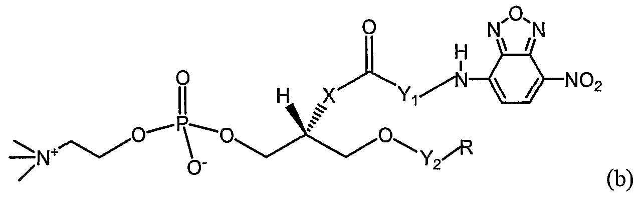


Claims


Priority Applications (5)
| Application Number | Priority Date | Filing Date | Title |
|---|---|---|---|
| JP2006552204A JP5155564B2 (en) | 2004-02-03 | 2005-02-03 | Method for detecting Lp-PLA2 activity |
| US10/588,339 US20070281323A1 (en) | 2004-02-03 | 2005-02-03 | Methods Of Detecting Lp-PLA2 Activity |
| ES05712595T ES2416344T3 (en) | 2004-02-03 | 2005-02-03 | Methods to determine the activity of Lp-PLA2. |
| EP05712595.7A EP1718967B1 (en) | 2004-02-03 | 2005-02-03 | METHODS OF DETECTING Lp-PLA2 ACTIVITY |
| DK05712595.7T DK1718967T3 (en) | 2004-02-03 | 2005-02-03 | Methods for detecting Lp-PLA2 activity |
Applications Claiming Priority (2)
| Application Number | Priority Date | Filing Date | Title |
|---|---|---|---|
| US54158304P | 2004-02-03 | 2004-02-03 | |
| US60/541,583 | 2004-02-03 |
Publications (2)
| Publication Number | Publication Date |
|---|---|
| WO2005074604A2 true WO2005074604A2 (en) | 2005-08-18 |
| WO2005074604A3 WO2005074604A3 (en) | 2005-12-29 |
Family
ID=34837507
Family Applications (1)
| Application Number | Title | Priority Date | Filing Date |
|---|---|---|---|
| PCT/US2005/003211 WO2005074604A2 (en) | 2004-02-03 | 2005-02-03 | METHODS OF DETECTING Lp-PLA2 ACTIVITY |
Country Status (6)
| Country | Link |
|---|---|
| US (1) | US20070281323A1 (en) |
| EP (2) | EP1718967B1 (en) |
| JP (2) | JP5155564B2 (en) |
| DK (1) | DK1718967T3 (en) |
| ES (1) | ES2416344T3 (en) |
| WO (1) | WO2005074604A2 (en) |
Cited By (8)
| Publication number | Priority date | Publication date | Assignee | Title |
|---|---|---|---|---|
| EP1735457A2 (en) * | 2004-04-16 | 2006-12-27 | Glaxo Group Limited | METHODS FOR DETECTING Lp-PLA2 ACTIVITY AND INHIBITION OF Lp-PLA2 ACTIVITY |
| US7531316B2 (en) | 2003-05-28 | 2009-05-12 | Glaxo Group Limited | High throughput assay of Lp-PLA2 activity |
| WO2010092069A1 (en) * | 2009-02-10 | 2010-08-19 | Aterovax | Enzymatic assay for the quantitative determination of phospholipase a1 or a2 activity in a sample |
| CN103792353A (en) * | 2013-10-17 | 2014-05-14 | 武汉生之源生物科技有限公司 | Lipoprotein-related phospholipase A2 content detection kit and preparation method thereof |
| CN104237517A (en) * | 2013-06-18 | 2014-12-24 | 深圳市安群生物工程有限公司 | Fluorescence immunochromatography test paper for detecting human Lp-PLA2 proteins and preparation method of fluorescence immunochromatography test paper |
| US9000132B2 (en) | 2013-03-15 | 2015-04-07 | Diadexus, Inc. | Lipoprotein-associated phospholipase A2 antibody compositions and methods of use |
| US9465029B2 (en) | 2004-04-16 | 2016-10-11 | Glaxo Group Limited | Methods for detecting LP-PLA2 activity and inhibition of LP-PLA2 activity |
| CN111662387A (en) * | 2020-07-14 | 2020-09-15 | 重庆中元汇吉生物技术有限公司 | Monoclonal antibody of anti-human lipoprotein-associated phospholipase A2 and application thereof |
Families Citing this family (12)
| Publication number | Priority date | Publication date | Assignee | Title |
|---|---|---|---|---|
| SI2313778T1 (en) * | 2008-07-18 | 2015-06-30 | Boston Medical Center Corporation | Diagnostics for membranous nephropathy |
| US20120029003A1 (en) * | 2010-07-02 | 2012-02-02 | Bg Medicine, Inc. | Galectin-3 and statin therapy |
| CN102692507B (en) * | 2011-07-29 | 2015-09-16 | 南京诺尔曼生物技术有限公司 | Platelet-activating factor acetylhydro-lase (LPPLA2) measures kit (latex enhancing immune turbidimetry) |
| CN103048457B (en) * | 2012-11-27 | 2014-12-17 | 同昕生物技术(北京)有限公司 | Myocardial infarction early-warning colloidal gold kit and preparation method thereof |
| US20150086998A1 (en) * | 2013-09-24 | 2015-03-26 | Thomas D. Schaal | Value-assigned solutions of lipoprotein-associated phospholipase a2 having a long shelf-life |
| WO2015058158A1 (en) * | 2013-10-18 | 2015-04-23 | Diadexus, Inc. | METHOD FOR DETECTION OF LIPOPROTEIN-SPECIFIC Lp-PLA2 ASSOCIATION |
| US10451624B2 (en) | 2015-02-10 | 2019-10-22 | Shenzhen New Industries Biomedical Engineering Co., Ltd | Reagent kit used for detecting lipoprotein-associated phospholipase A2, and preparation method and application for reagent kit |
| CN104634970B (en) * | 2015-02-10 | 2017-04-05 | 深圳市新产业生物医学工程股份有限公司 | For detecting the test kit of platelet-activating factor acetylhydro-lase and its preparing and application |
| CN104820097A (en) * | 2015-05-22 | 2015-08-05 | 北京协和洛克生物技术有限责任公司 | Liquid-chip kit for quantitatively detecting concentration of lipoprotein phospholipase A2 in sample and preparation method thereof |
| US11237167B2 (en) * | 2017-01-24 | 2022-02-01 | Cleveland Heartlab, Inc. | Lp-PLA2 assays and composition with detergent |
| CN109917131B (en) * | 2019-03-26 | 2021-12-17 | 苏州博源医疗科技有限公司 | Lipoprotein phospholipase A2 detection reagent and preparation and use methods thereof |
| CN111175242A (en) * | 2020-03-17 | 2020-05-19 | 湖南新大陆生物技术有限公司 | Lipoprotein phospholipase A2 detection kit and application thereof |
Citations (11)
| Publication number | Priority date | Publication date | Assignee | Title |
|---|---|---|---|---|
| US5627052A (en) | 1990-08-02 | 1997-05-06 | B.R. Centre, Ltd. | Methods for the production of proteins with a desired function |
| US5693761A (en) | 1988-12-28 | 1997-12-02 | Protein Design Labs, Inc. | Polynucleotides encoding improved humanized immunoglobulins |
| US5766886A (en) | 1991-12-13 | 1998-06-16 | Xoma Corporation | Modified antibody variable domains |
| US5807715A (en) | 1984-08-27 | 1998-09-15 | The Board Of Trustees Of The Leland Stanford Junior University | Methods and transformed mammalian lymphocyte cells for producing functional antigen-binding protein including chimeric immunoglobulin |
| US5821337A (en) | 1991-06-14 | 1998-10-13 | Genentech, Inc. | Immunoglobulin variants |
| US5847088A (en) | 1993-10-06 | 1998-12-08 | Icos Corporation | Antibodies specific for platelet-activating factor acetylhydrolase |
| US5869619A (en) | 1991-12-13 | 1999-02-09 | Xoma Corporation | Modified antibody variable domains |
| US6013256A (en) | 1996-09-24 | 2000-01-11 | Protein Design Labs, Inc. | Method of preventing acute rejection following solid organ transplantation |
| US6054297A (en) | 1991-06-14 | 2000-04-25 | Genentech, Inc. | Humanized antibodies and methods for making them |
| WO2000029444A1 (en) | 1998-11-16 | 2000-05-25 | Genway Biotech, Inc. | Generation of antibodies using polynucleotide vaccination in avian species |
| US6180377B1 (en) | 1993-06-16 | 2001-01-30 | Celltech Therapeutics Limited | Humanized antibodies |
Family Cites Families (19)
| Publication number | Priority date | Publication date | Assignee | Title |
|---|---|---|---|---|
| US4925788A (en) | 1986-10-24 | 1990-05-15 | Immunicon Corporation | Immunoassay system and procedure based on precipitin-like interaction between immune complex and Clq or other non-immunospecific factor |
| US5108933A (en) | 1988-09-16 | 1992-04-28 | Immunicon Corporation | Manipulation of colloids for facilitating magnetic separations |
| US5200084A (en) | 1990-09-26 | 1993-04-06 | Immunicon Corporation | Apparatus and methods for magnetic separation |
| US5186827A (en) | 1991-03-25 | 1993-02-16 | Immunicon Corporation | Apparatus for magnetic separation featuring external magnetic means |
| AU670108B2 (en) * | 1992-09-11 | 1996-07-04 | Becton Dickinson & Company | Improved antibodies to plasmodium falciparum |
| ES2194843T3 (en) | 1992-09-11 | 2003-12-01 | Hoffmann La Roche | DETECTION OF NUCLEIC ACIDS IN BLOOD. |
| JPH08500740A (en) | 1993-06-25 | 1996-01-30 | スミスクライン・ビーチャム・パブリック・リミテッド・カンパニー | Lipoprotein-related phospholipase A-2, its inhibitors and their use in diagnosis and therapy |
| US5977308A (en) | 1993-10-06 | 1999-11-02 | Icos Corporation | Platelet-activating factor acetylhydrolase |
| DE69434609T2 (en) | 1993-10-06 | 2006-09-21 | Icos Corp., Bothell | Acethylhydrolase of the platelet activating factor |
| US5656431A (en) | 1993-10-06 | 1997-08-12 | Icos Corporation | Platelet-activating factor acetylhydrolase |
| US6630301B1 (en) | 1997-03-14 | 2003-10-07 | The Penn State Research Foundation | Detection of extracellular tumor-associated nucleic acid in blood plasma or serum |
| US6156504A (en) | 1996-03-15 | 2000-12-05 | The Penn State Research Foundation | Detection of extracellular tumor-associated nucleic acid in blood plasma or serum using nucleic acid amplification assays |
| EP1057024A4 (en) * | 1998-02-23 | 2002-10-30 | Diadexus Inc | Methods using pla 2? as a marker of metastases and for the diagnosis of selected cancers |
| JP2002528084A (en) | 1998-10-28 | 2002-09-03 | スミスクライン・ビーチャム・パブリック・リミテッド・カンパニー | Method for purifying low-density lipoprotein-related phospholipase A2 using immobilized metal affinity chromatography |
| JP4220603B2 (en) | 1998-12-02 | 2009-02-04 | アルフレッサファーマ株式会社 | Method for measuring platelet activating factor acetylhydrolase activity |
| WO2000047998A1 (en) | 1999-02-10 | 2000-08-17 | Cell Works Inc. | Class characterization of circulating cancer cells isolated from body fluids and methods of use |
| JP2002051164A (en) * | 2000-05-24 | 2002-02-15 | Victor Co Of Japan Ltd | Audio contents auditioning system, system server, and portable telephone |
| EP1318154B1 (en) | 2001-12-07 | 2004-06-16 | SIRS-Lab GmbH | Compounds for determination of the activity of phospholipase A2 |
| PL1633864T3 (en) | 2003-05-28 | 2010-09-30 | Glaxo Group Ltd | High throughput assay of lp-pla2 activity |
-
2005
- 2005-02-03 EP EP05712595.7A patent/EP1718967B1/en not_active Not-in-force
- 2005-02-03 US US10/588,339 patent/US20070281323A1/en not_active Abandoned
- 2005-02-03 DK DK05712595.7T patent/DK1718967T3/en active
- 2005-02-03 WO PCT/US2005/003211 patent/WO2005074604A2/en active Application Filing
- 2005-02-03 EP EP10185947A patent/EP2280282A1/en not_active Withdrawn
- 2005-02-03 JP JP2006552204A patent/JP5155564B2/en not_active Expired - Fee Related
- 2005-02-03 ES ES05712595T patent/ES2416344T3/en active Active
-
2011
- 2011-04-05 JP JP2011083991A patent/JP2011177182A/en not_active Ceased
Patent Citations (14)
| Publication number | Priority date | Publication date | Assignee | Title |
|---|---|---|---|---|
| US5807715A (en) | 1984-08-27 | 1998-09-15 | The Board Of Trustees Of The Leland Stanford Junior University | Methods and transformed mammalian lymphocyte cells for producing functional antigen-binding protein including chimeric immunoglobulin |
| US5693761A (en) | 1988-12-28 | 1997-12-02 | Protein Design Labs, Inc. | Polynucleotides encoding improved humanized immunoglobulins |
| US6180370B1 (en) | 1988-12-28 | 2001-01-30 | Protein Design Labs, Inc. | Humanized immunoglobulins and methods of making the same |
| US5627052A (en) | 1990-08-02 | 1997-05-06 | B.R. Centre, Ltd. | Methods for the production of proteins with a desired function |
| US6054297A (en) | 1991-06-14 | 2000-04-25 | Genentech, Inc. | Humanized antibodies and methods for making them |
| US5821337A (en) | 1991-06-14 | 1998-10-13 | Genentech, Inc. | Immunoglobulin variants |
| US5821123A (en) | 1991-12-13 | 1998-10-13 | Xoma Corporation | Modified antibody variable domains |
| US5869619A (en) | 1991-12-13 | 1999-02-09 | Xoma Corporation | Modified antibody variable domains |
| US5770196A (en) | 1991-12-13 | 1998-06-23 | Xoma Corporation | Modified antibody variable domains and therapeutic uses thereof |
| US5766886A (en) | 1991-12-13 | 1998-06-16 | Xoma Corporation | Modified antibody variable domains |
| US6180377B1 (en) | 1993-06-16 | 2001-01-30 | Celltech Therapeutics Limited | Humanized antibodies |
| US5847088A (en) | 1993-10-06 | 1998-12-08 | Icos Corporation | Antibodies specific for platelet-activating factor acetylhydrolase |
| US6013256A (en) | 1996-09-24 | 2000-01-11 | Protein Design Labs, Inc. | Method of preventing acute rejection following solid organ transplantation |
| WO2000029444A1 (en) | 1998-11-16 | 2000-05-25 | Genway Biotech, Inc. | Generation of antibodies using polynucleotide vaccination in avian species |
Non-Patent Citations (58)
| Title |
|---|
| "Antibody Production: Essential Techniques", 1997, JOHN WILEY & SON LTD |
| "Monoclonal Antibody Protocols", vol. 45, 1995, HUMANA PRESS |
| "Using Antibodies: A Laboratory Manual", 1998, COLD SPRING HARBOR LABORATORY |
| AILOR ET AL., BIOTECHNOL. BIOENG., vol. 58, no. 2-3, 1998, pages 196 - 203 |
| AUJAME ET AL., HUMAN ANTIBODIES, vol. 8, 1997, pages 155 - 168 |
| BARBAS ET AL., TRENDS IN BIOTECHNOL., vol. 14, 1996, pages 230 - 234 |
| BLANKENBERG, J OF LIPID RESEARCH, vol. 44, 2003, pages 1381 - 1386 |
| CO ET AL., NATURE, vol. 351, no. 6326, 1991, pages 501 - 2 |
| COLIGAN ET AL.: "Current Protocols in Immunology", 2001, JOHN WILEY & SONS, INC. |
| DE KRUIF ET AL., TRENDS IN BIOTECHNOL., vol. 17, 1996, pages 453 - 455 |
| EDELMAN ET AL., IMMUNOLOGY, vol. 91, no. 1, 1997, pages 13 - 9 |
| ELDIN ET AL., J IMMUNOL. METHODS., vol. 201, no. 1, 1997, pages 67 - 75 |
| FISCHER ET AL., BIOL. CHEM., vol. 380, no. 7-8, 1999, pages 825 - 39 |
| FISCHER ET AL., BIOL. REGUL. HOMEOST. AGENTS, vol. 14, no. 2, 2000, pages 83 - 92 |
| FISCHER ET AL., BIOTECHNOL. APPL. BIOCHEM., vol. 30, 1999, pages 113 - 6 |
| FISCHER ET AL., BIOTECHNOL. APPL. BIOCHEM., vol. 30, no. 2, 1999, pages 117 - 20 |
| FRENKEN ET AL., RES. IMMUNOL., vol. 149, no. 6, 1998, pages 589 - 99 |
| FREYRE ET AL., J. BIOTECHNOL., vol. 76, no. 2-3, 2000, pages 157 - 63 |
| GAVILONDO ET AL., BIOTECHNIQUES, vol. 29, no. 1, 2000, pages 128 - 38 |
| GIDDINGS ET AL., NATURE BIOTECHNOL., vol. 18, no. 11, 2000, pages 1151 - 5 |
| GRIFFITHS ET AL., CURR. OPIN. BIOTECHNOL., vol. 9, no. 1, 1998, pages 102 - 8 |
| GROSS M, SPECK JDTSCH. TIERARZTL. WOCHENSCHR., vol. 103, 1996, pages 417 - 422 |
| HOOGENBOOM ET AL., IMMUNOTECHNOLOGY, vol. 4, no. 1, 1998, pages 1 - 20 |
| HOOGENBOOM, TRENDS IN BIOTECHNOL., vol. 15, 1997, pages 62 - 70 |
| HOWARD ET AL.: "Basic Methods in Antibody Production and Characterization", 2000, CRC PRESS |
| HSU ET AL., BIOTECHNOL. PROG., vol. 13, no. 1, 1997, pages 96 - 104 |
| HUDSON, CURR. OPIN. BIOTECHNOL., vol. 9, no. 4, 1998, pages 395 - 402 |
| KENNEY, ANTIBODY SOLUTION: AN ANTIBODY METHODS MANUAL, 1997 |
| KRICKA ET AL., J. IMMUNOASSAY, vol. 17, no. 1, 1996, pages 67 - 83 |
| LI ET AL., PROTEIN EXPR. PURIF., vol. 21, no. 1, 2001, pages 121 - 8 |
| LIMONTA ET AL., IMMUNOTECHNOLOGY, vol. 1, 1995, pages 107 - 13 |
| LUNDQVIST ET AL., J. BIOLUMIN. CHEMILUMIN., vol. 10, no. 6, 1995, pages 353 - 9 |
| MA ET AL., PLANT PHYSIOL., vol. 109, no. 2, 1995, pages 341 - 6 |
| MERK ET AL., J BIOCHEM. (TOKYO, vol. 125, no. 2, 1999, pages 328 - 33 |
| MORRISON ET AL., PROC. NATL. ACAD SCI USA., vol. 81, no. 21, 1984, pages 6851 - 5 |
| MOSS, SEMIN. IMMUNOL., vol. 2, 1990, pages 317 - 327 |
| NESBIT ET AL., J IMMUNOL. METHODS, vol. 151, no. 1-2, 1992, pages 201 - 8 |
| PENNELL ET AL., RES. IMMUNOL., vol. 149, no. 6, 1998, pages 599 - 603 |
| POLLOCK ET AL., J IMMUNOL. METHODS, vol. 231, no. 1-2, 1999, pages 147 - 57 |
| POLLOCK ET AL., J. IMMUNOL METHODS., vol. 231, 1999, pages 147 - 57 |
| POSNETT ET AL., J. BIOL. CHEM., vol. 263, 1988, pages 1719 - 1725 |
| RADER ET AL., CURRENT OPINION IN BIOTECHNOLOGY, vol. 8, 1997, pages 503 - 508 |
| RIECHMANN ET AL., NATURE, vol. 332, no. 6162, 1988, pages 323 - 7 |
| RUSSELL, CURR. TOP. MICROBIOL. IMMUNOL., vol. 240, 1999, pages 119 - 38 |
| RYABOVA ET AL., NATURE BIOTECHNOL., vol. 15, no. 1, 1997, pages 79 - 84 |
| SHARON ET AL., NATURE, vol. 309, no. 5966, 1984, pages 364 - 7 |
| SHUSTA ET AL., NATURE BIOTECHNOL., vol. 16, no. 8, 1998, pages 773 - 7 |
| SIDHU, CURR. OPIN. BIOTECHNOL., vol. 11, no. 6, 2000, pages 610 - 6 |
| TAKAHASHI ET AL., BIOSCI. BIOTECHNOL. BIOCHEM., vol. 64, no. 10, 2000, pages 2138 - 44 |
| TAKEDAETAL., NATURE, vol. 314, no. 6010, 1985, pages 452 - 4 |
| TAM ET AL., PROC. NATL. ACAD. SCI. USA, vol. 85, 1988, pages 5409 - 5413 |
| THORPE ET AL., METHODS ENZYMOL., vol. 133, 1986, pages 331 - 53 |
| VERMA ET AL., J IMMUNOL. METHODS, vol. 216, no. 1-2, 1998, pages 165 - 81 |
| VIKINGE ET AL., BIOSENS. BIOELECTRON., vol. 13, 1998, pages 1257 - 1262 |
| WINKLER, J CLIN ENDOCRINOL METAB., vol. 89, no. 3, 2004, pages 1153 - 1159 |
| WINTER ET AL., ANN. REV. IMMUNOL., 1994, pages 433 - 455 |
| YOUNG ET AL., RES. IMMUNOL., vol. 149, 1998, pages 609 - 10 |
| ZOLA: "Monoclonal Antibodies: Preparation and Use of Monoclonal Antibodies and Engineered Antibody Derivatives", 2000, SPRINGER VERLAG |
Cited By (14)
| Publication number | Priority date | Publication date | Assignee | Title |
|---|---|---|---|---|
| US7531316B2 (en) | 2003-05-28 | 2009-05-12 | Glaxo Group Limited | High throughput assay of Lp-PLA2 activity |
| US8609357B2 (en) | 2004-04-16 | 2013-12-17 | Glaxo Group Limited | Methods for detecting LP-PLA2 activity and inhibition of LP-PLA2 activity |
| EP1735457A4 (en) * | 2004-04-16 | 2007-08-29 | Glaxo Group Ltd | METHODS FOR DETECTING Lp-PLA2 ACTIVITY AND INHIBITION OF Lp-PLA2 ACTIVITY |
| US7741020B2 (en) | 2004-04-16 | 2010-06-22 | Glaxo Group Limited | Methods for detecting Lp-PLA2 activity and inhibition of Lp-PLA2 activity |
| EP1735457A2 (en) * | 2004-04-16 | 2006-12-27 | Glaxo Group Limited | METHODS FOR DETECTING Lp-PLA2 ACTIVITY AND INHIBITION OF Lp-PLA2 ACTIVITY |
| US8846309B2 (en) | 2004-04-16 | 2014-09-30 | Glaxo Group Limited | Methods for detecting Lp-PLA2 activity and inhibition of Lp-PLA2 activity |
| US9465029B2 (en) | 2004-04-16 | 2016-10-11 | Glaxo Group Limited | Methods for detecting LP-PLA2 activity and inhibition of LP-PLA2 activity |
| WO2010092069A1 (en) * | 2009-02-10 | 2010-08-19 | Aterovax | Enzymatic assay for the quantitative determination of phospholipase a1 or a2 activity in a sample |
| EP2246440A1 (en) * | 2009-04-29 | 2010-11-03 | Aterovax | Enzymatic assay for the quantitative determination of phospholipase a1 or a2 activity in a sample |
| US9000132B2 (en) | 2013-03-15 | 2015-04-07 | Diadexus, Inc. | Lipoprotein-associated phospholipase A2 antibody compositions and methods of use |
| CN104237517A (en) * | 2013-06-18 | 2014-12-24 | 深圳市安群生物工程有限公司 | Fluorescence immunochromatography test paper for detecting human Lp-PLA2 proteins and preparation method of fluorescence immunochromatography test paper |
| CN103792353A (en) * | 2013-10-17 | 2014-05-14 | 武汉生之源生物科技有限公司 | Lipoprotein-related phospholipase A2 content detection kit and preparation method thereof |
| CN103792353B (en) * | 2013-10-17 | 2015-07-15 | 武汉生之源生物科技有限公司 | Lipoprotein-related phospholipase A2 content detection kit and preparation method thereof |
| CN111662387A (en) * | 2020-07-14 | 2020-09-15 | 重庆中元汇吉生物技术有限公司 | Monoclonal antibody of anti-human lipoprotein-associated phospholipase A2 and application thereof |
Also Published As
| Publication number | Publication date |
|---|---|
| JP5155564B2 (en) | 2013-03-06 |
| EP1718967A2 (en) | 2006-11-08 |
| ES2416344T3 (en) | 2013-07-31 |
| EP1718967A4 (en) | 2008-03-05 |
| JP2007523644A (en) | 2007-08-23 |
| WO2005074604A3 (en) | 2005-12-29 |
| DK1718967T3 (en) | 2013-06-03 |
| US20070281323A1 (en) | 2007-12-06 |
| JP2011177182A (en) | 2011-09-15 |
| EP1718967B1 (en) | 2013-04-24 |
| EP2280282A1 (en) | 2011-02-02 |
Similar Documents
| Publication | Publication Date | Title |
|---|---|---|
| EP1718967B1 (en) | METHODS OF DETECTING Lp-PLA2 ACTIVITY | |
| US20180209993A1 (en) | Uses of lp-pla2 in combination to assess coronary risk | |
| JP2008523394A (en) | Serum amyloid A protein in inflammation and obesity | |
| US8183047B2 (en) | Methods of detecting mastitis by levels of proteasomes | |
| JP2020160076A (en) | Method for supporting determination of success rate of immunity checkpoint inhibitor, reagent kit, device, and computer program | |
| US8263356B2 (en) | Antibodies and methods for predicting dental caries | |
| CA3148161A1 (en) | Anti-drug antibody assay | |
| JP2012530253A (en) | Diagnostic use of peroxiredoxin 4 | |
| US20130095492A1 (en) | Method of detecting tau protein and tau fragments in serum | |
| US20080299585A1 (en) | METHODS FOR DETECTION AND MEASUREMENT OF SECRETORY PHOSPHOLIPASE A2 LEVELS (sPLA2) IN BIOLOGICAL FLUIDS | |
| EP2579040A2 (en) | Qualitative and quantitative analytical method for analyzing the activity type of an enzyme that is activated by proteolysis | |
| JP5484050B2 (en) | Use of sPLA2 activity for cardiovascular event diagnosis | |
| CA2965344A1 (en) | Lp(a) subform size identification using elisa | |
| JP5598537B2 (en) | Post-translationally modified cardiac troponin T as a biomarker for heart failure risk | |
| US20160370382A1 (en) | Method for immunoassay of autoantibody against ku86, kit for use in same, and method for determination of primary hepatocellular carcinoma using same | |
| JP2012508872A (en) | Evaluation of proteolysis by measuring collagen fragments | |
| US9422591B2 (en) | Methods and kits for detecting mastitis | |
| EP3311164B1 (en) | Methods and compositions for diagnosis and prognosis of appendicitis and differentiation of causes of abdominal pain | |
| US8383355B2 (en) | Combination of sPLA2 type IIA mass and OXPL/APOB cardiovascular risk factors for the diagnosis/prognosis of a cardiovascular disease/event | |
| US20230212316A1 (en) | IgG EPITOPE PEPTIDES THAT BIND RHEUMATOID FACTOR AND METHODS OF USE THEREOF | |
| Muñoz et al. | Quantitation of secretory group V phospholipase A2 in human tissues by sandwich enzyme-linked immunosorbent assay | |
| JPH11166931A (en) | Method for detecting quantitative or qualitative anomaly in organism component | |
| US20110105349A1 (en) | Use of a monophosphate ester of a phthalein compound as a substrate for tartrate-resistant acid phosphatase b5 (trap 5b) | |
| WO2019234087A1 (en) | Complement c4d assay | |
| US20180252735A1 (en) | Method for assisting determination of type iii hyperlipidemia |
Legal Events
| Date | Code | Title | Description |
|---|---|---|---|
| AK | Designated states |
Kind code of ref document: A2 Designated state(s): AE AG AL AM AT AU AZ BA BB BG BR BW BY BZ CA CH CN CO CR CU CZ DE DK DM DZ EC EE EG ES FI GB GD GE GH GM HR HU ID IL IN IS JP KE KG KP KR KZ LC LK LR LS LT LU LV MA MD MG MK MN MW MX MZ NA NI NO NZ OM PG PH PL PT RO RU SC SD SE SG SK SL SY TJ TM TN TR TT TZ UA UG US UZ VC VN YU ZA ZM ZW |
|
| AL | Designated countries for regional patents |
Kind code of ref document: A2 Designated state(s): BW GH GM KE LS MW MZ NA SD SL SZ TZ UG ZM ZW AM AZ BY KG KZ MD RU TJ TM AT BE BG CH CY CZ DE DK EE ES FI FR GB GR HU IE IS IT LT LU MC NL PL PT RO SE SI SK TR BF BJ CF CG CI CM GA GN GQ GW ML MR NE SN TD TG |
|
| WWE | Wipo information: entry into national phase |
Ref document number: 2006552204 Country of ref document: JP |
|
| NENP | Non-entry into the national phase |
Ref country code: DE |
|
| WWW | Wipo information: withdrawn in national office |
Country of ref document: DE |
|
| WWE | Wipo information: entry into national phase |
Ref document number: 2005712595 Country of ref document: EP |
|
| WWP | Wipo information: published in national office |
Ref document number: 2005712595 Country of ref document: EP |
|
| WWE | Wipo information: entry into national phase |
Ref document number: 10588339 Country of ref document: US |
|
| WWP | Wipo information: published in national office |
Ref document number: 10588339 Country of ref document: US |