ALTERED ANTIBODIES HAVING IMPROVED ANTIGEN-BINDING AFFINITY
Related Information The application claims priority to U.S. provisional patent application number 60/490,087, filed on July 26, 2003, the entire contents of which are hereby incorporated by reference. The contents of any patents, patent applications, and references cited throughout this specification are hereby incorporated by reference in their entireties.
Background of the Invention Antibodies are exquisite, naturally occurring biological agents that play a critical role in defending the body from pathogens. Antibodies, which are also commonly referred to as immunoglobulins, contain four polypeptides: two longer polypeptides ("heavy chains") that are identical to one another and two shorter polypeptides ("light chains") that are identical to one another. The heavy chains are paired with the light chains by disulfide bonds, and the two heavy chains are similarly bound to one another to create a tetrameric structure. Moreover, the heavy and light chains each contain a variable domain and one or more constant regions: the heavy chain includes one variable domain (VH) followed by three constant regions (CiH, C H, and C3H), and the light chain includes one variable domain (VL) followed by a single constant region (CL). The variable domains of each pair of light and heavy chains form the site that comes into contact with an antigen. Both VH and NL have the same general structure, with four framework regions (FRs), whose sequences are relatively conserved, connected by three hypervariable or complementarity determining regions (CDRs) (see Kabat et al. , In "Sequences of Proteins of Immunological Interest," U.S. Department of Health and Human Services, 1983; see also Chothia et al, J. Mol. Biol. 196:901-917, 1987). The four framework regions largely adopt a β-sheet conformation and the CDRs form loops connecting, and in some cases forming part of, the β-sheet structure. The CDRs of NH and NL are held in close proximity by the FRs, and amino acid residues within the CDRs bind the antigen. More detailed accounts of the structure of variable domains can be found in Poljak et al. (Proc. Natl. Acad. Sci. USA 70:3305-3310, 1973) Segal et al (Proc. Natl. Acad. Sci. USA 71:4298-4302, 1974), and Marquart et al. (J. Mol. Biol, 141:369- 391, 1980). Researchers have modified antibodies in various ways in order to study their function or to improve their utility as therapeutic agents. In some of the earliest modifications, researchers used double-stranded DΝA sequences to express the NH or NL domains, but none of the sequence of the constant region (see, e.g., EP-A-0 088 994; Schering Corporation). Other fragments and chimeric antibodies have also been made.
One particular type of chimera, commonly referred to as a CDR-grafted antibody, includes sequences from two antibodies that differ in species (e.g., murine CDRs have been used in place of the naturally occurring CDRs in otherwise human antibodies; see, e.g., U.S. Patent No. 5,225,539). Researchers hoped that such antibodies would be no more foreign to the human body than a genuine human antibody, but the utility of such antibodies has been restricted, at least in some cases, by a reduction in the antibody's affinity for the antigen. In an attempt to improve affinity, some of the amino acids in the FRs of CDR-grafted antibodies have been changed from those of the acceptor molecule (e.g., a human antibody) to those of the antibody that donated the CDRs (e.g., those of a murine antibody; see, e.g., U.S. Patent No. 5,585,089; U.S. PatentNo. 5,693,761; U.S. PatentNo. 5,693,762; and U.S. Patent No. 6,180,370). Accordingly, there remains a need for antibodies that do not provoke a strong immune response but yet bind strongly to their antigens and methods for identifying such antibodies.
Summary of the Invention The present invention is based, in part, on the discovery that the affinity of an antibody (or an antigen-binding fragment thereof) can be improved by modifying amino acid residues within the antibody. The modifications are based, wholly or partially, on a computational analysis of electrostatic forces between the antibody and an antigen to which it binds. The computational analysis, in turn, is based on a prediction of charge distribution within the antibody that generates the electrostatic forces that influence binding between the antibody and its antigen in a solvent (e.g., an aqueous solvent such as water, phosphate-buffered saline (PBS), plasma, or blood). The computational methods define the electrostatic complement (the optimal tradeoff between unfavorable desolvation energy and favorable interactions in an antigen-antibody complex) for a given target site and geometry. In particular, the invention provides criteria or rules by which one can calculate the optimal charge distribution and associated change in binding free energy between an antibody and an antigen, when bound in a solvent, and then identify discrete residue positions for modification. Moreover, the invention provides rules which guide the selection of an appropriate modification at the identified residue position, e.g., side chain chemistry, by building a subset of modifications in silico followed by recalculating the binding free energy and election of a preferred modification. Thus, the invention has several advantages in that it, unlike other methods, is not restricted to mere global or pair wise alignment of charges with the presumptive conclusion that only opposite net charges between an antibody and antigen are favorable. Rather, the invention provides a more sophisticated analysis (as is appropriate given that a typical antibody comprises up to four polypeptide chains with inter and intra chain
disulfide linkages and six CDR binding surfaces as well as inter chain interfaces) for revealing the exact residue positions and side chain chemistries to be used to modify the binding-affinity of an antibody/antigen complex. Moreover, the invention also fully accounts for the binding interactions of a antibody when bound to an antigen within a solvent. And importantly, the invention provides for antibody modifications that alter antigen-binding which other methods would either fail to identify or dismiss as unsuitable to try. In one aspect, the invention features a method of modulating the antigen-binding affinity of an antibody that includes the steps of providing data corresponding to the structure (e.g., a three-dimensional structure) of a complex between an antibody and an antigen to which the antibody binds; determining, using the data, a representation of a charge distribution (e.g., a set of multipoles or point charges) within the antibody (e.g., within one or more of the CDRs) that would reduce (i.e., optimize or make more negative) the electrostatic contribution to binding free energy between the antibody and the antigen; and modifying one or more amino acid residues within the antibody (e.g., within one or more of the CDRs) to create a modified antibody corresponding to (or with a better correspondence to) the charge distribution (i.e., the optimal charge distribution determined). The result is a charge distribution that can be used to modulate (e.g., improve, alter, etc.) the interaction between an antibody and its antigen. For example, if the side chain of an amino acid residue in an optimized antibody that has a net total charge of -1, one can replace the corresponding amino acid residue in the original antibody, sometime referred to as the first antibody or parent antibody, with an amino acid residue that has a negatively charged side chain to create a modified antibody which is a variant of the parent antibody and sometimes referred to herein as a second antibody (or even a third or fourth antibody if referring to the modification of a antibody that has been previously modified and is therefore an iterative variation of the preceding antibody). In a related aspect, the invention provides a method of modulating the antigen- binding affinity of an antibody by determining a spatial representation of an optimal charge distribution of the amino acids of the antibody and associated change in binding free energy of the antibody when bound to an antigen in a solvent; identifying at least one candidate amino acid residue position of the antibody to be modified to alter the binding free energy of the antibody when bound to the antigen; and selecting an elected amino acid residue for substitution for said amino acid position, such that upon substitution, the antigen-binding affinity of the antibody is modulated. As described further below, once a charge distribution is determined, one or more of the amino acid residues in the antibody (e.g., one or more of the residues in the CDR(s), e.g., 2-10 residues or more, e.g., most if not all of the CDR residues and,
optionally, only in the CDR(s)) can be modified to match, or better match, that charge distribution. For example, an amino acid residue can be replaced with another naturally occurring amino acid residue or a non-naturally occurring residue. The substitution may or may not constitute a conservative amino acid substitution. In some instances, it may be desired to alter the charge distribution by deleting or inserting one or more amino acid residues. In some instances, for example, where the data of the structure of a complex between the antibody and the antigen is available prior to provision of the antibody, one need only know the sequence of the parent antibody (or the sequence of one or more of the CDRs of that antibody). The method can be carried out so long as one has, or can obtain, information regarding the charge distribution within an antibody-antigen complex containing a parent antibody; that information is then used to modify a modified antibody in a way that improves the modified antibody's affinity for its antigen. Alternatively, the methods of the invention can be used to alter (e.g., optimize) the affinity of a fully human antibody or antigen-binding fragments containing human FRs and human CDRs, for example, affinity mature the antibody for improved antigen-binding affinity. A fully human antibody can be one obtained from human plasma (even though this is an uncommon practice) or generated in vivo (e.g., an antibody generated in a transgenic mouse containing human immunoglobulin genes; see U.S. Patent No. 6,150,584). In the methods of the invention, the parent and modified antibodies can be of the same or of different species (e.g., the parent antibody can be a non-human antibody (e.g., a murine antibody), and the modified antibody can be a human antibody). The antibodies can also be of the same, or of different, classes or subclasses. Regardless of their origin or class, portions of the sequences of the two antibodies can be identical to one another. For example, the FRs of the parent antibody can be identical to the FRs of the modified antibody. This would occur, for example, where the parent antibody is a human antibody and the modified antibody varies from the parent antibody only in that the modified antibody contains one or more non-human CDRs (i.e., in the modified antibody, one or more of the original, human CDRs have been replaced with a non-human (e.g., murine) CDR). The methods of the invention can be carried out with antibodies that have the structure of a naturally occurring antibody. For example, the methods of the invention can be carried out with antibodies that have the structure of an IgG molecule (two full- length heavy chains and two full-length light chains). Thus, in some embodiments, the parent and/or modified antibody can include an Fc region of an antibody (e.g. , the Fc region of a human antibody). The methods of the invention can be carried out, however, with less than complete antibodies; they can be carried out with any antigen-binding fragment of an antibody including those described further below (Fab fragments, F(ab')2 fragments, or single-chain antibodies (scFv)). The "fragments" can constitute minor
variations of naturally occurring antibodies. For example, an antibody fragment can include all but a few of the amino acid residues of a "complete" antibody (e.g., the FR of NH or NL can be truncated). Regardless of whether the method is carried out with a complete antibody or a fragment thereof, where all or part of the FR is present, the sequence of that FR can be that of a wild-type antibody. Alternatively, the FR can contain a mutation. For example, the methods of the invention can be carried out with a parent antibody that includes a framework region (e.g., a human FR) that contains one or more amino acid residues that differ from the corresponding residue(s) in the wild-type FR. The mutation can be one that changes an amino acid residue to the corresponding residue in an antibody of another species. Thus, an otherwise human FR can contain a murine residue (such mutations are referred to in the art as "back mutations"). For example, framework regions of a human antibody can be "back-mutated" to the amino acid residue at the same position in a non- human antibody. Such a back-mutated antibody can be used in the present methods as the "parent" antibody, in which case the "modified" antibody can include completely human FRs. Mutations in the FRs can occur within any of FR1, FR2, FR3, and/or FR4 in either VH or NL (or in NH and VL). Up to about 10 residues or more can be mutated (e.g., 1, 2, 3, 4, 5, 6, 7, 8, 9, or 10 or more residues in FR1, FR2, FR3, and/or FR4 can be changed from the naturally occurring residue (e.g. , the human residue) to another residue (e.g. , a donor residue, for example, murine residue, at the corresponding position)). The residues that immediately flank the CDRs are among those that can be mutated. In one embodiment, the methods of the invention are carried out with a parent antibody that is completely non-human (e.g., a murine antibody) and a modified antibody that includes a human Fc region and completely human FRs. In certain embodiments, the relative affinities of the parent and modified antibodies (e.g., the parent, modified or altered antibody of the present invention) can be such that the affinity of the modified antibody to a given antigen is at least as high as the affinity of the parent antibody to that antigen. For example, the affinity of the modified antibody to the antigen can be at least (or about) 1.1, 1.2, 1.3, 1.4, 1.5, 1.6, 1.7, 1.8, 1.9, 2, 3, 5, 8, 10, 50, 102, 103, 104, 105, or 106, 107, or 108 times greater than the affinity of the parent antibody to the antigen (or any range or value in between). The method may also be used lower the affinity of the antibody, for example, where it is desirable to have a lower affinity for better pharmacokinetics, antigen-binding specificity, reduced cross-talk between related antigen epitopes, and the like. For example, the affinity of the modified antibody to the antigen can be at least (or about) 1.1, 1.2, 1.3, 1.4, 1.5, 1.6, 1.7, 1.8, 1.9, 2, 3, 5, 8, 10, 50, 102, 103, 104, 10s, or 106, 107, or 108 times less than the affinity of the parent antibody to the antigen (or any range or value in between). .
The methods of the invention can be iterative. An antibody generated, as described above, can be re-modeled (for example, in silico or empirically, e.g., using experimental data) and further altered to further improve antigen binding. Thus, the steps described above can be followed by additional steps, including: obtaining data corresponding to the structure of a complex between the modified antibody and the antigen; determining, using the data (which can be referred to as "additional data" to distinguish it from the data obtained and used in the parent "round"), a representation of an additional charge distribution of the CDRs of the modified antibody which minimizes electrostatic contribution to binding free energy between the modified antibody and the antigen; and expressing a third or further modified antibody that binds to the antigen, the third antibody having a matured CDR differing from a CDR of the modified antibody by at least one amino acid, the matured CDR corresponding to the additional charge distribution. Yet additional rounds of maturation can be carried out. In the method just described, the resulting antibody would be complexed with (i.e. allowed to bind to) antigen and used to obtain a charge distribution that minimizes the electrostatic contribution. A fourth or further modified antibody would then be produced that would contain modifications, dictated by the charge distribution, that improve antigen binding. And so forth. As noted above, the modified antibody (or subsequent antibodies serving in the place of the modified antibody) can contain a CDR that has been modified so that the electrostatic forces in the antibody-antigen complex are improved (or optimized). Presently, the software used to examine electrostatic forces models an optimal charge distribution and the user then determines what amino acid substitution(s) or alteration(s) would improve that distribution. Accordingly, such steps (e.g., examining the modeled, optimal charge distribution and determining a sequence modification to improve antigen binding) are, or can be, part of the methods now claimed. However, as it would not be difficult to modify the software so that the program includes the selection of amino acid substitutions (or alterations), in the future, one may need only examine that output and execute the suggested change (or some variation of it, if desired). The methods of the invention may be characterized as those that "produce" an antibody (or a fragment thereof). The term "produce" means to "make," "generate," or "design" a non-naturally occurring antibody (or fragment thereof). The antibody produced may be considered more "mature" than either of the antibodies whose sequences (e.g., whose CDR(s) and FRs) were used in its construction. While the antibody produced may have a stronger affinity for an antigen, the methods of the invention are not limited to those that produce antibodies with improved affinity. For example, the methods of the invention can produce an antibody that has about the same affinity for an antigen as it did prior to being modified by the present methods. When a human antibody is modified, as described in the prior art, to contain murine CDRs, the
resulting CDR-grafted antibody can lose affinity for its antigen. Thus, for example, where the methods of the invention are applied to CDR-grafted antibodies, they are useful and successful when they prevent the loss of affinity (some or all of the loss) that would otherwise occur with a conventional CDR graft. In addition to minimizing the electrostatic contribution to the binding free energy, the methods of the invention can further include minimizing the van der Waals or solvent accessible surface area contribution to the binding free energy. In such further computational analysis, additional amino acids in a CDR of the parent antibody may be altered to generate the modified antibody, such that the binding free energy is further reduced beyond what was achieved by solely minimizing the electrostatic contribution. As few as one and as many as 50 CDR residues may be modified in the methods and compositions of the instant invention. Most commonly, between 1 and 10 (e.g., 1, 2, 3, 4, 5, 6, 7, 8, 9, or 10) amino acid residues are altered by the methods and compositions of the instant invention. Antibodies produced by any of the methods of the invention are also within the scope of the invention, pharmaceutical compositions containing those antibodies, as well as nucleic acids encoding such antibodies. The present invention also includes vectors that express the modified antibodies (or polypeptides or fragments thereof) found by the methods described above. These vectors can be used to transform cell lines, and such transformed (e.g. transfected) cells are within the scope of the invention. The details of one or more embodiments of the invention are set forth in the description below. Other features, objects, and advantages of the invention will be apparent from the description and the claims.
Brief Description of the Figures Figure 1 illustrates geometries for modeling the binding interactions between an antibody, or antigen-binding fragment thereof, and an antigen, when bound in a solvent (top panel). In particular, the boundary- value problem which comprises a determination of the charge distribution in a spherical region of radius R with a dielectric constant £,, surrounded by solvent with a dielectric constant G2 as well as other geometries of the antibody-antigen interface (bottom panel, see also text, infra). Figure 2 depicts nucleotide (SEQ ID NOs: 1, 3) and polypeptide (SEQ ID NOs: 2, 4) sequences for 5c8 heavy variable and light variable chain domains.
Detailed Description of the Invention In order to provide a clear understanding of the specification and claims, the following definitions are conveniently provided below.
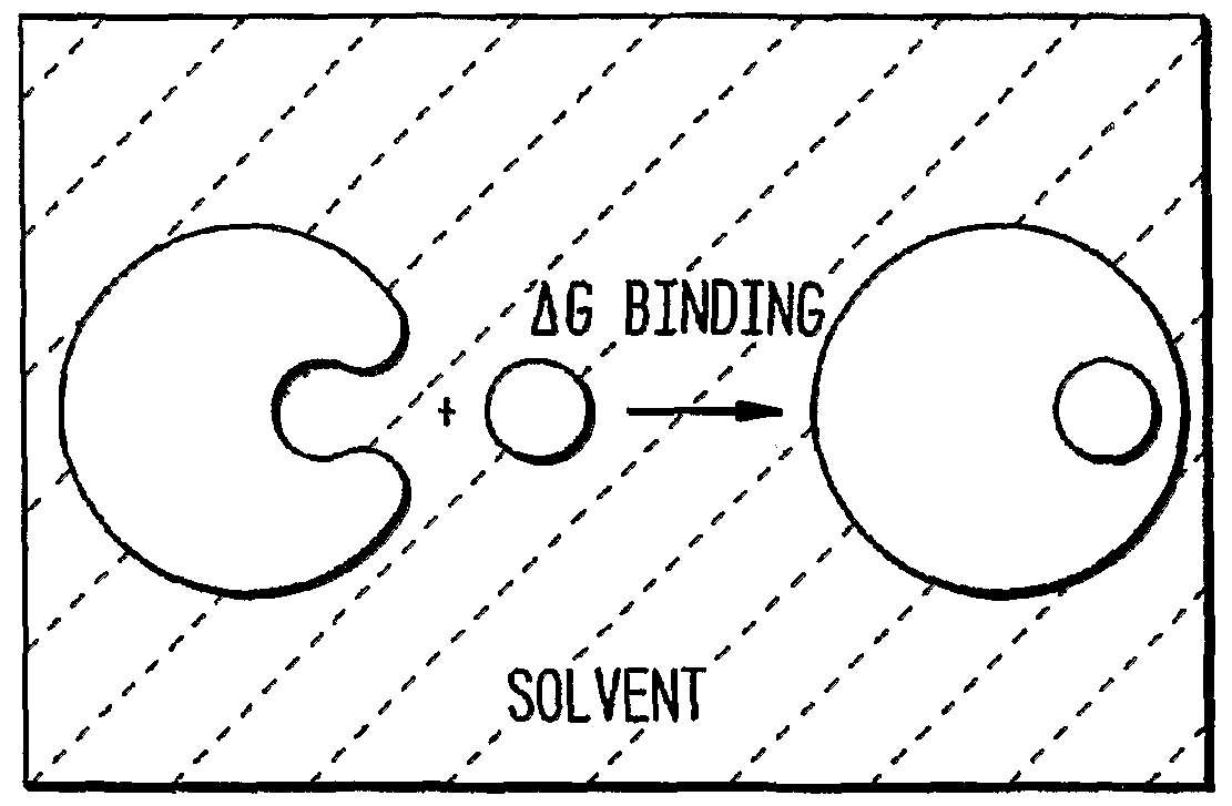
Definitions The term "structure", or "structural data", as used herein, includes the known, predicted and/or modeled position(s) in three-dimensional space that are occupied by the atoms, molecules, compounds, amino acid residues and portions thereof, and macromolecules and portions thereof, of the invention, and, in particular, an antibody bound to an antigen in a solvent. A number of methods for identifying and or predicting structure at the molecular/atomic level can be used such as X-ray crystallography, NMR structural modeling, and the like. The term "binding affinity", as used herein, includes the strength of a binding interaction and therefore includes both the actual binding affinity as well as the apparent binding affinity. The actual binding affinity is a ratio of the association rate over the disassociation rate. Therefore, conferring or optimizing binding affinity includes altering either or both of these components to achieve the desired level of binding affinity. The apparent affinity can include, for example, the avidity of the interaction. For example, a bivalent altered variable region binding fragment can exhibit altered or optimized binding affinity due to its valency. Binding affinities may also be modeled, with such modeling contributing to selection of residue alterations in the methods of the current invention. The term "binding free energy" or "free energy of binding", as used herein, includes its art-recognized meaning, and, in particular, as applied to antibody-antigen interactions in a solvent. Reductions in binding free energy enhance antibody-antigen affinities, whereas increases in binding free energy reduce antibody-antigen affinities. The phrase "spatial representation of an optimal charge distribution", as used herein, includes modeling the charge distribution for an antibody or antibody-antigen
complex, wherein the electrostatic contribution to free energy of the antibody when bound to antigen is optimized (minimized), as compared to the known and/or modeled representation of charge distribution of the parent antibody and/or parent antibody when bound to antigen. The modeling of optimal charge distribution can be arrived at by an in silico process that incorporates the known and/or modeled structure(s) of an antibody and/or antibody-antigen complex as an input. Response continuum modeling (e.g., the linearized Poisson-Boltzmann equation) can be employed to express the electrostatic binding free energy of the antigen-antibody complex in a solvent as a sum of antibody desolvation, antibody-antigen interaction, and antigen desolvation terms. This in silico process is characterized by the ability to incorporate monopole, dipolar, and quadrupolar terms in representing charge distributions within the modeled charge distributions of the invention, and allows for extensive assessment of solvation/desolvation energies for antibody residues during transition of the antibody between unbound and bound states. The process of modeling the spatial representation of an optimal charge distribution for an antibody-antigen complex may additionally incorporate modeling of van der Waals forces, solvent accessible surface area forces, etc. The term "solvent", as used herein, includes its broadest art-recognized meaning, referring to any liquid in which an antibody of the instant invention is dissolved and/or resides. The term "antibody", as used herein, includes monoclonal antibodies (including full length monoclonal antibodies), polyclonal antibodies, multispecific antibodies (e.g., bispecific antibodies), chimeric antibodies, CDR-grafted antibodies, humanized antibodies, human antibodies and antigen-binding fragments thereof, for example, an antibody light chain (NL), an antibody heavy chain (NH), a single chain antibody (scFv), a F(ab')2 fragment, a Fab fragment, an Fd fragment, an Fv fragment, and a single domain antibody fragment (DAb). The term "antigen", as used herein, includes an entity (e.g., a proteinaceous entity or peptide) to which an antibody specifically binds, and includes, e.g., a predetermined antigen to which both a parent antibody and modified antibody as herein defined bind. The target antigen may be polypeptide, carbohydrate, nucleic acid, lipid, hapten, or other naturally occurring or synthetic compound. Preferably, the target antigen is a polypeptide. The term "CDR", as used herein, includes the complementarity determining regions as described by, for example Kabat , Chothia, or MacCallum et al, (see, e.g., Kabat et al, In "Sequences of Proteins of Immunological Interest," U.S. Department of Health and Human Services, 1983; Chothia et al, J. Mol. Biol. 196:901-917, 1987; and MacCallum et al, J. Mol Biol. 262:732-745 (1996); the contents of which are incorporated herein in their entirety). The amino acid residue positions which typically encompass the CDRs as
described by each of the above cited references are set forth below for comparison.
Table of CDR Definitions Kabat Chothia MacCallum NH CDR1 31-35 26-32 30-35 NHCDR2 50-65 53-55 47-58 NHCDR3 95-102 96-101 93-101 NLCDR1 24-34 26-32 30-36 NLCDR2 50-56 50-52 46-55 NLCDR3 89-97 91-96 89-96
The term "variable region", as used herein, includes the amino terminal portion of an antibody which confers antigen binding onto the molecule and which is not the constant region. The term is intended to include functional fragments, for example, antigen-binding fragments, which maintain some or all of the binding function of the whole variable region. The term "framework region", as used herein, includes the antibody sequence that is between and separates the CDRs. Therefore, a variable region framework is between about 100-120 amino acids in length but is intended to reference only those amino acids outside of the CDRs. For the specific example of a heavy chain variable region and for the CDRs as defined by Kabat et al. , framework region 1 corresponds to the domain of the variable region encompassing amino acids 1-30; region 2 corresponds to the domain of the variable region encompassing amino acids 36-49; region 3 corresponds to the domain of the variable region encompassing amino acids 66-94, and region 4 corresponds to the domain of the variable region from amino acids 103 to the end of the variable region. The framework regions for the light chain are similarly separated by each of the light claim variable region CDRs. Similarly, using the definition of CDRs by Chothia et al. or McCallum et al. the framework region boundaries are separated by the respective CDR termini as described above. The term terms "modified" or "altered", as used herein, include antibodies or antigen-binding fragments thereof, that contain one or more amino acid changes in, for example, a CDR(s), a framework region(s), or both as compared to the parent amino acid sequence at the changed position. A modified or altered antibody typically has one or more residues which has been substituted with another amino acid residue, related side chain chemistry thereof, or one or more amino acid residue insertions or deletions. The term "parent antibody", "original antibody", "starting antibody", "wild-type", or "first antibody", as used herein, includes any antibody for which modification of antibody-antigen binding affinity by the methods of the instant invention is desired. Thus, the parent antibody represents the input antibody on which the methods of the instant invention are performed. The parent polypeptide may comprise a native sequence (i.e. a naturally occurring) antibody (including a naturally occurring allelic variant), or an
antibody with pre-existing amino acid sequence modifications (such as insertions, deletions and/or other alterations) of a naturally occurring sequence. The parent antibody may be a monoclonal, chimeric, CDR-grafted, humanized, or human antibody. The terms "antibody variant", "modified antibody", "antibody containing a modified amino acid", "mutant", or "second antibody", "third antibody", etc., as used herein, include an antibody which has an amino acid sequence which differs from the amino acid sequence of a parent antibody. Preferably, the antibody variant comprises a heavy chain variable domain or a light chain variable domain having an amino acid sequence which is not found in nature. Such variants necessarily have less than 100% sequence identity or similarity with the parent antibody. In a preferred embodiment, the antibody variant will have an amino acid sequence from about 75% to less than 100% amino acid sequence identity or similarity with the amino acid sequence of either the heavy or light chain variable domain of the parent antibody, more preferably from about 80%) to less than 100%), more preferably from about 85% to less than 100%), more pref- erably from about 90%) to less than 100%>, and most preferably from about 95% to less than 100%. Identity or similarity with respect to this sequence is defined herein as the percentage of amino acid residues in the candidate sequence that are identical (i.e. same residue) with the parent antibody residues, after aligning the sequences and introducing gaps, if necessary, to achieve the maximum percent sequence identity. Typically, N- terminal, C-terminal, or internal extensions, deletions, or insertions into the antibody sequence outside of the variable domain are not construed as affecting sequence identity or similarity. The antibody variant is generally one which comprises one or more amino acid alterations in or adjacent to one or more hypervariable regions thereof. The modified antibodies of the present invention may either be expressed, or alternatively, may be modeled in silico. The phrase "candidate amino acid residue position", as used herein, includes an amino acid position identified within an antibody of the present invention, wherein the substitution of the candidate amino acid is modeled, predicted, or known to impact charge distribution of the antibody upon alteration, deletion, insertion, or substitution with another amino acid. The term "elected amino acid", as used herein, refers to an amino acid residue(s) that has been selected by the methods of the present invention for substitution as a replacement amino acid at the candidate amino acid position within the antibody. Substitution of the candidate amino acid residue position with the elected amino acid residue may either reduce or increase the electrostatic contribution to binding free energy of the antibody-antigen complex. The terms "amino acid alteration" or "alteration for said amino acid", as used herein, include refers to a change in the amino acid sequence of a predetermined amino acid sequence. Exemplary alterations include insertions, substitutions, and deletions.
The term "amino acid modification", as used herein, includes the replacement of an existing amino acid residue side chain chemistry in a predetermined amino acid sequence with another different amino acid residue side chain chemistry, by, for example, amino acid substitution. Individual amino acid modifications of the instant invention are selected from any one of the following: (1) the set of amino acids with nonpolar sidechains, e.g., Ala, Cys, He, Leu, Met, Phe, Pro, Nal, (2) the set of amino acids with negatively charged side chains, e.g., Asp, Glu, (3) the set of amino acids with positively charged sidechains, e.g., Arg, His, Lys, and (4) the set of amino acids with uncharged polar sidechains, e.g., Asn, Cys, Gin, Gly, His, Met, Phe, Ser, Thr, Trp, Tyr, to which are added Cys, Gly, Met and Phe. The term "naturally occurring amino acid residue", as used herein, includes one encoded by the genetic code, generally selected from the group consisting of: alanine (Ala); arginine (Arg); asparagine (Asn); aspartic acid (Asp); cysteine (Cys); glutamine (Gin); glutamic acid (Glu); glycine (Gly); histidine (His); isoleucine (He): leucine (Leu); lysine (Lys); methionine (Met); phenylalanine (Phe); proline (Pro); serine (Ser); threonine (Thr); tryptophan (Trp); tyrosine (Tyr); and valine (Val). The term "non-naturally occurring amino acid residue", as used herein, includes an amino acid residue other than those naturally occurring amino acid residues listed above, which is able to covalently bind adjacent amino acid residues(s) in a polypeptide chain. Examples of non-naturally occurring amino acid residues include norleucine, omithine, norvaline, homoserine and other amino acid residue analogues such as those described in Ellman et al. Meth. Enzym. 202:301-336 (1991). To generate such non- naturally occurring amino acid residues, the procedures of Νoren et al. Science 244:182 (1989) and Ellman et al, supra, can be used. Briefly, these procedures involve chemically activating a suppressor fRΝA with a non-naturally occurring amino acid residue followed by in vitro transcription and translation of the RΝA. The term "exposed" amino acid residue, as used herein, includes one in which at least part of its surface is exposed, to some extent, to solvent when present in a polypeptide (e.g., an antibody or polypeptide antigen) in solution. Preferably, the exposed amino acid residue is one in which at least about one third of its side chain surface area is exposed to solvent. Various methods are available for determining whether a residue is exposed or not, including an analysis of a molecular model or structure of the polypeptide. The term "treatment" refers to both therapeutic treatment and prophylactic or preventative measures. Those in need of treatment include those already with the disorder as well as those in which the disorder is to be prevented. The term "disorder or disease" is any condition that would benefit from treatment with the antibody variant. This includes chronic and acute disorders or diseases including those pathological conditions which predispose the mammal to the disorder in question.
The terms "cell", "cell line", "cell culture", or "host cell", as used herein, includes "transformants", "transformed cells", or "transfected cells" and progeny thereof. Host cells within the scope of the invention include prokaryotic cells such as E. coli, lower eukaryotic cells such as yeast cells, insect cells, and higher eukaryotic cells such as vertebrate cells, for example, mammalian cells, e.g. , Chinese hamster ovary cells and NSO myeloma cells.
Detailed Description Overview The methods described herein can be used to obtain an optimized antibody (or an antigen-binding fragment thereof). Based on a computational analysis, positions are identified within any given antibody where there is a difference (the larger the difference, the more significant it can be) between the charge distribution in an optimized antibody- antigen complex and that in an original antibody-antigen complex. Such differences in charge distribution are also associated with changes in binding free energy of the antibody when bound to the antigen in a solvent. The amino acid residue at such a position can then be changed so that the electrostatic forces in the original antibody more nearly ■ approach (or in alternative embodiments, are more divergent from) those in the optimized antibody, thereby modulating binding free energy of the antibody when bound to an antigen in a solvent. Changes to the antibody are introduced according to a set of discrete criteria or rules as described herein.
Rules for Modifying Antibodies for Improved Function The rules of the invention can be applied as follows. To modulate the antigen- binding affinity of an antibody, for example, to improve or restore such binding, basic sequence and/or structural data is first acquired. Electrostatic charge optimization techniques are then applied to suggest improved-affinity mutants. Typically, an electrostatic charge optimization is first used to determine the position(s) of the CDR residue(s) that are sub-optimal for binding (Lee and Tidor, J Chem. Phys. 106:8681- 8690, 1997; Kangas and Tidor, J. Chem. Phys. 109:7522-7545, 1998). Then, one or more CDR mutations (i.e., modifications) is subjected to further computational analysis. Based on these calculations, the binding affinity is then determined for a subset of modified antibodies having one or more modifications according to the rules of the invention. Using a continuum electrostatics model, an electrostatic charge optimization can be performed on each side chain of the amino acids in the CDRs of the antibody. A charge optimization gives charges at atom centers but does not always yield actual mutation(s). Accordingly, a round of charge optimizations can be performed with various constraints imposed to represent natural side chain characteristics at the positions of interest. For example, an optimization can be performed for a net side chain charge of-1,
0, and +1 with the additional constraint that no atom's charge exceeded a particular value, e.g., 0.85 electron charge units. Candidate amino acid side chain positions, and residue modifications at these positions, are then determined based on the potential gain in electrostatic binding free energy observed in the optimizations. Binding free energy difference (in kcal/mol) in going from the native residue to a completely uncharged sidechain isostere, i.e., a residue with the same shape but no charges or partial charges on the atoms can be calculated. Negative numbers indicate a predicted increase of binding affinity. Optimal charge distribution wherein the net side chain charge is +1, 0, or -1 can be used to calculate the binding free energy difference. In those instances in which binding free energy difference is favorable (ΔG < -
0.25 kcal/mol) and associated with a transition from the native residue to a completely uncharged side chain isostere, i.e., a residue with the same shape but no charges or partial charges on the atoms, modifications from the set of amino acids with nonpolar sidechains, e.g., Ala, Cys, He, Leu, Met, Phe, Pro, Val are selected. Where the binding free energy difference that can be obtained with an optimal charge distribution in the side chain and a net side chain charge of -1 is favorable (ΔG < -
0.25 kcal/mol), modifications from the set of amino acids with negatively charged side chains, e.g., Asp, Glu are selected. Similarly, where the binding free energy difference that can be obtained with an optimal charge distribution in the side chain and a net side chain charge of +1 is favorable
(ΔG < -0.25 kcal/mol), modifications from the set of amino acids with positively charged sidechains, e.g., Arg, His, Lys are selected. Finally, in those cases where the binding free energy difference that can be obtained with an optimal charge distribution in the side chain and a net side chain charge of 0 is favorable (ΔG < -0.25 kcal/mol), modifications from the set of amino acids with uncharged polar sidechains, e.g., Asn, Cys, Gin, Gly, His, Met, Phe, Ser, Thr, Trp, Tyr, to which are added Cys, Gly, Met and Phe are selected. As described herein, the designed modified antibodies can be built in silico and the binding energy recalculated. Modified side chains can be built by performing a rotamer dihedral scan in CHARMM, using dihedral angle increments of 60 degrees, to determine the most desirable position for each side chain. Binding energies are then calculated for the wild type (parent) and mutant (modified) complexes using the Poisson-
Boltzmann electrostatic energy and additional terms for the van der Waals energy and buried surface area. Results from these computational modification calculations are then reevaluated as needed, for example, after subsequent reiterations of the method either in silico or informed by additional experimental stractural/functional data. The rules allow for several predictions to be made which can be categorized as follows:
1) modifications at the interaction interface involving residues on the antibody that become partially buried upon binding (interactions are improved by making hydrogen bonds with the antigen); 2) modifications of polar residues on the antibody that become buried upon binding and thus pay a desolvation penalty but do not make any direct electrostatic interactions with the antigen (improvements are usually made by modifying to a hydrophobic residue with similar shape to the wild-type residue or by adding a residue that can make favorable electrostatic interactions); and 3) modifications of surface residues on the antibody that are in regions of uncomplementary potentials. These modifications are believed to improve long-range electrostatic interactions between the antibody and antigen without perturbing packing interactions at the binding interface. Thus practiced, the rules of the invention allow for the successful prediction of affinity altering, e.g., enhancing, side chain modifications. These findings can be classified into three general classes of modifications. The first type of modification involves residues at the interface across from a charged group on the antigen capable of making a hydrogen bond; the second type involves buried polar residues that pay a desolvation penalty upon binding but do not make back electrostatic interactions; and the third type involves long-range electrostatic interactions. The first type of modification is determined by inspection of basic physical/chemical considerations, as these residues essentially make hydrogen bonds with unsatisfied hydrogen partners of the antigen. Unlike other methods, the rules of the invention allowed for surprising residue modifications in which the cost of desolvation is allowed to outweigh the beneficial interaction energy. The second type of modification represents still another set of modifications, as the energy gained is primarily a result of eliminating an unfavorable desolvation while maintaining non-polar interactions. The third type of modification concerns long-range interactions that show potential for significant gain in affinity. These types of modifications are particularly interesting because they do not make direct contacts with the antigen and, therefore, pose less of a perturbation in the delicate interactions at the antibody-antigen interface. Accordingly, when the desired side chain chemistries are determined for the candidate amino acid position(s) according to the rules, the residue position(s) is then modified or altered, e.g., by substitution, insertion, or deletion, as further described herein. In addition to the above rules for antibody modification, it is noted that certain determinations, e.g., solvent effects can be factored into initial (and subsequent) calculations of optimal charge distributions.
Obtaining an Antibody or Antigen-Binding Fragment Thereof The methods of the invention that are aimed at generating a non-naturally occurring antibody (or an antigen-binding fragment thereof) can, but do not necessarily, begin by obtaining an antibody. That antibody may be referred to herein as a "parent" antibody or sometimes as a "first" antibody, and it can be used to obtain information that will allow one to modify or alter one or more amino acid residues either within that antibody (i.e., within the parent antibody) or within a modified or altered antibody having a sequence that is similar to, or that contains portions of, the sequence of the parent antibody. As described herein, for example, one or more of the CDRs (or portions thereof) of a parent antibody, can be replaced with the corresponding CDR(s) of the modified antibody by standard genetic engineering techniques to accomplish the so-called CDR graft or transplant. Accordingly, the method can begin with a mammalian monoclonal or polyclonal antibody (e.g., murine or primate), chimeric, CDR-grafted, humanized, or human antibody. The parent antibodies can be obtained from art-recognized sources or produced according to art-recognized technologies. For example, the parent antibody can be a CDR-grafted or humanized antibody having CDR regions derived from another source or species, e.g., murine. The parent antibody or any of the modified antibodies of the invention can be in the format of a monoclonal antibody. Methods for producing monoclonal antibodies are known in the art (see, e.g., Kohler and Milstein, Nature 256:495-497, 1975), as well as techniques for stably introducing immunoglobulin-encoding DNA into myeloma cells (see, e.g., Oi et al, Proc. Natl. Acad. Sci. USA 80:825-829, 1983; Neuberger, EMBO J. 2:1373-1378, 1983; and Ochi etal, Proc. Natl. Acad. Sci. USA 80:6351-6355, 1983). These techniques, which include in vitro mutagenesis and DNA transfection, allow for the construction of recombinant immunoglobulins; these techniques can be used to produce the parent and modified antibodies used in the methods of the invention or to produce the modified antibodies that result from those methods. Alternatively, the parent antibodies can be obtained from a commercial supplier. Antibody fragments (scFvs and Fabs) can also be produced in E. coli (production methods and cellular hosts are described further below). The parent antibody or any of the modified antibodies of the invention can be an antibody of the IgA, IgD, IgΕ, IgG, or IgM class. As noted above, the methods of the invention can be applied to more than just tetrameric antibodies (e.g. , antibodies having the structure of an immunoglobulin of the G class (an IgG)). For example, the methods of modifying an antibody can be carried out with antigen-binding fragments of any antibody as well. The fragments can be recombinantly produced and engineered, synthesized, or produced by digesting an antibody with a proteolytic enzyme. For example, the fragment can be an Fab fragment;
digestion with papain breaks the antibody at the region, before the inter-chain (i.e., VH- VH) disulphide bond, that joins the two heavy chains. This results in the formation of two identical fragments that contain the light chain and the VH and CHI domains of the heavy chain. Alternatively, the fragment can be an F(ab')2 fragment. These fragments can be created by digesting an antibody with pepsin, which cleaves the heavy chain after the inter-chain disulphide bond, and results in a fragment that contains both antigen-binding sites. Yet another alternative is to use a "single chain" antibody. Single-chain Fv (scFv) fragments can be constructed in a variety of ways. For example, the C-terminus of VH can be linked to the N-terminus of VL- Typically, a linker (e.g., (GGGGS)4) is placed between VH and VL- However, the order in which the chains can be linked can be reversed, and tags that facilitate detection or purification (e.g., Myc-, His-, or FLAG-tags) can be included (tags such as these can be appended to any antibody or antibody fragment of the invention; their use is not restricted to scFv). Accordingly, and as noted below, tagged antibodies are within the scope of the present invention. In alternative embodiments, the antibodies used in the methods described herein, or generated by those methods, can be heavy chain dimers or light chain dimers. Still further, an antibody light or heavy chain, or portions thereof, for example, a single domain antibody (DAb), can be used. As the methods of the invention can be iterative, the parent antibody may not be a naturally occurring antibody. As the process of modifying an antibody can be repeated as many times as necessary, the starting antibody (or antigen-binding fragment thereof) can be wholly non-human or an antibody containing human FRs and non-human (e.g., murine) CDRs. That is, the "parent" antibody can be a CDR-grafted antibody that is subjected to the methods of the invention in order to improve the affinity of the antibody, . e. , affinity mature the antibody. As noted above, the affinity may only be improved to the extent that it is about the same as (or not significantly worse than) the affinity of the naturally occurring human antibody (the FR-donor) for its antigen. Thus, the "parent" antibody may, instead, be an antibody created by one or more earlier rounds of modification, including an antibody that contains sequences of more than one species (e.g. , human FRs and non-human CDRs). The methods of the invention encompass the use of a "parent" antibody that includes one or more CDRs from a non-human (e.g., murine) antibody and the FRs of a human antibody. Alternatively, the parent antibody can be completely human. Where the structure is available, of course, one may begin the computational analysis with that structure (rather than creating it again).
The Method of the Invention Informed by Antibody-Antigen Structural Data Proteins are known to fold into three-dimensional structures that are dictated by the sequences of their amino acids and by the solvent in which a given protein (or protein-
containing complex) is provided. The three-dimensional structure of a protein influences its biological activity and stability, and that structure can be determined or predicted in a number of ways. Generally, empirical methods use physical biochemical analysis. Alternatively, tertiary structure can be predicted using model building of three- dimensional structures of one or more homologous proteins (or protein complexes) that have a known three-dimensional structure. X-ray crystallography is perhaps the best- known way of determining protein structure (accordingly, the term "crystal structure" may be used in place of the term "structure"), but estimates can also be made using circular dichroism, light scattering, or by measuring the absorption and emission of radiant energy. Other useful techniques include neutron diffraction and nuclear magnetic resonance (NMR). All of these methods are known to those of ordinary skill in the art, and they have been well described in standard textbooks (see, e.g., Physical Chemistry, 4th Ed., WJ. Moore, Prentiss-Hall, N.J., 1972, or Physical Biochemistry, K.E. Van Holde, Prentiss-Hall, N.J., 1971)) and numerous publications. Any of these techniques can be carried out to determine the structure of an antibody, or antibody -antigen- containing complex, which can then be analyzed according to the methods of the present invention and, e.g., used to inform one or more steps of the method of the invention. Similarly, these and like methods can be used to obtain the structure of an antigen bound to an antibody fragment, including a fragment consisting of, e.g., a single-chain antibody, Fab fragment, etc. Methods for forming crystals of an antibody, an antibody fragment, or scFv-antigen complex have been reported by, for example, van den Elsen et al. (Proc. Natl. Acad. Sci. USA 96:13679-13684, 1999, which is expressly incorporated by reference herein).
Computational Analysis The basic computational fonnulae used in carrying out the methods of the invention are provided in, e.g., U.S. PatentNo. 6,230,102, the contents of which are hereby incoφorated by reference in the present application in their entirety. As noted above, antibodies are altered (or "modified") according to the results of a computational analysis of electrostatic forces between the antibody and an antigen to which it binds, preferably, in accordance to the discrete criteria or rules of the invention described herein. The computational analysis allows one to predict the optimal charge distribution within the antibody, and one way to represent the charge distribution in a computer system is as a set of multipoles. Alternatively, the charge distribution can be represented by a set of point charges located at the positions of the atoms of the antibody. Once a charge distribution is determined (preferably, an optimal charge distribution), one can modify the antibody to match, or better match, that charge distribution. The computational analysis can be mediated by a computer-implemented process that carries out the calculations described in U.S. Patent No. 6,230,102. The computer
program is adapted herein to consider the real world context of antigen-antibody binding (and unlike other methods, this methods of the invention take into account, e.g., solvent, long-range electrostatics, and dielectric effects in the binding between an antibody and its antigen in a solvent). The process is used to identify modifications to the antibody structure that will achieve a charge distribution on the "matured" antibody that minimizes the electrostatic contribution to binding free energy between the matured antibody and its antigen (compared to that of the unmodified ("starting" or "parent") antibody. As is typical, the computer system (or device(s)) that performs the operations described here (and in more detail in U.S. Patent No. 6,230,102) will include an output device that displays information to a user (e.g. , a CRT display, an LCD, a printer, a communication device such as a modem, audio output, and the like). In addition, instructions for carrying out the method, in part or in whole, can be conferred to a medium suitable for use in an electronic device for carrying out the instructions. Thus, the methods of the invention are amendable to a high throughput approach comprising software (e.g., computer-readable instructions) and hardware (e.g., computers, robotics, and chips). The computer- implemented process is not limited to a particular computer platform, particular processor, or particular high-level programming language. A useful process is set forth in Appendix A (U.S. Patent No. 6,230,102) and a more detailed exposition is provided in Appendix B (Lee and Tidor (J. Chem. Phys. 106:8681-8690, 1997; each of which is expressly incorporated herein by reference).
Analysis of Affinity Affinity, avidity, and/or specificity can be measured in a variety of ways. Generally, and regardless of the precise manner in which affinity is defined or measured, the methods of the invention improve antibody affinity when they generate an antibody that is superior in any aspect of its clinical application to the antibody (or antibodies) from which it was made (for example, the methods of the invention are considered effective or successful when a modified antibody can be administered at a lower dose or less frequently or by a more convenient route of administration than an antibody (or antibodies) from which it was made). More specifically, the affinity between an antibody and an antigen to which it binds can be measured by various assays, including, e.g., a BiaCore assay or the KinExA™ 3000 assay (available from Sapidyne Instruments (Boise, ID)). The latter assay was used to measure the affinity of AQC2 scFv mutants for the VLA1 1 domain (see the Examples below). Briefly, sepharose beads are coated with antigen (in the Examples below, the antigen is a VLA1 1-domain protein, but the antigen used in the methods of the invention can be any antigen of interest (e.g., a cancer antigen; a cell surface protein or secreted protein; an antigen of a pathogen (e.g., a bacterial or viral antigen (e.g., an HIV antigen, an influenza antigen, or a hepatitis antigen)), or an allergen)
by covalent attachment. (It is understood, however, that the methods described here are generally applicable; they are not limited to the production of antibodies that bind any particular antigen or class of antigens.) Those of ordinary skill in the art will recognize that determining affinity is not always as simple as looking at a single, bottom-line figure. Since antibodies have two arms, their apparent affinity is usually much higher than the intrinsic affinity between the variable region and the antigen (this is believed to be due to avidity). Intrinsic affinity can be measured using scFv or Fab fragments.
Chimeric Antibodies and Antibody Fragments The term "chimeric antibody" is used to describe a protein comprising at least an antigen-binding portion of an irnmunoglobulin molecule that is attached by, for example, a peptide bond or peptide linker, to a heterologous protein or a peptide thereof. The "heterologous" protein can be a non-immunoglobulin or a portion of an irnmunoglobulin of a different species, class or subclass. There are numerous processes by which such antibodies can be made. For example, one can prepare an expression vector including a promoter that is operably linked to a DNA sequence that encodes at least VH or VL and a sequence that encodes the heterologous protein (or a peptide thereof (the peptide being of a sufficient length that it can be recognized as a non-immunoglobulin molecule (i.e., a peptide having no substantial sequence identity to an irnmunoglobulin))). If necessary, or desired, one can prepare a second expression vector including a promoter that is operably linked to a DNA sequence that encodes the complementary variable domain (i. e. , where the parent expression vector encodes VH, the second expression vector encodes VL and vice versa). A cell line (e.g. , an immortalized mammalian cell line) can then be transformed with one or both of the expression vectors and cultured under conditions that permit expression of the chimeric variable domain or chimeric antibody (see, e.g., International Patent Application No. PCT/GB85/00392 to Neuberger et. al). While Neuberger et al. produced chimeric antibodies in which complete variable domains were encoded by the parent expression vector, this method can be used to express the modified antibodies of the present invention, antibodies containing full-length heavy and light chains, or fragments thereof (e.g., the Fab, F(ab')2, or scFv fragments described herein). The methods are not limited to expression of chimeric antibodies. The antibodies produced by the methods described herein can be labeled just as any other antibody can be labeled. Accordingly, the invention encompasses antibodies produced by the present methods that are labeled with detectable labels such as a radioactive label (e.g., P32 or S35), an enzyme (e.g., horseradish peroxidase, chloramphenicol acetyltransferase (CAT), β-galactosidase (β-gal), or the like), a chromophore or a fluorophore including a quantum dot. The labeled antibodies can be
used to carry out diagnostic procedures (many diagnostic assays rely on detection of a protein antigen (such as PSA)) in a variety of cell or tissue types. For imaging procedures, in vitro or in vivo, the altered antibodies produced by the methods described herein can be labeled with additional agents, such as NMR contrasting agents, X-ray contrasting agents, or quantum dots. Methods for attaching a detectable agent to polypeptides, including antibodies or fragments thereof, are known in the art. The antibodies can also be attached to an insoluble support (such as a bead, a glass or plastic slide, or the like).
Construction of Modified Antibodies Once the sequence of an antibody (e.g., a CDR-grafted or otherwise modified or "humanized" antibody) has been decided upon, that antibody can be made by techniques well known in the art of molecular biology. More specifically, recombinant DNA techniques can be used to produce a wide range of polypeptides by transforming a host cell with a nucleic acid sequence (e.g. , a DNA sequence that encodes the desired protein products (e.g., a modified heavy or light chain; the variable domains thereof, or other antigen-binding fragments thereof)). More specifically, the methods of production can be carried out as described above for chimeric antibodies. The DNA sequence encoding, for example, an altered variable domain can be prepared by oligonucleotide synthesis. The variable domain can be one that includes the FRs of a human acceptor molecule and the CDRs of a donor, e.g., murine, either before or after one or more of the residues (e.g., a residue within a CDR) has been modified to facilitate antigen binding. This is facilitated by determining the framework region sequence of the acceptor antibody and at least the CDR sequences of the donor antibody. Alternatively, the DNA sequence encoding the altered variable domain may be prepared by primer directed oligonucleotide site-directed mutagenesis. This technique involves hybridizing an oligonucleotide coding for a desired mutation with a single strand of DNA containing the mutation point and using the single strand as a template for extension of the oligonucleotide to produce a strand containing the mutation. This technique, in various forms, is described by, e.g., Zoller and Smith (Nuc. Acids Res. 10:6487-6500, 1982), Norris et αl (Nuc. Acids Res. 11.5103-5112, 1983), Zoller and Smith (DNA 3:479-488, 1984), and Kramer et αl. (Nuc. Acids Res. 10:6475-6485, 1982). Other methods of introducing mutations into a sequence are known as well and can be used to generate the altered antibodies described herein (see, e.g., Carter et αl, Nuc. Acids Res. 13:4431-4443, 1985). The oligonucleotides used for site-directed mutagenesis can be prepared by oligonucleotide synthesis or isolated from DNA coding for the variable domain of the donor antibody by use of suitable restriction enzymes.
Host Cells and Cell Lines for Expression of the Modified Antibodies
Either the parent antibodies or modified antibodies as described herein (whether in a final form or an intermediate form) can be expressed by host cells or cell lines in culture. They can also be expressed in cells in vivo. The cell line that is transformed (e.g., transfected) to produce the altered antibody can be an immortalised mammalian cell line, such as those of lymphoid origin (e.g., a myeloma, hybridoma, trioma or quadroma cell line). The cell line can also include normal lymphoid cells, such as B-cells, that have been immortalized by transformation with a virus (e.g. , the Epstein-Barr virus). Although typically the cell line used to produce the altered antibody is a mammalian cell line, cell lines from other sources (such as bacteria and yeast) can also be used. In particular, E. cøi-derived bacterial strains can be used, especially, e.g., phage display. Some immortalized lymphoid cell lines, such as myeloma cell lines, in their normal state, secrete isolated Ig light or heavy chains. If such a cell line is transformed with a vector that expresses an altered antibody, prepared during the process of the invention, it will not be necessary to carry out the remaining steps of the process, provided that the normally secreted chain is complementary to the variable domain of the Ig chain encoded by the vector prepared earlier. If the immortalised cell line does not secrete or does not secrete a complementary chain, it will be necessary to introduce into the cells a vector that encodes the appropriate complementary chain or fragment thereof. In the case where the immortalised cell line secretes a complementary light or heavy chain, the transformed cell line may be produced for example by transfoπning a suitable bacterial cell with the vector and then fusing the bacterial cell with the immortalised cell line (e.g. , by spheroplast fusion). Alternatively, the DNA may be directly introduced into the immortalised cell line by electroporation.
Pharmaceutical Formulations and Their Uses In prophylactic applications, pharmaceutical compositions or medicaments are administered to a subject suffering from a disorder in an amount sufficient to eliminate or reduce the risk, lessen the severity, or delay the outset of the disorder, including biochemical, histologic and/or behavioral symptoms of the disorder, its complications and intermediate pathological phenotypes presenting during development of the disorder. In therapeutic applications, compositions or medicaments are administered to a subject suspected of, or already suffering from such a disorder in an amount sufficient to cure, or at least partially arrest, the symptoms of the disorder (biochemical, histologic and/or behavioral), including its complications and intermediate pathological phenotypes in development of the disorder. Effective doses of the compositions of the present invention, for the treatment of a condition vary depending upon many different factors, including means of administration,
target site, physiological state of the subject, whether the subject is human or an animal, other medications administered, and whether treatment is prophylactic or therapeutic. Usually, the subject is a human but non-human mammals including transgenic mammals can also be treated. For passive immunization with an antibody, the dosage ranges from about 0.0001 to 100 mg/kg, and more usually 0.01 to 20 mg/kg, of the host body weight. For example dosages can be 1 mg/kg body weight or 10 mg/kg body weight or within the range of 1- 10 mg/kg, e.g., at least 1 mg/kg. Subjects can be administered such doses daily, on alternative days, weekly or according to any other schedule determined by empirical analysis. An exemplary treatment entails administration in multiple dosages over a prolonged period, for example, of at least six months. Additional exemplary treatment regimes entail administration once per every two weeks or once a month or once every 3 to 6 months. Exemplary dosage schedules include 1-10 mg/kg or 15 mg/kg on consecutive days, 30 mg/kg on alternate days or 60 mg/kg weekly. In some methods, two or more monoclonal antibodies with different binding specificities are administered simultaneously, in which case the dosage of each antibody administered falls within the ranges indicated. Antibody is usually administered on multiple occasions. Intervals between single dosages can be weekly, monthly or yearly. In some methods, dosage is adjusted to achieve a plasma antibody concentration of 1-1000 mg/ml and in some methods 25-300 μg/ml. Alternatively, antibody can be administered as a sustained release formulation, in which case less frequent administration is required. Dosage and frequency vary depending on the half-life of the antibody in the subject. In general, human antibodies show the longest half-life, followed by humanized antibodies, chimeric antibodies, and nonhuman antibodies, in descending order. The dosage and frequency of administration can vary depending on whether the treatment is prophylactic or therapeutic. In prophylactic applications, compositions containing the present antibodies or a cocktail thereof are administered to a subject not already in the disease state to enhance the subject's resistance. Such an amount is defined to be a "prophylactic effective dose." In this use, the precise amounts again depend upon the subject's state of health and general immunity, but generally range from 0.1 to 25 mg per dose, especially 0.5 to 2.5 mg per dose. A relatively low dosage is administered at relatively infrequent intervals over a long period of time. Some subjects continue to receive treatment for the rest of their lives. In therapeutic applications, a relatively high dosage (e.g., from about 1 to 200 mg of antibody per dose, with dosages of from 5 to 25 mg being more commonly used) at relatively short intervals is sometimes required until progression of the disease is reduced or terminated, and preferably until the subject shows partial or complete amelioration of symptoms of disease. Thereafter, the patent can be administered a prophylactic regime.
Therapeutic agents can be administered by parenteral, topical, intravenous, oral, subcutaneous, intraarterial, intracranial, intraperitoneal, intranasal or intramuscular means for prophylactic and/or therapeutic treatment. The most typical route of administration of a protein drug is intravascular, subcutaneous, or intramuscular, although other routes can be effective. In some methods, agents are injected directly into a particular tissue where deposits have accumulated, for example intracranial injection. In some methods, antibodies are administered as a sustained release composition or device, such as a Medipad™ device. The protein drug can also be administered via the respiratory tract, e.g., using a dry powder inhalation device. Agents of the invention can optionally be administered in combination with other agents that are at least partly effective in treatment of immune disorders. The pharmaceutical compositions of the invention include at least one antibody of the invention in a pharmaceutically acceptable carrier. A "pharmaceutically acceptable carrier" refers to at least one component of a pharmaceutical preparation that is normally used for administration of active ingredients. As such, a carrier may contain any pharmaceutical excipient used in the art and any form of vehicle for administration. The compositions may be, for example, injectable solutions, aqueous suspensions or solutions, non-aqueous suspensions or solutions, solid and liquid oral formulations, salves, gels, ointments, intradermal patches, creams, lotions, tablets, capsules, sustained release formulations, and the like. Additional excipients may include, for example, colorants, taste-masking agents, solubility aids, suspension agents, compressing agents, enteric coatings, sustained release aids, and the like. Agents of the invention are often administered as pharmaceutical compositions including an active therapeutic agent and a variety of other pharmaceutically acceptable components. See Remington's Pharmaceutical Science (15th ed., Mack Publishing Company, Easton, Pennsylvania (1980)). The preferred form depends on the intended mode of administration and therapeutic application. The compositions can also include, depending on the formulation desired, pharmaceutically acceptable, non-toxic carriers or diluents, which are defined as vehicles commonly used to formulate pharmaceutical compositions for animal or human administration. The diluent is selected so as not to affect the biological activity of the combination. Examples of such diluents are distilled water, physiological phosphate-buffered saline, Ringer's solutions, dextrose solution, and Hank's solution. In addition, the pharmaceutical composition or formulation may also include other carriers, adjuvants, or nontoxic, nontherapeutic, nonimmunogenic stabilizers and the like. Antibodies can be administered in the form of a depot injection or implant preparation, which can be formulated in such a manner as to permit a sustained release of the active ingredient. An exemplary composition comprises monoclonal antibody at 5 mg/ml, formulated in aqueous buffer consisting of 50 mM L-histidine, 150 mM NaCl,
adjusted to pH 6.0 with HCl. Another example of a suitable formulation buffer for monoclonal antibodies contains 20 mM sodium citrate, pH 6.0, 10% sucrose, 0.1 % Tween 80. Typically, compositions are prepared as injectables, either as liquid solutions or suspensions; solid forms suitable for solution in, or suspension in, liquid vehicles prior to injection can also be prepared. The preparation also can be emulsified or encapsulated in liposomes or microparticles such as polylactide, polyglycolide, or copolymer for enhanced adjuvant effect, as discussed above (see Langer, Science 249: 1527 (1990) and Hanes, Advanced Drug Delivery Reviews 28:97 (1997)).
Therapies Treatment of a subject suffering from a disease or disorder can be monitored using standard methods. Some methods entail determining a baseline value, for example, of an antibody level or profile in a subject, before administering a dosage of agent, and comparing this with a value for the profile or level after treatment. A significant increase (i.e., greater than the typical margin of experimental error in repeat measurements of the same sample, expressed as one standard deviation from the mean of such measurements) in value of the level or profile signals a positive treatment outcome (i.e., that administration of the agent has achieved a desired response). If the value for immune response does not change significantly, or decreases, a negative treatment outcome is indicated. In other methods, a control value (i.e., a mean and standard deviation) of level or profile is determined for a control population. Typically the individuals in the control population have not received prior treatment. Measured values of the level or profile in a subject after administering a therapeutic agent are then compared with the control value. A significant increase relative to the control value (e.g., greater than one standard deviation from the mean) signals a positive or sufficient treatment outcome. A lack of significant increase or a decrease signals a negative or insufficient treatment outcome. Administration of agent is generally continued while the level is increasing relative to the control value. As before, attainment of a plateau relative to control values is an indicator that the administration of treatment can be discontinued or reduced in dosage and/or frequency. In other methods, a control value of the level or profile (e.g., a mean and standard deviation) is determined from a control population of individuals who have undergone treatment with a therapeutic agent and whose levels or profiles have plateaued in response to treatment. Measured values of levels or profiles in a subject are compared with the control value. If the measured level in a subject is not significantly different (e.g. , more than one standard deviation) from the control value, treatment can be discontinued. If the level in a subject is significantly below the control value, continued administration of
agent is warranted. If the level in the subject persists below the control value, then a change in treatment may be indicated. In other methods, a subject who is not presently receiving treatment but has undergone a previous course of treatment is monitored for antibody levels or profiles to determine whether a resumption of treatment is required. The measured level or profile in the subject can be compared with a value previously achieved in the subject after a previous course of treatment. A significant decrease relative to the previous measurement (i.e., greater than a typical margin of error in repeat measurements of the same sample) is an indication that treatment can be resumed. Alternatively, the value measured in a subject can be compared with a control value (mean plus standard deviation) determined in a population of subjects after undergoing a course of treatment. Alternatively, the measured value in a subject can be compared with a control value in populations of prophylactically treated subjects who remain free of symptoms of disease, or populations of therapeutically treated subjects who show amelioration of disease characteristics. In all of these cases, a significant decrease relative to the control level (i.e. , more than a standard deviation) is an indicator that treatment should be resumed in a subject. The antibody profile following administration typically shows an immediate peak in antibody concentration followed by an exponential decay. Without a further dosage, the decay approaches pretreatment levels within a period of days to months depending on the half-life of the antibody administered. For example the half-life of some human antibodies is of the order of 20 days. In some methods, a baseline measurement of antibody to a given antigen in the subject is made before administration, a second measurement is made soon thereafter to determine the peak antibody level, and one or more further measurements are made at intervals to monitor decay of antibody levels. When the level of antibody has declined to baseline or a predetermined percentage of the peak less baseline (e.g., 50%), 25%ι or 10%>), administration of a further dosage of antibody is administered. In some methods, peak or subsequent measured levels less background are compared with reference levels previously determined to constitute a beneficial prophylactic or therapeutic treatment regime in other subjects. If the measured antibody level is significantly less than a reference level (e.g., less than the mean minus one standard deviation of the reference value in population of subjects benefiting from treatment) administration of an additional dosage of antibody is indicated. Additional methods include monitoring, over the course of treatment, any art- recognized physiologic symptom (e.g., physical or mental symptom) routinely relied on by researchers or physicians to diagnose or monitor disorders.
The following examples are included for purposes of illustration and should not be construed as limiting the invention.
Exemplification Throughout the examples, the following materials and methods were used unless otherwise stated.
Materials and Methods In general, the practice of the present invention employs, unless otherwise indicated, conventional techniques of chemistry, molecular biology, recombinant DNA technology, immunology (especially, e.g., antibody technology), and standard techniques in electrophoresis. See, e.g., Sambrook, Fritsch and Maniatis, Molecular Cloning: Cold Spring Harbor Laboratory Press (1989); Antibody Engineering Protocols (Methods in Molecular Biology), 510, Paul, S., Humana Pr (1996); Antibody Engineering: A Practical Approach (Practical Approach Series, 169), McCafferty, Ed., Irl Pr (1996); Antibodies: A Laboratory Manual, Harlow et al, C.S.H.L. Press, Pub. (1999); and Current Protocols in Molecular Biology, eds. Ausubel et al, John Wiley & Sons (1992).
Generation of Antibodies and Antigen-Binding Fragments Thereof The selection, cloning, and manufacture of antibodies, for example, chimeric, humanized, monoclonal, and single-chain antibodies is well described in the art. In addition, the humanization of hu5c8 mAb has been described previously. See Lederman, 1992 and Karpusas, 2001, respectively. This antibody is available from the ATCC (PTA- 4931). The 5c8 antibody was stably expressed in NS0 myeloma cells and purified by Protein A and gel filtration chromatography. SDS-PAGE and analytical gel filtration chromatography demonstrated that the protein formed the expected disulfide linked tetramer. The single-chain antibodies of the invention were typically expressed in E. coli and immunopurified using standard techniques.
AQC2 scFv production AQC2 scFv is expressed by plasmid pKJS217. This plasmid contains 318 nucleotides of the AQC2 light chain encoding the 106 amino acid light chain variable region followed in frame by 45 nucleotides encoding 3 copies of a GGGGS linker moiety. The linker is followed in frame by 360 nucleotides encoding the 120 amino acid AQC2 heavy chain variable region. Immediately following the heavy chain variable region is an enterokinase cleavage site and myc and HIS tags. Expression was done in E.coli and is driven by the ara-BAD promoter and the protein is directed to the periplasmic space by an 80 nucleotide fragment encoding the gl 11 peptide from the bacteriophage fd. This peptide was cleaved from the protein during periplasmic export.
SC8 Fab production The 5C8 Fab fragment was expressed by the bicistronic plasmid pBEF064. The first cistron contains 354 nucleotides of the 5C8 heavy chain encoding the 118 amino acid heavy chain variable region followed in frame by 306 nucleotides encoding the first 102 amino acids of the human IgGl constant domain and 18 nucleotides encoding a 6 histidine tag. A second ribosome entry site is located 7 nucleotides after the end of the heavy chain cistron. The second cistron contains 333 nucleotides encoding the 111 amino acid 5C8 light chain variable region followed in frame by 321 nucleotides encoding the 107 amino acid light chain constant domain. Expression was done in E.coli and is driven by the ara-BAD promoter and the heavy and light chains are directed to the periplasmic space by the OmpA (heavy chain) and PhoA (light chain) periplasmic localization signals. The periplasmic localization signals are cleaved from the protein during periplasmic export.
Binding Assays Binding assays were typically performed using the KinExA™ kit. The assay is carried out by passing a dilute solution of the antibody (or antigen-binding fragment) through the column provided in the kit, and some of the antibody (or the antigen-binding fragment thereof) interacts with the antigen on the bead. The antibody (or the fragment) is then detected with a secondary anti-human IgG heavy and light chain antibody conjugated with the fluorescent dye Cy5 (Jackson ImmunoResearch Laboratories, Inc., West Grove, PA). The concentration of the antibody (or the fragment) is set so that the signal from the fluorescent dye is proportional to the concentration of protein. To obtain the solution phase affinity of the interaction, the antibody (or the fragment) is mixed with a dilution series of soluble antigen. These proteins (antibody and antigen) are allowed to reach equilibrium during a three-hour incubation at room temperature or an overnight incubation at 4°C. The mixture is flowed over the antigen-containing column, and the signal is proportional to the amount of unbound antibody (or antibody fragment) that remains in solution. The resulting data can be plotted on a linear-log scale graph and fit to a quadratic curve by non-linear regression, which gives a value for the KD.
Binding assay SC8-CD40L An ELISA-based competitive binding assay was done. Anti c-myc mAb was coated onto NUNC Maxisorb plates at 10 ug/mL in PBS for 2 hrs at room temperature. Serial dilutions of unlabeled 5C8 Fab (mutants or wildtype) were made and mixed with equal volumes of fixed concentration (30 ng/ml) of biotin-labeled 5C8 Fab competitor, and added to the plate. After 2 hours incubation at room temperature, the plate was
washed and bound biotin-labeled 5C8 Fab competitor was detected with streptavidin- HRP. Binding affinities were obtained from four parameter curve fits.
Computer Modeling Metrics and Formulae For carrying out the optimization of an antibody according to the invention, the following metrics and formulae can be used. For example, the free energy of binding difference between the electrostatic free energy in the bound and the unbound state of antibody can be represented as such, A.Gbinώng=Gbound-Gιmbmnd (see FIG. la). Because the dielectric model includes responses that affect the entropy as well as the enthalpy, the electrostatic energy is considered to be a free energy. The free energy of each state is expressed as a sum of coulombic and reaction-field (hydration) terms involving the antigen (L), the antibody (or antigen-binding fragment thereof) (R), and their interaction (L-R):
Uf *-(_»„ ,,„,!,_ +«-β .j, -*wrw,.. .-Jf *lJ.UΛ'. "μ lY *
This results in the following expression for the binding free energy,
where the fact that the geometry of point charges in the antigen and antibody remain fixed is used in the model to cancel the coulombic self contribution of antibody and antigen and where the two L-R terms are due only to the bound state because the antibody and antigen are assumed not to interact in the unbound state. (Note, however, that the charge distribution for the antigen need not be the same in the bound and unbound states. If they are different, this adds a constant to Gbindmg that can be dropped in defining ΔGvα- in Eq. (3)). Thus, Eq. (2) describes the electrostatic binding free energy as a sum of desolvation contributions of the antibody and the antigen (which are unfavorable) and solvent screened electrostatic interaction in the bound state (which is usually favorable). Since the goal is to vary the antibody charge distribution to optimize the electrostatic binding free energy and the last term simply adds a constant, a relevant variational binding energy is defined,
in which the first two terms on the right hand side (RHS) of Eq. (2) have been combined into a screened interaction term and the constant term has been dropped. Note that - -
a
where γ
L state is the total electrostatic potential in the indicated state due to the antibody charge distribution only and V
te >/
tete is the coulombic or reaction-field (hydration) term, as indicated. The summations are over atomic point charges in the antibody (iεL) or antigen j GR). The factor of 1/2 in Eq. (5) is due to the fact that the antibody charge distribution interacts with the self-induced reaction field. N
Coui,L °
und, Vhyd,L °
md, and Yhyd,L
mbo d , the three electrostatic potentials in Eqs. (4) and (5), are expressed in terms of the given geometry and charge distribution by solving the boundary- value problem shown in FIG. lb. A charge distribution (corresponding to the antibody) is embedded in a sphere of radius R. The center of the sphere is taken as the origin of coordinates (unprimed) but the charge distribution in multipoles is expanded about a second origin (primed) translated a distance d along the z-axis, so that r (r.β.φ/~ ( J^O^r-y μ- r
*(τ β'. }.. [ ft i
The potential everywhere satisfies the Poisson equation. Inside the sphere, it may be written as,
where the first term on the RHS is the coulombic and the second is the reaction-field (hydration) potential, and the summation over i corresponds to the antibody point charges. Outside the sphere, the coulombic and reaction-field potential can be combined and written as,
where A
/,
m and B
/>m are to be determined by the proper boundary conditions and Y
/,,„(θ, φ) are the spherical harmonics. The coulombic term in Eq. (7) is expanded in spherical harmonics and multipoles of the charge distribution about the center of the sphere. Here the origin of the multipole expansion is shifted to d,
Z_ Ei | r-Tι | _ i W- Σ n \r- ■ , X-ιX0'^'
) (m
where Q'ι
m is a spherical multipole expanded about the primed origin, d,
QC' ^ ∑Vi'l'Yl Θ' Φl). W
The definition of the Yι,m(Q, φ) used by Jackson is adopted (J, D. Jackson, Electrodynamics, 2nd ed, (John Wiley and Sons. New York. 1975).
The expression in Eq. (10) is valid for r'>r'i (i.e., outside the antibody or, more precisely, outside the sphere whose center is at d and whose radius is the longest distance between d and a point charge). To substitute into Eq. (7) and combine terms involving spherical harmonics, first Y/,m(θ', φ')/r'/+y of Eq. (10) was expanded in terms of Y/,,„(θ, φ)/r/+i. This was done using the results of Greengard (The Rapid Evaluation of Potential Fields in Particle Systems MIT Press, Cambridge, Mass., 1988) which state that for r>d,
where i (13) i; -h i. + m + m] ut; + /. — M — nu i ~
* ' (/' 4- m>) !(/' - m') 1(1 + m) 1(1 - m) \
Since a geometry with 6^=0 has been used, only m'=0 terms in Eq. (12) are non- vanishing, in which case Eq. (10) becomes,
in which the multipole distribution was taken about the point d , but the potential was expressed as a summation of spherical harmonics about the large-sphere center. The above equation can also be written as,
where terms with the same Y/,
m(θ, φ) are grouped together, as opposed to Eq. (14), where terms with the same Q'*
/,,
« are grouped. Upon substituting Eq. (15) into Eq. (7) and matching boundary conditions at r=R or room temperature,
VJ. K = V
ϋflf (16
)
the hydration (reaction-field) potential inside the sphere is,
Vh≠i = ∑Nn y J,„,M (IS)
The various V's can be rewritten, with their dependence on the Q'*ι,
m made explicit.
Eq. (20), N
hyd,L
bo""
d is given by Eq. (19) but rewritten so that the terms with the same Q'*
/ιJB are collected, and h
yd,ι
nbmmd is given by Eq. (19) with R=a and d=0. C21) i tbaimd 4π Yln .. Φ') 4- 1 OX e.j- Φ-ϊ M>,„=-— '
* ei?
(ϊTϊ «' -^
dl'~{-^Y
y'"
(Λ )
Substituting into Eq. (4), the dependence of AG^L-R on the Q'*/,,
?! is made explicit,
^ t 4π W 4π >2
t (.;'+i rj' YtX βj, )
where in the last line the element α
/,
m is defined, which is independent of the Q'*
/,,«, to be the factor multiplying Q'*
/,
m in Eq. (25). Each α
/>m expresses the contribution of a multipole to AG
mt,
L-
R and contains all information concerning the antigen charge distribution required to obtain ΔG
vαr. For AG
hyd,
L it is useful to re-express Eq. (5) in terms of the Q'*
/,
m, the multipoles describing the antibody charge distribution, rather than the individual charges, q;.
V( r ) is expanded around the center of the multipole expansion, d,
∑<?N
(^ = ∑^β
+ '?)
27) .el i£L
It has been shown by Rose (M. E. Rose, J. Math. & Phys. 37, 215 (1958); M. E. Rose, Elementary Theory of Angular Momentum (John Wiley and Sons, New York, 1957)) that in spherical coordinates the expansion becomes,
and y/,,«(V) is the operator obtained by replacing r with V.
For positive m and when y/,,«(V) operates on a solution of the Laplace equation (i. e.
rΥimCΘ, φ) or Y/,n!(θ, φ)/r/+; ), it has been shown that,
- (2/)! (3D ,'-"'<v = Yϊ\ .1 4π )( ()t + »!)!(/ - )!J for m ≥ 0. The double factorial is defined as (2. + 1)!! =(2i+J).(2!-U'f2/-3)...3.1 (32) _ (2t4-l)! (33) 2'/!
and the spherical partial derivatives are
To compute y/,m(V) for negative m, the fact that Y/,.rø(θ, φ)=(-l)mY*/jW(θ, φ) is used and the definitions of spherical partial derivatives in Eq. (34) to obtain,
The hydration energy of the bound antibody is then
^ ^ ^ °° .1 I
.'-.0 π =-/' f-Q ?«=•■■■/ r=. G/ ^... w,, "' ''( V S «v"W 'X ^)(μ" γr-*& 0)1 \r=d -• (3?)
To evaluate y/,m(V) in Eq. (37), Eq. (31) and the gradient formula are used (M. E. Rose, Elementary Theory of Angular Momentum (John Wiley and Sons, New York, 1957))
(38) ^( (r)y„(θ, φ)) = -[^yj [-^j - -Φ(r)Jru+f, „,(.(?. # + rf M /+1 " 2/ +
where
Tt/.m Φ)≡ ∑ CM', 1. / m - m\ m fy.w (0, φ)ξ.„> (39) m'el-1,0,1)
the C(/', 1,1; m-m', m') are the vector addition (or Clebsch-Gordon) coefficients frequently encountered in the study of angular momentum shown in Table 1 (of Rose), and »ra» are spherical unit vectors, (40) I, = — = (x ÷ iy), "_1 = — = (x - iy). |0 = t Y2 -\/2
Accordingly, V=xVx+ V),4.?; z= ι _.1--i..1 ι+|oVα. (41)
From Eqs. (38) through (41), V!XrfY^,(0>φ)- i)ll(2l+l)]l^C(μi. s«;m+ ,~u)r,-:lY/...1.m,.„(θ#)
Using Table I, Eq. (31), and Eqs. (37) through (42), the following intermediate results are obtained, 1/ » (43)
Of" +.. (." 4- /H) l(f'-«i)! ,/' -/' - ;? I" J't» (2/" - 2/' + 2m' + 1 ){!." -m - /' + PJI'J !
■a' ;j"-i' 7 'y ι45) -IV
lt"~l>ιm',m } = (2/" - 2f 4- 2«' 4- 1 )(/" + m -I' + ')'. (-if 2'"' (21" -2l' + l )(/" +m-l'-m')\ and the final expression for the hydration energy of the antibody in the bound state,
(r+m)!(r-«)!|
dV"-i-i> (t'~l.)ψ~l')l (/.4- m) !(/ - ».)!(_' 4- m) !(/' - m) ! (46)
βun/jn'Q'CmQt''.m' (47) (=β nts-i ra'
where ι,,„,i
',
m' is defined by the above two equations; note that β/,
OT,/
',,
M' is zero for m' m.
The hydration energy of the unbound antibody is obtained by setting d=0 and R=
≡ Σ Σ -δ&β c4^ "0 «;=-; where γ/)W is defined by Eqs. (48) and (49). Then, γ/.,„ is written as a function of both 1 and m for notational convenience, although there is no formal dependence on m. Thus ΔGvα,. has been expressed as a function of the multipoles of the antibody charge distribution, Q'/,m (expanded about the center of the antibody sphere) and the elements α/,,„, β/,m,/ ' and γ/,,„ which do not depend on Q'/,n!. Combining Eqs. (26), (47) and (49) gives «> J (50)
AGvar = ∑ a\mQ{' ,„ + t-0 »ι=-.
Note that only the α
/>m depend on the antigen charges, while the β /,,„,/
',
>«' and γ
>m depend solely on the geometry of the bound and unbound states. While ΔG
vα-
0jP' is a real quantity, the α
/,,„ and Q'
/,
m are complex and the products α/,
7„Q'*/,,„ and Q'*ι,
mQ'r,m involve summations over terms of the form Y*
/ (θ', φ')Y
/;,„(θ, φ); note that the β
/,»,,t
' and γ
/ιOT are real. Then AG
var°
pt is rewritten in terms of the real and imaginary parts of α/,,„ and Q'ι,
m, Σ ' (51) ffit,oQ ,u + 2∑ (Reo-^Reβ^ + Imα^Tmβ^
(where summations over m are excluded for 1=0) by noting again that Y/,-,7!(θ, φ)= (-iyY*7,,„(θ, φ) and
Y (θ^,)Υ;„(θ, )+γ^^!(θ^φ')Y^„ θ5 )=γ« Am(θ^φ^γ/ιW(θ3<i>) ^ =2[ReYΛra(ø, Jφ leΥ/^ θ,ψ)+IιrιY ffi(ø',(j>,)TmY/,Λ,( l))]- (53)
The new variables ReQ'/,„ and ImQ' ι>m are re-indexed and renamed Q, as follows, {Q'o.θ!Q'ι>o!ReQ'tl,ImQ'l!i5Q'3>θ5ReQ'2!l,IrαQl 2,l3 ReQS * . . . }**{Q1>Q2,Q3Λ,Qs.Q6,Q7,<i_, • • • }■ (5 ) and similar transformations are used to create α,, β;^ and γ,. Eq. (51) can then be written as
AC = ∑αjβ 4- ∑ ∑ A/ftβ,' - ∑rrβ; (55) f=l 1=1 hi i-i
= ∑. a
;Q; 4- ∑ ∑ (/ - δijYi QiQj (56) (=1 i l _/_.. or in matrix notation,
= (β 4- -T
1 ,4j β( β4- - B A j - -Λ B \A (58)
where Q is the vector formed by the Q;, A is the vector formed by the α,;, s is the symmetric matrix formed by the (β /-δj/ y,), and completion of the square has been used to arrive at Eq. (58). Since o 1? a in Eq. (57) corresponds to the antibody desolvation penalty, which must be greater than zero for chemically reasonable geometries, the matrix * is positive definite and the extreme of ΔGva/. is a minimum (G. Strang, Introduction to Applied Mathematics
(Wellesley-Cambridge Press, Wellesley, Mass., 1986). From Eq. (58) the optimum values of the multipoles, Qopt and the minimum variational binding energy, ΔGra7-0 ,t are obtained,
ΔGvαr op is always negative because is also positive definite.
To solve for the optimal multipole distribution with the monopole (total charge) fixed (Q\= ), the equation for the remaining optimal multipoles (i≠l) is,
(61) 2∑ V« ~ δij7, Q7' + (2Λ C + fflι = o
which is analogous to Eq. (59). The above matrix equations, with the dimension truncated at
can be solved numerically by relatively modest computational resources. In practice, since the α, and β
;y, contain a summation over an infinite number of terms, a second cutoff value of l
cut must be used to truncate the innermost sum in Eqs. (25) and (46). When l
max and l
cut are sufficiently large, ΔG
vα op/ converges and the incremental advantage of including more multipoles essentially vanishes. For any given antigen and geometry, the present description has thus described a method to determine the charge distribution of the tightest binding antibody as a set of multipoles. The deviation of the binding free energy from the optimum for any test antibody can be calculated by subtracting Eq. (60) from Eq. (58) and using Eq. (59) to eliminate A,
Table 1 - Vector Addition Coefficients , .l JL;. jj S...Z. X.:, PI:,!.:.' ni' - i >n - *t «)' - 1 X..XX . ,' .".' il - ΛJ M 1,1' i til I tl rrϊϋl P' ~ llP τ 5) (:r i 1,(1' * % I (.r- isf +') l - ι( (Ϋ i- m)(l' m r 1} (I - mU - 111 11 li'fl + i}|'- :ι'(i' t- it
EXAMPLE 1 METHODS OF IMPROVING THE ANTIGEN-BINDING AFFINITY OF AN ANTI-INTEGRIN ANTIBODY In this example, methods for improving the binding affinity of an antibody against a therapeutically relevant antigen target, are described. As proof-of-principle, the method of the invention was applied to an antibody against VLA-1 integrin, a cell-surface receptor for collagen and laminin, and in particular, the monoclonal antibody AQC2, which was raised against the human VLA-1 receptor by affinity maturation in mice. AQC2 inhibits the pathological processes mediated by VLA- 1 integrin (see, e.g., WO 02/083854). A variant of ACQ2 with two mutations binds to VLA-1 with 100-fold less affinity than the wild-type antibody. In an effort to restore this binding, electrostatic charge optimization techniques were applied to a crystal structure of the antibody-antigen complex in a two-level procedure to suggest improved-affinity mutants. First, electrostatic charge optimization was used to determine the position(s) of the CDR residue(s) that are sub-optimal for binding (Lee and Tidor, J Chem. Phys. 106:8681- 8690, 1997; Kangas and Tidor, J Chem. Phys. 109:7522-7545, 1998). Second, a set of CDR mutations were then determined for further computational analysis. Based on these calculations, the binding affinity was determined for 36 modified antibodies having a single mutation (i.e., 36 "single mutants") and 10 antibodies having two mutations (i.e., ten "double mutants"). It was predicted that 26 of the single mutants would be electrostatically favorable relative to the wild-type antibody, and that 15 would bind better with a full energy function including a van der Waals energy term and a solvent accessible surface area term. These terms are unrelated to electrostatic forces, but they were calculated to ensure that the designed mutations did not contact other residues and would not reduce the amount of buried surface area significantly; increased buried surface area in complex formation is usually beneficial (see the "Full Energy" column of the table below). Additionally, it was predicted that many of the double mutants would be more favorable than the wild-type complex and that the effects would be partially additive with respect to the single mutants. The mutation predictions can be categorized as involving (1) mutations at the interaction interface involving residues that become partially buried upon binding (interactions are improved by making hydrogen bonds with the antibody); (2) mutations of polar residues on the antibody that become buried upon binding and thus pay a desolvation penalty but do not make any direct electrostatic interactions with the antibody (improvements are usually made by mutation to a hydrophobic residue with similar shape to the wild-type residue or by adding a residue that can make favorable electrostatic interactions); and (3) mutations of surface residues on the antibody that are in regions of
uncomplementary potentials. These mutations are believed to improve long-range electrostatic interactions between the antibody and antigen without perturbing packing interactions at the binding interface. Based on results from a charge optimization, mutations were determined for computational analysis (the optimal charge distributions and design mutations that were closer to optimal than the current residue were examined; this process was done by inspection). A charge optimization gave charges at atom centers but did not yield actual mutation(s). A round of charge optimizations was performed with various constraints imposed to represent natural side chain characteristics. For example, an optimization was performed for a net side chain charge of-1, 0, and +1 with the additional constraint that no atom's charge exceeded an absolute value of 0.85 electron charge units. The crystal structure of the VLA-1/AQC2 complex (PDB code: 1MHP) was prepared using standard procedures for adding hydrogens with the program CHARMM (Accelrys, Inc., San Diego, CA). N-acetamide and N-methylamide patches were applied to the N termini and C-termini, respectively. There was missing density for residues 288- 293 in one of the complexes (Model 1), but no attempt was made to rebuild the density. Using a continuum electrostatics model, an electrostatic charge optimization was performed on each side chain of the amino acids in the CDRs of the ACQ2 antibody. Appropriate side chain mutations were then determined based on the potential gain in electrostatic binding energy observed in the optimizations. Side chains were built by performing a rotamer dihedral scan in CHARMM, using dihedral angle increments of 60 degrees, to determine the most desirable position for each side chain. Binding energies were then calculated for the wild type and mutant complexes using the Poisson- Boltzmann electrostatic energy and additional terms for the van der Waals energy and buried surface area. The crystal structure of the αl integrin I-domain (VLA-1) complexed with the Fab fragment of a humanized neutralizing antibody (AQC2) was solved to 2.8A at a pH of 7.40. There were two complexes within the asymmetric unit cell. A manganese (MN) atom was at the complex interface in both complexes, with most of its interactions coming from the I-domain. AsplOl from the antibody mimics a collagen glutamate interaction. The following table shows the optimization results obtamed for CDR variable loop 2 in the heavy chain of AQC2. The Mut (Mutation energy) column corresponds to the binding free energy difference (in kcal/mol) in going from the native residue to a completely uncharged sidechain isostere, i. e. , a residue with the same shape but no charges or partial charges on the atoms. Negative numbers indicate a predicted increase of binding affinity. The Opt-1 column corresponds to the binding free energy difference that can be obtained with an optimal charge distribution in the side chain and a net side chain charge of-1. The columns OptO and Optl correspond to the binding free energy
differences with optimal charges, the net charge being 0 and +1, respectively. Based on these results and the visual inspection of the structure, mutations are designed that can take advantage of these binding free energy improvements. For instance, the mutation from THR50 to VAL, which is an uncharged isostere, makes use of the predicted -0.52 kcal/mol in the mutation energy. The mutation LYS64 to GLU uses the -1.42 kcal/mol predicted maximal free energy gain for a mutation to a side chain with a net charge of-1. The selection of mutant designs were further explored computationally according to the following rules. For example, in those instances in which mutation energy (Mut, corresponding to the binding free energy difference (in kcal/mol) associated with a transition from the native residue to a completely uncharged side chain isostere, i.e., a residue with the same shape but no charges or partial charges on the atoms) was modeled to be favorable (e.g., ΔG < -0.25 kcal/mol), mutations from the set of amino acids with nonpolar sidechains, e.g., Ala, Cys, lie, Leu, Met, Phe, Pro, Val were selected. Where Opt-1 energy (corresponding to the binding free energy difference that can be obtained with an optimal charge distribution in the side chain and a net side chain charge of-1) was favorable (e.g., ΔG < -0.25 kcal/mol), mutations from the set of amino acids with negatively charged side chains, e.g., Asp, Glu were selected. Similarly, where Opt+1 energy (corresponding to the binding free energy difference that can be obtained with an optimal charge distribution in the side chain and a net side chain charge of +1) was favorable (e.g., ΔG < -0.25kcal/mol), mutations from the set of amino acids with positively charged sidechains, e.g., Arg, His, Lys were selected. Finally, in those cases in which OptO energy (corresponding to the binding free energy difference that can be obtained with an optimal charge distribution in the side chain and a net side chain charge of 0) was favorable (e.g., ΔG < -0.25kcal/mol), mutations from the set of amino acids with uncharged polar sidechains, e.g., Asn, Cys, Gin, Gly, His, Met, Phe, Ser, Thr, Trp, Tyr, to which are added Cys, Gly, Met and Phe were selected.
Table 2 - Optimization results obtained for AQC2 CDR heavy chain variable loop 2 Number Residue Mut Opt-1 OptO Opt1 ===== 50 THR -0.52 0.3 -1.24 3.17 51 ILE 0 -1.05 -0.91 -0.56 52 SER 0.39 6.33 -0.09 1.77 53 GLY — — ... ... 54 GLY — — — ... 55 GLY — — — — 56 HSD -0.2 -0.09 -0.68 -0.02 57 THR 0.05 -0.77 -0.61 -0.3 58 TYR -0.13 -1.98 -1.37 3.06 59 TYR 0.03 -1.35 -0.91 -0.39 60 LEU 0 -1.39 -1.08 -0.71 61 ASP 0.56 -0.11 0.25 0.64 62 SER 0.01 -0.21 -0.08 0.08 63 VAL 0 -0.98 -0.73 -0.36 64 LYS -0.55 -1.42 -1.23 -0.97
As described before, the designed mutants are built in silico and the binding energy is recalculated. Results from these computational mutation calculations are shown below. Numbers represent change in binding affinity from wild-type to the mutant (negative meaning mutant is more favorable). Energies are the average of the two models.
Table 3 - Computational mutation calculations for AQC2 CDRs
Table 3 - Computational mutation calculations for AQC2 CDRs (continued)
As the results show, the computational process described above was successfully implemented to predict affinity enhancing side chain mutations. These findings were classified into three general classes of mutations. The first type of mutation involves residues at the interface across from a charged group on the antigen capable of making a hydrogen bond; the second involves buried polar residues that pay a desolvation penalty upon binding but do not make back electrostatic interactions; and the third involves long- range electrostatic interactions. The first type of mutation is determined by inspection of basic physical/chemical considerations, as these residues essentially make hydrogen bonds with unsatisfied hydrogen partners of the antigen. Surprisingly, it was observed that the cost of desolvation seemed to outweigh the beneficial interaction energy in most cases. The second type of mutation represents a less intuitive type or set of mutations, as the energy gained is primarily a result of eliminating an unfavorable desolvation while maintaining non-polar interactions. The third mutation type concerns long-range interactions that show potential for significant gain in affinity. These types of mutations are particularly interesting because they do not make direct contacts with the antigen and should, therefore, pose less of a perturbation in the delicate interactions at the antibody-antigen interface. In accordance with the computational data obtained as described above, mutants of ACQ2 (single chain Fv mutants) were generated, and their affinity was measured by the KinExA™ assay described above. The mutants generated to date are shown in the
table that follows. Where an affinity assay has been conducted, the results are shown in the column headed "Kd." The affinity of the original ACQ2 single chain Fv was 25 nM.
Table 4 - Observed affinity values for AQC2 altered antibodies
The following alterations in AQC2 were also made: heavy chain modifications R31K, R31F, R31E, H56F, Y58E, Y58Q, Y59D, Y59E, L60E; light chain mutations N30L, N30A, N30I, H31K, L49K, L49R, L49H, W95E, W95D.
EXAMPLE 2 METHODS OF IMPROVING THE ANTIGEN-BINDING AFFINITY OF AN ANTI-CD154 ANTIBODY In this example, methods for improving the binding affinity of an antibody against a therapeutically relevant antigen target, are described. An antibody against human CD 154 (also known as CD40 ligand or CD40L; see, e.g., Yamada et al, Transplantation, 73:S36-9 (2002); Schonbeck et al, Cell. Mol. Life
Sci. 58:4-43 (2001); Kirk et al, Philos. Trans. R. Soc. Lond. B. Sci. 356:691-702 (2001);
Fiumara et al. , Br. J. Haematol. 113 :265-74 (2001); and Biancone et al. , Int. J. Mol. Med. 3 (4) : 343 -53 ( 1999)) which is a member of TNF family of proteins involved in mediating immunological responses, was raised by affinity maturation in mice. The 5c8 monoclonal antibody was developed from such studies and determined to inhibit the pathological processes mediated by CD154/CD40L. In an effort to increase the affinity 5c8/CD40L interaction, electrostatic charge optimization techniques were applied to a crystal structure of the antibody-antigen complex in a two-level procedure to suggest improved-affinity mutants. First, electrostatic charge optimization was used to determine the position(s) of the CDR residue(s) that are sub-optimal for binding (Lee and Tidor, J. Chem. Phys. 106:8681- 8690, 1997; Kangas and Tidor, J. Chem. Phys. 109:7522-7545, 1998). Second, a set of CDR mutations were determined for further computational analysis. Based on these calculations, the binding affinity was computationally determined for 23 modified antibodies having a single mutation (i.e., 23 "single mutants"). It was predicted that 8 of the single mutants would be more favorable than wild-type antibody both in terms of electrostatic energy, and in terms of full energy function including a van der Waals energy term and a solvent accessible surface area term. These terms are unrelated to electrostatic forces, but they were calculated to ensure that the designed mutations did not contact other residues and would not reduce the amount of buried surface area significantly; increased buried surface area in complex formation is usually beneficial (see the "Full Energy" column of the table below). The mutation predictions can be categorized as involving (1) mutations at the interaction interface involving residues that become partially buried upon binding (interactions are improved by making hydrogen bonds with the antibody); (2) mutations of polar residues on the antibody that become buried upon binding and thus pay a desolvation penalty but do not make any direct electrostatic interactions with the antibody (improvements are usually made by mutation to a hydrophobic residue with similar shape to the wild-type residue or by adding a residue that can make favorable electrostatic interactions); and (3) mutations of surface residues on the antibody that are in regions of uncomplementary potentials. These mutations are believed to improve long-range
electrostatic interactions between the antibody and antigen without perturbing packing interactions at the binding interface. Based on results from a charge optimization, mutations were determined for computational analysis (the optimal charge distributions and design mutations that were closer to optimal than the current residue were examined; this process was done by inspection). A charge optimization gave charges at atom centers but did not yield actual mutation. A round of charge optimizations was performed with various constraints imposed to represent natural side chain characteristics. For example, an optimization was performed for a net side chain charge of-1, 0, and +1 with the additional constraint that no atom's charge exceeded an absolute value of 0.85 electron charge units. The crystal structure of the CD40L/5c8 complex (PDB code: 1I9R) was prepared using standard procedures for adding hydrogens with the program CHARMM (Accelrys, Inc., San Diego, CA). N-acetamide and N-methylamide patches were applied to the N termini and C-termini, respectively. Using a continuum electrostatics model, an electrostatic charge optimization was performed on each side chain of the amino acids in the CDRs of the ACQ2 antibody. Appropriate side chain mutations were then determined based on the potential gain in electrostatic binding energy observed in the optimizations. Side chains were built by performing a rotamer dihedral scan in CHARMM, using dihedral angle increments of 60 degrees, to determine the most desirable position for each side chain. Binding energies were then calculated for the wild type and mutant complexes using the Poisson-Boltzmann electrostatic energy and additional terms for the van der Waals energy and buried surface area. The crystal structure of the CD40 ligand complexed with the Fab fragment of a humanized neutralizing antibody (5c8) was solved to 3.1 A at a pH of 6.50. Since CD40L is naturally a trimer, there are three 5c8 Fab molecules and 5 CD40L molecules in the complex. They form three independent CD40L/5c8 interfaces in the complex. A zinc (ZN) atom was bound to each of the 5c8 Fab and it was included into the calculation. Calculations were carried out independently for three interfaces and the amino acid substitutions that were found to be favorable over wild type for all three sites were exploited. The following table shows the optimization results obtained for CDR variable loop 1 in the light chain of 5c8 for all three 5c8 molecules. The Mut (Mutation energy) column corresponds to the binding free energy difference (in kcal/mol) in going from the native residue to a completely uncharged sidechain isostere, i.e., a residue with the same shape but no charges or partial charges on the atoms. Negative numbers indicate a predicted increase of binding affinity. The Opt-1 column corresponds to the binding free energy difference that can be obtained with an optimal charge distribution in the side chain and a net side chain charge of-1. The columns OptO and Optl correspond to the binding free energy differences with optimal charges, the net charge being 0 and +1,
respectively. Based on these results and the visual inspection of the structure, mutations are designed that could take advantage of these binding free energy improvements. For instance, the mutation from SER 31 to VAL, which is an uncharged isostere, makes use of the predicted -1.23 to -0.98 kcal/mol in the mutation energy. The mutation GLN 27 to GLU uses the -1.21 to -0.88 kcal/mol predicted maximal free energy gain for a mutation to a side chain with a net charge of-1.
Table 5 - Optimization results obtained for 5c8 CDR light chain variable loop 1
Chain Residue Mut Opt-1 OptO Opt1 1L 24 ARG 0.11 0.17 -0.11 -0.37 1L 26 SER 0.06 -0.59 -0.06 0.57 1L 27 GLN 0.21 -1.21 -0.95 -0.26 1L 28 ARG 0.11 -0.96 -0.71 -0.40 1L 30 SER 0.01 - -0.14 -0.42 -0.47 1L 31 SER ■1.23 3.88 -2.16 -0.42 1L 32 SER 1.45 0.91 -0.65 -0.67 1L 33 THR 0.02 -0.66 -0.41 0.07 1L 34 TYR 0.25 -loo -1.10 -0.80 1L 35 SER 0.02 0.00 -0.11 0.04 1L 36 TYR 0.01 -0.95 -1.31 1.74 1L 38 HSD ■0.15 -0.48 -0.70 -0.62 2L 24 ARG 0.46 -1.04 -0.46 0.13 2L 26 SER ■0.29 -1.60 -0.79 0.19 2L 27 GLN 0.26 -0.88 -0.41 0.35 2L 28 ARG ■0.59 -0.94 -0.46 0.08 2L 30 SER 0.08 -0.38 -0.55 -0.42 2L 31 SER 0.98 4.04 -1.89 -0.54 2L 32 SER 0.74 2.31 -0.86 -0.87 2L 33 THR 0.00 -0.65 -0.38 0.09 2L 34 TYR 0.09 -0.62 -0.48 -0.12 2L 35 SER 0.09 0.02 0.09 0.18 2L 36 TYR 0.10 -1.70 -1.24 2.37 2L 38 HSD •0.23 -1.20 -1.17 -0.79 3L 24 ARG •0.35 -0.34 -0.35 -0.35 3L 26 SER ■0.27 -1.23 -0.53 0.27 3L 27 GLN 0.11 -1.07 -0.71 -0.08 3L 28 ARG -0.30 -0.85 -0.30 0.15 3L 30 SER 0.03 0.02 -0.29 -0.36 3L 31 SER ■1.06 4.02 -2.03 -0.90 3L 32 SER 0.82 1.18 -0.85 -1.05 3L 33 THR 0.20 -0.32 -0.15 0.29 3L 34 TYR 0.09 -0.80 -0.74 -0.38 3L 35 SER 0.06 -0.05 -0.10 -0.02 3L 36 TYR 0.04 -0.99 -1.30 1.66 3L 38 HSD 0.20 -0.46 -0.76 -0.72
As described before, the designed mutants were built in silico and the binding energy was recalculated. Results from these computational mutation calculations are shown below. Numbers represent change in binding affinity from wild-type to the mutant (negative meaning mutant is more favorable). Energies for all three chains of 5c8 are given.
Table 6 - Computational mutation calculations for 5c8 CDRs Chain Mutant Full Energy Electrostatics 1H TYR33PHE 0.197 -2.741 1H ASN59ASP -0.995 -2.548 1H ASN59LEU -1.294 -2.517 1L SER26ASP -0.703 -0.712 1 GLN27GLU -0.514 -0.357 1 SER31VAL 8.154 -1.739 1L THR33ASP -0.219 -0.916 1 TYR54GLU -0.999 -0.729 2H TYR33PHE 0.623 -2.726 2H ASN59ASP -0.218 -2.885 2H ASN59LEU -1.116 -3.067 2L SER26ASP -1.333 -1.627 2L GLN27GLU -0.658 -0.395 2L SER31VAL 9.293 -0.832 2L THR33ASP -0.430 -1.359 2L TYR54GLU -1.012 -1.030 3H TYR33PHE 0.145 -1.979 3H ASN59ASP -0.837 -2.267 3H ASN59LEU -1.179 -2.271 3L SER26ASP -0.540 -0.565 3L GLN27GLU -0.497 -0.342 3L SER31VAL 8.129 -1.284 3L THR33ASP -0.337 -0.676 3L TYR54GLU -1.123 -0.825
As the results show, the computational process described above was successfully implemented to predict affinity enhancing side chain mutations. These findings have been classified into three general classes of mutations. The first type of mutation involves residues at the interface across from a charged group on the antigen capable of making a hydrogen bond; the second involves buried polar residues that pay a desolvation penalty upon binding but do not make back electrostatic interactions; and the third involves long- range electrostatic interactions. The first type of mutation was resolved by inspection, as these residues essentially make hydrogen bonds with unsatisfied hydrogen partners of the antigen. Surprisingly, the cost of desolvation seemed to outweigh the beneficial interaction energy in most cases. The second type of mutation represents a less intuitive type or set of mutations, as the energy gained is primarily a result of eliminating an unfavorable desolvation while maintaining non-polar interactions. The third mutation type concerns long-range interactions that show potential for significant gain in affinity. These types of mutations are particularly interesting because they do not make direct contacts with the antigen and, therefore, pose less of a perturbation in the delicate interactions at the antibody-antigen interface.
In accordance with the computational data obtained as described above, mutants of 5c8 (Fab fragments) were generated, and their affinity towards CD40L was measured by the KinExA™ assay described above. Selected results of some of the mutants generated to date are shown in the table that follows. Where an affinity assay has been conducted, the results are shown in the column headed "IC50." The affinity of the original 5c8 Fab to CD40L was 0.81 nM. Table 7 - Observed affinity values for 5c8 altered antibodies
Accordingly, it was concluded that the methods of the invention allow for the affinity maturation of a an antibody of therapeutic relevance.
Equivalents For one skilled in the art, using no more than routine experimentation, there are many equivalents to the specific embodiments of the invention described herein. Such equivalents are intended to be encompassed by the following claims.
(i2) United States Patent (io) Patent No.: US 6,230,102 BI Tidor et al. (45) Date of Patent: May 8, 2001
(54) COMPUTER SYSTEM AND PROCESS FOR Kuntz, Irwin D., Jeffrey M. Blaney, Staurt J. Oatley, Robert IDENTIFYING A CHARGE DISTRIBUTION Langridge and Thomas E. Ferrin, "A geometric Approach to WHICH MINIMIZES ELECTROSTATIC Macromolecule-Ligand Interactions", J. Mol. Biol, CONTRIBUTION TO BINDING AT BINDING 161:269-288, 1982. BETWEEN A LIGAND AND A MOLECULE IN Miranker, Andrew and Martin Kaiplus, "Functionality of A SOLVENT AND USES THEREOF Binding Sites: A Multiple Copy Simultaneous Search Method", PROTEINS: Structure, Function, and Genetics.
(75) Inventors: Bruce Tidor, Lexington; Lee-Peng 1:29-34, 1991. Lee; Sara E. Dempster, both of Caflisch, Amedeo, Andrew Miranker And Martin Kaiplus, Cambridge, all of MA (US) "Multiple Copy Simultaneous Search and Construction of Ligands in Binding Sites: Application to Inhibitors of HIV-1
(73) Assignee: Massachusetts Institute of Aspartic Proteinase", /. Med. Chem., 36:2142-2167, 1993. Technology, Cambridge, MA (US) Eisen, Michael B., Don c. Wiley, Martin Kaiplus and Roderick E. Hubbard, "HOOK: A Program for Finding
( * ) Notice: Subject to any disclaimer, the term of this Novel Molecular Architectures That Satisfy the Chemical patent is extended or adjusted under 35 and Steric Requirements of a acromolecule Binding Site", U.S.C. 154(b) by 0 days. PROTEINS: Structure, Function, and Genetics. 19:199-221, 1994.
(21) Appl. No.: 09/055,475 Miranker, Andrew and Martin Kaiplus, "An Automated
(22) Filed: Apr. 3, 1998 Method for Dynamic Ligand Design", PROTEINS: Structure, Function, and Genetics. 23:472-490, 1995. Related U.S. Application Data Sitkoff, Doree, Kim A. Sharp and Barry Honig, "Accurate (60) frovisional application No. 60/042,692, filed on Apr. 4, Calculation of Hydration Free Energies Using Macroscopic 1997. Solvent Models", J. Phys. Chem., 98:1978-1988, 1994. Yang, An-Suei and Barry Honig, "Free Energy Determi¬
(51) Int. CI. G01N 33/48; G01N 33/50; nants of Secondary Structure Formation: I. α-Helices", J. G01N 33/00 Mol. Biol, 252:351-365, 1995. (List continued on next page.)
(52) U.S. CI. 702/19; 702/20; 436/89; 364/496; 364/578 Primary Examiner—. John S. Brusca Assistant Examiner — Stephen Siu
(58) Field of Search 364/496, 578, (74) Attorney, Agent, or Firm — olf, Greenfield & Sacks, 364/797, 499; 702/19, 20; 436/89 P.C. (57) ABSTRACT
(56) References Cited The present computer-implemented process involves a U.S. PATENT DOCUMENTS methodology for determining properties of ligands which in 4,939,666 7/1990 Hardman 364/496 turn can be used for designing ligands for binding with 5,081,584 1/1992 Omichins i et al 364/497 protein or other molecular targets, for example, HIV targets. 5,579,250 11/1996 Balaii et al 364/496 The methodology defines the electrostatic complement for a 5,612,895 3/1997 Balaii et al 364/496 given target site and geometry. The electrostatic complement OTHER PUBLICATIONS may be used with steric complement for the target site o discover ligands through explicit construction and through
Brooks et al : Proteins., advances in chem. physics, vol. the design or bias of combinatorial libraries. The definition LXXVI, pp. 136-174, John Wiley, 1988.* of an electrostatic complement, i.e., the optimal tradeoff Gao, J. et al., "Hidden Thermodynamics of Mutant Proteins: between unfavorable desolvation energy and favorable interA Molecular Dynamics Analysis", Science, vol. 244, pp. actions in the complex, has been discovered to be useful in 1069-1072, Jun. 2, 1989. ligand design. This methodology essentially inverts the
Klapper, I. et al., "Focusing of Electric Fields in the Active design problem by defining the properties of the optimal Site of Cu-Zu Superoxide Dismutase: Effects of Ionic ligand based on physical principles. These properties proStrength and Amino-Acid Modification", Proteins: Strucvide a clear and precise standard to which trial ligands may ture Function and Genetics, 1986, pp. 47-59. be compared and can be used as a template in the modifiWong, CF. et al., "Cytochrome c: A -Molecular Proving cation of existing ligands and the de novo construction of Ground for Computer Simulations", J. Phys. Chem., vol. 97. new ligands. The electrostatic complement for a given target No. 13, 1993, pp. 3100-3110. site is defined by a charge distribution which minimizes the
DesJarlais, R. L., Robert P. Sheridan, J. Scott Dixon, Irwin electrostatic contribution to binding at the binding sites on D. Kuntz and R. Venkatarghavan, "Docking Flexible the molecule in a given solvent. One way to represent the Ligands to Macromolecular Receptors by Molecular charge distribution in a computer system is as a set of Shape", J. Med. Chem, 289:2149-2153, 1986. multipoles. By identifying molecules having point charges Connolly, Michael L., "Analytical Molecular Surface Calthat match this optimum charge distribution, the determined culation", J. Appl. Cryst, 16:548-558, 1983. charge distribution may be used to identify ligands, to design Richards, Frederic M., "Areas, Volumes, Packing- and Prodrugs, and to design combinatorial libraries. teins Structure", Ann. Rev. Biophys. Bioeng., 6:151-76, 1977. 6 Claims, 5 Drawing Sheets
Page 2
OTHER PUBLICATIONS Luty, Brock A., Malcolm E. Davis and J. Andrew McCam- mon, "Solving the Finite-Difference Non-Linear
Yang, An Suei and Barry Honig, "Free Energy Determinants Poisson-Boltzmann Equation", Journal of Computational of Secondary Structure Formation: II. β-Sheets", /. Mol. Chemistry, 13(9):111 -1118, 1992. Biol, 252:366-376, 1995.
Friedman, Richard A. and Barry Honig, "A Free Energy Zacharias, Martin, Brock A. Luty, Malcolm E. Davis and J. Analysis of Nucleic Acid Base Stacking in Aqueous SoluAndrew McCammon, "Poisson-Boltzmann Analysis of the tion", Biophysical Journal, 69:1528-1535, 1995. λ s Represor-operator Interaction", Biophys J. Biophysical Zhou, Zhongxiang, Philip Payne and Max Vasquez, "FinitSociety, 63:1280-1258, Nov. 1992. e-Difference Solution of the Poisson-Boltzmann Equation Complete Elimination of Self-Energy", Journal of CompuZauhar, R. J. and R. S. Morgan, "The Rigorous Computation tational Chemistry, 11(11):1344-1351, 1996. of the Molecular Electric Potential", Journal of ComputaKlapper, Issac, Ray Hagstrom, Richard Fine, Kim A. Sharp tional Chemistry, 9(2):171-187, 1988. and Barry H. Honig, "Focusing of Electric Fields in the Active Site of Cu-Zu Superoxide Dismutase: Effects of Bharadwaj, Ranganathan, Andreas Windemuth, S. Sridha- Ionic Strength and Aminc—Acid Modification", PROTEINS: ran, Barry Honig and Anthony Nicholls, "The Fast Multi- Structure, Function, and Genetics. 1:47-59, 1986. pole Boundary Element Method for Molecular ElectrostatGilson, Michael K., Kim A. Sharp and Barry H. Honig, ics: An Optimal Approach for Large Systems", Journal of "Calculating the Electrostatic Potential of Molecules in Computational Chemistry, 16(7):898-913, 1995. Solution: Method and Error Assessment", Journal of Computational Chemistry, 9(4):327-335, 1987. * cited by examiner
FIG. 1
FIG. 2
FIG. 3
FIG. 4B
FIG. 4C
FIG. 4D
COMPOUND X,Y A-77003 R-OH, S-OH A-768B9 R-OH, R-OH A-769B2 S-OH, S-OH A-78791 S-OH, H
FIG. 5A

COMPUTER SYSTEM AND PROCESS FOR region of a binding site and determine locations having IDENTIFYING A CHARGE DISTRIBUTION especially favorable interaction energy with probes that WHICH MINIMIZES ELECTROSTATIC represent a library of functional groups (carbonyl, amide, CONTRIBUTION TO BINDING AT BINDING amine, carboxylate, hydroxyl, etc.). After the probes are BETWEEN A LIGAND AND A MOLECULE IN 5 successfully placed in the binding site, various subsets are A SOLVENT AND USES THEREOF linked to form coherent molecules. Two approaches to this problem have been developed. One attempts to fit small RELATED APPLICATIONS molecules from a database to join functional groups (HOOK) (M. B. Eisen, et al., Proteins: Struct., Funct., This application claims the benefit under 35 USC §119(e) 10 Genet. 19:199 (1995) and the other uses a simulated annealof U.S. Provisional Patent Application Serial No. 60/042, ing protocol to grow linker atoms and bonds between 692 filed on Apr. 4, 1997, entitled COMPUTER SYSTEM fragments to produce ligand candidates with good covalent AND PROCESS FOR IDENTIFYING A CHARGE DISgeometry and non-bonded interactions (DLD, dynamic TRIBUTION WHICH MINIMIZES ELECTROSTATIC ligand design) (A. Miranker and M. Karplus, Proteins: CONTRIBUTION TO BINDING AT BINDING 15 Struct., Funct., Genet. 11:29-34 (1991) and 23:472 (1995)). BETWEEN A LIGAND AND A MOLECULE IN A SOLVENT AND USES THEREFOR. The contents of the proThe current methods for rational drug design are useful visional application are hereby expressly incorporated by for suggesting novel and provocative geometries that appear reference. to roughly compensate hydrogen-bonding groups in the site. Unfortunately, the current methods use approximations GOVERNMENT SUPPORT
20 which may be inaccurate and which result in difficulties in accurately ranking candidates. Thus, although a number of This work was funded in part by grant numbers GM computational algorithms exist both for the analysis of 47678 and GM 56552 from the National Institutes of Health. binding sites and bound complexes and for the rational Accordingly, the United States Government may have cerdesign of ligands and other drug candidates, structure-based tain rights to this invention.
25 design remains an imprecise and non-deterministic endeavor. FIELD OF THE INVENTION The present invention relates to rational drug design, and SUMMARY OF THE INVENΗON more particularly, to rational drug design based upon the
30 The limitations of the prior art are overcome by providing prediction of a charge distribution on a ligand which minifor (i) a rigorous treatment of solvation, dielectric, and mizes the electrostatic contribution to binding between the long-range electrostatic effects operating in both the ligand and its target molecule in a solvent. The present unbound and the bound state of the target molecule and the process also relates to methods and tools for making such ligand candidate, and (ii) a detailed quantitative method for predictions and enhanced-binding ligands, and to the diagranking suggested liga nostic and therapeutic uses of the ligands so produced. 35 nds. The present process is based upon the discovery that the crude treatment of solvent, long-range electrostatics, and dielectric effects, as well as BACKGROUND OF THE INVENTION the lack of appropriate treatment for the unbound state of the Methods for computational rational drug design include target molecule and the ligand candidate, have limited the two general approaches: those that screen whole molecules 40 rational design and identification of novel ligand candidates and those that probe local sites and construct molecules for binding to a preselected target molecule. The present through the joining of molecular fragments or grafting of computer-implementation overcomes these limitations by chemical moieties onto a parent structure. DOCK is an providing a process which considers the exchange nature of example of a whole-molecule algorithm which uses a proligand/target molecule binding, in which interactions with cedure to find the complementary shape to a given target site
45 solvent are traded for interactions between a ligand and its (I. D. Kuntz, et al., J. Mol. Biol. 161:269 (1982) (Kuntz); R. complementary target molecule. In contrast to the prior art L. DesJarlais, et al., J. Med. Chem. 31:722 (1988) methods, the process disclosed herein takes into account (DesJarlais)). Large compound databases can be computasolvent, long-range electrostatics, and dielectric effects in tionally "screened" by first eliminating molecules whose the binding between a ligand and its target receptor in a shape is incompatible with the target site (by computing an 50 solvent. overlap with the complementary shape) and then by attemptAccordingly, in one aspect, a process for identifying ing to rank those that remain with an approximate energy properties of a ligand for binding to a target molecule (e.g., function. This procedure has been successful at identifying receptor, enzyme) in a solvent given a representation of a a number of ligands that bind to target sites. Unfortunately, shape of the target molecule in three dimensions is provided. X-ray crystal studies have shown that the ligands often bind 55 The process involves selecting a shape of the ligand defined differently in the site than predicted. One possible reason for in three dimensions, which shape is complementary to this discrepancy between prediction and reality is that (matches) a shape of a selected portion of the target molalthough the shape-complementarity algorithm is effective ecule; and determining a representation of a charge distriat removing extremely incompatible trial ligands, the bution on the ligand which minimizes the electrostatic approximate energy function is too inexact to define higher-
60 contribution to binding between the ligand and the target level details of binding. molecule in the solvent. In some embodiments, the repreThe MCSS (Multiple Copy Simultaneous Search) algosentation of the charge distribution is a set of multipoles. In rithm is one of the most popular fragment based approaches other embodiments, the process further involves the step of to ligand design (P. J. Goodford, J. Med. Chem. 28:849 identifying a molecule having point charges that match the (1985); A. Miranker and M. Karplus, Proteins: Struct., 65 representation of the charge distribution. Funct., Genet. 11:29 (1991); and A. Caflisch, et al., J. Med. These methods are particularly useful for designing Chem. 36:2142 (1993)). The essential idea is to search the enhanced-binding ligands for binding to a target molecule
3 4 which has a known ligand. As used herein, an enhanced- is a protein is provided. Proteins are known to fold into a binding ligand refers to a ligand which has a structure that three-dimensional structure which is dictated by the is based upon that of a known ligand for the target molecule sequence of the amino acids (the primary structure of the but which is modified in accordance with the methods protein) and by the solvent in which the protein is provided, disclosed herein to have a charge distribution which mini- 5 The biological activity and stability of proteins are depen- mizes the electrostatic contribution to binding between the dent upon the protein's three-dimensional structure. The ligand and the target molecule in a solvent. Thus, the present three-dimensional structure of a protein can be determined computer-implemented process provides a method of ratio-
0r predicted in a number of ways. The best known way of nal drug design that identifies such improved ligands for determining a protein structure involves the use of X-ray binding to a target molecule having a known or predictable
1Q cry
Stallograρhy. The three-dimensional structure of a protein three-dimensional structure. The method involves selecting
also can be estimated using circular dichroism, light a shape of the ligand defined in three dimensions which
scattering; or by measuring the absorption and emission of matches a shape of a selected portion of the target molecule
radiant e p
rotein structure tΔ∞ be determined and determining a representation of a charge distπbution on
h ^
uge of techni such as neutroπ di£&actioil; or the ligand which minimizes electrostatic contribution to , , ,. ^ «-, „, „, „ . ' binding between the ligand and the target molecule in the « by nuclear magnet
ic resonance (NMR) The forego
ing meth- solvent
are ™
owπ to those of ordinary skill m the art and are The
'target molecules for which ligands are identified described in standard chemistry textbooks (e g Physical using the claimed process are molecules for which a repre- Chem
istry, 4th Ed Moore, W. J Prentiss-HaU NJ. (1972) sentation of the three dimensional shape of the molecule is
and P^
8"
1 Biochemistry, Van Holde, K. E., Prentiss-Hall, known or can be predicted. Such target molecules include 20 NJ. (1971)). Using the foregoing techniques, a number of biopolymers and non-biopolymers. Exemplary biopolymers recurring patterns in naturally occurring proteins have been include proteins, nucleic acids, lipids, carbohydrates, and identified, the most common of which are alpha helices, mixtures of the foregoing (e.g., glycoproteins, lipoproteins parallel beta sheets and anti-parallel beta sheets. See, e.g., R. and so forth). Exemplary non-biopolymers include Dickerson, et al., The Structure and Action of Proteins polyamides, polycarbonates, polyalkylenes, polyalkylene
2s (1969). Together, the helices, sheets and turns of a protein's glycols, polyalkylene oxides, polyalkylene terphthalates, secondary structure produce the three dimensional structure polyvinyl alcohols, polyvinyl ethers, polyvinyl esters, poly- of the active molecule. The three dimensional structure of vinyl halides, polyvinylpyrrolidone, polyglycolides, proteins can be determined empirically using physical bio- polysiloxanes, polyurethanes, alkyl cellulose, polymers of chemical analysis or, alternatively, can be predicted using acrylic and methacrylic esters, polyethylene, polypropylene,
30 model building of three dimensional structures of one or ρoly(ethylene glycol), ρoly(ethylene oxide), polyethylene more homologous proteins which have a known three terphthalate), ρoly(vinyl alcohols), polyvinyl acetate, poly- dimensional structure. vinyl chloride, polystyrene, polyvinylpyrrolidone, polymers The present computer-implemented process is particu- of lactic acid and glycolic acid, polyanhydrides, poly(ortho)
larι
y use for designing an improved ligand that has a esters, polyurethanes, poly utic acid), ρoly(valeric acid),
35 structure which is based upon the structure of a known poly(lactide-cocaprolactone) and copolymers thereof. ligand for a target molecule but which has been modified in As used herein, the terms "protein" or "polypeptide" are accordance with the present methods to have a charge used interchangeably to embrace a variety of biopolymers distribution which minimizes the electrostatic contribution that are formed of amino acids, e.g., receptors, hormones, to binding between the improved ligand and the target and enzymes. It should be understood that as described
0 molecule in a solvent. Such improved ligands are referred to herein, references to a "protein", a "polypeptide", or a herein as "enhanced-binding ligands". Accordingly, the
"receptor" are generally applicable to analogous structures, present process uses a ligand of known conformation as a such as lipoproteins, glycoproteins, proteins which have starting point for the further optimization and selection of a other organic or inorganic groups attached, and multi-chain ligand structure which will have reduced electrostatic con- and multi-domain polypeptide structures such as large 45 tribution to binding to the molecule and the solvent. For enzymes and viruses, and include non-biopolymers. In these example, the present process is used to produce an improved instances, analogous issues regarding the electrostatic con- (enhanced-binding) co-factor or inhibitor of an enzyme tribution to binding between the ligand and the protein (e.g., HIV-1 protease), molecule are present. The present computer-implemented process also provides In some embodiments, the target molecule is a protein and 50 for the design of an improved hormone or other ligand for the present computer-implemented process is used to iden- optimum binding (minimized electrostatic contribution to tify novel and/or improved ligands for binding to a protein binding) to fit any known receptor site. This process is having a known three-dimensional structure in a solvent. particularly useful for drug design, since it permits drugs to
Known binding partners of ligands and proteins include be designed and manufactured which more selectively and hormone/receptor, cofactor or inhibitor/enzyme, antigen/ 55 more stably are capable of binding to the receptor site. The antibody, and so forth. For proteins to which a ligand design of improved ligands for binding to receptors means previously has been identified, the present process is used to that lower dosages can be used, thereby reducing the chance identify the appropriate modifications to the known ligand 0f side effects and/or toxicity that may be associated with structure to achieve a charge distribution on the "improved" higher dosages. The design of improved ligands for binding ligand that minimizes the electrostatic contribution to bind- βo to receptors also permits the identification of drugs having ing between the improved ligand and the protein compared greater efficacy than the original ligand which is used as the to that of the unmodified (natural) ligand. Exemplary ligand/ basis for identifying an improved ligand having improved protein binding partners used as starting points for identi- binding properties. Accordingly, known ligands for a protein fying "improved" ligands in accordance with the present can be used as a starting point for the design of improved process are provided in the examples. 65 ligands, wherein the improvement is based upon the In another aspect, a process identifying novel and/or improved binding properties of the ligand to the protein that enhanced-binding ligands that bind to a target molecule that are attributed to the selection of a ligand having a charge
5 6 distribution which minimizes the electrostatic contribution binding between the ligand and the target molecule in a to binding between the ligand and the protein in a solvent. solvent. Ligands that are found to contain both the desired
Thus, the present process permits the customizing of anti- shape and charge distribution are additional candidates as gens and epitopes to more selectively and, with greater peptidomimetics of the original target peptides. affinity, bind to antibodies, and also provides for the design s τhe ^ aάs wnich are identmed in accordance with the and selection of novel and/or improved ligands which bind nt computer-implemented process are evaluated for to other receptors or target molecules. biological activity and/or for binding affinity to the target The ligands that are identified in accordance with the molecule. ^ iterative ach ^ used t0 identify the methods disclosed herein can be labeled with detectable j. ^ haym ^ mQst ferred biolo^cal properties. See, labels such as radioactive labels, enzymes, chromophores 10 e pcχ WQ 19359 iφro∞ss fo. maM Xantherιe or and so forth for carrying out unmuno-diagnostic procedures oύ)a∞ based d ^ Protease inhibitors", which or other diagnostic procedures. These labeled agents can be describes aπ iterative ss for identifyirlg the bioactive used to detect the target molecules m a variety of diagnostic coπformation of m e mhibitor in a ∞ leχ chemical samples . Foi ■ imaging procedures, in vitro or vivo, th combmatorial lib ^ bioactive conformation then is hgands identified herein can be labeled with additional 15 used tø degi tidomimetics, or -^ to search a mree_ agente, such as NMR contrasting agents or x-ray contrasting dimensional database of ic structures to suggest poten- agents. Methods for attaching a detectable agent to a ^ eptidomimetics. Standard physiological, pharmaco- polypeptide or other smaU molecule containing reactive ]Q≠SΛ and biochemical procedures m available for testing ammo groups are know in the art. T e ligands also can be ^ «im d„ Q. nθvel ligands identified usi the nt attached to msoluble support for facilitating diagnostic 20 process. τhe particuiar protocol for evaluating bioactivity is assays. a faction 0f me compound that is being tested. This kind of The present computer-implemented process also is useful anaiysis can be apphed to known hgands that bind to a target for searching three-dimensional databases for structures moιecuιe, (e.g., HIV protease, MHC class II proteins) to which have a shape which matches a shape of a selected design enhanced-binding ligands for these biologically portion of the protein and which also has a charge distribu- 2S mportant target molecules, tion which minimizes electrostatic contribution to binding between the ligand and the protein in a solvent. BRIEF DESCRIPTION OF THE DRAWINGS
Alternatively, three-dimensional databases can be selected jn tbe rawmgs on the basis of the shape of the ligand alone (so that it „-,-, Λ . . , .' .. _ , .. . „ „, ,. . „ mat .c ,hes a s .hape of a se ,lect .ed j port .i•on o rf . t1h.e pro .tei •n \) wi •.t!h. 30 ,, FIG. 1 is , a block . dia. gra.m des .cπ ,bmg one embodiment of
J_ .ι_ J-J2 .• .ι_ j - L 1 1 .t. . X- c the present computer-unplemented process; further modification of the database molecules that satisfy .-, _, . . f , ,. - . . . this criteria to have a charge distribution which minimizes FIG- 2 κ a odk .^ of a computer system which electrostatic contribution to binding between the modified ma be U8ed t0 implement the present computer- ligand and the protein in a solvent. Search algorithms for implemented process; three-dimensional database comparison are available in the 35 FIG- 3 is a diagram illustrating chemical principles under- literature. See, e.g., U.S. Pat. No. 5,612,895, issued to V. tyiQg the present computer-implemented process; and
Balaji, et al., "Method of Rational Drug Design Based on Ab FIG. 4 is a diagram illustrating inhibitors of HIV-1
Initio Computer Simulation of Conformational Features of protease.
Peptides" and references disclosed therein. For related com- FIG. 5 is an illustration of problem geometries, puter methods for drug design, see also, U.S. Pat. No. 40 nc Aττ Bn nTOπ)πmnM
5,081,584, issued to Omichinski et al., "Computer-assisted DETAILED DESCRIPTION
Design of Anti-pep tides Based on the Amino acid Sequence The present computer-implemented process will be more of a Target Peptide", and U.S. Pat. No. 4,939,666, issued to completely understood through the following detailed
Hardman, "Incremental Macromolecule Construction Meth- description which should be read in conjunction with the ods". 5 attached drawing in which similar reference numbers indi- Each of the novel and/or "improved" hgands identified cate similar structures. All references cited above and in the using the present process are prepared employing standard following description are hereby expressly incorporated by synthetic or recombinant procedures and then tested for reference. bioactivity. Those compounds which display bioactivity are The the present computer-implemented process involves candidate peptidomimetics; those compounds which do not 50 a methodology for determining properties of ligands which display bioactivity help further define portions of the ligand in turn is used for designing ligands for binding with protein which are essential for binding of the ligand to the target or other molecular targets, for example, HIV targets. The molecule. As used herein, a peptidomimetics broadly refers methodology defines the electrostatic complement for a to a compound which mimics a peptide. For example, given target site and geometry. The electrostatic complement morphine is a peptidomimetic of the peptide endorphin. 55 may be used with a steric complement for the target site to A database of known compounds (e.g., the Cambridge discover hgands through explicit construction and through
Crystal Structure Data Base, Crystallographic Data Center, the design or bias of combinatorial libraries.
Lensfield Road, Cambridge, CB21EW, England; and Allen, The definition of an electrostatic complement, i.e., the
F. H., et al., Acta Crystallogr., B35:2331 (1979)) also can be optimal tradeoff between unfavorable desolvation energy searched for structures which contain the steric (shape) 60 and favorable interactions in the complex, has been discov- parameters used for complementing (matching) a shape of a ered to be useful in hgand design. This methodology essen- selected portion of the target molecule. Compounds which tially inverts the design problem by defining the properties are found to contain the desired steric parameters are of the optimal ligand based on physical principles. These retrieved, and further analyzed to determine which of the properties provide a clear and precise standard to which trial retrieved compounds also have the desired charge distribu- 65 Hgands may be compared and used as a template in the tion or that can be modified to have the desired charge modification of existing hgands and the de novo construc- distribution to minimize the electrostatic contribution to tion of new ligands.
7 8 The electrostatic complement for a given target site is limited to the particular input or output devices used in defined by a charge distribution which minimizes the elec- combination with the computer system or to those described trostatic contribution to binding at the binding sites on the herein. molecule in a given solvent. One way to represent the charge The computer system 60 may be a general purpose distribution in a computer system is as a set of multipoles. 5 computer system which is programmable using a high level
By identifying molecules having point charges that match computer programming language, such as "C", "Fortran," or this optimum charge distribution, the determined charge "Pascal." The computer system may also be specially distribution may be used to identify Hgands, to design drugs, programmed, special purpose hardware. In a general pur- and to design combinatorial libraries. Pose computer system, the processor is typically a commer- Referring now to FIG. 1, one embodiment of the present M ciall available processor of which the series x86 computer-implemented process is shown. This embodiment roces ors, available from Intel and the 680X0 series may be implemented using one or more computer programs microprocessors avauabιe from Motorola are examples, on a computer system, an example of which is described Many otner processors are available. Such a microprocessor below. Given a definition of a molecule for which a Hgand ^tes °S™ _, c^ed m oper tlns S st*m> of T^f is to be designed, indicated at 30, a molecular analysis tool 15 UNIX, DOS and VMS are examples, which controls the 32 provides a possible conformation, or shape, of the execution °* other computer programs and provides molecule as indicated at 34. There are several systems scheduling, debugging, input/output control, accounting available to provide such conformations, including but not compilation, storage assignment, data management and limited to x-ray crystallography, homology modeling, m m management and communication control and nuclear magnetic resonance imaging or analytical tech- ∞ "l^d se™∞s °ne embodiment was imp lemented using a niques such as shown in Kuntz and DesJarlais. The desired g^^8^ 90°°/735 T*1 Wlth a PA™ (" binding or active points on the molecule, indicated at 36, and MHz) cmP; ™e processor and operating system define a a desired Hgand shape for binding with the molecule at the ∞mputer platform for which application programs in high- indicated binding points, as indicated at 38, also are input to level programming languages are wntten. the computer system 25 ^ memory system typicaUy includes a computer readable A , . . ,. .• , „Λ J •-. J • and writeable nonvolatile recording medium, of which a An electrostatic continuum analyzer 40, descnbed in .. .. , a . ,°. , _, , , .. , . . j * J - • . j- . - 7- magnetic disk, a flash memory and tape are examples. The more detail below, is used to determine a charge distπbution .. , ^ , , , ' e „ X. , . . . . . . ', . < . ^. . •!- .• - _.• j- . disk may be removable, known as a floppy disk, or which minimizes the electrostatic contnbution to binding at ' . , . , , . . ,. n , . _
.. . . .. .. • • , . • .. . ? permanent, known as a hard dnve. A disk has a number of the ; binding sites in a given solvent, given the representation ^ wbich rf ^ are stQred ^ ^ b f of the shape of the molecule in mree dimensions, the binding . „ • . . j *• J I , . j „ . , , .. . ', ,. a i.e., a form mterpreted as a sequence of one and zeros. Such sites on the molecule defined by locations in three dimen- h ^ m ap Hcat?on program to be executed by sions and the desired hgand shape, also defined in three .X . J . ,• f J .L J- I . I.
,. . . ,. , " ,, c ι .„ . the microprocessor, or information stored on the disk to be dimensions. Accordmgly, the output of analyzer 40 is a f , ,, .. .. „ . ., . . .. c Λ- ._•-. ..■ . . . . , . processed by the application program. Typically, m representation of a charge distribution minimizing electro- .. ., J rr f . °. , JK . •" ., X.. . •_. .. . ι • j. ■ j- . j . ,. 35 operation, the processor causes data to be read from the static contnbution to bindmg as indicated at 42. r . .., r ,. ,. . . . . . , . & nonvolatile recordmg medium into an integrated circuit The charge distribution 42 is used in combination with memory element, which is typicaUy a volatile, random candidate ligands having the desired ligand shape, as indi- access memory such as a dynamic random access memory cated at 44. A candidate Hgand shape and point charge (DRAM) or static memory (SRAM). The integrated circuit analyzer 46 determines which candidate Hgands have a 4Q memory element aUows for faster access to the information charge distribution closest to the optimal charge distribution by the processor than does the disk. The processor generaUy
42. Analyzer 46 outputs candidate Hgands for the binding manipulates the data within the integrated circuit memory site as indicated at 48. Similarly, a screening system 50 may aπd then copies me data to Λe disk when processing is also be used to screen candidate Hgands 44 for their prox- completed. A variety of mechanisms are known for manag- imity to the optimum charge distribution indicated at 42 in 45 mg data movement between the disk and the integrated order to develop a combinatorial library 52. Such a combi- circuit memory element, and the present process is not natorial Hbrary may be used to develop more complex hmited thereto. It should also be understood that the present molecules having desired characteristics. process j. also not Hmited to a particular memory system. Referring now to FIG. 2, a suitable computer system 60 it should be understood the present computer- typicaUy includes an output device 62 which displays infor- 50 implemented process is not Hmited to a particular computer mation to a user. The computer system includes a main unit platform, particular processor, or particular high-level pro-
61 connected to the output device 62 and an input device 64, gramming language. AdditionaUy, the computer system 60 such as a keyboard. The main unit 61 generally includes a
may be a multiprocessor computer system or may include processor 66 connected to a memory system 68 via an multiple computers connected over a computer network, interconnection mechanism 70. The input device 64 is also 55 Defining Ligand Properties connected to the processor 66 and memory system 68 via the The process for defining complementary Hgand properties connection mechanism 70, as is the output device 62.
0f electrostatic interactions, using such continuum calcula- It should be understood that one or more output devices tions is outlined in FIG. 3. Because of the exchange nature may be connected to the computer system. Example output of electrostatic interactions, seemingly "strong" electrostatic devices include a cathode ray tube (CRT) display, Hquid 60 attractions found in the bound state frequently destabilize crystal displays (LCD), printers, communication devices the binding equiHbrium, but presumably contribute to speci- such as a modem, and audio output. It should also be ficity. That is, because of the substantial desolvation penalty understood that one or more input devices may be connected incurred for burying polar and charged groups, their net to the computer system. Example input devices include a electrostatic contribution to macromolecular association is keyboard, keypad, track ball, mouse, pen and tablet, com- 65 generaUy unfavorable. In designing a Hgand for a given, munication device, audio input and scanner. It should be fixed target that has polar and charged groups at the site, it understood present computer-implemented process is not is important to balance the desolvation and interaction
9 10 energies so as to contribute most favorably to binding or at Combining the above three equations, least to provide the smaUest possible destabilization. The following method solves this problem analyticaUy, using (5) idealized geometries and a continuum electrostatic model, ΔGv„ = and provides a single, unique optimum which is solved 5

exactly. For the case of binding a spherical Hgand to an arbitrarily , . „ . .
^ . , - --. shaped receptor to form a spherical complex, the free energy
and transformmg to matr
ix notat
ion, one completes the of binding is expressed in terms of the charge multipoles of
s(l
uare and solves for the Q >~ &
vmS
e °P
tlmal varιatlon the ligand. By minimizing the binding free energy with 10
bmdiπg
ener -
Slnce terms neglected from the variational respect to the multipoles, (i) there is a single, optimal
bmdi°g energy are constant for a given geometry, these multipole distribution defining the tightest binding Hgand describe the multipoles of the optimal binding Hgand. A for the given geometry, (ii) this multipole distribution cor- more detailed exposition of this process is set forth in the responds to a minimum in G
binding, (in) at this optimum the article by L. P. Lee and B. Tidor, J. Chem. Phys., magnitude of the Hgand desolvation penalty is exactly half 15 106:8681-8690, (1997), which is expressly incorporated the magnitude of the favorable intermolecular electrostatic herein by reference and part of which can also be found in interactions in the complex, and (iv) the loss in binding free ' the Appendix. energy for a sub-optimal charge distribution is easily calcu- ^ implementation of a computer program to perform the lated by comparing to the optimum. This minimization of
]άaά& of pr0Cessing outlined in Lee and Tidor, supra and in the binding free energy with respect to the multipoles 20 the Appendix, can receive as an input the value 1 ^, which provides a clear and unambiguous route from the continuum
determines the size 0f the matrix of equations 59 and 61 (see model, an accepted energetic descnption of macromolecules
A j- \ ι -.• -. . . .-. • . .• • j i- j . -
n j - . • .-. i.-
ι .-
. Appendix), L„„ which truncates the innermost summation in and hgands to a set of descriptors, i.e., the multipoles, for the
r ,. „X ,
Λl- ,
A J-
\ .-. .
∑ Λ ..
ι ,. ,
r,
iL. ., j ' \ . j,
ι- . ι equations 25 and 46 (see Appendix), the geometry of the optimal hgand. For this method to be broadly applicable, , , , . , • •,. . ..
1 X ^ - .
J ^ any requirement for spherical geometries is removed.
25 P
roblem' ^ ™*^**
Λe shaP
e °f ^e target and the
Accordingly, macromolecules and Hgands are of arbitrary ^gand, and whether the monopole of the optimum is to be shape and are treated as such free or fixed at some value' The geometry of Λe problem In the spherical case, a variational binding energy for includes the radius and coordinates of the center of both the .. . ,-X • J « J * ii bound state and hgand spheres on the z axis and the optimization, is defined as foUows: .. . „ &. , r_ , .. , . . , coordinates of magnitude of each partial atomic charge in ΛO -Λfr +o bou^d r. u bound ( > the system. The dielectric constants e, and &, are deter- mmed by the particular problem. Evaluation of the α,-, β,y This includes three terms, which are discussed separately and γ,- values is carried out, foUowed by solution of matrix here. The first is the Hgand-receptor screened interaction equation 59 or 61 (see Appendix), for example by using LU energy, which includes a contribution from the interaction of ,- decomposition. The eigen values of the B matrix may be each multipole component of the Hgand charge distribution obtained to verify that the stationary point was a minimum, with aH point charges in the receptor. These contributions are AU real floating point values may be represented, for accounted for by coefficients, the .lfn, which are computed example, using 64 bits or other suitable format. The com- analyticaUy, and the Hgand multipoles (Q'/,m), putation of the matrix algebra may be accomplished using available or increased precision versions of appropriate ∞ i (2) subroutines, such as defined in Press et al, Numerical G:a,i i! = ∑ atmQζ'm Recipes in C: live Art of Scientific Computing, Cambridge '=° ,n=-1 University Press, Cambridge, 1988. The output of the program when executed is a representation of the optimal The second term is the bound-state reaction-field energy due 5 charge distribution (e.g., using multipoles), the nature of the to the ligand charge distribution, G6o""<* It has contri- stationary point and a file recording the alpha, beta and butions from aU pairs of multipole components with the gamma values- Because a direct method, i.e., LU same value of m, since the Hgand multipole distribution is decomposition, was used to solve a matrix equation, the time generaUy expanded about a point that is not the center of the scales >* (W aπd the ∞fmory used scales as {\max) . This spherical boundary in the bound state but the geometry is so ProSram 0UtPut ma be pr ved by accounting for the chosen with azimuthal symmetry, particularly sparse matnx in the matrix equation. Optimizations also may be provided by solving the matrix equation j .,, with iterative methods such as the conjugate gradient rboi a _ V V n , n" n', method or various relaxation methods. This method has been ι=o m— κ'_o ss implemented and tested using both a highly symmetnc charge distribution and the terminus of an alpha-heHx as the receptor. The third term is the unbound-state solvation energy, which -p. . ., , . . , , . . .. ., , _, , , . . •-. .• £ ι ι.> ι . This method is extended to arbitranly shaped molecules, mvolves a contribution from each multipole component. , ., . . . .■: . r . . , ., τ, .. ,,. , ■ . ,, . ,, . £ by usmg iterative numerical computation to calculate the
Because the multipole expansion is taken about the center of ' " ,. , . a. • . j -c •
.. .. , , r X, . . a „ j. ... j. .. Λn corresponding matrix coefficients and, for efficiency, by the hgand sphere, and due to the orthogonality of the 6U . * ? t . ,. , '., . .. , '
-.» • ι • ii - _«- «« ι • • using a number of centers dispersed through the hgand sphencal harmonics, aU cross-terms cancel, giving . ° . ■ • . • •,. . . , ,.• , • ° & . volume at which individual multipole expansions are located. ∞ i (4) eg"! = y y rimfii* QI' When this method is extended to non-spherical t=o m=-ι 65 geometries, it takes the foUowing form. The α/ m retain the same character, the β/>mΛm become β,^;.^. because azimuthal alignment can no longer be used and the γ;>OT become
11 12
Yijn,r,m> because the orthogonaHty of the spherical harmonics lombic interaction with the receptor point charges does'not eHminate the cross-terms for a non-spherical sur(essentiaUy α,m), its interaction with its own reaction field face. Thus, a very similar matrix equation is found, and that of each of the other pole components in the system in the bound (essentially β;>m>;. m.) and unbound (essentiaUy
ΛCvar = (6) 5 Y/,m,/', ') state. It is estimated that a ligand represented by 99 pole components (such as 11 centers with lmαa;=2 at each) wUl require under three weeks of CPU time. For many ∑ ∑ ^e^ + ∑ ∑ Σ (ΛΛi ' -Λm/,m')β;;me;< appHcations, half that number of centers and half the time could be sufficient. A multipole distribution about a single 10 center uses many global terms to accurately describe a which is solvable by the same matrix methods used for the complex charge distribution fairly far from the center. By spherical case or by singular value decomposition using distributing in space a number of centers for the expansion, avaUable or improved precision versions of appropriate an equaUy accurate description can be obtained with many subroutines, such as defined in Press et al, supra. However, fewer, somewhat more local, terms. numerical computations may be used to calculate the corUsing Molecular Descriptors to Discover Ligands responding matrix coefficients. For the spherical case above, closed form expressions may be derived for rapid compuReferring again to FIG. 1, the charge distribution 42 tation. When the same matrix coefficients (α, m, β;^/.^ and defined by the above procedure may be used to determine γ,^,) are computed using iterative numerical methods, the which candidate ligands would have a charge distribution computing requirements increase substantially. closest to the optimum. The descriptions of the charge Alternatively, the ligand may be described by using more 20 distribution and molecular shape can be used to construct centers, each described by a smaU number of multipoles. In Hgand structures de novo, or they can be used to screen the extreme, each ligand can be composed on point-charge compound databases, or they can be used in the design or locations, and currently 500 would be affordable, i.e., combias of combinatorial Hbraries. putable in under three weeks of computer time, with 1„1∞.=0 In the process of discovering Hgands, detailed point- at each center. It is likely that the best solution will be 25 charge distributions are fit to the multipole distributions intermediate, in which there are roughly 10 locations with determined using the above methods. Next, molecular fragXn X^ (monopole, dipolar, and quadrupolar terms) or so at ments and/or molecules are fit to the point-charge distribueach center. The distributed centers of low-order multipoles tions. Finally, both the point charges and the fragments may may be an efficient and accurate way to describe arbitrary be used in the design of combinatorial libraries, described Hgand charge distributions. The method has been properly 30 below. elaborated for inclusion of interactions between separate A least squares fitting procedures may be used to define a multipole centers, and results using spherical geometries point-charge distribution that is a close fit to the multipole indicate that using two multipole distributions rather than distribution describing the optimum. For example, a regular one aUows an equivalent description of the optimal charge cubic lattice of grid points with roughly the spacing used in distribution to be achieved using roughly one-quarter the 35 FDPB computations may be used. This can be achieved number of multipole components and thus essentiaUy one- using the same tri-Hnear function used in FDPB codes to quarter the time. carry out the mapping in the opposite direction arbitrary Two alternative schemes may be used for iterative point charges mapped to charged lattice points. (See numerical computation of matrix coefficients. The first Klapper.) Whether a set of point charges can provide an scheme is a modification of a finite-difference Poisson- 40 adequate fit to the electrostatic charge distribution repreBoltzmann (FDPB) solver, such as DELPHI (I. Klapper, R. sented by the multipoles, can be determined by comparing Hagstrom, R. Fine, K. Sharp, and B. Honig). Focusing of the decrease in free energy of binding due to using the fit electric fields in the active site of Cu — Zn superoxide point-charge distribution in place of the multipoles themdismutase: Effects of ionic strength and amino-acid modiselves. A trial using a cubic lattice of grid points with 0.5-A fication. Proteins: Struct., Funct., Genet. 1: 47-59 (1986), 45 spacing indicates that the computed loss in binding energy M. K. Gilson, K. A. Sharp, and B. H. Honig. Calculating the is less than 0.001 kcal/mol due to fitting point charges. In electrostatic potential of molecules in solution: Method and addition, the resulting point charges assigned are reasonable error assessment. J. Comput. Chem. 9: 327-335 (1988) and in magnitude (nearly all are less than O.lOe), making a fit to UHBD (B. A. molecular fragments plausible. In this embodiment the Luty, M. E. Davis, and J. A. McCammon. Solving the so multipoles, which are a somewhat non-local description of finite-difference non-linear Poisson-Boltzmann equation. J. the charge distribution, are converted into a local grid based Comput. Chem. 13: 1114-1118 (1992), M. Zacharias, B. A. point-charge description so molecules can be fit. Luty, M. E. Davis, and J. A. Mcammon. Poisson-Boltzmann The effectiveness of set points in fitting charges may be analysis of the λ repressor-operating interaction. Biophys. J. measured not only by minimizing the loss in binding energy, 63: 1280-1285 (1992)), and the second scheme is a modi- 55 but also by how simply molecules or molecular fragments fication of boundary-element methods (BEM) (R. J. Zauhar may be constructed from the point charges. The cubic lattice and R. S. Morgan. The rigorous computation of the molecuis used as described above to fit functional groups and lar electric potential. J. Comput. Chem. 9: 171-187 (1988), molecules. A more molecule-based grid may also be used R. Bharadwaj, A. Windemuth, S. Sridharan, B. Honig, and and may include connectivity for the common valencies (sp A. NichoUs. The fast multipole boundary element method 60 sp2, and sp3) co-embedded. AdditionaUy, a uniform density for molecular electrostatics: An optimal approach for large of point charges may be a disadvantage, rather, having a systems. J. Comput. Chem. 16: 898-913 (1995)). These higher density of point charges near the Hgand surface may modifications aUow point multipoles, as opposed to just provide a more effective fit. point charges, to be represented. Given a point-charge distribution, it may be fit to a Thus, a more complex method includes the following. For 65 molecule or molecular fragment in several ways. For each pole component at each center iterative continuum example, a database of molecular fragment geometries and calculations are carried out to determine its screened cou- point-charge distributions (such as a Hbrary derived from the
13 14
PARSE parameter set of fragments (D. Sitkoff, K. Sharp, should contain. They may serve as useful scaffolds or seeds and B. Honig. Accurate calculation of hydration free ener- upon which further computational molecular design should gies using macroscopic solvent models. J. Phys. Chem. 98: be carried out or about which a synthetic combinatorial
1978-1988 (1994)), may be used to match individual func- strategy could usefuUy be bmlt. If they bind tightly enough, tional groups to favorable locations on the point-charge 5 they may be particularly useful therapeutics because it may distribution. This matching process could be a very large be very difficult for the virus to evolve resistance to a small scanning search if each fragment needed to be attempted at Hgand targeted to catalytic side chains. aU locations and in every orientation in the ligand volume. Designing Combinatorial Libraries
The timing may be improved substantially, though, using a The design of combinatorial libraries as iUustrated at 50 regular cubic lattice for the point-charge distribution. Each 10 in FIG. 1, wiU now be described in more detail. Although fragment would only need to be scanned over a relatively there is substantial long-standing interest in using compu- small section of lattice to determine sets of lattice point tational molecular modeling to carry out de novo rational charges "diagnostic" for it. These diagnoses may be com- Hgand design, there are other ways in which this method can piled for aU library fragments, for example, in a hash table, be used for ligand discovery. In particular, this method can and clusters of point-charge values may be used to query the 15 be used to define a relatively narrow region of chemical hash table and fit fragments to the charge lattice. So long as space, and a combinatorial Hbrary can be designed to search the same grid spacing is maintained, the hash table may be that Hmited space particularly thoroughly. Given the finite reused for many different targets and optimizations. synthetic capacity of even the most ambitious combinatorial After fragments have been placed, the problem of fitting chemistry schemes, this mechanism can channel synthetic them together into molecules is siπήlar to the one addressed 20 diversity into higher probability directions, by the MCSS algorithm described above, although the Again, there are several alternative implementations for theoretical foundations for choosing fragment locations are this computational method. One implementation begins with very different in that method and in the present computer- detailed grids of point charges fit to the optimal multipoles implemented process. Two solutions developed there may be and segregates the grid into regions of space corresponding adapted for use here. In the HOOK method described above, 25 to pockets appropriate for receiving one or more functional a database of smaU molecules is used as tinkers to fit groups. The shape and point charges are then used to assign fragments together, generally trying to introduce rigidity at the general size and character, e.g., positively charged, the same time. In the dynamic ligand method (DLD) negatively charged, highly polar, moderately polar, weakly described above, a sea of carbon atoms is superposed with polar, or hydrophobic. These property definitions may be the fragments and a simulated annealing procedure is used 30 used to bias combinatorial synthesis towards generating in which the occupancy of each carbon can grow and shrink appropriate Hgands. and in which bond-making and bond-breaking events are Having described the computational aspects of the present used to coalesce novel carbon linkers. In each method, each computer-implemented process some biological model sys- fragment generaUy is allowed to move somewhat in order to terns will now be described, create relatively unstrained Hgands. An accurate penalty 35 Biological Model Systems function for movement based on how movement affects the FXAMPX P 1 computed binding energy may be used. The DLD based approach may be better because of its flexibiHty. Class II Major HistocompatibiHty Complex (MHC) Alternatively, molecules may be grown in a sequential Proteins fashion so as to fill the Hgand volume and fit the point- o Introduction charge distribution. A straightforward scheme involves plac- The major histocompatibiHty complex proteins (MHC) ing a single fragment at a location where it fits the point- are cell-surface antigen presenting structures whose role is charge field and executing a search for other fragments that to display a sample of proteoHzed intraceUular peptide to T can be joined to the first, adjusting their relative orientation cells. Recognition of a peptide as "foreign" by a T cell via the connecting torsion. This procedure can be carried out 45 induces an immune response. This response includes killing in a tree-like manner to create large numbers of Hgands. An the antigen-presenting ceU (class MHC, usuaUy) or secreting appropriate figure of merit or distance metric, is applied to lymphokines that control attack by various elements of the determine whether to accept or reject each new fragment. A immune system, including B cell activation (class II MHC, potential that includes van der Waals and torsional terms as usuaUy). Because each individual has a limited number of weU as a fit to the volume and charge distribution of the 50 histocompatibiHty proteins and a virtually unlimited number defined optimum may be used in this method. of peptides to present, each MHC molecule is capable of Yet another alternative is the design of "minimahst" presenting a wide variety of peptides. Structural studies have
Hgands. The multipole distributions of the optima may be fit revealed separate mechanisms used by class I and class II with as few point charges as possible. This optimization MHC molecules for achieving high affinity yet fairly low process involves finding a relatively smaU number of point 55 specificity (L. J. Stem and D. C. Wiley, Structure 2:245 charges whose computed binding energy is within a few (1994)). tenths of a kcal/mol of the optimum. Studies with comple- The structure of the HLA-DRl class II MHC protein mentary nucleotide bases suggest that a better complemen- complexed with a peptide from influenza virus has been tary "base" than that used by nature can be reconstructed solved (L. J. Stern, et al., Nature (London) 368:215 (1994)). using only one-third the number of pont charges, i.e., a 60 The original structure of HLA-DRl in complex with inftu- complement to adenine can be constructed using only four enza hemagglutinin residues 306-318 point charges; and this complement binds tighter than (PKYVKQNTLKLAT) elucidated a number of important adenine. These reduced point-charge ligands retain the features of binding and recognition that have been confirmed Watson-Crick hydrogen bonding to the partner, although in in other class II MHC complexes. The protein is comprised somewhat different fashion. Models for ligands containing 65 of an eight-stranded beta-sheet with two immuπoglobulin- very few required point charges may represent the key like domains on the ceU-surface side and a pair of alpha- compensating interactions that a more elaborate ligand helices on the extracellular side. The peptide-binding site is
15 16 a cleft between the two hehces and supported by the optimize the free energy of binding. By comparing these beta-sheet. The peptide binds in an extended but highly point charges to the actual point charges, the reduction in twisted conformation, similar to the type II polyproline affinity of the peptide compared to the calculated optimum helical conformation; the N- and C-termini extend outside of can be computed. Discrepancies from point charges between the site. Most of the hydrogen bonds between peptide and 5 the calculation and the actual point charges of the viral protein (12 of 15) are to peptide backbone groups, which peptide suggest that the possibiHty exists to design an helps to explain how the protein recognizes many different enhanced-binding Hgand. In this manner, this set of tests is peptides. The observed peptide conformation forces each used to confirm the asserted utility of the claimed methods peptide side chain into one of three directions: 5 of the side with respect to designing an enhanced-binding Hgand based chains (Y308, Q311, T313, L314, and L316) are directed 10 upon the structure of a known Hgand and its binding partner. into pockets in the surface of the MHC molecule and are Enhanced- and Reduced-Binding Mutations essentiaUy buried, 6 of the side chains (K307, V309, K310, Tests of relative affinity are performed initially using N312, K315, and T318) are directed out away from the isosteric or near-isosteric replacements. From the data of binding site and toward the T ceU receptor, and the remainHammer et al. using phage display studies, Tyr is preferred ing 2 side chains (P306 and A317) are directed across the 15 over Phe at position 1, and Met or Leu is preferred over Gin site. Thus, residues making extensive contact with the MHC at position 4, where the underlined residue corresponds that are, for the most part, distinct from those poised to interact in the bound peptide structure (L. J. Stem, et al., Nature with approaching T ceU receptors. Of the 5 pockets, the (London) 368:215 (1994); J. Hammer, et al., J. Exp. Med. deepest accommodates Tyr308, though binding studies 176:1007 (1992); and J. Hammer, et al., PNAS U.S.A. shown that tyrosine, phenylalanine, or tryptophan are all 20 91:4456 (1994)). The methods disclosed herein are used to aUowed. Different class II MHC alleles incorporate substicompute the change in affinity due to these mutations. tutions at the 5 pockets that receive the 5 buried side chains. The novel strategy disclosed herein for ligand design is to It is thought that alterations in these interactions are responstart with a given conformation of receptor (or other target sible for allotypic differences in peptide specificity. Because molecule, such as an antibody or an enzyme) and find the individuals differ in their aUotypic complement of MHC 25 properties of the ligand that wUl optimaUy complement that molecules, individuals differ in the profile of their immune conformation. The tests performed described herein assay response. whether the methods can detect differences in affinity due to The relative affinity of peptides for binding to individual differences in the ligand charge distribution, an essential class II MHC molecules is thought to be responsible for prerequisite for defining the optimal ligand charge distriburelative peptide antigenicity. Phage display selection and 30 tion. When the point-charge magnitudes are optimized as amplification studies have defined the frequency with which described in the previous paragraph, it is expected that the each amino acid is found at individual positions in high- polarity assigned to the Tyrl hydroxyl remains, that of Val2 affinity peptides (J. Hammer, et al., J. Exp. Med. 176:1007 increase, and that of Gln4 and Thr6 decrease, reflecting the (1992) and J. Hammer, et al., PNAS U.S.A. 91: 4456 electrostatic tendencies of Hammer et al. (J. Hammer, et al., (1994)). The strongest anchor position was determined to be 35 J. Exp. Med. 176:1007 (1992); and J. Hammer, et al., PNAS a large aromatic at position 1, which was found as Tyr U.S.A. 91:4456 (1994)). (48%), Phe (25%), or Tip (13%) predominantly. Position 4 Pattern of Polar and Non-Polar Side Chains was a long hydrophobe, found as Met (50%) and Leu (28%); The methods of the present computer-implemented proposition 6 was a smaU residue, found as Ala (32%) and Gly cess are used to probe the peptide binding site without (22%); and position 9 was generaUy found as Leu (45%).(J. o reference to known positions of peptide atoms. This probing Hammer, et al., J. Exp. Med. 176:1007 (1992)). Also, there is done in two modes. In one mode, each of the five major were very few negatively charged side chains recovered at binding pockets is probed through individual optimization any position. This data provides a useful semi-quantitative of the charge distribution in that site only; in the second, the set of relative affinities that are useful for validating the five major binding pockets are probed together, with the computational methodology of the present process. 45 charge distribution for the entire site optimized in one
Testing and Validation computation. A comparison of the results indicates the The class II MHC HLA-DRl system is used to test the extent to which the sites are coupled; experimental work computational methodology disclosed herein to analyze the suggests that the coupHng should be minimal (J. Hammer, et peptide-binding site, and to design enhanced-binding molal., J. Exp. Med. 180:2353 (1994)). A complementary ecules. Testing and validation consists of a number of tasks, 50 shaped region is constructed through sphere packing and the initiaUy using the crystal structure with bound viral peptide multipolar charge distribution that optimizes binding to the (L. J. Stem, et al., Nature (London) 368:215 (1994)). These site is computed. Both through direct examination of the tests are designed to confirm that the methods are capable of multipoles and by constructing a gridded point-charge dis(i) recognizing that the observed bound peptide is a good tribution complementary to the site, each site is categorized binder, (u) recognizing that known deleterious peptide muta- 55 as to how weU it accepts hydrophobic, polar, positive, and tions are unfavorable, (iii) recognizing that known negative groups. Examination may reveal mixed character, enhanced-binding peptide mutations are favorable, (iv) where a site is largely hydrophobic but accepting of some reproducing the known pattern of binding hydrophobic, localized polar groups (presumably the Tyrl site is of this polar, and charged residues in individual surface pockets, type). Comparison with the known site characteristics is and (v) regenerating the known peptide backbone confor- 60 done to evaluate the results. A discrepancy may result if the mation and contacts. peptide desolvation penalty used in the computation (that Analysis of Bound-Peptide Complex which would result from a rigid peptide in the bound The binding of viral peptide to HLA-DRl is analyzed conformation) were substantially different from that expeusing the methods disclosed herein. Briefly, the strategies rienced by actual Hgands in phage-display studies. However, disclosed herein are used to regenerate the bound peptide's 65 we do not anticipate this discrepancy to be of concern charge distribution. A variable point charge is placed on each because the desolvation penalty is dominated by polar and atom of the peptide and the charge values are computed that chaiged groups, which should be exposed to solvent in the
17 18 unbound state and which should be in the observed extended occupies the site. The orientation of the Trp side chain is and twisted conformation. roughly 90° rotated with respect to the Tyr, yet the surround-
Backbone Trace and Contacts ing protein pocket is essentiaUy unchanged. Placing a large Because the backbone trace is thought to be invariant for hydrophobic side chain in this pocket appears virtually a essentiaUy aU peptides that bind, one expects the site to 5 requirement for binding J. Hammer, et al., PNAS U.S.A. strongly dictate backbone contacts. Accordingly, the meth- 91:4456 (1994)). The optimized charge distribution gener- odology of the present computer-implemented process is ated for groups binding to this pocket can be used as a guide used to regenerate the position of the backbone observed to a combinatorial synthetic scheme to synthesize enhanced crystallographicaUy to further validate the methods dis- binding Hgands. closed herein. Using the above-described methods, the dis- 10 Backbone Trace and Contacts tributed multipole description of the optimal Hgand in the The present computer-implemented process can be used region of peptide backbone binding is identified, converted to design Hgands having non-peptide backbones for to a gridded point-charge field, and the peptide amide groups improved binding. By comparing the optimized charge
(N-methyl acetamide) are fitted into the charge field as a distributions for the backbone-binding region to the peptide least-squares fit while also not aUowing steric overlap with 15 charge distribution, improved backbone chemistries can be the walls of the site. rationaUy designed. For example, the method can be used to
Design for Enhanced Binding identify Hgands that have the equivalent of an alpha-carbon In general terms, the strategy for designing enhanced (or at least a beta-carbon) so permit attachment of the binding Hgands is used to locate opportunities where known presented side chains onto the T-cell side of any new
Hgands do not take fuU advantage of the site. To this end, 20 platform is designed, both individual chemical groups that pay more in desolva- PXA PT F ? tion energy than they recover in favorable interactions and also sites where current Hganding groups faU short of HIV Protease computed optima are identified. The computations carried Introduction out above (Testing and Validation) are re-analyzed in search 25 The protease from HIV is required for proper assembly of of such opportunities. virus. Inactivation of the protease by mutation leads to the
Analysis of Bound-Peptide Complex production of non-infectious particles. Design of HIV- The complete electrostatic dissection described above is protease inhibitors has been a major effort of a number of used to detect functional groups (side chains or backbone pharmaceutical companies for the past decade or more. This dipolar groups on both the peptide and the binding site) 30 research was aided by the facility with which high- whose total electrostatic contribution to binding is unfavor- resolution X-ray crystaUographic data could be obtained able (that is, whose mutation to a hydrophobic group is after proper conditions were worked out for expression, computed to lead to tighter binding). T is electrostatic purification, and crystaUization. In the Protein Data Bank dissection suggests targeting regions of the peptide (even if there are currently over 45 structures of HIV-1 protease they are backbone) for modification to hydrophobic groups 35 either alone or in complex with ligand. These structures to produce a more stable complex. Using this strategy we provide a rich data set for examining modes of interaction of were able to identify three stabiHzing mutations in a variant different ligands with a common protein. A number of very of the Arc repressor (Z. S. Hendsch, et al., Biochemistry promising inhibitors have already been developed, some are
35:7621 (1996)). An MHC protein group that contributes in cHnical trials, and a few have been approved by the FDA. unfavorably to binding can be ameliorated by modifying the o Nevertheless, "escape" mutants of the protease have been peptide to make improved interactions with it. These oppor- isolated for a number of these inhibitors, tunities can be confirmed by a number of parallel studies, The protease structure reveals an essentiaUy symmetric including the computation in which the point charges of the homodimer of a 99-residue polypeptide chain. The active viral peptide atoms are re-optimized (see above). The same site is formed at the two-fold axis, is enclosed by a pair of locations for reduction and increase in the polarity of the 45 symmetry-related loops that appear highly flexible in the
Hgand should be found. Such paraUel confirmation is used to unbound state but close over the active site upon Hgand provide further evidence that a proposed site can be modi- binding, and adjoins a cleft that can bind substrates up to fied to enhance binding. seven residues long. The active site contains the triad Asp25,
Pattern of Polar and Non-Polar Side Chains Thr26, and Gly27 from each subunit, with the pair of Asp25 It is anticipated that the individual pocket optimizations as 50 carboxylate groups in close proximity and nearly coplanar. weU as that of the whole site can be used as the source of The apparent exact two-fold symmetry of the enzyme in the suggested detafled changes for the purpose of identifying absence of Hgand is disrupted somewhat by binding
Hgands with enhanced binding to its target molecule. In (asymmetric) peptide Hgands. One particularly interesting choosing locations for such optimization, regions where the issue in design studies has been whether asymmetric Hgands largest free energy gains can be. recovered as measured by 55 (such as those modeled on peptide substrates) or symmetric discrepancy between actual and optimized charge distribu- Hgands (which have the opportunity to bind with the Hgand tion and corresponding binding energy are initiaUy selected. two-fold axis coincident with the enzyme two-fold) are
Three such regions include: the position 1 binding pocket, tighter binding. A surprising result has been that certain the peptide-backbone binding area (see below), and a pocket symmetric ligands are found to bind asymmetrically in the occupied by a solvent cluster in the viral-peptide study (L. 60 active site. The computational methods disclosed herein can
J. Stem, et al., Nature (London) 368:215 (1994)). Position 1 be used to investigate the energetic contributions to this accommodates a Tyr in the viral-peptide complex (L. J. difference.
Stem, et al., Nature (London) 368:215 (1994)) but is fre- Substrate specificity studies have been used to determine quently found as Phe or Tip as weU (J. Hammer, et al., J. binding preferences for peptides. These have revealed affm-
Exp. Med. 176:1007 (1992); and J. Hammer, et al., PNAS 65 ity for Gin or Glu at the P2' position and a largely hydro-
U.S.A. 91:4456 (1994). In a recently determined crystal phobic side chain (Phe, Leu, Met, Asn, or Tyr) at PI. Less structure of HLA-DRl with a different peptide bound, Trp pronounced preferences include Glu at P3 and a hydrophobe
19 20 at P2 (A. Wlodawer and J. W. Erickson, Annu. Rev. Bio- pated that the protonation state of this pair of side chains will chem. 62:543 (1993)). substantiaUy change the properties of the computed electro- The apphcation of the methods disclosed herein to the static complement. It is potentiaUy worthwhile for a Hgand analysis of binding modes of molecules whose involvement to incur greater desolvation penalty to interact with a is critical to HIV infection can be used to facUitate the design 5 charged, rather than an uncharged, aspartic acid. It is antici- of tight-binding ligand molecules for use as diagnostic and pated that a comparison of the computed optimal Hgand therapeutic agents. In general, the methods of the present electrostatic properties to actual bound Hgands wiU permit computer-implemented process are primarily continuum the assignment of protonation states to some of these corn- electrostatics and secondarily free energy simulations. The plexes. The case of the cyclic ureas from the DuPont Merck process provides a novel method for finding the electrostatic 10 group are useful in this study because NMR evidence is complement of a target molecule. The preHminary results consistent with the aspartyl groups each being protonated demonstrate that the computational modeHng used herein for (D . A. molecular and energetic dissection for a continuum analysis Torchia, et al., J. Am. Chem. Soc. 116:1149 (1989)). By yield conclusions that are consistent with those found in by comparing the optimal complements from computations using a more detaUed (and time-consuming) free energy using a doubly-, singly-, and unprotonated catalytic pair, the simulation for a pair of studies on pro tein-DNA recognition affect of the availabiHty of chemical freedom in an active by 434 repressor (see, example 3). site on its Hgand binding properties is determined. Such
Testing studies also permit the identification of a preferred titration Initial testing of the fundamentals of the method are 20 state that is more susceptible to Hgand binding than others, carried out in studies of the class II MHC molecule, and next Symmetric and Asymmetric Binding carried out using the HIV-1 protease. Accordingly, the A number of symmetric inhibitors have been designed above-described methods are used to design enhanced- based on the principle that they would be more complemen- binding Hgands that bind to the HIV-1 protease. One diffi- tary to the symmetric enzyme (M. MiUer, et al., Science culty encountered with many ligand design protocols is the 2S 246:1149 (1989)). Although some of these have been need to predict the conformation of bound complexes. The observed to binding symmetrically in crystaUographic stud- present computer-implemented process circumvents this ies (XK 263 (P. Y. S. Lam, et al., Science 263:380 (1994), problem by choosing a conformation of the protein and DMP 450 (C. N. Hodge, et al., Chem. & Biol.3:301 (1996)), solving for a set of molecular descriptors for an optimaUy 30 and A-76928 (M. V. Hosur, et al., J. Am. Chem. Soc.116:847 complementary ligand. The process also provides tools to (1994)), others bind asymmetrically (A-76889 (M. V. Hosur, examine a subset of the available structures of HIV-1 et al., J. Am. Chem. Soc. 116:847 (1994)). There could be protease, both alone and in complex with various Hgands. two reasons for asymmetric binding of a symmetric inhibi- Loop Conformation tor. Either the site can deform so that it is truly complemen- Two symmetry related loops are in an open conformation 3S tary to an asymmetric ligand, or the site can remain essen- in the unbound form of the enzyme and close down against tially symmetric but the ligand preferentially makes the active site in the bound form. One set of inhibitors that asymmetric interactions. These cases can be distinguished is weU characterized and is attractive due to their relative more precisely by examining the computed electrostatic rigidity is the cycHc urea compounds being developed by ._ complement for sites harboring symmetricaUy and asym-
DuPont Merck Pharmaceuticals (P. Y. S. Lam, et al., Science metrically bound Hgands for two-fold symmetry. If the
263:380 (1994) and C. N. Hodge, et al., Chem. & Biol.3:301 complement remains symmetric for asymmetrically bound
(1996)). Members of this family of compounds can be used Hgands, improvements to the ligand can be defined using the to analyze the bound-state structure. For example, the com- above-described methods. For example, enhanced-binding plex with XK 263 (a symmetric cyclic urea with two 45 Hgands can be designed by studying the four compounds in naphthyl, two phenyl, and two hydroxyl substituents) is in FIG. 4 which represent different choices (both symmetric the Protein Data Bank, and the complexes with DMP 323 and asymmetric) for using hydroxyl groups to compensate and DMP 450 are shown in FIG. 4 ("Inhibitors of HIV-1 the buried catalytic aspartic acid side chains.
Protease"). The effect of this conformational change is Design examined on the computed properties of the optimal Hgand The approaches to design of protease inhibitors are simi- using the above-described methods. The effect of receptor lar to those described above in reference to the design of conformational change on complementary ligand properties MHC Hgands. A few design points unique to HIV protease is examined by characterizing the optima by the shapes and are described herein. relative polarities of the moieties occupying individual sub- 55 Each of the above-described studies answers specific site pockets at the active site. It is anticipated that the questions about how conformational and titration changes to differences in computations wUl be rather small, since the active sites affect the properties of the computed comple- substrate must initiate binding with the loops in the open mentary Hgand. Each study also can be analyzed for oppor- conformation and complete binding when the loops are -unities to modify existing Hgands (to obtain "enhanced- closed. Substrates either represent some compromise 60 binding ligands") or to design entirely new ligands with between being complementary to the open and closed form, enhanced affinity. Alcohols and diols are prevalent in a or there isn't substantial difference between the two. number of HIV-1 protease inhibitors; more effective moi-
Protonation State eties to satisfy the electrostatic properties of the aspartyl One important and currently unresolved question central 65 groups can be identified to design enhanced-binding Hgands. to the design of protease inhibitors is the protonation state of One particularly important problem with all drugs tar- the catalytic aspartyl residues (Asp 25 and 25'). It is antici- geted to HIV is the eventual evolution of "escape" mutants.
21 22
The invention is useful for developing the minimal charge and individual phosphate groups as weU as the strong configuration required to complement the active site resi- interaction between the backbone groups in a protein loop dues. It is betieved that such a core molecule is useful with a set of phosphates. Additionally, some interactions because its limited size should reduce the number of con- between charged side chains and the DNA backbone are tacts potentiaUy disruptable by escape mutants. In addition, s rather distant, but are directed through the low-dielectric since contacts would aU be at the catalytic site, disrupting protein, where electrostatic interactions might be expected mutants could be inactive. t0 be longer ran§e due t0 less screening by solvent.
Design Studies on Other HIV Targets Intermolecular interactions with the bases are smaU: 2.2 Design studies against other HIV targets are performed 10 kcal/mol (unfavorable) with protem backbone and -14.6 using the above-described methods. Other HIV targets ca^mo1 (favorable) with protein side chains, although the include the RNA complexes of TAR and RRE and the HIV base-side chain interactions are generaUy thought to confer . . . substantial specificity to protein-DNA complexes. ' Interestingly, interactions close enough to make a hydrogen EXAMPLE 3 15 bond account for roughly half the favorable intermolecular interactions; an equal contribution comes from interactions The 434 Repressόr DNA-binding Domain too distant to be hydrogen bonding. In particular, many of
Introduction these more distant interactions are to the "non-contacted" We have analyzed the high-resolution X-ray crystal struc- bases in me centraι region of the operator; Arg43 in the left tare of the 434 repressor DNA-binding domain, Rl-69, 20 and rignt half-sites contribute -3.9 kcal/mol. bound to the OΛ1 operator using continuum electrostatic OveraU the intramolecular interactions contribute only 8.8 calculations. The principal results are outHned below. The kcal/mol to the electrostatic docking energy, but the sources interaction was dissected into contributions from each pro- of this e£fect are quite interesting. These interactions exist in tein backbone carbonyl, CαNH, and side chain and each the identical geometry in the bound and free states since they nucleic-acid ribose, base, and phosphate. For each group a 2* are ^^ the protein or DNA. Their magnitude changes on desolvation contribution to binding was calculated as weU as docking, however, because the removal of high-dielectric contributions from new interactions made across the inter- soιvent in the complex reduces the screening of interactions, face (termed "intermolecular") and from changes in the Repulsions within the DNA backbone (due largely to screening within the protein or DNA (termed "intramolecu- _Q phosphate-phosphate interactions) increase in magnitude by lar interactions). 19 2 kcal/mol on binding protein because the reduced sol- Currently only the rigid binding of pre-conformed protein vent screening in the bound state leads to a lower effective dimer to pre-conforned DNA has been studied. These meth- dielectric. This is partiaUy offset by a favorable contribution ods can be extended to address conformational flexibiHty as etween protein side chains of -11.9 kcal mol, which is due described above. The overaU electrostatic contribution to 35 largely to attracti e salt bridges within the protein whose binding is unfavorable (45.3 kcal/mol). This is due to a large strength "increases" due to reduced screening in the com- desolvation penalty (132.9 kcal/mol) that is only partiaUy [eχ offset by favorable intermolecular terms (-96.4 kcal/mol). W en all of the contributions (desolvation,
The sum of intramolecular terms is small and unfavorable 4Q intermolecular, and intramolecular) are tabulated for each
(8.8 kcal/mol). Four salt bridges formed in the complex (two groupj most g-0ups individually pay more in desolvation symmetry-related pairs) stabffize complex formation by an energy than they recover in other interactions. This is average of -1.7 kcal/mol each. This is due largely to the fact particularly true for the phosphate groups and all but one that these groups incur a smaller desolvation penalty than do base> as well as for me side chains at the binding interface, protein side chains in folding from the unfolded state. In this 45 Gr0ups that do recover more than they pay in desolvation regard, binding appears to be somewhat different from energy tend to be largely buried in the undocked state, folding, but our further results show that the distinction is In summary, this work demonstrates the detailed insights somewhat more complex. mat resuit from an energetic dissection of a binding event. The largest contributors to the desolvation penalty come ^ These techniques are useful for exploring ligand binding to from the charged groups in the system— protein side chains mv targets, and permit the rational design of enhanced-
(63.5 kcal/mol) and DNAbackbone groups (50.6 kcal/mol). binding Hgands.
Protein backbone groups (6.9 kcal/mol) and DNA bases Free Energy Analvsis of the Effect of a Point Mutation:
(11.9 kcal/mol) incur much smaUer costs. The desolvation Simulation of a Base-Pair Change in a 434 Repressor-DNA penalty is substantial for many groups that become buried at 55 Complex the protein-DNA interface. Interestingly, some side chains To address specific issues of recognition and to vahdate that He nearby but not at the interface also lose significant the results of our continuum electrostatic investigation, we solvation on binding. carried out a free energy simulation study with expHcit The strong, favorable intermolecular interactions formed solvent. The bound-state starting structure was the high- in the complex are made almost entirely with DNA back- 60 resolution complex of Rl-69 bound to 0Λ2 (L. J. W. Shimon bone groups. Surprisingly, equal amounts come from inter- and S. C. Harrison, J. Mol. Biol. 232:826-838 (1993)). The actions with protein side chains (-42.2 kcal/mol), which mutation was TA-→GC at position 7L. Multiple unbound include a large number of charged groups, as with the conformations of the DNA were generated from a 300-ρs protein backbone (-41.8 kcal/mol), which is only polar 65 molecular dynamics trajectory. Five frames of the trajectory except for the charged termini. The analysis points to strong were chosen and used as starting structures for the unbound- interactions made between the N-termini of alpha-heHces state free energy calculation. Although most of the interac-
23 24 tions between 434 repressor and DNA are in the major The electrostatic free energy of binding is the difference groove, this operator mutation occurs near the pseudo-dyad between the electrostatic free energy in the bound and the axis where repressor faces the minor-groove side of DNA. A unbound state, Gbindin =Gbou"d-Gu"b°""d (see FIG. 5a). total of ten simulations were carried out in the unbound state Because the dielectric model includes responses that affect and six in the bound state, which led to good statistics. The the entropy as weU as the en-thalpy, the electrostatic energy results are outlined briefly here (E. J. Simon, "A Molecular is considered to be a free energy. The free energy of each
Dynamics Study of a Mutation in a Bacteriophage 434 state is expressed as a sum of coulombic and reaction-field
Operator/Repressor Complex", PhD thesis, Harvard Univer- (hydration) terms involving the Hgand (L), the receptor (R), sity (1996)). 10 and their interaction (L-R):
Results: The overall stabflity change is +1.4±0.7 kcal/mol, which disfavors binding to the mutant operator. This is in good agreement with experimental values of 0.8-1.2 kcal/mol (G. This results in the foUowing expression for the binding free
B. Koudelka, et al., Nature (London) 326:886 (1987)). An 15 energy, analysis of the source of this overall stabUity change (B. ΔGω_Λ_s=ΔG∞„, _R+ΔGΛ^_s+ΔGΛ^+ΔGΛ;>rfιK (2)
Tidor, "Molecular Modeling of Contributions to Free Energy , ., - .. , ., . • ,
„. , . ,. ,. . π , . „, --, ,, . „ , where the fact that the geometry of pomt charges in the
Changes: Applications to Proteins. PhD thesis, Harvard . . .. , ■ a i • Λ • Λ. J 1 . T . . Z^r... --, „,. , , . - τ, , -,. , • receptor and ligand remain fixed is used in the model to
University (1990); B. Tidor and M. Karplus, Biochemistry 2Q cancel me coulombic self contribution of Hgand and receptor
30:3217 (1991); and B. Tidor Proteins: Struct., Funct., and where the two L-R terms are due only to the bound state
Genet. 19:310 (1994)) was carried out and shows a strong because the ligand and receptor are assumed not to interact repulsion between the side chain of Arg43L and the N2 in the unbound state. (Note, however, that the charge dis- amino group of the mutant guanine. This is a remarkably tribution for the receptor need not be the same in the bound interesting interaction because it suggests that this arginine 25 and unbound states. If they are different, this adds a constant acts as a negative determinant of specificity by "interfering" to t.Gbinding that can be dropped in defining ΔGvαr in Eq. with a guanine at this position. The array of hydrogen-bond (3))- Thus, Eq. (2) describes the electrostatic binding free accept .ors i •n t .h. e mi •nor groove i •n t .h. i•s A ATn.-n •ch t- regi •on of c t ah.e enme,r.^gy a _.s_. a α s oum ιu o uf. des ^olvation conutribut iii^ons of t mhe H sea«n-d N- •. J .i. a ι • 7 ι • . and the receptor (which are unfavorable) and solvent- mutation site and the flanking phosphate groups polarize the 3Q screened ^.^ interaction in the bound state (which surrounding solvent water to interact favorably with this fa usuaUy favorable). since the goal fc t0 vary me Ugand negative potential. The introduction of the Gua N2 donor to cnarge distribution to optimize the electrostatic binding free the minor groove effectively repels this polarized solvent. energy and the last term simply adds a constant, a relevant
The repulsion is stronger in the unbound than the bound state variational binding energy is defined, because solvent is displaced from this region of the minor 35 . . . • j. ΔGvor=ΔGtol . .R+ΔGA , (3) groove on protein binding. ,-- w. Comparison of these free energy simulation results with in which the first two terms on the right hand side (RHS) of the continuum electrostatic study shows essentially the same Eq- (2) have been combined into a screened interaction term dissection for the interactions of Arg 43, including the and the constant term has been dropped. Note that solvent polarization effect. This comparison demonstrates the accuracy of continuum methodology relative to expHcit AG;»,,L-R = ∑ ?; L"°"f('j' = ∑ lAV &tf) + Vα ( M simulations. The present computer-implemented process is based around the continuum approach, which is more eco- and nomical and can be used to analyze an entire binding site at 45 , , (5) once, rather than one group at a time. Free energy simula- ΔGhydtL = ^ ^yd^ n - 2 Qiv& (n) tions are used primarily to examine points of disagreement ιei ,cL between continuum theory and experiment. where \L state is the total electrostatic potential in the indi- APPENDIX 50 cated state due to the Hgand charge distribution only and FIG.5. iUustrates problem geometries. FIG. So. shows the V^— is the coulombic or reaction-field (hydration)
. . .. .- . . 7 m\ J ■ I T Λ term, as indicated. The summations are over atomic pomt bmdmg ieac tionbe tween a receptor (R) and spherical gand m &e ,. d (i(≡L) Qr Qr QeR) -^ ^ of
(L) that dock npdly to form a spherical bound-state com- % in Eq (5) fc due to me &ct that the ligand charge plex. Receptor, Hgand, and complex are aU low-dielectnc 55 distribution interacts with the self-induced reaction field, media (£-) that are surrounded by high-dielectric solvent y , ,6o""<f γ„ j ,61*""* and V„ . ,unbo,md the three elec-
(<=-,). FIG. Sb. shows that the boundary-value problem trostatic potentials in Eqs. (4) and (5), are expressed in terms solved here involves a charge distribution in a spherical of the given geometry and charge distribution by solving the region of radius R with dielectric constant e
α surrounded by boundary-value problem shown in FIG. Sb. A charge distri- solvent with dielectric constant &,. The origin of coordi-
60 bution (corresponding to the ligand) is embedded in a sphere nates is the center of the larger spherical region, but the
0f radius R. The center of the sphere is taken as the origin charge distribution is expanded in multipoles about a point
0f coordinates (unprimed) but the charge distribution in a distance d along the z-axis. The geometric requirement is multipoles is expanded about a second origin (primed) that the Hgand sphere not extend beyond the receptor sphere,
65 translated a distance d along the z-axis, so that R≥d+a, although the case of equality is ulustrated in the figure.
(6)
25 26
The potential everywhere satisfies the Poisson equation. (14)
where the first term on the RHS is the coulombic and the
where A
;-m and B,^ are to be determined by the proper
2o
∞ ' i
/ i boundary conditions and Y
/<m(θ, φ) are the spherical har-
l (
4π ^''"
1^ Y J
u.
yJ'~''f To.?*, monies. The coulombic term in Eq. (7) is expanded in
t^^XX ^ Izά
" " ^
2/'
+ 1^ spherical harmonics and multipoles of the charge distribution about the center of the sphere. Here the origin of the — » 25 multipole expansion is shifted to d , , . ... ., _. ,_ ,. , . .,
r r where terms with the same Y^θ, φ) are grouped together, __ as opposed to Eq. (14), where terms with the same Q'*»_ are Y — *— = * = Y — 2-— (9) grouped.
∞ i
Y iff i's
30 Up substituting Eq. (15) into Eq. (7) and matching
= Σ Σ
/4-1 ^ 1
/■''+' ^ boundary conditions at r=R, V» „ = V», (16) where Q'
7)|B is a spherical multipole expanded about the
35
gv dy .
(1~ primed origin, d , dr U
Λ ~
2 dr \^
R fiϋ -∑^W.Λ).
(11) 40 the hydration (reaction-field) potential inside the sphere is,
The definition of the Y
t m(θ, φ) used by Jackson
1 is adopted.
^ v
V (18) 1 „
« κ
. 1' 1 (12) (eι - &>)
YU „&V., s 0V' V V V -- f r 44^^(2//4-- 11)) I ].. C c, == r XX; ι γl. (20) T^ = Z Z., K'«H& + 1X2 -4-2Z4- 1) J 5S «.fa + te. /tf+ D]
^ + + The various V's can he rewritten, with their dependence on where the Q' made explicit. V'couU b°ur"1 is given by Eq. (10), 60 ^hydjX""""1 is given by Eq. (19) but rewritten so that the _ ur + . +>«' +m)!(/' -t-f -m' -m)! . (13) terms with the same Q'*l m are collected, and Vhyd!L'"J">""d - Q, +m,)!(/, _,„' !(/+,„)!(/_,„)!] • is given by Eq. (19) with R=a and d=0.
Since a geometry with m'=0 terms in Eq. (12)
(10) becomes,
27 28 -continued
V Z r
) φ
ω y
;Λ,(T)
≡rΥ,
,m(θ
sφ) (30)
Substituting into Eq. (4), the dependence of iGW]i.s on the and y/m( V ) is the operator obtained by replacing r with V . / ' m P > por positive m and when y, m( V) operates on a solution of ^ r __.,- _. --1 1S the Laplace equation (i^r'Y^^φ) or Y^(θ,φ)/r'+1), it
Δ
G,.,,^ =∑? ^^
(^
+ vw.
(5 )] C
2 ) has been shown that,
3 r|YYO2Z/J +- nlλ T 2
m» 1152 (
31) l-4rJ(/ + m)!(/-m)!} 't'o
The double-factorial is defined as 25 >*jYvJ
βj, j
) (2Z + 1)!! = (2.+ l)-(2.-l)-(2_-3)...3-l (32) •∑∑ <*.!£,
(26 2±± 2'.»'ϊ (33) 30
It has been shown by Rose that in spherical coordinates the expansion becomes,3 The hydration energy of the bound Hgand is then
29 30 The hydration energy of the unbound Hgand is obtained
To evaluate y
/jn,(^) in Eq. (37), Eq. (31) and the gradient by setting d=0 and R=α in Eq. (46), formula are used,
4 tΦMJW
β, Φ
)) = -{ rif
(48)
where 10
(40) 20 (50) ξ
1 = --X(x+ i ξ_
i = -X&-ry), ξ
0 --
ΔG''
» = ∑ ∑
Q'''
mS'
*'
» +
Accor Υl*,
Q',„'
Q,',„
Note that only the α
/jOT depend on the receptor charges, whUe
From Eqs. (38) through (41), the β/^;.^. and t m depend solely on the geometry of the V_(rΥ^(θ,φ)-(-l)"[l(21+l)]1'2C(l-l,l,l;m+U,-u)r'-1Y,.1,mM(θ^) -0 bound and unbound states. While ΔGvar opc is a real quantity, the α/>m and Q m are complex and the products l m Q'*/jm
Using Table I, Eq. (31), and Eqs. (37) through (42), the and Q'*
j m Q',,
m involve summations over terms of the form following intermediate results are obtained, Y*
/y
»(θ'
> Φ')Y θ, Φ)); note that the β
/<m>,
m, and γ
/>m are real. Then ΔG
M_
r°'
pr is rewritten in terms of the real and imaginary parts of a
t m and Q'
/>m,
50 and the final expression for the hydration energy of the (where the summations over m are excluded for 1=0) by Hgand in the bound state, noting again that Y^θ, φ) -l)"Υ*
/,
m(θ, φ) and
(Z" -m);(Z" -m)! 1
d21"-(-l' (Z
» -Z)!(Z" -Z')! L(Z + m)!(Z-m)!(Z' +m)!(.'^m)!
65 where β/^;.^. is defined by the above two equations; note Y*^(e,,Φ')Y,ι„(θ,Φ)+Y«Λ-„(θ',φ,)Υ,,-„(θ>φ)=.γ* -1(θ'ϊφ Y/ι_,(θ φ) +Y^(Θ',Φ)Y*/„(θ,Φ) (52 that β;_mΛm. is zero for m' m. =2[ReYf _,(θ',φ ReY^(θ,φ)+ImY^(θ';φ')-ImY,,m(θ,φ)]- (53]
31 32
The new variables ReQ'^ and ImQ'
;>m are re-indexed and renamed Q
t as foUows,
2∑ (A, - <5,J7,)G ' + (2A/G + <*,) = 0 (61) {QO,o
IQ'
l,o»R
eQ'ι,ι»
ImQ'ι,ιΛ'2,o
>R
eQ'ιι.ImQ'
2,ι, ReQ'2,2, • ■ • }
<→,{Qι.Q2>Q3>Q ,Q5,Q
s,Q7,Qβ, . ■ ■ } (54) and simUar transformations are used to create α„ β
y, and γ,. Eq. (51) can then be written as which is analogous to Eq. (59). ΛC = ∑d,Q, +∑∑β,jQιQj -∑7ιQj (55) 10 The above matrix equations, with the dimension truncated t i
m∞ OL
∞+1)
2; can be solved numericaUy by relatively = ∑ a, & + ∑ ∑ (A, - δ,j7,)Q, Qj (56) .=1 -1 modest computational resources. In practice, since the α, and β
y contain a summation over an infinite number of terms, a second cutoff value of \
cut must be used to truncate or in matrix notation, IS the innermost sum in Eqs. (25) and (46). When \
max and 1
∞. are sufficiently large, ΔG
va °
pt converges and the incremenΔG„ -- Q B Q + QΑ (57) tal advantage of including more multipoles essentially vanishes.
For any given receptor and geometry, we have thus described a method to determine the charge distribution of where Q is the vector formed by the Q„ A is the vector the tightest binding ligand as a set of multipoles. The formed by the „ § is the symmetric matrix formed by the deviation of the binding free energy from the optimum for (β
y-"δ
yY
>). and completion of the square has been used to
2S any test Hgand can be calculated by subtracting Eq. (60) arrive at Eq. (58). Since Q
r § Q in Eq. (57) corresponds from Eq. (58) and using Eq. (59) to eliminate A,
TABLE I err. 1. 1: m - m'. mV m' = 0 l = r + 1 ' Q' + m)(l' + m + l) 1 [ (l' - m + lXl' + m -H) ] [" ft' - m)(l' - m-1- ) 1 (21' + l)(21' + 2) J [ (21' + 1)0' + 1) J [ (21' + 1)(21' + 2) J 1 = 1' q' + m)(l' - ÷ l) ] π.fl. + wv. r O' - mXl' -Hn + l) ] 21'(1' + 1) J L ^J L 21' ' +1) J i - r - 1 ' (r - m)(l,- m + l)f4 _ [ Q'- m)0' + m) 1i4 [ Q' + m + iχi' + m) V* 21'(21' + 1) J " [ l'(21' + l) J [ 21'(21' + 1) J "from reference 4 to the Hgand desolvation penalty, which must be greater than APPENDIX REFERENCES zero for chemicaUy reasonable geometries, the matrix 3 is αJ. D. Jackson, Classic positive definite and the extremum of ΔGvαris a minimum.3 so al Electrodynamics, 2nd ed. (John — > Wiley and Sons, New York, 1975).
From Eq. (58) the optimum values of the multipoles, Q
opc 2L. Greengard, The Rapid Evaluation of Potential Fields in and the minimum variational binding energy, ΔG
var opt are Particle Systems (MIT Press, Cambridge, Mass., 1988). obtained,
3M. E. Rose, J. Math. & Phys. 37, 215 (1958); 55
4M. E. Rose, Elementary Theory of Angular Momentum (59) (John Wiley and Sons, New York, 1957).
5G. Strang, Introduction to Applied Mathematics (WeUesley- (60) Cambridge Press, Wellesley, Mass., 1986). ΔG* = -iA
7'S'-
1Λ. Having now described a few embodiments of the present 60 computer-implemented process, it should be apparent to those sldUed in the art that the foregoing is merely illustra¬
ΔGva °pt is always negative because S-1 is also positive tive and not Hmiting, having been presented by way of definite. example only. Numerous modifications and other embodiments are within the scope of one of ordinary skiU in the art To solve for the optimal multipole distribution with the 65 and are contemplated as faffing within the scope of the monopole (total charge) fixed (Qι=S), the equation for the present process as defined by the appended claims and remaining optimal multipoles (i≠l) is, equivalent thereto.
33 34
SEQUENCE LISTING
<160> NUMBER OP SEQ ID NOS: 1
<210> SEQ ID NO 1
<211> LENGTH: 13
<212> TYPE: PRT
<213> ORGANISM: Influenza A virus
<400> SEQUENCE: 1
Pro Lyε Tyr Val Lys Gin Asn Thr Leu Lys Leu Ala Thr 1 5 10
What is claimed is: charges that match the representation of the charge distri- 1. A computer-implemented process for identifying prop- bution. erties of a Hgand for binding to a target molecule in a solvent 4, The computer-implemented process of claim 2, further comprising the steps of: comprising the step of identifying a Hgand having point receiving an indication of a selected shape of the ligand, charges that match the representation of the charge distri- defined in three dimensions, which complements a bution shape of a selected portion of the target molecule, 5 -^ computer-implemented process of claim 1, further defined m three dimensions; _ . . X . „ , . . , . . . , ... 25 compπsmg the step of desigmng a combinatonal hbrary determining a representation of a charge distribution containing y ds havmg point charges that match the which minimizes electrostatic contnbution to binding . ,. - ., . .. . ., ,. c ι_ . . 1- J J . . . 1 1 representation of the charge distnbution. free energy between the hgand and the target molecule , __ . , , ,- ■, . „ ,. , in the solvent e computer-implemented process of claim 2, further 2. The computer-implemented process of claim 1, 30 comprising the step of designing a combinatorial library wherein the representation of the charge distribution is a set containing Hgands having point charges that match the of multipoles. representation of the charge distribution. 3. The computer-implemented process of claim 1, further comprising the step of identifying a Hgand having point * * * * *
Appendix B
Optimization of electrostatic binding free energy Lee-Peng Lee Departments of Cltemistry and Physics, Massachusetts Institute of Technology, Cambridge, Massachusetts 02139-4307 Bruce Tidor0 Department of Chemistry, Massachusetts Institute of Technology, Cambridge, Massachussets 02139-4307 (Received 9 December 1996; accepted 24 February 1997) An analytic result is derived that defines the charge distribution of the tightest-binding Hgand given a receptor charge distribution and spherical geometries. Using the framework of continuum electrostatics, the optimal distribution is expressed as a set of multipoles determined by minimizing the electrostatic free energy of binding. Results for two simple receptor systems are presented to illustrate appUcations of the theory. © 1997 American Institute of Physics. [S0021-9606(97)50221-2]
I. INTRODUCTION devised for test purposes and to a second charge distribution, the terminus of an alpha-heHx, present in some protein bindOne mechanism operating in many diseases is the undeing sites. Discussion and conclusions are presented in Secsirable action of a protein (here termed receptor) that can be tion IV. arrested, at least in principle, through the tight binding of a molecular Hgand (e.g., by sterically blocking the active site or by preventing a required conformational change).1 To be effective as a drug, such a molecule must possess a number II. THEORY of important pharmacological activities, such as bioavailabil- The electrostatic free energy of binding is the difference ity and non-toxicity. One sX&p^ in the discovery of drug molbetween the electrostatic free energy in the bound and the ecules is the identification or design of tight-binding Hgands. unbound state, ΔGbinding= Gbound- Gunbound (see Fig. la). BeLigand design is particularly difficult because opposing concause the dielectric model includes responses that affect the tributions to the free energy of binding must be properly entropy as well as the enthalpy, the electrostatic energy is tuned. For instance, increasing the magnitude of a point considered to be a free energy. Here we express the free charge in a Hgand can enhance its interaction with receptor energy of each state as a sum of Coulombic and reaction- (tending to favor binding), but it will also enhance its interfield (hydration) terms involving the Hgand (L), the receptor action with solvent in the unbound state (tending to disfavor (R), and their interaction (L-R) binding). What magnitude charge should be chosen to balance these effects and produce the most favorable free enGstate= Q cout, , coul.R"1" °c-ul,L-R + GW + G"hyd,R ergy of binding? The question can be generaUzed to all mul, ^-i state tipole teπns of the ligand charge distribution. The charge "•" "hydJL-R ■ (1) distribution that optimally balances these effects will bind This results in the following expression for the binding free tightest to the receptor. energy: Here the problem of determining the Hgand charge distribution binding tightest to a given receptor is addressed Δ bindi- = Δ Gcoul> _R+ ΔGhydιL_R+ ΔGhydι + Δ GhydιR , using continuum electrostatic theory. In Section H" a solution (2) is presented for the case in which both the free Hgand and the bound complex are spherical regions of low dielectric surwhere we have used the fact that the geometry of point rounded by aqueous medium of high dielectric and the becharges in the receptor2 and ligand remain fixed in the model havior of the system is governed by the Poisson equation. To to cancel the Coulombic self contribution of Hgand and refaciUtate an analytic solution the following assumptions are ceptor ' and where the two L-R terms are due only to the made: the Hgand and receptor do not interact in the unbound bound state because the Hgand and receptor are assumed not state, the Hgand charge distribution is the same in the bound to interact in the unbound state. Thus, Eq. (2) describes the and unbound state, and the Hgand binds rigidly to the recepelectrostatic binding free energy as a sum of desolvation contor with a unique orientation. The optimal charge distribution tributions of the Hgand and the receptor (which are unfavoris obtained by expressing the Hgand charge distribution as an able) and solvent-screened electrostatic interaction in the arbitrary set of multipoles and minimizing the free energy of bound state (which is usually favorable). Since our goal is to binding with respect to the multipoles. In Section DI the vary the ligand charge distribution to optimize the electrotheory is appHed to a highly symmetric charge distribution static binding free energy and the last term simply adds a constant, we define a relevant variational binding energy, "'Author to whom correspondence should be addressed. Δ Gvar- Δ G iπt,L-R+ Δ GhydjL, (3) J. Chem. Phys. 106 (21), 1 June 1997 0021-9606/97/106(21)/8681/10/$10.00 © 1997 American Institute of Physics 8681 Downloaded->26-ιJul-ι2004->to->192.58.150. 1.-ιRedistributior-,7Jbjert-ιto-ιAlP-ιlicense-ιor-ιcopyright,-ιsee-ιhttp://jcp.aip.org/|cp/copyri
8682 L.-P. Lee and B. Tidor: Electrostatic binding free energy
distribution in a disequation. In¬
(7) and the and the charges. poten
FIG 1. Illustration of problem geometπes (a) The binding reaction is where A/jm and B^m are to be determined by the proper shown between a receptor (R) and spherical ligand (L) that dock rigidly to boundary conditions and 7; m(0, 6) are the spherical harform a spherical bound-state complex Receptor, ligand, and complex are all monics. The standard way to proceed is to expand the Coulow-dielectπc media (e,) that are surrounded by high-dielectπc solvent (e2). (b) The boundary-value problem solved here involves a charge distrilombic term in Eq. (7) in spherical harmonics and multipoles bution in a spherical region of radius R with dielectric constant e, surof the charge distribution about the center of the sphere. Here rounded by solvent with dielectric constant e2 The oπgm of coordinates is we shift the origin of the multipole expansion to d, the center of the larger spherical region, but the charge distribution is expanded in multipoles about a point a distance d along the z-axis. The geometric requirement is that the ligand sphere not extend beyond the receptor sphere, R^d+a, although the case of equality is illustrated in the figure ' eι\r-r,\ • e^r- d) - r \ ' e -x I\r-' --r-7, ' ^ -Λ ^',* Yl,m( θ', Φ') ■ ,m 7T in which the first two terms on the RHS of Eq. (2) have been 1∑=00 mm ∑=-l 21 + 1 Q l +T" -, do) combined into a screened interaction term and the constant where Ql' m is a spherical multipole expanded about the term has been dropped. Note that primed origin, d,
and
J. Chem. Phys., Vol. 106, No. 21, 1 June 1997 Downioaded-ι26-τJul->2004-.to-
| 92.58.150.41.-.Redistribution- bject->to-ιAIP-ιlicense-ιor-.copyright,->see-ιhttp://jcp.aip.org/jcp/copyrig
L.-P. Lee and B. Tidor: Electrostatic binding free energy 8683
Eq. (10) in terms of Yltm(θ,φ)/rl+1. This is readily done using the results of Greengard,4 which state that for r>d, ^hyd(r) = ∑ ∑ A mrlY m{θ,φ) (18) 1 = 0 m = -l
-ι>, o,i',
md
Yι 4,- M2
■xj'Υ*. . (ft, t λ— >+l,m'+m(θ'Φ) J'+l+l (12) X (19) 2 ' + li ^''-
m where where (l' + l + m' + m)\{l' + l-m'-m)\ 1/2 («i-e
2) C,= (20) Kl',m',l,m~
eι[e
2 + .
eι/(Z+l)]- (l'+m') (l'-m')l(l + m)\(l-m)l (13)
e can now write the various V's, with their dependence on .,
fl n /τ,
1U , the Q,'* made expHcit. V'
b!T? is givenbyEq. (10), V^ r is
Since we have chosen a geometry with 0d==0 (Fig. lb), only . ' •» „ , t « • N; flΛ given by Eq. (19) but rewritten so that the terms with the m' = 0 terms in Eq. (12) are non- vanishing, in which case same Q% are collected, and V^h ά°^d is given by Eq. (19) Eq. (10) becomes with R = a and d=0,
4ττ
12^'+;, (^<^) (14) 2/'+2/+l J'+l+l
XQΑ' Jl'-l Cy -l,0,l,m
a 72l' + \ V'Yv
n(θ,φ). (22) in wliich the multipole distribution is taken about the point Qι',m
r Yι,m( > Φ) ■ (23)
dependence of
J. Chem. Phys., Vol.106, No.21, 1 June 1997 Downioadedι26-Juh2004-.to-ι192.58.150.41.-.Redistribution- bject-ιto-.AIP-.license-.or-'Copyright,-.see->http://jcp.aip.orgicp/copyrigl
8684 L.-P. Lee and B. Tidor: Electrostatic binding free energy a multipole to ΔG
int)L.
R and contains all information con-
bound= l
bound - -? cerning the receptor charge distribution required to obtain "hy^L 2/~^'
hy
d ' L^ '
"' ΔG
var. For ΔG
hydL it is useful to re-express Eq. (5) in terms of ATΓ the Q'*
m, the multipoles describing the ligand charge distri
~ o 2^ 2J
0 » (2Γ + 1)!! bution, rather than the individual charges, q
t . We expand V(r) around the center of the multipole expansion, d, (36) Σ ?*V(π) = ∑ qiVtf+fi (27) ie
2ά
B'--i
' ( i'
+ι)iι
δ''"' = Σ q
i[V(d) + ?
rVV(d)+---]. (28) 4ιr \
12/ 4-ττ \ 1/2 ieL X ;∑=0 m Σ = -/
; Σ»=
; \ 2/4-1 ,2/" + 1
It has been shown by Rose that in spherical coordinates the expansion becomes,5 l"-l\ χβ ι,mKi"-ι,o,ι,md R 21" +1
∑ qιvtf+%) =
>eL (37)
where To evaluate ^
/,m(V) in Eq. (37), we use Eq. (31) and the gradient formula
6 'ι,m(r)
≡r'Y
l (θ,Φ), (30)
xyy
i-m
for m3,
0. (31) where
The double-factorial is defined as Tι
,i'
,m(θ,Φ)≡ ∑ {l',l,l;m-m',m
r) m'e{- 1,0,1} (2/+1)!! = (2Z+l)-(2/-l)-(2/-3)-- •3^ •1 (32)
■χY
v>m_
m,(θ,φ)i
m,, (39) (2/+1)! the W(l' ,l,l;m — m',m') are the vector addition (or (33) 2'.! Clebsch-Gordon) coefficients frequently encountered in the study of angular momentum shown in Table I,
6 and f
m< are and the spherical partial derivatives are spherical unit vectors, Vι = (
0)
V
0 = V_. It is straightforward to show that (41)
(42)
we obe y raton energy o t e ound gan is then tain t e folowng interme iate resuts: J. Chem. Phys., Vol.106, No.21, 1 June 1997 Downloaded-.26-ιJul-.2004-.to->192.58.150.41.-.Redistribution- bject-.to->AIP-.license-ιor-.copyright,-ιsee-ιhttp://jcp.aip.orgjcp/copyrigl
L.-P. Lee and B. Tidor: Electrostatic binding free energy 8685 TABLET .l'Λ,l;m-m',m').' m' — \ m' = 0 m' = — 1 (Z'+m)(.'+m+l) (Z'-m+l)(Z'+m+l) (t'-m)(/'-nι+l) '=.' + 1 (2.'+l)(2Z'+2) . [ 2.' + l)(.' + l) . (2.' + l)(2.'+2)
, '(/'+m)(.'-m+l) m (l'-m)(l'+m+l) 2.'(.'+l) [.'(/ +l)]
lκ 2/'(/'+l) (/'-/«)(.' -m+1)
'(.'-m)(Z'+m) (t'+m+l)(.'+m) '=/'-! 2.'(2Z'+1) Z'(2Z'+1) 2.'(2.'+l)
"From Reference 6.
v
7mmπ'- ,,r _l/""--l/'
■+ +„m'
m' ll'+ +nm "t — / (2/"-2/'+2??ι' + l)(/"+m-/'+/n')! 1/2 V
■ j"ι( ,rl
l"- -
■D" Yv l"~l',m-n (45) 2
m (2l"-2l' + l)(l" + m-l'-m')\ and the final expression for the hydration energy of the Hgand in the bound state,
Gh≠X-τ l qi
Vh≠ (
ri)- f-* Σ. Q'*mQl'',m 2/+1 2/' + 1 ιeL ^. = 0 m = -Z j'--
0 Z"=max(Z,Z') ' * C,» (/"+»z)!(/"-m)! 1/2 X- d
2<" -/-/' ^2/"+! (;"_/)!(/"_/')! (/ +
m)!(Z-
m)!(/'+
m)!(r-in)! (46)
about the center of the Hgand sphere) and the elements α
/,
m> βι,
m,i',
m'
ajl<l T;,
> which do not depend on β;'
?m. Combining Eqs. (26), (47), and (49) gives
G
_ _y v 4w / C, ' fi,',se;. (48) + l Σ=
0 m Σ = -l , Σ
I = Q m> Σ
= _,ι βl,m,l',m'Ql,mQl'> m' 7,,
mQι',tQ,',
m- (50)
where γ,
m is defined by Eqs. (48) and (49). We write γ
m as a function of both / and m for notational convenience, al- though there is no formal dependence on m. Thus ΔG
var has been expressed as a function of the mul-
tipoles of the Hgand charge distribution, Q
l'
m (expanded Q Q,
m involve summations over terms J. Chem. Phys., Vol.106, No.21, 1 June 1997 Do nIoaded->26-»Jul-.2004-ιto->192.58.150.41.-.Redistributioπ-. 3ject-.to-.AIP-.license-.or-.copyright,-'see->http://jcp.aip.org]cp/copyrigt
8686 L.-P. Lee and B. Tidor: Electrostatic binding free energy
Y*,m(
θ'> Φ')
Yl,m( θ> Φy>
note mat me βl,m,l'm' ^ 7l,m ∞
& positive definite and the extremum of ΔG
var is a minimum.
7 real. We rewrite ΔG
opJ in terms of the real and imaginary From Eq. (58) the optimum values of the multipoles, β
opt parts of
m and Q'
m , and the minimum variational binding energy, ΔG°j are obΔG
var=
+ 2 ∑ /3
;,
m,;',
m(Reβ;
,mReβ;,
)„ ΔG°j£ is always negative because B is also positive defim = l nite. To solve for the optimal multipole distribution with the +Im ;
,mImβ;, monopole (total charge) fixed (Qι = &), the equation for the remaining optimal multipoles (i≠ l) is
ΔG
var= ∑ α,β. + ∑ Σ βijQiQj- ∑ Υiβt (55) A. Implementation i = l i = l j = \ ( = 1 The algorithm described was implemented in a computer program whose input was Z
max [which determined the size of = Σ α.β. + Σ Σ (βij- δijΥi)QiQj , (56) i= l i= l j= l the matrix in Eqs. (59) and (61)], Z
cut [which was used to truncate the innermost summation in Eqs. (25) and (46)], the or in matrix notation, geometry of the problem, and whether the monopole of the optimum was to be free or fixed at some value. The geomΔG
var=β
rBβ + β
τA (57) etry of the problem included the radius and coordinates of X 1 ~ . Λ
T . 1 , - 1 ,„« , . the center of both the bound-state and ligand spheres (on the = ^ Q+ ^B
~ lAj B^ Q+ -B-
lAJ - ^A
TB
~lA, z-axis) and the coordinates and magnitude of each partial (58) atomic charge in the system. The dielectric constants e
x and e
2 were chosen to be 4 and 80, respectively. Evaluation of where Q is the vector formed by the β, , A is the vector the dj , β
tj , and γ
; was carried out, followed by solution of formed by the α,- , β is the symmetric matrix formed by the the matrix equation [Eq. (59) or (61)] using LU decomposi(βi
j— δ^ji), and completion of the square has been used to tion. The eigenvalues of the B matrix were obtained to verify arrive at Eq. (58). Since Q
TBQ in Eq. (57) corresponds to that the stationary point was a minimum. All real floatingthe Hgand desolvation penalty, which must be greater than point values were represented using 64 bits. The matrix alzero for chemically reasonable geometries, the matrix B is gebra was accompUshed using increased-precision versions J. Chem. Phys., Vol. 106, No. 21, 1 June 1997 Do nloaded-.26-tJuI->2004-ιto->192.58.150.41.-.Redistributionπ oject-ιto->AIP-.license-.or-.copyright,-ιsee-ιhttp://jcp.aip.org/jcp/copyrigl
L.-P. Lee and B. Tidor: Electrostatic binding free energy 8687
obtained when the value was increased to 80) and with the tial of the optimal ligand, plotted in the xy-plane just outside monopole of the variational distribution either free or fixed at the Hgand (at z= 16.0) and computed with an Z
max of 40, is 0 or + le. Figure 2 shows the convergence of the calculated shown in Fig. 5a for the four-dipolar-groups problem and ΔG°PJ as a function of the value of Z
max used (part a is for the Fig. 5c for the alpha-heHx. The optimal ligand's potential four-dipolar-groups problem and part b is for the alpha- contained the appropriate four-fold symmetry to match that heHx). In all cases the calculated ΔG
0Pj was monotonically of the four-dipolar-groups receptor, indicating that such a decreasing for increasing Z
max, and for any value of Z
max, Hgand would interact equally with all four dipoles. However, ΔG
opJ was lower (more favorable) with free than with fixed for the alpha-heHx, which presents a coil of dipolar groups J. Chem. Phys., Vol. 106, No. 21, 1 June 1997 Downioadedi26-
hlul-.2004ito-.192.58.150.41.-iRedistribu.ion- bject-.to-.AIP-ιlicense-.or-.copyright,-.see-.http://jcp.aip.org/jcp/copyri9
8688 L.-P. Lee and B. Tidor: Electrostatic binding free energy
due to the optimal ligand, of values of Z-^- , for The optimizations were charge. Note that the curves
FIG. 3. Convergence of the magnitude of the lowest seven 2!-poles for the plex. An algorithm has been developed and implemented usoptimal ligand as a function of the value of /„„,- used in the calculation for ing numerical computation to evaluate the analytic theory, (a) the four dipolar groups and (b) the alpha-helix. The optimizations were performed with no constraint on the total ligand charge. In (a) note that the and results have been presented for two test cases. In all Z = 4 and Z=5 lines fall nearly on top of one another. solutions examined to date, second-derivative analysis has verified that the stationary point is a minimum. In this sense, the multipole distribution is said to be an optimum. An imreceding in the z-direction, it appears that the optimal Hgand portant feature of the tiieory presented is that, by expressing computed in this manner would interact strongly only with the optimum as a multipole distribution, it can be solved for the closest dipolar group. It is also interesting to note that the directly, without resorting to stochastic searches or other Coulombic potential due to the optimized multipole distribunon-deterministic methods of optimization. This charactertion calculated in this way is not a simple reflection of the ization of the multipole properties of the optimal charge disCoulombic potential for the isolated receptor. Compare, for tribution for a given spherical Hgand shape and binding geinstance, the coulombic potentials due to the optimized ometry may be useful in understanding complementary Hgand (Fig. 5a) and due to the receptor (Fig. 5b) for the interactions in molecular binding and recognition. Such four-dipolar-groups problem, both computed in the z = 16.0 properties may prove particularly appHcable to the field of plane. The peaks in the Hgand potential are "inside" those Hgand design either by faciUtating the construction of indiof the receptor potential. This may turn out to be a general vidual tight-binding Hgands or by providing descriptors that feature of electrostatically optimized binding interactions, can be used to search compound Hbraries or aid in the design which are fundamentaUy asymmetrical, since one distribuof combinatorial Hbraries. tion is fixed while the other is optimized. The observation that an optimum can be defined within the continuum model presented here so as to provide the
IV. DISCUSSION AND CONCLUSION greatest excess of favorable interactions between Hgand and receptor over unfavorable ligand desolvation energy suggests Analytic solutions to the Poisson equation have been that the successful design of a tight-binding ligand may inused to define the multipole distribution of the Hgand that volve substantially more than the construction of a produces a minimum for the free energy of binding a sphericomplementary-shaped molecule that provides compensating cal ligand to an invariant receptor to form a spherical com- interactions for polar and charged groups in the receptor J. Chem. Phys., Vol. 106, No. 21, 1 June 1997 Downioaded-.26-.JuH2004-ιto-ι192.58.150.41.-.Redistribution-< jject-ιto-.AIP-.license-.or-.copyright,-isee-'hrtp://jcp.aip.org jcp/copyrigl
L.-P. Lee and B. Tidor: Electrostatic binding free energy 8689
-1.5 -1 -0.5 0.5 1.5 -2 -1.5 -1 -0.5 0 0.5 1 1.5 2
FIG. 5. Contour plot of the Coulombic potential in the z = 16.0 plane for (a) the optimum ligand for the four dipolar groups, (b) the four dipolar groups themselves, (c) the optimum ligand for the alpha-helix, and (d) the alpha-helix itself. The optimizations were performed with no constraint on the total ligand charge. Each plot consists of equally spaced contour levels. Each label marks the closest contour level and is valid to three decimal places (i.e., 0.32 in (a) is 0.320 and -0.8 in (b) is -0.800), except —1.1 in (d), which is -1.090 but was rounded for clarity in the figure.
binding site. For example, the electrostatics of compensating (unfavorable) due to the receptor desolvation energy.11 a neutral, polar carbonyl group in a receptor with a neutral, Moreover, it is straightforward to prove that the magnitude polar hydroxyl may be substantially different than compleof the screened Hgand-receptor interaction free energy is menting it with a positively charged ammonium group. twice that of the Hgand desolvation energy at the Moreover, due to the effects of longer-range electrostatic inoptimum (ΔG°£L.R= -2ΔG° <,L, so ΔG°£= -ΔG°p d,L teractions, merely discussing the problem in terms of indi= ΔGj ) n p t'L.R), and that the same relationship holds for the vidually compensating pairs of groups may be inappropriate, contribution of each multipole component, g°pt.12 Finally, since each group affects the overall multipole moments of the relationship between the Coulombic potential of the opthe Hgand. To help answer these questions, we are currently studying algorithms for desigmng sets of point charges, as timized charge distribution and that of the receptor reveals well as molecules, that have multipole moments correspondnon-trivial features that reflect subtleties of how best to ing closely to the optimum defined by this algorithm. achieve favorable interactions in the bound state relative to The properties of the optimal multipole distribution and ligand desolvation. For the example involving four dipolar binding energy are worthy of further study. Here we note groups, this suggests that chemical groups compensating that ΔGopJ is always negative (favorable), but that the overall each dipole should He closer to the azimuthal axis than the binding free energy, ΔGbinding, may or may not be positive conesponding receptor dipole. J. Chem. Phys., Vol. 106, No. 21, 1 June 1997 Downloadedι26ιJulι2004ιtθ 192.58.150.41.ιRedistribution- bject-.to-.AIP-.license-.or-<copyright,-.see-.http://jcp.aip.org/lcp/copyrigl
8690 L.-P. Lee and B. Tidor: Electrostatic binding free energy The current theory provides a useful starting point for supported by the National Institutes of Health (GM47678) further studies. We are presently investigating extensions to and the MTT Science Partnership Fund. solve the linearized13 and the non-linear Poisson-Boltzmann equation, which would allow ionic-strength effects of the aqueous medium to be included. Moreover, it may be pos'M. Perutz, Protein Structure: New Approaches to Disease and Therapy (Freeman, New York, 1992). sible to release the restrictions that both the unbound Hgand 2The charge distribution for the receptor need not be the same in the bound and the bound complex have spherical geometry, that the and unbound states. If they are different, this adds a constant to charge distribution of the Hgand be the same in the bound ΔGbindiπg that can be dropped in denning ΔGvnr in Eq. (3). and unbound states, and that titratable groups be treated in a 3J. D. Jackson, Classical Electrodynamics, 2nd ed. (Wiley, New York, 1975). fixed protonation state. It should be noted that there is no 4L. Greengard, The Rapid Evaluation of Potential Fields in Particle Sysrestriction in the current theory on the shape or charge distems (Massachusetts Institute of Technology, Cambridge, 1988). tribution of the unbound receptor, since its contribution is a 5M. E. Rose, J. Math. Phys. 37, 215 (1958). constant that has been eliminated in the definition of 6M. E. Rose, Elementary Theory of Angular Momentum (Wiley, New York, 1957). ΔGvar. 7G. Strang, Introduction to Applied Mathematics (Wellesley-Cambridge, Wellesley, 1986).
ACKNOWLEDGMENTS 8W. H. Press, B. P. Flannery, S. A. Teukolsky, and W. T. Vetterling, Numerical Recipes in C: The Art of Scientific Computing (Cambridge The authors thank Moungi G. Bawendi, Christopher C. University Press, Cambridge, 1988). Cummins, Rick L. Danheiser, Robert W. Field, Cristina 9B. R. Brooks et al., J. Comput. Chem. 4, 187 (1983). Jarque, Erik Kangas, Whay C. Lee, Stephen J. Lippard, Irwin I0D. Sitkoff, K. Sharp, and B. Honig, J. Phys. Chem. 98, 1978 (1994). 11 Z. S. Hendsch and B. Tidor (unpublished). Oppenheim, Carl O. Pabo, Robert J. Silbey, and particularly 12S. E. Dempster, L.-P. Lee, and B. Tidor (unpublished). Sara E. Dempster for helpful discussions. This work was 13J. G. Kirkwood, J. Chem. Phys. 2, 351 (1934).
J. Chem. Phys., Vol. 106, No. 21, 1 June 1997 Downloaded-.26-.Jul-i2004->to->192.58.150.41.-iRedistribution- bject to-.AlP-ιlicense-.or-.copyright,-.see-.http://jcp.aip.org/]cp/copyrigl