WO2004084823A2 - Antibodies against t cell immunoglobulin domain and mucin domain 1 (tim-1) antigen and uses thereof - Google Patents
Antibodies against t cell immunoglobulin domain and mucin domain 1 (tim-1) antigen and uses thereof Download PDFInfo
- Publication number
- WO2004084823A2 WO2004084823A2 PCT/US2004/008502 US2004008502W WO2004084823A2 WO 2004084823 A2 WO2004084823 A2 WO 2004084823A2 US 2004008502 W US2004008502 W US 2004008502W WO 2004084823 A2 WO2004084823 A2 WO 2004084823A2
- Authority
- WO
- WIPO (PCT)
- Prior art keywords
- antibody
- tim
- tlm
- antibodies
- cells
- Prior art date
Links
- 108091007433 antigens Proteins 0.000 title claims abstract description 61
- 102000036639 antigens Human genes 0.000 title claims abstract description 61
- 239000000427 antigen Substances 0.000 title claims abstract description 60
- 108060003951 Immunoglobulin Proteins 0.000 title claims abstract description 27
- 102000018358 immunoglobulin Human genes 0.000 title claims abstract description 27
- 101100369802 Caenorhabditis elegans tim-1 gene Proteins 0.000 title description 3
- 210000004027 cell Anatomy 0.000 claims abstract description 164
- 241000282414 Homo sapiens Species 0.000 claims abstract description 118
- 150000001413 amino acids Chemical group 0.000 claims abstract description 58
- 210000004408 hybridoma Anatomy 0.000 claims abstract description 16
- 102100034459 Hepatitis A virus cellular receptor 1 Human genes 0.000 claims abstract description 7
- JPSHPWJJSVEEAX-OWPBQMJCSA-N (2s)-2-amino-4-fluoranylpentanedioic acid Chemical compound OC(=O)[C@@H](N)CC([18F])C(O)=O JPSHPWJJSVEEAX-OWPBQMJCSA-N 0.000 claims abstract 5
- 108010007712 Hepatitis A Virus Cellular Receptor 1 Proteins 0.000 claims abstract 5
- 108090000623 proteins and genes Proteins 0.000 claims description 117
- 108090000765 processed proteins & peptides Proteins 0.000 claims description 86
- 238000000034 method Methods 0.000 claims description 85
- 230000027455 binding Effects 0.000 claims description 72
- 206010028980 Neoplasm Diseases 0.000 claims description 66
- 102000004196 processed proteins & peptides Human genes 0.000 claims description 66
- 239000012634 fragment Substances 0.000 claims description 58
- 229920001184 polypeptide Polymers 0.000 claims description 52
- 239000000203 mixture Substances 0.000 claims description 40
- 230000014509 gene expression Effects 0.000 claims description 37
- 239000003814 drug Substances 0.000 claims description 33
- 238000011282 treatment Methods 0.000 claims description 30
- 210000001744 T-lymphocyte Anatomy 0.000 claims description 27
- 201000011510 cancer Diseases 0.000 claims description 27
- 206010038389 Renal cancer Diseases 0.000 claims description 26
- 210000001519 tissue Anatomy 0.000 claims description 25
- 208000008839 Kidney Neoplasms Diseases 0.000 claims description 23
- 241001465754 Metazoa Species 0.000 claims description 19
- 206010033128 Ovarian cancer Diseases 0.000 claims description 19
- 210000003734 kidney Anatomy 0.000 claims description 18
- 201000010982 kidney cancer Diseases 0.000 claims description 16
- 206010009944 Colon cancer Diseases 0.000 claims description 14
- 239000003053 toxin Substances 0.000 claims description 13
- 206010058467 Lung neoplasm malignant Diseases 0.000 claims description 12
- 206010060862 Prostate cancer Diseases 0.000 claims description 12
- 231100000765 toxin Toxicity 0.000 claims description 12
- 206010061535 Ovarian neoplasm Diseases 0.000 claims description 11
- 241000124008 Mammalia Species 0.000 claims description 10
- 208000006265 Renal cell carcinoma Diseases 0.000 claims description 10
- 201000010174 renal carcinoma Diseases 0.000 claims description 10
- 238000001514 detection method Methods 0.000 claims description 9
- 210000004602 germ cell Anatomy 0.000 claims description 9
- 238000003149 assay kit Methods 0.000 claims description 8
- 210000001072 colon Anatomy 0.000 claims description 8
- 206010017758 gastric cancer Diseases 0.000 claims description 8
- 230000002496 gastric effect Effects 0.000 claims description 8
- 238000001727 in vivo Methods 0.000 claims description 8
- 238000004519 manufacturing process Methods 0.000 claims description 8
- 229940124597 therapeutic agent Drugs 0.000 claims description 8
- 230000004663 cell proliferation Effects 0.000 claims description 7
- 210000004072 lung Anatomy 0.000 claims description 7
- 210000002307 prostate Anatomy 0.000 claims description 7
- 239000002246 antineoplastic agent Substances 0.000 claims description 6
- 239000003085 diluting agent Substances 0.000 claims description 6
- 239000003550 marker Substances 0.000 claims description 6
- 102000004190 Enzymes Human genes 0.000 claims description 5
- 108090000790 Enzymes Proteins 0.000 claims description 5
- 238000006243 chemical reaction Methods 0.000 claims description 5
- 201000001514 prostate carcinoma Diseases 0.000 claims description 5
- 239000003937 drug carrier Substances 0.000 claims description 4
- 230000002401 inhibitory effect Effects 0.000 claims description 4
- 239000000463 material Substances 0.000 claims description 4
- 230000002018 overexpression Effects 0.000 claims description 4
- BFMYDTVEBKDAKJ-UHFFFAOYSA-L disodium;(2',7'-dibromo-3',6'-dioxido-3-oxospiro[2-benzofuran-1,9'-xanthene]-4'-yl)mercury;hydrate Chemical compound O.[Na+].[Na+].O1C(=O)C2=CC=CC=C2C21C1=CC(Br)=C([O-])C([Hg])=C1OC1=C2C=C(Br)C([O-])=C1 BFMYDTVEBKDAKJ-UHFFFAOYSA-L 0.000 claims description 3
- 210000001672 ovary Anatomy 0.000 claims description 3
- 230000002285 radioactive effect Effects 0.000 claims description 3
- 241000699802 Cricetulus griseus Species 0.000 claims description 2
- 229940127089 cytotoxic agent Drugs 0.000 claims description 2
- 208000010749 gastric carcinoma Diseases 0.000 claims 4
- 201000005296 lung carcinoma Diseases 0.000 claims 4
- 201000000498 stomach carcinoma Diseases 0.000 claims 4
- 102000017727 Immunoglobulin Variable Region Human genes 0.000 claims 1
- 108010067060 Immunoglobulin Variable Region Proteins 0.000 claims 1
- 230000008556 epithelial cell proliferation Effects 0.000 claims 1
- 102000040430 polynucleotide Human genes 0.000 abstract description 32
- 108091033319 polynucleotide Proteins 0.000 abstract description 32
- 239000002157 polynucleotide Substances 0.000 abstract description 32
- 108010047041 Complementarity Determining Regions Proteins 0.000 abstract description 15
- 102000004169 proteins and genes Human genes 0.000 description 87
- 235000018102 proteins Nutrition 0.000 description 85
- 125000003275 alpha amino acid group Chemical group 0.000 description 75
- 235000001014 amino acid Nutrition 0.000 description 68
- 229940024606 amino acid Drugs 0.000 description 58
- 125000003729 nucleotide group Chemical group 0.000 description 48
- 239000002773 nucleotide Substances 0.000 description 47
- 230000001225 therapeutic effect Effects 0.000 description 30
- 230000000694 effects Effects 0.000 description 26
- 238000003556 assay Methods 0.000 description 20
- 108091034117 Oligonucleotide Proteins 0.000 description 18
- 210000004241 Th2 cell Anatomy 0.000 description 16
- 239000003795 chemical substances by application Substances 0.000 description 15
- 150000007523 nucleic acids Chemical class 0.000 description 15
- 238000012452 Xenomouse strains Methods 0.000 description 14
- 239000000872 buffer Substances 0.000 description 14
- 150000001875 compounds Chemical class 0.000 description 14
- 241000699666 Mus <mouse, genus> Species 0.000 description 13
- 238000004458 analytical method Methods 0.000 description 13
- 201000010099 disease Diseases 0.000 description 13
- 208000037265 diseases, disorders, signs and symptoms Diseases 0.000 description 13
- 238000009472 formulation Methods 0.000 description 13
- 102000039446 nucleic acids Human genes 0.000 description 13
- 108020004707 nucleic acids Proteins 0.000 description 13
- 241000894007 species Species 0.000 description 13
- 241000699670 Mus sp. Species 0.000 description 12
- 108010076504 Protein Sorting Signals Proteins 0.000 description 12
- JLCPHMBAVCMARE-UHFFFAOYSA-N [3-[[3-[[3-[[3-[[3-[[3-[[3-[[3-[[3-[[3-[[3-[[5-(2-amino-6-oxo-1H-purin-9-yl)-3-[[3-[[3-[[3-[[3-[[3-[[5-(2-amino-6-oxo-1H-purin-9-yl)-3-[[5-(2-amino-6-oxo-1H-purin-9-yl)-3-hydroxyoxolan-2-yl]methoxy-hydroxyphosphoryl]oxyoxolan-2-yl]methoxy-hydroxyphosphoryl]oxy-5-(5-methyl-2,4-dioxopyrimidin-1-yl)oxolan-2-yl]methoxy-hydroxyphosphoryl]oxy-5-(6-aminopurin-9-yl)oxolan-2-yl]methoxy-hydroxyphosphoryl]oxy-5-(6-aminopurin-9-yl)oxolan-2-yl]methoxy-hydroxyphosphoryl]oxy-5-(6-aminopurin-9-yl)oxolan-2-yl]methoxy-hydroxyphosphoryl]oxy-5-(6-aminopurin-9-yl)oxolan-2-yl]methoxy-hydroxyphosphoryl]oxyoxolan-2-yl]methoxy-hydroxyphosphoryl]oxy-5-(5-methyl-2,4-dioxopyrimidin-1-yl)oxolan-2-yl]methoxy-hydroxyphosphoryl]oxy-5-(4-amino-2-oxopyrimidin-1-yl)oxolan-2-yl]methoxy-hydroxyphosphoryl]oxy-5-(5-methyl-2,4-dioxopyrimidin-1-yl)oxolan-2-yl]methoxy-hydroxyphosphoryl]oxy-5-(5-methyl-2,4-dioxopyrimidin-1-yl)oxolan-2-yl]methoxy-hydroxyphosphoryl]oxy-5-(6-aminopurin-9-yl)oxolan-2-yl]methoxy-hydroxyphosphoryl]oxy-5-(6-aminopurin-9-yl)oxolan-2-yl]methoxy-hydroxyphosphoryl]oxy-5-(4-amino-2-oxopyrimidin-1-yl)oxolan-2-yl]methoxy-hydroxyphosphoryl]oxy-5-(4-amino-2-oxopyrimidin-1-yl)oxolan-2-yl]methoxy-hydroxyphosphoryl]oxy-5-(4-amino-2-oxopyrimidin-1-yl)oxolan-2-yl]methoxy-hydroxyphosphoryl]oxy-5-(6-aminopurin-9-yl)oxolan-2-yl]methoxy-hydroxyphosphoryl]oxy-5-(4-amino-2-oxopyrimidin-1-yl)oxolan-2-yl]methyl [5-(6-aminopurin-9-yl)-2-(hydroxymethyl)oxolan-3-yl] hydrogen phosphate Polymers Cc1cn(C2CC(OP(O)(=O)OCC3OC(CC3OP(O)(=O)OCC3OC(CC3O)n3cnc4c3nc(N)[nH]c4=O)n3cnc4c3nc(N)[nH]c4=O)C(COP(O)(=O)OC3CC(OC3COP(O)(=O)OC3CC(OC3COP(O)(=O)OC3CC(OC3COP(O)(=O)OC3CC(OC3COP(O)(=O)OC3CC(OC3COP(O)(=O)OC3CC(OC3COP(O)(=O)OC3CC(OC3COP(O)(=O)OC3CC(OC3COP(O)(=O)OC3CC(OC3COP(O)(=O)OC3CC(OC3COP(O)(=O)OC3CC(OC3COP(O)(=O)OC3CC(OC3COP(O)(=O)OC3CC(OC3COP(O)(=O)OC3CC(OC3COP(O)(=O)OC3CC(OC3COP(O)(=O)OC3CC(OC3COP(O)(=O)OC3CC(OC3CO)n3cnc4c(N)ncnc34)n3ccc(N)nc3=O)n3cnc4c(N)ncnc34)n3ccc(N)nc3=O)n3ccc(N)nc3=O)n3ccc(N)nc3=O)n3cnc4c(N)ncnc34)n3cnc4c(N)ncnc34)n3cc(C)c(=O)[nH]c3=O)n3cc(C)c(=O)[nH]c3=O)n3ccc(N)nc3=O)n3cc(C)c(=O)[nH]c3=O)n3cnc4c3nc(N)[nH]c4=O)n3cnc4c(N)ncnc34)n3cnc4c(N)ncnc34)n3cnc4c(N)ncnc34)n3cnc4c(N)ncnc34)O2)c(=O)[nH]c1=O JLCPHMBAVCMARE-UHFFFAOYSA-N 0.000 description 12
- 238000002474 experimental method Methods 0.000 description 12
- 230000006870 function Effects 0.000 description 12
- 230000001404 mediated effect Effects 0.000 description 12
- 238000006467 substitution reaction Methods 0.000 description 12
- WOWDZACBATWTAU-FEFUEGSOSA-N (2s)-2-[[(2s)-2-(dimethylamino)-3-methylbutanoyl]amino]-n-[(3r,4s,5s)-1-[(2s)-2-[(1r,2r)-3-[[(1s,2r)-1-hydroxy-1-phenylpropan-2-yl]amino]-1-methoxy-2-methyl-3-oxopropyl]pyrrolidin-1-yl]-3-methoxy-5-methyl-1-oxoheptan-4-yl]-n,3-dimethylbutanamide Chemical compound CC(C)[C@H](N(C)C)C(=O)N[C@@H](C(C)C)C(=O)N(C)[C@@H]([C@@H](C)CC)[C@H](OC)CC(=O)N1CCC[C@H]1[C@H](OC)[C@@H](C)C(=O)N[C@H](C)[C@@H](O)C1=CC=CC=C1 WOWDZACBATWTAU-FEFUEGSOSA-N 0.000 description 11
- 206010061902 Pancreatic neoplasm Diseases 0.000 description 11
- 238000007792 addition Methods 0.000 description 11
- 239000002299 complementary DNA Substances 0.000 description 11
- 230000005764 inhibitory process Effects 0.000 description 11
- 201000002528 pancreatic cancer Diseases 0.000 description 11
- 238000002360 preparation method Methods 0.000 description 11
- 230000000295 complement effect Effects 0.000 description 10
- 238000013461 design Methods 0.000 description 10
- 230000002163 immunogen Effects 0.000 description 10
- 230000002147 killing effect Effects 0.000 description 10
- 210000002966 serum Anatomy 0.000 description 10
- 239000000243 solution Substances 0.000 description 10
- 206010006187 Breast cancer Diseases 0.000 description 9
- 208000026310 Breast neoplasm Diseases 0.000 description 9
- 108090000695 Cytokines Proteins 0.000 description 9
- 102000004127 Cytokines Human genes 0.000 description 9
- 108020004414 DNA Proteins 0.000 description 9
- 102100020870 La-related protein 6 Human genes 0.000 description 9
- 108050008265 La-related protein 6 Proteins 0.000 description 9
- 241001529936 Murinae Species 0.000 description 9
- 230000004071 biological effect Effects 0.000 description 9
- 210000004369 blood Anatomy 0.000 description 9
- 239000008280 blood Substances 0.000 description 9
- 208000029742 colonic neoplasm Diseases 0.000 description 9
- 229940127121 immunoconjugate Drugs 0.000 description 9
- 239000002596 immunotoxin Substances 0.000 description 9
- 208000020816 lung neoplasm Diseases 0.000 description 9
- 108020003175 receptors Proteins 0.000 description 9
- 102000005962 receptors Human genes 0.000 description 9
- 210000004881 tumor cell Anatomy 0.000 description 9
- 206010005003 Bladder cancer Diseases 0.000 description 8
- 238000002965 ELISA Methods 0.000 description 8
- 208000007097 Urinary Bladder Neoplasms Diseases 0.000 description 8
- 229940079593 drug Drugs 0.000 description 8
- 230000002637 immunotoxin Effects 0.000 description 8
- 229940051026 immunotoxin Drugs 0.000 description 8
- 231100000608 immunotoxin Toxicity 0.000 description 8
- 201000005202 lung cancer Diseases 0.000 description 8
- 238000000746 purification Methods 0.000 description 8
- 201000005112 urinary bladder cancer Diseases 0.000 description 8
- 239000013598 vector Substances 0.000 description 8
- 108091032973 (ribonucleotides)n+m Proteins 0.000 description 7
- WOVKYSAHUYNSMH-RRKCRQDMSA-N 5-bromodeoxyuridine Chemical compound C1[C@H](O)[C@@H](CO)O[C@H]1N1C(=O)NC(=O)C(Br)=C1 WOVKYSAHUYNSMH-RRKCRQDMSA-N 0.000 description 7
- 241000283707 Capra Species 0.000 description 7
- 238000012413 Fluorescence activated cell sorting analysis Methods 0.000 description 7
- 241000709721 Hepatovirus A Species 0.000 description 7
- 208000000236 Prostatic Neoplasms Diseases 0.000 description 7
- KYIKRXIYLAGAKQ-UHFFFAOYSA-N abcn Chemical compound C1CCCCC1(C#N)N=NC1(C#N)CCCCC1 KYIKRXIYLAGAKQ-UHFFFAOYSA-N 0.000 description 7
- 238000013459 approach Methods 0.000 description 7
- 238000010276 construction Methods 0.000 description 7
- 230000009089 cytolysis Effects 0.000 description 7
- 239000013604 expression vector Substances 0.000 description 7
- 238000002347 injection Methods 0.000 description 7
- 239000007924 injection Substances 0.000 description 7
- 125000005647 linker group Chemical group 0.000 description 7
- 210000002784 stomach Anatomy 0.000 description 7
- DHMQDGOQFOQNFH-UHFFFAOYSA-N Glycine Chemical compound NCC(O)=O DHMQDGOQFOQNFH-UHFFFAOYSA-N 0.000 description 6
- WHUUTDBJXJRKMK-VKHMYHEASA-N L-glutamic acid Chemical compound OC(=O)[C@@H](N)CCC(O)=O WHUUTDBJXJRKMK-VKHMYHEASA-N 0.000 description 6
- 206010025323 Lymphomas Diseases 0.000 description 6
- 108010084592 Saporins Proteins 0.000 description 6
- 239000005557 antagonist Substances 0.000 description 6
- 238000009643 clonogenic assay Methods 0.000 description 6
- 231100000096 clonogenic assay Toxicity 0.000 description 6
- 230000002055 immunohistochemical effect Effects 0.000 description 6
- 238000000338 in vitro Methods 0.000 description 6
- 208000015486 malignant pancreatic neoplasm Diseases 0.000 description 6
- 239000013642 negative control Substances 0.000 description 6
- 208000008443 pancreatic carcinoma Diseases 0.000 description 6
- 230000008569 process Effects 0.000 description 6
- 230000009257 reactivity Effects 0.000 description 6
- 239000000126 substance Substances 0.000 description 6
- -1 tyrasine Chemical compound 0.000 description 6
- NFGXHKASABOEEW-UHFFFAOYSA-N 1-methylethyl 11-methoxy-3,7,11-trimethyl-2,4-dodecadienoate Chemical compound COC(C)(C)CCCC(C)CC=CC(C)=CC(=O)OC(C)C NFGXHKASABOEEW-UHFFFAOYSA-N 0.000 description 5
- 208000003174 Brain Neoplasms Diseases 0.000 description 5
- 208000000461 Esophageal Neoplasms Diseases 0.000 description 5
- ZDXPYRJPNDTMRX-VKHMYHEASA-N L-glutamine Chemical compound OC(=O)[C@@H](N)CCC(N)=O ZDXPYRJPNDTMRX-VKHMYHEASA-N 0.000 description 5
- 206010030155 Oesophageal carcinoma Diseases 0.000 description 5
- MTCFGRXMJLQNBG-UHFFFAOYSA-N Serine Natural products OCC(N)C(O)=O MTCFGRXMJLQNBG-UHFFFAOYSA-N 0.000 description 5
- AYFVYJQAPQTCCC-UHFFFAOYSA-N Threonine Natural products CC(O)C(N)C(O)=O AYFVYJQAPQTCCC-UHFFFAOYSA-N 0.000 description 5
- 239000004473 Threonine Substances 0.000 description 5
- 239000002253 acid Substances 0.000 description 5
- 239000000556 agonist Substances 0.000 description 5
- 210000001106 artificial yeast chromosome Anatomy 0.000 description 5
- 239000011324 bead Substances 0.000 description 5
- 230000001413 cellular effect Effects 0.000 description 5
- 239000002738 chelating agent Substances 0.000 description 5
- 230000004540 complement-dependent cytotoxicity Effects 0.000 description 5
- 238000007796 conventional method Methods 0.000 description 5
- 230000016396 cytokine production Effects 0.000 description 5
- 238000012217 deletion Methods 0.000 description 5
- 230000037430 deletion Effects 0.000 description 5
- 238000011161 development Methods 0.000 description 5
- 201000004101 esophageal cancer Diseases 0.000 description 5
- ZDXPYRJPNDTMRX-UHFFFAOYSA-N glutamine Natural products OC(=O)C(N)CCC(N)=O ZDXPYRJPNDTMRX-UHFFFAOYSA-N 0.000 description 5
- 201000010536 head and neck cancer Diseases 0.000 description 5
- 208000014829 head and neck neoplasm Diseases 0.000 description 5
- 230000001900 immune effect Effects 0.000 description 5
- 230000003053 immunization Effects 0.000 description 5
- 238000010348 incorporation Methods 0.000 description 5
- 229960000310 isoleucine Drugs 0.000 description 5
- 201000007270 liver cancer Diseases 0.000 description 5
- 208000014018 liver neoplasm Diseases 0.000 description 5
- 239000002609 medium Substances 0.000 description 5
- 201000001441 melanoma Diseases 0.000 description 5
- 230000001613 neoplastic effect Effects 0.000 description 5
- 239000000546 pharmaceutical excipient Substances 0.000 description 5
- 239000013641 positive control Substances 0.000 description 5
- 239000000523 sample Substances 0.000 description 5
- 238000012216 screening Methods 0.000 description 5
- 238000010186 staining Methods 0.000 description 5
- 239000006228 supernatant Substances 0.000 description 5
- 238000012360 testing method Methods 0.000 description 5
- 238000012384 transportation and delivery Methods 0.000 description 5
- 239000004474 valine Substances 0.000 description 5
- 238000005406 washing Methods 0.000 description 5
- 239000004475 Arginine Substances 0.000 description 4
- 201000009030 Carcinoma Diseases 0.000 description 4
- 241000282552 Chlorocebus aethiops Species 0.000 description 4
- 108091035707 Consensus sequence Proteins 0.000 description 4
- 235000004035 Cryptotaenia japonica Nutrition 0.000 description 4
- 238000012286 ELISA Assay Methods 0.000 description 4
- QNAYBMKLOCPYGJ-REOHCLBHSA-N L-alanine Chemical compound C[C@H](N)C(O)=O QNAYBMKLOCPYGJ-REOHCLBHSA-N 0.000 description 4
- AGPKZVBTJJNPAG-WHFBIAKZSA-N L-isoleucine Chemical compound CC[C@H](C)[C@H](N)C(O)=O AGPKZVBTJJNPAG-WHFBIAKZSA-N 0.000 description 4
- ROHFNLRQFUQHCH-YFKPBYRVSA-N L-leucine Chemical compound CC(C)C[C@H](N)C(O)=O ROHFNLRQFUQHCH-YFKPBYRVSA-N 0.000 description 4
- KZSNJWFQEVHDMF-BYPYZUCNSA-N L-valine Chemical compound CC(C)[C@H](N)C(O)=O KZSNJWFQEVHDMF-BYPYZUCNSA-N 0.000 description 4
- ROHFNLRQFUQHCH-UHFFFAOYSA-N Leucine Natural products CC(C)CC(N)C(O)=O ROHFNLRQFUQHCH-UHFFFAOYSA-N 0.000 description 4
- 108091028043 Nucleic acid sequence Proteins 0.000 description 4
- ONIBWKKTOPOVIA-UHFFFAOYSA-N Proline Natural products OC(=O)C1CCCN1 ONIBWKKTOPOVIA-UHFFFAOYSA-N 0.000 description 4
- 208000005718 Stomach Neoplasms Diseases 0.000 description 4
- 102000007641 Trefoil Factors Human genes 0.000 description 4
- 235000015724 Trifolium pratense Nutrition 0.000 description 4
- KZSNJWFQEVHDMF-UHFFFAOYSA-N Valine Natural products CC(C)C(N)C(O)=O KZSNJWFQEVHDMF-UHFFFAOYSA-N 0.000 description 4
- 241000700605 Viruses Species 0.000 description 4
- 150000007513 acids Chemical class 0.000 description 4
- 235000004279 alanine Nutrition 0.000 description 4
- 230000000692 anti-sense effect Effects 0.000 description 4
- 230000006907 apoptotic process Effects 0.000 description 4
- 210000003719 b-lymphocyte Anatomy 0.000 description 4
- 230000037396 body weight Effects 0.000 description 4
- 231100000433 cytotoxic Toxicity 0.000 description 4
- 230000001472 cytotoxic effect Effects 0.000 description 4
- 238000002784 cytotoxicity assay Methods 0.000 description 4
- 231100000263 cytotoxicity test Toxicity 0.000 description 4
- 238000005516 engineering process Methods 0.000 description 4
- 238000002649 immunization Methods 0.000 description 4
- 230000001965 increasing effect Effects 0.000 description 4
- 238000001990 intravenous administration Methods 0.000 description 4
- AGPKZVBTJJNPAG-UHFFFAOYSA-N isoleucine Natural products CCC(C)C(N)C(O)=O AGPKZVBTJJNPAG-UHFFFAOYSA-N 0.000 description 4
- 239000003446 ligand Substances 0.000 description 4
- 239000002502 liposome Substances 0.000 description 4
- 210000004698 lymphocyte Anatomy 0.000 description 4
- 210000004962 mammalian cell Anatomy 0.000 description 4
- 239000012528 membrane Substances 0.000 description 4
- 229920001223 polyethylene glycol Polymers 0.000 description 4
- 230000004044 response Effects 0.000 description 4
- 230000000284 resting effect Effects 0.000 description 4
- 238000012163 sequencing technique Methods 0.000 description 4
- DAEPDZWVDSPTHF-UHFFFAOYSA-M sodium pyruvate Chemical compound [Na+].CC(=O)C([O-])=O DAEPDZWVDSPTHF-UHFFFAOYSA-M 0.000 description 4
- 230000009870 specific binding Effects 0.000 description 4
- 201000011549 stomach cancer Diseases 0.000 description 4
- UCSJYZPVAKXKNQ-HZYVHMACSA-N streptomycin Chemical compound CN[C@H]1[C@H](O)[C@@H](O)[C@H](CO)O[C@H]1O[C@@H]1[C@](C=O)(O)[C@H](C)O[C@H]1O[C@@H]1[C@@H](NC(N)=N)[C@H](O)[C@@H](NC(N)=N)[C@H](O)[C@H]1O UCSJYZPVAKXKNQ-HZYVHMACSA-N 0.000 description 4
- 239000013589 supplement Substances 0.000 description 4
- 230000004083 survival effect Effects 0.000 description 4
- 238000013268 sustained release Methods 0.000 description 4
- 239000012730 sustained-release form Substances 0.000 description 4
- 208000024891 symptom Diseases 0.000 description 4
- 238000002560 therapeutic procedure Methods 0.000 description 4
- 230000009466 transformation Effects 0.000 description 4
- 230000004614 tumor growth Effects 0.000 description 4
- MTCFGRXMJLQNBG-REOHCLBHSA-N (2S)-2-Amino-3-hydroxypropansäure Chemical compound OC[C@H](N)C(O)=O MTCFGRXMJLQNBG-REOHCLBHSA-N 0.000 description 3
- DCXYFEDJOCDNAF-UHFFFAOYSA-N Asparagine Natural products OC(=O)C(N)CC(N)=O DCXYFEDJOCDNAF-UHFFFAOYSA-N 0.000 description 3
- 108091026890 Coding region Proteins 0.000 description 3
- 206010014733 Endometrial cancer Diseases 0.000 description 3
- 206010014759 Endometrial neoplasm Diseases 0.000 description 3
- WSFSSNUMVMOOMR-UHFFFAOYSA-N Formaldehyde Chemical compound O=C WSFSSNUMVMOOMR-UHFFFAOYSA-N 0.000 description 3
- 239000004471 Glycine Substances 0.000 description 3
- 241000282412 Homo Species 0.000 description 3
- 101100005713 Homo sapiens CD4 gene Proteins 0.000 description 3
- 108010021625 Immunoglobulin Fragments Proteins 0.000 description 3
- 102000008394 Immunoglobulin Fragments Human genes 0.000 description 3
- 108090000978 Interleukin-4 Proteins 0.000 description 3
- 108010002616 Interleukin-5 Proteins 0.000 description 3
- ODKSFYDXXFIFQN-BYPYZUCNSA-P L-argininium(2+) Chemical compound NC(=[NH2+])NCCC[C@H]([NH3+])C(O)=O ODKSFYDXXFIFQN-BYPYZUCNSA-P 0.000 description 3
- DCXYFEDJOCDNAF-REOHCLBHSA-N L-asparagine Chemical compound OC(=O)[C@@H](N)CC(N)=O DCXYFEDJOCDNAF-REOHCLBHSA-N 0.000 description 3
- CKLJMWTZIZZHCS-REOHCLBHSA-N L-aspartic acid Chemical compound OC(=O)[C@@H](N)CC(O)=O CKLJMWTZIZZHCS-REOHCLBHSA-N 0.000 description 3
- HNDVDQJCIGZPNO-YFKPBYRVSA-N L-histidine Chemical compound OC(=O)[C@@H](N)CC1=CN=CN1 HNDVDQJCIGZPNO-YFKPBYRVSA-N 0.000 description 3
- KDXKERNSBIXSRK-YFKPBYRVSA-N L-lysine Chemical compound NCCCC[C@H](N)C(O)=O KDXKERNSBIXSRK-YFKPBYRVSA-N 0.000 description 3
- COLNVLDHVKWLRT-QMMMGPOBSA-N L-phenylalanine Chemical compound OC(=O)[C@@H](N)CC1=CC=CC=C1 COLNVLDHVKWLRT-QMMMGPOBSA-N 0.000 description 3
- AYFVYJQAPQTCCC-GBXIJSLDSA-N L-threonine Chemical compound C[C@@H](O)[C@H](N)C(O)=O AYFVYJQAPQTCCC-GBXIJSLDSA-N 0.000 description 3
- QIVBCDIJIAJPQS-VIFPVBQESA-N L-tryptophane Chemical compound C1=CC=C2C(C[C@H](N)C(O)=O)=CNC2=C1 QIVBCDIJIAJPQS-VIFPVBQESA-N 0.000 description 3
- 206010035226 Plasma cell myeloma Diseases 0.000 description 3
- 108020004511 Recombinant DNA Proteins 0.000 description 3
- QIVBCDIJIAJPQS-UHFFFAOYSA-N Tryptophan Natural products C1=CC=C2C(CC(N)C(O)=O)=CNC2=C1 QIVBCDIJIAJPQS-UHFFFAOYSA-N 0.000 description 3
- 229910052770 Uranium Inorganic materials 0.000 description 3
- 230000009471 action Effects 0.000 description 3
- 230000004913 activation Effects 0.000 description 3
- 239000000654 additive Substances 0.000 description 3
- 230000000259 anti-tumor effect Effects 0.000 description 3
- 229940124691 antibody therapeutics Drugs 0.000 description 3
- 229940034982 antineoplastic agent Drugs 0.000 description 3
- ODKSFYDXXFIFQN-UHFFFAOYSA-N arginine Natural products OC(=O)C(N)CCCNC(N)=N ODKSFYDXXFIFQN-UHFFFAOYSA-N 0.000 description 3
- 235000009697 arginine Nutrition 0.000 description 3
- 229960001230 asparagine Drugs 0.000 description 3
- 235000009582 asparagine Nutrition 0.000 description 3
- 230000008901 benefit Effects 0.000 description 3
- 230000001588 bifunctional effect Effects 0.000 description 3
- 239000012472 biological sample Substances 0.000 description 3
- 230000015572 biosynthetic process Effects 0.000 description 3
- 239000000969 carrier Substances 0.000 description 3
- 230000030833 cell death Effects 0.000 description 3
- 230000022534 cell killing Effects 0.000 description 3
- 230000008859 change Effects 0.000 description 3
- 239000003153 chemical reaction reagent Substances 0.000 description 3
- 238000003776 cleavage reaction Methods 0.000 description 3
- 229920001577 copolymer Polymers 0.000 description 3
- 238000011393 cytotoxic chemotherapy Methods 0.000 description 3
- 238000003745 diagnosis Methods 0.000 description 3
- 238000010790 dilution Methods 0.000 description 3
- 239000012895 dilution Substances 0.000 description 3
- 238000009510 drug design Methods 0.000 description 3
- 230000002255 enzymatic effect Effects 0.000 description 3
- 229940088598 enzyme Drugs 0.000 description 3
- 238000000684 flow cytometry Methods 0.000 description 3
- 230000004927 fusion Effects 0.000 description 3
- 238000001415 gene therapy Methods 0.000 description 3
- 238000009396 hybridization Methods 0.000 description 3
- 208000015181 infectious disease Diseases 0.000 description 3
- 230000004968 inflammatory condition Effects 0.000 description 3
- 238000007918 intramuscular administration Methods 0.000 description 3
- 238000007912 intraperitoneal administration Methods 0.000 description 3
- 150000002632 lipids Chemical class 0.000 description 3
- 230000003211 malignant effect Effects 0.000 description 3
- 238000013507 mapping Methods 0.000 description 3
- 201000000050 myeloid neoplasm Diseases 0.000 description 3
- 230000007302 negative regulation of cytokine production Effects 0.000 description 3
- 230000002611 ovarian Effects 0.000 description 3
- 230000007170 pathology Effects 0.000 description 3
- 239000013610 patient sample Substances 0.000 description 3
- 238000002823 phage display Methods 0.000 description 3
- COLNVLDHVKWLRT-UHFFFAOYSA-N phenylalanine Natural products OC(=O)C(N)CC1=CC=CC=C1 COLNVLDHVKWLRT-UHFFFAOYSA-N 0.000 description 3
- 239000013612 plasmid Substances 0.000 description 3
- 239000000047 product Substances 0.000 description 3
- 238000011160 research Methods 0.000 description 3
- 238000012552 review Methods 0.000 description 3
- 230000007017 scission Effects 0.000 description 3
- 150000003384 small molecules Chemical class 0.000 description 3
- 239000007787 solid Substances 0.000 description 3
- 239000000725 suspension Substances 0.000 description 3
- 238000003786 synthesis reaction Methods 0.000 description 3
- 230000005030 transcription termination Effects 0.000 description 3
- 238000001890 transfection Methods 0.000 description 3
- 238000012546 transfer Methods 0.000 description 3
- 239000003981 vehicle Substances 0.000 description 3
- 230000003442 weekly effect Effects 0.000 description 3
- UAIUNKRWKOVEES-UHFFFAOYSA-N 3,3',5,5'-tetramethylbenzidine Chemical compound CC1=C(N)C(C)=CC(C=2C=C(C)C(N)=C(C)C=2)=C1 UAIUNKRWKOVEES-UHFFFAOYSA-N 0.000 description 2
- CIWBSHSKHKDKBQ-JLAZNSOCSA-N Ascorbic acid Chemical compound OC[C@H](O)[C@H]1OC(=O)C(O)=C1O CIWBSHSKHKDKBQ-JLAZNSOCSA-N 0.000 description 2
- 238000010599 BrdU assay Methods 0.000 description 2
- 102000017420 CD3 protein, epsilon/gamma/delta subunit Human genes 0.000 description 2
- 208000001333 Colorectal Neoplasms Diseases 0.000 description 2
- 150000008574 D-amino acids Chemical class 0.000 description 2
- WQZGKKKJIJFFOK-QTVWNMPRSA-N D-mannopyranose Chemical compound OC[C@H]1OC(O)[C@@H](O)[C@@H](O)[C@@H]1O WQZGKKKJIJFFOK-QTVWNMPRSA-N 0.000 description 2
- KCXVZYZYPLLWCC-UHFFFAOYSA-N EDTA Chemical compound OC(=O)CN(CC(O)=O)CCN(CC(O)=O)CC(O)=O KCXVZYZYPLLWCC-UHFFFAOYSA-N 0.000 description 2
- 102000001301 EGF receptor Human genes 0.000 description 2
- 108060006698 EGF receptor Proteins 0.000 description 2
- 229910052693 Europium Inorganic materials 0.000 description 2
- GHASVSINZRGABV-UHFFFAOYSA-N Fluorouracil Chemical compound FC1=CNC(=O)NC1=O GHASVSINZRGABV-UHFFFAOYSA-N 0.000 description 2
- 208000012766 Growth delay Diseases 0.000 description 2
- 102100026122 High affinity immunoglobulin gamma Fc receptor I Human genes 0.000 description 2
- 101000913074 Homo sapiens High affinity immunoglobulin gamma Fc receptor I Proteins 0.000 description 2
- 101000878602 Homo sapiens Immunoglobulin alpha Fc receptor Proteins 0.000 description 2
- PMMYEEVYMWASQN-DMTCNVIQSA-N Hydroxyproline Chemical compound O[C@H]1CN[C@H](C(O)=O)C1 PMMYEEVYMWASQN-DMTCNVIQSA-N 0.000 description 2
- 102100034980 ICOS ligand Human genes 0.000 description 2
- 101710093458 ICOS ligand Proteins 0.000 description 2
- 108700005091 Immunoglobulin Genes Proteins 0.000 description 2
- 102100038005 Immunoglobulin alpha Fc receptor Human genes 0.000 description 2
- 206010061218 Inflammation Diseases 0.000 description 2
- XUJNEKJLAYXESH-REOHCLBHSA-N L-Cysteine Chemical compound SC[C@H](N)C(O)=O XUJNEKJLAYXESH-REOHCLBHSA-N 0.000 description 2
- FFEARJCKVFRZRR-BYPYZUCNSA-N L-methionine Chemical compound CSCC[C@H](N)C(O)=O FFEARJCKVFRZRR-BYPYZUCNSA-N 0.000 description 2
- OUYCCCASQSFEME-QMMMGPOBSA-N L-tyrosine Chemical compound OC(=O)[C@@H](N)CC1=CC=C(O)C=C1 OUYCCCASQSFEME-QMMMGPOBSA-N 0.000 description 2
- 108010000817 Leuprolide Proteins 0.000 description 2
- KDXKERNSBIXSRK-UHFFFAOYSA-N Lysine Natural products NCCCCC(N)C(O)=O KDXKERNSBIXSRK-UHFFFAOYSA-N 0.000 description 2
- 239000004472 Lysine Substances 0.000 description 2
- 108010052285 Membrane Proteins Proteins 0.000 description 2
- 102000018697 Membrane Proteins Human genes 0.000 description 2
- QPCDCPDFJACHGM-UHFFFAOYSA-N N,N-bis{2-[bis(carboxymethyl)amino]ethyl}glycine Chemical class OC(=O)CN(CC(O)=O)CCN(CC(=O)O)CCN(CC(O)=O)CC(O)=O QPCDCPDFJACHGM-UHFFFAOYSA-N 0.000 description 2
- 229930182555 Penicillin Natural products 0.000 description 2
- JGSARLDLIJGVTE-MBNYWOFBSA-N Penicillin G Chemical compound N([C@H]1[C@H]2SC([C@@H](N2C1=O)C(O)=O)(C)C)C(=O)CC1=CC=CC=C1 JGSARLDLIJGVTE-MBNYWOFBSA-N 0.000 description 2
- 231100000742 Plant toxin Toxicity 0.000 description 2
- 239000004698 Polyethylene Substances 0.000 description 2
- 229920001213 Polysorbate 20 Polymers 0.000 description 2
- 239000004365 Protease Substances 0.000 description 2
- 108091034057 RNA (poly(A)) Proteins 0.000 description 2
- 108020005067 RNA Splice Sites Proteins 0.000 description 2
- 206010061481 Renal injury Diseases 0.000 description 2
- 108091028664 Ribonucleotide Proteins 0.000 description 2
- 208000004337 Salivary Gland Neoplasms Diseases 0.000 description 2
- 206010061934 Salivary gland cancer Diseases 0.000 description 2
- PXIPVTKHYLBLMZ-UHFFFAOYSA-N Sodium azide Chemical compound [Na+].[N-]=[N+]=[N-] PXIPVTKHYLBLMZ-UHFFFAOYSA-N 0.000 description 2
- QAOWNCQODCNURD-UHFFFAOYSA-N Sulfuric acid Chemical compound OS(O)(=O)=O QAOWNCQODCNURD-UHFFFAOYSA-N 0.000 description 2
- 208000024770 Thyroid neoplasm Diseases 0.000 description 2
- 229940122530 Tubulin polymerization inhibitor Drugs 0.000 description 2
- SXEHKFHPFVVDIR-UHFFFAOYSA-N [4-(4-hydrazinylphenyl)phenyl]hydrazine Chemical compound C1=CC(NN)=CC=C1C1=CC=C(NN)C=C1 SXEHKFHPFVVDIR-UHFFFAOYSA-N 0.000 description 2
- 125000001931 aliphatic group Chemical group 0.000 description 2
- 208000026935 allergic disease Diseases 0.000 description 2
- 230000004075 alteration Effects 0.000 description 2
- 230000001028 anti-proliverative effect Effects 0.000 description 2
- 239000003963 antioxidant agent Substances 0.000 description 2
- 235000006708 antioxidants Nutrition 0.000 description 2
- 125000003118 aryl group Chemical group 0.000 description 2
- 229940009098 aspartate Drugs 0.000 description 2
- 208000006673 asthma Diseases 0.000 description 2
- 238000001815 biotherapy Methods 0.000 description 2
- DQXBYHZEEUGOBF-UHFFFAOYSA-N but-3-enoic acid;ethene Chemical compound C=C.OC(=O)CC=C DQXBYHZEEUGOBF-UHFFFAOYSA-N 0.000 description 2
- 210000004899 c-terminal region Anatomy 0.000 description 2
- 208000035269 cancer or benign tumor Diseases 0.000 description 2
- 230000007910 cell fusion Effects 0.000 description 2
- 210000000170 cell membrane Anatomy 0.000 description 2
- 238000002512 chemotherapy Methods 0.000 description 2
- 230000002759 chromosomal effect Effects 0.000 description 2
- 230000001684 chronic effect Effects 0.000 description 2
- 208000009060 clear cell adenocarcinoma Diseases 0.000 description 2
- 238000003759 clinical diagnosis Methods 0.000 description 2
- 230000008878 coupling Effects 0.000 description 2
- 238000010168 coupling process Methods 0.000 description 2
- 238000005859 coupling reaction Methods 0.000 description 2
- 239000013078 crystal Substances 0.000 description 2
- XUJNEKJLAYXESH-UHFFFAOYSA-N cysteine Natural products SCC(N)C(O)=O XUJNEKJLAYXESH-UHFFFAOYSA-N 0.000 description 2
- 235000018417 cysteine Nutrition 0.000 description 2
- 230000006378 damage Effects 0.000 description 2
- 230000007423 decrease Effects 0.000 description 2
- 230000002950 deficient Effects 0.000 description 2
- 230000001419 dependent effect Effects 0.000 description 2
- 230000029087 digestion Effects 0.000 description 2
- 238000010494 dissociation reaction Methods 0.000 description 2
- 230000005593 dissociations Effects 0.000 description 2
- PMMYEEVYMWASQN-UHFFFAOYSA-N dl-hydroxyproline Natural products OC1C[NH2+]C(C([O-])=O)C1 PMMYEEVYMWASQN-UHFFFAOYSA-N 0.000 description 2
- 231100000673 dose–response relationship Toxicity 0.000 description 2
- 239000012636 effector Substances 0.000 description 2
- 238000004520 electroporation Methods 0.000 description 2
- 239000000839 emulsion Substances 0.000 description 2
- 230000002357 endometrial effect Effects 0.000 description 2
- 230000002708 enhancing effect Effects 0.000 description 2
- 239000003797 essential amino acid Substances 0.000 description 2
- 235000020776 essential amino acid Nutrition 0.000 description 2
- DNJIEGIFACGWOD-UHFFFAOYSA-N ethyl mercaptane Natural products CCS DNJIEGIFACGWOD-UHFFFAOYSA-N 0.000 description 2
- 239000005038 ethylene vinyl acetate Substances 0.000 description 2
- OGPBJKLSAFTDLK-UHFFFAOYSA-N europium atom Chemical compound [Eu] OGPBJKLSAFTDLK-UHFFFAOYSA-N 0.000 description 2
- 239000012091 fetal bovine serum Substances 0.000 description 2
- MHMNJMPURVTYEJ-UHFFFAOYSA-N fluorescein-5-isothiocyanate Chemical compound O1C(=O)C2=CC(N=C=S)=CC=C2C21C1=CC=C(O)C=C1OC1=CC(O)=CC=C21 MHMNJMPURVTYEJ-UHFFFAOYSA-N 0.000 description 2
- 239000007850 fluorescent dye Substances 0.000 description 2
- 238000004108 freeze drying Methods 0.000 description 2
- 238000002825 functional assay Methods 0.000 description 2
- 229910052733 gallium Inorganic materials 0.000 description 2
- 229930195712 glutamate Natural products 0.000 description 2
- 229940049906 glutamate Drugs 0.000 description 2
- 229960002989 glutamic acid Drugs 0.000 description 2
- 229960002743 glutamine Drugs 0.000 description 2
- 230000012010 growth Effects 0.000 description 2
- 230000036541 health Effects 0.000 description 2
- 208000005252 hepatitis A Diseases 0.000 description 2
- HNDVDQJCIGZPNO-UHFFFAOYSA-N histidine Natural products OC(=O)C(N)CC1=CN=CN1 HNDVDQJCIGZPNO-UHFFFAOYSA-N 0.000 description 2
- 239000000017 hydrogel Substances 0.000 description 2
- 229960002591 hydroxyproline Drugs 0.000 description 2
- 230000028993 immune response Effects 0.000 description 2
- 230000005847 immunogenicity Effects 0.000 description 2
- 230000016784 immunoglobulin production Effects 0.000 description 2
- 229940072221 immunoglobulins Drugs 0.000 description 2
- 238000009169 immunotherapy Methods 0.000 description 2
- 238000011065 in-situ storage Methods 0.000 description 2
- 230000001939 inductive effect Effects 0.000 description 2
- 208000027866 inflammatory disease Diseases 0.000 description 2
- 230000004054 inflammatory process Effects 0.000 description 2
- 238000001802 infusion Methods 0.000 description 2
- 238000001361 intraarterial administration Methods 0.000 description 2
- 208000017169 kidney disease Diseases 0.000 description 2
- 238000012933 kinetic analysis Methods 0.000 description 2
- 238000002372 labelling Methods 0.000 description 2
- JVTAAEKCZFNVCJ-UHFFFAOYSA-N lactic acid Chemical compound CC(O)C(O)=O JVTAAEKCZFNVCJ-UHFFFAOYSA-N 0.000 description 2
- RGLRXNKKBLIBQS-XNHQSDQCSA-N leuprolide acetate Chemical compound CC(O)=O.CCNC(=O)[C@@H]1CCCN1C(=O)[C@H](CCCNC(N)=N)NC(=O)[C@H](CC(C)C)NC(=O)[C@@H](CC(C)C)NC(=O)[C@@H](NC(=O)[C@H](CO)NC(=O)[C@H](CC=1C2=CC=CC=C2NC=1)NC(=O)[C@H](CC=1N=CNC=1)NC(=O)[C@H]1NC(=O)CC1)CC1=CC=C(O)C=C1 RGLRXNKKBLIBQS-XNHQSDQCSA-N 0.000 description 2
- 239000011159 matrix material Substances 0.000 description 2
- 229940121386 matrix metalloproteinase inhibitor Drugs 0.000 description 2
- 239000003771 matrix metalloproteinase inhibitor Substances 0.000 description 2
- 238000005259 measurement Methods 0.000 description 2
- 230000007246 mechanism Effects 0.000 description 2
- ANZJBCHSOXCCRQ-FKUXLPTCSA-N mertansine Chemical compound CO[C@@H]([C@@]1(O)C[C@H](OC(=O)N1)[C@@H](C)[C@@H]1O[C@@]1(C)[C@@H](OC(=O)[C@H](C)N(C)C(=O)CCS)CC(=O)N1C)\C=C\C=C(C)\CC2=CC(OC)=C(Cl)C1=C2 ANZJBCHSOXCCRQ-FKUXLPTCSA-N 0.000 description 2
- 229960005558 mertansine Drugs 0.000 description 2
- 108020004999 messenger RNA Proteins 0.000 description 2
- 229930182817 methionine Natural products 0.000 description 2
- 238000000386 microscopy Methods 0.000 description 2
- 230000004048 modification Effects 0.000 description 2
- 238000012986 modification Methods 0.000 description 2
- 238000000302 molecular modelling Methods 0.000 description 2
- 210000005087 mononuclear cell Anatomy 0.000 description 2
- 230000035772 mutation Effects 0.000 description 2
- 239000003921 oil Substances 0.000 description 2
- 229940049954 penicillin Drugs 0.000 description 2
- 239000000816 peptidomimetic Substances 0.000 description 2
- 239000008194 pharmaceutical composition Substances 0.000 description 2
- 229910052698 phosphorus Inorganic materials 0.000 description 2
- 239000003123 plant toxin Substances 0.000 description 2
- 210000004180 plasmocyte Anatomy 0.000 description 2
- 238000007747 plating Methods 0.000 description 2
- 229920001200 poly(ethylene-vinyl acetate) Polymers 0.000 description 2
- 229920000642 polymer Polymers 0.000 description 2
- 239000000256 polyoxyethylene sorbitan monolaurate Substances 0.000 description 2
- 235000010486 polyoxyethylene sorbitan monolaurate Nutrition 0.000 description 2
- 239000000843 powder Substances 0.000 description 2
- 238000012545 processing Methods 0.000 description 2
- 230000035755 proliferation Effects 0.000 description 2
- 238000000159 protein binding assay Methods 0.000 description 2
- 229940051022 radioimmunoconjugate Drugs 0.000 description 2
- 230000003439 radiotherapeutic effect Effects 0.000 description 2
- 230000008707 rearrangement Effects 0.000 description 2
- 230000001177 retroviral effect Effects 0.000 description 2
- 239000002336 ribonucleotide Substances 0.000 description 2
- 125000002652 ribonucleotide group Chemical group 0.000 description 2
- 210000003705 ribosome Anatomy 0.000 description 2
- 210000003079 salivary gland Anatomy 0.000 description 2
- 150000003839 salts Chemical class 0.000 description 2
- 238000000926 separation method Methods 0.000 description 2
- 229940054269 sodium pyruvate Drugs 0.000 description 2
- 230000002269 spontaneous effect Effects 0.000 description 2
- 230000006641 stabilisation Effects 0.000 description 2
- 238000011105 stabilization Methods 0.000 description 2
- 238000010561 standard procedure Methods 0.000 description 2
- 230000000638 stimulation Effects 0.000 description 2
- 229960005322 streptomycin Drugs 0.000 description 2
- 238000007920 subcutaneous administration Methods 0.000 description 2
- 238000000672 surface-enhanced laser desorption--ionisation Methods 0.000 description 2
- 201000002510 thyroid cancer Diseases 0.000 description 2
- 210000001685 thyroid gland Anatomy 0.000 description 2
- OUYCCCASQSFEME-UHFFFAOYSA-N tyrosine Natural products OC(=O)C(N)CC1=CC=C(O)C=C1 OUYCCCASQSFEME-UHFFFAOYSA-N 0.000 description 2
- 241000701447 unidentified baculovirus Species 0.000 description 2
- 241001430294 unidentified retrovirus Species 0.000 description 2
- 238000011144 upstream manufacturing Methods 0.000 description 2
- 230000035899 viability Effects 0.000 description 2
- XLYOFNOQVPJJNP-UHFFFAOYSA-N water Substances O XLYOFNOQVPJJNP-UHFFFAOYSA-N 0.000 description 2
- DGVVWUTYPXICAM-UHFFFAOYSA-N β‐Mercaptoethanol Chemical compound OCCS DGVVWUTYPXICAM-UHFFFAOYSA-N 0.000 description 2
- BEJKOYIMCGMNRB-GRHHLOCNSA-N (2s)-2-amino-3-(4-hydroxyphenyl)propanoic acid;(2s)-2-amino-3-phenylpropanoic acid Chemical compound OC(=O)[C@@H](N)CC1=CC=CC=C1.OC(=O)[C@@H](N)CC1=CC=C(O)C=C1 BEJKOYIMCGMNRB-GRHHLOCNSA-N 0.000 description 1
- XMQUEQJCYRFIQS-YFKPBYRVSA-N (2s)-2-amino-5-ethoxy-5-oxopentanoic acid Chemical compound CCOC(=O)CC[C@H](N)C(O)=O XMQUEQJCYRFIQS-YFKPBYRVSA-N 0.000 description 1
- WUAPFZMCVAUBPE-NJFSPNSNSA-N 188Re Chemical compound [188Re] WUAPFZMCVAUBPE-NJFSPNSNSA-N 0.000 description 1
- XBBVURRQGJPTHH-UHFFFAOYSA-N 2-hydroxyacetic acid;2-hydroxypropanoic acid Chemical compound OCC(O)=O.CC(O)C(O)=O XBBVURRQGJPTHH-UHFFFAOYSA-N 0.000 description 1
- BRMWTNUJHUMWMS-UHFFFAOYSA-N 3-Methylhistidine Natural products CN1C=NC(CC(N)C(O)=O)=C1 BRMWTNUJHUMWMS-UHFFFAOYSA-N 0.000 description 1
- 229940117976 5-hydroxylysine Drugs 0.000 description 1
- QTBSBXVTEAMEQO-UHFFFAOYSA-M Acetate Chemical compound CC([O-])=O QTBSBXVTEAMEQO-UHFFFAOYSA-M 0.000 description 1
- 102100022089 Acyl-[acyl-carrier-protein] hydrolase Human genes 0.000 description 1
- 102000002260 Alkaline Phosphatase Human genes 0.000 description 1
- 108020004774 Alkaline Phosphatase Proteins 0.000 description 1
- 108010032595 Antibody Binding Sites Proteins 0.000 description 1
- 108020000948 Antisense Oligonucleotides Proteins 0.000 description 1
- 244000105624 Arachis hypogaea Species 0.000 description 1
- 208000023275 Autoimmune disease Diseases 0.000 description 1
- 241000894006 Bacteria Species 0.000 description 1
- 108091003079 Bovine Serum Albumin Proteins 0.000 description 1
- 240000007124 Brassica oleracea Species 0.000 description 1
- 125000001433 C-terminal amino-acid group Chemical group 0.000 description 1
- BVKZGUZCCUSVTD-UHFFFAOYSA-L Carbonate Chemical compound [O-]C([O-])=O BVKZGUZCCUSVTD-UHFFFAOYSA-L 0.000 description 1
- 206010057248 Cell death Diseases 0.000 description 1
- 231100000023 Cell-mediated cytotoxicity Toxicity 0.000 description 1
- 206010057250 Cell-mediated cytotoxicity Diseases 0.000 description 1
- 241000282693 Cercopithecidae Species 0.000 description 1
- VYZAMTAEIAYCRO-BJUDXGSMSA-N Chromium-51 Chemical compound [51Cr] VYZAMTAEIAYCRO-BJUDXGSMSA-N 0.000 description 1
- KRKNYBCHXYNGOX-UHFFFAOYSA-K Citrate Chemical compound [O-]C(=O)CC(O)(CC([O-])=O)C([O-])=O KRKNYBCHXYNGOX-UHFFFAOYSA-K 0.000 description 1
- 108020004705 Codon Proteins 0.000 description 1
- 241000557626 Corvus corax Species 0.000 description 1
- 241000699800 Cricetinae Species 0.000 description 1
- FBPFZTCFMRRESA-FSIIMWSLSA-N D-Glucitol Natural products OC[C@H](O)[C@H](O)[C@@H](O)[C@H](O)CO FBPFZTCFMRRESA-FSIIMWSLSA-N 0.000 description 1
- FBPFZTCFMRRESA-KVTDHHQDSA-N D-Mannitol Chemical compound OC[C@@H](O)[C@@H](O)[C@H](O)[C@H](O)CO FBPFZTCFMRRESA-KVTDHHQDSA-N 0.000 description 1
- FBPFZTCFMRRESA-JGWLITMVSA-N D-glucitol Chemical compound OC[C@H](O)[C@@H](O)[C@H](O)[C@H](O)CO FBPFZTCFMRRESA-JGWLITMVSA-N 0.000 description 1
- KDXKERNSBIXSRK-RXMQYKEDSA-N D-lysine Chemical compound NCCCC[C@@H](N)C(O)=O KDXKERNSBIXSRK-RXMQYKEDSA-N 0.000 description 1
- 102000053602 DNA Human genes 0.000 description 1
- 238000001712 DNA sequencing Methods 0.000 description 1
- 229920002307 Dextran Polymers 0.000 description 1
- 239000004375 Dextrin Substances 0.000 description 1
- 229920001353 Dextrin Polymers 0.000 description 1
- BWGNESOTFCXPMA-UHFFFAOYSA-N Dihydrogen disulfide Chemical compound SS BWGNESOTFCXPMA-UHFFFAOYSA-N 0.000 description 1
- 101000922140 Drosophila melanogaster Peripheral plasma membrane protein CASK Proteins 0.000 description 1
- LVGKNOAMLMIIKO-UHFFFAOYSA-N Elaidinsaeure-aethylester Natural products CCCCCCCCC=CCCCCCCCC(=O)OCC LVGKNOAMLMIIKO-UHFFFAOYSA-N 0.000 description 1
- 241000588724 Escherichia coli Species 0.000 description 1
- 241000206602 Eukaryota Species 0.000 description 1
- 108700024394 Exon Proteins 0.000 description 1
- XZWYTXMRWQJBGX-VXBMVYAYSA-N FLAG peptide Chemical compound NCCCC[C@@H](C(O)=O)NC(=O)[C@H](CC(O)=O)NC(=O)[C@H](CC(O)=O)NC(=O)[C@H](CC(O)=O)NC(=O)[C@H](CC(O)=O)NC(=O)[C@H](CCCCN)NC(=O)[C@@H](NC(=O)[C@@H](N)CC(O)=O)CC1=CC=C(O)C=C1 XZWYTXMRWQJBGX-VXBMVYAYSA-N 0.000 description 1
- 108010020195 FLAG peptide Proteins 0.000 description 1
- 229920001917 Ficoll Polymers 0.000 description 1
- 241000724791 Filamentous phage Species 0.000 description 1
- 108010010803 Gelatin Proteins 0.000 description 1
- WQZGKKKJIJFFOK-GASJEMHNSA-N Glucose Natural products OC[C@H]1OC(O)[C@H](O)[C@@H](O)[C@@H]1O WQZGKKKJIJFFOK-GASJEMHNSA-N 0.000 description 1
- WHUUTDBJXJRKMK-UHFFFAOYSA-N Glutamic acid Natural products OC(=O)C(N)CCC(O)=O WHUUTDBJXJRKMK-UHFFFAOYSA-N 0.000 description 1
- 101710185991 Hepatitis A virus cellular receptor 1 homolog Proteins 0.000 description 1
- 229920000209 Hexadimethrine bromide Polymers 0.000 description 1
- 108091006054 His-tagged proteins Proteins 0.000 description 1
- 101000777314 Homo sapiens Choline kinase alpha Proteins 0.000 description 1
- 101000777313 Homo sapiens Choline/ethanolamine kinase Proteins 0.000 description 1
- 101001068136 Homo sapiens Hepatitis A virus cellular receptor 1 Proteins 0.000 description 1
- 101001138544 Homo sapiens UMP-CMP kinase Proteins 0.000 description 1
- 101000771675 Homo sapiens WD repeat and HMG-box DNA-binding protein 1 Proteins 0.000 description 1
- 108010001336 Horseradish Peroxidase Proteins 0.000 description 1
- 206010020751 Hypersensitivity Diseases 0.000 description 1
- DGAQECJNVWCQMB-PUAWFVPOSA-M Ilexoside XXIX Chemical compound C[C@@H]1CC[C@@]2(CC[C@@]3(C(=CC[C@H]4[C@]3(CC[C@@H]5[C@@]4(CC[C@@H](C5(C)C)OS(=O)(=O)[O-])C)C)[C@@H]2[C@]1(C)O)C)C(=O)O[C@H]6[C@@H]([C@H]([C@@H]([C@H](O6)CO)O)O)O.[Na+] DGAQECJNVWCQMB-PUAWFVPOSA-M 0.000 description 1
- 102000016844 Immunoglobulin-like domains Human genes 0.000 description 1
- 108050006430 Immunoglobulin-like domains Proteins 0.000 description 1
- 108010002350 Interleukin-2 Proteins 0.000 description 1
- ONIBWKKTOPOVIA-BYPYZUCNSA-N L-Proline Chemical compound OC(=O)[C@@H]1CCCN1 ONIBWKKTOPOVIA-BYPYZUCNSA-N 0.000 description 1
- 108060001084 Luciferase Proteins 0.000 description 1
- 239000005089 Luciferase Substances 0.000 description 1
- 229930195725 Mannitol Natural products 0.000 description 1
- 206010027476 Metastases Diseases 0.000 description 1
- 241000713869 Moloney murine leukemia virus Species 0.000 description 1
- 108010085220 Multiprotein Complexes Proteins 0.000 description 1
- 102000007474 Multiprotein Complexes Human genes 0.000 description 1
- DTERQYGMUDWYAZ-ZETCQYMHSA-N N(6)-acetyl-L-lysine Chemical compound CC(=O)NCCCC[C@H]([NH3+])C([O-])=O DTERQYGMUDWYAZ-ZETCQYMHSA-N 0.000 description 1
- JDHILDINMRGULE-LURJTMIESA-N N(pros)-methyl-L-histidine Chemical compound CN1C=NC=C1C[C@H](N)C(O)=O JDHILDINMRGULE-LURJTMIESA-N 0.000 description 1
- JJIHLJJYMXLCOY-BYPYZUCNSA-N N-acetyl-L-serine Chemical compound CC(=O)N[C@@H](CO)C(O)=O JJIHLJJYMXLCOY-BYPYZUCNSA-N 0.000 description 1
- PYUSHNKNPOHWEZ-YFKPBYRVSA-N N-formyl-L-methionine Chemical compound CSCC[C@@H](C(O)=O)NC=O PYUSHNKNPOHWEZ-YFKPBYRVSA-N 0.000 description 1
- 125000000729 N-terminal amino-acid group Chemical group 0.000 description 1
- 241000283973 Oryctolagus cuniculus Species 0.000 description 1
- 229910019142 PO4 Inorganic materials 0.000 description 1
- 108090000526 Papain Proteins 0.000 description 1
- 235000019483 Peanut oil Nutrition 0.000 description 1
- 241001494479 Pecora Species 0.000 description 1
- 102000057297 Pepsin A Human genes 0.000 description 1
- 108090000284 Pepsin A Proteins 0.000 description 1
- 108091005804 Peptidases Proteins 0.000 description 1
- 108010067902 Peptide Library Proteins 0.000 description 1
- 108010090127 Periplasmic Proteins Proteins 0.000 description 1
- 241001674048 Phthiraptera Species 0.000 description 1
- 108010004729 Phycoerythrin Proteins 0.000 description 1
- 239000002202 Polyethylene glycol Substances 0.000 description 1
- 102000052575 Proto-Oncogene Human genes 0.000 description 1
- 108700020978 Proto-Oncogene Proteins 0.000 description 1
- 102100037486 Reverse transcriptase/ribonuclease H Human genes 0.000 description 1
- 241000283984 Rodentia Species 0.000 description 1
- 241000714474 Rous sarcoma virus Species 0.000 description 1
- 206010039491 Sarcoma Diseases 0.000 description 1
- 108010071390 Serum Albumin Proteins 0.000 description 1
- 102000007562 Serum Albumin Human genes 0.000 description 1
- 244000000231 Sesamum indicum Species 0.000 description 1
- FAPWRFPIFSIZLT-UHFFFAOYSA-M Sodium chloride Chemical compound [Na+].[Cl-] FAPWRFPIFSIZLT-UHFFFAOYSA-M 0.000 description 1
- NINIDFKCEFEMDL-UHFFFAOYSA-N Sulfur Chemical compound [S] NINIDFKCEFEMDL-UHFFFAOYSA-N 0.000 description 1
- 108700005078 Synthetic Genes Proteins 0.000 description 1
- 230000006044 T cell activation Effects 0.000 description 1
- 108091008874 T cell receptors Proteins 0.000 description 1
- 102000016266 T-Cell Antigen Receptors Human genes 0.000 description 1
- 102000043124 TIM family Human genes 0.000 description 1
- 108091054435 TIM family Proteins 0.000 description 1
- 229940123237 Taxane Drugs 0.000 description 1
- 239000007983 Tris buffer Substances 0.000 description 1
- 102100031988 Tumor necrosis factor ligand superfamily member 6 Human genes 0.000 description 1
- 108050002568 Tumor necrosis factor ligand superfamily member 6 Proteins 0.000 description 1
- 108010006886 Vitrogen Proteins 0.000 description 1
- 208000027418 Wounds and injury Diseases 0.000 description 1
- 238000010521 absorption reaction Methods 0.000 description 1
- 239000003929 acidic solution Substances 0.000 description 1
- 230000002378 acidificating effect Effects 0.000 description 1
- 230000003213 activating effect Effects 0.000 description 1
- 239000012190 activator Substances 0.000 description 1
- 239000004480 active ingredient Substances 0.000 description 1
- 239000000443 aerosol Substances 0.000 description 1
- 230000009824 affinity maturation Effects 0.000 description 1
- 230000002776 aggregation Effects 0.000 description 1
- 238000004220 aggregation Methods 0.000 description 1
- 230000036428 airway hyperreactivity Effects 0.000 description 1
- 238000012867 alanine scanning Methods 0.000 description 1
- 230000007815 allergy Effects 0.000 description 1
- 150000001408 amides Chemical class 0.000 description 1
- 150000001412 amines Chemical class 0.000 description 1
- 125000000539 amino acid group Chemical group 0.000 description 1
- 230000003321 amplification Effects 0.000 description 1
- 125000000129 anionic group Chemical group 0.000 description 1
- 230000025164 anoikis Effects 0.000 description 1
- 230000010056 antibody-dependent cellular cytotoxicity Effects 0.000 description 1
- 230000000890 antigenic effect Effects 0.000 description 1
- 229940041181 antineoplastic drug Drugs 0.000 description 1
- 239000000074 antisense oligonucleotide Substances 0.000 description 1
- 238000012230 antisense oligonucleotides Methods 0.000 description 1
- 238000003491 array Methods 0.000 description 1
- 210000001367 artery Anatomy 0.000 description 1
- 235000010323 ascorbic acid Nutrition 0.000 description 1
- 229960005070 ascorbic acid Drugs 0.000 description 1
- 239000011668 ascorbic acid Substances 0.000 description 1
- 125000000613 asparagine group Chemical group N[C@@H](CC(N)=O)C(=O)* 0.000 description 1
- 150000001508 asparagines Chemical group 0.000 description 1
- 235000003704 aspartic acid Nutrition 0.000 description 1
- 238000011717 athymic nude mouse Methods 0.000 description 1
- 108010044540 auristatin Proteins 0.000 description 1
- 230000005784 autoimmunity Effects 0.000 description 1
- 230000009286 beneficial effect Effects 0.000 description 1
- WQZGKKKJIJFFOK-VFUOTHLCSA-N beta-D-glucose Chemical compound OC[C@H]1O[C@@H](O)[C@H](O)[C@@H](O)[C@@H]1O WQZGKKKJIJFFOK-VFUOTHLCSA-N 0.000 description 1
- 102000005936 beta-Galactosidase Human genes 0.000 description 1
- 108010005774 beta-Galactosidase Proteins 0.000 description 1
- OQFSQFPPLPISGP-UHFFFAOYSA-N beta-carboxyaspartic acid Natural products OC(=O)C(N)C(C(O)=O)C(O)=O OQFSQFPPLPISGP-UHFFFAOYSA-N 0.000 description 1
- 238000013357 binding ELISA Methods 0.000 description 1
- 230000008827 biological function Effects 0.000 description 1
- 239000012620 biological material Substances 0.000 description 1
- 230000033228 biological regulation Effects 0.000 description 1
- 229920001222 biopolymer Polymers 0.000 description 1
- 125000004057 biotinyl group Chemical group [H]N1C(=O)N([H])[C@]2([H])[C@@]([H])(SC([H])([H])[C@]12[H])C([H])([H])C([H])([H])C([H])([H])C([H])([H])C(*)=O 0.000 description 1
- 230000000903 blocking effect Effects 0.000 description 1
- 238000006664 bond formation reaction Methods 0.000 description 1
- 239000011575 calcium Substances 0.000 description 1
- 229910052791 calcium Inorganic materials 0.000 description 1
- 239000001506 calcium phosphate Substances 0.000 description 1
- 229910000389 calcium phosphate Inorganic materials 0.000 description 1
- 235000011010 calcium phosphates Nutrition 0.000 description 1
- 244000309466 calf Species 0.000 description 1
- 230000004611 cancer cell death Effects 0.000 description 1
- 150000001720 carbohydrates Chemical class 0.000 description 1
- 235000014633 carbohydrates Nutrition 0.000 description 1
- 239000011545 carbonate/bicarbonate buffer Substances 0.000 description 1
- UHBYWPGGCSDKFX-UHFFFAOYSA-N carboxyglutamic acid Chemical compound OC(=O)C(N)CC(C(O)=O)C(O)=O UHBYWPGGCSDKFX-UHFFFAOYSA-N 0.000 description 1
- 125000002091 cationic group Chemical group 0.000 description 1
- 238000004113 cell culture Methods 0.000 description 1
- 230000003915 cell function Effects 0.000 description 1
- 230000010261 cell growth Effects 0.000 description 1
- 230000012292 cell migration Effects 0.000 description 1
- 230000005890 cell-mediated cytotoxicity Effects 0.000 description 1
- 230000005754 cellular signaling Effects 0.000 description 1
- 239000001913 cellulose Substances 0.000 description 1
- 229920002678 cellulose Polymers 0.000 description 1
- 238000005119 centrifugation Methods 0.000 description 1
- 239000013522 chelant Substances 0.000 description 1
- 210000004978 chinese hamster ovary cell Anatomy 0.000 description 1
- 238000010367 cloning Methods 0.000 description 1
- 239000011248 coating agent Substances 0.000 description 1
- 238000000576 coating method Methods 0.000 description 1
- 238000002742 combinatorial mutagenesis Methods 0.000 description 1
- 238000012875 competitive assay Methods 0.000 description 1
- 239000003636 conditioned culture medium Substances 0.000 description 1
- 239000000356 contaminant Substances 0.000 description 1
- 230000001276 controlling effect Effects 0.000 description 1
- RYGMFSIKBFXOCR-IGMARMGPSA-N copper-64 Chemical compound [64Cu] RYGMFSIKBFXOCR-IGMARMGPSA-N 0.000 description 1
- 238000012937 correction Methods 0.000 description 1
- 230000000139 costimulatory effect Effects 0.000 description 1
- 235000012343 cottonseed oil Nutrition 0.000 description 1
- 239000002385 cottonseed oil Substances 0.000 description 1
- 239000012228 culture supernatant Substances 0.000 description 1
- 238000012258 culturing Methods 0.000 description 1
- 125000000151 cysteine group Chemical group N[C@@H](CS)C(=O)* 0.000 description 1
- 230000001461 cytolytic effect Effects 0.000 description 1
- 210000001151 cytotoxic T lymphocyte Anatomy 0.000 description 1
- 230000003013 cytotoxicity Effects 0.000 description 1
- 231100000135 cytotoxicity Toxicity 0.000 description 1
- 230000003247 decreasing effect Effects 0.000 description 1
- 230000003412 degenerative effect Effects 0.000 description 1
- 239000003405 delayed action preparation Substances 0.000 description 1
- YSMODUONRAFBET-UHFFFAOYSA-N delta-DL-hydroxylysine Natural products NCC(O)CCC(N)C(O)=O YSMODUONRAFBET-UHFFFAOYSA-N 0.000 description 1
- 239000005547 deoxyribonucleotide Substances 0.000 description 1
- 125000002637 deoxyribonucleotide group Chemical group 0.000 description 1
- 238000009795 derivation Methods 0.000 description 1
- 238000003795 desorption Methods 0.000 description 1
- 235000019425 dextrin Nutrition 0.000 description 1
- 239000000032 diagnostic agent Substances 0.000 description 1
- 229940039227 diagnostic agent Drugs 0.000 description 1
- 235000005911 diet Nutrition 0.000 description 1
- 230000037213 diet Effects 0.000 description 1
- PGUYAANYCROBRT-UHFFFAOYSA-N dihydroxy-selanyl-selanylidene-lambda5-phosphane Chemical compound OP(O)([SeH])=[Se] PGUYAANYCROBRT-UHFFFAOYSA-N 0.000 description 1
- 150000002009 diols Chemical class 0.000 description 1
- NAGJZTKCGNOGPW-UHFFFAOYSA-K dioxido-sulfanylidene-sulfido-$l^{5}-phosphane Chemical compound [O-]P([O-])([S-])=S NAGJZTKCGNOGPW-UHFFFAOYSA-K 0.000 description 1
- 150000002016 disaccharides Chemical class 0.000 description 1
- 238000004090 dissolution Methods 0.000 description 1
- 238000009826 distribution Methods 0.000 description 1
- AMRJKAQTDDKMCE-UHFFFAOYSA-N dolastatin Chemical class CC(C)C(N(C)C)C(=O)NC(C(C)C)C(=O)N(C)C(C(C)C)C(OC)CC(=O)N1CCCC1C(OC)C(C)C(=O)NC(C=1SC=CN=1)CC1=CC=CC=C1 AMRJKAQTDDKMCE-UHFFFAOYSA-N 0.000 description 1
- 238000007876 drug discovery Methods 0.000 description 1
- 230000000459 effect on growth Effects 0.000 description 1
- 238000005538 encapsulation Methods 0.000 description 1
- 238000006911 enzymatic reaction Methods 0.000 description 1
- 102000052116 epidermal growth factor receptor activity proteins Human genes 0.000 description 1
- 108700015053 epidermal growth factor receptor activity proteins Proteins 0.000 description 1
- 210000000981 epithelium Anatomy 0.000 description 1
- 230000008029 eradication Effects 0.000 description 1
- YSMODUONRAFBET-UHNVWZDZSA-N erythro-5-hydroxy-L-lysine Chemical compound NC[C@H](O)CC[C@H](N)C(O)=O YSMODUONRAFBET-UHNVWZDZSA-N 0.000 description 1
- 210000003743 erythrocyte Anatomy 0.000 description 1
- LVGKNOAMLMIIKO-QXMHVHEDSA-N ethyl oleate Chemical compound CCCCCCCC\C=C/CCCCCCCC(=O)OCC LVGKNOAMLMIIKO-QXMHVHEDSA-N 0.000 description 1
- 229940093471 ethyl oleate Drugs 0.000 description 1
- 238000001914 filtration Methods 0.000 description 1
- 229960002949 fluorouracil Drugs 0.000 description 1
- 230000005714 functional activity Effects 0.000 description 1
- 108020001507 fusion proteins Proteins 0.000 description 1
- 102000037865 fusion proteins Human genes 0.000 description 1
- 239000000499 gel Substances 0.000 description 1
- 239000008273 gelatin Substances 0.000 description 1
- 229920000159 gelatin Polymers 0.000 description 1
- 235000019322 gelatine Nutrition 0.000 description 1
- 235000011852 gelatine desserts Nutrition 0.000 description 1
- 229960003297 gemtuzumab ozogamicin Drugs 0.000 description 1
- 238000012215 gene cloning Methods 0.000 description 1
- 230000002068 genetic effect Effects 0.000 description 1
- 208000005017 glioblastoma Diseases 0.000 description 1
- 239000008103 glucose Substances 0.000 description 1
- 235000013922 glutamic acid Nutrition 0.000 description 1
- 239000004220 glutamic acid Substances 0.000 description 1
- 150000002337 glycosamines Chemical group 0.000 description 1
- 239000003102 growth factor Substances 0.000 description 1
- 229940093915 gynecological organic acid Drugs 0.000 description 1
- 230000002949 hemolytic effect Effects 0.000 description 1
- 206010073071 hepatocellular carcinoma Diseases 0.000 description 1
- 231100000844 hepatocellular carcinoma Toxicity 0.000 description 1
- 238000013537 high throughput screening Methods 0.000 description 1
- 102000051539 human HAVCR1 Human genes 0.000 description 1
- 210000003917 human chromosome Anatomy 0.000 description 1
- 229920001477 hydrophilic polymer Polymers 0.000 description 1
- 229920001600 hydrophobic polymer Polymers 0.000 description 1
- 125000001841 imino group Chemical group [H]N=* 0.000 description 1
- 210000000987 immune system Anatomy 0.000 description 1
- 230000036039 immunity Effects 0.000 description 1
- 238000003018 immunoassay Methods 0.000 description 1
- 238000003119 immunoblot Methods 0.000 description 1
- 238000010166 immunofluorescence Methods 0.000 description 1
- 238000011532 immunohistochemical staining Methods 0.000 description 1
- 239000007943 implant Substances 0.000 description 1
- 239000012535 impurity Substances 0.000 description 1
- 238000011534 incubation Methods 0.000 description 1
- 229910052738 indium Inorganic materials 0.000 description 1
- 231100000405 induce cancer Toxicity 0.000 description 1
- 230000028709 inflammatory response Effects 0.000 description 1
- 239000004615 ingredient Substances 0.000 description 1
- 208000014674 injury Diseases 0.000 description 1
- 238000003780 insertion Methods 0.000 description 1
- 230000037431 insertion Effects 0.000 description 1
- 238000007689 inspection Methods 0.000 description 1
- 230000003993 interaction Effects 0.000 description 1
- 238000010255 intramuscular injection Methods 0.000 description 1
- 239000007927 intramuscular injection Substances 0.000 description 1
- 238000007913 intrathecal administration Methods 0.000 description 1
- 238000010253 intravenous injection Methods 0.000 description 1
- 238000007914 intraventricular administration Methods 0.000 description 1
- 230000009545 invasion Effects 0.000 description 1
- 235000015110 jellies Nutrition 0.000 description 1
- 210000003292 kidney cell Anatomy 0.000 description 1
- 208000037806 kidney injury Diseases 0.000 description 1
- 238000012004 kinetic exclusion assay Methods 0.000 description 1
- 235000014655 lactic acid Nutrition 0.000 description 1
- 239000004310 lactic acid Substances 0.000 description 1
- 229910052747 lanthanoid Inorganic materials 0.000 description 1
- 150000002602 lanthanoids Chemical class 0.000 description 1
- 230000003902 lesion Effects 0.000 description 1
- 208000032839 leukemia Diseases 0.000 description 1
- 229960004338 leuprorelin Drugs 0.000 description 1
- 230000000670 limiting effect Effects 0.000 description 1
- 238000001638 lipofection Methods 0.000 description 1
- 239000007788 liquid Substances 0.000 description 1
- 238000011068 loading method Methods 0.000 description 1
- 229940087857 lupron Drugs 0.000 description 1
- OHSVLFRHMCKCQY-NJFSPNSNSA-N lutetium-177 Chemical compound [177Lu] OHSVLFRHMCKCQY-NJFSPNSNSA-N 0.000 description 1
- 230000001926 lymphatic effect Effects 0.000 description 1
- 239000012139 lysis buffer Substances 0.000 description 1
- 229920002521 macromolecule Polymers 0.000 description 1
- 238000002826 magnetic-activated cell sorting Methods 0.000 description 1
- 230000036210 malignancy Effects 0.000 description 1
- 239000000594 mannitol Substances 0.000 description 1
- 235000010355 mannitol Nutrition 0.000 description 1
- 230000035800 maturation Effects 0.000 description 1
- 238000002483 medication Methods 0.000 description 1
- 230000004060 metabolic process Effects 0.000 description 1
- 230000009401 metastasis Effects 0.000 description 1
- 230000001394 metastastic effect Effects 0.000 description 1
- 206010061289 metastatic neoplasm Diseases 0.000 description 1
- 239000003094 microcapsule Substances 0.000 description 1
- 238000000520 microinjection Methods 0.000 description 1
- 239000004005 microsphere Substances 0.000 description 1
- 150000007522 mineralic acids Chemical class 0.000 description 1
- 238000002156 mixing Methods 0.000 description 1
- 238000010369 molecular cloning Methods 0.000 description 1
- 230000004879 molecular function Effects 0.000 description 1
- 210000001616 monocyte Anatomy 0.000 description 1
- 239000000178 monomer Substances 0.000 description 1
- 150000002772 monosaccharides Chemical class 0.000 description 1
- 238000002703 mutagenesis Methods 0.000 description 1
- 231100000350 mutagenesis Toxicity 0.000 description 1
- YOHYSYJDKVYCJI-UHFFFAOYSA-N n-[3-[[6-[3-(trifluoromethyl)anilino]pyrimidin-4-yl]amino]phenyl]cyclopropanecarboxamide Chemical compound FC(F)(F)C1=CC=CC(NC=2N=CN=C(NC=3C=C(NC(=O)C4CC4)C=CC=3)C=2)=C1 YOHYSYJDKVYCJI-UHFFFAOYSA-N 0.000 description 1
- 210000000822 natural killer cell Anatomy 0.000 description 1
- 229930014626 natural product Natural products 0.000 description 1
- 238000006386 neutralization reaction Methods 0.000 description 1
- 229910052757 nitrogen Inorganic materials 0.000 description 1
- 231100000065 noncytotoxic Toxicity 0.000 description 1
- 239000002736 nonionic surfactant Substances 0.000 description 1
- 231100000252 nontoxic Toxicity 0.000 description 1
- 230000003000 nontoxic effect Effects 0.000 description 1
- 238000003199 nucleic acid amplification method Methods 0.000 description 1
- 210000004940 nucleus Anatomy 0.000 description 1
- 235000015097 nutrients Nutrition 0.000 description 1
- 235000019198 oils Nutrition 0.000 description 1
- 239000002674 ointment Substances 0.000 description 1
- 238000002515 oligonucleotide synthesis Methods 0.000 description 1
- 108091008819 oncoproteins Proteins 0.000 description 1
- 102000027450 oncoproteins Human genes 0.000 description 1
- 230000003287 optical effect Effects 0.000 description 1
- 229940126701 oral medication Drugs 0.000 description 1
- 150000007524 organic acids Chemical class 0.000 description 1
- 235000005985 organic acids Nutrition 0.000 description 1
- 230000003647 oxidation Effects 0.000 description 1
- 238000007254 oxidation reaction Methods 0.000 description 1
- 238000004806 packaging method and process Methods 0.000 description 1
- 229940055729 papain Drugs 0.000 description 1
- 235000019834 papain Nutrition 0.000 description 1
- 230000036961 partial effect Effects 0.000 description 1
- 239000006072 paste Substances 0.000 description 1
- 230000001575 pathological effect Effects 0.000 description 1
- 239000000312 peanut oil Substances 0.000 description 1
- 230000035515 penetration Effects 0.000 description 1
- 229940111202 pepsin Drugs 0.000 description 1
- 210000003819 peripheral blood mononuclear cell Anatomy 0.000 description 1
- 239000008177 pharmaceutical agent Substances 0.000 description 1
- 229940124531 pharmaceutical excipient Drugs 0.000 description 1
- 239000000825 pharmaceutical preparation Substances 0.000 description 1
- 230000000144 pharmacologic effect Effects 0.000 description 1
- 239000012071 phase Substances 0.000 description 1
- NBIIXXVUZAFLBC-UHFFFAOYSA-K phosphate Chemical compound [O-]P([O-])([O-])=O NBIIXXVUZAFLBC-UHFFFAOYSA-K 0.000 description 1
- 239000010452 phosphate Substances 0.000 description 1
- PTMHPRAIXMAOOB-UHFFFAOYSA-N phosphoramidic acid Chemical compound NP(O)(O)=O PTMHPRAIXMAOOB-UHFFFAOYSA-N 0.000 description 1
- BZQFBWGGLXLEPQ-REOHCLBHSA-N phosphoserine Chemical compound OC(=O)[C@@H](N)COP(O)(O)=O BZQFBWGGLXLEPQ-REOHCLBHSA-N 0.000 description 1
- 229920001983 poloxamer Polymers 0.000 description 1
- 229920000724 poly(L-arginine) polymer Polymers 0.000 description 1
- 229920000747 poly(lactic acid) Polymers 0.000 description 1
- 230000008488 polyadenylation Effects 0.000 description 1
- 108010011110 polyarginine Proteins 0.000 description 1
- 229920000728 polyester Polymers 0.000 description 1
- 229920002338 polyhydroxyethylmethacrylate Polymers 0.000 description 1
- 238000003752 polymerase chain reaction Methods 0.000 description 1
- 229920000136 polysorbate Polymers 0.000 description 1
- 229920002451 polyvinyl alcohol Polymers 0.000 description 1
- 238000001556 precipitation Methods 0.000 description 1
- 239000003755 preservative agent Substances 0.000 description 1
- 125000002924 primary amino group Chemical group [H]N([H])* 0.000 description 1
- 230000002062 proliferating effect Effects 0.000 description 1
- 230000002035 prolonged effect Effects 0.000 description 1
- 230000000069 prophylactic effect Effects 0.000 description 1
- 235000019419 proteases Nutrition 0.000 description 1
- 230000001681 protective effect Effects 0.000 description 1
- 229940076376 protein agonist Drugs 0.000 description 1
- 229940076372 protein antagonist Drugs 0.000 description 1
- 230000029983 protein stabilization Effects 0.000 description 1
- 230000004850 protein–protein interaction Effects 0.000 description 1
- 230000017854 proteolysis Effects 0.000 description 1
- 210000001938 protoplast Anatomy 0.000 description 1
- 238000011002 quantification Methods 0.000 description 1
- 238000001959 radiotherapy Methods 0.000 description 1
- 238000010188 recombinant method Methods 0.000 description 1
- 230000009467 reduction Effects 0.000 description 1
- 230000002829 reductive effect Effects 0.000 description 1
- 230000001105 regulatory effect Effects 0.000 description 1
- 210000002254 renal artery Anatomy 0.000 description 1
- 208000015347 renal cell adenocarcinoma Diseases 0.000 description 1
- 210000005084 renal tissue Anatomy 0.000 description 1
- 230000000717 retained effect Effects 0.000 description 1
- 238000012340 reverse transcriptase PCR Methods 0.000 description 1
- WUAPFZMCVAUBPE-IGMARMGPSA-N rhenium-186 Chemical compound [186Re] WUAPFZMCVAUBPE-IGMARMGPSA-N 0.000 description 1
- PYWVYCXTNDRMGF-UHFFFAOYSA-N rhodamine B Chemical compound [Cl-].C=12C=CC(=[N+](CC)CC)C=C2OC2=CC(N(CC)CC)=CC=C2C=1C1=CC=CC=C1C(O)=O PYWVYCXTNDRMGF-UHFFFAOYSA-N 0.000 description 1
- 102220126190 rs556840308 Human genes 0.000 description 1
- 239000012146 running buffer Substances 0.000 description 1
- KZUNJOHGWZRPMI-AKLPVKDBSA-N samarium-153 Chemical compound [153Sm] KZUNJOHGWZRPMI-AKLPVKDBSA-N 0.000 description 1
- SIXSYDAISGFNSX-NJFSPNSNSA-N scandium-47 Chemical compound [47Sc] SIXSYDAISGFNSX-NJFSPNSNSA-N 0.000 description 1
- 230000003248 secreting effect Effects 0.000 description 1
- JRPHGDYSKGJTKZ-UHFFFAOYSA-K selenophosphate Chemical compound [O-]P([O-])([O-])=[Se] JRPHGDYSKGJTKZ-UHFFFAOYSA-K 0.000 description 1
- 239000008159 sesame oil Substances 0.000 description 1
- 235000011803 sesame oil Nutrition 0.000 description 1
- 238000002741 site-directed mutagenesis Methods 0.000 description 1
- 108010047846 soblidotin Proteins 0.000 description 1
- DZMVCVHATYROOS-ZBFGKEHZSA-N soblidotin Chemical compound CC(C)[C@H](N(C)C)C(=O)N[C@@H](C(C)C)C(=O)N(C)[C@@H]([C@@H](C)CC)[C@H](OC)CC(=O)N1CCC[C@H]1[C@H](OC)[C@@H](C)C(=O)NCCC1=CC=CC=C1 DZMVCVHATYROOS-ZBFGKEHZSA-N 0.000 description 1
- 239000011734 sodium Substances 0.000 description 1
- 229910052708 sodium Inorganic materials 0.000 description 1
- 239000011780 sodium chloride Substances 0.000 description 1
- 238000002415 sodium dodecyl sulfate polyacrylamide gel electrophoresis Methods 0.000 description 1
- 239000008247 solid mixture Substances 0.000 description 1
- 239000007790 solid phase Substances 0.000 description 1
- 239000006104 solid solution Substances 0.000 description 1
- 239000000600 sorbitol Substances 0.000 description 1
- 238000009987 spinning Methods 0.000 description 1
- 239000003381 stabilizer Substances 0.000 description 1
- 238000011146 sterile filtration Methods 0.000 description 1
- 239000008174 sterile solution Substances 0.000 description 1
- 238000003860 storage Methods 0.000 description 1
- 238000005556 structure-activity relationship Methods 0.000 description 1
- 238000010254 subcutaneous injection Methods 0.000 description 1
- 239000007929 subcutaneous injection Substances 0.000 description 1
- 150000005846 sugar alcohols Chemical class 0.000 description 1
- 229910052717 sulfur Inorganic materials 0.000 description 1
- 239000011593 sulfur Substances 0.000 description 1
- 230000002194 synthesizing effect Effects 0.000 description 1
- 230000009897 systematic effect Effects 0.000 description 1
- 230000008685 targeting Effects 0.000 description 1
- DKPFODGZWDEEBT-QFIAKTPHSA-N taxane Chemical class C([C@]1(C)CCC[C@@H](C)[C@H]1C1)C[C@H]2[C@H](C)CC[C@@H]1C2(C)C DKPFODGZWDEEBT-QFIAKTPHSA-N 0.000 description 1
- 229940124598 therapeutic candidate Drugs 0.000 description 1
- 229940126585 therapeutic drug Drugs 0.000 description 1
- 125000003396 thiol group Chemical group [H]S* 0.000 description 1
- RYYWUUFWQRZTIU-UHFFFAOYSA-K thiophosphate Chemical compound [O-]P([O-])([O-])=S RYYWUUFWQRZTIU-UHFFFAOYSA-K 0.000 description 1
- 101150022210 tim gene Proteins 0.000 description 1
- 230000000699 topical effect Effects 0.000 description 1
- 230000001988 toxicity Effects 0.000 description 1
- 231100000419 toxicity Toxicity 0.000 description 1
- 238000013518 transcription Methods 0.000 description 1
- 230000035897 transcription Effects 0.000 description 1
- 230000002463 transducing effect Effects 0.000 description 1
- QORWJWZARLRLPR-UHFFFAOYSA-H tricalcium bis(phosphate) Chemical compound [Ca+2].[Ca+2].[Ca+2].[O-]P([O-])([O-])=O.[O-]P([O-])([O-])=O QORWJWZARLRLPR-UHFFFAOYSA-H 0.000 description 1
- 235000015112 vegetable and seed oil Nutrition 0.000 description 1
- 239000008158 vegetable oil Substances 0.000 description 1
- 108700026220 vif Genes Proteins 0.000 description 1
- 239000013603 viral vector Substances 0.000 description 1
- 230000003612 virological effect Effects 0.000 description 1
- 239000011534 wash buffer Substances 0.000 description 1
- 239000001993 wax Substances 0.000 description 1
- 238000001262 western blot Methods 0.000 description 1
- 238000002424 x-ray crystallography Methods 0.000 description 1
Classifications
-
- C—CHEMISTRY; METALLURGY
- C07—ORGANIC CHEMISTRY
- C07K—PEPTIDES
- C07K16/00—Immunoglobulins [IGs], e.g. monoclonal or polyclonal antibodies
- C07K16/18—Immunoglobulins [IGs], e.g. monoclonal or polyclonal antibodies against material from animals or humans
- C07K16/28—Immunoglobulins [IGs], e.g. monoclonal or polyclonal antibodies against material from animals or humans against receptors, cell surface antigens or cell surface determinants
- C07K16/2803—Immunoglobulins [IGs], e.g. monoclonal or polyclonal antibodies against material from animals or humans against receptors, cell surface antigens or cell surface determinants against the immunoglobulin superfamily
-
- A—HUMAN NECESSITIES
- A61—MEDICAL OR VETERINARY SCIENCE; HYGIENE
- A61K—PREPARATIONS FOR MEDICAL, DENTAL OR TOILETRY PURPOSES
- A61K51/00—Preparations containing radioactive substances for use in therapy or testing in vivo
- A61K51/02—Preparations containing radioactive substances for use in therapy or testing in vivo characterised by the carrier, i.e. characterised by the agent or material covalently linked or complexing the radioactive nucleus
- A61K51/04—Organic compounds
- A61K51/08—Peptides, e.g. proteins, carriers being peptides, polyamino acids, proteins
- A61K51/10—Antibodies or immunoglobulins; Fragments thereof, the carrier being an antibody, an immunoglobulin or a fragment thereof, e.g. a camelised human single domain antibody or the Fc fragment of an antibody
- A61K51/1027—Antibodies or immunoglobulins; Fragments thereof, the carrier being an antibody, an immunoglobulin or a fragment thereof, e.g. a camelised human single domain antibody or the Fc fragment of an antibody against receptors, cell-surface antigens or cell-surface determinants
- A61K51/1039—Antibodies or immunoglobulins; Fragments thereof, the carrier being an antibody, an immunoglobulin or a fragment thereof, e.g. a camelised human single domain antibody or the Fc fragment of an antibody against receptors, cell-surface antigens or cell-surface determinants against T-cell receptors
-
- A—HUMAN NECESSITIES
- A61—MEDICAL OR VETERINARY SCIENCE; HYGIENE
- A61P—SPECIFIC THERAPEUTIC ACTIVITY OF CHEMICAL COMPOUNDS OR MEDICINAL PREPARATIONS
- A61P13/00—Drugs for disorders of the urinary system
- A61P13/08—Drugs for disorders of the urinary system of the prostate
-
- A—HUMAN NECESSITIES
- A61—MEDICAL OR VETERINARY SCIENCE; HYGIENE
- A61P—SPECIFIC THERAPEUTIC ACTIVITY OF CHEMICAL COMPOUNDS OR MEDICINAL PREPARATIONS
- A61P13/00—Drugs for disorders of the urinary system
- A61P13/10—Drugs for disorders of the urinary system of the bladder
-
- A—HUMAN NECESSITIES
- A61—MEDICAL OR VETERINARY SCIENCE; HYGIENE
- A61P—SPECIFIC THERAPEUTIC ACTIVITY OF CHEMICAL COMPOUNDS OR MEDICINAL PREPARATIONS
- A61P13/00—Drugs for disorders of the urinary system
- A61P13/12—Drugs for disorders of the urinary system of the kidneys
-
- A—HUMAN NECESSITIES
- A61—MEDICAL OR VETERINARY SCIENCE; HYGIENE
- A61P—SPECIFIC THERAPEUTIC ACTIVITY OF CHEMICAL COMPOUNDS OR MEDICINAL PREPARATIONS
- A61P29/00—Non-central analgesic, antipyretic or antiinflammatory agents, e.g. antirheumatic agents; Non-steroidal antiinflammatory drugs [NSAID]
-
- A—HUMAN NECESSITIES
- A61—MEDICAL OR VETERINARY SCIENCE; HYGIENE
- A61P—SPECIFIC THERAPEUTIC ACTIVITY OF CHEMICAL COMPOUNDS OR MEDICINAL PREPARATIONS
- A61P35/00—Antineoplastic agents
-
- A—HUMAN NECESSITIES
- A61—MEDICAL OR VETERINARY SCIENCE; HYGIENE
- A61P—SPECIFIC THERAPEUTIC ACTIVITY OF CHEMICAL COMPOUNDS OR MEDICINAL PREPARATIONS
- A61P43/00—Drugs for specific purposes, not provided for in groups A61P1/00-A61P41/00
-
- C—CHEMISTRY; METALLURGY
- C07—ORGANIC CHEMISTRY
- C07K—PEPTIDES
- C07K16/00—Immunoglobulins [IGs], e.g. monoclonal or polyclonal antibodies
- C07K16/18—Immunoglobulins [IGs], e.g. monoclonal or polyclonal antibodies against material from animals or humans
- C07K16/28—Immunoglobulins [IGs], e.g. monoclonal or polyclonal antibodies against material from animals or humans against receptors, cell surface antigens or cell surface determinants
- C07K16/2803—Immunoglobulins [IGs], e.g. monoclonal or polyclonal antibodies against material from animals or humans against receptors, cell surface antigens or cell surface determinants against the immunoglobulin superfamily
- C07K16/2809—Immunoglobulins [IGs], e.g. monoclonal or polyclonal antibodies against material from animals or humans against receptors, cell surface antigens or cell surface determinants against the immunoglobulin superfamily against the T-cell receptor (TcR)-CD3 complex
-
- G—PHYSICS
- G01—MEASURING; TESTING
- G01N—INVESTIGATING OR ANALYSING MATERIALS BY DETERMINING THEIR CHEMICAL OR PHYSICAL PROPERTIES
- G01N33/00—Investigating or analysing materials by specific methods not covered by groups G01N1/00 - G01N31/00
- G01N33/48—Biological material, e.g. blood, urine; Haemocytometers
- G01N33/50—Chemical analysis of biological material, e.g. blood, urine; Testing involving biospecific ligand binding methods; Immunological testing
- G01N33/53—Immunoassay; Biospecific binding assay; Materials therefor
- G01N33/574—Immunoassay; Biospecific binding assay; Materials therefor for cancer
-
- A—HUMAN NECESSITIES
- A61—MEDICAL OR VETERINARY SCIENCE; HYGIENE
- A61K—PREPARATIONS FOR MEDICAL, DENTAL OR TOILETRY PURPOSES
- A61K39/00—Medicinal preparations containing antigens or antibodies
- A61K2039/505—Medicinal preparations containing antigens or antibodies comprising antibodies
-
- C—CHEMISTRY; METALLURGY
- C07—ORGANIC CHEMISTRY
- C07K—PEPTIDES
- C07K2317/00—Immunoglobulins specific features
- C07K2317/20—Immunoglobulins specific features characterized by taxonomic origin
- C07K2317/21—Immunoglobulins specific features characterized by taxonomic origin from primates, e.g. man
-
- C—CHEMISTRY; METALLURGY
- C07—ORGANIC CHEMISTRY
- C07K—PEPTIDES
- C07K2317/00—Immunoglobulins specific features
- C07K2317/30—Immunoglobulins specific features characterized by aspects of specificity or valency
- C07K2317/31—Immunoglobulins specific features characterized by aspects of specificity or valency multispecific
-
- C—CHEMISTRY; METALLURGY
- C07—ORGANIC CHEMISTRY
- C07K—PEPTIDES
- C07K2317/00—Immunoglobulins specific features
- C07K2317/30—Immunoglobulins specific features characterized by aspects of specificity or valency
- C07K2317/33—Crossreactivity, e.g. for species or epitope, or lack of said crossreactivity
-
- C—CHEMISTRY; METALLURGY
- C07—ORGANIC CHEMISTRY
- C07K—PEPTIDES
- C07K2317/00—Immunoglobulins specific features
- C07K2317/30—Immunoglobulins specific features characterized by aspects of specificity or valency
- C07K2317/34—Identification of a linear epitope shorter than 20 amino acid residues or of a conformational epitope defined by amino acid residues
-
- C—CHEMISTRY; METALLURGY
- C07—ORGANIC CHEMISTRY
- C07K—PEPTIDES
- C07K2317/00—Immunoglobulins specific features
- C07K2317/50—Immunoglobulins specific features characterized by immunoglobulin fragments
-
- C—CHEMISTRY; METALLURGY
- C07—ORGANIC CHEMISTRY
- C07K—PEPTIDES
- C07K2317/00—Immunoglobulins specific features
- C07K2317/50—Immunoglobulins specific features characterized by immunoglobulin fragments
- C07K2317/56—Immunoglobulins specific features characterized by immunoglobulin fragments variable (Fv) region, i.e. VH and/or VL
-
- C—CHEMISTRY; METALLURGY
- C07—ORGANIC CHEMISTRY
- C07K—PEPTIDES
- C07K2317/00—Immunoglobulins specific features
- C07K2317/60—Immunoglobulins specific features characterized by non-natural combinations of immunoglobulin fragments
- C07K2317/62—Immunoglobulins specific features characterized by non-natural combinations of immunoglobulin fragments comprising only variable region components
- C07K2317/622—Single chain antibody (scFv)
-
- C—CHEMISTRY; METALLURGY
- C07—ORGANIC CHEMISTRY
- C07K—PEPTIDES
- C07K2317/00—Immunoglobulins specific features
- C07K2317/70—Immunoglobulins specific features characterized by effect upon binding to a cell or to an antigen
- C07K2317/73—Inducing cell death, e.g. apoptosis, necrosis or inhibition of cell proliferation
-
- C—CHEMISTRY; METALLURGY
- C07—ORGANIC CHEMISTRY
- C07K—PEPTIDES
- C07K2317/00—Immunoglobulins specific features
- C07K2317/70—Immunoglobulins specific features characterized by effect upon binding to a cell or to an antigen
- C07K2317/76—Antagonist effect on antigen, e.g. neutralization or inhibition of binding
-
- C—CHEMISTRY; METALLURGY
- C07—ORGANIC CHEMISTRY
- C07K—PEPTIDES
- C07K2317/00—Immunoglobulins specific features
- C07K2317/90—Immunoglobulins specific features characterized by (pharmaco)kinetic aspects or by stability of the immunoglobulin
- C07K2317/92—Affinity (KD), association rate (Ka), dissociation rate (Kd) or EC50 value
-
- G—PHYSICS
- G01—MEASURING; TESTING
- G01N—INVESTIGATING OR ANALYSING MATERIALS BY DETERMINING THEIR CHEMICAL OR PHYSICAL PROPERTIES
- G01N2333/00—Assays involving biological materials from specific organisms or of a specific nature
- G01N2333/435—Assays involving biological materials from specific organisms or of a specific nature from animals; from humans
- G01N2333/705—Assays involving receptors, cell surface antigens or cell surface determinants
- G01N2333/70503—Immunoglobulin superfamily, e.g. VCAMs, PECAM, LFA-3
Definitions
- the invention disclosed herein is related to antibodies directed to the antigen T cell, immunoglobulin domain and mucin domain 1 (TIM-1) proteins and uses of such antibodies.
- TIM-1 immunoglobulin domain and mucin domain 1
- Nucleotide sequences encoding, and amino acid sequences comprising, heavy and light chain immunoglobulin molecules, particularly sequences corresponding to contiguous heavy and light chain sequences spanning the framework ' regions and/or complementarity determining regions (CDRs), specifically from FR1 through FR4 or CDR1 through CDR3, are provided.
- CDRs complementarity determining regions
- T cell, immunoglobulin domain and mucin domain (TLM) proteins have been described recently with emerging roles in immunity.
- the TIM gene family members reside in chromosomal regions, 5q33.2 in human and 1 IB 1.1 in mouse, and have been linked to allergy and autoimmune diseases. Shevach, Nat Rev Immunol 2:389-400 (2002); Wills- Karp et al, Nat Immunol 4:1050-1052 (2003).
- TIM-1 is also known as Hepatitis A virus cellular receptor (HAVcr-1) and was originally discovered as a receptor for Hepatitis A virus (HAV) (Kaplan et al, EMBO J15(16):4282-96 (1996)). This gene was later cloned as kidney injury molecule 1 (KLM-1) (Ichimura et al, J Biol Chem 273:4135-4142 (1998); Han et al, Kidney Int 62:237-244 (2002)).
- KLM-1 kidney injury molecule 1
- Kaplan et al. isolated the cellular receptor for hepatitis A virus from a cDNA library from a primary African Green Monkey Kidney (AGMK) cell line expressing the receptor. See U.S. Patent No. 5,622,861.
- the disclosed utility of the polypeptides and nucleic acids was to diagnose infection by hepatitis A virus, to separate hepatitis A virus from impurities in a sample, to treat infection as well as to prevent infection by hepatitis A virus.
- the polypeptides could be expressed in transformed cells and used to test efficacy of compounds in an anti-hepatitis A virus binding assay.
- hHAVcr-1 The human homolog, hHAVcr-1 (aka TLM-1), was described by Feigelstock et al, J Virology 72(8): 6621-6628 (1998).
- the same molecules were described in PCT Publication Nos: WO 97/44460 and WO 98/53071 and U.S. Patent No. 6,664,385 as Kidney Injury-related Molecules (KIM) that were found to be upregulated in renal tissue after injury to the kidney.
- KIM Kidney Injury-related Molecules
- the molecules were described as being useful in a variety of therapeutic interventions, specifically, renal disease, disorder or injury.
- PCT Publication No. WO 02/098920 describes antibodies to KLM and describes antibodies that inhibit the shedding of KIM- 1 polypeptide from KLM-1 expressing cells e.g., renal cells, or renal cancer cells.
- TIM-1 is a type 1' membrane protein that contains a novel six-cysteine immunoglobulin-like domain and a mucin threonine/serine.proline-rich (T/S/P) domain.
- TLM-1 has been found in mouse, African green monkey, and humans (Feigelstock et al, J Virol 72(8):6621-8 (1998).
- the African green monkey ortholog is most closely related to human TLM-1 showing 77.6% amino acid identity over 358 aligned amino acids.
- Rat and mouse orthologs exhibit 50% (155/310) and 45.6% (126/276) amino acid identity respectively, although over shorter segments of aligned sequence than for African green monkey.
- TIM-1 shows homology to the P-type "trefoil” domain suggesting that it may have similar biological activity to other P-type trefoil family members.
- Some trefoil domain containing proteins have been shown to induce cellular scattering and invasion when used to treat kidney, colon and breast tumor cell lines. Prest et al, FASEB J 16(6):592-4 (2002).
- some trefoil containing proteins confer cellular resistance to anoikis, an anchorage-related apoptosis phenomenon in epithelium. Chen et al, Biochem Biophys Res Commun 274(3):576-82 (2000).
- TIM-1 maps to a region of human chromosome 5 known as Tapr in the murine sytenic region that has been implicated in asthma. Tapr, a major T cell regulatory, locus, controls the development of airway hyperreactivity. Wills-Karp, Nature Immunology 2: 1095-1096 (2001); Mclntire et al, Nature Immunology 2:1109-1116 (2001).
- Embodiments of the invention described herein are based upon the development of human monoclonal antibodies, or binding fragements thereof, that bind TEVI- 1 and affect TIM-1 function.
- TIM-1 is expressed at elevated levels in pathologies, such as neoplasms and inflammatory diseases. Inhibition of the biological activity of TIM-1 can thus prevent inflammation and other desired effects, including TIM-1 induced cell proliferation.
- Embodiments of the invention are based upon the generation and identification of isolated antibodies, or binding fragments thereof, that bind specifically to TLM-1.
- one embodiment of the invention includes isolated antibodies, or fragments of those antibodies, that specifically bind to TIM-1.
- the antibodies can advantageously be, for example, monoclonal, chimeric and/or fully human antibodies.
- Embodiments of the invention described herein also provide cells for producing these antibodies.
- Some embodiments of the invention described herein relate to monoclonal antibodies that bind TLM-1 and affect TLM-1 function.
- Other embodiments relate to fully human anti-TIM-1 antibodies and anti -TLM-1 antibody preparations with desirable properties from a therapeutic perspective, including strong binding affinity for TIM-1, the ability to neutralize TIM-1 in vitro and in vivo, and the ability to inhibit TLM-1 induced cell proliferation.
- antibodies described herein bind to TLM-1 with very high affinities (Kd).
- Kd very high affinities
- a human, rabbit, mouse, chimeric or humanized antibody that is capable of binding TIM-1 with a Kd less than, but not limited to, 10 "7 , 10 "8 , 10 “9 , 10 "10 , 10 “u , 10 "12 , 10 “13 or 10 "14 M, or any range or value therein.
- Affinity and/or avidity measurements can be measured by KinExA ® and/or BIACORE ® , as described herein.
- the invention provides an isolated antibody that specifically binds to T cell, immunoglobulin domain and mucin domain 1 (TIM-1).
- the isolated antibody has a heavy chain polypeptide comprising an amino acid sequence selected from the group consisting of SEQ ID NOs: 2, 6, 10, 14, 18, 22, 26, 30, 34, 38, 42, 46, and 50.
- the invention provides an isolated antibody that specifically binds to T cell, immunoglobulin domain and mucin domain 1 (TLM-1) and has a light chain polypeptide comprising an amino acid sequence selected from the group consisting of SEQ ID NOs: 4, 8, 12, 16, 20, 24, 28, 32, 36, 40, 44, 48, and 52.
- the invention provides an isolated antibody that specifically binds to TLM-1 and has a heavy chain polypeptide comprising an amino acid sequence selected from the group consisting of SEQ ID NOs: 2, 6, 10, 14, 18, 22, 26, 30, 34, 38, 42, 46, and 50 and has a light chain polypeptide comprising an amino acid sequence selected from the group consisting of SEQ ID NOs: 4, 8, 12, 16, 20, 24, 28, 32, 36, 40, 44, 48, and 52.
- Another embodiment of the invention is a fully human antibody that specifically binds to TIM-1 and has a heavy chain polypeptide comprising an amino acid sequence comprising the complementarity determining region (CDR) with one of the sequences shown in Table 4.
- CDR complementarity determining region
- Yet another embodiment is an antibody that specifically binds to TLM-1 and has a light chain polypeptide comprising an amino acid sequence comprising a CDR comprising one of the sequences shown in Table 5.
- the antibody is a fully human monoclonal antibody.
- a further embodiment is an antibody that binds to TLM-1 and comprises a heavy chain polypeptide comprising an amino acid sequence comprising one of the CDR sequences shown in Table 4 and a light chain polypeptide comprising an amino acid sequence comprising one of the CDR sequences shown in Table 5.
- the antibody is a fully human monoclonal antibody.
- Another embodiment of the invention is a fully human antibody that binds to orthologs of TIM-1.
- a further embodiment herein is an antibody that cross-competes for binding to TLM-1 with the fully human antibodies described herein.
- Other embodiments includes methods of producing high affinity antibodies to TLM-1 by immunizing a mammal with human TIM-1, or a fragment thereof, and one or more orthologous sequences or fragments thereof.
- the anti-TLM-1 antibody can be a full length antibody (e.g., having an intact human Fc region) or an antibody fragment (e.g., a Fab, Fab', F(ab') 2 , Fv, or single chain antibodies).
- the antibody can be manufactured from a hybridoma that secretes the antibody, or from a recombinantly produced cell that has been transformed or transfected with a gene or genes encoding the antibody.
- Some embodiments of the invention include isolated nucleic acid molecules encoding any of the anti-TIM-1 antibodies described herein, vectors having an isolated nucleic acid molecule encoding the anti-TLM-1 antibody, and a host cell transformed with such a nucleic acid molecule.
- one embodiment of the invention is a method of producing an anti-TIM-1 antibody by culturing host cells under conditions wherein a nucleic acid molecule is expressed to produce the antibody followed by recovering the antibody from the host cell.
- compositions including an antibody, or functional fragment thereof, and a pharmaceutically acceptable carrier.
- the invention includes pharmaceutical compositions having an effective amount of an anti-TLM-1 antibody in admixture with a pharmaceutically acceptable carrier or diluent.
- the anti-TIM-1 antibody, or a fragment thereof is conjugated to a therapeutic agent.
- the therapeutic agent can be, for example, a toxin, a radioisotope, or a chemotherapeutic agent.
- such antibodies can be used for the treatment of pathologies, including for example, tumors and cancers, such as ovarian, stomach, endometrial, salivary gland, lung, kidney, colon, colorectal, thyroid, pancreatic, prostate and bladder cancer, as well as other inflammatory conditions. More preferably, the antibodies can be used to treat renal and ovarian carcinomas.
- the antibodies described herein can be used for the preparation of a medicament for the effective treatment of TIM-1 induced cell proliferation in an animal, wherein said monoclonal antibody specifically binds to TLM-1.
- the neoplasm includes, without limitation, tumors and cancers, such as ovarian, stomach, endometrial, salivary gland, lung, kidney, colon, colorectal, thyroid, pancreatic, prostate and bladder cancer.
- the invention includes a method for effectively treating pathologies associated with the expression of TLM-1. These methods include selecting an animal in need of treatment for a condition associated with the expression of TLM-1, and administering to said animal a therapeutically effective dose of a fully human monoclonal antibody, wherein said antibody specifically binds to TLM-1.
- neoplasms are treated, including, without limitation, renal and pancreatic tumors, head and neck cancer, ovarian cancer, gastric (stomach) cancer, melanoma, lymphoma, prostate cancer, liver cancer, lung cancer, renal cancer, bladder cancer, colon cancer, esophageal cancer, and brain cancer.
- Further embodiments of the invention include the use of an antibody of in the preparation of medicament for the effective treatment of neoplastic disease in an animal, wherein said monoclonal antibody specifically binds to TIM-1.
- Treatable neoplastic diseases include, for example, ovarian cancer, bladder cancer, lung cancer, glioblastoma, stomach cancer, endometrial cancer, kidney cancer, colon cancer, pancreatic cancer, and prostrate cancer.
- the invention includes a method for inhibiting cell proliferation associated with the expression of TIM-1.
- methods include selecting an animal in need of treatment for TIM-1 induced cell proliferation and administering to said animal a therapeutically effective dose of a fully human monoclonal antibody, wherein the antibody specifically binds TIM-1.
- cells expressing TLM-1 are treated with an effective amount of an anti-TLM-1 antibody or a fragment thereof. The method can be performed in vivo.
- the methods can be performed in vivo and the patient is preferably a human patient.
- the methods concern the treatment of neoplastic diseases, for example, tumors and cancers, such as renal (kidney) cancer, pancreatic cancer, head and neck cancer, ovarian cancer, gastric (stomach) cancer, melanoma, lymphoma, prostate cancer, liver cancer, breast cancer, lung cancer, bladder cancer, colon cancer, esophageal cancer, and brain cancer.
- neoplastic diseases for example, tumors and cancers, such as renal (kidney) cancer, pancreatic cancer, head and neck cancer, ovarian cancer, gastric (stomach) cancer, melanoma, lymphoma, prostate cancer, liver cancer, breast cancer, lung cancer, bladder cancer, colon cancer, esophageal cancer, and brain cancer.
- the anti-TLM-1 antibody is administered to a patient, followed by administration of a clearing agent to remove excess circulating antibody from the blood.
- anti-TIM-1 antibodies can be modified to enhance their capability of fixing complement and participating ' in complement-dependent cytotoxicity (CDC).
- anti-TLM-1 antibodies can be modified, such as by an amino acid substitution, to alter their clearance from the body. Alternatively, some other amino acid substitutions can slow clearance of the antibody from the body.
- the invention provides an article of manufacture including a container.
- the container includes a composition containing an anti-TLM-1 antibody, and a package insert or label indicating that the composition can be used to treat neoplastic or inflammatory diseases characterized by the overexpression of TLM-1.
- Yet another embodiment provides methods for assaying the level of TLM-1 in a patient sample, comprising contacting an anti-TIM-1 antibody with a biological sample from a patient, and detecting the level of binding between said antibody and TLM-1 in said sample.
- the biological sample is blood.
- the invention includes an assay kit for detecting TIM- 1 and TLM-1 orthologs in mammalian tissues or cells to screen for neoplastic diseases or inflammatory conditions.
- the kit includes an antibody that binds to TLM-1 and a means for indicating the reaction of the antibody with TIM-1, if present.
- the antibody is a monoclonal antibody.
- the antibody that binds TLM-1 is labeled.
- the antibody is an unlabeled first antibody and the kit further includes a means for detecting the first antibody, hi one embodiment, the means includes a labeled second antibody that is an anti-immunoglobulin.
- the antibody is labeled with a marker selected from the group consisting of a fluorochrome, an enzyme, a radionuclide and a radiopaque material.
- Another embodiment of the invention includes a method of diagnosing diseases or conditions in which an antibody prepared as described herein is utilized to detect the level of TLM-1 in a patient sample.
- the patient sample is blood or blood serum.
- methods for the identification of risk factors, diagnosis of disease, and staging of disease is presented which involves the identification of the overexpression of TIM-1 using anti-TIM-1 antibodies.
- Embodiments of the invention described herein also pertain ) to variants of a TIM-1 protein that function as either TIM-1 agonists (mimetics) or as TIM-1 antagonists.
- Another embodiment of the invention is the use of monoclonal antibodies directed against the TIM-1 antigen coupled to cyto toxic chemotherapic agents or radiotherapic agents such as anti-tumor therapeutics.
- One embodiment provides an isolated antibody that blocks simultaneous binding to TLM-1 antigen by an antibody having a heavy chain sequence comprising an the amino acid sequence selected from the group consisting of SEQ ID NOS: 2, 6, 10, 14, 18, 22, 26, 30, 34, 38, 42, 46, and 50.
- Another embodiment provides an isolated antibody that binds to TLM-1 antigen and that cross reacts with an antibody having a heavy chain sequence comprising the amino acid sequence from the group consisting of SEQ ID NOS: 2, 6, 10, 14, 18, 22, 26, 30, 34, 38, 42, 46, and 50.
- Another embodiment of the invention provides an isolated antibody that binds to an epitope of SEQ ID NO: 87 on the TLM-1 antigen of SEQ ID NO. 54, and that cross reacts with an antibody having a heavy chain sequence comprising the amino acid sequence selected from the group consisting of SEQ ID NOS: 2, 6, 10, 14, 18, 22, 26, 30, 34, 38, 42, 46, and 50.
- the invention provides an isolated antibody that binds to an epitope of SEQ ID NO: 87 on the TLM-1 antigen of SEQ ID NO. 54, wherein said antibody blocks simultaneous binding to TLM-1 antigen by an antibody having a heavy chain sequence comprising the amino acid sequence selected from the group comprising SEQ LD NOS: 2, 6, 10, 14, 18, 22, 26, 30, 34, 38, 42, 46, and 50.
- Figure 1 is a bar graph of the results of an ELISA assay of anti-TLM-1 monoclonal antibodies 1.29, 2.56.2, 2.59.2, and 2.45.1 against the TLM-1 antigen.
- Figure 2 is a bar graph of the results of an ELISA assay of anti-TLM-1 monoclonal antibodies 1.29, 2.56.2, 2.59.2, and 2.45.1 against irrelevant protein.
- Figure 3 shows staining of Renal Cell Cancer (3 A) and Pancreatic Cancer (3B) with the anti-TIM-1 mAb 2.59.2.
- Figure 4 is a bar graph of clonogenic assay results of anti-TLM-1 monoclonal antibody mediated toxin killing in the ACHN kidney cancer cell line.
- Figure 5 is a bar graph of clonogenic assay results of anti-TLM-1 monoclonal antibody mediated toxin killing in the BT549 breast cancer cell line.
- Figure 6 is a bar graph of the results of a clonogenic assay of CAKI- 1 cells treated with Auristatin E (AE) conjugated antibodies.
- Figure 7 is a bar graph of the results of a clonogenic assay of BT549 cells treated with Auristatin E (AE) conjugated antibodies.
- Figure 8 is a bar graph showing that anti-TLM-1 monoclonal antibodies 2.59.2, 2.56.2 and 2.45.1 significantly inhibit IL-4 release from Thl cells compared to the control PK16.3 mAb.
- Figure 9 is a bar graph showing that anti-TLM-1 monoclonal antibodies 2.59.2 and 2.45.1 significantly inhibit IL-4 release from Th2 cells compared to control PK16.3 mAb.
- Figure 10 is a bar graph showing that anti-TLM-1 monoclonal antibody 2.59.2 significantly inhibited IL-5 release from Thl cells compared to control PK16.3 mAb.
- Figure 11 is a bar graph showing that anti-TIM-1 monoclonal antibodies 2.59.2 and 1.29 significantly inhibited IL-5 release from Th2 cells compared to control PK16.3 mAb.
- Figure 12 is a bar graph showing that anti-TLM-1 monoclonal antibodies 2.59.2, 1.29 and 2.56.2 significantly inhibited IL-10 release from Thl cells compared to control PK16.3 mAb.
- Figure 13 is a bar graph showing that anti-TIM-1 monoclonal antibodies 2.59.2, 1.29 and 2.45.1 significantly inhibited LL-10 release from Th2 cells compared to control PK16.3 mAb.
- Figure 14 is a bar graph showing that anti-TLM-1 monoclonal antibodies 2.59.2, 1.29 and 2.56.2 significantly inhibited IL-13 release from Thl cells compared to control PK16.3 mAb.
- Figure 15 is a bar graph showing that anti-TLM-1 monoclonal antibodies 2.59.2 and 1.29 significantly inhibited LL-13 release from Th2 cells compared to control PK16.3 mAb.
- Figure 16 is a bar graph showing that anti-TLM-1 monoclonal antibodies did not inhibit LFN ⁇ release from Thl cells compared to control PK16.3 mAb.
- Figure 17 is a bar graph showing that anti-TLM-1 monoclonal antibodies 2.59.2 and 2.45.1 significantly inhibited IFN ⁇ release from Th2 cells compared to control PK16.3 mAb.
- Figures 18A-18T are bar graphs showing BrdU incorporation assay results from experiments in which the neutralization of various human anti-TIM-1 monoclonal antibodies was assessed.
- Figures 19A through 19D are line graphs showing the results of antibody conjugate studies performed using the plant toxin Saporin conjugated to TLM-1 -specific antibodies and irrelevant antibodies ( Figures 19A-19C). Additional negative controls included irrelevant antibodies alone without toxin ( Figure 19D).
- Embodiments of the invention described herein are based upon the generation and identification of isolated antibodies that bind specifically to T cell, immunoglobulin domain and mucin domain 1 (TLM-1).
- TLM-1 immunoglobulin domain and mucin domain 1
- TIM-1 is expressed at elevated levels in clear cell carcinomas and cancer cell lines derived from the same. Accordingly, antibodies that bind to TIM-1 are useful for the treatment and inhibition of carcinomas. In addition, antibodies that bind TIM-1 are also useful for reducing cell migration and enhancing apoptosis of kidney cancer cells.
- embodiments of the invention described herein provide isolated antibodies, or fragments of those antibodies, that bind to TIM-1.
- the antibodies can advantageously be, e.g., monoclonal, chimeric and/or human antibodies.
- Embodiments of the invention described herein also provide cells for producing these antibodies.
- embodiments of the invention provide for using these antibodies for diagnostic or therapeutic purposes.
- embodiments of the invention provide methods and antibodies for inhibiting the expression of TLM-1 associated with cell proliferation.
- the antibodies are used to treat neoplasms such as renal and pancreatic tumors, head and neck cancer, ovarian cancer, gastric (stomach) cancer, melanoma, lymphoma, prostate cancer, liver cancer, breast cancer, lung cancer, renal cancer, bladder cancer, colon cancer, esophageal cancer, and brain cancer.
- articles of manufacture comprising these antibodies are provided.
- an assay kit comprising these antibodies is provided to screen for cancers or tumors.
- nucleic acids described herein, and fragments and variants thereof may be used, by way of nonlimiting example, (a) to direct the biosynthesis of the corresponding encoded proteins, polypeptides, fragments and variants as recombinant or heterologous gene products, (b) as probes for detection and quantification of the nucleic acids disclosed herein, (c) as sequence templates for preparing antisense molecules, and the like. Such uses are described more fully in the following disclosure.
- TLM-1 proteins and polypeptides described herein, and fragments and variants thereof may be used, in ways that include (a) serving as an immunogen to stimulate the production of an anti-TLM-1 antibody, (b) a capture antigen in an immunogenic assay for such an antibody, (c) as a target for screening for substances that bind to a TLM-1 polypeptide described herein, and (d) a target for a TLM-1 specific antibody such that treatment with the antibody affects the molecular and/or cellular function mediated by the target.
- TLM-1 polypeptide expression or activity can promote cell survival and/or metastatic potential. Conversely, a decrease in TIM-1 polypeptide expression or inhibition of its function reduces tumor cell survival and invasiveness in a therapeutically beneficial manner.
- Single chain antibodies (scFv's) and bispecific antibodies specific for TLM-1 are useful particularly because it may more readily penetrate a tumor mass due to its smaller size relative to a whole IgG molecule. Studies comparing the tumor penetration between whole IgG molecules and scFv's have been have been described in the literature.
- the scFv can be derivatized with a toxin or radionuclide in order to destroy tumor cells expressing the TLM-1 antigen, in a manner similar to the IgG2 or IgG4 anti-TIM-1 toxin labeled or radionuclide derivatized whole antibodies already discussed, but with the advantage of being able to penetrate the tumor more fully, which may translate into increased efficacy in eradicating the tumor.
- a specific example of a biologically active anti-TIM-1 scFv is provided herein. Sequence Listing
- Enzymatic reactions and purification techniques are performed according to manufacturer's specifications or as commonly accomplished in the art or as described herein.
- the foregoing techniques and procedures are generally performed according to conventional methods well known in the art and as described in various general and more, specific references that are cited and discussed throughout the present specification. See e.g., Sambrook et al. Molecular Cloning: A Laboratory Manual (2d ed., Cold Spring Harbor Laboratory Press, Cold Spring Harbor, N.Y. (1989)), which is incorporated herein by reference.
- the nomenclatures utilized in connection with, and the laboratory procedures and techniques of, analytical chemistry, synthetic organic chemistry, and medicinal and pharmaceutical chemistry described herein are those well known and commonly used in the art. Standard techniques are used for chemical syntheses, chemical analyses, pha ⁇ naceutical preparation, formulation, and delivery, and treatment of patients.
- TLM-1 refers to T cell, immunoglobulin domain and mucin domain 1.
- TLM-1 refers to a polypeptide comprinsing the amino acid sequence of SEQ ID NO: 54.
- polypeptide is used herein as a generic term to refer to native protein, fragments, or analogs of a polypeptide sequence.
- native protein, fragments, and analogs are species of the polypeptide genus.
- Preferred polypeptides in accordance with the invention comprise human heavy chain immunoglobulin molecules and human kappa light chain immunoglobulin molecules, as well as antibody molecules formed by combinations comprising the heavy chain immunoglobulin molecules with light chain immunoglobulin molecules, such as the kappa light chain immunoglobulin molecules, and vice versa, as well as fragments and analogs thereof.
- polynucleotide as referred to herein means a polymeric form of nucleotides of at least 10 bases in length, either ribonucleotides or deoxynucleotides or a modified form of either type of nucleotide.
- the term includes single and double stranded forms of DNA.
- isolated polynucleotide shall mean a polynucleotide of genomic, cDNA, or synthetic origin or some combination thereof, which by virtue of its origin the isolated polynucleotide (1) is not associated with all or a portion of a polynucleotide in which the isolated polynucleotide is found in nature, (2) is operably linked to a polynucleotide which it is not linked to in nature, or (3) does not occur in nature as part of a larger sequence.
- isolated protein means a protein of cDNA, recombinant RNA, or synthetic origin or some combination thereof, which by virtue of its origin, or source of derivation, the "isolated protein” (1) is not associated with proteins found in nature, (2) is free of other proteins from the same source, e.g., free of murine proteins, (3) is expressed by a cell from a different species, or (4) does not occur in nature.
- oligonucleotide includes naturally occurring, and modified nucleotides linked together by naturally occurring, and non-naturally occurring oligonucleotide linkages.
- Oligonucleotides are a polynucleotide subset generally comprising a length of 200 bases or fewer.
- Preferably oligonucleotides are 10 to 60 bases in length and most preferably 12, 13, 14, 15, 16, 17, 18, 19, or 20 to 40 bases in length.
- Oligonucleotides are usually single stranded, e.g. for probes; although oligonucleotides may be double stranded, e.g. for use in the construction of a gene mutant.
- Oligonucleotides described herein can be either sense or antisense oligonucleotides.
- the lefthand end of single-stranded polynucleotide sequences is the 5' end; the lefthand direction of double-stranded polynucleotide sequences is referred to as the 5' direction.
- the direction of 5' to 3' addition of nascent RNA transcripts is referred to as the transcription direction; sequence regions on the DNA stand having the same sequence as the RNA and which are 5' to the 5' end of the RNA transcript are referred to as upstream sequences; sequence regions on the DNA strand having the same sequence as the RNA and which are 3 ' to the 3 ' end of the RNA transcript are referred to as downstream sequences.
- naturally-occurring refers to the fact that an object can be found in nature.
- a polypeptide or polynucleotide sequence that is present in an organism (including viruses) that can be isolated from a source in nature and which has not been intentionally modified by man in the laboratory or otherwise is naturally-occurring.
- nucleotides include deoxyribonucleotides and ribonucleotides.
- modified nucleotides includes nucleotides with modified or substituted sugar groups and the like.
- oligonucleotide linkages includes oligonucleotides linkages such as phosphorothioate, phosphorodithioate, phosphoroselenoate, phosphorodiselenoate, phosphoroanilothioate, phoshoraniladate, phosphoroamidate, and the like. See, e.g., LaPlanche et al, Nucl. Acids Res.
- oligonucleotide can include a label for detection, if desired.
- control sequence refers to polynucleotide sequences which are necessary to effect the expression and processing of coding sequences to which they are ligated.
- control sequences differs depending upon the hos' organism; in prokaryotes, such control sequences generally include promoter, ribosoma binding site, and transcription termination sequence; in eukaryotes, generally, such contra sequences include promoters and transcription termination sequence.
- contra sequences is intended to include, at a minimum, all components whose presence is essentia for expression and processing, and can also include additional components whose presence is advantageous, for example, leader sequences and fusion partner sequences.
- the term "selectively hybridize” referred to herein means to detectably and specifically bind.
- Polynucleotides, oligonucleotides and fragments thereof described herein selectively hybridize to nucleic acid strands under hybridization and wash conditions that minimize appreciable amounts of detectable binding to nonspecific nucleic acids.
- High stringency conditions can be used to achieve selective hybridization conditions as known in the art and discussed herein.
- the nucleic acid sequence homology between the polynucleotides, oligonucleotides, and fragments described herein and a nucleic acid sequence of interest will be at least 80%, and more typically with preferably increasing homologies of at least 85%, 90%, 95%, 99%, and 100%.
- Two amino acid sequences are homologous if there is a partial or complete identity between their sequences. For example, 85% homology means that 85% of the amino acids are identical when the two sequences are aligned for maximum matching. Gaps (in either of the two sequences being matched) are allowed in maximizing matching; gap lengths of 5 or less are preferred with 2 or less being more preferred. Alternatively and preferably, two protein sequences (or polypeptide sequences derived from them of at least 30 amino acids in length) are homologous, as this term is used herein, if they have an alignment score of at more than 5 (in standard deviation units) using the program ALIGN with the mutation data matrix and a gap penalty of 6 or greater.
- polynucleotide sequence is homologous (i.e., is identical, not strictly evolutionarily related) to all or a portion of a reference polynucleotide sequence, or that a polypeptide sequence is identical to a reference polypeptide sequence.
- the te ⁇ n "complementary to” is used herein to mean that the complementary sequence is homologous to all or a portion of a reference polynucleotide sequence.
- the nucleotide sequence "TATAC” corresponds to a reference sequence “TATAC” and is complementary to a reference sequence "GTATA.”
- reference sequence is a defined sequence used as a basis for a sequence comparison; a reference sequence may be a subset of a larger sequence, for example, as a segment of a full-length cDNA or gene sequence given in a sequence listing or may comprise a complete cDNA or gene sequence.
- a reference sequence is at least 18 nucleotides or 6 amino acids in length, frequently at least 24 nucleotides or 8 amino acids in length, and often at least 48 nucleotides or 16 amino acids in length.
- two polynucleotides or amino acid sequences may each (1) comprise a sequence (i.e., a portion of the complete polynucleotide or amino acid sequence) that is similar between the two molecules, and (2) may further comprise a sequence that is divergent between the two polynucleotides or amino acid sequences, sequence comparisons between two (or more) molecules are typically performed by comparing sequences of the two molecules over a comparison window to identify and compare local regions of sequence similarity.
- a “comparison window,” as used herein, refers to a conceptual segment of at least 18 contiguous nucleotide positions or 6 amino acids wherein a polynucleotide sequence or amino acid sequence may be compared to a reference sequence of at least 18 contiguous nucleotides or 6 amino acid sequences and wherein the portion of the polynucleotide sequence in the comparison window may comprise additions, deletions, substitutions, and the like (i.e., gaps) of 20 percent or less as compared to the reference sequence (which does not comprise additions or deletions) for optimal alignment of the two sequences.
- Optimal alignment of sequences for aligning a comparison window may be conducted by the local homology algorithm of Smith and Waterman, Adv. Appl.
- sequence identity means that two polynucleotide or amino acid sequences are identical (i.e., on a nucleotide-by-nucleotide or residue-by-residue basis) over the comparison window.
- percentage of sequence identity is calculated by comparing two optimally aligned sequences over the window of comparison, determining the number of positions at which the identical nucleic acid base (e.g., A, T, C, G, U, or I) or residue occurs in both sequences to yield the number of matched positions, dividing the number of matched positions by the total number of positions in the comparison window (i.e., the window size), and multiplying the result by 100 to yield the percentage of sequence identity.
- substantially identical denotes a characteristic of a polynucleotide or amino acid sequence, wherein the polynucleotide or amino acid comprises a sequence that has at least 85 percent sequence identity, preferably at least 90 to 95 percent sequence identity, more usually at least 99 percent sequence identity as compared to a reference sequence over a comparison window of at least 18 nucleotide (6 amino acid) positions, frequently over a window of at least 24-48 nucleotide (8-16 amino acid) positions, wherein the percentage of sequence identity is calculated by comparing the reference sequence to the sequence which may include deletions or additions which total 20 percent or less of the reference sequence over the comparison window.
- the reference sequence may be a subset of a larger sequence.
- Examples of unconventional amino acids include: 4-hydroxyproline, ⁇ -carboxyglutamate, ⁇ -N,N,N-trimethyllysine, ⁇ -N-acetyllysine, O-phosphoserine, N-acetylserine, N-formylmethionine, 3-methylhistidine, 5-hydroxylysine, ⁇ -N-methylarginine, and other similar amino acids and imino acids (e.g., 4-hydroxyproline).
- the lefthand direction is the amino terminal direction and the righthand direction is the carboxy-terminal direction, in accordance with standard usage and convention.
- the term "substantial identity” means that two peptide sequences, when optimally aligned, such as by the programs GAP or BESTFIT using default gap weights, share at least 80 percent sequence identity, preferably at least 90 percent sequence identity, more preferably at least 95 percent sequence identity, and most preferably at least 99 percent sequence identity.
- residue positions which are not identical differ by conservative amino acid substitutions.
- Conservative amino acid substitutions refer to the interchangeability of residues having similar side chains.
- a group of amino acids having aliphatic side chains is glycine, alanine, valine, leucine, and isoleucine; a group of amino acids having aliphatic-hydroxyl side chains is serine and threonine; a group of amino acids having amide-containing side chains is asparagine and glutamine; a group of amino acids having aromatic side chains is phenylalanine, tyrasine, and tryptophan; a group of amino acids having basic side chains is lysine, arginine, and histidine; and a group of amino acids having sulfur-containing side chains is cysteine and methionine.
- Preferred conservative amino acids substitution groups are: valine-leucine-isoleucine, phenylalanine- tyrosine, lysine-arginine, alanine-valine, glutamic-aspartic, and asparagine-glutamine.
- amino acid sequences of antibodies or immunoglobulin molecules are contemplated as being encompassed by the invention described herein, providing that the variations in the amino acid sequence maintain at least 75%, more preferably at least 80%, 90%, 95%, and most preferably 99% sequence identity to the antibodies or immunoglobin molecules described herein, hi particular, conservative amino acid replacements are contemplated. Conservative replacements are those that take place within a family of amino acids that are related in their side chains.
- More preferred families are: serine and threonine are aliphatic-hydroxy family; asparagine and glutamine are an amide- containing family; alanine, valine, leucine and isoleucine are an aliphatic family; and phenylalanine, tryptophan, and tyrosine are an aromatic family.
- Structural and functional domains can be identified by comparison of the nucleotide and/or amino acid sequence data to public or proprietary sequence databases.
- computerized comparison methods are used to identify sequence motifs or predicted protein conformation domains that occur in other proteins of known structure and/or function. Methods to identify protein sequences that fold into a l ⁇ iown three-dimensional structure are known. Bowie et al, Science, 253:164 (1991).
- sequence motifs and structural conformations that may be used to define structural and functional domains described herein.
- Preferred amino acid substitutions are those which: (1) reduce susceptibility to proteolysis, (2) reduce susceptibility to oxidation, (3) alter binding affinity for forming protein complexes, (4) alter binding affinities, and (4) confer or modify other physicochemical or functional properties of such analogs.
- Analogs can include various muteins of a sequence other than the naturally-occurring peptide sequence. For example, single or multiple amino acid substitutions (preferably conservative amino acid substitutions) may be made in the naturally-occurring sequence (preferably in the portion of the polypeptide outside the domain(s) forming intermolecular contacts).
- a conservative amino acid substitution should not substantially change the structural characteristics of the parent sequence (e.g., a replacement amino acid should not tend to break a helix that occurs in the parent sequence, or disrupt other types of secondary structure that characterizes the parent sequence).
- Examples of art-recognized polypeptide secondary and tertiary structures are described in Proteins, Structures and Molecular Principles (Creighton, Ed., W. H. Freeman and Company, New York (1984)); Introduction to Protein Structure (C. Branden and J. Tooze, eds., Garland Publishing, New York, N.Y. (1991)); and Thornton et al, Nature, 354: 105 (1991), which are each incorporated herein by reference.
- polypeptide fragment refers to a polypeptide that has an amino-terminal and or carboxy-terminal deletion, but where the remaining amino acid sequence is identical to the corresponding positions in the naturally-occurring sequence deduced, for example, from a full-length cDNA sequence. Fragments typically are at least 5, 6, 8 or 10 amino acids long, preferably at least 14 amino acids long, more preferably at least 20 amino acids long, usually at least 50 amino acids long, and even more preferably at least 70 amino acids long.
- analog refers to polypeptides which are comprised of a segment of at least 25 amino acids that has substantial identity to a portion of a deduced amino acid sequence and which has at least one of the following properties: (1) specific binding to a TLM-1, under suitable binding conditions, (2) ability to block appropriate TLM-1 binding, or (3) ability to inhibit the growth and/or survival of TLM-1 expressing cells in vitro or in vivo.
- polypeptide analogs comprise a conservative amino acid substitution (or addition or deletion) with respect to the naturally occurring sequence.
- Analogs typically are at least 20 amino acids long, preferably at least 50 amino acids long or longer, and can often be as long as a full-length naturally-occurring polypeptide.
- Peptide analogs are commonly used in the pharmaceutical industry as non- peptide drugs with properties analogous to those of the template peptide. These types of non- peptide compounds are termed peptide mimetics or peptidomimetics. Fauchere, J. Adv. Drug Res., 15:29 (1986); Veber and Freidinger, TINS, p.392 (1985); and Evans et al, J. Med. Client., 30:1229 (1987), which are incorporated herein by reference. Such compounds are often developed with the aid of computerized molecular modeling. Peptide mimetics that are structurally similar to therapeutically useful peptides may be used to produce an equivalent therapeutic or prophylactic effect.
- a paradigm polypeptide i.e., a polypeptide that has a biochemical property or pharmacological activity
- Systematic substitution of one or more amino acids of a consensus sequence with a D-amino acid of the same type may be used to generate more stable peptides.
- constrained peptides comprising a consensus sequence or a substantially identical consensus sequence variation may be generated by methods l ⁇ iown in the art (Rizo and Gierasch, Ann. Rev. Biochem., 61:387 (1992), incorporated herein by reference); for example, by adding internal cysteine residues capable of forming intramolecular disulfide bridges which cyclize the peptide.
- Antibody or "antibody peptide(s)” refer to an intact antibody, or a binding fragment thereof that competes with the intact antibody for specific binding. Binding fragments are produced by recombinant DNA techniques, or by enzymatic or chemical cleavage of intact antibodies. Binding fragments include Fab, Fab', F(ab') , Fv, and single- chain antibodies. An antibody other than a bispecific or bifunctional antibody is understood to have each of its binding sites identical.
- An antibody substantially inhibits adhesion of a receptor to a counterreceptor when an excess of antibody reduces the quantity of receptor bound to counterreceptor by at least about 20%, 40%, 60% or 80%, and more usually greater than about 85% (as measured in an in vitro competitive binding assay).
- Digestion of antibodies with the enzyme, papain results in two identical antigen-binding fragments, known also as "Fab" fragments, and a "Fc” fragment, having no antigen-binding activity but having the ability to crystallize.
- Digestion of antibodies with the enzyme, pepsin results in the a "F(ab') 2 " fragment in which the two arms of the antibody molecule remain linked and comprise two-antigen binding sites. The F(ab') 2 fragment has the ability to crosslink antigen.
- Fv when used herein refers to the minimum fragment of an antibody that retains both antigen-recognition and antigen-binding sites.
- Fab when used herein refers to a fragment of an antibody which comprises the constant domain of the light chain and the CHI domain of the heavy chain.
- epitopic determinants includes any protein determinant capable of specific binding to an immunoglobulin or T-cell receptor.
- Epitopic determinants usually consist of chemically active surface groupings of molecules such as amino acids or sugar side chains and usually have specific three dimensional structural characteristics, as well as specific charge characteristics.
- An antibody is said to specifically bind an antigen when the dissociation constant is ⁇ 1 ⁇ M, preferably ⁇ 100 nM and most preferably ⁇ 10 nM.
- agent is used herein to denote a chemical compound, a mixture of chemical compounds, a biological macromolecule, or an extract made from biological materials.
- pharmaceutical agent or “drug” as used herein refers to a chemical compound or composition capable of inducing a desired therapeutic effect when properly administered to a patient.
- Other chemistry terms herein are used according to conventional usage in the art, as exemplified by Tlie McGraw-Hill Dictionary of Chemical Terms (Parker, S., Ed., McGraw-Hill, San Francisco (1985)), incorporated herein by reference).
- anti-plastic agent is used herein to refer to agents that have the functional property of inhibiting a development or progression of a neoplasm in a human, particularly a malignant (cancerous) lesion, such as a carcinoma, sarcoma, lymphoma, or leukemia. Inhibition of metastasis is frequently a property of antineoplastic agents.
- substantially pure means an object species is the predominant species present (i.e., on a molar basis it is more abundant than any other individual species in the composition), and preferably a substantially purified fraction is a composition wherein the object species comprises at least about 50 percent (on a molar basis) of all macromolecular species present. Generally, a substantially pure composition will comprise more than about 80 percent of all macromolecular species present in the composition, more preferably more than about 85%, 90%, 95%, and 99%. Most preferably, the object species is purified to essential homogeneity (contaminant species cannot be detected in the composition by conventional detection methods) wherein the composition consists essentially of a single macromolecular species.
- TIM-1 polypeptide refers to a portion of a TIM-1 polypeptide which has a biological or an immunological activity of a native TLM- 1 polypeptide.
- Biological when used herein refers to a biological function that results from the activity of the native TLM-1 polypeptide.
- a preferred biological activity includes, for example, regulation of cellular growth.
- Label refers to the addition of a detectable moiety to a polypeptide, for example, a radiolabel, fluorescent label, enzymatic label chemiluminescent labeled or a biotinyl group.
- Radioisotopes or radionuclides may include 3 H, 14 C, 15 N, 35 S, 90 Y, "Tc, In, 125 I, 13I I
- fluorescent labels may include rhodamine, lanthanide phosphors or FITC and enzymatic labels may include horseradish peroxidase, ⁇ - galactosidase, luciferase, alkaline phosphatase.
- mammal when used herein refers to any animal that is considered a mammal. Preferably, the mammal is human.
- Liposome when used herein refers to a small vesicle that may be useful for delivery of drugs that may include the TLM-1 polypeptide described herein or antibodies to such a TLM-1 polypeptide to a mammal.
- patient includes human and veterinary subjects.
- the basic whole antibody structural unit is known to comprise a tetramer.
- Each tetramer is composed of two identical pairs of polypeptide chains, each pair having one "light” (about 25 kDa) and one "heavy" chain (about 50-70 kDa).
- the amino-terminal portion of each chain includes a variable domain of about 100 to 110 or more amino acids primarily responsible for antigen recognition.
- the carboxy-terminal portion of each chain defines a constant region primarily responsible for effector function.
- Human light chains are classified as kappa and lambda light chains.
- Human heavy chains are classified as mu, delta, gamma, alpha, or epsilon, and define the antibody's isotype as IgM, IgG, IgA, and IgE, respectively.
- the variable and constant regions are joined by a "J" region of about 12 or more amino acids, with the heavy chain also including a "D” region of about 10 more amino acids. See generally, Fundamental Immunology Ch. 7 (Paul, W., ed., 2d ed. Raven Press, N.Y. (1989)) (incorporated by reference in its entirety for all purposes).
- the variable regions of each light/heavy chain pair form the antibody binding site.
- variable domains all exhibit the same general structure of relatively conserved framework regions (FR) joined by three hyper variable regions, also called complementarity determining regions or CDRs.
- the CDRs from the heavy and light chains of each pair are aligned by the framework regions, enabling binding to a specific epitope.
- both light and heavy chains comprise the domains FR1, CDR1, FR2, CDR2, FR3, CDR3 and FR4.
- the assignment of amino acids to each region is in accordance with the definitions of Kabat, Sequences of Proteins of Immunological Interest (National Institutes of Health, Bethesda, Md. (1987 and 1991)), or Chothia & Lesk, J. Mol. Biol. 196:901-917 (1987); Chothia et al, Nature 342:878-883 (1989).
- a bispecific or bifunctional antibody is an artificial hybrid antibody having two different heavy/light chain pairs and two different binding sites.
- Bispecific antibodies can be produced by a variety of methods including fusion of hybridomas or linking of Fab' fragments. See, e.g., Songsivilai & Lachmann, Clin. Exp. Immunol. 79: 315-321 (1990), Kostelny et al, J. Immunol. 148:1547-1553 (1992).
- Bispecific antibodies do not exist in the form of fragments having a single binding site (e.g., Fab, Fab', and Fv).
- a bispecific single chain antibody with specificity to TLM-1 and to the CD3 antigen on cytotoxic T lymphocytes can be used to direct these T cells to tumor cells expressing TLM- 1 and cause apoptosis and eradication of the tumor.
- Two bispecific scFv constructs for this purpose are described herein.
- the scFv components specific for TIM-1 can be derived from anti-TIM-1 antibodies described herein.
- the anti-TIM-1 antibody components disclosed in Tables 4 and 5 can be used to generate a biologically active scFv directed against TIM-1.
- the scFv components are derived from mAb 2.70.
- the anti-CD3 scFv component of the therapeutic bispecific scFv was derived from a sequence deposited in Genbank (accession number CAE85148).
- Alternative antibodies known to target CD3 or other T cell antigens may similarly be effective in treating malignancies when coupled with anti-TIM-1, whether on a single-chain backbone or a full IgG.
- Embodiments of the invention described herein contemplate and encompass human antibodies.
- Human antibodies avoid certain of the problems associated with antibodies that possess murine or rat variable and/or constant regions. The presence of such murine or rat derived proteins can lead to the rapid clearance of the antibodies or can lead to the generation of an immune response against the antibody by a mammal other than a rodent.
- the XENOMOUSE ® strains are available from Abgenix, Inc. (Fremont, CA). Greater than approximately 80% of the human antibody repertoire has been introduced through introduction of megabase sized, germline configuration YAC fragments of the human heavy chain loci and kappa light chain loci, respectively, to produce XenoMouse® mice.
- Mendez et al Nature Genetics 15:146-156 (1997) and Green and Jakobovits, J. Exp. Med. 188:483-495 (1998).
- European Patent No. EP 0 463 151 Bl grant published June 12, 1996, International Patent Application No., WO 94/02602, published February 3, 1994, International Patent Application No., WO 96/34096, published October 31, 1996, WO 98/24893, published June 11, 1998, WO 00/76310, published December 21, 2000.
- the disclosures of each of the above-cited patents, applications, and references are hereby incorporated by reference in their entirety.
- chimeric antibodies While chimeric antibodies have a human constant region and a murine variable region, it is expected that certain human anti-chimeric antibody (HACA) responses will be observed, particularly in chronic or multi-dose utilizations of the antibody. Thus, it would be desirable to provide fully human antibodies against TIM-1 in order to vitiate concerns and/or effects of human anti -mouse antibody (HAMA) or HACA response.
- HACA human anti-chimeric antibody
- Antibodies with reduced immunogenicity can be generated using humanization and library display techniques. It will be appreciated that antibodies can be humanized or primatized using techniques well l ⁇ iown in the art. See e.g., Winter and Harris, Immunol Today 14:43-46 (1993) and Wright et al, Crit, Reviews in Immunol. 12:125-168 (1992).
- the antibody of interest can be engineered by recombinant DNA techniques to substitute the CHI, CH2, CH3, hinge domains, and/or the framework domain with the corresponding human sequence (see, WO 92/02190 and U.S. Patent Nos. 5,530,101, 5,585,089, 5,693,761, 5,693,792, 5,714,350, and 5,777,085).
- Ig cDNA for construction of chimeric immunoglobulin genes is known in the art (Liu et al, P.N.A.S. 84:3439 (1987) and J. Immunol.l39:352l (1987)).
- mRNA is isolated from a hybridoma or other cell producing the antibody and used to produce cDNA.
- the cDNA of interest can be amplified by the polymerase chain reaction using specific primers (U.S. Pat. Nos. 4,683,195 and 4,683,202).
- an expression library is made and screened to isolate the sequence of interest encoding the variable region of the antibody is then fused to human constant region sequences.
- the sequences of human constant regions genes can be found in Kabat et al, "Sequences of Proteins of Immunological Interest," N.I.H. publication no. 91- 3242 (1991). Human C region genes are readily available from l ⁇ iown clones. The choice of isotype will be guided by the desired effector functions, such as complement fixation, or activity in antibody-dependent cellular cytotoxicity. Preferred isotypes are IgGl, IgG2 and IgG4. Either of the human light chain constant regions, kappa or lambda, can be used. The chimeric, humanized antibody is then expressed by conventional methods. Expression vectors include plasmids, retroviruses, YACs, EBV derived episomes, and the like.
- Antibody fragments such as Fv, F(ab') 2 and Fab can be prepared by cleavage of the intact protein, e.g., by protease or chemical cleavage.
- a truncated gene is designed.
- a chimeric gene encoding a portion of the F(ab') 2 fragment would include DNA sequences encoding the CHI domain and hinge region of the H chain, followed by a translational stop codon to yield the truncated molecule.
- Consensus sequences of H and L J regions can be used to design oligonucleotides for use as primers to introduce useful restriction sites into the J region for subsequent linkage of V region segments to human C region segments.
- C region cDNA can be modified by site directed mutagenesis to place a restriction site at the analogous position in the human sequence.
- Expression vectors include plasmids, retroviruses, YACs, EBV derived episomes, and the like.
- a convenient vector is one that encodes a functionally complete human CH or CL immunoglobulin sequence, with appropriate restriction sites engineered so that any VH or VL sequence can be easily inserted and expressed.
- splicing usually occurs between the splice donor site in the inserted J region and the splice acceptor site preceding the human C region, and also at the splice regions that occur within the human CH exons. Polyadenylation and transcription termination occur at native chromosomal sites downstream of the coding regions.
- the resulting chimeric antibody can be joined to any strong promoter, including retroviral LTRs, e.g., SV-40 early promoter, (Okayama et al, Mol. Cell. Bio. 3:280 (1983)), Rous sarcoma virus LTR (Gorman et al, P.N.A.S. 19:6111 (1982)), and moloney murine leukemia virus LTR (Grosschedl et al, Cell 41:885 (1985)).
- retroviral LTRs e.g., SV-40 early promoter, (Okayama et al, Mol. Cell. Bio. 3:280 (1983)
- Rous sarcoma virus LTR Rous sarcoma virus LTR
- moloney murine leukemia virus LTR Rosschedl et al, Cell 41:885 (1985)
- native Ig promoters and the like can be used.
- human antibodies or antibodies from other species can be generated through display-type technologies, including, without limitation, phage display, retroviral display, ribosomal display, and other techniques, using techniques well known in the art and the resulting molecules can be subjected to additional maturation, such as affinity maturation, as such techniques are well known in the art.
- additional maturation such as affinity maturation, as such techniques are well known in the art.
- antibodies include, without limitation, the following: murine. IgM, murine IgG2a, murine IgG2b, murine IgG3, human IgM, human IgGl, and human IgG3.
- murine. IgM murine. IgM
- murine IgG2a murine IgG2b
- murine IgG3 human IgM
- human IgGl human IgG3.
- antibodies that are generated need not initially possess such an isotype but, rather, the antibody as generated can possess any isotype and the antibody can be isotype switched thereafter using conventional techniques that are well l ⁇ iown in the art.
- Such techniques include the use of direct recombinant techniques (see, e.g., U.S. Patent No. 4,816,397), cell- cell fusion techniques (see, e.g., U.S. Patent Nos. 5,916,771 and 6,207,418), among others.
- a myeloma or other cell line is prepared that possesses a heavy chain with any desired isotype and another myeloma or other cell line is prepared that possesses the light chain.
- Such cells can, thereafter,; be fused and a cell line expressing an intact antibody can be isolated.
- the TLM-1 antibody discussed herein is a human anti- TLM-1 IgG2 antibody. If such antibody possessed desired binding to the TIM-1 molecule, it could be readily isotype switched to generate a human IgM, human IgGl, or human IgG3 isotype, while still possessing the same variable region (which defines the antibody's specificity and some of its affinity). Such molecule would then be capable of fixing complement and participating in CDC. Design and Generation of Other Therapeutics
- antineoplastic agents comprising anti-TLM-1 antibodies are contemplated and encompassed by the invention.
- Such modalities include, without limitation, advanced antibody therapeutics, such as bispecific antibodies, immunotoxins, and radiolabeled therapeutics, generation of peptide therapeutics, gene therapies, particularly intrabodies, antisense therapeutics, and small molecules.
- bispecific antibodies can be generated that comprise (i) two antibodies one with a specificity to TIM-1 and another to a second molecule that are conjugated together, (ii) a single antibody that has one chain specific to TIM-1 and a second chain specific to a second molecule, or (iii) a single chain antibody that has specificity to TIM-1 and the other molecule.
- bispecific antibodies can be generated using techniques that are well known for example, in connection with (i) and (ii) see, e.g., Fanger et al, Immunol Methods 4:72-81 (1994) and Wright and Harris, supra and in connection with (iii) see, e.g., Traunecker et al, Int. J. Cancer (Suppl) 7:51-52 (1992).
- the second specificity can be made to the heavy chain activation receptors, including, without limitation, CD 16 or CD64 (see, e.g., Deo et al, 18:127 (1997)) or CD89 (see, e.g., Valerius et al, Blood 90:4485-4492 (1997)).
- bispecific antibodies prepared in accordance with the foregoing would be likely to kill cells expressing TLM-1, and particularly those cells in which the TIM-1 antibodies described herein are effective.
- antibodies can be modified to act as immunotoxins utilizing techniques that are well l ⁇ iown in the art. See, e.g., Vitetta, Immunol Today 14:252 (1993). See also U.S. Patent No. 5,194,594. In connection with the preparation of radiolabeled antibodies, such modified antibodies can also be readily prepared utilizing techniques that are well known in the art.
- therapeutic peptides can be generated that are directed against TLM-1.
- Design and screening of peptide therapeutics is discussed in connection with Houghten et al, Biotechniques 13:412-421 (1992), Houghten, PNAS USA 82:5131-5135 (1985), Pinalla et al, Biotechniques 13:901-905 (1992), Blake and Litzi-Davis, BioConjugate Chem. 3:510-513 (1992).
- Immunotoxins and radiolabeled molecules can also be prepared, and in a similar manner, in connection with peptidic moieties as discussed above in connection with antibodies.
- TIM-1 molecule or a form, such as a splice variant or alternate form
- gene and antisense therapeutics thereto through conventional techniques.
- Such modalities can be utilized for modulating the function of TLM-1.
- the antibodies as described herein, facilitate design and use of functional assays related thereto.
- a design and strategy for antisense therapeutics is discussed in detail in International Patent Application No. WO 94/29444.
- Design and strategies for gene therapy are well known. However, in particular, the use of gene therapeutic techniques involving intrabodies could prove to be particularly advantageous.
- Small molecule therapeutics can also be envisioned. Drugs can be designed to modulate the activity of TIM-1, as described herein. Knowledge gleaned from the structure of the TLM-1 molecule and its interactions with other molecules, as described herein, such as the antibodies described herein, and others can be utilized to rationally design additional therapeutic modalities. In this regard, rational drug design techniques such as X- ray crystallography, computer-aided (or assisted) molecular modeling (CAMM), quantitative or qualitative structure-activity relationship (QSAR), and similar technologies can be utilized to focus drug discovery efforts. Rational design allows prediction of protein or synthetic structures which can interact with the molecule or specific forms thereof which can be used to modify or modulate the activity of TLM-1.
- ACM computer-aided
- QSAR quantitative or qualitative structure-activity relationship
- Such structures can be synthesized chemically or expressed in biological systems. This approach has been reviewed in Capsey et al, Genetically Engineered Human Therapeutic Drugs (Stockton Press, NY (1988)). Further, combinatorial libraries can be designed and synthesized and used in screening programs, such as high throughput screening efforts.
- Embodiments of the invention described herein also pertain to variants of a TLM-1 protein that function as either TLM-1 agonists (mimetics) or as TLM-1 antagonists.
- Variants of a TIM-1 protein can be generated by mutagenesis, e.g., discrete point mutation or truncation of the TIM-1 protein.
- An agonist of the TIM-1 protein can retain substantially the same, or a subset of, the biological activities of the naturally occurring form of the TIM-1 protein.
- An antagonist of the TLM-1 protein can inhibit one or more of the activities of the naturally occurring form of the TLM-1 protein by, for example, competitively binding to a downstream or upstream member of a cellular signaling cascade which includes the TIM-1 protein.
- TLM-1 protein that function as either TLM-1 agonists (mimetics) or as TIM-1 antagonists can be identified by screening combinatorial libraries of mutants, e.g., truncation mutants, of the TLM-1 protein for protein agonist or antagonist activity.
- a variegated library of TLM-1 variants is generated by combinatorial mutagenesis at the nucleic acid level and is encoded by a variegated gene library.
- a variegated library of TLM-1 variants can be produced by, for example, enzymatically ligating a mixture of synthetic oligonucleotides into gene sequences such that a degenerate set of potential TIM-1 sequences is expressible as individual polypeptides, or alternatively, as a set of larger fusion proteins (e.g., for phage display) containing the set of TLM-1 sequences therein.
- Cytotoxic chemotherapy or radiotherapy of cancer is limited by serious, sometimes life-threatening, side effects that arise from toxicities to sensitive normal cells because the therapies are not selective for malignant cells. Therefore, there is a need to improve the selectivity.
- One strategy is to couple therapeutics to antibodies that recognize tumor-associated antigens. This increases the exposure of the malignant cells to the ligand- targeted therapeutics but reduces the exposure of normal cells to the same agent. See Allen, Nat. Rev. Cancer 2(10):750-63 (2002).
- the TLM-1 antigen is one of these tumor-associated antigens, as shown by its specific expression on cellular membranes of tumor cells by FACS and IHC. Therefore one embodiment of the invention is to use monoclonal antibodies directed against the TLM-1 antigen coupled to cytotoxic chemotherapic agents or radiotherapic agents as anti-tumor therapeutics.
- Radiolabels are l ⁇ iown in the art and have been used for diagnostic or therapeutic radioimmuno conjugates.
- radiolabels includes, but are not limited to, the following: radioisotopes or radionuclides (e.g., 3H, 14C, 15N, 35S, 90Y, 99Tc, l l lln, 1251, 1311, 177Lu, Rhenium-186, Rhenium-188, Samarium-153, Copper-64, Scandium-47).
- radionuclides which have been used in radioimmunoconjugate guided clinical diagnosis include, but are not limited to: 131 I, 125 I, 123 I, 99 Tc, 67 Ga, as well as 111 In. Antibodies have also been labeled with a variety of radionuclides for potential use in targeted immunotherapy (see Peirersz et al, 1987). Monoclonal antibody conjugates have also been used for the diagnosis and treatment of cancer (e.g., Immunol Cell Biol 65: 111-125). These radionuclides include, for example, 188 Re and 186 Re as well as 90 Y, and to a lesser extent 199 Au and 67 Cu. I-(131) have also been used for therapeutic purposes. U.S.
- Patent No. 5,460,785 provides a listing of such radioisotopes.
- Radiotherapeutic chelators and chelator conjugates are known in the art. See U.S. Patent Nos. 4,831,175, 5,099,069, 5,246,692, 5,286,850, and 5,124,471.
- Immunoradiopharmaceuticals utilizing anti-TIM-1 antibodies can be prepared utilizing techniques that are well known in the art. See, e.g., Junghans et al, in Cancer Chemotherapy and Biotherapy 655-686 (2d ed., Chafner and Longo, eds., Lippincott Raven (1996)), U.S. Patent Nos. 4,681,581, 4,735,210, 5,101,827, RE 35,500, 5,648,471, and 5,697,902.
- Cyotoxic immunoconjugates are l ⁇ iown in the art and have been used as therapeutic agents.
- Such immunoconjugates may for example, use maytansinoids (U.S. Patent No. 6,441,163), tubulin polymerization inhibitor, auristatin (Mohammad et al, Int. J. Oncol. 15(2):367-72 (1999); Doronina et al, Nature Biotechnology 21(7):778-784 (2003)), dolastatin derivatives (Ogawa et al, Toxicol Lett.
- Immunotoxins utilizing anti-TIM-1 antibodies may be prepared by techniques that are well l ⁇ iown in the art. See, e.g., Vitetta, Immunol Today 14:252 (1993); U.S. Patent No. 5,194,594.
- Bispecific antibodies may be generated using techniques that are well known in the art for example, see, e.g., Fanger et al, Immunol Methods 4:72-81 (1994); Wright and Harris, supra; Traunecker et al, Int. J. Cancer (Suppl) 7:51-52 (1992).
- the first specificity is to TLM-1
- the second specificity may be made to the heavy chain activation receptors, including, without limitation, CD 16 or CD64 (see, e.g., Deo et al, 18:127 (1997)) or CD89 (see, e.g., Valerius et al, Blood 90:4485-4492 (1997)).
- Bispecific antibodies prepared in accordance with the foregoing would kill cells expressing TLM-1.
- radiolabels are l ⁇ iown in the art and have been used for similar purposes.
- radionuclides which have been used in clinical diagnosis include, but are not limited to: .sup.131 I, .sup.125 I, .sup.123 I, .sup.99 Tc, .sup.67 Ga, as well as .sup. I l l In.
- Antibodies have also been labeled with a variety of radionuclides for potential use in targeted immunotherapy. See Peirersz et al, (1987). Monoclonal antibody conjugates have also been used for the diagnosis and treatment of cancer.
- radionuclides include, for example, .sup.188 Re and .sup.186 Re as well as .sup.90 Y, and to a lesser extent .sup.199 Au and .sup.67 Cu. I-(131) have also been used for therapeutic purposes.
- U.S. Pat. No. 5,460,785 provides a listing of such radioisotopes.
- Patents relating to radiotherapeutic chelators and chelator conjugates are l ⁇ iown in the art.
- U.S. Pat. No. 4,831,175 of Gansow is directed to polysubstituted diethylenetriaminepentaacetic acid chelates and protein conjugates containing the same, and methods for their preparation.
- U.S. Pat. Nos. 5,099,069, 5,246,692, 5,286,850, and 5,124,471 of Gansow also relate to polysubstituted DTPA chelates.
- Cytotoxic chemotherapies are known in the art and have been used for similar purposes.
- U.S. Pat. No. 6,441,163 describes the process for the production of cytotoxic conjugates of maytansinoids and antibodies.
- the anti-tumor activity of a tubulin polymerization inhibitor, auristatin PE is also l ⁇ iown in the art. Mohammad et al. nt. J. Oncol. 15(2):367-72 (Aug 1999).
- XenoMouse® lines of mice were immunized with TLM-1 protein, lymphatic cells (such as B-cells) were recovered from the mice that express antibodies and were fused with a myeloid-type cell line to prepare immortal hybridoma cell lines, and such hybridoma cell lines were screened and selected to identify hybridoma cell lines that produce antibodies specific to TLM-1.
- lymphatic cells such as B-cells
- hybridoma cell lines were screened and selected to identify hybridoma cell lines that produce antibodies specific to TLM-1.
- the recovered B cells isolated from immunized XenoMouse® lines of mice, with reactivity against TIM-1 (determined by e.g.
- ELISA with TLM-1 -His protein were then isolated using a TLM-1 -specific hemolytic plaque assay.
- TLM-1 -specific hemolytic plaque assay Babcook et al, Proc. Natl. Acad. Sci. USA, 93:7843-7848 (1996).
- target cells such as sheep red blood cells (SRBCs) were coated with the TLM-1 antigen.
- SRBCs sheep red blood cells
- the formation of a plaque indicates specific TIM-1 -mediated lysis of the target cells.
- Single antigen-specific plasma cells in the center of the plaques were isolated and the genetic information that encodes the specificity of the antibody isolated from single plasma cells.
- the DNA encoding the variable region of the antibody secreted was cloned and inserted into a suitable expression vector, preferably a vector cassette such as a pcDNA, more preferably the pcDNA vector containing the constant domains of immunglobulin heavy and light chain.
- a suitable expression vector preferably a vector cassette such as a pcDNA, more preferably the pcDNA vector containing the constant domains of immunglobulin heavy and light chain.
- the generated vector was then be transfected into host cells, preferably CHO cells, and cultured in conventional nutrient media modified as appropriate for inducing promoters, selecting transformants, or amplifying the genes encoding the desired sequences.
- antibodies produced by the above-mentioned cell lines possessed fully human IgG2 heavy chains with human kappa light chains.
- the antibodies possessed high affinities, typically possessing Kd's of from about 10-6 through about 10- 11 M, when measured by either solid phase and solution phase.
- Kd's typically possessing Kd's of from about 10-6 through about 10- 11 M, when measured by either solid phase and solution phase.
- These mAbs can be stratified into groups or "bins" based on antigen binding competition studies, as discussed below.
- antibodies as described herein, can be expressed in cell lines other than hybridoma cell lines. Sequences encoding particular antibodies can be used for transformation of a suitable mammalian host cell. Transformation can be by any known method for introducing polynucleotides into a host cell, including, for example packaging the polynucleotide in a virus (or into a viral vector) and transducing a host cell with the virus (or vector) or by transfection procedures l ⁇ iown in the art, as exemplified by U.S. Patent Nos. 4,399,216, 4,912,040, 4,740,461, and 4,959,455 (which patents are hereby incorporated herein by reference).
- the transformation procedure used depends upon the host to be transformed.
- Methods for introduction of heterologous polynucleotides into mammalian cells are well l ⁇ iown in the art and include dextran-mediated transfection, calcium phosphate precipitation, polybrene mediated transfection, protoplast fusion, electroporation, encapsulation of the polynucleotide(s) in liposomes, and direct microinjection of the DNA into nuclei.
- Mammalian cell lines available as hosts for expression are well known in the art and include many immortalized cell lines available from the American Type Culture Collection (ATCC), including but not limited to Chinese hamster ovary (CHO) cells, HeLa cells, baby hamster kidney (BHK) cells, monkey kidney cells (COS), human hepatocellular carcinoma cells (e.g., Hep G2), and a number of other cell lines.
- ATCC American Type Culture Collection
- CHO Chinese hamster ovary
- HeLa cells HeLa cells
- BHK baby hamster kidney cells
- COS monkey kidney cells
- Hep G2 human hepatocellular carcinoma cells
- Cell lines of particular preference are selected through determining which cell lines have high expression levels and produce antibodies with constitutive TLM-1 binding properties.
- the compounds of the invention are formulated according to standard practice, such as prepared in a carrier vehicle.
- a carrier vehicle includes sterile saline although other aqueous and non-aqueous isotonic sterile solutions and sterile suspensions known to be pharmaceutically acceptable are known to those of ordinary skill in the art.
- carrier encompasses liposomes and the antibody (See Chen et al, Anal. Biochem. 227: 168-175 (1995) as well as any plasmid and viral expression vectors.
- any of the novel polypeptides of this invention may be used in the form of a pharmaceutically acceptable salt.
- Suitable acids and bases which are capable of forming salts with the polypeptides of the present invention are well known to those of skill in the art, and include inorganic and organic acids and bases.
- a compound of the invention is administered to a subject in a therapeutically-effective amount, which means an amount of the compound which produces a medically desirable result or exerts an influence on the particular condition being treated.
- An effective amount of a compound of the invention is capable of ameliorating or delaying progression of the diseased, degenerative or damaged condition.
- the effective amount can be determined on an individual basis and will be based, in part, on consideration of the physical attributes of the subject, symptoms to be treated and results sought. An effective amount can be determined by one of ordinary skill in the art employing such factors and using no more than routine experimentation.
- the compounds of the invention may be administered in any manner which is medically acceptable. This may include injections, by parenteral routes such as intravenous, intravascular, intraarterial, subcutaneous, intramuscular, intratumor, intraperitoneal, intraventricular, intraepidural, or others as well as oral, nasal, ophthalmic, rectal, or topical. Sustained release administration is also specifically included in the invention, by such means as depot injections or erodible implants. Localized delivery is particularly contemplated, by such means as delivery via a catheter to one or more arteries, such as the renal artery or a vessel supplying a localized tumor.
- Biologically active anti-TIM-1 antibodies as described herein can be used in a sterile pharmaceutical preparation or formulation to reduce the level of serum TIM-1 thereby effectively treating pathological conditions where, for example, serum TLM-1 is abnormally elevated.
- Anti-TIM-1 antibodies preferably possess adequate affinity to potently suppress TIM-1 to within the target therapeutic range, and preferably have an adequate duration of action to allow for infrequent dosing. A, prolonged duration of action will allow for less frequent and more convenient dosing schedules by alternate parenteral routes such as subcutaneous or intramuscular injection.
- the antibody formulation When used for in vivo administration, the antibody formulation must be sterile. This is readily accomplished, for example, by filtration through sterile filtration membranes, prior to or following lyophilization and reconstitution. The antibody ordinarily will be stored in lyophilized form or in solution.
- Therapeutic antibody compositions generally are placed into a container having a sterile access port, for example, an intravenous solution bag or vial having an adapter that allows retrieval of the formulation, such as a stopper pierceable by a hypodermic injection needle.
- the route of antibody administration is in accord with known methods, e.g., injection or infusion by intravenous, intraperitoneal, intracerebral, intramuscular, intraocular, intraarterial, intrathecal, inhalation or intralesional routes, or by sustained release systems as noted below.
- the antibody is preferably administered continuously by infusion or by bolus injection.
- an effective amount of antibody to be employed therapeutically will depend, for example, upon the therapeutic objectives, the route of administration, and the condition of the patient. Accordingly, it is preferred that the therapist titer the dosage and modify the route of administration as required to obtain the optimal therapeutic effect. Typically, the clinician will administer antibody until a dosage is reached that achieves the desired effect. The progress of this therapy is easily monitored by conventional assays or by the assays described herein.
- Antibodies as described herein, can be prepared in a mixture with a pharmaceutically acceptable carrier.
- This therapeutic composition can be administered intravenously or through the nose or lung, preferably as a liquid or powder aerosol (lyophilized).
- the composition can also be administered parenterally or subcutaneously as desired.
- the therapeutic composition should be sterile, pyrogen-free and in a parenterally acceptable solution having due regard for pH, isotonicity, and stability. These conditions are known to those skilled in the art. Briefly, dosage formulations of the compounds described herein are prepared for storage or administration by mixing the compound having the desired degree of purity with physiologically acceptable carriers, excipients, or stabilizers.
- Such materials are non-toxic to the recipients at the dosages and concentrations employed, and include buffers such as TRIS HC1, phosphate, citrate, acetate and other organic acid salts; antioxidants such as ascorbic acid; low molecular weight (less than about ten residues) peptides such as polyarginine, proteins, such as serum albumin, gelatin, or immunoglobulins; hydrophilic polymers such as polyvinylpyrrolidinone; amino acids such as glycine, glutamic acid, aspartic acid, or arginine; monosaccharides, disaccharides, and other carbohydrates including cellulose or its derivatives, glucose, mannose, or dextrins; chelating agents such as EDTA; sugar alcohols such as mannitol or sorbitol; counterions such as sodium and/or nonionic surfactants such as TWEEN, PLURONICS or polyethyleneglycol.
- buffers such as TRIS HC1, phosphate, citrate, acetate and
- Sterile compositions for injection can be formulated according to conventional pharmaceutical practice as described in Remington: Tlie Science and Practice of Pharmacy (20 l1 ed, Lippincott Williams & Wilkens Publishers (2003)).
- a vehicle such as water or naturally occurring vegetable oil like sesame, peanut, or cottonseed oil or a synthetic fatty vehicle like ethyl oleate or the like can be desired.
- Buffers, preservatives, antioxidants and the like can be incorporated according to accepted pharmaceutical practice.
- sustained-release preparations include semipermeable matrices of solid hydrophobic polymers containing the polypeptide, which matrices are in the form of shaped articles, films or microcapsules.
- sustained- release matrices include polyesters, hydrogels (e.g., poly(2-hydroxyethyl-methacrylate) as described by Langer et al, J. Biomed Mater. Res., (1981) 15:167-277 and Langer, Chem. Tech., (1982) 12:98-105, or poly(vinylalcohol)), polylactides (U.S. Pat. No.
- polymers such as ethylene-vinyl acetate and lactic acid-glycolic acid enable release of molecules for over 100 days
- certain hydrogels release proteins for shorter time periods.
- encapsulated proteins remain in the body for a long time, they can denature or aggregate as a result of exposure to moisture at 37°C, resulting in a loss of biological activity and possible changes in immunogenicity. Rational strategies can be devised for protein stabilization depending on the mechanism involved.
- stabilization can be achieved by modifying sulfhydryl residues, lyophilizing from acidic solutions, controlling moisture content, using appropriate additives, and developing specific polymer matrix compositions.
- Sustained-released compositions also include preparations of crystals of the antibody suspended in suitable formulations capable of maintaining crystals in suspension. These preparations when injected subcutaneously or intraperitonealy can produce a sustained release effect.
- Other compositions also include liposomally entrapped antibodies. Liposomes containing such antibodies are prepared by methods l ⁇ iown per se: U.S. Pat. No. DE 3,218,121; Epstein et al, Proc. Natl Acad. Sci. USA, (1985) 82:3688-3692; Hwang et al, Proc. Natl. Acad. Sci.
- the dosage of the antibody formulation for a given patient will be determined by the attending physician taking into consideration various factors known to modify the action of drugs including severity and type of disease, body weight, sex, diet, time and route of administration, other medications and other relevant clinical factors.
- Therapeutically effective dosages can be determined by either in vitro or in vivo methods.
- an effective amount of the antibodies, described herein, to be employed therapeutically will depend, for example, upon the therapeutic objectives, the route of administration, and the condition of the patient. Accordingly, it is preferred for the therapist to titer the dosage and modify the route of administration as required to obtain the optimal therapeutic effect.
- a typical daily dosage might range from about O.OOlmg/kg to up to lOOmg/kg or more, depending on the factors mentioned above.
- the clinician will administer the therapeutic antibody until a dosage is reached that achieves the desired effect. The progress of this therapy is easily monitored by conventional assays or as described herein.
- compositions and methods herein will be administered with suitable carriers, excipients, and other agents that are incorporated into formulations to provide improved transfer, delivery, tolerance, and the like.
- suitable carriers, excipients, and other agents that are incorporated into formulations to provide improved transfer, delivery, tolerance, and the like.
- formulations include, for example, powders, pastes, ointments, jellies, waxes, oils, lipids, lipid (cationic or anionic) containing vesicles (such as LipofectinTM), DNA conjugates, anhydrous absorption pastes, oil-in-water and water-in-oil emulsions, emulsions carbowax (polyethylene glycols of various molecular weights), semi-solid gels, and semi-solid mixtures containing carbowax.
- any of the foregoing mixtures can be appropriate in treatments and therapies in accordance with the present invention, provided that the active ingredient in the fo ⁇ nulation is not inactivated by the formulation and the formulation is physiologically compatible and tolerable with the route of administration. See also Baldrick P. "Pharmaceutical excipient development: the need for preclinical guidance.” Regu Toxicol Pharmacol. 32(2):210-8 (2000), Wang W. “Lyophilization and development of solid protein pharmaceuticals.” Int. J. Pharm. 203(1- 2): 1-60 (2000), Charman WN "Lipids, lipophilic drugs, and oral drug delivery-some emerging concepts.” J Pharm Sci .89(8):967-78 (2000), Powell et al.
- the antibodies described herein will have therapeutic effect in treatment of symptoms and conditions resulting from TIM- 1 expression.
- the antibodies and methods herein relate to the treatment of symptoms resulting from TLM-1 expression including symptoms of cancer. Further embodiments, involve using the antibodies and methods described herein to treat cancers, such as cancer of the lung, colon, stomach, kidney, prostrate, or ovary.
- TLM-1 has been found to be expressed at low levels in normal kidney but its expression is increased dramatically in postischemic kidney. Ichimura et al, J. Biol. Chem. 273(7):4135-42 (1998). As immunohistochemical staining with anti-TLM-1 antibody shows positive staining of renal, kidney, prostate and ovarian carcinomas (see below), TLM-1 overexpression relative to no ⁇ nal tissues can serve as a diagnostic marker of such diseases.
- Antibodies can be used to qualitatively or quantitatively detect the expression of TLM-1 proteins.
- the antibody preferably is equipped with a detectable, e.g., fluorescent label, and binding can be monitored by light microscopy, flow cytometry, fluorimetry, or other techniques l ⁇ iown in the art. These techniques are particularly suitable if the amplified gene encodes a cell surface protein, e.g., a growth factor. Such binding assays are performed as l ⁇ iown in the art.
- In situ detection of antibody binding to the TLM-1 protein can be perfonned, for example, by immunofluorescence or immunoelectron microscopy.
- a tissue specimen is removed from the patient, and a labeled antibody is applied to it, preferably by overlaying the antibody on a biological sample.
- This procedure also allows for dete ⁇ nining the distribution of the marker gene product in the tissue examined. It will be apparent for those skilled in the art that a wide variety of histological methods are readily available for in situ detection.
- the specific part of the protein immunogen recognized by an antibody may be determined by assaying the antibody reactivity to parts of the protein, for example an N terminal and C terminal half. The resulting reactive fragment can then be further dissected, assaying consecutively smaller parts of the immunogen with the antibody until the minimal reactive peptide is defined.
- Anti-TIM-1 mAb 2.70.2 was assayed for reactivity against overlapping peptides designed from the antigen sequence and was found to specifically recognize the amino acid sequence PLPRQNHE (SEQ ED NO:96) corresponding to amino acids 189-202 of the TLM-1 immunogen (SEQ ID NO:54). Furthermore using an alanine scanning technique, it has been determined that the second proline and the asparagine residues appear to be important for mAb 2.70.2 binding.
- the epitope that is bound by the anti-TLM-1 antibodies of the invention may be determined by subjecting the TIM-1 immunogen to SDS-PAGE either in the absence or presence of a reduction agent and analyzed by immunoblotting. Epitope mapping may also be performed using SELDI. SELDI ProteinChip® (LumiCyte) arrays used to define sites of protein-protein interaction. TIM-1 protein antigen or fragments thereof may be specifically captured by antibodies covalently immobilized onto the PROTEINCHIP array surface. The bound antigens may be detected by a laser-induced desorption process and analyzed directly to determine their mass.
- the epitope recognized by anti-TLM-1 antibodies described herein may be determined by exposing the PROTEINCHIP Arcay to a combinatorial library of random peptide 12-mer displayed on Filamentous phage (New England Bio labs). Antibody-bound phage are eluted and then amplified and taken through additional binding and amplification cycles to enrich the pool in favor of binding sequences. After three or four rounds, individual binding clones are further tested for binding by phage ELISA assays perfo ⁇ ned on antibody- coated wells and characterized by specific DNA sequencing of positive clones.
- TLM-1 The soluble extracellular domain of TLM-1 was used as the immunogen to stimulate an immune response in XenoMouse® animals.
- a DNA (CG57008-02), which encodes the amino acid sequence for the TIM-1 extracellular domain (minus the predicted N- terminal signal peptide) was subcloned to the baculovirus expression vector, pMelV5His
- amino acid sequence encoded thereby is as follows:
- the expression construct can incorporate coding sequences for the V5 binding domain V5 and a HIS tag.
- Fully human IgG2 and IgG4 monoclonal antibodies (mAb), directed against TIM-1 were generated from human antibody-producing XenoMouse® strains engineered to be deficient in mouse antibody production and to contain the majority of the human antibody gene repertoire on megabase-sized fragments from the human heavy and kappa light chain loci as previously described in Yang et al, Cancer Res. (1999).
- mice HIgG 2 strain
- mode of immunization footpad
- mice IgG 4 strain
- mode of immunization footpad
- Hybridoma cell lines were generated from the immunized mice. Selected hybridomas designated 1.29, 1.37, 2.16, 2.17, 2.24, 2.45, 2.54 2.56, 2.59, 2.61, 2.70, and 2.76 (and subclones thereof) were further characterized. The antibodies produced by cell lines 1.29 and 1.37 possess fully human IgG2 heavy chains with human kappa light chains while those antibodies produced by cell lines 2.16, 2.17, 2.24, 2.45, 2.54 2.56, 2.59, 2.61, 2.70, and 2.76 possess fully human IgG4 heavy chains with human kappa light chains.
- the amino acid sequences of the heavy chain variable domain regions of twelve anti-TIM-1 antibodies with their respective germline sequences are shown in Table 4 below.
- the corresponding light chain variable domain regions amino acid sequence is shown in Table 5 below.
- "X” indicates any amino acid, preferably the germline sequence in the corresponding amino acid position.
- the CDRs (CDRl, CDR2, and CDR3) and FRs (FRl, FR2, and FR3) in the immunoglobulins are shown under the respective column headings.
- Human antibody heavy chain VH3-33 was frequently selected in productive rearrangement for producing antibody successfully binding to TLM-1. Any variants of a human antibody VH3-33 germline in a productive rearrangement making antibody to TIM-1 is within the scope of the invention.
- Other heavy chain V regions selected in TLM-1 binding antibodies included: VH4-31, VH3-15, VH4-61, VH3-7 and VH3-48.
- the light chain V regions selected included: A27, A3, A30, A23, 012, Ol, and A26. It is understood that the ⁇ XenoMouse® may be used to generate anti-TIM-1 antibodies utilizing lambda V regions.
- the heavy chain variable domain germ line usage of the twelve anti-TLM-1 antibodies is shown in Table 6.
- the light chain variable domain germ line usage is shown in Table 7 (below).
- Example 2 Antibody reactivity with membrane bound TIM-1 protein by FACS.
- Fluorescent Activated Cell Sorter (FACS) analysis was performed to demonstrate the specificity of the anti-TIM-1 antibodies for cell membrane-bound TIM-1 antigen and to identify preferred antibodies for use as a therapeutic or diagnostic agent. The analysis was performed on two renal cancer cell lines, ACHN (ATCC#:CRL-1611) and CAKI-2 (ATCC#:HTB-47). A breast cancer cell line that does not express the TIM-1 antigen, BT549, was used as a control. Table 8 shows that both antibodies 2.59.2 and 2.70.2 specifically bound to TIM-1 antigen expressed on ACHN and CAKI-2 cells, but not antigen negative BT549 cells. Based on the Geo Mean Ratios normalized to the irrelevant antibody isotype control (pK16), ACHN cells had a higher cell surface expression of TLM-1 protein than CAKI-2 cells.
- Example 3 Specificity of the anti-TLM-1 monoclonal antibodies [01 ⁇ 2]
- the anti-TIM-1 antibodies bound specifically to TLM-1 protein but not an irrelevant protein in an ELISA assay.
- TIM-1 antigen with a V5-HIS tag
- specific binding results for four of the anti-TIM-1 monoclonal antibodies (1.29, 2.56.2, 2.59.2, and 2.45.1) as well as an isotype matched control mAb PK16.3 are shown in Figure 1.
- the X axis depicts the antibodies used in the order listed above and the Y axis is the optical density.
- the respective binding of these antibodies to the irrelevant protein also with a V5-HIS tag is shown in Figure 2.
- a 96-well high protein binding ELISA plate (Corning Costar cat. no. 3590) was coated with 50 ⁇ L of the TIM-1 antigen at a concentration of 5 ⁇ g/mL diluted in coating buffer (0.1M Carbonate, pH9.5), and incubated overnight at 4 oC. The wells were then washed five times with 200-300 ⁇ L of 0.5% Tween-20 in PBS. Next, plates were blocked with 200 ⁇ L of assay diluent (Pharmingen, San Diego, CA, cat. no. 2641 IE) for at least 1 hour at room temperature.
- assay diluent Pharmingen, San Diego, CA, cat. no. 2641 IE
- Anti-TIM-1 monoclonal antibodies were then diluted in assay diluent with the final concentrations of 7, 15, 31.3, 62.5, 125, 250, 500 and 1000 ng/mL.
- An anti-V5-HRP antibody was used at 1:1000 to detect the V5 containing peptide as i the positive control for the ELISA. Plates were then washed again as described above. Next 50 ⁇ L of each antibody dilution was added to the proper wells, then incubated for at least 2 hours at room temp. Plates were washed again as described above, then 50 ⁇ L of secondary antibody (goat anti-human-HRP) was added at 1:1000 and allowed to incubate for 1 hour at room temp.
- MxhlgG conjugated beads were prepared for coupling to primary antibody.
- Table 9 shows that the monoclonal antibodies generated belong to eight distinct bins. Antibodies bound to at least three distinct epitopes on the TLM-1 antigen.
- BiaCore® analysis was used to determine binding affinity of anti-TLM-l antibody to TIM-1 antigen. The analysis was performed at 25°C using a BiaCore® 2000 biosensor equipped with a research-grade CM5 sensor chip. A high-density goat a human antibody surface over a CM5 BiaCore® chip was prepared using routine amine coupling. Antibody supernatents were diluted to ⁇ 5 ⁇ g/mL in HBS-P running buffer containing 100 Ug/mL BSA and 10 mg/mL carboxymethyldextran. The antibodies were then captured individually on a separate surface using a 2 minute contact time, and a 5 minute wash for stabilization of antibody baseline.
- TLM-1 antigen was injected at 292 nM over each surface for 75 seconds, followed by a 3 -minute dissociation. Double-referenced binding data were obtained by subtracting the signal from a control flow cell and subtracting the baseline drift of a buffer inject just prior to the TIM-1 injection. TLM-1 binding data for each mAb were nonnalized for the amount of mAb captured on each surface. The normalized, drift-co ⁇ ected responses were also measured. The kinetic analysis results of anti-TLM-1 mAB binding at 25°C are listed in Table 9 below.
- Anti-TIM-1 mAb 2.70.2 was assayed for reactivity against overlapping peptides designed from the TIM-1 antigen sequence. Assay plates were coated with the TLM- 1 fragment peptides, using i ⁇ elevant peptide or no peptide as controls. Anti-TIM-1 mAb 2.70.2 was added to the plates, incubated, washed and then bound antibody was detected using anti-human Ig HRP conjugate. Human antibody not specific to TLM-1, an isotype control antibody or no antibody served as controls.
- Results showed that mAb 2.70.2 specifically reacted with a peptide having the amino acid sequence PMPLPRQNHEPVAT (SEQ ID NO: 87), corresponding to amino acids 189-202 of the TIM-1 immunogen (SEQ ID NO:54).
- Results showed mAb 2.70.2 specifically bound to peptides A, B, C, and F, narrowing the antibody epitope to PLPRNHE (SEQ ID NO: 96)
- Immunohistochemical (IHC) analysis of TLM-1 expression in normal and tumor tissue specimens was performed with techniques known in the art. Biotinylated fully human anti-TIM-1 antibodies 2.59.2, 2.16.1 and 2.45.1 were analyzed. Streptavidin-HRP was used for detection.
- tissues were deparaff ⁇ nized using conventional techniques, and then processed using a heat-induced epitope retrieval process to reveal antigenic epitopes within the tissue sample.
- Sections were incubated with 10% normal goat serum for 10 minutes. Normal goat serum solution was drained and wiped to remove excess solution.
- Sections were incubated with the biotinylated anti-TLM-1 mAb at 5 ⁇ g/mL for 30 minutes at 25°C, and washed thoroughly with PBS. After incubation with streptavidin-HRP conjugate for 10 minutes, a solution of diaminobenzidine (DAB) was applied onto the sections to visualize the immunor ⁇ activity.
- DAB diaminobenzidine
- the specimens were graded on a scale of 0-3, with a score of 1+ indicating that the staining is above that observed in control tissues stained with an isotype control irrelevant antibody.
- the corresponding histological specimens from one renal tumor and the pancreatic tumor are shown in Figure 3 (A and B).
- specimens from head and neck cancer, ovarian cancer, gastric cancer, melanoma, lymphoma, prostate cancer, liver cancer, breast cancer, lung cancer, bladder cancer, colon cancer, esophageal cancer, and brain cancer as well the corresponding normal tissues were stained with anti-TLM-1 mAb 2.59.2.
- TLM-1 is a marker of cancer in these tissues and that anti-TIM-1 mAb can be used to differentiate cancers from normal tissues and to target TLM-1 expressing cells in vivo.
- ACHN and BT549 cells were plated onto flat bottom tissue culture plates at a density of 3000 cells per well.
- lOO ng/well secondary mAb-toxin (goat anti-human IgG-saporin; Advanced Targeting Systems; HUM-ZAP; cat. no. IT-22) was added.
- a positive control anti-EGFR antibody, mAb 2.7.2, mAb 2.59.2, or an isotype control mAb was then added to each well at the desired concentration (typically 1 to 500 ng/mL).
- the cells were trypsinized, transfe ⁇ ed to a 150 mm tissue culture dish, and incubated at 37 °C. Plates were examined daily. On days 10-12, all plates were Giemsa stained and colonies on the plates were counted. Plating efficiency was determined by comparing the number of cells prior to transfer to 150 mm plates to the number of colonies that eventually formed. ,
- Antibody toxin conjugate mediated killing Clonogenic Assay
- CAKI-1 and BT5 9 cells were plated onto flat bottom tissue culture plates at a density of 3000 cells per well.
- various concentrations typically 1 to 1000 ng/ml
- AE Auristatin E-conjugated mAb, which included anti-EGFR, anti-TIM-1 mAb 2.7.2, anti-TIM-1 mAb 2.59.2 or isotype control mAb, were added to cells.
- AE Auristatin E
- the cells were trypsinized, transferred to a 150 mm tissue culture dish, and incubated at 37 °C. Plates were examined daily. On days 10-12, all plates were Giemsa stained and colonies on the plates were counted. Plating efficiency was determined by counting the cells prior to transfer to 150 mm plates and compared to the number of colonies that eventually formed.
- Example 9 Human Tumor Xenograft Growth Delay Assay A tumor growth inhibition model is used according to standard testing methods. Geran et al, Cancer Chemother. Rep. 3:1-104 (1972). Athymic nude mice (nu/nu) are implanted with either tumor cells or tumor fragments from an existing host, in particular, renal (CaKi-1) or ovarian (OVCAR) carcinoma tumor fragments are used. These animals are then treated with an anti-TLM-1 antibody immunotoxin conjugate, for example, mAb 2.70.2 AE conjugate at doses ranging from 1 to 20 mg/kg body weight, twice weekly for a period of ' 2 weeks. Tumor volume for treated animals is assessed and compared to untreated control tumors, thus determining the tumor growth delay.
- an anti-TLM-1 antibody immunotoxin conjugate for example, mAb 2.70.2 AE conjugate
- the volume determinations for the treated groups are compared to the untreated tumor bearing control group. The difference in time for the treated tumors to reach specific volumes is calculated for 500 1000, 1500 and 2000 mm3. Body weights are evaluated for changes when compared to untreated tumor bearing control animals. Data are reported as tumor growth in volume plotted against time. Body weights for each experimental group are also plotted in graph form.
- Results show that the treatment is well tolerated by the mice.
- Treatment with anti-T -l mAb AE conjugate inhibits tumor growth of established CaKi-1 and OVCAR tumors.
- Example 10 Treatment of Renal Carcinoma with anti-TIM-1 antibodies [0207]
- a patient in need of treatment for a renal carcinoma is given an intravenous injection of anti-TLM-1 antibodies coupled to a cytotoxic chemotherapic agent or radiotherapic agent.
- the progress of the patient is monitored and additional administrations of anti-TLM-1 antibodies are given as needed to inhibit growth of the renal carcinoma.
- the level of carcinoma in the patient is decreased.
- Mononuclear cells were isolated from human blood diluted 1 : 1 in PBS, by spinning over Ficoll for 20 minutes. The mononuclear cells were washed twice at 1000 rpm with PBS -Mg and Ca and re-suspended in Miltenyi buffer (Miltenyi Biotec Inc., Auburn, CA); PBS, 0.5% BSA, 5 mM EDTA at approximately 108 cells/mL. 20 ⁇ L of CD4 Miltenyi beads were added per 107 cells and incubated for 15 minutes on ice. Cells were washed with a 10-fold excess volume of Miltenyi buffer.
- Miltenyi buffer Miltenyi buffer
- a positive selection column (type VS+) (Miltenyi Biotec Inc., Auburn, CA) was washed with 3 mL of Miltenyi buffer. The pelleted cells were re-suspended at 108 cells per mL of Miltenyi buffer and applied to the washed VS column. The column was then washed three times with 3 mL of Miltenyi buffer. Following this, the VS column was removed from the magnetic field and CD4+ cells were eluted from the column with 5 mL of Miltenyi buffer.
- Isolated CD4+ lymphocytes were pelleted and resuspended in DMEM 5% FCS plus additives (non-essential amino acids, sodium pyruvate, mercaptoethanol, glutamine, penicillin, and streptomycin) at 106 cells/mL.
- 1x10⁶ freshly isolated resting CD4+ T cells were transfe ⁇ ed into flow cytometry tubes and washed with 2 mL/tube FACS staining buffer (FSB) containing PBS, 1% BSA and 0.05% NaN3. Cells were spun down and supernatant removed. Cells were blocked with 20% goat serum in FSB for 30 minutes on ice.
- Table 14 demonstrates that over the course of 5 days, continual stimulation of T cells results in an increase in TLM-1 expression, as measured by anti-TLM-1 mAb 2.70.2, as compared to the control PK16.3 antibody. Furthermore, addition of matrix metalloproteinase inhibitor (MMPI) did not measurably increase TIM-1 expression, demonstrating that the receptor is not shed from T cells under these experimental conditions. Thus, expression of the TLM-1 protein and specific antibody binding is specific to activated Thl and Th2 cells, which in turn, are characteristic of inflammatory response, specifically asthma. Table 14 Percent of activated T cells that express TLM-1
- Example 12 Cytokine assays [0212] ⁇ L-4, IL-5, IL-10, IL-13, and LFN ⁇ production levels by activated Thl and Th2 cell were measured in culture supernatants treated with anti-TIM-1 antibodies using standard ELISA protocols. Cytokine production by Thl or Th2 cells treated with anti-TLM-1 antibodies was compared to Thl or Th2 cells treated with the control PK16.3 antibody.
- CD4+ T cells were isolated as described in the Example above. Isolated CD4+ lymphocytes were then spun down and resuspended in DMEM 5% FCS plus additives (non-essential amino acids, sodium pyruvate, mercaptoethanol, glutamine, penicillin, and streptomycin) at 10 6 cells/mL.
- the plates were washed with PBS and CD4+ lymphocytes were suspended at 500,000 cells/mL in Th2 medium: DMEM+ 10%> FCS plus supplements and LL-2 5ng/mL, LL-4 5 ng/mL, anti-IFN gamma 5 ⁇ g/mL and cells were stimulated 4-6 days at 37 °C and 5% C02 in the presence of 5 ⁇ g/mL of mAb recognizing the TLM-1 protein or isotype matched negative control mAb PK16.3.
- CD4+ lymphocytes were suspended at 500,000 cells/mL in Thl medium: DMEM+ 10% FCS plus supplements and LL-2 5 ng/mL, LL12 5 ng/mL, anti-IL-4 5 ⁇ g/mL and stimulated 4-6 days 37°C temp and 5% C02 in the presence of 5 ⁇ g/mL TIM-1 or isotype matched control mAb PK16.3.
- Th2 cytokine inhibition data obtained from multiple experiments with different donors is provided in Table 17.
- PK16.3 antibody are underlined.
- VL and VH domains of mAb 2.70 were used to make a scFv construct.
- the sequence of the anti-TLM-1 scFv was synthesized by methods l ⁇ iown in the art.
- nucleotide sequence of anti-TIM-1 scFv is as follows:
- amino acid sequence of mature anti-TLM-1 scFv is as follows:
- the synthesized DNA can be inserted into the pET-20b(+) expression vector, for periplasmic expression in E. coli.
- Cells are grown and the periplasmic proteins prepared using standard protocols.
- Purification of the anti-TLM-1 scFv is achieved using an anti-FLAG M2 affinity column as per the manufacturer's directions.
- the predicted molecular weight of the mature protein is 30222.4 daltons. This purified scFv is used in the assays described below to test for biological activity.
- the scFv construct is comprised of a signal peptide (SP), VL (VLl) derived from mAb 2.70, a linker (L4) based on the 25 amino acid linker 205C, the VH (VH1) derived from mAb 2.70, and a Tag (in this case the FLAG tag).
- SP signal peptide
- VLl VL derived from mAb 2.70
- L4 linker
- VH1 derived from mAb 2.70
- Tag in this case the FLAG tag
- the signal peptide SP1 is the same as IgG kappa signal peptide VKIII A27 from Medical Research Council (MRC) Centre for Protein Engineering, University of Cambridge, UK.
- the predicted molecular weight of the mature cleaved protein is 54833.3 dalton.
- LI corresponds to the (Gly4Ser)3 linker, while linker 2 (L2) corresponds to the short linker sequence: GGGGS.
- L3 is an 18 amino acid linker.
- VH2 corresponds to the anti-CD3 variable heavy chain domain from Genbank (accession number CAE85148) while VLl corresponds to the anti-CD3 variable light chain domain from Genbank (accession number CAE85148).
- the tag being used for this construct is a His tag to facilitate purification and detection of this novel protein. Standard protocols are used to express and purify this His tagged protein, which is tested for activity and tumor cell killing in the protocols described below.
- amino acid and nucleic acid numbering for the components comprising the anti-TLM-1 and anti-CD3 bispecific scFvl is as follows:
- VLl 1-113 aa; l-339nt
- VH1 129-251 aa; 385-753nt
- VH2 257-375 aa; 769-1125nt
- VL2 394-499 aa; 1180-1497nt
- the protein being described in this example is also designed to be expressed from mammalian cells and is similar to the anti-TIM-1 and anti-CD3 bispecific scFvl, except that it utilizes a different linker as indicated in the basic formula above (L4, as described earlier), and that a Flag tag is used instead of the His tag as in the first example.
- the predicted molecular weight of the mature cleaved protein is 58070.0 dalton.
- the tag being used for this construct is a FLAG tag to facilitate purification and detection of this novel protein. Standard protocols are used to express this secreted protein and purify it, which is tested for activity and tumor cell killing in the protocols described below.
- amino acid and nucleic acid numbering for the components comprising the anti-TLM-1 and anti-CD3 bispecific scFv2 is as follows:
- VLl 1-113 aa; l-339nt
- VH1 139-261 aa; 415-783nt
- nucleotide sequence of anti-TIM-1 and anti-CD3 bispecific scFv2 is as follows:
- DLVMTQTPLSLPVTPG EPASISCRSSRSLLDSDDGNTYLDWYLQKP GQSPQLLLYTLSYRASGVPDRFSGSGSGTDFTLKISRVEAEDVGVYYCMQRVEFPITF
- Plates are washed in 0.01% Tween 20 in PBS, followed by addition of HRP-conjugated mAb to either the 6-His tag (Invitrogen, Carlsbad, CA) or the FLAG peptide tag or (Sigma, St. Louis, MO) in assay diluent for 60 minutes at room temperature. Color is developed with TMB substrate (Pharmingen), and the reaction stopped with H 2 S0 4 . Plates are read at A450 nm, and the O.D. value taken as a measure of protein binding.
- Binding of the anti-TIM-1 and anti-CD3 bispecific scFvl and scFv2 antibodies, as well as the anti-TLM-1 scFv antibody to cells expressing the antigens recognized by the anti-TLM-1 human mAbs is examined by FACS analysis.
- Cells (such as ACHN) are washed in PBS and resuspended in FACS buffer consisting of ice cold PBS with addition of 1% BSA or 1% FBS. The resuspended cells are then incubated on ice with various concentrations of the bispecific antibody for 30 minutes. Cells are washed to remove non-bound antibody.
- Bound antibody is detected by binding of a secondary labeled mAb (phycoerythrin or FITC labeled) that specifically recognizes the 6-his tag or the FLAG-tag that is engineered on the bispecific antibody sequence. Cells are washed and analyzed for binding of the anti-tag mAb by FACS analysis. Binding of bispecific mAb plus anti-tag mAb is compared to binding of the anti-tag mAb alone.
- a secondary labeled mAb phycoerythrin or FITC labeled
- T cells are obtained from the low density cells derived from centrifugation of blood over density separation medium (specific density 1.077). T cells can be used in a heterogeneous mix from the peripheral blood mononuclear cell fraction (which also contains B cells, NK cells and monocytes) or further purified from the low-density cells using MACS separation and negative or positive selection.
- cytotoxicity assays measure the release of natural products of the cells metabolism upon lysis, such as LDH.
- Other assays are based around labeling cells with various agents such as radioactive chromium (51Cr), DELFIA BATDA, CSFE or similar labeling agents and detecting release or change in live cells bound by the agent.
- DELFIA cytotoxicity assays (PerkinElmer Life and Analytical Sciences, Inc. Boston, MA) offer a non-radioactive method to be used in cell mediated cytotoxicity studies. The method is based on loading cells with an acetoxymethyl ester of a fluorescence enhancing ligand. After the ligand has penetrated the cell membrane the ester bonds are hydrolyzed within the cell to form a hydrophilic ligand, which no longer passes through the membrane. After cytolysis the released ligand is introduced to a europium solution to form a fluorescent chelate. The measured signal correlates directly with the amount of lysed cells. Target cells are- resuspended to a concentration of 2xl0 6 /ml.
- DELFIA BATDA 10 ⁇ l of DELFIA BATDA was mixed in a tube with 2 ml of target cells according to the manufacturers instructions.
- Various concentrations of T cells are added to a fixed concentration of labeled target cells (5000 cells per well) in 96 well U-bottom plates, and incubated for at least 2 hours at 37°C.
- the plates are spun at approximately 200g, followed by the aspiration of 20 ⁇ l of supernatant, which was then added to a europium solution (200 ⁇ l) in a separate plate.
- the plate is incubated for 15 minutes at room temperature, followed by analysis on a SAFIRE (Tecan, Maennedorf, Switzerland) according to the manufacturer's instructions.
- % specific lysis (test - spontaneous release)/ 100% lysis xlOO.
- OVCAR-5 cancer cells Manassas, VA were cultured in Dulbeccos Modification of Eagles Medium (DMEM) supplemented with 10% fetal bovine serum or 10% calf serum respectively.
- DMEM Dulbeccos Modification of Eagles Medium
- the ovarian cancer cell line was grown to confluence at 37°C in 10% CO 2 /air. Cells were then starved in DMEM for 24 hours. Enriched conditioned medium was added (10 ⁇ L/100 ⁇ L of culture) for 18 hours. BrdU (10 ⁇ M) was then added and incubated with the cells for 5 hours. BrdU incorporation was assayed by colorimetric immunoassay according to the manufacturer's specifications (Boehringer Mannheim, Indianapolis, IN).
- Example 18 Antibody conjugate studies were performed using the plant toxin saporin conjugated to anti-TIM-1 -specific mABs (1.29 and 2.56.2) and various irrelevant antibodies, including, PK16.3 ( Figures 19A-19C). Additional negative controls included anti-TLM-1 -specific mAB 2.56.2 and irrelevant antibody PK16.3 without toxin ( Figure 19D).
- Four cancer cell lines three kidney cancer cell lines (ACHN, CAKI, and 786- O) and one breast cancer cell line (BT549), were treated for 72 hours with saporin-antibody conjugates or antibodies alone, after which time BrdU was added to monitor proliferation over a 24 hour period.
- Example 19 Sequences related to monoclonal antibodies against TLM-1. With regard to the amino acid sequences, bold indicates framework regions, underlining indicates CDR regions, and italics indicates constant regions.
- AAAGTACAG3' (SEQ ID NO: 11)
- VTYSSASTKGPSVFPLAPCSRSTSESTAALGCLVKDYFPEPVTVSWNSGALTSGVHTFPAV LQSSGLYSLS (SEQ LD NO: 120)
- Amino acid sequence of heavy chain variable region and a portion of constant region encoded by SEQ ID NO: 29 OCOVOLVESGGGVVOPGRSLRLSCAASGFTFSSYGMHWVROAPGKGLEWVAV FWYDGSHKY/LYA/TDSVKGRFTISRDNSKNTLYLOMNSLRAEDTAVYYSARDYY MSIS ⁇ G ⁇ OCWGQGT ' YTYSSASTKGPSVFPLAPCSRSTSESTAALGCLVKDYFPEP VTVSWNSGALTSGVHTFP (SEQ ID NO: 128)
- CT3' (SEQ ED NO:33)
- CMQRVEFPITFGOGTRLEIKR ⁇ WKVDNALQSGNSQESVTEQDSKDSTYSLSSTLTLSKADYEKHKVYACEVTHQGLSSPVTKS FNRGEC (SEQ ID NO: 139)
Abstract
Description


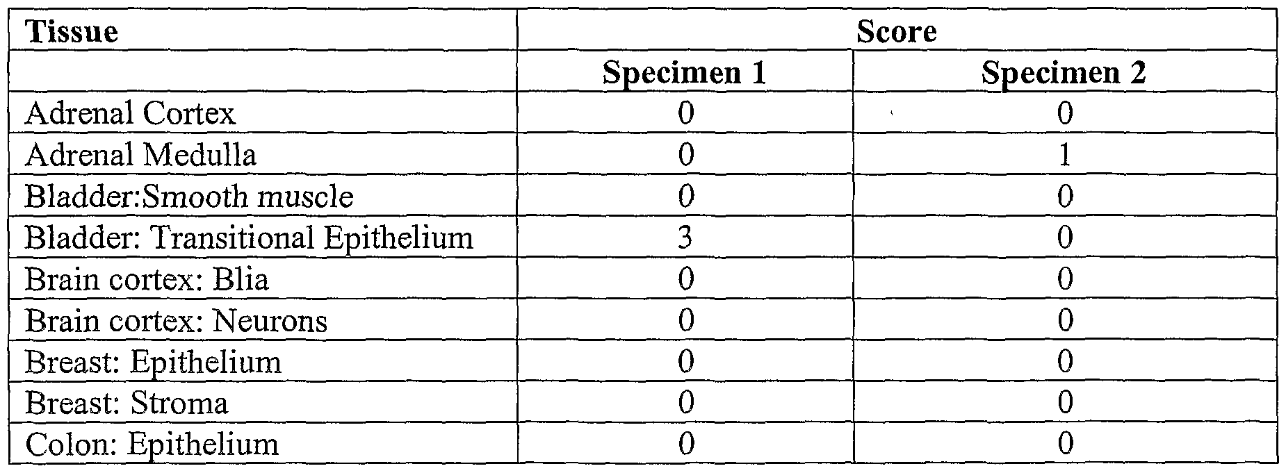


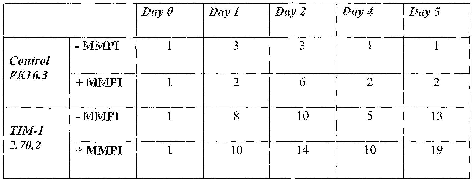


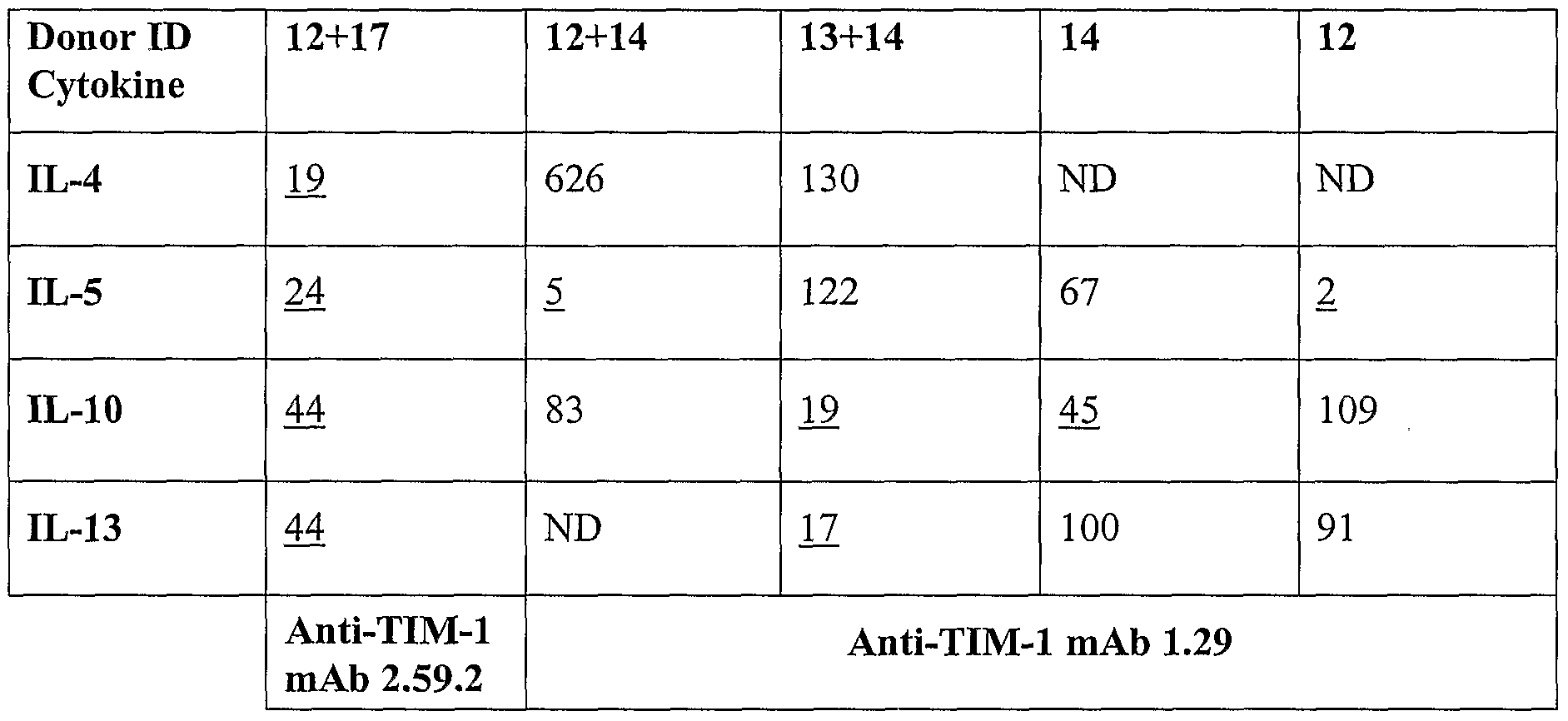
Claims
Priority Applications (7)
| Application Number | Priority Date | Filing Date | Title |
|---|---|---|---|
| CA2519528A CA2519528C (en) | 2003-03-19 | 2004-03-19 | Antibodies against t cell immunoglobulin domain and mucin domain 1 (tim-1) antigen and uses thereof |
| JP2006507388A JP2007525434A (en) | 2003-03-19 | 2004-03-19 | Antibodies to T cells, immunoglobulin domain and mucin domain 1 (TIM-1) antigen and uses thereof. |
| AU2004224390A AU2004224390A1 (en) | 2003-03-19 | 2004-03-19 | Antibodies against T cell immunoglobulin domain and mucin domain 1 (TIM-1) antigen and uses thereof |
| EP04757910.7A EP1613750B1 (en) | 2003-03-19 | 2004-03-19 | Antibodies against t cell immunoglobulin domain and mucin domain 1 (tim-1) antigen and uses thereof |
| DK04757910.7T DK1613750T3 (en) | 2003-03-19 | 2004-03-19 | ANTIBODIES TO T CELL AND MUCINDO immunoglobulin-binding domain 1 (TIM-1) antigen and uses thereof |
| ES04757910.7T ES2557769T3 (en) | 2003-03-19 | 2004-03-19 | Antibodies against the T cell immunoglobulin domain antigen and mucin domain 1 (TIM-1) and uses thereof |
| AU2010227016A AU2010227016A1 (en) | 2003-03-19 | 2010-10-07 | Antibodies Against T Cell Immunoglobulin Domain and Mucin Domain 1 (TIM-1) Antigen and Uses Thereof |
Applications Claiming Priority (2)
| Application Number | Priority Date | Filing Date | Title |
|---|---|---|---|
| US45665203P | 2003-03-19 | 2003-03-19 | |
| US60/456,652 | 2003-03-19 |
Publications (2)
| Publication Number | Publication Date |
|---|---|
| WO2004084823A2 true WO2004084823A2 (en) | 2004-10-07 |
| WO2004084823A3 WO2004084823A3 (en) | 2005-09-15 |
Family
ID=33098138
Family Applications (1)
| Application Number | Title | Priority Date | Filing Date |
|---|---|---|---|
| PCT/US2004/008502 WO2004084823A2 (en) | 2003-03-19 | 2004-03-19 | Antibodies against t cell immunoglobulin domain and mucin domain 1 (tim-1) antigen and uses thereof |
Country Status (8)
| Country | Link |
|---|---|
| US (3) | US8067544B2 (en) |
| EP (2) | EP3000886A1 (en) |
| JP (5) | JP2007525434A (en) |
| AU (2) | AU2004224390A1 (en) |
| CA (1) | CA2519528C (en) |
| DK (1) | DK1613750T3 (en) |
| ES (1) | ES2557769T3 (en) |
| WO (1) | WO2004084823A2 (en) |
Cited By (14)
| Publication number | Priority date | Publication date | Assignee | Title |
|---|---|---|---|---|
| US7041290B2 (en) | 2001-06-01 | 2006-05-09 | Biogen Idec Ma Inc. | Molecules and methods for inhibiting shedding of KIM-1 |
| WO2006094134A2 (en) | 2005-03-02 | 2006-09-08 | Biogen Idec Ma Inc. | Kim-1 antibodies for treatment of th2-mediated conditions |
| US7179901B2 (en) | 2000-06-16 | 2007-02-20 | Biogen Idec Ma Inc. | Renal regulatory elements and methods of use thereof |
| WO2007059082A1 (en) | 2005-11-10 | 2007-05-24 | Curagen Corporation | Method of treating ovarian and renal cancer using antibodies against t cell immunoglobulin domain and mucin domain 1 (tim-1) antigen |
| WO2009064290A1 (en) * | 2007-11-13 | 2009-05-22 | Government Of The United States As Represented By The Secretary Of The Department Of Health And Human Services | Structure of tim family members |
| US7597887B2 (en) | 2002-12-30 | 2009-10-06 | Biogen Idec Ma Inc. | KIM-1 antagonists and use to modulate immune system |
| US8067544B2 (en) | 2003-03-19 | 2011-11-29 | Curagen Corporation | Antibodies against T cell immunoglobulin domain and mucin domain 1 (TIM-1) antigen and uses thereof |
| US9062113B2 (en) | 2003-06-27 | 2015-06-23 | Amgen Fremont Inc. | Antibodies directed to the deletion mutants of epidermal growth factor receptor and uses thereof |
| AU2013206293B2 (en) * | 2005-11-10 | 2016-05-19 | Curagen Corporation | Method of treating ovarian and renal cancer using antibodies against t cell immunoglobulin domain and mucin domain 1 (tim-1) antigen |
| WO2016145085A2 (en) | 2015-03-09 | 2016-09-15 | Celldex Therapeutics, Inc. | Cd27 agonists |
| US9718883B2 (en) | 2003-09-10 | 2017-08-01 | Amgen Fremont Inc. | Antibodies to M-CSF |
| US9771425B2 (en) | 2014-10-27 | 2017-09-26 | Agency For Science, Technology And Research | Anti-PD-1 antibodies |
| CN109265549A (en) * | 2018-09-25 | 2019-01-25 | 中国人民解放军军事科学院军事医学研究院 | A kind of anti-human Tim-1 antibody with neutralization activity |
| US11541076B2 (en) | 2017-01-13 | 2023-01-03 | Celdara Medical, Llc | Chimeric antigen receptors targeting TIM-1 |
Families Citing this family (25)
| Publication number | Priority date | Publication date | Assignee | Title |
|---|---|---|---|---|
| US8709412B2 (en) | 2001-06-29 | 2014-04-29 | The Board Of Trustees Of The Leland Stanford Junior University | Modulation of TIM receptor activity in combination with cytoreductive therapy |
| US7658924B2 (en) * | 2001-10-11 | 2010-02-09 | Amgen Inc. | Angiopoietin-2 specific binding agents |
| CA2494105C (en) * | 2002-07-31 | 2013-04-02 | Seattle Genetics, Inc. | Drug conjugates and their use for treating cancer, an autoimmune disease or an infectious disease |
| TWI347951B (en) * | 2002-09-06 | 2011-09-01 | Amgen Inc | Therapeutic human anti-il-1r1 monoclonal antibody |
| ZA200603619B (en) | 2003-11-06 | 2008-10-29 | Seattle Genetics Inc | Monomethylvaline compounds capable of conjugation to ligands |
| DK1827492T3 (en) * | 2004-11-30 | 2010-11-22 | Curagen Corp | Antibodies targeting GPNMB and uses thereof |
| JP2008531730A (en) * | 2005-03-04 | 2008-08-14 | キュアーディーエム、インク. | Methods and pharmaceutical compositions for treating type I diabetes mellitus and other conditions |
| US20090142338A1 (en) * | 2005-03-04 | 2009-06-04 | Curedm, Inc. | Methods and Compositions for Treating Type 1 and Type 2 Diabetes Mellitus and Related Conditions |
| EP1883417A2 (en) | 2005-05-25 | 2008-02-06 | Curedm Inc. | Peptides, derivatives and analogs thereof, and methods of using same |
| WO2008064306A2 (en) * | 2006-11-22 | 2008-05-29 | Curedm, Inc. | Methods and compositions relating to islet cell neogenesis |
| JP5469600B2 (en) | 2007-07-16 | 2014-04-16 | ジェネンテック, インコーポレイテッド | Anti-CD79b antibody and immunoconjugate and method of use thereof |
| AR067543A1 (en) | 2007-07-16 | 2009-10-14 | Genentech Inc | ANTI-CD79B ANTIBODIES AND HUMANIZED IMMUNOCATE PLAYERS AND METHODS OF USE |
| DK2193142T3 (en) * | 2007-08-30 | 2015-04-20 | Curedm Group Holdings Llc | Compositions and Methods for Using Project Cell Peptides and Analogs Thereto |
| PE20091318A1 (en) | 2008-01-31 | 2009-09-16 | Genentech Inc | ANTI-CD79B ANTIBODIES AND IMMUNOCONJUGATES AND METHODS OF USE OF THE SAME |
| JO2913B1 (en) * | 2008-02-20 | 2015-09-15 | امجين إنك, | Antibodies directed to angiopoietin-1 and angiopoietin-2 and uses thereof |
| US20130156784A1 (en) * | 2008-08-20 | 2013-06-20 | Michael Jeffers | Compositions Using Antibodies Directed To GPNMB And Uses Thereof |
| EP2453919A1 (en) * | 2009-05-20 | 2012-05-23 | Celldex Therapeutics, Inc. | Antibodies directed to gpnmb and uses thereof |
| CA2850291A1 (en) * | 2011-09-29 | 2013-04-04 | Apo-T B.V. | Multi-specific binding molecules targeting aberrant cells |
| CA2860914A1 (en) | 2012-01-13 | 2013-07-18 | Apo-T B.V. | Aberrant cell-restricted immunoglobulins provided with a toxic moiety |
| EP2875347B1 (en) * | 2012-07-23 | 2019-05-08 | Astute Medical, Inc. | Methods for diagnosis of sepsis |
| ES2796903T3 (en) | 2014-09-23 | 2020-11-30 | Hoffmann La Roche | Procedure for the use of anti-CD79b immunoconjugates |
| SG10201803042PA (en) | 2014-10-27 | 2018-06-28 | Agency Science Tech & Res | Anti-tim-3 antibodies |
| GB201419094D0 (en) | 2014-10-27 | 2014-12-10 | Agency Science Tech & Res | Anti-TIM-3-antibodies |
| WO2018039068A1 (en) * | 2016-08-23 | 2018-03-01 | The Scripps Research Institute | Methods and compositions related to in vivo selection of functional molecules |
| RU2674894C2 (en) * | 2017-01-30 | 2018-12-13 | Общество с ограниченной ответственностью "ПЛАНТА" | New luciferases and methods of their use |
Citations (83)
| Publication number | Priority date | Publication date | Assignee | Title |
|---|---|---|---|---|
| US3773919A (en) | 1969-10-23 | 1973-11-20 | Du Pont | Polylactide-drug mixtures |
| EP0036676A1 (en) | 1978-03-24 | 1981-09-30 | The Regents Of The University Of California | Method of making uniformly sized liposomes and liposomes so made |
| EP0052322A2 (en) | 1980-11-10 | 1982-05-26 | Gersonde, Klaus, Prof. Dr. | Method of preparing lipid vesicles by ultrasonic treatment, the use of this method and apparatus for its application |
| EP0058481A1 (en) | 1981-02-16 | 1982-08-25 | Zeneca Limited | Continuous release pharmaceutical compositions |
| US4399216A (en) | 1980-02-25 | 1983-08-16 | The Trustees Of Columbia University | Processes for inserting DNA into eucaryotic cells and for producing proteinaceous materials |
| EP0088046A2 (en) | 1982-02-17 | 1983-09-07 | Ciba-Geigy Ag | Lipids in the aqueous phase |
| DE3218121A1 (en) | 1982-05-14 | 1983-11-17 | Leskovar, Peter, Dr.-Ing., 8000 München | Pharmaceutical compositions for tumour treatment |
| EP0102324A2 (en) | 1982-07-29 | 1984-03-07 | Ciba-Geigy Ag | Lipids and surfactants in an aqueous medium |
| US4485045A (en) | 1981-07-06 | 1984-11-27 | Research Corporation | Synthetic phosphatidyl cholines useful in forming liposomes |
| EP0133988A2 (en) | 1983-08-02 | 1985-03-13 | Hoechst Aktiengesellschaft | Regulating peptide-containing pharmaceutical preparations with retarded release, and process for their preparation |
| EP0142641A2 (en) | 1983-09-26 | 1985-05-29 | Udo Dr. Ehrenfeld | Means and product for the diagnosis and therapy of tumours and for the treatment of weaknesses of the cellular and humoral immune system |
| EP0143949A1 (en) | 1983-11-01 | 1985-06-12 | TERUMO KABUSHIKI KAISHA trading as TERUMO CORPORATION | Pharmaceutical composition containing urokinase |
| US4544545A (en) | 1983-06-20 | 1985-10-01 | Trustees University Of Massachusetts | Liposomes containing modified cholesterol for organ targeting |
| US4681581A (en) | 1983-12-05 | 1987-07-21 | Coates Fredrica V | Adjustable size diaper and folding method therefor |
| US4683202A (en) | 1985-03-28 | 1987-07-28 | Cetus Corporation | Process for amplifying nucleic acid sequences |
| US4683195A (en) | 1986-01-30 | 1987-07-28 | Cetus Corporation | Process for amplifying, detecting, and/or-cloning nucleic acid sequences |
| US4735210A (en) | 1985-07-05 | 1988-04-05 | Immunomedics, Inc. | Lymphographic and organ imaging method and kit |
| US4740461A (en) | 1983-12-27 | 1988-04-26 | Genetics Institute, Inc. | Vectors and methods for transformation of eucaryotic cells |
| US4816397A (en) | 1983-03-25 | 1989-03-28 | Celltech, Limited | Multichain polypeptides or proteins and processes for their production |
| US4831175A (en) | 1986-09-05 | 1989-05-16 | The United States Of America As Represented By The Secretary Of The Department Of Health And Human Services | Backbone polysubstituted chelates for forming a metal chelate-protein conjugate |
| US4912040A (en) | 1986-11-14 | 1990-03-27 | Genetics Institute, Inc. | Eucaryotic expression system |
| US4959455A (en) | 1986-07-14 | 1990-09-25 | Genetics Institute, Inc. | Primate hematopoietic growth factors IL-3 and pharmaceutical compositions |
| WO1992002190A1 (en) | 1990-08-09 | 1992-02-20 | Silverman Harvey N | Method and system for whitening teeth |
| WO1992003918A1 (en) | 1990-08-29 | 1992-03-19 | Genpharm International, Inc. | Transgenic non-human animals capable of producing heterologous antibodies |
| US5099069A (en) | 1986-09-05 | 1992-03-24 | Gansow Otto A | Backbone polysubstituted chelates for forming a metal chelate-protein conjugate |
| US5102990A (en) | 1989-08-09 | 1992-04-07 | Rhomed Incorporated | Direct radiolabeling of antibodies and other proteins with technetium or rhenium |
| US5101827A (en) | 1985-07-05 | 1992-04-07 | Immunomedics, Inc. | Lymphographic and organ imaging method and kit |
| US5124471A (en) | 1990-03-26 | 1992-06-23 | The United States Of America As Represented By The Secretary Of The Department Of Health And Human Services | Bifunctional dtpa-type ligand |
| US5151510A (en) | 1990-04-20 | 1992-09-29 | Applied Biosystems, Inc. | Method of synethesizing sulfurized oligonucleotide analogs |
| WO1992022647A1 (en) | 1991-06-12 | 1992-12-23 | Genpharm International, Inc. | Early detection of transgenic emryros |
| WO1992022645A1 (en) | 1991-06-14 | 1992-12-23 | Genpharm International, Inc. | Transgenic immunodeficient non-human animals |
| US5194594A (en) | 1990-09-07 | 1993-03-16 | Techniclone, Inc. | Modified antibodies |
| WO1993012227A1 (en) | 1991-12-17 | 1993-06-24 | Genpharm International, Inc. | Transgenic non-human animals capable of producing heterologous antibodies |
| US5246692A (en) | 1986-09-05 | 1993-09-21 | The United States Of America As Represented By The Secretary Of Health And Human Services | Backbone polysubstituted chelates for forming a metal chelate-protein conjugate |
| WO1994000569A1 (en) | 1992-06-18 | 1994-01-06 | Genpharm International, Inc. | Methods for producing transgenic non-human animals harboring a yeast artificial chromosome |
| WO1994002602A1 (en) | 1992-07-24 | 1994-02-03 | Cell Genesys, Inc. | Generation of xenogeneic antibodies |
| US5313193A (en) | 1990-06-29 | 1994-05-17 | Thomson-Csf | Object-marking means, method for making such means and reading device |
| WO1994025585A1 (en) | 1993-04-26 | 1994-11-10 | Genpharm International, Inc. | Transgenic non-human animals capable of producing heterologous antibodies |
| WO1994029444A1 (en) | 1993-06-04 | 1994-12-22 | The Government Of The United States Of America, Represented By The Secretary, Department Of Health And Human Services | Method for treating kaposi's sarcoma with antisense oligonucleotides |
| US5460785A (en) | 1989-08-09 | 1995-10-24 | Rhomed Incorporated | Direct labeling of antibodies and other protein with metal ions |
| WO1996014436A1 (en) | 1994-11-04 | 1996-05-17 | Genpharm International, Inc. | Method for making recombinant yeast artificial chromosomes |
| EP0463151B1 (en) | 1990-01-12 | 1996-06-12 | Cell Genesys, Inc. | Generation of xenogeneic antibodies |
| US5530101A (en) | 1988-12-28 | 1996-06-25 | Protein Design Labs, Inc. | Humanized immunoglobulins |
| US5545806A (en) | 1990-08-29 | 1996-08-13 | Genpharm International, Inc. | Ransgenic non-human animals for producing heterologous antibodies |
| US5545807A (en) | 1988-10-12 | 1996-08-13 | The Babraham Institute | Production of antibodies from transgenic animals |
| WO1996034096A1 (en) | 1995-04-28 | 1996-10-31 | Abgenix, Inc. | Human antibodies derived from immunized xenomice |
| US5591669A (en) | 1988-12-05 | 1997-01-07 | Genpharm International, Inc. | Transgenic mice depleted in a mature lymphocytic cell-type |
| US5612205A (en) | 1990-08-29 | 1997-03-18 | Genpharm International, Incorporated | Homologous recombination in mammalian cells |
| WO1997013852A1 (en) | 1995-10-10 | 1997-04-17 | Genpharm International, Inc. | Transgenic non-human animals capable of producing heterologous antibodies |
| US5622861A (en) | 1994-08-05 | 1997-04-22 | The United States Of America As Represented By The Department Of Health And Human Services | Recombinant DNA encoding hepatitis A virus receptor |
| US5625126A (en) | 1990-08-29 | 1997-04-29 | Genpharm International, Inc. | Transgenic non-human animals for producing heterologous antibodies |
| US5625825A (en) | 1993-10-21 | 1997-04-29 | Lsi Logic Corporation | Random number generating apparatus for an interface unit of a carrier sense with multiple access and collision detect (CSMA/CD) ethernet data network |
| US5633425A (en) | 1990-08-29 | 1997-05-27 | Genpharm International, Inc. | Transgenic non-human animals capable of producing heterologous antibodies |
| US5648471A (en) | 1987-12-03 | 1997-07-15 | Centocor, Inc. | One vial method for labeling antibodies with Technetium-99m |
| US5661016A (en) | 1990-08-29 | 1997-08-26 | Genpharm International Inc. | Transgenic non-human animals capable of producing heterologous antibodies of various isotypes |
| WO1997038137A1 (en) | 1996-04-04 | 1997-10-16 | Mercator Genetics, Inc. | Hereditary hemochromatosis gene |
| WO1997044460A1 (en) | 1996-05-24 | 1997-11-27 | Biogen, Inc. | Modulators of tissue regeneration |
| US5693792A (en) | 1992-02-18 | 1997-12-02 | Otsuga Kagaku Kabushiki Kaisha | β-lactam and cephem compounds and processes for their production |
| US5697902A (en) | 1985-07-05 | 1997-12-16 | Immunomedics, Inc. | Method for imaging and treating organs and tissues |
| US5703057A (en) | 1995-04-07 | 1997-12-30 | Board Of Regents The University Of Texas System | Expression library immunization |
| US5714350A (en) | 1992-03-09 | 1998-02-03 | Protein Design Labs, Inc. | Increasing antibody affinity by altering glycosylation in the immunoglobulin variable region |
| US5733743A (en) | 1992-03-24 | 1998-03-31 | Cambridge Antibody Technology Limited | Methods for producing members of specific binding pairs |
| WO1998024884A1 (en) | 1996-12-02 | 1998-06-11 | Genpharm International | Transgenic non-human animals capable of producing heterologous antibodies |
| WO1998024893A2 (en) | 1996-12-03 | 1998-06-11 | Abgenix, Inc. | TRANSGENIC MAMMALS HAVING HUMAN IG LOCI INCLUDING PLURAL VH AND Vλ REGIONS AND ANTIBODIES PRODUCED THEREFROM |
| US5777085A (en) | 1991-12-20 | 1998-07-07 | Protein Design Labs, Inc. | Humanized antibodies reactive with GPIIB/IIIA |
| US5789215A (en) | 1991-08-20 | 1998-08-04 | Genpharm International | Gene targeting in animal cells using isogenic DNA constructs |
| US5789650A (en) | 1990-08-29 | 1998-08-04 | Genpharm International, Inc. | Transgenic non-human animals for producing heterologous antibodies |
| US5814318A (en) | 1990-08-29 | 1998-09-29 | Genpharm International Inc. | Transgenic non-human animals for producing heterologous antibodies |
| WO1998053071A1 (en) | 1997-05-23 | 1998-11-26 | Biogen, Inc. | Modulators of tissue regeneration |
| US5874299A (en) | 1990-08-29 | 1999-02-23 | Genpharm International, Inc. | Transgenic non-human animals capable of producing heterologous antibodies |
| US5877397A (en) | 1990-08-29 | 1999-03-02 | Genpharm International Inc. | Transgenic non-human animals capable of producing heterologous antibodies of various isotypes |
| US5916771A (en) | 1996-10-11 | 1999-06-29 | Abgenix, Inc. | Production of a multimeric protein by cell fusion method |
| US5981175A (en) | 1993-01-07 | 1999-11-09 | Genpharm Internation, Inc. | Methods for producing recombinant mammalian cells harboring a yeast artificial chromosome |
| US6075181A (en) | 1990-01-12 | 2000-06-13 | Abgenix, Inc. | Human antibodies derived from immunized xenomice |
| US6150584A (en) | 1990-01-12 | 2000-11-21 | Abgenix, Inc. | Human antibodies derived from immunized xenomice |
| US6162963A (en) | 1990-01-12 | 2000-12-19 | Abgenix, Inc. | Generation of Xenogenetic antibodies |
| WO2000076310A1 (en) | 1999-06-10 | 2000-12-21 | Abgenix, Inc. | Transgenic animals for producing specific isotypes of human antibodies via non-cognate switch regions |
| WO2001025433A2 (en) | 1999-10-07 | 2001-04-12 | Genentech, Inc. | Novel polypeptides, their nucleic acids, and methods for their use in angiogenesis and vascularization |
| US6255458B1 (en) | 1990-08-29 | 2001-07-03 | Genpharm International | High affinity human antibodies and human antibodies against digoxin |
| US6441163B1 (en) | 2001-05-31 | 2002-08-27 | Immunogen, Inc. | Methods for preparation of cytotoxic conjugates of maytansinoids and cell binding agents |
| WO2002098920A1 (en) | 2001-06-01 | 2002-12-12 | Biogen, Inc. | Molecules and methods for inhibiting shedding of kim-1 |
| US20030157730A1 (en) | 2001-12-03 | 2003-08-21 | Walker Wynn L. | Antibody categorization based on binding characteristics |
| US9676293B2 (en) | 2013-04-23 | 2017-06-13 | Calsonic Kansei Corporation | Driving force controller for electric vehicle |
Family Cites Families (9)
| Publication number | Priority date | Publication date | Assignee | Title |
|---|---|---|---|---|
| US3180193A (en) | 1963-02-25 | 1965-04-27 | Benedict David | Machines for cutting lengths of strip material |
| CA2002910A1 (en) | 1988-11-18 | 1990-05-18 | Motonobu Furuta | Thermoplastic resin composition |
| US6849425B1 (en) * | 1999-10-14 | 2005-02-01 | Ixsys, Inc. | Methods of optimizing antibody variable region binding affinity |
| WO2003002722A2 (en) * | 2001-06-29 | 2003-01-09 | The Board Of Trustees Of The Leland Stanford Junior University | T cell regulatory genes and methods of use thereof |
| AU2003228336B2 (en) * | 2002-03-19 | 2009-07-30 | Curagen Corporation | Therapeutic polypeptides, nucleic acids encoding same, and methods of use |
| JP5356648B2 (en) | 2003-02-20 | 2013-12-04 | シアトル ジェネティックス, インコーポレイテッド | Anti-CD70 antibody-drug conjugates and their use for the treatment of cancer and immune disorders |
| US8067544B2 (en) * | 2003-03-19 | 2011-11-29 | Curagen Corporation | Antibodies against T cell immunoglobulin domain and mucin domain 1 (TIM-1) antigen and uses thereof |
| WO2005001092A2 (en) * | 2003-05-20 | 2005-01-06 | Wyeth | Compositions and methods for diagnosing and treating cancers |
| US11284893B2 (en) | 2019-04-02 | 2022-03-29 | Covidien Lp | Stapling device with articulating tool assembly |
-
2004
- 2004-03-19 US US10/805,177 patent/US8067544B2/en not_active Expired - Fee Related
- 2004-03-19 ES ES04757910.7T patent/ES2557769T3/en not_active Expired - Lifetime
- 2004-03-19 CA CA2519528A patent/CA2519528C/en not_active Expired - Fee Related
- 2004-03-19 JP JP2006507388A patent/JP2007525434A/en active Pending
- 2004-03-19 AU AU2004224390A patent/AU2004224390A1/en not_active Abandoned
- 2004-03-19 WO PCT/US2004/008502 patent/WO2004084823A2/en active Application Filing
- 2004-03-19 EP EP15189243.7A patent/EP3000886A1/en not_active Withdrawn
- 2004-03-19 DK DK04757910.7T patent/DK1613750T3/en active
- 2004-03-19 EP EP04757910.7A patent/EP1613750B1/en not_active Expired - Lifetime
-
2010
- 2010-10-07 AU AU2010227016A patent/AU2010227016A1/en not_active Abandoned
-
2011
- 2011-09-16 JP JP2011203584A patent/JP6103801B2/en not_active Expired - Fee Related
- 2011-11-29 US US13/305,898 patent/US9580503B2/en not_active Expired - Fee Related
-
2014
- 2014-04-14 JP JP2014082974A patent/JP2014144971A/en active Pending
-
2016
- 2016-11-22 JP JP2016226619A patent/JP2017048240A/en not_active Ceased
-
2017
- 2017-02-24 US US15/442,083 patent/US20170218067A1/en not_active Abandoned
-
2018
- 2018-04-10 JP JP2018075311A patent/JP2018150305A/en active Pending
Patent Citations (102)
| Publication number | Priority date | Publication date | Assignee | Title |
|---|---|---|---|---|
| US3773919A (en) | 1969-10-23 | 1973-11-20 | Du Pont | Polylactide-drug mixtures |
| EP0036676A1 (en) | 1978-03-24 | 1981-09-30 | The Regents Of The University Of California | Method of making uniformly sized liposomes and liposomes so made |
| US4399216A (en) | 1980-02-25 | 1983-08-16 | The Trustees Of Columbia University | Processes for inserting DNA into eucaryotic cells and for producing proteinaceous materials |
| EP0052322A2 (en) | 1980-11-10 | 1982-05-26 | Gersonde, Klaus, Prof. Dr. | Method of preparing lipid vesicles by ultrasonic treatment, the use of this method and apparatus for its application |
| EP0058481A1 (en) | 1981-02-16 | 1982-08-25 | Zeneca Limited | Continuous release pharmaceutical compositions |
| US4485045A (en) | 1981-07-06 | 1984-11-27 | Research Corporation | Synthetic phosphatidyl cholines useful in forming liposomes |
| EP0088046A2 (en) | 1982-02-17 | 1983-09-07 | Ciba-Geigy Ag | Lipids in the aqueous phase |
| DE3218121A1 (en) | 1982-05-14 | 1983-11-17 | Leskovar, Peter, Dr.-Ing., 8000 München | Pharmaceutical compositions for tumour treatment |
| EP0102324A2 (en) | 1982-07-29 | 1984-03-07 | Ciba-Geigy Ag | Lipids and surfactants in an aqueous medium |
| US4816397A (en) | 1983-03-25 | 1989-03-28 | Celltech, Limited | Multichain polypeptides or proteins and processes for their production |
| US4544545A (en) | 1983-06-20 | 1985-10-01 | Trustees University Of Massachusetts | Liposomes containing modified cholesterol for organ targeting |
| EP0133988A2 (en) | 1983-08-02 | 1985-03-13 | Hoechst Aktiengesellschaft | Regulating peptide-containing pharmaceutical preparations with retarded release, and process for their preparation |
| EP0142641A2 (en) | 1983-09-26 | 1985-05-29 | Udo Dr. Ehrenfeld | Means and product for the diagnosis and therapy of tumours and for the treatment of weaknesses of the cellular and humoral immune system |
| EP0143949A1 (en) | 1983-11-01 | 1985-06-12 | TERUMO KABUSHIKI KAISHA trading as TERUMO CORPORATION | Pharmaceutical composition containing urokinase |
| US4681581A (en) | 1983-12-05 | 1987-07-21 | Coates Fredrica V | Adjustable size diaper and folding method therefor |
| US4740461A (en) | 1983-12-27 | 1988-04-26 | Genetics Institute, Inc. | Vectors and methods for transformation of eucaryotic cells |
| US4683202A (en) | 1985-03-28 | 1987-07-28 | Cetus Corporation | Process for amplifying nucleic acid sequences |
| US4683202B1 (en) | 1985-03-28 | 1990-11-27 | Cetus Corp | |
| US5697902A (en) | 1985-07-05 | 1997-12-16 | Immunomedics, Inc. | Method for imaging and treating organs and tissues |
| US4735210A (en) | 1985-07-05 | 1988-04-05 | Immunomedics, Inc. | Lymphographic and organ imaging method and kit |
| US5101827A (en) | 1985-07-05 | 1992-04-07 | Immunomedics, Inc. | Lymphographic and organ imaging method and kit |
| US4683195B1 (en) | 1986-01-30 | 1990-11-27 | Cetus Corp | |
| US4683195A (en) | 1986-01-30 | 1987-07-28 | Cetus Corporation | Process for amplifying, detecting, and/or-cloning nucleic acid sequences |
| US4959455A (en) | 1986-07-14 | 1990-09-25 | Genetics Institute, Inc. | Primate hematopoietic growth factors IL-3 and pharmaceutical compositions |
| US5246692A (en) | 1986-09-05 | 1993-09-21 | The United States Of America As Represented By The Secretary Of Health And Human Services | Backbone polysubstituted chelates for forming a metal chelate-protein conjugate |
| US5099069A (en) | 1986-09-05 | 1992-03-24 | Gansow Otto A | Backbone polysubstituted chelates for forming a metal chelate-protein conjugate |
| US4831175A (en) | 1986-09-05 | 1989-05-16 | The United States Of America As Represented By The Secretary Of The Department Of Health And Human Services | Backbone polysubstituted chelates for forming a metal chelate-protein conjugate |
| US4912040A (en) | 1986-11-14 | 1990-03-27 | Genetics Institute, Inc. | Eucaryotic expression system |
| US5648471A (en) | 1987-12-03 | 1997-07-15 | Centocor, Inc. | One vial method for labeling antibodies with Technetium-99m |
| US5545807A (en) | 1988-10-12 | 1996-08-13 | The Babraham Institute | Production of antibodies from transgenic animals |
| US5591669A (en) | 1988-12-05 | 1997-01-07 | Genpharm International, Inc. | Transgenic mice depleted in a mature lymphocytic cell-type |
| US6023010A (en) | 1988-12-05 | 2000-02-08 | Genpharm International | Transgenic non-human animals depleted in a mature lymphocytic cell-type |
| US5585089A (en) | 1988-12-28 | 1996-12-17 | Protein Design Labs, Inc. | Humanized immunoglobulins |
| US5693761A (en) | 1988-12-28 | 1997-12-02 | Protein Design Labs, Inc. | Polynucleotides encoding improved humanized immunoglobulins |
| US5530101A (en) | 1988-12-28 | 1996-06-25 | Protein Design Labs, Inc. | Humanized immunoglobulins |
| US5460785A (en) | 1989-08-09 | 1995-10-24 | Rhomed Incorporated | Direct labeling of antibodies and other protein with metal ions |
| US5102990A (en) | 1989-08-09 | 1992-04-07 | Rhomed Incorporated | Direct radiolabeling of antibodies and other proteins with technetium or rhenium |
| USRE35500E (en) | 1989-08-09 | 1997-05-06 | Aberlyn Capital Management Limited Partnership | Direct radiolabeling of antibodies and other proteins with technetium or rhenium |
| EP0463151B1 (en) | 1990-01-12 | 1996-06-12 | Cell Genesys, Inc. | Generation of xenogeneic antibodies |
| US5939598A (en) | 1990-01-12 | 1999-08-17 | Abgenix, Inc. | Method of making transgenic mice lacking endogenous heavy chains |
| JP3068507B2 (en) | 1990-01-12 | 2000-07-24 | アブジェニックス インコーポレイテッド | Generation of heterologous antibodies |
| US6114598A (en) | 1990-01-12 | 2000-09-05 | Abgenix, Inc. | Generation of xenogeneic antibodies |
| US6150584A (en) | 1990-01-12 | 2000-11-21 | Abgenix, Inc. | Human antibodies derived from immunized xenomice |
| JP3068180B2 (en) | 1990-01-12 | 2000-07-24 | アブジェニックス インコーポレイテッド | Generation of heterologous antibodies |
| JP3068506B2 (en) | 1990-01-12 | 2000-07-24 | アブジェニックス インコーポレイテッド | Generation of heterologous antibodies |
| US6075181A (en) | 1990-01-12 | 2000-06-13 | Abgenix, Inc. | Human antibodies derived from immunized xenomice |
| US6162963A (en) | 1990-01-12 | 2000-12-19 | Abgenix, Inc. | Generation of Xenogenetic antibodies |
| US5286850A (en) | 1990-03-26 | 1994-02-15 | The United States Of America As Represented By The Department Of Health And Human Services | Antibody DTPA-type ligand conjugates |
| US5124471A (en) | 1990-03-26 | 1992-06-23 | The United States Of America As Represented By The Secretary Of The Department Of Health And Human Services | Bifunctional dtpa-type ligand |
| US5151510A (en) | 1990-04-20 | 1992-09-29 | Applied Biosystems, Inc. | Method of synethesizing sulfurized oligonucleotide analogs |
| US5313193A (en) | 1990-06-29 | 1994-05-17 | Thomson-Csf | Object-marking means, method for making such means and reading device |
| WO1992002190A1 (en) | 1990-08-09 | 1992-02-20 | Silverman Harvey N | Method and system for whitening teeth |
| US5877397A (en) | 1990-08-29 | 1999-03-02 | Genpharm International Inc. | Transgenic non-human animals capable of producing heterologous antibodies of various isotypes |
| US5874299A (en) | 1990-08-29 | 1999-02-23 | Genpharm International, Inc. | Transgenic non-human animals capable of producing heterologous antibodies |
| US5661016A (en) | 1990-08-29 | 1997-08-26 | Genpharm International Inc. | Transgenic non-human animals capable of producing heterologous antibodies of various isotypes |
| EP0546073B1 (en) | 1990-08-29 | 1997-09-10 | GenPharm International, Inc. | production and use of transgenic non-human animals capable of producing heterologous antibodies |
| US5545806A (en) | 1990-08-29 | 1996-08-13 | Genpharm International, Inc. | Ransgenic non-human animals for producing heterologous antibodies |
| US5612205A (en) | 1990-08-29 | 1997-03-18 | Genpharm International, Incorporated | Homologous recombination in mammalian cells |
| US5633425A (en) | 1990-08-29 | 1997-05-27 | Genpharm International, Inc. | Transgenic non-human animals capable of producing heterologous antibodies |
| US5789650A (en) | 1990-08-29 | 1998-08-04 | Genpharm International, Inc. | Transgenic non-human animals for producing heterologous antibodies |
| US5770429A (en) | 1990-08-29 | 1998-06-23 | Genpharm International, Inc. | Transgenic non-human animals capable of producing heterologous antibodies |
| WO1992003918A1 (en) | 1990-08-29 | 1992-03-19 | Genpharm International, Inc. | Transgenic non-human animals capable of producing heterologous antibodies |
| US5625126A (en) | 1990-08-29 | 1997-04-29 | Genpharm International, Inc. | Transgenic non-human animals for producing heterologous antibodies |
| US5814318A (en) | 1990-08-29 | 1998-09-29 | Genpharm International Inc. | Transgenic non-human animals for producing heterologous antibodies |
| US5721367A (en) | 1990-08-29 | 1998-02-24 | Pharming B.V. | Homologous recombination in mammalian cells |
| US6255458B1 (en) | 1990-08-29 | 2001-07-03 | Genpharm International | High affinity human antibodies and human antibodies against digoxin |
| US5194594A (en) | 1990-09-07 | 1993-03-16 | Techniclone, Inc. | Modified antibodies |
| WO1992022670A1 (en) | 1991-06-12 | 1992-12-23 | Genpharm International, Inc. | Early detection of transgenic embryos |
| WO1992022647A1 (en) | 1991-06-12 | 1992-12-23 | Genpharm International, Inc. | Early detection of transgenic emryros |
| WO1992022645A1 (en) | 1991-06-14 | 1992-12-23 | Genpharm International, Inc. | Transgenic immunodeficient non-human animals |
| US5789215A (en) | 1991-08-20 | 1998-08-04 | Genpharm International | Gene targeting in animal cells using isogenic DNA constructs |
| WO1993012227A1 (en) | 1991-12-17 | 1993-06-24 | Genpharm International, Inc. | Transgenic non-human animals capable of producing heterologous antibodies |
| US5777085A (en) | 1991-12-20 | 1998-07-07 | Protein Design Labs, Inc. | Humanized antibodies reactive with GPIIB/IIIA |
| US5693792A (en) | 1992-02-18 | 1997-12-02 | Otsuga Kagaku Kabushiki Kaisha | β-lactam and cephem compounds and processes for their production |
| US5714350A (en) | 1992-03-09 | 1998-02-03 | Protein Design Labs, Inc. | Increasing antibody affinity by altering glycosylation in the immunoglobulin variable region |
| US5733743A (en) | 1992-03-24 | 1998-03-31 | Cambridge Antibody Technology Limited | Methods for producing members of specific binding pairs |
| WO1994000569A1 (en) | 1992-06-18 | 1994-01-06 | Genpharm International, Inc. | Methods for producing transgenic non-human animals harboring a yeast artificial chromosome |
| WO1994002602A1 (en) | 1992-07-24 | 1994-02-03 | Cell Genesys, Inc. | Generation of xenogeneic antibodies |
| US5981175A (en) | 1993-01-07 | 1999-11-09 | Genpharm Internation, Inc. | Methods for producing recombinant mammalian cells harboring a yeast artificial chromosome |
| WO1994025585A1 (en) | 1993-04-26 | 1994-11-10 | Genpharm International, Inc. | Transgenic non-human animals capable of producing heterologous antibodies |
| WO1994029444A1 (en) | 1993-06-04 | 1994-12-22 | The Government Of The United States Of America, Represented By The Secretary, Department Of Health And Human Services | Method for treating kaposi's sarcoma with antisense oligonucleotides |
| US5625825A (en) | 1993-10-21 | 1997-04-29 | Lsi Logic Corporation | Random number generating apparatus for an interface unit of a carrier sense with multiple access and collision detect (CSMA/CD) ethernet data network |
| US5622861A (en) | 1994-08-05 | 1997-04-22 | The United States Of America As Represented By The Department Of Health And Human Services | Recombinant DNA encoding hepatitis A virus receptor |
| WO1996014436A1 (en) | 1994-11-04 | 1996-05-17 | Genpharm International, Inc. | Method for making recombinant yeast artificial chromosomes |
| US5643763A (en) | 1994-11-04 | 1997-07-01 | Genpharm International, Inc. | Method for making recombinant yeast artificial chromosomes by minimizing diploid doubling during mating |
| US5703057A (en) | 1995-04-07 | 1997-12-30 | Board Of Regents The University Of Texas System | Expression library immunization |
| WO1996034096A1 (en) | 1995-04-28 | 1996-10-31 | Abgenix, Inc. | Human antibodies derived from immunized xenomice |
| WO1997013852A1 (en) | 1995-10-10 | 1997-04-17 | Genpharm International, Inc. | Transgenic non-human animals capable of producing heterologous antibodies |
| WO1997038137A1 (en) | 1996-04-04 | 1997-10-16 | Mercator Genetics, Inc. | Hereditary hemochromatosis gene |
| WO1997044460A1 (en) | 1996-05-24 | 1997-11-27 | Biogen, Inc. | Modulators of tissue regeneration |
| US6664385B1 (en) | 1996-05-24 | 2003-12-16 | Biogen, Inc. | Kidney injury-related molecules |
| US6207418B1 (en) | 1996-10-11 | 2001-03-27 | Abgenix, Inc. | Production of a multimeric protein by cell fusion method |
| US5916771A (en) | 1996-10-11 | 1999-06-29 | Abgenix, Inc. | Production of a multimeric protein by cell fusion method |
| WO1998024884A1 (en) | 1996-12-02 | 1998-06-11 | Genpharm International | Transgenic non-human animals capable of producing heterologous antibodies |
| WO1998024893A2 (en) | 1996-12-03 | 1998-06-11 | Abgenix, Inc. | TRANSGENIC MAMMALS HAVING HUMAN IG LOCI INCLUDING PLURAL VH AND Vλ REGIONS AND ANTIBODIES PRODUCED THEREFROM |
| WO1998053071A1 (en) | 1997-05-23 | 1998-11-26 | Biogen, Inc. | Modulators of tissue regeneration |
| WO2000076310A1 (en) | 1999-06-10 | 2000-12-21 | Abgenix, Inc. | Transgenic animals for producing specific isotypes of human antibodies via non-cognate switch regions |
| WO2001025433A2 (en) | 1999-10-07 | 2001-04-12 | Genentech, Inc. | Novel polypeptides, their nucleic acids, and methods for their use in angiogenesis and vascularization |
| US6441163B1 (en) | 2001-05-31 | 2002-08-27 | Immunogen, Inc. | Methods for preparation of cytotoxic conjugates of maytansinoids and cell binding agents |
| WO2002098920A1 (en) | 2001-06-01 | 2002-12-12 | Biogen, Inc. | Molecules and methods for inhibiting shedding of kim-1 |
| US20030157730A1 (en) | 2001-12-03 | 2003-08-21 | Walker Wynn L. | Antibody categorization based on binding characteristics |
| US9676293B2 (en) | 2013-04-23 | 2017-06-13 | Calsonic Kansei Corporation | Driving force controller for electric vehicle |
Non-Patent Citations (102)
| Title |
|---|
| "Remington: The Science and Practice of Pharmacy 20th ed,", 2003, LIPPINCOTT WILLIAMS & WILKENS PUBLISHERS |
| ALLEN, NAT. REV. CANCER, vol. 2, no. 10, 2002, pages 750 - 63 |
| BABCOOK ET AL., PROC. NATL. ACAD. SCI. USA, vol. 93, 1996, pages 7843 - 7848 |
| BALDRICK P.: "Pharmaceutical excipient development: the need for preclinical guidance", REGUL. TOXICOL. PHARMACOL., vol. 32, no. 2, 2000, pages 210 - 8 |
| BLAKE; LITZI-DAVIS, BIOCONJUGATE CHEM., vol. 3, 1992, pages 510 - 513 |
| BOWIE ET AL., SCIENCE, vol. 253, 1991, pages 164 |
| C. BRANDEN AND J. TOOZE,: "Introduction to Protein Structure", 1991, GARLAND PUBLISHING |
| CAPSEY ET AL.: "Genetically Engineered Human Therapeutic Drugs", 1988, STOCKTON PRESS |
| CHARMAN WN: "Lipids, lipophilic drugs, and oral drug delivery-some emerging doncepts", J PHARM SCI, vol. 89, no. 8, 2000, pages 967 - 78 |
| CHEN ET AL., ANAL. BIOCHEM., vol. 227, 1995, pages 168 - 175 |
| CHEN ET AL., BIOCHEM BIOPHYS RES COMMUN, vol. 274, no. 3, 2000, pages 576 - 82 |
| CHEN ET AL., HUMAN GENE THERAPY, vol. 5, 1994, pages 595 - 601 |
| CHISWELL; MCCAFFERTY, TIBTECH, vol. 10, 1992, pages 80 - 84 |
| CHOTHIA ET AL., NATURE, vol. 342, 1989, pages 878 - 883 |
| CHOTHIA; LESK, J. MOL. BIOL., vol. 196, 1987, pages 901 - 917 |
| CREIGHTON,: "Proteins, Structures and Molecular Principles", 1984, W. H. FREEMAN AND COMPANY |
| CWIRLA ET AL., PNAS USA, vol. 87, 1990, pages 6378 - 6382 |
| DAYHOFF, M.O.: "Atlas of Protein Sequence and Structure", vol. 5, 1972, NATIONAL BIOMEDICAL RESEARCH FOUNDATION, pages: 101 - 110 |
| DORONINA ET AL., NATURE BIOTECHNOLOGY, vol. 21, no. 7, 2003, pages 778 - 784 |
| E.S. GOLUB AND D.R. GREN,: "Immunology - A Synthesis 2nd Edition", 1991, SINAUER ASSOCIATES |
| EPSTEIN ET AL., PROC. NATL. ACAD. SCI. USA, vol. 82, 1985, pages 3688 - 3692 |
| EVANS ET AL., J. MED. CHEM., vol. 30, 1987, pages 1229 |
| FANGER ET AL., IMMUNOL METHODS, vol. 4, 1994, pages 72 - 81 |
| FANGER, IMMUNOL METHODS, vol. 4, 1994, pages 72 - 81 |
| FAUCHERE, J. ADV. DRUG RES., vol. 15, 1986, pages 29 |
| FEIGELSTOCK ET AL., J VIROL, vol. 72, no. 8, 1998, pages 6621 - 8 |
| FEIGELSTOCK ET AL., J VIROLOGY, vol. 72, no. 8, 1998, pages 6621 - 6628 |
| GERAN ET AL., CANCER CHEMOTHER. REP., vol. 3, 1972, pages 1 - 104 |
| GORMAN ET AL., P.N.A.S., vol. 79, 1982, pages 6777 |
| GREEN, NATURE GENETICS, vol. 7, 1994, pages 13 - 21 |
| GREEN; JAKOBOVITS, J. EXP. MED., vol. 188, 1998, pages 483 - 495 |
| GROSSCHEDL ET AL., CELL, vol. 41, 1985, pages 885 |
| HAN ET AL., KIDNEY INT, vol. 62, 2002, pages 237 - 244 |
| HANES; PLUCTHAU, PNAS USA, vol. 94, 1997, pages 4937 - 4942 |
| HOGANBOOM ET AL., IMMUNOL. REVIEWS, vol. 130, 1992, pages 43 - 68 |
| HOUGHTEN ET AL., BIOTECHNIQUES, vol. 13, 1992, pages 412 - 421 |
| HOUGHTEN, PNAS USA, vol. 82, 1985, pages 5131 - 5135 |
| HWANG ET AL., PROC. NATL. ACAD. SCI. USA, vol. 77, 1980, pages 4030 - 4034 |
| ICHIMURA ET AL., J. BIOL. CHEM, vol. 273, no. 7, 1998, pages 4135 - 42 |
| ICHIMURA ET AL., JBIOL CHEM, vol. 273, 1998, pages 4135 - 4142 |
| ICHIMURA ET AL., JBIOL CHEM, vol. 273, no. 7, 1998, pages 4135 - 42 |
| IKE ET AL., NUCL. ACID RES., vol. 11, 1983, pages 477 |
| IMMUNOL. CELL BIOL., vol. 65, pages 111 - 125 |
| ITAKURA ET AL., ANNU. REV. BIOCHEM., vol. 53, 1984, pages 323 |
| ITAKURA ET AL., SCIENCE, vol. 198, 1984, pages 1056 |
| J. IMMUNOL., vol. 139, 1987, pages 3521 |
| JUNGHANS ET AL.: "Cancer Chemotherapy and Biotherapy 2d ed.,", 1996, LIPPINCOTT RAVEN, pages: 655 - 686 |
| KABAT: "Sequences of Proteins of Immunological Interest", 1987, NATIONAL INSTITUTES OF HEALTH |
| KABAT: "Sequences of Proteins of Immunological Interest", 1991, N.I.H. PUBLICATION NO. 91-3242 |
| KAPLAN ET AL., EMBO J, vol. 15, no. 16, 1996, pages 4282 - 96 |
| KOHLS; LAPPI, BIOTECHNIQUES, vol. 28, no. 1, 2000, pages 162 - 5 |
| KOSTELNY ET AL., J. IMMUNOL., vol. 148, 1992, pages 1547 - 1553 |
| KUCHROO ET AL., NAT REV IMMUNOL, vol. 3, 2003, pages 454 - 462 |
| LANGER ET AL., J. BIOMED MATER. RES., vol. 15, 1981, pages 167 - 277 |
| LANGER, CHEM. TECH., vol. 12, 1982, pages 98 - 105 |
| LAPLANCHE ET AL., NUCL. ACIDS RES., vol. 14, 1986, pages 9081 |
| LIU ET AL., P.N.A.S., vol. 84, 1987, pages 3439 |
| MARASCO, GENE THERAPY, vol. 4, 1997, pages 11 - 15 |
| MCINTIRE ET AL., NAT IMMUNOL, vol. 2, 2001, pages 1109 - 1116 |
| MCINTIRE ET AL., NATURE IMMUNOLOGY, vol. 2, 2001, pages 1109 - 1116 |
| MENDEZ ET AL., NATURE GENETICS, vol. 15, 1997, pages 146 - 156 |
| MOHAMMAD ET AL., INT. J. ONCOL., vol. 15, no. 2, 1999, pages 367 - 72 |
| MOHAMMAD ET AL., INT. J. ONCOL., vol. 15, no. 2, August 1999 (1999-08-01), pages 367 - 72 |
| NARANG, TETRAHEDRON, vol. 39, 1983, pages 3 |
| NEEDLEMAN; WUNSCH, J. MOL. BIOL., vol. 48, 1970, pages 443 |
| OGAWA ET AL., TOXICOL LETT., vol. 121, no. 2, 2001, pages 97 - 106 |
| OKAYAMA ET AL., MOL. CELL. BIO., vol. 3, 1983, pages 280 |
| PARKER, S.,: "The McGraw-Hill Dictionary of Chemical Terms", 1985, MCGRAW-HILL |
| PARMLEY; SMITH, GENE, vol. 73, 1988, pages 305 - 318 |
| PAUL, W.: "Fundamental Immunology, 2d ed.", 1989, RAVEN PRESS, article "Ch. 7" |
| PEARSON; LIPMAN, PROC. NATL. ACAD. SCI. (U.S.A.), vol. 85, 1988, pages 2444 |
| PINALLA ET AL., BIOTECHNIQUES, vol. 13, 1992, pages 901 - 905 |
| POWELL ET AL.: "Compendium of excipients for parenteral formulations", PDA J PHARM SCI TECHNOL., vol. 52, 1998, pages 238 - 311 |
| PREST ET AL., FASEB J, vol. 16, no. 6, 2002, pages 592 - 4 |
| RIZO; GIERASCH, ANN. REV. BIOCHEM., vol. 61, 1992, pages 387 |
| RUSSEL ET AL., NUCL. ACIDS RES., vol. 21, 1993, pages 1081 - 1085 |
| SAMBROOK ET AL.: "Molecular Cloning: A Laboratory Manual 2d ed.,", 1989, COLD SPRING HARBOR LABORATORY PRESS |
| SCOTT, TIBS, vol. 17, 1992, pages 241 - 245 |
| See also references of EP1613750A4 |
| SHEVACH, NAT REV IMMUNOL, vol. 2, 2002, pages 389 - 400 |
| SIDMAN ET AL., BIOPOLYMERS, vol. 22, 1983, pages 547 - 556 |
| SILBERSTEIN ET AL., J VIROL, vol. 75, no. 2, 2001, pages 717 - 25 |
| SMITH; WATERMAN, ADV. APPL. MATH., vol. 2, 1981, pages 482 |
| SONGSIVILAI; LACHMANN, CLIN. EXP. IMMUNOL., vol. 79, 1990, pages 315 - 321 |
| STEC ET AL., J. AM. CHEM. SOC., vol. 106, 1984, pages 6077 |
| STEIN ET AL., NUCL. ACIDS RES., vol. 16, 1988, pages 3209 |
| THORNTON ET AL., NATURE, vol. 354, 1991, pages 105 |
| TOXICOL LETT., vol. 21, no. 3, pages 778 - 784 |
| TRAUNECKER ET AL., INT. J. CANCER (SUPPL.), vol. 7, 1992, pages 51 - 52 |
| UHLMANN; PEYMAN, CHEMICAL REVIEWS, vol. 90, 1990, pages 543 |
| VALERIUS ET AL., BLOOD, vol. 90, 1997, pages 4485 - 4492 |
| VEBER; FREIDINGER, TINS, 1985, pages 392 |
| VITETTA, IMMUNOL TODAY, vol. 14, 1993, pages 252 |
| VITETTA, LMMUNOL TODAY, vol. 14, 1993, pages 252 |
| WANG W.: "Lyophilization and development of solid protein pharmaceuticals", INT. J. PHARM., vol. 203, no. 1-2, 2000, pages 1 - 60 |
| WILLS-KAIP, NATURE IMMUNOLOGY, vol. 2, 2001, pages 1095 - 1096 |
| WILLS-KARP ET AL., NAT IMMUNOL, vol. 4, 2003, pages 1050 - 1052 |
| WINTER; HARRIS, IMMUNOL TODAY, vol. 14, 1993, pages 43 - 46 |
| WRIGHT ET AL., CRIT, REVIEWS IN IMMUNOL., vol. 12, 1992, pages 125 - 168 |
| YANG ET AL., CANCER RES., 1999 |
| ZON ET AL., ANTI-CANCER DRUG DESIGN, vol. 6, 1991, pages 539 |
| ZON ET AL.: "Oligonucleotides and Analogues: A Practical Approach", 1991, OXFORD UNIVERSITY PRESS, pages: 87 - 108 |
Cited By (35)
| Publication number | Priority date | Publication date | Assignee | Title |
|---|---|---|---|---|
| US7179901B2 (en) | 2000-06-16 | 2007-02-20 | Biogen Idec Ma Inc. | Renal regulatory elements and methods of use thereof |
| US7041290B2 (en) | 2001-06-01 | 2006-05-09 | Biogen Idec Ma Inc. | Molecules and methods for inhibiting shedding of KIM-1 |
| US7696321B2 (en) | 2001-06-01 | 2010-04-13 | Biogen Idec Ma Inc. | Molecules and methods for inhibiting shedding of KIM-1 |
| US7300652B2 (en) | 2001-06-01 | 2007-11-27 | Biogen Idec Ma Inc. | Molecules and methods for inhibiting shedding of KIM-1 |
| US7597887B2 (en) | 2002-12-30 | 2009-10-06 | Biogen Idec Ma Inc. | KIM-1 antagonists and use to modulate immune system |
| US9580503B2 (en) | 2003-03-19 | 2017-02-28 | Celldex Therapeutics, Inc. | Antibodies against T cell immunoglobulin domain and mucin domain 1 (TIM-1) antigen and uses thereof |
| US8067544B2 (en) | 2003-03-19 | 2011-11-29 | Curagen Corporation | Antibodies against T cell immunoglobulin domain and mucin domain 1 (TIM-1) antigen and uses thereof |
| US9085624B2 (en) | 2003-06-27 | 2015-07-21 | Amgen Fremont Inc. | Antibodies directed to the deletion mutants of epidermal growth factor receptor and uses thereof |
| US10118968B2 (en) | 2003-06-27 | 2018-11-06 | Amgen Fremont Inc. | Antibodies directed to the deletion mutants of epidermal growth factor receptor and uses thereof |
| US9096672B2 (en) * | 2003-06-27 | 2015-08-04 | Amgen Fremont Inc. | Antibodies directed to the deletion mutants of epidermal growth factor receptor and uses thereof |
| US10508153B2 (en) | 2003-06-27 | 2019-12-17 | Amgen Fremont Inc. | Antibodies directed to the deletion mutants of epidermal growth factor receptor and uses thereof |
| US11492411B2 (en) | 2003-06-27 | 2022-11-08 | Amgen Fremont Inc. | Antibodies directed to the deletion mutants of epidermal growth factor receptor and uses thereof |
| US9073998B2 (en) | 2003-06-27 | 2015-07-07 | Amgen Fremont Inc. | Antibodies directed to the deletion mutants of epidermal growth factor receptor and uses thereof |
| US9062113B2 (en) | 2003-06-27 | 2015-06-23 | Amgen Fremont Inc. | Antibodies directed to the deletion mutants of epidermal growth factor receptor and uses thereof |
| US9718883B2 (en) | 2003-09-10 | 2017-08-01 | Amgen Fremont Inc. | Antibodies to M-CSF |
| US10280219B2 (en) | 2003-09-10 | 2019-05-07 | Amgen Fremont Inc. | Antibodies to M-CSF |
| AU2011202405B2 (en) * | 2005-03-02 | 2013-03-28 | Biogen Ma Inc. | KIM-1 Antibodies for Treatment of TH2-mediated Conditions |
| JP2008531719A (en) * | 2005-03-02 | 2008-08-14 | バイオジェン・アイデック・エムエイ・インコーポレイテッド | KIM-1 antibody for the treatment of Th2-mediated conditions |
| WO2006094134A2 (en) | 2005-03-02 | 2006-09-08 | Biogen Idec Ma Inc. | Kim-1 antibodies for treatment of th2-mediated conditions |
| US8206705B2 (en) | 2005-03-02 | 2012-06-26 | Biogen Idec Ma Inc. | KIM-1 antibodies for treatment of TH2-mediated conditions |
| AU2006218489B2 (en) * | 2005-03-02 | 2011-02-24 | Biogen Ma Inc. | KIM-1 antibodies for treatment of Th2-mediated conditions |
| EP2251037A1 (en) * | 2005-03-02 | 2010-11-17 | Biogen Idec MA Inc. | KIM-1 antibodies for treatment of TH1/TH2-mediated conditions |
| WO2006094134A3 (en) * | 2005-03-02 | 2006-10-19 | Biogen Idec Inc | Kim-1 antibodies for treatment of th2-mediated conditions |
| JP2009515897A (en) * | 2005-11-10 | 2009-04-16 | キュラジェン コーポレイション | Methods of treating ovarian and renal cancer using antibodies to immunoglobulin domains of murine domain and mucin domain 1 (TIM-1) antigen |
| JP2013127007A (en) * | 2005-11-10 | 2013-06-27 | Curagen Corp | Method of treating ovarian and renal cancer using antibodies against t cell immunoglobulin domain and mucin domain 1 (tim-1) antigen |
| WO2007059082A1 (en) | 2005-11-10 | 2007-05-24 | Curagen Corporation | Method of treating ovarian and renal cancer using antibodies against t cell immunoglobulin domain and mucin domain 1 (tim-1) antigen |
| AU2013206293B2 (en) * | 2005-11-10 | 2016-05-19 | Curagen Corporation | Method of treating ovarian and renal cancer using antibodies against t cell immunoglobulin domain and mucin domain 1 (tim-1) antigen |
| EP2548583A3 (en) * | 2005-11-10 | 2013-02-27 | Curagen Corporation | Method of treating ovarian and renal cancer using antibodies against t cell immunoglobulin domain and mucin domain 1 (tim-1) antigen |
| WO2009064290A1 (en) * | 2007-11-13 | 2009-05-22 | Government Of The United States As Represented By The Secretary Of The Department Of Health And Human Services | Structure of tim family members |
| US9771425B2 (en) | 2014-10-27 | 2017-09-26 | Agency For Science, Technology And Research | Anti-PD-1 antibodies |
| US10280224B2 (en) | 2014-10-27 | 2019-05-07 | Agency For Science, Technology And Research | Anti-PD-1 antibodies |
| US11072659B2 (en) | 2014-10-27 | 2021-07-27 | Agency For Science, Technology And Research | Anti-PD-1 antibodies |
| WO2016145085A2 (en) | 2015-03-09 | 2016-09-15 | Celldex Therapeutics, Inc. | Cd27 agonists |
| US11541076B2 (en) | 2017-01-13 | 2023-01-03 | Celdara Medical, Llc | Chimeric antigen receptors targeting TIM-1 |
| CN109265549A (en) * | 2018-09-25 | 2019-01-25 | 中国人民解放军军事科学院军事医学研究院 | A kind of anti-human Tim-1 antibody with neutralization activity |
Also Published As
| Publication number | Publication date |
|---|---|
| WO2004084823A3 (en) | 2005-09-15 |
| US20050084449A1 (en) | 2005-04-21 |
| JP2012001561A (en) | 2012-01-05 |
| JP6103801B2 (en) | 2017-03-29 |
| US8067544B2 (en) | 2011-11-29 |
| EP1613750B1 (en) | 2015-10-14 |
| US20120251555A1 (en) | 2012-10-04 |
| AU2004224390A1 (en) | 2004-10-07 |
| ES2557769T3 (en) | 2016-01-28 |
| CA2519528C (en) | 2016-01-26 |
| EP3000886A1 (en) | 2016-03-30 |
| JP2018150305A (en) | 2018-09-27 |
| JP2007525434A (en) | 2007-09-06 |
| US20170218067A1 (en) | 2017-08-03 |
| CA2519528A1 (en) | 2004-10-07 |
| JP2014144971A (en) | 2014-08-14 |
| EP1613750A2 (en) | 2006-01-11 |
| JP2017048240A (en) | 2017-03-09 |
| EP1613750A4 (en) | 2008-06-25 |
| AU2010227016A1 (en) | 2010-10-28 |
| DK1613750T3 (en) | 2016-01-18 |
| US9580503B2 (en) | 2017-02-28 |
Similar Documents
| Publication | Publication Date | Title |
|---|---|---|
| US20170218067A1 (en) | Antibodies Against T Cell Immunoglobulin Domain and Mucin Domain 1 (TIM-1) Antigen and Uses Thereof | |
| US20170267750A1 (en) | Method of Treating Ovarian and Renal Cancer Using Antibodies Against T Cell Immunoglobulin Domain and Mucin Domain 1 (TIM-1) Antigen | |
| ES2321088T3 (en) | ANTIBODIES DIRECTED AGAINST THE PARTIROID HORMONE (PTH) AND USES OF THE SAME. | |
| KR102001686B1 (en) | Novel egfr binding proteins | |
| KR20080113278A (en) | Targeted binding agents directed to upar and uses thereof | |
| WO2004050683A2 (en) | Antibodies directed to tumor necrosis factor and uses thereof | |
| KR20090056969A (en) | Targeted binding agents directed to pdgfr-alpha and uses thereof | |
| AU2013242840B2 (en) | Antibodies Against T Cell Immunoglobulin Domain and Mucin Domain 1 (TIM-1) Antigen and Uses Thereof | |
| AU2013206293B2 (en) | Method of treating ovarian and renal cancer using antibodies against t cell immunoglobulin domain and mucin domain 1 (tim-1) antigen | |
| AU2013224652B2 (en) | Antibodies Directed to Angiopoietin-like Protein 4 and Uses Thereof | |
| TSE et al. | Sommaire du brevet 2629453 | |
| TSE et al. | Patent 2629453 Summary |
Legal Events
| Date | Code | Title | Description |
|---|---|---|---|
| AK | Designated states |
Kind code of ref document: A2 Designated state(s): AE AG AL AM AT AU AZ BA BB BG BR BW BY BZ CA CH CN CO CR CU CZ DE DK DM DZ EC EE EG ES FI GB GD GE GH GM HR HU ID IL IN IS JP KE KG KP KR KZ LC LK LR LS LT LU LV MA MD MG MK MN MW MX MZ NA NI NO NZ OM PG PH PL PT RO RU SC SD SE SG SK SL SY TJ TM TN TR TT TZ UA UG US UZ VC VN YU ZA ZM ZW |
|
| AL | Designated countries for regional patents |
Kind code of ref document: A2 Designated state(s): BW GH GM KE LS MW MZ SD SL SZ TZ UG ZM ZW AM AZ BY KG KZ MD RU TJ TM AT BE BG CH CY CZ DE DK EE ES FI FR GB GR HU IE IT LU MC NL PL PT RO SE SI SK TR BF BJ CF CG CI CM GA GN GQ GW ML MR NE SN TD TG |
|
| 121 | Ep: the epo has been informed by wipo that ep was designated in this application | ||
| DPEN | Request for preliminary examination filed prior to expiration of 19th month from priority date (pct application filed from 20040101) | ||
| WWE | Wipo information: entry into national phase |
Ref document number: 2519528 Country of ref document: CA |
|
| WWE | Wipo information: entry into national phase |
Ref document number: 2006507388 Country of ref document: JP |
|
| WWE | Wipo information: entry into national phase |
Ref document number: 2004224390 Country of ref document: AU |
|
| WWE | Wipo information: entry into national phase |
Ref document number: 2004757910 Country of ref document: EP |
|
| WWP | Wipo information: published in national office |
Ref document number: 2004224390 Country of ref document: AU |
|
| WWP | Wipo information: published in national office |
Ref document number: 2004757910 Country of ref document: EP |