WO2004067736A2 - Custom-made meganuclease and use thereof - Google Patents
Custom-made meganuclease and use thereof Download PDFInfo
- Publication number
- WO2004067736A2 WO2004067736A2 PCT/IB2004/000827 IB2004000827W WO2004067736A2 WO 2004067736 A2 WO2004067736 A2 WO 2004067736A2 IB 2004000827 W IB2004000827 W IB 2004000827W WO 2004067736 A2 WO2004067736 A2 WO 2004067736A2
- Authority
- WO
- WIPO (PCT)
- Prior art keywords
- meganuclease
- dna
- site
- cleavage
- vector
- Prior art date
Links
Classifications
-
- C—CHEMISTRY; METALLURGY
- C12—BIOCHEMISTRY; BEER; SPIRITS; WINE; VINEGAR; MICROBIOLOGY; ENZYMOLOGY; MUTATION OR GENETIC ENGINEERING
- C12N—MICROORGANISMS OR ENZYMES; COMPOSITIONS THEREOF; PROPAGATING, PRESERVING, OR MAINTAINING MICROORGANISMS; MUTATION OR GENETIC ENGINEERING; CULTURE MEDIA
- C12N15/00—Mutation or genetic engineering; DNA or RNA concerning genetic engineering, vectors, e.g. plasmids, or their isolation, preparation or purification; Use of hosts therefor
- C12N15/09—Recombinant DNA-technology
- C12N15/87—Introduction of foreign genetic material using processes not otherwise provided for, e.g. co-transformation
- C12N15/90—Stable introduction of foreign DNA into chromosome
- C12N15/902—Stable introduction of foreign DNA into chromosome using homologous recombination
- C12N15/907—Stable introduction of foreign DNA into chromosome using homologous recombination in mammalian cells
-
- A—HUMAN NECESSITIES
- A01—AGRICULTURE; FORESTRY; ANIMAL HUSBANDRY; HUNTING; TRAPPING; FISHING
- A01K—ANIMAL HUSBANDRY; CARE OF BIRDS, FISHES, INSECTS; FISHING; REARING OR BREEDING ANIMALS, NOT OTHERWISE PROVIDED FOR; NEW BREEDS OF ANIMALS
- A01K67/00—Rearing or breeding animals, not otherwise provided for; New breeds of animals
- A01K67/027—New breeds of vertebrates
- A01K67/0275—Genetically modified vertebrates, e.g. transgenic
- A01K67/0278—Humanized animals, e.g. knockin
-
- A—HUMAN NECESSITIES
- A61—MEDICAL OR VETERINARY SCIENCE; HYGIENE
- A61K—PREPARATIONS FOR MEDICAL, DENTAL OR TOILETRY PURPOSES
- A61K38/00—Medicinal preparations containing peptides
- A61K38/16—Peptides having more than 20 amino acids; Gastrins; Somatostatins; Melanotropins; Derivatives thereof
- A61K38/17—Peptides having more than 20 amino acids; Gastrins; Somatostatins; Melanotropins; Derivatives thereof from animals; from humans
- A61K38/1703—Peptides having more than 20 amino acids; Gastrins; Somatostatins; Melanotropins; Derivatives thereof from animals; from humans from vertebrates
- A61K38/1709—Peptides having more than 20 amino acids; Gastrins; Somatostatins; Melanotropins; Derivatives thereof from animals; from humans from vertebrates from mammals
-
- A—HUMAN NECESSITIES
- A61—MEDICAL OR VETERINARY SCIENCE; HYGIENE
- A61K—PREPARATIONS FOR MEDICAL, DENTAL OR TOILETRY PURPOSES
- A61K38/00—Medicinal preparations containing peptides
- A61K38/16—Peptides having more than 20 amino acids; Gastrins; Somatostatins; Melanotropins; Derivatives thereof
- A61K38/43—Enzymes; Proenzymes; Derivatives thereof
- A61K38/46—Hydrolases (3)
- A61K38/465—Hydrolases (3) acting on ester bonds (3.1), e.g. lipases, ribonucleases
-
- A—HUMAN NECESSITIES
- A61—MEDICAL OR VETERINARY SCIENCE; HYGIENE
- A61K—PREPARATIONS FOR MEDICAL, DENTAL OR TOILETRY PURPOSES
- A61K48/00—Medicinal preparations containing genetic material which is inserted into cells of the living body to treat genetic diseases; Gene therapy
- A61K48/005—Medicinal preparations containing genetic material which is inserted into cells of the living body to treat genetic diseases; Gene therapy characterised by an aspect of the 'active' part of the composition delivered, i.e. the nucleic acid delivered
- A61K48/0058—Nucleic acids adapted for tissue specific expression, e.g. having tissue specific promoters as part of a contruct
-
- A—HUMAN NECESSITIES
- A61—MEDICAL OR VETERINARY SCIENCE; HYGIENE
- A61P—SPECIFIC THERAPEUTIC ACTIVITY OF CHEMICAL COMPOUNDS OR MEDICINAL PREPARATIONS
- A61P31/00—Antiinfectives, i.e. antibiotics, antiseptics, chemotherapeutics
-
- A—HUMAN NECESSITIES
- A61—MEDICAL OR VETERINARY SCIENCE; HYGIENE
- A61P—SPECIFIC THERAPEUTIC ACTIVITY OF CHEMICAL COMPOUNDS OR MEDICINAL PREPARATIONS
- A61P31/00—Antiinfectives, i.e. antibiotics, antiseptics, chemotherapeutics
- A61P31/12—Antivirals
-
- A—HUMAN NECESSITIES
- A61—MEDICAL OR VETERINARY SCIENCE; HYGIENE
- A61P—SPECIFIC THERAPEUTIC ACTIVITY OF CHEMICAL COMPOUNDS OR MEDICINAL PREPARATIONS
- A61P31/00—Antiinfectives, i.e. antibiotics, antiseptics, chemotherapeutics
- A61P31/12—Antivirals
- A61P31/14—Antivirals for RNA viruses
- A61P31/18—Antivirals for RNA viruses for HIV
-
- A—HUMAN NECESSITIES
- A61—MEDICAL OR VETERINARY SCIENCE; HYGIENE
- A61P—SPECIFIC THERAPEUTIC ACTIVITY OF CHEMICAL COMPOUNDS OR MEDICINAL PREPARATIONS
- A61P31/00—Antiinfectives, i.e. antibiotics, antiseptics, chemotherapeutics
- A61P31/12—Antivirals
- A61P31/20—Antivirals for DNA viruses
-
- A—HUMAN NECESSITIES
- A61—MEDICAL OR VETERINARY SCIENCE; HYGIENE
- A61P—SPECIFIC THERAPEUTIC ACTIVITY OF CHEMICAL COMPOUNDS OR MEDICINAL PREPARATIONS
- A61P35/00—Antineoplastic agents
-
- A—HUMAN NECESSITIES
- A61—MEDICAL OR VETERINARY SCIENCE; HYGIENE
- A61P—SPECIFIC THERAPEUTIC ACTIVITY OF CHEMICAL COMPOUNDS OR MEDICINAL PREPARATIONS
- A61P35/00—Antineoplastic agents
- A61P35/02—Antineoplastic agents specific for leukemia
-
- A—HUMAN NECESSITIES
- A61—MEDICAL OR VETERINARY SCIENCE; HYGIENE
- A61P—SPECIFIC THERAPEUTIC ACTIVITY OF CHEMICAL COMPOUNDS OR MEDICINAL PREPARATIONS
- A61P43/00—Drugs for specific purposes, not provided for in groups A61P1/00-A61P41/00
-
- C—CHEMISTRY; METALLURGY
- C07—ORGANIC CHEMISTRY
- C07K—PEPTIDES
- C07K14/00—Peptides having more than 20 amino acids; Gastrins; Somatostatins; Melanotropins; Derivatives thereof
- C07K14/435—Peptides having more than 20 amino acids; Gastrins; Somatostatins; Melanotropins; Derivatives thereof from animals; from humans
-
- C—CHEMISTRY; METALLURGY
- C12—BIOCHEMISTRY; BEER; SPIRITS; WINE; VINEGAR; MICROBIOLOGY; ENZYMOLOGY; MUTATION OR GENETIC ENGINEERING
- C12N—MICROORGANISMS OR ENZYMES; COMPOSITIONS THEREOF; PROPAGATING, PRESERVING, OR MAINTAINING MICROORGANISMS; MUTATION OR GENETIC ENGINEERING; CULTURE MEDIA
- C12N15/00—Mutation or genetic engineering; DNA or RNA concerning genetic engineering, vectors, e.g. plasmids, or their isolation, preparation or purification; Use of hosts therefor
- C12N15/09—Recombinant DNA-technology
- C12N15/10—Processes for the isolation, preparation or purification of DNA or RNA
- C12N15/1034—Isolating an individual clone by screening libraries
- C12N15/1058—Directional evolution of libraries, e.g. evolution of libraries is achieved by mutagenesis and screening or selection of mixed population of organisms
-
- C—CHEMISTRY; METALLURGY
- C12—BIOCHEMISTRY; BEER; SPIRITS; WINE; VINEGAR; MICROBIOLOGY; ENZYMOLOGY; MUTATION OR GENETIC ENGINEERING
- C12N—MICROORGANISMS OR ENZYMES; COMPOSITIONS THEREOF; PROPAGATING, PRESERVING, OR MAINTAINING MICROORGANISMS; MUTATION OR GENETIC ENGINEERING; CULTURE MEDIA
- C12N15/00—Mutation or genetic engineering; DNA or RNA concerning genetic engineering, vectors, e.g. plasmids, or their isolation, preparation or purification; Use of hosts therefor
- C12N15/09—Recombinant DNA-technology
- C12N15/63—Introduction of foreign genetic material using vectors; Vectors; Use of hosts therefor; Regulation of expression
- C12N15/79—Vectors or expression systems specially adapted for eukaryotic hosts
- C12N15/85—Vectors or expression systems specially adapted for eukaryotic hosts for animal cells
- C12N15/8509—Vectors or expression systems specially adapted for eukaryotic hosts for animal cells for producing genetically modified animals, e.g. transgenic
-
- C—CHEMISTRY; METALLURGY
- C12—BIOCHEMISTRY; BEER; SPIRITS; WINE; VINEGAR; MICROBIOLOGY; ENZYMOLOGY; MUTATION OR GENETIC ENGINEERING
- C12N—MICROORGANISMS OR ENZYMES; COMPOSITIONS THEREOF; PROPAGATING, PRESERVING, OR MAINTAINING MICROORGANISMS; MUTATION OR GENETIC ENGINEERING; CULTURE MEDIA
- C12N15/00—Mutation or genetic engineering; DNA or RNA concerning genetic engineering, vectors, e.g. plasmids, or their isolation, preparation or purification; Use of hosts therefor
- C12N15/09—Recombinant DNA-technology
- C12N15/63—Introduction of foreign genetic material using vectors; Vectors; Use of hosts therefor; Regulation of expression
- C12N15/79—Vectors or expression systems specially adapted for eukaryotic hosts
- C12N15/85—Vectors or expression systems specially adapted for eukaryotic hosts for animal cells
- C12N15/86—Viral vectors
-
- C—CHEMISTRY; METALLURGY
- C12—BIOCHEMISTRY; BEER; SPIRITS; WINE; VINEGAR; MICROBIOLOGY; ENZYMOLOGY; MUTATION OR GENETIC ENGINEERING
- C12N—MICROORGANISMS OR ENZYMES; COMPOSITIONS THEREOF; PROPAGATING, PRESERVING, OR MAINTAINING MICROORGANISMS; MUTATION OR GENETIC ENGINEERING; CULTURE MEDIA
- C12N15/00—Mutation or genetic engineering; DNA or RNA concerning genetic engineering, vectors, e.g. plasmids, or their isolation, preparation or purification; Use of hosts therefor
- C12N15/09—Recombinant DNA-technology
- C12N15/87—Introduction of foreign genetic material using processes not otherwise provided for, e.g. co-transformation
- C12N15/90—Stable introduction of foreign DNA into chromosome
-
- C—CHEMISTRY; METALLURGY
- C12—BIOCHEMISTRY; BEER; SPIRITS; WINE; VINEGAR; MICROBIOLOGY; ENZYMOLOGY; MUTATION OR GENETIC ENGINEERING
- C12N—MICROORGANISMS OR ENZYMES; COMPOSITIONS THEREOF; PROPAGATING, PRESERVING, OR MAINTAINING MICROORGANISMS; MUTATION OR GENETIC ENGINEERING; CULTURE MEDIA
- C12N7/00—Viruses; Bacteriophages; Compositions thereof; Preparation or purification thereof
-
- C—CHEMISTRY; METALLURGY
- C12—BIOCHEMISTRY; BEER; SPIRITS; WINE; VINEGAR; MICROBIOLOGY; ENZYMOLOGY; MUTATION OR GENETIC ENGINEERING
- C12N—MICROORGANISMS OR ENZYMES; COMPOSITIONS THEREOF; PROPAGATING, PRESERVING, OR MAINTAINING MICROORGANISMS; MUTATION OR GENETIC ENGINEERING; CULTURE MEDIA
- C12N9/00—Enzymes; Proenzymes; Compositions thereof; Processes for preparing, activating, inhibiting, separating or purifying enzymes
- C12N9/14—Hydrolases (3)
- C12N9/16—Hydrolases (3) acting on ester bonds (3.1)
- C12N9/22—Ribonucleases RNAses, DNAses
-
- A—HUMAN NECESSITIES
- A01—AGRICULTURE; FORESTRY; ANIMAL HUSBANDRY; HUNTING; TRAPPING; FISHING
- A01K—ANIMAL HUSBANDRY; CARE OF BIRDS, FISHES, INSECTS; FISHING; REARING OR BREEDING ANIMALS, NOT OTHERWISE PROVIDED FOR; NEW BREEDS OF ANIMALS
- A01K2207/00—Modified animals
- A01K2207/15—Humanized animals
-
- A—HUMAN NECESSITIES
- A01—AGRICULTURE; FORESTRY; ANIMAL HUSBANDRY; HUNTING; TRAPPING; FISHING
- A01K—ANIMAL HUSBANDRY; CARE OF BIRDS, FISHES, INSECTS; FISHING; REARING OR BREEDING ANIMALS, NOT OTHERWISE PROVIDED FOR; NEW BREEDS OF ANIMALS
- A01K2217/00—Genetically modified animals
-
- A—HUMAN NECESSITIES
- A01—AGRICULTURE; FORESTRY; ANIMAL HUSBANDRY; HUNTING; TRAPPING; FISHING
- A01K—ANIMAL HUSBANDRY; CARE OF BIRDS, FISHES, INSECTS; FISHING; REARING OR BREEDING ANIMALS, NOT OTHERWISE PROVIDED FOR; NEW BREEDS OF ANIMALS
- A01K2217/00—Genetically modified animals
- A01K2217/05—Animals comprising random inserted nucleic acids (transgenic)
-
- A—HUMAN NECESSITIES
- A01—AGRICULTURE; FORESTRY; ANIMAL HUSBANDRY; HUNTING; TRAPPING; FISHING
- A01K—ANIMAL HUSBANDRY; CARE OF BIRDS, FISHES, INSECTS; FISHING; REARING OR BREEDING ANIMALS, NOT OTHERWISE PROVIDED FOR; NEW BREEDS OF ANIMALS
- A01K2227/00—Animals characterised by species
- A01K2227/10—Mammal
- A01K2227/105—Murine
-
- A—HUMAN NECESSITIES
- A01—AGRICULTURE; FORESTRY; ANIMAL HUSBANDRY; HUNTING; TRAPPING; FISHING
- A01K—ANIMAL HUSBANDRY; CARE OF BIRDS, FISHES, INSECTS; FISHING; REARING OR BREEDING ANIMALS, NOT OTHERWISE PROVIDED FOR; NEW BREEDS OF ANIMALS
- A01K2267/00—Animals characterised by purpose
- A01K2267/03—Animal model, e.g. for test or diseases
- A01K2267/0337—Animal models for infectious diseases
-
- A—HUMAN NECESSITIES
- A61—MEDICAL OR VETERINARY SCIENCE; HYGIENE
- A61K—PREPARATIONS FOR MEDICAL, DENTAL OR TOILETRY PURPOSES
- A61K38/00—Medicinal preparations containing peptides
-
- A—HUMAN NECESSITIES
- A61—MEDICAL OR VETERINARY SCIENCE; HYGIENE
- A61K—PREPARATIONS FOR MEDICAL, DENTAL OR TOILETRY PURPOSES
- A61K48/00—Medicinal preparations containing genetic material which is inserted into cells of the living body to treat genetic diseases; Gene therapy
-
- C—CHEMISTRY; METALLURGY
- C07—ORGANIC CHEMISTRY
- C07K—PEPTIDES
- C07K2319/00—Fusion polypeptide
- C07K2319/80—Fusion polypeptide containing a DNA binding domain, e.g. Lacl or Tet-repressor
- C07K2319/81—Fusion polypeptide containing a DNA binding domain, e.g. Lacl or Tet-repressor containing a Zn-finger domain for DNA binding
-
- C—CHEMISTRY; METALLURGY
- C12—BIOCHEMISTRY; BEER; SPIRITS; WINE; VINEGAR; MICROBIOLOGY; ENZYMOLOGY; MUTATION OR GENETIC ENGINEERING
- C12N—MICROORGANISMS OR ENZYMES; COMPOSITIONS THEREOF; PROPAGATING, PRESERVING, OR MAINTAINING MICROORGANISMS; MUTATION OR GENETIC ENGINEERING; CULTURE MEDIA
- C12N2730/00—Reverse transcribing DNA viruses
- C12N2730/00011—Details
- C12N2730/10011—Hepadnaviridae
- C12N2730/10111—Orthohepadnavirus, e.g. hepatitis B virus
- C12N2730/10141—Use of virus, viral particle or viral elements as a vector
- C12N2730/10143—Use of virus, viral particle or viral elements as a vector viral genome or elements thereof as genetic vector
-
- C—CHEMISTRY; METALLURGY
- C12—BIOCHEMISTRY; BEER; SPIRITS; WINE; VINEGAR; MICROBIOLOGY; ENZYMOLOGY; MUTATION OR GENETIC ENGINEERING
- C12N—MICROORGANISMS OR ENZYMES; COMPOSITIONS THEREOF; PROPAGATING, PRESERVING, OR MAINTAINING MICROORGANISMS; MUTATION OR GENETIC ENGINEERING; CULTURE MEDIA
- C12N2799/00—Uses of viruses
- C12N2799/02—Uses of viruses as vector
- C12N2799/021—Uses of viruses as vector for the expression of a heterologous nucleic acid
- C12N2799/022—Uses of viruses as vector for the expression of a heterologous nucleic acid where the vector is derived from an adenovirus
-
- C—CHEMISTRY; METALLURGY
- C12—BIOCHEMISTRY; BEER; SPIRITS; WINE; VINEGAR; MICROBIOLOGY; ENZYMOLOGY; MUTATION OR GENETIC ENGINEERING
- C12N—MICROORGANISMS OR ENZYMES; COMPOSITIONS THEREOF; PROPAGATING, PRESERVING, OR MAINTAINING MICROORGANISMS; MUTATION OR GENETIC ENGINEERING; CULTURE MEDIA
- C12N2800/00—Nucleic acids vectors
- C12N2800/80—Vectors containing sites for inducing double-stranded breaks, e.g. meganuclease restriction sites
-
- C—CHEMISTRY; METALLURGY
- C12—BIOCHEMISTRY; BEER; SPIRITS; WINE; VINEGAR; MICROBIOLOGY; ENZYMOLOGY; MUTATION OR GENETIC ENGINEERING
- C12N—MICROORGANISMS OR ENZYMES; COMPOSITIONS THEREOF; PROPAGATING, PRESERVING, OR MAINTAINING MICROORGANISMS; MUTATION OR GENETIC ENGINEERING; CULTURE MEDIA
- C12N2830/00—Vector systems having a special element relevant for transcription
- C12N2830/001—Vector systems having a special element relevant for transcription controllable enhancer/promoter combination
- C12N2830/002—Vector systems having a special element relevant for transcription controllable enhancer/promoter combination inducible enhancer/promoter combination, e.g. hypoxia, iron, transcription factor
-
- C—CHEMISTRY; METALLURGY
- C12—BIOCHEMISTRY; BEER; SPIRITS; WINE; VINEGAR; MICROBIOLOGY; ENZYMOLOGY; MUTATION OR GENETIC ENGINEERING
- C12N—MICROORGANISMS OR ENZYMES; COMPOSITIONS THEREOF; PROPAGATING, PRESERVING, OR MAINTAINING MICROORGANISMS; MUTATION OR GENETIC ENGINEERING; CULTURE MEDIA
- C12N2830/00—Vector systems having a special element relevant for transcription
- C12N2830/55—Vector systems having a special element relevant for transcription from bacteria
-
- C—CHEMISTRY; METALLURGY
- C12—BIOCHEMISTRY; BEER; SPIRITS; WINE; VINEGAR; MICROBIOLOGY; ENZYMOLOGY; MUTATION OR GENETIC ENGINEERING
- C12N—MICROORGANISMS OR ENZYMES; COMPOSITIONS THEREOF; PROPAGATING, PRESERVING, OR MAINTAINING MICROORGANISMS; MUTATION OR GENETIC ENGINEERING; CULTURE MEDIA
- C12N2840/00—Vectors comprising a special translation-regulating system
- C12N2840/20—Vectors comprising a special translation-regulating system translation of more than one cistron
-
- C—CHEMISTRY; METALLURGY
- C12—BIOCHEMISTRY; BEER; SPIRITS; WINE; VINEGAR; MICROBIOLOGY; ENZYMOLOGY; MUTATION OR GENETIC ENGINEERING
- C12N—MICROORGANISMS OR ENZYMES; COMPOSITIONS THEREOF; PROPAGATING, PRESERVING, OR MAINTAINING MICROORGANISMS; MUTATION OR GENETIC ENGINEERING; CULTURE MEDIA
- C12N2840/00—Vectors comprising a special translation-regulating system
- C12N2840/44—Vectors comprising a special translation-regulating system being a specific part of the splice mechanism, e.g. donor, acceptor
-
- C—CHEMISTRY; METALLURGY
- C12—BIOCHEMISTRY; BEER; SPIRITS; WINE; VINEGAR; MICROBIOLOGY; ENZYMOLOGY; MUTATION OR GENETIC ENGINEERING
- C12Y—ENZYMES
- C12Y301/00—Hydrolases acting on ester bonds (3.1)
-
- C—CHEMISTRY; METALLURGY
- C12—BIOCHEMISTRY; BEER; SPIRITS; WINE; VINEGAR; MICROBIOLOGY; ENZYMOLOGY; MUTATION OR GENETIC ENGINEERING
- C12Y—ENZYMES
- C12Y301/00—Hydrolases acting on ester bonds (3.1)
- C12Y301/21—Endodeoxyribonucleases producing 5'-phosphomonoesters (3.1.21)
- C12Y301/21004—Type II site-specific deoxyribonuclease (3.1.21.4)
Definitions
- the present invention relates to new rare-cutting endonucleases, also called custom-made meganucleases, which recognize and cleave a specific nucleotide sequence, to polynucleotide sequences encoding for said new rare-cutting endonucleases, to a vector comprising one of said polynucleotide sequences, to a cell, an animal, or a plant comprising one of said polynucleotide sequences or said rare- cutting endonucleases, to a process for producing one of said rare-cutting endonucleases and any use of the disclosed products and methods. More particularly, this invention contemplates any use such rare-cutting endonuclease for genetic engineering, antiviral therapy, genome therapy and gene therapy.
- Homing endonucleases constitute a family of very rare-cutting endonucleases. It was first characterised at beginning of the Nineties by the use (in vivo) of the protein I-Sce I (Omega nuclease encoded by a mitochondrial group I intron of the yeast Saccharomyces cerevisiae). Homing endonucleases encoded by introns ORF, independent genes or intervening sequences (inteins) present striking structural and functional properties that distinguish them from "classical” restriction enzymes (generally from bacterial system R/MII).
- homing endonucleases encoded by introns ORF or inteins have been shown to promote the homing of their respective genetic elements into allelic intronless or inteinless sites. By making a site-specific double-strand break in the intronless or inteinless alleles, these nucleases create recombinogenic ends, which engage in a gene conversion process that duplicates the coding sequence and leads to the insertion of an intron or an intervening sequence at the DNA level. Homing endonucleases fall into 4 separated families on the basis of pretty well conserved amino acids motifs.
- I-Ceu I and I-Cre I illustrate the homing endonucleases with one Dodecapeptide (mono dodecapeptide).
- I-Dmo I, I-Sce I, Pl-Pfu I and Pl-Sce I illustrate homing endonucleases with two Dodecapeptide motifs.
- Endonucleases are requisite enzymes for today's advanced genetic engineering techniques, notably for cloning and analyzing genes. Homing endonucleases are very interesting as rare-cutter endonucleases because they have a very low recognition and cleavage frequency in large genome due to the size of their recognition site. Therefore, the homing endonucleases are used for molecular biology and for genetic engineering.
- homologous recombination provides a method for genetically modifying chromosomal DNA sequences in a precise way.
- a methodology makes it possible to correct the genetic defects in genes which cause disease.
- current methods for achieving homologous recombination are inherently inefficient, in that homologous recombination-mediated gene repair can usually be achieved in only a small proportion of cells that have taken up the relevant "targeting or correcting' DNA. For example, in cultured mammalian cells, such recombinational events usually occur in only one in ten thousand transfected cells.
- the induction of a double stranded break at a site of interest is employed to obtain correction of a genetic lesion via a gene conversion event in which the homologous chromosomal DNA sequences from an other copy of the gene donates sequences to the sequences where the double stranded break was induced.
- This latter strategy leads to the correction of genetic diseases either in which one copy of a defective gene causes the disease phenotype (such as occurs in the case of dominant mutations) or in which mutations occur in both alleles of the gene, but at different locations (as is the case of compound heterozygous mutations).
- the present invention concerns a method for producing a custom- made meganuclease able to cleave a targeted DNA sequence derived from an initial meganuclease.
- This method comprises the steps of preparing a library of meganuclease variants and selecting the variants able to cleave the targeted DNA sequence.
- the initial meganuclease is a natural meganuclease.
- said initial meganuclease is not a natural one.
- said initial meganuclease is a homing endonuclease, more preferably a LAGLIDADG homing endonuclease.
- said LAGLIDADG homing endonuclease is I-Cre I.
- the library of meganuclease variants is generated by targeted mutagenesis, by random mutagenesis, by DNA shuffling, by directed mutation or by a combination thereof.
- said library is generated by targeted mutagenesis.
- Said targeted mutagenesis is performed in meganuclease segments interacting with the DNA target, and more preferably introduced at the positions of the interacting amino acids.
- the amino acids present at the variable positions comprise or are selected from the group consisting of D, E, H, K, N, Q, R, S, T, Y.
- a library of I-Cre I variants is prepared by introducing amino acid diversity in positions selected from the group consisting of: Q26, K28, N30, S32, Y33, Q38, Q44, R68, R70 and T140.
- a library of I-Cre I variants is prepared by introducing diversity in positions : a) Q26, K28, N30, Y33, Q38, Q44, R68, R70, T140 ; b) Q26, K28, N30, Y33, Q38, Q44, R68, R70 ; c) Q26, K28, N30, Y33, Q44, R68, R70 ; or d) Q26, K28, Y33, Q38, Q44, R68, R70. More preferably, a library of I-Cre I variants is prepared by introducing diversity in positions Q26, K28, N30, Y33, Q38, Q44, R68, and R70.
- said selection of the variants able to cleave the targeted DNA sequence or a part thereof comprises the following steps: a) a selection step for the binding ability, a screening step for the binding ability, a selection for the cleavage activity, and a screening step for the cleavage activity; b) a selection step for the binding ability, a screening step for the binding ability, and a screening step for the cleavage activity; c) a selection step for the binding ability, a selection for the cleavage activity, and a screening step for the cleavage activity; or, d) a screening step for the binding apathy and a screening step for the cleavage.
- said selection of the variants able to cleave the targeted DNA sequence or a part thereof comprises a selection step for the binding ability, a selection for the cleavage activity, and a screening step for the cleavage activity
- a screening assay for the binding ability after a selection step based on the binding capacity can be done in order to estimate the enrichment of the library for meganuclease variants presenting a binding capacity.
- the selection and the screening based on the binding ability use the phage display technology.
- the selection based on the cleavage activity uses a test in which the cleavage leads to either the activation of a positive selection marker or the inactivation of a negative selection marker.
- the screening based on the cleavage activity uses a test in which the cleavage leads to a) the activation of a positive selection marker or a reporter gene; or b) the inactivation of a negative selection marker or a reporter gene.
- one object of the present invention is custom-made meganuclease produced by the above-mentioned method, a polynucleotide encoding said custom made meganuclease and any use thereof. Furthermore, the invention concerns a cell, an animal or a plant comprising said custom-made meganuclease or a polynucleotide encoding said custom-made meganuclease.
- the invention concerns the use of a custom-made meganuclease for molecular biology, for in vivo or in vitro genetic engineering for in vivo or in vitro genome engineering for antiviral therapy, for genome therapy or for gene therapy. More particularly, the invention concerns the use of a custom-made meganuclease for introducing a double-stranded break in a site of interest comprising the recognition and cleavage site of said meganuclease, thereby inducing a DNA recombination event, preferably a homologous recombination event, a DNA loss or cell death.
- a custom- made meganuclease introduces a double-stranded break in a site of interest located on a vector and comprising the recognition and cleavage site of said meganuclease, thereby inducing a homologous recombination with another vector presenting homology with the sequence surrounding the cleavage site.
- the method comprises the following steps: 1) introducing a double-stranded break at the genomic locus comprising at least one recognition and cleavage site of a custom-made meganuclease according to the present invention; 2) providing a targeting DNA construct comprising the sequence to be introduced flanked by sequences sharing homologies to the targeted locus.
- the method comprises the following steps: 1) introducing a double-stranded break at the genomic locus comprising at least one recognition and cleavage site of a custom-made meganuclease according to the present invention; 2) maintaining under conditions appropriate for homologous recombination with chromosomal DNA sharing homologies to regions surrounding the cleavage site.
- the invention concerns the use of at least one custom- made meganuclease according to the present invention to repair a specific sequence, to restore a functional gene in place of a mutated one, to modify a specific sequence, to attenuate or activate an endogenous gene of interest, to introduce a mutation into a site of interest, to introduce an exogenous gene or a part thereof, and to inactivate or delete an endogenous gene or part thereof, to translocate a chromosomal arm, or to leave the DNA unrepaired and degraded, by exposing cells, animals, or plants to said meganuclease.
- said cells, animals, or plants are further exposed to a targeting DNA construct comprising the sequence to be introduced flanked by sequences sharing homologies to the targeted locus.
- the invention also concerns a composition
- a composition comprising at least one custom made meganuclease according to the present invention.
- Said composition is used for repairing a specific sequence, modifying a specific sequence for attenuating or activating an endogenous gene of interest, for introducing a mutation into a site of interest, for introducing an exogenous gene or a part thereof, for inactivating or deleting an endogenous gene or a part thereof, to translocate a chromosomal arm, or to leave the DNA unrepaired and by exposing cells, animals, or plants to said meganuclease.
- said composition can further comprise a targeting DNA construct comprising the sequence to be introduced flanked by sequences sharing homologies to the targeted locus.
- the invention also relates to a method for treating or prophylaxis of a genetic disease in an individual in need thereof comprising (a) inducing in cells of the individual a double stranded cleavage at a site of interest comprising at least one recognition and cleavage site of a custom-made meganuclease according to the present invention, and introducing into the individual a targeting DNA, wherein said targeting DNA comprises (1) DNA sharing homologies to the region surrounding the cleavage site and (2) DNA which will be used to repair the site of interest in the chromosomal DNA.
- the method for treating or prophylaxis of a genetic disease in an individual in need thereof comprises inducing in cells of the individual a double stranded break at a site of interest comprising at least one recognition and cleavage site of a custom-made meganuclease according to the present invention under conditions appropriate for the DNA homologous to the region surrounding the site of cleavage to be used in order to repair the site of interest.
- the present invention further relates to the resulting cells and their uses, such as for treatment or prophylaxis of a disease or disorder in an individual.
- the present invention concerns the use of at least one custom-made meganuclease according to the present invention to prevent, ameliorate or cure a genetic disease by exposing cells, animals or patients to said meganuclease.
- said cells, animals or patients are also exposed to a targeting DNA construct comprising the sequence which repairs the site of interest flanked by sequences sharing homologies to the targeted locus.
- the invention concerns a composition comprising at least one custom-made meganuclease according to the present invention.
- said composition is used for preventing, ameliorating or curing a genetic disease by exposing cells, plants, animals or patients to said composition.
- said composition can further comprise the targeting DNA construct comprising the sequence which repairs the site of interest flanked by sequences sharing homologies to the targeted locus.
- the custom meganucleases according to the present invention can also be used as therapeutics in the treatment of diseases caused by infectious agents that present a DNA intermediate. It is therefore an object of the present invention to use at least one custom-made meganuclease according to the present invention to prevent, ameliorate or cure infection by an infectious agent by exposing said infectious agent and/or infected cells, animals, plants or patients to said meganuclease, the DNA target sequence of which being present in genome of said infectious agent.
- said infectious agent is a virus.
- Another object of the present invention is to use at least one meganuclease according to the present invention for inactivating or deleting an infectious agent in biologically derived products and products intended for biological uses by treating the products with said meganucleases.
- said infectious agent is a virus.
- a further object of the invention is a composition comprising at least one custom-made meganuclease according to the present invention for preventing, ameliorating or curing an infection by an infectious agent by exposing the infectious agent or the infected cells, animals or patients to said composition.
- said infectious agent is a virus.
- An additional object of the invention is to provide compositions comprising at least one meganuclease according to the present invention for inhibiting propagation of an infectious agent, inactivating or deleting an infectious agent in biologically derived products or products intended for biological use, or for disinfecting an object.
- said infectious agent is a virus.
- the invention also relates to a method for treating or prophylaxis of an infection by an infectious agent in an individual in need thereof comprising (a) introducing into individual's cells of the individual at least one custom-made meganuclease presenting a recognition and cleavage site in the infectious agent sequence, and (b) inducing a double-strand break at said recognition and cleavage site, thereby leading to a recombination event resulting in inactivation or deletion of the infectious agent.
- meganuclease is intended a double- stranded endonuclease having a polynucleotide recognition site of 14-40 bp.
- Said meganuclease is either monomeric or dimeric. Therefore, the meganuclease are also called rare-cutting or very rare cutting endonuclease.
- the homing endonucleases are one type of meganucleases.
- custom-made meganuclease is intended a meganuclease derived from an initial meganuclease presenting a recognition and cleavage site different from the site of the initial one.
- different is intended that the custom- made meganuclease cleaves the site with an efficacity at least 10 fold more than the natural meganuclease, preferably at least 50 fold, more preferably at least 100 fold.
- the initial meganuclease can be a natural meganuclease or a modified one.
- “naturaf' refer to the fact that an object can be found in nature. For example, a meganuclease that is present in an organism, that can be isolated from a source in nature and which has not been intentionally modified by man in the laboratory is natural.
- Identity refers to sequence identity between two nucleic acid molecules or polypeptides. Identity can be determined by comparing a position in each sequence which may be aligned for purposes of comparison. When a position in the compared sequence is occupied by the same base, then the molecules are identical at that position. A degree of similarity or identity between nucleic acid or amino acid sequences is a function of the number of identical or matching nucleotides at positions shared by the nucleic acid sequences. Various alignment algorithms and/or programs may be used to calculate the identity between two sequences, including FASTA, or BLAST which are available as a part of the GCG sequence analysis package (University of Wisconsin, Madison, Wis.), and can be used with, e.g., default settings. O 2004/067736
- homologous is intended a sequence with enough identity to another one to lead to a homologous recombination between sequences, more particularly having at least 95 % identity, preferably 97 %, and more preferably 99%.
- vector refers to a nucleic acid molecule capable of transporting another nucleic acid to which it has been linked.
- One type of preferred vector is an episome, i.e., a nucleic acid capable of extra-chromosomal replication.
- Preferred vectors are those capable of autonomous replication and/or expression of nucleic acids to which they are linked.
- a vector according to the present invention comprises, but is not limited to, a YAC (yeast artificial chromosome), a BAC (bacterial artificial), a baculovirus vector, a phage, a phagemid, a cosmid, a viral vector, a plasmid, a RNA vector or a linear or circular DNA or RNA molecule which may consist of chromosomal, non chromosomal, semi-synthetic or synthetic DNA.
- expression vectors of utility in recombinant DNA techniques are often in the form of "plasmids" which refer generally to circular double stranded DNA loops which, in their vector form are not bound to the chromosome.
- plasmids refer generally to circular double stranded DNA loops which, in their vector form are not bound to the chromosome.
- suitable vectors are known to those of skill in the art and commercially available, such as the following bacterial vectors: pQE7O, pQE6O, pQE-9 (Qiagen), pbs, pDIO, phagescript, psiX174.
- pbluescript SK, pbsks, pNH8A, pNH16A, pNH18A, pNH46A (Stratagene); ptrc99a, pKK223-3, pKK233-3, pDR540, P R1T5 (Pharmacia); pWLNEO,pSV2CAT, pOG44, pXTI, pSG (Stratagene); pSVK3, pBPV, pMSG, pSVL (Pharmacia); pQE-30 (QlAexpress), pET (Novagen).
- Viral vectors include retrovirus, adenovirus, parvovirus (e. g., adenoassociated viruses), coronavirus, negative strand RNA viruses such as ortho- myxovirus (e. g., influenza virus), rhabdovirus (e. g., rabies and vesicular stomatitis virus), paramyxovirus (e. g. measles and Sendai), positive strand RNA viruses such as picornavirus and alphavirus, and double stranded DNA viruses including adenovirus, herpesvirus (e. g., Herpes Simplex virus types 1 and 2, Epstein-Barr virus, cytomega- lovirus), and poxvirus (e.
- ortho- myxovirus e. g., influenza virus
- rhabdovirus e. g., rabies and vesicular stomatitis virus
- paramyxovirus e. g. measles and Sendai
- viruses include Norwalk virus, togavirus, flavivirus, reoviruses, papovavirus, hepadnavirus, and hepatitis virus, for example.
- retroviruses include: avian leukosis- sarcoma, mammalian C-type, B-type viruses, Dtype viruses, HTLV-BLV group, lenti- virus, spumavirus (Coffin, J. M., Retroviridae: The viruses and their replication, In Fundamental Virology, Third Edition, B. N. Fields, et al., Eds., Lippincott-Raven Publishers, Philadelphia, 1996).
- murine leukemia viruses include murine leukemia viruses, murine sarcoma viruses, mouse mammary tumor virus, bovine leukemia virus, feline leukemia virus, feline sarcoma virus, avian leukemia virus, human T-cell leukemia virus, baboon endogenous virus, Gibbon ape leukemia virus, Mason Pfizer monkey virus, simian immunodeficiency virus, simian sarcoma virus, Rous sarcoma virus and lentiviruses.
- vectors are described, for example, in McVey et al., US 5,801,030, the teachings of which are incorporated herein by reference.
- Vectors can comprise selectable markers (for example, neomycin phosphotransferase, histidinol dehydrogenase, dihydrofolate reductase, hygromycin phosphotransferase, herpes simplex virus thymidine kinase, adenosine deaminase, glutamine synthetase, and hypoxanthine-guanine phosphoribosyl transferase for eukaiyotic cell culture ; TRPl for S. cerevisiae; tetracycline, rifampicin or ampicillin resistance in E. coli; etc.).
- selectable markers for example, neomycin phosphotransferase, histidinol dehydrogenase, dihydrofolate reductase, hygromycin phosphotransferase, herpes simplex virus thymidine kinase, adenosine dea
- site of interest refers to a distinct DNA location, preferably a chromosomal location, at which a double stranded break (cleavage) is to be induced by the meganuclease.
- the term "individual” includes mammals, as well as other vertebrates (e.g., birds, fish and reptiles).
- mammals e.g., birds, fish and reptiles.
- Examples of mammalian species include humans and other primates (e.g., monkeys, chimpanzees), rodents (e.g., rats, mice, guinea pigs) and ruminants (e.g., cows, pigs, horses).
- genetic disease is intended any disease, partially or completely, directly or indirectly, due to an abnormality in one or several genes.
- Said abnormality can be a mutation, an insertion or a deletion.
- Said mutation can be a punctual muta- tion.
- Said abnormality can affect the coding sequence of the gene or its regulatory sequence.
- Said abnormality can affect the structure of the genomic sequence or the structure or stability of the encoded mR A.
- Said genetic disease can be recessive or dominant.
- Such genetic disease could be, but are not limited to, cystic fibrosis, Huntington's chorea, familial hyperchoiesterolemia (LDL receptor defect), hepatoblastoma, Wilson's disease, congenital hepatic porphyrias, inherited disorders of hepatic metabolism, Lesch Nyhan syndrome, sickle cell anemia, thalassaemias, xeroderma pigmentosum, Fanconi's anemia, retinitis pigmentosa, ataxia telangiectasia, Bloom's syndrome, retinoblastoma, Duchenne's muscular dystrophy, and Tay-Sachs disease.
- the present invention concerns a method to produce a custom-made meganuclease specific to a targeted DNA sequence derived from an initial meganuclease by the introduction of diversity.
- said initial meganuclease is a natural meganuclease.
- This method comprises the steps of preparing a library of meganuclease variants and isolating, by selection and/or screening, the variants able to bind andl/or cleave the targeted DNA sequence or a part thereof.
- the diversity could be introduced in the meganuclease by any method available for the man skilled in the art.
- the diversity is introduced by targeted mutagenesis (i.e. cassette mutagenesis, oligonucleotide directed codon mutagenesis, targeted random mutagenesis), by random mutagenesis (i.e. mutator strains, Neurospora crassa system (US 6,232,112; WOO 1/70946, error-prone PCR), by DNA shuffling, by directed mutation or a combination of these technologies (See Current Protocols in Molecular Biology, Chapter 8 "Mutagenesis in cloned DNA", Eds Ausubel et al, John Wiley and Sons).
- the meganuclease variants are preferably prepared by the targeted mutagenesis of the initial meganuclease.
- the diversity is introduced at positions of the residues contacting or interacting directly or indirectly with the DNA target.
- the diversity is preferably introduced in regions interacting with the DNA target, and more preferably introduced at the positions of the interacting amino acids.
- the 20 amino acids can be introduced at the chosen variable positions.
- the amino acids present at the variable positions are the amino acids well-known to be generally involved in protein- DNA interaction. More particularly, these amino acids are generally the hydrophilic amino acids.
- the amino acids present at the variable positions comprise D, E, H, K, N, Q, R, S, T, Y.
- the amino acids present at the variable positions are selected from the group consisting of D, E, H, K, N, Q, R, S, T, Y. Synthetic or modified amino acids are also contemplated in the present invention.
- a degenerated codon N N K leads to 32 different codons encoding the 20 amino acids and one stop.
- a degenerated codon N V K [ATCG] [ACG] [TG] ) leads to 24 different codons encoding the 15 amino acids and one stop.
- a degenerated codon V V K [ACG] [ACG] [TG] leads to 18 different codons encoding the 12 amino acids (A, D, E, G, H, K, N, P, Q, R, S, T) and no stop.
- a degenerated codon R V K leads to 12 different codons encoding the 9 amino acids (A, D, E, G, K, N, R, S, T).
- a degenerated codon V V K leading to 18 different codons encoding the 12 amino acids (A, D, E, G, H, K, N, P, Q, R, S, T) is used for generating the library.
- the V V K degenerated codon does not contain any stop codon and comprises all the hydrophilic amino acids. If a directed library is generated, knowledge on amino acids interacting with the DNA target is useful.
- the amino acids interacting with the DNA target can also be deduced by sequence alignment with a homologous protein.
- the custom-made meganuclease is derived from any initial meganuclease.
- initial meganuclease is intended a natural one or a modified one.
- Said modified one can be derived from natural ones by the hybrid generation or by a modification of physico-chemical properties of a natural one.
- the initial meganuclease is selected so as its natural recognition and cleavage site is the closest to the targeted DNA site.
- the initial meganuclease is a homing endonuclease, as specified, in the here above definitions.
- Homing endonucleases fall into 4 separated families on the basis of well conserved amino acids motifs, namely the LAGLIDADG family, the GIY-YIG family, the His-Cys box family, and the HNH family (Chevalier et al., 2001, NA.R, 29, 3757-3774).
- E-Dre I The detailed three-dimensional structures of several homing endonucleases are known, namely I-Dmo I, Pl-Sce I, Pl-Pfu I, I-Cre I, I-Ppo I, and a hybrid homing endonuclease I-Dmo I / I-Cre I called E-Dre I (Chevalier et al., 2001 Nat Struct Biol, 8, 312-316; Duan et al, 1997, Cell, 89, 555-564; Heath et al., 1997 Nat Struct Biol, 4, 468-476; Hu et al, 2000, J Biol Chem, 275, 2705-2712; Ichiyanag et al., 2000, J Mol Biol, 300, 889-901; Jurica et al., 1998, Mol Cell, 2, 469-476 Poland et al., 2000, J Biol Chem, 275, 16408-16413; Silva et al.,
- the LAGLIDADG family is the largest family of proteins clustered by their most general conserved sequence motif: one or two copies of a twelve-residue sequence: the di-dodecapeptide, also called LAGLIDADG motif.
- Homing endonucleases with one dodecapeptide (D) are around 20 kDa in molecular mass and act as homodimer.
- Those with two copies (DD) range from 25 kDa (230 AA) to 50 kDa (HO, 545 AA) with 70 to 150 residues between each motif and act as monomer.
- Cleavage is inside the recognition site, leaving 4 nt staggered cut with 3 'OH overhangs.
- I-Ceu I, and I-Cre I illustrate the homodimeric homing endonucleases with one Dodecapeptide motif (mono-dodecapeptide).
- I-Dmo I, I-Sce I, Pl-Pfu I and Pl-Sce I illustrate monomeric homing endonucleases with two Dodecapeptide motifs.
- the initial LAGLIDADG homing endonuclease can be selected from the group consisting of : I-Sce I, I-Chu I, I-Dmo I, I-Cre I, I-Csm I, Pl-Sce I, PI- Tli I, PI-Mtu I, I-Ceu I, I-Sce II, I-Sce III, HO, Pi-Civ I, Pl-Ctr I, PI-Aae I, PI-Bsu I, PI-Dha I, PI-Dra I, PI-Mav I, PI-Mch I, PI-Mfu I, PI-Mfl I, PI-Mga I, PI-Mgo I, PI- Min I, PI-Mka I, PI-Mle I, PI-Mma I, PI-Msh I, PI-Msm I, PI-Mth I, PI-Mtu I, PI
- LAGLIDADG homing endonucleases reveal the functional significance of the LAGIDADG motif, and the nature of the DNA-binding interface.
- the core ⁇ ⁇ ⁇ ⁇ ⁇ fold of the homodimer homing endonuclease is repeated twice in the monomer homing endonuclease and confers upon the monomer a pseudo-dimeric structure.
- the first ⁇ -helix of each domain or subunit contains the defining LAGLIDADG motif.
- the two LAGLIDADG helices of each protein form a tightly packed dimer or domain interface.
- the DNA binding interface is formed by the four ⁇ -strands of each domain or subunit that fold into an antiparallel ⁇ -sheet.
- a minimal DNA binding moiety could be defined in the LAGLIDADG homing endonucleases as a ⁇ -hairpin (2 ⁇ -strands connected by a loop or turn), two such ⁇ -hairpins being connected into the 4-stranded ⁇ -sheet.
- Each domain or subunit interacts with a half recognition site.
- meganuclease variants derived from LAGLIDADG homing endonuclease can be fragmented in several directed libraries.
- This fragmented approach for the evolution of an initial meganuclease allows the introduction of a greater diversity (more amino acids at a position and/or more diversificated positions).
- the diversity is introduced only in the region involved in the interaction with a half or a quarter recognition site, the targeted DNA being modified only for the part interacting with the region comprising the introduced diversity. More particularly, if a new half site is searched for, then the diversity is preferably introduced in the 4-stranded ⁇ -sheet of one domain or subunit, more preferably at the positions of the DNA interacting amino acids in this structure. If a new quarter site is searched for, then the diversity is introduced in the corresponding ⁇ -hairpin, more preferably at the positions of the DNA interacting amino acids of this structure.
- a set of libraries covers the entire targeted DNA site.
- the libraries comprise diversity only in the region interacting with a half-site, at least two libraries, preferably two, are necessary. However, if the initial meganuclease is a dimer, one library is enough with a half-site approach. If the libraries comprise diversity only in the region interacting with a quarter site, at least four libraries, preferably four, are necessary. If the initial meganuclease is a dimer, two libraries can be enough with a quarter site approach. After the selection or screening of the primary libraries, the selected elements from the primary libraries are fused or combined in a subsequent library for a new cycle of selection. For example, two libraries can be fused by shuffling. A new cycle of selection could be then done on the whole targeted DNA site.
- the new cycle of selection can be done on a half targeted DNA site if the first libraries are based on a quarter site. Subsequently, the results of the selection and/or screening of the half site are combined to give a final library which can be screened for the whole targeted DNA site.
- the best elements from each libraries are joined together in order to obtain a meganuclease able to bind and cleave the targeted DNA site.
- a library with diversity located only in the region involved in the interaction with a half or a quarter recognition site is prepared. Then, after selection or screening of this library, the selected elements from the library are modified such as to introduce diversity in another region involved in the interaction with recognition site, leading to a subsequent library. Libraries are generated until the complete targeted DNA site is bound and cleaved by the selected meganuclease.
- a library can be generated by introducing diversity only in the region interacting with a half-site, a half site corresponding to one monomer of the initial homing endonuclease.
- This library can be used for selection and/or screening on each half sites of the target DNA sequence.
- positive elements from the library have been selected for each half site, a variant for the first half site and a variant for the other half site are brought together for binding and cleaving the whole target DNA sequence.
- the positive variants can be introduced in a single chain meganuclease structure.
- a single chain meganuclease is an enzyme in which the two monomers of the initial dimeric homing endonuclease are covalently bound by a linker. If an approach by a quarter site is chosen from an initial dimer homing endonuclease, at least two libraries are generated by introducing diversity only in the region involved in the interaction with each quarter recognition sites. After the selection or screening of the primary libraries, the selected variants from the primary libraries are fused in a subsequent library for a new cycle of selection on the half site. Alternatively, the best elements from each libraries are joined together to obtain a monomer able to bind the half site. Otherwise, a library with diversity only in the region involved in the interaction with a quarter recognition site is prepared.
- the selected elements from the library are modified such as to introduce diversity in the region involved in the interaction with the other quarter site, leading to a subsequent library.
- the selection and/or screening of this second library lead to the variants monomer able to bind the half site.
- a variant for the first half site and a variant for the other half site are brought together for binding and cleaving the target DNA sequence.
- the positive variants can be introduced in a single chain meganuclease structure.
- the present invention concerns a method to prepare a custom-made meganuclease which recognizes and cleaves a desired polynucleotide target is derived from the directed evolution of the homing endonuclease I-Cre I.
- the homing endonuclease is a homodimer, the approach in this case is based either on the half recognition site or on the quarter site.
- the directed evolution is based on a library of I-Cre I variants. These I-Cre I variants present a diversity of amino acids at several positions predicted to interact with the polynucleotide target.
- a set of I-Cre I variants is prepared by introducing amino acid diversity in positions selected from the group consisting of : Q26, K28, N30, S32, Y33, Q38, Q44, R68, R70 and T140.
- a set of I-Cre I variants is prepared by introducing diversity in positions : a) Q26, K28, N30, Y33, Q38, Q44, R68, R70, T140 ; b) Q26, K28, N30, Y33, Q38, Q44, R68, R70 ; c) Q26, K28, N30, Y33, Q44, R68, R70 ; or d) Q26, K28, Y33, Q38, Q44, R68, R70.
- a set of I-Cre I variants is prepared by introducing diversity in positions Q26, K28, N30, Y33, Q38, Q44, R68, and R70.
- the residue D75 of I-Cre I could be mutated in an uncharged amino acid such as N.
- this amino acid has an interaction with 2 residues which are preferably modified in the library. As this charge is present in the core of the structure, it could be preferable to abolish this charge.
- residues interacting directly with DNA should be modified: 124, Q26, K28, N30, S32, Y33, Q38, S40 and T42.
- the turn at the middle of the ⁇ -hairpin, which interacts with the very end of the 24bp-long DNA target may be replaced by a short and flexible loop that would be tolerant to DNA bases substitution.
- residues 30 to 36 could be replaced by 2, 3, 4, 5 or 6 glycine residues. This strategy is worth testing with all meganucleases presenting a comparable 3D structure.
- the second hairpin could be replaced similarly as a single unit (from residue Y66 to 177). However, while this hairpin interacts predominantly with the internal quarter site (bases -6 to -1 or +1 to +6), other residues (i.e. S22, Q44 and T46) separated from the hairpin may play a role in directing the specificity of interaction. Thus, a library could be created by replacing residues Y66, R68, R70, V73, D75 and 177. In parallel, S22, Q44 and T46 may either be left untouched, replaced by small polar amino acids (G, S or T; more preferably S or T), or randomized to contribute to the library.
- G, S or T small polar amino acids
- Mutants selected from separate library can be combined together by standard DNA shuffling methods based on recombination at homologous DNA regions (i.e. the DNA coding for the region between residue 43 and residue 65 is strictly conserved).
- the second library includes mutations of residues S22, Q44 and T46, recombination becomes impractical, and more classical DNA/protein engineering is required.
- a library of I-Cre I variants is prepared by introducing diversity in positions selected from the group consisting of : a) 124, Q26, K28, N30, S32, Y33, Q38, S40 and T42; or b) Y66, R68, R70, V73, D75, and 177.
- the diversity could be also introduced in positions selected from the group consisting of: S22, Q44, and T46.
- a custom-made meganuclease which recognizes and cleaves a desired polynucleotide target could be prepared by the directed evolution of single chain I-Cre I endonuclease.
- a set of single-chain I-Cre I variants is prepared by introducing amino acid diversity in positions selected from the group consisting of: Q26, K28, N30, S32, Y33, Q38, Q44, R68, R70, Q123, K125, N127, S129, Y130, Q135, Q141, R165, R167.
- Two properties of the meganuclease can be used for the steps of selection and/or screening, namely the capacity to bind the targeted DNA sequence and the ability to cleave it.
- the meganuclease variants can be selected and screened, or only screened.
- the selection and/or screening can be done directly for the ability of the meganuclease to cleave the targeted DNA sequence.
- the selection and/or screening can be done for the binding capacity on the targeted DNA sequence, and then for ability of the meganuclease to cleave it.
- the method to prepare a custom-made meganuclease comprises or consists of the following steps: a) a selection step for the binding ability, a screening step for the binding ability, a selection for the cleavage activity, and a screening step for the cleavage activity; b) a selection step for the binding ability, a screening step for the binding ability, and a screening step for the cleavage activity; c) a selection step for the binding ability, a selection for the cleavage activity, and a screening step for the cleavage activity; d) a screening step for the binding ability and a screening step for the cleavage activity; e) a selection step for and a screening step for the cleavage activity; or, f) a screening step for the cleavage activity.
- the method to prepare a custom-made meganuclease comprises or consists of the following steps: a selection step for the binding ability, a selection for the cleavage activity, and a screening step for the cleavage activity.
- a screening assay for the binding ability after a selection step based on the binding capacity can be done in order to estimate the enrichment of the library for meganuclease variants presenting a binding capacity.
- the selection and screening assays are performed on the DNA region in which a double stranded cleavage has to be introduced or a fragment thereof.
- the targeted sequences comprise at least 15 nucleotides, preferably 18 to 40, more preferably 18 to 30 nucleotides.
- the targeted DNA polynucleotide can be reduced to at least 8 nucleotides for binding only.
- the targeted DNA polynucleotide length is less than 10 kb, preferably less than 3 kb, more preferably less than 1 kb.
- the targeted DNA polynucleotide length is preferably less than 500 bp, more preferably less than 200 bp.
- Any targeted sequence can be used to generate a custom-made meganuclease able to cleave it according.
- the targeted sequence is chosen such as to present the most identity with the original recognition and cleavage site of the initial meganuclease.
- the DNA region in which a double stranded break has to be introduced is analyzed to choose at least 1, 2, 3 or 5 sequences of at least 15 nucleotides length, preferably 18 to 40 nucleotides, more preferably 18 to 30 nucleotides, having at least 25 % identity, preferably 50 % identity and more preferably 75 % identity with the original recognition and cleavage site of the initial meganuclease.
- the targeted DNA sequence is adapted to the type of meganuclease variants library. If the library is based on a half site approach, the targeted DNA sequence used for the selection / screening comprises one half original site and one half site of the desired DNA sequence. If the library is based on a quarter site approach, the targeted DNA sequence used for the selection / screening comprises three quarters of the original site and one quarter site of the desired DNA sequence.
- the meganuclease variants resulting from the selection and/or screening steps could optionally be an input for another cycle of diversity introduction.
- the positive meganuclease variants selected by the selection and/or screening steps are validated by in vitro and or ex vivo cleavage assay.
- the selection and screening of meganuclease variants based on the binding capacity has to be made in conditions that are not compatible with the cleavage activity. For example, most of homing endonucleases need manganese or magnesium for their cleavage activity. Therefore, the binding assays on this type of homing endonuclease variants are done without manganese or magnesium, preferably replaced by calcium. - Selection based on binding property of meganuclease
- the binding selection assay is based on the enrichment of the meganuclease variants able to bind the targeted DNA polynucleotide. Therefore, the meganuclease variants encoded by the library are incubated with an immobilized targeted DNA polynucleotide so that meganuclease variants that bind to the immobi- lized targeted DNA polynucleotide can be differentially partitioned from those that do not present any binding capacity. The meganuclease variants which are bound to the immobilized targeted DNA polynucleotide are then recovered and amplified for a subsequent round of affinity enrichment and amplification.
- the library members that are thus selected can be isolated.
- the nucleotide sequences encoding the selected meganuclease variants are determined, thereby identifying of the meganuclease variants able to bind the targeted DNA sequence.
- the selection of meganuclease variants requires a system linking genotype and phenotype such as phage display (WO91/17271, WO91/18980, and WO91/19818 and WO93/08278 ; the disclosures of which are incorporated herein by reference), ribosome display (Hanes & Pl ⁇ ckthun, PNAS, 1997, vol.
- Phage display involves the presentation of a meganuclease variant on the surface of a filamentous bacteriophage, typically as a fusion with a bacterio- phage coat protein.
- the library of meganuclease variants is introduced into a phage chromosome or phagemid so as to obtain a protein fusion with a bacteriophage coat protein, preferably with the pill protein. If the initial meganuclease is a homodimer, the monomer variants of the meganuclease are introduced so as to be displayed and the constant monomer can be introduced so as to be produced in the periplasm.
- the bacteriophage library can be incubated with an immobilized targeted DNA sequence so that elements able to bind the DNA are selected.
- mRNA-protein fusion system opens the possibility to select among 10 13 different meganuclease variants. This system consists in the creation of a link between the mRNA and the encoded protein via a puromycin at the 3' end of the mRNA which leads to a covalent mRNA-protein fusion at the end of the translation.
- a double-stranded DNA library comprising the coding sequence for the meganuclease variants is used regenerate mRNA templates for translation that contain 3' puromycin.
- the mRNA-puromycin conjugates are translated in vitro to generate the mRNA-meganuclease fusions. After cDNA synthesis, the fusions are tested for the ability to bind the immobilized targeted DNA polynucleotide. A PCR is then used to generate double-stranded DNA enriched in meganuclease variants presenting the binding capacity. If the initial meganuclease is a homodimer, the constant monomer can be introduced either as DNA or mRNA encoding this monomer or as a monomer protein. In this case, an approach with the single chain meganuclease will be prefera- bly used.
- Ribosome display involves a double-stranded DNA library comprising the coding sequence for the meganuclease variants that is used to generate mRNA templates for translation. After a brief incubation, translation is halted by addition of Mg 2+ and incubation al low temperature or addition of translation inhibitor. The ribosome complexes are then tested for the ability to bind immobilized targeted DNA polynucleotide. The selected mRNA is used to construct cDNA and a PCR generates double-stranded DNA enriched in meganuclease variants presenting the binding capacity.
- the constant monomer is introduced either as DNA or mRNA encoding this monomer or as a monomer protein.
- an approach with the single chain meganuclease will be preferably used.
- the targeted DNA sequence can be immobilized on a solid support.
- Said solid support could be a column, paramagnetic beads or a well of a microplate.
- the polynucleotides comprising the targeted DNA sequence present a ligand (such as a biotin) at one end, said ligand allowing the immobilization on a solid support bearing the target of the ligand (for example, streptavidin if biotin is used).
- the selection of the meganuclease variants may usually be monitored by a screening assay based on the binding or cleavage capacity of these meganucleases. However, the selected meganuclease variants can be also directly introduced in a selection step based on the cleavage capacity.
- the selected meganuclease variants need to be cloned. If the selection was done with the phage display system, the clone encoding each meganuclease variants can be easily isolated. If the selection was done by mRNA-protein fusion or ribosome display, the selected meganuclease variants have to be subcloned in expression vector.
- the screening assays are preferably performed in microplates (96,
- the targeted DNA polynucleotides are immobilized.
- these variants are incubated with the immobilized targeted DNA polynucleotides.
- the meganuclease variants expression can be performed either in vivo or in vitro, preferably by in vitro expression system.
- the meganuclease variants are purified prior to the incubation with the targeted polynucleotide.
- the retained meganuclease variants are then detected. The detection could be done by several means well known by the man skilled in the art.
- the detection can be done with antibodies against phages (ELISA). Otherwise, the expression could be done in presence of S35 amino acids in order to obtain radioactive meganucleases. Thus, the binding is estimated by a radioactivity measurement.
- the invention also considers the others means of detection of DNA binding by meganuclease available to the man skilled in the art.
- the nucleotide sequences encoding the positively screened meganuclease variants are determined, thereby identifying of the meganuclease variants able to bind the targeted DNA sequence.
- the positively screened meganuclease variants have to be tested for their cleavage capacity. Therefore, said meganuclease variants are incorporated in a cleavage selection and/or screening experiment, preferably an in vivo cleavage screening assay. Optionally, said meganuclease variants can be tested by an in vitro cleavage assay.
- the screening assay can also be used only for estimate the enrich- ment in meganuclease variants presenting the binding capacity. This estimation helps to decide if a new round of selection based on the binding capacity is necessary or if the selected library can be submitted to a cleavage selection and/or screening, preferably an in vivo cleavage selection and/or screening.
- the selection and screening of meganuclease variants based on the cleavage capacity has to be made in conditions compatible with the cleavage activity.
- the meganuclease variants used in the selection and/or screening based on cleavage capacity may be either the initial library of meganuclease variants or the meganucle- ase variants selected and/or screened for the binding activity.
- the selected and/or screened meganuclease variants are subcloned in an appropriate expression vector for the in vitro and in vivo cleavage assay.
- Such subcloning step can be performed in batch or individually. More particularly, if the initial meganuclease is a dimer, the subcloning step allows the introduc- tion of the selected library(ies) in a single chain meganuclease structure. If two libraries have been selected and/or screened for two half recognition and cleavage sites, the subcloning step allows to bring together the two selected libraries in a single chain meganuclease structure.
- the general principle of an in vivo selection of the meganuclease variants based on their cleavage capacity is that the double-strand break leads to the activation of a positive selection marker or the inactivation of a negative selection marker.
- the method involves the use of cell containing an expression vector comprising the coding sequence for a negative selection marker and the targeted DNA sequence for the desired meganuclease and an expression vector comprising the library of meganuclease variants.
- said expression vector is a plasmid.
- said targeted DNA sequence is located either near the negative selection gene or in the negative selection gene, preferably between the promoter driving the expression of the negative selection and the ORF.
- the expression of the negative selection marker has to be conditional in order to keep the cell alive until the meganuclease variants have the opportunity to cleave. Such a conditional expression can be easily done with a conditional promoter.
- the meganuclease variants are introduced in an expression cassette.
- the meganuclease encoding sequence can be operably linked to an inducible promoter or to a constitutive promoter.
- the promoter is compatible with the cell used in the assay. If the meganuclease variant has the capacity to cleave the targeted DNA, then the negative selection marker is inactivated, either by deleting the whole negative marker gene or a part thereof (coding sequence or promoter) or by degrading the vector.
- a culture in a negative selection condition allows the selection of the cell containing the meganuclease variants able to cleave the targeted DNA sequence.
- the vector comprising the negative selection marker is preferably transfected before the introduction of the vector encoding the meganuclease variants.
- the vector comprising the negative selection marker can be conserved in the cell in an episomal form.
- the vector comprising the negative selection marker and the vector encoding the meganuclease variants can be cotransfected into the cell.
- the cell can be prokaryotic or eukaryotic.
- the prokaryotic cell is E. coli.
- the eukaryotic cell is a yeast cell.
- the negative selection marker is a protein directly or indirectly toxic for the cell.
- the negative selection marker can be selected from the group consisting of toxins, translation inhibitors, barnase, and antibiotic for bacteria, URA3 with 5FOA (5-fluoro-orotic acid) medium and LYS2 with a ⁇ -AA medium (alpha-adipic acid) for yeast, and thymidine kinase for superior eukaryotic cells.
- toxins toxins, translation inhibitors, barnase, and antibiotic for bacteria
- URA3 with 5FOA (5-fluoro-orotic acid) medium and LYS2 with a ⁇ -AA medium (alpha-adipic acid) for yeast
- thymidine kinase for superior eukaryotic cells.
- the method involves the use of cell containing an expression vector comprising an inactive positive selection marker and the targeted DNA sequence for the desired meganuclease and an expression vector comprising the library of meganuclease variants.
- the inactive positive selection marker, the targeted DNA sequence and the library of meganuclease variants can be on the same vector (See WO 02/44409).
- said expression vector is a plasmid.
- the meganuclease variants are introduced in an expression cassette.
- the meganuclease encoding sequence can be operably linked to an inducible promoter or to a constitutive promoter.
- the promoter is compatible with the cell used in the assay.
- the positive selection marker can be an antibiotic resistance (e.g. tetracycline, rifampicin and ampicillin resistance) or an auxotrophy marker for bacteria, TRPl, URA3, or an auxotrophy marker for yeast, and neomycine et puromycine for superior eukaryotic cell.
- the positive selection marker can be an auxotrophy marker compatible with both bacteria and yeast (e.g. URA3, LYS2, TRPl, and L ⁇ U2).
- the inactive positive selection marker gene and the targeted DNA sequence have to be arranged so that the double-strand break leads to a rearrangement of the marker in an active positive marker.
- Two kinds of repair processes can lead to an active positive selection marker, namely single-strand annealing (SSA) or gene conversion (GC).
- SSA Single-strand annealing recombination test
- an in vivo assay based on SSA in a cell preferably a bacterial or yeast cell
- the method uses a yeast cell. This organism has the advantage that it recombines naturally its DNA via homologous recombination with a high frequency.
- This in vivo test is based on the reparation by SSA of a positive selection marker induced by double-strand break generated by an active meganuclease variant.
- the target consists of a modified positive selection gene with an internal duplication separated by a intervening sequence comprising the targeted DNA sequence.
- the internal duplication should contain at least 50 bp, preferably at least 200 bp.
- the efficiency of the SSA test will be increased by the size of the internal duplication.
- the intervening sequences are at least the targeted DNA sequence.
- the intervening sequence can optionally comprise a selection marker, this marker allowing checking that the cell has not repaired the positive selection marker by a spontaneous recombination event.
- the positive selection marker gene is preferably operably linked to a constitutive promoter relating to the cell used in the assay. According to said assay method, the cell will be selected only if a SSA event occurs following the double-strand break introduced by an active meganuclease variant.
- each vector can comprise a selectable marker to ensure the presence of the plasmid in the cell. The presence of this selectable marker is preferable for the assay performed in yeast cell.
- a first construct comprising the target gene can comprise a Leu2 selectable marker allowing transformed yeast to grow on a synthetic medium that does not contain any Leucine and a second construct can comprise the Trpl selectable marker allowing transformed yeast to grow on a synthetic medium that does not contain any tryptophane.
- the vector comprising the positive selection marker is preferably transfected before the introduction of the vector encoding the meganuclease variants.
- the vector comprising the positive selection marker can be conserved in the cell in an episomal form.
- the vector comprising the positive selection marker and the vector encoding the meganuclease variants can be cotransfected into the cell.
- the in vivo selection of the meganuclease variants can also be performed with a gene conversion assay.
- the selection vector comprises a first modified positive selection gene with a deletion or a mutation and an insertion of the targeted DNA sequence for the meganuclease at the place of the deletion.
- the positive selection gene can also be inactivated by the interruption of the gene by an insert comprising the targeted DNA sequence.
- the selection construct further comprises the segment of the positive selection marker gene which has been deleted flanked at each side by the positive selection marker gene sequences bordering the deletion.
- the bordering sequences comprise at least 100 bp of homology with the positive selection marker gene at each side, preferably at least 300 pb.
- the double- stand break generated by an active meganuclease variant in the targeted DNA sequence triggers on a gene conversion event resulting in a functional positive selection marker gene.
- the in vivo selection of the meganuclease variants can be performed through a recombination assay on chromosomic target.
- the recombination can be based on SSA or gene conversion mechanisms.
- the in vivo selection can be based on several SSA targets, preferably at least two SSA targets.
- a first example based on SSA is the following.
- a modified positive selection gene with an internal duplication separated by an intervening sequence comprising the targeted DNA sequence for the desired meganuclease variant is introduced into the chromosome of the cell.
- the internal duplication should contain at least 50 bp, preferably at least 200 bp.
- the efficiency of the SSA test will be increased by the size of the internal duplication.
- the intervening sequence is at least the targeted DNA sequence.
- a mutated non-functional positive selection marker gene comprising the targeted DNA sequence for the desired meganuclease variant is introduced into the chromosome of the cell.
- Said targeted DNA sequence has to be in the vicinity of the mutation, preferably at less than 1 kb from the mutation, more preferably at less than 500 bp, 200 bp, or 100 pb surrounding the mutation.
- the fragment of the functional positive selection marker allowing the repair can be integrated on the chromosome.
- Rouet et al (Mol. Cell. Biol, 1994, 14, 8096-8106) ; Choulika et al (Mol. Cell. Biol, 1995, 15, 1968-1973); Donoho et al (Mol. Cell. Biol, 1998, 18, 4070-4078); the disclosures of which are incorporated herein by reference.
- the selected clones comprise a meganuclease variant presenting the capacity to cleave the targeted DNA sequence. It is preferable to validate the selection by a screening assay.
- This screening assay can be performed in vivo or in vitro, preferably in vivo.
- nucleotide sequences encoding the positively screened meganuclease variants are determined, thereby identifying the meganuclease variants able to cleave the targeted DNA sequence.
- the selected meganuclease variants need to be cloned and the cleavage assay need to be performed individually for each clone.
- the in vivo cleavage assay for the screening is similar to those used for the selection step. It can be based on the inactivation of either a negative selection marker or a reporter gene, or on the activation of either a positive selection marker or a reporter gene.
- reporter gene any nucleic acid encoding a product easily assayed, for example ⁇ -galactosidase, luciferase, alkaline phosphatase, green fluorescent protein, tyrosinase, DsRed proteins.
- the reporter gene is preferably oper- ably linked to a constitutive promoter relating to the cell used in the assay (for example CMV promoter).
- Cells used for this screening assay can be prokaryotic, preferably E. coli, or eukaryotic, preferably a yeast cell or a mammalian cell. More particularly, it could be interesting to use mammalian cells for a validation of a positive meganuclease variant by an ex vivo cleavage assay
- the recognition and cleavage of the targeted D ⁇ A sequence or a part thereof by the meganuclease variants can be assayed by any method known by the man skilled in the art.
- One way to test the activity of the meganuclease variants is to use an in vitro cleavage assay on a polynucleotide substrate comprising the targeted D ⁇ A sequence or a part thereof.
- Said polynucleotide substrate could be a synthetic target site corresponding to: - the whole targeted D ⁇ A site ;
- Said polynucleotide substrate can be linear or circular and comprises preferably only one cleavage site.
- the assayed meganuclease variant is incubated with the polynucleotide substrate in appropriate conditions.
- the resulting polynucleotides are analyzed by any known method, for example by electrophoresis on agarose or by chromatography. If the polynucleotide substrate is a linearized plasmid, the meganuclease activity is detected by the apparition of two bands (products) and the disappearance of the initial full-length substrate band.
- said assayed meganuclease variants are digested by proteinase K, for example, before the analysis of the resulting polynucleotides.
- the polynucleotide substrate is prepared by the introduction of a polynucleotide comprising the sequence of the target site in a plasmid by TA or restriction enzyme cloning, optionally followed by the linearization of the plasmid.
- such linearization is not done in the surrounding of the targeted D ⁇ A sequence.
- in vitro cleavage assay can be performed with polynucleotide substrates linked to fluorophores, such substrates comprising the targeted DNA sequence. These polynucleotide substrates are immobilized on a solid support. Said solid support is preferably a microplate (96, 384 or 1536 wells).
- the polynucleotides comprising the targeted DNA sequence present a ligand (such as a biotin) at one end, said ligand allowing the immobilization on a solid support bearing the target of the ligand (for example, streptavidin if biotin is used).
- a ligand such as a biotin
- the end opposite to the immobilized end is linked to a fluorophore. Cleavage leads to loss of fluorescence by release of the fluorochrome from the solid support. Otherwise, some in vitro cleavage assays can be based on the fluorescence quenching.
- a fluorophore for example, FAM or TAMRA
- a quencher for example, DABCYL
- the quenching is abolished when the cleavage by the meganuclease variants occurs on the polynucleotide substrates.
- the present invention concerns any custom-made meganuclease prepared by the method according to the present invention and any use of it.
- said meganuclease comprises a purification tag.
- the present invention concerns a recombinant polynucleotide encoding a custom-made meganuclease prepared by a method according to the present invention.
- the present invention concerns: a) any vector comprising a polynucleotide sequence encoding a custom-made meganuclease according to the present invention; b) any prokaryotic or eukaryotic cell comprising either a polynucleotide sequence encoding a custom-made meganuclease according to the present invention or a vector according a); and, c) any non-human animal or plant comprising a polynucleotide sequence encoding a custom-made meganuclease according to the present invention, or a vector according a), or a cell according b).
- a cell refers to a prokaryotic cell, such as a bacterial cell, or eukaryotic cell, such as an animal, plant or yeast cell.
- a cell which is of animal or plant origin can be a stem cell or somatic cell.
- Suitable animal cells can be of, for example, mammalian, avian or invertebrate origin. Examples of mammalian cells include human (such as HeLa cells), bovine, ovine, caprine, porcine, murine (such as embryonic stem cells), rabbit and monkey (such as COS1 cells) cells.
- the cell may be an embryonic cell, bone marrow stem cell or other progenitor cell.
- the cell can be, for example, an epithelial cell, fibroblast, smooth muscle cell, blood cell (including a hematopoietic cell, red blood cell, T-cell, B-cell, etc.), tumor cell, cardiac muscle cell, macrophage, hepatic cell, dendritic cell, neuronal cell (e.g. a glial cell or astrocyte), or pathogen-infected cell (e.g., those infected by bacteria, viruses, virusoids, parasites, or prions).
- an epithelial cell including a hematopoietic cell, red blood cell, T-cell, B-cell, etc.
- tumor cell e.g. a hematopoietic cell, red blood cell, T-cell, B-cell, etc.
- cardiac muscle cell including a hematopoietic cell, red blood cell, T-cell, B-cell, etc.
- macrophage hepatic cell
- dendritic cell e.g. a
- the cells can be obtained commercially or from a depository or obtained directly from an individual, such as by biopsy.
- the cell can be obtained from an individual to whom they will be returned or from individual of the same or different species.
- nonhuman cells such as pig cells
- the cell need to be isolated from the individual for example, it is desirable to deliver the vector to the individual in gene therapy.
- the vector comprising a polynucleotide encoding a custom-made meganuclease contains all or part of the coding sequence for said meganuclease operably linked to one or more expression control sequences whereby the coding sequence is under the control of transcriptional signals to permit production or synthesis of said meganuclease. Therefore, said polynucleotide encoding a custom made meganuclease is comprised in an expression cassette. More particularly, the vector comprises a replication origin, a promoter operatively linked to said encoding polynucleotide, a ribosome site, an RNA-splicing site (when genomic DNA is used), a polyadenylation site and a transcription termination site. It also can comprise an enhancer. Selection of the promoter will depend upon the desired route for expressing the meganuclease.
- the invention concerns a method for producing a custom-made meganuclease comprising introducing an expression vector into a cell compatible with the element of said expression vector.
- the polynucleotide sequence encoding the custom meganuclease can be prepared by any method known by the man skilled in the art.
- the custom-made meganucleases according to the present invention are of great utility.
- these custom-made meganucteases are precious for molecular biology and for genetic engineering, antiviral therapy, genome therapy and gene therapy, more particularly according to the methods described in WO 96/14408, US 5,830,729, WO 00/46385, WO 00/46386, the disclosure of these documents being incorporated by reference.
- the custom-made meganucleases with new specificity according to the present abolish the limiting step of introducing the recognition and cleavage site for a natural meganuclease in the method of genetic engineering involving meganucleases.
- the custom-made meganuclease according to the present invention can be used in genetic engineering for the preparation of vector.
- said meganuclease are useful when a rare-cutting endonuclease is necessary in the vector construction.
- Said custom-made meganuclease can also be used for in vivo vector construction. For example, if the recognition and cleavage site for a custom-made meganuclease is located on a vector, said meganuclease can be used to induce a homologous recombination with an other vector presenting homology with the sequence surrounding the cleavage site.
- Said vector can be a plasmid or a viral vector.
- said custom made meganuclease can be used to delete a helper vector (generally a plasmid) in a transcomplementing cell line for the production of retro viruses, AAV or adeno viruses.
- Genome engineering is the set of methods used to induce a change in the genetic program of a living cell and/or organism.
- the meganucleases obtained by the method of the present invention allows rational site directed modifications of cell genomes. The purpose of these techniques is to rewrite chromosomes precisely where they should be modified leaving the rest of the genome intact.
- Fields of applications of the genome engineering are multiple: animal models generation (knock-in or knock-out), protein production (engineering of production strains, protein production in plant and animals for protein production in milks), agricultural biotechnology (addition or removal of a trait, marker excision), modification and study of metabolic pathway, or therapy of genetic diseases or viral diseases.
- a custom-made meganuclease according to the present invention can be used in a method of genome engineering comprising: 1) introducing a double- strand break at the genomic locus comprising at least one recognition and cleavage site of said meganuclease; and, 2) providing a targeting DNA construct comprising the sequence to be introduced flanked by sequences sharing homologies to the targeted locus.
- shared DNA homologies are located in regions flanking upstream and downstream the site of the break in the targeting DNA construct and the DNA that might be introduced should be located between the two arms.
- Said meganuclease can be provided directly to the cell or through an expression vector comprising the polynucleotide sequence encoding said meganuclease and suitable for its expression in the used cell.
- the method of genome engineering comprises: 1) introducing a double-strand break at the genomic locus comprising at least one recognition and cleavage site of said meganuclease; 2) maintaining under conditions appropriate for homologous recombination with the chromosomal DNA homologous to the region surrounding the cleavage site.
- any of these methods of genetic engineering could be used for repairing a specific sequence, modifying a specific sequence, for attenuating or activating an endogen gene of in for introducing a mutation into a site of interest, for introducing an exogenous gene or a part thereof, for inactivating or deleting an endogenous gene or a part thereof, for methylated or demethylating the CpG dinucleotides of a gene.
- the invention relates to the resulting cells and their uses.
- the custom-made meganuclease according to the present invention could also be used for addition or substitution of telomer, for killing cells, for chromosomic translocation, for changing the chromatinization, or for chromosomic loss.
- compositions comprising at least one custom-made meganuclease according to the present invention.
- said composition is used for repairing a specific sequence, modifying a specific sequence, for attenuating or activating an endogenous gene of interest, for introducing a mutation into a site of interest, for introducing an exogenous gene or a part thereof, for inactivating or deleting an endogenous gene or a part thereof by exposing cells, animals, or plants to said meganuclease.
- said composition comprises one custom-made meganuclease or two different custom-made meganucleases.
- said composition can further comprise a targeting DNA construct comprising the sequence to be introduced flanked by homologous sequence to the targeted locus.
- the invention also relates to the use of a custom- made meganuclease obtained by the method according to the present invention in a method for treating or prophylaxis of a genetic disease in an individual in need thereof comprising (a) inducing in cells of the individual a double stranded cleavage at a site of interest comprising at least one recognitron and cleavage site of said meganuclease, and (b) introducing into the individual a targeting DNA, wherein said targeting DNA comprises (1) DNA sharing homologies to the region surrounding the cleavage site and (2) DNA which repairs the site of interest upon recombination between the targeting DNA and the chromosomal DNA.
- the targeting DNA is introduced into the individual under conditions appropriate for introduction of the targeting DNA into the site of interest.
- the method for treating or prophylaxis of a genetic disease in an individual in need thereof comprises inducing in cells of the individual a double stranded break at a site of interest comprising at least one recognition and cleavage site of said meganuclease under conditions appropriate for chromosomal DNA homologous to the region surrounding to be introduced or deleted into the site of interest and repair of the site of interest.
- cells can be removed from an individual to be treated, modified by the present method and reintroduced by autographt into the individual.
- the invention relates to custom-made meganuclease obtained by the method according to the present invention in a method for correcting a genetic lesion or abnormality in chromosomal DNA of a cell comprising inducing in the cell double stranded break at a site of interest in the genetic lesion or abnormality comprising at least one recognition and cleavage site of said meganuclease under conditions appropriate for chromosomal DNA homologous to the region surrounding the site of cleavage to be introduced into the site of interest and correct the genetic lesion or abnormality.
- the method can be carried out in cells present in an individual or in cells removed from the individual, modified and then returned to the individual (ex vivo).
- the present invention further relates to the resulting cells and their uses, such as for treatment or prophylaxis of a condition or disorder in an individual (e.g., a human or other mammal or vertebrate).
- an individual e.g., a human or other mammal or vertebrate.
- cells can be produced (e.g., ex vivo) by the method described herein and then introduced into an individual using known methods.
- cells can be modified in the individual (without being removed from the individual).
- the invention relates to the use of one custom-made meganuclease or two different custom-made meganucleases.
- compositions comprising at least one custom-made meganuclease according to the present invention.
- said composition is used for preventing, ameliorating or cure a genetic disease by exposing cells, animals or patients to said composition.
- said composition comprises at least one custom-made meganuclease.
- said composition can further comprise a targeting DNA construct comprising the sequence to be introduced flanked by sequences homologous to the targeted locus. Targeting DNA and/or custom-made meganucleases introduced into a cell, an animal, a plant or an individual as described above can be inserted in a vector..
- Vectors comprising targeting DNA and/or nucleic acid encoding a meganuclease can be introduced into a cell by a variety of methods (e.g., injection, transformation, transfection, direct uptake, projectile bombardment, liposomes). Meganucieases can be stably or transiently expressed into cells using expression vectors. Techniques of expression in eukaryotic cells are well known to those in the art. (See Current Protocols in Human Genetics: Chapter 12 "Vecto Therapy” & Chapter 13 "Delivery Systems for Gene Therapy”). Optionally, it may be preferable to incorporate a nuclear localization signal into the recombinant protein to be sure that it is expressed within the nucleus.
- Custom-made meganuclease can also be introduced into a cell according to methods generally known in the art which are appropriate for the particular meganuclease and cell type.
- Custom-made meganucleases according to the present invention can be introduced into cells using liposomes or by fusion to the membrane translocating peptide (Bonetta, 2002, The Sientist, 16, 38; Ford et al, Gene Ther, 2001,8, 1-4; Wadia & Dowdy, 2002, CurrOpin Biotechnol, 13, 52-56).
- custom-made meganuclease and the vector comprising targeting DNA and/or nucleic acid encoding a custom-made meganuclease are imported or translocated by the cell from the cytoplasm to the site of action in the nucleus.
- Custom-made meganucleases and vectors which comprise targeting are imported or translocated by the cell from the cytoplasm to the site of action in the nucleus.
- DMA homologous to the region surrounding the cleavage site and/or nucleic acid encoding a custom-made meganuclease can be introduced into an individual using routes of administration generally known in the art.
- Administration may be topical or internal, or by any other suitable avenue for introducing a therapeutic agent to a patient.
- Topical administration may be by application to the skin, or to the eyes, ears, or nose.
- Internal administration may proceed intradermally, subcutaneously, intramuscularly, intraperitoneally, intraarterially or intravenously, or by any other suitable route. It also may in some cases be advantageous to administer a composition of the invention by oral ingestion, by respiration, rectally, or vaginally.
- the custom-made meganucleases and vectors can be administered in a pharmaceutically acceptable carrier, such as saline, sterile water, Ringers solution, and isotonic sodium chloride solution.
- a pharmaceutically acceptable carrier such as saline, sterile water, Ringers solution, and isotonic sodium chloride solution.
- the custom-made meganucleases will be combined with a pharmaceutically acceptable excipient appropriate to a planned route of administration.
- a variety of pharmaceutically acceptable excipients are well known, from which those that are effective for delivering meganucleases to a site of infection may be selected.
- the HANDBOOK OF PHARMACEUTICAL EXCIPIENTS published by the American Pharmaceutical Association is one useful guide to appropriate excipients for use in the invention.
- a composition is said to be a "pharmaceutically acceptable excipient" if its administration can be tolerated by the recipient.
- Sterile phosphate-buffer saline is one example of pharmaceutically acceptable excipient that is appropriatae for intravenous administration.
- the mode of administration is preferably at the location of the targeted cells.
- the dosage of custom-made meganuclease or vector of the present invention administered to an individual, including frequency of administration, will vary depending upon a variety of factors, including mode and route of administration: size, age, sex, health, body weight and diet of the recipient; nature and extent of symptoms of the disease or disorder being treated; kind of concurrent treatment, frequency of treatment, and the effect desired.
- mode and route of administration size, age, sex, health, body weight and diet of the recipient; nature and extent of symptoms of the disease or disorder being treated; kind of concurrent treatment, frequency of treatment, and the effect desired.
- the custom-made meganucleases and a pharmaceutically acceptable excipient are administered in a therapeutically effective amount.
- Such a combination is said to be administered in a "therapeutically effective amount” if the amount administered is physiologically significant.
- An agent is physiologically significant if its presence results in a detectable change in the physiology of the recipient.
- an agent is physiologically significant if its presence results in a decrease in the severity of one or more symptoms of the targeted disease or in a genome correction of the lesion or abnormality.
- the custom-made meganucleases are substantially non-immunogenic, i.e., engender little or no adverse immunological response.
- a variety of methods for ameliorating or eliminating deleterious immunological reactions oft his sort can be used in accordance with the invention.
- the meganucleases are substantially free of N- formylmethionine.
- Another way to avoid unwanted immunological reactions is to conjugate meganucleases to polyethylene glycol (“PEG”) or polypropylene glycol (“PPG”) (preferably of 500 to 20,000 daltons average molecular weight (MW).
- Conjugation with PEG or PPG can provide non-immunogenic, physiologically active, water soluble endonuclease conjugates with anti-viral activity. Similar methods also using a polyethylene-polypropylene glycol copolymer are described in Saifer et al. (US 5,006,333).
- the custom-made meganucleases can be for in vivo excision of a polynucleotide fragment flanked by at least one, preferably.two, recognition and cleavage site for one or two distinct custom-mademeganucleases.
- Custom-made meganucteases according to the present invention can be used in methods involving the excision of targeted DNA or polynucleotide fragment from a vector within cells which have taken up the vector.
- Such methods involve the use of a vector comprising said poiynucleotide fragment flanked by at least one, preferably two, recognition and cleavage site for a custom-made meganuclease and either an expression vector comprising a polynucleotide encoding said custom-made meganuclease corresponding to the target site suitable for the expression in the used eel 1, or said custom-made meganuclease.
- Said excised polynucleotide fragment can be used for transgenesis as described in detail in US patent application under number 10/242,664 filed on 13/1502.
- said excised targeting DNA comprises shared DNA homologies located in regions flanking upstream and downstream the site of the break in the targeting DNA construct and the DNA that might be introduced being located between the two arms.
- Said method of excision of targeting DNA from a vector within the cell can be used for repairing a specific sequence of interest in chromosomal DNA, for modifying a specific sequence or a gene in chromosomal DNA, for attenuating an endogeneous gene of interest, for introducing a mutation in a target site, or for treating or prophylaxis of a genetic disease in an individual.
- Certain drugs are designed to inhibit a component of the virus's replicative machinery such as the enzymes thymidine kinase or reverse transcriptase. These agents do not destroy viral DNA. Other anti-viral agents act to promote the host's immune response so that infected cells are killed more efficiently. This results in non-specific destruction of both the virus and the host cell.
- Sechler (US 5,523,232) disclosed the use et restriction endonucleases against viruses.
- This method comprises a step of administering to a patient a composition with a restriction endonuclease able te cleave the targeted virus.
- the problem of this method is the very low specificity of the restriction endonuclease (recognition sites of 4 to 6 nucleotides length) and the high probability to cleave the patient's genome.
- Chandrasegaran (US 6,265,196) disclosed a prospective example concerning the use of a hybrid endonuclease (FoK I domain and zinc finger DNA binding domain) in treatment of viral diseases.
- a hybrid endonuclease FraK I domain and zinc finger DNA binding domain
- such hybrid endonucleases generally lack the ability to specifically act as a single unique phosphodiester bond or base pair within the DNA target site (Smith et al, 1999, supra) and they do not show a high sequence specificity or any antiviral evidence.
- custom-made meganucleases according to the present invention is as therapeutics in the treatment of viral diseases caused by viruses or retroviruses that present a DNA intermediate. Indeed, many viruses which infect eukaryotic cells possess, during at least one part of their life cycle, genomes that consist of double stranded DNA which can be cleaved readily by a meganuclease. Custom-made meganucleases according to the present invention can be designed so that they specifically target viral-specific DNA sequences.
- custom-made meganucleases present a good specificity for its viral DNA target and they generate a unique double strand break only in its viral target.
- This strategy involves identification of DNA sequences within the viral genome that are viral-specific, i.e., they are not present within the human genome. Once identified, meganucleases that specifically bind and cleave such sequences with high affinity and specificity can be designed using the method for preparing custom-made meganucleases as described in the present invention. Then the designed meganucleases are used for the treatment of viral infection.
- the invention relates to the use of one meganuclease or two different maganucleases.
- Another object of the present invention is to use at least one meganuclease according to the present invention for inactivating or deleting virus in biologically derived products and products intended for biological uses by treating the products with said meganucleases.
- said biological products are blood or blood-derived products.
- the invention relates to the use of at least one meganuclease; optionally two different custom-made meganucleases.
- Another object of the invention is a composition
- a composition comprising at least one custom-made meganuclease according to the present invention for preventing, ameliorating or curing viral infection by exposing the virus or the infected cells, plants, animals or patients to said composition.
- said composition comprises at least one custom-made meganuclease, optionally two meganucleases.
- compositions comprising at least one meganuclease according to the invention for inhibiting propagation of a virus, inactivating or deleting virus in biologically derived products or products intended for biological use, or for disinfecting an object.
- said biological products are blood or blood-derived products.
- said composition comprises one custom-made meganuclease or two different custom-made meganucleases Any virus that contains a double stranded DNA stage in its life cycle can be targeted for deletion or inactivation by creating a meganuclease that recognizes DNA sequences specific of the viral genome. These viruses could be in replicative or latent form. They could stay either episomal or integrated in the host's genome.
- the double stranded DNA genome viruses are well appropriate to be treated by using meganuclases as defined in the present invention.
- the adenoviruses include herpes simplex virus (HSV), varicella virus (VZV), Epstein-Barr virus (EBV), cytomegalo virus (CMV), herpes virus 6, 7 and 8.
- HSV herpes simplex virus
- VZV varicella virus
- EBV Epstein-Barr virus
- CMV cytomegalo virus
- HBV herpes simplex virus
- VZV varicella virus
- EBV Epstein-Barr virus
- CMV cytomegalo virus
- the hepadnaviruses are found the human hepatitis B virus (HBV).
- the papovariruses are found papillomavirus (HPV) (i.e. HPV16 or HPV 18) and polyoma virus.
- the retroviruses are also well appropriate to be treated by using meganucleases according to the present invention. Although they are RNA viruses, they are integrated in the host genome as double-stranded DNA form. Among the retroviruses are found the human immunodeficiency virus (HIV) and the human T lymphoma virus (HTLV) (i.e. HTLV1).
- HIV human immunodeficiency virus
- HTLV human T lymphoma virus
- EBV in Burkitt's lymphoma, other lymphoproliferative disease and nasopharyngeal carcinoma
- herpes virus 8 in Kaposi sarcoma
- HBV in hepatocellular carcinoma
- HPV in genital cancer
- HTLV-1 in T-cell leukemia.
- a double-strand break introduced in its genome leads to the linearisation of the genome and its degradation.
- episomal viruses are HSV-1, EBV, and HPV. See example 3.
- a double strand break introduced in or near the integrated viral sequence leads to partial or complete deletion of the integrated viral sequence.
- integrated viruses are HPV, HTLV, HBV, and HIV.
- a double-strand break in a chromosome induces a gene conversion with the homologous chromosome, therefore leading to viral sequence deletion. If directed repeat sequences are present near the double strand break, the break could also be repaired by SSA (single strand annealing) leading to partial or complete viral deletion.
- the chromosome could also be repaired by end joining leading to partial or complete deletion of the virus, depending on the positions of the double-strand breaks. See Example 5 in US Patent n° 5,948,678, the disclosure of which is incorporated herein by reference.
- DNA target sequences should be at least 15 nucleotides in length and preferably at least 18 nucleotides in length.
- the homing endonuclease present a recognition sequence spanning to 12-40 bp, this condition is fulfilled with the custom-made meganucleases as defined in the present invention. More particularly, I-Cre I homing endonuclease has a 22 bp recognition sequence.
- Any DNA sequence of viral genomes can be targeted for cleavage by meganucleases as defined in the present invention.
- Preferred target sites include those sequences that are conserved between strains of virus and/or which genes are essential for virus propagation or infectivity. These positions are preferable for at least two reasons. First, essential parts of viruses are less mutated than others. Secondly, it is preferably to target an essential region of the virus to maximize the inactivation of the virus.
- a good target for the custom-made meganuclease could be the viral origin of replication (ori) and/or the viral gene encoding an ori binding protein.
- ori binding proteins include the HSV-1 UL9 gene product, the VZV gene 51 product, the human herpesvirus 6B CH6R gene product, the EBV EBNA-1 gene product and the HPV El and E2 gene products.
- Other interesting targets for HPV are the genes E6 and E7 as products of which are involved in the initiation and maintenance of the proliferative and malignant phenotype.
- a preferred target is the highly conserved 62 nucleotides sequence in the pre-core/core region of HPV (E6, E7).
- Examples of interesting targets for EBV are the genes EBNA and LMP.
- an interesting target could be the X gene as the X protein interacts with elements of the DNA repair system and may increase the muta- tion rate of p53.
- a preferred target is within TAT, REV, or TAR genes.
- the viral targets are not limited to the above-mentioned examples.
- the target DNA could be located in the viral repeated sequences such as ITR (Inverted Terminal Repeat) and LTR (Long Terminal Repeat).
- At least two different targeted sites are used.
- two targeted sites avoid the virus to escape the treatment by using the custom-made meganucleases, according to the present invention.
- the successive use of different custom-made meganucleases may avoid the adverse immunologic response.
- Said different custom-made meganuclease can present different initial meganucleases, therefore different immunogenicities.
- the treatment by custom-made meganucleases according to the present invention can be applied either on cells, preferably cells taken from the patient, or on the whole body of the patient, with or without organ targeting.
- cells are preferably taken from a patient. These cells are treated by custom-made meganucleases according te the present invention in order to inactivate oo delete the virus. After the treatment, cells are reintroduced into the patient. These cells will proliferate and repopulate infected tissues.
- said cells are stem cells, totipotent cells or pluripotent cells.
- stem cells could be hematopoietic, neuronal, mesenchymal, embryonic, muscle-derived.
- Another examples of cells are those which are able te regenerate such as the hepatocytes.
- the treatment by custom-made meganucleases can be done either by the introduction of meganucleases into cells or by the transfection with an expression vector encoding said meganucleases.
- the transfection can be transient or stable.
- a transient expression of the custom-made meganuclease allows the cell to be cleaned up from the virus.
- a stable transfection allows the cell to be cleaned up from the virus and avoids a further infection of the treated cells by the targeted virus.
- custom-made meganucleases according ta the present invention or expression vector encoding said meganucleases are introduced into the individual by any convenient mean.
- the custom-made meganucleases are introduced or expressed into the infected cells, the virus is inactivated and/or deleted.
- the custom-made meganuclease treatment has no functional impact on healthy cells.
- Such cell or whole antiviral therapy based on the use of at least one custom-made meganuclease could be used to treat cells or organs of an animal dedicated to xenotransplantation.
- the effectiveness of a meganuclease to inhibit viral propagation and infection is preferably assessed by in vitro and in vivo assays of infection.
- Such assays can be carried out first in cell culture to establish the potential of different meganucleases to cleave a viral DNA in a way that deleteriously affects viral propagation. Preliminary studies of this type are followed by studies in appropriate animal models. Finally, clinical studies will be carried out. Different viruses require different assay systems, since hosts and culture conditions suitable to different viruses vary greatly.
- a host and/or virus can be exposed at various times during a course of infection, under varying conditions, in several amounts, and in a variety of vehicles, to mention just a few relevant parameters that can be varied, to assess the potential of meganuclease to achieve a potentially therapeutic effect.
- potential therapeutical meganuclease can be tested in animal models to assess prophylactic, ameliorative, therapeutic and/or curative potential, either alone or in conjunction with other therapeutic agents.
- Any animal model can be used to assess the therapeutic potential of a meganuclease.
- a potentially effective dose of the assayed meganucleases may be administered to a suitable population of animals, and the effect of the meganucleases on the course of a viral infection may be assessed by comparison with an appropriate control.
- Such methods for assessing pharmacological effect are well known in the art and can readily be adapted to determining the therapeutic profile of the meganucleases.
- the meganuclease is substantially non-immunogenic, i.e., engender little or no adverse immunological response.
- a variety of methods for ameliorating or eliminating deleterious immunological reactions of this sort can be used in accordance with the inven- tion.
- the meganuclease is substantially free of N-formyl methionine.
- Another way to avoid unwanted immunological reactions is to conjugate meganucleases to polyethylene glycol (“PEG”) or polypropylene glycol (“PPG”) (preferably of 500 to 20,000 daltons average molecular weight (MW)).
- Conjugation with PEG or PPG can provide non-immunogenic, physiologically active, water soluble endonuclease conjugates with ami- viral activity. Similar methods also using a polyethylene- polypropylene glycol copolymer are described in Saifer et al. (US 5,006,333).
- Custom-made meganuclease according to the present invention can be introduced into cells using liposomes or by fusion to the membrane translocating peptides (Bonetta 2002, The Engineer, 16, 38; Ford et al, Gene Ther, 2001, 8, 1-4; Wadia & Dowdy Curr Opin Biotechnol, 13, 52-56). Otherwise, meganucleases can be stably or transiently expressed into cells using expression vectors. Techniques of expression in eukaryotic cells are well known to those in the art. (See Current Protocols in Human Genetics: Chapter 12 "Vectors For Gene Therapy” & Chapter 13 "Delivery Systems for Gene Therapy”). Optionally, it may be preferable to incorporate a nuclear localization signal into the recombinant protein to be sure that it is expressed within the nucleus.
- the custom-made meganucleases will be combined with a pharmaceutically acceptable excipient appropriate to a planned route of administration.
- a pharmaceutically acceptable excipient are well known, from which those that are effective for delivering meganucleases to a site af infection may be selected.
- the HANDBOOK OF PHARMACEUTICAL EXCIPIENTS published by the American Pharmaceutical Association is one useful guide to appropriate excipients for use in the invention.
- a composition is said ta be a "pharmaceutically acceptable excipient" if its administration can be tolerated by the recipient.
- Sterile phosphate-buffered saline is one example of a pharmaceutically acceptable excipient that is appropriate for intravenous administration.
- the custom-made meganucleases and a pharmaceutically acceptable excipient are administered in a therapeutically effective amount.
- Said composition can comprise either one kind of custom-made meganuclease or several custom-made meganucleases with different specificity.
- Such a combination is said to be administered in a "therapeutically effective amount” if the amount administered is physiologically significant.
- An agent is physiologically significant if its presence results in a detectable change in the physiology of the recipient.
- an agent is physiologically significant if its presence results in a decrease in the severity of one or more symptoms of a viral illness.
- Administration may be topical or internal or other suitable avenue for introducing a therapeutic agent to a patient.
- Topical administration may be by application to the skin, or to the eyes, ears or nose. Internal administration may proceed intradermally, subcutaneously, intraperitoneally, intraarterially or intravenously, or by any other suitable route. It also may in some cases be advantageous to administer a composition of the invention by oral ingestion, by respiration, rectally, or vaginally.
- PHARMACEUTICAL DOSAGE FORMS AND THEIR USE (1985) (Hans Huber Publishers, Berne, Switzerland).
- custom-made meganucleases according to the present invention can be formulated using any suitable pharmacological technique.
- the meganucleases can be formulated for prolonged release.
- persistence of anti-viral activity of the meganucteases may be increased or modulated by incorporating the meganucleases into liposomes.
- the method described in the present invention could be used to modify the physico-chemical properties of meganucleases. For example, said method could be used to change the sensitivity of a meganuclease to temperature, pH or salt concentration (e.g.
- the method described in the present invention could also be used to relax or strengthen the specificity of given meganucleases for preferred DNA sequence targets.
- Figure 1 discloses the amino acid sequence of a single chain I-Cre I meganuclease and one polynucleotide encoding said single chain meganuclease.
- the two first N-terminal residues are methionine and alanine (MA), and the three C-terminal residues alanine, alanine and aspartic acid (AAD).
- MA methionine and alanine
- AAD aspartic acid
- Figure 2 discloses polynucleotide sequences.
- Figure 2A discloses a polynucleotide called “Natural” encoding the I-Cre I homing endonuclease.
- Figure 2B discloses a polynucleotide sequence called “Non homologous” encoding the I-Cre I homing endonuclease.
- Figure 2C discloses a polynucleotide sequence called "Template" encoding the I-Cre I homing endonuclease comprising the mutation D75NJ.
- Each I-Cre I homing endonuclease has two additional amino acids (MA) at the N terminal end and three additional amino acids (AAD) at the C-terminal ends.
- Figure 2D discloses the polynucleotide sequences of the primers, called Uliblfor, Uliblrev, Ulibllfor, and Ulibllrev, used for the generation of the libraries Ulibl and Ulibll.
- FIG. 3 is a schematic representation of the polynucleotide sequence called "Template" encoding the I-Cre I homing endonuclease comprising the mutation D75N.
- the dark arrows indicate the position of the primers Uliblfor, Uliblrev, Ulibllfor, and Ulibllrev used to generate the two libraries Ulibl and Uliblll.
- D-Helix refers to the LAGLIDADG helix.
- N75 refers to the mutation D75N.
- Step 1 pET24C-T is a plasmid comprising a polynucleotide
- PCR ulibl and ulib2 Two PCR amplifications, PCR ulibl and ulib2, are done with either Uliblfor and Ulibllrev, or Ulibllfor and Uliblllrev.
- the PCR ulibl products are cloned in a phagemid pCes4 NHT.
- the PCR ulib2 products are cloned in a plasmid pET24C- T.
- Step 2 Subcloning a fragment of Ulib2 vector (pET45C-Ulib2) into the Ulibl phagemid (pCes4-Ulibl).
- COS cells monolayers were transfected with vector expressing I Sce-I (B) or with control plasmid (A). Fourty eight (48) hours after transfection cells were infected with rHSN-1 (30 PFU). Two days later monolayer was fixed and stained (X-Gal). Infected cells appeared in blue.
- FIG. 6 figure 6A: cells monolayer was infected with 30 PFU. HSN-1 growth was quantified by ⁇ -galactosidase activity in cell lysate.
- Figure 6B cell monolayer was infected with 300 PFU. Cell survival was measured by protein determination in cell lysate.
- I-Sce I refers to vector expressing I-Sce I ;
- I-Sce I(-) refers to a vector in which ORF of I Sce-I was inserted in reverse orientation ; negative control refers to control plasmid.
- figure 7A is a schematic representation of recombinant HSN-1 genomic D ⁇ A.
- Cassette containing CMN promoter driving Lac gene was inserted in the major LAT transcript.
- I-Sce I restriction site was cloned between promoter and reporter gene, a and b represent primers used for the semi-quantitative PCR.
- COS-7 monolayers were transfected with vector expressing I-Sce I or with control plasmids. Fourty eight hours after transfection cells were infected with rHSN- 1 (30 PFU). D ⁇ A was extracted 1, 2 or 3 days after infection. PCR was carried out as described in « experimental procedures Lister, rHSN- 1 (30 PFU).
- Std refers to Internal standard; Lac refers to an amplicon of the rHSN-1 Lac gene.
- I-Sce I refers to vector expressing I-Sce I ; I-Sce I(-) refers to a vector in which ORF of I-Sce I was inserted in reverse orientation ; negative control refers to control plasmid.
- Figure 7B PCR quantification of the viral thymidine kinase (TK) gene. PCR was carried out at 2 D ⁇ A concentrations.
- ISce- I refers to vector expressing I-Sce I; I-Sce I(-) refers to a vector in which ORF of I-Sce I was inserted in reverse orientation ; negative control refers to control plasmid.
- I-Sce I refers to vector expressing I-Scel
- I-Sce I(-) refers to a vector in which ORF of I-Sce I was inserted in reverse orientation
- negative control refers to control plasmid.
- - Figure 9 represents the I-Crel D ⁇ A target and five related targets. conserveed positions are in grey boxes.
- - Figure 10 illustrates four binding patterns obtained after screening of the Lib2 library with six targets. Positives were identified in a first screen and confirmed in a second one during which they were assayed eight times (corresponding to the eight solid bars) on each of the targets (C1234, C1221, C4334, H1234, H1221 and H4334). Histograms are shown for one clone from each class. Targets are described in Figure 9.
- FIG. 11 illustrates the schematic representation of the target vectors.
- the CpG depleted LacZ gene (LagoZ) is driven by the human elongation factor 1 alpha promoter.
- the LagoZ gene is inactivated by the insertion of I-Scel cleavage site. Flanking repeats are represented by open arrows. The length of the homologous sequences are indicated in bold.
- FIG. 12 illustrates the effect of the length of homology on single strand annealing (SSA) efficiency.
- SSA single strand annealing
- Figure 13 Cell monolayers were cotransfected with a vector expressing (+)I-SceI or with a control plasmid (-)I-SceI, Seventy-two hours after transfection cells were fixed and stained (X-Gal).
- Figure 13 A cells where gene repair took place appeared in dark.
- Figure 13B frequency of I-Scel induced recombination on 70 and 220 bp duplication target vectors. The frequency is calculated by the ratio of blue cells/transfected cells.
- Figure 14A X-Gal staining of liver from mice injected with a mixture of the target LagoZ gene (30 ⁇ g) and an I-Scel expression vector (10 ⁇ g).
- Figure 14B X-Gal staining of liver from mice injected with a mixture of the target LagoZ gene (30 ⁇ g) and an expression vector where the ORF of I-Scel was inserted in the reverse orientation (10 ⁇ g).
- ⁇ -galactosidase activity is detected in multiple cells of the entire liver of 10 10 infectious units infected « 58A » hemizygote. In contrast, no ⁇ - galactosidase activity could be detected by X-Gal staining of the livers of
- FIG. 16 Fluorescent ⁇ -galactosidase assay on liver extract.
- Two independent strains of transgenic mice (58 A and 361) were injected with 10 or 10 PFU of adenovirus expressing I-Scel (Ad.I-Scel) or control virus (Ad.control).
- Mice were sacrified 5 or 14 days post injection, liver was dissected and proteins were extracted. 30 ⁇ l of liver protein extract were incubated at 37°C in presence of Fluorescein digalactoside (FDG). Bars represent the standard deviation of the assay (two measure experiments with samples of the same extracts).
- NI non injected mice
- Ad.I-Scel mice injected with adenovirus expressing I-Scel
- Ad.control mice injected with control adenovirus.
- Example 1 Single chain meganuclease derived from dimeric homing endonucleases.
- LAGLIDADG homing endonucleases are active as homodimer. Each monomer mainly dimerizes through their dodecapeptide motifs.
- a single-chain meganuclease can be engineered by covalently binding two monomers modified such as to introduce a covalent link between the two sub-units of this enzyme. Preferably, the covalent link is introduced by creating a peptide bond between the two monomers. However, other convenient covalent links are also contemplated.
- the single-chain meganuclease preferably comprises two subunits from the same homing endonuclease such as single-chain I-Cre I and single-chain I-Ceu I.
- a single-chain meganuclease has multiple advantages.
- a single-chain meganuclease is easier to manipulate.
- the single-chain meganuclease is thermo- dynamically favored, for example for the recognition of the target sequence, compared to a dimer formation.
- the single-chain meganuclease allows the control of the oligomerisation.
- a single chain version of I-Crel (scI-Crel) was modeled and engineered. scI-Crel cleaves its cognate DNA substrate in vitro and induces homologous recombination both in yeast and mammalian cells.
- I-Dmol comprises a linker region that leads from one apparent domain to the other.
- the boundary of that linker finely matches related main chain atoms of the I-Crel dimer.
- residues 93 to 95 from the third ⁇ -helices of I-Crel and I-Dmol are structurally equivalent.
- I-Dmol residues 104 to 106 correspond to I-Crel residues 7 to 9.
- I-Dmol residue Argl04 aligns with another basic residue in I-Crel (Lys7).
- scI-Crel single chain I-Crel
- His-tagged proteins were over-expressed in E. coli BL21 (DE3) cells using pET-24d (+) vectors (Novagen). Induction with IPTG (lmM), was performed at 25°C. Cells were sonicated in a solution of 25mM HEPES (pH 8) containing protease inhibitors (Complete EDTA-free tablets, Roche) and 5% (v/v) glycerol. Cell lysates were centrifuged twice (15 000 g for 30 min). His-tagged proteins were then affinity- purified, using 5ml Hi-Trap chelating columns (Amersham) loaded with cobalt.
- pGEM plasmids with single meganuclease DNA target cut sites were first linearized with Xmnl. Cleavage assays were performed at 37°C or 65°C in 12.5mM HEPES (pH 8), 2.5% (v/v) glycerol and lOmM MgC12. Reactions were stopped by addition of 0.1 volume of 0.1 M Tris-HCl (pH 7.5), 0.25 M EDTA, 5% (w/v) SDS, and 0.5 mg/ml proteinase K and incubation at 37°C for 20 minutes. Reaction products were examined following separation by electrophoresis in 1% agarose gels.
- the yeast transformation method has been adapted from previous protocols.
- a classic qualitative X-Gal Agarose Overlay Assay was used for staining. Each plate was covered with 2.5 ml of 1% agarose in 0.1 M Sodium Phosphate buffer, pH 7.0, 0.2% SDS, 12% Dimethyl Formamide (DMF), 14 mM ⁇ -mercaptoethanol, 0.4% X-Gal, at 60°. Plates were incubated at 37°C. Mammalian cells assays
- COS cells were transfected with Superfect transfection reagent accordingly to the supplier (Qiagen) protocol. 72 hours after transfection, cells were rinsed twice with PBSIX and incubated in lysis buffer (Tris-HCl lOmM pH7.5, NaCl 150mM, Triton X100 0,1 %, BSA 0.1 mg/ml, protease inhibitors). Lysate was centrifuged and the supernatant used for protein concentration determination and ⁇ - galactosidase liquid assay.
- lysis buffer Tris-HCl lOmM pH7.5, NaCl 150mM, Triton X100 0,1 %, BSA 0.1 mg/ml, protease inhibitors
- a synthetic gene corresponding to the new enzyme was engineered and the scI-Crel protein over-expressed in E. coli.
- the ability of purified scI-Crel to cleave DNA substrates in vitro was tested, using linearized plasmids bearing a copy of the I-Crel homing site.
- the novel enzyme cleaves an I-Crel target site at 37°C.
- SSA reporter vector with two truncated, non-functional copies of the bacterial LacZ gene and an I-Crel cut site within the intervening sequence was constructed in a yeast replicative plasmid. Cleavage of the cut site should result in a unique, functional LacZ copy that can be easily detected by X-gal staining.
- the reporter vector was used to transform yeast cells. A small fraction of cells appeared to express functional LacZ, probably due to recombination events during transformation. Co-transformation with plasmids expressing either I- Crel or scI-Crel, in contrast, resulted in blue staining for all plated cells. Even in non- induced conditions (glucose), the residual level of protein was enough to induce SSA, suggesting that scI-Crel, as much as I-Crel, is highly efficient in yeast cells. Furthermore, SSA induction was truly dependent on cleavage of the target cut site by I-Crel proteins, as vectors devoid of that site display no increase in ⁇ -galactosidase activity compared to background levels.
- the SSA assay was modified for tests in mammalian cells.
- the promoter and termination sequences of the reporter and meganuclease expression plasmid were changed, and plasmid recombination was evaluated in a transient transfection assay. Similar levels of induced recombination (2 to 3 -fold increase) were observed with either scI-Crel or I-Crel. As in the yeast experiment, recombination depends on an I-Crel cut site between the repeats, for no increase of the ⁇ - galactosidase was observed in the absence of this site.
- a combinatorial library was constructed by mutagenesis of the I-Cre I homing endonuclease replacing D ⁇ A binding residues. Selection and screening applications then enabled to find those variants that were able to bind a particular, chosen D ⁇ A target.
- phage display as I-Cre I is a homodimer, a phagemid vector was required that encoded two separate I-Cre I proteins. Only one of the two I-Cre I copies, which was fused to the phage coat protein p3, was mutated. The resulting protein library, in phage display format, comprised thus I-Cre I wild-type/mutant heterodimers.
- residue D75 which is shielded from solvent by R68 and R70, was mutated to N (Asn) in order to remove the likely energetic strain caused by replacements of those two basic residues in the library.
- Homodimers of mutant D75N purified from E. coli cells wherein it was over-expressed using a p ⁇ T expression vector were shown to cleave the I-Crel homing site.
- a phagemid vector was then engineered that encodes wild-type I-Crel (Figure 2 A and 2B: « Natural » or « Non homologous ») and the D75N mutant ( Figure 2C: «Template») fused to the phage coat protein p3 and phage-displayed wild-type/D75N heterodimers were shown to bind that target DNA.
- Libl and Lib2 were constructed by ligation of the corresponding PCR products, digested with specific restriction enzymes, into the D75N mutant gene, within the phagemid vector and within the p ⁇ T expression vector, respectively. Digestions of vectors and inserts DNA were conducted in two steps (single enzyme digestions) between which the DNA sample was extracted (phenol:chloroform:isoamylalcohol) and ⁇ tOH-precipitated. 10 ⁇ g of digested vector DNA were used for ligations, with a 5:1 excess of insert DNA. E. coli TGI cells were transformed with the resulting vectors by electroporation.
- cells were sonicated in a solution of 25 mM HEPES (pH 8) containing protease inhibitors (Complete EDTA-free tablets, Roche) and 5% (v/v) glycerol.
- Cell lysates were centrifuged twice (15 000 g for 30 min). His- tagged proteins were then affinity-purified, using 1 ml Hi-Trap chelating columns (Amersham) loaded with cobalt.
- Several fractions were collected during elution with a linear gradient of immidazole (up to 0.25 M immidazole, followed by plateau at 0.5 M immidazole and 0.5 M NaCl).
- Protein-rich fractions were concentrated with a 10 kDa cut-off centriprep Amicon system. The resulting sample was eventually purified by exclusion chromatography on a Superdex75 PG Hi-Load 26-60 column (Amersham).
- Reactions were stopped by addition of 0.1 volume of 0.1 M Tris— HCI (pH 7.5), 0.25 M EDTA, 5% (w/v) SDS, and 0.5 mg/ml proteinase K and incubation at 37°C for 20 minutes. Reaction products were examined following separation by electrophoresis in 1% agarose gels.
- Phage Display of I-Cre I/D75N heterodimer was obtained by using a phagemid harboring two different ORFs as a bicistron, under the control of promoter pLac. The first one yields a soluble protein fused to a N-terminal signal sequence directing the product into the periplasmic space of E. coli Gene 1-Cre I WT was cloned into this ORF using restriction enzymes ApaLI and Ascl. The D75N domain was cloned into the second ORF using Nco I and Eag I restriction enzyme, leading to a fusion with the phage coat protein p3 via a hexahis tag, a C-Myc tag and an amber stop codon. This final phagemid was called pCeslCreT. In a suppressive strain like
- helper phage e.g. M13K07
- D75N-p3 fusions are incorporated in the phage coat and the soluble I-Crel mononers produced in the same compartment will either dimerize or interact with the displayed D75N domain, thereby producing particles displaying I-Crel WT/D75N heterodimer.
- phage helper Ml 3K07 (Pharmacia) was added at a ratio phage:bacteria of 20: 1.
- Microtiter plates were coated for lh at 37°C with 100 ⁇ l/well of biotinylated BSA at 2 ⁇ g/mL in PBS. After several washes in PBS containing 0.1% Tween20 (PBST), wells were incubated with 100 ⁇ l/well of streptavidin at 10 ⁇ g/mL in PBS and incubated for lh at RT. Plates were further washed and incubated with biotinylated PCR fragments harboring the target site, at 250 pM in PBS. After lh incubation at RT and washing, plates were saturated with 200 ⁇ l/well of PBS containing 3% powder milk and 25 mM CaCl 2 (PMC).
- PMC CaCl 2
- PMC was discarded and plates were filled with 80 ⁇ l of PMC and 20 ⁇ l/well of culture supernatant containing the phage particles.
- plates were extensively washed with PBST and incubated with 100 ⁇ l/well of anti 25 M13-HRP conjugated antibody (Pharmacia) diluted 1/5000 in PMC. Plates were incubated for lh at RT, washed and incubated with TMB solution (Sigma). The reaction was blocked with 50 ⁇ l/well of 1M H SO . Plates were read at 450 nm. A signal higher than 3X the background (irrelevant target) can be considered as positive.
- Plasmid pET24-T45 containing the gene I-Crel D75N was diluted at 1 ng/ ⁇ l to be used as template for PCR.
- Degenerated oligonucleotides encoding the desired randomizations were used to amplify PCR fragments Libl and Lib2 in 4x50 ⁇ l PCR reactions per inserts.
- PCR products were pooled, EtOH precipitated and resuspended in 50 ⁇ l 10 mM Tris. DNA digestions
- the goal of this project was to obtain a meganuclease capable of cutting a sequence found in the genome of HIN2 (GGAAGAAGCCTTAAGACATTTTGA).
- the homing endonuclease I-Cre I was used as a scaffold to build a library of 10 8 variants by randomizing 8 residues located at the D ⁇ A-binding interface of one I-Cre I monomer (see previous section).
- This library was enriched for binders by several rounds of selection /amplification using biotinylated DNA fragments harboring the HIV2 derived target (H1V6335). The selected targets were subsequently screened for binding using a phage ELIS A.
- Phagemid format A phagemid based on pCesl (pCLS346) was chosen. This plasmid harbored two different ORFs as a bicistron, under the control of promoter pLac. The first one yielded a soluble protein fused to a N-terminal signal sequence directing the product into the periplasmic space of E. coli. In our case, this first product was a wild- type monomer of I-Crel. The second ORF encoded an I-Crel monomer that was fused to the phage coat protein p3 via a hexahis tag, a C-Myc tag and an amber stop codon.
- helper phage e.g. M13K07
- bacteria harboring this phagemid produces phage particles and around 1-10% of them displays the recombinant protein on their surface.
- the monomer fused to p3 and randomized on the DNA-binding interface was incorporated in the phage coat and the soluble I-Crel monomers produced in the same compartment either dimerize or interact with the displayed monomer, thereby producing particles displaying I-Crel homodimers (or heterodimers if the monomer fused to p3 was mutated).
- Target production Two complementary primers encoding the desired sequences but harboring an extra adenosine in 3' were annealed and ligated into pGEM-t Easy (Promega).
- a correct clone was chosen as template to PCR amplify a biotinylated 200 pb fragment using the kit KOD (Novagen) and primers SP6 (TTTAGGTGACACTATAGAATAC) and biotT7 (biot- TAATACGACTCACTATAGG).
- the PCR product concentration was estimated on gel and the fragment was used as such in ELISA or selection procedures.
- a representative aliquot of the library (at least lOx more bacteria than the library size) was used to inoculate 50 ml of 2xTY containing 100 ⁇ g/ml ampicillin and 2% glucose (2TYAG) and the culture was agitated at 37°C. At an OD 600 of 0.5, 5 ml of this culture was infected with helper phage K07 at a ratio phage:bacteria of 20:1 and incubated without agitation for 30 min at 37°C.
- the pellet was resuspended in 25 ml of 2xTY containing 100 ⁇ g/ml ampicillin and 25 ⁇ g/ml kanamycin (2TYAK) and agitated overnight at 30°C.
- the culture was centrifuged at 4000 rpm for 20 min at 4°C and phage particles were precipitated by the addition of 0.2 volume of 20% PEG6000 /2.5M NaCl for lh on ice.
- the phage pellet was resuspended in 1 ml of PBS and centrifuged at 10 00 rpm for 5 min. 0.2 volume of 20% PEG6000 /2.5M NaCl was added to the supernatant and the mix was centrifuged at 10 000 rpm to pellet the phage particles. Particles were finally resuspended in 250 ⁇ l PBS.
- Phage particles were diluted in 1 ml of PBS containing 3% dry milk and 25 mM CaCl (PMC) and incubated for lh at RT.
- 100 pi Streptavidin beads (Dynal, 200 ⁇ l for the first round) were washed 3X in PMC and blocked for lh in the same buffer.
- the biotinylated targets were added to the phage at the indicated concentration and the mix was agitated at RT for lh. Beads were added to the mix and incubated at RT for 15 min. Beads were collected on the vial wall using a magnet and washed 10X in PMC containing 0.1% tween.
- Isolated colonies from selection outputs were toothpicked into 100 ⁇ l of 2TYAG in 96 well plates, and agitated overnight at 37°C. Next day, a fresh plate containing 100 ⁇ l 2TYAG was isolated using a transfer device. 50 ⁇ l of sterile 60%> glycerol was added to the overnight plate and this masterplate was stored at — 80°C.
- the fresh plate was agitated at 37°C for 2.5 h, rescued by the addition of 2TYAG containing 2x 10 9 pfu of helper phage M13K07, incubated for 30 min at 30°C, spun at 1700 rpm for 15 min. Cells pellets were resuspended in 150 ⁇ l 2TYAK and agitated overnight at 30°C. After centrifugation, 20 ⁇ l of supernatant was used as described in the previous section. - Results
- Phage particles displaying I-Cre I variants were produced by infecting bacteria harboring the phagemid library with helper phage M13KO7. Phage particles were purified by PEG precipitation and incubated with a biotinylated PCR fragment harboring HIN6335 target. After lh of incubation at room temperature, streptavidin-coated magnetic beads were added to the solution to retrieve the biotinylated DMA and bound phages. The beads were extensively washed and the bound phages were eluted by pH shock. Bacteria were infected with the eluted phages and plated on large 2xTYplates containing ampicillin and 2% glucose. Serial dilutions of an aliquot of the eluted phages were used to infect bacteria to calculate the number of phage particle and obtain isolated colonies.
- bacteria were scrapped from the large plates and stored as glycerol stocks.
- An aliquot (representative of the diversity) was used to produce a new batch of phage particles for a second round of selection.
- the stringency of the selections was increased after each round. The first selection was done using 10 nM of biotinylated target. The second was done with 400 pM and the washing steps were extended. The third round was done using 250 pM and washed more extensively.
- Table 2 Percentage of positive clones in a ELISA assay directed against the I-Crel target (C1234) or the HIV2 derived target (H6335). 77 clones were assayed for each output. Round 0: unselected library Using phage display, new meganucleases were selected from a large library of I-Cre I variants. Selections on biotynilated DNA targets lead to an increase of output titers characteristic of an enrichment for molecules capable of binding the
- - FYBL2-7B a, ura3 ⁇ 851, trpl ⁇ 63, leu2 ⁇ l, lys2 ⁇ 202
- YASP3 (derived from FYC2-6A): alpha, ura3::SSA-ura3-HIV2- KanR, ade2::SSA-ade2-HIV2-TRPl, trpl ⁇ 63, leu2 ⁇ , his3 ⁇ 200
- - pCLS0279 ADH1 promoter, TRPl selectable marker and ARS- CEN origin of replication, ⁇ -galactosidase SSA target, HIV2 6335 cleavage site.
- kanamycin resistance cassette HIV2 6335, internal fragment of the URA3 gene.
- - pCLS0570 kanamycin resistance cassette, HIV2 6335, internal fragment of the LYS2 gene.
- - pCLS0576 TRPl selectable marker, HIV2 6335, internal fragment of the ADE2 gene.
- - pCLS0047 Galactose inducible promoter, LEU2 selectable marker and 2 micron origin of replication. Results
- An in vivo assay in yeast that allows to screen mutagenized I-Crel protein variants with detectable activity towards a specified target was performed.
- a library of mutated I-Crel meganucleases has been first selected by a phage display procedure, resulting in a sub-library enriched for variants of interest, able to bind the HIN2 6335 target.
- the inserts from this enriched sub-library are subcloned into pCLS0047 under the control of a galactose-inducible promoter, for further selection in yeast.
- YASP3 A specific yeast strain (YASP3) containing two reporter systems integrated in chromosomes was prepared. These two reporter systems are based on recombination by Single Strand Annealing (SSA). SSA is induced by specific cleavage ofthe HIV2 6335 site.
- the URA3 SSA target was a modified ura3 gene with 2 direct repeats of 600 base pairs separated by 4,3kb (containing a kanamycin resistance cassette and the HIN2 6335 cleavage site).
- the strain was unable to grow on a minimal medium lacking uracile but was resistant to G418.
- this target was cleaved and recombined properly, the yeast was able to grow on media without uracil and was sensitive to G418.
- the ADE2 SSA target was a modified ade2 gene with 2 direct repeats of 1,1 kb separated by 3,6kb (containing a tryptophan selectable marker and the HIN2 6335 cleavage site). Because of this mutated ade2 gene, the yeast strain was unable to grow on a minimal medium lacking adenine, but harbored a red color on a medium with a low adenine content. Because of the tryptophan selectable marker, it was able to grow on minimal media without tryptophan. When this target was cleaved and recombined properly, the yeast was white, able to grow on media without adenine and unable to grow on a minimal medium lacking tryptophan.
- the recipient yeast strain was red (on low adenine medium), G418 resistant, tryptophan prototroph and auxotroph for uracile and adenine. If a specific meganuclease is expressed in this strain and cleaves its target sites, the resulting yeast clone is white, G418 sensitive, prototroph for tryptophan and auxotroph for uracile and adenine.
- the YASP3 strain was validated by determining the level of spontaneous recombination of each target alone and of both targets taken together.
- the URA3 SSA 10 target recombined spontaneously as an uracile prototrophe, G418 sensitive at an approximate 6 10 "4 rate.
- the ADE2 SSA target recombined spontaneously as an adenine prototrophe at an approximate 2,7 10 "3 rate. Recombination of both markers occurred spontaneously (resulting in uracile/adenin rototrophes) at an approximate 10 '6 rate.
- a pilot experiment with 1,5x10 in transformants showed no background level of uracileladenine prototrophes means that the number of false positive clones should be less than 10 after a transformation experiment with a library that would yield about a million of independent clones.
- the library is used to transform YASP3.
- a classical chemical/heat chock protocol that routinely gives 10 6 independent transformants per pg of DNA was used (Gietz, R.D. and Woods, R.A, 2002) Transformation of yeast by lithium acetate/single-stranded carrier DNA/polyethylene glycol method (Methods Enzymol, 350, 87-96).
- Transformation of the strain with the library gives more than 10 independent yeast transformants from which a number of clones are able to grow on a selective medium whithout uracile, leucine and containing galactose as a carbone source and a low amount of adenine.
- those clones the interesting ones are white indicating that they contain a LEU2 vector allowing the expression of a meganuclease specific for HIN2 6335 site and that the enzyme is able to cut both URA3 and ADE2 reporters.
- the positive clones are isolated and screened for their ability to induce the specific recombination of a plasmidic SSA ⁇ -galactosidase target (pCLSO279).
- This plasmidic reporter was a modified LacZ gene with 2 direct repeats of 825 base pairs separated by l,3kb (containing a URA3 selectable marker and the HIN2 6335 cleavage site).
- the vector (which can be selected on a medium without tryptophan) is used to transform a yeast strain (FYBL2-7B) and clones are maintained on minimal media 35 lacking uracile to maintain the unrecombined LacZ target.
- Yeast clones resulting from the selection experiment are mated with the yeast strain containing the SSA ⁇ -galactosidase target. Diploids are selected and assayed for induced ⁇ -galactosidase activity. A number of clones are expected to behave as false positives at this step. They correspond to the background level of spontaneous recombination of the URA3 and ADE2 SSA targets. All remaining clones (uracile and adenine auxotrophes able to induce recombination of the SSA- LacZ target) are true positives expressing a meganuclease cleaving the HIV2 6335 target in vivo. Also, other experiments, based on the ones described above, can be used to determine more precisely the activity of such novel enzymes.
- COS-7 cell lines from the american Type culture collection were cultured in DMEM plus 10% fetal bovine serum.
- PC- 12 cells from ATCC were grown in RPMI1640 supplemented with 10% heat-inactivated horse serum and 5% heat-inactivated fetal bovine serum.
- PC- 12 cells were differentiated as previously described (Su et al, 1999, Journal of Virology, 4171-4180). Briefly, cells were seeded on 6 well-plate at 5 10 4 cells per well. The following day, cells were incubated in PC- 12 medium containing 100 ng/ml of 2,5S NGF (Invitrogen). Medium was changed every three days. After 7 days of incubation, undifferentiated cells were eliminated by adding 2 ⁇ M of fluorodeoxyuridine (FdUrd). Construction of recombinant HSV-1
- HSV-1 was purchased from ATCC. Viruses were propagated on COS-7 cells at low MOI (O.OTPFU/cell). Recombinant virus (rHSV-1) were generated as previously described (Lachmann, R.H, Efstathiou, S, 1997, Journal of Virology, 3197-3207). A 4,6 Kb pstl-bamHI viral genomic DNA fragment was cloned in pUC 19. Based on HSV-1 sequence from data base (ID : NC 001806 ), this region represents nucleotides 118867 to 123460. A cassette consisting of a CMV promoter driving Lac gene expression was introduced into al68 bp Hpal deletion. This region is located within the major LAT locus of HSV-1.
- I-Sce I cleavage site was finally cloned directly after the CMV promoter. This construct was used to generate recombinant viruses. Plasmid was linearized by Xmnl digestion, and 2 ⁇ g of this plasmid DNA was contransfected with 15 ⁇ g of HSV-1 genomic DNA prepared from COS-7 infected cells by CaCl 2 method. After 3 or 4 days, infected cells were harvested and sonicated. Aliquot of the lysed cells were used to infect COS monolayer.
- Virus recombinant were selected by overlaying COS monolayer with 1% agarose in medium containing 300 ⁇ g/ml of X-Gal (5-bromo-4-chloro-3-indolyl- ⁇ -D-galactopyranoside). Blue clones were picked and further subjected to three round of plaque purification. Presence of the I-Sce I site was confirmed by PCR and in vitro I Sce-I enzymatic digestion. Viral inhibition
- Cell monolayer was fixed in 0,5% glutaraldehyde in lOOmM PBS containing ImM MgCl 2 at 4° for 10 minutes. After one wash with detergent solution (lOOmM PBS, ImM MgCl 2 , 0,02% Nonidet p-40) cells were incubated at 37° in X- Gal stain solution (10 mM PBS, ImM MgCl 2 , 150 mM NaCl, 33 mM K 4 Fe(CN) 6 .3H 2 O, 33 mM K 3 Fe(CN) 6 , 0,1% X-Gal) until color development.
- X- Gal stain solution 10 mM PBS, ImM MgCl 2 , 150 mM NaCl, 33 mM K 4 Fe(CN) 6 .3H 2 O, 33 mM K 3 Fe(CN) 6 , 0,1% X-Gal
- Beta- galactosidase activity was also measured on cell extract with o-nitrophenyl- ⁇ -D- galactopyrannoside (ONPG) as substrate.
- Cell monolayer was washed once with PBS.
- Cells were then lysed with 10 mM Tris ph 7.5, 150 mM NaCl, 1% Triton X-100, protease inhibitors. After 30 minutes incubation on ice cell lysate was centrifuged and ⁇ -galactosidase was assayed.
- reaction buffer 10 mM PBS; ph 7,5, 1 mM MgCl 2 , 0,3% ⁇ -mercaptoethanol
- reaction buffer 10 mM PBS; ph 7,5, 1 mM MgCl 2 , 0,3% ⁇ -mercaptoethanol
- IM NaCO 3 0.5 ml
- Optical density was measured at 415 nm.
- Beta-galactosidase activity is calculated as relative unit normalized for protein concentration and incubation time.
- oligonucleotides were designed to amplify a 217 bp fragment from Lac gene.
- the standard DNA used in this assay was generated by cloning this fragment in a Bluescript plasmid, and by inserting a 50 bp fragment downstream to the 5' oligonucleotide.
- PCR of the standard produced 267 bp amplicon.
- Series of PCR (not shown) were carried out to fix the amount of standard and DNA sample, and the number of cycles to achieve linear response of the amplification.
- the basic semi-quantitative PCR were carried out in a total volume of 30 ⁇ l, using the READYMIXTM TAQ (Sigma) with 20 pmols of each primers and 180 pg of DNA.
- the tubes were heated for 4 min at 94° and subjected to 22 cycles : 94° for 1 min, 62° for 50 sec, 72° for 2 min, and 72° for 7 min.
- Virus titration
- the culture medium was collected every day at days 1, 2,3, and 4, and fresh medium was added. In the other, the medium was not changed during experiment and aliquots were collected every day. To monitor for the release of HSV-1 progeny, aliquot of medium were titred on COS-7 cells by standard plaque assay. - Results
- I-Sce I The effect of I-Sce I on viral replication was examined using a recombinant Herpes simplex virus carrying a I-Sce I restriction site (rHSV-1).
- rHSV-1 was build with a cassette containing CMV promoter driving the Lac gene.
- I-Sce I site was inserted at the junction of the CMV promoter and Lac gene.
- the expression cassette was cloned by homologous recombination in the major LAT locus which allowed Beta-galactosidase ( ⁇ -gal) expression during lytic infection in COS-7 cell monolayer. Strinkingly transfection of I-Sce I expression vector before viral infection virtually completely inhibited HSV-1 plaque formation in COS cells ( Figure 5) as shown by X-Gal coloration.
- the cells were checked for I-Sce I expression. All the lysis plaques formed in cells monolayer transfected with I-Sce I expression vector represented cells which did not expressed I-Sce I (the transient transfection is about 70% efficient). However, cells expressing I-Sce I surrounding the lysis plaque inhibited the viral propagation. The I-Sce I effect was confirmed by measuring the ⁇ -galactosidase activity in a cell lysate.
- FIG. 6A shows a drastic decrease of the ⁇ - galactosidase activity reflecting the inhibition of rHSV-1 replication.
- the protective effect of I-Sce I over a time course of rHSV-1 infection was evaluated next.
- cells transfected with I-Sce I expressing vector shown no sign of cyto- pathic effect whereas control cultures were completely lysed as shown in Figure 6B.
- TK Thymidine Kinase
- COS-7 cells expressing active I-Sce I or I-Sce I ORF in the reverse orientation were infected with rHSV-1 and the concentration of virus released in the medium at different time points was measured by plaque assay (figure 8).
- Viruses were quantified in a rough array at day one when I-Sce I was produced. Viruses production was still markly decreased two days after the infection when compared with cells which did not expressed I-Sce I showing that I-Sce I effectively inhibited viral replication. This effect was still observed at day three although in a lesser extent. Probably the high mutation rate occuring during viral replication allowed emergence of mutant HSV-1 which were able to escape the I-Sce I activity.
- the purpose of this experiment was to obtain novel meganucleases binding target sites close to the I-Crel natural target site.
- a series of 6 targets were used (Figure 9), including the wild-type natural I-Crel target (named C1234), the HIV2 target described in example 2 (named here H1234), and four additional targets. These four additional targets are 24 bp palindromes corresponding to inverted repeats of a 12 bp half I-Crel or HIV2 target site: C1221 and C4334 are inverted repeats of the first half and second half, respectively, of the C1234 target; H1221 and H4334 are inverted repeats of the first half and second half, respectively, of the H1234 target.
- the method used here did not involve any selection step, but was based on the extensive screening of the Lib2 library (see example 2). Three residues (Q44, R68 and R70) capable of base specific interactions with the DNA target were selected.
- the combinatorial library was obtained by replacing the three corresponding codons with a unique degenerated VVK codon.
- mutants in the protein library corresponded to independant combinations of any of the 12 amino acids encoded by the VVK codon (ADEGHKNPQRST) at three residue positions.
- the maximal (theoretical) diversity of the protein library was 12 3 or 1728.
- residue D75 which is shielded from solvent by R68 and R70, was mutated to N (Asn) in order to remove the likely energetic strain caused by replacements of those two basic residues in the library.
- Homodimers of mutant D75N purified from E. coli cells wherein it was over-expressed using a pET expression vector
- a phagemid vector was then engineered that encodes the D75N mutant (Figure 2C : « Template ») fused to the phage coat protein p3 and phage-displayed D75N monomers were shown to bind the I-Crel natural DNA target (C1234 on Figure 9).
- DNA fragments carrying combinations of the desired mutations were obtained by PCR (several reactions in 50 1), using degenerated primers (Figure 2D : Ulibllfor, Ulibllrev) and as DNA template, the D75N gene.
- Lib2 was constructed by ligation of the corresponding PCR products, digested with specific restriction enzymes, into the D75N mutant gene, within the phagemid vector, as described in example 2.
- Table 3 Sequence of the proteins found in the four different classes. Only amino acids from position 44, 68 and 70 are indicated. Clones found twice are labeled with (2).
- Figure 10 features one representative example for each one.
- the first class (Class I) corresponds to a strong binding of C1234, C1221, C4334 and H4334.
- the wild-type protein (QRR) was recovered in this class, showing that Class I profile is the regular binding profile of the original scaffold. Two variants were also shown to display such binding (QRK and another yet not completely identified mutant). Variants from the second class have lowered their affinity for all targets, but H4334, since no binding was observed with C1234, C1221 and C4334. Eight different proteins were found to belong to this class, plus a protein which sequence could not be determined.
- Class II Five retain the Q44 amino acid from the wild-type sequence, and one of the two arginines in position 68 or 70. However, in one mutant (DSH), none of the amino acids from position 44, 68 and 70 has been retained. Class III (4 different proteins) has a more complex pattern, as it retains apparent binding for the C1221 and H4334 target. Finally, one protein (Class IV) retains only a slight binding for target C1221 as none of the other targets are bound anymore.
- the purpose is here to compare the screening and selection methods in yeast.
- screening is the extensive examination of each individual clone of a population for its desired properties (for us, the cleavage properties)
- selection is an enrichment step : an initial library is submitted to the selection process, resulting in a sublibrary enriched for clones with the desired properties.
- a selection method and a screening method were developped to look for meganuclease cleaving specific DNA target in yeast. Both are based on the production, by homologous recombination, and more precisely, by Single-Strand annealing, of specific markers, upon cleavage of the DNA target by the meganuclease within the yeast cell. The principle of these assays is described in Example 2.
- Selection is based on the restoration of an auxotrophy marker, (URA3 in Example 2, ADE2 and LYS2 in this example), whereas screening is based on the restoration of a color marker, (LacZ and ADE2 in example 2, LacZ in this example (Since an ade2 mutation results in no growth, or in a yeast red color, depending on the amount of adenine in the culture medium, it can be used for both selection and screening).
- an auxotrophy marker (URA3 in Example 2, ADE2 and LYS2 in this example)
- screening is based on the restoration of a color marker, (LacZ and ADE2 in example 2, LacZ in this example (Since an ade2 mutation results in no growth, or in a yeast red color, depending on the amount of adenine in the culture medium, it can be used for both selection and screening).
- cutl-Crel represents the cleavage site for I-Crel:
- - FYBL2-7B a, ura3A851, tr ⁇ lA63, leu2Al, lys2A202 - YAP4 and YDD6 (derived from FYC2-6A): alpha, lys2::SSA-ura3-cutI-CreI-KanR, ade2::SSA-ade2-cutI-CreI-TRPl, trplA63, leu2AI, his3A200 Plasmids
- - pCLS050 ADH1 promoteur, ERE7 selectable marker and ARS-C ⁇ N origin of replication, ⁇ -galactosidase SSA target, I-Crel cleavage site.
- - pCLS0047 Galactose inducible promoter, LEU2 selectable marker and 2 micron origin of replication. 2004/067736
- YAP4 and YDD6 Two specific yeast strain (YAP4 and YDD6) containing two reporter systems integrated in chromosomes were prepared. These two reporter systems are based on recombination by Single Strand Annealing (SSA). SSA is induced by specific cleavage of the I-Crel site.
- the LYS2 SSA target was a modified lys2 gene with 2 direct repeats of 3240 bp base pairs separated by 4,3kb (containing a kanamycin resistance cassette and the I-Crel cleavage site).
- the strain was unable to grow on a minimal medium lacking lysine but was resistant to G418.
- the ADE2 SSA target was a modified ade2 gene with 2 direct repeats of 1,1 kb separated by 3,6kb (containing a tryptophan selectable marker and the
- the yeast strain was unable to grow on a minimal medium lacking adenine. Because of the tryptophan selectable marker, it was able to grow on minimal media without tryptophan. When this target was cleaved and recombined properly, the yeast was able to grow on media without adenine and unable to grow on a minimal medium lacking tryptophan.
- the recipient yeast was G418 resistant, tryptophan prototroph and auxotroph for both lysine and adenine. If a specific meganuclease is expressed in this strain and cleaves the I-Crel target sites, the resulting yeast clones are G418 sensitive, auxotroph for tryptophan and prototroph for lysine and adenine.
- the Lib2 library was used to transform YAP4 and YDD6.
- Aclassical chemical/heat chock protocol that routinely gives us 10 6 independent transformants per ⁇ g of DNA was used(Gietz and Woods, 2002, Methods Enzymol, 350, 87-96).
- Transformation of the strain with the library gives more than 10 6 independent yeast transformants on a selective medium whithout leucine (selection for the meganuclease expression vectors), and containing glucose as a carbone source and a low amount of adenine.
- Such clones can be screened after mating with the yeast strain containing the SSA ⁇ -galactosidase target (FYBL2-7B transformed with pCLS050). Diploids are selected and assayed for induced ⁇ -galactosidase activity. Screening of 2400 independant clones resulted in the identification of 15 positives. DNA from these clones was recovered, electroporated into E.
- the Lib2 libary was transformed into YDD6, and the cells were plated onto a a selective medium without leucine (selection for the meganuclease expression vectors), and containing glucose as a carbone source. After three days of growth, colonies were resuspended in water, and plated onto a a selective medium whithout leucine (selection for the meganuclease expression vectors), adenine and lysine (selection for the clones wherein the two AD ⁇ 2 and LYS2 SSA reporter had recombined) and containing galactose as a carbone source.
- Table 4 Comparison of the clones obtained either by simple screening or selection and screening.
- Example 6 Screening of active meganuclease based on their clevage properties in vitro, and in mammalian cells.
- the purpose of this experiment is to demonstrate that active and inactive meganucleases can be distinguished on simple screening assays in vitro and in mammalian cells.
- the in vitro assay is similar to a restriction assay, and the assay in mammalian cells is based on cleavage-induced recombination.
- Cleavage in mammalian cells is similar to the assay in yeast (example 2): cleavage induced recombination (and more precisely single-strand annealing) results in a functional LacZ reporter gene which can be monitored by standard methods.
- the yeast assay which relies on stable replicative plasmids, the cell- based assay is working with transient matrix.
- the Lib2 library was used. Three residues (Q44, R68 and R70) capable of base specific interactions with the DNA target were selected. The combinatorial library was obtained by replacing the three corresponding codons with a unique degenerated VVK codon. Eventually, mutants in the protein library corresponded to independant combinations of any of the 12 amino acids encoded by the VVK codon (ADEGHKNPQRST) at three residue positions. In consequence, the maximal (theoretical) diversity of the protein library was 12 or 1728.
- the target used was the natural I-Crel target (named C1234). 2000 clones were individually picked, and tested with the two screening methods, to establish a comparizon of these methods. Positives were then sequenced and analyzed.
- Reactions were stopped after 1 hour by addition of 0.1 volume of 0.1 M Tris-HCl (pH 7.5), 0.25 M EDTA, 5% (w/v) SDS, and 0.5 mg/ml proteinase K and incubation at 37°C for 20 minutes. Reaction products were examined following separation by electrophoresis in 1%> agarose gels. Cleavage in mammalian cells
- CHO cells were cotransfected with the meganuclease expressing pTriEx plamid and the reporter plasmid.
- the reporter plasmid contains an inactive LacZ gene under the contol of an appropriate promoter. LacZ is inactive because it contains an internal duplication of 220 bp, and an insertion of the target site (24 to 80 pb), located between the two 220 bp repeats.
- Polyfect transfection reagent was used accordingly to the supplier (Qiagen) protocol.
- tissue culture medium was removed and cells were incubated in lysis buffer (Tris-HCl 10 mM pH7.5, NaCl 150 mM, Triton X100 0,1%, BSA 0.1 mg/ml, protease inhibitors).
- lysis buffer Tris-HCl 10 mM pH7.5, NaCl 150 mM, Triton X100 0,1%, BSA 0.1 mg/ml, protease inhibitors.
- Mg 100X buffer MgCl 2 100 mM, ⁇ -mercaptoethanol 35%)
- ONPG 8 mg/ml 1,8 volume of sodium phosphate 0.1 M pH7.5.
- OD was measured at 415nm.
- the relative D -galactosidase activity is determined as a function of this OD.
- Positives are clones choosen by comparizon with a negative control where an empty expression vector is tranfected to the cells. Their ⁇ -galactosidase activities is higher than (M + 2 E) (where M is the average ⁇ -galactosidase activity of the negative controls and E the standard deviation between those same measurements). 200
- Plasmid DNA was extracted using a BioRobot8000 platform (Qiagen) with the Qiaprep 96 Turbo BioRobot kit (Qiagen). Since the pTriEx plamid has been designed to drive the expression of protein in bacteria and mammalian cells, plasmid DNA was then used for both the in vitro assay and the assay in cells. All the positives obtained with either methods were rechecked for cleavage in vitro and in cells.
- the plasmid were cotransfected with reporter plasmid containing the target sites in CHO cells, and cleavage-induced recombination was monitored 72hours later, as a function of D - galactosidase activity.
- 121 different variant proteins were identified after sequencing of positive clones. The identity and cleavage properties of these 121 variants are shown in the table below. 54 variants display a detectable cleavage activity in both assays, as
- 66 are positives only for cleavage in vitro.
- a single variant is positive in the cell-based assay but not in vitro.
- Example 7 Meganuclease-induced recombination of an extrachromosomal reporter in toto using I-Sce I expressing plasmid A-Optimization of the reporter system - Experimental procedures Vectors construction
- the target vectors are based on a LagoZ expression vector driven by promoter of the human EF1 -alpha gene. This promoter has been shown previously to have wide expression spectrum in vivo (Kim, D.W, Uetsuki, T, Kaziro, Y, Yamaguchi, N, Sugano, S, 1990, Gene, 91, 217-223).
- the promoter region includes splice donor and acceptor sites in the 5' untranslated region of the h-EFl -alpha gene- LagoZ is a CpG island depleted LacZ gene designed to abolish gene silencing in transgenic mice (Henry I, Forlani S, Vaillant S, Muschler J, Choulika A, Nicolas JF, 1999, C R Acad Sci III. 322, 1061-70).
- the 3' fragment of the LagoZ gene was first deleted (about 2000 bp) and replaced by the I-Sce I cleavage site. The 3' fragments of different lengths were generated by digestion of the parental plasmid.
- COS-7 and CHO-K1 cell lines from the American Type Culture Collection (ATCC) were cultured in DMEM or Ham's F12K medium respectively plus 10% fetal bovine serum.
- ATCC American Type Culture Collection
- DMEM fetal bovine serum
- SSA Single Strand annealing
- cells were seeded in 12 well-plates at a 15.10 3 cells per well one day prior transfection. Transient transfection was carried out the following day with 500 ng of DNA using the EFFECTENE transfection kit (Qiagen). Equimolar amounts of target plasmid and I-Scel expression vector were used. The next day, medium was replaced and cells were incubated for an other 72 hours.
- Cell monolayers were fixed in 0,5% glutaraldehyde in lOOmM PBS containing ImM MgCl 2 at 4° for 10 minutes. After one wash with detergent solution (lOOmM PBS, ImM MgCl 2 , 0,02% Nonidet p-40) cells were incubated at 37° in X- Gal stain solution (10 mM PBS, 1 mM MgCl 2 , 150 mM NaCl, 33 mM K 4 Fe(CN) 6 .3H 2 O, 33 mM K 3 Fe(CN) 6 , 0,1% X-Gal) until color development.
- X- Gal stain solution 10 mM PBS, 1 mM MgCl 2 , 150 mM NaCl, 33 mM K 4 Fe(CN) 6 .3H 2 O, 33 mM K 3 Fe(CN) 6 , 0,1% X-Gal
- Beta- galactosidase activity was also measured in cell extracts with o-nitrophenyl- ⁇ -D- galactopyrannoside (ONPG) as a substrate.
- Cell monolayers were washed once with PBS.
- Cells were then lysed with 10 mM Tris pH 7.5, 150 mM NaCl, 1% Triton X- 100, protease inhibitors. After 30 minutes incubation on ice, cells lysates were centrifuged and ⁇ -galactosidase was assayed.
- reaction buffer 10 mM PBS; pH 7,5, 1 mM MgCl 2 , 0,3% ⁇ - mercaptoethanol
- reaction buffer 10 mM PBS; pH 7,5, 1 mM MgCl 2 , 0,3% ⁇ - mercaptoethanol
- IM NaCO 3 IM NaCO 3
- Optical density was measured at 415 nm.
- Beta-galactosidase activity is calculated as relative unit normalized for protein concentration and incubation time.
- SSA single-strand annealing
- LagoZ codes for the ⁇ - galactosidase enzyme which can be detected by colorimetry.
- Transient transfection with equimolar amounts of target vector and I-Scel expression vector or expression vector that doesn't express the meganuclease were carried out in CHO or COS-7 cells. The results obtained with the different constructs are presented in Figure 12. I-Scel induced DSBs clearly stimulate the SSA repair mechanism.
- Figure 13B shows that 50 to 60% of the cells undergo homologous recombination when I-Scel is present along with the target vector carrying 70 or 220 bp duplications while spontaneous recombination represents less than 0,1% of the events.
- the construct with the 70 bp and 220 bp of homology as well as the transgene were selected for the animal study.
- Hydrodynamic-based transfection in vivo Transduction of the mouse liver cells was performed by hydrodynamic tail vein injections as previously described (Zhang, G, Budker, V, Wolff, A, 1999, Human Gene Therapy, 10, 1735-1737; Liu, F,Song, Y.K, Liu, D, 1999, Gene Therapy, 6, 1258-1266).
- This method allows efficient transduction and expression of exogenous genes in animals by administration of plasmid DNA by tail vein injection. Briefly, DNA is mixed in 1.5 to 2 ml of PBS, which represents 10% of the animal's weight.
- Tail vein injections are subsequently performed with a 26-gauge needle over a 5-10 sec period using sterile materials and working conditions.
- the I- Scel expressing vector used is the pCLS 197 corresponding to the I-Scel-coding sequences (Patent US 5,474,896) under the control of the CMV promoter in a pUC backbone and is 5737bp long.
- OF1 mice weighing fifteen to twenty grams were obtained from Charles River Laboratories, France. A total of twenty micrograms of DNA, containing equal amounts of target vector and either an I-Scel expression or control vector, was injected into mouse tail veins.
- the target vector contains the LagoZ gene interrupted by an I-Scel cleavage site flanked by direct repeat sequences containing 70 bp of homology.
- Control mice were injected with a mixture of the target vector and a plasmid that does not express I-Scel. ⁇ -galactosidase activity
- mice were euthanized by cervical dislocation and X-Gal stainings of their livers were performed. Livers were dissected out of the animals in cold lx PBS and the lobes were cut in pieces of about one fourth a centimeter in order to allow a better access of the X-Gal in the tissue. Then liver pieces were placed in fresh cold PBS lx in a 12-well cell culture plate kept on ice, and fixed in 4% paraformaldehyde for 1 hour under agitation at 4°C.
- the SSA pathway results in the deletion of one repeat and reconstitution of a functional gene. Active ⁇ - galactosidase encoded by the LagoZ gene can then be detected by X-Gal stainings. Furthermore, no gene correction was detected in the absence of the meganuclease expression vector.
- Exemple 8 Meganuclease-induced recombination of a chromosomal reporter in toto using I-Sce I expressing adenovirus.
- I-Scel-expressing adenovirus « Ad.I-Scel Market Control transgenic littermates were infected with a non-I- Scel-expressing adenovirus, « Ad.control Cyprus Adenovirus infections in transgenic mice were performed by intraveinous (IV) injections. Repair of the lagoZ gene in toto in several tissues was then tested by two methods that detect ⁇ -galactosidase activity in toto, X-gal staining and FDG assays.
- transgene used for the generation of transgenic founders was a BgHllNotl fragment of 5919bp carrying the defective LagoZ gene, inactivated by a LagoZ duplication of 70 bp or 220bp and the I-Scel cleavage site, under the control of the human elongation factor 1 alpha promoter (See Figure 11).
- Transgenic founder were generated by classical transgenesis, i.e. by microinjecting the linear and purified BgHl/Notl fragment described above at 500copies/picolitres into the male pronuclei of fertilized ova at the one-cell stage derived from the mating of B6D2F1 males and females purchased from Elevage Janvier. Microinjections were performed under a Nikon TE2000 microscope with Normarski DIC with eppendorf transferMan NK2 micromanipulators and eppendorf Femtojet 5247 micro-injector. After injections, ova were transferred to surrogate pseudopregnant B6CBAF1/J females (Elevage Janvier) for development and delivery. Transgenic mice generated by this procedure were identified by PCR and Southern Blot analysis on genomic DNA extracted from tail biopsies of F0 mice. The molecular characterization of the transgene integration was done by PCR and Southern Blot analysis.
- the founder were mated to B6D2F1 mice in order to obtain hemizygote transgenic FI animal.
- Expression of the transgene was tested by performing an RT-PCR experiment on RNAs extracted from a tail biopsie from a transgenic FI animal using Qiagen RNeasy kit (cat N°74124). Hemizygote FI mice were then mated to B6D2F1 mice in order to establish an F2 hemizygote transgenic strain. Two independent strains were used bearing either 220bp or 70bp long lagoZ gene repeated sequences. These transgenic strains are referred as strain « 361 » and « 58A», respectively.
- Recombinant type V adenovirus infections were performed by intraveinous (IV) injections in transgenic mice tail veins. Transgenic mice were weighed and anesthetized before infections by intraperitoneal injection of a mixture of Xylasin (100 mg/kg) and Ketamine (10 mg/kg).
- IV infections were performed with 10 10 infectious units/animal in a volume of 400 ⁇ l. Infections were performed in 4 to 7 weeks-old transgenic mice. Adenovirus- infected mice and uninfected control littermates were bred in isolator untill sacrificed for ⁇ -galactosidase assays. ⁇ -galactosidase activity
- mice were sacrificed by C0 2 inhalation from 5 to 14 days-post-infections (dpi) and their organs were processed for ⁇ -galactosidase assays. About 10% of the liver (8 mm 3 ) was emplyed for protein extraction and the remaining 90% was used for ⁇ -galactosidase in toto X-gal assays (protocol described previously).
- Fluorescent ⁇ -galactosidase assays were incubated at 37°C in 96 well plate. The assays were performed in a total volume of 100 ⁇ l containing 30 ⁇ l of protein extract, 1 ⁇ M Fluorescein digalactoside (FDG, Sigma), 0.1% ⁇ - mercaptoethanol, 1 mM MgCl 2 , and 100 mM Phosphate buffer, pH 7.0. The plates were scanned on the Fuoroskan Ascent (Labsystem) at 5-minutes intervals. The ⁇ - galactosidase activity is calculated as relative unit normalized for protein concentration and incubation time. - Results
- mice Two « 58 A » transgenic mice were IV-injected with 10 10 infectious units of « Ad.I-SceI» adenovirus in order to target a DSB in-between the 70bp duplicated lagoZ sequences and induce the repair of the reporter gene.
- Ad.I-SceI « Ad.I-SceI» adenovirus
- the mice were sacrificed and several organs were dissected and analyzed by in toto X-gal assays. Blue staining was detected as dispersed cells over the entire liver of infected mouse euthanized at 5dpi ( Figure 15 A). No staining could be detected in the other organs tested, i.e. kidneys, spleen, heart and lungs.
- the IV-injected mouse with 10 10 infectious units of «Ad.l-SceI» adenovirus exhibited more stained liver cells and more ⁇ -galactosidase activity than the IV-injected mouse with 10 9 infectious units of «Ad.l-SceI» adenovirus.
Abstract
Description
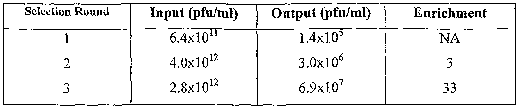


Claims
Priority Applications (9)
| Application Number | Priority Date | Filing Date | Title |
|---|---|---|---|
| CA002514417A CA2514417A1 (en) | 2003-01-28 | 2004-01-28 | Custom-made meganuclease and use thereof |
| EP04705873.0A EP1590453B1 (en) | 2003-01-28 | 2004-01-28 | Custom-made meganuclease and use thereof |
| AU2004208030A AU2004208030B2 (en) | 2003-01-28 | 2004-01-28 | Custom-made meganuclease and use thereof |
| JP2006502490A JP4966006B2 (en) | 2003-01-28 | 2004-01-28 | Custom-made meganucleases and their use |
| US10/543,556 US20060206949A1 (en) | 2003-01-28 | 2004-01-28 | Custom-made meganuclease and use thereof |
| EP17153147.8A EP3202899B1 (en) | 2003-01-28 | 2004-01-28 | Custom-made meganuclease and use thereof |
| AU2009206192A AU2009206192C1 (en) | 2003-01-28 | 2009-08-06 | Custom-made meganuclease and use thereof |
| US12/892,635 US20120159659A1 (en) | 2003-01-28 | 2010-09-28 | Custom-made meganuclease and use thereof |
| US13/725,798 US20140121115A1 (en) | 2003-01-28 | 2012-12-21 | Custom-made meganuclease and use thereof |
Applications Claiming Priority (4)
| Application Number | Priority Date | Filing Date | Title |
|---|---|---|---|
| US44291103P | 2003-01-28 | 2003-01-28 | |
| US60/442,911 | 2003-01-28 | ||
| US49153503P | 2003-08-01 | 2003-08-01 | |
| US60/491,535 | 2003-08-01 |
Related Child Applications (3)
| Application Number | Title | Priority Date | Filing Date |
|---|---|---|---|
| US10/543,556 A-371-Of-International US20060206949A1 (en) | 2003-01-28 | 2004-01-28 | Custom-made meganuclease and use thereof |
| US12/892,635 Continuation US20120159659A1 (en) | 2003-01-28 | 2010-09-28 | Custom-made meganuclease and use thereof |
| US13/725,798 Continuation US20140121115A1 (en) | 2003-01-28 | 2012-12-21 | Custom-made meganuclease and use thereof |
Publications (2)
| Publication Number | Publication Date |
|---|---|
| WO2004067736A2 true WO2004067736A2 (en) | 2004-08-12 |
| WO2004067736A3 WO2004067736A3 (en) | 2004-11-25 |
Family
ID=32829807
Family Applications (2)
| Application Number | Title | Priority Date | Filing Date |
|---|---|---|---|
| PCT/IB2004/000827 WO2004067736A2 (en) | 2003-01-28 | 2004-01-28 | Custom-made meganuclease and use thereof |
| PCT/IB2004/000848 WO2004067753A2 (en) | 2003-01-28 | 2004-01-28 | Use of meganucleases for inducing homologous recombination ex vivo and in toto in vertebrate somatic tissues and application thereof. |
Family Applications After (1)
| Application Number | Title | Priority Date | Filing Date |
|---|---|---|---|
| PCT/IB2004/000848 WO2004067753A2 (en) | 2003-01-28 | 2004-01-28 | Use of meganucleases for inducing homologous recombination ex vivo and in toto in vertebrate somatic tissues and application thereof. |
Country Status (6)
| Country | Link |
|---|---|
| US (16) | US20060153826A1 (en) |
| EP (5) | EP2559759A1 (en) |
| JP (3) | JP2006518372A (en) |
| AU (3) | AU2004208031B2 (en) |
| CA (2) | CA2514417A1 (en) |
| WO (2) | WO2004067736A2 (en) |
Cited By (131)
| Publication number | Priority date | Publication date | Assignee | Title |
|---|---|---|---|---|
| WO2006097784A1 (en) * | 2005-03-15 | 2006-09-21 | Cellectis | I-crei meganuclease variants with modified specificity, method of preparation and uses thereof |
| WO2006097853A1 (en) * | 2005-03-15 | 2006-09-21 | Cellectis | I-crei meganuclease variants with modified specificity, method of preparation and uses thereof |
| WO2007034262A1 (en) * | 2005-09-19 | 2007-03-29 | Cellectis | Heterodimeric meganucleases and use thereof |
| WO2007049156A2 (en) | 2005-10-25 | 2007-05-03 | Cellectis | I-CreI HOMING ENDONUCLEASE VARIANTS HAVING NOVEL CLEAVAGE SPECIFICITY AND USE THEREOF |
| WO2007057781A2 (en) | 2005-10-25 | 2007-05-24 | Cellectis | Laglidadg homing endonuclease variants having mutations in two functional subdomains and use thereof. |
| WO2007093918A2 (en) | 2006-02-13 | 2007-08-23 | Cellectis | Meganuclease variants cleaving a dna target sequence from a xeroderma pigmentosum gene and uses thereof |
| WO2007123636A3 (en) * | 2006-03-27 | 2008-11-20 | Children S Hospital & Regional | Compositions and methods comprising the use of cell surface displayed homing endonucleases |
| WO2008148559A1 (en) | 2007-06-05 | 2008-12-11 | Bayer Bioscience N.V. | Methods and means for exact replacement of target dna in eukaryotic organisms |
| WO2009013622A2 (en) | 2007-07-23 | 2009-01-29 | Cellectis | Meganuclease variants cleaving a dna target sequence from the human hemoglobin beta gene and uses thereof |
| EP2180058A1 (en) | 2008-10-23 | 2010-04-28 | Cellectis | Meganuclease recombination system |
| JP2010517524A (en) * | 2007-02-01 | 2010-05-27 | セレクティス | Obligate heterodimeric meganuclease and uses thereof |
| JP2010528649A (en) * | 2007-06-06 | 2010-08-26 | セレクティス | Method for enhancing cleavage activity of I-CreI-derived meganuclease |
| US7842489B2 (en) | 2003-01-28 | 2010-11-30 | Cellectis | Use of meganucleases for inducing homologous recombination ex vivo and in toto in vertebrate somatic tissues and application thereof |
| WO2010136981A2 (en) | 2009-05-26 | 2010-12-02 | Cellectis | Meganuclease variants cleaving the genome of a pathogenic non-integrating virus and uses thereof |
| WO2011007336A1 (en) | 2009-07-17 | 2011-01-20 | Cellectis | Viral vectors encoding a dna repair matrix and containing a virion-associated site specific meganuclease for gene targeting |
| WO2011021062A1 (en) * | 2009-08-21 | 2011-02-24 | Cellectis | Meganuclease variants cleaving a dna target sequence from the human lysosomal acid alpha-glucosidase gene and uses thereof |
| JP2011505809A (en) * | 2007-12-07 | 2011-03-03 | プレシジョン バイオサイエンシズ,インク. | A rationally designed meganuclease with a recognition sequence found in the DNase hypersensitive region of the human genome |
| EP2313498A1 (en) | 2008-07-14 | 2011-04-27 | Precision Biosciences, Inc. | Recognition sequences for i-crei-derived meganucleases and uses thereof |
| WO2011052539A1 (en) | 2009-10-26 | 2011-05-05 | 独立行政法人農業生物資源研究所 | Production method for genetically modified plant cells |
| WO2011101696A1 (en) | 2010-02-18 | 2011-08-25 | Cellectis | Improved meganuclease recombination system |
| WO2011104382A1 (en) | 2010-02-26 | 2011-09-01 | Cellectis | Use of endonucleases for inserting transgenes into safe harbor loci |
| US8021867B2 (en) | 2005-10-18 | 2011-09-20 | Duke University | Rationally-designed meganucleases with altered sequence specificity and DNA-binding affinity |
| WO2011117249A1 (en) | 2010-03-22 | 2011-09-29 | Philip Morris Products S.A. | Modifying enzyme activity in plants |
| WO2011141825A1 (en) | 2010-05-12 | 2011-11-17 | Cellectis | Meganuclease variants cleaving a dna target sequence from the rhodopsin gene and uses thereof |
| WO2011141820A1 (en) | 2010-05-12 | 2011-11-17 | Cellectis | Meganuclease variants cleaving a dna target sequence from the dystrophin gene and uses thereof |
| WO2011154158A1 (en) | 2010-06-09 | 2011-12-15 | Bayer Bioscience N.V. | Methods and means to modify a plant genome at a nucleotide sequence commonly used in plant genome engineering |
| WO2011154159A1 (en) | 2010-06-09 | 2011-12-15 | Bayer Bioscience N.V. | Methods and means to modify a plant genome at a nucleotide sequence commonly used in plant genome engineering |
| WO2012001527A2 (en) | 2010-06-15 | 2012-01-05 | Cellectis S.A. | Method for improving cleavage of dna by endonuclease sensitive to methylation |
| WO2012004671A2 (en) | 2010-07-07 | 2012-01-12 | Cellectis | Meganucleases variants cleaving a dna target sequence in the nanog gene and uses thereof |
| WO2012007848A2 (en) | 2010-07-16 | 2012-01-19 | Cellectis | Meganuclease variants cleaving a dna target sequence in the was gene and uses thereof |
| WO2012010976A2 (en) | 2010-07-15 | 2012-01-26 | Cellectis | Meganuclease variants cleaving a dna target sequence in the tert gene and uses thereof |
| WO2012021794A1 (en) | 2010-08-13 | 2012-02-16 | Pioneer Hi-Bred International, Inc. | Chimeric promoters and methods of use |
| US8148607B2 (en) | 2005-04-04 | 2012-04-03 | Bayer Cropscience N.V. | Methods and means for removal of a selected DNA sequence |
| WO2012058458A2 (en) | 2010-10-27 | 2012-05-03 | Cellectis Sa | Method for increasing the efficiency of double-strand break-induced mutagenesis |
| WO2012129373A2 (en) | 2011-03-23 | 2012-09-27 | Pioneer Hi-Bred International, Inc. | Methods for producing a complex transgenic trait locus |
| WO2012138939A1 (en) | 2011-04-05 | 2012-10-11 | Philippe Duchateau | New tale-protein scaffolds and uses thereof |
| WO2012138901A1 (en) | 2011-04-05 | 2012-10-11 | Cellectis Sa | Method for enhancing rare-cutting endonuclease efficiency and uses thereof |
| WO2012149470A1 (en) | 2011-04-27 | 2012-11-01 | Amyris, Inc. | Methods for genomic modification |
| WO2012158778A1 (en) | 2011-05-16 | 2012-11-22 | Cellectis S.A. | Doubly secure transgenic algae or cyanobacteria strains to prevent their establishment and spread in natural ecosystems |
| US8338157B2 (en) | 2008-03-11 | 2012-12-25 | Precision Biosciences, Inc. | Rationally-designed meganuclease variants of lig-34 and I-crei for maize genome engineering |
| WO2013009525A1 (en) | 2011-07-08 | 2013-01-17 | Cellectis S.A. | Method for increasing the efficiency of double-strand break-induced mutagenssis |
| US8367890B2 (en) | 2006-09-28 | 2013-02-05 | Bayer Cropscience N.V. | Methods and means for removal of a selected DNA sequence |
| EP2568048A1 (en) | 2007-06-29 | 2013-03-13 | Pioneer Hi-Bred International, Inc. | Methods for altering the genome of a monocot plant cell |
| WO2013053729A1 (en) | 2011-10-12 | 2013-04-18 | Bayer Cropscience Ag | Plants with decreased activity of a starch dephosphorylating enzyme |
| WO2013053730A1 (en) | 2011-10-12 | 2013-04-18 | Bayer Cropscience Ag | Plants with decreased activity of a starch dephosphorylating enzyme |
| US8445251B2 (en) | 2007-10-31 | 2013-05-21 | Precision Biosciences, Inc. | Rationally-designed single-chain meganucleases with non-palindromic recognition sequences |
| US8450561B2 (en) | 2008-02-29 | 2013-05-28 | Monsanto Technology Llc | Corn plant event MON87460 and compositions and methods for detection thereof |
| EP2602323A1 (en) * | 2007-06-01 | 2013-06-12 | Omt, Inc. | Compositions and methods for inhibiting endogenous immunoglobin genes and producing transgenic human idiotype antibodies |
| WO2013103366A1 (en) | 2012-01-06 | 2013-07-11 | Pioneer Hi-Bred International, Inc. | A method to screen plants for genetic elements inducing parthenogenesis in plants |
| WO2013136175A2 (en) | 2012-03-15 | 2013-09-19 | Cellectis | New repeat variable diresidues for targeting nucleotides |
| WO2013140250A1 (en) | 2012-03-23 | 2013-09-26 | Cellectis | Method to overcome dna chemical modifications sensitivity of engineered tale dna binding domains |
| WO2013160230A1 (en) | 2012-04-23 | 2013-10-31 | Bayer Cropscience Nv | Targeted genome engineering in plants |
| WO2013176915A1 (en) | 2012-05-25 | 2013-11-28 | Roman Galetto | Methods for engineering allogeneic and immunosuppressive resistant t cell for immunotherapy |
| WO2013182910A2 (en) | 2012-06-05 | 2013-12-12 | Cellectis | New transcription activator-like effector (tale) fusion protein |
| WO2014018601A2 (en) | 2012-07-24 | 2014-01-30 | Cellectis | New modular base-specific nucleic acid binding domains from burkholderia rhizoxinica proteins |
| WO2014033556A2 (en) | 2012-09-03 | 2014-03-06 | Cellectis | Methods for modulating tal specificity |
| WO2014039523A1 (en) | 2012-09-04 | 2014-03-13 | Cellectis | Multi-chain chimeric antigen receptor and uses thereof |
| WO2014071006A1 (en) | 2012-10-31 | 2014-05-08 | Cellectis | Coupling herbicide resistance with targeted insertion of transgenes in plants |
| WO2014071039A1 (en) | 2012-11-01 | 2014-05-08 | Cellectis Sa | Plants for production of therapeutic proteins |
| WO2014096972A2 (en) | 2012-12-21 | 2014-06-26 | Cellectis | Potatoes with reduced cold-induced sweetening |
| WO2014118719A1 (en) | 2013-02-01 | 2014-08-07 | Cellectis | Tevl chimeric endonuclease and their preferential cleavage sites |
| WO2014128659A1 (en) | 2013-02-21 | 2014-08-28 | Cellectis | Method to counter-select cells or organisms by linking loci to nuclease components |
| WO2014141147A1 (en) | 2013-03-15 | 2014-09-18 | Cellectis | Modifying soybean oil composition through targeted knockout of the fad2-1a/1b genes |
| WO2014184744A1 (en) | 2013-05-13 | 2014-11-20 | Cellectis | Methods for engineering highly active t cell for immunotherapy |
| WO2014184741A1 (en) | 2013-05-13 | 2014-11-20 | Cellectis | Methods for engineering allogeneic and highly active t cell for immunotheraphy |
| US20140370558A1 (en) * | 2013-03-15 | 2014-12-18 | Cellectis | Modifying soybean oil composition through targeted knockout of the fad2-1a/1b genes |
| WO2014199358A1 (en) | 2013-06-14 | 2014-12-18 | Cellectis | Methods for non-transgenic genome editing in plants |
| US8927247B2 (en) | 2008-01-31 | 2015-01-06 | Cellectis, S.A. | I-CreI derived single-chain meganuclease and uses thereof |
| WO2015026886A1 (en) | 2013-08-22 | 2015-02-26 | E. I. Du Pont De Nemours And Company | Methods for producing genetic modifications in a plant genome without incorporating a selectable transgene marker, and compositions thereof |
| EP2853598A1 (en) | 2013-09-25 | 2015-04-01 | Board of Regents of the University of Nebraska | Methods and compositions for obtaining useful plant traits |
| WO2015059265A1 (en) | 2013-10-25 | 2015-04-30 | Cellectis | Design of rare-cutting endonucleases for efficient and specific targeting dna sequences comprising highly repetitive motives |
| WO2015095804A1 (en) | 2013-12-19 | 2015-06-25 | Amyris, Inc. | Methods for genomic integration |
| WO2015193858A1 (en) | 2014-06-20 | 2015-12-23 | Cellectis | Potatoes with reduced granule-bound starch synthase |
| WO2016125078A1 (en) | 2015-02-02 | 2016-08-11 | Cellectis | Agrobacterium-mediated genome modification without t-dna integration |
| WO2017015567A1 (en) | 2015-07-23 | 2017-01-26 | Mayo Foundation For Medical Education And Research | Editing mitochondrial dna |
| WO2017046772A1 (en) | 2015-09-17 | 2017-03-23 | Cellectis | Modifying messenger rna stability in plant transformations |
| US9670496B2 (en) | 2011-08-22 | 2017-06-06 | Bayer Cropscience N.V. | Methods and means to modify a plant genome |
| WO2017134601A1 (en) | 2016-02-02 | 2017-08-10 | Cellectis | Modifying soybean oil composition through targeted knockout of the fad3a/b/c genes |
| WO2017144630A1 (en) | 2016-02-26 | 2017-08-31 | Cellectis | Micelle based system nuclease encapsulation for in-vivo gene editing |
| WO2017205665A1 (en) | 2016-05-25 | 2017-11-30 | Cargill, Incorporated | Engineered nucleases to generate deletion mutants in plants |
| WO2018005589A1 (en) | 2016-06-28 | 2018-01-04 | Cellectis | Altering expression of gene products in plants through targeted insertion of nucleic acid sequences |
| WO2018007263A1 (en) | 2016-07-06 | 2018-01-11 | Cellectis | Sequential gene editing in primary immune cells |
| WO2018035456A1 (en) | 2016-08-18 | 2018-02-22 | Cellectis | Black-spot resistant potatoes with reduced tuber-specific polyphenol oxidase activity |
| WO2018042346A2 (en) | 2016-09-01 | 2018-03-08 | Cellectis | Methods for altering amino acid content in plants |
| WO2018054911A1 (en) | 2016-09-23 | 2018-03-29 | Bayer Cropscience Nv | Targeted genome optimization in plants |
| WO2018073391A1 (en) | 2016-10-19 | 2018-04-26 | Cellectis | Targeted gene insertion for improved immune cells therapy |
| WO2018092072A1 (en) | 2016-11-16 | 2018-05-24 | Cellectis | Methods for altering amino acid content in plants through frameshift mutations |
| WO2018115189A1 (en) | 2016-12-21 | 2018-06-28 | Cellectis | Stably enginereed proteasome inhibitor resistant immune cells for immunotherapy |
| WO2018189360A1 (en) | 2017-04-13 | 2018-10-18 | Cellectis | New sequence specific reagents targeting ccr5 in primary hematopoietic cells |
| EP3392339A1 (en) | 2017-04-18 | 2018-10-24 | Rheinisch-Westfälische Technische Hochschule (RWTH) Aachen | Improved genome editing in plant cells |
| WO2018198049A1 (en) | 2017-04-25 | 2018-11-01 | Cellectis | Alfalfa with reduced lignin composition |
| WO2018206791A1 (en) | 2017-05-12 | 2018-11-15 | Cellectis | Protease based switch chimeric antigen receptors for safer cell immunotherapy |
| WO2018234239A1 (en) | 2017-06-19 | 2018-12-27 | Cellectis | Anti-hbv combination therapies involving specific endonucleases |
| WO2019016360A1 (en) | 2017-07-21 | 2019-01-24 | Cellectis | Engineered immune cells resistant to tumor microoenvironment |
| WO2019076489A1 (en) | 2017-10-19 | 2019-04-25 | Cellectis | Targeted gene integration of crs inhibitor genes for improved immune cells therapy |
| WO2019106163A1 (en) | 2017-12-01 | 2019-06-06 | Cellectis | Reprogramming of genetically engineered primary immune cells |
| WO2019129850A1 (en) | 2017-12-29 | 2019-07-04 | Cellectis | Off-the-shelf engineered cells for therapy |
| WO2019138083A1 (en) | 2018-01-12 | 2019-07-18 | Basf Se | Gene underlying the number of spikelets per spike qtl in wheat on chromosome 7a |
| EP3560329A1 (en) | 2011-05-02 | 2019-10-30 | Board of Regents of the University of Nebraska | Plants with useful traits and related methods |
| WO2019211796A1 (en) | 2018-05-02 | 2019-11-07 | Cellectis | Engineering wheat with increased dietary fiber |
| WO2019238832A1 (en) | 2018-06-15 | 2019-12-19 | Nunhems B.V. | Seedless watermelon plants comprising modifications in an abc transporter gene |
| WO2020012365A1 (en) | 2018-07-09 | 2020-01-16 | Cellectis | Canola with high oleic acid |
| WO2020142598A2 (en) | 2019-01-04 | 2020-07-09 | Cargill, Incorporated | Engineered nucleases to generate mutations in plants |
| WO2021055838A1 (en) | 2019-09-20 | 2021-03-25 | Calyxt, Inc. | Low phytoestrogen soybean based on gene editing |
| WO2021108248A1 (en) | 2019-11-27 | 2021-06-03 | Calyxt, Inc. | Tal-effector nucleases for gene editing |
| WO2021130718A1 (en) | 2019-12-24 | 2021-07-01 | Selexis Sa | Targeted integration in mammalian sequences enhancing gene expression |
| WO2021130250A1 (en) | 2019-12-23 | 2021-07-01 | Cellectis | New mesothelin specific chimeric antigen receptors (car) for solid tumors cancer immunotherapy |
| WO2021155376A1 (en) | 2020-01-31 | 2021-08-05 | Calyxt, Inc. | Increased of saturated fat in soybean |
| WO2021224395A1 (en) | 2020-05-06 | 2021-11-11 | Cellectis S.A. | Methods for targeted insertion of exogenous sequences in cellular genomes |
| WO2021224416A1 (en) | 2020-05-06 | 2021-11-11 | Cellectis S.A. | Methods to genetically modify cells for delivery of therapeutic proteins |
| US11174493B2 (en) | 2016-05-26 | 2021-11-16 | Nunhems B.V. | Seedless fruit producing plants |
| US11192929B2 (en) | 2016-12-08 | 2021-12-07 | Regents Of The University Of Minnesota | Site-specific DNA base editing using modified APOBEC enzymes |
| US11193131B2 (en) | 2015-06-30 | 2021-12-07 | Regents Of The University Of Minnesota | Haploid inducer line for accelerated genome editing |
| GB202117314D0 (en) | 2021-11-30 | 2022-01-12 | Clarke David John | Cyclic nucleic acid fragmentation |
| DE212020000516U1 (en) | 2019-03-07 | 2022-01-17 | The Regents of the University of California | CRISPR-CAS effector polypeptides |
| WO2022018262A1 (en) | 2020-07-24 | 2022-01-27 | Cellectis S.A. | T-cells expressing immune cell engagers in allogenic settings |
| US11332749B2 (en) | 2017-07-13 | 2022-05-17 | Regents Of The University Of Minnesota | Real-time reporter systems for monitoring base editing |
| WO2022112596A1 (en) | 2020-11-30 | 2022-06-02 | Cellectis Sa | Use of aminoquinoline compounds for higher gene integration |
| US11384360B2 (en) | 2012-06-19 | 2022-07-12 | Regents Of The University Of Minnesota | Gene targeting in plants using DNA viruses |
| WO2022229412A1 (en) | 2021-04-30 | 2022-11-03 | Cellectis S.A. | New anti-muc1 cars and gene edited immune cells for solid tumors cancer immunotherapy |
| WO2022243529A1 (en) | 2021-05-20 | 2022-11-24 | Cellectis S.A. | GENE THERAPY FOR THE TREATMENT OF HYPER-IgE SYNDROME (HIES) BY TARGETED GENE INTEGRATION |
| WO2022243531A1 (en) | 2021-05-20 | 2022-11-24 | Cellectis S.A. | Gene therapy for the treatment of severe combined immunodeficiency (scid) related to rag1 |
| WO2022243565A1 (en) | 2021-05-21 | 2022-11-24 | Cellectis S.A. | Enhancing efficacy of t-cell-mediated immunotherapy by modulating cancer-associated fibroblasts in solid tumors |
| WO2023081756A1 (en) | 2021-11-03 | 2023-05-11 | The J. David Gladstone Institutes, A Testamentary Trust Established Under The Will Of J. David Gladstone | Precise genome editing using retrons |
| US11680254B2 (en) | 2017-04-21 | 2023-06-20 | Precision Biosciences, Inc. | Engineered meganucleases specific for recognition sequences in the PCSK9 gene |
| WO2023141602A2 (en) | 2022-01-21 | 2023-07-27 | Renagade Therapeutics Management Inc. | Engineered retrons and methods of use |
| EP4219731A2 (en) | 2016-05-18 | 2023-08-02 | Amyris, Inc. | Compositions and methods for genomic integration of nucleic acids into exogenous landing pads |
| EP4276174A1 (en) | 2022-05-09 | 2023-11-15 | Cellectis S.A. | Gene therapy for the treatment of activated pi3kinase delta syndrome type 1 (apds1) |
| WO2023233003A1 (en) | 2022-06-03 | 2023-12-07 | Cellectis Sa | Tale base editors for gene and cell therapy |
| WO2024003334A1 (en) | 2022-06-30 | 2024-01-04 | Cellectis S.A. | Enhancing safety of t-cell-mediated immunotherapy |
| WO2024044723A1 (en) | 2022-08-25 | 2024-02-29 | Renagade Therapeutics Management Inc. | Engineered retrons and methods of use |
Families Citing this family (200)
| Publication number | Priority date | Publication date | Assignee | Title |
|---|---|---|---|---|
| US20100151556A1 (en) * | 2002-03-15 | 2010-06-17 | Cellectis | Hybrid and single chain meganucleases and use thereof |
| JP2005520519A (en) * | 2002-03-15 | 2005-07-14 | セレクティス | Hybrid and single chain meganucleases and uses thereof |
| US8409861B2 (en) | 2003-08-08 | 2013-04-02 | Sangamo Biosciences, Inc. | Targeted deletion of cellular DNA sequences |
| US11311574B2 (en) | 2003-08-08 | 2022-04-26 | Sangamo Therapeutics, Inc. | Methods and compositions for targeted cleavage and recombination |
| US7888121B2 (en) | 2003-08-08 | 2011-02-15 | Sangamo Biosciences, Inc. | Methods and compositions for targeted cleavage and recombination |
| EP2927318B1 (en) * | 2003-08-08 | 2020-05-20 | Sangamo Therapeutics, Inc. | Methods and compositions for targeted cleavage and recombination |
| US20140010797A1 (en) * | 2005-10-18 | 2014-01-09 | Duke University | Fusion Molecules of Rationally-Designed DNA-Binding Proteins and Effector Domains |
| WO2007139982A2 (en) * | 2006-05-25 | 2007-12-06 | Sangamo Biosciences, Inc. | Methods and compositions for gene inactivation |
| WO2008010009A1 (en) * | 2006-07-18 | 2008-01-24 | Cellectis | Meganuclease variants cleaving a dna target sequence from a rag gene and uses thereof |
| BRPI0718747A2 (en) * | 2006-11-14 | 2013-12-03 | Cellectis | MEGANUCLEASE VARIANTS KEYING ONE OR SEQUENCE DNA TARGET FROM THE HPRT GENE AND USES THEREOF. |
| WO2008059317A1 (en) * | 2006-11-14 | 2008-05-22 | Cellectis | Meganuclease variants cleaving a dna target sequence from the hprt gene and uses thereof |
| EP2086644A2 (en) * | 2006-11-17 | 2009-08-12 | Schering Corporation | Combination of an inhibitor of dna polymerase-alpha and an inhibitor of a checkpoint kinase for proliferative disorders |
| AU2007347328B2 (en) * | 2007-02-19 | 2013-03-07 | Cellectis | LAGLIDADG homing endonuclease variants having novel substrate specificity and use thereof |
| WO2008102199A1 (en) * | 2007-02-20 | 2008-08-28 | Cellectis | Meganuclease variants cleaving a dna target sequence from the beta-2-microglobulin gene and uses thereof |
| AU2008259834B2 (en) * | 2007-06-01 | 2013-08-01 | Corbion Biotech, Inc. | Production of oil in microorganisms |
| JP2010528625A (en) * | 2007-06-01 | 2010-08-26 | ビューロウ リサーチ エンタープライザズ リミティド ライアビリティ カンパニー | Compositions and methods for producing antibodies having human idiotypes in transgenic birds |
| WO2009019528A1 (en) * | 2007-08-03 | 2009-02-12 | Cellectis | Meganuclease variants cleaving a dna target sequence from the human interleukin-2 receptor gamma chain gene and uses thereof |
| US20110014616A1 (en) * | 2009-06-30 | 2011-01-20 | Sangamo Biosciences, Inc. | Rapid screening of biologically active nucleases and isolation of nuclease-modified cells |
| US11235026B2 (en) | 2007-09-27 | 2022-02-01 | Sangamo Therapeutics, Inc. | Methods and compositions for modulating PD1 |
| US8563314B2 (en) | 2007-09-27 | 2013-10-22 | Sangamo Biosciences, Inc. | Methods and compositions for modulating PD1 |
| EP2188384B1 (en) * | 2007-09-27 | 2015-07-15 | Sangamo BioSciences, Inc. | Rapid in vivo identification of biologically active nucleases |
| WO2009074842A1 (en) * | 2007-12-13 | 2009-06-18 | Cellectis | Improved chimeric meganuclease enzymes and uses thereof |
| US20100071083A1 (en) * | 2008-03-12 | 2010-03-18 | Smith James J | Temperature-dependent meganuclease activity |
| WO2009146179A1 (en) * | 2008-04-15 | 2009-12-03 | University Of Iowa Research Foundation | Zinc finger nuclease for the cftr gene and methods of use thereof |
| JP2011519558A (en) * | 2008-04-28 | 2011-07-14 | プレシジョン バイオサイエンシズ,インク. | A rationally designed fusion molecule of DNA-binding protein and effector domain |
| US9217155B2 (en) | 2008-05-28 | 2015-12-22 | University Of Massachusetts | Isolation of novel AAV'S and uses thereof |
| EP2352821B1 (en) * | 2008-09-08 | 2016-11-23 | Cellectis | Meganuclease variants cleaving a dna target sequence from a glutamine synthetase gene and uses thereof |
| EP3517622A1 (en) | 2008-11-28 | 2019-07-31 | Corbion Biotech, Inc. | Production of tailored oils in heterotrophic microorganisms |
| US20110030072A1 (en) * | 2008-12-04 | 2011-02-03 | Sigma-Aldrich Co. | Genome editing of immunodeficiency genes in animals |
| US20110023151A1 (en) * | 2008-12-04 | 2011-01-27 | Sigma-Aldrich Co. | Genome editing of abc transporters |
| US20110023146A1 (en) * | 2008-12-04 | 2011-01-27 | Sigma-Aldrich Co. | Genomic editing of genes involved in secretase-associated disorders |
| US20110023148A1 (en) * | 2008-12-04 | 2011-01-27 | Sigma-Aldrich Co. | Genome editing of addiction-related genes in animals |
| US20110016539A1 (en) * | 2008-12-04 | 2011-01-20 | Sigma-Aldrich Co. | Genome editing of neurotransmission-related genes in animals |
| US20110023140A1 (en) * | 2008-12-04 | 2011-01-27 | Sigma-Aldrich Co. | Rabbit genome editing with zinc finger nucleases |
| US20110023158A1 (en) * | 2008-12-04 | 2011-01-27 | Sigma-Aldrich Co. | Bovine genome editing with zinc finger nucleases |
| US20110023154A1 (en) * | 2008-12-04 | 2011-01-27 | Sigma-Aldrich Co. | Silkworm genome editing with zinc finger nucleases |
| US20110023150A1 (en) * | 2008-12-04 | 2011-01-27 | Sigma-Aldrich Co. | Genome editing of genes associated with schizophrenia in animals |
| US20110016541A1 (en) * | 2008-12-04 | 2011-01-20 | Sigma-Aldrich Co. | Genome editing of sensory-related genes in animals |
| US20110023141A1 (en) * | 2008-12-04 | 2011-01-27 | Sigma-Aldrich Co. | Genomic editing of genes involved with parkinson's disease |
| US20110023143A1 (en) * | 2008-12-04 | 2011-01-27 | Sigma-Aldrich Co. | Genomic editing of neurodevelopmental genes in animals |
| US20110023145A1 (en) * | 2008-12-04 | 2011-01-27 | Sigma-Aldrich Co. | Genomic editing of genes involved in autism spectrum disorders |
| US20110023156A1 (en) * | 2008-12-04 | 2011-01-27 | Sigma-Aldrich Co. | Feline genome editing with zinc finger nucleases |
| US20110016540A1 (en) * | 2008-12-04 | 2011-01-20 | Sigma-Aldrich Co. | Genome editing of genes associated with trinucleotide repeat expansion disorders in animals |
| US20110023139A1 (en) * | 2008-12-04 | 2011-01-27 | Sigma-Aldrich Co. | Genomic editing of genes involved in cardiovascular disease |
| US20110023147A1 (en) * | 2008-12-04 | 2011-01-27 | Sigma-Aldrich Co. | Genomic editing of prion disorder-related genes in animals |
| US20110023149A1 (en) * | 2008-12-04 | 2011-01-27 | Sigma-Aldrich Co. | Genomic editing of genes involved in tumor suppression in animals |
| US20110023144A1 (en) * | 2008-12-04 | 2011-01-27 | Sigma-Aldrich Co. | Genomic editing of genes involved in amyotrophyic lateral sclerosis disease |
| US20110016543A1 (en) * | 2008-12-04 | 2011-01-20 | Sigma-Aldrich Co. | Genomic editing of genes involved in inflammation |
| US20110023153A1 (en) * | 2008-12-04 | 2011-01-27 | Sigma-Aldrich Co. | Genomic editing of genes involved in alzheimer's disease |
| US20110016546A1 (en) * | 2008-12-04 | 2011-01-20 | Sigma-Aldrich Co. | Porcine genome editing with zinc finger nucleases |
| US20110023152A1 (en) * | 2008-12-04 | 2011-01-27 | Sigma-Aldrich Co. | Genome editing of cognition related genes in animals |
| US20110239315A1 (en) | 2009-01-12 | 2011-09-29 | Ulla Bonas | Modular dna-binding domains and methods of use |
| EP2206723A1 (en) | 2009-01-12 | 2010-07-14 | Bonas, Ulla | Modular DNA-binding domains |
| WO2010093966A2 (en) * | 2009-02-12 | 2010-08-19 | Fred Hutchinson Cancer Research Center | Generation of a dna nicking enzyme that stimulates site-specific gene conversion from a homing endonuclease |
| EP2408921B1 (en) | 2009-03-20 | 2017-04-19 | Sangamo BioSciences, Inc. | Modification of cxcr4 using engineered zinc finger proteins |
| WO2010122367A2 (en) * | 2009-04-21 | 2010-10-28 | Cellectis | Meganuclease variants cleaving the genomic insertion of a virus and uses thereof |
| US8772008B2 (en) * | 2009-05-18 | 2014-07-08 | Sangamo Biosciences, Inc. | Methods and compositions for increasing nuclease activity |
| WO2010136841A2 (en) * | 2009-05-26 | 2010-12-02 | Cellectis | Meganuclease variants cleaving the genome of a non-genomically integrating virus and uses thereof |
| WO2010138263A2 (en) | 2009-05-28 | 2010-12-02 | University Of Massachusetts | Novel aav 's and uses thereof |
| EP2461819A4 (en) | 2009-07-28 | 2013-07-31 | Sangamo Biosciences Inc | Methods and compositions for treating trinucleotide repeat disorders |
| BR112012003257A2 (en) | 2009-08-11 | 2016-11-22 | Sangamo Biosciences Inc | homozygous organisms for directed modification |
| WO2011036640A2 (en) | 2009-09-24 | 2011-03-31 | Cellectis | Meganuclease reagents of uses thereof for treating genetic diseases caused by frame shift/non sense mutations |
| TR201815882T4 (en) | 2009-12-10 | 2018-11-21 | Univ Iowa State Res Found Inc | Tal effector mediated DNA modification. |
| US8704041B2 (en) | 2009-12-30 | 2014-04-22 | Pioneer Hi Bred International Inc | Methods and compositions for targeted polynucleotide modification |
| US20120329067A1 (en) * | 2010-01-22 | 2012-12-27 | Barbas Iii Carlos F | Methods of Generating Zinc Finger Nucleases Having Altered Activity |
| EP2531609A1 (en) | 2010-02-02 | 2012-12-12 | INSERM (Institut National de la Santé et de la Recherche Médicale) | Methods for the diagnosis and therapy of retinitis pigmentosa |
| WO2011097603A1 (en) | 2010-02-08 | 2011-08-11 | Regeneron Pharmaceuticals, Inc. | Common light chain mouse |
| US9796788B2 (en) | 2010-02-08 | 2017-10-24 | Regeneron Pharmaceuticals, Inc. | Mice expressing a limited immunoglobulin light chain repertoire |
| US20130045492A1 (en) | 2010-02-08 | 2013-02-21 | Regeneron Pharmaceuticals, Inc. | Methods For Making Fully Human Bispecific Antibodies Using A Common Light Chain |
| JP2013533847A (en) | 2010-04-23 | 2013-08-29 | ユニバーシティ オブ マサチューセッツ | AAV-based treatment of cholesterol-related disorders |
| CA3050894C (en) | 2010-04-23 | 2022-10-18 | University Of Massachusetts | Multicistronic expression constructs |
| DK2561073T3 (en) | 2010-04-23 | 2016-12-12 | Univ Massachusetts | Aav vectors targeted to central nervous system and methods of use thereof |
| CN103025344B (en) | 2010-05-17 | 2016-06-29 | 桑格摩生物科学股份有限公司 | Novel DNA-associated proteins and application thereof |
| CA3039432A1 (en) | 2010-05-28 | 2011-12-01 | Corbion Biotech, Inc. | Tailored oils produced from recombinant heterotrophic microorganisms |
| US20140148361A1 (en) * | 2010-06-07 | 2014-05-29 | Barry L. Stoddard | Generation and Expression of Engineered I-ONUI Endonuclease and Its Homologues and Uses Thereof |
| GB201009732D0 (en) * | 2010-06-10 | 2010-07-21 | Gene Bridges Gmbh | Direct cloning |
| EP2412806B1 (en) * | 2010-07-28 | 2014-01-08 | Institut Pasteur | Use of terminal deoxynucleotidyl transferase for mutagenic DNA repair to generate variability, at a determined position in DNA |
| CA3024641A1 (en) | 2010-11-03 | 2012-05-10 | Corbion Biotech, Inc. | Microbial oils with lowered pour points, dielectric fluids produced therefrom, and related methods |
| SG192594A1 (en) | 2011-02-02 | 2013-09-30 | Solazyme Inc | Tailored oils produced from recombinant oleaginous microorganisms |
| US9044492B2 (en) | 2011-02-04 | 2015-06-02 | Cellectis Sa | Method for modulating the efficiency of double-strand break-induced mutagenesis |
| AR085302A1 (en) | 2011-02-24 | 2013-09-18 | Sanofi Sa | METHOD OF PRODUCTION OF STIRATED ANTIBODIES |
| ES2906059T3 (en) | 2011-02-28 | 2022-04-13 | Seattle Childrens Res Institute | Coupling endonucleases with end-processing enzymes that drive high-efficiency gene disruption |
| EP3318635A1 (en) | 2011-04-21 | 2018-05-09 | University of Massachusetts | Raav-based compositions and methods for treating alpha-1 anti-trypsin deficiencies |
| JP2014513964A (en) | 2011-05-06 | 2014-06-19 | ソラザイム、インク | Genetically engineered microorganisms that metabolize xylose |
| ES2773891T3 (en) * | 2011-06-01 | 2020-07-15 | Prec Biosciences Inc | Procedures for the production of modified mammalian cell lines with amplified transgenes |
| CA2841541C (en) * | 2011-07-25 | 2019-11-12 | Sangamo Biosciences, Inc. | Methods and compositions for alteration of a cystic fibrosis transmembrane conductance regulator (cftr) gene |
| IL273982B2 (en) | 2011-08-05 | 2023-03-01 | Regeneron Pharma | Humanized universal light chain mice |
| AU2012328682B2 (en) * | 2011-10-27 | 2017-09-21 | Sangamo Therapeutics, Inc. | Methods and compositions for modification of the HPRT locus |
| DK2836226T3 (en) | 2012-02-24 | 2017-09-18 | Hutchinson Fred Cancer Res | COMPOSITIONS AND PROCEDURES FOR TREATING HEMOGLOBINOPATHY |
| WO2013130824A1 (en) | 2012-02-29 | 2013-09-06 | Sangamo Biosciences, Inc. | Methods and compositions for treating huntington's disease |
| US9719114B2 (en) | 2012-04-18 | 2017-08-01 | Terravia Holdings, Inc. | Tailored oils |
| CN110358686A (en) | 2012-04-18 | 2019-10-22 | 柯碧恩生物技术公司 | Customization oil |
| US10648001B2 (en) | 2012-07-11 | 2020-05-12 | Sangamo Therapeutics, Inc. | Method of treating mucopolysaccharidosis type I or II |
| DK2872625T3 (en) | 2012-07-11 | 2017-02-06 | Sangamo Biosciences Inc | METHODS AND COMPOSITIONS FOR TREATING LYSOSOMAL STORAGE DISEASES |
| JP6164696B2 (en) * | 2012-08-16 | 2017-07-19 | 公立大学法人横浜市立大学 | Method for detecting mitochondrial complex III deficiency patient or carrier |
| EP2906684B8 (en) | 2012-10-10 | 2020-09-02 | Sangamo Therapeutics, Inc. | T cell modifying compounds and uses thereof |
| WO2014083379A1 (en) | 2012-11-30 | 2014-06-05 | Institut Pasteur | Use of anti-fcyri and/or anti-fcyriia antibodies for treating arthritis, inflammation, thrombocytopenia and allergic shock |
| WO2014104878A1 (en) | 2012-12-27 | 2014-07-03 | Keygene N.V. | Method for removing genetic linkage in a plant |
| WO2014158593A1 (en) * | 2013-03-13 | 2014-10-02 | President And Fellows Of Harvard College | Mutants of cre recombinase |
| US10053715B2 (en) | 2013-10-04 | 2018-08-21 | Corbion Biotech, Inc. | Tailored oils |
| NZ719477A (en) | 2013-11-11 | 2022-05-27 | Sangamo Therapeutics Inc | Methods and compositions for treating huntington’s disease |
| JP2017512767A (en) * | 2014-03-12 | 2017-05-25 | プレシジョン バイオサイエンシズ,インク. | Deletion of dystrophin gene exons using a modified nuclease |
| AU2015231353B2 (en) | 2014-03-18 | 2020-11-05 | Sangamo Therapeutics, Inc. | Methods and compositions for regulation of zinc finger protein expression |
| DK3119797T3 (en) | 2014-03-18 | 2021-03-15 | Univ Massachusetts | RAAV-BASED COMPOSITIONS AND METHODS FOR THE TREATMENT OF AMYOTROPHIC LATERAL SCLEROSIS |
| MX2016011690A (en) | 2014-03-21 | 2016-11-07 | Univ Leland Stanford Junior | Genome editing without nucleases. |
| RU2016141307A (en) | 2014-03-21 | 2018-04-24 | Регенерон Фармасьютикалз, Инк. | EXCELLENT HUMAN ANIMALS THAT MAKE SINGLE-DOMAIN BINDING PROTEINS |
| US10689653B2 (en) | 2014-06-03 | 2020-06-23 | University Of Massachusetts | Compositions and methods for modulating dysferlin expression |
| EP3620517A3 (en) | 2014-07-10 | 2020-07-29 | Corbion Biotech, Inc. | Ketoacyl acp synthase genes and uses thereof |
| US10370432B2 (en) | 2014-10-03 | 2019-08-06 | University Of Massachusetts | Heterologous targeting peptide grafted AAVS |
| US10711270B2 (en) | 2014-10-03 | 2020-07-14 | University Of Massachusetts | High efficiency library-identified AAV vectors |
| CN107073051B (en) | 2014-10-21 | 2021-08-24 | 马萨诸塞大学 | Recombinant AAV variants and uses thereof |
| JP6827928B2 (en) | 2014-12-19 | 2021-02-10 | ユニヴェルシテ・ドゥ・ナント | Anti-IL-34 antibody |
| EP3256170B1 (en) | 2015-02-13 | 2020-09-23 | University of Massachusetts | Compositions and methods for transient delivery of nucleases |
| AU2016232715A1 (en) | 2015-03-19 | 2017-09-28 | Regeneron Pharmaceuticals, Inc. | Non-human animals that select for light chain variable regions that bind antigen |
| EP3274453B1 (en) * | 2015-03-26 | 2021-01-27 | Editas Medicine, Inc. | Crispr/cas-mediated gene conversion |
| US20180094243A1 (en) | 2015-04-03 | 2018-04-05 | Dana-Farber Cancer Institute, Inc. | Composition and methods of genome editing of b-cells |
| US11046955B2 (en) | 2015-04-24 | 2021-06-29 | University Of Massachusetts | Modified AAV constructs and uses thereof |
| EP3288570A4 (en) | 2015-04-29 | 2018-11-21 | Fred Hutchinson Cancer Research Center | Modified stem cells and uses thereof |
| DK3289076T3 (en) | 2015-05-01 | 2022-01-17 | Prec Biosciences Inc | PRECISE DELETION OF CHROMOSOMAL SEQUENCES IN VIVO |
| US11845928B2 (en) | 2015-05-04 | 2023-12-19 | Tsinghua University | Methods and kits for fragmenting DNA |
| WO2016183345A1 (en) | 2015-05-13 | 2016-11-17 | Seattle Children' S Hospital (Dba Seattle Children 's Research Institute) | Enhancing endonuclease based gene editing in primary cells |
| EP3339437B1 (en) * | 2015-08-20 | 2021-11-17 | Inter-University Research Institute Corporation Research Organization of Information and Systems | Method for site-specific insertion of foreign dna into animal cell genome and cell obtained using same |
| WO2017070516A1 (en) | 2015-10-22 | 2017-04-27 | University Of Massachusetts | Prostate-targeting adeno-associated virus serotype vectors |
| EP3364997B1 (en) | 2015-10-22 | 2024-01-17 | University of Massachusetts | Aspartoacylase gene therapy in the treatment of canavan disease |
| WO2017079428A1 (en) | 2015-11-04 | 2017-05-11 | President And Fellows Of Harvard College | Site specific germline modification |
| WO2017106537A2 (en) | 2015-12-18 | 2017-06-22 | Sangamo Biosciences, Inc. | Targeted disruption of the mhc cell receptor |
| CN108778297B (en) | 2015-12-18 | 2024-02-09 | 桑格摩生物治疗股份有限公司 | Targeted disruption of T cell receptors |
| BR112018013679A2 (en) | 2016-01-11 | 2019-01-22 | Univ Leland Stanford Junior | chimeric proteins and gene expression regulation methods |
| WO2017123556A1 (en) | 2016-01-11 | 2017-07-20 | The Board Of Trustees Of The Leland Stanford Junior University | Chimeric proteins and methods of immunotherapy |
| US11826433B2 (en) | 2016-02-02 | 2023-11-28 | University Of Massachusetts | Method to enhance the efficiency of systemic AAV gene delivery to the central nervous system |
| CA3012344A1 (en) | 2016-02-12 | 2017-08-17 | University Of Massachusetts | Anti-angiogenic mirna therapeutics for inhibiting corneal neovascularization |
| EP3427055A1 (en) | 2016-03-07 | 2019-01-16 | Pierre Fabre Medicament | A new universal method to capture and analyze adcs for characterization of drug distribution and the drug-to-antibody ratio in biological samples |
| EP3440210A4 (en) | 2016-04-05 | 2019-11-27 | University of Massachusetts | Compositions and methods for selective inhibition of grainyhead-like protein expression |
| EP3443080B1 (en) * | 2016-04-13 | 2021-08-11 | University of Massachusetts | Repairing compound heterozygous recessive mutations by allele exchange |
| US11413356B2 (en) | 2016-04-15 | 2022-08-16 | University Of Massachusetts | Methods and compositions for treating metabolic imbalance |
| ES2905791T3 (en) | 2016-05-03 | 2022-04-12 | Prec Biosciences Inc | Engineered Nucleases Useful for Treatment of Hemophilia A |
| DK3462853T3 (en) | 2016-06-03 | 2023-04-03 | Regeneron Pharma | Rodents expressing exogenous terminal deoxynucleotidyl transferase |
| WO2017218852A1 (en) | 2016-06-15 | 2017-12-21 | University Of Massachusetts | Recombinant adeno-associated viruses for delivering gene editing molecules to embryonic cells |
| CA3034094A1 (en) * | 2016-08-16 | 2018-02-22 | Bluebird Bio, Inc. | Il-10 receptor alpha homing endonuclease variants, compositions, and methods of use |
| WO2018039448A1 (en) | 2016-08-24 | 2018-03-01 | Sangamo Therapeutics, Inc. | Engineered target specific nucleases |
| US10457940B2 (en) | 2016-09-22 | 2019-10-29 | University Of Massachusetts | AAV treatment of Huntington's disease |
| MX2019003176A (en) | 2016-09-23 | 2019-07-04 | Hutchinson Fred Cancer Res | Tcrs specific for minor histocompatibility (h) antigen ha-1 and uses thereof. |
| US11578340B2 (en) | 2016-10-13 | 2023-02-14 | University Of Massachusetts | AAV capsid designs |
| WO2018170338A2 (en) | 2017-03-15 | 2018-09-20 | Fred Hutchinson Cancer Research Center | High affinity mage-a1-specific tcrs and uses thereof |
| JP7297676B2 (en) | 2017-04-28 | 2023-06-26 | アクイタス セラピューティクス インコーポレイテッド | Novel carbonyl lipids and lipid nanoparticle formulations for nucleic acid delivery |
| RU2019139045A (en) | 2017-05-03 | 2021-06-03 | Сангамо Терапьютикс, Инк. | METHODS AND COMPOSITIONS FOR MODIFICATION OF THE TRANSMEMBRANE CONDUCTIVITY REGULATOR GENE IN CYSTIC FIBROSIS (CFTR) |
| WO2018208972A1 (en) | 2017-05-09 | 2018-11-15 | University Of Massachusetts | Methods of treating amyotrophic lateral sclerosis (als) |
| WO2018218038A1 (en) | 2017-05-24 | 2018-11-29 | Effector Therapeutics, Inc. | Methods and compositions for cellular immunotherapy |
| JP7191042B2 (en) | 2017-05-25 | 2022-12-16 | 2セブンティ バイオ インコーポレイテッド | CBLB ENDONUCLEASE VARIANTS, COMPOSITIONS AND METHODS OF USE |
| AU2018327225A1 (en) | 2017-09-06 | 2020-02-27 | Fred Hutchinson Cancer Center | Strep-tag specific chimeric receptors and uses thereof |
| US20210106618A1 (en) | 2017-09-06 | 2021-04-15 | Fred Hutchinson Cancer Research Center | Methods for improving adoptive cell therapy |
| CN111448321A (en) | 2017-09-22 | 2020-07-24 | 马萨诸塞大学 | SOD1 double expression vector and its use |
| WO2019079777A1 (en) | 2017-10-20 | 2019-04-25 | Fred Hutchinson Cancer Research Center | Compositions and methods of immunotherapy targeting tigit and/or cd112r or comprising cd226 overexpression |
| EP3704238B1 (en) * | 2017-11-01 | 2024-01-03 | Precision Biosciences, Inc. | Engineered nucleases that target human and canine factor viii genes as a treatment for hemophilia a |
| WO2019109047A1 (en) | 2017-12-01 | 2019-06-06 | Fred Hutchinson Cancer Research Center | Binding proteins specific for 5t4 and uses thereof |
| PT3720879T (en) | 2017-12-05 | 2022-07-13 | Progastrine Et Cancers S A R L | Combination therapy between anti-progastrin antibody and immunotherapy to treat cancer |
| WO2019140278A1 (en) | 2018-01-11 | 2019-07-18 | Fred Hutchinson Cancer Research Center | Immunotherapy targeting core binding factor antigens |
| AU2019224051A1 (en) | 2018-02-26 | 2020-09-03 | Fred Hutchinson Cancer Center | Compositions and methods for cellular immunotherapy |
| AU2019228339A1 (en) | 2018-02-27 | 2020-09-10 | Ecs-Progastrin Sa | Progastrin as a biomarker for immunotherapy |
| US11690921B2 (en) | 2018-05-18 | 2023-07-04 | Sangamo Therapeutics, Inc. | Delivery of target specific nucleases |
| WO2019234750A1 (en) | 2018-06-07 | 2019-12-12 | The State Of Israel, Ministry Of Agriculture & Rural Development, Agricultural Research Organization (Aro) (Volcani Center) | Methods of regenerating and transforming cannabis |
| WO2019234754A1 (en) | 2018-06-07 | 2019-12-12 | The State Of Israel, Ministry Of Agriculture & Rural Development, Agricultural Research Organization (Aro) (Volcani Center) | Nucleic acid constructs and methods of using same |
| JP2021531007A (en) | 2018-07-20 | 2021-11-18 | ピエール、ファーブル、メディカマン | Receptor for VISTA |
| WO2020018964A1 (en) | 2018-07-20 | 2020-01-23 | Fred Hutchinson Cancer Research Center | Compositions and methods for controlled expression of antigen-specific receptors |
| EP3841113A1 (en) | 2018-08-22 | 2021-06-30 | Fred Hutchinson Cancer Research Center | Immunotherapy targeting kras or her2 antigens |
| JP2021534785A (en) | 2018-08-28 | 2021-12-16 | フレッド ハッチンソン キャンサー リサーチ センター | Methods and Compositions for Adoptive T Cell Therapy Using Induced Notch Signaling |
| JP2022500052A (en) | 2018-09-18 | 2022-01-04 | サンガモ セラピューティクス, インコーポレイテッド | Programmed cell death 1 (PD1) specific nuclease |
| WO2020068702A1 (en) | 2018-09-24 | 2020-04-02 | Fred Hutchinson Cancer Research Center | Chimeric receptor proteins and uses thereof |
| EP3856229A1 (en) | 2018-09-28 | 2021-08-04 | Pierre Fabre Medicament | New immunocytokines for the treatment of cancer |
| WO2020072059A1 (en) | 2018-10-04 | 2020-04-09 | Bluebird Bio, Inc. | Cblb endonuclease variants, compositions, and methods of use |
| JP2022506781A (en) | 2018-11-09 | 2022-01-17 | フレッド ハッチンソン キャンサー リサーチ センター | Immunotherapy targeting mesothelin |
| EP3908568A1 (en) | 2019-01-11 | 2021-11-17 | Acuitas Therapeutics, Inc. | Lipids for lipid nanoparticle delivery of active agents |
| CA3130618A1 (en) | 2019-02-20 | 2020-08-27 | Fred Hutchinson Cancer Research Center | Binding proteins specific for ras neoantigens and uses thereof |
| CA3132845A1 (en) | 2019-03-11 | 2020-09-17 | Fred Hutchinson Cancer Research Center | High avidity wt1 t cell receptors and uses thereof |
| EP3987024A4 (en) | 2019-06-20 | 2023-11-01 | University Of Massachusetts | Compositions and methods for improved gene editing |
| CA3147717A1 (en) | 2019-08-20 | 2021-02-25 | Fred Hutchinson Cancer Research Center | T-cell immunotherapy specific for wt-1 |
| BR112022003635A2 (en) | 2019-09-04 | 2022-05-24 | Pf Medicament | Anti-vsig4 antibody or antigen-binding fragment and its use |
| EP4073117A1 (en) | 2019-12-10 | 2022-10-19 | Institut Pasteur | New antibody blocking human fcgriiia and fcgriiib |
| EP3889183A1 (en) | 2020-04-01 | 2021-10-06 | Pierre Fabre Medicament | A protein complex comprising an immunocytokine |
| WO2022020196A2 (en) * | 2020-07-16 | 2022-01-27 | The Texas A&M University System | Self-eliminating transgenes |
| WO2022046760A2 (en) | 2020-08-25 | 2022-03-03 | Kite Pharma, Inc. | T cells with improved functionality |
| JP2023542528A (en) | 2020-09-24 | 2023-10-10 | フレッド ハッチンソン キャンサー センター | Immunotherapy targeting SOX2 antigen |
| WO2022066973A1 (en) | 2020-09-24 | 2022-03-31 | Fred Hutchinson Cancer Research Center | Immunotherapy targeting pbk or oip5 antigens |
| IL301396A (en) | 2020-09-30 | 2023-05-01 | Nobell Foods Inc | Recombinant milk proteins and food compositions comprising the same |
| US10894812B1 (en) | 2020-09-30 | 2021-01-19 | Alpine Roads, Inc. | Recombinant milk proteins |
| US10947552B1 (en) | 2020-09-30 | 2021-03-16 | Alpine Roads, Inc. | Recombinant fusion proteins for producing milk proteins in plants |
| TW202222841A (en) | 2020-10-06 | 2022-06-16 | 福瑞德哈金森腫瘤研究中心 | Compositions and methods for treating mage-a1-expressing disease |
| CA3201767A1 (en) | 2020-12-14 | 2022-06-23 | Thomas M. Schmitt | Compositions and methods for cellular immunotherapy |
| KR20230156727A (en) | 2021-03-03 | 2023-11-14 | 피에르 파브르 메디카먼트 | Anti-VSIG4 antibody or antigen-binding fragment thereof and uses |
| JP2024510989A (en) | 2021-03-12 | 2024-03-12 | メンドゥス・ベスローテン・フェンノートシャップ | Vaccination methods and use of CD47 blockers |
| KR20240037188A (en) | 2021-04-30 | 2024-03-21 | 피에르 파브르 메디카먼트 | Novel stable anti-VISTA antibody |
| US20230038295A1 (en) | 2021-06-25 | 2023-02-09 | Homology Medicines, Inc. | Adeno-associated virus packaging systems |
| WO2023288281A2 (en) | 2021-07-15 | 2023-01-19 | Fred Hutchinson Cancer Center | Chimeric polypeptides |
| WO2023014922A1 (en) | 2021-08-04 | 2023-02-09 | The Regents Of The University Of Colorado, A Body Corporate | Lat activating chimeric antigen receptor t cells and methods of use thereof |
| US20230076072A1 (en) | 2021-08-24 | 2023-03-09 | Oxford Biomedica Solutions Llc | Adeno-associated virus formulations |
| US20230183659A1 (en) | 2021-10-29 | 2023-06-15 | Homology Medicines, Inc. | Methods and compositions for the purification of adeno-associated virus |
| WO2023102550A2 (en) | 2021-12-03 | 2023-06-08 | The Broad Institute, Inc. | Compositions and methods for efficient in vivo delivery |
| WO2023114897A2 (en) | 2021-12-15 | 2023-06-22 | Homology Medicines, Inc. | Methods and compositions for the production of adeno-associated virus |
| WO2023213814A1 (en) | 2022-05-02 | 2023-11-09 | Pierre Fabre Medicament | New formulation of anti vista antibody |
| WO2023215725A1 (en) | 2022-05-02 | 2023-11-09 | Fred Hutchinson Cancer Center | Compositions and methods for cellular immunotherapy |
Citations (21)
| Publication number | Priority date | Publication date | Assignee | Title |
|---|---|---|---|---|
| US4179337A (en) | 1973-07-20 | 1979-12-18 | Davis Frank F | Non-immunogenic polypeptides |
| US5006333A (en) | 1987-08-03 | 1991-04-09 | Ddi Pharmaceuticals, Inc. | Conjugates of superoxide dismutase coupled to high molecular weight polyalkylene glycols |
| WO1991017271A1 (en) | 1990-05-01 | 1991-11-14 | Affymax Technologies N.V. | Recombinant library screening methods |
| WO1991018980A1 (en) | 1990-06-01 | 1991-12-12 | Cetus Corporation | Compositions and methods for identifying biologically active molecules |
| WO1991019818A1 (en) | 1990-06-20 | 1991-12-26 | Affymax Technologies N.V. | Peptide library and screening systems |
| WO1993008278A1 (en) | 1991-10-16 | 1993-04-29 | Affymax Technologies N.V. | Peptide library and screening method |
| WO1994018313A1 (en) | 1993-02-12 | 1994-08-18 | The Johns-Hopkins University | Functional domains in flavobacterium okeanokoites (foki) restriction endonuclease |
| WO1995009233A1 (en) | 1993-09-27 | 1995-04-06 | The Johns-Hopkins University | Functional domains in flavobacterium okeanokoites (foki) restriction endonuclease |
| US5474896A (en) | 1992-05-05 | 1995-12-12 | Institut Pasteur | Nucleotide sequence encoding the enzyme I-SceI and the uses thereof |
| WO1996014408A2 (en) | 1994-11-07 | 1996-05-17 | Institut Pasteur | Nucleotide sequence encoding the enzyme i-scei and the uses thereof |
| US5523232A (en) | 1992-04-02 | 1996-06-04 | The United States Of America As Represented By The Department Of Health And Human Services | Use of restriction endonucleases against viruses, including HIV |
| US5801030A (en) | 1995-09-01 | 1998-09-01 | Genvec, Inc. | Methods and vectors for site-specific recombination |
| US5830729A (en) | 1996-04-18 | 1998-11-03 | Institut Pasteur | I Sce I-induced gene replacement and gene conversion in embryonic stem cells |
| US5843701A (en) | 1990-08-02 | 1998-12-01 | Nexstar Pharmaceticals, Inc. | Systematic polypeptide evolution by reverse translation |
| WO2000046385A1 (en) | 1999-02-03 | 2000-08-10 | The Children's Medical Center Corporation | Gene repair involving in vivo excision of targeting dna |
| WO2000046386A2 (en) | 1999-02-03 | 2000-08-10 | The Children's Medical Center Corporation | Gene repair involving the induction of double-stranded dna cleavage at a chromosomal target site |
| WO2000047775A1 (en) | 1999-02-09 | 2000-08-17 | The General Hospital Corporation | Selection of proteins using rna-protein fusions |
| US6232112B1 (en) | 1997-11-24 | 2001-05-15 | Flinders Technologies Pty Ltd Of Flinders University | Reagents and methods for diversification of DNA |
| US6265196B1 (en) | 1996-05-07 | 2001-07-24 | Johns Hopkins University | Methods for inactivating target DNA and for detecting conformational change in a nucleic acid |
| WO2001070946A2 (en) | 2000-03-21 | 2001-09-27 | Neugenesis Corporation | Methods for in vivo diversification of single genes |
| WO2002044409A2 (en) | 2000-12-01 | 2002-06-06 | European Molecular Biology Laboratory | Substrate linked directed evolution (slide) |
Family Cites Families (39)
| Publication number | Priority date | Publication date | Assignee | Title |
|---|---|---|---|---|
| FR2646438B1 (en) * | 1989-03-20 | 2007-11-02 | Pasteur Institut | A METHOD FOR SPECIFIC REPLACEMENT OF A COPY OF A GENE PRESENT IN THE RECEIVER GENOME BY INTEGRATION OF A GENE DIFFERENT FROM THAT OR INTEGRATION |
| US5272071A (en) * | 1989-12-22 | 1993-12-21 | Applied Research Systems Ars Holding N.V. | Method for the modification of the expression characteristics of an endogenous gene of a given cell line |
| US6063630A (en) * | 1991-11-05 | 2000-05-16 | Transkaryotic Therapies, Inc. | Targeted introduction of DNA into primary or secondary cells and their use for gene therapy |
| US5641670A (en) * | 1991-11-05 | 1997-06-24 | Transkaryotic Therapies, Inc. | Protein production and protein delivery |
| US20040019916A1 (en) * | 1992-04-24 | 2004-01-29 | Zarling David A. | In vivo homologous sequence targeting in eukaryotic cells |
| US6395959B1 (en) * | 1992-05-05 | 2002-05-28 | Institut Pasteur | Nucleotide sequence encoding the enzyme I SceI and the use thereof |
| US20040054156A1 (en) * | 1992-05-14 | 2004-03-18 | Kenneth Draper | Method and reagent for inhibiting hepatitis B viral replication |
| IL120241A0 (en) * | 1997-02-17 | 1997-06-10 | B G Negev Technologies And App | DNA segments encoding a domain of ho-endonuclease |
| CA2365901A1 (en) * | 1999-04-14 | 2000-10-19 | Musc Foundation For Research Development | Tissue-specific and pathogen-specific toxic agents and ribozymes |
| JP2004515229A (en) | 2000-11-27 | 2004-05-27 | スローン−ケッタリング インスティテュート フォー キャンサー リサーチ | Methods of using fluorescent energy conversion probes to detect nucleic acid cleavage |
| WO2002068471A2 (en) * | 2000-12-21 | 2002-09-06 | Bristol-Myers Squibb Company | Nucleic acid molecules and polypeptides for a human cation channel polypeptide |
| US7476500B1 (en) * | 2001-03-19 | 2009-01-13 | President And Fellows Of Harvard College | In vivo selection system for enzyme activity |
| WO2002099105A2 (en) | 2001-06-05 | 2002-12-12 | Cellectis | Methods for modifying the cpg content of polynucleotides |
| DE10131786A1 (en) * | 2001-07-04 | 2003-01-16 | Sungene Gmbh & Co Kgaa | Recombination systems and methods for removing nucleic acid sequences from the genome of eukaryotic organisms |
| US20030228583A1 (en) * | 2001-10-31 | 2003-12-11 | Amacher David E. | Biomarkers of liver response |
| CA2470329A1 (en) * | 2001-12-20 | 2003-07-03 | Sungene Gmbh & Co. Kgaa | Methods for the transformation of vegetal plastids |
| JP2005520519A (en) * | 2002-03-15 | 2005-07-14 | セレクティス | Hybrid and single chain meganucleases and uses thereof |
| EP2559759A1 (en) | 2003-01-28 | 2013-02-20 | Cellectis | Custom-made meganuclease and use thereof |
| US7888121B2 (en) * | 2003-08-08 | 2011-02-15 | Sangamo Biosciences, Inc. | Methods and compositions for targeted cleavage and recombination |
| EP1591521A1 (en) | 2004-04-30 | 2005-11-02 | Cellectis | I-Dmo I derivatives with enhanced activity at 37 degrees C and use thereof |
| ES2347684T3 (en) | 2005-03-15 | 2010-11-03 | Cellectis | VARIANTS OF MEGANUCLEASE I-CREI WITH MODIFY SPECIFICITY, METHOD OF PREPARATION AND USES OF THE SAME. |
| WO2006097784A1 (en) | 2005-03-15 | 2006-09-21 | Cellectis | I-crei meganuclease variants with modified specificity, method of preparation and uses thereof |
| DK2650365T3 (en) * | 2005-10-18 | 2016-12-05 | Prec Biosciences | RATIONAL MEGANUCLEASES constructed with altered sequence specificity and DNA binding affinity |
| WO2007049095A1 (en) * | 2005-10-25 | 2007-05-03 | Cellectis | Laglidadg homing endonuclease variants having mutations in two functional subdomains and use thereof |
| WO2007060495A1 (en) | 2005-10-25 | 2007-05-31 | Cellectis | I-crei homing endonuclease variants having novel cleavage specificity and use thereof |
| WO2007093836A1 (en) | 2006-02-13 | 2007-08-23 | Cellectis | Meganuclease variants cleaving a dna target sequence from a xp gene and uses thereof |
| WO2008002199A2 (en) | 2006-06-23 | 2008-01-03 | Aichinger, Elena | Biocidal and virulicidal agent-clathrate 1,6,3,8-dimethan-tetra-aza-cyclodecane-sodium carbonate |
| RU2318466C1 (en) | 2006-06-23 | 2008-03-10 | Давид Георгиевич Кочиев | Laser assembly for ablation of tissue and lithotripsy |
| WO2008002274A1 (en) | 2006-06-27 | 2008-01-03 | Inventys Pte Ltd | Method and system for client-side user interface enhancement to enable application integration and portalisation |
| WO2008010009A1 (en) | 2006-07-18 | 2008-01-24 | Cellectis | Meganuclease variants cleaving a dna target sequence from a rag gene and uses thereof |
| WO2008059317A1 (en) | 2006-11-14 | 2008-05-22 | Cellectis | Meganuclease variants cleaving a dna target sequence from the hprt gene and uses thereof |
| WO2009001159A1 (en) | 2007-06-25 | 2008-12-31 | Cellectis | Method for enhancing the cleavage activity of i-crei derived meganucleases |
| BRPI0718747A2 (en) | 2006-11-14 | 2013-12-03 | Cellectis | MEGANUCLEASE VARIANTS KEYING ONE OR SEQUENCE DNA TARGET FROM THE HPRT GENE AND USES THEREOF. |
| WO2008093152A1 (en) | 2007-02-01 | 2008-08-07 | Cellectis | Obligate heterodimer meganucleases and uses thereof |
| US20140112904A9 (en) | 2007-06-06 | 2014-04-24 | Cellectis | Method for enhancing the cleavage activity of i-crei derived meganucleases |
| WO2008149176A1 (en) | 2007-06-06 | 2008-12-11 | Cellectis | Meganuclease variants cleaving a dna target sequence from the mouse rosa26 locus and uses thereof |
| WO2009013559A1 (en) | 2007-07-23 | 2009-01-29 | Cellectis | Meganuclease variants cleaving a dna target sequence from the human hemoglobin beta gene and uses thereof |
| WO2009019528A1 (en) | 2007-08-03 | 2009-02-12 | Cellectis | Meganuclease variants cleaving a dna target sequence from the human interleukin-2 receptor gamma chain gene and uses thereof |
| EP2023541B1 (en) | 2007-08-07 | 2013-02-13 | Nokia Siemens Networks Oy | Method, device and communication system to avoid loops in an Ethernet Ring System with an underlying 802.3ad network |
-
2004
- 2004-01-28 EP EP12174235A patent/EP2559759A1/en not_active Withdrawn
- 2004-01-28 EP EP17153147.8A patent/EP3202899B1/en not_active Expired - Lifetime
- 2004-01-28 US US10/543,557 patent/US20060153826A1/en not_active Abandoned
- 2004-01-28 JP JP2006502491A patent/JP2006518372A/en active Pending
- 2004-01-28 EP EP04705872A patent/EP1590468A2/en not_active Withdrawn
- 2004-01-28 JP JP2006502490A patent/JP4966006B2/en not_active Expired - Lifetime
- 2004-01-28 AU AU2004208031A patent/AU2004208031B2/en not_active Ceased
- 2004-01-28 WO PCT/IB2004/000827 patent/WO2004067736A2/en active Application Filing
- 2004-01-28 EP EP12176308.0A patent/EP2522723B1/en not_active Expired - Lifetime
- 2004-01-28 US US10/543,556 patent/US20060206949A1/en not_active Abandoned
- 2004-01-28 EP EP04705873.0A patent/EP1590453B1/en not_active Expired - Lifetime
- 2004-01-28 CA CA002514417A patent/CA2514417A1/en not_active Abandoned
- 2004-01-28 WO PCT/IB2004/000848 patent/WO2004067753A2/en active Application Filing
- 2004-01-28 CA CA002514517A patent/CA2514517A1/en not_active Abandoned
- 2004-01-28 AU AU2004208030A patent/AU2004208030B2/en not_active Expired
-
2009
- 2009-08-06 AU AU2009206192A patent/AU2009206192C1/en not_active Expired
-
2010
- 2010-02-23 US US12/710,969 patent/US7842489B2/en not_active Expired - Fee Related
- 2010-09-28 US US12/892,635 patent/US20120159659A1/en not_active Abandoned
-
2011
- 2011-02-04 US US13/021,473 patent/US20110151539A1/en not_active Abandoned
- 2011-06-03 US US13/152,961 patent/US8530214B2/en not_active Expired - Lifetime
- 2011-11-04 JP JP2011242304A patent/JP2012085640A/en active Pending
-
2012
- 2012-05-31 US US13/485,486 patent/US20120331574A1/en not_active Abandoned
- 2012-05-31 US US13/485,469 patent/US8624000B2/en not_active Expired - Lifetime
- 2012-05-31 US US13/485,561 patent/US20120304321A1/en not_active Abandoned
- 2012-06-15 US US13/524,995 patent/US20120322689A1/en not_active Abandoned
- 2012-07-24 US US13/556,209 patent/US20120288943A1/en not_active Abandoned
- 2012-07-24 US US13/556,206 patent/US8697395B2/en not_active Expired - Lifetime
- 2012-07-24 US US13/556,208 patent/US20120288942A1/en not_active Abandoned
- 2012-10-10 US US13/649,093 patent/US20130203840A1/en not_active Abandoned
- 2012-12-21 US US13/725,798 patent/US20140121115A1/en not_active Abandoned
-
2016
- 2016-03-24 US US15/080,232 patent/US20160273003A1/en not_active Abandoned
Patent Citations (22)
| Publication number | Priority date | Publication date | Assignee | Title |
|---|---|---|---|---|
| US4179337A (en) | 1973-07-20 | 1979-12-18 | Davis Frank F | Non-immunogenic polypeptides |
| US5006333A (en) | 1987-08-03 | 1991-04-09 | Ddi Pharmaceuticals, Inc. | Conjugates of superoxide dismutase coupled to high molecular weight polyalkylene glycols |
| WO1991017271A1 (en) | 1990-05-01 | 1991-11-14 | Affymax Technologies N.V. | Recombinant library screening methods |
| WO1991018980A1 (en) | 1990-06-01 | 1991-12-12 | Cetus Corporation | Compositions and methods for identifying biologically active molecules |
| WO1991019818A1 (en) | 1990-06-20 | 1991-12-26 | Affymax Technologies N.V. | Peptide library and screening systems |
| US5843701A (en) | 1990-08-02 | 1998-12-01 | Nexstar Pharmaceticals, Inc. | Systematic polypeptide evolution by reverse translation |
| WO1993008278A1 (en) | 1991-10-16 | 1993-04-29 | Affymax Technologies N.V. | Peptide library and screening method |
| US5523232A (en) | 1992-04-02 | 1996-06-04 | The United States Of America As Represented By The Department Of Health And Human Services | Use of restriction endonucleases against viruses, including HIV |
| US5474896A (en) | 1992-05-05 | 1995-12-12 | Institut Pasteur | Nucleotide sequence encoding the enzyme I-SceI and the uses thereof |
| US5948678A (en) | 1992-05-05 | 1999-09-07 | Institut Pasteur | Nucleotide sequence encoding the enzyme I-SceI and the uses thereof |
| WO1994018313A1 (en) | 1993-02-12 | 1994-08-18 | The Johns-Hopkins University | Functional domains in flavobacterium okeanokoites (foki) restriction endonuclease |
| WO1995009233A1 (en) | 1993-09-27 | 1995-04-06 | The Johns-Hopkins University | Functional domains in flavobacterium okeanokoites (foki) restriction endonuclease |
| WO1996014408A2 (en) | 1994-11-07 | 1996-05-17 | Institut Pasteur | Nucleotide sequence encoding the enzyme i-scei and the uses thereof |
| US5801030A (en) | 1995-09-01 | 1998-09-01 | Genvec, Inc. | Methods and vectors for site-specific recombination |
| US5830729A (en) | 1996-04-18 | 1998-11-03 | Institut Pasteur | I Sce I-induced gene replacement and gene conversion in embryonic stem cells |
| US6265196B1 (en) | 1996-05-07 | 2001-07-24 | Johns Hopkins University | Methods for inactivating target DNA and for detecting conformational change in a nucleic acid |
| US6232112B1 (en) | 1997-11-24 | 2001-05-15 | Flinders Technologies Pty Ltd Of Flinders University | Reagents and methods for diversification of DNA |
| WO2000046385A1 (en) | 1999-02-03 | 2000-08-10 | The Children's Medical Center Corporation | Gene repair involving in vivo excision of targeting dna |
| WO2000046386A2 (en) | 1999-02-03 | 2000-08-10 | The Children's Medical Center Corporation | Gene repair involving the induction of double-stranded dna cleavage at a chromosomal target site |
| WO2000047775A1 (en) | 1999-02-09 | 2000-08-17 | The General Hospital Corporation | Selection of proteins using rna-protein fusions |
| WO2001070946A2 (en) | 2000-03-21 | 2001-09-27 | Neugenesis Corporation | Methods for in vivo diversification of single genes |
| WO2002044409A2 (en) | 2000-12-01 | 2002-06-06 | European Molecular Biology Laboratory | Substrate linked directed evolution (slide) |
Non-Patent Citations (54)
| Title |
|---|
| "HANDBOOK OF PHARMACEUTICAL EXCIPIENTS", AMERICAN PHARMACEUTICAL ASSOCIATION |
| "PHARMACEUTICAL DOSAGE FORMS AND THEIR USE", 1985, HANS HUBER PUBLISHERS |
| "PHARMACEUTICAL DOSAGE FORMS AND THEIR USE", 1985, HANSHUBER PUBLISHERS |
| AUSUBEL ET AL.,: "Current Protocols in Molecular Biology", JOHN WILEY AND SONS, article "Mutagenesis in cloned DNA" |
| BONETTA, THE SCIENTIST, vol. 16, 2002, pages 38 |
| BONETTA, THE SIENTIST, vol. 16, 2002, pages 38 |
| CHEVALIE ET AL., MOLECULAR CELL, vol. 10, 2002, pages 895 - 905 |
| CHEVALIER ET AL., MOLECULAR CELL, vol. 10, 2002, pages 895 - 905 |
| CHEVALIER ET AL., N.A.R, vol. 29, 2001, pages 3757 - 3774 |
| CHEVALIER ET AL., NAT STRUCT BIOL, vol. 8, 2001, pages 312 - 316 |
| CHEVALIER; STODDARD, NUCLEIC ACIDS RESEARCH, vol. 29, 2001, pages 3757 - 3774 |
| CHOULIKA ET AL., MOL CELL BIOL, vol. 15, 1995, pages 1965 - 73 |
| CHOULIKA ET AL., MOL. CELL. BIOL., vol. 15, 1995, pages 1968 - 1973 |
| COFFIN, J. M. ET AL.: "Fundamental Virology, Third Edition", 1996, LIPPINCOTT-RAVEN PUBLISHERS, article "Retroviridae: The viruses and their replication" |
| COHEN-TANNOUDJI ET AL., MOL CELL BIOL, vol. 15, no. 18, 1998, pages 1444 - 8 |
| DONOHO ET AL., MOL CELL BIOL |
| DONOHO ET AL., MOL. CELL. BIOL., vol. 18, 1998, pages 4070 - 4078 |
| DUAN ET AL., CELL, vol. 89, 1997, pages 555 - 564 |
| FIELDS AND KNIPE: "FIELDS VIROLOGY, 2nd Ed.,", 1990, RAVEN PRESS |
| FISHMAN-LOBELL; HABER, SCIENCE, vol. 258, 1992, pages 480 - 4 |
| FORD ET AL., GENE THER, vol. 8, 2001, pages 1 - 4 |
| FORD ET AL., GENE THER., vol. 8, 2001, pages 1 - 4 |
| GIETZ; WOODS, METHODS ENZYMOL, vol. 350, 2002, pages 87 - 96 |
| HANES; PLUCKTHUN, PNAS, vol. 94, 1997, pages 4937 - 4942 |
| HE; TAUSSIG, NUCL. ACIDS RES., vol. 25, 1997, pages 5132 - 5143 |
| HEATH ET AL., NAT STRUCT BIOL, vol. 4, 1997, pages 468 - 476 |
| HENRY I; FORLANI S; VAILLANT S; MUSCHLER J; CHOULIKA A; NICOLAS JF, C R ACAD SCI III., vol. 322, 1999, pages 1061 - 70 |
| HU ET AL., J BIOL CHEM, vol. 275, 2000, pages 2705 - 2712 |
| ICHIYANAGI ET AL., J MOL BIOL, vol. 300, 2000, pages 889 - 901 |
| JURICA ET AL., MOL CELL, vol. 2, 1998, pages 469 - 476 |
| KIM ET AL., PROC NATL ACAD SCI USA, vol. 93, 1996, pages 1156 - 60 |
| KIM, D.W.; UETSUKI, T.; KAZIRO, Y.; YAMAGUCHI, N.; SUGANO, S, GENE, vol. 91, 1990, pages 217 - 223 |
| KIM; CHANDRASEGARAN, PROC NATL ACAD SCI USA, vol. 91, 1994, pages 883 - 7 |
| LACHMANN, R.H.; EFSTATHIOU, S., JOURNAL OF VIROLOGY, 1997, pages 3197 - 3207 |
| LANIO ET AL., PROTEIN ENG., vol. 13, 2000, pages 275 - 281 |
| LIN ET AL., MOL. CELL. BIOL., vol. 4, 1984, pages 1020 - 1034 |
| LIU, F.; SONG, Y.K.; LIU, D., GENE THERAPY, vol. 6, 1999, pages 1258 - 1266 |
| METHODS ENZYMOL, vol. 350, pages 87 - 96 |
| PAQUES; HABER, MOL. CELL. BIOL., vol. 17, 1997, pages 6765 - 677 1 |
| POLAND ET AL., J BIOL CHEM, vol. 275, 2000, pages 16408 - 16413 |
| ROUET ET AL., MOL CELL BIOL, vol. 14, 1994, pages 5096 - 106 |
| ROUET ET AL., MOL. CELL. BIOL., vol. 14, 1994, pages 8096 - 8106 |
| ROUET ET AL., PROC. NATL. ACAD. SCI. USA, vol. 91, 1994, pages 6064 - 6068 |
| RUDIN ET AL., GENETICS, vol. 122, 1989, pages 519 - 534 |
| SELIGMAN ET AL., NUCLEIC ACIDS RESEARCH, vol. 30, 2002, pages 3870 - 3879 |
| SILVA ET AL., J MOL BIOL, vol. 286, 1999, pages 1123 - 1136 |
| SMITH ET AL., NUCLEIC ACIDS RES, vol. 28, 2000, pages 3361 - 9 |
| SMITH ET AL., NUCLEIC ACIDS RES., vol. 27, 1999, pages 274 - 281 |
| SU ET AL., JOURNAL OF VIROLOGY, 1999, pages 4171 - 4180 |
| TABUCHI ET AL., FEBS LETTERS, vol. 508, 2001, pages 309 - 312 |
| UNKNOWN: "Current Protocols in Human Genetics", article "Chapter 12 "Vecto Therapy" & Chapter 13 "Delivery Systems for Gene Therapy" |
| WADIA; DOWDY, CURR OPIN BIOTECHNOL, vol. 13, pages 52 - 56 |
| WADIA; DOWDY, CURROPIN BIOTECHNOL, vol. 13, 2002, pages 52 - 56 |
| ZHANG, G; BUDKER, V.; WOLFF, A., HUMAN GENE THERAPY, vol. 10, 1999, pages 1735 - 1737 |
Cited By (241)
| Publication number | Priority date | Publication date | Assignee | Title |
|---|---|---|---|---|
| US8697395B2 (en) | 2003-01-28 | 2014-04-15 | Cellectis S.A. | Use of meganucleases for inducing homologous recombination ex vivo and in toto in vertebrate somatic tissues and application thereof |
| US8530214B2 (en) | 2003-01-28 | 2013-09-10 | Cellectis S.A. | Use of meganucleases for inducing homologous recombination ex vivo and in toto in vertebrate somatic tissues and application thereof |
| US8624000B2 (en) | 2003-01-28 | 2014-01-07 | Cellectis S.A. | Use of meganucleases for inducing homologous recombination ex vivo and in toto in vertebrate somatic tissues and application thereof |
| US7842489B2 (en) | 2003-01-28 | 2010-11-30 | Cellectis | Use of meganucleases for inducing homologous recombination ex vivo and in toto in vertebrate somatic tissues and application thereof |
| EP2327773A1 (en) * | 2005-03-15 | 2011-06-01 | Cellectis | I-crel meganuclease variants with modified specificity, method of preparation and uses thereof |
| US8715992B2 (en) | 2005-03-15 | 2014-05-06 | Cellectis | I-CreI meganuclease variants with modified specificity, method of preparation and uses thereof |
| WO2006097784A1 (en) * | 2005-03-15 | 2006-09-21 | Cellectis | I-crei meganuclease variants with modified specificity, method of preparation and uses thereof |
| WO2006097854A1 (en) * | 2005-03-15 | 2006-09-21 | Cellectis | Heterodimeric meganucleases and use thereof |
| WO2006097853A1 (en) * | 2005-03-15 | 2006-09-21 | Cellectis | I-crei meganuclease variants with modified specificity, method of preparation and uses thereof |
| CN101198694B (en) * | 2005-03-15 | 2013-04-10 | 赛莱克蒂斯公司 | I-CreI meganuclease variants with modified specificity, method of preparation and uses thereof |
| EP2327771A1 (en) * | 2005-03-15 | 2011-06-01 | Cellectis | I-crel meganuclease variants with modified specificity, method of preparation and uses thereof |
| JP2008535484A (en) * | 2005-03-15 | 2008-09-04 | セレクティス | Specificity-modified I-CreI meganuclease mutant, its production method and use thereof |
| EP2327772A1 (en) * | 2005-03-15 | 2011-06-01 | Cellectis | I-crel meganuclease variants with modified specificity, method of preparation and uses thereof |
| US7897372B2 (en) | 2005-03-15 | 2011-03-01 | Cellectis | I-CreI meganuclease variants with modified specificity, method of preparation and uses thereof |
| EP2325307A1 (en) * | 2005-03-15 | 2011-05-25 | Cellectis | I-crel meganuclease variants with modified specificity, method of preparation and uses thereof |
| US8148607B2 (en) | 2005-04-04 | 2012-04-03 | Bayer Cropscience N.V. | Methods and means for removal of a selected DNA sequence |
| WO2007034262A1 (en) * | 2005-09-19 | 2007-03-29 | Cellectis | Heterodimeric meganucleases and use thereof |
| US8119361B2 (en) | 2005-10-18 | 2012-02-21 | Duke University | Methods of cleaving DNA with rationally-designed meganucleases |
| US8133697B2 (en) | 2005-10-18 | 2012-03-13 | Duke University | Methods of cleaving DNA with rationally-designed meganucleases |
| US8129134B2 (en) | 2005-10-18 | 2012-03-06 | Duke University | Methods of cleaving DNA with rationally-designed meganucleases |
| US8148098B2 (en) | 2005-10-18 | 2012-04-03 | Duke University | Methods of cleaving DNA with rationally-designed meganucleases |
| US8124369B2 (en) | 2005-10-18 | 2012-02-28 | Duke University | Method of cleaving DNA with rationally-designed meganucleases |
| US8143016B2 (en) | 2005-10-18 | 2012-03-27 | Duke University | Methods of cleaving DNA with rationally-designed meganucleases |
| US8119381B2 (en) | 2005-10-18 | 2012-02-21 | Duke University | Rationally-designed meganucleases with altered sequence specificity and DNA-binding affinity |
| US8021867B2 (en) | 2005-10-18 | 2011-09-20 | Duke University | Rationally-designed meganucleases with altered sequence specificity and DNA-binding affinity |
| US8163514B2 (en) | 2005-10-18 | 2012-04-24 | Duke University | Methods of cleaving DNA with rationally-designed meganucleases |
| US8304222B1 (en) | 2005-10-18 | 2012-11-06 | Duke University | Rationally-designed meganucleases with altered sequence specificity and heterodimer formation |
| US8377674B2 (en) | 2005-10-18 | 2013-02-19 | Duke University | Method for producing genetically-modified cells with rationally-designed meganucleases with altered sequence specificity |
| US8143015B2 (en) | 2005-10-18 | 2012-03-27 | Duke University | Methods of cleaving DNA with rationally-designed meganucleases |
| EP2365065A2 (en) | 2005-10-25 | 2011-09-14 | Cellectis | I-Crel homing endonuclease variants having novel cleavage specificity and use thereof |
| EP2365065A3 (en) * | 2005-10-25 | 2012-10-10 | Cellectis | I-Crel homing endonuclease variants having novel cleavage specificity and use thereof |
| JP2009513129A (en) * | 2005-10-25 | 2009-04-02 | セレクティス | I-CreI homing endonuclease variant with novel cleavage specificity and use thereof |
| JP2009513130A (en) * | 2005-10-25 | 2009-04-02 | セレクティス | LAGLIDADG homing endonuclease variant having mutations in two functional subdomains and uses thereof |
| EP2343368A1 (en) | 2005-10-25 | 2011-07-13 | Cellectis | Laglidadg homing endonuclease variants having mutations in two functional subdominants und use thereof |
| EP2343369A1 (en) | 2005-10-25 | 2011-07-13 | Cellectis | Laglidadg homing endonuclease variants having mutations in two functional subdominants und use thereof |
| US20110179507A1 (en) * | 2005-10-25 | 2011-07-21 | Paques Frederic | Laglidadg homing endonuclease variants having mutations in two functional subdomains and use thereof |
| WO2007049156A2 (en) | 2005-10-25 | 2007-05-03 | Cellectis | I-CreI HOMING ENDONUCLEASE VARIANTS HAVING NOVEL CLEAVAGE SPECIFICITY AND USE THEREOF |
| AU2006307603B2 (en) * | 2005-10-25 | 2012-08-02 | Cellectis | I-Crel homing endonuclease variants having novel cleavage specificity and use thereof |
| WO2007060495A1 (en) * | 2005-10-25 | 2007-05-31 | Cellectis | I-crei homing endonuclease variants having novel cleavage specificity and use thereof |
| EP2365066A1 (en) | 2005-10-25 | 2011-09-14 | Cellectis | Laglidadg homing endonuclease variants having mutations in two functional subdominants und use thereof |
| WO2007049156A3 (en) * | 2005-10-25 | 2008-02-14 | Cellectis | I-CreI HOMING ENDONUCLEASE VARIANTS HAVING NOVEL CLEAVAGE SPECIFICITY AND USE THEREOF |
| EP2368980A3 (en) * | 2005-10-25 | 2012-10-10 | Cellectis | I-crel homing endonuclease variants having novel claevage specificity and use thereof |
| EP2368979A2 (en) | 2005-10-25 | 2011-09-28 | Cellectis | I-crel homing endonuclease variants having novel claevage specificity and use thereof |
| EP2368980A2 (en) | 2005-10-25 | 2011-09-28 | Cellectis | I-crel homing endonuclease variants having novel claevage specificity and use thereof |
| EP2368979A3 (en) * | 2005-10-25 | 2012-10-10 | Cellectis | I-crel homing endonuclease variants having novel claevage specificity and use thereof |
| WO2007057781A2 (en) | 2005-10-25 | 2007-05-24 | Cellectis | Laglidadg homing endonuclease variants having mutations in two functional subdomains and use thereof. |
| WO2007093918A3 (en) * | 2006-02-13 | 2008-01-24 | Cellectis | Meganuclease variants cleaving a dna target sequence from a xeroderma pigmentosum gene and uses thereof |
| WO2007093918A2 (en) | 2006-02-13 | 2007-08-23 | Cellectis | Meganuclease variants cleaving a dna target sequence from a xeroderma pigmentosum gene and uses thereof |
| WO2007093836A1 (en) * | 2006-02-13 | 2007-08-23 | Cellectis | Meganuclease variants cleaving a dna target sequence from a xp gene and uses thereof |
| US10407672B2 (en) | 2006-03-27 | 2019-09-10 | Seattle Children's Hospital | Compositions and methods comprising the use of cell surface displayed homing endonucleases |
| WO2007123636A3 (en) * | 2006-03-27 | 2008-11-20 | Children S Hospital & Regional | Compositions and methods comprising the use of cell surface displayed homing endonucleases |
| US8367890B2 (en) | 2006-09-28 | 2013-02-05 | Bayer Cropscience N.V. | Methods and means for removal of a selected DNA sequence |
| JP2010517524A (en) * | 2007-02-01 | 2010-05-27 | セレクティス | Obligate heterodimeric meganuclease and uses thereof |
| US8907157B2 (en) | 2007-06-01 | 2014-12-09 | Omt, Inc. | Methods for producing transgenic rodents having recombinant immunoglobulin loci |
| EP2602323A1 (en) * | 2007-06-01 | 2013-06-12 | Omt, Inc. | Compositions and methods for inhibiting endogenous immunoglobin genes and producing transgenic human idiotype antibodies |
| US10072069B2 (en) | 2007-06-01 | 2018-09-11 | Open Monoclonal Technology, Inc. | Humanized monoclonal antibodies from a transgenic rat |
| US9388233B2 (en) | 2007-06-01 | 2016-07-12 | Roland Buelow | Rat having a disrupted immunoglobulin gene |
| EP3382022A1 (en) * | 2007-06-01 | 2018-10-03 | Open Monoclonal Technology, Inc. | Compositions and methods for inhibiting endogenous immunoglobulin genes and producing transgenic human idiotype antibodies |
| US8765448B2 (en) | 2007-06-05 | 2014-07-01 | Bayer CropScience, N.V. | Methods and means for exact replacement of target DNA in eukaryotic organisms |
| WO2008148559A1 (en) | 2007-06-05 | 2008-12-11 | Bayer Bioscience N.V. | Methods and means for exact replacement of target dna in eukaryotic organisms |
| JP2010528649A (en) * | 2007-06-06 | 2010-08-26 | セレクティス | Method for enhancing cleavage activity of I-CreI-derived meganuclease |
| EP2770052A1 (en) | 2007-06-06 | 2014-08-27 | Cellectis | Method for enhancing the cleavage activity of I-CreI derived meganucleases |
| US8912392B2 (en) | 2007-06-29 | 2014-12-16 | Pioneer Hi-Bred International, Inc. | Methods for altering the genome of a monocot plant cell |
| EP2568048A1 (en) | 2007-06-29 | 2013-03-13 | Pioneer Hi-Bred International, Inc. | Methods for altering the genome of a monocot plant cell |
| EP2535406A1 (en) | 2007-07-23 | 2012-12-19 | Cellectis | Meganuclease variants cleaving a DNA target sequence from the human hemoglobin beta gene and uses thereof |
| WO2009013622A2 (en) | 2007-07-23 | 2009-01-29 | Cellectis | Meganuclease variants cleaving a dna target sequence from the human hemoglobin beta gene and uses thereof |
| US9340777B2 (en) | 2007-10-31 | 2016-05-17 | Precision Biosciences, Inc. | Rationally-designed single-chain meganucleases with non-palindromic recognition sequences |
| US9434931B2 (en) | 2007-10-31 | 2016-09-06 | Precision Biosciences, Inc. | Rationally-designed single-chain meganucleases with non-palindromic recognition sequences |
| EP2660317A1 (en) | 2007-10-31 | 2013-11-06 | Precision Biosciences, Inc. | Rationally-designed single-chain meganucleases with non-palindromic recognition sequences |
| EP3098309A1 (en) | 2007-10-31 | 2016-11-30 | Precision Biosciences, Inc. | Rationally-designed single-chain meganucleases with non-palindromic recognition sequences |
| US8445251B2 (en) | 2007-10-31 | 2013-05-21 | Precision Biosciences, Inc. | Rationally-designed single-chain meganucleases with non-palindromic recognition sequences |
| US10041053B2 (en) | 2007-10-31 | 2018-08-07 | Precision Biosciences, Inc. | Rationally-designed single-chain meganucleases with non-palindromic recognition sequences |
| JP2011505809A (en) * | 2007-12-07 | 2011-03-03 | プレシジョン バイオサイエンシズ,インク. | A rationally designed meganuclease with a recognition sequence found in the DNase hypersensitive region of the human genome |
| US8927247B2 (en) | 2008-01-31 | 2015-01-06 | Cellectis, S.A. | I-CreI derived single-chain meganuclease and uses thereof |
| US8450561B2 (en) | 2008-02-29 | 2013-05-28 | Monsanto Technology Llc | Corn plant event MON87460 and compositions and methods for detection thereof |
| EP2602325A1 (en) | 2008-02-29 | 2013-06-12 | Monsanto Technology LLC | Corn plant event MON87460 and compositions and methods for detection thereof |
| US10100328B2 (en) | 2008-02-29 | 2018-10-16 | Monsanto Technology Llc | Corn plant event MON87460 and compositions and methods for detection thereof |
| US10851385B2 (en) | 2008-02-29 | 2020-12-01 | Monanto Technology Llc | Corn plant event MON87460 and compositions and methods for detection thereof |
| US10428345B2 (en) | 2008-02-29 | 2019-10-01 | Monsanto Technology Llc | Corn plant event MON87460 and compositions and methods for detection thereof |
| US9228197B2 (en) | 2008-02-29 | 2016-01-05 | Monsanto Technology Llc | Corn plant event MON87460 and compositions and methods for detection thereof |
| US8338157B2 (en) | 2008-03-11 | 2012-12-25 | Precision Biosciences, Inc. | Rationally-designed meganuclease variants of lig-34 and I-crei for maize genome engineering |
| AU2016200034B2 (en) * | 2008-07-14 | 2018-02-15 | Precision Biosciences, Inc. | Recognition sequences for I-Crei-derived meganucleases and uses thereof |
| EP4001408A2 (en) | 2008-07-14 | 2022-05-25 | Precision Biosciences, Inc. | Recognition sequences for i-crei-derived meganucleases and uses thereof |
| US10287626B2 (en) | 2008-07-14 | 2019-05-14 | Precision Biosciences, Inc. | Recognition sequences for I-CreI-derived meganucleases and uses thereof |
| EP3495478A3 (en) * | 2008-07-14 | 2019-07-24 | Precision Biosciences, Inc. | Recognition sequences for i-crei-derived meganucleases and uses thereof |
| US10273524B2 (en) | 2008-07-14 | 2019-04-30 | Precision Biosciences, Inc. | Recognition sequences for I-CreI-derived meganucleases and uses thereof |
| EP2313498A1 (en) | 2008-07-14 | 2011-04-27 | Precision Biosciences, Inc. | Recognition sequences for i-crei-derived meganucleases and uses thereof |
| EP4001408A3 (en) * | 2008-07-14 | 2022-06-08 | Precision Biosciences, Inc. | Recognition sequences for i-crei-derived meganucleases and uses thereof |
| EP2313498B1 (en) * | 2008-07-14 | 2017-03-15 | Precision Biosciences, Inc. | Recognition sequences for i-crei-derived meganucleases and uses thereof |
| US9683257B2 (en) | 2008-07-14 | 2017-06-20 | Precision Biosciences, Inc. | Recognition sequences for I-CreI-derived meganucleases and uses thereof |
| JP2011527906A (en) * | 2008-07-14 | 2011-11-10 | プレシジョン バイオサイエンシズ,インク. | Recognition sequence of I-CreI derived meganuclease and use thereof |
| EP3495478A2 (en) | 2008-07-14 | 2019-06-12 | Precision Biosciences, Inc. | Recognition sequences for i-crei-derived meganucleases and uses thereof |
| AU2009271011B2 (en) * | 2008-07-14 | 2015-10-22 | Precision Biosciences, Inc. | Recognition sequences for I-Crei-derived meganucleases and uses thereof |
| EP3211075A2 (en) | 2008-07-14 | 2017-08-30 | Precision Biosciences, Inc. | Recognition sequences for i-crei-derived meganucleases and uses thereof |
| EP2180058A1 (en) | 2008-10-23 | 2010-04-28 | Cellectis | Meganuclease recombination system |
| WO2010136981A2 (en) | 2009-05-26 | 2010-12-02 | Cellectis | Meganuclease variants cleaving the genome of a pathogenic non-integrating virus and uses thereof |
| WO2011007336A1 (en) | 2009-07-17 | 2011-01-20 | Cellectis | Viral vectors encoding a dna repair matrix and containing a virion-associated site specific meganuclease for gene targeting |
| WO2011021166A1 (en) | 2009-08-21 | 2011-02-24 | Cellectis | Meganuclease variants cleaving a dna target sequence from the human lysosomal acid alpha-glucosidase gene and uses thereof |
| WO2011021062A1 (en) * | 2009-08-21 | 2011-02-24 | Cellectis | Meganuclease variants cleaving a dna target sequence from the human lysosomal acid alpha-glucosidase gene and uses thereof |
| WO2011052539A1 (en) | 2009-10-26 | 2011-05-05 | 独立行政法人農業生物資源研究所 | Production method for genetically modified plant cells |
| WO2011101696A1 (en) | 2010-02-18 | 2011-08-25 | Cellectis | Improved meganuclease recombination system |
| WO2011101811A2 (en) | 2010-02-18 | 2011-08-25 | Cellectis | Improved meganuclease recombination system |
| WO2011104382A1 (en) | 2010-02-26 | 2011-09-01 | Cellectis | Use of endonucleases for inserting transgenes into safe harbor loci |
| WO2011117249A1 (en) | 2010-03-22 | 2011-09-29 | Philip Morris Products S.A. | Modifying enzyme activity in plants |
| JP2013526844A (en) * | 2010-03-22 | 2013-06-27 | フィリップ・モーリス・プロダクツ・ソシエテ・アノニム | Modification of plant enzyme activity |
| WO2011141820A1 (en) | 2010-05-12 | 2011-11-17 | Cellectis | Meganuclease variants cleaving a dna target sequence from the dystrophin gene and uses thereof |
| WO2011141825A1 (en) | 2010-05-12 | 2011-11-17 | Cellectis | Meganuclease variants cleaving a dna target sequence from the rhodopsin gene and uses thereof |
| WO2011154159A1 (en) | 2010-06-09 | 2011-12-15 | Bayer Bioscience N.V. | Methods and means to modify a plant genome at a nucleotide sequence commonly used in plant genome engineering |
| WO2011154158A1 (en) | 2010-06-09 | 2011-12-15 | Bayer Bioscience N.V. | Methods and means to modify a plant genome at a nucleotide sequence commonly used in plant genome engineering |
| WO2012001527A2 (en) | 2010-06-15 | 2012-01-05 | Cellectis S.A. | Method for improving cleavage of dna by endonuclease sensitive to methylation |
| WO2012004671A2 (en) | 2010-07-07 | 2012-01-12 | Cellectis | Meganucleases variants cleaving a dna target sequence in the nanog gene and uses thereof |
| WO2012010976A2 (en) | 2010-07-15 | 2012-01-26 | Cellectis | Meganuclease variants cleaving a dna target sequence in the tert gene and uses thereof |
| WO2012007848A2 (en) | 2010-07-16 | 2012-01-19 | Cellectis | Meganuclease variants cleaving a dna target sequence in the was gene and uses thereof |
| WO2012021794A1 (en) | 2010-08-13 | 2012-02-16 | Pioneer Hi-Bred International, Inc. | Chimeric promoters and methods of use |
| WO2012058458A2 (en) | 2010-10-27 | 2012-05-03 | Cellectis Sa | Method for increasing the efficiency of double-strand break-induced mutagenesis |
| WO2012129373A2 (en) | 2011-03-23 | 2012-09-27 | Pioneer Hi-Bred International, Inc. | Methods for producing a complex transgenic trait locus |
| US9315788B2 (en) | 2011-04-05 | 2016-04-19 | Cellectis, S.A. | Method for the generation of compact TALE-nucleases and uses thereof |
| EP3320910A1 (en) | 2011-04-05 | 2018-05-16 | Cellectis | Method for the generation of compact tale-nucleases and uses thereof |
| WO2012138939A1 (en) | 2011-04-05 | 2012-10-11 | Philippe Duchateau | New tale-protein scaffolds and uses thereof |
| WO2012138901A1 (en) | 2011-04-05 | 2012-10-11 | Cellectis Sa | Method for enhancing rare-cutting endonuclease efficiency and uses thereof |
| WO2012138927A2 (en) | 2011-04-05 | 2012-10-11 | Philippe Duchateau | Method for the generation of compact tale-nucleases and uses thereof |
| US11198856B2 (en) | 2011-04-05 | 2021-12-14 | Cellectis | Method for the generation of compact tale-nucleases and uses thereof |
| US9701971B2 (en) | 2011-04-27 | 2017-07-11 | Amyris, Inc. | Methods for genomic modification |
| WO2012149470A1 (en) | 2011-04-27 | 2012-11-01 | Amyris, Inc. | Methods for genomic modification |
| US8685737B2 (en) | 2011-04-27 | 2014-04-01 | Amyris, Inc. | Methods for genomic modification |
| EP3560329A1 (en) | 2011-05-02 | 2019-10-30 | Board of Regents of the University of Nebraska | Plants with useful traits and related methods |
| WO2012158778A1 (en) | 2011-05-16 | 2012-11-22 | Cellectis S.A. | Doubly secure transgenic algae or cyanobacteria strains to prevent their establishment and spread in natural ecosystems |
| WO2013009525A1 (en) | 2011-07-08 | 2013-01-17 | Cellectis S.A. | Method for increasing the efficiency of double-strand break-induced mutagenssis |
| US9670496B2 (en) | 2011-08-22 | 2017-06-06 | Bayer Cropscience N.V. | Methods and means to modify a plant genome |
| US10538774B2 (en) | 2011-08-22 | 2020-01-21 | Basf Agricultural Solutions Seed, Us Llc | Methods and means to modify a plant genome |
| WO2013053729A1 (en) | 2011-10-12 | 2013-04-18 | Bayer Cropscience Ag | Plants with decreased activity of a starch dephosphorylating enzyme |
| WO2013053730A1 (en) | 2011-10-12 | 2013-04-18 | Bayer Cropscience Ag | Plants with decreased activity of a starch dephosphorylating enzyme |
| WO2013103366A1 (en) | 2012-01-06 | 2013-07-11 | Pioneer Hi-Bred International, Inc. | A method to screen plants for genetic elements inducing parthenogenesis in plants |
| WO2013136175A2 (en) | 2012-03-15 | 2013-09-19 | Cellectis | New repeat variable diresidues for targeting nucleotides |
| EP3415158A2 (en) | 2012-03-15 | 2018-12-19 | Cellectis | Repeat variable diresidues for targeting nucleotides |
| EP3141259A2 (en) | 2012-03-15 | 2017-03-15 | Cellectis | Repeat variable diresidues for targeting nucleotides |
| EP3799878A2 (en) | 2012-03-15 | 2021-04-07 | Cellectis | Repeat variable diresidues for targeting nucleotides |
| WO2013140250A1 (en) | 2012-03-23 | 2013-09-26 | Cellectis | Method to overcome dna chemical modifications sensitivity of engineered tale dna binding domains |
| WO2013160230A1 (en) | 2012-04-23 | 2013-10-31 | Bayer Cropscience Nv | Targeted genome engineering in plants |
| US11518997B2 (en) | 2012-04-23 | 2022-12-06 | BASF Agricultural Solutions Seed US LLC | Targeted genome engineering in plants |
| WO2013176915A1 (en) | 2012-05-25 | 2013-11-28 | Roman Galetto | Methods for engineering allogeneic and immunosuppressive resistant t cell for immunotherapy |
| EP3473707A1 (en) | 2012-05-25 | 2019-04-24 | Cellectis | Methods for engineering allogeneic and immunosuppressive resistant t cell for immunotherapy |
| EP3964567A1 (en) | 2012-05-25 | 2022-03-09 | Cellectis | Methods for engineering allogeneic and immunosuppressive resistant t cell for immunotherapy |
| EP3276000A2 (en) | 2012-05-25 | 2018-01-31 | Cellectis | Methods for engineering allogeneic and immunosuppressive resistant t cell for immunotherapy |
| EP3279315A2 (en) | 2012-05-25 | 2018-02-07 | Cellectis | Use of pre t alpha or functional variant thereof for expanding tcr alpha deficient t cells |
| WO2013182910A2 (en) | 2012-06-05 | 2013-12-12 | Cellectis | New transcription activator-like effector (tale) fusion protein |
| US11384360B2 (en) | 2012-06-19 | 2022-07-12 | Regents Of The University Of Minnesota | Gene targeting in plants using DNA viruses |
| WO2014018601A2 (en) | 2012-07-24 | 2014-01-30 | Cellectis | New modular base-specific nucleic acid binding domains from burkholderia rhizoxinica proteins |
| WO2014033556A2 (en) | 2012-09-03 | 2014-03-06 | Cellectis | Methods for modulating tal specificity |
| WO2014039523A1 (en) | 2012-09-04 | 2014-03-13 | Cellectis | Multi-chain chimeric antigen receptor and uses thereof |
| WO2014071006A1 (en) | 2012-10-31 | 2014-05-08 | Cellectis | Coupling herbicide resistance with targeted insertion of transgenes in plants |
| US11576317B2 (en) | 2012-11-01 | 2023-02-14 | Cellectis Sa | Mutant Nicotiana benthamiana plant or cell with reduced XylT and FucT |
| US11555198B2 (en) | 2012-11-01 | 2023-01-17 | Cellectis Sa | Method for making nicotiana plants with mutations in XylT and FucT alleles using rare-cutting endonucleases |
| WO2014071039A1 (en) | 2012-11-01 | 2014-05-08 | Cellectis Sa | Plants for production of therapeutic proteins |
| EP3695713A1 (en) | 2012-11-01 | 2020-08-19 | Cellectis | Plants for production of therapeutic proteins |
| US10513698B2 (en) | 2012-12-21 | 2019-12-24 | Cellectis | Potatoes with reduced cold-induced sweetening |
| WO2014096972A2 (en) | 2012-12-21 | 2014-06-26 | Cellectis | Potatoes with reduced cold-induced sweetening |
| WO2014118719A1 (en) | 2013-02-01 | 2014-08-07 | Cellectis | Tevl chimeric endonuclease and their preferential cleavage sites |
| WO2014128659A1 (en) | 2013-02-21 | 2014-08-28 | Cellectis | Method to counter-select cells or organisms by linking loci to nuclease components |
| WO2014141147A1 (en) | 2013-03-15 | 2014-09-18 | Cellectis | Modifying soybean oil composition through targeted knockout of the fad2-1a/1b genes |
| US10113162B2 (en) | 2013-03-15 | 2018-10-30 | Cellectis | Modifying soybean oil composition through targeted knockout of the FAD2-1A/1B genes |
| US20140370558A1 (en) * | 2013-03-15 | 2014-12-18 | Cellectis | Modifying soybean oil composition through targeted knockout of the fad2-1a/1b genes |
| EP3936612A1 (en) | 2013-05-13 | 2022-01-12 | Cellectis | Methods for engineering allogeneic and highly active t cell for immunotheraphy |
| WO2014184741A1 (en) | 2013-05-13 | 2014-11-20 | Cellectis | Methods for engineering allogeneic and highly active t cell for immunotheraphy |
| WO2014184744A1 (en) | 2013-05-13 | 2014-11-20 | Cellectis | Methods for engineering highly active t cell for immunotherapy |
| WO2014199358A1 (en) | 2013-06-14 | 2014-12-18 | Cellectis | Methods for non-transgenic genome editing in plants |
| EP3611268A1 (en) | 2013-08-22 | 2020-02-19 | E. I. du Pont de Nemours and Company | Plant genome modification using guide rna/cas endonuclease systems and methods of use |
| WO2015026886A1 (en) | 2013-08-22 | 2015-02-26 | E. I. Du Pont De Nemours And Company | Methods for producing genetic modifications in a plant genome without incorporating a selectable transgene marker, and compositions thereof |
| WO2015026883A1 (en) | 2013-08-22 | 2015-02-26 | E. I. Du Pont De Nemours And Company | Plant genome modification using guide rna/cas endonuclease systems and methods of use |
| WO2015026885A1 (en) | 2013-08-22 | 2015-02-26 | Pioneer Hi-Bred International, Inc. | Genome modification using guide polynucleotide/cas endonuclease systems and methods of use |
| EP3473720A1 (en) | 2013-08-22 | 2019-04-24 | Pioneer Hi-Bred International, Inc. | Genome modification using guide polynucleotide/cas endonuclease systems and methods of use |
| EP2853598A1 (en) | 2013-09-25 | 2015-04-01 | Board of Regents of the University of Nebraska | Methods and compositions for obtaining useful plant traits |
| WO2015059265A1 (en) | 2013-10-25 | 2015-04-30 | Cellectis | Design of rare-cutting endonucleases for efficient and specific targeting dna sequences comprising highly repetitive motives |
| WO2015095804A1 (en) | 2013-12-19 | 2015-06-25 | Amyris, Inc. | Methods for genomic integration |
| US10301637B2 (en) | 2014-06-20 | 2019-05-28 | Cellectis | Potatoes with reduced granule-bound starch synthase |
| WO2015193858A1 (en) | 2014-06-20 | 2015-12-23 | Cellectis | Potatoes with reduced granule-bound starch synthase |
| WO2016125078A1 (en) | 2015-02-02 | 2016-08-11 | Cellectis | Agrobacterium-mediated genome modification without t-dna integration |
| US11193131B2 (en) | 2015-06-30 | 2021-12-07 | Regents Of The University Of Minnesota | Haploid inducer line for accelerated genome editing |
| WO2017015567A1 (en) | 2015-07-23 | 2017-01-26 | Mayo Foundation For Medical Education And Research | Editing mitochondrial dna |
| WO2017046772A1 (en) | 2015-09-17 | 2017-03-23 | Cellectis | Modifying messenger rna stability in plant transformations |
| US10837024B2 (en) | 2015-09-17 | 2020-11-17 | Cellectis | Modifying messenger RNA stability in plant transformations |
| WO2017134601A1 (en) | 2016-02-02 | 2017-08-10 | Cellectis | Modifying soybean oil composition through targeted knockout of the fad3a/b/c genes |
| US10550402B2 (en) | 2016-02-02 | 2020-02-04 | Cellectis | Modifying soybean oil composition through targeted knockout of the FAD3A/B/C genes |
| WO2017144630A1 (en) | 2016-02-26 | 2017-08-31 | Cellectis | Micelle based system nuclease encapsulation for in-vivo gene editing |
| EP4219731A2 (en) | 2016-05-18 | 2023-08-02 | Amyris, Inc. | Compositions and methods for genomic integration of nucleic acids into exogenous landing pads |
| WO2017205665A1 (en) | 2016-05-25 | 2017-11-30 | Cargill, Incorporated | Engineered nucleases to generate deletion mutants in plants |
| US11174493B2 (en) | 2016-05-26 | 2021-11-16 | Nunhems B.V. | Seedless fruit producing plants |
| US11761018B2 (en) | 2016-05-26 | 2023-09-19 | Nunhems B.V. | Seedless fruit producing plants |
| WO2018005589A1 (en) | 2016-06-28 | 2018-01-04 | Cellectis | Altering expression of gene products in plants through targeted insertion of nucleic acid sequences |
| WO2018007263A1 (en) | 2016-07-06 | 2018-01-11 | Cellectis | Sequential gene editing in primary immune cells |
| WO2018035456A1 (en) | 2016-08-18 | 2018-02-22 | Cellectis | Black-spot resistant potatoes with reduced tuber-specific polyphenol oxidase activity |
| WO2018042346A2 (en) | 2016-09-01 | 2018-03-08 | Cellectis | Methods for altering amino acid content in plants |
| WO2018054911A1 (en) | 2016-09-23 | 2018-03-29 | Bayer Cropscience Nv | Targeted genome optimization in plants |
| WO2018073391A1 (en) | 2016-10-19 | 2018-04-26 | Cellectis | Targeted gene insertion for improved immune cells therapy |
| WO2018092072A1 (en) | 2016-11-16 | 2018-05-24 | Cellectis | Methods for altering amino acid content in plants through frameshift mutations |
| US11312972B2 (en) | 2016-11-16 | 2022-04-26 | Cellectis | Methods for altering amino acid content in plants through frameshift mutations |
| US11192929B2 (en) | 2016-12-08 | 2021-12-07 | Regents Of The University Of Minnesota | Site-specific DNA base editing using modified APOBEC enzymes |
| WO2018115189A1 (en) | 2016-12-21 | 2018-06-28 | Cellectis | Stably enginereed proteasome inhibitor resistant immune cells for immunotherapy |
| WO2018189360A1 (en) | 2017-04-13 | 2018-10-18 | Cellectis | New sequence specific reagents targeting ccr5 in primary hematopoietic cells |
| EP3392339A1 (en) | 2017-04-18 | 2018-10-24 | Rheinisch-Westfälische Technische Hochschule (RWTH) Aachen | Improved genome editing in plant cells |
| WO2018192961A1 (en) | 2017-04-18 | 2018-10-25 | Markus Sack | Improved genome editing in differentiated cells |
| US11680254B2 (en) | 2017-04-21 | 2023-06-20 | Precision Biosciences, Inc. | Engineered meganucleases specific for recognition sequences in the PCSK9 gene |
| US11479782B2 (en) | 2017-04-25 | 2022-10-25 | Cellectis | Alfalfa with reduced lignin composition |
| WO2018198049A1 (en) | 2017-04-25 | 2018-11-01 | Cellectis | Alfalfa with reduced lignin composition |
| WO2018206791A1 (en) | 2017-05-12 | 2018-11-15 | Cellectis | Protease based switch chimeric antigen receptors for safer cell immunotherapy |
| WO2018234239A1 (en) | 2017-06-19 | 2018-12-27 | Cellectis | Anti-hbv combination therapies involving specific endonucleases |
| US11332749B2 (en) | 2017-07-13 | 2022-05-17 | Regents Of The University Of Minnesota | Real-time reporter systems for monitoring base editing |
| WO2019016360A1 (en) | 2017-07-21 | 2019-01-24 | Cellectis | Engineered immune cells resistant to tumor microoenvironment |
| WO2019076489A1 (en) | 2017-10-19 | 2019-04-25 | Cellectis | Targeted gene integration of crs inhibitor genes for improved immune cells therapy |
| WO2019076486A1 (en) | 2017-10-19 | 2019-04-25 | Cellectis | Targeted gene integration of nk inhibitors genes for improved immune cells therapy |
| WO2019106163A1 (en) | 2017-12-01 | 2019-06-06 | Cellectis | Reprogramming of genetically engineered primary immune cells |
| WO2019129850A1 (en) | 2017-12-29 | 2019-07-04 | Cellectis | Off-the-shelf engineered cells for therapy |
| WO2019138083A1 (en) | 2018-01-12 | 2019-07-18 | Basf Se | Gene underlying the number of spikelets per spike qtl in wheat on chromosome 7a |
| WO2019211796A1 (en) | 2018-05-02 | 2019-11-07 | Cellectis | Engineering wheat with increased dietary fiber |
| WO2019238832A1 (en) | 2018-06-15 | 2019-12-19 | Nunhems B.V. | Seedless watermelon plants comprising modifications in an abc transporter gene |
| WO2020012365A1 (en) | 2018-07-09 | 2020-01-16 | Cellectis | Canola with high oleic acid |
| US11624072B2 (en) | 2018-07-09 | 2023-04-11 | Cellectis | Canola with high oleic acid |
| WO2020142598A2 (en) | 2019-01-04 | 2020-07-09 | Cargill, Incorporated | Engineered nucleases to generate mutations in plants |
| DE212020000516U1 (en) | 2019-03-07 | 2022-01-17 | The Regents of the University of California | CRISPR-CAS effector polypeptides |
| EP4219700A1 (en) | 2019-03-07 | 2023-08-02 | The Regents of the University of California | Crispr-cas effector polypeptides and methods of use thereof |
| WO2021055838A1 (en) | 2019-09-20 | 2021-03-25 | Calyxt, Inc. | Low phytoestrogen soybean based on gene editing |
| WO2021108248A1 (en) | 2019-11-27 | 2021-06-03 | Calyxt, Inc. | Tal-effector nucleases for gene editing |
| WO2021130250A1 (en) | 2019-12-23 | 2021-07-01 | Cellectis | New mesothelin specific chimeric antigen receptors (car) for solid tumors cancer immunotherapy |
| WO2021130718A1 (en) | 2019-12-24 | 2021-07-01 | Selexis Sa | Targeted integration in mammalian sequences enhancing gene expression |
| WO2021155376A1 (en) | 2020-01-31 | 2021-08-05 | Calyxt, Inc. | Increased of saturated fat in soybean |
| WO2021224416A1 (en) | 2020-05-06 | 2021-11-11 | Cellectis S.A. | Methods to genetically modify cells for delivery of therapeutic proteins |
| WO2021224395A1 (en) | 2020-05-06 | 2021-11-11 | Cellectis S.A. | Methods for targeted insertion of exogenous sequences in cellular genomes |
| WO2022018262A1 (en) | 2020-07-24 | 2022-01-27 | Cellectis S.A. | T-cells expressing immune cell engagers in allogenic settings |
| WO2022112596A1 (en) | 2020-11-30 | 2022-06-02 | Cellectis Sa | Use of aminoquinoline compounds for higher gene integration |
| WO2022229412A1 (en) | 2021-04-30 | 2022-11-03 | Cellectis S.A. | New anti-muc1 cars and gene edited immune cells for solid tumors cancer immunotherapy |
| WO2022243529A1 (en) | 2021-05-20 | 2022-11-24 | Cellectis S.A. | GENE THERAPY FOR THE TREATMENT OF HYPER-IgE SYNDROME (HIES) BY TARGETED GENE INTEGRATION |
| WO2022243531A1 (en) | 2021-05-20 | 2022-11-24 | Cellectis S.A. | Gene therapy for the treatment of severe combined immunodeficiency (scid) related to rag1 |
| WO2022243565A1 (en) | 2021-05-21 | 2022-11-24 | Cellectis S.A. | Enhancing efficacy of t-cell-mediated immunotherapy by modulating cancer-associated fibroblasts in solid tumors |
| WO2023081756A1 (en) | 2021-11-03 | 2023-05-11 | The J. David Gladstone Institutes, A Testamentary Trust Established Under The Will Of J. David Gladstone | Precise genome editing using retrons |
| GB202117314D0 (en) | 2021-11-30 | 2022-01-12 | Clarke David John | Cyclic nucleic acid fragmentation |
| WO2023141602A2 (en) | 2022-01-21 | 2023-07-27 | Renagade Therapeutics Management Inc. | Engineered retrons and methods of use |
| EP4276174A1 (en) | 2022-05-09 | 2023-11-15 | Cellectis S.A. | Gene therapy for the treatment of activated pi3kinase delta syndrome type 1 (apds1) |
| WO2023217653A1 (en) | 2022-05-09 | 2023-11-16 | Cellectis S.A. | Gene therapy for the treatment of activated pi3kinase delta syndrome type 1 (apds1) |
| WO2023233003A1 (en) | 2022-06-03 | 2023-12-07 | Cellectis Sa | Tale base editors for gene and cell therapy |
| WO2024003334A1 (en) | 2022-06-30 | 2024-01-04 | Cellectis S.A. | Enhancing safety of t-cell-mediated immunotherapy |
| WO2024044723A1 (en) | 2022-08-25 | 2024-02-29 | Renagade Therapeutics Management Inc. | Engineered retrons and methods of use |
Also Published As
Similar Documents
| Publication | Publication Date | Title |
|---|---|---|
| EP2522723B1 (en) | Custom-made meganuclease and use thereof | |
| AU2002333832B2 (en) | Random integration of a polynucleotide after in vivo linearization | |
| AU2012200800B2 (en) | Custom-made meganuclease and use thereof |
Legal Events
| Date | Code | Title | Description |
|---|---|---|---|
| AK | Designated states |
Kind code of ref document: A2 Designated state(s): AE AG AL AM AT AU AZ BA BB BG BR BW BY BZ CA CH CN CO CR CU CZ DE DK DM DZ EC EE EG ES FI GB GD GE GH GM HR HU ID IL IN IS JP KE KG KP KR KZ LC LK LR LS LT LU LV MA MD MG MK MN MW MX MZ NA NI NO NZ OM PG PH PL PT RO RU SC SD SE SG SK SL SY TJ TM TN TR TT TZ UA UG US UZ VC VN YU ZA ZM ZW |
|
| AL | Designated countries for regional patents |
Kind code of ref document: A2 Designated state(s): BW GH GM KE LS MW MZ SD SL SZ TZ UG ZM ZW AM AZ BY KG KZ MD RU TJ TM AT BE BG CH CY CZ DE DK EE ES FI FR GB GR HU IE IT LU MC NL PT RO SE SI SK TR BF BJ CF CG CI CM GA GN GQ GW ML MR NE SN TD TG |
|
| 121 | Ep: the epo has been informed by wipo that ep was designated in this application | ||
| WWE | Wipo information: entry into national phase |
Ref document number: 2514417 Country of ref document: CA |
|
| WWE | Wipo information: entry into national phase |
Ref document number: 2006502490 Country of ref document: JP |
|
| WWE | Wipo information: entry into national phase |
Ref document number: 2004208030 Country of ref document: AU |
|
| ENP | Entry into the national phase |
Ref document number: 2004208030 Country of ref document: AU Date of ref document: 20040128 Kind code of ref document: A |
|
| WWP | Wipo information: published in national office |
Ref document number: 2004208030 Country of ref document: AU |
|
| WWE | Wipo information: entry into national phase |
Ref document number: 2004705873 Country of ref document: EP |
|
| WWP | Wipo information: published in national office |
Ref document number: 2004705873 Country of ref document: EP |
|
| WWE | Wipo information: entry into national phase |
Ref document number: 10543556 Country of ref document: US |
|
| WWP | Wipo information: published in national office |
Ref document number: 10543556 Country of ref document: US |