WO2002005729A2 - Implantable braided stroke preventing device and method of manufacturing - Google Patents
Implantable braided stroke preventing device and method of manufacturing Download PDFInfo
- Publication number
- WO2002005729A2 WO2002005729A2 PCT/IL2001/000624 IL0100624W WO0205729A2 WO 2002005729 A2 WO2002005729 A2 WO 2002005729A2 IL 0100624 W IL0100624 W IL 0100624W WO 0205729 A2 WO0205729 A2 WO 0205729A2
- Authority
- WO
- WIPO (PCT)
- Prior art keywords
- diameter
- artery
- cca
- deflecting
- filaments
- Prior art date
Links
Classifications
-
- A—HUMAN NECESSITIES
- A61—MEDICAL OR VETERINARY SCIENCE; HYGIENE
- A61F—FILTERS IMPLANTABLE INTO BLOOD VESSELS; PROSTHESES; DEVICES PROVIDING PATENCY TO, OR PREVENTING COLLAPSING OF, TUBULAR STRUCTURES OF THE BODY, e.g. STENTS; ORTHOPAEDIC, NURSING OR CONTRACEPTIVE DEVICES; FOMENTATION; TREATMENT OR PROTECTION OF EYES OR EARS; BANDAGES, DRESSINGS OR ABSORBENT PADS; FIRST-AID KITS
- A61F2/00—Filters implantable into blood vessels; Prostheses, i.e. artificial substitutes or replacements for parts of the body; Appliances for connecting them with the body; Devices providing patency to, or preventing collapsing of, tubular structures of the body, e.g. stents
- A61F2/82—Devices providing patency to, or preventing collapsing of, tubular structures of the body, e.g. stents
- A61F2/86—Stents in a form characterised by the wire-like elements; Stents in the form characterised by a net-like or mesh-like structure
- A61F2/90—Stents in a form characterised by the wire-like elements; Stents in the form characterised by a net-like or mesh-like structure characterised by a net-like or mesh-like structure
-
- A—HUMAN NECESSITIES
- A61—MEDICAL OR VETERINARY SCIENCE; HYGIENE
- A61F—FILTERS IMPLANTABLE INTO BLOOD VESSELS; PROSTHESES; DEVICES PROVIDING PATENCY TO, OR PREVENTING COLLAPSING OF, TUBULAR STRUCTURES OF THE BODY, e.g. STENTS; ORTHOPAEDIC, NURSING OR CONTRACEPTIVE DEVICES; FOMENTATION; TREATMENT OR PROTECTION OF EYES OR EARS; BANDAGES, DRESSINGS OR ABSORBENT PADS; FIRST-AID KITS
- A61F2/00—Filters implantable into blood vessels; Prostheses, i.e. artificial substitutes or replacements for parts of the body; Appliances for connecting them with the body; Devices providing patency to, or preventing collapsing of, tubular structures of the body, e.g. stents
- A61F2/02—Prostheses implantable into the body
- A61F2/04—Hollow or tubular parts of organs, e.g. bladders, tracheae, bronchi or bile ducts
- A61F2/06—Blood vessels
-
- A—HUMAN NECESSITIES
- A61—MEDICAL OR VETERINARY SCIENCE; HYGIENE
- A61F—FILTERS IMPLANTABLE INTO BLOOD VESSELS; PROSTHESES; DEVICES PROVIDING PATENCY TO, OR PREVENTING COLLAPSING OF, TUBULAR STRUCTURES OF THE BODY, e.g. STENTS; ORTHOPAEDIC, NURSING OR CONTRACEPTIVE DEVICES; FOMENTATION; TREATMENT OR PROTECTION OF EYES OR EARS; BANDAGES, DRESSINGS OR ABSORBENT PADS; FIRST-AID KITS
- A61F2/00—Filters implantable into blood vessels; Prostheses, i.e. artificial substitutes or replacements for parts of the body; Appliances for connecting them with the body; Devices providing patency to, or preventing collapsing of, tubular structures of the body, e.g. stents
- A61F2/01—Filters implantable into blood vessels
-
- A—HUMAN NECESSITIES
- A61—MEDICAL OR VETERINARY SCIENCE; HYGIENE
- A61F—FILTERS IMPLANTABLE INTO BLOOD VESSELS; PROSTHESES; DEVICES PROVIDING PATENCY TO, OR PREVENTING COLLAPSING OF, TUBULAR STRUCTURES OF THE BODY, e.g. STENTS; ORTHOPAEDIC, NURSING OR CONTRACEPTIVE DEVICES; FOMENTATION; TREATMENT OR PROTECTION OF EYES OR EARS; BANDAGES, DRESSINGS OR ABSORBENT PADS; FIRST-AID KITS
- A61F2/00—Filters implantable into blood vessels; Prostheses, i.e. artificial substitutes or replacements for parts of the body; Appliances for connecting them with the body; Devices providing patency to, or preventing collapsing of, tubular structures of the body, e.g. stents
- A61F2/82—Devices providing patency to, or preventing collapsing of, tubular structures of the body, e.g. stents
- A61F2/86—Stents in a form characterised by the wire-like elements; Stents in the form characterised by a net-like or mesh-like structure
-
- A—HUMAN NECESSITIES
- A61—MEDICAL OR VETERINARY SCIENCE; HYGIENE
- A61L—METHODS OR APPARATUS FOR STERILISING MATERIALS OR OBJECTS IN GENERAL; DISINFECTION, STERILISATION OR DEODORISATION OF AIR; CHEMICAL ASPECTS OF BANDAGES, DRESSINGS, ABSORBENT PADS OR SURGICAL ARTICLES; MATERIALS FOR BANDAGES, DRESSINGS, ABSORBENT PADS OR SURGICAL ARTICLES
- A61L27/00—Materials for grafts or prostheses or for coating grafts or prostheses
- A61L27/02—Inorganic materials
- A61L27/04—Metals or alloys
-
- A—HUMAN NECESSITIES
- A61—MEDICAL OR VETERINARY SCIENCE; HYGIENE
- A61F—FILTERS IMPLANTABLE INTO BLOOD VESSELS; PROSTHESES; DEVICES PROVIDING PATENCY TO, OR PREVENTING COLLAPSING OF, TUBULAR STRUCTURES OF THE BODY, e.g. STENTS; ORTHOPAEDIC, NURSING OR CONTRACEPTIVE DEVICES; FOMENTATION; TREATMENT OR PROTECTION OF EYES OR EARS; BANDAGES, DRESSINGS OR ABSORBENT PADS; FIRST-AID KITS
- A61F2/00—Filters implantable into blood vessels; Prostheses, i.e. artificial substitutes or replacements for parts of the body; Appliances for connecting them with the body; Devices providing patency to, or preventing collapsing of, tubular structures of the body, e.g. stents
- A61F2/01—Filters implantable into blood vessels
- A61F2002/018—Filters implantable into blood vessels made from tubes or sheets of material, e.g. by etching or laser-cutting
-
- A—HUMAN NECESSITIES
- A61—MEDICAL OR VETERINARY SCIENCE; HYGIENE
- A61F—FILTERS IMPLANTABLE INTO BLOOD VESSELS; PROSTHESES; DEVICES PROVIDING PATENCY TO, OR PREVENTING COLLAPSING OF, TUBULAR STRUCTURES OF THE BODY, e.g. STENTS; ORTHOPAEDIC, NURSING OR CONTRACEPTIVE DEVICES; FOMENTATION; TREATMENT OR PROTECTION OF EYES OR EARS; BANDAGES, DRESSINGS OR ABSORBENT PADS; FIRST-AID KITS
- A61F2230/00—Geometry of prostheses classified in groups A61F2/00 - A61F2/26 or A61F2/82 or A61F9/00 or A61F11/00 or subgroups thereof
- A61F2230/0002—Two-dimensional shapes, e.g. cross-sections
- A61F2230/0004—Rounded shapes, e.g. with rounded corners
- A61F2230/0006—Rounded shapes, e.g. with rounded corners circular
-
- A—HUMAN NECESSITIES
- A61—MEDICAL OR VETERINARY SCIENCE; HYGIENE
- A61F—FILTERS IMPLANTABLE INTO BLOOD VESSELS; PROSTHESES; DEVICES PROVIDING PATENCY TO, OR PREVENTING COLLAPSING OF, TUBULAR STRUCTURES OF THE BODY, e.g. STENTS; ORTHOPAEDIC, NURSING OR CONTRACEPTIVE DEVICES; FOMENTATION; TREATMENT OR PROTECTION OF EYES OR EARS; BANDAGES, DRESSINGS OR ABSORBENT PADS; FIRST-AID KITS
- A61F2230/00—Geometry of prostheses classified in groups A61F2/00 - A61F2/26 or A61F2/82 or A61F9/00 or A61F11/00 or subgroups thereof
- A61F2230/0063—Three-dimensional shapes
- A61F2230/0069—Three-dimensional shapes cylindrical
-
- A—HUMAN NECESSITIES
- A61—MEDICAL OR VETERINARY SCIENCE; HYGIENE
- A61F—FILTERS IMPLANTABLE INTO BLOOD VESSELS; PROSTHESES; DEVICES PROVIDING PATENCY TO, OR PREVENTING COLLAPSING OF, TUBULAR STRUCTURES OF THE BODY, e.g. STENTS; ORTHOPAEDIC, NURSING OR CONTRACEPTIVE DEVICES; FOMENTATION; TREATMENT OR PROTECTION OF EYES OR EARS; BANDAGES, DRESSINGS OR ABSORBENT PADS; FIRST-AID KITS
- A61F2230/00—Geometry of prostheses classified in groups A61F2/00 - A61F2/26 or A61F2/82 or A61F9/00 or A61F11/00 or subgroups thereof
- A61F2230/0063—Three-dimensional shapes
- A61F2230/0073—Quadric-shaped
- A61F2230/0078—Quadric-shaped hyperboloidal
-
- A—HUMAN NECESSITIES
- A61—MEDICAL OR VETERINARY SCIENCE; HYGIENE
- A61F—FILTERS IMPLANTABLE INTO BLOOD VESSELS; PROSTHESES; DEVICES PROVIDING PATENCY TO, OR PREVENTING COLLAPSING OF, TUBULAR STRUCTURES OF THE BODY, e.g. STENTS; ORTHOPAEDIC, NURSING OR CONTRACEPTIVE DEVICES; FOMENTATION; TREATMENT OR PROTECTION OF EYES OR EARS; BANDAGES, DRESSINGS OR ABSORBENT PADS; FIRST-AID KITS
- A61F2250/00—Special features of prostheses classified in groups A61F2/00 - A61F2/26 or A61F2/82 or A61F9/00 or A61F11/00 or subgroups thereof
- A61F2250/0014—Special features of prostheses classified in groups A61F2/00 - A61F2/26 or A61F2/82 or A61F9/00 or A61F11/00 or subgroups thereof having different values of a given property or geometrical feature, e.g. mechanical property or material property, at different locations within the same prosthesis
- A61F2250/0015—Special features of prostheses classified in groups A61F2/00 - A61F2/26 or A61F2/82 or A61F9/00 or A61F11/00 or subgroups thereof having different values of a given property or geometrical feature, e.g. mechanical property or material property, at different locations within the same prosthesis differing in density or specific weight
-
- A—HUMAN NECESSITIES
- A61—MEDICAL OR VETERINARY SCIENCE; HYGIENE
- A61F—FILTERS IMPLANTABLE INTO BLOOD VESSELS; PROSTHESES; DEVICES PROVIDING PATENCY TO, OR PREVENTING COLLAPSING OF, TUBULAR STRUCTURES OF THE BODY, e.g. STENTS; ORTHOPAEDIC, NURSING OR CONTRACEPTIVE DEVICES; FOMENTATION; TREATMENT OR PROTECTION OF EYES OR EARS; BANDAGES, DRESSINGS OR ABSORBENT PADS; FIRST-AID KITS
- A61F2250/00—Special features of prostheses classified in groups A61F2/00 - A61F2/26 or A61F2/82 or A61F9/00 or A61F11/00 or subgroups thereof
- A61F2250/0014—Special features of prostheses classified in groups A61F2/00 - A61F2/26 or A61F2/82 or A61F9/00 or A61F11/00 or subgroups thereof having different values of a given property or geometrical feature, e.g. mechanical property or material property, at different locations within the same prosthesis
- A61F2250/0039—Special features of prostheses classified in groups A61F2/00 - A61F2/26 or A61F2/82 or A61F9/00 or A61F11/00 or subgroups thereof having different values of a given property or geometrical feature, e.g. mechanical property or material property, at different locations within the same prosthesis differing in diameter
Definitions
- the present invention relates to implantable braided stroke preventing
- a major portion of blood supply to the brain hemispheres is by two arteries,
- CCA common carotid arteries
- ICA ICA
- EKA external carotid artery
- Stroke is a leading cause of disability, death and health care expenditure. It
- Stroke is caused either due to ischemia-infarction or intracranial hemorrhage. Infarction constitutes 85 to 90 percent of the total group in western countries [Sacco RL, Toni D, Mohr JP. Classification of ischemic
- the carotid plaque is
- Wilson and Jamieson reviewed their experience with patients who had high-grade internal carotid artery
- cardiac or aortic sources of emboli were present in 54% of patients; aortic arch plaques greater than 4 mm in diameter were found in 26% of patients
- Carotid artery stenting has potential advantages of offering treatment to high-risk patients with carotid stenosis, lowering peri-procedural risk,
- an intravascular implant that addresses also
- the diameter of which is typically of the order of down to microns.
- embolic material in venous blood is
- emboli including different materials, such as blood clots and atherosclerotic
- the filter in order to provide efficient filtering means, should be of fine mesh.
- a fine mesh has a
- the ECA is a non-hazardous artery, because it supplies blood to superficial organs in the
- the present invention provides an implantable device (hereinafter referred to as an implantable device
- Dividing Filter is an intravascular carotid artery stent-like
- the invention is directed to an implantable deflecting device
- said device comprising a braided tubular body having a contracted state with a first diameter, and an expanded state having a second diameter greater than
- CCA common carotid artery
- filtering element suitable to deflect the flow of embohc material flowing
- said device comprising a braided tubular body having a
- balloon-expandable devices can be provided, it is usually preferred to
- the length of a side of its opening after expansion is between 100 -
- Differently shaped and sized braided filaments can be employed. According to
- the braided filaments have a round cross-section having a diameter of 10 - 50 ⁇ , preferably 20 - 40 ⁇ m.
- filaments have a square cross-section before pohshing of dimensions 10x10 -
- the deflecting device of the invention can be made of any suitable material.
- the filament can be made of a material selected from among
- the invention encompasses a method for preventing the flow
- deflecting filtering element suitable to deflect the flow of said embohc material
- said device comprising a braided tubular body having a
- the method is a method for preventing the flow of embolic material flowing in the CCA from accessing
- the ICA comprising implanting in the vicinity of a bifurcation of an artery
- a deflecting filtering element suitable to deflect the flow of embohc material flowing toward the
- said device comprising a braided tubular body having a contracted state
- CCA carotid artery
- EVA vascular artery
- the invention further provides a method for preventing cerebralvascular
- CCA CCA
- a non-vital artery leading to a non-vital artery
- deflecting filtering element suitable to deflect the flow of embohc material flowing toward the CCA, into said non-vital artery, while filtering the blood
- said device comprising a braided tubular body
- the deflecting filtering element is implanted in the vicinity of the bifurcation of the common carotid artery (CCA) into the internal carotid artery
- the device of the invention may have a diameter that varies along its
- the diverting filter can be positioned in the carotid bifurcation
- CCA common carotid artery
- ECA carotid artery
- brachiocephalic bifurcation diverting particles to the right subclavian artery (the right hand) preventing
- the diverting filter may be combined with a conventional stent - e.g., for the
- a diverting filter is
- embohc similarly effective in diverting embolic material above a certain size, irrespective of the composition of the embohc material.
- thrombotic material thrombotic material
- platelet-fibrin particles thrombotic material
- the invention is directed to the prevention of the occurrence, or the recurrence, of cerebralvascular diseases, particularly of
- CCA internal carotid artery
- EVA a deflecting device according to the invention.
- FIG. lA is a front view of a device in accordance with a preferred embodiment
- - Fig.- IB is a detailed view of an opening of the device of Fig. 1A, positioned
- FIG. 1 schematically illustrates the device of Fig. 1, located within the
- FIG. 3 schematically illustrates the deployment of a self-expandable device
- FIG. 3A schematically shows the device of Fig. 1, in collapsed form (i.e., prior
- FIG. 3B schematically shows the device of Fig. 3 A, during its expansion
- FIG. 3C shows a situation in which the device of Fig. 1 has been fully
- FIG. 4 schematically illustrates a sheathed device, according to a preferred
- FIG. 5A shows in enlarged view a self-expandable device according to
- FIG. 5D is a detailed view of an opening of the device of Fig. 1A;
- Fig. 5B shows the device of Fig. 5A constrained within a delivery device
- FIG. 5E is a detailed view of an opening of the device of Fig. 5B;
- ⁇ Fig. 5C shows the same device in place within the artery (the artery not
- FIG. 5F is a detailed view of an opening of the device of Fig. 5C.
- FIG. 6A illustrates the pattern of right and left common artery origin in
- FIG. 6B illustrates the pattern of right and left common artery origin in
- - Fig 6C illustrates the pattern of right and left common artery origin in which
- Fig. 7 illustrates how the tapering of an artery leads to different diameters at
- FIG. 8A schematically illustrates a solution to the problem shown in Fig 7;
- - Fig. 8B shows a non-cylindrical device according to another preferred
- FIG. 9 schematically illustrates a device with axially varying porosity index
- FIG. 10A and B illustrates the making of devices with varying end diameters
- Fig. HA is a front view of a device in accordance with a preferred
- Fig. 11B is the device of Fig. HA with one end folded back upon itself.
- Fig. 1A It consists of a substantially tubular body 20, that has been
- Fig. IB is an enlargement of the area 26 of Fig. 1A.
- the braided deflecting device of the invention must possess critical dimensional characteristics, in order to function properly as a deflecting
- ⁇ m preferably 200 - 400 ⁇ m.
- the filaments can be made of any suitable material, which is bio-compatible
- the filament is made of a material selected from among 316L
- Cutting can be effected by any suitable method, e.g., by
- Rigid connection points 24 can be
- pohshing such as pohshing, may be required, depending on the filaments employed and
- deflecting device over other constructions, is that it is possible to perform invasive procedures through it, by enlarging
- the ICA and after it is positioned the ICA can only be reached through it.
- FIG. 2 This is illustrated in Fig. 2, in which a device 30, made essentially as
- CCA common carotid artery
- ICA internal carotid artery
- ESA external carotid artery
- the deflecting device 30 is positioned within the bifurcation zone 52, opposite inlet 54 of ICA 40.
- the body of deflecting device 30 anchor against respective inner walls of the common carotid artery 38 and the external carotid artery
- the deflecting device 30 has an essentially cylindrical shape with its body generally serving as an anchoring portion.
- An anchoring portion is a
- Fig. 3A shows the diverting filter in folded state
- Fig. 3B shows it during the
- Fig. 3C shows it in fully expanded state.
- the diverting filter 111 is supported on a guide wire 112, which is used to
- a covering envelope 113 which may be made of
- Envelope 113 is connected to a retraction ring 114, which can be pulled away from diverting
- Fig. 4 shows a self-expandable device 60, according to a preferred embodiment
- balloon 61 is not used to expand
- the braided deflecting filter is self-expandable. Rather, it is
- Fig 5A shows a self-expandable device 63, according to another preferred
- Fig 5A the device is fully expanded and, as
- Fig. 5D which is an enlargement of area 26 of Fig. 5A the angle ⁇ >90°.
- Fig. 5B is the same device constrained by a delivery device 113. In the constrained position, >90°, as shown in Fig 5E.
- Fig. 5C shows the same device " in place in the artery
- Fig. 6 illustrates different patterns of Right and Left common carotid artery
- the invention can be positioned in any similar bifurcation, provided that it deflects the embohc material into an artery reaching non-vital organs, where
- Fig. 6A shows the most common pattern, in which the arterial pressure
- the diverting filter of the invention can be positioned at any one of
- Fig. 6B shows a pattern where the arterial brachio-cephalic trunk and the left
- the diverting filter of the invention can be positioned at any one of the positions indicated by numerals 160, 160', 162, 163, 167, or 167'.
- Fig. 6C shows a pattern where four independent vessels exist. In this
- the diverting filter of the invention can be positioned at any one of
- numeral 162 relates to the aortic arch. Positioning a
- deflecting device in the aortic arch will cover all blood vessels leading to the
- This solution will also protect the vertebral arteries 166, 166' from
- Fig. 7 illustrates a problem existing in many blood vessels, which is easily
- artery 170 in which a deflecting filter of the invention is to be positioned, has
- d s is smaller than di.
- the difference can be of the order of 3 - 5 mm.
- variable-diameter artery this may result in a defective anchoring of the device
- FIG. 8B illustrates an alternative
- the device of Fig. 9 is constructed by taking the device of Fig. 8A
- end sections of the device is chosen to increase the mechanical strength of the
- the pitch in "F' is chosen such that when the device is in position in the artery, the angle
- Fig. 10 illustrates the making of devices with variable end diameters
- FIG. 10A it is a preferred embodiment of the invention. Looking at Fig. 10A, it is a preferred embodiment of the invention. Looking at Fig. 10A, it is a preferred embodiment of the invention. Looking at Fig. 10A, it is a preferred embodiment of the invention. Looking at Fig. 10A, it is a preferred embodiment of the invention. Looking at Fig. 10A, it is a preferred embodiment of the invention. Looking at Fig. 10A, it
- the device of the invention is braided on a mandrel 110, which has one enlarged end 112. Braiding the filaments on
- the device of the invention can be constructed in a way very similar to
- the braid is produced by combining one or
- variable orientation angles, porosity index and radii are variable orientation angles, porosity index and radii.
- the braid may be removed from the mandrel after or during processing. It
- braiding is a very well known process, and therefore it is
- Wallsten stent which is a braided stent disclosed
- angles (see Fig. 5A') of 140° or greater, preferably 140° - 179°, more preferably 160° - 170°, since the larger the value of this angle, the greater the radial force
- the final angle ⁇ is between 80° - 100°, preferably about 90° (Fig.
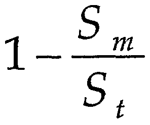
- porosity index is defined by the relation:
- Wallsten stent employs 24 filaments
- the device of the invention employs a much larger number of filaments, depending on the diameter of the device and upon the size of the windows (as
- a typical Wallstent is about 80 - 85%, and although the device of the invention
- the Porosity Index of the device is
- openings of the order of magnitude of 100 - 500 ⁇ m, while a stent typically
- filaments having a diameter of 200 ⁇ m for instance, filaments having a diameter of 200 ⁇ m.
- strengthening rings can also be provided at the extremities of the device, or at
- Such thicker filaments and rings can also function as markers, to permit to locate the position of the device within the body.
- This method both increases the mechanical strength of the device and also provides a marker for locating the position of
- beads could be threaded onto the filaments at designated locations during the weaving process. Also if the braided material
- the filtering means i.e., the "windows" of the deflecting device should have
- Re av is the average Reynolds number
- Womersley is the
- Re pr ox is the Reynolds number for the filaments of which the deflecting element is made, and the shear stress is measured at the device.
- invention provides a method of manufacturing a device of the invention
- Diameter 7 mm. The behavior of the device was compared with a Wallstent having the
- Diameter of round filament 90 ⁇
- the Nitinol device achieved 13% of the mechanical strength of the Wallstent.
- the device of the invention relative to a conventional stent.
- Embohc strokes from proximal sources e.g., mechanical heart
- valves Afib, LVT, protruding AAA. These are:
Abstract
Description
Claims
Priority Applications (9)
| Application Number | Priority Date | Filing Date | Title |
|---|---|---|---|
| EP01947773A EP1301145A2 (en) | 2000-07-17 | 2001-07-09 | Implantable braided stroke preventing device and method of manufacturing |
| KR10-2003-7000561A KR20030018049A (en) | 2000-07-17 | 2001-07-09 | Implantable braided stroke preventing device and method of manufacturing |
| JP2002511669A JP2004503327A (en) | 2000-07-17 | 2001-07-09 | Implantable braided device for stroke prevention and method of manufacturing the same |
| AU2001269412A AU2001269412A1 (en) | 2000-07-17 | 2001-07-09 | Implantable braided stroke preventing device and method of manufacturing |
| CA002414840A CA2414840A1 (en) | 2000-07-17 | 2001-07-09 | Implantable braided stroke preventing device and method of manufacturing |
| US10/311,876 US20040024416A1 (en) | 2000-07-17 | 2001-07-09 | Implantable braided stroke preventing device and method of manufacturing |
| US10/910,621 US7306624B2 (en) | 2001-07-09 | 2004-08-04 | Implantable intraluminal device and method of using same in treating aneurysms |
| US11/907,675 US7572290B2 (en) | 2001-07-09 | 2007-10-16 | Implantable intraluminal device and method of using same in treating aneurysms |
| US12/496,672 US7942925B2 (en) | 2001-07-09 | 2009-07-02 | Implantable intraluminal device and method of using same in treating aneurysms |
Applications Claiming Priority (2)
| Application Number | Priority Date | Filing Date | Title |
|---|---|---|---|
| IL137326 | 2000-07-17 | ||
| IL13732600A IL137326A0 (en) | 2000-07-17 | 2000-07-17 | Implantable braided stroke preventing device and method of manufacturing |
Related Child Applications (2)
| Application Number | Title | Priority Date | Filing Date |
|---|---|---|---|
| US10/216,356 Continuation-In-Part US20030100945A1 (en) | 2001-07-09 | 2002-08-12 | Implantable intraluminal device and method of using same in treating aneurysms |
| US10/910,621 Continuation-In-Part US7306624B2 (en) | 2001-07-09 | 2004-08-04 | Implantable intraluminal device and method of using same in treating aneurysms |
Publications (2)
| Publication Number | Publication Date |
|---|---|
| WO2002005729A2 true WO2002005729A2 (en) | 2002-01-24 |
| WO2002005729A3 WO2002005729A3 (en) | 2002-06-13 |
Family
ID=11074398
Family Applications (1)
| Application Number | Title | Priority Date | Filing Date |
|---|---|---|---|
| PCT/IL2001/000624 WO2002005729A2 (en) | 2000-07-17 | 2001-07-09 | Implantable braided stroke preventing device and method of manufacturing |
Country Status (9)
| Country | Link |
|---|---|
| US (1) | US20040024416A1 (en) |
| EP (1) | EP1301145A2 (en) |
| JP (1) | JP2004503327A (en) |
| KR (1) | KR20030018049A (en) |
| CN (1) | CN1455657A (en) |
| AU (1) | AU2001269412A1 (en) |
| CA (1) | CA2414840A1 (en) |
| IL (1) | IL137326A0 (en) |
| WO (1) | WO2002005729A2 (en) |
Cited By (21)
| Publication number | Priority date | Publication date | Assignee | Title |
|---|---|---|---|---|
| US6602271B2 (en) | 2000-05-24 | 2003-08-05 | Medtronic Ave, Inc. | Collapsible blood filter with optimal braid geometry |
| US6964681B2 (en) | 2002-01-29 | 2005-11-15 | Medtronic Vascular, Inc. | Flared stent and method of use |
| US7306624B2 (en) | 2001-07-09 | 2007-12-11 | Surpass Medical Ltd. | Implantable intraluminal device and method of using same in treating aneurysms |
| US8623067B2 (en) * | 2004-05-25 | 2014-01-07 | Covidien Lp | Methods and apparatus for luminal stenting |
| US9039754B2 (en) | 2011-04-14 | 2015-05-26 | Asahi Intecc Co., Ltd. | Stent |
| US9168132B2 (en) | 2005-05-27 | 2015-10-27 | Hlt, Inc. | Stentless support structure |
| US9271832B2 (en) | 2011-05-16 | 2016-03-01 | Hlt, Inc. | Inversion delivery device and method for a prosthesis |
| EP3021789A1 (en) * | 2013-07-17 | 2016-05-25 | Lake Region Manufacturing, Inc. d/b/a Lake Region Medical | High flow embolic protection device |
| US9486314B2 (en) | 2013-03-15 | 2016-11-08 | Hlt, Inc. | Low-profile prosthetic valve structure |
| US9561122B2 (en) | 2013-02-05 | 2017-02-07 | Covidien Lp | Vascular device for aneurysm treatment and providing blood flow into a perforator vessel |
| US9610181B2 (en) | 2006-02-22 | 2017-04-04 | Covidien Lp | Stents having radiopaque mesh |
| US9801711B2 (en) | 2010-08-11 | 2017-10-31 | Hlt, Inc. | Reinforced commissural support structure |
| US9827095B2 (en) | 2005-05-27 | 2017-11-28 | Hlt, Inc. | Stentless support structure |
| US9855047B2 (en) | 2004-05-25 | 2018-01-02 | Covidien Lp | Flexible vascular occluding device |
| EP3164087A4 (en) * | 2014-07-03 | 2018-01-10 | The Curators Of The University Of Missouri | Embolic protection system |
| US9907643B2 (en) | 2012-10-30 | 2018-03-06 | Covidien Lp | Systems for attaining a predetermined porosity of a vascular device |
| US9943427B2 (en) | 2012-11-06 | 2018-04-17 | Covidien Lp | Shaped occluding devices and methods of using the same |
| US9999504B2 (en) | 2014-10-13 | 2018-06-19 | Hlt, Inc. | Inversion delivery device and method for a prosthesis |
| US10004618B2 (en) | 2004-05-25 | 2018-06-26 | Covidien Lp | Methods and apparatus for luminal stenting |
| US10206798B2 (en) | 2012-10-31 | 2019-02-19 | Covidien Lp | Methods and systems for increasing a density of a region of a vascular device |
| US10364413B2 (en) | 2007-05-07 | 2019-07-30 | Protalix Ltd. | Large scale disposable bioreactor |
Families Citing this family (84)
| Publication number | Priority date | Publication date | Assignee | Title |
|---|---|---|---|---|
| US7018401B1 (en) | 1999-02-01 | 2006-03-28 | Board Of Regents, The University Of Texas System | Woven intravascular devices and methods for making the same and apparatus for delivery of the same |
| IL153753A0 (en) * | 2002-12-30 | 2003-07-06 | Neovasc Medical Ltd | Varying-diameter vascular implant and balloon |
| US6866679B2 (en) | 2002-03-12 | 2005-03-15 | Ev3 Inc. | Everting stent and stent delivery system |
| US7217287B2 (en) | 2002-08-28 | 2007-05-15 | Heart Leaflet Technologies, Inc. | Method of treating diseased valve |
| US7951557B2 (en) * | 2003-04-27 | 2011-05-31 | Protalix Ltd. | Human lysosomal proteins from plant cell culture |
| US20100196345A1 (en) * | 2003-04-27 | 2010-08-05 | Protalix | Production of high mannose proteins in plant culture |
| US20040236414A1 (en) * | 2003-05-23 | 2004-11-25 | Brar Balbir S. | Devices and methods for treatment of stenotic regions |
| US7226473B2 (en) * | 2003-05-23 | 2007-06-05 | Brar Balbir S | Treatment of stenotic regions |
| US8747453B2 (en) * | 2008-02-18 | 2014-06-10 | Aga Medical Corporation | Stent/stent graft for reinforcement of vascular abnormalities and associated method |
| US8267985B2 (en) | 2005-05-25 | 2012-09-18 | Tyco Healthcare Group Lp | System and method for delivering and deploying an occluding device within a vessel |
| US9675476B2 (en) | 2004-05-25 | 2017-06-13 | Covidien Lp | Vascular stenting for aneurysms |
| US8617234B2 (en) * | 2004-05-25 | 2013-12-31 | Covidien Lp | Flexible vascular occluding device |
| US20060206200A1 (en) | 2004-05-25 | 2006-09-14 | Chestnut Medical Technologies, Inc. | Flexible vascular occluding device |
| CA2560222A1 (en) * | 2004-06-22 | 2006-01-05 | Jeffrey E. Yeung | Disc shunt for treating back pain |
| US7063720B2 (en) * | 2004-09-14 | 2006-06-20 | The Wallace Enterprises, Inc. | Covered stent with controlled therapeutic agent diffusion |
| US20070055365A1 (en) * | 2005-04-28 | 2007-03-08 | The Cleveland Clinic Foundation | Stent with integrated filter |
| US8273101B2 (en) * | 2005-05-25 | 2012-09-25 | Tyco Healthcare Group Lp | System and method for delivering and deploying an occluding device within a vessel |
| CA2604081C (en) | 2005-05-25 | 2013-11-26 | Chestnut Medical Technologies, Inc. | System and method for delivering and deploying a self-expanding device within a vessel |
| US8663312B2 (en) | 2005-05-27 | 2014-03-04 | Hlt, Inc. | Intravascular cuff |
| US20070112371A1 (en) * | 2005-11-14 | 2007-05-17 | Medtronic Vascular, Inc. | Embolic protection filter having compact collapsed dimensions and method of making same |
| US9089404B2 (en) | 2006-03-31 | 2015-07-28 | Covidien Lp | Embolic protection devices having radiopaque elements |
| US8460335B2 (en) * | 2006-09-11 | 2013-06-11 | Embrella Cardiovascular, Inc. | Method of deflecting emboli from the cerebral circulation |
| US20100179647A1 (en) * | 2006-09-11 | 2010-07-15 | Carpenter Judith T | Methods of reducing embolism to cerebral circulation as a consequence of an index cardiac procedure |
| US9480548B2 (en) * | 2006-09-11 | 2016-11-01 | Edwards Lifesciences Ag | Embolic protection device and method of use |
| US20100179583A1 (en) * | 2006-09-11 | 2010-07-15 | Carpenter Judith T | Methods of deploying and retrieving an embolic diversion device |
| US9339367B2 (en) * | 2006-09-11 | 2016-05-17 | Edwards Lifesciences Ag | Embolic deflection device |
| US20080097401A1 (en) | 2006-09-22 | 2008-04-24 | Trapp Benjamin M | Cerebral vasculature device |
| EP3150177B1 (en) | 2006-10-22 | 2021-06-02 | Idev Technologies, Inc. | Methods for securing strand ends and the resulting devices |
| EP2124814B1 (en) * | 2007-03-20 | 2015-05-20 | Minvasys | Apparatus and methods for stent delivery with embolic protection |
| US9034007B2 (en) * | 2007-09-21 | 2015-05-19 | Insera Therapeutics, Inc. | Distal embolic protection devices with a variable thickness microguidewire and methods for their use |
| US8163004B2 (en) * | 2008-02-18 | 2012-04-24 | Aga Medical Corporation | Stent graft for reinforcement of vascular abnormalities and associated method |
| WO2009140437A1 (en) | 2008-05-13 | 2009-11-19 | Nfocus Neuromedical, Inc. | Braid implant delivery systems |
| US8992455B2 (en) * | 2008-07-09 | 2015-03-31 | Coraflo Ltd. | Methods, apparatuses and systems for caval stenting for venous drainage |
| MX2011002473A (en) * | 2008-09-04 | 2011-08-17 | Anders Joensson | Temporary embolic protection device and medical procedure for delivery thereof. |
| AU2010238636A1 (en) * | 2009-04-24 | 2011-11-17 | Flexible Stenting Solutions, Inc. | Flexible devices |
| US9327060B2 (en) * | 2009-07-09 | 2016-05-03 | CARDINAL HEALTH SWITZERLAND 515 GmbH | Rapamycin reservoir eluting stent |
| US9259550B2 (en) * | 2009-07-13 | 2016-02-16 | Cook Medical Technologies Llc | Swaged braided catheter and method of fabrication |
| WO2012111089A1 (en) * | 2011-02-15 | 2012-08-23 | アクセスポイントテクノロジーズ有限会社 | Stent system, and stent placing method |
| US8261648B1 (en) | 2011-10-17 | 2012-09-11 | Sequent Medical Inc. | Braiding mechanism and methods of use |
| JP6133307B2 (en) | 2011-10-17 | 2017-05-24 | シークエント メディカル インコーポレイテッド | Braiding machine and method of use |
| US20130226278A1 (en) | 2012-02-23 | 2013-08-29 | Tyco Healthcare Group Lp | Methods and apparatus for luminal stenting |
| US9072624B2 (en) | 2012-02-23 | 2015-07-07 | Covidien Lp | Luminal stenting |
| US9078659B2 (en) | 2012-04-23 | 2015-07-14 | Covidien Lp | Delivery system with hooks for resheathability |
| US9155647B2 (en) | 2012-07-18 | 2015-10-13 | Covidien Lp | Methods and apparatus for luminal stenting |
| US9724222B2 (en) | 2012-07-20 | 2017-08-08 | Covidien Lp | Resheathable stent delivery system |
| US9295393B2 (en) | 2012-11-09 | 2016-03-29 | Elwha Llc | Embolism deflector |
| US20140277397A1 (en) * | 2013-03-12 | 2014-09-18 | DePuy Synthes Products, LLC | Variable porosity intravascular implant and manufacturing method |
| US10561509B2 (en) | 2013-03-13 | 2020-02-18 | DePuy Synthes Products, Inc. | Braided stent with expansion ring and method of delivery |
| US8679150B1 (en) | 2013-03-15 | 2014-03-25 | Insera Therapeutics, Inc. | Shape-set textile structure based mechanical thrombectomy methods |
| US8715314B1 (en) | 2013-03-15 | 2014-05-06 | Insera Therapeutics, Inc. | Vascular treatment measurement methods |
| US8690907B1 (en) | 2013-03-15 | 2014-04-08 | Insera Therapeutics, Inc. | Vascular treatment methods |
| CN116172656A (en) | 2013-03-15 | 2023-05-30 | 伊瑟拉医疗公司 | Vascular treatment devices and methods |
| US10130500B2 (en) | 2013-07-25 | 2018-11-20 | Covidien Lp | Methods and apparatus for luminal stenting |
| WO2017142874A2 (en) | 2016-02-16 | 2017-08-24 | Insera Therapeutics, Inc. | Aspiration devices and anchored flow diverting devices |
| US9775590B2 (en) * | 2013-08-20 | 2017-10-03 | Boston Scientific Scimed, Inc. | Braided hemostasis shaft for improved torsional response |
| US8968383B1 (en) | 2013-08-27 | 2015-03-03 | Covidien Lp | Delivery of medical devices |
| US9782186B2 (en) | 2013-08-27 | 2017-10-10 | Covidien Lp | Vascular intervention system |
| JP2017520324A (en) * | 2014-07-01 | 2017-07-27 | ボストン サイエンティフィック サイムド,インコーポレイテッドBoston Scientific Scimed,Inc. | Multilayer braid end |
| EP2987463A1 (en) * | 2014-08-21 | 2016-02-24 | Noureddine Frid | 3d filter for prevention of stroke |
| US10206796B2 (en) | 2014-08-27 | 2019-02-19 | DePuy Synthes Products, Inc. | Multi-strand implant with enhanced radiopacity |
| US11771446B2 (en) | 2020-10-19 | 2023-10-03 | Anaconda Biomed, S.L. | Thrombectomy system and method of use |
| EP3307211A4 (en) * | 2015-06-11 | 2018-07-25 | Beijing Advanced Medical Technologies, Inc. | A close-cell structured stent, a preparation method and use thereof |
| US10842606B2 (en) * | 2015-09-09 | 2020-11-24 | Frid Mind Technologies | Bifurcated 3D filter assembly for prevention of stroke |
| US10213290B2 (en) * | 2016-02-17 | 2019-02-26 | Boston Scientific Scimed, Inc. | Braided stent and method of manufacturing a braided stent |
| CN109069160B (en) | 2016-03-17 | 2022-05-17 | S·珍耶那曼 | Occlusion of anatomical structures |
| CN105963051B (en) * | 2016-04-20 | 2018-01-19 | 东华大学 | A kind of intravascular stent fabric overlay film with local resistant structure and preparation method thereof |
| US10076428B2 (en) | 2016-08-25 | 2018-09-18 | DePuy Synthes Products, Inc. | Expansion ring for a braided stent |
| US10292851B2 (en) | 2016-09-30 | 2019-05-21 | DePuy Synthes Products, Inc. | Self-expanding device delivery apparatus with dual function bump |
| US10376396B2 (en) | 2017-01-19 | 2019-08-13 | Covidien Lp | Coupling units for medical device delivery systems |
| WO2018156655A1 (en) * | 2017-02-22 | 2018-08-30 | Claret Medical, Inc. | Systems and methods for protecting the cerebral vasculature |
| US11123209B2 (en) | 2018-04-12 | 2021-09-21 | Covidien Lp | Medical device delivery |
| US10786377B2 (en) | 2018-04-12 | 2020-09-29 | Covidien Lp | Medical device delivery |
| US11071637B2 (en) | 2018-04-12 | 2021-07-27 | Covidien Lp | Medical device delivery |
| US11413176B2 (en) | 2018-04-12 | 2022-08-16 | Covidien Lp | Medical device delivery |
| AU2019204522A1 (en) | 2018-07-30 | 2020-02-13 | DePuy Synthes Products, Inc. | Systems and methods of manufacturing and using an expansion ring |
| US10456280B1 (en) * | 2018-08-06 | 2019-10-29 | DePuy Synthes Products, Inc. | Systems and methods of using a braided implant |
| US10278848B1 (en) | 2018-08-06 | 2019-05-07 | DePuy Synthes Products, Inc. | Stent delivery with expansion assisting delivery wire |
| US11039944B2 (en) | 2018-12-27 | 2021-06-22 | DePuy Synthes Products, Inc. | Braided stent system with one or more expansion rings |
| CN113795204A (en) | 2019-01-11 | 2021-12-14 | 阿纳康达生物医学有限公司 | Loading device and method for loading a medical instrument into a catheter |
| CN109770825B (en) * | 2019-03-06 | 2021-09-24 | 杭州行开医学影像技术有限公司 | Endoscope with 3D imaging function |
| US11413174B2 (en) | 2019-06-26 | 2022-08-16 | Covidien Lp | Core assembly for medical device delivery systems |
| CN112022461B (en) * | 2020-09-16 | 2023-11-24 | 北京美迪微科技有限责任公司 | Be applied to vascular support of carotid artery |
| DE202020107453U1 (en) * | 2020-12-22 | 2022-03-25 | Acandis Gmbh | Stent, in particular for the treatment of diseases of the carotid artery |
| US11944558B2 (en) | 2021-08-05 | 2024-04-02 | Covidien Lp | Medical device delivery devices, systems, and methods |
Citations (6)
| Publication number | Priority date | Publication date | Assignee | Title |
|---|---|---|---|---|
| US4655771A (en) | 1982-04-30 | 1987-04-07 | Shepherd Patents S.A. | Prosthesis comprising an expansible or contractile tubular body |
| US5061275A (en) | 1986-04-21 | 1991-10-29 | Medinvent S.A. | Self-expanding prosthesis |
| WO1997016133A1 (en) | 1995-11-01 | 1997-05-09 | Biocompatibles Limited | Braided stent |
| EP0804909A2 (en) | 1996-04-30 | 1997-11-05 | Schneider (Usa) Inc. | Three dimensional braided covered stent |
| EP0895761A2 (en) | 1997-08-04 | 1999-02-10 | Schneider (Usa) Inc. | Balloon expandable braided stent with restraint |
| WO1999055256A1 (en) | 1998-04-28 | 1999-11-04 | Intratherapeutics, Inc. | Braided stent |
Family Cites Families (40)
| Publication number | Priority date | Publication date | Assignee | Title |
|---|---|---|---|---|
| US4425908A (en) * | 1981-10-22 | 1984-01-17 | Beth Israel Hospital | Blood clot filter |
| US4643184A (en) * | 1982-09-29 | 1987-02-17 | Mobin Uddin Kazi | Embolus trap |
| JPS6198254A (en) * | 1984-10-19 | 1986-05-16 | ザ・ベントリー―ハリス・マニュファクチュアリング・カンパニー | Prosthetic stent |
| US4577631A (en) * | 1984-11-16 | 1986-03-25 | Kreamer Jeffry W | Aneurysm repair apparatus and method |
| SE450809B (en) * | 1985-04-10 | 1987-08-03 | Medinvent Sa | PLANT TOPIC PROVIDED FOR MANUFACTURING A SPIRAL SPRING SUITABLE FOR TRANSLUMINAL IMPLANTATION AND MANUFACTURED SPIRAL SPRINGS |
| US5843089A (en) * | 1990-12-28 | 1998-12-01 | Boston Scientific Corporation | Stent lining |
| US5350398A (en) * | 1991-05-13 | 1994-09-27 | Dusan Pavcnik | Self-expanding filter for percutaneous insertion |
| US5304220A (en) * | 1991-07-03 | 1994-04-19 | Maginot Thomas J | Method and apparatus for implanting a graft prosthesis in the body of a patient |
| US5415630A (en) * | 1991-07-17 | 1995-05-16 | Gory; Pierre | Method for removably implanting a blood filter in a vein of the human body |
| US5324304A (en) * | 1992-06-18 | 1994-06-28 | William Cook Europe A/S | Introduction catheter set for a collapsible self-expandable implant |
| US5562725A (en) * | 1992-09-14 | 1996-10-08 | Meadox Medicals Inc. | Radially self-expanding implantable intraluminal device |
| US5383392A (en) * | 1993-03-16 | 1995-01-24 | Ward Holding Company, Inc. | Sheet registration control |
| US5464449A (en) * | 1993-07-08 | 1995-11-07 | Thomas J. Fogarty | Internal graft prosthesis and delivery system |
| US5443497A (en) * | 1993-11-22 | 1995-08-22 | The Johns Hopkins University | Percutaneous prosthetic by-pass graft and method of use |
| US5769816A (en) * | 1995-11-07 | 1998-06-23 | Embol-X, Inc. | Cannula with associated filter |
| US6576009B2 (en) * | 1995-12-01 | 2003-06-10 | Medtronic Ave, Inc. | Bifurcated intraluminal prostheses construction and methods |
| US5876367A (en) * | 1996-12-05 | 1999-03-02 | Embol-X, Inc. | Cerebral protection during carotid endarterectomy and downstream vascular protection during other surgeries |
| US5807330A (en) * | 1996-12-16 | 1998-09-15 | University Of Southern California | Angioplasty catheter |
| US6048360A (en) * | 1997-03-18 | 2000-04-11 | Endotex Interventional Systems, Inc. | Methods of making and using coiled sheet graft for single and bifurcated lumens |
| US5843172A (en) * | 1997-04-15 | 1998-12-01 | Advanced Cardiovascular Systems, Inc. | Porous medicated stent |
| WO1998046168A1 (en) * | 1997-04-15 | 1998-10-22 | Schneider (Usa) Inc | Prostheses with selectively welded crossing strands |
| WO1998047447A1 (en) * | 1997-04-23 | 1998-10-29 | Dubrul William R | Bifurcated stent and distal protection system |
| US6258120B1 (en) * | 1997-12-23 | 2001-07-10 | Embol-X, Inc. | Implantable cerebral protection device and methods of use |
| CA2235911C (en) * | 1997-05-27 | 2003-07-29 | Schneider (Usa) Inc. | Stent and stent-graft for treating branched vessels |
| US5951599A (en) * | 1997-07-09 | 1999-09-14 | Scimed Life Systems, Inc. | Occlusion system for endovascular treatment of an aneurysm |
| US5855599A (en) * | 1997-09-02 | 1999-01-05 | Sitek, Inc. | Silicon micro machined occlusion implant |
| US5941896A (en) * | 1997-09-08 | 1999-08-24 | Montefiore Hospital And Medical Center | Filter and method for trapping emboli during endovascular procedures |
| US6030414A (en) * | 1997-11-13 | 2000-02-29 | Taheri; Syde A. | Variable stent and method for treatment of arterial disease |
| US6261305B1 (en) * | 1998-02-12 | 2001-07-17 | Eclips Systems Inc. | Endovascular prothesis with expandable leaf portion |
| US5928261A (en) * | 1998-06-29 | 1999-07-27 | Ruiz; Carlos E. | Removable vascular filter, catheter system and methods of use |
| US6093199A (en) * | 1998-08-05 | 2000-07-25 | Endovascular Technologies, Inc. | Intra-luminal device for treatment of body cavities and lumens and method of use |
| US6117117A (en) * | 1998-08-24 | 2000-09-12 | Advanced Cardiovascular Systems, Inc. | Bifurcated catheter assembly |
| DK1148839T3 (en) * | 1999-02-01 | 2008-12-15 | Univ Texas | Woven two-branched and three-branched stents and methods of making them |
| US6673089B1 (en) * | 1999-03-11 | 2004-01-06 | Mindguard Ltd. | Implantable stroke treating device |
| IL128938A0 (en) * | 1999-03-11 | 2000-02-17 | Mind Guard Ltd | Implantable stroke treating device |
| US6146370A (en) * | 1999-04-07 | 2000-11-14 | Coaxia, Inc. | Devices and methods for preventing distal embolization from the internal carotid artery using flow reversal by partial occlusion of the external carotid artery |
| US6409757B1 (en) * | 1999-09-15 | 2002-06-25 | Eva Corporation | Method and apparatus for supporting a graft assembly |
| US6383171B1 (en) * | 1999-10-12 | 2002-05-07 | Allan Will | Methods and devices for protecting a passageway in a body when advancing devices through the passageway |
| US6312463B1 (en) * | 2000-02-01 | 2001-11-06 | Endotex Interventional Systems, Inc. | Micro-porous mesh stent with hybrid structure |
| US6638294B1 (en) * | 2001-08-30 | 2003-10-28 | Advanced Cardiovascular Systems, Inc. | Self furling umbrella frame for carotid filter |
-
2000
- 2000-07-17 IL IL13732600A patent/IL137326A0/en unknown
-
2001
- 2001-07-09 WO PCT/IL2001/000624 patent/WO2002005729A2/en not_active Application Discontinuation
- 2001-07-09 CA CA002414840A patent/CA2414840A1/en not_active Abandoned
- 2001-07-09 KR KR10-2003-7000561A patent/KR20030018049A/en not_active Application Discontinuation
- 2001-07-09 JP JP2002511669A patent/JP2004503327A/en not_active Abandoned
- 2001-07-09 CN CN01815497A patent/CN1455657A/en active Pending
- 2001-07-09 AU AU2001269412A patent/AU2001269412A1/en not_active Abandoned
- 2001-07-09 US US10/311,876 patent/US20040024416A1/en not_active Abandoned
- 2001-07-09 EP EP01947773A patent/EP1301145A2/en not_active Withdrawn
Patent Citations (7)
| Publication number | Priority date | Publication date | Assignee | Title |
|---|---|---|---|---|
| US4655771A (en) | 1982-04-30 | 1987-04-07 | Shepherd Patents S.A. | Prosthesis comprising an expansible or contractile tubular body |
| US4655771B1 (en) | 1982-04-30 | 1996-09-10 | Medinvent Ams Sa | Prosthesis comprising an expansible or contractile tubular body |
| US5061275A (en) | 1986-04-21 | 1991-10-29 | Medinvent S.A. | Self-expanding prosthesis |
| WO1997016133A1 (en) | 1995-11-01 | 1997-05-09 | Biocompatibles Limited | Braided stent |
| EP0804909A2 (en) | 1996-04-30 | 1997-11-05 | Schneider (Usa) Inc. | Three dimensional braided covered stent |
| EP0895761A2 (en) | 1997-08-04 | 1999-02-10 | Schneider (Usa) Inc. | Balloon expandable braided stent with restraint |
| WO1999055256A1 (en) | 1998-04-28 | 1999-11-04 | Intratherapeutics, Inc. | Braided stent |
Cited By (44)
| Publication number | Priority date | Publication date | Assignee | Title |
|---|---|---|---|---|
| US6602271B2 (en) | 2000-05-24 | 2003-08-05 | Medtronic Ave, Inc. | Collapsible blood filter with optimal braid geometry |
| US7306624B2 (en) | 2001-07-09 | 2007-12-11 | Surpass Medical Ltd. | Implantable intraluminal device and method of using same in treating aneurysms |
| EP2425800A2 (en) | 2001-11-23 | 2012-03-07 | Surpass Medical Ltd | Implantable intraluminal device and method of using same in treating aneurysms |
| US6964681B2 (en) | 2002-01-29 | 2005-11-15 | Medtronic Vascular, Inc. | Flared stent and method of use |
| US7867269B2 (en) | 2002-01-29 | 2011-01-11 | Medtronic Vascular, Inc. | Flared stent and method for use |
| US9801744B2 (en) | 2004-05-25 | 2017-10-31 | Covidien Lp | Methods and apparatus for luminal stenting |
| US8623067B2 (en) * | 2004-05-25 | 2014-01-07 | Covidien Lp | Methods and apparatus for luminal stenting |
| US11771433B2 (en) | 2004-05-25 | 2023-10-03 | Covidien Lp | Flexible vascular occluding device |
| US9050205B2 (en) | 2004-05-25 | 2015-06-09 | Covidien Lp | Methods and apparatus for luminal stenting |
| US10918389B2 (en) | 2004-05-25 | 2021-02-16 | Covidien Lp | Flexible vascular occluding device |
| US10004618B2 (en) | 2004-05-25 | 2018-06-26 | Covidien Lp | Methods and apparatus for luminal stenting |
| US9855047B2 (en) | 2004-05-25 | 2018-01-02 | Covidien Lp | Flexible vascular occluding device |
| US9814575B2 (en) | 2005-05-27 | 2017-11-14 | Hlt, Inc. | Stentless support structure |
| US9439760B2 (en) | 2005-05-27 | 2016-09-13 | Hlt, Inc. | Stentless support structure |
| US10080655B2 (en) | 2005-05-27 | 2018-09-25 | Hlt, Inc. | Stentless support structure |
| US9168132B2 (en) | 2005-05-27 | 2015-10-27 | Hlt, Inc. | Stentless support structure |
| US11026784B2 (en) | 2005-05-27 | 2021-06-08 | Hlt, Inc. | Stentless support structure |
| US10646337B2 (en) | 2005-05-27 | 2020-05-12 | Hlt, Inc. | Stentless support structure |
| US9827095B2 (en) | 2005-05-27 | 2017-11-28 | Hlt, Inc. | Stentless support structure |
| US11382777B2 (en) | 2006-02-22 | 2022-07-12 | Covidien Lp | Stents having radiopaque mesh |
| US10433988B2 (en) | 2006-02-22 | 2019-10-08 | Covidien Lp | Stents having radiopaque mesh |
| US9610181B2 (en) | 2006-02-22 | 2017-04-04 | Covidien Lp | Stents having radiopaque mesh |
| US10364413B2 (en) | 2007-05-07 | 2019-07-30 | Protalix Ltd. | Large scale disposable bioreactor |
| US9801711B2 (en) | 2010-08-11 | 2017-10-31 | Hlt, Inc. | Reinforced commissural support structure |
| US9039754B2 (en) | 2011-04-14 | 2015-05-26 | Asahi Intecc Co., Ltd. | Stent |
| US10188517B2 (en) | 2011-05-16 | 2019-01-29 | Hlt, Inc. | Inversion delivery device and method for a prosthesis |
| US11413143B2 (en) | 2011-05-16 | 2022-08-16 | Hlt, Inc. | Inversion delivery device and method for a prosthesis |
| US9566154B2 (en) | 2011-05-16 | 2017-02-14 | Hlt, Inc. | Inversion delivery device and method for a prosthesis |
| US9271832B2 (en) | 2011-05-16 | 2016-03-01 | Hlt, Inc. | Inversion delivery device and method for a prosthesis |
| US9522064B2 (en) | 2011-05-16 | 2016-12-20 | Hlt, Inc. | Inversion delivery device and method for a prosthesis |
| US10278817B2 (en) | 2011-05-16 | 2019-05-07 | Hlt, Inc. | Inversion delivery device and method for a prosthesis |
| US9693863B2 (en) | 2011-05-16 | 2017-07-04 | Hlt, Inc. | Inversion delivery device and method for a prosthesis |
| US9795478B2 (en) | 2011-05-16 | 2017-10-24 | Hlt, Inc. | Inversion delivery device and method for a prosthesis |
| US9907643B2 (en) | 2012-10-30 | 2018-03-06 | Covidien Lp | Systems for attaining a predetermined porosity of a vascular device |
| US10206798B2 (en) | 2012-10-31 | 2019-02-19 | Covidien Lp | Methods and systems for increasing a density of a region of a vascular device |
| US9943427B2 (en) | 2012-11-06 | 2018-04-17 | Covidien Lp | Shaped occluding devices and methods of using the same |
| US9561122B2 (en) | 2013-02-05 | 2017-02-07 | Covidien Lp | Vascular device for aneurysm treatment and providing blood flow into a perforator vessel |
| US9486314B2 (en) | 2013-03-15 | 2016-11-08 | Hlt, Inc. | Low-profile prosthetic valve structure |
| US9931205B2 (en) | 2013-03-15 | 2018-04-03 | Hlt, Inc. | Low-profile prosthetic valve structure |
| EP3021789A1 (en) * | 2013-07-17 | 2016-05-25 | Lake Region Manufacturing, Inc. d/b/a Lake Region Medical | High flow embolic protection device |
| EP3021789A4 (en) * | 2013-07-17 | 2017-04-26 | Lake Region Manufacturing, Inc. d/b/a Lake Region Medical | High flow embolic protection device |
| EP3164087A4 (en) * | 2014-07-03 | 2018-01-10 | The Curators Of The University Of Missouri | Embolic protection system |
| US10820995B2 (en) | 2014-10-13 | 2020-11-03 | Hlt, Inc. | Inversion delivery device and method for a prosthesis |
| US9999504B2 (en) | 2014-10-13 | 2018-06-19 | Hlt, Inc. | Inversion delivery device and method for a prosthesis |
Also Published As
| Publication number | Publication date |
|---|---|
| CA2414840A1 (en) | 2002-01-24 |
| WO2002005729A3 (en) | 2002-06-13 |
| KR20030018049A (en) | 2003-03-04 |
| CN1455657A (en) | 2003-11-12 |
| AU2001269412A1 (en) | 2002-01-30 |
| JP2004503327A (en) | 2004-02-05 |
| IL137326A0 (en) | 2001-07-24 |
| US20040024416A1 (en) | 2004-02-05 |
| EP1301145A2 (en) | 2003-04-16 |
Similar Documents
| Publication | Publication Date | Title |
|---|---|---|
| US20040024416A1 (en) | Implantable braided stroke preventing device and method of manufacturing | |
| EP1011532B1 (en) | Bifurcated stent and distal protection system | |
| US20040010308A1 (en) | Implantable composite device and corresponding method for deflecting embolic material in blood flowing at an arterial bifurcation | |
| EP2268235B1 (en) | Stent prosthesis having select flared crowns | |
| US20040010307A1 (en) | Implantable integral device and corresponding method for deflecting embolic material in blood flowing at an arterial bifurcation | |
| JP4801655B2 (en) | Stent capable of intravascular supply for strengthening abnormal parts of blood vessels | |
| US9517146B2 (en) | Methods and apparatus for stenting comprising enhanced embolic protection coupled with improved protections against restenosis and thrombus formation | |
| JP5421929B2 (en) | Stent graft for reinforcing vascular abnormalities and methods related thereto | |
| ES2278688T3 (en) | ENDOPROTESIS COVERED OF SELF-EXPANDABLE GRAFT. | |
| US7806923B2 (en) | Side branch stent having a proximal split ring | |
| US20130131714A1 (en) | Embolic protection device and methods of making the same | |
| MXPA03008852A (en) | Radiopaque intraluminal medical device. | |
| WO2002055125A2 (en) | Implantable composite device and corresponding method for deflecting embolic material in blood flowing at an arterial bifurcation | |
| US20230149192A1 (en) | Expandable stent and a method for promoting a natural intracranial angiogenesis process, and use of the expandable stent in the method for promoting a natural intracranial angiogenesis process | |
| EP1363573A2 (en) | Implantable integral device and corresponding method for deflecting embolic material in blood flowing at an arterial bifurcation | |
| JPH11513902A (en) | Device implanted in a blood vessel or hollow organ lumen |
Legal Events
| Date | Code | Title | Description |
|---|---|---|---|
| AK | Designated states |
Kind code of ref document: A2 Designated state(s): AE AG AL AM AT AU AZ BA BB BG BR BY BZ CA CH CN CO CR CU CZ DE DK DM DZ EC EE ES FI GB GD GE GH GM HR HU ID IL IN IS JP KE KG KP KR KZ LC LK LR LS LT LU LV MA MD MG MK MN MW MX MZ NO NZ PL PT RO RU SD SE SG SI SK SL TJ TM TR TT TZ UA UG US UZ VN YU ZA ZW |
|
| AL | Designated countries for regional patents |
Kind code of ref document: A2 Designated state(s): GH GM KE LS MW MZ SD SL SZ TZ UG ZW AM AZ BY KG KZ MD RU TJ TM AT BE CH CY DE DK ES FI FR GB GR IE IT LU MC NL PT SE TR BF BJ CF CG CI CM GA GN GW ML MR NE SN TD TG |
|
| 121 | Ep: the epo has been informed by wipo that ep was designated in this application | ||
| DFPE | Request for preliminary examination filed prior to expiration of 19th month from priority date (pct application filed before 20040101) | ||
| AK | Designated states |
Kind code of ref document: A3 Designated state(s): AE AG AL AM AT AU AZ BA BB BG BR BY BZ CA CH CN CO CR CU CZ DE DK DM DZ EC EE ES FI GB GD GE GH GM HR HU ID IL IN IS JP KE KG KP KR KZ LC LK LR LS LT LU LV MA MD MG MK MN MW MX MZ NO NZ PL PT RO RU SD SE SG SI SK SL TJ TM TR TT TZ UA UG US UZ VN YU ZA ZW |
|
| AL | Designated countries for regional patents |
Kind code of ref document: A3 Designated state(s): GH GM KE LS MW MZ SD SL SZ TZ UG ZW AM AZ BY KG KZ MD RU TJ TM AT BE CH CY DE DK ES FI FR GB GR IE IT LU MC NL PT SE TR BF BJ CF CG CI CM GA GN GW ML MR NE SN TD TG |
|
| WWE | Wipo information: entry into national phase |
Ref document number: 10216356 Country of ref document: US |
|
| WWE | Wipo information: entry into national phase |
Ref document number: 10311876 Country of ref document: US |
|
| WWE | Wipo information: entry into national phase |
Ref document number: 2001269412 Country of ref document: AU |
|
| WWE | Wipo information: entry into national phase |
Ref document number: 2414840 Country of ref document: CA |
|
| WWE | Wipo information: entry into national phase |
Ref document number: 2001947773 Country of ref document: EP |
|
| WWE | Wipo information: entry into national phase |
Ref document number: 1020037000561 Country of ref document: KR |
|
| WWP | Wipo information: published in national office |
Ref document number: 1020037000561 Country of ref document: KR |
|
| WWE | Wipo information: entry into national phase |
Ref document number: 018154972 Country of ref document: CN |
|
| WWP | Wipo information: published in national office |
Ref document number: 2001947773 Country of ref document: EP |
|
| REG | Reference to national code |
Ref country code: DE Ref legal event code: 8642 |
|
| WWW | Wipo information: withdrawn in national office |
Ref document number: 2001947773 Country of ref document: EP |