WO2001000874A2 - Cancer associated antigens and uses therefor - Google Patents
Cancer associated antigens and uses therefor Download PDFInfo
- Publication number
- WO2001000874A2 WO2001000874A2 PCT/US2000/017207 US0017207W WO0100874A2 WO 2001000874 A2 WO2001000874 A2 WO 2001000874A2 US 0017207 W US0017207 W US 0017207W WO 0100874 A2 WO0100874 A2 WO 0100874A2
- Authority
- WO
- WIPO (PCT)
- Prior art keywords
- nucleic acid
- group
- molecule
- cancer associated
- associated antigen
- Prior art date
Links
- 206010028980 Neoplasm Diseases 0.000 title claims abstract description 374
- 108091007433 antigens Proteins 0.000 title claims abstract description 345
- 102000036639 antigens Human genes 0.000 title claims abstract description 339
- 239000000427 antigen Substances 0.000 title claims abstract description 337
- 201000011510 cancer Diseases 0.000 title claims abstract description 324
- 108090000765 processed proteins & peptides Proteins 0.000 claims abstract description 339
- 150000007523 nucleic acids Chemical class 0.000 claims abstract description 310
- 102000039446 nucleic acids Human genes 0.000 claims abstract description 300
- 108020004707 nucleic acids Proteins 0.000 claims abstract description 300
- 102000004196 processed proteins & peptides Human genes 0.000 claims abstract description 287
- 229920001184 polypeptide Polymers 0.000 claims abstract description 234
- 210000004027 cell Anatomy 0.000 claims abstract description 218
- 108090000623 proteins and genes Proteins 0.000 claims abstract description 189
- 230000014509 gene expression Effects 0.000 claims abstract description 131
- 239000012634 fragment Substances 0.000 claims abstract description 123
- 102000004169 proteins and genes Human genes 0.000 claims abstract description 123
- 239000013604 expression vector Substances 0.000 claims abstract description 48
- 201000010208 Seminoma Diseases 0.000 claims abstract description 18
- 210000001151 cytotoxic T lymphocyte Anatomy 0.000 claims abstract description 13
- 238000012544 monitoring process Methods 0.000 claims abstract description 12
- 238000000034 method Methods 0.000 claims description 141
- 241000282414 Homo sapiens Species 0.000 claims description 114
- 239000002773 nucleotide Substances 0.000 claims description 93
- 125000003729 nucleotide group Chemical group 0.000 claims description 93
- 239000003795 chemical substances by application Substances 0.000 claims description 91
- 239000000203 mixture Substances 0.000 claims description 48
- 239000002243 precursor Substances 0.000 claims description 48
- 230000027455 binding Effects 0.000 claims description 42
- 230000028993 immune response Effects 0.000 claims description 33
- 238000006467 substitution reaction Methods 0.000 claims description 29
- 239000000523 sample Substances 0.000 claims description 27
- 239000002671 adjuvant Substances 0.000 claims description 25
- 150000001413 amino acids Chemical class 0.000 claims description 25
- 208000037265 diseases, disorders, signs and symptoms Diseases 0.000 claims description 25
- 230000000694 effects Effects 0.000 claims description 25
- 125000003275 alpha amino acid group Chemical group 0.000 claims description 24
- 239000008194 pharmaceutical composition Substances 0.000 claims description 24
- 230000002159 abnormal effect Effects 0.000 claims description 20
- 239000000825 pharmaceutical preparation Substances 0.000 claims description 19
- 230000001225 therapeutic effect Effects 0.000 claims description 19
- 230000004044 response Effects 0.000 claims description 18
- 230000003993 interaction Effects 0.000 claims description 17
- 238000013518 transcription Methods 0.000 claims description 17
- 230000035897 transcription Effects 0.000 claims description 17
- 239000003814 drug Substances 0.000 claims description 16
- 108091028043 Nucleic acid sequence Proteins 0.000 claims description 14
- 238000007792 addition Methods 0.000 claims description 13
- 239000003937 drug carrier Substances 0.000 claims description 12
- 238000004519 manufacturing process Methods 0.000 claims description 12
- 230000000692 anti-sense effect Effects 0.000 claims description 11
- 238000013519 translation Methods 0.000 claims description 11
- 230000004936 stimulating effect Effects 0.000 claims description 10
- 238000012217 deletion Methods 0.000 claims description 8
- 230000037430 deletion Effects 0.000 claims description 8
- 239000000284 extract Substances 0.000 claims description 8
- 230000001461 cytolytic effect Effects 0.000 claims description 7
- 108020003175 receptors Proteins 0.000 claims description 6
- 102000005962 receptors Human genes 0.000 claims description 6
- 230000005867 T cell response Effects 0.000 claims description 5
- 210000003719 b-lymphocyte Anatomy 0.000 claims description 5
- 239000000032 diagnostic agent Substances 0.000 claims description 5
- 229940039227 diagnostic agent Drugs 0.000 claims description 5
- 230000002163 immunogen Effects 0.000 claims description 5
- 230000002062 proliferating effect Effects 0.000 claims description 5
- 229940124597 therapeutic agent Drugs 0.000 claims description 5
- 102000004190 Enzymes Human genes 0.000 claims description 4
- 108090000790 Enzymes Proteins 0.000 claims description 4
- 108010017213 Granulocyte-Macrophage Colony-Stimulating Factor Proteins 0.000 claims description 4
- 238000012258 culturing Methods 0.000 claims description 4
- 230000002285 radioactive effect Effects 0.000 claims description 4
- 229930182490 saponin Natural products 0.000 claims description 4
- 239000003053 toxin Substances 0.000 claims description 4
- 231100000765 toxin Toxicity 0.000 claims description 4
- 108700012359 toxins Proteins 0.000 claims description 4
- 239000012472 biological sample Substances 0.000 claims description 3
- 210000001124 body fluid Anatomy 0.000 claims description 3
- 230000002401 inhibitory effect Effects 0.000 claims description 3
- 230000001575 pathological effect Effects 0.000 claims description 3
- 239000001397 quillaja saponaria molina bark Substances 0.000 claims description 3
- 150000007949 saponins Chemical group 0.000 claims description 3
- 108010063738 Interleukins Proteins 0.000 claims description 2
- 102000015696 Interleukins Human genes 0.000 claims description 2
- 239000010839 body fluid Substances 0.000 claims description 2
- 239000001963 growth medium Substances 0.000 claims description 2
- 238000009877 rendering Methods 0.000 claims description 2
- 102000004457 Granulocyte-Macrophage Colony-Stimulating Factor Human genes 0.000 claims 1
- 230000001594 aberrant effect Effects 0.000 claims 1
- 210000001550 testis Anatomy 0.000 abstract description 22
- 238000012216 screening Methods 0.000 abstract description 13
- 238000003745 diagnosis Methods 0.000 abstract description 8
- 238000011282 treatment Methods 0.000 abstract description 8
- 238000011160 research Methods 0.000 abstract description 3
- 235000018102 proteins Nutrition 0.000 description 83
- 101001054842 Homo sapiens Leucine zipper protein 4 Proteins 0.000 description 49
- 102100026910 Leucine zipper protein 4 Human genes 0.000 description 49
- 235000001014 amino acid Nutrition 0.000 description 38
- 210000001519 tissue Anatomy 0.000 description 34
- 239000013598 vector Substances 0.000 description 30
- 239000000047 product Substances 0.000 description 27
- 229940024606 amino acid Drugs 0.000 description 25
- 108020004414 DNA Proteins 0.000 description 24
- 239000002299 complementary DNA Substances 0.000 description 21
- 108091034117 Oligonucleotide Proteins 0.000 description 20
- 239000000463 material Substances 0.000 description 20
- QTBSBXVTEAMEQO-UHFFFAOYSA-N Acetic acid Chemical compound CC(O)=O QTBSBXVTEAMEQO-UHFFFAOYSA-N 0.000 description 19
- 210000001744 T-lymphocyte Anatomy 0.000 description 18
- 210000004443 dendritic cell Anatomy 0.000 description 18
- 238000012360 testing method Methods 0.000 description 18
- 238000002360 preparation method Methods 0.000 description 16
- 108020004705 Codon Proteins 0.000 description 15
- 238000000636 Northern blotting Methods 0.000 description 15
- 241000700605 Viruses Species 0.000 description 15
- 238000000338 in vitro Methods 0.000 description 15
- JLCPHMBAVCMARE-UHFFFAOYSA-N [3-[[3-[[3-[[3-[[3-[[3-[[3-[[3-[[3-[[3-[[3-[[5-(2-amino-6-oxo-1H-purin-9-yl)-3-[[3-[[3-[[3-[[3-[[3-[[5-(2-amino-6-oxo-1H-purin-9-yl)-3-[[5-(2-amino-6-oxo-1H-purin-9-yl)-3-hydroxyoxolan-2-yl]methoxy-hydroxyphosphoryl]oxyoxolan-2-yl]methoxy-hydroxyphosphoryl]oxy-5-(5-methyl-2,4-dioxopyrimidin-1-yl)oxolan-2-yl]methoxy-hydroxyphosphoryl]oxy-5-(6-aminopurin-9-yl)oxolan-2-yl]methoxy-hydroxyphosphoryl]oxy-5-(6-aminopurin-9-yl)oxolan-2-yl]methoxy-hydroxyphosphoryl]oxy-5-(6-aminopurin-9-yl)oxolan-2-yl]methoxy-hydroxyphosphoryl]oxy-5-(6-aminopurin-9-yl)oxolan-2-yl]methoxy-hydroxyphosphoryl]oxyoxolan-2-yl]methoxy-hydroxyphosphoryl]oxy-5-(5-methyl-2,4-dioxopyrimidin-1-yl)oxolan-2-yl]methoxy-hydroxyphosphoryl]oxy-5-(4-amino-2-oxopyrimidin-1-yl)oxolan-2-yl]methoxy-hydroxyphosphoryl]oxy-5-(5-methyl-2,4-dioxopyrimidin-1-yl)oxolan-2-yl]methoxy-hydroxyphosphoryl]oxy-5-(5-methyl-2,4-dioxopyrimidin-1-yl)oxolan-2-yl]methoxy-hydroxyphosphoryl]oxy-5-(6-aminopurin-9-yl)oxolan-2-yl]methoxy-hydroxyphosphoryl]oxy-5-(6-aminopurin-9-yl)oxolan-2-yl]methoxy-hydroxyphosphoryl]oxy-5-(4-amino-2-oxopyrimidin-1-yl)oxolan-2-yl]methoxy-hydroxyphosphoryl]oxy-5-(4-amino-2-oxopyrimidin-1-yl)oxolan-2-yl]methoxy-hydroxyphosphoryl]oxy-5-(4-amino-2-oxopyrimidin-1-yl)oxolan-2-yl]methoxy-hydroxyphosphoryl]oxy-5-(6-aminopurin-9-yl)oxolan-2-yl]methoxy-hydroxyphosphoryl]oxy-5-(4-amino-2-oxopyrimidin-1-yl)oxolan-2-yl]methyl [5-(6-aminopurin-9-yl)-2-(hydroxymethyl)oxolan-3-yl] hydrogen phosphate Chemical class Cc1cn(C2CC(OP(O)(=O)OCC3OC(CC3OP(O)(=O)OCC3OC(CC3O)n3cnc4c3nc(N)[nH]c4=O)n3cnc4c3nc(N)[nH]c4=O)C(COP(O)(=O)OC3CC(OC3COP(O)(=O)OC3CC(OC3COP(O)(=O)OC3CC(OC3COP(O)(=O)OC3CC(OC3COP(O)(=O)OC3CC(OC3COP(O)(=O)OC3CC(OC3COP(O)(=O)OC3CC(OC3COP(O)(=O)OC3CC(OC3COP(O)(=O)OC3CC(OC3COP(O)(=O)OC3CC(OC3COP(O)(=O)OC3CC(OC3COP(O)(=O)OC3CC(OC3COP(O)(=O)OC3CC(OC3COP(O)(=O)OC3CC(OC3COP(O)(=O)OC3CC(OC3COP(O)(=O)OC3CC(OC3COP(O)(=O)OC3CC(OC3CO)n3cnc4c(N)ncnc34)n3ccc(N)nc3=O)n3cnc4c(N)ncnc34)n3ccc(N)nc3=O)n3ccc(N)nc3=O)n3ccc(N)nc3=O)n3cnc4c(N)ncnc34)n3cnc4c(N)ncnc34)n3cc(C)c(=O)[nH]c3=O)n3cc(C)c(=O)[nH]c3=O)n3ccc(N)nc3=O)n3cc(C)c(=O)[nH]c3=O)n3cnc4c3nc(N)[nH]c4=O)n3cnc4c(N)ncnc34)n3cnc4c(N)ncnc34)n3cnc4c(N)ncnc34)n3cnc4c(N)ncnc34)O2)c(=O)[nH]c1=O JLCPHMBAVCMARE-UHFFFAOYSA-N 0.000 description 14
- 239000000074 antisense oligonucleotide Substances 0.000 description 14
- 238000012230 antisense oligonucleotides Methods 0.000 description 14
- 238000009396 hybridization Methods 0.000 description 14
- 238000003556 assay Methods 0.000 description 13
- 238000001727 in vivo Methods 0.000 description 13
- 238000003757 reverse transcription PCR Methods 0.000 description 13
- 150000003839 salts Chemical class 0.000 description 13
- 238000004458 analytical method Methods 0.000 description 12
- 108020004999 messenger RNA Proteins 0.000 description 12
- 108020000948 Antisense Oligonucleotides Proteins 0.000 description 11
- 230000000735 allogeneic effect Effects 0.000 description 11
- 238000013459 approach Methods 0.000 description 11
- 210000005105 peripheral blood lymphocyte Anatomy 0.000 description 11
- 238000003752 polymerase chain reaction Methods 0.000 description 11
- 210000004881 tumor cell Anatomy 0.000 description 11
- 206010061535 Ovarian neoplasm Diseases 0.000 description 10
- 208000035475 disorder Diseases 0.000 description 10
- 230000003053 immunization Effects 0.000 description 10
- 238000002649 immunization Methods 0.000 description 10
- 239000013612 plasmid Substances 0.000 description 10
- 230000001105 regulatory effect Effects 0.000 description 10
- 238000002560 therapeutic procedure Methods 0.000 description 10
- 230000014616 translation Effects 0.000 description 10
- 241001430294 unidentified retrovirus Species 0.000 description 10
- 238000002255 vaccination Methods 0.000 description 10
- 108091032973 (ribonucleotides)n+m Proteins 0.000 description 9
- 108091026890 Coding region Proteins 0.000 description 9
- 230000003321 amplification Effects 0.000 description 9
- 201000001441 melanoma Diseases 0.000 description 9
- 238000003199 nucleic acid amplification method Methods 0.000 description 9
- 238000001890 transfection Methods 0.000 description 9
- -1 BAGE-1 Proteins 0.000 description 8
- 241000702421 Dependoparvovirus Species 0.000 description 8
- 210000000612 antigen-presenting cell Anatomy 0.000 description 8
- 238000010367 cloning Methods 0.000 description 8
- 239000002245 particle Substances 0.000 description 8
- 230000010076 replication Effects 0.000 description 8
- 238000010561 standard procedure Methods 0.000 description 8
- 241000701161 unidentified adenovirus Species 0.000 description 8
- 229960005486 vaccine Drugs 0.000 description 8
- 102000004127 Cytokines Human genes 0.000 description 7
- 108090000695 Cytokines Proteins 0.000 description 7
- ROHFNLRQFUQHCH-UHFFFAOYSA-N Leucine Natural products CC(C)CC(N)C(O)=O ROHFNLRQFUQHCH-UHFFFAOYSA-N 0.000 description 7
- 102000043129 MHC class I family Human genes 0.000 description 7
- 108091054437 MHC class I family Proteins 0.000 description 7
- 238000002105 Southern blotting Methods 0.000 description 7
- 230000000295 complement effect Effects 0.000 description 7
- 230000000139 costimulatory effect Effects 0.000 description 7
- 201000010099 disease Diseases 0.000 description 7
- 230000002068 genetic effect Effects 0.000 description 7
- 230000001965 increasing effect Effects 0.000 description 7
- 239000012528 membrane Substances 0.000 description 7
- 230000035772 mutation Effects 0.000 description 7
- 108700028369 Alleles Proteins 0.000 description 6
- 108020004635 Complementary DNA Proteins 0.000 description 6
- ROHFNLRQFUQHCH-YFKPBYRVSA-N L-leucine Chemical compound CC(C)C[C@H](N)C(O)=O ROHFNLRQFUQHCH-YFKPBYRVSA-N 0.000 description 6
- 238000002955 isolation Methods 0.000 description 6
- 239000003446 ligand Substances 0.000 description 6
- 210000004698 lymphocyte Anatomy 0.000 description 6
- 230000004048 modification Effects 0.000 description 6
- 238000012986 modification Methods 0.000 description 6
- 238000012163 sequencing technique Methods 0.000 description 6
- 210000002966 serum Anatomy 0.000 description 6
- 239000000243 solution Substances 0.000 description 6
- 206010018338 Glioma Diseases 0.000 description 5
- 241000699666 Mus <mouse, genus> Species 0.000 description 5
- 206010033128 Ovarian cancer Diseases 0.000 description 5
- 102000007056 Recombinant Fusion Proteins Human genes 0.000 description 5
- 108010008281 Recombinant Fusion Proteins Proteins 0.000 description 5
- DBMJMQXJHONAFJ-UHFFFAOYSA-M Sodium laurylsulphate Chemical compound [Na+].CCCCCCCCCCCCOS([O-])(=O)=O DBMJMQXJHONAFJ-UHFFFAOYSA-M 0.000 description 5
- 241000700618 Vaccinia virus Species 0.000 description 5
- 230000001413 cellular effect Effects 0.000 description 5
- 210000001072 colon Anatomy 0.000 description 5
- 150000001875 compounds Chemical class 0.000 description 5
- 230000002950 deficient Effects 0.000 description 5
- 238000009472 formulation Methods 0.000 description 5
- 210000000987 immune system Anatomy 0.000 description 5
- 238000002347 injection Methods 0.000 description 5
- 239000007924 injection Substances 0.000 description 5
- 210000003734 kidney Anatomy 0.000 description 5
- 239000002502 liposome Substances 0.000 description 5
- 208000020816 lung neoplasm Diseases 0.000 description 5
- 210000004962 mammalian cell Anatomy 0.000 description 5
- 239000002609 medium Substances 0.000 description 5
- 210000003819 peripheral blood mononuclear cell Anatomy 0.000 description 5
- 235000019333 sodium laurylsulphate Nutrition 0.000 description 5
- 239000002904 solvent Substances 0.000 description 5
- YBJHBAHKTGYVGT-ZKWXMUAHSA-N (+)-Biotin Chemical compound N1C(=O)N[C@@H]2[C@H](CCCCC(=O)O)SC[C@@H]21 YBJHBAHKTGYVGT-ZKWXMUAHSA-N 0.000 description 4
- 241000894006 Bacteria Species 0.000 description 4
- 206010009944 Colon cancer Diseases 0.000 description 4
- 208000001333 Colorectal Neoplasms Diseases 0.000 description 4
- 108010047041 Complementarity Determining Regions Proteins 0.000 description 4
- 108700039691 Genetic Promoter Regions Proteins 0.000 description 4
- 241000238631 Hexapoda Species 0.000 description 4
- 101000914514 Homo sapiens T-cell-specific surface glycoprotein CD28 Proteins 0.000 description 4
- 108010054477 Immunoglobulin Fab Fragments Proteins 0.000 description 4
- 238000012300 Sequence Analysis Methods 0.000 description 4
- 102100027213 T-cell-specific surface glycoprotein CD28 Human genes 0.000 description 4
- 230000001464 adherent effect Effects 0.000 description 4
- 239000000443 aerosol Substances 0.000 description 4
- 125000000539 amino acid group Chemical group 0.000 description 4
- 230000000890 antigenic effect Effects 0.000 description 4
- 230000004071 biological effect Effects 0.000 description 4
- 230000015572 biosynthetic process Effects 0.000 description 4
- 210000000481 breast Anatomy 0.000 description 4
- 238000010276 construction Methods 0.000 description 4
- 238000001514 detection method Methods 0.000 description 4
- 230000004069 differentiation Effects 0.000 description 4
- 239000012636 effector Substances 0.000 description 4
- 238000002474 experimental method Methods 0.000 description 4
- 238000001990 intravenous administration Methods 0.000 description 4
- 239000007788 liquid Substances 0.000 description 4
- 210000004185 liver Anatomy 0.000 description 4
- 210000004072 lung Anatomy 0.000 description 4
- 230000007246 mechanism Effects 0.000 description 4
- 238000010369 molecular cloning Methods 0.000 description 4
- 231100000252 nontoxic Toxicity 0.000 description 4
- 230000003000 nontoxic effect Effects 0.000 description 4
- 230000004962 physiological condition Effects 0.000 description 4
- 230000035755 proliferation Effects 0.000 description 4
- 238000000746 purification Methods 0.000 description 4
- 230000009467 reduction Effects 0.000 description 4
- 239000000126 substance Substances 0.000 description 4
- 235000000346 sugar Nutrition 0.000 description 4
- 238000003786 synthesis reaction Methods 0.000 description 4
- 230000008685 targeting Effects 0.000 description 4
- 238000010361 transduction Methods 0.000 description 4
- 230000026683 transduction Effects 0.000 description 4
- 238000012546 transfer Methods 0.000 description 4
- 230000003612 virological effect Effects 0.000 description 4
- GVJHHUAWPYXKBD-UHFFFAOYSA-N (±)-α-Tocopherol Chemical compound OC1=C(C)C(C)=C2OC(CCCC(C)CCCC(C)CCCC(C)C)(C)CCC2=C1C GVJHHUAWPYXKBD-UHFFFAOYSA-N 0.000 description 3
- 108010032595 Antibody Binding Sites Proteins 0.000 description 3
- 206010006187 Breast cancer Diseases 0.000 description 3
- 208000026310 Breast neoplasm Diseases 0.000 description 3
- 230000004568 DNA-binding Effects 0.000 description 3
- KCXVZYZYPLLWCC-UHFFFAOYSA-N EDTA Chemical compound OC(=O)CN(CC(O)=O)CCN(CC(O)=O)CC(O)=O KCXVZYZYPLLWCC-UHFFFAOYSA-N 0.000 description 3
- 108010008177 Fd immunoglobulins Proteins 0.000 description 3
- 102100039620 Granulocyte-macrophage colony-stimulating factor Human genes 0.000 description 3
- 102000001706 Immunoglobulin Fab Fragments Human genes 0.000 description 3
- 102100037850 Interferon gamma Human genes 0.000 description 3
- 108010074328 Interferon-gamma Proteins 0.000 description 3
- 108010065805 Interleukin-12 Proteins 0.000 description 3
- 102000013462 Interleukin-12 Human genes 0.000 description 3
- 108010002350 Interleukin-2 Proteins 0.000 description 3
- 206010058467 Lung neoplasm malignant Diseases 0.000 description 3
- 102000000440 Melanoma-associated antigen Human genes 0.000 description 3
- 108050008953 Melanoma-associated antigen Proteins 0.000 description 3
- 241001465754 Metazoa Species 0.000 description 3
- 108091005461 Nucleic proteins Proteins 0.000 description 3
- 240000004808 Saccharomyces cerevisiae Species 0.000 description 3
- FAPWRFPIFSIZLT-UHFFFAOYSA-M Sodium chloride Chemical compound [Na+].[Cl-] FAPWRFPIFSIZLT-UHFFFAOYSA-M 0.000 description 3
- 206010043276 Teratoma Diseases 0.000 description 3
- 239000002253 acid Substances 0.000 description 3
- 239000004480 active ingredient Substances 0.000 description 3
- 238000010171 animal model Methods 0.000 description 3
- 230000005875 antibody response Effects 0.000 description 3
- 229940034982 antineoplastic agent Drugs 0.000 description 3
- 239000002246 antineoplastic agent Substances 0.000 description 3
- 230000001580 bacterial effect Effects 0.000 description 3
- 239000011230 binding agent Substances 0.000 description 3
- 238000001574 biopsy Methods 0.000 description 3
- 210000004369 blood Anatomy 0.000 description 3
- 239000008280 blood Substances 0.000 description 3
- 210000004556 brain Anatomy 0.000 description 3
- 239000000872 buffer Substances 0.000 description 3
- 239000006172 buffering agent Substances 0.000 description 3
- 238000012512 characterization method Methods 0.000 description 3
- KRKNYBCHXYNGOX-UHFFFAOYSA-N citric acid Chemical compound OC(=O)CC(O)(C(O)=O)CC(O)=O KRKNYBCHXYNGOX-UHFFFAOYSA-N 0.000 description 3
- 238000004590 computer program Methods 0.000 description 3
- 230000004940 costimulation Effects 0.000 description 3
- 239000003085 diluting agent Substances 0.000 description 3
- 238000001962 electrophoresis Methods 0.000 description 3
- 239000000839 emulsion Substances 0.000 description 3
- 108020001507 fusion proteins Proteins 0.000 description 3
- 102000037865 fusion proteins Human genes 0.000 description 3
- 239000000499 gel Substances 0.000 description 3
- 230000036541 health Effects 0.000 description 3
- 230000028996 humoral immune response Effects 0.000 description 3
- 238000003018 immunoassay Methods 0.000 description 3
- 230000006698 induction Effects 0.000 description 3
- 208000015181 infectious disease Diseases 0.000 description 3
- 230000010354 integration Effects 0.000 description 3
- 229940117681 interleukin-12 Drugs 0.000 description 3
- 238000007918 intramuscular administration Methods 0.000 description 3
- 210000000265 leukocyte Anatomy 0.000 description 3
- 201000005202 lung cancer Diseases 0.000 description 3
- 238000012737 microarray-based gene expression Methods 0.000 description 3
- 238000012243 multiplex automated genomic engineering Methods 0.000 description 3
- 210000005259 peripheral blood Anatomy 0.000 description 3
- 239000011886 peripheral blood Substances 0.000 description 3
- 238000002823 phage display Methods 0.000 description 3
- 230000026731 phosphorylation Effects 0.000 description 3
- 238000006366 phosphorylation reaction Methods 0.000 description 3
- 230000003389 potentiating effect Effects 0.000 description 3
- 239000003755 preservative agent Substances 0.000 description 3
- 230000008569 process Effects 0.000 description 3
- 230000009257 reactivity Effects 0.000 description 3
- 235000017709 saponins Nutrition 0.000 description 3
- 125000003607 serino group Chemical group [H]N([H])[C@]([H])(C(=O)[*])C(O[H])([H])[H] 0.000 description 3
- 210000000952 spleen Anatomy 0.000 description 3
- 238000007920 subcutaneous administration Methods 0.000 description 3
- 239000000758 substrate Substances 0.000 description 3
- 241000701447 unidentified baculovirus Species 0.000 description 3
- 238000011144 upstream manufacturing Methods 0.000 description 3
- 239000003981 vehicle Substances 0.000 description 3
- 108700026220 vif Genes Proteins 0.000 description 3
- PUPZLCDOIYMWBV-UHFFFAOYSA-N (+/-)-1,3-Butanediol Chemical compound CC(O)CCO PUPZLCDOIYMWBV-UHFFFAOYSA-N 0.000 description 2
- 108091027075 5S-rRNA precursor Proteins 0.000 description 2
- 108010029697 CD40 Ligand Proteins 0.000 description 2
- 102100032937 CD40 ligand Human genes 0.000 description 2
- 108010084313 CD58 Antigens Proteins 0.000 description 2
- 241000283707 Capra Species 0.000 description 2
- 102100026398 Cyclic AMP-responsive element-binding protein 3 Human genes 0.000 description 2
- 241000701022 Cytomegalovirus Species 0.000 description 2
- 102100039498 Cytotoxic T-lymphocyte protein 4 Human genes 0.000 description 2
- AOJJSUZBOXZQNB-TZSSRYMLSA-N Doxorubicin Chemical compound O([C@H]1C[C@@](O)(CC=2C(O)=C3C(=O)C=4C=CC=C(C=4C(=O)C3=C(O)C=21)OC)C(=O)CO)[C@H]1C[C@H](N)[C@H](O)[C@H](C)O1 AOJJSUZBOXZQNB-TZSSRYMLSA-N 0.000 description 2
- 241000255581 Drosophila <fruit fly, genus> Species 0.000 description 2
- 238000002965 ELISA Methods 0.000 description 2
- 238000011510 Elispot assay Methods 0.000 description 2
- 241000588724 Escherichia coli Species 0.000 description 2
- 241000701959 Escherichia virus Lambda Species 0.000 description 2
- WSFSSNUMVMOOMR-UHFFFAOYSA-N Formaldehyde Chemical compound O=C WSFSSNUMVMOOMR-UHFFFAOYSA-N 0.000 description 2
- 102100040578 G antigen 7 Human genes 0.000 description 2
- 108700028146 Genetic Enhancer Elements Proteins 0.000 description 2
- 241000282412 Homo Species 0.000 description 2
- 101000855520 Homo sapiens Cyclic AMP-responsive element-binding protein 3 Proteins 0.000 description 2
- 101000889276 Homo sapiens Cytotoxic T-lymphocyte protein 4 Proteins 0.000 description 2
- 101000893968 Homo sapiens G antigen 7 Proteins 0.000 description 2
- 241000701044 Human gammaherpesvirus 4 Species 0.000 description 2
- 108090001005 Interleukin-6 Proteins 0.000 description 2
- 108010064548 Lymphocyte Function-Associated Antigen-1 Proteins 0.000 description 2
- 102100030984 Lymphocyte function-associated antigen 3 Human genes 0.000 description 2
- 108010071463 Melanoma-Specific Antigens Proteins 0.000 description 2
- 102000007557 Melanoma-Specific Antigens Human genes 0.000 description 2
- 108010052285 Membrane Proteins Proteins 0.000 description 2
- 102000018697 Membrane Proteins Human genes 0.000 description 2
- 241001529936 Murinae Species 0.000 description 2
- 239000000020 Nitrocellulose Substances 0.000 description 2
- 108010067902 Peptide Library Proteins 0.000 description 2
- NBIIXXVUZAFLBC-UHFFFAOYSA-N Phosphoric acid Chemical compound OP(O)(O)=O NBIIXXVUZAFLBC-UHFFFAOYSA-N 0.000 description 2
- 241000288906 Primates Species 0.000 description 2
- 206010060862 Prostate cancer Diseases 0.000 description 2
- 208000000236 Prostatic Neoplasms Diseases 0.000 description 2
- 108010076504 Protein Sorting Signals Proteins 0.000 description 2
- 241000710961 Semliki Forest virus Species 0.000 description 2
- 241000710960 Sindbis virus Species 0.000 description 2
- 241000282898 Sus scrofa Species 0.000 description 2
- 108091023040 Transcription factor Proteins 0.000 description 2
- 102000040945 Transcription factor Human genes 0.000 description 2
- 102000003425 Tyrosinase Human genes 0.000 description 2
- 108060008724 Tyrosinase Proteins 0.000 description 2
- 150000007513 acids Chemical class 0.000 description 2
- RJURFGZVJUQBHK-UHFFFAOYSA-N actinomycin D Natural products CC1OC(=O)C(C(C)C)N(C)C(=O)CN(C)C(=O)C2CCCN2C(=O)C(C(C)C)NC(=O)C1NC(=O)C1=C(N)C(=O)C(C)=C2OC(C(C)=CC=C3C(=O)NC4C(=O)NC(C(N5CCCC5C(=O)N(C)CC(=O)N(C)C(C(C)C)C(=O)OC4C)=O)C(C)C)=C3N=C21 RJURFGZVJUQBHK-UHFFFAOYSA-N 0.000 description 2
- 230000003213 activating effect Effects 0.000 description 2
- 230000000118 anti-neoplastic effect Effects 0.000 description 2
- 230000002238 attenuated effect Effects 0.000 description 2
- 238000000376 autoradiography Methods 0.000 description 2
- TZCXTZWJZNENPQ-UHFFFAOYSA-L barium sulfate Chemical compound [Ba+2].[O-]S([O-])(=O)=O TZCXTZWJZNENPQ-UHFFFAOYSA-L 0.000 description 2
- 229960002685 biotin Drugs 0.000 description 2
- 235000020958 biotin Nutrition 0.000 description 2
- 239000011616 biotin Substances 0.000 description 2
- 239000000969 carrier Substances 0.000 description 2
- 238000004113 cell culture Methods 0.000 description 2
- 108091092356 cellular DNA Proteins 0.000 description 2
- 238000006243 chemical reaction Methods 0.000 description 2
- OSASVXMJTNOKOY-UHFFFAOYSA-N chlorobutanol Chemical compound CC(C)(O)C(Cl)(Cl)Cl OSASVXMJTNOKOY-UHFFFAOYSA-N 0.000 description 2
- 238000003776 cleavage reaction Methods 0.000 description 2
- 239000013599 cloning vector Substances 0.000 description 2
- 208000029742 colonic neoplasm Diseases 0.000 description 2
- 238000007796 conventional method Methods 0.000 description 2
- 238000002784 cytotoxicity assay Methods 0.000 description 2
- 231100000263 cytotoxicity test Toxicity 0.000 description 2
- 230000007423 decrease Effects 0.000 description 2
- 238000013461 design Methods 0.000 description 2
- 238000011161 development Methods 0.000 description 2
- 230000018109 developmental process Effects 0.000 description 2
- 238000002405 diagnostic procedure Methods 0.000 description 2
- 238000010586 diagram Methods 0.000 description 2
- 235000014113 dietary fatty acids Nutrition 0.000 description 2
- 238000005516 engineering process Methods 0.000 description 2
- 230000002255 enzymatic effect Effects 0.000 description 2
- 238000010195 expression analysis Methods 0.000 description 2
- 238000001400 expression cloning Methods 0.000 description 2
- 229930195729 fatty acid Natural products 0.000 description 2
- 239000000194 fatty acid Substances 0.000 description 2
- 150000004665 fatty acids Chemical class 0.000 description 2
- 239000000945 filler Substances 0.000 description 2
- 229910052731 fluorine Inorganic materials 0.000 description 2
- 210000002443 helper t lymphocyte Anatomy 0.000 description 2
- 210000003958 hematopoietic stem cell Anatomy 0.000 description 2
- 230000001900 immune effect Effects 0.000 description 2
- 238000010348 incorporation Methods 0.000 description 2
- 238000011534 incubation Methods 0.000 description 2
- 230000001939 inductive effect Effects 0.000 description 2
- 239000004615 ingredient Substances 0.000 description 2
- 230000005764 inhibitory process Effects 0.000 description 2
- 238000002372 labelling Methods 0.000 description 2
- 239000006166 lysate Substances 0.000 description 2
- 210000002540 macrophage Anatomy 0.000 description 2
- 230000014759 maintenance of location Effects 0.000 description 2
- 230000003211 malignant effect Effects 0.000 description 2
- 239000003550 marker Substances 0.000 description 2
- 206010027191 meningioma Diseases 0.000 description 2
- 238000010172 mouse model Methods 0.000 description 2
- 210000003205 muscle Anatomy 0.000 description 2
- 238000002703 mutagenesis Methods 0.000 description 2
- 231100000350 mutagenesis Toxicity 0.000 description 2
- 229920001220 nitrocellulos Polymers 0.000 description 2
- 239000000346 nonvolatile oil Substances 0.000 description 2
- 230000030648 nucleus localization Effects 0.000 description 2
- 239000003921 oil Substances 0.000 description 2
- 229940124276 oligodeoxyribonucleotide Drugs 0.000 description 2
- 238000004806 packaging method and process Methods 0.000 description 2
- 150000004713 phosphodiesters Chemical class 0.000 description 2
- 230000001766 physiological effect Effects 0.000 description 2
- 239000004033 plastic Substances 0.000 description 2
- 229920003023 plastic Polymers 0.000 description 2
- 239000013641 positive control Substances 0.000 description 2
- 210000002307 prostate Anatomy 0.000 description 2
- 230000001681 protective effect Effects 0.000 description 2
- 238000003259 recombinant expression Methods 0.000 description 2
- 230000003362 replicative effect Effects 0.000 description 2
- 210000001995 reticulocyte Anatomy 0.000 description 2
- 230000001177 retroviral effect Effects 0.000 description 2
- 230000007017 scission Effects 0.000 description 2
- 238000007423 screening assay Methods 0.000 description 2
- 230000000405 serological effect Effects 0.000 description 2
- 238000002741 site-directed mutagenesis Methods 0.000 description 2
- 239000007787 solid Substances 0.000 description 2
- 241000894007 species Species 0.000 description 2
- 238000001228 spectrum Methods 0.000 description 2
- 210000000130 stem cell Anatomy 0.000 description 2
- 230000000638 stimulation Effects 0.000 description 2
- 150000008163 sugars Chemical class 0.000 description 2
- 230000001629 suppression Effects 0.000 description 2
- 239000000725 suspension Substances 0.000 description 2
- 230000002103 transcriptional effect Effects 0.000 description 2
- 230000014621 translational initiation Effects 0.000 description 2
- 238000001262 western blot Methods 0.000 description 2
- GSVQIUGOUKJHRC-YFKPBYRVSA-N (2s)-3-(n-acetyl-3-amino-2,4,6-triiodoanilino)-2-methylpropanoic acid Chemical compound OC(=O)[C@@H](C)CN(C(C)=O)C1=C(I)C=C(I)C(N)=C1I GSVQIUGOUKJHRC-YFKPBYRVSA-N 0.000 description 1
- YYGNTYWPHWGJRM-UHFFFAOYSA-N (6E,10E,14E,18E)-2,6,10,15,19,23-hexamethyltetracosa-2,6,10,14,18,22-hexaene Chemical compound CC(C)=CCCC(C)=CCCC(C)=CCCC=C(C)CCC=C(C)CCC=C(C)C YYGNTYWPHWGJRM-UHFFFAOYSA-N 0.000 description 1
- WRIDQFICGBMAFQ-UHFFFAOYSA-N (E)-8-Octadecenoic acid Natural products CCCCCCCCCC=CCCCCCCC(O)=O WRIDQFICGBMAFQ-UHFFFAOYSA-N 0.000 description 1
- FDKXTQMXEQVLRF-ZHACJKMWSA-N (E)-dacarbazine Chemical compound CN(C)\N=N\c1[nH]cnc1C(N)=O FDKXTQMXEQVLRF-ZHACJKMWSA-N 0.000 description 1
- PNDPGZBMCMUPRI-HVTJNCQCSA-N 10043-66-0 Chemical compound [131I][131I] PNDPGZBMCMUPRI-HVTJNCQCSA-N 0.000 description 1
- OTLLEIBWKHEHGU-UHFFFAOYSA-N 2-[5-[[5-(6-aminopurin-9-yl)-3,4-dihydroxyoxolan-2-yl]methoxy]-3,4-dihydroxy-6-(hydroxymethyl)oxan-2-yl]oxy-3,5-dihydroxy-4-phosphonooxyhexanedioic acid Chemical compound C1=NC=2C(N)=NC=NC=2N1C(C(C1O)O)OC1COC1C(CO)OC(OC(C(O)C(OP(O)(O)=O)C(O)C(O)=O)C(O)=O)C(O)C1O OTLLEIBWKHEHGU-UHFFFAOYSA-N 0.000 description 1
- BFSVOASYOCHEOV-UHFFFAOYSA-N 2-diethylaminoethanol Chemical compound CCN(CC)CCO BFSVOASYOCHEOV-UHFFFAOYSA-N 0.000 description 1
- LQJBNNIYVWPHFW-UHFFFAOYSA-N 20:1omega9c fatty acid Natural products CCCCCCCCCCC=CCCCCCCCC(O)=O LQJBNNIYVWPHFW-UHFFFAOYSA-N 0.000 description 1
- 108020005345 3' Untranslated Regions Proteins 0.000 description 1
- WUIABRMSWOKTOF-OYALTWQYSA-N 3-[[2-[2-[2-[[(2s,3r)-2-[[(2s,3s,4r)-4-[[(2s,3r)-2-[[6-amino-2-[(1s)-3-amino-1-[[(2s)-2,3-diamino-3-oxopropyl]amino]-3-oxopropyl]-5-methylpyrimidine-4-carbonyl]amino]-3-[(2r,3s,4s,5s,6s)-3-[(2r,3s,4s,5r,6r)-4-carbamoyloxy-3,5-dihydroxy-6-(hydroxymethyl)ox Chemical compound OS([O-])(=O)=O.N([C@H](C(=O)N[C@H](C)[C@@H](O)[C@H](C)C(=O)N[C@@H]([C@H](O)C)C(=O)NCCC=1SC=C(N=1)C=1SC=C(N=1)C(=O)NCCC[S+](C)C)[C@@H](O[C@H]1[C@H]([C@@H](O)[C@H](O)[C@H](CO)O1)O[C@@H]1[C@H]([C@@H](OC(N)=O)[C@H](O)[C@@H](CO)O1)O)C=1NC=NC=1)C(=O)C1=NC([C@H](CC(N)=O)NC[C@H](N)C(N)=O)=NC(N)=C1C WUIABRMSWOKTOF-OYALTWQYSA-N 0.000 description 1
- WYWHKKSPHMUBEB-UHFFFAOYSA-N 6-Mercaptoguanine Natural products N1C(N)=NC(=S)C2=C1N=CN2 WYWHKKSPHMUBEB-UHFFFAOYSA-N 0.000 description 1
- FVFVNNKYKYZTJU-UHFFFAOYSA-N 6-chloro-1,3,5-triazine-2,4-diamine Chemical group NC1=NC(N)=NC(Cl)=N1 FVFVNNKYKYZTJU-UHFFFAOYSA-N 0.000 description 1
- STQGQHZAVUOBTE-UHFFFAOYSA-N 7-Cyan-hept-2t-en-4,6-diinsaeure Natural products C1=2C(O)=C3C(=O)C=4C(OC)=CC=CC=4C(=O)C3=C(O)C=2CC(O)(C(C)=O)CC1OC1CC(N)C(O)C(C)O1 STQGQHZAVUOBTE-UHFFFAOYSA-N 0.000 description 1
- QSBYPNXLFMSGKH-UHFFFAOYSA-N 9-Heptadecensaeure Natural products CCCCCCCC=CCCCCCCCC(O)=O QSBYPNXLFMSGKH-UHFFFAOYSA-N 0.000 description 1
- 108010066676 Abrin Proteins 0.000 description 1
- DLFVBJFMPXGRIB-UHFFFAOYSA-N Acetamide Chemical class CC(N)=O DLFVBJFMPXGRIB-UHFFFAOYSA-N 0.000 description 1
- 102000007469 Actins Human genes 0.000 description 1
- 108010085238 Actins Proteins 0.000 description 1
- 101710117290 Aldo-keto reductase family 1 member C4 Proteins 0.000 description 1
- 102000002260 Alkaline Phosphatase Human genes 0.000 description 1
- 108020004774 Alkaline Phosphatase Proteins 0.000 description 1
- KHOITXIGCFIULA-UHFFFAOYSA-N Alophen Chemical compound C1=CC(OC(=O)C)=CC=C1C(C=1N=CC=CC=1)C1=CC=C(OC(C)=O)C=C1 KHOITXIGCFIULA-UHFFFAOYSA-N 0.000 description 1
- 102100034561 Alpha-N-acetylglucosaminidase Human genes 0.000 description 1
- 239000004475 Arginine Substances 0.000 description 1
- DCXYFEDJOCDNAF-UHFFFAOYSA-N Asparagine Natural products OC(=O)C(N)CC(N)=O DCXYFEDJOCDNAF-UHFFFAOYSA-N 0.000 description 1
- 108090001008 Avidin Proteins 0.000 description 1
- 102100026189 Beta-galactosidase Human genes 0.000 description 1
- 108010006654 Bleomycin Proteins 0.000 description 1
- 241000283690 Bos taurus Species 0.000 description 1
- 108091003079 Bovine Serum Albumin Proteins 0.000 description 1
- COVZYZSDYWQREU-UHFFFAOYSA-N Busulfan Chemical compound CS(=O)(=O)OCCCCOS(C)(=O)=O COVZYZSDYWQREU-UHFFFAOYSA-N 0.000 description 1
- 101100268670 Caenorhabditis elegans acc-3 gene Proteins 0.000 description 1
- 241000178270 Canarypox virus Species 0.000 description 1
- 102100025570 Cancer/testis antigen 1 Human genes 0.000 description 1
- 102100039510 Cancer/testis antigen 2 Human genes 0.000 description 1
- 101001007681 Candida albicans (strain WO-1) Kexin Proteins 0.000 description 1
- 241000282465 Canis Species 0.000 description 1
- 108090000565 Capsid Proteins Proteins 0.000 description 1
- 201000009030 Carcinoma Diseases 0.000 description 1
- DLGOEMSEDOSKAD-UHFFFAOYSA-N Carmustine Chemical compound ClCCNC(=O)N(N=O)CCCl DLGOEMSEDOSKAD-UHFFFAOYSA-N 0.000 description 1
- 102100023321 Ceruloplasmin Human genes 0.000 description 1
- JWBOIMRXGHLCPP-UHFFFAOYSA-N Chloditan Chemical compound C=1C=CC=C(Cl)C=1C(C(Cl)Cl)C1=CC=C(Cl)C=C1 JWBOIMRXGHLCPP-UHFFFAOYSA-N 0.000 description 1
- 108010049048 Cholera Toxin Proteins 0.000 description 1
- 102000009016 Cholera Toxin Human genes 0.000 description 1
- VYZAMTAEIAYCRO-UHFFFAOYSA-N Chromium Chemical compound [Cr] VYZAMTAEIAYCRO-UHFFFAOYSA-N 0.000 description 1
- GUTLYIVDDKVIGB-OUBTZVSYSA-N Cobalt-60 Chemical compound [60Co] GUTLYIVDDKVIGB-OUBTZVSYSA-N 0.000 description 1
- 241000699800 Cricetinae Species 0.000 description 1
- 102000008130 Cyclic AMP-Dependent Protein Kinases Human genes 0.000 description 1
- 108010049894 Cyclic AMP-Dependent Protein Kinases Proteins 0.000 description 1
- CMSMOCZEIVJLDB-UHFFFAOYSA-N Cyclophosphamide Chemical compound ClCCN(CCCl)P1(=O)NCCCO1 CMSMOCZEIVJLDB-UHFFFAOYSA-N 0.000 description 1
- PMATZTZNYRCHOR-CGLBZJNRSA-N Cyclosporin A Chemical compound CC[C@@H]1NC(=O)[C@H]([C@H](O)[C@H](C)C\C=C\C)N(C)C(=O)[C@H](C(C)C)N(C)C(=O)[C@H](CC(C)C)N(C)C(=O)[C@H](CC(C)C)N(C)C(=O)[C@@H](C)NC(=O)[C@H](C)NC(=O)[C@H](CC(C)C)N(C)C(=O)[C@H](C(C)C)NC(=O)[C@H](CC(C)C)N(C)C(=O)CN(C)C1=O PMATZTZNYRCHOR-CGLBZJNRSA-N 0.000 description 1
- 108010036949 Cyclosporine Proteins 0.000 description 1
- HMFHBZSHGGEWLO-SOOFDHNKSA-N D-ribofuranose Chemical compound OC[C@H]1OC(O)[C@H](O)[C@@H]1O HMFHBZSHGGEWLO-SOOFDHNKSA-N 0.000 description 1
- 102000053602 DNA Human genes 0.000 description 1
- 102000052510 DNA-Binding Proteins Human genes 0.000 description 1
- 101710096438 DNA-binding protein Proteins 0.000 description 1
- 108010092160 Dactinomycin Proteins 0.000 description 1
- BWGNESOTFCXPMA-UHFFFAOYSA-N Dihydrogen disulfide Chemical compound SS BWGNESOTFCXPMA-UHFFFAOYSA-N 0.000 description 1
- 101710201734 E3 protein Proteins 0.000 description 1
- 238000012286 ELISA Assay Methods 0.000 description 1
- 102100031780 Endonuclease Human genes 0.000 description 1
- 108010042407 Endonucleases Proteins 0.000 description 1
- 208000000832 Equine Encephalomyelitis Diseases 0.000 description 1
- 241000283073 Equus caballus Species 0.000 description 1
- 101001056496 Escherichia coli (strain K12) Enterobactin exporter EntS Proteins 0.000 description 1
- 108700039887 Essential Genes Proteins 0.000 description 1
- 229910052693 Europium Inorganic materials 0.000 description 1
- 108700024394 Exon Proteins 0.000 description 1
- 102000010834 Extracellular Matrix Proteins Human genes 0.000 description 1
- 108010037362 Extracellular Matrix Proteins Proteins 0.000 description 1
- 241000282326 Felis catus Species 0.000 description 1
- 229920001917 Ficoll Polymers 0.000 description 1
- PXGOKWXKJXAPGV-UHFFFAOYSA-N Fluorine Chemical compound FF PXGOKWXKJXAPGV-UHFFFAOYSA-N 0.000 description 1
- GHASVSINZRGABV-UHFFFAOYSA-N Fluorouracil Chemical compound FC1=CNC(=O)NC1=O GHASVSINZRGABV-UHFFFAOYSA-N 0.000 description 1
- 102100039717 G antigen 1 Human genes 0.000 description 1
- 102100039699 G antigen 4 Human genes 0.000 description 1
- 101710092267 G antigen 5 Proteins 0.000 description 1
- 102100039698 G antigen 5 Human genes 0.000 description 1
- 101710092269 G antigen 6 Proteins 0.000 description 1
- 102100039713 G antigen 6 Human genes 0.000 description 1
- 229910052688 Gadolinium Inorganic materials 0.000 description 1
- 108700004714 Gelonium multiflorum GEL Proteins 0.000 description 1
- 208000032612 Glial tumor Diseases 0.000 description 1
- 102000007390 Glycogen Phosphorylase Human genes 0.000 description 1
- 108010046163 Glycogen Phosphorylase Proteins 0.000 description 1
- 208000005234 Granulosa Cell Tumor Diseases 0.000 description 1
- 108010043121 Green Fluorescent Proteins Proteins 0.000 description 1
- 102000004144 Green Fluorescent Proteins Human genes 0.000 description 1
- 102100031573 Hematopoietic progenitor cell antigen CD34 Human genes 0.000 description 1
- 108010088652 Histocompatibility Antigens Class I Proteins 0.000 description 1
- 102000008949 Histocompatibility Antigens Class I Human genes 0.000 description 1
- 101000690301 Homo sapiens Aldo-keto reductase family 1 member C4 Proteins 0.000 description 1
- 101000924350 Homo sapiens Alpha-N-acetylglucosaminidase Proteins 0.000 description 1
- 101000856237 Homo sapiens Cancer/testis antigen 1 Proteins 0.000 description 1
- 101000889345 Homo sapiens Cancer/testis antigen 2 Proteins 0.000 description 1
- 101000935587 Homo sapiens Flavin reductase (NADPH) Proteins 0.000 description 1
- 101000886137 Homo sapiens G antigen 1 Proteins 0.000 description 1
- 101000886678 Homo sapiens G antigen 2D Proteins 0.000 description 1
- 101000886136 Homo sapiens G antigen 4 Proteins 0.000 description 1
- 101000777663 Homo sapiens Hematopoietic progenitor cell antigen CD34 Proteins 0.000 description 1
- 101001014223 Homo sapiens MAPK/MAK/MRK overlapping kinase Proteins 0.000 description 1
- 101001036406 Homo sapiens Melanoma-associated antigen C1 Proteins 0.000 description 1
- 101001057156 Homo sapiens Melanoma-associated antigen C2 Proteins 0.000 description 1
- 101000962088 Homo sapiens NBAS subunit of NRZ tethering complex Proteins 0.000 description 1
- 101001010832 Homo sapiens Non-homologous end joining factor IFFO1 Proteins 0.000 description 1
- 101001114057 Homo sapiens P antigen family member 1 Proteins 0.000 description 1
- 101001116548 Homo sapiens Protein CBFA2T1 Proteins 0.000 description 1
- 101000880770 Homo sapiens Protein SSX2 Proteins 0.000 description 1
- 101000914484 Homo sapiens T-lymphocyte activation antigen CD80 Proteins 0.000 description 1
- 241000701024 Human betaherpesvirus 5 Species 0.000 description 1
- 108090000604 Hydrolases Proteins 0.000 description 1
- 102000004157 Hydrolases Human genes 0.000 description 1
- DGAQECJNVWCQMB-PUAWFVPOSA-M Ilexoside XXIX Chemical compound C[C@@H]1CC[C@@]2(CC[C@@]3(C(=CC[C@H]4[C@]3(CC[C@@H]5[C@@]4(CC[C@@H](C5(C)C)OS(=O)(=O)[O-])C)C)[C@@H]2[C@]1(C)O)C)C(=O)O[C@H]6[C@@H]([C@H]([C@@H]([C@H](O6)CO)O)O)O.[Na+] DGAQECJNVWCQMB-PUAWFVPOSA-M 0.000 description 1
- 108060003951 Immunoglobulin Proteins 0.000 description 1
- 108010067060 Immunoglobulin Variable Region Proteins 0.000 description 1
- 108010064593 Intercellular Adhesion Molecule-1 Proteins 0.000 description 1
- 102100037877 Intercellular adhesion molecule 1 Human genes 0.000 description 1
- 102000006992 Interferon-alpha Human genes 0.000 description 1
- 108010047761 Interferon-alpha Proteins 0.000 description 1
- 108010002586 Interleukin-7 Proteins 0.000 description 1
- ZCYVEMRRCGMTRW-AHCXROLUSA-N Iodine-123 Chemical compound [123I] ZCYVEMRRCGMTRW-AHCXROLUSA-N 0.000 description 1
- OIRFJRBSRORBCM-UHFFFAOYSA-N Iopanoic acid Chemical compound CCC(C(O)=O)CC1=C(I)C=C(I)C(N)=C1I OIRFJRBSRORBCM-UHFFFAOYSA-N 0.000 description 1
- ONIBWKKTOPOVIA-BYPYZUCNSA-N L-Proline Chemical compound OC(=O)[C@@H]1CCCN1 ONIBWKKTOPOVIA-BYPYZUCNSA-N 0.000 description 1
- DCXYFEDJOCDNAF-REOHCLBHSA-N L-asparagine Chemical compound OC(=O)[C@@H](N)CC(N)=O DCXYFEDJOCDNAF-REOHCLBHSA-N 0.000 description 1
- AGPKZVBTJJNPAG-WHFBIAKZSA-N L-isoleucine Chemical compound CC[C@H](C)[C@H](N)C(O)=O AGPKZVBTJJNPAG-WHFBIAKZSA-N 0.000 description 1
- FBOZXECLQNJBKD-ZDUSSCGKSA-N L-methotrexate Chemical compound C=1N=C2N=C(N)N=C(N)C2=NC=1CN(C)C1=CC=C(C(=O)N[C@@H](CCC(O)=O)C(O)=O)C=C1 FBOZXECLQNJBKD-ZDUSSCGKSA-N 0.000 description 1
- GQYIWUVLTXOXAJ-UHFFFAOYSA-N Lomustine Chemical compound ClCCN(N=O)C(=O)NC1CCCCC1 GQYIWUVLTXOXAJ-UHFFFAOYSA-N 0.000 description 1
- 108060001084 Luciferase Proteins 0.000 description 1
- 239000005089 Luciferase Substances 0.000 description 1
- 102100031520 MAPK/MAK/MRK overlapping kinase Human genes 0.000 description 1
- 102000016200 MART-1 Antigen Human genes 0.000 description 1
- 108010010995 MART-1 Antigen Proteins 0.000 description 1
- 102000043131 MHC class II family Human genes 0.000 description 1
- 108091054438 MHC class II family Proteins 0.000 description 1
- 241000124008 Mammalia Species 0.000 description 1
- 102100039447 Melanoma-associated antigen C1 Human genes 0.000 description 1
- 102100027252 Melanoma-associated antigen C2 Human genes 0.000 description 1
- BAQCROVBDNBEEB-UBYUBLNFSA-N Metrizamide Chemical compound CC(=O)N(C)C1=C(I)C(NC(C)=O)=C(I)C(C(=O)N[C@@H]2[C@H]([C@H](O)[C@@H](CO)OC2O)O)=C1I BAQCROVBDNBEEB-UBYUBLNFSA-N 0.000 description 1
- 241000204795 Muraena helena Species 0.000 description 1
- 101100341510 Mus musculus Itgal gene Proteins 0.000 description 1
- 241000699670 Mus sp. Species 0.000 description 1
- 108700026495 N-Myc Proto-Oncogene Proteins 0.000 description 1
- 102000055056 N-Myc Proto-Oncogene Human genes 0.000 description 1
- 238000005481 NMR spectroscopy Methods 0.000 description 1
- 108091061960 Naked DNA Proteins 0.000 description 1
- 102100029980 Non-homologous end joining factor IFFO1 Human genes 0.000 description 1
- 239000004677 Nylon Substances 0.000 description 1
- 239000005642 Oleic acid Substances 0.000 description 1
- ZQPPMHVWECSIRJ-UHFFFAOYSA-N Oleic acid Natural products CCCCCCCCC=CCCCCCCCC(O)=O ZQPPMHVWECSIRJ-UHFFFAOYSA-N 0.000 description 1
- 108700020796 Oncogene Proteins 0.000 description 1
- 241000283973 Oryctolagus cuniculus Species 0.000 description 1
- 102100023219 P antigen family member 1 Human genes 0.000 description 1
- 229910019142 PO4 Inorganic materials 0.000 description 1
- 108060006580 PRAME Proteins 0.000 description 1
- 102000036673 PRAME Human genes 0.000 description 1
- 102100034640 PWWP domain-containing DNA repair factor 3A Human genes 0.000 description 1
- 108050007154 PWWP domain-containing DNA repair factor 3A Proteins 0.000 description 1
- 229930012538 Paclitaxel Natural products 0.000 description 1
- 241001494479 Pecora Species 0.000 description 1
- 108091005804 Peptidases Proteins 0.000 description 1
- 102000035195 Peptidases Human genes 0.000 description 1
- 102000002508 Peptide Elongation Factors Human genes 0.000 description 1
- 108010068204 Peptide Elongation Factors Proteins 0.000 description 1
- 108010043958 Peptoids Proteins 0.000 description 1
- 241000009328 Perro Species 0.000 description 1
- 108010081690 Pertussis Toxin Proteins 0.000 description 1
- 108091000080 Phosphotransferase Proteins 0.000 description 1
- 108010004729 Phycoerythrin Proteins 0.000 description 1
- 208000007452 Plasmacytoma Diseases 0.000 description 1
- 108091036407 Polyadenylation Proteins 0.000 description 1
- 239000004793 Polystyrene Substances 0.000 description 1
- ZLMJMSJWJFRBEC-UHFFFAOYSA-N Potassium Chemical compound [K] ZLMJMSJWJFRBEC-UHFFFAOYSA-N 0.000 description 1
- ONIBWKKTOPOVIA-UHFFFAOYSA-N Proline Natural products OC(=O)C1CCCN1 ONIBWKKTOPOVIA-UHFFFAOYSA-N 0.000 description 1
- 239000004365 Protease Substances 0.000 description 1
- 102000001253 Protein Kinase Human genes 0.000 description 1
- 102100037686 Protein SSX2 Human genes 0.000 description 1
- 101000762949 Pseudomonas aeruginosa (strain ATCC 15692 / DSM 22644 / CIP 104116 / JCM 14847 / LMG 12228 / 1C / PRS 101 / PAO1) Exotoxin A Proteins 0.000 description 1
- 241000205156 Pyrococcus furiosus Species 0.000 description 1
- 238000010240 RT-PCR analysis Methods 0.000 description 1
- 108020004511 Recombinant DNA Proteins 0.000 description 1
- 108091028664 Ribonucleotide Proteins 0.000 description 1
- PYMYPHUHKUWMLA-LMVFSUKVSA-N Ribose Natural products OC[C@@H](O)[C@@H](O)[C@@H](O)C=O PYMYPHUHKUWMLA-LMVFSUKVSA-N 0.000 description 1
- 108010039491 Ricin Proteins 0.000 description 1
- 241000283984 Rodentia Species 0.000 description 1
- 241001222774 Salmonella enterica subsp. enterica serovar Minnesota Species 0.000 description 1
- 241000219287 Saponaria Species 0.000 description 1
- 208000000097 Sertoli-Leydig cell tumor Diseases 0.000 description 1
- 241000700584 Simplexvirus Species 0.000 description 1
- 206010042566 Superinfection Diseases 0.000 description 1
- 102100036234 Synaptonemal complex protein 1 Human genes 0.000 description 1
- 101710143177 Synaptonemal complex protein 1 Proteins 0.000 description 1
- 230000006052 T cell proliferation Effects 0.000 description 1
- 102100027222 T-lymphocyte activation antigen CD80 Human genes 0.000 description 1
- 210000000173 T-lymphoid precursor cell Anatomy 0.000 description 1
- 108700026226 TATA Box Proteins 0.000 description 1
- BHEOSNUKNHRBNM-UHFFFAOYSA-N Tetramethylsqualene Natural products CC(=C)C(C)CCC(=C)C(C)CCC(C)=CCCC=C(C)CCC(C)C(=C)CCC(C)C(C)=C BHEOSNUKNHRBNM-UHFFFAOYSA-N 0.000 description 1
- RYYWUUFWQRZTIU-UHFFFAOYSA-N Thiophosphoric acid Chemical class OP(O)(S)=O RYYWUUFWQRZTIU-UHFFFAOYSA-N 0.000 description 1
- 108091036066 Three prime untranslated region Proteins 0.000 description 1
- AYFVYJQAPQTCCC-UHFFFAOYSA-N Threonine Natural products CC(O)C(N)C(O)=O AYFVYJQAPQTCCC-UHFFFAOYSA-N 0.000 description 1
- 239000004473 Threonine Substances 0.000 description 1
- 241000209140 Triticum Species 0.000 description 1
- 235000021307 Triticum Nutrition 0.000 description 1
- 108060008682 Tumor Necrosis Factor Proteins 0.000 description 1
- 102000000852 Tumor Necrosis Factor-alpha Human genes 0.000 description 1
- 102100040245 Tumor necrosis factor receptor superfamily member 5 Human genes 0.000 description 1
- KTVKGXZZBQCBGD-UHFFFAOYSA-M Tyropanoate sodium Chemical compound [Na+].CCCC(=O)NC1=C(I)C=C(I)C(CC(CC)C([O-])=O)=C1I KTVKGXZZBQCBGD-UHFFFAOYSA-M 0.000 description 1
- 241000710959 Venezuelan equine encephalitis virus Species 0.000 description 1
- JXLYSJRDGCGARV-WWYNWVTFSA-N Vinblastine Natural products O=C(O[C@H]1[C@](O)(C(=O)OC)[C@@H]2N(C)c3c(cc(c(OC)c3)[C@]3(C(=O)OC)c4[nH]c5c(c4CCN4C[C@](O)(CC)C[C@H](C3)C4)cccc5)[C@@]32[C@H]2[C@@]1(CC)C=CCN2CC3)C JXLYSJRDGCGARV-WWYNWVTFSA-N 0.000 description 1
- 108020000999 Viral RNA Proteins 0.000 description 1
- 229930003427 Vitamin E Natural products 0.000 description 1
- 230000005856 abnormality Effects 0.000 description 1
- 238000005903 acid hydrolysis reaction Methods 0.000 description 1
- RJURFGZVJUQBHK-IIXSONLDSA-N actinomycin D Chemical compound C[C@H]1OC(=O)[C@H](C(C)C)N(C)C(=O)CN(C)C(=O)[C@@H]2CCCN2C(=O)[C@@H](C(C)C)NC(=O)[C@H]1NC(=O)C1=C(N)C(=O)C(C)=C2OC(C(C)=CC=C3C(=O)N[C@@H]4C(=O)N[C@@H](C(N5CCC[C@H]5C(=O)N(C)CC(=O)N(C)[C@@H](C(C)C)C(=O)O[C@@H]4C)=O)C(C)C)=C3N=C21 RJURFGZVJUQBHK-IIXSONLDSA-N 0.000 description 1
- 230000004913 activation Effects 0.000 description 1
- 239000012190 activator Substances 0.000 description 1
- 239000013543 active substance Substances 0.000 description 1
- 238000001042 affinity chromatography Methods 0.000 description 1
- 125000001931 aliphatic group Chemical group 0.000 description 1
- 125000005600 alkyl phosphonate group Chemical group 0.000 description 1
- 238000007844 allele-specific PCR Methods 0.000 description 1
- HMFHBZSHGGEWLO-UHFFFAOYSA-N alpha-D-Furanose-Ribose Natural products OCC1OC(O)C(O)C1O HMFHBZSHGGEWLO-UHFFFAOYSA-N 0.000 description 1
- 230000004075 alteration Effects 0.000 description 1
- 229940037003 alum Drugs 0.000 description 1
- 229910000147 aluminium phosphate Inorganic materials 0.000 description 1
- ROBVIMPUHSLWNV-UHFFFAOYSA-N aminoglutethimide Chemical compound C=1C=C(N)C=CC=1C1(CC)CCC(=O)NC1=O ROBVIMPUHSLWNV-UHFFFAOYSA-N 0.000 description 1
- 229960003437 aminoglutethimide Drugs 0.000 description 1
- 238000000137 annealing Methods 0.000 description 1
- 239000003242 anti bacterial agent Substances 0.000 description 1
- 230000005809 anti-tumor immunity Effects 0.000 description 1
- 229940088710 antibiotic agent Drugs 0.000 description 1
- PYMYPHUHKUWMLA-WDCZJNDASA-N arabinose Chemical compound OC[C@@H](O)[C@@H](O)[C@H](O)C=O PYMYPHUHKUWMLA-WDCZJNDASA-N 0.000 description 1
- PYMYPHUHKUWMLA-UHFFFAOYSA-N arabinose Natural products OCC(O)C(O)C(O)C=O PYMYPHUHKUWMLA-UHFFFAOYSA-N 0.000 description 1
- ODKSFYDXXFIFQN-UHFFFAOYSA-N arginine Natural products OC(=O)C(N)CCCNC(N)=N ODKSFYDXXFIFQN-UHFFFAOYSA-N 0.000 description 1
- 229960001230 asparagine Drugs 0.000 description 1
- 235000009582 asparagine Nutrition 0.000 description 1
- 238000011888 autopsy Methods 0.000 description 1
- LMEKQMALGUDUQG-UHFFFAOYSA-N azathioprine Chemical compound CN1C=NC([N+]([O-])=O)=C1SC1=NC=NC2=C1NC=N2 LMEKQMALGUDUQG-UHFFFAOYSA-N 0.000 description 1
- 229960002170 azathioprine Drugs 0.000 description 1
- 229940092690 barium sulfate Drugs 0.000 description 1
- 239000011324 bead Substances 0.000 description 1
- 230000008901 benefit Effects 0.000 description 1
- 229960000686 benzalkonium chloride Drugs 0.000 description 1
- CADWTSSKOVRVJC-UHFFFAOYSA-N benzyl(dimethyl)azanium;chloride Chemical compound [Cl-].C[NH+](C)CC1=CC=CC=C1 CADWTSSKOVRVJC-UHFFFAOYSA-N 0.000 description 1
- SRBFZHDQGSBBOR-UHFFFAOYSA-N beta-D-Pyranose-Lyxose Natural products OC1COC(O)C(O)C1O SRBFZHDQGSBBOR-UHFFFAOYSA-N 0.000 description 1
- 108010005774 beta-Galactosidase Proteins 0.000 description 1
- 229960004395 bleomycin sulfate Drugs 0.000 description 1
- 210000000601 blood cell Anatomy 0.000 description 1
- 210000001185 bone marrow Anatomy 0.000 description 1
- KGBXLFKZBHKPEV-UHFFFAOYSA-N boric acid Chemical compound OB(O)O KGBXLFKZBHKPEV-UHFFFAOYSA-N 0.000 description 1
- 239000004327 boric acid Substances 0.000 description 1
- 229940098773 bovine serum albumin Drugs 0.000 description 1
- 229960002092 busulfan Drugs 0.000 description 1
- 235000019437 butane-1,3-diol Nutrition 0.000 description 1
- 159000000007 calcium salts Chemical class 0.000 description 1
- 239000002775 capsule Substances 0.000 description 1
- 150000004657 carbamic acid derivatives Chemical class 0.000 description 1
- OKTJSMMVPCPJKN-BJUDXGSMSA-N carbon-11 Chemical compound [11C] OKTJSMMVPCPJKN-BJUDXGSMSA-N 0.000 description 1
- 150000004649 carbonic acid derivatives Chemical class 0.000 description 1
- 125000003178 carboxy group Chemical group [H]OC(*)=O 0.000 description 1
- 125000002057 carboxymethyl group Chemical group [H]OC(=O)C([H])([H])[*] 0.000 description 1
- 229960005243 carmustine Drugs 0.000 description 1
- 230000024245 cell differentiation Effects 0.000 description 1
- 230000005859 cell recognition Effects 0.000 description 1
- 108091092328 cellular RNA Proteins 0.000 description 1
- 230000030570 cellular localization Effects 0.000 description 1
- 230000008859 change Effects 0.000 description 1
- HVZGHKKROPCBDE-HZIJXFFPSA-L chembl2068725 Chemical compound [Ca+2].CN(C)\C=N\C1=C(I)C=C(I)C(CCC([O-])=O)=C1I.CN(C)\C=N\C1=C(I)C=C(I)C(CCC([O-])=O)=C1I HVZGHKKROPCBDE-HZIJXFFPSA-L 0.000 description 1
- 125000003636 chemical group Chemical group 0.000 description 1
- 239000003153 chemical reaction reagent Substances 0.000 description 1
- 210000004978 chinese hamster ovary cell Anatomy 0.000 description 1
- JCKYGMPEJWAADB-UHFFFAOYSA-N chlorambucil Chemical compound OC(=O)CCCC1=CC=C(N(CCCl)CCCl)C=C1 JCKYGMPEJWAADB-UHFFFAOYSA-N 0.000 description 1
- 229960004630 chlorambucil Drugs 0.000 description 1
- 229960004926 chlorobutanol Drugs 0.000 description 1
- 238000004587 chromatography analysis Methods 0.000 description 1
- 239000012501 chromatography medium Substances 0.000 description 1
- 229910052804 chromium Inorganic materials 0.000 description 1
- 239000011651 chromium Substances 0.000 description 1
- 210000000349 chromosome Anatomy 0.000 description 1
- 229960001265 ciclosporin Drugs 0.000 description 1
- DQLATGHUWYMOKM-UHFFFAOYSA-L cisplatin Chemical compound N[Pt](N)(Cl)Cl DQLATGHUWYMOKM-UHFFFAOYSA-L 0.000 description 1
- 229960004316 cisplatin Drugs 0.000 description 1
- 201000010989 colorectal carcinoma Diseases 0.000 description 1
- 230000024203 complement activation Effects 0.000 description 1
- 238000000205 computational method Methods 0.000 description 1
- 239000000039 congener Substances 0.000 description 1
- 238000011109 contamination Methods 0.000 description 1
- 230000008878 coupling Effects 0.000 description 1
- 238000010168 coupling process Methods 0.000 description 1
- 238000005859 coupling reaction Methods 0.000 description 1
- 208000035250 cutaneous malignant susceptibility to 1 melanoma Diseases 0.000 description 1
- 230000001351 cycling effect Effects 0.000 description 1
- 229960004397 cyclophosphamide Drugs 0.000 description 1
- 229930182912 cyclosporin Natural products 0.000 description 1
- 125000000151 cysteine group Chemical group N[C@@H](CS)C(=O)* 0.000 description 1
- 230000009089 cytolysis Effects 0.000 description 1
- 230000000120 cytopathologic effect Effects 0.000 description 1
- 239000000824 cytostatic agent Substances 0.000 description 1
- 230000001085 cytostatic effect Effects 0.000 description 1
- RGWHQCVHVJXOKC-SHYZEUOFSA-J dCTP(4-) Chemical compound O=C1N=C(N)C=CN1[C@@H]1O[C@H](COP([O-])(=O)OP([O-])(=O)OP([O-])([O-])=O)[C@@H](O)C1 RGWHQCVHVJXOKC-SHYZEUOFSA-J 0.000 description 1
- 229960003901 dacarbazine Drugs 0.000 description 1
- 229960000640 dactinomycin Drugs 0.000 description 1
- 229960000975 daunorubicin Drugs 0.000 description 1
- STQGQHZAVUOBTE-VGBVRHCVSA-N daunorubicin Chemical compound O([C@H]1C[C@@](O)(CC=2C(O)=C3C(=O)C=4C=CC=C(C=4C(=O)C3=C(O)C=21)OC)C(C)=O)[C@H]1C[C@H](N)[C@H](O)[C@H](C)O1 STQGQHZAVUOBTE-VGBVRHCVSA-N 0.000 description 1
- 230000002939 deleterious effect Effects 0.000 description 1
- 239000005547 deoxyribonucleotide Substances 0.000 description 1
- 125000002637 deoxyribonucleotide group Chemical group 0.000 description 1
- 229960005133 diatrizoate meglumine Drugs 0.000 description 1
- 229960003718 diatrizoate sodium Drugs 0.000 description 1
- 239000000539 dimer Substances 0.000 description 1
- 238000006471 dimerization reaction Methods 0.000 description 1
- 206010013023 diphtheria Diseases 0.000 description 1
- 239000002270 dispersing agent Substances 0.000 description 1
- NAGJZTKCGNOGPW-UHFFFAOYSA-N dithiophosphoric acid Chemical class OP(O)(S)=S NAGJZTKCGNOGPW-UHFFFAOYSA-N 0.000 description 1
- PRAKJMSDJKAYCZ-UHFFFAOYSA-N dodecahydrosqualene Natural products CC(C)CCCC(C)CCCC(C)CCCCC(C)CCCC(C)CCCC(C)C PRAKJMSDJKAYCZ-UHFFFAOYSA-N 0.000 description 1
- 239000002552 dosage form Substances 0.000 description 1
- 229960004679 doxorubicin Drugs 0.000 description 1
- 229940079593 drug Drugs 0.000 description 1
- 229960001484 edetic acid Drugs 0.000 description 1
- 230000001516 effect on protein Effects 0.000 description 1
- 230000008030 elimination Effects 0.000 description 1
- 238000003379 elimination reaction Methods 0.000 description 1
- 210000001671 embryonic stem cell Anatomy 0.000 description 1
- 230000012202 endocytosis Effects 0.000 description 1
- 239000002158 endotoxin Substances 0.000 description 1
- 230000002708 enhancing effect Effects 0.000 description 1
- 238000001976 enzyme digestion Methods 0.000 description 1
- 230000008029 eradication Effects 0.000 description 1
- ZMMJGEGLRURXTF-UHFFFAOYSA-N ethidium bromide Chemical compound [Br-].C12=CC(N)=CC=C2C2=CC=C(N)C=C2[N+](CC)=C1C1=CC=CC=C1 ZMMJGEGLRURXTF-UHFFFAOYSA-N 0.000 description 1
- 229960005542 ethidium bromide Drugs 0.000 description 1
- VJJPUSNTGOMMGY-MRVIYFEKSA-N etoposide Chemical compound COC1=C(O)C(OC)=CC([C@@H]2C3=CC=4OCOC=4C=C3[C@@H](O[C@H]3[C@@H]([C@@H](O)[C@@H]4O[C@H](C)OC[C@H]4O3)O)[C@@H]3[C@@H]2C(OC3)=O)=C1 VJJPUSNTGOMMGY-MRVIYFEKSA-N 0.000 description 1
- 229960005420 etoposide Drugs 0.000 description 1
- OGPBJKLSAFTDLK-UHFFFAOYSA-N europium atom Chemical compound [Eu] OGPBJKLSAFTDLK-UHFFFAOYSA-N 0.000 description 1
- 231100000776 exotoxin Toxicity 0.000 description 1
- 239000002095 exotoxin Substances 0.000 description 1
- 210000002744 extracellular matrix Anatomy 0.000 description 1
- 230000002349 favourable effect Effects 0.000 description 1
- 238000001943 fluorescence-activated cell sorting Methods 0.000 description 1
- 239000011737 fluorine Substances 0.000 description 1
- YCKRFDGAMUMZLT-BJUDXGSMSA-N fluorine-18 atom Chemical compound [18F] YCKRFDGAMUMZLT-BJUDXGSMSA-N 0.000 description 1
- 229960002949 fluorouracil Drugs 0.000 description 1
- 231100000221 frame shift mutation induction Toxicity 0.000 description 1
- 230000037433 frameshift Effects 0.000 description 1
- 238000002825 functional assay Methods 0.000 description 1
- UIWYJDYFSGRHKR-UHFFFAOYSA-N gadolinium atom Chemical compound [Gd] UIWYJDYFSGRHKR-UHFFFAOYSA-N 0.000 description 1
- WIGCFUFOHFEKBI-UHFFFAOYSA-N gamma-tocopherol Natural products CC(C)CCCC(C)CCCC(C)CCCC1CCC2C(C)C(O)C(C)C(C)C2O1 WIGCFUFOHFEKBI-UHFFFAOYSA-N 0.000 description 1
- 238000001415 gene therapy Methods 0.000 description 1
- 229940012229 genone Drugs 0.000 description 1
- 239000005090 green fluorescent protein Substances 0.000 description 1
- 238000004128 high performance liquid chromatography Methods 0.000 description 1
- 102000054751 human RUNX1T1 Human genes 0.000 description 1
- 125000001165 hydrophobic group Chemical group 0.000 description 1
- 230000005661 hydrophobic surface Effects 0.000 description 1
- 125000002887 hydroxy group Chemical group [H]O* 0.000 description 1
- 238000003384 imaging method Methods 0.000 description 1
- 210000003297 immature b lymphocyte Anatomy 0.000 description 1
- 210000002861 immature t-cell Anatomy 0.000 description 1
- 210000002865 immune cell Anatomy 0.000 description 1
- 230000008073 immune recognition Effects 0.000 description 1
- 230000037451 immune surveillance Effects 0.000 description 1
- 230000000899 immune system response Effects 0.000 description 1
- 230000036039 immunity Effects 0.000 description 1
- 238000003317 immunochromatography Methods 0.000 description 1
- 230000002998 immunogenetic effect Effects 0.000 description 1
- 230000009851 immunogenic response Effects 0.000 description 1
- 230000005847 immunogenicity Effects 0.000 description 1
- 102000018358 immunoglobulin Human genes 0.000 description 1
- 229940072221 immunoglobulins Drugs 0.000 description 1
- 238000001114 immunoprecipitation Methods 0.000 description 1
- 238000009169 immunotherapy Methods 0.000 description 1
- 230000001976 improved effect Effects 0.000 description 1
- 230000002458 infectious effect Effects 0.000 description 1
- 230000002757 inflammatory effect Effects 0.000 description 1
- 238000001802 infusion Methods 0.000 description 1
- 230000000977 initiatory effect Effects 0.000 description 1
- 229940102223 injectable solution Drugs 0.000 description 1
- 229940102213 injectable suspension Drugs 0.000 description 1
- 238000003780 insertion Methods 0.000 description 1
- 230000037431 insertion Effects 0.000 description 1
- 238000002743 insertional mutagenesis Methods 0.000 description 1
- 230000002452 interceptive effect Effects 0.000 description 1
- 230000019734 interleukin-12 production Effects 0.000 description 1
- 230000017306 interleukin-6 production Effects 0.000 description 1
- 230000003834 intracellular effect Effects 0.000 description 1
- 238000007912 intraperitoneal administration Methods 0.000 description 1
- 230000002601 intratumoral effect Effects 0.000 description 1
- 229960001943 iocetamic acid Drugs 0.000 description 1
- 238000004255 ion exchange chromatography Methods 0.000 description 1
- 229960002979 iopanoic acid Drugs 0.000 description 1
- 229960000310 isoleucine Drugs 0.000 description 1
- AGPKZVBTJJNPAG-UHFFFAOYSA-N isoleucine Natural products CCC(C)C(N)C(O)=O AGPKZVBTJJNPAG-UHFFFAOYSA-N 0.000 description 1
- QXJSBBXBKPUZAA-UHFFFAOYSA-N isooleic acid Natural products CCCCCCCC=CCCCCCCCCC(O)=O QXJSBBXBKPUZAA-UHFFFAOYSA-N 0.000 description 1
- 210000002510 keratinocyte Anatomy 0.000 description 1
- 125000001909 leucine group Chemical group [H]N(*)C(C(*)=O)C([H])([H])C(C([H])([H])[H])C([H])([H])[H] 0.000 description 1
- 230000021633 leukocyte mediated immunity Effects 0.000 description 1
- 150000002632 lipids Chemical class 0.000 description 1
- 229920006008 lipopolysaccharide Polymers 0.000 description 1
- 238000011068 loading method Methods 0.000 description 1
- 230000004807 localization Effects 0.000 description 1
- 229960002247 lomustine Drugs 0.000 description 1
- 239000007937 lozenge Substances 0.000 description 1
- 210000001165 lymph node Anatomy 0.000 description 1
- 230000001320 lysogenic effect Effects 0.000 description 1
- 230000002101 lytic effect Effects 0.000 description 1
- 238000012423 maintenance Methods 0.000 description 1
- 230000036210 malignancy Effects 0.000 description 1
- 239000011159 matrix material Substances 0.000 description 1
- 230000035800 maturation Effects 0.000 description 1
- 230000001404 mediated effect Effects 0.000 description 1
- MIKKOBKEXMRYFQ-WZTVWXICSA-N meglumine amidotrizoate Chemical compound C[NH2+]C[C@H](O)[C@@H](O)[C@H](O)[C@H](O)CO.CC(=O)NC1=C(I)C(NC(C)=O)=C(I)C(C([O-])=O)=C1I MIKKOBKEXMRYFQ-WZTVWXICSA-N 0.000 description 1
- 210000002752 melanocyte Anatomy 0.000 description 1
- GLVAUDGFNGKCSF-UHFFFAOYSA-N mercaptopurine Chemical compound S=C1NC=NC2=C1NC=N2 GLVAUDGFNGKCSF-UHFFFAOYSA-N 0.000 description 1
- 229960001428 mercaptopurine Drugs 0.000 description 1
- 229910052751 metal Inorganic materials 0.000 description 1
- 239000002184 metal Substances 0.000 description 1
- 229930182817 methionine Natural products 0.000 description 1
- 125000001360 methionine group Chemical group N[C@@H](CCSC)C(=O)* 0.000 description 1
- 229960000485 methotrexate Drugs 0.000 description 1
- 229960000554 metrizamide Drugs 0.000 description 1
- 244000005700 microbiome Species 0.000 description 1
- 230000011278 mitosis Effects 0.000 description 1
- 229960000350 mitotane Drugs 0.000 description 1
- 230000000394 mitotic effect Effects 0.000 description 1
- 238000002156 mixing Methods 0.000 description 1
- 210000001616 monocyte Anatomy 0.000 description 1
- 229940035032 monophosphoryl lipid a Drugs 0.000 description 1
- 108700024542 myc Genes Proteins 0.000 description 1
- PUPNJSIFIXXJCH-UHFFFAOYSA-N n-(4-hydroxyphenyl)-2-(1,1,3-trioxo-1,2-benzothiazol-2-yl)acetamide Chemical compound C1=CC(O)=CC=C1NC(=O)CN1S(=O)(=O)C2=CC=CC=C2C1=O PUPNJSIFIXXJCH-UHFFFAOYSA-N 0.000 description 1
- 210000004897 n-terminal region Anatomy 0.000 description 1
- 239000013642 negative control Substances 0.000 description 1
- 230000001613 neoplastic effect Effects 0.000 description 1
- 238000007899 nucleic acid hybridization Methods 0.000 description 1
- 229920001778 nylon Polymers 0.000 description 1
- ZQPPMHVWECSIRJ-KTKRTIGZSA-N oleic acid Chemical compound CCCCCCCC\C=C/CCCCCCCC(O)=O ZQPPMHVWECSIRJ-KTKRTIGZSA-N 0.000 description 1
- 229940046166 oligodeoxynucleotide Drugs 0.000 description 1
- 231100000590 oncogenic Toxicity 0.000 description 1
- 230000002246 oncogenic effect Effects 0.000 description 1
- 125000000962 organic group Chemical group 0.000 description 1
- 230000002611 ovarian Effects 0.000 description 1
- 210000001672 ovary Anatomy 0.000 description 1
- 229960001592 paclitaxel Drugs 0.000 description 1
- 210000002741 palatine tonsil Anatomy 0.000 description 1
- 238000007911 parenteral administration Methods 0.000 description 1
- 230000036961 partial effect Effects 0.000 description 1
- 238000012259 partial gene deletion Methods 0.000 description 1
- 238000010647 peptide synthesis reaction Methods 0.000 description 1
- 230000000144 pharmacologic effect Effects 0.000 description 1
- 239000010452 phosphate Substances 0.000 description 1
- 125000002467 phosphate group Chemical group [H]OP(=O)(O[H])O[*] 0.000 description 1
- 150000008298 phosphoramidates Chemical class 0.000 description 1
- 150000003014 phosphoric acid esters Chemical class 0.000 description 1
- 102000020233 phosphotransferase Human genes 0.000 description 1
- 108700028325 pokeweed antiviral Proteins 0.000 description 1
- 230000008488 polyadenylation Effects 0.000 description 1
- 210000004896 polypeptide structure Anatomy 0.000 description 1
- 229920002223 polystyrene Polymers 0.000 description 1
- 239000001267 polyvinylpyrrolidone Substances 0.000 description 1
- 229920000036 polyvinylpyrrolidone Polymers 0.000 description 1
- 235000013855 polyvinylpyrrolidone Nutrition 0.000 description 1
- 230000004481 post-translational protein modification Effects 0.000 description 1
- 229910052700 potassium Inorganic materials 0.000 description 1
- 239000011591 potassium Substances 0.000 description 1
- 239000002244 precipitate Substances 0.000 description 1
- 230000002265 prevention Effects 0.000 description 1
- 125000002924 primary amino group Chemical group [H]N([H])* 0.000 description 1
- 230000001566 pro-viral effect Effects 0.000 description 1
- CPTBDICYNRMXFX-UHFFFAOYSA-N procarbazine Chemical compound CNNCC1=CC=C(C(=O)NC(C)C)C=C1 CPTBDICYNRMXFX-UHFFFAOYSA-N 0.000 description 1
- 229960000624 procarbazine Drugs 0.000 description 1
- 238000012545 processing Methods 0.000 description 1
- 238000004393 prognosis Methods 0.000 description 1
- 230000009465 prokaryotic expression Effects 0.000 description 1
- 230000000644 propagated effect Effects 0.000 description 1
- 108020001580 protein domains Proteins 0.000 description 1
- 108060006633 protein kinase Proteins 0.000 description 1
- 238000001243 protein synthesis Methods 0.000 description 1
- 230000017854 proteolysis Effects 0.000 description 1
- 230000002685 pulmonary effect Effects 0.000 description 1
- 238000007388 punch biopsy Methods 0.000 description 1
- 238000002708 random mutagenesis Methods 0.000 description 1
- 230000033339 regulation of endocytosis Effects 0.000 description 1
- 230000022532 regulation of transcription, DNA-dependent Effects 0.000 description 1
- 230000003252 repetitive effect Effects 0.000 description 1
- 230000000284 resting effect Effects 0.000 description 1
- 108091008146 restriction endonucleases Proteins 0.000 description 1
- 230000000717 retained effect Effects 0.000 description 1
- 238000010839 reverse transcription Methods 0.000 description 1
- 230000002441 reversible effect Effects 0.000 description 1
- 238000012552 review Methods 0.000 description 1
- 239000002336 ribonucleotide Substances 0.000 description 1
- 125000002652 ribonucleotide group Chemical group 0.000 description 1
- 238000007790 scraping Methods 0.000 description 1
- 230000035945 sensitivity Effects 0.000 description 1
- 238000000926 separation method Methods 0.000 description 1
- 238000002864 sequence alignment Methods 0.000 description 1
- 238000007493 shaping process Methods 0.000 description 1
- 238000001542 size-exclusion chromatography Methods 0.000 description 1
- 210000002027 skeletal muscle Anatomy 0.000 description 1
- 210000003491 skin Anatomy 0.000 description 1
- 229910052708 sodium Inorganic materials 0.000 description 1
- 239000011734 sodium Substances 0.000 description 1
- ZEYOIOAKZLALAP-UHFFFAOYSA-M sodium amidotrizoate Chemical compound [Na+].CC(=O)NC1=C(I)C(NC(C)=O)=C(I)C(C([O-])=O)=C1I ZEYOIOAKZLALAP-UHFFFAOYSA-M 0.000 description 1
- 239000011780 sodium chloride Substances 0.000 description 1
- 239000001509 sodium citrate Substances 0.000 description 1
- NLJMYIDDQXHKNR-UHFFFAOYSA-K sodium citrate Chemical compound O.O.[Na+].[Na+].[Na+].[O-]C(=O)CC(O)(CC([O-])=O)C([O-])=O NLJMYIDDQXHKNR-UHFFFAOYSA-K 0.000 description 1
- AJPJDKMHJJGVTQ-UHFFFAOYSA-M sodium dihydrogen phosphate Chemical compound [Na+].OP(O)([O-])=O AJPJDKMHJJGVTQ-UHFFFAOYSA-M 0.000 description 1
- 229910000162 sodium phosphate Inorganic materials 0.000 description 1
- 229940031439 squalene Drugs 0.000 description 1
- TUHBEKDERLKLEC-UHFFFAOYSA-N squalene Natural products CC(=CCCC(=CCCC(=CCCC=C(/C)CCC=C(/C)CC=C(C)C)C)C)C TUHBEKDERLKLEC-UHFFFAOYSA-N 0.000 description 1
- 239000003381 stabilizer Substances 0.000 description 1
- 238000010186 staining Methods 0.000 description 1
- 238000012289 standard assay Methods 0.000 description 1
- 210000002784 stomach Anatomy 0.000 description 1
- 239000000375 suspending agent Substances 0.000 description 1
- 208000024891 symptom Diseases 0.000 description 1
- 230000002194 synthesizing effect Effects 0.000 description 1
- 239000006188 syrup Substances 0.000 description 1
- 235000020357 syrup Nutrition 0.000 description 1
- 239000003826 tablet Substances 0.000 description 1
- RCINICONZNJXQF-MZXODVADSA-N taxol Chemical compound O([C@@H]1[C@@]2(C[C@@H](C(C)=C(C2(C)C)[C@H](C([C@]2(C)[C@@H](O)C[C@H]3OC[C@]3([C@H]21)OC(C)=O)=O)OC(=O)C)OC(=O)[C@H](O)[C@@H](NC(=O)C=1C=CC=CC=1)C=1C=CC=CC=1)O)C(=O)C1=CC=CC=C1 RCINICONZNJXQF-MZXODVADSA-N 0.000 description 1
- 208000001608 teratocarcinoma Diseases 0.000 description 1
- 238000010998 test method Methods 0.000 description 1
- 230000002381 testicular Effects 0.000 description 1
- RTKIYNMVFMVABJ-UHFFFAOYSA-L thimerosal Chemical compound [Na+].CC[Hg]SC1=CC=CC=C1C([O-])=O RTKIYNMVFMVABJ-UHFFFAOYSA-L 0.000 description 1
- 229940033663 thimerosal Drugs 0.000 description 1
- 229960003087 tioguanine Drugs 0.000 description 1
- MNRILEROXIRVNJ-UHFFFAOYSA-N tioguanine Chemical compound N1C(N)=NC(=S)C2=NC=N[C]21 MNRILEROXIRVNJ-UHFFFAOYSA-N 0.000 description 1
- 239000003104 tissue culture media Substances 0.000 description 1
- 229930003799 tocopherol Natural products 0.000 description 1
- 229960001295 tocopherol Drugs 0.000 description 1
- 239000011732 tocopherol Substances 0.000 description 1
- 235000010384 tocopherol Nutrition 0.000 description 1
- 230000005026 transcription initiation Effects 0.000 description 1
- 238000012250 transgenic expression Methods 0.000 description 1
- 230000001052 transient effect Effects 0.000 description 1
- 230000032258 transport Effects 0.000 description 1
- 229910052721 tungsten Inorganic materials 0.000 description 1
- 229960005268 tyropanoate Drugs 0.000 description 1
- 230000003827 upregulation Effects 0.000 description 1
- KDQAABAKXDWYSZ-PNYVAJAMSA-N vinblastine sulfate Chemical compound OS(O)(=O)=O.C([C@H](C[C@]1(C(=O)OC)C=2C(=CC3=C([C@]45[C@H]([C@@]([C@H](OC(C)=O)[C@]6(CC)C=CCN([C@H]56)CC4)(O)C(=O)OC)N3C)C=2)OC)C[C@@](C2)(O)CC)N2CCC2=C1NC1=CC=CC=C21 KDQAABAKXDWYSZ-PNYVAJAMSA-N 0.000 description 1
- 229960004982 vinblastine sulfate Drugs 0.000 description 1
- AQTQHPDCURKLKT-JKDPCDLQSA-N vincristine sulfate Chemical compound OS(O)(=O)=O.C([C@@H](C[C@]1(C(=O)OC)C=2C(=CC3=C([C@]45[C@H]([C@@]([C@H](OC(C)=O)[C@]6(CC)C=CCN([C@H]56)CC4)(O)C(=O)OC)N3C=O)C=2)OC)C[C@@](C2)(O)CC)N2CCC2=C1NC1=CC=CC=C21 AQTQHPDCURKLKT-JKDPCDLQSA-N 0.000 description 1
- 229960002110 vincristine sulfate Drugs 0.000 description 1
- 230000009385 viral infection Effects 0.000 description 1
- 239000013603 viral vector Substances 0.000 description 1
- 238000012800 visualization Methods 0.000 description 1
- 235000019165 vitamin E Nutrition 0.000 description 1
- 239000011709 vitamin E Substances 0.000 description 1
- 229940046009 vitamin E Drugs 0.000 description 1
- 238000005406 washing Methods 0.000 description 1
- XLYOFNOQVPJJNP-UHFFFAOYSA-N water Substances O XLYOFNOQVPJJNP-UHFFFAOYSA-N 0.000 description 1
- 239000000080 wetting agent Substances 0.000 description 1
- 238000001086 yeast two-hybrid system Methods 0.000 description 1
- 229910052727 yttrium Inorganic materials 0.000 description 1
- GVJHHUAWPYXKBD-IEOSBIPESA-N α-tocopherol Chemical compound OC1=C(C)C(C)=C2O[C@@](CCC[C@H](C)CCC[C@H](C)CCCC(C)C)(C)CCC2=C1C GVJHHUAWPYXKBD-IEOSBIPESA-N 0.000 description 1
Classifications
-
- A—HUMAN NECESSITIES
- A61—MEDICAL OR VETERINARY SCIENCE; HYGIENE
- A61K—PREPARATIONS FOR MEDICAL, DENTAL OR TOILETRY PURPOSES
- A61K39/00—Medicinal preparations containing antigens or antibodies
- A61K39/0005—Vertebrate antigens
- A61K39/0011—Cancer antigens
-
- G—PHYSICS
- G01—MEASURING; TESTING
- G01N—INVESTIGATING OR ANALYSING MATERIALS BY DETERMINING THEIR CHEMICAL OR PHYSICAL PROPERTIES
- G01N33/00—Investigating or analysing materials by specific methods not covered by groups G01N1/00 - G01N31/00
- G01N33/48—Biological material, e.g. blood, urine; Haemocytometers
- G01N33/50—Chemical analysis of biological material, e.g. blood, urine; Testing involving biospecific ligand binding methods; Immunological testing
- G01N33/53—Immunoassay; Biospecific binding assay; Materials therefor
- G01N33/574—Immunoassay; Biospecific binding assay; Materials therefor for cancer
- G01N33/57484—Immunoassay; Biospecific binding assay; Materials therefor for cancer involving compounds serving as markers for tumor, cancer, neoplasia, e.g. cellular determinants, receptors, heat shock/stress proteins, A-protein, oligosaccharides, metabolites
-
- A—HUMAN NECESSITIES
- A61—MEDICAL OR VETERINARY SCIENCE; HYGIENE
- A61K—PREPARATIONS FOR MEDICAL, DENTAL OR TOILETRY PURPOSES
- A61K39/00—Medicinal preparations containing antigens or antibodies
- A61K39/46—Cellular immunotherapy
- A61K39/461—Cellular immunotherapy characterised by the cell type used
- A61K39/4611—T-cells, e.g. tumor infiltrating lymphocytes [TIL], lymphokine-activated killer cells [LAK] or regulatory T cells [Treg]
-
- A—HUMAN NECESSITIES
- A61—MEDICAL OR VETERINARY SCIENCE; HYGIENE
- A61K—PREPARATIONS FOR MEDICAL, DENTAL OR TOILETRY PURPOSES
- A61K39/00—Medicinal preparations containing antigens or antibodies
- A61K39/46—Cellular immunotherapy
- A61K39/464—Cellular immunotherapy characterised by the antigen targeted or presented
- A61K39/4643—Vertebrate antigens
- A61K39/4644—Cancer antigens
- A61K39/464499—Undefined tumor antigens, e.g. tumor lysate or antigens targeted by cells isolated from tumor
-
- A—HUMAN NECESSITIES
- A61—MEDICAL OR VETERINARY SCIENCE; HYGIENE
- A61P—SPECIFIC THERAPEUTIC ACTIVITY OF CHEMICAL COMPOUNDS OR MEDICINAL PREPARATIONS
- A61P35/00—Antineoplastic agents
-
- C—CHEMISTRY; METALLURGY
- C07—ORGANIC CHEMISTRY
- C07K—PEPTIDES
- C07K14/00—Peptides having more than 20 amino acids; Gastrins; Somatostatins; Melanotropins; Derivatives thereof
- C07K14/435—Peptides having more than 20 amino acids; Gastrins; Somatostatins; Melanotropins; Derivatives thereof from animals; from humans
- C07K14/46—Peptides having more than 20 amino acids; Gastrins; Somatostatins; Melanotropins; Derivatives thereof from animals; from humans from vertebrates
- C07K14/47—Peptides having more than 20 amino acids; Gastrins; Somatostatins; Melanotropins; Derivatives thereof from animals; from humans from vertebrates from mammals
- C07K14/4701—Peptides having more than 20 amino acids; Gastrins; Somatostatins; Melanotropins; Derivatives thereof from animals; from humans from vertebrates from mammals not used
- C07K14/4748—Tumour specific antigens; Tumour rejection antigen precursors [TRAP], e.g. MAGE
-
- A—HUMAN NECESSITIES
- A61—MEDICAL OR VETERINARY SCIENCE; HYGIENE
- A61K—PREPARATIONS FOR MEDICAL, DENTAL OR TOILETRY PURPOSES
- A61K39/00—Medicinal preparations containing antigens or antibodies
- A61K2039/51—Medicinal preparations containing antigens or antibodies comprising whole cells, viruses or DNA/RNA
- A61K2039/53—DNA (RNA) vaccination
-
- A—HUMAN NECESSITIES
- A61—MEDICAL OR VETERINARY SCIENCE; HYGIENE
- A61K—PREPARATIONS FOR MEDICAL, DENTAL OR TOILETRY PURPOSES
- A61K38/00—Medicinal preparations containing peptides
Definitions
- the invention relates to nucleic acids and encoded polypeptides which are cancer associated antigens.
- the invention also relates to agents which bind the nucleic acids or polypeptides.
- the nucleic acid molecules, polypeptides coded for by such molecules and peptides derived therefrom, as well as related antibodies and cytolytic T lymphocytes, are useful, inter alia, in diagnostic and therapeutic contexts.
- T cells recognize foreign materials has been implicated in cancer.
- CTL cytolytic T lymphocyte
- autologous CTLs for identifying tumor antigens requires that the target cells which express the antigens can be cultured in vitro and that stable lines of autologous CTL clones which recognize the antigen-expressing cells can be isolated and propagated. While this approach has worked well for melanoma antigens, other tumor types, such as epithelial cancers including breast and colon cancer, have proved refractory to the approach.
- SEREX serological analysis of tumor antigens by recombinant cDNA expression cloning
- CT antigens cancer/testis antigens
- CTAs cancer/testis antigens
- testis-enriched cDNA library using antisera from a seminoma patient.
- SEREX SEREX
- testis expression libraries were enriched for testis-specific transcripts by subtractive techniques and immunoscreened with allogeneic sera from cancer patients.
- novel cancer associated antigens have been identified.
- the invention provides, inter alia, isolated nucleic acid molecules, expression vectors containing those molecules and host cells transfected with those molecules.
- the invention also provides isolated proteins and peptides, antibodies to those proteins and peptides and CTLs which recognize the proteins and peptides. Fragments including functional fragments and variants of the foregoing also are provided.
- Kits containing the foregoing molecules additionally are provided.
- the foregoing can be used in the diagnosis, monitoring, research, or treatment of conditions characterized by the expression of one or more cancer associated antigens.
- Prior to the present invention only a handful of cancer associated genes had been identified in the past 20 years.
- the invention involves the surprising discovery of several genes, one previously known and the remainder previously unknown, which are expressed in individuals who have cancer. These individuals all have serum antibodies against the proteins (or fragments thereof) encoded by these genes. Thus, abnormally expressed genes are recognized by the host's immune system and therefore can form a basis for diagnosis, monitoring and therapy.
- the invention involves the use of a single material, a plurality of different materials and even large panels and combinations of materials.
- a single gene, a single protein encoded by a gene, a single functional fragment thereof, a single antibody thereto, etc. can be used in methods and products of the invention.
- pairs, groups and even panels of these materials and optionally other cancer associated antigen genes and/or gene products can be used for diagnosis, monitoring and therapy.
- the pairs, groups or panels can involve 2, 3, 4, 5 or more genes, gene products, fragments thereof or agents that recognize such materials.
- a plurality of such materials are not only useful in monitoring, typing, characterizing and diagnosing cells abnormally expressing such genes, but a plurality of such materials can be used therapeutically.
- the invention has in vivo and in vitro uses, including for therapeutic, diagnostic, monitoring and research purposes.
- One aspect of the invention is the ability to fingerprint a cell expressing a number of the genes identified according to the invention by, for example, quantifying the expression of such gene products. Such fingerprints will be characteristic, for example, of the stage of the cancer, the type of the cancer, or even the effect in animal models of a therapy on a cancer. Cells also can be screened to determine whether such cells abnormally express the genes identified according to the invention.
- the invention in one aspect, is a method of diagnosing a disorder characterized by expression of a cancer associated antigen precursor coded for by a nucleic acid molecule.
- the method involves the steps of contacting a biological sample isolated from a subject with an agent that specifically binds to the nucleic acid molecule, an expression product thereof, or a fragment of an expression product thereof complexed with an MHC, preferably an HLA, molecule, wherein the nucleic acid molecule is a NA Group 1 nucleic acid molecule, and determining the interaction between the agent and the nucleic acid molecule, the expression product or fragment of the expression product as a determination of the disorder.
- the agent is selected from the group consisting of (a) a nucleic acid molecule comprising NA Group 1 nucleic acid molecules or a fragment thereof, (b) a nucleic acid molecule comprising NA Group 3 nucleic acid molecules or a fragment thereof, (c) a nucleic acid molecule comprising NA Group 5 nucleic acid molecules or a fragment thereof, (d) an antibody that binds to an expression product, or a fragment thereof, of NA group 1 nucleic acids, (e) an antibody that binds to an expression product, or a fragment thereof, of NA group 3 nucleic acids, (f) an antibody that binds to an expression product, or a fragment thereof, of NA group 5 nucleic acids, (g) and agent that binds to a complex of an MHC, preferably HLA, molecule and a fragment of an expression product of a NA Group 1 nucleic acid, (h) an agent that binds to a complex of an MHC, preferably HLA, molecule and a fragment of an expression
- the disorder may be characterized by expression of a plurality of cancer associated antigen precursors.
- the methods of diagnosis may include use of a plurality of agents, each of which is specific for a different human cancer associated antigen precursor (including at least one of the cancer associated antigen precursors disclosed herein), and wherein said plurality of agents is at least 2, at least 3, at least 4, at least 5, at least 6, at least 7, at least 8, at least 9 or at least 10 such agents.
- the agent may be specific for a human cancer associated antigen precursor, including the seminoma cancer associated antigen precursors disclosed herein.
- the invention is a method for determining regression, progression or onset of a condition characterized by expression of abnormal levels of a protein encoded by a nucleic acid molecule that is a NA Group 1 molecule.
- the method involves the steps of monitoring a sample, from a subject who has or is suspected of having the condition, for a parameter selected from the group consisting of (i) the protein, (ii) a peptide derived from the protein, (iii) an antibody which selectively binds the protein or peptide, and (iv) cytolytic T cells specific for a complex of the peptide derived from the protein and an MHC molecule, as a determination of regression, progression or onset of said condition.
- the sample is a body fluid, a body effusion or a tissue.
- the step of monitoring comprises contacting the sample with a detectable agent selected from the group consisting of (a) an antibody which selectively binds the protein of (i), or the peptide of (ii), (b) a protein or peptide which binds the antibody of (iii), and (c) a cell which presents the complex of the peptide and MHC molecule of (iv).
- a detectable agent selected from the group consisting of (a) an antibody which selectively binds the protein of (i), or the peptide of (ii), (b) a protein or peptide which binds the antibody of (iii), and (c) a cell which presents the complex of the peptide and MHC molecule of (iv).
- the antibody, the protein, the peptide or the cell is labeled with a detectable molecule, such as a radioactive label or an enzyme.
- the sample in a preferred embodiment is assayed for the peptide.
- the nucleic acid molecule is one of the following: a NA Group 3 molecule or a NA Group 5 molecule.
- the protein is a plurality of proteins
- the parameter is a plurality of parameters, each of the plurality of parameters being specific for a different of the plurality of proteins.
- the invention in another aspect is a pharmaceutical preparation for a human subject.
- the pharmaceutical preparation includes an agent which when administered to the subject enriches selectively the presence of complexes of an HLA molecule and a human cancer associated antigen, and a pharmaceutically acceptable carrier, wherein the human cancer associated antigen is a fragment of a human cancer associated antigen precursor encoded by a nucleic acid molecule which comprises a NA Group 1 molecule.
- the nucleic acid molecule is a NA Group 3 nucleic acid molecule.
- the agent in one embodiment comprises a plurality of agents, each of which enriches selectively in the subject complexes of an HLA molecule and a different human cancer associated antigen.
- the plurality is at least two, at least three, at least four or at least five different such agents.
- the agent is selected from the group consisting of (1) an isolated polypeptide comprising the human cancer associated antigen, or a functional variant thereof, (2) an isolated nucleic acid operably linked to a promoter for expressing the isolated polypeptide, or functional variant thereof, (3) a host cell expressing the isolated polypeptide, or functional variant thereof, and (4) isolated complexes of the polypeptide, or functional variants thereof, and an HLA molecule.
- the agent may be a cell expressing an isolated polypeptide.
- the agent is a cell expressing an isolated polypeptide comprising the human cancer associated antigen or a functional variant thereof.
- the agent is a cell expressing an isolated polypeptide comprising the human cancer associated antigen or a functional variant thereof, and wherein the cell expresses an HLA molecule that binds the polypeptide.
- the cell can express one or both of the polypeptide and HLA molecule recombinantly.
- the cell is nonproliferative.
- the agent is at least two, at least three, at least four or at least five different polypeptides, each representing a different human cancer associated antigen or functional variant thereof.
- the agent in one embodiment is a PP Group 2 polypeptide. In other embodiments the agent is a PP Group 3 polypeptide or a PP Group 4 polypeptide.
- each of the pharmaceutical preparations described herein also includes an adjuvant.
- a composition which includes an isolated agent that binds selectively a PP Group 1 polypeptide.
- the agent binds selectively to a polypeptide selected from the following: a PP Group 2 polypeptide, a PP Group 3 polypeptide, a PP Group 4 polypeptide, and a PP Group 5 polypeptide.
- the agent is a plurality of different agents that bind selectively at least two, at least three, at least four, or at least five different such polypeptides.
- the agent may be an antibody.
- the invention is a composition of matter composed of a conjugate of the agent of the above-described compositions of the invention and a therapeutic or diagnostic agent.
- the conjugate is of the agent and a therapeutic or diagnostic that is an antineoplastic.
- the invention in another aspect is a pharmaceutical composition which includes an isolated nucleic acid molecule selected from the group consisting of: (1) NA Group 1 molecules, and (2) NA Group 2 molecules, and a pharmaceutically acceptable carrier.
- the isolated nucleic acid molecule comprises a NA Group 3 or NA Group 4 molecule.
- the isolated nucleic acid molecule comprises at least two isolated nucleic acid molecules coding for two different polypeptides, each polypeptide comprising a different cancer associated antigen.
- the pharmaceutical composition also includes an expression vector with a promoter operably linked to the isolated nucleic acid molecule.
- the pharmaceutical composition also includes a host cell recombinantly expressing the isolated nucleic acid molecule.
- a pharmaceutical composition includes an isolated polypeptide comprising a PP Group 1 or a PP Group 2 polypeptide, and a pharmaceutically acceptable carrier.
- the isolated polypeptide comprises a PP Group 3 or a PP Group 4 polypeptide.
- the isolated polypeptide comprises at least two different polypeptides, each comprising a different cancer associated antigen at least one of which is encoded by a NA group 1 molecule as disclosed herein.
- the isolated polypeptides are selected from the following: PP Group 3 polypeptides or HLA binding fragments thereof and PP Group 5 polypeptides or HLA binding fragments thereof.
- each of the pharmaceutical compositions described herein also includes an adjuvant.
- Another aspect the invention is an isolated nucleic acid molecule comprising a NA Group 3 molecule. Another aspect the invention is an isolated nucleic acid molecule comprising a NA Group 4 molecule.
- the invention in another aspect is an isolated nucleic acid molecule selected from the group consisting of (a) a fragment of a nucleic acid selected from the group of nucleic acid molecules consisting of SEQ ID Nos numbered below and comprising all nucleic acid sequences among SEQ ID NOs:5-10, of sufficient length to represent a sequence unique within the human genome, and identifying a nucleic acid encoding a human cancer associated antigen precursor, (b) complements of (a), provided that the fragment includes a sequence of contiguous nucleotides which is not identical to any sequence selected from the sequence group consisting of (1) sequences having the GenBank accession numbers of Table 6, (2) complements of (1), and (3) fragments of (1) and (2).
- sequence of contiguous nucleotides is selected from the group consisting of: (1) at least two contiguous nucleotides nonidentical to the sequences in Table 6, (2) at least three contiguous nucleotides nonidentical to the sequences in Table 6, (3) at least four contiguous nucleotides nonidentical to the sequences in Table 6, (4) at least five contiguous nucleotides nonidentical to the sequences in Table 6, (5) at least six contiguous nucleotides nonidentical to the sequences in Table 6, or (6) at least seven contiguous nucleotides nonidentical to the sequences in Table 6.
- the fragment has a size selected from the group consisting of at least: 8 nucleotides, 10 nucleotides, 12 nucleotides, 14 nucleotides, 16 nucleotides, 18 nucleotides, 20, nucleotides, 22 nucleotides, 24 nucleotides, 26 nucleotides, 28 nucleotides, 30 nucleotides, 50 nucleotides, 75 nucleotides, 100 nucleotides, 200 nucleotides, 1000 nucleotides and every integer length therebetween.
- the molecule encodes a polypeptide which, or a fragment of which, binds a human HLA receptor or a human antibody.
- Another aspect of the invention is an expression vector comprising an isolated nucleic acid molecule of the invention described above operably linked to a promoter.
- the invention is an expression vector comprising a nucleic acid operably linked to a promoter, wherein the nucleic acid is a NA Group 1 , Group 2 or Group 3 molecule.
- the invention is an expression vector comprising a NA Group 1 or Group 2 molecule and a nucleic acid encoding an MHC, preferably HLA, molecule.
- the invention is a host cell transformed or transfected with an expression vector of the invention described above.
- the invention is a host cell transformed or transfected with an expression vector comprising an isolated nucleic acid molecule of the invention described above operably linked to a promoter, or an expression vector comprising a nucleic acid operably linked to a promoter, wherein the nucleic acid is a NA Group 1 or 2 molecule and further comprising a nucleic acid encoding HLA.
- the methods include culturing the host cells and isolating the nucleic acid or polypeptide from the host cells or culture medium.
- the methods include providing a non-cell system for transcription and/or translation of a nucleic acid, such a cell-free transcription translation lysate of rabbit reticulocytes or wheat germ extract.
- the methods also include introducing the nucleic acid or expression vector into the non-cell system, incubating the system under conditions sufficient for transcription or translation of the nucleic acid and isolating the transcibed nucleic acid or translated polypeptide from the non-cell system.
- isolated polypeptides encoded by the isolated nucleic acid molecules of the invention, described above, are provided. These include PP Group 1-5 polypeptides.
- the invention also includes a fragment of the polypeptide which is immunogenic. In one embodiment the fragment, or a portion of the fragment, binds HLA or a human antibody.
- the invention includes in another aspect an isolated fragment of a human cancer associated antigen precursor which, or portion of which, binds HLA or a human antibody, wherein the precursor is encoded by a nucleic acid molecule that is a NA Group 1 molecule.
- the fragment is part of a complex with HLA.
- the fragment is between 8 and 12 amino acids in length.
- the invention includes an isolated polypeptide comprising a fragment of the polypeptide of sufficient length to represent a sequence unique within the human genome and identifying a polypeptide that is a human cancer associated antigen precursor.
- a kit for detecting the presence of the expression of a cancer associated antigen precursor is provided.
- the kit includes a pair of isolated nucleic acid molecules each of which consists essentially of a molecule selected from the group consisting of (a) a 12-32 nucleotide contiguous segment of the nucleotide sequence of any of the NA Group 1 molecules and (b) complements of ("a"), wherein the contiguous segments are nonoverlapping.
- the pair of isolated nucleic acid molecules is constructed and arranged to selectively amplify an isolated nucleic acid molecule that is a NA Group 3 molecule.
- the pair amplifies a human NA Group 3 molecule.
- a method for treating a subject with a disorder characterized by expression of a human cancer associated antigen precursor is provided.
- the method includes the step of administering to the subject an amount of an agent, which enriches selectively in the subject the presence of complexes of an HLA molecule and a human cancer associated antigen, effective to ameliorate the disorder, wherein the human cancer associated antigen is a fragment of a human cancer associated antigen precursor encoded by a nucleic acid molecule selected from the group consisting of (a) a nucleic acid molecule comprising NA group 1 nucleic acid molecules, (b) a nucleic acid molecule comprising NA group 3 nucleic acid molecules and (c) a nucleic acid molecule comprising NA group 5 nucleic acid molecules.
- the disorder is characterized by expression of a plurality of human cancer associated antigen precursors and the agent is a plurality of agents, each of which enriches selectively in the subject the presence of complexes of an HLA molecule and a different human cancer associated antigen.
- the plurality is at least 2, at least 3, at least 4, or at least 5 such agents.
- the agent is an isolated polypeptide selected from the group consisting of PP Group 1, PP Group 2, PP Group 3, PP Group 4, and PP group 5 polypeptides.
- the disorder is cancer.
- the invention is a method for treating a subject having a condition characterized by expression of a cancer associated antigen precursor in cells of the subject.
- the method includes the steps of (i) removing an immunoreactive cell containing sample from the subject, (ii) contacting the immunoreactive cell containing sample to the host cell under conditions favoring production of cytolytic T cells against a human cancer associated antigen which is a fragment of the precursor, (iii) introducing the cytolytic T cells to the subject in an amount effective to lyse cells which express the human cancer associated antigen, wherein the host cell is transformed or transfected with an expression vector comprising an isolated nucleic acid molecule operably linked to a promoter, the isolated nucleic acid molecule being selected from the group of nucleic acid molecules consisting of NA Group 1, NA Group 2, NA Group 3, NA Group 4, NA Group 5.
- the host cell recombinantly expresses an HLA molecule which binds the human cancer associated antigen.
- the host cell endogenously expresses an HLA molecule which binds the human cancer associated antigen.
- the invention includes in another aspect a method for treating a subject having a condition characterized by expression of a cancer associated antigen precursor in cells of the subject.
- the method includes the steps of (i) identifying a nucleic acid molecule expressed by the cells associated with said condition, wherein said nucleic acid molecule is a NA Group 1 molecule (ii) transfecting a host cell with a nucleic acid selected from the group consisting of (a) the nucleic acid molecule identified, (b) a fragment of the nucleic acid identified which includes a segment coding for a cancer associated antigen, (c) deletions, substitutions or additions to (a) or (b), and (d) degenerates of (a), (b), or (c); (iii) culturing said transfected host cells to express the transfected nucleic acid molecule, and; (iv) introducing an amount of said host cells or an extract thereof to the subject effective to increase an immune response against the cells of the subject associated with the condition.
- the antigen is a human antigen and the subject is a human.
- the method also includes the step of (a) identifying an MHC molecule which presents a portion of an expression product of the nucleic acid molecule, wherein the host cell expresses the same MHC molecule as identified in (a) and wherein the host cell presents an MHC binding portion of the expression product of the nucleic acid molecule.
- the method also includes the step of treating the host cells to render them non-proliferative.
- the immune response comprises a B-cell response or a T cell response.
- the response is a T-cell response which comprises generation of cytolytic T-cells specific for the host cells presenting the portion of the expression product of the nucleic acid molecule or cells of the subject expressing the human cancer associated antigen.
- the nucleic acid molecule is a NA Group 3 molecule.
- Another aspect of the invention is a method for treating or diagnosing or monitoring a subject having a condition characterized by expression of an abnormal amount of a protein encoded by a nucleic acid molecule that is a NA Group 1 molecule.
- the method includes the step of administering to the subject an antibody which specifically binds to the protein or a peptide derived therefrom, the antibody being coupled to a therapeutically useful agent, in an amount effective to treat the condition.
- the antibody is a monoclonal antibody.
- the monoclonal antibody is a chimeric antibody or a humanized antibody.
- the invention is a method for treating a condition characterized by expression in a subject of abnormal amounts of a protein encoded by a nucleic acid molecule that is a NA Group 1 nucleic acid molecule.
- the method involves the step of administering to a subject at least one of the pharmaceutical compositions of the invention described above in an amount effective to prevent, delay the onset of, or inhibit the condition in the subject.
- the condition is cancer.
- the method includes the step of first identifying that the subject expresses in a tissue abnormal amounts of the protein.
- the invention in another aspect is a method for treating a subject having a condition characterized by expression of abnormal amounts of a protein encoded by a nucleic acid molecule that is a NA Group 1 nucleic acid molecule.
- the method includes the steps of (i) identifying cells from the subject which express abnormal amounts of the protein; (ii) isolating a sample of the cells; (iii) cultivating the cells, and (iv) introducing the cells to the subject in an amount effective to provoke an immune response against the cells.
- the method includes the step of rendering the cells non- proliferative, prior to introducing them to the subject.
- the invention is a method for treating a pathological cell condition characterized by abnormal expression of a protein encoded by a nucleic acid molecule that is a NA Group 1 nucleic acid molecule.
- the method includes the step of administering to a subject in need thereof an effective amount of an agent which inhibits the expression or activity of the protein.
- the agent is an inhibiting antibody which selectively binds to the protein and wherein the antibody is a monoclonal antibody, a chimeric antibody, a humanized antibody or a fragment thereof.
- the agent is an antisense nucleic acid molecule which selectively binds to the nucleic acid molecule which encodes the protein.
- the nucleic acid molecule is a NA Group 3 nucleic acid molecule.
- the invention includes in another aspect a composition of matter useful in stimulating an immune response to a plurality of proteins encoded by nucleic acid molecules that are NA Group 1 molecules.
- the composition is a plurality of peptides derived from the amino acid sequences of the proteins, wherein the peptides bind to one or more MHC molecules presented on the surface of the cells which express an abnormal amount of the protein.
- at least a portion of the plurality of peptides bind to MHC molecules and elicit a cytolytic or antibody response thereto.
- the composition of matter includes an adjuvant and/or a costimulatory molecule.
- the adjuvant is a saponin, GM-CSF, or an interleukin.
- compositions also includes at least one peptide useful in stimulating an immune response to at least one protein which is not encoded by nucleic acid molecules that are NA Group 1 molecules, wherein the at least one peptide binds to one or more MHC molecules.
- the invention is an isolated antibody which selectively binds to a complex of: (i) a peptide derived from a protein encoded by a nucleic acid molecule that is a NA Group 1 molecule and (ii) and an MHC molecule to which binds the peptide to form the complex, wherein the isolated antibody does not bind to (i) or (ii) alone.
- the antibody is a monoclonal antibody, a chimeric antibody, a humanized antibody or a fragment thereof.
- the invention also involves the use of the genes, gene products, fragments thereof, agents which bind thereto, and so on in the preparation of medicaments.
- a particular medicament is for treating cancer and a more particular medicament is for treating seminomas, melanomas, teratomas, gliomas, lung cancer, ovarian cancer and/or colorectal cancer.
- Fig. 1 shows a helical wheel diagram of HOM-TES-85/CT-8 leucine zipper.
- CTAs cancer associated antigens
- HOM-TES-85 CTA-8
- CTA-8 CTA-8
- HOM-TES-85 CTA-8
- the lists are meant to embrace each single sequence separately, two or more sequences together where they form a part of the same gene, any combination of two or more sequences which relate to different genes, including and up to the total number on the list, as if each and every combination were separately and specifically enumerated.
- fragment size it is intended that a range embrace the smallest fragment mentioned to the full- length of the sequence (less one nucleotide or amino acid so that it is a fragment), each and every fragment length intended as if specifically enumerated.
- a fragment could be between 10 and 15 in length, it is explicitly meant to mean 10, 11, 12, 13, 14, or 15 in length.
- a precursor is substantially the full-length protein encoded by the coding region of the isolated DNA and the antigen is a peptide which complexes with MHC, preferably HLA, and which participates in the immune response as part of that complex.
- antigens are typically 9 amino acids long (for HLA class II molecules), although this may vary slightly.
- nucleic acid molecules described herein preferably are isolated.
- isolated means: (i) amplified in vitro by, for example, polymerase chain reaction (PCR); (ii) recombinantly produced by cloning; (iii) purified, as by cleavage and gel separation; or (iv) synthesized by, for example, chemical synthesis.
- An isolated nucleic acid is one which is readily manipulable by recombinant DNA techniques well known in the art.
- nucleotide sequence contained in a vector in which 5' and 3' restriction sites are known or for which polymerase chain reaction (PCR) primer sequences have been disclosed is considered isolated but a nucleic acid sequence existing in its native state in its natural host is not.
- An isolated nucleic acid may be substantially purified, but need not be.
- a nucleic acid that is isolated within a cloning or expression vector is not pure in that it may comprise only a tiny percentage of the material in the cell in which it resides.
- Such a nucleic acid is isolated, however, as the term is used herein because it is readily manipulable by standard techniques known to those of ordinary skill in the art.
- An isolated nucleic acid as used herein is not a naturally occurring chromosome.
- polypeptides described herein also preferably are isolated.
- isolated means separated from its native environment and present in sufficient quantity to permit its identification or use. Isolated, when referring to a protein or polypeptide, means, for example: (i) selectively produced by expression cloning or (ii) purified as by chromatography or electrophoresis. Isolated proteins or polypeptides may, but need not be, substantially pure. The term “substantially pure” means that the proteins or polypeptides are essentially free of other substances with which they may be found in nature or in vivo systems to an extent practical and appropriate for their intended use. Substantially pure polypeptides may be produced by techniques well known in the art.
- an isolated protein may be admixed with a pharmaceutically acceptable carrier in a pharmaceutical preparation, the protein may comprise only a small percentage by weight of the preparation.
- the protein is nonetheless isolated in that it has been separated from the substances with which it may be associated in living systems, i.e. isolated from other proteins.
- a subject is a human, non-human primate, cow, horse, pig, sheep, goat, dog, cat, rodent or other verterinary animal. In all embodiments human cancer antigens and human subjects are preferred.
- the present invention in one aspect involves the cloning of cDNAs encoding human cancer associated antigen precursors using autologous antisera of subjects having seminoma cancer.
- the sequences of the clones representing genes identified according to the methods described herein are presented in the attached Sequence Listing. Of the foregoing, it can be seen that some of the clones are considered completely novel as no nucleotide or amino acid homologies to coding regions were found in the databases searched. Other clones are novel but have some homology to sequences deposited in databases (mainly EST sequences).
- the invention thus involves in one aspect cancer associated antigen polypeptides, genes encoding those polypeptides, functional modifications and variants of the foregoing, useful fragments of the foregoing, as well as diagnostics and therapeutics relating thereto.
- Homologs and alleles of the cancer associated antigen nucleic acids of the invention can be identified by conventional techniques.
- an aspect of the invention is those nucleic acid sequences which code for cancer associated antigen precursors.
- Preferred nucleic acid molecules exclude nucleic acid molecules that are composed entirely of the nucleotide sequences of the molecules identified by GenBank accession number in Table 6. Because this application contains several nucleotide and amino acid sequences, the following chart is provided to identify the various groups of sequences discussed in the claims and in the summary:
- NA Group 1 nucleic acid molecules which code for a cancer associated antigen precursor and which hybridize under stringent conditions to a molecule consisting of a nucleic acid sequence selected from the group consisting of nucleic acid sequences among SEQ ID NOs:4-l l,
- nucleic acid molecules that differ from the nucleic acid molecules of (a) or (b) in codon sequence due to the degeneracy of the genetic code, and (d) complements of (a), (b) or (c).
- NA Group 2 Fragments of NA Group 1, which code for a polypeptide which, or a portion of which, binds an MHC molecule to form a complex recognized by a an autologous antibody or lymphocyte.
- NA Group 3 The subset of NA Group 1 where the nucleotide sequence is selected from the group consisting of:
- nucleic acid molecules that differ from the nucleic acid molecules of (a) or (b) in codon sequence due to the degeneracy of the genetic code, and (d) complements of (a), (b) or (c).
- NA Group 4 Fragments of NA Group 3, which code for a polypeptide which, or a portion of which, binds to an MHC molecule to form a complex recognized by an autologous antibody or lymphocyte.
- NA Group 5 A subset of NA Group 1, comprising human cancer associated antigens that react with allogeneic cancer antisera.
- stringent conditions refers to parameters with which the art is familiar. Nucleic acid hybridization parameters may be found in references which compile such methods, e.g. Molecular Cloning: A Laboratory Manual, J. Sambrook, et al., eds., Second Edition, Cold Spring Harbor Laboratory Press, Cold Spring Harbor, New York, 1989, or Current Protocols in Molecular Biology, F.M. Ausubel, et al., eds., John Wiley & Sons, Inc., New York.
- stringent conditions refers, for example, to hybridization at 65°C in hybridization buffer (3.5 x SSC, 0.02%, Ficoll, 0.02% polyvinyl pyrrolidone, 0.02% Bovine Serum Albumin, 2.5mM NaH 2 PO 4 (pH7), 0.5% SDS, 2mM EDTA).
- SSC is 0.15M sodium chloride/0.15M sodium citrate, pH7; SDS is sodium dodecyl sulphate; and EDTA is ethylenediaminetetracetic acid.
- the membrane upon which the DNA is transferred is washed, for example, in 2 x SSC at room temperature and then at 0.1 - 0.5 x SSC/0.1 x SDS at temperatures up to 68°C.
- homologs and alleles typically will share at least 40% nucleotide identity and/or at least 50% amino acid identity to the sequences of cancer associated antigen nucleic acid and polypeptides, respectively, in some instances will share at least 50% nucleotide identity and/or at least 65% amino acid identity and in still other instances will share at least 60% nucleotide identity and/or at least 75% amino acid identity.
- the homologs and alleles will share at least 80% nucleotide identity and/or at least 90% amino acid identity, and more preferably will share at least 90% nucleotide identity and/or at least 95% amino acid identity.
- homologs and alleles will share at least 95% nucleotide identity and/or at least 99% amino acid identity.
- the homology can be calculated using various, publicly available software tools developed by NCBI (Bethesda, Maryland) that can be obtained through the internet (ftp:/ncbi.nlm.nih.gov/pub/). Exemplary tools include the BLAST system available from the National Center for Biotechnology Information at http://www.ncbi.nlm.nih.gov, used with default settings. Pairwise and ClustalW alignments (BLOSUM30 matrix setting) as well as Kyte-Doolittle hydropathic analysis can be obtained, for example, using the Mac Vector sequence analysis software (Oxford Molecular Group). Watson-Crick complements of the foregoing nucleic acids also are embraced by the invention.
- a Southern blot may be performed using the foregoing conditions, together with a radioactive probe. After washing the membrane to which the DNA is finally transferred, the membrane can be placed against X-ray film to detect the radioactive signal.
- Northern blot hybridizations using the foregoing conditions can be performed on samples taken from cancer patients or subjects suspected of having a condition characterized by expression of cancer associated antigen genes.
- Amplification protocols such as polymerase chain reaction using primers which hybridize to the sequences presented also can be used for detection of the cancer associated antigen genes or expression thereof.
- the cancer associated genes correspond to SEQ ID NOs:4-10.
- the preferred cancer associated antigens for the methods of diagnosis disclosed herein are those which react with allogeneic cancer antisera (i.e. NA Group 5).
- Encoded polypeptides e.g., proteins
- peptides and antisera thereto are also preferred for diagnosis.
- the invention also includes degenerate nucleic acids which include alternative codons to those present in the native materials. For example, serine residues are encoded by the codons TCA, AGT, TCC, TCG, TCT and AGC. Each of the six codons is equivalent for the purposes of encoding a serine residue.
- any of the serine-encoding nucleotide triplets may be employed to direct the protein synthesis apparatus, in vitro or in vivo, to incorporate a serine residue into an elongating cancer associated antigen polypeptide.
- nucleotide sequence triplets which encode other amino acid residues include, but are not limited to: CCA, CCC, CCG and CCT (proline codons); CGA, CGC, CGG, CGT, AGA and AGG (arginine codons); ACA, ACC, ACG and ACT (threonine codons); AAC and AAT (asparagine codons); and ATA, ATC and ATT (isoleucine codons).
- Other amino acid residues may be encoded similarly by multiple nucleotide sequences.
- the invention embraces degenerate nucleic acids that differ from the biologically isolated nucleic acids in codon sequence due to the degeneracy of the genetic code.
- the invention also provides modified nucleic acid molecules which include additions, substitutions and deletions of one or more nucleotides.
- these modified nucleic acid molecules and/or the polypeptides they encode retain at least one activity or function of the unmodified nucleic acid molecule and/or the polypeptides, such as antigenicity, enzymatic activity, receptor binding, formation of complexes by binding of peptides by MHC class I and class II molecules, etc.
- the modified nucleic acid molecules encode modified polypeptides, preferably polypeptides having conservative amino acid substitutions as are described elsewhere herein.
- the modified nucleic acid molecules are structurally related to the unmodified nucleic acid molecules and in preferred embodiments are sufficiently structurally related to the unmodified nucleic acid molecules so that the modified and unmodified nucleic acid molecules hybridize under stringent conditions known to one of skill in the art.
- modified nucleic acid molecules which encode polypeptides having single amino acid changes can be prepared. Each of these nucleic acid molecules can have one, two or three nucleotide substitutions exclusive of nucleotide changes corresponding to the degeneracy of the genetic code as described herein. Likewise, modified nucleic acid molecules which encode polypeptides having two amino acid changes can be prepared which have, e.g., 2-6 nucleotide changes. Numerous modified nucleic acid molecules like these will be readily envisioned by one of skill in the art, including for example, substitutions of nucleotides in codons encoding amino acids 2 and 3, 2 and 4, 2 and 5, 2 and 6, and so on.
- each combination of two amino acids is included in the set of modified nucleic acid molecules, as well as all nucleotide substitutions which code for the amino acid substitutions.
- Additional nucleic acid molecules that encode polypeptides having additional substitutions (i.e., 3 or more), additions or deletions (e.g., by introduction of a stop codon or a splice site(s)) also can be prepared and are embraced by the invention as readily envisioned by one of ordinary skill in the art. Any of the foregoing nucleic acids or polypeptides can be tested by routine experimentation for retention of structural relation or activity to the nucleic acids and/or polypeptides disclosed herein.
- the invention also provides isolated unique fragments of cancer associated antigen nucleic acid sequences or complements thereof.
- a unique fragment is one that is a 'signature' for the larger nucleic acid. It, for example, is long enough to assure that its precise sequence is not found in molecules within the human genome outside of the cancer associated antigen nucleic acids defined above (and human alleles). Those of ordinary skill in the art may apply no more than routine procedures to determine if a fragment is unique within the human genome.
- a fragment which is completely composed of the sequence described in the foregoing GenBank deposits is one which does not include any of the nucleotides unique to the sequences of the invention.
- a unique fragment must contain a nucleotide sequence other than the exact sequence of those in GenBank or fragments thereof. The difference may be an addition, deletion or substitution with respect to the GenBank sequence or it may be a sequence wholly separate from the GenBank sequence.
- Unique fragments can be used as probes in Southern and Northern blot assays to identify such nucleic acids, or can be used in amplification assays such as those employing PCR. As known to those skilled in the art, large probes such as 200, 250, 300 or more nucleotides are preferred for certain uses such as Southern and Northern blots, while smaller fragments will be preferred for uses such as PCR. Unique fragments also can be used to produce fusion proteins for generating antibodies or determining binding of the polypeptide fragments, or for generating immunoassay components. Likewise, unique fragments can be employed to produce nonfused fragments of the cancer associated antigen polypeptides, useful, for example, in the preparation of antibodies, and in immunoassays. Unique fragments further can be used as antisense molecules to inhibit the expression of cancer associated antigen nucleic acids and polypeptides, particularly for therapeutic purposes as described in greater detail below.
- the size of the unique fragment will depend upon its conservancy in the genetic code.
- some regions of cancer associated antigen sequences and complements thereof will require longer segments to be unique (e.g., 50, 75, 100, 150, 200, 250, 300, 400, 500, 750, 1000 nucleotides) while others will require only short segments, typically between 12 and 32 nucleotides (e.g. 12, 13, 14, 15, 16, 17, 18, 19, 20, 21, 22, 23, 24, 25, 26, 27, 28, 29, 30, 31 and 32), up to the entire length of the disclosed sequence.
- this disclosure intends to embrace each and every fragment of each sequence, beginning at the first nucleotide, the second nucleotide and so on, up to 8 nucleotides short of the end, and ending anywhere from nucleotide number 8, 9, 10 and so on for each sequence, up to the very last nucleotide (provided the sequence is unique as described above).
- nucleic acid molecules include nucleic acids encoding a series of epitopes, known as "polytopes". The epitopes can be arranged in sequential or overlapping fashion (see, e.g., Thomson et al., Proc. Natl.
- polypeptides derived from a polypeptide having an amino acid sequence encoded by one of the nucleic acid molecules disclosed herein, and which are presented by MHC molecules and recognized by CTL or T helper lymphocytes, can be combined with peptides from one or more other cancer associated antigens (e.g.
- polytopes by preparation of hybrid nucleic acids or polypeptides) to form "polytopes".
- the two or more peptides (or nucleic acids encoding the peptides) can be selected from those described herein, or they can include one or more peptides of previously known cancer associated antigens.
- Exemplary cancer associated peptide antigens (which are presented by MHC class I or II) that can be administered to induce or enhance an immune response are derived from tumor associated genes and encoded proteins including MAGE-A1, MAGE-A2, MAGE -A3, MAGE-A4, MAGE-A5, MAGE-A6, MAGE-A7, MAGE-A8, MAGE-A9, MAGE-A10, MAGE-A11, MAGE-A12, GAGE-1, GAGE-2, GAGE-3, GAGE-4, GAGE-5, GAGE-6, GAGE-7, GAGE-8, GAGE-9, BAGE-1, RAGE-1, LB33/MUM-1, PRAME, NAG, MAGE- B2, MAGE-B3, MAGE-B4, tyrosinase, brain glycogen phosphorylase, Melan-A, MAGE-C1, MAGE-C2, NY-ESO-1, LAGE-1, SSX-1, SSX-2 (HOM-MEL-40), SSX-4, SSX-5, S
- polypeptides comprising one or more peptides of the invention and one or more of the foregoing known cancer associated peptides, or nucleic acids encoding such polypeptides, according to standard procedures of molecular biology.
- polytopes are groups of two or more potentially immunogenic or immune response stimulating peptides which can be joined together in various arrangements (e.g. concatenated, overlapping).
- the polytope (or nucleic acid encoding the polytope) can be administered in a standard immunization protocol, e.g. to animals, to test the effectiveness of the polytope in stimulating, enhancing and/or provoking an immune response.
- the peptides can be joined together directly or via the use of flanking sequences to form polytopes, and the use of polytopes as vaccines is well known in the art (see, e.g., Thomson et al., Proc. Acad. Natl. Acad. Sci USA 92(13):5845-5849, 1995; Gilbert et al., Nature Biotechnol. 15(12):1280-1284, 1997; Thomson et al., J. Immunol. 157(2):822-826, 1996; Tam et al., J. Exp. Med. 171(l):299-306, 1990).
- Tam showed that polytopes consisting of both MHC class I and class II binding epitopes successfully generated antibody and protective immunity in a mouse model. Tam also demonstrated that polytopes comprising "strings" of epitopes are processed to yield individual epitopes which are presented by MHC molecules and recognized by CTLs. Thus polytopes containing various numbers and combinations of epitopes can be prepared and tested for recognition by CTLs and for efficacy in increasing an immune response. It is known that tumors express a set of tumor antigens, of which only certain subsets may be expressed in the tumor of any given patient. Polytopes can be prepared which correspond to the different combination of epitopes representing the subset of tumor rejection antigens expressed in a particular patient.
- Polytopes also can be prepared to reflect a broader spectrum of tumor rejection antigens known to be expressed by a tumor type. Polytopes can be introduced to a patient in need of such treatment as polypeptide structures, or via the use of nucleic acid delivery systems known in the art (see, e.g., Allsopp et al., Eur. J. Immunol. 26(8):1951-1959, 1996). Adenovirus, pox virus, Ty-virus like particles, adeno-associated virus, plasmids, bacteria, etc. can be used in such delivery. One can test the polytope delivery systems in mouse models to determine efficacy of the delivery system. The systems also can be tested in human clinical trials.
- the expression vector may also include a nucleic acid sequence coding for the HLA molecule that presents any particular tumor rejection antigen derived from these nucleic acids and polypeptides.
- the nucleic acid sequence coding for such a HLA molecule can be contained within a separate expression vector.
- the single vector can be used to transfect a cell which does not normally express either one.
- the expression vectors can be cotransfected.
- the cancer associated antigen precursor coding sequence may be used alone, when, e.g. the host cell already expresses a HLA molecule which presents a cancer associated antigen derived from precursor molecules.
- the host cell already expresses a HLA molecule which presents a cancer associated antigen derived from precursor molecules.
- the vectors which contain the two coding sequences may be used in any antigen-presenting cells if desired, and the gene for cancer associated antigen precursor can be used in host cells which do not express a HLA molecule which presents a cancer associated antigen.
- cell-free transcription systems may be used in lieu of cells.
- a "vector" may be any of a number of nucleic acids into which a desired sequence may be inserted by restriction and ligation for transport between different genetic environments or for expression in a host cell.
- Vectors are typically composed of DNA although RNA vectors are also available.
- Vectors include, but are not limited to, plasmids, phagemids and virus genomes.
- a cloning vector is one which is able to replicate autonomously or integrated in the genone in a host cell, and which is further characterized by one or more endonuclease restriction sites at which the vector may be cut in a determinable fashion and into which a desired DNA sequence may be ligated such that the new recombinant vector retains its ability to replicate in the host cell.
- replication of the desired sequence may occur many times as the plasmid increases in copy number within the host bacterium or just a single time per host before the host reproduces by mitosis. In the case of phage, replication may occur actively during a lytic phase or passively during a lysogenic phase.
- An expression vector is one into which a desired DNA sequence may be inserted by restriction and ligation such that it is operably joined to regulatory sequences and may be expressed as an RNA transcript. Vectors may further contain one or more marker sequences suitable for use in the identification of cells which have or have not been transformed or transfected with the vector.
- Markers include, for example, genes encoding proteins which increase or decrease either resistance or sensitivity to antibiotics or other compounds, genes which encode enzymes whose activities are detectable by standard assays known in the art (e.g., ⁇ -galactosidase, luciferase or alkaline phosphatase), and genes which visibly affect the phenotype of transformed or transfected cells, hosts, colonies or plaques (e.g., green fluorescent protein).
- Preferred vectors are those capable of autonomous replication and expression of the structural gene products present in the DNA segments to which they are operably joined.
- a coding sequence and regulatory sequences are said to be "operably” joined when they are covalently linked in such a way as to place the expression or transcription of the coding sequence under the influence or control of the regulatory sequences. If it is desired that the coding sequences be translated into a functional protein, two DNA sequences are said to be operably joined if induction of a promoter in the 5' regulatory sequences results in the transcription of the coding sequence and if the nature of the linkage between the two DNA sequences does not (1) result in the introduction of a frame- shift mutation, (2) interfere with the ability of the promoter region to direct the transcription of the coding sequences, or (3) interfere with the ability of the corresponding RNA transcript to be translated into a protein.
- a promoter region would be operably joined to a coding sequence if the promoter region were capable of effecting transcription of that DNA sequence such that the resulting transcript might be translated into the desired protein or polypeptide.
- the precise nature of the regulatory sequences needed for gene expression may vary between species or cell types, but shall in general include, as necessary, 5' non-transcribed and 5' non-translated sequences involved with the initiation of transcription and translation respectively, such as a TATA box, capping sequence, CAAT sequence, and the like.
- 5 ' non-transcribed regulatory sequences will include a promoter region which includes a promoter sequence for transcriptional control of the operably joined gene. Regulatory sequences may also include enhancer sequences or upstream activator sequences as desired.
- the vectors of the invention may optionally include 5' leader or signal sequences. The choice and design of an appropriate vector is within the ability and discretion of one of ordinary skill in the art.
- RNA heterologous DNA
- That heterologous DNA (RNA) is placed under operable control of transcriptional elements to permit the expression of the heterologous DNA in the host cell.
- Preferred systems for mRNA expression in mammalian cells are those such as pcDNA3.1 and pRc/CMV (available from Invitrogen, Carlsbad, CA) that contain a selectable marker such as a gene that confers G418 resistance (which facilitates the selection of stably transfected cell lines) and the human cytomegalovirus (CMV) enhancer-promoter sequences.
- a selectable marker such as a gene that confers G418 resistance (which facilitates the selection of stably transfected cell lines) and the human cytomegalovirus (CMV) enhancer-promoter sequences.
- CMV human cytomegalovirus
- Another expression vector is the pEF-BOS plasmid containing the promoter of polypeptide Elongation Factor l ⁇ , which stimulates efficiently transcription in vitro.
- the plasmid is described by Mishizuma and Nagata (Nuc. Acids Res. 18:5322, 1990), and its use in transfection experiments is disclosed by, for example, Demoulin (Mol. Cell. Biol. 16:4710-4716, 1996).
- Still another preferred expression vector is an adenovirus, described by Stratford-Pemcaudet, which is defective for El and E3 proteins (J.
- adenovirus as an Adeno.PlA recombinant for the expression of an antigen is disclosed by Warmer et al., in intradermal injection in mice for immunization against P1A (Int. J. Cancer, 67:303-310, 1996). Additional vectors for delivery of nucleic acid are provided below.
- the invention also embraces so-called expression kits, which allow the artisan to prepare a desired expression vector or vectors. Such expression kits include at least separate portions of a vector and one or more of the previously discussed cancer associated antigen nucleic acid molecules. Other components may be added, as desired, as long as the previously mentioned nucleic acid molecules, which are required, are included.
- kits for amplification of a cancer associated antigen nucleic acid including at least one pair of amplification primers which hybridize to a cancer associated antigen nucleic acid.
- the primers preferably are 12-32 nucleotides in length and are non-overlapping to prevent formation of "primer-dimers".
- One of the primers will hybridize to one strand of the cancer associated antigen nucleic acid and the second primer will hybridize to the complementary strand of the cancer associated antigen nucleic acid, in an arrangement which permits amplification of the cancer associated antigen nucleic acid. Selection of appropriate primer pairs is standard in the art.
- the selection can be made with assistance of a computer program designed for such a purpose, optionally followed by testing the primers for amplification specificity and efficiency.
- the invention also permits the construction of cancer associated antigen gene "knockouts" and stable or transient transgenic expression in cells and in animals, providing materials for studying certain aspects of cancer and immune system responses to cancer.
- the invention embraces antisense oligonucleotides that selectively bind to a nucleic acid molecule encoding a cancer associated antigen polypeptide, to reduce the expression of cancer associated antigens. This is desirable in virtually any medical condition wherein a reduction of expression of cancer associated antigens is desirable, e.g., in the treatment of cancer. This is also useful for in vitro or in vivo testing of the effects of a reduction of expression of one or more cancer associated antigens.
- antisense oligonucleotide or “antisense” describes an oligonucleotide that is an oligoribonucleotide, oligodeoxyribonucleotide, modified oligoribonucleotide, or modified oligodeoxyribonucleotide which hybridizes under physiological conditions to DNA comprising a particular gene or to an mRNA transcript of that gene and, thereby, inhibits the transcription of that gene and/or the translation of that mRNA.
- the antisense molecules are designed so as to interfere with transcription or translation of a target gene upon hybridization with the target gene or transcript.
- the exact length of the antisense oligonucleotide and its degree of complementarity with its target will depend upon the specific target selected, including the sequence of the target and the particular bases which comprise that sequence. It is preferred that the antisense oligonucleotide be constructed and arranged so as to bind selectively with the target under physiological conditions, i.e., to hybridize substantially more to the target sequence than to any other sequence in the target cell under physiological conditions. Based upon the sequences of nucleic acids encoding cancer associated antigen, or upon allelic or homologous genomic and/or cDNA sequences, one of skill in the art can easily choose and synthesize any of a number of appropriate antisense molecules for use in accordance with the present invention.
- antisense oligonucleotides should comprise at least 10 and, more preferably, at least 15 consecutive bases which are complementary to the target, although in certain cases modified oligonucleotides as short as 7 bases in length have been used successfully as antisense oligonucleotides (Wagner et al, Nature Biotechnol. 14:840-844, 1996). Most preferably, the antisense oligonucleotides comprise a complementary sequence of 20-30 bases.
- oligonucleotides may be chosen which are antisense to any region of the gene or mRNA transcripts, in preferred embodiments the antisense oligonucleotides correspond to N-terminal or 5' upstream sites such as translation initiation, transcription initiation or promoter sites. In addition, 3 '-untranslated regions may be targeted. Targeting to mRNA splicing sites has also been used in the art but may be less preferred if alternative mRNA splicing occurs. In addition, the antisense is targeted, preferably, to sites in which mRNA secondary structure is not expected (see, e.g., Sainio et al., Cell Mol. Neurobiol.
- the listed sequences are cDNA sequences, one of ordinary skill in the art may easily derive the genomic DNA corresponding to the cDNA of a cancer associated antigen.
- the present invention also provides for antisense oligonucleotides which are complementary to the genomic DNA corresponding to nucleic acids encoding cancer associated antigens.
- antisense to allelic or homologous cDNAs and genomic DNAs are enabled without undue experimentation.
- the antisense oligonucleotides of the invention may be composed of "natural" deoxyribonucleotides, ribonucleotides, or any combination thereof. That is, the 5' end of one native nucleotide and the 3' end of another native nucleotide may be covalently linked, as in natural systems, via a phosphodiester internucleoside linkage.
- These oligonucleotides may be prepared by art recognized methods which may be carried out manually or by an automated synthesizer. They also may be produced recombinantly by vectors.
- the antisense oligonucleotides of the invention also may include "modified" oligonucleotides. That is, the oligonucleotides may be modified in a number of ways which do not prevent them from hybridizing to their target but which enhance their stability or targeting or which otherwise enhance their therapeutic effectiveness.
- modified oligonucleotide as used herein describes an oligonucleotide in which (1) at least two of its nucleotides are covalently linked via a synthetic internucleoside linkage (i.e., a linkage other than a phosphodiester linkage between the 5' end of one nucleotide and the 3' end of another nucleotide) and/or (2) a chemical group not normally associated with nucleic acids has been covalently attached to the oligonucleotide.
- a synthetic internucleoside linkage i.e., a linkage other than a phosphodiester linkage between the 5' end of one nucleotide and the 3' end of another nucleotide
- Preferred synthetic internucleoside linkages are phosphorothioates, alkylphosphonates, phosphorodithioates, phosphate esters, alkylphosphonothioates, phosphoramidates, carbamates, carbonates, phosphate triesters, acetamidates, carboxymethyl esters and peptides.
- modified oligonucleotide also encompasses oligonucleotides with a covalently modified base and/or sugar.
- modified oligonucleotides include oligonucleotides having backbone sugars which are covalently attached to low molecular weight organic groups other than a hydroxyl group at the 3' position and other than a phosphate group at the 5' position.
- modified oligonucleotides may include a 2'-O- alkylated ribose group.
- modified oligonucleotides may include sugars such as arabinose instead of ribose.
- the present invention contemplates pharmaceutical preparations containing modified antisense molecules that are complementary to and hybridizable with, under physiological conditions, nucleic acids encoding cancer associated antigen polypeptides, together with pharmaceutically acceptable carriers.
- Antisense oligonucleotides may be administered as part of a pharmaceutical composition.
- a pharmaceutical composition may include the antisense oligonucleotides in combination with any standard physiologically and/or pharmaceutically acceptable carriers which are known in the art.
- the compositions should be sterile and contain a therapeutically effective amount of the antisense oligonucleotides in a unit of weight or volume suitable for administration to a patient.
- pharmaceutically acceptable means a non-toxic material that does not interfere with the effectiveness of the biological activity of the active ingredients.
- physiologically acceptable refers to a non-toxic material that is compatible with a biological system such as a cell, cell culture, tissue, or organism. The characteristics of the carrier will depend on the route of administration.
- Physiologically and pharmaceutically acceptable carriers include diluents, fillers, salts, buffers, stabilizers, solubilizers, and other materials which are well known in the art, as further described below.
- the invention also provides isolated polypeptides (including whole proteins and partial proteins) encoded by the foregoing cancer associated antigen nucleic acids. Such polypeptides are useful, for example, alone or as fusion proteins to generate antibodies, as components of an immunoassay or diagnostic assay or as therapeutics.
- Cancer associated antigen polypeptides can be isolated from biological samples including tissue or cell homogenates, and can also be expressed recombinantly in a variety of prokaryotic and eukaryotic expression systems by constructing an expression vector appropriate to the expression system, introducing the expression vector into the expression system, and isolating the recombinantly expressed protein.
- Short polypeptides, including antigenic peptides also can be synthesized chemically using well-established methods of peptide synthesis.
- a unique fragment of a cancer associated antigen polypeptide in general, has the features and characteristics of unique fragments as discussed above in connection with nucleic acids. As will be recognized by those skilled in the art, the size of the unique fragment will depend upon factors such as whether the fragment constitutes a portion of a conserved protein domain. Thus, some regions of cancer associated antigens will require longer segments to be unique (e.g. 15, 20, 25, 30, 40, 50, 75 or 100 or more amino acids including each integer up to the full length) while others will require only short segments, typically between 5 and 12 amino acids (e.g. 5, 6, 7, 8, 9, 10, 11 or 12).
- Unique fragments of a polypeptide preferably are those fragments which retain a distinct functional capability of the polypeptide.
- Functional capabilities which can be retained in a unique fragment of a polypeptide include interaction with antibodies, interaction with other polypeptides or fragments thereof, selective binding of nucleic acids or proteins, and enzymatic activity.
- One important activity is the ability to act as a signature for identifying the polypeptide.
- Another is the ability to complex with HLA and to provoke in a human an immune response.
- Those skilled in the art are well versed in methods for selecting unique amino acid sequences, typically on the basis of the ability of the unique fragment to selectively distinguish the sequence of interest from non-family members. A comparison of the sequence of the fragment to those on known databases typically is all that is necessary.
- a "variant" of a cancer associated antigen polypeptide is a polypeptide which contains one or more modifications to the primary amino acid sequence of a cancer associated antigen polypeptide.
- Modifications which create a cancer associated antigen variant can be made to a cancer associated antigen polypeptide 1) to reduce or eliminate an activity of a cancer associated antigen polypeptide; 2) to enhance a property of a cancer associated antigen polypeptide, such as protein stability in an expression system or the stability of protein-protein binding; 3) to provide a novel activity or property to a cancer associated antigen polypeptide, such as addition of an antigenic epitope or addition of a detectable moiety; or 4) to provide equivalent or better binding to an HLA molecule.
- Modifications to a cancer associated antigen polypeptide are typically made to the nucleic acid which encodes the cancer associated antigen polypeptide, and can include deletions, point mutations, truncations, amino acid substitutions and additions of amino acids or non-amino acid moieties. Alternatively, modifications can be made directly to the polypeptide, such as by cleavage, addition of a linker molecule, addition of a detectable moiety, such as biotin, addition of a fatty acid, and the like. Modifications also embrace fusion proteins comprising all or part of the cancer associated antigen amino acid sequence.
- variants include cancer associated antigen polypeptides which are modified specifically to alter a feature of the polypeptide unrelated to its desired physiological activity.
- cysteine residues can be substituted or deleted to prevent unwanted disulfide linkages.
- certain amino acids can be changed to enhance expression of a cancer associated antigen polypeptide by eliminating proteolysis by proteases in an expression system (e.g., dibasic amino acid residues in yeast expression systems in which KEX2 protease activity is present).
- Mutations of a nucleic acid which encode a cancer associated antigen polypeptide preferably preserve the amino acid reading frame of the coding sequence, and preferably do not create regions in the nucleic acid which are likely to hybridize to form secondary structures, such a hairpins or loops, which can be deleterious to expression of the variant polypeptide.
- Mutations can be made by selecting an amino acid substitution, or by random mutagenesis of a selected site in a nucleic acid which encodes the polypeptide. Variant polypeptides are then expressed and tested for one or more activities to determine which mutation provides a variant polypeptide with the desired properties. Further mutations can be made to variants (or to non- variant cancer associated antigen polypeptides) which are silent as to the amino acid sequence of the polypeptide, but which provide preferred codons for translation in a particular host. The preferred codons for translation of a nucleic acid in, e.g., E. coli, are well known to those of ordinary skill in the art.
- variants of cancer associated antigen polypeptides can be tested by cloning the gene encoding the variant cancer associated antigen polypeptide into a bacterial or mammalian expression vector, introducing the vector into an appropriate host cell, expressing the variant cancer associated antigen polypeptide, and testing for a functional capability of the cancer associated antigen polypeptides as disclosed herein.
- the variant cancer associated antigen polypeptide can be tested for reaction with autologous or allogeneic sera as disclosed in the Examples. Preparation of other variant polypeptides may favor testing of other activities, as will be known to one of ordinary skill in the art.
- conservative amino acid substitutions may be made in cancer associated antigen polypeptides to provide functional variants of the foregoing polypeptides, i.e, variants that retain the functional capabilities of the cancer associated antigen polypeptides such as stimulation of an immune response, binding to HLA molecules, etc.
- a "conservative amino acid substitution” refers to an amino acid substitution which does not alter the relative charge or size characteristics of the protein in which the amino acid substitution is made.
- Variants can be prepared according to methods for altering polypeptide sequence known to one of ordinary skill in the art such as are found in references which compile such methods, e.g. Molecular Cloning: A Laboratory Manual, J.
- cancer associated antigen polypeptides include conservative amino acid substitutions of in the amino acid sequences of proteins disclosed herein.
- Conservative substitutions of amino acids include substitutions made amongst amino acids within the following groups: (a) M, I, L, V; (b) F, Y, W; (c) K, R, H; (d) A, G; (e) S, T; (f) Q, N; and (g) E, D.
- a peptide derived from a cancer associated antigen polypeptide is presented by an MHC molecule and recognized by CTLs (e.g., as described in the Examples)
- CTLs e.g., as described in the Examples
- conservative amino acid substitutions to the amino acid sequence of the peptide, particularly at residues which are thought not to be direct contact points with the MHC molecule.
- methods for identifying functional variants of HLA class II binding peptides are provided in a published PCT application of Strominger and Wucherpfennig (PCT/US96/03182).
- Peptides bearing one or more amino acid substitutions also can be tested for concordance with known HLA/MHC motifs prior to synthesis using, e.g.
- amino acid sequence of cancer associated antigen polypeptides to produce functional variants of cancer associated antigen polypeptides typically are made by alteration of a nucleic acid encoding a cancer associated antigen polypeptide.
- substitutions can be made by a variety of methods known to one of ordinary skill in the art.
- amino acid substitutions may be made by PCR-directed mutation, site-directed mutagenesis according to the method of Kunkel (Kunkel, Proc. Nat. Acad. Sci. U.S.A. 82: 488-492, 1985), or by chemical synthesis of a gene encoding a cancer associated antigen polypeptide.
- substitutions are made to a small unique fragment of a cancer associated antigen polypeptide, such as an antigenic epitope recognized by autologous or allogeneic sera or cytolytic T lymphocytes
- the substitutions can be made by directly synthesizing the peptide.
- the activity of functionally equivalent fragments of cancer associated antigen polypeptides can be tested by cloning the gene encoding the altered cancer associated antigen polypeptide into a bacterial or mammalian expression vector, introducing the vector into an appropriate host cell, expressing the altered cancer associated antigen polypeptide, and testing for a functional capability of the cancer associated antigen polypeptides as disclosed herein.
- Peptides which are chemically synthesized can be tested directly for function, e.g., for binding to antisera recognizing associated antigens.
- the invention permits isolation of the cancer associated antigen protein molecules.
- a variety of methodologies well-known to the skilled practitioner can be utilized to obtain isolated cancer associated antigen molecules.
- the polypeptide may be purified from cells which naturally produce the polypeptide by chromatographic means or immunological recognition.
- an expression vector may be introduced into cells to cause production of the polypeptide.
- mRNA transcripts may be microinjected or otherwise introduced into cells to cause production of the encoded polypeptide. Translation of mRNA in cell-free extracts such as the reticulocyte lysate system also may be used to produce polypeptide.
- cancer associated antigen polypeptides include, but are not limited to, immunochromatography, HPLC, size-exclusion chromatography, ion-exchange chromatography and immune-affinity chromatography.
- the isolation and identification of cancer associated antigen genes also makes it possible for the artisan to diagnose a disorder characterized by expression of cancer associated antigens.
- These methods involve determining expression of one or more cancer associated antigen nucleic acids, and/or encoded cancer associated antigen polypeptides and/or peptides derived therefrom. In the former situation, such determinations can be carried out via any standard nucleic acid determination assay, including the polymerase chain reaction, or assaying with labeled hybridization probes. In the latter situation, such determinations can be carried out by screening patient antisera for recognition of the polypeptide.
- the invention also makes it possible isolate proteins which bind to cancer associated antigens as disclosed herein, including antibodies and cellular binding partners of the cancer associated antigens. Additional uses are described further herein.
- the invention also provides, in certain embodiments, "dominant negative" polypeptides derived from cancer associated antigen polypeptides.
- a dominant negative polypeptide is an inactive variant of a protein, which, by interacting with the cellular machinery, displaces an active protein from its interaction with the cellular machinery or competes with the active protein, thereby reducing the effect of the active protein.
- a dominant negative receptor which binds a ligand but does not transmit a signal in response to binding of the ligand can reduce the biological effect of expression of the ligand.
- a dominant negative catalytically-inactive kinase which interacts normally with target proteins but does not phosphorylate the target proteins can reduce phosphorylation of the target proteins in response to a cellular signal.
- a dominant negative transcription factor which binds to a promoter site in the control region of a gene but does not increase gene transcription can reduce the effect of a normal transcription factor by occupying promoter binding sites without increasing transcription.
- the end result of the expression of a dominant negative polypeptide in a cell is a reduction in function of active proteins.
- One of ordinary skill in the art can assess the potential for a dominant negative variant of a protein, and using standard mutagenesis techniques to create one or more dominant negative variant polypeptides.
- one of ordinary skill in the art can modify the sequence of the cancer associated antigens by site-specific mutagenesis, scanning mutagenesis, partial gene deletion or truncation, and the like. See, e.g., U.S. Patent No.
- the invention also involves agents such as polypeptides which bind to cancer associated antigen polypeptides.
- agents such as polypeptides which bind to cancer associated antigen polypeptides.
- binding agents can be used, for example, in screening assays to detect the presence or absence of cancer associated antigen polypeptides and complexes of cancer associated antigen polypeptides and their binding partners and in purification protocols to isolated cancer associated antigen polypeptides and complexes of cancer associated antigen polypeptides and their binding partners.
- agents also can be used to inhibit the native activity of the cancer associated antigen polypeptides, for example, by binding to such polypeptides.
- the invention therefore, embraces peptide binding agents which, for example, can be antibodies or fragments of antibodies having the ability to selectively bind to cancer associated antigen polypeptides.
- Antibodies include polyclonal and monoclonal antibodies, prepared according to conventional methodology. Significantly, as is well-known in the art, only a small portion of an antibody molecule, the paratope, is involved in the binding of the antibody to its epitope (see, in general, Clark, W.R. (1986) The Experimental Foundations of Modern Immunology Wiley & Sons, Inc., New York; Roitt, I. (1991) Essential Immunology, 7th Ed., Blackwell Scientific Publications, Oxford).
- the pFc' and Fc regions are effectors of the complement cascade but are not involved in antigen binding.
- An antibody from which the pFc' region has been enzymatically cleaved, or which has been produced without the pFc' region, designated an F(ab') fragment retains both of the antigen binding sites of an intact antibody.
- an antibody from which the Fc region has been enzymatically cleaved, or which has been produced without the Fc region designated an antibody from which the Fc region has been enzymatically cleaved, or which has been produced without the Fc region, designated an Fab fragment, retains one of the antigen binding sites of an intact antibody molecule.
- Fab fragments consist of a covalently bound antibody light chain and a portion of the antibody heavy chain denoted Fd.
- the Fd fragments are the major determinant of antibody specificity (a single Fd fragment may be associated with up to ten different light chains without altering antibody specificity) and Fd fragments retain epitope-binding ability in isolation.
- CDRs complementarity determining regions
- FRs framework regions
- CDRl through CDR3 complementarity determining regions
- PCT International Publication Number WO 92/04381 teaches the production and use of humanized murine RSV antibodies in which at least a portion of the murine FR regions have been replaced by FR regions of human origin.
- Such antibodies including fragments of intact antibodies with antigen-binding ability, are often referred to as "chimeric" antibodies.
- the present invention also provides for F(ab') , Fab, Fv and Fd fragments; chimeric antibodies in which the Fc and/or FR and/or CDRl and/or CDR2 and/or light chain CDR3 regions have been replaced by homologous human or non-human sequences; chimeric F(ab') fragment antibodies in which the FR and/or CDRl and/or CDR2 and/or light chain CDR3 regions have been replaced by homologous human or non-human sequences; chimeric Fab fragment antibodies in which the FR and/or CDRl and/or CDR2 and/or light chain CDR3 regions have been replaced by homologous human or non-human sequences; and chimeric Fd fragment antibodies in which the FR and/or CDRl and/or CDR2 regions have been replaced by homologous human or non-human sequences.
- the present invention also includes so-called single chain antibodies.
- polypeptides of numerous size and type that bind specifically to cancer associated antigen polypeptides, and complexes of both cancer associated antigen polypeptides and their binding partners.
- polypeptide binding agents can be provided by degenerate peptide libraries which can be readily prepared in solution, in immobilized form or as phage display libraries.
- Combinatorial libraries also can be synthesized of peptides containing one or more amino acids. Libraries further can be synthesized of peptoids and non-peptide synthetic moieties.
- Phage display can be particularly effective in identifying binding peptides useful according to the invention. Briefly, one prepares a phage library (using e.g. ml3, fd, or lambda phage), displaying inserts from 4 to about 80 amino acid residues using conventional procedures.
- the inserts may represent, for example, a completely degenerate or biased array.
- the minimal linear portion of the sequence that binds to the cancer associated antigen polypeptide can be determined.
- Yeast two-hybrid screening methods also may be used to identify polypeptides that bind to the cancer associated antigen polypeptides.
- the cancer associated antigen polypeptides of the invention, or a fragment thereof can be used to screen peptide libraries, including phage display libraries, to identify and select peptide binding partners of the cancer associated antigen polypeptides of the invention.
- Such molecules can be used, as described, for screening assays, for purification protocols, for interfering directly with the functioning of cancer associated antigen and for other purposes that will be apparent to those of ordinary skill in the art.
- antibodies and other binding molecules may be used for example to identify tissues expressing protein or to purify protein.
- Antibodies also may be coupled to specific diagnostic labeling agents for imaging of cells and tissues that express cancer associated antigens or to therapeutically useful agents according to standard coupling procedures.
- Diagnostic agents include, but are not limited to, barium sulfate, iocetamic acid, iopanoic acid, ipodate calcium, diatrizoate sodium, diatrizoate meglumine, metrizamide, tyropanoate sodium and radiodiagnostics including positron emitters such as fluorine- 18 and carbon-11, gamma emitters such as iodine-123, technitium-99m, iodine-131 and indium-I l l, nuclides for nuclear magnetic resonance such as fluorine and gadolinium.
- Other diagnostic agents useful in the invention will be apparent to one of ordinary skill in the art.
- therapeutically useful agents include any therapeutic molecule which desirably is targeted selectively to a cell expressing one of the cancer antigens disclosed herein, including antineoplastic agents, radioiodinated compounds, toxins, other cytostatic or cytolytic drugs, and so forth.
- Antineoplastic therapeutics include: aminoglutethimide, azathioprine, bleomycin sulfate, busulfan, carmustine, chlorambucil, cisplatin, cyclophosphamide, cyclosporine, cytarabidine, dacarbazine, dactinomycin, daunorubicin, doxorubicin, taxol, etoposide, fluorouracil, interferon- ⁇ , lomustine, mercaptopurine, methotrexate, mitotane, procarbazine HC1, thioguanine, vinblastine sulfate and vincristine sulfate.
- Additional antineoplastic agents include those disclosed in Chapter 52, Antineoplastic Agents (Paul Calabresi and Bruce A. Chabner), and the introduction thereto, 1202-1263, of Goodman and Gilman's "The Pharmacological Basis of Therapeutics", Eighth Edition, 1990, McGraw-Hill, Inc. (Health Professions Division).
- Toxins can be proteins such as, for example, pokeweed anti-viral protein, cholera toxin, pertussis toxin, ricin, gelonin, abrin, diphtheria exotoxin, or Pseudomonas exotoxin.
- Toxin moieties can also be high energy-emitting radionuclides such as cobalt-60.
- antibodies prepared according to the invention also preferably are specific for the cancer associated antigen/MHC complexes described herein.
- disorder refers to any pathological condition where the cancer associated antigens are expressed.
- An example of such a disorder is cancer, with seminomas, melanomas, teratomas, gliomas, colorectal, ovarian and lung cancers as particular examples.
- an immunoreactive cell sample is removed from a subject.
- immunoreactive cell is meant a cell which can mature into an immune cell (such as a B cell, a helper T cell, or a cytolytic T cell) upon appropriate stimulation.
- immunoreactive cells include CD34 + hematopoietic stem cells, immature T cells and immature B cells.
- the immunoreactive cell When it is desired to produce cytolytic T cells which recognize a cancer associated antigen, the immunoreactive cell is contacted with a cell which expresses a cancer associated antigen under conditions favoring production, differentiation and/or selection of cytolytic T cells; the differentiation of the T cell precursor into a cytolytic T cell upon exposure to antigen is similar to clonal selection of the immune system.
- Some therapeutic approaches based upon the disclosure are premised on a response by a subject's immune system, leading to lysis of antigen presenting cells, such as cancer cells which present one or more cancer associated antigens.
- One such approach is the administration of autologous CTLs specific to a cancer associated antigen/MHC complex to a subject with abnormal cells of the phenotype at issue. It is within the ability of one of ordinary skill in the art to develop such CTLs in vitro.
- An example of a method for T cell differentiation is presented in International Application number PCT/US96/05607.
- a sample of cells taken from a subject are contacted with a cell presenting the complex and capable of provoking CTLs to proliferate (e.g., dendritic cells).
- the target cell can be a transfectant, such as a COS cell. These transfectants present the desired complex of their surface and, when combined with a CTL of interest, stimulate its proliferation. COS cells are widely available, as are other suitable host cells. Specific production of CTL clones is well known in the art. The clonally expanded autologous CTLs then are administered to the subject.
- Tetramers are formed by mixing the biotinylated peptide-MHC complex with labeled avidin (e.g. phycoerythrin) at a molar ratio or 4: 1. Tetramers are then contacted with a source of CTLs such as peripheral blood or lymph node. The tetramers bind CTLs which recognize the peptide antigen/MHC class I complex. Cells bound by the tetramers can be sorted by fluorescence activated cell sorting to isolate the reactive CTLs. The isolated CTLs then can be expanded in vitro for use as described herein.
- avidin e.g. phycoerythrin
- the foregoing therapy assumes that at least some of the subject's abnormal cells present the relevant HLA/cancer associated antigen complex. This can be determined very easily, as the art is very familiar with methods for identifying cells which present a particular HLA molecule, as well as how to identify cells expressing DNA of the pertinent sequences, in this case a cancer associated antigen sequence.
- Once cells presenting the relevant complex are identified via the foregoing screening methodology, they can be combined with a sample from a patient, where the sample contains CTLs. If the complex presenting cells are lysed by the mixed CTL sample, then it can be assumed that a cancer associated antigen is being presented, and the subject is an appropriate candidate for the therapeutic approaches set forth supra.
- Adoptive transfer is not the only form of therapy that is available in accordance with the invention.
- CTLs can also be provoked in vivo, using a number of approaches.
- One approach is the use of non-proliferative cells expressing the complex.
- the cells used in this approach may be those that normally express the complex, such as irradiated tumor cells or cells transfected with one or both of the genes necessary for presentation of the complex (i.e. the antigenic peptide and the presenting HLA molecule).
- Chen et al. Proc. Natl. Acad. Sci. USA 88: 110-114, 1991
- exemplifies this approach showing the use of transfected cells expressing HPVE7 peptides in a therapeutic regime.
- Various cell types may be used.
- vectors carrying one or both of the genes of interest may be used.
- Viral or bacterial vectors are especially preferred.
- nucleic acids which encode a cancer associated antigen polypeptide or peptide may be operably linked to promoter and enhancer sequences which direct expression of the cancer associated antigen polypeptide or peptide in certain tissues or cell types.
- the nucleic acid may be incorporated into an expression vector.
- Expression vectors may be unmodified extrachromosomal nucleic acids, plasmids or viral genomes constructed or modified to enable insertion of exogenous nucleic acids, such as those encoding cancer associated antigen, as described elsewhere herein.
- Nucleic acids encoding a cancer associated antigen also may be inserted into a retroviral genome, thereby facilitating integration of the nucleic acid into the genome of the target tissue or cell type.
- the gene of interest is carried by a microorganism, e.g., a Vaccinia virus, pox virus, herpes simplex virus, retrovirus or adenovirus, and the materials de facto "infect" host cells. The cells which result present the complex of interest, and are recognized by autologous CTLs, which then proliferate.
- a similar effect can be achieved by combining the cancer associated antigen or a stimulatory fragment thereof with an adjuvant to facilitate incorporation into antigen presenting cells in vivo.
- the cancer associated antigen polypeptide is processed to yield the peptide partner of the HLA molecule while a cancer associated antigen peptide may be presented without the need for further processing.
- subjects can receive an intradermal injection of an effective amount of the cancer associated antigen. Initial doses can be followed by booster doses, following immunization protocols standard in the art.
- Preferred cancer associated antigens include those found to react with allogeneic cancer antisera, shown in the examples below.
- the invention involves the use of various materials disclosed herein to "immunize” subjects or as “vaccines".
- immunize or “vaccination” means increasing or activating an immune response against an antigen. It does not require elimination or eradication of a condition but rather contemplates the clinically favorable enhancement of an immune response toward an antigen.
- Generally accepted animal models can be used for testing of immunization against cancer using a cancer associated antigen nucleic acid.
- human cancer cells can be introduced into a mouse to create a tumor, and one or more cancer associated antigen nucleic acids can be delivered by the methods described herein.
- the effect on the cancer cells can be assessed as a measure of the effectiveness of the cancer associated antigen nucleic acid immunization.
- testing of the foregoing animal model using more conventional methods for immunization include the administration of one or more cancer associated antigen polypeptides or peptides derived therefrom, optionally combined with one or more adjuvants and/or cytokines to boost the immune response.
- Methods for immunization including formulation of a vaccine composition and selection of doses, route of administration and the schedule of administration (e.g. primary and one or more booster doses), are well known in the art.
- the tests also can be performed in humans, where the end point is to test for the presence of enhanced levels of circulating CTLs against cells bearing the antigen, to test for levels of circulating antibodies against the antigen, to test for the presence of cells expressing the antigen and so forth.
- one or more cancer associated antigens or stimulatory fragments thereof are administered with one or more adjuvants to induce an immune response or to increase an immune response.
- An adjuvant is a substance incorporated into or administered with antigen which potentiates the immune response.
- Adjuvants may enhance the immunological response by providing a reservoir of antigen (extracellularly or within macrophages), activating macrophages and stimulating specific sets of lymphocytes. Adjuvants of many kinds are well known in the art.
- adjuvants include monophosphoryl lipid A (MPL, SmithKline Beecham), a congener obtained after purification and acid hydrolysis of Salmonella minnesota Re 595 lipopolysaccharide; saponins including QS21 (SmithKline Beecham), a pure QA-21 saponin purified from Quillja saponaria extract; DQS21, described in PCT application WO96/33739 (SmithKline Beecham); QS-7, QS-17, QS-18, and QS-L1 (So et al., Mol.
- MPL monophosphoryl lipid A
- SPL SmithKline Beecham
- saponins including QS21 (SmithKline Beecham), a pure QA-21 saponin purified from Quillja saponaria extract
- DQS21 described in PCT application WO96/33739 (SmithKline Beecham)
- QS-7, QS-17, QS-18, and QS-L1 So et al., Mol.
- the peptides are administered mixed with a combination of DQS21/MPL.
- the ratio of DQS21 to MPL typically will be about 1 :10 to 10:1, preferably about 1:5 to 5:1 and more preferably about 1 :1.
- DQS21 and MPL will be present in a vaccine formulation in the range of about 1 ⁇ g to about 100 ⁇ g.
- Other adjuvants are known in the art and can be used in the invention (see, e.g. Goding, Monoclonal Antibodies: Principles and Practice, 2nd Ed., 1986). Methods for the preparation of mixtures or emulsions of peptide and adjuvant are well known to those of skill in the art of vaccination.
- cytokines are also useful in vaccination protocols as a result of their lymphocyte regulatory properties.
- cytokines useful for such purposes will be known to one of ordinary skill in the art, including interleukin-12 (IL-12) which has been shown to enhance the protective effects of vaccines (see, e.g., Science 268: 1432-1434, 1995), GM-CSF and IL-18.
- IL-12 interleukin-12
- GM-CSF GM-CSF
- IL-18 interleukin-18
- costimulatory molecules provided in either protein or nucleic acid form.
- costimulatory molecules include the B7-1 and B7-2 (CD80 and CD86 respectively) molecules which are expressed on dendritic cells (DC) and interact with the CD28 molecule expressed on the T cell.
- DC dendritic cells
- This interaction provides costimulation (signal 2) to an antigen/MHC/TCR stimulated (signal 1) T cell, increasing T cell proliferation and effector function.
- B7 also interacts with CTLA4 (CD 152) on T cells and studies involving CTLA4 and B7 ligands indicate that the B7-CTLA4 interaction can enhance antitumor immunity and CTL proliferation (Zheng P., et al. Proc. Natl. Acad. Sci. USA 95 (11):6284-6289 (1998)).
- B7 typically is not expressed on tumor cells so they are not efficient antigen presenting cells (APCs) for T cells. Induction of B7 expression would enable the tumor cells to stimulate more efficiently CTL proliferation and effector function.
- a combination of B7/IL-6/IL-12 costimulation has been shown to induce IFN-gamma and a Thl cytokine profile in the T cell population leading to further enhanced T cell activity (Gajewski et al., J. Immunol, 154:5637- 5648 (1995)).
- Tumor cell transfection with B7 has ben discussed in relation to in vitro CTL expansion for adoptive transfer immunotherapy by Wang et al., (J. Immunol, 19:1-8 (1986)).
- B7 molecule delivery mechanisms for the B7 molecule would include nucleic acid (naked DNA) immunization (Kim j., et al. Nat Biotechnol, 15:7:641-646 (1997)) and recombinant viruses such as adeno and pox (Wendtner et al., Gene Ther., 4:7:726-735 (1997)).
- nucleic acid naked DNA
- recombinant viruses such as adeno and pox
- adeno and pox Wang et al., Gene Ther., 4:7:726-735 (1997).
- These systems are all amenable to the construction and use of expression cassettes for the coexpression of B7 with other molecules of choice such as the antigens or fragment(s) of antigens discussed herein (including polytopes) or cytokines.
- These delivery systems can be used for induction of the appropriate molecules in vitro and for in vivo vaccination situations.
- anti- CD28 antibodies to directly stimulate T cells in vitro and in vivo could also be considered.
- the inducible co-stimulatory molecule ICOS which induces T cell responses to foreign antigen could be modulated, for example, by use of anti-ICOS antibodies (Hutloff et al., Nature 397:263-266, 1999).
- Lymphocyte function associated antigen-3 (LFA-3) is expressed on APCs and some tumor cells and interacts with CD2 expressed on T cells. This interaction induces T cell IL-2 and IFN-gamma production and can thus complement but not substitute, the B7/CD28 costimulatory interaction (Parra et al., J. Immunol, 158:637-642 (1997), Fenton et al., J. Immunother., 21 :2:95-108 (1998)).
- Lymphocyte function associated antigen- 1 (LFA-1) is expressed on leukocytes and interacts with ICAM-1 expressed on APCs and some tumor cells. This interaction induces T cell IL-2 and IFN-gamma production and can thus complement but not substitute, the B7/CD28 costimulatory interaction (Fenton et al., J. Immunother., 21 :2:95-108 (1998)).
- LFA- 1 is thus a further example of a costimulatory molecule that could be provided in a vaccination protocol in the various ways discussed above for B7.
- Th cell help through the interaction between the Th cell CD40L (CD40 ligand) molecule and the CD40 molecule expressed by DCs (Ridge et al., Nature, 393:474 (1998), Bennett et al., Nature, 393:478 (1998), Schoenberger et al., Nature, 393:480 (1998)).
- This mechanism of this costimulatory signal is likely to involve upregulation of B7 and associated IL-6/IL-12 production by the DC (APC).
- the CD40-CD40L interaction thus complements the signal 1 (antigen/MHC-TCR) and signal 2 (B7-CD28) interactions.
- anti-CD40 antibodies to stimulate DC cells directly would be expected to enhance a response to tumor antigens which are normally encountered outside of a inflammatory context or are presented by non-professional APCs (tumor cells). In these situations Th help and B7 costimulation signals are not provided. This mechanism might be used in the context of antigen pulsed DC based therapies or in situations where Th epitopes have not been defined within known TRA precursors.
- Other methods for inducing maturation of dendritic cells e.g., by increasing CD40-CD40L interaction, or by contacting DCs with CpG-containing oligodeoxynucleotides or stimulatory sugar moieties from extracellular matrix, are known in the art.
- a cancer associated antigen polypeptide, or a fragment thereof also can be used to isolate their native binding partners. Isolation of such binding partners may be performed according to well-known methods. For example, isolated cancer associated antigen polypeptides can be attached to a substrate (e.g., chromatographic media, such as polystyrene beads, or a filter), and then a solution suspected of containing the binding partner may be applied to the substrate. If a binding partner which can interact with cancer associated antigen polypeptides is present in the solution, then it will bind to the substrate-bound cancer associated antigen polypeptide. The binding partner then may be isolated.
- a substrate e.g., chromatographic media, such as polystyrene beads, or a filter
- the invention embraces the use of the cancer associated antigen cD ⁇ A sequences in expression vectors, as well as to transfect host cells and cell lines, be these prokaryotic (e.g., E. coli), or eukaryotic (e.g., dendritic cells, B cells, CHO cells, COS cells, yeast expression systems and recombinant baculovirus expression in insect cells).
- prokaryotic e.g., E. coli
- eukaryotic e.g., dendritic cells, B cells, CHO cells, COS cells, yeast expression systems and recombinant baculovirus expression in insect cells.
- mammalian cells such as human, mouse, hamster, pig, goat, primate, etc. They may be of a wide variety of tissue types, and include primary cells and cell lines. Specific examples include keratinocytes, peripheral blood leukocytes, bone marrow stem cells and embryonic stem cells.
- the expression vectors require that the pertinent sequence, i.e., those nucleic acids described supra, be operably linked to a promoter.
- the invention also contemplates delivery of nucleic acids, polypeptides or peptides for vaccination. Delivery of polypeptides and peptides can be accomplished according to standard vaccination protocols which are well known in the art. In another embodiment, the delivery of nucleic acid is accomplished by ex vivo methods, i.e. by removing a cell from a subject, genetically engineering the cell to include a cancer associated antigen, and reintroducing the engineered cell into the subject.
- ex vivo methods i.e. by removing a cell from a subject, genetically engineering the cell to include a cancer associated antigen, and reintroducing the engineered cell into the subject.
- U.S. Patent 5,399,346 One example of such a procedure is outlined in U.S. Patent 5,399,346 and in exhibits submitted in the file history of that patent, all of which are publicly available documents.
- a virus vector for delivering a nucleic acid encoding a cancer associated antigen is selected from the group consisting of adenoviruses, adeno- associated viruses, poxviruses including vaccinia viruses and attenuated poxviruses, Semliki Forest virus, Venezuelan equine encephalitis virus, retroviruses, Sindbis virus, and Ty viruslike particle.
- viruses and virus-like particles which have been used to deliver exogenous nucleic acids include: replication-defective adenoviruses (e.g., Xiang et al., Virology 219:220-227, 1996; Eloit et al., J. Virol.
- the virus vector is an adenovirus.
- the adeno-associated virus is capable of infecting a wide range of cell types and species and can be engineered to be replication-deficient. It further has advantages, such as heat and lipid solvent stability, high transduction frequencies in cells of diverse lineages, including hematopoietic cells, and lack of superinfection inhibition thus allowing multiple series of transductions.
- the adeno-associated virus can integrate into human cellular DNA in a site-specific manner, thereby minimizing the possibility of insertional mutagenesis and variability of inserted gene expression.
- adeno-associated virus infections have been followed in tissue culture for greater than 100 passages in the absence of selective pressure, implying that the adeno-associated virus genomic integration is a relatively stable event.
- the adeno-associated virus can also function in an extrachromosomal fashion.
- Non-cytopathic viral vectors are based on non-cytopathic eukaryotic viruses in which non-essential genes have been replaced with the gene of interest.
- Non-cytopathic viruses include retroviruses, the life cycle of which involves reverse transcription of genomic viral RNA into DNA with subsequent proviral integration into host cellular DNA.
- Adenoviruses and retroviruses have been approved for human gene therapy trials.
- the retroviruses are replication-deficient (i.e., capable of directing synthesis of the desired proteins, but incapable of manufacturing an infectious particle).
- retroviral expression vectors have general utility for the high-efficiency transduction of genes in vivo.
- nucleic acid delivery vectors (1) contain exogenous genetic material that can be transcribed and translated in a mammalian cell and that can induce an immune response in a host, and (2) contain on a surface a ligand that selectively binds to a receptor on the surface of a target cell, such as a mammalian cell, and thereby gains entry to the target cell.
- nucleic acids of the invention may be introduced in vitro or in vivo in a host.
- Such techniques include transfection of nucleic acid-CaPO 4 precipitates, transfection of nucleic acids associated with DEAE, transfection or infection with the foregoing viruses including the nucleic acid of interest, liposome mediated transfection, and the like.
- a vehicle used for delivering a nucleic acid of the invention into a cell e.g., a retrovirus, or other virus; a liposome
- a molecule such as an antibody specific for a surface membrane protein on the target cell or a ligand for a receptor on the target cell can be bound to or incorporated within the nucleic acid delivery vehicle.
- Preferred antibodies include antibodies which selectively bind a cancer associated antigen, alone or as a complex with a MHC molecule.
- monoclonal antibodies are employed.
- proteins which bind to a surface membrane protein associated with endocytosis may be incorporated into the liposome formulation for targeting and/or to facilitate uptake.
- Such proteins include capsid proteins or fragments thereof tropic for a particular cell type, antibodies for proteins which undergo internalization in cycling, proteins that target intracellular localization and enhance intracellular half life, and the like.
- Polymeric delivery systems also have been used successfully to deliver nucleic acids into cells, as is known by those skilled in the art. Such systems even permit oral delivery of nucleic acids.
- compositions of the present invention can be administered in pharmaceutically acceptable preparations.
- Such preparations may routinely contain pharmaceutically acceptable concentrations of salt, buffering agents, preservatives, compatible carriers, supplementary immune potentiating agents such as adjuvants and cytokines and optionally other therapeutic agents.
- the therapeutics of the invention can be administered by any conventional route, including injection or by gradual infusion over time.
- the administration may, for example, be oral, intravenous, intraperitoneal, intramuscular, intracavity, subcutaneous, or transdermal.
- a preferred route of administration is by pulmonary aerosol.
- Techniques for preparing aerosol delivery systems containing antibodies are well known to those of skill in the art. Generally, such systems should utilize components which will not significantly impair the biological properties of the antibodies, such as the paratope binding capacity (see, for example, Sciarra and Cutie, "Aerosols," in Remington's Pharmaceutical Sciences, 18th edition, 1990, pp 1694-1712; incorporated by reference). Those of skill in the art can readily determine the various parameters and conditions for producing antibody aerosols without resort to undue experimentation. When using antisense preparations of the invention, slow intravenous administration is preferred.
- compositions of the invention are administered in effective amounts.
- An "effective amount" is that amount of a cancer associated antigen composition that alone, or together with further doses, produces the desired response, e.g. increases an immune response to the cancer associated antigen.
- the desired response is inhibiting the progression of the disease. This may involve only slowing the progression of the disease temporarily, although more preferably, it involves halting the progression of the disease permanently. This can be monitored by routine methods or can be monitored according to diagnostic methods of the invention discussed herein.
- the desired response to treatment of the disease or condition also can be delaying the onset or even preventing the onset of the disease or condition.
- Such amounts will depend, of course, on the particular condition being treated, the severity of the condition, the individual patient parameters including age, physical condition, size and weight, the duration of the treatment, the nature of concurrent therapy (if any), the specific route of administration and like factors within the knowledge and expertise of the health practioner. These factors are well known to those of ordinary skill in the art and can be addressed with no more than routine experimentation. It is generally preferred that a maximum dose of the individual components or combinations thereof be used, that is, the highest safe dose according to sound medical judgment. It will be understood by those of ordinary skill in the art, however, that a patient may insist upon a lower dose or tolerable dose for medical reasons, psychological reasons or for virtually any other reasons.
- compositions used in the foregoing methods preferably are sterile and contain an effective amount of cancer associated antigen or nucleic acid encoding cancer associated antigen for producing the desired response in a unit of weight or volume suitable for administration to a patient.
- the response can, for example, be measured by determining the immune response following administration of the cancer associated antigen composition via a reporter system by measuring downstream effects such as gene expression, or by measuring the physiological effects of the cancer associated antigen composition, such as regression of a tumor or decrease of disease symptoms.
- Other assays will be known to one of ordinary skill in the art and can be employed for measuring the level of the response.
- cancer associated antigen compositions e.g., polypeptide, peptide, antibody, cell or nucleic acid
- doses of cancer associated antigen compositions can be chosen in accordance with different parameters, in particular in accordance with the mode of administration used and the state of the subject. Other factors include the desired period of treatment. In the event that a response in a subject is insufficient at the initial doses applied, higher doses (or effectively higher doses by a different, more localized delivery route) may be employed to the extent that patient tolerance permits.
- cancer associated antigen are formulated and administered in doses between 1 ng and 1 mg, and preferably between 10 ng and 100 ⁇ g, according to any standard procedure in the art.
- doses of between 1 ng and 0.1 mg generally will be formulated and administered according to standard procedures.
- Other protocols for the administration of cancer associated antigen compositions will be known to one of ordinary skill in the art, in which the dose amount, schedule of injections, sites of injections, mode of administration (e.g., intra-tumoral) and the like vary from the foregoing.
- cancer associated antigen peptides are used for vaccination
- modes of administration which effectively deliver the cancer associated antigen and adjuvant, such that an immune response to the antigen is increased, can be used.
- preferred methods include intradermal, intravenous, intramuscular and subcutaneous administration.
- Standard references in the art e.g., Remington 's Pharmaceutical Sciences, 18th edition, 1990 provide modes of administration and formulations for delivery of immunogens with adjuvant or in a non-adjuvant carrier.
- the pharmaceutical preparations of the invention When administered, the pharmaceutical preparations of the invention are applied in pharmaceutically-acceptable amounts and in pharmaceutically-acceptable compositions.
- pharmaceutically acceptable means a non-toxic material that does not interfere with the effectiveness of the biological activity of the active ingredients. Such preparations may routinely contain salts, buffering agents, preservatives, compatible carriers, and optionally other therapeutic agents.
- the salts When used in medicine, the salts should be pharmaceutically acceptable, but non-pharmaceutically acceptable salts may conveniently be used to prepare pharmaceutically-acceptable salts thereof and are not excluded from the scope of the invention.
- Such pharmacologically and pharmaceutically-acceptable salts include, but are not limited to, those prepared from the following acids: hydrochloric, hydrobromic, sulfuric, nitric, phosphoric, maleic, acetic, salicylic, citric, formic, malonic, succinic, and the like.
- pharmaceutically-acceptable salts can be prepared as alkaline metal or alkaline earth salts, such as sodium, potassium or calcium salts.
- a cancer associated antigen composition may be combined, if desired, with a pharmaceutically-acceptable carrier.
- pharmaceutically-acceptable carrier as used herein means one or more compatible solid or liquid fillers, diluents or encapsulating substances which are suitable for administration into a human.
- carrier denotes an organic or inorganic ingredient, natural or synthetic, with which the active ingredient is combined to facilitate the application.
- the components of the pharmaceutical compositions also are capable of being co-mingled with the molecules of the present invention, and with each other, in a manner such that there is no interaction which would substantially impair the desired pharmaceutical efficacy.
- the pharmaceutical compositions may contain suitable buffering agents, including: acetic acid in a salt; citric acid in a salt; boric acid in a salt; and phosphoric acid in a salt.
- suitable buffering agents including: acetic acid in a salt; citric acid in a salt; boric acid in a salt; and phosphoric acid in a salt.
- compositions also may contain, optionally, suitable preservatives, such as: benzalkonium chloride; chlorobutanol; parabens and thimerosal.
- suitable preservatives such as: benzalkonium chloride; chlorobutanol; parabens and thimerosal.
- compositions may conveniently be presented in unit dosage form and may be prepared by any of the methods well-known in the art of pharmacy. All methods include the step of bringing the active agent into association with a carrier which constitutes one or more accessory ingredients. In general, the compositions are prepared by uniformly and intimately bringing the active compound into association with a liquid carrier, a finely divided solid carrier, or both, and then, if necessary, shaping the product.
- compositions suitable for oral administration may be presented as discrete units, such as capsules, tablets, lozenges, each containing a predetermined amount of the active compound.
- Other compositions include suspensions in aqueous liquids or non-aqueous liquids such as a syrup, elixir or an emulsion.
- compositions suitable for parenteral administration conveniently comprise a sterile aqueous or non-aqueous preparation of cancer associated antigen polypeptides or nucleic acids, which is preferably isotonic with the blood of the recipient.
- This preparation may be formulated according to known methods using suitable dispersing or wetting agents and suspending agents.
- the sterile injectable preparation also may be a sterile injectable solution or suspension in a non-toxic parenterally-acceptable diluent or solvent, for example, as a solution in 1,3-butane diol.
- the acceptable vehicles and solvents that may be employed are water, Ringer's solution, and isotonic sodium chloride solution.
- sterile, fixed oils are conventionally employed as a solvent or suspending medium.
- any bland fixed oil may be employed including synthetic mono-or di-glycerides.
- fatty acids such as oleic acid may be used in the preparation of injectables.
- Carrier formulation suitable for oral, subcutaneous, intravenous, intramuscular, etc. administrations can be found in Remington 's Pharmaceutical Sciences, Mack Publishing Co., Easton, PA.
- Sera, tissues and cell lines were obtained during routine diagnostic or therapeutic procedures. Sera were stored at -80°C until use. Normal tissues were collected from autopsies of tumor- free patients. The study had been approved of by the local ethical review board ("Ethikkommission der Cardioehunt des Saarcius").
- testis-specific cDNA fragments were prepared using testis mRNA as a tester probe, while the driver probe was represented by a pool of mRNAs derived from 10 healthy tissues (colon, stomach, brain, resting and activated peripheral blood mononuclear cells (PBMC), skeletal muscle, liver, kidney, lung and skin).
- PBMC peripheral blood mononuclear cells
- the fraction of testis-specific cDNA produced by subtraction was used to capture their long counterparts from a conventional cDNA phagemid library constructed from the original testis mRNA.
- pBK-CMV phagemid DNA (Stratagene, Heidelberg, Germany) was extracted after in vivo mass excision of the conventional phage expression library using standard protocols and hybridized to the testis cDNA derived from the suppression PCR, the latter being immobilized on nitrocellulose membranes (Schleicher & Sch ⁇ ll, Heidelberg, Germany). After hybridization the nitrocellulose membranes were washed and phagemid DNA bound to immobilized cDNAs were eluted. Double-stranded cDNA inserts were synthesized using thermostable polymerase from Pyrococcus furiosus (Stratagene) and flanking vector- specific primers (pBK Reverse and Universe). Synthesized cDNAs were cut by restriction enzyme digestion and religated into precut dephosphorylated lambda ZAP Express vector. The ligation product was packaged into lambda phage particles and used for transfection and library amplification.
- Northern blot analysis Northern blots were performed with RNA extracted from tumors and normal tissues using standard protocols. Integrity of RNA was checked by electrophoresis in formalin/MOPS-gels. Gels containing 10 ⁇ g RNA per lane were blotted onto nylon membranes. After prehybridization the members were incubated with transcript- specific 32 P-labeled cDNA probes overnight at 65 °C in hybridization solution (ExpressHyb, Clontech, Heidelberg, Germany). The membranes were then washed at progressively higher stringency, with the final wash in 1 x SSC and 0.2% SDS at 65°C. Autoradiography was conducted at -70°C for up to 7 days using Kodak X-OMAT-AR film and intensifying screen.
- Southern blot analysis Southern blot analysis was performed according to standard protocols (T ⁇ reci et al., Proc. Natl. Acad. Sci. USA 95:5211-5216, 1998) with digested DNA extracted from peripheral blood lymphocytes from two normal controls. Equal loading of samples was checked by staining with ethidium bromide and visualization of DNA under UV light. Hybridization with a [ 32 P]dCTP radiolabelled HOM-TES-85 probe comprising the entire insert of the primary clone was carried out in ExpressHyb buffer at 65 °C. Washes and autoradiography were performed as described for Northern blot analysis.
- phages from positive clone HOM-TES-85 were mixed with non-reactive phages of the cDNA library as internal negative controls at a ratio of 1 : 10 and used to transfect bacteria. 1 :200 diluted E. co/t-absorbed sera from allogeneic patients and healthy controls were tested on the presence of high-titered HOM-TES-85 specific IgG with the described immunoscreening assay.
- Example 1 Detection of reactive clones and preliminary characterization.
- the pattern of tissue expression was investigated by specific RT-PCR in a panel of normal cDNAs derived from normal lung, colon, muscle, peripheral blood lymphocytes (PBL), liver, kidney, breast, spleen, testis tissues. In selected cases, expression was assessed also by Northern blot. Frequencies of humoral immune responses were determined by phage assay with a panel of sera derived from seminoma and ovarian cancer patients and healthy individuals. Antigens representing the same transcript are referred to in Table 1 as one group.
- HOM-TES-87 Two antigens share sequence simila ⁇ ties with known proteins HOM-TES-87 (SEQ ID NO 7) has high homologies to the X laevis mitotic protein p43 (Stukenberg et al , Curr Bwl 7:338-348, 1997), most likely being the human counterpart of this proliferation- associated gene
- Clones HOM-TES-88, HOM-TES-94 and HOM-TES-95 represent the same transc ⁇ pt with significant homology to the mouse gene PC326 which codes for a product reported to be specific for mouse plasmocytoma (Bergsagel et al., Oncogene 7:2059-2064, 1992).
- the carboxy terminal part of HOM-TES-85 protein is composed of two long sequentially aligned repeats of homologous heptads (AA 160-288 of SEQ ED NO:l 1; AA 160- 236 and AA 237-288).
- the second heptad repeat (AA 237-288 of SEQ ED NOT 1) contains an unusually long leucine zipper which consists of 8 leucine repeats.
- Fig. 1 shows a helical wheel diagram of the HOM-TES-85 leucine zipper, in which the residues of the heptad repeats are labeled as a-g.
- HOM-TES-85 has putative phosphorylation sites for several kinases, including three sites for cAMP dependent protein kinase near the N-terminus (AA 5-8, AA 56-59 and AA 90- 93 of SEQ ED NO:l 1).
- Conformational analysis using several algorithms revealed a high probability for ⁇ -helical structures in the leucine zipper region as well as in a N-terminal region with prevailing basic amino acids.
- the leucine zipper region of HOM-TES-85 shows an atypical amphipathy, as clusters of hydrophobic residues appear at positions b and c of the helix. As a consequence, the helix exposes hydrophobic surfaces to all directions. Searching the Swissprot-database for other leucine zipper proteins with aliphatic residues at these positions revealed that HOM-TES-85 shares this feature exclusively with the N-myc protooncogene. In the DNA-binding leucine zipper molecules the zipper region is regularly located in the C-terminus and is responsible for mediating specific protein dimerization, which in turn is an essential prerequisite for the
- HOM-TES-85 DNA-binding activity of an N-terminal basic region.
- the helical N-terminal basic region of HOM-TES-85 may be responsible for interaction with DNA.
- the phosphorylation motifs and the predicted nuclear localization HOM-TES-85 maybe a regulated tumor-specific DNA-binding protein involved in transcriptional processes in tumor cells.
- Example 3 Southern Blot analysis of HOM-TES-85.
- HOM-TES-85 The expression of HOM-TES-85 in normal tissues and tumor specimens was analyzed by Northern blot hybridization and RT-PCR with transcript-specific oligonucleotides on random hexamer-primed total RNA.
- the tissues analyzed were normal tissues (lung, spleen, muscle, PBMC, testis, liver, kidney, breast and colon) and tumor specimens (11 melanomas). No expression was detectable in any normal adult tissue, except for testis. Analysis of normal tissues by RT-PCR and Northern blot was repeated using additional tissue types. The results are consistent with the foregoing results and are reported in Table 2.
- HOM-TES-85 expression was detected in 7/22 ovarian cancers, in 4/13 seminomas, 2/21 colorectal tumors, 4/12 lung cancers, 8/22 malignant melanoma, 7/20 gliomas, 1/1 teratocarcinoma, and two melanoma cells lines by specific RT-PCR (Table 3). No expression was found in cancers of the prostate, breast, kidney and meningiomas.
- both ovarian cancers derived from coeloma epithelia as well as those derived from the sex cord stroma expressed HOM- TES-85.
- the majority of ovarian tumors included in the study were of epithelial origin, and five of them expressed HOM-TES-85/CT-8.
- Two specimens were derived from the sex cord stroma (one Sertoli-Leydig cell-tumor, one moderately differentiated granulosa cell tumor), and both expressed HOM-TES-85/CT-8.
- the data show that HOM-TES-85/CT-8 expression is not restricted to ovarian cancers of a particular origin.
- transcripts of 1.85 kb which is compatible with the cloned full-length sequence together with a poly(A)-tail.
- the length of exposure times suggested a moderately abundant transcript level in the tumors compared to testis.
- HOM-TES-85 fulfills the criteria for CT antigens as its expression is not detectable in normal tissues except for testis (Table 2), but in a varying proportion of different tumor types (Table 3). According to the suggested nomenclature of CTA (Gaugler et al., J Exp. Med., 79:921-930, 1995) designation of HOM-TES-85 as CT-8 is proposed. While many CTA have been shown to be expressed in melanomas, HOM-TES-85 is potentially of great clinical interest for seminomas, gliomas, lung cancer, and ovarian cancers, where the expression of the CTA known to date is comparatively rare.
- Example 5 Screening of allogeneic sera for anti-HOM-TES-85 antibodies.
- sera of patients and normal controls were screened using the phage assay.
- One of seven patients with ovarian cancer and one additional patient out of 15 patients with seminoma had a detectable serum reactivity to HOM-TES-85, while sera from controls and patients suffering from other types of cancer were negative (Table 4).
- All three patients with anti-HOM-TES-85 antibody responses had tumors that expressed HOM-TES-85 as assessed by RT-PCR.
- Example 6 Preparation of recombinant cancer associated antigens
- recombinant proteins are prepared according to standard procedures.
- the clones encoding cancer associated antigens are subcloned into a baculovirus expression vector, and the recombinant expression vectors are introduced into appropriate insect cells.
- Baculovirus/insect cloning systems are preferred because post- translational modifications are carried out in the insect cells.
- Another preferred eukaryotic system is the Drosophila Expression System from Invitrogen. Clones which express high amounts of the recombinant protein are selected and used to produce the recombinant proteins.
- the recombinant proteins are tested for antibody recognition using serum from the patient which was used to isolated the particular clone, or in the case of cancer associated antigens recognized by allogeneic sera, by the sera from any of the patients used to isolate the clones or sera which recognize the clones' gene products.
- cancer associated antigen clones are inserted into a prokaryotic expression vector for production of recombinant proteins in bacteria.
- Other systems including yeast expression systems and mammalian cell culture systems also can be used.
- the recombinant cancer associated antigens produced as in Example 6 above are used to generate polyclonal antisera and monoclonal antibodies according to standard procedures.
- the antisera and antibodies so produced are tested for correct recognition of the cancer associated antigens by using the antisera/antibodies in assays of cell extracts of patients known to express the particular cancer associated antigen (e.g. an ELISA assay).
- These antibodies can be used for experimental purposes (e.g. localization of the cancer associated antigens, immunoprecipitations, Western blots, etc.) as well as diagnostic purposes (e.g., testing extracts of tissue biopsies, testing for the presence of cancer associated antigens).
- Example 8 Expression of cancer associated antigens in cancers of similar and different origin.
- the expression of one or more of the cancer associated antigens is tested in a range of tumor samples to determine which, if any, other malignancies should be diagnosed and/or treated by the methods described herein.
- Tumor cell lines and tumor samples are tested for expression of cancer associated antigens not already tested (e.g., HOM-TES-84, 86, 88, 94, 95, 103) preferably by RT-PCR and Northern blot procedures as described above.
- Antibody based assays such as ELISA and western blot, also can be used to determine protein expression of all of the cancer associated antigens.
- a preferred method of testing expression of cancer associated antigens is allogeneic serotyping using a modified SEREX protocol (as described above).
- extracts from the tumors of patients who provided sera for the initial isolation of the cancer associated antigens preferably are used as positive controls.
- the cells containing recombinant expression vectors described in the Examples above also can be used as positive controls.
- results generated from the foregoing experiments provide panels of multiple cancer associated nucleic acids and/or polypeptides for use in diagnostic (e.g. determining the existence of cancer, determining the prognosis of a patient undergoing therapy, etc.) and therapeutic methods (e.g., vaccine composition, etc.).
- diagnostic e.g. determining the existence of cancer, determining the prognosis of a patient undergoing therapy, etc.
- therapeutic methods e.g., vaccine composition, etc.
- Example 9 HLA typing of patients positive for cancer associated antigens
- HLA typed cells of the patients which express the cancer associated antigens are HLA typed.
- Peripheral blood lymphocytes are taken from the patient and typed for HLA class I or class II, as well as for the particular subtype of class I or class II.
- Tumor biopsy samples also can be used for typing.
- HLA typing can be carried out by any of the standard methods in the art of clinical immunology, such as by recognition by specific monoclonal antibodies, or by HLA allele-specific PCR (e.g. as described in WO97/31126).
- Example 10 Characterization of cancer associated antigen peptides presented by MHC class I and class II molecules.
- Antigens which provoke an antibody response in a subject may also provoke a cell- mediated immune response.
- Cells process proteins into peptides for presentation on MHC class I or class II molecules on the cell surface for immune surveillance.
- Peptides presented by certain MHC/HLA molecules generally conform to motifs. These motifs are known in some cases, and can be used to screen the cancer associated antigens for the presence of potential class I and/or class II peptides. Summaries of class I and class II motifs have been published (e.g., Rammensee et al., Immunogenetics 41 :178-228, 1995). Based on the results of experiments such as those described above, the HLA types which present the individual cancer associated antigens are known. Motifs of peptides presented by these HLA molecules thus are preferentially searched.
- HLA binding predictions can conveniently be made using an algorithm available via the Internet on the National Institutes of Health World Wide Web site at URL http://bimas.dcrt.nih.gov, for example, using default settings. For example, several predicted HLA binding peptides were identified for HOM-TES-84 and HOM-TES-85 proteins, and are listed in the table below:
- HOM-TES - 84 HLA peptides HLA Sequence Position SEQ ID NO
- Example 11 Identification of the portion of a cancer associated polypeptide encoding an antigen
- peripheral blood mononuclear cells PBMCs
- FPCs peripheral blood mononuclear cells
- PBLs non-adherent peripheral blood lymphocytes
- the non-adherent PBLs are cryopreserved until needed, while adherent cells are stimulated to differentiate into dendritic cells (DCs) by incubation in AEMV-medium (Gibco), containing 1000 U/ml IL4 and 1000 U/ml GM-CSF for 5 days.
- DCs dendritic cells
- 8xl0 5 DCs are loaded with 50mg/ml exogenously added peptide for 2 hours at 37°C in medium containing 1000 U/ml TNF ⁇ and 10,000 U/ml ILl ⁇ .
- the peptide pulsed DCs are then washed twice in excess medium containing no peptide.
- Autologous PBLs are thawed and 4 xlO 7 PBLs are coincubated with 8 x 10 5 peptide loaded DCs (ratio 50: 1), in medium containing 5ng/ml IL-7 and 20 U/ml IL2. These cultures are then incubated at 37°C.
- the lymphocyte cultures are further stimulated with autologous peptide pulsed DC cells as for day 7.
- Functional assays such as Elispot and cytotoxicity assays are done on days 14, 21 and/or 28.
- the Elispot assays are performed using commercially available Elispot-system for EFN- ⁇ ; NC plates from Millipore; antibodies from Holzel, following manufacturers instructions.
- the cytotoxicity assays are performed using a Europium labelling based assay which is analogous to chromium release type assay (Blomberg et al., J. Immunol Methods 114:191-195, 1988).
- a similar method is performed to determine if the cancer associated antigen contains one or more HLA class II peptides recognized by T cells.
- class II peptides are presented by a limited number of cell types.
- dendritic cells or B cell clones which express HLA class II molecules preferably are used.
- THC206981 THC207073.
- THC200913 THC155246, THC202325, THC177377.
- THC155246. THC213587. THC93502.
- THC149538 THC149538.
- AU051 1 19. AI503662, AA692931. AA549132. AA239122, W77412. AI00661 1. AI006648. AI558238.1. AA145053, AA168206. AI463784. AA276678. AI181898. W29515. AA617497. AA694709. AA950554.
- AI100102 AA817713, D34318. R47180. AA875513. D27555, AA696871. AA202154. D64500. AA946427. AI233726, AI516726, AA850599, AA820767, AI045306, AI543460, AA893725, AI547608, AI102863, AI514445, AA392877, AI543459, AI170351, AI574901.1, AI530959, A60169.1, A46020.1, A46012.1 , 187380.
- AI 137385 AA964476, AI066064, Al l 1 1838, AA819889, AA818261.
- AI044651 AI555357.1. AI058918.
- AI556467.1 AI071716, AI145388, C69332, AI547778, AA924214, AI104547, AI013373.
- AI549051 AI105188. AI071477, AI045871. AI233888, AA957761 , AI171208, AA817997. AI548785.
- AI483146 AA257424.
- AI483146. AI392322, AI392465.
- AI483111 AA818425. AA471641, AI483119, AI483341, C98607, AI483298, AA406690. AI483283. AI483111.
- AI483316 C96750, AI483279, AI228978. AI392388. AI398149. AI398196. AI483327, T22798. AI105486.
- AI483280 AI397752. AI483137. AI483545. AA257682. AI483373. AI483115. AI483265. All 13274. AI483185, AI483204, AI483181, AI483205, AI483100, AI483056, AI483335, AI483252, D23855, AI483329,
- AI068590 AA752481, H36500, H07829, AI483247, AI483257, AI353413, AI399616, AI483281, 132740.
- AI247682 AA251427, U25930. AA232601. AA340991 , R35742, AI497627, T80687. H09206. R12342. AA148871. AA781 174, R74490. AA805673. C15931. N69291 , AA195853. AI090561. AI278132. AI393008.1.
- AI502501 C24819, D48959, C20232, D48988, C74340, AU033205, AU004975.
- HOM-TES-85 (SEQ ID NO: 10)
- AF033096 AF027174, Z72514, AF061786, Z49980, U37573, U78082, U32582, X96604, AA471 140,
- N85900 N56555, AA263076, AA248551, AA216240, N84718, N84721, AA247827, N86694, N84048, N84723,
- N56179. N55675, N83897, T25017, N84262.
- AA926254 AA924987, AA925962, AA875580, AA817956, AI385176, AI236652, AI1771 16, AI105189, AA963669. AA818196, AI137645, AI030627, AI045239.
- AI 136623, AI072383 All 12389, AI045758, AI044489, AI044248, AA852040, AI137060, AA944487, AA754182.
- AI234100 AI180399.
Abstract
Cancer associated antigens have been identified by autologous antibody screening of libraries of nucleic acids expressed in testis cells using antisera from seminoma patients. The invention relates to nucleic acids and encoded polypeptides which are cancer associated antigens expressed in patients afflicted with a variety of cancers. The invention provides, inter alia, isolated nucleic acid molecules, expression vectors containing those molecules and host cells transfected with those molecules. The invention also provides isolated proteins and peptides, antibodies to those proteins and peptides and cytotoxic T lymphocytes which recognize the proteins and peptides. Fragments of the foregoing including functional fragments and variants also are provided. Kits containing the foregoing molecules additionally are provided. The molecules provided by the invention can be used in the diagnosis, monitoring, research, or treatment of conditions characterized by the expression of one or more cancer associated antigens.
Description
CANCER ASSOCIATED ANTIGENS AND USES THEREFOR
Field of the Invention
The invention relates to nucleic acids and encoded polypeptides which are cancer associated antigens. The invention also relates to agents which bind the nucleic acids or polypeptides. The nucleic acid molecules, polypeptides coded for by such molecules and peptides derived therefrom, as well as related antibodies and cytolytic T lymphocytes, are useful, inter alia, in diagnostic and therapeutic contexts.
Background of the Invention
The mechanism by which T cells recognize foreign materials has been implicated in cancer. A number of cytolytic T lymphocyte (CTL) clones directed against autologous melanoma antigens, testicular antigens, and melanocyte differentiation antigens have been described. In many instances, the antigens recognized by these clones have been characterized.
The use of autologous CTLs for identifying tumor antigens requires that the target cells which express the antigens can be cultured in vitro and that stable lines of autologous CTL clones which recognize the antigen-expressing cells can be isolated and propagated. While this approach has worked well for melanoma antigens, other tumor types, such as epithelial cancers including breast and colon cancer, have proved refractory to the approach.
More recently a serological cloning approach named SEREX (serological analysis of tumor antigens by recombinant cDNA expression cloning) was described by Sahin et al. (Proc. Natl. Acad. Sci. USA 92:11810-11813, 1995). Also, see U.S. Patent No. 5,698,396, incorporated herein by reference. According to this approach, autologous antisera are used to identify immunogenic protein antigens expressed in cancer cells by screening expression libraries constructed from tumor cell cDNA. Antigen-encoding clones so identified have been found to have elicited an high-titer humoral immune response in the patients from which the antisera were obtained. Such a high-titer IgG response implies helper T cell recognition of the detected antigen. These tumor antigens can then be screened for the presence of MHC/HLA class I and class II motifs and reactivity with CTLs.
Applying SEREX to a range of tumor types, a number of novel cancer- associated immunogenic gene products have been cloned (reviewed in Tϋreci et al., Mol. Med. Today,
3:342-349, 1997; Sahin et al., Curr. Opin. Immunol, 9:709-716, 1997; Old et al., J. Exp. Med., 187:1163-1167, 1998), among them tumor-specific antigens which had been originally identified as targets of CTLs, e.g. MAGE and tyrosinase. According to their expression profile, gene/transcript structure, and immunogenicity different groups of tumor antigens can be distinguished. These include differentiation antigens, overexpressed immunogenic proteins, mutated gene products and tumor-specific splice variants (Sahin, et al., 1997).
Among the most interesting is the group of cancer/testis antigens (CT antigens, or CTAs), which is characterized by its intriguing expression pattern. CT antigens are expressed by a variable proportion (ranging from 10% to 70%) of a wide range of different human tumor types (Chen et al., Proc. Natl. Acad. Sci. USA, 94:1914-1918, 1997). Cancer/testis antigens are not found in normal tissues other than testis since their respective encoding genes - the so- called cancer/testis genes (CTGs) (Tϋreci et al., Proc. Natl. Acad. Sci. USA, 95:5211-5216, 1998) - are only transcribed in malignant cells. The prototypes of CTAs, MAGE, B AGE, and GAGE, were initially identified as targets for CTLs. Several new members were added by SEREX to this category (Tϋreci et al., Cancer
Res. 56:4766-4772, 1996; Chen et al., 1997; Tϋreci et al., 1998; Chen et al., Proc. Natl. Acad. Sci. USA, 95:6919-6923, 1998). The stringent restriction of their expression pattern makes CTAs ideal candidates for cancer vaccination.
Since individual CTAs are expressed only in a variable proportion of tumors of a given entity, the availability of additional CTAs would significantly enlarge the proportion of patients who are potentially eligible for therapeutic interventions. Despite the fact that the pool of available tumor antigens has grown since the introduction of SEREX, the proportion of antigen-negative tumors is still high, particularly in frequent neoplasms such as colon and prostate cancer (Sahin et al., Int. J. Cancer, 78:387-389, 1998). Thus there presently is a need for additional cancer antigens for development of therapeutics and diagnostics applicable to a greater number of cancer patients having various cancers.
Summary of the Invention
Autologous antibody screening has now been applied to a testis-enriched cDNA library using antisera from a seminoma patient. To enhance the detection of CTAs, the original SEREX technique was modified. To this end, testis expression libraries were enriched for testis-specific transcripts by subtractive techniques and immunoscreened with
allogeneic sera from cancer patients. Several novel cancer associated antigens have been identified. The invention provides, inter alia, isolated nucleic acid molecules, expression vectors containing those molecules and host cells transfected with those molecules. The invention also provides isolated proteins and peptides, antibodies to those proteins and peptides and CTLs which recognize the proteins and peptides. Fragments including functional fragments and variants of the foregoing also are provided. Kits containing the foregoing molecules additionally are provided. The foregoing can be used in the diagnosis, monitoring, research, or treatment of conditions characterized by the expression of one or more cancer associated antigens. Prior to the present invention, only a handful of cancer associated genes had been identified in the past 20 years. The invention involves the surprising discovery of several genes, one previously known and the remainder previously unknown, which are expressed in individuals who have cancer. These individuals all have serum antibodies against the proteins (or fragments thereof) encoded by these genes. Thus, abnormally expressed genes are recognized by the host's immune system and therefore can form a basis for diagnosis, monitoring and therapy.
The invention involves the use of a single material, a plurality of different materials and even large panels and combinations of materials. For example, a single gene, a single protein encoded by a gene, a single functional fragment thereof, a single antibody thereto, etc. can be used in methods and products of the invention. Likewise, pairs, groups and even panels of these materials and optionally other cancer associated antigen genes and/or gene products can be used for diagnosis, monitoring and therapy. The pairs, groups or panels can involve 2, 3, 4, 5 or more genes, gene products, fragments thereof or agents that recognize such materials. A plurality of such materials are not only useful in monitoring, typing, characterizing and diagnosing cells abnormally expressing such genes, but a plurality of such materials can be used therapeutically. An example of this is the use of a plurality of such materials prophylactically or acutely for the prevention, delay of onset, amelioration, etc. of cancer in cells which express or will express such genes. Any and all combinations of the genes, gene products, and materials which recognize the genes and gene products can be tested and identified for use according to the invention. It would be far too lengthy to recite all such combinations; those skilled in the art, particularly in view of the teaching contained herein, will readily be able to determine which combinations are most appropriate for which
circumstances.
As will be clear from the following discussion, the invention has in vivo and in vitro uses, including for therapeutic, diagnostic, monitoring and research purposes. One aspect of the invention is the ability to fingerprint a cell expressing a number of the genes identified according to the invention by, for example, quantifying the expression of such gene products. Such fingerprints will be characteristic, for example, of the stage of the cancer, the type of the cancer, or even the effect in animal models of a therapy on a cancer. Cells also can be screened to determine whether such cells abnormally express the genes identified according to the invention. The invention, in one aspect, is a method of diagnosing a disorder characterized by expression of a cancer associated antigen precursor coded for by a nucleic acid molecule. The method involves the steps of contacting a biological sample isolated from a subject with an agent that specifically binds to the nucleic acid molecule, an expression product thereof, or a fragment of an expression product thereof complexed with an MHC, preferably an HLA, molecule, wherein the nucleic acid molecule is a NA Group 1 nucleic acid molecule, and determining the interaction between the agent and the nucleic acid molecule, the expression product or fragment of the expression product as a determination of the disorder.
In one embodiment the agent is selected from the group consisting of (a) a nucleic acid molecule comprising NA Group 1 nucleic acid molecules or a fragment thereof, (b) a nucleic acid molecule comprising NA Group 3 nucleic acid molecules or a fragment thereof, (c) a nucleic acid molecule comprising NA Group 5 nucleic acid molecules or a fragment thereof, (d) an antibody that binds to an expression product, or a fragment thereof, of NA group 1 nucleic acids, (e) an antibody that binds to an expression product, or a fragment thereof, of NA group 3 nucleic acids, (f) an antibody that binds to an expression product, or a fragment thereof, of NA group 5 nucleic acids, (g) and agent that binds to a complex of an MHC, preferably HLA, molecule and a fragment of an expression product of a NA Group 1 nucleic acid, (h) an agent that binds to a complex of an MHC, preferably HLA, molecule and a fragment of an expression product of a NA group 3 nucleic acid, and (i) an agent that binds to a complex of an MHC, preferably HLA, molecule and a fragment of an expression product of a NA Group 5 nucleic acid.
The disorder may be characterized by expression of a plurality of cancer associated antigen precursors. Thus the methods of diagnosis may include use of a plurality of agents,
each of which is specific for a different human cancer associated antigen precursor (including at least one of the cancer associated antigen precursors disclosed herein), and wherein said plurality of agents is at least 2, at least 3, at least 4, at least 5, at least 6, at least 7, at least 8, at least 9 or at least 10 such agents. In each of the above embodiments the agent may be specific for a human cancer associated antigen precursor, including the seminoma cancer associated antigen precursors disclosed herein.
In another aspect the invention is a method for determining regression, progression or onset of a condition characterized by expression of abnormal levels of a protein encoded by a nucleic acid molecule that is a NA Group 1 molecule. The method involves the steps of monitoring a sample, from a subject who has or is suspected of having the condition, for a parameter selected from the group consisting of (i) the protein, (ii) a peptide derived from the protein, (iii) an antibody which selectively binds the protein or peptide, and (iv) cytolytic T cells specific for a complex of the peptide derived from the protein and an MHC molecule, as a determination of regression, progression or onset of said condition. In one embodiment the sample is a body fluid, a body effusion or a tissue.
In another embodiment the step of monitoring comprises contacting the sample with a detectable agent selected from the group consisting of (a) an antibody which selectively binds the protein of (i), or the peptide of (ii), (b) a protein or peptide which binds the antibody of (iii), and (c) a cell which presents the complex of the peptide and MHC molecule of (iv). In a preferred embodiment the antibody, the protein, the peptide or the cell is labeled with a detectable molecule, such as a radioactive label or an enzyme. The sample in a preferred embodiment is assayed for the peptide.
According to another embodiment the nucleic acid molecule is one of the following: a NA Group 3 molecule or a NA Group 5 molecule. In yet another embodiment the protein is a plurality of proteins, the parameter is a plurality of parameters, each of the plurality of parameters being specific for a different of the plurality of proteins.
The invention in another aspect is a pharmaceutical preparation for a human subject. The pharmaceutical preparation includes an agent which when administered to the subject enriches selectively the presence of complexes of an HLA molecule and a human cancer associated antigen, and a pharmaceutically acceptable carrier, wherein the human cancer associated antigen is a fragment of a human cancer associated antigen precursor encoded by a
nucleic acid molecule which comprises a NA Group 1 molecule. In one embodiment the nucleic acid molecule is a NA Group 3 nucleic acid molecule.
The agent in one embodiment comprises a plurality of agents, each of which enriches selectively in the subject complexes of an HLA molecule and a different human cancer associated antigen. Preferably the plurality is at least two, at least three, at least four or at least five different such agents.
In another embodiment the agent is selected from the group consisting of (1) an isolated polypeptide comprising the human cancer associated antigen, or a functional variant thereof, (2) an isolated nucleic acid operably linked to a promoter for expressing the isolated polypeptide, or functional variant thereof, (3) a host cell expressing the isolated polypeptide, or functional variant thereof, and (4) isolated complexes of the polypeptide, or functional variants thereof, and an HLA molecule.
The agent may be a cell expressing an isolated polypeptide. In one embodiment the agent is a cell expressing an isolated polypeptide comprising the human cancer associated antigen or a functional variant thereof. In another embodiment the agent is a cell expressing an isolated polypeptide comprising the human cancer associated antigen or a functional variant thereof, and wherein the cell expresses an HLA molecule that binds the polypeptide. The cell can express one or both of the polypeptide and HLA molecule recombinantly. In preferred embodiments the cell is nonproliferative. In yet another embodiment the agent is at least two, at least three, at least four or at least five different polypeptides, each representing a different human cancer associated antigen or functional variant thereof.
The agent in one embodiment is a PP Group 2 polypeptide. In other embodiments the agent is a PP Group 3 polypeptide or a PP Group 4 polypeptide.
In an embodiment each of the pharmaceutical preparations described herein also includes an adjuvant.
According to another aspect the invention, a composition is provided which includes an isolated agent that binds selectively a PP Group 1 polypeptide. In separate embodiments the agent binds selectively to a polypeptide selected from the following: a PP Group 2 polypeptide, a PP Group 3 polypeptide, a PP Group 4 polypeptide, and a PP Group 5 polypeptide. In other embodiments, the agent is a plurality of different agents that bind selectively at least two, at least three, at least four, or at least five different such polypeptides. In each of the above described embodiments the agent may be an antibody.
In another aspect the invention is a composition of matter composed of a conjugate of the agent of the above-described compositions of the invention and a therapeutic or diagnostic agent. Preferably the conjugate is of the agent and a therapeutic or diagnostic that is an antineoplastic. The invention in another aspect is a pharmaceutical composition which includes an isolated nucleic acid molecule selected from the group consisting of: (1) NA Group 1 molecules, and (2) NA Group 2 molecules, and a pharmaceutically acceptable carrier. In one embodiment the isolated nucleic acid molecule comprises a NA Group 3 or NA Group 4 molecule. In another embodiment the isolated nucleic acid molecule comprises at least two isolated nucleic acid molecules coding for two different polypeptides, each polypeptide comprising a different cancer associated antigen.
Preferably the pharmaceutical composition also includes an expression vector with a promoter operably linked to the isolated nucleic acid molecule. In another embodiment the pharmaceutical composition also includes a host cell recombinantly expressing the isolated nucleic acid molecule.
According to another aspect of the invention a pharmaceutical composition is provided. The pharmaceutical composition includes an isolated polypeptide comprising a PP Group 1 or a PP Group 2 polypeptide, and a pharmaceutically acceptable carrier. In one embodiment the isolated polypeptide comprises a PP Group 3 or a PP Group 4 polypeptide. In another embodiment the isolated polypeptide comprises at least two different polypeptides, each comprising a different cancer associated antigen at least one of which is encoded by a NA group 1 molecule as disclosed herein. In separate embodiments the isolated polypeptides are selected from the following: PP Group 3 polypeptides or HLA binding fragments thereof and PP Group 5 polypeptides or HLA binding fragments thereof. In an embodiment each of the pharmaceutical compositions described herein also includes an adjuvant.
Another aspect the invention is an isolated nucleic acid molecule comprising a NA Group 3 molecule. Another aspect the invention is an isolated nucleic acid molecule comprising a NA Group 4 molecule. The invention in another aspect is an isolated nucleic acid molecule selected from the group consisting of (a) a fragment of a nucleic acid selected from the group of nucleic acid molecules consisting of SEQ ID Nos numbered below and comprising all nucleic acid
sequences among SEQ ID NOs:5-10, of sufficient length to represent a sequence unique within the human genome, and identifying a nucleic acid encoding a human cancer associated antigen precursor, (b) complements of (a), provided that the fragment includes a sequence of contiguous nucleotides which is not identical to any sequence selected from the sequence group consisting of (1) sequences having the GenBank accession numbers of Table 6, (2) complements of (1), and (3) fragments of (1) and (2).
In one embodiment the sequence of contiguous nucleotides is selected from the group consisting of: (1) at least two contiguous nucleotides nonidentical to the sequences in Table 6, (2) at least three contiguous nucleotides nonidentical to the sequences in Table 6, (3) at least four contiguous nucleotides nonidentical to the sequences in Table 6, (4) at least five contiguous nucleotides nonidentical to the sequences in Table 6, (5) at least six contiguous nucleotides nonidentical to the sequences in Table 6, or (6) at least seven contiguous nucleotides nonidentical to the sequences in Table 6.
In another embodiment the fragment has a size selected from the group consisting of at least: 8 nucleotides, 10 nucleotides, 12 nucleotides, 14 nucleotides, 16 nucleotides, 18 nucleotides, 20, nucleotides, 22 nucleotides, 24 nucleotides, 26 nucleotides, 28 nucleotides, 30 nucleotides, 50 nucleotides, 75 nucleotides, 100 nucleotides, 200 nucleotides, 1000 nucleotides and every integer length therebetween.
In yet another embodiment the molecule encodes a polypeptide which, or a fragment of which, binds a human HLA receptor or a human antibody.
Another aspect of the invention is an expression vector comprising an isolated nucleic acid molecule of the invention described above operably linked to a promoter.
According to one aspect the invention is an expression vector comprising a nucleic acid operably linked to a promoter, wherein the nucleic acid is a NA Group 1 , Group 2 or Group 3 molecule. In another aspect the invention is an expression vector comprising a NA Group 1 or Group 2 molecule and a nucleic acid encoding an MHC, preferably HLA, molecule.
In yet another aspect the invention is a host cell transformed or transfected with an expression vector of the invention described above. In another aspect the invention is a host cell transformed or transfected with an expression vector comprising an isolated nucleic acid molecule of the invention described above operably linked to a promoter, or an expression vector comprising a nucleic acid
operably linked to a promoter, wherein the nucleic acid is a NA Group 1 or 2 molecule and further comprising a nucleic acid encoding HLA.
In another aspect, methods for making the nucleic acids described herein and polypeptides encoded thereby are provided. In some embodiments, the methods include culturing the host cells and isolating the nucleic acid or polypeptide from the host cells or culture medium. In other embodiments, the methods include providing a non-cell system for transcription and/or translation of a nucleic acid, such a cell-free transcription translation lysate of rabbit reticulocytes or wheat germ extract. The methods also include introducing the nucleic acid or expression vector into the non-cell system, incubating the system under conditions sufficient for transcription or translation of the nucleic acid and isolating the transcibed nucleic acid or translated polypeptide from the non-cell system.
According to another aspect of the invention isolated polypeptides encoded by the isolated nucleic acid molecules of the invention, described above, are provided. These include PP Group 1-5 polypeptides. The invention also includes a fragment of the polypeptide which is immunogenic. In one embodiment the fragment, or a portion of the fragment, binds HLA or a human antibody.
The invention includes in another aspect an isolated fragment of a human cancer associated antigen precursor which, or portion of which, binds HLA or a human antibody, wherein the precursor is encoded by a nucleic acid molecule that is a NA Group 1 molecule. In one embodiment the fragment is part of a complex with HLA. In another embodiment the fragment is between 8 and 12 amino acids in length. In another embodiment the invention includes an isolated polypeptide comprising a fragment of the polypeptide of sufficient length to represent a sequence unique within the human genome and identifying a polypeptide that is a human cancer associated antigen precursor. According to another aspect of the invention a kit for detecting the presence of the expression of a cancer associated antigen precursor is provided. The kit includes a pair of isolated nucleic acid molecules each of which consists essentially of a molecule selected from the group consisting of (a) a 12-32 nucleotide contiguous segment of the nucleotide sequence of any of the NA Group 1 molecules and (b) complements of ("a"), wherein the contiguous segments are nonoverlapping. In one embodiment the pair of isolated nucleic acid molecules is constructed and arranged to selectively amplify an isolated nucleic acid molecule that is a NA Group 3 molecule. Preferably, the pair amplifies a human NA Group 3 molecule.
According to another aspect of the invention a method for treating a subject with a disorder characterized by expression of a human cancer associated antigen precursor is provided. The method includes the step of administering to the subject an amount of an agent, which enriches selectively in the subject the presence of complexes of an HLA molecule and a human cancer associated antigen, effective to ameliorate the disorder, wherein the human cancer associated antigen is a fragment of a human cancer associated antigen precursor encoded by a nucleic acid molecule selected from the group consisting of (a) a nucleic acid molecule comprising NA group 1 nucleic acid molecules, (b) a nucleic acid molecule comprising NA group 3 nucleic acid molecules and (c) a nucleic acid molecule comprising NA group 5 nucleic acid molecules.
In one embodiment the disorder is characterized by expression of a plurality of human cancer associated antigen precursors and the agent is a plurality of agents, each of which enriches selectively in the subject the presence of complexes of an HLA molecule and a different human cancer associated antigen. Preferably the plurality is at least 2, at least 3, at least 4, or at least 5 such agents.
In another embodiment the agent is an isolated polypeptide selected from the group consisting of PP Group 1, PP Group 2, PP Group 3, PP Group 4, and PP group 5 polypeptides.
In yet another embodiment the disorder is cancer.
According to another aspect the invention is a method for treating a subject having a condition characterized by expression of a cancer associated antigen precursor in cells of the subject. The method includes the steps of (i) removing an immunoreactive cell containing sample from the subject, (ii) contacting the immunoreactive cell containing sample to the host cell under conditions favoring production of cytolytic T cells against a human cancer associated antigen which is a fragment of the precursor, (iii) introducing the cytolytic T cells to the subject in an amount effective to lyse cells which express the human cancer associated antigen, wherein the host cell is transformed or transfected with an expression vector comprising an isolated nucleic acid molecule operably linked to a promoter, the isolated nucleic acid molecule being selected from the group of nucleic acid molecules consisting of NA Group 1, NA Group 2, NA Group 3, NA Group 4, NA Group 5. In one embodiment the host cell recombinantly expresses an HLA molecule which binds the human cancer associated antigen. In another embodiment the host cell endogenously expresses an HLA molecule which binds the human cancer associated antigen.
The invention includes in another aspect a method for treating a subject having a condition characterized by expression of a cancer associated antigen precursor in cells of the subject. The method includes the steps of (i) identifying a nucleic acid molecule expressed by the cells associated with said condition, wherein said nucleic acid molecule is a NA Group 1 molecule (ii) transfecting a host cell with a nucleic acid selected from the group consisting of (a) the nucleic acid molecule identified, (b) a fragment of the nucleic acid identified which includes a segment coding for a cancer associated antigen, (c) deletions, substitutions or additions to (a) or (b), and (d) degenerates of (a), (b), or (c); (iii) culturing said transfected host cells to express the transfected nucleic acid molecule, and; (iv) introducing an amount of said host cells or an extract thereof to the subject effective to increase an immune response against the cells of the subject associated with the condition. Preferably, the antigen is a human antigen and the subject is a human.
In one embodiment the method also includes the step of (a) identifying an MHC molecule which presents a portion of an expression product of the nucleic acid molecule, wherein the host cell expresses the same MHC molecule as identified in (a) and wherein the host cell presents an MHC binding portion of the expression product of the nucleic acid molecule.
In another embodiment the method also includes the step of treating the host cells to render them non-proliferative. In yet another embodiment the immune response comprises a B-cell response or a T cell response. Preferably the response is a T-cell response which comprises generation of cytolytic T-cells specific for the host cells presenting the portion of the expression product of the nucleic acid molecule or cells of the subject expressing the human cancer associated antigen. In another embodiment the nucleic acid molecule is a NA Group 3 molecule.
Another aspect of the invention is a method for treating or diagnosing or monitoring a subject having a condition characterized by expression of an abnormal amount of a protein encoded by a nucleic acid molecule that is a NA Group 1 molecule. The method includes the step of administering to the subject an antibody which specifically binds to the protein or a peptide derived therefrom, the antibody being coupled to a therapeutically useful agent, in an amount effective to treat the condition.
In one embodiment the antibody is a monoclonal antibody. Preferably the monoclonal
antibody is a chimeric antibody or a humanized antibody.
In another aspect the invention is a method for treating a condition characterized by expression in a subject of abnormal amounts of a protein encoded by a nucleic acid molecule that is a NA Group 1 nucleic acid molecule. The method involves the step of administering to a subject at least one of the pharmaceutical compositions of the invention described above in an amount effective to prevent, delay the onset of, or inhibit the condition in the subject. In one embodiment the condition is cancer. In another embodiment the method includes the step of first identifying that the subject expresses in a tissue abnormal amounts of the protein.
The invention in another aspect is a method for treating a subject having a condition characterized by expression of abnormal amounts of a protein encoded by a nucleic acid molecule that is a NA Group 1 nucleic acid molecule. The method includes the steps of (i) identifying cells from the subject which express abnormal amounts of the protein; (ii) isolating a sample of the cells; (iii) cultivating the cells, and (iv) introducing the cells to the subject in an amount effective to provoke an immune response against the cells. In one embodiment the method includes the step of rendering the cells non- proliferative, prior to introducing them to the subject.
In another aspect the invention is a method for treating a pathological cell condition characterized by abnormal expression of a protein encoded by a nucleic acid molecule that is a NA Group 1 nucleic acid molecule. The method includes the step of administering to a subject in need thereof an effective amount of an agent which inhibits the expression or activity of the protein.
In one embodiment the agent is an inhibiting antibody which selectively binds to the protein and wherein the antibody is a monoclonal antibody, a chimeric antibody, a humanized antibody or a fragment thereof. In another embodiment the agent is an antisense nucleic acid molecule which selectively binds to the nucleic acid molecule which encodes the protein. In yet another important embodiment the nucleic acid molecule is a NA Group 3 nucleic acid molecule.
The invention includes in another aspect a composition of matter useful in stimulating an immune response to a plurality of proteins encoded by nucleic acid molecules that are NA Group 1 molecules. The composition is a plurality of peptides derived from the amino acid sequences of the proteins, wherein the peptides bind to one or more MHC molecules presented on the surface of the cells which express an abnormal amount of the protein.
In one embodiment at least a portion of the plurality of peptides bind to MHC molecules and elicit a cytolytic or antibody response thereto. In another embodiment the composition of matter includes an adjuvant and/or a costimulatory molecule. In another embodiment the adjuvant is a saponin, GM-CSF, or an interleukin. In still another embodiment, the compositions also includes at least one peptide useful in stimulating an immune response to at least one protein which is not encoded by nucleic acid molecules that are NA Group 1 molecules, wherein the at least one peptide binds to one or more MHC molecules.
According to another aspect the invention is an isolated antibody which selectively binds to a complex of: (i) a peptide derived from a protein encoded by a nucleic acid molecule that is a NA Group 1 molecule and (ii) and an MHC molecule to which binds the peptide to form the complex, wherein the isolated antibody does not bind to (i) or (ii) alone.
In one embodiment the antibody is a monoclonal antibody, a chimeric antibody, a humanized antibody or a fragment thereof. The invention also involves the use of the genes, gene products, fragments thereof, agents which bind thereto, and so on in the preparation of medicaments. A particular medicament is for treating cancer and a more particular medicament is for treating seminomas, melanomas, teratomas, gliomas, lung cancer, ovarian cancer and/or colorectal cancer.
Brief Description of the Drawing
Fig. 1 shows a helical wheel diagram of HOM-TES-85/CT-8 leucine zipper.
Detailed Description of the Invention Several new genes which are cancer associated antigens (CTAs), including a novel
CTA, HOM-TES-85 (CTA-8), have been cloned and molecularly characterized. These cancer associated antigens were identified by SEREX using a testis-enriched cDNA library and high- titered IgG in the serum of a seminoma patient. Sequence analysis and database search revealed that HOM-TES-85 is a novel leucine zipper protein. It may therefore be involved in DNA-binding and gene transcription in tumor cells. The HOM-TES-85 oncogenic function, and the function of the other cancer associated antigens can be inhibited by specific immunological and genetic therapeutic interventions.
In the above summary and in the ensuing description, lists of sequences are provided. The lists are meant to embrace each single sequence separately, two or more sequences together where they form a part of the same gene, any combination of two or more sequences which relate to different genes, including and up to the total number on the list, as if each and every combination were separately and specifically enumerated. Likewise, when mentioning fragment size, it is intended that a range embrace the smallest fragment mentioned to the full- length of the sequence (less one nucleotide or amino acid so that it is a fragment), each and every fragment length intended as if specifically enumerated. Thus, if a fragment could be between 10 and 15 in length, it is explicitly meant to mean 10, 11, 12, 13, 14, or 15 in length. The summary and the claims mention antigen precursors and antigens. As used in the summary and in the claims, a precursor is substantially the full-length protein encoded by the coding region of the isolated DNA and the antigen is a peptide which complexes with MHC, preferably HLA, and which participates in the immune response as part of that complex. Such antigens are typically 9 amino acids long (for HLA class II molecules), although this may vary slightly.
The nucleic acid molecules described herein preferably are isolated. As used herein with respect to nucleic acids, the term "isolated" means: (i) amplified in vitro by, for example, polymerase chain reaction (PCR); (ii) recombinantly produced by cloning; (iii) purified, as by cleavage and gel separation; or (iv) synthesized by, for example, chemical synthesis. An isolated nucleic acid is one which is readily manipulable by recombinant DNA techniques well known in the art. Thus, a nucleotide sequence contained in a vector in which 5' and 3' restriction sites are known or for which polymerase chain reaction (PCR) primer sequences have been disclosed is considered isolated but a nucleic acid sequence existing in its native state in its natural host is not. An isolated nucleic acid may be substantially purified, but need not be. For example, a nucleic acid that is isolated within a cloning or expression vector is not pure in that it may comprise only a tiny percentage of the material in the cell in which it resides. Such a nucleic acid is isolated, however, as the term is used herein because it is readily manipulable by standard techniques known to those of ordinary skill in the art. An isolated nucleic acid as used herein is not a naturally occurring chromosome. The polypeptides described herein also preferably are isolated. As used herein with respect to polypeptides, "isolated" means separated from its native environment and present in sufficient quantity to permit its identification or use. Isolated, when referring to a protein or
polypeptide, means, for example: (i) selectively produced by expression cloning or (ii) purified as by chromatography or electrophoresis. Isolated proteins or polypeptides may, but need not be, substantially pure. The term "substantially pure" means that the proteins or polypeptides are essentially free of other substances with which they may be found in nature or in vivo systems to an extent practical and appropriate for their intended use. Substantially pure polypeptides may be produced by techniques well known in the art. Because an isolated protein may be admixed with a pharmaceutically acceptable carrier in a pharmaceutical preparation, the protein may comprise only a small percentage by weight of the preparation. The protein is nonetheless isolated in that it has been separated from the substances with which it may be associated in living systems, i.e. isolated from other proteins.
As used herein, a subject is a human, non-human primate, cow, horse, pig, sheep, goat, dog, cat, rodent or other verterinary animal. In all embodiments human cancer antigens and human subjects are preferred.
The present invention in one aspect involves the cloning of cDNAs encoding human cancer associated antigen precursors using autologous antisera of subjects having seminoma cancer. The sequences of the clones representing genes identified according to the methods described herein are presented in the attached Sequence Listing. Of the foregoing, it can be seen that some of the clones are considered completely novel as no nucleotide or amino acid homologies to coding regions were found in the databases searched. Other clones are novel but have some homology to sequences deposited in databases (mainly EST sequences).
Nevertheless, the entire gene sequence was not previously known. In some cases no function was suspected and in other cases, even if a function was suspected, it was not know that the gene was associated with cancer. In all cases, it was not known or suspected that the gene encoded a cancer antigen which reacted with antibody from autologous sera. Analysis of the clone sequences by comparison to nucleic acid and protein databases determined that still other of the clones surprisingly are closely related to other previously-cloned genes. The sequences of these related genes is also presented in the Sequence Listing. The nature of the foregoing genes as encoding antigens recognized by the immune systems of cancer patients is, of course, unexpected. The invention thus involves in one aspect cancer associated antigen polypeptides, genes encoding those polypeptides, functional modifications and variants of the foregoing, useful fragments of the foregoing, as well as diagnostics and therapeutics relating thereto.
Homologs and alleles of the cancer associated antigen nucleic acids of the invention can be identified by conventional techniques. Thus, an aspect of the invention is those nucleic acid sequences which code for cancer associated antigen precursors. Preferred nucleic acid molecules exclude nucleic acid molecules that are composed entirely of the nucleotide sequences of the molecules identified by GenBank accession number in Table 6. Because this application contains several nucleotide and amino acid sequences, the following chart is provided to identify the various groups of sequences discussed in the claims and in the summary:
Nucleic Acid Sequences
NA Group 1. (a) nucleic acid molecules which code for a cancer associated antigen precursor and which hybridize under stringent conditions to a molecule consisting of a nucleic acid sequence selected from the group consisting of nucleic acid sequences among SEQ ID NOs:4-l l,
(b) deletions, additions and substitutions which code for a respective cancer associated antigen precursor,
(c) nucleic acid molecules that differ from the nucleic acid molecules of (a) or (b) in codon sequence due to the degeneracy of the genetic code, and (d) complements of (a), (b) or (c).
NA Group 2. Fragments of NA Group 1, which code for a polypeptide which, or a portion of which, binds an MHC molecule to form a complex recognized by a an autologous antibody or lymphocyte.
NA Group 3. The subset of NA Group 1 where the nucleotide sequence is selected from the group consisting of:
(a) previously unknown human nucleic acids coding for a human cancer associated antigen precursor (i.e. nucleic acid sequences among SEQ ID NOs: 5-10), (b) deletions, additions and substitutions which code for a respective human cancer associated antigen precursor,
(c) nucleic acid molecules that differ from the nucleic acid molecules of (a) or
(b) in codon sequence due to the degeneracy of the genetic code, and (d) complements of (a), (b) or (c).
NA Group 4. Fragments of NA Group 3, which code for a polypeptide which, or a portion of which, binds to an MHC molecule to form a complex recognized by an autologous antibody or lymphocyte.
NA Group 5. A subset of NA Group 1, comprising human cancer associated antigens that react with allogeneic cancer antisera.
Polypeptide Sequences
PP Group 1. Polypeptides encoded by NA Group 1.
PP Group 2. Polypeptides encoded by NA Group 2 PP Group 3. Polypeptides encoded by NA Group 3.
PP Group 4. Polypeptides encoded by NA Group 4.
PP Group 5. Polypeptides encoded by NA Group 5.
The term "stringent conditions" as used herein refers to parameters with which the art is familiar. Nucleic acid hybridization parameters may be found in references which compile such methods, e.g. Molecular Cloning: A Laboratory Manual, J. Sambrook, et al., eds., Second Edition, Cold Spring Harbor Laboratory Press, Cold Spring Harbor, New York, 1989, or Current Protocols in Molecular Biology, F.M. Ausubel, et al., eds., John Wiley & Sons, Inc., New York. More specifically, stringent conditions, as used herein, refers, for example, to hybridization at 65°C in hybridization buffer (3.5 x SSC, 0.02%, Ficoll, 0.02% polyvinyl pyrrolidone, 0.02% Bovine Serum Albumin, 2.5mM NaH2PO4(pH7), 0.5% SDS, 2mM EDTA). SSC is 0.15M sodium chloride/0.15M sodium citrate, pH7; SDS is sodium dodecyl sulphate; and EDTA is ethylenediaminetetracetic acid. After hybridization, the membrane upon which the DNA is transferred is washed, for example, in 2 x SSC at room temperature and then at 0.1 - 0.5 x SSC/0.1 x SDS at temperatures up to 68°C.
There are other conditions, reagents, and so forth which can be used, which result in a similar degree of stringency. The skilled artisan will be familiar with such conditions, and
thus they are not given here. It will be understood, however, that the skilled artisan will be able to manipulate the conditions in a manner to permit the clear identification of homologs and alleles of cancer associated antigen nucleic acids of the invention (e.g., by using lower stringency conditions). The skilled artisan also is familiar with the methodology for screening cells and libraries for expression of such molecules which then are routinely isolated, followed by isolation of the pertinent nucleic acid molecule and sequencing.
In general homologs and alleles typically will share at least 40% nucleotide identity and/or at least 50% amino acid identity to the sequences of cancer associated antigen nucleic acid and polypeptides, respectively, in some instances will share at least 50% nucleotide identity and/or at least 65% amino acid identity and in still other instances will share at least 60% nucleotide identity and/or at least 75% amino acid identity. Preferably the homologs and alleles will share at least 80% nucleotide identity and/or at least 90% amino acid identity, and more preferably will share at least 90% nucleotide identity and/or at least 95% amino acid identity. Most preferably the homologs and alleles will share at least 95% nucleotide identity and/or at least 99% amino acid identity. The homology can be calculated using various, publicly available software tools developed by NCBI (Bethesda, Maryland) that can be obtained through the internet (ftp:/ncbi.nlm.nih.gov/pub/). Exemplary tools include the BLAST system available from the National Center for Biotechnology Information at http://www.ncbi.nlm.nih.gov, used with default settings. Pairwise and ClustalW alignments (BLOSUM30 matrix setting) as well as Kyte-Doolittle hydropathic analysis can be obtained, for example, using the Mac Vector sequence analysis software (Oxford Molecular Group). Watson-Crick complements of the foregoing nucleic acids also are embraced by the invention.
In screening for cancer associated antigen genes, a Southern blot may be performed using the foregoing conditions, together with a radioactive probe. After washing the membrane to which the DNA is finally transferred, the membrane can be placed against X-ray film to detect the radioactive signal. In screening for the expression of cancer associated antigen nucleic acids, Northern blot hybridizations using the foregoing conditions (see also the Examples) can be performed on samples taken from cancer patients or subjects suspected of having a condition characterized by expression of cancer associated antigen genes. Amplification protocols such as polymerase chain reaction using primers which hybridize to the sequences presented also can be used for detection of the cancer associated antigen genes or expression thereof.
The cancer associated genes correspond to SEQ ID NOs:4-10. The preferred cancer associated antigens for the methods of diagnosis disclosed herein are those which react with allogeneic cancer antisera (i.e. NA Group 5). Encoded polypeptides (e.g., proteins), peptides and antisera thereto are also preferred for diagnosis. The invention also includes degenerate nucleic acids which include alternative codons to those present in the native materials. For example, serine residues are encoded by the codons TCA, AGT, TCC, TCG, TCT and AGC. Each of the six codons is equivalent for the purposes of encoding a serine residue. Thus, it will be apparent to one of ordinary skill in the art that any of the serine-encoding nucleotide triplets may be employed to direct the protein synthesis apparatus, in vitro or in vivo, to incorporate a serine residue into an elongating cancer associated antigen polypeptide. Similarly, nucleotide sequence triplets which encode other amino acid residues include, but are not limited to: CCA, CCC, CCG and CCT (proline codons); CGA, CGC, CGG, CGT, AGA and AGG (arginine codons); ACA, ACC, ACG and ACT (threonine codons); AAC and AAT (asparagine codons); and ATA, ATC and ATT (isoleucine codons). Other amino acid residues may be encoded similarly by multiple nucleotide sequences. Thus, the invention embraces degenerate nucleic acids that differ from the biologically isolated nucleic acids in codon sequence due to the degeneracy of the genetic code.
The invention also provides modified nucleic acid molecules which include additions, substitutions and deletions of one or more nucleotides. In preferred embodiments, these modified nucleic acid molecules and/or the polypeptides they encode retain at least one activity or function of the unmodified nucleic acid molecule and/or the polypeptides, such as antigenicity, enzymatic activity, receptor binding, formation of complexes by binding of peptides by MHC class I and class II molecules, etc. In certain embodiments, the modified nucleic acid molecules encode modified polypeptides, preferably polypeptides having conservative amino acid substitutions as are described elsewhere herein. The modified nucleic acid molecules are structurally related to the unmodified nucleic acid molecules and in preferred embodiments are sufficiently structurally related to the unmodified nucleic acid molecules so that the modified and unmodified nucleic acid molecules hybridize under stringent conditions known to one of skill in the art.
For example, modified nucleic acid molecules which encode polypeptides having single amino acid changes can be prepared. Each of these nucleic acid molecules can have
one, two or three nucleotide substitutions exclusive of nucleotide changes corresponding to the degeneracy of the genetic code as described herein. Likewise, modified nucleic acid molecules which encode polypeptides having two amino acid changes can be prepared which have, e.g., 2-6 nucleotide changes. Numerous modified nucleic acid molecules like these will be readily envisioned by one of skill in the art, including for example, substitutions of nucleotides in codons encoding amino acids 2 and 3, 2 and 4, 2 and 5, 2 and 6, and so on. In the foregoing example, each combination of two amino acids is included in the set of modified nucleic acid molecules, as well as all nucleotide substitutions which code for the amino acid substitutions. Additional nucleic acid molecules that encode polypeptides having additional substitutions (i.e., 3 or more), additions or deletions (e.g., by introduction of a stop codon or a splice site(s)) also can be prepared and are embraced by the invention as readily envisioned by one of ordinary skill in the art. Any of the foregoing nucleic acids or polypeptides can be tested by routine experimentation for retention of structural relation or activity to the nucleic acids and/or polypeptides disclosed herein. The invention also provides isolated unique fragments of cancer associated antigen nucleic acid sequences or complements thereof. A unique fragment is one that is a 'signature' for the larger nucleic acid. It, for example, is long enough to assure that its precise sequence is not found in molecules within the human genome outside of the cancer associated antigen nucleic acids defined above (and human alleles). Those of ordinary skill in the art may apply no more than routine procedures to determine if a fragment is unique within the human genome. Unique fragments, however, exclude fragments completely composed of the nucleotide sequences of any of GenBank accession numbers listed in Table 6 or other previously published sequences as of the filing date of the priority documents for sequences listed in a respective priority document or the filing date of this application for sequences listed for the first time in this application which overlap the sequences of the invention.
A fragment which is completely composed of the sequence described in the foregoing GenBank deposits is one which does not include any of the nucleotides unique to the sequences of the invention. Thus, a unique fragment must contain a nucleotide sequence other than the exact sequence of those in GenBank or fragments thereof. The difference may be an addition, deletion or substitution with respect to the GenBank sequence or it may be a sequence wholly separate from the GenBank sequence.
Unique fragments can be used as probes in Southern and Northern blot assays to
identify such nucleic acids, or can be used in amplification assays such as those employing PCR. As known to those skilled in the art, large probes such as 200, 250, 300 or more nucleotides are preferred for certain uses such as Southern and Northern blots, while smaller fragments will be preferred for uses such as PCR. Unique fragments also can be used to produce fusion proteins for generating antibodies or determining binding of the polypeptide fragments, or for generating immunoassay components. Likewise, unique fragments can be employed to produce nonfused fragments of the cancer associated antigen polypeptides, useful, for example, in the preparation of antibodies, and in immunoassays. Unique fragments further can be used as antisense molecules to inhibit the expression of cancer associated antigen nucleic acids and polypeptides, particularly for therapeutic purposes as described in greater detail below.
As will be recognized by those skilled in the art, the size of the unique fragment will depend upon its conservancy in the genetic code. Thus, some regions of cancer associated antigen sequences and complements thereof will require longer segments to be unique (e.g., 50, 75, 100, 150, 200, 250, 300, 400, 500, 750, 1000 nucleotides) while others will require only short segments, typically between 12 and 32 nucleotides (e.g. 12, 13, 14, 15, 16, 17, 18, 19, 20, 21, 22, 23, 24, 25, 26, 27, 28, 29, 30, 31 and 32), up to the entire length of the disclosed sequence. As mentioned above, this disclosure intends to embrace each and every fragment of each sequence, beginning at the first nucleotide, the second nucleotide and so on, up to 8 nucleotides short of the end, and ending anywhere from nucleotide number 8, 9, 10 and so on for each sequence, up to the very last nucleotide (provided the sequence is unique as described above).
Virtually any segment of the polypeptide coding region of novel cancer associated antigen nucleic acids, or complements thereof, that is 25 or more nucleotides in length will be unique. Those skilled in the art are well versed in methods for selecting such sequences, typically on the basis of the ability of the unique fragment to selectively distinguish the sequence of interest from other sequences in the human genome of the fragment to those on known databases typically is all that is necessary, although in vitro confirmatory hybridization and sequencing analysis may be performed. Especially preferred nucleic acid molecules include nucleic acids encoding a series of epitopes, known as "polytopes". The epitopes can be arranged in sequential or overlapping fashion (see, e.g., Thomson et al., Proc. Natl. Acad. Sci. USA 92:5845-5849, 1995; Gilbert et
al., Nature Biotechnol. 15:1280-1284, 1997), with or without the natural flanking sequences, and can be separated by unrelated linker sequences if desired. The polytope is processed to generated individual epitopes which are recognized by the immune system for generation of immune responses. Thus, for example, peptides derived from a polypeptide having an amino acid sequence encoded by one of the nucleic acid molecules disclosed herein, and which are presented by MHC molecules and recognized by CTL or T helper lymphocytes, can be combined with peptides from one or more other cancer associated antigens (e.g. by preparation of hybrid nucleic acids or polypeptides) to form "polytopes". The two or more peptides (or nucleic acids encoding the peptides) can be selected from those described herein, or they can include one or more peptides of previously known cancer associated antigens. Exemplary cancer associated peptide antigens (which are presented by MHC class I or II) that can be administered to induce or enhance an immune response are derived from tumor associated genes and encoded proteins including MAGE-A1, MAGE-A2, MAGE -A3, MAGE-A4, MAGE-A5, MAGE-A6, MAGE-A7, MAGE-A8, MAGE-A9, MAGE-A10, MAGE-A11, MAGE-A12, GAGE-1, GAGE-2, GAGE-3, GAGE-4, GAGE-5, GAGE-6, GAGE-7, GAGE-8, GAGE-9, BAGE-1, RAGE-1, LB33/MUM-1, PRAME, NAG, MAGE- B2, MAGE-B3, MAGE-B4, tyrosinase, brain glycogen phosphorylase, Melan-A, MAGE-C1, MAGE-C2, NY-ESO-1, LAGE-1, SSX-1, SSX-2 (HOM-MEL-40), SSX-4, SSX-5, SCP-1 and CT-7. See, for example, PCT application publication no. WO96/10577. Other examples will be known to one of ordinary skill in the art (for example, see Coulie, Stem Cells 13:393- 403, 1995), and can be used in the invention in a like manner as those disclosed herein. One of ordinary skill in the art can prepare polypeptides comprising one or more peptides of the invention and one or more of the foregoing known cancer associated peptides, or nucleic acids encoding such polypeptides, according to standard procedures of molecular biology.
Thus polytopes are groups of two or more potentially immunogenic or immune response stimulating peptides which can be joined together in various arrangements (e.g. concatenated, overlapping). The polytope (or nucleic acid encoding the polytope) can be administered in a standard immunization protocol, e.g. to animals, to test the effectiveness of the polytope in stimulating, enhancing and/or provoking an immune response.
The peptides can be joined together directly or via the use of flanking sequences to form polytopes, and the use of polytopes as vaccines is well known in the art (see, e.g.,
Thomson et al., Proc. Acad. Natl. Acad. Sci USA 92(13):5845-5849, 1995; Gilbert et al., Nature Biotechnol. 15(12):1280-1284, 1997; Thomson et al., J. Immunol. 157(2):822-826, 1996; Tam et al., J. Exp. Med. 171(l):299-306, 1990). For example, Tam showed that polytopes consisting of both MHC class I and class II binding epitopes successfully generated antibody and protective immunity in a mouse model. Tam also demonstrated that polytopes comprising "strings" of epitopes are processed to yield individual epitopes which are presented by MHC molecules and recognized by CTLs. Thus polytopes containing various numbers and combinations of epitopes can be prepared and tested for recognition by CTLs and for efficacy in increasing an immune response. It is known that tumors express a set of tumor antigens, of which only certain subsets may be expressed in the tumor of any given patient. Polytopes can be prepared which correspond to the different combination of epitopes representing the subset of tumor rejection antigens expressed in a particular patient. Polytopes also can be prepared to reflect a broader spectrum of tumor rejection antigens known to be expressed by a tumor type. Polytopes can be introduced to a patient in need of such treatment as polypeptide structures, or via the use of nucleic acid delivery systems known in the art (see, e.g., Allsopp et al., Eur. J. Immunol. 26(8):1951-1959, 1996). Adenovirus, pox virus, Ty-virus like particles, adeno-associated virus, plasmids, bacteria, etc. can be used in such delivery. One can test the polytope delivery systems in mouse models to determine efficacy of the delivery system. The systems also can be tested in human clinical trials.
In instances in which a human HLA molecule presents tumor rejection antigens derived from cancer associated nucleic acids, the expression vector may also include a nucleic acid sequence coding for the HLA molecule that presents any particular tumor rejection antigen derived from these nucleic acids and polypeptides. Alternatively, the nucleic acid sequence coding for such a HLA molecule can be contained within a separate expression vector. In a situation where the vector contains both coding sequences, the single vector can be used to transfect a cell which does not normally express either one. Where the coding sequences for a cancer associated antigen precursor and the HLA molecule which presents it are contained on separate expression vectors, the expression vectors can be cotransfected. The cancer associated antigen precursor coding sequence may be used alone, when, e.g. the host cell already expresses a HLA molecule which presents a cancer associated antigen derived from precursor molecules. Of course, there is no limit on the particular host cell
which can be used. As the vectors which contain the two coding sequences may be used in any antigen-presenting cells if desired, and the gene for cancer associated antigen precursor can be used in host cells which do not express a HLA molecule which presents a cancer associated antigen. Further, cell-free transcription systems may be used in lieu of cells. As used herein, a "vector" may be any of a number of nucleic acids into which a desired sequence may be inserted by restriction and ligation for transport between different genetic environments or for expression in a host cell. Vectors are typically composed of DNA although RNA vectors are also available. Vectors include, but are not limited to, plasmids, phagemids and virus genomes. A cloning vector is one which is able to replicate autonomously or integrated in the genone in a host cell, and which is further characterized by one or more endonuclease restriction sites at which the vector may be cut in a determinable fashion and into which a desired DNA sequence may be ligated such that the new recombinant vector retains its ability to replicate in the host cell. In the case of plasmids, replication of the desired sequence may occur many times as the plasmid increases in copy number within the host bacterium or just a single time per host before the host reproduces by mitosis. In the case of phage, replication may occur actively during a lytic phase or passively during a lysogenic phase. An expression vector is one into which a desired DNA sequence may be inserted by restriction and ligation such that it is operably joined to regulatory sequences and may be expressed as an RNA transcript. Vectors may further contain one or more marker sequences suitable for use in the identification of cells which have or have not been transformed or transfected with the vector. Markers include, for example, genes encoding proteins which increase or decrease either resistance or sensitivity to antibiotics or other compounds, genes which encode enzymes whose activities are detectable by standard assays known in the art (e.g., β-galactosidase, luciferase or alkaline phosphatase), and genes which visibly affect the phenotype of transformed or transfected cells, hosts, colonies or plaques (e.g., green fluorescent protein). Preferred vectors are those capable of autonomous replication and expression of the structural gene products present in the DNA segments to which they are operably joined.
As used herein, a coding sequence and regulatory sequences are said to be "operably" joined when they are covalently linked in such a way as to place the expression or transcription of the coding sequence under the influence or control of the regulatory sequences. If it is desired that the coding sequences be translated into a functional protein,
two DNA sequences are said to be operably joined if induction of a promoter in the 5' regulatory sequences results in the transcription of the coding sequence and if the nature of the linkage between the two DNA sequences does not (1) result in the introduction of a frame- shift mutation, (2) interfere with the ability of the promoter region to direct the transcription of the coding sequences, or (3) interfere with the ability of the corresponding RNA transcript to be translated into a protein. Thus, a promoter region would be operably joined to a coding sequence if the promoter region were capable of effecting transcription of that DNA sequence such that the resulting transcript might be translated into the desired protein or polypeptide. The precise nature of the regulatory sequences needed for gene expression may vary between species or cell types, but shall in general include, as necessary, 5' non-transcribed and 5' non-translated sequences involved with the initiation of transcription and translation respectively, such as a TATA box, capping sequence, CAAT sequence, and the like. Especially, such 5 ' non-transcribed regulatory sequences will include a promoter region which includes a promoter sequence for transcriptional control of the operably joined gene. Regulatory sequences may also include enhancer sequences or upstream activator sequences as desired. The vectors of the invention may optionally include 5' leader or signal sequences. The choice and design of an appropriate vector is within the ability and discretion of one of ordinary skill in the art.
Expression vectors containing all the necessary elements for expression are commercially available and known to those skilled in the art. See, e.g., Sambrook et al.,
Molecular Cloning: A Laboratory Manual, Second Edition, Cold Spring Harbor Laboratory Press, 1989. Cells are genetically engineered by the introduction into the cells of heterologous DNA (RNA) encoding a cancer associated antigen polypeptide or fragment or variant thereof. That heterologous DNA (RNA) is placed under operable control of transcriptional elements to permit the expression of the heterologous DNA in the host cell.
Preferred systems for mRNA expression in mammalian cells are those such as pcDNA3.1 and pRc/CMV (available from Invitrogen, Carlsbad, CA) that contain a selectable marker such as a gene that confers G418 resistance (which facilitates the selection of stably transfected cell lines) and the human cytomegalovirus (CMV) enhancer-promoter sequences. Additionally, suitable for expression in primate or canine cell lines is the pCEP4 vector
(Invitrogen), which contains an Epstein Barr Virus (EBV) origin of replication, facilitating the maintenance of plasmid as a multicopy extrachromosomal element. Another expression
vector is the pEF-BOS plasmid containing the promoter of polypeptide Elongation Factor lα, which stimulates efficiently transcription in vitro. The plasmid is described by Mishizuma and Nagata (Nuc. Acids Res. 18:5322, 1990), and its use in transfection experiments is disclosed by, for example, Demoulin (Mol. Cell. Biol. 16:4710-4716, 1996). Still another preferred expression vector is an adenovirus, described by Stratford-Pemcaudet, which is defective for El and E3 proteins (J. Clin. Invest. 90:626-630, 1992). The use of the adenovirus as an Adeno.PlA recombinant for the expression of an antigen is disclosed by Warmer et al., in intradermal injection in mice for immunization against P1A (Int. J. Cancer, 67:303-310, 1996). Additional vectors for delivery of nucleic acid are provided below. The invention also embraces so-called expression kits, which allow the artisan to prepare a desired expression vector or vectors. Such expression kits include at least separate portions of a vector and one or more of the previously discussed cancer associated antigen nucleic acid molecules. Other components may be added, as desired, as long as the previously mentioned nucleic acid molecules, which are required, are included. The invention also includes kits for amplification of a cancer associated antigen nucleic acid, including at least one pair of amplification primers which hybridize to a cancer associated antigen nucleic acid. The primers preferably are 12-32 nucleotides in length and are non-overlapping to prevent formation of "primer-dimers". One of the primers will hybridize to one strand of the cancer associated antigen nucleic acid and the second primer will hybridize to the complementary strand of the cancer associated antigen nucleic acid, in an arrangement which permits amplification of the cancer associated antigen nucleic acid. Selection of appropriate primer pairs is standard in the art. For example, the selection can be made with assistance of a computer program designed for such a purpose, optionally followed by testing the primers for amplification specificity and efficiency. The invention also permits the construction of cancer associated antigen gene "knockouts" and stable or transient transgenic expression in cells and in animals, providing materials for studying certain aspects of cancer and immune system responses to cancer.
As mentioned above, the invention embraces antisense oligonucleotides that selectively bind to a nucleic acid molecule encoding a cancer associated antigen polypeptide, to reduce the expression of cancer associated antigens. This is desirable in virtually any medical condition wherein a reduction of expression of cancer associated antigens is desirable, e.g., in the treatment of cancer. This is also useful for in vitro or in vivo testing of
the effects of a reduction of expression of one or more cancer associated antigens.
As used herein, the term "antisense oligonucleotide" or "antisense" describes an oligonucleotide that is an oligoribonucleotide, oligodeoxyribonucleotide, modified oligoribonucleotide, or modified oligodeoxyribonucleotide which hybridizes under physiological conditions to DNA comprising a particular gene or to an mRNA transcript of that gene and, thereby, inhibits the transcription of that gene and/or the translation of that mRNA. The antisense molecules are designed so as to interfere with transcription or translation of a target gene upon hybridization with the target gene or transcript. Those skilled in the art will recognize that the exact length of the antisense oligonucleotide and its degree of complementarity with its target will depend upon the specific target selected, including the sequence of the target and the particular bases which comprise that sequence. It is preferred that the antisense oligonucleotide be constructed and arranged so as to bind selectively with the target under physiological conditions, i.e., to hybridize substantially more to the target sequence than to any other sequence in the target cell under physiological conditions. Based upon the sequences of nucleic acids encoding cancer associated antigen, or upon allelic or homologous genomic and/or cDNA sequences, one of skill in the art can easily choose and synthesize any of a number of appropriate antisense molecules for use in accordance with the present invention. In order to be sufficiently selective and potent for inhibition, such antisense oligonucleotides should comprise at least 10 and, more preferably, at least 15 consecutive bases which are complementary to the target, although in certain cases modified oligonucleotides as short as 7 bases in length have been used successfully as antisense oligonucleotides (Wagner et al, Nature Biotechnol. 14:840-844, 1996). Most preferably, the antisense oligonucleotides comprise a complementary sequence of 20-30 bases.
Although oligonucleotides may be chosen which are antisense to any region of the gene or mRNA transcripts, in preferred embodiments the antisense oligonucleotides correspond to N-terminal or 5' upstream sites such as translation initiation, transcription initiation or promoter sites. In addition, 3 '-untranslated regions may be targeted. Targeting to mRNA splicing sites has also been used in the art but may be less preferred if alternative mRNA splicing occurs. In addition, the antisense is targeted, preferably, to sites in which mRNA secondary structure is not expected (see, e.g., Sainio et al., Cell Mol. Neurobiol.
14(5):439-457, 1994) and at which proteins are not expected to bind. Finally, although the listed sequences are cDNA sequences, one of ordinary skill in the art may easily derive the
genomic DNA corresponding to the cDNA of a cancer associated antigen. Thus, the present invention also provides for antisense oligonucleotides which are complementary to the genomic DNA corresponding to nucleic acids encoding cancer associated antigens. Similarly, antisense to allelic or homologous cDNAs and genomic DNAs are enabled without undue experimentation.
In one set of embodiments, the antisense oligonucleotides of the invention may be composed of "natural" deoxyribonucleotides, ribonucleotides, or any combination thereof. That is, the 5' end of one native nucleotide and the 3' end of another native nucleotide may be covalently linked, as in natural systems, via a phosphodiester internucleoside linkage. These oligonucleotides may be prepared by art recognized methods which may be carried out manually or by an automated synthesizer. They also may be produced recombinantly by vectors.
In preferred embodiments, however, the antisense oligonucleotides of the invention also may include "modified" oligonucleotides. That is, the oligonucleotides may be modified in a number of ways which do not prevent them from hybridizing to their target but which enhance their stability or targeting or which otherwise enhance their therapeutic effectiveness.
The term "modified oligonucleotide" as used herein describes an oligonucleotide in which (1) at least two of its nucleotides are covalently linked via a synthetic internucleoside linkage (i.e., a linkage other than a phosphodiester linkage between the 5' end of one nucleotide and the 3' end of another nucleotide) and/or (2) a chemical group not normally associated with nucleic acids has been covalently attached to the oligonucleotide. Preferred synthetic internucleoside linkages are phosphorothioates, alkylphosphonates, phosphorodithioates, phosphate esters, alkylphosphonothioates, phosphoramidates, carbamates, carbonates, phosphate triesters, acetamidates, carboxymethyl esters and peptides. The term "modified oligonucleotide" also encompasses oligonucleotides with a covalently modified base and/or sugar. For example, modified oligonucleotides include oligonucleotides having backbone sugars which are covalently attached to low molecular weight organic groups other than a hydroxyl group at the 3' position and other than a phosphate group at the 5' position. Thus modified oligonucleotides may include a 2'-O- alkylated ribose group. In addition, modified oligonucleotides may include sugars such as arabinose instead of ribose. The present invention, thus, contemplates pharmaceutical preparations containing modified antisense molecules that are complementary to and
hybridizable with, under physiological conditions, nucleic acids encoding cancer associated antigen polypeptides, together with pharmaceutically acceptable carriers.
Antisense oligonucleotides may be administered as part of a pharmaceutical composition. Such a pharmaceutical composition may include the antisense oligonucleotides in combination with any standard physiologically and/or pharmaceutically acceptable carriers which are known in the art. The compositions should be sterile and contain a therapeutically effective amount of the antisense oligonucleotides in a unit of weight or volume suitable for administration to a patient. The term "pharmaceutically acceptable" means a non-toxic material that does not interfere with the effectiveness of the biological activity of the active ingredients. The term "physiologically acceptable" refers to a non-toxic material that is compatible with a biological system such as a cell, cell culture, tissue, or organism. The characteristics of the carrier will depend on the route of administration. Physiologically and pharmaceutically acceptable carriers include diluents, fillers, salts, buffers, stabilizers, solubilizers, and other materials which are well known in the art, as further described below. The invention also provides isolated polypeptides (including whole proteins and partial proteins) encoded by the foregoing cancer associated antigen nucleic acids. Such polypeptides are useful, for example, alone or as fusion proteins to generate antibodies, as components of an immunoassay or diagnostic assay or as therapeutics. Cancer associated antigen polypeptides can be isolated from biological samples including tissue or cell homogenates, and can also be expressed recombinantly in a variety of prokaryotic and eukaryotic expression systems by constructing an expression vector appropriate to the expression system, introducing the expression vector into the expression system, and isolating the recombinantly expressed protein. Short polypeptides, including antigenic peptides (such as are presented by MHC molecules on the surface of a cell for immune recognition) also can be synthesized chemically using well-established methods of peptide synthesis.
A unique fragment of a cancer associated antigen polypeptide, in general, has the features and characteristics of unique fragments as discussed above in connection with nucleic acids. As will be recognized by those skilled in the art, the size of the unique fragment will depend upon factors such as whether the fragment constitutes a portion of a conserved protein domain. Thus, some regions of cancer associated antigens will require longer segments to be unique (e.g. 15, 20, 25, 30, 40, 50, 75 or 100 or more amino acids including each integer up to the full length) while others will require only short segments, typically between 5 and 12
amino acids (e.g. 5, 6, 7, 8, 9, 10, 11 or 12).
Unique fragments of a polypeptide preferably are those fragments which retain a distinct functional capability of the polypeptide. Functional capabilities which can be retained in a unique fragment of a polypeptide include interaction with antibodies, interaction with other polypeptides or fragments thereof, selective binding of nucleic acids or proteins, and enzymatic activity. One important activity is the ability to act as a signature for identifying the polypeptide. Another is the ability to complex with HLA and to provoke in a human an immune response. Those skilled in the art are well versed in methods for selecting unique amino acid sequences, typically on the basis of the ability of the unique fragment to selectively distinguish the sequence of interest from non-family members. A comparison of the sequence of the fragment to those on known databases typically is all that is necessary.
The invention embraces variants of the cancer associated antigen polypeptides described above. As used herein, a "variant" of a cancer associated antigen polypeptide is a polypeptide which contains one or more modifications to the primary amino acid sequence of a cancer associated antigen polypeptide. Modifications which create a cancer associated antigen variant can be made to a cancer associated antigen polypeptide 1) to reduce or eliminate an activity of a cancer associated antigen polypeptide; 2) to enhance a property of a cancer associated antigen polypeptide, such as protein stability in an expression system or the stability of protein-protein binding; 3) to provide a novel activity or property to a cancer associated antigen polypeptide, such as addition of an antigenic epitope or addition of a detectable moiety; or 4) to provide equivalent or better binding to an HLA molecule. Modifications to a cancer associated antigen polypeptide are typically made to the nucleic acid which encodes the cancer associated antigen polypeptide, and can include deletions, point mutations, truncations, amino acid substitutions and additions of amino acids or non-amino acid moieties. Alternatively, modifications can be made directly to the polypeptide, such as by cleavage, addition of a linker molecule, addition of a detectable moiety, such as biotin, addition of a fatty acid, and the like. Modifications also embrace fusion proteins comprising all or part of the cancer associated antigen amino acid sequence. One of skill in the art will be familiar with methods for predicting the effect on protein conformation of a change in protein sequence, and can thus "design" a variant cancer associated antigen polypeptide according to known methods. One example of such a method is described by Dahiyat and Mayo in Science 278:82-87, 1997, whereby proteins can be designed de novo. The method can be applied to a
known protein to vary a only a portion of the polypeptide sequence. By applying the computational methods of Dahiyat and Mayo, specific variants of a cancer associated antigen polypeptide can be proposed and tested to determine whether the variant retains a desired conformation. In general, variants include cancer associated antigen polypeptides which are modified specifically to alter a feature of the polypeptide unrelated to its desired physiological activity. For example, cysteine residues can be substituted or deleted to prevent unwanted disulfide linkages. Similarly, certain amino acids can be changed to enhance expression of a cancer associated antigen polypeptide by eliminating proteolysis by proteases in an expression system (e.g., dibasic amino acid residues in yeast expression systems in which KEX2 protease activity is present).
Mutations of a nucleic acid which encode a cancer associated antigen polypeptide preferably preserve the amino acid reading frame of the coding sequence, and preferably do not create regions in the nucleic acid which are likely to hybridize to form secondary structures, such a hairpins or loops, which can be deleterious to expression of the variant polypeptide.
Mutations can be made by selecting an amino acid substitution, or by random mutagenesis of a selected site in a nucleic acid which encodes the polypeptide. Variant polypeptides are then expressed and tested for one or more activities to determine which mutation provides a variant polypeptide with the desired properties. Further mutations can be made to variants (or to non- variant cancer associated antigen polypeptides) which are silent as to the amino acid sequence of the polypeptide, but which provide preferred codons for translation in a particular host. The preferred codons for translation of a nucleic acid in, e.g., E. coli, are well known to those of ordinary skill in the art. Still other mutations can be made to the noncoding sequences of a cancer associated antigen gene or cDNA clone to enhance expression of the polypeptide. The activity of variants of cancer associated antigen polypeptides can be tested by cloning the gene encoding the variant cancer associated antigen polypeptide into a bacterial or mammalian expression vector, introducing the vector into an appropriate host cell, expressing the variant cancer associated antigen polypeptide, and testing for a functional capability of the cancer associated antigen polypeptides as disclosed herein. For example, the variant cancer associated antigen polypeptide can be tested for reaction with autologous or allogeneic sera as disclosed in the Examples. Preparation of other variant
polypeptides may favor testing of other activities, as will be known to one of ordinary skill in the art.
The skilled artisan will also realize that conservative amino acid substitutions may be made in cancer associated antigen polypeptides to provide functional variants of the foregoing polypeptides, i.e, variants that retain the functional capabilities of the cancer associated antigen polypeptides such as stimulation of an immune response, binding to HLA molecules, etc. As used herein, a "conservative amino acid substitution" refers to an amino acid substitution which does not alter the relative charge or size characteristics of the protein in which the amino acid substitution is made. Variants can be prepared according to methods for altering polypeptide sequence known to one of ordinary skill in the art such as are found in references which compile such methods, e.g. Molecular Cloning: A Laboratory Manual, J. Sambrook, et al., eds., Second Edition, Cold Spring Harbor Laboratory Press, Cold Spring Harbor, New York, 1989, or Current Protocols in Molecular Biology, F.M. Ausubel, et al., eds., John Wiley & Sons, Inc., New York. Exemplary functional variants of the cancer associated antigen polypeptides include conservative amino acid substitutions of in the amino acid sequences of proteins disclosed herein. Conservative substitutions of amino acids include substitutions made amongst amino acids within the following groups: (a) M, I, L, V; (b) F, Y, W; (c) K, R, H; (d) A, G; (e) S, T; (f) Q, N; and (g) E, D.
For example, upon determining that a peptide derived from a cancer associated antigen polypeptide is presented by an MHC molecule and recognized by CTLs (e.g., as described in the Examples), one can make conservative amino acid substitutions to the amino acid sequence of the peptide, particularly at residues which are thought not to be direct contact points with the MHC molecule. For example, methods for identifying functional variants of HLA class II binding peptides are provided in a published PCT application of Strominger and Wucherpfennig (PCT/US96/03182). Peptides bearing one or more amino acid substitutions also can be tested for concordance with known HLA/MHC motifs prior to synthesis using, e.g. the computer program described by D'Amaro and Drijfhout (D'Amaro et al., Human Immunol. 43:13-18, 1995; Drijfhout et al., Human Immunol. 43:1-12, 1995). The substituted peptides can then be tested for binding to the MHC molecule and recognition by CTLs when bound to MHC. These variants can be tested for improved stability and are useful, inter alia, in vaccine compositions.
Conservative amino-acid substitutions in the amino acid sequence of cancer associated
antigen polypeptides to produce functional variants of cancer associated antigen polypeptides typically are made by alteration of a nucleic acid encoding a cancer associated antigen polypeptide. Such substitutions can be made by a variety of methods known to one of ordinary skill in the art. For example, amino acid substitutions may be made by PCR-directed mutation, site-directed mutagenesis according to the method of Kunkel (Kunkel, Proc. Nat. Acad. Sci. U.S.A. 82: 488-492, 1985), or by chemical synthesis of a gene encoding a cancer associated antigen polypeptide. Where amino acid substitutions are made to a small unique fragment of a cancer associated antigen polypeptide, such as an antigenic epitope recognized by autologous or allogeneic sera or cytolytic T lymphocytes, the substitutions can be made by directly synthesizing the peptide. The activity of functionally equivalent fragments of cancer associated antigen polypeptides can be tested by cloning the gene encoding the altered cancer associated antigen polypeptide into a bacterial or mammalian expression vector, introducing the vector into an appropriate host cell, expressing the altered cancer associated antigen polypeptide, and testing for a functional capability of the cancer associated antigen polypeptides as disclosed herein. Peptides which are chemically synthesized can be tested directly for function, e.g., for binding to antisera recognizing associated antigens.
The invention as described herein has a number of uses, some of which are described elsewhere herein. First, the invention permits isolation of the cancer associated antigen protein molecules. A variety of methodologies well-known to the skilled practitioner can be utilized to obtain isolated cancer associated antigen molecules. The polypeptide may be purified from cells which naturally produce the polypeptide by chromatographic means or immunological recognition. Alternatively, an expression vector may be introduced into cells to cause production of the polypeptide. In another method, mRNA transcripts may be microinjected or otherwise introduced into cells to cause production of the encoded polypeptide. Translation of mRNA in cell-free extracts such as the reticulocyte lysate system also may be used to produce polypeptide. Those skilled in the art also can readily follow known methods for isolating cancer associated antigen polypeptides. These include, but are not limited to, immunochromatography, HPLC, size-exclusion chromatography, ion-exchange chromatography and immune-affinity chromatography. The isolation and identification of cancer associated antigen genes also makes it possible for the artisan to diagnose a disorder characterized by expression of cancer associated antigens. These methods involve determining expression of one or more cancer associated
antigen nucleic acids, and/or encoded cancer associated antigen polypeptides and/or peptides derived therefrom. In the former situation, such determinations can be carried out via any standard nucleic acid determination assay, including the polymerase chain reaction, or assaying with labeled hybridization probes. In the latter situation, such determinations can be carried out by screening patient antisera for recognition of the polypeptide.
The invention also makes it possible isolate proteins which bind to cancer associated antigens as disclosed herein, including antibodies and cellular binding partners of the cancer associated antigens. Additional uses are described further herein.
The invention also provides, in certain embodiments, "dominant negative" polypeptides derived from cancer associated antigen polypeptides. A dominant negative polypeptide is an inactive variant of a protein, which, by interacting with the cellular machinery, displaces an active protein from its interaction with the cellular machinery or competes with the active protein, thereby reducing the effect of the active protein. For example, a dominant negative receptor which binds a ligand but does not transmit a signal in response to binding of the ligand can reduce the biological effect of expression of the ligand. Likewise, a dominant negative catalytically-inactive kinase which interacts normally with target proteins but does not phosphorylate the target proteins can reduce phosphorylation of the target proteins in response to a cellular signal. Similarly, a dominant negative transcription factor which binds to a promoter site in the control region of a gene but does not increase gene transcription can reduce the effect of a normal transcription factor by occupying promoter binding sites without increasing transcription.
The end result of the expression of a dominant negative polypeptide in a cell is a reduction in function of active proteins. One of ordinary skill in the art can assess the potential for a dominant negative variant of a protein, and using standard mutagenesis techniques to create one or more dominant negative variant polypeptides. For example, given the teachings contained herein of cancer associated antigens, especially those which are similar to known proteins which have known activities, one of ordinary skill in the art can modify the sequence of the cancer associated antigens by site-specific mutagenesis, scanning mutagenesis, partial gene deletion or truncation, and the like. See, e.g., U.S. Patent No. 5,580,723 and Sambrook et al., Molecular Cloning: A Laboratory Manual, Second Edition, Cold Spring Harbor Laboratory Press, 1989. The skilled artisan then can test the population of mutagenized polypeptides for diminution in a selected and/or for retention of such an
activity. Other similar methods for creating and testing dominant negative variants of a protein will be apparent to one of ordinary skill in the art.
The invention also involves agents such as polypeptides which bind to cancer associated antigen polypeptides. Such binding agents can be used, for example, in screening assays to detect the presence or absence of cancer associated antigen polypeptides and complexes of cancer associated antigen polypeptides and their binding partners and in purification protocols to isolated cancer associated antigen polypeptides and complexes of cancer associated antigen polypeptides and their binding partners. Such agents also can be used to inhibit the native activity of the cancer associated antigen polypeptides, for example, by binding to such polypeptides.
The invention, therefore, embraces peptide binding agents which, for example, can be antibodies or fragments of antibodies having the ability to selectively bind to cancer associated antigen polypeptides. Antibodies include polyclonal and monoclonal antibodies, prepared according to conventional methodology. Significantly, as is well-known in the art, only a small portion of an antibody molecule, the paratope, is involved in the binding of the antibody to its epitope (see, in general, Clark, W.R. (1986) The Experimental Foundations of Modern Immunology Wiley & Sons, Inc., New York; Roitt, I. (1991) Essential Immunology, 7th Ed., Blackwell Scientific Publications, Oxford). The pFc' and Fc regions, for example, are effectors of the complement cascade but are not involved in antigen binding. An antibody from which the pFc' region has been enzymatically cleaved, or which has been produced without the pFc' region, designated an F(ab') fragment, retains both of the antigen binding sites of an intact antibody. Similarly, an antibody from which the Fc region has been enzymatically cleaved, or which has been produced without the Fc region, designated an Fab fragment, retains one of the antigen binding sites of an intact antibody molecule. Proceeding further, Fab fragments consist of a covalently bound antibody light chain and a portion of the antibody heavy chain denoted Fd. The Fd fragments are the major determinant of antibody specificity (a single Fd fragment may be associated with up to ten different light chains without altering antibody specificity) and Fd fragments retain epitope-binding ability in isolation. Within the antigen-binding portion of an antibody, as is well-known in the art, there are complementarity determining regions (CDRs), which directly interact with the epitope of the antigen, and framework regions (FRs), which maintain the tertiary structure of the
paratope (see, in general, Clark, 1986; Roitt, 1991). In both the heavy chain Fd fragment and the light chain of IgG immunoglobulins, there are four framework regions (FR1 through FR4) separated respectively by three complementarity determining regions (CDRl through CDR3). The CDRs, and in particular the CDR3 regions, and more particularly the heavy chain CDR3, are largely responsible for antibody specificity.
It is now well-established in the art that the non-CDR regions of a mammalian antibody maybe replaced with similar regions of conspecific or heterospecific antibodies while retaining the epitopic specificity of the original antibody. This is most clearly manifested in the development and use of "humanized" antibodies in which non-human CDRs are covalently joined to human FR and/or Fc/pFc' regions to produce a functional antibody. See, e.g., U.S. patents 4,816,567, 5,225,539, 5,585,089, 5,693,762 and 5,859,205.
Thus, for example, PCT International Publication Number WO 92/04381 teaches the production and use of humanized murine RSV antibodies in which at least a portion of the murine FR regions have been replaced by FR regions of human origin. Such antibodies, including fragments of intact antibodies with antigen-binding ability, are often referred to as "chimeric" antibodies.
Thus, as will be apparent to one of ordinary skill in the art, the present invention also provides for F(ab') , Fab, Fv and Fd fragments; chimeric antibodies in which the Fc and/or FR and/or CDRl and/or CDR2 and/or light chain CDR3 regions have been replaced by homologous human or non-human sequences; chimeric F(ab') fragment antibodies in which the FR and/or CDRl and/or CDR2 and/or light chain CDR3 regions have been replaced by homologous human or non-human sequences; chimeric Fab fragment antibodies in which the FR and/or CDRl and/or CDR2 and/or light chain CDR3 regions have been replaced by homologous human or non-human sequences; and chimeric Fd fragment antibodies in which the FR and/or CDRl and/or CDR2 regions have been replaced by homologous human or non- human sequences. The present invention also includes so-called single chain antibodies.
Thus, the invention involves polypeptides of numerous size and type that bind specifically to cancer associated antigen polypeptides, and complexes of both cancer associated antigen polypeptides and their binding partners. These polypeptides may be derived also from sources other than antibody technology. For example, such polypeptide binding agents can be provided by degenerate peptide libraries which can be readily prepared in solution, in immobilized form or as phage display libraries. Combinatorial libraries also
can be synthesized of peptides containing one or more amino acids. Libraries further can be synthesized of peptoids and non-peptide synthetic moieties.
Phage display can be particularly effective in identifying binding peptides useful according to the invention. Briefly, one prepares a phage library (using e.g. ml3, fd, or lambda phage), displaying inserts from 4 to about 80 amino acid residues using conventional procedures. The inserts may represent, for example, a completely degenerate or biased array. One then can select phage-bearing inserts which bind to the cancer associated antigen polypeptide. This process can be repeated through several cycles of reselection of phage that bind to the cancer associated antigen polypeptide. Repeated rounds lead to enrichment of phage bearing particular sequences. DNA sequence analysis can be conducted to identify the sequences of the expressed polypeptides. The minimal linear portion of the sequence that binds to the cancer associated antigen polypeptide can be determined. One can repeat the procedure using a biased library containing inserts containing part or all of the minimal linear portion plus one or more additional degenerate residues upstream or downstream thereof. Yeast two-hybrid screening methods also may be used to identify polypeptides that bind to the cancer associated antigen polypeptides. Thus, the cancer associated antigen polypeptides of the invention, or a fragment thereof, can be used to screen peptide libraries, including phage display libraries, to identify and select peptide binding partners of the cancer associated antigen polypeptides of the invention. Such molecules can be used, as described, for screening assays, for purification protocols, for interfering directly with the functioning of cancer associated antigen and for other purposes that will be apparent to those of ordinary skill in the art.
As detailed herein, the foregoing antibodies and other binding molecules may be used for example to identify tissues expressing protein or to purify protein. Antibodies also may be coupled to specific diagnostic labeling agents for imaging of cells and tissues that express cancer associated antigens or to therapeutically useful agents according to standard coupling procedures. Diagnostic agents include, but are not limited to, barium sulfate, iocetamic acid, iopanoic acid, ipodate calcium, diatrizoate sodium, diatrizoate meglumine, metrizamide, tyropanoate sodium and radiodiagnostics including positron emitters such as fluorine- 18 and carbon-11, gamma emitters such as iodine-123, technitium-99m, iodine-131 and indium-I l l, nuclides for nuclear magnetic resonance such as fluorine and gadolinium. Other diagnostic agents useful in the invention will be apparent to one of ordinary skill in the art. As used
herein, "therapeutically useful agents" include any therapeutic molecule which desirably is targeted selectively to a cell expressing one of the cancer antigens disclosed herein, including antineoplastic agents, radioiodinated compounds, toxins, other cytostatic or cytolytic drugs, and so forth. Antineoplastic therapeutics are well known and include: aminoglutethimide, azathioprine, bleomycin sulfate, busulfan, carmustine, chlorambucil, cisplatin, cyclophosphamide, cyclosporine, cytarabidine, dacarbazine, dactinomycin, daunorubicin, doxorubicin, taxol, etoposide, fluorouracil, interferon-α, lomustine, mercaptopurine, methotrexate, mitotane, procarbazine HC1, thioguanine, vinblastine sulfate and vincristine sulfate. Additional antineoplastic agents include those disclosed in Chapter 52, Antineoplastic Agents (Paul Calabresi and Bruce A. Chabner), and the introduction thereto, 1202-1263, of Goodman and Gilman's "The Pharmacological Basis of Therapeutics", Eighth Edition, 1990, McGraw-Hill, Inc. (Health Professions Division). Toxins can be proteins such as, for example, pokeweed anti-viral protein, cholera toxin, pertussis toxin, ricin, gelonin, abrin, diphtheria exotoxin, or Pseudomonas exotoxin. Toxin moieties can also be high energy-emitting radionuclides such as cobalt-60.
In the foregoing methods, antibodies prepared according to the invention also preferably are specific for the cancer associated antigen/MHC complexes described herein.
When "disorder" is used herein, it refers to any pathological condition where the cancer associated antigens are expressed. An example of such a disorder is cancer, with seminomas, melanomas, teratomas, gliomas, colorectal, ovarian and lung cancers as particular examples.
Samples of tissue and/or cells for use in the various methods described herein can be obtained through standard methods such as tissue biopsy, including punch biopsy and cell scraping, and collection of blood or other bodily fluids by aspiration or other methods. In certain embodiments of the invention, an immunoreactive cell sample is removed from a subject. By "immunoreactive cell" is meant a cell which can mature into an immune cell (such as a B cell, a helper T cell, or a cytolytic T cell) upon appropriate stimulation. Thus immunoreactive cells include CD34+ hematopoietic stem cells, immature T cells and immature B cells. When it is desired to produce cytolytic T cells which recognize a cancer associated antigen, the immunoreactive cell is contacted with a cell which expresses a cancer associated antigen under conditions favoring production, differentiation and/or selection of cytolytic T cells; the differentiation of the T cell precursor into a cytolytic T cell upon
exposure to antigen is similar to clonal selection of the immune system.
Some therapeutic approaches based upon the disclosure are premised on a response by a subject's immune system, leading to lysis of antigen presenting cells, such as cancer cells which present one or more cancer associated antigens. One such approach is the administration of autologous CTLs specific to a cancer associated antigen/MHC complex to a subject with abnormal cells of the phenotype at issue. It is within the ability of one of ordinary skill in the art to develop such CTLs in vitro. An example of a method for T cell differentiation is presented in International Application number PCT/US96/05607. Generally, a sample of cells taken from a subject, such as blood cells, are contacted with a cell presenting the complex and capable of provoking CTLs to proliferate (e.g., dendritic cells). The target cell can be a transfectant, such as a COS cell. These transfectants present the desired complex of their surface and, when combined with a CTL of interest, stimulate its proliferation. COS cells are widely available, as are other suitable host cells. Specific production of CTL clones is well known in the art. The clonally expanded autologous CTLs then are administered to the subject.
Another method for selecting antigen-specific CTL clones has recently been described (Altman et al., Science 274:94-96, 1996; Dunbar et al., Curr. Biol. 8:413-416, 1998), in which fluorogenic tetramers of MHC class I molecule/peptide complexes are used to detect specific CTL clones. Briefly, soluble MHC class I molecules are folded in vitro in the presence of β2- microglobulin and a peptide antigen which binds the class I molecule. After purification, the MHC/peptide complex is purified and labeled with biotin. Tetramers are formed by mixing the biotinylated peptide-MHC complex with labeled avidin (e.g. phycoerythrin) at a molar ratio or 4: 1. Tetramers are then contacted with a source of CTLs such as peripheral blood or lymph node. The tetramers bind CTLs which recognize the peptide antigen/MHC class I complex. Cells bound by the tetramers can be sorted by fluorescence activated cell sorting to isolate the reactive CTLs. The isolated CTLs then can be expanded in vitro for use as described herein.
To detail a therapeutic methodology, referred to as adoptive transfer (Greenberg, J. Immunol. 136(5): 1917, 1986; Riddel et al., Science 257: 238, 1992; Lynch et al, Eur. J. Immunol. 21 : 1403-1410,1991; Kast et al., Cell 59: 603-614, 1989), cells presenting the desired complex (e.g., dendritic cells) are combined with CTLs leading to proliferation of the CTLs specific thereto. The proliferated CTLs are then administered to a subject with a
cellular abnormality which is characterized by certain of the abnormal cells presenting the particular complex. The CTLs then lyse the abnormal cells, thereby achieving the desired therapeutic goal.
The foregoing therapy assumes that at least some of the subject's abnormal cells present the relevant HLA/cancer associated antigen complex. This can be determined very easily, as the art is very familiar with methods for identifying cells which present a particular HLA molecule, as well as how to identify cells expressing DNA of the pertinent sequences, in this case a cancer associated antigen sequence. Once cells presenting the relevant complex are identified via the foregoing screening methodology, they can be combined with a sample from a patient, where the sample contains CTLs. If the complex presenting cells are lysed by the mixed CTL sample, then it can be assumed that a cancer associated antigen is being presented, and the subject is an appropriate candidate for the therapeutic approaches set forth supra.
Adoptive transfer is not the only form of therapy that is available in accordance with the invention. CTLs can also be provoked in vivo, using a number of approaches. One approach is the use of non-proliferative cells expressing the complex. The cells used in this approach may be those that normally express the complex, such as irradiated tumor cells or cells transfected with one or both of the genes necessary for presentation of the complex (i.e. the antigenic peptide and the presenting HLA molecule). Chen et al. (Proc. Natl. Acad. Sci. USA 88: 110-114, 1991) exemplifies this approach, showing the use of transfected cells expressing HPVE7 peptides in a therapeutic regime. Various cell types may be used.
Similarly, vectors carrying one or both of the genes of interest may be used. Viral or bacterial vectors are especially preferred. For example, nucleic acids which encode a cancer associated antigen polypeptide or peptide may be operably linked to promoter and enhancer sequences which direct expression of the cancer associated antigen polypeptide or peptide in certain tissues or cell types. The nucleic acid may be incorporated into an expression vector.
Expression vectors may be unmodified extrachromosomal nucleic acids, plasmids or viral genomes constructed or modified to enable insertion of exogenous nucleic acids, such as those encoding cancer associated antigen, as described elsewhere herein. Nucleic acids encoding a cancer associated antigen also may be inserted into a retroviral genome, thereby facilitating integration of the nucleic acid into the genome of the target tissue or cell type. In these systems, the gene of interest is carried by a microorganism, e.g., a Vaccinia virus, pox virus, herpes simplex virus, retrovirus or adenovirus, and the materials de facto "infect" host cells.
The cells which result present the complex of interest, and are recognized by autologous CTLs, which then proliferate.
A similar effect can be achieved by combining the cancer associated antigen or a stimulatory fragment thereof with an adjuvant to facilitate incorporation into antigen presenting cells in vivo. The cancer associated antigen polypeptide is processed to yield the peptide partner of the HLA molecule while a cancer associated antigen peptide may be presented without the need for further processing. Generally, subjects can receive an intradermal injection of an effective amount of the cancer associated antigen. Initial doses can be followed by booster doses, following immunization protocols standard in the art. Preferred cancer associated antigens include those found to react with allogeneic cancer antisera, shown in the examples below.
The invention involves the use of various materials disclosed herein to "immunize" subjects or as "vaccines". As used herein, "immunization" or "vaccination" means increasing or activating an immune response against an antigen. It does not require elimination or eradication of a condition but rather contemplates the clinically favorable enhancement of an immune response toward an antigen. Generally accepted animal models can be used for testing of immunization against cancer using a cancer associated antigen nucleic acid. For example, human cancer cells can be introduced into a mouse to create a tumor, and one or more cancer associated antigen nucleic acids can be delivered by the methods described herein. The effect on the cancer cells (e.g., reduction of tumor size) can be assessed as a measure of the effectiveness of the cancer associated antigen nucleic acid immunization. Of course, testing of the foregoing animal model using more conventional methods for immunization include the administration of one or more cancer associated antigen polypeptides or peptides derived therefrom, optionally combined with one or more adjuvants and/or cytokines to boost the immune response. Methods for immunization, including formulation of a vaccine composition and selection of doses, route of administration and the schedule of administration (e.g. primary and one or more booster doses), are well known in the art. The tests also can be performed in humans, where the end point is to test for the presence of enhanced levels of circulating CTLs against cells bearing the antigen, to test for levels of circulating antibodies against the antigen, to test for the presence of cells expressing the antigen and so forth.
As part of the immunization compositions, one or more cancer associated antigens or
stimulatory fragments thereof are administered with one or more adjuvants to induce an immune response or to increase an immune response. An adjuvant is a substance incorporated into or administered with antigen which potentiates the immune response. Adjuvants may enhance the immunological response by providing a reservoir of antigen (extracellularly or within macrophages), activating macrophages and stimulating specific sets of lymphocytes. Adjuvants of many kinds are well known in the art. Specific examples of adjuvants include monophosphoryl lipid A (MPL, SmithKline Beecham), a congener obtained after purification and acid hydrolysis of Salmonella minnesota Re 595 lipopolysaccharide; saponins including QS21 (SmithKline Beecham), a pure QA-21 saponin purified from Quillja saponaria extract; DQS21, described in PCT application WO96/33739 (SmithKline Beecham); QS-7, QS-17, QS-18, and QS-L1 (So et al., Mol. Cells 7:178-186, 1997); incomplete Freund's adjuvant; complete Freund's adjuvant; vitamin E; montanide; alum; CpG oligonucleotides (see e.g. Kreig et al., Nature 374:546-9, 1995); and various water-in-oil emulsions prepared from biodegradable oils such as squalene and/or tocopherol. Preferably, the peptides are administered mixed with a combination of DQS21/MPL. The ratio of DQS21 to MPL typically will be about 1 :10 to 10:1, preferably about 1:5 to 5:1 and more preferably about 1 :1. Typically for human administration, DQS21 and MPL will be present in a vaccine formulation in the range of about 1 μg to about 100 μg. Other adjuvants are known in the art and can be used in the invention (see, e.g. Goding, Monoclonal Antibodies: Principles and Practice, 2nd Ed., 1986). Methods for the preparation of mixtures or emulsions of peptide and adjuvant are well known to those of skill in the art of vaccination.
Other agents which stimulate the immune response of the subject can also be administered to the subject. For example, other cytokines are also useful in vaccination protocols as a result of their lymphocyte regulatory properties. Many other cytokines useful for such purposes will be known to one of ordinary skill in the art, including interleukin-12 (IL-12) which has been shown to enhance the protective effects of vaccines (see, e.g., Science 268: 1432-1434, 1995), GM-CSF and IL-18. Thus cytokines can be administered in conjunction with antigens and adjuvants to increase the immune response to the antigens.
There are a number of immune response potentiating compounds that can be used in vaccination protocols. These include costimulatory molecules provided in either protein or nucleic acid form. Such costimulatory molecules include the B7-1 and B7-2 (CD80 and CD86 respectively) molecules which are expressed on dendritic cells (DC) and interact with the
CD28 molecule expressed on the T cell. This interaction provides costimulation (signal 2) to an antigen/MHC/TCR stimulated (signal 1) T cell, increasing T cell proliferation and effector function. B7 also interacts with CTLA4 (CD 152) on T cells and studies involving CTLA4 and B7 ligands indicate that the B7-CTLA4 interaction can enhance antitumor immunity and CTL proliferation (Zheng P., et al. Proc. Natl. Acad. Sci. USA 95 (11):6284-6289 (1998)).
B7 typically is not expressed on tumor cells so they are not efficient antigen presenting cells (APCs) for T cells. Induction of B7 expression would enable the tumor cells to stimulate more efficiently CTL proliferation and effector function. A combination of B7/IL-6/IL-12 costimulation has been shown to induce IFN-gamma and a Thl cytokine profile in the T cell population leading to further enhanced T cell activity (Gajewski et al., J. Immunol, 154:5637- 5648 (1995)). Tumor cell transfection with B7 has ben discussed in relation to in vitro CTL expansion for adoptive transfer immunotherapy by Wang et al., (J. Immunol, 19:1-8 (1986)). Other delivery mechanisms for the B7 molecule would include nucleic acid (naked DNA) immunization (Kim j., et al. Nat Biotechnol, 15:7:641-646 (1997)) and recombinant viruses such as adeno and pox (Wendtner et al., Gene Ther., 4:7:726-735 (1997)). These systems are all amenable to the construction and use of expression cassettes for the coexpression of B7 with other molecules of choice such as the antigens or fragment(s) of antigens discussed herein (including polytopes) or cytokines. These delivery systems can be used for induction of the appropriate molecules in vitro and for in vivo vaccination situations. The use of anti- CD28 antibodies to directly stimulate T cells in vitro and in vivo could also be considered. Similarly, the inducible co-stimulatory molecule ICOS which induces T cell responses to foreign antigen could be modulated, for example, by use of anti-ICOS antibodies (Hutloff et al., Nature 397:263-266, 1999).
Lymphocyte function associated antigen-3 (LFA-3) is expressed on APCs and some tumor cells and interacts with CD2 expressed on T cells. This interaction induces T cell IL-2 and IFN-gamma production and can thus complement but not substitute, the B7/CD28 costimulatory interaction (Parra et al., J. Immunol, 158:637-642 (1997), Fenton et al., J. Immunother., 21 :2:95-108 (1998)).
Lymphocyte function associated antigen- 1 (LFA-1) is expressed on leukocytes and interacts with ICAM-1 expressed on APCs and some tumor cells. This interaction induces T cell IL-2 and IFN-gamma production and can thus complement but not substitute, the B7/CD28 costimulatory interaction (Fenton et al., J. Immunother., 21 :2:95-108 (1998)). LFA-
1 is thus a further example of a costimulatory molecule that could be provided in a vaccination protocol in the various ways discussed above for B7.
Complete CTL activation and effector function requires Th cell help through the interaction between the Th cell CD40L (CD40 ligand) molecule and the CD40 molecule expressed by DCs (Ridge et al., Nature, 393:474 (1998), Bennett et al., Nature, 393:478 (1998), Schoenberger et al., Nature, 393:480 (1998)). This mechanism of this costimulatory signal is likely to involve upregulation of B7 and associated IL-6/IL-12 production by the DC (APC). The CD40-CD40L interaction thus complements the signal 1 (antigen/MHC-TCR) and signal 2 (B7-CD28) interactions. The use of anti-CD40 antibodies to stimulate DC cells directly would be expected to enhance a response to tumor antigens which are normally encountered outside of a inflammatory context or are presented by non-professional APCs (tumor cells). In these situations Th help and B7 costimulation signals are not provided. This mechanism might be used in the context of antigen pulsed DC based therapies or in situations where Th epitopes have not been defined within known TRA precursors. Other methods for inducing maturation of dendritic cells, e.g., by increasing CD40-CD40L interaction, or by contacting DCs with CpG-containing oligodeoxynucleotides or stimulatory sugar moieties from extracellular matrix, are known in the art.
A cancer associated antigen polypeptide, or a fragment thereof, also can be used to isolate their native binding partners. Isolation of such binding partners may be performed according to well-known methods. For example, isolated cancer associated antigen polypeptides can be attached to a substrate (e.g., chromatographic media, such as polystyrene beads, or a filter), and then a solution suspected of containing the binding partner may be applied to the substrate. If a binding partner which can interact with cancer associated antigen polypeptides is present in the solution, then it will bind to the substrate-bound cancer associated antigen polypeptide. The binding partner then may be isolated.
It will also be recognized that the invention embraces the use of the cancer associated antigen cDΝA sequences in expression vectors, as well as to transfect host cells and cell lines, be these prokaryotic (e.g., E. coli), or eukaryotic (e.g., dendritic cells, B cells, CHO cells, COS cells, yeast expression systems and recombinant baculovirus expression in insect cells).
Especially useful are mammalian cells such as human, mouse, hamster, pig, goat, primate, etc. They may be of a wide variety of tissue types, and include primary cells and cell lines.
Specific examples include keratinocytes, peripheral blood leukocytes, bone marrow stem cells and embryonic stem cells. The expression vectors require that the pertinent sequence, i.e., those nucleic acids described supra, be operably linked to a promoter.
The invention also contemplates delivery of nucleic acids, polypeptides or peptides for vaccination. Delivery of polypeptides and peptides can be accomplished according to standard vaccination protocols which are well known in the art. In another embodiment, the delivery of nucleic acid is accomplished by ex vivo methods, i.e. by removing a cell from a subject, genetically engineering the cell to include a cancer associated antigen, and reintroducing the engineered cell into the subject. One example of such a procedure is outlined in U.S. Patent 5,399,346 and in exhibits submitted in the file history of that patent, all of which are publicly available documents. In general, it involves introduction in vitro of a functional copy of a gene into a cell(s) of a subject, and returning the genetically engineered cell(s) to the subject. The functional copy of the gene is under operable control of regulatory elements which permit expression of the gene in the genetically engineered cell(s). Numerous transfection and transduction techniques as well as appropriate expression vectors are well known to those of ordinary skill in the art, some of which are described in PCT application WO95/00654. In vivo nucleic acid delivery using vectors such as viruses and targeted liposomes also is contemplated according to the invention.
In preferred embodiments, a virus vector for delivering a nucleic acid encoding a cancer associated antigen is selected from the group consisting of adenoviruses, adeno- associated viruses, poxviruses including vaccinia viruses and attenuated poxviruses, Semliki Forest virus, Venezuelan equine encephalitis virus, retroviruses, Sindbis virus, and Ty viruslike particle. Examples of viruses and virus-like particles which have been used to deliver exogenous nucleic acids include: replication-defective adenoviruses (e.g., Xiang et al., Virology 219:220-227, 1996; Eloit et al., J. Virol. 7:5375-5381, 1997; Chengalvala et al., Vaccine 15:335-339, 1997), a modified retrovirus (Townsend et al., J. Virol. 71 :3365-3374, 1997), a nonreplicating retrovirus (Irwin et al., J. Virol. 68:5036-5044, 1994), a replication defective Semliki Forest virus (Zhao et al., Proc. Natl. Acad. Sci. USA 92:3009-3013, 1995), canarypox virus and highly attenuated vaccinia virus derivative (Paoletti, Proc. Natl. Acad. Set. USA 93:11349-11353, 1996), non-replicative vaccinia virus (Moss, Proc. Natl. Acad. Sci. USA 93:11341-11348, 1996), replicative vaccinia virus (Moss, Dev. Biol. Stand. 82:55-63, 1994), Venzuelan equine encephalitis virus (Davis et al., J. Virol. 70:3781-3787, 1996),
Sindbis virus (Pugachev et al., Virology 212:587-594, 1995), and Ty virus-like particle (Allsopp et al., Ewr. J. Immunol 26:1951-1959, 1996). In preferred embodiments, the virus vector is an adenovirus.
Another preferred virus for certain applications is the adeno-associated virus, a double-stranded DNA virus. The adeno-associated virus is capable of infecting a wide range of cell types and species and can be engineered to be replication-deficient. It further has advantages, such as heat and lipid solvent stability, high transduction frequencies in cells of diverse lineages, including hematopoietic cells, and lack of superinfection inhibition thus allowing multiple series of transductions. The adeno-associated virus can integrate into human cellular DNA in a site-specific manner, thereby minimizing the possibility of insertional mutagenesis and variability of inserted gene expression. In addition, wild-type adeno-associated virus infections have been followed in tissue culture for greater than 100 passages in the absence of selective pressure, implying that the adeno-associated virus genomic integration is a relatively stable event. The adeno-associated virus can also function in an extrachromosomal fashion.
In general, other preferred viral vectors are based on non-cytopathic eukaryotic viruses in which non-essential genes have been replaced with the gene of interest. Non-cytopathic viruses include retroviruses, the life cycle of which involves reverse transcription of genomic viral RNA into DNA with subsequent proviral integration into host cellular DNA. Adenoviruses and retroviruses have been approved for human gene therapy trials. In general, the retroviruses are replication-deficient (i.e., capable of directing synthesis of the desired proteins, but incapable of manufacturing an infectious particle). Such genetically altered retroviral expression vectors have general utility for the high-efficiency transduction of genes in vivo. Standard protocols for producing replication-deficient retroviruses (including the steps of incorporation of exogenous genetic material into a plasmid, transfection of a packaging cell lined with plasmid, production of recombinant retroviruses by the packaging cell line, collection of viral particles from tissue culture media, and infection of the target cells with viral particles) are provided in Kriegler, M., "Gene Transfer and Expression, A Laboratory Manual," W.H. Freeman Co., New York (1990) and Murry, E.J. Ed. "Methods in Molecular Biology," vol. 7, Humana Press, Inc., Cliffton, New Jersey (1991).
Preferably the foregoing nucleic acid delivery vectors: (1) contain exogenous genetic material that can be transcribed and translated in a mammalian cell and that can induce an
immune response in a host, and (2) contain on a surface a ligand that selectively binds to a receptor on the surface of a target cell, such as a mammalian cell, and thereby gains entry to the target cell.
Various techniques may be employed for introducing nucleic acids of the invention into cells, depending on whether the nucleic acids are introduced in vitro or in vivo in a host. Such techniques include transfection of nucleic acid-CaPO4 precipitates, transfection of nucleic acids associated with DEAE, transfection or infection with the foregoing viruses including the nucleic acid of interest, liposome mediated transfection, and the like. For certain uses, it is preferred to target the nucleic acid to particular cells. In such instances, a vehicle used for delivering a nucleic acid of the invention into a cell (e.g., a retrovirus, or other virus; a liposome) can have a targeting molecule attached thereto. For example, a molecule such as an antibody specific for a surface membrane protein on the target cell or a ligand for a receptor on the target cell can be bound to or incorporated within the nucleic acid delivery vehicle. Preferred antibodies include antibodies which selectively bind a cancer associated antigen, alone or as a complex with a MHC molecule. Especially preferred are monoclonal antibodies. Where liposomes are employed to deliver the nucleic acids of the invention, proteins which bind to a surface membrane protein associated with endocytosis may be incorporated into the liposome formulation for targeting and/or to facilitate uptake. Such proteins include capsid proteins or fragments thereof tropic for a particular cell type, antibodies for proteins which undergo internalization in cycling, proteins that target intracellular localization and enhance intracellular half life, and the like. Polymeric delivery systems also have been used successfully to deliver nucleic acids into cells, as is known by those skilled in the art. Such systems even permit oral delivery of nucleic acids.
When administered, the therapeutic compositions of the present invention can be administered in pharmaceutically acceptable preparations. Such preparations may routinely contain pharmaceutically acceptable concentrations of salt, buffering agents, preservatives, compatible carriers, supplementary immune potentiating agents such as adjuvants and cytokines and optionally other therapeutic agents.
The therapeutics of the invention can be administered by any conventional route, including injection or by gradual infusion over time. The administration may, for example, be oral, intravenous, intraperitoneal, intramuscular, intracavity, subcutaneous, or transdermal. When antibodies are used therapeutically, a preferred route of administration is by pulmonary
aerosol. Techniques for preparing aerosol delivery systems containing antibodies are well known to those of skill in the art. Generally, such systems should utilize components which will not significantly impair the biological properties of the antibodies, such as the paratope binding capacity (see, for example, Sciarra and Cutie, "Aerosols," in Remington's Pharmaceutical Sciences, 18th edition, 1990, pp 1694-1712; incorporated by reference). Those of skill in the art can readily determine the various parameters and conditions for producing antibody aerosols without resort to undue experimentation. When using antisense preparations of the invention, slow intravenous administration is preferred.
The compositions of the invention are administered in effective amounts. An "effective amount" is that amount of a cancer associated antigen composition that alone, or together with further doses, produces the desired response, e.g. increases an immune response to the cancer associated antigen. In the case of treating a particular disease or condition characterized by expression of one or more cancer associated antigens, such as seminoma, the desired response is inhibiting the progression of the disease. This may involve only slowing the progression of the disease temporarily, although more preferably, it involves halting the progression of the disease permanently. This can be monitored by routine methods or can be monitored according to diagnostic methods of the invention discussed herein. The desired response to treatment of the disease or condition also can be delaying the onset or even preventing the onset of the disease or condition. Such amounts will depend, of course, on the particular condition being treated, the severity of the condition, the individual patient parameters including age, physical condition, size and weight, the duration of the treatment, the nature of concurrent therapy (if any), the specific route of administration and like factors within the knowledge and expertise of the health practioner. These factors are well known to those of ordinary skill in the art and can be addressed with no more than routine experimentation. It is generally preferred that a maximum dose of the individual components or combinations thereof be used, that is, the highest safe dose according to sound medical judgment. It will be understood by those of ordinary skill in the art, however, that a patient may insist upon a lower dose or tolerable dose for medical reasons, psychological reasons or for virtually any other reasons. The pharmaceutical compositions used in the foregoing methods preferably are sterile and contain an effective amount of cancer associated antigen or nucleic acid encoding cancer associated antigen for producing the desired response in a unit of weight or volume suitable
for administration to a patient. The response can, for example, be measured by determining the immune response following administration of the cancer associated antigen composition via a reporter system by measuring downstream effects such as gene expression, or by measuring the physiological effects of the cancer associated antigen composition, such as regression of a tumor or decrease of disease symptoms. Other assays will be known to one of ordinary skill in the art and can be employed for measuring the level of the response.
The doses of cancer associated antigen compositions (e.g., polypeptide, peptide, antibody, cell or nucleic acid) administered to a subject can be chosen in accordance with different parameters, in particular in accordance with the mode of administration used and the state of the subject. Other factors include the desired period of treatment. In the event that a response in a subject is insufficient at the initial doses applied, higher doses (or effectively higher doses by a different, more localized delivery route) may be employed to the extent that patient tolerance permits.
In general, for treatments for eliciting or increasing an immune response, doses of cancer associated antigen are formulated and administered in doses between 1 ng and 1 mg, and preferably between 10 ng and 100 μg, according to any standard procedure in the art. Where nucleic acids encoding cancer associated antigen of variants thereof are employed, doses of between 1 ng and 0.1 mg generally will be formulated and administered according to standard procedures. Other protocols for the administration of cancer associated antigen compositions will be known to one of ordinary skill in the art, in which the dose amount, schedule of injections, sites of injections, mode of administration (e.g., intra-tumoral) and the like vary from the foregoing. Administration of cancer associated antigen compositions to mammals other than humans, e.g. for testing purposes or veterinary therapeutic purposes, is carried out under substantially the same conditions as described above. Where cancer associated antigen peptides are used for vaccination, modes of administration which effectively deliver the cancer associated antigen and adjuvant, such that an immune response to the antigen is increased, can be used. For administration of a cancer associated antigen peptide in adjuvant, preferred methods include intradermal, intravenous, intramuscular and subcutaneous administration. Although these are prefeπed embodiments, the invention is not limited by the particular modes of administration disclosed herein.
Standard references in the art (e.g., Remington 's Pharmaceutical Sciences, 18th edition, 1990) provide modes of administration and formulations for delivery of immunogens with adjuvant
or in a non-adjuvant carrier.
When administered, the pharmaceutical preparations of the invention are applied in pharmaceutically-acceptable amounts and in pharmaceutically-acceptable compositions. The term "pharmaceutically acceptable" means a non-toxic material that does not interfere with the effectiveness of the biological activity of the active ingredients. Such preparations may routinely contain salts, buffering agents, preservatives, compatible carriers, and optionally other therapeutic agents. When used in medicine, the salts should be pharmaceutically acceptable, but non-pharmaceutically acceptable salts may conveniently be used to prepare pharmaceutically-acceptable salts thereof and are not excluded from the scope of the invention. Such pharmacologically and pharmaceutically-acceptable salts include, but are not limited to, those prepared from the following acids: hydrochloric, hydrobromic, sulfuric, nitric, phosphoric, maleic, acetic, salicylic, citric, formic, malonic, succinic, and the like. Also, pharmaceutically-acceptable salts can be prepared as alkaline metal or alkaline earth salts, such as sodium, potassium or calcium salts. A cancer associated antigen composition may be combined, if desired, with a pharmaceutically-acceptable carrier. The term "pharmaceutically-acceptable carrier" as used herein means one or more compatible solid or liquid fillers, diluents or encapsulating substances which are suitable for administration into a human. The term "carrier" denotes an organic or inorganic ingredient, natural or synthetic, with which the active ingredient is combined to facilitate the application. The components of the pharmaceutical compositions also are capable of being co-mingled with the molecules of the present invention, and with each other, in a manner such that there is no interaction which would substantially impair the desired pharmaceutical efficacy.
The pharmaceutical compositions may contain suitable buffering agents, including: acetic acid in a salt; citric acid in a salt; boric acid in a salt; and phosphoric acid in a salt.
The pharmaceutical compositions also may contain, optionally, suitable preservatives, such as: benzalkonium chloride; chlorobutanol; parabens and thimerosal.
The pharmaceutical compositions may conveniently be presented in unit dosage form and may be prepared by any of the methods well-known in the art of pharmacy. All methods include the step of bringing the active agent into association with a carrier which constitutes one or more accessory ingredients. In general, the compositions are prepared by uniformly and intimately bringing the active compound into association with a liquid carrier, a finely
divided solid carrier, or both, and then, if necessary, shaping the product.
Compositions suitable for oral administration may be presented as discrete units, such as capsules, tablets, lozenges, each containing a predetermined amount of the active compound. Other compositions include suspensions in aqueous liquids or non-aqueous liquids such as a syrup, elixir or an emulsion.
Compositions suitable for parenteral administration conveniently comprise a sterile aqueous or non-aqueous preparation of cancer associated antigen polypeptides or nucleic acids, which is preferably isotonic with the blood of the recipient. This preparation may be formulated according to known methods using suitable dispersing or wetting agents and suspending agents. The sterile injectable preparation also may be a sterile injectable solution or suspension in a non-toxic parenterally-acceptable diluent or solvent, for example, as a solution in 1,3-butane diol. Among the acceptable vehicles and solvents that may be employed are water, Ringer's solution, and isotonic sodium chloride solution. In addition, sterile, fixed oils are conventionally employed as a solvent or suspending medium. For this purpose any bland fixed oil may be employed including synthetic mono-or di-glycerides. In addition, fatty acids such as oleic acid may be used in the preparation of injectables. Carrier formulation suitable for oral, subcutaneous, intravenous, intramuscular, etc. administrations can be found in Remington 's Pharmaceutical Sciences, Mack Publishing Co., Easton, PA.
Examples
Materials and Methods
Sera, tissues and cell lines. Sera and tumor tissues were obtained during routine diagnostic or therapeutic procedures. Sera were stored at -80°C until use. Normal tissues were collected from autopsies of tumor- free patients. The study had been approved of by the local ethical review board ("Ethikkommission der Arztekammer des Saarlandes").
Construction ofcDNA expression libraries. The construction of the cDNA-expression library enriched for testis-specific transcripts has been described elsewhere (Tϋreci et al., Proc. Natl. Acad. Sci. USA 95:5211-5216, 1998). In brief, based on a previously described suppression subtractive hybridization PCR protocol (Diatchenko et al., Proc. Natl. Acad. Sci., USA, 93:6025-6030, 1996), a pool of testis-specific cDNA fragments was prepared using testis mRNA as a tester probe, while the driver probe was represented by a pool of mRNAs
derived from 10 healthy tissues (colon, stomach, brain, resting and activated peripheral blood mononuclear cells (PBMC), skeletal muscle, liver, kidney, lung and skin). The fraction of testis-specific cDNA produced by subtraction was used to capture their long counterparts from a conventional cDNA phagemid library constructed from the original testis mRNA. For capturing, single-stranded pBK-CMV phagemid DNA (Stratagene, Heidelberg, Germany) was extracted after in vivo mass excision of the conventional phage expression library using standard protocols and hybridized to the testis cDNA derived from the suppression PCR, the latter being immobilized on nitrocellulose membranes (Schleicher & Schϋll, Heidelberg, Germany). After hybridization the nitrocellulose membranes were washed and phagemid DNA bound to immobilized cDNAs were eluted. Double-stranded cDNA inserts were synthesized using thermostable polymerase from Pyrococcus furiosus (Stratagene) and flanking vector- specific primers (pBK Reverse and Universe). Synthesized cDNAs were cut by restriction enzyme digestion and religated into precut dephosphorylated lambda ZAP Express vector. The ligation product was packaged into lambda phage particles and used for transfection and library amplification.
Immunoscreening of transfectants. Serum (diluted 1 :500) from a 31 -year old seminoma patient with seminoma (pT2N0M0) was used to immunoscreen for clones reactive with IgG antibodies as described in detail previously (Sahin, et al., Proc. Natl. Acad. Sci. USA, 92:11810-11813, 1995; Tϋreci et al., Cancer Res. 56:4766-4772, 1996).
Sequence analysis of identified antigens. Positive clones were subcloned to monoclonality and subjected to in vivo excision of pBK-CMV phagemids. The nucleotide sequence of cDNA inserts was determined using EXCEL cycle sequencing kit (Epicentre) on a Li-COR sequencer. Sequencing was performed according to the manufacturers' instructions starting with the vector specific primers and designing transcript-specific ones as the sequencing proceeded. Sequence alignments were performed with DNASIS (Pharmacia Biotech) and BLAST and software on EMBL, Genbank and PROSITE databases.
Northern blot analysis. Northern blots were performed with RNA extracted from tumors and normal tissues using standard protocols. Integrity of RNA was checked by
electrophoresis in formalin/MOPS-gels. Gels containing 10 μg RNA per lane were blotted onto nylon membranes. After prehybridization the members were incubated with transcript- specific 32P-labeled cDNA probes overnight at 65 °C in hybridization solution (ExpressHyb, Clontech, Heidelberg, Germany). The membranes were then washed at progressively higher stringency, with the final wash in 1 x SSC and 0.2% SDS at 65°C. Autoradiography was conducted at -70°C for up to 7 days using Kodak X-OMAT-AR film and intensifying screen.
Reverse transcription PCR. Total cellular RNA was extracted and primed with random hexamer and dT(18) oligonucleotides and reverse-transcribed with Superscript II enzyme (Gibco/BRL Life Technologies, Gaithersburg, MD). cDNA thus obtained was tested for integrity by amplification of β-actin transcripts in a 30-cycle PCR reaction. Presence of HOM-TES-85 transcripts was checked with two sets of specific primer pairs. Amplification with 250-S (5' -gga gag get act caa gat gca gaa gc-3'; SEQ ID NO:l) and 1480- AS (5' -gtt cag ctg ccc aaa gat aca tct acc-3'; SEQ ID NO:2) yielded a 1230bp fragment, whereas 250-S and 865-AS (5' -ctg agt gac tat gag ate tct ctg agt -3'; SEQ ID NO:3) resulted in two fragments (615bp and 429bp) due to the annealing of the 3' -oligonucleotide to a repetitive sequence in the HOM-TES-85 cDNA. Both primers did not amplify fragments from genomic DNA, confirming that they hybridized to different exons. Amplified cDNA products were checked by electrophoresis.
Southern blot analysis. Southern blot analysis was performed according to standard protocols (Tϋreci et al., Proc. Natl. Acad. Sci. USA 95:5211-5216, 1998) with digested DNA extracted from peripheral blood lymphocytes from two normal controls. Equal loading of samples was checked by staining with ethidium bromide and visualization of DNA under UV light. Hybridization with a [32P]dCTP radiolabelled HOM-TES-85 probe comprising the entire insert of the primary clone was carried out in ExpressHyb buffer at 65 °C. Washes and autoradiography were performed as described for Northern blot analysis.
Screening of sera. For the detection of antibodies in allogeneic sera from healthy controls and other tumor patients, phages from positive clone HOM-TES-85 were mixed with non-reactive phages of the cDNA library as internal negative controls at a ratio of 1 : 10 and used to transfect bacteria. 1 :200 diluted E. co/t-absorbed sera from allogeneic patients and
healthy controls were tested on the presence of high-titered HOM-TES-85 specific IgG with the described immunoscreening assay.
Example 1 : Detection of reactive clones and preliminary characterization.
Approximately 200,000 clones were screened with the 1 :200 diluted serum from a seminoma patient. Nine primary positive clones were found which represented 6 different transcripts (Table 1). As described elsewhere, the initial characterization for each antigen was performed by assessing the nucleotide sequence, the expression pattern in different healthy and neoplastic tissues as well as reactivity with sera from healthy controls and tumor patients. Sequence homologies were determined with the BLAST program in Genbank, EMBL and Swissprot databases; accession numbers of homologous database entries are shown in brackets. Sequences of the identified antigen transcripts were submitted to Genbank, and accession numbers are shown in Table 1. The pattern of tissue expression was investigated by specific RT-PCR in a panel of normal cDNAs derived from normal lung, colon, muscle, peripheral blood lymphocytes (PBL), liver, kidney, breast, spleen, testis tissues. In selected cases, expression was assessed also by Northern blot. Frequencies of humoral immune responses were determined by phage assay with a panel of sera derived from seminoma and ovarian cancer patients and healthy individuals. Antigens representing the same transcript are referred to in Table 1 as one group.
Table 1. Results of basic analysis of antigens identified by immunoscreening of the subtractive cDNA testis expression library.


Sequence determination and compaπson with databases revealed that the sequence of clone HOM-TES-83 (SEQ ED NO:4) is identical with the putative human counterpart of the Drosophila hook-1 protein (Kramer and Phistry, J Cell Bwl , 133 1205-1215, 1996) known to be involved in the regulation of endocytosis
Two antigens share sequence similaπties with known proteins HOM-TES-87 (SEQ ID NO 7) has high homologies to the X laevis mitotic protein p43 (Stukenberg et al , Curr Bwl 7:338-348, 1997), most likely being the human counterpart of this proliferation- associated gene Clones HOM-TES-88, HOM-TES-94 and HOM-TES-95 (SEQ ID NO:8) represent the same transcπpt with significant homology to the mouse gene PC326 which codes for a product reported to be specific for mouse plasmocytoma (Bergsagel et al., Oncogene 7:2059-2064, 1992). Our preliminary RT-PCR expression analysis of this transcπpt, however, demonstrated a non-restπcted expression in normal tissues Nevertheless, since our first in sihco cloning attempts revealed that this transcπpt belongs to a whole family of related sequences and with regard to the specific expression pattern descπbed for the mouse homologue of this gene product, additional studies of the type descπbed herein (e.g , RT-PCR, Northern blot) can determine whether individual members of this family have a more restπcted expression pattern
A search using the BLAST algoπthm (Altschul, J Mol Bwl , 215-403-410, 1990, hosted by the National Center for Biotechnology Information, http //www ncbi nlm.nih gov/) revealed no significant nucleotide sequence homologies for clone HOM-TES-84/86 (DNA=SEQ ID NO:5; protem=SEQ ID NO:6, starting at nt370 in reading frame 3) An ammo acid pattern search demonstrated that this antigen contains two domains significantly characteπstic for ubiquitme-carboxy-termmal hydrolase proteins Since the gene is devoid of mtrons, RT-PCR analysis of this transcπpt was hampered by genomic DNA contamination
Example 2: Sequence analysis of HOM-TES-85.
Sequencing and analysis of the 1747 bp cDNA insert from clone HOM-TES-85 showed an N-terminal methionine complying with the general features of a translation initiation site. This was followed by a 939 bp ORF (nt 34-972 of SEQ ID NO: 10) coding for a protein with a predicted molecular weight of 39.5 kDa (SEQ ID NO: 11 ) and by a long 3'-UTR containing a typical polyadenylation signal. Sequence comparison with public databases did not yield any significant identity or homology matches at the nucleic acid or protein levels. The carboxy terminal part of HOM-TES-85 protein is composed of two long sequentially aligned repeats of homologous heptads (AA 160-288 of SEQ ED NO:l 1; AA 160- 236 and AA 237-288). The second heptad repeat (AA 237-288 of SEQ ED NOT 1) contains an unusually long leucine zipper which consists of 8 leucine repeats. Fig. 1 shows a helical wheel diagram of the HOM-TES-85 leucine zipper, in which the residues of the heptad repeats are labeled as a-g. HOM-TES-85 has putative phosphorylation sites for several kinases, including three sites for cAMP dependent protein kinase near the N-terminus (AA 5-8, AA 56-59 and AA 90- 93 of SEQ ED NO:l 1). Conformational analysis using several algorithms (Gamier et al., Methods Enzymol. 266:540-553, 1996; Geourjon and Deleage, Comput. Appl. Biosci. 11 :681- 684, 1995; Deleage and Roux, Protein Eng. 1:289-294, 1987) revealed a high probability for α-helical structures in the leucine zipper region as well as in a N-terminal region with prevailing basic amino acids. Screening for signal sequences and compartmentalization motifs using the pSORT tool (Horton and Nakai, Intell Syst. Molec. Biol 4: 109-115, 1996) disclosed a 65% affirmative prediction for a nuclear localization of the gene product.
The leucine zipper region of HOM-TES-85 shows an atypical amphipathy, as clusters of hydrophobic residues appear at positions b and c of the helix. As a consequence, the helix exposes hydrophobic surfaces to all directions. Searching the Swissprot-database for other leucine zipper proteins with aliphatic residues at these positions revealed that HOM-TES-85 shares this feature exclusively with the N-myc protooncogene. In the DNA-binding leucine zipper molecules the zipper region is regularly located in the C-terminus and is responsible for mediating specific protein dimerization, which in turn is an essential prerequisite for the
DNA-binding activity of an N-terminal basic region. Similarly, the helical N-terminal basic region of HOM-TES-85 may be responsible for interaction with DNA. Together with the
phosphorylation motifs and the predicted nuclear localization HOM-TES-85 maybe a regulated tumor-specific DNA-binding protein involved in transcriptional processes in tumor cells.
Example 3 : Southern Blot analysis of HOM-TES-85.
Southern blot hybridization of HaeHI-digested high molecular weight genomic DNA from peripheral blood lymphocytes (PBL) and kidney of three different individuals revealed five or six bands with an interindividual polymophism. Hybridization of a HOM-TES-85 full- length probe to five or six bands suggests either a large single gene or the coexistence of closely related gene(s).
Example 4: Expression spectrum of HOM-TES-85.
The expression of HOM-TES-85 in normal tissues and tumor specimens was analyzed by Northern blot hybridization and RT-PCR with transcript-specific oligonucleotides on random hexamer-primed total RNA. The tissues analyzed were normal tissues (lung, spleen, muscle, PBMC, testis, liver, kidney, breast and colon) and tumor specimens (11 melanomas). No expression was detectable in any normal adult tissue, except for testis. Analysis of normal tissues by RT-PCR and Northern blot was repeated using additional tissue types. The results are consistent with the foregoing results and are reported in Table 2.
Table 2. RT-PCR and Northern blot* analysis of HOM-TES-85/CT-8 expression in normal tissues.
Tissue Expression
(positive / number tested)
brain 0/4 colon 0/2 lung 0/2 spleen 0/2 liver 0/2 peripheral blood leukocytes 0/2 tonsil 0/2 ovary 0/4 prostate 0/2 testis 0/2
*Expression analysis was performed by RT-PCR with oligonucleotides specific for HOM- TES-85/CT-8. For selected samples expression was confirmed by Northern blot analysis.
Expression in tumor tissues was investigated further by specific RT-PCR of a large panel of tumor-derived first-strand cDNAs, and for selected samples additionally by Northern blot. Primer pairs 293S/865AS and 293S/1480AS were used for RT-PCR and yielded identical results. HOM-TES-85 expression was detected in 7/22 ovarian cancers, in 4/13 seminomas, 2/21 colorectal tumors, 4/12 lung cancers, 8/22 malignant melanoma, 7/20 gliomas, 1/1 teratocarcinoma, and two melanoma cells lines by specific RT-PCR (Table 3). No expression was found in cancers of the prostate, breast, kidney and meningiomas.
Table 3. Expression of HOM-TES-85/CT-8 in malignant tissues.
Tumor Type HOM-TES-85/CT-8 expression
(positive / number tested)
melanoma 8/22 melanoma cell lines 2/2 breast cancer 0/25 colorectal carcinoma 2/21 seminoma 5/13 teratoma 1/1 prostate cancer 0/15 glioma 7/20 meningioma 0/5 lung cancer 4/12 ovarian carcinoma 7/22
With regard to histological subtype, grading and stage, both ovarian cancers derived from coeloma epithelia as well as those derived from the sex cord stroma expressed HOM- TES-85. The majority of ovarian tumors included in the study were of epithelial origin, and five of them expressed HOM-TES-85/CT-8. Two specimens were derived from the sex cord stroma (one Sertoli-Leydig cell-tumor, one moderately differentiated granulosa cell tumor), and both expressed HOM-TES-85/CT-8. The data show that HOM-TES-85/CT-8 expression
is not restricted to ovarian cancers of a particular origin.
Probing of positive specimens in Northern blots revealed transcripts of 1.85 kb which is compatible with the cloned full-length sequence together with a poly(A)-tail. The length of exposure times suggested a moderately abundant transcript level in the tumors compared to testis.
HOM-TES-85 fulfills the criteria for CT antigens as its expression is not detectable in normal tissues except for testis (Table 2), but in a varying proportion of different tumor types (Table 3). According to the suggested nomenclature of CTA (Gaugler et al., J Exp. Med., 79:921-930, 1995) designation of HOM-TES-85 as CT-8 is proposed. While many CTA have been shown to be expressed in melanomas, HOM-TES-85 is potentially of great clinical interest for seminomas, gliomas, lung cancer, and ovarian cancers, where the expression of the CTA known to date is comparatively rare.
Example 5: Screening of allogeneic sera for anti-HOM-TES-85 antibodies. To evaluate the frequency of anti-HOM-TES-85 IgG antibodies, sera of patients and normal controls were screened using the phage assay. One of seven patients with ovarian cancer and one additional patient out of 15 patients with seminoma had a detectable serum reactivity to HOM-TES-85, while sera from controls and patients suffering from other types of cancer were negative (Table 4). All three patients with anti-HOM-TES-85 antibody responses had tumors that expressed HOM-TES-85 as assessed by RT-PCR.
Table 4. Humoral immune responses to HOM-TES-85/CT-8 as assessed by phage-assay.
diagnosis of patients number of positive sera / number tested
seminoma 2/15 ovarian cancer 1/7 breast cancer 0/8 melanoma 0/10 healthy controls 0/20
Example 6: Preparation of recombinant cancer associated antigens To facilitate screening of patients' sera for antibodies reactive with cancer associated antigens, for example by ELISA, recombinant proteins are prepared according to standard
procedures. In one method, the clones encoding cancer associated antigens are subcloned into a baculovirus expression vector, and the recombinant expression vectors are introduced into appropriate insect cells. Baculovirus/insect cloning systems are preferred because post- translational modifications are carried out in the insect cells. Another preferred eukaryotic system is the Drosophila Expression System from Invitrogen. Clones which express high amounts of the recombinant protein are selected and used to produce the recombinant proteins. The recombinant proteins are tested for antibody recognition using serum from the patient which was used to isolated the particular clone, or in the case of cancer associated antigens recognized by allogeneic sera, by the sera from any of the patients used to isolate the clones or sera which recognize the clones' gene products.
Alternatively, the cancer associated antigen clones are inserted into a prokaryotic expression vector for production of recombinant proteins in bacteria. Other systems, including yeast expression systems and mammalian cell culture systems also can be used.
Example 7: Preparation of antibodies to cancer associated antigens
The recombinant cancer associated antigens produced as in Example 6 above are used to generate polyclonal antisera and monoclonal antibodies according to standard procedures. The antisera and antibodies so produced are tested for correct recognition of the cancer associated antigens by using the antisera/antibodies in assays of cell extracts of patients known to express the particular cancer associated antigen (e.g. an ELISA assay). These antibodies can be used for experimental purposes (e.g. localization of the cancer associated antigens, immunoprecipitations, Western blots, etc.) as well as diagnostic purposes (e.g., testing extracts of tissue biopsies, testing for the presence of cancer associated antigens).
Example 8: Expression of cancer associated antigens in cancers of similar and different origin.
The expression of one or more of the cancer associated antigens is tested in a range of tumor samples to determine which, if any, other malignancies should be diagnosed and/or treated by the methods described herein. Tumor cell lines and tumor samples are tested for expression of cancer associated antigens not already tested (e.g., HOM-TES-84, 86, 88, 94, 95, 103) preferably by RT-PCR and Northern blot procedures as described above. Antibody based assays, such as ELISA and western blot, also can be used to determine protein
expression of all of the cancer associated antigens. A preferred method of testing expression of cancer associated antigens (in other cancers and in additional same type cancer patients) is allogeneic serotyping using a modified SEREX protocol (as described above).
In all of the foregoing, extracts from the tumors of patients who provided sera for the initial isolation of the cancer associated antigens preferably are used as positive controls. The cells containing recombinant expression vectors described in the Examples above also can be used as positive controls.
The results generated from the foregoing experiments provide panels of multiple cancer associated nucleic acids and/or polypeptides for use in diagnostic (e.g. determining the existence of cancer, determining the prognosis of a patient undergoing therapy, etc.) and therapeutic methods (e.g., vaccine composition, etc.).
Example 9: HLA typing of patients positive for cancer associated antigens
To determine which HLA molecules present peptides derived from the cancer associated antigens disclosed herein, cells of the patients which express the cancer associated antigens are HLA typed. Peripheral blood lymphocytes are taken from the patient and typed for HLA class I or class II, as well as for the particular subtype of class I or class II. Tumor biopsy samples also can be used for typing. HLA typing can be carried out by any of the standard methods in the art of clinical immunology, such as by recognition by specific monoclonal antibodies, or by HLA allele-specific PCR (e.g. as described in WO97/31126).
Example 10: Characterization of cancer associated antigen peptides presented by MHC class I and class II molecules.
Antigens which provoke an antibody response in a subject may also provoke a cell- mediated immune response. Cells process proteins into peptides for presentation on MHC class I or class II molecules on the cell surface for immune surveillance. Peptides presented by certain MHC/HLA molecules generally conform to motifs. These motifs are known in some cases, and can be used to screen the cancer associated antigens for the presence of potential class I and/or class II peptides. Summaries of class I and class II motifs have been published (e.g., Rammensee et al., Immunogenetics 41 :178-228, 1995). Based on the results of experiments such as those described above, the HLA types which present the individual cancer associated antigens are known. Motifs of peptides presented by these HLA molecules
thus are preferentially searched.
One also can search for class I and class II motifs using computer algorithms. For example, computer programs for predicting potential CTL epitopes based on known class I motifs has been described (see, e.g., Parker et al, J. Immunol. 152:163, 1994; D'Amaro et al., Human Immunol. 43:13-18, 1995; Drijfhout et al., Human Immunol. 43:1-12, 1995). HLA binding predictions can conveniently be made using an algorithm available via the Internet on the National Institutes of Health World Wide Web site at URL http://bimas.dcrt.nih.gov, for example, using default settings. For example, several predicted HLA binding peptides were identified for HOM-TES-84 and HOM-TES-85 proteins, and are listed in the table below:
Table 5: Predicted HLA class I binding motifs in HOM-TES-85
HOM-TES - 84 HLA peptides HLA Sequence Position SEQ ID NO
Al KVEPNNY S I 447 - 456 12
Al ELEYΝCQMCK 481 - 490 13
Al ΝΝEQVYIPK 525 - 533 14
Al ΝADLQRFQR 600 - 608 15
Al VTESTΝGFY 708 - 716 16
Al MGDPLQAYR 816 - 824 17
Al ISDVYDFQK 842 - 850 18
A 0201 K KEA IETV 21 - ■ 30 19
A 0201 FQ SΝΝIRSV 51 - • 60 20
A 0201 QLSΝNIRSV 52 - ■ 60 21
A 0201 RLTLKNΝVFL 73 - • 82 22
A 0201 ΝVFLFIDKL 79 - ■ 87 23
A 0201 KLSYRDAKQL 86 - • 95 24
A_0201 KQLΝMFLDI 93 - 101 25
A 0201 SVFESRΝML 118 - 126 26
A 0201 ΝM KEIDKT 124 - 132 27
A_0201 FMSKSPTHV 149 - 157 28
A_0201 KLGPSFΝTΝC 223 - 232 29
A 0201 ΝLDETVLAT 237 - 245 30
A 0201 QLQQGFPΝ 282 - 290 31
A 0201 YMΝAVLQSL 295 - 303 32
A 0201 AVLQSLFAI 298 - 306 33
A_0201 SLFAIPSFA 302 - 310 34
A 0201 ALIMT TQ 327 - 335 35
A 0201 IMTLTQLLA 329 - 338 36
A_0201 Q LA KDFC 334 - 342 37
A 0201 L ALKDFCST 335 - 344 38
A 0201 ELLGΝVKKV 349 - 357 39
A 0201 NMQNDAHEFL 368 - 377 40
A 0201 QNDAHEFL 369 - 377 41
A 0201 FLGQCLDQL 376 - 384 42
A 0201 WANFEFEL 424 - 432 43
A 0201 FEFELQ SLI 428 - 437 44
A 0201 YLSINLHQET 453 - 462 45
A 0201 NLHQETKP 457 - 465 46
A 0201 QMCKQKSCV 487 - 495 47
A 0201 CQMCKQKSCV 486 - 495 48
A 0201 R SRVLIIHL 502 - 511 49
A 0201 LLVKNNEQV 520 - 529 50
A 0201 LLVKNNEQV 521 - 529 51
A 0201 MISEINSPL 569 - 577 52
A 0201 KLTSESSDS 582 - 591 53
A 0201 AIGEKELPV 639 - 647 54
A 0201 SLMDQGDISL 650 - 659 55
A 0201 VMYEDGGKLI 661 - 670 56
A 0201 KLISSPDTRL 668 - 677 57
A 0201 RLVEVHLQEV 676 - 685 58
A 0201 GMAEQLQQC 729 - 737 59
A 0201 SIIDEFLQQA 741 - 750 60
A 0201 F QQAPPPGV 746 - 755 61
A 0201 TLNQSTELRL 767 - 776 62
A 0201 RLQKADLNHL 775 - 784 63
A 0201 QAYRLISV 820 - 828 64
A 0201 RLISWSHI 824 - 832 65
A 0201 YIFFYMHNGI 878 - 887 66
A 0201 YMHNGIFEEL 882 - 891 67
A_0201 RLPSTQAGV 899 - 907 68
A3 KLWTFKSGK 37 - 46 69
A3 FLFIDKLSY 81 - 89 70
A3 FLDIIHQNK 98 - 106 71
A3 SVFESRNMLK 118 - 127 72
A3 FMSKSPTHVK 149 - 158 73
A3 S KYIQSΝRK 199 - 208 74
A3 VLATQTLΝAK 242 - 251 75
A3 TLTQLLALK 331 - 339 76
A3 ALKDFCSTK 337 - 345 77
A3 F GQCLDQLK 376 - 385 78
A3 KLΝATLΝTGK 390 - 399 79
A3 QMHVGSAATK 409 - 418 80
A3 SLVLPVEPDK 590 - 599 81
A3 KMGDPLQAY 815 - 823 82
A3 RLHSGYIFFY 873 - 882 83
A3 GIFEELLRK 886 - 894 84
A24 SYRDAKQLΝM 88 - 97 85
A24 SYQKMPLF 142 - 150 86
A24 KYKTDSLKYI 194 - 203 87
A24 CYMΝAV QSL 294 - 303 88
A24 EYIPFEALI 321 - 329 89
A24 EF GQCLDQL 375 - 384 90

≥!
TJ
≥i tr1 α
M
H
< t"1


to M
U Ul
 in w
in w


-' h-' I — » I — » I — » I — ' I — » I—1 I — • I — » I — » H-' l-' l-' l-' l-' l-' h-' l-' h-' l-' l-' l-' ^ I-' μ μ μ μ μ μ μ μ μ M
O O O lO lO J J tO tO tO O tO (o t w w w w μ μ μ μ μ μ μ u μ O O O O O O O C ιπ . ω to μj o u) Co ι crι in ^ ) to μ o ιc cD -j m uι * u
B 3501 VPLDSHSQQL 274 - 283 109
B 3501 FPNLGNTCY 287 - 295 134
B 3501 IPSFADD L 306 - 314 111
B 3501 IPFEALIMTL 323 - 332 112
B 3501 CPWANFEF 422 - 430 135
B 3501 EPNNY SINL 449 - 458 113
B 3501 LPLSIQNSL 465 - 473 114
B 3501 IPKSLSLSSY 531 - 540 136
B 3501 APVGKCEVL 555 - 563 119
B 3501 SPLTPSMKL 575 - 583 120
B 3501 VPQHPELQKY 685 - 694 137
B 3501 APPPGVRKL 750 - 758 122
B3501 J 3mer NPVPNKKY 188 - 195 138
B3501 i 3mer NPSSLEDL 209 - 216 139
B3501 i 3mer IPSFADD 306 - 313 140
B3501 i 3mer VPWEYIPF 318 - 325 141
B3501 i 3 er IPFEALI 323 - 330 142
B3501 i 3mer KSLSLSSY 533 - 540 143
B3501 i 3mer LPVADSLM 645 - 652 144
B3501 i 3mer NPGNKNIL 791 - 798 145
B3501 3mer DPLQAYRL 818 - 825 146
B 4403 KEIKLWTF 34 - • 42 147
B 4403 KEIDKTSFY 127 - 135 148
B_4403 EDNPVPNKKY 186 - 195 149
B 4403 DNPVPNKKY 187 - 195 150
B 4403 DETVLATQT 239 - 247 151
B 4403 RELLGNVKKV 348 - 357 152
B 4403 SEINSPLTPS 571 - 580 153
B_4403 KELPVADSL 643 - 651 154
B 4403 GDISLPVMY 655 - 663 155
B 4403 YEKTNTFVEF 694 - 703 156
B_4403 IEESIIDEF 738 - 746 157
B_4403 QEARLHSGY 870 - 878 158
HOM-TES-85 HLA peptides
HLA Sequence Position SEQ ID NO
Al SLDDIIIYK 29 - 37 159
Al NSEEGNHDK 82 - 90 160
A_0201 FLDMSLDDI 25 - 33 161
A 0201 DLIVTQRDL 272 - 280 162
A_0201 DLIATQRD 265 - 273 163
258 - 266 163
244 - 252 163
A_0201 LIVTQRDLV 273 - 281 164
A_0201 LIATQRDLI 266 - 275 165
259 - 257 165
252 - 260 165

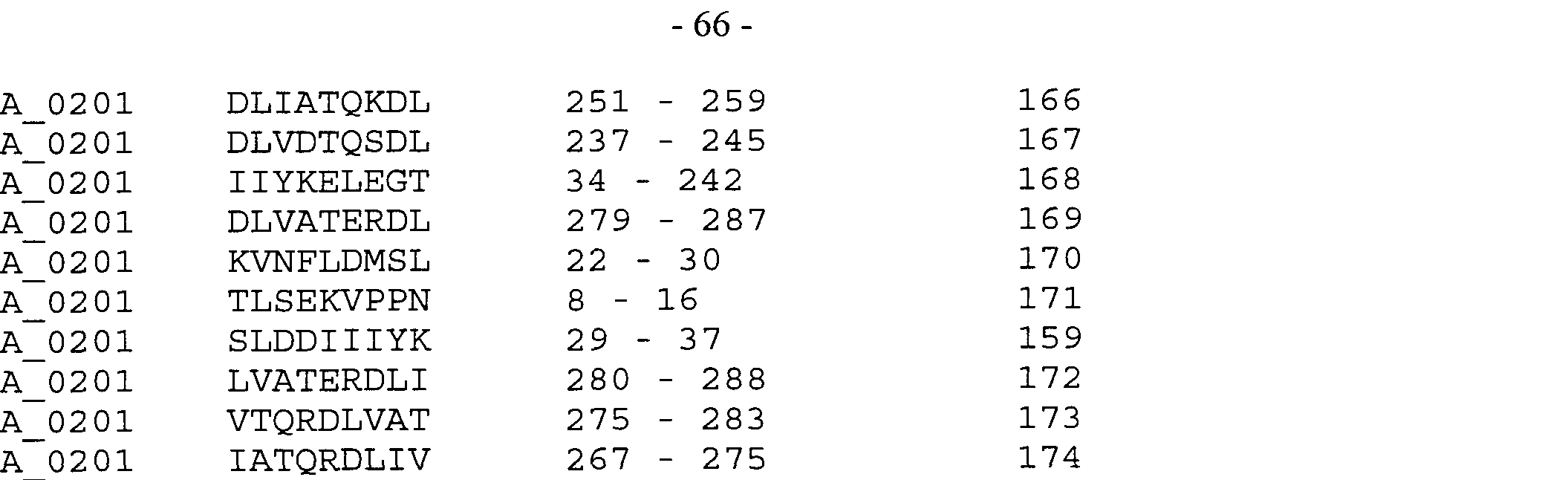
μ-> to H
IO O σi to μ-1
H to t 10 μ-1 co μj 0

to
0
 in
in

Methods for determining HLA class II peptides and making substitutions thereto are also known (e.g. Strominger and Wucherpfennig (PCT/US96/03182)).
Example 11 : Identification of the portion of a cancer associated polypeptide encoding an antigen
To determine if the cancer associated antigens isolated as described above can provoke a cytolytic T lymphocyte response, the following method is performed for generation of CD8+ T cells using the predicted HLA peptides described above. Briefly, peripheral blood mononuclear cells (PBMCs) are isolated using ficoll-Hypaque (Pharmacia) from blood of HLA-A201+ healthy donors (or donors positive for whatever HLA binding peptide is being investigated). Adherent monocytes are separated from non-adherent peripheral blood lymphocytes (PBLs) by plastic adherence via overnight incubation at 37°C on plastic. The non-adherent PBLs are cryopreserved until needed, while adherent cells are stimulated to differentiate into dendritic cells (DCs) by incubation in AEMV-medium (Gibco), containing 1000 U/ml IL4 and 1000 U/ml GM-CSF for 5 days.
On day 7, 8xl05 DCs are loaded with 50mg/ml exogenously added peptide for 2 hours at 37°C in medium containing 1000 U/ml TNFα and 10,000 U/ml ILlβ. The peptide pulsed DCs are then washed twice in excess medium containing no peptide. Autologous PBLs are thawed and 4 xlO7 PBLs are coincubated with 8 x 10⁵ peptide loaded DCs (ratio 50: 1), in medium containing 5ng/ml IL-7 and 20 U/ml IL2. These cultures are then incubated at 37°C.
On days 14, 21 and 28 the lymphocyte cultures are further stimulated with autologous peptide pulsed DC cells as for day 7. Functional assays such as Elispot and cytotoxicity assays are done on days 14, 21 and/or 28. The Elispot assays are performed using commercially available Elispot-system for EFN-γ; NC plates from Millipore; antibodies from Holzel, following manufacturers instructions. The cytotoxicity assays are performed using a Europium labelling based assay which is analogous to chromium release type assay (Blomberg et al., J. Immunol Methods 114:191-195, 1988).
A similar method is performed to determine if the cancer associated antigen contains one or more HLA class II peptides recognized by T cells. One can search the sequence of the cancer associated antigen polypeptides for HLA class II motifs as described above. In contrast to class I peptides, class II peptides are presented by a limited number of cell types. Thus for
these experiments, dendritic cells or B cell clones which express HLA class II molecules preferably are used.
Table 6: Sequence homologies listing GenBank accession numbers
HOM-TES-83 (SEQ ID NO:4)
AF044923, AF045432, U52868, AF032922, U48696, AF039698, U65376, S78798. U66300, AF033565. U44386, Y15421, U39066, Y17148, AF030515, AJ010903, Z97178, AF101425, U4181 1, AC004787, AC004513, AF027174, AF001551 , AC004054, AC004649. AF033096, AB011481 , AC004459. AF061786, AF047464, U37573, AL034583, Z49980, AL009227, AL023823. L06133, Z97066. AF077538, AJ222586. U91522, U96967, L04663. U48697. Z82215, X68367. AF034416. U91521. Z80361. AC004679. AB004546. AB000617. D14824. Z97196. AF000196. U67595, AA159497. W02610, T78470. AA317590. H40247, H77785, T9081 1, AA707590. T25043. H90416, H90470. N84722. AA093861. AA214702. N83992. N55669. N83993, N86694, N84721. N84718, AA263076, AA216240, AA093224. AA094237, AA248551. N84601. N56555, N84733. AA210625. N84712, N83991, N84048. AA247827, AA249064, N55681, AA471 140, N85900, AA471338. N8471 1. N84602. N84874, N89520, N84734, N55717, AA285245, AA089553. T85713. H58760, N84735, N84855, N87989. T78548, N84873, N561 18. N87317. AA089554, N84828. N84561. N84720, N55641. N88601. N84723, N84736, N84830. AA096066, N84714. AA095641, AA096046. N84665. N55698, N84016, N88496. AA248055. N84764, AA248540. AA215911. AA247965. N55658. AA247964. N55684, N88518. N83168. N88782, AA263154, AA249295. N84563. AA090034. AA093897. AA210635. N55697, N55639, N85930. N88643, AA095359, N87898. N55653. N85031. N84569, N84829. N56179. N84300, AA215908, N55675, N83897. AA210634. AA460287, N88760. N84262. H40248, AA094769. N88107, AA247755, AA527091, N86441 , N84562, AA471 132. N71515. N93688. H62375. N55724. N71071. AA962562, AA249232, N63557. H82122. AI263206, W15324, W45549. AI264341 , N84892. N88721. R26158. R38030, AA040091. W72586. AA210636, AA837398, R33839. N75863. N30346. AI253021. T50744. N89732. AA777852. R76796. AA009488. AA210641, Z42580. H21958. H54881. H 14743. H49102. AA 127602, AI 167483. N93281. AI253022. N56396. AA971439. N3151 1. AA938908. A1201960. AA635743. AA063769, AI153154, AA259895. AA221 179, AA 175431. AA222683. Al l 19050. AA510576. AA 163741.
AA271287, AA423048, AA734534, AA163559, AA146103, AU041276, W62646, AI390568. AU020283,
AA760466, AA960429, AU042263, AA655629, AA265339, AI313592, AA208274, AA959259, AI390199. AA623568, C89476, AA793357, AI325590, AA285408, W40959, AA555783, C89109, AA473205, W64733. AA138684, AA474354, AA562556, AA755428, C89043, AI353166, AI354060, AI353694, AI353413, AI353159. AI353169, AA933116, AI235773, AA819471, AA925593, C39845, AA996568, All 12818,
AA819501, AI176018, Z48423, All 1 1865, AI177662, AI180243, AI072388, All 77791, AA933363, AI176043, AI013473, AF041408, C46551, AI060205, AA997478, C29768, AA925635. AI177765, AI178946, AI179929, AI136932, AI103944, C34993, AU008936, D36745, C62715, AI172257, AI231787, All 76821. AI406819. R62064, AI103956, AA998655, AA818207, AI397313, C19512, AI103922, T15173. C65586, AI384895, AI137215. AA851347, AA924928. H07834, AI103571, AA875102. AA996719, AI176754, AI397732,
AI072219, AI138031, AI 170365. AA754038, AA819017, AA95741 1, AA925826, AA818841 , AI01281 1. AI385276, R61938, AA842387. AI030644, AA996678, AI007606, AI014038, AI059391 , AI071253, AI030753, All 13700, All 70891, All 72516, AI392014, AA933448, AA900231 , All 80384, AA842493, AI179593, AI175763, AI045102, AA955950, AA964252, R62026, AA257572, C98703, AA956796, AI043996, Al 175769, Al 137671 , Al 136868, Al 144660, AI045429, Al 177440, AA962989, AI073174, N94700. AIO 13886, AA875549. AI169598, AI045345, AA924877, AI146156, AA965045, All 12960. AA754176, AI068754, AA996517, AI136870, AI045182. AI176687, AI070067. AI070455, AA547871, T00607, All 12152, AA840646, N81438, AA998743, AI178187, AI178690, AA118224. AA933231, C67287, C69931. AI007649, AI137216, AI102951, AA963034. AA660535, A1145091 , AI169162, AI104015, AI176643, AI392367, AA957045, AA933499, AI007651, AI010361. AI071429, AI136544, AI169315, AU005075, AA858479. AA956869, N98171, AA2461 12, AI030919, AI059533, AI073245, AI136600, AI137539, AI137400, AI138001 , AA142295, AI233966, AT001096, AU005198, AI170293, AA925197, AA841342, AI044524, AI137301 , AI029013, AI136171, AI137140, A1007864, AI104309, AI176249, R471 15, AA925652. AI 179399, AA955824. AI 176867, AI176813, AI137442, AI137403, AA675831 , AI072261 , AA999172, AI029010. AI072290. AI176635, AI176263, AA998891, AA964987, AA908025. AU032968, AI385297. L19154.
AA866480. AI392595. AU033109. AI178908. AI177405, AA956780, AI397565. AA933362. AI043755, AI030526, AI044807, AA433148, AA925057, 143668, 102094, E00915, 130479, 140371 , 141023. A08351 , AR018830, A39966, 186849, AR008278. THC205223, THC150248, THC152424, THC179487, THC154497. THC149197, THC214674, THC149159, THC147005, THC149371 , THC213088. THC179408. THC206981 , THC207073. THC173043, THC1 13540, THC172082, THC180340, THC180341, THC207073. THC200913. THC155246, THC202325, THC177377. THC177574, THC177377, THC155246. THC156636, THC176027. THC155246. THC213587. THC93502. THC149538.
HOM-TES-84/86 3' (SEQ ID NO:5)
Z81365, AC002524. AC006204, U80017, D50925, U46753, AC006088. AL031228. Z97356, U79240.
AC004919. AC004130. AC004594. AC002352. Z82076, M74716, AC004841. AC004415. AF030882.
AA909508. N31434. AA084051. AA228062. AA 195129, AI475696.1, AI051291. AA961654. AI291256.
AU051 1 19. AI503662, AA692931. AA549132. AA239122, W77412. AI00661 1. AI006648. AI558238.1. AA145053, AA168206. AI463784. AA276678. AI181898. W29515. AA617497. AA694709. AA950554.
AI100102. AA817713, D34318. R47180. AA875513. D27555, AA696871. AA202154. D64500. AA946427.
AI233726, AI516726, AA850599, AA820767, AI045306, AI543460, AA893725, AI547608, AI102863, AI514445, AA392877, AI543459, AI170351, AI574901.1, AI530959, A60169.1, A46020.1, A46012.1 , 187380. 189868, AR014270, A50831.1, 162304, E02138, 159889, A39300.1, AR014241, A12360.1, A04707.1, 109513, 131 140, A26182.1, A05350.1, A03789.1, A50828.1, A57756.1, 173182, A09995.1, AR014271, AR014269, 173180, A07020.1.
HOM-TES-84/86 51 (SEQ ID NO:6)
U 14567, AC004656, AL009174, M31951. AC004463, AC004143, AC005577, M90058. AC005585, AC004212, AC004997, AC004025. U91323, AL021937, Z98941 , AL009179, AL021 155. AC005972,
AC002365, X82126, AC005099, AL022324, AF030876, Z86064, AF031078, AC002378, AC004677,
AC000090, M l 0065, Z93023, AC005192, AC003037, AL031293, AC004472, AC005803. AD000864,
AC005562, AC004160, AC006163, AC005932, AC003695, U07563, AD000833, AF024533, AC002985.
AF001548, AC006501 , AC004699. AL009181, AC002116, AC004876.2. AC002477, AC003071 , AC004129, AC007014, AC002549, AC005330, AC005295, AC005912, AC002312, AC004125, AC002349. Z49918,
AC000067, AC004084, AC003086, AC004778, AC005899, AC004236, AL022476.2, AC004408, AC002395,
AC005529, AC000379, AC004132, AC002070, AC005211, U47924, AC003684, AC005952. AC005329,
AC002364, Z84484. AL022334, Z68162, AC005781 , AC003010, Z98257, AC005356, AC004983, AC002300,
Z99774, AC004762. AC002369, AC000159, AC004805, U521 12. AC005778, AF1 1 1 168, C87864, W62377, AI430519, AA501297, AU018489, AU019533, AI326216, AI503861 , AA501 128, AA516955. AI413410,
AA501262, W51648, C88193, AA261001, W61986, AI425687, AI042727, W62449, C87438. A1272569,
W77222, AA435247, AI526365, C87922, C88111, AI042721, AI425650, AI046782, C86532. AI042687.
AA470242, AA270527, AA544076, AI551876.1 , C87581 , AI550077.1 , AA537471 , AA 1 14713, AI463886,
C87512, AI550714.1 , AA086548, AA863851 , W09657, AU016622, AA117753, AI506614, AU0431 12, AI413665. AI506213. AI467583. AA71 1962. AI066909, H39328. AI539956, H39389. AI151560, AI218793.
AA550283, AI044039, AI 145414. W06387. H39351, AI41 1496, AI138025. AI 144749, AI549274. AI 176698.
AI 137385. AA964476, AI066064, Al l 1 1838, AA819889, AA818261. AI044651 , AI555357.1. AI058918.
AI180353, AI137218. AI232721, AI234683. AI 104953, AI103758, AI535033. AI071598, AA924075,
AI556467.1 , AI071716, AI145388, C69332, AI547778, AA924214, AI104547, AI013373. AA946023, AA800835, AI233681, AA963340, AA923893, AI237409, T00372, AI556862.1. AI501019, AA923995.
C69739, AA891772. AA997533, D67872, D70097, AA859499, M79850, AI178891, AA946419, AI548486.
AI010741, AI136965, AA893373. AI177088, AI233014, AI4121 19, AI070034, AI232696. D74926, AI535528.
AI549051. AI105188. AI071477, AI045871. AI233888, AA957761 , AI171208, AA817997. AI548785.
AA925900, AI 169541 , AI043787. AI179734, AI556644.1. AI008802. AI170964, AI556772.1. AI548343. AA946366, AI229818. AI146082. A39482.1. A39479.1 , 174786. A52294.1, AR016035, 131750. A 12027.1.
138532, 119138. A62791.1 , A51 133.1. 159730. 140899, 140904, 109371 , A62786.1, A51 132.1. 143100, 143096.
173182, 173246, A62802.1. 159642. AR014241. A51135.1. A62777.1. A47886.1 , 117291. A47885.1, 122254.
122241. 196182, 108667, 155948, 125678, 115157. AR020909. 196203, A48605.1. 166249. 121323. E05293.
AR003505. 173181. 117548, 145974, 11 1727. E01058, 123499. A62792.1. 104664. AR007160. AR007159. 151997. AR0071 18, 131097. A58884.1. A 19035.1. A19036.1. 138891. 134294. 176960. 118513. AR022305.
A65972.1. E12183, A62731.1, A28005.1. AR0001 18. AR0001 13. AR022524. A03929.1. 134189. AR008154.
A45331.1, A42944.1, 124738, E01888, 136306, E02193, 174660, A62929.1, E00140, A38669.1, 138533,
A51 134.1 , A12032.1, E01662, A26236.1 , AR022523, E12964, A28928.1, 131125, 116884, 115767, AR009805,
AR002554, A26415.1, 176967, 10871 1, 108101, A63257.1, 101958, AR016729. 192783, AR009214, 115549,
134187, 196204, AR014571, 162418, A58551.1, A42329.1, A45340.1, 117129, 102034, 108362, 134190. A37262.1, AR011888, 140313, 107269, A45367.1, 116885, 102155, 109076, 117130, 101147. 141409. 147706,
125849, AR022304, 181226, E03351, A37264.1, 109383, 180049, A64383.1, AR016442, E12647, 109380,
A00794.1 , 124432. 104391, E02192, E03829, A64531.1, A64510.1, A64529.1, AR008155, 115970. A25212.1,
E05931, 107993, 105479, A22672.1, 156088, 14141 1, E03350, A62778.1, 180039, 179967, A45356.1. 114076,
189273, A65890.1, 102857, A43445.1 , 108406. 109139, 108349, 107890, E12979, 136934, 140908, 193602, 189344, 166342, 168025, 167702, A43764.1, 128360. 186854, 162750, 167703, A25214.1 , A51 136.1, 162419, 113706, A08862.1, A62780.1, 109337. 1081 10, A62781.1 , 126614, 186415, A62764.1 , A21829.1, 171461. 171463, E12100, E03352, A22938.1 , 176969, 100326, E02302, AR021107, 108032, AR014384, E02588. AR013984, 151756, 143820, AR007149, 187420, A48600.1 , 108636, 105274, 18981 1, A08856.1, 186855, E03600, A65971.1 , A63032.1 , E02252, 165583, 127606, 127609, 182202, 131089. 143668, 140314, A45355.1 , AR014294, A62794.1. A62795.1 , 106826, 196178, 176959, 176970, A41486.1, 109216, 189274, 108764. 126616, A65264.1 , 152002, E08844, AR002262, 105094, A02514.1, 176956, 105724, A62784.1, 160524, 182133, A13669.1, A13672.1 , A62482.1, A37109.1, 101960, 108571, E08652, 186156, 179963, 128845, A01733.1. 136464, A37107.1, 186159, A19638.1.
HOM-TES-87 (SEQ ID NO:7)
AF001548, U95097, AF001501. AF015037, Z36949, X13666, U40270, J03158. U70374, AC004648,
AF049895, U68249, AC005915, AF072268.1, U68072, AC004612, Y07642, U50871, AF068862. AC005373.
Z96810, D64002, M33336, S54705, U18678, AF069291, M18468, AA813001. AA286988, AI457781, AI221417, AI092521, AI278369. AA027065, AA846365, AA766287, AA535104. AA286989. AA909880.
AA644529. AA360087. AI400692.1, AI092126, AI092523. H24090. AA442918. Z42805, R55738. AI536016.
AA843559. AA203108, AA263154, H06476. AA502875. AA247964. N87317. AA332821. N40100. N55684.
AI525171, W22363. AA143570, F02425, AA976582. AA015682. AA089553. AA312215. T83757. AA383441.
H16636, W05105, AI552096.1, AA086744, AI115481, AA014964. AA037969. AA497991. AA530017. AA184072, AA798929, AI483047. AI483356, AI483156, AI408920. Z92686. AI397652, AI483182. AI483326.
A1483209, AI483386, AI483346, AI483139, AI483224, AI397853, AA754092. AI483157. AI483344.
A1483328, AA257755. AI483331. AI483123, C19877. AI483142. AI534191, AI483153, AI483197.
AA754068, AI483126, AA471673, AI398237. AI483150, AI483191, AI483176. AI483162, AI483340,
AA257424. AI483332, AI105522. AI483146. AI392322, AI392465. AI483233. AI228145. AI391994. AI483337, AI483320, AI483342, AI483215, AI483158. AI483190, AA109261. AI483333. AI483148. D39376.
AA818425. AA471641, AI483119, AI483341, C98607, AI483298, AA406690. AI483283. AI483111.
AI483316. C96750, AI483279, AI228978. AI392388. AI398149. AI398196. AI483327, T22798. AI105486.
AI483155, AI483164. AI483220. AI058098. AI483228. AI483261. AA454249. AI392453. AI483347.
AI483175, AI483235, AI353694. AI483225. AI483237. AI483242. AI483263. A1483343. R90674. AA054906. H07815. AI483250, AA430929. AI483312. AI483212. AI012887. AI353159. AA754053. N96914. AI483211.
AI483280. AI397752. AI483137. AI483545. AA257682. AI483373. AI483115. AI483265. All 13274.
AI483185, AI483204, AI483181, AI483205, AI483100, AI483056, AI483335, AI483252, D23855, AI483329,
AI068590, AA752481, H36500, H07829, AI483247, AI483257, AI353413, AI399616, AI483281, 132740.
138469, 177040, A50265.1. A50263.1. 108883, 146766, A38680.1.
HOM-TES-88/94/95 (SEQ ID NO:8)
D50617, D44602, AF071095, AF071097, AE00071 1, U28374, M85293. AF051934, U05676, Z54196. J04815.
AC005966, X17017, U93264. AF049634, AA479513, W19888. AA626631, AA447852, W00613. AI334344,
AA447700, AA150083. AA486299, H82142, H08089, AA085991 , H71626, AA909807, AA888983, AI026772. C05160, AA347780. R61226, AA971016, AA677738, AI004624, AA340276, AI217393, AA130053. T3251 1.
AA337664, R14260, H08090, N54388. C04239, AA897589, AA652646. AA328725, Z28501. Z38367,
AA370106, R96217, AA301061 , Z42140, Z19403. AA371349, T31977. R86231, H54488, R61225. AI086866.
AA479515, N63302, AA973047, AA936602, AA883965, AI263407, Z24943, AI275063, AA458950.
AA 150018, AA923063, AI554276.1. AA236098, AI472144, AA486193, AI287605, Z42097. C02287. D62662. AI082201, AI274243, AA909806, AI565387.1, AA085621, AI141767, AA835001 , T39190. AA622645.
AA458812, H20695, AI241319, T90895, AA886340, T40464, AA377965, AA401704. AA522852, AA225842,
N4941 1, N55669, AA184049, AA 184050, AA154852, AU024524, AA119248, AU023952. AA998195.
H31 1 19, AA848541 , AA848540, AIO 1 1035, AI484782, AI072661 , AI483241 , D32315, D64488. D34006.
T75876, AI483097, D64678, 189392. AR022360. E02219, E08016, 176964, AR018830, E04316. A58381.1. E02869, A12519.1. 173380. E02376.
HOM-TES-103 (SEQ ID NO:9)
Z65613, Z58697, AB010070, AF024654. AB015474, X71428, S75762, X99005, LI 1366. S62138. S62140. Z83219, AB022223, S75763. U 10438. AF071213, AC004266, X71427, AE001006. AF076183. AL032637.
Z97025, AF076184, M32281, AC006585.6. Z75543. AB016882. X95262. AL031369.1. AL009028. M32280.
U19270. Z32687. X90869. U40799. Z48583. Z66559. AC004433, AB003107, AC004755. AF079444,
AF025424, M59318, U89695. L38513. AF006002, Z19158. M60978. D45890.1. X 13595. L38512. U66526.
AF047428, U31 164.1, U25281. U09964. L26320. M95490, Z72004, AC002447. U83230, X81325. X03912. M10058, Z50875, AC000098, AF045571 , L38514, U33175, U7021 1 , U83231. U31159.1, AC005854, U64603.
X56226, X53374, X62716.1 , AA043139, AI096830, AI161033, AI367873. AI423929.1, AA029284,
AA040870, AA707496. AA121381. AI374945. AI216022, AI264487, AI473234.1, AA121575. AI015278.
AA343698. D31076. AI201752, D30927. AA251428, N47018, AA368626. D31203. AI468485. AA635737.
AI247682, AA251427, U25930. AA232601. AA340991 , R35742, AI497627, T80687. H09206. R12342. AA148871. AA781 174, R74490. AA805673. C15931. N69291 , AA195853. AI090561. AI278132. AI393008.1.
AA978015. AI205970. W42886. AI095706. AA749259. H58598. W42792. AA514046. AI242705. H39660.
AI554918.1 , AI554087.1. Tl 1997. AA226982. AA614097, AA714104. AI206080. AA808052. AA582301.
„. .— ,- ΛI ιι-f-τ /Dl II C C\
72/1 AA615677, AA124754, AA870414, AA637162, AA137700, W44164, AA915237, AA590789, AU015748.
AA693246, AU017040, AA914852, AA 197621, W34772, W29410, AI197723, W84035, AA183775,
AA139018, AA387873, AA940503, AA615957, AA116469, AI006420, AU035924, AA387647, AA930164.
AI41 1183, AI575205.1, AI578460.1, AI070580, AI41 1 178, AA924434, AA858819, AI577981.1 , AI170079, All 04361, AI103143, AI070284, All 66371 , All 77669, AI227782, AI069081, C22780, C63872, C94106,
AI502501 , C24819, D48959, C20232, D48988, C74340, AU033205, AU004975. C74221, AI399489,
AI496191. D48372, D41023, C99275. C25189, D49283, C90102, C90097, AU003729, C69236. Z47394,
C41670, AI437843, C44945, All 34261, C43691 , AIO 10961, AI180435. T04717, AI238015, T70653, C43953.
AA697982, AI231905, D73619, C39661 , C47272, E08233. E12702, 116765. A27284.1 , A27258.1 , 150804. 141349, A27276.1 , 150805, A27274.1, 108056, A51776.1, E13193, 160505, A48404.1 , 196207. 123305. 173389.
E07277, A23635.1, 112551, 156080, 134034, 108038, 173380, 114943, 159730. AR007301 , AR021 160, 122065.
HOM-TES-85 (SEQ ID NO: 10)
AF032922, U65376. AJ004935. Y 15421, AF045432. S78798, AF103726. AF039698, U48696. U52868,
U39066, AF033565, AF030515, Y17148, U66300, U44386, AJ010903, Z97178, AF101425, U4181 1,
AF033096. AF027174, Z72514, AF061786, Z49980, U37573, U78082, U32582, X96604, AA471 140,
AA210625, N84601, N83991 , N84712, N84602, N89520, AA471338, AA249064, AA093861, AA094237.
AA093224, N55681, N84733, N8471 1 , N55669, AA214702, N83993, N84874, N84722, N83992. N85900. N56555, AA263076, AA248551, AA216240, N84718, N84721, AA247827, N86694, N84048, N84723,
N84734, N84714, N55698, N84735, N84665, N87317, AA096066, H58760, N84828, N84873. AA248540,
AA089554, AA096046, N55658, N84561, N88782, AA248055, AA215911, N55717, N88496, AA095641 ,
N84736, N55684. N88601. N84830. N84720, AA247965, N55641, N84016, AA247964, N88518. AA089553.
N84764, N84855. N83168, AA285245, N561 18. N87989. N84563, N55653, AA263154, AA249295. N55697. AA093897, N87898, AA095359, N84300, AA210635. N85930, AA090034. N55639. N85031 , AA247755.
N84569, N84829, AA210634, AA215908. N56179. N55675, N83897, T25017, N84262. N86441. AA527091.
N88760, N84562, N88107. H82122, N71594. N93837. AA094769. N88643. W45549. AI263206. R26158.
AA040091 , AA471 132. AA249232. N84892, N88721, R38030, AA837398, AA156784, N7107 I . AA047536.
W15324, AA421059, AA693963. R76796, AA421544. N70806, N93294. AA210636. W76542. W72586. N30346, N62435, AA421082, N55724, AA834824, T27986. N93688. AA022469. AA524123. AA693497.
AI077556, N58190, N48161 , C05801. N72255, N83262, AI241303. T69289, N31060. AA045799. H70027.
AA634087, H54881, W68672, N75202. C 19100. AA412561 , AI309044. AA210641 , W44426, D51 124.
AA423230, AI551609.1 , AI428388, AI467555, AA212810, AA208274. AA561517. Wl 1774. AI573582.1.
AA617092, AA866732. AA755285, AA815727, AA874170. AI050479. AI553236.1. AI353166. AI353169. AI354060, AI353159, AI353694, AI353413. AA9331 16, AI010101. AA946507, AA819377. AA965190.
AI 170254. AI 179306. AI104294, AI 175760. AA901096. AI058297, AI044932. AA752812, AI 179399,
H07769, AI 104356, AA754227, AA933363, AI072813. AI059160, AI 176018, AI180243. AA435473.
AI 137632. AI030526. AA925643. AF041408. AI071 191 , All 1 1701. A1136334. AA088161. AI179890.
AI 1371 18, AI 137686. AA964298. AI 177851 , AI102562. AI072188. AA944363. AA998274. AI072016. AA925983. AA944344. AI 176613, AI045145. D43357. Al l 79918. AI178318. AA406761. AA752761.
Al l 37103, Al l 70346. AI072358, AA842512. AA944465, Al 144718. AI060296. AI0451 12. AA996580.
72/2 AI043714, AI009641, AI138042, AI137138, AI137227, AI176056, AA859564, AA859963, AA901411 ,
D48239, AA109365, AI045829, AI071234, AA109423, AA957307, AA858599, AI065703, AI071422,
AI138012, AI104268, AI100474, AI137107, AA925514, AI137436, AI177791, N28061, AI237589,
AA840999, AA997716, AF083278, D47088, L19151, AI102579, AA901058, AI136747, AI237633. All 13047, AI385354, AI072294, AI029665, AI070626. AI178709, AI508166, AI044277, AI104339, AA803772,
AI044436, AI044641, AA866527, AI398012, AI179446, AA819708, AI177786, AI353750, AA818816,
AI 137813, AI060318, AI072849, AA998866. AA997449, AA925512, AA890966, AA901 138, AI070323,
AI179272, AI387267, All 12438. AI029667. AI137785, AI045056, AI234795. AI072421 , AI144764,
AA875194, AI072480, AA924543, All 1 1659, AI1701 14. AA964744, AI179614, AI060006, AA956716, AI102791 , AA81901 1 , AI392346, AA964530. AA394539, AI029595, AI072676, AA901 150, AA963888, AI071964, AA997431, AI071719, AA849393, AA858789, AA956285, AI177300, AA866250, AI 137956, AI070065, AA997359, AI071540, AA964863. AI052950, AA9571 11 , AA964820, AA900519, AA859635, AF083281, AI229521, AI 180108, AI 179134. AI179052, AI144966, AI 144946, AI137495, All 1 1696, Al l 12050, AI068434, AI044264. AI045957. AI398048, AI01 1837, AI009615. AA997586, AA 161655, AI 136235, AI177046, AI 137067. AI 178243. AI397741. AA956309. AA926254, AA924987, AA925962, AA875580, AA817956, AI385176, AI236652, AI1771 16, AI105189, AA963669. AA818196, AI137645, AI030627, AI045239. AI409867, AA925257, AA875071 , AA858759, AI398332, AI172515, AI171211, D471 19, AI103909, AI102007, AI 137954, AI012599, AA818888, AI045508, AI236198, AI072043, AI406899, AA900194, AA900054, AI072293, AA818985, AI178550, AI177916, AA900186. AI 169730, AI 137897, AI137765, AI072664, AA964780, AI136884, AI071804, All 12389, AI045758, AI044489, AI044248, AA852040, AI137060, AA944487, AA754182. AI137091, AI179451, AI175912, AI175779, AI029369, AI137726, AI 144819, AI145347, AI059283, AI 170350, AI 175907, AI137276, AI 104035, AI178762, AI 177290, AI229494, AI 180251. AI253806. AA924156, AI385307. AA509328, AA952317. AI 136623, AI072383. AA859469, AI058604, AI044993, AI030774, AA963992. AI009143, AA964269, AA957874, AA965599, AA754215, Z29190, D39884, AA819165, AA998593, AA858856. AI 166854, AI012876,
AI234100, AI180399. AI 136429. All 1 1529. AI069945, AI066048. AI059814, AA874993, D47170, AI398022. AA955886, AI044301, AI045697, AA924441. AI180364, AA875218. AI102947. AI 137521. AI 136662, AI136629, AI136376, Al l 12210. All 12188. AI058963, AI070205. AI029238. AA999324. AI 136934, AA924125, AA072471 , AA956675, AI 137226. AA955424. AA924539, AA900367. AI104107. AI389074, AI 175985, AI172614. AI 170355. AI103247. AI 144861, AI 144669. AI398143. AI070514, AI070362,
AI052952, AI028947, AI399523, AI043605, A1043686, AI404137. C83596. C82740, AA161657, E03585. 186415. E12936, AR008277. 168296, AR025455, A19976. 181219. A19977, A45655, 186389. 144902, AR008281 , THC205223, THC150248, THC179487. THC149197. THC147005. THC152424. THC 154497, THC149159, THC214674, THC149371, THC213088, THC179408. THC206981. THC155886. THC212609. THC 159576, THC136194, THC165367, THC165368.
Claims
1. A method of diagnosing a disorder characterized by expression of a human cancer associated antigen precursor coded for by a nucleic acid molecule, comprising: contacting a biological sample isolated from a subject with an agent that specifically binds to the nucleic acid molecule, an expression product thereof, or a fragment of an expression product thereof complex ed with an HLA molecule, wherein the nucleic acid molecule is a NA Group 1 nucleic acid molecule, and determining the interaction between the agent and the nucleic acid molecule or the expression product as a determination of the disorder.
2. The method of claim 1 , wherein the agent is selected from the group consisting of
(a) a nucleic acid molecule comprising NA group 1 nucleic acid molecules or a fragment thereof,
(b) a nucleic acid molecule comprising NA group 3 nucleic acid molecules or a fragment thereof,
(c) a nucleic acid molecule comprising NA group 5 nucleic acid molecules or a fragment thereof,
(d) an antibody that binds to an expression product of NA group 1 nucleic acids,
(e) an antibody that binds to an expression product of NA group 3 nucleic acids, (f) an antibody that binds to an expression product of NA group 5 nucleic acids,
(g) an agent that binds to a complex of an HLA molecule and a fragment of an expression product of a NA group 1 nucleic acid,
(h) an agent that binds to a complex of an HLA molecule and a fragment of an expression product of a NA group 3 nucleic acid, and (i) an agent that binds to a complex of an HLA molecule and a fragment of an expression product of a NA group 5 nucleic acid.
3. The method of claim 1 , wherein the disorder is characterized by expression of a plurality of human cancer associated antigen precursors and wherein the agent is a plurality of agents, each of which is specific for a different human cancer associated antigen precursor, and wherein said plurality of agents is at least 2, at least 3, at least 4, at least 4, at least 6, at least 7, or at least 8, at least 9 or at least 10 such agents. - 74 -
4. The method of claims 1-3, wherein the agent is specific for a human cancer associated antigen precursor that is a seminoma cancer associated antigen precursor.
5. A method for determining regression, progression or onset of a condition characterized by expression of abnormal levels of a protein encoded by a nucleic acid molecule that is a NA
Group 1 molecule, comprising monitoring a sample, from a patient who has or is suspected of having the condition, for a parameter selected from the group consisting of (i) the protein, (ii) a peptide derived from the protein,
(iii) an antibody which selectively binds the protein or peptide, and (iv) cytolytic T cells specific for a complex of the peptide derived from the protein and an MHC molecule, as a determination of regression, progression or onset of said condition.
6. The method of claim 5, wherein the sample is a body fluid, a body effusion or a tissue.
7. The method of claim 5, wherein the step of monitoring comprises contacting the sample with a detectable agent selected from the group consisting of (a) an antibody which selectively binds the protein of (i), or the peptide of (ii),
(b) a protein or peptide which binds the antibody of (iii), and
(c) a cell which presents the complex of the peptide and MHC molecule of (iv).
8. The method of claim 7, wherein the antibody, the protein, the peptide or the cell is labeled with a radioactive label or an enzyme.
9. The method of claim 5, comprising assaying the sample for the peptide.
10. The method of claim 5, wherein the nucleic acid molecule is a NA Group 3 molecule.
11. The method of claim 5, wherein the nucleic acid molecule is a NA Group 5 molecule. - 75 -
12. The method of claim 5, wherein the protein is a plurality of proteins, the parameter is a plurality of parameters, each of the plurality of parameters being specific for a different of the plurality of proteins, at least one of which is a cancer associated protein encoded by a NA Group 1 molecule.
13. A pharmaceutical preparation for a human subj ect comprising an agent which when administered to the subject enriches selectively the presence of complexes of an HLA molecule and a human cancer associated antigen, and a pharmaceutically acceptable carrier, wherein the human cancer associated antigen is a fragment of a human cancer associated antigen precursor encoded by a nucleic acid molecule comprises a NA Group 1 molecule.
14. The pharmaceutical preparation of claim 13, wherein the agent comprises a plurality of agents, each of which enriches selectively in the subject complexes of an HLA molecule and a different human cancer associated antigen, wherein at least one of the human cancer associated antigens is encoded by a NA Group 1 molecule.
15. The pharmaceutical preparation of claim 14, wherein the plurality is at least two, at least three, at least four or at least 5 different such agents.
16. The pharmaceutical preparation of claim 13, wherein the nucleic acid molecule is a NA Group 3 nucleic acid molecule.
17. The pharmaceutical preparation of claim 13, wherein the agent is selected from the group consisting of
(1) an isolated polypeptide comprising the human cancer associated antigen, or a functional variant thereof,
(2) an isolated nucleic acid operably linked to a promoter for expressing the isolated polypeptide, or functional variant thereof, (3) a host cell expressing the isolated polypeptide, or functional variant thereof, and
(4) isolated complexes of the polypeptide, or functional variant thereof, and an HLA molecule. - 76 -
18. The pharmaceutical preparation of claims 13-17, further comprising an adjuvant.
19. The pharmaceutical preparation of claim 13, wherein the agent is a cell expressing an isolated polypeptide comprising the human cancer associated antigen or a functional variant thereof, and wherein the cell is nonproliferative.
20. The pharmaceutical preparation of claim 13, wherein the agent is a cell expressing an isolated polypeptide comprising the human cancer associated antigen or a functional variant thereof, and wherein the cell expresses an HLA molecule that binds the polypeptide.
21. The pharmaceutical preparation of claim 13, wherein the agent is at least two, at least three, at least four or at least five different polypeptides, each coding for a different human cancer associated antigen or functional variant thereof, wherein at least one of the human cancer associated antigens is encoded by a NA Group 1 molecule.
22. The pharmaceutical preparation of claim 13, wherein the agent is a PP Group 2 polypeptide.
23. The pharmaceutical preparation of claim 13, wherein the agent is a PP Group 3 polypeptide or a PP Group 4 polypeptide.
24. The pharmaceutical preparation of claim 20, wherein the cell expresses one or both of the polypeptide and HLA molecule recombinantly.
25. The pharmaceutical preparation of claim 20, wherein the cell is nonproliferative.
26. A composition of matter comprising an isolated agent that binds selectively a PP Group 1 polypeptide.
27. The composition of matter of claim 26, wherein the agent binds selectively a PP Group 2 polypeptide. - 77 -
28. The composition of matter of claim 26, wherein the agent binds selectively a PP Group 3 polypeptide.
29. The composition of matter of claim 26, wherein the agent binds selectively a PP Group 4 polypeptide.
30. The composition of matter of claim 26, wherein the agent binds selectively a PP Group 5 polypeptide.
31. The composition of matter of claims 26-30, wherein the agent is a plurality of different agents that bind selectively at least two, at least three, at least four, or at least five different such polypeptides.
32. The composition of matter of claims 26-30, wherein the agent is an antibody.
33. The composition of matter of claim 31, wherein the agent is an antibody.
34. A composition of matter comprising a conjugate of the agent of claims 26-30 and a therapeutic or diagnostic agent.
35. A composition of matter comprising a conjugate of the agent of claim 31 and a therapeutic or diagnostic agent.
36. The composition of matter of claim 34, wherein the conjugate is of the agent and a therapeutic or diagnostic that is a toxin.
37. A pharmaceutical composition comprising an isolated nucleic acid molecule selected from the group consisting of NA Group 1 molecules and NA Group 2 molecules, and a pharmaceutically acceptable carrier.
38. The pharmaceutical composition of claim 37, wherein the isolated nucleic acid molecule comprises a NA Group 3 or NA Group 4 molecule. - 78 -
39. The pharmaceutical composition of claim 37, wherein the isolated nucleic acid molecule comprises at least two isolated nucleic acid molecules coding for two different polypeptides, each polypeptide comprising a different human cancer associated antigen.
40. The pharmaceutical composition of claims 37-39 further comprising an expression vector with a promoter operably linked to the isolated nucleic acid molecule.
41. The pharmaceutical composition of claims 37-39 further comprising a host cell recombinantly expressing the isolated nucleic acid molecule.
42. A pharmaceutical composition comprising an isolated polypeptide comprising a PP Group 1 or a PP Group 2 polypeptide, and a pharmaceutically acceptable carrier.
43. The pharmaceutical composition of claim 42, wherein the isolated polypeptide comprises a PP Group 3 or a PP Group 4 polypeptide.
44. The pharmaceutical composition of claim 42, wherein the isolated polypeptide comprises at least two different polypeptides, each comprising a different human cancer associated antigen.
45. The pharmaceutical composition of claims 42-44, further comprising an adjuvant.
46. An isolated nucleic acid molecule comprising a NA Group 3 molecule.
47. An isolated nucleic acid molecule comprising a NA Group 4 molecule.
48. An isolated nucleic acid molecule selected from the group consisting of
(a) a fragment of a nucleic acid molecule having a nucleotide sequence selected from the group consisting of nucleotide sequences set forth as SEQ ED NOs:4-10, of sufficient length to represent a sequence unique within the human genome, and identifying a nucleic acid encoding a human cancer associated antigen precursor, - 79 -
(b) complements of (a), provided that the fragment includes a sequence of contiguous nucleotides which is not identical to any sequence selected from the sequence group consisting of
(1) sequences having the GenBank accession numbers of Table 6, (2) complements of (1), and
(3) fragments of (1) and (2).
49. The isolated nucleic acid molecule of claim 48, wherein the sequence of contiguous nucleotides is selected from the group consisting of: (1) at least two contiguous nucleotides nonidentical to the sequence group,
(2) at least three contiguous nucleotides nonidentical to the sequence group,
(3) at least four contiguous nucleotides nonidentical to the sequence group,
(4) at least five contiguous nucleotides nonidentical to the sequence group,
(5) at least six contiguous nucleotides nonidentical to the sequence group, (6) at least seven contiguous nucleotides nonidentical to the sequence group.
50. The isolated nucleic acid molecule of claim 48, wherein the fragment has a size selected from the group consisting of at least: 8 nucleotides, 10 nucleotides, 12 nucleotides, 14 nucleotides, 16 nucleotides, 18 nucleotides, 20, nucleotides, 22 nucleotides, 24 nucleotides, 26 nucleotides, 28 nucleotides, 30 nucleotides, 50 nucleotides, 75 nucleotides, 100 nucleotides, and 200 nucleotides.
51. The isolated nucleic acid molecule of claim 48, wherein the molecule encodes a polypeptide which, or a fragment of which, binds a human HLA receptor or a human antibody.
52. An expression vector comprising an isolated nucleic acid molecule of any of claims 46-51 operably linked to a promoter.
53. An expression vector comprising a nucleic acid operably linked to a promoter, wherein the nucleic acid is a NA Group 2 molecule. - 80 -
54. An expression vector comprising a NA Group 1 or Group 2 molecule and a nucleic acid encoding an HLA molecule.
55. A host cell transformed or transfected with an expression vector of claim 52.
56. A host cell transformed or transfected with an expression vector of claims 53 or 54.
57. A host cell transformed or transfected with an expression vector of claim 52 and further comprising a nucleic acid encoding HLA.
58. A host cell transformed or transfected with an expression vector of claim 53 and further comprising a nucleic acid encoding HLA.
59. An isolated polypeptide encoded by the isolated nucleic acid molecule of claim 46 or claim 47.
60. A fragment of the polypeptide of claim 59 which is immunogenic.
61. The fragment of claim 60, wherein the fragment, or a portion of the fragment, binds HLA or a human antibody.
62. An isolated fragment of a human cancer associated antigen precursor which, or portion of which, binds HLA or a human antibody, wherein the precursor is encoded by a nucleic acid molecule that is a NA Group 1 molecule.
63. The fragment of claim 62, wherein the fragment is part of a complex with HLA.
64. The fragment of claim 63, wherein the fragment is between 8 and 12 amino acids in length.
65. An isolated polypeptide comprising a fragment of the polypeptide of claim 59 of sufficient length to represent a sequence unique within the human genome and identifying a - 81 - polypeptide that is a human cancer associated antigen precursor.
66. A kit for detecting the presence of the expression of a human cancer associated antigen precursor comprising a pair of isolated nucleic acid molecules each of which consists essentially of a molecule selected from the group consisting of (a) a 12-32 nucleotide contiguous segment of the nucleotide sequence of any of the NA Group 1 molecules and (b) complements of ("a"), wherein the contiguous segments are nonoverlapping.
67. The kit of claim 66, wherein the pair of isolated nucleic acid molecules is constructed and arranged to selectively amplify an isolated nucleic acid molecule that is a NA Group 3 molecule.
68. A method for treating a subject with a disorder characterized by expression of a human cancer associated antigen precursor, comprising administering to the subject an amount of an agent, which enriches selectively in the subject the presence of complexes of an HLA molecule and a human cancer associated antigen, effective to ameliorate the disorder, wherein the human cancer associated antigen is a fragment of a human cancer associated antigen precursor encoded by a nucleic acid molecule selected from the group consisting of
(a) a nucleic acid molecule comprising NA group 1 nucleic acid molecules,
(b) a nucleic acid molecule comprising NA group 3 nucleic acid molecules,
(c) a nucleic acid molecule comprising NA group 5 nucleic acid molecules.
69. The method of claim 68, wherein the disorder is characterized by expression of a plurality of human cancer associated antigen precursors and wherein the agent is a plurality of agents, each of which enriches selectively in the subject the presence of complexes of an HLA molecule and a different human cancer associated antigen, wherein at least one of the human cancer associated antigens is encoded by a NA Group 1 molecule.
70. The method of claim 69, wherein the plurality is at least 2, at least 3, at least 4, or at least 5 such agents. - 82 -
71. The method of claims 68-70, wherein the agent is an isolated polypeptide selected from the group consisting of PP Group 1, PP Group 2, PP Group 3, PP Group 4 and PP Group 5 polypeptides.
72. The method of claims 68-70, wherein the disorder is cancer.
73. The method of claims 71, wherein the disorder is cancer.
74. A method for treating a subject having a condition characterized by expression of a human cancer associated antigen precursor in cells of the subject, comprising:
(i) removing an immunoreactive cell containing sample from the subject, (ii) contacting the immunoreactive cell containing sample to the host cell under conditions favoring production of cytolytic T cells against a human cancer associated antigen which is a fragment of the precursor, (iii) introducing the cytolytic T cells to the subject in an amount effective to lyse cells which express the human cancer associated antigen, wherein the host cell is transformed or transfected with an expression vector comprising an isolated nucleic acid molecule operably linked to a promoter, the isolated nucleic acid molecule being selected from the group of nucleic acid molecules consisting of NA Group 1, NA Group 2, NA Group 3, NA Group 4, and NA Group 5.
75. The method of claim 74, wherein the host cell recombinantly expresses an HLA molecule which binds the human cancer associated antigen.
76. The method of claim 74, wherein the host cell endogenously expresses an HLA molecule which binds the human cancer associated antigen.
77. A method for treating a subject having a condition characterized by expression of a human cancer associated antigen precursor in cells of the subject, comprising: (i) identifying a nucleic acid molecule expressed by the cells associated with said condition, wherein said nucleic acid molecule is a NA Group 1 molecule;
(ii) transfecting a host cell with a nucleic acid selected from the group consisting - 83 - of (a) the nucleic acid molecule identified, (b) a fragment of the nucleic acid identified which includes a segment coding for a human cancer associated antigen, (c) deletions, substitutions or additions to (a) or (b), and (d) degenerates of (a), (b), or (c);
(iii) culturing said transfected host cells to express the transfected nucleic acid molecule, and;
(iv) introducing an amount of said host cells or an extract thereof to the subject effective to increase an immune response against the cells of the subject associated with the condition.
78. The method of claim 77, further comprising identifying an MHC molecule which presents a portion of an expression product of the nucleic acid molecule, wherein the host cell expresses the same MHC molecule as identified and wherein the host cell presents an MHC binding portion of the expression product of the nucleic acid molecule.
79. The method of claim 77, wherein the immune response comprises a B-cell response or a T cell response.
80. The method of claim 79, wherein the response is a T-cell response which comprises generation of cytolytic T-cells specific for the host cells presenting the portion of the expression product of the nucleic acid molecule or cells of the subject expressing the human cancer associated antigen.
81. The method of claim 77, wherein the nucleic acid molecule is a NA Group 3 molecule.
82. The method of claims 77 or 78, further comprising treating the host cells to render them non-proliferative.
83. A method for treating or diagnosing or monitoring a subject having a condition characterized by expression of an abnormal amount of a protein encoded by a nucleic acid molecule that is a NA Group 1 molecule, comprising administering to the subject an antibody which specifically binds to the protein or a peptide derived therefrom, the antibody being coupled to a therapeutically useful agent, in an - 84 - amount effective to treat the condition.
84. The method of claim 83, wherein the antibody is a monoclonal antibody.
85. The method of claim 84, wherein the monoclonal antibody is a chimeric antibody or a humanized antibody.
86. A method for treating a condition characterized by expression in a subject of abnormal amounts of a protein encoded by a nucleic acid molecule that is a NA Group 1 nucleic acid molecule, comprising administering to a subject a pharmaceutical composition of any one of claims 13-25 and 37-45 in an amount effective to prevent, delay the onset of, or inhibit the condition in the subject.
87. The method of claim 86, wherein the condition is cancer.
88. The method of claim 86, further comprising first identifying that the subject expresses in a tissue abnormal amounts of the protein.
89. The method of claim 87, further comprising first identifying that the subject expresses in a tissue abnormal amounts of the protein.
90. A method for treating a subject having a condition characterized by expression of abnormal amounts of a protein encoded by a nucleic acid molecule that is a NA Group 1 nucleic acid molecule, comprising
(i) identifying cells from the subject which express abnormal amounts of the protein;
(ii) isolating a sample of the cells;
(iii) cultivating the cells, and
(iv) introducing the cells to the subject in an amount effective to provoke an immune response against the cells.
91. The method of claim 90, further comprising rendering the cells non-proliferative, prior - 85 - to introducing them to the subject.
92. A method for treating a pathological cell condition characterized by aberrant expression of a protein encoded by a nucleic acid molecule that is a NA Group 1 nucleic acid molecule, comprising administering to a subject in need thereof an effective amount of an agent which inhibits the expression or activity of the protein.
93. The method of claim 92, wherein the agent is an inhibiting antibody which selectively binds to the protein and wherein the antibody is a monoclonal antibody, a chimeric antibody or a humanized antibody.
94. The method of claim 92, wherein the agent is an antisense nucleic acid molecule which selectively binds to the nucleic acid molecule which encodes the protein.
95. The method of claim 92, wherein the nucleic acid molecule is a NA Group 3 nucleic acid molecule.
96. A composition of matter useful in stimulating an immune response to a plurality of a proteins encoded by nucleic acid molecules that are NA Group 1 molecules, comprising a plurality of peptides derived from the amino acid sequences of the proteins, wherein the peptides bind to one or more MHC molecules presented on the surface of the cells which express an abnormal amount of the protein.
97. The composition of matter of claim 96, wherein at least a portion of the plurality of peptides bind to MHC molecules and elicit a cytolytic response thereto.
98. The composition of matter of claim 97, further comprising an adjuvant.
99. The composition of matter of claim 98, wherein said adjuvant is a saponin, GM-CSF, or an interleukin. - 86 -
100. The composition of matter of claim 96, further comprising at least one peptide useful in stimulating an immune response to at least one protein which is not encoded by nucleic acid molecules that are NA Group 1 molecules, wherein the at least one peptide binds to one or more MHC molecules.
101. An isolated antibody which selectively binds to a complex of:
(i) a peptide derived from a protein encoded by a nucleic acid molecule that is a NA Group 1 molecule and
(ii) and an MHC molecule to which binds the peptide to form the complex, wherein the isolated antibody does not bind to (i) or (ii) alone.
102. The antibody of claim 101, wherein the antibody is a monoclonal antibody, a chimeric antibody, a humanized antibody, or a fragment thereof.
103. A method for making a nucleic acid or polypeptides encoded thereby comprising culturing the host cell of claim 55 or claim 56 and isolating the nucleic acid or polypeptide from the host cells or culture medium.
104. A method for making a nucleic acid or polypeptide encoded thereby comprising providing a non-cell system for transcription and/or translation of a nucleic acid, introducing the nucleic acid of any of claims 46-48 or the expression vector of claim 52 into the non-cell system, incubating the non-cell system under conditions sufficient for transcription or translation of the nucleic acid of any of claims 46-48 or the expression vector of claim 52 and isolating the transcibed nucleic acid or translated polypeptide from the non-cell system.
Priority Applications (3)
| Application Number | Priority Date | Filing Date | Title |
|---|---|---|---|
| JP2001506864A JP2003527826A (en) | 1999-06-30 | 2000-06-23 | Cancer-associated antigens and uses thereof |
| AU56325/00A AU5632500A (en) | 1999-06-30 | 2000-06-23 | Cancer associated antigens and uses therefor |
| EP00941644A EP1218538A2 (en) | 1999-06-30 | 2000-06-23 | Cancer associated antigens and uses therefor |
Applications Claiming Priority (2)
| Application Number | Priority Date | Filing Date | Title |
|---|---|---|---|
| US34649899A | 1999-06-30 | 1999-06-30 | |
| US09/346,498 | 1999-06-30 |
Publications (2)
| Publication Number | Publication Date |
|---|---|
| WO2001000874A2 true WO2001000874A2 (en) | 2001-01-04 |
| WO2001000874A3 WO2001000874A3 (en) | 2002-05-02 |
Family
ID=23359678
Family Applications (1)
| Application Number | Title | Priority Date | Filing Date |
|---|---|---|---|
| PCT/US2000/017207 WO2001000874A2 (en) | 1999-06-30 | 2000-06-23 | Cancer associated antigens and uses therefor |
Country Status (4)
| Country | Link |
|---|---|
| EP (1) | EP1218538A2 (en) |
| JP (1) | JP2003527826A (en) |
| AU (1) | AU5632500A (en) |
| WO (1) | WO2001000874A2 (en) |
Cited By (4)
| Publication number | Priority date | Publication date | Assignee | Title |
|---|---|---|---|---|
| EP1390387A2 (en) * | 2001-04-20 | 2004-02-25 | Ludwig Institute For Cancer Research | Cancer-testis antigens |
| EP1399478A2 (en) * | 2001-03-30 | 2004-03-24 | Ludwig Institute For Cancer Research | Cancer-testis antigens |
| JP2009060910A (en) * | 2001-04-06 | 2009-03-26 | Mannkind Corp | Epitope sequences |
| EP2190870A1 (en) * | 2007-08-28 | 2010-06-02 | Auckland Uniservices Limited | Cell marker of melanocyte cell lineage and uses thereof |
Citations (9)
| Publication number | Priority date | Publication date | Assignee | Title |
|---|---|---|---|---|
| US4816567A (en) | 1983-04-08 | 1989-03-28 | Genentech, Inc. | Recombinant immunoglobin preparations |
| WO1992004381A1 (en) | 1990-09-11 | 1992-03-19 | Scotgen Limited | Novel antibodies for treatment and prevention of infection in animals and man |
| US5225539A (en) | 1986-03-27 | 1993-07-06 | Medical Research Council | Recombinant altered antibodies and methods of making altered antibodies |
| WO1996010577A1 (en) | 1994-09-30 | 1996-04-11 | Ludwig Institute For Cancer Research | Isolated nucleic acid molecule coding for tumor rejection antigen precursor dage and uses thereof |
| WO1996033265A1 (en) | 1995-04-21 | 1996-10-24 | President And Fellows Of Harvard College | In vitro differentiation of cd34+ progenitor cells into t lymphocytes |
| US5580723A (en) | 1988-10-28 | 1996-12-03 | Genetech, Inc. | Method for identifying active domains and amino acid residues in polypeptides and hormone variants |
| US5585089A (en) | 1988-12-28 | 1996-12-17 | Protein Design Labs, Inc. | Humanized immunoglobulins |
| US5698396A (en) | 1995-06-07 | 1997-12-16 | Ludwig Institute For Cancer Research | Method for identifying auto-immunoreactive substances from a subject |
| US5859205A (en) | 1989-12-21 | 1999-01-12 | Celltech Limited | Humanised antibodies |
-
2000
- 2000-06-23 JP JP2001506864A patent/JP2003527826A/en active Pending
- 2000-06-23 WO PCT/US2000/017207 patent/WO2001000874A2/en active Application Filing
- 2000-06-23 EP EP00941644A patent/EP1218538A2/en not_active Withdrawn
- 2000-06-23 AU AU56325/00A patent/AU5632500A/en not_active Abandoned
Patent Citations (10)
| Publication number | Priority date | Publication date | Assignee | Title |
|---|---|---|---|---|
| US4816567A (en) | 1983-04-08 | 1989-03-28 | Genentech, Inc. | Recombinant immunoglobin preparations |
| US5225539A (en) | 1986-03-27 | 1993-07-06 | Medical Research Council | Recombinant altered antibodies and methods of making altered antibodies |
| US5580723A (en) | 1988-10-28 | 1996-12-03 | Genetech, Inc. | Method for identifying active domains and amino acid residues in polypeptides and hormone variants |
| US5585089A (en) | 1988-12-28 | 1996-12-17 | Protein Design Labs, Inc. | Humanized immunoglobulins |
| US5693762A (en) | 1988-12-28 | 1997-12-02 | Protein Design Labs, Inc. | Humanized immunoglobulins |
| US5859205A (en) | 1989-12-21 | 1999-01-12 | Celltech Limited | Humanised antibodies |
| WO1992004381A1 (en) | 1990-09-11 | 1992-03-19 | Scotgen Limited | Novel antibodies for treatment and prevention of infection in animals and man |
| WO1996010577A1 (en) | 1994-09-30 | 1996-04-11 | Ludwig Institute For Cancer Research | Isolated nucleic acid molecule coding for tumor rejection antigen precursor dage and uses thereof |
| WO1996033265A1 (en) | 1995-04-21 | 1996-10-24 | President And Fellows Of Harvard College | In vitro differentiation of cd34+ progenitor cells into t lymphocytes |
| US5698396A (en) | 1995-06-07 | 1997-12-16 | Ludwig Institute For Cancer Research | Method for identifying auto-immunoreactive substances from a subject |
Non-Patent Citations (33)
| Title |
|---|
| "Current Protocols in Molecular Biology", JOHN WILEY & SONS |
| "Current Protocols in Molecular Biology", JOHN WILEY & SONS, INC. |
| "Molecular Cloning: A Laboratory Manual", 1989, COLD SPRING HARBOR LABORATORY PRESS |
| ALLSOPP ET AL., EUR. J IMMUNOL., vol. 26, no. 8, 1996, pages 1951 - 1959 |
| ALTMAN ET AL., SCIENCE, vol. 274, 1996, pages 94 - 96 |
| BLOMBERG ET AL., J IMMUNOL METHODS, vol. 114, 1988, pages 191 - 195 |
| CLARK, W.R.: "The Experimental Foundations of Modem Immunology", 1986, WILEY & SONS, INC. |
| COULIE, STEM CELLS, vol. 13, 1995, pages 393 - 403 |
| DAHIYAT; MAYO, SCIENCE, vol. 278, 1997, pages 82 - 87 |
| D'AMARO ET AL., HUMAN IMMUNOL., vol. 43, 1995, pages 13 - 18 |
| DEMOULIN, MOL. CELL. BIOL., vol. 16, 1996, pages 4710 - 4716 |
| DRIJFHOUT ET AL., HUMAN IMMUNOL, vol. 43, no. 1-12, 1995 |
| DUNBAR ET AL., CURR. BIOL., vol. 8, 1998, pages 413 - 416 |
| GILBERT ET AL., NATURE BIOTECHNOL., vol. 15, 1997, pages 1280 - 1284 |
| GILBERT ET AL., NATURE BIOTECHNOL., vol. 15, no. 12, 1997, pages 1280 - 1284 |
| GREENBERG, J., TMMUNOL, vol. 136, no. 5, 1986, pages 1917 |
| INT. J. CANCER, vol. 67, 1996, pages 303 - 310 |
| J. CLIN. INVEST., vol. 90, 1992, pages 626 - 630 |
| KAST ET AL., CELL, vol. 59, 1989, pages 603 - 614 |
| LYNCH ET AL., EUR. J IMMUNOL., vol. 21, 1991, pages 1403 - 1410 |
| MISHIZUMA; NAGATA, NUC. ACIDS RES., vol. 18, 1990, pages 5322 |
| RIDDEL ET AL., SCIENCE, vol. 257, 1992, pages 238 |
| ROITT, I: "Essential Immunology", 1991, BLACKWELL SCIENTIFIC PUBLICATIONS |
| SAHIN ET AL., PROC. NATL. ACAD. SCI., vol. 92, 1995, pages 11810 - 11813 |
| SAINIO, CELL MOL. NEUROBIOL., vol. 14, no. 5, 1994, pages 439 - 457 |
| SAMBROOK ET AL.: "Molecular Cloning: A Laboratory Manual", 1989, COLD SPRING HARBOR LABORATORY PRESS |
| SCIARRA; CUTIE: "Remington's Pharmaceutical Sciences", 1990, article "Aerosols", pages: 1694 - 1712 |
| TAM ET AL., J. EXP. MED., vol. 171, no. 1, 1990, pages 299 - 306 |
| THOMSON ET AL., J. IMMUNOL., vol. 157, no. 2, 1996, pages 822 - 826 |
| THOMSON ET AL., PROC. ACAD. NATL. ACAD. SCI, vol. 92, no. 13, 1995, pages 5845 - 5849 |
| THOMSON ET AL., PROC. NATL. ACAD. SCI., vol. 92, 1995, pages 5845 - 5849 |
| TIIRECI ET AL., MOL. MED. TODAY |
| WAGNER ET AL., NATURE BIOTECHNOL., vol. 14, 1996, pages 840 - 844 |
Cited By (7)
| Publication number | Priority date | Publication date | Assignee | Title |
|---|---|---|---|---|
| EP1399478A2 (en) * | 2001-03-30 | 2004-03-24 | Ludwig Institute For Cancer Research | Cancer-testis antigens |
| EP1399478A4 (en) * | 2001-03-30 | 2005-09-28 | Ludwig Inst Cancer Res | Cancer-testis antigens |
| JP2009060910A (en) * | 2001-04-06 | 2009-03-26 | Mannkind Corp | Epitope sequences |
| EP1390387A2 (en) * | 2001-04-20 | 2004-02-25 | Ludwig Institute For Cancer Research | Cancer-testis antigens |
| EP1390387A4 (en) * | 2001-04-20 | 2004-12-08 | Ludwig Inst Cancer Res | Cancer-testis antigens |
| EP2190870A1 (en) * | 2007-08-28 | 2010-06-02 | Auckland Uniservices Limited | Cell marker of melanocyte cell lineage and uses thereof |
| EP2190870A4 (en) * | 2007-08-28 | 2010-12-01 | Auckland Uniservices Ltd | Cell marker of melanocyte cell lineage and uses thereof |
Also Published As
| Publication number | Publication date |
|---|---|
| AU5632500A (en) | 2001-01-31 |
| JP2003527826A (en) | 2003-09-24 |
| EP1218538A2 (en) | 2002-07-03 |
| WO2001000874A3 (en) | 2002-05-02 |
Similar Documents
| Publication | Publication Date | Title |
|---|---|---|
| EP1073734B1 (en) | Tumor associated nucleic acids and uses therefor | |
| US7314721B2 (en) | Small cell lung cancer associated antigens and uses therefor | |
| EP0996857B1 (en) | Cancer associated nucleic acids and polypeptides | |
| US20070128655A1 (en) | Breast, gastric and prostate cancer associated antigens and uses therefor | |
| JP2003518364A (en) | Breast, gastric and prostate cancer-related antigens and uses thereof | |
| EP1117791A2 (en) | Cancer associated antigens and uses therefor | |
| US20110177079A1 (en) | Cancer-testis antigens | |
| US6686147B1 (en) | Cancer associated antigens and uses therefor | |
| US20030180298A1 (en) | Cancer-testis antigens | |
| US20030044813A1 (en) | Cancer-testis antigens | |
| US8247379B2 (en) | Tumor associated amino acids and uses therefor | |
| US6303756B1 (en) | Tumor associated nucleic acids and uses therefor | |
| WO2001000874A2 (en) | Cancer associated antigens and uses therefor | |
| EP1077989A1 (en) | Tumor associated antigen encoded by the reverse strand of a new ubiquitously expressed gene | |
| WO2001040271A2 (en) | Cancer associated antigens and uses therefor | |
| US20040086852A1 (en) | Cancer associated antigens and uses therefor | |
| WO2000072021A2 (en) | Cancer associated antigens and uses therefor | |
| WO2000020586A2 (en) | Renal cancer associated antigens and uses therefor | |
| AU2002306975A1 (en) | Cancer-testis antigens |
Legal Events
| Date | Code | Title | Description |
|---|---|---|---|
| AK | Designated states |
Kind code of ref document: A2 Designated state(s): AU CA CN JP KR |
|
| AL | Designated countries for regional patents |
Kind code of ref document: A2 Designated state(s): AT BE CH CY DE DK ES FI FR GB GR IE IT LU MC NL PT SE |
|
| 121 | Ep: the epo has been informed by wipo that ep was designated in this application | ||
| DFPE | Request for preliminary examination filed prior to expiration of 19th month from priority date (pct application filed before 20040101) | ||
| WWE | Wipo information: entry into national phase |
Ref document number: 2000941644 Country of ref document: EP |
|
| WWP | Wipo information: published in national office |
Ref document number: 2000941644 Country of ref document: EP |