WO1997031019A2 - Antipathogenic peptides and compositions comprising them - Google Patents
Antipathogenic peptides and compositions comprising them Download PDFInfo
- Publication number
- WO1997031019A2 WO1997031019A2 PCT/IL1997/000066 IL9700066W WO9731019A2 WO 1997031019 A2 WO1997031019 A2 WO 1997031019A2 IL 9700066 W IL9700066 W IL 9700066W WO 9731019 A2 WO9731019 A2 WO 9731019A2
- Authority
- WO
- WIPO (PCT)
- Prior art keywords
- leu
- lys
- peptide
- peptides
- amino acid
- Prior art date
Links
- 108090000765 processed proteins & peptides Proteins 0.000 title claims abstract description 424
- 102000004196 processed proteins & peptides Human genes 0.000 title claims abstract description 218
- 239000000203 mixture Substances 0.000 title claims description 38
- 230000001775 anti-pathogenic effect Effects 0.000 title description 8
- 230000001461 cytolytic effect Effects 0.000 claims abstract description 92
- 230000002949 hemolytic effect Effects 0.000 claims abstract description 90
- 108010036176 Melitten Proteins 0.000 claims abstract description 88
- CTVQQQPWNOVEAG-QDOPKCMFSA-N pardaxin Chemical compound C([C@@H](C(=O)N[C@@H](C)C(=O)N[C@@H](CC(C)C)C(=O)N[C@H](C(=O)N1CCC[C@H]1C(=O)N[C@@H](CCCCN)C(=O)N[C@@H]([C@@H](C)CC)C(=O)N[C@@H]([C@@H](C)CC)C(=O)N[C@@H](CO)C(=O)N[C@@H](CO)C(=O)N1[C@@H](CCC1)C(=O)N[C@@H](CC(C)C)C(=O)N[C@@H](CC=1C=CC=CC=1)C(=O)N[C@@H](CCCCN)C(=O)N[C@@H]([C@@H](C)O)C(=O)N[C@@H](CC(C)C)C(=O)N[C@@H](CC(C)C)C(=O)N[C@@H](CO)C(=O)N[C@@H](C)C(=O)N[C@@H](C(C)C)C(=O)NCC(=O)N[C@@H](CO)C(=O)N[C@@H](C)C(=O)N[C@@H](CC(C)C)C(=O)N[C@@H](CO)C(=O)N[C@@H](CO)C(=O)N[C@@H](CO)C(=O)NCC(=O)NCC(=O)N[C@@H](CCC(N)=O)C(=O)N[C@@H](CCC(O)=O)C(O)=O)[C@@H](C)CC)NC(=O)[C@H](CC=1C=CC=CC=1)NC(=O)CN)C1=CC=CC=C1 CTVQQQPWNOVEAG-QDOPKCMFSA-N 0.000 claims abstract description 68
- 150000008574 D-amino acids Chemical group 0.000 claims abstract description 50
- 150000001413 amino acids Chemical class 0.000 claims abstract description 48
- 230000002209 hydrophobic effect Effects 0.000 claims abstract description 27
- 150000008575 L-amino acids Chemical group 0.000 claims abstract description 26
- 125000000539 amino acid group Chemical group 0.000 claims abstract description 25
- 125000004122 cyclic group Chemical group 0.000 claims abstract description 13
- 239000012634 fragment Substances 0.000 claims abstract description 12
- 244000052769 pathogen Species 0.000 claims abstract description 9
- 206010028980 Neoplasm Diseases 0.000 claims abstract description 8
- 201000011510 cancer Diseases 0.000 claims abstract description 8
- 239000008194 pharmaceutical composition Substances 0.000 claims abstract description 8
- 241000204031 Mycoplasma Species 0.000 claims abstract description 4
- VDXZNPDIRNWWCW-JFTDCZMZSA-N melittin Chemical compound NCC(=O)N[C@@H]([C@@H](C)CC)C(=O)NCC(=O)N[C@@H](C)C(=O)N[C@@H](C(C)C)C(=O)N[C@@H](CC(C)C)C(=O)N[C@@H](CCCCN)C(=O)N[C@@H](C(C)C)C(=O)N[C@@H](CC(C)C)C(=O)N[C@@H]([C@@H](C)O)C(=O)N[C@@H]([C@@H](C)O)C(=O)NCC(=O)N[C@@H](CC(C)C)C(=O)N1CCC[C@H]1C(=O)N[C@@H](C)C(=O)N[C@@H](CC(C)C)C(=O)N[C@@H]([C@@H](C)CC)C(=O)N[C@@H](CO)C(=O)N[C@H](C(=O)N[C@@H]([C@@H](C)CC)C(=O)N[C@@H](CCCCN)C(=O)N[C@@H](CCCNC(N)=N)C(=O)N[C@@H](CCCCN)C(=O)N[C@@H](CCCNC(N)=N)C(=O)N[C@@H](CCC(N)=O)C(=O)N[C@@H](CCC(N)=O)C(N)=O)CC1=CNC2=CC=CC=C12 VDXZNPDIRNWWCW-JFTDCZMZSA-N 0.000 claims description 92
- 210000004027 cell Anatomy 0.000 claims description 75
- 241000894006 Bacteria Species 0.000 claims description 50
- 235000001014 amino acid Nutrition 0.000 claims description 47
- 229940024606 amino acid Drugs 0.000 claims description 47
- KDXKERNSBIXSRK-UHFFFAOYSA-N Lysine Natural products NCCCCC(N)C(O)=O KDXKERNSBIXSRK-UHFFFAOYSA-N 0.000 claims description 33
- 210000003743 erythrocyte Anatomy 0.000 claims description 32
- KDXKERNSBIXSRK-YFKPBYRVSA-N L-lysine Chemical compound NCCCC[C@H](N)C(O)=O KDXKERNSBIXSRK-YFKPBYRVSA-N 0.000 claims description 30
- ROHFNLRQFUQHCH-YFKPBYRVSA-N L-leucine Chemical compound CC(C)C[C@H](N)C(O)=O ROHFNLRQFUQHCH-YFKPBYRVSA-N 0.000 claims description 28
- 230000001717 pathogenic effect Effects 0.000 claims description 26
- 230000009089 cytolysis Effects 0.000 claims description 21
- 125000003440 L-leucyl group Chemical group O=C([*])[C@](N([H])[H])([H])C([H])([H])C(C([H])([H])[H])([H])C([H])([H])[H] 0.000 claims description 15
- 239000004472 Lysine Substances 0.000 claims description 15
- ROHFNLRQFUQHCH-UHFFFAOYSA-N Leucine Natural products CC(C)CC(N)C(O)=O ROHFNLRQFUQHCH-UHFFFAOYSA-N 0.000 claims description 14
- 206010057248 Cell death Diseases 0.000 claims description 13
- QIVBCDIJIAJPQS-VIFPVBQESA-N L-tryptophane Chemical compound C1=CC=C2C(C[C@H](N)C(O)=O)=CNC2=C1 QIVBCDIJIAJPQS-VIFPVBQESA-N 0.000 claims description 12
- QIVBCDIJIAJPQS-UHFFFAOYSA-N Tryptophan Natural products C1=CC=C2C(CC(N)C(O)=O)=CNC2=C1 QIVBCDIJIAJPQS-UHFFFAOYSA-N 0.000 claims description 12
- 125000003178 carboxy group Chemical group [H]OC(*)=O 0.000 claims description 12
- ROHFNLRQFUQHCH-RXMQYKEDSA-N D-leucine Chemical compound CC(C)C[C@@H](N)C(O)=O ROHFNLRQFUQHCH-RXMQYKEDSA-N 0.000 claims description 8
- DHMQDGOQFOQNFH-UHFFFAOYSA-N Glycine Chemical compound NCC(O)=O DHMQDGOQFOQNFH-UHFFFAOYSA-N 0.000 claims description 8
- -1 aminoethylamino groups Chemical group 0.000 claims description 8
- 210000000170 cell membrane Anatomy 0.000 claims description 8
- 241000233866 Fungi Species 0.000 claims description 7
- ONIBWKKTOPOVIA-UHFFFAOYSA-N Proline Natural products OC(=O)C1CCCN1 ONIBWKKTOPOVIA-UHFFFAOYSA-N 0.000 claims description 6
- 241000700605 Viruses Species 0.000 claims description 6
- ONIBWKKTOPOVIA-BYPYZUCNSA-N L-Proline Chemical compound OC(=O)[C@@H]1CCCN1 ONIBWKKTOPOVIA-BYPYZUCNSA-N 0.000 claims description 5
- QNAYBMKLOCPYGJ-REOHCLBHSA-N L-alanine Chemical compound C[C@H](N)C(O)=O QNAYBMKLOCPYGJ-REOHCLBHSA-N 0.000 claims description 5
- KZSNJWFQEVHDMF-BYPYZUCNSA-N L-valine Chemical compound CC(C)[C@H](N)C(O)=O KZSNJWFQEVHDMF-BYPYZUCNSA-N 0.000 claims description 5
- KZSNJWFQEVHDMF-UHFFFAOYSA-N Valine Natural products CC(C)C(N)C(O)=O KZSNJWFQEVHDMF-UHFFFAOYSA-N 0.000 claims description 5
- 235000004279 alanine Nutrition 0.000 claims description 5
- 125000003636 chemical group Chemical group 0.000 claims description 5
- 229920005604 random copolymer Polymers 0.000 claims description 5
- 239000004474 valine Substances 0.000 claims description 5
- 239000004475 Arginine Substances 0.000 claims description 4
- 229930182819 D-leucine Natural products 0.000 claims description 4
- 239000004471 Glycine Substances 0.000 claims description 4
- ODKSFYDXXFIFQN-UHFFFAOYSA-N arginine Natural products OC(=O)C(N)CCCNC(N)=N ODKSFYDXXFIFQN-UHFFFAOYSA-N 0.000 claims description 4
- 230000003211 malignant effect Effects 0.000 claims description 4
- 238000006386 neutralization reaction Methods 0.000 claims description 4
- ODKSFYDXXFIFQN-BYPYZUCNSA-P L-argininium(2+) Chemical compound NC(=[NH2+])NCCC[C@H]([NH3+])C(O)=O ODKSFYDXXFIFQN-BYPYZUCNSA-P 0.000 claims description 3
- HNDVDQJCIGZPNO-YFKPBYRVSA-N L-histidine Chemical compound OC(=O)[C@@H](N)CC1=CN=CN1 HNDVDQJCIGZPNO-YFKPBYRVSA-N 0.000 claims description 3
- 239000003937 drug carrier Substances 0.000 claims description 3
- HNDVDQJCIGZPNO-UHFFFAOYSA-N histidine Natural products OC(=O)C(N)CC1=CN=CN1 HNDVDQJCIGZPNO-UHFFFAOYSA-N 0.000 claims description 3
- MTCFGRXMJLQNBG-REOHCLBHSA-N (2S)-2-Amino-3-hydroxypropansäure Chemical compound OC[C@H](N)C(O)=O MTCFGRXMJLQNBG-REOHCLBHSA-N 0.000 claims description 2
- DCXYFEDJOCDNAF-UHFFFAOYSA-N Asparagine Natural products OC(=O)C(N)CC(N)=O DCXYFEDJOCDNAF-UHFFFAOYSA-N 0.000 claims description 2
- XUJNEKJLAYXESH-REOHCLBHSA-N L-Cysteine Chemical compound SC[C@H](N)C(O)=O XUJNEKJLAYXESH-REOHCLBHSA-N 0.000 claims description 2
- DCXYFEDJOCDNAF-REOHCLBHSA-N L-asparagine Chemical compound OC(=O)[C@@H](N)CC(N)=O DCXYFEDJOCDNAF-REOHCLBHSA-N 0.000 claims description 2
- ZDXPYRJPNDTMRX-VKHMYHEASA-N L-glutamine Chemical compound OC(=O)[C@@H](N)CCC(N)=O ZDXPYRJPNDTMRX-VKHMYHEASA-N 0.000 claims description 2
- AGPKZVBTJJNPAG-WHFBIAKZSA-N L-isoleucine Chemical compound CC[C@H](C)[C@H](N)C(O)=O AGPKZVBTJJNPAG-WHFBIAKZSA-N 0.000 claims description 2
- FFEARJCKVFRZRR-BYPYZUCNSA-N L-methionine Chemical compound CSCC[C@H](N)C(O)=O FFEARJCKVFRZRR-BYPYZUCNSA-N 0.000 claims description 2
- COLNVLDHVKWLRT-QMMMGPOBSA-N L-phenylalanine Chemical compound OC(=O)[C@@H](N)CC1=CC=CC=C1 COLNVLDHVKWLRT-QMMMGPOBSA-N 0.000 claims description 2
- AYFVYJQAPQTCCC-GBXIJSLDSA-N L-threonine Chemical compound C[C@@H](O)[C@H](N)C(O)=O AYFVYJQAPQTCCC-GBXIJSLDSA-N 0.000 claims description 2
- OUYCCCASQSFEME-QMMMGPOBSA-N L-tyrosine Chemical compound OC(=O)[C@@H](N)CC1=CC=C(O)C=C1 OUYCCCASQSFEME-QMMMGPOBSA-N 0.000 claims description 2
- MTCFGRXMJLQNBG-UHFFFAOYSA-N Serine Natural products OCC(N)C(O)=O MTCFGRXMJLQNBG-UHFFFAOYSA-N 0.000 claims description 2
- AYFVYJQAPQTCCC-UHFFFAOYSA-N Threonine Natural products CC(O)C(N)C(O)=O AYFVYJQAPQTCCC-UHFFFAOYSA-N 0.000 claims description 2
- 239000004473 Threonine Substances 0.000 claims description 2
- 235000009582 asparagine Nutrition 0.000 claims description 2
- 229960001230 asparagine Drugs 0.000 claims description 2
- XUJNEKJLAYXESH-UHFFFAOYSA-N cysteine Natural products SCC(N)C(O)=O XUJNEKJLAYXESH-UHFFFAOYSA-N 0.000 claims description 2
- 235000018417 cysteine Nutrition 0.000 claims description 2
- 230000003247 decreasing effect Effects 0.000 claims description 2
- ZDXPYRJPNDTMRX-UHFFFAOYSA-N glutamine Natural products OC(=O)C(N)CCC(N)=O ZDXPYRJPNDTMRX-UHFFFAOYSA-N 0.000 claims description 2
- 235000004554 glutamine Nutrition 0.000 claims description 2
- 208000015181 infectious disease Diseases 0.000 claims description 2
- AGPKZVBTJJNPAG-UHFFFAOYSA-N isoleucine Natural products CCC(C)C(N)C(O)=O AGPKZVBTJJNPAG-UHFFFAOYSA-N 0.000 claims description 2
- 229960000310 isoleucine Drugs 0.000 claims description 2
- 229930182817 methionine Natural products 0.000 claims description 2
- 235000006109 methionine Nutrition 0.000 claims description 2
- COLNVLDHVKWLRT-UHFFFAOYSA-N phenylalanine Natural products OC(=O)C(N)CC1=CC=CC=C1 COLNVLDHVKWLRT-UHFFFAOYSA-N 0.000 claims description 2
- 235000004400 serine Nutrition 0.000 claims description 2
- 235000008521 threonine Nutrition 0.000 claims description 2
- OUYCCCASQSFEME-UHFFFAOYSA-N tyrosine Natural products OC(=O)C(N)CC1=CC=C(O)C=C1 OUYCCCASQSFEME-UHFFFAOYSA-N 0.000 claims description 2
- LIWMQSWFLXEGMA-WDSKDSINSA-N Ala-Val Chemical compound CC(C)[C@@H](C(O)=O)NC(=O)[C@H](C)N LIWMQSWFLXEGMA-WDSKDSINSA-N 0.000 claims 1
- 230000000844 anti-bacterial effect Effects 0.000 abstract description 85
- 208000037265 diseases, disorders, signs and symptoms Diseases 0.000 abstract description 7
- 230000036961 partial effect Effects 0.000 abstract description 6
- 201000010099 disease Diseases 0.000 abstract description 4
- VDXZNPDIRNWWCW-UHFFFAOYSA-N melitten Chemical compound NCC(=O)NC(C(C)CC)C(=O)NCC(=O)NC(C)C(=O)NC(C(C)C)C(=O)NC(CC(C)C)C(=O)NC(CCCCN)C(=O)NC(C(C)C)C(=O)NC(CC(C)C)C(=O)NC(C(C)O)C(=O)NC(C(C)O)C(=O)NCC(=O)NC(CC(C)C)C(=O)N1CCCC1C(=O)NC(C)C(=O)NC(CC(C)C)C(=O)NC(C(C)CC)C(=O)NC(CO)C(=O)NC(C(=O)NC(C(C)CC)C(=O)NC(CCCCN)C(=O)NC(CCCNC(N)=N)C(=O)NC(CCCCN)C(=O)NC(CCCNC(N)=N)C(=O)NC(CCC(N)=O)C(=O)NC(CCC(N)=O)C(N)=O)CC1=CNC2=CC=CC=C12 VDXZNPDIRNWWCW-UHFFFAOYSA-N 0.000 abstract description 2
- 206010017533 Fungal infection Diseases 0.000 abstract 1
- 206010028470 Mycoplasma infections Diseases 0.000 abstract 1
- 208000031888 Mycoses Diseases 0.000 abstract 1
- 208000010362 Protozoan Infections Diseases 0.000 abstract 1
- 208000036142 Viral infection Diseases 0.000 abstract 1
- 230000002538 fungal effect Effects 0.000 abstract 1
- 230000003612 virological effect Effects 0.000 abstract 1
- 239000012528 membrane Substances 0.000 description 60
- WTJKGGKOPKCXLL-RRHRGVEJSA-N phosphatidylcholine Chemical compound CCCCCCCCCCCCCCCC(=O)OC[C@H](COP([O-])(=O)OCC[N+](C)(C)C)OC(=O)CCCCCCCC=CCCCCCCCC WTJKGGKOPKCXLL-RRHRGVEJSA-N 0.000 description 49
- 230000000694 effects Effects 0.000 description 35
- 150000003904 phospholipids Chemical class 0.000 description 35
- 241000588724 Escherichia coli Species 0.000 description 24
- ZWZWYGMENQVNFU-UHFFFAOYSA-N Glycerophosphorylserin Natural products OC(=O)C(N)COP(O)(=O)OCC(O)CO ZWZWYGMENQVNFU-UHFFFAOYSA-N 0.000 description 24
- 230000015572 biosynthetic process Effects 0.000 description 24
- TZCPCKNHXULUIY-RGULYWFUSA-N 1,2-distearoyl-sn-glycero-3-phosphoserine Chemical compound CCCCCCCCCCCCCCCCCC(=O)OC[C@H](COP(O)(=O)OC[C@H](N)C(O)=O)OC(=O)CCCCCCCCCCCCCCCCC TZCPCKNHXULUIY-RGULYWFUSA-N 0.000 description 23
- 238000002474 experimental method Methods 0.000 description 23
- 238000003786 synthesis reaction Methods 0.000 description 20
- 150000002632 lipids Chemical class 0.000 description 19
- 125000001176 L-lysyl group Chemical group [H]N([H])[C@]([H])(C(=O)[*])C([H])([H])C([H])([H])C([H])([H])C(N([H])[H])([H])[H] 0.000 description 17
- 230000001580 bacterial effect Effects 0.000 description 17
- 230000003993 interaction Effects 0.000 description 16
- OKKJLVBELUTLKV-UHFFFAOYSA-N Methanol Chemical compound OC OKKJLVBELUTLKV-UHFFFAOYSA-N 0.000 description 15
- 239000004098 Tetracycline Substances 0.000 description 14
- 210000004962 mammalian cell Anatomy 0.000 description 14
- 229960002180 tetracycline Drugs 0.000 description 14
- 229930101283 tetracycline Natural products 0.000 description 14
- 235000019364 tetracycline Nutrition 0.000 description 14
- JPOKAKNGULMYHZ-UILVTTEASA-N (2s)-6-amino-2-[[(2s)-2-[[(2s)-2-[[(2s)-2-[[(2s)-6-amino-2-[[(2s)-2-[[(2s)-6-amino-2-[[(2s)-6-amino-2-[[(2s)-2-amino-5-(diaminomethylideneamino)pentanoyl]amino]hexanoyl]amino]hexanoyl]amino]-3-(4-hydroxyphenyl)propanoyl]amino]hexanoyl]amino]-3-(4-hydroxyp Chemical compound C([C@@H](C(=O)N[C@@H](CCCN=C(N)N)C(=O)N[C@@H](CCCN=C(N)N)C(=O)N[C@@H](CCCCN)C(N)=O)NC(=O)[C@H](CCCCN)NC(=O)[C@H](CC=1C=CC(O)=CC=1)NC(=O)[C@H](CCCCN)NC(=O)[C@H](CCCCN)NC(=O)[C@@H](N)CCCN=C(N)N)C1=CC=C(O)C=C1 JPOKAKNGULMYHZ-UILVTTEASA-N 0.000 description 13
- 238000011534 incubation Methods 0.000 description 13
- 150000003522 tetracyclines Chemical class 0.000 description 13
- RZVAJINKPMORJF-UHFFFAOYSA-N Acetaminophen Chemical compound CC(=O)NC1=CC=C(O)C=C1 RZVAJINKPMORJF-UHFFFAOYSA-N 0.000 description 12
- IAZDPXIOMUYVGZ-UHFFFAOYSA-N Dimethylsulphoxide Chemical compound CS(C)=O IAZDPXIOMUYVGZ-UHFFFAOYSA-N 0.000 description 12
- 230000004071 biological effect Effects 0.000 description 12
- 239000000872 buffer Substances 0.000 description 12
- ATBOMIWRCZXYSZ-XZBBILGWSA-N [1-[2,3-dihydroxypropoxy(hydroxy)phosphoryl]oxy-3-hexadecanoyloxypropan-2-yl] (9e,12e)-octadeca-9,12-dienoate Chemical compound CCCCCCCCCCCCCCCC(=O)OCC(COP(O)(=O)OCC(O)CO)OC(=O)CCCCCCC\C=C\C\C=C\CCCCC ATBOMIWRCZXYSZ-XZBBILGWSA-N 0.000 description 11
- AWUCVROLDVIAJX-UHFFFAOYSA-N alpha-glycerophosphate Natural products OCC(O)COP(O)(O)=O AWUCVROLDVIAJX-UHFFFAOYSA-N 0.000 description 11
- 239000012466 permeate Substances 0.000 description 11
- 108060003100 Magainin Proteins 0.000 description 10
- YFHLIDBAPTWLGU-CTKMSOPVSA-N dermaseptin Chemical compound C([C@@H](C(=O)N[C@@H](C)C(=O)NCC(=O)N[C@@H](CCCCN)C(=O)N[C@@H](C)C(=O)N[C@@H](C)C(=O)N[C@@H](CC(C)C)C(=O)NCC(=O)N[C@@H](C)C(=O)N[C@@H](C)C(=O)N[C@@H](C)C(=O)N[C@@H](CC(O)=O)C(=O)N[C@H](C(=O)N[C@@H]([C@@H](C)CC)C(=O)N[C@@H](CO)C(=O)N[C@@H](CCC(N)=O)C(=O)NCC(=O)N[C@@H]([C@@H](C)O)C(=O)N[C@@H](CCC(N)=O)C(O)=O)[C@@H](C)O)NC(=O)[C@H](CC(C)C)NC(=O)[C@H](C)NC(=O)[C@H](CCSC)NC(=O)[C@@H](NC(=O)CNC(=O)[C@H](CC(C)C)NC(=O)[C@H](CCCCN)NC(=O)[C@H](CCCCN)NC(=O)[C@H](CC(C)C)NC(=O)[C@H](CCSC)NC(=O)[C@@H](NC(=O)[C@H](CCCCN)NC(=O)[C@H](CC=1C2=CC=CC=C2NC=1)NC(=O)[C@H](CC(C)C)NC(=O)[C@H](C)N)[C@@H](C)O)[C@@H](C)O)C1=CN=CN1 YFHLIDBAPTWLGU-CTKMSOPVSA-N 0.000 description 10
- 108090000454 dermaseptin Proteins 0.000 description 10
- 230000007246 mechanism Effects 0.000 description 10
- 206010018910 Haemolysis Diseases 0.000 description 9
- FAPWRFPIFSIZLT-UHFFFAOYSA-M Sodium chloride Chemical compound [Na+].[Cl-] FAPWRFPIFSIZLT-UHFFFAOYSA-M 0.000 description 9
- RHQDFWAXVIIEBN-UHFFFAOYSA-N Trifluoroethanol Chemical compound OCC(F)(F)F RHQDFWAXVIIEBN-UHFFFAOYSA-N 0.000 description 9
- 238000009792 diffusion process Methods 0.000 description 9
- 238000001493 electron microscopy Methods 0.000 description 9
- 230000008588 hemolysis Effects 0.000 description 9
- 238000000034 method Methods 0.000 description 9
- 239000003910 polypeptide antibiotic agent Substances 0.000 description 9
- 230000003389 potentiating effect Effects 0.000 description 9
- 210000002966 serum Anatomy 0.000 description 9
- 239000000243 solution Substances 0.000 description 9
- RTZKZFJDLAIYFH-UHFFFAOYSA-N Diethyl ether Chemical compound CCOCC RTZKZFJDLAIYFH-UHFFFAOYSA-N 0.000 description 8
- HVYWMOMLDIMFJA-DPAQBDIFSA-N cholesterol Chemical compound C1C=C2C[C@@H](O)CC[C@]2(C)[C@@H]2[C@@H]1[C@@H]1CC[C@H]([C@H](C)CCCC(C)C)[C@@]1(C)CC2 HVYWMOMLDIMFJA-DPAQBDIFSA-N 0.000 description 8
- 238000001142 circular dichroism spectrum Methods 0.000 description 8
- 231100000433 cytotoxic Toxicity 0.000 description 8
- 230000001472 cytotoxic effect Effects 0.000 description 8
- 230000002101 lytic effect Effects 0.000 description 8
- 238000005192 partition Methods 0.000 description 8
- 229920001184 polypeptide Polymers 0.000 description 8
- 239000011347 resin Substances 0.000 description 8
- 229920005989 resin Polymers 0.000 description 8
- 238000004007 reversed phase HPLC Methods 0.000 description 8
- KRHYYFGTRYWZRS-UHFFFAOYSA-N Fluorane Chemical compound F KRHYYFGTRYWZRS-UHFFFAOYSA-N 0.000 description 7
- JZNWSCPGTDBMEW-UHFFFAOYSA-N Glycerophosphorylethanolamin Natural products NCCOP(O)(=O)OCC(O)CO JZNWSCPGTDBMEW-UHFFFAOYSA-N 0.000 description 7
- ZMXDDKWLCZADIW-UHFFFAOYSA-N N,N-Dimethylformamide Chemical compound CN(C)C=O ZMXDDKWLCZADIW-UHFFFAOYSA-N 0.000 description 7
- 108010067973 Valinomycin Proteins 0.000 description 7
- FCFNRCROJUBPLU-UHFFFAOYSA-N compound M126 Natural products CC(C)C1NC(=O)C(C)OC(=O)C(C(C)C)NC(=O)C(C(C)C)OC(=O)C(C(C)C)NC(=O)C(C)OC(=O)C(C(C)C)NC(=O)C(C(C)C)OC(=O)C(C(C)C)NC(=O)C(C)OC(=O)C(C(C)C)NC(=O)C(C(C)C)OC1=O FCFNRCROJUBPLU-UHFFFAOYSA-N 0.000 description 7
- 230000003013 cytotoxicity Effects 0.000 description 7
- 231100000135 cytotoxicity Toxicity 0.000 description 7
- 238000013461 design Methods 0.000 description 7
- 150000008104 phosphatidylethanolamines Chemical class 0.000 description 7
- 239000011148 porous material Substances 0.000 description 7
- 238000002360 preparation method Methods 0.000 description 7
- 235000018102 proteins Nutrition 0.000 description 7
- 102000004169 proteins and genes Human genes 0.000 description 7
- 108090000623 proteins and genes Proteins 0.000 description 7
- 238000010791 quenching Methods 0.000 description 7
- 230000000171 quenching effect Effects 0.000 description 7
- 238000001228 spectrum Methods 0.000 description 7
- FCFNRCROJUBPLU-DNDCDFAISA-N valinomycin Chemical compound CC(C)[C@@H]1NC(=O)[C@H](C)OC(=O)[C@@H](C(C)C)NC(=O)[C@@H](C(C)C)OC(=O)[C@H](C(C)C)NC(=O)[C@H](C)OC(=O)[C@@H](C(C)C)NC(=O)[C@@H](C(C)C)OC(=O)[C@H](C(C)C)NC(=O)[C@H](C)OC(=O)[C@@H](C(C)C)NC(=O)[C@@H](C(C)C)OC1=O FCFNRCROJUBPLU-DNDCDFAISA-N 0.000 description 7
- XLYOFNOQVPJJNP-UHFFFAOYSA-N water Substances O XLYOFNOQVPJJNP-UHFFFAOYSA-N 0.000 description 7
- WEVYAHXRMPXWCK-UHFFFAOYSA-N Acetonitrile Chemical compound CC#N WEVYAHXRMPXWCK-UHFFFAOYSA-N 0.000 description 6
- 241000194107 Bacillus megaterium Species 0.000 description 6
- 125000000151 cysteine group Chemical group N[C@@H](CS)C(=O)* 0.000 description 6
- 231100000673 dose–response relationship Toxicity 0.000 description 6
- 229910000040 hydrogen fluoride Inorganic materials 0.000 description 6
- 230000001965 increasing effect Effects 0.000 description 6
- 230000002401 inhibitory effect Effects 0.000 description 6
- 238000011084 recovery Methods 0.000 description 6
- 239000000725 suspension Substances 0.000 description 6
- 108050004290 Cecropin Proteins 0.000 description 5
- DTQVDTLACAAQTR-UHFFFAOYSA-N Trifluoroacetic acid Chemical compound OC(=O)C(F)(F)F DTQVDTLACAAQTR-UHFFFAOYSA-N 0.000 description 5
- 238000004458 analytical method Methods 0.000 description 5
- 230000000845 anti-microbial effect Effects 0.000 description 5
- 239000003795 chemical substances by application Substances 0.000 description 5
- 229940049701 dermaseptin Drugs 0.000 description 5
- 239000003814 drug Substances 0.000 description 5
- 230000005284 excitation Effects 0.000 description 5
- 239000007850 fluorescent dye Substances 0.000 description 5
- 244000005700 microbiome Species 0.000 description 5
- 230000000717 retained effect Effects 0.000 description 5
- 239000002904 solvent Substances 0.000 description 5
- 241000894007 species Species 0.000 description 5
- IJGRMHOSHXDMSA-UHFFFAOYSA-N Atomic nitrogen Chemical compound N#N IJGRMHOSHXDMSA-UHFFFAOYSA-N 0.000 description 4
- 241001441579 Pardachirus marmoratus Species 0.000 description 4
- AOILQMZPNLUXCM-AVGNSLFASA-N Val-Val-Lys Chemical compound CC(C)[C@H](N)C(=O)N[C@@H](C(C)C)C(=O)N[C@H](C(O)=O)CCCCN AOILQMZPNLUXCM-AVGNSLFASA-N 0.000 description 4
- 238000002835 absorbance Methods 0.000 description 4
- 230000002378 acidificating effect Effects 0.000 description 4
- 230000009471 action Effects 0.000 description 4
- 125000003368 amide group Chemical group 0.000 description 4
- 230000000843 anti-fungal effect Effects 0.000 description 4
- 238000003556 assay Methods 0.000 description 4
- 230000003115 biocidal effect Effects 0.000 description 4
- 210000004369 blood Anatomy 0.000 description 4
- 239000008280 blood Substances 0.000 description 4
- 230000015556 catabolic process Effects 0.000 description 4
- 238000006243 chemical reaction Methods 0.000 description 4
- 235000012000 cholesterol Nutrition 0.000 description 4
- 238000000978 circular dichroism spectroscopy Methods 0.000 description 4
- 238000003776 cleavage reaction Methods 0.000 description 4
- 238000006731 degradation reaction Methods 0.000 description 4
- 229940079593 drug Drugs 0.000 description 4
- 235000013601 eggs Nutrition 0.000 description 4
- 239000002158 endotoxin Substances 0.000 description 4
- 230000006870 function Effects 0.000 description 4
- 229920006008 lipopolysaccharide Polymers 0.000 description 4
- 230000017066 negative regulation of growth Effects 0.000 description 4
- 238000010647 peptide synthesis reaction Methods 0.000 description 4
- 229920000642 polymer Polymers 0.000 description 4
- 230000007017 scission Effects 0.000 description 4
- 239000011780 sodium chloride Substances 0.000 description 4
- 238000010532 solid phase synthesis reaction Methods 0.000 description 4
- 238000006467 substitution reaction Methods 0.000 description 4
- 230000002195 synergetic effect Effects 0.000 description 4
- 108010073969 valyllysine Proteins 0.000 description 4
- 108010009551 Alamethicin Proteins 0.000 description 3
- OBMZMSLWNNWEJA-XNCRXQDQSA-N C1=CC=2C(C[C@@H]3NC(=O)[C@@H](NC(=O)[C@H](NC(=O)N(CC#CCN(CCCC[C@H](NC(=O)[C@@H](CC4=CC=CC=C4)NC3=O)C(=O)N)CC=C)NC(=O)[C@@H](N)C)CC3=CNC4=C3C=CC=C4)C)=CNC=2C=C1 Chemical compound C1=CC=2C(C[C@@H]3NC(=O)[C@@H](NC(=O)[C@H](NC(=O)N(CC#CCN(CCCC[C@H](NC(=O)[C@@H](CC4=CC=CC=C4)NC3=O)C(=O)N)CC=C)NC(=O)[C@@H](N)C)CC3=CNC4=C3C=CC=C4)C)=CNC=2C=C1 OBMZMSLWNNWEJA-XNCRXQDQSA-N 0.000 description 3
- QOSSAOTZNIDXMA-UHFFFAOYSA-N Dicylcohexylcarbodiimide Chemical compound C1CCCCC1N=C=NC1CCCCC1 QOSSAOTZNIDXMA-UHFFFAOYSA-N 0.000 description 3
- LFQSCWFLJHTTHZ-UHFFFAOYSA-N Ethanol Chemical compound CCO LFQSCWFLJHTTHZ-UHFFFAOYSA-N 0.000 description 3
- PIICEJLVQHRZGT-UHFFFAOYSA-N Ethylenediamine Chemical compound NCCN PIICEJLVQHRZGT-UHFFFAOYSA-N 0.000 description 3
- 241000192125 Firmicutes Species 0.000 description 3
- 102000001554 Hemoglobins Human genes 0.000 description 3
- 108010054147 Hemoglobins Proteins 0.000 description 3
- 241000222722 Leishmania <genus> Species 0.000 description 3
- 239000000232 Lipid Bilayer Substances 0.000 description 3
- IKXQOBUBZSOWDY-AVGNSLFASA-N Lys-Val-Val Chemical compound CC(C)[C@@H](C(=O)N[C@@H](C(C)C)C(=O)O)NC(=O)[C@H](CCCCN)N IKXQOBUBZSOWDY-AVGNSLFASA-N 0.000 description 3
- 108010052285 Membrane Proteins Proteins 0.000 description 3
- JGFZNNIVVJXRND-UHFFFAOYSA-N N,N-Diisopropylethylamine (DIPEA) Chemical compound CCN(C(C)C)C(C)C JGFZNNIVVJXRND-UHFFFAOYSA-N 0.000 description 3
- 239000007832 Na2SO4 Substances 0.000 description 3
- 101710176384 Peptide 1 Proteins 0.000 description 3
- 102000015439 Phospholipases Human genes 0.000 description 3
- 108010064785 Phospholipases Proteins 0.000 description 3
- PMZURENOXWZQFD-UHFFFAOYSA-L Sodium Sulfate Chemical compound [Na+].[Na+].[O-]S([O-])(=O)=O PMZURENOXWZQFD-UHFFFAOYSA-L 0.000 description 3
- VPGCVZRRBYOGCD-AVGNSLFASA-N Val-Lys-Val Chemical compound CC(C)[C@H](N)C(=O)N[C@@H](CCCCN)C(=O)N[C@@H](C(C)C)C(O)=O VPGCVZRRBYOGCD-AVGNSLFASA-N 0.000 description 3
- 230000002776 aggregation Effects 0.000 description 3
- 238000004220 aggregation Methods 0.000 description 3
- 235000008206 alpha-amino acids Nutrition 0.000 description 3
- 230000009435 amidation Effects 0.000 description 3
- 238000007112 amidation reaction Methods 0.000 description 3
- 239000003242 anti bacterial agent Substances 0.000 description 3
- 230000001093 anti-cancer Effects 0.000 description 3
- 210000004899 c-terminal region Anatomy 0.000 description 3
- 210000002421 cell wall Anatomy 0.000 description 3
- 238000005119 centrifugation Methods 0.000 description 3
- 230000000295 complement effect Effects 0.000 description 3
- 230000006378 damage Effects 0.000 description 3
- 230000007123 defense Effects 0.000 description 3
- 230000001419 dependent effect Effects 0.000 description 3
- 208000035475 disorder Diseases 0.000 description 3
- 239000000975 dye Substances 0.000 description 3
- 238000000635 electron micrograph Methods 0.000 description 3
- 230000012010 growth Effects 0.000 description 3
- 238000010348 incorporation Methods 0.000 description 3
- 238000003780 insertion Methods 0.000 description 3
- 230000037431 insertion Effects 0.000 description 3
- 230000004807 localization Effects 0.000 description 3
- 239000002609 medium Substances 0.000 description 3
- 230000003278 mimic effect Effects 0.000 description 3
- 230000009456 molecular mechanism Effects 0.000 description 3
- 229910052757 nitrogen Inorganic materials 0.000 description 3
- 244000045947 parasite Species 0.000 description 3
- 230000008569 process Effects 0.000 description 3
- 239000000047 product Substances 0.000 description 3
- 238000000746 purification Methods 0.000 description 3
- 230000002829 reductive effect Effects 0.000 description 3
- 238000007363 ring formation reaction Methods 0.000 description 3
- 239000000523 sample Substances 0.000 description 3
- 229910052938 sodium sulfate Inorganic materials 0.000 description 3
- 238000012360 testing method Methods 0.000 description 3
- 238000004627 transmission electron microscopy Methods 0.000 description 3
- 125000000430 tryptophan group Chemical group [H]N([H])C(C(=O)O*)C([H])([H])C1=C([H])N([H])C2=C([H])C([H])=C([H])C([H])=C12 0.000 description 3
- ZIIUUSVHCHPIQD-UHFFFAOYSA-N 2,4,6-trimethyl-N-[3-(trifluoromethyl)phenyl]benzenesulfonamide Chemical compound CC1=CC(C)=CC(C)=C1S(=O)(=O)NC1=CC=CC(C(F)(F)F)=C1 ZIIUUSVHCHPIQD-UHFFFAOYSA-N 0.000 description 2
- 241000588624 Acinetobacter calcoaceticus Species 0.000 description 2
- QGZKDVFQNNGYKY-UHFFFAOYSA-N Ammonia Chemical compound N QGZKDVFQNNGYKY-UHFFFAOYSA-N 0.000 description 2
- 101800002011 Amphipathic peptide Proteins 0.000 description 2
- 244000063299 Bacillus subtilis Species 0.000 description 2
- 235000014469 Bacillus subtilis Nutrition 0.000 description 2
- OYPRJOBELJOOCE-UHFFFAOYSA-N Calcium Chemical compound [Ca] OYPRJOBELJOOCE-UHFFFAOYSA-N 0.000 description 2
- 102000000584 Calmodulin Human genes 0.000 description 2
- 108010041952 Calmodulin Proteins 0.000 description 2
- OKTJSMMVPCPJKN-UHFFFAOYSA-N Carbon Chemical compound [C] OKTJSMMVPCPJKN-UHFFFAOYSA-N 0.000 description 2
- 108010069514 Cyclic Peptides Proteins 0.000 description 2
- 102000001189 Cyclic Peptides Human genes 0.000 description 2
- ONIBWKKTOPOVIA-SCSAIBSYSA-N D-Proline Chemical compound OC(=O)[C@H]1CCCN1 ONIBWKKTOPOVIA-SCSAIBSYSA-N 0.000 description 2
- 101710121036 Delta-hemolysin Proteins 0.000 description 2
- 102000004310 Ion Channels Human genes 0.000 description 2
- 241000222734 Leishmania mexicana Species 0.000 description 2
- 102000018697 Membrane Proteins Human genes 0.000 description 2
- SEQKRHFRPICQDD-UHFFFAOYSA-N N-tris(hydroxymethyl)methylglycine Chemical compound OCC(CO)(CO)[NH2+]CC([O-])=O SEQKRHFRPICQDD-UHFFFAOYSA-N 0.000 description 2
- 238000005481 NMR spectroscopy Methods 0.000 description 2
- 101710138657 Neurotoxin Proteins 0.000 description 2
- 241000269325 Pardachirus pavoninus Species 0.000 description 2
- 102000035195 Peptidases Human genes 0.000 description 2
- 108091005804 Peptidases Proteins 0.000 description 2
- 241000224016 Plasmodium Species 0.000 description 2
- 241000589517 Pseudomonas aeruginosa Species 0.000 description 2
- 241000191967 Staphylococcus aureus Species 0.000 description 2
- 241000251539 Vertebrata <Metazoa> Species 0.000 description 2
- 239000002253 acid Substances 0.000 description 2
- 150000007513 acids Chemical class 0.000 description 2
- 238000013019 agitation Methods 0.000 description 2
- LGHSQOCGTJHDIL-UTXLBGCNSA-N alamethicin Chemical compound N([C@@H](C)C(=O)NC(C)(C)C(=O)N[C@@H](C)C(=O)N[C@@H](CCC(N)=O)C(=O)NC(C)(C)C(=O)N[C@H](C(=O)NC(C)(C)C(=O)NCC(=O)N[C@@H](CC(C)C)C(=O)NC(C)(C)C(=O)N1[C@@H](CCC1)C(=O)N[C@@H](C(C)C)C(=O)NC(C)(C)C(=O)NC(C)(C)C(=O)N[C@@H](CCC(O)=O)C(=O)N[C@@H](CCC(N)=O)C(=O)N[C@H](CO)CC=1C=CC=CC=1)C(C)C)C(=O)C(C)(C)NC(=O)[C@@H]1CCCN1C(=O)C(C)(C)NC(C)=O LGHSQOCGTJHDIL-UTXLBGCNSA-N 0.000 description 2
- 150000001371 alpha-amino acids Chemical class 0.000 description 2
- 125000005122 aminoalkylamino group Chemical group 0.000 description 2
- 230000000840 anti-viral effect Effects 0.000 description 2
- 238000011482 antibacterial activity assay Methods 0.000 description 2
- 229940088710 antibiotic agent Drugs 0.000 description 2
- 239000004599 antimicrobial Substances 0.000 description 2
- YZXBAPSDXZZRGB-DOFZRALJSA-N arachidonic acid Chemical compound CCCCC\C=C/C\C=C/C\C=C/C\C=C/CCCC(O)=O YZXBAPSDXZZRGB-DOFZRALJSA-N 0.000 description 2
- 230000008901 benefit Effects 0.000 description 2
- 230000008827 biological function Effects 0.000 description 2
- 230000009141 biological interaction Effects 0.000 description 2
- 239000011575 calcium Substances 0.000 description 2
- 229910052791 calcium Inorganic materials 0.000 description 2
- 229910052799 carbon Inorganic materials 0.000 description 2
- 230000008859 change Effects 0.000 description 2
- 239000003153 chemical reaction reagent Substances 0.000 description 2
- 230000001332 colony forming effect Effects 0.000 description 2
- 239000012141 concentrate Substances 0.000 description 2
- 230000008878 coupling Effects 0.000 description 2
- 238000010168 coupling process Methods 0.000 description 2
- 238000005859 coupling reaction Methods 0.000 description 2
- 230000001687 destabilization Effects 0.000 description 2
- 238000010790 dilution Methods 0.000 description 2
- 239000012895 dilution Substances 0.000 description 2
- 239000006185 dispersion Substances 0.000 description 2
- 238000009826 distribution Methods 0.000 description 2
- 238000000295 emission spectrum Methods 0.000 description 2
- WBJINCZRORDGAQ-UHFFFAOYSA-N ethyl formate Chemical compound CCOC=O WBJINCZRORDGAQ-UHFFFAOYSA-N 0.000 description 2
- 210000003527 eukaryotic cell Anatomy 0.000 description 2
- 150000004676 glycans Chemical class 0.000 description 2
- 230000001492 haemagglutinating effect Effects 0.000 description 2
- 238000004128 high performance liquid chromatography Methods 0.000 description 2
- 239000002555 ionophore Substances 0.000 description 2
- 230000000236 ionophoric effect Effects 0.000 description 2
- 230000014759 maintenance of location Effects 0.000 description 2
- 125000002496 methyl group Chemical group [H]C([H])([H])* 0.000 description 2
- 230000004048 modification Effects 0.000 description 2
- 238000012986 modification Methods 0.000 description 2
- 239000000178 monomer Substances 0.000 description 2
- 239000002581 neurotoxin Substances 0.000 description 2
- 231100000618 neurotoxin Toxicity 0.000 description 2
- 230000008520 organization Effects 0.000 description 2
- 230000003071 parasitic effect Effects 0.000 description 2
- 230000035699 permeability Effects 0.000 description 2
- 239000008363 phosphate buffer Substances 0.000 description 2
- IYDGMDWEHDFVQI-UHFFFAOYSA-N phosphoric acid;trioxotungsten Chemical compound O=[W](=O)=O.O=[W](=O)=O.O=[W](=O)=O.O=[W](=O)=O.O=[W](=O)=O.O=[W](=O)=O.O=[W](=O)=O.O=[W](=O)=O.O=[W](=O)=O.O=[W](=O)=O.O=[W](=O)=O.O=[W](=O)=O.OP(O)(O)=O IYDGMDWEHDFVQI-UHFFFAOYSA-N 0.000 description 2
- 229920001282 polysaccharide Polymers 0.000 description 2
- 239000005017 polysaccharide Substances 0.000 description 2
- 125000001500 prolyl group Chemical group [H]N1C([H])(C(=O)[*])C([H])([H])C([H])([H])C1([H])[H] 0.000 description 2
- 125000006239 protecting group Chemical group 0.000 description 2
- 239000010453 quartz Substances 0.000 description 2
- 238000012163 sequencing technique Methods 0.000 description 2
- 238000013207 serial dilution Methods 0.000 description 2
- VYPSYNLAJGMNEJ-UHFFFAOYSA-N silicon dioxide Inorganic materials O=[Si]=O VYPSYNLAJGMNEJ-UHFFFAOYSA-N 0.000 description 2
- 239000011734 sodium Substances 0.000 description 2
- 238000002415 sodium dodecyl sulfate polyacrylamide gel electrophoresis Methods 0.000 description 2
- 238000010186 staining Methods 0.000 description 2
- 239000006228 supernatant Substances 0.000 description 2
- 230000001225 therapeutic effect Effects 0.000 description 2
- 238000004448 titration Methods 0.000 description 2
- 231100000331 toxic Toxicity 0.000 description 2
- 230000002588 toxic effect Effects 0.000 description 2
- 239000002691 unilamellar liposome Substances 0.000 description 2
- 238000012800 visualization Methods 0.000 description 2
- ASOKPJOREAFHNY-UHFFFAOYSA-N 1-Hydroxybenzotriazole Chemical compound C1=CC=C2N(O)N=NC2=C1 ASOKPJOREAFHNY-UHFFFAOYSA-N 0.000 description 1
- 238000005160 1H NMR spectroscopy Methods 0.000 description 1
- YEDUAINPPJYDJZ-UHFFFAOYSA-N 2-hydroxybenzothiazole Chemical compound C1=CC=C2SC(O)=NC2=C1 YEDUAINPPJYDJZ-UHFFFAOYSA-N 0.000 description 1
- CKTUXQBZPWBFDX-UHFFFAOYSA-N 3-azaniumylcyclohexane-1-carboxylate Chemical compound NC1CCCC(C(O)=O)C1 CKTUXQBZPWBFDX-UHFFFAOYSA-N 0.000 description 1
- DRNGLYHKYPNTEA-UHFFFAOYSA-N 4-azaniumylcyclohexane-1-carboxylate Chemical compound NC1CCC(C(O)=O)CC1 DRNGLYHKYPNTEA-UHFFFAOYSA-N 0.000 description 1
- SLXKOJJOQWFEFD-UHFFFAOYSA-N 6-aminohexanoic acid Chemical compound NCCCCCC(O)=O SLXKOJJOQWFEFD-UHFFFAOYSA-N 0.000 description 1
- FCSKFTKZLAGOGY-BYSRGVEESA-N 81541-05-1 Chemical compound C([C@@H](C(=O)N[C@@H](CCCCN)C(=O)N[C@@H](CCCCN)C(=O)N[C@H](C(=O)N[C@@H](CCC(O)=O)C(=O)N[C@@H](CCCCN)C(=O)N[C@H](C(=O)NCC(=O)N[C@@H](CCC(N)=O)C(=O)N[C@@H](CC(N)=O)C(=O)N[C@H](C(=O)N[C@@H](CCCNC(N)=N)C(=O)N[C@@H](CC(O)=O)C(=O)NCC(=O)N[C@@H]([C@@H](C)CC)C(=O)N[C@@H]([C@@H](C)CC)C(=O)N[C@@H](CCCCN)C(=O)N[C@@H](C)C(=O)NCC(O)=O)[C@@H](C)CC)C(C)C)[C@@H](C)CC)NC(=O)[C@H](CC(C)C)NC(=O)[C@H](CCCCN)NC(=O)[C@H](CC=1C2=CC=CC=C2NC=1)NC(=O)[C@H](CCCCN)NC(=O)[C@@H](N)[C@@H](C)O)C1=CC=CC=C1 FCSKFTKZLAGOGY-BYSRGVEESA-N 0.000 description 1
- 241000251468 Actinopterygii Species 0.000 description 1
- VHUUQVKOLVNVRT-UHFFFAOYSA-N Ammonium hydroxide Chemical class [NH4+].[OH-] VHUUQVKOLVNVRT-UHFFFAOYSA-N 0.000 description 1
- 108700042778 Antimicrobial Peptides Proteins 0.000 description 1
- 102000044503 Antimicrobial Peptides Human genes 0.000 description 1
- 241000269350 Anura Species 0.000 description 1
- 241000256844 Apis mellifera Species 0.000 description 1
- 241000283690 Bos taurus Species 0.000 description 1
- 108091003079 Bovine Serum Albumin Proteins 0.000 description 1
- 125000001433 C-terminal amino-acid group Chemical group 0.000 description 1
- FWPKHBSTLJXXIA-CATQOQJWSA-N CC[C@H](C)[C@H](NC(=O)[C@@H](NC(=O)[C@H](CO)NC(=O)[C@@H](NC(=O)[C@@H](NC(=O)[C@H](CC(O)=O)NC(=O)[C@H](CCC(N)=O)NC(=O)[C@H](C)NC(=O)[C@@H](N)CCSC)[C@@H](C)CC)[C@@H](C)CC)[C@@H](C)O)C(=O)N[C@@H](CO)C(=O)N[C@@H](CC(O)=O)C(=O)N[C@@H](CC(C)C)C(=O)N[C@@H](C(C)C)C(=O)N[C@@H](CCCCN)C(=O)N[C@@H](Cc1c[nH]c2ccccc12)C(=O)N[C@@H]([C@@H](C)CC)C(=O)N[C@@H]([C@@H](C)CC)C(=O)N[C@@H](CC(O)=O)C(=O)N[C@@H]([C@@H](C)O)C(=O)N[C@@H](C(C)C)C(=O)N[C@@H](CC(N)=O)C(=O)N[C@@H](CCCCN)C(=O)N[C@@H](Cc1ccccc1)C(=O)N[C@@H]([C@@H](C)O)C(=O)N[C@@H](CCCCN)C(=O)N[C@@H](CCCCN)C(O)=O Chemical compound CC[C@H](C)[C@H](NC(=O)[C@@H](NC(=O)[C@H](CO)NC(=O)[C@@H](NC(=O)[C@@H](NC(=O)[C@H](CC(O)=O)NC(=O)[C@H](CCC(N)=O)NC(=O)[C@H](C)NC(=O)[C@@H](N)CCSC)[C@@H](C)CC)[C@@H](C)CC)[C@@H](C)O)C(=O)N[C@@H](CO)C(=O)N[C@@H](CC(O)=O)C(=O)N[C@@H](CC(C)C)C(=O)N[C@@H](C(C)C)C(=O)N[C@@H](CCCCN)C(=O)N[C@@H](Cc1c[nH]c2ccccc12)C(=O)N[C@@H]([C@@H](C)CC)C(=O)N[C@@H]([C@@H](C)CC)C(=O)N[C@@H](CC(O)=O)C(=O)N[C@@H]([C@@H](C)O)C(=O)N[C@@H](C(C)C)C(=O)N[C@@H](CC(N)=O)C(=O)N[C@@H](CCCCN)C(=O)N[C@@H](Cc1ccccc1)C(=O)N[C@@H]([C@@H](C)O)C(=O)N[C@@H](CCCCN)C(=O)N[C@@H](CCCCN)C(O)=O FWPKHBSTLJXXIA-CATQOQJWSA-N 0.000 description 1
- 241000222122 Candida albicans Species 0.000 description 1
- 241000251730 Chondrichthyes Species 0.000 description 1
- 208000035473 Communicable disease Diseases 0.000 description 1
- 201000007336 Cryptococcosis Diseases 0.000 description 1
- 241000221204 Cryptococcus neoformans Species 0.000 description 1
- AGPKZVBTJJNPAG-RFZPGFLSSA-N D-Isoleucine Chemical compound CC[C@@H](C)[C@@H](N)C(O)=O AGPKZVBTJJNPAG-RFZPGFLSSA-N 0.000 description 1
- KDXKERNSBIXSRK-RXMQYKEDSA-N D-lysine Chemical compound NCCCC[C@@H](N)C(O)=O KDXKERNSBIXSRK-RXMQYKEDSA-N 0.000 description 1
- KZSNJWFQEVHDMF-SCSAIBSYSA-N D-valine Chemical compound CC(C)[C@@H](N)C(O)=O KZSNJWFQEVHDMF-SCSAIBSYSA-N 0.000 description 1
- 125000003625 D-valyl group Chemical group N[C@@H](C(=O)*)C(C)C 0.000 description 1
- GZDFHIJNHHMENY-UHFFFAOYSA-N Dimethyl dicarbonate Chemical compound COC(=O)OC(=O)OC GZDFHIJNHHMENY-UHFFFAOYSA-N 0.000 description 1
- 239000006144 Dulbecco’s modified Eagle's medium Substances 0.000 description 1
- KCXVZYZYPLLWCC-UHFFFAOYSA-N EDTA Chemical compound OC(=O)CN(CC(O)=O)CCN(CC(O)=O)CC(O)=O KCXVZYZYPLLWCC-UHFFFAOYSA-N 0.000 description 1
- 238000002965 ELISA Methods 0.000 description 1
- 230000005526 G1 to G0 transition Effects 0.000 description 1
- 241000287828 Gallus gallus Species 0.000 description 1
- WQZGKKKJIJFFOK-GASJEMHNSA-N Glucose Natural products OC[C@H]1OC(O)[C@H](O)[C@@H](O)[C@@H]1O WQZGKKKJIJFFOK-GASJEMHNSA-N 0.000 description 1
- 241000238631 Hexapoda Species 0.000 description 1
- 241000282412 Homo Species 0.000 description 1
- 241000256023 Hyalophora cecropia Species 0.000 description 1
- DGAQECJNVWCQMB-PUAWFVPOSA-M Ilexoside XXIX Chemical class C[C@@H]1CC[C@@]2(CC[C@@]3(C(=CC[C@H]4[C@]3(CC[C@@H]5[C@@]4(CC[C@@H](C5(C)C)OS(=O)(=O)[O-])C)C)[C@@H]2[C@]1(C)O)C)C(=O)O[C@H]6[C@@H]([C@H]([C@@H]([C@H](O6)CO)O)O)O.[Na+] DGAQECJNVWCQMB-PUAWFVPOSA-M 0.000 description 1
- 108700022013 Insecta cecropin B Proteins 0.000 description 1
- 241000196833 Kocuria rhizophila DC2201 Species 0.000 description 1
- 241001298245 Lauria Species 0.000 description 1
- 208000004554 Leishmaniasis Diseases 0.000 description 1
- NYTDJEZBAAFLLG-IHRRRGAJSA-N Lys-Val-Lys Chemical compound NCCCC[C@H](N)C(=O)N[C@@H](C(C)C)C(=O)N[C@@H](CCCCN)C(O)=O NYTDJEZBAAFLLG-IHRRRGAJSA-N 0.000 description 1
- 241000711408 Murine respirovirus Species 0.000 description 1
- 108010056995 Perforin Proteins 0.000 description 1
- 102000004503 Perforin Human genes 0.000 description 1
- OAICVXFJPJFONN-UHFFFAOYSA-N Phosphorus Chemical compound [P] OAICVXFJPJFONN-UHFFFAOYSA-N 0.000 description 1
- 239000004952 Polyamide Substances 0.000 description 1
- 108010039918 Polylysine Proteins 0.000 description 1
- 239000004365 Protease Substances 0.000 description 1
- 239000012980 RPMI-1640 medium Substances 0.000 description 1
- 241000405383 Salmonella enterica subsp. enterica serovar Typhimurium str. LT2 Species 0.000 description 1
- 241000304160 Sarcophaga carnaria Species 0.000 description 1
- 241000257193 Sarcophaga peregrina Species 0.000 description 1
- 101000704536 Sarcophaga peregrina Sarcotoxin-1A Proteins 0.000 description 1
- 241000270295 Serpentes Species 0.000 description 1
- 241000191940 Staphylococcus Species 0.000 description 1
- UZMAPBJVXOGOFT-UHFFFAOYSA-N Syringetin Natural products COC1=C(O)C(OC)=CC(C2=C(C(=O)C3=C(O)C=C(O)C=C3O2)O)=C1 UZMAPBJVXOGOFT-UHFFFAOYSA-N 0.000 description 1
- 239000007997 Tricine buffer Substances 0.000 description 1
- GLNADSQYFUSGOU-GPTZEZBUSA-J Trypan blue Chemical compound [Na+].[Na+].[Na+].[Na+].C1=C(S([O-])(=O)=O)C=C2C=C(S([O-])(=O)=O)C(/N=N/C3=CC=C(C=C3C)C=3C=C(C(=CC=3)\N=N\C=3C(=CC4=CC(=CC(N)=C4C=3O)S([O-])(=O)=O)S([O-])(=O)=O)C)=C(O)C2=C1N GLNADSQYFUSGOU-GPTZEZBUSA-J 0.000 description 1
- COQLPRJCUIATTQ-UHFFFAOYSA-N Uranyl acetate Chemical compound O.O.O=[U]=O.CC(O)=O.CC(O)=O COQLPRJCUIATTQ-UHFFFAOYSA-N 0.000 description 1
- 230000004913 activation Effects 0.000 description 1
- 239000013543 active substance Substances 0.000 description 1
- 125000002015 acyclic group Chemical group 0.000 description 1
- 208000009956 adenocarcinoma Diseases 0.000 description 1
- 125000003282 alkyl amino group Chemical group 0.000 description 1
- 150000001370 alpha-amino acid derivatives Chemical class 0.000 description 1
- 229960002684 aminocaproic acid Drugs 0.000 description 1
- MDFFNEOEWAXZRQ-UHFFFAOYSA-N aminyl Chemical compound [NH2] MDFFNEOEWAXZRQ-UHFFFAOYSA-N 0.000 description 1
- 229910021529 ammonia Inorganic materials 0.000 description 1
- 229940124350 antibacterial drug Drugs 0.000 description 1
- 238000013459 approach Methods 0.000 description 1
- 229940114079 arachidonic acid Drugs 0.000 description 1
- 235000021342 arachidonic acid Nutrition 0.000 description 1
- 239000003899 bactericide agent Substances 0.000 description 1
- AOJDZKCUAATBGE-UHFFFAOYSA-N bromomethane Chemical compound Br[CH2] AOJDZKCUAATBGE-UHFFFAOYSA-N 0.000 description 1
- MIOPJNTWMNEORI-UHFFFAOYSA-N camphorsulfonic acid Chemical compound C1CC2(CS(O)(=O)=O)C(=O)CC1C2(C)C MIOPJNTWMNEORI-UHFFFAOYSA-N 0.000 description 1
- 229940095731 candida albicans Drugs 0.000 description 1
- 150000007942 carboxylates Chemical group 0.000 description 1
- 150000001768 cations Chemical class 0.000 description 1
- 108010092446 cecropin A (1-33) Proteins 0.000 description 1
- 230000006037 cell lysis Effects 0.000 description 1
- 230000036755 cellular response Effects 0.000 description 1
- 150000001875 compounds Chemical class 0.000 description 1
- 239000000470 constituent Substances 0.000 description 1
- 229920001577 copolymer Polymers 0.000 description 1
- 239000002537 cosmetic Substances 0.000 description 1
- 230000034994 death Effects 0.000 description 1
- 239000000645 desinfectant Substances 0.000 description 1
- 230000001066 destructive effect Effects 0.000 description 1
- 238000001514 detection method Methods 0.000 description 1
- 239000003599 detergent Substances 0.000 description 1
- 238000011161 development Methods 0.000 description 1
- KCFYHBSOLOXZIF-UHFFFAOYSA-N dihydrochrysin Natural products COC1=C(O)C(OC)=CC(C2OC3=CC(O)=CC(O)=C3C(=O)C2)=C1 KCFYHBSOLOXZIF-UHFFFAOYSA-N 0.000 description 1
- 239000012153 distilled water Substances 0.000 description 1
- 239000012154 double-distilled water Substances 0.000 description 1
- 235000013399 edible fruits Nutrition 0.000 description 1
- 230000037149 energy metabolism Effects 0.000 description 1
- 210000000981 epithelium Anatomy 0.000 description 1
- 238000011156 evaluation Methods 0.000 description 1
- 238000001704 evaporation Methods 0.000 description 1
- 230000008020 evaporation Effects 0.000 description 1
- 230000002964 excitative effect Effects 0.000 description 1
- 238000000605 extraction Methods 0.000 description 1
- 239000012091 fetal bovine serum Substances 0.000 description 1
- 238000001914 filtration Methods 0.000 description 1
- 235000013305 food Nutrition 0.000 description 1
- 238000004108 freeze drying Methods 0.000 description 1
- 239000000417 fungicide Substances 0.000 description 1
- 239000011521 glass Substances 0.000 description 1
- 239000008103 glucose Substances 0.000 description 1
- 125000000404 glutamine group Chemical group N[C@@H](CCC(N)=O)C(=O)* 0.000 description 1
- 230000009036 growth inhibition Effects 0.000 description 1
- 239000001963 growth medium Substances 0.000 description 1
- 238000010438 heat treatment Methods 0.000 description 1
- 210000000087 hemolymph Anatomy 0.000 description 1
- XMBWDFGMSWQBCA-UHFFFAOYSA-N hydrogen iodide Chemical compound I XMBWDFGMSWQBCA-UHFFFAOYSA-N 0.000 description 1
- 230000007062 hydrolysis Effects 0.000 description 1
- 238000006460 hydrolysis reaction Methods 0.000 description 1
- 230000005660 hydrophilic surface Effects 0.000 description 1
- 230000005661 hydrophobic surface Effects 0.000 description 1
- NPZTUJOABDZTLV-UHFFFAOYSA-N hydroxybenzotriazole Substances O=C1C=CC=C2NNN=C12 NPZTUJOABDZTLV-UHFFFAOYSA-N 0.000 description 1
- 210000000987 immune system Anatomy 0.000 description 1
- 230000036039 immunity Effects 0.000 description 1
- 238000000338 in vitro Methods 0.000 description 1
- 230000002779 inactivation Effects 0.000 description 1
- 230000006698 induction Effects 0.000 description 1
- 230000001939 inductive effect Effects 0.000 description 1
- 230000002458 infectious effect Effects 0.000 description 1
- 230000005764 inhibitory process Effects 0.000 description 1
- 239000003999 initiator Substances 0.000 description 1
- 238000002347 injection Methods 0.000 description 1
- 239000007924 injection Substances 0.000 description 1
- 230000015788 innate immune response Effects 0.000 description 1
- 230000009545 invasion Effects 0.000 description 1
- 238000011835 investigation Methods 0.000 description 1
- 238000002955 isolation Methods 0.000 description 1
- 235000021374 legumes Nutrition 0.000 description 1
- 230000003902 lesion Effects 0.000 description 1
- 230000021633 leukocyte mediated immunity Effects 0.000 description 1
- 230000000670 limiting effect Effects 0.000 description 1
- 239000002502 liposome Substances 0.000 description 1
- 230000002934 lysing effect Effects 0.000 description 1
- 201000004792 malaria Diseases 0.000 description 1
- 238000004949 mass spectrometry Methods 0.000 description 1
- 239000000463 material Substances 0.000 description 1
- 238000005259 measurement Methods 0.000 description 1
- 239000003068 molecular probe Substances 0.000 description 1
- 230000004660 morphological change Effects 0.000 description 1
- 239000002858 neurotransmitter agent Substances 0.000 description 1
- FEMOMIGRRWSMCU-UHFFFAOYSA-N ninhydrin Chemical compound C1=CC=C2C(=O)C(O)(O)C(=O)C2=C1 FEMOMIGRRWSMCU-UHFFFAOYSA-N 0.000 description 1
- 231100001160 nonlethal Toxicity 0.000 description 1
- 230000003287 optical effect Effects 0.000 description 1
- 230000000426 osmoregulatory effect Effects 0.000 description 1
- 230000001575 pathological effect Effects 0.000 description 1
- 239000008188 pellet Substances 0.000 description 1
- 239000000575 pesticide Substances 0.000 description 1
- 230000000144 pharmacologic effect Effects 0.000 description 1
- 239000012071 phase Substances 0.000 description 1
- 229910052698 phosphorus Inorganic materials 0.000 description 1
- 239000011574 phosphorus Substances 0.000 description 1
- 229920001308 poly(aminoacid) Polymers 0.000 description 1
- 229920002647 polyamide Polymers 0.000 description 1
- 229920000768 polyamine Polymers 0.000 description 1
- 229920000728 polyester Polymers 0.000 description 1
- 229920000656 polylysine Polymers 0.000 description 1
- OTYBMLCTZGSZBG-UHFFFAOYSA-L potassium sulfate Chemical compound [K+].[K+].[O-]S([O-])(=O)=O OTYBMLCTZGSZBG-UHFFFAOYSA-L 0.000 description 1
- 229910052939 potassium sulfate Inorganic materials 0.000 description 1
- 238000001556 precipitation Methods 0.000 description 1
- 238000004321 preservation Methods 0.000 description 1
- 239000003755 preservative agent Substances 0.000 description 1
- 230000003518 presynaptic effect Effects 0.000 description 1
- 230000035755 proliferation Effects 0.000 description 1
- 229940024999 proteolytic enzymes for treatment of wounds and ulcers Drugs 0.000 description 1
- 239000011541 reaction mixture Substances 0.000 description 1
- 230000008261 resistance mechanism Effects 0.000 description 1
- 230000002441 reversible effect Effects 0.000 description 1
- SPIDLBQKAFAVOG-HTZUFYBVSA-N sarcotoxin ia Chemical compound C([C@@H](C(=O)N[C@H](C(=O)N[C@@H](CCCNC(N)=N)C(=O)N[C@@H](CC(O)=O)C(=O)N[C@@H](C)C(=O)N[C@H](C(=O)N[C@H](C(=O)N[C@@H](CCC(N)=O)C(=O)NCC(=O)N[C@@H](CC(C)C)C(=O)NCC(=O)N[C@@H]([C@@H](C)CC)C(=O)N[C@@H](C)C(=O)N[C@@H](CCC(N)=O)C(=O)N[C@@H](CCC(N)=O)C(=O)N[C@@H](C)C(=O)N[C@@H](C)C(=O)N[C@@H](CC(N)=O)C(=O)N[C@@H](C(C)C)C(=O)N[C@@H](C)C(=O)N[C@@H](C)C(=O)N[C@@H]([C@@H](C)O)C(=O)N[C@@H](C)C(=O)N[C@@H](CCCNC(N)=N)C(N)=O)[C@@H](C)CC)[C@@H](C)O)[C@@H](C)O)NC(=O)[C@H](CCC(N)=O)NC(=O)CNC(=O)[C@@H](NC(=O)[C@H](CCCNC(N)=N)NC(=O)[C@H](CCC(O)=O)NC(=O)[C@@H](NC(=O)[C@H](CCCCN)NC(=O)[C@H](CCCCN)NC(=O)CNC(=O)[C@@H](NC(=O)[C@H](CCCCN)NC(=O)[C@H](CCCCN)NC(=O)[C@H](CC(C)C)NC(=O)[C@H](CC=1C2=CC=CC=C2NC=1)NC(=O)CN)[C@@H](C)CC)[C@@H](C)CC)C(C)C)C1=CN=CN1 SPIDLBQKAFAVOG-HTZUFYBVSA-N 0.000 description 1
- 230000028327 secretion Effects 0.000 description 1
- 229910052708 sodium Inorganic materials 0.000 description 1
- 239000001488 sodium phosphate Substances 0.000 description 1
- 229910000162 sodium phosphate Inorganic materials 0.000 description 1
- 238000000638 solvent extraction Methods 0.000 description 1
- 238000000527 sonication Methods 0.000 description 1
- 210000000278 spinal cord Anatomy 0.000 description 1
- 239000000126 substance Substances 0.000 description 1
- 230000004083 survival effect Effects 0.000 description 1
- 238000002560 therapeutic procedure Methods 0.000 description 1
- ANRHNWWPFJCPAZ-UHFFFAOYSA-M thionine Chemical compound [Cl-].C1=CC(N)=CC2=[S+]C3=CC(N)=CC=C3N=C21 ANRHNWWPFJCPAZ-UHFFFAOYSA-M 0.000 description 1
- 230000001988 toxicity Effects 0.000 description 1
- 231100000419 toxicity Toxicity 0.000 description 1
- 231100000027 toxicology Toxicity 0.000 description 1
- 239000003053 toxin Substances 0.000 description 1
- 231100000765 toxin Toxicity 0.000 description 1
- 238000005891 transamination reaction Methods 0.000 description 1
- 238000012546 transfer Methods 0.000 description 1
- 230000032258 transport Effects 0.000 description 1
- RYFMWSXOAZQYPI-UHFFFAOYSA-K trisodium phosphate Chemical compound [Na+].[Na+].[Na+].[O-]P([O-])([O-])=O RYFMWSXOAZQYPI-UHFFFAOYSA-K 0.000 description 1
- GPRLSGONYQIRFK-MNYXATJNSA-N triton Chemical compound [3H+] GPRLSGONYQIRFK-MNYXATJNSA-N 0.000 description 1
- 125000002987 valine group Chemical group [H]N([H])C([H])(C(*)=O)C([H])(C([H])([H])[H])C([H])([H])[H] 0.000 description 1
- 239000002435 venom Substances 0.000 description 1
- 231100000611 venom Toxicity 0.000 description 1
- 210000001048 venom Anatomy 0.000 description 1
- 230000035899 viability Effects 0.000 description 1
- 210000002845 virion Anatomy 0.000 description 1
- 238000003260 vortexing Methods 0.000 description 1
- 238000009736 wetting Methods 0.000 description 1
- 238000002424 x-ray crystallography Methods 0.000 description 1
Classifications
-
- C—CHEMISTRY; METALLURGY
- C07—ORGANIC CHEMISTRY
- C07K—PEPTIDES
- C07K14/00—Peptides having more than 20 amino acids; Gastrins; Somatostatins; Melanotropins; Derivatives thereof
-
- C—CHEMISTRY; METALLURGY
- C07—ORGANIC CHEMISTRY
- C07K—PEPTIDES
- C07K14/00—Peptides having more than 20 amino acids; Gastrins; Somatostatins; Melanotropins; Derivatives thereof
- C07K14/435—Peptides having more than 20 amino acids; Gastrins; Somatostatins; Melanotropins; Derivatives thereof from animals; from humans
- C07K14/46—Peptides having more than 20 amino acids; Gastrins; Somatostatins; Melanotropins; Derivatives thereof from animals; from humans from vertebrates
- C07K14/461—Peptides having more than 20 amino acids; Gastrins; Somatostatins; Melanotropins; Derivatives thereof from animals; from humans from vertebrates from fish
-
- A—HUMAN NECESSITIES
- A61—MEDICAL OR VETERINARY SCIENCE; HYGIENE
- A61P—SPECIFIC THERAPEUTIC ACTIVITY OF CHEMICAL COMPOUNDS OR MEDICINAL PREPARATIONS
- A61P31/00—Antiinfectives, i.e. antibiotics, antiseptics, chemotherapeutics
-
- A—HUMAN NECESSITIES
- A61—MEDICAL OR VETERINARY SCIENCE; HYGIENE
- A61P—SPECIFIC THERAPEUTIC ACTIVITY OF CHEMICAL COMPOUNDS OR MEDICINAL PREPARATIONS
- A61P31/00—Antiinfectives, i.e. antibiotics, antiseptics, chemotherapeutics
- A61P31/04—Antibacterial agents
-
- A—HUMAN NECESSITIES
- A61—MEDICAL OR VETERINARY SCIENCE; HYGIENE
- A61P—SPECIFIC THERAPEUTIC ACTIVITY OF CHEMICAL COMPOUNDS OR MEDICINAL PREPARATIONS
- A61P31/00—Antiinfectives, i.e. antibiotics, antiseptics, chemotherapeutics
- A61P31/10—Antimycotics
-
- A—HUMAN NECESSITIES
- A61—MEDICAL OR VETERINARY SCIENCE; HYGIENE
- A61P—SPECIFIC THERAPEUTIC ACTIVITY OF CHEMICAL COMPOUNDS OR MEDICINAL PREPARATIONS
- A61P31/00—Antiinfectives, i.e. antibiotics, antiseptics, chemotherapeutics
- A61P31/12—Antivirals
-
- A—HUMAN NECESSITIES
- A61—MEDICAL OR VETERINARY SCIENCE; HYGIENE
- A61P—SPECIFIC THERAPEUTIC ACTIVITY OF CHEMICAL COMPOUNDS OR MEDICINAL PREPARATIONS
- A61P33/00—Antiparasitic agents
-
- A—HUMAN NECESSITIES
- A61—MEDICAL OR VETERINARY SCIENCE; HYGIENE
- A61P—SPECIFIC THERAPEUTIC ACTIVITY OF CHEMICAL COMPOUNDS OR MEDICINAL PREPARATIONS
- A61P35/00—Antineoplastic agents
-
- C—CHEMISTRY; METALLURGY
- C07—ORGANIC CHEMISTRY
- C07K—PEPTIDES
- C07K14/00—Peptides having more than 20 amino acids; Gastrins; Somatostatins; Melanotropins; Derivatives thereof
- C07K14/001—Peptides having more than 20 amino acids; Gastrins; Somatostatins; Melanotropins; Derivatives thereof by chemical synthesis
-
- C—CHEMISTRY; METALLURGY
- C07—ORGANIC CHEMISTRY
- C07K—PEPTIDES
- C07K14/00—Peptides having more than 20 amino acids; Gastrins; Somatostatins; Melanotropins; Derivatives thereof
- C07K14/435—Peptides having more than 20 amino acids; Gastrins; Somatostatins; Melanotropins; Derivatives thereof from animals; from humans
- C07K14/43504—Peptides having more than 20 amino acids; Gastrins; Somatostatins; Melanotropins; Derivatives thereof from animals; from humans from invertebrates
- C07K14/43563—Peptides having more than 20 amino acids; Gastrins; Somatostatins; Melanotropins; Derivatives thereof from animals; from humans from invertebrates from insects
- C07K14/43572—Peptides having more than 20 amino acids; Gastrins; Somatostatins; Melanotropins; Derivatives thereof from animals; from humans from invertebrates from insects from bees
-
- A—HUMAN NECESSITIES
- A61—MEDICAL OR VETERINARY SCIENCE; HYGIENE
- A61K—PREPARATIONS FOR MEDICAL, DENTAL OR TOILETRY PURPOSES
- A61K38/00—Medicinal preparations containing peptides
Definitions
- the present invention concerns novel non-hemolytic cytolytic peptides, compositions comprising them and their use in the treatment of diseases or disorders and in agriculture.
- vertebrates and other organisms have a defense system made up of distinct groups of broad spectrum cytolytic, e.g., antibacterial, peptides.
- cytolytic peptides also known as cytolysins
- cytolysins lipid-peptide interactions
- a major group of cytolytic peptides in this family are host-defense short linear peptides ( ⁇ 40 amino acids), which are devoid of disulfide bridges (Boman, 1995).
- These peptides vary considerably in chain length, hydrophobicity and overall distribution of charges, but share a common structure upon association with lipid bilayers, namely, an amphipathic ⁇ -helix structure (Segrest et al., 1990).
- cytolysins examples include: (i) antibacterial peptides that are cytolytic to bacteria only, e.g. cecropins, isolated from the cecropia moth (Steiner et al., 1981), magainins (Zasloff, 1987) and dermaseptins (Mor et al., 1991) isolated from the skin of frogs; (ii) cytolysins that are selectively cytotoxic to mammalian cells, such as ⁇ - hemolysin isolated from Staphylococcus aureus (Dhople and Nagaraj, 1993); and (iii) cytolysins that are not cell-selective, such as the bee venom melittin (Habermann and Jentsch, 1967) and the neurotoxin pardaxin (Shai et al., 1988) that lyse both mammalian cells and bacteria.
- antibacterial peptides that are cytolytic to bacteria only, e.g
- Antibacterial peptides were initially discovered in invertebrates, and subsequently in vertebrates, including humans.
- this secondary, chemical immune system provides organisms with a repertoire of small peptides that are synthesized promptly upon induction, and which act against invasion by occasional and obligate pathogens as well as against the uncontrolled proliferation of commensal microorganisms (Boman, 1995). So far, more than 100 different antibacterial peptides have been isolated and characterized. The largest family, and probably the most studied, includes those peptides that are positively charged and adopt an amphipathic ⁇ - helical structure.
- transmembrane amphiphilic ⁇ -helices form bundles in which outwardly-directed hydrophobic surfaces interact with the lipid constituents of the membrane, while inwardly facing hydrophilic surfaces produce a pore.
- the peptides bind parallel to the surface of the membrane, cover the surface of the membrane in a "carpet”-like manner and dissolve it like a detergent (Shai, 1995).
- Pardaxin a 33-mer peptide, is an excitatory neurotoxin that has been purified from the Red Sea Moses Sole Pardachirus marmoratus (Shai et al., 1988) and from the Peacock Sole of the western Pacific Pardachirus pavoninus (Thompson et al., 1986). Pardaxin possesses a variety of biological activities depending upon its concentration (reviewed in Shai, 1994). At concentrations below 10 -7 M, pardaxin induces the release of neurotransmitters in a calcium-dependent manner. At higher concentrations of 10 -7 M to 10 -5 M, the process is calcium-independent, and above 10 -5 M cytolysis is induced.
- Pardaxin also affects the activities of various physiological preparations in vitro. Its biological roles have been attributed to its interference with the ionic transport of the osmoregulatory system in epithelium and to presynaptic activity by forming ion channels that are voltage dependent and slightly selective to cations. A "barrel-stave" mechanism for insertion of pardaxin into membranes was proposed on the basis of its structure and various biophysical studies (reviewed in Shai, 1994). Pardaxin has a helix-hinge-helix structure: the N-helix includes residues 1-11 and the C-helix includes residues 14-26. The helices are separated by a proline residue situated at position 13. This structural motif is found both in antibacterial peptides that can act specifically on bacteria (e.g., cecropin), and in cytotoxic peptides that can lyse a variety of cells (e.g., melittin).
- Melittin a 26-mer amphipathic peptide, is the major component of the venom of the honey bee Apis mellifera (Habermann and Jentsch, 1967) and is one of the most studied membrane-seeking peptides (Dempsey, 1990). Melittin is highly cytotoxic for mammalian cells, but is also a highly potent antibacterial agent (Steiner et al., 1981). Numerous studies have been undertaken to determine the nature of the interaction of melittin with membranes, both with the aim of understanding the molecular mechanism of melittin-induced hemolysis and as a model for studying the general features of structures of membrane proteins and interactions of such proteins with phospholipid membranes.
- pardaxin ⁇ ⁇ ⁇ ⁇ ⁇ ⁇ ⁇ ⁇ ⁇ ⁇ ⁇ ⁇ ⁇ ⁇ ⁇ ⁇ ⁇ ⁇ ⁇ ⁇ ⁇ ⁇ ⁇ ⁇ ⁇ ⁇ ⁇ ⁇ ⁇ ⁇ ⁇ ⁇ ⁇ ⁇ ⁇ ⁇ ⁇ ⁇ ⁇ ⁇ ⁇ ⁇ ⁇ ⁇ ⁇ ⁇ ⁇ ⁇ ⁇ ⁇ ⁇ ⁇ ⁇ ⁇ ⁇ ⁇ ⁇ ⁇ ⁇ ⁇ ⁇ ⁇ ⁇ ⁇ ⁇ ⁇ ⁇ ⁇ ⁇ ⁇ ⁇ ⁇ ⁇ ⁇ ⁇ ⁇ ⁇ ⁇ ⁇ ⁇ ⁇ ⁇ ⁇ ⁇ ⁇ ⁇ ⁇ ⁇ ⁇ ⁇ ⁇ ⁇ ⁇ ⁇ ⁇ ⁇ ⁇ ⁇ ⁇ ⁇ ⁇ ⁇ ⁇ ⁇ ⁇ ⁇ ⁇ ⁇ ⁇ ⁇ ⁇ ⁇ ⁇ ⁇ ⁇ ⁇ ⁇ ⁇ ⁇ ⁇ ⁇ ⁇ ⁇ ⁇
- Pardaxin binds similarly to both zwitterionic and negatively charged phospholipids (Rapaport and Shai, 1991), while melittin binds better to negatively charged than to zwitterionic phospholipids (Batenburg et al., 1987; Batenburg et al., 1987). Also, pardaxin binds to phospholipids with positive cooperativity (Rapaport and Shai, 1991) while melittin binds with negative cooperativity (Batenburg et al., 1987; Batenburg et al., 1987).
- pardaxin is 40-100 fold less hemolytic than melittin towards human erythrocytes (Oren and Shai, 1996).
- Analogues of pardaxin with L- to D- substitutions were shown to be capable of lysing human erythrocytes (Pouny and Shai, 1992). It was later shown (see results reported below) that two of the peptides disclosed in Pouny and Shai, 1992, namely, D-Pro 7 - pardaxin and D-Leu 18 Leu l9 -pardaxin, while being hemolytic, have a very low antibacterial activity. Analogues of magainin with L- to D- substitutions were also found to lack antibacterial activity (Chen et al., 1988). GLOSSARY
- Heterogeneous peptide refers to a peptide comprising both D- and L-amino acid residues.
- Homogeneous peptide refers to a peptide comprising either only the natural L-amino acid residues, or only D-amino acid residues.
- homogeneous L-peptide and homogeneous D-peptide refers the homogeneous polypeptide consisting entirely of either L- or D-amino acid residues, respectively.
- Heterogeneous L-based peptide and “heterogeneous D-based peptide” as used herein refers to a heterogeneous peptide comprising primarily L-amino acid residues, e.g., a peptide derived from homogeneous L-peptide in which one or more of the L-amino acid residues has been replaced by counterpart D-enantiomers, and a heterogeneous peptide comprising primarily D-amino acid residues in which one or more of the D-amino acid residues has been replaced by counterpart L-enantiomers, respectively.
- Helical peptide refers to a peptide having a continuous ⁇ -helix stretch throughout the major portion of its length.
- the helical portion of a helical peptide consists entirely of either L-amino acid residues or D-amino acid residues.
- Non-helical peptide refers to a peptide which has no ⁇ -helix structure or has non-continuous ⁇ -helix structures dispersed along its length.
- a non-helical peptide according to the invention may have an ⁇ -helix stretch which, in case it is terminal, has a length spanning less than half a width of a cell's membrane, e.g., less than about 10-15 amino acid residues, and if it is a non-terminal ⁇ -helix, has a length which is less than the full width of the cell's membrane, e.g., less than about 20-25 amino acid residues.
- a non-helical peptide may be a homogeneous peptide with ⁇ -helix breaker moieties (see below) or may be a heterogeneous peptide.
- ⁇ -helix breaker moiety refers to a moiety which if inserted into an ⁇ -helix structure disrupts its continuity.
- Such a moiety may for example be the amino acid residue proline or glycine, ⁇ -methyl-substituted ⁇ -amino acids, non- ⁇ -amino acids both cyclic and acyclic such as 6-amino-hexanoic acid, 3 -amino- 1-cyclohexanoic acid, 4- amino- 1-cyclohexanoic acid or may be an L- or D-enantiomer inserted into an ⁇ -helix stretch consisting of a stretch of amino acid residues of the opposite enantiomer.
- 'Tathogenic cells refers to cells which are non-naturally occurring within the body, including cancer cells and pathogenic organisms such as bacteria, fungi, protozoa, virus and mycoplasma, as well as mammalian cells infected with pathogenic organisms such as parasitic protozoans, e.g Leishmania and Plasmodium.
- Selective cytolytic activity refers to activity of an agent in inducing cytolysis of a pathogenic cell, the selectivity being manifested in that the agent induces cytolysis of the pathogenic cells at a much lower concentration to that required for the cytolysis of normal non-pathogenic cells such as red blood cells.
- Non-hemolytic refers to agents which cause hemolysis of red blood cells at much higher concentrations than the concentration required to cause cytolysis of other cells, such as pathogenic cells such as microorganism cells, cancer cells, and the like.
- the present invention is based on the surprising finding that certain heterogeneous L-based peptides which have none or only a partial ⁇ -helix configuration have a non- hemolytic selective cytolytic activity. This finding is surprising in view of the prevalent concept that peptide-induced cytolysis in cells depends on the ⁇ -helix configuration of the amphipathic peptide.
- an ⁇ -helical structure is not essential for a peptide to have a cytolytic activity at least on certain cells, e.g., on microorganisms.
- the ⁇ -helical structure is required for mammalian cell lysis but it is not necessary for antibacterial activity.
- the present invention thus provides a peptide having a selective cytolytic activity manifested in that: (a) it has a cytolytic activity on pathogenic cells, being cells which are non-naturally occurring within the body and include pathogenic organisms and malignant cells; and
- the invention provides a non-hemolytic cytolytic peptide having the following characteristics:
- the non-selective cytolytic natural peptide is for example pardaxin, mellitin or a fragment thereof and the net positive charge may be due to the native amino acid composition, to neutralization of free carboxyl groups, and/or to the addition of positively charged amino acid residues or positively charged chemical groups.
- the invention provides a non-hemolytic cytolytic peptide having the following characteristics:
- the ratio of hydrophobic to positively charged amino acids is such that the peptide is cytolytic to pathogenic cells but does not cause cytolysis of red blood cells.
- Examples of positively charged amino acids are lysine, arginine and histidine
- hydrophobic amino acids are leucine, isoleucine, glycine, alanine, valine, phenylalanine, proline, tyrosine and tryptophan.
- the net positive charge is due to the amino acid composition, but the addition of positively charged chemical groups may also be considered.
- polar amino acids such as serine, threonine, methionine, asparagine, glutamine and cysteine, may be added in order to decrease the hydrophobicity and/or the toxicity of the molecule.
- the peptide is composed of one hydrophobic amino acid such as leucine, alanine or valine, and one positively charged amino acid such as lysine or arginine.
- the synthetic peptide may have at least 6, particularly ten or more amino amino acid residues.
- the synthetic diastereomer is a 12-mer peptide composed of leucine, alanine or valine and lysine, and at least one third of the sequence is composed of D-amino acids.
- the non-hemolytic cytolytic peptide of the invention is a cyclic diastereomer of a peptide derived from a non-selective cytolytic natural peptide or of a non-natural synthetic peptide as described above, or of a fragment thereof.
- These cyclic diastereomers are obtained by conventional cyclization methods for peptides.
- the cyclic diastereomer is derived from the fragment 1 -22 of pardaxin to which 1 to 3 Lys residues have been added to the N-terminus and cysteine residues have been added to both N- and C-terminus for cyclization.
- the invention provides a non-hemolytic cytolytic plurality of 2 or more non-hemolytic cytolytic peptides of the invention complexed or "bundled" together, e.g. by the use of a linker or "template” molecule covalently bound to each of the peptides.
- the bundle may be composed of 2 or more, preferably 5, molecules of the same peptide or of different peptides.
- the linker /template may be a peptide of the invention or a commonly used linker, e.g. polymers such as polyesters, polyamides, polypeptides, polyaminoacids (e.g. polylysine) carrying active groups such as OH, SH. COOH, NH 2 , CH 2 Br.
- the invention provides a non-hemolytic cytolytic mixture of hydrophilic diastereomers of the invention obtained by adding a mixture composed of 1 eq each of the desired hydrophobic, positively charged and D-amino acid at each coupling step of the solid phase method for peptide synthesis.
- a mixture of 3 12 different peptides were obtained with a mixture of lysine, leucine and D-leucine, and the hydrophilic mixture was obtained therefrom after HF cleavage, extraction with water and lyophilization.
- the invention provides non-hemolytic cytolytic random copolymers consisting of different ratios of a hydrophobic, a positively charged and a D- amino acid, e.g. 1 : 1 : 1, 2 : 1 : 1 and 3 : 1 : 1 (Mol) copolymers of Lys : Leu : D-Leu.
- the non-hemolytic cytolytic peptide in accordance with the invention has either no ⁇ -helix structure or has an ⁇ -helix structure which length is insufficient to span the width of a cell membrane.
- the peptide of the invention thus does not contain an uninterrupted stretch of either all D- or all L-amino acid residues of a length capable of forming part of a transmembrane pore.
- a length is typically about 20-22 amino acids, where the stretch is in the non-terminal portion of the peptide and about half, i.e., 10-11 amino acids, where the stretch is in the terminus of the peptides, since in such a case two peptides may join their terminus together and span the cell's membrane.
- the disruption of a stretch of D- or L-amino acid residues may be carried out by replacement of one or more amino acids in the stretch by the amino acid of the opposite enantiomer or by placing in the continuous stretch an ⁇ -helix breaker moiety such as proline, glycine, an ⁇ -methyl- ⁇ -amino acid or a non- ⁇ -amino acid.
- an ⁇ -helix breaker moiety such as proline, glycine, an ⁇ -methyl- ⁇ -amino acid or a non- ⁇ -amino acid.
- the peptides of the invention have a net positive charge greater than +1.
- the net positive charge may be due to the native amino acid composition of the invention, to neutralization of free COOH groups, for example by amidation, or may be due to addition of positively charged amino acids or chemical groups. It was found that the selective cytolytic activity can at times be enhanced by increasing the net positive charge, for example, by attaching at any position in the molecule a positively charged amino acid and/or a positively charged group.
- a polyamine group, an alkylamino group or amino alkylamino group, etc. may be attached at one of its terminals, typically at its carboxyl terminal.
- a preferred such group is the aminoethylamino group -NH-CH 2 -NH 2 , designated hereinafter "TA ".
- the peptides of the invention that are derived from non-selective cytolytic natural peptides, e.g. pardaxin and melittin, are amphipathic, meaning that they have one surface which is mainly composed of hydrophobic amino acid residues and an opposite surface which is mainly composed of hydrophilic amino acid residues.
- the amphipathic nature of peptides which fall under the scope of the invention may be verified according to methods known in the art.
- An example of such a method is the use of a Shiffer and Edmondson wheel projection wherein the amino acid residues are written, according to their sequence in a circle so that each amino acid in the sequence is angularly displaced by 100° from its neighboring amino acid residues (3.6 amino acids per circle). If most hydrophilic amino acids concentrate on one side of the wheel and hydrophobic amino acids concentrate on the opposite side of the wheel then the peptide may be considered amphipathic.
- peptides of the invention that are not derived from non-selective cytolytic natural peptides, e.g. the synthetic diastereomers composed of hydrophobic, positively charged and D-amino acids, are not amphipathic. They have a net positive charge greater than +1 and a suitable hydrophobic to positively charged amino acid ratio such that the resulting peptide is cytolytic to pathogenic cells but not hemolytic. These peptides can be screened very easily according to the invention by using the antibacterial and hemolytic tests described herein.
- an appropriate Leu : Lys ratio may be 64% : 36% for a diastereomer of 6 amino acid residues, and 66% : 34% for a diastereomer of 12 amino acid residues
- the cytolytic activity may be the result of aggregation of a number of peptides on the surface of the membrane and together such peptides cause lesion of the cell membrane. Accordingly, as described above, it is contemplated in accordance with the invention also to use a plurality of peptides of the invention complexed (or bundled) together, e.g., by the use of a linker molecule covalently bound to each of the peptides.
- the individual peptide of the invention typically consists of at least six, and preferably ten or more amino acid residues. In a complex of the invention, each individual peptide may typically have a length of above 5 amino acid residues.
- the non-hemolytic cytolytic peptide is a heterogeneous peptide comprising both D- and L-amino acid residues, i.e. a diastereomer, having a selective cytolytic activity on pathogenic cells, the selectivity being manifested in that the peptide induces cytolysis of the pathogenic cell at a much lower concentration to that in which it induces hemolysis, i.e., cytolysis of red blood cells.
- the present invention also provides a pharmaceutical composition
- a pharmaceutical composition comprising a non-hemolytic cytolytic peptide of the invention as the active agent, and a pharmaceutically acceptable carrier.
- the compositions are for use in the treatment of diseases or disorders caused by different pathogenic organisms such as Gram-positive and Gram-negative bacteria, virus, fungi, mycoplasma, and parasitic protozoans, e.g Leishmania that causes leishmaniasis and Plasmodium that causes malaria.
- the anti-pathogenic composition is an antimicrobial, particularly antibacterial compositions.
- the compositions of the invention are useful against malignant cells and can be used in the treatment of cancer.
- Also provided by the present invention is a method of treatment comprising administering said hemolytic non-cytolytic peptide to a subject in need.
- the method of the invention as well as the above composition are applicable in both human and veterinary medicine.
- non- hemolytic cytolytic peptide in the preparation of a pharmaceutical composition for the treatment of a disease or a disorder in human or a non-human animal, in particular antibacterial compositions.
- the selective peptides of the invention can be used as disinfectants for the destruction of microorganisms, i.e., in solution for wetting contact lenses, may be used as preservatives, for example in the cosmetic or food industry, and as pesticides, e.g. fungicides, bactericides, in agriculture, or for preservation of agricultural products, e.g. fruits and legumes.
- pesticides e.g. fungicides, bactericides, in agriculture, or for preservation of agricultural products, e.g. fruits and legumes.
- Fig. 1 shows circular dichroism (CD) spectra of aminoethylaminopardaxin
- TApar TApar-derived peptides. Spectra were taken at peptide concentrations of 0.8-2.0 x 10 -5 M in 40% 2,2,2-trifluoroethanol (TFE)/water. Symbols: TApar - [D]P 7 -TApar
- Fig. 2 depicts dose-response curves of the hemolytic activity of TApar-derived peptides towards human red blood cells (hRBC).
- the inset shows the assay results at low concentration. Symbols: Filled squares, melittin; filled triangles, TApar; filled circles, [D]P 7 -TApar; empty circles, [D]L 18 L 19 -TApar; empty squares, [D]P 7 L 18 L 19 -TApar; empty triangles, dermaseptin.
- Figs. 3A-B show the maximal dissipation of the diffusion potential in vesicles induced by the TApar-derived peptides.
- the peptides were added to isotonic K + free buffer containing small unilamellar vesicles (SUV) composed of egg phosphatidylcholine/phosphatidylserine (PC/PS) (Fig. 3A) or PC (Fig. 3B), pre- equilibrated with the fluorescent dye diS-C2-5 and valinomycin. Fluorescence recovery was measured 10-20 min after the peptides were mixed with the vesicles. Symbols: Filled triangles, TApar; filled circles, [D]P 7 -TApar; empty circles, [D]L 18 L 19 -TApar; empty squares, [D]P 7 L 18 L 19 - TApar;
- Figs. 4A-C show electron micrographs of negatively stained E.coli cell untreated (Fig.4A) or treated with [D]P 7 L 18 L 19 -TApar at concentrations lower than the minimal inhibitory concentration (MIC) (4B) or at MIC concentrations (4C);
- Fig. 5 shows CD spectra of melittin and melittin-derived diastereomers. Spectra were taken at peptide concentrations of 0.8-2.0 x 10 -5 M in 40% TFE/water. Symbols: melittin, (-); [D]-V 5,8 ,I 17 ,K 21 -melittin, ( ⁇ ⁇ ⁇ ⁇ ⁇ ); [D]-V 5,8 ,I 17 ,K 21 -melittin- COOH, (- - - - - -).
- Fig. 6 depicts dose-response curves of the hemolytic activity of the melittin- derived diastereomers towards hRBC. Symbols: filled circles, melittin; empty circles, [D]-
- V 5,8 ,l 17 ,K 21 -melittin-COOH filled triangles, [D]-V 5,8 ,I 17 ,K 21 -melittin.
- Figs. 7A-C shows electron micrographs of negatively stained E. coli untreated (Fig. 7A) or treated with [D]-V 5,8 ,l 17 ,K 21 -melittin at concentrations lower than the MIC
- Figs. 8A-B shows maximal dissipation of the diffusion potential in vesicles induced by melittin and a melittin-derived diastereomer.
- the peptides were added to isotonic K + free buffer containing SUV composed of PC (8A) or PC/PS (8B), pre- equilibrated with the fluorescent dye diS-C 2 -5 and valinomycin. Fluorescence recovery was measured 10-20 min after the peptides were mixed with the vesicles. Symbols: filled circles, melittin; filled triangles, [D]-V 5,8 ,I 17 ,K 21 -melittin.
- Figs. 9A-B show increase in the fluorescence of [D]-V 5,8 ,l 17 ,K 21 -melittin
- Fig. 11 shows the effect of the hydrophobicity of the Leu/Lys diastereomers on RP-HPLC retention time.
- Fig. 12 shows dose-response curves of the hemolytic activity of the Leu/Lys diastereomers towards hRBC.
- the inset shows the assay results at low concentrations.
- Figs. 13A-B show maximal dissipation of the diffusion potential in vesicles, induced by the Leu/Lys diastereomers .
- the peptides were added to isotonic K + free buffer containing SUV composed of PC (Fig. 13 A) or PE/PG (13B), pre-equilibrated with the fluorescent dye diS-C2-5 and valinomycin. Fluorescence recovery was measured 3-10 min after the peptides were mixed with the vesicles.
- Figs. 14A-H show electron micrographs of negatively stained E. coli untreated and treated with the various Leu/Lys diastereomers at 80% of their MIC.
- Fig. 14 A control;
- Fig. 14B E. coli treated with[D]-L 3,4,8, 10 -K 3 L 9 ;
- Fig. 14C E. coli treated with [D]-L 3,4,8, 10 -K 4 L 8 ;
- Fig. 14D E. coli treated with [D]-L 3,4,8, 10 -K 5 L 7 ;
- Fig. 14E E. coli treated with [D]-L 3,4,8, 10 -K 7 L 5 ;
- Fig. 14F control;
- Fig. 14G E. coli treated with
- heterogeneous L-based peptides possess selective cytolytic activity manifested by a selective destruction of pathogenic cells, e.g., bacteria, with little or no effect on non- pathogenic cells, i.e., red blood cells.
- pathogenic cells e.g., bacteria
- non- pathogenic cells i.e., red blood cells.
- these findings of the present invention appear to show that peptide- induced cytolysis is a result of several cytolytic mechanisms, operating in different cells in dependence on the exact nature of the membrane of the target cell; thus by disrupting the ⁇ -helix structure of a peptide, for example by replacing some amino acid residues in a homogeneous peptide by the opposite enantiomer, it is possible to obtain a cytolytic peptide which is cytolytically active in one cytolytic mechanism while being inactive in another cytolytic mechanism thus obtaining a cytolytic peptide specific for certain pathogenic target cells.
- pardaxin and melittin diastereomers suggest that hydrophobicity and a net positive charge confer selective antibacterial activity to non-selective cytolytic peptides and that amphipathic ⁇ -helical structure is not required.
- the diastereomers of pardaxin and melittin contained long stretches of L-amino acids (14-17 aa long) which raises the possibility that the low residual helicity could be sufficient for membrane binding and destabilization.
- the data show that modulating hydrophobicity and positive charge is sufficient to confer antibacterial activity and cytolytic selectivity. Furthermore, the resulting antibacterial peptides act synergistically at non lethal concentrations with available antibacterial drugs such as tetracycline, and they are totally resistant to human serum inactivation which dramatically reduces the activity of native antibacterial peptides. Further shorter diastereomers (6 aa and 8 aa long) were prepared and tested and found to be non- hemolytic cytolytic.
- non-helical peptides have an anti-pathogenic activity
- the anti-pathogenic agents have the additional advantage of being more resistant to degradation, for example by proteases, than homologous L-peptides, on the one hand, and on the other hand, are not completely degradation-resistant as the full homologous D-peptides. Resistance to degradation may be disadvantageous in view of slow clearance from the body with possible associated toxic side effects.
- the non- ⁇ -helical antipathogenic peptides may be used in a variety of therapeutic procedures.
- heterogeneous D-based peptides possess the same antipathogenic properties as heterogeneous L-based peptides.
- the finding of the present invention i.e., that certain non- ⁇ -helical peptides have a cytolytic activity against bacteria without a cytolytic activity against red blood cells, is a result of the fact that bacterial cells differ from red blood cells in the composition of their cell membrane. Differences in the composition of the cell membrane can also be found among a variety of pathogenic cells, such as cancer cells, and normal cells.
- non- ⁇ -helical peptides possess a specific cytolytic activity against one type of cells, i.e., bacterial cells, paves the way for development of a variety of drugs having a selective cytolytic activity against one class of cells within the body such as bacteria cells, cells of a parasite, fungus cells, protozoa cells, or cancer cells, with little or no activity against non-pathogenic normal body cells.
- non-hemolytic cytolytic peptides of the invention having a selective cytolytic activity against pathogenic cells, while having a much lower, or no cytolytic activity against normal, non-pathogenic cells, may be used for a variety of therapeutic applications with no or little toxic side effects.
- One group of peptides in accordance with the invention are non- ⁇ -helical heterogeneous peptides derived from homogeneous peptides with an ⁇ -helical structure possessing a broad range cytolytic activity.
- the present invention thus provides in accordance with one embodiment, a heterogeneous peptide comprising both D- and L- amino acid residues having a sequence such that a homogeneous peptide comprising only L- or only D-amino acid residues and having the same amino acid sequence as said heterogeneous peptide, has an ⁇ -helix configuration and has a broad spectrum cytolytic activity manifested on a variety of cells; said heterogeneous peptide having a cytolytic activity on only some of the cells on which said homogeneous peptide is cytolytically active.
- a cytolytic activity of the heterogeneous peptide is manifested only on pathogenic cells while having no cytolytic activity on normal cells such as red blood cells.
- cytolytic peptides in accordance with the invention are such which are derived from natural peptides which have an ⁇ -helical structure and possess a cytolytic activity.
- the non- ⁇ -helical peptides of the invention have a sequence essentially corresponding to the entire or partial sequence of the natural peptide in which D-amino acids are incorporated along the N- and C-helices of the molecule and a net positive charge is attained either by addition of a positively charged amino acid residue, e.g., lysine, arginine, histidine, for example at the N-terminus and/or of a positively charged group, e.g.
- aminoalkylamino group such as aminoethylamino, for example at the C-terminus of the molecule, or by neutralization of free carboxyl groups e.g. by converting them to amide groups.
- aminoalkylamino group such as aminoethylamino, for example at the C-terminus of the molecule, or by neutralization of free carboxyl groups e.g. by converting them to amide groups.
- Examples of such natural peptides are melittin and pardaxin, and fragments thereof.
- the non- ⁇ -helical peptide may be derived from pardaxin which is a 33-mer peptide or from melittin, which is a 26-mer peptide
- the non- ⁇ -helical peptide may be a 33-mer or a 26-mer peptide comprising a sequence corresponding to the entire sequence of pardaxin or of melittin, respectively, or may be a non-helical peptide having a sequence corresponding to a partial sequence of pardaxin or melittin, e.g., 8-23 mer melittin sequence.
- the heterogeneous peptide in accordance with the invention may comprise a partial sequence corresponding to that of pardaxin, comprising as little as 10 amino acid residues and ranging between 10 and 24 amino acid residues.
- Another group of peptides in accordance with the invention are non-helical peptides which have a sequence having no natural homologs and are composed of at least one hydrophobic and at least one positively charged amino acid and in which sequence at least one amino acid residue is a D-amino acid.
- the antibacterial peptide magainin is a non-hemolytic peptide, while melittin, pardaxin, and a model peptide with a sequence similar to that of [D]-L 3,4,8, 10 -K 4 L 8 , but composed of entirely L-amino acids, are hemolytic, mainly due to their high hydrophobicity.
- the ⁇ -helical structure of magainin was disrupted by the introduction of three D-amino acids, the resulting diastereomer had no antibacterial activity (Chen et al.,1988), even though its net positive charge is similar to that of native magainin.
- hydrophobicity appears to play a major role in compensating for the loss of ⁇ -helical structure in melittin, pardaxin and the Leu/Lys diastereomers of the invention.
- the results according to the invention suggest a new strategy for the design of a repertoire of short, simple, and easily manipulated antibacterial peptides.
- Each of the diastereomeric model Leu/Lys peptides has a unique spectrum of activity (Table 5).
- the existence of a repertoire of diastereomeric antibacterial peptides will enable one to choose the most efficacious peptide with regard to the target cell.
- simultaneous administration of multiple forms of diastereomers peptides, acting separately or in concert also has a selective survival value, and provides a better shielding against a wider range of infectious microbes. All the Leu/Lys diastereomers displayed increased antibacterial activity against Gram-positive in comparison to Gram-negative bacteria.
- Diastereomeric peptides should have several advantages over known antibacterial peptides: (1) The peptides should lack the diverse pathological and pharmacological effects induced by ⁇ -helical lytic cytolysins. For example, staphylococcus ⁇ -toxin, the antibacterial peptide alamethicin, cobra direct lytic factor and pardaxin exert several histopathological effects on various cells due to pore formation and activation of the arachidonic acid cascade. However, pardaxin diastereomers do not exert these activities. In addition, many amphipathic ⁇ -helical peptides bind to calmodulin and elicit several cell responses, and even all D-amino acid ⁇ -helices, including melittin have similar activity
- Diastereomers with disrupted ⁇ -helical structure are not expected to bind to calmodulin; (2) Local D-amino acid substitution would result in controlled clearance of the antibacterial peptides by proteolytic enzymes, as opposed to the total protection acquired by complete D-amino acids substitution (Wade et al., 1990). Total resistance of a lytic peptide to degradation is disadvantageous for therapeutic use.
- Butyloxycarbonyl-(amino acid)-(phenylacetamido) methyl resin was purchased from Applied Biosystems (Foster City, CA) and butyloxycarbonyl (Boc) amino acids were obtained from Peninsula Laboratories (Belmont, CA).
- Other reagents used for peptide synthesis included trifluoroacetic acid (TFA, Sigma), N,N-diisopropylethylamine (DIEA, Aldrich, distilled over ninhydrin), dicyclohexylcarbodiimide (DCC, Fluka), 1- hydroxybenzotriazole (HOBT, Pierce) and dimethylformamide (peptide synthesis grade, Biolab).
- Egg phosphatidylcholine (PC) and phosphatidylserine (PS) from bovine spinal cord (sodium salt-grade I) were purchased from Lipid Products (South Nutfield, U.K).
- Egg phosphatidylglycerol (PG) and phosphatidylethanolamine (PE) (Type V, from Escherichia coli) were purchased from Sigma.
- Cholesterol (extra pure) was supplied by Merck (Darmstadt, Germany) and recrystallized twice from ethanol.
- 3,3'-Diethylthio- dicarbocyanine iodide [diS-C 2 -5] was obtained from Molecular Probes (Eugene, OR). Native melittin was purchased from Sigma.
- melittin usually contains traces of phospholipase A 2 , which causes rapid hydrolysis of phospholipids. Therefore, special care was taken to remove all the phospholipase A 2 from melittin using RP-HPLC. All other reagents were of analytical grade. Buffers were prepared in double glass-distilled water.
- the column was eluted in 40 min, using a linear gradient of 10-60% acetonitrile in water, both containing 0.05% TFA (v/v), at a flow rate of 1.8 ml/min.
- the purified peptides which were shown to be homogeneous ( ⁇ 95%) by analytical HPLC, were subjected to amino- acid analysis and to mass spectrometry to confirm their sequences.
- methanol and ammonia were evaporated under a stream of nitrogen, and the protected peptides were extracted from the resin with DMSO, and precipitated with dry ether. The products were then subjected to HF cleavage and to further purification using RP-HPLC as described above.
- lipid vesicles Small unilamellar vesicles (SUV) were prepared by sonication of PC/cholesterol (10:1 w/w) or PC/PS (1 :1 w/w) dispersions. Briefly, dry lipid and cholesterol (10:1 w/w) were dissolved in a CHC ⁇ /MeOH mixture (2:1 v/v). The solvents were then evaporated under a stream of nitrogen and the lipids (at a concentration of 7.2 mg/ml) were subjected to a vacuum for 1 h and then resuspended in the appropriate buffer, by vortexing.
- lipid dispersions were then sonicated for 5-15 min in a bath type sonicator (G1125SP1 sonicator, Laboratory Supplies Company Inc., NY) until clear.
- the lipid concentrations of the resulting preparations were determined by phosphorus analysis (Bartlett, 1959).
- Vesicles were visualized using a JEOL JEM 100B electron microscope (Japan Electron Optics Laboratory Co., Tokyo, Japan) as follows. A drop of vesicles was deposited on a carbon-coated grid and negatively stained with uranyl acetate. Examination of the grids demonstrated that the vesicles were unilamellar with an average diameter of 20-50 nm (Papahadjopoulos and Miller, 1967).
- CD Spectroscopy The CD spectra of the peptides were measured with a Jasco J- 500 A spectropolarimeter after calibrating the instrument with (+)-10-camphorsulfonic acid. The spectra were scanned at 23 °C in a capped, quartz optical cell with a 0.5 mm path length. Spectra were obtained at wavelengths of 250 to 190 nm. Eight scans were taken for each peptide at a scan rate of 20 nm/min. The peptides were scanned at concentrations of 1.5 x 10 -5 - 2.0 x 10 -5 M in 40% trifluoroethanol (TFE), a solvent that strongly promotes ⁇ -helical structure. Fractional helicities (Greenfield and Fasman, 1969; Wu et al., 1981) were calculated as follows:
- [ ⁇ ] 222 is the experimentally-observed mean residue ellipticity at 222 nm, and the values for [ ⁇ ] 0 222 and [ ⁇ ] 100 222 , which correspond to 0% and 100% helix content at 222 nm, are estimated to be 2000 and 32000 deg ⁇ cm 2 /dmole, respectively (Wu et al., 1981).
- the antibacterial activity of the diastereomers was examined in sterile 96- well plates (Nunc F96 microtiter plates) in a final volume of 100 ⁇ L as follows: Aliquots (50 ⁇ l) of a suspension containing bacteria at a concentration of 10 6 Colony-Forming Units (CFU)/ml LB (Lauria broth) medium were added to 50 ⁇ L of water or 66% pooled normal human serum in PBS, containing the peptide in 2-fold serial dilutions. Growth inhibition was determined by measuring the absorbance at 492 nm with a Microplate autoreader E1309 (Bio-tek Instruments), following incubation for 18-20 h at 37°C.
- CFU Colony-Forming Units
- Antibacterial activity is expressed as the minimal inhibitory concentration (MIC), the concentration at which 100% inhibition of growth was observed after 18-20 h of incubation.
- the bacteria used were: Escherichia coli D21, Pseudomonas aeruginosa ATCC 27853, Acinetobacter calcoaceticus Acl l, Salmonella typhimurium LT2, Bacillus megaterium Bml 1, Micrococcus luteus ATCC 9341, Bacillus subtilis ATCC 6051.
- Valinomycin (1 ⁇ l of 10 -7 M) was added to the suspension in order to slowly create a negative diffusion potential inside the vesicles, which led to a quenching of the dye's fluorescence. Once the fluorescence had stabilized, which took 3-10 minutes, peptides were added. The subsequent dissipation of the diffusion potential, as reflected by an increase in fluorescence, was monitored on a Perkin Elmer LS-50B spectrofluorometer, with the excitation set at 620 nm, the emission at 670 nm, and the gain adjusted to 100%. The percentage of fluorescence recovery, F t , was defined as:
- I 0 the initial fluorescence
- If the total fluorescence observed before the addition of valinomycin
- If the fluorescence observed after adding the peptide at time t.
- the fluorescence intensity was measured as a function of the lipid/peptide molar ratio (4 separate experiments) on a Perkin-Elmer LS-5 Spectrofluorometer, with excitation set at 280 nm, using a 5 nm slit, and emission set at 340 nm, using a 2.5 nm slit.
- the binding isotherms were analyzed as a partition equilibrium, using the following formula:
- X b * is defined as the molar ratio of bound peptide per 60% of total lipid and K P * is the estimated surface partition constant.
- the curve resulting from plotting X b * vs. free peptide, C f is referred to as the conventional binding isotherm.
- Pardaxin (par) is a 33-mer peptide of the following sequence:
- Modification of the pardaxin molecule in order to introduce a positive charge was made by either deleting the acidic C-terminus of pardaxin or converting the acidic C- terminus of pardaxin or of a fragment thereof to a positive one by reaction of both carboxyl groups of the Glu residue at the C-terminus with ethylene diamine (TA), and/or adding positively charged amino acid residues such as Lys to the N-terminus, in pardaxin diastereomers in which the N-helix and/or the C-helix were altered by either replacing the residue Pro at position 7 of TApar or of a pardaxin fragment by D-Pro (herein indicated by [D]P 7 ), or the two Leu residues at positions 18 and 19 of TApar or of a pardaxin fragment by D-Leu (herein [D]L 18 L 19 ), or both (herein [D]P 7 L 18 L 19 ).
- the D-amino acids were introduced in the centers of the N- and C-
- pardaxin-derived diastereomers were found to be non-hemolytic and to exhibit selective cytolytic activity (the bold and underlined residues are D-amino acids).
- the peptides will be represented hereinafter by numerals in bold.
- pardaxin derivatives were synthesized and found to be hemolytic as shown in Table 1 hereinafter:
- proline at position 7 does not introduce a kink in the structure but rather participates in the formation of the N-helix as revealed by NMR spectroscopy (Zagorski et al., 1991).
- pardaxin gave a low value of ⁇ 12% ⁇ -helix content while all the analogues with D-amino acid residues gave very low signals that could not be attributed to specific structures (data not shown).
- the pardaxin-derived peptides 1-17 were then examined for their hemolytic activity towards the highly susceptible human erythrocytes, and for their potential to inhibit the growth of different species of bacteria, as described in Experimental Procedures, sections (ix) and (xviii), respectively.
- the cytotoxic bee venom melittin, the antibacterial peptide dermaseptin S, and the antibiotic tetracycline were used as controls.
- Fig. 2 shows the dose response curves of the hemolytic activity of the peptides 1, 8,
- Table 1 gives the MIC (in ⁇ M) of the peptides 1-17 for a representative set of test bacteria, which includes two Gram-negative species, Escherichia coli and Acinetobacter calcoaceticus, and two Gram-positive species, Bacillus megaterium and Bacillus subtilis, as well as the % hemolysis at 50 ⁇ M peptide.
- Table 2 gives the MIC (in ⁇ M) of the peptides 1, 8, 12, 14 and of melittin, dermaseptin S and tetracycline for some bacterial species
- the data reveal that despite the dramatic decrease in the ⁇ -helix content and hemolytic activity of the diastereomeric analogues 1-7, they all retained most of the potent antibacterial activity of the parent peptide pardaxin, which is comparable to that of known native antibacterial peptides.
- peptides 12 and 14 behaves similar to native antibacterial peptides, although it is devoid of ⁇ -helical structure.
- the lack of significant intermediate activities with peptides 12 and 14 might be explained by the fact that they both have either the hydrophobic N- helix or the amphipathic C-helix intact, which is sufficient to promote strong binding to both types of vesicles via hydrophobic interactions.
- Melittin is a 26-mer peptide of the sequence:
- Modification of the melittin molecule in order to introduce a positive charge was made by converting the acidic C-terminus of melittin or of a fragment thereof to a positive one by reaction of the carboxyl group at the C-terminus with ethylene diamine, in melittin diastereomers in which the N-helix and the C-helix were altered by replacing the two Val residues at positions 5 and 8 of melittin, the He residue at position 17 and the Lys residue at position 21 by D-Val, D-Ile and D-Lys, respectively (herein [D]-V 5 V 8 I 17 K 21 ).
- Electron microscopy study of bacterial lysis The effect of the peptide 18 on the morphology of intact and treated bacteria was visualized using transmission electron microscopy. As shown in Fig. 7, at the MIC, the peptide 18 caused total lysis of the bacteria (Fig. 7C). However, at concentrations lower then the MIC, patches were observed on the bacterial wall (Fig. 7B). These patches might represent an initial step in the lytic process.
- the inability of the diastereomer 18 to permeate PC vesicles may be due to its inability to bind to PC, or alternatively, it may bind to PC vesicles, but once bound cannot organize into structures that induce membrane leakage.
- a binding study was conducted.
- the single Trp residue at position 19 of peptide 18 was used as an intrinsic fluorescence probe to follow its binding to PC and PC/PS vesicles.
- a fixed concentration ( ⁇ 0.5 ⁇ M) of the peptide was titrated with the desired vesicles (PC or PC/PS) and an increase in the fluorescence intensity was observed if binding occurred.
- Binding isotherms were constructed by plotting X* b (the molar ratio of bound peptide per 60% of the total lipid) versus C f (the equilibrium concentration of the free peptide in the solution) ( Figures 5B).
- the surface partition coefficients were estimated by extrapolating the initial slopes of the curves to Cf values of zero.
- the estimated surface partition coefficient, Kp*, of peptide 18 was 1.1 ⁇ 0.2 x10 4 M - 1 (obtained from 4 measurements). This value is similar to the value reported for melittin binding to phosphatidylglycerol/phosphatidylcholine (4.5 ⁇ 0.6 x10 4 M - 1 ) (Beschiaschvili and Seelig, 1990).
- the shape of the binding isotherm of a peptide can provide information on the organization of the peptide within membranes (Schwarz et al., 1987).
- the binding isotherm of peptide 18 bend downward indicating a negative cooperativity.
- a possible explanation for this negative cooperativity is that at low concentration, peptide 18 binding to PS/PC is enhanced by the negative charge of the phospholipid headgroups compared to the partition equilibrium with no charge effect.
- the peptide upon binding to the membrane the peptide partially neutralizes the negative membrane surface charge. However, once the membrane surface charge is neutralized, further peptide 18 binding is difficult, since repulsion of like charges becomes the dominant factor.
- a tryptophan residue naturally present in the sequence of a protein or a peptide can serve as an intrinsic probe for the localization of the peptide within a membrane.
- Melittin contains a tryptophan residue at position 19, the N- terminal side of the C-helix.
- melittin and peptide 18 the largest quenching of tryptophan fluorescence was observed with 6,7-Br-PC/PS vesicles (Fig. 10). Less quenching was observed with 9,10-Br-PC/PS, and the least with 11,12-Br-PC/PS.
- the hemolytic activity of the peptides 22-29 against hRBC was tested.
- a dose response curve for the hemolytic activity of the peptides 22-25 is shown in Fig. 12 wherein the hemolytic activity of melittin served as a control.
- a direct correlation was found between the hydrophobicity (Table 4) and the hemolytic activity of the diastereomers.
- Peptide 22, [D]-L 3,4,8, 10 - K 3 L 9 which has the highest hydrophobicity, was the most hemolytic peptide. However, its hemolytic activity is very low in comparison to melittin (>60 fold less activity). All the other peptides showed no significant hemolytic activity up to the maximum concentration tested (100 ⁇ M).
- the hemolytic activity of peptides 22-29 is shown in Table 5. It should be noted that although peptide 23, [D]-L 3,4,8, 10 -K 4 L 8 , is not hemolytic at concentrations >100 fold of those required for significant hemolysis by melittin, its entirely L-amino acid form has been shown in a recent study to have hemolytic activity similar to that of melittin (-5 fold less) (Cornut et al., 1994).
- the peptides 22-29 were also tested for their antibacterial activity against a representative set of bacteria, in which tetracycline, dermaseptin S, and melittin served as controls.
- the resultant MICs are shown in Table 5.
- the data show that the antibacterial activity of the diastereomers 22-29 was modulated by the balance between hydrophobicity and positively charged amino acids.
- Both the most hydrophobic peptide 22 and the most hydrophilic peptide 25 displayed the lowest range in antibacterial activity (Table 5).
- peptides 23 and 24 displayed high antibacterial activity against most of the bacteria tested with the former being slightly more potent.
- each peptide had a unique spectrum of antibacterial activity, and each was active more against Gram- positive as compared to Gram-negative bacteria.
- Tetracycline shows little activity against P. aeruginosa.
- a concentration which is 10 fold lower than that required for lytic activity against P. aeruginosa an eight fold increase in the activity of tetracycline was observed (Table 6).
- a possible explanation for the synergistic effect is that the peptide slightly disrupts the bacterial wall which improves partitioning of tetracycline into the bacteria. This is supported by electron microscopy studies which show that below its MIC, peptide 24 causes morphological changes in the bacterial wall (Fig. 14).
- the effect of pooled human serum on the antibacterial activity of peptide 24 and the native antibacterial peptide dermaseptin against P. aeruginosa and E. coli was found to differ considerably (Table 6). While dermaseptin was 8-10 fold less active in the presence of serum, peptide 24 retained its antibacterial activity.
- Results are the mean of 2 independent experiments each performed in duplicates, with standard deviation of 20%.
- cyclic diastereomers The cyclic peptides were synthesized by a solid-phase method as described in Experimental Procedures, section (ii), with cysteine residues at both the N and C-termini of the peptides. After HF cleavage and RP-HPLC purification the peptides were solubilized at low concentration in PBS (pH 7.3), and cyclization was completed after 12 h. The cyclic peptides were further purified on RP- HPLC and subjected to amino acid analysis to confirm their composition, and SDS-PAGE to confirm their monomeric state.
- EXAMPLE 7 Synthesis and biological activity of bundled Lys/Leu peptide diastereomers.
- the remaining bromoacetic acid was extracted with dry ether.
- the template was then reacted with excess of diastereomers 23C and 24C with cysteine residue at their C-terminus, in PBS pH 7.3 at 25 0 C for 1 h.
- the template-bound diastereomers 92 and 93 were further purified on RP-HPLC, and examined on SDS-PAGE to confirm their aggregation state.
- EXAMPLE 8 Synthesis and biological activity of mixtures of hydrophilic Lys/Leu 12-mer peptide diastereomers. Peptides were synthesized by a solid phase method as described in Experimental Procedures, section (ii) above. At each coupling step a mixture composed of 1 eq each of lysine, leucine and D-leucine was added to the reaction. The synthesis resulted in a mixture of 3 12 different peptides. After HF cleavage the hydrophilic peptides were extracted with double distilled water (ddw) and lyophilized.
- ddw double distilled water
- the hydrophilic mixture of the Lys/Leu 12-mer peptide diastereomers was tested against E. coli D21 (MIC: 15 ⁇ g/ml) and B. megaterium Bml l D21 (MIC: 3 ⁇ g/ml) and hRBC (0% hemolysis at 100 ⁇ M). As expected, the hydrophilic mixture had antibacterial activity but was non-hemolytic.
- EXAMPLE 9 Synthesis and biological activity of Lys/Leu/D-Leu random copolymers. In order to produce diastereomers of polymers of different sizes, excess of N- carboxyanhydride residues over initiator free amino acids were allowed to polymerize in DMF at 25 °C for 4 h (Katchalski and Sela, 1958). Polymers consisting of different ratios of lysine, leucine and D-leucine were produced using different ratios of lysine-N- carboxyanhydride, leucine-N-carboxyanhydride and D-leucine-N-carboxyanhydride. Three of such polymers and their antibacterial and hemolytic activity are shown in Table 10.
- EXAMPLE 10 Antifungal activity of the diastereomers - The antifungal activity of the pardaxin-derived peptides 1 and 16 (see Example 1 above) was examined in sterile 96- well plates (Nunc F96 microtiter plates) in a final volume of 100 ⁇ L as follows: Fifty microliters of a suspension containing fungi at concentration of 1 X 10 6 Colony-Forming Units (CFU)/ml in culture medium (Sabouraud's glucose broth medium) was added to 50 ⁇ L of water containing the peptide in serial 2-fold dilutions in water.
- CFU Colony-Forming Units
- Inhibition of growth was determined by measuring the absorbance at 492 nm with a Microplate autoreader E1309 (Bio-tek Instruments), after an incubation time of 48 h at 30°C. Antifungal activities were expressed as the minimal inhibitory concentration (MIC), the concentration at which 100% inhibition of growth was observed after 48 h of incubation.
- the fungi used were: Candida albicans (IP886-65) and Cryptococcus neoformans (IP960-67). As shown in Table 1 1, both peptides 1 and 16 showed antifungal activity.
- EXAMPLE 11 Anticancer activity of the diastereomers.
- the anticancer activity of the Lys/Leu diastereomers 23 and 24 was examined against mouse adenocarcinoma. Cells were seeded at 5-10 000/well in 96-well microtiter plates in Dulbecco's modified Eagle's medium. After the cells had attached, 20 ⁇ l of diluted peptide solution in normal saline were transferred to the well to give final concentrations ranging from 20 to 150 ⁇ M. Following lh incubation with the peptides, the viability of the cancer cell was measured by Trypan blue (0.1% w/v) vital staining assay. In control experiments the peptide solvent alone was added to the cells. Anticancer activities were expressed as the minimal inhibitory concentration (MIC), the concentration at which 100% inhibition of growth was observed after 1 h of incubation. The results in Table 12 show that both peptides are active against malignant cells.
- MIC minimal inhibitory concentration
- EXAMPLE 12 Activity of the diastereomers against Leishmania mexicana.
- the melittin-derived diastereomer peptide 20 (see Example 2 above) and the Leu/Lys diastereomer peptide 23 (see Example 3 above) were tested against Leishmania.
- Promastigotes of the Leishmania mexicana NR strain to be assayed were cultured at 27 °C in RPMI 1640 medium supplemented with 10% fetal bovine serum. Parasite were harvested by centrifugation at 1200 x g for 10 min at 4 °C and washed twice with PBS (50 mM sodium phosphate, 150 mM NaCl, pH 7).
- the washed promastigotes were counted in a hemocytometer and adjusted to 1x 10 6 parasites/ml. Aliquotes of this suspension were assayed in a final volume of 100 ⁇ l by counting living (motile) cells after 24 h of incubation at 26 °C in the absence or presence of various concentrations of the diastereomers. Anti -Leishmania activities were expressed as the minimal inhibitory concentration (MIC), the concentration at which 100% death was observed after 24 h of incubation. It was found that for peptide 23 the MIC is 17 ⁇ M and for peptide 20 the MIC is 32 ⁇ M.
- MIC minimal inhibitory concentration
- EXAMPLE 13 Antiviral activity of the diastereomer 23 - Sendai virus (Z strain) was grown in the allantoic sac of 10-11 day old embryonated chicken eggs, harvested 48 h after injection and purified. The virus was resuspended in buffer composed of 160 mM NaCl,
- Virus haemagglutinating activity was measured in haemagglutinating units (HAU).
- HAU haemagglutinating units
- erythrocytes were washed twice with PBS pH 7.2, and diluted to the desired concentration (% v/v) with the same buffer.
- Virions, erythrocytes and peptides were mixed in different orders of addition and various amounts. The final incubation was always at 37 °C for 60 min, followed by centrifugation at 5700 g for 10 min to remove intact cells.
Abstract
Description


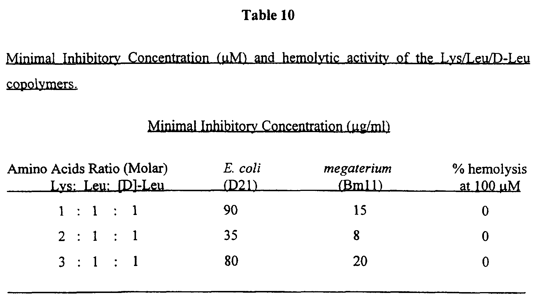


Claims
Priority Applications (19)
| Application Number | Priority Date | Filing Date | Title |
|---|---|---|---|
| EP97904561A EP0904291A2 (en) | 1996-02-22 | 1997-02-20 | Antipathogenic peptides and compositions comprising them |
| IL12573497A IL125734A (en) | 1996-02-22 | 1997-02-20 | Antipathogenic peptides and compositions comprising them |
| JP9529954A JP2000506128A (en) | 1996-02-22 | 1997-02-20 | Antipathogenic peptides and compositions containing them |
| AU17319/97A AU730069B2 (en) | 1996-02-22 | 1997-02-20 | Antipathogenic peptides and compositions comprising them |
| BR9707691A BR9707691A (en) | 1996-02-22 | 1997-02-20 | Antipathogenic peptides and compositions comprising the same |
| NZ331219A NZ331219A (en) | 1996-02-22 | 1997-02-20 | Antipathogenic peptides having a non-hemolytic selective cytolytic activity and compositions comprising them |
| US09/125,605 US6172038B1 (en) | 1996-02-22 | 1997-02-20 | Antipathogenic peptides and compositions comprising them |
| EA199800754A EA199800754A1 (en) | 1996-02-22 | 1997-02-20 | ANTIPATOGENIC PEPTIDES AND COMPOSITIONS INCLUDING THEM |
| CN98804347A CN1252808A (en) | 1997-02-20 | 1998-02-19 | Antipathogenic synthetic piptides and compositions comprising them |
| IL13136898A IL131368A0 (en) | 1997-02-20 | 1998-02-19 | Antipathogenic synthetic peptides and compositions comprising them |
| US09/367,714 US7001983B1 (en) | 1997-02-20 | 1998-02-19 | Antipathogenic synthetic peptides and compositions comprising them |
| EA199900752A EA199900752A1 (en) | 1997-02-20 | 1998-02-19 | ANTIPATOGENIC SYNTHETIC PEPTIDES AND COMPOSITIONS INCLUDING THEM |
| PCT/IL1998/000081 WO1998037090A1 (en) | 1997-02-20 | 1998-02-19 | Antipathogenic synthetic peptides and compositions comprising them |
| JP53645398A JP2001512475A (en) | 1997-02-20 | 1998-02-19 | Antipathogenic synthetic peptides and compositions containing them |
| AU60056/98A AU6005698A (en) | 1997-02-20 | 1998-02-19 | Antipathogenic synthetic peptides and compositions comprising them |
| BR9807456-3A BR9807456A (en) | 1997-02-20 | 1998-02-19 | Synthetic antipathogenic peptides and compositions comprising the same |
| KR1019997007477A KR20000071187A (en) | 1996-02-22 | 1998-02-19 | Antipathogenic synthetic peptides and compositions comprising them |
| CA002281204A CA2281204A1 (en) | 1997-02-20 | 1998-02-19 | Antipathogenic synthetic peptides and compositions comprising them |
| EP98903286A EP0977774A1 (en) | 1997-02-20 | 1998-02-19 | Antipathogenic synthetic peptides and compositions comprising them |
Applications Claiming Priority (2)
| Application Number | Priority Date | Filing Date | Title |
|---|---|---|---|
| IL117223 | 1996-02-22 | ||
| IL11722396A IL117223A0 (en) | 1996-02-22 | 1996-02-22 | Antipathogenic polypeptides and compositions comprising them |
Publications (3)
| Publication Number | Publication Date |
|---|---|
| WO1997031019A2 true WO1997031019A2 (en) | 1997-08-28 |
| WO1997031019A3 WO1997031019A3 (en) | 1997-10-23 |
| WO1997031019B1 WO1997031019B1 (en) | 1997-10-23 |
Family
ID=11068576
Family Applications (1)
| Application Number | Title | Priority Date | Filing Date |
|---|---|---|---|
| PCT/IL1997/000066 WO1997031019A2 (en) | 1996-02-22 | 1997-02-20 | Antipathogenic peptides and compositions comprising them |
Country Status (13)
| Country | Link |
|---|---|
| US (1) | US6172038B1 (en) |
| EP (1) | EP0904291A2 (en) |
| JP (1) | JP2000506128A (en) |
| KR (2) | KR19990087100A (en) |
| CN (1) | CN1216549A (en) |
| AU (1) | AU730069B2 (en) |
| BR (1) | BR9707691A (en) |
| CA (1) | CA2247025A1 (en) |
| CZ (1) | CZ264398A3 (en) |
| EA (1) | EA199800754A1 (en) |
| IL (2) | IL117223A0 (en) |
| NZ (1) | NZ331219A (en) |
| WO (1) | WO1997031019A2 (en) |
Cited By (14)
| Publication number | Priority date | Publication date | Assignee | Title |
|---|---|---|---|---|
| WO1998037090A1 (en) * | 1997-02-20 | 1998-08-27 | Yeda Research And Development Co. Ltd. | Antipathogenic synthetic peptides and compositions comprising them |
| WO2002040529A2 (en) * | 2000-11-16 | 2002-05-23 | Yeda Research And Development Co. Ltd. | Diastereomeric peptides and pharmaceutical compositions comprising them |
| US6660843B1 (en) | 1998-10-23 | 2003-12-09 | Amgen Inc. | Modified peptides as therapeutic agents |
| US7232803B2 (en) | 2000-03-09 | 2007-06-19 | Lytix Biopharma As | Antimicrobial compounds and formulations |
| WO2007074457A2 (en) * | 2005-12-27 | 2007-07-05 | Yeda Research And Development Co. Ltd. | Histidine-containing diastereomeric peptides and uses thereof |
| WO2007091959A1 (en) * | 2006-02-10 | 2007-08-16 | Dermagen Ab | Novel antimicrobial peptides and use thereof |
| US7488590B2 (en) | 1998-10-23 | 2009-02-10 | Amgen Inc. | Modified peptides as therapeutic agents |
| US7550141B2 (en) | 1998-07-13 | 2009-06-23 | Board Of Regents, The University Of Texas System | Methods for imaging tumor vasculature using conjugates that bind to aminophospholipids |
| US8143380B2 (en) | 2004-07-08 | 2012-03-27 | Amgen Inc. | Therapeutic peptides |
| EP2594287A1 (en) | 2006-04-21 | 2013-05-22 | Amgen Inc. | Lyophilized therapeutic peptibody formulations |
| WO2014038004A1 (en) * | 2012-09-05 | 2014-03-13 | 株式会社メニコン | Slow-drug-release ophthalmic lens and manufacturing method therefor |
| US8709430B2 (en) | 1998-07-13 | 2014-04-29 | Board Of Regents, The University Of Texas System | Cancer treatment kits comprising therapeutic antibody conjugates that bind to aminophospholipids |
| US9145450B2 (en) | 1998-10-23 | 2015-09-29 | Amgen Inc. | Thrombopoietic compounds |
| US10188740B2 (en) | 2005-08-12 | 2019-01-29 | Amgen Inc. | Modified Fc molecules |
Families Citing this family (7)
| Publication number | Priority date | Publication date | Assignee | Title |
|---|---|---|---|---|
| AU2004246909B2 (en) * | 2003-06-19 | 2010-08-19 | Yeda Research & Development Co. Ltd. | Antimicrobial and anticancer lipopeptides |
| US7704955B2 (en) * | 2004-11-24 | 2010-04-27 | Neopro Pain, Inc. | Methods and compositions for modulating conditions in both mammals and plants |
| WO2006128289A1 (en) * | 2005-06-02 | 2006-12-07 | University Of Manitoba | Use of brevinin-2r in the treatment of cancer |
| JP6093528B2 (en) * | 2012-09-19 | 2017-03-08 | 株式会社ミルボン | Screening method |
| US11103547B2 (en) | 2016-02-04 | 2021-08-31 | Yissum Research Development Company Of The Hebrew University Of Jerusalem Ltd. | Methods for disrupting biofilms |
| GB2557654A (en) * | 2016-12-14 | 2018-06-27 | Uea Enterprises Ltd | Method for nucleic acid depletion |
| CN113528600A (en) * | 2021-07-08 | 2021-10-22 | 石河子大学 | Method for preparing antibacterial peptide by inducing fly maggots with salmonella |
Family Cites Families (1)
| Publication number | Priority date | Publication date | Assignee | Title |
|---|---|---|---|---|
| US5258454A (en) * | 1988-09-01 | 1993-11-02 | Riso National Laboratory | Peptide synthesis method and solid support for use in the method |
-
1996
- 1996-02-22 IL IL11722396A patent/IL117223A0/en unknown
-
1997
- 1997-02-20 IL IL12573497A patent/IL125734A/en not_active IP Right Cessation
- 1997-02-20 US US09/125,605 patent/US6172038B1/en not_active Expired - Fee Related
- 1997-02-20 EP EP97904561A patent/EP0904291A2/en not_active Ceased
- 1997-02-20 JP JP9529954A patent/JP2000506128A/en not_active Ceased
- 1997-02-20 CZ CZ982643A patent/CZ264398A3/en unknown
- 1997-02-20 KR KR1019980706488A patent/KR19990087100A/en not_active Application Discontinuation
- 1997-02-20 EA EA199800754A patent/EA199800754A1/en unknown
- 1997-02-20 CN CN97194000A patent/CN1216549A/en active Pending
- 1997-02-20 CA CA002247025A patent/CA2247025A1/en not_active Abandoned
- 1997-02-20 WO PCT/IL1997/000066 patent/WO1997031019A2/en not_active Application Discontinuation
- 1997-02-20 AU AU17319/97A patent/AU730069B2/en not_active Ceased
- 1997-02-20 BR BR9707691A patent/BR9707691A/en unknown
- 1997-02-20 NZ NZ331219A patent/NZ331219A/en unknown
-
1998
- 1998-02-19 KR KR1019997007477A patent/KR20000071187A/en not_active Application Discontinuation
Non-Patent Citations (5)
| Title |
|---|
| BIOCHEMISTRY, vol. 31, no. 39, 1992, EASTON, PA US, pages 9482-9490, XP002036499 Y POUNY & Y SHAI: "Interaction of D-amino acid incorporated analogues of pardaxin with membranes " * |
| BIOCHEMISTRY, vol. 32, no. 13, 1993, EASTON, PA US, pages 3291-3297, XP002036498 D RAPAPORT ET AL.: "pH and ionic strength-dependent fusion of phospholipid vesicles induced by pardaxin analogues or by mixtures of charge-reversed peptides" * |
| FEBS LETTERS, vol. 259, no. 1, 18 December 1989, AMSTERDAM NL, pages 103-106, XP000083550 H G BOMAN ET AL.: "Antibacterial and antimalarial properties of peptides that are cecropin-melittin hybrids" * |
| INTERNATIONAL JOURNAL OF PEPTIDE AND PROTEIN RESEARCH, vol. 40, no. 5, November 1992, COPENHAGEN DK, pages 429-436, XP000319702 D WADE ET AL.: "Antibacterial peptides designed as analogs of hybrids of cecropins and melittins " * |
| JOURNAL OF BIOLOGICAL CHEMISTRY, vol. 271, no. 13, 29 March 1996, MD US, pages 7305-7308, XP002036500 Y SHAI & Z OREN: "Diastereoisomers of cytolysins, a novel class of potent antibacterial peptides " * |
Cited By (30)
| Publication number | Priority date | Publication date | Assignee | Title |
|---|---|---|---|---|
| WO1998037090A1 (en) * | 1997-02-20 | 1998-08-27 | Yeda Research And Development Co. Ltd. | Antipathogenic synthetic peptides and compositions comprising them |
| US8709430B2 (en) | 1998-07-13 | 2014-04-29 | Board Of Regents, The University Of Texas System | Cancer treatment kits comprising therapeutic antibody conjugates that bind to aminophospholipids |
| US7790860B2 (en) | 1998-07-13 | 2010-09-07 | Board Of Regents, The University Of Texas System | Targeting and imaging tumor vasculature using conjugates that bind to aminophospholipids |
| US7550141B2 (en) | 1998-07-13 | 2009-06-23 | Board Of Regents, The University Of Texas System | Methods for imaging tumor vasculature using conjugates that bind to aminophospholipids |
| US7488590B2 (en) | 1998-10-23 | 2009-02-10 | Amgen Inc. | Modified peptides as therapeutic agents |
| US9534032B2 (en) | 1998-10-23 | 2017-01-03 | Amgen Inc. | Thrombopoietic compounds |
| US9145450B2 (en) | 1998-10-23 | 2015-09-29 | Amgen Inc. | Thrombopoietic compounds |
| US6660843B1 (en) | 1998-10-23 | 2003-12-09 | Amgen Inc. | Modified peptides as therapeutic agents |
| US7166707B2 (en) | 1998-10-23 | 2007-01-23 | Amgen Inc. | Modified peptides as therapeutic agents |
| US7169905B2 (en) | 1998-10-23 | 2007-01-30 | Amgen Inc. | Modified peptides as therapeutic agents |
| US7189827B2 (en) | 1998-10-23 | 2007-03-13 | Amgen Inc. | Modified peptides as therapeutic agents |
| US8048852B2 (en) | 2000-03-09 | 2011-11-01 | Lytix Biopharma As | Antimicrobial compounds and formulations |
| US7232803B2 (en) | 2000-03-09 | 2007-06-19 | Lytix Biopharma As | Antimicrobial compounds and formulations |
| US9115169B2 (en) | 2000-03-09 | 2015-08-25 | Lytix Biopharma As | Method of treating tumors |
| WO2002040529A2 (en) * | 2000-11-16 | 2002-05-23 | Yeda Research And Development Co. Ltd. | Diastereomeric peptides and pharmaceutical compositions comprising them |
| WO2002040529A3 (en) * | 2000-11-16 | 2003-03-20 | Yeda Res & Dev | Diastereomeric peptides and pharmaceutical compositions comprising them |
| US8143380B2 (en) | 2004-07-08 | 2012-03-27 | Amgen Inc. | Therapeutic peptides |
| US11266744B2 (en) | 2005-08-12 | 2022-03-08 | Amgen Inc. | Modified Fc molecules |
| US10188740B2 (en) | 2005-08-12 | 2019-01-29 | Amgen Inc. | Modified Fc molecules |
| WO2007074457A3 (en) * | 2005-12-27 | 2007-11-01 | Yeda Res & Dev | Histidine-containing diastereomeric peptides and uses thereof |
| WO2007074457A2 (en) * | 2005-12-27 | 2007-07-05 | Yeda Research And Development Co. Ltd. | Histidine-containing diastereomeric peptides and uses thereof |
| WO2007091959A1 (en) * | 2006-02-10 | 2007-08-16 | Dermagen Ab | Novel antimicrobial peptides and use thereof |
| US8076286B2 (en) | 2006-02-10 | 2011-12-13 | Dermagen Ab | Antimicrobial peptides and use thereof |
| EP2594285A1 (en) | 2006-04-21 | 2013-05-22 | Amgen Inc. | Lyophilized therapeutic peptibody formulations |
| EP2594288A1 (en) | 2006-04-21 | 2013-05-22 | Amgen Inc. | Lyophilized therapeutic peptibody formulations |
| EP2594287A1 (en) | 2006-04-21 | 2013-05-22 | Amgen Inc. | Lyophilized therapeutic peptibody formulations |
| EP2594284A1 (en) | 2006-04-21 | 2013-05-22 | Amgen Inc. | Lyophilized therapeutic peptibody formulations |
| EP2594286A1 (en) | 2006-04-21 | 2013-05-22 | Amgen Inc. | Lyophilized therapeutic peptibody formulations |
| WO2014038004A1 (en) * | 2012-09-05 | 2014-03-13 | 株式会社メニコン | Slow-drug-release ophthalmic lens and manufacturing method therefor |
| JPWO2014038004A1 (en) * | 2012-09-05 | 2016-08-08 | 株式会社メニコン | Drug sustained release ophthalmic lens and method for producing the same |
Also Published As
| Publication number | Publication date |
|---|---|
| CA2247025A1 (en) | 1997-08-28 |
| IL125734A0 (en) | 1999-04-11 |
| WO1997031019A3 (en) | 1997-10-23 |
| AU1731997A (en) | 1997-09-10 |
| IL125734A (en) | 2005-08-31 |
| CZ264398A3 (en) | 1999-01-13 |
| EP0904291A2 (en) | 1999-03-31 |
| AU730069B2 (en) | 2001-02-22 |
| KR20000071187A (en) | 2000-11-25 |
| US6172038B1 (en) | 2001-01-09 |
| BR9707691A (en) | 1999-07-27 |
| NZ331219A (en) | 2000-04-28 |
| EA199800754A1 (en) | 1999-10-28 |
| KR19990087100A (en) | 1999-12-15 |
| CN1216549A (en) | 1999-05-12 |
| IL117223A0 (en) | 1996-06-18 |
| JP2000506128A (en) | 2000-05-23 |
Similar Documents
| Publication | Publication Date | Title |
|---|---|---|
| US7001983B1 (en) | Antipathogenic synthetic peptides and compositions comprising them | |
| US6172038B1 (en) | Antipathogenic peptides and compositions comprising them | |
| Oren et al. | Selective lysis of bacteria but not mammalian cells by diastereomers of melittin: structure− function study | |
| Shai et al. | From “carpet” mechanism to de-novo designed diastereomeric cell-selective antimicrobial peptides | |
| Oren et al. | Cyclization of a cytolytic amphipathic α-helical peptide and its diastereomer: effect on structure, interaction with model membranes, and biological function | |
| Shai | Pardaxin: channel formation by a shark repellant peptide from fish | |
| Strahilevitz et al. | Spectrum of antimicrobial activity and assembly of dermaseptin-b and its precursor form in phospholipid membranes | |
| Sato et al. | Peptide–membrane interactions and mechanisms of membrane destruction by amphipathic α-helical antimicrobial peptides | |
| Papo et al. | Can we predict biological activity of antimicrobial peptides from their interactions with model phospholipid membranes? | |
| Bobone et al. | Selectivity of antimicrobial peptides: a complex interplay of multiple equilibria | |
| JP5335241B2 (en) | Antimicrobial hexapeptide | |
| US8686113B2 (en) | Antibiotic peptides | |
| Hong et al. | Structure and organization of hemolytic and nonhemolytic diastereomers of antimicrobial peptides in membranes | |
| Zhu et al. | Design and mechanism of action of a novel bacteria-selective antimicrobial peptide from the cell-penetrating peptide Pep-1 | |
| KR100891157B1 (en) | Short bioactive peptides and methods for their use | |
| Oren et al. | A comparative study on the structure and function of a cytolytic α‐helical peptide and its antimicrobial β‐sheet diastereomer | |
| André et al. | Structure–activity relationship-based optimization of small temporin-SHf analogs with potent antibacterial activity | |
| Saido-Sakanaka et al. | Synthesis and characterization of bactericidal oligopeptides designed on the basis of an insect anti-bacterial peptide | |
| Zhu et al. | Antimicrobial and cytolytic activities and plausible mode of bactericidal action of the cell penetrating peptide penetratin and its Lys‐linked two‐stranded peptide | |
| Kuhn-Nentwig et al. | Cupiennin 1d*: the cytolytic activity depends on the hydrophobic N-terminus and is modulated by the polar C-terminus | |
| Lim et al. | The role of the central l-or d-Pro residue on structure and mode of action of a cell-selective α-helical IsCT-derived antimicrobial peptide | |
| Blondelle et al. | Progress in antimicrobial peptides | |
| Zhao | Mode of action of antimicrobial peptides | |
| MXPA99007799A (en) | Antipathogenic synthetic peptides and compositions comprising them | |
| MXPA98006869A (en) | Antipathogenic peptides and compositions that buy them |
Legal Events
| Date | Code | Title | Description |
|---|---|---|---|
| WWE | Wipo information: entry into national phase |
Ref document number: 97194000.2 Country of ref document: CN |
|
| AK | Designated states |
Kind code of ref document: A2 Designated state(s): AL AM AT AU AZ BA BB BG BR BY CA CH CN CU CZ DE DK EE ES FI GB GE HU IL IS JP KE KG KP KR KZ LC LK LR LS LT LU LV MD MG MK MN MW MX NO NZ PL PT RO RU SD SE SG SI SK TJ TM TR TT UA UG US UZ VN YU AM AZ BY KG KZ MD RU TJ TM |
|
| AL | Designated countries for regional patents |
Kind code of ref document: A2 Designated state(s): KE LS MW SD SZ UG AT BE CH DE DK ES FI FR GB GR IE IT LU MC NL PT SE BF BJ CF |
|
| DFPE | Request for preliminary examination filed prior to expiration of 19th month from priority date (pct application filed before 20040101) | ||
| 121 | Ep: the epo has been informed by wipo that ep was designated in this application | ||
| WWE | Wipo information: entry into national phase |
Ref document number: 199800754 Country of ref document: EA |
|
| WWE | Wipo information: entry into national phase |
Ref document number: 331219 Country of ref document: NZ |
|
| WWE | Wipo information: entry into national phase |
Ref document number: PV1998-2643 Country of ref document: CZ |
|
| WWE | Wipo information: entry into national phase |
Ref document number: 1019980706488 Country of ref document: KR |
|
| ENP | Entry into the national phase |
Ref document number: 2247025 Country of ref document: CA Ref document number: 2247025 Country of ref document: CA Kind code of ref document: A |
|
| WWE | Wipo information: entry into national phase |
Ref document number: PA/a/1998/006869 Country of ref document: MX |
|
| WWE | Wipo information: entry into national phase |
Ref document number: 1997904561 Country of ref document: EP |
|
| WWE | Wipo information: entry into national phase |
Ref document number: 09125605 Country of ref document: US |
|
| REG | Reference to national code |
Ref country code: DE Ref legal event code: 8642 |
|
| WWP | Wipo information: published in national office |
Ref document number: PV1998-2643 Country of ref document: CZ |
|
| WWP | Wipo information: published in national office |
Ref document number: 1997904561 Country of ref document: EP |
|
| WWP | Wipo information: published in national office |
Ref document number: 1019980706488 Country of ref document: KR |
|
| WWW | Wipo information: withdrawn in national office |
Ref document number: 1019980706488 Country of ref document: KR |
|
| WWW | Wipo information: withdrawn in national office |
Ref document number: PV1998-2643 Country of ref document: CZ |
|
| WWR | Wipo information: refused in national office |
Ref document number: 1997904561 Country of ref document: EP |
|
| WWW | Wipo information: withdrawn in national office |
Ref document number: 1997904561 Country of ref document: EP |