WO1997004008A1 - Anti-fungal peptides - Google Patents
Anti-fungal peptides Download PDFInfo
- Publication number
- WO1997004008A1 WO1997004008A1 PCT/US1996/003845 US9603845W WO9704008A1 WO 1997004008 A1 WO1997004008 A1 WO 1997004008A1 US 9603845 W US9603845 W US 9603845W WO 9704008 A1 WO9704008 A1 WO 9704008A1
- Authority
- WO
- WIPO (PCT)
- Prior art keywords
- xmp
- peptides
- fungal
- peptide
- activity
- Prior art date
Links
Classifications
-
- A—HUMAN NECESSITIES
- A61—MEDICAL OR VETERINARY SCIENCE; HYGIENE
- A61K—PREPARATIONS FOR MEDICAL, DENTAL OR TOILETRY PURPOSES
- A61K38/00—Medicinal preparations containing peptides
- A61K38/16—Peptides having more than 20 amino acids; Gastrins; Somatostatins; Melanotropins; Derivatives thereof
- A61K38/17—Peptides having more than 20 amino acids; Gastrins; Somatostatins; Melanotropins; Derivatives thereof from animals; from humans
- A61K38/1703—Peptides having more than 20 amino acids; Gastrins; Somatostatins; Melanotropins; Derivatives thereof from animals; from humans from vertebrates
- A61K38/1709—Peptides having more than 20 amino acids; Gastrins; Somatostatins; Melanotropins; Derivatives thereof from animals; from humans from vertebrates from mammals
- A61K38/1751—Bactericidal/permeability-increasing protein [BPI]
-
- A—HUMAN NECESSITIES
- A61—MEDICAL OR VETERINARY SCIENCE; HYGIENE
- A61P—SPECIFIC THERAPEUTIC ACTIVITY OF CHEMICAL COMPOUNDS OR MEDICINAL PREPARATIONS
- A61P31/00—Antiinfectives, i.e. antibiotics, antiseptics, chemotherapeutics
- A61P31/10—Antimycotics
-
- C—CHEMISTRY; METALLURGY
- C07—ORGANIC CHEMISTRY
- C07K—PEPTIDES
- C07K14/00—Peptides having more than 20 amino acids; Gastrins; Somatostatins; Melanotropins; Derivatives thereof
- C07K14/435—Peptides having more than 20 amino acids; Gastrins; Somatostatins; Melanotropins; Derivatives thereof from animals; from humans
- C07K14/46—Peptides having more than 20 amino acids; Gastrins; Somatostatins; Melanotropins; Derivatives thereof from animals; from humans from vertebrates
- C07K14/47—Peptides having more than 20 amino acids; Gastrins; Somatostatins; Melanotropins; Derivatives thereof from animals; from humans from vertebrates from mammals
- C07K14/4701—Peptides having more than 20 amino acids; Gastrins; Somatostatins; Melanotropins; Derivatives thereof from animals; from humans from vertebrates from mammals not used
- C07K14/4742—Bactericidal/Permeability-increasing protein [BPI]
Definitions
- the present invention relates generally to anti-fungal peptides derived from or based on Domain III (amino acids 142-169) of bactericidal/permeability-increasing protein (BPI) and therapeutic uses of such peptides.
- Domain III amino acids 142-169
- BPI bactericidal/permeability-increasing protein
- BPI is a protein isolated from the granules of mammalian polymorphonuclear leukocytes (PMNs or neutrophils), which are blood cells essential in thedefense againstinvading microorganisms.
- PMNs or neutrophils mammalian polymorphonuclear leukocytes
- HumanBPIprotein has been isolated from PMNs by acid extraction combined with either ion exchangechromatography [Elsbach, J. Biol. Chem., 254:11000(1979)] orE. coli affinity chromatography [Weiss, et al., Blood, 69:652 (1987)].
- BPI obtained in such a manner is referred to herein as natural BPI and has been shown to have potent bactericidal activity against a broad spectrum ofgram-negative bacteria.
- the molecular weight of human BPI is approximately 55,000 daltons (55 kD).
- the amino acid sequence ofthe entire human BPI protein and thenucleic acid sequence ofDNA encoding theprotein havebeen reported in Figure 1 of Gray et al., J. Biol. Chem., 264:9505 (1989), incorporated herein by reference.
- the Gray et al. DNA and amino acid sequences are set out in SEQ ID NOS: 251 and 252 hereto.
- BPI is a strongly cationic protein.
- the N-terminal halfofBPI accounts for the high net positive charge; the C-terminal halfofthe molecule has a net charge of-3.
- a proteolytic N-terminal fragment of BPI having a molecular weight of about 25 kD has an amphipathic character, containing alternating hydrophobic and hydrophilic regions.
- This N-terminal fragment ofhuman BPI possesses the anti-bacterial efficacy ofthe naturally-derived 55 kD human BPI holoprotein. [Ooi et al., J. Bio. Chem., 262: 14891-14894 (1987)].
- rBPI 23 An N-terminal BPI fragment of approximately 23 kD, referred to as "rBPI 23 ,” has been produced by recombinant means and also retains anti-bacterial activity against gram-negative organisms [Gazzano-Santoro et al., Infect. Immun.60:4754-4761 (1992)].
- an expression vector was used as a source of DNA encoding a recombinant expression product (rBPI 23 ).
- the vector was constructed to encode the 31-residue signal sequence and the first 199 amino acids ofthe N-terminus ofthe mature human BPI, as set outin SEQ ID NOS: 251 and 252 taken from Gray et al., supra, except that valine atposition 151 is specified by GTG rather than GTC and residue 185 is glutamic acid (specified by GAG) rather than lysine (specified by AAG).
- Recombinant holoprotein also referred to as rBPI, has also been produced having the sequence set out in SEQ ID NOS: 251 and 252 taken from Gray et al., supra, with the exceptions noted for rBPI 23 .
- rBPI 21 or rBPI 21 ⁇ cys has been described in co-owned, copending U.S. Patent No.5,420,019 which is incorporated herein by reference.
- This analog comprises the first 193 amino acids of BPI holoprotein as set out in SEQ ID NOS: 251 and 252 but wherein the cysteine at residue number 132 is substituted with alanine, and with the exceptions noted for rBPI 23 .
- BPI Bacillus subtilis bactericidal effect has been reported to be highly specific to gram-negative species, e.g., in Elsbach and Weiss, Inflammation: BasicPrinciplesand Clinical Correlates, eds. Gallinetal., Chapter30, Raven Press, Ltd. (1992). BPI is commonly thought to be non-toxic for other microorganisms, includingyeast, and forhighereukaryoticcells.
- BPI exhibitsanti-bacterial activity towards a broadrange ofgram-negativebacteriaatconcentrations as low as 10 -8 to 10- 9 M, but that 100- to 1,000-fold higher concentrations ofBPI were non-toxic toallofthegram-positivebacterial species, yeasts, and highereukaryoticcells tested at that time.
- BPI at a concentration of 10 -6 M or 160 ⁇ g/ml had no toxic effect, when tested at apH of either 7.0 or5.5, on the gram-positive organisms Staphylococcus aureus (four strains), Staphylococcus epidermidis, Streptococcus faecalis, Bacillus subtilis, Micrococcus lysodeikticus, and Listeria monocytogenes.
- BPI at 10 -6 M reportedly had no toxic effect on the fungi Candida albicans and Candida parapsilosis at pH 7.0 or 5.5, and was non-toxic to higher eukaryotic cells such as human, rabbit and sheepred blood cells and several human tumorcell lines.
- BPI kills gram-negative bacteria The precise mechanism by which BPI kills gram-negative bacteria is not yet completely elucidated, but it is believed that BPI must first bind to the surface of the bacteria through hydrophobic and electrostatic interactions between the cationic BPI protein and negatively charged sites on LPS.
- LPS has been referred to as "endotoxin" because of the potent inflammatoryresponse that it stimulates, i.e., therelease ofmediators by host inflammatory cells which may ultimately result in irreversible endotoxic shock.
- endotoxin because of the potent inflammatoryresponse that it stimulates, i.e., therelease ofmediators by host inflammatory cells which may ultimately result in irreversible endotoxic shock.
- BPI binds to lipid A, reported to be the most toxic and most biologically active component of LPS.
- BPI binding is thought to disrupt LPS structure, leading to activation of bacterial enzymes that degrade phospholipids and peptidoglycans, altering the permeability of the cell's outer membrane, and initiating events that ultimately lead to cell death. [Elsbach and Weiss (1992), supra]. BPI is thought to act in two stages. The first is a sublethal stage that is characterized by immediate growth arrest, permeabilization of the outer membrane and selective activation of bacterial enzymes that hydrolyze phospholipids and peptidoglycans. Bacteria at this stage can be rescued by growth in serum albumin supplemented media [Mannion et al., J. Clin. Invest., 85:853-860 (1990)]. The second stage, defined by growth inhibition that cannot be reversed by serum albumin, occurs after prolonged exposure of the bacteria to BPI and is characterized by extensive physiologic and structural changes, including apparent damage to the inner cytoplasmic membrane.
- BPI is at least as inhibitory of cytoplasmic membrane vesicle function as polymyxin B [In't Veld et al., Infection and Immunity 56: 1203-1208 (1988)] but the exact mechanism as well as the relevance of such vesicles to studies of intact organisms has not yet been elucidated.
- Domain II is defined as the amino acid sequence of BPI comprising from about amino acid 65 to about amino acid 99. Peptides based on this domain exhibit high LPS and heparin binding capacity and are bactericidal.
- Domain III is defined as the amino acid sequence of BPI comprising from about amino acid 142 to about amino acid 169. Peptides based on this domain exhibit high LPS and heparin binding activity and are bactericidal.
- the biological activities of functional domain peptides may include LPS binding, LPS neutralization, heparin binding, heparin neutralization or bactericidal activity.
- Fungi are eukaryotic cells that may reproduce sexually or asexually and may be biphasic, with one form in nature and a different form in the infected host.
- Fungal diseases are referred to as mycoses.
- Some mycoses are endemic, i.e. infection is acquired in the geographic area that is the natural habitat of that fungus. These endemic mycoses are usually self-limited and minimally symptomatic.
- Some mycoses are chiefly opportunistic, occurring in immunocompromised patients such as organ transplant patients, cancer patients undergoing chemotherapy, burn patients, AIDS patients, or patients with diabetic ketoacidosis.
- Fungal infections are becoming a major health concern for a number of reasons, including the limited number of anti-fungal agents available, the increasing incidence of species resistant to older anti-fungal agents, and the growing population of immunocompromised patients at risk for opportunistic fungal infections.
- the incidence of systemic fungal infections increased 600% in teaching hospitals and 220% in non-teaching hospitals during the 1980's.
- the most common clinical isolate is Candida albicans (comprising about 19% of all isolates). In one study, nearly 40% of all deaths from hospital-acquired infections were due to fungi. [Steinberg, Science, 266:1632-1634 (1994).]
- Anti-fungal agents include three main groups.
- the major group includes polyene derivatives, including amphotericin B and the structurally related compounds nystatin and pimaricin. These are broad-spectrum anti-fungals that bind to ergosterol, a component of fungal cell membranes, and thereby disrupt the membranes.
- Amphotericin B is usually effective for systemic mycoses, but its administration is limited by toxic effects that include fever and kidney damage, and other accompanying side effects such as anemia, low blood pressure, headache, nausea, vomiting and phlebitis.
- the unrelated anti-fungal agent flucytosine (5-fluorocytosine), an orally absorbed drug, is frequently used as an adjunct to amphotericin B treatment for some forms ofcandidiasis and cryptococcal meningitis. Its adverse effects include bone marrow depression with leukopenia and thrombocytopenia.
- the second major group of anti-fungal agents includes azole derivatives which impair synthesis of ergosterol and lead to accumulation of metabolites that disrupt the function of fungal membrane-bound enzyme systems (e.g., cytochrome P450) and inhibit fungal growth. Significant inhibition of mammalian P450 results in significant drug interactions.
- This group of agents includes ketoconazole, clotrimazole, miconazole, econazole, butoconazole, oxiconazole, sulconazole, terconazole, fluconazole and itraconazole. These agents may be administered to treat systemic mycoses.
- Ketoconazole an orally administered imidazole, is used to treat nonmeningeal blastomycosis,histoplasmosis,coccidioidomycosisandparacoccidioidomycosis in non-immunocompromised patients, and is also useful for oral and esophageal candidiasis.
- Adverse effects include rare drug-induced hepatitis; ketoconazole is also contraindicated in pregnancy.
- Itraconazole appears to have fewer side effects than ketoconazole and is used for most of the same indications. Fluconazole also has fewer side effects than ketoconazole and is used for oral and esophageal candidiasis and cryptococcal meningitis.
- Miconazoleis aparenteral imidazole with efficacy in coccidioidomycosis and several other mycoses, but has side effects including hyperlipidemia and hyponatremia.
- the third major group of anti-fungal agents includes allylamines-thiocarbamates, which are generally used to treat skin infections. This group includes tolnaftate and naftifine.
- griseofulvin a fungistatic agent which is administered orally for fungal infections of skin, hair or nails that do not respond to topical treatment.
- endemic mycoses are acquired by the respiratory route and are minimally symptomatic; cough, fever, headache, and pleuritic pain may be seen. Occasionally, endemic mycoses may cause progressive pulmonary disease or systemic infection. Histoplasmosis, caused by Histoplasma, is the most common endemic respiratory mycosis in the United States; over 40 million people have been infected. The disease is noncontagiousandordinarily self-limited, but chronic pulmonary infection and disseminated infection may occur. Pulmonary infection rarely requires treatment, but disseminated infection may be treated with amphotericin B. Coccidioidomycosis, caused by Coccidioides, is a noncontagious respiratory mycosis prevalent in the southwest United States.
- Blastomycosis caused by Blastomyces is a noncontagious, subacute or chronic endemic mycosis most commonly seeninthesoutheastUnited States. Mostpulmonary infections are probably self-limited. Patients with progressive lung disease or disseminated disease, and immunocompromised patients, may be treated systemically with amphotericin B.
- Paracoccidioidomycosis caused by Paracoccidioides, is a noncontagious respiratory mycosis that is the most commonsystemic mycosisin SouthAmerica.
- Disseminated disease is generally fatal in the absence of therapy.
- Sulfonamides may be used but have a low success rate.
- Amphotericin B produces a higher response rate but relapses may still occur.
- Cryptococcosis is a noncontagious, often opportunistic mycosis. It is characterized by respiratory involvement or hematogenous dissemination, often with meningitis. A major etiologic agent is C. neoformans. Most pulmonary infections are probably overlooked, but cryptococcal meningitis, which accounts for 90% of reported disease, is dramatic and seldom overlooked. Cryptococcosis is a particular problem in immunocompromised patients; cryptococcal meningitis occurs in 7 to 10% of AIDS patients. The principal symptom of meningitis is headache; associated findings include mental changes, ocular symptoms, hearing deficits, nausea, vomiting, and seizures. Without treatment, 80% of patients die within two years.
- Aspergillosis is a term that encompasses a variety of disease processes caused by Aspergillus species. Aspergillus species are ubiquitous; their spores are constantly being inhaled. Of the more than 300 species known, only a few are ordinarily pathogenic for man: A. fumigatus, A. flavus, A. niger, A. nidulans, A. terreus, A. sydowi, A. flavatus, and A. glaucus. Aspergillosis is increasing in prevalence and is particularly a problem among patients with chronic respiratory disease or immunocompromised patients.
- aspergillosis is second only to candidiasis as the most common opportunistic mycosis and accounts for about 15% of the systemic mycoses in this group.
- Opportunistic pulmonary aspergillosis is characterized by widespread bronchial erosion and ulceration, followed by invasion of the pulmonary vessels, with thrombosis, embolization and infarction.
- infection manifests as a necrotizing patchy bronchopneumonia, sometimes with hemorrhagic pulmonary infarction. In about 40% of cases, there is hematogenous spread to other sites. Aspergillosis is also a rare but devastating complication of burn wounds; amputation is often required for cure.
- Mucormycosis is an acute suppurative opportunistic mycosis that produces rhinocerebral, pulmonary or disseminated disease in immunocompromisedpatients, and local ordisseminated diseaseinpatients with burns or open wounds. Infection is caused by fungi in the class Zygomycetes, and include Basidiobolus, Conidiobolus, Rhizopus, Mucor, Absidia, Mortiere ⁇ la, Cunninghamella, and Saksenaea. Rhinocerebral mucormycosis accounts for about half of all cases of mucormycosis. It is one of the most rapidly fatal fungal diseases, with death occurring within 2-10 days in untreated patients.
- Pulmonary mucormycosis is nearly as common as rhinocerebral disease and manifests with the same necrotizing and infarction as aspergillosis. Fungi are virtually never seen or cultured from blood, sputum or cerebrospinal fluid. Disseminated mucormycosis may follow pulmonary or burn wound infection. Treatment is with amphotericin B.
- Candidiasis is a general term for a variety of local and systemic processes caused by colonization or infection of the host by species of the yeast Candida. Candidiasis occurs worldwide; superficial infections of the skin, mouth and other mucus membranes are universal. Invasive systemic disease has become a problem due to the use of high doses of antibiotics that destroy normal bacterial flora, immunosuppressive agents, and agents toxic to bone marrow, e.g. , during cancer therapy. Neutropenia is a major risk factor for Candida dissemination.
- Candidiasis is also seen among immunocompromised individuals such as AIDS patients, organ transplant patients, patients receiving parenteral nutrition, and cancer patients undergoing radiation treatment and chemotherapy. It is the most common opportunistic mycosisin theworld.
- Candida albicans The most common etiologic agentis Candida albicans. Otherinfectious species include C. tropicalis, C.parapsilosis, C. stellatoidea, C. krusei, C. parakrusei, C. lusitamae, C. pseudotropicalis, C. guilliermondi and C. glabrata.
- Candida albicans is normally found in the mouth, throat, gastrointestinal tract and vagina of humans.
- Non-albicans species frequently colonize skin.
- Candida species occur in two forms that are not temperature- or host-dependent. The usual colonizing forms are yeasts that may assume a pseudomycelial configuration, especially during tissue invasion. Pseudomyceliae result from the sequential budding of yeasts into branching chains ofelongated organisms.
- Candida albicans contains cell wall mannoproteins that appear to be responsible for attachment of the yeast cells to specific host tissues. It has been reported that the mannan portion, rather than theprotein portion, of the mannoproteins is responsible for adherence of fungal cells to spleen and lymph node tissues in mice. [Kanbe etal., Infection Immunity, 6.7:2578-2584 (1993).]
- C. albicans also binds avidly to extracellular matrix (ECM) proteins such as fibronectin, laminin, and types I and IV collagen, all of which contain heparin-binding domains. This suggests C. albicans may express a heparin-like surface molecule. Adherence of C. albicans to the ECM maybeimportantin thepathogenesis ofdisseminated candidiasis. Ithas been demonstrated that heparin, heparan sulfate and dextran sulfate glycosaminoglycans (GAGs) inhibit adherence of C.
- ECM extracellular matrix
- candidiasis manifests as superficial mucocutaneous infections, chronic mucocutaneous candidiasis, or systemic infection.
- Superficial mucocutaneous infections can occur in any area of skin or mucus membrane.
- Thrush commonly seen in AIDS patients, is characterized by a patchy or continuous, creamy to gray pseudomembrane that covers the tongue, mouth, orotheroropharyngeal surfacesand maybeaccompaniedbyulceration and necrosis. Laryngeal involvement results in hoarseness.
- Esophagitis is often an extension of oropharyngeal disease and may manifest with symptoms of retrosternal pain and dysphagia.
- Intestinal candidiasis is commonly asymptomatic, but is a major source of hematogenous invasion in immunocompromised individuals.
- Intertrigo involves the axillae, groins, inframammary folds, and other warm, moist areas, and may manifest as red, oozing or dry, scaly lesions. Infections may occur in other areas, including perianal and genital areas. Paronychia, infection of the nails, often follows chronic exposure ofthe hands orfeet to moisture. Somepatients with limited T-cell immunodeficiency develop chronic mucocutaneous candidiasis. These patients suffer from persistent superficial Candida infection of the skin, scalp, nails and mucus membranes.
- systemic candidiasis Most cases of systemic candidiasis are caused by Candida albicans and C. tropicalis, and increasingly, C. glabrata. Clinical manifestations of Candida infection appear mainly in the eyes, kidneys and skin. In the eyes, there may be single or multiple raised, white, fluffy chorioretinal lesions. These lesions are a potential cause of blindness. Involvement of the kidneys includes diffuse abscesses, capillary necrosis and obstruction of the ureters. Infection may result in progressive renal insufficiency. Systemic Candidainfectioncanalso manifestas maculonodular skin lesions surrounded by a reddened area; these lesions have an appearance similar to acne but are a major clue to a potentially lethal disease. Other manifestations of systemic candidiasis may include osteomyelitis, arthritis, meningitis, and abscesses in the brain, heart, liver, spleen and thyroid.
- Candida endocarditis can occur in patients receiving prolonged intravenous therapy or cardiac valve implants, or in intravenous drug abusers. Fungal lesions appear on the valves, and can embolize and occlude large blood vessels.
- Superficial infections are diagnosed by microscopic examination of scrapings or swabs of infected lesions in the presence of 10% potassium hydroxide. Candida organisms can also be seen on gram stain. Endocarditis is diagnosed by blood cultures or demonstration of bulky valvular lesions on echocardiography. Systemic candidiasis may be difficult to diagnose because thepresence ofheavy colonization at the usual sites ofinfectionindicates, but does not prove, that dissemination has occurred. The most reliable evidence ofsystemic candidiasis is biopsy demonstration oftissue invasion or recovery of yeast from fluid in a closed body cavity, such as cerebral spinal fluid, pleural or peritoneal fluid. Similarly, positive blood or urine or sputum cultures may indicate invasive disease or simply localized disease around indwelling devices, e.g., catheters or intravenous lines.
- Mucocutaneous infections may be treated with topical preparations of nystatin, amphotericin B, clotrimazole, miconazole, haloprogin or gentian violet.
- Oropharyngeal or esophageal candidiasis can be treated with systemic agents such as ketoconazole or fluconazole.
- Chronic mucocutaneous candidiasis syndrome may respond to topical or systemic therapeutic agents such as amphotericin B orketoconazole, but often relapses when medication is discontinued.
- Cystitis may be treated with amphotericin B bladder rinses, or a brief low-dose intravenous course of amphotericin B with or without oral flucytosine.
- Endocarditis is essentially incurable without valve replacement, accompanied by a 6 to 10 week course ofamphotericin B and flucytosine. Even with therapy, however, complete cure of endocarditis is not always possible.
- the mortality rate from systemic candidiasis is about 50%.
- Systemic candidiasis may be treated with fluconazole, a fungistatic agent, or amphotericin B, a fungicidal agent although systemic use of the latter is limited by its toxicity. Both drugs have substantial adverse reactions when used in combination with cyclosporine A, which itself can be nephrotoxic. The removal of precipitating factors such as intravenous lines or catheters is also important for controlling infection. Flucytosine therapy can be added to the amphotericin B therapy for treatment of systemic candidiasis, especially in patients that are not immunocompromised. In immunocompromised patients, however, these infections are problematic and resist effective treatment. Mortality with systemic candidiasis can be over 90% in such patients. Furthermore, chronic mucocutaneous candidiasis and candidal endocarditis often show evidence of disease after having been declared cured.
- the present invention provides novel peptides derived from or based on Domain III (amino acids 142-169) of bactericidal/permeability- increasing protein (BPI) and therapeutic uses of such peptides as anti-fungal agents.
- Peptides of the invention are useful in methods of treating a subject suffering from a fungal infection by administering a therapeutically effective amount of the peptide. This is based on the surprising discovery that Domain III derived peptides have fungicidal/fungistatic effects. A second surprising discovery is that such peptides have LPS-neutralizing activity. This activity provides an additional benefit in the use of peptides of the invention for treating fungal infections. Domain III derived peptides may be administered alone or in conjunction with known anti-fungal agents.
- Domain III derived peptides When made the subject of adjunctive therapy, the administration of Domain III derived peptides may reduce the amount of anti-fungal agent needed for effective therapy, thus limiting potential toxic response and/or high cost of treatment. Admmistration of Domain III derived peptides may also enhance the effect of such agents, accelerate the effect of such agents, or reverse resistance of fungi to such agents.
- Peptides according to the invention include peptides SEQ ID NOS:1-250.
- the invention provides a method of killing or inhibiting growth of fungi comprising contacting the fungi with a Domain III derived peptide.
- This method can be practiced in vivo or in a variety of in vitro uses such as to decontaminate fluids and surfaces and to sterilize surgical and other medical equipment and implantable devices, including prosthetic joints and indwelling invasive devices.
- a further aspect of the invention involves use of a Domain III derived peptide for the manufacture of a medicament for treatment of fungal infection.
- the medicament may include, in addition to a Domain III derived peptide, other chemotherapeutic agents such as anti-fungal agents.
- Figure 1 provides results of broth assay tests of the activity of various peptides against C. albicans.
- Figures 2A and 2B provideresults ofradial diffusionassays of the activity ofvarious peptides against C. albicans SLU-1 (Fig.2A) and C. albicans SLU-2G (Fig.2B).
- Figure 3 provides results ofbroth assay tests of the activity of combinations ofpeptide and amphotericin B against C. albicans.
- Figures 4, 5, and 6 graphically represent survival data in mice after C. albicans challenge and treatment with peptides or buffer control.
- Figures 7 graphically represents survival data in cyclosporin-treated mice after C. albicans challenge and treatment with peptides or buffer control.
- Figure 8 provides results of RAW cellassay tests of the activity of various peptides.
- Figure 9 graphically represents survival data in mice after challenge with E. coli 0111:B4 LPS and treatment with peptide.
- the present invention relates to the surprising discovery that a Domain III derived peptide has fungicidal activity and can be administered to treat subjects suffering from fungal infection.
- subject is meant to refer to higher organisms, including animals (e.g., humans; companion animals such as dogs; livestock such as horses, cows and pigs; poultry; insects; fish; avian species) and plants.
- animals e.g., humans; companion animals such as dogs; livestock such as horses, cows and pigs; poultry; insects; fish; avian species
- methods of treating fungal infections with such peptides.
- Domain III derived peptides were demonstrated to have anti-fungal activities both in in vitro killing assays and in in vivo models of fungal infection, as measured, for example, by improved survival or reduction of colony-forming units in circulation after fungal challenge.
- a variety of fungal infections including infections caused by Aspergillus, infections caused by Cryptococcus, such as cryptococcal meningitis, and mucocutaneous and systemic candidiasis caused by Candida species, may be treated according to the invention.
- Domain III derived peptides were demonstrated to have LPS-neutralizing activity both in an in vitro assay and an in vivo model. This activity provides an additional benefit in the treatment of fungal infections where bacterial LPS from translocation or additional infection is associated with the fungal infection.
- Domain III derived peptide includes peptides having an amino acid sequence of BPI protein from about position 142 to about position 169, subsequences thereof and variants of the sequence or subsequence thereof, which posses antifungal activity. Specifically included are those antifungal peptides having six to fourteen amino acids and having the amino acid sequence of BPI protein from about position 148 to about position 161, subsequences thereof and variants of the sequence or subsequence. Certain preferred peptides have fourteen amino acids and among the preferred variant sequences and subsequences are those having K as an amino acid corresponding to G at position 152.
- Preferred peptide sequences with fourteen amino acids have a core amino acid sequence selected from the group consisting of LIQL, IQLF, WLIQL, LIQLF and WLIQLF or a variant core amino acid sequence having at least 75% homology to said core amino acid sequence and include the peptides of SEQ ID NOS: 4 (XMP.13), 6-19 (XMP.31-44),21-22(XMP.82-83),23-25 (XMP.85-87),26-27(XMP.91-92), 28-31 (XMP.94-97), 32-33 (XMP.100-101), 34 (XMP.104), 35-40 (XMP.106-111), 41 (XMP.113), 42 (XMP.116), 43-55 (XMP.123-135), 57-58 (XMP.138-139), 59-61 (XMP.142-144), 62 (XMP.146), 66-78 (XMP.222-234), 80-88 (XMP.236-244), 89-109 (X
- This group of antifungal 14 mer peptides includes variant sequencepeptideswhereinatleastoneBPIsequenceresiduehasbeenreplaced by a D-isomer amino acid. See, e.g., SEQ ID NOS: 46(XMP.126), 48 (XMP.128), 86-87 (XMP.242-243) and 92-93 (XMP.252-253).
- Variants involving BPI sequence replacements by atypical amino acids such as ⁇ (1-naphthyl)A, ⁇ (2-naphthyl)A, para-amino F, cyclohexyl A, ⁇ - and ⁇ -aminobutyric acids, ⁇ methyl A and N-methyl G, V and L are also included within this group.
- Domain III derived antifungal peptides of the invention having from seven to twelve amino acids comprising: (a) a core sequence of amino acids selected from the group consisting of LIQL, IQLF, WLIQL, LIQLF and WLQLF; and (b) one or more cationic amino acids selected from the group consisting of K, R, H, ornithine and diaminobutyric acid at the amino and/or carboxy terminal portion of the core sequence.
- a subset of peptides have from seven to nine amino acids comprising: (a) a core sequence of amino acids selected from the group consisting of LIQL and IQLF; and (b) at least two cationic amino acids selected from the group consisting of K, R, H, ornithine and diaminobutyric acid at the amino and/or carboxy terminal portion of the core sequence.
- Another subset ofpeptides has from eight to ten amino acids comprising: (a) a core sequence of amino acids selected from the group consisting of LIQLF and WLQLF; and (b) at least two cationic amino acids selected from the group consisting of K, R, H, ornithine and diaminobutyric acid at the amino and/or carboxy terminal portion ofthe core sequence.
- Still another subset of peptides has nine to twelve amino acids comprising: (a) a core sequence of amino acids selected from the group consisting of WLQLF; and (b) at least three cationic amino acids selected from the group consisting of K, R, H, ornithine and diaminobutyric acid at the amino and/or carboxy terminal portion of the core sequence.
- peptides of SEQ ID NOS: 118-137 (XMP.285-304), 140-144 (XMP.307-311), 155-160 (XMP.322-327), 166-170(XMP.335-339), 174-177(XMP.343-346), 179-184 (XMP.348-353), 186 (XMP.355), 188-190 (XMP.357-359).
- the Domain III sequence of BPI amino acids from 148 to 161 includes the core sequences) noted above as well as multiple cationic residues (K and H) flanking the core.
- This motif is carried forward in the structures of subsequences of the 148 to 161 sequence providing antifungal peptides of the invention and also preserved in antifungal variants of the 148 to 161 sequence and subsequences thereof.
- G residue normally in the BPI sequence at position 152 is replaced by K, this replacement serves to provide a cationic residue immediately adjacent to the predominantly hydrophobic core residues.
- Sequence and subsequence variants providing antifungal peptides according to the invention thus include those peptides wherein one or more existing non-cationic residues ordinarily flanking the core sequence(s) are replaced by cationic residues.
- the neutral aliphatic residues L and I are each replaceable by neutral aliphatic residues G, A, V, I and L.
- the aromatic residues W (BPI position 153) and F (BPI position 158) are replaceable by a different aromatic amino acid residues or by neutral aliphatic residues G, A, V, I and L.
- the core sequence Q (BPI residue 156) is replaceable preferably by a neutral hydrophilic amino acid T, S and N. As noted above, where variations are introduced into core subsequence(s), it is preferable that the variant core sequences) retain 75% homology to the sequences occurring in BPI.
- Antifungal Domain III peptides of the invention have one or more D-isomer amino acids, as illustrated by the peptides of SEQ ID NOS: 164 (XMP.333), 165 (XMP.334), 173 (XMP.342), 194 (XMP.363) and 196 (XMP.365) and have the core sequence amino acids comprise D-isomer amino acids in reverse sequence order as illustrated by peptides having the amino acid sequence set out in SEQ ID NOS: 163 (XMP.332) and 198 (XMP.367).
- the antifungal peptides can have an acetylated amino terminal amino acid residue as illustrated by the peptides of SEQ ID NOS: 162 (XMP.331), 185 (XMP.354), 187 (XMP.356), 195 (XMP.364), 199 (XMP.368) and 204 (XMP.373).
- Cyclic antifungal peptides as illustrated by SEQ ID NOS: 191- 193 (XMP.360-362) are also within the scope of the invention.
- Additional Domain III antifungal peptides of the invention include antifungal peptides SEQ ID NOS: 1 (XMP.5), 2-4 (XMP.11-.13), 5 (XMP.29), 20 (XMP.55), 56 (XMP.137), 79 (XMP.235), 111-115 (XMP.271-.275), 117 (XMP.284), 132 (XMP.299), 138-139 (XMP.305-
- Additional Domain III antifungal peptides of the invention include antifungal peptides SEQ ID NOS:205-243 (XMP.374-.412) and SEQ ID NOS:244-250 (XMP.414-.420).
- peptides of the invention include peptides that have SEQ ID NOS:1-250 as shown in Table 1 herein.
- compositions of the invention comprise a Domain III derived peptide and a pharmaceutically acceptable diluent, adjuvant or carrier and are administered topically, intravenously, orally or as an aerosol.
- Fungal infection treatment methods of the invention comprise administering to a subject suffering from a fungal infection a therapeutically effective amount of a Domain III antifungal peptide and such treatment methods are applicable to infections by fungal infection involves a fungal species selected from the group consisting of Candida (especially, C. albicans, C. glabrata, C. krusei, C. lusitaniae, C. parapsilosis and C. tropicalis), Aspergillus and Cryptococcus species.
- Candida especially, C. albicans, C. glabrata, C. krusei, C. lusitaniae, C. parapsilosis and C. tropicalis
- medicaments/pharmaceutical compositions developed according to the invention can include other antifungal agents including non-peptide agents or can be used in combinative therapeutic methods with other such agents.
- BPI-derived peptides Peptides derived from or based on BPI produced by recombinant or synthetic means (BPI-derived peptides) have been described in co-owned and copending PCT Application No. US94/10427 filed September 15, 1994, which corresponds to U.S. Patent Application Serial No. 08/306,473, filed September 15, 1994, and PCT Application No. US94/02465 filed March 11, 1994, which corresponds to U.S. Patent Application Serial No. 08/209,762, filed March 11, 1994, which is a continuation-in-part of U.S. Patent Application Serial No.08/183,222, filed January 14, 1994, which is a continuation-in-part of U.S. Patent Application Ser.
- BPI-derived peptides having an amino acid sequence of BPI protein from about position 142 to about position 169, subsequences thereof and variants of the sequence or subsequence thereof, which possess a BPI anti-fungal biological activity, were disclosed in co- owned and co-pending U.S. priority application Serial No.08/372,105 filed January 13, 1995, the disclosure ofwhich isincorporated hereinbyreference.
- the Domain III derived peptide may be administered systemically or topically.
- Systemic routes of admmistration include oral, intravenous, intramuscularor subcutaneous injection(includingintodepotsfor long-term release), intraocular or retrobulbar, intrathecal, intraperitoneal (e.g. by intraperitoneal lavage), transpulmonary using aerosolized or nebulized drug, or transdermal.
- Topical routes include administration in the form of salves, ophthalmic drops, ear drops, or irrigation fluids (for, e.g., irrigation of wounds).
- the Domain III derived peptide may be administered in conjunction with other anti-fungal agents.
- Preferred anti-fungal agents for this purpose are amphotericin B and fluconazole.
- Concurrent administration of Domain III derived peptide with anti-fungalagents is expected to improve the therapeutic effectiveness of the anti-fungal agents. This may occur through reducing the concentration of anti-fungal agent required to eradicate or inhibit fungal growth, e.g., replication. Because the use of some agents is limited by their systemic toxicity or prohibitive cost, lowering the concentration of anti-fungal agent required for therapeutic effectiveness reduces toxicity and/or cost of treatment, and thus allows wider use of the agent.
- Domain III derived peptide Concurrent administration of Domain III derived peptide and another anti-fungal agent may produce a more rapid or complete fungicidal/fungistatic effect than could be achieved with either agent alone. Domain III derived peptide administration may reverse the resistance of fungi to anti-fungal agents.
- Domain III derived peptide administration may also convert a fungistatic agent into a fungicidal agent.
- An advantage provided by the present invention is the ability to treat fungal infections, particularly Candida infections, that are presently considered incurable. Another advantage is the ability to treat fungi that have acquired resistance to known anti-fungal agents.
- a further advantage of concurrent administration of Domain III derived peptide with an anti-fungal agent having undesirable side effects, e.g., amphotericin B, is the ability to reduce the amount of anti-fungal agent needed for effective therapy.
- the present invention may also provide quality of life benefits due to, e.g., decreased duration of therapy, reduced stay in intensive care units or reduced stay overall in the hospital, with the concomitant reduced risk of serious nosocomial (hospital-acquired) infections.
- Constant administration includes administration of the agents together, simultaneously or before or after each other.
- the Domain III derived peptide and anti-fungal agents may be administered by different routes.
- the Domain III derived peptide may be administered intravenously while the anti-fungal agents are administered intramuscularly, intravenously, subcutaneously, orally or intraperitoneally.
- the Domain III derived peptide may be administered intraperitoneally while the anti-fungal agents are administered intraperitoneally or intravenously, or the Domain III derived peptide may be administered in an aerosolized or nebulized form while the anti-fungal agents are administered, e.g., intravenously.
- the Domain III derived peptide and anti-fungal agents may be both administered intravenously.
- the Domain III derived peptide and anti-fungal agents may be given sequentially in the same intravenous line, after an intermediate flush, or may be given in different intravenous lines.
- the Domain III derived peptide and anti-fungal agents may be administered simultaneously or sequentially, as long as they are given in a manner sufficient to allow both agents to achieve effective concentrations at the site of infection.
- Concurrent administration of Domain III derived peptide and another anti-fungal agent is expected to provide more effective treatment of fungal infections.
- Concurrent administration of the two agents may provide greater therapeutic effects in vivo than either agent provides when administered singly.
- concurrent administration may permit a reduction in the dosage of one or both agents with achievement of a similar therapeutic effect.
- the concurrent administration may produce a more rapid or complete fungicidal/fungistatic effect than could be achieved with either agent alone.
- Therapeutic effectiveness is based on a successful clinical outcome, and does not require that the anti-fungal agent or agents kill 100% of the organisms involved in the infection. Success depends on achieving a level of anti-fungal activity at the site of infection that is sufficient to inhibit the fungi in a manner that tips the balance in favor of the host. When host defenses are maximally effective, the anti-fungal effect required may be minimal. Reducing organism load by even one log (a factor of 10) may permit the host's own defenses to control the infection. In addition, augmenting an early fungicidal/fungistatic effect can be more important than long-term fungicidal/fungistatic effect. These early events are a significant and critical part of therapeutic success, because they allow time for host defense mechanisms to activate.
- a Domain III derived peptide may interact with a variety of host defense elements present in whole blood or serum, including complement, p15 and LBP, and other cells and components of the immune system. Such interactions may result in potentiation of the activities of the peptide. Because of these interactions, Domain III derived peptides can be expected to exert even greater activity in vivo than in vitro. Thus, while in vitro tests are predictive of in vivo utility, absence of activity in vitro does not necessarily indicate absence of activity in vivo. For example, BPI has been observed to display a greater bactericidal effect on gram-negative bacteria in whole blood or plasma assays than in assays using conventional media. [Weiss et al., J. Clin. Invest.
- the Domain III derived peptides be administered with other products that potentiate the activity of the peptide, including the anti-fungal activity of the peptides.
- serum complement potentiates the gram-negative bactericidal activity of BPI protein products; the combination of BPI protein product and serum complement provides synergistic bactericidal/growth inhibitory effects.
- poloxamer surfactants enhance the anti-bacterial activity of BPI protein products, as described in Lambert, U.S. Application No.08/372, 104 filed January 13, 1995; poloxamer surfactants may also enhance the activity of anti-fungal agents.
- Domain III derived peptides may have several modes of action.
- the peptide through its heparin-binding ability, may interfere with the binding of fungi to the extracellular matrix.
- heparin-like surface molecules of Candida are believed to mediate adhesion of the yeast to extracellular matrix and host tissues.
- the peptide may also act directly on the cytoplasmic membrane of fungi.
- the peptide may bind to fungal cell wall mannoproteins that are structurally similar to the LPS of gram-negative organisms or that are responsible for adherence to target host tissues, thus interfering with fungal interaction with host tissues.
- Binding to fungal mannans may also promote access of the peptide to the inner cytoplasmic membrane.
- the peptide may also act beneficially by killing gram-negative bacteria and neutralizing UPS.
- the antifungal activity of Domain III peptides according to the invention may result from unique structural features. For example, a six amino acid sequence within Domain III (WLIQLF) and the included five and four amino acid sequences (LIQL, IQLF, WLIQL and LIQLF) are composed of hydrophobic amino acids with the exception of glutamine (Q) that is a neutral hydrophilic amino acid.
- This hydrophobic stretch is bounded by highly cationic(polar)lysines on the N- and C-termini. This motifis reminiscent of leader/signal peptides as well as transmembrane segments of membrane proteins.
- Aliphatic amino acids such as I, L, V, M, A, have a high propensity to form transmembrane ⁇ -helical structures within the hydrophobic membrane environment when found in sequences of 12-15 nonpolar amino acids due to their ability to form backbone hydrogen bonds.
- Aromatic hydrophobic amino acids such as W and F can also incorporate into a membrane ⁇ -helix.
- the neutral, hydrophilic glutamine in the middle of a Domain III hydrophobic stretch may participate in hydrogen bonding with other fungal membrane components such as ergosterol and thus play an important role in the fungicidal activity.
- a short 10 amino acid peptide e.g., XMP.293
- the short motif of six to twelve amino acid peptides with a core of neutral amino acids bounded by cationic amino acids is not long enough to span a fungal lipid bilayer and thus may be allowed to traverse the membrane bilayer more efficiently than longer peptides.
- the cationic/neutral/cationic molecules may inhibit the function of endogenous polyamines(spermidine, spermine, putrescine) by either competitive inhibition of the polyamine regulation of cell wall carbohydrate synthesis and/or by feedback inhibition of polyamine synthesis.
- the invention provides a method of killing or inhibiting growth of fungi comprising contacting the fungi with a Domain III derived peptide.
- This method can be practiced in vivo or in a variety of in vitro uses such as use in food preparations or to decontaminate fluids and surfaces or to sterilize surgical and other medical equipment and implantable devices, including prosthetic joints. These methods can also be used for in situ sterilization of indwelling invasive devices such as intravenous lines and catheters, which are often foci of infection.
- a further aspect of the invention involves use of a Domain III derived peptide for the manufacture of a medicament for treatment offungal infection.
- the medicament may include, in addition to a BPI protein product, other chemotherapeutic agents such as anti-fungal agents.
- the medicament can optionally comprise a pharmaceutically acceptable diluent, adjuvant or carrier.
- antifungal peptides are suitably accomplished with a pharmaceutical composition comprising a peptide and a pharmaceutically acceptable diluent, adjuvant, or carrier.
- a pharmaceutical composition comprising a peptide and a pharmaceutically acceptable diluent, adjuvant, or carrier.
- the peptide may be administered without or in conjunction with known surfactants, other chemotherapeutic agents or additional known anti-fungal agents.
- Example 1 addresses peptide preparation and purification;
- Example 2 addresses in vitro anti-fungal testing of peptides;
- Example 3 addresses additional in vitro and in vivo testing of the anti-fungal effect of peptides on a variety of fungal species, including Candida strains and antibiotic resistant strains;
- Example 4 addresses the in vivo effect of peptides on survival of mice challenged with Candida;
- Example 5 addresses the serum stability of peptides;
- Example 6 addresses the design and assay of anti-fungal peptides for structural motif and minimum functional sequence analysis;
- Example 7 addresses LPS neutralization activities of anti-fungal peptides; and
- Example 8 addresses peptide formulations.
- This example addresses the preparation and purification of Domain III derived peptides.
- Peptides may be prepared according to a variety of synthetic procedures. Some peptides (e.g., XMP.5) were prepared by solid phase peptide synthesis as described in parent U.S. Patent Application Serial Nos. 08/209,762 and 08/183,222 according to the methods of Merrifield, J. Am Chem. Soc.85: 2149 (1963) and Merrifield et al. Anal. Chem., 38: 1905-1914 (1966) using an Applied Biosystems, Inc. Model 432 peptide synthesizer.
- XMP.5 Some peptides (e.g., XMP.5) were prepared by solid phase peptide synthesis as described in parent U.S. Patent Application Serial Nos. 08/209,762 and 08/183,222 according to the methods of Merrifield, J. Am Chem. Soc.85: 2149 (1963) and Merrifield et al. Anal. Chem., 38: 1905-1914 (1966) using an Applied Biosystems, Inc. Model
- peptides were synthesized on alarger scaleusing solid phase peptide synthesis on an Advanced Chemtech (ACT-Model 357 MPS)synthesizerutilizinga1-Fluorenylmethyl-oxycarbonyl(Fmoc)protection strategy with a double coupling procedure employing N,N-diisopropylcarbodiimide (DIC)/1-hydroxybenzotriazole (HOBt) and 2-(1-H-benzotriazol-1-yl)-1,1,3,3,-tetramethyluronium hexa-fluorophosphate (HBTU)/HOBt/diisopropylethylamine (DIEA).
- DIC N,N-diisopropylcarbodiimide
- HOBt 2-(1-H-benzotriazol-1-yl)-1,1,3,3,-tetramethyluronium hexa-fluorophosphate
- DIEA diisopropylethyl
- the solid support used was a polystyrene resin with 1% divinylbenzene (DVB) cross-linking and an 4-(2',4'-dimethoxyphenyl-Fmoc-aminomethyl)-phenoxy (Fmoc-Rink amide) linker with a substitution rate of 0.44 mmoles/gram.
- the scale used was between 0.1 grams and 5 grams of starting resin.
- Dimethylformamide was the primary solvent with a 50/50 solution of piperidine/DMF used for Fmoc deprotection in three consecutive treatments of 1, 5, and 10 minutes, respectively.
- a double coupling procedure was used in each cycle with a 4:1 amino acid to peptide ratio used in each coupling.
- the amino acids were dissolved in a 0.5M HOBt solution in N-methylpyrrolidinone (NMP) at a concentration also of 0.5M.
- NMP N-methylpyrrolidinone
- NMP N-methylpyrrolidinone
- an equimolar (to amino acid) amount of a 0.5M solution of diisopropylcarbodiimide (DIPCDI) in NMP was used and allowed to react for45 minutes.
- DIPCDI diisopropylcarbodiimide
- the second coupling utilized an equimolar (to amino acid) volume of a 0.5M HBTU solution in DMF with an equal volume of a 1M DIEA solution in NMP (2:1, DIEA:amino acid) for a period of 30 minutes.
- Selected peptides were purified by high performance liquid chromatography (HPLC), using a Waters Prep LC 2000 Preparative Chromatography System (Water Corp., Milford, MA) equipped with a Delta Pak C-18, 15 ⁇ m, 300 A cartridge column consisting ofa 40 ⁇ 10 mm guard cartridge and a 40 ⁇ 100 mm Prep Pak cartridge.
- Peptides were dissolved to ⁇ 20 mg/mL in buffer A and 200-800 mg were applied to the column through the LC pump operating at a flow rate of 8-17 mL/minute bound material was eluted with a gradient of 25-35% buffer B/30 min applied at 8-17 mL/minute. (Some peptides were purified with a gradient of 23-33%B/30 minute). The eluate was monitored at 220 and/or 280 and 300 nm with a Waters 490E Programmable Multiwavelength Detector.
- Fractions were collected and assayed for the peptide of interest on an Ultrafast Micoprotein Analyzer (Michrom BioResources, Inc., Pleasanton, CA) equipped with a Zorbax C-8, 150 ⁇ 1 mm, 5 ⁇ m, 300 A maintained at 40°C. Fractions containing the peptide of interest at ⁇ 95 % purity were pooled and lyophilized to dryness. The purity of the recovered material was determined with analytical reverse-phase HPLC.
- Table 1 below sets out peptides derived from or based on Domain III BPI sequences. Such peptides may be identified by peptide number with a prefix XMP or BPI (e.g., XMP.1 or BPI.1, XMP.2 or BPL2, etc.). Table 1 also sets out the SEQ ID NO: of each peptide, the amino acid sequence based on reference to position within BPI and the designation ofamino acid substitutions and additions. Also set out in Table 1 are HPLC estimates of purity of the peptides. The HPLC purity analysis was performed as described in Example 1.
- CA-1 a colony of C. albicans designated CA-1
- Strain SLU-1 that was recdved from the laboratories of G. Matuschak and A. Lechner, St. Louis University Hospital, St. Louis, MO, where the strain was maintained, was inoculated into a tube containing 5 mL Sabouraud Dextrose broth (2% dextrose, 1% neopeptone) and incubated overnight at 37°C with shaking. The overnight culture was diluted 1:50 into 5 mL of fresh broth and incubated for 3 hours at 37°C.
- Organisms were pelleted by centrifugation in a Beckman J-6M centrifuge for 5 minutes at 3000 rpm (1500 ⁇ g) and the pellets were resuspended in 5 mL phosphate buffered saline (PBS) and the optical density at 570 nm was determined. On the basis of the determination that one OD unit equals 3 ⁇ 10 7 colony forming units/mL, yeast cells were diluted to 2 ⁇ 10 6 cells/mL in Sabouraud Dextrose broth.
- PBS phosphate buffered saline
- Domain III peptides derived from or based on BPI to be screened were originally constituted in Dulbecco's-PBS, were diluted to 100 ⁇ g/mL in broth and were serially diluted 2-fold into wells of a 96 well sterile, flatbottom, non-pyrogenic tissue cultureplate (Costar, Cambridge, MA). All assays were performed in triplicate. 2 ⁇ 10 5 organisms were added at 100 ⁇ l per well; final volume was 200 ⁇ L/well; the plate was incubated on a shaker at 37°C for 18 hours; and the optical densities for each well were read at 590 nm.
- Figure 1 hereto graphically illustrates the dose response curves for five peptides (XMP.13, XMP.138, XMP.139, XMP.142 and XMP.143). All illustrated peptides reduced optical density of the cultures to below 0.1 at doses of less than about 50 ⁇ g/mL, with XMP.138 displaying the best results of the illustrated peptides at low dosages.
- the broth assay data may be set out in terms of minimum inhibitory concentration (MIC), i.e. the lowest concentration required to reduce the optical density at 590 nm to below 0.1.
- the MIC ( ⁇ g/mL) of each of the five peptides listed above in Figure 1 is 12.5, 3.13, 6.25, 12.5 and 25.0, respectively.
- the CA-1 cultures and peptide solutions were prepared as in the broth assay procedure described above.
- Ten mL of molten underlayer agarose comprising 3% Sabouraud Dextrose broth, 1% agarose (Pharmacia, Piscataway, NJ), 0.02% Tween 20, and 10 mM sodium phosphate atpH 7.4, was added to polystyrene tubes and maintained in a 56°C water bath until the addition of yeast. Tubes were cooled to approximately 45°C, yeast were added to give a final concentration of 1 ⁇ 10 6 CFU/mL, and the tubes were mixed again by inverting. The contents were poured into level square petri dishes and distributed evenly. The agarose solidified in less than 30 seconds and had a uniform thickness of about 1 mm. A series of wells were punched into the hardened agarose using a sterile 3 mm punch attached to a vacuum apparatus.
- Peptides to be assayed were 2-fold serially diluted in Dulbecco's PBS (D-PBS) starting from a concentration of approximately 1 mg/mL. Five ⁇ L of each dilution were added to each well and the plates were incubated at 37°C for 3 hours.
- An overlayer of 10 mL of molten agarose comprising 6% Sabouraud Dextrose broth, 1% agarose, and 10 mM sodium phosphate, pH
- the example also addresses the effects of combinations ofpeptide and amphotericin B against Candida strain SLU-1.
- Domain III derived peptides were tested for their fungicidal activity on amphotericin resistant Candida. Resistant colonies of Candida were isolated using a gradientplate technique. A slanted Sabouraud dextrose agar plate was poured and allowed to harden. The plate was made level and additional agar supplemented with nystatin (Sigma, St. Louis, MO, cat. no. N-3503) at a concentration of 10 ⁇ g/mL was poured. Cells from the the CA-1 colony of Candida albicans SLU-1 strain described in Example 2 (10 7 cells in a volume of 100 ⁇ L) were spread over the plate and incubated at 37°C overnight.
- nystatin Sigma, St. Louis, MO, cat. no. N-3503
- Candida albicans SLU-1 were grown as described above and SLU-2G were grown overnight in Sabouraud dextrose broth supplemented with 10 ⁇ g/mL amphotericin B and 5 ⁇ g/mL ceftriaxone at 37°C. Cultures were diluted 1:25 into fresh, unsupplemented broth and allowed to grow for 5 hours at 37°C. Cells were pelleted at 1,500 ⁇ g for 5 minutes at 4 °C. Supernatant was decanted and replaced with 5 mL of 10 mM phosphate buffer, pH 7.4. After centrifugation the cell pellets were resuspended with 5 mL phosphate buffer for an OD 570 determination. One OD 570 for SLU-1 cells was 3 ⁇ 10 7 CFU/mL and for SLU-2G cells was 5 ⁇ 10 6 CFU/mL.
- Peptides were two-fold serially diluted with D-PBS from a starting concentration of approximately 1 mg/mL.
- Amphotericin B and nystatin were similarly diluted starting at 100 and 225 ⁇ g/mL, respectively.
- Five ⁇ L were added per well and allowed to diffuse at 37°C for 1.5-2.0 hours.
- 10 mL of molten overlayer agarose were added and the plates were incubated inverted at 37°C overnight. Plates were stained with a dilute Coomassie solution, inhibition zones were measured with calipers and net areas were calculated, then converted to pmol values by PROBIT analysis.
- the results of a representative experiment are shown in Figure 2A for the SLU-1 strain and Figure 2B for the SLU-2G strain.
- the fungicidal activity is represented for XMP.13 as open circles; for XMP.37 as closed circles; for XMP.97 as open triangles; for XMP.127 as closed triangles; for amphotericin B as open squares; and for nystatin as closed squares.
- the pmol for a 30 mm 2 zone of inhibition were calculated to be: for XMP.13, 689 pmol against SLU-1 and 129 pmol against SLU-2G; for XMP.37, 231 pmol against SLU-1 and 75 pmol against SLU-2G; for XMP.97, 670 pmol against SLU-1 and 161 pmol against SLU-2G; for XMP.127, 935 pmol against SLU-1 and 116 pmol against SLU-2G; for amphotericin B, 36 pmol against SLU-1 and >541 pmol for SLU-2G; and for nystatin, 98 pmol against SLU-1 and > 1,215 pmol against SLU-2G.
- the anti-fungal activity of Domain III derived peptides was also evaluated in vitro against a variety of fungal species, including Candida glabrata, Candida krusei, Candida lusitaniae, Candida parapsilosis, and Candida tropicalis.
- Candida glabrata Sabouraud's dextrose agar
- SDB Sabouraud's dextrose broth
- YM Yeast Malt broth
- BL11405 Yeast Malt broth
- OD 570 of a 1: 10 dilution wass greater than or equal to the following values: 0.083 for Candida glabrata, 0.154 for Candida krusei, 0.117 for Candida lusitaniae, 0.076 for Candida parapsilosis, and 0.192 for Candida tropicalis. Cells were centrifuged for 7 minutes in an Eppendorf microfuge at 3,000 rpm (about 1,500 g).
- the cell pellet was resuspended in 1 mL PBS and approximately 1 ⁇ 10 7 cells in about 0.5 mL were added to 10 mL ofcooled underlay agar (3% SBD, 1% agarose, 0.02% Tween 20, 10 mM sodium phosphate buffer, pH 7.4 at about 45°C). The suspension was poured into square petriplates, allowed to solidify, and wells cut as decribed above.
- Peptides were two-fold serially diluted with D-PBS from about 20 ⁇ L of a starting concentration of approximately 1 mg/mL. Five ⁇ L of peptide dilution were added per well and allowed to diffuse for at least about 30 minutes into the agar at 37°C (to allow complete diffusion). Then 10 mL of molten overlayer agarose (6% SDB, 1 % agarose, 10 mM sodium phosphate buffer, pH 7.4 at about 45°C) were added and the plates were incubated inverted at 37°C overnight. Plates were stained with a dilute Coomassie solution, inhibition zones were measured with calipers and net areas were calculated, then converted to pmole values by PROBIT analysis. The results ofa representative experiment are shown in Table 2.
- Exemplary Domain III derived peptides XMP.13P, XMP.97P, XMP.127P, XMP.166P, XMP.286P, XMP.327P, XMP.331P, XMP.332P, XMP.333P and XMP.337P demonstrated some fungicidal activity against at least several of the Candida strains tested. These results demonstrate that Domain III derived peptides according to the invention are effective fungicidal agents in a broad spectrum against a variety of Candida species.
- Alamar Blue is an indicator dye formulated to measure quantitatively the proliferation of a variety of human or animal cells, bacteria, or fungi. It consists of an oxidation-reduction (REDOX) indicatorthat yields a colorimetric change in response to metabolic activity.
- REDOX oxidation-reduction
- Candida and Cryptococcus were grown in Sabouraud's dextrose broth (SDB) overnight.
- Strains of filamentous fungi (Aspergillus, Fusarium, Trichophyton) were obtained by irrigation of a confluent culture from a petri dish. Cells were washed and adjusted to a concentration of 5.0 ⁇ 10 3 /mL in fresh SDB. Peptides were two-fold serially diluted in SDB from a concentration of20 ⁇ g/mL. Controls included amphotericin B, fluconazole, ketoconazole and griseofulvin. Antifungal drugs were also diluted in the same manner.
- Assays were performed in 96-well microtiter plates. Peptides were in a volume of 100 ⁇ Lper well followed by the addition of 100 ⁇ L ofthe fungal cell suspension. Final concentration of fungi was 2.5 ⁇ 10 3 /mL and test antifungal compounds started from a concentration of 10 ⁇ g/mL. Alamar Blue was added at 20 ⁇ L per well and plates were incubated for a period of 18 hours at 37°C for Aspergillus, Candida, Cryptococcus, 48-72 hours at 30°C for slower growing fungi (i.e., Trichophyton). Plates were centrifuged briefly (1,000 rpm, 1 minute) to pellet fungal cells or debris. 100 ⁇ L from each well was transferred to new 96-well plates and an OD 590 reading was performed on an ELISA plate reader.
- a fluorescence-activated cell sorter (FACS) based assay was developed to test the fungicidal activity of the peptides.
- FACS fluorescence-activated cell sorter
- Both suspensions were combined into one tube and mixed to generate a stock culture.
- the concentration of the fungal stock was determined by either diluting a sample ofthe stock 1:10 with SDB and then determining the OD of the dilution by spectrophotometry at 570 nm (Shimadzu UV-160 spectrophotometer) or by diluting the stock 1:10 with Trypan Blue and counting the cells using a hemacytometer. After determining the concentration of the stock, appropriate dilutions were made with Sabouraud's Dextrose media to obtain 100 mL of 1 ⁇ 10 6 cell/mL.
- Peptide solutions were prepared in saline to concentrations of approximately 1 mg/mL.
- the peptides were diluted 1:2 six times in a serial dilution with PBS.
- 1 mL ofthe 1 ⁇ 10 6 cells/mLcell suspension was dispensed into appropriate number of FACScan tubes (Falcon 2054), seven tubes per peptide and three tubes for assay controls (positive, negative, and autofluorescence controls).
- Approximately 20 ⁇ l of the peptide solutions were added to the 1 mL cell suspension to achieve a final peptide concentration in the tube of 20, 10, 5, 2.5, 1.25, 0.625, and 0.313 ⁇ g/mL peptide.
- the tubes were incubated at 30°C for 1 hour except for the positive control tube which was incubated for 40 minutes, then centrifuged at 3000 rpm for 5 minutes. The supernatant was decanted and the cell pellet was resuspended with 1 mL of 70% EtOH then incubated for 10 minutes to achieve 100% kill. After the 1 hour incubation, all the tubes were centrifuged at 3000 rpm for 5 minutes. Supernatants were decanted and the pellets resuspended with 1 mL of 80 ⁇ g/mL of propidium iodide (Sigma, St.
- the FACScan flow cytometer (Becton Dickenson, Mountainview, CA) was allowed to warm up for at least 5 minutes before assay analysis. The settings were adjusted appropriately to the following approximate parameters:
- Candida albicans SLU-1 was grown and assayed in a broth dilution assay as described in Example 2, except that peptide alone, amphotericin B alone, or combinations ofpeptide and amphotericin B were incubated with the fungal cells for testing.
- FIG. 3 The results of such an assay using representative peptide XMP.97, alone or in combination with amphotericin B, are shown in Figure 3.
- the fungicidal activity of combinations of XMP.97 and amphotericin B are represented for the XMP.97 concentrations shown and concentrations of amphotericin B of 0.047 ⁇ g/ml (open squares); 0.074 ⁇ g/ml (closed triangles); 0.188 ⁇ g/ml (open triangles; 0.375 ⁇ g/ml (closed circles); and 0.750 ⁇ g/ml (open circles).
- the activity ofXMP.97 alone is represented by the closed squares.
- Both XMP.97 and amphotericin B are each effective alone at certain concentrations as anti-fungal agents.
- the combination of peptide and amphotericin B does desult in inhibition (asit would ifthe two drugs were antagonistic), but rather results in decreasing the amount ofboth anti-fungal agents required for maximum killing.
- concurrent administration of this Domain III derived peptide with an anti-fungal agent, such as amphotericin B achieved an improved therapeutic effectiveness through reducing the concentration ofamphotericin B required to eradicate or inhibit fungalgrowth. Because the use ofamphotericin B has been limited by its systemic toxicity, lowering the concentration of such an anti-fungal agent required for therapeutic effectiveness can reduce toxicity, and thus may allow wider use of this agent.
- Theanti-fungalactivityofDomain IIIderivedpeptides may also be evaluated in vivo in animal models for a variety of fungal species, includingCryptosporidiumparvum, Cryptococcus neoformans and Histoplasma capsulation.
- SCID severe combined immunodeficiency
- the anti-fungal activity of Domain III derived peptides may be evaluated in vivo in additional animal models, including, for example, a granulocytopenic rabbit model ofdisseminated Candidiasis such as described by Walsh et al., J. Infect. Dis., 161:755-760 (1990) and Thaler et al., J. Infect. Dis., 158:80 (1988); a mouse model ofdisseminated Aspergillosis such as described by Arroyo et al., Antimicrob. Agents ⁇ Chemo., pp.21-25 (January, 1977); and a neutropenic rat model of disseminated Candidiasis such as described by Lechner et al., Am. J. Physiol. (Lung Cell. Mol. Physiol.) 10:1-8 (1994) and references cited therein.
- a granulocytopenic rabbit model ofdisseminated Candidiasis such as described by Walsh et al., J
- Groups of 15 male DBA/2J mice at age 6-8 weeks (Jackson Laboratory, Bar Harbor, ME) were inoculated with 1.24 ⁇ 10 5 C. albicans (SLU-1 strain as described in Example2) byintravenous injectioninto the tail vein.
- Cells were prepared for animal injection as follows. A single colony was selected and used to inoculate a 5 mL tube ofSabouraud dextrose broth.
- Colonies were counted the following day after overnight incubation at 37°C. A 500 mL culture yielded approximately 1 ⁇ 10 9 CFU/mL.
- mice were intravenously injected via the tail vein with a 0.1 mL volume of 10 mg/kg XMP.36, 5 mg/kg XMP.97, 10 mg/kg XMP.102, 1 mg/kg amphotericin B (Sigma, St. Louis, MO), or phosphate buffered saline (PBS) as a control.
- Treatment with the same amounts ofpeptides, amphotericin B or PBS was repeated at Day 2 and Day 4 (except that the second dose of XMP.36 was given at a dose of5 mg/kg).
- mice were monitored twice daily for mortality until termination of the study at Day 28.
- the mortality data displayed in Figure 4, show that 100% ofthe mice treated with amphotericin B survived, 53% of mice treated with XMP.97 survived (p ⁇ 0.05 compared to control), 33% ofmice treated with XMP.36 survived, 27% ofmice treated with XMP.102 survived, and 20% of mice treated with PBS survived until Day 28.
- the symbol "X” represents survival after treatment with amphotericin B; open squares, treatment with XMP.97; open circles, treatment with XMP.36; open diamonds, treatment with XMP.102; and open triangles, treatment with buffer.
- Statistical significance was evaluated using the Lifetest Survival Curve analysis. [Lawless, Statistical Models and Methods for Lifetime Data, John Wiley & Sons, New York (1982).] The duration and almost linear decline in survival is analogous to human opportunistic candidiasis.
- mice were injected with a fiingal challenge of0.5 ⁇ 10 5 Candida cells, prepared for injection as described above, followed by treatment at Day 0, Day 2 and Day 5 with a 0.1 mL volume of 10 mg/kg XMP.127, 5 mg/kg XMP.13, 5 mg/kg XMP.37, 1 mg/kg amphotericin B, orPBS as a control.
- the mortality data are displayed in Figure 5; 100% ofthe mice treated with amphotericin B survived, 67% of mice treated with XMP.127 survived (p ⁇ 0.05 compared to control), 33% of mice treated with XMP.37 survived, 20% of mice treated with XMP.13 survived, and 33% ofmicetreated with PBS survived until Day 28.
- the symbol "X" represents survival after treatment with amphotericin B; open circles, treatment with XMP.127; filled triangles, treatment with buffer; open squares, treatment with XMP.37; open triangles, treatment with
- amphotericin B was completely protective, as expected.
- the effect of XMP.102 a control peptide without anti-fungal activity as determined by a radial diffusion assay as described in Example 2, was no different from PBS.
- the data demonstrate that administration of peptides XMP.97 and XMP.127 to mice challenged systemically with C. albicans unexpectedly provided a significant reduction in mortality compared with buffer-treated controls.
- mice were inoculated with concentrations of 2.7 ⁇ 10 5 Candida cells (prepared as described above) by intravenous injection in the tail vein. Immediately after fungal challenge, the mice were treated with a 0.1 mL volume of 10 mg/kg XMP.284, 1 mg/kg amphotericin B or PBS as a control at Day 0 , Day 2,
- An in vivo fungicidal assay was developed to study the comparative efficacy of peptides and AmphotericinB (AmpB)toreduce fungal load in the kidneys of mice systemically infected with Candida albicans. Experiments were designed to determine the extent of fungal clearance from the kidneys following peptide or AmpB treatment as follows.
- kidneys were excised, and adrenal glands and adhering tissue removed. Pairs of kidneys were placed immediately into pre-weighed 15 mL conical tubes containing 5 mL sterile saline plus a 1:100 dilution of a 10 mg/mL stock solution of penicillin/streptomycin. Tubes were weighed again, and the differencerecorded as "kidney gram fresh weight.” Tubes were stored on ice until organ maceration.
- a glass-on-glass tissue homogenizer (Tenbroeck Tissue Grinder, 15 mL, Wheaton) was washed with soap and water, rinsed, and sterilized for 2 minutes with ice-cold 70% ethanol. Following decanting ofthe ethanol, homogenizers were rinsed with sterile PBS, which was also decanted. Then 5 mL of saline/antibiotics and kidneys were added to theprepared homogenizer and ground until kidney capsules were free of adhering tissue. 2 mL of this homogenate was transferred sterilely to a clean tube on ice.
- mice were immunosuppressed by pretreatment with 10 mg/kg (Day-1) of cyclosporin A administered by intraperitoneal injection.
- Day 0 the mice were inoculated with 2 ⁇ 10 5 Candida cells by intravenous injection in the tail vein.
- the mice were treated with a 0.1 mL volume of 10 mg/kg XMP.284, 10 mg/kg XMP.127, or PBS as a control.
- XMP.284 and to a lesser extent XMP.127 providedprotection against the infectionas measured by increased survival compared with PBS controls.
- Example 5 Further in vivo experiments with or without cyclosporin A immunosuppression are performed to confirm the in vitro anti-fungal activity of peptides as described in Example 3 on strains of Candida considered resistant to other anti-fungal agents: polyene-resistant C. albicans (ATCC Accession No.38247), 5-fluorocytosine-resistant C. albicans (ATCC No. 44373), azole-resistant C. albicans (ATCC No.62342), and ketoconazoleresistant C. albicans (ATCC No.64124).
- polyene-resistant C. albicans ATCC Accession No.38247
- 5-fluorocytosine-resistant C. albicans ATCC No. 44373
- azole-resistant C. albicans ATCC No.62342
- ketoconazoleresistant C. albicans ATCC No.64124
- This example addresses the serum stability of Domain III derived peptides and the effect of serum degradation using HPLC and bioassay.
- peptides were prepared by solid phase peptide sythesis and purified to 94% or greater purity as described in Example 1. Blood was collected from metaphane anesthesized rats by aortic bleed into VacutainerTM tubes and allowed to clot at room temperature for approximately 30 minutes, then centrifuged at 3000 rpm (about 1000 Xg) for 10 minutes atroom temperature and the serum aspirated. In addition, frozen human serum (North American Biologies, Inc., Miami, FL, cat. no.2140, lot no.94115) was thawed at room temperature and filtered through a 0.45 ⁇ m membrane before use.
- a 1 mg/mL solution ofan exemplary XMPpeptide tobe tested was added to an equal volume ofeither rat or human serum described above and maintained at 37°C. At 0, 1, 2, and 4 hours, 100 ⁇ L were removed and processed by solid phase extraction for HPLC analysis as follows. Serum samples were prepared for HPLC using C-18 Sep-Pak cartridges (1 mL cartridge with 100 mg ofsorbent, Waters Corp., Milford, MA). Onehundred ⁇ L of serum sample were added to an equal volume of 1% TFA and mixed for 30 seconds on a Vortex mixer. The sample was then applied to a C-18 Sep-Pak cartridge that had been conditioned by washing with 1 mL of methanol followedby 1 mLMilli-Qwater. Weaklyretained components were eluted by washing with 1 mL of 0.1% TFA. The bound peptide was eluted with two volumes of 250 ⁇ L 80% acetonitrile/0.065% TFA.
- the material eluted from the Sep-Pak cartridge was analyzed on a Michrom Ultrafast Microprotein Analyzer equipped with a 150 mm X 1 mm, 5 ⁇ particle, 300 A pore C-8 Zorbax column.
- the column oven was set to 40°C, the flow rate was 100 ⁇ L/minute, and injection volumes were typically 5-10 ⁇ L.
- HPLC was performed using 5 % acetonitrile/0.1 % TFA in water as mobilephase A, and 80% acetonitrile/0.065% TFA as mobilephase B.
- the eluate was momtored spectrophotometrically at 214 nm.
- Peptide standards were dissolved in mobilephase A at 0.1 mg/mL.
- the gradient was 25-35% B/10 minutes followed by a 5 minute wash step of 100% B and reequilibration at 25% B for 10 minutes.
- the peptides identified and purified after serum incubation as described above were subjected to N-terminal peptide sequencing performed on an Applied Biosystems Model 477A/120A sequencer and to electrospray ionization mass spectrometry (ESI/MS) performed using a VG Biotech Bio-Q Mass Spectrometer.
- ESI/MS electrospray ionization mass spectrometry
- the peptides identified and purified after serum incubation as described above were also tested for their anti-fungal activity in a radial diffusion bioassay with Candida albicans SLU-1 strain as described in Example 2.
- XMP.97 representative Domain III derived peptides XMP.97, XMP.327, XMP.332 and XMP.333 were used.
- the serum stability ofeach differed substantially.
- XMP.97 was degraded in serum with a half-life of59 minutes under the described assay conditions.
- Two metabolites of XMP.97 were detected and were determined to be cleavage products where the cleavage at the amino terminus yielded peptides shortened by either one or two amino acids.
- the degradation products and kinetics were similar for commercially obtained human serum or freshly prepared rat serum.
- Other metabolic products of XMP.97 were presumably present but in concentrations below detection limits.
- the chemical changes observed after serum incubation of a peptide were generally accompanied by aloss in activity as determined in the radial diffusion assay with Candida.
- XMP.327 was degraded with a serum half-life of 40 minutes under the described HPLC assay conditions.
- the serum half-life of XMP.327 as determined by anti-fungal activity in the radial diffusion assay was 43 minutes. In other cases, there may be a difference between the rate ofdisappearance ofanti-fungal activity and the rate ofpeptide disappearance, indicating that certain metabolites may have activity.
- aminopeptidases present in serum are capable of removing one or more residues from the N- terminus ofpeptides.
- Aminopeptidase N (EC 3.4.11.2), for example, has a broad substrate specificity, releasing the N-terminal amino acid from unblocked peptides. Based on sites of potential hydrolysis, peptides can be designed to minimize certain degradation pathways. Serum degradation at specific amino acids within a peptide may be avoided by incorporation of D- amino acids or other atypical amino acids, and/or by cyclization to prevent protease recognition.
- peptides were designed to have increased serum stability. For example, peptides were synthesized using one or more D-amino acids. Also, peptides were synthesized and then their N-terminus was acetylated as described in Example 1. For example, XMP.333 was synthesized having the sameaminoacid sequenceasXMP.327, exceptthatthe amino-terminal lysine residue used for synthesis was a D-amino acid.
- Peptide constructs can be prepared with increased half-life, but may not maintain the same in vitro activity.
- XMP.327 had a serum half-life of43 minutes and activity in radial diffusion of353 pmol (see Table 1).
- Peptide XMP.331 having the same amino acid sequence of XMP.327 but having an acetylated N-terminus, had an increased serum half-life of280 minutes as detected by HPLC analysis, but a decreased activity of >3,493 pmol (see Table 1) as compared with the non-acetylated XMP.327.
- such a peptide may have increased efficacy in vivo due to its increased stability.
- peptide constructs can be prepared with not only significantly increased stability but also with maintained anti-fungal activity in the radial diffusion assay.
- XMP.332 was synthesized using all D-amino acids and is the inverse sequence of XMP.327.
- Such a "retro-D" peptide should be resistant to serum enzymes that recognize and hydrolyze the peptide bond between L-amino acids.
- XMP.332 did not show any decrease in activity or decrease in peptide concentration over a 6 hour period of serum incubation.
- Such a peptide construct which can maintain the in vitro equivalent molar activity to its L-amino acid peptide counterpart and shows increased serum half-life, may have increased efficacy in vivo.
- the half-life of the activity in serum as measured byradialdiffusionassays and the half-life of peptide in serum as detected by HPLC analysis are shown in Table 11 for representative peptides.
- STRUCTURE/FUNCTION STUDIES This example addresses the design and assay of anti-fungal peptides for structural motifand minimum functional sequence analysis.
- XMP.97 was determined to have significant in vitro activity against C. albicans and significant in vivo activity in a mouse systemic candidiasis model.
- the sequence was derived from XMP.13 with a lysine substitution for glycine at position 152 in the BPI sequence.
- sequential N-terminal amino acid removal was observed when the peptides, including XMP.97, were incubated with serum.
- Crude peptides were assayed for an initial purity as described in Example 1 and for in vitro activity withtheradial diffusionfungicidalassay as described in Example 2.
- the nmol value shown in Table 12 represents a calculatedvalue (log titration curve) for thenumber ofnanomolesrequired to achieve a net 30 mm 2 zone in the assay.
- final comparisons were made using peptides purified as described in Example 1, and preferably asssayed on the same day.
- a structural motif consisting of a core of4 to 6, preferably 5 to 6, amino acids where the core contains one neutral hydrophilic residue in the middle ofhydrophobic amino acids, and where the core is bounded or flanked at the N- and/or C-terminus by cationic amino acids.
- Preferred core sequences include: LIQL, IQLF, WLIQF, LIQLF and WLIQLF.
- Preferred cationic amino acids include: K (most preferred), R, H, ornithine (ORN) and diaminobutyric acid (DAB). Peptides with such a motifpossess optimal activity.
- This example addresses the in vitro and in vivo LPS neutralizing activity of Domain LIT derived peptides.
- mouse RAW 264.7 cells (ATCC Accession No. T1B71), maintained in RPMI 1640 medium (GIBCO), supplemented with 10 mM HEPES buffer (pH 7.4), 2 mM L-glutamine, penicillin (100 U/mL), streptomycin (100 ⁇ g/mL), 0.075% sodium bicarbonate, 0.15M 2-mercaptoethanol and 10% fetal bovine serum (Hyclone, Inc., Logan, UT), were first induced by incubation in the presence of 50 U/mL recombinant mouse 7-interferon (Genzyme, Cambridge, MA) for 24h prior to assay.
- Induced cells were then mechanically collected and centrifuged at 500 ⁇ g at 4oC and then resuspended in 50 mL RPMI 1640 medium (without supplements), re-centrifuged and again resuspended in RPMI 1640 medium (without supplements). The cells were counted and their concentration adjusted to 2 ⁇ 10 5 cells/mL and 100 ⁇ L aliquots were added to each well of a 96-well microtitre plate. The cells were then incubated for about 15 hours with E. coli O113 LPS (Control Standard, Assoc.
- rBPI 21 also designated rBPI 21 ⁇ cys, which is rBPI 1-193, with alanine substituted at position 132 for cysteine [see co-owned U.S.
- Patent No.5,420,019 was used as a positive control at a concentration of 1 ⁇ g/mL.
- Cell proliferation was quantitatively measured by the addition of 1 ⁇ Ci/well [ 3 H]-thymidine 5 hours after the time of initiation of the assay. After the 15-hour incubation, labeled cells were harvested onto glass fiber filters with a cell harvester (Inotech Biosystems, LNB-384, Sample Processing and Filter Counting System, Lansing, MI.).
- the LPS-mediated inhibition of RAW 264.7 cell proliferation is dependent on the presence ofLBP, as added to the reaction mixture either as a component of serum or as recombinant LBP (at a concentration of 1 ⁇ g/mL). Different patterns of peptide behavior are observed in the assay.
- the Domain III derived anti-fungal peptides with LPS-neutralizing activity according to the present invention generally did not exhibit an IC 50 at the concentrations tested, unlike other XMP peptides including LPS-neutralizing peptides that are not anti-fungal, as described in co-owned and co-pending U.S. applications Serial Nos.08/209,762 and 08/306,473.
- XMP.5 displayed an EC 50 (i.e.
- Domain III derived peptides are also tested for LPS neutralizing efficacy in an in vivo mouse experimental endotoxemia model.
- Groups of 15 mice were administered an intravenous injection of endotoxin (E. coli O111:B4, Sigma Chemical Co., St. Louis, MO) at a LD 90 dosage of20 mg/kg. This was followed by a second intravenous injection ofthe peptide to be tested. Injections of saline were used in negative control mice. The animals were observed for 7 days and mortality recorded. The efficacy of the peptides was measured by a decrease in endotoxemia-associatedmortalityinpeptide-injectedmiceascomparedwithcontrol mice.
- XMP.284 is a representative peptide active in this murine model. As shown in Figure 7, significant protection was observed with a 0.5 mg/kg dose of XMP.284, while at a 1 mg/kg dose, 14 of 15 animals were protected, and a 3 mg/kg dose was effective to protect all treated animals (100% survival). No animals survived in the saline control group.
- a representative Domain III derived peptide XMP.284 was evaluated for stability in liquid formulations containing buffered saline solutions and to elucidate breakdown mechanisms, ifany, that might be common to such peptides.
- the lyophilized peptide was dissolved to a concentration of 1 mg/mL in three different buffers.
- the three formulation buffers used were (a) 10 mM solution acetate, 150 mM sodium chloride pH 4, (b) 10 mM sodium acetate, 150 mM sodium chloride pH 5 and (c) 10 mM sodium acetate, 150 mM sodium chloride pH 6.
- the formulated peptide samples were incubated at 4°C and 37°C. At each time point, samples were withdrawn from each vial and assayed by C18 reverse phase HPLC, absorbance at 280 nm, and SDS-PAGE. The study was conducted for 50 days.
- Samples were analyzed by SDS polyacrylamide gel electrophoresis, run on Novex 10-20% tricine precast gels (Novex, La Jolla, CA, EC6625). Samples were mixedwithnon-reducing sampleloadingbuffer (NovexLC1676, 2x) and heated at 95°C for two minutes. After cooling, samples were run on the gel and the gels were stained with Coomassie Blue. In addition, samples were analyzed spectrophotometrically. For this, samples withdrawn at each time point were diluted 6-fold with Millipore water and absorbance was measured at 280 nm and scanning from 210 nm to 340 nm using a Shimadzu UV 160 spectrophotometer. All samples were filteredprior to absorbance measurement(s).
- XMP.284 was soluble in water and unbuffered saline. XMP.284 was also soluble in 10 mM sodium phosphate, 150 mM sodium chloride pH 7.
- the peptide remained soluble in the phosphate buffer at 40°C for 1 hour and then 55°C for 1 hour. There was very little product loss, as measured by absorbance at 280 nm, in 0.15 M saline buffered with 10 mM acetate at pH 4, 5 or 6.
- the real time stability study at 4°C showed that product concentration was unchanged.
- Even the accelerated stability study at 37°C showed that greater than 95% of product concentration was maintained and that only low levels (less than 0.5% at 50 days) of new HPLC peaks accumulated with time at 37°C in the acetate buffered saline formulations.
Abstract
Description


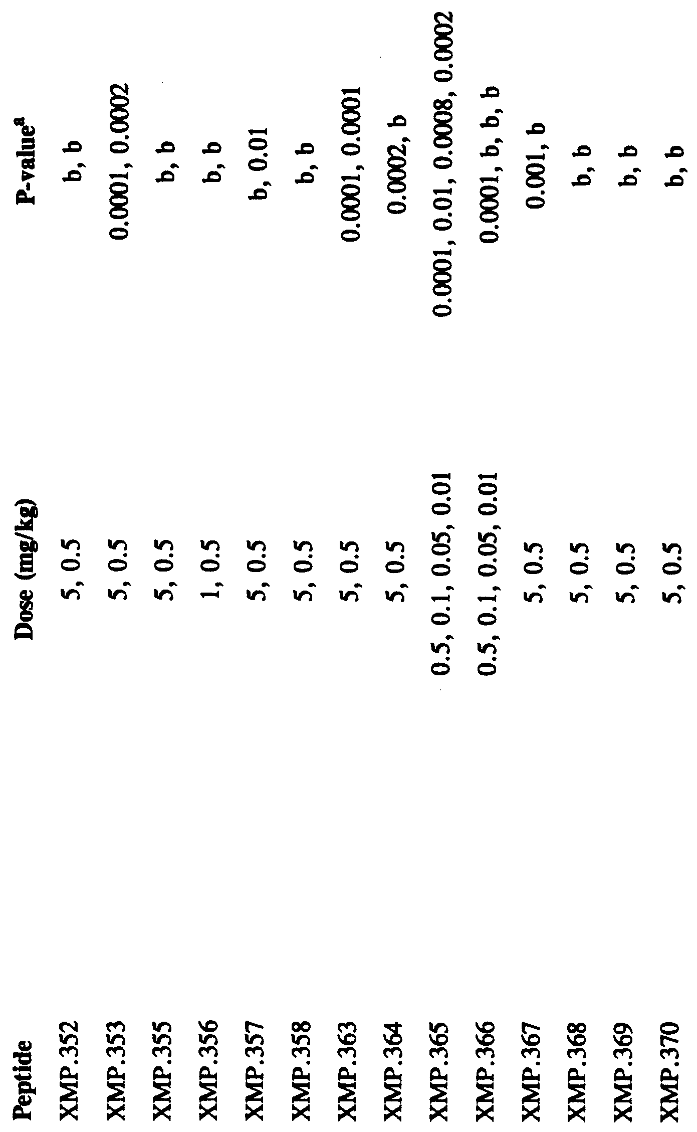


Claims
Priority Applications (4)
| Application Number | Priority Date | Filing Date | Title |
|---|---|---|---|
| AU53675/96A AU727085B2 (en) | 1995-07-20 | 1996-03-21 | Anti-fungal peptides |
| CA002227292A CA2227292A1 (en) | 1995-07-20 | 1996-03-21 | Anti-fungal peptides |
| JP50664097A JP2001520631A (en) | 1995-07-20 | 1996-03-21 | Antifungal peptide |
| EP96910500A EP0839156A1 (en) | 1995-07-20 | 1996-03-21 | Anti-fungal peptides |
Applications Claiming Priority (2)
| Application Number | Priority Date | Filing Date | Title |
|---|---|---|---|
| US50484195A | 1995-07-20 | 1995-07-20 | |
| US08/504,841 | 1995-07-20 |
Publications (1)
| Publication Number | Publication Date |
|---|---|
| WO1997004008A1 true WO1997004008A1 (en) | 1997-02-06 |
Family
ID=24007971
Family Applications (1)
| Application Number | Title | Priority Date | Filing Date |
|---|---|---|---|
| PCT/US1996/003845 WO1997004008A1 (en) | 1995-07-20 | 1996-03-21 | Anti-fungal peptides |
Country Status (6)
| Country | Link |
|---|---|
| US (4) | US5858974A (en) |
| EP (1) | EP0839156A1 (en) |
| JP (1) | JP2001520631A (en) |
| AU (1) | AU727085B2 (en) |
| CA (1) | CA2227292A1 (en) |
| WO (1) | WO1997004008A1 (en) |
Cited By (19)
| Publication number | Priority date | Publication date | Assignee | Title |
|---|---|---|---|---|
| US5856438A (en) * | 1993-03-12 | 1999-01-05 | Xoma Corporation | Biologically active peptides from functional domains of bactericidal/permeability-increasing protein and uses thereof |
| US5858974A (en) * | 1995-07-20 | 1999-01-12 | Xoma Corporation | Anti-fungal peptides |
| US5888973A (en) * | 1996-08-09 | 1999-03-30 | Xoma Corporation | Anti-chlamydial uses of BPI protein products |
| US6013631A (en) * | 1998-06-19 | 2000-01-11 | Xoma Corporation | Bactericidal/permeability-increasing protein (BPI) deletion analogs |
| WO2000018798A1 (en) * | 1998-09-25 | 2000-04-06 | Xoma Technology Ltd. | Antifungal and antibacterial peptide |
| US6093573A (en) * | 1997-06-20 | 2000-07-25 | Xoma | Three-dimensional structure of bactericidal/permeability-increasing protein (BPI) |
| WO2001000671A1 (en) * | 1999-06-25 | 2001-01-04 | Xoma Technology Ltd. | Derivative compounds |
| US6242219B1 (en) | 1999-03-18 | 2001-06-05 | Xoma (Us) Llc | Methods for recombinant peptide production |
| WO2002030975A2 (en) * | 2000-10-11 | 2002-04-18 | Xoma Technology, Ltd. | Antifungal peptides |
| US6423825B1 (en) * | 1999-06-25 | 2002-07-23 | Xoma Technology Ltd. | Therapeutic derivative compounds derived from domain II of bactericidal/permeability-increasing protein |
| US6482796B2 (en) | 1996-11-01 | 2002-11-19 | Xoma Corporation | Therapeutic uses of N-terminal BPI protein products in ANCA-positive patients |
| US6515104B1 (en) * | 1999-06-25 | 2003-02-04 | Xoma Technology Ltd. | Therapeutic peptide-based constructs derived from domain II of bactericidal/permeability-increasing protein |
| US6964948B2 (en) * | 1999-06-25 | 2005-11-15 | Xoma Technology Ltd. | Therapeutic peptide-based constructs derived from Domain II of bactericidal/permeability-increasing protein |
| EP1741440A1 (en) | 2005-07-08 | 2007-01-10 | Mellitus S.L. | Use of BPI protein for the treatment of disorders of the metabolism and cardiovascular disorders |
| US7291451B2 (en) | 2000-12-01 | 2007-11-06 | Xoma Technology Ltd. | Modulation of pericyte proliferation |
| EP2311962A1 (en) | 2002-03-29 | 2011-04-20 | XOMA Technology Ltd. | Multigenic vectors plasmids and methods for increasing expression of recombinant polypeptides |
| WO2013120619A1 (en) * | 2012-02-17 | 2013-08-22 | Institut National De La Recherche Agronomique (Inra) | Anti-oomycete activity of lipopolysaccharide (lps)-binding proteins / bactericidal/permeability-increasing proteins |
| US10450348B2 (en) | 2013-11-06 | 2019-10-22 | Norwegian University Of Science And Technology | Antimicrobial agents and their use in therapy |
| US10517923B2 (en) | 2013-11-06 | 2019-12-31 | Norwegian University Of Science And Technology | Immunosuppressive agents and their use in therapy |
Families Citing this family (19)
| Publication number | Priority date | Publication date | Assignee | Title |
|---|---|---|---|---|
| CN1245074A (en) * | 1998-04-26 | 2000-02-23 | 曾忠铭 | Medicine for reducing acidity in vegina and its usage |
| US7262170B2 (en) * | 1998-04-26 | 2007-08-28 | Zhongming Zeng | Pharmaceutical composition for reducing vaginal acidity and treatment of vaginitis, and the use thereof |
| AU4203900A (en) * | 1999-07-12 | 2001-01-30 | Xoma Technology Ltd. | Method to identify antimicrobial agents |
| AU6928700A (en) * | 1999-08-25 | 2001-03-19 | Clarity Biosciences, Inc. | Identifying organisms by detecting intronic nucleic acid or encoded proteins |
| WO2001098531A1 (en) * | 2000-06-20 | 2001-12-27 | Ortho-Mcneil Pharmaceutical, Inc. | Resazurin-based cytotoxicity assay |
| EP1370679B1 (en) * | 2001-03-01 | 2011-10-05 | Clarity Biosciences, Inc. | Methods of making and uses of compositions that modulate intronic region-encoded protein function |
| US7270975B2 (en) * | 2001-03-09 | 2007-09-18 | Trustees Of Dartmouth College | Methods for regulating bud-hypha transitions and cAMP levels in Candida albicans |
| US6706688B2 (en) | 2001-03-09 | 2004-03-16 | Paula Sundstrom | Methods for regulating bud-hypha transitions and cAMP levels by the adenylate cyclase-associated protein gene, CAP1 |
| US6982152B2 (en) * | 2002-04-17 | 2006-01-03 | Promega Corporation | Cytotoxicity assay |
| CA2515488C (en) * | 2003-02-10 | 2013-04-16 | Raindance Communications, Inc. | Methods and apparatus for providing egalitarian control in a multimedia collaboration session |
| TWI328038B (en) * | 2003-09-17 | 2010-08-01 | Yeastern Biotech Co Ltd | Fungal immunomodulatory protein (fip) prepared by microorganisms and uses thereof |
| US20050282755A1 (en) * | 2004-03-18 | 2005-12-22 | Ansata Therapeutics, Inc. | Compositions having antimicrobial activity and uses thereof |
| AU2006212922B2 (en) * | 2005-02-09 | 2011-04-21 | Helix Biomedix Inc. | Antimicrobial hexapeptides |
| RU2463308C2 (en) * | 2005-12-22 | 2012-10-10 | НоваБиотикс Лимитед | Cyclic antimicrobial peptides |
| GB0526120D0 (en) * | 2005-12-22 | 2006-02-01 | Novabiotics Ltd | Therapeutic peptides |
| US9181303B2 (en) * | 2005-12-22 | 2015-11-10 | Novabiotics Limited | Treatment of bacterial infections with cyclic antimicrobial peptides |
| AU2012203804B2 (en) * | 2005-12-22 | 2014-10-16 | Novabiotics Limited | Cyclic antimicrobial peptides |
| FI20115785A0 (en) * | 2011-08-08 | 2011-08-08 | Thermo Fisher Scientific Oy | Method and apparatus for automatic analysis |
| WO2013021101A1 (en) | 2011-08-08 | 2013-02-14 | Thermo Fisher Scientific Oy | Method and apparatus for automated analysis |
Citations (4)
| Publication number | Priority date | Publication date | Assignee | Title |
|---|---|---|---|---|
| WO1994020532A1 (en) * | 1993-03-12 | 1994-09-15 | Xoma Corporation | Biologically active peptides from functional domains of bactericidal/permeability-increasing protein and uses thereof |
| WO1995019372A1 (en) * | 1994-01-14 | 1995-07-20 | Xoma Corporation | Biologically active peptides from functional domains of bactericidal/permeability-increasing protein and uses thereof |
| WO1995019179A1 (en) * | 1994-01-14 | 1995-07-20 | Xoma Corporation | Anti-fungal methods and materials |
| WO1996008509A1 (en) * | 1994-09-15 | 1996-03-21 | Xoma Corporation | Anti-fungal peptides |
Family Cites Families (44)
| Publication number | Priority date | Publication date | Assignee | Title |
|---|---|---|---|---|
| WO1989001486A1 (en) * | 1987-08-11 | 1989-02-23 | New York University | Biologically active bactericidal/permeability-increasing protein fragments |
| US5198541A (en) * | 1987-08-11 | 1993-03-30 | New York University | Dna encoding bactericidal/permeability-increasing proteins |
| US5576292A (en) * | 1987-08-11 | 1996-11-19 | New York University | Biologically active bactericidal/permeability-increasing protein fragments |
| US5308834A (en) * | 1989-02-14 | 1994-05-03 | Incyte Pharmaceuticals, Inc. | Treatment of endotoxin-associated shock and prevention thereof using a BPI protein |
| US5234912A (en) * | 1989-02-14 | 1993-08-10 | Incyte Pharmaceuticals, Inc. | Pharmaceutical compositions comprising recombinant BPI proteins and a lipid carrier and uses thereof |
| US5089274A (en) * | 1989-02-14 | 1992-02-18 | Incyte Pharmaceuticals, Inc. | Use of bactericidal/permeability increasing protein or biologically active analogs thereof to treat endotoxin-related disorders |
| US6265187B1 (en) * | 1989-02-14 | 2001-07-24 | Incyte Pharmaceuticals, Inc. | Recombinant endotoxin-neutralizing proteins |
| US5171739A (en) * | 1989-02-14 | 1992-12-15 | Incyte Pharmaceuticals, Inc. | Treatment of endotoxin-associated shock and preventation thereof using a BPI protein |
| US5334584A (en) * | 1989-02-14 | 1994-08-02 | Incyte Pharamaceuticals, Inc. | Recombinant, non-glycosylated bpi protein and uses thereof |
| DE69128968T2 (en) * | 1990-12-03 | 1998-10-08 | Univ New York | BIOLOGICALLY ACTIVE BACTERICIDES / PERMEABILITY-INCREASING PROTEIN PIECES |
| JPH07502642A (en) * | 1991-09-26 | 1995-03-23 | インサイト ファーマシューティカルズ,インコーポレイテッド | A novel form of liposaccharide binding protein (LBP) |
| US5643570A (en) * | 1992-05-19 | 1997-07-01 | Xoma Corporation | BPI-immunoglobulin fusion proteins |
| ES2132239T3 (en) * | 1992-05-19 | 1999-08-16 | Xoma Corp | IMPROVED METHODS FOR THE PREPARATION OF BINDING PROTEINS TO ENDOTOXINS. |
| US5420019A (en) * | 1993-02-02 | 1995-05-30 | Xoma Corporation | Stable bactericidal/permeability-increasing protein muteins |
| AU695125B2 (en) * | 1993-02-02 | 1998-08-06 | Xoma Corporation | Pharmaceutical compositions containing bactericidal permeability increasing protein and a surfactant |
| JPH08509703A (en) * | 1993-03-12 | 1996-10-15 | ゾーマ コーポレイション | Sterilization / Therapeutic use of increased permeability protein products |
| US5348942A (en) * | 1993-03-12 | 1994-09-20 | Xoma Corporation | Therapeutic uses of bactericidal/permeability increasing protein products |
| CA2157925C (en) * | 1993-03-12 | 2000-06-06 | Lewis H. Lambert, Jr. | Treatment of mycobacterial diseases by administration of bactericidal/permeability-increasing protein products |
| US5858974A (en) * | 1995-07-20 | 1999-01-12 | Xoma Corporation | Anti-fungal peptides |
| US5652332A (en) * | 1993-03-12 | 1997-07-29 | Xoma | Biologically active peptides from functional domains of bactericidal/permeability-increasing protein and uses thereof |
| WO1994021280A1 (en) * | 1993-03-22 | 1994-09-29 | Incyte Pharmaceuticals, Inc. | Use of bactericidal/permeability increasing protein and lipopolysaccharide binding protein levels and ratios thereof in diagnosis |
| AU6942994A (en) * | 1993-04-30 | 1994-11-21 | Incyte Pharmaceuticals, Inc. | Recombinant bpi-based and lbp-based proteins, nucleic acid molecules encoding same, methods of producing same, and uses thereof |
| AU7175694A (en) * | 1993-06-17 | 1995-01-17 | Xoma Corporation | Lipopolysaccharide binding protein derivatives |
| EP0726944B1 (en) * | 1993-07-02 | 2002-12-18 | Incyte Pharmaceuticals, Inc. | Glycosylated and non-glycosylated bactericidal/permeability increasing proteins, and methods for producing same |
| AU7330994A (en) * | 1993-07-14 | 1995-02-13 | Xoma Corporation | Method for potentiating bpi protein product bactericidal activity by administration of lbp protein products |
| WO1995008344A1 (en) * | 1993-09-22 | 1995-03-30 | Xoma Corporation | Method of treating gram-negative bacterial infection by administration of bactericidal/permeability-increasing (bpi) protein product and antibiotic |
| CN1133634A (en) * | 1993-09-22 | 1996-10-16 | 爱克斯欧玛公司 | Method for quantifying BPI in body fluids |
| US5466580A (en) * | 1993-09-22 | 1995-11-14 | Xoma Corporation | Method for quantifying BPI in body fluids |
| DE69433848T2 (en) * | 1993-10-05 | 2005-05-25 | Xoma Technology Ltd., Berkeley | BPI PROTEIN PRODUCTS FOR DEFINED FEATURES OF THE RETICULOENDOTHELIAL SYSTEM |
| AU703192B2 (en) * | 1994-01-14 | 1999-03-18 | Xoma Corporation | Anti-gram-positive bacterial methods and materials |
| US5484705A (en) * | 1994-01-24 | 1996-01-16 | Xoma Corporation | Method for quantifying lipopolysaccharide binding protein |
| US5643875A (en) * | 1994-01-24 | 1997-07-01 | Friedmann; Nadav | Human therapeutic uses of bactericidal/permeability increasing (BPI) protein products |
| US5447913A (en) * | 1994-03-11 | 1995-09-05 | Xoma Corporation | Therapeutic uses of bactericidal/permeability-increasing protein dimer products |
| US5578568A (en) * | 1994-04-22 | 1996-11-26 | Xoma Corporation | Method of treating conditions associated with intestinal ischemia/reperfusion |
| US5532216A (en) * | 1994-06-24 | 1996-07-02 | Incyte Pharmaceuticals, Inc. | Neutralization of non-lipopolysaccharide compounds by bactericidal/permeability-increasing protein |
| US5646114A (en) * | 1994-07-11 | 1997-07-08 | Xoma Corporation | Anti-protozoan methods |
| US5912228A (en) * | 1995-01-13 | 1999-06-15 | Xoma Corporation | Therapeutic compositions comprising bactericidal/permeability-increasing (BPI) protein products |
| US5494896A (en) * | 1995-03-31 | 1996-02-27 | Xoma Corporation | Method of treating conditions associated with burn injuries |
| EP0861088A1 (en) * | 1995-11-14 | 1998-09-02 | Xoma Corporation | Bactericidal permeability increasing protein (bpi) for treating conditions associated with corneal injury |
| US5686414A (en) * | 1995-11-14 | 1997-11-11 | Xoma Corporation | Methods of treating conditions associated with corneal transplantation |
| US5851802A (en) * | 1996-03-22 | 1998-12-22 | Xoma Corporation | Methods for recombinant microbial production of fusion proteins and BPI-derived peptides |
| CA2253837C (en) * | 1996-05-10 | 2002-07-02 | Xoma Corporation | Therapeutic uses of bpi protein products for human meningococcemia |
| US5741779A (en) * | 1996-05-10 | 1998-04-21 | Xoma Corporation | Antithrombotic materials and methods |
| CA2255539C (en) * | 1996-05-23 | 2005-09-27 | Xoma Corporation | Therapeutic uses of bpi protein products in humans with hemorrhage due to trauma |
-
1996
- 1996-03-21 US US08/621,259 patent/US5858974A/en not_active Expired - Fee Related
- 1996-03-21 CA CA002227292A patent/CA2227292A1/en not_active Abandoned
- 1996-03-21 WO PCT/US1996/003845 patent/WO1997004008A1/en not_active Application Discontinuation
- 1996-03-21 JP JP50664097A patent/JP2001520631A/en active Pending
- 1996-03-21 AU AU53675/96A patent/AU727085B2/en not_active Ceased
- 1996-03-21 EP EP96910500A patent/EP0839156A1/en not_active Withdrawn
-
1999
- 1999-01-08 US US09/227,659 patent/US6156730A/en not_active Expired - Fee Related
-
2000
- 2000-10-02 US US09/677,664 patent/US6664231B1/en not_active Expired - Fee Related
-
2001
- 2001-06-14 US US09/881,490 patent/US20020077298A1/en not_active Abandoned
Patent Citations (4)
| Publication number | Priority date | Publication date | Assignee | Title |
|---|---|---|---|---|
| WO1994020532A1 (en) * | 1993-03-12 | 1994-09-15 | Xoma Corporation | Biologically active peptides from functional domains of bactericidal/permeability-increasing protein and uses thereof |
| WO1995019372A1 (en) * | 1994-01-14 | 1995-07-20 | Xoma Corporation | Biologically active peptides from functional domains of bactericidal/permeability-increasing protein and uses thereof |
| WO1995019179A1 (en) * | 1994-01-14 | 1995-07-20 | Xoma Corporation | Anti-fungal methods and materials |
| WO1996008509A1 (en) * | 1994-09-15 | 1996-03-21 | Xoma Corporation | Anti-fungal peptides |
Non-Patent Citations (1)
| Title |
|---|
| R.G. LITTLE ET AL.: "Functional Domains of Recombinant Bactericidal/Permeability Increasing Protein (rBPI23)", JOURNAL OF BIOLOGICAL CHEMISTRY, vol. 269, no. 3, 21 January 1994 (1994-01-21), BALTIMORE, MD US, pages 1865 - 1872, XP002008631 * |
Cited By (31)
| Publication number | Priority date | Publication date | Assignee | Title |
|---|---|---|---|---|
| US5856438A (en) * | 1993-03-12 | 1999-01-05 | Xoma Corporation | Biologically active peptides from functional domains of bactericidal/permeability-increasing protein and uses thereof |
| US5858974A (en) * | 1995-07-20 | 1999-01-12 | Xoma Corporation | Anti-fungal peptides |
| US5888973A (en) * | 1996-08-09 | 1999-03-30 | Xoma Corporation | Anti-chlamydial uses of BPI protein products |
| US6162788A (en) * | 1996-08-09 | 2000-12-19 | Xoma Corporation | Uses of BPI protein products |
| US6583116B1 (en) | 1996-08-09 | 2003-06-24 | Xoma Corporation | Anti-chlamydial methods and materials |
| US6482796B2 (en) | 1996-11-01 | 2002-11-19 | Xoma Corporation | Therapeutic uses of N-terminal BPI protein products in ANCA-positive patients |
| US6093573A (en) * | 1997-06-20 | 2000-07-25 | Xoma | Three-dimensional structure of bactericidal/permeability-increasing protein (BPI) |
| US6013631A (en) * | 1998-06-19 | 2000-01-11 | Xoma Corporation | Bactericidal/permeability-increasing protein (BPI) deletion analogs |
| US6087126A (en) * | 1998-06-19 | 2000-07-11 | Xoma Corporation | Bactericidal/permeability-increasing protein (BPI) deletion analogs |
| US6599880B1 (en) | 1998-06-19 | 2003-07-29 | Xoma Corporation | Bactericidal/permeability-increasing protein (BPI) deletion analogs |
| WO2000018798A1 (en) * | 1998-09-25 | 2000-04-06 | Xoma Technology Ltd. | Antifungal and antibacterial peptide |
| US6500648B1 (en) | 1999-03-18 | 2002-12-31 | Xoma Technology, Ltd. | Methods for recombinant peptide production |
| US7749731B2 (en) | 1999-03-18 | 2010-07-06 | Xoma Technology Ltd. | Methods for recombinant peptide production |
| US6242219B1 (en) | 1999-03-18 | 2001-06-05 | Xoma (Us) Llc | Methods for recombinant peptide production |
| US6964948B2 (en) * | 1999-06-25 | 2005-11-15 | Xoma Technology Ltd. | Therapeutic peptide-based constructs derived from Domain II of bactericidal/permeability-increasing protein |
| US6355616B1 (en) | 1999-06-25 | 2002-03-12 | Xoma (Us) Technology Ltd. | Derivative compounds derived from or based on bactericidal/permeability-increasing protein |
| US6515104B1 (en) * | 1999-06-25 | 2003-02-04 | Xoma Technology Ltd. | Therapeutic peptide-based constructs derived from domain II of bactericidal/permeability-increasing protein |
| US6423825B1 (en) * | 1999-06-25 | 2002-07-23 | Xoma Technology Ltd. | Therapeutic derivative compounds derived from domain II of bactericidal/permeability-increasing protein |
| WO2001000671A1 (en) * | 1999-06-25 | 2001-01-04 | Xoma Technology Ltd. | Derivative compounds |
| US6906037B2 (en) * | 1999-06-25 | 2005-06-14 | Xoma Technology Ltd. | Therapeutic peptide-based constructs |
| WO2002030975A3 (en) * | 2000-10-11 | 2003-04-24 | Xoma Technology Ltd | Antifungal peptides |
| WO2002030975A2 (en) * | 2000-10-11 | 2002-04-18 | Xoma Technology, Ltd. | Antifungal peptides |
| US7291451B2 (en) | 2000-12-01 | 2007-11-06 | Xoma Technology Ltd. | Modulation of pericyte proliferation |
| EP2311962A1 (en) | 2002-03-29 | 2011-04-20 | XOMA Technology Ltd. | Multigenic vectors plasmids and methods for increasing expression of recombinant polypeptides |
| EP1741440A1 (en) | 2005-07-08 | 2007-01-10 | Mellitus S.L. | Use of BPI protein for the treatment of disorders of the metabolism and cardiovascular disorders |
| WO2013120619A1 (en) * | 2012-02-17 | 2013-08-22 | Institut National De La Recherche Agronomique (Inra) | Anti-oomycete activity of lipopolysaccharide (lps)-binding proteins / bactericidal/permeability-increasing proteins |
| FR2986970A1 (en) * | 2012-02-17 | 2013-08-23 | Agronomique Inst Nat Rech | ANTI-OOMYCTIC ACTIVITY OF LIPOPOLYSACCHARIDES (LPS) -BINDING PROTEINS / BACTERICIDAL / PERMEABILITY-INCREASING PROTEINS |
| US10450348B2 (en) | 2013-11-06 | 2019-10-22 | Norwegian University Of Science And Technology | Antimicrobial agents and their use in therapy |
| US10517923B2 (en) | 2013-11-06 | 2019-12-31 | Norwegian University Of Science And Technology | Immunosuppressive agents and their use in therapy |
| US11246907B2 (en) | 2013-11-06 | 2022-02-15 | Norwegian University Of Science And Technology | Immunosuppressive agents and their use in therapy |
| US11337427B2 (en) | 2013-11-06 | 2022-05-24 | Norwegian University Of Science And Technology | Antimicrobial agents and their use in therapy |
Also Published As
| Publication number | Publication date |
|---|---|
| US5858974A (en) | 1999-01-12 |
| US6156730A (en) | 2000-12-05 |
| EP0839156A1 (en) | 1998-05-06 |
| CA2227292A1 (en) | 1997-02-06 |
| JP2001520631A (en) | 2001-10-30 |
| US6664231B1 (en) | 2003-12-16 |
| AU5367596A (en) | 1997-02-18 |
| AU727085B2 (en) | 2000-11-30 |
| US20020077298A1 (en) | 2002-06-20 |
Similar Documents
| Publication | Publication Date | Title |
|---|---|---|
| US5858974A (en) | Anti-fungal peptides | |
| EP0754049B1 (en) | Anti-fungal methods and materials | |
| KR101306643B1 (en) | Antimicrobial hexapeptides | |
| EP0824547B1 (en) | Anti-fungal peptides | |
| JP2002544759A (en) | Compositions and methods for treating infections using cationic peptides alone or in combination with antibiotics | |
| JPH11511971A (en) | Antimicrobial cationic peptide and screening method thereof | |
| CA2319094C (en) | Antimicrobial peptides | |
| Saido-Sakanaka et al. | Synthesis and characterization of bactericidal oligopeptides designed on the basis of an insect anti-bacterial peptide | |
| US5994308A (en) | Broad spectrum antimicrobial peptides containing a tryptophan triplet and methods of use | |
| US5792831A (en) | Analogues of magainin peptides containing D-amino acids | |
| WO1997031942A9 (en) | Broad spectrum antimicrobial peptides containing a tryptophan triplet and methods of use | |
| US6355616B1 (en) | Derivative compounds derived from or based on bactericidal/permeability-increasing protein | |
| JPH09507669A (en) | Amphipathic ion channel-forming peptide with N-terminal modification | |
| EP0533795A4 (en) | Composition of and treatment with biologically active peptides having d-amino acid residues | |
| JP2005532784A (en) | New antibacterial voricin peptide |
Legal Events
| Date | Code | Title | Description |
|---|---|---|---|
| AK | Designated states |
Kind code of ref document: A1 Designated state(s): AL AM AT AU AZ BB BG BR BY CA CH CN CZ DE DK EE ES FI GB GE HU IS JP KE KG KP KR KZ LK LR LS LT LU LV MD MG MK MN MW MX NO NZ PL PT RO RU SD SE SG SI SK TJ TM TR TT UA UG UZ VN AM AZ BY KG KZ MD RU TJ TM |
|
| AL | Designated countries for regional patents |
Kind code of ref document: A1 Designated state(s): KE LS MW SD SZ UG AT BE CH DE DK ES FI FR GB GR IE IT LU MC NL PT SE BF BJ CF CG CI CM GA GN ML |
|
| DFPE | Request for preliminary examination filed prior to expiration of 19th month from priority date (pct application filed before 20040101) | ||
| 121 | Ep: the epo has been informed by wipo that ep was designated in this application | ||
| WWE | Wipo information: entry into national phase |
Ref document number: 1996910500 Country of ref document: EP |
|
| ENP | Entry into the national phase |
Ref document number: 2227292 Country of ref document: CA Ref country code: CA Ref document number: 2227292 Kind code of ref document: A Format of ref document f/p: F Ref country code: JP Ref document number: 1997 506640 Kind code of ref document: A Format of ref document f/p: F |
|
| WWP | Wipo information: published in national office |
Ref document number: 1996910500 Country of ref document: EP |
|
| REG | Reference to national code |
Ref country code: DE Ref legal event code: 8642 |
|
| WWW | Wipo information: withdrawn in national office |
Ref document number: 1996910500 Country of ref document: EP |