WO1986006605A1 - Hepatobiliary nmr contrast agents - Google Patents
Hepatobiliary nmr contrast agents Download PDFInfo
- Publication number
- WO1986006605A1 WO1986006605A1 PCT/US1986/001035 US8601035W WO8606605A1 WO 1986006605 A1 WO1986006605 A1 WO 1986006605A1 US 8601035 W US8601035 W US 8601035W WO 8606605 A1 WO8606605 A1 WO 8606605A1
- Authority
- WO
- WIPO (PCT)
- Prior art keywords
- agent
- protein
- contrast agent
- nmr
- binding
- Prior art date
Links
Classifications
-
- A—HUMAN NECESSITIES
- A61—MEDICAL OR VETERINARY SCIENCE; HYGIENE
- A61K—PREPARATIONS FOR MEDICAL, DENTAL OR TOILETRY PURPOSES
- A61K49/00—Preparations for testing in vivo
- A61K49/06—Nuclear magnetic resonance [NMR] contrast preparations; Magnetic resonance imaging [MRI] contrast preparations
Definitions
- This invention relates to diagnostic NMR imaging.
- NMR imaging has been used in medical diagnosis for a number of years.
- contrast agents to enhance its diagnostic utility has only recently appeared.
- German Patent DE 3,129,906 describes NMR contrast agents which consist of a paramagnetic ion complexed with a chelating agent and a base or acid, e.g., the di-N-methylglucosamine salt of manganese chelated with EDTA.
- the present invention provides an in vivo method of decreasing the NMR relaxation times of water protons in contact with a biological tissue.
- the method involves administering to a human patient an NMR contrast agent containing a paramagnetic metal ion complexed with a chelating substance, the contrast agent being characterized in that it is capable of binding non-covalently and non-immunologically to a component of the tissue, and as a result of such binding is capable of enhancing the relaxivity (i.e., decreasing the NMR relaxation times T 1 or T 2 ) of the water protons by a factor of at least 2, compared to the relaxivity induced in such water protons by the paramagnetic substance alone free in solution; and subjecting the patient to NMR imaging.
- the contrast agent has a specific affinity for the biological tissue in which binding occurs.
- specific affinity means capable of being taken up by, retained by, or bound to a particular tissue or tissue component to a substantially greater degree than other tissue or tissue components; agents which have this property are said to be “targeted” to the "target” tissue or component.
- the components to which the agents of the invention bind are generally particular chemical classes, e.g., proteins, lipids, or polysaccharides. It has been found that the tight binding of the agents to these components causes an increase (at least by a factor of 2) in the longitudinal (1/T 1 ) and transverse (1/T 2 ) relaxivity of water protons by the metal complex. Relaxivity enhancement is apparently due in large part to an alteration in the effective correlation time of the electron-nuclear interaction, as described in Lauffer et al. (1985) Magn. Res. Imaging 3, 11.
- the toxic paramagnetic ion e.g., gadolinium
- a chelating agent to reduce toxicity; it has been found that such agents are effective in reducing T 1 and T 2 (discussed below), despite the relatively lower accessibility of the paramagnetic ion to the surrounding water protons.
- Examples of classes of chelating substances of the invention are porphyrins, cryptate compounds, and bis, tris, or tetra-catechol compounds.
- the contrast agents of the invention which bind tightly to proteins are also taken up specifically by human hepatocytes, compared to human reticuloendothelial cells, and, because hepatocytes make up the bulk of the liver, provide superior NMR imaging of the liver.
- the agents thus allow visualization of hepatocarcinoma or metastatic tumors of the liver, whose cells take up the agents at a different rate, or retain the agent for a different length of time, than normally functioning hepatocytes.
- the invention also allows the use of NMR imaging to monitor liver function, as manifested by uptake or retention rates of the contrast agents of the invention.
- agents of the invention will have utility in a wide range of applications, because the chemical requirements for tight binding to many components are the same, and also because in some instances the same properties which induce tight binding also influence tissue specificity.
- the properties of agents which cause selective uptake by hepatocytes compared to reticuloendothelial cells also cause tight binding of the agents to proteins, e.g., intracellular proteins of hepatocytes.
- the preferred NMR contrast agents of the invention possess a number of physical/chemical properties, discussed below, related to their utility in diagnostic applications.
- agents which are targeted to provide the NMR contrast needed for imaging they must alter the proton NMR relaxation time in the target component.
- the agents must have properties which cause them to selectively be taken up by or bound to the target. This is achieved either by means of a higher rate of uptake of the contrast agent by the target, or by a different retention profile between target and non-target tissues.
- NMR contrast is achieved by the altering, by the paramagnetic portion of the agent, of T 1 (longitudinal relaxation time) or T 2 (transverse relaxation time) of the water protons in the target.
- T 1 longitudinal relaxation time
- T 2 transverse relaxation time
- proteins can be intracellular proteins, e.g., the proteins such as ligandin (also known as Y protein or glutathione-S-transferase (EC 2.5.1.18) and Protein A (also known as Z protein or fatty acid binding protein) inside hepatocytes (J. Clin. Invest. 48[, 2156-2167
- the agents are targeted to particular cells such as hepatocytes, it is generally the cells, and not the intracellular proteins themselves, to which the agents are targeted as a result of the properties of the agents, which properties in turn cause tight binding to the intracellular proteins of those cells.
- Agents which have protein-binding properties can bind not only to intracellular proteins but also to serum proteins such as human serum albumin (HSA).
- HSA human serum albumin
- This binding provides selective enhancement of intravascular structures or patterns on NMR images, permitting diagnosis of blood/brain barrier disruptions caused, e.g., by strokes and brain tumors, and also permitting flow imaging of the blood.
- some agents can bind to both HSA and ligandin in vivo, and thus represent dual intravascular-hepatobiliary agents.
- Another important protein which is bound tightly by the protein-binding agents is the immature, poorly cross-linked collagen present in tumors. This collagen can be bound tightly by NMR contrast agents which comprise a paramagnetic metal ion complexed with a porphyrin. When these proteins are bound, the agent serves the dual roles of tumor targeting and relaxivity enhancement.
- Protein binding is provided for by the incorporation of hydrophobic groups into the agent, and providing the agent with the proper net charge.
- Binding is promoted when both the contrast agent and the protein contain one or more hydrophobic domains; the contrast agent binds non-covalently to the protein through Van der Waals interactions between the hydrophobic domains, thus enhancing binding.
- lipophilicity enhances binding of the contrast agents to the protein.
- Lipophilicity is provided by a non-polar structure, the presence of at least one aryl group (e.g., a substituted or unsubstituted phenyl ring), at least one halogen atom, and/or hydrophobic alkyl groups.
- the contrast agent it is also desirable that the contrast agent not carry excessive charge, i.e., of absolute value greater than 4, at physiological pH.
- Lipophilicity is expressed in terms of octanol:water coefficient, determined by introducing a small amount ( 0.1 mM) of the radiolabeled contrast agent into equal volumes of octanol and Tris buffer (50 mM, pH 7.4).
- the coefficient of the agents of the invention is preferably at least 0.005, and more preferably at least 0.01.
- Binding capacity can be expressed as the percentage of the agent bound to 4.5% human serum albumin (HSA) at a concentration of 0.2 mM of the agent, as determined by equilibrium dialysis.
- HSA human serum albumin
- Electrostatic Interactions Binding may be further increased if electrostatic interactions between the contrast agent and protein are possible.
- the net charge on the agent should be negative, preferably -1 to -4.
- additional negatively charged groups e.g., sulfonate or carboxylate
- the agent should have overall positive charge.
- the agents preferably have a molecular weight of at least 250, and more preferably over 300. Solubility
- the agents should have good water solubility, and preferably should be soluble to a concentration of at least 1.0 mM in normal saline at 20°C. Relaxivity
- contrast agents of the invention must, as mentioned above, lower either T 1 or T 2 or both.
- the ability to achieve this is referred to as "relaxivity.”
- Relaxivity is optimal where the paramagnetic ion, when bound to the chelating ligand, still has one or more open coordination sites for water exchange. Generally, one or two such sites are preferred, since the presence of more than two open sites in general will unacceptably increase toxicity by release of the metal ion in vivo. However, zero open coordination sites may also be satisfactory, though not preferable, since second coordination sphere water molecules are still relaxed and binding-enhancement is still possible.
- In vitro relaxivity is expressed in units of s -1 mM -1 , or change in 1/T 1 or 1/T 2 per mM agent, as measured in saline at 20 MHz.
- the agents Preferably have an in vitro relaxivity of at least 0.5 s -1 mM -1 , more preferably at least 1.0 s -1 mM -1 .
- Relaxivity can also be measured in vivo for the tissue component of interest.
- In vivo relaxivity is expressed in units of s -1 (mmol/gram of tissue) -1 , representing the change in 1/T 1 or 1/T 2 above that of saline-injected controls caused by the agents, divided by the concentration of the agent (in mmol/gram of tissue). Tissue concentration is measured using agents made with radiolabeled paramagnetic ions.
- the in vivo relaxivity of the agents in liver tissue is at least 1.0 s -1 (mmol/g) -1 . The agents should bind sufficiently tightly to enhance relaxivity by a factor of at least
- the contrast agent with at least one aryl or aliphatic group which makes multiple contacts with the biological binding site, preventing free rotation.
- free (non-coordinating) charged groups e.g., sulfonate or carboxylate
- a different strategy to increase the relaxivity of metal complexes is to alter the configuration of the donor atoms around the metal ions to achieve the most symmetrical orientation. This symmetry of the ligand field may lead to longer electron spin relaxation times, and higher relaxivities.
- symmetry-constrained macrocyclic ligands like DOTA are an example in which the symmetry is very high (almost cubic) compared to, e.g., DTPA-derived ligands (described below), which wrap around the metal ion in an anisotropic fashion.
- DTPA-derived ligands described below
- An additional benefit of symmetry-constrained macrocyclic ligands like DOTA is their high kinetic stability (vide infra).
- the contrast agents must have acceptably low toxicity levels at the dosage required for contrast enhancement, and preferably have an LD 50 of at least 0.05 mmol/kg.
- Toxicity of the contrast agents is a function of both the inherent toxicity of the intact complex, and of the degree to which the metal ion dissociates from the chelating agent; toxicity generally increases with the degree of dissociation.
- a high thermodynamic stability a formation constant of at least 10 15 M -1 , and more preferably at least 10 20 M -1 ) is desirable to minimize dissociation and its attendant toxicity.
- Kinetically stable complexes generally contain a paramagnetic metal ion, e.g., gadolinium (III), complexed with a highly constrictive chelating agent, e.g., dibenzo-1, 4, 7,
- Toxicity is also a function of the number of open coordination sites in the complex; the fewer open coordination sites, the less tendency there is, generally, for the chelating agent to release the cytotoxic paramagnetic ion.
- the complex contains two, one, or zero open coordination sites.
- the presence of one or even two open coordination sites can be acceptable in agents in which the paramagnetic substance has a high magnetic moment (i.e., is strongly paramagnetic), and can thus affect T 1 or T 2 at a low dosage; an example is gadolinium, which is strongly paramagnetic owing to its seven unpaired electrons.
- the paramagnetic portion of the contrast agents of the invention can be any paramagnetic ion of the transition metal or lanthanide series which has at least one, and more preferably five or more, unpaired electrons, and a magnetic moment of at least 1.7 Bohr magneton.
- Suitable ions include gadolinium (III), iron (III), manganese (II and III), chromium (III), copper (II), dysprosium (III), terbium (III), holmium (III), erbrium (III), and europium (III); most preferred are gadolinium (III), and iron (III), and manganese (II).
- the organic chelating ligand should be physiologically compatible and preferably contains at least 1 aryl ring which may be substituted with halogen atoms and/or C 1 -C 10 alkyl groups.
- the molecular size of the chelating ligand should be compatible with the size of the paramagnetic substance.
- gadolinium (III) which has a crystal ionic radius of 0.938 ⁇ , requires a larger chelating ligand than iron (III), which has a crystal ionic radius of 0.64 ⁇ .
- the chelating ligand is a single multidentate ligand. Such ligands maximize the stability of the contract agents towards hydrolysis, and minimize the transfer of the metal ion from the contrast agent to binding sites on the target component.
- EHPG ethylenebis-(2-hydroxyphenylglycine)
- 5-Cl-EHPG 5-Br-EHPG
- 5-Me-EHPG 5-t-Bu-EHPG
- 5-sec-Bu-EHPG 5-sec-Bu-EHPG
- substitution at the 5 position of EHPG is the most effective in increasing lipophilicity
- substitution at any position on the two phenyl rings can be used.
- chelating ligands are benzodiethylenetriamine-pentaacetic acid (benzo-DTPA) and derivatives thereof, including dibenzo-DTPA; phenyl-DTPA; diphenyl-DTPA; benzyl-DTPA; and dibenzyl-DTPA.
- benzo-DTPA benzodiethylenetriamine-pentaacetic acid
- Two of these compounds have the structures shown below:
- HBED bis-2 (hydroxybenzyl)-ethylene-diaminediacetic acid
- the HBED ligand advantageously has a very high formation constant for iron of 10 40.
- This ligand is available from the Strem Chemical Company.
- Another suitable class of chelating ligands is the class of macrocyclic compounds which contain at least 3 carbon atoms, more preferably at least 6, and at least two hetero (O and/or N) atoms.
- the macrocyclic compounds can consist of one ring, or two or three rings joined together at the hetero ring elements.
- One suitable class of mono-macrocyclic chelating ligands has the general formula
- A is -N- or X is 0 or 1, each R 5, R 6 ,
- R 5 -CH-CO 2 H R 7 , and R 8 independently, is H or methyl, and each, R 1 , R 2 , R 3 , and R 4 , independently, is ethyl, propyl, butyl, pentyl, or , provided that when A , at least one R group must be
- the aryl groups may be substituted with halogen atoms or C 1 -C 4 alkyl groups.
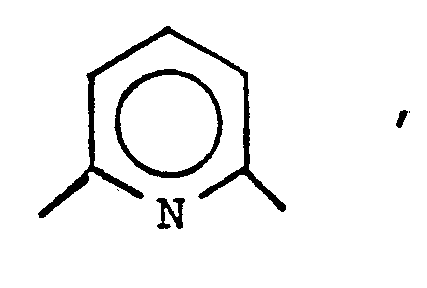
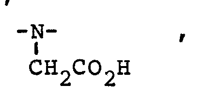
- suitable macrocyclic ligands include benzo-DOTA, where DOTA is 1, 4, 7, 10-tetraazacyclotetradecane-1, 4, 7, 10-tetraacetic acid; dibenzo-DOTA; benzo-NOTA, where NOTA is 1, 4, 7-triazacyclononane- N, N', N''-triacetic acid; benzo-TETA, where TETA is 1, 4, 8, 11-tetraazacyclotetradecane- 1, 4, 8, 11-tetraacetic acid; benzo-DOTMA, where DOTMA is 1, 4, 7, 10-tetraazacyclotetradecane-1, 4, 7, 10-tetra (methyl tetraacetic acid); and benzo-TETMA, where TETMA is 1, 4, 8, 11-tetraazacyclotetradecane-1, 4, 8, 11-(methyl tetraacetic acid).
- DOTA is 1, 4, 7, 10-tetraazacyclotetradecane-1, 4, 7, 10-tetraacetic acid
- DOTA Hydrophobicity, and thus lipophilicity, can also be provided, in the case of ligands (e.g., DOTA derivatives) containing ethylenediamine portions by attaching the above hydrophobic substituents directly to the ethylene carbon atoms.
- DOTA has the structure:
- Hydrophobic substituents e.g., fused phenyl rings or C 1-5 alkyl groups, can be attached to one or more of carbon atoms 1-8 of DOTA.
- DTPA derivatives containing hydrophobic substituents are DTPA derivatives containing hydrophobic substituents. Structures of suitable such derivatives are given below, in which each R 1 , R 2 , R 3 , R 4 and R 5 , independently, can be a C 6-10 aryl group, e.g., phenyl or benzyl; or a C 1-5 aliphatic group, e.g., methyl or ethyl.
- R 1 , R 2 , R 3 , R 4 , R 5 , R 6 , and R 7 group can be a C 6-10 aryl group, e.g., phenyl or benzyl; or a C 1-5 aliphatic group, e.g., methyl or ethyl.
- Another suitable class of chelating ligands are derivatives of
- R 1 , R 2 , R 3 , R 4 , R 5 , and R 6 independently, can be CO 2 H, SO 3 H, H, a halogen, e.g., Cl, or a C 1-5 alkyl group, e.g., methyl or ethyl.
- the contrast agents of the invention can be synthesized from commercially available or readily synthesized reagents using conventional synthetic methods.
- a salt of the paramagnetic ion is added to a slightly alkaline (pH 7.4-9) aqueous solution of the chelating ligand and the resulting mixture is stirred for 3-24 hours at room temperature.
- the resulting contrast agent is then used immediately or stored in lyophilized form or in physiological buffer until use.
- EHPG iron (III) - (EHPG) - is carried out as follows.
- EHPG Sigma
- EHPG Sigma
- Solid FeCl 3 .6H 2 O is added to the solution and the pH adjusted to 7.4 with 1M NaOH.
- the resulting dark red solution is then stirred at room temperature for 30 minutes, after which it is filtered with 0.2 mm micropore filters (Gelman).
- concentration of iron (III)- (EHPG)- is determined by visible absorption of diluted aliquots using a Beckman Spectrophotometer and an extinction coefficient at 480 nm of 4300 CM -1 M -1 .
- the first step is to make the appropriate EHPG derivative, according to Mannich reaction, described in Theodorakis et al. (1980) J. Pharm. Sci 69, 581; the reaction employs ethylenediamine, dichloroacetic acid, and the appropriate parasubstituted phenol.
- the reaction scheme for 5-Br-EHPG is:
- Iron (III)-(5-Cl-EHPG)-, iron (III)-(5-Bu-EHPG)-, iron (III) - (5-Me-EHPG)-, and iron (III) -HBED are prepared in analogous fashion to iron-EHPG.
- iron-EHPG The structure of iron-EHPG is:
- HSA binding percentages of Iron-EHPG, Iron- (5-Br-EHPG), and Iron (HBED) are shown below:
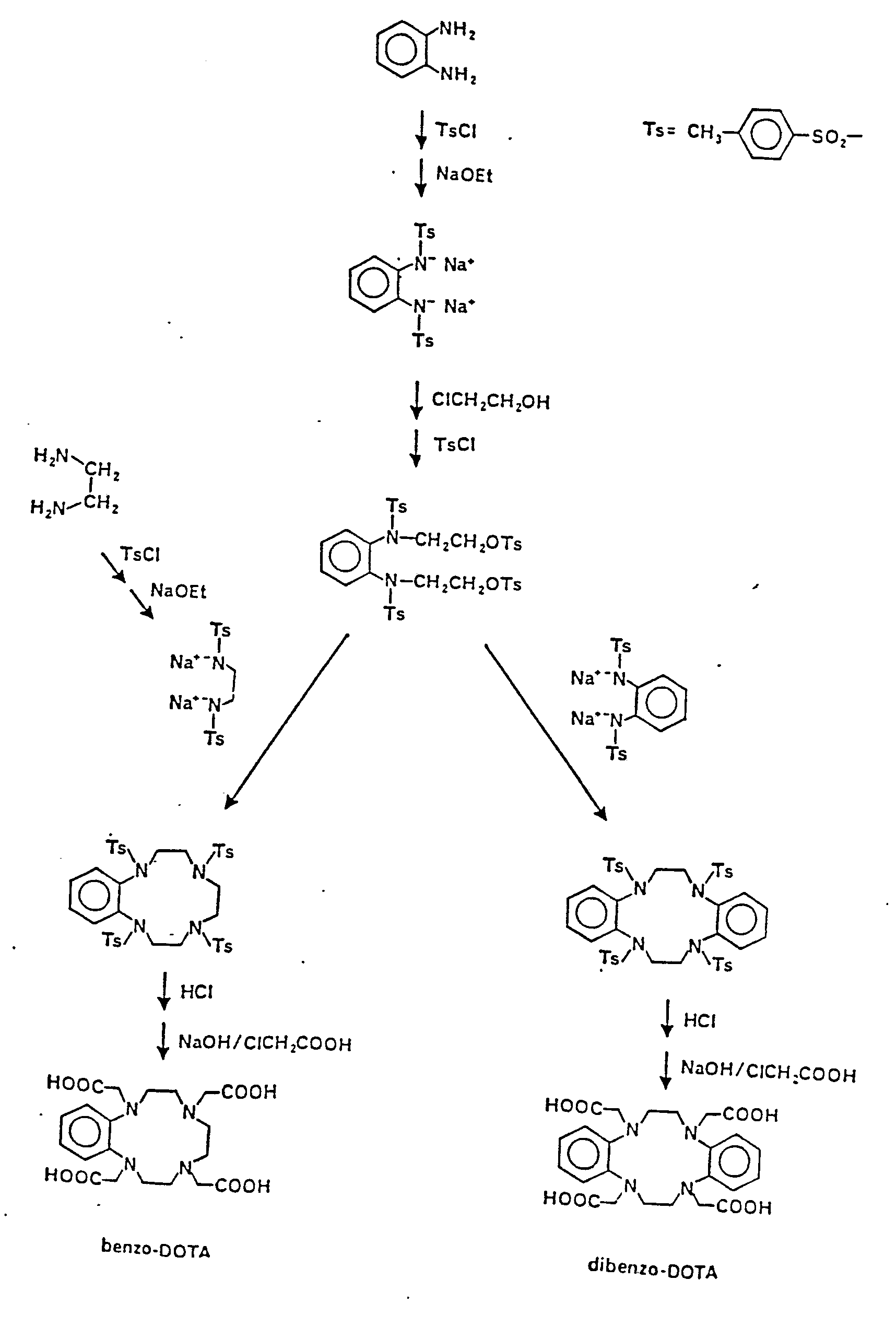
- the macrocyclic DOTA chelating ligands are synthesized generally as described in Desreux et al. (1984) Inorg. Chem. 19, 1319, generally according to the reaction
- DOTA itself lacks sufficient lipophilic groups for hepatocellular uptake.
- Two derivatives with the required lipophilicity are made according to the following general reaction scheme.
- hydrophobic substituents can be incorporated into, e.g., DOTA, via substituted ethylenediamines prepared according to Meares et al. (Anal. Biochem. 100 152-159 (1979).)
- DTPA derivatives e.g., benzo-DTPA and dibenzo-DTPA are made by methods analogous to the methods used for making benzo-EDTA (McCandlish et al. (1978) Inorg. Chem. 17, 1383).
- Paramagnetic ion chelating ligand complexes made using DOTA derivatives are made generally as described earlier, with a longer time (24 hours) and higher reaction temperatures being required for the formation of metal ion/macrocyclic ligand complexes.
- a reaction scheme is shown below: Use
- the contrast agents of the invention are administered orally or intravenously in physiological buffer. Dosage depends on the sensitivity of the NMR imaging instrumentation, as well as on the composition of the contrast agent.
- a contrast agent containing a highly paramagnetic substance e.g., gadolinium (III)
- gadolinium (III) generally requires a lower dosage than a contrast agent containing a paramagnetic substance with a lower magnetic moment, e.g., iron (III).
- dosage will be in the range of about .001-1 mmol/kg, more preferably about 0.005 - 0.05 mmol/kg.
- TI inversion time
- repetition time TR
- TI and TR will remain approximately the same for both T 1 - and T 2 -weighted images; TI and TR are generally on the order of about 200-600 and 100-1000 milliseconds, respectively.
- Iron (III)-(EHPG) was prepared as described above and used for in vivo imaging of rat livers as follows. Fasted male Sprague-Dawley rats (of average weight of about 400g) were anesthetized with intraperitoneal pentobarbitol (50 mg/kg), placed on a calibrated carrier, and subjected to NMR imaging, along with calibration tubes containing paramagnetically-doped water or agar gels of known T 1 and T 2 , to establish an initial baseline image. NMR imaging was performed with a horizontal bore (8cm) superconducting magnet system (Technicare Corp.) at a magnetic field strength of 1.4 tesla ( 1 H resonance of 61.4 MHz).
- Images were obtained using a 2-D Fournier transform technique with a slice selection determined by selective irradiation. All images were obtained using 128 phase encoded gradient steps. To maximize T 1 contrast, an IR pulse sequence was used (TE 15 msec, TI 400 msec, TR 1000 msec).
- iron (III)-(EHPG)- iron (III)-(DTPA) -2 distributes throughout the extracellular liver space, rather than in functioning hepa-tocytes, and is rapidly excreted into the urine.
- T 1 and T 2 values of excised rat liver, blood, spleen, and thigh muscle at various post-injection times also demonstrated that iron (III)-(EHPG)- is predominantly taken up by functioning hepatocytes, and thus decreases the relaxation times of water protons in these cells.
Abstract
A method of decreasing the NMR relaxation times (T1 or T2) of water protons in contact with a biological tissue, the method involving administering to a human patient an NMR contrast agent comprising a paramagnetic ion complexed with a chelating substance, the contrast agent being characterized in that it is capable of binding non-covalently and non-immunologically to a component of the tissue, and as a result of such binding is capable of enhancing relaxivity of the water protons by a factor of at least 2, compared to the relaxivity induced by the paramagnetic substance alone free in solution, and subjecting the patient to NMR imaging.
Description
HEPATOBILIARY NMR CONTRAST AGENTS
Background of the Invention This invention relates to diagnostic NMR imaging.
NMR imaging has been used in medical diagnosis for a number of years. The use of contrast agents to enhance its diagnostic utility has only recently appeared. For example, Gries et al. German Patent DE 3,129,906 describes NMR contrast agents which consist of a paramagnetic ion complexed with a chelating agent and a base or acid, e.g., the di-N-methylglucosamine salt of manganese chelated with EDTA.
Summary of the Invention The present invention provides an in vivo method of decreasing the NMR relaxation times of water protons in contact with a biological tissue. The method involves administering to a human patient an NMR contrast agent containing a paramagnetic metal ion complexed with a chelating substance, the contrast agent being characterized in that it is capable of binding non-covalently and non-immunologically to a component of the tissue, and as a result of such binding is capable of enhancing the relaxivity (i.e., decreasing the NMR relaxation times T1 or T2) of the water protons by a factor of at least 2, compared to the relaxivity induced in such water protons by the paramagnetic substance alone free in solution; and subjecting the patient to NMR imaging.
Preferably, the contrast agent has a specific affinity for the biological tissue in which binding occurs. (As used herein, "specific affinity" means capable of being taken up by, retained by, or bound to a particular tissue or tissue component to a substantially greater degree than other tissue or tissue components;
agents which have this property are said to be "targeted" to the "target" tissue or component.)
The components to which the agents of the invention bind are generally particular chemical classes, e.g., proteins, lipids, or polysaccharides. It has been found that the tight binding of the agents to these components causes an increase (at least by a factor of 2) in the longitudinal (1/T1) and transverse (1/T2) relaxivity of water protons by the metal complex. Relaxivity enhancement is apparently due in large part to an alteration in the effective correlation time of the electron-nuclear interaction, as described in Lauffer et al. (1985) Magn. Res. Imaging 3, 11. In the agents of the invention, the toxic paramagnetic ion (e.g., gadolinium) is strongly complexed by a chelating agent to reduce toxicity; it has been found that such agents are effective in reducing T1 and T2 (discussed below), despite the relatively lower accessibility of the paramagnetic ion to the surrounding water protons.
Examples of classes of chelating substances of the invention are porphyrins, cryptate compounds, and bis, tris, or tetra-catechol compounds.
The contrast agents of the invention which bind tightly to proteins are also taken up specifically by human hepatocytes, compared to human reticuloendothelial cells, and, because hepatocytes make up the bulk of the liver, provide superior NMR imaging of the liver. The agents thus allow visualization of hepatocarcinoma or metastatic tumors of the liver, whose cells take up the agents at a different rate, or retain the agent for a different length of time, than normally functioning hepatocytes. The invention also allows the use of NMR
imaging to monitor liver function, as manifested by uptake or retention rates of the contrast agents of the invention.
Other features and advantages of the invention will be apparent from the following description of the preferred embodiments thereof, and from the claims. Description of the Preferred Embodiments Preferred embodiments of the invention are described below. Properties of Contrast Agents
Many agents of the invention will have utility in a wide range of applications, because the chemical requirements for tight binding to many components are the same, and also because in some instances the same properties which induce tight binding also influence tissue specificity. For example, the properties of agents which cause selective uptake by hepatocytes compared to reticuloendothelial cells also cause tight binding of the agents to proteins, e.g., intracellular proteins of hepatocytes.
The preferred NMR contrast agents of the invention possess a number of physical/chemical properties, discussed below, related to their utility in diagnostic applications. In order for agents which are targeted to provide the NMR contrast needed for imaging, they must alter the proton NMR relaxation time in the target component. Thus the agents must have properties which cause them to selectively be taken up by or bound to the target. This is achieved either by means of a higher rate of uptake of the contrast agent by the target, or by a different retention profile between target and non-target tissues. NMR contrast is achieved by the
altering, by the paramagnetic portion of the agent, of T1 (longitudinal relaxation time) or T2 (transverse relaxation time) of the water protons in the target. As mentioned above, one tissue component to which the agents of the invention can bind are proteins. These can be intracellular proteins, e.g., the proteins such as ligandin (also known as Y protein or glutathione-S-transferase (EC 2.5.1.18) and Protein A (also known as Z protein or fatty acid binding protein) inside hepatocytes (J. Clin. Invest. 48[, 2156-2167
(1969)). Where the agents are targeted to particular cells such as hepatocytes, it is generally the cells, and not the intracellular proteins themselves, to which the agents are targeted as a result of the properties of the agents, which properties in turn cause tight binding to the intracellular proteins of those cells.
Agents which have protein-binding properties can bind not only to intracellular proteins but also to serum proteins such as human serum albumin (HSA). This binding provides selective enhancement of intravascular structures or patterns on NMR images, permitting diagnosis of blood/brain barrier disruptions caused, e.g., by strokes and brain tumors, and also permitting flow imaging of the blood. For example, some agents can bind to both HSA and ligandin in vivo, and thus represent dual intravascular-hepatobiliary agents. Another important protein which is bound tightly by the protein-binding agents is the immature, poorly cross-linked collagen present in tumors. This collagen can be bound tightly by NMR contrast agents which comprise a paramagnetic metal ion complexed with a
porphyrin. When these proteins are bound, the agent serves the dual roles of tumor targeting and relaxivity enhancement.
Protein binding is provided for by the incorporation of hydrophobic groups into the agent, and providing the agent with the proper net charge.
Hydrophobic Binding
Binding is promoted when both the contrast agent and the protein contain one or more hydrophobic domains; the contrast agent binds non-covalently to the protein through Van der Waals interactions between the hydrophobic domains, thus enhancing binding.
Where the target is a protein, lipophilicity enhances binding of the contrast agents to the protein. Lipophilicity is provided by a non-polar structure, the presence of at least one aryl group (e.g., a substituted or unsubstituted phenyl ring), at least one halogen atom, and/or hydrophobic alkyl groups. For lipophilicity, it is also desirable that the contrast agent not carry excessive charge, i.e., of absolute value greater than 4, at physiological pH.
Lipophilicity is expressed in terms of octanol:water coefficient, determined by introducing a small amount ( 0.1 mM) of the radiolabeled contrast agent into equal volumes of octanol and Tris buffer (50 mM, pH 7.4). The coefficient of the agents of the invention is preferably at least 0.005, and more preferably at least 0.01.
Another index related to lipophilicity is that of protein-binding. Binding capacity can be expressed as the percentage of the agent bound to 4.5% human serum albumin (HSA) at a concentration of 0.2 mM of the agent, as determined by equilibrium dialysis. For
protein-targeted agents, preferably at least 15%, and more preferably at least 50%, of the agent, binds to HSA. Electrostatic Interactions Binding may be further increased if electrostatic interactions between the contrast agent and protein are possible. Thus, if the protein is known to have positively charged binding sites (e.g., human serum albumin) or if the protein is known to have the highest affinity for anionic ligands (e.g., albumin, ligandin or Protein A), then the net charge on the agent should be negative, preferably -1 to -4. Also, direct electrostatic interactions with positively charged residues may be promoted if the agent has additional negatively charged groups (e.g., sulfonate or carboxylate) that are not coordinated to the metal ion in solution.
Alternatively, if the binding sites are known to have anionic character, the agent should have overall positive charge. Molecular Weight
The agents preferably have a molecular weight of at least 250, and more preferably over 300. Solubility
To facilitate administration and uptake, the agents should have good water solubility, and preferably should be soluble to a concentration of at least 1.0 mM in normal saline at 20°C. Relaxivity
The contrast agents of the invention must, as mentioned above, lower either T1 or T2 or both. The ability to achieve this is referred to as "relaxivity."
Relaxivity is optimal where the paramagnetic ion, when bound to the chelating ligand, still has one
or more open coordination sites for water exchange. Generally, one or two such sites are preferred, since the presence of more than two open sites in general will unacceptably increase toxicity by release of the metal ion in vivo. However, zero open coordination sites may also be satisfactory, though not preferable, since second coordination sphere water molecules are still relaxed and binding-enhancement is still possible.
In vitro relaxivity is expressed in units of s-1 mM-1, or change in 1/T1 or 1/T2 per mM agent, as measured in saline at 20 MHz. Preferably the agents have an in vitro relaxivity of at least 0.5 s-1 mM-1, more preferably at least 1.0 s-1 mM-1.
Relaxivity can also be measured in vivo for the tissue component of interest. In vivo relaxivity is expressed in units of s-1 (mmol/gram of tissue)-1, representing the change in 1/T1 or 1/T2 above that of saline-injected controls caused by the agents, divided by the concentration of the agent (in mmol/gram of tissue). Tissue concentration is measured using agents made with radiolabeled paramagnetic ions. Preferably, the in vivo relaxivity of the agents in liver tissue is at least 1.0 s-1 (mmol/g)-1. The agents should bind sufficiently tightly to enhance relaxivity by a factor of at least
2. This increased relaxivity will allow for lower doses of the contrast agents and thus a higher margin of safety in their use.
To maximize the degree of relaxivity enhancement, it is desirable to maximize the rigidity of the binding interaction. Preferably, this is achieved by providing the contrast agent with at least one aryl or aliphatic group which makes multiple contacts with
the biological binding site, preventing free rotation. Additionally, free (non-coordinating) charged groups (e.g., sulfonate or carboxylate) can be incorporated into the agent to promote electrostatic interactions with positively charged amino acid residues; this will increase both the binding affinity and rigidity.
A different strategy to increase the relaxivity of metal complexes is to alter the configuration of the donor atoms around the metal ions to achieve the most symmetrical orientation. This symmetry of the ligand field may lead to longer electron spin relaxation times, and higher relaxivities. The DOTA ligands for Gd +3
(described below) are an example in which the symmetry is very high (almost cubic) compared to, e.g., DTPA-derived ligands (described below), which wrap around the metal ion in an anisotropic fashion. An additional benefit of symmetry-constrained macrocyclic ligands like DOTA is their high kinetic stability (vide infra). Toxicity
The contrast agents must have acceptably low toxicity levels at the dosage required for contrast enhancement, and preferably have an LD50 of at least 0.05 mmol/kg. Toxicity of the contrast agents is a function of both the inherent toxicity of the intact complex, and of the degree to which the metal ion dissociates from the chelating agent; toxicity generally increases with the degree of dissociation. For complexes in which kinetic stability is low, a high thermodynamic stability (a formation constant of at least 1015 M -1, and more preferably at least 1020 M-1) is desirable to minimize dissociation and its attendant toxicity. For complexes in which kinetic
stability is comparitively higher, dissociation can be minimized with a lower formation constant, i.e., 1010 M-1 or higher. Kinetically stable complexes generally contain a paramagnetic metal ion, e.g., gadolinium (III), complexed with a highly constrictive chelating agent, e.g., dibenzo-1, 4, 7,
10-tetraazacyclotetradecene 1, 4, 7, 10-tetraacetic acid (dibenzo-DOTA).
Toxicity is also a function of the number of open coordination sites in the complex; the fewer open coordination sites, the less tendency there is, generally, for the chelating agent to release the cytotoxic paramagnetic ion. Preferably, therefore, the complex contains two, one, or zero open coordination sites. The presence of one or even two open coordination sites can be acceptable in agents in which the paramagnetic substance has a high magnetic moment (i.e., is strongly paramagnetic), and can thus affect T1 or T2 at a low dosage; an example is gadolinium, which is strongly paramagnetic owing to its seven unpaired electrons.
The paramagnetic portion of the contrast agents of the invention can be any paramagnetic ion of the transition metal or lanthanide series which has at least one, and more preferably five or more, unpaired electrons, and a magnetic moment of at least 1.7 Bohr magneton. Suitable ions include gadolinium (III), iron (III), manganese (II and III), chromium (III), copper (II), dysprosium (III), terbium (III), holmium (III), erbrium (III), and europium (III); most preferred are gadolinium (III), and iron (III), and manganese (II).
Chelating Ligand
The following discussion applies to chelating ligands which cause the agents of the invention to bind tightly to proteins and to be selectively taken up by functioning hepatocytes.
The organic chelating ligand should be physiologically compatible and preferably contains at least 1 aryl ring which may be substituted with halogen atoms and/or C1-C10 alkyl groups. The molecular size of the chelating ligand should be compatible with the size of the paramagnetic substance. Thus gadolinium (III), which has a crystal ionic radius of 0.938Å, requires a larger chelating ligand than iron (III), which has a crystal ionic radius of 0.64Å. Preferably, the chelating ligand is a single multidentate ligand. Such ligands maximize the stability of the contract agents towards hydrolysis, and minimize the transfer of the metal ion from the contrast agent to binding sites on the target component. One suitable class of chelating ligands are ethylenebis-(2-hydroxyphenylglycine) ("EHPG"), and derivatives thereof, including 5-Cl-EHPG; 5-Br-EHPG; 5-Me-EHPG; 5-t-Bu-EHPG; and 5-sec-Bu-EHPG. EHPG and derivatives thereof have the structure:

Another suitable class of chelating ligands are benzodiethylenetriamine-pentaacetic acid (benzo-DTPA) and derivatives thereof, including dibenzo-DTPA; phenyl-DTPA; diphenyl-DTPA; benzyl-DTPA; and dibenzyl-DTPA. Two of these compounds have the structures shown below:

Another class of suitable chelating ligands are bis-2 (hydroxybenzyl)-ethylene-diaminediacetic acid (HBED) and derivatives thereof. The structure of HBED is shown below:

Another suitable class of chelating ligands is the class of macrocyclic compounds which contain at least 3 carbon atoms, more preferably at least 6, and at least two hetero (O and/or N) atoms. The macrocyclic compounds can consist of one ring, or two or three rings joined together at the hetero ring elements. One suitable class of mono-macrocyclic chelating ligands has the general formula

where A is -N- or X is 0 or 1, each R5, R6,
R5-CH-CO2H
 R7, and R8, independently, is H or methyl, and each, R1 , R2, R3, and R4, independently, is ethyl, propyl, butyl, pentyl, or , provided that when A ,
 at least one R group must
 be The aryl groups may be substituted with halogen atoms or C1-C4 alkyl groups. Examples of suitable macrocyclic ligands include benzo-DOTA, where DOTA is 1, 4, 7, 10-tetraazacyclotetradecane-1, 4, 7, 10-tetraacetic acid; dibenzo-DOTA; benzo-NOTA, where NOTA is 1, 4, 7-triazacyclononane- N, N', N''-triacetic acid; benzo-TETA, where TETA is 1, 4, 8, 11-tetraazacyclotetradecane- 1, 4, 8, 11-tetraacetic acid; benzo-DOTMA, where DOTMA is 1, 4, 7, 10-tetraazacyclotetradecane-1, 4, 7, 10-tetra (methyl tetraacetic acid); and benzo-TETMA, where TETMA is 1, 4, 8, 11-tetraazacyclotetradecane-1, 4, 8, 11-(methyl tetraacetic acid).
Hydrophobicity, and thus lipophilicity, can also be provided, in the case of ligands (e.g., DOTA derivatives) containing ethylenediamine portions by attaching the above hydrophobic substituents directly to the ethylene carbon atoms. For example, DOTA has the structure:

Another suitable class of chelating ligands are DTPA derivatives containing hydrophobic substituents. Structures of suitable such derivatives are given below, in which each R1, R2, R3, R4 and R5, independently, can be a C6-10 aryl group, e.g., phenyl or benzyl; or a C1-5 aliphatic group, e.g., methyl or ethyl.

Another suitable class of chelating ligands are derivatives of 1,3-propylenediaminetetraacetic acid (PDTA) and triethylenetetraaminehexaacetic acid (TTHA), given below. Each R1, R2, R3, R4, R5, R6, and R7 group, independently, can be a C6-10 aryl group, e.g., phenyl or benzyl; or a C1-5 aliphatic group, e.g., methyl or ethyl.

1,5,10-N,N1,N11-tris(2,3-dihydroxybenzoyl)-tricatecholate (LICAM) and 1,3,5-N,N',N''-tris(2,3-dihydroxybenzoyl) aminomethylbenzene (MECAM), having the structures given below. Each R1, R2, R3, R4, R5, and R6, independently, can be CO2H, SO3H, H, a halogen, e.g., Cl, or a C1-5 alkyl group, e.g., methyl or ethyl.


The contrast agents of the invention can be synthesized from commercially available or readily synthesized reagents using conventional synthetic methods. In general, a salt of the paramagnetic ion is added to a slightly alkaline (pH 7.4-9) aqueous solution of the chelating ligand and the resulting mixture is stirred for 3-24 hours at room temperature. The resulting contrast agent is then used immediately or stored in lyophilized form or in physiological buffer until use.
The synthesis of iron (III) - (EHPG) - is carried out as follows. EHPG (Sigma) is dissolved at room temperature in distilled, deionized water maintained at pH 8-9 by addition of 1M NaOH. Solid FeCl3.6H2O is added to the solution and the pH adjusted to 7.4 with 1M NaOH. The resulting dark red solution is then stirred at room temperature for 30 minutes, after which it is filtered with 0.2 mm micropore filters (Gelman). The concentration of iron (III)- (EHPG)- is determined by visible absorption of diluted aliquots using a Beckman Spectrophotometer and an extinction coefficient at 480 nm of 4300 CM-1M-1.
To make iron chelates of EHPG derivatives the first step is to make the appropriate EHPG derivative, according to Mannich reaction, described in Theodorakis et al. (1980) J. Pharm. Sci 69, 581; the reaction employs ethylenediamine, dichloroacetic acid, and the appropriate parasubstituted phenol. The reaction scheme for 5-Br-EHPG is:

Iron (III)-(5-Cl-EHPG)-, iron (III)-(5-Bu-EHPG)-, iron (III) - (5-Me-EHPG)-, and iron (III) -HBED are prepared in analogous fashion to iron-EHPG.
The structure of iron-EHPG is:

The octanol/water partition coefficients and
HSA binding percentages of Iron-EHPG, Iron- (5-Br-EHPG), and Iron (HBED) are shown below:


DOTA itself lacks sufficient lipophilic groups for hepatocellular uptake. Two derivatives with the required lipophilicity (provided by fused phenyl rings), benzo-DOTA and dibenzo-DOTA, are made according to the following general reaction scheme. (Alternatively, hydrophobic substituents can be incorporated into, e.g., DOTA, via substituted ethylenediamines prepared according to Meares et al. (Anal. Biochem. 100 152-159 (1979).)
DTPA derivatives (e.g., benzo-DTPA and dibenzo-DTPA) are made by methods analogous to the methods used for making benzo-EDTA (McCandlish et al. (1978) Inorg. Chem. 17, 1383).
Paramagnetic ion chelating ligand complexes made using DOTA derivatives are made generally as described earlier, with a longer time (24 hours) and higher reaction temperatures being required for the formation of metal ion/macrocyclic ligand complexes. A reaction scheme is shown below:
 Use
Use
The contrast agents of the invention are administered orally or intravenously in physiological buffer. Dosage depends on the sensitivity of the NMR imaging instrumentation, as well as on the composition of the contrast agent. For example, a contrast agent containing a highly paramagnetic substance, e.g., gadolinium (III), generally requires a lower dosage than a contrast agent containing a paramagnetic substance with a lower magnetic moment, e.g., iron (III). In general, dosage will be in the range of about .001-1 mmol/kg, more preferably about 0.005 - 0.05 mmol/kg.
Following administration of the contrast agent, conventional NMR imaging is carried out; the choice of pulse sequence (inversion recovery, IR; spin echo, SE) and the values of the imaging parameters (echo time, TE; inversion time, TI; repetition time, TR) will be governed by the diagnostic information sought. In general, if one desires to measure T1, then TE should be less than 30 milliseconds (or the minimum value) to maximize T1-weighting. Conversely, if one desires to measure T2, then TE should be greater than 30 milliseconds to minimize competing T1 effects. TI and TR will remain approximately the same for both T1- and T2-weighted images; TI and TR are generally on the order of about 200-600 and 100-1000 milliseconds, respectively.
NMR Imaging using Iron (III)-(EHPG)
Iron (III)-(EHPG) was prepared as described above and used for in vivo imaging of rat livers as follows.
Fasted male Sprague-Dawley rats (of average weight of about 400g) were anesthetized with intraperitoneal pentobarbitol (50 mg/kg), placed on a calibrated carrier, and subjected to NMR imaging, along with calibration tubes containing paramagnetically-doped water or agar gels of known T1 and T2, to establish an initial baseline image. NMR imaging was performed with a horizontal bore (8cm) superconducting magnet system (Technicare Corp.) at a magnetic field strength of 1.4 tesla (1H resonance of 61.4 MHz). Images were obtained using a 2-D Fournier transform technique with a slice selection determined by selective irradiation. All images were obtained using 128 phase encoded gradient steps. To maximize T1 contrast, an IR pulse sequence was used (TE 15 msec, TI 400 msec, TR 1000 msec).
After baseline images were obtained, the rats were removed from the magnet and injected in the tail vein with 0.2 mmol/kg of iron (III) - (EHPG)-. As a comparison, some rats received 0.2 mmol/kg of iron
(III)-(DTPA)-2 instead. The rats were then reinserted into the magnet, along with the calibration tubes, in the same position as for the initial baseline imaging. Imaging began immediately and continued for 1.5-3 hours. Background-subtracted, region-of-interest intensity values of liver and muscle were obtained for each image; these values were then normalized for any alteration in the signal intensity of the calibration tubes. The IR 1000/400/15 images of rats which recieved iron (III) - (EHPG) - demonstrated a marked and prolonged increase in signal intensity of the liver consistent with a short T1. In contrast, images of
rats which received iron (III)-(DTPA)-2 demonstrated only small and transient increases in liver intensity.
This is presumably because, unlike iron (III)-(EHPG)-, iron (III)-(DTPA)-2 distributes throughout the extracellular liver space, rather than in functioning hepa-tocytes, and is rapidly excreted into the urine.
Ex vivo biodistribution studies measuring the
T1 and T2 values of excised rat liver, blood, spleen, and thigh muscle at various post-injection times also demonstrated that iron (III)-(EHPG)- is predominantly taken up by functioning hepatocytes, and thus decreases the relaxation times of water protons in these cells.
Rats given intravenous doses of 2.0 mmol/kg of iron-EHPG suffered no apparent ill effects over a two-week observation period.
It is believed that the mechanism of operation of iron-EHPG is as follows. Relaxation time enhancement normally occurs where the unpaired electrons of the paramagnetic substance interact with water molecules directly bound to the paramagnetic substance; the degree of enhancement is inversely related to the distance from the paramagnetic center to the water molecules. In iron (III)-(EHPG)-, however, there are no directly bound water molecules. Relaxation time enhancement, therefore, probably results mainly from the interaction between the paramagnetic substance and indirectly bound, second coordination sphere water molecules. It is believed that since there are a sufficiently large number of these outer-sphere water molecules, appreciable relaxation time enhancement occurs despite the larger distance between the water molecules and the paramagnetic substance.
Other embodiments are within the following claims.
Claims
Claims 1. A method of decreasing the NMR relaxation times (T1 or T2) of water protons in contact with a biological tissue, said method comprising administering to a human patient an NMR contrast agent comprising a paramagnetic ion complexed with a chelating substance, said contrast agent being characterized in that it is capable of binding non-covalently and non-immunologically to a component of said tissue, and as a result of such binding is capable of enhancing relaxivity of said water protons by a factor of at least 2, compared to the relaxivity induced by said paramagnetic substance alone free in solution, and subjecting said patient to NMR imaging.
2. The method of claim 1 wherein said NMR contrast agent has specific affinity for said biological tissue.
3. The method of claim 1 wherein said component is a protein and said contrast agent and said protein each contains one or more hydrophobic domains, so that said contrast agent binds non-covalently to said protein through Van der Waals binding interactions between said hydrophobic domains.
4. The method of claim 1 wherein a domain in said component and said contrast agent are of opposite charge, so that binding is promoted via electrostatic interactions.
5. The method of claim 1 wherein said contrast agent binds to said component via multiple hydrogen bonding interactions.
6. The method of claim 1 wherein said contrast agent binds to said component via more than one of the mechanisms of claims 3-5.
7. The method of claim 3 wherein said biological tissue is blood and said protein is human serum albumin.
8. The method of claim 3 wherein said biological tissue comprises human hepatocytes and said protein is the intracellular protein ligandin or protein A.
9. The method of claim 3 wherein said contrast agent binds to ligandin. Protein A, and HSA and is thus a dual intravascular/hepatobiliary NMR contrast agent.
10. The method of claim 3 wherein said protein is the immature, poorly cross-linked collagen in neoplastic tissue.
11. The method of claim 1 wherein said paramagnetic ion has at least two unpaired electrons.
12. The method of claim 1 wherein said chelating substance contains at least one aryl, aliphatic, non-coordinating sulfonate, or non-coordinating carboxylate group.
13. The method of claim 1 wherein said chelating substance comprises an aminocarboxylate derivative.
14. The method of claim 1 wherein said chelating substance comprises a porphyrin.
15. The method of claim 1 wherein said chelating substance comprises a cryptate compound.
16. The method of claim 1 wherein said chelating substance comprises a bis-, tris-, or tetra-catechol compound.
17. The method of claim 1 wherein said component of said tissue comprises a protein in the liver or bile duct of said patient, said agent being further characterized in that it is taken up preferentially by human hepatocytes, compared to human reticuloendothelial cells.
18. The method of claim 1 wherein said agent is further characterized in that it comprises a complex of said paramagnetic substance and an organic chelating ligand, said, complex being characterized in that it has a solubility to at least 1.0 mM concentration in normal saline, it has a molecular weight greater than 250, and it has a charge of an absolute value of 2 or less.
19. The method of claim 1 wherein said agent is further characterized in that it contains at least one aryl ring.
20. The method of claim 17 wherein said complex is further characterized in that its lipophilicity is sufficiently high to cause it to be taken up in greater amount by normally functioning human hepatocytes than by hepatocarcinoma cells.
21. The method of claim 1 wherein the formation constant of said complex is at least 1015M-1.
22. The method of claim 17 wherein said agent is further characterized in that it exhibits an octanol:water coefficient of at least 0.005.
23. The method of claim 17 wherein said agent is further characterized in that it exhibits an octanol:water coefficient of at least 0.01.
24. The method of claim 1 wherein said agent is further characterized in that at least 15% of said agent binds to 4.5% human serum albumin at a concentration of 0.2 mM agent.
25. The method of claim 24 wherein at least 50% Of said agent binds to 4.5% human serum albumin at a concentration of 0.2 mM agent.
26. The method of claim 1 or 17 wherein said macrocyclic compound has the formula

where A is X i s 0 or 1 , and each  2 2 &
2 2 &
R1, R2, R3, and R4 , independently , is ethyl , propyl , or provided that when A is 
 at least one R group must be
Priority Applications (6)
| Application Number | Priority Date | Filing Date | Title |
|---|---|---|---|
| DE3650572T DE3650572T3 (en) | 1985-05-08 | 1986-05-08 | CONTRASTANT FOR THE ILLUSTRATION OF THE LIVER GALLING SYSTEM BY NMR CONTRASTING AGENT |
| JP61503050A JPH0819005B2 (en) | 1985-05-08 | 1986-05-08 | Hepatobiliary NMR contrast agent |
| DE200512000008 DE122005000008I2 (en) | 1985-05-08 | 1986-05-08 | Contrast agent for imaging the liver-bile system using NMR contrast media |
| EP86903806A EP0222886B2 (en) | 1985-05-08 | 1986-05-08 | Hepatobiliary nmr contrast agents |
| HK97101880A HK1000311A1 (en) | 1985-05-08 | 1997-10-04 | Hepatobiliary nmr contrast agents |
| NL300177C NL300177I2 (en) | 1985-05-08 | 2005-02-22 | NMR contrast media for the liver bile system. |
Applications Claiming Priority (2)
| Application Number | Priority Date | Filing Date | Title |
|---|---|---|---|
| US06/731,841 US4899755A (en) | 1985-05-08 | 1985-05-08 | Hepatobiliary NMR contrast agents |
| US731,841 | 1985-05-08 |
Publications (1)
| Publication Number | Publication Date |
|---|---|
| WO1986006605A1 true WO1986006605A1 (en) | 1986-11-20 |
Family
ID=24941167
Family Applications (1)
| Application Number | Title | Priority Date | Filing Date |
|---|---|---|---|
| PCT/US1986/001035 WO1986006605A1 (en) | 1985-05-08 | 1986-05-08 | Hepatobiliary nmr contrast agents |
Country Status (11)
| Country | Link |
|---|---|
| US (1) | US4899755A (en) |
| EP (2) | EP0722739A1 (en) |
| JP (3) | JPH0819005B2 (en) |
| AT (1) | ATE143241T1 (en) |
| CA (1) | CA1264663A (en) |
| DE (2) | DE122005000008I2 (en) |
| HK (1) | HK1000311A1 (en) |
| LV (1) | LV11981B (en) |
| NL (1) | NL300177I2 (en) |
| SG (1) | SG43886A1 (en) |
| WO (1) | WO1986006605A1 (en) |
Cited By (54)
| Publication number | Priority date | Publication date | Assignee | Title |
|---|---|---|---|---|
| WO1987005030A1 (en) * | 1986-02-13 | 1987-08-27 | Celltech Limited | Conjugate compound |
| US4885363A (en) * | 1987-04-24 | 1989-12-05 | E. R. Squibb & Sons, Inc. | 1-substituted-1,4,7-triscarboxymethyl-1,4,7,10-tetraazacyclododecane and analogs |
| WO1990014881A2 (en) * | 1989-05-26 | 1990-12-13 | Akzo N.V. | Chelating agents for attaching metal ions to proteins |
| WO1991014459A1 (en) * | 1990-03-26 | 1991-10-03 | The United States Of America, Represented By The Secretary, United States Department Of Commerce | A bifunctional dtpa-type ligand |
| WO1991015243A1 (en) * | 1990-04-02 | 1991-10-17 | Cockbain, Julian, Roderick, Michaelson | Diagnostic agents |
| WO1991010645A3 (en) * | 1990-01-19 | 1991-12-26 | Cockbain Julian R M | Chelants |
| EP0481420A1 (en) * | 1990-10-16 | 1992-04-22 | Nihon Medi-Physics Co., Ltd. | Magnetic resonance imaging agent |
| EP0489869A1 (en) * | 1989-08-28 | 1992-06-17 | The General Hospital Corporation | Hydroxy-aryl metal chelates for diagnostic nmr imaging |
| US5186922A (en) * | 1985-03-15 | 1993-02-16 | See/Shell Biotechnology, Inc. | Use of biodegradable microspheres labeled with imaging energy constrast materials |
| US5284646A (en) * | 1986-07-03 | 1994-02-08 | Advanced Magnetics Inc. | Hepatocyte specific receptor mediated endocytosis type magnetic resonance imaging contrast agents |
| US5322681A (en) * | 1990-01-19 | 1994-06-21 | Nycomed Imaging As | Chelating compounds |
| US5342607A (en) * | 1986-07-03 | 1994-08-30 | Advanced Magnetics, Inc. | Receptor mediated endocytosis type magnetic resonance imaging contrast agents |
| US5352432A (en) * | 1986-07-03 | 1994-10-04 | Advanced Magnetics, Inc. | Hepatocyte specific composition and their use as diagnostic imaging agents |
| WO1995002831A1 (en) * | 1993-07-12 | 1995-01-26 | Nycomed Imaging As | Methods and compositions for image contrast enhancing |
| EP0670167A1 (en) | 1988-08-04 | 1995-09-06 | Advanced Magnetics Incorporated | Receptor mediated endocytosis type diagnostic agents |
| US5476644A (en) * | 1994-04-13 | 1995-12-19 | Sterling Winthrop Inc. | Cyclic triamine chelating agents |
| US5488126A (en) * | 1989-05-26 | 1996-01-30 | Akzo Nobel N.V. | Bifunctional chelating agents |
| EP0697872A1 (en) * | 1993-05-06 | 1996-02-28 | The Dow Chemical Company | Tricyclopolyazamacrocyclophosphonic acids, complexes and derivatives thereof, for use as contrast agents |
| US5496534A (en) * | 1991-09-26 | 1996-03-05 | Nycomed Imaging As | Squid magnetometry using ferri-and ferromagnetic particles |
| US5582814A (en) * | 1994-04-15 | 1996-12-10 | Metasyn, Inc. | 1-(p-n-butylbenzyl) DTPA for magnetic resonance imaging |
| US5674470A (en) * | 1986-01-23 | 1997-10-07 | Bracco Diagnostics Inc. | Method for imaging mammalian tissue using 1-substituted- 4,7,10-tricarboxymethyl-1,4,7,10-tetraazacyclododecane and analogs |
| US5679323A (en) * | 1986-07-03 | 1997-10-21 | Advanced Magnetics, Inc. | Hepatocyte-specific receptor-mediated endocytosis-type compositions |
| US5808003A (en) * | 1989-05-26 | 1998-09-15 | Perimmune Holdings, Inc. | Polyaminocarboxylate chelators |
| WO1999016474A1 (en) * | 1997-09-26 | 1999-04-08 | Schering Aktiengesellschaft | Lipophilic metal complexes for necrosis and infarct imaging |
| US6495118B1 (en) | 1997-09-26 | 2002-12-17 | Schering Aktiengesellschaft | Lipophilic metal complexes for necrosis and infarction imaging |
| US6676929B2 (en) | 1995-02-01 | 2004-01-13 | Epix Medical, Inc. | Diagnostic imaging contrast agents with extended blood retention |
| EP1944312A1 (en) | 2003-03-03 | 2008-07-16 | Dyax Corporation | Peptides that specifically bind HGF receptor (CMET) and uses thereof |
| EP2014310A2 (en) | 2002-03-01 | 2009-01-14 | Dyax Corporation | KDR and VEGF/KDR binding peptides and their use in diagnosis and therapy |
| WO2010107832A1 (en) | 2009-03-17 | 2010-09-23 | Bracco Imaging Spa | Lhrh-ii peptide analogs |
| EP2281580A2 (en) | 2003-01-13 | 2011-02-09 | Bracco Imaging S.p.A | Labeled gastrin releasing peptides (GRP) |
| WO2011039244A1 (en) * | 2009-09-30 | 2011-04-07 | General Electric Company | Hydroxylated contrast enhancement agents and intermediates thereof |
| WO2011121002A1 (en) * | 2010-03-31 | 2011-10-06 | General Electric Company | Hydroxylated phosphorylated contrast enhancement agents |
| US8378134B2 (en) | 2009-09-30 | 2013-02-19 | General Electric Company | Hydroxylated contrast enhancement agents |
| US9056138B2 (en) | 2002-03-01 | 2015-06-16 | Bracco Suisse Sa | Multivalent constructs for therapeutic and diagnostic applications |
| US9295737B2 (en) | 2002-03-01 | 2016-03-29 | Bracco Suisse Sa | Targeting vector-phospholipid conjugates |
| US9408926B2 (en) | 2002-03-01 | 2016-08-09 | Bracco Suisse S.A. | KDR and VEGF/KDR binding peptides and their use in diagnosis and therapy |
| US9446155B2 (en) | 2002-03-01 | 2016-09-20 | Bracco Suisse Sa | KDR and VEGF/KDR binding peptides and their use in diagnosis and therapy |
| WO2016149404A1 (en) | 2015-03-16 | 2016-09-22 | California Institute Of Technology | Botulinum neurotoxin-specific capture agents, compositions, and methods of using and making |
| WO2017176769A1 (en) | 2016-04-04 | 2017-10-12 | Indi Molecular, Inc. | Cd8-specific capture agents, compositions, and methods of using and making |
| WO2018064597A1 (en) | 2016-09-29 | 2018-04-05 | Indi Molecular, Inc. | Compositions for detection, inhibition and imaging of indoleamine 2,3-dioxygenase 1 (ido1) and methods of making and using same |
| WO2018085375A1 (en) | 2016-11-01 | 2018-05-11 | Ohio State Innovation Foundation | Methods for the iodination of biomolecules |
| WO2018232345A1 (en) | 2017-06-15 | 2018-12-20 | Indi Molecular, Inc. | Il-17f and il-17a-specific capture agents, compositions, and methods of using and making |
| US10471162B2 (en) | 2014-06-20 | 2019-11-12 | The General Hospital Corporation | Collagen targeted imaging probes |
| US10471163B2 (en) | 2013-09-13 | 2019-11-12 | The General Hospital Corporation | Activatable fibrin-binding probes |
| US10598671B2 (en) | 2015-07-15 | 2020-03-24 | Indi Molecular, Inc. | IL-17F-specific capture agents, compositions, and methods of using and making |
| WO2020097531A1 (en) | 2018-11-08 | 2020-05-14 | Indi Molecular, Inc. | Theranostic capture agents, compositions, and methods of using and making |
| WO2020186091A1 (en) | 2019-03-12 | 2020-09-17 | Indi Molecular, Inc. | Cross-linked epitopes and methods of use thereof |
| WO2020236969A1 (en) | 2019-05-20 | 2020-11-26 | Indi Molecular, Inc. | Compositions and methods relating to detection, inhibition, and imaging of indoleamine 2,3-dioxygenase 1 (ido1) |
| US10975123B2 (en) | 2014-05-05 | 2021-04-13 | California Institute Of Technology | Mutant Akt-specific capture agents, compositions, and methods of using and making |
| WO2021219871A2 (en) | 2020-04-30 | 2021-11-04 | Aduro Biotech Holdings, Europe B.V. | Anti-cd103 antibodies |
| WO2022098745A1 (en) | 2020-11-03 | 2022-05-12 | Indi Molecular, Inc. | Compositions, delivery systems, and methods useful in tumor therapy |
| WO2022098743A1 (en) | 2020-11-03 | 2022-05-12 | Indi Molecular, Inc. | Compositions, imaging, and therapeutic methods targeting folate receptor 1 (folr1) |
| US11414460B2 (en) | 2019-07-19 | 2022-08-16 | Institute For Systems Biology | KRAS-specific capture agents, compositions, and methods of making and using |
| US11919972B2 (en) | 2018-11-02 | 2024-03-05 | Regeneron Pharmaceuticals, Inc. | Peptide libraries with non-canonical amino acids |
Families Citing this family (47)
| Publication number | Priority date | Publication date | Assignee | Title |
|---|---|---|---|---|
| US5250285A (en) * | 1985-05-08 | 1993-10-05 | The General Hospital Corporation | Hydroxy-aryl metal chelates for diagnostic NMR imaging |
| US5422096A (en) * | 1985-05-08 | 1995-06-06 | The General Hospital Corporation | Hydroxy-aryl metal chelates for diagnostic NMR imaging |
| US5399340A (en) * | 1987-09-24 | 1995-03-21 | Schering Aktiengesellschaft | Use of amide complex compounds |
| US5559207A (en) * | 1989-03-06 | 1996-09-24 | Board Of Regents, University Of Texas | Texaphyrin metal complex mediated ester hydrolysis |
| US5567687A (en) * | 1989-03-06 | 1996-10-22 | University Of Texas | Texaphyrins and uses thereof |
| US4935498A (en) * | 1989-03-06 | 1990-06-19 | Board Of Regents, The University Of Texas System | Expanded porphyrins: large porphyrin-like tripyrroledimethine-derived macrocycles |
| US5599923A (en) * | 1989-03-06 | 1997-02-04 | Board Of Regents, University Of Tx | Texaphyrin metal complexes having improved functionalization |
| US5162509A (en) * | 1989-03-06 | 1992-11-10 | Board Of Regents, The University Of Texas System | Process for preparing expanded porphyrins: large porphyrin-like tripyrroledimethine-derived macrocycles |
| US5252720A (en) * | 1989-03-06 | 1993-10-12 | Board Of Regents, The University Of Texas System | Metal complexes of water soluble texaphyrins |
| US5457183A (en) * | 1989-03-06 | 1995-10-10 | Board Of Regents, The University Of Texas System | Hydroxylated texaphyrins |
| US5695739A (en) * | 1989-06-30 | 1997-12-09 | Schering Aktiengesellschaft | Derivatized DTPA complexes, pharmaceutical agents containing these compounds, their use, and processes for their production |
| US6039931A (en) * | 1989-06-30 | 2000-03-21 | Schering Aktiengesellschaft | Derivatized DTPA complexes, pharmaceutical agents containing these compounds, their use, and processes for their production |
| US5594136A (en) * | 1989-12-21 | 1997-01-14 | Pharmacyclics, Inc. | Texaphyrin solid supports and devices |
| US5169944A (en) * | 1991-04-12 | 1992-12-08 | Board Of Regents Of The University Of Washington | Methods and compositions for the enteral administration of hepatobiliary MRI contrast agents |
| US6875864B2 (en) | 1991-08-01 | 2005-04-05 | Bracco International B.V. | Aminocarboxylate ligands having substituted aromatic amide moieties |
| US5562894A (en) * | 1991-08-09 | 1996-10-08 | Regents Of The University Of California | Amino-acyl-type and catecholamine-type contrast agents for MRI |
| US5595726A (en) * | 1992-01-21 | 1997-01-21 | Pharmacyclics, Inc. | Chromophore probe for detection of nucleic acid |
| US5888997A (en) * | 1994-04-14 | 1999-03-30 | Pharmacyclics, Inc. | Radiation sensitization using texaphyrins |
| US5607924A (en) * | 1992-01-21 | 1997-03-04 | Pharmacyclics, Inc. | DNA photocleavage using texaphyrins |
| US5763172A (en) * | 1992-01-21 | 1998-06-09 | Board Of Regents, The University Of Texas System | Method of phosphate ester hydrolysis |
| US5565552A (en) * | 1992-01-21 | 1996-10-15 | Pharmacyclics, Inc. | Method of expanded porphyrin-oligonucleotide conjugate synthesis |
| US5462725A (en) * | 1993-05-06 | 1995-10-31 | The Dow Chemical Company | 2-pyridylmethylenepolyazamacrocyclophosphonic acids, complexes and derivatives thereof, for use as contrast agents |
| US5798491A (en) * | 1993-06-09 | 1998-08-25 | Board Of Regents, The University Of Texas System | Multi-mechanistic chemical cleavage using certain metal complexes |
| US5358704A (en) * | 1993-09-30 | 1994-10-25 | Bristol-Myers Squibb | Hepatobiliary tetraazamacrocyclic magnetic resonance contrast agents |
| WO1995027705A1 (en) * | 1994-04-08 | 1995-10-19 | Bracco International B.V. | Aromatic amide compounds and metal chelates thereof |
| US5969111A (en) * | 1994-04-14 | 1999-10-19 | Board Of Regents, The University Of Texas System | Texaphyrins substituted with imidazole are provided |
| US6693190B1 (en) | 1994-05-11 | 2004-02-17 | Bracco International B.V. | Enhanced relaxivity monomeric and multimeric compounds |
| US5837866A (en) * | 1994-09-21 | 1998-11-17 | Board Of Regents, The University Of Texas | Phosphoramidite derivatives of macrocycles |
| US5633354A (en) * | 1994-09-21 | 1997-05-27 | Pharmacyclics, Inc. | Phosphoramidite derivatives of texaphyrins |
| US5672335A (en) * | 1994-11-30 | 1997-09-30 | Schering Aktiengesellschaft | Use of metal complexes as liver and gallbladder X-ray diagnostic agents |
| US5591422A (en) * | 1995-06-02 | 1997-01-07 | Pharmacyclics, Inc. | Texaphyrin complexes having improved functionalization |
| US5714328A (en) * | 1995-06-07 | 1998-02-03 | Board Of Regents, The University Of Texas System | RNA photocleavage using texaphyrins |
| EP0907379B1 (en) * | 1996-04-01 | 2004-06-02 | Epix Medical, Inc. | Bioactivated diagnostic imaging contrast agents |
| DE19641197C2 (en) * | 1996-09-24 | 1999-02-18 | Schering Ag | Ion pairs and their use as contrast agents |
| EP1031354A3 (en) | 1999-01-19 | 2003-02-05 | Rohm And Haas Company | Polymeric MRI Contrast agents |
| US7311893B2 (en) * | 2000-07-25 | 2007-12-25 | Neurochem (International) Limited | Amyloid targeting imaging agents and uses thereof |
| US7211240B2 (en) * | 2002-03-01 | 2007-05-01 | Bracco International B.V. | Multivalent constructs for therapeutic and diagnostic applications |
| US7303741B2 (en) * | 2002-09-23 | 2007-12-04 | General Electric Company | Systems and methods for high-resolution in vivo imaging of biochemical activity in a living organism |
| BRPI0410635A (en) | 2003-05-23 | 2006-06-13 | Epix Pharm Inc | optically pure enriched isomers of chelating binders and contrast agents |
| RU2006105644A (en) * | 2003-07-24 | 2006-08-10 | Бракко Имэджинг С.П.А. (It) | STABLE RADIO-PHARMACEUTICAL COMPOSITIONS AND METHODS FOR PRODUCING THEM |
| MX2007009354A (en) * | 2005-02-03 | 2008-01-14 | Epix Pharm Inc | Steady state perfusion methods. |
| JP2009274962A (en) * | 2008-05-12 | 2009-11-26 | Yoshihiro Ishikawa | Iron salen complex, medicine having magnetism, guiding system of medicine and device for detecting magnetism |
| WO2010121133A2 (en) | 2009-04-17 | 2010-10-21 | The General Hospital Corporation | Multimodal imaging of fibrin |
| CN102356087A (en) | 2009-03-19 | 2012-02-15 | 惠氏有限责任公司 | Methods for preparation of [2-(8,9-dioxo-2,6-diazabicyclo[5.2.0]non-1(7)-en-2-yl)ethyl]phosphonic acid and precursors thereof |
| US20110077417A1 (en) * | 2009-09-30 | 2011-03-31 | General Electric Company | Intermediates for Hydroxylated Contrast Enhancement Agents |
| JP5806356B2 (en) * | 2014-03-10 | 2015-11-10 | 株式会社Ihi | Iron-salen complex, magnetic drug, drug guidance system, and magnetic detector |
| JP2018523671A (en) | 2015-08-13 | 2018-08-23 | ザ ジェネラル ホスピタル コーポレイション | Manganese-based chelate conjugates for molecular MR imaging |
Citations (8)
| Publication number | Priority date | Publication date | Assignee | Title |
|---|---|---|---|---|
| US4150047A (en) * | 1978-03-06 | 1979-04-17 | Air Products And Chemicals, Inc. | Process for preparing halogenated metal chelates |
| US4308249A (en) * | 1979-12-13 | 1981-12-29 | G. D. Searle & Co. | Radiopharmaceutical complexes of N-(tri-substituted alkyl)-iminodiacetic acids |
| US4331647A (en) * | 1980-03-03 | 1982-05-25 | Goldenberg Milton David | Tumor localization and therapy with labeled antibody fragments specific to tumor-associated markers |
| US4352751A (en) * | 1979-09-10 | 1982-10-05 | Analytical Radiation Corporation | Species-linked diamine triacetic acids and their chelates |
| US4361544A (en) * | 1980-03-03 | 1982-11-30 | Goldenberg Milton David | Tumor localization and therapy with labeled antibodies specific to intracellular tumor-associated markers |
| AU8633082A (en) * | 1981-07-24 | 1983-01-27 | Schering Aktiengesellschaft | Paramagnetic complex salts and their use in nmr- diagnostics |
| US4472509A (en) * | 1982-06-07 | 1984-09-18 | Gansow Otto A | Metal chelate conjugated monoclonal antibodies |
| EP0133603A1 (en) * | 1983-08-12 | 1985-02-27 | Société anonyme: COMPAGNIE ORIS INDUSTRIE | Specific relaxation agents for organs or pathologics for use in modifying the contrast of medical imagery by nuclear magnetic resonance |
Family Cites Families (7)
| Publication number | Priority date | Publication date | Assignee | Title |
|---|---|---|---|---|
| US4088747A (en) * | 1975-02-19 | 1978-05-09 | Australian Atomic Energy Commission | Phenolic amino-carboxylic acid radiopharmaceuticals |
| US4401647A (en) * | 1980-03-03 | 1983-08-30 | The Regents Of The University Of Ca | Radiolabeled neoglycopeptides |
| US4647447A (en) * | 1981-07-24 | 1987-03-03 | Schering Aktiengesellschaft | Diagnostic media |
| US4615879A (en) * | 1983-11-14 | 1986-10-07 | Vanderbilt University | Particulate NMR contrast agents for gastrointestinal application |
| US4687658A (en) * | 1984-10-04 | 1987-08-18 | Salutar, Inc. | Metal chelates of diethylenetriaminepentaacetic acid partial esters for NMR imaging |
| US4687659A (en) * | 1984-11-13 | 1987-08-18 | Salutar, Inc. | Diamide-DTPA-paramagnetic contrast agents for MR imaging |
| US4639365A (en) * | 1984-10-18 | 1987-01-27 | The Board Of Regents, The University Of Texas System | Gadolinium chelates as NMR contrast agents |
-
1985
- 1985-05-08 US US06/731,841 patent/US4899755A/en not_active Expired - Lifetime
-
1986
- 1986-05-08 DE DE200512000008 patent/DE122005000008I2/en active Active
- 1986-05-08 SG SG1996004009A patent/SG43886A1/en unknown
- 1986-05-08 DE DE3650572T patent/DE3650572T3/en not_active Expired - Lifetime
- 1986-05-08 EP EP95120005A patent/EP0722739A1/en not_active Withdrawn
- 1986-05-08 EP EP86903806A patent/EP0222886B2/en not_active Expired - Lifetime
- 1986-05-08 CA CA000508749A patent/CA1264663A/en not_active Expired
- 1986-05-08 WO PCT/US1986/001035 patent/WO1986006605A1/en active IP Right Grant
- 1986-05-08 AT AT86903806T patent/ATE143241T1/en not_active IP Right Cessation
- 1986-05-08 JP JP61503050A patent/JPH0819005B2/en not_active Expired - Lifetime
-
1997
- 1997-08-25 JP JP9228501A patent/JPH10152447A/en active Pending
- 1997-09-24 LV LVP-97-180A patent/LV11981B/en unknown
- 1997-10-04 HK HK97101880A patent/HK1000311A1/en not_active IP Right Cessation
-
2001
- 2001-11-29 JP JP2001365307A patent/JP2002220348A/en active Pending
-
2005
- 2005-02-22 NL NL300177C patent/NL300177I2/en unknown
Patent Citations (8)
| Publication number | Priority date | Publication date | Assignee | Title |
|---|---|---|---|---|
| US4150047A (en) * | 1978-03-06 | 1979-04-17 | Air Products And Chemicals, Inc. | Process for preparing halogenated metal chelates |
| US4352751A (en) * | 1979-09-10 | 1982-10-05 | Analytical Radiation Corporation | Species-linked diamine triacetic acids and their chelates |
| US4308249A (en) * | 1979-12-13 | 1981-12-29 | G. D. Searle & Co. | Radiopharmaceutical complexes of N-(tri-substituted alkyl)-iminodiacetic acids |
| US4331647A (en) * | 1980-03-03 | 1982-05-25 | Goldenberg Milton David | Tumor localization and therapy with labeled antibody fragments specific to tumor-associated markers |
| US4361544A (en) * | 1980-03-03 | 1982-11-30 | Goldenberg Milton David | Tumor localization and therapy with labeled antibodies specific to intracellular tumor-associated markers |
| AU8633082A (en) * | 1981-07-24 | 1983-01-27 | Schering Aktiengesellschaft | Paramagnetic complex salts and their use in nmr- diagnostics |
| US4472509A (en) * | 1982-06-07 | 1984-09-18 | Gansow Otto A | Metal chelate conjugated monoclonal antibodies |
| EP0133603A1 (en) * | 1983-08-12 | 1985-02-27 | Société anonyme: COMPAGNIE ORIS INDUSTRIE | Specific relaxation agents for organs or pathologics for use in modifying the contrast of medical imagery by nuclear magnetic resonance |
Non-Patent Citations (2)
| Title |
|---|
| AJR, March 1984, WEINMANN et al, "Characteristics of Gadolinium-DTPA Complex: A Potential, NMR Contrast Agent," pp. 619-624 see the Entire Document * |
| FEBS Letters, Volume 168, Number 1, issued March 1984. CHEN et al. "Paramagnetic Metalloporphyrins as Potential Contrast Agents in NMR Imaging pp. 70-74. see th Entire Document * |
Cited By (89)
| Publication number | Priority date | Publication date | Assignee | Title |
|---|---|---|---|---|
| US5186922A (en) * | 1985-03-15 | 1993-02-16 | See/Shell Biotechnology, Inc. | Use of biodegradable microspheres labeled with imaging energy constrast materials |
| US6143274A (en) * | 1986-01-23 | 2000-11-07 | Tweedle; Michael F. | Method for imaging and radiopharmaceutical therapy using 1-substituted-4,7,10-tricarboxymethyl-1,4,7,10-tetraazacyclododecane and analogs |
| US5846519A (en) * | 1986-01-23 | 1998-12-08 | Bracco Diagnostics Inc. | Method for imaging mammalian tissue using 1-substituted-1,4,7-tricarboxymethyl-1,4,7,10-tetraazacyclododecane and analogs |
| US5674470A (en) * | 1986-01-23 | 1997-10-07 | Bracco Diagnostics Inc. | Method for imaging mammalian tissue using 1-substituted- 4,7,10-tricarboxymethyl-1,4,7,10-tetraazacyclododecane and analogs |
| EP0238196A1 (en) * | 1986-02-13 | 1987-09-23 | Celltech Limited | Conjugate compound comprising a macrocyclic ligand and an antibody |
| WO1987005030A1 (en) * | 1986-02-13 | 1987-08-27 | Celltech Limited | Conjugate compound |
| US5679323A (en) * | 1986-07-03 | 1997-10-21 | Advanced Magnetics, Inc. | Hepatocyte-specific receptor-mediated endocytosis-type compositions |
| US5284646A (en) * | 1986-07-03 | 1994-02-08 | Advanced Magnetics Inc. | Hepatocyte specific receptor mediated endocytosis type magnetic resonance imaging contrast agents |
| US5352432A (en) * | 1986-07-03 | 1994-10-04 | Advanced Magnetics, Inc. | Hepatocyte specific composition and their use as diagnostic imaging agents |
| US5342607A (en) * | 1986-07-03 | 1994-08-30 | Advanced Magnetics, Inc. | Receptor mediated endocytosis type magnetic resonance imaging contrast agents |
| US4885363A (en) * | 1987-04-24 | 1989-12-05 | E. R. Squibb & Sons, Inc. | 1-substituted-1,4,7-triscarboxymethyl-1,4,7,10-tetraazacyclododecane and analogs |
| EP0670167A1 (en) | 1988-08-04 | 1995-09-06 | Advanced Magnetics Incorporated | Receptor mediated endocytosis type diagnostic agents |
| WO1990014881A2 (en) * | 1989-05-26 | 1990-12-13 | Akzo N.V. | Chelating agents for attaching metal ions to proteins |
| WO1990014881A3 (en) * | 1989-05-26 | 1991-01-24 | Akzo Nv | Chelating agents for attaching metal ions to proteins |
| EP0661267A1 (en) * | 1989-05-26 | 1995-07-05 | Akzo Nobel N.V. | Chelating agent for attaching metal ions to proteins |
| US5583219A (en) * | 1989-05-26 | 1996-12-10 | Akzo Nobel N.V. | Macrocyclic chelating agent |
| US5808003A (en) * | 1989-05-26 | 1998-09-15 | Perimmune Holdings, Inc. | Polyaminocarboxylate chelators |
| US5488126A (en) * | 1989-05-26 | 1996-01-30 | Akzo Nobel N.V. | Bifunctional chelating agents |
| EP0489869A1 (en) * | 1989-08-28 | 1992-06-17 | The General Hospital Corporation | Hydroxy-aryl metal chelates for diagnostic nmr imaging |
| EP0489869B1 (en) * | 1989-08-28 | 1998-11-11 | The General Hospital Corporation | Hydroxy-aryl metal chelates for diagnostic nmr imaging |
| US5322681A (en) * | 1990-01-19 | 1994-06-21 | Nycomed Imaging As | Chelating compounds |
| WO1991010645A3 (en) * | 1990-01-19 | 1991-12-26 | Cockbain Julian R M | Chelants |
| US5434287A (en) * | 1990-03-26 | 1995-07-18 | The United States Of America As Represented By The Department Of Health And Human Services | Bifunctional DTPA-type ligand |
| WO1991014459A1 (en) * | 1990-03-26 | 1991-10-03 | The United States Of America, Represented By The Secretary, United States Department Of Commerce | A bifunctional dtpa-type ligand |
| US5738837A (en) * | 1990-04-02 | 1998-04-14 | Nycomed Imaging As | Lanthanide paramagnetic agents for magnetometric imaging |
| US5628983A (en) * | 1990-04-02 | 1997-05-13 | Nycomed Imaging | Squid magnetometry using paramagnetic metal chelates |
| EP0727224A3 (en) * | 1990-04-02 | 1997-07-09 | Nycomed Imaging As | Diagnostic agents |
| WO1991015243A1 (en) * | 1990-04-02 | 1991-10-17 | Cockbain, Julian, Roderick, Michaelson | Diagnostic agents |
| EP0727224A2 (en) * | 1990-04-02 | 1996-08-21 | Nycomed Imaging A/S | Diagnostic agents |
| US5384109A (en) * | 1990-04-02 | 1995-01-24 | Nycomed Imaging As | Diagnostic magnetometry using superparamagnetic particles |
| US5811077A (en) * | 1990-10-16 | 1998-09-22 | Nihon Medi-Physics Co., Ltd. | Method of NMR imaging |
| EP0481420A1 (en) * | 1990-10-16 | 1992-04-22 | Nihon Medi-Physics Co., Ltd. | Magnetic resonance imaging agent |
| US5496534A (en) * | 1991-09-26 | 1996-03-05 | Nycomed Imaging As | Squid magnetometry using ferri-and ferromagnetic particles |
| EP0697872A1 (en) * | 1993-05-06 | 1996-02-28 | The Dow Chemical Company | Tricyclopolyazamacrocyclophosphonic acids, complexes and derivatives thereof, for use as contrast agents |
| EP0697872A4 (en) * | 1993-05-06 | 2003-04-02 | Dow Global Technologies Inc | Tricyclopolyazamacrocyclophosphonic acids, complexes and derivatives thereof, for use as contrast agents |
| WO1995002831A1 (en) * | 1993-07-12 | 1995-01-26 | Nycomed Imaging As | Methods and compositions for image contrast enhancing |
| US5869023A (en) * | 1993-07-12 | 1999-02-09 | Nycomed Imaging As | Composition for MRI comprising both a positive and a negative contrast agent |
| US6045775A (en) * | 1993-07-12 | 2000-04-04 | Nycomed Imaging As | Parenteral administration of positive and negative contrast agents for MRI |
| US5476644A (en) * | 1994-04-13 | 1995-12-19 | Sterling Winthrop Inc. | Cyclic triamine chelating agents |
| US5582814A (en) * | 1994-04-15 | 1996-12-10 | Metasyn, Inc. | 1-(p-n-butylbenzyl) DTPA for magnetic resonance imaging |
| US7060250B2 (en) | 1995-02-01 | 2006-06-13 | Epix Pharmaceuticals, Inc. | Diagnostic imaging contrast agents with extended blood retention |
| US8017105B2 (en) | 1995-02-01 | 2011-09-13 | Lantheus Medical Imaging, Inc. | Diagnostic imaging contrast agents with extended blood retention |
| US6676929B2 (en) | 1995-02-01 | 2004-01-13 | Epix Medical, Inc. | Diagnostic imaging contrast agents with extended blood retention |
| US7011815B2 (en) | 1995-02-01 | 2006-03-14 | Epix Pharmaceuticals, Inc. | Diagnostic imaging contrast agents with extended blood retention |
| US7229606B2 (en) | 1995-02-01 | 2007-06-12 | Epix Pharmaceuticals, Inc. | Diagnostic imaging contrast agents with extended blood retention |
| US8394356B2 (en) | 1995-02-01 | 2013-03-12 | Lantheus Medical Imaging, Inc. | Diagnostic imaging contrast agents with extended blood retention |
| WO1999016474A1 (en) * | 1997-09-26 | 1999-04-08 | Schering Aktiengesellschaft | Lipophilic metal complexes for necrosis and infarct imaging |
| US6495118B1 (en) | 1997-09-26 | 2002-12-17 | Schering Aktiengesellschaft | Lipophilic metal complexes for necrosis and infarction imaging |
| US9381258B2 (en) | 2002-03-01 | 2016-07-05 | Bracco Suisse S.A. | Targeting vector-phospholipid conjugates |
| EP2014310A2 (en) | 2002-03-01 | 2009-01-14 | Dyax Corporation | KDR and VEGF/KDR binding peptides and their use in diagnosis and therapy |
| US9629934B2 (en) | 2002-03-01 | 2017-04-25 | Dyax Corp. | KDR and VEGF/KDR binding peptides and their use in diagnosis and therapy |
| US9446155B2 (en) | 2002-03-01 | 2016-09-20 | Bracco Suisse Sa | KDR and VEGF/KDR binding peptides and their use in diagnosis and therapy |
| US9408926B2 (en) | 2002-03-01 | 2016-08-09 | Bracco Suisse S.A. | KDR and VEGF/KDR binding peptides and their use in diagnosis and therapy |
| US9295737B2 (en) | 2002-03-01 | 2016-03-29 | Bracco Suisse Sa | Targeting vector-phospholipid conjugates |
| US9056138B2 (en) | 2002-03-01 | 2015-06-16 | Bracco Suisse Sa | Multivalent constructs for therapeutic and diagnostic applications |
| EP2500040A1 (en) | 2003-01-13 | 2012-09-19 | Bracco Imaging S.p.A | Improved Gastrin Releasing Peptide Receptor-Binding Compounds |
| EP2281580A2 (en) | 2003-01-13 | 2011-02-09 | Bracco Imaging S.p.A | Labeled gastrin releasing peptides (GRP) |
| EP2949658A2 (en) | 2003-03-03 | 2015-12-02 | Dyax Corp. | Peptides that specifically bind HGF receptor (cMet) and uses thereof |
| EP1944312A1 (en) | 2003-03-03 | 2008-07-16 | Dyax Corporation | Peptides that specifically bind HGF receptor (CMET) and uses thereof |
| WO2010107832A1 (en) | 2009-03-17 | 2010-09-23 | Bracco Imaging Spa | Lhrh-ii peptide analogs |
| US8378134B2 (en) | 2009-09-30 | 2013-02-19 | General Electric Company | Hydroxylated contrast enhancement agents |
| WO2011039244A1 (en) * | 2009-09-30 | 2011-04-07 | General Electric Company | Hydroxylated contrast enhancement agents and intermediates thereof |
| WO2011121002A1 (en) * | 2010-03-31 | 2011-10-06 | General Electric Company | Hydroxylated phosphorylated contrast enhancement agents |
| US10471163B2 (en) | 2013-09-13 | 2019-11-12 | The General Hospital Corporation | Activatable fibrin-binding probes |
| US10975123B2 (en) | 2014-05-05 | 2021-04-13 | California Institute Of Technology | Mutant Akt-specific capture agents, compositions, and methods of using and making |
| US10471162B2 (en) | 2014-06-20 | 2019-11-12 | The General Hospital Corporation | Collagen targeted imaging probes |
| US9913875B2 (en) | 2015-03-16 | 2018-03-13 | California Institute Of Technology | Botulinum neurotoxin-specific capture agents, compositions, and methods of using and making |
| US11723944B2 (en) | 2015-03-16 | 2023-08-15 | Indi Molecular, Inc. | Botulinum neurotoxin-specific capture agents, compositions, and methods of using and making |
| WO2016149404A1 (en) | 2015-03-16 | 2016-09-22 | California Institute Of Technology | Botulinum neurotoxin-specific capture agents, compositions, and methods of using and making |
| US11007245B2 (en) | 2015-03-16 | 2021-05-18 | Indi Molecular, Inc. | Botulinum neurotoxin-specific capture agents, compositions, and methods of using and making |
| US10598671B2 (en) | 2015-07-15 | 2020-03-24 | Indi Molecular, Inc. | IL-17F-specific capture agents, compositions, and methods of using and making |
| WO2017176769A1 (en) | 2016-04-04 | 2017-10-12 | Indi Molecular, Inc. | Cd8-specific capture agents, compositions, and methods of using and making |
| WO2018064597A1 (en) | 2016-09-29 | 2018-04-05 | Indi Molecular, Inc. | Compositions for detection, inhibition and imaging of indoleamine 2,3-dioxygenase 1 (ido1) and methods of making and using same |
| US11884707B2 (en) | 2016-09-29 | 2024-01-30 | Regeneron Pharmaceuticals, Inc. | Compositions for detection, inhibition and imaging of indoleamine 2, 3-dioxygenase 1 (IDO1) and methods of making and using same |
| US11358982B2 (en) | 2016-11-01 | 2022-06-14 | Ohio State Innovation Foundation | Methods for the iodination of biomolecules |
| WO2018085375A1 (en) | 2016-11-01 | 2018-05-11 | Ohio State Innovation Foundation | Methods for the iodination of biomolecules |
| WO2018232345A1 (en) | 2017-06-15 | 2018-12-20 | Indi Molecular, Inc. | Il-17f and il-17a-specific capture agents, compositions, and methods of using and making |
| US11719705B2 (en) | 2017-06-15 | 2023-08-08 | Indi Molecular, Inc. | IL-17F and IL-17A-specific capture agents, compositions, and methods of using and making |
| US11919972B2 (en) | 2018-11-02 | 2024-03-05 | Regeneron Pharmaceuticals, Inc. | Peptide libraries with non-canonical amino acids |
| US11638764B2 (en) | 2018-11-08 | 2023-05-02 | Indi Molecular, Inc. | Theranostic capture agents, compositions, and methods of using and making |
| WO2020097531A1 (en) | 2018-11-08 | 2020-05-14 | Indi Molecular, Inc. | Theranostic capture agents, compositions, and methods of using and making |
| WO2020186091A1 (en) | 2019-03-12 | 2020-09-17 | Indi Molecular, Inc. | Cross-linked epitopes and methods of use thereof |
| WO2020236969A1 (en) | 2019-05-20 | 2020-11-26 | Indi Molecular, Inc. | Compositions and methods relating to detection, inhibition, and imaging of indoleamine 2,3-dioxygenase 1 (ido1) |
| US11414460B2 (en) | 2019-07-19 | 2022-08-16 | Institute For Systems Biology | KRAS-specific capture agents, compositions, and methods of making and using |
| WO2021219871A2 (en) | 2020-04-30 | 2021-11-04 | Aduro Biotech Holdings, Europe B.V. | Anti-cd103 antibodies |
| WO2022098743A1 (en) | 2020-11-03 | 2022-05-12 | Indi Molecular, Inc. | Compositions, imaging, and therapeutic methods targeting folate receptor 1 (folr1) |
| WO2022098745A1 (en) | 2020-11-03 | 2022-05-12 | Indi Molecular, Inc. | Compositions, delivery systems, and methods useful in tumor therapy |
| US11733246B2 (en) | 2020-11-03 | 2023-08-22 | Indi Molecular, Inc. | Compositions, imaging, and therapeutic methods targeting folate receptor 1 (FOLR1) |
| US11766414B2 (en) | 2020-11-03 | 2023-09-26 | Indi Molecular, Inc. | Compositions, delivery systems, and methods useful in tumor therapy |
Also Published As
| Publication number | Publication date |
|---|---|
| JP2002220348A (en) | 2002-08-09 |
| HK1000311A1 (en) | 1998-02-27 |
| DE122005000008I1 (en) | 2005-05-25 |
| EP0722739A1 (en) | 1996-07-24 |
| NL300177I1 (en) | 2005-05-02 |
| EP0222886B2 (en) | 2002-05-02 |
| EP0222886A1 (en) | 1987-05-27 |
| JPH10152447A (en) | 1998-06-09 |
| DE3650572D1 (en) | 1996-10-31 |
| DE3650572T2 (en) | 1997-04-17 |
| DE122005000008I2 (en) | 2006-02-02 |
| CA1264663A (en) | 1990-01-23 |
| EP0222886B1 (en) | 1996-09-25 |
| EP0222886A4 (en) | 1988-07-04 |
| SG43886A1 (en) | 1997-11-14 |
| DE3650572T3 (en) | 2006-06-29 |
| ATE143241T1 (en) | 1996-10-15 |
| JPS62503034A (en) | 1987-12-03 |
| JPH0819005B2 (en) | 1996-02-28 |
| LV11981A (en) | 1998-03-20 |
| LV11981B (en) | 1998-06-20 |
| NL300177I2 (en) | 2005-08-01 |
| US4899755A (en) | 1990-02-13 |
Similar Documents
| Publication | Publication Date | Title |
|---|---|---|
| US4880008A (en) | Vivo enhancement of NMR relaxivity | |
| EP0222886B2 (en) | Hepatobiliary nmr contrast agents | |
| Lauffer et al. | Iron-EHPG as an hepatobiliary MR contrast agent: initial imaging and biodistribution studies | |
| EP0489869B1 (en) | Hydroxy-aryl metal chelates for diagnostic nmr imaging | |
| US5417959A (en) | Functionalized aza-crytand ligands for diagnostic imaging applications | |
| US5087440A (en) | Heterocyclic derivatives of DTPA used for magnetic resonance imaging | |
| TW319763B (en) | ||
| US5628982A (en) | Hydroxy-aryl metal chelates for diagnostic NMR imaging | |
| US5250285A (en) | Hydroxy-aryl metal chelates for diagnostic NMR imaging | |
| Lauffer et al. | Hepatobiliary MR contrast agents: 5‐substituted iron‐EHPG derivatives | |
| Wallace et al. | Synthesis and preliminary evaluation of MP‐2269: a novel, nonaromatic small‐molecule blood‐pool MR contrast agent | |
| US20040214810A1 (en) | Paramagnetic metal-phthalocyanine complex compounds and contrast agent using the same | |
| WO1995020353A1 (en) | Functionalized aza-bimacrocyclic ligands for imaging applications | |
| US6146615A (en) | Contrast agents for image enhancement in magnetic resonance imaging | |
| US6251367B1 (en) | Paramagnetic 3-,8-substituted deuteroporphyrin derivatives, pharmaceutical agents that contain the latter, process for their production, and their use for MR imaging of necrosis and infarction | |
| US11771780B2 (en) | Method for preparing an Fe-tCDTA contrast agent and product obtainable by the method | |
| JP2001518454A (en) | Lipophilic metal complexes for imaging necrosis and infarction | |
| CA2271735C (en) | Magnetic resonance blood pool agents | |
| US6294152B1 (en) | Iron(III) complexes as contrast agents for image enhancement in magnetic resonance imaging | |
| DE19835082A1 (en) | Paramagnetic 3-, 8-substituted deuteroporphyrin derivatives, pharmaceutical compositions containing them, processes for their preparation and their use for necrosis and infarct MR imaging | |
| Weitz | Fluorinated Metal Complexes as MRI Contrast Agents |
Legal Events
| Date | Code | Title | Description |
|---|---|---|---|
| AK | Designated states |
Kind code of ref document: A1 Designated state(s): JP |
|
| AL | Designated countries for regional patents |
Kind code of ref document: A1 Designated state(s): AT BE CH DE FR GB IT LU NL SE |
|
| WWE | Wipo information: entry into national phase |
Ref document number: 1986903806 Country of ref document: EP |
|
| WWP | Wipo information: published in national office |
Ref document number: 1986903806 Country of ref document: EP |
|
| WWG | Wipo information: grant in national office |
Ref document number: 1986903806 Country of ref document: EP |