US9642943B2 - Tissue scaffold - Google Patents

Tissue scaffold Download PDF

Info

Publication number
US9642943B2
US9642943B2 US14/479,651 US201414479651A US9642943B2 US 9642943 B2 US9642943 B2 US 9642943B2 US 201414479651 A US201414479651 A US 201414479651A US 9642943 B2 US9642943 B2 US 9642943B2
Authority
US
United States
Prior art keywords
film
tissue
scaffold
films
cell openings
Prior art date
Legal status (The legal status is an assumption and is not a legal conclusion. Google has not performed a legal analysis and makes no representation as to the accuracy of the status listed.)
Active, expires
Application number
US14/479,651
Other versions
US20140374004A1 (en
Inventor
Peter Gingras
Current Assignee (The listed assignees may be inaccurate. Google has not performed a legal analysis and makes no representation or warranty as to the accuracy of the list.)
Proxy Biomedical Ltd
Original Assignee
Proxy Biomedical Ltd
Priority date (The priority date is an assumption and is not a legal conclusion. Google has not performed a legal analysis and makes no representation as to the accuracy of the date listed.)
Filing date
Publication date
Application filed by Proxy Biomedical Ltd filed Critical Proxy Biomedical Ltd
Priority to US14/479,651 priority Critical patent/US9642943B2/en
Assigned to PROXY BIOMEDICAL LIMITED reassignment PROXY BIOMEDICAL LIMITED ASSIGNMENT OF ASSIGNORS INTEREST (SEE DOCUMENT FOR DETAILS). Assignors: GINGRAS, PETER
Publication of US20140374004A1 publication Critical patent/US20140374004A1/en
Priority to US15/589,212 priority patent/US10004829B2/en
Application granted granted Critical
Publication of US9642943B2 publication Critical patent/US9642943B2/en
Active legal-status Critical Current
Adjusted expiration legal-status Critical

Links

Images

Classifications

    • AHUMAN NECESSITIES
    • A61MEDICAL OR VETERINARY SCIENCE; HYGIENE
    • A61LMETHODS OR APPARATUS FOR STERILISING MATERIALS OR OBJECTS IN GENERAL; DISINFECTION, STERILISATION OR DEODORISATION OF AIR; CHEMICAL ASPECTS OF BANDAGES, DRESSINGS, ABSORBENT PADS OR SURGICAL ARTICLES; MATERIALS FOR BANDAGES, DRESSINGS, ABSORBENT PADS OR SURGICAL ARTICLES
    • A61L27/00Materials for grafts or prostheses or for coating grafts or prostheses
    • A61L27/50Materials characterised by their function or physical properties, e.g. injectable or lubricating compositions, shape-memory materials, surface modified materials
    • A61L27/56Porous materials, e.g. foams or sponges
    • AHUMAN NECESSITIES
    • A61MEDICAL OR VETERINARY SCIENCE; HYGIENE
    • A61FFILTERS IMPLANTABLE INTO BLOOD VESSELS; PROSTHESES; DEVICES PROVIDING PATENCY TO, OR PREVENTING COLLAPSING OF, TUBULAR STRUCTURES OF THE BODY, e.g. STENTS; ORTHOPAEDIC, NURSING OR CONTRACEPTIVE DEVICES; FOMENTATION; TREATMENT OR PROTECTION OF EYES OR EARS; BANDAGES, DRESSINGS OR ABSORBENT PADS; FIRST-AID KITS
    • A61F2/00Filters implantable into blood vessels; Prostheses, i.e. artificial substitutes or replacements for parts of the body; Appliances for connecting them with the body; Devices providing patency to, or preventing collapsing of, tubular structures of the body, e.g. stents
    • A61F2/02Prostheses implantable into the body
    • A61F2/08Muscles; Tendons; Ligaments
    • AHUMAN NECESSITIES
    • A61MEDICAL OR VETERINARY SCIENCE; HYGIENE
    • A61FFILTERS IMPLANTABLE INTO BLOOD VESSELS; PROSTHESES; DEVICES PROVIDING PATENCY TO, OR PREVENTING COLLAPSING OF, TUBULAR STRUCTURES OF THE BODY, e.g. STENTS; ORTHOPAEDIC, NURSING OR CONTRACEPTIVE DEVICES; FOMENTATION; TREATMENT OR PROTECTION OF EYES OR EARS; BANDAGES, DRESSINGS OR ABSORBENT PADS; FIRST-AID KITS
    • A61F2/00Filters implantable into blood vessels; Prostheses, i.e. artificial substitutes or replacements for parts of the body; Appliances for connecting them with the body; Devices providing patency to, or preventing collapsing of, tubular structures of the body, e.g. stents
    • A61F2/02Prostheses implantable into the body
    • A61F2/10Hair or skin implants
    • A61F2/105Skin implants, e.g. artificial skin
    • AHUMAN NECESSITIES
    • A61MEDICAL OR VETERINARY SCIENCE; HYGIENE
    • A61FFILTERS IMPLANTABLE INTO BLOOD VESSELS; PROSTHESES; DEVICES PROVIDING PATENCY TO, OR PREVENTING COLLAPSING OF, TUBULAR STRUCTURES OF THE BODY, e.g. STENTS; ORTHOPAEDIC, NURSING OR CONTRACEPTIVE DEVICES; FOMENTATION; TREATMENT OR PROTECTION OF EYES OR EARS; BANDAGES, DRESSINGS OR ABSORBENT PADS; FIRST-AID KITS
    • A61F2/00Filters implantable into blood vessels; Prostheses, i.e. artificial substitutes or replacements for parts of the body; Appliances for connecting them with the body; Devices providing patency to, or preventing collapsing of, tubular structures of the body, e.g. stents
    • A61F2/02Prostheses implantable into the body
    • A61F2/28Bones
    • AHUMAN NECESSITIES
    • A61MEDICAL OR VETERINARY SCIENCE; HYGIENE
    • A61FFILTERS IMPLANTABLE INTO BLOOD VESSELS; PROSTHESES; DEVICES PROVIDING PATENCY TO, OR PREVENTING COLLAPSING OF, TUBULAR STRUCTURES OF THE BODY, e.g. STENTS; ORTHOPAEDIC, NURSING OR CONTRACEPTIVE DEVICES; FOMENTATION; TREATMENT OR PROTECTION OF EYES OR EARS; BANDAGES, DRESSINGS OR ABSORBENT PADS; FIRST-AID KITS
    • A61F2/00Filters implantable into blood vessels; Prostheses, i.e. artificial substitutes or replacements for parts of the body; Appliances for connecting them with the body; Devices providing patency to, or preventing collapsing of, tubular structures of the body, e.g. stents
    • A61F2/02Prostheses implantable into the body
    • A61F2/30Joints
    • A61F2/30756Cartilage endoprostheses
    • AHUMAN NECESSITIES
    • A61MEDICAL OR VETERINARY SCIENCE; HYGIENE
    • A61LMETHODS OR APPARATUS FOR STERILISING MATERIALS OR OBJECTS IN GENERAL; DISINFECTION, STERILISATION OR DEODORISATION OF AIR; CHEMICAL ASPECTS OF BANDAGES, DRESSINGS, ABSORBENT PADS OR SURGICAL ARTICLES; MATERIALS FOR BANDAGES, DRESSINGS, ABSORBENT PADS OR SURGICAL ARTICLES
    • A61L27/00Materials for grafts or prostheses or for coating grafts or prostheses
    • A61L27/14Macromolecular materials
    • A61L27/16Macromolecular materials obtained by reactions only involving carbon-to-carbon unsaturated bonds
    • BPERFORMING OPERATIONS; TRANSPORTING
    • B32LAYERED PRODUCTS
    • B32BLAYERED PRODUCTS, i.e. PRODUCTS BUILT-UP OF STRATA OF FLAT OR NON-FLAT, e.g. CELLULAR OR HONEYCOMB, FORM
    • B32B37/00Methods or apparatus for laminating, e.g. by curing or by ultrasonic bonding
    • B32B37/12Methods or apparatus for laminating, e.g. by curing or by ultrasonic bonding characterised by using adhesives
    • BPERFORMING OPERATIONS; TRANSPORTING
    • B32LAYERED PRODUCTS
    • B32BLAYERED PRODUCTS, i.e. PRODUCTS BUILT-UP OF STRATA OF FLAT OR NON-FLAT, e.g. CELLULAR OR HONEYCOMB, FORM
    • B32B37/00Methods or apparatus for laminating, e.g. by curing or by ultrasonic bonding
    • B32B37/14Methods or apparatus for laminating, e.g. by curing or by ultrasonic bonding characterised by the properties of the layers
    • B32B37/15Methods or apparatus for laminating, e.g. by curing or by ultrasonic bonding characterised by the properties of the layers with at least one layer being manufactured and immediately laminated before reaching its stable state, e.g. in which a layer is extruded and laminated while in semi-molten state
    • B32B37/153Methods or apparatus for laminating, e.g. by curing or by ultrasonic bonding characterised by the properties of the layers with at least one layer being manufactured and immediately laminated before reaching its stable state, e.g. in which a layer is extruded and laminated while in semi-molten state at least one layer is extruded and immediately laminated while in semi-molten state
    • BPERFORMING OPERATIONS; TRANSPORTING
    • B32LAYERED PRODUCTS
    • B32BLAYERED PRODUCTS, i.e. PRODUCTS BUILT-UP OF STRATA OF FLAT OR NON-FLAT, e.g. CELLULAR OR HONEYCOMB, FORM
    • B32B38/00Ancillary operations in connection with laminating processes
    • B32B38/0008Electrical discharge treatment, e.g. corona, plasma treatment; wave energy or particle radiation
    • BPERFORMING OPERATIONS; TRANSPORTING
    • B32LAYERED PRODUCTS
    • B32BLAYERED PRODUCTS, i.e. PRODUCTS BUILT-UP OF STRATA OF FLAT OR NON-FLAT, e.g. CELLULAR OR HONEYCOMB, FORM
    • B32B38/00Ancillary operations in connection with laminating processes
    • B32B38/04Punching, slitting or perforating
    • AHUMAN NECESSITIES
    • A61MEDICAL OR VETERINARY SCIENCE; HYGIENE
    • A61FFILTERS IMPLANTABLE INTO BLOOD VESSELS; PROSTHESES; DEVICES PROVIDING PATENCY TO, OR PREVENTING COLLAPSING OF, TUBULAR STRUCTURES OF THE BODY, e.g. STENTS; ORTHOPAEDIC, NURSING OR CONTRACEPTIVE DEVICES; FOMENTATION; TREATMENT OR PROTECTION OF EYES OR EARS; BANDAGES, DRESSINGS OR ABSORBENT PADS; FIRST-AID KITS
    • A61F2/00Filters implantable into blood vessels; Prostheses, i.e. artificial substitutes or replacements for parts of the body; Appliances for connecting them with the body; Devices providing patency to, or preventing collapsing of, tubular structures of the body, e.g. stents
    • A61F2/0063Implantable repair or support meshes, e.g. hernia meshes
    • AHUMAN NECESSITIES
    • A61MEDICAL OR VETERINARY SCIENCE; HYGIENE
    • A61FFILTERS IMPLANTABLE INTO BLOOD VESSELS; PROSTHESES; DEVICES PROVIDING PATENCY TO, OR PREVENTING COLLAPSING OF, TUBULAR STRUCTURES OF THE BODY, e.g. STENTS; ORTHOPAEDIC, NURSING OR CONTRACEPTIVE DEVICES; FOMENTATION; TREATMENT OR PROTECTION OF EYES OR EARS; BANDAGES, DRESSINGS OR ABSORBENT PADS; FIRST-AID KITS
    • A61F2/00Filters implantable into blood vessels; Prostheses, i.e. artificial substitutes or replacements for parts of the body; Appliances for connecting them with the body; Devices providing patency to, or preventing collapsing of, tubular structures of the body, e.g. stents
    • A61F2/02Prostheses implantable into the body
    • A61F2/04Hollow or tubular parts of organs, e.g. bladders, tracheae, bronchi or bile ducts
    • AHUMAN NECESSITIES
    • A61MEDICAL OR VETERINARY SCIENCE; HYGIENE
    • A61FFILTERS IMPLANTABLE INTO BLOOD VESSELS; PROSTHESES; DEVICES PROVIDING PATENCY TO, OR PREVENTING COLLAPSING OF, TUBULAR STRUCTURES OF THE BODY, e.g. STENTS; ORTHOPAEDIC, NURSING OR CONTRACEPTIVE DEVICES; FOMENTATION; TREATMENT OR PROTECTION OF EYES OR EARS; BANDAGES, DRESSINGS OR ABSORBENT PADS; FIRST-AID KITS
    • A61F2/00Filters implantable into blood vessels; Prostheses, i.e. artificial substitutes or replacements for parts of the body; Appliances for connecting them with the body; Devices providing patency to, or preventing collapsing of, tubular structures of the body, e.g. stents
    • A61F2/02Prostheses implantable into the body
    • A61F2/30Joints
    • A61F2/38Joints for elbows or knees
    • A61F2/3872Meniscus for implantation between the natural bone surfaces
    • AHUMAN NECESSITIES
    • A61MEDICAL OR VETERINARY SCIENCE; HYGIENE
    • A61FFILTERS IMPLANTABLE INTO BLOOD VESSELS; PROSTHESES; DEVICES PROVIDING PATENCY TO, OR PREVENTING COLLAPSING OF, TUBULAR STRUCTURES OF THE BODY, e.g. STENTS; ORTHOPAEDIC, NURSING OR CONTRACEPTIVE DEVICES; FOMENTATION; TREATMENT OR PROTECTION OF EYES OR EARS; BANDAGES, DRESSINGS OR ABSORBENT PADS; FIRST-AID KITS
    • A61F2/00Filters implantable into blood vessels; Prostheses, i.e. artificial substitutes or replacements for parts of the body; Appliances for connecting them with the body; Devices providing patency to, or preventing collapsing of, tubular structures of the body, e.g. stents
    • A61F2/02Prostheses implantable into the body
    • A61F2/30Joints
    • A61F2/44Joints for the spine, e.g. vertebrae, spinal discs
    • A61F2/442Intervertebral or spinal discs, e.g. resilient
    • AHUMAN NECESSITIES
    • A61MEDICAL OR VETERINARY SCIENCE; HYGIENE
    • A61FFILTERS IMPLANTABLE INTO BLOOD VESSELS; PROSTHESES; DEVICES PROVIDING PATENCY TO, OR PREVENTING COLLAPSING OF, TUBULAR STRUCTURES OF THE BODY, e.g. STENTS; ORTHOPAEDIC, NURSING OR CONTRACEPTIVE DEVICES; FOMENTATION; TREATMENT OR PROTECTION OF EYES OR EARS; BANDAGES, DRESSINGS OR ABSORBENT PADS; FIRST-AID KITS
    • A61F2/00Filters implantable into blood vessels; Prostheses, i.e. artificial substitutes or replacements for parts of the body; Appliances for connecting them with the body; Devices providing patency to, or preventing collapsing of, tubular structures of the body, e.g. stents
    • A61F2/0077Special surfaces of prostheses, e.g. for improving ingrowth
    • A61F2002/0086Special surfaces of prostheses, e.g. for improving ingrowth for preferentially controlling or promoting the growth of specific types of cells or tissues
    • AHUMAN NECESSITIES
    • A61MEDICAL OR VETERINARY SCIENCE; HYGIENE
    • A61FFILTERS IMPLANTABLE INTO BLOOD VESSELS; PROSTHESES; DEVICES PROVIDING PATENCY TO, OR PREVENTING COLLAPSING OF, TUBULAR STRUCTURES OF THE BODY, e.g. STENTS; ORTHOPAEDIC, NURSING OR CONTRACEPTIVE DEVICES; FOMENTATION; TREATMENT OR PROTECTION OF EYES OR EARS; BANDAGES, DRESSINGS OR ABSORBENT PADS; FIRST-AID KITS
    • A61F2/00Filters implantable into blood vessels; Prostheses, i.e. artificial substitutes or replacements for parts of the body; Appliances for connecting them with the body; Devices providing patency to, or preventing collapsing of, tubular structures of the body, e.g. stents
    • A61F2/02Prostheses implantable into the body
    • A61F2/30Joints
    • A61F2002/30001Additional features of subject-matter classified in A61F2/28, A61F2/30 and subgroups thereof
    • A61F2002/30003Material related properties of the prosthesis or of a coating on the prosthesis
    • A61F2002/30004Material related properties of the prosthesis or of a coating on the prosthesis the prosthesis being made from materials having different values of a given property at different locations within the same prosthesis
    • A61F2002/30011Material related properties of the prosthesis or of a coating on the prosthesis the prosthesis being made from materials having different values of a given property at different locations within the same prosthesis differing in porosity
    • AHUMAN NECESSITIES
    • A61MEDICAL OR VETERINARY SCIENCE; HYGIENE
    • A61FFILTERS IMPLANTABLE INTO BLOOD VESSELS; PROSTHESES; DEVICES PROVIDING PATENCY TO, OR PREVENTING COLLAPSING OF, TUBULAR STRUCTURES OF THE BODY, e.g. STENTS; ORTHOPAEDIC, NURSING OR CONTRACEPTIVE DEVICES; FOMENTATION; TREATMENT OR PROTECTION OF EYES OR EARS; BANDAGES, DRESSINGS OR ABSORBENT PADS; FIRST-AID KITS
    • A61F2/00Filters implantable into blood vessels; Prostheses, i.e. artificial substitutes or replacements for parts of the body; Appliances for connecting them with the body; Devices providing patency to, or preventing collapsing of, tubular structures of the body, e.g. stents
    • A61F2/02Prostheses implantable into the body
    • A61F2/30Joints
    • A61F2002/30001Additional features of subject-matter classified in A61F2/28, A61F2/30 and subgroups thereof
    • A61F2002/30003Material related properties of the prosthesis or of a coating on the prosthesis
    • A61F2002/30004Material related properties of the prosthesis or of a coating on the prosthesis the prosthesis being made from materials having different values of a given property at different locations within the same prosthesis
    • A61F2002/30032Material related properties of the prosthesis or of a coating on the prosthesis the prosthesis being made from materials having different values of a given property at different locations within the same prosthesis differing in absorbability or resorbability, i.e. in absorption or resorption time
    • AHUMAN NECESSITIES
    • A61MEDICAL OR VETERINARY SCIENCE; HYGIENE
    • A61FFILTERS IMPLANTABLE INTO BLOOD VESSELS; PROSTHESES; DEVICES PROVIDING PATENCY TO, OR PREVENTING COLLAPSING OF, TUBULAR STRUCTURES OF THE BODY, e.g. STENTS; ORTHOPAEDIC, NURSING OR CONTRACEPTIVE DEVICES; FOMENTATION; TREATMENT OR PROTECTION OF EYES OR EARS; BANDAGES, DRESSINGS OR ABSORBENT PADS; FIRST-AID KITS
    • A61F2/00Filters implantable into blood vessels; Prostheses, i.e. artificial substitutes or replacements for parts of the body; Appliances for connecting them with the body; Devices providing patency to, or preventing collapsing of, tubular structures of the body, e.g. stents
    • A61F2/02Prostheses implantable into the body
    • A61F2/30Joints
    • A61F2002/30001Additional features of subject-matter classified in A61F2/28, A61F2/30 and subgroups thereof
    • A61F2002/30003Material related properties of the prosthesis or of a coating on the prosthesis
    • A61F2002/3006Properties of materials and coating materials
    • A61F2002/30062(bio)absorbable, biodegradable, bioerodable, (bio)resorbable, resorptive
    • AHUMAN NECESSITIES
    • A61MEDICAL OR VETERINARY SCIENCE; HYGIENE
    • A61FFILTERS IMPLANTABLE INTO BLOOD VESSELS; PROSTHESES; DEVICES PROVIDING PATENCY TO, OR PREVENTING COLLAPSING OF, TUBULAR STRUCTURES OF THE BODY, e.g. STENTS; ORTHOPAEDIC, NURSING OR CONTRACEPTIVE DEVICES; FOMENTATION; TREATMENT OR PROTECTION OF EYES OR EARS; BANDAGES, DRESSINGS OR ABSORBENT PADS; FIRST-AID KITS
    • A61F2/00Filters implantable into blood vessels; Prostheses, i.e. artificial substitutes or replacements for parts of the body; Appliances for connecting them with the body; Devices providing patency to, or preventing collapsing of, tubular structures of the body, e.g. stents
    • A61F2/02Prostheses implantable into the body
    • A61F2/30Joints
    • A61F2002/30001Additional features of subject-matter classified in A61F2/28, A61F2/30 and subgroups thereof
    • A61F2002/30108Shapes
    • A61F2002/3011Cross-sections or two-dimensional shapes
    • A61F2002/30112Rounded shapes, e.g. with rounded corners
    • A61F2002/30113Rounded shapes, e.g. with rounded corners circular
    • AHUMAN NECESSITIES
    • A61MEDICAL OR VETERINARY SCIENCE; HYGIENE
    • A61FFILTERS IMPLANTABLE INTO BLOOD VESSELS; PROSTHESES; DEVICES PROVIDING PATENCY TO, OR PREVENTING COLLAPSING OF, TUBULAR STRUCTURES OF THE BODY, e.g. STENTS; ORTHOPAEDIC, NURSING OR CONTRACEPTIVE DEVICES; FOMENTATION; TREATMENT OR PROTECTION OF EYES OR EARS; BANDAGES, DRESSINGS OR ABSORBENT PADS; FIRST-AID KITS
    • A61F2/00Filters implantable into blood vessels; Prostheses, i.e. artificial substitutes or replacements for parts of the body; Appliances for connecting them with the body; Devices providing patency to, or preventing collapsing of, tubular structures of the body, e.g. stents
    • A61F2/02Prostheses implantable into the body
    • A61F2/30Joints
    • A61F2002/30001Additional features of subject-matter classified in A61F2/28, A61F2/30 and subgroups thereof
    • A61F2002/30108Shapes
    • A61F2002/3011Cross-sections or two-dimensional shapes
    • A61F2002/30112Rounded shapes, e.g. with rounded corners
    • A61F2002/30125Rounded shapes, e.g. with rounded corners elliptical or oval
    • AHUMAN NECESSITIES
    • A61MEDICAL OR VETERINARY SCIENCE; HYGIENE
    • A61FFILTERS IMPLANTABLE INTO BLOOD VESSELS; PROSTHESES; DEVICES PROVIDING PATENCY TO, OR PREVENTING COLLAPSING OF, TUBULAR STRUCTURES OF THE BODY, e.g. STENTS; ORTHOPAEDIC, NURSING OR CONTRACEPTIVE DEVICES; FOMENTATION; TREATMENT OR PROTECTION OF EYES OR EARS; BANDAGES, DRESSINGS OR ABSORBENT PADS; FIRST-AID KITS
    • A61F2/00Filters implantable into blood vessels; Prostheses, i.e. artificial substitutes or replacements for parts of the body; Appliances for connecting them with the body; Devices providing patency to, or preventing collapsing of, tubular structures of the body, e.g. stents
    • A61F2/02Prostheses implantable into the body
    • A61F2/30Joints
    • A61F2002/30001Additional features of subject-matter classified in A61F2/28, A61F2/30 and subgroups thereof
    • A61F2002/30108Shapes
    • A61F2002/3011Cross-sections or two-dimensional shapes
    • A61F2002/30138Convex polygonal shapes
    • A61F2002/30148Convex polygonal shapes lozenge- or diamond-shaped
    • AHUMAN NECESSITIES
    • A61MEDICAL OR VETERINARY SCIENCE; HYGIENE
    • A61FFILTERS IMPLANTABLE INTO BLOOD VESSELS; PROSTHESES; DEVICES PROVIDING PATENCY TO, OR PREVENTING COLLAPSING OF, TUBULAR STRUCTURES OF THE BODY, e.g. STENTS; ORTHOPAEDIC, NURSING OR CONTRACEPTIVE DEVICES; FOMENTATION; TREATMENT OR PROTECTION OF EYES OR EARS; BANDAGES, DRESSINGS OR ABSORBENT PADS; FIRST-AID KITS
    • A61F2/00Filters implantable into blood vessels; Prostheses, i.e. artificial substitutes or replacements for parts of the body; Appliances for connecting them with the body; Devices providing patency to, or preventing collapsing of, tubular structures of the body, e.g. stents
    • A61F2/02Prostheses implantable into the body
    • A61F2/30Joints
    • A61F2002/30001Additional features of subject-matter classified in A61F2/28, A61F2/30 and subgroups thereof
    • A61F2002/30108Shapes
    • A61F2002/3011Cross-sections or two-dimensional shapes
    • A61F2002/30138Convex polygonal shapes
    • A61F2002/30154Convex polygonal shapes square
    • AHUMAN NECESSITIES
    • A61MEDICAL OR VETERINARY SCIENCE; HYGIENE
    • A61FFILTERS IMPLANTABLE INTO BLOOD VESSELS; PROSTHESES; DEVICES PROVIDING PATENCY TO, OR PREVENTING COLLAPSING OF, TUBULAR STRUCTURES OF THE BODY, e.g. STENTS; ORTHOPAEDIC, NURSING OR CONTRACEPTIVE DEVICES; FOMENTATION; TREATMENT OR PROTECTION OF EYES OR EARS; BANDAGES, DRESSINGS OR ABSORBENT PADS; FIRST-AID KITS
    • A61F2/00Filters implantable into blood vessels; Prostheses, i.e. artificial substitutes or replacements for parts of the body; Appliances for connecting them with the body; Devices providing patency to, or preventing collapsing of, tubular structures of the body, e.g. stents
    • A61F2/02Prostheses implantable into the body
    • A61F2/30Joints
    • A61F2002/30001Additional features of subject-matter classified in A61F2/28, A61F2/30 and subgroups thereof
    • A61F2002/30108Shapes
    • A61F2002/30199Three-dimensional shapes
    • A61F2002/30224Three-dimensional shapes cylindrical
    • A61F2002/30225Flat cylinders, i.e. discs
    • AHUMAN NECESSITIES
    • A61MEDICAL OR VETERINARY SCIENCE; HYGIENE
    • A61FFILTERS IMPLANTABLE INTO BLOOD VESSELS; PROSTHESES; DEVICES PROVIDING PATENCY TO, OR PREVENTING COLLAPSING OF, TUBULAR STRUCTURES OF THE BODY, e.g. STENTS; ORTHOPAEDIC, NURSING OR CONTRACEPTIVE DEVICES; FOMENTATION; TREATMENT OR PROTECTION OF EYES OR EARS; BANDAGES, DRESSINGS OR ABSORBENT PADS; FIRST-AID KITS
    • A61F2/00Filters implantable into blood vessels; Prostheses, i.e. artificial substitutes or replacements for parts of the body; Appliances for connecting them with the body; Devices providing patency to, or preventing collapsing of, tubular structures of the body, e.g. stents
    • A61F2/02Prostheses implantable into the body
    • A61F2/30Joints
    • A61F2002/30001Additional features of subject-matter classified in A61F2/28, A61F2/30 and subgroups thereof
    • A61F2002/30108Shapes
    • A61F2002/30199Three-dimensional shapes
    • A61F2002/30224Three-dimensional shapes cylindrical
    • A61F2002/30228Cylinders of elliptical or oval basis
    • AHUMAN NECESSITIES
    • A61MEDICAL OR VETERINARY SCIENCE; HYGIENE
    • A61FFILTERS IMPLANTABLE INTO BLOOD VESSELS; PROSTHESES; DEVICES PROVIDING PATENCY TO, OR PREVENTING COLLAPSING OF, TUBULAR STRUCTURES OF THE BODY, e.g. STENTS; ORTHOPAEDIC, NURSING OR CONTRACEPTIVE DEVICES; FOMENTATION; TREATMENT OR PROTECTION OF EYES OR EARS; BANDAGES, DRESSINGS OR ABSORBENT PADS; FIRST-AID KITS
    • A61F2/00Filters implantable into blood vessels; Prostheses, i.e. artificial substitutes or replacements for parts of the body; Appliances for connecting them with the body; Devices providing patency to, or preventing collapsing of, tubular structures of the body, e.g. stents
    • A61F2/02Prostheses implantable into the body
    • A61F2/30Joints
    • A61F2002/30001Additional features of subject-matter classified in A61F2/28, A61F2/30 and subgroups thereof
    • A61F2002/30316The prosthesis having different structural features at different locations within the same prosthesis; Connections between prosthetic parts; Special structural features of bone or joint prostheses not otherwise provided for
    • A61F2002/30329Connections or couplings between prosthetic parts, e.g. between modular parts; Connecting elements
    • A61F2002/30448Connections or couplings between prosthetic parts, e.g. between modular parts; Connecting elements using adhesives
    • AHUMAN NECESSITIES
    • A61MEDICAL OR VETERINARY SCIENCE; HYGIENE
    • A61FFILTERS IMPLANTABLE INTO BLOOD VESSELS; PROSTHESES; DEVICES PROVIDING PATENCY TO, OR PREVENTING COLLAPSING OF, TUBULAR STRUCTURES OF THE BODY, e.g. STENTS; ORTHOPAEDIC, NURSING OR CONTRACEPTIVE DEVICES; FOMENTATION; TREATMENT OR PROTECTION OF EYES OR EARS; BANDAGES, DRESSINGS OR ABSORBENT PADS; FIRST-AID KITS
    • A61F2/00Filters implantable into blood vessels; Prostheses, i.e. artificial substitutes or replacements for parts of the body; Appliances for connecting them with the body; Devices providing patency to, or preventing collapsing of, tubular structures of the body, e.g. stents
    • A61F2/02Prostheses implantable into the body
    • A61F2/30Joints
    • A61F2002/30001Additional features of subject-matter classified in A61F2/28, A61F2/30 and subgroups thereof
    • A61F2002/30667Features concerning an interaction with the environment or a particular use of the prosthesis
    • A61F2002/30677Means for introducing or releasing pharmaceutical products, e.g. antibiotics, into the body
    • AHUMAN NECESSITIES
    • A61MEDICAL OR VETERINARY SCIENCE; HYGIENE
    • A61FFILTERS IMPLANTABLE INTO BLOOD VESSELS; PROSTHESES; DEVICES PROVIDING PATENCY TO, OR PREVENTING COLLAPSING OF, TUBULAR STRUCTURES OF THE BODY, e.g. STENTS; ORTHOPAEDIC, NURSING OR CONTRACEPTIVE DEVICES; FOMENTATION; TREATMENT OR PROTECTION OF EYES OR EARS; BANDAGES, DRESSINGS OR ABSORBENT PADS; FIRST-AID KITS
    • A61F2/00Filters implantable into blood vessels; Prostheses, i.e. artificial substitutes or replacements for parts of the body; Appliances for connecting them with the body; Devices providing patency to, or preventing collapsing of, tubular structures of the body, e.g. stents
    • A61F2/02Prostheses implantable into the body
    • A61F2/30Joints
    • A61F2/30756Cartilage endoprostheses
    • A61F2002/30762Means for culturing cartilage
    • AHUMAN NECESSITIES
    • A61MEDICAL OR VETERINARY SCIENCE; HYGIENE
    • A61FFILTERS IMPLANTABLE INTO BLOOD VESSELS; PROSTHESES; DEVICES PROVIDING PATENCY TO, OR PREVENTING COLLAPSING OF, TUBULAR STRUCTURES OF THE BODY, e.g. STENTS; ORTHOPAEDIC, NURSING OR CONTRACEPTIVE DEVICES; FOMENTATION; TREATMENT OR PROTECTION OF EYES OR EARS; BANDAGES, DRESSINGS OR ABSORBENT PADS; FIRST-AID KITS
    • A61F2/00Filters implantable into blood vessels; Prostheses, i.e. artificial substitutes or replacements for parts of the body; Appliances for connecting them with the body; Devices providing patency to, or preventing collapsing of, tubular structures of the body, e.g. stents
    • A61F2/02Prostheses implantable into the body
    • A61F2/30Joints
    • A61F2/30756Cartilage endoprostheses
    • A61F2002/30766Scaffolds for cartilage ingrowth and regeneration
    • AHUMAN NECESSITIES
    • A61MEDICAL OR VETERINARY SCIENCE; HYGIENE
    • A61FFILTERS IMPLANTABLE INTO BLOOD VESSELS; PROSTHESES; DEVICES PROVIDING PATENCY TO, OR PREVENTING COLLAPSING OF, TUBULAR STRUCTURES OF THE BODY, e.g. STENTS; ORTHOPAEDIC, NURSING OR CONTRACEPTIVE DEVICES; FOMENTATION; TREATMENT OR PROTECTION OF EYES OR EARS; BANDAGES, DRESSINGS OR ABSORBENT PADS; FIRST-AID KITS
    • A61F2/00Filters implantable into blood vessels; Prostheses, i.e. artificial substitutes or replacements for parts of the body; Appliances for connecting them with the body; Devices providing patency to, or preventing collapsing of, tubular structures of the body, e.g. stents
    • A61F2/02Prostheses implantable into the body
    • A61F2/30Joints
    • A61F2/30767Special external or bone-contacting surface, e.g. coating for improving bone ingrowth
    • A61F2/30771Special external or bone-contacting surface, e.g. coating for improving bone ingrowth applied in original prostheses, e.g. holes or grooves
    • A61F2002/30772Apertures or holes, e.g. of circular cross section
    • A61F2002/30784Plurality of holes
    • A61F2002/30785Plurality of holes parallel
    • AHUMAN NECESSITIES
    • A61MEDICAL OR VETERINARY SCIENCE; HYGIENE
    • A61FFILTERS IMPLANTABLE INTO BLOOD VESSELS; PROSTHESES; DEVICES PROVIDING PATENCY TO, OR PREVENTING COLLAPSING OF, TUBULAR STRUCTURES OF THE BODY, e.g. STENTS; ORTHOPAEDIC, NURSING OR CONTRACEPTIVE DEVICES; FOMENTATION; TREATMENT OR PROTECTION OF EYES OR EARS; BANDAGES, DRESSINGS OR ABSORBENT PADS; FIRST-AID KITS
    • A61F2/00Filters implantable into blood vessels; Prostheses, i.e. artificial substitutes or replacements for parts of the body; Appliances for connecting them with the body; Devices providing patency to, or preventing collapsing of, tubular structures of the body, e.g. stents
    • A61F2/02Prostheses implantable into the body
    • A61F2/30Joints
    • A61F2/30767Special external or bone-contacting surface, e.g. coating for improving bone ingrowth
    • A61F2002/3092Special external or bone-contacting surface, e.g. coating for improving bone ingrowth having an open-celled or open-pored structure
    • AHUMAN NECESSITIES
    • A61MEDICAL OR VETERINARY SCIENCE; HYGIENE
    • A61FFILTERS IMPLANTABLE INTO BLOOD VESSELS; PROSTHESES; DEVICES PROVIDING PATENCY TO, OR PREVENTING COLLAPSING OF, TUBULAR STRUCTURES OF THE BODY, e.g. STENTS; ORTHOPAEDIC, NURSING OR CONTRACEPTIVE DEVICES; FOMENTATION; TREATMENT OR PROTECTION OF EYES OR EARS; BANDAGES, DRESSINGS OR ABSORBENT PADS; FIRST-AID KITS
    • A61F2/00Filters implantable into blood vessels; Prostheses, i.e. artificial substitutes or replacements for parts of the body; Appliances for connecting them with the body; Devices providing patency to, or preventing collapsing of, tubular structures of the body, e.g. stents
    • A61F2/02Prostheses implantable into the body
    • A61F2/30Joints
    • A61F2/30767Special external or bone-contacting surface, e.g. coating for improving bone ingrowth
    • A61F2002/3093Special external or bone-contacting surface, e.g. coating for improving bone ingrowth for promoting ingrowth of bone tissue
    • AHUMAN NECESSITIES
    • A61MEDICAL OR VETERINARY SCIENCE; HYGIENE
    • A61FFILTERS IMPLANTABLE INTO BLOOD VESSELS; PROSTHESES; DEVICES PROVIDING PATENCY TO, OR PREVENTING COLLAPSING OF, TUBULAR STRUCTURES OF THE BODY, e.g. STENTS; ORTHOPAEDIC, NURSING OR CONTRACEPTIVE DEVICES; FOMENTATION; TREATMENT OR PROTECTION OF EYES OR EARS; BANDAGES, DRESSINGS OR ABSORBENT PADS; FIRST-AID KITS
    • A61F2/00Filters implantable into blood vessels; Prostheses, i.e. artificial substitutes or replacements for parts of the body; Appliances for connecting them with the body; Devices providing patency to, or preventing collapsing of, tubular structures of the body, e.g. stents
    • A61F2/02Prostheses implantable into the body
    • A61F2/30Joints
    • A61F2/3094Designing or manufacturing processes
    • A61F2/30942Designing or manufacturing processes for designing or making customized prostheses, e.g. using templates, CT or NMR scans, finite-element analysis or CAD-CAM techniques
    • A61F2002/30952Designing or manufacturing processes for designing or making customized prostheses, e.g. using templates, CT or NMR scans, finite-element analysis or CAD-CAM techniques using CAD-CAM techniques or NC-techniques
    • AHUMAN NECESSITIES
    • A61MEDICAL OR VETERINARY SCIENCE; HYGIENE
    • A61FFILTERS IMPLANTABLE INTO BLOOD VESSELS; PROSTHESES; DEVICES PROVIDING PATENCY TO, OR PREVENTING COLLAPSING OF, TUBULAR STRUCTURES OF THE BODY, e.g. STENTS; ORTHOPAEDIC, NURSING OR CONTRACEPTIVE DEVICES; FOMENTATION; TREATMENT OR PROTECTION OF EYES OR EARS; BANDAGES, DRESSINGS OR ABSORBENT PADS; FIRST-AID KITS
    • A61F2/00Filters implantable into blood vessels; Prostheses, i.e. artificial substitutes or replacements for parts of the body; Appliances for connecting them with the body; Devices providing patency to, or preventing collapsing of, tubular structures of the body, e.g. stents
    • A61F2/02Prostheses implantable into the body
    • A61F2/30Joints
    • A61F2/3094Designing or manufacturing processes
    • A61F2/30942Designing or manufacturing processes for designing or making customized prostheses, e.g. using templates, CT or NMR scans, finite-element analysis or CAD-CAM techniques
    • A61F2002/30957Designing or manufacturing processes for designing or making customized prostheses, e.g. using templates, CT or NMR scans, finite-element analysis or CAD-CAM techniques using a positive or a negative model, e.g. moulds
    • AHUMAN NECESSITIES
    • A61MEDICAL OR VETERINARY SCIENCE; HYGIENE
    • A61FFILTERS IMPLANTABLE INTO BLOOD VESSELS; PROSTHESES; DEVICES PROVIDING PATENCY TO, OR PREVENTING COLLAPSING OF, TUBULAR STRUCTURES OF THE BODY, e.g. STENTS; ORTHOPAEDIC, NURSING OR CONTRACEPTIVE DEVICES; FOMENTATION; TREATMENT OR PROTECTION OF EYES OR EARS; BANDAGES, DRESSINGS OR ABSORBENT PADS; FIRST-AID KITS
    • A61F2/00Filters implantable into blood vessels; Prostheses, i.e. artificial substitutes or replacements for parts of the body; Appliances for connecting them with the body; Devices providing patency to, or preventing collapsing of, tubular structures of the body, e.g. stents
    • A61F2/02Prostheses implantable into the body
    • A61F2/30Joints
    • A61F2/3094Designing or manufacturing processes
    • A61F2002/30971Laminates, i.e. layered products
    • AHUMAN NECESSITIES
    • A61MEDICAL OR VETERINARY SCIENCE; HYGIENE
    • A61FFILTERS IMPLANTABLE INTO BLOOD VESSELS; PROSTHESES; DEVICES PROVIDING PATENCY TO, OR PREVENTING COLLAPSING OF, TUBULAR STRUCTURES OF THE BODY, e.g. STENTS; ORTHOPAEDIC, NURSING OR CONTRACEPTIVE DEVICES; FOMENTATION; TREATMENT OR PROTECTION OF EYES OR EARS; BANDAGES, DRESSINGS OR ABSORBENT PADS; FIRST-AID KITS
    • A61F2/00Filters implantable into blood vessels; Prostheses, i.e. artificial substitutes or replacements for parts of the body; Appliances for connecting them with the body; Devices providing patency to, or preventing collapsing of, tubular structures of the body, e.g. stents
    • A61F2/02Prostheses implantable into the body
    • A61F2/30Joints
    • A61F2/46Special tools or methods for implanting or extracting artificial joints, accessories, bone grafts or substitutes, or particular adaptations therefor
    • A61F2/4644Preparation of bone graft, bone plugs or bone dowels, e.g. grinding or milling bone material
    • A61F2002/4648Means for culturing bone graft
    • AHUMAN NECESSITIES
    • A61MEDICAL OR VETERINARY SCIENCE; HYGIENE
    • A61FFILTERS IMPLANTABLE INTO BLOOD VESSELS; PROSTHESES; DEVICES PROVIDING PATENCY TO, OR PREVENTING COLLAPSING OF, TUBULAR STRUCTURES OF THE BODY, e.g. STENTS; ORTHOPAEDIC, NURSING OR CONTRACEPTIVE DEVICES; FOMENTATION; TREATMENT OR PROTECTION OF EYES OR EARS; BANDAGES, DRESSINGS OR ABSORBENT PADS; FIRST-AID KITS
    • A61F2210/00Particular material properties of prostheses classified in groups A61F2/00 - A61F2/26 or A61F2/82 or A61F9/00 or A61F11/00 or subgroups thereof
    • A61F2210/0004Particular material properties of prostheses classified in groups A61F2/00 - A61F2/26 or A61F2/82 or A61F9/00 or A61F11/00 or subgroups thereof bioabsorbable
    • AHUMAN NECESSITIES
    • A61MEDICAL OR VETERINARY SCIENCE; HYGIENE
    • A61FFILTERS IMPLANTABLE INTO BLOOD VESSELS; PROSTHESES; DEVICES PROVIDING PATENCY TO, OR PREVENTING COLLAPSING OF, TUBULAR STRUCTURES OF THE BODY, e.g. STENTS; ORTHOPAEDIC, NURSING OR CONTRACEPTIVE DEVICES; FOMENTATION; TREATMENT OR PROTECTION OF EYES OR EARS; BANDAGES, DRESSINGS OR ABSORBENT PADS; FIRST-AID KITS
    • A61F2220/00Fixations or connections for prostheses classified in groups A61F2/00 - A61F2/26 or A61F2/82 or A61F9/00 or A61F11/00 or subgroups thereof
    • A61F2220/0025Connections or couplings between prosthetic parts, e.g. between modular parts; Connecting elements
    • A61F2220/005Connections or couplings between prosthetic parts, e.g. between modular parts; Connecting elements using adhesives
    • AHUMAN NECESSITIES
    • A61MEDICAL OR VETERINARY SCIENCE; HYGIENE
    • A61FFILTERS IMPLANTABLE INTO BLOOD VESSELS; PROSTHESES; DEVICES PROVIDING PATENCY TO, OR PREVENTING COLLAPSING OF, TUBULAR STRUCTURES OF THE BODY, e.g. STENTS; ORTHOPAEDIC, NURSING OR CONTRACEPTIVE DEVICES; FOMENTATION; TREATMENT OR PROTECTION OF EYES OR EARS; BANDAGES, DRESSINGS OR ABSORBENT PADS; FIRST-AID KITS
    • A61F2230/00Geometry of prostheses classified in groups A61F2/00 - A61F2/26 or A61F2/82 or A61F9/00 or A61F11/00 or subgroups thereof
    • A61F2230/0002Two-dimensional shapes, e.g. cross-sections
    • A61F2230/0004Rounded shapes, e.g. with rounded corners
    • A61F2230/0006Rounded shapes, e.g. with rounded corners circular
    • AHUMAN NECESSITIES
    • A61MEDICAL OR VETERINARY SCIENCE; HYGIENE
    • A61FFILTERS IMPLANTABLE INTO BLOOD VESSELS; PROSTHESES; DEVICES PROVIDING PATENCY TO, OR PREVENTING COLLAPSING OF, TUBULAR STRUCTURES OF THE BODY, e.g. STENTS; ORTHOPAEDIC, NURSING OR CONTRACEPTIVE DEVICES; FOMENTATION; TREATMENT OR PROTECTION OF EYES OR EARS; BANDAGES, DRESSINGS OR ABSORBENT PADS; FIRST-AID KITS
    • A61F2230/00Geometry of prostheses classified in groups A61F2/00 - A61F2/26 or A61F2/82 or A61F9/00 or A61F11/00 or subgroups thereof
    • A61F2230/0002Two-dimensional shapes, e.g. cross-sections
    • A61F2230/0004Rounded shapes, e.g. with rounded corners
    • A61F2230/0008Rounded shapes, e.g. with rounded corners elliptical or oval
    • AHUMAN NECESSITIES
    • A61MEDICAL OR VETERINARY SCIENCE; HYGIENE
    • A61FFILTERS IMPLANTABLE INTO BLOOD VESSELS; PROSTHESES; DEVICES PROVIDING PATENCY TO, OR PREVENTING COLLAPSING OF, TUBULAR STRUCTURES OF THE BODY, e.g. STENTS; ORTHOPAEDIC, NURSING OR CONTRACEPTIVE DEVICES; FOMENTATION; TREATMENT OR PROTECTION OF EYES OR EARS; BANDAGES, DRESSINGS OR ABSORBENT PADS; FIRST-AID KITS
    • A61F2230/00Geometry of prostheses classified in groups A61F2/00 - A61F2/26 or A61F2/82 or A61F9/00 or A61F11/00 or subgroups thereof
    • A61F2230/0002Two-dimensional shapes, e.g. cross-sections
    • A61F2230/0017Angular shapes
    • A61F2230/0021Angular shapes square
    • AHUMAN NECESSITIES
    • A61MEDICAL OR VETERINARY SCIENCE; HYGIENE
    • A61FFILTERS IMPLANTABLE INTO BLOOD VESSELS; PROSTHESES; DEVICES PROVIDING PATENCY TO, OR PREVENTING COLLAPSING OF, TUBULAR STRUCTURES OF THE BODY, e.g. STENTS; ORTHOPAEDIC, NURSING OR CONTRACEPTIVE DEVICES; FOMENTATION; TREATMENT OR PROTECTION OF EYES OR EARS; BANDAGES, DRESSINGS OR ABSORBENT PADS; FIRST-AID KITS
    • A61F2230/00Geometry of prostheses classified in groups A61F2/00 - A61F2/26 or A61F2/82 or A61F9/00 or A61F11/00 or subgroups thereof
    • A61F2230/0063Three-dimensional shapes
    • A61F2230/0069Three-dimensional shapes cylindrical
    • AHUMAN NECESSITIES
    • A61MEDICAL OR VETERINARY SCIENCE; HYGIENE
    • A61FFILTERS IMPLANTABLE INTO BLOOD VESSELS; PROSTHESES; DEVICES PROVIDING PATENCY TO, OR PREVENTING COLLAPSING OF, TUBULAR STRUCTURES OF THE BODY, e.g. STENTS; ORTHOPAEDIC, NURSING OR CONTRACEPTIVE DEVICES; FOMENTATION; TREATMENT OR PROTECTION OF EYES OR EARS; BANDAGES, DRESSINGS OR ABSORBENT PADS; FIRST-AID KITS
    • A61F2240/00Manufacturing or designing of prostheses classified in groups A61F2/00 - A61F2/26 or A61F2/82 or A61F9/00 or A61F11/00 or subgroups thereof
    • A61F2240/001Designing or manufacturing processes
    • AHUMAN NECESSITIES
    • A61MEDICAL OR VETERINARY SCIENCE; HYGIENE
    • A61FFILTERS IMPLANTABLE INTO BLOOD VESSELS; PROSTHESES; DEVICES PROVIDING PATENCY TO, OR PREVENTING COLLAPSING OF, TUBULAR STRUCTURES OF THE BODY, e.g. STENTS; ORTHOPAEDIC, NURSING OR CONTRACEPTIVE DEVICES; FOMENTATION; TREATMENT OR PROTECTION OF EYES OR EARS; BANDAGES, DRESSINGS OR ABSORBENT PADS; FIRST-AID KITS
    • A61F2250/00Special features of prostheses classified in groups A61F2/00 - A61F2/26 or A61F2/82 or A61F9/00 or A61F11/00 or subgroups thereof
    • A61F2250/0014Special features of prostheses classified in groups A61F2/00 - A61F2/26 or A61F2/82 or A61F9/00 or A61F11/00 or subgroups thereof having different values of a given property or geometrical feature, e.g. mechanical property or material property, at different locations within the same prosthesis
    • A61F2250/0023Special features of prostheses classified in groups A61F2/00 - A61F2/26 or A61F2/82 or A61F9/00 or A61F11/00 or subgroups thereof having different values of a given property or geometrical feature, e.g. mechanical property or material property, at different locations within the same prosthesis differing in porosity
    • AHUMAN NECESSITIES
    • A61MEDICAL OR VETERINARY SCIENCE; HYGIENE
    • A61FFILTERS IMPLANTABLE INTO BLOOD VESSELS; PROSTHESES; DEVICES PROVIDING PATENCY TO, OR PREVENTING COLLAPSING OF, TUBULAR STRUCTURES OF THE BODY, e.g. STENTS; ORTHOPAEDIC, NURSING OR CONTRACEPTIVE DEVICES; FOMENTATION; TREATMENT OR PROTECTION OF EYES OR EARS; BANDAGES, DRESSINGS OR ABSORBENT PADS; FIRST-AID KITS
    • A61F2250/00Special features of prostheses classified in groups A61F2/00 - A61F2/26 or A61F2/82 or A61F9/00 or A61F11/00 or subgroups thereof
    • A61F2250/0014Special features of prostheses classified in groups A61F2/00 - A61F2/26 or A61F2/82 or A61F9/00 or A61F11/00 or subgroups thereof having different values of a given property or geometrical feature, e.g. mechanical property or material property, at different locations within the same prosthesis
    • A61F2250/003Special features of prostheses classified in groups A61F2/00 - A61F2/26 or A61F2/82 or A61F9/00 or A61F11/00 or subgroups thereof having different values of a given property or geometrical feature, e.g. mechanical property or material property, at different locations within the same prosthesis differing in adsorbability or resorbability, i.e. in adsorption or resorption time
    • BPERFORMING OPERATIONS; TRANSPORTING
    • B32LAYERED PRODUCTS
    • B32BLAYERED PRODUCTS, i.e. PRODUCTS BUILT-UP OF STRATA OF FLAT OR NON-FLAT, e.g. CELLULAR OR HONEYCOMB, FORM
    • B32B38/00Ancillary operations in connection with laminating processes
    • B32B38/04Punching, slitting or perforating
    • B32B2038/042Punching
    • BPERFORMING OPERATIONS; TRANSPORTING
    • B32LAYERED PRODUCTS
    • B32BLAYERED PRODUCTS, i.e. PRODUCTS BUILT-UP OF STRATA OF FLAT OR NON-FLAT, e.g. CELLULAR OR HONEYCOMB, FORM
    • B32B38/00Ancillary operations in connection with laminating processes
    • B32B38/04Punching, slitting or perforating
    • B32B2038/047Perforating
    • BPERFORMING OPERATIONS; TRANSPORTING
    • B32LAYERED PRODUCTS
    • B32BLAYERED PRODUCTS, i.e. PRODUCTS BUILT-UP OF STRATA OF FLAT OR NON-FLAT, e.g. CELLULAR OR HONEYCOMB, FORM
    • B32B2367/00Polyesters, e.g. PET, i.e. polyethylene terephthalate
    • BPERFORMING OPERATIONS; TRANSPORTING
    • B32LAYERED PRODUCTS
    • B32BLAYERED PRODUCTS, i.e. PRODUCTS BUILT-UP OF STRATA OF FLAT OR NON-FLAT, e.g. CELLULAR OR HONEYCOMB, FORM
    • B32B2535/00Medical equipment, e.g. bandage, prostheses, catheter
    • YGENERAL TAGGING OF NEW TECHNOLOGICAL DEVELOPMENTS; GENERAL TAGGING OF CROSS-SECTIONAL TECHNOLOGIES SPANNING OVER SEVERAL SECTIONS OF THE IPC; TECHNICAL SUBJECTS COVERED BY FORMER USPC CROSS-REFERENCE ART COLLECTIONS [XRACs] AND DIGESTS
    • Y10TECHNICAL SUBJECTS COVERED BY FORMER USPC
    • Y10TTECHNICAL SUBJECTS COVERED BY FORMER US CLASSIFICATION
    • Y10T156/00Adhesive bonding and miscellaneous chemical manufacture
    • Y10T156/10Methods of surface bonding and/or assembly therefor
    • Y10T156/1052Methods of surface bonding and/or assembly therefor with cutting, punching, tearing or severing
    • Y10T156/1056Perforating lamina

Definitions

  • This application relates generally to cell and tissue scaffolds.
  • Scaffolds can also be produced using vacuum foaming techniques as described by Brekke (see U.S. Pat. Nos. 5,755,792 and 5,133,755). Molded porous biodegradable polymer implants can also be created as described by Walter (see U.S. Pat. No. 5,716,413).
  • a biodegradable foam useful for cell transplantation is described by Leong (see U.S. Pat. No. 5,686,091).
  • a polymeric foam with continuous open cell pores containing living cells is described by Shalaby (see U.S. Pat. No. 5,677,355).
  • the preparation of a three-dimensional fibrous scaffold for attaching cells to produce vascularized tissue in vivo is described by Vacanti (see U.S. Pat. No. 5,770,193).
  • Textile based porous scaffolds have also been described (see U.S. Pat. Nos. 5,770,193 and 5,711,960).
  • a hernia mesh with two or more functional components or layers with different degradation rates is described by Tormala (see U.S. Pat. No. 6,319,264).
  • Microfabricated membranes and matrices with a three-dimensional topography are described by Morgan (see U.S. Pat. No. 6,479,072).
  • Foam based scaffolds have also been described by Vyakarnam (see EP 1452191A2 and EP 1064958B1). Two layered structures based on cultured cells are described by Murphy (see EP 1131410B1).
  • the present invention features tissue scaffolds and methods of making and using these scaffolds for tissue engineering.
  • the scaffolds can be configured to facilitate tissue regeneration (e.g., bone or muscle formation) or to replace tissues such as adipose tissue (as may be required in cosmetic or reconstructive surgeries), blood vessels and valves (as may be required in connection with angioplasties, vessel inflammation, or valve deterioration), or skin (as may be required where the skin is damaged by heat or mechanical force or by disease (e.g., by diabetic ulcers)).
  • tissue scaffold(s) and “scaffold(s)” interchangeably.
  • the materials from which they are made are substantially non-toxic. Accordingly, we may also refer to “biocompatible (tissue) scaffolds”.
  • the scaffolds can include two or more films having properties that can be varied to alter the tissue scaffold's features (e.g., strength, void or “open space” volume, porosity, and durability) and performance.
  • One or more of the films can include a plurality of cell openings, each of which defines a pore. Cell openings within the plurality can vary in size and/or shape and may be uniform or non-uniform within a given film or scaffold.
  • the tissue scaffold can have, or can include, a first film including a plurality of cell openings and a second film, adjacent the first film, that includes a plurality of cell openings that vary in size, shape, or pattern from those of the first film (e.g., the cell openings of the second film can be of the same shape or pattern but larger than the cell openings of the first film).
  • the size of the cell openings vary, at least one of the first and second films can include progressively larger cell openings along a radial direction to define a cell opening gradient.
  • at least one of the first and second films can include a plurality of cell openings sized and configured to define a cell opening gradient along the film.
  • the size and/or pattern of the cell openings can be altered to generate a radial or axial porosity gradient within a film.
  • an increase in the density of the cells openings can be used to increase porosity or to generate a porosity gradient.
  • that gradient can be defined radially or axially.
  • the cell openings of the first film can interconnect with the cell openings of the second film to define pathways extending from the first film to the second film.
  • these pathways may refer to these pathways as interconnecting pores or regions of interconnectivity.
  • the regions of interconnectivity can be generated or altered by orienting or altering the orientation of any one film to another (e.g., the orientation of a first film with respect to a second).
  • the regions of interconnectivity can be substantially identical within a scaffold or may vary as the cell in pattern(s) within the films vary.
  • the cell openings of the first film can be aligned with the cell opening of the second film to define a first plurality of pathways; in a second orientation of the first film with respect to the second film, the cell openings of the first film can be substantially offset from the cell openings of the second film to define a second plurality of pathways.
  • the design of the cell opening pattern of the first film or a first pair or group of films can be the same as or different from the design of the cell opening pattern of the second film or a second pair or group of films.
  • the porosity of one scaffold can be the same as that of another, even where the orientation of the films within the scaffolds is different.
  • the tissue scaffolds can also include a plurality of delivery channels (which we may refer to more simply as “channels”) extending from the first film to the second film. Like the cell openings and the pores they define, the delivery channels can vary in size, shape, or pattern and can be uniform or non-uniform within a given film or scaffold.
  • At least one of the first and second films, and up to all of the films within a scaffold, can also include features to align the cell openings of the first and second films when joined together.
  • the films can be joined thermally, mechanically (e.g., by a suture or staple) or chemically (e.g., by a biocompatible adhesive).
  • the films can assume essentially any shape.
  • the first and second films can be substantially identical in their dimensions (as measured, e.g., by length, width, or circumference) and can be substantially circular, oval, square, rectangular, triangular, hexagonal, or irregular in outline.
  • the films can be manufactured from a variety of materials, which may or may not be substantially identical and may or may not be bioabsorbable.
  • the tissue scaffolds can have, or can include, a first film comprised of a first material and a second film comprised of a second material. Where the materials are bioabsorbable, the first material can have a higher absorption rate than the second material.
  • the materials within a film e.g., polymers and copolymers
  • the oriented film is stronger, and may be exponentially stronger, than a non-oriented film.
  • the tissue scaffolds can further include one or more therapeutic agents (e.g., growth factors), which may be included in at least one of the cell openings, pathways, or channels within a film or plurality of films.
  • the scaffolds can further include one or more types of biological cells (e.g., stem cells, progenitor cells (e.g., osteoblasts or any other partially differentiated cell), cells of an established cell line, or mature cells such as fibroblasts), which may be included in at least one of the cell openings, pathways, or channels within a film or plurality of films.
  • the tissue scaffolds can also include one or more antibiotics, antiviral agents, or antifungal agents, or a combination thereof, and/or one or more vitamins or minerals.
  • Biological cells can also be included.
  • the cell openings of the first and second films can be sized and configured to define multiple cell opening gradients to establish pathways for preferential cell placement, culturing (or growth), or ingrowth (e.g., from a tissue within a patient to whom the scaffold is administered).
  • therapeutic agents are described further below, we note here that they can include a naturally or normaturally-occurring material that substantially modifies (by suppressing or promoting) tissue adhesion.
  • growth factors can be incorporated into the interconnecting pores or channels of a scaffold.
  • Suitable growth factors include cytokines, interleukins, and other peptide growth factors such as epidermal growth factor (EGF), members of the fibroblast growth factor (FGF) family, platelet-derived growth factor (PDGF), nerve growth factor (NGF), glial growth factor (GGF), vascular endothelial growth factor (VEGF), or members of the Transforming Growth Factor (TGF) family (e.g., TGF- ⁇ or TGF- ⁇ ).
  • EGF epidermal growth factor
  • FGF fibroblast growth factor
  • PDGF platelet-derived growth factor
  • NGF nerve growth factor
  • GGF glial growth factor
  • VEGF vascular endothelial growth factor
  • TGF Transforming Growth Factor
  • the interconnecting pores, the channels, or one or more surfaces of a scaffold can also contain or be coated with one or more molecules involved in cell-cell adhesion or cell-matrix adhesion (i.e., an adhesion ligand).
  • the adhesion ligand can be an adheren or cadherin and, more specifically, can be of the ICAM (intercellular adhesion molecule) family or the N-CAM (neural cell adhesion molecule) family of proteins.
  • adhesion ligands as pendant functionalities into our scaffolds to facilitate integrin-dependent migration of cells, such as fibroblasts and endothelial cells, to and into the scaffolds.
  • Growth factors located therein could then induce the desired differentiation and necessary mitotic effect.
  • the growth factor(s) could also facilitate proliferation and differentiation of stem cells or progenitor cells included in the scaffold.
  • the scaffold can serve as either a provisional or permanent matrix for in vivo tissue regeneration.
  • the scaffolds of the invention can include, as therapeutic agents, ligands for cell adhesion; a mechanism of relatively rapid and localized matrix dissolution (the fibrin scaffold paradigm), ideally synchronized to cellular invasion; morphogenic signals to attract and retain endogenous or exogenous progenitor cells and induce their differentiation to a tissue specific pathway.
  • the scaffold is biodegradable, surgical removal should not be required (and the risks associated with such surgery are avoided).
  • At least one of the first and second films can include one or more attachment regions configured to receive surgical fastening elements or delivery devices (e.g., pipettes, needles, syringes, and the like, through which agents such as those described herein can be delivered to the scaffold).
  • surgical fastening elements or delivery devices e.g., pipettes, needles, syringes, and the like, through which agents such as those described herein can be delivered to the scaffold.
  • the invention features a tissue scaffold that has, or that includes: a first film having a first porosity; a second film joined to the first film and including a second porosity less than or greater than the first porosity; and a plurality of cell openings extending through the first and second films.
  • the first porosity and the second porosity define a porosity gradient extending from the first to the second film to selectively promote cellular regeneration along the gradient.
  • the invention also features methods of repairing or engineering tissue and thereby treating a subject or “patient”. Accordingly, the invention encompasses the use of a tissue scaffold as described herein in the repair or engineering of tissue. While we expect the methods will be carried out with human patients, the invention is not so limited.
  • the scaffolds can be administered to tissues of non-human animals such as mammals (e.g., dogs and cats) and birds. More specifically, one can apply a scaffold as described herein to a target tissue (e.g., muscle, connective tissue such as bone, cartilage, ligaments, and tendons, a blood vessel (including an interior surface of an artery or vein), the gastrointestinal tract, a subcutaneous space, or to the skin).
  • a target tissue e.g., muscle, connective tissue such as bone, cartilage, ligaments, and tendons
  • a blood vessel including an interior surface of an artery or vein
  • the scaffold can be applied in the course of a sterile surgical procedure in the same or similar manner as presently available implants are administered to tissues.
  • the tissue scaffold includes films having differing properties
  • One or more of the properties of the film(s) contacting the first target tissue may better promote remodeling or repair of that tissue, while one or more of the properties of the film(s) contacting the second target tissue may better promote remodeling or repair of that second tissue.
  • One can, for example, position a plurality of delivery channels extending from a first film or plurality of films to a second film or plurality of films to a predetermined region of a tissue or tissues.
  • the methods can further include a step in which a subject is identified or diagnosed as having a disease or condition that would benefit from application of a tissue scaffold.
  • the methods can further include a step in which one introduces an agent (e.g., a therapeutic agent) to the tissue through the delivery channels.
  • an agent e.g., a therapeutic agent
  • Cells can be similarly introduced, with or without a therapeutic agent. Where an agent or cells is/are introduced, one can apply a pressure differential across first and second ends of the delivery channels to generate fluid flow therethrough.
  • the invention also features methods of making a tissue scaffold as described herein.
  • the methods can be carried out by forming cell openings in a first film to define a first porosity (porosity being governed by a plurality of pores defined by the openings); forming cell openings in a second film to define a second porosity greater than the first porosity; aligning the first film with respect to the second film; and attaching the first and second films such that the cell openings of the first film interconnect with the cell openings of the second film to define pathways extending from the first film to the second film.
  • the materials within the films e.g., the polymers and copolymers described herein
  • the size of one cell opening in one film may be greater than, less than, or substantially the same as the size of another cell opening in another film.
  • other properties such as porosity; the comparators are relative.
  • the cell openings can be formed by any acceptable process.
  • the cell openings can be formed using laser ablation, die punching, extrusion, injection molding, electrospinning or dip coating techniques.
  • the scaffolds described herein having channels sufficiently large to accommodate blood vessel ingrowth can be used.
  • the scaffold can also include one or more therapeutic agents that promote blood vessel growth (e.g., VEGF).
  • the scaffolds and implants presently in use may be deficient in one or more ways and, while the present invention is not so limited, the scaffolds described herein may have one or more advantages over those previously described.
  • the present scaffolds can be constructed in a way that produces pores and channels with controlled dimensions and the properties of the films that constitute the scaffolds can be varied to predictably alter the scaffolds' characteristics in ways that favorably impact healing and cellular responses.
  • the present scaffolds can also be configured to improve the delivery of nutrients, fluids, cells (whether autologous or xenogeneic), therapeutic agents and the like.
  • the architecture of the present scaffolds may also be more readily controlled (e.g., from batch to batch).
  • the porosity of current scaffolds may be approximated, and the mechanical stress/strain profiles can be too high or too low.
  • FIG. 1A is a plan view of a film used to make tissue scaffolds with 500 micron cell opening patterns.
  • FIG. 1B is a side view of the film of FIG. 1A used to make tissue scaffolds with 500 micron cell opening patterns.
  • FIG. 1C is a plan view of a film used to make tissue scaffolds with 750 micron cell opening patterns.
  • FIG. 1D is a side view of the film of FIG. 1C used to make tissue scaffolds with 750 micron cell opening patterns.
  • FIG. 1E is a perspective view of two films used to make tissue scaffolds with different offset cell opening patterns.
  • FIG. 1F is a plan view of two films used to make tissue scaffolds with different cell opening patterns offset combined to create a 250 micron interconnecting pore.
  • FIG. 1G is a side view of the two films of FIG. 1F used to make tissue scaffolds with different cell opening patterns combined to create a 250 micron interconnecting pore.
  • FIG. 1H is a perspective view of six films used to make tissue scaffolds with different cell opening patterns.
  • FIG. 1I is a perspective view of six films used to make tissue scaffolds with different cell opening patterns combined to create a 250 micron interconnecting pore.
  • FIG. 1J is a side view of the six films of FIG. 1I used to make tissue scaffolds with different cell opening patterns combined to create a 250 micron interconnecting pore.
  • FIG. 1K is a perspective view of ten films used to make tissue scaffolds with different cell opening patterns.
  • FIG. 1L is a perspective view of the ten films of FIG. 1K used to make tissue scaffolds with different cell opening patterns combined to create a 250 micron interconnecting pore.
  • FIG. 1M is a side view of six films used to make tissue scaffolds with different cell opening patterns combined to create a 250 micron interconnecting pore.
  • FIG. 1N is a plan view often films used to make tissue scaffolds with different cell opening patterns combined to create a 250 micron interconnecting pore.
  • FIG. 1O is a perspective view often films used to make tissue scaffolds with different cell opening patterns combined to create a 250 micron interconnecting pore in partial section.
  • FIG. 2A is a plan view of a film used to make tissue scaffolds with 1000 micron round cell opening patterns.
  • FIG. 2B is a side view of the film of FIG. 2A used to make tissue scaffolds with 1000 micron round cell opening patterns.
  • FIG. 2C is a plan view of a film used to make tissue scaffolds with 1000 micron round cell opening patterns.
  • FIG. 2D is a side view of the film of FIG. 2C used to make tissue scaffolds with 1000 micron round cell opening patterns.
  • FIG. 2E is a perspective view of two films used to make tissue scaffolds with offset cell opening patterns.
  • FIG. 2F is a perspective view of six films used to make tissue scaffolds with offset cell opening patterns.
  • FIG. 2G is a perspective view of six films used to make tissue scaffolds with 1000 micron cell opening patterns offset by 750 microns and combined to create a 250 micron interconnecting pore.
  • FIG. 2H is a perspective view of the six films of FIG. 2F used to make tissue scaffolds with 1000 micron cell opening patterns offset by 750 microns and combined to create a 250 micron interconnecting pores in partial section.
  • FIG. 3A is a plan view of a film used to make tissue scaffolds with 300 to 1300 micron round and oval cell opening patterns.
  • FIG. 3B is a side view of the film of FIG. 3A used to make tissue scaffolds with 300 to 1300 micron round and oval cell opening patterns.
  • FIG. 3C is a plan view of a film used to make tissue scaffolds with 300 to 1300 micron round and oval cell opening patterns.
  • FIG. 3D is a side view of the film of FIG. 3C used to make tissue scaffolds with 300 to 1300 micron round and oval cell opening patterns.
  • FIG. 3E is a plan view of a film used to make tissue scaffolds with 300 to 1300 micron round and oval cell opening patterns.
  • FIG. 3F is a side view of the film of FIG. 3E used to make tissue scaffolds with 300 to 1300 micron round and oval cell opening patterns.
  • FIG. 3G is a plan view of a film used to make tissue scaffolds with 300 to 1300 micron round and oval cell opening patterns.
  • FIG. 3H is a side view of the film of FIG. 3G used to make tissue scaffolds with 300 to 1300 micron round and oval cell opening patterns.
  • FIG. 3I is a plan view of a film used to make tissue scaffolds with 300 to 1300 micron round and oval cell opening patterns.
  • FIG. 3J is a side view of the film of FIG. 3I used to make tissue scaffolds with 300 to 1300 micron round and oval cell opening patterns.
  • FIG. 3K is a perspective view of five films used to make tissue scaffolds with offset cell opening patterns.
  • FIG. 3L is a perspective view of the five films of FIG. 3K combined to make tissue scaffolds with offset cell opening patterns.
  • FIG. 3M is a perspective view of five films used to make tissue scaffolds with 300 to 1300 micron cell opening patterns offset and combined to create an interconnecting pores in partial section.
  • FIG. 4A is a plan view of a film used to make tissue scaffolds with 60 to 950 micron oval cell opening patterns with a gradient pattern.
  • FIG. 4B is a side view of the film of FIG. 4A used to make tissue scaffolds with 60 to 950 micron round and oval cell opening patterns with a gradient pattern.
  • FIG. 4C is a plan view of a film used to make tissue scaffolds with 270 to 2080 micron round and oval cell opening patterns with a gradient pattern.
  • FIG. 4D is a side view of the film of FIG. 4C used to make tissue scaffolds with 270 to 2080 micron round and oval cell opening patterns with a gradient pattern.
  • FIG. 4E is a perspective view of two films used to make tissue scaffolds with offset cell opening patterns with a gradient pattern.
  • FIG. 4F is a plan view of two films combined to make tissue scaffolds with offset cell opening patterns with a gradient pattern to create an interconnecting pore scaffold with a gradient in porosity.
  • FIG. 4G is a side view of the two films of FIG. 4F combined to make tissue scaffolds with offset cell opening patterns with a gradient pattern to create an interconnecting pore scaffold with a gradient in porosity.
  • FIG. 4H is a perspective view of ten films used to make tissue scaffolds with offset cell opening patterns with a gradient pattern.
  • FIG. 4I is a perspective view of the ten films of FIG. 4H combined to make tissue scaffolds with offset cell opening patterns with a gradient pattern to create an interconnecting pore scaffold with a gradient in porosity.
  • FIG. 5A is a perspective view of fifteen films used to make tissue scaffolds with offset cell opening patterns with a gradient pattern and two hydrogel films.
  • FIG. 5B is a plan view of the fifteen films of FIG. 5A combined to make tissue scaffolds with offset cell opening patterns with a gradient pattern to create an interconnecting pore scaffold with a gradient in porosity and two hydrogel films.
  • FIG. 5C is a perspective view of fifteen films combined to make tissue scaffolds with offset cell opening patterns with a gradient pattern to create an interconnecting pore scaffold with a gradient in porosity and two hydrogel films in partial section.
  • FIG. 6A is a perspective view of twenty seven films used to make a tissue scaffold with delivery channel openings within the scaffold.
  • FIG. 6B is a plan view of the twenty five films of FIG. 6A combined to make a tissue scaffold with delivery channel openings within the scaffold.
  • FIG. 6C is a side view of the twenty five films of FIG. 6A combined to make a tissue scaffold with delivery channel openings within the scaffold.
  • FIG. 6D is a perspective view of the twenty five films of FIG. 6A combined to make a tissue scaffold with delivery channel openings within the scaffold in partial section.
  • FIG. 6E is a perspective view of twenty seven films combined to make a tissue scaffold with delivery channel openings within the scaffold with a cut out section and isolated on the top and bottom in partial section.
  • FIG. 6F is a perspective view of twenty seven films combined to make a tissue scaffold with delivery channel openings within the scaffold in partial section to depict the process of delivering agents or cells to the channel openings.
  • FIG. 6G is a perspective view of twenty seven films combined to make a tissue scaffold with delivery channel openings within the scaffold in partial section to depict the process of delivering agents or cells to the channel openings.
  • FIG. 6H is a perspective view of two separate tissue scaffolds with separate delivery channel openings within each scaffold in partial section to depict delivering agents or cells to the channel openings.
  • FIG. 6I is a perspective view of two separate tissue scaffolds combined to make a composite tissue scaffold with separate delivery channel openings within each scaffold in partial section depicting delivering agents or cells to the channel openings.
  • FIG. 6J is a perspective view of twenty five films combined to make a tissue scaffold with ingrowth channel openings within the scaffold that extend to the center of the scaffold.
  • FIG. 6K is a perspective view of twenty five films combined to make a tissue scaffold with ingrowth channel openings within the scaffold that extend to the center of the scaffoldand depicting a blood vessel growing into the channels of the scaffold.
  • FIG. 7A is a plan view of a film used to make tissue scaffolds with oval cell opening patterns with a gradient pattern.
  • FIG. 7B is a side view of the film of FIG. 7A used to make tissue scaffolds with oval cell opening patterns with a gradient pattern.
  • FIG. 7C is a plan view of a film used to make tissue scaffolds with oval cell opening patterns with a gradient pattern.
  • FIG. 7D is a side view of the film of FIG. 7C used to make tissue scaffolds with oval cell opening patterns with a gradient pattern.
  • FIG. 7E is a perspective view of two films used to make tissue scaffolds with offset cell opening patterns with a gradient pattern.
  • FIG. 7F is a perspective view of six films used to make tissue scaffolds with offset cell opening patterns with a gradient pattern.
  • FIG. 7G is a perspective view of the six films of FIG. 7F combined to make tissue scaffolds with offset cell opening patterns with a gradient pattern to create an interconnecting pore scaffold with a gradient in porosity.
  • FIG. 7H is a perspective view of six films combined to make tissue scaffolds with offset cell opening patterns with a gradient pattern to create an interconnecting pore scaffold with a gradient in porosity in partial section.
  • FIG. 8 is a flow chart depicting a method of producing a tissue scaffold.
  • tissue scaffolds that, in some embodiments, are biocompatible (bioresorbable or nonabsorbable) scaffolds of layered films, at least some of which are porous (macroporous or microporous) and that can provide controlled morphological and material-based gradients.
  • the films used in the scaffolds can have a structure that provides organization at the microstructure level that facilitates cellular invasion, proliferation, and differentiation that can ultimately result in regeneration of wholly or partially functional tissue.
  • the films of the tissue scaffold have a gradient in composition and microstructure that permits tissue ingrowth, tissue repair, tissue regeneration, and cell based research for therapeutic agent discovery.
  • the scaffold provides layered films that have been machined with openings that interface with living cells to control growth in a predictable manner.
  • scaffolds can be controlled to suit a desired application by choosing the appropriate conditions to form a layered film structure with openings in select areas of each film.
  • These scaffolds have distinct advantages over the prior art where the scaffolds are isotropic or random structures.
  • the tissue scaffolds described herein can include cell openings (e.g., cell openings defining pores in one or more films) that vary in size and shape. Whether of a regular or irregular shape, the diameter of the cell opening can be between about 1 to about 10,000 microns.
  • cell openings can be from about 5 microns to 9,5000 microns; from about 10 to 10,000 microns; from about 25 to about 7,500 microns; from about 50 to 5,000 microns; from about 100 to about 2,500 microns; from about 100 to about 5,000 microns; from about 250 to about 2,500 microns; from about 250 to about 1,000 microns; from about 500 to about 1,000 microns; from about 750 to about 1,000 microns; or ranges therebetween.
  • the cellular openings can provide pathways for cellular ingrowth and nutrient diffusion. Porosities can be controlled and can range from about 10% to 95% porous. Because the cell openings and/or channels can have diameters in the range of microns, useful films and scaffolds can be described as microporous. They can also be non-porous.
  • the features of the tissue scaffolds can be controlled to suit desired applications by selecting features to obtain the following properties: gradient along three axes for preferential cell culturing; channels that run through the scaffold for enhanced cell invasion, vascularization, and nutrient diffusion; micro-patterning of films on the surface for improved cellular organization; tailorability of pore size and shape; anisotropic mechanical properties; composite layered structure with a polymer composition gradient to modify the cellular response to different materials; blends of different polymer compositions to create structures that have portions that will degrade or resorb at different rates; films blended or coated with bioactive agents (or “compounds”) included but not limited to biological factors, growth factors, and the like; ability to make three dimensional structures with controlled microstructures; and assembly with other medical devices or agents to provide a composite structure.
  • bioactive agents or “compounds”
  • a biocompatible scaffold includes a substantially controllable pore structure. Characteristics selected from the group comprising composition, stiffness, pore architecture, and bioabsorption rate can be controlled.
  • the scaffold can be made from absorbable or nonabsorbable polymers. A blend of polymers can be applied to form a in compositional gradient from one layer to the next. In applications where one composition is sufficient, the scaffold provides a biocompatible scaffold that may have structural variations across one or more layers that may mimic the anatomical features of the tissue. The structural variations can result in a variation in degradation across the scaffold.
  • the biocompatible scaffold includes interconnecting pores and channels to facilitate the transport of nutrients and/or invasion of cells into the scaffold. Some channels may be created to facilitate delivery of agents, compounds or cells into the scaffold using delivery means. Positive or negative pressure methods can be employed to deliver the agents, compounds, or cells.
  • a method for the repair or regeneration of tissue includes contacting a first tissue with a scaffold pore gradient at a location on the scaffold that has appropriate characteristics to permit growth of the tissue.
  • the concept of controlled transition in physical and chemical properties, and/or microstructural features in the scaffold can facilitate the growth or regeneration of tissue.
  • the scaffolds are particularly useful for the generation of tissue junctions between two or more layers of tissue.
  • one type of cell can be present in one area of the scaffold and a second type of cell can be present in a separate area of the scaffold.
  • Delivery channels can be utilized to position agents, compounds or cells in certain regions of the scaffold. Channels can also be used to generate controlled flow of a medium using positive or negative pressure means. An external source can be used to generate flow through the channels.
  • a gradient of absorbable polymers of different layers forming a compositional gradient from one polymeric material to a second polymeric material can be created.
  • the scaffold provides a biocompatible film scaffold that may have microstructural variations in the structure across one or more dimensions that may mimic the anatomical features of the tissue.
  • the cross sectional area of the implant can vary in this instance. When the scaffold degrades by surface erosion or through bulk degradation, the regions with an increased cross sectional area would degrade at a slower rate.
  • the films can be layered and bonded together.
  • the films can be attached using ionic or covalent bonds.
  • Photo-initiated bonds can be created using suitable materials such as benzaphenone.
  • Biocompatible adhesives can be used. Alternatively, heat and pressure can be used.
  • the tissue scaffolds may be comprised of closed cell and open cell combinations.
  • either the closed cell or open cell features may contain therapeutic agents or compounds.
  • the device may comprise a stimulator that enhances the regeneration of tissue.
  • the materials used to produce the tissue scaffolds may be suitable for promoting the growth of either adhering, non-adhering cell lines, or any combination thereof.
  • the material used to produce the scaffold comprises a sheet.
  • the sheet may be substantially planar.
  • the material may be at least partially of a layered construction.
  • the material comprises a first layer and a second layer, the first layer having a higher absorption rate than the second layer.
  • the first layer may be located adjacent to the second layer.
  • the second layer may be configured to be located closer to a tissue structure than the first layer.
  • the material is at least partially porous to promote tissue in-growth.
  • the first layer may have a higher pore density than the second layer.
  • the first layer may have a smaller pore size than the second layer. In one case at least some of the pores form at least a partial gradient with varying density.
  • the material is at least partially porous to promote tissue in-growth.
  • the layers may have a higher pore density in select regions.
  • the central region may have a higher pore density than the outer region. In one case at least some of the pores form at least a partial gradient from one region to the next.
  • the material may comprise an anti-adhesion filler filling at least some of the pores.
  • the material may comprise an anti-adhesion coating along at least part of the surface of the material.
  • a material used to promote tissue attachment and bonding may be utilised with the scaffold.
  • the scaffold can be produced by processing a biocompatible polymer into a film and creating a controlled pore geometry in the film (i.e., the cell openings).
  • the film can be stretched, oriented, or otherwise manipulated (e.g., trimmed, shaped, washed or otherwise treated) before or after forming pores in the film.
  • methods can be carried out by extruding a first biocompatible polymer to form a first film, extruding a second biocompatible polymer to form a second film, attaching the first film to the second film to produce a implant, and forming pores in the implant.
  • the pores can be formed before the two films are adhered to one another.
  • the method of making the implant can be carried out by: extruding a first biocompatible polymer to form a first film; forming pores in the first film; extruding a second biocompatible polymer to form a second film; forming pores in the second film; and attaching the first film to the second film to produce a tissue scaffold.
  • the process can be repeated or amplified as need to produce a scaffold having the desired number of films.
  • the pores can have different dimensions, the films can have different thicknesses, and the films can have different compositions all of which vary the healing and biodegradation characteristics.
  • the method of making the scaffold can be carried out by: extruding a first biocompatible polymer to form a first film; forming pores in the first film; extruding a second biocompatible polymer to form a second film; forming pores in the second film; and attaching the first film to the second film to produce a tissue scaffold.
  • the tissue scaffold can be designed with controlled tissue ingrowth and remodelling to permanently alter the mechanical properties of the tissue.
  • the methods of making the tissue scaffold can simply require providing a given film that is then attached (e.g., reversibly or irreversibly bound by mechanical or chemical forces), if desired, to another film and/or processed to include one or more pores of a given size and arrangement.
  • the single provided film (or adherent multiple films) can then be subjected to a process (e.g., laser ablation, die punching, or the like) that forms pores within the film(s).
  • any of the methods can be carried out by providing a given biocompatible film, rather than by producing it by an extrusion or extrusion-like process.
  • the films used in the scaffold layers can also be produced using casting, injection moulding, electrospinning, or dip coating techniques.
  • the tissue scaffolds can include a film that has ideal mechanical properties and a controlled thickness and that is biocompatible.
  • a biocompatible film is one that can, for example, reside next to biological tissue without harming the tissue to any appreciable extent.
  • the film(s) used in the scaffolds can include pores (e.g., open passages from one surface of the film to another) that permit tissue ingrowth and/or cellular infiltration.
  • the scaffolds can offer a combination of controlled porosity, high strength, and specific material content, and they may have one or more of the following advantages. They can include pores or porous structures that stimulate tissue integration and reduce inflammation; they can reduce the risk of rejection with adjacent tissue (this is especially true with scaffolds having a smooth surface and atraumatic (e.g., smooth, tapered, or rounded edges); they can simulate the physical properties of the tissue being repaired or replaced, which is expected to promote more complete healing and minimise patient discomfort; their surface areas can be reduced relative to prior art devices (having a reduced amount of material may decrease the likelihood of an immune or inflammatory response).
  • scaffolds with a reduced profile can be produced and implanted in a minimally invasive fashion; as they are pliable, they can be placed or implanted through smaller surgical incisions.
  • Methods may also produce scaffolds with improved optical properties (e.g., scaffolds through which the surgeon can visualise more of the underlying tissue).
  • the micromachining techniques that can be used to produce the scaffolds are efficient and reproducible.
  • the scaffolds described herein should provide enhanced biocompatibility in a low profile configuration while maintaining the requisite strength for the intended purpose.
  • the film is made of, or includes, a biocompatible material that is biodegradable (i.e., it degrades within a human patient within a discernable period of time (e.g., within months or years)).
  • the biocompatible material may be at least partially absorbable by the body.
  • the biocompatible material may comprise an absorbable polymer or copolymer such as polyglycolic acid (PGA), polylactic acid (PLA), polycaprolactone, polyhydroxyalkanoate, or polyfumarate and derivatives of the above polymers.
  • the biocompatible material is nonabsorbable and can be, or can include, polypropylene, polyethylene terephthalate, polytetrafluoroethylene, polyaryletherketone, nylon, fluorinated ethylene propylene, polybutester, or silicone.
  • the tissue scaffolds can also include a biological material such as collagen, fibrin, or elastin. Biological materials such as these can be incorporated into one or more of the films assembled into the scaffold (e.g., as a component of the film or a coating thereon) or can be contained within one or more of the pores, pathways, or channels within the scaffold.
  • Biocompatible materials useful in the film layers can include non-absorbable polymers such as polypropylene, polyethylene, polyethylene terephthalate, polytetrafluoroethylene, polyaryletherketone, nylon, fluorinated ethylene propylene, polybutester, and silicone, or copolymers thereof (e.g., a copolymer of polypropylene and polyethylene); absorbable polymers such as polyglycolic acid (PGA), polylactic acid (PLA), polycaprolactone, and polyhydroxyalkanoate, or copolymers thereof (e.g., a copolymer of PGA and PLA); or tissue based materials (e.g., collagen or other biological material or tissue obtained from the patient who is to receive the scaffold or obtained from another person.)
  • the polymers can be of the D-iso form, the L-isoform, or a mixture of both.
  • An example of a biocompatible film suitable for producing the laminated film structure is expanded polytetrafluoroethylene.
  • the various layers may be of the same or different materials.
  • the material of the layers may be selected to have varying rates of absorption.
  • the tissue scaffolds can also include one or more materials that prevent adhesions, such as hyaluronic acid.
  • the adhesion prevention material can coat a surface of a film, reside within one or more of the pores, pathways, or channels, or both. The adhesion prevention material may degrade as surrounding tissue heals and minimize the risk of future adhesions.
  • the biocompatible material has a plurality of cells.
  • the biocompatible material may have a plurality of cells and one or more of the cells in the plurality of cells have a diameter, measured along the longest axis of the cell, of about 10 to about 10,000 microns.
  • the biocompatible material may have a plurality of cells and one or more of the cells of the plurality are essentially square, rectangular, round, oval, sinusoidal, or diamond-shaped.
  • the thickness of one or more of the films within the scaffold is about or less than about 0.25 inches.
  • the scaffold can be formed from two or more films, which can be of the same or different thicknesses.
  • the films can be about or less than about 0.20 inches; about or less than about 0.18 inches; about or less than about 0.16 inches; about or less than about 0.14 inches; about or less than about 0.12 inches; about or less than about 0.10 inches; about or less than about 0.05 inches; about or less than about 0.025 inches; about or less than about 0.020 inches; about or less than about 0.015 inches; about or less than about 0.014 inches; about or less than about 0.013 inches; about or less than about 0.012 inches; about or less than about 0.011 inches; about or less than about 0.010 inches; about or less than about 0.009 inches; about or less than about 0.008 inches; about or less than about 0.007 inches; about or less than about 0.006 inches; about or less than about 0.005 inches; about or less than
  • a film may be thicker (e.g., about 0.5-1.0 inch thick).
  • a given scaffold can include more than one film and the overall thickness of the scaffold can vary tremendously, depending on its intended application. For example, where the scaffold is implanted to fill a void in bone or to repair a biopsy, it can be more than an inch thick.
  • the tissue scaffold may comprise attachment regions, which may be adapted to receive sutures, staples or the like.
  • the individual layers for the tissue scaffold may have alignment regions to ensure the pores in the films match up properly.
  • a method for producing a tissue scaffold comprising: extruding a first biocompatible polymer to form a first film; forming cell patterns in the first film; extruding a second biocompatible polymer to form a second film; forming cell patterns in the second film; attaching the first film to the second film to produce a tissue scaffold; wherein the method may further comprise the optional step of cleaning the scaffold.
  • the various layers may be of the same or different materials.
  • the material of the layers may be selected to have varying rates of absorption.
  • tissue scaffolds described above may include but are not limited to tissue repair of bone, spine disc, articular cartilage, meniscus, fibrocartilage, tendons, ligaments, dura, skin, vascular grafts, nerves, liver, and pancreas.
  • the tissue scaffold may be produced in a variety of shapes and sizes for the particular indication.
  • tissue scaffolds can be produced in three-dimensional forms to facilitate sizing.
  • An example is a scaffold with a curvature to construct a substantially cylindrical shape.
  • a three dimensional structure could be machined using a system incorporating a third axis for micromachining.
  • a substantially two-dimensional tissue scaffold could be thermoformed into a three-dimensional shape after machining.
  • FIG. 1A illustrates a film layer 10 used to make a tissue scaffold with diamond shaped 500 micron cell openings 14 in a predetermined arrangement.
  • the film layer 10 has known or discernable dimensions (width, length, and thickness), which can be modified or left intact during the manufacture of a tissue scaffold.
  • Film layer 10 is a single layer.
  • FIG. 1B is a side view of a film layer 10 used to make a tissue scaffold.
  • FIG. 1C is a perspective view of a film layer 18 used to make a tissue scaffold with diamond shaped 750 micron cell openings 22 in a predetermined arrangement.
  • the film layer 18 has known or discernable dimensions (width, length, and thickness), which can be modified or left intact during the manufacture of a tissue scaffold.
  • Film layer 18 is a single layer.
  • FIG. 1D is a side view of a film layer 18 used to make a tissue scaffold.
  • FIG. 1E is a perspective view of film layer 10 and film layer 18 used to make a tissue scaffold with different offset cell opening patterns.
  • film layer 10 and film layer 18 can be combined to form a two film layer tissue scaffold 26 .
  • the cell openings from the first film layer 10 are offset and interconnect with the cell openings from the second film layer 18 to create a 250 micron interconnecting pore 30 .
  • FIG. 1D is a side view of a film layer 18 used to make a tissue scaffold.
  • FIG. 1E is a perspective view of film layer 10 and film layer 18 used to make a tissue scaffold with different offset cell opening patterns.
  • film layer 10 and film layer 18 can be combined to form a two film layer tissue scaffold 26 .
  • FIG. 1G is a side view of film layer 10 and film layer 18 forming a two layer tissue scaffold 26 .
  • FIGS. 1H-J are perspective views of six film layers used to make a six layer tissue scaffold 34 .
  • FIGS. 1K-O are perspective views of ten film layers used to make a ten layer tissue scaffold 38 .
  • FIG. 1O illustrates the combination of film layers 10 and film layers 18 to create an interconnecting pore 30 .
  • FIG. 2A illustrates a film layer 42 used to make a tissue scaffold with round 1000 micron cell openings 46 in a predetermined arrangement.
  • the film layer 42 has known or discernable dimensions (width, length, and thickness), which can be modified or left intact during the manufacture of a tissue scaffold.
  • Film layer 42 is a single layer.
  • FIG. 2B is a side view of a film layer 42 used to make a tissue scaffold.
  • FIG. 2C is a perspective view of a film layer 50 used to make a tissue scaffold with round 1000 micron cell openings 46 in a predetermined arrangement.
  • FIG. 2D is a side view of a film layer 50 used to make a tissue scaffold.
  • FIG. 2E is a perspective view of film layer 42 and film layer 50 used to make a two layer tissue scaffold with different offset cell opening patterns.
  • FIG. 2F is a perspective view of film layer 42 and film layer 50 used to make a six layer tissue scaffold with different offset cell opening patterns.
  • FIG. 2G is a perspective view of film layers 42 and film layers 50 forming a six layer tissue scaffold 54 .
  • 2H illustrates the combination of film layers 42 and film layers 50 to create an interconnecting pore 58 .
  • the round 1000 micron cell openings 46 from the first film layer 42 are offset by 750 microns and interconnect with the round 1000 micron cell openings 46 from the second film layer 50 to create a 250 micron interconnecting pore 58 .
  • FIGS. 3A-3M film layers of further embodiments including cell openings of predetermined size and configuration according to other embodiments can be combined to form a tissue scaffold.
  • FIGS. 3A-J illustrate film layers 62 , 66 , 70 , 74 , and 78 used to make a tissue scaffold with round shaped cell openings 82 and oval shaped cell openings 86 in a predetermined arrangement.
  • the round shaped cell openings 82 and oval shaped cell openings 86 have diameters ranging from 300 microns to 1300 microns.
  • the film layers 62 , 66 , 70 , 74 , and 78 have known or discernable dimensions (width, length, and thickness), which can be modified or left intact during the manufacture of a tissue scaffold.
  • FIG. 3K is a perspective view of five film layers 62 , 66 , 70 , 74 , and 78 used to make a tissue scaffold with offset cell opening patterns.
  • FIG. 3L is perspective view of the five film layers 62 , 66 , 70 , 74 , and 78 repeated five times to make a twenty five layer tissue scaffold 94 .
  • FIG. 3M illustrates the combination of offset film layers 62 , 66 , 70 , 74 , and 78 with cell openings to create a network of interconnecting pores 98 . The outer section containing the alignment holes 90 has been removed from the tissue scaffold.
  • FIG. 4A illustrates a film layer 102 used to make a tissue scaffold with oval cell openings 106 with diameters ranging from 60 to 950 microns in a predetermined arrangement.
  • the predetermined arrangement has a gradient of higher porosity on the edge to lower porosity in the center.
  • the film layer 102 has known or discernable dimensions (width, length, and thickness), which can be modified or left intact during the manufacture of a tissue scaffold.
  • Film layer 102 is a single layer.
  • FIG. 4B is a side view of a film layer 102 used to make a tissue scaffold.
  • FIG. 4C illustrates a film layer 110 used to make a tissue scaffold with oval cell openings 114 with diameters ranging from 270 to 2080 microns in a predetermined arrangement.
  • the predetermined arrangement has a gradient of higher porosity on the edge to lower porosity in the center.
  • the film layer 110 has known or discernable dimensions (width, length, and thickness), which can be modified or left intact during the manufacture of a tissue scaffold.
  • Film layer 110 is a single layer.
  • FIG. 4D is a side view of a film layer 110 used to make a tissue scaffold.
  • FIG. 4E is a perspective view of film layer 102 and film layer 110 used to make a two layer tissue scaffold with different offset cell opening patterns.
  • FIG. 4F is a perspective view of film layer 102 and film layer 110 used to make a two layer tissue scaffold 118 with different offset cell opening patterns.
  • FIG. 4G is a side view of film layers 102 and film layers 110 forming a two layer tissue scaffold 118 .
  • the predetermined arrangement of the cell openings creates a gradient of higher porosity on the edge to lower porosity in the center which, in turn, creates a gradient of interconnecting pores with higher interconnecting areas on the edge of the tissue scaffold 118 .
  • FIGS. 4H-I are perspective views of ten film layers used to make a ten layer tissue scaffold 122 .
  • FIG. 5A illustrates fifteen film layers 102 and 110 used to make a tissue scaffold with offset cell opening patterns with a gradient pattern and two hydrogel film layers 126 .
  • Film layer 102 is used with film layer 110 to make a tissue scaffold assembly.
  • Hydrogel film layer 126 is place under film layer 102 during assembly of the tissue scaffold.
  • the hydrogel film layer can promote cellular interaction with the tissue scaffold with or without the use of therapeutic agents.
  • FIGS. 5B-C are perspective views of fifteen film layers used to make a tissue scaffold 130 with offset cell opening patterns forming interconnecting pores 134 with a gradient pattern and two hydrogel film layers 126 .
  • the outer section containing the alignment holes 90 has been removed from the tissue scaffold.
  • FIG. 6A illustrates twenty seven film layers 138 used to make a tissue scaffold with delivery channels 146 .
  • FIG. 6B is a perspective view of twenty five film layers 138 used to make a tissue scaffold with delivery channels.
  • FIG. 6C is a side view of a film layers 138 used to make a tissue scaffold.
  • FIG. 6D is a perspective view of twenty five film layers 138 combined to make a tissue scaffold with delivery channels within the scaffold with a cut out section depicting large delivery channels 150 and small delivery channels 154 .
  • FIG. 6E is a perspective view of twenty seven film layers combined to make a tissue scaffold 146 with delivery channel openings within the tissue scaffold.
  • FIG. 6F is a perspective view of twenty seven films combined to make a tissue scaffold with delivery channel openings 158 containing an injector within the scaffold with a cut out section depicting the process of delivering agents or cells 162 to the channel openings.
  • FIG. 6G is a perspective view of twenty seven films combined to make a tissue scaffold with delivery channel openings within the scaffold with a cut out section depicting delivered agents or cells 162 to the channel openings.
  • FIG. 6H is a perspective view of two separate tissue scaffolds 166 and 170 with separate delivery channel openings within each scaffold with a cut out section depicting delivered agents or cells to the channel openings.
  • FIG. 6I is a perspective view of two separate tissue scaffolds 166 and 170 combined to make a composite tissue scaffold 174 with separate delivery channel openings within each scaffold with a cut out section depicting delivered agents or cells to the channel openings.
  • FIG. 6J is a perspective sectioned view of the twenty five film layer tissue scaffold combined to make a tissue scaffold with ingrowth channel openings 178 within the scaffold that extend to the center of the scaffold.
  • FIG. 6K is a perspective sectioned view of twenty five film layer tissue scaffold combined to make a tissue scaffold with ingrowth channel openings 178 within the scaffold that extend to the center of the scaffold. The tissue scaffold is depicted with a blood vessel 182 growing into the ingrowth channel openings 178 of the scaffold.
  • FIG. 7A illustrates a film layer 186 used to make a tissue scaffold with oval cell openings 190 in a predetermined arrangement.
  • the predetermined arrangement has a gradient of higher porosity in select regions to lower porosity in select regions.
  • the film layer 186 has known or discernable dimensions (width, length, and thickness), which can be modified or left intact during the manufacture of a tissue scaffold.
  • Film layer 186 is a single layer.
  • FIG. 7B is a side view of a film layer 186 used to make a tissue scaffold.
  • FIG. 7C illustrates a film layer 194 used to make a tissue scaffold with oval cell openings 190 in a predetermined arrangement.
  • the predetermined arrangement has a gradient of higher porosity in select regions to lower porosity in select regions.
  • the film layer 194 has known or discernable dimensions (width, length, and thickness), which can be modified or left intact during the manufacture of a tissue scaffold.
  • Film layer 194 is a single layer.
  • FIG. 7D is a side view of a film layer 194 used to make a tissue scaffold.
  • FIG. 7E is a perspective view of film layer 186 and film layer 194 used to make a two layer tissue scaffold with different offset cell opening patterns.
  • FIG. 7F is a perspective view of film layer 186 and film layer 194 used to make a six layer tissue scaffold with different offset cell opening patterns.
  • FIGS. 7G-H are perspective views of film layers 186 and film layers 194 forming a six layer tissue scaffold 198 .
  • the predetermined arrangement of the cell openings creates a gradient of higher porosity in select regions to lower porosity in select regions which, in turn, creates a gradient of interconnecting pores 202 with higher interconnecting areas in select regions of the tissue scaffold 198 .
  • FIG. 8 is a flow chart illustrating some of the steps in a method of producing a tissue scaffold of the present invention.
  • a tissue scaffold was constructed using a copolymer film of polylactic acid (PLA) and polycaprolactone (PCL).
  • a tissue scaffold measuring 6000 microns in thickness was fabricated by combining six 1000 micron thick film layers with round 1000 micron cell openings. Three of the films had the cell pattern depicted in FIG. 2A and three of the films had the cell pattern depicted in FIG. 2C .
  • a 100 watt CO 2 laser was used to create the cell openings in the film layers using a CAD-CAM process. Because the cell openings in film layers were machined to the specifications outlined in FIGS. 2A and 2C , the cell openings were offset by 750 microns. Consequently, interconnecting pores measuring 250 microns were created between the cell openings in the film layers.
  • Six film layers were built up to create the scaffold.
  • a 4061 cyanoacrylate adhesive manufactured by Henkel Loctite (Hertfordshire, UK) was used to bond the film layers together.
  • the scaffold created in Example 1 can be used as a scaffold for evaluating cellular behaviour in a three-dimensional environment.
  • the scaffold can be included in a kit that includes a sterile polystyrene tissue culture plate with the standard number of wells 6, 12, 24, 48 or 96 within which the scaffolds have been placed, instructions for the cellular seeding and/or optimal dispersion concentration of growth/active factors, and accessory tools for proper scaffold handling.
  • the invention can feature a kit that includes sterile pre-formed three-dimensional scaffold shapes, a lyophilized or a combination of lyophilized growth/active factor(s), associated tools to allow the delivery of the lyophilized agents homogenously within the scaffold, and instructions for proper growth/active factor dispersion.
  • the invention can feature a kit that includes sterile pre-formed 3D scaffold shapes, a lyophilized or a combination of lyophilized growth/active factor(s), a photopolymerizable agent, a vial to mix the photopolymerizable agent with the lyophilized compound, associated tools to allow the homogenous distribution of the photopolymerizable agent plus lyophilized compound into the scaffold, and necessary instructions.
  • the kit could or could not include a light source to induce local photopolymerization, thus, trapping of the lyophilized compound into the 3D scaffold.

Abstract

A tissue scaffold includes a first film having a plurality of cell openings and a second film adjacent the first film and having a plurality of cell openings larger than the cell openings of the first film. The cell openings of the first film interconnect with the cell openings of the second film to define pathways extending through the first and second films.

Description

CROSS-REFERENCE TO RELATED APPLICATIONS
This application is a divisional of U.S. application Ser. No. 14/450,047, filed on Aug. 1, 2014, which is a continuation of U.S. application Ser. No. 11/270,220, filed on Nov. 9, 2005, now U.S. Pat. No. 8,796,015, which claims priority to Ireland Patent Application 2004/0751, filed on Nov. 9, 2004, the entire contents of which are hereby incorporated by reference in the present application.
TECHNICAL FIELD
This application relates generally to cell and tissue scaffolds.
BACKGROUND
The field of tissue engineering has resulted in the development of biocompatible scaffolds with significant potential for use in the repair and regeneration of tissue. For example, the use of porous mesh plugs composed of hydroxy acid polymers such as polylactide for healing bone voids was described by Brekke (see U.S. Pat. No. 4,186,448). Open cell tantalum structures are described by Kaplan (see U.S. Pat. No. 5,282,861). Biodegradable and bioresorbable templates are created using leachables as described by Mikos (see U.S. Pat. Nos. 5,522,895 and 5,514,378). Multi-phase bioerodible implants and methods have been described by Athanasiou (see U.S. Pat. No. 5,607,474). Scaffolds can also be produced using vacuum foaming techniques as described by Brekke (see U.S. Pat. Nos. 5,755,792 and 5,133,755). Molded porous biodegradable polymer implants can also be created as described by Walter (see U.S. Pat. No. 5,716,413). A biodegradable foam useful for cell transplantation is described by Leong (see U.S. Pat. No. 5,686,091). A polymeric foam with continuous open cell pores containing living cells is described by Shalaby (see U.S. Pat. No. 5,677,355). The preparation of a three-dimensional fibrous scaffold for attaching cells to produce vascularized tissue in vivo is described by Vacanti (see U.S. Pat. No. 5,770,193). Textile based porous scaffolds have also been described (see U.S. Pat. Nos. 5,770,193 and 5,711,960). A hernia mesh with two or more functional components or layers with different degradation rates is described by Tormala (see U.S. Pat. No. 6,319,264). Microfabricated membranes and matrices with a three-dimensional topography are described by Morgan (see U.S. Pat. No. 6,479,072). Foam based scaffolds have also been described by Vyakarnam (see EP 1452191A2 and EP 1064958B1). Two layered structures based on cultured cells are described by Murphy (see EP 1131410B1).
SUMMARY
The present invention features tissue scaffolds and methods of making and using these scaffolds for tissue engineering. For example, the scaffolds can be configured to facilitate tissue regeneration (e.g., bone or muscle formation) or to replace tissues such as adipose tissue (as may be required in cosmetic or reconstructive surgeries), blood vessels and valves (as may be required in connection with angioplasties, vessel inflammation, or valve deterioration), or skin (as may be required where the skin is damaged by heat or mechanical force or by disease (e.g., by diabetic ulcers)). We tend to use the terms “tissue scaffold(s)” and “scaffold(s)” interchangeably. As the scaffolds are intended for use with patients, the materials from which they are made are substantially non-toxic. Accordingly, we may also refer to “biocompatible (tissue) scaffolds”.
The scaffolds can include two or more films having properties that can be varied to alter the tissue scaffold's features (e.g., strength, void or “open space” volume, porosity, and durability) and performance. One or more of the films can include a plurality of cell openings, each of which defines a pore. Cell openings within the plurality can vary in size and/or shape and may be uniform or non-uniform within a given film or scaffold. For example, the tissue scaffold can have, or can include, a first film including a plurality of cell openings and a second film, adjacent the first film, that includes a plurality of cell openings that vary in size, shape, or pattern from those of the first film (e.g., the cell openings of the second film can be of the same shape or pattern but larger than the cell openings of the first film). In embodiments where the size of the cell openings vary, at least one of the first and second films can include progressively larger cell openings along a radial direction to define a cell opening gradient. Alternatively, at least one of the first and second films can include a plurality of cell openings sized and configured to define a cell opening gradient along the film. Thus, the size and/or pattern of the cell openings can be altered to generate a radial or axial porosity gradient within a film. Where size remains constant, an increase in the density of the cells openings can be used to increase porosity or to generate a porosity gradient. In any embodiment where a film includes progressively larger cell openings to define a cell opening gradient, that gradient can be defined radially or axially.
Moreover, the cell openings of the first film can interconnect with the cell openings of the second film to define pathways extending from the first film to the second film. We may refer to these pathways as interconnecting pores or regions of interconnectivity. The regions of interconnectivity can be generated or altered by orienting or altering the orientation of any one film to another (e.g., the orientation of a first film with respect to a second). The regions of interconnectivity can be substantially identical within a scaffold or may vary as the cell in pattern(s) within the films vary. For example, in a first orientation of the first film with respect to the second film, the cell openings of the first film can be aligned with the cell opening of the second film to define a first plurality of pathways; in a second orientation of the first film with respect to the second film, the cell openings of the first film can be substantially offset from the cell openings of the second film to define a second plurality of pathways. In addition, the design of the cell opening pattern of the first film or a first pair or group of films can be the same as or different from the design of the cell opening pattern of the second film or a second pair or group of films. The porosity of one scaffold can be the same as that of another, even where the orientation of the films within the scaffolds is different.
The tissue scaffolds can also include a plurality of delivery channels (which we may refer to more simply as “channels”) extending from the first film to the second film. Like the cell openings and the pores they define, the delivery channels can vary in size, shape, or pattern and can be uniform or non-uniform within a given film or scaffold.
At least one of the first and second films, and up to all of the films within a scaffold, can also include features to align the cell openings of the first and second films when joined together. The films can be joined thermally, mechanically (e.g., by a suture or staple) or chemically (e.g., by a biocompatible adhesive).
The films can assume essentially any shape. For example, the first and second films can be substantially identical in their dimensions (as measured, e.g., by length, width, or circumference) and can be substantially circular, oval, square, rectangular, triangular, hexagonal, or irregular in outline.
The films can be manufactured from a variety of materials, which may or may not be substantially identical and may or may not be bioabsorbable. For example, the tissue scaffolds can have, or can include, a first film comprised of a first material and a second film comprised of a second material. Where the materials are bioabsorbable, the first material can have a higher absorption rate than the second material. The materials within a film (e.g., polymers and copolymers) can also be oriented with respect to one another. For example, one can apply heat and a mechanical load to orient the polymers within a film. The oriented film is stronger, and may be exponentially stronger, than a non-oriented film.
The tissue scaffolds can further include one or more therapeutic agents (e.g., growth factors), which may be included in at least one of the cell openings, pathways, or channels within a film or plurality of films. Alternatively, or in addition, the scaffolds can further include one or more types of biological cells (e.g., stem cells, progenitor cells (e.g., osteoblasts or any other partially differentiated cell), cells of an established cell line, or mature cells such as fibroblasts), which may be included in at least one of the cell openings, pathways, or channels within a film or plurality of films. The tissue scaffolds can also include one or more antibiotics, antiviral agents, or antifungal agents, or a combination thereof, and/or one or more vitamins or minerals. Biological cells can also be included. For example, the cell openings of the first and second films can be sized and configured to define multiple cell opening gradients to establish pathways for preferential cell placement, culturing (or growth), or ingrowth (e.g., from a tissue within a patient to whom the scaffold is administered). While therapeutic agents are described further below, we note here that they can include a naturally or normaturally-occurring material that substantially modifies (by suppressing or promoting) tissue adhesion.
As noted, growth factors can be incorporated into the interconnecting pores or channels of a scaffold. Suitable growth factors include cytokines, interleukins, and other peptide growth factors such as epidermal growth factor (EGF), members of the fibroblast growth factor (FGF) family, platelet-derived growth factor (PDGF), nerve growth factor (NGF), glial growth factor (GGF), vascular endothelial growth factor (VEGF), or members of the Transforming Growth Factor (TGF) family (e.g., TGF-α or TGF-β).
The interconnecting pores, the channels, or one or more surfaces of a scaffold (e.g., a surface that comes into contact with cells, tissues, or organs upon implantation in a subject) can also contain or be coated with one or more molecules involved in cell-cell adhesion or cell-matrix adhesion (i.e., an adhesion ligand). The adhesion ligand can be an adheren or cadherin and, more specifically, can be of the ICAM (intercellular adhesion molecule) family or the N-CAM (neural cell adhesion molecule) family of proteins. We expect the incorporation of adhesion ligands as pendant functionalities into our scaffolds to facilitate integrin-dependent migration of cells, such as fibroblasts and endothelial cells, to and into the scaffolds. Growth factors located therein (some of which are exemplified above) could then induce the desired differentiation and necessary mitotic effect. The growth factor(s) could also facilitate proliferation and differentiation of stem cells or progenitor cells included in the scaffold. Depending on whether the scaffold is biodegradable or not, it can serve as either a provisional or permanent matrix for in vivo tissue regeneration. Thus, the scaffolds of the invention can include, as therapeutic agents, ligands for cell adhesion; a mechanism of relatively rapid and localized matrix dissolution (the fibrin scaffold paradigm), ideally synchronized to cellular invasion; morphogenic signals to attract and retain endogenous or exogenous progenitor cells and induce their differentiation to a tissue specific pathway. Where the scaffold is biodegradable, surgical removal should not be required (and the risks associated with such surgery are avoided).
Regardless of the precise content or configuration of the films within the scaffold, at least one of the first and second films can include one or more attachment regions configured to receive surgical fastening elements or delivery devices (e.g., pipettes, needles, syringes, and the like, through which agents such as those described herein can be delivered to the scaffold).
In one embodiment, the invention features a tissue scaffold that has, or that includes: a first film having a first porosity; a second film joined to the first film and including a second porosity less than or greater than the first porosity; and a plurality of cell openings extending through the first and second films. The first porosity and the second porosity define a porosity gradient extending from the first to the second film to selectively promote cellular regeneration along the gradient.
The invention also features methods of repairing or engineering tissue and thereby treating a subject or “patient”. Accordingly, the invention encompasses the use of a tissue scaffold as described herein in the repair or engineering of tissue. While we expect the methods will be carried out with human patients, the invention is not so limited. The scaffolds can be administered to tissues of non-human animals such as mammals (e.g., dogs and cats) and birds. More specifically, one can apply a scaffold as described herein to a target tissue (e.g., muscle, connective tissue such as bone, cartilage, ligaments, and tendons, a blood vessel (including an interior surface of an artery or vein), the gastrointestinal tract, a subcutaneous space, or to the skin). The scaffold can be applied in the course of a sterile surgical procedure in the same or similar manner as presently available implants are administered to tissues. Where the tissue scaffold includes films having differing properties, one can position the scaffold so that certain film(s) contact a first target tissue and certain film(s) contact a second target tissue. One or more of the properties of the film(s) contacting the first target tissue may better promote remodeling or repair of that tissue, while one or more of the properties of the film(s) contacting the second target tissue may better promote remodeling or repair of that second tissue. One can, for example, position a plurality of delivery channels extending from a first film or plurality of films to a second film or plurality of films to a predetermined region of a tissue or tissues.
The methods can further include a step in which a subject is identified or diagnosed as having a disease or condition that would benefit from application of a tissue scaffold.
The methods can further include a step in which one introduces an agent (e.g., a therapeutic agent) to the tissue through the delivery channels. Cells can be similarly introduced, with or without a therapeutic agent. Where an agent or cells is/are introduced, one can apply a pressure differential across first and second ends of the delivery channels to generate fluid flow therethrough.
The invention also features methods of making a tissue scaffold as described herein. The methods can be carried out by forming cell openings in a first film to define a first porosity (porosity being governed by a plurality of pores defined by the openings); forming cell openings in a second film to define a second porosity greater than the first porosity; aligning the first film with respect to the second film; and attaching the first and second films such that the cell openings of the first film interconnect with the cell openings of the second film to define pathways extending from the first film to the second film. The materials within the films (e.g., the polymers and copolymers described herein) can be oriented if desired by application of a mechanical force or load to the film. Of course, our characterization of size and other properties is relative. In any embodiment, the size of one cell opening in one film may be greater than, less than, or substantially the same as the size of another cell opening in another film. The same is true of other properties such as porosity; the comparators are relative.
The cell openings can be formed by any acceptable process. For example, the cell openings can be formed using laser ablation, die punching, extrusion, injection molding, electrospinning or dip coating techniques.
One of the hurdles in achieving successful cell transplantation and tissue engineering can be the lack of adequate vacsularization. In procedures where that is a concern, the scaffolds described herein having channels sufficiently large to accommodate blood vessel ingrowth can be used. In those applications, the scaffold can also include one or more therapeutic agents that promote blood vessel growth (e.g., VEGF).
The scaffolds and implants presently in use may be deficient in one or more ways and, while the present invention is not so limited, the scaffolds described herein may have one or more advantages over those previously described. For example, the present scaffolds can be constructed in a way that produces pores and channels with controlled dimensions and the properties of the films that constitute the scaffolds can be varied to predictably alter the scaffolds' characteristics in ways that favorably impact healing and cellular responses. The present scaffolds can also be configured to improve the delivery of nutrients, fluids, cells (whether autologous or xenogeneic), therapeutic agents and the like. The architecture of the present scaffolds may also be more readily controlled (e.g., from batch to batch). The porosity of current scaffolds may be approximated, and the mechanical stress/strain profiles can be too high or too low.
The details of one or more embodiments are set forth in the accompanying drawings and the description below. Other features and advantages of will be apparent from the description and drawings, and from the claims.
BRIEF DESCRIPTION OF THE DRAWINGS
FIG. 1A is a plan view of a film used to make tissue scaffolds with 500 micron cell opening patterns.
FIG. 1B is a side view of the film of FIG. 1A used to make tissue scaffolds with 500 micron cell opening patterns.
FIG. 1C is a plan view of a film used to make tissue scaffolds with 750 micron cell opening patterns.
FIG. 1D is a side view of the film of FIG. 1C used to make tissue scaffolds with 750 micron cell opening patterns.
FIG. 1E is a perspective view of two films used to make tissue scaffolds with different offset cell opening patterns.
FIG. 1F is a plan view of two films used to make tissue scaffolds with different cell opening patterns offset combined to create a 250 micron interconnecting pore.
FIG. 1G is a side view of the two films of FIG. 1F used to make tissue scaffolds with different cell opening patterns combined to create a 250 micron interconnecting pore.
FIG. 1H is a perspective view of six films used to make tissue scaffolds with different cell opening patterns.
FIG. 1I is a perspective view of six films used to make tissue scaffolds with different cell opening patterns combined to create a 250 micron interconnecting pore.
FIG. 1J is a side view of the six films of FIG. 1I used to make tissue scaffolds with different cell opening patterns combined to create a 250 micron interconnecting pore.
FIG. 1K is a perspective view of ten films used to make tissue scaffolds with different cell opening patterns.
FIG. 1L is a perspective view of the ten films of FIG. 1K used to make tissue scaffolds with different cell opening patterns combined to create a 250 micron interconnecting pore.
FIG. 1M is a side view of six films used to make tissue scaffolds with different cell opening patterns combined to create a 250 micron interconnecting pore.
FIG. 1N is a plan view often films used to make tissue scaffolds with different cell opening patterns combined to create a 250 micron interconnecting pore.
FIG. 1O is a perspective view often films used to make tissue scaffolds with different cell opening patterns combined to create a 250 micron interconnecting pore in partial section.
FIG. 2A is a plan view of a film used to make tissue scaffolds with 1000 micron round cell opening patterns.
FIG. 2B is a side view of the film of FIG. 2A used to make tissue scaffolds with 1000 micron round cell opening patterns.
FIG. 2C is a plan view of a film used to make tissue scaffolds with 1000 micron round cell opening patterns.
FIG. 2D is a side view of the film of FIG. 2C used to make tissue scaffolds with 1000 micron round cell opening patterns.
FIG. 2E is a perspective view of two films used to make tissue scaffolds with offset cell opening patterns.
FIG. 2F is a perspective view of six films used to make tissue scaffolds with offset cell opening patterns.
FIG. 2G is a perspective view of six films used to make tissue scaffolds with 1000 micron cell opening patterns offset by 750 microns and combined to create a 250 micron interconnecting pore.
FIG. 2H is a perspective view of the six films of FIG. 2F used to make tissue scaffolds with 1000 micron cell opening patterns offset by 750 microns and combined to create a 250 micron interconnecting pores in partial section.
FIG. 3A is a plan view of a film used to make tissue scaffolds with 300 to 1300 micron round and oval cell opening patterns.
FIG. 3B is a side view of the film of FIG. 3A used to make tissue scaffolds with 300 to 1300 micron round and oval cell opening patterns.
FIG. 3C is a plan view of a film used to make tissue scaffolds with 300 to 1300 micron round and oval cell opening patterns.
FIG. 3D is a side view of the film of FIG. 3C used to make tissue scaffolds with 300 to 1300 micron round and oval cell opening patterns.
FIG. 3E is a plan view of a film used to make tissue scaffolds with 300 to 1300 micron round and oval cell opening patterns.
FIG. 3F is a side view of the film of FIG. 3E used to make tissue scaffolds with 300 to 1300 micron round and oval cell opening patterns.
FIG. 3G is a plan view of a film used to make tissue scaffolds with 300 to 1300 micron round and oval cell opening patterns.
FIG. 3H is a side view of the film of FIG. 3G used to make tissue scaffolds with 300 to 1300 micron round and oval cell opening patterns.
FIG. 3I is a plan view of a film used to make tissue scaffolds with 300 to 1300 micron round and oval cell opening patterns.
FIG. 3J is a side view of the film of FIG. 3I used to make tissue scaffolds with 300 to 1300 micron round and oval cell opening patterns.
FIG. 3K is a perspective view of five films used to make tissue scaffolds with offset cell opening patterns.
FIG. 3L is a perspective view of the five films of FIG. 3K combined to make tissue scaffolds with offset cell opening patterns.
FIG. 3M is a perspective view of five films used to make tissue scaffolds with 300 to 1300 micron cell opening patterns offset and combined to create an interconnecting pores in partial section.
FIG. 4A is a plan view of a film used to make tissue scaffolds with 60 to 950 micron oval cell opening patterns with a gradient pattern.
FIG. 4B is a side view of the film of FIG. 4A used to make tissue scaffolds with 60 to 950 micron round and oval cell opening patterns with a gradient pattern.
FIG. 4C is a plan view of a film used to make tissue scaffolds with 270 to 2080 micron round and oval cell opening patterns with a gradient pattern.
FIG. 4D is a side view of the film of FIG. 4C used to make tissue scaffolds with 270 to 2080 micron round and oval cell opening patterns with a gradient pattern.
FIG. 4E is a perspective view of two films used to make tissue scaffolds with offset cell opening patterns with a gradient pattern.
FIG. 4F is a plan view of two films combined to make tissue scaffolds with offset cell opening patterns with a gradient pattern to create an interconnecting pore scaffold with a gradient in porosity.
FIG. 4G is a side view of the two films of FIG. 4F combined to make tissue scaffolds with offset cell opening patterns with a gradient pattern to create an interconnecting pore scaffold with a gradient in porosity.
FIG. 4H is a perspective view of ten films used to make tissue scaffolds with offset cell opening patterns with a gradient pattern.
FIG. 4I is a perspective view of the ten films of FIG. 4H combined to make tissue scaffolds with offset cell opening patterns with a gradient pattern to create an interconnecting pore scaffold with a gradient in porosity.
FIG. 5A is a perspective view of fifteen films used to make tissue scaffolds with offset cell opening patterns with a gradient pattern and two hydrogel films.
FIG. 5B is a plan view of the fifteen films of FIG. 5A combined to make tissue scaffolds with offset cell opening patterns with a gradient pattern to create an interconnecting pore scaffold with a gradient in porosity and two hydrogel films.
FIG. 5C is a perspective view of fifteen films combined to make tissue scaffolds with offset cell opening patterns with a gradient pattern to create an interconnecting pore scaffold with a gradient in porosity and two hydrogel films in partial section.
FIG. 6A is a perspective view of twenty seven films used to make a tissue scaffold with delivery channel openings within the scaffold.
FIG. 6B is a plan view of the twenty five films of FIG. 6A combined to make a tissue scaffold with delivery channel openings within the scaffold.
FIG. 6C is a side view of the twenty five films of FIG. 6A combined to make a tissue scaffold with delivery channel openings within the scaffold.
FIG. 6D is a perspective view of the twenty five films of FIG. 6A combined to make a tissue scaffold with delivery channel openings within the scaffold in partial section.
FIG. 6E is a perspective view of twenty seven films combined to make a tissue scaffold with delivery channel openings within the scaffold with a cut out section and isolated on the top and bottom in partial section.
FIG. 6F is a perspective view of twenty seven films combined to make a tissue scaffold with delivery channel openings within the scaffold in partial section to depict the process of delivering agents or cells to the channel openings.
FIG. 6G is a perspective view of twenty seven films combined to make a tissue scaffold with delivery channel openings within the scaffold in partial section to depict the process of delivering agents or cells to the channel openings.
FIG. 6H is a perspective view of two separate tissue scaffolds with separate delivery channel openings within each scaffold in partial section to depict delivering agents or cells to the channel openings.
FIG. 6I is a perspective view of two separate tissue scaffolds combined to make a composite tissue scaffold with separate delivery channel openings within each scaffold in partial section depicting delivering agents or cells to the channel openings.
FIG. 6J is a perspective view of twenty five films combined to make a tissue scaffold with ingrowth channel openings within the scaffold that extend to the center of the scaffold.
FIG. 6K is a perspective view of twenty five films combined to make a tissue scaffold with ingrowth channel openings within the scaffold that extend to the center of the scaffoldand depicting a blood vessel growing into the channels of the scaffold.
FIG. 7A is a plan view of a film used to make tissue scaffolds with oval cell opening patterns with a gradient pattern.
FIG. 7B is a side view of the film of FIG. 7A used to make tissue scaffolds with oval cell opening patterns with a gradient pattern.
FIG. 7C is a plan view of a film used to make tissue scaffolds with oval cell opening patterns with a gradient pattern.
FIG. 7D is a side view of the film of FIG. 7C used to make tissue scaffolds with oval cell opening patterns with a gradient pattern.
FIG. 7E is a perspective view of two films used to make tissue scaffolds with offset cell opening patterns with a gradient pattern.
FIG. 7F is a perspective view of six films used to make tissue scaffolds with offset cell opening patterns with a gradient pattern.
FIG. 7G is a perspective view of the six films of FIG. 7F combined to make tissue scaffolds with offset cell opening patterns with a gradient pattern to create an interconnecting pore scaffold with a gradient in porosity.
FIG. 7H is a perspective view of six films combined to make tissue scaffolds with offset cell opening patterns with a gradient pattern to create an interconnecting pore scaffold with a gradient in porosity in partial section.
FIG. 8 is a flow chart depicting a method of producing a tissue scaffold.
DETAILED DESCRIPTION
We have generated tissue scaffolds that, in some embodiments, are biocompatible (bioresorbable or nonabsorbable) scaffolds of layered films, at least some of which are porous (macroporous or microporous) and that can provide controlled morphological and material-based gradients. The films used in the scaffolds can have a structure that provides organization at the microstructure level that facilitates cellular invasion, proliferation, and differentiation that can ultimately result in regeneration of wholly or partially functional tissue. The films of the tissue scaffold have a gradient in composition and microstructure that permits tissue ingrowth, tissue repair, tissue regeneration, and cell based research for therapeutic agent discovery. In particular the scaffold provides layered films that have been machined with openings that interface with living cells to control growth in a predictable manner.
The features of such scaffolds can be controlled to suit a desired application by choosing the appropriate conditions to form a layered film structure with openings in select areas of each film. These scaffolds have distinct advantages over the prior art where the scaffolds are isotropic or random structures.
The tissue scaffolds described herein can include cell openings (e.g., cell openings defining pores in one or more films) that vary in size and shape. Whether of a regular or irregular shape, the diameter of the cell opening can be between about 1 to about 10,000 microns. For example, cell openings can be from about 5 microns to 9,5000 microns; from about 10 to 10,000 microns; from about 25 to about 7,500 microns; from about 50 to 5,000 microns; from about 100 to about 2,500 microns; from about 100 to about 5,000 microns; from about 250 to about 2,500 microns; from about 250 to about 1,000 microns; from about 500 to about 1,000 microns; from about 750 to about 1,000 microns; or ranges therebetween. The cellular openings can provide pathways for cellular ingrowth and nutrient diffusion. Porosities can be controlled and can range from about 10% to 95% porous. Because the cell openings and/or channels can have diameters in the range of microns, useful films and scaffolds can be described as microporous. They can also be non-porous.
The features of the tissue scaffolds can be controlled to suit desired applications by selecting features to obtain the following properties: gradient along three axes for preferential cell culturing; channels that run through the scaffold for enhanced cell invasion, vascularization, and nutrient diffusion; micro-patterning of films on the surface for improved cellular organization; tailorability of pore size and shape; anisotropic mechanical properties; composite layered structure with a polymer composition gradient to modify the cellular response to different materials; blends of different polymer compositions to create structures that have portions that will degrade or resorb at different rates; films blended or coated with bioactive agents (or “compounds”) included but not limited to biological factors, growth factors, and the like; ability to make three dimensional structures with controlled microstructures; and assembly with other medical devices or agents to provide a composite structure.
In some embodiments, a biocompatible scaffold includes a substantially controllable pore structure. Characteristics selected from the group comprising composition, stiffness, pore architecture, and bioabsorption rate can be controlled. The scaffold can be made from absorbable or nonabsorbable polymers. A blend of polymers can be applied to form a in compositional gradient from one layer to the next. In applications where one composition is sufficient, the scaffold provides a biocompatible scaffold that may have structural variations across one or more layers that may mimic the anatomical features of the tissue. The structural variations can result in a variation in degradation across the scaffold.
In some embodiments, the biocompatible scaffold includes interconnecting pores and channels to facilitate the transport of nutrients and/or invasion of cells into the scaffold. Some channels may be created to facilitate delivery of agents, compounds or cells into the scaffold using delivery means. Positive or negative pressure methods can be employed to deliver the agents, compounds, or cells.
In one aspect, a method for the repair or regeneration of tissue includes contacting a first tissue with a scaffold pore gradient at a location on the scaffold that has appropriate characteristics to permit growth of the tissue. The concept of controlled transition in physical and chemical properties, and/or microstructural features in the scaffold can facilitate the growth or regeneration of tissue.
The scaffolds are particularly useful for the generation of tissue junctions between two or more layers of tissue. For a multi-cellular system, one type of cell can be present in one area of the scaffold and a second type of cell can be present in a separate area of the scaffold. Delivery channels can be utilized to position agents, compounds or cells in certain regions of the scaffold. Channels can also be used to generate controlled flow of a medium using positive or negative pressure means. An external source can be used to generate flow through the channels.
A gradient of absorbable polymers of different layers forming a compositional gradient from one polymeric material to a second polymeric material can be created. In situations where one composition is sufficient for the application, the scaffold provides a biocompatible film scaffold that may have microstructural variations in the structure across one or more dimensions that may mimic the anatomical features of the tissue. The cross sectional area of the implant can vary in this instance. When the scaffold degrades by surface erosion or through bulk degradation, the regions with an increased cross sectional area would degrade at a slower rate.
The films can be layered and bonded together. The films can be attached using ionic or covalent bonds. Photo-initiated bonds can be created using suitable materials such as benzaphenone. Biocompatible adhesives can be used. Alternatively, heat and pressure can be used.
The tissue scaffolds may be comprised of closed cell and open cell combinations. In this instance either the closed cell or open cell features may contain therapeutic agents or compounds. In addition, the device may comprise a stimulator that enhances the regeneration of tissue.
The materials used to produce the tissue scaffolds may be suitable for promoting the growth of either adhering, non-adhering cell lines, or any combination thereof.
In one case the material used to produce the scaffold comprises a sheet. The sheet may be substantially planar. The material may be at least partially of a layered construction. In one case the material comprises a first layer and a second layer, the first layer having a higher absorption rate than the second layer. The first layer may be located adjacent to the second layer. The second layer may be configured to be located closer to a tissue structure than the first layer.
In one embodiment the material is at least partially porous to promote tissue in-growth. The first layer may have a higher pore density than the second layer. The first layer may have a smaller pore size than the second layer. In one case at least some of the pores form at least a partial gradient with varying density.
In another embodiment the material is at least partially porous to promote tissue in-growth. The layers may have a higher pore density in select regions. The central region may have a higher pore density than the outer region. In one case at least some of the pores form at least a partial gradient from one region to the next.
The material may comprise an anti-adhesion filler filling at least some of the pores. The material may comprise an anti-adhesion coating along at least part of the surface of the material. Alternatively, a material used to promote tissue attachment and bonding may be utilised with the scaffold.
The scaffold can be produced by processing a biocompatible polymer into a film and creating a controlled pore geometry in the film (i.e., the cell openings). In alternative embodiments, the film can be stretched, oriented, or otherwise manipulated (e.g., trimmed, shaped, washed or otherwise treated) before or after forming pores in the film. Where the scaffold contains more than one film, methods can be carried out by extruding a first biocompatible polymer to form a first film, extruding a second biocompatible polymer to form a second film, attaching the first film to the second film to produce a implant, and forming pores in the implant. Alternatively, the pores can be formed before the two films are adhered to one another. In that instance, the method of making the implant can be carried out by: extruding a first biocompatible polymer to form a first film; forming pores in the first film; extruding a second biocompatible polymer to form a second film; forming pores in the second film; and attaching the first film to the second film to produce a tissue scaffold. The process can be repeated or amplified as need to produce a scaffold having the desired number of films.
As noted, the pores can have different dimensions, the films can have different thicknesses, and the films can have different compositions all of which vary the healing and biodegradation characteristics. In that instance, the method of making the scaffold can be carried out by: extruding a first biocompatible polymer to form a first film; forming pores in the first film; extruding a second biocompatible polymer to form a second film; forming pores in the second film; and attaching the first film to the second film to produce a tissue scaffold. The tissue scaffold can be designed with controlled tissue ingrowth and remodelling to permanently alter the mechanical properties of the tissue.
Where a film is obtained, rather than made, the methods of making the tissue scaffold can simply require providing a given film that is then attached (e.g., reversibly or irreversibly bound by mechanical or chemical forces), if desired, to another film and/or processed to include one or more pores of a given size and arrangement. The single provided film (or adherent multiple films) can then be subjected to a process (e.g., laser ablation, die punching, or the like) that forms pores within the film(s). Accordingly, any of the methods can be carried out by providing a given biocompatible film, rather than by producing it by an extrusion or extrusion-like process. The films used in the scaffold layers can also be produced using casting, injection moulding, electrospinning, or dip coating techniques.
Preferably, the tissue scaffolds can include a film that has ideal mechanical properties and a controlled thickness and that is biocompatible. A biocompatible film is one that can, for example, reside next to biological tissue without harming the tissue to any appreciable extent. As noted above, the film(s) used in the scaffolds can include pores (e.g., open passages from one surface of the film to another) that permit tissue ingrowth and/or cellular infiltration.
The scaffolds can offer a combination of controlled porosity, high strength, and specific material content, and they may have one or more of the following advantages. They can include pores or porous structures that stimulate tissue integration and reduce inflammation; they can reduce the risk of rejection with adjacent tissue (this is especially true with scaffolds having a smooth surface and atraumatic (e.g., smooth, tapered, or rounded edges); they can simulate the physical properties of the tissue being repaired or replaced, which is expected to promote more complete healing and minimise patient discomfort; their surface areas can be reduced relative to prior art devices (having a reduced amount of material may decrease the likelihood of an immune or inflammatory response). Moreover, scaffolds with a reduced profile can be produced and implanted in a minimally invasive fashion; as they are pliable, they can be placed or implanted through smaller surgical incisions. Methods may also produce scaffolds with improved optical properties (e.g., scaffolds through which the surgeon can visualise more of the underlying tissue). Practically, the micromachining techniques that can be used to produce the scaffolds are efficient and reproducible. The scaffolds described herein should provide enhanced biocompatibility in a low profile configuration while maintaining the requisite strength for the intended purpose.
In one embodiment, the film is made of, or includes, a biocompatible material that is biodegradable (i.e., it degrades within a human patient within a discernable period of time (e.g., within months or years)). The biocompatible material may be at least partially absorbable by the body. The biocompatible material may comprise an absorbable polymer or copolymer such as polyglycolic acid (PGA), polylactic acid (PLA), polycaprolactone, polyhydroxyalkanoate, or polyfumarate and derivatives of the above polymers.
In another embodiment, the biocompatible material is nonabsorbable and can be, or can include, polypropylene, polyethylene terephthalate, polytetrafluoroethylene, polyaryletherketone, nylon, fluorinated ethylene propylene, polybutester, or silicone. The tissue scaffolds can also include a biological material such as collagen, fibrin, or elastin. Biological materials such as these can be incorporated into one or more of the films assembled into the scaffold (e.g., as a component of the film or a coating thereon) or can be contained within one or more of the pores, pathways, or channels within the scaffold.
Biocompatible materials useful in the film layers can include non-absorbable polymers such as polypropylene, polyethylene, polyethylene terephthalate, polytetrafluoroethylene, polyaryletherketone, nylon, fluorinated ethylene propylene, polybutester, and silicone, or copolymers thereof (e.g., a copolymer of polypropylene and polyethylene); absorbable polymers such as polyglycolic acid (PGA), polylactic acid (PLA), polycaprolactone, and polyhydroxyalkanoate, or copolymers thereof (e.g., a copolymer of PGA and PLA); or tissue based materials (e.g., collagen or other biological material or tissue obtained from the patient who is to receive the scaffold or obtained from another person.) The polymers can be of the D-iso form, the L-isoform, or a mixture of both. An example of a biocompatible film suitable for producing the laminated film structure is expanded polytetrafluoroethylene.
In the case of a tissue scaffold made from film layers, the various layers may be of the same or different materials. For example, in the case of an absorbable material, the material of the layers may be selected to have varying rates of absorption.
The tissue scaffolds can also include one or more materials that prevent adhesions, such as hyaluronic acid. The adhesion prevention material can coat a surface of a film, reside within one or more of the pores, pathways, or channels, or both. The adhesion prevention material may degrade as surrounding tissue heals and minimize the risk of future adhesions.
In one embodiment the biocompatible material has a plurality of cells. The biocompatible material may have a plurality of cells and one or more of the cells in the plurality of cells have a diameter, measured along the longest axis of the cell, of about 10 to about 10,000 microns. The biocompatible material may have a plurality of cells and one or more of the cells of the plurality are essentially square, rectangular, round, oval, sinusoidal, or diamond-shaped.
In one embodiment the thickness of one or more of the films within the scaffold is about or less than about 0.25 inches. For example, the scaffold can be formed from two or more films, which can be of the same or different thicknesses. For example, the films can be about or less than about 0.20 inches; about or less than about 0.18 inches; about or less than about 0.16 inches; about or less than about 0.14 inches; about or less than about 0.12 inches; about or less than about 0.10 inches; about or less than about 0.05 inches; about or less than about 0.025 inches; about or less than about 0.020 inches; about or less than about 0.015 inches; about or less than about 0.014 inches; about or less than about 0.013 inches; about or less than about 0.012 inches; about or less than about 0.011 inches; about or less than about 0.010 inches; about or less than about 0.009 inches; about or less than about 0.008 inches; about or less than about 0.007 inches; about or less than about 0.006 inches; about or less than about 0.005 inches; about or less than about 0.004 inches; about or less than about 0.003 inches; about or less than about 0.002 inches; or about 0.001 inch. In some instances, for example, where a film is non-porous, it may be thicker (e.g., about 0.5-1.0 inch thick). As noted, a given scaffold can include more than one film and the overall thickness of the scaffold can vary tremendously, depending on its intended application. For example, where the scaffold is implanted to fill a void in bone or to repair a biopsy, it can be more than an inch thick.
The tissue scaffold may comprise attachment regions, which may be adapted to receive sutures, staples or the like. In addition, the individual layers for the tissue scaffold may have alignment regions to ensure the pores in the films match up properly.
In another aspect, a method for producing a tissue scaffold, the method comprising: extruding a first biocompatible polymer to form a first film; forming cell patterns in the first film; extruding a second biocompatible polymer to form a second film; forming cell patterns in the second film; attaching the first film to the second film to produce a tissue scaffold; wherein the method may further comprise the optional step of cleaning the scaffold.
In the case of a layered scaffold, the various layers may be of the same or different materials. For example, in the case of an absorbable material, the material of the layers may be selected to have varying rates of absorption.
Medical applications for the tissue scaffolds described above may include but are not limited to tissue repair of bone, spine disc, articular cartilage, meniscus, fibrocartilage, tendons, ligaments, dura, skin, vascular grafts, nerves, liver, and pancreas. The tissue scaffold may be produced in a variety of shapes and sizes for the particular indication. One may select a non-absorbable scaffold for tissue defects that require permanent treatment and long-term durability and strength. Alternatively, one may select an absorbable scaffold for tissue defects that require temporary treatment when one wants to avoid the potential complications associated with a permanent implant.
In addition, the tissue scaffolds can be produced in three-dimensional forms to facilitate sizing. An example is a scaffold with a curvature to construct a substantially cylindrical shape. A three dimensional structure could be machined using a system incorporating a third axis for micromachining. Alternatively, a substantially two-dimensional tissue scaffold could be thermoformed into a three-dimensional shape after machining.
Referring collectively to FIGS. 1A-1O, film layers including cell openings of predetermined size and configuration can be combined to form a tissue scaffold. FIG. 1A illustrates a film layer 10 used to make a tissue scaffold with diamond shaped 500 micron cell openings 14 in a predetermined arrangement. The film layer 10 has known or discernable dimensions (width, length, and thickness), which can be modified or left intact during the manufacture of a tissue scaffold. Film layer 10 is a single layer. FIG. 1B is a side view of a film layer 10 used to make a tissue scaffold. FIG. 1C is a perspective view of a film layer 18 used to make a tissue scaffold with diamond shaped 750 micron cell openings 22 in a predetermined arrangement. The film layer 18 has known or discernable dimensions (width, length, and thickness), which can be modified or left intact during the manufacture of a tissue scaffold. Film layer 18 is a single layer. FIG. 1D is a side view of a film layer 18 used to make a tissue scaffold. FIG. 1E is a perspective view of film layer 10 and film layer 18 used to make a tissue scaffold with different offset cell opening patterns. As shown in FIG. 1F, film layer 10 and film layer 18 can be combined to form a two film layer tissue scaffold 26. The cell openings from the first film layer 10 are offset and interconnect with the cell openings from the second film layer 18 to create a 250 micron interconnecting pore 30. FIG. 1G is a side view of film layer 10 and film layer 18 forming a two layer tissue scaffold 26. FIGS. 1H-J are perspective views of six film layers used to make a six layer tissue scaffold 34. FIGS. 1K-O are perspective views of ten film layers used to make a ten layer tissue scaffold 38. FIG. 1O illustrates the combination of film layers 10 and film layers 18 to create an interconnecting pore 30.
Referring now collectively to FIGS. 2A-2H, film layers of other embodiments including cell openings of predetermined size and configuration according to other embodiments can be combined to form a tissue scaffold. FIG. 2A illustrates a film layer 42 used to make a tissue scaffold with round 1000 micron cell openings 46 in a predetermined arrangement. The film layer 42 has known or discernable dimensions (width, length, and thickness), which can be modified or left intact during the manufacture of a tissue scaffold. Film layer 42 is a single layer. FIG. 2B is a side view of a film layer 42 used to make a tissue scaffold. FIG. 2C is a perspective view of a film layer 50 used to make a tissue scaffold with round 1000 micron cell openings 46 in a predetermined arrangement. It should be noted that the round cell openings 46 have the same dimensions for film layer 42 and film layer 50. The film layer 50 has known or discernable dimensions (width, length, and thickness), which can be modified or left intact during the manufacture of a tissue scaffold. Film layer 50 is a single layer. FIG. 2D is a side view of a film layer 50 used to make a tissue scaffold. FIG. 2E is a perspective view of film layer 42 and film layer 50 used to make a two layer tissue scaffold with different offset cell opening patterns. FIG. 2F is a perspective view of film layer 42 and film layer 50 used to make a six layer tissue scaffold with different offset cell opening patterns. FIG. 2G is a perspective view of film layers 42 and film layers 50 forming a six layer tissue scaffold 54. FIG. 2H illustrates the combination of film layers 42 and film layers 50 to create an interconnecting pore 58. The round 1000 micron cell openings 46 from the first film layer 42 are offset by 750 microns and interconnect with the round 1000 micron cell openings 46 from the second film layer 50 to create a 250 micron interconnecting pore 58.
Referring collectively to FIGS. 3A-3M, film layers of further embodiments including cell openings of predetermined size and configuration according to other embodiments can be combined to form a tissue scaffold. FIGS. 3A-J illustrate film layers 62, 66, 70, 74, and 78 used to make a tissue scaffold with round shaped cell openings 82 and oval shaped cell openings 86 in a predetermined arrangement. The round shaped cell openings 82 and oval shaped cell openings 86 have diameters ranging from 300 microns to 1300 microns. The film layers 62, 66, 70, 74, and 78 have known or discernable dimensions (width, length, and thickness), which can be modified or left intact during the manufacture of a tissue scaffold. The film layers have alignment holes 90 to ensure the cell openings from each film layer are offset in a controlled manner. FIG. 3K is a perspective view of five film layers 62, 66, 70, 74, and 78 used to make a tissue scaffold with offset cell opening patterns. FIG. 3L is perspective view of the five film layers 62, 66, 70, 74, and 78 repeated five times to make a twenty five layer tissue scaffold 94. FIG. 3M illustrates the combination of offset film layers 62, 66, 70, 74, and 78 with cell openings to create a network of interconnecting pores 98. The outer section containing the alignment holes 90 has been removed from the tissue scaffold.
Referring collectively to FIGS. 4A-4I, film layers of some embodiments including cell openings of predetermined size and configuration according to other embodiments can be combined to form a tissue scaffold. FIG. 4A illustrates a film layer 102 used to make a tissue scaffold with oval cell openings 106 with diameters ranging from 60 to 950 microns in a predetermined arrangement. The predetermined arrangement has a gradient of higher porosity on the edge to lower porosity in the center. The film layer 102 has known or discernable dimensions (width, length, and thickness), which can be modified or left intact during the manufacture of a tissue scaffold. Film layer 102 is a single layer. FIG. 4B is a side view of a film layer 102 used to make a tissue scaffold. FIG. 4C illustrates a film layer 110 used to make a tissue scaffold with oval cell openings 114 with diameters ranging from 270 to 2080 microns in a predetermined arrangement. The predetermined arrangement has a gradient of higher porosity on the edge to lower porosity in the center. The film layer 110 has known or discernable dimensions (width, length, and thickness), which can be modified or left intact during the manufacture of a tissue scaffold. Film layer 110 is a single layer. FIG. 4D is a side view of a film layer 110 used to make a tissue scaffold. FIG. 4E is a perspective view of film layer 102 and film layer 110 used to make a two layer tissue scaffold with different offset cell opening patterns. FIG. 4F is a perspective view of film layer 102 and film layer 110 used to make a two layer tissue scaffold 118 with different offset cell opening patterns. FIG. 4G is a side view of film layers 102 and film layers 110 forming a two layer tissue scaffold 118. The predetermined arrangement of the cell openings creates a gradient of higher porosity on the edge to lower porosity in the center which, in turn, creates a gradient of interconnecting pores with higher interconnecting areas on the edge of the tissue scaffold 118. FIGS. 4H-I are perspective views of ten film layers used to make a ten layer tissue scaffold 122.
Referring collectively to FIGS. 5A-5C, film layers of further embodiments including cell openings and hydrogel film layers can be combined to form a tissue scaffold. FIG. 5A illustrates fifteen film layers 102 and 110 used to make a tissue scaffold with offset cell opening patterns with a gradient pattern and two hydrogel film layers 126. Film layer 102 is used with film layer 110 to make a tissue scaffold assembly. Hydrogel film layer 126 is place under film layer 102 during assembly of the tissue scaffold. The hydrogel film layer can promote cellular interaction with the tissue scaffold with or without the use of therapeutic agents. FIGS. 5B-C are perspective views of fifteen film layers used to make a tissue scaffold 130 with offset cell opening patterns forming interconnecting pores 134 with a gradient pattern and two hydrogel film layers 126. The outer section containing the alignment holes 90 has been removed from the tissue scaffold.
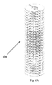
Referring collectively to FIGS. 6A-6K, film layers including delivery channel openings are combined to form a tissue scaffold. FIG. 6A illustrates twenty seven film layers 138 used to make a tissue scaffold with delivery channels 146. FIG. 6B is a perspective view of twenty five film layers 138 used to make a tissue scaffold with delivery channels. FIG. 6C is a side view of a film layers 138 used to make a tissue scaffold. FIG. 6D is a perspective view of twenty five film layers 138 combined to make a tissue scaffold with delivery channels within the scaffold with a cut out section depicting large delivery channels 150 and small delivery channels 154. FIG. 6E is a perspective view of twenty seven film layers combined to make a tissue scaffold 146 with delivery channel openings within the tissue scaffold. Alignment holes 90 and delivery channel openings 158 for delivery agents or cells are also shown with a cut out section. FIG. 6F is a perspective view of twenty seven films combined to make a tissue scaffold with delivery channel openings 158 containing an injector within the scaffold with a cut out section depicting the process of delivering agents or cells 162 to the channel openings. FIG. 6G is a perspective view of twenty seven films combined to make a tissue scaffold with delivery channel openings within the scaffold with a cut out section depicting delivered agents or cells 162 to the channel openings. FIG. 6H is a perspective view of two separate tissue scaffolds 166 and 170 with separate delivery channel openings within each scaffold with a cut out section depicting delivered agents or cells to the channel openings. FIG. 6I is a perspective view of two separate tissue scaffolds 166 and 170 combined to make a composite tissue scaffold 174 with separate delivery channel openings within each scaffold with a cut out section depicting delivered agents or cells to the channel openings. FIG. 6J is a perspective sectioned view of the twenty five film layer tissue scaffold combined to make a tissue scaffold with ingrowth channel openings 178 within the scaffold that extend to the center of the scaffold. FIG. 6K is a perspective sectioned view of twenty five film layer tissue scaffold combined to make a tissue scaffold with ingrowth channel openings 178 within the scaffold that extend to the center of the scaffold. The tissue scaffold is depicted with a blood vessel 182 growing into the ingrowth channel openings 178 of the scaffold.
Referring collectively to FIGS. 7A-7H, film layers of still further embodiments including cell openings of predetermined size and configuration are combined to form a tissue scaffold. FIG. 7A illustrates a film layer 186 used to make a tissue scaffold with oval cell openings 190 in a predetermined arrangement. The predetermined arrangement has a gradient of higher porosity in select regions to lower porosity in select regions. The film layer 186 has known or discernable dimensions (width, length, and thickness), which can be modified or left intact during the manufacture of a tissue scaffold. Film layer 186 is a single layer. FIG. 7B is a side view of a film layer 186 used to make a tissue scaffold. FIG. 7C illustrates a film layer 194 used to make a tissue scaffold with oval cell openings 190 in a predetermined arrangement. The predetermined arrangement has a gradient of higher porosity in select regions to lower porosity in select regions. The film layer 194 has known or discernable dimensions (width, length, and thickness), which can be modified or left intact during the manufacture of a tissue scaffold. Film layer 194 is a single layer. FIG. 7D is a side view of a film layer 194 used to make a tissue scaffold. FIG. 7E is a perspective view of film layer 186 and film layer 194 used to make a two layer tissue scaffold with different offset cell opening patterns. FIG. 7F is a perspective view of film layer 186 and film layer 194 used to make a six layer tissue scaffold with different offset cell opening patterns. FIGS. 7G-H are perspective views of film layers 186 and film layers 194 forming a six layer tissue scaffold 198. The predetermined arrangement of the cell openings creates a gradient of higher porosity in select regions to lower porosity in select regions which, in turn, creates a gradient of interconnecting pores 202 with higher interconnecting areas in select regions of the tissue scaffold 198.
FIG. 8 is a flow chart illustrating some of the steps in a method of producing a tissue scaffold of the present invention.
EXAMPLES Example 1
A tissue scaffold was constructed using a copolymer film of polylactic acid (PLA) and polycaprolactone (PCL). A tissue scaffold measuring 6000 microns in thickness was fabricated by combining six 1000 micron thick film layers with round 1000 micron cell openings. Three of the films had the cell pattern depicted in FIG. 2A and three of the films had the cell pattern depicted in FIG. 2C. A 100 watt CO2 laser was used to create the cell openings in the film layers using a CAD-CAM process. Because the cell openings in film layers were machined to the specifications outlined in FIGS. 2A and 2C, the cell openings were offset by 750 microns. Consequently, interconnecting pores measuring 250 microns were created between the cell openings in the film layers. Six film layers were built up to create the scaffold. A 4061 cyanoacrylate adhesive manufactured by Henkel Loctite (Hertfordshire, UK) was used to bond the film layers together.
Example 2
The scaffold created in Example 1 can be used as a scaffold for evaluating cellular behaviour in a three-dimensional environment. The scaffold can be included in a kit that includes a sterile polystyrene tissue culture plate with the standard number of wells 6, 12, 24, 48 or 96 within which the scaffolds have been placed, instructions for the cellular seeding and/or optimal dispersion concentration of growth/active factors, and accessory tools for proper scaffold handling. In a different approach, the invention can feature a kit that includes sterile pre-formed three-dimensional scaffold shapes, a lyophilized or a combination of lyophilized growth/active factor(s), associated tools to allow the delivery of the lyophilized agents homogenously within the scaffold, and instructions for proper growth/active factor dispersion. In a different approach, the invention can feature a kit that includes sterile pre-formed 3D scaffold shapes, a lyophilized or a combination of lyophilized growth/active factor(s), a photopolymerizable agent, a vial to mix the photopolymerizable agent with the lyophilized compound, associated tools to allow the homogenous distribution of the photopolymerizable agent plus lyophilized compound into the scaffold, and necessary instructions. The kit could or could not include a light source to induce local photopolymerization, thus, trapping of the lyophilized compound into the 3D scaffold.
A number of embodiments have been described. Other embodiments are within the scope of the following claims.

Claims (18)

What is claimed is:
1. A method of producing a tissue scaffold, the method comprising:
extruding a first biocompatible polymer to form a first film;
extruding a second biocompatible polymer to form a second film;
axially orienting the first and second film by applying a mechanical force and/or load to the first and second films;
bonding the first film to the second film to produce a tissue scaffold; and
forming a plurality of cell openings in the tissue scaffold to produce pores in the tissue scaffold such that cell openings of the first film interconnect with cell openings of the second film to define pathways with interconnecting pores of controlled area extending from the first film to the second film;
wherein the diameter of a majority of the pores is less than the diameter of the cell openings of the first and second films that define each pore.
2. The method of claim 1, wherein at least one of the first and second films further comprises alignment holes to align the cell openings of the first and second films when attached together.
3. The method of claim 1, wherein at least one of the films is an oriented film.
4. The method of claim 1, wherein the first film comprises a first material and the second film comprises a second material, wherein the first material has a higher absorption rate than the second material.
5. The method of claim 1, wherein the cell openings are formed to have diameters of between about 10 to about 10,000 microns.
6. The method of claim 1, wherein the cell openings of the second film are offset in a predetermined arrangement from the cell openings of the first film.
7. The method of claim 6, wherein the cell openings from the first film are offset and interconnect with the cell openings from the second film to create a 250 micron interconnecting pore.
8. The method of claim 6, wherein the predetermined arrangement of the cell openings creates a gradient of higher porosity on an edge of the tissue scaffold to lower porosity in the center which, in turn, creates a gradient of interconnecting pores with higher interconnecting areas on the edge of the edge.
9. The method of claim 1, further comprising the step of placing a hydrogel film between the first and second films before the first film is bonded to the second film.
10. A method of producing a tissue scaffold, the method comprising:
forming cell openings in a first film to define a first porosity;
forming cell openings in a second film to define a second porosity;
aligning the first film with respect to the second film; and
attaching the first and second films such that cell openings of the first film interconnect with cell openings of the second film to define pathways with interconnecting pores of controlled area extending from the first film to the second film;
wherein the diameter of a majority of the pores is less than the diameter of the cell openings of the first and second films that define each pore.
11. The method of claim 10, wherein the second porosity is greater than the first porosity.
12. The method of claim 10, wherein the cell openings of the second film are offset in a predetermined arrangement from the cell openings of the first film.
13. The method of claim 10, wherein the first porosity and the second porosity define a controlled porosity gradient extending from the first to the second film to selectively promote cellular regeneration along the gradient.
14. The method of claim 10, wherein the second film includes a plurality of alignment holes.
15. A method of producing a tissue scaffold, the method comprising:
(a) extruding a first biocompatible polymer to form a first film;
(b) extruding a second biocompatible polymer to form a second film;
(c) forming in the cell openings in a first and second films to define a first porosity and a second porosity, respectively; and
(d) attaching the first and second films such that cell openings of the first film interconnect with cell openings of the second film to define pathways with interconnecting pores of controlled area extending from the first film to the second film;
wherein the diameter of a majority of the pores is less than the diameter of the cell openings of the first and second films that define each pore.
16. The method of claim 15, wherein the second porosity is greater than the first porosity.
17. The method of claim 15, wherein the cell openings of the second film are offset in a predetermined arrangement from the cell openings of the first film.
18. The method of claim 15, wherein the first porosity and the second porosity define a controlled porosity gradient extending from the first to the second film to selectively promote cellular regeneration along the gradient.
US14/479,651 2004-11-09 2014-09-08 Tissue scaffold Active 2026-01-15 US9642943B2 (en)

Priority Applications (2)

Application Number Priority Date Filing Date Title
US14/479,651 US9642943B2 (en) 2004-11-09 2014-09-08 Tissue scaffold
US15/589,212 US10004829B2 (en) 2004-11-09 2017-05-08 Tissue scaffold

Applications Claiming Priority (5)

Application Number Priority Date Filing Date Title
IE20040751 2004-11-09
IE2004/0751 2004-11-09
US11/270,220 US8796015B2 (en) 2004-11-09 2005-11-09 Tissue scaffold
US14/450,047 US9440007B2 (en) 2004-11-09 2014-08-01 Tissue scaffold
US14/479,651 US9642943B2 (en) 2004-11-09 2014-09-08 Tissue scaffold

Related Parent Applications (1)

Application Number Title Priority Date Filing Date
US14/450,047 Division US9440007B2 (en) 2004-11-09 2014-08-01 Tissue scaffold

Related Child Applications (1)

Application Number Title Priority Date Filing Date
US15/589,212 Continuation US10004829B2 (en) 2004-11-09 2017-05-08 Tissue scaffold

Publications (2)

Publication Number Publication Date
US20140374004A1 US20140374004A1 (en) 2014-12-25
US9642943B2 true US9642943B2 (en) 2017-05-09

Family

ID=36337296

Family Applications (4)

Application Number Title Priority Date Filing Date
US11/270,220 Active 2027-07-17 US8796015B2 (en) 2004-11-09 2005-11-09 Tissue scaffold
US14/450,047 Active US9440007B2 (en) 2004-11-09 2014-08-01 Tissue scaffold
US14/479,651 Active 2026-01-15 US9642943B2 (en) 2004-11-09 2014-09-08 Tissue scaffold
US15/589,212 Active US10004829B2 (en) 2004-11-09 2017-05-08 Tissue scaffold

Family Applications Before (2)

Application Number Title Priority Date Filing Date
US11/270,220 Active 2027-07-17 US8796015B2 (en) 2004-11-09 2005-11-09 Tissue scaffold
US14/450,047 Active US9440007B2 (en) 2004-11-09 2014-08-01 Tissue scaffold

Family Applications After (1)

Application Number Title Priority Date Filing Date
US15/589,212 Active US10004829B2 (en) 2004-11-09 2017-05-08 Tissue scaffold

Country Status (3)

Country Link
US (4) US8796015B2 (en)
EP (1) EP1816987A4 (en)
WO (1) WO2006053291A2 (en)

Families Citing this family (74)

* Cited by examiner, † Cited by third party
Publication number Priority date Publication date Assignee Title
US20060147332A1 (en) 2004-12-30 2006-07-06 Howmedica Osteonics Corp. Laser-produced porous structure
CA2448592C (en) 2002-11-08 2011-01-11 Howmedica Osteonics Corp. Laser-produced porous surface
US20050182443A1 (en) 2004-02-18 2005-08-18 Closure Medical Corporation Adhesive-containing wound closure device and method
US20060009099A1 (en) 2004-07-12 2006-01-12 Closure Medical Corporation Adhesive-containing wound closure device and method
US8796015B2 (en) * 2004-11-09 2014-08-05 Proxy Biomedical Limited Tissue scaffold
US7790080B2 (en) * 2005-11-22 2010-09-07 National Taiwan University Method for forming a bioartificial guidance conduit
US8728387B2 (en) 2005-12-06 2014-05-20 Howmedica Osteonics Corp. Laser-produced porous surface
US20100273667A1 (en) * 2006-02-10 2010-10-28 The Regents Of The University Of Michigan Cell culture well-plates having inverted colloidal crystal scaffolds
US20090041825A1 (en) * 2006-02-10 2009-02-12 Kotov Nicholas A Cell culture well-plates having inverted colloidal crystal scaffolds
EP3192369B1 (en) * 2007-04-12 2018-12-12 The General Hospital Corporation Biomimetic vascular network
WO2009003726A1 (en) * 2007-07-02 2009-01-08 Loehde Eckhard Three-dimensional hernia mesh
US20090069904A1 (en) * 2007-09-12 2009-03-12 Applied Medical Research Biomaterial including micropores
WO2009043052A1 (en) * 2007-09-27 2009-04-02 Columbia University Methods and systems for forming biocompatible materials
DE102007056993A1 (en) * 2007-11-27 2009-06-04 Kilian Kraus Bone-contacting implants
WO2009086535A2 (en) * 2007-12-27 2009-07-09 The Trustees Of Columbia University In The City Of New York Systems and methods for forming patterned extracellular matrix materials
US9295757B2 (en) 2008-06-10 2016-03-29 Cook Biotech Incorporated Quilted implantable graft
CA2728240C (en) * 2008-06-10 2016-11-15 Cook Biotech Incorporated Quilted implantable graft
WO2010019799A1 (en) * 2008-08-13 2010-02-18 Smed-Ta/Td, Llc Orthopaedic implant with porous structural member
US9616205B2 (en) 2008-08-13 2017-04-11 Smed-Ta/Td, Llc Drug delivery implants
US10842645B2 (en) 2008-08-13 2020-11-24 Smed-Ta/Td, Llc Orthopaedic implant with porous structural member
US9700431B2 (en) 2008-08-13 2017-07-11 Smed-Ta/Td, Llc Orthopaedic implant with porous structural member
US20100042213A1 (en) 2008-08-13 2010-02-18 Nebosky Paul S Drug delivery implants
WO2010025386A1 (en) 2008-08-29 2010-03-04 Smed-Ta/Td, Llc Orthopaedic implant
US10610364B2 (en) * 2008-12-04 2020-04-07 Subchondral Solutions, Inc. Method for ameliorating joint conditions and diseases and preventing bone hypertrophy
WO2010088699A2 (en) * 2009-02-02 2010-08-05 Biomerix Corporation Composite mesh devices and methods for soft tissue repair
WO2010092065A1 (en) * 2009-02-11 2010-08-19 INSERM (Institut National de la Santé et de la Recherche Médicale) Endo-prosthesis for cartilage lesions
PL2442750T3 (en) * 2009-06-15 2019-10-31 Cartiheal 2009 Ltd Solid forms for tissue repair
FR2949667B1 (en) * 2009-09-09 2011-08-19 Obl POROUS STRUCTURE WITH A CONTROLLED PATTERN, REPEAT IN SPACE, FOR THE PRODUCTION OF SURGICAL IMPLANTS
EP3378930B1 (en) 2009-12-16 2019-10-23 VivaBioCell SpA Scaffold for the growth of tissue in vivo
US20110171732A1 (en) * 2010-01-12 2011-07-14 Corrado Mandoli Highly porous solid material made of biodegradable polymer and method of fabricating, processing, and cell-seeding the same
US9757132B2 (en) 2010-03-24 2017-09-12 Biorez, Inc. Mechanically competent scaffold for rotator cuff and tendon augmentation
IT1402752B1 (en) * 2010-11-12 2013-09-18 Novagenit S R L BIOCOMPATIBLE AND BIODEGRADABLE COMPOSITE MATERIAL FOR USE IN SURGERY AND PROCESS FOR ITS PRODUCTION
DE102011002536A1 (en) * 2011-01-11 2012-07-12 Aesculap Ag Packaging containing a medical product for the treatment of human or animal cartilage damage
AU2012222860A1 (en) * 2011-02-28 2013-09-26 Scimotana Pty Ltd Surgical implant and method
US8771353B2 (en) * 2011-03-08 2014-07-08 DePuy Synthes Products, LLC Method and implant for replacing damaged meniscal tissue
KR101375828B1 (en) * 2011-03-31 2014-03-17 인제대학교 산학협력단 Complex Scaffold For Bone-Cartilage Regeneration, Method For Preparing Thereof And Composition for Treating Bone Cartilage Disease Comprising The Same
JP5884321B2 (en) * 2011-07-08 2016-03-15 ソニー株式会社 Manufacturing method of structure
EP2757998A4 (en) 2011-09-22 2015-05-06 Ams Res Corp Pelvic implant and treatment method
PT105907A (en) * 2011-09-26 2013-03-26 Alrc Tecn E Materiais Cirurgicos Lda Zona Franca Da Madeira IMPLANTABLE ROLL-OUT PROSTHETICS FOR REPAIR OF ABDOMINAL WALL DEFECTS OR WEAKNESSES
US8535388B2 (en) * 2011-11-23 2013-09-17 Timothy Ganey Bone graft
US9877822B2 (en) 2012-04-24 2018-01-30 Biostage, Inc. Engineered tissue scaffolds and supports therefor
JP2015517879A (en) * 2012-05-30 2015-06-25 ニューヨーク ユニバーシティ Tissue repair device or scaffold
US10449026B2 (en) * 2012-06-26 2019-10-22 Biostage, Inc. Methods and compositions for promoting the structural integrity of scaffolds for tissue engineering
US9681936B2 (en) 2012-11-30 2017-06-20 Covidien Lp Multi-layer porous film material
EP2943231A4 (en) 2013-01-09 2016-12-07 Harvard Apparatus Regenerative Tech Inc Synthetic scaffolds
US20140243995A1 (en) * 2013-02-28 2014-08-28 The Massachusetts Institute Of Technology Stacked planar sheet tissue engineering scaffolds with 3d structural order
US9433489B2 (en) * 2013-03-12 2016-09-06 Soft Tissue Regeneration, Inc. Absorbable synthetic braided matrix for breast reconstruction and hernia repair
US9724203B2 (en) * 2013-03-15 2017-08-08 Smed-Ta/Td, Llc Porous tissue ingrowth structure
WO2015052346A1 (en) * 2013-10-11 2015-04-16 Proxy Biomedical Limited A method of providing interconnected pores with a growth-promoting medium for controlled tissue integration
US10898312B2 (en) * 2013-10-28 2021-01-26 The Regents Of The University Of California Tissue grafts with fenestrations
FR3022765B1 (en) * 2014-06-25 2016-07-08 Assist Publique - Hopitaux De Paris REMOVABLE IMPLANT FOR GENERATING A TENDON OR LIGAMENT IN REPLACEMENT OF A TENDON OR LIGAMENT ROMPU
USD824525S1 (en) 2014-09-25 2018-07-31 Ethicon Llc Release paper for wound treament devices
US20170246344A1 (en) * 2014-10-06 2017-08-31 Proxy Biomedical Inc. Tissue integration devices and methods of making the same
BR112018010562B1 (en) 2015-11-25 2023-05-02 Subchondral Solutions, Inc IMPLANTABLE ORTHOPEDIC DEVICE AND KIT TO REPAIR AN ANATOMIC JOINT
US10888407B2 (en) * 2015-12-11 2021-01-12 Poly-Med, Inc. Method for forming a multi-layer construct
KR20180095853A (en) * 2015-12-16 2018-08-28 너바시브 인코퍼레이티드 Porous Spine Fusion Implant
US10624750B2 (en) 2016-08-07 2020-04-21 Nanochon, Llc Three-dimensionally printed tissue engineering scaffolds for tissue regeneration
US10792024B2 (en) 2016-09-28 2020-10-06 Ethicon, Inc. Scaffolds with channels for joining layers of tissue at discrete points
US10687986B2 (en) 2016-09-29 2020-06-23 Ethicon, Inc. Methods and devices for skin closure
US10470934B2 (en) 2016-09-29 2019-11-12 Ethicon, Inc. Methods and devices for skin closure
USD848624S1 (en) 2016-09-29 2019-05-14 Ethicon, Inc. Release paper for wound treatment devices
US20180271505A1 (en) 2017-03-23 2018-09-27 Ethicon, Inc. Scaffolds for Joining Layers of Tissue at Discrete Points
US10470935B2 (en) 2017-03-23 2019-11-12 Ethicon, Inc. Skin closure systems and devices of improved flexibility and stretchability for bendable joints
WO2018195476A1 (en) 2017-04-20 2018-10-25 The Regents Of The University Of California Systems and methods for acellular dermal matrix fenestrations in prepectoral breast reconstruction
US11504446B2 (en) 2017-04-25 2022-11-22 Ethicon, Inc. Skin closure devices with self-forming exudate drainage channels
CN111432749B (en) * 2017-10-19 2023-04-04 C.R.巴德公司 Self-gripping hernia prosthesis
EP3829663A4 (en) * 2018-07-30 2022-05-18 Nanochon, LLC Implantable scaffolds and uses thereof
US10993708B2 (en) 2018-07-31 2021-05-04 Ethicon, Inc. Skin closure devices with interrupted closure
JP7290302B2 (en) * 2018-10-26 2023-06-13 国立研究開発法人農業・食品産業技術総合研究機構 Hydrogel membrane and its use
TWI689293B (en) * 2019-04-03 2020-04-01 晟格科技股份有限公司 Bandage structure
TWI774156B (en) * 2019-12-25 2022-08-11 賽宇細胞科技股份有限公司 A carrier for cell biomass production and cell culture device comprising the same
CN116157093A (en) * 2020-04-29 2023-05-23 台湾卫生研究院 Scaffolds for cell or tissue culture, methods for their preparation and their use in tissue engineering and regenerative medicine
CA3183956A1 (en) * 2020-07-08 2022-01-13 Jessee Hunt Implants having bone growth promoting agents contained within biodegradable materials
WO2023230119A1 (en) * 2022-05-24 2023-11-30 Dsm Ip Assets B.V. Joint spacers

Citations (28)

* Cited by examiner, † Cited by third party
Publication number Priority date Publication date Assignee Title
US4186448A (en) 1976-04-16 1980-02-05 Brekke John H Device and method for treating and healing a newly created bone void
US4661458A (en) 1983-08-31 1987-04-28 Cell Environmental Systems, Ltd. Cell culture system
US5133755A (en) 1986-01-28 1992-07-28 Thm Biomedical, Inc. Method and apparatus for diodegradable, osteogenic, bone graft substitute device
US5282861A (en) 1992-03-11 1994-02-01 Ultramet Open cell tantalum structures for cancellous bone implants and cell and tissue receptors
US5514378A (en) 1993-02-01 1996-05-07 Massachusetts Institute Of Technology Biocompatible polymer membranes and methods of preparation of three dimensional membrane structures
US5522895A (en) 1993-07-23 1996-06-04 Rice University Biodegradable bone templates
US5607474A (en) 1992-02-14 1997-03-04 Board Of Regents, University Of Texas System Multi-phase bioerodible implant/carrier and method of manufacturing and using same
US5677355A (en) 1993-08-13 1997-10-14 Smith & Nephew, Inc. Continuous open-cell polymeric foams containing living cells
US5686091A (en) 1994-03-28 1997-11-11 The Johns Hopkins University School Of Medicine Biodegradable foams for cell transplantation
US5711960A (en) 1993-09-24 1998-01-27 Takiron Co., Ltd. Biocompatible implant material comprising a tri-axial or more three-dimensional fabric
US5716413A (en) 1995-10-11 1998-02-10 Osteobiologics, Inc. Moldable, hand-shapable biodegradable implant material
US5755792A (en) 1986-01-28 1998-05-26 Thm Biomedical, Inc. Method and apparatus for biodegradable, osteogenic, bone graft substitute device
US5770193A (en) 1986-11-20 1998-06-23 Massachusetts Institute Of Technology Children's Medical Center Corporation Preparation of three-dimensional fibrous scaffold for attaching cells to produce vascularized tissue in vivo
US6103255A (en) 1999-04-16 2000-08-15 Rutgers, The State University Porous polymer scaffolds for tissue engineering
EP1028774A1 (en) 1997-11-07 2000-08-23 Rutgers, The State University Of New Jersey Biodegradable, anionic polymers derived from the amino acid l-tyrosine
US6319264B1 (en) 1998-04-03 2001-11-20 Bionx Implants Oy Hernia mesh
US20020142413A1 (en) 1999-05-07 2002-10-03 Salviac Limted Tissue engineering scaffold
US6479072B1 (en) 1999-02-11 2002-11-12 The General Hospital Corporation Microfabricated membranes and matrices
US20020182241A1 (en) 2001-01-02 2002-12-05 Borenstein Jeffrey T. Tissue engineering of three-dimensional vascularized using microfabricated polymer assembly technology
US20030004578A1 (en) 2001-06-28 2003-01-02 Ethicon, Inc. Composite scaffold with post anchor for the repair and regeneration of tissue
US20030065400A1 (en) 2001-04-12 2003-04-03 Beam Heather Ann Method and apparatus for engineered regenrative biostructures such as hydroxyapatite substrates for bone healing applications
WO2004006808A2 (en) 2002-07-17 2004-01-22 Proxy Biomedical Limited Soft tissue implants and methods for making same
EP1452191A2 (en) 1999-06-30 2004-09-01 Ethicon, Inc. Porous tissue scaffoldings for the repair or regeneration of tissue
WO2004078954A1 (en) 2003-01-14 2004-09-16 Yonsei University Biodegradable dual porous scaffold wrapped with semi-permeable membrane and tissue cell culture using thereof
US20040203147A1 (en) 2001-09-11 2004-10-14 Triffitt James Tomlinson Method and structure for growing living organic tissue
EP1493404A1 (en) 2003-06-30 2005-01-05 DePuy Mitek, Inc. Scaffold for connective tissue repair
EP1064958B1 (en) 1999-06-30 2005-08-17 Ethicon, Inc. Porous tissue scaffoldings for the repair or regeneration of tissue
EP1131410B1 (en) 1998-11-19 2005-10-05 Organogenesis Inc. Bioengineered tissue constructs and methods for producing and using them

Family Cites Families (2)

* Cited by examiner, † Cited by third party
Publication number Priority date Publication date Assignee Title
WO2002082432A1 (en) * 2000-06-07 2002-10-17 Fundacao De Amparo A Pesquisa Do Estado De Sao Paulo (Fapesp) Isolated genome of xyllela fastidiosa and uses thereof
US8796015B2 (en) * 2004-11-09 2014-08-05 Proxy Biomedical Limited Tissue scaffold

Patent Citations (30)

* Cited by examiner, † Cited by third party
Publication number Priority date Publication date Assignee Title
US4186448A (en) 1976-04-16 1980-02-05 Brekke John H Device and method for treating and healing a newly created bone void
US4661458A (en) 1983-08-31 1987-04-28 Cell Environmental Systems, Ltd. Cell culture system
US5133755A (en) 1986-01-28 1992-07-28 Thm Biomedical, Inc. Method and apparatus for diodegradable, osteogenic, bone graft substitute device
US5755792A (en) 1986-01-28 1998-05-26 Thm Biomedical, Inc. Method and apparatus for biodegradable, osteogenic, bone graft substitute device
US5770193A (en) 1986-11-20 1998-06-23 Massachusetts Institute Of Technology Children's Medical Center Corporation Preparation of three-dimensional fibrous scaffold for attaching cells to produce vascularized tissue in vivo
US5607474A (en) 1992-02-14 1997-03-04 Board Of Regents, University Of Texas System Multi-phase bioerodible implant/carrier and method of manufacturing and using same
US5282861A (en) 1992-03-11 1994-02-01 Ultramet Open cell tantalum structures for cancellous bone implants and cell and tissue receptors
US5514378A (en) 1993-02-01 1996-05-07 Massachusetts Institute Of Technology Biocompatible polymer membranes and methods of preparation of three dimensional membrane structures
US5522895A (en) 1993-07-23 1996-06-04 Rice University Biodegradable bone templates
US5677355A (en) 1993-08-13 1997-10-14 Smith & Nephew, Inc. Continuous open-cell polymeric foams containing living cells
US5711960A (en) 1993-09-24 1998-01-27 Takiron Co., Ltd. Biocompatible implant material comprising a tri-axial or more three-dimensional fabric
US5686091A (en) 1994-03-28 1997-11-11 The Johns Hopkins University School Of Medicine Biodegradable foams for cell transplantation
US5716413A (en) 1995-10-11 1998-02-10 Osteobiologics, Inc. Moldable, hand-shapable biodegradable implant material
EP1028774A1 (en) 1997-11-07 2000-08-23 Rutgers, The State University Of New Jersey Biodegradable, anionic polymers derived from the amino acid l-tyrosine
US6319264B1 (en) 1998-04-03 2001-11-20 Bionx Implants Oy Hernia mesh
EP1131410B1 (en) 1998-11-19 2005-10-05 Organogenesis Inc. Bioengineered tissue constructs and methods for producing and using them
US6479072B1 (en) 1999-02-11 2002-11-12 The General Hospital Corporation Microfabricated membranes and matrices
US6103255A (en) 1999-04-16 2000-08-15 Rutgers, The State University Porous polymer scaffolds for tissue engineering
US20020142413A1 (en) 1999-05-07 2002-10-03 Salviac Limted Tissue engineering scaffold
EP1452191A2 (en) 1999-06-30 2004-09-01 Ethicon, Inc. Porous tissue scaffoldings for the repair or regeneration of tissue
EP1064958B1 (en) 1999-06-30 2005-08-17 Ethicon, Inc. Porous tissue scaffoldings for the repair or regeneration of tissue
US20020182241A1 (en) 2001-01-02 2002-12-05 Borenstein Jeffrey T. Tissue engineering of three-dimensional vascularized using microfabricated polymer assembly technology
US20030065400A1 (en) 2001-04-12 2003-04-03 Beam Heather Ann Method and apparatus for engineered regenrative biostructures such as hydroxyapatite substrates for bone healing applications
US6626950B2 (en) 2001-06-28 2003-09-30 Ethicon, Inc. Composite scaffold with post anchor for the repair and regeneration of tissue
EP1277450A2 (en) 2001-06-28 2003-01-22 Ethicon, Inc. Composite scaffold with post anchor for the repair and regeneration of tissue
US20030004578A1 (en) 2001-06-28 2003-01-02 Ethicon, Inc. Composite scaffold with post anchor for the repair and regeneration of tissue
US20040203147A1 (en) 2001-09-11 2004-10-14 Triffitt James Tomlinson Method and structure for growing living organic tissue
WO2004006808A2 (en) 2002-07-17 2004-01-22 Proxy Biomedical Limited Soft tissue implants and methods for making same
WO2004078954A1 (en) 2003-01-14 2004-09-16 Yonsei University Biodegradable dual porous scaffold wrapped with semi-permeable membrane and tissue cell culture using thereof
EP1493404A1 (en) 2003-06-30 2005-01-05 DePuy Mitek, Inc. Scaffold for connective tissue repair

Non-Patent Citations (1)

* Cited by examiner, † Cited by third party
Title
European Search Report, Application No. 05851604.8-2310 / 1816987, mailed Feb. 8, 2011 (5 pages).

Also Published As

Publication number Publication date
US9440007B2 (en) 2016-09-13
WO2006053291A3 (en) 2007-02-01
US20140343690A1 (en) 2014-11-20
EP1816987A2 (en) 2007-08-15
US20060141012A1 (en) 2006-06-29
WO2006053291A2 (en) 2006-05-18
US20140374004A1 (en) 2014-12-25
US10004829B2 (en) 2018-06-26
US8796015B2 (en) 2014-08-05
US20170274122A1 (en) 2017-09-28
EP1816987A4 (en) 2011-03-09

Similar Documents

Publication Publication Date Title
US10004829B2 (en) Tissue scaffold
US20090069904A1 (en) Biomaterial including micropores
US20220331491A1 (en) Scaffolds with viable tissue
US7914808B2 (en) Hybrid biologic/synthetic porous extracellular matrix scaffolds
US8518123B2 (en) System and method for tissue generation and bone regeneration
ES2238736T3 (en) BIODEGRADABLE POLYMER FILM.
CN109276348B (en) Bone implant for encapsulating bone material
EP2108334B1 (en) Implant for linking two vertebras
DE602004013257T2 (en) Implants with living tissue for the repair of tissue damage or tissue defects
EP2617440A2 (en) Medical implants and methods for delivering biologically active agents
AU2002316694A1 (en) Hybrid biologic/synthetic porous extracellular matrix scaffolds
WO2001054593A1 (en) Neural regeneration conduit
US20220167988A1 (en) Biomimetic Scaffold for Peripheral Nerve Injuries
US11364122B2 (en) Tissue engineering scaffolds
Chaput et al. III. 1 Artificial Materials and Structures for Extracellular Matrix Scaffolding of Orthopedic Tissues: A Review

Legal Events

Date Code Title Description
AS Assignment

Owner name: PROXY BIOMEDICAL LIMITED, IRELAND

Free format text: ASSIGNMENT OF ASSIGNORS INTEREST;ASSIGNOR:GINGRAS, PETER;REEL/FRAME:033700/0659

Effective date: 20140605

STCF Information on status: patent grant

Free format text: PATENTED CASE

MAFP Maintenance fee payment

Free format text: PAYMENT OF MAINTENANCE FEE, 4TH YR, SMALL ENTITY (ORIGINAL EVENT CODE: M2551); ENTITY STATUS OF PATENT OWNER: SMALL ENTITY

Year of fee payment: 4

FEPP Fee payment procedure

Free format text: ENTITY STATUS SET TO UNDISCOUNTED (ORIGINAL EVENT CODE: BIG.); ENTITY STATUS OF PATENT OWNER: LARGE ENTITY