US9535059B2 - Microfluidic device - Google Patents
Microfluidic device Download PDFInfo
- Publication number
- US9535059B2 US9535059B2 US14/931,645 US201514931645A US9535059B2 US 9535059 B2 US9535059 B2 US 9535059B2 US 201514931645 A US201514931645 A US 201514931645A US 9535059 B2 US9535059 B2 US 9535059B2
- Authority
- US
- United States
- Prior art keywords
- fluorescently labeled
- cells
- approximately
- fluid sample
- channel
- Prior art date
- Legal status (The legal status is an assumption and is not a legal conclusion. Google has not performed a legal analysis and makes no representation as to the accuracy of the status listed.)
- Active
Links
- 210000004027 cell Anatomy 0.000 claims abstract description 57
- 210000000265 leukocyte Anatomy 0.000 claims abstract description 47
- 238000000034 method Methods 0.000 claims abstract description 37
- 210000004369 blood Anatomy 0.000 claims abstract description 35
- 239000008280 blood Substances 0.000 claims abstract description 35
- 239000012530 fluid Substances 0.000 claims description 57
- 238000001514 detection method Methods 0.000 claims description 32
- 239000011324 bead Substances 0.000 claims description 23
- 230000005284 excitation Effects 0.000 claims description 18
- 239000004793 Polystyrene Substances 0.000 claims description 2
- 238000005259 measurement Methods 0.000 claims description 2
- 229920002223 polystyrene Polymers 0.000 claims description 2
- 238000005086 pumping Methods 0.000 claims description 2
- 239000000523 sample Substances 0.000 description 35
- DPKHZNPWBDQZCN-UHFFFAOYSA-N acridine orange free base Chemical compound C1=CC(N(C)C)=CC2=NC3=CC(N(C)C)=CC=C3C=C21 DPKHZNPWBDQZCN-UHFFFAOYSA-N 0.000 description 27
- DZBUGLKDJFMEHC-UHFFFAOYSA-N benzoquinolinylidene Natural products C1=CC=CC2=CC3=CC=CC=C3N=C21 DZBUGLKDJFMEHC-UHFFFAOYSA-N 0.000 description 27
- 238000004891 communication Methods 0.000 description 15
- 239000000975 dye Substances 0.000 description 11
- 238000005286 illumination Methods 0.000 description 11
- 238000010186 staining Methods 0.000 description 11
- XKRFYHLGVUSROY-UHFFFAOYSA-N Argon Chemical compound [Ar] XKRFYHLGVUSROY-UHFFFAOYSA-N 0.000 description 10
- 210000003743 erythrocyte Anatomy 0.000 description 10
- 230000003287 optical effect Effects 0.000 description 9
- 239000004205 dimethyl polysiloxane Substances 0.000 description 8
- 229920000435 poly(dimethylsiloxane) Polymers 0.000 description 8
- 238000012360 testing method Methods 0.000 description 7
- 108020004414 DNA Proteins 0.000 description 6
- 238000000149 argon plasma sintering Methods 0.000 description 6
- 239000012472 biological sample Substances 0.000 description 6
- 239000011521 glass Substances 0.000 description 6
- 210000003714 granulocyte Anatomy 0.000 description 6
- 210000004698 lymphocyte Anatomy 0.000 description 6
- XUIMIQQOPSSXEZ-UHFFFAOYSA-N Silicon Chemical compound [Si] XUIMIQQOPSSXEZ-UHFFFAOYSA-N 0.000 description 5
- 238000004458 analytical method Methods 0.000 description 5
- 229910052786 argon Inorganic materials 0.000 description 5
- 238000010790 dilution Methods 0.000 description 5
- 239000012895 dilution Substances 0.000 description 5
- 210000001616 monocyte Anatomy 0.000 description 5
- 239000010703 silicon Substances 0.000 description 5
- 229910052710 silicon Inorganic materials 0.000 description 5
- 239000000758 substrate Substances 0.000 description 5
- 108010043121 Green Fluorescent Proteins Proteins 0.000 description 4
- 230000008021 deposition Effects 0.000 description 4
- 210000003979 eosinophil Anatomy 0.000 description 4
- 239000012997 ficoll-paque Substances 0.000 description 4
- FFUAGWLWBBFQJT-UHFFFAOYSA-N hexamethyldisilazane Chemical compound C[Si](C)(C)N[Si](C)(C)C FFUAGWLWBBFQJT-UHFFFAOYSA-N 0.000 description 4
- 230000002934 lysing effect Effects 0.000 description 4
- 230000007935 neutral effect Effects 0.000 description 4
- 239000002245 particle Substances 0.000 description 4
- 230000004044 response Effects 0.000 description 4
- 102000053602 DNA Human genes 0.000 description 3
- 210000003651 basophil Anatomy 0.000 description 3
- 239000000872 buffer Substances 0.000 description 3
- 210000003855 cell nucleus Anatomy 0.000 description 3
- 230000004069 differentiation Effects 0.000 description 3
- 230000000694 effects Effects 0.000 description 3
- 239000008187 granular material Substances 0.000 description 3
- 210000003712 lysosome Anatomy 0.000 description 3
- 230000001868 lysosomic effect Effects 0.000 description 3
- 210000000440 neutrophil Anatomy 0.000 description 3
- 210000004940 nucleus Anatomy 0.000 description 3
- 210000003463 organelle Anatomy 0.000 description 3
- 230000008569 process Effects 0.000 description 3
- -1 DNA Chemical class 0.000 description 2
- 206010018910 Haemolysis Diseases 0.000 description 2
- 230000002378 acidificating effect Effects 0.000 description 2
- 125000002091 cationic group Chemical group 0.000 description 2
- 210000000805 cytoplasm Anatomy 0.000 description 2
- ZMMJGEGLRURXTF-UHFFFAOYSA-N ethidium bromide Chemical compound [Br-].C12=CC(N)=CC=C2C2=CC=C(N)C=C2[N+](CC)=C1C1=CC=CC=C1 ZMMJGEGLRURXTF-UHFFFAOYSA-N 0.000 description 2
- 229960005542 ethidium bromide Drugs 0.000 description 2
- 230000005484 gravity Effects 0.000 description 2
- 230000008588 hemolysis Effects 0.000 description 2
- 238000009652 hydrodynamic focusing Methods 0.000 description 2
- 238000002372 labelling Methods 0.000 description 2
- 239000000463 material Substances 0.000 description 2
- 239000000203 mixture Substances 0.000 description 2
- 239000002953 phosphate buffered saline Substances 0.000 description 2
- 230000035945 sensitivity Effects 0.000 description 2
- 239000007787 solid Substances 0.000 description 2
- 239000011550 stock solution Substances 0.000 description 2
- 239000002699 waste material Substances 0.000 description 2
- ZOMLUNRKXJYKPD-UHFFFAOYSA-N 1,3,3-trimethyl-2-[2-(2-methylindol-3-ylidene)ethylidene]indole;hydrochloride Chemical compound [Cl-].C1=CC=C2C(C)(C)C(/C=C/C=3C4=CC=CC=C4NC=3C)=[N+](C)C2=C1 ZOMLUNRKXJYKPD-UHFFFAOYSA-N 0.000 description 1
- BCHZICNRHXRCHY-UHFFFAOYSA-N 2h-oxazine Chemical compound N1OC=CC=C1 BCHZICNRHXRCHY-UHFFFAOYSA-N 0.000 description 1
- ZTGKHKPZSMMHNM-UHFFFAOYSA-N 3-(2-phenylethenyl)benzene-1,2-disulfonic acid Chemical class OS(=O)(=O)C1=CC=CC(C=CC=2C=CC=CC=2)=C1S(O)(=O)=O ZTGKHKPZSMMHNM-UHFFFAOYSA-N 0.000 description 1
- 235000008733 Citrus aurantifolia Nutrition 0.000 description 1
- KCXVZYZYPLLWCC-UHFFFAOYSA-N EDTA Chemical compound OC(=O)CN(CC(O)=O)CCN(CC(O)=O)CC(O)=O KCXVZYZYPLLWCC-UHFFFAOYSA-N 0.000 description 1
- 241001465754 Metazoa Species 0.000 description 1
- 108020004682 Single-Stranded DNA Proteins 0.000 description 1
- 235000011941 Tilia x europaea Nutrition 0.000 description 1
- 238000010521 absorption reaction Methods 0.000 description 1
- 239000003513 alkali Substances 0.000 description 1
- 238000000347 anisotropic wet etching Methods 0.000 description 1
- 230000000712 assembly Effects 0.000 description 1
- 238000000429 assembly Methods 0.000 description 1
- QVGXLLKOCUKJST-UHFFFAOYSA-N atomic oxygen Chemical compound [O] QVGXLLKOCUKJST-UHFFFAOYSA-N 0.000 description 1
- 239000002585 base Substances 0.000 description 1
- 230000008901 benefit Effects 0.000 description 1
- 238000004061 bleaching Methods 0.000 description 1
- 210000000601 blood cell Anatomy 0.000 description 1
- 210000000170 cell membrane Anatomy 0.000 description 1
- 239000006285 cell suspension Substances 0.000 description 1
- 230000008859 change Effects 0.000 description 1
- 210000000349 chromosome Anatomy 0.000 description 1
- 238000005345 coagulation Methods 0.000 description 1
- 230000015271 coagulation Effects 0.000 description 1
- 230000000295 complement effect Effects 0.000 description 1
- 239000000356 contaminant Substances 0.000 description 1
- 210000004748 cultured cell Anatomy 0.000 description 1
- 230000009089 cytolysis Effects 0.000 description 1
- 230000003247 decreasing effect Effects 0.000 description 1
- 238000007872 degassing Methods 0.000 description 1
- 238000011161 development Methods 0.000 description 1
- 238000009792 diffusion process Methods 0.000 description 1
- 239000003085 diluting agent Substances 0.000 description 1
- LOKCTEFSRHRXRJ-UHFFFAOYSA-I dipotassium trisodium dihydrogen phosphate hydrogen phosphate dichloride Chemical compound P(=O)(O)(O)[O-].[K+].P(=O)(O)([O-])[O-].[Na+].[Na+].[Cl-].[K+].[Cl-].[Na+] LOKCTEFSRHRXRJ-UHFFFAOYSA-I 0.000 description 1
- 201000010099 disease Diseases 0.000 description 1
- 208000037265 diseases, disorders, signs and symptoms Diseases 0.000 description 1
- 238000005530 etching Methods 0.000 description 1
- 238000000684 flow cytometry Methods 0.000 description 1
- 238000012757 fluorescence staining Methods 0.000 description 1
- 239000007850 fluorescent dye Substances 0.000 description 1
- 230000002209 hydrophobic effect Effects 0.000 description 1
- 238000003384 imaging method Methods 0.000 description 1
- 238000012744 immunostaining Methods 0.000 description 1
- 238000002847 impedance measurement Methods 0.000 description 1
- 238000010348 incorporation Methods 0.000 description 1
- 230000010354 integration Effects 0.000 description 1
- 238000009830 intercalation Methods 0.000 description 1
- 230000002687 intercalation Effects 0.000 description 1
- 239000004571 lime Substances 0.000 description 1
- HYLDLLCHFLSKAG-UHFFFAOYSA-M lissamine flavine FF Chemical class [Na+].C1=CC(C)=CC=C1N(C1=O)C(=O)C2=C3C1=CC=CC3=C(N)C(S([O-])(=O)=O)=C2 HYLDLLCHFLSKAG-UHFFFAOYSA-M 0.000 description 1
- 210000002540 macrophage Anatomy 0.000 description 1
- 239000012528 membrane Substances 0.000 description 1
- 239000002184 metal Substances 0.000 description 1
- 238000002156 mixing Methods 0.000 description 1
- 239000003068 molecular probe Substances 0.000 description 1
- 210000000633 nuclear envelope Anatomy 0.000 description 1
- 108020004707 nucleic acids Proteins 0.000 description 1
- 102000039446 nucleic acids Human genes 0.000 description 1
- 150000007523 nucleic acids Chemical class 0.000 description 1
- 239000001301 oxygen Substances 0.000 description 1
- 229910052760 oxygen Inorganic materials 0.000 description 1
- 238000009832 plasma treatment Methods 0.000 description 1
- 239000004033 plastic Substances 0.000 description 1
- 238000012545 processing Methods 0.000 description 1
- 239000001397 quillaja saponaria molina bark Substances 0.000 description 1
- 230000009467 reduction Effects 0.000 description 1
- 238000011160 research Methods 0.000 description 1
- 150000003839 salts Chemical class 0.000 description 1
- 229930182490 saponin Natural products 0.000 description 1
- 150000007949 saponins Chemical class 0.000 description 1
- 239000004065 semiconductor Substances 0.000 description 1
- 238000000926 separation method Methods 0.000 description 1
- 238000002174 soft lithography Methods 0.000 description 1
- 239000000243 solution Substances 0.000 description 1
- 239000002904 solvent Substances 0.000 description 1
- 239000000126 substance Substances 0.000 description 1
- 239000004094 surface-active agent Substances 0.000 description 1
- 230000007704 transition Effects 0.000 description 1
- 238000011144 upstream manufacturing Methods 0.000 description 1
- XLYOFNOQVPJJNP-UHFFFAOYSA-N water Substances O XLYOFNOQVPJJNP-UHFFFAOYSA-N 0.000 description 1
Images


Classifications
-
- G—PHYSICS
- G01—MEASURING; TESTING
- G01N—INVESTIGATING OR ANALYSING MATERIALS BY DETERMINING THEIR CHEMICAL OR PHYSICAL PROPERTIES
- G01N33/00—Investigating or analysing materials by specific methods not covered by groups G01N1/00 - G01N31/00
- G01N33/48—Biological material, e.g. blood, urine; Haemocytometers
- G01N33/50—Chemical analysis of biological material, e.g. blood, urine; Testing involving biospecific ligand binding methods; Immunological testing
- G01N33/5005—Chemical analysis of biological material, e.g. blood, urine; Testing involving biospecific ligand binding methods; Immunological testing involving human or animal cells
- G01N33/5094—Chemical analysis of biological material, e.g. blood, urine; Testing involving biospecific ligand binding methods; Immunological testing involving human or animal cells for blood cell populations
-
- B—PERFORMING OPERATIONS; TRANSPORTING
- B01—PHYSICAL OR CHEMICAL PROCESSES OR APPARATUS IN GENERAL
- B01L—CHEMICAL OR PHYSICAL LABORATORY APPARATUS FOR GENERAL USE
- B01L3/00—Containers or dishes for laboratory use, e.g. laboratory glassware; Droppers
- B01L3/50—Containers for the purpose of retaining a material to be analysed, e.g. test tubes
- B01L3/502—Containers for the purpose of retaining a material to be analysed, e.g. test tubes with fluid transport, e.g. in multi-compartment structures
- B01L3/5027—Containers for the purpose of retaining a material to be analysed, e.g. test tubes with fluid transport, e.g. in multi-compartment structures by integrated microfluidic structures, i.e. dimensions of channels and chambers are such that surface tension forces are important, e.g. lab-on-a-chip
- B01L3/502761—Containers for the purpose of retaining a material to be analysed, e.g. test tubes with fluid transport, e.g. in multi-compartment structures by integrated microfluidic structures, i.e. dimensions of channels and chambers are such that surface tension forces are important, e.g. lab-on-a-chip specially adapted for handling suspended solids or molecules independently from the bulk fluid flow, e.g. for trapping or sorting beads, for physically stretching molecules
-
- G—PHYSICS
- G01—MEASURING; TESTING
- G01N—INVESTIGATING OR ANALYSING MATERIALS BY DETERMINING THEIR CHEMICAL OR PHYSICAL PROPERTIES
- G01N33/00—Investigating or analysing materials by specific methods not covered by groups G01N1/00 - G01N31/00
- G01N33/48—Biological material, e.g. blood, urine; Haemocytometers
- G01N33/50—Chemical analysis of biological material, e.g. blood, urine; Testing involving biospecific ligand binding methods; Immunological testing
- G01N33/5005—Chemical analysis of biological material, e.g. blood, urine; Testing involving biospecific ligand binding methods; Immunological testing involving human or animal cells
-
- G—PHYSICS
- G01—MEASURING; TESTING
- G01N—INVESTIGATING OR ANALYSING MATERIALS BY DETERMINING THEIR CHEMICAL OR PHYSICAL PROPERTIES
- G01N33/00—Investigating or analysing materials by specific methods not covered by groups G01N1/00 - G01N31/00
- G01N33/48—Biological material, e.g. blood, urine; Haemocytometers
- G01N33/50—Chemical analysis of biological material, e.g. blood, urine; Testing involving biospecific ligand binding methods; Immunological testing
- G01N33/58—Chemical analysis of biological material, e.g. blood, urine; Testing involving biospecific ligand binding methods; Immunological testing involving labelled substances
- G01N33/582—Chemical analysis of biological material, e.g. blood, urine; Testing involving biospecific ligand binding methods; Immunological testing involving labelled substances with fluorescent label
-
- B—PERFORMING OPERATIONS; TRANSPORTING
- B01—PHYSICAL OR CHEMICAL PROCESSES OR APPARATUS IN GENERAL
- B01L—CHEMICAL OR PHYSICAL LABORATORY APPARATUS FOR GENERAL USE
- B01L2200/00—Solutions for specific problems relating to chemical or physical laboratory apparatus
- B01L2200/06—Fluid handling related problems
- B01L2200/0636—Focussing flows, e.g. to laminate flows
-
- B—PERFORMING OPERATIONS; TRANSPORTING
- B01—PHYSICAL OR CHEMICAL PROCESSES OR APPARATUS IN GENERAL
- B01L—CHEMICAL OR PHYSICAL LABORATORY APPARATUS FOR GENERAL USE
- B01L2200/00—Solutions for specific problems relating to chemical or physical laboratory apparatus
- B01L2200/06—Fluid handling related problems
- B01L2200/0647—Handling flowable solids, e.g. microscopic beads, cells, particles
-
- B—PERFORMING OPERATIONS; TRANSPORTING
- B01—PHYSICAL OR CHEMICAL PROCESSES OR APPARATUS IN GENERAL
- B01L—CHEMICAL OR PHYSICAL LABORATORY APPARATUS FOR GENERAL USE
- B01L2300/00—Additional constructional details
- B01L2300/06—Auxiliary integrated devices, integrated components
- B01L2300/0627—Sensor or part of a sensor is integrated
- B01L2300/0636—Integrated biosensor, microarrays
-
- B—PERFORMING OPERATIONS; TRANSPORTING
- B01—PHYSICAL OR CHEMICAL PROCESSES OR APPARATUS IN GENERAL
- B01L—CHEMICAL OR PHYSICAL LABORATORY APPARATUS FOR GENERAL USE
- B01L2300/00—Additional constructional details
- B01L2300/06—Auxiliary integrated devices, integrated components
- B01L2300/0627—Sensor or part of a sensor is integrated
- B01L2300/0654—Lenses; Optical fibres
-
- B—PERFORMING OPERATIONS; TRANSPORTING
- B01—PHYSICAL OR CHEMICAL PROCESSES OR APPARATUS IN GENERAL
- B01L—CHEMICAL OR PHYSICAL LABORATORY APPARATUS FOR GENERAL USE
- B01L2300/00—Additional constructional details
- B01L2300/06—Auxiliary integrated devices, integrated components
- B01L2300/0681—Filter
-
- B—PERFORMING OPERATIONS; TRANSPORTING
- B01—PHYSICAL OR CHEMICAL PROCESSES OR APPARATUS IN GENERAL
- B01L—CHEMICAL OR PHYSICAL LABORATORY APPARATUS FOR GENERAL USE
- B01L2300/00—Additional constructional details
- B01L2300/08—Geometry, shape and general structure
- B01L2300/0809—Geometry, shape and general structure rectangular shaped
- B01L2300/0816—Cards, e.g. flat sample carriers usually with flow in two horizontal directions
-
- B—PERFORMING OPERATIONS; TRANSPORTING
- B01—PHYSICAL OR CHEMICAL PROCESSES OR APPARATUS IN GENERAL
- B01L—CHEMICAL OR PHYSICAL LABORATORY APPARATUS FOR GENERAL USE
- B01L2300/00—Additional constructional details
- B01L2300/08—Geometry, shape and general structure
- B01L2300/0809—Geometry, shape and general structure rectangular shaped
- B01L2300/0822—Slides
-
- B—PERFORMING OPERATIONS; TRANSPORTING
- B01—PHYSICAL OR CHEMICAL PROCESSES OR APPARATUS IN GENERAL
- B01L—CHEMICAL OR PHYSICAL LABORATORY APPARATUS FOR GENERAL USE
- B01L2300/00—Additional constructional details
- B01L2300/08—Geometry, shape and general structure
- B01L2300/0861—Configuration of multiple channels and/or chambers in a single devices
- B01L2300/0864—Configuration of multiple channels and/or chambers in a single devices comprising only one inlet and multiple receiving wells, e.g. for separation, splitting
-
- B—PERFORMING OPERATIONS; TRANSPORTING
- B01—PHYSICAL OR CHEMICAL PROCESSES OR APPARATUS IN GENERAL
- B01L—CHEMICAL OR PHYSICAL LABORATORY APPARATUS FOR GENERAL USE
- B01L2300/00—Additional constructional details
- B01L2300/08—Geometry, shape and general structure
- B01L2300/0861—Configuration of multiple channels and/or chambers in a single devices
- B01L2300/0877—Flow chambers
-
- B—PERFORMING OPERATIONS; TRANSPORTING
- B01—PHYSICAL OR CHEMICAL PROCESSES OR APPARATUS IN GENERAL
- B01L—CHEMICAL OR PHYSICAL LABORATORY APPARATUS FOR GENERAL USE
- B01L2400/00—Moving or stopping fluids
- B01L2400/08—Regulating or influencing the flow resistance
- B01L2400/084—Passive control of flow resistance
- B01L2400/086—Passive control of flow resistance using baffles or other fixed flow obstructions
-
- B—PERFORMING OPERATIONS; TRANSPORTING
- B01—PHYSICAL OR CHEMICAL PROCESSES OR APPARATUS IN GENERAL
- B01L—CHEMICAL OR PHYSICAL LABORATORY APPARATUS FOR GENERAL USE
- B01L3/00—Containers or dishes for laboratory use, e.g. laboratory glassware; Droppers
- B01L3/50—Containers for the purpose of retaining a material to be analysed, e.g. test tubes
- B01L3/502—Containers for the purpose of retaining a material to be analysed, e.g. test tubes with fluid transport, e.g. in multi-compartment structures
- B01L3/5027—Containers for the purpose of retaining a material to be analysed, e.g. test tubes with fluid transport, e.g. in multi-compartment structures by integrated microfluidic structures, i.e. dimensions of channels and chambers are such that surface tension forces are important, e.g. lab-on-a-chip
- B01L3/502753—Containers for the purpose of retaining a material to be analysed, e.g. test tubes with fluid transport, e.g. in multi-compartment structures by integrated microfluidic structures, i.e. dimensions of channels and chambers are such that surface tension forces are important, e.g. lab-on-a-chip characterised by bulk separation arrangements on lab-on-a-chip devices, e.g. for filtration or centrifugation
-
- G01N15/1023—
-
- G—PHYSICS
- G01—MEASURING; TESTING
- G01N—INVESTIGATING OR ANALYSING MATERIALS BY DETERMINING THEIR CHEMICAL OR PHYSICAL PROPERTIES
- G01N15/00—Investigating characteristics of particles; Investigating permeability, pore-volume, or surface-area of porous materials
- G01N15/10—Investigating individual particles
- G01N15/1056—Microstructural devices for other than electro-optical measurement
-
- G—PHYSICS
- G01—MEASURING; TESTING
- G01N—INVESTIGATING OR ANALYSING MATERIALS BY DETERMINING THEIR CHEMICAL OR PHYSICAL PROPERTIES
- G01N15/00—Investigating characteristics of particles; Investigating permeability, pore-volume, or surface-area of porous materials
- G01N15/10—Investigating individual particles
- G01N15/14—Electro-optical investigation, e.g. flow cytometers
- G01N15/1484—Electro-optical investigation, e.g. flow cytometers microstructural devices
-
- G01N15/149—
-
- G—PHYSICS
- G01—MEASURING; TESTING
- G01N—INVESTIGATING OR ANALYSING MATERIALS BY DETERMINING THEIR CHEMICAL OR PHYSICAL PROPERTIES
- G01N15/00—Investigating characteristics of particles; Investigating permeability, pore-volume, or surface-area of porous materials
- G01N15/10—Investigating individual particles
- G01N2015/1006—Investigating individual particles for cytology
-
- G—PHYSICS
- G01—MEASURING; TESTING
- G01N—INVESTIGATING OR ANALYSING MATERIALS BY DETERMINING THEIR CHEMICAL OR PHYSICAL PROPERTIES
- G01N15/00—Investigating characteristics of particles; Investigating permeability, pore-volume, or surface-area of porous materials
- G01N15/10—Investigating individual particles
- G01N15/14—Electro-optical investigation, e.g. flow cytometers
- G01N2015/1486—Counting the particles
-
- G—PHYSICS
- G01—MEASURING; TESTING
- G01N—INVESTIGATING OR ANALYSING MATERIALS BY DETERMINING THEIR CHEMICAL OR PHYSICAL PROPERTIES
- G01N15/00—Investigating characteristics of particles; Investigating permeability, pore-volume, or surface-area of porous materials
- G01N15/10—Investigating individual particles
- G01N15/14—Electro-optical investigation, e.g. flow cytometers
- G01N2015/149—Sorting the particles
-
- Y—GENERAL TAGGING OF NEW TECHNOLOGICAL DEVELOPMENTS; GENERAL TAGGING OF CROSS-SECTIONAL TECHNOLOGIES SPANNING OVER SEVERAL SECTIONS OF THE IPC; TECHNICAL SUBJECTS COVERED BY FORMER USPC CROSS-REFERENCE ART COLLECTIONS [XRACs] AND DIGESTS
- Y10—TECHNICAL SUBJECTS COVERED BY FORMER USPC
- Y10T—TECHNICAL SUBJECTS COVERED BY FORMER US CLASSIFICATION
- Y10T436/00—Chemistry: analytical and immunological testing
- Y10T436/11—Automated chemical analysis
-
- Y—GENERAL TAGGING OF NEW TECHNOLOGICAL DEVELOPMENTS; GENERAL TAGGING OF CROSS-SECTIONAL TECHNOLOGIES SPANNING OVER SEVERAL SECTIONS OF THE IPC; TECHNICAL SUBJECTS COVERED BY FORMER USPC CROSS-REFERENCE ART COLLECTIONS [XRACs] AND DIGESTS
- Y10—TECHNICAL SUBJECTS COVERED BY FORMER USPC
- Y10T—TECHNICAL SUBJECTS COVERED BY FORMER US CLASSIFICATION
- Y10T436/00—Chemistry: analytical and immunological testing
- Y10T436/13—Tracers or tags
-
- Y—GENERAL TAGGING OF NEW TECHNOLOGICAL DEVELOPMENTS; GENERAL TAGGING OF CROSS-SECTIONAL TECHNOLOGIES SPANNING OVER SEVERAL SECTIONS OF THE IPC; TECHNICAL SUBJECTS COVERED BY FORMER USPC CROSS-REFERENCE ART COLLECTIONS [XRACs] AND DIGESTS
- Y10—TECHNICAL SUBJECTS COVERED BY FORMER USPC
- Y10T—TECHNICAL SUBJECTS COVERED BY FORMER US CLASSIFICATION
- Y10T436/00—Chemistry: analytical and immunological testing
- Y10T436/25—Chemistry: analytical and immunological testing including sample preparation
- Y10T436/2575—Volumetric liquid transfer
Definitions
- the present disclosure relates to fabricated microfluidic devices that can be utilized as cell sensors and/or actuators.
- the microfluidic device may be used for labeling, sensing, differentiating, and/or sorting targets, particularly cell populations.
- Standard cell sensors or actuators are generally based on flow cytometry and employ one or a combination of electrical impedance sensing, light scattering measurement, and chemical or immunostaining followed by optical sensing.
- red blood cells are removed by lysing in order to reduce the blood volume. Lysing is generally done through the use of saponin or surfactants. During the lysing process, the leukocyte cell volume changes depending on cell type, due to the leakage of cytoplasm contents and cell nucleus shrinkage in varying amounts. Fujimoto, Sysmex J. Int. 9 (1990). Thus, normally 2-part (lymphocytes versus granulocytes) or even 3-part (lymphocytes, neutrophils, and other leukocytes) differential can be achieved by simple electrical impedance measurement of particle volume. Hughes-Jones, et al., J. Clin. Pathol.
- FIG. 1A shows the molecular structure of acridine orange.
- FIG. 1B shows leukocyte staining results with acridine orange.
- FIG. 2 shows the top view of one embodiment of a novel fabricated microfluidic apparatus.
- FIG. 3 shows one embodiment of the optical system setup.
- FIG. 4A shows erythrocyte concentration in whole blood.
- FIG. 4B shows leukocyte staining in whole blood with acridine orange at a concentration of 100 ng/mL.
- FIG. 4C shows leukocyte staining In whole blood with acridine orange at a concentration of 1 ⁇ g/mL.
- FIG. 4D shows leukocyte staining in whole blood with acridine orange at a concentration of 10 ⁇ g/mL.
- FIG. 4E shows leukocyte staining In whole blood with acridine orange at a concentration of 100 ⁇ g/mL.
- FIG. 4F shows leukocyte staining in whole blood with acridine orange at a concentration of 1 mg/mL.
- FIG. 5 shows fluorescent signal bleaching from a single leukocyte in one embodiment.
- FIG. 6A shows an image of background and a 5 ⁇ m bead with focused laser illumination flow taken by CCD camera with long pass emission filter, according to one embodiment.
- FIG. 6B shows an image of background and a 5 ⁇ m bead with laser illumination flow taken by CCD camera with long pass emission filter, according to one embodiment.
- FIG. 6C shows an image of background and a 5 ⁇ m bead with diffused laser illumination flow taken by CCD camera with long pass emission filter, according to one embodiment.
- FIG. 6D shows an image of background and the trace of a 5 ⁇ m bead with diffused laser illumination flow taken by CCD camera with long pass emission filter, according to one embodiment.
- FIG. 7 shows a graph of detection of 5 ⁇ m fluorescent beads detection with photo-diode detector with long pass emission filter.
- FIG. 8A shows background image of focused laser beam from video taken by CCD camera with long pass emission filter, according to one embodiment.
- FIG. 8B shows a signal from a leukocyte from diluted whole blood testing from video taken by CCD camera with long pass emission filter, according to one embodiment.
- FIG. 9 shows the lime trace of amplified photodiode signal of acridine orange stained undiluted whole blood with green emission filter centered at 525 nm, and peaks labeled, according to one embodiment.
- FIG. 10 shows a histogram of signal intensity from photodiode detector with green emission filter centered at 525 nm.
- FIG. 11 shows a histogram of signal intensity from photodiode detector with red emission filter centered at 650 nm.
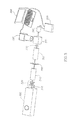
- FIG. 12A shows an illustration of a handheld defection box instrument according to one embodiment.
- FIG. 12B shows an assembled detection box instrument according to one embodiment.
- FIG. 13A shows the top view of an apparatus according to one particular embodiment.
- FIG. 13B shows the top view of an apparatus according to one particular embodiment.
- a microfluidic apparatus comprising a substrate having a first channel having a defined physical feature, wherein said first channel is in fluid communication with at least one inlet for receiving a fluid, wherein said first channel leads to a restrictive access, and wherein said first channel is in fluid communication with a second channel having a defined physical feature, wherein said second channel is in fluid communication with at least one fluid flow outlet; and a fluid biological sample.
- said defined physical feature is a depression or protrusion.
- said fluid biological sample comprises blood.
- the microfluidic apparatus further comprises a detection zone, and/or a filter array, each in fluid communication with said channel and said fluid flow outlet.
- a microfluidic apparatus comprising a substrate having at least one first channel having a defined physical feature; at least one first inlet formed in said first channel for receiving a first fluid; wherein said first channel is in fluid communication with a bifurcated channel, wherein said bifurcated channel is in fluid communication with a third channel detection zone; at least one second inlet for receiving a second fluid, wherein said second inlet is in fluid communication with a branched channel; a filter structure in fluid communication with a reservoir, wherein said reservoir is in fluid communication with said third channel detection zone; at least one fluid flow outlet formed In said third channel; and a fluid sample; wherein the ratio of the cross-sectional area of said second channel compared to the cross-sectional area of said first channel is 1:10.
- the defined physical feature is a depression or a protrusion.
- said filter structure comprises a filter array, said first fluid comprises sheath fluid and said second fluid comprises blood.
- a detection system comprising a microfluidic apparatus and further comprising a light source; a lens assembly; a filter assembly; and an image capture device.
- the detection system further comprises at least one display unit or at least one recording unit.
- said excitation source comprises a laser, particularly an argon laser.
- said filter assembly comprises an excitation filter, and at least one emission filter.
- said filter assembly further comprises at least one aperture and at least one neutral density filter.
- said filter assembly further comprises at least one glass polarizer.
- the lens assembly of the detection system comprises at least one condenser lens, at least one objective lens, and at least one beamsplitter.
- said image capture device comprises at least one CCD camera, CMOS device, photodiode, or photomultiplier tube.
- said filter assembly comprises at least two emission filters and said image capture device comprises at least one photomultiplier tube.
- said display unit comprises a computer and said recording unit comprises an oscilloscope.
- said excitation source comprises an argon laser
- said lens assembly comprises a condenser lens, an objective lens, and a beamsplitter
- said filter assembly comprises an excitation filter, a pinhole aperture and a neutral density filter, and at least one emission filter
- said image capture device comprises a CCD camera and a photodiode
- said display unit comprises a personal computer, and further comprising an amplifier.
- said excitation source of the detection system comprises an argon laser; said lens assembly comprises a condenser lens, an objective lens, and a beamsplitter, said filter assembly comprises an excitation filter, at least one emission filter; said image capture device comprises a photomultiplier tube, and said display unit comprises a personal computer.
- a method for identifying a target comprising providing a fluid sample to at least one microfluidic apparatus, wherein said fluid sample contains at least one dye; providing an excitation source to induce at least one fluorescent signal in a target; defecting the fluorescent signal using a sensor in the apparatus; and identifying the target based in part on the analysis of the fluorescent signal.
- said target is selected from the group consisting of: cells, organelles, nuclei, granules, DNA, and RNA.
- said target comprises a cell selected from the group consisting of a monocyte, a granulocyte, a macrophage, a neutrophil, an eosinophil, a basophil, or other leukocyte.
- said target comprises a leukocyte and said dye comprises acridine orange.
- Particular embodiments of the method further comprising counting or sorting the target in the sample by analysis of the fluorescent signal.
- said fluid sample comprises blood.
- the present disclosure relates to fabricated microfluidic devices that can be utilized as cell sensors and/or actuators.
- the microfluidic device may be used for labeling, sensing, differentiating, and/or sorting cell populations.
- Microfluidic cell sensors and actuators can provide cell sensing and counting for a more accurate outcome and a lower cost.
- Particle counting has been demonstrated, for example, by electrical impedance sensing, light scattering detection, and fluorescent sensing.
- microfabrication has allowed development of microdevices to replace glass capillary-based flow chambers, and to integrate compact optics and provide on-chip sample transport.
- Dilution is also often required in order to avoid clogging sample chambers, and also In order to remove erythrocytes that are lysed prior to running the sample, particularly for electrical impedance or light scattering detection. Some of these protocols also require an additional fixation buffer.
- a dye such as Acridine orange (3,6-dimethylamineoacridine, FIG. 1 ), can be used to differentiate a target, such as cells, organelles, granules, nuclei, molecules (including double or single stranded nucleic acids, such as DNA, or RNA, chromosomes, and also including synthetic forms).
- a target such as cells, organelles, granules, nuclei, molecules (including double or single stranded nucleic acids, such as DNA, or RNA, chromosomes, and also including synthetic forms).
- leukocytes may be detected, counted, or sorted without need for lysing erythrocytes or fixing the cell sample.
- Certain dyes, such as Acridine orange are also desirable due to the fast diffusion into cells, easy commercial availability, and excitation and emission wavelength compatibility with common light sources (i.e.
- Acridine orange is a pH-sensitive fluorescent cationic dye that binds to double-stranded DNA by electrostatic attraction and intercalation of the Acridine orange between base pairs. Upon binding, the excitation maximum becomes 502 nm and the emission maximum becomes 525 nm (green). Acridine orange also binds to RNA and single-stranded DNA, with a shifted excitation maximum of 460 nm and an emission maximum of 650 nm (red). Adams and Kamentsky, Acta Cytol. 15, 289 (1971); Adams and Kamentsky Acta Cytol. 18, 389-91 (1974); Steinkam et al., Acta Cytol. 17, 113-17 (1973).
- Acridine orange is also desirable in that it is hydrophobic in neutral pH, and can easily diffuse through the cell membrane and cell nuclear membrane to bind to RNA and DNA. In living cells, Acridine orange is protonated in the acidic environment of lysosomes, which makes it cationic and prevents the dye from leaking out of lysosome membranes. Moriyama et al., J. Biochem. 92; 1333-36 (1982). When Acridine orange is used for leukocyte analysis, the cell nucleus is stained green with slightly mixed red, a result of double-stranded DNA and single-stranded RNA, while the cell cytoplasm is stained red due to the RNA and lysosomes. Thus, leukocyte counting can be achieved easily by using the strong signal from the green fluorescent channel. Leukocyte differentiation can be achieved by analyzing the signal from the red fluorescence channel.
- a 3-part differential can be achieved by studying the red fluorescent signal of an Acridine orange stained diluted blood sample, whereas a 5-part differential leukocytes (lymphocytes, monocytes, neutrophils, eosinophils, and basophils) has been demonstrated with hypotonic dilution and fresh Acridine orange-stained leukocyte samples.
- Adams and Kamentsky Acta Cytol. 15, 289 (1971); Adams and Kamentsky, Acta Cytol. 18, 389-391 (1974); Steinkam et al., Acta Cytol. 17, 113-17 (1973).
- ethidium bromide ethidium bromide, three-dye combinations (ethidium bromide, brilliant sulfaflavine, and stilbene disulfonic acid derivative); oxazine dyes, basic orange 21, and a polymethine dye.
- ethidium bromide ethidium bromide, three-dye combinations (ethidium bromide, brilliant sulfaflavine, and stilbene disulfonic acid derivative); oxazine dyes, basic orange 21, and a polymethine dye.
- the device or apparatus comprises a substrate formed from a material, such as silicon, glass, plastic, metal, or other material.
- a material such as silicon, glass, plastic, metal, or other material.
- One particular embodiment of the instant disclosure was fabricated using soft lithography. Quake, Science 290, 1536-40 (2000). Other photolithographic or etching techniques could also be used, according to specific embodiments.
- One embodiment of the device was microfabricated using two parts of PDMS (polydimethylsiloxane) (Sylgard 184, Dow Corning, MI, USA) mixed vigorously in 10:1 ratio. After degassing in vacuum for about 30 minutes, the mixture was poured onto DRIE-etched silicon mold, that had been pretreated with HMDS (hexamethyldisilazane) for easy separation after baking. The molds were baked at 80° C. for 30 minutes. The hardened PDMS was separated from the silicon mold, and PDMS sheet was cut into pieces and fluidic access holes were punctured on each piece with a Luer stub adapter (Becton Dickinson, NJ, USA).
- PDMS polydimethylsiloxane
- HMDS hexamethyldisilazane
- the channel structure was molded on a 1 cm ⁇ 1 cm PDMS block, with the thickness of the PDMS block at less than 3 mm. In one particular embodiment the channel depth was 16 ⁇ m in order to accommodate large leukocyte sizes.
- a first fluid flow Inlet 200 allows for deposition of, for example sheath flow fluid, and is in fluid communication with a bifurcated channel with a first channel arm 260 and a second channel arm 270 that both converge at a junction of a reservoir 290 and the detection zone 240 .
- the apparatus further comprises a second fluid flow inlet 210 that allows for deposition of, for example, a sample fluid, such as blood, that is in fluid communication with a filter array structure 230 , by way of a branched sample flow zone channel 220 and a fluid flow outlet 250 .
- 2-D hydrodynamic focusing was adopted to control the particle position of the cell sample in the detection zone 240 .
- the ratio of cross-sectional area of sheath flow to core sample flow was 10:1
- the channel width of the defection zone 240 was 50 ⁇ m, with the width of the focused sample flow preferably 5 ⁇ m or less.
- the channels comprise a physical feature, such as a depression or a protrusion.
- the fluid flow inlet 1340 allows for deposition of a sample fluid, such as a biological sample, or other fluid sample containing a target.
- the biological sample includes a cell sample, such as blood.
- the fluid inlet is in fluid communication with a first channel 1330 which contains a restrictive access 1320 that is juxtaposed to a second channel 1310 which comprises the detection zone which is also in fluid communication with the fluid flow outlet 1300 .
- the height of the first and/or second channels is approximately 5 ⁇ m, approximately 8 ⁇ m, approximately 10 ⁇ m, approximately 12 ⁇ m, approximately 15 ⁇ m, approximately 20 ⁇ m, approximately 25 ⁇ m, approximately 30 ⁇ m, approximately 35 ⁇ m, approximately 40 ⁇ m, or any value therebetween.
- the width of the second channel is approximately 5 ⁇ m, 10 ⁇ m, approximately 15 ⁇ m, approximately 20 ⁇ m, approximately 25 ⁇ m, approximately 30 ⁇ m, approximately 35 ⁇ m, approximately 40 ⁇ m, approximately 45 ⁇ m, approximately 50 ⁇ m, or any value therebetween. In the exemplary embodiment shown in FIG. 13A , the second channel width was approximately 20 ⁇ m in size.
- the fluid flow inlet 1440 allows for deposition of a sample fluid, such as a biological sample, or other fluid sample containing a target.
- the biological sample includes a cell sample, such as blood.
- the fluid inlet is in fluid communication with a first channel 1430 which contains a restrictive access 1420 that is juxtaposed to a second channel 1410 which comprises the detection zone which is also in fluid communication with the fluid flow outlet 1400 .
- the height of the first and/or second channels is approximately 5 ⁇ m, approximately 8 ⁇ m, approximately 10 ⁇ m, approximately 12 ⁇ m, approximately 15 ⁇ m, approximately 20 ⁇ m, approximately 25 ⁇ m, approximately 30 ⁇ m, approximately 35 ⁇ m, approximately 40 ⁇ m, or any value therebetween.
- the width of the second channel is approximately 5 ⁇ m, 10 ⁇ m, approximately 15 ⁇ m, approximately 20 ⁇ m, approximately 25 ⁇ m, approximately 30 ⁇ m, approximately 35 ⁇ m, approximately 40 ⁇ m, approximately 45 ⁇ m, approximately 50 ⁇ m, or any value therebetween. In the exemplary embodiment shown in FIG. 13B , the second channel width was approximately 30 ⁇ m in size.
- Certain embodiments of the device use a focused laser source for illumination, since cell focusing in the detection zone 240 is highly desirable. However, other embodiments included in the present disclosure use a more uniform diffused light source and a slit aperture. Such embodiments utilize straight channel geometry without cell focusing.
- the channel length of the detection zone 240 is 1000 ⁇ m.
- a filter structure 230 upstream of the sample flow zone 220 may also be included in certain embodiments, which filtered out contaminants, including erythrocyte rouleaux, and other large particle aggregates to prevent clogging in the detection zone 240 .
- the size of the rectangular pillar structure components of the filter structure 230 was 200 ⁇ m ⁇ 40 ⁇ m. The spacing between the pillars in each of the three rows was 40 ⁇ m, 30 ⁇ m, and 20 ⁇ m respectively, which allows for even the largest leukocytes to pass through the filter region 230 .
- the optical system was set up on an optical bench as shown in FIG. 3 (transmitted laser-induced fluorescent detection system or LIF).
- the system setup comprises an excitation or laser source 300 , a lens assembly 340 , the microfluidic apparatus 350 , an optional additional lens assembly 360 , a filter assembly 320 , 330 , and an image capture device 390 , 395 .
- one or more emission filters comprise, the filter assemblies 320 , 330 .
- the image capture device 395 comprises a charge coupled device (CCD) camera, a complementary metal-oxide-semiconductor (CMOS) device, or a photomultiplier tube (PMT) device.
- CCD charge coupled device
- CMOS complementary metal-oxide-semiconductor
- PMT photomultiplier tube
- the image capture device 395 may be coupled to communicate with a display unit or computing device 396 , such as a personal computer.
- a display unit or computing device 396 such as a personal computer.
- One of skill in the art would recognize that multiple and various computer software programs are available that allow for integration, compilation, analysis, reconfiguration, and other manipulation of data received from the system, particularly by way of the computing device 396 .
- an argon laser (National Laser NLC210BL, 483 nm, and 15-30 mW adjustable, Salt take City, Utah, USA) is used as the excitation source.
- An aperture 310 of 50 ⁇ m diameter is put in front of the laser output to facilitate the alignment process and lower the illumination intensity.
- art optional laser-line bandpass filter (bandwidth equal to about 1.9 nm with a central wavelength of 488 nm) is used to further purify the laser source.
- an optional neutral density filter (NDF) is used to attenuate laser excitation.
- the pinhole and NDF are replaced by two linear glass polarizers (Edmond Optics TECH SPEC, Barrington N.J., USA) so that the Illumination level on the device can be easily adjusted.
- a long-working-distance microscope objective (USMCO M Plan Apo, 10 ⁇ , 0.28 NA, Dayton, Nev., USA) is used as a condenser lens 340 .
- Another long-working-distance microscope objective (Bausch & Lomb, 50 ⁇ , 0.45 NA, Rochester, N.Y., USA) is used as an objective lens 360 .
- a green bandpass filter with central wavelength 525 nm and a bandwidth 50 nm Chroma D525_50 m
- a red bandpass filter with central wavelength 650 nm and bandwidth 50 nm
- the signal is electrically amplified and detected either with a silicon photodiode receiver module 390 (Electro-Optical Systems, UVS-025-H, Phoenixville, Pa., USA) or a photon multiplier tube (PMT, Hamamatsu H5784-20, Japan).
- the voltage signal is sent to a deep memory oscilloscope (HP 54645A, Palo Alto, Calif., USA).
- the buffer in the oscilloscope is full, the data can be loaded to a computer and analyzed with a Matlab peak-detection program.
- Video may be taken with an analog CCD camera (Hitachi KP-D20B, Japan) at 30 frames per second and then converted to digital format and stored in a computer 396 .
- imaging capture devices 395 such as CMOS, PMT, or still other devices may also be used with particular embodiments described herein.
- the system set up utilizing a photodiode detector and PMT are more sensitive than the CCD camera and have a faster time response.
- the optical system was first roughly aligned on a dummy device with the aid of images from CCD camera. A 10 ⁇ m diameter illumination spot on the detection zone is easily achieved with proper alignment.
- the instant apparatus may be incorporated into a hand-held unit comprising a laser source (such as a laser emitting diode or LED 120 ), at least one lens 190 , at least one filter assembly with optional beamsplitter 195 , a microfluidic apparatus as described herein on a microchip or other substrate 185 , an input/output port 130 , at least one image capture device 100 , 110 , which may be a photomultiplier tube.
- the hand-held unit may be assembled and enclosed by an outer casing or casings 160 , 180 , and rivets or bolts 140 , 160 .
- One aspect of the instant disclosure relates to methods of counting and/or differentiating cells, particularly leukocytes, from undiluted cell samples, such as human or other animal blood, by utilizing microfabricated devices.
- cell detection was conducted utilizing Acridine orange and fresh whole human blood.
- fresh human blood was obtained from healthy donors and used within 3 days of collection.
- EDTA was added to the blood samples in order to prevent coagulation.
- the stock solution was added to obtain a final dye-concentration of 10 ⁇ g/mL in Ficoll-Paque Plus.
- Ficoll-Paque Plus was also used as the sheath flow solution.
- Fluorescent polystyrene beads (5 ⁇ m green fluorescent beads) were purchased from Duke Scientific Corporations, Fremont, Calif., USA.
- Cell nucleus stain Acridine orange was obtained from Molecular Probes, Eugene, Oreg., USA, and dissolved in water to achieve a 10 mg/mL stock solution.
- Blood diluent Ficoll-Paque Plus was purchased from Amersham Biosciences, Sweden.
- Phosphate buffered saline (10 ⁇ PBS) was obtained from Ambion (9625), Austin, Tex., USA.
- Ficoll-Paque Plus specific gravity 1.077 g/mL
- All fluids were pumped into the devices using syringe pumps (Harvard Apparatus Pico Plus, Holliston, Mass., USA).
- an analog CCD camera was used for video recording at a matched camera frame rate of 3 nL/min sample flow rate and 30 nL/min sheath flow rate.
- a 0.1 ⁇ L/minute sample flow rate and a 1 ⁇ L/minute sheath flow rate were used for photodiode detection.
- a 1 ⁇ L/minute sample flow and a 10 ⁇ L/minute sheath flow were used with the photon multiplier tube instrument.
- the maximal concentration for cell staining was established using routine methods in the art. Adams and Kamentsky, Acta Cytol. 15; 289 (1971). As shown in FIG. 4 , whole blood samples were analyzed with different Acridine orange concentrations. The optimal concentration for leukocyte staining was determined to be approximately in the range of 1 ⁇ g/mL. In the particular exemplary embodiment utilized in FIG. 4 , the distance between the coverslip and the grid surface was approximately 100 ⁇ m. As can be seen in FIG. 4A , an abundance of erythrocytes were present under the field of view, yet these cells did not interfere with the fluorescent signal from the leukocytes, as shown in FIG. 4B-F .
- the exemplary embodiment utilized in cell detection did not experience any significant photobleaching.
- the signal was fitted as a first-order exponential decay with time constant of 6.4+/ ⁇ 0.7 seconds.
- the photobleaching time constant for one particular embodiment was characterized by filling the device with Acridine orange-stained whole blood. The channel was scanned by the laser spot and the illumination was set to be the same as that used in testing. The entire process was recorded with a CCD camera. Whenever a fluorescing leukocyte was observed with fluorescent emission clearly distinct from the background, we stopped moving the laser spot and waited until the leukocyte was photobleached to background level. The images were extracted from the video, converted to 8-bit gray scale images, and analyzed with a Matlab program. The data was fitted to a single time-constant exponential decay.
- green fluorescent beads were tested at a concentration of about 2 ⁇ 10 3 / ⁇ L, as observed by CCD camera, and shown in FIG. 6 .
- Sample flow rate was set at about 3 nL/min, and sheath flow was about 30 nL/min.
- a hydrodynamic focused laser beam as shown in FIG. 6A , created an enlarged light circle as shown in FIG. 6B . Only a single bead normally appeared in each image. With diffused laser illumination, as shown in FIG. 6C , the trace of the bead could be identified, as shown in FIG. 6D .
- Hydrodynamic focusing limits the cross-sectional area of the defection zone without shrinking the channel diameter, thus the signal-to-noise ratio may be improved without increasing the risk of clogging the channel. Also, the reduction of the cross-section of the core flow reduces the coincidence effect. Finally, enclosing the core sample flow with sheath flow minimizes fluorescent dye absorption in the device walls, thus reducing background noise. As indicated in FIG. 7 , bead signals from the photodiode detector could easily be identified.
- images extracted from video taken by the CCD camera show the signal identified from a leukocyte stained with Acridine orange, as well as the signal obtained from the fluorescent control bead.
- the expected leukocyte detection rate would average about 4-11 cells per second for a normal individual.
- a time trace over 50 seconds of an undiluted blood sample stained with Acridine orange using a green emission filter, and a throughput of up to about 1000 leukocytes per second was attained.
- Maxima signal Intensity (peak height as in FIG. 9 ) from the green fluorescent channel with 525 nm emission filter was studied by plotting its histogram, as shown in FIG. 10 .
- the lower-intensity portion Is likely contributed mainly by lymphocytes, while the higher-intensity portion is likely mainly from monocytes, with the center-region is likely mostly from granulocytes. Steinkam et al., Acta Cytol. 17; 113-117 (1973).
- a time trace over 50 seconds of an undiluted blood sample stained with Acridine orange using a red fluorescent channel with 650 nm emission filter was conducted. As shown in FIG. 11 , two peaks were identified. The lower intensity is dominated by lymphocytes and the higher-intensity peak is largely monocytes and granulocytes. The time between the start of staining the cells to photodiode recording was typically greater than 15 minutes.
- the maximal throughput was about 1000 leukocytes per second utilizing one embodiment of the PMT detector.
- minimal sample volume was maintained, which increases the throughput.
- sample throughput is proportional to volume flow rate, but is limited by the maximal pumping rate and response time of the sensing system, a 3 nL/minute core flow rate was used with the CCD camera defection. Under this flow rate, a typical leukocyte traveled through the detection zone in approximately 30 milliseconds, which roughly equals the CCD frame acquisition time.
- Flow rates for varying embodiments may be suitable for a range from approximately 1 nL/minute, approximately 2 nL/minute, approximately 3 nL/minute, approximately 4 nL/minute, approximately 5 nL/minute, approximately 6 nL/minute, approximately 7 nL/minute, approximately 8 nL/minute, approximately 9 nL/minute, approximately 10 nL/minute, approximately 20 nL/minute, approximately 30 nL/minute, approximately 40 nL/minute, approximately 50 nL/minute, approximately 60 nL/minute, approximately 70 nL/minute, approximately 80 nL/minute, approximately 90 nL/minute, approximately 100 nL/minute, approximately 110 nL/minute, approximately 120 nL/minute, approximately 130 nL/minute, approximately 140 nL/minute, approximately 150 nL/minute, or any value therebetween for photodiode detection.
- flow rates for varying embodiments may be suitable for a range from approximately 200 nL/minute, approximately 300 nL/minute, approximately 400 nL/minute, approximately 500 nL/minute, approximately 600 nL/minute, approximately 700 nL/minute, approximately 800 nL/minute, approximately 900 nL/minute, approximately 1 ⁇ L/minute, approximately 2 ⁇ L/minute, approximately 3 ⁇ L/minute, approximately 4 ⁇ L/minute, approximately 5 ⁇ L/minute, or any value therebetween.
- the time response of the photodiode receiver module under low sensitivity setting was 0.16 milliseconds, and 0.6 milliseconds under high sensitivity, while the time response of the PMT detector in one exemplary run was about 16 microseconds.
- the linear flow velocity of the core flow is increased, which requires faster sensing, and reduces the coincidence effect by increasing the average distance between cells in the detection zone.
- leukocyte sensing, counting, and sorting can be achieved one-by-one in a micro flow cytometer system.
- dense cell suspensions such as whole, undiluted blood may be utilized in certain embodiments described herein, which provides for reduced sample and waste volume, reduced processing time, and completely eliminates on-chip mixing and buffer storage.
- leukocytes can be sensed one-by-one in a micro flow cytometer system.
- certain embodiments of the device can be implemented in various sizes and conformations, including but not limited to a bench-top device, a handheld device (such as is shown in FIG. 12 ), an implantable device, a nanotechnology device, or other size or conformation.
- a bench-top device such as is shown in FIG. 12
- an implantable device such as is shown in FIG. 12
- nanotechnology device such as is shown in FIG. 12
- high-illumination LED is used for excitation and a minipump is used to manipulate the sample in suction mode, while fluorescent signals from green and red channels can be detected simultaneously.
Abstract
Described herein are particular embodiments relating to a microfluidic device that may be utilized for cell sensing, counting, and/or sorting. Particular aspects relate to a microfabricated device that is capable of differentiating single cell types from dense cell populations. One particular embodiment relates a device and methods of using the same for sensing, counting, and/or sorting leukocytes from whole, undiluted blood samples.
Description
This application claims the benefit of priority under 35 U.S.C. §120 as a continuation of U.S. patent application Ser. No. 14/685,480, filed Apr. 13, 2015, and now U.S. Pat. No. 9,234,884, which is a continuation of U.S. patent application Ser. No. 14/296,199, filed Jun. 4, 2014, and issued as U.S. Pat. No. 9,029,158 on May 12, 2015, which is a divisional of U.S. patent application Ser. No. 12/062,808, filed Apr. 4, 2008, now abandoned, which claims priority under 35 U.S.C. §119(e) to U.S. provisional patent application No. 60/922,296, filed Apr. 6, 2007, now expired, the contents of each of which are herein incorporated by reference in their entirety.
This invention was made with government support under grant number NCC 9-58-317 awarded by National Space Biomedical Research Institute through NASA. The government has certain rights in this invention.
The present disclosure relates to fabricated microfluidic devices that can be utilized as cell sensors and/or actuators. In certain embodiments, the microfluidic device may be used for labeling, sensing, differentiating, and/or sorting targets, particularly cell populations.
Standard cell sensors or actuators are generally based on flow cytometry and employ one or a combination of electrical impedance sensing, light scattering measurement, and chemical or immunostaining followed by optical sensing.
For differentiation of blood cells by electrical impedance sensing, red blood cells are removed by lysing in order to reduce the blood volume. Lysing is generally done through the use of saponin or surfactants. During the lysing process, the leukocyte cell volume changes depending on cell type, due to the leakage of cytoplasm contents and cell nucleus shrinkage in varying amounts. Fujimoto, Sysmex J. Int. 9 (1990). Thus, normally 2-part (lymphocytes versus granulocytes) or even 3-part (lymphocytes, neutrophils, and other leukocytes) differential can be achieved by simple electrical impedance measurement of particle volume. Hughes-Jones, et al., J. Clin. Pathol. 27; 623-625 (1974); Oberjat, et al., J. Lab. Clin. Med. 76; 518 (1970); Vandilla, et al., Proc. Soc. Exp. Biol. Med. 125; 367 (1967); Maeda, et al., Clin. Pathol. 27; 1117-1200 (1979); Maeda, et al., Clin. Pathol. 9; 555-558 (1982). Combining direct current and alternating current impedance, special acidic hemolysis in basophile channel and alkali hemolysis in eosinophil channel, a 5-part leukocyte differential can be achieved. Tatsumi, et al., Sysmex J. Int. 9; 9-20 (1999).
Alternative optical methods are based on light scattering and fluorescence staining of organelles, granules, and nuclei. Generally, low-angle scattered light contains information on cell size and high-angle scattered light can be used to probe internal composition of the cell. To achieve 5-part differential, certain leukocyte populations, such as eosinophils, require special stain to change its scattering characteristics from other granulocytes, and basophils typically need to be counted separately following the differential lysis of other leukocytes. McKenzie, Clinical Laboratory Hematology, Prentice Hall, 2004; Fujimoto, Sysmex J. Int. 9 (1999).
In general, conventional automated cell analyzers are bulky, expensive, and mechanically complex, which restricts their locations to hospitals or central laboratories. Conventional cell analyzers require larger sample volumes and generate more waste than the systems developed using microdevices. Furthermore, for analysis of certain cell types, such as leukocytes, accuracy and speed of counting, differentiation, and/or sorting is important for determining disease state and treatment.
Certain embodiments disclosed herein include a microfluidic apparatus comprising a substrate having a first channel having a defined physical feature, wherein said first channel is in fluid communication with at least one inlet for receiving a fluid, wherein said first channel leads to a restrictive access, and wherein said first channel is in fluid communication with a second channel having a defined physical feature, wherein said second channel is in fluid communication with at least one fluid flow outlet; and a fluid biological sample. In certain embodiments, said defined physical feature is a depression or protrusion. In particular embodiments, said fluid biological sample comprises blood. In certain embodiments, the microfluidic apparatus further comprises a detection zone, and/or a filter array, each in fluid communication with said channel and said fluid flow outlet.
A microfluidic apparatus comprising a substrate having at least one first channel having a defined physical feature; at least one first inlet formed in said first channel for receiving a first fluid; wherein said first channel is in fluid communication with a bifurcated channel, wherein said bifurcated channel is in fluid communication with a third channel detection zone; at least one second inlet for receiving a second fluid, wherein said second inlet is in fluid communication with a branched channel; a filter structure in fluid communication with a reservoir, wherein said reservoir is in fluid communication with said third channel detection zone; at least one fluid flow outlet formed In said third channel; and a fluid sample; wherein the ratio of the cross-sectional area of said second channel compared to the cross-sectional area of said first channel is 1:10. In certain embodiments, the defined physical feature is a depression or a protrusion.
In certain embodiments, said filter structure comprises a filter array, said first fluid comprises sheath fluid and said second fluid comprises blood.
Certain embodiments disclosed herein relate to a detection system comprising a microfluidic apparatus and further comprising a light source; a lens assembly; a filter assembly; and an image capture device. In some embodiments, the detection system further comprises at least one display unit or at least one recording unit. In certain particular embodiments, said excitation source comprises a laser, particularly an argon laser. In particular embodiments, said filter assembly comprises an excitation filter, and at least one emission filter. In certain embodiments, said filter assembly further comprises at least one aperture and at least one neutral density filter. In particular embodiments, said filter assembly further comprises at least one glass polarizer.
In certain embodiments, the lens assembly of the detection system comprises at least one condenser lens, at least one objective lens, and at least one beamsplitter. In particular embodiments, said image capture device comprises at least one CCD camera, CMOS device, photodiode, or photomultiplier tube. In certain embodiments, said filter assembly comprises at least two emission filters and said image capture device comprises at least one photomultiplier tube. In certain embodiments, said display unit comprises a computer and said recording unit comprises an oscilloscope. In particular embodiments, said excitation source comprises an argon laser; said lens assembly comprises a condenser lens, an objective lens, and a beamsplitter; said filter assembly comprises an excitation filter, a pinhole aperture and a neutral density filter, and at least one emission filter; said image capture device comprises a CCD camera and a photodiode, and said display unit comprises a personal computer, and further comprising an amplifier.
In particular embodiments, said excitation source of the detection system comprises an argon laser; said lens assembly comprises a condenser lens, an objective lens, and a beamsplitter, said filter assembly comprises an excitation filter, at least one emission filter; said image capture device comprises a photomultiplier tube, and said display unit comprises a personal computer.
Other embodiments disclosed herein relate to a method for identifying a target comprising providing a fluid sample to at least one microfluidic apparatus, wherein said fluid sample contains at least one dye; providing an excitation source to induce at least one fluorescent signal in a target; defecting the fluorescent signal using a sensor in the apparatus; and identifying the target based in part on the analysis of the fluorescent signal. In certain embodiments, said target is selected from the group consisting of: cells, organelles, nuclei, granules, DNA, and RNA. In other embodiments, said target comprises a cell selected from the group consisting of a monocyte, a granulocyte, a macrophage, a neutrophil, an eosinophil, a basophil, or other leukocyte. In specific embodiments, said target comprises a leukocyte and said dye comprises acridine orange. Particular embodiments of the method further comprising counting or sorting the target in the sample by analysis of the fluorescent signal. In certain embodiments, said fluid sample comprises blood.
The present disclosure relates to fabricated microfluidic devices that can be utilized as cell sensors and/or actuators. In certain embodiments, the microfluidic device may be used for labeling, sensing, differentiating, and/or sorting cell populations.
Microfluidic cell sensors and actuators can provide cell sensing and counting for a more accurate outcome and a lower cost. Particle counting (including bead, erythrocyte, and cultured cell) has been demonstrated, for example, by electrical impedance sensing, light scattering detection, and fluorescent sensing. Gawad, et al. Lab Chip 1; 76 (2001); Lee, et al. Proceedings of the 18th IEEE International Conference on Micro Electro Mechanical Systems (MEMS) 678-681 (2005); Satake et al. Sens. Actuators B: Chem. 83; 77 (2002); Morgan, et al. Curr. Appl. Phys. 6, 367-370 (2006); Lee et al., J. Micromech. Microeng. 15; 447-454 (2005); Altendorf, et al. Proceedings of the International Conference on Solid State Sensors and Actuators (Transducers '97) v. 1, p. 531, Chicago, Ill. (1997); Holmes et al., Biosens. Bioelectron. 21; 1621-1630 (2006); Yang et al., Meas. Sci. Technol. 17; 2001-2009 (2006); Simonnet et al., Anal. Chem. 78; 5653-5663 (2006); Niehren, et al., Anal. Chem. 67; 2666-2671 (1995).
In the area of optical sensing, microfabrication has allowed development of microdevices to replace glass capillary-based flow chambers, and to integrate compact optics and provide on-chip sample transport.
Cell sensing and counting, particularly of leukocytes, is cumbersome due in part to the cell population numbers. For leukocyte differential in microdevices based on optical sensing, a V-groove micro-channel was fabricated by anisotropic wet etching of a silicon substrate and 3-part leukocyte differential was demonstrated for diluted blood without sheath flow by two-parameter light scattering. Altendorf, Proceedings of the Int'l Conference on Solid State Sensors and Actuators, v. 1, p. 531 (1997).
However, until the instant embodied disclosure, it was necessary to dilute cell samples for cell sensors and actuators for many reasons. One reason dilution has been necessary is in order to prevent the coincidence effect in which multiple cells appear in the defection zone simultaneously. In human blood, the ratio of erythrocytes, or red blood cells, to leukocytes is on the order of about a thousand to one, a dilution factor of from about one hundred to several tens of thousands is typically required to avoid erythrocyte interference for electrical impedance or light scattering defection. Furthermore, for counting leukocytes in samples where leukocytes are specifically fluorescently labeled, a dilution of at least ten times is usually required. Sheenan and Storey, J. Pathol. Bacteriol. 59; 336 (1947); Kass, J. Clin. Pathol. 76; 810-12 (1981); Weigl et al., Biomed. Microdev. 3; 267-274 (2001);
Dilution is also often required in order to avoid clogging sample chambers, and also In order to remove erythrocytes that are lysed prior to running the sample, particularly for electrical impedance or light scattering detection. Some of these protocols also require an additional fixation buffer.
Dyes
In the present disclosure, a dye, such as Acridine orange (3,6-dimethylamineoacridine, FIG. 1 ), can be used to differentiate a target, such as cells, organelles, granules, nuclei, molecules (including double or single stranded nucleic acids, such as DNA, or RNA, chromosomes, and also including synthetic forms). In one particular embodiment, leukocytes may be detected, counted, or sorted without need for lysing erythrocytes or fixing the cell sample. Certain dyes, such as Acridine orange, are also desirable due to the fast diffusion into cells, easy commercial availability, and excitation and emission wavelength compatibility with common light sources (i.e. argon laser and other broad spectrum light sources in visible range) and optical filters. Kosenow, Acta Haematol. 7, 217 (1952); Schiffer, Blood, 19, 200 (1962): Jackson, Blood, 17, 643 (1961); Hallermann et al., Verh Deutsch Ges Inn Med. 70, 217 (1964).
Acridine orange is a pH-sensitive fluorescent cationic dye that binds to double-stranded DNA by electrostatic attraction and intercalation of the Acridine orange between base pairs. Upon binding, the excitation maximum becomes 502 nm and the emission maximum becomes 525 nm (green). Acridine orange also binds to RNA and single-stranded DNA, with a shifted excitation maximum of 460 nm and an emission maximum of 650 nm (red). Adams and Kamentsky, Acta Cytol. 15, 289 (1971); Adams and Kamentsky Acta Cytol. 18, 389-91 (1974); Steinkam et al., Acta Cytol. 17, 113-17 (1973). Acridine orange is also desirable in that it is hydrophobic in neutral pH, and can easily diffuse through the cell membrane and cell nuclear membrane to bind to RNA and DNA. In living cells, Acridine orange is protonated in the acidic environment of lysosomes, which makes it cationic and prevents the dye from leaking out of lysosome membranes. Moriyama et al., J. Biochem. 92; 1333-36 (1982). When Acridine orange is used for leukocyte analysis, the cell nucleus is stained green with slightly mixed red, a result of double-stranded DNA and single-stranded RNA, while the cell cytoplasm is stained red due to the RNA and lysosomes. Thus, leukocyte counting can be achieved easily by using the strong signal from the green fluorescent channel. Leukocyte differentiation can be achieved by analyzing the signal from the red fluorescence channel.
For fresh-stained leukocytes, a 3-part differential (lymphocytes, monocytes, and granulocytes) can be achieved by studying the red fluorescent signal of an Acridine orange stained diluted blood sample, whereas a 5-part differential leukocytes (lymphocytes, monocytes, neutrophils, eosinophils, and basophils) has been demonstrated with hypotonic dilution and fresh Acridine orange-stained leukocyte samples. Adams and Kamentsky, Acta Cytol. 15, 289 (1971); Adams and Kamentsky, Acta Cytol. 18, 389-391 (1974); Steinkam et al., Acta Cytol. 17, 113-17 (1973).
Other dyes can be utilized with certain embodiments described in the instant disclosure, such as ethidium bromide, three-dye combinations (ethidium bromide, brilliant sulfaflavine, and stilbene disulfonic acid derivative); oxazine dyes, basic orange 21, and a polymethine dye. Shapiro, et al., J. Histochem. Cytochem. 24, 396-411 (1976); Shapiro, et al., J. Histochem. Cytochem. 25, 836-844 (1977); U.S. Pat. No. 4,376,820; U.S. Pat. No. 4,882,284; Tibbe, et al., Nat. Biotechnol. 17, 1210-1213 (1999); U.S. Pat. No. 4,400,370; Kass, J. Histochem. Cytochem. 36, 711-715 (1988).
Apparatus
One embodiment of the instant disclosure relates to a device or apparatus for cell counting and/or differentiating. In particular embodiments, the device or apparatus comprises a substrate formed from a material, such as silicon, glass, plastic, metal, or other material. One particular embodiment of the instant disclosure was fabricated using soft lithography. Quake, Science 290, 1536-40 (2000). Other photolithographic or etching techniques could also be used, according to specific embodiments.
One embodiment of the device was microfabricated using two parts of PDMS (polydimethylsiloxane) (Sylgard 184, Dow Corning, MI, USA) mixed vigorously in 10:1 ratio. After degassing in vacuum for about 30 minutes, the mixture was poured onto DRIE-etched silicon mold, that had been pretreated with HMDS (hexamethyldisilazane) for easy separation after baking. The molds were baked at 80° C. for 30 minutes. The hardened PDMS was separated from the silicon mold, and PDMS sheet was cut into pieces and fluidic access holes were punctured on each piece with a Luer stub adapter (Becton Dickinson, NJ, USA). Each PDMS piece was carefully placed on a cleaned glass slide and baked overnight at 80° C. In some cases, oxygen plasma treatment (300 m Torr, 25 W, 30 s) was used for PDMS and glass slides in order to improve adhesion between them, particularly with devices that were intended to be reused. Bhattacharya et al., J. Microelectromechan. Syst. 14, 590-97 (2005).
In one particular embodiment, the channel structure was molded on a 1 cm×1 cm PDMS block, with the thickness of the PDMS block at less than 3 mm. In one particular embodiment the channel depth was 16 μm in order to accommodate large leukocyte sizes.
One exemplary embodiment of the device is shown in FIG. 2 . For this particular embodiment, a first fluid flow Inlet 200 allows for deposition of, for example sheath flow fluid, and is in fluid communication with a bifurcated channel with a first channel arm 260 and a second channel arm 270 that both converge at a junction of a reservoir 290 and the detection zone 240. In this particular embodiment, the apparatus further comprises a second fluid flow inlet 210 that allows for deposition of, for example, a sample fluid, such as blood, that is in fluid communication with a filter array structure 230, by way of a branched sample flow zone channel 220 and a fluid flow outlet 250. In this particular exemplary embodiment, 2-D hydrodynamic focusing was adopted to control the particle position of the cell sample in the detection zone 240. According to the embodiment shown in FIG. 2 , the ratio of cross-sectional area of sheath flow to core sample flow was 10:1, and the channel width of the defection zone 240 was 50 μm, with the width of the focused sample flow preferably 5 μm or less. In particular embodiments, the channels comprise a physical feature, such as a depression or a protrusion.
One other exemplary embodiment of the device is shown in FIG. 13A . For this particular embodiment the fluid flow inlet 1340 allows for deposition of a sample fluid, such as a biological sample, or other fluid sample containing a target. In one particular embodiment, the biological sample includes a cell sample, such as blood. In this exemplary embodiment, the fluid inlet is in fluid communication with a first channel 1330 which contains a restrictive access 1320 that is juxtaposed to a second channel 1310 which comprises the detection zone which is also in fluid communication with the fluid flow outlet 1300. In certain embodiments, the height of the first and/or second channels is approximately 5 μm, approximately 8 μm, approximately 10 μm, approximately 12 μm, approximately 15 μm, approximately 20 μm, approximately 25 μm, approximately 30 μm, approximately 35 μm, approximately 40 μm, or any value therebetween. In certain embodiments, the width of the second channel is approximately 5 μm, 10 μm, approximately 15 μm, approximately 20 μm, approximately 25 μm, approximately 30 μm, approximately 35 μm, approximately 40 μm, approximately 45 μm, approximately 50 μm, or any value therebetween. In the exemplary embodiment shown in FIG. 13A , the second channel width was approximately 20 μm in size.
One other exemplary embodiment of the device is shown in FIG. 13B . For this particular embodiment, the fluid flow inlet 1440 allows for deposition of a sample fluid, such as a biological sample, or other fluid sample containing a target. In one particular embodiment, the biological sample includes a cell sample, such as blood. In this exemplary embodiment, the fluid inlet is in fluid communication with a first channel 1430 which contains a restrictive access 1420 that is juxtaposed to a second channel 1410 which comprises the detection zone which is also in fluid communication with the fluid flow outlet 1400. In certain embodiments, the height of the first and/or second channels is approximately 5 μm, approximately 8 μm, approximately 10 μm, approximately 12 μm, approximately 15 μm, approximately 20 μm, approximately 25 μm, approximately 30 μm, approximately 35 μm, approximately 40 μm, or any value therebetween. In certain embodiments, the width of the second channel is approximately 5 μm, 10 μm, approximately 15 μm, approximately 20 μm, approximately 25 μm, approximately 30 μm, approximately 35 μm, approximately 40 μm, approximately 45 μm, approximately 50 μm, or any value therebetween. In the exemplary embodiment shown in FIG. 13B , the second channel width was approximately 30 μm in size.
Certain embodiments of the device use a focused laser source for illumination, since cell focusing in the detection zone 240 is highly desirable. However, other embodiments included in the present disclosure use a more uniform diffused light source and a slit aperture. Such embodiments utilize straight channel geometry without cell focusing. In one embodiment, the channel length of the detection zone 240 is 1000 μm. A filter structure 230 upstream of the sample flow zone 220 may also be included in certain embodiments, which filtered out contaminants, including erythrocyte rouleaux, and other large particle aggregates to prevent clogging in the detection zone 240. In certain embodiments, the size of the rectangular pillar structure components of the filter structure 230 was 200 μm×40 μm. The spacing between the pillars in each of the three rows was 40 μm, 30 μm, and 20 μm respectively, which allows for even the largest leukocytes to pass through the filter region 230.
System
The optical system was set up on an optical bench as shown in FIG. 3 (transmitted laser-induced fluorescent detection system or LIF). In one particular embodiment, the system setup comprises an excitation or laser source 300, a lens assembly 340, the microfluidic apparatus 350, an optional additional lens assembly 360, a filter assembly 320, 330, and an image capture device 390, 395. In certain embodiments, one or more emission filters comprise, the filter assemblies 320, 330. In certain embodiments, the image capture device 395 comprises a charge coupled device (CCD) camera, a complementary metal-oxide-semiconductor (CMOS) device, or a photomultiplier tube (PMT) device. In particular embodiments, the image capture device 395 may be coupled to communicate with a display unit or computing device 396, such as a personal computer. One of skill in the art would recognize that multiple and various computer software programs are available that allow for integration, compilation, analysis, reconfiguration, and other manipulation of data received from the system, particularly by way of the computing device 396.
In one particular exemplary embodiment, an argon laser (National Laser NLC210BL, 483 nm, and 15-30 mW adjustable, Salt take City, Utah, USA) is used as the excitation source. An aperture 310 of 50 μm diameter is put in front of the laser output to facilitate the alignment process and lower the illumination intensity. In certain embodiments, art optional laser-line bandpass filter (bandwidth equal to about 1.9 nm with a central wavelength of 488 nm) is used to further purify the laser source. In certain other embodiments, an optional neutral density filter (NDF) is used to attenuate laser excitation. Alternatively, the pinhole and NDF are replaced by two linear glass polarizers (Edmond Optics TECH SPEC, Barrington N.J., USA) so that the Illumination level on the device can be easily adjusted.
In one particular embodiment, a long-working-distance microscope objective (USMCO M Plan Apo, 10×, 0.28 NA, Dayton, Nev., USA) is used as a condenser lens 340. Another long-working-distance microscope objective (Bausch & Lomb, 50×, 0.45 NA, Rochester, N.Y., USA) is used as an objective lens 360. In the same embodiment, three emission filters 320 are used in one particular test: 488 nm long pass filter (Chroma H1500 LP, transition width>4.9 nm, edge steepness=2.5 nm, Rockingham, Vt., USA), a green bandpass filter with central wavelength 525 nm and a bandwidth 50 nm (Chroma D525_50 m), and a red bandpass filter with central wavelength 650 nm and bandwidth 50 nm (Chroma D650_50 m). A broadband non-polarizing hybrid cube beamsplitter 370 (Newport 05BC17MB.1, 400-700 nm, R/T=45%/45%, Irvine, Calif., USA) is used to direct light to the photodiode detector 380 and CCD camera simultaneously.
The signal is electrically amplified and detected either with a silicon photodiode receiver module 390 (Electro-Optical Systems, UVS-025-H, Phoenixville, Pa., USA) or a photon multiplier tube (PMT, Hamamatsu H5784-20, Japan). The voltage signal is sent to a deep memory oscilloscope (HP 54645A, Palo Alto, Calif., USA). When the buffer in the oscilloscope is full, the data can be loaded to a computer and analyzed with a Matlab peak-detection program. Video may be taken with an analog CCD camera (Hitachi KP-D20B, Japan) at 30 frames per second and then converted to digital format and stored in a computer 396. Other imaging capture devices 395, such as CMOS, PMT, or still other devices may also be used with particular embodiments described herein. In certain exemplary test runs, the system set up utilizing a photodiode detector and PMT are more sensitive than the CCD camera and have a faster time response. During one exemplary test run, the optical system was first roughly aligned on a dummy device with the aid of images from CCD camera. A 10 μm diameter illumination spot on the detection zone is easily achieved with proper alignment.
As shown in FIG. 12A and FIG. 12B , the instant apparatus may be incorporated into a hand-held unit comprising a laser source (such as a laser emitting diode or LED 120), at least one lens 190, at least one filter assembly with optional beamsplitter 195, a microfluidic apparatus as described herein on a microchip or other substrate 185, an input/output port 130, at least one image capture device 100, 110, which may be a photomultiplier tube. In certain embodiments, the hand-held unit may be assembled and enclosed by an outer casing or casings 160, 180, and rivets or bolts 140, 160.
Cell Detection
One aspect of the instant disclosure relates to methods of counting and/or differentiating cells, particularly leukocytes, from undiluted cell samples, such as human or other animal blood, by utilizing microfabricated devices. In one exemplary embodiment, cell detection was conducted utilizing Acridine orange and fresh whole human blood.
In one exemplary embodiment, fresh human blood was obtained from healthy donors and used within 3 days of collection. EDTA was added to the blood samples in order to prevent coagulation. For Acridine orange staining, the stock solution was added to obtain a final dye-concentration of 10 μg/mL in Ficoll-Paque Plus. Ficoll-Paque Plus was also used as the sheath flow solution. Fluorescent polystyrene beads (5 μm green fluorescent beads) were purchased from Duke Scientific Corporations, Fremont, Calif., USA. Cell nucleus stain Acridine orange was obtained from Molecular Probes, Eugene, Oreg., USA, and dissolved in water to achieve a 10 mg/mL stock solution. Blood diluent Ficoll-Paque Plus was purchased from Amersham Biosciences, Sweden. Phosphate buffered saline (10×PBS) was obtained from Ambion (9625), Austin, Tex., USA.
Staining results were observed under a fluorescent microscope (Nikon E800, Japan) with a triple band filter block DAPI-FITC-TRITC, which has excitation filter wavelengths of 385-400 nm, 475-490 nm, and 545-565 nm, and emission filter wavelengths of 450-465 nm, 505-535 nm and 580-620 nm. Images were taken with a cooled CCD camera (RT-KE color 3-shot, Diagnostic Instruments, Sterling Heights, Mich., USA). Rough count of leukocytes was made with a hemactyometer (Hausser Scientific, Horsham, Pa., USA). When necessary, blood or fluorescent beads were diluted with Ficoll-Paque Plus (specific gravity 1.077 g/mL) to match the specific gravity of the solvent to leukocytes. All fluids were pumped into the devices using syringe pumps (Harvard Apparatus Pico Plus, Holliston, Mass., USA).
In this particular embodiment, an analog CCD camera was used for video recording at a matched camera frame rate of 3 nL/min sample flow rate and 30 nL/min sheath flow rate. For photodiode detection, a 0.1 μL/minute sample flow rate and a 1 μL/minute sheath flow rate were used. A 1 μL/minute sample flow and a 10 μL/minute sheath flow were used with the photon multiplier tube instrument.
In order to achieve a high signal-to-noise ratio, the maximal concentration for cell staining was established using routine methods in the art. Adams and Kamentsky, Acta Cytol. 15; 289 (1971). As shown in FIG. 4 , whole blood samples were analyzed with different Acridine orange concentrations. The optimal concentration for leukocyte staining was determined to be approximately in the range of 1 μg/mL. In the particular exemplary embodiment utilized in FIG. 4 , the distance between the coverslip and the grid surface was approximately 100 μm. As can be seen in FIG. 4A , an abundance of erythrocytes were present under the field of view, yet these cells did not interfere with the fluorescent signal from the leukocytes, as shown in FIG. 4B-F .
As can be seen in FIG. 5 , the exemplary embodiment utilized in cell detection did not experience any significant photobleaching. The signal was fitted as a first-order exponential decay with time constant of 6.4+/−0.7 seconds. Two more tests confirmed that the photobleaching time constant for one particular embodiment was between 1 second and 10 seconds. The photobleaching time constant for one particular embodiment was characterized by filling the device with Acridine orange-stained whole blood. The channel was scanned by the laser spot and the illumination was set to be the same as that used in testing. The entire process was recorded with a CCD camera. Whenever a fluorescing leukocyte was observed with fluorescent emission clearly distinct from the background, we stopped moving the laser spot and waited until the leukocyte was photobleached to background level. The images were extracted from the video, converted to 8-bit gray scale images, and analyzed with a Matlab program. The data was fitted to a single time-constant exponential decay.
Additionally, green fluorescent beads were tested at a concentration of about 2×10³/μL, as observed by CCD camera, and shown in FIG. 6 . Sample flow rate was set at about 3 nL/min, and sheath flow was about 30 nL/min. In one exemplary test run, a hydrodynamic focused laser beam, as shown in FIG. 6A , created an enlarged light circle as shown in FIG. 6B . Only a single bead normally appeared in each image. With diffused laser illumination, as shown in FIG. 6C , the trace of the bead could be identified, as shown in FIG. 6D . Hydrodynamic focusing limits the cross-sectional area of the defection zone without shrinking the channel diameter, thus the signal-to-noise ratio may be improved without increasing the risk of clogging the channel. Also, the reduction of the cross-section of the core flow reduces the coincidence effect. Finally, enclosing the core sample flow with sheath flow minimizes fluorescent dye absorption in the device walls, thus reducing background noise. As indicated in FIG. 7 , bead signals from the photodiode detector could easily be identified.
As shown in FIG. 8 , using both red and green emission filters, images extracted from video taken by the CCD camera show the signal identified from a leukocyte stained with Acridine orange, as well as the signal obtained from the fluorescent control bead. For photodiode detection, the expected leukocyte detection rate would average about 4-11 cells per second for a normal individual.
In one exemplary embodiment, a time trace over 50 seconds of an undiluted blood sample stained with Acridine orange using a green emission filter, and a throughput of up to about 1000 leukocytes per second was attained. Maxima signal Intensity (peak height as in FIG. 9 ) from the green fluorescent channel with 525 nm emission filter was studied by plotting its histogram, as shown in FIG. 10 . As expected, the lower-intensity portion Is likely contributed mainly by lymphocytes, while the higher-intensity portion is likely mainly from monocytes, with the center-region is likely mostly from granulocytes. Steinkam et al., Acta Cytol. 17; 113-117 (1973).
In one exemplary embodiment, a time trace over 50 seconds of an undiluted blood sample stained with Acridine orange using a red fluorescent channel with 650 nm emission filter was conducted. As shown in FIG. 11 , two peaks were identified. The lower intensity is dominated by lymphocytes and the higher-intensity peak is largely monocytes and granulocytes. The time between the start of staining the cells to photodiode recording was typically greater than 15 minutes.
In both exemplary studies, the maximal throughput was about 1000 leukocytes per second utilizing one embodiment of the PMT detector. By using undiluted blood, minimal sample volume was maintained, which increases the throughput. Since sample throughput is proportional to volume flow rate, but is limited by the maximal pumping rate and response time of the sensing system, a 3 nL/minute core flow rate was used with the CCD camera defection. Under this flow rate, a typical leukocyte traveled through the detection zone in approximately 30 milliseconds, which roughly equals the CCD frame acquisition time.
Flow rates for varying embodiments may be suitable for a range from approximately 1 nL/minute, approximately 2 nL/minute, approximately 3 nL/minute, approximately 4 nL/minute, approximately 5 nL/minute, approximately 6 nL/minute, approximately 7 nL/minute, approximately 8 nL/minute, approximately 9 nL/minute, approximately 10 nL/minute, approximately 20 nL/minute, approximately 30 nL/minute, approximately 40 nL/minute, approximately 50 nL/minute, approximately 60 nL/minute, approximately 70 nL/minute, approximately 80 nL/minute, approximately 90 nL/minute, approximately 100 nL/minute, approximately 110 nL/minute, approximately 120 nL/minute, approximately 130 nL/minute, approximately 140 nL/minute, approximately 150 nL/minute, or any value therebetween for photodiode detection. Likewise, for PMT defection, flow rates for varying embodiments may be suitable for a range from approximately 200 nL/minute, approximately 300 nL/minute, approximately 400 nL/minute, approximately 500 nL/minute, approximately 600 nL/minute, approximately 700 nL/minute, approximately 800 nL/minute, approximately 900 nL/minute, approximately 1 μL/minute, approximately 2 μL/minute, approximately 3 μL/minute, approximately 4 μL/minute, approximately 5 μL/minute, or any value therebetween.
In one exemplary embodiment, the time response of the photodiode receiver module under low sensitivity setting was 0.16 milliseconds, and 0.6 milliseconds under high sensitivity, while the time response of the PMT detector in one exemplary run was about 16 microseconds.
Furthermore, by decreasing the cross-sectional area, the linear flow velocity of the core flow is increased, which requires faster sensing, and reduces the coincidence effect by increasing the average distance between cells in the detection zone.
Thus, by utilizing particular embodiments disclosed herein relating to a microfluidic device, leukocyte sensing, counting, and sorting can be achieved one-by-one in a micro flow cytometer system. Furthermore, dense cell suspensions, such as whole, undiluted blood may be utilized in certain embodiments described herein, which provides for reduced sample and waste volume, reduced processing time, and completely eliminates on-chip mixing and buffer storage. In particular aspects, leukocytes can be sensed one-by-one in a micro flow cytometer system.
As described herein, certain embodiments of the device can be implemented in various sizes and conformations, including but not limited to a bench-top device, a handheld device (such as is shown in FIG. 12 ), an implantable device, a nanotechnology device, or other size or conformation. In the smaller exemplary conformations, high-illumination LED is used for excitation and a minipump is used to manipulate the sample in suction mode, while fluorescent signals from green and red channels can be detected simultaneously.
All of the above U.S. patents, U.S. patent application publications, U.S. patent applications, foreign patents, foreign patent applications and non-patent publications referred to in this specification and/or listed in the Application Data Sheet, are incorporated herein by reference, in their entirety.
Claims (22)
1. A method for analyzing cells, the method comprising:
(a) introducing a fluid sample comprising fluorescently labeled cells and fluorescently labeled control beads to an inlet of a first microfluidic channel, wherein the fluorescently labeled cells are labeled with a fluorophore different than a fluorophore label of the control beads;
(b) flowing the fluid sample from the inlet of the first microfluidic channel to an outlet of a second microfluidic channel, wherein an outlet of the first microfluidic channel is physically and fluidically coupled to an inlet of the second microfluidic channel and wherein the flow of the fluid sample is aided with a pump;
(c) exciting the fluorophores of the fluorescently labeled cells and of the fluorescently labeled control beads using an excitation source;
(d) detecting fluorescence of the fluorescently labeled cells and fluorescence of the fluorescently labeled control beads as they pass through a detection zone within the second microfluidic channel, wherein the detection is performed on individual cells;
(e) analyzing the detected fluorescence of the fluorescently labeled cells and fluorescence of the fluorescently labeled control beads using a computer software inscribed on a computing device; and
(f) providing a result concerning one or more properties of the cells being analyzed.
2. The method of claim 1 , wherein the cells are leukocytes.
3. The method of claim 1 , wherein the fluid sample is a treated blood sample.
4. The method of claim 1 , wherein the second microfluidic channel has fixed dimensions.
5. The method of claim 4 , wherein the fixed dimensions are of approximately 40 microns by approximately 50 microns.
6. The method of claim 1 , wherein the first channel and the second channel are obstacle-free.
7. The method of claim 1 , wherein the flowing of the fluid sample from the inlet of the first microfluidic channel to the outlet of the second microfluidic channel increases a core flow rate of the fluid sample to form an increased flow rate.
8. The method of claim 1 , wherein the method further comprises identifying the fluorescently labeled cells.
9. The method of claim 1 , wherein the method further comprises sorting the fluorescently labeled cells.
10. The method of claim 1 , wherein the method further comprises counting the fluorescently labeled cells.
11. The method of claim 1 , further comprising video recording a measurement on a recording unit.
12. The method of claim 1 , wherein the method can detect cells at a rate of up to about 1000 cells per second.
13. The method of claim 1 , wherein the excitation source is a laser or a LED.
14. The method of claim 1 , wherein the excitation source comprises a lens assembly.
15. The method of claim 1 , further comprising manipulating the detected fluorescence using a computer software program.
16. The method of claim 15 , further comprising displaying a result of the manipulation on a display unit.
17. The method of claim 1 , wherein the fluorescently labeled beads are polymeric beads.
18. The method of claim 1 , wherein the fluorescently labeled beads are polystyrene beads.
19. The method of claim 1 , wherein the step of detecting fluorescence comprises using an image capture device.
20. The method of claim 1 , wherein flowing the fluid sample comprises pumping the fluid sample through a microfluidic device with the pump.
21. The method of claim 1 , wherein the fluid sample does not comprise a sheath fluid.
22. The method of claim 1 , wherein the fluid sample is an undiluted blood sample.
Priority Applications (2)
| Application Number | Priority Date | Filing Date | Title |
|---|---|---|---|
| US14/931,645 US9535059B2 (en) | 2007-04-06 | 2015-11-03 | Microfluidic device |
| US15/355,696 US9757729B2 (en) | 2007-04-06 | 2016-11-18 | Microfluidic device |
Applications Claiming Priority (5)
| Application Number | Priority Date | Filing Date | Title |
|---|---|---|---|
| US92229607P | 2007-04-06 | 2007-04-06 | |
| US12/062,808 US20090042241A1 (en) | 2007-04-06 | 2008-04-04 | Microfluidic device |
| US14/296,199 US9029158B2 (en) | 2007-04-06 | 2014-06-04 | Microfluidic device |
| US14/685,480 US9234884B2 (en) | 2007-04-06 | 2015-04-13 | Microfluidic device |
| US14/931,645 US9535059B2 (en) | 2007-04-06 | 2015-11-03 | Microfluidic device |
Related Parent Applications (1)
| Application Number | Title | Priority Date | Filing Date |
|---|---|---|---|
| US14/685,480 Continuation US9234884B2 (en) | 2007-04-06 | 2015-04-13 | Microfluidic device |
Related Child Applications (1)
| Application Number | Title | Priority Date | Filing Date |
|---|---|---|---|
| US15/355,696 Continuation US9757729B2 (en) | 2007-04-06 | 2016-11-18 | Microfluidic device |
Publications (2)
| Publication Number | Publication Date |
|---|---|
| US20160131640A1 US20160131640A1 (en) | 2016-05-12 |
| US9535059B2 true US9535059B2 (en) | 2017-01-03 |
Family
ID=39596812
Family Applications (5)
| Application Number | Title | Priority Date | Filing Date |
|---|---|---|---|
| US12/062,808 Abandoned US20090042241A1 (en) | 2007-04-06 | 2008-04-04 | Microfluidic device |
| US14/296,199 Active US9029158B2 (en) | 2007-04-06 | 2014-06-04 | Microfluidic device |
| US14/685,480 Active US9234884B2 (en) | 2007-04-06 | 2015-04-13 | Microfluidic device |
| US14/931,645 Active US9535059B2 (en) | 2007-04-06 | 2015-11-03 | Microfluidic device |
| US15/355,696 Active US9757729B2 (en) | 2007-04-06 | 2016-11-18 | Microfluidic device |
Family Applications Before (3)
| Application Number | Title | Priority Date | Filing Date |
|---|---|---|---|
| US12/062,808 Abandoned US20090042241A1 (en) | 2007-04-06 | 2008-04-04 | Microfluidic device |
| US14/296,199 Active US9029158B2 (en) | 2007-04-06 | 2014-06-04 | Microfluidic device |
| US14/685,480 Active US9234884B2 (en) | 2007-04-06 | 2015-04-13 | Microfluidic device |
Family Applications After (1)
| Application Number | Title | Priority Date | Filing Date |
|---|---|---|---|
| US15/355,696 Active US9757729B2 (en) | 2007-04-06 | 2016-11-18 | Microfluidic device |
Country Status (5)
| Country | Link |
|---|---|
| US (5) | US20090042241A1 (en) |
| EP (1) | EP2134471A2 (en) |
| CN (2) | CN103977848B (en) |
| HK (1) | HK1200754A1 (en) |
| WO (1) | WO2008124589A2 (en) |
Cited By (7)
| Publication number | Priority date | Publication date | Assignee | Title |
|---|---|---|---|---|
| US9757729B2 (en) | 2007-04-06 | 2017-09-12 | California Institute Of Technology | Microfluidic device |
| US9989523B2 (en) | 2012-12-17 | 2018-06-05 | Leukodx Ltd. | Kits, compositions and methods for detecting a biological condition |
| US10610861B2 (en) | 2012-12-17 | 2020-04-07 | Accellix Ltd. | Systems, compositions and methods for detecting a biological condition |
| US10761094B2 (en) | 2012-12-17 | 2020-09-01 | Accellix Ltd. | Systems and methods for determining a chemical state |
| US11167313B2 (en) | 2018-02-09 | 2021-11-09 | Paul NEISER | Filtration apparatus and method |
| US11260330B2 (en) | 2018-02-09 | 2022-03-01 | Paul NEISER | Filtration apparatus and method |
| US11666924B2 (en) | 2018-02-15 | 2023-06-06 | Paul NEISER | Apparatus and methods for selectively transmitting objects |
Families Citing this family (50)
| Publication number | Priority date | Publication date | Assignee | Title |
|---|---|---|---|---|
| US11243494B2 (en) | 2002-07-31 | 2022-02-08 | Abs Global, Inc. | Multiple laminar flow-based particle and cellular separation with laser steering |
| AU2010257118B2 (en) * | 2009-06-04 | 2014-08-28 | Lockheed Martin Corporation | Multiple-sample microfluidic chip for DNA analysis |
| EP3239717B1 (en) | 2010-04-15 | 2020-05-27 | Accellix Ltd | System and method for determination of a medical condition |
| CN101914435B (en) * | 2010-05-24 | 2013-08-21 | 博奥生物有限公司 | Microtube device and using method thereof |
| CA2814720C (en) | 2010-10-15 | 2016-12-13 | Lockheed Martin Corporation | Micro fluidic optic design |
| US10908066B2 (en) | 2010-11-16 | 2021-02-02 | 1087 Systems, Inc. | Use of vibrational spectroscopy for microfluidic liquid measurement |
| WO2012137506A1 (en) * | 2011-04-08 | 2012-10-11 | パナソニック株式会社 | Diagnosis kit and diagnosis method |
| CN105817276B (en) * | 2011-08-24 | 2018-02-06 | 艾博特健康公司 | Biologicfluid sample analyzes box |
| CN103091230B (en) * | 2011-10-31 | 2016-08-24 | 深圳迈瑞生物医疗电子股份有限公司 | A kind of sheath flow impedance counting assembly and sheath flow impedance method of counting |
| CN104040351A (en) * | 2011-11-28 | 2014-09-10 | 加州理工学院 | Compositions and methods for leukocyte differential counting |
| CN103160430A (en) * | 2011-12-13 | 2013-06-19 | 三星电子株式会社 | Cell capturing filter having high aspect ratio |
| US9322054B2 (en) | 2012-02-22 | 2016-04-26 | Lockheed Martin Corporation | Microfluidic cartridge |
| CN102998242B (en) * | 2012-11-25 | 2014-06-18 | 中北大学 | Micro-fluid cytometer and manufacture method thereof |
| EP2996809A2 (en) | 2013-05-14 | 2016-03-23 | President and Fellows of Harvard College | Rapid production of droplets |
| CN103341372A (en) * | 2013-07-05 | 2013-10-09 | 西北工业大学 | Micro-fluidic chip structure for flow cytometer, and preparation method of micro-fluidic chip |
| US8961904B2 (en) | 2013-07-16 | 2015-02-24 | Premium Genetics (Uk) Ltd. | Microfluidic chip |
| JP6205055B2 (en) * | 2013-07-16 | 2017-09-27 | プレミアム ジェネティクス (ユーケー) リミテッド | Microfluidic chip |
| US9464994B2 (en) | 2013-07-30 | 2016-10-11 | Clemson University | High sensitivity tunable radio frequency sensors |
| US11796449B2 (en) | 2013-10-30 | 2023-10-24 | Abs Global, Inc. | Microfluidic system and method with focused energy apparatus |
| CN105874316B (en) | 2013-10-30 | 2022-06-28 | Abs全球公司 | Microfluidic system and method with focused energy device |
| DE102014203285A1 (en) * | 2014-02-24 | 2015-08-27 | Miltenyi Biotec Gmbh | Hydrodynamic focusing unit |
| JP2015166707A (en) * | 2014-03-04 | 2015-09-24 | キヤノン株式会社 | microchannel device |
| US10031348B2 (en) | 2014-10-15 | 2018-07-24 | California Institute Of Technology | Contact lens with metered liquid system |
| KR101605518B1 (en) * | 2014-10-24 | 2016-03-22 | 주식회사 센플러스 | Device and method for a high-throughput self-assembly of micro particles in microfluidic channel |
| CN104388299B (en) * | 2014-10-30 | 2016-03-16 | 东南大学 | A kind of micro-fluidic chip for cell capture |
| CN107110763B (en) | 2014-11-03 | 2020-12-15 | 通用医疗公司 | Sorting particles in a microfluidic device |
| TW201629203A (en) * | 2015-02-13 | 2016-08-16 | Nat Univ Chung Hsing | Cellular organelle screening device and method for collecting cellular organelles |
| WO2016132222A2 (en) | 2015-02-19 | 2016-08-25 | Premium Genetics (Uk) Ltd. | Scanning infrared measurement system |
| JPWO2016136273A1 (en) * | 2015-02-27 | 2017-12-07 | 凸版印刷株式会社 | Method for separating cells and device therefor |
| US10634602B2 (en) | 2015-06-12 | 2020-04-28 | Cytochip Inc. | Fluidic cartridge for cytometry and additional analysis |
| WO2016200922A1 (en) | 2015-06-12 | 2016-12-15 | Cytochip Inc. | Fluidic units and cartridges for multi-analyte analysis |
| WO2017011554A1 (en) | 2015-07-14 | 2017-01-19 | Cytochip Inc. | Volume sensing in fluidic cartridge |
| WO2017070528A1 (en) | 2015-10-23 | 2017-04-27 | California Institute Of Technology | Reliable deposition of thin parylene |
| CN105424261B (en) * | 2015-12-08 | 2018-05-04 | 上海交通大学 | A kind of flexible MEMS pressures in bubbles sensor and its application and preparation method |
| CN105823815B (en) * | 2016-01-04 | 2018-06-26 | 浙江农林大学 | Anticancer drug docetaxel Composition analyzed device and detection method |
| EP3338062B1 (en) | 2016-01-22 | 2019-11-20 | Hewlett-Packard Development Company, L.P. | Fluid sensing with control of particle aggregation in sensing zone |
| CN105728069B (en) * | 2016-01-30 | 2021-01-19 | 深圳市安测健康信息技术有限公司 | Multi-channel micro-fluidic chip for rapidly self-checking blood |
| KR101674018B1 (en) * | 2016-05-16 | 2016-11-08 | (주)글로리바이오텍 | Hiv diagnosis method using cd4, cd8 cell information |
| CN106198363B (en) * | 2016-09-13 | 2020-08-07 | 江苏卓微生物科技有限公司 | Flow cytometry detection chip |
| US11226278B2 (en) | 2016-11-08 | 2022-01-18 | Kennesaw State University Research And Service Foundation, Inc. | Leukocyte quantitation microfluidic method and device |
| CN107271421A (en) * | 2017-07-27 | 2017-10-20 | 深圳中科芯海智能科技有限公司 | A kind of microparticle fluorescence detection means in fluid sample |
| WO2019083844A1 (en) | 2017-10-23 | 2019-05-02 | Cytochip Inc. | Devices and methods for measuring analytes and target particles |
| WO2019226682A1 (en) * | 2018-05-22 | 2019-11-28 | California Institute Of Technology | Miniature fixed and adjustable flow restrictor for the body |
| EP3796998A1 (en) | 2018-05-23 | 2021-03-31 | ABS Global, Inc. | Systems and methods for particle focusing in microchannels |
| MX2020012342A (en) | 2018-06-11 | 2021-02-26 | California Inst Of Techn | Eye treatment device having a ring like shape. |
| EP4245140A3 (en) | 2019-04-18 | 2024-01-17 | ABS Global, Inc. | System and process for continuous addition of cryoprotectant |
| CN110124757B (en) * | 2019-04-29 | 2021-02-19 | 南京师范大学 | Micro-fluidic device for sorting micro-particles |
| CN110628567B (en) * | 2019-09-25 | 2021-04-02 | 中国科学院苏州生物医学工程技术研究所 | Ultrahigh-flux single-cell nucleic acid molecule real-time fluorescence quantitative analysis chip |
| US11628439B2 (en) | 2020-01-13 | 2023-04-18 | Abs Global, Inc. | Single-sheath microfluidic chip |
| JP2022017705A (en) * | 2020-07-14 | 2022-01-26 | ソニーグループ株式会社 | Microparticle dispensing device, microparticle dispensing method, program, and microparticle dispensing system |
Citations (72)
| Publication number | Priority date | Publication date | Assignee | Title |
|---|---|---|---|---|
| US4233029A (en) | 1978-10-25 | 1980-11-11 | Eastman Kodak Company | Liquid transport device and method |
| US4376820A (en) | 1979-09-10 | 1983-03-15 | E.N.I. Ente Nazionale Idrocarburi | Composition suitable for testing biological tissues and/or liquids, and the method of use |
| US4400370A (en) | 1980-03-12 | 1983-08-23 | Lawrence Kass | Metachromatic dye sorption means for differential determination of leukocytes |
| US4730899A (en) | 1982-10-14 | 1988-03-15 | Mitsubishi Denki Kabushiki Kaisha | Optical signal reading device |
| US4745285A (en) | 1986-08-21 | 1988-05-17 | Becton Dickinson And Company | Multi-color fluorescence analysis with single wavelength excitation |
| US4882284A (en) | 1987-04-13 | 1989-11-21 | Ortho Pharmaceutical Corporation | Method for quantitating and differentiating white blood cells |
| US5126276A (en) | 1984-11-27 | 1992-06-30 | Falk Fish | Method for the determination and measurements of more than one unknown material in a single surface of a multianalytic assay |
| US5304487A (en) | 1992-05-01 | 1994-04-19 | Trustees Of The University Of Pennsylvania | Fluid handling in mesoscale analytical devices |
| US5311426A (en) | 1988-08-02 | 1994-05-10 | Abbott Laboratories | Apparatus and method for providing assay calibration data |
| US5716852A (en) | 1996-03-29 | 1998-02-10 | University Of Washington | Microfabricated diffusion-based chemical sensor |
| US5837115A (en) | 1993-06-08 | 1998-11-17 | British Technology Group Usa Inc. | Microlithographic array for macromolecule and cell fractionation |
| US5932100A (en) | 1995-06-16 | 1999-08-03 | University Of Washington | Microfabricated differential extraction device and method |
| US6136610A (en) | 1998-11-23 | 2000-10-24 | Praxsys Biosystems, Inc. | Method and apparatus for performing a lateral flow assay |
| US6168948B1 (en) | 1995-06-29 | 2001-01-02 | Affymetrix, Inc. | Miniaturized genetic analysis systems and methods |
| US20010008760A1 (en) | 1998-08-25 | 2001-07-19 | Chester F. King | Reagent system and kit for detecting hiv infected cells |
| WO2001068238A2 (en) | 2000-03-14 | 2001-09-20 | Micronics, Inc. | Microfluidic analysis cartridge |
| US20020031255A1 (en) | 2000-04-24 | 2002-03-14 | Kasdan Harvey L. | Multi-neural net imaging apparatus and method |
| US6372516B1 (en) | 2000-09-07 | 2002-04-16 | Sun Biomedical Laboratories, Inc. | Lateral flow test device |
| US6426230B1 (en) | 1997-08-01 | 2002-07-30 | Qualigen, Inc. | Disposable diagnostic device and method |
| US20020177174A1 (en) | 2001-03-12 | 2002-11-28 | Joseph Zock | Methods to increase the capacity of high content cell-based screening assays |
| US20030002037A1 (en) | 1996-05-15 | 2003-01-02 | Harvey Kasdan | Method and apparatus for verifying uniform flow of a fluid sample through a flow cell and distribution on a slide |
| US20030073089A1 (en) | 2001-10-16 | 2003-04-17 | Mauze Ganapati R. | Companion cartridge for disposable diagnostic sensing platforms |
| US6551841B1 (en) | 1992-05-01 | 2003-04-22 | The Trustees Of The University Of Pennsylvania | Device and method for the detection of an analyte utilizing mesoscale flow systems |
| US20030170881A1 (en) | 2002-03-05 | 2003-09-11 | I-Stat Corporation | Apparatus and methods for analyte measurement and immuno assay |
| US20030175990A1 (en) | 2002-03-14 | 2003-09-18 | Hayenga Jon W. | Microfluidic channel network device |
| US6635163B1 (en) | 1999-06-01 | 2003-10-21 | Cornell Research Foundation, Inc. | Entropic trapping and sieving of molecules |
| US6637463B1 (en) | 1998-10-13 | 2003-10-28 | Biomicro Systems, Inc. | Multi-channel microfluidic system design with balanced fluid flow distribution |
| US20030233827A1 (en) | 2002-06-24 | 2003-12-25 | Yuan-Fong Kuo | Partially closed microfluidic system and microfluidic driving method |
| US6674525B2 (en) | 2001-04-03 | 2004-01-06 | Micronics, Inc. | Split focusing cytometer |
| US20040126008A1 (en) | 2000-04-24 | 2004-07-01 | Eric Chapoulaud | Analyte recognition for urinalysis diagnostic system |
| US20040155309A1 (en) | 2001-08-07 | 2004-08-12 | Takaaki Sorin | Surface plasmon resonance sensor chip and sample analysis method and apparatus using the same |
| US6852284B1 (en) | 1998-05-18 | 2005-02-08 | University Of Washington | Liquid analysis cartridge |
| US20050105077A1 (en) | 2000-08-02 | 2005-05-19 | Aravind Padmanabhan | Miniaturized cytometer for detecting multiple species in a sample |
| US6897954B2 (en) | 2002-12-20 | 2005-05-24 | Becton, Dickinson And Company | Instrument setup system for a fluorescence analyzer |
| US20050148093A1 (en) | 2003-12-30 | 2005-07-07 | Caliper Life Sciences, Inc. | Method for determining diffusivity and molecular weight in a microfluidic device |
| US20050221281A1 (en) | 2003-01-08 | 2005-10-06 | Ho Winston Z | Self-contained microfluidic biochip and apparatus |
| US20050255600A1 (en) | 2004-05-14 | 2005-11-17 | Honeywell International Inc. | Portable sample analyzer cartridge |
| US20050261560A1 (en) | 2001-04-11 | 2005-11-24 | Ridder Trent D | Noninvasive determination of alcohol in tissue |
| US20050275839A1 (en) | 2004-04-08 | 2005-12-15 | Robinson Joseph P | Multi-spectral detector and analysis system |
| US20060011862A1 (en) | 2004-07-16 | 2006-01-19 | The Charles Stark Draper Laboratory, Inc. | Methods and apparatus for biomolecule identification |
| WO2006055816A2 (en) | 2004-11-19 | 2006-05-26 | Trillium Diagnostics, Llc | SOFTWARE INTEGRATED FLOW CYTOMETRIC ASSAY FOR QUANTIFICATION OF THE HUMAN POLYMORPHONUCLEAR LEUKOCYTE FcϜRI RECEPTOR (CD64) |
| US20060134712A1 (en) | 2004-10-14 | 2006-06-22 | Meso Scale Technologies, Llc. | Multiplexed assay methods |
| US7105355B2 (en) | 2001-07-18 | 2006-09-12 | The Regents Of The University Of Michigan | Flow cytometers and detection system of lesser size |
| WO2006118586A2 (en) | 2004-07-28 | 2006-11-09 | Honeywell International Inc. | Microfluidic cartridge with reservoirs for increased shelf life of installed reagents |
| US7192560B2 (en) | 2001-12-20 | 2007-03-20 | 3M Innovative Properties Company | Methods and devices for removal of organic molecules from biological mixtures using anion exchange |
| US7247274B1 (en) | 2001-11-13 | 2007-07-24 | Caliper Technologies Corp. | Prevention of precipitate blockage in microfluidic channels |
| US20070227890A1 (en) | 1999-08-12 | 2007-10-04 | Ramsey J M | Microfluidic devices for the controlled manipulation of small volumes |
| US20070253868A1 (en) | 2006-04-26 | 2007-11-01 | Beebe David J | Microfluidic platform and method of generating a gradient therein |
| US20070292941A1 (en) | 2006-03-24 | 2007-12-20 | Handylab, Inc. | Integrated system for processing microfluidic samples, and method of using the same |
| US7347976B2 (en) | 2001-12-20 | 2008-03-25 | 3M Innovative Properties Company | Methods and devices for removal of organic molecules from biological mixtures using a hydrophilic solid support in a hydrophobic matrix |
| US20080101993A1 (en) | 2005-04-14 | 2008-05-01 | Gyros Patent Ab | Microfluidic device with serial valve |
| US20080212102A1 (en) | 2006-07-25 | 2008-09-04 | Nuzzo Ralph G | Multispectral plasmonic crystal sensors |
| WO2008121828A2 (en) | 2007-03-28 | 2008-10-09 | Bionanomatrix, Inc. | Methods of macromolecular analysis using nanochannel arrays |
| WO2008124589A2 (en) | 2007-04-06 | 2008-10-16 | California Institute Of Technology | Microfluidic device |
| US7553453B2 (en) | 2000-06-02 | 2009-06-30 | Honeywell International Inc. | Assay implementation in a microfluidic format |
| WO2009144660A1 (en) | 2008-05-27 | 2009-12-03 | Koninklijke Philips Electronics N.V. | Device and methods for detecting analytes in saliva |
| US20100051124A1 (en) | 2008-08-29 | 2010-03-04 | Mir Imran | Micro-fluidic device |
| US20100093019A1 (en) | 2007-01-12 | 2010-04-15 | Environmental Biotechnology Crc Pty Limited | Sample handling device |
| US7718421B2 (en) | 2003-02-05 | 2010-05-18 | Iquum, Inc. | Sample processing |
| US20110184537A1 (en) | 2002-11-18 | 2011-07-28 | Harvey Kasdan | Multi-Level Controller System |
| WO2011094577A2 (en) | 2010-01-29 | 2011-08-04 | Micronics, Inc. | Sample-to-answer microfluidic cartridge |
| WO2011128893A2 (en) | 2010-04-15 | 2011-10-20 | Leukodx Ltd. | Device, system and method for rapid determination of a medical condition |
| US20120071342A1 (en) | 2010-09-15 | 2012-03-22 | Mbio Diagnostics, Inc. | System and method for detecting multiple molecules in one assay |
| WO2012092010A1 (en) | 2010-12-29 | 2012-07-05 | Abbott Point Of Care Inc. | Multi-fluidic cartridges for sample analysis and methods for using same |
| US20120177543A1 (en) | 2005-11-30 | 2012-07-12 | Micronics, Inc. | Microfluidic reactor system |
| USD669191S1 (en) | 2008-07-14 | 2012-10-16 | Handylab, Inc. | Microfluidic cartridge |
| US20120266986A1 (en) | 2009-10-21 | 2012-10-25 | Biocartis Sa | Microfluidic cartridge with parallel pneumatic interface plate |
| US20120275972A1 (en) | 2009-10-05 | 2012-11-01 | Boehringer Ingelheim Microparts Gmbh | Joining method and joint for microfluidic components |
| US8318109B2 (en) | 2003-01-14 | 2012-11-27 | Micronics, Inc. | Microfluidic devices for fluid manipulation and analysis |
| US20130137135A1 (en) | 2011-11-28 | 2013-05-30 | California Institute Of Technology | Compositions and methods for leukocyte differential counting |
| US8518705B2 (en) | 1999-08-13 | 2013-08-27 | Pathogenetix, Inc. | Methods and apparatuses for stretching polymers |
| WO2014097287A1 (en) | 2012-12-17 | 2014-06-26 | Leukodx, Ltd. | Systems and methods for detecting a biological condition |
Family Cites Families (2)
| Publication number | Priority date | Publication date | Assignee | Title |
|---|---|---|---|---|
| EP1202802B1 (en) * | 1999-08-12 | 2004-09-15 | UT-Battelle, LLC | Microfluidic devices for the controlled manipulation of small volumes |
| CN101081261B (en) | 2007-04-25 | 2010-05-19 | 赵清鹏 | Medicine for treating hemorrhoids |
-
2008
- 2008-04-04 WO PCT/US2008/059408 patent/WO2008124589A2/en active Application Filing
- 2008-04-04 CN CN201410093616.8A patent/CN103977848B/en active Active
- 2008-04-04 US US12/062,808 patent/US20090042241A1/en not_active Abandoned
- 2008-04-04 CN CN200880015296.7A patent/CN101678356B/en active Active
- 2008-04-04 EP EP08733132A patent/EP2134471A2/en not_active Withdrawn
-
2014
- 2014-06-04 US US14/296,199 patent/US9029158B2/en active Active
-
2015
- 2015-02-06 HK HK15101375.6A patent/HK1200754A1/en unknown
- 2015-04-13 US US14/685,480 patent/US9234884B2/en active Active
- 2015-11-03 US US14/931,645 patent/US9535059B2/en active Active
-
2016
- 2016-11-18 US US15/355,696 patent/US9757729B2/en active Active
Patent Citations (85)
| Publication number | Priority date | Publication date | Assignee | Title |
|---|---|---|---|---|
| US4233029A (en) | 1978-10-25 | 1980-11-11 | Eastman Kodak Company | Liquid transport device and method |
| US4376820A (en) | 1979-09-10 | 1983-03-15 | E.N.I. Ente Nazionale Idrocarburi | Composition suitable for testing biological tissues and/or liquids, and the method of use |
| US4400370A (en) | 1980-03-12 | 1983-08-23 | Lawrence Kass | Metachromatic dye sorption means for differential determination of leukocytes |
| US4730899A (en) | 1982-10-14 | 1988-03-15 | Mitsubishi Denki Kabushiki Kaisha | Optical signal reading device |
| US5126276A (en) | 1984-11-27 | 1992-06-30 | Falk Fish | Method for the determination and measurements of more than one unknown material in a single surface of a multianalytic assay |
| US4745285A (en) | 1986-08-21 | 1988-05-17 | Becton Dickinson And Company | Multi-color fluorescence analysis with single wavelength excitation |
| US4745285B1 (en) | 1986-08-21 | 1992-08-11 | Becton Dickinson Co | |
| US4882284A (en) | 1987-04-13 | 1989-11-21 | Ortho Pharmaceutical Corporation | Method for quantitating and differentiating white blood cells |
| US5311426A (en) | 1988-08-02 | 1994-05-10 | Abbott Laboratories | Apparatus and method for providing assay calibration data |
| US5304487A (en) | 1992-05-01 | 1994-04-19 | Trustees Of The University Of Pennsylvania | Fluid handling in mesoscale analytical devices |
| US6551841B1 (en) | 1992-05-01 | 2003-04-22 | The Trustees Of The University Of Pennsylvania | Device and method for the detection of an analyte utilizing mesoscale flow systems |
| US5837115A (en) | 1993-06-08 | 1998-11-17 | British Technology Group Usa Inc. | Microlithographic array for macromolecule and cell fractionation |
| US5932100A (en) | 1995-06-16 | 1999-08-03 | University Of Washington | Microfabricated differential extraction device and method |
| US6168948B1 (en) | 1995-06-29 | 2001-01-02 | Affymetrix, Inc. | Miniaturized genetic analysis systems and methods |
| US5716852A (en) | 1996-03-29 | 1998-02-10 | University Of Washington | Microfabricated diffusion-based chemical sensor |
| US5972710A (en) | 1996-03-29 | 1999-10-26 | University Of Washington | Microfabricated diffusion-based chemical sensor |
| US20030002037A1 (en) | 1996-05-15 | 2003-01-02 | Harvey Kasdan | Method and apparatus for verifying uniform flow of a fluid sample through a flow cell and distribution on a slide |
| US6426230B1 (en) | 1997-08-01 | 2002-07-30 | Qualigen, Inc. | Disposable diagnostic device and method |
| US6852284B1 (en) | 1998-05-18 | 2005-02-08 | University Of Washington | Liquid analysis cartridge |
| US20010008760A1 (en) | 1998-08-25 | 2001-07-19 | Chester F. King | Reagent system and kit for detecting hiv infected cells |
| US6637463B1 (en) | 1998-10-13 | 2003-10-28 | Biomicro Systems, Inc. | Multi-channel microfluidic system design with balanced fluid flow distribution |
| US6136610A (en) | 1998-11-23 | 2000-10-24 | Praxsys Biosystems, Inc. | Method and apparatus for performing a lateral flow assay |
| US6635163B1 (en) | 1999-06-01 | 2003-10-21 | Cornell Research Foundation, Inc. | Entropic trapping and sieving of molecules |
| US20070227890A1 (en) | 1999-08-12 | 2007-10-04 | Ramsey J M | Microfluidic devices for the controlled manipulation of small volumes |
| US8518705B2 (en) | 1999-08-13 | 2013-08-27 | Pathogenetix, Inc. | Methods and apparatuses for stretching polymers |
| WO2001068238A2 (en) | 2000-03-14 | 2001-09-20 | Micronics, Inc. | Microfluidic analysis cartridge |
| US20040126008A1 (en) | 2000-04-24 | 2004-07-01 | Eric Chapoulaud | Analyte recognition for urinalysis diagnostic system |
| US20020031255A1 (en) | 2000-04-24 | 2002-03-14 | Kasdan Harvey L. | Multi-neural net imaging apparatus and method |
| US7553453B2 (en) | 2000-06-02 | 2009-06-30 | Honeywell International Inc. | Assay implementation in a microfluidic format |
| US20050105077A1 (en) | 2000-08-02 | 2005-05-19 | Aravind Padmanabhan | Miniaturized cytometer for detecting multiple species in a sample |
| US6372516B1 (en) | 2000-09-07 | 2002-04-16 | Sun Biomedical Laboratories, Inc. | Lateral flow test device |
| US20020177174A1 (en) | 2001-03-12 | 2002-11-28 | Joseph Zock | Methods to increase the capacity of high content cell-based screening assays |
| US6674525B2 (en) | 2001-04-03 | 2004-01-06 | Micronics, Inc. | Split focusing cytometer |
| US20050261560A1 (en) | 2001-04-11 | 2005-11-24 | Ridder Trent D | Noninvasive determination of alcohol in tissue |
| US7105355B2 (en) | 2001-07-18 | 2006-09-12 | The Regents Of The University Of Michigan | Flow cytometers and detection system of lesser size |
| US20040155309A1 (en) | 2001-08-07 | 2004-08-12 | Takaaki Sorin | Surface plasmon resonance sensor chip and sample analysis method and apparatus using the same |
| US20030073089A1 (en) | 2001-10-16 | 2003-04-17 | Mauze Ganapati R. | Companion cartridge for disposable diagnostic sensing platforms |
| US7247274B1 (en) | 2001-11-13 | 2007-07-24 | Caliper Technologies Corp. | Prevention of precipitate blockage in microfluidic channels |
| US7347976B2 (en) | 2001-12-20 | 2008-03-25 | 3M Innovative Properties Company | Methods and devices for removal of organic molecules from biological mixtures using a hydrophilic solid support in a hydrophobic matrix |
| US7192560B2 (en) | 2001-12-20 | 2007-03-20 | 3M Innovative Properties Company | Methods and devices for removal of organic molecules from biological mixtures using anion exchange |
| US20030170881A1 (en) | 2002-03-05 | 2003-09-11 | I-Stat Corporation | Apparatus and methods for analyte measurement and immuno assay |
| US20030175990A1 (en) | 2002-03-14 | 2003-09-18 | Hayenga Jon W. | Microfluidic channel network device |
| US20030233827A1 (en) | 2002-06-24 | 2003-12-25 | Yuan-Fong Kuo | Partially closed microfluidic system and microfluidic driving method |
| US20110184537A1 (en) | 2002-11-18 | 2011-07-28 | Harvey Kasdan | Multi-Level Controller System |
| US6897954B2 (en) | 2002-12-20 | 2005-05-24 | Becton, Dickinson And Company | Instrument setup system for a fluorescence analyzer |
| US20050221281A1 (en) | 2003-01-08 | 2005-10-06 | Ho Winston Z | Self-contained microfluidic biochip and apparatus |
| US8318109B2 (en) | 2003-01-14 | 2012-11-27 | Micronics, Inc. | Microfluidic devices for fluid manipulation and analysis |
| US7718421B2 (en) | 2003-02-05 | 2010-05-18 | Iquum, Inc. | Sample processing |
| US20050148093A1 (en) | 2003-12-30 | 2005-07-07 | Caliper Life Sciences, Inc. | Method for determining diffusivity and molecular weight in a microfluidic device |
| US20050275839A1 (en) | 2004-04-08 | 2005-12-15 | Robinson Joseph P | Multi-spectral detector and analysis system |
| US20050255600A1 (en) | 2004-05-14 | 2005-11-17 | Honeywell International Inc. | Portable sample analyzer cartridge |
| US20060011862A1 (en) | 2004-07-16 | 2006-01-19 | The Charles Stark Draper Laboratory, Inc. | Methods and apparatus for biomolecule identification |
| WO2006118586A2 (en) | 2004-07-28 | 2006-11-09 | Honeywell International Inc. | Microfluidic cartridge with reservoirs for increased shelf life of installed reagents |
| US20060134712A1 (en) | 2004-10-14 | 2006-06-22 | Meso Scale Technologies, Llc. | Multiplexed assay methods |
| US8364418B2 (en) | 2004-11-19 | 2013-01-29 | Trillium Diagnostics, Llc | Software integrated cytometric assay for quantification of the human polymorphonuclear leukocyte FCγRI receptor (CD64) |
| US8116984B2 (en) | 2004-11-19 | 2012-02-14 | Trillium Diagnostics, Llc | Software integrated cytometric assay for quantification of the human polymorphonuclear leukocyte FcγRI receptor (CD64) |
| US20130230867A1 (en) | 2004-11-19 | 2013-09-05 | Trillium Diagnostics, Llc | Software Integrated Flow Cytometric Assay For Quantification Of The Human Polymorphonuclear Leukocyte FCgammaRI Receptor (CD64) |
| US20090117605A1 (en) | 2004-11-19 | 2009-05-07 | Trillium Diagnostics, Llc | Software integrated cytometric assay for quantification of the human polymorphonuclear leukocyte fctri receptor (CD64) |
| WO2006055816A2 (en) | 2004-11-19 | 2006-05-26 | Trillium Diagnostics, Llc | SOFTWARE INTEGRATED FLOW CYTOMETRIC ASSAY FOR QUANTIFICATION OF THE HUMAN POLYMORPHONUCLEAR LEUKOCYTE FcϜRI RECEPTOR (CD64) |
| US20080101993A1 (en) | 2005-04-14 | 2008-05-01 | Gyros Patent Ab | Microfluidic device with serial valve |
| US20120177543A1 (en) | 2005-11-30 | 2012-07-12 | Micronics, Inc. | Microfluidic reactor system |
| US20070292941A1 (en) | 2006-03-24 | 2007-12-20 | Handylab, Inc. | Integrated system for processing microfluidic samples, and method of using the same |
| US20070253868A1 (en) | 2006-04-26 | 2007-11-01 | Beebe David J | Microfluidic platform and method of generating a gradient therein |
| US20080212102A1 (en) | 2006-07-25 | 2008-09-04 | Nuzzo Ralph G | Multispectral plasmonic crystal sensors |
| US20100093019A1 (en) | 2007-01-12 | 2010-04-15 | Environmental Biotechnology Crc Pty Limited | Sample handling device |
| WO2008121828A2 (en) | 2007-03-28 | 2008-10-09 | Bionanomatrix, Inc. | Methods of macromolecular analysis using nanochannel arrays |
| CN101678356A (en) | 2007-04-06 | 2010-03-24 | 加利福尼亚技术学院 | Microfluidic device |
| US9234884B2 (en) * | 2007-04-06 | 2016-01-12 | California Institute Of Technology | Microfluidic device |
| US9029158B2 (en) * | 2007-04-06 | 2015-05-12 | California Institute Of Technology | Microfluidic device |
| WO2008124589A2 (en) | 2007-04-06 | 2008-10-16 | California Institute Of Technology | Microfluidic device |
| US20150309011A1 (en) | 2007-04-06 | 2015-10-29 | California Institute Of Technology | Microfluidic device |
| US20090042241A1 (en) | 2007-04-06 | 2009-02-12 | California Institute Of Technology | Microfluidic device |
| US20140377742A1 (en) | 2007-04-06 | 2014-12-25 | California Institute Of Technology | Microfluidic device |
| WO2009144660A1 (en) | 2008-05-27 | 2009-12-03 | Koninklijke Philips Electronics N.V. | Device and methods for detecting analytes in saliva |
| USD669191S1 (en) | 2008-07-14 | 2012-10-16 | Handylab, Inc. | Microfluidic cartridge |
| US20100051124A1 (en) | 2008-08-29 | 2010-03-04 | Mir Imran | Micro-fluidic device |
| US20120275972A1 (en) | 2009-10-05 | 2012-11-01 | Boehringer Ingelheim Microparts Gmbh | Joining method and joint for microfluidic components |
| US20120266986A1 (en) | 2009-10-21 | 2012-10-25 | Biocartis Sa | Microfluidic cartridge with parallel pneumatic interface plate |
| WO2011094577A2 (en) | 2010-01-29 | 2011-08-04 | Micronics, Inc. | Sample-to-answer microfluidic cartridge |
| WO2011128893A2 (en) | 2010-04-15 | 2011-10-20 | Leukodx Ltd. | Device, system and method for rapid determination of a medical condition |
| US20120071342A1 (en) | 2010-09-15 | 2012-03-22 | Mbio Diagnostics, Inc. | System and method for detecting multiple molecules in one assay |
| WO2012092010A1 (en) | 2010-12-29 | 2012-07-05 | Abbott Point Of Care Inc. | Multi-fluidic cartridges for sample analysis and methods for using same |
| US20130137135A1 (en) | 2011-11-28 | 2013-05-30 | California Institute Of Technology | Compositions and methods for leukocyte differential counting |
| WO2014097286A1 (en) | 2012-12-17 | 2014-06-26 | Leukodx, Ltd. | Systems and methods for determining a chemical state |
| WO2014097287A1 (en) | 2012-12-17 | 2014-06-26 | Leukodx, Ltd. | Systems and methods for detecting a biological condition |
Non-Patent Citations (88)
| Title |
|---|
| Adams, et al. Fluorometric characterization of six classes of human leukocytes. Acta Cytol. Sep.-Oct. 1974; 18(5): 389-391. |
| Adams, et al. Machine characterization of human leukocytes by acridine orange fluorescence. Acta Cytol. May-Jun. 1971; 15(3): 289-291. |
| Altendorf, et al. Differential Blood Cell Counts Obtained Using a Microchannel Based Flow Cytometer. Transducers. 1997 Jun. 16-19, 1997; 1: 531-534. |
| Assicot, et al. High serum procalcitonin concentrations in patients with sepsis and infection. Lancet. Feb. 27, 1993; 341(8844): 515-518. |
| Aulesa, et al. Validation of the Coulter LH 750 in a hospital reference laboratory. Lab Hematol. 2003; 9(1): 15-28. |
| Ault, Kenneth A. Flow cytometric measurement of platelet function and reticulated platelets. Annals of the New York Academy of Sciences. Mar. 20, 1993; 677: 293-308. |
| Bhattacharya, et al. Studies on Surface Wettability of Poly(Dimethyl) Siloxane (PDMS) and Glass Under Oxygen-Plasma Treatment and Correlation With Bond Strength. J. Microelectromechan Syst. Jun. 2005; 14: 590-597. |
| Bjornsson, et al., Total Nucleated cell differential for blood and bone marrow using a single tube in a five-color flow cytometer. Cytometry Part B clinical cytometry. Mar. 2008. 74B:91-103. |
| Blajchman, et al. Bacterial detection of platelets: current problems and possible resolutions. Transfusion medicine reviews. Oct. 2005;19(4):259-272. |
| Bodensieiner, David C. A flow cytometric technique to accurately measure post-filtration white blood cell counts. Transfusion. Sep. 1989; 29(7): 651-653. |
| Cheson, et al. National Cancer Institute-sponsored Working Group guidelines for chronic lymphocytic leukemia: revised guidelines for diagnosis and treatment. Blood. 1996; 87(12): 4990-4997. |
| Christ-Crain, et al. Effect of procalcitonin-guided treatment on antibiotic use and outcome in lower respiratory tract infections: cluster-randomised, single-blinded intervention trial. Lancet. Feb. 21, 2004; 363(9409): 600-607. |
| Cristofanilli, et al. Circulating tumor cells, disease progression, and survival in metastatic breast cancer. N Engl J Med. Aug. 19, 2004; 351(8): 781-791. |
| Davis, et al. Neutrophil CD64 is an improved indicator of infection or sepsis in emergency department patients. Arch Pathol Lab Med. May 2006; 130(5): 654-661. |
| Dieye, et al. Absolute CD4 T-cell counting in resource-poor settings: direct volumetric measurements versus bead-based clinical flow cytometry instruments. J Acquir Immune Defic Syndr. May 1, 2005; 39(1): 32-37. |
| Divers, et al. Quantitation of CD62, soluble CD62, and lysosome-associated membrane proteins 1 and 2 for evaluation of the quality of stored platelet concentrates. Transfusion. Apr. 1995; 35(4): 292-297. |
| Drexler, et al. Diagnostic value of immunological leukemia phenotyping. Acta Haematol. 1986; 76(1): 1-8. |
| Dziegiel, et al. Detecting fetomaternal hemorrhage by flow cytometry. Curr Opin Hematol. Nov. 2006; 13(6): 490-495. |
| Fischer, et al. Reducing costs in flow cytometric counting of residual white blood cells in blood products: utilization of a single platform bead free flow rate calibration method. Transfusion. Jul. 2011; 51(7): 1431-1438. |
| Fujimoto, Keiji. Principles of Measurement in Hematology Analyzers Manufactured by Sysmex Corporation. Sysmex Journal International. 1999; 9(1): 31-44. |
| Gawad, et al. Micromachined impedance spectroscopy flow cytometer for cell analysis and particle sizing. Lab Chip. Sep. 2001; 1(1): 76-82. |
| Graff, et al. Close relationship between the platelet activation marker CD62 and the granular release of platelet-derived growth factor. J Pharmacol Exp Ther. Mar. 2002; 300(3): 952-957. |
| Groselj-Gren, et al. Neutrophil and monocyte CD64 and CD163 expression in critically ill neonates and children with sepsis: comparison of fluorescence intensities and calculated indexes. Mediators Inflamm 2008;2008:202646. doi: 10.1155/2008/202646. |
| Guerti, et al. Performance evaluation of the PENTRA 60C+ automated hematology analyzer and comparison with the ADVIA 2120. Int J Lab Hematol. Apr. 2009; 31(2): 132-141. |
| Hawkins, Robert C. Laboratory turnaround time. The Clinical Biochemist Reviews. Nov. 2007; 28(4): 179-194. |
| Hershman, et al. Monocyte HLA-DR antigen expression characterizes clinical outcome in the trauma patient. Br. J. Surg. Feb. 1990; 77(2): 204-207. |
| Hilfrich, et al. Prognostic relevance of human papillomavirus L1 capsid protein detection within mild and moderate dysplastic lesions of the cervix uteri in combination with p16 biomarker. Anal Quant Cytol Histol. Apr. 2008; 30(2): 78-82. |
| Hillier, et al. A case-control study of chorioamnionic infection and histologic chorioamnionitis in prematurity. N. Engl. J. Med. Oct. 13, 1988; 319(15): 972-978. |
| Hoffmann, Johannes JML. Neutrophil CD64 as a sepsis biomarker. Biochem Med (Zagreb). 2011; 21(3): 282-290. |
| Holmes, et al. High throughput particle analysis: combining dielectrophoretic particle focussing with confocal optical detection. Biosens Bioelectron. Feb. 15, 2006; 21(8): 1621-1630. |
| Hughes-Jones, et al. Differential white cell counts by frequency distribution analysis of cell volumes. J. Clin. Pathol. Aug. 1974; 27(8): 623-625. |
| IPRP and WO in PCT/US2008/059408, dated Oct. 6, 2009. |
| ISR in PCT/US2008/059408, dated Oct. 17, 2008. |
| Jackson, JF. Supravital blood studies, using acridine orange fluorescence. Blood. May 1961; 17: 643-649. |
| Kass, L. Identification of lymphocyte subpopulations with a polymethine dye. J. Histochem. Cytochem. Jul. 1988; 36(7): 711-715. |
| Kass, L. Staining of granulocytic cells by Chlorazol black E. Am J. Clin. Pathol. Dec. 1981; 76(6): 810-812. |
| Kibe, et al. Diagnostic and prognostic biomarkers of sepsis in critical care. J Antimicrob Chemother. Apr. 2011; 66 Suppl 2: ii33-40. |
| Larosa, et al. Biomarkers: the future. Crit. Care Clin. Apr. 2011; 27(2): 407-419. |
| Lee, et al. A flow-rate independent counter using a fixed control volume between double electrical sensing zones. Proceedings of the 18th IEEE International Conference on Micro Electro Mechanical Systems (MEMS). 2005. 678-681. |
| Lee, et al. Micromachine-based multi-channel flow cytometers for cell/particle counting and sorting. J. Micromechanics and Microengineering. 2005; 15(3): 447-454. |
| Liu, et al. Improved quantitative Apt test for detecting fetal hemoglobin in bloody stools of newborns. Clin. Chem. Nov. 1993; 39(11 Pt 1): 2326-2329. |
| Lotan, et al. Bladder cancer screening in a high risk asymptomatic population using a point of care urine based protein tumor marker. J Urol. Jul. 2009; 182(1): 52-57. |
| Masse, et al. Validation of a simple method to count very low white cell concentrations in filtered red cells or platelets. Transfusion. Jul.-Aug. 1992; 32(6): 565-571. |
| Matic, et al. Whole blood analysis of reticulated platelets: improvements of detection and assay stability. Cytometry. Oct. 15, 1998; 34(5): 229-234. |
| McDonald, et al. Use of a solid-phase fluorescent cytometric technique for the detection of bacteria in platelet concentrates. Transfus Med. Jun. 2005; 15(3): 175-183. |
| Michelson, Alan D. Flow cytometly: a clinical test of platelet function. Blood. Jun. 15, 1996; 87(12): 4925-4936. |
| Miller, et al. Proteomics in Microfluidic Devices. In Encyclopedia of Micro- and Nanofluidics; Li, D. Q., Ed.; Springer: Heidelberg, Germany, 2008; 3: 1749-1758. |
| Morgan, et al. High speed simultaneous single particle impedance and fluorescence analysis on a chip. Curr. Appl. Phys. 2006; 6: 367-370. |
| Moriyama, et al. Acridine Orange as a Fluorescent Probe for Lysosomal Proton Pump3. J. Biochem. 1982; 92: 1333-1336. |
| Moro, et al. A new broad-spectrum cancer marker. Vitro Diagnostic Technology. Jun. 1, 2005; 1-3. |
| Niehren, et al. An All-Solid-State Flow Cytometer for Counting Fluorescent Microspheres. Anal. Chem. 1995; 67(15): 2666-2671. |
| Notice of allowance dated Aug. 14, 2015 for U.S. Appl. No. 14/685,480. |
| Notice of allowance dated Aug. 21, 2014 for U.S. Appl. No. 14/296,317. |
| Notice of allowance dated Jan. 23, 2015 for U.S. Appl. No. 14/296,199. |
| OA in CN 200880015296.7, dated Oct. 18, 2011. |
| OA in CN 200880015296.7, dated Sep. 20, 2012. |
| Oberjat, et al. Rapid and reliable differential counts on dilute leukocyte suspensions. J. Lab. Clin. Med. Sep. 1970; 76(3): 518-522. |
| Office action dated Apr. 14, 2011 for U.S. Appl. No. 12/062,808. |
| Office action dated Apr. 6, 2012 for U.S. Appl. No. 12/062,808. |
| Office action dated Dec. 4, 2013 for U.S. Appl. No. 12/062,808. |
| Office action dated Dec. 9, 2010 for U.S. Appl. No. 12/062,808. |
| Office action dated Jul. 12, 2013 for U.S. Appl. No. 12/062,808. |
| Office action dated Jun. 25, 2010 for U.S. Appl. No. 12/062,808. |
| Office action dated Nov. 5, 2012 for U.S. Appl. No. 12/062,808. |
| Office action dated Oct. 8, 2014 for U.S. Appl. No. 14/296,199. |
| Office action dated Sep. 17, 2010 for U.S. Appl. No. 12/062,808. |
| Perry, et al. Is low monocyte HLA-DR expression helpful to predict outcome in severe sepsis? Intensive Care Med. Aug. 2003;29(8):1245-1252. |
| Ramakumar, et al. Comparison of screening methods in the detection of bladder cancer. J Urol. Feb. 1999; 161(2): 388-394. |
| Rawstron, et al. Quantitation of minimal disease levels in chronic lymphocytic leukemia using a sensitive flow cytometric assay improves the prediction of outcome and can be used to optimize therapy. Blood. Jul. 1, 2001; 98(1): 29-35. |
| Rodriguez, et al. A microchip CD4 counting method for HIV monitoring in resource-poor settings. PLoS Med. Jul. 2005; 2(7): e182. |
| Rylatt, et al. An immunoassay for human D dimer using monoclonal antibodies. Thromb Res. Sep. 15, 1983; 31(6): 767-778. |
| Sacks, et al. Guidelines and recommendations for laboratory analysis in the diagnosis and management of diabetes mellitus. Clin Chem. Mar. 2002; 48(3): 436-472. |
| Satake, et al. A sensor for blood cell counter using MEMS technology. Sensors and Actuators B: Chemical. 2002; 83(1): 77-81. |
| Segal, et al. Accuracy of platelet counting haematology analysers in severe thrombocytopenia and potential impact on platelet transfusion. Br. J. Haematol. Feb. 2005; 128(4): 520-525. |
| Shapiro, et al. Combined blood cell counting and classification with fluorochrome stains and flow instrumentation J Histochem Cytochem. Jan. 1976; 24(1): 396-411. |
| Shapiro, et al. Cytomat-R: a computer-controlled multiple laser source multiparameter flow cytophotometer system. J Histochem Cytochem. Jul. 1977; 25(7): 836-844. |
| Sheehan, et al. An improved method of staining leucocyte granules with Sudan black B. J Pathol Bacteriol. Jan.-Apr. 1947; 59(1-2): 336-337. |
| Simonnet, et al. High-throughput and high-resolution flow cytometry in molded microfluidic devices. Anal Chem. Aug. 15, 2006; 78(16): 5653-5663. |
| Stein, et al. D-dimer for the exclusion of acute venous thrombosis and pulmonary embolism: a systematic review. Ann Intern Med. Apr. 20, 2004; 140(8): 589-602. |
| Steinkamp, et al. Multiparameter Cell Sorting: Identification of Human Leukocytes by Acridine Orange Fluorescence. Acta Cytol. 1973; 17: 113-117. |
| Sutherland, et al. The ISHAGE guidelines for CD34+ cell determination by flow cytometry. J Hematother. Jun. 1996; 5(3): 213-226. |
| Tatsumi, et al. Principle of blood cell counter-development of electric impedance method. Sysmex J. Int. 1999; 9(1): 8-20. |
| Tibbe, et al. Optical tracking and detection of immunomagnetically selected and aligned cells. Nat Biotechnol. Dec. 1999; 17(12): 1210-1213. |
| U.S. Appl. No. 60/922,296, filed Apr. 6, 2007, Tai et al. |
| Van Dilla, et al. Volume distribution and separation of normal human leucocytes. Proc. Soc. Exp. Bio. Med. Jun. 1967; 125(2):367-370. |
| Wang, et al. "Reticulated platelets predict platelet count recovery following chemotherapy." Transfusion. Mar. 2002; 42(3): 368-374. |
| Weigl, et al. Design and rapid prototyping of thin-flim laminate-based microfluidic devices. Biomed Microdev. 2001; 3: 267-274. |
| Yang, et al. A cell counting/sorting system incorporated with a microfabricated flow cytometer chip. Meas. Sci. Technol. 2006; 17: 2001-2009. |
Cited By (8)
| Publication number | Priority date | Publication date | Assignee | Title |
|---|---|---|---|---|
| US9757729B2 (en) | 2007-04-06 | 2017-09-12 | California Institute Of Technology | Microfluidic device |
| US9989523B2 (en) | 2012-12-17 | 2018-06-05 | Leukodx Ltd. | Kits, compositions and methods for detecting a biological condition |
| US10610861B2 (en) | 2012-12-17 | 2020-04-07 | Accellix Ltd. | Systems, compositions and methods for detecting a biological condition |
| US10761094B2 (en) | 2012-12-17 | 2020-09-01 | Accellix Ltd. | Systems and methods for determining a chemical state |
| US11703506B2 (en) | 2012-12-17 | 2023-07-18 | Accellix Ltd. | Systems and methods for determining a chemical state |
| US11167313B2 (en) | 2018-02-09 | 2021-11-09 | Paul NEISER | Filtration apparatus and method |
| US11260330B2 (en) | 2018-02-09 | 2022-03-01 | Paul NEISER | Filtration apparatus and method |
| US11666924B2 (en) | 2018-02-15 | 2023-06-06 | Paul NEISER | Apparatus and methods for selectively transmitting objects |
Also Published As
| Publication number | Publication date |
|---|---|
| WO2008124589A3 (en) | 2008-12-11 |
| US9757729B2 (en) | 2017-09-12 |
| EP2134471A2 (en) | 2009-12-23 |
| CN101678356A (en) | 2010-03-24 |
| HK1200754A1 (en) | 2015-08-14 |
| CN103977848A (en) | 2014-08-13 |
| US20170136463A1 (en) | 2017-05-18 |
| WO2008124589A2 (en) | 2008-10-16 |
| US9234884B2 (en) | 2016-01-12 |
| US20090042241A1 (en) | 2009-02-12 |
| US20140377742A1 (en) | 2014-12-25 |
| US9029158B2 (en) | 2015-05-12 |
| US20160131640A1 (en) | 2016-05-12 |
| CN101678356B (en) | 2014-04-16 |
| US20150309011A1 (en) | 2015-10-29 |
| CN103977848B (en) | 2016-08-24 |
Similar Documents
| Publication | Publication Date | Title |
|---|---|---|
| US9757729B2 (en) | Microfluidic device | |
| Vembadi et al. | Cell cytometry: Review and perspective on biotechnological advances | |
| KR100746431B1 (en) | Cell sorter chip | |
| US9442106B2 (en) | Simple and affordable method for immunophenotyping using a microfluidic chip sample preparation with image cytometry | |
| US20170370914A1 (en) | Systems and Methods for Detecting a Biological Condition | |
| JP5431732B2 (en) | Assay implementation in microfluidic format | |
| US9746462B2 (en) | Systems and methods for detecting a biological condition | |
| Watkins et al. | A microfabricated electrical differential counter for the selective enumeration of CD4+ T lymphocytes | |
| US20110129931A1 (en) | Microfluidic system for detecting a biological entity in a sample | |
| US20120164719A1 (en) | Method and apparatus for analyzing individual cells or particulates using fluorescent quenching and/or bleaching | |
| WO2012060163A1 (en) | Cell analyzer | |
| Bills et al. | Simplified white blood cell differential: an inexpensive, smartphone-and paper-based blood cell count | |
| Zheng et al. | Fluorescent labeling, sensing, and differentiation of leukocytes from undiluted whole blood samples | |
| Majors et al. | Low-cost disposable cartridge for performing a white blood cell count and partial differential at the point-of-care | |
| US10429387B2 (en) | Simple and affordable method for immuophenotyping using a microfluidic chip sample preparation with image cytometry | |
| EP4220123A1 (en) | Microfluidic device for cell count | |
| Saunders | Development of automation of differential leukocyte counts by use of cytochemistry | |
| Khan et al. | Microfluidics add-on technologies for single-cell analysis | |
| Hu et al. | Time-delayed integration–spectral flow cytometer (TDI-SFC) for low-abundance-cell immunophenotyping | |
| Tai et al. | Microfluidic Device | |
| Majors et al. | Imaging based system for performing a white blood cell count and partial differential at the point of care | |
| KR20220046751A (en) | System for single or multi conducting Biochemistry, Immunoassay, Blood test and Gene analysis, and Multi-Disc therefor | |
| Shi et al. | Four-part differential leukocyte count using μflow cytometer | |
| Shi et al. | Leukocyte count and two-part differential in whole blood based on a portable microflow cytometer | |
| Choi | Microfluidic Molecular and Cellular Detection of Malaria Towards Elimination |
Legal Events
| Date | Code | Title | Description |
|---|---|---|---|
| AS | Assignment |
Owner name: CALIFORNIA INSTITUTE OF TECHNOLOGY, CALIFORNIA Free format text: ASSIGNMENT OF ASSIGNORS INTEREST;ASSIGNORS:TAI, YU-CHONG;ZHENG, SIYANG;LIN, JEFFREY CHUN-HUI;AND OTHERS;SIGNING DATES FROM 20140709 TO 20140913;REEL/FRAME:037932/0156 |
|
| STCF | Information on status: patent grant |
Free format text: PATENTED CASE |
|
| MAFP | Maintenance fee payment |
Free format text: PAYMENT OF MAINTENANCE FEE, 4TH YEAR, LARGE ENTITY (ORIGINAL EVENT CODE: M1551); ENTITY STATUS OF PATENT OWNER: LARGE ENTITY Year of fee payment: 4 |