US9456797B2 - System and method for generating a 2D image from a tomosynthesis data set - Google Patents
System and method for generating a 2D image from a tomosynthesis data set Download PDFInfo
- Publication number
- US9456797B2 US9456797B2 US14/549,604 US201414549604A US9456797B2 US 9456797 B2 US9456797 B2 US 9456797B2 US 201414549604 A US201414549604 A US 201414549604A US 9456797 B2 US9456797 B2 US 9456797B2
- Authority
- US
- United States
- Prior art keywords
- image
- images
- tomosynthesis
- mammogram
- synthesized
- Prior art date
- Legal status (The legal status is an assumption and is not a legal conclusion. Google has not performed a legal analysis and makes no representation as to the accuracy of the status listed.)
- Expired - Lifetime
Links
Images


Classifications
-
- A—HUMAN NECESSITIES
- A61—MEDICAL OR VETERINARY SCIENCE; HYGIENE
- A61B—DIAGNOSIS; SURGERY; IDENTIFICATION
- A61B6/00—Apparatus for radiation diagnosis, e.g. combined with radiation therapy equipment
- A61B6/50—Clinical applications
- A61B6/502—Clinical applications involving diagnosis of breast, i.e. mammography
-
- A—HUMAN NECESSITIES
- A61—MEDICAL OR VETERINARY SCIENCE; HYGIENE
- A61B—DIAGNOSIS; SURGERY; IDENTIFICATION
- A61B6/00—Apparatus for radiation diagnosis, e.g. combined with radiation therapy equipment
- A61B6/02—Devices for diagnosis sequentially in different planes; Stereoscopic radiation diagnosis
- A61B6/025—Tomosynthesis
-
- A—HUMAN NECESSITIES
- A61—MEDICAL OR VETERINARY SCIENCE; HYGIENE
- A61B—DIAGNOSIS; SURGERY; IDENTIFICATION
- A61B6/00—Apparatus for radiation diagnosis, e.g. combined with radiation therapy equipment
- A61B6/46—Apparatus for radiation diagnosis, e.g. combined with radiation therapy equipment with special arrangements for interfacing with the operator or the patient
- A61B6/461—Displaying means of special interest
- A61B6/463—Displaying means of special interest characterised by displaying multiple images or images and diagnostic data on one display
-
- G—PHYSICS
- G06—COMPUTING; CALCULATING OR COUNTING
- G06T—IMAGE DATA PROCESSING OR GENERATION, IN GENERAL
- G06T11/00—2D [Two Dimensional] image generation
- G06T11/003—Reconstruction from projections, e.g. tomography
-
- G—PHYSICS
- G06—COMPUTING; CALCULATING OR COUNTING
- G06T—IMAGE DATA PROCESSING OR GENERATION, IN GENERAL
- G06T11/00—2D [Two Dimensional] image generation
- G06T11/003—Reconstruction from projections, e.g. tomography
- G06T11/006—Inverse problem, transformation from projection-space into object-space, e.g. transform methods, back-projection, algebraic methods
-
- G—PHYSICS
- G06—COMPUTING; CALCULATING OR COUNTING
- G06T—IMAGE DATA PROCESSING OR GENERATION, IN GENERAL
- G06T11/00—2D [Two Dimensional] image generation
- G06T11/003—Reconstruction from projections, e.g. tomography
- G06T11/008—Specific post-processing after tomographic reconstruction, e.g. voxelisation, metal artifact correction
-
- G—PHYSICS
- G06—COMPUTING; CALCULATING OR COUNTING
- G06T—IMAGE DATA PROCESSING OR GENERATION, IN GENERAL
- G06T11/00—2D [Two Dimensional] image generation
- G06T11/60—Editing figures and text; Combining figures or text
-
- G—PHYSICS
- G06—COMPUTING; CALCULATING OR COUNTING
- G06T—IMAGE DATA PROCESSING OR GENERATION, IN GENERAL
- G06T19/00—Manipulating 3D models or images for computer graphics
-
- G—PHYSICS
- G06—COMPUTING; CALCULATING OR COUNTING
- G06T—IMAGE DATA PROCESSING OR GENERATION, IN GENERAL
- G06T19/00—Manipulating 3D models or images for computer graphics
- G06T19/20—Editing of 3D images, e.g. changing shapes or colours, aligning objects or positioning parts
-
- G—PHYSICS
- G06—COMPUTING; CALCULATING OR COUNTING
- G06T—IMAGE DATA PROCESSING OR GENERATION, IN GENERAL
- G06T2207/00—Indexing scheme for image analysis or image enhancement
- G06T2207/10—Image acquisition modality
- G06T2207/10072—Tomographic images
- G06T2207/10081—Computed x-ray tomography [CT]
-
- G—PHYSICS
- G06—COMPUTING; CALCULATING OR COUNTING
- G06T—IMAGE DATA PROCESSING OR GENERATION, IN GENERAL
- G06T2207/00—Indexing scheme for image analysis or image enhancement
- G06T2207/10—Image acquisition modality
- G06T2207/10072—Tomographic images
- G06T2207/10112—Digital tomosynthesis [DTS]
-
- G—PHYSICS
- G06—COMPUTING; CALCULATING OR COUNTING
- G06T—IMAGE DATA PROCESSING OR GENERATION, IN GENERAL
- G06T2207/00—Indexing scheme for image analysis or image enhancement
- G06T2207/20—Special algorithmic details
- G06T2207/20212—Image combination
- G06T2207/20221—Image fusion; Image merging
-
- G—PHYSICS
- G06—COMPUTING; CALCULATING OR COUNTING
- G06T—IMAGE DATA PROCESSING OR GENERATION, IN GENERAL
- G06T2207/00—Indexing scheme for image analysis or image enhancement
- G06T2207/30—Subject of image; Context of image processing
- G06T2207/30004—Biomedical image processing
- G06T2207/30068—Mammography; Breast
-
- G—PHYSICS
- G06—COMPUTING; CALCULATING OR COUNTING
- G06T—IMAGE DATA PROCESSING OR GENERATION, IN GENERAL
- G06T2207/00—Indexing scheme for image analysis or image enhancement
- G06T2207/30—Subject of image; Context of image processing
- G06T2207/30204—Marker
-
- G—PHYSICS
- G06—COMPUTING; CALCULATING OR COUNTING
- G06T—IMAGE DATA PROCESSING OR GENERATION, IN GENERAL
- G06T2210/00—Indexing scheme for image generation or computer graphics
- G06T2210/41—Medical
-
- G—PHYSICS
- G06—COMPUTING; CALCULATING OR COUNTING
- G06T—IMAGE DATA PROCESSING OR GENERATION, IN GENERAL
- G06T2219/00—Indexing scheme for manipulating 3D models or images for computer graphics
- G06T2219/20—Indexing scheme for editing of 3D models
- G06T2219/2008—Assembling, disassembling
Definitions
- An Ms image may be provided in any number of ways using one or more Tp images and/or one or more Tr images. A variety of techniques for generating Ms images will be described in more detail below.
- the Ms image may be dynamically generated prior to display, or alternatively may be pre-generated and stored.
- Ms images may be dynamically synthesized prior to display of Tr/Tp images, may be generated upon acquisition of the Tp images and stored with Tp images, or may be generated following reconstruction of the Tr images, using a combination of Tp and Tr images.
- an improvement in reconstruction algorithms improves image quality so as to allow detection of a cancerous lesion in an image where it was not visible using a previous version of the reconstruction algorithm and the then existing standard of care. While it could be useful to see older images processed with the newer algorithms, it may also be important to allow the re-display of images as they were viewed during an original detection/diagnosis.
- One way to accomplish this in accordance with the disclosure in this patent specification is to put a version number or some other information in the data for Tp images, which identifies the software and/or hardware versions of the Tp image data acquisition and/or Tr image reconstruction system at the time of acquisition, or to otherwise associate such information with the Tp images. During reconstruction at a later time, the reconstruction engine reads this version number or other similar information and reconstructs using the appropriate algorithm.
- system upgrades can maintain a library of older algorithms and/or hardware so as to be able to reconstruct using the proper technique.
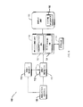
- FIG. 5 illustrates an exemplary display of an Mp image together with a 2D synthesized image; each image may be labeled to indicate whether the image is from a current acquisition, or based on legacy data.
- the Mp image may be a stored legacy mammogram
- the 2D Ms image may be generated from a current tomosynthesis acquisition and may be provided as an initial view to guide the medical professional's perusal of the tomosynthesis data.
- the method and system described in the “Matching Geometry” patent application obtain 2D x-ray projection data for tomosynthesis images preferably using a cone-shaped or pyramid-shaped imaging x-ray beam, and generate tomosynthesis images such that they conform to the same geometric coordinate system as a mammogram and, preferably, to the same coordinate system as a 2D projection mammogram.
- anatomical structures appear at geometrically matching or corresponding places in such tomosynthesis images and, preferably, in the mammogram.
Abstract
Description
Tslab[j,z]=MAX(Tr[j,z−Nslab/2],Tr[j,z−Nslab/2+1, . . . ,Tr[j,z+Nslab/2]) Equation III:
Tslab[j,z]=AVE(Tr[j,z−Nslab/2],Tr[j,z−Nslab/2+1, . . . ,Tr[j,z+Nslab/2]) Equation IV:
Claims (21)
Priority Applications (4)
| Application Number | Priority Date | Filing Date | Title |
|---|---|---|---|
| US14/549,604 US9456797B2 (en) | 2002-11-27 | 2014-11-21 | System and method for generating a 2D image from a tomosynthesis data set |
| US15/088,844 US9808215B2 (en) | 2002-11-27 | 2016-04-01 | System and method for generating a 2D image from a tomosynthesis data set |
| US15/802,225 US10010302B2 (en) | 2002-11-27 | 2017-11-02 | System and method for generating a 2D image from a tomosynthesis data set |
| US16/013,782 US10413263B2 (en) | 2002-11-27 | 2018-06-20 | System and method for generating a 2D image from a tomosynthesis data set |
Applications Claiming Priority (11)
| Application Number | Priority Date | Filing Date | Title |
|---|---|---|---|
| US10/305,480 US7123684B2 (en) | 2002-11-27 | 2002-11-27 | Full field mammography with tissue exposure control, tomosynthesis, and dynamic field of view processing |
| US10/723,486 US7831296B2 (en) | 2002-11-27 | 2003-11-26 | X-ray mammography with tomosynthesis |
| US62851604P | 2004-11-15 | 2004-11-15 | |
| US63129604P | 2004-11-26 | 2004-11-26 | |
| US11/271,050 US7577282B2 (en) | 2002-11-27 | 2005-11-10 | Image handling and display in X-ray mammography and tomosynthesis |
| US60406906A | 2006-11-24 | 2006-11-24 | |
| US11/827,909 US7616801B2 (en) | 2002-11-27 | 2007-07-13 | Image handling and display in x-ray mammography and tomosynthesis |
| US12/276,006 US7760924B2 (en) | 2002-11-27 | 2008-11-21 | System and method for generating a 2D image from a tomosynthesis data set |
| US12/471,981 US8571289B2 (en) | 2002-11-27 | 2009-05-26 | System and method for generating a 2D image from a tomosynthesis data set |
| US14/044,959 US8897535B2 (en) | 2002-11-27 | 2013-10-03 | System and method for generating a 2D image from a tomosynthesis data set |
| US14/549,604 US9456797B2 (en) | 2002-11-27 | 2014-11-21 | System and method for generating a 2D image from a tomosynthesis data set |
Related Parent Applications (1)
| Application Number | Title | Priority Date | Filing Date |
|---|---|---|---|
| US14/044,959 Continuation US8897535B2 (en) | 2002-11-27 | 2013-10-03 | System and method for generating a 2D image from a tomosynthesis data set |
Related Child Applications (1)
| Application Number | Title | Priority Date | Filing Date |
|---|---|---|---|
| US15/088,844 Continuation US9808215B2 (en) | 2002-11-27 | 2016-04-01 | System and method for generating a 2D image from a tomosynthesis data set |
Publications (2)
| Publication Number | Publication Date |
|---|---|
| US20150182181A1 US20150182181A1 (en) | 2015-07-02 |
| US9456797B2 true US9456797B2 (en) | 2016-10-04 |
Family
ID=42227086
Family Applications (6)
| Application Number | Title | Priority Date | Filing Date |
|---|---|---|---|
| US12/471,981 Active 2025-10-19 US8571289B2 (en) | 2002-11-27 | 2009-05-26 | System and method for generating a 2D image from a tomosynthesis data set |
| US14/044,959 Expired - Lifetime US8897535B2 (en) | 2002-11-27 | 2013-10-03 | System and method for generating a 2D image from a tomosynthesis data set |
| US14/549,604 Expired - Lifetime US9456797B2 (en) | 2002-11-27 | 2014-11-21 | System and method for generating a 2D image from a tomosynthesis data set |
| US15/088,844 Expired - Lifetime US9808215B2 (en) | 2002-11-27 | 2016-04-01 | System and method for generating a 2D image from a tomosynthesis data set |
| US15/802,225 Active US10010302B2 (en) | 2002-11-27 | 2017-11-02 | System and method for generating a 2D image from a tomosynthesis data set |
| US16/013,782 Expired - Fee Related US10413263B2 (en) | 2002-11-27 | 2018-06-20 | System and method for generating a 2D image from a tomosynthesis data set |
Family Applications Before (2)
| Application Number | Title | Priority Date | Filing Date |
|---|---|---|---|
| US12/471,981 Active 2025-10-19 US8571289B2 (en) | 2002-11-27 | 2009-05-26 | System and method for generating a 2D image from a tomosynthesis data set |
| US14/044,959 Expired - Lifetime US8897535B2 (en) | 2002-11-27 | 2013-10-03 | System and method for generating a 2D image from a tomosynthesis data set |
Family Applications After (3)
| Application Number | Title | Priority Date | Filing Date |
|---|---|---|---|
| US15/088,844 Expired - Lifetime US9808215B2 (en) | 2002-11-27 | 2016-04-01 | System and method for generating a 2D image from a tomosynthesis data set |
| US15/802,225 Active US10010302B2 (en) | 2002-11-27 | 2017-11-02 | System and method for generating a 2D image from a tomosynthesis data set |
| US16/013,782 Expired - Fee Related US10413263B2 (en) | 2002-11-27 | 2018-06-20 | System and method for generating a 2D image from a tomosynthesis data set |
Country Status (1)
| Country | Link |
|---|---|
| US (6) | US8571289B2 (en) |
Cited By (15)
| Publication number | Priority date | Publication date | Assignee | Title |
|---|---|---|---|---|
| US9805507B2 (en) | 2012-02-13 | 2017-10-31 | Hologic, Inc | System and method for navigating a tomosynthesis stack using synthesized image data |
| US9808215B2 (en) | 2002-11-27 | 2017-11-07 | Hologic, Inc. | System and method for generating a 2D image from a tomosynthesis data set |
| US10008184B2 (en) | 2005-11-10 | 2018-06-26 | Hologic, Inc. | System and method for generating a 2D image using mammography and/or tomosynthesis image data |
| US10573276B2 (en) | 2011-11-27 | 2020-02-25 | Hologic, Inc. | System and method for generating a 2D image using mammography and/or tomosynthesis image data |
| US11139072B2 (en) | 2019-12-04 | 2021-10-05 | International Business Machines Corporation | Three-dimensional medical image generation |
| US11403483B2 (en) | 2017-06-20 | 2022-08-02 | Hologic, Inc. | Dynamic self-learning medical image method and system |
| US11406332B2 (en) | 2011-03-08 | 2022-08-09 | Hologic, Inc. | System and method for dual energy and/or contrast enhanced breast imaging for screening, diagnosis and biopsy |
| US11419565B2 (en) | 2014-02-28 | 2022-08-23 | IIologic, Inc. | System and method for generating and displaying tomosynthesis image slabs |
| US11445993B2 (en) | 2017-03-30 | 2022-09-20 | Hologic, Inc. | System and method for targeted object enhancement to generate synthetic breast tissue images |
| US11455754B2 (en) | 2017-03-30 | 2022-09-27 | Hologic, Inc. | System and method for synthesizing low-dimensional image data from high-dimensional image data using an object grid enhancement |
| US11452486B2 (en) | 2006-02-15 | 2022-09-27 | Hologic, Inc. | Breast biopsy and needle localization using tomosynthesis systems |
| US11589944B2 (en) | 2013-03-15 | 2023-02-28 | Hologic, Inc. | Tomosynthesis-guided biopsy apparatus and method |
| US11701199B2 (en) | 2009-10-08 | 2023-07-18 | Hologic, Inc. | Needle breast biopsy system and method of use |
| US11775156B2 (en) | 2010-11-26 | 2023-10-03 | Hologic, Inc. | User interface for medical image review workstation |
| US11783476B2 (en) | 2019-10-25 | 2023-10-10 | DeepHealth, Inc. | System and method for analyzing three-dimensional image data |
Families Citing this family (46)
| Publication number | Priority date | Publication date | Assignee | Title |
|---|---|---|---|---|
| US7577282B2 (en) | 2002-11-27 | 2009-08-18 | Hologic, Inc. | Image handling and display in X-ray mammography and tomosynthesis |
| WO2006058160A2 (en) | 2004-11-26 | 2006-06-01 | Hologic, Inc. | Integrated multi-mode mammography/tomosynthesis x-ray system and method |
| US8565372B2 (en) | 2003-11-26 | 2013-10-22 | Hologic, Inc | System and method for low dose tomosynthesis |
| US7123684B2 (en) | 2002-11-27 | 2006-10-17 | Hologic, Inc. | Full field mammography with tissue exposure control, tomosynthesis, and dynamic field of view processing |
| US10638994B2 (en) | 2002-11-27 | 2020-05-05 | Hologic, Inc. | X-ray mammography with tomosynthesis |
| US7616801B2 (en) | 2002-11-27 | 2009-11-10 | Hologic, Inc. | Image handling and display in x-ray mammography and tomosynthesis |
| US8768026B2 (en) | 2003-11-26 | 2014-07-01 | Hologic, Inc. | X-ray imaging with x-ray markers that provide adjunct information but preserve image quality |
| US7702142B2 (en) | 2004-11-15 | 2010-04-20 | Hologic, Inc. | Matching geometry generation and display of mammograms and tomosynthesis images |
| US7630533B2 (en) | 2007-09-20 | 2009-12-08 | Hologic, Inc. | Breast tomosynthesis with display of highlighted suspected calcifications |
| WO2011059655A1 (en) * | 2009-10-29 | 2011-05-19 | Optovue, Inc. | Enhanced imaging for optical coherence tomography |
| FR2954556B1 (en) * | 2009-12-22 | 2017-07-28 | Gen Electric | METHOD OF PROCESSING TOMOSYNTHESIS ACQUISITIONS TO OBTAIN REPRESENTATION OF THE CONTENT OF AN ORGAN |
| FR2967520B1 (en) * | 2010-11-16 | 2012-12-21 | Gen Electric | METHOD FOR PROCESSING RADIOLOGICAL IMAGES OF A PATIENT |
| DE102012215997B4 (en) | 2012-09-10 | 2022-10-06 | Siemens Healthcare Gmbh | Contrast-enhanced recording of objects |
| JP6360495B2 (en) | 2013-01-10 | 2018-07-18 | ホロジック, インコーポレイテッドHologic, Inc. | Method for reducing data transmission volume in tomosynthesis |
| US10169534B2 (en) * | 2013-09-30 | 2019-01-01 | Toshiba Medical Systems Corporation | Medical image display system and method |
| JP6012577B2 (en) | 2013-09-30 | 2016-10-25 | 富士フイルム株式会社 | Image processing apparatus, radiographic imaging system, image processing program, and image processing method |
| US11364005B2 (en) | 2013-10-24 | 2022-06-21 | Hologic, Inc. | System and method for navigating x-ray guided breast biopsy |
| KR101604812B1 (en) | 2014-01-15 | 2016-03-18 | 삼성전자주식회사 | Medical image processing apparatus and medical image processing method thereof |
| US9613440B2 (en) * | 2014-02-12 | 2017-04-04 | General Electric Company | Digital breast Tomosynthesis reconstruction using adaptive voxel grid |
| US9256939B1 (en) * | 2014-07-17 | 2016-02-09 | Agfa Healthcare | System and method for aligning mammography images |
| JP6126058B2 (en) | 2014-09-30 | 2017-05-10 | 富士フイルム株式会社 | Image display apparatus, image processing apparatus, radiographic imaging system, tomographic image display method, and tomographic image display program. |
| US10255697B2 (en) | 2014-11-20 | 2019-04-09 | Koninklijke Philips N.V. | Method for generation of synthetic mammograms from tomosynthesis data |
| JP6486100B2 (en) * | 2014-12-22 | 2019-03-20 | キヤノン株式会社 | Image processing apparatus, image processing method, and program |
| GB2533801B (en) * | 2014-12-31 | 2018-09-12 | Gen Electric | Method and system for tomosynthesis projection images enhancement |
| US9984478B2 (en) * | 2015-07-28 | 2018-05-29 | PME IP Pty Ltd | Apparatus and method for visualizing digital breast tomosynthesis and other volumetric images |
| JP6456550B2 (en) | 2015-09-01 | 2019-01-23 | コーニンクレッカ フィリップス エヌ ヴェKoninklijke Philips N.V. | Device for displaying medical image data of a body part |
| EP3445247B1 (en) | 2016-04-22 | 2021-03-10 | Hologic, Inc. | Tomosynthesis with shifting focal spot x-ray system using an addressable array |
| JP6639357B2 (en) | 2016-08-24 | 2020-02-05 | 富士フイルム株式会社 | Image processing apparatus, method and program |
| US10096106B2 (en) * | 2016-11-10 | 2018-10-09 | General Electric Company | Combined medical imaging |
| EP3326535B1 (en) | 2016-11-25 | 2019-06-12 | ScreenPoint Medical | Displaying system for displaying digital breast tomosynthesis data |
| WO2018183548A1 (en) | 2017-03-30 | 2018-10-04 | Hologic, Inc. | System and method for hierarchical multi-level feature image synthesis and representation |
| EP4129188A1 (en) | 2017-08-16 | 2023-02-08 | Hologic, Inc. | Techniques for breast imaging patient motion artifact compensation |
| EP3449835B1 (en) | 2017-08-22 | 2023-01-11 | Hologic, Inc. | Computed tomography system and method for imaging multiple anatomical targets |
| US10679384B2 (en) | 2017-09-29 | 2020-06-09 | General Electric Company | Systems and methods for deep learning-based image reconstruction |
| US11090017B2 (en) | 2018-09-13 | 2021-08-17 | Hologic, Inc. | Generating synthesized projection images for 3D breast tomosynthesis or multi-mode x-ray breast imaging |
| EP3856033B1 (en) | 2018-09-28 | 2023-06-07 | Hologic, Inc. | Method for synthetic breast tissue image generation by high density element suppression |
| US11227418B2 (en) * | 2018-12-28 | 2022-01-18 | General Electric Company | Systems and methods for deep learning-based image reconstruction |
| US11883206B2 (en) | 2019-07-29 | 2024-01-30 | Hologic, Inc. | Personalized breast imaging system |
| US11694792B2 (en) | 2019-09-27 | 2023-07-04 | Hologic, Inc. | AI system for predicting reading time and reading complexity for reviewing 2D/3D breast images |
| EP3797698A1 (en) | 2019-09-30 | 2021-03-31 | Siemens Healthcare GmbH | Method for generating a synthetic mammogram based on dual-energy tomosynthesis capture |
| EP3832689A3 (en) | 2019-12-05 | 2021-08-11 | Hologic, Inc. | Systems and methods for improved x-ray tube life |
| US11471118B2 (en) | 2020-03-27 | 2022-10-18 | Hologic, Inc. | System and method for tracking x-ray tube focal spot position |
| US11481038B2 (en) | 2020-03-27 | 2022-10-25 | Hologic, Inc. | Gesture recognition in controlling medical hardware or software |
| US11786191B2 (en) | 2021-05-17 | 2023-10-17 | Hologic, Inc. | Contrast-enhanced tomosynthesis with a copper filter |
| EP4134908A1 (en) | 2021-08-10 | 2023-02-15 | Siemens Healthcare GmbH | Computer-implemented method for providing an outline of a lesion in digital breast tomosynthesis |
| WO2023140475A1 (en) * | 2022-01-21 | 2023-07-27 | 주식회사 루닛 | Apparatus and method for linking lesion location between guide image and 3d tomosynthesis images including plurality of 3d image slices, and providing linked image |
Citations (104)
| Publication number | Priority date | Publication date | Assignee | Title |
|---|---|---|---|---|
| US4160906A (en) | 1977-06-23 | 1979-07-10 | General Electric Company | Anatomically coordinated user dominated programmer for diagnostic x-ray apparatus |
| US4744099A (en) | 1983-11-03 | 1988-05-10 | Siemens Aktiengesellschaft | X-ray diagnostic apparatus comprising radiation filters |
| US4773086A (en) | 1983-12-16 | 1988-09-20 | Yokogawa Medical Systems, Limited | Operator console for X-ray tomographs |
| US4969174A (en) | 1989-09-06 | 1990-11-06 | General Electric Company | Scanning mammography system with reduced scatter radiation |
| USRE33634E (en) | 1986-09-23 | 1991-07-09 | Method and structure for optimizing radiographic quality by controlling X-ray tube voltage, current focal spot size and exposure time | |
| US5163075A (en) | 1991-08-08 | 1992-11-10 | Eastman Kodak Company | Contrast enhancement of electrographic imaging |
| US5365562A (en) | 1993-09-20 | 1994-11-15 | Fischer Imaging Corporation | Digital imaging apparatus |
| US5452367A (en) | 1993-11-29 | 1995-09-19 | Arch Development Corporation | Automated method and system for the segmentation of medical images |
| US5526394A (en) | 1993-11-26 | 1996-06-11 | Fischer Imaging Corporation | Digital scan mammography apparatus |
| US5553111A (en) | 1994-10-26 | 1996-09-03 | The General Hospital Corporation | Apparatus and method for improved tissue imaging |
| US5592562A (en) | 1994-01-19 | 1997-01-07 | International Business Machines Corporation | Inspection system for cross-sectional imaging |
| US5594769A (en) | 1991-11-27 | 1997-01-14 | Thermotrex Corporation | Method and apparatus for obtaining stereotactic mammographic guided needle breast biopsies |
| US5596200A (en) | 1992-10-14 | 1997-01-21 | Primex | Low dose mammography system |
| US5598454A (en) | 1994-04-26 | 1997-01-28 | Siemens Aktiengesellschaft | X-ray diagnostics installation |
| US5668889A (en) | 1990-04-19 | 1997-09-16 | Fuji Photo Film Co., Ltd. | Apparatus for determining an image position, and method for adjusting read-out conditions and/or image processing conditions for a radiation image |
| WO1998016903A1 (en) | 1996-10-16 | 1998-04-23 | Vital Images, Inc. | Advanced diagnostic viewer |
| US5828722A (en) | 1996-05-17 | 1998-10-27 | Sirona Dental Systems Gmbh & Co., Kg | X-ray diagnostic apparatus for tomosynthesis having a detector that detects positional relationships |
| US5872828A (en) | 1996-07-23 | 1999-02-16 | The General Hospital Corporation | Tomosynthesis system for breast imaging |
| US5878104A (en) | 1996-05-17 | 1999-03-02 | Sirona Dental Systems Gmbh & Co. Kg | Method for producing tomosynthesis exposures employing a reference object formed by a region of the examination subject |
| US5878746A (en) | 1993-08-25 | 1999-03-09 | Lemelson; Jerome H. | Computerized medical diagnostic system |
| US5896437A (en) | 1996-05-17 | 1999-04-20 | Sirona Dental Systems Gmbh & Co. Kg | X-ray diagnostics apparatus for tomosynthesis having a reference object in fixed relationship to a radiation emitter |
| US5941832A (en) | 1991-09-27 | 1999-08-24 | Tumey; David M. | Method and apparatus for detection of cancerous and precancerous conditions in a breast |
| US6005907A (en) | 1996-05-17 | 1999-12-21 | Sirona Dental Systems Gmbh & Co. Kg | Method and apparatus for producing tomosynthesis exposures employing a reference object composed of a number of sub-objects |
| EP0982001A1 (en) | 1998-08-25 | 2000-03-01 | General Electric Company | Protocol driven image reconstruction, display, and processing in a multislice imaging system |
| US6091841A (en) | 1997-09-04 | 2000-07-18 | Qualia Computing, Inc. | Method and system for segmenting desired regions in digital mammograms |
| US6137527A (en) | 1996-12-23 | 2000-10-24 | General Electric Company | System and method for prompt-radiology image screening service via satellite |
| US6175117B1 (en) | 1998-01-23 | 2001-01-16 | Quanta Vision, Inc. | Tissue analysis apparatus |
| US6196715B1 (en) | 1959-04-28 | 2001-03-06 | Kabushiki Kaisha Toshiba | X-ray diagnostic system preferable to two dimensional x-ray detection |
| US6216540B1 (en) | 1995-06-06 | 2001-04-17 | Robert S. Nelson | High resolution device and method for imaging concealed objects within an obscuring medium |
| US6233473B1 (en) | 1999-02-16 | 2001-05-15 | Hologic, Inc. | Determining body composition using fan beam dual-energy x-ray absorptiometry |
| US6243441B1 (en) | 1999-07-13 | 2001-06-05 | Edge Medical Devices | Active matrix detector for X-ray imaging |
| US6256370B1 (en) | 2000-01-24 | 2001-07-03 | General Electric Company | Method and apparatus for performing tomosynthesis |
| US6272207B1 (en) | 1999-02-18 | 2001-08-07 | Creatv Microtech, Inc. | Method and apparatus for obtaining high-resolution digital X-ray and gamma ray images |
| US6292530B1 (en) | 1999-04-29 | 2001-09-18 | General Electric Company | Method and apparatus for reconstructing image data acquired by a tomosynthesis x-ray imaging system |
| US6341156B1 (en) | 1999-05-14 | 2002-01-22 | Siemens Aktiengesellschaft | X-ray diagnostic apparatus with relatively moved x-ray source and detector |
| US6375352B1 (en) | 1999-10-01 | 2002-04-23 | General Electric Company | Apparatus and method for obtaining x-ray tomosynthesis data for mammography |
| US20020050986A1 (en) | 2000-08-11 | 2002-05-02 | Hitoshi Inoue | Image display apparatus and method, and storage medium |
| US6389104B1 (en) | 2000-06-30 | 2002-05-14 | Siemens Corporate Research, Inc. | Fluoroscopy based 3-D neural navigation based on 3-D angiography reconstruction data |
| US6411836B1 (en) | 1999-12-30 | 2002-06-25 | General Electric Company | Method and apparatus for user preferences configuring in an image handling system |
| US6415015B2 (en) | 1999-12-28 | 2002-07-02 | Ge Medical Systems Sa | Method and system of compensation of thickness of an organ |
| US6442288B1 (en) | 1997-12-17 | 2002-08-27 | Siemens Aktiengesellschaft | Method for reconstructing a three-dimensional image of an object scanned in the context of a tomosynthesis, and apparatus for tomosynthesis |
| US6463181B2 (en) | 2000-12-22 | 2002-10-08 | The United States Of America As Represented By The Secretary Of The Navy | Method for optimizing visual display of enhanced digital images |
| US20030007598A1 (en) | 2000-11-24 | 2003-01-09 | U-Systems, Inc. | Breast cancer screening with adjunctive ultrasound mammography |
| US20030026386A1 (en) | 2001-02-01 | 2003-02-06 | Cha-Mei Tang | Anti-scatter grids and collimator designs, and their motion, fabrication and assembly |
| US6556655B1 (en) | 1998-11-27 | 2003-04-29 | Ge Medical Systems Sa | Method for automatic detection of glandular tissue |
| US20030095624A1 (en) | 2001-11-21 | 2003-05-22 | Eberhard Jeffrey Wayne | Dose management system for mammographic tomosynthesis |
| US6574304B1 (en) | 2002-09-13 | 2003-06-03 | Ge Medical Systems Global Technology Company, Llc | Computer aided acquisition of medical images |
| US20030128893A1 (en) | 2001-11-19 | 2003-07-10 | Alfio Castorina | Method for merging digital images to obtain a high dynamic range digital image |
| US6597762B1 (en) | 2002-11-27 | 2003-07-22 | Ge Medical Systems Global Technology Co., Llc | Method and apparatus of lesion detection and validation based on multiple reviews of a CT image |
| US20030169847A1 (en) | 2001-11-21 | 2003-09-11 | University Of Massachusetts Medical Center | System and method for x-ray fluoroscopic imaging |
| US6633674B1 (en) | 1999-11-24 | 2003-10-14 | General Electric Company | Picture archiving and communication system employing improved data compression |
| US20030194050A1 (en) * | 2002-04-15 | 2003-10-16 | General Electric Company | Multi modality X-ray and nuclear medicine mammography imaging system and method |
| US20030194121A1 (en) | 2002-04-15 | 2003-10-16 | General Electric Company | Computer aided detection (CAD) for 3D digital mammography |
| US6647092B2 (en) | 2002-01-18 | 2003-11-11 | General Electric Company | Radiation imaging system and method of collimation |
| US20030210254A1 (en) | 2002-05-13 | 2003-11-13 | Doan William D. | Method, system and computer product for displaying axial images |
| US20030212327A1 (en) | 2000-11-24 | 2003-11-13 | U-Systems Inc. | Adjunctive ultrasound processing and display for breast cancer screening |
| US20030215120A1 (en) | 2002-05-15 | 2003-11-20 | Renuka Uppaluri | Computer aided diagnosis of an image set |
| US20040008901A1 (en) | 2002-07-11 | 2004-01-15 | Avinash Gopal B. | Interpolated image filtering method and apparatus |
| US20040047518A1 (en) | 2002-08-28 | 2004-03-11 | Carlo Tiana | Image fusion system and method |
| US20040052328A1 (en) | 2002-09-13 | 2004-03-18 | Sabol John M. | Computer assisted analysis of tomographic mammography data |
| US20040070582A1 (en) | 2002-10-11 | 2004-04-15 | Matthew Warren Smith To Sonocine, Inc. | 3D modeling system |
| US20040094167A1 (en) | 2000-03-17 | 2004-05-20 | Brady John Michael | Three-dimensional reconstructions of a breast from two x-ray mammographics |
| US6744848B2 (en) | 2000-02-11 | 2004-06-01 | Brandeis University | Method and system for low-dose three-dimensional imaging of a scene |
| US6813334B2 (en) | 2000-10-20 | 2004-11-02 | Koninklijke Philips Electronics N.V. | Tomosynthesis in a limited angular range |
| US6882700B2 (en) | 2002-04-15 | 2005-04-19 | General Electric Company | Tomosynthesis X-ray mammogram system and method with automatic drive system |
| US6885724B2 (en) | 2003-08-22 | 2005-04-26 | Ge Medical Systems Global Technology Company, Llc | Radiographic tomosynthesis image acquisition utilizing asymmetric geometry |
| US20050113681A1 (en) | 2002-11-27 | 2005-05-26 | Defreitas Kenneth F. | X-ray mammography with tomosynthesis |
| US20050135555A1 (en) | 2003-12-23 | 2005-06-23 | Claus Bernhard Erich H. | Method and system for simultaneously viewing rendered volumes |
| US20050135664A1 (en) | 2003-12-23 | 2005-06-23 | Kaufhold John P. | Methods and apparatus for reconstruction of volume data from projection data |
| US6940943B2 (en) | 2002-10-07 | 2005-09-06 | General Electric Company | Continuous scan tomosynthesis system and method |
| US20050226375A1 (en) | 2004-03-31 | 2005-10-13 | Eberhard Jeffrey W | Enhanced X-ray imaging system and method |
| US20060018526A1 (en) | 2004-07-23 | 2006-01-26 | Avinash Gopal B | Methods and apparatus for noise reduction filtering of images |
| US6999554B2 (en) | 2003-11-17 | 2006-02-14 | Siemens Aktiengesellschaft | X-ray diagnostic apparatus for mammography examinations |
| US20060098855A1 (en) | 2002-11-27 | 2006-05-11 | Gkanatsios Nikolaos A | Image handling and display in X-ray mammography and tomosynthesis |
| US7110490B2 (en) | 2002-12-10 | 2006-09-19 | General Electric Company | Full field digital tomosynthesis method and apparatus |
| US7127091B2 (en) | 2000-12-22 | 2006-10-24 | Koninklijke Philips Electronics, N.V. | Method and apparatus for visualizing a limited part of a 3D medical image-point-related data set, through basing a rendered image on an intermediate region between first and second clipping planes, and including spectroscopic viewing of such region |
| WO2005051197A3 (en) | 2003-11-26 | 2007-03-01 | Koninkl Philips Electronics Nv | Workflow optimization for high throughput imaging environment |
| US20070052700A1 (en) | 2005-09-07 | 2007-03-08 | Wheeler Frederick W | System and method for 3D CAD using projection images |
| US7323692B2 (en) | 2004-08-10 | 2008-01-29 | Research Foundation Of State University Of New York | Flat-panel detector with avalanche gain |
| US7346381B2 (en) | 2002-11-01 | 2008-03-18 | Ge Medical Systems Global Technology Company Llc | Method and apparatus for medical intervention procedure planning |
| WO2008047270A1 (en) | 2006-10-17 | 2008-04-24 | Koninklijke Philips Electronics N.V. | Visualization of 3d images in combination with 2d projection images |
| US20080130979A1 (en) | 2004-11-15 | 2008-06-05 | Baorui Ren | Matching Geometry Generation and Display of Mammograms and Tomosynthesis Images |
| US20080187095A1 (en) | 2005-05-03 | 2008-08-07 | The Regents Of The University Of California | Biopsy Systems For Breast Computed Tomography |
| US20090005668A1 (en) | 2007-06-30 | 2009-01-01 | West Jay B | Non-invasive method for using 2D angiographic images for radiosurgical target definition |
| US20090034684A1 (en) * | 2007-08-02 | 2009-02-05 | Sylvain Bernard | Method and system for displaying tomosynthesis images |
| US20090123052A1 (en) | 2002-11-27 | 2009-05-14 | Chris Ruth | System and Method for Generating a 2D Image from a Tomosynthesis Data Set |
| US20090238424A1 (en) | 2008-03-19 | 2009-09-24 | Kazumasa Arakita | Image processing apparatus and image processing method |
| US20090268865A1 (en) | 2003-11-26 | 2009-10-29 | Baorui Ren | X-ray imaging with X-ray markers that provide adjunct information but preserve image quality |
| US7630533B2 (en) | 2007-09-20 | 2009-12-08 | Hologic, Inc. | Breast tomosynthesis with display of highlighted suspected calcifications |
| US20090304147A1 (en) | 2005-08-15 | 2009-12-10 | Hologic, Inc. | X-ray mammography/tomosynthesis of patient's breast |
| US20100135558A1 (en) | 2002-11-27 | 2010-06-03 | Chris Ruth | System and Method for Generating a 2D Image from a Tomosynthesis Data Set |
| US20110069906A1 (en) | 2009-09-23 | 2011-03-24 | Samsung Electronics Co., Ltd. | Method and apparatus for blending images |
| EP2301432A1 (en) | 2008-06-03 | 2011-03-30 | FUJIFILM Corporation | Projection image creation device, method, and program |
| US20110110576A1 (en) | 2009-10-07 | 2011-05-12 | Hologic, Inc. | Selective Display Of Computer-Aided Detection Findings With Associated Breast X-Ray Mammogram and/or Tomosynthesis Image Information |
| US20110178389A1 (en) | 2008-05-02 | 2011-07-21 | Eigen, Inc. | Fused image moldalities guidance |
| WO2011091300A2 (en) | 2010-01-24 | 2011-07-28 | Mistretta Medical, Llc | System and method for implementation of 4d time-energy subtraction computed tomography |
| US20110234630A1 (en) | 2008-11-28 | 2011-09-29 | Fujifilm Medical Systems USA, Inc | Active Overlay System and Method for Accessing and Manipulating Imaging Displays |
| US20110242092A1 (en) | 2010-03-30 | 2011-10-06 | Fujifilm Corporation | Image display system |
| US8044972B2 (en) | 2006-12-21 | 2011-10-25 | Sectra Mamea Ab | Synchronized viewing of tomosynthesis and/or mammograms |
| US8051386B2 (en) | 2006-12-21 | 2011-11-01 | Sectra Ab | CAD-based navigation of views of medical image data stacks or volumes |
| WO2012063653A1 (en) | 2010-11-12 | 2012-05-18 | 株式会社 日立メディコ | Medical image display device and medical image display method |
| US20120293511A1 (en) | 2010-02-25 | 2012-11-22 | Siemens Aktiengesellschaft | Method for displaying an area to be medically examined and/or treated |
| US20140327702A1 (en) | 2005-11-10 | 2014-11-06 | Hologic, Inc | System and method for generating a 2d image using mammography and/or tomosynthesis image data |
| US8983156B2 (en) | 2012-11-23 | 2015-03-17 | Icad, Inc. | System and method for improving workflow efficiences in reading tomosynthesis medical image data |
Family Cites Families (79)
| Publication number | Priority date | Publication date | Assignee | Title |
|---|---|---|---|---|
| US3502878A (en) | 1967-09-22 | 1970-03-24 | Us Health Education & Welfare | Automatic x-ray apparatus for limiting the field size of a projected x-ray beam in response to film size and to source-to-film distance |
| US3863073A (en) | 1973-04-26 | 1975-01-28 | Machlett Lab Inc | Automatic system for precise collimation of radiation |
| US3971950A (en) | 1975-04-14 | 1976-07-27 | Xerox Corporation | Independent compression and positioning device for use in mammography |
| DE2838901C2 (en) | 1978-09-06 | 1986-11-06 | Siemens AG, 1000 Berlin und 8000 München | Catapult drawer |
| FR2512024A1 (en) | 1981-08-27 | 1983-03-04 | Adir | TRICYCLIC ETHERS, PREPARATION THEREOF AND PHARMACEUTICAL COMPOSITIONS CONTAINING THEM |
| FR2549248B1 (en) | 1983-06-24 | 1986-01-31 | Thomson Csf | RETRACTABLE CASSETTE HOLDER FOR RADIOLOGICAL AND RADIOGRAPHIC EXAMINATION APPARATUS |
| US4706269A (en) | 1985-03-11 | 1987-11-10 | Reina Leo J | Anti-scatter grid structure |
| US4773087A (en) | 1986-04-14 | 1988-09-20 | University Of Rochester | Quality of shadowgraphic x-ray images |
| US4821727A (en) | 1986-10-30 | 1989-04-18 | Elscint Ltd. | Mammographic biopsy needle holder system |
| US4819258A (en) | 1986-11-28 | 1989-04-04 | Bennett X-Ray Corp. | Auto-setting of KV in an x-ray machine after selection of technic factors |
| US4907156A (en) | 1987-06-30 | 1990-03-06 | University Of Chicago | Method and system for enhancement and detection of abnormal anatomic regions in a digital image |
| US5051904A (en) | 1988-03-24 | 1991-09-24 | Olganix Corporation | Computerized dynamic tomography system |
| DK654488A (en) | 1988-11-23 | 1990-05-24 | Nordisk Roentgen Tech App | ROENTGENAPPARAT |
| FR2645006A1 (en) | 1989-03-29 | 1990-10-05 | Gen Electric Cgr | MAMMOGRAPH HAVING INTEGRATED STEREOTAXIC VIEWING DEVICE AND METHOD OF USING SUCH A MAMMOGRAPHER |
| FR2646340A1 (en) | 1989-04-28 | 1990-11-02 | Gen Electric Cgr | ADJUSTABLE CASSETTE HOLDER IN DIMENSION AND POSITION FOR MAMMOGRAPHY |
| EP0406455B1 (en) | 1989-07-03 | 1994-09-21 | Siemens Aktiengesellschaft | X-ray diagnostic apparatus for mammographies |
| US5133020A (en) | 1989-07-21 | 1992-07-21 | Arch Development Corporation | Automated method and system for the detection and classification of abnormal lesions and parenchymal distortions in digital medical images |
| CA2014918A1 (en) | 1989-09-06 | 1991-03-06 | James A. Mcfaul | Scanning mammography system with improved skin line viewing |
| US5078142A (en) | 1989-11-21 | 1992-01-07 | Fischer Imaging Corporation | Precision mammographic needle biopsy system |
| US5415169A (en) | 1989-11-21 | 1995-05-16 | Fischer Imaging Corporation | Motorized mammographic biopsy apparatus |
| US5240011A (en) | 1991-11-27 | 1993-08-31 | Fischer Imaging Corporation | Motorized biopsy needle positioner |
| US5199056A (en) | 1989-11-28 | 1993-03-30 | Darrah Carol J | Mammography compression paddle |
| US5289520A (en) | 1991-11-27 | 1994-02-22 | Lorad Corporation | Stereotactic mammography imaging system with prone position examination table and CCD camera |
| US5343390A (en) | 1992-02-28 | 1994-08-30 | Arch Development Corporation | Method and system for automated selection of regions of interest and detection of septal lines in digital chest radiographs |
| US5359637A (en) | 1992-04-28 | 1994-10-25 | Wake Forest University | Self-calibrated tomosynthetic, radiographic-imaging system, method, and device |
| FR2703237B1 (en) | 1993-03-29 | 1995-05-19 | Ge Medical Syst Sa | Mammograph equipped with a stereotaxic camera with digital detector and method of using such a mammograph. |
| US5491627A (en) | 1993-05-13 | 1996-02-13 | Arch Development Corporation | Method and system for the detection of microcalcifications in digital mammograms |
| US6075879A (en) | 1993-09-29 | 2000-06-13 | R2 Technology, Inc. | Method and system for computer-aided lesion detection using information from multiple images |
| US5506877A (en) | 1994-11-23 | 1996-04-09 | The General Hospital Corporation | Mammography breast compression device and method |
| US5712890A (en) | 1994-11-23 | 1998-01-27 | Thermotrex Corp. | Full breast digital mammography device |
| US5657362A (en) | 1995-02-24 | 1997-08-12 | Arch Development Corporation | Automated method and system for computerized detection of masses and parenchymal distortions in medical images |
| US5818898A (en) | 1995-11-07 | 1998-10-06 | Kabushiki Kaisha Toshiba | X-ray imaging apparatus using X-ray planar detector |
| US5627869A (en) | 1995-11-22 | 1997-05-06 | Thermotrex Corporation | Mammography apparatus with proportional collimation |
| FI955636A0 (en) | 1995-11-23 | 1995-11-23 | Planmed Oy | Foerfarande och system Foer styrning av funktionerna av en mammografiaanordning |
| US5769086A (en) | 1995-12-06 | 1998-06-23 | Biopsys Medical, Inc. | Control system and method for automated biopsy device |
| JP3554172B2 (en) | 1998-01-09 | 2004-08-18 | キヤノン株式会社 | Radiography equipment |
| US6289235B1 (en) | 1998-03-05 | 2001-09-11 | Wake Forest University | Method and system for creating three-dimensional images using tomosynthetic computed tomography |
| US6081577A (en) | 1998-07-24 | 2000-06-27 | Wake Forest University | Method and system for creating task-dependent three-dimensional images |
| EP1143845A4 (en) | 1998-11-25 | 2004-10-06 | Fischer Imaging Corp | User interface system for mammographic imager |
| US6149301A (en) | 1998-12-30 | 2000-11-21 | General Electric Company | X-ray target centering apparatus for radiographic imaging system |
| US6689142B1 (en) | 1999-04-26 | 2004-02-10 | Scimed Life Systems, Inc. | Apparatus and methods for guiding a needle |
| US6645520B2 (en) | 1999-12-16 | 2003-11-11 | Dermatrends, Inc. | Transdermal administration of nonsteroidal anti-inflammatory drugs using hydroxide-releasing agents as permeation enhancers |
| US6960020B2 (en) | 2001-08-31 | 2005-11-01 | Analogic Corporation | Image positioning method and system for tomosynthesis in a digital X-ray radiography system |
| US6327336B1 (en) | 2000-06-05 | 2001-12-04 | Direct Radiography Corp. | Radiogram showing location of automatic exposure control sensor |
| GB2376633B (en) | 2000-11-06 | 2004-11-10 | Suros Surgical Systems Inc | Biopsy apparatus |
| US6758824B1 (en) | 2000-11-06 | 2004-07-06 | Suros Surgical Systems, Inc. | Biopsy apparatus |
| US6501819B2 (en) | 2000-12-18 | 2002-12-31 | Ge Medical Systems Global Technology Company, Llc | Medical diagnostic method and apparatus to control dual energy exposure techniques based on image information |
| US6620111B2 (en) | 2001-04-20 | 2003-09-16 | Ethicon Endo-Surgery, Inc. | Surgical biopsy device having automatic rotation of the probe for taking multiple samples |
| US6954667B2 (en) | 2001-06-28 | 2005-10-11 | Chemimage Corporation | Method for Raman chemical imaging and characterization of calcification in tissue |
| US6611575B1 (en) | 2001-07-27 | 2003-08-26 | General Electric Company | Method and system for high resolution 3D visualization of mammography images |
| EP1444873A2 (en) | 2001-10-19 | 2004-08-11 | Hologic, Inc. | Mammography system and method employing offset compression paddles, automatic collimation, and retractable anti-scatter grid |
| US6626849B2 (en) | 2001-11-01 | 2003-09-30 | Ethicon Endo-Surgery, Inc. | MRI compatible surgical biopsy device |
| JP4099984B2 (en) | 2001-12-14 | 2008-06-11 | コニカミノルタホールディングス株式会社 | Abnormal shadow detection apparatus and image output apparatus |
| US6978040B2 (en) | 2001-12-19 | 2005-12-20 | Canon Kabushiki Kaisha | Optical recovery of radiographic geometry |
| SE524458C2 (en) | 2002-03-01 | 2004-08-10 | Mamea Imaging Ab | Protective device by an X-ray apparatus |
| US6878115B2 (en) | 2002-03-28 | 2005-04-12 | Ultrasound Detection Systems, Llc | Three-dimensional ultrasound computed tomography imaging system |
| US7123684B2 (en) | 2002-11-27 | 2006-10-17 | Hologic, Inc. | Full field mammography with tissue exposure control, tomosynthesis, and dynamic field of view processing |
| WO2006058160A2 (en) | 2004-11-26 | 2006-06-01 | Hologic, Inc. | Integrated multi-mode mammography/tomosynthesis x-ray system and method |
| US7616801B2 (en) | 2002-11-27 | 2009-11-10 | Hologic, Inc. | Image handling and display in x-ray mammography and tomosynthesis |
| US7356113B2 (en) | 2003-02-12 | 2008-04-08 | Brandeis University | Tomosynthesis imaging system and method |
| JP4497837B2 (en) | 2003-05-12 | 2010-07-07 | キヤノン株式会社 | Radiation imaging equipment |
| US7835556B2 (en) | 2004-05-14 | 2010-11-16 | Koninklijke Philips Electronics N.V. | System and method for diagnosing breast cancer |
| GB0411402D0 (en) | 2004-05-21 | 2004-06-23 | Tissuomics Ltd | Penetrating radiation measurements |
| US7725153B2 (en) | 2004-10-04 | 2010-05-25 | Hologic, Inc. | Estimating visceral fat by dual-energy x-ray absorptiometry |
| US7606801B2 (en) | 2005-06-07 | 2009-10-20 | Varonis Inc. | Automatic management of storage access control |
| DE202005013910U1 (en) | 2005-09-02 | 2005-11-24 | Siemens Ag | Mammography unit has face shield moving within X-ray source head to provide withdrawn, protruding and transport positions |
| DE202007019497U1 (en) | 2006-02-15 | 2013-03-06 | Hologic, Inc. | Breast biopsy and needle localization using tomosynthesis systems |
| US20070223651A1 (en) | 2006-03-21 | 2007-09-27 | Wagenaar Douglas J | Dual modality mammography device |
| US7489761B2 (en) | 2006-03-27 | 2009-02-10 | Hologic, Inc. | Breast compression for digital mammography, tomosynthesis and other modalities |
| US20090080602A1 (en) | 2006-08-03 | 2009-03-26 | Kenneth Brooks | Dedicated breast radiation imaging/therapy system |
| US8126226B2 (en) | 2007-09-20 | 2012-02-28 | General Electric Company | System and method to generate a selected visualization of a radiological image of an imaged subject |
| JP5294654B2 (en) | 2008-02-29 | 2013-09-18 | 富士フイルム株式会社 | Image display method and apparatus |
| US7991106B2 (en) | 2008-08-29 | 2011-08-02 | Hologic, Inc. | Multi-mode tomosynthesis/mammography gain calibration and image correction using gain map information from selected projection angles |
| US8413054B2 (en) | 2009-04-13 | 2013-04-02 | Cisco Technology, Inc. | Graphical user interface for still image capture from video footage |
| CN102473290A (en) | 2009-06-29 | 2012-05-23 | 汤姆逊许可证公司 | Contrast enhancement |
| US9289183B2 (en) | 2009-11-27 | 2016-03-22 | Qview Medical, Inc. | Interactive display of computer aided detection results in combination with quantitative prompts |
| BR112012014423A2 (en) | 2009-12-17 | 2017-12-19 | Koninklijke Philips Eletronics N V | "system for generating an object construction, medical workstation, medical imaging equipment, method of generating an object construction and computer program product |
| EP2782505B1 (en) | 2011-11-27 | 2020-04-22 | Hologic, Inc. | System and method for generating a 2d image using mammography and/or tomosynthesis image data |
| US9805507B2 (en) | 2012-02-13 | 2017-10-31 | Hologic, Inc | System and method for navigating a tomosynthesis stack using synthesized image data |
-
2009
- 2009-05-26 US US12/471,981 patent/US8571289B2/en active Active
-
2013
- 2013-10-03 US US14/044,959 patent/US8897535B2/en not_active Expired - Lifetime
-
2014
- 2014-11-21 US US14/549,604 patent/US9456797B2/en not_active Expired - Lifetime
-
2016
- 2016-04-01 US US15/088,844 patent/US9808215B2/en not_active Expired - Lifetime
-
2017
- 2017-11-02 US US15/802,225 patent/US10010302B2/en active Active
-
2018
- 2018-06-20 US US16/013,782 patent/US10413263B2/en not_active Expired - Fee Related
Patent Citations (117)
| Publication number | Priority date | Publication date | Assignee | Title |
|---|---|---|---|---|
| US6196715B1 (en) | 1959-04-28 | 2001-03-06 | Kabushiki Kaisha Toshiba | X-ray diagnostic system preferable to two dimensional x-ray detection |
| US4160906A (en) | 1977-06-23 | 1979-07-10 | General Electric Company | Anatomically coordinated user dominated programmer for diagnostic x-ray apparatus |
| US4744099A (en) | 1983-11-03 | 1988-05-10 | Siemens Aktiengesellschaft | X-ray diagnostic apparatus comprising radiation filters |
| US4773086A (en) | 1983-12-16 | 1988-09-20 | Yokogawa Medical Systems, Limited | Operator console for X-ray tomographs |
| USRE33634E (en) | 1986-09-23 | 1991-07-09 | Method and structure for optimizing radiographic quality by controlling X-ray tube voltage, current focal spot size and exposure time | |
| US4969174A (en) | 1989-09-06 | 1990-11-06 | General Electric Company | Scanning mammography system with reduced scatter radiation |
| US5668889A (en) | 1990-04-19 | 1997-09-16 | Fuji Photo Film Co., Ltd. | Apparatus for determining an image position, and method for adjusting read-out conditions and/or image processing conditions for a radiation image |
| US5163075A (en) | 1991-08-08 | 1992-11-10 | Eastman Kodak Company | Contrast enhancement of electrographic imaging |
| US5941832A (en) | 1991-09-27 | 1999-08-24 | Tumey; David M. | Method and apparatus for detection of cancerous and precancerous conditions in a breast |
| US5594769A (en) | 1991-11-27 | 1997-01-14 | Thermotrex Corporation | Method and apparatus for obtaining stereotactic mammographic guided needle breast biopsies |
| US5596200A (en) | 1992-10-14 | 1997-01-21 | Primex | Low dose mammography system |
| US5878746A (en) | 1993-08-25 | 1999-03-09 | Lemelson; Jerome H. | Computerized medical diagnostic system |
| US5365562A (en) | 1993-09-20 | 1994-11-15 | Fischer Imaging Corporation | Digital imaging apparatus |
| US5526394A (en) | 1993-11-26 | 1996-06-11 | Fischer Imaging Corporation | Digital scan mammography apparatus |
| US5452367A (en) | 1993-11-29 | 1995-09-19 | Arch Development Corporation | Automated method and system for the segmentation of medical images |
| US5592562A (en) | 1994-01-19 | 1997-01-07 | International Business Machines Corporation | Inspection system for cross-sectional imaging |
| US5719952A (en) | 1994-01-19 | 1998-02-17 | International Business Machines Corporation | Inspection system for cross-sectional imaging |
| US5598454A (en) | 1994-04-26 | 1997-01-28 | Siemens Aktiengesellschaft | X-ray diagnostics installation |
| US5553111A (en) | 1994-10-26 | 1996-09-03 | The General Hospital Corporation | Apparatus and method for improved tissue imaging |
| US6216540B1 (en) | 1995-06-06 | 2001-04-17 | Robert S. Nelson | High resolution device and method for imaging concealed objects within an obscuring medium |
| US5828722A (en) | 1996-05-17 | 1998-10-27 | Sirona Dental Systems Gmbh & Co., Kg | X-ray diagnostic apparatus for tomosynthesis having a detector that detects positional relationships |
| US5878104A (en) | 1996-05-17 | 1999-03-02 | Sirona Dental Systems Gmbh & Co. Kg | Method for producing tomosynthesis exposures employing a reference object formed by a region of the examination subject |
| US5896437A (en) | 1996-05-17 | 1999-04-20 | Sirona Dental Systems Gmbh & Co. Kg | X-ray diagnostics apparatus for tomosynthesis having a reference object in fixed relationship to a radiation emitter |
| US6005907A (en) | 1996-05-17 | 1999-12-21 | Sirona Dental Systems Gmbh & Co. Kg | Method and apparatus for producing tomosynthesis exposures employing a reference object composed of a number of sub-objects |
| US5872828A (en) | 1996-07-23 | 1999-02-16 | The General Hospital Corporation | Tomosynthesis system for breast imaging |
| US6219059B1 (en) | 1996-10-16 | 2001-04-17 | Vital Images, Inc. | Interactive control of voxel attributes using selectable characteristics |
| WO1998016903A1 (en) | 1996-10-16 | 1998-04-23 | Vital Images, Inc. | Advanced diagnostic viewer |
| US5986662A (en) | 1996-10-16 | 1999-11-16 | Vital Images, Inc. | Advanced diagnostic viewer employing automated protocol selection for volume-rendered imaging |
| US6137527A (en) | 1996-12-23 | 2000-10-24 | General Electric Company | System and method for prompt-radiology image screening service via satellite |
| US6091841A (en) | 1997-09-04 | 2000-07-18 | Qualia Computing, Inc. | Method and system for segmenting desired regions in digital mammograms |
| US6442288B1 (en) | 1997-12-17 | 2002-08-27 | Siemens Aktiengesellschaft | Method for reconstructing a three-dimensional image of an object scanned in the context of a tomosynthesis, and apparatus for tomosynthesis |
| US6175117B1 (en) | 1998-01-23 | 2001-01-16 | Quanta Vision, Inc. | Tissue analysis apparatus |
| US6141398A (en) | 1998-08-25 | 2000-10-31 | General Electric Company | Protocol driven image reconstruction, display, and processing in a multislice imaging system |
| EP0982001A1 (en) | 1998-08-25 | 2000-03-01 | General Electric Company | Protocol driven image reconstruction, display, and processing in a multislice imaging system |
| US6556655B1 (en) | 1998-11-27 | 2003-04-29 | Ge Medical Systems Sa | Method for automatic detection of glandular tissue |
| US6233473B1 (en) | 1999-02-16 | 2001-05-15 | Hologic, Inc. | Determining body composition using fan beam dual-energy x-ray absorptiometry |
| US6272207B1 (en) | 1999-02-18 | 2001-08-07 | Creatv Microtech, Inc. | Method and apparatus for obtaining high-resolution digital X-ray and gamma ray images |
| US6292530B1 (en) | 1999-04-29 | 2001-09-18 | General Electric Company | Method and apparatus for reconstructing image data acquired by a tomosynthesis x-ray imaging system |
| US6341156B1 (en) | 1999-05-14 | 2002-01-22 | Siemens Aktiengesellschaft | X-ray diagnostic apparatus with relatively moved x-ray source and detector |
| US6243441B1 (en) | 1999-07-13 | 2001-06-05 | Edge Medical Devices | Active matrix detector for X-ray imaging |
| US6375352B1 (en) | 1999-10-01 | 2002-04-23 | General Electric Company | Apparatus and method for obtaining x-ray tomosynthesis data for mammography |
| US6912319B1 (en) | 1999-11-24 | 2005-06-28 | Ge Medical Systems Information Technologies, Inc. | Method and system for lossless wavelet decomposition, compression and decompression of data |
| US6633674B1 (en) | 1999-11-24 | 2003-10-14 | General Electric Company | Picture archiving and communication system employing improved data compression |
| US6415015B2 (en) | 1999-12-28 | 2002-07-02 | Ge Medical Systems Sa | Method and system of compensation of thickness of an organ |
| US6411836B1 (en) | 1999-12-30 | 2002-06-25 | General Electric Company | Method and apparatus for user preferences configuring in an image handling system |
| US6256370B1 (en) | 2000-01-24 | 2001-07-03 | General Electric Company | Method and apparatus for performing tomosynthesis |
| US6744848B2 (en) | 2000-02-11 | 2004-06-01 | Brandeis University | Method and system for low-dose three-dimensional imaging of a scene |
| US20040094167A1 (en) | 2000-03-17 | 2004-05-20 | Brady John Michael | Three-dimensional reconstructions of a breast from two x-ray mammographics |
| US6389104B1 (en) | 2000-06-30 | 2002-05-14 | Siemens Corporate Research, Inc. | Fluoroscopy based 3-D neural navigation based on 3-D angiography reconstruction data |
| US20020050986A1 (en) | 2000-08-11 | 2002-05-02 | Hitoshi Inoue | Image display apparatus and method, and storage medium |
| US6813334B2 (en) | 2000-10-20 | 2004-11-02 | Koninklijke Philips Electronics N.V. | Tomosynthesis in a limited angular range |
| US20030007598A1 (en) | 2000-11-24 | 2003-01-09 | U-Systems, Inc. | Breast cancer screening with adjunctive ultrasound mammography |
| US20030212327A1 (en) | 2000-11-24 | 2003-11-13 | U-Systems Inc. | Adjunctive ultrasound processing and display for breast cancer screening |
| US7127091B2 (en) | 2000-12-22 | 2006-10-24 | Koninklijke Philips Electronics, N.V. | Method and apparatus for visualizing a limited part of a 3D medical image-point-related data set, through basing a rendered image on an intermediate region between first and second clipping planes, and including spectroscopic viewing of such region |
| US6463181B2 (en) | 2000-12-22 | 2002-10-08 | The United States Of America As Represented By The Secretary Of The Navy | Method for optimizing visual display of enhanced digital images |
| US20030026386A1 (en) | 2001-02-01 | 2003-02-06 | Cha-Mei Tang | Anti-scatter grids and collimator designs, and their motion, fabrication and assembly |
| US20030128893A1 (en) | 2001-11-19 | 2003-07-10 | Alfio Castorina | Method for merging digital images to obtain a high dynamic range digital image |
| US20030095624A1 (en) | 2001-11-21 | 2003-05-22 | Eberhard Jeffrey Wayne | Dose management system for mammographic tomosynthesis |
| US20030169847A1 (en) | 2001-11-21 | 2003-09-11 | University Of Massachusetts Medical Center | System and method for x-ray fluoroscopic imaging |
| US6751285B2 (en) | 2001-11-21 | 2004-06-15 | General Electric Company | Dose management system for mammographic tomosynthesis |
| US6647092B2 (en) | 2002-01-18 | 2003-11-11 | General Electric Company | Radiation imaging system and method of collimation |
| US20030194121A1 (en) | 2002-04-15 | 2003-10-16 | General Electric Company | Computer aided detection (CAD) for 3D digital mammography |
| US6882700B2 (en) | 2002-04-15 | 2005-04-19 | General Electric Company | Tomosynthesis X-ray mammogram system and method with automatic drive system |
| US20030194050A1 (en) * | 2002-04-15 | 2003-10-16 | General Electric Company | Multi modality X-ray and nuclear medicine mammography imaging system and method |
| US20030210254A1 (en) | 2002-05-13 | 2003-11-13 | Doan William D. | Method, system and computer product for displaying axial images |
| US20030215120A1 (en) | 2002-05-15 | 2003-11-20 | Renuka Uppaluri | Computer aided diagnosis of an image set |
| US20040008901A1 (en) | 2002-07-11 | 2004-01-15 | Avinash Gopal B. | Interpolated image filtering method and apparatus |
| US20040047518A1 (en) | 2002-08-28 | 2004-03-11 | Carlo Tiana | Image fusion system and method |
| US6748044B2 (en) | 2002-09-13 | 2004-06-08 | Ge Medical Systems Global Technology Company, Llc | Computer assisted analysis of tomographic mammography data |
| US20040052328A1 (en) | 2002-09-13 | 2004-03-18 | Sabol John M. | Computer assisted analysis of tomographic mammography data |
| US6574304B1 (en) | 2002-09-13 | 2003-06-03 | Ge Medical Systems Global Technology Company, Llc | Computer aided acquisition of medical images |
| US6940943B2 (en) | 2002-10-07 | 2005-09-06 | General Electric Company | Continuous scan tomosynthesis system and method |
| US20040070582A1 (en) | 2002-10-11 | 2004-04-15 | Matthew Warren Smith To Sonocine, Inc. | 3D modeling system |
| US7346381B2 (en) | 2002-11-01 | 2008-03-18 | Ge Medical Systems Global Technology Company Llc | Method and apparatus for medical intervention procedure planning |
| US8897535B2 (en) * | 2002-11-27 | 2014-11-25 | Hologic, Inc. | System and method for generating a 2D image from a tomosynthesis data set |
| US20050113681A1 (en) | 2002-11-27 | 2005-05-26 | Defreitas Kenneth F. | X-ray mammography with tomosynthesis |
| US20100135558A1 (en) | 2002-11-27 | 2010-06-03 | Chris Ruth | System and Method for Generating a 2D Image from a Tomosynthesis Data Set |
| US20090123052A1 (en) | 2002-11-27 | 2009-05-14 | Chris Ruth | System and Method for Generating a 2D Image from a Tomosynthesis Data Set |
| US7760924B2 (en) | 2002-11-27 | 2010-07-20 | Hologic, Inc. | System and method for generating a 2D image from a tomosynthesis data set |
| US6597762B1 (en) | 2002-11-27 | 2003-07-22 | Ge Medical Systems Global Technology Co., Llc | Method and apparatus of lesion detection and validation based on multiple reviews of a CT image |
| US20060098855A1 (en) | 2002-11-27 | 2006-05-11 | Gkanatsios Nikolaos A | Image handling and display in X-ray mammography and tomosynthesis |
| US8571289B2 (en) | 2002-11-27 | 2013-10-29 | Hologic, Inc. | System and method for generating a 2D image from a tomosynthesis data set |
| US7110490B2 (en) | 2002-12-10 | 2006-09-19 | General Electric Company | Full field digital tomosynthesis method and apparatus |
| US6885724B2 (en) | 2003-08-22 | 2005-04-26 | Ge Medical Systems Global Technology Company, Llc | Radiographic tomosynthesis image acquisition utilizing asymmetric geometry |
| US6999554B2 (en) | 2003-11-17 | 2006-02-14 | Siemens Aktiengesellschaft | X-ray diagnostic apparatus for mammography examinations |
| WO2005051197A3 (en) | 2003-11-26 | 2007-03-01 | Koninkl Philips Electronics Nv | Workflow optimization for high throughput imaging environment |
| US20090268865A1 (en) | 2003-11-26 | 2009-10-29 | Baorui Ren | X-ray imaging with X-ray markers that provide adjunct information but preserve image quality |
| US20050135555A1 (en) | 2003-12-23 | 2005-06-23 | Claus Bernhard Erich H. | Method and system for simultaneously viewing rendered volumes |
| US20050135664A1 (en) | 2003-12-23 | 2005-06-23 | Kaufhold John P. | Methods and apparatus for reconstruction of volume data from projection data |
| US7142633B2 (en) | 2004-03-31 | 2006-11-28 | General Electric Company | Enhanced X-ray imaging system and method |
| US20050226375A1 (en) | 2004-03-31 | 2005-10-13 | Eberhard Jeffrey W | Enhanced X-ray imaging system and method |
| US20060018526A1 (en) | 2004-07-23 | 2006-01-26 | Avinash Gopal B | Methods and apparatus for noise reduction filtering of images |
| US7323692B2 (en) | 2004-08-10 | 2008-01-29 | Research Foundation Of State University Of New York | Flat-panel detector with avalanche gain |
| US20080130979A1 (en) | 2004-11-15 | 2008-06-05 | Baorui Ren | Matching Geometry Generation and Display of Mammograms and Tomosynthesis Images |
| US8155421B2 (en) | 2004-11-15 | 2012-04-10 | Hologic, Inc. | Matching geometry generation and display of mammograms and tomosynthesis images |
| US20080187095A1 (en) | 2005-05-03 | 2008-08-07 | The Regents Of The University Of California | Biopsy Systems For Breast Computed Tomography |
| US20090304147A1 (en) | 2005-08-15 | 2009-12-10 | Hologic, Inc. | X-ray mammography/tomosynthesis of patient's breast |
| US20070052700A1 (en) | 2005-09-07 | 2007-03-08 | Wheeler Frederick W | System and method for 3D CAD using projection images |
| US20140327702A1 (en) | 2005-11-10 | 2014-11-06 | Hologic, Inc | System and method for generating a 2d image using mammography and/or tomosynthesis image data |
| WO2008047270A1 (en) | 2006-10-17 | 2008-04-24 | Koninklijke Philips Electronics N.V. | Visualization of 3d images in combination with 2d projection images |
| US8044972B2 (en) | 2006-12-21 | 2011-10-25 | Sectra Mamea Ab | Synchronized viewing of tomosynthesis and/or mammograms |
| US8051386B2 (en) | 2006-12-21 | 2011-11-01 | Sectra Ab | CAD-based navigation of views of medical image data stacks or volumes |
| US20090005668A1 (en) | 2007-06-30 | 2009-01-01 | West Jay B | Non-invasive method for using 2D angiographic images for radiosurgical target definition |
| US20090034684A1 (en) * | 2007-08-02 | 2009-02-05 | Sylvain Bernard | Method and system for displaying tomosynthesis images |
| US7630533B2 (en) | 2007-09-20 | 2009-12-08 | Hologic, Inc. | Breast tomosynthesis with display of highlighted suspected calcifications |
| US20090238424A1 (en) | 2008-03-19 | 2009-09-24 | Kazumasa Arakita | Image processing apparatus and image processing method |
| US20110178389A1 (en) | 2008-05-02 | 2011-07-21 | Eigen, Inc. | Fused image moldalities guidance |
| EP2301432A1 (en) | 2008-06-03 | 2011-03-30 | FUJIFILM Corporation | Projection image creation device, method, and program |
| US20110234630A1 (en) | 2008-11-28 | 2011-09-29 | Fujifilm Medical Systems USA, Inc | Active Overlay System and Method for Accessing and Manipulating Imaging Displays |
| US20110069906A1 (en) | 2009-09-23 | 2011-03-24 | Samsung Electronics Co., Ltd. | Method and apparatus for blending images |
| US20110109650A1 (en) | 2009-10-07 | 2011-05-12 | Hologic, Inc. | Displaying Computer-Aided Detection Information With Associated Breast Tomosynthesis Image Information |
| US20110110576A1 (en) | 2009-10-07 | 2011-05-12 | Hologic, Inc. | Selective Display Of Computer-Aided Detection Findings With Associated Breast X-Ray Mammogram and/or Tomosynthesis Image Information |
| WO2011091300A2 (en) | 2010-01-24 | 2011-07-28 | Mistretta Medical, Llc | System and method for implementation of 4d time-energy subtraction computed tomography |
| US20120293511A1 (en) | 2010-02-25 | 2012-11-22 | Siemens Aktiengesellschaft | Method for displaying an area to be medically examined and/or treated |
| US20110242092A1 (en) | 2010-03-30 | 2011-10-06 | Fujifilm Corporation | Image display system |
| WO2012063653A1 (en) | 2010-11-12 | 2012-05-18 | 株式会社 日立メディコ | Medical image display device and medical image display method |
| US8983156B2 (en) | 2012-11-23 | 2015-03-17 | Icad, Inc. | System and method for improving workflow efficiences in reading tomosynthesis medical image data |
Non-Patent Citations (30)
| Title |
|---|
| "Biomechanical 3-D Finite Element Modeling of the Human Breast Using MRI Data" by A. Samani et al. IEEE Transactions on Medical Imaing, vol. 20, No. 4, pp. 271-279. 2001. |
| "ImageParser: a tool for finite element generation from three-dimensional medical images" by H. M. Yin et al., BioMedical Engineering Online. 3:31, pp. 1-9, Oct. 1, 2004. |
| "Mammogram synthesis using a 3D simulation. I. breast tissue model and image acquisition simulation" by Bakic et al., Medical Physics. 29, pp. 2131-2139 (2002). |
| "Predicting tumour location by simulating large deformations of the breast using a 3D finite element model and nonlinear elasticity" by P. Pathmanathan et al., Medical Image Computing and Computer-Assisted Intervention, pp. 217-224, vol. 3217 (2004). |
| Amendment Resonse after Final Office Action for U.S. Appl. No. 12/471,981 dated Apr. 3, 2013 (6 pages). |
| Amendment Response to Final Office Action for U.S. Appl. No. 12/276,006 dated Mar. 24, 2010 (6 pages). |
| Amendment Response to Non-Final Office Action for U.S. Appl. No. 12/276,006 dated Sep. 28, 2009 (8 pages). |
| Amendment Response to Non-Final Office Action for U.S. Appl. No. 12/471,981 dated Dec. 10, 2012 (6 pages). |
| Amendment Response to Non-Final Office Action for U.S. Appl. No. 14/044,959 dated May 13, 2014 (8 pages). |
| Extended European Search Report for European Application No. 12851085.6, Applicant Hologic, Inc., EPO Forms 1507S, 1503, P0459, and 1703 dated Jan. 6, 2015 (6 pages). |
| Final Office Action dated Jan. 20, 2010 for U.S. Appl. No. 12/276,006. |
| Foreign Office Action for CN Application No. 200980101409.X dated Jun. 26, 2014. |
| Foreign Office Action for EP Patent Application No. 09796173.4 dated Apr. 11, 2014. |
| Foreign Office Action for JP Patent Application No. 2011-537644 dated Jul. 29, 2013. |
| Foreign Office Action for JP Patent Application No. 2014-047021 dated Jan. 21, 2015. |
| Heang-Ping, ROC "Study of the effect of stereoscopic imaging on assessment of breast lesions," Medical Physics, vol. 32, No. 4, Apr. 2005. |
| International Preliminary Report on Patentability for International Publication No. PCT/US2012/066526 dated May 27, 2014. |
| International Preliminary Report on Patentability for International Publication No. PCT/US2013/025993 dated Aug. 19, 2014. |
| International Search Reort for International Publication No. PCT/US2009/065288 dated Jan. 22, 2014. |
| International Search Report and Written Opinion for International Application No. PCT/US2013025993. Applicant Hologic, Inc., EPO Forms 1507S, 1503, P0459, and 1703 dated Oct. 10, 2015 (7 pages). |
| International Search Report dated Jan. 29, 2014 for PCT/US09/065288. |
| International Search Report for International Publication No. PCT/US2012/066526 dated Jan. 15, 2013. |
| International Search Report for International Publication No. PCT/US2013/025993 dated Apr. 1, 2013. |
| Mikko Lilja, "Fast and accurate voxel projection technique in free-form cone-beam geometry with application to algebraic reconstruction," Applies Sciences on Biomedical and Communication Technologies, 2008, Isabel '08, first international symposium on, IEEE, Piscataway NJ, Oct. 25, 2008. |
| Non-Final Office Action dated Aug. 10, 2012 for U.S. Appl. No. 12/471,981. |
| Non-Final Office Action dated Feb. 13, 2013 for U.S. Appl. No. 12/471,981. |
| Non-Final Office Action dated Feb. 13, 2014 for U.S. Appl. No. 14/044,959. |
| Non-Final Office Action dated Jun. 25, 2009 for U.S. Appl. No. 12/276,006. |
| Non-Final Office Action for U.S. Appl. No. 14/360,389, mailed on Jan. 22, 2016 (61 pages). |
| Pediconi, "Color-coded automated signal intensity-curve for detection and characterization of breast lesions: Preliminary evaluation of new software for MR-based breast imaging," International Congress Series 1281 (2005) 1081-1086. |
Cited By (26)
| Publication number | Priority date | Publication date | Assignee | Title |
|---|---|---|---|---|
| US9808215B2 (en) | 2002-11-27 | 2017-11-07 | Hologic, Inc. | System and method for generating a 2D image from a tomosynthesis data set |
| US10010302B2 (en) | 2002-11-27 | 2018-07-03 | Hologic, Inc. | System and method for generating a 2D image from a tomosynthesis data set |
| US10413263B2 (en) | 2002-11-27 | 2019-09-17 | Hologic, Inc. | System and method for generating a 2D image from a tomosynthesis data set |
| US10008184B2 (en) | 2005-11-10 | 2018-06-26 | Hologic, Inc. | System and method for generating a 2D image using mammography and/or tomosynthesis image data |
| US11452486B2 (en) | 2006-02-15 | 2022-09-27 | Hologic, Inc. | Breast biopsy and needle localization using tomosynthesis systems |
| US11918389B2 (en) | 2006-02-15 | 2024-03-05 | Hologic, Inc. | Breast biopsy and needle localization using tomosynthesis systems |
| US11701199B2 (en) | 2009-10-08 | 2023-07-18 | Hologic, Inc. | Needle breast biopsy system and method of use |
| US11775156B2 (en) | 2010-11-26 | 2023-10-03 | Hologic, Inc. | User interface for medical image review workstation |
| US11406332B2 (en) | 2011-03-08 | 2022-08-09 | Hologic, Inc. | System and method for dual energy and/or contrast enhanced breast imaging for screening, diagnosis and biopsy |
| US10978026B2 (en) | 2011-11-27 | 2021-04-13 | Hologic, Inc. | System and method for generating a 2D image using mammography and/or tomosynthesis image data |
| US11837197B2 (en) | 2011-11-27 | 2023-12-05 | Hologic, Inc. | System and method for generating a 2D image using mammography and/or tomosynthesis image data |
| US10573276B2 (en) | 2011-11-27 | 2020-02-25 | Hologic, Inc. | System and method for generating a 2D image using mammography and/or tomosynthesis image data |
| US11508340B2 (en) | 2011-11-27 | 2022-11-22 | Hologic, Inc. | System and method for generating a 2D image using mammography and/or tomosynthesis image data |
| US11663780B2 (en) | 2012-02-13 | 2023-05-30 | Hologic Inc. | System and method for navigating a tomosynthesis stack using synthesized image data |
| US9805507B2 (en) | 2012-02-13 | 2017-10-31 | Hologic, Inc | System and method for navigating a tomosynthesis stack using synthesized image data |
| US10977863B2 (en) | 2012-02-13 | 2021-04-13 | Hologic, Inc. | System and method for navigating a tomosynthesis stack using synthesized image data |
| US10410417B2 (en) | 2012-02-13 | 2019-09-10 | Hologic, Inc. | System and method for navigating a tomosynthesis stack using synthesized image data |
| US11589944B2 (en) | 2013-03-15 | 2023-02-28 | Hologic, Inc. | Tomosynthesis-guided biopsy apparatus and method |
| US11419565B2 (en) | 2014-02-28 | 2022-08-23 | IIologic, Inc. | System and method for generating and displaying tomosynthesis image slabs |
| US11801025B2 (en) | 2014-02-28 | 2023-10-31 | Hologic, Inc. | System and method for generating and displaying tomosynthesis image slabs |
| US11455754B2 (en) | 2017-03-30 | 2022-09-27 | Hologic, Inc. | System and method for synthesizing low-dimensional image data from high-dimensional image data using an object grid enhancement |
| US11445993B2 (en) | 2017-03-30 | 2022-09-20 | Hologic, Inc. | System and method for targeted object enhancement to generate synthetic breast tissue images |
| US11403483B2 (en) | 2017-06-20 | 2022-08-02 | Hologic, Inc. | Dynamic self-learning medical image method and system |
| US11850021B2 (en) | 2017-06-20 | 2023-12-26 | Hologic, Inc. | Dynamic self-learning medical image method and system |
| US11783476B2 (en) | 2019-10-25 | 2023-10-10 | DeepHealth, Inc. | System and method for analyzing three-dimensional image data |
| US11139072B2 (en) | 2019-12-04 | 2021-10-05 | International Business Machines Corporation | Three-dimensional medical image generation |
Also Published As
| Publication number | Publication date |
|---|---|
| US10010302B2 (en) | 2018-07-03 |
| US20180055470A1 (en) | 2018-03-01 |
| US9808215B2 (en) | 2017-11-07 |
| US20140086471A1 (en) | 2014-03-27 |
| US20160220210A1 (en) | 2016-08-04 |
| US10413263B2 (en) | 2019-09-17 |
| US20190053776A1 (en) | 2019-02-21 |
| US20150182181A1 (en) | 2015-07-02 |
| US20100135558A1 (en) | 2010-06-03 |
| US8897535B2 (en) | 2014-11-25 |
| US8571289B2 (en) | 2013-10-29 |
Similar Documents
| Publication | Publication Date | Title |
|---|---|---|
| US10413263B2 (en) | System and method for generating a 2D image from a tomosynthesis data set | |
| CA2702782C (en) | System and method for generating a 2d image from a tomosynthesis data set | |
| US10679095B2 (en) | Matching geometry generation and display of mammograms and tomosynthesis images | |
| US10452252B2 (en) | Image handling and display in X-ray mammography and tomosynthesis | |
| US20200348835A1 (en) | Image handling and display in x-ray mammography and tomosynthesis | |
| US10008184B2 (en) | System and method for generating a 2D image using mammography and/or tomosynthesis image data | |
| WO2013078476A1 (en) | System and method for generating a 2d image using mammography and/or tomosynthesis image data |
Legal Events
| Date | Code | Title | Description |
|---|---|---|---|
| AS | Assignment |
Owner name: HOLOGIC, INC., MASSACHUSETTS Free format text: ASSIGNMENT OF ASSIGNORS INTEREST;ASSIGNORS:RUTH, CHRISTOPHER;SMITH, ANDREW;STEIN, JAY;SIGNING DATES FROM 20090618 TO 20090619;REEL/FRAME:034226/0458 |
|
| AS | Assignment |
Owner name: GOLDMAN SACHS BANK, USA, NEW JERSEY Free format text: SECURITY INTEREST;ASSIGNORS:HOLOGIC, INC.;BIOLUCENT, LLC;CYTYC CORPORATION;AND OTHERS;REEL/FRAME:034784/0590 Effective date: 20150109 |
|
| AS | Assignment |
Owner name: CYTYC SURGICAL PRODUCTS, LIMITED PARTNERSHIP, MASSACHUSETTS Free format text: SECURITY INTEREST RELEASE REEL/FRAME 034784 0590;ASSIGNOR:GOLDMAN SACHS BANK USA, AS COLLATERAL AGENT;REEL/FRAME:036127/0001 Effective date: 20150529 Owner name: THIRD WAVE TECHNOLOGIES, INC., MASSACHUSETTS Free format text: SECURITY INTEREST RELEASE REEL/FRAME 034784 0590;ASSIGNOR:GOLDMAN SACHS BANK USA, AS COLLATERAL AGENT;REEL/FRAME:036127/0001 Effective date: 20150529 Owner name: HOLOGIC, INC., MASSACHUSETTS Free format text: SECURITY INTEREST RELEASE REEL/FRAME 034784 0590;ASSIGNOR:GOLDMAN SACHS BANK USA, AS COLLATERAL AGENT;REEL/FRAME:036127/0001 Effective date: 20150529 Owner name: DIRECT RADIOGRAPHY CORP., MASSACHUSETTS Free format text: SECURITY INTEREST RELEASE REEL/FRAME 034784 0590;ASSIGNOR:GOLDMAN SACHS BANK USA, AS COLLATERAL AGENT;REEL/FRAME:036127/0001 Effective date: 20150529 Owner name: BIOLUCENT, LLC, MASSACHUSETTS Free format text: SECURITY INTEREST RELEASE REEL/FRAME 034784 0590;ASSIGNOR:GOLDMAN SACHS BANK USA, AS COLLATERAL AGENT;REEL/FRAME:036127/0001 Effective date: 20150529 Owner name: GEN-PROBE INCORPORATED, MASSACHUSETTS Free format text: SECURITY INTEREST RELEASE REEL/FRAME 034784 0590;ASSIGNOR:GOLDMAN SACHS BANK USA, AS COLLATERAL AGENT;REEL/FRAME:036127/0001 Effective date: 20150529 Owner name: CYTYC SURGICAL PRODUCTS, LIMITED PARTNERSHIP, MASS Free format text: SECURITY INTEREST RELEASE REEL/FRAME 034784 0590;ASSIGNOR:GOLDMAN SACHS BANK USA, AS COLLATERAL AGENT;REEL/FRAME:036127/0001 Effective date: 20150529 Owner name: CYTYC CORPORATION, MASSACHUSETTS Free format text: SECURITY INTEREST RELEASE REEL/FRAME 034784 0590;ASSIGNOR:GOLDMAN SACHS BANK USA, AS COLLATERAL AGENT;REEL/FRAME:036127/0001 Effective date: 20150529 Owner name: SUROS SURGICAL SYSTEMS, INC., MASSACHUSETTS Free format text: SECURITY INTEREST RELEASE REEL/FRAME 034784 0590;ASSIGNOR:GOLDMAN SACHS BANK USA, AS COLLATERAL AGENT;REEL/FRAME:036127/0001 Effective date: 20150529 |
|
| AS | Assignment |
Owner name: BANK OF AMERICA, N.A., AS COLLATERAL AGENT, NORTH CAROLINA Free format text: SECURITY AGREEMENT;ASSIGNORS:HOLOGIC, INC.;BIOLUCENT, LLC;CYTYC CORPORATION;AND OTHERS;REEL/FRAME:036307/0199 Effective date: 20150529 Owner name: BANK OF AMERICA, N.A., AS COLLATERAL AGENT, NORTH Free format text: SECURITY AGREEMENT;ASSIGNORS:HOLOGIC, INC.;BIOLUCENT, LLC;CYTYC CORPORATION;AND OTHERS;REEL/FRAME:036307/0199 Effective date: 20150529 |
|
| STCF | Information on status: patent grant |
Free format text: PATENTED CASE |
|
| MAFP | Maintenance fee payment |
Free format text: PAYMENT OF MAINTENANCE FEE, 4TH YEAR, LARGE ENTITY (ORIGINAL EVENT CODE: M1551); ENTITY STATUS OF PATENT OWNER: LARGE ENTITY Year of fee payment: 4 |