RELATED APPLICATIONS
This application is a continuation of U.S. application Ser. No. 12/055,151, which was filed on Mar. 25, 2008, which claims priority to U.S. provisional application 60/908,012, which was filed on Mar. 26, 2007, the contents of which are incorporated herein by reference in their entireties.
BACKGROUND OF THE INVENTION
Although there have been great improvements in the diagnosis and treatment of cancer, many people die from cancer each year, and their deaths are typically due to metastases and cancers that are resistant to conventional therapies.
Most drug-mediated cancer therapies rely on chemotherapeutical agents, i.e. cytotoxic agents, selective for dividing cells. These agents are usually administered at or near maximum tolerated doses resulting in frequent dramatic toxicities that compromise the quality of life and have a severe effect on the immune response. However, such drugs almost inevitably do not kill all of the cancer cells in the patient since some of them acquire a resistance against the particular drug.
Anticancer drugs in general are more effective when used in combination. In particular, combination therapy is desirable in order to avoid an overlap of major toxicities, mechanism of action and resistance mechanism(s). The major advantages of combining chemotherapeutic drugs are that it may promote additive or possible synergistic effects through biochemical interactions and may also decrease the emergence of resistance in early tumor cells which would have been otherwise responsive to initial chemotherapy with a single agent. An example of the use of biochemical interactions in selecting drug combinations is demonstrated by the administration of leucovorin to increase the binding of an active intracellular metabolite of 5-fluorouracil to its target, thymidylate synthase, thus increasing its cytotoxic effects.
Various combination and treatment schemes were developed to overcome the developing drug resistance of cancer cells so that nowadays numerous combinations, mainly of conventional cytotoxic drugs, are used in current treatments. An extensive review of current medical practices may be found in “Oncologic Therapies” edited by E. E., Vokes and H. M. Golomb, published by Springer.
Kinase Inhibitors in Combination with Chemotherapeutics of Other Classes
Several references describe combinations of Sunitinib malate with other agents. For example, U.S. Patent Publication No. 2003-0216410 describes combinations of sunitinib malate with cyclooxygenase inhibitors. U.S. Patent Publication No. 2004-0152759 describes combinations of sunitinib malate with several agents, such as CPT-11 (topoisomerase inhibitor irinotecan, Camptosar™), the cytosceletal disruptor docetaxel and 5-fluorouracil (5-FU). However, no combinations with active immunotherapy are disclosed.
Kinase Inhibitors in Combination with Non-Specific Immunotherapy
Non-specific immunotherapy usually relies on molecule such as cytokines and interleukins to activate the immune system of a recipient in a non-specific manner so that an already present but weak immune response of the patient may be enhanced to reach beneficial levels. The rational behind this kind of treatment is the fact that tumor cells usually do not express MHC II and costimulators, which means that they usually do not activate helper T cells and no immune response ensues. Cytokine/interleukin treatment attempts to by-pass the need for helper T cells by providing cytokines for T cell growth and activation. Trials currently are under way to determine whether a combination of the TKI Genistein with interleukin-2 may be beneficial (Phase II Pilot Study of Genistein and High-Dose Interleukin-2 in Patients With Metastatic Malignant Melanoma or Renal Clear Cell Carcinoma NCT00276835).
While chemotherapeutics and their combinations are the mainstay of the majority of antitumor drug treatment strategies, other classes of drugs are being developed. They include specific active and passive immunotherapies. Additional combination therapies and treatment regimens encompassing these novel specific immunotherapies for the treatment of neoplastic cell growth, such as cancers are being developed.
Antigen-Specific Vaccination in Combination with Non-Specific Immunotherapy
Cytokines generally stimulate proliferation or differentiation of cells of the hematopoietic lineage or participate in the immune and inflammatory response mechanisms of the body. The interleukins are a family of cytokines that mediate immunological responses. Central to an immune response is the T cell, which produces many cytokines and plays a role in adaptive immunity to antigens. Cytokines produced by the T cell have been classified as type 1 and type 2 (Kelso, A. Immun. Cell Biol. 76:300-317, 1998). Type 1 cytokines include IL-2, IFN-γ, LT-α, and are involved in inflammatory responses, viral immunity, intracellular parasite immunity and allograft rejection. Type 2 cytokines include IL4, IL-5, IL-6, IL-10 and IL-13, and are involved in humoral responses, helminth immunity and allergic response. Shared cytokines between Type 1 and 2 include IL-3, GM-CSF and TNF-α. There is some evidence to suggest that Type 1 and Type 2 producing T cell populations preferentially migrate into different types of inflamed tissue. Cytokines such as GM-CSF are often used in lower doses as adjuvants in vaccination therapy.
Vaccination with tumor cells genetically engineered to produce interleukin (IL)-2 provides another strategy to enhance antitumor immune responses (Koppenhagen F J et al., Clin Cancer Res. 1998 (8):1881-1886).
Conventional Chemotherapeutics in Combination with Active Immunotherapy
Machiels et al. observed that cyclophosphamide, paclitaxel, and doxorubicin, when given in a defined sequence either before or after the whole-cell vaccine and by a different route of administration with a GM-CSF-secreting, neu-expressing whole-cell vaccine, enhanced the vaccine's potential to delay tumor growth in neu transgenic mice. In addition, it was shown that these drugs mediate their effects by enhancing the efficacy of the vaccine rather than via a direct cytolytic effect on cancer cells. Furthermore, paclitaxel and cyclophosphamide appear to amplify the T helper 1 neu-specific T-cell response. These findings suggest that the combined treatment with immune-modulating doses of DNA interfering chemotherapy and the GM-CSF-secreting neu vaccine can overcome immune tolerance and induce an antigen-specific antitumor immune response (Machiels et al. Cancer Res 2001 May 1; 61(9):3689-97).
Another study (C J Wheeler et al, Clin Cancer Res, 2004, Aug. 15, Clinical Responsiveness of Glioblastoma Multiforme to Chemotherapy after Vaccination) suggested that chemotherapy synergizes with previous therapeutic vaccination to generate a uniquely effective treatment that slows Globlastoma Multiforme (GBM) progression and significantly extends patient survival relative to individual therapies. Tumors treated with dendritic cell therapy were highly sensitive to subsequent chemotherapy suggesting that the vaccine either primes' the cell-death machinery or fundamentally alters the genetic or structural makeup of the tumor cells.
US2006-051354 suggests the use immunomodulator chemotherapeutic agents as adjuvants for vaccines. The inventors found that paclitaxel triggers the induction of MCP-1, a chemokine known to recruit dendritic cells (APC) at the injection site, a critical event for the induction of immune responses and therefore proposed to enhance immunogenicity of a vaccine by combining directly low-dose immunomodulator chemotherapeutic agents with the vaccine in one single administration. However, no combination treatment with therapeutical anti-neoplastic doses of a chemotherapeutic was disclosed.
Virtually all chemotherapeutics, including kinase inhibitors cause depression of the immune system when used in therapeutical doses, often by paralysing the bone marrow and leading to a decrease of white blood cells, red blood cells and platelets. Depending on their target, some monoclonal antibodies used in cancer therapy also have a detrimental effect on the immune system.
Thus, it was surprising to find, that small molecules, kinase inhibitors and antibodies that lead to a suppressed immune system do not prevent the desired immune response when used in combination with active immunotherapy.
SUMMARY OF THE INVENTION
The present invention provides a method of treating a neoplastic disorder in a mammal wherein the mammal, preferably human, is administered an active immunotherapy and at least one additional therapeutic agent.
In certain preferred embodiments, the active immunotherapy comprises a vaccine, which is preferably comprised of at least one protein, nucleic acid or fragment thereof, a peptide, cells or cellular extracts.
The additional therapeutic agent is selected from the group consisting of an immunoactive small molecule, an antibody, a kinase inhibitor or a combination thereof.
The kinase inhibitor is preferably a multi-kinase inhibitor and/or a tyrosine kinase inhibitor. The multi-kinase inhibitor and/or a tyrosine kinase inhibitor is preferably sunitinib malate and/or sorafenib tosylate or a pharmaceutically acceptable salt thereof.
In one embodiment, the active immunotherapy comprises administering to the mammal at least one vaccine and the therapeutic agent comprises administering a multi-kinase inhibitor and/or a tyrosine kinase inhibitor.
In other embodiments, the active immunotherapy comprises administering to the mammal at least one immunogenic peptide and the additional therapeutic agent comprises administering to the mammal a multi-kinase inhibitor and/or a tyrosine kinase inhibitor, preferably of the sunitinib and/or sorafenib type or a pharmaceutically acceptable salt or derivative thereof.
The methods of the invention may be used as a sole treatment or in an adjuvant or a neoadjuvant or a palliative therapy setting.
The active immunotherapy and the additional therapeutic agent may be administered simultaneously, sequentially or separately. The active immunotherapy may administered subcutaneously, intravenously, intradermally, intratumorally, intramuscularly, orally, or intranasal. The therapeutic agent may be administered subcutaneously, intravenously, intradermally, intramuscularly, orally, or intranasal.
In some embodiments, the routes of administration of the active immunotherapy and the route of administration of the additional therapeutic agent are different, and in other embodiments the routes of administration are the same. The active immunotherapy may be administered prior to and/or concurrently with the additional therapeutic agent.
In certain preferred embodiments, the present invention provides a method of treating cancer (preferably renal cancer) in a mammal comprising administering to the mammal a combination therapy comprising a vaccine and a multi-kinase inhibitor, wherein the vaccine comprises an isolated tumor associated peptide having the ability to bind to a molecule of the human major histocompatibility complex (MHC) class-I or class-II. Preferably the multi-kinase inhibitor is sunitinib malate and/or sorafenib tosylate or a pharmaceutically acceptable salt thereof. In a preferred embodiment, the vaccine comprises the following peptides: SEQ ID NO: 1 (SVASTITGV); SEQ ID NO: 2 (VMAGDIYSV); SEQ ID NO: 3 (ALADGVQKV); SEQ ID NO: 4 (LLGATCMFV); SEQ ID NO: 5 (SVFAGVVGV); SEQ ID NO: 6 (ALFDGDPHL); SEQ ID NO: 7 (YVDPVITSI); SEQ ID NO: 8 (SQDDIKGIQKLYGKRS); SEQ ID NO: 9 (STAPPVHNV); and SEQ ID NO: 10 (LAALPHSCL).
In another embodiment the vaccine comprises at least one peptide selected from the group consisting of SEQ ID NO: 1 (SVASTITGV); SEQ ID NO: 2 (VMAGDIYSV); SEQ ID NO: 3 (ALADGVQKV); SEQ ID NO: 4 (LLGATCMFV); SEQ ID NO: 5 (SVFAGVVGV); SEQ ID NO: 6 (ALFDGDPHL); SEQ ID NO: 7 (YVDPVITSI); SEQ ID NO: 8 (SQDDIKGIQKLYGKRS); SEQ ID NO: 9 (STAPPVHNV); and SEQ ID NO: 10 (LAALPHSCL).
In another embodiment, the vaccine comprises SEQ ID NO: 7 (YVDPVITSI); SEQ ID NO: 8 (SQDDIKGIQKLYGKRS) and SEQ ID NO: 9 (STAPPVHNV).
BRIEF DESCRIPTION OF THE FIGURES
FIG. 1 depicts the percentage of highly-proliferated, CFSE-labeled C57BL/6 cells after 5 and 7 days allogenic stimulation with irradiated BALB/c splenocytes. CD4+ (left) and CD8+ (right) T-cells were analyzed separately. Means of duplicates with error bars representing half of the distance between measured values. Cells treated with 13 μM Sorafenib showed different morphology in flow cytometry, therefore the measured values are not comparable with the other treatment groups.
FIG. 2 depicts the proliferation of CD4 and CD8 cells in the presence of different concentrations of Sorafenib or Sunitinib. Means of triplicates with standard deviations are shown. Cells with 6.5 or 13 μM Sorafenib showed severe changes in morphology due to toxic effects of the drug.
FIGS. 3A-D show the influence of Sorafenib and Subitinib on artificial APC mediated priming of human CD8+ T-cells. Readout was always HLA-tetramers for the antigen MLA-001 either by counting % Tetramer+ among CD8+ lymphocytes (upper panel) or by counting absolute number of Tetramer+ cells per well (lower panel). Shown are mean (filled bars) and standard deviation (error bars) of triplicate wells. Cells were stimulated with either high density pMHC (left panel) or low density pMHC (right panel) with antigens MLA-001 or negative control 1 antigen DDX5-001 as indicated. Final concentrations of TKIs in wells at timepoint of stimulation and medium exchange as indicated (Sorafenib or Sunitinib). Mock represents the DMSO control for TKIs.
FIGS. 4A-B show the percentage of highly-proliferative, CFSE-labeled HIA-A *0201+ PBMCs after seven days allogenic stimulation with irradiated HLA-A*0201− PBMCs. CD4+ (upper panel) and CD8+ (lower panel) HLA-A2*0201+ cells were analyzed separately. Means of triplicate with error bars representing standard deviation. Labeling of horizontal axis (upper-, middle-, and lower label) represent effector cells, target cells and TKIs present, respectively. This figure shows that in the absence of target cells, or when autologous target cells were added, only baseline proliferation of effector cells was observed, which did not increase by the addition of sorafenib or sunitinib. In the presence of HLA-mismatched target cells, a prominent proliferation of CD8+ and CD4+ (presumably allo-reactive) effector cells could be detected. This proliferation did not change significantly in the presence of solvent (DMSO). However, increasing concentrations of sorafenib, but not sunitib, dramatically suppressed proliferation of CD8+ and CD4+ effector cells in this mixed lymphocyte reaction (MLR). Although absolute cell numbers were not determined, sufficient cells could be found in flow cytometry from all samples containing effector cells.
FIGS. 5A-B are a schematic representation of the treatment schedules for combination treatment of mice. A. Treatment schedule with continuous TKI treatment during vaccination. B. Treatment schedule with vaccination after discontinuation of TKI treatment.
FIG. 6: OVA-001 specific T-cells in total CD8+ T-cells after 2 cycles of peptide immunizations during tyrosine kinase inhibitor treatment. Means with standard deviations are shown (n=6; n=4 for 15 mg/kg bw Sunitinib; n=2 for 80 mg/kg bw Sorafenib). *=significant reduced number of tetramer-positive cells (p<0.05 with unpaired, heteroscedastic student's t-test). §=toxic effects observed for this dosage: bad general condition, shrunken spleens, yellow discolored claws.
FIG. 7: OVA-001 specific T-cells in total CD8+ T-cells after 2 cycles of peptide immunization and tyrosine kinase inhibitor treatment stopped 48 h before first immunization. Mean values with standard deviations are shown (n=6).
FIG. 8 shows the number of CD25+ cells among blood CD4+ cells after 4 weeks treatment with indicated tyrosine kinase inhibitor doses. The group of mice treated with 80 mglk:g body weight might not be directly comparable to the other groups due to the general toxicity observed for this treatment.
FIG. 9 shows the number of CD25+ cells among blood CD4+ cells after 2 weeks treatment with indicated tyrosine kinase inhibitor doses followed by 2 weeks recovery without treatment. Mean values with standard deviations are shown (n=3).
FIG. 10 shows the correlation of number of T-cell responses with frequency of regulatory T cells. Shown on the vertical axis are % of Foxp3+/CD45+ lymphocytes of tested pre- and post-vaccination timepoints (among 27 T-cell response evaluable patients). On the horizontal axis, the number of vaccine induced TUMAP responses per patient is indicated. Dot symbols represent individual patient samples and dashes represent averages. For two patient groups, averages of pre- and post-vaccination samples are overlaid and hence only one symbol is visible.
FIG. 11 provides a list of tumor associated antigens that are useful in the combination therapy of the present invention.
FIG. 12 provides a list of tumor associated antigens that are useful in the combination therapy of the present invention.
FIGS. 13A-Z3 provide a list of tumor associated antigens that are useful in the combination therapy of the present invention.
FIGS. 14A-H provide a list of tumor associated antigens that are useful in the combination therapy of the present invention.
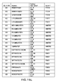
FIGS. 15A-Q provide a list of tumor associated antigens that are useful in the combination therapy of the present invention.
FIGS. 16A-B provide a list of tumor associated antigens that are useful in the combination therapy of the present invention.
FIG. 17 provides a list of tumor associated antigens that are useful in the combination therapy of the present invention.
FIGS. 18A-F provide a list of tumor associated antigens that are useful in the combination therapy of the present invention.
DETAILED DESCRIPTION OF THE INVENTION
The current invention refers to a method of treating a neoplastic disorder comprising administering to a mammal an active immunotherapy and at least one additional therapeutic agent selected from the group consisting of an immunoactive small molecule, an antibody, a kinase inhibitor or a combination thereof. The method of the present invention may be used in an adjuvant or a neoadjuvant or a palliative therapy setting or as a sole treatment. The active immunotherapy and the at least one additional therapeutic agent may target the same and/or different molecules and/or pathways in a neoplastic cell.
As used herein the term “adjuvant” therapy refers to treatment after surgical resection of the primary tumor. As used herein, the term “neoadjuvant therapy” refers to treatment prior to the surgical resection of a primary malignant tumor while “palliative” therapy is intended to relieve symptoms but is not expected to be a cure.
The term “neoplastic disorder” generally refers to one of a group of more than 100 diseases caused by the abnormal growth of cells that can spread to adjoining tissues or other parts of the body. In cancer this growth is uncontrolled and cells can form a solid tumor, in which the cancer cells are massed together, or exists as dispersed cells, as in leukemia. Normal cells divide (reproduce) until maturation is attained and then only as necessary for replacement of damaged or dead cells. Neoplastic cells are referred to as “malignant,” if they divide endlessly, eventually crowding out nearby cells and spreading to other parts of the body. The tendency of cancer cells to spread from one organ to another or from one part of the body to another distinguishes them from benign tumor cells, which overgrow but do not spread to other organs or parts of the body. Malignant cancer cells eventually metastasize and spread to other parts of the body via the bloodstream or lymphatic system, where they can multiply and form new tumors. Benign neoplastic disorders are, for example, but not limited to psosiaris, uterine leiomyoma, melanocytic nevi, restinosis, and benign prostatic hyperplasia. Malignant neoplasias are, for example, cancer of the buccal cavity and pharynx, cancer of the digestive tract, cancer of the colon, rectum, and anus, cancer of the respiratory tract, breast cancer, cancer of the cervix uteri, vagina, and vulva, cancer of the uterine corpus and ovary, cancer of the male genital tract, cancer of the urinary tract, cancer of the bone and soft tissue, kaposi sarcoma, melanoma of the skin, eye melanoma, non-melanoma eye cancer, cancer of the brain and central nervous system, cancer of the thyroid and other endocrine glands, Hodgkin's Lymphoma, Non-Hodgkin's Lymphoma, and myeloma. Most preferably the neoplastic disorder treated by the method of the current invention is renal cancer, colorectal cancer, lung cancer, breast cancer, pancreatic cancer, prostate cancer, gastric cancer, GIST or Glioblastoma or a combination of one or more of the foregoing cancers.
“mammal” includes any mammal able to respond to active immunotherapy with an immune reaction. Preferred mammals include, but are not limited to humans, sport and pet animals, such as cats, dogs, horses, experimental animals such as e.g. rats, rabbits, mice, and livestock. Most preferably the mammal is a human.
In a preferred embodiment at least one additional therapeutic agent may be an immunoactive small molecule, an antibody, or a kinase inhibitor or a combination thereof.
Within the context of this invention, an immunoactive small molecule is a small molecule that may act synergistically with active immunotherapy approaches, in particular peptide-based therapeutic vaccines. Small molecules may, for example, act in such a way by:
-
- reducing regulatory T cells in the periphery and in the tumor lesions
- by improving activation of professional APCs and/or helper and/or killer T cells and/or
- by biasing the immune response towards a TH1-type immune response (cytokine profile including e.g. IFN-gammma, IL-2 upregulation).
In preferred embodiments of the present invention the immunoactive small molecule is 1-MT, ABH, AMD3100, AZD2171, BEC, celebrex, CP-547632, CPA-7, cyclophosphamide, JSI-124, loxoribine, LY580276, NCX-4016, nor-NOHA, pazopanib, rofecoxib, S-27609, SB-505124, SD-208, Sildenafil, Tadalafil, Vardenafil, XL-999, and ZD2171.
The antibody may be a monoclonal or a polyclonal antibody or a fragment thereof, preferably a monoclonal antibody. Humanized and/or chimeric antibodies are included. The antibody may be conjugated or non-conjugated and may be directed at any target antigen of interest, in particular tumor-associated antigens. Examples of antibodies therapeutically active against neoplasia include, but are not limited to, anti-cancer antibodies such as 1D09C3, Abciximab, Alemtuzumab, Apolizumab, Avastin, Basiliximab, Bevacizumab, Cantuzumab, Cetuximab, Dacliximab, Eculizumab, Epratuzumab, Gemtuzumab Ozogamicin, Ibritumomab Tiuxetan, Infliximab, Labetuzumab, Mapatumumab, Matuzumab, Mepolizumab, Muromonab-Cd3, Nimotuzumab, Oregovomab, Palivizumab, Panitumumab, Panorex, Pertuzumab, Rituximab, Tositumomab, and Trastuzumab. Preferred therapeutic antibodies for use in the method of the present invention include anti-CD20 antibodies (e.g., Rituxan™, Bexxar™, Zevalin™), anti-Her2/neu antibodies (e.g., Herceptin™), anti-CD33 antibodies (e.g., Mylotarg™), anti-CD52 antibodies (e.g., Campath™), anti-CD22 antibodies, anti-CD25 antibodies, anti-CTLA-4 antibodies, anti-EGF-R antibodies (e.g. Erbitux™), anti-VEGF antibodies (e.g. Avastin™, VEGF Trap) anti-HLA-DR10β antibodies, anti-MUC1 antibodies, anti-CD40 antibodies (e.g. CP-870,893), anti-Treg cell antibodies (e.g. MDX-010, CP-675,206), anti-GITR antibodies, anti-CCL22 antibodies, and the like.
An antibody as contemplated herein includes any antibody specific to any region of a protein involved in the abnormal growth, differentiation, duplication, angiogenesis, metastasis, apoptosis and/or invasion of cells and the like.
The additional therapeutic agent of the invention is preferably a kinase inhibitor. Protein kinases are a family of enzymes that catalyse the phosphorylation of specific residues in proteins. In general, protein kinases fall into several groups; those that preferentially phosphorylate serine and/or threonine residues, those which preferentially phosphorylate tyrosine residues and those that phosphorylate both tyrosine and Ser/Thr residues. Protein kinases are key elements in signal transduction pathways responsible for transducing extracellular signals, including the action of cytokines on their receptors, to the nuclei, triggering various biological events. The many roles of protein kinases in normal cell physiology include cell cycle control and cell growth, differentiation, apoptosis, cell mobility and mitogenesis. Kinases such as c-Src, c-Abl, mitogen activated protein (MAP) kinase, phosphotidylinositol-3-kinase (PI3K) AKT, and the epidermal growth factor (EGF) receptor are commonly activated in cancer cells, and are known to contribute to tumorigenesis. Many of these occur in the same signaling pathway. For example, HER-kinase family members (HER1 EGFR, HER3, and HER4) transmit signals through MAP kinase and PI3 kinase to promote cell proliferation. Logically, a number of kinase inhibitors are currently being developed for anti-cancer therapy, in particular tyrosine kinase inhibitors (TKIs): cyclin-dependent kinase inhibitors, aurora kinase inhibitors, cell cycle checkpoint inhibitors, epidermal growth factor receptor (EGFR) inhibitors, FMS-like tyrosine kinase inhibitors, platelet-derived growth factor receptor (PDGFR) inhibitors, kinase insert domain inhibitors, inhibitors targeting the PI3K/Akt/mTOR pathway, inhibitors targeting the Ras-Raf-MEK-ERK (ERK) pathway, vascular endothelial growth factor receptor (VEGFR) kinase inhibitors, c-kit inhibitors and serine/threonine kinase inhibitors.
Kinase inhibitors useful in the method of the present invention include, but are not limited to, Lapatinib, AZD 2171, ET18OCH3, Indirubin-3′-oxime, NSC-154020, PD 169316, Quercetin, Roscovitine, Triciribine, ZD 1839, 5-Iodotubercidin, Adaphostin, Aloisine, Alsterpaullone, Aminogenistein, API-2, Apigenin, Arctigenin, ARRY-334543, Axitinib (AG-013736), AY-22989, AZD 2171, Bisindolylmaleimide IX, CCI-779, Chelerythrine, DMPQ, DRB, Edelfosine, ENMD-981693, Erbstatin analog, Erlotinib, Fasudil, Gefitinib (ZD1839), H-7, H-8, H-89, HA-100, HA-1004, HA-1077, HA-1100, Hydroxyfasudil, Kenpaullone, KN-62, KY12420, LFM-A13, Luteolin, LY294002, LY-294002, Mallotoxin, ML-9, MLN608, NSC-226080, NSC-231634, NSC-664704, NSC-680410, NU6102, Olomoucine, Oxindole I, PD 153035, PD 98059, Phloridzin, Piceatannol, Picropodophyllin, PK1, PP1, PP2, PTK787/ZK222584, PTK787/ZK-222584, Purvalanol A, Rapamune, Rapamycin, Ro 31-8220, Rottlerin, SB202190, SB203580, Sirolimus, SL327, SP600125, Staurosporine, STI-571, SU1498, SU4312, SU5416, SU5416 (Semaxanib), SU6656, SU6668, syk inhibitor, TBB, TCN, Tyrphostin AG 1024, Tyrphostin AG 490, Tyrphostin AG 825, Tyrphostin AG 957, U0126, W-7, Wortmannin, Y-27632, Zactima (ZD6474), ZM 252868. Recently approved TKIs for cancer therapy include, for example, Sorafenib and Sunitinib.
KIs currently under clinical investigation for use in anti-cancer therapies and/or novel indications are, for example, MK0457, VX-680, ZD6474, MLN8054, AZD2171, SNS-032, PTK787/ZK222584, Sorafinib (BAY43-9006), SU5416, SU6668 AMG706, Zactima (ZD6474), MP-412, Dasatinib, CEP-701, (Lestaurtinib), XL647, XL999, Tykerb, (Lapatinib), MLN518, (formerly known as CT53518), PKC412, STI571, AMN107, AEE 788, OSI-930, OSI-817, Sunitinib maleate (Sutent SU11248), Vatalanib (PTK787/ZK 222584), SNS-032, SNS-314 and Axitinib (AG-013736). Gefitinib and Erlotinib are two orally available EGFR-TKIs.
Thus, in a preferred embodiment of the present invention, the kinase inhibitor is a tyrosine kinase inhibitor, preferably a multi-kinase inhibitor. Within the context of this invention a multi-kinase inhibitor is an inhibitor that acts on more than one specific kinase. Multi-kinase inhibitors include the so-called DGF out-binders, such as imatinib, sorafenib, lapatinib, BIRB-796 and AZD-1152; other multi-kinase inhibitors are AMG706, Zactima (ZD6474), MP-412, sorafenib (BAY 43-9006), dasatinib, CEP-701 (lestaurtinib), XL647, XL999, Tykerb (lapatinib), MLN518, (formerly known as CT53518), PKC412, STI571, AEE 788, OSI-930, OSI-817, Sutent (sunitinib maleate), axitinib (AG-013736), erlotinib, gefitinib, axitinib, temsirolismus and nilotinib (AMN107).
Most preferred are Sunitinib and/or Sorafenib or a pharmaceutically acceptable salt or derivative, such as for example a malate or a tosylate thereof. The term “derivative” refers to a chemical modification still retaining kinase inhibitory function of the parent molecule. Examples for derivatives are disclosed e.g. in the patent applications mentioned below.
Sunitinib targets multiple receptor tyrosine kinase inhibitors, including PDGFR, KIT and VEGFR, and is a potent and selective anti-angiogenesis agent. Sunitinib or its L-malate salt is also referred to as SU11248, SU011248, Sunitinib malate (USAN/WHO designation) or SUTENT™ (L-malate salt).
The compound, its synthesis, and particular polymorphs are described in U.S. Pat. No. 6,573,293, U.S. Patent Publication Nos. 2003-0229229, 2003-0069298 and 2005-0059824, and in J. M. Manley, M. J. Kalman, B. G. Conway, C. C. Ball, J. L Havens and R. Vaidyanathan, “Early Amidation Approach to 3-[(4-amido)pyrrol-2-yl]-2-indolinones,” J. Org. Chew. 68, 6447-6450 (2003). Preferred formulations of Sunitinib and its L-malate salt are described in PCT Publication No. WO 2004/024127. Preferred dosing regimens are described in U.S. patent application Ser. No. 10/991,244 published as U.S. Patent Publication No. 2005-0182122. The disclosures of these references are incorporated herein by reference in their entireties.
Sorafenib, is also a multi-kinase inhibitor, also known as BAY 43-9006. Sorafenib is a substituted omega carboxy diphenyl urea that inhibits RAF-1 activation, and thereby decreases RAF-1 dependent phosphorylation of MEK-1 and ERK-1, as described in US Patent Application No. 2003-0125359A1, WO 03/047523A2, and Wilhelm et al., Current Pharmaceutical Design, 8:2255-2257 (2002), each of which is herein incorporated by reference in its entirety, particularly in parts pertinent to its structure and properties, methods for making and using it, and other related molecules. Its chemical name is 4-(4-{3-[4-Chloro-3-(trifluoromethyl)phenyl]ureido}phenoxy)-N-methylpyrid-ine-2-carboxamide. A variety of derivatives have been produced. Among these are fluorinated derivatives described in US Patent Application 2005-0038080A1 and WO 2005/009961A2, which are herein incorporated by reference in their entireties, particularly as to these and other pharmaceutically active diphenyl urea compounds.
Currently different nonspecific immunotherapies are used to stimulate the immune system to improve or induce an immune response against neoplastic cells. Nonspecific immunotherapy refers to therapies that can stimulate the immune system by using a substance that activates or enhances immune cell function regardless of their antigen specificity. Nonspecific immunotherapies known in the art include, for example, Bacille Calmette-Guerin (BCG) therapy, cytokine therapy, cell therapy etc.
Antigen-specific immunotherapy refers to either adoptive transfer or vaccination. Adoptive transfer means the direct transfer of the actual components of the immune system that are already capable of producing a specific immune response, such as, for example, T cells or dendritic cells into the recipient. For example, isolated antigen-specific T cells from a cancer patient are expanded to large numbers in vitro, and re-infused back into the patient. Vaccination on the other hand involves the administration of one ore more particular antigen(s) to induce a specific immune response by the host (patient).
An active immunotherapy of the invention may be any immunotherapy that stimulates the intrinsic immune system of the recipient, non-specifically, antigen-specifically and/or multi-targeted. Preferably the active immunotherapy is a multi-targeted, antigen-specific immunotherapy.
In a preferred embodiment the method of the invention comprises an active immunotherapy, whereby at least one vaccine is administered to the mammal.
In whole-cell vaccines, the tumor cell itself is used to provide the broadest set of tumor-related antigens. The tumor cells in the composition should contain antigens that are also present in the tumor to be treated, so that the immune response elicited against the antigens in the composition is effected against the tumor. Generally, the cells are recovered from tumors, suspended in a preservation medium and frozen until used for the vaccine preparation. When needed, the cells are thawed, and then stored at temperatures ranging from about 0° C. (on ice) to room temperature until administration. Immunotherapy approaches using unmodified intact tumor cells prepared from tumors taken from the patient, i.e., autologous tumor cells, have been described in the literature (see, e.g., Berd et al., Cancer Research 1986; 46:2572-2577; Hoover et al., Cancer 1985; 55: 1236-1243; and U.S. Pat. No. 5,484,596).
Alternative vaccine compositions based on disrupted cells have also been suggested including, e.g., tumor membranes (see, e.g., Levin et al., In: Human Tumors in Short Term Culture Techniques and Clinical Applications, P. P. Dendy, Ed., 1976, Academic Press, London, pp. 277-280) or tumor peptides extracted from tumors (see, e.g., U.S. Pat. No. 5,550,214 and U.S. Pat. No. 5,487,556).
The tumor cells can also be modified in some manner to alter or increase the immune response (see, e.g., Hostetler et al., Cancer Research 1989, 49:1207-1213, and Muller et al., Anticancer Research 1991; 11:925-930). Further examples for modifications and preparation methods are, for example, provided by US patent application 2007-0014775, 2006-0165668, 2002-0085997 or 2003-0170756.
One particular form of tumor cell modification that has a pronounced effect on immunotherapy is coupling of a hapten to the tumor cells. Such haptenized vaccines are described, for example, in WO 96/40173, WO 00/09140, and U.S. Pat. No. 6,333,028. Transducing the tumor with genes so that the tumor cell may act like an antigen presenting cell (Antonia S J et al. Phase I trial of a B7-1 (CD80) gene modified autologous tumor cell vaccine in combination with systemic interleukin-2 in patients with metastatic renal cell carcinoma. J Urol. 2002; 167:1995-2000) or may attract and stimulate local antigen-presenting cells (Simons et al. Bioactivity of autologous irradiated renal cell carcinoma vaccines generated by ex vivo granulocyte-macrophage colony-stimulating factor gene transfer. Cancer Res. 1997; 57:1537-1546) are two approaches.
A person skilled in the art will be able to determine the type of vaccine compositions and antigen modification suitable for a certain type and stage of tumor and/or the individual patient without undue experimentation using the general knowledge of the art and the references and suggestions disclosed in the present application.
In another embodiment the cell based vaccine employs non-tumor cells. The cells used for vaccination are antigen presenting cells (APCs), which may be isolated from the patient. These are loaded or pulsed with a tumor antigen ex vivo. The transfer of these pulsed APCs into the patient elicits a significant tumor-specific immune response that attacks the tumor cells. Currently, there are three different methods for pulsing or loading APCs. First, growing APCs in the presence of a tumor-associated protein; second, using genetic engineering techniques to introduce the gene that codes for a tumor-associated protein into APCs, and third, pulsing APCs with fragments (peptides) isolated from a tumor antigen or synthetic peptides.
The main advantage of APC-based vaccination is that dendritic cells (DCs) produce all the molecules required for eliciting an immune response, unlike other forms of cancer immunotherapy where adjuvants and co-stimulatory molecules are required to boost the ensuing immune response. The potency of DCs as vehicles for delivering antigen and achieving a tumor-specific immune response has been demonstrated in a number of clinical trials.
Thus, in a preferred embodiment of the present invention the method employs a vaccine that comprises cells or cellular extracts, preferably tumors cells or extracts thereof, which were derived from the same or a different mammal as the one to be treated by the inventive method. The cells are, for example, modified or unmodified tumor cells or APCs loaded or transfected with tumor antigen(s). The tumor antigen that is loaded or transfected includes the same proteins, nucleic acids and/or peptides that may be employed for direct vaccination (see below). The cells may also be T cells for adoptive transfer.
A trimolecular complex consisting of the components of T-cell-antigen receptor, an MHC (Major Histocompatibility Complex) molecule and the ligand thereof, which is a peptide fragment derived from a protein, plays a central role in the regulation of the specific (adaptive) immune response.
MHC class I and class II molecules (or the corresponding human molecules, the Human Leukocyte Antigene receptors, HLAs) are peptide receptors that allow the binding of millions of different ligands, with stringent specificity. The binding specifically provided by allele-specific peptide-binding motifs that have the following specificity criteria: the peptides have a defined length, which in the MHC class I haplotypes vary generally from eight to ten amino acids, while class II molecules bind peptides from a length of thirteen amino acids and above. Typically, two of the amino acid positions are so-called “anchors” which can only be occupied by a single amino acid or by amino acid groups with closely related physico-chemical properties defined by their side chains. The exact position of the anchor amino acids in the peptide and the requirements made on their properties vary with the MHC alleles. The C-terminus of the peptide ligands is frequently an aliphatic or a charged group. Examples for such peptide ligands, motifs, variants, as well as examples for extensions on the N- and/or C-terminal sides can be derived from public databases (Rammensee et al. SYFPEITHI: database for MHC ligands and peptide motifs Immunogenetics 1999, 50, 213-219.
Inside the cell, regular, degenerate and foreign gene products, e.g. viral proteins or tumor antigens, are broken down into small peptides. Peptides arising in the cytosol can be trimmed by cytosolic peptidases, as well as by proteolytic enzymes residing in the ER (after transfer of precursors into the ER through TAP). Peptides with a length of, on average, 8 to 10 amino acid residues fulfilling the binding requirements of the binding groove of expressed HLA alleles can then be presented by the respective HLA receptors on the cell surface. Some of those peptides constitute potential ligands for MHC molecules. Binding of the ligands to the MHC molecules provides the prerequisite for peptide presentation by MI-IC-molecules and the triggering of a cellular immune response. Thus, the introduction of a peptide may trigger an immune response. Since the immunogenic epitopes of a vast amount of proteins are known, protein fragments or synthetic peptides containing one or more epitopes may also be employed as vaccines.
In a preferred embodiment the method of the present invention employs a vaccine that comprises at least one protein, nucleic acid and/or fragment thereof derived from a tumor associated antigen (TAA) or cancer antigen. A TAA or cancer antigen is defined as an antigen that is selectively or abundantly expressed in cancer cells. See for example, the following applications directed to certain tumor associated peptides that bind to MHC-molecules useful in a vaccine and/or vaccines per se: Ser. No. 10/999,264 (filed Nov. 28, 2004) (claiming the peptide YVDPVITSI (SEQ ID NO:1)); Ser. No. 11/848,062 (filed Aug. 30, 2007) (claiming the peptide SVASTITGV (SEQ ID NO:2); Ser. No. 10/999,364 (Filed Aug. 30, 2007) (claiming the peptide ALFDGDPHL (SEQ ID NO:4) and others shown in FIG. 18)); 60/953,161 (filed Jul. 31, 2007) (claiming various peptides such as TGBI-001 and NOX-001 and others shown in FIG. 11); 60/953,109 (filed Jul. 31, 2007) (claiming various pharmaceutical compositions comprising peptides shown in FIG. 12); Ser. No. 11/596,802 (filed Nov. 17, 2006) (claiming various peptides shown in FIG. 13 and specifically the peptides FPSLREAAL, LAALPHSCL, GLASFKSFLK; SLLTSSKQLQK, IARNLTQQL and GPALGRSFL); Ser. No. 10/549,718 (filed Sep. 16, 2005) (claiming various peptides shown in FIG. 14); Ser. No. 11/664,627 (filed Apr. 2, 2007) (claiming various peptides shown in FIG. 15 and specifically the peptides NPPSMVAAGSVVAAV and SHYFKIIEDLRAQI); U.S. Pat. No. 7,087,712 (issued Aug. 8, 2006) (claiming the MUC-1 peptide STAPPVHNV); Ser. No. 11/414,897 (filed May 1, 2006) (claiming the MUC-1 peptide LLLLTVLTV); Ser. No. 11/912,668 (filed Oct. 25, 2007) (claiming the peptides LLAARAIVAI and ALCNTDSPL); Ser. No. 11/912,670 (filed Oct. 25, 2007) (claiming various peptides shown in FIG. 16); and Ser. No. 12/065,725 (filed Mar. 4, 2008) (claiming various peptides shown in FIG. 17), all of which are herein incorporated by reference in their entirety.
The main advantage of a peptide-based vaccine is that it provides a method for monitoring a specific immune response for a particular antigen and thus allows the evaluation of the efficacy of vaccination. Other advantages include the bypassing of the need for antigen-presenting cells to process a whole cell before presenting the antigen to the immune system. In addition, administration of a peptide antigen does not carry the risk of introducing dangerous substances into the patient, unlike other vaccines that rely on tumor cells.
The protein, or fragment thereof or peptide may also be generated within the recipient mammal by introducing a nucleic acid encoding the peptide. The nucleic acid may be DNA, cDNA, PNA, CNA, RNA or a combination thereof. Methods for designing and introducing such a nucleic acid are well known in the art. An overview is provided by e.g. S. Pascolo: Vaccination with messenger RNA Methods Mol Med 2006, 127; 23-40; R. Stan, J D Wolchok and A D Cohen, DNA vaccines against cancer Hematol Oncol Clin North Am 2006, 3; 613-636 or A Mandavi and B J Monk Recent advances in human papillomavirus vaccines Curr Oncol Rep 2006, 6, 465-472. Polynucleotide vaccines are easy to prepare, but the mode of action of these vectors in inducing an immune response is not fully understood. Suitable vectors and delivery systems include viral DNA and/or RNA, such as systems based on adenovirus, vaccinia virus, retroviruses, herpes virus, adeno-associated virus or hybrids containing elements of more than one virus. Non-viral delivery systems include cationic lipids and cationic polymers and are well known in the art of DNA delivery. Physical delivery, such as via a “gene-gun,” may also be used. The peptide or peptide encoded by the nucleic acid may be a fusion protein, for example with an epitope from tetanus toxoid, which stimulates CD4+ T cells. Clinical trials using polynucleotide vaccines in cancer have been reported (e.g. Restifo and Rosenberg, Developing recombinant and synthetic vaccines for the treatment of melanoma. Curr Opin Oncol. 1999 (1): 50-57).
A person skilled in the art will readily be able to determine the type of molecule for vaccination purposes, compositions suitable for a certain type and stage of tumor and/or the individual patient, as well as respective antigen modifications and/or delivery vehicles to enhance the immune response without undue experimentation using the general knowledge of the art and the references and suggestions disclosed in the present application.
Most preferably, the vaccine employed in the method of the invention comprises at least one peptide. Such a peptide comprises, for example, an epitope of a TAA, preferably an epitope that is capable of binding to a MHC molecule and generated in vivo by a tumor cell. Epitopes with these characteristics can be identified by methods described in WO03/100432, WO2005/076009, WO03/102023, WO2004/085461, WO2005/116051, U.S. Pat. No. 7,087,712, EP 04 013 790.3, WO2006/037421, WO2006/114307, EP 05 019 254.1, and EP 05 019 255.8, which are hereby incorporated by reference in their entireties.
In a particularly preferred embodiment the vaccine contains at least one of the peptides disclosed in EP 05 019 255.8, namely the peptides provided below:
| |
|
| |
Peptide Code |
SEQ ID NO |
Peptide Sequence |
| |
|
| |
ADF-001 |
1 |
SVASTITGV |
| |
|
| |
ADF-002 |
2 |
VMAGDIYSV |
| |
|
| |
APO-001 |
3 |
ALADGVQKV |
| |
|
| |
CCN-001 |
4 |
LLGATCMFV |
| |
|
| |
GUC-001 |
5 |
SVFAGVVGV |
| |
|
| |
K67-001 |
6 |
ALFDGDPHL |
| |
|
| |
MET-001 |
7 |
YVDPVITSI |
| |
|
| |
MMP-001 |
8 |
SQDDIKGIQKLYGKRS |
| |
|
| |
MUC-001 |
9 |
STAPPVHNV |
| |
|
| |
RGS-001 |
10 |
LAALPHSCL |
| |
|
In another embodiment, the vaccine contains one or more proteins containing at least one of the peptides mentioned above or one or more nucleic acids encoding at least one of the peptides mentioned above.
In another preferred embodiment, the vaccine contains at least one peptide selected from the group consisting of MET-001 (YVDPVITSI) (SEQ ID NO:7), MMP-001 (SQDDIKGIQKLYGKRS) (SEQ ID NO:8), and MUC-001 (STAPPVHNV) (SEQ ID NO:9) or one or more proteins containing at least one of the peptides selected from MET-001 (YVDPVITSI) (SEQ ID NO:7), MMP-001 (SQDDIKGIQKLYGKRS) (SEQ ID NO: 8), and MUC-001 (STAPPVHNV) (SEQ ID NO:9) or one or more nucleic acids encoding at least one of the peptides MET-001 (YVDPVITSI) (SEQ ID NO:7), MMP-001 (SQDDIKGIQKLYGKRS) (SEQ ID NO:8), and MUC-001 (STAPPVHNV) (SEQ ID NO:9).
In one aspect, the vaccine comprises at least one peptide, preferably two to 50, more preferably two to 25, even more preferably two to 15 and most preferably two, three, four, five, six, seven, eight, nine, ten or eleven peptides. The peptide(s) may be derived from one or more specific TAAs and may bind to MHC class I and/or class II molecules.
In one aspect of the invention, the method utilizes an active immunotherapy that comprises at least one vaccine in combination with at least one additional therapeutic agent comprising a multi-kinase inhibitor and/or a tyrosine kinase inhibitor. Preferred is the combination wherein at least one immunogenic peptide is administered to the mammal and said at least one additional therapeutic agent comprises a multi-kinase inhibitor and/or a tyrosine kinase inhibitor, preferably of the Sunitinib and/or Sorafenib type or a pharmaceutically acceptable salt or derivative thereof.
The exact combination of active immunotherapy and additional therapeutic agent in individual patients should take into account the patient's metabolism, the kind and stage of the disorder to be treated, and the biochemistry of the targets of the two arms of treatment. The setting of treatment (i.e. sole, adjuvant, neoadjuvant, palliative) needs also to be considered. Depending on these factors, the person skilled in the art will determine in which individual situation what kind of combination is the most promising. For example, in a situation where the tumor cells have gained resistance to certain therapeutic agents, the following combination treatment according to the method of the invention will involve TKIs and/or antibodies and targets for vaccination aiming at different key molecules/pathways than those involved in the resistance. The key molecule/pathway targets for TKI and active immunotherapy may be identical. In a different setting, for example, in neoadjuvant therapy, where there is a need for fast tumor shrinkage, it may be advantageous to get different key molecules/pathways with active immunotherapy and additional therapy. For an adjuvant therapy it may be advantageous to destroy any residual tumor cells. Beneficial combinations may also be suggested by studying the alteration of target presentation in cancer cell lines by additional therapeutic agent(s) as in Example 2, the in vitro alteration of T cell activation by said agent(s) as in Example 3, or the in vivo effects by animal experiments such as in Example 4. These procedures can also be used to determine the order of administration of the agents, i.e. before, simultaneously, or after vaccination.
In general, the success of vaccine strategies depends on the mode of antigen delivery, the choice of adjuvant, and the particular antigen being used.
The at least one additional therapeutic agent and/or active immunotherapy agent, i.e. the immunogenic protein, nucleic acid and/or peptide, can be administered by any means known to one of skill in the art (see Banga, A., “Parenteral Controlled Delivery of Therapeutic Peptides and Proteins,” in Therapeutic Peptides and Proteins, Technomic Publishing Co., Inc., Lancaster, Pa., 1995, S. Pascolo: “Vaccination with messenger RNA Methods,” Mol Med 2006, 127; 23-40; R. Stan, J D Wolchok and A D Cohen, “DNA vaccines against cancer,” Hematol Oncol Clin North Am 2006, 3; 613-636 or A Mandavi and B Y Monk, “Recent advances in human papillomavirus vaccines,” Curr Oncol Rep 2006, 6, 465-472) such as by intradermal, intramuscular, subcutaneous, intratumoral or intravenous injection. Other administration is contemplated such as mucosal, such as oral, nasal, or anal and dermal administration. For TKIs such as Sorafenib and Sunitinib, oral administration is preferred.
In one embodiment, administration of the active immunotherapy agent is by subcutaneous, intratumoral or intramuscular injection. To extend the time during which the peptide, nucleic acid and/or protein is available to stimulate a response, the agent can be provided as an implant, an oily injection, an oil-in-water emulsion, an water-in-oil emulsion, a suspension or as a particulate system. The particulate system, for example, can be a microparticle, a microcapsule, a microsphere, a nanocapsule, or similar particle. (see, e.g., Banga, supra) including controlled release devices and patches etc.
Controlled release antigen delivery systems may also be used. For example, WO 95/11008 (Genentech Inc.) discloses the use of PLGA (poly(DL-lactide-co-glycolide) microspheres for encapsulating an antigen. EP 0 686 030 teaches a method of potentiating an immune response by embedding an antigen in a biodegradable biopolymer and injecting it in the form of a dispersion to trigger a humoral and cellular response. Lipid-based systems disclosed in US patent application 2006-0275777, or virosomes may also be used. Preferred systems include those by Juvaris (e.g. JuvImmune™ or JuvaVax™). Particulate systems include microspheres, microparticles, microcapsules, nanocapsules, nanospheres, and nanoparticles. Microcapsules contain the therapeutic protein as a central core. In microspheres, the therapeutic agent is dispersed throughout the particle. Particles, microspheres, and microcapsules smaller than about 1 μm are generally referred to as nanoparticles, nanospheres, and nanocapsules, respectively. Capillaries have a diameter of approximately 5 μm so that only nanoparticles are administered intravenously. Microparticles are typically around 100 μm in diameter and are administered subcutaneously or intramuscularly (see, Kreuter, Colloidal Drug Delivery Systems, J. Kreuter, ed., Marcel Dekker, Inc., New York, N.Y., pp. 219-342, 1994; Tice & Tabibi, Treatise on Controlled Drug Delivery, A. Kydonieus, ed., Marcel Dekker, Inc., New York, N.Y., pp. 315-339, 1992). Numerous additional systems for controlled delivery of therapeutic proteins are known (e.g., U.S. Pat. Nos. 5,055,303; 5,188,837; 4,235,871; 4,501,728; 4,837,028; 4,957,735; and 5,019,369; 5,055,303; 5,514,670; 5,413,797; 5,268,164; 5,004,697; 4,902,505; 5,506,206; 5,271,961; 5,254,342; and 5,534,496).
In a preferred embodiment the additional therapeutic agent is administered orally while the active immunotherapeutic agent is administered intradermally, subcutaneously, intravenously, intratumorally or intramuscularly.
A person skilled in the art can readily determine the route of administration to choose depending of the type of composition, its solubility, dissolution, bioavailability, stability, the optional adjuvant(s) used etc. Formulations for the additional therapy by immunoactive small molecules, TKIs and/or antibodies are preferably those approved by drug regulatory authorities, but may also be adjusted to the particular combination with the active immunotherapy of the method of the invention. One of ordinary skill in the art would take into consideration the need to formulate the active ingredients of both therapeutic arms in a manner that does not cause severe toxicity in the individual, damage the individual to any appreciable degree or cause appreciable adverse side effects. The formulation and preparation of compositions is well-known to those skilled in the art of pharmaceutical formulation, and the descriptions herein are illustrative and not limiting. See, e.g., Genarro A R, Remington's Pharmaceutical Sciences, Easton, Pa.: Mack Publishing Company, 2000, 20th. ed.; Allen, Popovich and Ansel, 2005, Pharmaceutical Dosage Forms and Drug Delivery Systems 8th ed. Lippincott Williams & Wilkins;
In the method of the present invention, the active immunotherapy and at least one additional therapeutic agent can be administered simultaneously, sequentially (sequenced over time) or separately. For example, the active immunotherapy agent can be administered within the same hour or within the same day as the additional therapeutic agent to save visits to the medical practitioner, both agents may be administered on different days but within the same period of time, such as for example during the period of time of a chemotherapy regimen, or they may be administered separately, for example the active immunotherapy agent is administered some time, e.g. days, weeks or month after a therapy with the additional therapeutic agent(s) has been concluded. Also the additional therapeutic agent(s) may be administered days, weeks or month after the last vaccination took place.
Generally the routes of administration of the composition that effect the active immunotherapy and the route of administration of at least one additional therapeutic agent will be different, particularly in embodiments, wherein active immunotherapy is combined with treatment with an orally administered TKI. For instance, a vaccine may be administered intradermally, while the accompanying additional therapeutic agent such as e.g. a TKI like sunitinib or sorafenib, is given orally.
With certain combinations the routes of administration of the composition that effect the active immunotherapy and the route of administration of at least one additional therapeutic agent will be the same. This may be the case, for example, if the active immunotherapy is given intravenously and combined with an antibody as additional therapeutic agent, which has to be administered intravenously as well.
The treatment regimen with the active immunotherapy and the additional therapeutic agent in individual patients should take into account the patients height, weight, rate of absorption and metabolism of the medication in question, the type and stage of the disorder to be treated, and other pharmacological agents that are administered concurrently. Additionally, any synergistic or neutralizing effects of the two arms of treatment will be taken into consideration, so that synergistically acting treatment arms are preferably administered within a period of time that allows such synergies. In contrast, treatment arms having neutralizing effects will be administered separately so that the effects of the first arm of treatment have worn off, so they do not interfere with the second arm of treatment. The setting of treatment (i.e. sole, adjuvant, neoadjuvant, palliative) needs to be considered as well. Depending on these factors, the active immunotherapy may be administered prior to, concurrently with and/or after at least one additional therapeutic agent. For example, a patient receiving the treatment of the present invention might have renal cancer. A person skilled in the art may treat the patient first with a conventional chemotherapy consisting of several cycles of treatment with a TKI such as Sorafinib and, upon remission and recovery of the immune system, administer several boosts of a peptide vaccine to prevent or delay recurrence of a tumor. If Sunitinib is administered as the TKI, it may be of advantage to administer the vaccine concurrently, or concurrently and after the Sunitinib treatment, for example, since this particular TKI seems to inhibit regulatory T-cells (Tregs) limiting the immune response.
Treg cells represent a T-cell population that can functionally suppress an immune response by influencing the activity of other immune effector cells. The existence of Tregs was first established in 1971, when Gershon and Kondo transferred antigen-specific tolerance to antigen-naïve animals by transferring T-cells that had previously been exposed to the specific antigen. Several phenotypically distinct Tregs exist. The object of recent intensive research are CD4+ CD25+ Foxp3+ T cells, which also express high levels of glucocorticoid-induced TNFR-related protein (GITR). These Tregs are considered key mediators of peripheral tolerance. More recently, another type of Tregs (IL10+CCR7+) possibly involved in central priming suppression rather than in peripheral effector suppression, was described (Zou, 2005). CD4+ Foxp3+ Tregs suppress the execution of effector functions of T-cells in the periphery.
The active immunotherapy may be administered with or without adjuvant. Adjuvants are substances that non-specifically enhance or potentiate the immune response (e.g., immune responses mediated by CTLs and Helper-T (TH) cells) to an antigen, and would thus be considered useful in the active immunotherapy of the present invention. Suitable adjuvants include, but are not limited to 1018 ISS, aluminium salts, Amplivax, AS15, BCG, CP-870,893, CpG7909, CyaA, dSLIM, GM-CSF, IC30, IC31, Imiquimod, ImuFact IMP321, IS Patch, ISCOMATRIX, JuvImmune, LipoVac, MF59, monophosphoryl lipid A, Montanide IMS 1312, Montanide ISA 206, Montanide ISA 50V, Montanide ISA-51, OK-432, OM-174, OM-197-MP-EC, ONTAK, PepTel® vector system, PLG microparticles, resiquimod, SRL172, Virosomes and other Virus-like particles, YF-17 DBCG, Aquila's QS21 stimulon (Aquila Biotech, Worcester, Mass., USA), which is derived from saponin, mycobacterial extracts and synthetic bacterial cell wall mimics, and other proprietory adjuvants such as Ribi's Detox. Quil or Superfos. Adjuvants such as Freund's or GM-CSF are preferred. Other examples for adjuvants include cholera toxin, which acts locally as a mucosal adjuvant for the induction of peptide-specific CTLs following intranasal immunization of dendritic cells with CTL epitope peptides (Porgador et al., 1997; Porgador et al., 1998). Several immunological adjuvants (e.g., MF59) specific for dendritic cells and their preparation have been described previously (Dupis et al., 1998; Allison, 1997; Allison, 1998). Cytokines may also be used. Several cytokines have been directly linked to influencing dendritic cell migration to lymphoid tissues (e.g., TNF-α), accelerating the maturation of dendritic cells into efficient antigen-presenting cells for T-lymphocytes (e.g., GM-CSF, IL-1 and IL-4) (Dupis et al., 1998; Allison, 1997; Allison, 1998; U.S. Pat. No. 5,849,589, specifically incorporated herein by reference in its entirety) and acting as immunoadjuvants (e.g., IL-12) (Gabrilovich et al., 1996).
CpG immunostimulatory oligonucleotides have also been reported to enhance the effects of adjuvants in a vaccine setting. Without being bound by theory, CpG oligonucleotides act by activating the innate (non-adaptive) immune system via Toll-like receptors (TLR), mainly TLR9. CpG triggered TLR9 activation enhances antigen-specific humoral and cellular responses to a wide variety of antigens, including peptide or protein antigens, live or killed viruses, dendritic cell vaccines, autologous cellular vaccines and polysaccharide conjugates in both prophylactic and therapeutic vaccines. More importantly it enhances dendritic cell maturation and differentiation, resulting in enhanced activation of TH1 cells and strong cytotoxic T-lymphocyte (CTL) generation, even in the absence of CD4 T-cell help. The TH1 bias induced by TLR9 stimulation is maintained even in the presence of vaccine adjuvants such as alum or incomplete Freund's adjuvant (IFA) that normally promote a TH2 bias. CpG oligonucleotides show even greater adjuvant activity when formulated or co-administered with other adjuvants or in formulations such as microparticles, nano particles, lipid emulsions or similar formulations, which are especially necessary for inducing a strong response when the antigen is relatively weak. They also accelerate the immune response and enabled the reduction of antigen doses by approximately two orders of magnitude, with comparable antibody responses to the full-dose vaccine without CpG in some experiments (Arthur M. Krieg, Therapeutic potential of Toll-like receptor 9 activation, Nature Reviews|Drug Discovery, 5, Jun. 2006, 471-484). U.S. Pat. No. 6,406,705 B1 describes the combined use of CpG oligonucleotides, non-nucleic acid adjuvants and an antigen to induce an antigen-specific immune response. Other examples of useful adjuvants include, but are not limited to, chemically modified CpGs (e.g. CpR, Idera), non-CpG bacterial DNA or RNA, as well as immunoactive small molecules (see above) that may act therapeutically and/or as an adjuvant. The amounts and concentrations of adjuvants and additives useful in the context of the present invention can readily be determined by the skilled artisan without undue experimentation.
The dosage of an active immunotherapy agent and an additional therapeutic agent will be tailored to each individual patient manifesting symptoms characteristic of a specific neoplastic disorder. For example, a patient receiving the treatment of the present invention might have renal cancer. A person skilled in the art will recognize that the optimal dose of a pharmaceutical agent to be administered will vary from one individual to another. Dosage in individual patients should take into account the patients height, weight, rate of absorption and metabolism of the medication in question, the stage of the disorder to be treated, and what other pharmacological agents are administered concurrently. The skilled artisan will adjust doses depending on tumor response and adverse effect profile. Generally, the dosage of the additional therapeutic agent(s) will be within the range approved by drug regulatory authorities and proven to be effective and save within clinical trial or below.
In a particularly preferred aspect of this embodiment, the invention provides a method of treating renal cell carcinoma in a patient, such as a human, by administering to the patient Sunitinib, for example in an amount of 25 to 75, preferably 25, 37.5, 50 or 62.5 mg daily, continuous (i.e., not intermittent) or intermittent dosing schedule for example on a 4/2, 4/1, 3/1 or 2/1 dosing schedule and a multi-target peptide vaccine, for example 50 μg to 1 mg of each peptide, preferably 200 μg to 600 μg of each peptide per patient and injection, preferably together with an adjuvant. In another embodiment, the invention provides a method of treating any of the earlier-recited cancers in a patient, such as a human, by administering to the patient Sorafinib in an amount of 200 mg or 400 mg, twice daily or once daily or once every two days.
One skilled in the art can readily determine the optimal dosage for a particular patient based on tumor response and adverse event profile. Those skilled in the art will appreciate that dosages may also be determined with guidance from Goodman & Gilman's The Pharmacological Basis of Therapeutics, McGraw-Hill Professional; 11th edition (2005).
It is to be understood that the description, specific examples and data, while indicating exemplary embodiments, are given by way of illustration and are not intended to limit the present invention. Various changes and modifications within the present invention will become apparent to the skilled artisan from the discussion, disclosure and data contained herein, and thus are considered part of the invention.
EXAMPLES
The tyrosine kinase inhibitors (TKIs) Sorafenib and Sunitinib are multi-target kinase inhibitors recently approved for the treatment of advanced renal cell carcinoma (RCC) in the U.S. To gain preclinical knowledge on potential influence of TKIs on the effects caused by cancer vaccines, Sorafenib and Sunitinib treatment was combined with peptide vaccination.
Example 1
Quantification of Sorafenib and Sunitinib in Biological Fluids
Sunitinib malate and sorafenib tosylate were supplied by euroasia chemicals PVT. LTD., Mumbai (India).
1.1. Sample Preparation for Sunitinib Quantification
To quantify sunitinib in blood serum or in cell culture medium, 10 μl of 50% acetonitril (Acros, Geel, Belgium) was added to 50 μl serum or medium in brown glass tubes to protect the photo-unstable sunitinib from light, and mixed for 10 seconds. The proteins were precipitated with 40 μl 100% acetonitril (Acros, Geel, Belgium), centrifuged and filtered through a 0.2 μm PVDF filter. 10 μl was directly injected to the HPLC system.
1.2. HPLC-Conditions:
The HPLC system consisted of a Binary HPLC pump (Shimadzu LC10aVP), a Shimadzu SIL-10aVP autosampler, a Shimadzu CTO-10asVP column oven and a Shimadzu SPD10aVP detector. Data acquisition and analysis was performed using the Shimadzu Class-VP 7.3 software. Chromatographic separation was carried out on a reverse phase C18 column (Reprosil Pur ODS-3μ, 60×2 mm). To protect the analytical column a guard column has been used (Reprosil Pur ODS-5μ, 10×2 mm). Eluent A consisted of water (LCMS grade, Acros, Geel, Belgium), modified by 0.1% formic acid (Merck, Darmstadt, Germany) and eluent B was 80% acetonitril (Acros, Geel, Belgium) with 0.1% formic acid (Merck, Darmstadt, Germany).
The following gradient was used: 5 min 25% eluent B, 15 min 62.5% eluent B, 6 min 80% eluent B and 5 min 80% eluent B. The temperature of the autosampler was kept at 4° C. The temperature of the column was maintained at 30° C. The detection wavelength was set at 400 nm, the injection volume was 10 μl. The column was equilibrated with the mobile phase at a flow rate 0.5 ml/min.
1.3. Sample Preparation for Sorafenib Quantification
For Sorafenib analysis, 10 μl of 50% acetonitril (Acros, Geel, Belgium) was added to 50 serum or medium in 0.5 ml PCR tubes and mixed for 10 seconds. 10 mg NH4Cl (Roth, Karlsruhe, Germany) was added and mixed for 10 seconds. 60 μl acetonitril (Acros, Geel, Belgium) solution, containing Tolnaftate (Sigma, Steinheim, Germany) as internal standard was added and mixed for 1 minute. The mixture was centrifuged for 3 minutes at room temperature. After phase separation, 35 μl from the acetonitril phase was transferred into HPLC-vials and 10 μl were directly injected to the HPLC system.
1.4. HPLC-Conditions:
Sorafinib was analysed on the same system as sunitinib with Eluent A consisting of 20 mM KH2PO4-buffer (Sigma, Steinheim, Germany). Eluent B consisted of 80% acetonitril (Acros, Geel, Belgium), 20% 20 mM KH2PO4 (Sigma, Steinheim, Germany) and 0.01% phosphoric acid (Sigma, Steinheim, Germany). The following gradient was used: 5 min 20% eluent B, 5 min 32% eluent B, 20 min 56% eluent B and 10 min 80% eluent B. The temperature of the autosampler was kept at 4° C. The temperature of the column was maintained at 40° C. The detection wavelength was set at 265 nm, the injection volume was 10 μl. The column was equilibrated with the mobile phase at a flow rate 0.5 ml/min.
Using these methods, bioavailability of sorafenib and sunitinib in the mouse models at the used doses was confirmed to reach plasma levels shown by others to be effective in tumor growth inhibition. It could also be shown that the TKIs stability in the cell culture systems of choice was acceptable for the conduction of in vitro experiments. In vitro concentrations in later experiments were chosen to include steady state plasma concentrations of TKI treated patients.
Example 2
Alteration of Expression of Vaccination Relevant Genes During TKI Treatment
2.1. Alteration of Gene Expression Profiles of Human Tumor Cell Lines In Vitro
Genome-wide mRNA expression was measured by Affymetrix microarrays. The human renal cell carcinoma cell line A498 was cultured in the presence of sorafenib and sunitinib. Gene expression for a selection of tumor associated antigens and genes involved in antigen presentation to T lymphocytes was compared with untreated cells to determine whether these tyrosine kinase inhibitors (TKIs) might have the potential to cause altered presentation of antigens in vitro.
The human renal cell carcinoma cell lines A498 and RCC068 were cultured in RPMI medium (5% FCS (Biochrom, Berlin, Germany), 5% HS (PromoCell, Heidelberg, Germany)). Human serum was added as a supply of ligands influencing signaling pathways, which might be altered by TKIs. 40 h after seeding, the experiment was started by addition of TKIs to the culture flasks. The following incubation periods (time points) were planned: 1 h, 6 h, 24 h, 14 days. For each time point 3 flasks of each cell line were prepared by containing either 0.1% DMSO alone as a control, 13 μM sorafenib (8.3 μg/ml sorafenib tosylate)+0.1% DMSO, or 250 nM sunitinib (133 ng/ml sunitinib malate). At each time point, cells were harvested by removing the culture medium and adding 1.25 ml TRI Reagent (Fermentas, St. Leon-Rot, Germany).
RNA was isolated according to standard protocols and further cleaned up by the RNeasy Mini Kit (QIAGEN, Hilden, Germany). For the 14 d time point, cells were trypsinized every 3-4 days and supplied with fresh medium containing fresh TKIs. Sorafenib cells were already harvested after 10 days since the TKI prevented cell growth. A normal medium control sample containing neither DMSO nor TKIs was harvested at 1 h for each cell line. Quality and quantity of RNA samples were assessed on an Agilent 2100 Bioanalyzer (Agilent, Waldbronn, Germany) using the RNA 6000 Pico LabChip Kit (Agilent).
Gene expression analysis was performed only for the 24 h time point (3 samples) and the normal medium control (1 sample) of the A498 cells by Affymetrix Human Genome HG-U133 Plus 2.0 oligonucleotide microarrays (Affymetrix, Santa Clara, Calif., USA).
All steps were carried out according to the Affymetrix manual. Briefly, double-stranded cDNA was synthesized from 8 μg of total RNA, using SuperScript RTII (Invitrogen, Karlsruhe, Germany) and the oligo-dTT7 primer (MWG Biotech, Ebersberg, Germany) as described in the manual. In vitro transcription was performed with the GeneChip IVT Labeling Kit (Affymetrix) followed by cRNA fragmentation, hybridization, and staining with streptavidin-phycoerythrin and biotinylated anti-streptavidin antibody (Molecular Probes, Leiden, Netherlands). Images were scanned with the Affymetrix Gene-Chip Scanner 3000 and data were analyzed with the GCOS software (Affymetrix), using default settings for all parameters. Pairwise comparisons were calculated using the normal medium control array as baseline. For normalization, 100 housekeeping genes provided by Affymetrix were used. Relative expression values were calculated from the signal log ratios given by the software and the expression level in the control sample was set to 100% for each gene.
mRNA expression was analyzed for possible vaccination target antigens as well as for proteins involved in antigen presentation to T cells, like HLA proteins themselves or members of the processing machinery like TAP1 or the immunoproteasomal subunits PSMB9 (LMP2) or PSMB8 (LMP7). The influence of DMSO addition alone or in combination with the TKIs sorafenib and sunitinib on the renal cell carcinoma cell line A498 after 24 h incubation was assessed by comparing these samples with the normal medium control cell line. Expression in the control was defined as 100%.
Result are summarized in Table 1.
| TABLE 1 |
| |
| Relative expression of tumor associated antigens and genes |
| involved in MHC-peptide presentation in A498 tumor cells. |
| |
% Expression relative to control |
| |
Gene |
DMSO |
Sorafenib |
Sunitinib |
| |
|
| Tumor associated antigens |
| |
100 |
100 |
87 |
| |
APOL1 |
62 |
71 |
62 |
| |
CCND1 |
76 |
81 |
93 |
| |
GUCY1A3 |
115 |
44 |
57 |
| |
KIAA0367 |
n.d. |
n.d. |
n.d. |
| |
MET |
81 |
62 |
41 |
| |
MMP7 |
71 |
25 |
31 |
| |
MUC1 |
76 |
107 |
54 |
| |
RGS5 |
n.d. |
n.d. |
n.d. |
| MHC and processing related |
| |
HLA-A |
93 |
100 |
93 |
| |
HLA-B |
87 |
87 |
76 |
| |
HLA-C |
100 |
87 |
87 |
| |
HLA-DPB1 |
n.d. |
n.d. |
n.d. |
| |
HLA-DQB1 |
n.d. |
n.d. |
n.d. |
| |
HLA-DRB1 |
n.d. |
n.d. |
n.d. |
| |
TAP1 |
115 |
123 |
123 |
| |
PSMB9 |
107 |
123 |
132 |
| |
PSMB8 |
93 |
87 |
87 |
| |
|
Expression values are given relative to the normal medium control (set to 100% for each gene) after 24 h incubation with DMSO, sorafenib, or sunitinib. “n.d.”=gene was not reliably detected in the samples.
Many tested antigens are expressed only at relatively low levels in the A498 cell line compared with primary RCC samples (data not shown), further confirming that analyzing primary tissue rather than cell lines is highly relevant. KIAA0367 and RGS5 could not be detected at all in A498, and CCND1, GUCY1A3, MMP7, MUC1 showed very low levels as compared to expression in primary RCCs. For the majority of tumor associated antigen genes, no significant changes in gene expression have been detected. For three genes, GUCY1A3, MET and MMP7, expression levels were found to be moderately lower as compared to the DMSO control.
For proteins related to antigen presentation, no effects of TKI treatment on the tumor cell line A498 were observed. Expression of HLA-A, -B and -C was not altered under TKI treatment. Also expression of genes involved in antigen processing was not influenced. HLA class II genes are absent from the cell line despite their frequent detection in primary RCC samples (data not shown).
2.2. Alteration of Gene Expression Profiles in Primary Human Tumor Tissue In Vivo
mRNA expression was measured as described in 1.1 except that mRNA from 20 primary clear cell renal cell carcinoma (ccRCC) samples of patients not treated with TKIs and 1 locally recurring ccRCC tumor sample of a patient having received sorafenib treatment (800 mg Nexavar™ per day starting 35 days before and stopping 2 days before surgery) previous to surgery were included in this analysis.
Tumor tissue specimens were snap-frozen in liquid nitrogen immediately after surgery and later homogenized with mortar and pestle under liquid nitrogen. Total RNA was prepared from these samples using TRIzol (Invitrogen) or TRI Reagent (Fermentas) followed by a cleanup with RNeasy (QIAGEN); both methods were performed according to the manufacturers' protocols. Quality and quantity of all RNA samples were assessed on an Agilent 2100 Bioanalyzer (Agilent) using the RNA 6000 Pico LabChip Kit (Agilent).
Gene expression analysis of the tumor samples was performed by Affymetrix Human Genome (HG) U133A or HG-U133 Plus 2.0 oligonucleotide microarrays (Affymetrix, Santa Clara, Calif., USA). A normal reference kidney sample was hybridized to both array types to achieve direct comparability of all samples.
All steps were carried out according to the Affymetrix manual. Briefly, double-stranded cDNA was synthesized from 5-8 μg of total RNA, using SuperScript RTII (Invitrogen) and the oligo-dT-T7 primer (MWG Biotech, Ebersberg, Germany) as described in the manual. In vitro transcription was performed with the BioArray High Yield RNA Transcript Labeling Kit (ENZO Diagnostics, Inc., Farmingdale, N.Y., USA) for the U133A arrays or with the GeneChip IVT Labeling Kit (Affymetrix) for the U133 Plus 2.0 arrays, followed by cRNA fragmentation, hybridization, and staining with streptavidin-phycoerythrin and biotinylated anti-streptavidin antibody (Molecular Probes, Leiden, Netherlands). Images were scanned with the Agilent 2500A GeneArray Scanner (U133A) or the Affymetrix Gene-Chip Scanner 3000 (U133 Plus 2.0), and data were analyzed with the GCOS software (Affymetrix), using default settings for all parameters. Pairwise comparisons were calculated using the respective normal reference kidney array as baseline. For normalization, 100 housekeeping genes provided by Affymetrix were used. Relative expression values were calculated from the signal log ratios given by the software and the normal kidney sample was arbitrarily set to 1.0.
The results are summarized in Table 2.
| TABLE 2 |
| |
| Composite expression of tumor associated antigens |
| and MHC related genes in primary ccRCC samples and |
| expression in the RCC of one sorafenib patient. |
| |
ccRCC untreated |
Sorafenib patient |
| |
Gene |
Mean |
Range |
Single value |
| |
|
| |
ADFP |
2.5 |
1-6.6 |
5.7 |
| |
APOL1 |
7.4 |
2.8-19.6 |
19.7 |
| |
CCND1 |
2.3 |
1-5.2 |
3.2 |
| |
GUCY1A3 |
2.0 |
1.1-3.5 |
0.5 |
| |
KIAA0367 |
1.7 |
0.6-4.8 |
1.2 |
| |
MET |
12.5 |
7.2-21.7 |
9.8 |
| |
MMP7 |
1.8 |
0.4-7.3 |
6.1 |
| |
MUC1 |
0.2 |
0.1-0.6 |
0.3 |
| |
RGS5 |
7.0 |
2.4-20.3 |
1.5 |
| MHC and processing related |
| |
HLA-A |
1.0 |
0.8-1.3 |
0.9 |
| |
HLA-B |
2.2 |
1.5-3.1 |
2.1 |
| |
HLA-C |
1.1 |
0.8-1.4 |
1.0 |
| |
HLA-DPB1 |
3.4 |
1.9-6.2 |
4.0 |
| |
HLA-DQB1 |
5.3 |
0.4-63.3 |
168.9 |
| |
HLA-DRB1 |
2.0 |
1.5-2.7 |
2.8 |
| |
TAP1 |
2.9 |
1.9-4.5 |
2.1 |
| |
PSMB9 |
4.4 |
2.2-8.6 |
5.7 |
| |
PSMB8 |
3.4 |
2.2-5.1 |
3.5 |
| |
|
Expression values are given relative to a normal reference kidney sample. Mean expression for “untreated” (i.e. no TKI treatment) tumors was calculated as the geometric mean (by log transformation of the original values and re-transformation of the calculated mean) and “Range” designates the span between Mean minus Geometric Standard Deviation and Mean plus Geometric Standard Deviation.
As expected, mRNA expression shows a certain variation among primary ccRCC samples from patients having not received any TKI therapy. For the purpose of this experiment, the typical range of expression values was defined as the mean±one standard deviation for each gene (Table 2). The expression values of a patient RCC tumor sample treated with sorafenib previous to surgery lie within this range for most antigens considered in this experiment. Among the IMA901 target antigens, only GUCY1A3 and RGS5 expression is at the lower boundary of the range. Both genes are mainly involved in tumor angiogenesis. Downregulation of these two genes might reflect the effect of sorafenib on angiogenesis as reported recently (Murphy, D. A., S. et al. 2006. Inhibition of Tumor Endothelial ERK Activation, Angiogenesis, and Tumor Growth by Sorafenib (BAY43-9006). Am. J Pathol. 169:1875).
For proteins involved in antigen presentation to T cells, like HLA proteins themselves or members of the processing machinery like TAP1 or the immunoproteasomal subunits PSMB9 (LMP2) or PSMB8 (LMP7), only HLA-DQB1 seems to be an outlier.
In conclusion, data from this sample do not provide any evidence for a potential influence of sorafenib treatment on the expression profile of tumor associated antigens or their HLA presentation in vivo.
Example 3
In Vitro Alteration of T-Cell Activation by Kinase Inhibitors
3.1. Mouse T-Cell Activation
To test whether the presence of sorafenib or sunitinib has an influence on mouse T-cell responses, alloreactive T-cells responses (CD4 and CD8) were assessed in mixed lymphocyte reactions (MLR).
Allogenic responses are the most potent and strong immune responses and they are easy to generate in the mouse system due to the availability of congenic mouse strains differing in their H2 alleles. Therefore, first hints on the influence of sunitinib and sorafenib on immune responses can be drawn from in vitro mixed lymphocyte cultures.
3.1.1. Mixed Lymphocyte Reaction Assay
CFSE-labeled spleen cells from C57BL/6 (H2-b) mice were co-cultured with irradiated splenocytes from BALB/c (H2-d) mice, resulting in the strong allogenic response and proliferation of the C57BL/6 T cells against H2-d MHC molecules. The proliferation of the T cells results in a diminished CFSE staining of the divided cells. Percentage of divided cells and their number of divisions can be analyzed by flow cytometry.
Spleen cells were prepared from 1 C57BL/6 mouse and 1 BALB/c mouse (Harlan Winkelmann GmbH, Borchen, Germany). BALB/c cells were irradiated with 33 Gy. Effectors (C57BL/6) were adjusted to 20 Mio cells/ml. A 2 mM 5(6)-Carboxyfluorescindiacetate-N-succinimidylester (CFSE, Fluka, Buchs, Switzerland) solution in PBS was freshly prepared from a 10 mM DMSO stock solution. The cell suspension was mixed 1:1 with the CFSE solution. After incubation at 37° C. for 4 min, reaction was stopped by addition of fetal calf serum (Biochrom, Berlin, Germany), and free CFSE was washed out. 2×10⁵ stimulator cells (BALB/c) were plated into cavities of a 96-well round-bottom plate. 1×10⁵ or 2×10⁵ CFSE-labeled effector cells and tyrosine kinase inhibitors at two different concentrations were added. Appropriate positive and negative controls were included. All cultures were in 200 μl T-cell medium with 0.1% (v/v) DMSO for 5 or 7 days without further medium change. Thereafter, cells were stained with fluorescently labelled anti-CD4-PerCP and anti-CD8-FITC antibodies (both BD Biosciences, Heidelberg, Germany) and analyzed by flow cytometry.
The experiment described above was repeated with the exception that a 96-well flat-bottom plate was used for mixed lymphocyte reactions (MLRs), a syngenic control with irradiated C57BL/6 cells was included, more concentrations of inhibitors were assessed, and all groups were analyzed in triplicates.
During cultivation, half of the medium was replaced with fresh appropriate medium supplemented with final concentrations of tyrosine kinase inhibitors every day, and proliferation was analyzed at day 7 only. Additionally cells were stained for H2-Kb to clearly identify effector cells in a further dimension.
3.1.2. Results
Percentage of highly proliferated CD8 and CD4 cells are shown in FIG. 1. Cells in cultures with 13 μM sorafenib showed a clear change in morphology resembling apoptotic and/or necrotic cells due to putatively toxic effects of the drug and were therefore not comparable to the other treatment groups. For CD4 cells, an increased proliferation was observed for all cultures with tyrosine kinase inhibitor compared to the positive control (no inhibitor). CD8 proliferation was only slightly affected by 1.3 μM sorafenib and slightly increased for both sunitinib concentrations.
Proliferation of CD4 and CD8 cells in the presence of different concentrations of tyrosine kinase inhibitors are shown in FIG. 2. Tendency towards elevated proliferation of CD4+ cells with both tyrosine kinase inhibitors was reproduced with even significant increase for 1.6 μM sorafenib. In addition, a slight increase in CD8+ proliferation with sunitinib and a slight decrease with sorafenib was again observed. Sorafenib levels of 6.5 μM or greater induced toxic effects resulting in a dramatically changed morphology of cells in flow cytometry.
In summary, CD8 T-cell proliferation due to allogenic stimuli is not affected by sunitinib, but may be slightly decreased by sorafenib. In addition, sorafenib is toxic for cells in mixed lymphocyte reactions at concentration near the steady-state plasma level of treated patients. Observed changes were higher in the first experiment, most likely due to daily change of medium with cell-produced cytokine milieu during the second experiment.
In addition, sorafenib slightly reduced CD8+ T cell proliferation in response to allogenic stimuli, while CD4+ T-cell proliferation was increased. In contrast, CD4+ and CD8+ T-cell proliferation was not altered or even increased in the presence of sunitinib and no drug-related toxicity was observed in MLRs.
3.2. Human T-Cell Activation
To test whether the presence of TKIs has an influence on human T-cell activation in vitro, alloreactive T-cells responses (CD4 and CD8) were assessed in mixed lymphocyte reactions (MLR) using PBMCs of healthy human individuals. Two types of tests were performed.
3.2.1. Priming and Expansion of Isolated Human CD8+ Cells in the Presence of TKIs
Six fresh buffy coats (HLA-A*02+, HBC-131 to -136) were obtained from the Katharinenhospital Stuttgart. PBMCs were isolated by standard density gradient isolation and incubated overnight in T-cell medium (TCM) for human in vitro priming consisting of RPMI-Glutamax (Invitrogen, Karlsruhe, Germany) supplemented with 10% heat inactivated human AB serum (PAA, Colbe, Germany), 100 U/ml Penicillin/100 μg/ml Streptomycin (Cambrex, Verviers, Belgium), 1 mM sodium pyruvate (CC Pro, Neustadt, Germany) and 20 μg/ml Gentamycin (Cambrex). CD8+ lymphocytes were isolated using the CD8+ MACS positive selection kit (Miltenyi, Bergisch Gladbach, Germany) according to the manufacturer's instructions. Isolated CD8+ T-cells were incubated until use in TCM supplemented with 2.5 ng/ml IL-7 (PromoCell, Heidelberg, Germany) and 10 U/ml IL-2 (Chiron, Munich, Germany). Coating of pMHC/anti-CD28 coated beads, T-cell stimulations and readout was performed as described before with minor modifications. Briefly, 800000 beads/200 μl were coated in 96-well plates in the presence of 600 ng biotin anti-CD28 plus 200 ng relevant biotin-pMHC (high density beads) or 2 ng relevant plus 200 ng irrelevant (pMHC library) MHC (low density beads). pMHC used were A*0201/MLA-001 (peptide ELAGIGILTV from modified Melan-A/MART-1) or negative control A*0201/DDX5-001 (YLLPAIVHI from DDX5). Stimulations were initiated in 96-well plates by co-incubating 1×10⁶ CD8+ T-cells with 2×10⁵ washed coated beads in 200 μl TCM supplemented with 5 ng/ml IL-12 (PromoCell) for 3-4 days at 37° C. Half of the medium was then exchanged by fresh TCM supplemented with 80 U/ml IL-2 and incubating was continued for 3-4 days at 37° C. TKIs (sorafenib tosylate or sunitinib malate) in DMSO or DMSO alone were added at indicated final concentrations to the well during stimulations and to the added medium during exchanges. Final concentration of DMSO was always 0.1%. This stimulation cycle was performed for a total of three times. Tetrameric analyses were then performed with fluorescent MHC tetramers plus Abs CD8-FITC clone SK1 (BD, Heidelberg, Germany) on a four-color FACSCalibur (BD). Total specific cell numbers per well were calculated by FACS analysis as follows: (specific cells counted)×(microspheres added per well)/(microspheres counted).
Evaluable results were available for three buffy coat donors of which one representative is shown in FIG. 3. aAPC priming with antigen A*0201/MLA-001 was successful in the presence of mock TKIs as compared to irrelevant stimulation with A*0201/DDX5-001. However, no concentration-dependent influence of sorafenib or sunitinib that was consistent between donors was seen on either the percentage of MLA-001 specific within CD8+ lymphocytes (upper panel) or their total number within wells (lower panel) after priming. Variations were higher after LD priming (right panel) as compared to HD priming (left panel), which can be readily explained by the expected lower precursor frequency of cells primed by LD stimulations within one well.
CD8+ T-cell priming and expansion is not altered by either sorafenib or sunitinib in this in vitro system in the absence of natural antigen presenting cells.
3.2.2. Proliferation of CD4+ and CD8+ Human PBMC Subsets in the Presence of TKIs
Two fresh buffy coat (HLA-A*02+ and HLA-A*02−) were obtained from the Katharinenhospital Stuttgart. PBMCs were isolated by standard density gradient isolation and incubated overnight in T-cell medium (TCM) for human in vitro priming as in WP03 #4. Ca2+ and Mg2+ free PBS (Cambrex) washed HLA-A*02+ PBMCs were labeled at 1×10⁷ cells/ml with 1 μM CFSE (Fluka, Buchs, Germany) at 37° C. for 4 minutes. Labeling was stopped by adding the same volume heat inactivated FCS (Invitrogen) and labeled cells were washed in TCM. Irradiation of target or feeder cells, if indicated, was performed at 33 Gy using a 1000 Elite gammacell (MDS Nordion, Ottawa, Canada). Stock solutions of sorafenib tosylate and sunitinib malate were prepared in DMSO (Merck, Darmstadt, Germany) and frozen aliquoted at −80° C. Mixed lymphocyte reactions were performed by coculturing 2×10⁵ CFSE labeled effector plus 2×10⁵ irradiated target cells as indicated per well of 96 well round bottom plates in 200 μl TCM in the presence of indicated concentrations of sorafenib, sunitinib or DMSO at 37° C. and 5% CO2 (day 1). On day 4, 100 μl supernatant was removed and 100 μl TCM supplemented with the original concentration of TKIs was added. On day 8, 3.75×10⁵ unlabeled beads (T cell Activation/Expansion Kit, Miltenyi Biotech) were added to wells. Cells were washed in PBS containing 2% FCS (Invitrogen), 2 mM EDTA (Roth) and 0.02% sodium azide (Merck) (PFEA buffer) and stained with anti-HLA-A2 PE (AbD Serotec, Düsseldorf, Germany), anti-CD8 PerCP (BD) and anti-CD4 APC (BD). Cells were washed, fixed in PFEA containing 1% formaldehyde (Fluka) and analyzed on a FACSCalibur (BD). Data analysis was performed with FCS Express V3 (DeNovo Software).
Summarized results are shown in FIG. 4 for CD8+ (upper panel) and CD4+ (lower panel) effector cells, respectively.
In the absence of target cells, or when autologous target cells were added, only baseline proliferation of effector cells was observed that did not increase by the addition of sorafenib or sunitinib. In the presence of HLA-mismatched target cells, a prominent proliferation of CD8+ and CD4+ (presumably allo-reactive) effector cells could be detected. This proliferation did not change significantly in the presence of solvent (DMSO). However, increasing concentrations of sorafenib suppressed proliferation of CD8+ and CD4+ effector cells in this MLR. Sunitinib did not have this effect. Although absolute cell numbers were not determined, sufficient cells could be found in flow cytometry from all samples containing effector cells.
No effect on the CD8+ and CD4+ T cell expansion in response to allogenic stimuli was detected in this system for sunitinib.
Example 4
In Vivo Alteration of Immune Cell and Vaccine Immunogenicity Populations in Mice by Kinase Inhibitors
Immune responses are complex events that are dependent not only on several cell types, but also on the surrounding cytokine milieu and the architecture of tissues like skin and lymphoid organs that are the scene of immune response triggering. Therefore, it was the objective of these experiments to assess whether treatment with TKIs alters key cell populations in the immune response and the overall outcome of a peptide-vaccine triggered activation of the immune system. Therefore, mice were pre-treated for 2 weeks with TKIs applying dosages that have been shown to inhibit tumor growth. Thereafter, the mice were immunized with OVA-001 peptide under continued drug treatment before immune cell populations in spleen and blood and the triggered CD8+ T-cell response were analyzed. Sunitinib did not alter the overall immunogenicity of the peptide vaccine in subtoxic dosages. Probable adverse effect of sunitinib on T-cell activation pathways might be compensated by reduced numbers of CD4+ CD25+ regulatory T-cells that was observed for sunitinib-treated mice in these experiments. All observed effects of the tyrosine kinase inhibitors were reversible, as after discontinuation of treatment cell populations and the immune response recovered quickly to normal levels.
4.1. Principle of Tests
Due to the potential inhibition of several key players in the activation pathways of T-cells by sorafenib and sunitinib, immune responses might be dramatically altered under the treatment with these drugs. Therefore, we assessed immune responses and immune cell populations during, and shortly after, the treatment with sorafenib or sunitinib in the mouse. The well-described H2-Kb restricted epitope SIINFEKL (OVA-001) from hen egg albumin was used for immunization of C57BL/6 mice. Evaluation of CD8+ T-cell responses was performed with a fluorescently labeled H2-Kb/SIINFEKL tetramer followed by flow cytometry analysis. With the same method, T-cell, B-cell, and NK cell populations in blood and spleen were assessed after staining with lineage specific antibodies. A pretreatment of 2 weeks before immunization was chosen, to allow cell populations to achieve a “near-steady-state” level. Although longer time spans are usually required for cell populations to reach full-steady state levels (up to 3 months), possible toxic effects of the drug and the stress caused to the animals by daily treatment argued against a prolonged drug treatment before first immunization. Plasma levels of tyrosine kinase inhibitors were analyzed routinely to ensure that drug application was reliable during the experiment.
4.2. Treatment of Mice
4.2.1. Animal Keeping
Female C57BL/6 mice (20-25 g, Harlan Winkelmann GmbH, Borchen, Germany) were used for all experiments described in this section. Animals were kept in the animal facility of the Dept. of Immunology at the University of Tuebingen. Animals were cared for by trained animal keepers and health status of animals was supervised by the veterinaries of the University of Tübingen. Feeding with drug was also performed in part by the local animal keepers after special training. All animals were supplied with water and food ad libitum. The described experiments were performed according to procedure no. IM1/06 approved by the Regierungsprasidium Tuebingen.
4.2.2. Drug Treatment
A liquid, viscous vehicle composed of 30% (w/v) Cremophor EL (Sigma, Deisenhofen, Germany), 30% (w/v) PEG 400 (Sigma, Deisenhofen, Germany), 10% ethanol p.a., 10% glucose was used. Sunitinib and sorafenib were suspended in vehicle according to the planned dosages in a way that 2.5 μl/kg body weight had to be applied to all mice, according to approx. 50 μl of suspension. Feasible aliquots were prepared from suspensions and from vehicle and stored until use at −20° C. in the dark. Thawed substance was stored at 4° C. and used within the next three days. Animals were weighed before start of treatment and thereafter weekly. The deduced dosage was applied daily using a 1 ml syringe with gavage into the backward cavity of the mouth of the animal without anesthesia. Drug delivery into blood plasma was controlled as described in example 1.
4.2.3. Immunization
Mice were immunized with 100 μl of a 1:1 water-in-oil suspension of 40 nmol CpG deoxyoligonucleotide 1668 (TIB MOLBIOL, Berlin, Germany), OVA-001 peptide (30 μg, SIINFEKL) and PBS in Incomplete Freund's Adjuvants (IFA)/Titermax (4:1; both from Sigma, Deisenhofen, Germany) s.c. under the dorsal skin (approx. 80 μl) and into the base of tail (approx. 20 μl). Negative control mice were immunized with peptide VSV-001 (RGYVYQGL) employing same composition of the vaccination cocktail. Negative and positive control mice were fed with vehicle only. One week after the first immunization, immune response was boosted by a second immunization with 30 μg peptide (OVA-001 or VSV-001), 25 nmol CpG deoxyoligonucleotide in 100 μl PBS.
4.2.4. Treatment Schedules
3 experiments were performed:
1) Immunization under continuous treatment with tyrosine kinase inhibitors. Pretreatment phase was two weeks (see FIG. 5A). Beside negative and positive control groups, groups treated with 15 and 60 mg/kg body weight sorafenib, and 20 and 80 mg/kg body weight sunitinib were included (6 mice per group). Analysis of the immune response was performed using the procedure described in example 3 with the following alterations:
Blood cells were not further purified by ficoll density centrifugation.
Staining for tetramer analysis: CD3e-PerCP was substituted by PerCP-labeled CD45R/B220 (exclusion of B cells).
Spleen cells well assessed for, CD4, Tregs, CD8 and memory subtypes, B cells and NK cells. PBMCs were analyzed for CD4, CD8, B cells and Tregs.
2) Immunization directly after discontinuation of treatment with tyrosine kinase inhibitors. Pretreatment phase was two weeks. First immunization was done 48 h after last drug treatment (see FIG. 5B).
Analysis of the immune response was performed using the procedure described in example 3 with the following alterations:
Staining for tetramer analysis: CD3e-PerCP was substituted by PerCP-labeled CD45R/B220 (exclusion of B cells).
Spleen cells were assessed for, CD4, Tregs, CD8 cells, B cells and NK cells. PBMCs were analyzed for CD4, CD8, B cells and Tregs.
3) The experimental design was identical to a) Beside negative and positive control groups, groups treated with 60 mg/kg body weight sorafenib, and 20 and 40 mg/kg body weight sunitinib were included (6 mice per group). For spleen cells, CD4, Tregs, CD8 cells, and B cells were assessed, for PBMCs CD4, CD8, B cells and Tregs were analyzed.
4.3. Analysis of Specific T-Cell Responses and Immune Cell Populations
4.3.1. Preparation of Blood Cells
One week after the boost immunization, mice were bled from the retrobulbar plexus under ether anesthesia and sacrificed without awakening by cervical dislocation. Blood was collected in tubes containing 40 μl of citrate phosphate dextrose (CPG, Sigma, Deisenhofen, Germany) to inhibit blood clotting. Single cell suspensions were prepared from spleens by passing cells through a 40 μm cell strainer (BD Biosciences). After lysis of erythrocytes, spleen cells were counted and stored in T-cell medium (see section 3.1.4.) until further use. Erythrocytes of blood cells were lysed with ACK (154 mM ammonium chloride, 19 mM potassium bicarbonate, 1 mM EDTA) and PBMC were further purified from contaminating erythrocytes by ficoll separation.
Splenocytes were stained with PE-labeled H2-Kb/OVA-001 tetramer, APC-labeled H2-Kb/VSV-001 (produced inhouse), CD8-FITC, and CD3e-PerCP (both BD Biosciences, Heidelberg, Germany) for analysis of induced peptide-specific T-cell responses. Splenocytes and PBMCs were further incubated with cell type-specific fluorecently-labeled antibodies (CD8, CD4, CD25, CD45R, CD19, NK1.1; all BD Biosciences, Heidelberg, Germany) for population analysis. Cells were measured by flow cytometry.
4.3.2. Results of Peptide Vaccination Under Continuous Tyrosine Kinase Inhibitor Treatment
Frequency of specific T-cells from experiments 1) and 3) after two immunizations with OVA-001 peptide during treatment with sorafenib and sunitinib are shown in FIG. 6 Mice had been treated with tyrosine kinase inhibitors 2 weeks before and during the whole immunization phase. Serum analysis revealed acceptable drug delivery for all three experiments performed. Toxicity of drug treatment was observed for 40 mg/kg sunitinib (local loss of hairs around the eye) and for 80 mg/kg sunitinib (general bad condition; 2 out of 6 mice in experiment one died; and shrunken spleens in survivors). All OVA-001 immunized groups showed significantly elevated numbers of OVA-001 specific T-cells versus the VSV-001 immunized negative controls.
Both groups of mice treated with sorafenib and the high-dose sunitinib treated animals had a reduced immune response towards OVA-001 compared with vehicle treated controls. In contrast to all other groups, toxic effects of the drug were observed in the latter group with general bad conditions, death of 2 out of 6 animals, shrunken spleens (mean spleen cell number reduced to 20% of positive control for evaluable samples) and yellow discolored claws, leaving only 2 animals evaluable for tetramer analysis. Therefore, the reduced T-cell response in this group might be an indirect effect caused by general toxicity of the drug. Because variances and absolute results for the positive control groups were similar in both experiments, identical groups from the two experiments were pooled for statistical analysis to assess significance of the observed effects. Data are shown in table 3.
| TABLE 3 |
| |
| Combined results from both experiments on CD8+ T-cell responses |
| during tyrosine kinase treatment. Rational for summarizing identical |
| groups were the similar absolute values and variances of the positive |
| control of both experiments (3.0% ± 1.5% vs. 3.2% ± 2.1%). |
| |
OVA-001 specific CD8+ T-cells |
| |
|
|
|
p value vs. |
| treatment |
n |
mean | stdev |
group | 2 |
| |
| vehicle, VSV-001 immunized |
9 |
0.17% |
0.06% |
0.0001 |
| vehicle, OVA-001 immunized |
12 |
3.10% |
1.72% |
N/A |
| (group 2) |
| sunitinib, 20 mg/kg bw, |
12 |
3.47% |
2.09% |
0.66 |
| OVA-001 immunized |
| sunitinib, 40 mg/kg bw, |
6 |
2.73% |
1.02% |
0.57 |
| OVA-001 immunized |
| sunitinib, 80 mg/kg bw, |
2 |
0.51% |
0.03% |
0.0003 |
| OVA-001 immunized |
| sorafenib, 15 mg/kg bw, |
4 |
0.72% |
0.30% |
0.0005 |
| OVA-001 immunized |
| sorafenib, 60 mg/kg bw, |
12 |
1.81% |
1.08% |
0.04 |
| OVA-001 immunized |
| |
| n indicates the number of evaluable animals per group. |
This analysis clearly shows that an immune response could be induced even under continuous treatment with toxic doses of TKIs. Sunitinib does not impair CD8+ T-cell responses at intermediate dosages, but only at relatively high dosage of 80 mg/kg bw, most probably due to general toxicity. Sorafenib reduced specific T-cell responses at intermediate and high dosages without any observed toxic side effects. These results suggest that both substances may be combined with immune therapies based on T-cell responses.
4.3.3. Results of Peptide Vaccination after Discontinued Tyrosine Kinase Inhibitor Treatment
In experiment 3), drug treatment was stopped 48 h before first immunization to analyze whether application of TKIs had any long-term effects on peptide-induced immune responses. Reduced numbers of OVA-001-specific T-cells were found for 80 mg/kg bw sunitinib and both sorafenib dosages, as in the experiments described above, but these differences were not significant (FIG. 7). Therefore, the observed slightly immunosuppressive influence of sorafenib and sunitinib is rapidly reversed after discontinuation of treatment.
4.4. Analysis of Immune Cell Populations Under Tyrosine Kinase Treatment
T cell subpopulations (CD4+, CD8+, Tregs), B cells and NK cells were analyzed in spleen and PBMCs of mice treated according to experiment 3). All mice treated with tyrosine kinase inhibitors showed reduced numbers of total splenocytes. For mice treated with high dose sunitinib elevated numbers of T-cells and B cells among splenocytes were observed, but might be explained by the general toxicity, already described above. Interestingly, a significantly reduced number of CD4+ CD25+ Tregs among total CD4+ cells was observed in PBMCs for mice treated with sunitinib, while Treg numbers in sorafenib treated mice were unchanged or even slightly elevated (FIG. 8). The reduced numbers of Tregs in sunitinib-treated mice recovered to normal levels during two weeks after discontinuation of treatment (treatment experiment 2) (FIG. 9). Absolute %-values for regulatory T cells among blood CD4+ cells between FIGS. 8 and 9 can not be compared because staining and FACS analysis was not done within one experiment for both analyses. However, the reduced number of Tregs during sunitinib treatment might favor the triggering of immune responses or may balance other adverse effects. This can in part explain that sunitinib does not have a negative effect on peptide-induced CD8+ T-cell responses. Interestingly, inflamed lesions of injections sites were frequently observed in mice treated with sunitinib, but not in other groups, supporting the suggestion that in these animals the barrier for activation of the immune system might be lower.
Example 5
Influence of Initial Treg Concentration on Vaccination Success in Humans
Regulatory T cells (Tregs) have been recently subject of renewed interest. Considerable experimental evidence now exists that shows that Tregs are a key mediator of peripheral tolerance in vivo, that they suppress T-cell functions in vitro, and that they are strongly implicated in cancer immunology, although the detailed molecular mechanisms are still subject of debate (for a review, see Beyer, M., and J. L. Schultze. 2006. Regulatory T cells in cancer. Blood 108:804.). Links of Tregs to cancer immunology are based on reports of increased Treg frequencies in cancer patients (Okita, R., T. Saeki, S. Takashima, Y. Yamaguchi, and T. Toge. 2005. CD4+ CD25+ regulatory T cells in the peripheral blood of patients with breast cancer and non-small cell lung cancer. Oncol. Rep. 14:1269); reports of correlation of Treg frequencies with prognosis; and reports that Treg depletion may lead to enhanced immune responses against cancer vaccines (Dannull, J., Z. Su, D. Rizzieri, B. K. Yang, D. Coleman, D. Yancey, A. Zhang, P. Dahm, N. Chao, E. Gilboa, and J. Vieweg. 2005. Enhancement of vaccine-mediated antitumor immunity in cancer patients after depletion of regulatory T cells. J Clin Invest 115:3623) and adoptive T-cell transfers in mice.
In a multicenter, open label, non controlled, single arm phase 1 study, patients suffering from renal cancer (RCC) were monitored prior and post vaccination with 578 μg of each of the peptides SEQ ID NO:1 to SEQ ID NO:10 (IMA901), which were purified by HPLC and ion exchange chromatography, dissolved in sodium hydrogen carbonate, and used for injection within 30 min after reconstitution at room temperature in combination with GM-CSF as adjuvant. Each vaccination consisted of an intradermal (i.d.) injection of rhuGM-CSF followed by an i.d. injection of IMA901. A total of 8 vaccinations was given. The last vaccination occurred in WEEK 10. A final evaluation took place after 4 weeks of follow-up.
A newly available antibody against Foxp3 was used to quantify Tregs ex vivo from blood samples of the patients. For an optimal quality, it was considered crucial that all stainings were performed in parallel in one single experiment. Results are shown in FIG. 10. Although the individual post-vaccination Treg frequencies were closely linked to the pre-vaccination frequencies, there was a statistically significant (p=0.013) albeit slight overall reduction in Treg frequency post GM-CSF+IMA901 vaccination. This reduction appeared to be more prominent in patients responding to fewer peptides. Thus, the number of regulatory T cells pre-vaccination may be a new marker for the immunocompetence of RCC patients.
Finally and more importantly, there was a clear tendency that Treg frequencies correlate inversely with the number of responses against different peptides among patients. Patients with 2-3 peptide immune responses have significantly lower Treg levels than patients with 0-1 peptide responses (p=0.016 Wilcoxon Test, N=27 patients). A possible explanation for this observation is that the number of regulatory T cells prior to vaccination directly interferes with the in vivo T cell responses. Substances reducing the number regulatory T cells in a pre-vaccination setting may be favorable for the immunological and clinical outcome.
Consequently, these data support combination therapies of vaccines with an additional therapy arm that reduces the number of regulatory T cells previous to vaccination to enhance immunological outcome.