US9170255B2 - Cell analysis apparatus and method - Google Patents
Cell analysis apparatus and method Download PDFInfo
- Publication number
- US9170255B2 US9170255B2 US14/150,423 US201414150423A US9170255B2 US 9170255 B2 US9170255 B2 US 9170255B2 US 201414150423 A US201414150423 A US 201414150423A US 9170255 B2 US9170255 B2 US 9170255B2
- Authority
- US
- United States
- Prior art keywords
- cartridge
- sensor
- well
- ports
- wells
- Prior art date
- Legal status (The legal status is an assumption and is not a legal conclusion. Google has not performed a legal analysis and makes no representation as to the accuracy of the status listed.)
- Active
Links
Images


Classifications
-
- G—PHYSICS
- G01—MEASURING; TESTING
- G01N—INVESTIGATING OR ANALYSING MATERIALS BY DETERMINING THEIR CHEMICAL OR PHYSICAL PROPERTIES
- G01N33/00—Investigating or analysing materials by specific methods not covered by groups G01N1/00 - G01N31/00
- G01N33/48—Biological material, e.g. blood, urine; Haemocytometers
- G01N33/50—Chemical analysis of biological material, e.g. blood, urine; Testing involving biospecific ligand binding methods; Immunological testing
- G01N33/5005—Chemical analysis of biological material, e.g. blood, urine; Testing involving biospecific ligand binding methods; Immunological testing involving human or animal cells
- G01N33/5008—Chemical analysis of biological material, e.g. blood, urine; Testing involving biospecific ligand binding methods; Immunological testing involving human or animal cells for testing or evaluating the effect of chemical or biological compounds, e.g. drugs, cosmetics
- G01N33/502—Chemical analysis of biological material, e.g. blood, urine; Testing involving biospecific ligand binding methods; Immunological testing involving human or animal cells for testing or evaluating the effect of chemical or biological compounds, e.g. drugs, cosmetics for testing non-proliferative effects
- G01N33/5038—Chemical analysis of biological material, e.g. blood, urine; Testing involving biospecific ligand binding methods; Immunological testing involving human or animal cells for testing or evaluating the effect of chemical or biological compounds, e.g. drugs, cosmetics for testing non-proliferative effects involving detection of metabolites per se
-
- B—PERFORMING OPERATIONS; TRANSPORTING
- B01—PHYSICAL OR CHEMICAL PROCESSES OR APPARATUS IN GENERAL
- B01L—CHEMICAL OR PHYSICAL LABORATORY APPARATUS FOR GENERAL USE
- B01L3/00—Containers or dishes for laboratory use, e.g. laboratory glassware; Droppers
- B01L3/50—Containers for the purpose of retaining a material to be analysed, e.g. test tubes
- B01L3/502—Containers for the purpose of retaining a material to be analysed, e.g. test tubes with fluid transport, e.g. in multi-compartment structures
- B01L3/5025—Containers for the purpose of retaining a material to be analysed, e.g. test tubes with fluid transport, e.g. in multi-compartment structures for parallel transport of multiple samples
-
- B—PERFORMING OPERATIONS; TRANSPORTING
- B01—PHYSICAL OR CHEMICAL PROCESSES OR APPARATUS IN GENERAL
- B01L—CHEMICAL OR PHYSICAL LABORATORY APPARATUS FOR GENERAL USE
- B01L3/00—Containers or dishes for laboratory use, e.g. laboratory glassware; Droppers
- B01L3/50—Containers for the purpose of retaining a material to be analysed, e.g. test tubes
- B01L3/508—Containers for the purpose of retaining a material to be analysed, e.g. test tubes rigid containers not provided for above
- B01L3/5085—Containers for the purpose of retaining a material to be analysed, e.g. test tubes rigid containers not provided for above for multiple samples, e.g. microtitration plates
- B01L3/50853—Containers for the purpose of retaining a material to be analysed, e.g. test tubes rigid containers not provided for above for multiple samples, e.g. microtitration plates with covers or lids
-
- C—CHEMISTRY; METALLURGY
- C12—BIOCHEMISTRY; BEER; SPIRITS; WINE; VINEGAR; MICROBIOLOGY; ENZYMOLOGY; MUTATION OR GENETIC ENGINEERING
- C12M—APPARATUS FOR ENZYMOLOGY OR MICROBIOLOGY; APPARATUS FOR CULTURING MICROORGANISMS FOR PRODUCING BIOMASS, FOR GROWING CELLS OR FOR OBTAINING FERMENTATION OR METABOLIC PRODUCTS, i.e. BIOREACTORS OR FERMENTERS
- C12M23/00—Constructional details, e.g. recesses, hinges
- C12M23/02—Form or structure of the vessel
- C12M23/12—Well or multiwell plates
-
- C—CHEMISTRY; METALLURGY
- C12—BIOCHEMISTRY; BEER; SPIRITS; WINE; VINEGAR; MICROBIOLOGY; ENZYMOLOGY; MUTATION OR GENETIC ENGINEERING
- C12M—APPARATUS FOR ENZYMOLOGY OR MICROBIOLOGY; APPARATUS FOR CULTURING MICROORGANISMS FOR PRODUCING BIOMASS, FOR GROWING CELLS OR FOR OBTAINING FERMENTATION OR METABOLIC PRODUCTS, i.e. BIOREACTORS OR FERMENTERS
- C12M23/00—Constructional details, e.g. recesses, hinges
- C12M23/42—Integrated assemblies, e.g. cassettes or cartridges
-
- C—CHEMISTRY; METALLURGY
- C12—BIOCHEMISTRY; BEER; SPIRITS; WINE; VINEGAR; MICROBIOLOGY; ENZYMOLOGY; MUTATION OR GENETIC ENGINEERING
- C12M—APPARATUS FOR ENZYMOLOGY OR MICROBIOLOGY; APPARATUS FOR CULTURING MICROORGANISMS FOR PRODUCING BIOMASS, FOR GROWING CELLS OR FOR OBTAINING FERMENTATION OR METABOLIC PRODUCTS, i.e. BIOREACTORS OR FERMENTERS
- C12M41/00—Means for regulation, monitoring, measurement or control, e.g. flow regulation
- C12M41/46—Means for regulation, monitoring, measurement or control, e.g. flow regulation of cellular or enzymatic activity or functionality, e.g. cell viability
-
- G—PHYSICS
- G01—MEASURING; TESTING
- G01N—INVESTIGATING OR ANALYSING MATERIALS BY DETERMINING THEIR CHEMICAL OR PHYSICAL PROPERTIES
- G01N21/00—Investigating or analysing materials by the use of optical means, i.e. using sub-millimetre waves, infrared, visible or ultraviolet light
- G01N21/01—Arrangements or apparatus for facilitating the optical investigation
- G01N21/03—Cuvette constructions
-
- G—PHYSICS
- G01—MEASURING; TESTING
- G01N—INVESTIGATING OR ANALYSING MATERIALS BY DETERMINING THEIR CHEMICAL OR PHYSICAL PROPERTIES
- G01N21/00—Investigating or analysing materials by the use of optical means, i.e. using sub-millimetre waves, infrared, visible or ultraviolet light
- G01N21/17—Systems in which incident light is modified in accordance with the properties of the material investigated
- G01N21/25—Colour; Spectral properties, i.e. comparison of effect of material on the light at two or more different wavelengths or wavelength bands
- G01N21/251—Colorimeters; Construction thereof
- G01N21/253—Colorimeters; Construction thereof for batch operation, i.e. multisample apparatus
-
- G—PHYSICS
- G01—MEASURING; TESTING
- G01N—INVESTIGATING OR ANALYSING MATERIALS BY DETERMINING THEIR CHEMICAL OR PHYSICAL PROPERTIES
- G01N21/00—Investigating or analysing materials by the use of optical means, i.e. using sub-millimetre waves, infrared, visible or ultraviolet light
- G01N21/62—Systems in which the material investigated is excited whereby it emits light or causes a change in wavelength of the incident light
- G01N21/63—Systems in which the material investigated is excited whereby it emits light or causes a change in wavelength of the incident light optically excited
- G01N21/64—Fluorescence; Phosphorescence
- G01N21/6428—Measuring fluorescence of fluorescent products of reactions or of fluorochrome labelled reactive substances, e.g. measuring quenching effects, using measuring "optrodes"
-
- G—PHYSICS
- G01—MEASURING; TESTING
- G01N—INVESTIGATING OR ANALYSING MATERIALS BY DETERMINING THEIR CHEMICAL OR PHYSICAL PROPERTIES
- G01N21/00—Investigating or analysing materials by the use of optical means, i.e. using sub-millimetre waves, infrared, visible or ultraviolet light
- G01N21/62—Systems in which the material investigated is excited whereby it emits light or causes a change in wavelength of the incident light
- G01N21/63—Systems in which the material investigated is excited whereby it emits light or causes a change in wavelength of the incident light optically excited
- G01N21/64—Fluorescence; Phosphorescence
- G01N21/645—Specially adapted constructive features of fluorimeters
- G01N21/6452—Individual samples arranged in a regular 2D-array, e.g. multiwell plates
-
- G—PHYSICS
- G01—MEASURING; TESTING
- G01N—INVESTIGATING OR ANALYSING MATERIALS BY DETERMINING THEIR CHEMICAL OR PHYSICAL PROPERTIES
- G01N21/00—Investigating or analysing materials by the use of optical means, i.e. using sub-millimetre waves, infrared, visible or ultraviolet light
- G01N21/75—Systems in which material is subjected to a chemical reaction, the progress or the result of the reaction being investigated
- G01N21/77—Systems in which material is subjected to a chemical reaction, the progress or the result of the reaction being investigated by observing the effect on a chemical indicator
- G01N21/7703—Systems in which material is subjected to a chemical reaction, the progress or the result of the reaction being investigated by observing the effect on a chemical indicator using reagent-clad optical fibres or optical waveguides
-
- G—PHYSICS
- G01—MEASURING; TESTING
- G01N—INVESTIGATING OR ANALYSING MATERIALS BY DETERMINING THEIR CHEMICAL OR PHYSICAL PROPERTIES
- G01N35/00—Automatic analysis not limited to methods or materials provided for in any single one of groups G01N1/00 - G01N33/00; Handling materials therefor
- G01N35/00584—Control arrangements for automatic analysers
-
- G—PHYSICS
- G01—MEASURING; TESTING
- G01N—INVESTIGATING OR ANALYSING MATERIALS BY DETERMINING THEIR CHEMICAL OR PHYSICAL PROPERTIES
- G01N35/00—Automatic analysis not limited to methods or materials provided for in any single one of groups G01N1/00 - G01N33/00; Handling materials therefor
- G01N35/10—Devices for transferring samples or any liquids to, in, or from, the analysis apparatus, e.g. suction devices, injection devices
- G01N35/1002—Reagent dispensers
-
- B—PERFORMING OPERATIONS; TRANSPORTING
- B01—PHYSICAL OR CHEMICAL PROCESSES OR APPARATUS IN GENERAL
- B01L—CHEMICAL OR PHYSICAL LABORATORY APPARATUS FOR GENERAL USE
- B01L2300/00—Additional constructional details
- B01L2300/04—Closures and closing means
- B01L2300/046—Function or devices integrated in the closure
- B01L2300/047—Additional chamber, reservoir
-
- B—PERFORMING OPERATIONS; TRANSPORTING
- B01—PHYSICAL OR CHEMICAL PROCESSES OR APPARATUS IN GENERAL
- B01L—CHEMICAL OR PHYSICAL LABORATORY APPARATUS FOR GENERAL USE
- B01L2300/00—Additional constructional details
- B01L2300/06—Auxiliary integrated devices, integrated components
- B01L2300/0627—Sensor or part of a sensor is integrated
- B01L2300/0654—Lenses; Optical fibres
-
- B—PERFORMING OPERATIONS; TRANSPORTING
- B01—PHYSICAL OR CHEMICAL PROCESSES OR APPARATUS IN GENERAL
- B01L—CHEMICAL OR PHYSICAL LABORATORY APPARATUS FOR GENERAL USE
- B01L2300/00—Additional constructional details
- B01L2300/08—Geometry, shape and general structure
- B01L2300/0809—Geometry, shape and general structure rectangular shaped
- B01L2300/0829—Multi-well plates; Microtitration plates
-
- B—PERFORMING OPERATIONS; TRANSPORTING
- B01—PHYSICAL OR CHEMICAL PROCESSES OR APPARATUS IN GENERAL
- B01L—CHEMICAL OR PHYSICAL LABORATORY APPARATUS FOR GENERAL USE
- B01L2400/00—Moving or stopping fluids
- B01L2400/04—Moving fluids with specific forces or mechanical means
- B01L2400/0475—Moving fluids with specific forces or mechanical means specific mechanical means and fluid pressure
- B01L2400/0487—Moving fluids with specific forces or mechanical means specific mechanical means and fluid pressure fluid pressure, pneumatics
-
- G—PHYSICS
- G01—MEASURING; TESTING
- G01N—INVESTIGATING OR ANALYSING MATERIALS BY DETERMINING THEIR CHEMICAL OR PHYSICAL PROPERTIES
- G01N21/00—Investigating or analysing materials by the use of optical means, i.e. using sub-millimetre waves, infrared, visible or ultraviolet light
- G01N21/01—Arrangements or apparatus for facilitating the optical investigation
- G01N21/03—Cuvette constructions
- G01N2021/0325—Cells for testing reactions, e.g. containing reagents
-
- G—PHYSICS
- G01—MEASURING; TESTING
- G01N—INVESTIGATING OR ANALYSING MATERIALS BY DETERMINING THEIR CHEMICAL OR PHYSICAL PROPERTIES
- G01N21/00—Investigating or analysing materials by the use of optical means, i.e. using sub-millimetre waves, infrared, visible or ultraviolet light
- G01N21/62—Systems in which the material investigated is excited whereby it emits light or causes a change in wavelength of the incident light
- G01N21/63—Systems in which the material investigated is excited whereby it emits light or causes a change in wavelength of the incident light optically excited
- G01N21/64—Fluorescence; Phosphorescence
- G01N21/6428—Measuring fluorescence of fluorescent products of reactions or of fluorochrome labelled reactive substances, e.g. measuring quenching effects, using measuring "optrodes"
- G01N2021/6434—Optrodes
-
- G—PHYSICS
- G01—MEASURING; TESTING
- G01N—INVESTIGATING OR ANALYSING MATERIALS BY DETERMINING THEIR CHEMICAL OR PHYSICAL PROPERTIES
- G01N21/00—Investigating or analysing materials by the use of optical means, i.e. using sub-millimetre waves, infrared, visible or ultraviolet light
- G01N21/62—Systems in which the material investigated is excited whereby it emits light or causes a change in wavelength of the incident light
- G01N21/63—Systems in which the material investigated is excited whereby it emits light or causes a change in wavelength of the incident light optically excited
- G01N21/64—Fluorescence; Phosphorescence
- G01N21/645—Specially adapted constructive features of fluorimeters
- G01N2021/6484—Optical fibres
-
- G—PHYSICS
- G01—MEASURING; TESTING
- G01N—INVESTIGATING OR ANALYSING MATERIALS BY DETERMINING THEIR CHEMICAL OR PHYSICAL PROPERTIES
- G01N35/00—Automatic analysis not limited to methods or materials provided for in any single one of groups G01N1/00 - G01N33/00; Handling materials therefor
- G01N35/00584—Control arrangements for automatic analysers
- G01N35/00722—Communications; Identification
- G01N2035/00891—Displaying information to the operator
- G01N2035/0091—GUI [graphical user interfaces]
-
- G—PHYSICS
- G01—MEASURING; TESTING
- G01N—INVESTIGATING OR ANALYSING MATERIALS BY DETERMINING THEIR CHEMICAL OR PHYSICAL PROPERTIES
- G01N35/00—Automatic analysis not limited to methods or materials provided for in any single one of groups G01N1/00 - G01N33/00; Handling materials therefor
- G01N35/00584—Control arrangements for automatic analysers
- G01N35/0092—Scheduling
- G01N2035/0094—Scheduling optimisation; experiment design
Definitions
- the invention relates generally to devices that measure one or more properties of a living cell culture that is contained in liquid media within a vessel, and typically analyzes plural cell cultures contained in plural vessels such as the wells of a multiwell microplate substantially in parallel. More specifically, the invention relates to devices that incorporate a sensor that remains in equilibrium with, e.g., remains submerged within, the liquid cell media during the performance of a measurement and during addition of one or more cell affecting fluids such as solutions of potential drug compounds.
- Sensor probes may be used to measure a concentration of an analyte in a liquid media surrounding living cells as a means to interrogate the behavior of the cells and, in particular, to profile behavioral changes that are induced by exposure of the cells to candidate drug compounds.
- An example of an apparatus and method for making measurements of this type is described in U.S. Patent Publication No. 2005/0054028, the disclosure of which is incorporated by reference herein in its entirety.
- an equilibration period may be required each time that the submersible sensor probe is placed in or otherwise exposed to the cell media.
- the equilibration period may be preferable or required to allow time for the probe to adjust to the temperature of the media, or for the sensor or its associated electronics to adapt to the difference between ambient air and the cell media.
- Such equilibration may require seconds, minutes, or hours depending, for example, on the sensor and measurement sensitivity desired.
- the equilibration process may be undesirable to the user of the apparatus, because it may lengthen the time needed for analysis, and potentially may result in a measurement error if sequential equilibrations have differing characteristics.
- a typical reason for removing a sensor probe from the cell media is to allow the addition of a test compound such as a drug candidate. This is particularly likely when the sensor probe is part of an assembly containing an array of probes, and when the test compound is delivered using an array of delivery devices such as pipettes, e.g., controlled and implemented by a robot.
- the invention described herein provides a method, apparatus, instrument, cartridge, and measurement system for adding a test compound to a vessel, or multiple of the same or different test compounds to multiple wells of a microplate, while a sensor probe remains in equilibrium with, e.g., remains submerged within, the liquid contained within each vessel or well. Because the sensor probe remains submerged during compound delivery, equilibration time is reduced.
- a system and a method are provided for storing and dispensing a single preselected test compound, or preselected concentration of the compound per vessel or well.
- the storage and delivery apparatus may be fabricated from low cost materials, so that it may be discarded after use to eliminate cross-contamination from one use to another.
- the apparatus and method store and deliver multiple test compounds per well, preferably using a supply of compressed gas from a remote source to actuate the compound delivery.
- both the sensor probe and test compound delivery structure are incorporated within a single disposable cartridge.
- a pneumatic multiplexer is also described that, when temporarily attached to the cartridge, allows a single actuator to initiate the delivery of test compound from multiple ports using a supply of compressed gas from a remote source.
- the invention features a cartridge adapted to mate with a multiwell plate having a plurality of wells.
- the cartridge includes a substantially planar element having a plurality of regions corresponding to a common number of respective openings of the wells in the multiwell plate.
- At least one port is formed in the cartridge in at least one region, the port being adapted to deliver a test fluid, e.g., an aqueous solution of a candidate drug compound, to the respective well.
- the cartridge also includes at least one of a) a sensor or portion thereof adapted to analyze a constituent in a well and b) an aperture adapted to receive a sensor located in a sub region of the at least one region of the cartridge.
- the apparatus and method may include one or more of the following features.
- the port forms a capillary aperture to retain test fluid in the port absent an external force.
- the external force may be a positive pressure differential force, a negative pressure differential force, and/or a centrifugal force.
- the sensor (or portion thereof or aperture adapted to receive a sensor) preferably is compliantly attached to the planar element so as to accommodate slight misalignment of the probe structure with the wells of microplate.
- a second port is formed in the cartridge in the at least one region, the second port being adapted to deliver a second test fluid to the respective well.
- the second test fluid may be the same or different from the first test fluid, or a different concentration of the previously deposited fluid.
- the cartridge may form a cover for the multiwell plate to reduce contamination and/or evaporation of sample in the multiwell plate.
- at least one port preferably multiple ports, e.g., four ports, are formed in multiple regions of the cartridge, and at least one of the sensor and the aperture adapted to receive the sensor is located in each region. Multiple ports in different regions may be in fluidic communication.
- a second port may be formed in the cartridge in every region. The second ports also may be in fluidic communication with each other and not in fluidic communication with other ports.
- a common number of ports may be formed in every region of the cartridge, and a common number of sets of ports.
- a multiplexer may be in fluidic communication with each set of ports. The multiplexer may be adapted to connect to a single pneumatic source to permit delivering fluid to the wells sequentially from each set of ports.
- the senor is adapted to analyze (determine the presence or concentration of) an extracellular constituent in a well, such as CO 2 , O 2 , Ca ++ , H + , or a consumed or secreted cellular metabolite.
- the aperture adapted to receive the sensor may comprise a sensor sleeve structure having a surface proximal to a well of the multiwell plate. Disposed on the surface is a fluorophore having fluorescent properties dependant on at least one of the presence and the concentration of a constituent in the well.
- the sensor sleeve may include an elongate housing for receiving a wave guide for at least one of stimulating the fluorophore and for receiving fluorescent emissions from the fluorophore.
- the invention features apparatus comprising a system for analyzing cells.
- the apparatus includes a stage adapted to receive and position a plate having a plurality of wells and a cartridge which mates with the multiwell plate.
- the apparatus also includes an elevator mechanism adapted to move the cartridge relative to the stage or the plate to dispose the sensor in the well, typically multiple sensors in multiple wells simultaneously.
- the cartridge comprises a substantially planar element having a plurality of regions corresponding to a number of respective openings of the wells in the multiwell plate, with each region defining at least one port adapted to deliver a test fluid to the respective well.
- At least one sensor adapted to analyze a constituent in a well is located in each region of the cartridge.
- the apparatus may include one or a combination of the following features: A pressure source adapted to be mated fluidically with the cartridge, to deliver the test fluid from a port in the cartridge to a well; a multiplexer disposed between the pressure source and the cartridge, the multiplexer being adapted to be in fluidic communication with a plurality of ports formed in the cartridge.
- the multiplexer may be in fluidic communication selectively with exclusive sets of ports formed in the cartridge; a controller to control the elevator mechanism, the multiplexer, and/or the pressure source to enable delivery of test fluid from a given port or set of ports to a corresponding well or set of wells when an associated sensor is disposed in the well.
- An array of sensors corresponding to an array of wells may be and preferably are integral with the cartridge, but may also be separate elements mated with and disposed within apertures formed in the cartridge.
- the sensor array preferably is mounted compliantly relative to the well plate.
- the sensors preferably comprise a fluorophore having fluorescent properties dependant on at least one of the presence and concentration of a constituent in the well, and a wave guide for stimulating the fluorophore and for receiving fluorescent emissions from the fluorophore.
- the invention features a method of analyzing cells disposed in media in a multiwell plate.
- the method includes disposing as least a portion of a sensor in media in a well in the multiwell plate, analyzing a constituent related to the cells within the media in the well, delivering a test fluid to the well while the sensor remains disposed in the media in the well, and further analyzing the constituent to determine any change therein.
- the analyzing step may include analyzing respective constituents related to respective cells within media in respective wells.
- the respective constituents may be the same constituent.
- the delivering step includes delivering respective test fluids to the respective wells while respective sensors remains disposed within media in respective wells.
- the respective test fluids may include the same test fluid.
- the step of analyzing step includes analyzing respective constituents related to respective cells within media in respective wells to determine any respective changes therein.
- the method may include substantially maintaining equilibration between the sensor and the media during the delivery step or maintaining thermal equilibrium between the test fluid and the media during the delivery step.
- the invention features an instrument for analysis of cells disposed in a microplate.
- the instrument includes a stage for positioning a microplate and a plurality of probes, each probe positioned for acquiring data from respective wells in the microplate.
- the instrument also includes a controller for effecting the addition of one or more reagents to one or more of the wells of the microplate; and a system in communication with the controller and the probes including a graphical user interface residing on a computer.
- the graphical user interface is configured to receive instructions for the design of a multi-well experiment and to receive the data acquired by the probes in response to the execution of the multi-well experiment.
- the instructions describe the addition of one or more selected solutions of potential cell affecting substances to one or more of the wells.
- the graphical user interface includes a plurality of display areas, each area being attributed to one of the wells.
- the display areas include at least one parameter of the experiment.
- the display areas include at least one result of the experiment, the results being based at least in part on the data acquired by the probes.
- the plurality of display areas are disposed about a screen, the screen representing the microplate.
- the data acquired by the probes in response to the execution of the multi-well experiment includes data spanning multiple microplates, and the graphical user interface is further configured to display data from each microplate on a separate respective screen.
- the system includes an analysis engine configured to produce one or more graphical representations of the data acquired by the probes.
- the analysis engine is configured to perform statistical analysis on the data acquired by the probes.
- the instrument includes a communications module for transmitting the experiment instructions and the data acquired by the probes among the controller, the probes and the system. The communication among the controller, the probes and the system is carried out over a digital communications network.
- the communications network includes a local-area network, a wide-area network, an intranet, the Internet, and/or combinations thereof.
- the invention features an instrument for analysis of reactions of cells disposed in a microplate.
- the instrument includes a stage for positioning a microplate, and a sensor probe positionable for acquiring data from respective wells in the microplate.
- the instrument also includes a system in communication with the probe including a graphical user interface residing on a computer and including a plurality of display areas, each area being attributed to one of the wells.
- the graphical user interface is configured to receive instructions written in respective areas attributed to one of the wells for the design of a multi-well experiment, and receive the data acquired by the sensors in response to the execution of the multi-well experiment for display in a respective area attributed to one of the wells.
- the display areas further include at least one parameter of the experiment.
- the display areas further include at least one result of the experiment, the result being based at least in part on the data acquired by the probes.
- the instrument includes a plurality of the probes.
- the probe reads at least one of optical density, luminescence, phosphorescence, and fluorescence.
- the instrument includes a controller for effecting the addition of at least one reagent to at least one of the wells, the user interface being configured to receive instructions for the design of a multi-well experiment.
- the instrument includes a single probe addressable to at least one of a well and a subset of wells.
- the system includes an analysis engine configured to produce one or more graphical representations of the acquired data.
- the analysis engine is configured to perform statistical analysis on the acquired data.
- the instrument operating software enables the use, by both a desktop application and the instrument operating software, of a file that contains both experiment design information entered by the user (e.g., what material is in each well, etc.) and results data entered by the instrument.
- An embedded analysis tool may be included.
- the instrument operating software may be incorporated within a third party spreadsheet package such as Excel.
- FIG. 1 is a schematic illustration of a complete measurement system and apparatus in accordance with one embodiment of the invention
- FIGS. 2 a and 2 b are upright and inverted (respectively) exploded perspective views of a multiwell plate and a covered cartridge adapted to mate with the multiwell plate in accordance with one embodiment of the invention
- FIGS. 3 , 4 and 5 together present a schematic, isolated, partial cross-sectional, exploded view of one region of an embodiment of the apparatus of the present invention, illustrating ( FIG. 3 ) a portion of a sensor probe structure including an internal optical fiber bundle for light transmission to and from fluorescent sensor spots, the probe structures being inserted through a pneumatic multiplexer; the cartridge (FIG. 4 —see figures FIGS. 2 a and 2 b ) illustrating spots of fluorescent sensors disposed on an outside of a sleeve defining an aperture for receiving the portion of a sensor probe of FIG. 3 , and two ports adapted to deliver a test fluid to a single well of the multiwell plate; and a single well of the multiwell plate ( FIG. 5 , see FIGS. 2 a and 2 b );
- FIGS. 6 a and 6 b are schematic cross-sectional views of the probe structure, cartridge portion, and single well of FIGS. 3 , 4 , and 5 in a partially raised (mix or equilibrate) position and in a lowered (data gathering) position;
- FIG. 7 is a schematic top view of four layers of a microfabricated pneumatic multiplexer, a portion of which is shown in FIG. 3 ;
- FIG. 8 is a schematic cross-sectional view of three regions each comprising a probe, a portion of a cartridge, and a well in combination with the pneumatic multiplexer of FIG. 7 ;
- FIG. 9 is a schematic view of a sensor probe submerged in the liquid media contained within a microplate well
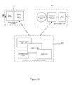
- FIG. 10 is a block diagram of an embodiment of a system according to the invention.
- FIG. 11 is one example of a screen display of one embodiment of the system of FIG. 10 ;
- FIG. 12 is another example of a screen display of one embodiment of the system of FIG. 10 ;
- FIG. 13 a , 13 b , 13 c , and 13 d are graphs illustrating the results of monitoring metabolic rates in accordance with an embodiment of the invention.
- the invention enables the measurement of one or more properties of living cells that are disposed in, for example, a well of a multiwell microplate.
- Embodiments of the invention include a sensor, preferably a submersible sensor that enables fast sensor stabilization, thereby increasing measurement throughput.
- the invention provides a cartridge structure which permits repeated use of the apparatus for disparate cellular assays without requiring intermediate cleaning, and while eliminating the possibility of cross contamination between tests. Still further, the invention provides software for designing and implementing multi-well cellular assays run in parallel, and for receiving and analyzing the generated data that is intuitive and easy to use, permits multiple scientists to design and execute multiwell parallel assays during the same time period, and preferably is based on a spreadsheet program of the type well understood by scientists and easily integrated with sophisticated LIMS systems.
- the apparatus 100 comprises a compound storage and delivery apparatus in a housing 105 (shown in dashed lines) that includes a cartridge 110 defining a plurality of apertures for receiving sensor structures and a plurality of fluid ports (shown in detail in FIGS. 2 a and 2 b ) compliantly mounted, and a stage or base 130 adapted to receive a multiwell plate 120 , e.g., a cell culture plate.
- the cartridge 110 is disposed above, and adapted to mate with, the multiwell plate 120 .
- the cartridge 110 optionally is held by a cartridge holder 122 adapted to receive the cartridge 110 .
- the compound storage and delivery apparatus 105 also includes a mounting block 140 , which can reciprocate as shown by the double headed arrow, preferably powered by a motor (not shown), including an elevator mechanism 145 .
- the elevator mechanism 145 is adapted to move the cartridge 110 relative to the stage 130 , or well plate 120 .
- the mounting block includes a gas multiplexer 150 attached to a pressure source, e.g., gas supply or gas reservoir 160 .
- the gas supply 160 is in fluid communication with the cartridge, and is used to impel the delivery of test fluid from a port in the cartridge to a well in the multiwell plate 120 as disclosed below.
- a plurality of sensor probes 170 are adapted for insertion into the plurality of apertures in the cartridge 110 , and may be used to gather data indicative of the state of cells disposed in wells in the multiwell plate 120 .
- the compound storage and delivery apparatus 105 is controlled by a controller 175 , that may be integrated with a computer 180 , that may control the elevator mechanism, the multiplexer, and the pressure source.
- the controller 175 may, thereby, permit delivery of a test fluid from a port to a corresponding well when an associated sensor is disposed in the well.
- FIGS. 2 a and 2 b illustrate the currently preferred form of the cartridge 110 and microplate 120 , and how they relate to each other
- the cartridge is a generally planar element comprising a frame 200 made, e.g., from molded plastics.
- Planar surface 205 defines a plurality of regions 210 that correspond to, i.e., register with, a number of the respective openings of a plurality of wells 220 defined in the multiwell plate 120 .
- the planar element defines first, second, third, and fourth ports 230 , which serve as test compound reservoirs, and a central aperture 215 to a sleeve 240 .
- Each of the ports is adapted to hold and to release on demand a test fluid to the respective well 220 beneath it.
- the ports 230 are sized and positioned so that groups of four ports may be positioned over the wells 220 , and test fluid from any one of the four ports may be delivered to a respective well 220 . In other embodiments the number of ports in each region may be less than four or greater than four.
- the ports 230 and sleeves 240 may be compliantly mounted relative to the microplate 120 so as to permit it to nest within the microplate by accommodating lateral movement.
- the construction of the microplate to include compliant regions permits its manufacture to looser tolerances, and permits the cartridge to be used with slightly differently dimensioned microplates. Compliance can be achieved, for example, by using an elastomeric polymer to form planar element 205 , so as to permit relative movement between frame 200 and the sleeves and ports in each region.
- Each of the ports 230 may have a cylindrical, conic or cubic shape, open through planar element 200 at the top, and closed at the bottom except for a small hole, i.e., a capillary aperture, typically centered within the bottom surface.
- the capillary aperture is adapted to retain test fluid in the port, e.g., by surface tension, absent an external force, such as a positive pressure differential force, a negative pressure differential force, or possibly a centrifugal force.
- Each port may be fabricated from a polymer material that is impervious to test compounds, or from any other solid material. When configured for use with a multiwell microplate 120 , the liquid volume contained by each port may range from 500 ⁇ l to as little as 2 ⁇ l, although volumes outside this range are contemplated.
- multiwell plate 120 has 24 wells.
- the number of wells 220 in a plate may vary from 1 to several thousand.
- a single well of nearly any size may be fabricated, or multiple wells may be fabricated, or multiple wells may be fabricated in a one- or two-dimensional arrangement.
- a two-dimensional pattern of wells corresponding to the pattern and dimensions of a microplate, as described by the Society for Biomolecular Screening standards for microplates (“SBS-1 Footprints” and “SBS-4 Well Positions,” both full proposed standards updated May 20, 2003), and containing a total of 12, 24, 96, 384, 1536, or any other number of individual wells may be fabricated.
- a submersible sensor sleeve or barrier 240 in each region of the cartridge 110 , disposed between and associated with one or more ports 230 , is a submersible sensor sleeve or barrier 240 , adapted to be disposed in the corresponding well 220 .
- Sensor sleeve 240 may have one or more sensors 250 disposed on a lower surface 255 thereof for insertion into media in a well 220 .
- a sensor for this purpose is a fluorescent indicator, such as an oxygen-quenched fluorophore, embedded in an oxygen permeable substance, such as silicone rubber.
- the fluorophore has fluorescent properties dependant on the presence and/or concentration of a constituent in the well 220 .
- Sensor sleeve 240 may define an aperture and an internal volume adapted to receive a sensor. Examples of the types of sensors that may be used are described below with reference to FIG. 3 .
- the cartridge 110 may be attached to the sensor sleeve, or may be located proximal to the sleeve without attachment, to allow independent movement.
- the cartridge 110 may include an array of compound storage and delivery ports assembled into a single unit and associated with a similar array of sensor sleeves.
- the apparatus may also feature a removable cover 260 for the cartridge 110 or for multiwell plate 120 .
- the configuration of cartridge 110 as a cover for multiwell plate 120 may help prevent evaporation or contamination of a sample or media disposed in wells 220 .
- the cover 260 may also be configured to fit over the cartridge 110 thereby to reduce possible contamination or evaporation of fluids disposed in the ports 230 of the cartridge 110 .
- FIG. 3 shows a fixed (preferably not part of the cartridge and reusable) sensor probe structure 170 configured to fit within the sensor sleeve 240 .
- the sensor probe structure 170 includes a rigid outer tube 315 made from, e.g., stainless steel.
- Optical fibers 300 are disposed within the tube 315 , and are configured to stimulate one or more fluorophores 250 disposed on a light transmissive outside lower wall portion 325 of sensor sleeve 240 and to receive fluorescent emissions from the fluorophore through the wall portion.
- the probe When the probe is in its down position, it preferably forms a reduced media test volume in each well, as shown, for example, in FIG.
- probe sleeve 240 may comprise an annular wall portion extending below portion 325 which defines the reduced test volume.
- the sensor probe structure and fluorophore may be configured to read optical density, luminescence, phosphorescence, or, preferably, fluorescence.
- the sensor probe structure 170 may be a self contained sensor which gathers data from a well through a signal transmissive bottom wall of the sleeve 240 , or directly through an open bottom on the sleeve, preferably sealed to the probe.
- sensors can be utilized depending on the analysis to be performed and its selected configuration, including oxygen sensors, such as oxygen-quenched fluorescent sensors, pH sensors, including fluorescent sensors, ISFET and impedance sensors using electrodes coupled through bottom wall 325 of sleeve 240 , CO 2 sensors, including bicarbonate buffer coupled and ammonium dye coupled fluorescent sensors as well as other CO 2 sensors; various ion and small molecule sensors; large molecule sensors including surface plasmon resonance sensors and sensors exploiting the principle of Wood's anomaly; acoustic sensors; and microwave sensors.
- oxygen sensors such as oxygen-quenched fluorescent sensors, pH sensors, including fluorescent sensors, ISFET and impedance sensors using electrodes coupled through bottom wall 325 of sleeve 240
- CO 2 sensors including bicarbonate buffer coupled and ammonium dye coupled fluorescent sensors as well as other CO 2 sensors
- various ion and small molecule sensors large molecule sensors including surface plasmon resonance sensors and sensors exploiting the principle of Wood's anomaly
- acoustic sensors and microwave sensors.
- Preferred sensors are fluorophores. Many fluorescent sensing compounds and preparations are described in the art and many are available commercially from, for example, Molecular Probes Inc and Frontier Scientific, Inc.
- the currently preferred oxygen sensor is a fluorophore with the signal inversely proportional to oxygen concentration such as a porphyrin or rhodamine compounds immobilized as a particle or homogenously distributed in an oxygen permeable polymer, e.g., silicone rubber.
- the currently preferred compound is porphyrin.
- the currently preferred pH sensor is a fluorescent indicator dye, fluorescein, whose signal decreases upon protonation of the dye, and which is either entrapped in a particle that is suspended in a carrier polymer, or covalently attached to a hydrophilic polymer.
- Useful fluorescent CO2 indicator sensor typically are based on a pH sensitive transducer, with the fluorescence being indirectly modulated by the production of carbonic acid due to reaction of carbon dioxide with water. See, e.g. O. S. Wolfbeis, Anal. Chem. 2002, 74, 2663-2678.
- a fluorophore that detects glucose also can be used, such as one based on a non-enzymatic transduction using a boronic probe that complexes with glucose, resulting in a charge transfer that modulates the fluorescence of the probe, or an enzymatic glucose transducer that couples a glucose oxidase to a fluorescent oxygen sensor, with the binding and oxidation of glucose resulting in a quantitative modulation of the oxygen sensor.
- a lactate sensor can be based on an enzymatic sensor configuration, with lactate oxidase coupled to a fluorescent oxygen sensor, and with the binding and oxidation of lactate resulting in a quantitative modulation of the oxygen sensor.
- An ammonia or ammonium ion sensor can be configured with immobilization of a protonated pH indicator in a hydrophobic, gas permeable polymer, with the fluorescence output quantitatively modulated by reaction with transient ammonia.
- a urea sensor can be based on an enzymatic sensor configuration, with urease coupled to a fluorescent ammonia transducer, and with the binding and reduction of urea to ammonia, resulting in modulation of the ammonia sensor fluorescence.
- the fixed sensor probe 170 is attached to and extends orthogonally from the pneumatic multiplexer 150 .
- Other sensor configurations will be apparent to those skilled in the art.
- probes may be disposed on a wall within the well under examination, or on a bottom, translucent surface of a well.
- Air channels 310 are defined within the pneumatic multiplexer 150 and are positioned to feed drug wells or ports 230 when the elongated neck of the fixed sensor probe 315 is fitted within with the sleeve 240 .
- the pneumatic multiplexer 150 serves to deliver compressed gas to a plurality of ports (see FIG. 6 a ) from a single source that may be controlled by an electrical or mechanical gas regulator or valving.
- Other types of pneumatic, mechanical or hydraulic pressure actuators may be used.
- the actuator may be a piston within a sleeve, as described in U.S. Pat. No. 4,498,510 to Minshew et al., or a controlled gas supply as described in U.S. Pat. No. 4,461,328 to Kenney, or any other suitable means for ejecting liquid test compound from the bottom of the port 230 using an extrinsic force.
- pneumatic multiplexer may be preferable for the sake of simplification and reduction of the number of components that supply compressed gas to the apparatus.
- the currently preferred pneumatic multiplexer 150 is discussed in greater detail below.
- a region 210 of the cartridge 110 includes first and second ports 230 .
- a test compound such as a drug, drug candidate, toxin, etc. is added to the ports 230 of cartridge 110 before beginning an analysis using a pipettor or other means.
- the compound typically will be an aqueous solution of a known concentration. In preferred embodiments, it is held within each port despite the presence of a small outlet at its bottom by surface tension. The dimensions of the port inhibit leakage from the bottom and from the top end (forming a meniscus that prevents leakage if the apparatus is turned on its side or upside down).
- the test compound may be released by, e.g., the application of pressurized air.
- a frangible membrane or a fragile material, such as wax may be attached to cover the hole in the bottom of the port 230 , such that an extrinsic force can breach the membrane to eject the liquid at a desired time.
- the submersible sleeve 240 is disposed between first and second ports 230 .
- Sensors 250 e.g., fluorophores, are disposed on surface 325 at the lower end of the sleeve.
- the submersible sleeve 240 is configured to receive the sensor probe 170 .
- An array of integrated sensor sleeves and compound storage and delivery ports may be fabricated as a single assembly using a low cost fabrication process such as injection molding so that the cartridge may be disposed of after use.
- the well 220 is formed of, e.g., molded plastic, such as polystyrene or polypropylene.
- cell media 500 and live cells 510 are disposed in the well 220 .
- Cells 510 may or may not adhere to a bottom surface 520 of the well, and the bottom surface may be treated or coated to encourage adherence. Alternatively, cells may be suspended within the media.
- FIGS. 6 a and 6 b in use, when the parts are assembled, they allow simultaneous sensing of constituents in the cell media in plural wells simultaneously, and delivery of test compound from the ports.
- the fixed probe structure and drug loaded cartridge are assembled such that the outer tubing holding the fiber optic bundle is disposed within the sleeve of the cartridge, and the assembly is reciprocated from an up position, where the probe tip and sensors are disposed in the cell medium, to a lower, data gathering position, preferably one that reduces the volume of media about the cells so as to improve the ability of the sensor to detect changes in the concentration of an analyte in the media about the cells (see US 2005/0054028).
- the sensors 250 disposed on the lower surface 325 of the sensor sleeve 240 remains submerged during mixing, equilibrating, and measurement steps.
- One or more constituents within the media secreted from or absorbed by the cells may by analyzed.
- a fluid such as a drug sample
- a fluid is delivered from one of the ports 230 to the cell medium, in this embodiment impelled by air pressure communicated through air channels 310 .
- the drug may be released through a small hole disposed at a bottom portion of the port 230 .
- the sensor sleeve 240 may be raised and lowered one or more times while remaining submerged in the media to mix the fluid with the media.
- the sensors 250 may remain disposed within the media during the dispensing and mixing steps, thereby reducing stabilization periods.
- a bottom portion of the well 220 may include a seating surface for the sensor sleeve 240 , e.g., an internal step defining a step plane above a bottom plane of the well 220 , the step plane and bottom plane being parallel planes.
- the height of the step plane may generally be less than about 1 mm above the bottom plane and typically less than about 50 ⁇ m to 200 ⁇ m above the bottom plane.
- a flat bottomed well or other well configuration may be used, and the fluorophore probes may disposed on surface 255 within a recess formed by a wall extending slightly beyond the surface as disclosed above.
- a small volume subchamber is formed about cells when the assembly is disposed in a down position. Relatively small changes in the concentration of the constituent than can be detected by the fluorophore probes, as the measurement is taken within the confines of a much smaller volume of medium. This subvolume is maintained for a short time period to make a measurement, and the assembly is moved upwardly, permitting the cells to be exposed to the full well volume of its medium.
- test fluid from the port may be delivered to the media when the sensor sleeve in the partially raised, but still submerged position.
- the constituent in the medium may be analyzed to determine any changes, and the measurements can be repeated with or without intermediate addition of test compounds.
- Any number of constituents of the media may be analyzed, including dissolved gasses, ions, proteins, metabolic substrates, salts, and minerals. These constituents may be consumed by the cells (such as O 2 ), or may be produced by the cells either as a byproduct (such as CO 2 and NH 3 ) or as a secreted factor (such as insulin, cytokines, chemokines, hormones, or antibodies). Ions such as H + , Na + , K + , and Ca ++ secreted or extracted by cells in various cellular metabolism processes may also be analyzed.
- Substrates either consumed or produced by cells such as glucose, fatty acid, amino acids, glutamine, glycogen, and pyruvate may be analyzed.
- Specialized media may be used to improve the sensitivity of the measurement. For example, a change in pH resulting from extracellular acidification can be increased by using a media with reduced buffer capacity, such as bicarbonate-free media.
- the method may be used to measure any number of attributes of cells and cellular function. For example, cell viability and metabolic rate may be determined from measurements of oxygen consumption rate, extracellular acidification rate, or other metabolic analyte fluxes. By comparison of one or more analyte flux rates to a known rate per cell, cell number may be determined and therefore growth rates can be monitored.
- an environment altering constituent such as a chemical, dissolved gas, or nutrient may be applied to either the full volume of the well or alternatively to only the reduced volume of the well.
- the volume of media surrounding the cells is first reduced, the constituents of the media are measured, and the volume is restored to its original value.
- the volume is then again reduced and the environment immediately surrounding the cells within only the reduced volume is then altered, by the addition of a constituent from one of the four corresponding ports. This may be accomplished by discharging the constituent from a port proximate the sensors or the bottom of the sleeve, for example.
- One or more measurements in the reduced volume are made in the presence of the constituent.
- the media within the reduced volume may be exchanged one or more times to flush out the constituent before exposing cells once again to the full original volume.
- This approach may provide a benefit of reducing the volume of compound required. It may also provide the possibility of studying isolated effects without contaminating the entire volume, thereby, in effect, simulating a flow system in microplate format.
- a plurality of sensors are inserted and disposed simultaneously or sequentially in a corresponding plurality of wells in the multiwell plate, and constituents related to respective cell cultures in respective wells are analyzed.
- the respective constituents may include the same constituent.
- Respective test fluids may be delivered to the respective wells while the respective sensors remain in equilibrium with, preferably remain disposed within the media in respective wells. It is possible to maintain equilibrium with many sensors, particularly fluorophore sensors, while the sensor body is removed from the media for a short time, e.g., if the probe remains wetted, permitting maintenance of equilibrium while adding test fluid.
- the respective test fluids may be the same test fluid.
- the respective constituents related to respective cells within media in respective wells may be analyzed to determine any respective changes therein. These delivery and analysis steps may be repeated. In another embodiment, the delivery step is repeated with a different test fluid.
- the delivery and analysis may be repeated after a time period. More particularly, sequential measurements of a single group of cells may be made at predetermined time intervals to analyze the effect of a compound addition temporally, for example to examine the effect of exposure to a drug, chemical, or toxin.
- the volume of media surrounding the cells is first reduced, the constituents of the media are measured, and the volume is restored to its original value.
- the environment surrounding the cells is then altered, such as by adding one or more predetermined concentrations of a ligand that activates a transmembrane receptor, changing the dissolved oxygen level, or adding a nutrient.
- One or more additional measurement cycles then are performed using the temporarily reduced volume method, to analyze the effect of the altered extracellular environment.
- Equilibration between the sensor and the media may be maintained during the delivery step. Thermal equilibrium may be substantially maintained between the test fluid and media during the delivery.
- the currently preferred form of the multiplexer 150 is shown. It comprises a laminated assembly of multiple layers 700 of planar polymeric sheet material containing machined channels 710 for gas flow, sandwiched between a cover sheet 800 and cartridge facing gasket 810 .
- One such arrangement uses four layers, e.g., four machined blocks placed in different orientations, to create a pneumatic multiplexer enabling the dispensing of fluid from any one of four ports disposed in each region of the cartridge.
- the multiplexer enables the delivery of gas from a single gas inlet to multiple outlets.
- the multiplexer is disposed between a pressure source and the cartridge, with the multiplexer adapted to be in fluidic communication with a plurality of ports formed in the cartridge.
- the multiplexer may be selectively in fluidic communication with an exclusive set of ports formed in the cartridge.
- the sensor probe 170 is depicted submerged in the liquid media contained within a microplate well 220 .
- the drug delivery apparatus is shown activated using gas pressure from the pressurized gas supply 160 to deliver a drug from the port 230 to the media.
- FIG. 10 schematically illustrate one embodiment of the invention realized as an instrument and software for analyzing cells undergoing various experimental processes using any of the techniques described above.
- a key element of the invention is a data file shared by instrument operating system running on the embedded instrument computer, and desktop software running on a user's personal desktop computer.
- Desktop software 900 contains a user interface that allows a user to enter experiment design information into data file 901 .
- Experiment design information may include the type of cells, number of cells, type of drug, and concentration of drug contained in each microplate well, the required measurement time, media mixing time, the analyte to be assayed, or other data that define attributes of the experiment to be run by the instrument.
- Instrument operating system software 902 both receives experiment design information from, and stores experiment results to, data file 901 .
- Operating system software 902 also contains a user interface for viewing and modification of experiment design information and for viewing of experiment results.
- the instrument operating system software provides actuation and control of motors, heaters and other devices based on the settings provided in the data file. During each measurement cycle, measured data may be displayed on the user interface and concurrently added to the data file. At the end of a complete experiment, the data file, containing experiment definition data, and measured sensor data, may be stored and transmitted to the user's desktop computer for analysis.
- the user may a third-party analysis software package that draws data from the data file. Examples of suitable third-party analysis software include MICROSOFT EXCEL (Microsoft Corp), JMP (SAS Corp), and SIGMA PLOT (Systat Corp).
- data file 901 is in the form of a spreadsheet.
- data file 901 contains experiment design information and experiment results as separate worksheets within one spreadsheet file.
- data file 901 contains experiment design information, experiment results, and a data analysis tool, each as separate worksheets within one spreadsheet file.
- Data file 901 may be formatted as a workbook file for use within a spreadsheet software application such as Microsoft Excel.
- the experiment definition information and instrument-generated data may be duplicated and additionally saved in machine-readable binary format on a separate hidden, password-protected area within the file. This capability preserves the integrity of the original data while changes are made by the user for analysis.
- proprietary binary data packets may be passed directly to other software configurations encoded with the custom graphical user interface and display areas.
- These alternative software environments might include traditional Windows or Macintosh applications, stand-alone executable files with the embedded binary data, web browser applications configured to load and display the data, or other viewing environments.
- the system includes a graphical user-interface for accepting instructions relating to the experiments and presenting results therefrom.
- graphical user interface 1100 includes multiple display areas (e.g., cells in a spreadsheet program in which each cell can be individually addressed using row and column designators).
- Each display area may, for example, represent one of the wells in a microplate, thus providing data specific to the cells in each well, and in some cases also include various parameters of the experiment and/or results of the experiment based, for example, on signals received from the probes 170 .
- inputs, parameters and/or results of the experiments from the different wellplates may be presented using multiple spreadsheets, such as tabbed worksheets in EXCEL.
- the system also includes an analysis module for producing graphical representations and/or statistical analysis of the data acquired by the probes and presented within the user interface.
- an analysis module for producing graphical representations and/or statistical analysis of the data acquired by the probes and presented within the user interface.
- the results of the experiments, as well as the outputs can also be represented within user-interface 1200 .
- individual icons, images (e.g., GIF files, JPEG files) or colorations of display areas can be used to represent the current (or most recently measured) pH of an assay, and a graphical display can be used to represent the same measurement (or others) over time.
- the results of an experiment may be presented to the user in the form of a chart having data from each of two sensors shown on each of two axes. For example, oxygen consumption rate may be displayed on the ordinate while extracellular acidification rate is shown on the abscissa.
- chart 1210 displays data from each well of a multi-well experiment as a dot with an associated label and error bar set.
- Probes incorporating four drug wells or ports were fabricated from polystyrene material using injection molding. Twenty four probes were then bound together using an elastomeric sheet to form a single 4′′ ⁇ 6′′ cartridge unit that is suitable for use as a disposable measurement and drug delivery assembly.
- a pneumatic multiplexer was fabricated by machining gas channels in four polystyrene blocks, then bonding these layers together and applying a cover. The multiplexer was then clamped to the cartridge.
- the cartridge and multiplexer assembly was then placed above a 24 well microplate reservoir (Innovative Microplate).
- An electrical drive circuit was configured to actuate each solenoid for 250 ⁇ sec in order to deliver the fluid from the drug wells.
- the accumulator pressure was then reduced to 5 psi, and was recharged between sequential actuation of solenoids one through four.
- the electronic circuit was then adjusted to increase the actuation time to 275 ⁇ sec. In this case, complete injection of water was noted for each of the 96 drug wells.
- Example 2 A test was performed using the components and method described in Example 1, except that a mixture of saline solution and Tartrazine was substituted for water in the drug wells. The fluid was injected into a microplate reservoir, and then the absorbance of the contents of each well in the reservoir was measured using a Molecular Devices Versamax microplate reader. Absorbance readings indirectly measure dye injection volume and demonstrate injection performance.
- Example 2 A test was performed using the components described in Example 1, and a 24 well microplate containing 30 ⁇ 10 3 HEP-G2 human hepatocellular liver carcinoma cells per well. Three initial “baseline” measurements of cellular oxygen consumption rate (OCR) and extracellular acidification rate (ECAR) were performed at eleven minute intervals using a 4 minute measurement period.
- OCR oxygen consumption rate
- ECAR extracellular acidification rate
- FCCP FCCP
- the final concentration of FCCP in the well is shown above each graph for each injection, A-D.
- the cumulative addition of FCCP stock concentrations from each injector port followed by measurement of OCR and ECAR enabled a four-point dose curve to be generated in each well.
- FCCP induces mitochondrial uncoupling and causes cells to increase their metabolic rate and therefore OCR and ECAR.
- each series produced either no or an increasingly higher OCR and ECAR response until toxicity was reached as demonstrated in the high dose series ( FIG. 13 d ).
- OCR and ECAR simultaneously the total metabolic rate and capacity of the HepG2 cells could be determined.
Abstract
Description
| TABLE E1 |
| Absorbance measurements for injection of Tartrazine dye into water using pneumatic |
| multiplexer |
| |
|
| Column | Column |
| Row | A | B | C | D | E | F | Row | A | B | C | | E | F | |
| 50 |
50 |
| 1 | 0.223 | 0.222 | 0.269 | 0.244 | 0.223 | 0.219 | 1 | 0.222 | 0.222 | 0.234 | 0.220 | 0.216 | 0.223 |
| 2 | 0.225 | 0.232 | 0.226 | 0.228 | 0.221 | 0.229 | 2 | 0.221 | 0.225 | 0.223 | 0.226 | 0.226 | 0.223 |
| 3 | 0.225 | 0.216 | 0.219 | 0.219 | 0.221 | 0.222 | 3 | 0.211 | 0.216 | 0.220 | 0.215 | 0.237 | 0.230 |
| 4 | 0.219 | 0.219 | 0.221 | 0.248 | 0.223 | 0.222 | 4 | 0.222 | 0.222 | 0.225 | 0.223 | 0.221 | 0.223 |
| 75 uL Tartrazine | 75 |
| 1 | 0.305 | 0.300 | 0.282 | 0.289 | 0.326 | 0.296 | 1 | 0.326 | 0.300 | 0.320 | 0.317 | 0.319 | 0.314 |
| 2 | 0.292 | 0.282 | 0.292 | 0.281 | 0.285 | 0.292 | 2 | 0.311 | 0.309 | 0.310 | 0.310 | 0.314 | 0.308 |
| 3 | 0.277 | 0.279 | 0.274 | 0.284 | 0.282 | 0.281 | 3 | 0.308 | 0.301 | 0.310 | 0.307 | 0.314 | 0.318 |
| 4 | 0.282 | 0.279 | 0.329 | 0.282 | 0.284 | 0.293 | 4 | 0.308 | 0.308 | 0.313 | 0.319 | 0.317 | 0.307 |
| 100 |
100 |
| 1 | 0.349 | 0.345 | 0.343 | 0.343 | 0.339 | 0.335 | 1 | 0.358 | 0.385 | 0.357 | 0.346 | 0.350 | 0.356 |
| 2 | 0.340 | 0.332 | 0.338 | 0.340 | 0.343 | 0.342 | 2 | 0.365 | 0.343 | 0.359 | 0.348 | 0.353 | 0.357 |
| 3 | 0.342 | 0.342 | 0.336 | 0.335 | 0.339 | 0.337 | 3 | 0.349 | 0.346 | 0.351 | 0.349 | 0.357 | 0.353 |
| 4 | 0.345 | 0.295 | 0.345 | 0.344 | 0.355 | 0.348 | 4 | 0.358 | 0.349 | 0.357 | 0.355 | 0.353 | 0.352 |
| Tartrazine | Mean | Tartrazine | Mean | ||||
| qty | absorb | Std Dev | c.v. | qty | absorb | Std Dev | c.v. |
| 50 ul | 0.23 | 0.0118 | 5.2% | 50 ul | 0.22 | 0.0056 | 2.5% |
| 75 ul | 0.29 | 0.0139 | 4.8% | 75 ul | 0.31 | 0.0061 | 2.0% |
| 100 ul | 0.34 | 0.0108 | 3.2% | 100 ul | 0.35 | 0.0083 | 2.3% |
Claims (15)
Priority Applications (2)
| Application Number | Priority Date | Filing Date | Title |
|---|---|---|---|
| US14/150,423 US9170255B2 (en) | 2006-07-13 | 2014-01-08 | Cell analysis apparatus and method |
| US14/830,459 US10359418B2 (en) | 2006-07-13 | 2015-08-19 | Cell analysis apparatus and method |
Applications Claiming Priority (2)
| Application Number | Priority Date | Filing Date | Title |
|---|---|---|---|
| US11/486,440 US8658349B2 (en) | 2006-07-13 | 2006-07-13 | Cell analysis apparatus and method |
| US14/150,423 US9170255B2 (en) | 2006-07-13 | 2014-01-08 | Cell analysis apparatus and method |
Related Parent Applications (1)
| Application Number | Title | Priority Date | Filing Date |
|---|---|---|---|
| US11/486,440 Continuation US8658349B2 (en) | 2003-10-17 | 2006-07-13 | Cell analysis apparatus and method |
Related Child Applications (1)
| Application Number | Title | Priority Date | Filing Date |
|---|---|---|---|
| US14/830,459 Continuation US10359418B2 (en) | 2006-07-13 | 2015-08-19 | Cell analysis apparatus and method |
Publications (2)
| Publication Number | Publication Date |
|---|---|
| US20140186876A1 US20140186876A1 (en) | 2014-07-03 |
| US9170255B2 true US9170255B2 (en) | 2015-10-27 |
Family
ID=38776413
Family Applications (3)
| Application Number | Title | Priority Date | Filing Date |
|---|---|---|---|
| US11/486,440 Active 2028-10-09 US8658349B2 (en) | 2003-10-17 | 2006-07-13 | Cell analysis apparatus and method |
| US14/150,423 Active US9170255B2 (en) | 2006-07-13 | 2014-01-08 | Cell analysis apparatus and method |
| US14/830,459 Active 2027-02-06 US10359418B2 (en) | 2006-07-13 | 2015-08-19 | Cell analysis apparatus and method |
Family Applications Before (1)
| Application Number | Title | Priority Date | Filing Date |
|---|---|---|---|
| US11/486,440 Active 2028-10-09 US8658349B2 (en) | 2003-10-17 | 2006-07-13 | Cell analysis apparatus and method |
Family Applications After (1)
| Application Number | Title | Priority Date | Filing Date |
|---|---|---|---|
| US14/830,459 Active 2027-02-06 US10359418B2 (en) | 2006-07-13 | 2015-08-19 | Cell analysis apparatus and method |
Country Status (4)
| Country | Link |
|---|---|
| US (3) | US8658349B2 (en) |
| EP (2) | EP3382374A3 (en) |
| CN (1) | CN101506643B (en) |
| WO (1) | WO2008008149A2 (en) |
Cited By (6)
| Publication number | Priority date | Publication date | Assignee | Title |
|---|---|---|---|---|
| WO2017153992A1 (en) | 2016-03-08 | 2017-09-14 | Yissum Research Development Company Of The Hebrew University Of Jerusalem Ltd | Method and system for continuous biosensing |
| WO2021079892A1 (en) | 2019-10-21 | 2021-04-29 | Phcホールディングス株式会社 | Sensor unit and cell culture analysis device having same |
| WO2021193029A1 (en) | 2020-03-25 | 2021-09-30 | Phcホールディングス株式会社 | Cell culture analyzer and cell culture analysis method using same, additive supply unit and cell culture analyzer provided therewith, and sensor unit and cell culture analyzer provided therewith |
| US11175281B2 (en) | 2018-08-21 | 2021-11-16 | International Business Machines Corporation | Well plate cover with embedded electronic sensors for monitoring cell metabolism |
| WO2022097584A1 (en) | 2020-11-06 | 2022-05-12 | Phcホールディングス株式会社 | Sensor unit and cell culture analysis device having same |
| WO2023150717A1 (en) | 2022-02-03 | 2023-08-10 | Agilent Technologies, Inc. | Apparatuses and methods for analyzing live cells |
Families Citing this family (53)
| Publication number | Priority date | Publication date | Assignee | Title |
|---|---|---|---|---|
| US7276351B2 (en) | 2003-09-10 | 2007-10-02 | Seahorse Bioscience | Method and device for measuring multiple physiological properties of cells |
| US20070087401A1 (en) * | 2003-10-17 | 2007-04-19 | Andy Neilson | Analysis of metabolic activity in cells using extracellular flux rate measurements |
| US8658349B2 (en) | 2006-07-13 | 2014-02-25 | Seahorse Bioscience | Cell analysis apparatus and method |
| US8202702B2 (en) * | 2008-10-14 | 2012-06-19 | Seahorse Bioscience | Method and device for measuring extracellular acidification and oxygen consumption rate with higher precision |
| IT1392344B1 (en) * | 2008-12-17 | 2012-02-28 | Univ Padova | MECHANICAL MIXING DEVICE FOR CULTIVATION PLATES |
| WO2011004854A1 (en) | 2009-07-08 | 2011-01-13 | 株式会社ニコン | Cell picking-assisting device, display device and culture container |
| US8859264B2 (en) * | 2009-07-29 | 2014-10-14 | Gsi Helmholtzzentrum Fuer Schwerionenforschung Gmbh | Phantom for the experimental in-vitro validation of radiation procedures under the influence of motion, taking into account the biological effective dose |
| DE102009043570A1 (en) * | 2009-09-30 | 2011-03-31 | Manz Automation Tübingen GmbH | Module for a laboratory robot and laboratory robot |
| EP2591434A4 (en) * | 2010-07-08 | 2016-07-13 | Life Technologies Corp | Systems and methods for assigning attributes to a plurality of samples |
| CN103080294B (en) * | 2010-09-08 | 2017-02-15 | 株式会社岛津制作所 | Cell culture container and cell culture method using container |
| CN102391946B (en) * | 2011-11-11 | 2013-01-23 | 武汉优视科技有限公司 | Plunger sleeve and automatic cell processor therewith |
| GB2497750B (en) | 2011-12-19 | 2015-02-25 | Lumophore Ltd | Analysis of colorimetric or fluorometric test assays |
| JP5778354B2 (en) * | 2012-10-25 | 2015-09-16 | 浜松ホトニクス株式会社 | Cell observation device, electrical stimulation device, and cell observation method |
| EP2913391B1 (en) * | 2012-10-25 | 2020-07-01 | Hamamatsu Photonics K.K. | Cell observation device, and cell observation method |
| US9494577B2 (en) | 2012-11-13 | 2016-11-15 | Seahorse Biosciences | Apparatus and methods for three-dimensional tissue measurements based on controlled media flow |
| US9790465B2 (en) | 2013-04-30 | 2017-10-17 | Corning Incorporated | Spheroid cell culture well article and methods thereof |
| JP6864614B2 (en) * | 2014-03-20 | 2021-04-28 | ベックマン コールター, インコーポレイテッド | Instrument interface including presentation unit display |
| WO2015157345A2 (en) | 2014-04-07 | 2015-10-15 | Wake Forest University Health Sciences | Bioenergetic profiling of circulating blood cells and systems, devices, and methods relating thereto |
| CN106999926A (en) * | 2014-06-02 | 2017-08-01 | 安捷伦科技有限公司 | Single-row microplate system and carrier for analyzing biological specimen |
| US10750985B2 (en) | 2014-08-11 | 2020-08-25 | The Regents Of The University Of California | Continuous analyte sensor |
| CN113481097A (en) | 2014-10-29 | 2021-10-08 | 康宁股份有限公司 | Perfusion bioreactor platform |
| WO2016160702A1 (en) | 2015-03-27 | 2016-10-06 | Seahorse Bioscience | Method and system for determining integrated metabolic baseline and potential of living cells |
| CN104833783B (en) * | 2015-05-11 | 2016-08-24 | 王超 | Medicine irritation cell degree verifying attachment |
| US9952122B2 (en) * | 2015-08-03 | 2018-04-24 | Palo Alto Research Center Incorporated | Polysensing bioelectronic test plate |
| US11369966B2 (en) | 2015-09-18 | 2022-06-28 | Arizona Board Of Regents On Behalf Of Arizona State University | Layered structure for improved sealing of microwell arrays |
| CN105403544A (en) * | 2015-10-13 | 2016-03-16 | 江苏绿扬电子仪器集团有限公司 | Microbial rapid analyzer |
| JP6684643B2 (en) * | 2016-04-28 | 2020-04-22 | 富士フイルム株式会社 | Biological sample evaluation system, biological sample evaluation method, and biological sample evaluation control program |
| WO2017214637A1 (en) * | 2016-06-10 | 2017-12-14 | Nirvana Sciences Inc. | Hydroporphyrin beads with narrow fluorescence emissions |
| US10883978B2 (en) | 2017-01-31 | 2021-01-05 | Agilent Technologies, Inc. | Method and device for calibration of biological flux |
| US11904026B2 (en) | 2017-02-03 | 2024-02-20 | Nirvana Sciences Inc. | Metallohydroporphyrins for photoacoustic imaging |
| US11338294B2 (en) | 2017-04-27 | 2022-05-24 | Polybiomics, Inc. | Orthogonal polybiosensing and imaging systems |
| WO2018213357A1 (en) | 2017-05-16 | 2018-11-22 | Cairn Biosciences, Inc. | Microfluidic-enabled multiwell cell culture devices and systems for precision culture, control and monitoring of living cells |
| WO2019014151A1 (en) * | 2017-07-10 | 2019-01-17 | Fenologica Biosciences, Inc. | Microplate covers for environmental control and automation |
| WO2019014610A1 (en) | 2017-07-14 | 2019-01-17 | Corning Incorporated | Cell culture vessel for 3d culture and methods of culturing 3d cells |
| EP3652290B1 (en) | 2017-07-14 | 2022-05-04 | Corning Incorporated | 3d cell culture vessels for manual or automatic media exchange |
| US11857970B2 (en) | 2017-07-14 | 2024-01-02 | Corning Incorporated | Cell culture vessel |
| CN111065729A (en) | 2017-07-14 | 2020-04-24 | 康宁股份有限公司 | Cell culture container |
| CN110237373B (en) * | 2018-03-08 | 2023-06-02 | 润生药业有限公司 | Device and method for producing drug carrier for combined use |
| JP7021802B2 (en) * | 2018-03-26 | 2022-02-17 | 国立研究開発法人物質・材料研究機構 | Electrochemical measurement system, electrochemical search method, and microplate |
| EP3649229B1 (en) | 2018-07-13 | 2021-08-25 | Corning Incorporated | Cell culture vessels with stabilizer devices |
| CN111065725B (en) | 2018-07-13 | 2024-03-29 | 康宁股份有限公司 | Fluidic device comprising microplates with interconnected walls |
| EP3649226B1 (en) | 2018-07-13 | 2022-04-06 | Corning Incorporated | Microcavity dishes with sidewall including liquid medium delivery surface |
| DE102018212489A1 (en) * | 2018-07-26 | 2020-01-30 | Implen GmbH | Device for light spectroscopic analysis |
| WO2020041765A1 (en) * | 2018-08-24 | 2020-02-27 | Polybiomics, Inc. | Integrated platforms for precise poly-sensing and imaging |
| KR20210054545A (en) * | 2018-08-31 | 2021-05-13 | 루시드 사이언티픽, 인코포레이티드 | Measurement of dynamic systems |
| DE102018131123A1 (en) * | 2018-12-06 | 2020-06-10 | Analytik Jena Ag | Lid for a microtiter plate |
| CN111690535A (en) * | 2020-06-24 | 2020-09-22 | 重庆智领医创科技成果转化服务有限责任公司 | Cell test observation box |
| EP4308906A1 (en) | 2021-03-14 | 2024-01-24 | Agilent Technologies, Inc. | System and method for analyzing biological material |
| DE102021204572A1 (en) * | 2021-05-06 | 2022-11-10 | Fraunhofer-Gesellschaft zur Förderung der angewandten Forschung eingetragener Verein | Dosing head and dosing system for receiving and dosing at least two media |
| DE102021117034B4 (en) * | 2021-07-01 | 2024-02-01 | Hexagonfab | SYSTEM WITH ROBOT ACTUATOR FOR IMMERSIBLE ELECTRICAL SENSOR FOR MEASURING PROPERTIES OF MOLECULES |
| US20230128888A1 (en) | 2021-10-12 | 2023-04-27 | Agilent Technologies, Inc. | Methods and systems for determining metabolic poise and capacity of living cells |
| WO2023081354A1 (en) | 2021-11-05 | 2023-05-11 | Agilent Technologies, Inc. | Apparatus and methods for generating and analyzing three-dimensional cellular materials |
| WO2023086372A1 (en) | 2021-11-09 | 2023-05-19 | Agilent Technologies, Inc. | Wellplate apparatus and method for filling same |
Citations (164)
| Publication number | Priority date | Publication date | Assignee | Title |
|---|---|---|---|---|
| US4016617A (en) | 1975-01-06 | 1977-04-12 | Francis Cardus | Electrically operable apparatus for the application of a treating product |
| US4065357A (en) | 1974-01-21 | 1977-12-27 | General Electric Company | Detection of catalase-containing bacteria |
| US4217868A (en) | 1977-03-16 | 1980-08-19 | Robert Bosch Gmbh | Ignition system for internal combustion engines, particularly of the automotive type |
| USD258145S (en) | 1978-03-23 | 1981-02-03 | Corning Glass Works | Stackable laboratory rack or the like |
| US4256832A (en) | 1978-12-12 | 1981-03-17 | Bioresearch Inc. | Carcinogen and mutagen screening method and apparatus |
| USD260428S (en) | 1979-03-15 | 1981-08-25 | Abbott Laboratories | Cuvette array or the like |
| USD268130S (en) | 1980-06-27 | 1983-03-01 | Easton Harlan J | Tray for veterinary supplies and equipment |
| US4405375A (en) | 1980-05-21 | 1983-09-20 | The B. F. Goodrich Company | Niyuminouzbituminous emulsion roof resaturant |
| US4461328A (en) | 1982-06-04 | 1984-07-24 | Drummond Scientific Company | Pipette device |
| EP0128438A2 (en) | 1983-06-13 | 1984-12-19 | Anton Stigler | Method of making a profile from dry powder material, and apparatus therefor |
| US4498510A (en) | 1982-08-20 | 1985-02-12 | Minshew Jr Edward C | Device for drawing, holding and dispensing liquid |
| USD280131S (en) | 1982-04-10 | 1985-08-13 | Olympus Optical Co., Ltd. | Case for a multiplicity of liquid sample containers for a multitest chemistry analyzer |
| USD280663S (en) | 1981-11-17 | 1985-09-17 | Internationale Octrooi Maatschappij "Octropa" B.V. | Tray for carrying out chemical and biological tests |
| US4599315A (en) | 1983-09-13 | 1986-07-08 | University Of California Regents | Microdroplet test apparatus |
| USD285118S (en) | 1983-01-19 | 1986-08-12 | Abbott Laboratories | Sample cup |
| US4711851A (en) | 1984-05-21 | 1987-12-08 | State University Of New York | Test apparatus for determining a metabolic characteristic of microorganisms |
| WO1988009808A2 (en) | 1987-06-05 | 1988-12-15 | Arthur D. Little, Inc. | Receptor-based biosensors |
| USD300245S (en) | 1986-08-14 | 1989-03-14 | Sherwood Medical Company | Microbiological test tray carrier |
| USD301167S (en) | 1987-02-27 | 1989-05-16 | Allelix Limited | Immunoassay tray |
| US4879097A (en) | 1982-11-20 | 1989-11-07 | Whitehead Thomas P | Dispensing device and recording apparatus |
| EP0363262A1 (en) | 1988-09-30 | 1990-04-11 | Terumo Kabushiki Kaisha | Cell-incubator |
| EP0402917A2 (en) | 1989-06-15 | 1990-12-19 | Biocircuits Corporation | Biosensors employing electrical, optical and mechanical signals |
| DE4016617A1 (en) | 1990-05-23 | 1991-11-28 | Rainer Dylla | Holder plate e.g. for blood samples - used with structured second plate to form gaps of variable vol. allowing use of small samples |
| USD324426S (en) | 1989-10-20 | 1992-03-03 | Pacific Biotech, Inc. | Reaction unit for use in analyzing biological fluids |
| US5104804A (en) | 1990-06-04 | 1992-04-14 | Molecular Devices Corporation | Cell assay device used in a microphysiometer |
| US5120421A (en) | 1990-08-31 | 1992-06-09 | The United States Of America As Represented By The United States Department Of Energy | Electrochemical sensor/detector system and method |
| USD328135S (en) | 1990-01-12 | 1992-07-21 | Pacific Biotech, Inc. | Reaction unit for use in analyzing biological fluids |
| USD332145S (en) | 1989-12-18 | 1992-12-29 | Kurashiki Boseki Kabushiki Kaisha | Centrifuge tube |
| US5200051A (en) | 1988-11-14 | 1993-04-06 | I-Stat Corporation | Wholly microfabricated biosensors and process for the manufacture and use thereof |
| WO1993008464A1 (en) | 1991-10-21 | 1993-04-29 | Holm Kennedy James W | Method and device for biochemical sensing |
| USD335348S (en) | 1991-07-23 | 1993-05-04 | Pb Diagnostic Systems Inc. | Assay module magazine |
| EP0545284A1 (en) | 1991-11-29 | 1993-06-09 | Canon Kabushiki Kaisha | Sample measuring device and sample measuring system |
| USD339869S (en) | 1991-01-14 | 1993-09-28 | Amgen, Inc. | Vial container |
| US5250419A (en) | 1988-12-16 | 1993-10-05 | L'oreal | Method for the direct measurement of at least one chemical parameter of skin using a biosensor |
| WO1993022678A2 (en) | 1992-04-23 | 1993-11-11 | Massachusetts Institute Of Technology | Optical and electrical methods and apparatus for molecule detection |
| US5264103A (en) | 1991-10-18 | 1993-11-23 | Matsushita Electric Industrial Co., Ltd. | Biosensor and a method for measuring a concentration of a substrate in a sample |
| US5278048A (en) | 1988-10-21 | 1994-01-11 | Molecular Devices Corporation | Methods for detecting the effect of cell affecting agents on living cells |
| US5286364A (en) | 1987-06-08 | 1994-02-15 | Rutgers University | Surface-modified electochemical biosensor |
| WO1994003583A1 (en) | 1992-07-29 | 1994-02-17 | Oncotherapeutics | An improved system for electronically monitoring and recording cell cultures |
| US5309085A (en) | 1990-08-21 | 1994-05-03 | Byung Ki Sohn | Measuring circuit with a biosensor utilizing ion sensitive field effect transistors |
| US5345213A (en) | 1992-10-26 | 1994-09-06 | The United States Of America, As Represented By The Secretary Of Commerce | Temperature-controlled, micromachined arrays for chemical sensor fabrication and operation |
| USD351661S (en) | 1993-02-16 | 1994-10-18 | Ultradent Products, Inc. | Combined organizer and tray for an endodontic dental kit |
| WO1994029708A1 (en) | 1993-06-03 | 1994-12-22 | Fraunhofer-Gesellschaft zur Förderung der angewandten Forschung e.V. | Electrochemical sensor |
| DE4217868C2 (en) | 1992-05-29 | 1995-01-26 | Univ Schiller Jena | Temperable multicuvette |
| USD359125S (en) | 1993-09-28 | 1995-06-06 | Becton Dickinson And Company | Microbial monitoring module |
| WO1995022406A1 (en) | 1994-02-19 | 1995-08-24 | Dr. Fritz Nerbe Nachfolger Jürgen Nerbe Ohg | Microtitration plate |
| US5459300A (en) | 1993-03-03 | 1995-10-17 | Kasman; David H. | Microplate heater for providing uniform heating regardless of the geometry of the microplates |
| US5468605A (en) | 1993-10-15 | 1995-11-21 | Molecular Devices Corp. | Transverse flow plungers for microphysiometers |
| US5496697A (en) | 1988-10-21 | 1996-03-05 | Molecular Devices Corporation | Methods and apparatus for detecting the effect of cell affecting agents on living cells |
| US5495850A (en) | 1994-04-21 | 1996-03-05 | Zuckerman; Ralph | Method for the measurement of oxygen concentration levels by the steady-state determination of fluorescence lifetime |
| US5512492A (en) | 1993-05-18 | 1996-04-30 | University Of Utah Research Foundation | Waveguide immunosensor with coating chemistry providing enhanced sensitivity |
| EP0722136A3 (en) | 1995-01-12 | 1996-08-28 | Schulz Joachim | |
| US5567598A (en) | 1991-04-18 | 1996-10-22 | Becton Dickinson And Company | Microbial monitoring device |
| US5622872A (en) | 1989-06-15 | 1997-04-22 | Biocircuits Corporation | Analyte detection through observed optical modulation of polymerized lipid layers |
| US5728541A (en) | 1996-07-12 | 1998-03-17 | Precision Therapeutics, Inc. | Method for preparing cell cultures from biologial specimens for chemotherapeutic and other assays |
| WO1998015645A1 (en) | 1996-10-08 | 1998-04-16 | Photonics Biosystems | Microbiological assessment method and device utilizing oxygen gradient sensing |
| US5766875A (en) | 1993-07-30 | 1998-06-16 | Molecular Devices Corporation | Metabolic monitoring of cells in a microplate reader |
| US5774214A (en) | 1996-12-12 | 1998-06-30 | Photometrics, Ltd. | Multi-mode imaging apparatus for radiation-emitting or absorbing samples |
| US5792426A (en) | 1994-10-11 | 1998-08-11 | Schweizerische Eidgenossenschaft Vertreten Durch Das Ac-Laboratorium Spiez Der Gruppe Rustung | Multi-well titerplate for instrumental analysis |
| US5830138A (en) | 1996-12-16 | 1998-11-03 | Trustees Of The University Of Pennsylvania | Intravascular catheter probe for clinical oxygen, pH and CO2 measurement |
| USD404497S (en) | 1995-09-27 | 1999-01-19 | Becton Dickinson And Company | Cell insert assembly |
| USD404831S (en) | 1997-04-21 | 1999-01-26 | Hitachi, Ltd. | Reagent bottle holder for a reagent disc |
| US5959297A (en) | 1996-10-09 | 1999-09-28 | Symyx Technologies | Mass spectrometers and methods for rapid screening of libraries of different materials |
| WO1999055827A1 (en) | 1998-04-27 | 1999-11-04 | Amersham Pharmacia Biotech Uk Ltd. | Microfabricated apparatus for cell based assays |
| WO1999060630A1 (en) | 1998-05-15 | 1999-11-25 | Glaxo Group Limited | Infrared thermography |
| US5998517A (en) | 1998-06-05 | 1999-12-07 | Becton, Dickinson And Company | Composition for the detection of microorganisms in a sample |
| US6030917A (en) | 1996-07-23 | 2000-02-29 | Symyx Technologies, Inc. | Combinatorial synthesis and analysis of organometallic compounds and catalysts |
| USD421498S (en) | 1997-05-23 | 2000-03-07 | Becton, Dickinson And Company | Microbiological test panel |
| USD423679S (en) | 1998-06-12 | 2000-04-25 | Tafco Medical Products LLC | Medical organizer |
| US6078698A (en) | 1999-09-20 | 2000-06-20 | Flir Systems, Inc. | System for reading data glyphs |
| US6080574A (en) | 1994-05-10 | 2000-06-27 | Becton, Dickinson And Company | Composite optical blood culture sensor |
| US6083761A (en) | 1996-12-02 | 2000-07-04 | Glaxo Wellcome Inc. | Method and apparatus for transferring and combining reagents |
| USD428657S (en) | 1998-09-10 | 2000-07-25 | Pharmacia & Upjohn Ab | Transport tray for ampoule parts |
| US6146967A (en) | 1997-08-20 | 2000-11-14 | Micron Technology, Inc. | Selective deposition of amorphous silicon film seeded in a chlorine gas and a hydride gas ambient when forming a stacked capacitor with HSG |
| WO2000032308A3 (en) | 1998-12-04 | 2000-11-23 | Symyx Technologies Inc | Continuous feed parallel reactor |
| WO2000071669A1 (en) | 1999-05-06 | 2000-11-30 | Micronas Gmbh | Device for performing analyses on cell-cultures |
| USD438631S1 (en) | 1999-12-21 | 2001-03-06 | Compucyte Corporation | Reagent treatment cartridge |
| USD438633S1 (en) | 1999-12-21 | 2001-03-06 | Compucyte Corporation | Reagent cartridge for treating a sample |
| USD438632S1 (en) | 1999-12-21 | 2001-03-06 | Compucyte Corporation | Multi-well reagent cartridge for treating a sample |
| WO2000036410A9 (en) | 1998-12-11 | 2001-05-25 | Symyx Technologies Inc | Sensor array-based system and method for rapid materials characterization |
| US6280586B1 (en) | 1995-04-04 | 2001-08-28 | Micronas Gmbh | Measuring device using biological cells or chemical biologically active substances contained in an analyte |
| US6306658B1 (en) | 1998-08-13 | 2001-10-23 | Symyx Technologies | Parallel reactor with internal sensing |
| US20010039045A1 (en) | 2000-03-28 | 2001-11-08 | Cook Incorporated | Perfusion incubator |
| US20010051353A1 (en) | 1996-07-12 | 2001-12-13 | Paul L. Kornblith | Method for using multicellular particulates to analyze malignant or hyperproliferative tissue |
| WO2002000336A2 (en) | 2000-06-28 | 2002-01-03 | Illumina, Inc. | Composite arrays utilizing microspheres with a hybridization chamber |
| WO2002002736A1 (en) | 2000-06-30 | 2002-01-10 | Precision System Science Co.,Ltd. | Reaction vessel, reaction device and temperature control method for reaction liquid |
| WO2002008385A1 (en) | 2000-07-22 | 2002-01-31 | Inbionet Corporation | Novel strain for decomposing tmah, and method of wastewater treatment using the same |
| WO2002011881A1 (en) | 2000-08-05 | 2002-02-14 | Millennium Pharmaceuticals Limited | Monitoring thermal events |
| US6376233B1 (en) | 1996-11-12 | 2002-04-23 | Micronas Intermetall Gmbh | Device for conducting research on cell specimens and similar materials |
| US6380605B1 (en) | 1997-12-05 | 2002-04-30 | Imec Vzw | Device and a method for thermal sensing |
| US6395506B1 (en) | 1991-04-18 | 2002-05-28 | Becton, Dickinson And Company | Device for monitoring cells |
| US6395555B1 (en) | 1999-10-14 | 2002-05-28 | David F. Wilson | Method and apparatus for determining the effect of a drug on cells |
| WO2001085901A3 (en) | 2000-05-08 | 2002-06-06 | Imec Inter Uni Micro Electr | Microphysiometer |
| US20020098592A1 (en) | 2000-11-17 | 2002-07-25 | Flir Systems Boston, Inc. | Apparatus and methods for infrared calorimetric measurements |
| US20020098593A1 (en) | 2000-11-17 | 2002-07-25 | Flir Systems Boston, Inc. | Apparatus and methods for infrared calorimetric measurements |
| USD461554S1 (en) | 2001-08-03 | 2002-08-13 | 3088081 Canada Inc. | Test tube rack |
| WO2002072423A1 (en) | 2001-03-09 | 2002-09-19 | Biomicro Systems, Inc. | Microplate lid |
| WO2002061858A3 (en) | 2000-11-17 | 2002-10-03 | Thermogenic Imaging Inc | Apparatus and methods for infrared calorimetric measurements |
| US6468736B2 (en) | 2000-06-22 | 2002-10-22 | Atto Instruments, Llc. | High efficiency cell analysis system and high throughput drug screening system |
| US20020168679A1 (en) | 1998-06-11 | 2002-11-14 | Gregory J. Naus | Staining agents and protocols for characterizing malignant cells in culture |
| USD466219S1 (en) | 1999-09-13 | 2002-11-26 | Micronic B.V. | Carrier for test-tubes |
| US6486947B2 (en) | 1998-07-22 | 2002-11-26 | Ljl Biosystems, Inc. | Devices and methods for sample analysis |
| DE10136005C1 (en) | 2001-06-06 | 2002-11-28 | Infineon Technologies Ag | Semiconductor temperature sensor using spreading-resistance principle, has 2 relatively spaced electrodes provided at surface of semiconductor layer |
| US20020182720A1 (en) | 2001-05-30 | 2002-12-05 | Gevaert Matthew R. | Apparatus and method for biomaterial assay |
| USD467080S1 (en) | 2001-09-07 | 2002-12-17 | Wade N. Zimmerman | Rocker arm organizer tray |
| WO2003004596A1 (en) | 2001-07-06 | 2003-01-16 | Precision System Science Co., Ltd. | Reaction container and reaction device |
| FR2792333B1 (en) | 1999-04-14 | 2003-01-24 | Labonord | DEVICE FOR DEPOSITING CELLS ON AN ANALYSIS PLATE |
| WO2002099386A3 (en) | 2001-06-07 | 2003-02-20 | Proligo Llc | Microcalorimetric detection of analytes and binding events |
| WO2003000557A3 (en) | 2001-06-25 | 2003-03-06 | Photonic Biosystems Inc | Process for forming polymer structures containing an oxygen sensor |
| WO2003059518A1 (en) | 2002-01-17 | 2003-07-24 | Univ College Cork Nat Univ Ie | An assay device and method for chemical or biological screening |
| US6653124B1 (en) | 2000-11-10 | 2003-11-25 | Cytoplex Biosciences Inc. | Array-based microenvironment for cell culturing, cell monitoring and drug-target validation |
| US6673532B2 (en) | 2000-08-14 | 2004-01-06 | University Of Maryland, Baltimore County | Bioreactor and bioprocessing technique |
| US20040023375A1 (en) | 2002-07-30 | 2004-02-05 | Precision Therapeutics, Inc. | Method for preparing cell cultures from biological specimens for chemotherapeutic and other assays |
| USD486580S1 (en) | 2002-06-24 | 2004-02-10 | Ussama M. Abdel-Model | Rack for holding tissue cell culture flasks |
| WO2002083852A3 (en) | 2001-04-10 | 2004-02-19 | Bioprocessors Corp | Microfermentor device and cell based screening method |
| US20040072722A1 (en) | 2002-10-10 | 2004-04-15 | Kornblith Paul L. | Methods for assessing efficacy of chemotherapeutic agents |
| US20040077075A1 (en) | 2002-05-01 | 2004-04-22 | Massachusetts Institute Of Technology | Microfermentors for rapid screening and analysis of biochemical processes |
| US20040086888A1 (en) | 2001-10-18 | 2004-05-06 | Kornblith Paul L | Method for tandem genomic/proteomic analysis of proliferating cells |
| US20040107986A1 (en) | 2002-12-06 | 2004-06-10 | Neilson Andy C. | High throughput microcalorimeter systems and methods |
| US20040110301A1 (en) | 2000-11-17 | 2004-06-10 | Neilson Andy C | Apparatus and methods for measuring reaction byproducts |
| US20040121454A1 (en) | 2000-03-10 | 2004-06-24 | Bioprocessors Corp. | Microreactor |
| USD492419S1 (en) | 2002-09-20 | 2004-06-29 | Dade Behring Inc. | Stackable aliquot vessel array |
| US6766817B2 (en) | 2001-07-25 | 2004-07-27 | Tubarc Technologies, Llc | Fluid conduction utilizing a reversible unsaturated siphon with tubarc porosity action |
| US20040197905A1 (en) | 2003-01-16 | 2004-10-07 | Thermogenic Imagining | Methods and devices for monitoring cellular metabolism in microfluidic cell-retaining chambers |
| WO2004094060A1 (en) | 2003-04-22 | 2004-11-04 | Chip-Man Technologies Oy | Analysis and culture apparatus |
| US20050054028A1 (en) | 2003-09-10 | 2005-03-10 | Thermogenic Imaging | Method and device for measuring multiple physiological properties of cells |
| DE10329983A1 (en) | 2003-06-27 | 2005-03-31 | Siemens Ag | Micro-reactor module allows multiple different reactions to be performed simultaneously and to be serviced by a standard automatic micro-titer head, is formed of a multiple recessed base plate which is sealed by a releasable cover plate |
| US6880158B1 (en) | 2000-04-10 | 2005-04-12 | International Business Machines Corporation | Network processor services architecture that is platform and operating system independent |
| US6881584B1 (en) | 1998-05-15 | 2005-04-19 | Smithkline Beecham Corporation | Infrared thermography |
| USD529184S1 (en) | 2004-09-01 | 2006-09-26 | The Automation Partnership (Cambridge) | Filter block |
| US20070037285A1 (en) | 2003-10-03 | 2007-02-15 | Ralf Ehret | Method and supply unit for monitoring changes and states in reaction chambers |
| US20070087401A1 (en) | 2003-10-17 | 2007-04-19 | Andy Neilson | Analysis of metabolic activity in cells using extracellular flux rate measurements |
| US20080014571A1 (en) | 2006-07-13 | 2008-01-17 | Seahorse Bioscience | Cell analysis apparatus and method |
| USD565742S1 (en) | 2006-03-27 | 2008-04-01 | Handylab, Inc. | Microfluidic cartridge |
| USD566291S1 (en) | 2005-05-03 | 2008-04-08 | Handylab, Inc. | Microfluidic cartridge |
| USD613418S1 (en) | 2008-07-23 | 2010-04-06 | Smiths Medical Asd, Inc. | Tray assembly |
| US7704475B2 (en) | 2005-12-14 | 2010-04-27 | Basf Catalysts Llc | Zeolite catalyst with improved NOx reduction in SCR |
| US7726351B2 (en) | 2005-01-21 | 2010-06-01 | Picanol N.V. | Device for the picking of weft threads in an air jet weaving machine |
| USD617468S1 (en) | 2009-05-27 | 2010-06-08 | Inverness Medical Innovations, Inc. | Test strip |
| USD618821S1 (en) | 2009-07-30 | 2010-06-29 | Ferrosan A/S | Tray |
| USD619257S1 (en) | 2008-09-10 | 2010-07-06 | Carl Zeiss Vision Gmbh | Transport tray for spectacle lenses and spectacle lens blanks |
| US7795012B2 (en) | 1999-05-06 | 2010-09-14 | Micronas Gmbh | Device for performing analysis on cell cultures |
| USD624661S1 (en) | 2008-02-26 | 2010-09-28 | Bruker Biospin Ag | Holder for sample containers |
| USD628305S1 (en) | 2009-07-14 | 2010-11-30 | Medical Research Council | Sitting-drop microwell plate for crystalization |
| USD628306S1 (en) | 2009-01-23 | 2010-11-30 | Roche Diagnostics Operations, Inc. | Microtiter plate |
| USD631557S1 (en) | 2009-10-02 | 2011-01-25 | Universal Bio Research Co., Ltd | Cartridge container for separating magnetic particles |
| USD632402S1 (en) | 2009-02-19 | 2011-02-08 | Roche Diagnostics Operations, Inc. | Combination of reagent holder and parts |
| USD651802S1 (en) | 2011-06-14 | 2012-01-10 | Falcon Plastics, Inc. | Stackable tray for holding cartridges |
| USD657473S1 (en) | 2009-12-25 | 2012-04-10 | Hitachi Plant Technologies, Ltd. | Reagent container |
| US8202702B2 (en) | 2008-10-14 | 2012-06-19 | Seahorse Bioscience | Method and device for measuring extracellular acidification and oxygen consumption rate with higher precision |
| USD669594S1 (en) | 2010-08-31 | 2012-10-23 | Canon U.S. Life Sciences, Inc. | Cartridge assembly |
| USD672053S1 (en) | 2010-05-07 | 2012-12-04 | Anp Technologies, Inc. | Multi-lateral flow assay strip carrier assembly |
| USD673293S1 (en) | 2010-10-22 | 2012-12-25 | T2 Biosystems, Inc. | Modular cartridge assembly |
| USD674112S1 (en) | 2010-10-22 | 2013-01-08 | T2 Biosystems, Inc. | Modular cartridge assembly |
| USD686311S1 (en) | 2011-05-10 | 2013-07-16 | Fujifilm Corporation | Cartridge for immunochromatographic test device |
| USD686749S1 (en) | 2012-04-20 | 2013-07-23 | Stratec Biomedical Ag | Rack for holding sheaths |
| USD694906S1 (en) | 2013-01-30 | 2013-12-03 | Apothecary Products, Inc. | Pill fill tray |
| USD694908S1 (en) | 2012-05-07 | 2013-12-03 | Terumo Kabushiki Kaisha | Medical syringe container |
| USD694904S1 (en) | 2013-01-31 | 2013-12-03 | Medtrain Technologies, Llc | Three well culture plate |
| USD696419S1 (en) | 2011-11-09 | 2013-12-24 | Dynex Technologies, Inc. | Reagent kit insert |
| USD701972S1 (en) | 2012-09-28 | 2014-04-01 | Sysmex Corporation | Storage case for sample plate |
| USD707847S1 (en) | 2010-01-22 | 2014-06-24 | Biotix, Inc. | Anti-static pipette tip tray assembly |
| USD714957S1 (en) | 2011-07-28 | 2014-10-07 | Ge Healthcare Uk Limited | Sample card magazine |
| USD720468S1 (en) | 2013-01-28 | 2014-12-30 | Advanced Animal Diagnostics | Sample collection device assembly |
Family Cites Families (6)
| Publication number | Priority date | Publication date | Assignee | Title |
|---|---|---|---|---|
| US4064357A (en) * | 1975-12-02 | 1977-12-20 | Teledyne Electro-Mechanisms | Interconnected printed circuits and method of connecting them |
| US20020159919A1 (en) * | 1998-01-09 | 2002-10-31 | Carl Churchill | Method and apparatus for high-speed microfluidic dispensing using text file control |
| US7294309B1 (en) * | 2003-05-15 | 2007-11-13 | Takeda San Diego, Inc. | Small volume liquid handling apparatus and method |
| US20080031774A1 (en) | 2006-03-13 | 2008-02-07 | Sage Science, Inc. | Apparatus for Guiding Sample and Reagent Manipulations and Receptacles for Holding Same |
| WO2011094572A2 (en) | 2010-01-28 | 2011-08-04 | Shuichi Takayama | Hanging drop devices, systems and/or methods |
| EP2636452A1 (en) | 2012-03-06 | 2013-09-11 | Greiner Bio-One GmbH | Spotting plate and process for its production |
-
2006
- 2006-07-13 US US11/486,440 patent/US8658349B2/en active Active
-
2007
- 2007-06-14 CN CN2007800315226A patent/CN101506643B/en active Active
- 2007-06-14 EP EP18170833.0A patent/EP3382374A3/en active Pending
- 2007-06-14 EP EP07809558A patent/EP2041551A2/en not_active Withdrawn
- 2007-06-14 WO PCT/US2007/013998 patent/WO2008008149A2/en active Application Filing
-
2014
- 2014-01-08 US US14/150,423 patent/US9170255B2/en active Active
-
2015
- 2015-08-19 US US14/830,459 patent/US10359418B2/en active Active
Patent Citations (191)
| Publication number | Priority date | Publication date | Assignee | Title |
|---|---|---|---|---|
| US4065357A (en) | 1974-01-21 | 1977-12-27 | General Electric Company | Detection of catalase-containing bacteria |
| US4016617A (en) | 1975-01-06 | 1977-04-12 | Francis Cardus | Electrically operable apparatus for the application of a treating product |
| US4217868A (en) | 1977-03-16 | 1980-08-19 | Robert Bosch Gmbh | Ignition system for internal combustion engines, particularly of the automotive type |
| USD258145S (en) | 1978-03-23 | 1981-02-03 | Corning Glass Works | Stackable laboratory rack or the like |
| US4256832A (en) | 1978-12-12 | 1981-03-17 | Bioresearch Inc. | Carcinogen and mutagen screening method and apparatus |
| USD260428S (en) | 1979-03-15 | 1981-08-25 | Abbott Laboratories | Cuvette array or the like |
| US4405375A (en) | 1980-05-21 | 1983-09-20 | The B. F. Goodrich Company | Niyuminouzbituminous emulsion roof resaturant |
| USD268130S (en) | 1980-06-27 | 1983-03-01 | Easton Harlan J | Tray for veterinary supplies and equipment |
| USD280663S (en) | 1981-11-17 | 1985-09-17 | Internationale Octrooi Maatschappij "Octropa" B.V. | Tray for carrying out chemical and biological tests |
| USD280131S (en) | 1982-04-10 | 1985-08-13 | Olympus Optical Co., Ltd. | Case for a multiplicity of liquid sample containers for a multitest chemistry analyzer |
| US4461328A (en) | 1982-06-04 | 1984-07-24 | Drummond Scientific Company | Pipette device |
| US4498510A (en) | 1982-08-20 | 1985-02-12 | Minshew Jr Edward C | Device for drawing, holding and dispensing liquid |
| US4879097A (en) | 1982-11-20 | 1989-11-07 | Whitehead Thomas P | Dispensing device and recording apparatus |
| USD285118S (en) | 1983-01-19 | 1986-08-12 | Abbott Laboratories | Sample cup |
| EP0128438A2 (en) | 1983-06-13 | 1984-12-19 | Anton Stigler | Method of making a profile from dry powder material, and apparatus therefor |
| US4599315A (en) | 1983-09-13 | 1986-07-08 | University Of California Regents | Microdroplet test apparatus |
| US4711851A (en) | 1984-05-21 | 1987-12-08 | State University Of New York | Test apparatus for determining a metabolic characteristic of microorganisms |
| USD300245S (en) | 1986-08-14 | 1989-03-14 | Sherwood Medical Company | Microbiological test tray carrier |
| USD301167S (en) | 1987-02-27 | 1989-05-16 | Allelix Limited | Immunoassay tray |
| WO1988009808A2 (en) | 1987-06-05 | 1988-12-15 | Arthur D. Little, Inc. | Receptor-based biosensors |
| US5286364A (en) | 1987-06-08 | 1994-02-15 | Rutgers University | Surface-modified electochemical biosensor |
| EP0363262A1 (en) | 1988-09-30 | 1990-04-11 | Terumo Kabushiki Kaisha | Cell-incubator |
| US5496697A (en) | 1988-10-21 | 1996-03-05 | Molecular Devices Corporation | Methods and apparatus for detecting the effect of cell affecting agents on living cells |
| US5278048A (en) | 1988-10-21 | 1994-01-11 | Molecular Devices Corporation | Methods for detecting the effect of cell affecting agents on living cells |
| US5200051A (en) | 1988-11-14 | 1993-04-06 | I-Stat Corporation | Wholly microfabricated biosensors and process for the manufacture and use thereof |
| US5250419A (en) | 1988-12-16 | 1993-10-05 | L'oreal | Method for the direct measurement of at least one chemical parameter of skin using a biosensor |
| US5622872A (en) | 1989-06-15 | 1997-04-22 | Biocircuits Corporation | Analyte detection through observed optical modulation of polymerized lipid layers |
| EP0402917A2 (en) | 1989-06-15 | 1990-12-19 | Biocircuits Corporation | Biosensors employing electrical, optical and mechanical signals |
| USD324426S (en) | 1989-10-20 | 1992-03-03 | Pacific Biotech, Inc. | Reaction unit for use in analyzing biological fluids |
| USD332145S (en) | 1989-12-18 | 1992-12-29 | Kurashiki Boseki Kabushiki Kaisha | Centrifuge tube |
| USD328135S (en) | 1990-01-12 | 1992-07-21 | Pacific Biotech, Inc. | Reaction unit for use in analyzing biological fluids |
| DE4016617A1 (en) | 1990-05-23 | 1991-11-28 | Rainer Dylla | Holder plate e.g. for blood samples - used with structured second plate to form gaps of variable vol. allowing use of small samples |
| US5104804A (en) | 1990-06-04 | 1992-04-14 | Molecular Devices Corporation | Cell assay device used in a microphysiometer |
| US5536662A (en) | 1990-06-04 | 1996-07-16 | Molecular Devices Corporation | Cell assay device |
| US5309085A (en) | 1990-08-21 | 1994-05-03 | Byung Ki Sohn | Measuring circuit with a biosensor utilizing ion sensitive field effect transistors |
| US5120421A (en) | 1990-08-31 | 1992-06-09 | The United States Of America As Represented By The United States Department Of Energy | Electrochemical sensor/detector system and method |
| USD339869S (en) | 1991-01-14 | 1993-09-28 | Amgen, Inc. | Vial container |
| US6395506B1 (en) | 1991-04-18 | 2002-05-28 | Becton, Dickinson And Company | Device for monitoring cells |
| US5567598A (en) | 1991-04-18 | 1996-10-22 | Becton Dickinson And Company | Microbial monitoring device |
| USD335348S (en) | 1991-07-23 | 1993-05-04 | Pb Diagnostic Systems Inc. | Assay module magazine |
| US5264103A (en) | 1991-10-18 | 1993-11-23 | Matsushita Electric Industrial Co., Ltd. | Biosensor and a method for measuring a concentration of a substrate in a sample |
| US5466348A (en) | 1991-10-21 | 1995-11-14 | Holm-Kennedy; James W. | Methods and devices for enhanced biochemical sensing |
| WO1993008464A1 (en) | 1991-10-21 | 1993-04-29 | Holm Kennedy James W | Method and device for biochemical sensing |
| EP0545284A1 (en) | 1991-11-29 | 1993-06-09 | Canon Kabushiki Kaisha | Sample measuring device and sample measuring system |
| WO1993022678A2 (en) | 1992-04-23 | 1993-11-11 | Massachusetts Institute Of Technology | Optical and electrical methods and apparatus for molecule detection |
| DE4217868C2 (en) | 1992-05-29 | 1995-01-26 | Univ Schiller Jena | Temperable multicuvette |
| WO1994003583A1 (en) | 1992-07-29 | 1994-02-17 | Oncotherapeutics | An improved system for electronically monitoring and recording cell cultures |
| US5345213A (en) | 1992-10-26 | 1994-09-06 | The United States Of America, As Represented By The Secretary Of Commerce | Temperature-controlled, micromachined arrays for chemical sensor fabrication and operation |
| USD351661S (en) | 1993-02-16 | 1994-10-18 | Ultradent Products, Inc. | Combined organizer and tray for an endodontic dental kit |
| US5459300A (en) | 1993-03-03 | 1995-10-17 | Kasman; David H. | Microplate heater for providing uniform heating regardless of the geometry of the microplates |
| US5512492A (en) | 1993-05-18 | 1996-04-30 | University Of Utah Research Foundation | Waveguide immunosensor with coating chemistry providing enhanced sensitivity |
| WO1994029708A1 (en) | 1993-06-03 | 1994-12-22 | Fraunhofer-Gesellschaft zur Förderung der angewandten Forschung e.V. | Electrochemical sensor |
| US5766875A (en) | 1993-07-30 | 1998-06-16 | Molecular Devices Corporation | Metabolic monitoring of cells in a microplate reader |
| USD359125S (en) | 1993-09-28 | 1995-06-06 | Becton Dickinson And Company | Microbial monitoring module |
| US5468605A (en) | 1993-10-15 | 1995-11-21 | Molecular Devices Corp. | Transverse flow plungers for microphysiometers |
| DE4405375C2 (en) | 1994-02-19 | 1996-07-25 | Fritz Nerbe Nachfolger Juergen | Microtiter plate |
| WO1995022406A1 (en) | 1994-02-19 | 1995-08-24 | Dr. Fritz Nerbe Nachfolger Jürgen Nerbe Ohg | Microtitration plate |
| US5495850A (en) | 1994-04-21 | 1996-03-05 | Zuckerman; Ralph | Method for the measurement of oxygen concentration levels by the steady-state determination of fluorescence lifetime |
| US6080574A (en) | 1994-05-10 | 2000-06-27 | Becton, Dickinson And Company | Composite optical blood culture sensor |
| US5792426A (en) | 1994-10-11 | 1998-08-11 | Schweizerische Eidgenossenschaft Vertreten Durch Das Ac-Laboratorium Spiez Der Gruppe Rustung | Multi-well titerplate for instrumental analysis |
| EP0722136A3 (en) | 1995-01-12 | 1996-08-28 | Schulz Joachim | |
| US6280586B1 (en) | 1995-04-04 | 2001-08-28 | Micronas Gmbh | Measuring device using biological cells or chemical biologically active substances contained in an analyte |
| USD404497S (en) | 1995-09-27 | 1999-01-19 | Becton Dickinson And Company | Cell insert assembly |
| US6887680B2 (en) | 1996-07-12 | 2005-05-03 | Precision Therapeutics, Inc. | Method for preparing cell cultures from biological specimens for chemotherapeutic and other assays |
| US20010051353A1 (en) | 1996-07-12 | 2001-12-13 | Paul L. Kornblith | Method for using multicellular particulates to analyze malignant or hyperproliferative tissue |
| US6900027B1 (en) | 1996-07-12 | 2005-05-31 | Precision Therapeutics, Inc. | Method for preparing cell cultures from biological specimens for chemotherapeutic and other assays |
| US6933129B1 (en) | 1996-07-12 | 2005-08-23 | Precision Therapeutics, Inc. | Method for culturing and assaying cells |
| US5728541A (en) | 1996-07-12 | 1998-03-17 | Precision Therapeutics, Inc. | Method for preparing cell cultures from biologial specimens for chemotherapeutic and other assays |
| US20020192638A1 (en) | 1996-07-12 | 2002-12-19 | Precision Therapeutics, Inc. | Method for preparing cell cultures from biological specimens for chemotherapeutic and other assays |
| US6416967B2 (en) | 1996-07-12 | 2002-07-09 | Precision Therapeutics, Inc. | Method of using multicellular particulates to analyze malignant or hyperproliferative tissue |
| US6030917A (en) | 1996-07-23 | 2000-02-29 | Symyx Technologies, Inc. | Combinatorial synthesis and analysis of organometallic compounds and catalysts |
| WO1998015645A1 (en) | 1996-10-08 | 1998-04-16 | Photonics Biosystems | Microbiological assessment method and device utilizing oxygen gradient sensing |
| US5959297A (en) | 1996-10-09 | 1999-09-28 | Symyx Technologies | Mass spectrometers and methods for rapid screening of libraries of different materials |
| US6376233B1 (en) | 1996-11-12 | 2002-04-23 | Micronas Intermetall Gmbh | Device for conducting research on cell specimens and similar materials |
| US6083761A (en) | 1996-12-02 | 2000-07-04 | Glaxo Wellcome Inc. | Method and apparatus for transferring and combining reagents |
| US5774214A (en) | 1996-12-12 | 1998-06-30 | Photometrics, Ltd. | Multi-mode imaging apparatus for radiation-emitting or absorbing samples |
| US5830138A (en) | 1996-12-16 | 1998-11-03 | Trustees Of The University Of Pennsylvania | Intravascular catheter probe for clinical oxygen, pH and CO2 measurement |
| USD404831S (en) | 1997-04-21 | 1999-01-26 | Hitachi, Ltd. | Reagent bottle holder for a reagent disc |
| USD421498S (en) | 1997-05-23 | 2000-03-07 | Becton, Dickinson And Company | Microbiological test panel |
| US6146967A (en) | 1997-08-20 | 2000-11-14 | Micron Technology, Inc. | Selective deposition of amorphous silicon film seeded in a chlorine gas and a hydride gas ambient when forming a stacked capacitor with HSG |
| US6380605B1 (en) | 1997-12-05 | 2002-04-30 | Imec Vzw | Device and a method for thermal sensing |
| WO1999055827A1 (en) | 1998-04-27 | 1999-11-04 | Amersham Pharmacia Biotech Uk Ltd. | Microfabricated apparatus for cell based assays |
| WO1999060630A1 (en) | 1998-05-15 | 1999-11-25 | Glaxo Group Limited | Infrared thermography |
| US6881584B1 (en) | 1998-05-15 | 2005-04-19 | Smithkline Beecham Corporation | Infrared thermography |
| US5998517A (en) | 1998-06-05 | 1999-12-07 | Becton, Dickinson And Company | Composition for the detection of microorganisms in a sample |
| US20020168679A1 (en) | 1998-06-11 | 2002-11-14 | Gregory J. Naus | Staining agents and protocols for characterizing malignant cells in culture |
| USD423679S (en) | 1998-06-12 | 2000-04-25 | Tafco Medical Products LLC | Medical organizer |
| US6486947B2 (en) | 1998-07-22 | 2002-11-26 | Ljl Biosystems, Inc. | Devices and methods for sample analysis |
| US6306658B1 (en) | 1998-08-13 | 2001-10-23 | Symyx Technologies | Parallel reactor with internal sensing |
| USD428657S (en) | 1998-09-10 | 2000-07-25 | Pharmacia & Upjohn Ab | Transport tray for ampoule parts |
| WO2000032308A3 (en) | 1998-12-04 | 2000-11-23 | Symyx Technologies Inc | Continuous feed parallel reactor |
| WO2000036410A9 (en) | 1998-12-11 | 2001-05-25 | Symyx Technologies Inc | Sensor array-based system and method for rapid materials characterization |
| US6627158B1 (en) | 1999-04-14 | 2003-09-30 | Labonord | Device for depositing cells on an analytical plate |
| FR2792333B1 (en) | 1999-04-14 | 2003-01-24 | Labonord | DEVICE FOR DEPOSITING CELLS ON AN ANALYSIS PLATE |
| US7795012B2 (en) | 1999-05-06 | 2010-09-14 | Micronas Gmbh | Device for performing analysis on cell cultures |
| WO2000071669A1 (en) | 1999-05-06 | 2000-11-30 | Micronas Gmbh | Device for performing analyses on cell-cultures |
| USD466219S1 (en) | 1999-09-13 | 2002-11-26 | Micronic B.V. | Carrier for test-tubes |
| US6078698A (en) | 1999-09-20 | 2000-06-20 | Flir Systems, Inc. | System for reading data glyphs |
| US6395555B1 (en) | 1999-10-14 | 2002-05-28 | David F. Wilson | Method and apparatus for determining the effect of a drug on cells |
| USD438631S1 (en) | 1999-12-21 | 2001-03-06 | Compucyte Corporation | Reagent treatment cartridge |
| USD438633S1 (en) | 1999-12-21 | 2001-03-06 | Compucyte Corporation | Reagent cartridge for treating a sample |
| USD438632S1 (en) | 1999-12-21 | 2001-03-06 | Compucyte Corporation | Multi-well reagent cartridge for treating a sample |
| US20040121454A1 (en) | 2000-03-10 | 2004-06-24 | Bioprocessors Corp. | Microreactor |
| US20010039045A1 (en) | 2000-03-28 | 2001-11-08 | Cook Incorporated | Perfusion incubator |
| US6880158B1 (en) | 2000-04-10 | 2005-04-12 | International Business Machines Corporation | Network processor services architecture that is platform and operating system independent |
| WO2001085901A3 (en) | 2000-05-08 | 2002-06-06 | Imec Inter Uni Micro Electr | Microphysiometer |
| US6468736B2 (en) | 2000-06-22 | 2002-10-22 | Atto Instruments, Llc. | High efficiency cell analysis system and high throughput drug screening system |
| WO2002000336A2 (en) | 2000-06-28 | 2002-01-03 | Illumina, Inc. | Composite arrays utilizing microspheres with a hybridization chamber |
| WO2002002736A1 (en) | 2000-06-30 | 2002-01-10 | Precision System Science Co.,Ltd. | Reaction vessel, reaction device and temperature control method for reaction liquid |
| US20030162285A1 (en) | 2000-06-30 | 2003-08-28 | Hideji Tajima | Reaction vessel, reaction device and temperature control method for reaction liquid |
| WO2002008385A1 (en) | 2000-07-22 | 2002-01-31 | Inbionet Corporation | Novel strain for decomposing tmah, and method of wastewater treatment using the same |
| WO2002011881A1 (en) | 2000-08-05 | 2002-02-14 | Millennium Pharmaceuticals Limited | Monitoring thermal events |
| US6673532B2 (en) | 2000-08-14 | 2004-01-06 | University Of Maryland, Baltimore County | Bioreactor and bioprocessing technique |
| US6653124B1 (en) | 2000-11-10 | 2003-11-25 | Cytoplex Biosciences Inc. | Array-based microenvironment for cell culturing, cell monitoring and drug-target validation |
| US20020146345A1 (en) | 2000-11-17 | 2002-10-10 | Neilson Andy C. | Apparatus and methods for infrared calorimetric measurements |
| US20020098592A1 (en) | 2000-11-17 | 2002-07-25 | Flir Systems Boston, Inc. | Apparatus and methods for infrared calorimetric measurements |
| US6821787B2 (en) | 2000-11-17 | 2004-11-23 | Thermogenic Imaging, Inc. | Apparatus and methods for infrared calorimetric measurements |
| US6835574B2 (en) | 2000-11-17 | 2004-12-28 | Flir Systems Boston, Inc. | Apparatus and methods for infrared calorimetric measurements |
| US20040110301A1 (en) | 2000-11-17 | 2004-06-10 | Neilson Andy C | Apparatus and methods for measuring reaction byproducts |
| US20020132360A1 (en) | 2000-11-17 | 2002-09-19 | Flir Systems Boston, Inc. | Apparatus and methods for infrared calorimetric measurements |
| US20020098593A1 (en) | 2000-11-17 | 2002-07-25 | Flir Systems Boston, Inc. | Apparatus and methods for infrared calorimetric measurements |
| WO2002061858A3 (en) | 2000-11-17 | 2002-10-03 | Thermogenic Imaging Inc | Apparatus and methods for infrared calorimetric measurements |
| US20020146836A1 (en) | 2000-11-17 | 2002-10-10 | Flir Systems Boston, Inc. | Apparatus and methods for infrared calorimetric measurements |
| WO2002072423A1 (en) | 2001-03-09 | 2002-09-19 | Biomicro Systems, Inc. | Microplate lid |
| WO2002083852A3 (en) | 2001-04-10 | 2004-02-19 | Bioprocessors Corp | Microfermentor device and cell based screening method |
| US20020182720A1 (en) | 2001-05-30 | 2002-12-05 | Gevaert Matthew R. | Apparatus and method for biomaterial assay |
| DE10136005C1 (en) | 2001-06-06 | 2002-11-28 | Infineon Technologies Ag | Semiconductor temperature sensor using spreading-resistance principle, has 2 relatively spaced electrodes provided at surface of semiconductor layer |
| US20030059807A1 (en) | 2001-06-07 | 2003-03-27 | Proligo Llc | Microcalorimetric detection of analytes and binding events |
| WO2002099386A3 (en) | 2001-06-07 | 2003-02-20 | Proligo Llc | Microcalorimetric detection of analytes and binding events |
| WO2003000557A3 (en) | 2001-06-25 | 2003-03-06 | Photonic Biosystems Inc | Process for forming polymer structures containing an oxygen sensor |
| EP1416041A4 (en) | 2001-07-06 | 2004-08-04 | Prec System Science Co Ltd | Reaction container and reaction device |
| WO2003004596A1 (en) | 2001-07-06 | 2003-01-16 | Precision System Science Co., Ltd. | Reaction container and reaction device |
| US6766817B2 (en) | 2001-07-25 | 2004-07-27 | Tubarc Technologies, Llc | Fluid conduction utilizing a reversible unsaturated siphon with tubarc porosity action |
| US7066586B2 (en) | 2001-07-25 | 2006-06-27 | Tubarc Technologies, Llc | Ink refill and recharging system |
| US6918404B2 (en) | 2001-07-25 | 2005-07-19 | Tubarc Technologies, Llc | Irrigation and drainage based on hydrodynamic unsaturated fluid flow |
| USD461554S1 (en) | 2001-08-03 | 2002-08-13 | 3088081 Canada Inc. | Test tube rack |
| USD467080S1 (en) | 2001-09-07 | 2002-12-17 | Wade N. Zimmerman | Rocker arm organizer tray |
| US20040086888A1 (en) | 2001-10-18 | 2004-05-06 | Kornblith Paul L | Method for tandem genomic/proteomic analysis of proliferating cells |
| WO2003059518A1 (en) | 2002-01-17 | 2003-07-24 | Univ College Cork Nat Univ Ie | An assay device and method for chemical or biological screening |
| US20040077075A1 (en) | 2002-05-01 | 2004-04-22 | Massachusetts Institute Of Technology | Microfermentors for rapid screening and analysis of biochemical processes |
| USD486580S1 (en) | 2002-06-24 | 2004-02-10 | Ussama M. Abdel-Model | Rack for holding tissue cell culture flasks |
| US20040023375A1 (en) | 2002-07-30 | 2004-02-05 | Precision Therapeutics, Inc. | Method for preparing cell cultures from biological specimens for chemotherapeutic and other assays |
| USD492419S1 (en) | 2002-09-20 | 2004-06-29 | Dade Behring Inc. | Stackable aliquot vessel array |
| US20040072722A1 (en) | 2002-10-10 | 2004-04-15 | Kornblith Paul L. | Methods for assessing efficacy of chemotherapeutic agents |
| US20040107986A1 (en) | 2002-12-06 | 2004-06-10 | Neilson Andy C. | High throughput microcalorimeter systems and methods |
| US20040197905A1 (en) | 2003-01-16 | 2004-10-07 | Thermogenic Imagining | Methods and devices for monitoring cellular metabolism in microfluidic cell-retaining chambers |
| WO2004065618A3 (en) | 2003-01-16 | 2006-08-24 | Thermogenic Imaging | Methods and devices for monitoring cellular metabolism in microfluidic cell-retaining chambers |
| WO2004094060A1 (en) | 2003-04-22 | 2004-11-04 | Chip-Man Technologies Oy | Analysis and culture apparatus |
| DE10329983A1 (en) | 2003-06-27 | 2005-03-31 | Siemens Ag | Micro-reactor module allows multiple different reactions to be performed simultaneously and to be serviced by a standard automatic micro-titer head, is formed of a multiple recessed base plate which is sealed by a releasable cover plate |
| US20050054028A1 (en) | 2003-09-10 | 2005-03-10 | Thermogenic Imaging | Method and device for measuring multiple physiological properties of cells |
| US7638321B2 (en) | 2003-09-10 | 2009-12-29 | Seahorse Bioscience, Inc. | Method and device for measuring multiple physiological properties of cells |
| US7276351B2 (en) | 2003-09-10 | 2007-10-02 | Seahorse Bioscience | Method and device for measuring multiple physiological properties of cells |
| US7851201B2 (en) | 2003-09-10 | 2010-12-14 | Seahorse Bioscience, Inc. | Method and device for measuring multiple physiological properties of cells |
| US8697431B2 (en) | 2003-09-10 | 2014-04-15 | Seahorse Bioscience, Inc. | Method and device for measuring multiple physiological properties of cells |
| US20100227385A1 (en) | 2003-09-10 | 2010-09-09 | Seahorse Bioscience | Method and device for measuring multiple physiological properties of cells |
| US20070037285A1 (en) | 2003-10-03 | 2007-02-15 | Ralf Ehret | Method and supply unit for monitoring changes and states in reaction chambers |
| US20070087401A1 (en) | 2003-10-17 | 2007-04-19 | Andy Neilson | Analysis of metabolic activity in cells using extracellular flux rate measurements |
| USD529184S1 (en) | 2004-09-01 | 2006-09-26 | The Automation Partnership (Cambridge) | Filter block |
| US7726351B2 (en) | 2005-01-21 | 2010-06-01 | Picanol N.V. | Device for the picking of weft threads in an air jet weaving machine |
| USD566291S1 (en) | 2005-05-03 | 2008-04-08 | Handylab, Inc. | Microfluidic cartridge |
| US7704475B2 (en) | 2005-12-14 | 2010-04-27 | Basf Catalysts Llc | Zeolite catalyst with improved NOx reduction in SCR |
| USD565742S1 (en) | 2006-03-27 | 2008-04-01 | Handylab, Inc. | Microfluidic cartridge |
| US20080014571A1 (en) | 2006-07-13 | 2008-01-17 | Seahorse Bioscience | Cell analysis apparatus and method |
| US8658349B2 (en) | 2006-07-13 | 2014-02-25 | Seahorse Bioscience | Cell analysis apparatus and method |
| USD624661S1 (en) | 2008-02-26 | 2010-09-28 | Bruker Biospin Ag | Holder for sample containers |
| USD613418S1 (en) | 2008-07-23 | 2010-04-06 | Smiths Medical Asd, Inc. | Tray assembly |
| USD619257S1 (en) | 2008-09-10 | 2010-07-06 | Carl Zeiss Vision Gmbh | Transport tray for spectacle lenses and spectacle lens blanks |
| US8202702B2 (en) | 2008-10-14 | 2012-06-19 | Seahorse Bioscience | Method and device for measuring extracellular acidification and oxygen consumption rate with higher precision |
| USD628306S1 (en) | 2009-01-23 | 2010-11-30 | Roche Diagnostics Operations, Inc. | Microtiter plate |
| USD632402S1 (en) | 2009-02-19 | 2011-02-08 | Roche Diagnostics Operations, Inc. | Combination of reagent holder and parts |
| USD617468S1 (en) | 2009-05-27 | 2010-06-08 | Inverness Medical Innovations, Inc. | Test strip |
| USD628305S1 (en) | 2009-07-14 | 2010-11-30 | Medical Research Council | Sitting-drop microwell plate for crystalization |
| USD618821S1 (en) | 2009-07-30 | 2010-06-29 | Ferrosan A/S | Tray |
| USD631557S1 (en) | 2009-10-02 | 2011-01-25 | Universal Bio Research Co., Ltd | Cartridge container for separating magnetic particles |
| USD657473S1 (en) | 2009-12-25 | 2012-04-10 | Hitachi Plant Technologies, Ltd. | Reagent container |
| USD707847S1 (en) | 2010-01-22 | 2014-06-24 | Biotix, Inc. | Anti-static pipette tip tray assembly |
| USD672053S1 (en) | 2010-05-07 | 2012-12-04 | Anp Technologies, Inc. | Multi-lateral flow assay strip carrier assembly |
| USD669594S1 (en) | 2010-08-31 | 2012-10-23 | Canon U.S. Life Sciences, Inc. | Cartridge assembly |
| USD717470S1 (en) | 2010-10-22 | 2014-11-11 | T2 Biosystems, Inc. | Modular cartridge assembly |
| USD674112S1 (en) | 2010-10-22 | 2013-01-08 | T2 Biosystems, Inc. | Modular cartridge assembly |
| USD673293S1 (en) | 2010-10-22 | 2012-12-25 | T2 Biosystems, Inc. | Modular cartridge assembly |
| USD686311S1 (en) | 2011-05-10 | 2013-07-16 | Fujifilm Corporation | Cartridge for immunochromatographic test device |
| USD651802S1 (en) | 2011-06-14 | 2012-01-10 | Falcon Plastics, Inc. | Stackable tray for holding cartridges |
| USD714957S1 (en) | 2011-07-28 | 2014-10-07 | Ge Healthcare Uk Limited | Sample card magazine |
| USD696419S1 (en) | 2011-11-09 | 2013-12-24 | Dynex Technologies, Inc. | Reagent kit insert |
| USD686749S1 (en) | 2012-04-20 | 2013-07-23 | Stratec Biomedical Ag | Rack for holding sheaths |
| USD694908S1 (en) | 2012-05-07 | 2013-12-03 | Terumo Kabushiki Kaisha | Medical syringe container |
| USD701972S1 (en) | 2012-09-28 | 2014-04-01 | Sysmex Corporation | Storage case for sample plate |
| USD720468S1 (en) | 2013-01-28 | 2014-12-30 | Advanced Animal Diagnostics | Sample collection device assembly |
| USD694906S1 (en) | 2013-01-30 | 2013-12-03 | Apothecary Products, Inc. | Pill fill tray |
| USD694904S1 (en) | 2013-01-31 | 2013-12-03 | Medtrain Technologies, Llc | Three well culture plate |
Non-Patent Citations (93)
| Title |
|---|
| "Footprint Dimensions", Society for Biomolecular Sciences SBS, ANSI American National Standards Institute, ANSI/SBS Jan. 2004, Jan. 25, 2006. |
| "How to Adjust pH Levels," Office Action mailed Sep. 28, 2010 in U.S. Appl. No. 11/486,440. |
| "The Nature of ATP," ATP and Biological Energy, (as printed from Internet on Oct. 4, 2005, http://www.emc.maricopa.edu/faculty/farabee/BIOBK/BioBookATP.html), 7 pages. |
| "Well Positions", Society for Laboratory Automation and Screening SLAS, ANSI American National Standards Institute, ANSI/SLAS Apr. 2004 (formerly recognized as ANSI/SBS Apr. 2004), Oct. 13, 2011. |
| Ainscow et al., "Top-down control analysis of ATP turnover, glycolysis and oxidative phosphorylation in rat hepatocytes," Eur. J. Biochem., 263(3):671-85 (Aug. 1999). |
| Amano et al., "Measuring respiration of cultured cell with oxygen electrode as a metabolic indicator for drug screening," Human Cell 12(1):3-10 (1999). |
| Andreescu et al., "Autonomous Multielectrode System for Monitoring the Interations of Isoflavonoids with Lung Cancer Cells," 76 Anal. Chem. 8, pp. 2321-2330 (2004). |
| Andreescu, S. et al., "Advanced electrochemical sensors for cell cancer monitoring," Methods, vol. 37, pp. 84-93 (2005). |
| Beebe, D.J. et al., "Functional hydrogel structures for autonomous flow control inside microfluidic channels," Nature 404, pp. 588-590 (2000). |
| Beebe, D.J. et al., "Physics and applications of microfluidics in biology," Ann. Rev. Biomed. Eng., 4, pp. 261-286 (2002). |
| Bousse, L. et al., "Electrokinetically Controlled Microfluidic Analysis Systems," Ann. Rev. Biophys. Biomol. Struct. 29, pp. 155-181 (2000). |
| Brecht et al., "Optical probes and transducers," Biosensors and Bioelectronics 10, pp. 923-936 (1995). |
| Burd et al., "Tumor oxygenation and acidification are increased in melanoma xenografts after exposure to hyperglycemia and meta-iodo-benzylguanidine," Rediation Research 159:328-335 (2003). |
| Buttgereit et al., "A hierarchy of ATP-consuming processes in mammalian cells," Biochem. J., Nov. 15, 1995;312 (Pt 1):163-7. |
| Clark, L.C. et al., "Electrode Systems for Continuous Monitoring in Cardiovascular Surgery", NY Acad. Sci. 1962; 102:29-45. |
| Criddle et al., "Simultaneous Measurement of Metabolic Heat Rate, CO2 Production, and O2 Consumption by Microcalorimetry," Analytical Biochem., 194:413-417 (1991). |
| Cunningham et al., "Colorimetric resonant reflection as a direct biochemical assay technique," 81 Sensors and Actuators B, pp. 316-328, (Jan. 5, 2002). |
| De Beer, Dirk, "Micro-Electrodes." Immolilized Cells, Chapter 10 2001, 85-100. (OA mailed Feb. 2, 2012). |
| Deshpande et al., "Microplates with integrated oxygen sensing for medium optimization in animal cell culture," Cytotechnology 46:1-8 (2004). |
| Ekelund et al., "Microphysiometry: new technology for evaluation of anticancer drug activity in human tumor cells in vitro," Anti-Cancer Drugs 9:531-538 (1998). |
| Examination Report mailed Aug. 31, 2012 for European Patent Application No. 04788615.5 filed Sep. 8, 2004, 4 pages. |
| Extended European Search Report mailed Sep. 4, 2012 for European Application Publication No. [ ]2322913A2 published May 18, 2011 7 pages. |
| Ferguson et al., "Simultaneous monitoring of pH, CO2, and O2 using an optical imaging fiber," Analytica Chemica Acta, 340:123-131 (1997). |
| Flora K. et al., "Comparison of Formats for the Development of Fiber-Optic Biosensors Utilizing Sol-Gel Derived Materials Entrapping Fluorescently-Labeled Proteins," Analyst 124, pp. 1455-1462 (1999). |
| Gatti et al., "Oxygen microoptodes: a new tool for oxygen measurments in aquatic animal ecology," Marine Biology, 2002, 140:1075-1085. |
| Ge, X. et al., "High Stability non-invasive autoclavable naked optical CO2 sensor," Biosensor and Bioelectronics 18, pp. 857-865 (2003). |
| Gesinski R.M. et al., "Measurement of oxygen consumption of rat bone marrow cells by a polarographic method." J Appl Physiol., 24(6):751-754 (1968). |
| Gump et al., "TAT transduction: the molecular mechanism and therapeutic prospects," Trends Mol. Med., 13(10):443-48 (2007). |
| Guppy, J. Cell Phys. 170:1-7 (1997). |
| Haber et al. American Laboratory, 2004, pp. 32, 34 and 36. * |
| Handbook of Fluorescent Probes and Research Products published by Molecular Probes, Inc., Eugene, Oregon, USA, http://www.probes.com/handbook/ (accessed Mar. 12, 2004), Table of Contents, 2 pages. |
| Hasselbrink E.F., Jr. et al., "High-pressure microfluidic control in lab-on-a-chip devices using mobile polymer monoliths," Anal. Chem. 74, pp. 4913-4918 (2002). |
| Hua S.Z. et al., "Microfluidic actuation using electrochemically generated bubbles," Anal. Chem. 74, pp. 6392-6396 (2002). |
| Huber et al., "Direct optical immunosensing (sensitivity and selectivity)," Sensors and Actuators B, 6, pp. 122-126 (1992). |
| International Preliminary Report on Patentability for PCT/US2004/029163, Dec. 15, 2005, 15 pages. |
| International Preliminary Report on Patentability for PCT/US2007/013998, Jan. 22, 2009, 12 pages. |
| International Preliminary Report on Patentability in PCT/US2013/069839 dated May 28, 2015, 8 pages. |
| International Search Report and Written Opinion for PCT/US2004/029163, Mar. 2, 2005, 12 pages. |
| International Search Report and Written Opinion for PCT/US2007/ 013998, Apr. 8, 2008, 19 pages. |
| International Search Report and Written Opinion for PCT/US2013/069839, Jun. 3, 2014, 9 pages. |
| International Search Report for International Application No. PCT/US03/38294, Apr. 2004. |
| Invitation to Pay Additional Fees & Partial Internation Search for International Application No. PCT/US2007/013998, mailed Feb. 1, 2008. |
| Isao Karube, "Microbial Sensor", Journal of Biotechnology, 15, (1990), pp. 255-266. |
| Jekabsons et al., "Bioenergetic analysis of cerebellar granule neurons undergoing apoptosis by potassium/serum deprivation," Cell Death Differ. 13(9):1595-610 (Sep. 2006) (Epub Jan. 20, 2006). |
| Jin et al., "A biosensor concept based on imaging ellipsometry for visualization of biomolecular interactions," Analytical Biochemistry, 232, p. 69-72, 1995. |
| Jordan et al., "Surface Plasmon Resonance Imaging Measurements of Electrostatic Biopolymer Adsorption onto Chemically Modified Gold Surfaces," Anal. Chem., 69:1449-1456 (1997). |
| Karasinski, J. et al., "Detection and identification of bacteria using antibiotic susceptibility and a multi-array electrochemical sensor with pattern recognition," Biosensors and Bioelectronics, vol. 22, pp. 2643-2649 (2007). |
| Klaus Riedel et al., "Microbial Sensors: Fundamentals and Application for Process Control", J. Chem. Tech. Biotechnol. 44, (1989), pp. 85-106. |
| Kraus et al. "Biosensing with Cellular Systems", Bioscope, 1, pp. 24-33, 1993. |
| Lähdesmäki I. et al., "Detection of Oxygen Consumption of Cultured Adherent Cells by Bead Injection Spectroscopy," Anal. Chem., 71:5248-5252 (1999). |
| Lehmann, M. et al., "Simultaneous measurement of cellular respiration and acidification with a single CMOS ISFET," Biosensors & Bioelectronics, 16:195-203 (2001). |
| Linder, V., Sia, S., and Whitesides, G. "Reagent-Loaded Cartridges for Valveless and Automated Fluid Delivery in Microfluidic Devices." American Chemical Society 2005; 77(1):64-71. (OA mailed Jun. 21, 2011). |
| Lou et al., "Mitochondrial uncouplers with an extraordinary dynamic range," Biochem J., 407(1):129-40 (Oct. 2007). |
| Maharbiz et al., "Silicon microbial bioreactor arrays," Poster 83, 1st Annual International IEEE-EMBS Special Topic Conference on Microtechnologies in Medicine & Biology, Oct. 12-14, Lyon, France. |
| McConnell, H.M. et al., "The Cytosensor Microphysiometer: Biological Applications of Silicon Technology," Science 257:1906 (1992). |
| Metzgar, R. et al., "Towards in-vitro prediction of an in-vitro cytostatic response of human tumor cells with a fast chemosensitivity assay", Toxicology 166, pp. 97-108 (2001). |
| Morhard et al., "Immobilization of antibodies in micropatterns for cell detection by optical diffraction," Sensors and Actuators B, 70, p. 232-242, 2000. |
| Motterlini et. al., "Depression of Endothelial and Smooth Muscle Cell Oxygen Consumption by Endotoxin," American Journ. of Physio. vol. 275, No. 168 p. 776-782, Sep. 1998. |
| Office Action in Chinese Patent Application No. 200480029825, mailed Jul. 18, 2008. |
| Office Action in Chinese Patent Application No. 200480029825, mailed Nov. 28, 2008 (translation). |
| Office Action in Chinese Patent Application No. 200780031522.6, dated Feb. 1, 2011. |
| Office Action in Chinese Patent Application No. 200780031522.6, dated Jul. 15, 2010 (translation). |
| Office Action in Chinese Patent Application No. 200780031522.6, dated Jul. 29, 2011 (translation). |
| Office Action in Indian Patent Application No. 1170/DELNP/2006, mailed Oct. 6, 2008 (translation). |
| Official Action in European Patent Application No. 04788615.5-1234, dated Mar. 12, 2008, 4 pages. |
| Official Action in European Patent Application No. 04788615.5-1234, dated Sep. 8, 2010, 6 pages. |
| O'Riordan T.C. et al., "A cell viability assay based on monitoring respiration by optical oxygen sensor," Anal. Biochem, 278(2):221-227 (2000). |
| Owicki, J.C. et al., "The Light-Addressable Potentiometric Sensor: Principles and Biological Applications," Ann. Rev. Biophys. Biomol. Struct. 23:87-113 (1994). |
| Paitan et al., "Monitoring Aromatics Hydrocarbons by Whole Cell Electrochemical Biosensors," Analytical Biochemistry, 335:175-183 (2004). |
| Panten U. et al., "O2 consumption by isolated pancreatic islets, as measured in a Microincubation system with a Clark-type electrode," Endocrinology, 111:1595-1600 (1982). |
| Parce W. et al., "Detection of Cell-Affecting Agents with a Silicon Biosensor," Science, 246(4927):243-247 (1989). |
| Pattison R. et al., "Measurement and Control of Dissolved Carbon Dioxide in Mammalian Cell Culture Processes Using an in Situ Fiber Optic Chemical Sensor," Biotechnology Prog., 16:769-774 (2000). |
| Pouli, A.E. et al., "A phogrin-aequorin chimaera to image Ca2+ in the vicinity of secretory granules," Biochem. J. 330, pp. 1399-1404 (1998). |
| Prokop et al., "NanoLiterBioReactor: long-term mammalian cell culture at nanofabricated scale," Biomedical Microdevices 6(4):325-339 (2004). |
| Robiolio et al., "Oxygen diffusion and mitochondrial respiration in neuroblastoma cells," Am. J. Physiol. 256 (6 Pt 1):C1207-1213 (Jun. 1989). |
| Rumsey et al., "Cellular Energetics and the Oxygen Dependence of Respiration in Cardiac Myocytes Isolated from Adult Rat," Journal of Biological Chemistry, 265(5):15392-15399 (1990). |
| Scott et al., "Energy transduction in intact synaptosomes. Influence of plasma-membrane depolarization on the respiration and membrane potential of internal mitochondria determined in situ," Biochem. J. 186(1):21-33 (Jan. 1980). |
| Seaver et al., "Hydrogen Peroxide Fluxes and Compartmentalization inside Growing Eschericha coli," J. Bacteriol., 183:7182-7189 (2001). |
| Shenoy M.A. et al., "Inhibition of cultured human tumor cell oxygen utilization by chlorpromazine," Adv Exp Med Biol., 159:359-68 (1983). |
| Terada, "Uncouplers of oxidative phosphorylation," Environ. Health Perspect., 87:213-18 (1990). |
| Thorsen, T. et al., Microfluidic Large-Scale Integration Science 298, pp. 580-586 (2002). |
| Tolosa, L. et al., "Noninvasive measurement of dissolved oxygen in shake flasks," Biotechnol Bioeng, 80(5):594-97 (Dec. 5, 2002). |
| Unger, M.A. et al., "Monolithic Microfabricated Valves and Pumps my Multilayer Soft Lithography", Science 288, pp. 113-116 (2000). |
| Van der Gun et al., "Serum insensitive, intranuclear protein delivery by the multipurpose cationic lipid SAINT-2," J. Control Release, 123:228-238 (2007). |
| Vanderkooi et. al., "An Optical Method for Measurement of Dioxygen Concentration Based upon Quenching of Phosphorescence," J. Biol. Chem., 262 (12):5476-5482 (Apr. 1987). |
| Wada, H.G. et al., "GM-CSF Triggers a Rapid Glucose Dependent Extracellular Mediated Activation of Acid Production," J. Cell Physiol., 154:129-138 (1993). |
| Wiley, C. et al., "Continuous measurement of glucose utilization in heart myoblasts," Analytical Biochemistry 304, pp. 139-146 (2002). |
| Wilson et al., "The Oxygen Dependence of Mitochondrial Oxidative Phosphorylation Measured by a New Optical Method for Measuring Oxygen Concentration," J. Biol. Chem., 263:2712-2718 (1988). |
| Wodnicka M. et al., "Novel fluorescent technology platform for high throughput cytotoxicity and proliferation assays," Journal of Biomolecular Screening, 5:141-152 (2000). |
| Wolfbeis O.S., "Fiber-Optic Chemical Sensors and Biosensors," Annal of Chem., 74:2663-2678 (2002). |
| Y.I. Korpan et al., "A Cell Biosensor Specific for Formaldehyde Based on pH-Sensitive Transistors Coupled to Methylotrophic Yeast Cells with Genetically Adjusted Metabolism", Analytical Biochemistry, 215, (1993), pp. 216-222. |
| Yang et al., "Reversible and repeatable linear local cell force response under large stretches," Experimental Cell Research, Apr. 2005, 305:42-50. |
| Yicong et al., "Drug evaluations using a novel microphysiometer based on cell-based biosensors", Sensors & Actuators B 80:215-221 (2001). |
Cited By (6)
| Publication number | Priority date | Publication date | Assignee | Title |
|---|---|---|---|---|
| WO2017153992A1 (en) | 2016-03-08 | 2017-09-14 | Yissum Research Development Company Of The Hebrew University Of Jerusalem Ltd | Method and system for continuous biosensing |
| US11175281B2 (en) | 2018-08-21 | 2021-11-16 | International Business Machines Corporation | Well plate cover with embedded electronic sensors for monitoring cell metabolism |
| WO2021079892A1 (en) | 2019-10-21 | 2021-04-29 | Phcホールディングス株式会社 | Sensor unit and cell culture analysis device having same |
| WO2021193029A1 (en) | 2020-03-25 | 2021-09-30 | Phcホールディングス株式会社 | Cell culture analyzer and cell culture analysis method using same, additive supply unit and cell culture analyzer provided therewith, and sensor unit and cell culture analyzer provided therewith |
| WO2022097584A1 (en) | 2020-11-06 | 2022-05-12 | Phcホールディングス株式会社 | Sensor unit and cell culture analysis device having same |
| WO2023150717A1 (en) | 2022-02-03 | 2023-08-10 | Agilent Technologies, Inc. | Apparatuses and methods for analyzing live cells |
Also Published As
| Publication number | Publication date |
|---|---|
| EP3382374A3 (en) | 2018-11-28 |
| US20160077083A1 (en) | 2016-03-17 |
| US10359418B2 (en) | 2019-07-23 |
| WO2008008149A3 (en) | 2008-07-03 |
| US8658349B2 (en) | 2014-02-25 |
| US20140186876A1 (en) | 2014-07-03 |
| EP3382374A2 (en) | 2018-10-03 |
| US20080014571A1 (en) | 2008-01-17 |
| WO2008008149A2 (en) | 2008-01-17 |
| CN101506643A (en) | 2009-08-12 |
| CN101506643B (en) | 2012-05-30 |
| EP2041551A2 (en) | 2009-04-01 |
Similar Documents
| Publication | Publication Date | Title |
|---|---|---|
| US10359418B2 (en) | Cell analysis apparatus and method | |
| EP2920292B1 (en) | Apparatus and methods for three-dimensional tissue measurements based on controlled media flow | |
| US9170253B2 (en) | Method and device for measuring multiple physiological properties of cells | |
| US9248421B2 (en) | Parallel integrated bioreactor device and method | |
| US8008065B2 (en) | Disposable bioreactor vessel port | |
| EP2021103B1 (en) | Electrowetting droplet microactuator controlled via graphical user interface | |
| US20070087401A1 (en) | Analysis of metabolic activity in cells using extracellular flux rate measurements | |
| US20090269800A1 (en) | Device and method for processing cell samples | |
| US20040033168A1 (en) | Self aliquoting sample storage plate | |
| US20070015276A1 (en) | Fluid transfer device | |
| US20070036690A1 (en) | Inlet channel volume in a reactor | |
| Gernaey et al. | Monitoring and control of microbioreactors: An expert opinion on development needs | |
| EP3354332A1 (en) | Method and device for calibration of biological flux | |
| KR100749939B1 (en) | Detection antibody immobilization automation device | |
| AU2018250920A1 (en) | Device for processing a liquid sample | |
| KR20200016016A (en) | Sample mixing device capable of discriminating the kind of reagent and sample mixing system containing thereof | |
| WO2009139274A1 (en) | Micro inspection chip, inspection device, and method of driving micro inspection chip | |
| Buchenauer | Microfluidic devices for bioprocess control in microtiter plates |
Legal Events
| Date | Code | Title | Description |
|---|---|---|---|
| AS | Assignment |
Owner name: SEAHORSE BIOSCIENCE, MASSACHUSETTS Free format text: ASSIGNMENT OF ASSIGNORS INTEREST;ASSIGNORS:TEICH, JAY;NEILSON, ANDY;YOUNG, STEVE;AND OTHERS;SIGNING DATES FROM 20060902 TO 20060911;REEL/FRAME:032089/0462 |
|
| STCF | Information on status: patent grant |
Free format text: PATENTED CASE |
|
| MAFP | Maintenance fee payment |
Free format text: PAYMENT OF MAINTENANCE FEE, 4TH YEAR, LARGE ENTITY (ORIGINAL EVENT CODE: M1551); ENTITY STATUS OF PATENT OWNER: LARGE ENTITY Year of fee payment: 4 |
|
| AS | Assignment |
Owner name: AGILENT TECHNOLOGIES, INC., CALIFORNIA Free format text: MERGER;ASSIGNOR:SEAHORSE BIOSCIENCE, INC.;REEL/FRAME:050219/0035 Effective date: 20160606 |
|
| MAFP | Maintenance fee payment |
Free format text: PAYMENT OF MAINTENANCE FEE, 8TH YEAR, LARGE ENTITY (ORIGINAL EVENT CODE: M1552); ENTITY STATUS OF PATENT OWNER: LARGE ENTITY Year of fee payment: 8 |