US9157974B2 - NMR detection of coagulation time - Google Patents
NMR detection of coagulation time Download PDFInfo
- Publication number
- US9157974B2 US9157974B2 US13/124,318 US200913124318A US9157974B2 US 9157974 B2 US9157974 B2 US 9157974B2 US 200913124318 A US200913124318 A US 200913124318A US 9157974 B2 US9157974 B2 US 9157974B2
- Authority
- US
- United States
- Prior art keywords
- coagulation
- sample
- time
- reagent
- nmr
- Prior art date
- Legal status (The legal status is an assumption and is not a legal conclusion. Google has not performed a legal analysis and makes no representation as to the accuracy of the status listed.)
- Active, expires
Links
Images


Classifications
-
- G—PHYSICS
- G01—MEASURING; TESTING
- G01R—MEASURING ELECTRIC VARIABLES; MEASURING MAGNETIC VARIABLES
- G01R33/00—Arrangements or instruments for measuring magnetic variables
- G01R33/20—Arrangements or instruments for measuring magnetic variables involving magnetic resonance
- G01R33/44—Arrangements or instruments for measuring magnetic variables involving magnetic resonance using nuclear magnetic resonance [NMR]
- G01R33/46—NMR spectroscopy
- G01R33/465—NMR spectroscopy applied to biological material, e.g. in vitro testing
-
- C—CHEMISTRY; METALLURGY
- C12—BIOCHEMISTRY; BEER; SPIRITS; WINE; VINEGAR; MICROBIOLOGY; ENZYMOLOGY; MUTATION OR GENETIC ENGINEERING
- C12Q—MEASURING OR TESTING PROCESSES INVOLVING ENZYMES, NUCLEIC ACIDS OR MICROORGANISMS; COMPOSITIONS OR TEST PAPERS THEREFOR; PROCESSES OF PREPARING SUCH COMPOSITIONS; CONDITION-RESPONSIVE CONTROL IN MICROBIOLOGICAL OR ENZYMOLOGICAL PROCESSES
- C12Q1/00—Measuring or testing processes involving enzymes, nucleic acids or microorganisms; Compositions therefor; Processes of preparing such compositions
- C12Q1/56—Measuring or testing processes involving enzymes, nucleic acids or microorganisms; Compositions therefor; Processes of preparing such compositions involving blood clotting factors, e.g. involving thrombin, thromboplastin, fibrinogen
-
- G—PHYSICS
- G01—MEASURING; TESTING
- G01N—INVESTIGATING OR ANALYSING MATERIALS BY DETERMINING THEIR CHEMICAL OR PHYSICAL PROPERTIES
- G01N24/00—Investigating or analyzing materials by the use of nuclear magnetic resonance, electron paramagnetic resonance or other spin effects
- G01N24/08—Investigating or analyzing materials by the use of nuclear magnetic resonance, electron paramagnetic resonance or other spin effects by using nuclear magnetic resonance
- G01N24/088—Assessment or manipulation of a chemical or biochemical reaction, e.g. verification whether a chemical reaction occurred or whether a ligand binds to a receptor in drug screening or assessing reaction kinetics
-
- G—PHYSICS
- G01—MEASURING; TESTING
- G01N—INVESTIGATING OR ANALYSING MATERIALS BY DETERMINING THEIR CHEMICAL OR PHYSICAL PROPERTIES
- G01N33/00—Investigating or analysing materials by specific methods not covered by groups G01N1/00 - G01N31/00
- G01N33/48—Biological material, e.g. blood, urine; Haemocytometers
- G01N33/483—Physical analysis of biological material
- G01N33/487—Physical analysis of biological material of liquid biological material
- G01N33/49—Blood
- G01N33/4905—Determining clotting time of blood
-
- G—PHYSICS
- G01—MEASURING; TESTING
- G01N—INVESTIGATING OR ANALYSING MATERIALS BY DETERMINING THEIR CHEMICAL OR PHYSICAL PROPERTIES
- G01N33/00—Investigating or analysing materials by specific methods not covered by groups G01N1/00 - G01N31/00
- G01N33/48—Biological material, e.g. blood, urine; Haemocytometers
- G01N33/50—Chemical analysis of biological material, e.g. blood, urine; Testing involving biospecific ligand binding methods; Immunological testing
- G01N33/86—Chemical analysis of biological material, e.g. blood, urine; Testing involving biospecific ligand binding methods; Immunological testing involving blood coagulating time or factors, or their receptors
-
- G—PHYSICS
- G01—MEASURING; TESTING
- G01R—MEASURING ELECTRIC VARIABLES; MEASURING MAGNETIC VARIABLES
- G01R33/00—Arrangements or instruments for measuring magnetic variables
- G01R33/20—Arrangements or instruments for measuring magnetic variables involving magnetic resonance
- G01R33/44—Arrangements or instruments for measuring magnetic variables involving magnetic resonance using nuclear magnetic resonance [NMR]
- G01R33/48—NMR imaging systems
- G01R33/50—NMR imaging systems based on the determination of relaxation times, e.g. T1 measurement by IR sequences; T2 measurement by multiple-echo sequences
-
- G—PHYSICS
- G01—MEASURING; TESTING
- G01R—MEASURING ELECTRIC VARIABLES; MEASURING MAGNETIC VARIABLES
- G01R33/00—Arrangements or instruments for measuring magnetic variables
- G01R33/20—Arrangements or instruments for measuring magnetic variables involving magnetic resonance
- G01R33/44—Arrangements or instruments for measuring magnetic variables involving magnetic resonance using nuclear magnetic resonance [NMR]
- G01R33/48—NMR imaging systems
- G01R33/54—Signal processing systems, e.g. using pulse sequences ; Generation or control of pulse sequences; Operator console
- G01R33/56—Image enhancement or correction, e.g. subtraction or averaging techniques, e.g. improvement of signal-to-noise ratio and resolution
- G01R33/561—Image enhancement or correction, e.g. subtraction or averaging techniques, e.g. improvement of signal-to-noise ratio and resolution by reduction of the scanning time, i.e. fast acquiring systems, e.g. using echo-planar pulse sequences
- G01R33/5615—Echo train techniques involving acquiring plural, differently encoded, echo signals after one RF excitation, e.g. using gradient refocusing in echo planar imaging [EPI], RF refocusing in rapid acquisition with relaxation enhancement [RARE] or using both RF and gradient refocusing in gradient and spin echo imaging [GRASE]
- G01R33/5617—Echo train techniques involving acquiring plural, differently encoded, echo signals after one RF excitation, e.g. using gradient refocusing in echo planar imaging [EPI], RF refocusing in rapid acquisition with relaxation enhancement [RARE] or using both RF and gradient refocusing in gradient and spin echo imaging [GRASE] using RF refocusing, e.g. RARE
-
- G—PHYSICS
- G01—MEASURING; TESTING
- G01N—INVESTIGATING OR ANALYSING MATERIALS BY DETERMINING THEIR CHEMICAL OR PHYSICAL PROPERTIES
- G01N33/00—Investigating or analysing materials by specific methods not covered by groups G01N1/00 - G01N31/00
- G01N33/48—Biological material, e.g. blood, urine; Haemocytometers
- G01N33/483—Physical analysis of biological material
- G01N33/487—Physical analysis of biological material of liquid biological material
Definitions
- the invention relates to detecting coagulation and coagulation-related activities such as, for example, agglutination and fibrinolysis of samples (e.g., human blood samples). More particularly, the invention relates to methods and apparatus for obtaining a coagulation time of a sample (e.g., plasma, blood concentrate, citrated blood) using NMR-based detectors.
- a sample e.g., plasma, blood concentrate, citrated blood
- Hemostasis the physiological process of preventing excess blood loss by arresting flow via the formation of a hemostatic plug while maintaining blood in a fluid state within intact blood vessels, is maintained by tightly regulated interactions of the blood vessel wall, blood platelets, and blood plasma proteins. Under normal conditions there is a delicate balance between the individual components of the hemostatic system. Any disturbances in this balance, called the hemostatic potential, can result in either uncontrolled bleeding or formation of unwanted blood clots (thrombosis).
- Clinical assessment of clotting function has long been recognized to be important in management of surgical patients. Preoperatively, assessment of clotting function of a patient's blood is utilized as a predictor of risk of patient bleeding, allowing advanced preparation of blood components.
- Perioperative monitoring of clotting function of a patient's blood is also important because coagulopathies can be induced by hemodilution of procoagulants, fibrinogen and platelets, as a result of consumption of coagulation factors during surgical procedures, or cardiac procedures (e.g., cardiopulmonary bypass). Post-operative assessment of clotting function can also be crucial to a patient's successful recovery.
- Coagulation is defined as transformation of a liquid or solution into a soft, semi-solid or solid mass. Blood naturally coagulates or clots to form a barrier when trauma or pathologic conditions cause vessel damage.
- Blood coagulation or clotting assays are principally used for screening or diagnosis and/or monitoring the hemostatic or coagulation status of a subject (e.g., a patient).
- coagulation assays including prothrombin time (PT), partial thromboplastin time (PTT) or activated partial thromboplastin time (APTT), fibrinogen assay, thrombin clotting time (TCT, TAT, or TT), activated clotting time (ACT).
- PT monitors the Contact Activation pathway of coagulation, and is useful for monitoring, e.g., antithrombotic therapy, for example, warfarin therapy.
- PTT or APTT detects factor changes in the Tissue Factor coagulation cascade (e.g., factors VIII, IX, XI, XII, other enzymes and factors), and is used primarily to monitor heparin therapy.
- ACT evaluates the Tissue Factor pathways of coagulation and is useful for monitoring e.g., anticoagulation therapy, e.g., heparin therapy in situations where an APTT test cannot be performed, such as, for example if a patient was administered a high dose of heparin.
- TCT is not sensitive to deficiencies in either pathway, and measures a common pathway at the level of prothrombin to test for fibrinogen polymerization.
- the fibrinogen assay by the Clauss method utilizes activating levels of thrombin to initiate coagulation of a sample, and resulting coagulation time correlates with levels of fibrinogen in the sample.
- PT tests measures the activation of the Contact Activation coagulation pathway by addition of tissue thromboplastin.
- PT tests can be used for a number of different applications, including, for example, monitoring patients undergoing antithrombotic therapy (e.g., anticoagulant therapy) and assessing the status of a various clotting disorders including, e.g., acquired platelet function defect, congenital platelet function defects, congenital protein C or S deficiency, deep intracerebral hemorrhage, DIC (Disseminated intravascular coagulation), factor II deficiency, factor V deficiency, factor VII deficiency, factor X deficiency, hemolytic-uremic syndrome (HUS), hemophilia A, hemophilia B, hemorrhagic stroke, hepatic encephalopathy, hepatorenal syndrome;,hypertensive intracerebral hemorrhage,
- antithrombotic therapy e.g., anticoagulant therapy
- HUS
- coagulation parameters are determined by “wet chemistry” testing, wherein an aliquot of blood sample is mixed with one or more liquid coagulation reagents and the point of time at which the blood clots is detected. Results are indicated either directly (in seconds) or in the form of derived quantities such as ratio to a respective normal value (in percent). With respect to PT, common derived results for clotting indication include % Quick and the WHO standard, INR (International Normalized Ratio) values.
- Coagulation detection methods include detecting an increase in viscosity (viscosity detection method), detecting turbidity (turbidity detection method), and combined viscosity/turbidity detection methods.
- Other methods of coagulation detection employ multi-layered porous membranes impregnated with one or more coagulation reagents. Impregnated coagulation reagent(s) initiate coagulation of a sample (e.g., a predetermined blood volume), producing a detectable signal and the assays sometimes require predetermined blood volumes.
- Still other methods employ detection of oscillation of magnetic particles suspended in a reagent in a changing electric field, wherein oscillations change as a blood sample clots. Still other methods simply measure a change in light absorbance through a sample before and after a clotting reaction.
- the present invention provides non-optical methods for monitoring and measuring coagulation (e.g., blood coagulation, plasma coagulation) using nuclear magnetic resonance parameters detectable by relaxometer readings.
- Provided methods allow for accuracy and precision at point of care (POC) settings or at home settings, which are currently available only through central laboratory facilities.
- POC point of care
- Provided methods can be used optionally without a need for additives beyond a coagulation reagent for initiating the coagulation process to be measured; can measure coagulation directly without sample interference due to non-invasive detection; allows fast determination of coagulation state changes, thereby providing real time monitoring of samples; are not limited to blood type, therapeutic window or other interfering factors; are not limited to clear samples required for optical assessment; require only small amounts of coagulating sample; and can provide highly time-resolved coagulation curves that allow for profiling of coagulation abnormalities.
- the present invention further provides test carriers for containing samples used in methods provided herein.
- One embodiment of the present invention is a method for measuring a coagulation time.
- the method comprises providing a test carrier containing a sample within a detection volume of a NMR detector and measuring a change in a NMR parameter over time to determine the coagulation time, wherein the measured change in NMR parameter over time provides a measurement of coagulation time
- a further embodiment of the present invention is a method for determining the coagulation state and/or coagulation time of a sample using a nuclear magnetic resonance (NMR) device.
- the method includes the following steps: a) providing a test carrier containing the sample within a detection volume of an NMR detector of the NMR device; b) performing NMR measurements on the sample to determine at least two values of an NMR parameter of the sample over time, the NMR parameter being responsive to coagulation in the sample; and c) assessing the values determined in step b) to obtain the coagulation state and/or coagulation time of the sample.
- NMR nuclear magnetic resonance
- a further embodiment of the present invention is a method for determining the extent of coagulation of a blood sample obtained from a subject.
- the method includes the following steps: a) measuring an NMR parameter of the blood sample, wherein the NMR parameter is responsive to the extent of coagulation; b) comparing the measured value of the NMR parameter obtained in step a) with a known value for the NMR parameter wherein the known value has been correlated with the extent of coagulation in blood; c) assessing the extent of coagulation from the comparison made in step b).
- a further embodiment of the present invention is a method for determining the coagulation time of a blood sample obtained from a test subject.
- the method includes the following steps: a) measuring an NMR parameter of the blood sample, wherein the NMR parameter is responsive to the extent of coagulation; b) comparing the measured value of the NMR parameter at a given time obtained in step a) with a standard coagulation-time-curve that provides a standard curve of change of the NMR parameter over time due to coagulating blood; and c) determining the coagulation time from the comparison in step b).
- a further embodiment of the present invention is a method for monitoring coagulation of a blood sample from a test subject.
- the method includes measuring a plurality of values of an NMR parameter of the blood sample over time, wherein the NMR parameter is responsive to the coagulation state of the blood sample.
- a further embodiment of the present invention is a method for diagnosing an abnormal clotting event in a blood sample of a test subject.
- the method includes a) providing at least one test carrier, each test carrier containing a blood sample from the test subject, and being within a detection volume of an NMR detector; b) obtaining test data of an NMR parameter over time, the NMR parameter being responsive to coagulation in the blood sample of each test carrier; and c) comparing one or more characteristics of the test data obtained in step (b) with those of a standard coagulation-time-curve of the NMR parameter responsive to normal coagulation to thereby diagnose an abnormal clotting event in the subject.
- a further embodiment of the present invention is a test carrier.
- a test carrier comprises a carrier and one or more coagulation reagents that induce or support coagulation in a sample.
- a test carrier comprises a carrier and one or more coagulation reagents that activate coagulation.
- a test carrier includes a carrier in which one or more interior surfaces have been etched.
- a test carrier includes a carrier suitable for NMR measurements and one or more coagulation reagents that induce or support coagulation in a sample.
- FIG. 1 depicts illustrative elements of an NMR detector and test carrier utilizing underlying principles of the present invention for measurement of coagulation time.
- FIG. 2 depicts a graphical result demonstrating reduction in T 2 relaxation time during coagulation induced by addition of calcium chloride to a mixture of plasma and an APTT reagent, CEPHALINEX®.
- FIG. 3 depicts a graphical result providing normal and abnormal aPTT plasma clotting/coagulation obtained by measuring changes of T 2 relaxation time over time using time-resolved relaxation time acquisition methodology.
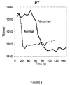
- FIG. 4 depicts a graphical result providing normal and abnormal PT plasma clotting/coagulation obtained by measuring changes of T 2 relaxation time over time using time-resolved relaxation time acquisition methodology.
- FIG. 5 depicts a graphical result providing two discrete abnormal plasma clotting/coagulation curves, relative to normal plasma clotting/coagulation, obtained by measuring changes of T 2 relaxation time over time using time-resolved relaxation time acquisition methodology.
- FIG. 6 depicts a graphical result providing a correlation between a coagulation measurement method of the present invention and a commercial bench-top coagulation instrument from Diagnostica Stago (Parsippany, N.J.), called the Start-4. Prothrombin Time (PT) and Activated Partial Thromboplastin Time (aPPT) were measured with both methods.
- PT Prothrombin Time
- aPPT Activated Partial Thromboplastin Time
- FIGS. 7 a and 7 b depict schematic graphical representations of two different magnetic resonance pulse sequences for measuring T 2 :
- a spin echo sequence consists of two radiofrequency (RF) pulses: a 90°, x phase, and a 180°, y phase, separated by a delay ⁇ . The echo signal appears at time 2 ⁇ . T 2 is measured by obtaining the echo signal from successive cycles using incremental values of ⁇ . The recycle delay, d 1 , is typically 1-3 sec.
- d 1 The recycle delay, d 1 , is typically 1-3 sec.
- a CPMG sequence allows for much faster T 2 measurements because multiple echos are acquired in rapid succession by a series of 180°, y phase RF pulses and signal acquisitions. T 2 measurements acquired with a CPMG sequence avoid diffusion artifacts because of the short time over which the measurement occurs.
- the present invention provides methods for detecting a change in a sample (e.g., a blood sample) coagulation state, for example, monitoring blood clotting (hereinafter also “coagulation”) using time-resolved relaxation time acquisition methodology.
- a sample e.g., a blood sample
- coagulation blood clotting
- Provided methods for measuring coagulation time of a sample are simple to practice, rapid, and reliable.
- a “subject” encompasses mammals and non-mammals.
- mammals include, but are not limited to, any member of the mammalian class, including humans, non-human primates such as chimpanzees, and other apes and monkey species; farm animals such as cattle, horses, sheep, goats, swine; domestic animals such as rabbits, dogs, and cats; laboratory animals including rodents, such as rats, mice and guinea pigs; etc.
- non-mammals include, but are not limited to, birds, fish, etc.
- a subject includes a clinical patient.
- a sample can be a biologic sample, for example, a blood sample (e.g., whole blood, plasma, blood concentrate, citrated blood) from a subject, or a liquid containing compounds (e.g., monomers) that can coagulate, for example, upon providing conditions suitable for coagulation.
- a blood sample e.g., whole blood, plasma, blood concentrate, citrated blood
- a liquid containing compounds e.g., monomers
- a blood sample can be obtained from a subject (e.g., a patient) by traditional means such as venipuncture or a finger prick.
- a sample can be applied, for example via sample application port, onto a test carrier.
- a sample of blood obtained from a subject can be used without additional manipulation in the methods and apparatus of the invention.
- a whole blood sample is used in conjunction with provided methods.
- a blood sample obtained from a subject can be treated to remove, either completely or partially, red blood cells.
- blood cells are removed by any of known methods, such as, for example, centrifugation, reacting sample with a red blood cell agglutinant, or by employing a red blood cell filter.
- plasma is used in conjunction with provided methods.
- sample blood or plasma can be optionally diluted prior to coagulation.
- a diluent can simply be an aqueous solution or it can be a non-aqueous solution, and optionally can include various additives, such as, for example, salts, proteins, sugars, saccharides, metal ions, such as calcium, magnesium, lanthanides, and the like.
- Certain formulations of a diluent can include gelatin-containing composition and/or emulsion.
- a diluent is a saline solution.
- a diluent is a buffer solution, e.g., citrate buffer
- a sample may be maintained at a temperature of about 20° C. to about 40° C. In some embodiments a sample is maintained at about room temperature, about 22° C., about 25° C., about 30° C., about 35° C., about 37° C. or about 40° C. In certain embodiments, a blood sample is maintained at about body temperature, or about 37° C. Regardless of a preferred selected temperature, a sample is preferably maintained at about constant temperature throughout the process of obtaining measurement of NMR readings.
- a coagulation time can be one or more of the blood coagulation times, including prothrombin time (PT), partial thromboplastin time (PTT), activated partial thromboplastin time (APTT), fibrinogen assay, thrombin clotting time (TCT), fibrinogen assay, and activated clotting time (ACT).
- PT prothrombin time
- PTT partial thromboplastin time
- APTT activated partial thromboplastin time
- fibrinogen assay thrombin clotting time
- TCT thrombin clotting time
- ACT activated clotting time
- a sample may be heparinized and/or mixed with one or more reagents.
- a reagent may include, for example, an anti-coagulant.
- a reagent may include, for example, a coagulant, a coagulation agent (e.g., calcium (e.g., calcium chloride)), kaolin, celite, ellagic acid, glass particles, thrombin, thromboplastin, PT reagent, PTT or APTT reagent, ACT reagent, TCT reagent, fibrinogen reagent), or a heparin neutralizing or deactivating agent (e.g., heparinase, protamine).
- a coagulation agent e.g., calcium (e.g., calcium chloride)
- kaolin e.g., celite, ellagic acid, glass particles, thrombin, thromboplastin, PT reagent, PTT or APTT reagent
- coagulation reagent refers to a reagent that induces and/or supports (e.g., accelerates) coagulation when mixed with the sample, for example, a blood sample, under conditions suitable for the reagent to induce or support coagulation in the sample. These conditions are known in the art.
- Coagulation reagents include but are not limited to a prothrombin time (PT) reagent, a partial thromboplastin time (PTT)/activated partial thromboplastin time (APTT) reagent, thrombin clotting time (TCT) reagent, fibrinogen reagent, an activated clotting time (ACT) reagent, calcium (e.g., calcium chloride)), kaolin, celite, ellagic acid, glass particles, thrombin, thromboplastin; wherein specific agents comprising reagents for each test(s) are well known and have been described in the art, and are available through commercially available sources.
- PT prothrombin time
- PTT partial thromboplastin time
- APTT activated partial thromboplastin time
- TCT thrombin clotting time
- ACT activated clotting time
- a PT reagent can include any of STA® Neoplastine CL, STA® Neoplastine CL Plus (Diagnostica Stago, Parsippany, N.J., USA); Thromborel S, Innovin, Thromboplastin CL, Thromboplastin C Plus (Dade Behring, Liederbach, GERMANY); Plastinex (BioData Corporation, Horsham, Pa., USA); Diaplastin (Diamed AG, SWITZERLAND); Thromboplastin, Thromboplastin M1 (Helena Laboratories, Beaufort, Tex., USA); PT-Fibrinogen, PT-Fibrinogen HS, PT-Fibrinogen HS+, PT-Fibrinogen Recombinant, Brain Thromboplastin, RecombiPlasTin (Instrumentation Laboratory, Bedford, Mass., USA); Simplastin, Simplastin Excel, Simplastin Excel S, Simplastin L,
- Thromboplastin-D with Calcium Thromboplastin-DL with Calcium, Thromboplastin-DS, Thromboplastin Liquid (Pacific Hemostasis, Huntersville, N.C., USA); Thromboplastin with Calcium, Thromboplastin HS with Calcium, Thromboplastin M with Calcium, Thromboplastin XS with Calcium, ThromboMAX HS with calcium, ThromboMAX with calcium (Sigma Diagnostics, St. Louis, Mo., USA).
- An APTT/PTT reagent can include for example any of: Automated APTT Reagent, SILIMAT, Platelin®L, Platelin®LS, and MDA Platelin®L (bioMerieux, St. Laurent, Quebec, CANADA); Actin®, Actin®FS, Actin®FSL, and Pathromtin®SL (Dade Behring, Liederbach, GERMANY); APTT-SP, APTT-C, SynthASil, SynthAFax, and ThromboslL (Instrumentation Laboratory, Bedford, Mass., USA); SPECTRATM (Analytical Control Systems, Inc., Fishers, Ind., USA); Thrombosil, Activated Thrombofax (Ortho, Raritan, N.J., USA); CK-PREST, STA® PTT Automate (Diagnostica Stago, Parsippany, N.J., USA); Cephalinex® (BioData Corporation
- a fibrinogen reagent can include, for example, Fibri-Prest, STA®-Fibrinogen 5 (Diagnostica Stago, Parsippany, N.J., USA); Multifibren U, Fibrinogen Determination (Dade Behring Thrombin) (Dade Behring, Liederbach, GERMANY); Fibrinogen Assay (BioData Corporation, Horsham, Pa., USA); Fibrinogen Assay (Helena Laboratories, Beaufort, Tex., USA); QFA (bovine thrombin), Fibrinogen C, PT-Fibrinogen, PT-Fibrinogen HS, PT-Fibrinogen HS+, PT-Fibrinogen Recombinant, RecombiPlasTin.
- Fibri-Prest STA®-Fibrinogen 5 (Diagnostica Stago, Parsippany, N.J., USA); Multifibren U, Fibrin
- a TCT reagent can include, for example, any commercial or produced source of animal thrombin, e.g., STA®-Thrombin, Thrombin 10, (Diagnostica Stago, Parsippany, N.J., USA); Human alpha-Thrombin (Sigma Diagnostics, St. Louis, Mo., USA); MDA® Thromboquik, (bioMerieux, St. Laurent, Quebec, CANADA); BC-Thrombin reagent (Dade Behring, Liederbach, GERMANY).
- An ACT reagent can include, for example, any commercial or produced source of silica based coagulation activator compound, (e.g., kaolin, celite, ellagic acid, glass particles). Further, combinations of coagulation reagents can be used as reagents to induce and/or support coagulation.
- carrier is understood to mean any localizer of a liquid sample, for example, a well, an indentation, a support, a channel, a reservoir, a sunken volume, a compartment, a recessed area, an enclosure with or without an opening, a tube, or a trough.
- test carrier means a carrier into which a sample is deposited for analysis.
- a test carrier can be placed within the detection volume of an NMR detection coil, for example a relaxometer (i.e., Bruker Minispec) or a customized miniature relaxometer.
- a sample can be placed in the test carrier either before or after the test carrier is placed within the detection volume of a NMR detection coil.
- coagulation test time based on the measurement of viscosity of the sample using NMR relaxivity measurements can be ascertained under temperature control.
- an effective T 2 relaxation rate (i.e. 1/T 2 ) can be related to a coagulation state of a sample, and coagulation time can be determined by monitoring one or more parameters relating to a series of T 2 relaxation rate measurements over time (herein referred to as a coagulation-time-curve).
- a tailored radiofrequency (RF) pulse sequence is applied to a test carrier containing a sample; and RF echo signals are monitored and analyzed to determine one or more NMR parameters (e.g., T 2 ).
- an effective T 2 relaxation rate as measured by a custom CPMG (Carr-Purcell-Meiboom-Gill) pulse sequence can be used to measure a temporal change in coagulation state. While true T 2 measurements with methods such as spin echos (see, e.g., FIG. 7 a ) can be obtained, such measurements do not typically yield the same sensitivity to coagulation state as methods provided herein, and thus are not useful in the current application. For example, to obtain a useful relaxation parameter for measuring a coagulation time with adequate temporal resolution (e.g. sampling rate), CPMG sequence parameters are adjusted such that obtained relaxation curves are sampled to obtain optimal coagulation response measurements, as described in further detail herein.
- CPMG sequence parameters are adjusted such that obtained relaxation curves are sampled to obtain optimal coagulation response measurements, as described in further detail herein.
- Suitable CPMG sequences for measuring effective T 2 of a sample can be characterized by the following sequence of steps: 1) waiting (i.e., not applying a radiofrequency pulse to a sample) at least for a time period given by a recycle delay (e.g., the time between initiation of a relaxation measurement and a first radiofrequency pulse, time between the end of prior sequence measurement to allow for the system to return to equilibrium (e,g., about 0.5 to about 5 seconds); 2) applying a 90° radiofrequency pulse to the sample, 3) waiting for a time period given by, for example, one-half the inter-echo delay, 4) applying a 180° radiofrequency pulse to the sample, 5) waiting for a time period given by the inter-echo delay, and optionally, repeating steps 4) and 5) one or more times.
- a recycle delay e.g., the time between initiation of a relaxation measurement and a first radiofrequency pulse, time between the end of prior sequence measurement to allow for the system to return to equilibrium (e,g., about
- a relaxation measurement optionally coincides with one or more of completion of a previous pulse sequence measurement, insertion of a sample into the magnet, etc.
- initiation of a relaxation measurement coincides with completion of a previous relaxation measurement pulse sequence.
- a sample responds with an echo that can be acquired to determine T 2 by methods known in the art. See, e.g., Can, H. Y., and Purcell, E. M., “Effects of Diffusion on Free Procession in Nuclear Magnetic Resonance Experiments,” Phy. Rev. 904, No.
- spectrometer recording hardware may constrain the total number of echos that can be recorded.
- a subset of detectable echos are recorded, (e.g., acquiring one of every four echos (e.g., herein referred to as a CPMG sequence characterized by a dummy echo value of three)).
- more than one CPMG sequence is employed, e.g., more than one measurement of T 2 is performed per sample.
- each of the CPMG sequence(s) are separated in time by a recycle delay.
- parameters characterizing a custom CPMG sequence can be determined using methods known in the art, because the time between T 2 value measurements is large compared to the signal decay time used to determine T 2 .
- blood coagulation times are short and/or higher time resolution is preferred. For example, for T 2 values of larger than 1.5 seconds and T 1 values larger than 1.5 seconds, upon first approximation, a dwell time (that is, acquisition time plus recycle delay) of about 5 seconds would appear to be needed to measure true sequential T 2 values.
- T 2 the parameter of a CPMG sequence
- T 2 the term “T 2 ” as used herein, if not specifically denoted as “effective” refers to both “true” and “effective” T 2
- dwell time e.g. the time between T 2 measurements must be short enough to provide a kinetic trace throughout the coagulation process.
- the measured T 2 value is actually an effective T 2 because the T 2 value is influenced by the optimization of the CPMG sequence, that is, the “true” T 2 requires acquisitions times that are not amenable to a short dwell times; therefore effective T 2 measurements are required.
- parameters are changed to: 1) maximize the change in T 2 measurements over the coagulation process (maximize overall delta T 2 ); 2) minimize noise levels of measurements taken (e.g., particularly at the upper and lower T 2 measurement extremes); and to increase the number of T 2 measurements taken over time in order to provide adequate sample measurements over the time course of coagulation so as to generate a useful coagulation wave form.
- one skilled in the art could modify parameters described herein in various combinations to achieve the results taught in the present methods.
- one skilled in the art may modify parameters that may vary slightly from the provided ranges, and/or in conjunction with other prarameters in a CPMG sequence, or other sequential relaxation signal measurements, to similarly optimize relaxation measurement sequence(s) to obtain coagulation measurements as provided herein.
- a recycle delay is between 0.1 seconds and 100 seconds. In particular embodiments, a recycle delay is between 0.5 seconds and 1 second. In certain embodiments, a recycle delay is about 1 second.
- an inter-echo delay is between 0.01 milliseconds and 10 milliseconds. In particular embodiments, an inter-echo delay is between 0.2 milliseconds and 2 milliseconds. In certain embodiments, an inter-echo delay is about 0.5 milliseconds.
- the number of acquired echos is between 1 and 10,000. In particular embodiments, the number of acquired echos is between 500 and 2,000. In certain embodiments, the number of acquired echos is between 1500 and 2000.
- the number of dummy echos is between 0 and 50. In particular embodiments, the number of dummy echos is between 0 and 10. In certain embodiments, the number of dummy echos is between 0 and 3.
- an acquisition time is between 0.01 milliseconds and 5,100 seconds.
- an acquisition time is between 0.1 and 44 seconds.
- an acquisition time is between about 0.5 and about 8 seconds.
- an acquisition time is about 3.5 seconds or about 4.5 seconds.
- a dwell time is between 0.1 seconds and about 5,200 seconds.
- a dwell time is between 0.6 and 45 seconds.
- a dwell time is between about 1 second and about 6 seconds.
- a dwell time is about 4.5 or about 5.5 seconds.
- a dwell time is sufficient to allow for taking at least two T 2 values while sample is coagulating and before the sample is coagulated. In some embodiments a dwell time is sufficient to allow for taking at least five T 2 values while a sample is coagulating and before the sample is coagulated. In certain embodiments a dwell time is sufficient to allow for taking at least ten T 2 values while a sample is coagulating and before the sample is coagulated.
- a recycle delay is between 0.1 and 100 seconds
- the number of acquired echos is between 1 and 10,000
- the number of dummy echos is between 0 and 50
- an inter-echo delay is between 0.01 and 10 milliseconds
- the number of T 2 measurements i.e., number of sequential CPMG sequences
- the number of T 2 measurements is between 2 and 10,000, leading to acquisition times between 0.00001 seconds and 5,100 seconds and dwell times between 0.1 second and 5,200 seconds.
- a recycle delay is between 0.5 and 1 seconds
- the number of acquired echos is between 500 and 2,000
- the number of dummy echos is between 0 and 10
- an inter-echo delay is between 0.2 and 2 milliseconds
- the number of T 2 measurements i.e., number of sequential CPMG sequences is between 100 and 500, leading to acquisition times between 0.1 and 44 seconds and dwell times between 0.6 and 45 seconds.
- a recycle delay is between about 0.8 and about 1 second
- the number of acquired echos is between about 1650 and about 1850
- the number of dummy echos is between 0 and 5
- an inter-echo delay is between about 0.3 and about 0.7 ms leading to acquisition times between about 0.5 and about 7.8 seconds and dwell times between about 1.3 and about 8.8 seconds.
- a recycle delay is about 1 second
- the number of acquired echos is about 1,750
- the number of dummy echos is about 3
- an inter-echo delay is about 0.5 milliseconds, leading to an acquisition time of about 3.5 seconds and a dwell time of about 4.5 seconds.
- Determination of coagulation times using methods of the present invention is based on the measurement of a nuclear magnetic parameter, typically T 2 , over time.
- a nuclear magnetic parameter typically T 2
- one measurement of T 2 of a coagulating sample at a time before the sample is substantially fully coagulated can be sufficient to determine the extent of coagulation and/or a coagulation time. For example, if a T 2 value has been determined for a normally coagulating sample, the T 2 value and corresponding time can be matched (e.g., by visual inspection, computationally, etc.) to a pre-determined standard coagulation-time-curve for the type of coagulating sample.
- a standard coagulation-time-curve has been correlated with the extent of coagulation for the type of coagulating sample (i.e., the extent of coagulation for given T 2 values at given times on the standard coagulation-time-curve has been determined)
- a single T 2 measurement can provide the extent of coagulation.
- comparison of a T 2 value and corresponding time with a standard coagulation-time-curve can allow determination of a sample coagulation time or determination of an estimate of the sample coagulation time. For example, if a measured T 2 value at a given time point matches a T 2 value of the standard curve for the given time point, the sample coagulation time could be associated with the standard coagulation-time-curve.
- a plurality of T 2 values over time are determined using methods of the present invention to assess coagulation, for example, to determine coagulation state (i.e., not coagulated or coagulated), the extent of coagulation (e.g., percentage coagulation), and/or a coagulation time (e.g., prothrombin time (PT), partial thromboplastin time (PTT), activated partial thromboplastin time (APTT), thrombin clotting time (TCT), fibrinogen assay clotting time, activated clotting time (ACT)).
- PT prothrombin time
- PTT partial thromboplastin time
- APTT activated partial thromboplastin time
- TCT thrombin clotting time
- fibrinogen assay clotting time activated clotting time (ACT)
- the start time for coagulation is the timepoint when coagulation is initiated in the sample, for example, by mixing a coagulation activating reagent (e.g., calcium) with a sample.
- a coagulation activating reagent e.g., calcium
- a plurality of T 2 values are measured before the sample is substantially fully coagulated, and, typically, further one or more T 2 values are determined for the substantially fully coagulated sample.
- a resulting coagulation time curve provided by the measured T 2 values over time allows for a determination of the coagulation time.
- coagulation typically leads to a decline of the measured T 2 values from a top plateau to a bottom plateau.
- a coagulation time can be determined based on a measured coagulation time curve alone, by comparison with a standard coagulation-time-curve, and/or by normalizing with a pre-determined calibration factor.
- coagulation time can be determined as the time from coagulation initiation, for example, using a coagulation reagent, to the time point that the bottom plateau is reached.
- a preferred way of determining a coagulation time from measured T 2 values is to average, independently, T 2 values of a top plateau to obtain a top plateau value T 2,t and T 2 values of a bottom plateau to obtain a bottom plateau value T 2,b , and determine the time for which T 2 is at the value T 2,b +(T 2,t ⁇ T 2,b )/2 on the coagulation time curve, and normalizin obtained time with a pre-determined calibration factor.
- This determination also provides a midpoint value between the initial (top plateau) T 2 and the final (bottom plateau) T 2 on a T 2 plasma coagulation curve. See, e.g., FIGS. 2 to 6 .
- a difference between a first average T 2 value (e.g., of a top plateau) and a second average T 2 value (e.g., of a bottom plateau) is substantially larger than the average standard error of a T 2 measurement using a CPMG sequence.
- a difference between a first average T 2 value (e.g., of a top plateau) and a second average T 2 value (e.g., of a bottom plateau) is at least 3% of the first T 2 value.
- a difference between a first average T 2 value (e.g., of a top plateau) and a second average T 2 value (e.g., of a bottom plateau) is at least 5% of the first T 2 value.
- a difference between a first average T 2 value (e.g., of a top plateau) and a second average T 2 value (e.g., of a bottom plateau) is at least 10% of the first T 2 value.
- a difference between a first average T 2 value (e.g., of a top plateau) and a second average T 2 value (e.g., of a bottom plateau) is at least 13% of the first T 2 value.
- a calibration factor can be determined by determining the time as described above for one or more samples and determining for the same samples a coagulation time using a commercially available method for determining coagulation (e.g., the start®4 method using the Diagnostica Stago device), and determining the factor by which the times determined using the methods of the present invention have to be multiplied with to obtain the coagulation times determined by the commercially available method.
- a data point given by the T 2 value and the corresponding time of T 2 measurement is matched a standard coagulation-time-curve is required to which the determined T 2 value can be compared.
- a “standard coagulation-time-curve” refers to data correlating values of an NMR parameter responsive to coagulation of a sample (e.g., a blood sample, a plasma sample, a fraction of blood in a sample) of one or more subjects, or values mathematically derived from values obtained over time. Data can be, but is not limited to be, in the form of a curve. Graphical presentation of obtained data in terms of a scatter or line plot/graph, for example, with an NMR parameter on the ordinate and time on the abscissa can provide an easy way to compare measured values of an NMR parameter with a corresponding standard coagulation-time-curve.
- a sample used in determination of a “standard coagulation-time-curve” is taken from one or more subjects that exhibit normal coagulation processes and timing of coagulation processes.
- the one or more subjects from which samples are used for generation of a standard coagulation time curve can differ but don't have to differ from a test subject for which coagulation is or will be assessed using methods of the present invention.
- a standard coagulation time curve may be generated using samples obtained from normal healthy patients, and a sample that will be measured and compared to the generated standard coagulation time curve is obtained from a patient requiring assessment of anticoagulant therapy.
- the sample from the patient is not part of the pool of samples used to generate the standard coagulation time curve.
- a standard coagulation-time-curve may be generated using sample(s) of blood of a test subject (e.g., a patient) prior to a procedure or therapy (e.g., a surgery that requires post-surgical administration of an anticoagulant). Coagulation of blood of the patient may be assessed from samples obtained from the patient while the patient is receiving anticoagulant using methods of the present invention and comparing obtained results to a subject's standard coagulation-time-curve determined prior to surgery.
- one or more standard coagulation time curves are prepared independently in advance and provided as a standard control curve for individual testing of samples for any one or more coagulation times (e.g., prothrombin time (PT), partial thromboplastin time (PTT), activated partial thromboplastin time (APTT), thrombin clotting time (TCT), fibrinogen assa clotting time, activated clotting time (ACT)).
- PT prothrombin time
- PTT partial thromboplastin time
- APTT activated partial thromboplastin time
- TCT thrombin clotting time
- fibrinogen assa clotting time activated clotting time
- ACT activated clotting time
- one or more standard coagulation curves are prepared in advance (e.g., immediately prior to) or in conjunction with (e.g., in parallel) an individual sample preparation and testing.
- one or more standard coagulation curves are prepared initially upon first use of a lot of provided reagents, wherein the prepared standard coagulation curve(s) are used for comparison to one or more test sample coagulation curves, and continually used for each of those test samples which utilize the same lot of reagents for sample coagulation tests.
- a coagulation time can be determined by monitoring elapsed time corresponding to one or more parameters of a coagulation-time-curve including a predetermined magnitude change in the coagulation-time-curve, a percent change from baseline of the magnitude of the coagulation-time-curve, the first derivative of the coagulation-time-curve, the second derivative of the coagulation time curve, higher derivatives of the coagulation-time-curve, to an inflection point, to a steady-state value, and combinations thereof.
- Parameters can be monitored as a magnitude of elapsed time or as a function of time to enable a calculation or derivation of a characteristic value or characteristic kinetic rate (i.e. half-life of the signal, etc.).
- a characteristic value or rate can be compared to a standard or control relating to one or more parameters of a coagulation state of a sample, either simultaneously or sequentially.
- a standard can take many specific forms, but may be generically described as a data set relating the characteristic value or rate to a coagulation time determined by a standard coagulation instrument (e.g. calibration curve).
- a sample can be mixed with a coagulation reagent before being placed in the test carrier or can interact with a reagent coated on the surfaces of the test carrier. Additionally or alternatively, a sample may be mixed with a coagulation reagent disposed in the test carrier, either before or after a sample is placed in the test carrier. For example, surfaces of a test carrier may be coated with the coagulation reagent or a discrete element that is coated with or includes a coagulation reagent is disposed in the test carrier prior to, at the same time as, or after addition of a sample.
- a test carrier walls can be surface-etched to increase surface area and to enhance surface roughness that can cause fibrin to develop in a sample (e.g., a blood sample (e.g., whole blood, plasma, etc.). Surface roughness may activate or facilitate coagulation of a sample, either in place of or in addition to a coagulation reagent.
- a test carrier can be a fabrication of any natural, synthetic, porous, non-porous, non-metallic, magnetic susceptibility matched, hydrophobic or hydrophilic material (e.g., plastic (i.e. Delrin or Teflon), glass, Mylar).
- a test carrier may be of any geometric shape capable of isolating, or accommodating, or absorbing, or containing a volume of solution including capillaries, tubes, hollow channels, conduits, microfluidics, porous membranes, and encapsulations.
- the carrier may be a glass capillary or tube used for NMR relaxation measurements.
- a test carrier may accommodate volume samples in the range of 1 picoliter to 1 milliliter, preferably microliters, more preferably 1 to 500 microliters, most preferably 10 to 300 microliters.
- the present invention also provides methods for monitoring (for example, in real-time) coagulation of a blood sample of a test subject that makes use of measurements of an NMR parameter over time. Comparing obtained values for a monitored NMR parameter with a standard coagulation-time-curve provides information regarding abnormal coagulation events.
- Monitoring blood coagulation over time provides a clotting profile of a sample and provides information concerning discrete normal or abnormal events that may accompany the coagulation, clotting or lytic process (e.g., clot formation, clot retraction, or clot lysis), as well as providing insight into the overall event.
- the present invention is useful in distinguishing between platelet-rich and platelet-poor plasma depending on the clotting profile of a sample.
- the present invention also provides methods for diagnosing an abnormal clotting event in a test subject.
- At least one test carrier is provided, wherein each test carrier contains a sample (e.g., a blood sample) from a test subject and is placed within a detection volume of a NMR detector.
- Test data of a NMR parameter responsive to coagulation in the sample of each test carrier is obtained over time, for example, by measuring values of the NMR parameter over time.
- One or more characteristics of the test data are compared with those of a standard coagulation-time-curve in the NMR parameter responsive to normal coagulation to identify and thereby diagnose an abnormal clotting event in the subject (see, for example, FIG. 3 and FIG. 4 ). Any suitable characteristic associated with the test data can be compared.
- Examples include overall change of the NMR parameter over time, rate of the NMR parameter change over time, clotting time determined from the NMR parameter change, and fluctuation of the NMR parameter change in the sample prior to coagulation.
- the test data of the NMR parameter are obtained by monitoring values of the NMR parameter over time to provide a coagulation (clotting) profile of the sample.
- a plurality of samples (e.g., blood samples) from a test subject are collected at discrete times.
- a first test carrier and a second test carrier contain a first sample and a second sample from the same test subject, but collected at discrete times.
- Difference(s) between the first and second samples in coagulation can be obtained by comparing characteristic(s) of the obtained test NMR data (e.g., clotting profile) between the first and second samples. With such comparison, for example, one can determine if any change is present from (e.g., a first abnormal clotting event diagnosed from a first blood sample over the time period between the first and second blood collection).
- a first and a second sample are collected from different test subjects.
- difference(s) between the first and second samples in coagulation e.g., clotting profile
- the present methods are also useful for providing information concerning discrete events that may accompany the coagulation, clotting or lytic process (e.g., clot formation, clot retraction, or clot lysis), as well as providing insight into the overall event.
- Provided methods can be useful in distinguishing between platelet-rich and platelet-poor plasma depending on the clotting profile of the sample.
- the methods can also provide information useful to physicians developing a treatment plan for patients during and following surgery including cardiopulmonary bypass surgery to avoid or mitigate pre-operative, perioperative, and/or post-operative bleeding.
- the underlying principle of the present invention for coagulation state measurement and its use in determining coagulation time is based on the assumption of single-exponential decay functions obtained from NMR radiofrequency (RF) echo signals.
- RF radiofrequency
- an effective T 2 relaxivity change over time is related to a sample coagulation state and inversely proportional to temperature.
- Provided methods allow measurement of the kinetics of coagulation by monitoring changes in relaxation times. For example, in certain embodiments, measurements (e.g., 10-20 measurements) can be made after mixing a sample and a reagent to initiate coagulation, before coagulation is complete, and coagulation times can be determined from the resulting kinetic curves.
- FIG. 1 an NMR system is depicted to illustrate the principle on which the invention is explained on the basis of several embodiments.
- the depiction is not intended to limit the invention to a particular embodiment, but serves for the purpose of explaining the illustrative elements of devices utilizing the underlying principles for the measurement of one or more NMR parameter(s) to provide a sample coagulation time.
- FIG. 1 is a schematic diagram 100 of an NMR system for detection of an echo response of a sample 103 to an RF excitation, thereby determining the coagulation state of the sample and a corresponding coagulation time.
- a sample 103 within test carrier 104 is placed within the sensitive region of an RF coil 105 of device 100 .
- Device 100 comprises bias magnets 101 that generate a bias magnetic field B 0 102 through a sample 103 .
- An RF excitation pulse at the Larmor frequency is applied to a sample using RF coil 105 and RF oscillator 106 .
- the RF excitation and subsequent series of 180 degree pulses induces what is known in the art as a CPMG echo train.
- the coil 105 can be configured to act synchronously as an RF antenna to detect the echo signal.
- RF signal obtained from a coil 105 is amplified by amplifier 107 and processed to determine a change in the relaxation curve in response to the excitation applied to the sample 103 .
- the detected and processed signal is preferably the T 2 relaxation time.
- a series of T 2 relaxation times is monitored over a period of time from an initial set of values to a steady set of values.
- a corresponding coagulation time of the sample can be calculated using a standard data set or a calibration curve for comparison with the series of monitored T 2 relaxation times.
- various configurations of carrier 104 may be used for coagulation time testing.
- Other configurations of the bias magnetic field B 0 102 can be applied to sample 103 including, unilateral magnetic fields, low powered magnetic fields, and the earth's magnetic field.
- an RF coil 105 is wrapped around the sample carrier 104 .
- RF coil 105 can be a planar RF coil or other shape and form of RF coil can be used with sample carrier 104 .
- alternative and/or additional reagents can be added to a sample carrier 104 prior or introduced simultaneously with a sample 103 into carrier 104 .
- Gradient coils 109 can be used to apply discrete, intermittent, or continuous magnetic gradient forces on sample 103 , coagulation reagent 108 , and optional additional and/or alternative regents.
- T 2 relaxivity measurements of a sample can be taken independent of coagulation measurements described herein to assess viscosity of a sample using magnetic particles. See, e.g., WO2009/026164, the disclosure of which is incorporated herein by reference. Additionally or alternatively, T 2 relaxation rates may be analyzed to ascertain the mechanical integrity of clot formation within a sample over a period of time particularly to identify coagulopathic characteristics of a blood sample.
- a coagulation time test is conducted on one or more samples that are “incubated” in a test carrier 104 (e.g., incubating in a test chamber) by maintaining samples at a preferred temperature (e.g., body temperature) for a defined incubation time period.
- a preferred temperature e.g., body temperature
- it may be necessary to incubate sample e.g., citrated whole blood, plasma, or quality control sample(s)
- blood sample(s) drawn from a patient and immediately placed within the test carrier 104 before the sample has cooled may not need an incubation period and may only need to be maintained at 37° C.
- a heating element e.g., a heat block (not shown)
- a heating element e.g., a heat block
- a constant temperature e.g., body temperature, 37° C.
- a suitable reagent 108 may be selected to react with a sample (e.g., a blood sample) to facilitate sample coagulation (e.g., for performance of a particular test on a blood sample for determining sample coagulation times, e.g., one of PT, aPTT, TT, and ACT).
- a suitable reagent 108 may be added to a sample carrier 104 prior or introduced simultaneously with sample 103 into carrier 104 .
- a coagulation reagent 108 can be included in a sample carrier 104 prior wherein when an added sample 103 is place in carrier 104 , sample reacts with the reagent 108 .
- coagulation reagent(s) may be selected from coagulants or activating agents including calcium, kaolin, celite, ellagic acid, glass particles, thrombin, thromboplastin or other coagulation agents described herein and known in the art.
- coagulation reagents are selected from one or more coagulating agents selected from a prothrombin time (PT) reagent, a partial thromboplastin time (PTT)/activated partial thromboplastin time (APTT) reagent, thrombin clotting time (TCT) reagent, fibrinogen reagent, an activated clotting time (ACT) reagent, calcium (e.g., calcium chloride)), kaolin, celite, ellagic acid, glass particles, thrombin, and/or thromboplastin.
- PT prothrombin time
- PTT partial thromboplastin time
- APTT activated partial thromboplastin time
- TCT thrombin clotting time
- ACT activated clotting time
- test carrier 104 may also contain or optionally accommodate additional reagent(s).
- additional reagent(s) to counteract any anticoagulant(s) present in a blood sample.
- heparin may be administered to a subject to mitigate coagulation induced by a procedure, in which case neutralizing or deactivating agent(s) (e.g., heparinase, protamine) in test carrier 104 could counteract heparin and return the blood sample to a baseline condition.
- neutralizing or deactivating agent(s) e.g., heparinase, protamine
- one test carrier 104 could contain protamine, and another test carrier 104 could be devoid of protamine to perform comparative coagulation time tests.
- a Bruker Minispec mQseries (The Woodlands, Tex.) was adapted with pulse sequences for T 2 monitoring in real time. Several effective T 2 measurements made within 30 to 40 seconds and transverse relaxation times of plasma samples were measured every 5 seconds. Coagulation of a sample was induced by addition of calcium chloride to a mixture of reconstituted plasma (CITREX® I lyophilized plasma preparation, BIODATA Corporation, Horsham, PAS) and an aPTT reagent (CEPHALINEX® activated partial thromboplastin time reagent BIODATA Corporation, Horsham, Pa., USA).
- Coagulation time was estimated by curve analysis completed by midpoint determination between the T 2 intial and T 2 final. See FIG. 2 .
- the relaxation time of the plasma sample decreased steadily until the sample was fully coagulated. See FIG. 2 .
- the overall change in T 2 observed was about 300 ms.
- Coagulation time was determined to be about 35 s based on the curve in FIG. 2 .
- Curve analysis was completed to generate coagulation times by midpoint determination between the T 2 intial and T 2 final.
- aPTT coagulation measurements 100 ⁇ L of patient plasma and 100 ⁇ L of aPTT clotting reagent PTT-A (Diagnostica Stago, Parsippany, N.J.) were pre-warmed to 37° C. in a 5 mm NMR tube. 100 ⁇ L of calcium chloride pre-warmed to 37° C. was added to the plasma and clotting reagent activating coagulation. See, FIG. 3 and FIG. 5 . The overall change in T 2 observed was about 250 ms for the normal sample, and about 250 ms for the abnormal sample. Coagulation time was determined to be about 50 sec for the normal sample, and about 140 sec for the abnormal sample based on the curve in FIG. 3 .
- Neoplastine CI Plus Diagnostica Stago, Parsippany, N.J. pre-warmed to 37° C. were mixed activating coagulation. See FIG. 4 .
- the overall change in T 2 observed was about 150 ms for the normal sample, and about 160 ms for the abnormal sample.
- Coagulation time was determined to be about 19 sec for the normal sample, and about 67 sec for the abnormal sample based on the curve in FIG. 4 .
- aPTT measurements 100 ⁇ L of patient plasma and 100 ⁇ L of PTT-A (Diagnostica Stago) were pre-warmed to 37° C. in a 5 mm NMR tube. 100 ⁇ L of calcium chloride pre-warmed to 37° C. was added to the plasma and clotting reagent activating coagulation.
- 100 ⁇ L of patient plasma and 200 ⁇ L of Neoplastine CI Plus (Stago Diagnostica, Parsippany, N.J.) pre-warmed to 37° C. were mixed activating coagulation. Measurements were made kinetically on a Bruker M Q minispec using preloaded minispec software with CPMG parameters described in Example 2. Curve analysis to generate coagulation times was completed by midpoint determination between the T 2 intial and T 2 final.
- FIG. 6 shows a correlation plot representing a graphical comparison of these two methods.
- a subtraction factor was applied to time measured by T2 Biosystems to provide a correlation with the Diagnostica Stago Start®4. This subtraction factor was determined by subtracting a fixed value which resulted in all normal coagulation values derived with the NMR instrument to be in a clinically normal range.
- the correlation data points are very close to the plot diagonal, indicating excellent correlation of the results obtained using two very different approaches for measuring coagulation times.
Abstract
Description
- 1. Tau=0.25
- 2. Number of Points=1000
- 3. Dummy Echos: 3
- 4. Recycle Delay: 1
- 5. 0 dB pulse at 37° C.
- 6. Receiver gain: 75
- 7. 1 scan
- 1. Tau=0.25
- 2. Number of Points=1750
- 3. Dummy Echos: 3
- 4. Recycle Delay: 1
- 5. 18 dB pulse at 37° C.
- 6. Receiver gain: 75
- 7. 1 scan
Claims (23)
Priority Applications (1)
| Application Number | Priority Date | Filing Date | Title |
|---|---|---|---|
| US13/124,318 US9157974B2 (en) | 2008-10-29 | 2009-10-29 | NMR detection of coagulation time |
Applications Claiming Priority (3)
| Application Number | Priority Date | Filing Date | Title |
|---|---|---|---|
| US10936708P | 2008-10-29 | 2008-10-29 | |
| US13/124,318 US9157974B2 (en) | 2008-10-29 | 2009-10-29 | NMR detection of coagulation time |
| PCT/US2009/062537 WO2010051362A1 (en) | 2008-10-29 | 2009-10-29 | Nmr detection of coagulation time |
Related Parent Applications (1)
| Application Number | Title | Priority Date | Filing Date |
|---|---|---|---|
| PCT/US2009/062537 A-371-Of-International WO2010051362A1 (en) | 2008-10-29 | 2009-10-29 | Nmr detection of coagulation time |
Related Child Applications (1)
| Application Number | Title | Priority Date | Filing Date |
|---|---|---|---|
| US14/842,388 Continuation US10126314B2 (en) | 2008-10-29 | 2015-09-01 | NMR detection of coagulation time |
Publications (2)
| Publication Number | Publication Date |
|---|---|
| US20110312002A1 US20110312002A1 (en) | 2011-12-22 |
| US9157974B2 true US9157974B2 (en) | 2015-10-13 |
Family
ID=42129264
Family Applications (4)
| Application Number | Title | Priority Date | Filing Date |
|---|---|---|---|
| US13/124,318 Active 2032-09-16 US9157974B2 (en) | 2008-10-29 | 2009-10-29 | NMR detection of coagulation time |
| US14/842,388 Active US10126314B2 (en) | 2008-10-29 | 2015-09-01 | NMR detection of coagulation time |
| US16/168,118 Abandoned US20190154709A1 (en) | 2008-10-29 | 2018-10-23 | Nmr detection of coagulation time |
| US17/223,259 Pending US20220082575A1 (en) | 2008-10-29 | 2021-04-06 | Nmr detection of coagulation time |
Family Applications After (3)
| Application Number | Title | Priority Date | Filing Date |
|---|---|---|---|
| US14/842,388 Active US10126314B2 (en) | 2008-10-29 | 2015-09-01 | NMR detection of coagulation time |
| US16/168,118 Abandoned US20190154709A1 (en) | 2008-10-29 | 2018-10-23 | Nmr detection of coagulation time |
| US17/223,259 Pending US20220082575A1 (en) | 2008-10-29 | 2021-04-06 | Nmr detection of coagulation time |
Country Status (6)
| Country | Link |
|---|---|
| US (4) | US9157974B2 (en) |
| EP (1) | EP2348976B1 (en) |
| JP (2) | JP5926958B2 (en) |
| AU (1) | AU2009308841B2 (en) |
| CA (1) | CA2741550C (en) |
| WO (1) | WO2010051362A1 (en) |
Cited By (4)
| Publication number | Priority date | Publication date | Assignee | Title |
|---|---|---|---|---|
| US9599627B2 (en) | 2011-07-13 | 2017-03-21 | T2 Biosystems, Inc. | NMR methods for monitoring blood clot formation |
| US9739733B2 (en) | 2012-12-07 | 2017-08-22 | T2 Biosystems, Inc. | Methods for monitoring tight clot formation |
| US10126314B2 (en) * | 2008-10-29 | 2018-11-13 | T2 Biosystems, Inc. | NMR detection of coagulation time |
| US10620205B2 (en) | 2011-09-21 | 2020-04-14 | T2 Biosystems, Inc. | NMR methods for endotoxin analysis |
Families Citing this family (22)
| Publication number | Priority date | Publication date | Assignee | Title |
|---|---|---|---|---|
| JP2010540929A (en) | 2007-09-28 | 2010-12-24 | ティツー・バイオシステムズ・インコーポレーテッド | Diagnosis with NMR equipment using plastic sample containers |
| JP2011503541A (en) | 2007-11-06 | 2011-01-27 | ティツー・バイオシステムズ・インコーポレーテッド | Small magnet and RF coil for magnetic resonance relaxation measurements |
| ES2576927T3 (en) | 2010-10-22 | 2016-07-12 | T2 Biosystems, Inc. | NMR systems and methods for rapid analyte detection |
| US8409807B2 (en) | 2010-10-22 | 2013-04-02 | T2 Biosystems, Inc. | NMR systems and methods for the rapid detection of analytes |
| US8563298B2 (en) | 2010-10-22 | 2013-10-22 | T2 Biosystems, Inc. | NMR systems and methods for the rapid detection of analytes |
| JP2015503767A (en) | 2012-01-16 | 2015-02-02 | エイブラム サイエンティフィック,インコーポレーテッド | Methods, devices and systems for measuring physical properties of fluids |
| US9562271B2 (en) | 2012-04-20 | 2017-02-07 | T2 Biosystems, Inc. | Compositions and methods for detection of Candida species |
| US9500639B2 (en) * | 2012-07-18 | 2016-11-22 | Theranos, Inc. | Low-volume coagulation assay |
| US20170045494A1 (en) * | 2014-04-28 | 2017-02-16 | T2 Biosystems, Inc. | Systems and methods for identifying coagulopathies |
| US10520570B2 (en) * | 2014-05-09 | 2019-12-31 | Beth Israel Deaconess Medical Center, Inc. | System and method for tissue characterization using multislice magnetic resonance imaging |
| US20160367474A1 (en) * | 2015-06-17 | 2016-12-22 | Anthony Pignataro | Treatment process for skin imperfections |
| WO2016210424A1 (en) * | 2015-06-26 | 2016-12-29 | T2 Biosystems, Inc. | Systems and methods for multiplexed hemostasis test panels |
| EP3394614B1 (en) * | 2015-12-22 | 2022-03-02 | Radiometer Medical ApS | Method of detecting the presence or absence of a clot in a liquid sample analyzer |
| US11169141B2 (en) * | 2015-12-22 | 2021-11-09 | Radiometer Medical Aps | Method of detecting presence or absence of a clot in a liquid sample analyzer |
| CA3011901A1 (en) | 2016-01-21 | 2017-07-27 | T2 Biosystems, Inc. | Nmr methods and systems for the rapid detection of bacteria |
| CN108075063B (en) | 2016-11-09 | 2021-06-29 | Cps科技控股有限公司 | Battery pack with exhaust passage |
| WO2020051761A1 (en) * | 2018-09-11 | 2020-03-19 | 深圳迈瑞生物医疗电子股份有限公司 | Blood coagulation analyser, sample detection method thereof, and storage medium |
| CN109283206B (en) * | 2018-11-02 | 2021-07-06 | 东南大学 | Nuclear magnetic resonance rapid detection device and method for biomarkers |
| CN112955742A (en) * | 2018-12-26 | 2021-06-11 | 深圳迈瑞生物医疗电子股份有限公司 | Blood sample analysis method and blood coagulation analyzer |
| EP3948243A4 (en) * | 2019-04-05 | 2023-03-29 | The Regents of the University of California | Portable nmr instrumentation and methods for analysis of body fluids |
| CN112161955A (en) * | 2020-07-22 | 2021-01-01 | 三诺生物传感股份有限公司 | Testing method for four blood coagulation items |
| CN112986547B (en) * | 2021-04-22 | 2021-07-20 | 中国科学院苏州生物医学工程技术研究所 | Scheduling method, device and system of thrombelastogram detector |
Citations (88)
| Publication number | Priority date | Publication date | Assignee | Title |
|---|---|---|---|---|
| US4101435A (en) | 1975-06-19 | 1978-07-18 | Meito Sangyo Kabushiki Kaisha | Magnetic iron oxide-dextran complex and process for its production |
| US4374360A (en) | 1980-05-29 | 1983-02-15 | Sepponen Raimo E | NMR Diagnosis apparatus |
| US4452773A (en) | 1982-04-05 | 1984-06-05 | Canadian Patents And Development Limited | Magnetic iron-dextran microspheres |
| US4875486A (en) | 1986-09-04 | 1989-10-24 | Advanced Techtronics, Inc. | Instrument and method for non-invasive in vivo testing for body fluid constituents |
| US4920061A (en) | 1984-03-02 | 1990-04-24 | The University Of Texas System | Biological magnetic colloids |
| WO1990006045A2 (en) | 1988-11-21 | 1990-06-14 | Dynal As | Nucleic acid probes |
| US5042488A (en) | 1987-09-29 | 1991-08-27 | The Washington University | Methods employing deuterium for obtaining direct, observable deuterium magnetic resonance images in vivo and in situ |
| US5049819A (en) | 1989-06-30 | 1991-09-17 | Auburn International, Inc. | Magnetic resonance analysis in real time, industrial usage mode |
| WO1991017428A1 (en) | 1990-05-03 | 1991-11-14 | Advanced Magnetics Inc. | Solvant mediated relaxation assay system |
| US5136095A (en) | 1987-05-19 | 1992-08-04 | Syntex (U.S.A.) Inc. | Reversible agglutination mediators |
| US5204457A (en) | 1989-10-20 | 1993-04-20 | Meito Sangyo Kabushiki Kaisha | Organic magnetic complex |
| US5254460A (en) | 1990-05-03 | 1993-10-19 | Advanced Magnetics, Inc. | Solvent mediated relaxation assay system |
| US5262176A (en) | 1986-07-03 | 1993-11-16 | Advanced Magnetics, Inc. | Synthesis of polysaccharide covered superparamagnetic oxide colloids |
| US5424419A (en) | 1991-06-11 | 1995-06-13 | Meito Sangyo Kabushiki Kaisha | Oxidized complex comprising water-soluble carboxypolysaccharide and magnetic iron oxide |
| US5445970A (en) | 1992-03-20 | 1995-08-29 | Abbott Laboratories | Magnetically assisted binding assays using magnetically labeled binding members |
| US5445971A (en) | 1992-03-20 | 1995-08-29 | Abbott Laboratories | Magnetically assisted binding assays using magnetically labeled binding members |
| US5492814A (en) | 1990-07-06 | 1996-02-20 | The General Hospital Corporation | Monocrystalline iron oxide particles for studying biological tissues |
| US5637469A (en) | 1992-05-01 | 1997-06-10 | Trustees Of The University Of Pennsylvania | Methods and apparatus for the detection of an analyte utilizing mesoscale flow systems |
| WO1997040181A1 (en) | 1996-04-25 | 1997-10-30 | Spectrametrix Inc. | Analyte assay using particulate labels |
| WO1998004740A1 (en) | 1996-07-29 | 1998-02-05 | Nanosphere Llc | Nanoparticles having oligonucleotides attached thereto and uses therefor |
| US5801003A (en) | 1994-12-06 | 1998-09-01 | Ajinomoto Co., Inc. | Method and reagent detecting human disorders |
| US6013188A (en) | 1996-06-07 | 2000-01-11 | Immunivest Corporation | Methods for biological substance analysis employing internal magnetic gradients separation and an externally-applied transport force |
| JP2000166897A (en) | 1998-12-11 | 2000-06-20 | Toshiba Corp | Mri imager and method of mr imaging |
| US6165378A (en) | 1996-08-30 | 2000-12-26 | Meito Sangyo Kabushiki Kaisha | Polysaccharide derivative/magnetic metal oxide composite |
| WO2001000876A1 (en) | 1999-06-25 | 2001-01-04 | Mirkin Chad A | Nanoparticles having oligonucleotides attached thereto and uses therefor |
| WO2001011360A2 (en) | 1999-08-06 | 2001-02-15 | Institut für Diagnostikforschung GmbH an der Freien Universität Berlin | Relaxation of birefringence of magnetic nanoparticles during binding reactions |
| WO2001019405A2 (en) | 1999-09-14 | 2001-03-22 | Biomedical Apherese Systeme Gmbh | Magnetic nanoparticles having biochemical activity, method for the production thereof and their use |
| US6294342B1 (en) | 1999-09-29 | 2001-09-25 | Abbott Laboratories | Magnetically assisted binding assays utilizing a magnetically responsive reagent |
| US6297062B1 (en) | 1996-03-07 | 2001-10-02 | Bio-Magnetics Ltd. | Separation by magnetic particles |
| US6307372B1 (en) | 1999-11-02 | 2001-10-23 | Glaxo Wellcome, Inc. | Methods for high throughput chemical screening using magnetic resonance imaging |
| US6342396B1 (en) | 1997-01-30 | 2002-01-29 | Bio Merieux | Method for isolating, in particular for detecting or quantifying an analyte in a medium |
| US6361944B1 (en) | 1996-07-29 | 2002-03-26 | Nanosphere, Inc. | Nanoparticles having oligonucleotides attached thereto and uses therefor |
| US20020102214A1 (en) | 1996-09-12 | 2002-08-01 | Nycomed Imaging As. | Paramagnetic material-containing magnetic resonance external marker or calibration composition |
| WO2002098364A2 (en) | 2001-06-06 | 2002-12-12 | The General Hospital Corporation | Magnetic-nanoparticle conjugates and methods of use |
| US6500343B2 (en) | 1995-02-21 | 2002-12-31 | Iqbal W. Siddiqi | Method for mixing and separation employing magnetic particles |
| US20030054432A1 (en) | 2001-06-28 | 2003-03-20 | Biowhittaker, Inc. | Methods and reagents for detecting endotoxin |
| US6548311B1 (en) | 1997-11-21 | 2003-04-15 | Meinhard Knoll | Device and method for detecting analytes |
| US6599498B1 (en) | 1999-04-09 | 2003-07-29 | Advanced Magnetics, Inc. | Heat stable colloidal iron oxides coated with reduced carbohydrates and carbohdrate derivatives |
| US6630355B1 (en) | 1999-11-05 | 2003-10-07 | The Board Of Governors For Higher Education State Of Rhode Island | Magnetic focusing immunosensor for the detection of pathogens |
| US20030216638A1 (en) | 2002-05-16 | 2003-11-20 | Rohan Dharmakumar | Microbubble construct for sensitivity enhanced MR manometry |
| US20030219904A1 (en) | 1999-02-22 | 2003-11-27 | Eli Cohen | Protocol for monitoring platelet inhibition |
| US20030222648A1 (en) | 2002-06-03 | 2003-12-04 | Long-Sheng Fan | Batch-fabricated gradient glossary |
| US20040175388A1 (en) | 1998-12-24 | 2004-09-09 | National University Of Singapore | Recombinant proteins and peptides for endotoxin biosensors, endotoxin removal, and anti-microbial & anti-endotoxin therapeutics |
| US20040214348A1 (en) | 2001-04-23 | 2004-10-28 | Nicholson Jeremy Kirk | Methods for analysis of spectral data and their applications: osteoarthritis |
| US6866838B1 (en) | 1999-11-05 | 2005-03-15 | Bio Merieux | Composite nanospheres and their conjugates with biomolecules |
| US6884357B2 (en) | 1995-02-21 | 2005-04-26 | Iqbal Waheed Siddiqi | Apparatus and method for processing magnetic particles |
| US6940378B2 (en) | 2001-01-19 | 2005-09-06 | Halliburton Energy Services | Apparatus and method for magnetic resonance measurements in an interior volume |
| WO2005099419A2 (en) | 2004-04-13 | 2005-10-27 | President And Fellows Of Harvard College | Manipulation and/or detection of biological samples or other objects |
| WO2005111596A1 (en) | 2004-05-18 | 2005-11-24 | Koninklijke Philips Electronics N.V. | Magnetic rotation to improve signal-over-background in biosensing |
| US7018849B2 (en) | 2002-01-15 | 2006-03-28 | Piasio Roger N | Process for (A) separating biological/ligands from dilute solutions and (B) conducting an immunochromatographic assay thereof employing superparamagnetic particles throughtout |
| US20060121617A1 (en) | 2004-12-07 | 2006-06-08 | Dade Behring Marburg Gmbh | Method for automatically determining the endogenous thrombin potential |
| US20060269965A1 (en) | 2005-05-09 | 2006-11-30 | Lee Josephson | Water relaxation-based sensors |
| JP3876022B2 (en) | 1996-09-26 | 2007-01-31 | 生化学工業株式会社 | Method for quantifying substances that affect blood clotting time |
| US7217542B2 (en) | 2002-10-31 | 2007-05-15 | Hewlett-Packard Development Company, L.P. | Microfluidic system for analyzing nucleic acids |
| US7217457B2 (en) | 2001-05-10 | 2007-05-15 | Bio Merieux | Composite particles, derived conjugates, preparation method and applications |
| JP3917239B2 (en) | 1997-06-05 | 2007-05-23 | 生化学工業株式会社 | Blood clotting time measurement method |
| US20070116602A1 (en) * | 2005-08-31 | 2007-05-24 | Bioplex Systems Inc. | NMR device for detection of analytes |
| US20070166730A1 (en) | 2006-01-19 | 2007-07-19 | Menon & Associates, Inc. | Magnetic resonance system and method to detect and confirm analytes |
| US7274191B2 (en) | 2005-12-29 | 2007-09-25 | Intel Corporation | Integrated on-chip NMR and ESR device and method for making and using the same |
| WO2008007270A2 (en) | 2006-06-21 | 2008-01-17 | Spinomix S.A. | A method for manipulating magnetic particles in a liquid medium |
| US7332353B2 (en) | 2001-11-20 | 2008-02-19 | Diagnostica Stago | Method for detecting analyte(s) using magnetic colloidal particles |
| JP2008128883A (en) | 2006-11-22 | 2008-06-05 | Keio Gijuku | Measuring instrument, fuel cell equipped therewith , and measuring method |
| WO2008072156A2 (en) | 2006-12-12 | 2008-06-19 | Koninklijke Philips Electronics N. V. | Microelectronic sensor device for detecting label particles |
| US20080199539A1 (en) | 2007-02-21 | 2008-08-21 | Sarah Baker | Hemostatic compositions and methods of use |
| US20080204022A1 (en) | 2006-08-21 | 2008-08-28 | Sillerud Laurel O | Biological detector and method |
| JP2008209350A (en) | 2007-02-28 | 2008-09-11 | Matsushita Electric Ind Co Ltd | Device for measuring blood coagulation time |
| WO2008119054A1 (en) | 2007-03-27 | 2008-10-02 | Abqmr, Inc. | System and method for detecting labeled entities using microcoil magnetic mri |
| WO2008137721A2 (en) | 2007-05-03 | 2008-11-13 | Abqmr, Inc. | Microcoil nmr detectors |
| US20080305048A1 (en) | 2003-12-10 | 2008-12-11 | The General Hospital Corporation | Self-Assembling Nanoparticle Conjugates |
| WO2009017697A2 (en) | 2007-07-26 | 2009-02-05 | T2 Biosystems, Inc. | Diagnostic information generation and use |
| WO2009026251A1 (en) | 2007-08-17 | 2009-02-26 | The General Hospital Corporation | Detecting ions and measuring ion concentrations |
| WO2009045551A1 (en) | 2007-10-04 | 2009-04-09 | The General Hospital Corporation | Miniaturized magnetic resonance systems and methods |
| WO2009045354A1 (en) | 2007-09-28 | 2009-04-09 | T2 Biosystems, Inc. | Nmr diagnostics by means of a plastic sample container |
| US20090099342A1 (en) | 2005-12-15 | 2009-04-16 | Biomerieux | Process for Preparing Composite Particles, Composite Particles Obtained, and Their Use in a Diagnostic Test |
| WO2009055587A1 (en) | 2007-10-23 | 2009-04-30 | Abqmr, Inc. | Microcoil magnetic resonance detectors |
| WO2009061481A1 (en) | 2007-11-06 | 2009-05-14 | T2 Biosystems, Inc. | Small magnet and rf coil for magnetic resonance relaxometry |
| US7553542B2 (en) | 2006-10-02 | 2009-06-30 | National Defense University | Functionalized magnetizable microspheres and preparation thereof |
| WO2009085214A1 (en) | 2007-12-21 | 2009-07-09 | T2 Biosystems, Inc. | Magnetic resonance system with implantable components and methods of use thereof |
| WO2010002479A1 (en) | 2008-07-03 | 2010-01-07 | Lowery Thomas Jay Jr | Magnetic nanosensor compositions and bioanalytical assays therefor |
| US20100039109A1 (en) | 2008-08-15 | 2010-02-18 | Yuesheng Cheng | Methods for determining in situ the viscosity of heavy oil using nuclear magnetic resonance relaxation time measurements |
| US20100072994A1 (en) | 2006-11-08 | 2010-03-25 | T2 Biosystems , Inc. | Nmr systems for in vivo detection of analytes |
| WO2010051362A1 (en) | 2008-10-29 | 2010-05-06 | T2 Biosystems, Inc. | Nmr detection of coagulation time |
| US7781228B2 (en) | 2005-04-07 | 2010-08-24 | Menon & Associates, Inc. | Magnetic resonance system and method to detect and confirm analytes |
| US20120100546A1 (en) | 2010-10-22 | 2012-04-26 | T2 Biosystems, Inc. | Nmr systems and methods for the rapid detection of analytes |
| WO2013010080A1 (en) | 2011-07-13 | 2013-01-17 | T2 Biosystems, Inc. | Nmr methods for monitoring blood clot formation |
| WO2013043858A1 (en) | 2011-09-21 | 2013-03-28 | T2 Biosystems, Inc. | Nmr methods for endotoxin analysis |
| US20130260367A1 (en) | 2010-10-22 | 2013-10-03 | T2 Biosystems, Inc. | Nmr systems and methods for the rapid detection of analytes |
| WO2014004573A1 (en) | 2012-06-25 | 2014-01-03 | T2 Biosystems, Inc. | Portable device for nmr based analysis of rheological changes in liquid samples |
Family Cites Families (8)
| Publication number | Priority date | Publication date | Assignee | Title |
|---|---|---|---|---|
| FI58868C (en) * | 1979-06-01 | 1981-05-11 | Instrumentarium Oy | NMR DIAGNOSANORDNING |
| JP4334171B2 (en) * | 2001-12-03 | 2009-09-30 | シスメックス株式会社 | Blood coagulation reaction analysis method |
| SE0303249D0 (en) * | 2003-12-02 | 2003-12-02 | Inst Polymerutveckling Ab | Hematocrit and analyte concentration determination |
| WO2006128167A2 (en) | 2005-05-27 | 2006-11-30 | Board Of Regents, The University Of Texas System | Optical coherence tomographic detection of cells and compositions |
| US20080261261A1 (en) | 2006-03-31 | 2008-10-23 | Kmg2 Sensors Corporation | System/unit and method employing a plurality of magnetoelastic sensor elements for automatically quantifying parameters of whole blood and platelet-rich plasma |
| US20100136517A1 (en) * | 2007-05-07 | 2010-06-03 | Massachusetts Institute Of Technology | Matrix stabilization of aggregation-based assays |
| CN101659202A (en) | 2008-08-28 | 2010-03-03 | 比亚迪股份有限公司 | Hybrid power driving system and driving method thereof |
| EP2677037B1 (en) | 2012-06-21 | 2016-04-13 | Synapse B.V. | Simultaneous measurement of thrombin generation and clot strength in plasma and whole blood |
-
2009
- 2009-10-29 AU AU2009308841A patent/AU2009308841B2/en active Active
- 2009-10-29 WO PCT/US2009/062537 patent/WO2010051362A1/en active Application Filing
- 2009-10-29 EP EP09824124.3A patent/EP2348976B1/en active Active
- 2009-10-29 US US13/124,318 patent/US9157974B2/en active Active
- 2009-10-29 CA CA2741550A patent/CA2741550C/en active Active
- 2009-10-29 JP JP2011534756A patent/JP5926958B2/en active Active
-
2014
- 2014-10-10 JP JP2014208924A patent/JP2015038502A/en active Pending
-
2015
- 2015-09-01 US US14/842,388 patent/US10126314B2/en active Active
-
2018
- 2018-10-23 US US16/168,118 patent/US20190154709A1/en not_active Abandoned
-
2021
- 2021-04-06 US US17/223,259 patent/US20220082575A1/en active Pending
Patent Citations (99)
| Publication number | Priority date | Publication date | Assignee | Title |
|---|---|---|---|---|
| US4101435A (en) | 1975-06-19 | 1978-07-18 | Meito Sangyo Kabushiki Kaisha | Magnetic iron oxide-dextran complex and process for its production |
| US4374360A (en) | 1980-05-29 | 1983-02-15 | Sepponen Raimo E | NMR Diagnosis apparatus |
| US4452773A (en) | 1982-04-05 | 1984-06-05 | Canadian Patents And Development Limited | Magnetic iron-dextran microspheres |
| US4920061A (en) | 1984-03-02 | 1990-04-24 | The University Of Texas System | Biological magnetic colloids |
| US5262176A (en) | 1986-07-03 | 1993-11-16 | Advanced Magnetics, Inc. | Synthesis of polysaccharide covered superparamagnetic oxide colloids |
| US4875486A (en) | 1986-09-04 | 1989-10-24 | Advanced Techtronics, Inc. | Instrument and method for non-invasive in vivo testing for body fluid constituents |
| US5136095A (en) | 1987-05-19 | 1992-08-04 | Syntex (U.S.A.) Inc. | Reversible agglutination mediators |
| US5042488A (en) | 1987-09-29 | 1991-08-27 | The Washington University | Methods employing deuterium for obtaining direct, observable deuterium magnetic resonance images in vivo and in situ |
| WO1990006045A2 (en) | 1988-11-21 | 1990-06-14 | Dynal As | Nucleic acid probes |
| US5049819A (en) | 1989-06-30 | 1991-09-17 | Auburn International, Inc. | Magnetic resonance analysis in real time, industrial usage mode |
| US5204457A (en) | 1989-10-20 | 1993-04-20 | Meito Sangyo Kabushiki Kaisha | Organic magnetic complex |
| WO1991017428A1 (en) | 1990-05-03 | 1991-11-14 | Advanced Magnetics Inc. | Solvant mediated relaxation assay system |
| US5164297A (en) | 1990-05-03 | 1992-11-17 | Advanced Magnetics Inc. | Solvent mediated relaxation assay system |
| US5254460A (en) | 1990-05-03 | 1993-10-19 | Advanced Magnetics, Inc. | Solvent mediated relaxation assay system |
| US5492814A (en) | 1990-07-06 | 1996-02-20 | The General Hospital Corporation | Monocrystalline iron oxide particles for studying biological tissues |
| US5424419A (en) | 1991-06-11 | 1995-06-13 | Meito Sangyo Kabushiki Kaisha | Oxidized complex comprising water-soluble carboxypolysaccharide and magnetic iron oxide |
| US5445971A (en) | 1992-03-20 | 1995-08-29 | Abbott Laboratories | Magnetically assisted binding assays using magnetically labeled binding members |
| US5445970A (en) | 1992-03-20 | 1995-08-29 | Abbott Laboratories | Magnetically assisted binding assays using magnetically labeled binding members |
| US5637469A (en) | 1992-05-01 | 1997-06-10 | Trustees Of The University Of Pennsylvania | Methods and apparatus for the detection of an analyte utilizing mesoscale flow systems |
| US5801003A (en) | 1994-12-06 | 1998-09-01 | Ajinomoto Co., Inc. | Method and reagent detecting human disorders |
| US6884357B2 (en) | 1995-02-21 | 2005-04-26 | Iqbal Waheed Siddiqi | Apparatus and method for processing magnetic particles |
| US6500343B2 (en) | 1995-02-21 | 2002-12-31 | Iqbal W. Siddiqi | Method for mixing and separation employing magnetic particles |
| US7517457B2 (en) | 1995-02-21 | 2009-04-14 | Iqbal Waheed Siddiqi | Method of mixing magnetic particles |
| US6297062B1 (en) | 1996-03-07 | 2001-10-02 | Bio-Magnetics Ltd. | Separation by magnetic particles |
| WO1997040181A1 (en) | 1996-04-25 | 1997-10-30 | Spectrametrix Inc. | Analyte assay using particulate labels |
| US6013188A (en) | 1996-06-07 | 2000-01-11 | Immunivest Corporation | Methods for biological substance analysis employing internal magnetic gradients separation and an externally-applied transport force |
| WO1998004740A1 (en) | 1996-07-29 | 1998-02-05 | Nanosphere Llc | Nanoparticles having oligonucleotides attached thereto and uses therefor |
| US6361944B1 (en) | 1996-07-29 | 2002-03-26 | Nanosphere, Inc. | Nanoparticles having oligonucleotides attached thereto and uses therefor |
| US6165378A (en) | 1996-08-30 | 2000-12-26 | Meito Sangyo Kabushiki Kaisha | Polysaccharide derivative/magnetic metal oxide composite |
| US20020102214A1 (en) | 1996-09-12 | 2002-08-01 | Nycomed Imaging As. | Paramagnetic material-containing magnetic resonance external marker or calibration composition |
| JP3876022B2 (en) | 1996-09-26 | 2007-01-31 | 生化学工業株式会社 | Method for quantifying substances that affect blood clotting time |
| US6342396B1 (en) | 1997-01-30 | 2002-01-29 | Bio Merieux | Method for isolating, in particular for detecting or quantifying an analyte in a medium |
| JP3917239B2 (en) | 1997-06-05 | 2007-05-23 | 生化学工業株式会社 | Blood clotting time measurement method |
| US6548311B1 (en) | 1997-11-21 | 2003-04-15 | Meinhard Knoll | Device and method for detecting analytes |
| JP2000166897A (en) | 1998-12-11 | 2000-06-20 | Toshiba Corp | Mri imager and method of mr imaging |
| US20040175388A1 (en) | 1998-12-24 | 2004-09-09 | National University Of Singapore | Recombinant proteins and peptides for endotoxin biosensors, endotoxin removal, and anti-microbial & anti-endotoxin therapeutics |
| US20030219904A1 (en) | 1999-02-22 | 2003-11-27 | Eli Cohen | Protocol for monitoring platelet inhibition |
| US7179652B2 (en) | 1999-02-22 | 2007-02-20 | Haemoscope Corporation | Protocol for monitoring platelet inhibition |
| US6599498B1 (en) | 1999-04-09 | 2003-07-29 | Advanced Magnetics, Inc. | Heat stable colloidal iron oxides coated with reduced carbohydrates and carbohdrate derivatives |
| WO2001000876A1 (en) | 1999-06-25 | 2001-01-04 | Mirkin Chad A | Nanoparticles having oligonucleotides attached thereto and uses therefor |
| WO2001011360A2 (en) | 1999-08-06 | 2001-02-15 | Institut für Diagnostikforschung GmbH an der Freien Universität Berlin | Relaxation of birefringence of magnetic nanoparticles during binding reactions |
| US6767635B1 (en) | 1999-09-14 | 2004-07-27 | Biomedical Apherese Systeme Gmbh | Magnetic nanoparticles having biochemical activity, method for the production thereof and their use |
| WO2001019405A2 (en) | 1999-09-14 | 2001-03-22 | Biomedical Apherese Systeme Gmbh | Magnetic nanoparticles having biochemical activity, method for the production thereof and their use |
| US6294342B1 (en) | 1999-09-29 | 2001-09-25 | Abbott Laboratories | Magnetically assisted binding assays utilizing a magnetically responsive reagent |
| US6307372B1 (en) | 1999-11-02 | 2001-10-23 | Glaxo Wellcome, Inc. | Methods for high throughput chemical screening using magnetic resonance imaging |
| US6630355B1 (en) | 1999-11-05 | 2003-10-07 | The Board Of Governors For Higher Education State Of Rhode Island | Magnetic focusing immunosensor for the detection of pathogens |
| US7001589B2 (en) | 1999-11-05 | 2006-02-21 | Bio Merieux | Composite nanospheres and their conjugates with biomolecules |
| US6866838B1 (en) | 1999-11-05 | 2005-03-15 | Bio Merieux | Composite nanospheres and their conjugates with biomolecules |
| US6940378B2 (en) | 2001-01-19 | 2005-09-06 | Halliburton Energy Services | Apparatus and method for magnetic resonance measurements in an interior volume |
| US20040214348A1 (en) | 2001-04-23 | 2004-10-28 | Nicholson Jeremy Kirk | Methods for analysis of spectral data and their applications: osteoarthritis |
| US7217457B2 (en) | 2001-05-10 | 2007-05-15 | Bio Merieux | Composite particles, derived conjugates, preparation method and applications |
| US7829350B2 (en) | 2001-06-06 | 2010-11-09 | The General Hospital Corporation | Magnetic-nanoparticle conjugates and methods of use |
| WO2002098364A2 (en) | 2001-06-06 | 2002-12-12 | The General Hospital Corporation | Magnetic-nanoparticle conjugates and methods of use |
| US20030054432A1 (en) | 2001-06-28 | 2003-03-20 | Biowhittaker, Inc. | Methods and reagents for detecting endotoxin |
| US7332353B2 (en) | 2001-11-20 | 2008-02-19 | Diagnostica Stago | Method for detecting analyte(s) using magnetic colloidal particles |
| US7018849B2 (en) | 2002-01-15 | 2006-03-28 | Piasio Roger N | Process for (A) separating biological/ligands from dilute solutions and (B) conducting an immunochromatographic assay thereof employing superparamagnetic particles throughtout |
| US20030216638A1 (en) | 2002-05-16 | 2003-11-20 | Rohan Dharmakumar | Microbubble construct for sensitivity enhanced MR manometry |
| US20030222648A1 (en) | 2002-06-03 | 2003-12-04 | Long-Sheng Fan | Batch-fabricated gradient glossary |
| US7217542B2 (en) | 2002-10-31 | 2007-05-15 | Hewlett-Packard Development Company, L.P. | Microfluidic system for analyzing nucleic acids |
| US20080305048A1 (en) | 2003-12-10 | 2008-12-11 | The General Hospital Corporation | Self-Assembling Nanoparticle Conjugates |
| WO2005099419A2 (en) | 2004-04-13 | 2005-10-27 | President And Fellows Of Harvard College | Manipulation and/or detection of biological samples or other objects |
| WO2005111596A1 (en) | 2004-05-18 | 2005-11-24 | Koninklijke Philips Electronics N.V. | Magnetic rotation to improve signal-over-background in biosensing |
| JP2006162623A (en) | 2004-12-07 | 2006-06-22 | Dade Behring Marburg Gmbh | Method of automatic measurement of endogenous thrombin potential |
| US20060121617A1 (en) | 2004-12-07 | 2006-06-08 | Dade Behring Marburg Gmbh | Method for automatically determining the endogenous thrombin potential |
| US7781228B2 (en) | 2005-04-07 | 2010-08-24 | Menon & Associates, Inc. | Magnetic resonance system and method to detect and confirm analytes |
| US20100120174A1 (en) | 2005-05-09 | 2010-05-13 | The General Hospital Corporation | Water relaxation-based sensors |
| US20060269965A1 (en) | 2005-05-09 | 2006-11-30 | Lee Josephson | Water relaxation-based sensors |
| US7564245B2 (en) | 2005-08-31 | 2009-07-21 | T2 Biosystems, Inc. | NMR device for detection of analytes |
| US20070116602A1 (en) * | 2005-08-31 | 2007-05-24 | Bioplex Systems Inc. | NMR device for detection of analytes |
| US20090099342A1 (en) | 2005-12-15 | 2009-04-16 | Biomerieux | Process for Preparing Composite Particles, Composite Particles Obtained, and Their Use in a Diagnostic Test |
| US7274191B2 (en) | 2005-12-29 | 2007-09-25 | Intel Corporation | Integrated on-chip NMR and ESR device and method for making and using the same |
| US20070166730A1 (en) | 2006-01-19 | 2007-07-19 | Menon & Associates, Inc. | Magnetic resonance system and method to detect and confirm analytes |
| WO2008010111A2 (en) | 2006-06-21 | 2008-01-24 | Spinomix S.A. | A device and method for manipulating and mixing magnetic particles in a liquid medium |
| WO2008007270A2 (en) | 2006-06-21 | 2008-01-17 | Spinomix S.A. | A method for manipulating magnetic particles in a liquid medium |
| US8339135B2 (en) | 2006-08-21 | 2012-12-25 | Stc.Unm | Biological detector and method |
| US20080204022A1 (en) | 2006-08-21 | 2008-08-28 | Sillerud Laurel O | Biological detector and method |
| US7553542B2 (en) | 2006-10-02 | 2009-06-30 | National Defense University | Functionalized magnetizable microspheres and preparation thereof |
| US20100072994A1 (en) | 2006-11-08 | 2010-03-25 | T2 Biosystems , Inc. | Nmr systems for in vivo detection of analytes |
| JP2008128883A (en) | 2006-11-22 | 2008-06-05 | Keio Gijuku | Measuring instrument, fuel cell equipped therewith , and measuring method |
| WO2008072156A2 (en) | 2006-12-12 | 2008-06-19 | Koninklijke Philips Electronics N. V. | Microelectronic sensor device for detecting label particles |
| US20080199539A1 (en) | 2007-02-21 | 2008-08-21 | Sarah Baker | Hemostatic compositions and methods of use |
| JP2008209350A (en) | 2007-02-28 | 2008-09-11 | Matsushita Electric Ind Co Ltd | Device for measuring blood coagulation time |
| WO2008119054A1 (en) | 2007-03-27 | 2008-10-02 | Abqmr, Inc. | System and method for detecting labeled entities using microcoil magnetic mri |
| WO2008137721A2 (en) | 2007-05-03 | 2008-11-13 | Abqmr, Inc. | Microcoil nmr detectors |
| WO2009017697A2 (en) | 2007-07-26 | 2009-02-05 | T2 Biosystems, Inc. | Diagnostic information generation and use |
| WO2009026251A1 (en) | 2007-08-17 | 2009-02-26 | The General Hospital Corporation | Detecting ions and measuring ion concentrations |
| WO2009045354A1 (en) | 2007-09-28 | 2009-04-09 | T2 Biosystems, Inc. | Nmr diagnostics by means of a plastic sample container |
| WO2009045551A1 (en) | 2007-10-04 | 2009-04-09 | The General Hospital Corporation | Miniaturized magnetic resonance systems and methods |
| WO2009055587A1 (en) | 2007-10-23 | 2009-04-30 | Abqmr, Inc. | Microcoil magnetic resonance detectors |
| WO2009061481A1 (en) | 2007-11-06 | 2009-05-14 | T2 Biosystems, Inc. | Small magnet and rf coil for magnetic resonance relaxometry |
| WO2009085214A1 (en) | 2007-12-21 | 2009-07-09 | T2 Biosystems, Inc. | Magnetic resonance system with implantable components and methods of use thereof |
| WO2010002479A1 (en) | 2008-07-03 | 2010-01-07 | Lowery Thomas Jay Jr | Magnetic nanosensor compositions and bioanalytical assays therefor |
| US20100039109A1 (en) | 2008-08-15 | 2010-02-18 | Yuesheng Cheng | Methods for determining in situ the viscosity of heavy oil using nuclear magnetic resonance relaxation time measurements |
| WO2010051362A1 (en) | 2008-10-29 | 2010-05-06 | T2 Biosystems, Inc. | Nmr detection of coagulation time |
| US20120100546A1 (en) | 2010-10-22 | 2012-04-26 | T2 Biosystems, Inc. | Nmr systems and methods for the rapid detection of analytes |
| US20130260367A1 (en) | 2010-10-22 | 2013-10-03 | T2 Biosystems, Inc. | Nmr systems and methods for the rapid detection of analytes |
| WO2013010080A1 (en) | 2011-07-13 | 2013-01-17 | T2 Biosystems, Inc. | Nmr methods for monitoring blood clot formation |
| WO2013043858A1 (en) | 2011-09-21 | 2013-03-28 | T2 Biosystems, Inc. | Nmr methods for endotoxin analysis |
| WO2014004573A1 (en) | 2012-06-25 | 2014-01-03 | T2 Biosystems, Inc. | Portable device for nmr based analysis of rheological changes in liquid samples |
Non-Patent Citations (95)
Cited By (6)
| Publication number | Priority date | Publication date | Assignee | Title |
|---|---|---|---|---|
| US10126314B2 (en) * | 2008-10-29 | 2018-11-13 | T2 Biosystems, Inc. | NMR detection of coagulation time |
| US9599627B2 (en) | 2011-07-13 | 2017-03-21 | T2 Biosystems, Inc. | NMR methods for monitoring blood clot formation |
| US9797914B2 (en) | 2011-07-13 | 2017-10-24 | T2 Biosystems, Inc. | NMR methods for monitoring blood clot formation |
| US10697984B2 (en) | 2011-07-13 | 2020-06-30 | T2 Biosystems, Inc. | NMR methods for monitoring blood clot formation |
| US10620205B2 (en) | 2011-09-21 | 2020-04-14 | T2 Biosystems, Inc. | NMR methods for endotoxin analysis |
| US9739733B2 (en) | 2012-12-07 | 2017-08-22 | T2 Biosystems, Inc. | Methods for monitoring tight clot formation |
Also Published As
| Publication number | Publication date |
|---|---|
| JP2012507722A (en) | 2012-03-29 |
| EP2348976A1 (en) | 2011-08-03 |
| US20220082575A1 (en) | 2022-03-17 |
| AU2009308841B2 (en) | 2014-07-17 |
| JP5926958B2 (en) | 2016-05-25 |
| EP2348976A4 (en) | 2014-01-01 |
| CA2741550A1 (en) | 2010-05-06 |
| US20190154709A1 (en) | 2019-05-23 |
| CA2741550C (en) | 2018-12-04 |
| EP2348976B1 (en) | 2020-12-02 |
| AU2009308841A1 (en) | 2010-05-06 |
| WO2010051362A1 (en) | 2010-05-06 |
| US20110312002A1 (en) | 2011-12-22 |
| JP2015038502A (en) | 2015-02-26 |
| US20150369829A1 (en) | 2015-12-24 |
| US10126314B2 (en) | 2018-11-13 |
Similar Documents
| Publication | Publication Date | Title |
|---|---|---|
| US20220082575A1 (en) | Nmr detection of coagulation time | |
| Karon | Why is everyone so excited about thromboelastrography (TEG)? | |
| US9739733B2 (en) | Methods for monitoring tight clot formation | |
| JP7192944B2 (en) | Blood coagulation system analysis device, blood coagulation system analysis system, blood coagulation system analysis method, blood coagulation system analysis program, blood loss prediction device, blood loss prediction system, blood loss prediction method, and blood loss prediction program | |
| WO2012122462A2 (en) | Use of magnetic resonance techniques for molecular detection | |
| US10794896B2 (en) | Blood coagulation system examination module, blood coagulation system examination system, blood coagulation system examination method, and determination method of parameter for blood coagulation system examination module | |
| Christiansen et al. | Simultaneous R2* and quantitative susceptibility mapping measurement enables differentiation of thrombus hematocrit and age: an in vitro study at 3 T | |
| AU2014240329B2 (en) | Nmr detection of coagulation time | |
| RU2061953C1 (en) | Method for qualitatively determining general coagulation activity of blood platelets | |
| US20170045494A1 (en) | Systems and methods for identifying coagulopathies | |
| EP3514547B1 (en) | Platelet aggregation ability analyzing method and device | |
| US20220214292A1 (en) | Portable nmr instrumentation and methods for analysis of body fluids | |
| WO2017169261A1 (en) | System for analyzing blood coagulation system, method for analyzing blood coagulation system, and program for use in analysis of blood coagulation system | |
| Cugno et al. | Assessment of a new instrument (Coag‐A‐Mate X2) for performing global clotting tests and specific factor assays | |
| JP2020190570A (en) | Platelet agglutination ability analysis method, platelet agglutination ability analyzer, program for platelet agglutination ability analysis and platelet agglutination ability analysis system | |
| Yerov et al. | Roger Smith*, Vyacheslav Papkov1, Pierrette Andre1 | |
| KANNO et al. | Evaluation of Factor XIII Activity Using Berichrom FXIII Chromogenic Assay on the Fully-Automated Coagulation Analyzer CS-2000i-Based on Comparisons with Universal Biochemical Analyzer and Antigen Method | |
| Rowan et al. | Assessment of the Coag‐a‐Pet Dual Channel in the routine haemostatic laboratory |
Legal Events
| Date | Code | Title | Description |
|---|---|---|---|
| AS | Assignment |
Owner name: T2 BIOSYSTEMS, INC., MASSACHUSETTS Free format text: ASSIGNMENT OF ASSIGNORS INTEREST;ASSIGNORS:TAKTAK, SONIA;MOZELESKI, BRIAN M.;LOWERY, THOMAS J., JR.;SIGNING DATES FROM 20091222 TO 20091230;REEL/FRAME:025145/0513 |
|
| AS | Assignment |
Owner name: T2 BIOSYSTEMS, INC., MASSACHUSETTS Free format text: ASSIGNMENT OF ASSIGNORS INTEREST;ASSIGNORS:TAKTAK, SONIA;MOZELESKI, BRIAN M.;LOWERY, THOMAS J., JR.;SIGNING DATES FROM 20110823 TO 20110909;REEL/FRAME:027312/0192 |
|
| STCF | Information on status: patent grant |
Free format text: PATENTED CASE |
|
| AS | Assignment |
Owner name: CRG SERVICING LLC, AS ADMINISTRATIVE AGENT AND COLLATERAL AGENT, TEXAS Free format text: SECURITY INTEREST;ASSIGNOR:T2 BIOSYSTEMS, INC.;REEL/FRAME:041226/0541 Effective date: 20161230 Owner name: CRG SERVICING LLC, AS ADMINISTRATIVE AGENT AND COL Free format text: SECURITY INTEREST;ASSIGNOR:T2 BIOSYSTEMS, INC.;REEL/FRAME:041226/0541 Effective date: 20161230 |
|
| CC | Certificate of correction | ||
| MAFP | Maintenance fee payment |
Free format text: PAYMENT OF MAINTENANCE FEE, 4TH YR, SMALL ENTITY (ORIGINAL EVENT CODE: M2551); ENTITY STATUS OF PATENT OWNER: SMALL ENTITY Year of fee payment: 4 |
|
| FEPP | Fee payment procedure |
Free format text: MAINTENANCE FEE REMINDER MAILED (ORIGINAL EVENT CODE: REM.); ENTITY STATUS OF PATENT OWNER: SMALL ENTITY |
|
| FEPP | Fee payment procedure |
Free format text: 7.5 YR SURCHARGE - LATE PMT W/IN 6 MO, SMALL ENTITY (ORIGINAL EVENT CODE: M2555); ENTITY STATUS OF PATENT OWNER: SMALL ENTITY |
|
| MAFP | Maintenance fee payment |
Free format text: PAYMENT OF MAINTENANCE FEE, 8TH YR, SMALL ENTITY (ORIGINAL EVENT CODE: M2552); ENTITY STATUS OF PATENT OWNER: SMALL ENTITY Year of fee payment: 8 |