US9107676B2 - Latarjet instrumentation and method - Google Patents
Latarjet instrumentation and method Download PDFInfo
- Publication number
- US9107676B2 US9107676B2 US13/564,782 US201213564782A US9107676B2 US 9107676 B2 US9107676 B2 US 9107676B2 US 201213564782 A US201213564782 A US 201213564782A US 9107676 B2 US9107676 B2 US 9107676B2
- Authority
- US
- United States
- Prior art keywords
- graft
- glenoid
- coracoid
- guide
- drill guide
- Prior art date
- Legal status (The legal status is an assumption and is not a legal conclusion. Google has not performed a legal analysis and makes no representation as to the accuracy of the status listed.)
- Active, expires
Links
Images


Classifications
-
- A—HUMAN NECESSITIES
- A61—MEDICAL OR VETERINARY SCIENCE; HYGIENE
- A61B—DIAGNOSIS; SURGERY; IDENTIFICATION
- A61B17/00—Surgical instruments, devices or methods, e.g. tourniquets
- A61B17/16—Bone cutting, breaking or removal means other than saws, e.g. Osteoclasts; Drills or chisels for bones; Trepans
- A61B17/1657—Bone breaking devices
-
- A—HUMAN NECESSITIES
- A61—MEDICAL OR VETERINARY SCIENCE; HYGIENE
- A61B—DIAGNOSIS; SURGERY; IDENTIFICATION
- A61B17/00—Surgical instruments, devices or methods, e.g. tourniquets
- A61B17/16—Bone cutting, breaking or removal means other than saws, e.g. Osteoclasts; Drills or chisels for bones; Trepans
- A61B17/1604—Chisels; Rongeurs; Punches; Stamps
-
- A—HUMAN NECESSITIES
- A61—MEDICAL OR VETERINARY SCIENCE; HYGIENE
- A61B—DIAGNOSIS; SURGERY; IDENTIFICATION
- A61B17/00—Surgical instruments, devices or methods, e.g. tourniquets
- A61B17/16—Bone cutting, breaking or removal means other than saws, e.g. Osteoclasts; Drills or chisels for bones; Trepans
- A61B17/1662—Bone cutting, breaking or removal means other than saws, e.g. Osteoclasts; Drills or chisels for bones; Trepans for particular parts of the body
- A61B17/1684—Bone cutting, breaking or removal means other than saws, e.g. Osteoclasts; Drills or chisels for bones; Trepans for particular parts of the body for the shoulder
-
- A—HUMAN NECESSITIES
- A61—MEDICAL OR VETERINARY SCIENCE; HYGIENE
- A61B—DIAGNOSIS; SURGERY; IDENTIFICATION
- A61B17/00—Surgical instruments, devices or methods, e.g. tourniquets
- A61B17/16—Bone cutting, breaking or removal means other than saws, e.g. Osteoclasts; Drills or chisels for bones; Trepans
- A61B17/17—Guides or aligning means for drills, mills, pins or wires
- A61B17/1735—Guides or aligning means for drills, mills, pins or wires for rasps or chisels
-
- A—HUMAN NECESSITIES
- A61—MEDICAL OR VETERINARY SCIENCE; HYGIENE
- A61B—DIAGNOSIS; SURGERY; IDENTIFICATION
- A61B17/00—Surgical instruments, devices or methods, e.g. tourniquets
- A61B17/16—Bone cutting, breaking or removal means other than saws, e.g. Osteoclasts; Drills or chisels for bones; Trepans
- A61B17/17—Guides or aligning means for drills, mills, pins or wires
- A61B17/1739—Guides or aligning means for drills, mills, pins or wires specially adapted for particular parts of the body
- A61B17/1778—Guides or aligning means for drills, mills, pins or wires specially adapted for particular parts of the body for the shoulder
-
- A—HUMAN NECESSITIES
- A61—MEDICAL OR VETERINARY SCIENCE; HYGIENE
- A61B—DIAGNOSIS; SURGERY; IDENTIFICATION
- A61B17/00—Surgical instruments, devices or methods, e.g. tourniquets
- A61B17/16—Bone cutting, breaking or removal means other than saws, e.g. Osteoclasts; Drills or chisels for bones; Trepans
- A61B17/17—Guides or aligning means for drills, mills, pins or wires
- A61B17/1796—Guides or aligning means for drills, mills, pins or wires for holes for sutures or flexible wires
-
- A—HUMAN NECESSITIES
- A61—MEDICAL OR VETERINARY SCIENCE; HYGIENE
- A61B—DIAGNOSIS; SURGERY; IDENTIFICATION
- A61B17/00—Surgical instruments, devices or methods, e.g. tourniquets
- A61B17/56—Surgical instruments or methods for treatment of bones or joints; Devices specially adapted therefor
- A61B17/58—Surgical instruments or methods for treatment of bones or joints; Devices specially adapted therefor for osteosynthesis, e.g. bone plates, screws, setting implements or the like
- A61B17/88—Osteosynthesis instruments; Methods or means for implanting or extracting internal or external fixation devices
- A61B17/8866—Osteosynthesis instruments; Methods or means for implanting or extracting internal or external fixation devices for gripping or pushing bones, e.g. approximators
-
- A—HUMAN NECESSITIES
- A61—MEDICAL OR VETERINARY SCIENCE; HYGIENE
- A61F—FILTERS IMPLANTABLE INTO BLOOD VESSELS; PROSTHESES; DEVICES PROVIDING PATENCY TO, OR PREVENTING COLLAPSING OF, TUBULAR STRUCTURES OF THE BODY, e.g. STENTS; ORTHOPAEDIC, NURSING OR CONTRACEPTIVE DEVICES; FOMENTATION; TREATMENT OR PROTECTION OF EYES OR EARS; BANDAGES, DRESSINGS OR ABSORBENT PADS; FIRST-AID KITS
- A61F2/00—Filters implantable into blood vessels; Prostheses, i.e. artificial substitutes or replacements for parts of the body; Appliances for connecting them with the body; Devices providing patency to, or preventing collapsing of, tubular structures of the body, e.g. stents
- A61F2/02—Prostheses implantable into the body
- A61F2/30—Joints
- A61F2/30721—Accessories
- A61F2/30734—Modular inserts, sleeves or augments, e.g. placed on proximal part of stem for fixation purposes or wedges for bridging a bone defect
-
- A61B2017/1778—
-
- A—HUMAN NECESSITIES
- A61—MEDICAL OR VETERINARY SCIENCE; HYGIENE
- A61B—DIAGNOSIS; SURGERY; IDENTIFICATION
- A61B17/00—Surgical instruments, devices or methods, e.g. tourniquets
- A61B17/28—Surgical forceps
- A61B17/29—Forceps for use in minimally invasive surgery
- A61B2017/2926—Details of heads or jaws
-
- A61B2019/304—
-
- A61B2019/462—
-
- A—HUMAN NECESSITIES
- A61—MEDICAL OR VETERINARY SCIENCE; HYGIENE
- A61B—DIAGNOSIS; SURGERY; IDENTIFICATION
- A61B90/00—Instruments, implements or accessories specially adapted for surgery or diagnosis and not covered by any of the groups A61B1/00 - A61B50/00, e.g. for luxation treatment or for protecting wound edges
- A61B90/03—Automatic limiting or abutting means, e.g. for safety
- A61B2090/033—Abutting means, stops, e.g. abutting on tissue or skin
- A61B2090/034—Abutting means, stops, e.g. abutting on tissue or skin abutting on parts of the device itself
-
- A—HUMAN NECESSITIES
- A61—MEDICAL OR VETERINARY SCIENCE; HYGIENE
- A61B—DIAGNOSIS; SURGERY; IDENTIFICATION
- A61B90/00—Instruments, implements or accessories specially adapted for surgery or diagnosis and not covered by any of the groups A61B1/00 - A61B50/00, e.g. for luxation treatment or for protecting wound edges
- A61B90/06—Measuring instruments not otherwise provided for
- A61B2090/062—Measuring instruments not otherwise provided for penetration depth
-
- A—HUMAN NECESSITIES
- A61—MEDICAL OR VETERINARY SCIENCE; HYGIENE
- A61F—FILTERS IMPLANTABLE INTO BLOOD VESSELS; PROSTHESES; DEVICES PROVIDING PATENCY TO, OR PREVENTING COLLAPSING OF, TUBULAR STRUCTURES OF THE BODY, e.g. STENTS; ORTHOPAEDIC, NURSING OR CONTRACEPTIVE DEVICES; FOMENTATION; TREATMENT OR PROTECTION OF EYES OR EARS; BANDAGES, DRESSINGS OR ABSORBENT PADS; FIRST-AID KITS
- A61F2/00—Filters implantable into blood vessels; Prostheses, i.e. artificial substitutes or replacements for parts of the body; Appliances for connecting them with the body; Devices providing patency to, or preventing collapsing of, tubular structures of the body, e.g. stents
- A61F2/02—Prostheses implantable into the body
- A61F2/30—Joints
- A61F2/40—Joints for shoulders
- A61F2/4081—Glenoid components, e.g. cups
Definitions
- the present invention relates to osteotomies and, more specifically, to Latarjet instrumentation and methods of conducting a Latarjet procedure.
- the Latarjet procedure is known in the art and describes the coracoid bone block procedure.
- French surgeon Michel Latarjet first suggested that the horizontal limb of the coracoid process be fixed with a screw flush to the anteroinferior margin of the glenoid, making a horizontal incision through the fibers of the subscapularis.
- FIGS. 1 and 2 illustrate the attachment of the coracoid process 2 to the glenoid edge 4 , to reduce instability during the Latarjet procedure.
- specific instrumentation has not been designed to effectively address each step of the procedure. For example, surgeons are forced to improvise using general surgical instruments such as towel clamps to hold the coracoid process during cross hole drilling.
- the present invention provides improved instrumentation to be used during an open or arthroscopic procedure, that allows more control graft handling during the formation of the hole (drill hole formation) and proper positioning parallel to the articulating bone surface.
- the surgical instrumentation of the present invention includes, among others, an osteotome with a plurality of markings and a depth stop, a drill guide with jaws configured to securely engage an outer surface of the graft, and an offset guide to position the graft to the bone.
- FIGS. 1 and 2 illustrate a perspective view and a cross-sectional view, respectively, of a coracoid process secured with screws during a conventional Latarjet procedure;
- FIGS. 3-16 illustrate various instruments of the present invention depicted at different stages of an open Latarjet procedure performed according to the present invention.
- FIGS. 17-36 illustrate various steps of an open Latarjet procedure performed with the instrumentation of FIGS. 3-16 and according to an embodiment of the present invention.
- the present invention provides improved instrumentation configured to be employed during a Latarjet procedure, with reduced susceptibility to coracoid breakage and increased graft positioning and fixation.
- FIGS. 3-16 illustrate various surgical instruments of the present invention employed in conjunction with an open Latarjet procedure.
- FIGS. 17-36 illustrate a sequence of steps of an exemplary open Latarjet procedure conducted by employing a system of the present invention including the plurality of surgical instruments of FIGS. 3-16 .
- the unique surgical instrumentation of the present invention is configured to reduced susceptibility to coracoid breakage, increased graft positioning and fixation, and correct alignment of the coracoid to the articulating surface of the glenoid.
- coracoid graft 50 is retrieved using a special osteotome 10 including a protective shield 12 ( FIGS. 3 and 5 ) and an osteotome handle 14 ( FIGS. 3 and 5 ).
- Osteotome blade 11 may be disposable and includes depth markings 13 and a hard stop 15 at about 20 mm.
- the osteotome shield protects the surrounding soft tissue, nerves and blood supply from inadvertent damage during the coracoid osteotomy.
- the coracoid graft 50 is retrieved and secured with a grasping drill guide 20 (coracoid drill guide) that includes a ratcheting handle 25 and a pair of opposing jaws 26 , 28 (a first jaw 26 and a second jaw 28 ) that are moveable with respect to each other.
- a grasping drill guide 20 coracoid drill guide
- the most distal portion of each jaw 26 , 28 is enlarged and provided with a curved inner surface 29 to conform to the outer surface and shape of the coracoid graft to be grasped by the jaws, to allow secure engagement of the graft by the opposing jaws (as shown in FIG. 7 ).
- FIG. 7 the most distal portion of each jaw 26 , 28 is enlarged and provided with a curved inner surface 29 to conform to the outer surface and shape of the coracoid graft to be grasped by the jaws, to allow secure engagement of the graft by the opposing jaws (as shown in FIG.
- one of the jaws (for example, jaw 26 ) is provided with a plurality of clearance slots 22
- the opposing jaw (for example, jaw 28 ) is provided with corresponding holes 24 (guide holes or drill holes 24 ).
- Clearance slots or openings 22 are larger than the drill holes 24 , and are also elongated (as shown in FIG. 6 , for example) to ensure that the end of the drill will pass therethrough without contacting the jaw.
- Grasping drill guide 20 (shown in more detail in FIG. 8 ) is positioned on the graft 50 with clearance slots 22 adjacent to the surface of the coracoid that will eventually be in contact with the glenoid (as detailed below).
- Guide holes 24 of the grasping drill guide 20 allow a surgeon to drill two parallel holes (for example, two 4 mm holes) through the graft 50 approximately in the center of the graft.
- FIGS. 9-11 illustrate the attachment of the coracoid graft 50 to an offset guide 30 (glenoid drill guide 30 ) which is shown in more detail in FIGS. 10 and 11 .
- the offset guide 30 is provided with a central extension 32 (in the shape of a flange, finger or protuberance, for example) to allow the extension 32 to sit on the surface of the glenoid and to securely hold the coracoid graft on the rim or edge of the glenoid, before the insertion of pins 33 (as explained below).
- three different offset guides are provided at 4 mm, 6 mm and 8 mm based on the dimensions of the graft and bone (for example, based on the width of the coracoid graft and the width of the glenoid rim).
- FIGS. 9-11 illustrate an exemplary 6 mm offset guide.
- the invention contemplates any length of the extension or finger 32 (preferably, in increments of 2 mm).
- the offset guide 30 (glenoid drill guide 30 ) may be configured without an “offset” at all (i.e., with no finger or extension that sits on the surface of the glenoid).
- the offset guide is provided with the protruding finger or extension cut off, so that it resembles more a pitchfork.
- the offset guide of this exemplary embodiment would still hold the coracoid graft in the same manner (as the offset guide 30 with the extension 32 ), but allows the surgeon to freely position the graft anywhere on the anterior edge of the glenoid. In this manner, surgeons who follow the “French” Latarjet style (rather than the Latarjet procedure of the present invention) are able to position the graft more medially than the offset guide 30 (with the extension 32 ) allows.
- the offset guide 30 is cannulated (as shown in FIG. 11 , for example), to allow insertion of guide pins through it directly (i.e., the guide pins are inserted directly through the cannulation(s) of the offset guide).
- the coracoid graft 50 (attached to the offset guide 30 ) is subsequently positioned flush to the rim of glenoid 60 using the center flange 32 (extension 32 ), as shown in FIG. 9 .
- Two parallel guide pins 33 (for example, two 1.5 mm guide pins 33 ) are drilled directly through the offset guide 30 and into the glenoid 60 (preferably conducted with fluoroscopy), as shown in FIG. 12 .
- pins 33 are of different length to allow equal depth of penetration without inadvertent contact from the drill chuck. The shorter pin is preferably drilled first.
- FIGS. 13 and 14 illustrate removal of the offset guide 30 , leaving guide pins 33 in place.
- a depth gauge 40 ( FIG. 14 ) may be employed to determine the optimum screw length, if desired.
- fixation devices 44 for example, two 3.75 mm self-tapping, cannulated titanium screws
- a driver 45 for example, a 2.5 mm hex driver
- the fixation devices may preferably comprise screws available in 30 to 42 mm lengths, in 2 mm increments.
- the offset guide 30 may be provided with only one post.
- This design allows use of the device through standard arthroscopic cannulas.
- a surgeon could opt to use an illiac crest autograft (rather than a coracoid graft) to replace the missing glenoid bone.
- This illiac crest autograft could be shaped and sized and predrilled outside of the body. The graft would then be inserted into the shoulder.
- the single post offset guide would be arthroscopically inserted and mated with one of the predrilled graft holes. The guide would then be used to position the graft alongside the glenoid.
- a 1.5 mm guide pin would be drilled through the guide, which would then be repositioned onto the second predrilled hole, allowing the insertion of a second guide pin. The remainder of the technique would undergo as described above.
- FIGS. 17-36 illustrate an exemplary sequence of steps of an open Latarjet procedure conducted by employing the plurality of surgical instruments described above with reference to FIGS. 3-16 .
- coracoid graft 50 is retrieved using the special osteotome 10 described above with reference to FIGS. 3-5 .
- the shield 12 of the osteotome protects the surrounding soft tissue, nerves and blood supply from inadvertent damage during the coracoid osteotomy.
- the coracoid graft 50 may be additionally shaped and sized (with the same osteotome or with a different cutting instrument, for example) to remove a lateral edge and to form a flat, cut surface 50 a , as shown in FIGS. 20-22 , for enhanced positioning alongside the glenoid. In this manner, the coracoid graft 50 is positioned so that the cut flat surface 50 a abuts the rim of the glenoid ( FIGS. 22 and 23 ).
- the coracoid graft 50 is secured within curved inner surfaces 29 of opposing jaws 26 , 28 of the grasping drill guide 20 (coracoid guide), as shown in FIGS. 24 and 25 .
- FIGS. 25-28 illustrate the formation of holes 21 a ( FIG. 28 ) within the graft 50 by engaging drill 21 and by drilling through the clearance slots 22 and through the corresponding holes 24 (guide holes or drill holes 24 ) of jaws 26 , 28 .
- clearance slots or openings 22 are larger than the drill holes 24 (and may also be elongated, for example) to ensure that the end of the drill 21 will pass therethrough without contacting the jaw.
- Holes 21 a are formed about parallel within graft 50 , and are also formed approximately in the center of the graft. Holes 21 a may have various dimensions depending on the graft size and geometry (for example, about two 4 mm holes). In the exemplary embodiment illustrated with reference to FIGS.
- holes 21 a are formed sequentially; however, the invention is not limited to this embodiment and also contemplates the concurrent formation of holes 21 a (as well as the formation of only one hole, depending on the number of cannulations of the offset guide 30 ).
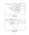
- FIGS. 29-34 Subsequent to the formation of holes 21 a , the grasping drill guide 20 (coracoid guide 20 ) is removed ( FIG. 28 ), and the coracoid graft 50 is brought into contact with offset guide 30 (glenoid guide 30 ) which was described in more detail with reference to FIGS. 9-12 .
- the coracoid graft 50 (attached to the offset guide 30 ) is subsequently positioned flush to the rim of glenoid 60 using the center flange 32 , as shown in FIGS. 29 and 30 .
- Two parallel guide pins 33 (for example, two 1.5 mm guide pins 33 ) are the inserted and drilled through cannulations 31 ( FIG. 29 ) of the offset guide 30 and into the glenoid 60 (preferably conducted with fluoroscopy).
- FIGS. 31-34 illustrate the subsequent insertion of pins 33 through the offset guide 30 .
- pins 33 are of different length to allow equal depth of penetration without inadvertent contact from the drill chuck. The shorter pin is preferably drilled first.
- FIG. 33 illustrates the removal of the offset guide 30 , leaving guide pins 33 in place.
- Depth gauge 40 FIG. 34
- Two fixation devices 44 (for example, two 3.75 mm self-tapping, cannulated titanium screws) are inserted using driver 45 , as shown in FIGS. 35 and 36 , to complete the coracoid graft fixation.
- the fixation devices 44 may preferably comprise screws available in 30 to 42 mm lengths, in 2 mm increments.
- the present invention has been described above with reference to a specific embodiment (i.e., a coracoid graft attached to the glenoid), the invention contemplates any attachment of a graft to bone, for example, any allograft or autograft attached to bone, or a graft from the illiac crest, or a tibial or scapular spine graft attached to any bone, among many others.
Abstract
Instrumentation for an open or arthroscopic Latarjet procedure that allows more control graft handling during the formation of the hole and proper positioning parallel to the articulating bone surface. The surgical instrumentation of the present invention includes an osteotome with a plurality of markings and a depth stop, a drill guide with jaws configured to securely engage an outer surface of the graft, and an offset guide to position the graft to the bone.
Description
This is a divisional of U.S. application Ser. No. 12/489,290, filed Jun. 22, 2009 now U.S. Pat. No. 8,257,359, which claims the benefit of U.S. Provisional Application No. 61/168,139, filed Apr. 9, 2009, and of U.S. Provisional Application No. 61/074,528, filed Jun. 20, 2008, the entire disclosures of which are incorporated by reference herein.
1. Field of the Invention
The present invention relates to osteotomies and, more specifically, to Latarjet instrumentation and methods of conducting a Latarjet procedure.
2. Description of the Related Art
The Latarjet procedure is known in the art and describes the coracoid bone block procedure. French surgeon Michel Latarjet first suggested that the horizontal limb of the coracoid process be fixed with a screw flush to the anteroinferior margin of the glenoid, making a horizontal incision through the fibers of the subscapularis. FIGS. 1 and 2 illustrate the attachment of the coracoid process 2 to the glenoid edge 4, to reduce instability during the Latarjet procedure. Although the Latarjet procedure has been constantly improved since 1954, specific instrumentation has not been designed to effectively address each step of the procedure. For example, surgeons are forced to improvise using general surgical instruments such as towel clamps to hold the coracoid process during cross hole drilling. This improvisation could result in non-parallel holes or, even worse, in breaking of the bone block which in turn could force abandonment of the procedure. Surgeons are also forced to “eyeball” the proper location of the coracoid graft, in an attempt to place it parallel to the articulating surface of the glenoid.
Accordingly, there is a need for improved instrumentation to be used during a Latarjet procedure, with reduced susceptibility to coracoid breakage, increased graft fixation, and correct alignment of the coracoid to the articulating surface of the glenoid.
The present invention provides improved instrumentation to be used during an open or arthroscopic procedure, that allows more control graft handling during the formation of the hole (drill hole formation) and proper positioning parallel to the articulating bone surface. The surgical instrumentation of the present invention includes, among others, an osteotome with a plurality of markings and a depth stop, a drill guide with jaws configured to securely engage an outer surface of the graft, and an offset guide to position the graft to the bone.
Other features and advantages of the present invention will become apparent from the following description of the invention, which refers to the accompanying drawings.
In the following detailed description, reference is made to various specific embodiments in which the invention may be practiced. These embodiments are described with sufficient detail to enable those skilled in the art to practice the invention, and it is to be understood that other embodiments may be employed, and that structural and logical changes may be made without departing from the spirit or scope of the present invention.
The present invention provides improved instrumentation configured to be employed during a Latarjet procedure, with reduced susceptibility to coracoid breakage and increased graft positioning and fixation.
Referring now to the drawings, where like elements are designated by like reference numerals, FIGS. 3-16 illustrate various surgical instruments of the present invention employed in conjunction with an open Latarjet procedure. FIGS. 17-36 illustrate a sequence of steps of an exemplary open Latarjet procedure conducted by employing a system of the present invention including the plurality of surgical instruments of FIGS. 3-16 . The unique surgical instrumentation of the present invention is configured to reduced susceptibility to coracoid breakage, increased graft positioning and fixation, and correct alignment of the coracoid to the articulating surface of the glenoid.
As shown in FIGS. 3-5 , coracoid graft 50 is retrieved using a special osteotome 10 including a protective shield 12 (FIGS. 3 and 5 ) and an osteotome handle 14 (FIGS. 3 and 5 ). Osteotome blade 11 may be disposable and includes depth markings 13 and a hard stop 15 at about 20 mm. The osteotome shield protects the surrounding soft tissue, nerves and blood supply from inadvertent damage during the coracoid osteotomy.
Referring to FIGS. 6-8 , the coracoid graft 50 is retrieved and secured with a grasping drill guide 20 (coracoid drill guide) that includes a ratcheting handle 25 and a pair of opposing jaws 26, 28 (a first jaw 26 and a second jaw 28) that are moveable with respect to each other. As shown in FIGS. 7 and 8 , the most distal portion of each jaw 26, 28 is enlarged and provided with a curved inner surface 29 to conform to the outer surface and shape of the coracoid graft to be grasped by the jaws, to allow secure engagement of the graft by the opposing jaws (as shown in FIG. 7 ). As also shown in FIG. 8 , one of the jaws (for example, jaw 26) is provided with a plurality of clearance slots 22, while the opposing jaw (for example, jaw 28) is provided with corresponding holes 24 (guide holes or drill holes 24). Clearance slots or openings 22 are larger than the drill holes 24, and are also elongated (as shown in FIG. 6 , for example) to ensure that the end of the drill will pass therethrough without contacting the jaw.
Grasping drill guide 20 (shown in more detail in FIG. 8 ) is positioned on the graft 50 with clearance slots 22 adjacent to the surface of the coracoid that will eventually be in contact with the glenoid (as detailed below). Guide holes 24 of the grasping drill guide 20 allow a surgeon to drill two parallel holes (for example, two 4 mm holes) through the graft 50 approximately in the center of the graft.
In additional embodiments, the offset guide 30 (glenoid drill guide 30) may be configured without an “offset” at all (i.e., with no finger or extension that sits on the surface of the glenoid). In this embodiment, the offset guide is provided with the protruding finger or extension cut off, so that it resembles more a pitchfork. The offset guide of this exemplary embodiment would still hold the coracoid graft in the same manner (as the offset guide 30 with the extension 32), but allows the surgeon to freely position the graft anywhere on the anterior edge of the glenoid. In this manner, surgeons who follow the “French” Latarjet style (rather than the Latarjet procedure of the present invention) are able to position the graft more medially than the offset guide 30 (with the extension 32) allows.
Preferably, the offset guide 30 is cannulated (as shown in FIG. 11 , for example), to allow insertion of guide pins through it directly (i.e., the guide pins are inserted directly through the cannulation(s) of the offset guide).
The coracoid graft 50 (attached to the offset guide 30) is subsequently positioned flush to the rim of glenoid 60 using the center flange 32 (extension 32), as shown in FIG. 9 . Two parallel guide pins 33 (for example, two 1.5 mm guide pins 33) are drilled directly through the offset guide 30 and into the glenoid 60 (preferably conducted with fluoroscopy), as shown in FIG. 12 . Preferably, pins 33 are of different length to allow equal depth of penetration without inadvertent contact from the drill chuck. The shorter pin is preferably drilled first.
As shown in FIGS. 15 and 16 , two fixation devices 44 (for example, two 3.75 mm self-tapping, cannulated titanium screws) are inserted using a driver 45 (for example, a 2.5 mm hex driver). The fixation devices may preferably comprise screws available in 30 to 42 mm lengths, in 2 mm increments.
In an alternative and exemplary only embodiment of the present invention, the offset guide 30 may be provided with only one post. This design allows use of the device through standard arthroscopic cannulas. As an example of how this instrument could be used, a surgeon could opt to use an illiac crest autograft (rather than a coracoid graft) to replace the missing glenoid bone. This illiac crest autograft could be shaped and sized and predrilled outside of the body. The graft would then be inserted into the shoulder. The single post offset guide would be arthroscopically inserted and mated with one of the predrilled graft holes. The guide would then be used to position the graft alongside the glenoid. A 1.5 mm guide pin would be drilled through the guide, which would then be repositioned onto the second predrilled hole, allowing the insertion of a second guide pin. The remainder of the technique would undergo as described above.
As shown in FIGS. 17 and 18 , coracoid graft 50 is retrieved using the special osteotome 10 described above with reference to FIGS. 3-5 . The shield 12 of the osteotome protects the surrounding soft tissue, nerves and blood supply from inadvertent damage during the coracoid osteotomy.
In an exemplary embodiment only, the coracoid graft 50 may be additionally shaped and sized (with the same osteotome or with a different cutting instrument, for example) to remove a lateral edge and to form a flat, cut surface 50 a, as shown in FIGS. 20-22 , for enhanced positioning alongside the glenoid. In this manner, the coracoid graft 50 is positioned so that the cut flat surface 50 a abuts the rim of the glenoid (FIGS. 22 and 23 ).
Once positioned on the glenoid rim, the coracoid graft 50 is secured within curved inner surfaces 29 of opposing jaws 26, 28 of the grasping drill guide 20 (coracoid guide), as shown in FIGS. 24 and 25 .
Reference is now made to FIGS. 29-34 . Subsequent to the formation of holes 21 a, the grasping drill guide 20 (coracoid guide 20) is removed (FIG. 28 ), and the coracoid graft 50 is brought into contact with offset guide 30 (glenoid guide 30) which was described in more detail with reference to FIGS. 9-12 .
The coracoid graft 50 (attached to the offset guide 30) is subsequently positioned flush to the rim of glenoid 60 using the center flange 32, as shown in FIGS. 29 and 30 . Two parallel guide pins 33 (for example, two 1.5 mm guide pins 33) are the inserted and drilled through cannulations 31 (FIG. 29 ) of the offset guide 30 and into the glenoid 60 (preferably conducted with fluoroscopy). FIGS. 31-34 illustrate the subsequent insertion of pins 33 through the offset guide 30. Preferably, pins 33 are of different length to allow equal depth of penetration without inadvertent contact from the drill chuck. The shorter pin is preferably drilled first.
Although the present invention has been described above with reference to a specific embodiment (i.e., a coracoid graft attached to the glenoid), the invention contemplates any attachment of a graft to bone, for example, any allograft or autograft attached to bone, or a graft from the illiac crest, or a tibial or scapular spine graft attached to any bone, among many others.
Although the present invention has been described in relation to particular embodiments thereof, many other variations and modifications and other uses will become apparent to those skilled in the art. It is preferred, therefore, that the present invention be limited not by the specific disclosure herein, but only by the appended claims.
Claims (8)
1. An instrument system for a Latarjet procedure, comprising:
an osteotome comprising a blade with a plurality of markings and a depth stop at about 20 mm, the osteotome further comprising a protective shield to protect surrounding soft tissue, nerves and blood supply from inadvertent damage during coracoid osteotomy to obtain a coracoid graft;
a drill guide comprising a pair of opposing jaws configured to securely engage the coracoid graft and to drill at least one hole through the coracoid graft, wherein the pair of opposing jaws comprise a first jaw and a second jaw, wherein one of the first and second jaws comprises a plurality of holes, and wherein the other of the first and second jaws comprises a plurality of slots, and wherein each of the first and second jaws comprises an enlarged distal tip with a curved inner surface which is complementary to an outer surface of the coracoid graft; and
a glenoid drill guide configured to attach the coracoid graft to the glenoid rim and to drill at least one guide pin through the glenoid drill guide and into the glenoid, wherein the glenoid drill guide is cannulated to allow insertion directly therethrough of at least one guide pin.
2. The system of claim 1 , wherein the blade of the osteotome is disposable.
3. The system of claim 1 , further comprising:
a depth gauge to determine a length of a fixation device to be secured to the coracoid graft; and
at least one fixation device configured to attach the coracoid graft to the glenoid rim.
4. The system of claim 1 , wherein the slots are larger than the holes.
5. The system of claim 1 , wherein the drill guide comprises a ratchet handle.
6. The system of claim 1 , wherein the glenoid drill guide is provided with an extension having a predetermined length.
7. The system of claim 6 , wherein the predetermined length of the extension is one of a 4 mm, 6 mm, or 8 mm.
8. The system of claim 6 , wherein the predetermined length of the glenoid drill guide is about equal to a width of the coracoid graft.
Priority Applications (1)
| Application Number | Priority Date | Filing Date | Title |
|---|---|---|---|
| US13/564,782 US9107676B2 (en) | 2008-06-20 | 2012-08-02 | Latarjet instrumentation and method |
Applications Claiming Priority (4)
| Application Number | Priority Date | Filing Date | Title |
|---|---|---|---|
| US7452808P | 2008-06-20 | 2008-06-20 | |
| US16813909P | 2009-04-09 | 2009-04-09 | |
| US12/489,290 US8257359B2 (en) | 2008-06-20 | 2009-06-22 | Latarjet instrumentation and method |
| US13/564,782 US9107676B2 (en) | 2008-06-20 | 2012-08-02 | Latarjet instrumentation and method |
Related Parent Applications (1)
| Application Number | Title | Priority Date | Filing Date |
|---|---|---|---|
| US12/489,290 Division US8257359B2 (en) | 2008-06-20 | 2009-06-22 | Latarjet instrumentation and method |
Publications (2)
| Publication Number | Publication Date |
|---|---|
| US20120296338A1 US20120296338A1 (en) | 2012-11-22 |
| US9107676B2 true US9107676B2 (en) | 2015-08-18 |
Family
ID=41165125
Family Applications (2)
| Application Number | Title | Priority Date | Filing Date |
|---|---|---|---|
| US12/489,290 Active 2031-01-16 US8257359B2 (en) | 2008-06-20 | 2009-06-22 | Latarjet instrumentation and method |
| US13/564,782 Active 2030-05-27 US9107676B2 (en) | 2008-06-20 | 2012-08-02 | Latarjet instrumentation and method |
Family Applications Before (1)
| Application Number | Title | Priority Date | Filing Date |
|---|---|---|---|
| US12/489,290 Active 2031-01-16 US8257359B2 (en) | 2008-06-20 | 2009-06-22 | Latarjet instrumentation and method |
Country Status (4)
| Country | Link |
|---|---|
| US (2) | US8257359B2 (en) |
| EP (1) | EP2135566B1 (en) |
| AT (1) | ATE500786T1 (en) |
| DE (1) | DE602009000826D1 (en) |
Cited By (18)
| Publication number | Priority date | Publication date | Assignee | Title |
|---|---|---|---|---|
| US9204873B2 (en) | 2000-05-01 | 2015-12-08 | Arthrosurface Incorporated | System and method for joint resurface repair |
| US9283076B2 (en) | 2009-04-17 | 2016-03-15 | Arthrosurface Incorporated | Glenoid resurfacing system and method |
| US9351745B2 (en) | 2003-02-24 | 2016-05-31 | Arthrosurface Incorporated | Trochlear resurfacing system and method |
| US9358029B2 (en) | 2006-12-11 | 2016-06-07 | Arthrosurface Incorporated | Retrograde resection apparatus and method |
| US9357989B2 (en) | 2000-05-01 | 2016-06-07 | Arthrosurface Incorporated | System and method for joint resurface repair |
| US9468448B2 (en) | 2012-07-03 | 2016-10-18 | Arthrosurface Incorporated | System and method for joint resurfacing and repair |
| US9492200B2 (en) | 2013-04-16 | 2016-11-15 | Arthrosurface Incorporated | Suture system and method |
| US9662126B2 (en) | 2009-04-17 | 2017-05-30 | Arthrosurface Incorporated | Glenoid resurfacing system and method |
| US9861492B2 (en) | 2014-03-07 | 2018-01-09 | Arthrosurface Incorporated | Anchor for an implant assembly |
| US10076343B2 (en) | 2002-12-03 | 2018-09-18 | Arthrosurface Incorporated | System for articular surface replacement |
| US10624748B2 (en) | 2014-03-07 | 2020-04-21 | Arthrosurface Incorporated | System and method for repairing articular surfaces |
| US10624752B2 (en) | 2006-07-17 | 2020-04-21 | Arthrosurface Incorporated | Tibial resurfacing system and method |
| US10945743B2 (en) | 2009-04-17 | 2021-03-16 | Arthrosurface Incorporated | Glenoid repair system and methods of use thereof |
| RU2752808C1 (en) * | 2020-08-11 | 2021-08-06 | Дмитрий Олегович Ильин | Guide for latarjet operation |
| US11160663B2 (en) | 2017-08-04 | 2021-11-02 | Arthrosurface Incorporated | Multicomponent articular surface implant |
| US11478358B2 (en) | 2019-03-12 | 2022-10-25 | Arthrosurface Incorporated | Humeral and glenoid articular surface implant systems and methods |
| US11607319B2 (en) | 2014-03-07 | 2023-03-21 | Arthrosurface Incorporated | System and method for repairing articular surfaces |
| US11712276B2 (en) | 2011-12-22 | 2023-08-01 | Arthrosurface Incorporated | System and method for bone fixation |
Families Citing this family (37)
| Publication number | Priority date | Publication date | Assignee | Title |
|---|---|---|---|---|
| GB2479694B (en) * | 2010-03-17 | 2012-02-08 | Thomas Maurice Stewart Gregory | Shoulder replacement apparatus |
| BE1019467A4 (en) * | 2010-09-02 | 2012-07-03 | Ceuster Marcel De | SURGICAL APPLICATION SYSTEM TO ENLARGE THE GLENOID WEAR SURFACE. |
| US8926661B2 (en) * | 2011-06-02 | 2015-01-06 | Smith & Nephew, Inc. | Surgical fastening |
| US10052114B2 (en) | 2011-07-12 | 2018-08-21 | Materialise, Nv | Shoulder base plate coverage and stability |
| US9320557B2 (en) * | 2011-07-21 | 2016-04-26 | Smith & Nephew, Inc. | Implant retaining loop and guide wire extension |
| BE1020248A4 (en) * | 2011-09-13 | 2013-07-02 | Ceuster Marcel De | SURGICAL APPLICATION SYSTEM TO ENLARGE THE GLENOID WEAR SURFACE: BLOCK. |
| US20140058446A1 (en) * | 2011-09-28 | 2014-02-27 | Avi Bernstein | Spinal implant system |
| US20130158610A1 (en) * | 2011-12-16 | 2013-06-20 | Depuy Mitek, Inc. | Bone graft fixation systems and methods |
| EP2630935B1 (en) * | 2012-02-27 | 2014-12-31 | Arthrex, Inc. | Glenoid extension block |
| FR2996114B1 (en) * | 2012-10-02 | 2015-02-13 | Sem Sa | DRILLING GUIDE FOR ANTERIOR SHOULDER STOOL |
| JP2016513566A (en) | 2013-03-15 | 2016-05-16 | スミス アンド ネフュー インコーポレーテッドSmith & Nephew,Inc. | Surgical fixation device |
| US9386997B2 (en) * | 2013-03-29 | 2016-07-12 | Smith & Nephew, Inc. | Tunnel gage |
| US10123813B2 (en) | 2014-02-17 | 2018-11-13 | Smith & Nephew, Inc. | Drill guide |
| US9949745B2 (en) | 2014-07-24 | 2018-04-24 | Medos International Sarl | Systems, devices, and methods for guiding surgical devices into bone |
| US10123832B2 (en) * | 2014-08-08 | 2018-11-13 | Stryker European Holdings I, Llc | Surgical forceps system |
| US10751161B2 (en) | 2014-10-23 | 2020-08-25 | Medos International Sárl | Biceps tenodesis anchor implants |
| US10034742B2 (en) | 2014-10-23 | 2018-07-31 | Medos International Sarl | Biceps tenodesis implants and delivery tools |
| US10729419B2 (en) | 2014-10-23 | 2020-08-04 | Medos International Sarl | Biceps tenodesis implants and delivery tools |
| US10856966B2 (en) | 2014-10-23 | 2020-12-08 | Medos International Sarl | Biceps tenodesis implants and delivery tools |
| US10076374B2 (en) | 2014-10-23 | 2018-09-18 | Medos International Sárl | Biceps tenodesis delivery tools |
| WO2016145152A1 (en) * | 2015-03-10 | 2016-09-15 | Smith & Nephew, Inc. | Open latarjet for correction of anterior-inferior glenoid bone loss |
| US9693856B2 (en) | 2015-04-22 | 2017-07-04 | DePuy Synthes Products, LLC | Biceps repair device |
| CN104783888A (en) * | 2015-04-22 | 2015-07-22 | 邓宇 | Drill stabilizing device for inclined bone surface drilling |
| CN106264652B (en) * | 2015-06-09 | 2019-04-16 | 北京纳通科技集团有限公司 | Osteotomy tool |
| US10172627B2 (en) * | 2015-06-29 | 2019-01-08 | Smith & Nephew, Inc. | Graft placement system and method |
| US9918769B2 (en) | 2015-08-28 | 2018-03-20 | Arthrex, Inc. | Instrumentation and technique for sizing a bone reconstruction graft |
| WO2017100581A1 (en) * | 2015-12-11 | 2017-06-15 | Smith & Nephew, Inc. | Coracoid drill guide |
| US10327789B2 (en) | 2015-12-29 | 2019-06-25 | Medos International Sarl | Methods and systems for preparing bone for a surgical procedure |
| US10231823B2 (en) | 2016-04-08 | 2019-03-19 | Medos International Sarl | Tenodesis implants and tools |
| US10231824B2 (en) | 2016-04-08 | 2019-03-19 | Medos International Sárl | Tenodesis anchoring systems and tools |
| FR3074415B1 (en) * | 2017-12-05 | 2022-02-25 | Newclip Int | ANCILLARY FOR BONE SURGERY, ADVANTAGEOUSLY FOR OPERATIVE ARTHROSCOPY |
| FR3087105B1 (en) * | 2018-10-11 | 2021-07-16 | V I M S Video Interventionnelle Medicale Scient | ANCILLARY INSTRUMENT FOR ARTHROSCOPIC LATARJET OPERATION |
| CN109223094B (en) * | 2018-10-16 | 2023-11-17 | 中国人民解放军陆军军医大学第一附属医院 | Novel coracoid bone osteotomy device |
| EP3897410A2 (en) * | 2018-12-21 | 2021-10-27 | Azurmeds Inc | Instrumental system for a latarjet procedure and corresponding method |
| US11103292B2 (en) | 2019-04-03 | 2021-08-31 | Arthrex, Inc. | Bone cerclage constructs and methods |
| US11517334B1 (en) * | 2019-11-04 | 2022-12-06 | Smith & Nephew, Inc. | Patient-specific guides for latarjet procedure |
| US11369486B2 (en) * | 2020-05-29 | 2022-06-28 | Arthrex, Inc. | Arthroscopic anterior glenoid guide |
Citations (12)
| Publication number | Priority date | Publication date | Assignee | Title |
|---|---|---|---|---|
| US2181746A (en) * | 1939-02-04 | 1939-11-28 | John R Siebrandt | Combination bone clamp and adjustable drill guide |
| US5409490A (en) * | 1993-08-16 | 1995-04-25 | Depuy Inc. | Shoulder separation reconstruction |
| US5643272A (en) | 1994-09-02 | 1997-07-01 | Hudson Surgical Design, Inc. | Method and apparatus for tibial resection |
| US5697933A (en) | 1995-12-18 | 1997-12-16 | Medicinelodge, Inc. | Bone-tendon-bone drill guide |
| EP1034744A1 (en) | 1999-03-09 | 2000-09-13 | Orthomed | Kit for surgical reduction of instabilities of the shoulder |
| US6174311B1 (en) * | 1998-10-28 | 2001-01-16 | Sdgi Holdings, Inc. | Interbody fusion grafts and instrumentation |
| US20050090829A1 (en) * | 2003-10-23 | 2005-04-28 | Osteotech, Inc. | Spinal bone chisels |
| US20060074426A1 (en) * | 2002-12-27 | 2006-04-06 | Lieberman Isador H | Apparatus for cutting bone |
| US20060074429A1 (en) * | 2004-10-06 | 2006-04-06 | Ralph James D | Adjustable angle handle for surgical instruments |
| WO2008015670A2 (en) | 2006-07-31 | 2008-02-07 | T.A.G. Medical Products A Limited Partnership | Arthroscopic bone transplanting procedure, and medical instruments useful therein |
| US7527631B2 (en) | 2003-03-31 | 2009-05-05 | Depuy Products, Inc. | Arthroplasty sizing gauge |
| US20090318977A1 (en) | 2008-06-20 | 2009-12-24 | Giovanni Di Giacomo | Wedged profile plate |
-
2009
- 2009-06-18 EP EP09163161A patent/EP2135566B1/en active Active
- 2009-06-18 DE DE602009000826T patent/DE602009000826D1/en active Active
- 2009-06-18 AT AT09163161T patent/ATE500786T1/en active
- 2009-06-22 US US12/489,290 patent/US8257359B2/en active Active
-
2012
- 2012-08-02 US US13/564,782 patent/US9107676B2/en active Active
Patent Citations (14)
| Publication number | Priority date | Publication date | Assignee | Title |
|---|---|---|---|---|
| US2181746A (en) * | 1939-02-04 | 1939-11-28 | John R Siebrandt | Combination bone clamp and adjustable drill guide |
| US5409490A (en) * | 1993-08-16 | 1995-04-25 | Depuy Inc. | Shoulder separation reconstruction |
| US5643272A (en) | 1994-09-02 | 1997-07-01 | Hudson Surgical Design, Inc. | Method and apparatus for tibial resection |
| US5697933A (en) | 1995-12-18 | 1997-12-16 | Medicinelodge, Inc. | Bone-tendon-bone drill guide |
| US6174311B1 (en) * | 1998-10-28 | 2001-01-16 | Sdgi Holdings, Inc. | Interbody fusion grafts and instrumentation |
| EP1034744A1 (en) | 1999-03-09 | 2000-09-13 | Orthomed | Kit for surgical reduction of instabilities of the shoulder |
| US20060074426A1 (en) * | 2002-12-27 | 2006-04-06 | Lieberman Isador H | Apparatus for cutting bone |
| US7527631B2 (en) | 2003-03-31 | 2009-05-05 | Depuy Products, Inc. | Arthroplasty sizing gauge |
| US20050090829A1 (en) * | 2003-10-23 | 2005-04-28 | Osteotech, Inc. | Spinal bone chisels |
| US20060074429A1 (en) * | 2004-10-06 | 2006-04-06 | Ralph James D | Adjustable angle handle for surgical instruments |
| WO2008015670A2 (en) | 2006-07-31 | 2008-02-07 | T.A.G. Medical Products A Limited Partnership | Arthroscopic bone transplanting procedure, and medical instruments useful therein |
| US20100069974A1 (en) | 2006-07-31 | 2010-03-18 | Ran Oren | Arthroscopic bone transplanting procedure, and medical instruments useful therein |
| US8617219B2 (en) * | 2006-07-31 | 2013-12-31 | T.A.G. Medical Devices—Agriculture Cooperative Ltd. | Arthroscopic bone transplanting procedure, and medical instruments useful therein |
| US20090318977A1 (en) | 2008-06-20 | 2009-12-24 | Giovanni Di Giacomo | Wedged profile plate |
Non-Patent Citations (1)
| Title |
|---|
| EP1034744A1 Translation, Aug. 19, 2014. * |
Cited By (36)
| Publication number | Priority date | Publication date | Assignee | Title |
|---|---|---|---|---|
| US9204873B2 (en) | 2000-05-01 | 2015-12-08 | Arthrosurface Incorporated | System and method for joint resurface repair |
| US9357989B2 (en) | 2000-05-01 | 2016-06-07 | Arthrosurface Incorporated | System and method for joint resurface repair |
| US10076343B2 (en) | 2002-12-03 | 2018-09-18 | Arthrosurface Incorporated | System for articular surface replacement |
| US10624749B2 (en) | 2003-02-24 | 2020-04-21 | Arthrosurface Incorporated | Trochlear resurfacing system and method |
| US9351745B2 (en) | 2003-02-24 | 2016-05-31 | Arthrosurface Incorporated | Trochlear resurfacing system and method |
| US9931211B2 (en) | 2003-02-24 | 2018-04-03 | Arthrosurface Incorporated | Trochlear resurfacing system and method |
| US11337819B2 (en) | 2003-02-24 | 2022-05-24 | Arthrosurface Incorporated | Trochlear resurfacing system and method |
| US11471289B2 (en) | 2006-07-17 | 2022-10-18 | Arthrosurface Incorporated | Tibial resurfacing system and method |
| US10624752B2 (en) | 2006-07-17 | 2020-04-21 | Arthrosurface Incorporated | Tibial resurfacing system and method |
| US9358029B2 (en) | 2006-12-11 | 2016-06-07 | Arthrosurface Incorporated | Retrograde resection apparatus and method |
| US10045788B2 (en) | 2006-12-11 | 2018-08-14 | Arthrosurface Incorporated | Retrograde resection apparatus and method |
| US10959740B2 (en) | 2006-12-11 | 2021-03-30 | Arthrosurface Incorporated | Retrograde resection apparatus and method |
| US9662126B2 (en) | 2009-04-17 | 2017-05-30 | Arthrosurface Incorporated | Glenoid resurfacing system and method |
| US11478259B2 (en) | 2009-04-17 | 2022-10-25 | Arthrosurface, Incorporated | Glenoid resurfacing system and method |
| US9283076B2 (en) | 2009-04-17 | 2016-03-15 | Arthrosurface Incorporated | Glenoid resurfacing system and method |
| US10478200B2 (en) | 2009-04-17 | 2019-11-19 | Arthrosurface Incorporated | Glenoid resurfacing system and method |
| US10945743B2 (en) | 2009-04-17 | 2021-03-16 | Arthrosurface Incorporated | Glenoid repair system and methods of use thereof |
| US11712276B2 (en) | 2011-12-22 | 2023-08-01 | Arthrosurface Incorporated | System and method for bone fixation |
| US11191552B2 (en) | 2012-07-03 | 2021-12-07 | Arthrosurface, Incorporated | System and method for joint resurfacing and repair |
| US9468448B2 (en) | 2012-07-03 | 2016-10-18 | Arthrosurface Incorporated | System and method for joint resurfacing and repair |
| US10307172B2 (en) | 2012-07-03 | 2019-06-04 | Arthrosurface Incorporated | System and method for joint resurfacing and repair |
| US9492200B2 (en) | 2013-04-16 | 2016-11-15 | Arthrosurface Incorporated | Suture system and method |
| US10695096B2 (en) | 2013-04-16 | 2020-06-30 | Arthrosurface Incorporated | Suture system and method |
| US11648036B2 (en) | 2013-04-16 | 2023-05-16 | Arthrosurface Incorporated | Suture system and method |
| US9962265B2 (en) | 2014-03-07 | 2018-05-08 | Arthrosurface Incorporated | System and method for repairing articular surfaces |
| US11083587B2 (en) | 2014-03-07 | 2021-08-10 | Arthrosurface Incorporated | Implant and anchor assembly |
| US10624754B2 (en) | 2014-03-07 | 2020-04-21 | Arthrosurface Incorporated | System and method for repairing articular surfaces |
| US9931219B2 (en) | 2014-03-07 | 2018-04-03 | Arthrosurface Incorporated | Implant and anchor assembly |
| US9861492B2 (en) | 2014-03-07 | 2018-01-09 | Arthrosurface Incorporated | Anchor for an implant assembly |
| US11607319B2 (en) | 2014-03-07 | 2023-03-21 | Arthrosurface Incorporated | System and method for repairing articular surfaces |
| US10575957B2 (en) | 2014-03-07 | 2020-03-03 | Arthrosurface Incoporated | Anchor for an implant assembly |
| US10624748B2 (en) | 2014-03-07 | 2020-04-21 | Arthrosurface Incorporated | System and method for repairing articular surfaces |
| US11766334B2 (en) | 2014-03-07 | 2023-09-26 | Arthrosurface Incorporated | System and method for repairing articular surfaces |
| US11160663B2 (en) | 2017-08-04 | 2021-11-02 | Arthrosurface Incorporated | Multicomponent articular surface implant |
| US11478358B2 (en) | 2019-03-12 | 2022-10-25 | Arthrosurface Incorporated | Humeral and glenoid articular surface implant systems and methods |
| RU2752808C1 (en) * | 2020-08-11 | 2021-08-06 | Дмитрий Олегович Ильин | Guide for latarjet operation |
Also Published As
| Publication number | Publication date |
|---|---|
| US20120296338A1 (en) | 2012-11-22 |
| ATE500786T1 (en) | 2011-03-15 |
| DE602009000826D1 (en) | 2011-04-21 |
| EP2135566A1 (en) | 2009-12-23 |
| US8257359B2 (en) | 2012-09-04 |
| US20090318923A1 (en) | 2009-12-24 |
| EP2135566B1 (en) | 2011-03-09 |
Similar Documents
| Publication | Publication Date | Title |
|---|---|---|
| US9107676B2 (en) | Latarjet instrumentation and method | |
| JP6319778B2 (en) | Medical device and medical kit for attaching sutures to bone | |
| US9931145B2 (en) | Flexible bone screw | |
| JP5129816B2 (en) | Arthroscopic bone grafting and medical devices useful for it | |
| JP5507463B2 (en) | Distal tibial plate fixation device | |
| US8801760B2 (en) | Growth control device | |
| US5312412A (en) | Fixation alignment guide for surgical use | |
| US7959564B2 (en) | Pedicle seeker and retractor, and methods of use | |
| JP5595522B2 (en) | Surgical system | |
| AU2004249313B2 (en) | Bone plates with intraoperatively tapped apertures | |
| US11937833B2 (en) | Chevron osteotomy tools and methods | |
| EP1878394A2 (en) | Threaded guide for an orthopaedic fixation plate | |
| EP1723917A1 (en) | Apparatus for bone fastener implantation | |
| JP2003199757A (en) | Surgical guide system for stabilization of spine | |
| JP2006326303A (en) | Bone fixation device | |
| US20150313640A1 (en) | Surgical instrument with movable guide and sleeve | |
| US20150100080A1 (en) | Reduction forceps with dual 90°-deformation for distribution among two planes | |
| US11950791B2 (en) | Coracoid drill guide assembly and methods of use thereof | |
| US20140081341A1 (en) | Bone fixation guide device | |
| JP2021517009A (en) | Arthroscopic shoulder joint formation, its components, its instruments, and its methods | |
| AU2017202188B2 (en) | Arthroscopic Bone Transplanting Procedure, and Medical Instruments Useful Therein |
Legal Events
| Date | Code | Title | Description |
|---|---|---|---|
| STCF | Information on status: patent grant |
Free format text: PATENTED CASE |
|
| MAFP | Maintenance fee payment |
Free format text: PAYMENT OF MAINTENANCE FEE, 4TH YEAR, LARGE ENTITY (ORIGINAL EVENT CODE: M1551); ENTITY STATUS OF PATENT OWNER: LARGE ENTITY Year of fee payment: 4 |
|
| MAFP | Maintenance fee payment |
Free format text: PAYMENT OF MAINTENANCE FEE, 8TH YEAR, LARGE ENTITY (ORIGINAL EVENT CODE: M1552); ENTITY STATUS OF PATENT OWNER: LARGE ENTITY Year of fee payment: 8 |