US9029093B2 - Methods and compositions for diagnosis and prognosis of renal injury and renal failure - Google Patents
Methods and compositions for diagnosis and prognosis of renal injury and renal failureInfo
- Publication number
- US9029093B2 US9029093B2 US13/580,878 US201113580878A US9029093B2 US 9029093 B2 US9029093 B2 US 9029093B2 US 201113580878 A US201113580878 A US 201113580878A US 9029093 B2 US9029093 B2 US 9029093B2
- Authority
- US
- United States
- Prior art keywords
- rifle
- subject
- threshold
- plasma
- renal
- Prior art date
- Legal status (The legal status is an assumption and is not a legal conclusion. Google has not performed a legal analysis and makes no representation as to the accuracy of the status listed.)
- Expired - Fee Related
Links
Images


Classifications
-
- G—PHYSICS
- G01—MEASURING; TESTING
- G01N—INVESTIGATING OR ANALYSING MATERIALS BY DETERMINING THEIR CHEMICAL OR PHYSICAL PROPERTIES
- G01N33/00—Investigating or analysing materials by specific methods not covered by groups G01N1/00 - G01N31/00
- G01N33/48—Biological material, e.g. blood, urine; Haemocytometers
- G01N33/50—Chemical analysis of biological material, e.g. blood, urine; Testing involving biospecific ligand binding methods; Immunological testing
- G01N33/68—Chemical analysis of biological material, e.g. blood, urine; Testing involving biospecific ligand binding methods; Immunological testing involving proteins, peptides or amino acids
- G01N33/6893—Chemical analysis of biological material, e.g. blood, urine; Testing involving biospecific ligand binding methods; Immunological testing involving proteins, peptides or amino acids related to diseases not provided for elsewhere
-
- G—PHYSICS
- G01—MEASURING; TESTING
- G01N—INVESTIGATING OR ANALYSING MATERIALS BY DETERMINING THEIR CHEMICAL OR PHYSICAL PROPERTIES
- G01N2333/00—Assays involving biological materials from specific organisms or of a specific nature
- G01N2333/435—Assays involving biological materials from specific organisms or of a specific nature from animals; from humans
- G01N2333/705—Assays involving receptors, cell surface antigens or cell surface determinants
- G01N2333/70503—Immunoglobulin superfamily, e.g. VCAMs, PECAM, LFA-3
- G01N2333/70539—MHC-molecules, e.g. HLA-molecules
-
- G—PHYSICS
- G01—MEASURING; TESTING
- G01N—INVESTIGATING OR ANALYSING MATERIALS BY DETERMINING THEIR CHEMICAL OR PHYSICAL PROPERTIES
- G01N2333/00—Assays involving biological materials from specific organisms or of a specific nature
- G01N2333/81—Protease inhibitors
- G01N2333/8107—Endopeptidase (E.C. 3.4.21-99) inhibitors
-
- G—PHYSICS
- G01—MEASURING; TESTING
- G01N—INVESTIGATING OR ANALYSING MATERIALS BY DETERMINING THEIR CHEMICAL OR PHYSICAL PROPERTIES
- G01N2800/00—Detection or diagnosis of diseases
- G01N2800/34—Genitourinary disorders
- G01N2800/347—Renal failures; Glomerular diseases; Tubulointerstitial diseases, e.g. nephritic syndrome, glomerulonephritis; Renovascular diseases, e.g. renal artery occlusion, nephropathy
-
- G—PHYSICS
- G01—MEASURING; TESTING
- G01N—INVESTIGATING OR ANALYSING MATERIALS BY DETERMINING THEIR CHEMICAL OR PHYSICAL PROPERTIES
- G01N2800/00—Detection or diagnosis of diseases
- G01N2800/60—Complex ways of combining multiple protein biomarkers for diagnosis
Definitions
- the kidney is responsible for water and solute excretion from the body. Its functions include maintenance of acid-base balance, regulation of electrolyte concentrations, control of blood volume, and regulation of blood pressure. As such, loss of kidney function through injury and/or disease results in substantial morbidity and mortality. A detailed discussion of renal injuries is provided in Harrison's Principles of Internal Medicine, 17 th Ed., McGraw Hill, New York, pages 1741-1830, which are hereby incorporated by reference in their entirety. Renal disease and/or injury may be acute or chronic.
- Acute and chronic kidney disease are described as follows (from Current Medical Diagnosis & Treatment 2008, 47 th Ed, McGraw Hill, New York, pages 785-815, which are hereby incorporated by reference in their entirety): “Acute renal failure is worsening of renal function over hours to days, resulting in the retention of nitrogenous wastes (such as urea nitrogen) and creatinine in the blood. Retention of these substances is called azotemia.
- Chronic renal failure results from an abnormal loss of renal function over months to years”.
- Acute renal failure also known as acute kidney injury, or AKI
- AKI acute kidney injury
- ischemic ARF the course of the disease may be divided into four phases.
- an initiation phase which lasts hours to days, reduced perfusion of the kidney is evolving into injury. Glomerular ultrafiltration reduces, the flow of filtrate is reduced due to debris within the tubules, and back leakage of filtrate through injured epithelium occurs.
- Renal injury can be mediated during this phase by reperfusion of the kidney.
- Initiation is followed by an extension phase which is characterized by continued ischemic injury and inflammation and may involve endothelial damage and vascular congestion.
- the maintenance phase lasting from 1 to 2 weeks, renal cell injury occurs, and glomerular filtration and urine output reaches a minimum.
- a recovery phase can follow in which the renal epithelium is repaired and GFR gradually recovers. Despite this, the survival rate of subjects with ARF may be as low as about 60%.
- Acute kidney injury caused by radiocontrast agents also called contrast media
- other nephrotoxins such as cyclosporine, antibiotics including aminoglycosides and anticancer drugs such as cisplatin manifests over a period of days to about a week.
- Contrast induced nephropathy (CIN, which is AKI caused by radiocontrast agents) is thought to be caused by intrarenal vasoconstriction (leading to ischemic injury) and from the generation of reactive oxygen species that are directly toxic to renal tubular epithelial cells.
- CIN classically presents as an acute (onset within 24-48 h) but reversible (peak 3-5 days, resolution within 1 week) rise in blood urea nitrogen and serum creatinine.
- a commonly reported criteria for defining and detecting AKI is an abrupt (typically within about 2-7 days or within a period of hospitalization) elevation of serum creatinine.
- serum creatinine elevation to define and detect AKI is well established, the magnitude of the serum creatinine elevation and the time over which it is measured to define AKI varies considerably among publications.
- relatively large increases in serum creatinine such as 100%, 200%, an increase of at least 100% to a value over 2 mg/dL and other definitions were used to define AKI.
- the recent trend has been towards using smaller serum creatinine rises to define AKI.
- “Failure” serum creatinine increased 3.0 fold from baseline OR creatinine >355 ⁇ mol/l (with a rise of >44) or urine output below 0.3 ml/kg/hr for 24 h or anuria for at least 12 hours;
- “Loss” persistent need for renal replacement therapy for more than four weeks.
- ESRD end stage renal disease—the need for dialysis for more than 3 months.
- RIFLE criteria which provide a useful clinical tool to classify renal status.
- the RIFLE criteria provide a uniform definition of AKI which has been validated in numerous studies.
- “Stage I” increase in serum creatinine of more than or equal to 0.3 mg/dL ( ⁇ 26.4 ⁇ mol/L) or increase to more than or equal to 150% (1.5-fold) from baseline OR urine output less than 0.5 mL/kg per hour for more than 6 hours;
- Stage II increase in serum creatinine to more than 200% (>2-fold) from baseline OR urine output less than 0.5 mL/kg per hour for more than 12 hours;
- Stage III increase in serum creatinine to more than 300% (>3-fold) from baseline OR serum creatinine ⁇ 354 ⁇ mol/L accompanied by an acute increase of at least 44 ⁇ mol/L OR urine output less than 0.3 mL/kg per hour for 24 hours or anuria for 12 hours.
- the CIN Consensus Working Panel uses a serum creatinine rise of 25% to define Contrast induced nephropathy (which is a type of AKI).
- Contrast induced nephropathy which is a type of AKI.
- various groups propose slightly different criteria for using serum creatinine to detect AKI, the consensus is that small changes in serum creatinine, such as 0.3 mg/dL or 25%, are sufficient to detect AKI (worsening renal function) and that the magnitude of the serum creatinine change is an indicator of the severity of the AKI and mortality risk.
- serum creatinine is generally regarded to have several limitations in the diagnosis, assessment and monitoring of AKI patients.
- the time period for serum creatinine to rise to values (e.g., a 0.3 mg/dL or 25% rise) considered diagnostic for AKI can be 48 hours or longer depending on the definition used. Since cellular injury in AKI can occur over a period of hours, serum creatinine elevations detected at 48 hours or longer can be a late indicator of injury, and relying on serum creatinine can thus delay diagnosis of AKI.
- serum creatinine is not a good indicator of the exact kidney status and treatment needs during the most acute phases of AKI when kidney function is changing rapidly. Some patients with AKI will recover fully, some will need dialysis (either short term or long term) and some will have other detrimental outcomes including death, major adverse cardiac events and chronic kidney disease. Because serum creatinine is a marker of filtration rate, it does not differentiate between the causes of AKI (pre-renal, intrinsic renal, post-renal obstruction, atheroembolic, etc) or the category or location of injury in intrinsic renal disease (for example, tubular, glomerular or interstitial in origin). Urine output is similarly limited, Knowing these things can be of vital importance in managing and treating patients with AKI.
- biomarker panel approach can be used for diagnosis, prognosis, risk stratification, staging, monitoring, categorizing and determination of further diagnosis and treatment regimens in subjects suffering or at risk of suffering from an injury to renal function, reduced renal function, and/or acute renal failure (also called acute kidney injury).
- Preferred biomarker panels comprise two, three, four, five, or more assays configured to detect two, three, four, five, or more of the biomarkers listed in Table 2 herein.
- kidney injury markers may be used in panels comprising a plurality of kidney injury markers, for risk stratification (that is, to identify subjects at risk for a future injury to renal function, for future progression to reduced renal function, for future progression to ARF, for future improvement in renal function, etc.); for diagnosis of existing disease (that is, to identify subjects who have suffered an injury to renal function, who have progressed to reduced renal function, who have progressed to ARF, etc.); for monitoring for deterioration or improvement of renal function; and for predicting a future medical outcome, such as improved or worsening renal function, a decreased or increased mortality risk, a decreased or increased risk that a subject will require renal replacement therapy (i.e., hemodialysis, peritoneal dialysis, hemofiltration, and/or renal transplantation, a decreased or increased risk that a subject will recover from an injury to renal function, a decreased or increased risk that a subject will recover from ARF, a decreased or increased risk that a subject will progress to end stage renal disease, a decreased or increased risk that
- the present invention relates to methods for evaluating renal status in a subject. These methods comprise performing an assay method that is configured to detect one or more kidney injury markers of the present invention in a body fluid sample obtained from the subject. A plurality of assay results, for example comprising a measured concentration of one or more markers selected from the group consisting of the markers listed in Table 2 herein are then correlated to the renal status of the subject. This correlation to renal status may include correlating the assay result(s) to one or more of risk stratification, diagnosis, prognosis, staging, classifying and monitoring of the subject as described herein.
- the present invention utilizes one or more kidney injury markers of the present invention for the evaluation of renal injury.
- the methods for evaluating renal status described herein are methods for risk stratification of the subject; that is, assigning a likelihood of one or more future changes in renal status to the subject.
- the assay result(s) is/are correlated to one or more such future changes. The following are preferred risk stratification embodiments.
- these methods comprise determining a subject's risk for a future injury to renal function, and the assay result(s) is/are correlated to a likelihood of such a future injury to renal function.
- the measured concentration(s) may each be compared to a threshold value.
- a threshold value For a “positive going” kidney injury marker, an increased likelihood of suffering a future injury to renal function is assigned to the subject when the measured concentration is above the threshold, relative to a likelihood assigned when the measured concentration is below the threshold.
- a “negative going” kidney injury marker an increased likelihood of suffering a future injury to renal function is assigned to the subject when the measured concentration is below the threshold, relative to a likelihood assigned when the measured concentration is above the threshold.
- these methods comprise determining a subject's risk for future reduced renal function, and the assay result(s) is/are correlated to a likelihood of such reduced renal function.
- the measured concentrations may each be compared to a threshold value.
- a threshold value For a “positive going” kidney injury marker, an increased likelihood of suffering a future reduced renal function is assigned to the subject when the measured concentration is above the threshold, relative to a likelihood assigned when the measured concentration is below the threshold.
- a “negative going” kidney injury marker an increased likelihood of future reduced renal function is assigned to the subject when the measured concentration is below the threshold, relative to a likelihood assigned when the measured concentration is above the threshold.
- these methods comprise determining a subject's likelihood for a future improvement in renal function, and the assay result(s) is/are correlated to a likelihood of such a future improvement in renal function.
- the measured concentration(s) may each be compared to a threshold value.
- a threshold value For a “positive going” kidney injury marker, an increased likelihood of a future improvement in renal function is assigned to the subject when the measured concentration is below the threshold, relative to a likelihood assigned when the measured concentration is above the threshold.
- a “negative going” kidney injury marker an increased likelihood of a future improvement in renal function is assigned to the subject when the measured concentration is above the threshold, relative to a likelihood assigned when the measured concentration is below the threshold.
- these methods comprise determining a subject's risk for progression to ARF, and the result(s) is/are correlated to a likelihood of such progression to ARF.
- the measured concentration(s) may each be compared to a threshold value.
- a threshold value For a “positive going” kidney injury marker, an increased likelihood of progression to ARF is assigned to the subject when the measured concentration is above the threshold, relative to a likelihood assigned when the measured concentration is below the threshold.
- a “negative going” kidney injury marker an increased likelihood of progression to ARF is assigned to the subject when the measured concentration is below the threshold, relative to a likelihood assigned when the measured concentration is above the threshold.
- these methods comprise determining a subject's outcome risk, and the assay result(s) is/are correlated to a likelihood of the occurrence of a clinical outcome related to a renal injury suffered by the subject.
- the measured concentration(s) may each be compared to a threshold value.
- a “positive going” kidney injury marker an increased likelihood of one or more of: acute kidney injury, progression to a worsening stage of AKI, mortality, a requirement for renal replacement therapy, a requirement for withdrawal of renal toxins, end stage renal disease, heart failure, stroke, myocardial infarction, progression to chronic kidney disease, etc., is assigned to the subject when the measured concentration is above the threshold, relative to a likelihood assigned when the measured concentration is below the threshold.
- kidney injury marker For a “negative going” kidney injury marker, an increased likelihood of one or more of: acute kidney injury, progression to a worsening stage of AKI, mortality, a requirement for renal replacement therapy, a requirement for withdrawal of renal toxins, end stage renal disease, heart failure, stroke, myocardial infarction, progression to chronic kidney disease, etc., is assigned to the subject when the measured concentration is below the threshold, relative to a likelihood assigned when the measured concentration is above the threshold.
- the likelihood or risk assigned is that an event of interest is more or less likely to occur within 180 days of the time at which the body fluid sample is obtained from the subject.
- the likelihood or risk assigned relates to an event of interest occurring within a shorter time period such as 18 months, 120 days, 90 days, 60 days, 45 days, 30 days, 21 days, 14 days, 7 days, 5 days, 96 hours, 72 hours, 48 hours, 36 hours, 24 hours, 12 hours, or less.
- a risk at 0 hours of the time at which the body fluid sample is obtained from the subject is equivalent to diagnosis of a current condition.
- the subject is selected for risk stratification based on the pre-existence in the subject of one or more known risk factors for prerenal, intrinsic renal, or postrenal ARF.
- a subject undergoing or having undergone major vascular surgery, coronary artery bypass, or other cardiac surgery a subject having pre-existing congestive heart failure, preeclampsia, eclampsia, diabetes mellitus, hypertension, coronary artery disease, proteinuria, renal insufficiency, glomerular filtration below the normal range, cirrhosis, serum creatinine above the normal range, or sepsis; or a subject exposed to NSAIDs, cyclosporines, tacrolimus, aminoglycosides, foscarnet, ethylene glycol, hemoglobin, myoglobin, ifosfamide, heavy metals, methotrexate, radiopaque contrast agents, or streptozotocin are all preferred subjects for monitoring risks according to the methods described here
- pre-existence in this context is meant that the risk factor exists at the time the body fluid sample is obtained from the subject.
- a subject is chosen for risk stratification based on an existing diagnosis of injury to renal function, reduced renal function, or ARF.
- the methods for evaluating renal status described herein are methods for diagnosing a renal injury in the subject; that is, assessing whether or not a subject has suffered from an injury to renal function, reduced renal function, or ARF.
- the assay results for example comprising a measured concentration of one or more markers selected from the group consisting of the biomarkers listed in Table 2 herein are correlated to the occurrence or nonoccurrence of a change in renal status. The following are preferred diagnostic embodiments.
- these methods comprise diagnosing the occurrence or nonoccurrence of an injury to renal function, and the assay result(s) is/are correlated to the occurrence or nonoccurrence of such an injury.
- each of the measured concentration(s) may be compared to a threshold value.
- an increased likelihood of the occurrence of an injury to renal function is assigned to the subject when the measured concentration is above the threshold (relative to the likelihood assigned when the measured concentration is below the threshold); alternatively, when the measured concentration is below the threshold, an increased likelihood of the nonoccurrence of an injury to renal function may be assigned to the subject (relative to the likelihood assigned when the measured concentration is above the threshold).
- an increased likelihood of the occurrence of an injury to renal function is assigned to the subject when the measured concentration is below the threshold (relative to the likelihood assigned when the measured concentration is above the threshold); alternatively, when the measured concentration is above the threshold, an increased likelihood of the nonoccurrence of an injury to renal function may be assigned to the subject (relative to the likelihood assigned when the measured concentration is below the threshold).
- these methods comprise diagnosing the occurrence or nonoccurrence of reduced renal function, and the assay result(s) is/are correlated to the occurrence or nonoccurrence of an injury causing reduced renal function.
- each of the measured concentration(s) may be compared to a threshold value.
- an increased likelihood of the occurrence of an injury causing reduced renal function is assigned to the subject when the measured concentration is above the threshold (relative to the likelihood assigned when the measured concentration is below the threshold); alternatively, when the measured concentration is below the threshold, an increased likelihood of the nonoccurrence of an injury causing reduced renal function may be assigned to the subject (relative to the likelihood assigned when the measured concentration is above the threshold).
- an increased likelihood of the occurrence of an injury causing reduced renal function is assigned to the subject when the measured concentration is below the threshold (relative to the likelihood assigned when the measured concentration is above the threshold); alternatively, when the measured concentration is above the threshold, an increased likelihood of the nonoccurrence of an injury causing reduced renal function may be assigned to the subject (relative to the likelihood assigned when the measured concentration is below the threshold).
- these methods comprise diagnosing the occurrence or nonoccurrence of ARF, and the assay result(s) is/are correlated to the occurrence or nonoccurrence of an injury causing ARF.
- each of the measured concentration(s) may be compared to a threshold value.
- an increased likelihood of the occurrence of ARF is assigned to the subject when the measured concentration is above the threshold (relative to the likelihood assigned when the measured concentration is below the threshold); alternatively, when the measured concentration is below the threshold, an increased likelihood of the nonoccurrence of ARF may be assigned to the subject (relative to the likelihood assigned when the measured concentration is above the threshold).
- an increased likelihood of the occurrence of ARF is assigned to the subject when the measured concentration is below the threshold (relative to the likelihood assigned when the measured concentration is above the threshold); alternatively, when the measured concentration is above the threshold, an increased likelihood of the nonoccurrence of ARF may be assigned to the subject (relative to the likelihood assigned when the measured concentration is below the threshold).
- these methods comprise diagnosing a subject as being in need of renal replacement therapy, and the assay result(s) is/are correlated to a need for renal replacement therapy.
- each of the measured concentration(s) may be compared to a threshold value.
- an increased likelihood of the occurrence of an injury creating a need for renal replacement therapy is assigned to the subject when the measured concentration is above the threshold (relative to the likelihood assigned when the measured concentration is below the threshold); alternatively, when the measured concentration is below the threshold, an increased likelihood of the nonoccurrence of an injury creating a need for renal replacement therapy may be assigned to the subject (relative to the likelihood assigned when the measured concentration is above the threshold).
- an increased likelihood of the occurrence of an injury creating a need for renal replacement therapy is assigned to the subject when the measured concentration is below the threshold (relative to the likelihood assigned when the measured concentration is above the threshold); alternatively, when the measured concentration is above the threshold, an increased likelihood of the nonoccurrence of an injury creating a need for renal replacement therapy may be assigned to the subject (relative to the likelihood assigned when the measured concentration is below the threshold).
- these methods comprise diagnosing a subject as being in need of renal transplantation, and the assay result (s0 is/are correlated to a need for renal transplantation.
- each of the measured concentration(s) may be compared to a threshold value.
- an increased likelihood of the occurrence of an injury creating a need for renal transplantation is assigned to the subject when the measured concentration is above the threshold (relative to the likelihood assigned when the measured concentration is below the threshold); alternatively, when the measured concentration is below the threshold, an increased likelihood of the nonoccurrence of an injury creating a need for renal transplantation may be assigned to the subject (relative to the likelihood assigned when the measured concentration is above the threshold).
- an increased likelihood of the occurrence of an injury creating a need for renal transplantation is assigned to the subject when the measured concentration is below the threshold (relative to the likelihood assigned when the measured concentration is above the threshold); alternatively, when the measured concentration is above the threshold, an increased likelihood of the nonoccurrence of an injury creating a need for renal transplantation may be assigned to the subject (relative to the likelihood assigned when the measured concentration is below the threshold).
- the methods for evaluating renal status described herein are methods for monitoring a renal injury in the subject; that is, assessing whether or not renal function is improving or worsening in a subject who has suffered from an injury to renal function, reduced renal function, or ARF.
- the assay results for example a measured concentration of one or more markers selected from the group consisting of the biomarkers listed in Table 2 herein are correlated to the occurrence or nonoccurrence of a change in renal status. The following are preferred monitoring embodiments.
- these methods comprise monitoring renal status in a subject suffering from an injury to renal function, and the assay result(s) is/are correlated to the occurrence or nonoccurrence of a change in renal status in the subject.
- the measured concentration(s) may be compared to a threshold value.
- a threshold value For a positive going marker, when the measured concentration is above the threshold, a worsening of renal function may be assigned to the subject; alternatively, when the measured concentration is below the threshold, an improvement of renal function may be assigned to the subject.
- a negative going marker when the measured concentration is below the threshold, a worsening of renal function may be assigned to the subject; alternatively, when the measured concentration is above the threshold, an improvement of renal function may be assigned to the subject.
- these methods comprise monitoring renal status in a subject suffering from reduced renal function, and the assay result(s) is/are correlated to the occurrence or nonoccurrence of a change in renal status in the subject.
- the measured concentration(s) may be compared to a threshold value.
- a threshold value For a positive going marker, when the measured concentration is above the threshold, a worsening of renal function may be assigned to the subject; alternatively, when the measured concentration is below the threshold, an improvement of renal function may be assigned to the subject.
- a negative going marker when the measured concentration is below the threshold, a worsening of renal function may be assigned to the subject; alternatively, when the measured concentration is above the threshold, an improvement of renal function may be assigned to the subject.
- these methods comprise monitoring renal status in a subject suffering from acute renal failure, and the assay result(s) is/are correlated to the occurrence or nonoccurrence of a change in renal status in the subject.
- the measured concentration(s) may be compared to a threshold value.
- a threshold value For a positive going marker, when the measured concentration is above the threshold, a worsening of renal function may be assigned to the subject; alternatively, when the measured concentration is below the threshold, an improvement of renal function may be assigned to the subject.
- a negative going marker when the measured concentration is below the threshold, a worsening of renal function may be assigned to the subject; alternatively, when the measured concentration is above the threshold, an improvement of renal function may be assigned to the subject.
- these methods comprise monitoring renal status in a subject at risk of an injury to renal function due to the pre-existence of one or more known risk factors for prerenal, intrinsic renal, or postrenal ARF, and the assay result(s) is/are correlated to the occurrence or nonoccurrence of a change in renal status in the subject.
- the measured concentration(s) may be compared to a threshold value.
- a threshold value For a positive going marker, when the measured concentration is above the threshold, a worsening of renal function may be assigned to the subject; alternatively, when the measured concentration is below the threshold, an improvement of renal function may be assigned to the subject.
- a negative going marker when the measured concentration is below the threshold, a worsening of renal function may be assigned to the subject; alternatively, when the measured concentration is above the threshold, an improvement of renal function may be assigned to the subject.
- the methods for evaluating renal status described herein are methods for classifying a renal injury in the subject; that is, determining whether a renal injury in a subject is prerenal, intrinsic renal, or postrenal; and/or further subdividing these classes into subclasses such as acute tubular injury, acute glomerulonephritis acute tubulointerstitial nephritis, acute vascular nephropathy, or infiltrative disease; and/or assigning a likelihood that a subject will progress to a particular RIFLE stage.
- the assay results for example a measured concentration of one or more markers selected from the group consisting of the biomarkers listed in Table 2 herein are correlated to a particular class and/or subclass. The following are preferred classification embodiments.
- these methods comprise determining whether a renal injury in a subject is prerenal, intrinsic renal, or postrenal; and/or further subdividing these classes into subclasses such as acute tubular injury, acute glomerulonephritis acute tubulointerstitial nephritis, acute vascular nephropathy, or infiltrative disease; and/or assigning a likelihood that a subject will progress to a particular RIFLE stage, and the assay result(s) is/are correlated to the injury classification for the subject. For example, the measured concentration may be compared to a threshold value, and when the measured concentration is above the threshold, a particular classification is assigned; alternatively, when the measured concentration is below the threshold, a different classification may be assigned to the subject.
- the threshold value may be determined from a population of normal subjects by selecting a concentration representing the 75 th , 85 th , 90 th , 95 th , or 99 th percentile of a kidney injury marker measured in such normal subjects.
- the threshold value may be determined from a “diseased” population of subjects, e.g., those suffering from an injury or having a predisposition for an injury (e.g., progression to ARF or some other clinical outcome such as death, dialysis, renal transplantation, etc.), by selecting a concentration representing the 75 th , 85 th , 90 th , 95 th , or 99 th percentile of a kidney injury marker measured in such subjects.
- the threshold value may be determined from a prior measurement of a kidney injury marker in the same subject; that is, a temporal change in the level of a kidney injury marker in the subject may be used to assign risk to the subject.
- kidney injury markers of the present invention must be compared to corresponding individual thresholds.
- Methods for combining assay results can comprise the use of multivariate logistical regression, loglinear modeling, neural network analysis, n-of-m analysis, decision tree analysis, calculating ratios of markers, etc. This list is not meant to be limiting.
- a composite result which is determined by combining individual markers may be treated as if it is itself a marker; that is, a threshold may be determined for the composite result as described herein for individual markers, and the composite result for an individual patient compared to this threshold.
- ROC curves established from a “first” subpopulation which is predisposed to one or more future changes in renal status, and a “second” subpopulation which is not so predisposed can be used to calculate a ROC curve, and the area under the curve provides a measure of the quality of the test.
- the tests described herein provide a ROC curve area greater than 0.5, preferably at least 0.6, more preferably 0.7, still more preferably at least 0.8, even more preferably at least 0.9, and most preferably at least 0.95.
- the measured concentration of one or more kidney injury markers, or a composite of such markers may be treated as continuous variables.
- any particular concentration can be converted into a corresponding probability of a future reduction in renal function for the subject, the occurrence of an injury, a classification, etc.
- a threshold that can provide an acceptable level of specificity and sensitivity in separating a population of subjects into “bins” such as a “first” subpopulation (e.g., which is predisposed to one or more future changes in renal status, the occurrence of an injury, a classification, etc.) and a “second” subpopulation which is not so predisposed.
- a threshold value is selected to separate this first and second population by one or more of the following measures of test accuracy:
- Multiple thresholds may also be used to assess renal status in a subject. For example, a “first” subpopulation which is predisposed to one or more future changes in renal status, the occurrence of an injury, a classification, etc., and a “second” subpopulation which is not so predisposed can be combined into a single group. This group is then subdivided into three or more equal parts (known as tertiles, quartiles, quintiles, etc., depending on the number of subdivisions). An odds ratio is assigned to subjects based on which subdivision they fall into. If one considers a tertile, the lowest or highest tertile can be used as a reference for comparison of the other subdivisions. This reference subdivision is assigned an odds ratio of 1.
- the second tertile is assigned an odds ratio that is relative to that first tertile. That is, someone in the second tertile might be 3 times more likely to suffer one or more future changes in renal status in comparison to someone in the first tertile.
- the third tertile is also assigned an odds ratio that is relative to that first tertile.
- the assay method is an immunoassay.
- Antibodies for use in such assays will specifically bind a full length kidney injury marker of interest, and may also bind one or more polypeptides that are “related” thereto, as that term is defined hereinafter. Numerous immunoassay formats are known to those of skill in the art.
- Preferred body fluid samples are selected from the group consisting of urine, blood, serum, saliva, tears, and plasma.
- preferred biomarker panels comprise two, three, four, five, or more assays configured to detect two, three, four, five, or more of the biomarkers listed in Table 2 herein. That said, the foregoing method steps should not be interpreted to mean that the kidney injury marker assay result(s) is/are used in isolation in the methods described herein. Rather, additional variables or other clinical indicia may be included in the methods described herein. For example, a risk stratification, diagnostic, classification, monitoring, etc.
- method may combine the assay result(s) with one or more variables measured for the subject selected from the group consisting of demographic information (e.g., weight, sex, age, race), medical history (e.g., family history, type of surgery, pre-existing disease such as aneurism, congestive heart failure, preeclampsia, eclampsia, diabetes mellitus, hypertension, coronary artery disease, proteinuria, renal insufficiency, or sepsis, type of toxin exposure such as NSAIDs, cyclosporines, tacrolimus, aminoglycosides, foscarnet, ethylene glycol, hemoglobin, myoglobin, ifosfamide, heavy metals, methotrexate, radiopaque contrast agents, or streptozotocin), clinical variables (e.g., blood pressure, temperature, respiration rate), risk scores (APACHE score, PREDICT score, TIMI Risk Score for UA/NSTEMI, Framingham Risk Score
- a glomerular filtration rate an estimated glomerular filtration rate, a urine production rate, a serum or plasma creatinine concentration, a urine creatinine concentration, a fractional excretion of sodium, a urine sodium concentration, a urine creatinine to serum or plasma creatinine ratio, a urine specific gravity, a urine osmolality, a urine urea nitrogen to plasma urea nitrogen ratio, a plasma BUN to creatnine ratio, a renal failure index calculated as urine sodium/(urine creatinine/plasma creatinine), a serum or plasma neutrophil gelatinase (NGAL) concentration, a urine NGAL concentration, a serum or plasma cystatin C concentration, a serum or plasma cardiac troponin concentration, a serum or plasma BNP concentration, a serum or plasma NTproBNP concentration, and a serum or plasma proBNP concentration.
- NGAL neutrophil gelatinase
- kidney injury marker assay result(s) Other measures of renal function which may be combined with one or more kidney injury marker assay result(s) are described hereinafter and in Harrison's Principles of Internal Medicine, 17 th Ed., McGraw Hill, New York, pages 1741-1830, and Current Medical Diagnosis & Treatment 2008, 47 th Ed, McGraw Hill, New York, pages 785-815, each of which are hereby incorporated by reference in their entirety.
- the individual markers may be measured in samples obtained at the same time, or may be determined from samples obtained at different (e.g., an earlier or later) times.
- the individual markers may also be measured on the same or different body fluid samples. For example, one kidney injury marker may be measured in a serum or plasma sample and another kidney injury marker may be measured in a urine sample.
- assignment of a likelihood may combine an individual kidney injury marker assay result with temporal changes in one or more additional variables.
- kits for performing the methods described herein comprise reagents sufficient for performing an assay for at least one of the described kidney injury markers, together with instructions for performing the described threshold comparisons.
- reagents for performing such assays are provided in an assay device, and such assay devices may be included in such a kit.
- Preferred reagents can comprise one or more solid phase antibodies, the solid phase antibody comprising antibody that detects the intended biomarker target(s) bound to a solid support.
- such reagents can also include one or more detectably labeled antibodies, the detectably labeled antibody comprising antibody that detects the intended biomarker target(s) bound to a detectable label. Additional optional elements that may be provided as part of an assay device are described hereinafter.
- Detectable labels may include molecules that are themselves detectable (e.g., fluorescent moieties, electrochemical labels, ecl (electrochemical luminescence) labels, metal chelates, colloidal metal particles, etc.) as well as molecules that may be indirectly detected by production of a detectable reaction product (e.g., enzymes such as horseradish peroxidase, alkaline phosphatase, etc.) or through the use of a specific binding molecule which itself may be detectable (e.g., a labeled antibody that binds to the second antibody, biotin, digoxigenin, maltose, oligohistidine, 2,4-dintrobenzene, phenylarsenate, ssDNA, dsDNA, etc.).
- a detectable reaction product e.g., enzymes such as horseradish peroxidase, alkaline phosphatase, etc.
- a specific binding molecule which itself may be detectable (e.g.,
- a signal from the signal development element can be performed using various optical, acoustical, and electrochemical methods well known in the art.
- detection modes include fluorescence, radiochemical detection, reflectance, absorbance, amperometry, conductance, impedance, interferometry, ellipsometry, etc.
- the solid phase antibody is coupled to a transducer (e.g., a diffraction grating, electrochemical sensor, etc) for generation of a signal, while in others, a signal is generated by a transducer that is spatially separate from the solid phase antibody (e.g., a fluorometer that employs an excitation light source and an optical detector).
- a transducer e.g., a diffraction grating, electrochemical sensor, etc
- a signal is generated by a transducer that is spatially separate from the solid phase antibody (e.g., a fluorometer that employs an excitation light source and an optical detector).
- Antibody-based biosensors may
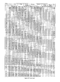
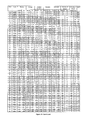
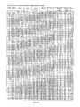
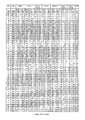
- FIGS. 1-43 provide a detailed summary of the ability of panels of biomarkers to evaluate acute kidney injury in an ICU patient population. Patients in the population are divided based on the RIFLE classification of renal status. The following analyses are provided:
- FIG. 1 No or R RIFLE stage versus RIFLE I or F. RIFLE stage adjudicated by sCr and Urine Output. Diseased group sample is obtained 24 hours prior to RIFLE I diagnosis.
- FIG. 2 No or R RIFLE stage versus RIFLE I or F. RIFLE stage adjudicated by sCr. Diseased group sample is obtained 24 hours prior to RIFLE I diagnosis.
- FIG. 3 No or R RIFLE stage versus RIFLE I or F. RIFLE stage adjudicated by Urine Output. Diseased group sample is obtained 24 hours prior to RIFLE I diagnosis.
- FIG. 4 No RIFLE stage versus RIFLE R, I, or F. RIFLE stage adjudicated by sCr and Urine Output. Diseased group sample is obtained 24 hours prior to RIFLE R diagnosis.
- FIG. 5 No RIFLE stage versus RIFLE R, I, or F. RIFLE stage adjudicated by sCr. Diseased group sample is obtained 24 hours prior to RIFLE R diagnosis.
- FIG. 6 No RIFLE stage versus RIFLE R, I, or F. RIFLE stage adjudicated by Urine Output. Diseased group sample is obtained 24 hours prior to RIFLE R diagnosis.
- FIG. 7 No, R, or I RIFLE stage versus RIFLE F. RIFLE stage adjudicated by sCr and Urine Output. Diseased group sample is obtained 24 hours prior to RIFLE I diagnosis.
- FIG. 8 No, R, or I RIFLE stage versus RIFLE F. RIFLE stage adjudicated by Urine Output. Diseased group sample is obtained 24 hours prior to RIFLE I diagnosis.
- FIG. 9 No or R RIFLE stage versus RIFLE I or F. RIFLE stage adjudicated by sCr and Urine Output. Diseased group sample is obtained at RIFLE I diagnosis.
- FIG. 10 No or R RIFLE stage versus RIFLE I or F. RIFLE stage adjudicated by sCr. Diseased group sample is obtained at RIFLE I diagnosis.
- FIG. 11 No or R RIFLE stage versus RIFLE I or F. RIFLE stage adjudicated by Urine Output. Diseased group sample is obtained at RIFLE I diagnosis.
- FIG. 12 No RIFLE stage versus RIFLE R, I, or F. RIFLE stage adjudicated by sCr and Urine Output. Diseased group sample is obtained at RIFLE R diagnosis.
- FIG. 13 No RIFLE stage versus RIFLE R, I, or F. RIFLE stage adjudicated by sCr. Diseased group sample is obtained at RIFLE R diagnosis.
- FIG. 14 No RIFLE stage versus RIFLE R, I, or F. RIFLE stage adjudicated by Urine Output. Diseased group sample is obtained at RIFLE R diagnosis.
- FIG. 15 No, R, or I RIFLE stage versus RIFLE F. RIFLE stage adjudicated by sCr and Urine Output. Diseased group sample is obtained at RIFLE I diagnosis.
- FIG. 16 No, R, or I RIFLE stage versus RIFLE F. RIFLE stage adjudicated by sCr. Diseased group sample is obtained at RIFLE I diagnosis.
- FIG. 17 No, R, or I RIFLE stage versus RIFLE F. RIFLE stage adjudicated by Urine Output. Diseased group sample is obtained at RIFLE I diagnosis.
- FIG. 18 No or R RIFLE stage versus RIFLE I or F. RIFLE stage adjudicated by sCr and Urine Output. Diseased group sample is obtained 48 hours prior to RIFLE I diagnosis.
- FIG. 19 No or R RIFLE stage versus RIFLE I or F. RIFLE stage adjudicated by sCr. Diseased group sample is obtained 48 hours prior to RIFLE I diagnosis.
- FIG. 20 No or R RIFLE stage versus RIFLE I or F. RIFLE stage adjudicated by Urine Output. Diseased group sample is obtained 48 hours prior to RIFLE I diagnosis.
- FIG. 21 No RIFLE stage versus RIFLE R, I, or F. RIFLE stage adjudicated by sCr and Urine Output. Diseased group sample is obtained 48 hours prior to RIFLE R diagnosis.
- FIG. 22 No RIFLE stage versus RIFLE R, I, or F. RIFLE stage adjudicated by sCr. Diseased group sample is obtained 48 hours prior to RIFLE R diagnosis.
- FIG. 23 No RIFLE stage versus RIFLE R, I, or F. RIFLE stage adjudicated by Urine Output. Diseased group sample is obtained 48 hours prior to RIFLE R diagnosis.
- FIG. 24 No, R, or I RIFLE stage versus RIFLE F. RIFLE stage adjudicated by sCr and Urine Output. Diseased group sample is obtained 48 hours prior to RIFLE I diagnosis.
- FIG. 25 Progression of RIFLE R to RIFLE I or F. RIFLE stage adjudicated by sCr and Urine Output. Sample is obtained at R diagnosis.
- FIG. 26 Progression of RIFLE R to RIFLE I or F. RIFLE stage adjudicated by sCr. Sample is obtained at R diagnosis.
- FIG. 27 Progression of RIFLE R to RIFLE I or F. RIFLE stage adjudicated by Urine Output. Sample is obtained at R diagnosis.
- FIG. 28 No or R RIFLE stage versus RIFLE I or F. RIFLE stage adjudicated by sCr and Urine Output, within 48 hrs of enrollment. Sample is obtained at enrollment.
- FIG. 29 No or R RIFLE stage versus RIFLE I or F. RIFLE stage adjudicated by sCr, within 48 hrs of enrollment. Sample is obtained at enrollment.
- FIG. 30 No or R RIFLE stage versus RIFLE I or F. RIFLE stage adjudicated by Urine Output, within 48 hrs of enrollment. Sample is obtained at enrollment.
- FIG. 31 No RIFLE stage versus RIFLE R, I, or F. RIFLE stage adjudicated by sCr and Urine Output, within 48 hrs of enrollment. Sample is obtained at enrollment.
- FIG. 32 No RIFLE stage versus RIFLE R, I, or F. RIFLE stage adjudicated by sCr, within 48 hrs of enrollment. Sample is obtained at enrollment.
- FIG. 33 No RIFLE stage versus RIFLE R, I, or F. RIFLE stage adjudicated by Urine Output, within 48 hrs of enrollment. Sample is obtained at enrollment.
- FIG. 34 No, R, or I RIFLE stage versus RIFLE F. RIFLE stage adjudicated by sCr and Urine Output, within 48 hrs of enrollment. Sample is obtained at enrollment.
- FIG. 35 No, R, or I RIFLE stage versus RIFLE F. RIFLE stage adjudicated by Urine Output, within 48 hrs of enrollment. Sample is obtained at enrollment.
- FIG. 36 No or R RIFLE stage versus RIFLE I or F. RIFLE stage adjudicated by sCr and Urine Output, within 24 hrs of enrollment. Sample is obtained at enrollment.
- FIG. 37 No or R RIFLE stage versus RIFLE I or F. RIFLE stage adjudicated by sCr, within 24 hrs of enrollment. Sample is obtained at enrollment.
- FIG. 38 No or R RIFLE stage versus RIFLE I or F. RIFLE stage adjudicated by Urine Output, within 24 hrs of enrollment. Sample is obtained at enrollment.
- FIG. 39 No RIFLE stage versus RIFLE R, I, or F. RIFLE stage adjudicated by sCr and Urine Output, within 24 hrs of enrollment. Sample is obtained at enrollment.
- FIG. 40 No RIFLE stage versus RIFLE R, I, or F. RIFLE stage adjudicated by sCr, within 24 hrs of enrollment. Sample is obtained at enrollment.
- FIG. 41 No RIFLE stage versus RIFLE R, I, or F. RIFLE stage adjudicated by Urine Output, within 24 hrs of enrollment. Sample is obtained at enrollment.
- FIG. 42 No, R, or I RIFLE stage versus RIFLE F. RIFLE stage adjudicated by sCr and Urine Output, within 24 hrs of enrollment. Sample is obtained at enrollment.
- FIG. 43 No, R, or I RIFLE stage versus RIFLE F. RIFLE stage adjudicated by Urine Output, within 24 hrs of enrollment. Sample is obtained at enrollment.
- the present invention relates to methods and compositions for diagnosis, differential diagnosis, risk stratification, monitoring, classifying and determination of treatment regimens in subjects suffering or at risk of suffering from injury to renal function, reduced renal function and/or acute renal failure through measurement of one or more kidney injury markers.
- a biomarker recited in Table 2 herein, or one or more markers related thereto is combined with one or more additional biomarkers listed in Table 2 herein and/or with one or more other biomarkers or clinical indicia, and the combination correlated to the renal status of the subject.
- an “injury to renal function” is an abrupt (within 14 days, preferably within 7 days, more preferably within 72 hours, and still more preferably within 48 hours) measurable reduction in a measure of renal function. Such an injury may be identified, for example, by a decrease in glomerular filtration rate or estimated GFR, a reduction in urine output, an increase in serum creatinine, an increase in serum cystatin C, a requirement for renal replacement therapy, etc.
- “Improvement in Renal Function” is an abrupt (within 14 days, preferably within 7 days, more preferably within 72 hours, and still more preferably within 48 hours) measurable increase in a measure of renal function. Preferred methods for measuring and/or estimating GFR are described hereinafter.
- reduced renal function is an abrupt (within 14 days, preferably within 7 days, more preferably within 72 hours, and still more preferably within 48 hours) reduction in kidney function identified by an absolute increase in serum creatinine of greater than or equal to 0.1 mg/dL ( ⁇ 8.8 ⁇ mol/L), a percentage increase in serum creatinine of greater than or equal to 20% (1.2-fold from baseline), or a reduction in urine output (documented oliguria of less than 0.5 ml/kg per hour).
- acute renal failure is an abrupt (within 14 days, preferably within 7 days, more preferably within 72 hours, and still more preferably within 48 hours) reduction in kidney function identified by an absolute increase in serum creatinine of greater than or equal to 0.3 mg/dl ( ⁇ 26.4 ⁇ mol/l), a percentage increase in serum creatinine of greater than or equal to 50% (1.5-fold from baseline), or a reduction in urine output (documented oliguria of less than 0.5 ml/kg per hour for at least 6 hours).
- This term is synonymous with “acute kidney injury” or “AKI.”
- a “marker” or “biomarker” refers to polypeptides present in a biological sample that are derived from a particular biosynthetic precursor.
- a “marker” or “biomarker” refers to the particular molecular entity recited.
- Table 2 provides a list of preferred biomarkers finding use in the present invention.
- the “recommended name” for the biomarker precursor from the Swiss-Prot “UniProtKB” database and for most polypeptide biomarkers the Swiss-Prot entry number for the human precursor.
- the Swiss Prot entry is listed for each member of the complex.
- Preferred Name Prot 60 kDa heat shock protein, mitochondrial P10809 72 kDa type IV collagenase P08253 72 kDa type IV collagenase:Metalloproteinase P08253 72 kDa type IV collagenase:Metalloproteinase P08253 inhibitor 1 complex P01033 inhibitor 2 complex P16035 72 kDa type IV collagenase:Metalloproteinase P08253 Adiponectin Q15848 inhibitor 4 complex Q99727 Advanced glycosylation end product-specific Q15109 Agouti-related protein O00253 receptor Alkaline phosphatase, tissue-nonspecific P05186 Alpha-1-antitrypsin P01009 isozyme Alpha-1-antitrypsin:Neutrophil elastase P01009 Alpha-1-antitrypsin:Plasminogen complex P01009 complex P08246 P00747 Alpha-2 macroglobulin P
- membrane proteins which exist in one form as type-I, type-II, or GPI-anchored membrane proteins.
- membrane proteins comprise a substantial extracellular domain, some or all of which can be detected as soluble forms present in aqueous samples such as blood, serum, plasma, urine, etc., either as cleavage products or as splice variants which delete an effective membrane spanning domain.
- membrane proteins include Swiss-Prot entry numbers O14788, O14944, O75309, P00797, P05186, P08473, P13688, P15514, P22223, P27487, P35070, Q03405, Q14956, Q16790, Q99075, Q9Y5Y7Q15109, Q02763, P17213, P12830, P33151, P06731, P29965, P16070, Q9H2A7, P17813, Q9UNN8, P00533, P16422, P19235, P16581, P78423, O43656, P08581, P08069, P05362, P13598, P32942, P14778, P27930, P01589, P24394, P08887, P40189, P21583, P09603, P08571, Q8WXI7, P13591, Q92823, P78380, P16284, P01133, P15309, P01135, P16109, Q14242,
- the signals obtained from an immunoassay are a direct result of complexes formed between one or more antibodies and the corresponding target biomolecule (i.e., the analyte) containing the necessary epitope(s) to which the antibodies bind. While such assays may detect the full length biomarker and the assay result be expressed as a concentration of a biomarker of interest, the signal from the assay is actually a result of all such “immunoreactive” molecules present in the sample.
- Biomarkers may also be determined by means other than immunoassays, including protein measurements (such as dot blots, western blots, chromatographic methods, mass spectrometry, etc.) and nucleic acid measurements (mRNA quatitation). This list is not meant to be limiting.
- an assay is “configured to detect” an analyte if an assay can generate a detectable signal indicative of the presence or amount of a physiologically relevant concentration of the analyte.
- an immunoassay configured to detect a marker of interest will also detect polypeptides related to the marker sequence, so long as those polypeptides contain the epitope(s) necessary to bind to the antibody or antibodies used in the assay.
- the term “related marker” as used herein with regard to a biomarker such as one of the kidney injury markers described herein refers to one or more fragments, variants, etc., of a particular marker or its biosynthetic parent that may be detected as a surrogate for the marker itself or as independent biomarkers.
- the term also refers to one or more polypeptides present in a biological sample that are derived from the biomarker precursor complexed to additional species, such as binding proteins, receptors, heparin, lipids, sugars, etc.
- positive going marker refers to a marker that is determined to be elevated in subjects suffering from a disease or condition, relative to subjects not suffering from that disease or condition.
- negative going marker refers to a marker that is determined to be reduced in subjects suffering from a disease or condition, relative to subjects not suffering from that disease or condition.
- subject refers to a human or non-human organism.
- methods and compositions described herein are applicable to both human and veterinary disease.
- a subject is preferably a living organism, the invention described herein may be used in post-mortem analysis as well.
- Preferred subjects are humans, and most preferably “patients,” which as used herein refers to living humans that are receiving medical care for a disease or condition. This includes persons with no defined illness who are being investigated for signs of pathology.
- an analyte is measured in a sample.
- a sample may be obtained from a subject, or may be obtained from biological materials intended to be provided to the subject.
- a sample may be obtained from a kidney being evaluated for possible transplantation into a subject, and an analyte measurement used to evaluate the kidney for preexisting damage.
- Preferred samples are body fluid samples.
- body fluid sample refers to a sample of bodily fluid obtained for the purpose of diagnosis, prognosis, classification or evaluation of a subject of interest, such as a patient or transplant donor. In certain embodiments, such a sample may be obtained for the purpose of determining the outcome of an ongoing condition or the effect of a treatment regimen on a condition.
- Preferred body fluid samples include blood, serum, plasma, cerebrospinal fluid, urine, saliva, sputum, and pleural effusions.
- body fluid samples would be more readily analyzed following a fractionation or purification procedure, for example, separation of whole blood into serum or plasma components.
- diagnosis refers to methods by which the skilled artisan can estimate and/or determine the probability (“a likelihood”) of whether or not a patient is suffering from a given disease or condition.
- diagnosis includes using the results of an assay, most preferably an immunoassay, for a kidney injury marker of the present invention, optionally together with other clinical characteristics, to arrive at a diagnosis (that is, the occurrence or nonoccurrence) of an acute renal injury or ARF for the subject from which a sample was obtained and assayed. That such a diagnosis is “determined” is not meant to imply that the diagnosis is 100% accurate. Many biomarkers are indicative of multiple conditions.
- a measured biomarker level on one side of a predetermined diagnostic threshold indicates a greater likelihood of the occurrence of disease in the subject relative to a measured level on the other side of the predetermined diagnostic threshold.
- a prognostic risk signals a probability (“a likelihood”) that a given course or outcome will occur.
- a level or a change in level of a prognostic indicator which in turn is associated with an increased probability of morbidity (e.g., worsening renal function, future ARF, or death) is referred to as being “indicative of an increased likelihood” of an adverse outcome in a patient.
- a “plurality” as used herein refers to at least two. Preferably, a plurality refers to at least 3, more preferably at least 4, even more preferably at least 5, even more preferably at least 10, and most preferably at least 20.
- immunoassays involve contacting a sample containing or suspected of containing a biomarker of interest with at least one antibody that specifically binds to the biomarker. A signal is then generated indicative of the presence or amount of complexes formed by the binding of polypeptides in the sample to the antibody. The signal is then related to the presence or amount of the biomarker in the sample. Numerous methods and devices are well known to the skilled artisan for the detection and analysis of biomarkers. See, e.g., U.S. Pat. Nos.
- the assay devices and methods known in the art can utilize labeled molecules in various sandwich, competitive, or non-competitive assay formats, to generate a signal that is related to the presence or amount of the biomarker of interest.
- Suitable assay formats also include chromatographic, mass spectrographic, and protein “blotting” methods.
- certain methods and devices such as biosensors and optical immunoassays, may be employed to determine the presence or amount of analytes without the need for a labeled molecule. See, e.g., U.S. Pat. Nos. 5,631,171; and 5,955,377, each of which is hereby incorporated by reference in its entirety, including all tables, figures and claims.
- robotic instrumentation including but not limited to Beckman ACCESS®, Abbott AXSYM®, Roche ELECSYS®, Dade Behring STRATUS® systems are among the immunoassay analyzers that are capable of performing immunoassays.
- any suitable immunoassay may be utilized, for example, enzyme-linked immunoassays (ELISA), radioimmunoassays (RIAs), competitive binding assays, and the like.
- Antibodies or other polypeptides may be immobilized onto a variety of solid supports for use in assays.
- Solid phases that may be used to immobilize specific binding members include include those developed and/or used as solid phases in solid phase binding assays. Examples of suitable solid phases include membrane filters, cellulose-based papers, beads (including polymeric, latex and paramagnetic particles), glass, silicon wafers, microparticles, nanoparticles, TentaGelTM resins (Rapp Polymere GmbH), AgroGelTM resins (I.L.S.A. Industria Lavorazione Sottoprodotti Animali S.P.A.), polyethylene glycol and acrylamide (PEGA) gels, SPOCC gels, and multiple-well plates.
- PEGA polyethylene glycol and acrylamide
- An assay strip could be prepared by coating the antibody or a plurality of antibodies in an array on solid support. This strip could then be dipped into the test sample and then processed quickly through washes and detection steps to generate a measurable signal, such as a colored spot.
- Antibodies or other polypeptides may be bound to specific zones of assay devices either by conjugating directly to an assay device surface, or by indirect binding. In an example of the later case, antibodies or other polypeptides may be immobilized on particles or other solid supports, and that solid support immobilized to the device surface.
- Biological assays require methods for detection, and one of the most common methods for quantitation of results is to conjugate a detectable label to a protein or nucleic acid that has affinity for one of the components in the biological system being studied.
- Detectable labels may include molecules that are themselves detectable (e.g., fluorescent moieties, electrochemical labels, metal chelates, etc.) as well as molecules that may be indirectly detected by production of a detectable reaction product (e.g., enzymes such as horseradish peroxidase, alkaline phosphatase, etc.) or by a specific binding molecule which itself may be detectable (e.g., biotin, digoxigenin, maltose, oligohistidine, 2,4-dintrobenzene, phenylarsenate, ssDNA, dsDNA, etc.).
- a detectable reaction product e.g., enzymes such as horseradish peroxidase, alkaline phosphatase, etc.
- Cross-linking reagents contain at least two reactive groups, and are divided generally into homofunctional cross-linkers (containing identical reactive groups) and heterofunctional cross-linkers (containing non-identical reactive groups). Homobifunctional cross-linkers that couple through amines, sulfhydryls or react non-specifically are available from many commercial sources. Maleimides, alkyl and aryl halides, alpha-haloacyls and pyridyl disulfides are thiol reactive groups.
- kits for the analysis of the described kidney injury markers comprises reagents for the analysis of at least one test sample which comprise at least one antibody that a kidney injury marker.
- the kit can also include devices and instructions for performing one or more of the diagnostic and/or prognostic correlations described herein.
- Preferred kits will comprise an antibody pair for performing a sandwich assay, or a labeled species for performing a competitive assay, for the analyte.
- an antibody pair comprises a first antibody conjugated to a solid phase and a second antibody conjugated to a detectable label, wherein each of the first and second antibodies that bind a kidney injury marker.
- each of the antibodies are monoclonal antibodies.
- the instructions for use of the kit and performing the correlations can be in the form of labeling, which refers to any written or recorded material that is attached to, or otherwise accompanies a kit at any time during its manufacture, transport, sale or use.
- labeling encompasses advertising leaflets and brochures, packaging materials, instructions, audio or video cassettes, computer discs, as well as writing imprinted directly on kits.
- antibody refers to a peptide or polypeptide derived from, modeled after or substantially encoded by an immunoglobulin gene or immunoglobulin genes, or fragments thereof, capable of specifically binding an antigen or epitope. See, e.g. Fundamental Immunology, 3rd Edition, W. E. Paul, ed., Raven Press, N.Y. (1993); Wilson (1994; J. Immunol. Methods 175:267-273; Yarmush (1992) J. Biochem. Biophys. Methods 25:85-97.
- antibody includes antigen-binding portions, i.e., “antigen binding sites,” (e.g., fragments, subsequences, complementarity determining regions (CDRs)) that retain capacity to bind antigen, including (i) a Fab fragment, a monovalent fragment consisting of the VL, VH, CL and CH1 domains; (ii) a F(ab′) 2 fragment, a bivalent fragment comprising two Fab fragments linked by a disulfide bridge at the hinge region; (iii) a Fd fragment consisting of the VH and CH1 domains; (iv) a Fv fragment consisting of the VL and VH domains of a single arm of an antibody, (v) a dAb fragment (Ward et al., (1989) Nature 341:544-546), which consists of a VH domain; and (vi) an isolated complementarity determining region (CDR).
- Antigen binding sites e.g., fragments, subs
- Antibodies used in the immunoassays described herein preferably specifically bind to a kidney injury marker of the present invention.
- the term “specifically binds” is not intended to indicate that an antibody binds exclusively to its intended target since, as noted above, an antibody binds to any polypeptide displaying the epitope(s) to which the antibody binds. Rather, an antibody “specifically binds” if its affinity for its intended target is about 5-fold greater when compared to its affinity for a non-target molecule which does not display the appropriate epitope(s).
- the affinity of the antibody will be at least about 5 fold, preferably 10 fold, more preferably 25-fold, even more preferably 50-fold, and most preferably 100-fold or more, greater for a target molecule than its affinity for a non-target molecule.
- Preferred antibodies bind with affinities of at least about 10 7 M ⁇ 1 , and preferably between about 10 8 M ⁇ 1 to about 10 9 M ⁇ 1 , about 10 9 M ⁇ 1 to about 10 10 M ⁇ 1 , or about 10 10 M ⁇ 1 to about 10 12 M ⁇ 1 .
- Antibody affinity measurement by Scatchard analysis is well known in the art. See, e.g., van Erp et al., J. Immunoassay 12: 425-43, 1991; Nelson and Griswold, Comput. Methods Programs Biomed. 27: 65-8, 1988.
- epitope refers to an antigenic determinant capable of specific binding to an antibody.
- Epitopes usually consist of chemically active surface groupings of molecules such as amino acids or sugar side chains and usually have specific three dimensional structural characteristics, as well as specific charge characteristics. Conformational and nonconformational epitopes are distinguished in that the binding to the former but not the latter is lost in the presence of denaturing solvents.
- phage display technology to produce and screen libraries of polypeptides for binding to a selected analyte. See, e.g., Cwirla et al., Proc. Natl. Acad. Sci. USA 87, 6378-82, 1990; Devlin et al., Science 249, 404-6, 1990, Scott and Smith, Science 249, 386-88, 1990; and Ladner et al., U.S. Pat. No. 5,571,698.
- a basic concept of phage display methods is the establishment of a physical association between DNA encoding a polypeptide to be screened and the polypeptide.
- This physical association is provided by the phage particle, which displays a polypeptide as part of a capsid enclosing the phage genome which encodes the polypeptide.
- the establishment of a physical association between polypeptides and their genetic material allows simultaneous mass screening of very large numbers of phage bearing different polypeptides.
- Phage displaying a polypeptide with affinity to a target bind to the target and these phage are enriched by affinity screening to the target.
- the identity of polypeptides displayed from these phage can be determined from their respective genomes. Using these methods a polypeptide identified as having a binding affinity for a desired target can then be synthesized in bulk by conventional means. See, e.g., U.S. Pat. No. 6,057,098, which is hereby incorporated in its entirety, including all tables, figures, and claims.
- the antibodies that are generated by these methods may then be selected by first screening for affinity and specificity with the purified polypeptide of interest and, if required, comparing the results to the affinity and specificity of the antibodies with polypeptides that are desired to be excluded from binding.
- the screening procedure can involve immobilization of the purified polypeptides in separate wells of microtiter plates. The solution containing a potential antibody or groups of antibodies is then placed into the respective microtiter wells and incubated for about 30 min to 2 h.
- microtiter wells are then washed and a labeled secondary antibody (for example, an anti-mouse antibody conjugated to alkaline phosphatase if the raised antibodies are mouse antibodies) is added to the wells and incubated for about 30 min and then washed. Substrate is added to the wells and a color reaction will appear where antibody to the immobilized polypeptide(s) are present.
- a labeled secondary antibody for example, an anti-mouse antibody conjugated to alkaline phosphatase if the raised antibodies are mouse antibodies
- the antibodies so identified may then be further analyzed for affinity and specificity in the assay design selected.
- the purified target protein acts as a standard with which to judge the sensitivity and specificity of the immunoassay using the antibodies that have been selected. Because the binding affinity of various antibodies may differ; certain antibody pairs (e.g., in sandwich assays) may interfere with one another sterically, etc., assay performance of an antibody may be a more important measure than absolute affinity and specificity of an antibody.
- aptamers are oligonucleic acid or peptide molecules that bind to a specific target molecule. Aptamers are usually created by selecting them from a large random sequence pool, but natural aptamers also exist. High-affinity aptamers containing modified nucleotides conferring improved characteristics on the ligand, such as improved in vivo stability or improved delivery characteristics. Examples of such modifications include chemical substitutions at the ribose and/or phosphate and/or base positions, and may include amino acid side chain functionalities. Assay Correlations
- correlating refers to comparing the presence or amount of the biomarker(s) in a patient to its presence or amount in persons known to suffer from, or known to be at risk of, a given condition; or in persons known to be free of a given condition. Often, this takes the form of comparing an assay result in the form of a biomarker concentration to a predetermined threshold selected to be indicative of the occurrence or nonoccurrence of a disease or the likelihood of some future outcome.
- Selecting a diagnostic threshold involves, among other things, consideration of the probability of disease, distribution of true and false diagnoses at different test thresholds, and estimates of the consequences of treatment (or a failure to treat) based on the diagnosis. For example, when considering administering a specific therapy which is highly efficacious and has a low level of risk, few tests are needed because clinicians can accept substantial diagnostic uncertainty. On the other hand, in situations where treatment options are less effective and more risky, clinicians often need a higher degree of diagnostic certainty. Thus, cost/benefit analysis is involved in selecting a diagnostic threshold.
- Suitable thresholds may be determined in a variety of ways. For example, one recommended diagnostic threshold for the diagnosis of acute myocardial infarction using cardiac troponin is the 97.5 th percentile of the concentration seen in a normal population. Another method may be to look at serial samples from the same patient, where a prior “baseline” result is used to monitor for temporal changes in a biomarker level.
- ROC Reciever Operating Characteristic
- the ROC graph is sometimes called the sensitivity vs (1 ⁇ specificity) plot.
- a perfect test will have an area under the ROC curve of 1.0; a random test will have an area of 0.5.
- a threshold is selected to provide an acceptable level of specificity and sensitivity.
- diseased is meant to refer to a population having one characteristic (the presence of a disease or condition or the occurrence of some outcome) and “nondiseased” is meant to refer to a population lacking the characteristic. While a single decision threshold is the simplest application of such a method, multiple decision thresholds may be used. For example, below a first threshold, the absence of disease may be assigned with relatively high confidence, and above a second threshold the presence of disease may also be assigned with relatively high confidence. Between the two thresholds may be considered indeterminate. This is meant to be exemplary in nature only.
- other methods for correlating assay results to a patient classification include decision trees, rule sets, Bayesian methods, and neural network methods. These methods can produce probability values representing the degree to which a subject belongs to one classification out of a plurality of classifications.
- Measures of test accuracy may be obtained as described in Fischer et al., Intensive Care Med. 29: 1043-51, 2003, and used to determine the effectiveness of a given biomarker. These measures include sensitivity and specificity, predictive values, likelihood ratios, diagnostic odds ratios, and ROC curve areas.
- the area under the curve (“AUC”) of a ROC plot is equal to the probability that a classifier will rank a randomly chosen positive instance higher than a randomly chosen negative one.
- the area under the ROC curve may be thought of as equivalent to the Mann-Whitney U test, which tests for the median difference between scores obtained in the two groups considered if the groups are of continuous data, or to the Wilcoxon test of ranks.
- suitable tests may exhibit one or more of the following results on these various measures: a specificity of greater than 0.5, preferably at least 0.6, more preferably at least 0.7, still more preferably at least 0.8, even more preferably at least 0.9 and most preferably at least 0.95, with a corresponding sensitivity greater than 0.2, preferably greater than 0.3, more preferably greater than 0.4, still more preferably at least 0.5, even more preferably 0.6, yet more preferably greater than 0.7, still more preferably greater than 0.8, more preferably greater than 0.9, and most preferably greater than 0.95; a sensitivity of greater than 0.5, preferably at least 0.6, more preferably at least 0.7, still more preferably at least 0.8, even more preferably at least 0.9 and most preferably at least 0.95, with a corresponding specificity greater than 0.2, preferably greater than 0.3, more preferably greater than 0.4, still more preferably at least 0.5, even more preferably 0.6, yet more preferably greater than 0.7, still more preferably greater than
- Additional clinical indicia may be combined with the kidney injury marker assay result(s) of the present invention.
- biomarkers related to renal status include the following, which recite the common biomarker name, followed by the Swiss-Prot entry number for that biomarker or its parent: Actin (P68133); Adenosine deaminase binding protein (DPP4, P27487); Alpha-1-acid glycoprotein 1 (P02763); Alpha-1-microglobulin (P02760); Albumin (P02768); Angiotensinogenase (Renin, P00797); Annexin A2 (P07355); Beta-glucuronidase (P08236); B-2-microglobulin (P61679); Beta-galactosidase (P16278); BMP-7 (P18075); Brain natriuretic peptide (proBNP, BNP-32, NTproBNP; P16860); Calcium-binding protein Beta (S100-beta
- Adiponectin (Q15848); Alkaline phosphatase (P05186); Aminopeptidase N (P15144); CalbindinD28k (P05937); Cystatin C (P01034); 8 subunit of FIFO ATPase (P03928); Gamma-glutamyltransferase (P19440); GSTa (alpha-glutathione-S-transferase, P08263); GSTpi (Glutathione-S-transferase P; GST class-pi; P09211); IGFBP-1 (P08833); IGFBP-2 (P18065); IGFBP-6 (P24592); Integral membrane protein 1 (Itm1, P46977); Interleukin-6 (P05231); Interleukin-8 (P10145); Interleukin-18 (Q14116); IP-10 (10 kDa interferon-gamma-induced protein, P02778); IRPR
- Other clinical indicia which may be combined with the kidney injury marker assay result(s) of the present invention includes demographic information (e.g., weight, sex, age, race), medical history (e.g., family history, type of surgery, pre-existing disease such as aneurism, congestive heart failure, preeclampsia, eclampsia, diabetes mellitus, hypertension, coronary artery disease, proteinuria, renal insufficiency, or sepsis, type of toxin exposure such as NSAIDs, cyclosporines, tacrolimus, aminoglycosides, foscarnet, ethylene glycol, hemoglobin, myoglobin, ifosfamide, heavy metals, methotrexate, radiopaque contrast agents, or streptozotocin), clinical variables (e.g., blood pressure, temperature, respiration rate), risk scores (APACHE score, PREDICT score, TIMI Risk Score for UA/NSTEMI, Framingham Risk Score
- kidney injury marker assay result(s) Other measures of renal function which may be combined with the kidney injury marker assay result(s) are described hereinafter and in Harrison's Principles of Internal Medicine, 17 th Ed., McGraw Hill, New York, pages 1741-1830, and Current Medical Diagnosis & Treatment 2008, 47 th Ed, McGraw Hill, New York, pages 785-815, each of which are hereby incorporated by reference in their entirety.
- Combining assay results/clinical indicia in this manner can comprise the use of multivariate logistical regression, loglinear modeling, neural network analysis, n-of-m analysis, decision tree analysis, etc. This list is not meant to be limiting.
- the terms “acute renal (or kidney) injury” and “acute renal (or kidney) failure” as used herein are defined in part in terms of changes in serum creatinine from a baseline value.
- Most definitions of ARF have common elements, including the use of serum creatinine and, often, urine output. Patients may present with renal dysfunction without an available baseline measure of renal function for use in this comparison. In such an event, one may estimate a baseline serum creatinine value by assuming the patient initially had a normal GFR.
- Glomerular filtration rate (GFR) is the volume of fluid filtered from the renal (kidney) glomerular capillaries into the Bowman's capsule per unit time. Glomerular filtration rate (GFR) can be calculated by measuring any chemical that has a steady level in the blood, and is freely filtered but neither reabsorbed nor secreted by the kidneys. GFR is typically expressed in units of ml/min:
- GFR Urine ⁇ ⁇ Concentration ⁇ Urine ⁇ ⁇ Flow Plasma ⁇ ⁇ Concentration
- GFR glomerular filtration rate
- eGFR glomerular filtration rate
- Creatinine clearance is used to measure GFR. Creatinine is produced naturally by the body (creatinine is a metabolite of creatine, which is found in muscle). It is freely filtered by the glomerulus, but also actively secreted by the renal tubules in very small amounts such that creatinine clearance overestimates actual GFR by 10-20%. This margin of error is acceptable considering the ease with which creatinine clearance is measured.
- Creatinine clearance can be calculated if values for creatinine's urine concentration (U Cr ), urine flow rate (V), and creatinine's plasma concentration (P Cr ) are known. Since the product of urine concentration and urine flow rate yields creatinine's excretion rate, creatinine clearance is also said to be its excretion rate (U Cr ⁇ V) divided by its plasma concentration. This is commonly represented mathematically as:
- the CCr is often corrected for the body surface area (BSA) and expressed compared to the average sized man as ml/min/1.73 m2. While most adults have a BSA that approaches 1.7 (1.6-1.9), extremely obese or slim patients should have their CCr corrected for their actual BSA:
- the clinician can readily select a treatment regimen that is compatible with the diagnosis, such as initiating renal replacement therapy, withdrawing delivery of compounds that are known to be damaging to the kidney, kidney transplantation, delaying or avoiding procedures that are known to be damaging to the kidney, modifying diuretic administration, initiating goal directed therapy, etc.
- a treatment regimen that is compatible with the diagnosis, such as initiating renal replacement therapy, withdrawing delivery of compounds that are known to be damaging to the kidney, kidney transplantation, delaying or avoiding procedures that are known to be damaging to the kidney, modifying diuretic administration, initiating goal directed therapy, etc.
- the skilled artisan is aware of appropriate treatments for numerous diseases discussed in relation to the methods of diagnosis described herein. See, e.g., Merck Manual of Diagnosis and Therapy, 17th Ed. Merck Research Laboratories, Whitehouse Station, N.J., 1999.
- the markers of the present invention may be used to monitor a course of treatment. For example, improved or worsened prognostic state may indicate that a particular treatment is or
- the objective of this sample collection study is to collect samples of plasma and urine and clinical data from patients before and after receiving intravascular contrast media. Approximately 250 adults undergoing radiographic/angiographic procedures involving intravascular administration of iodinated contrast media are enrolled. To be enrolled in the study, each patient must meet all of the following inclusion criteria and none of the following exclusion criteria:
- HIV human immunodeficiency virus
- an EDTA anti-coagulated blood sample (10 mL) and a urine sample (10 mL) are collected from each patient. Blood and urine samples are then collected at 4 ( ⁇ 0.5), 8 ( ⁇ 1), 24 ( ⁇ 2) 48 ( ⁇ 2), and 72 ( ⁇ 2) hrs following the last administration of contrast media during the index contrast procedure. Blood is collected via direct venipuncture or via other available venous access, such as an existing femoral sheath, central venous line, peripheral intravenous line or hep-lock. These study blood samples are processed to plasma at the clinical site, frozen and shipped to Astute Medical, Inc., San Diego, Calif. The study urine samples are frozen and shipped to Astute Medical, Inc.
- Serum creatinine is assessed at the site immediately prior to the first contrast administration (after any pre-procedure hydration) and at 4 ( ⁇ 0.5), 8 ( ⁇ 1), 24 ( ⁇ 2) and 48 ( ⁇ 2)), and 72 ( ⁇ 2) hours following the last administration of contrast (ideally at the same time as the study samples are obtained).
- each patient's status is evaluated through day 30 with regard to additional serum and urine creatinine measurements, a need for dialysis, hospitalization status, and adverse clinical outcomes (including mortality).
- the objective of this sample collection study is to collect samples of plasma and urine and clinical data from patients before and after undergoing cardiovascular surgery, a procedure known to be potentially damaging to kidney function. Approximately 900 adults undergoing such surgery are enrolled. To be enrolled in the study, each patient must meet all of the following inclusion criteria and none of the following exclusion criteria:
- HIV human immunodeficiency virus
- an EDTA anti-coagulated blood sample (10 mL), whole blood (3 mL), and a urine sample (35 mL) are collected from each patient. Blood and urine samples are then collected at 3 ( ⁇ 0.5), 6 ( ⁇ 0.5), 12 ( ⁇ 1), 24 ( ⁇ 2) and 48 ( ⁇ 2) hrs following the procedure and then daily on days 3 through 7 if the subject remains in the hospital. Blood is collected via direct venipuncture or via other available venous access, such as an existing femoral sheath, central venous line, peripheral intravenous line or hep-lock.
- These study blood samples are frozen and shipped to Astute Medical, Inc., San Diego, Calif.
- the study urine samples are frozen and shipped to Astute Medical, Inc.
- the objective of this study is to collect samples from acutely ill patients. Approximately 900 adults expected to be in the ICU for at least 48 hours will be enrolled. To be enrolled in the study, each patient must meet all of the following inclusion criteria and none of the following exclusion criteria:
- Study population 1 approximately 300 patients that have at least one of:
- Study population 2 approximately 300 patients that have at least one of:
- ICU acute care setting
- shock systolic BP ⁇ 90 mmHg and/or the need for vasopressor support to maintain a MAP >60 mmHg and/or a documented drop in SBP >40 mmHg)
- major trauma, hemorrhage, or major surgery major trauma, hemorrhage, or major surgery
- HW human immunodeficiency virus
- an EDTA anti-coagulated blood sample (10 mL) and a urine sample (25-30 mL) are collected from each patient. Blood and urine samples are then collected at 4 ( ⁇ 0.5) and 8 ( ⁇ 1) hours after contrast administration (if applicable); at 12 ( ⁇ 1), 24 ( ⁇ 2), and 48 ( ⁇ 2) hours after enrollment, and thereafter daily up to day 7 to day 14 while the subject is hospitalized. Blood is collected via direct venipuncture or via other available venous access, such as an existing femoral sheath, central venous line, peripheral intravenous line or hep-lock. These study blood samples are processed to plasma at the clinical site, frozen and shipped to Astute Medical, Inc., San Diego, Calif. The study urine samples are frozen and shipped to Astute Medical, Inc.
- Analytes are is measured using standard sandwich enzyme immunoassay techniques.
- a first antibody which binds the analyte is immobilized in wells of a 96 well polystyrene microplate.
- Analyte standards and test samples are pipetted into the appropriate wells and any analyte present is bound by the immobilized antibody.
- a horseradish peroxidase-conjugated second antibody which binds the analyte is added to the wells, thereby forming sandwich complexes with the analyte (if present) and the first antibody.
- a substrate solution comprising tetramethylbenzidine and hydrogen peroxide is added to the wells. Color develops in proportion to the amount of analyte present in the sample. The color development is stopped and the intensity of the color is measured at 540 nm or 570 nm. An analyte concentration is assigned to the test sample by comparison to a standard curve determined from the analyte standards.
- Chronic Disease Patients Human urine samples from donors with various chronic diseases (“Chronic Disease Patients”) including congestive heart failure, coronary artery disease, chronic kidney disease, chronic obstructive pulmonary disease, diabetes mellitus and hypertension were purchased from Virginia Medical Research, Inc., 915 First Colonial Rd., Virginia Beach, Va. 23454. The urine samples were shipped and stored frozen at less than ⁇ 20 degrees centigrade. The vendor provided a case report form for each individual donor with age, gender, race (Black/White), smoking status and alcohol use, height, weight, chronic disease(s) diagnosis, current medications and previous surgeries.
- Chronic Disease Patients including congestive heart failure, coronary artery disease, chronic kidney disease, chronic obstructive pulmonary disease, diabetes mellitus and hypertension were purchased from Virginia Medical Research, Inc., 915 First Colonial Rd., Virginia Beach, Va. 23454. The urine samples were shipped and stored frozen at less than ⁇ 20 degrees centigrade. The vendor provided a case report form for each individual donor with age, gender, race (Black/White), smoking
- NonDis non-diseased
- Diseased Diseased
- the combined NonDis/Dis population formed a data set, and the following analysis was performed. Multiple analytes were measured from either blood or urine or both, for the subjects in the data set. Blood and urine measurements were treated as separate biomarkers in calculating panel results. Panels were formed by algorithmically combing two or more analytes in one of two ways: (i) a very simple product and division of analyte values; and (ii) logistic regression.
- Logistic regression is a method widely used for models which have a binary outcome, such as the “diseased” “non-diseased” dichotomy presented here. The method will be briefly described below, full treatments can be found in the literature.
- the model used is:
- ⁇ i ⁇ ⁇ ( y i 1
- x i ) e a + ⁇ ⁇ ⁇ x i 1 + e ⁇ + ⁇ ⁇ ⁇ x i
- the panel value for each sample is ⁇ i .
- the log odds or logit is:
- a commonly used statistic to compare the fit of two nested models is the likelihood ratio test.
- This statistic or the deviance, is the difference in twice the negative log likelihood ( ⁇ 2LL) for the two model, and is asymptotically a ⁇ 2 with DF equal to the change in the number of degrees of freedom (number of analytes) between the models.
- the p-value is calculated from this statistic.
- the null hypothesis is that the logistic model is not different than a constant model ( ⁇ set to 0), is tested for each model using the likelihood ratio test.
- the ‘model p-value’ is defined as the probability that the null hypothesis is true.
- For the constant model a closed form solution for ⁇ and ⁇ 2LL can be found. It is a function of the number of diseased (#D) and the number of non-diseased (#ND) samples in the data set.
- Each analyte can be tested for significance by holding its ⁇ at zero and comparing the model found without the analyte to the full model.
- the likelihood ratio test is used with 1 degree of freedom.
- the ‘analyte p-value’ is defined as the probability that the models are not different when that analyte is removed.
- the analyte concentrations used in the panel can either be log transformed or untransformed. For each analyte in the panel, all the permutations of the analytes being log transformed/not transformed are calculated. The permutation with the lowest model p-value is used for that panel. For two marker panels there are 4 permutations, for six marker panels there are 8.
- Table 3 contains a list of assays that were measured on urine (“U”) and EDTA plasmas (“E”) samples. Also listed, are the two letter codes that will be used throughout this document to refer to these assays according to the following convention: if the first character of the code is capitalized, this means the assay was measured in urine. If the second character of the code is capitalized, this means the assay was measured in EDTA plasma. If both characters of the code are capitalized, then the assay was measured in both urine and EDTA plasma. In the table some assays have a single code in capitals, indicating that the same assay format (e.g., microtiter dish, automated analyzer, etc.) was used for both urine and plasma. Other assays have separate U and E codes. This indicates that different assay formats were used for urine and EDTA plasma. A code of “**” indicates that an insufficient number of samples were measured on that assay and it will not appear in the panel results presented hereinafter.
- U urine
- E EDTA plasmas
- Each figure contains an initial table presenting the univariate statistics for the analytes that appear in the biomarker panels that follows in the same figure.
- the data set was segregated into two groups, non-diseased (NonDis) and diseased (Dis) patients as indicated in the figure heading.
- the analyte code and units of measurement are shown, followed by the median, average, standard deviation, maximum and minimum of the analyte values calculated for each group.
- the number of samples that make up each group are also shown with the value 1 ⁇ 10 ⁇ 9 (written as “1.0E ⁇ 9”).
- the univariate AUC for each analyte is also shown.
- a value of 1 in the ‘median’ column converts to a value of 2.6 ng/ml, 2.5 ng/ml, or 2.3 mU/ml, respectively. Note that applying such a scaling factor to the data does not impact the AUC of a marker or the AUC of a panel of which it is a part.
- each figure provides panel tables which list the biomarker panels having model p-value in the ranges specified in the particular figures.
- the number of samples used was the intersection of the samples measured by all analytes on the particular panel. Panels that have an intersection of less than 7 samples in either the non-diseased or diseased groups were not considered in the analysis.
- aBCdEE represents the two panels aBCdEe and aBCdeE, in this case representing biomarker “ab” measured in plasma, biomarker “cd” measured in urine, and biomarker ee measured in urine for the first panel and ee measured in plasma for the second panel.
- Codes in ( ) or ⁇ ⁇ and separated by a space represent a list of analytes that join with the analyte(s) outside the brackets to form panels with a common analyte.
- Ab ⁇ cD(eF gH) Km(Ef QR) ⁇ represents 5 panels which all have biomarker Ab in common. They are AbcDeF, AbcDgH, AbKmEf, AbKmQr, and AbKmqR.
- Unconstrained Panels refer to panels which were not required to have each analyte univariate p-value ⁇ 0.05. Those results labeled ‘Constrained Panels’ require that each analyte use in the panel to have a univariate p-value ⁇ 0.05.
- biomarker was measured by standard immunoassay methods using commercially available assay reagents.
- the membrane-associated biomarkers include Swiss-Prot entries O14788, O14944, O75309, P00797, P05186, P08473, P13688, P15514, P22223, P27487, P35070, Q03405, Q14956, Q16790, Q99075, Q9Y5Y7Q15109, Q02763, P17213, P12830, P33151, P06731, P29965, P16070, Q9H2A7, P17813, Q9UNN8, P00533, P16422, P19235, P16581, P78423, O43656, P08581, P08069, P05362, P13598, P32942, P14778, P27930, P01589, P24394, P08887, P40189
- the stage 0 cohort may have included patients adjudicated to stage F on the basis of urine output; for those patients adjudicated to stage F on the basis of urine output alone, the stage 0 cohort may have included patients adjudicated to stage F on the basis of serum creatinine measurements; and for those patients adjudicated to stage F on the basis of serum creatinine measurements or urine output, the stage 0 cohort contains only patients in stage 0 for both serum creatinine measurements and urine output. Also, for those patients adjudicated to stage F on the basis of serum creatinine measurements or urine output, the adjudication method which yielded the most severe RIFLE stage was used.
- FIG. 1 No or R RIFLE stage versus RIFLE I or F. RIFLE stage adjudicated by sCr and Urine Output. Diseased group sample is 24 hours prior to RIFLE I diagnosis.
- FIG. 2 No or R RIFLE stage versus RIFLE I or F. RIFLE stage adjudicated by sCr. Diseased group sample is 24 hours prior to RIFLE I diagnosis.
- FIG. 3 No or R RIFLE stage versus RIFLE I or F. RIFLE stage adjudicated by Urine Output. Diseased group sample is 24 hours prior to RIFLE I diagnosis.
- FIG. 4 No RIFLE stage versus RIFLE R, I, or F. RIFLE stage adjudicated by sCr and Urine Output. Diseased group sample is 24 hours prior to RIFLE R diagnosis.
- FIG. 5 No RIFLE stage versus RIFLE R, I, or F. RIFLE stage adjudicated by sCr. Diseased group sample is 24 hours prior to RIFLE R diagnosis.
- FIG. 6 No RIFLE stage versus RIFLE R, I, or F. RIFLE stage adjudicated by Urine Output. Diseased group sample is 24 hours prior to RIFLE R diagnosis.
- FIG. 7 No, R, or I RIFLE stage versus RIFLE F. RIFLE stage adjudicated by sCr and Urine Output. Diseased group sample is 24 hours prior to RIFLE I diagnosis.
- FIG. 8 No, R, or I RIFLE stage versus RIFLE F. RIFLE stage adjudicated by Urine Output. Diseased group sample is 24 hours prior to RIFLE I diagnosis.
- FIG. 9 No or R RIFLE stage versus RIFLE I or F. RIFLE stage adjudicated by sCr and Urine Output. Diseased group sample is at RIFLE I diagnosis.
- FIG. 10 No or R RIFLE stage versus RIFLE I or F. RIFLE stage adjudicated by sCr. Diseased group sample is at RIFLE I diagnosis.
- FIG. 11 No or R RIFLE stage versus RIFLE I or F. RIFLE stage adjudicated by Urine Output. Diseased group sample is at RIFLE I diagnosis.
- FIG. 12 No RIFLE stage versus RIFLE R, I, or F. RIFLE stage adjudicated by sCr and Urine Output. Diseased group sample is at RIFLE R diagnosis.
- FIG. 13 No RIFLE stage versus RIFLE R, I, or F. RIFLE stage adjudicated by sCr. Diseased group sample is at RIFLE R diagnosis.
- FIG. 14 No RIFLE stage versus RIFLE R, I, or F. RIFLE stage adjudicated by Urine Output. Diseased group sample is at RIFLE R diagnosis.
- FIG. 15 No, R, or I RIFLE stage versus RIFLE F. RIFLE stage adjudicated by sCr and Urine Output. Diseased group sample is at RIFLE I diagnosis.
- FIG. 16 No, R, or I RIFLE stage versus RIFLE F. RIFLE stage adjudicated by sCr. Diseased group sample is at RIFLE I diagnosis.
- FIG. 17 No, R, or I RIFLE stage versus RIFLE F. RIFLE stage adjudicated by Urine Output. Diseased group sample is at RIFLE I diagnosis.
- FIG. 18 No or R RIFLE stage versus RIFLE I or F. RIFLE stage adjudicated by sCr and Urine Output. Diseased group sample is 48 hours prior to RIFLE I diagnosis.
- FIG. 19 No or R RIFLE stage versus RIFLE I or F. RIFLE stage adjudicated by sCr. Diseased group sample is 48 hours prior to RIFLE I diagnosis.
- FIG. 20 No or R RIFLE stage versus RIFLE I or F. RIFLE stage adjudicated by Urine Output. Diseased group sample is 48 hours prior to RIFLE I diagnosis.
- FIG. 21 No RIFLE stage versus RIFLE R, I, or F. RIFLE stage adjudicated by sCr and Urine Output. Diseased group sample is 48 hours prior to RIFLE R diagnosis.
- FIG. 22 No RIFLE stage versus RIFLE R, I, or F.
- RIFLE stage adjudicated by sCr. Diseased group sample is 48 hours prior to RIFLE R diagnosis.
- FIG. 23 No RIFLE stage versus RIFLE R, I, or F. RIFLE stage adjudicated by Urine Output. Diseased group sample is 48 hours prior to RIFLE R diagnosis.
- FIG. 24 No, R, or I RIFLE stage versus RIFLE F. RIFLE stage adjudicated by sCr and Urine Output. Diseased group sample is 48 hours prior to RIFLE I diagnosis.
- FIG. 25 Progression of RIFLE R to RIFLE I or F. RIFLE stage adjudicated by sCr and Urine Output. Sample is at R diagnosis.
- FIG. 26 Progression of RIFLE R to RIFLE I or F. RIFLE stage adjudicated by sCr. Sample is at R diagnosis.
- FIG. 27 Progression of RIFLE R to RIFLE I or F. RIFLE stage adjudicated by Urine Output. Sample is at R diagnosis.
- FIG. 28 No or R RIFLE stage versus RIFLE I or F. RIFLE stage adjudicated by sCr and Urine Output, within 48 hrs of enrollment. Sample is at enrollment.
- FIG. 29 No or R RIFLE stage versus RIFLE I or F. RIFLE stage adjudicated by sCr, within 48 hrs of enrollment. Sample is at enrollment.
- FIG. 30 No or R RIFLE stage versus RIFLE I or F. RIFLE stage adjudicated by Urine Output, within 48 hrs of enrollment. Sample is at enrollment.
- FIG. 31 No RIFLE stage versus RIFLE R, I, or F. RIFLE stage adjudicated by sCr and Urine Output, within 48 hrs of enrollment. Sample is at enrollment.
- FIG. 32 No RIFLE stage versus RIFLE R, I, or F. RIFLE stage adjudicated by sCr, within 48 hrs of enrollment. Sample is at enrollment.
- FIG. 33 No RIFLE stage versus RIFLE R, I, or F. RIFLE stage adjudicated by Urine Output, within 48 hrs of enrollment. Sample is at enrollment.
- FIG. 34 No, R, or I RIFLE stage versus RIFLE F. RIFLE stage adjudicated by sCr and Urine Output, within 48 hrs of enrollment. Sample is at enrollment.
- FIG. 35 No, R, or I RIFLE stage versus RIFLE F. RIFLE stage adjudicated by Urine Output, within 48 hrs of enrollment. Sample is at enrollment.
- FIG. 36 No or R RIFLE stage versus RIFLE I or F. RIFLE stage adjudicated by sCr and Urine Output, within 24 hrs of enrollment. Sample is at enrollment.
- FIG. 37 No or R RIFLE stage versus RIFLE I or F. RIFLE stage adjudicated by sCr, within 24 hrs of enrollment. Sample is at enrollment.
- FIG. 38 No or R RIFLE stage versus RIFLE I or F. RIFLE stage adjudicated by Urine Output, within 24 hrs of enrollment. Sample is at enrollment.
- FIG. 39 No RIFLE stage versus RIFLE R, I, or F. RIFLE stage adjudicated by sCr and Urine Output, within 24 hrs of enrollment. Sample is at enrollment.
- FIG. 40 No RIFLE stage versus RIFLE R, I, or F. RIFLE stage adjudicated by sCr, within 24 hrs of enrollment. Sample is at enrollment.
- FIG. 41 No RIFLE stage versus RIFLE R, I, or F. RIFLE stage adjudicated by Urine Output, within 24 hrs of enrollment. Sample is at enrollment.
- FIG. 42 No, R, or I RIFLE stage versus RIFLE F. RIFLE stage adjudicated by sCr and Urine Output, within 24 hrs of enrollment. Sample is at enrollment.
- FIG. 43 No, R, or I RIFLE stage versus RIFLE F. RIFLE stage adjudicated by Urine Output, within 24 hrs of enrollment. Sample is at enrollment.
- biomarker panels of 2, 3, 4 and 5 markers were generated using products (indicated by “*” in the table) and ratios (indicated by “/” in the table) of marker concentrations.
- the product of the maker concentrations was used unless the panel consisted of markers that increased and markers that decreased with kidney injury as determined by the univariate performance for the measured patient population. In this case, a ratio was formed in which markers that increased were divided by markers that decreased.
- Cohort 1 consisted of patients that did not progress beyond RIFLE stage R and Cohort 2 consisted of patients that reached stage I or F within 10 days.
- Panel values were determined for samples from patients in Cohort 1 and samples drawn 24 (+/ ⁇ 12) hours prior to stage I (or F if no sample at stage I) in Cohort 2.
- Patients were adjudicated to RIFLE stage R, I, or F based on either serum creatinine or urine output, whichever method yielded the most severe RIFLE stage.
- a receiver operating characteristic (ROC) curve was generated for each panel and the area under each ROC curve (AUC) was determined. Standard errors were calculated as described in Hanley, J. A., and McNeil, B. J., The meaning and use of the area under a receiver operating characteristic (ROC) curve.
- Examples of 2-, 3-, 4- and 5-marker panels are shown in the following table.
- the results for the 4 and 5 marker panels demonstrate that larger panels can be formed with the kidney injury markers of Table 2, and that these panels can have improved p-values relative to 2 and 3-marker panels.
Abstract
Description
| TABLE 1 | |
| Type | Risk Factors |
| Prerenal | |
| ECF volume depletion | Excessive diuresis, hemorrhage, GI losses, loss of |
| intravascular fluid into the extravascular space (due to | |
| ascites, peritonitis, pancreatitis, or burns), loss of skin | |
| and mucus membranes, renal salt- and water-wasting states | |
| Low cardiac output | Cardiomyopathy, MI, cardiac tamponade, pulmonary |
| embolism, pulmonary hypertension, positive-pressure | |
| mechanical ventilation | |
| Low systemic vascular | Septic shock, liver failure, antihypertensive drugs |
| resistance | |
| Increased renal vascular | NSAIDs, cyclosporines, tacrolimus, hypercalcemia, |
| resistance | anaphylaxis, anesthetics, renal artery obstruction, renal |
| vein thrombosis, sepsis, hepatorenal syndrome | |
| Decreased efferent | ACE inhibitors or angiotensin II receptor blockers |
| arteriolar tone (leading to | |
| decreased GFR from | |
| reduced glomerular | |
| transcapillary pressure, | |
| especially in patients with | |
| bilateral renal artery | |
| stenosis) | |
| Intrinsic Renal | |
| Acute tubular injury | Ischemia (prolonged or severe prerenal state): surgery, |
| hemorrhage, arterial or venous obstruction; Toxins: | |
| NSAIDs, cyclosporines, tacrolimus, aminoglycosides, | |
| foscarnet, ethylene glycol, hemoglobin, myoglobin, | |
| ifosfamide, heavy metals, methotrexate, radiopaque | |
| contrast agents, streptozotocin | |
| Acute glomerulonephritis | ANCA-associated: Crescentic glomerulonephritis, |
| polyarteritis nodosa, Wegener's granulomatosis; Anti- | |
| GBM glomerulonephritis: Goodpasture's syndrome; | |
| Immune-complex: Lupus glomerulonephritis, | |
| postinfectious glomerulonephritis, cryoglobulinemic | |
| glomerulonephritis | |
| Acute tubulointerstitial | Drug reaction (eg, β-lactams, NSAIDs, sulfonamides, |
| nephritis | ciprofloxacin, thiazide diuretics, furosemide, phenytoin, |
| allopurinol, pyelonephritis, papillary necrosis | |
| Acute vascular | Vasculitis, malignant hypertension, thrombotic |
| nephropathy | microangiopathies, scleroderma, atheroembolism |
| Infiltrative diseases | Lymphoma, sarcoidosis, leukemia |
| Postrenal | |
| Tubular precipitation | Uric acid (tumor lysis), sulfonamides, triamterene, |
| acyclovir, indinavir, methotrexate, ethylene glycol | |
| ingestion, myeloma protein, myoglobin | |
| Ureteral obstruction | Intrinsic: Calculi, clots, sloughed renal tissue, fungus |
| ball, edema, malignancy, congenital defects; Extrinsic: | |
| Malignancy, retroperitoneal fibrosis, ureteral trauma | |
| during surgery or high impact injury | |
| Bladder obstruction | Mechanical: Benign prostatic hyperplasia, prostate |
| cancer, bladder cancer, urethral strictures, phimosis, | |
| paraphimosis, urethral valves, obstructed indwelling | |
| urinary catheter; Neurogenic: Anticholinergic drugs, | |
| upper or lower motor neuron lesion | |
a specificity of greater than 0.5, preferably at least about 0.6, more preferably at least about 0.7, still more preferably at least about 0.8, even more preferably at least about 0.9 and most preferably at least about 0.95, with a corresponding sensitivity greater than 0.2, preferably greater than about 0.3, more preferably greater than about 0.4, still more preferably at least about 0.5, even more preferably about 0.6, yet more preferably greater than about 0.7, still more preferably greater than about 0.8, more preferably greater than about 0.9, and most preferably greater than about 0.95;
a sensitivity of greater than 0.5, preferably at least about 0.6, more preferably at least about 0.7, still more preferably at least about 0.8, even more preferably at least about 0.9 and most preferably at least about 0.95, with a corresponding specificity greater than 0.2, preferably greater than about 0.3, more preferably greater than about 0.4, still more preferably at least about 0.5, even more preferably about 0.6, yet more preferably greater than about 0.7, still more preferably greater than about 0.8, more preferably greater than about 0.9, and most preferably greater than about 0.95;
at least about 75% sensitivity, combined with at least about 75% specificity;
a positive likelihood ratio (calculated as sensitivity/(1−specificity)) of greater than 1, at least about 2, more preferably at least about 3, still more preferably at least about 5, and most preferably at least about 10; or
a negative likelihood ratio (calculated as (1-sensitivity)/specificity) of less than 1, less than or equal to about 0.5, more preferably less than or equal to about 0.3, and most preferably less than or equal to about 0.1.
The term “about” in the context of any of the above measurements refers to +/−5% of a given measurement.
| TABLE 2 | |||
| Swiss- | Swiss- | ||
| Preferred Name | Prot: | Preferred Name | Prot: |
| 60 kDa heat shock protein, mitochondrial | P10809 | 72 kDa type IV collagenase | P08253 |
| 72 kDa type IV collagenase:Metalloproteinase | P08253 | 72 kDa type IV collagenase:Metalloproteinase | P08253 |
| inhibitor 1 complex | P01033 | inhibitor 2 complex | P16035 |
| 72 kDa type IV collagenase:Metalloproteinase | P08253 | Adiponectin | Q15848 |
| inhibitor 4 complex | Q99727 | ||
| Advanced glycosylation end product-specific | Q15109 | Agouti-related protein | O00253 |
| receptor | |||
| Alkaline phosphatase, tissue-nonspecific | P05186 | Alpha-1-antitrypsin | P01009 |
| isozyme | |||
| Alpha-1-antitrypsin:Neutrophil elastase | P01009 | Alpha-1-antitrypsin:Plasminogen complex | P01009 |
| complex | P08246 | P00747 | |
| Alpha-2 macroglobulin | P01023 | Alpha-2-HS-glycoprotein | P02765 |
| Alpha-fetoprotein | P02771 | Amphiregulin | P15514 |
| Amyloid Beta 40 | P05067 | Amyloid Beta 42 | P05067 |
| (aa672-711) | (aa672-713) | ||
| Angiogenin | P03950 | Angiopoietin-1 | Q15389 |
| Angiopoietin-1 receptor | Q02763 | Angiopoietin-2 | O15123 |
| Angiopoietin-related protein 3 | Q9Y5C1 | Angiopoietin-related protein 4 | Q9BY76 |
| Angiopoietin-related protein 6 | Q8NI99 | Anti-Cathepsin-G (ANCA) | NA |
| Antileukoproteinase | P03973 | Apolipoprotein A-I | P02647 |
| Apolipoprotein A-II | P02652 | Apolipoprotein B-100 | P04114 |
| Apolipoprotein C-III | P02656 | Apolipoprotein E | P02649 |
| Apolipoprotein(a) | P08519 | Appetite-regulating hormone | Q9UBU3 |
| Aspartate aminotransferase, cytoplasmic | P17174 | Bactericidal permeability-increasing protein | P17213 |
| Bcl2 antagonist of cell death | Q92934 | Beta-2-glycoprotein 1 | P02749 |
| Beta-2-microglobulin | P61769 | Beta-nerve growth factor | P01138 |
| Betacellulin | P35070 | Bone morphogenetic protein 7 | P18075 |
| Brain-derived neurotrophic factor | P23560 | C—C motif chemokine 1 | P22362 |
| C—C motif chemokine 13 | Q99616 | C—C motif chemokine 15 | Q16663 |
| C—C motif chemokine 17 | Q92583 | C—C motif chemokine 18 | P55774 |
| C—C motif chemokine 19 | Q99731 | C—C motif chemokine 2 | P13500 |
| C—C motif chemokine 20 | P78556 | C—C motif chemokine 21 | O00585 |
| C—C motif chemokine 22 | O00626 | C—C motif chemokine 23 | P55773 |
| C—C motif chemokine 24 | O00175 | C—C motif chemokine 26 | Q9Y258 |
| C—C motif chemokine 27 | Q9Y4X3 | C—C motif chemokine 3 | P10147 |
| C—C motif chemokine 4 | P13236 | C—C motif chemokine 5 | P13501 |
| C—C motif chemokine 7 | P80098 | C—C motif chemokine 8 | P80075 |
| C-Peptide | P01308(aa | C-reactive protein | P02741 |
| 57-87) | |||
| C—X—C motif chemokine 10 | P02778 | C—X—C motif chemokine 11 | O14625 |
| C—X—C motif chemokine 13 | O43927 | C—X—C motif chemokine 16 | Q9H2A7 |
| C—X—C motif chemokine 2 | P19875 | C—X—C motif chemokine 5 | P42830 |
| C—X—C motif chemokine 6 | P80162 | C—X—C motif chemokine 9 | Q07325 |
| Cadherin-1 | P12830 | Cadherin-16 | O75309 |
| Cadherin-3 | P22223 | Cadherin-5 | P33151 |
| Calbindin | P05937 | Calcitonin | P01258 |
| Calcitonin (Procalcitonin) | P01258- | Cancer Antigen 15-3 | NA |
| Pro | |||
| Cancer Antigen 19-9 | NA | Carbonic anhydrase 9 | Q16790 |
| Carcinoembryonic antigen-related cell | P13688 | Carcinoembryonic antigen-related cell | P06731 |
| adhesion molecule 1 | adhesion molecule 5 | ||
| Caspase-1 | P29466 | Caspase-3, active | P42574 |
| Caspase-8 | Q14790 | Caspase-9 | P55211 |
| Cathepsin B | P07858 | Cathepsin D | P07339 |
| Cathepsin S | P25774 | CD40 ligand | P29965 |
| CD44 antigen | P16070 | Cellular tumor antigen p53 | P04637 |
| Choriogonadotropin subunit beta | P01233 | Ciliary neurotrophic factor | P26441 |
| Clusterin | P10909 | Coagulation factor VII | P08709 |
| Collagenase 3 | P45452 | Complement C3 | P01024 |
| Complement C4-B | P0C0L5 | Complement C5 | P01031 |
| Complement factor H | P08603 | Corticotropin | P01189(aa |
| 138-176) | |||
| Cortisol | NA | Creatine Kinase-MB | P12277 |
| P06732 | |||
| Creatinine | NA | Cyclin-dependent kinase inhibitor 1 | P38936 |
| Cystatin-C | P01034 | Cytochrome c | P99999 |
| DDRGK domain-containing protein 1 | Q96HY6 | Dipeptidyl peptidase 4 | P27487 |
| E-selectin | P16581 | Endoglin | P17813 |
| Endostatin | P39060(aa | Endothelial protein C receptor | Q9UNN8 |
| 1572-1754) | |||
| Endothelin-1 | P05305 | Eotaxin | P51671 |
| Epidermal growth factor receptor | P00533 | Epiregulin | O14944 |
| Epithelial cell adhesion molecule | P16422 | Erythropoietin | P01588 |
| Erythropoietin receptor | P19235 | Fatty acid-binding protein, heart | P05413 |
| Fatty acid-binding protein, intestinal | P12104 | Fatty acid-binding protein, liver | P07148 |
| Ferritin | P02792 | Fibrinogen | P02671 |
| P02794 | P02675 | ||
| P02679 | |||
| Fibroblast growth factor 19 | O95750 | Fibroblast growth factor 21 | Q9NSA1 |
| Fibroblast growth factor 23 | Q9GZV9 | Fibronectin | P02751 |
| Follistatin | P19883 | Follitropin | P01215 |
| P01225 | |||
| Follitropin subunit beta | P01225 | Fractalkine | P78423 |
| Galectin-3 | P17931 | Gastric inhibitory polypeptide | P09681 |
| Glial cell line-derived neurotrophic factor | P39905 | Glial fibrillary acidic protein | P14136 |
| Glucagon | P01275 | Glucagon-like peptide 1 | P01275(aa |
| 98-127 | |||
| aa98-128) | |||
| Glutathione S-transferase A1 | P08263 | Glutathione S-transferase P | P09211 |
| Granulocyte colony-stimulating factor | P09919 | Granulocyte-macrophage colony-stimulating | P04141 |
| factor | |||
| Granzyme B | P10144 | Granzyme M | P51124 |
| Growth-regulated alpha protein | P09341 | Haptoglobin | P00738 |
| Heat shock 70 kDa protein 1 | P08107 | Heat shock protein beta-1 | P04792 |
| Heat shock protein beta-1 (phospho SER78/ | P04792 | Heat shock protein HSP 90-alpha | P07900 |
| phospho SER82) | (pS78/pS82) | ||
| Heme oxygenase 1 | P09601 | Heparan Sulfate | NA |
| Heparin-binding EGF-like growth factor | Q99075 | Heparin-binding growth factor 1 | P05230 |
| Heparin-binding growth factor 2 | P09038 | Hepatitis A virus cellular receptor 1 | O43656 |
| Hepatocyte growth factor | P14210 | Hepatocyte growth factor receptor | P08581 |
| Hyaluronic acid | NA | Hypoxia-inducible factor 1 alpha | Q16665 |
| Immunoglobulin A | NA | Immunoglobulin E | NA |
| Immunoglobulin M | NA | Immunoglogulin G1 | NA |
| Immunoglogulin G2 | NA | Immunoglogulin G3 | NA |
| Immunoglogulin G4 | NA | Insulin | P01308 |
| Insulin receptor substrate 1 | P35568 | Insulin-like growth factor 1 receptor | P08069 |
| Insulin-like growth factor IA | P01343 | Insulin-like growth factor-binding protein 1 | P08833 |
| Insulin-like growth factor-binding protein 2 | P18065 | Insulin-like growth factor-binding protein 3 | P17936 |
| Insulin-like growth factor-binding protein 4 | P22692 | Insulin-like growth factor-binding protein 5 | P24593 |
| Insulin-like growth factor-binding protein 6 | P24592 | Insulin-like growth factor-binding protein 7 | Q16270 |
| Intercellular adhesion molecule 1 | P05362 | Intercellular adhesion molecule 2 | P13598 |
| Intercellular adhesion molecule 3 | P32942 | Interferon alpha-2 | P01563 |
| Interferon gamma | P01579 | Interleukin-1 alpha | P01583 |
| Interleukin-1 beta | P01584 | Interleukin-1 receptor antagonist protein | P18510 |
| Interleukin-1 receptor type I | P14778 | Interleukin-1 receptor type II | P27930 |
| Interleukin-10 | P22301 | Interleukin-11 | P20809 |
| Interleukin-12 | P29459 | Interleukin-12 subunit beta | P29460 |
| P29460 | |||
| Interleukin-13 | P35225 | Interleukin-15 | P40933 |
| Interleukin-17A | Q16552 | Interleukin-18 | Q14116 |
| Interleukin-2 | P60568 | Interleukin-2 receptor alpha chain | P01589 |
| Interleukin-20 | Q9NYY1 | Interleukin-21 | Q9HBE4 |
| Interleukin-23 | Q9NPF7 | Interleukin-28A | Q8IZJ0 |
| P29460 | |||
| Interleukin-29 | Q8IU54 | Interleukin-3 | P08700 |
| Interleukin-33 | O95760 | Interleukin-4 | P05112 |
| Interleukin-4 receptor alpha chain | P24394 | Interleukin-5 | P05113 |
| Interleukin-6 | P05231 | Interleukin-6 receptor subunit alpha | P08887 |
| Interleukin-6 receptor subunit beta | P40189 | Interleukin-7 | P13232 |
| Interleukin-8 | P10145 | Interleukin-9 | P15248 |
| Interstitial collagenase | P03956 | Interstitial collagenase:Metalloproteinase | P03956 |
| inhibitor 2 complex | P16035 | ||
| Involucrin | P07476 | Islet amyloid polypeptide | P10997 |
| Keratin, type I cytoskeletal 19 (aa311-367) | P08727 | Keratin, type II cytoskeletal 1; type1 | P04264 |
| cytoskeletal 10 (Keratin-1,-10 mix) | P13645 | ||
| Keratin, type II cytoskeletal 6 (6A, -6B, -6C | P02538 | Kit ligand | P21583 |
| mix) | P04259 | ||
| P48668 | |||
| Lactotransferrin | P02788 | Leptin | P41159 |
| Leukemia inhibitory factor | P15018 | Lipopolysaccharide (serotypes-K,-O) | NA |
| Lutropin | P01215 | Lutropin subunit beta | P01229 |
| P01229 | |||
| Lymphatic vessel endothelial hyaluronic acid | Q9Y5Y7 | Lymphotactin | P47992 |
| receptor 1 | |||
| Lymphotoxin-alpha | P01374 | Lysozyme C | P61626 |
| Macrophage colony-stimulating factor 1 | P09603 | Macrophage metalloelastase | P39900 |
| Macrophage migration inhibitory factor | P14174 | Malondialdehyde-modified low-density | NA |
| lipoprotein | |||
| Matrilysin | P09237 | Matrix metalloproteinase-9 | P14780 |
| Matrix metalloproteinase-9:Metalloproteinase | P14780 | Matrix metalloproteinase-9:Metalloproteinase | P14780 |
| inhibitor 2 complex | P16035 | inhibitor 3 complex | P35625 |
| Metalloproteinase inhibitor 1 | P01033 | Metalloproteinase inhibitor 2 | P16035 |
| Metalloproteinase inhibitor 3 | P35625 | Metalloproteinase inhibitor 4 | Q99727 |
| Midkine | P21741 | Mix of Growth-regulated alpha, beta, and | P09341 |
| gamma proteins | P19875 | ||
| P19876 | |||
| Monocyte differentiation antigen CD14 | P08571 | Mucin-16 | Q8WXI7 |
| Myeloid differentiation primary response | Q99836 | Myeloperoxidase | P05164 |
| protein MyD88 | |||
| Myoglobin | P02144 | Neprilysin | P08473 |
| Netrin-1 | O95631 | Neural cell adhesion molecule 1 | P13591 |
| Neuronal cell adhesion molecule | Q92823 | Neutrophil collagenase | P22894 |
| Neutrophil elastase | P08246 | Neutrophil gelatinase-associated lipocalin | P80188 |
| NF-kappa-B inhibitor alpha | P25963 | Nidogen-1 | P14543 |
| Nitric oxide synthase, inducible | P35228 | NT-pro-BNP | P16860 |
| Osteocalcin | P02818 | Osteopontin | P10451 |
| Oxidized low-density lipoprotein receptor 1 | P78380 | P-selectin | P16109 |
| P-selectin glycoprotein ligand 1 | Q14242 | Pancreatic prohormone | P01298 |
| Pappalysin-1 | Q13219 | Parathyroid hormone | P01270 |
| Peptide YY | P10082 | Pigment epithelium-derived factor | P36955 |
| Placenta growth factor | P49763 | Plasminogen activator inhibitor 1 | P05121 |
| Platelet basic protein | P02775 | Platelet endothelial cell adhesion molecule | P16284 |
| Platelet factor 4 | P02776 | Platelet-derived growth factor A | P04085 |
| P01127 | |||
| Platelet-derived growth factor subunit A | P04085 | Platelet-derived growth factor subunit B | P01127 |
| (dimer) | (dimer) | ||
| Poly [ADP-ribose] polymerase 1 (cleaved) | P09874 | Pro-epidermal growth factor | P01133 |
| Pro-Interleukin-1 beta | P01584- | Pro-interleukin-16 | Q14005 |
| Pro | |||
| Prolactin | P01236 | Prostate-specific antigen | P07288 |
| Prostatic acid phosphatase | P15309 | Protein NOV homolog | P48745 |
| Protein S100-A12 | P80511 | Protein S100-B | P04271 |
| Protransforming growth factor alpha | P01135 | Renin | P00797 |
| Resistin | Q9HD89 | Serum albumin | P02768 |
| Serum amyloid A protein | P02735 | Serum amyloid P-component | P02743 |
| Sex hormone-binding globulin | P04278 | SL cytokine | P49771 |
| Somatotropin | P01241 | Stromal cell-derived factor 1 | P48061 |
| Stromelysin-1 | P08254 | Stromelysin-1:Metalloproteinase inhibitor 2 | P08254 |
| complex | P16035 | ||
| Stromelysin-2 | P09238 | Tenascin | P24821 |
| Thrombomodulin | P07204 | Thrombopoietin | P40225 |
| Thrombospondin-1 | P07996 | Thrombospondin-2 | P35442 |
| Thymic stromal lymphopoietin | Q969D9 | Thyrotropin | P01215 |
| P01222 | |||
| Thyroxine-binding globulin | P05543 | Tissue factor | P13726 |
| Tissue-type plasminogen activator | P00750 | Transforming growth factor beta-1 | P01137 |
| Transforming growth factor beta-2 | P61812 | Transforming growth factor beta-3 | P10600 |
| Transmembrane glycoprotein NMB | Q14956 | Transthyretin | P02766 |
| Trefoil factor 3 | Q07654 | Tubulointerstitial nephritis antigen | Q9UJW2 |
| Tumor necrosis factor | P01375 | Tumor necrosis factor ligand superfamily | P50591 |
| member 10 | |||
| Tumor necrosis factor ligand superfamily | O14788 | Tumor necrosis factor ligand superfamily | P48023 |
| member 11 | member 6 | ||
| Tumor necrosis factor receptor superfamily | O14763 | Tumor necrosis factor receptor superfamily | O00300 |
| member 10B | member 11B | ||
| Tumor necrosis factor receptor superfamily | P19438 | Tumor necrosis factor receptor superfamily | P20333 |
| member 1A | member 1B | ||
| Tumor necrosis factor receptor superfamily | P25942 | Tumor necrosis factor receptor superfamily | P25445 |
| member 5 | member 6 | ||
| Tumor necrosis factor receptor superfamily | P28908 | Urokinase plasminogen activator surface | Q03405 |
| member 8 | receptor | ||
| Urokinase-type plasminogen activator | P00749 | Vascular cell adhesion protein 1 | P19320 |
| Vascular endothelial growth factor A | P15692 | Vascular endothelial growth factor D | O43915 |
| Vascular endothelial growth factor receptor 1 | P17948 | Vascular endothelial growth factor receptor 2 | P35968 |
| Vascular endothelial growth factor receptor 3 | P35916 | Versican core protein | P13611 |
| Vitamin D-binding protein | P02774 | Vitamin K-dependent protein C | P04070 |
| von Willebrand Factor | P04275 | WAP four-disulfide core domain protein 2 | Q14508 |
Exclusion Criteria
known pregnancy;
institutionalized individuals;
previous renal transplantation;
known acutely worsening renal function prior to enrollment (e.g., any category of RIFLE criteria);
received dialysis (either acute or chronic) within 5 days prior to enrollment or in imminent need of dialysis at the time of enrollment;
known infection with human immunodeficiency virus (HW) or a hepatitis virus;
meets only the SBP <90 mmHg inclusion criterion set forth above, and does not have shock in the attending physician's or principal investigator's opinion.
LL=ln(L(α,β))=Σ ln(p i)
| TABLE 3 | |||||
| Code | Code | ||||
| Preferred Name | Swiss-Prot: | U, E | Preferred Name | Swiss-Prot: | U, E |
| 60 kDa heat shock protein, mitochondrial | P10809 | YH | 72 kDa type IV collagenase | P08253 | CP |
| 72 kDa type IV collagenase:Metalloproteinase | P08253 | FE | 72 kDa type IV collagenase:Metalloproteinase | P08253 | FD |
| inhibitor 1 complex | P01033 | inhibitor 2 complex | P16035 | ||
| 72 kDa type IV collagenase:Metalloproteinase | P08253 | FF | Adiponectin | Q15848 | AD |
| inhibitor 4 complex | Q99727 | ||||
| Advanced glycosylation end product-specific | Q15109 | KK | Agouti-related protein | O00253 | SI |
| receptor | |||||
| Alkaline phosphatase, tissue-nonspecific | P05186 | UL | Alpha-1-antitrypsin | P01009 | Jj, aC |
| isozyme | |||||
| Alpha-1-antitrypsin Neutrophil elastase | P01009 | WN | Alpha-1-antitrypsin Plasminogen complex | P01009 | TK |
| complex | P08246 | P00747 | |||
| Alpha-2 macroglobulin | P01023 | Ir, aE | Alpha-2-HS-glycoprotein | P02765 | IO |
| Alpha-fetoprotein | P02771 | AF | Amphiregulin | P15514 | TN |
| Amyloid Beta 40 | P05067 | YE | Amyloid Beta 42 | P05067 | TM |
| (aa672-711) | (aa672-713) | ||||
| Angiogenin | P03950 | Tz, wJ | Angiopoietin-1 | Q15389 | DR |
| Angiopoietin-1 receptor | Q02763 | Us, gV | Angiopoietin-2 | O15123 | HW |
| Angiopoietin-related protein 3 | Q9Y5C1 | WC | Angiopoietin-related protein 4 | Q9BY76 | WD |
| Angiopoietin-related protein 6 | Q8NI99 | WE | Anti-Cathepsin-G (ANCA) | NA | DS |
| Antileukoproteinase | P03973 | GL | Apolipoprotein A-I | P02647 | Il, aG |
| Apolipoprotein A-II | P02652 | IJ | Apolipoprotein B-100 | P04114 | IH |
| Apolipoprotein C-III | P02656 | Ik, aH | Apolipoprotein E | P02649 | In, qI |
| Apolipoprotein(a) | P08519 | It, cJ | Appetite-regulating hormone | Q9UBU3 | RA |
| Aspartate aminotransferase, cytoplasmic | P17174 | DB | Bactericidal permeability-increasing protein | P17213 | DU |
| Bcl2 antagonist of cell death | Q92934 | DQ | Beta-2-glycoprotein 1 | P02749 | Im, aI |
| Beta-2-microglobulin | P61769 | AJ | Beta-nerve growth factor | P01138 | Vz, pY |
| Betacellulin | P35070 | TO | Bone morphogenetic protein 7 | P18075 | UO |
| Brain-derived neurotrophic factor | P23560 | Jn, aK | C—C motif chemokine 1 | P22362 | MU |
| C—C motif chemokine 13 | Q99616 | Mq, oT | C—C motif chemokine 15 | Q16663 | MW |
| C—C motif chemokine 17 | Q92583 | Mx, oV | C—C motif chemokine 18 | P55774 | JL |
| C—C motif chemokine 19 | Q99731 | Nq, iA | C—C motif chemokine 2 | P13500 | Ou, cL |
| C—C motif chemokine 20 | P78556 | NR | C—C motif chemokine 21 | O00585 | MY |
| C—C motif chemokine 22 | O00626 | Mj, cM | C—C motif chemokine 23 | P55773 | VI |
| C—C motif chemokine 24 | O00175 | MZ | C—C motif chemokine 26 | Q9Y258 | NA |
| C—C motif chemokine 27 | Q9Y4X3 | NB | C—C motif chemokine 3 | P10147 | Mk, cN |
| C—C motif chemokine 4 | P13236 | CO | C—C motif chemokine 5 | P13501 | Js, cY |
| C—C motif chemokine 7 | P80098 | MI | C—C motif chemokine 8 | P80075 | MP |
| C-Peptide | P01308(aa | QV | C-reactive protein | P02741 | IP |
| 57-87) | |||||
| C—X—C motif chemokine 10 | P02778 | OW | C—X—C motif chemokine 11 | O14625 | OH |
| C—X—C motif chemokine 13 | O43927 | MT | C—X—C motif chemokine 16 | Q9H2A7 | EF |
| C—X—C motif chemokine 2 | P19875 | PO | C—X—C motif chemokine 5 | P42830 | Mr, aV |
| C—X—C motif chemokine 6 | P80162 | NO | C—X—C motif chemokine 9 | Q07325 | NN |
| Cadherin-1 | P12830 | Gd, oO | Cadherin-16 | O75309 | TH |
| Cadherin-3 | P22223 | UG | Cadherin-5 | P33151 | GT |
| Calbindin | P05937 | YD | Calcitonin | P01258 | AO |
| Calcitonin (Procalcitonin) | P01258- | HO | Cancer Antigen 15-3 | NA | IC |
| Pro | |||||
| Cancer Antigen 19-9 | NA | AN | Carbonic anhydrase 9 | Q16790 | VH |
| Carcinoembryonic antigen-related cell | P13688 | VT | Carcinoembryonic antigen-related cell | P06731 | AR |
| adhesion molecule 1 | adhesion molecule 5 | ||||
| Caspase-1 | P29466 | DV | Caspase-3, active | P42574 | Kx, dX |
| Caspase-8 | Q14790 | DW | Caspase-9 | P55211 | DO |
| Cathepsin B | P07858 | UT | Cathepsin D | P07339 | JP |
| Cathepsin S | P25774 | UW | CD40 ligand | P29965 | Ml, aQ |
| CD44 antigen | P16070 | GC | Cellular tumor antigen p53 | P04637 | RX |
| Choriogonadotropin subunit beta | P01233 | Zx, zH | Ciliary neurotrophic factor | P26441 | SJ |
| Clusterin | P10909 | Ii, sO | Coagulation factor VII | P08709 | BB |
| Collagenase 3 | P45452 | Lt, lO | Complement C3 | P01024 | AL |
| Complement C4-B | P0C0L5 | JM | Complement C5 | P01031 | VU |
| Complement factor H | P08603 | QG | Corticotropin | P01189(aa | JY |
| 138-176) | |||||
| Cortisol | NA | Rh, rP | Creatine Kinase-MB | P12277 | AS |
| P06732 | |||||
| Creatinine | NA | AA | Cyclin-dependent kinase inhibitor 1 | P38936 | Fw, rW |
| Cystatin-C | P01034 | IZ | Cytochrome c | P99999 | KY |
| DDRGK domain-containing protein 1 | Q96HY6 | WM | Dipeptidyl peptidase 4 | P27487 | VQ |
| E-selectin | P16581 | PH | Endoglin | P17813 | UP |
| Endostatin | P39060(aa | Ua, wK | Endothelial protein C receptor | Q9UNN8 | Vj, eC |
| 1572-1754) | |||||
| Endothelin-1 | P05305 | AW | Eotaxin | P51671 | Or, aY |
| Epidermal growth factor receptor | P00533 | Kd, tQ | Epiregulin | O14944 | TR |
| Epithelial cell adhesion molecule | P16422 | UV | Erythropoietin | P01588 | Uk, aZ |
| Erythropoietin receptor | P19235 | ED | Fatty acid-binding protein, heart | P05413 | BA |
| Fatty acid-binding protein, intestinal | P12104 | EQ | Fatty acid-binding protein, liver | P07148 | EZ |
| Ferritin | P02792 | BC | Fibrinogen | P02671 | Iq, bE |
| P02794 | P02675 | ||||
| P02679 | |||||
| Fibroblast growth factor 19 | O95750 | WG | Fibroblast growth factor 21 | Q9NSA1 | WH |
| Fibroblast growth factor 23 | Q9GZV9 | WF | Fibronectin | P02751 | Rm, jB |
| Follistatin | P19883 | HX | Follitropin | P01215 | JU |
| P01225 | |||||
| Follitropin subunit beta | P01225 | SF | Fractalkine | P78423 | UM |
| Galectin-3 | P17931 | VB | Gastric inhibitory polypeptide | P09681 | QY |
| Glial cell line-derived neurotrophic factor | P39905 | TL | Glial fibrillary acidic protein | P14136 | EH |
| Glucagon | P01275 | QZ | Glucagon-like peptide 1 | P01275(aa | QX |
| 98-127 | |||||
| aa98-128) | |||||
| Glutathione S-transferase A1 | P08263 | Dp, bI | Glutathione S-transferase P | P09211 | FY |
| Granulocyte colony-stimulating factor | P09919 | Hq, bF | Granulocyte-macrophage colony-stimulating | P04141 | Pd, bH |
| factor | |||||
| Granzyme B | P10144 | EO | Granzyme M | P51124 | YF |
| Growth-regulated alpha protein | P09341 | Tj, uI | Haptoglobin | P00738 | Iu, bJ |
| Heat shock 70 kDa protein 1 | P08107 | Yi, eP | Heat shock protein beta-1 | P04792 | Yg, rS |
| Heat shock protein beta-1 (phospho SER78/ | P04792 | YK | Heat shock protein HSP 90-alpha | P07900 | YJ |
| phospho SER82) | (pS78/pS82) | ||||
| Heme oxygenase 1 | P09601 | EM | Heparan Sulfate | NA | TI |
| Heparin-binding EGF-like growth factor | Q99075 | TT | Heparin-binding growth factor 1 | P05230 | Ub, wL |
| Heparin-binding growth factor 2 | P09038 | Lv, tS | Hepatitis A virus cellular receptor 1 | O43656 | Uh, tF |
| Hepatocyte growth factor | P14210 | OK | Hepatocyte growth factor receptor | P08581 | RT |
| Hyaluronic acid | NA | ET | Hypoxia-inducible factor 1 alpha | Q16665 | OE |
| Immunoglobulin A | NA | Qe, bM | Immunoglobulin E | NA | BN |
| Immunoglobulin M | NA | Pz, bP | Immunoglogulin G1 | NA | QA |
| Immunoglogulin G2 | NA | QD | Immunoglogulin G3 | NA | QB |
| Immunoglogulin G4 | NA | QC | Insulin | P01308 | CH |
| Insulin receptor substrate 1 | P35568 | RU | Insulin-like growth factor 1 receptor | P08069 | RY |
| Insulin-like growth factor IA | P01343 | BO | Insulin-like growth factor-binding protein 1 | P08833 | OF |
| Insulin-like growth factor-binding protein 2 | P18065 | JD | Insulin-like growth factor-binding protein 3 | P17936 | JG |
| Insulin-like growth factor-binding protein 4 | P22692 | JE | Insulin-like growth factor-binding protein 5 | P24593 | JF |
| Insulin-like growth factor-binding protein 6 | P24592 | JH | Insulin-like growth factor-binding protein 7 | Q16270 | JI |
| Intercellular adhesion molecule 1 | P05362 | JO | Intercellular adhesion molecule 2 | P13598 | GN |
| Intercellular adhesion molecule 3 | P32942 | PK | Interferon alpha-2 | P01563 | LY |
| Interferon gamma | P01579 | Pc, bL | Interleukin-1 alpha | P01583 | Lz, bX |
| Interleukin-1 beta | P01584 | PF | Interleukin-1 receptor antagonist protein | P18510 | Ma, bZ |
| Interleukin-1 receptor type I | P14778 | KF | Interleukin-1 receptor type II | P27930 | KG |
| Interleukin-10 | P22301 | Pb, bQ | Interleukin-11 | P20809 | NT |
| Interleukin-12 | P29459 | Oz, bS | Interleukin-12 subunit beta | P29460 | Oi, bR |
| P29460 | |||||
| Interleukin-13 | P35225 | MF | Interleukin-15 | P40933 | Mg, bU |
| Interleukin-17A | Q16552 | MH | Interleukin-18 | Q14116 | Uf, bW |
| Interleukin-2 | P60568 | Pa, cA | Interleukin-2 receptor alpha chain | P01589 | MM |
| Interleukin-20 | Q9NYY1 | NI | Interleukin-21 | Q9HBE4 | NJ |
| Interleukin-23 | Q9NPF7 | NC | Interleukin-28A | Q8IZJ0 | NK |
| P29460 | |||||
| Interleukin-29 | Q8IU54 | NU | Interleukin-3 | P08700 | Mb, cB |
| Interleukin-33 | O95760 | NL | Interleukin-4 | P05112 | Mc, cC |
| Interleukin-4 receptor alpha chain | P24394 | KI | Interleukin-5 | P05113 | Og, cD |
| Interleukin-6 | P05231 | Pg, cE | Interleukin-6 receptor subunit alpha | P08887 | Kj, oD |
| Interleukin-6 receptor subunit beta | P40189 | KE | Interleukin-7 | P13232 | Md, cF |
| Interleukin-8 | P10145 | Pe, cG | Interleukin-9 | P15248 | ME |
| Interstitial collagenase | P03956 | Lp, lK | Interstitial collagenase:Metalloproteinase | P03956 | FC |
| inhibitor 2 complex | P16035 | ||||
| Involucrin | P07476 | Rf, rN | Islet amyloid polypeptide | P10997 | QW |
| Keratin, type I cytoskeletal 19 (aa311-367) | P08727 | Zw, zG | Keratin, type II cytoskeletal 1; type1 | P04264 | Rj, rR |
| cytoskeletal 10 (Keratin-1,-10 mix) | P13645 | ||||
| Keratin, type II cytoskeletal 6 (6A, -6B, -6C | P02538 | Ri, rQ | Kit ligand | P21583 | Ng, dA |
| mix) | P04259 | ||||
| P48668 | |||||
| Lactotransferrin | P02788 | FA | Leptin | P41159 | Hu, cI |
| Leukemia inhibitory factor | P15018 | ND | Lipopolysaccharide (serotypes-K,-O) | NA | Rg, rO |
| Lutropin | P01215 | JV | Lutropin subunit beta | P01229 | SH |
| P01229 | |||||
| Lymphatic vessel endothelial hyaluronic acid | Q9Y5Y7 | VW | Lymphotactin | P47992 | Ns, cK |
| receptor 1 | |||||
| Lymphotoxin-alpha | P01374 | DI | Lysozyme C | P61626 | FB |
| Macrophage colony-stimulating factor 1 | P09603 | NM | Macrophage metalloelastase | P39900 | Ld, lN |
| Macrophage migration inhibitory factor | P14174 | KS | Malondialdehyde-modified low-density | NA | HF |
| lipoprotein | |||||
| Matrilysin | P09237 | Lh, lL | Matrix metalloproteinase-9 | P14780 | Lj, cR |
| Matrix metalloproteinase-9:Metalloproteinase | P14780 | Oa, ** | Matrix metalloproteinase-9:Metalloproteinase | P14780 | OB |
| inhibitor 2 complex | P16035 | inhibitor 3 complex | P35625 | ||
| Metalloproteinase inhibitor 1 | P01033 | Nv, dF | Metalloproteinase inhibitor 2 | P16035 | NW |
| Metalloproteinase inhibitor 3 | P35625 | Nx, sC | Metalloproteinase inhibitor 4 | Q99727 | NY |
| Midkine | P21741 | EX | Mix of Growth-regulated alpha, beta, and | P09341 | LX |
| gamma proteins | P19875 | ||||
| P19876 | |||||
| Monocyte differentiation antigen CD14 | P08571 | VO | Mucin-16 | Q8WXI7 | Id, aM |
| Myeloid differentiation primary response | Q99836 | VC | Myeloperoxidase | P05164 | CS |
| protein MyD88 | |||||
| Myoglobin | P02144 | CT | Neprilysin | P08473 | UY |
| Netrin-1 | O95631 | FN | Neural cell adhesion molecule 1 | P13591 | JT |
| Neuronal cell adhesion molecule | Q92823 | UZ | Neutrophil collagenase | P22894 | Li, lM |
| Neutrophil elastase | P08246 | FP | Neutrophil gelatinase-associated lipocalin | P80188 | FR |
| NF-kappa-B inhibitor alpha | P25963 | RV | Nidogen-1 | P14543 | VS |
| Nitric oxide synthase, inducible | P35228 | EW | NT-pro-BNP | P16860 | HC |
| Osteocalcin | P02818 | RC | Osteopontin | P10451 | OP |
| Oxidized low-density lipoprotein receptor 1 | P78380 | HB | P-selectin | P16109 | PI |
| P-selectin glycoprotein ligand 1 | Q14242 | GP | Pancreatic prohormone | P01298 | QU |
| Pappalysin-1 | Q13219 | CW | Parathyroid hormone | P01270 | RB |
| Peptide YY | P10082 | QT | Pigment epithelium-derived factor | P36955 | JK |
| Placenta growth factor | P49763 | Ue, tU | Plasminogen activator inhibitor 1 | P05121 | CU |
| Platelet basic protein | P02775 | VP | Platelet endothelial cell adhesion molecule | P16284 | HR |
| Platelet factor 4 | P02776 | VV | Platelet-derived growth factor A | P04085 | JR |
| P01127 | |||||
| Platelet-derived growth factor subunit A | P04085 | JQ | Platelet-derived growth factor subunit B | P01127 | HV |
| (dimer) | (dimer) | ||||
| Poly [ADP-ribose] polymerase 1 (cleaved) | P09874 | Kz, ** | Pro-epidermal growth factor | P01133 | Lu, aU |
| Pro-Interleukin-1 beta | P01584- | GH | Pro-interleukin-16 | Q14005 | Mv, bV |
| Pro | |||||
| Prolactin | P01236 | IB | Prostate-specific antigen | P07288 | CX |
| Prostatic acid phosphatase | P15309 | CV | Protein NOV homolog | P48745 | UU |
| Protein S100-A12 | P80511 | AX | Protein S100-B | P04271 | XA |
| Protransforming growth factor alpha | P01135 | Mn, tX | Renin | P00797 | Sr, sK |
| Resistin | Q9HD89 | WB | Serum albumin | P02768 | St, zA |
| Serum amyloid A protein | P02735 | HP | Serum amyloid P-component | P02743 | Is, cZ |
| Sex hormone-binding globulin | P04278 | DC | SL cytokine | P49771 | LW |
| Somatotropin | P01241 | BG | Stromal cell-derived factor 1 | P48061 | MS |
| Stromelysin-1 | P08254 | CQ | Stromelysin-1:Metalloproteinase inhibitor 2 | P08254 | FI |
| complex | P16035 | ||||
| Stromelysin-2 | P09238 | UR | Tenascin | P24821 | TV |
| Thrombomodulin | P07204 | Pj, hG | Thrombopoietin | P40225 | Ne, dJ |
| Thrombospondin-1 | P07996 | GW | Thrombospondin-2 | P35442 | Uc, wP |
| Thymic stromal lymphopoietin | Q969D9 | NH | Thyrotropin | P01215 | DK |
| P01222 | |||||
| Thyroxine-binding globulin | P05543 | DD | Tissue factor | P13726 | DE |
| Tissue-type plasminogen activator | P00750 | HL | Transforming growth factor beta-1 | P01137 | Ql, qO |
| Transforming growth factor beta-2 | P61812 | Qm, qP | Transforming growth factor beta-3 | P10600 | Qn, qQ |
| Transmembrane glycoprotein NMB | Q14956 | UN | Transthyretin | P02766 | QH |
| Trefoil factor 3 | Q07654 | Ss, sM | Tubulointerstitial nephritis antigen | Q9UJW2 | GZ |
| Tumor necrosis factor | P01375 | Oy, dH | Tumor necrosis factor ligand superfamily | P50591 | NF |
| member 10 | |||||
| Tumor necrosis factor ligand superfamily | O14788 | YL | Tumor necrosis factor ligand superfamily | P48023 | KR |
| member 11 | member 6 | ||||
| Tumor necrosis factor receptor superfamily | O14763 | RZ | Tumor necrosis factor receptor superfamily | O00300 | ON |
| member 10B | member 11B | ||||
| Tumor necrosis factor receptor superfamily | P19438 | Kl, oQ | Tumor necrosis factor receptor superfamily | P20333 | DG |
| member 1A | member 1B | ||||
| Tumor necrosis factor receptor superfamily | P25942 | AP | Tumor necrosis factor receptor superfamily | P25445 | KQ |
| member 5 | member 6 | ||||
| Tumor necrosis factor receptor superfamily | P28908 | KC | Urokinase plasminogen activator surface | Q03405 | UX |
| member 8 | receptor | ||||
| Urokinase-type plasminogen activator | P00749 | VA | Vascular cell adhesion protein 1 | P19320 | DL |
| Vascular endothelial growth factor A | P15692 | Om, dM | Vascular endothelial growth factor D | O43915 | Ud, wQ |
| Vascular endothelial growth factor receptor 1 | P17948 | KN | Vascular endothelial growth factor receptor 2 | P35968 | KO |
| Vascular endothelial growth factor receptor 3 | P35916 | KP | Versican core protein | P13611 | HA |
| Vitamin D-binding protein | P02774 | PS | Vitamin K-dependent protein C | P04070 | GB |
| von Willebrand Factor | P04275 | Iv, dN | WAP four-disulfide core domain protein 2 | Q14508 | Zq, zI |
| TABLE 4 |
| 2-Marker Panels (AUC p-value <1e−3) |
| No/(dR) | Nw * iO | Fp/(Jj) | No/(Ch) | Lx * Nw | Et * oZ | Lx/(cM) | ||
| No * aA | No * iO | Bc/(Jj) | No/(Ck) | No * Nw | Lx * oZ | Nw * cN | ||
| Et * aA | No/(iQ) | Jl(Jj) | Gx * No | Ok/(Jj) | Nw * oZ | No * cN | ||
| No * eF | Nw/(iQ) | Kq/(Jj) | nN/(mX) | Ar * oF | No * oZ | Lx/(cW) | ||
| Lx * eF | Lj/(iT) | No/(kR) | Im * Lx | Et/(oI) | Nw * pA | No/(cW) | ||
| Nw * eF | Is/(Bk) | Nw/(kR) | Kq * Lx | No/(oI) | Lj * pA | Nw * cV | ||
| Ar * eF | Nw * iV | Lh/(Jj) | Ky * Lx | Lx/(oI) | Mz * pA | No * cV | ||
| Ar/(eC) | Nw/(iL) | Lj/(Jj) | Et * Lx | Nw/(oI) | No * pA | Lx * cV | ||
| Nw/(eC) | Lx/(iL) | Et * Lj | Lx/(Kj) | Nw/(oH) | Ar * pA | No * dC | ||
| No/(eC) | No/(iL) | Mz/(Jj) | Lx/(Jj) | No/(oH) | Ar * oY | No/(dD) | ||
| Mz/(eC) | Lj/(iL) | Lx * Mz | Lx/(Ct) | Lx/(oH) | Mz * oY | Lx/(dD) | ||
| Et/(Bk) | Ar/(iL) | Lx/(Ng) | Lx/(Ch) | Lj/(oH) | Et * oY | Nw * gL | ||
| Mz * iA | No/(iH) | Nr/(Jj) | Lx/(Gp) | Ar/(oH) | Nw * oY | No * gL | ||
| Lj * iA | Nw/(iH) | No/(Jj) | Bc * Lx | Oh/(Jj) | No * oY | Mz/(gP) | ||
| No * iA | No * iK | Jy * No | Lx/(Aj) | Nw * oL | Et * Fp | Et/(gP) | ||
| No * hB | Et * iK | Im * No | Fp * Lx | No/(pC) | bA * fR | Ji/(gP) | ||
| Nw * hB | Nw * iK | Et * No | Lx/(Az) | Lx * pF | cT * fR | Nw/(gP) | ||
| Mz * hB | Et * Im | No/(Kj) | Lx/(Bk) | Oa * pF | cV * fR | Lx/(gP) | ||
| Lj * hB | No/(iI) | Mz * No | Et * Mi | Et * pF | No * aJ | No/(gP) | ||
| Ar * hB | Nw/(iI) | Lj * No | No/(hH) | Lj * pF | Lx * aZ | Ar/(gP) | ||
| Et * hF | Ar/(iI) | Mi * No | No * Oa | Mz * pF | Lx * bA | |||
| Nw * hF | Et * iZ | Lw * No | No/(nW) | No * pF | Lj * bA | |||
| No * hF | Nw * iX | Lx * No | Lx/(nW) | Nw * pF | By * bA | |||
| Nw * hG | Et * jA | No/(Bk) | Nw/(nW) | Ar * pF | Bm * bA | |||
| Nw/(iR) | Nw * jA | Aw * No | Et/(nW) | Ar * pF | Ar * bA | |||
| No/(iR) | Is/(Jj) | Ay * No | Ar/(nW) | Ar * oZ | Bb * bA | |||
| Ar/(iR) | Ij(Jj) | Fb * No | Fp * Nw | By * oZ | Ba/(Aj) | |||
| Ar * iP | Im/(Jj) | Fp * No | Et * Nw | Nr * oZ | Lj * bZ | |||
| No * iP | Ji/(Jj) | No/(Aj) | Nw/(Jj) | Mz * oZ | No * bZ | |||
| Nw * iP | Et/(Jj) | By * No | Im * Nw | Lj * oZ | No/(cK) | |||
| 3-Marker Panels (AUC p-value <1e−7) |
| Et * Im/(Jj) | No/(iI)/(Bk) | Bc/(Jj)/(iR) | No * Nw/(eC) | Et * Lj/(iL) | ||
| Et * No/(Bk) | Et * No/(eC) | No/(Bk)/(iJ) | Ar * Nw/(iL) | Et * oZ/(Ck) | ||
| Lu * Im/(Jj) | Ar * Nw * oZ | Mz * Nw * pF | Jy * Nw * iK | No * oZ/(Jj) | ||
| Et * Nw/(Bk) | No/(gP)/(Kj) | Is/(Bk)/(oH) | No/(dR)/(Ch) | oZ * Nw * pA | ||
| No * eF/(Bk) | No/(eC)/(cW) | No/(nW)/(Ck) | Lj/(Jj)/(iR) | Fp * Nw * oZ | ||
| Im * Nw/(Jj) | Nw/(gP)/(Jj) | No/(oH)/(Ch) | Lj * Nw * pF | Ar * Nw/(gP) | ||
| No * cV/(Bk) | Nw/(gP)/(Ck) | oZ * No * pF | No/(iR)/(eC) | iK * No/(iL) | ||
| Et * Nw/(gP) | No/(oH)/(Ck) | Nw * iK/(gP) | Nw * iK/(Ck) | Et * No/(iR) | ||
| Et * No/(Jj) | Mz * pF/(Jj) | Mz * pF/(Bk) | Nw * iK/(Jj) | Ar * pA/(Ch) | ||
| Et * Is/(Bk) | Nw/(nW)/(Ck) | Bc/(Jj)/(nW) | Ar * Nw/(oH) | Bc/(Jj)/(iQ) | ||
| Lj * bZ/(Jj) | No/(gP)/(Ck) | No/(nW)/(Bk) | Lu * Mz * pF | Nw/(iQ)/(Ck) | ||
| No/(gP)/(Bk) | Lj * oZ/(Jj) | Lj/(oH)/(Jj) | No * gL/(Bk) | No * oZ/(Bk) | ||
| Et * Im/(Bk) | Et * Ar/(eC) | Et/(gP)/(Jj) | No * pF/(Bk) | Ar * Et * oZ | ||
| Im * No/(Jj) | Et * No/(nW) | No/(eC)/(Ch) | Lj/(iL)/(Jj) | No/(nW)/(gP) | ||
| Et * No/(gP) | oZ * Bc/(Jj) | Jy * Nw/(gP) | Et * iK/(Jj) | Ar * oZ/(Jj) | ||
| Et * Fp/(Jj) | eF * Lj/(Jj) | dC * Nw/(gP) | No/(nW)/(Jj) | No/(Bk)/(iS) | ||
| No/(cW)/(Bk) | No * pF/(Ch) | No/(pC)/(eC) | Lj/(iT)/(Jj) | Ar * Et/(nW) | ||
| No/(gP)/(Jj) | No/(gP)/(cK) | No/(iL)/(Bk) | No/(gP)/(Ch) | No/(nW)/(Ch) | ||
| Pj * Im/(Jj) | No * eF/(Jj) | Et * Nw * hF | Nw/(iR)/(eC) | Et/(nW)/(Ck) | ||
| Im * Ok/(Jj) | aA * Nw/(eC) | No/(iQ)/(Bk) | Nw * pF/(Ch) | No/(iI)/(Ch) | ||
| No/(iR)/(Bk) | Nw/(gP)/(aW) | No/(eC)/(Jj) | Et * Lj * oZ | No/(gP)/(eC) | ||
| No * Nw/(Bk) | No * iK/(Jj) | Lj * Nw * oZ | Lu * No/(nW) | Nw * pA/(Ch) | ||
| Nw * oZ/(Ck) | Lj/(Jj)/(gP) | iK * Nw/(iL) | Et * Lj * pF | Ar * Nw/(iR) | ||
| Et * Nw * iK | Et * Nw * iO | Im/(Jj)/(gP) | Lj/(Jj)/(nW) | Ar * Nw/(nW) | ||
| Ij * Et/(Bk) | aJ * Nw/(gP) | No * oZ/(nW) | Mz * pF/(Ch) | No * pA/(Bk) | ||
| No/(oH)/(Bk) | No/(nW)/(eC) | Ar * Nw/(eC) | Nw/(oH)/(Ck) | No/(iR)/(Ck) | ||
| No * eF/(Ch) | No * hB/(Bk) | Mz * No/(gP) | No * Nw/(nW) | Ar/(oH)/(Jj) | ||
| No * Nw/(gP) | Lx/(gP)/(Ch) | Mz * No * pF | No * oZ/(Ck) | Nw/(gP)/(eC) | ||
| Bc/(Jj)/(oH) | Nw/(gP)/(dD) | Lw * Nw/(gP) | No/(iR)/(Jj) | iZ * Nw/(eC) | ||
| Fy * Et/(Bk) | fS * Lx/(Ch) | Et * No * iK | Et * Nw * pF | |||
| 4-Marker Panels (AUC p-value <1e−8) |
| Nw * Im/Jj/Kg | Oa * Et * Im/Jj | Ar * Et * Im/Jj | Mb * Nw * Im/Jj | Et * Nw * Ji * Fp |
| Nw * Im/Jj/Kj | Fb * Nw * Im/Jj | Pj * Et * Im/Jj | Nt * Et * Im/Jj | Fp * Nw * Im/Jj |
| Ap * Nw * Im/Jj | Hb * Et * Im/Jj | Ke * Et * Im/Jj | Is * Et * Im/Jj | Ji * Et * Im/Jj |
| Bc * Nw * Im/Jj | Bb * Et * Im/Jj | Jy * Et * Im/Jj | Nw * Ji * Fp/Hw | Mb * Ji * Im/Jj |
| Kp * Et * Im/Jj | Cu * Et * Im/Jj | Fb * Et * Im/Jj | Mb * Et * Im/Jj | Ji * Im/Jj/Mc |
| Jy * Nw * Im/Jj | Bc * Et * Im/Jj | Aw * Et * Im/Jj | Et * Im/Jj/Mc | Fp * Et * Im/Jj |
| Cp * Nw * Im/Jj | Et * Im/Jj/Kg | Nt * Nw * Im/Jj | Ok * Et * Im/Jj | Et * Ji * Fp/Jj |
| Fy * Et * Im/Jj | Ar * Ji * Im/Jj | Nw * Im/Jj/Me | Et * Nw * Im/Jj | Fp * Ji * Im/Jj |
| Pj * Nw * Im/Jj | Aw * Nw * Im/Jj | Is * Nw * Im/Jj | Nw * Ji * Fp/Mc | Et * Ji * Fp/Lu |
| Kq * Nw * Im/Jj | Et * Im/Jj/Kj | Qe * Nw * Im/Jj | Ji * Im/Jj/Lu | Et * Ji * Fp/Mc |
| Ke * Nw * Im/Jj | Aw * Ji * Im/Jj | Nw * Im/Jj/Mc | Ji * Nw * Im/Jj | Nw * Ji * Fp/Jj |
| Ar * Nw * Im/Jj | Kq * Et * Im/Jj | Ok * Nw * Im/Jj | Ok * Ji * Im/Jj | Nw * Ji * Fp/Lu |
| Kk * Et * Im/Jj | Cp * Et * Im/Jj | Et * Aw * Qe/Jj | Pj * Is/Jj/Kg | Et * Ke * Is/Jj |
| Et * Is * Ji/Jj | Et * Ji * Nw/Lu | Et * Aw * Is/Jj | Nt * Is * Mt/Ct | Ny * Nm * Mt/Ct |
| Et * Is * Fp/Jj | Et * Jp * Fp/Jj | Et * Is/Kj/Jj | Nm * Pf * Mt/Ct | Nm * Mt/Ct/Jj |
| Et * Ji * Qe/Jj | Et * Qe * Fp/Jj | Im * Nm * Mt/Ct | Qe * Cp * Et/Jj | Nw * Et * Is/Kg |
| Mb * Is * Ji/Jj | Et * Fp/Mc/Jj | Mm * Is/Jj/Kg | Jy * Et * Qa/Jj | Ji * Is/Jj/Kg |
| Is * Ji * Fp/Jj | Im * Ok * Fp/Jj | Ar * Et * Is/Jj | Is * Nm * Mt/Ct | Nm * Mt/Ct/Ch |
| Et * Nw * Fp/Jj | Ji * Jp * Fp/Jj | Qa * Et * Aw/Jj | Nt * Pf * Mt/Ct | Is * Ok/Jj/Kg |
| Et * Ok * Fp/Jj | Ji * Qe * Fp/Jj | Jy * Et * Qe/Jj | Nb * Pf * Mt/Ct | Mz * Nm * Mt/Ct |
| Et * On * Fp/Jj | Ji * On * Fp/Jj | Is * Im * Mt/Ct | Is * Pf * Mt/Ct | Ke * Is/Jj/Kg |
| Ji * Ok * Fp/Jj | Et * Nw * Fp/Mc | Nb * Is * Mt/Ct | Ji * Et * Aw/Jj | Ke * Et * Fp/Jj |
| Et * Nw * Fp/Lu | Ok * Fp/Jj/Mc | Et * Is * Mt/Ct | Im * Pf * Mt/Ct | Im * Is/Jj/Kg |
| Nw * Ok * Fp/Jj | Et * Is/Jj/Kg | Et * Im * Ji/Aj | Nt * Im * Mt/Ct | Is * Nw/Jj/Kg |
| 5-Marker Panels (AUC p-value <1e−9) |
| Mz * Nw * Im/Jj/Kg | Lh * Et * Im/Jj/Kg | Nt * Fp * Nw * Im/Jj | Jp * Fp * Nw * Im/Jj |
| Kq * Nw * Im/Jj/Kg | Ip * Nw * Im/Jj/Kg | Mb * Ok * Nw * Ji * Fp | Mz * Fp * Nw * Im/Jj |
| Qa * Nw * Im/Jj/Kg | Dg * Ji * Im/Jj/Kj | Et * Is * Nw * Ji * Fp | Nt * Nw * Ji * Fp/Jj |
| Ma * Nw * Ji * Fp/Aj | Et * Cp * Ji * Im/Jj | On * Nw * Ji * Fp/Hw | Jt * Nw * Ji * Fp/Jj |
| Ok * Nw * Im/Jj/Kg | Nm * Et * Im/Jj/Aj | Et * Mi * Nw * Ji * Fp | Mi * Nw * Ji * Fp/Jj |
| Ji * Nw * Im/Jj/Kg | Et * Cp * Nw * Im/Jj | Qe * Fp * Nw * Im/Jj | Pz * Nw * Ji * Fp/Jj |
| Ke * Aw * Nw * Im/Jj | Et * Jy * Ji * Im/Jj | Fp * Nw * Im/Jj/Hw | Jp * Nw * Ji * Fp/Lu |
| Ip * Nw * Im/Jj/Kj | Ad * Nw * Im/Jj/Kj | Is * Fp * Nw * Im/Jj | Mb * Nw * Ji * Fp/Lu |
| Bc * Nw * Im/Jj/Kj | Et * Ar * Nw * Im/Jj | Im * Nw * Ji * Fp/Mc | Mz * Et * Ji * Fp/Hw |
| Ef * Nw * Im/Jj/Aj | Kl * Ji * Im/Jj/Aj | Lw * Nw * Ji * Fp/Jj | Is * Et * Ji * Fp/Hw |
| Ok * Nw * Im/Jj/Kj | Jo * Et * Im/Jj/Kg | Jo * Nw * Ji * Fp/Jj | Ok * Et * Ji * Fp/Hw |
| Ap * Et * Ji * Fp/Jj | Ad * Nw * Im/Jj/Aj | Js * Nw * Ji * Fp/Jj | Et * Is * Ji * Im/Jj |
| Qe * Nw * Im/Jj/Kj | Kq * Et * Im/Jj/Kj | Jp * Nw * Ji * Fp/Mc | Is * Ji * Im/Jj/Hw |
| Nt * Aw * Nw * Im/Jj | Dl * Et * Im/Jj/Aj | Qe * Nw * Ji * Fp/Lu | Is * Et * Ji * Fp/Jj |
| Fb * Aw * Nw * Im/Jj | Bc * Et * Im/Jj/Kj | Lw * Nw * Ji * Fp/Lu | Et * Fp * Nw * Im/Jj |
| Pj * Nw * Im/Jj/Kj | Ok * Et * Im/Jj/Kg | Mb * Nw * Ji * Fp/Mc | Et * Ji * Nw * Im/Jj |
| Ip * Nw * Im/Jj/Aj | Qe * Et * Im/Jj/Kj | Et * Nt * Nw * Im/Jj | Et * Nw * Im/Jj/Lu |
| Qe * Nw * Im/Jj/Kg | Ke * Et * Im/Jj/Ob | Et * Ok * Nw * Ji * Fp | Et * Nw * Im/Jj/Mc |
| Jg * Nw * Im/Jj/Aj | Kq * Aw * Et * Im/Jj | Jp * Nw * Ji * Fp/Hw | Et * Mb * Nw * Im/Jj |
| Kq * Aw * Nw * Im/Jj | Ke * Aw * Et * Im/Jj | Et * Jp * Nw * Ji * Fp | Ji * Et * Im/Jj/Lu |
| Et * Nw * Im/Jj/Aj | Mm * Et * Im/Jj/Kj | Nw * Ji * Fp/Nh/Jj | Ji * Fp * Et * Im/Jj |
| Ad * Et * Ji * Fp/Aj | Mm * Et * Im/Jj/Kg | Ma * Nw * Ji * Fp/Jj | Ji * Et * Im/Jj/Mc |
| Ad * Et * Ji * Fp/Jj | Ke * Et * Im/Jj/Kg | Lh * Nw * Ji * Fp/Jj | Ji * Mb * Et * Im/Jj |
| Jg * Et * Ji * Fp/Aj | Kq * Et * Im/Jj/Aj | Mz * Nt * Nw * Ji * Fp | Ok * Fp * Et * Im/Jj |
| Et * Kk * Ji * Im/Jj | Ad * Ji * Im/Jj/Aj | Ik * Ji * Nw * Im/Jj | Fp * Et * Im/Jj/Mc |
| Pj * Ji * Im/Jj/Kg | Jp * Aw * Et * Im/Jj | Nt * Et * Ji * Fp/Lu | Et * Nt * Ji * Im/Jj |
| Is * Ji * Im/Jj/Kg | Ik * Ji * Im/Jj/Aj | Nt * Nw * Et * Ji * Fp | Fp * Ji * Im/Jj/Hw |
| Ok * Aw * Nw * Im/Jj | Kq * Et * Im/Jj/Ch | Mi * Nw * Ji * Fp/Hw | Et * Ok * Ji * Im/Jj |
| Pf * Nw * Im/Jj/Ct | Dg * Ji * Im/Jj/Aj | Nw * Ji * Fp/Me/Hw | Et * Jp * Ji * Im/Jj |
| Mb * Aw * Nw * Im/Jj | Ef * Et * Im/Jj/Aj | Nw * Ji * Fp/Mc/Hw | Et * Qe * Ji * Im/Jj |
| Dg * Et * Ji * Fp/Aj | Ar * Et * Im/Jj/Mc | Qe * Nw * Ji * Fp/Hw | Et * Mi * Ji * Im/Jj |
| Aw * Nw * Im/Jj/Ob | Nt * Aw * Et * Im/Jj | Qa * Nw * Ji * Fp/Hw | Im * Et * Ji * Fp/Lu |
| Jp * Aw * Nw * Im/Jj | Et * Nw * Im/Jj/Kj | Mb * Fp * Nw * Im/Jj | Jg * Et * Ji * Fp/Jj |
| Et * Cu * Nw * Im/Jj | Qa * Et * Im/Jj/Kg | Nw * Ji * Fp/Me/Jj | Ji * Fp * Nw * Im/Jj |
| Et * Oa * Nw * Im/Jj | Pr * Et * Im/Jj/Kg | Om * Nw * Ji * Fp/Jj | Ji * Nw * Im/Jj/Lu |
| Bb * Et * Ji * Fp/Jj | Pj * Et * Im/Jj/Mc | Mi * Nw * Ji * Fp/Lu | Ji * Mb * Nw * Im/Jj |
| Pj * Nw * Im/Jj/Kg | Ke * Et * Im/Jj/Mc | Ok * Nw * Ji * Fp/Mc | Ji * Nw * Im/Jj/Mc |
| Mt * Nw * Im/Jj/Ct | Lw * Fb * Et * Im/Jj | Qe * Et * Ji * Fp/Hw | Fp * Ji * Im/Jj/Mc |
| Nw * Im/Jj/Ob/Mc | Pj * Et * Im/Jj/Kj | Qa * Et * Ji * Fp/Hw | Ok * Fp * Ji * Im/Jj |
| Et * Kk * Nw * Im/Jj | Ik * Nw * Im/Jj/Aj | Et * Is * Nw * Im/Jj | Fp * Ji * Im/Jj/Lu |
| Nm * Ji * Im/Jj/Aj | Ji * Et * Im/Jj/Kj | Ik * Et * Ji * Fp/Jj | Ok * Ji * Im/Jj/Lu |
| Ji * Ar * Nw * Im/Jj | Pj * Aw * Et * Im/Jj | Ik * Nw * Ji * Fp/Jj | Mb * Ji * Im/Jj/Mc |
| Fb * Aw * Ji * Im/Jj | Mi * Aw * Et * Im/Jj | Ji * Qe * Nw * Im/Jj | Jp * Fp * Ji * Im/Jj |
| Pf * Et * Ji * Fp/Ct | Et * Nw * Im/Jj/Kg | Jg * Nw * Ji * Fp/Jj | Mb * Ok * Ji * Im/Jj |
| Nt * Aw * Ji * Im/Jj | Jy * Et * Im/Jj/Mc | Mb * Ok * Nw * Im/Jj | Ok * Ji * Im/Jj/Mc |
| Ip * Ji * Im/Jj/Aj | Mi * Et * Im/Jj/Ob | Qd * Nw * Ji * Fp/Jj | Mb * Ji * Im/Jj/Lu |
| Et * Ji * Im/Jj/Ch | Qe * Et * Im/Jj/Kg | Mz * Nw * Ji * Fp/Jj | Mz * Fp * Ji * Im/Jj |
| Jg * Ji * Im/Jj/Aj | Fb * Pj * Et * Im/Jj | Mz * Nw * Ji * Fp/Lu | Ji * Im/Jj/Mc/Lu |
| Mt * Ji * Im/Jj/Ct | Ji * Ar * Et * Im/Jj | Nw * Et * Ji * Fp/Hw | On * Fp * Ji * Im/Jj |
| Et * Cu * Ji * Im/Jj | Mb * Et * Im/Jj/Ob | Et * Ji * Fp/Lu/Hw | Mb * Fp * Ji * Im/Jj |
| Et * Bb * Nw * Im/Jj | Et * Fb * Nw * Im/Jj | Nt * Et * Ji * Fp/Hw | Et * Ji * Fp/Mc/Jj |
| Et * Bc * Nw * Im/Jj | Jg * Et * Im/Jj/Aj | Et * Ji * Fp/Jj/Hw | Et * Ji * Fp/Lu/Jj |
| Qe * Aw * Ji * Im/Jj | Ji * Et * Im/Jj/Kg | Mi * Et * Ji * Fp/Hw | Ok * Et * Ji * Fp/Jj |
| Mm * Ji * Im/Jj/Kg | Qe * Aw * Et * Im/Jj | Im * Et * Ji * Fp/Hw | Nw * Et * Ji * Fp/Jj |
| Bc * Ji * Im/Jj/Aj | Ip * Ji * Im/Jj/Kg | Is * Nw * Im/Jj/Hw | Nw * Et * Ji * Fp/Lu |
| Mm * Nw * Im/Jj/Kj | Ji * Fb * Et * Im/Jj | Is * Nw * Ji * Fp/Hw | Et * Ji * Fp/Mc/Lu |
| Ma * Et * Ji * Fp/Aj | Bc * Et * Im/Jj/Aj | Ik * Mz * Nw * Im/Jj | Jp * Et * Ji * Fp/Jj |
| Qe * Aw * Nw * Im/Jj | Ji * Et * Im/Jj/Ob | Ik * Mz * Ji * Im/Jj | Mb * Et * Ji * Fp/Jj |
| Dg * Nw * Im/Jj/Kj | Is * Et * Im/Jj/Kj | Et * Ji * Im/Jj/Me | Qe * Et * Ji * Fp/Jj |
| Aw * Nw * Im/Jj/Mc | Lw * Aw * Et * Im/Jj | Im * Nw * Ji * Fp/Hw | Nt * Et * Ji * Fp/Jj |
| Ar * Ji * Im/Jj/Mc | Dg * Et * Im/Jj/Aj | Ji * Nt * Nw * Im/Jj | Qa * Et * Ji * Fp/Jj |
| Et * Ke * Nw * Im/Jj | Nm * Et * Im/Jj/Kj | Ji * Ok * Nw * Im/Jj | Ok * Et * Ji * Fp/Lu |
| Is * Nw * Im/Jj/Ct | Fb * Aw * Et * Im/Jj | Ji * Nw * Im/Jj/Me | Jt * Et * Ji * Fp/Jj |
| Et * Pj * Nw * Im/Jj | Is * Et * Im/Jj/Kg | Nt * Fp * Ji * Im/Jj | On * Et * Ji * Fp/Jj |
| Ji * Aw * Nw * Im/Jj | Et * Nw * Im/Jj/Ob | Mz * Nt * Ji * Im/Jj | Mi * Et * Ji * Fp/Jj |
| Dg * Nw * Im/Jj/Aj | Mb * Aw * Et * Im/Jj | Nt * Ji * Im/Jj/Mc | Jp * Et * Ji * Fp/Lu |
| Mm * Nw * Im/Jj/Kg | Et * Aw * Nw * Im/Jj | Nt * Ji * Im/Jj/Lu | Qd * Et * Ji * Fp/Jj |
| Mb * Aw * Ji * Im/Jj | Fb * Et * Im/Jj/Ob | Nt * Ok * Ji * Im/Jj | Mz * Et * Ji * Fp/Jj |
| Et * Ke * Ji * Im/Jj | Lw * Et * Im/Jj/Ob | Qe * Fp * Ji * Im/Jj | Nw * Et * Ji * Fp/Mc |
| Kl * Nw * Im/Jj/Aj | Kl * Et * Im/Jj/Aj | Mi * Fp * Ji * Im/Jj | Mz * Et * Ji * Fp/Lu |
| Is * Ji * Im/Jj/Ct | Ji * Aw * Et * Im/Jj | Jp * Mb * Ji * Im/Jj | Mb * Et * Ji * Fp/Lu |
| Et * Pj * Ji * Im/Jj | Fb * Et * Im/Jj/Mc | Qe * Ji * Im/Jj/Mc | Lw * Et * Ji * Fp/Jj |
| Et * Kq * Ji * Im/Jj | Ip * Et * Im/Jj/Kg | Ok * Ji * Im/Jj/Me | Js * Et * Ji * Fp/Jj |
| Is * Nw * Im/Jj/Kj | Ad * Et * Im/Jj/Kj | Et * Ji * Fp/Nh/Jj | Is * Nw * Ji * Fp/Jj |
| Aw * Et * Ji * Fp/Jj | Aw * Et * Im/Jj/Ob | Mz * Nt * Et * Ji * Fp | Nw * Ji * Fp/Lu/Hw |
| Nm * Ji * Im/Jj/Kj | Aw * Et * Im/Jj/Mc | Pz * Et * Ji * Fp/Jj | Ok * Nw * Ji * Fp/Hw |
| Et * Bb * Ji * Im/Jj | Ad * Et * Im/Jj/Aj | Mi * Et * Ji * Fp/Lu | Mz * Nw * Ji * Fp/Hw |
| Et * Kq * Nw * Im/Jj | Ik * Et * Im/Jj/Aj | Om * Et * Ji * Fp/Jj | Ok * Fp * Nw * Im/Jj |
| Ad * Ji * Im/Jj/Kj | Et * Im/Jj/Ob/Mc | Mb * Et * Ji * Fp/Mc | Fp * Nw * Im/Jj/Mc |
| Is * Nw * Im/Jj/Kg | No * Nw * Ji * Fp/Hw | Lh * Et * Ji * Fp/Jj | Ok * Nw * Ji * Fp/Jj |
| Aw * Ji * Im/Jj/Mc | Et * Nw * Ji * Fp/Nh | Mb * Nw * Et * Ji * Fp | Nw * Ji * Fp/Lu/Jj |
| Nm * Nw * Im/Jj/Kj | Nw * Ji * Fp/Nh/Hw | Lw * Et * Ji * Fp/Lu | Nw * Ji * Fp/Mc/Jj |
| Et * Ji * Im/Jj/Aj | Nt * Nw * Ji * Fp/Mc | Jp * Et * Ji * Fp/Mc | Qe * Nw * Ji * Fp/Jj |
| Ke * Nt * Et * Im/Jj | Nt * Ok * Nw * Ji * Fp | Jo * Et * Ji * Fp/Jj | Jp * Nw * Ji * Fp/Jj |
| Kk * Pj * Et * Im/Jj | Et * Qe * Nw * Ji * Fp | Nt * Nw * Ji * Fp/Hw | Mb * Nw * Ji * Fp/Jj |
| Pf * Et * Im/Jj/Kg | Jm * Nw * Ji * Fp/Jj | Im * Nw * Ji * Fp/Lu | On * Nw * Ji * Fp/Jj |
| Qd * Et * Im/Jj/Kg | Nt * Nw * Ji * Fp/Lu | Nw * Ji * Fp/Jj/Hw | Qa * Nw * Ji * Fp/Jj |
| Dg * Et * Im/Jj/Kj | Om * Nw * Ji * Fp/Hw | Fp * Nw * Im/Jj/Lu | Nw * Ji * Fp/Mc/Lu |
| Et * Jy * Nw * Im/Jj | Is * Nw * Ji * Fp/Lu | On * Fp * Nw * Im/Jj | Ok * Nw * Ji * Fp/Lu |
| Kl * Et * Im * Ji/Aj | Et * Ke * Is/Jj/Kg | Pf * Nm * Mt/Ct/Ch | Mz * Nm * Pf * Mt/Ct |
| Et * Pj * Is/Jj/Kg | Et * Ji * Is/Jj/Kg | Im * Nm * Pf * Mt/Ct | Mz * Nt * Is * Mt/Ct |
| Dg * Et * Im * Ji/Aj | Mz * Nm * Mt/Ct/Jj | Ji * Im * Nm * Mt/Ct | Et * Mm * Is/Jj/Kg |
| Ad * Et * Im * Ji/Aj | Pj * Et * Is/Kj/Jj | Et * Is * Ok/Jj/Kg | Nm * Pf * Mt/Ct/Aj |
| Jg * Et * Im * Ji/Aj | Dg * Et * Is/Kj/Jj | Nb * Im * Nm * Mt/Ct | Is * Nm * Mt/Ct/Ch |
| Im * Nm * Mt/Ct/Jj | Nb * Nm * Mt/Ct/Ch | Et * Nt * Im * Mt/Ct | Is * Mz * Nm * Mt/Ct |
| Im * Mz * Nm * Mt/Ct | Ny * Nm * Mt/Ct/Hw | Hu * Nm * Mt/Ct/Ch | Di * Im * Nm * Mt/Ct |
| Et * Im * Nm * Mt/Ct | Kq * Et * Is/Jj/Kg | Is * Nm * Mt/Ct/Aj | Et * Is * Nm * Mt/Ct |
| Im * Nm * Mt/Ct/Ch | Kq * Et * Im * Ji/Aj | Ad * Et * Is/Kj/Jj | Pf * Et * Is/Jj/Kg |
| Nm * Pf * Mt/Ct/Oy | Et * Nt * Is * Mt/Ct | Im * Nm * Mt/Ct/Aj | Ip * Et * Is/Jj/Kg |
| Et * Is/Jj/Kg/Mc | Is * Nm * Mt/Ct/Jj | Mz * Nt * Im * Mt/Ct | Jp * Et * Is/Jj/Kg |
| Is * Nt * Im * Mt/Ct | Nb * Nt * Is * Mt/Ct | Nw * Im * Nm * Mt/Ct | Et * Aw * Is/Jj/Kj |
| Is * Im * Nm * Mt/Ct | Et * Is * Nw/Jj/Kg | Ba * Nm * Mt/Ct/Ch | Jy * Et * Is/Jj/Kg |
Claims (4)
Priority Applications (1)
| Application Number | Priority Date | Filing Date | Title |
|---|---|---|---|
| US13/580,878 US9029093B2 (en) | 2010-02-26 | 2011-02-26 | Methods and compositions for diagnosis and prognosis of renal injury and renal failure |
Applications Claiming Priority (3)
| Application Number | Priority Date | Filing Date | Title |
|---|---|---|---|
| US30886110P | 2010-02-26 | 2010-02-26 | |
| PCT/US2011/026384 WO2011106746A1 (en) | 2010-02-26 | 2011-02-26 | Methods and compositions for diagnosis and prognosis of renal injury and renal failure |
| US13/580,878 US9029093B2 (en) | 2010-02-26 | 2011-02-26 | Methods and compositions for diagnosis and prognosis of renal injury and renal failure |
Related Parent Applications (1)
| Application Number | Title | Priority Date | Filing Date |
|---|---|---|---|
| PCT/US2011/026384 A-371-Of-International WO2011106746A1 (en) | 2010-02-26 | 2011-02-26 | Methods and compositions for diagnosis and prognosis of renal injury and renal failure |
Related Child Applications (1)
| Application Number | Title | Priority Date | Filing Date |
|---|---|---|---|
| US14/708,225 Division US20160018412A1 (en) | 2010-02-26 | 2015-05-09 | Methods and compositions for diagnosis and prognosis of renal injury and renal failure |
Publications (2)
| Publication Number | Publication Date |
|---|---|
| US20130210041A1 US20130210041A1 (en) | 2013-08-15 |
| US9029093B2 true US9029093B2 (en) | 2015-05-12 |
Family
ID=44507250
Family Applications (2)
| Application Number | Title | Priority Date | Filing Date |
|---|---|---|---|
| US13/580,878 Expired - Fee Related US9029093B2 (en) | 2010-02-26 | 2011-02-26 | Methods and compositions for diagnosis and prognosis of renal injury and renal failure |
| US14/708,225 Abandoned US20160018412A1 (en) | 2010-02-26 | 2015-05-09 | Methods and compositions for diagnosis and prognosis of renal injury and renal failure |
Family Applications After (1)
| Application Number | Title | Priority Date | Filing Date |
|---|---|---|---|
| US14/708,225 Abandoned US20160018412A1 (en) | 2010-02-26 | 2015-05-09 | Methods and compositions for diagnosis and prognosis of renal injury and renal failure |
Country Status (9)
| Country | Link |
|---|---|
| US (2) | US9029093B2 (en) |
| EP (2) | EP2539712A4 (en) |
| JP (3) | JP5998057B2 (en) |
| KR (1) | KR20140051754A (en) |
| AU (2) | AU2011220413B2 (en) |
| CA (1) | CA2790785A1 (en) |
| EA (1) | EA201290711A1 (en) |
| NZ (3) | NZ602056A (en) |
| WO (1) | WO2011106746A1 (en) |
Cited By (12)
| Publication number | Priority date | Publication date | Assignee | Title |
|---|---|---|---|---|
| US10390364B2 (en) * | 2017-04-18 | 2019-08-20 | Government Of The United States Of America, As Represented By The Secretary Of Commerce | Apparatus and method for dynamically controlling spectrum access |
| WO2019227055A1 (en) | 2018-05-24 | 2019-11-28 | Renibus Therapeutics, Inc. | Methods of treating patients at risk for renal injury and renal failure |
| US10823733B2 (en) | 2008-10-21 | 2020-11-03 | Astute Medical, Inc. | Methods and compositions for diagnosis and prognosis of renal injury and renal failure |
| US10823742B2 (en) | 2010-06-23 | 2020-11-03 | Astute Medical, Inc. | Methods and compositions for diagnosis and prognosis of renal injury and renal failure |
| US10830773B2 (en) | 2009-12-20 | 2020-11-10 | Astute Medical, Inc. | Methods for prognosis of future acute renal injury and acute renal failure |
| US10935548B2 (en) | 2011-12-08 | 2021-03-02 | Astute Medical, Inc. | Methods for diagnosis and prognosis of renal injury and renal failure using insulin-like growth factor-binding protein 7 and metalloproteinase inhibitor 2 |
| US11099194B2 (en) | 2013-01-17 | 2021-08-24 | Astute Medical, Inc. | Methods and compositions for diagnosis and prognosis of renal injury and renal failure |
| US11229676B2 (en) | 2013-12-03 | 2022-01-25 | Astute Medical, Inc. | Methods and compositions for diagnosis and prognosis of renal injury and renal failure |
| US11243217B2 (en) | 2016-06-06 | 2022-02-08 | Astute Medical, Inc. | Management of acute kidney injury using insulin-like growth factor-binding protein 7 and tissue inhibitor of metalloproteinase 2 |
| US11346846B2 (en) | 2017-02-06 | 2022-05-31 | Astute Medical, Inc. | Methods and compositions for diagnosis and prognosis of renal injury and renal failure |
| US11397187B2 (en) | 2015-10-08 | 2022-07-26 | Roche Diagnostics Operations, Inc. | IGFBP7 for prediction of risk of AKI when measured prior to surgery |
| US11771113B2 (en) | 2017-05-31 | 2023-10-03 | Mars, Incorporated | Methods of diagnosing and treating chronic kidney disease |
Families Citing this family (30)
| Publication number | Priority date | Publication date | Assignee | Title |
|---|---|---|---|---|
| AU2011269847A1 (en) | 2010-02-26 | 2013-01-31 | Astute Medical, Inc. | Methods and compositions for diagnosis and prognosis of renal injury and renal failure |
| AU2011220413B2 (en) * | 2010-02-26 | 2015-07-23 | Astute Medical, Inc. | Methods and compositions for diagnosis and prognosis of renal injury and renal failure |
| NZ606436A (en) * | 2010-07-14 | 2015-10-30 | Astute Medical Inc | Methods and compositions for diagnosis and prognosis of renal injury and renal failure |
| US20140011879A1 (en) * | 2010-09-29 | 2014-01-09 | Janssen Biotech, Inc. | Serum markers for identification of cutaneous systemic sclerosis subjects |
| NZ608316A (en) * | 2010-10-07 | 2015-03-27 | Astute Medical Inc | Methods and compositions for diagnosis and prognosis of renal injury and renal failure |
| WO2013043310A1 (en) * | 2011-08-26 | 2013-03-28 | Astute Medical, Inc. | Methods and compositions for diagnosis and prognosis of renal injury and renal failure |
| EP2568291A1 (en) * | 2011-09-07 | 2013-03-13 | Roche Diagnostics GmbH | L-FABP based diagnosis of kidney injury after an acute event or after a surgical intervention |
| CA2913074C (en) | 2013-05-30 | 2023-09-12 | Graham H. Creasey | Topical neurological stimulation |
| US11229789B2 (en) | 2013-05-30 | 2022-01-25 | Neurostim Oab, Inc. | Neuro activator with controller |
| JP6312287B2 (en) * | 2013-06-06 | 2018-04-18 | 国立大学法人金沢大学 | Prognosis prediction method for acute kidney injury |
| KR101460905B1 (en) | 2013-07-31 | 2014-11-13 | 경북대학교 산학협력단 | Diagnosis method and kit for renal disease |
| CN103760334B (en) * | 2014-01-28 | 2015-09-30 | 成都创宜生物科技有限公司 | Application and the testing tool of testing tool is prepared using Acrp30 as mark |
| GB201404789D0 (en) * | 2014-03-18 | 2014-04-30 | Univ Dundee | Biomarkers |
| US20170102396A1 (en) * | 2014-06-05 | 2017-04-13 | Sanofi-Aventis Deutschland Gmbh | New markers for the assessment of an increased risk for mortality |
| KR102290280B1 (en) | 2014-08-25 | 2021-08-17 | 삼성전자주식회사 | Non-invasive measuring device and method for bio-analyte and diagnosing device and method for diagnosing metabolic syndrome |
| WO2016041069A1 (en) * | 2014-09-15 | 2016-03-24 | Mcmaster University | Method and panel for determining acute kidney injury |
| CA3012985A1 (en) | 2015-01-27 | 2016-08-04 | Kardiatonos, Inc. | Biomarkers of vascular disease |
| US11077301B2 (en) | 2015-02-21 | 2021-08-03 | NeurostimOAB, Inc. | Topical nerve stimulator and sensor for bladder control |
| WO2016164854A1 (en) | 2015-04-09 | 2016-10-13 | Astute Medical, Inc. | Methods and compositions for diagnosis and prognosis of renal injury and renal failure |
| JP2018521307A (en) * | 2015-05-12 | 2018-08-02 | アスチュート メディカル,インコーポレイテッド | Methods and compositions for diagnosis and prognosis of kidney injury and renal failure |
| US11506672B2 (en) * | 2015-06-11 | 2022-11-22 | Astute Medical, Inc. | Follistatin-related protein 3 for diagnosis and prognosis of renal injury and renal failure |
| WO2018223006A1 (en) * | 2017-06-02 | 2018-12-06 | The Henry M. Jackson Foundation For The Advancement Of Military Medicine, Inc. | Predictive factors for acute kidney injury |
| WO2019094365A1 (en) | 2017-11-07 | 2019-05-16 | Neurostim Oab, Inc. | Non-invasive nerve activator with adaptive circuit |
| KR102319190B1 (en) * | 2018-12-19 | 2021-10-29 | 서울대학교병원 | Biomarkers for predicting prognosis of kidney disease |
| KR102077987B1 (en) * | 2018-12-19 | 2020-02-17 | 주식회사 헬릭스미스 | Biomarkers for predicting prognosis of kidney disease |
| CA3144957A1 (en) | 2019-06-26 | 2020-12-30 | Neurostim Technologies Llc | Non-invasive nerve activator with adaptive circuit |
| CA3152451A1 (en) | 2019-12-16 | 2021-06-24 | Michael Bernard Druke | Non-invasive nerve activator with boosted charge delivery |
| EP4134673A4 (en) * | 2020-04-10 | 2024-03-27 | Bioneer Corp | Composition using urine sample for diagnosis of kidney disease |
| WO2022221187A1 (en) * | 2021-04-12 | 2022-10-20 | Children's National Medical Center | Pediatric hydronephrosis severity from ultrasound images |
| TR2021019923A2 (en) * | 2021-12-14 | 2022-03-21 | Ceran Serdar Ceyhan | BIOSENSOR THAT IMPEDIMETRIC MEASUREMENT OF L-FABP PROTEIN |
Citations (100)
| Publication number | Priority date | Publication date | Assignee | Title |
|---|---|---|---|---|
| SU1429031A1 (en) | 1986-06-20 | 1988-10-07 | Калининский Государственный Медицинский Институт | Method of differential analysis of glomerolonephritis and pyelonephritis |
| EP0828159A1 (en) | 1996-09-03 | 1998-03-11 | Bayer Corporation | Method for the detection of lysozyme |
| WO1998055508A2 (en) | 1997-06-03 | 1998-12-10 | Sagami Chemical Research Center | HUMAN PROTEINS HAVING TRANSMEMBRANE DOMAINS AND DNAs ENCODING THESE PROTEINS |
| US6218122B1 (en) | 1998-06-19 | 2001-04-17 | Rosetta Inpharmatics, Inc. | Methods of monitoring disease states and therapies using gene expression profiles |
| RU2180965C1 (en) | 2000-07-03 | 2002-03-27 | Габбасова Наталья Вадимовна | Method of differentiated prediction of renal diseases |
| US20030003588A1 (en) | 2001-06-28 | 2003-01-02 | Comper Wayne D. | Method for kidney disease detection by protein profiling |
| WO2003054004A2 (en) | 2001-12-20 | 2003-07-03 | Ares Trading Sa | Secreted proteins |
| WO2003075016A1 (en) | 2002-03-07 | 2003-09-12 | Cambridge University Technical Services Limited (Cuts) | Scd fingerprints |
| US6664385B1 (en) | 1996-05-24 | 2003-12-16 | Biogen, Inc. | Kidney injury-related molecules |
| WO2004005934A2 (en) | 2002-07-04 | 2004-01-15 | Oxford Glycosciences (Uk) Ltd | Toxicity markers |
| US20040053309A1 (en) | 2000-12-29 | 2004-03-18 | Holt Gordon Duane | Proteins, genes and their use for diagnosis and treatment of kidney response |
| US20040106155A1 (en) | 1998-12-21 | 2004-06-03 | Monash University | Method for kidney disease treatment by drug intervention |
| US6784154B2 (en) | 2001-11-01 | 2004-08-31 | University Of Utah Research Foundation | Method of use of erythropoietin to treat ischemic acute renal failure |
| US20050002934A1 (en) | 2003-04-11 | 2005-01-06 | Medimmune, Inc. | Recombinant IL-9 antibodies and uses thereof |
| US6861404B1 (en) | 1996-05-06 | 2005-03-01 | Curis, Inc. | Therapies for chronic renal failure |
| US20050048033A1 (en) | 2001-12-07 | 2005-03-03 | Fraser John K. | Methods of using regenerative cells in the treatment of renal diseases and disorders |
| US20050112688A1 (en) | 2002-12-06 | 2005-05-26 | Huaizhong Hu | Systems and methods for characterizing kidney diseases |
| US20050137481A1 (en) | 2003-12-18 | 2005-06-23 | Paul Sheard | Monitoring method and apparatus |
| US20050148029A1 (en) | 2003-09-29 | 2005-07-07 | Biosite, Inc. | Methods and compositions for determining treatment regimens in systemic inflammatory response syndromes |
| US20050158801A1 (en) | 2002-12-06 | 2005-07-21 | Renovar Incorporated | Systems and methods for characterizing kidney diseases |
| US6941172B2 (en) | 2002-11-18 | 2005-09-06 | Zvi Nachum | Method and device for restoring kidney function using electromagnetic stimulation |
| WO2005087264A1 (en) | 2004-03-17 | 2005-09-22 | Academisch Ziekenhuis Bij De Universiteit Van Amsterdam | Cd44-targeting for reducing/preventing ischemia-reperfusion-injury |
| US20050256075A1 (en) | 1998-10-26 | 2005-11-17 | Kari Alitalo | Use of VEGF-C or VEGF-D gene or protein to prevent restenosis |
| US20050272101A1 (en) | 2004-06-07 | 2005-12-08 | Prasad Devarajan | Method for the early detection of renal injury |
| US20060003327A1 (en) | 2002-03-21 | 2006-01-05 | Anat Achiron | Peripheral blood cell markers useful for diagnosing multiple sclerosis and methods and kits utilizing same |
| US20060057066A1 (en) | 2004-07-19 | 2006-03-16 | Georges Natsoulis | Reagent sets and gene signatures for renal tubule injury |
| US20060088823A1 (en) | 2001-03-29 | 2006-04-27 | Brian Haab | Microarray gene expression profiling in clear cell renal cell carcinoma : prognosis and drug target identification |
| CN1791797A (en) | 2003-03-27 | 2006-06-21 | 儿童医院医疗中心 | A method and kit for detecting the early onset of renal tubular cell injury |
| WO2006083986A2 (en) | 2005-02-01 | 2006-08-10 | Government Of The U.S.A, As Represented By The Secretary Department Of Health & Human Services | Biomarkers for tissue status |
| US20060204951A1 (en) | 2004-04-26 | 2006-09-14 | Judah Folkman | Platelet biomarkers for the detection of disease |
| US20060223077A1 (en) | 1997-06-06 | 2006-10-05 | Human Genome Sciences, Inc. | 207 human secreted proteins |
| US20060240437A1 (en) | 2003-06-04 | 2006-10-26 | Krolewski Andrzej S | Predictors of renal disease |
| US20060246485A1 (en) | 2005-03-14 | 2006-11-02 | Sarwal Minnie S | Methods and compositions for evaluating graft survival in a solid organ transplant recipient |
| US7141382B1 (en) | 2004-10-12 | 2006-11-28 | Parikh Chirag R | Methods for detection of IL-18 as an early marker for diagnosis of acute renal failure and predictor of mortality |
| WO2007013919A2 (en) | 2005-07-21 | 2007-02-01 | The Johns Hopkins University | Methods of detecting and treating acute kidney injury |
| US20070031905A1 (en) | 2005-08-02 | 2007-02-08 | Board Of Regents, The University Of Texas System | Soluble fas urinary marker for the detection of bladder transitional cell carcinoma |
| WO2007041623A2 (en) | 2005-10-03 | 2007-04-12 | Biosite Incorporated | Methods and compositions for diagnosis and/or prognosis in systemic inflammatory response syndromes |
| US20070087387A1 (en) | 2005-04-21 | 2007-04-19 | Prasad Devarajan | Method for the Early Detection of Renal Disease Using Proteomics |
| US20070093969A1 (en) | 2002-11-22 | 2007-04-26 | Mendrick Donna L | Molecular nephrotoxicology modeling |
| US20070105142A1 (en) | 2005-10-31 | 2007-05-10 | Scott Wilhelm | Methods for prognosis and monitoring cancer therapy |
| US20070112327A1 (en) | 2005-11-03 | 2007-05-17 | Yun Anthony J | Methods and compositions for treating a renal disease condition in a subject |
| US7235358B2 (en) | 2001-06-08 | 2007-06-26 | Expression Diagnostics, Inc. | Methods and compositions for diagnosing and monitoring transplant rejection |
| US20070154897A1 (en) | 2006-01-02 | 2007-07-05 | National Defense Medical Center | Biomarkers for kidney diseases and method for using same |
| US20070248989A1 (en) | 2006-04-21 | 2007-10-25 | Prasad Devarajan | Method and Kit for the Early Detection of Impaired Renal Status |
| US20080014644A1 (en) | 2005-10-13 | 2008-01-17 | Barasch Jonathan M | Diagnosis and monitoring of chronic renal disease using ngal |
| US20080038192A1 (en) | 2004-07-19 | 2008-02-14 | Neurochem Inc. | Diagnostic Methods Of Multiple Organ Amyloidosis |
| US20080038269A1 (en) | 2006-05-25 | 2008-02-14 | Mount Sinai Hospital | Methods for detecting and treating kidney disease |
| EP1905846A2 (en) | 2006-09-05 | 2008-04-02 | Electrophoretics Limited | Markers of renal transplant rejection and renal damage |
| US20080090304A1 (en) | 2006-10-13 | 2008-04-17 | Barasch Jonathan Matthew | Diagnosis and monitoring of chronic renal disease using ngal |
| WO2008060607A2 (en) | 2006-11-14 | 2008-05-22 | Biosite Incorporated | Methods and compositions for monitoring and risk prediction in cardiorenal syndrome |
| US20080133141A1 (en) | 2005-12-22 | 2008-06-05 | Frost Stephen J | Weighted Scoring Methods and Use Thereof in Screening |
| WO2008084331A2 (en) | 2006-06-21 | 2008-07-17 | Hopitaux Universitaires De Geneve | Biomarkers for renal disorders |
| US20080206794A1 (en) | 2006-09-15 | 2008-08-28 | Renovar Incorporated | Systems And Methods For Characterizing Contrast Induced-Nephropathy |
| WO2008104804A2 (en) | 2007-02-26 | 2008-09-04 | Oxford Genome Sciences (Uk) Limited | Proteins |
| WO2008116867A1 (en) | 2007-03-26 | 2008-10-02 | Novartis Ag | Predictive renal safety biomarkers and biomarker signatures to monitor kidney function |
| WO2008122670A2 (en) | 2007-04-10 | 2008-10-16 | Integragen | Human diabetes susceptibility tnfrsf10b gene |
| US20080254483A1 (en) | 2005-08-26 | 2008-10-16 | Cezanne S.A.S | In Vitro Method for Diagnosing and Monitoring Renal Cell Carcinoma (Rcc) Using Mmp-7 as Humoral Biomarker for Rcc |
| WO2008154238A1 (en) | 2007-06-06 | 2008-12-18 | Siemens Healthcare Diagnostics Inc. | Predictive diagnostics for kidney disease |
| US20090022730A1 (en) | 2006-03-15 | 2009-01-22 | Friedrich Raulf | Methods and compositions for assessing acute rejection |
| US20090047689A1 (en) | 2007-06-20 | 2009-02-19 | John Kolman | Autoantigen biomarkers for early diagnosis of lung adenocarcinoma |
| US20090081713A1 (en) | 2007-09-20 | 2009-03-26 | University Of Louisville Research Foundation, Inc. | Peptide biomarkers predictive of renal function decline and kidney disease |
| WO2009038742A2 (en) | 2007-09-20 | 2009-03-26 | Caritas St. Elizabeth's Medical Center Of Boston, Inc. | Method for estimating risk of acute kidney injury |
| US20090088409A1 (en) | 2007-09-28 | 2009-04-02 | Charlton Michael R | Serum biomarkers for early detection of acute cellular rejection |
| US20090090856A1 (en) | 2007-06-01 | 2009-04-09 | Russell Philip Grant | Methods and systems for quantification of peptides and other analytes |
| US20090148539A1 (en) | 2001-07-24 | 2009-06-11 | Yale University | Methods, compositions, and kits relating to chitinases and chitinase-like molecules and inflammatory disease |
| US20090176656A1 (en) | 2006-07-21 | 2009-07-09 | Halloran Philip F | Tissue rejection |
| US20090178145A1 (en) * | 2005-05-11 | 2009-07-09 | The Procter & Gamble Company | Methods and targets for identifying compounds for regulating angiogenesis |
| US20090203588A1 (en) | 2002-12-06 | 2009-08-13 | Stc.Unm. | Outcome prediction and risk classification in childhood leukemia |
| US7608413B1 (en) | 2005-03-25 | 2009-10-27 | Celera Corporation | Kidney disease targets and uses thereof |
| US7623910B2 (en) * | 2006-03-10 | 2009-11-24 | University Of Rochester | ECG-based differentiation of LQT1 and LQT2 mutation |
| US20090298106A1 (en) | 2008-05-30 | 2009-12-03 | Therapeutic Monitoring Services, L.L.C. | Methods for Monitoring Immunosuppressant Drug Levels, Renal Function, and Hepatic Function Using Small Volume Samples |
| US20090298073A1 (en) | 2006-06-30 | 2009-12-03 | Gerhold David L | Kidney Toxicity Biomarkers |
| US20100022627A1 (en) | 2006-04-03 | 2010-01-28 | Andreas Scherer | Predictive biomarkers for chronic allograft nephropathy |
| WO2010025424A1 (en) | 2008-08-28 | 2010-03-04 | Astute Medical, Inc. | Methods and compositions for diagnosis and prognosis of renal injury and renal failure |
| WO2010025434A1 (en) | 2008-08-29 | 2010-03-04 | Astute Medical, Inc. | Methods and compositions for diagnosis and prognosis of renal injury and renal failure |
| US20100081148A1 (en) | 2008-09-26 | 2010-04-01 | University Of Pittsburgh - Of The Commonwealth System Of Higher Education | Urinary Biomarkers To Predict Long-Term Dialysis |
| WO2010048346A1 (en) | 2008-10-21 | 2010-04-29 | Astute Medical, Inc. | Methods and compositions for diagnosis and prognosis of renal injury and renal failure |
| WO2010048347A2 (en) | 2008-10-21 | 2010-04-29 | Astute Medical, Inc. | Methods and compositions for diagnosis and prognosis of renal injury and renal failure |
| WO2010054389A1 (en) | 2008-11-10 | 2010-05-14 | Astute Medical, Inc. | Methods and compositions for diagnosis and prognosis of renal injury and renal failure |
| US20100190164A1 (en) | 2008-08-21 | 2010-07-29 | Harald Tammen | Diagnosis and monitoring of renal failure using peptide biomarkers |
| WO2010091236A1 (en) | 2009-02-06 | 2010-08-12 | Astute Medical, Inc. | Methods and compositions for diagnosis and prognosis of renal injury and failure |
| US20100240078A1 (en) | 2007-03-23 | 2010-09-23 | Seok-Won Lee | Methods and compositions for diagnosis and/or prognosis in systemic inflammatory response syndromes |
| WO2010111746A1 (en) | 2009-04-03 | 2010-10-07 | Vegenics Limited | Anti-vegf-d antibodies |
| US20100267041A1 (en) | 2007-09-14 | 2010-10-21 | Predictive Biosciences, Inc. | Serial analysis of biomarkers for disease diagnosis |
| WO2010128158A1 (en) | 2009-05-08 | 2010-11-11 | Novartis Ag | Diagnostic biomarkers for fibrotic disorders |
| US7833699B2 (en) * | 2004-06-30 | 2010-11-16 | Institute Pasteur De Lille | Detection of tuberculosis and infection by Mycobacterium tuberculosis using HBHA |
| EP2261660A1 (en) | 2008-02-29 | 2010-12-15 | National University Corporation Nagoya University | Biomarker for the estimation of acute renal disorder and prognosis of the disorder, and use of the biomarker |
| US20110065608A1 (en) | 2009-08-07 | 2011-03-17 | Rules-Based Medicine, Inc. | Devices for Detecting Renal Disorders |
| WO2011035323A1 (en) | 2009-09-21 | 2011-03-24 | Astute Medical, Inc. | Methods and compositions for diagnosis and prognosis of renal injury and renal failure |
| WO2011075744A1 (en) | 2009-12-20 | 2011-06-23 | Astute Medical, Inc. | Methods and compositions for diagnosis and prognosis of renal injury and renal failure |
| US7981684B2 (en) | 2007-01-22 | 2011-07-19 | Psynova Neurotech Limited | Methods and biomarkers for diagnosing and monitoring psychotic disorders |
| US20110190156A1 (en) * | 2008-07-15 | 2011-08-04 | Trustees Of Dartmouth College | Molecular signatures for diagnosing scleroderma |
| US7998744B2 (en) | 2008-07-28 | 2011-08-16 | Greenwood Genetic Center, Inc. | Methods for determining dysregulation of methylation of brain expressed genes on the X chromosome to diagnose autism spectrum disorders |
| US8008008B2 (en) | 2005-04-18 | 2011-08-30 | Mitomics Inc. | Mitochondrial mutations and rearrangements as a diagnostic tool for the detection of sun exposure, prostate cancer and other cancers |
| US8071293B2 (en) | 2006-12-08 | 2011-12-06 | The Children's Hospital Of Philadelphia | PRRG4-associated compositions and methods of use thereof in methods of tumor diagnosis |
| US8080394B2 (en) | 2007-04-27 | 2011-12-20 | Brigham And Women's Hospital | Method for determining predisposition to pulmonary infection |
| US20120190051A1 (en) | 2009-02-06 | 2012-07-26 | Astute Medical, Inc. | Methods and compositions for diagnosis and prognosis of renal injury and renal failure |
| US8241861B1 (en) | 2008-07-08 | 2012-08-14 | Insilicos, Llc | Methods and compositions for diagnosis or prognosis of cardiovascular disease |
| US8252905B2 (en) * | 2005-06-03 | 2012-08-28 | Mochida Pharmaceutical Co., Ltd. | Anti-CD14 antibody fusion protein |
| US20130035290A1 (en) | 2011-05-17 | 2013-02-07 | Yale University | Chitinase-3-Like Protein 1 as a Biomarker of Recovery from Kidney Injury |
Family Cites Families (26)
| Publication number | Priority date | Publication date | Assignee | Title |
|---|---|---|---|---|
| US5223409A (en) | 1988-09-02 | 1993-06-29 | Protein Engineering Corp. | Directed evolution of novel binding proteins |
| US5028535A (en) | 1989-01-10 | 1991-07-02 | Biosite Diagnostics, Inc. | Threshold ligand-receptor assay |
| US5939272A (en) | 1989-01-10 | 1999-08-17 | Biosite Diagnostics Incorporated | Non-competitive threshold ligand-receptor assays |
| US5922615A (en) | 1990-03-12 | 1999-07-13 | Biosite Diagnostics Incorporated | Assay devices comprising a porous capture membrane in fluid-withdrawing contact with a nonabsorbent capillary network |
| WO1992005282A1 (en) | 1990-09-14 | 1992-04-02 | Biosite Diagnostics, Inc. | Antibodies to complexes of ligand receptors and ligands and their utility in ligand-receptor assays |
| US5955377A (en) | 1991-02-11 | 1999-09-21 | Biostar, Inc. | Methods and kits for the amplification of thin film based assays |
| ATE177841T1 (en) | 1991-04-10 | 1999-04-15 | Biosite Diagnostics Inc | CROSSTALK OR CROSSTALK INHIBITORS AND THEIR USE |
| AU1911592A (en) | 1991-04-10 | 1992-11-17 | Biosite Diagnostics Incorporated | Novel conjugates and assays for simultaneous detection of multiple ligands |
| US6143576A (en) | 1992-05-21 | 2000-11-07 | Biosite Diagnostics, Inc. | Non-porous diagnostic devices for the controlled movement of reagents |
| US6019944A (en) | 1992-05-21 | 2000-02-01 | Biosite Diagnostics, Inc. | Diagnostic devices and apparatus for the controlled movement of reagents without membranes |
| US5494829A (en) | 1992-07-31 | 1996-02-27 | Biostar, Inc. | Devices and methods for detection of an analyte based upon light interference |
| US5824799A (en) | 1993-09-24 | 1998-10-20 | Biosite Diagnostics Incorporated | Hybrid phthalocyanine derivatives and their uses |
| US6113855A (en) | 1996-11-15 | 2000-09-05 | Biosite Diagnostics, Inc. | Devices comprising multiple capillarity inducing surfaces |
| US5947124A (en) | 1997-03-11 | 1999-09-07 | Biosite Diagnostics Incorporated | Diagnostic for determining the time of a heart attack |
| US6057098A (en) | 1997-04-04 | 2000-05-02 | Biosite Diagnostics, Inc. | Polyvalent display libraries |
| US20030040617A9 (en) * | 1999-03-12 | 2003-02-27 | Rosen Craig A. | Nucleic acids, proteins and antibodies |
| US20040253637A1 (en) * | 2001-04-13 | 2004-12-16 | Biosite Incorporated | Markers for differential diagnosis and methods of use thereof |
| FI20050011A (en) * | 2005-01-05 | 2006-07-06 | Jurilab Ltd Oy | Procedure and test package to detect the risk of type 2 diabetes mellitus |
| BRPI0621006A2 (en) * | 2006-01-20 | 2011-11-29 | Mosaiques Diagnostics & Therap | method and markers for the diagnosis of kidney disease |
| US7972795B2 (en) * | 2006-06-20 | 2011-07-05 | Institut De Cardiologie De Montreal | Angiopoietin-like 2 as a biomarker of vascular endothelial cell abnormal function and senescence |
| EP2637020A3 (en) * | 2007-06-29 | 2014-01-08 | Correlogic Systems Inc. | Predictive markers for ovarian cancer |
| AU2011220413B2 (en) * | 2010-02-26 | 2015-07-23 | Astute Medical, Inc. | Methods and compositions for diagnosis and prognosis of renal injury and renal failure |
| WO2011109830A2 (en) * | 2010-03-05 | 2011-09-09 | Case Western Reserve University | Protein biomarkers and therapeutic targets for renal disorders |
| CN103826662A (en) * | 2011-07-06 | 2014-05-28 | 美艾利尔圣地亚哥公司 | Methods and compositions for assigning likelihood of acute kidney injury progression |
| ES2650917T3 (en) * | 2013-08-07 | 2018-01-23 | Alexion Pharmaceuticals, Inc. | Biomarker proteins of atypical hemolytic uremic syndrome (SUHA) |
| ES2774967T3 (en) * | 2015-10-08 | 2020-07-23 | Hoffmann La Roche | IGFBP7 for predicting the risk of AKI when measured before surgery |
-
2011
- 2011-02-26 AU AU2011220413A patent/AU2011220413B2/en not_active Ceased
- 2011-02-26 NZ NZ602056A patent/NZ602056A/en not_active IP Right Cessation
- 2011-02-26 NZ NZ701807A patent/NZ701807A/en not_active IP Right Cessation
- 2011-02-26 WO PCT/US2011/026384 patent/WO2011106746A1/en active Application Filing
- 2011-02-26 NZ NZ705411A patent/NZ705411A/en not_active IP Right Cessation
- 2011-02-26 JP JP2012554148A patent/JP5998057B2/en not_active Expired - Fee Related
- 2011-02-26 EP EP11748210.9A patent/EP2539712A4/en not_active Ceased
- 2011-02-26 EP EP15190063.6A patent/EP3070474A3/en not_active Withdrawn
- 2011-02-26 CA CA2790785A patent/CA2790785A1/en not_active Abandoned
- 2011-02-26 EA EA201290711A patent/EA201290711A1/en unknown
- 2011-02-26 KR KR1020127024981A patent/KR20140051754A/en not_active Application Discontinuation
- 2011-02-26 US US13/580,878 patent/US9029093B2/en not_active Expired - Fee Related
-
2015
- 2015-05-09 US US14/708,225 patent/US20160018412A1/en not_active Abandoned
- 2015-08-11 AU AU2015210483A patent/AU2015210483A1/en not_active Abandoned
-
2016
- 2016-03-24 JP JP2016129351A patent/JP6448584B2/en not_active Expired - Fee Related
-
2018
- 2018-06-13 JP JP2018142940A patent/JP2019070634A/en active Pending
Patent Citations (117)
| Publication number | Priority date | Publication date | Assignee | Title |
|---|---|---|---|---|
| SU1429031A1 (en) | 1986-06-20 | 1988-10-07 | Калининский Государственный Медицинский Институт | Method of differential analysis of glomerolonephritis and pyelonephritis |
| US6861404B1 (en) | 1996-05-06 | 2005-03-01 | Curis, Inc. | Therapies for chronic renal failure |
| US6664385B1 (en) | 1996-05-24 | 2003-12-16 | Biogen, Inc. | Kidney injury-related molecules |
| EP0828159A1 (en) | 1996-09-03 | 1998-03-11 | Bayer Corporation | Method for the detection of lysozyme |
| WO1998055508A2 (en) | 1997-06-03 | 1998-12-10 | Sagami Chemical Research Center | HUMAN PROTEINS HAVING TRANSMEMBRANE DOMAINS AND DNAs ENCODING THESE PROTEINS |
| US20060223077A1 (en) | 1997-06-06 | 2006-10-05 | Human Genome Sciences, Inc. | 207 human secreted proteins |
| US6218122B1 (en) | 1998-06-19 | 2001-04-17 | Rosetta Inpharmatics, Inc. | Methods of monitoring disease states and therapies using gene expression profiles |
| US20050256075A1 (en) | 1998-10-26 | 2005-11-17 | Kari Alitalo | Use of VEGF-C or VEGF-D gene or protein to prevent restenosis |
| US20040106155A1 (en) | 1998-12-21 | 2004-06-03 | Monash University | Method for kidney disease treatment by drug intervention |
| JP2006038877A (en) | 1998-12-21 | 2006-02-09 | Monash Univ | Detection method of kidney disease |
| RU2180965C1 (en) | 2000-07-03 | 2002-03-27 | Габбасова Наталья Вадимовна | Method of differentiated prediction of renal diseases |
| US20040053309A1 (en) | 2000-12-29 | 2004-03-18 | Holt Gordon Duane | Proteins, genes and their use for diagnosis and treatment of kidney response |
| US20060088823A1 (en) | 2001-03-29 | 2006-04-27 | Brian Haab | Microarray gene expression profiling in clear cell renal cell carcinoma : prognosis and drug target identification |
| US7235358B2 (en) | 2001-06-08 | 2007-06-26 | Expression Diagnostics, Inc. | Methods and compositions for diagnosing and monitoring transplant rejection |
| US20030003588A1 (en) | 2001-06-28 | 2003-01-02 | Comper Wayne D. | Method for kidney disease detection by protein profiling |
| US20090148539A1 (en) | 2001-07-24 | 2009-06-11 | Yale University | Methods, compositions, and kits relating to chitinases and chitinase-like molecules and inflammatory disease |
| US6784154B2 (en) | 2001-11-01 | 2004-08-31 | University Of Utah Research Foundation | Method of use of erythropoietin to treat ischemic acute renal failure |
| US20050048033A1 (en) | 2001-12-07 | 2005-03-03 | Fraser John K. | Methods of using regenerative cells in the treatment of renal diseases and disorders |
| WO2003054004A2 (en) | 2001-12-20 | 2003-07-03 | Ares Trading Sa | Secreted proteins |
| WO2003075016A1 (en) | 2002-03-07 | 2003-09-12 | Cambridge University Technical Services Limited (Cuts) | Scd fingerprints |
| US20060003327A1 (en) | 2002-03-21 | 2006-01-05 | Anat Achiron | Peripheral blood cell markers useful for diagnosing multiple sclerosis and methods and kits utilizing same |
| US20070249002A1 (en) | 2002-05-14 | 2007-10-25 | Renovar Incorporated | Systems and methods for characterizing kidney diseases |
| WO2004005934A2 (en) | 2002-07-04 | 2004-01-15 | Oxford Glycosciences (Uk) Ltd | Toxicity markers |
| US6941172B2 (en) | 2002-11-18 | 2005-09-06 | Zvi Nachum | Method and device for restoring kidney function using electromagnetic stimulation |
| US20070093969A1 (en) | 2002-11-22 | 2007-04-26 | Mendrick Donna L | Molecular nephrotoxicology modeling |
| US7138230B2 (en) | 2002-12-06 | 2006-11-21 | Renovar, Inc. | Systems and methods for characterizing kidney diseases |
| US20050158801A1 (en) | 2002-12-06 | 2005-07-21 | Renovar Incorporated | Systems and methods for characterizing kidney diseases |
| US20090197287A1 (en) | 2002-12-06 | 2009-08-06 | Renovar Incorporated | Systems and methods for characterizing kidney disease |
| US20090203588A1 (en) | 2002-12-06 | 2009-08-13 | Stc.Unm. | Outcome prediction and risk classification in childhood leukemia |
| US20050112688A1 (en) | 2002-12-06 | 2005-05-26 | Huaizhong Hu | Systems and methods for characterizing kidney diseases |
| CN1791797A (en) | 2003-03-27 | 2006-06-21 | 儿童医院医疗中心 | A method and kit for detecting the early onset of renal tubular cell injury |
| US20050002934A1 (en) | 2003-04-11 | 2005-01-06 | Medimmune, Inc. | Recombinant IL-9 antibodies and uses thereof |
| US20060240437A1 (en) | 2003-06-04 | 2006-10-26 | Krolewski Andrzej S | Predictors of renal disease |
| US20050148029A1 (en) | 2003-09-29 | 2005-07-07 | Biosite, Inc. | Methods and compositions for determining treatment regimens in systemic inflammatory response syndromes |
| US20050137481A1 (en) | 2003-12-18 | 2005-06-23 | Paul Sheard | Monitoring method and apparatus |
| WO2005087264A1 (en) | 2004-03-17 | 2005-09-22 | Academisch Ziekenhuis Bij De Universiteit Van Amsterdam | Cd44-targeting for reducing/preventing ischemia-reperfusion-injury |
| US20060204951A1 (en) | 2004-04-26 | 2006-09-14 | Judah Folkman | Platelet biomarkers for the detection of disease |
| US20050272101A1 (en) | 2004-06-07 | 2005-12-08 | Prasad Devarajan | Method for the early detection of renal injury |
| US7833699B2 (en) * | 2004-06-30 | 2010-11-16 | Institute Pasteur De Lille | Detection of tuberculosis and infection by Mycobacterium tuberculosis using HBHA |
| US20060057066A1 (en) | 2004-07-19 | 2006-03-16 | Georges Natsoulis | Reagent sets and gene signatures for renal tubule injury |
| US20080038192A1 (en) | 2004-07-19 | 2008-02-14 | Neurochem Inc. | Diagnostic Methods Of Multiple Organ Amyloidosis |
| US7141382B1 (en) | 2004-10-12 | 2006-11-28 | Parikh Chirag R | Methods for detection of IL-18 as an early marker for diagnosis of acute renal failure and predictor of mortality |
| WO2006083986A2 (en) | 2005-02-01 | 2006-08-10 | Government Of The U.S.A, As Represented By The Secretary Department Of Health & Human Services | Biomarkers for tissue status |
| US20090258002A1 (en) | 2005-02-01 | 2009-10-15 | Government Of The Us, As Represented By The Secretary, Department Of Health And Human Services | Biomarkers for Tissue Status |
| US20060246485A1 (en) | 2005-03-14 | 2006-11-02 | Sarwal Minnie S | Methods and compositions for evaluating graft survival in a solid organ transplant recipient |
| US7608413B1 (en) | 2005-03-25 | 2009-10-27 | Celera Corporation | Kidney disease targets and uses thereof |
| US8008008B2 (en) | 2005-04-18 | 2011-08-30 | Mitomics Inc. | Mitochondrial mutations and rearrangements as a diagnostic tool for the detection of sun exposure, prostate cancer and other cancers |
| US20070087387A1 (en) | 2005-04-21 | 2007-04-19 | Prasad Devarajan | Method for the Early Detection of Renal Disease Using Proteomics |
| US20090178145A1 (en) * | 2005-05-11 | 2009-07-09 | The Procter & Gamble Company | Methods and targets for identifying compounds for regulating angiogenesis |
| US8252905B2 (en) * | 2005-06-03 | 2012-08-28 | Mochida Pharmaceutical Co., Ltd. | Anti-CD14 antibody fusion protein |
| WO2007013919A2 (en) | 2005-07-21 | 2007-02-01 | The Johns Hopkins University | Methods of detecting and treating acute kidney injury |
| US20090220526A1 (en) | 2005-07-21 | 2009-09-03 | Rabb Hamid | Acute renal injury |
| US20070031905A1 (en) | 2005-08-02 | 2007-02-08 | Board Of Regents, The University Of Texas System | Soluble fas urinary marker for the detection of bladder transitional cell carcinoma |
| US20080254483A1 (en) | 2005-08-26 | 2008-10-16 | Cezanne S.A.S | In Vitro Method for Diagnosing and Monitoring Renal Cell Carcinoma (Rcc) Using Mmp-7 as Humoral Biomarker for Rcc |
| WO2007041623A2 (en) | 2005-10-03 | 2007-04-12 | Biosite Incorporated | Methods and compositions for diagnosis and/or prognosis in systemic inflammatory response syndromes |
| US20080014644A1 (en) | 2005-10-13 | 2008-01-17 | Barasch Jonathan M | Diagnosis and monitoring of chronic renal disease using ngal |
| US20070105142A1 (en) | 2005-10-31 | 2007-05-10 | Scott Wilhelm | Methods for prognosis and monitoring cancer therapy |
| US20070112327A1 (en) | 2005-11-03 | 2007-05-17 | Yun Anthony J | Methods and compositions for treating a renal disease condition in a subject |
| US20080133141A1 (en) | 2005-12-22 | 2008-06-05 | Frost Stephen J | Weighted Scoring Methods and Use Thereof in Screening |
| US20070154897A1 (en) | 2006-01-02 | 2007-07-05 | National Defense Medical Center | Biomarkers for kidney diseases and method for using same |
| US7623910B2 (en) * | 2006-03-10 | 2009-11-24 | University Of Rochester | ECG-based differentiation of LQT1 and LQT2 mutation |
| US20090022730A1 (en) | 2006-03-15 | 2009-01-22 | Friedrich Raulf | Methods and compositions for assessing acute rejection |
| US20100022627A1 (en) | 2006-04-03 | 2010-01-28 | Andreas Scherer | Predictive biomarkers for chronic allograft nephropathy |
| US7662578B2 (en) | 2006-04-21 | 2010-02-16 | Children's Hospital Medical Center | Method and kit for the early detection of impaired renal status |
| US20070248989A1 (en) | 2006-04-21 | 2007-10-25 | Prasad Devarajan | Method and Kit for the Early Detection of Impaired Renal Status |
| US20080038269A1 (en) | 2006-05-25 | 2008-02-14 | Mount Sinai Hospital | Methods for detecting and treating kidney disease |
| WO2008084331A2 (en) | 2006-06-21 | 2008-07-17 | Hopitaux Universitaires De Geneve | Biomarkers for renal disorders |
| US20090298073A1 (en) | 2006-06-30 | 2009-12-03 | Gerhold David L | Kidney Toxicity Biomarkers |
| US20090176656A1 (en) | 2006-07-21 | 2009-07-09 | Halloran Philip F | Tissue rejection |
| EP1905846A2 (en) | 2006-09-05 | 2008-04-02 | Electrophoretics Limited | Markers of renal transplant rejection and renal damage |
| US20080153092A1 (en) | 2006-09-05 | 2008-06-26 | Stefan Kienle | Markers of Renal Transplant Rejection and Renal Damage |
| US20080206794A1 (en) | 2006-09-15 | 2008-08-28 | Renovar Incorporated | Systems And Methods For Characterizing Contrast Induced-Nephropathy |
| US20080090304A1 (en) | 2006-10-13 | 2008-04-17 | Barasch Jonathan Matthew | Diagnosis and monitoring of chronic renal disease using ngal |
| WO2008060607A2 (en) | 2006-11-14 | 2008-05-22 | Biosite Incorporated | Methods and compositions for monitoring and risk prediction in cardiorenal syndrome |
| US20110104726A1 (en) | 2006-11-14 | 2011-05-05 | Alere International | Methods and Compositions for Monitoring and Risk Prediction in Cardiorenal Syndrome |
| US20080254485A1 (en) | 2006-11-14 | 2008-10-16 | Biosite Incorporated | Methods And Compositions For Monitoring And Risk Prediction In Cardiorenal Syndrome |
| US8071293B2 (en) | 2006-12-08 | 2011-12-06 | The Children's Hospital Of Philadelphia | PRRG4-associated compositions and methods of use thereof in methods of tumor diagnosis |
| US7981684B2 (en) | 2007-01-22 | 2011-07-19 | Psynova Neurotech Limited | Methods and biomarkers for diagnosing and monitoring psychotic disorders |
| WO2008104804A2 (en) | 2007-02-26 | 2008-09-04 | Oxford Genome Sciences (Uk) Limited | Proteins |
| US20100240078A1 (en) | 2007-03-23 | 2010-09-23 | Seok-Won Lee | Methods and compositions for diagnosis and/or prognosis in systemic inflammatory response syndromes |
| WO2008116867A1 (en) | 2007-03-26 | 2008-10-02 | Novartis Ag | Predictive renal safety biomarkers and biomarker signatures to monitor kidney function |
| WO2008122670A2 (en) | 2007-04-10 | 2008-10-16 | Integragen | Human diabetes susceptibility tnfrsf10b gene |
| US8080394B2 (en) | 2007-04-27 | 2011-12-20 | Brigham And Women's Hospital | Method for determining predisposition to pulmonary infection |
| US20090090856A1 (en) | 2007-06-01 | 2009-04-09 | Russell Philip Grant | Methods and systems for quantification of peptides and other analytes |
| WO2008154238A1 (en) | 2007-06-06 | 2008-12-18 | Siemens Healthcare Diagnostics Inc. | Predictive diagnostics for kidney disease |
| US20090047689A1 (en) | 2007-06-20 | 2009-02-19 | John Kolman | Autoantigen biomarkers for early diagnosis of lung adenocarcinoma |
| US20100267041A1 (en) | 2007-09-14 | 2010-10-21 | Predictive Biosciences, Inc. | Serial analysis of biomarkers for disease diagnosis |
| US20090081713A1 (en) | 2007-09-20 | 2009-03-26 | University Of Louisville Research Foundation, Inc. | Peptide biomarkers predictive of renal function decline and kidney disease |
| WO2009038742A2 (en) | 2007-09-20 | 2009-03-26 | Caritas St. Elizabeth's Medical Center Of Boston, Inc. | Method for estimating risk of acute kidney injury |
| US20090088409A1 (en) | 2007-09-28 | 2009-04-02 | Charlton Michael R | Serum biomarkers for early detection of acute cellular rejection |
| EP2261660A1 (en) | 2008-02-29 | 2010-12-15 | National University Corporation Nagoya University | Biomarker for the estimation of acute renal disorder and prognosis of the disorder, and use of the biomarker |
| US20090298106A1 (en) | 2008-05-30 | 2009-12-03 | Therapeutic Monitoring Services, L.L.C. | Methods for Monitoring Immunosuppressant Drug Levels, Renal Function, and Hepatic Function Using Small Volume Samples |
| US8241861B1 (en) | 2008-07-08 | 2012-08-14 | Insilicos, Llc | Methods and compositions for diagnosis or prognosis of cardiovascular disease |
| US20110190156A1 (en) * | 2008-07-15 | 2011-08-04 | Trustees Of Dartmouth College | Molecular signatures for diagnosing scleroderma |
| US7998744B2 (en) | 2008-07-28 | 2011-08-16 | Greenwood Genetic Center, Inc. | Methods for determining dysregulation of methylation of brain expressed genes on the X chromosome to diagnose autism spectrum disorders |
| US20100190164A1 (en) | 2008-08-21 | 2010-07-29 | Harald Tammen | Diagnosis and monitoring of renal failure using peptide biomarkers |
| WO2010025424A1 (en) | 2008-08-28 | 2010-03-04 | Astute Medical, Inc. | Methods and compositions for diagnosis and prognosis of renal injury and renal failure |
| US20110195429A1 (en) | 2008-08-28 | 2011-08-11 | Astute Medical Inc. | Methods and compositions for diagnosis and prognosis of renal injury and renal failure |
| US20110174062A1 (en) | 2008-08-29 | 2011-07-21 | Joseph Anderberg | Methods and compositions for diagnosis and prognosis of renal injury and renal failure |
| WO2010025434A1 (en) | 2008-08-29 | 2010-03-04 | Astute Medical, Inc. | Methods and compositions for diagnosis and prognosis of renal injury and renal failure |
| US20100081148A1 (en) | 2008-09-26 | 2010-04-01 | University Of Pittsburgh - Of The Commonwealth System Of Higher Education | Urinary Biomarkers To Predict Long-Term Dialysis |
| WO2010048346A1 (en) | 2008-10-21 | 2010-04-29 | Astute Medical, Inc. | Methods and compositions for diagnosis and prognosis of renal injury and renal failure |
| US20110201038A1 (en) | 2008-10-21 | 2011-08-18 | Astute Medical, Inc. | Methods and compositions for diagnosis and prognosis of renal injury and renal failure |
| US20110207161A1 (en) | 2008-10-21 | 2011-08-25 | Astute Medical, Inc. | Methods and compositions for diagnosis and prognosis of renal injury and renal failure |
| WO2010048347A2 (en) | 2008-10-21 | 2010-04-29 | Astute Medical, Inc. | Methods and compositions for diagnosis and prognosis of renal injury and renal failure |
| WO2010054389A1 (en) | 2008-11-10 | 2010-05-14 | Astute Medical, Inc. | Methods and compositions for diagnosis and prognosis of renal injury and renal failure |
| US20120190051A1 (en) | 2009-02-06 | 2012-07-26 | Astute Medical, Inc. | Methods and compositions for diagnosis and prognosis of renal injury and renal failure |
| WO2010091236A1 (en) | 2009-02-06 | 2010-08-12 | Astute Medical, Inc. | Methods and compositions for diagnosis and prognosis of renal injury and failure |
| WO2010111746A1 (en) | 2009-04-03 | 2010-10-07 | Vegenics Limited | Anti-vegf-d antibodies |
| WO2010128158A1 (en) | 2009-05-08 | 2010-11-11 | Novartis Ag | Diagnostic biomarkers for fibrotic disorders |
| US20110065608A1 (en) | 2009-08-07 | 2011-03-17 | Rules-Based Medicine, Inc. | Devices for Detecting Renal Disorders |
| WO2011035323A1 (en) | 2009-09-21 | 2011-03-24 | Astute Medical, Inc. | Methods and compositions for diagnosis and prognosis of renal injury and renal failure |
| US20120190044A1 (en) | 2009-09-21 | 2012-07-26 | Astute Medical, Inc. | Methods and compositions for diagnosis and prognosis of renal injury and renal failure |
| EP2480882A1 (en) | 2009-09-21 | 2012-08-01 | Astute Medical, Inc. | Methods and compositions for diagnosis and prognosis of renal injury and renal failure |
| WO2011075744A1 (en) | 2009-12-20 | 2011-06-23 | Astute Medical, Inc. | Methods and compositions for diagnosis and prognosis of renal injury and renal failure |
| EP2513649A1 (en) | 2009-12-20 | 2012-10-24 | Astute Medical, Inc. | Methods and compositions for diagnosis and prognosis of renal injury and renal failure |
| US20130035290A1 (en) | 2011-05-17 | 2013-02-07 | Yale University | Chitinase-3-Like Protein 1 as a Biomarker of Recovery from Kidney Injury |
Non-Patent Citations (341)
| Title |
|---|
| Abd El Latif et al., Urinary epidermal growth factor excretion: A useful prognostic marker for progression of renal damage in children. J Med Sci Oct. 2007; 7(7): 1171-1176. |
| Abou-Shousha and Youssef, Interleukin-2 Regulatory Effect on P-Selectin and Interleukin-8 Production in Patients with Chronic Renal Failure. Egypt J Immunol. 2006;13(1):11-18. |
| Akcay et al., Mediators of Inflammation in Acute Kidney Injury. Mediators Inflamm. 2009;2009:137072 (12 pp). |
| Albright, Acute Renal Failure: A Practical Update. Mayo Clin. Proc. Jan. 2001;76(1):67-74. |
| Anders et al., Chemokines and chemokine receptors are involved in the resolution or progression of renal disease. Kidney Int. Feb. 2003;63(2):401-415. |
| Anilkumar et al., Trimeric assembly of the C-terminal region of Thrombospondin-1 or Thrombospondin-2 is necessary for cell spreading and fascin spike organisation. J Cell Sci. Jun. 1, 2002;115(Pt 11):2357-2366. |
| Arribas and Esselens, ADAM17 as a Therapeutic Target in Multiple Diseases. Curr Pharm Des. 2009;15(20):2319-2335. |
| Arrizabalaga et al., Tubular and Interstitial Expression of ICAM-1 as a Marker of Renal Injury in IgA Nephropathy. Am J Nephrol May-Jun. 2003;23(3):121-128. |
| Bagshaw et al., Urinary biomarkers in septic acute kidney injury. Intensive Care Med. Jul. 2007;33(7):1285-1296. |
| Bajwa et al., Immune Mechanisms and Novel Pharmacological Therapies of Acute Kidney Injury. Curr Drug Targets Dec. 2009;10(12):1196-1204. |
| Barrera-Chimal et al., Hsp72 is an early and sensitive biomarker to detect acute kidney injury. EMBO Mol Med. Jan. 2011;3(1):5-20. |
| Berahovich et al., Proteolytic activation of alternative CCR1 ligands in inflammation. J Immunol. Jun. 1, 2005;174(11):7341-7351. |
| Beushausen, NWG Biomarker Objectives. ILSI Health and Environmental Sciences Institute, ILSI-HESI Annual Meeting 2006:17 pp. |
| Bicik et al., Role of Transforming Growth Factor-.beta.2 in, and a Possible Transforming Growth Factor-beta2 Gene Polymorphism as a Marker of, Renal Dysfunction in Essential Hypertension: A Study in Turkish Patients, Current Therapeutic Research, 2005;44(4):266-278. |
| Biotrin International, Biotrin Biomarkers: How late do you want to detect preclinical kidney damage? Biotrin's acute kidney injury test (AKI Test). Biotrin's Preclinical Kidney Biomarkers: 8 pp. |
| Bonomini et al., Serum Levels of Soluble Adhesion Molecules in Chronic Renal Failure and Dialysis Patients. Nephron. Aug. 1998;79(4):399-407. |
| Bonventre and Zuk, Ischemic acute renal failure: An inflammatory disease? Kidney Int. Aug. 2004;66(2):480-485. |
| Bonventre, Dedifferentiation and Proliferation of Surviving Epithelial Cells in Acute Renal Failure. J Am Soc Nephrol Jun. 2003;14 Suppl 1:S55-S61. |
| Bonventre, Pathophysiology of Acute Kidney Injury: Roles of Potential Inhibitors of Inflammation. Contrib Nephrol. 2007; 156: 39-46. |
| Burne et al., IL-1 and TNF independent pathways mediate ICAM-1/VCAM-1 up-regulation in ischemia reperfusion injury. J Leukoc Biol. Aug. 2001;70(2):192-198. |
| Burne-Taney and Rabb, The role of adhesion molecules and T cells in ischemic renal injury. Curr Opin Nephrol Hypertens. Jan. 2003;12(1):85-90. |
| Cai, Detection and Application for the biomarker of Rental Injury in Early Stage. Laboratory Med Clinic. Jun. 2005;2(3):124-127-incl Engl transl abstract only. |
| Cai, Detection and Application for the biomarker of Rental Injury in Early Stage. Laboratory Med Clinic. Jun. 2005;2(3):124-127—incl Engl transl abstract only. |
| Calabrese et al., Oxidative stress and cellular stress response in diabetic nephropathy. Database Biosis [Online]. Biosciences Information Service Jan. 2007; XP002705326. Database accession No. PREV200800097004 (abstract):3 pages & Cell Stress Chaperones. 2007 Winter;12(4):299-306. |
| Canani et al., The Fatty Acid-Binding Protein-2 A54T Polymorphism Is Associated With Renal Disease in Patients With Type 2 Diabetes. Diabetes Nov. 2005;54(11):3326-3330. |
| Caron et al. Ischemic injury alters endothelial cell properties of kidney cortex:stimulation of MMP-9. Exp Cell Res. Oct. 15, 2005;301(1):105-116. |
| Catania et al., Role of matrix metalloproteinases in renal pathophysiologies. Am J Physiol Renal Physiol Mar. 2007;292(3):F905-F911. |
| Choi et al., Expression of Vascular Endothelial Growth Factor-C and Its Receptor mRNA in the Rat Kidney With Ischemia-Reperfusion Injury. Clinical Kidney J. Jun. 2, 2011;4(Suppl 2):2 pages. |
| Coca et al., Biomarkers for the diagnosis and risk stratification of acute kidney injury: A systematic review. Kidney Int May 2008;73(9):1008-1016. |
| Coca et al., Kidney International, 73:1008-1016 2008. |
| Cooper, Effect of Tobacco Smoking on Renal Function. Indian J Med Res Sep. 2006;124(3):261-268. |
| Cottone et al., Endothelin-1 and F2-isoprostane relate to and predict renal dysfunction in hypertensive patients. Nephrol Dial Transpl. Feb. 2009;24(2):497-503. |
| Cruz et al., North East Italian Prospective Hospital Renal Outcome Survey on Acute Kidney Injury (NEiPHROS-AKI): Targeting the Problem with the Rifle Criteria. Clin J Amer. Soc. Nephrol. May 2007;2(3):418-425. |
| Daha and Van Kooten, Is the proximal tubular cell a proinflammatory cell? Nephrol Dial Transplant 2000;15 Suppl 6:41-43. |
| De Sa et al., Leukocyte, platelet and endothelial activation in patients with acute renal failure treated by intermittent hemodialysis. Am J Nephrol. Jul.-Aug. 2001;21(4):264-273. |
| Devarajan and Williams, Proteomics for Biomarker Discovery in Acute Kidney Injury. Semin Nephrol. Nov. 2007;27(6):637-651. |
| Devarajan, Cellular and molecular derangements in acute tubular necrosis. Curr Opin Pediatr. Apr. 2005;17(2):193-199. |
| Devarajan, Novel biomarkers for the early prediction of acute kidney injury. Cancer Therapy Sep. 2005;3:477-488. |
| Devarajan, Update on Mechanisms of Ischemic Acute Kidney Injury. J Am Soc Nephrol. Jun. 2006;17(6):1503-1520. |
| Domanski et al., Purine and Cytokine Concentrations in the Renal Vein of the Allograft During Reperfusion. Transplant Proc. Jun. 2007;39(5):1319-1322. |
| Doumas et al., Infection and Immunity 2995;pp. 1271-1274. * |
| Drannik et al., Zhurnal Akademii Med Nauk Ukraini 2007;vol. 13, issue 4, pp. 761-771, Abstract only. * |
| Edelstein, Biomarkers of Acute Kidney Injury. Adv Chronic Kidney Dis.Jul. 2008;15(3)222-234. |
| English Translation of International Search Report and Written Opinion issued in 200980154224.5 dated Nov. 23, 2012. |
| Extended European Search Report and Written Opinion issued in EP 10812639 dated Jul. 16, 2013. |
| Extended European Search Report and Written Opinion issued in EP 10817878 dated Apr. 15, 2013. |
| Extended European Search Report and Written Opinion issued in EP 10818036 dated Jun. 6, 2013. |
| Extended European Search Report and Written Opinion issued in EP 10829191 dated May 24, 2013. |
| Extended European Search Report and Written Opinion issued in EP 10829198 dated May 21, 2013. |
| Extended European Search Report and Written Opinion issued in EP 10838357 dated Jun. 3, 2013. |
| Extended European Search Report and Written Opinion issued in EP 11740468 dated Jun. 13, 2013. |
| Extended European Search Report and Written Opinion issued in EP 11740469 dated Jun. 13, 2013. |
| Extended European Search Report and Written Opinion issued in EP 11740470 dated Jun. 18, 2013. |
| Extended European Search Report and Written Opinion issued in EP 11748210 dated Aug. 16, 2013. |
| Extended European Search Report and Written Opinion issued in EP 11751238 dated Aug. 13, 2013. |
| Extended European Search Report and Written Opinion issued in PCT/US2010044708 dated Dec. 3, 2012. |
| Extended European Search Report and Written Opinion issued in PCT/US2010044772 dated Dec. 3, 2012. |
| Extended European Search Report and Written Opinion issued on Aug. 23, 2012 in PCT/US2009/061562. |
| Extended European Search Report and Written Opinion issued on Aug. 23, 2012 in PCT/US2010/023297. |
| Extended European Search Report and Written Opinion issued on Feb. 22, 2012 in PCT/US2009/055460. |
| Extended European Search Report and Written Opinion issued on Feb. 23, 2012 in PCT/US2009/065419. |
| Extended European Search Report and Written Opinion issued on Jul. 27, 2012 in PCT/US2010/023294. |
| Extended European Search Report and Written Opinion issued on Jul. 9, 2012 in PCT/US2009/061561. |
| Extended European Search Report and Written Opinion issued on Jul. 9, 2012 in PCT/US2010/023292. |
| Extended European Search Report and Written Opinion issued on Jun. 8, 2012 in PCT/US2009/063906. |
| Extended European Search Report and Written Opinion issued on Oct. 24, 2011 in PCT/US2009/055449. |
| FDA, European Medicines Agency to Consider Additional Test Results When Assessing New Drug Safety—Collaborative effort by FDA and EMEA expected to yield additional safety data. http://www.natap.org/2008/newsUpdates/071608—01.htm dated Jun. 12, 2008. |
| FDA, European Medicines Agency to Consider Additional Test Results When Assessing New Drug Safety-Collaborative effort by FDA and EMEA expected to yield additional safety data. http://www.natap.org/2008/newsUpdates/071608-01.htm. |
| FDA, European Medicines Agency to Consider Additional Test Results When Assessing New Drug Safety—Collaborative effort by FDA and EMEA expected to yield additional safety data. http://www.natap.org/2008/newsUpdates/071608—01.htm. |
| Ferguson et al., Biomarkers of nephrotoxic acute kidney injury. Toxicology Mar. 20, 2008;245(3):182-193. |
| Final Office Action issued by the United States Patent and Trademark Office in U.S. Appl. No. 13/061,413 dated Aug. 23, 2013. |
| Final Office Action issued by the United States Patent and Trademark Office in U.S. Appl. No. 13/061,446 dated Jun. 7, 2013. |
| Flynn et al., Urinary excretion of beta2-glycoprotein-1 (apolipoprotein H) and other markers of tubular malfunction in "non-tubular" renal disease J Clin Pathol. Jul. 1992;45(7):561-567. |
| Frangogiannis, Chemokines in ischemia and reperfusion. Thromb Haemost May 2007;97(5):738-747. |
| Fried et al., Inflammatory and Prothrombotic Markers and the Progression of Renal Disease in Elderly Individuals. J Am Soc Nephrol. Dec. 2004;15(12):3184-3191. |
| Fu and Tao, Study on the expression of VEGF,MMP-2 and TIMP-2 in the progression of IgA nephropathy, J Clin Exp Pathol. Oct. 2008;24(5):573-576—includes English language translation abstract only. |
| Fujisaki et al., Infusion of radiocontrast agents induces exaggerated release of urinary endothelin in patients with impaired renal function. Clin Exp Nephrol. Dec. 2003;7(4):279-283. |
| Furuichi et al., Chemokine/chemokine receptor-mediated inflammation regulates pathologic changes from acute kidney injury to chronic kidney disease. Clin Exp Nephrol Feb. 2009;13(1):9-14. |
| Furuichi et al., Roles of chemokines in renal ischemia/reperfusion injury. Front Biosci. May 1, 2008;13:4021-4028. |
| Galkina and Ley, Leukocyte Recruitment and Vascular Injury in Diabetic Nephropathy. J Am Soc Nephrol. Feb. 2006;17(2):368-377. |
| Garcia et al., Adenosine A2A receptor activation and macrophagemediated experimental glomerulonephritis. FASEB J. Feb. 2008;22(2):445-454. |
| Gbadegesin et al., Plasma and urinary soluble adhesion molecule expression is increased during first documented acute pyelonephritis. Arch Dis Child. Mar. 2002;86(3):218-221. |
| Goes et al., Effect of Recombinant Human Insulin-Like Growth Factor-1 on the Inflammatory Response to Acute Renal Injury. J Am Soc Nephrol. May 1996;7(5):710-720. |
| Grigoryev et al., The Local and Systemic Inflammatory Transcriptome after Acute Kidney Injury. J Am Soc Nephrol. Mar. 2008;19(3):547-558. |
| Gums et al., Serum Levels of Total Acid Phosphatase, Prostatic Acid Phosphatase, Total and Free Prostate-Specific Antigen in Patients Within Chronic Hemodialysis Program. Braz J Urol, Mar.-Apr. 2001;27(2):133-135. |
| Gupta et al., Role of Protein C in Renal Dysfunction after Polymicrobial Sepsis. J Am Soc Nephrol. Mar. 2007;18(3):860-867. |
| Haase et al., A comparison of the RIFLE and Acute Kidney Injury Network classifications for cardiac surgery-associated acute kidney injury: A prospective cohort study. J Thorac Cardiovasc Surg. Dec. 2009;138(6):1370-1376. |
| Han et al, Urinary biomarkers in the early diagnosis of acute kidney injury, Kidney Int. Apr. 2008;73(7):863-869. |
| Han et al., Upregulation of hyaluronan and its binding receptors in an experimental model of chronic cyclosporine nephropathy. Nephrology (Carlton). Mar. 2010;15(2):216-224. |
| Han, Biomarkers for Early Detection of Acute Kidney Injury. Nephrology Rounds Apr. 2008;6(4):6 pp. |
| Harpur et al., Biological Qualification of Biomarkers of Chemical-Induced Renal Toxicity in Two Strains of Male Rat. Toxicol Sci. Aug. 2011;122(2):235-252. |
| Harris et al., Growth Factors and Cytokines in Acute Renal Failure. Adv Ren Replace Ther. Apr. 1997;4(2 Suppl):43-53. |
| Hatta et al., Cytokine Array Comparisons of Plasma from Cycling Fertile Women on Cycle Day 5 and Ovulation. Am J Reprod Immunol. Sep. 2009;62(3):158-164. |
| He et al., Interleukin-18 binding protein transgenic mice are protected against ischemic acute kidney injury. Am J Physiol Renal Physiol. Nov. 2008;295(5):F1414-F1421. |
| Herget-Rosenthal et al., Early detection of acute renal failure by serum cystatin C. Kidney Int. Sep. 2004;66(3):1115-1122. |
| Hidaka et al., Urinary clusterin levels in the rat correlate with the severity of tubular damage and may help to differentiate between glomerular and tubular injuries. Cell Tissue Res. Dec. 2002;310(3):289-296. |
| Hirai et al., Plasma endothelin-1(ET-1) is a useful marker for renal dysfunction. Atheroscler Suppl. Jun. 19, 2006;7(3):60[Mo-P1:65]. |
| Hirschberg et al. Factors Predicting Poor Outcome in Patients with Acute Renal Failure (ARF). J. Am. Soc. Nephrol. Sep. 1, 1996;7(9):1374. |
| Hoste et al., RIFLE criteria for acute kidney injury are associated with hospital mortality in critically ill patients: a cohort analysis. Crit Care, 2006;10(3):R73 (10 pages). |
| Hugo and Daniel, Thrombospondin in Renal Disease. Nephron Exp Nephrol. 2009;111(3):e61-e66. |
| Hugo et al. ,Thrombospondin 1 precedes and predicts the development of tubulointerstitial fibrosis in glomerular disease in the rat. Kidney Int. Feb. 1998;53(2):302-311. |
| Humphreys and Bonventre, Mesenchymal Stein Cells in Acute Kidney Injury. Annu Rev Med. 2008;59:311-325. |
| Iglesias et al., Thyroid Dysfunction and Kidney Disease (Revised version). Eur J Endocrinol. Dec. 18, 2008:32 pages retrieved from URL://www.eje.org/content!early/2008/12/18/EJE-08-0837.full.pdf. |
| International Preliminary Report on Patentability issued in PCT/US2011/055055 dated May 24, 2013. |
| International Preliminary Report on Patentability issued on Apr. 5, 2012 in PCT/US2010/049695. |
| International Preliminary Report on Patentability issued on Aug. 16, 2012 in PCT/US2011/023830. |
| International Preliminary Report on Patentability issued on Aug. 16, 2012 in PCT/US2011/023831. |
| International Preliminary Report on Patentability issued on Aug. 16, 2012 in PCT/US2011/023832. |
| International Preliminary Report on Patentability issued on Aug. 18, 2011 in PCT/US2010/023292. |
| International Preliminary Report on Patentability issued on Aug. 18, 2011 in PCT/US2010/023294. |
| International Preliminary Report on Patentability issued on Jul. 5, 2012 in PCT/US2010/061377. |
| International Preliminary Report on Patentability issued on Jun. 3, 2011 in PCT/US2009/065419. |
| International Preliminary Report on Patentability issued on Mar. 10, 2011 in PCT/US2009/055449. |
| International Preliminary Report on Patentability issued on Mar. 10, 2011 in PCT/US2009/055460. |
| International Preliminary Report on Patentability issued on Mar. 29, 2011 in PCT/US2010/049234. |
| International Preliminary Report on Patentability issued on May 10, 2011 in PCT/US2009/063906. |
| International Preliminary Report on Patentability issued on May 18, 2012 in PCT/US2010/055730. |
| International Preliminary Report on Patentability issued on May 5, 2011 in PCT/US2009/061561. |
| International Preliminary Report on Patentability issued on May 5, 2011 in PCT/US2009/061562. |
| International Preliminary Report on Patentability issued on Oct. 21, 2011 in PCT/US2010/023297. |
| International Preliminary Report on Patentability issued on Sep. 7, 2012 in PCT/US2011/026384. |
| International Search Report and Written Opinion issued in 200980154224.5 dated Nov. 23, 2012. |
| International Search Report and Written Opinion issued in PCT/US2011/026384 on May 17, 2011. |
| International Search Report and Written Opinion issued in PCT/US2012/066152 dated Mar. 15, 2013. |
| International Search Report and Written Opinion issued in PCT/US2013/028005 dated Jun. 18, 2013. |
| International Search Report and Written Opinion issued on Apr. 13, 2010 in PCT/US2009/061562. |
| International Search Report and Written Opinion issued on Apr. 22, 2010 in PCT/US2010/023294. |
| International Search Report and Written Opinion issued on Apr. 27, 2011 in PCT/US2011/023830. |
| International Search Report and Written Opinion issued on Apr. 27, 2011 in PCT/US2011/023831. |
| International Search Report and Written Opinion issued on Apr. 29, 2011 in PCT/US2011/023832. |
| International Search Report and Written Opinion issued on Apr. 30, 2010 in PCT/US2010/023292. |
| International Search Report and Written Opinion issued on Dec. 10, 2009 in PCT/US2009/055449. |
| International Search Report and Written Opinion issued on Dec. 15, 2011 in PCT/US2011/001126. |
| International Search Report and Written Opinion issued on Dec. 3, 2010 in PCT/US2010/049234. |
| International Search Report and Written Opinion issued on Dec. 3, 2010 in PCT/US2010/049695. |
| International Search Report and Written Opinion issued on Dec. 31, 2009 in PCT/US2009/055460. |
| International Search Report and Written Opinion issued on Feb. 24, 2012 in PCT/US2011/055055. |
| International Search Report and Written Opinion issued on Feb. 8, 2011 in PCT/US2010/055730. |
| International Search Report and Written Opinion issued on Jan. 15, 2010 in PCT/US2009/063906. |
| International Search Report and Written Opinion issued on Jan. 18, 2012 in PCT/US2011/053015. |
| International Search Report and Written Opinion issued on Jan. 19, 2011 in PCT/US2010/055721. |
| International Search Report and Written Opinion issued on Jan. 20, 2010 in PCT/US2009/061561. |
| International Search Report and Written Opinion issued on Jun. 20, 2012 in PCT/US2012/020572. |
| International Search Report and Written Opinion issued on Jun. 3, 2010 in PCT/US2010/023297. |
| International Search Report and Written Opinion issued on Jun. 3, 2011 in PCT/US2011/026759. |
| International Search Report and Written Opinion issued on Mar. 30, 2010 in PCT/US2009/065419. |
| International Search Report and Written Opinion issued on Mar. 8, 2011 in PCT/US2010/061377. |
| International Search Report and Written Opinion issued on May 10, 2012 in PCT/US2012/020571. |
| International Search Report and Written Opinion issued on May 17, 2011 in PCT/US2011/026384. |
| International Search Report and Written Opinion issued on May 2, 2012 in PCT/US2012/022926. |
| International Search Report and Written Opinion issued on Nov. 18, 2010 in PCT/US2010/046910. |
| International Search Report and Written Opinion issued on Nov. 25, 2011 in PCT/US2011/001125. |
| International Search Report and Written Opinion issued on Nov. 25, 2011 in PCT/US2011/001127. |
| International Search Report and Written Opinion issued on Nov. 25, 2011 in PCT/US2011/001128. |
| International Search Report and Written Opinion issued on Oct. 28, 2010 in PCT/US2010/044772. |
| International Search Report and Written Opinion issued on Oct. 8, 2010 in PCT/US2010/044708. |
| International Search Report and Written Opinion issued on Sep. 21, 2012 in PCT/US2012/045583. |
| International Search Report and Written Opinion issued on Sep. 7, 2012 in PCT/US2012/043279. |
| Ishii et al, 1990, Thromb Haemost. Apr. 12, 1990;63(2):157-62 Abstract only. * |
| Jang and Rabb, The innate immune response in ischemic acute kidney injury. Clin Immunol. Jan. 2009;130(1):41-50. |
| Jonsson, The role of fibroblast growth factor 23 in renal disease. Nephrol. Dial. Transplant Mar. 2005;20(3):479-482. |
| Julian et al., Sources of Urinary Proteins and their Analysis by Urinary Proteomics for the Detection of Biomarkers of Disease. Proteomics Clin Appl., 2009;3(9):1029-1043. |
| Jung et al., Diagnostic significance of urinary enzymes in detecting acute rejection crises in renal transplant recipients depending on expression of results illustrated through the example of alanine aminopeptidase. Clin Biochem. Aug. 1985;18(4):257-260. |
| Kadiroglu et al., The Evaluation of Effects of Demographic Features, Biochemical Parameters, and Cytokines on Clinical Outcomes in Patients with Acute Renal Failure. Ren Fail. 2007;29(4):503-508. |
| Kalousova et al., Soluble Receptor for Advanced Glycation End Products in Patients With Decreased Renal Function. Am. J. Kidney Dis.Mar. 2006;47(3): 406-411. |
| Kamata et al., Up-regulation of glomerular extracellular matrix and transforming growth factor-beta expression in RF/J mice. Kidney Int. Mar. 1999;55(3):864-876. |
| Kasahara et al Clinical Significance of Serum Oxidized Low-Density Lipoprotein/beta2-Giycoprotein I Complexes in Patients with Chronic Renal Diseases. Nephron Clin Pract. 2004;98(1):15-24. |
| Kehoe et al. Elevated Plasma Renin Activity Associated with Renal Dysfunction. Nephron 1986;44:51-57 (abstract only). |
| Kellum et al. Definition and Classification of Acute Kidney Injury. Nephron Clin Pract 2008;109(4):c182-c187. |
| Kellum., Acute kidney injury, Crit Care Med, 2008;36(4):S141-S145. |
| Keyes and Bagshaw, Early diagnosis of acute kidney injury in critically ill patients. Expert Rev Mol Diagn. Jul. 2008;8(4):455-464. |
| Khanna et al., Expression of TGF-beta and fibrogenic genes in transplant recipients with tacrolimus and cyclosporine nephrotoxicity. Kidney Int. Dec. 2002;62(6):2257-2263. |
| Kharasch et al., Gene Expression Profiling of Nephrotoxicity from the Sevoflurane Degradation Product Fluoromethyl-2,2-difluoro-1-(trifluoromethyl)vinyl Ether ("Compound A") in Rats. Toxicol Sci. Apr. 2006;90(2):419-431. |
| Kiley and Chevalier, Urinary biomarkers: The future looks promising. Kidney Int. Jul. 2009;76( 2): 133-134. |
| Kilis-Pstrusinska et al., [Levels of selected soluble adhesion molecules in blood serum of children with chronic glomerulonephritis]. Pol Merkur Lekarski. Apr. 2001;10(58):247-249. |
| Kilis-Pstrusinska et al., Serum levels of soluble adhesion molecules in children with glomerulonephritis (GN). Nephrol Dialysis Transplant. Jun. 2001;16(6):A62. |
| Kimmel et al., Immunologic function and survival in hemodialysis patients. Kidney Int. Jul. 1998;54(1):236-244. |
| Kinsey et al., Inflammation in Acute Kidney Injury. Nephron Exp Nephrol. 2008; 109(4):e102-e107. |
| Koo et al., Cadaver versus living donor kidneys: Impact of donor factors on antigen induction before transplantation. Kidney Int. Oct. 1999;56(4):1551-1559. |
| Kos et al., Cathepsins B,H and L and Their Inhibitors Stefin A and Cystatin C in Sera of Melanoma Patients. Clin Cancer Res. Oct. 1997;3(10):1815-1822. |
| Landray et al., Inflammation, Endothelial Dysfunction, and Platelet Activation in Patients With Chronic Kidney Disease: The Chronic Renal Impairment in Birmingham (CRIB) Study. Am J Kidney Dis. Feb. 2004;43(2):244-253. |
| Lang et al., Heat Shock Protein 60 Is Released in Immune-Mediated Glomerulonephritis and Aggravates Disease: In Vivo Evidence for an Immunologic Danger Signal. J Am Soc Nephrol. Feb. 2005;16(2):383-391. |
| Lapsley et al., Beta 2-glycoprotein-1 (apolipoprotein H) excretion in chronic renal tubular disorders: Comparison with other protein markers of tubular malfunction. J Clin Pathol. Oct. 1991;44(10):812-816. |
| Lapsley et al., Beta2-glycoprotein-1 (apolipoprotein H) excretion in chronic renal tubular disorders: Comparison with other protein markers of tubular malfunction. J Clin Pathol. Oct. 1991;44(10):812-816. |
| Larsson et al., Circulating concentration of FGF-23 increases as renal function declines in patients with chronic kidney disease, but does not change in response to variation in Phosphate intake in healthy volunteers. Kidney Int.Dec. 2003;64(6):2272-2279. |
| Li et al., Predictive value of RIFLE classification on prognosis of critically ill patients with acute kidney injury treated with continuous renal replacement therapy. Chin Med J (Engl). May 5, 2009;122(9):1020-1025. |
| Liu et al., Predictive and pathogenetic value of plasma biomarkers for acute kidney injury in patients with acute lung injury. Crit Care Med Dec. 2007;35(12):2755-2761. |
| Liu et al., Serum Interleukin-6 and interleukin-8 are early biomarkers of acute kidney injury and predict prolonged mechanical ventilation in children undergoing cardiac surgery: a case-control study. Critical Care 2009;13(4):R104 (9 pp.). |
| Lopes-Virella et al., Urinary high density lipoprotein in minimal change glomerular disease and chronic glomerulopathies. Clin Chim Acta. May 16, 1979;94(1):73-81. |
| Lu et al., Clinical Study of Plasma Thrombomodulin Detection. Zhongguo Shi Yan Xue Ye Xue Za Zhi. Feb. 2007;15(1):112-116. |
| Lu et al., Increased Macrophage Infiltration and Fractalkine Expression in Cisplatin-Induced Acute Renal Failure in Mice. J Pharmacol Exp Ther. Jan. 2008;324(1):111-117. |
| Maddens et al., Chitinase-like Proteins are Candidate Biomarkers for Sepsis-induced Acute Kidney Injury. Mol Cell Proteomics. Jan. 10, 2012;11(6):1-13. |
| Malm et al., Changes in the plasma levels of vitamin K-dependent proteins C and S and of C4b-binding protein during pregnancy and oral contraception. Br J Haematol. Apr. 1988;68(4):437-443. |
| Malyszko et al., Visfatin and apelin, new adipocytokines, and their relation to endothelial function in patients with chronic renal failure. Adv Med Sci. 2008:53(1):32-36. |
| Malyszko et al., Visfatin and apelin, new adipocytokines, and their relation to endothelial function in patients with chronic renal failure. Adv Med Sci. 2008;53(1):32-36. |
| Mast et al., Clinical utility of the soluble transferrin receptor and comparison with serum ferritin in several populations. Clin Chem. Jan. 1998;44(1):45-51. |
| Matousovic et al., IgA-containing immune complexes in the urine of IgA nephropathy patients. Nephrol Dial Transplant Sep. 2006;21(9):2478-2484. |
| Matsuda et al., Beta 2-Glycoprotein I-Dependent and -Independent Anticardiolipin Antibody in Patients with End-Stage Renal Disease. Thromb Res. Oct. 15, 1993;72(2):109-117. |
| Matsuzaka et al., Relationship between vitamin K dependent coagulation factors and anticoagulants (protein C and protein S) in neonatal vitamin K deficiency. Arch Dis Child. Mar. 1993;68(3 Spec No):297-302. |
| Mattes, Experience With a Biomarker Consortium. CPath Predictive Safety Training Consortium, Critical Path Institute:48 pp. |
| Melnikov et al., Impaired IL-18 processing protects caspase-1-deficient mice from ischemic acute renal failure. J Clin Invest, May 2001;107(9):1145-1152. |
| Mezzano et al., Endothelial Cell Markers in Chronic Uremia: Relationship with Hemostatic Defects and Severity of Renal Failure. Thromb Res. Dec. 15, 1997;88(6):465-472. |
| Milford et al., Prognostic Markers in Diarrhoea-Associated Haemolytic-Uraemic Syndrome: Initial Neutrophil Count, Human Neutrophil Elastase and Von Willebrand Factor Antigen. Nephrol Dial Transplant 1991;6(4):232-237. |
| Mishra et al., Neutrophil gelatinase-associated lipocalin (NGAL) as a biomarker for acute renal injury after cardiac surgery. Lancet. Apr. 2-8, 2005;365(9466):1231-1238. |
| Montagna et al., Impairment of cellular redox status and membrane protein activities in kidneys from rats with ischemic acute renal failure. Biochim Biophys Acta Aug. 14, 1998;1407(2):99-108. |
| Moreau et al, Biochimie 2008;vol. 90, pp. 284-295. * |
| Musial et al., Soluble adhesion molecules in chronic renal failure (CRF) children treated conservatively. Nephrol Dialysis Transplant. 2002;17(Abstracts Suppl 1):232. |
| Musial et al., The Heat Shock Protein Profile in Children with Chronic Kidney Disease. Perit Dial Int. Mar.-Apr. 2010;30(2):227-232. |
| Nambi et al., Down regulation of kidney neutral endopeptidase mRNA, protein and activity during acute renal failure: possible mechanism for ischemia-induced acute renal failure in rats? Mol Cell Biochem. Jul. 1999;197(1-2):53-59. |
| Neziri et al., Cloning and molecular characterization of Dashurin encoded by C20orf116, a PCI-domain containing protein. Biochim Biophys Acta. Apr. 2010;1800(4):430-438. |
| Nguyen et al., Heparin-Binding EGF-Like Growth Factor Is Up-Regulated in the Obstructed Kidney in a Cell- and Region-Specific Manner and Acts to Inhibit Apoptosis. Am J Pathol. Mar. 2000;156(3):889-898. |
| Nishiyama et al., Up-Regulation of Galectin-3 in Acute Renal Failure of the Rat. Am J Pathol. Sep. 2000;157(3):815-823. |
| Non Final Office Action issued by the United States Patent and Trademark Office in U.S. Appl. No. 13/125,360 dated Aug. 27, 2013. |
| Non Final Office Action issued by the United States Patent and Trademark Office in U.S. Appl. No. 13/125,454 dated Mar. 5, 2013. |
| Non Final Office Action issued by the United States Patent and Trademark Office in U.S. Appl. No. 13/130,474 dated Nov. 27, 2012. |
| Non Final Office Action issued by the United States Patent and Trademark Office in U.S. Appl. No. 13/148,030 dated May 1, 2013. |
| Non Final Office Action issued by the United States Patent and Trademark Office in U.S. Appl. No. 13/389,363 dated Apr. 18, 2013. |
| Non Final Office Action issued by the US Patent and Trademark Office in U.S. Appl. No. 13/061,413 on Jan. 2, 2013. |
| Non Final Office Action issued by the US Patent and Trademark Office in U.S. Appl. No. 13/125,360 on Jan. 24, 2013. |
| Non Final Office Action issued by the US Patent and Trademark Office in U.S. Appl. No. 13/164,768 on Dec. 18, 2012. |
| Non Final Office Action issued by the US Patent and Trademark Office in U.S. Appl. No. 13/508,363 on Feb. 1, 2013. |
| Non Final Office Action issued by the US Patent and Trademark Office in U.S. Appl. No. 13/577,243 on Feb. 14, 2013. |
| Non Final Office Action issued by the USPTO in U.S. Appl. No. 13/061,446 on Oct. 12, 2012. |
| Non Final Office Action issued in 2009801542245 dated Dec. 17, 2012. |
| Non Final Office Action issued in Japanese Patent Application No. 2011-525262 on Feb. 5, 2013 (English translation). |
| Non Final Office Action issued in Japanese Patent Application No. 2011-525262 on Feb. 5, 2013. |
| Non-Final Office Action issued by the United States Patent and Trade Office in U.S. Appl. No. 13/577,242 dated Jun. 20, 2013. |
| Non-Final Office Action issued by the US Patent and Trademark Office on Nov. 16, 2012 in U.S. Appl. No. 13/389,351. |
| Non-Final Office Action issued by the US Patent and Trademark Office on Nov. 27, 2012 in U.S. Appl. No. 13/130,474. |
| Norman et al., Progressive Renal Disease: Fibroblasts, Extracellular Matrix, and Integrins. Exp Nephrol. Mar.-Apr. 1999;7(2):167-177. |
| Office Action and Search Report issued by SIPO in Application No. 200980140805.3 dated Apr. 23, 2013-includes English Translation rec'd May 16, 2013. |
| Office Action and Search Report issued by SIPO in Application No. 200980140805.3 dated Apr. 23, 2013—includes English Translation rec'd May 16, 2013. |
| Office Action and Search Report issued by SIPO in Chinese application No. 201080062499.9 dated Jan. 24, 2014—includes English language translation. |
| Office Action and Search Report issued by SIPO in Chinese application No. 201080062499.9 dated Sep. 15, 2014—includes English language translation. |
| Office Action and Search Report issued by the JPO in Japanese application No. 2012-544950 dated Jun. 24, 2014—includes English language translation. |
| Office Action issued by SIPO in 2009801406946 dated May 29, 2013—includes English translation. |
| Office Action issued by SIPO in Application No. 200980149555.X dated Jul. 1, 2013-includes English translation. |
| Office Action issued by SIPO in Application No. 200980149555.X dated Jul. 1, 2013—includes English translation. |
| Office Action issued by SIPO in Application No. 200980149636.X dated Jul. 1, 2013-includes English translation. |
| Office Action issued by SIPO in Application No. 200980149636.X dated Jul. 1, 2013—includes English translation. |
| Office Action issued by SIPO in Application No. 201080014932.1 dated Jun. 25, 2013-includes English translation. |
| Office Action issued by SIPO in Application No. 201080014932.1 dated Jun. 25, 2013—includes English translation. |
| Office Action issued by SIPO in Application No. 201080057014.7 dated Jul. 18, 2013-includes English translation. |
| Office Action issued by SIPO in Application No. 201080057014.7 dated Jul. 18, 2013—includes English translation. |
| Ohlsson et al., Mediators of inflammation 2001; vol. 10, 347-350. * |
| Ohno et al., Prognostic significance of tenascin-C expression in clear cell renal cell carcinoma. Oncol Rep. 2008;20(3):511-516. |
| Ozer et al., A panel of urinary biomarkers to monitor reversibility of renal injury and a serum marker with improved potential to assess renal function. Nat Biotechnol. May 2010;28(5):486-494. |
| Parikh and Devarajan, New biomarkers of acute kidney injury. Crit Care Med 2008;36(4 Suppl):S159-S165. |
| Parikh et al., cirt Care Med, 36(4Supplement):S159-S165 2008. |
| Parikh et al., Kidney International, 70:199-203 2006. |
| Parikh et al., Urinary IL-18 is an early predictive biomarker of acute kidney injury after cardiac surgery. Kidney Int, 2006;70(1):199-203. |
| Perco et al., Protein biomarkers associated with acute renal failure and chronic kidney disease. Eur J Clin Invest. Nov. 2006;36(11):753-763. |
| Picard et al., Origin of renal myofibroblasts in the model of unilateral ureter obstruction in the rat. Histochem Cell Biol. Jul. 2008;130(1):141-155. |
| Price, Abrupt Changes in Prostate-Specific Antigen Concentration in Acute Renal Failure. Clin Chem. Jan. 1993;39(1):161-162. |
| Prozialeck and Edwards, Cell Adhesion Molecules in Chemically-Induced Renal Injury. Pharmacol Ther. Apr. 2007;114(1):74-93. |
| Radford et al. Predicting renal outcome in IgA nephropathy. J Am Soc Nephrol Feb. 1997;8(2):199-207. |
| Rajashekar et al., Systemic diseases with renal manifestations. Prim Care. Jun. 2008;35(2):297-328.abstract retrieved from URL:www.ncbi.nlm.nih.gov/pubmed/18486717. |
| Ramesh and Reeves, TNF-α mediates chemokine and cytokine expression and renal injury in cisplatin nephrotoxicity. J. Clin. Invest. Sep. 2002;110(6):835-842. |
| Ramesh et al., Endotoxin and cisplatin synergistically induce renal dysfunction and cytokine production in mice. Am J Physiol Renal Physiol. Jul. 2007;293(1):F325-F332. |
| Ramirez et al., Prospective Study on Autoantibodies Against Apolipoprotein H ( B2GPI) in Several Clinical Parameters From Patients With Terminal Renal Failure and Functioning Renal Transplants. Transplantation Proceedings Jul. 2009;41(6):2370-2372. |
| Ramirez et al., Prospective Study on Autoantibodies Against Apolipoprotein H (beta2GPI) in Several Clinical Parameters From Patients With Terminal Renal Failure and Functioning Renal Transplants. Transplant Proc. Jul.-Aug. 2009;41(6):2370-2. |
| Response dated May 16, 2012 to Extended European Search Report and Written Opinion in PCT/US2009/055449. |
| Response to Non Final Office Action issued in U.S. Appl. No. 13/061,413 dated Jul. 2, 2013. |
| Response to Restriction Requirement in U.S. Appl. No. 13/061,413 dated Oct. 16, 2012. |
| Restriction Requirement issued by the United States Patent and Trademark Office in U.S. Appl. No. 13/061,413 dated Sep. 5, 2012. |
| Restriction Requirement issued by the United States Patent and Trademark Office in U.S. Appl. No. 13/148,031 dated Mar. 20, 2013. |
| Restriction Requirement issued by the United States Patent and Trademark Office in U.S. Appl. No. 13/517,244 dated Jul. 1, 2013. |
| Ricci et al., The RIFLE criteria and mortality in acute kidney injury: A systematic review. Kidney Int Mar. 2008;73(5):538-546. |
| Rini et al., Renal cell carcinoma. Lancet. Mar. 28, 2009;373(9669):1119-1132. |
| Rosenkranz et al., P-selectin deficiency exacerbates experimental glomerulonephritis: a protective role for endothelial P-selectin in inflammation. J Clin Invest Mar. 1999;103(5):649-659. |
| Rouschop et al., Pre-transplant plasma and cellular levels of CD44 correlate with acute renal allograft rejection Nephrol Dial Transplant Oct. 2005;20(10):2248-2254. |
| Rouschop et al., Renal expression of CD44 correlates with acute renal allograft rejection. Kidney Int. Sep. 2006;70(6):1127-1134. |
| Schaefer et al., Urinary excretion of cathepsin B and cystatins as parameters of tubular damage. Kidney Int Suppl. Nov. 1994;47:S64-S67. |
| Schena et al., EGF and MCP-1 Urinary Excretion Is a Suitable Prognostic Marker in Iga Nephropathy. J Am Soc of Nephrology; Meeting of the American Society of Nephrology. Sep. 1, 2002;13(Program and Abstracts Issue): 458A. |
| Schiffer et al., Activated Renal Macrophages Are Markers of Disease Onset and Disease Remission in Lupus Nephritis, J Immunol Feb. 1, 2008;180(3):1938-1947. |
| Schmaldienst et al., Angiogenin: a novel inhibitor of neutrophillactoferrin release during extracorporeal circulation. Kidney Blood Press Res. 2003;26(2):107-112. |
| Schmidt et al., Sexual hormone abnormalities in male patients with renal failure. Nephrol Dial Transplant. Mar. 2002;17(3):368-371. |
| Schulz et al., Endothelin-1 as an early prognostic marker in acute renal failure (ARF) and sepsis. Kidney Blood Press Res. 2000;23(3-5):341-342. |
| Search Report and Written Opinion issued by SIPO in 2009801406946 dated Apr. 15, 2013—includes English translation. |
| Search Report and Written Opinion issued in PCT/US2013/023479 dated May 15, 2013. |
| Search Report issued by SIPO in Application No. 200980149555.X dated May 23, 2013-includes English translation. |
| Search Report issued by SIPO in Application No. 200980149555.X dated May 23, 2013—includes English translation. |
| Search Report issued by SIPO in Application No. 200980149636.X dated Jun. 17, 2013-includes English translation. |
| Search Report issued by SIPO in Application No. 200980149636.X dated Jun. 17, 2013—includes English translation. |
| Search Report issued by SIPO in Application No. 201080014932.1 dated Jun. 9, 2013-includes English translation. |
| Search Report issued by SIPO in Application No. 201080014932.1 dated Jun. 9, 2013—includes English translation. |
| Search Report issued by SIPO in Application No. 201080057014.7 dated Jul. 8, 2013-includes English translation. |
| Search Report issued by SIPO in Application No. 201080057014.7 dated Jul. 8, 2013—includes English translation. |
| Segawa et al., In situ expression and soluble form of P-selectin in human glomerulonephritis. Kidney Int Oct. 1997;52(4):1054-1063. |
| Segerer et al., Chemokines, Chemokine Receptors, and Renal Disease: From Basic Science to Pathophysiologic and Therapeutic Studies. J Am Soc Nephrol Jan. 2000;11(1):152-176. |
| Senatorski et al., Urine activity of cathepsin B, collagenase and urine excretion of TGF-beta1 and fibronectin in membranous glomerulonephritis. Res Exp Med (Berl). Dec. 1998;198(4):199-206. |
| Severini and Aliberti, Diagnostic significance of urinary enzymes: Development of a high performance liquid chromatographic method for the measurement of urinary lysozyme. Clin Chim Acta Feb. 27, 1987:163(1):97-103. |
| Sharma et al. Two-dimensional fluorescence difference gel electrophoresis analysis of the urine proteome in human diabetic nephropathy. Proteomics. Jul. 2005;5(10):2648-2655. |
| Shimano et al., Clinica Chimica Acta 225, 1994. * |
| Shlipak et al., Elevations of Inflammatory and Procoagulant Biomarkers in Elderly Persons With Renal Insufficiency. Circulation Jan. 2003;107(1):87-92. |
| Shoji et al., Plasma angiopoietin-like protein 3 (ANGPTL3) concentration is associated with uremic dyslipidemia. Atherosclerosis. Dec. 2009;207(2):579-584. |
| Simmons et al., Plasma cytokine levels predict mortality in patients with acute renal failure. Kidney Int. Apr. 2004;65(4):1357-1365. |
| Song et al., Expression of TRAIL, DR4, and DR5 in kidney and serum from patients receiving renal transplantation. Transplant Proc. Jun. 2004;36(5):1340-1343. |
| Stafford-Smith et al., Acute Kidney Injury and Chronic Kidney Disease After Cardiac Surgery. Adv Chronic Kidney Dis. Jul. 2008;15(3):257-277. |
| Stasko et al., Soluble P-Selectin During a Single Hemodialysis Session in Patients With Chronic Renal Failure and Erythropoietin Treatment. Clin Appl Thromb Hemost. Oct. 2007;13(4):410-415. |
| Stenvinkel et al., High Serum Hyaluronan Indicates Poor Survival in Renal Replacement Therapy. Am J Kidney Dis. Dec. 1999;34(6):1083-1088. |
| Stuard et al., Soluble adhesion molecules in chronic renal failure patients. Nephrol Dialysis Transplant. 1997;12(9):A100. |
| Sun et al., A Survey on the Relationship between the Epidermal Growth Factor and Renal Function. Int J Transpl Hemopurific Dec. 31, 2006;4(1):41-44 (abstract English translation). |
| Sun et al., A Survey on the Relationship between the Epidermal Growth Factor and Renal Function. Int J Transpl Hemopurific Dec. 31, 2006;4(1):41-44. |
| Supavekin et al., Differential gene expression following early renal ischemia/reperfusion. Kidney Int. May 2003;63(5):1714-1724. |
| Sutton et al., Injury of the renal microvascular endothelium alters barrier function after ischemia. Am J Physiol Renal Physiol Aug. 2003;285(2):F191-F198. |
| Sutton et al., Microvascular endothelial injury and dysfunction during ischemic acute renal failure. Kidney Int. Nov. 2002;62(5):1539-1549. |
| Sutton, Alteration of microvascular permeability in acute kidney injury. Microvasc Res. Jan. 2009;77(1):4-7. |
| Symon et al., The endogenous insulin-like growth factor system in radiocontrast nephropathy. Am. J. Physiol. Renal Physiol. Mar. 1998;274(3 Pt 2):F490-497. |
| Takada et al., The Cytokine-adhesion Molecule Cascade in Ischemia/Reperfusion Injury of the Rat Kidney. Inhibition by a Soluble P-selectin Ligand. J. Clin. Invest. Jun. 1997; 99(11):2682-2690. |
| Tan et al., The level of urinary secretory immunoglobulin A (sIgA) of patients with IgA nephropathy is elevated and associated with pathological phenotypes. Clin Exp Immunol. Apr. 2009;156(1):111-116. |
| Tao et al., Expression of 60-kDa and Inducible 70-kDa Stress Proteins in Gentamicin-Induced Acute Renal Failure. Clin Exp Nephrol. Jul. 1997;1:254-260. |
| Tary-Lehmann et al., Enzyme-Linked Immunosorbent Assay Spot Detection of Interferon-Gamma and Interleukin 5-Producing Cells as a Predictive Marker for Renal Allograft Failure. Transplantation. Jul. 27, 1998;66(2):219-224. |
| Taulan et al., Comprehensive analysis of the renal transcriptional response to acute uranyl nitrate exposure. BMC Genomics Jan. 11, 2006;7(2) 14 pages. |
| Teppo et al., Soluble Intercellular Adhesion Molecule-1 (Sicam-1) after Kidney Transplantation: The Origin and Role of Urinary Sicam-1? Transplantation Apr. 27, 2001;71(8):1113-1119. |
| Thaker et al., Identification of thrombospondin 1 (TSP-1) as a novel mediator of cell injury in kidney ischemia. J Clin Invest Dec. 2005;115(12):3451-3458. |
| Thorburn et al., CXC and CC chemokines induced in human renal epithelial cells by inflammatory cytokines. APMIS Jul. 2009;117(7):477-487. |
| Timoshanko et al., Interleukin-12 from Intrinsic Cells Is an Effector of Renal Injury in Crescentic Glomerulonephritis. J. Am. Soc. Nephrol. Mar. 2001;12(3):464-471. |
| Torres et al., The ratio of epidermal growth factor to monocyte chemotactic peptide-1 in the urine predicts renal prognosis in IgA nephropathy. Kidney Int. Feb. 2008;73(3):327-333. |
| U.S. Food and Drug Administration. "FDA Allows Marketing of the First Test to Assess Risk of Developing Acute Kidney Injury". Press Announcements. N.p., Sep. 5, 2014. Web. Oct. 22, 2014. <http://www.fda.gov/NewsEvents/Newsroom/PressAnnouncements/ucm412910.htm>. |
| Vaidya and Bonventre, Mechanistic biomarkers for cytotoxic acute kidney injury. Expert Opin Drug Metab Toxicol. Oct. 2006;2(5):697-713. |
| Vaidya et al., Biomarkers of Acute Kidney Injury. Annu Rev Pharmacol Toxicol. Feb. 2008;48:463-493. |
| Vanhoutte et al., Biomarker discovery with SELDI-TOF MS in human urine associated with early renal injury: evaluation with computational analytical tools. Nephrol Dial Transplant Oct. 2007;22(10):2932-2943. |
| Villanueva et al., Ischemic acute renal failure induces the expression of a wide range of nephrogenic proteins. Am J Physiol Regul Integr Comp Physiol Apr. 2006;290(4):R861-R870. |
| Vonderscher, Biomarker of Drug Induced Kidney Injury Qualification for Regulatory Decision Making (CRADA). IOM/FDA, Silver Spring, MD Apr. 23, 2007:31 pp. |
| Voshol et al., Evaluation of Biomarker Discovery Approaches to Detect Protein Biomarkers of Acute Renal Allograft Rejection. J Proteome Res. Jul.-Aug. 2005;4(4):1192-1199. |
| Waikar et al., Diagnosis, Epidemiology and Outcomes of Acute Kidney Injury. Clin J Am Soc Nephrol. May 2008;3(3):844-861. |
| Wan et al., The pathogenesis of septic acute renal failure. Curr Opin Crit Care Dec. 2003;9(6):496-502. |
| Wang et al., Netrin-1 and kidney injury. I. Netrin-1 protects against ischemia-reperfusion injury of the kidney. Am J Physiol Renal Physiol. Apr. 2008;294(4):F739-F747. |
| Wang et al., Validation of putative genomic biomarkers of nephrotoxicity in rats. Toxicology Apr. 18, 2008;246(2-3):91-100. |
| Wilson and Hadley, Urinary lysozyme. J Pediatr. Feb. 1950;36(2):199-211. |
| Winchester et al., Sorbents in Acute Renal Failure and End-Stage Renal Disease: Middle Molecule and Cytokine Removal. Blood Purif. 2004;22(1):73-77. |
| Yang et al. Frequency of anti-bactericidal/permeability-increasing protein (BPI) and anti-azurocidin in patients with renal disease. Clin. Exp. Immunol. Jul. 1996;105(1):125-131. |
| Yu et al., Urinary biomarkers trefoil factor 3 and albumin enable early detection of kidney tubular injury. Nat Biotechnol May 2010;128(5):470-477. |
| Yuen et al., Ischemic and Nephrotoxic Acute Renal Failure are Distinguished by their Broad Transcriptomic Responses. Physiol Genomics. May 16, 2006;25(3):375-386. |
| Zaffanello M et al., Early diagnosis of acute kidney injury with urinary biomarkers in the newborn. J Matern Fetal Neonatal Med. 2009;22 Suppl 3:62-66. |
| Zager et al. Proximal tubular cytochrome c efflux: Determinant, and potential marker, of mitochondrial injury. Kidney Int. Jun. 2004;65(6):2123-2134. |
| Zhang et al., The level of serum secretory IgA of patients with IgA nephropathy is elevated and associated with pathological phenotypes. Nephrol Dial Transplant. Jan. 2008;23(1):207-212. |
| Zheng et al., Antiphospholipid antibody profiles in lupus nephritis with glomerular microthrombosis: a prospective study of 124 cases. Arthritis Res Ther. 2009;11(3):1-9. |
| Zhu et al., Expression of Urinary Epidermal Growth Factor and Renal Function. J Clin Urol Dec. 31, 1998;13(8):374-379 (abstract English translation). |
| Zhu et al., Expression of Urinary Epidermal Growth Factor and Renal Function. J Clin Urol Dec. 31, 1998;13(8):374-379. |
Cited By (15)
| Publication number | Priority date | Publication date | Assignee | Title |
|---|---|---|---|---|
| US11754566B2 (en) | 2008-10-21 | 2023-09-12 | Astute Medical, Inc. | Methods and compositions for diagnosis and prognosis of renal injury and renal failure |
| US10823733B2 (en) | 2008-10-21 | 2020-11-03 | Astute Medical, Inc. | Methods and compositions for diagnosis and prognosis of renal injury and renal failure |
| US10830773B2 (en) | 2009-12-20 | 2020-11-10 | Astute Medical, Inc. | Methods for prognosis of future acute renal injury and acute renal failure |
| US11262363B2 (en) | 2009-12-20 | 2022-03-01 | Astute Medical, Inc. | Methods and compositions for diagnosis and prognosis of renal injury and renal failure |
| US10823742B2 (en) | 2010-06-23 | 2020-11-03 | Astute Medical, Inc. | Methods and compositions for diagnosis and prognosis of renal injury and renal failure |
| US11761967B2 (en) | 2010-06-23 | 2023-09-19 | Astute Medical, Inc. | Methods and compositions for diagnosis and prognosis of renal injury and renal failure |
| US10935548B2 (en) | 2011-12-08 | 2021-03-02 | Astute Medical, Inc. | Methods for diagnosis and prognosis of renal injury and renal failure using insulin-like growth factor-binding protein 7 and metalloproteinase inhibitor 2 |
| US11099194B2 (en) | 2013-01-17 | 2021-08-24 | Astute Medical, Inc. | Methods and compositions for diagnosis and prognosis of renal injury and renal failure |
| US11229676B2 (en) | 2013-12-03 | 2022-01-25 | Astute Medical, Inc. | Methods and compositions for diagnosis and prognosis of renal injury and renal failure |
| US11397187B2 (en) | 2015-10-08 | 2022-07-26 | Roche Diagnostics Operations, Inc. | IGFBP7 for prediction of risk of AKI when measured prior to surgery |
| US11243217B2 (en) | 2016-06-06 | 2022-02-08 | Astute Medical, Inc. | Management of acute kidney injury using insulin-like growth factor-binding protein 7 and tissue inhibitor of metalloproteinase 2 |
| US11346846B2 (en) | 2017-02-06 | 2022-05-31 | Astute Medical, Inc. | Methods and compositions for diagnosis and prognosis of renal injury and renal failure |
| US10390364B2 (en) * | 2017-04-18 | 2019-08-20 | Government Of The United States Of America, As Represented By The Secretary Of Commerce | Apparatus and method for dynamically controlling spectrum access |
| US11771113B2 (en) | 2017-05-31 | 2023-10-03 | Mars, Incorporated | Methods of diagnosing and treating chronic kidney disease |
| WO2019227055A1 (en) | 2018-05-24 | 2019-11-28 | Renibus Therapeutics, Inc. | Methods of treating patients at risk for renal injury and renal failure |
Also Published As
| Publication number | Publication date |
|---|---|
| KR20140051754A (en) | 2014-05-02 |
| US20160018412A1 (en) | 2016-01-21 |
| NZ602056A (en) | 2014-11-28 |
| AU2015210483A1 (en) | 2015-10-15 |
| EP3070474A3 (en) | 2016-12-07 |
| JP5998057B2 (en) | 2016-11-30 |
| CA2790785A1 (en) | 2011-09-01 |
| EA201290711A1 (en) | 2013-10-30 |
| JP2019070634A (en) | 2019-05-09 |
| US20130210041A1 (en) | 2013-08-15 |
| WO2011106746A1 (en) | 2011-09-01 |
| JP2014511122A (en) | 2014-05-08 |
| NZ701807A (en) | 2015-05-29 |
| NZ705411A (en) | 2016-06-24 |
| AU2011220413A1 (en) | 2012-09-20 |
| JP6448584B2 (en) | 2019-01-09 |
| EP2539712A4 (en) | 2013-09-18 |
| AU2011220413B2 (en) | 2015-07-23 |
| JP2017106888A (en) | 2017-06-15 |
| EP3070474A2 (en) | 2016-09-21 |
| EP2539712A1 (en) | 2013-01-02 |
Similar Documents
| Publication | Publication Date | Title |
|---|---|---|
| US9029093B2 (en) | Methods and compositions for diagnosis and prognosis of renal injury and renal failure | |
| US9784750B2 (en) | Methods and compositions for diagnosis and prognosis of renal injury and renal failure | |
| EP2946211B1 (en) | Methods and compositions for diagnosis and prognosis of renal injury and renal failure | |
| US11150250B2 (en) | Methods for diagnosing acute kidney injury or renal failure | |
| EP2361018B1 (en) | Methods and compositions for diagnosis and prognosis of renal injury and renal failure | |
| EP2666872B1 (en) | Methods and compositions for diagnosis and prognosis of renal injury and renal failure | |
| EP2470905B1 (en) | Methods and compositions for diagnosis and prognosis of renal injury and renal failure | |
| EP2767833A2 (en) | Methods and compositions for diagnosis and prognosis of renal injury and renal failure | |
| EP2813848A2 (en) | Methods and compositions for diagnosis and prognosis of renal injury and renal failure | |
| EP2496942B1 (en) | Methods and compositions for diagnosis and prognosis of renal injury and renal failure | |
| EP3244213A1 (en) | Methods and compositions for diagnosis and prognosis of renal injury and renal failure | |
| EP3133398B1 (en) | Methods and compositions for diagnosis and prognosis of renal injury and renal failure | |
| US20140171522A1 (en) | Methods and compositions for diagnosis and prognosis of renal injury and renal failure | |
| EP2531622B1 (en) | Methods and compositions for diagnosis and prognosis of renal injury and renal failure | |
| US20220113319A1 (en) | Methods and compositions for diagnosis and prognosis of renal injury and renal failure | |
| EP2820419B1 (en) | Methods and compositions for diagnosis and prognosis of renal injury and renal failure | |
| US11506672B2 (en) | Follistatin-related protein 3 for diagnosis and prognosis of renal injury and renal failure | |
| US20130210029A1 (en) | Methods and compositions for diagnosis and prognosis of renal injury and renal failure in a non-surgical icu population |
Legal Events
| Date | Code | Title | Description |
|---|---|---|---|
| AS | Assignment |
Owner name: ASTUTE MEDICAL, INC., CALIFORNIA Free format text: ASSIGNMENT OF ASSIGNORS INTEREST;ASSIGNORS:ANDERBERG, JOSEPH;GRAY, JEFF;MCPHERSON, PAUL;AND OTHERS;SIGNING DATES FROM 20120904 TO 20120905;REEL/FRAME:029039/0266 |
|
| AS | Assignment |
Owner name: CAPITAL ROYALTY PARTNERS II - PARALLEL FUND "A" L.P., TEXAS Free format text: SECURITY INTEREST;ASSIGNOR:ASTUTE MEDICAL, INC.;REEL/FRAME:032459/0246 Effective date: 20140317 Owner name: PARALLEL INVESTMENT OPPORTUNITIES PARTNERS II L.P., TEXAS Free format text: SECURITY INTEREST;ASSIGNOR:ASTUTE MEDICAL, INC.;REEL/FRAME:032459/0246 Effective date: 20140317 Owner name: CAPITAL ROYALTY PARTNERS II ? PARALLEL FUND ?A? L. Free format text: SECURITY INTEREST;ASSIGNOR:ASTUTE MEDICAL, INC.;REEL/FRAME:032459/0246 Effective date: 20140317 Owner name: PARALLEL INVESTMENT OPPORTUNITIES PARTNERS II L.P. Free format text: SECURITY INTEREST;ASSIGNOR:ASTUTE MEDICAL, INC.;REEL/FRAME:032459/0246 Effective date: 20140317 Owner name: CAPITAL ROYALTY PARTNERS II L.P., TEXAS Free format text: SECURITY INTEREST;ASSIGNOR:ASTUTE MEDICAL, INC.;REEL/FRAME:032459/0246 Effective date: 20140317 Owner name: CAPITAL ROYALTY PARTNERS II - PARALLEL FUND "A" L. Free format text: SECURITY INTEREST;ASSIGNOR:ASTUTE MEDICAL, INC.;REEL/FRAME:032459/0246 Effective date: 20140317 |
|
| STCF | Information on status: patent grant |
Free format text: PATENTED CASE |
|
| AS | Assignment |
Owner name: PARALLEL INVESTMENT OPPORTUNITIES PARTNERS II L.P., TEXAS Free format text: CORRECTIVE ASSIGNMENT TO CORRECT THE APPLICATION NUMBER 61831594 PREVIOUSLY RECORDED AT REEL: 032459 FRAME: 0246. ASSIGNOR(S) HEREBY CONFIRMS THE SECURITY AGREEMENT;ASSIGNOR:ASTUTE MEDICAL, INC.;REEL/FRAME:037207/0287 Effective date: 20140317 Owner name: CAPITAL ROYALTY PARTNERS II - PARALLEL FUND "A" L.P., TEXAS Free format text: CORRECTIVE ASSIGNMENT TO CORRECT THE APPLICATION NUMBER 61831594 PREVIOUSLY RECORDED AT REEL: 032459 FRAME: 0246. ASSIGNOR(S) HEREBY CONFIRMS THE SECURITY AGREEMENT;ASSIGNOR:ASTUTE MEDICAL, INC.;REEL/FRAME:037207/0287 Effective date: 20140317 Owner name: CAPITAL ROYALTY PARTNERS II - PARALLEL FUND "A" L. Free format text: CORRECTIVE ASSIGNMENT TO CORRECT THE APPLICATION NUMBER 61831594 PREVIOUSLY RECORDED AT REEL: 032459 FRAME: 0246. ASSIGNOR(S) HEREBY CONFIRMS THE SECURITY AGREEMENT;ASSIGNOR:ASTUTE MEDICAL, INC.;REEL/FRAME:037207/0287 Effective date: 20140317 Owner name: CAPITAL ROYALTY PARTNERS II L.P., TEXAS Free format text: CORRECTIVE ASSIGNMENT TO CORRECT THE APPLICATION NUMBER 61831594 PREVIOUSLY RECORDED AT REEL: 032459 FRAME: 0246. ASSIGNOR(S) HEREBY CONFIRMS THE SECURITY AGREEMENT;ASSIGNOR:ASTUTE MEDICAL, INC.;REEL/FRAME:037207/0287 Effective date: 20140317 Owner name: PARALLEL INVESTMENT OPPORTUNITIES PARTNERS II L.P. Free format text: CORRECTIVE ASSIGNMENT TO CORRECT THE APPLICATION NUMBER 61831594 PREVIOUSLY RECORDED AT REEL: 032459 FRAME: 0246. ASSIGNOR(S) HEREBY CONFIRMS THE SECURITY AGREEMENT;ASSIGNOR:ASTUTE MEDICAL, INC.;REEL/FRAME:037207/0287 Effective date: 20140317 |
|
| AS | Assignment |
Owner name: ASTUTE MEDICAL, INC., CALIFORNIA Free format text: RELEASE BY SECURED PARTY;ASSIGNORS:CAPITAL ROYALTY PARTNERS II L.P.;CAPITAL ROYALTY PARTNERS II - PARALLEL FUND "A" L.P.;PARALLEL INVESTMENT OPPORTUNITIES PARTNERS II L.P.;REEL/FRAME:046077/0084 Effective date: 20180404 |
|
| MAFP | Maintenance fee payment |
Free format text: PAYMENT OF MAINTENANCE FEE, 4TH YR, SMALL ENTITY (ORIGINAL EVENT CODE: M2551); ENTITY STATUS OF PATENT OWNER: SMALL ENTITY Year of fee payment: 4 |
|
| FEPP | Fee payment procedure |
Free format text: MAINTENANCE FEE REMINDER MAILED (ORIGINAL EVENT CODE: REM.); ENTITY STATUS OF PATENT OWNER: SMALL ENTITY |
|
| LAPS | Lapse for failure to pay maintenance fees |
Free format text: PATENT EXPIRED FOR FAILURE TO PAY MAINTENANCE FEES (ORIGINAL EVENT CODE: EXP.); ENTITY STATUS OF PATENT OWNER: SMALL ENTITY |
|
| STCH | Information on status: patent discontinuation |
Free format text: PATENT EXPIRED DUE TO NONPAYMENT OF MAINTENANCE FEES UNDER 37 CFR 1.362 |
|
| FP | Lapsed due to failure to pay maintenance fee |
Effective date: 20230512 |