US8926543B2 - Immunoactivation blood perfusion filter for the treatment of malignant tumors - Google Patents
Immunoactivation blood perfusion filter for the treatment of malignant tumors Download PDFInfo
- Publication number
- US8926543B2 US8926543B2 US13/394,765 US201013394765A US8926543B2 US 8926543 B2 US8926543 B2 US 8926543B2 US 201013394765 A US201013394765 A US 201013394765A US 8926543 B2 US8926543 B2 US 8926543B2
- Authority
- US
- United States
- Prior art keywords
- patient
- blood
- apheresis
- cotton
- apheresis system
- Prior art date
- Legal status (The legal status is an assumption and is not a legal conclusion. Google has not performed a legal analysis and makes no representation as to the accuracy of the status listed.)
- Expired - Fee Related
Links
Images


Classifications
-
- A—HUMAN NECESSITIES
- A61—MEDICAL OR VETERINARY SCIENCE; HYGIENE
- A61K—PREPARATIONS FOR MEDICAL, DENTAL OR TOILETRY PURPOSES
- A61K31/00—Medicinal preparations containing organic active ingredients
- A61K31/70—Carbohydrates; Sugars; Derivatives thereof
- A61K31/715—Polysaccharides, i.e. having more than five saccharide radicals attached to each other by glycosidic linkages; Derivatives thereof, e.g. ethers, esters
- A61K31/726—Glycosaminoglycans, i.e. mucopolysaccharides
- A61K31/727—Heparin; Heparan
-
- A—HUMAN NECESSITIES
- A61—MEDICAL OR VETERINARY SCIENCE; HYGIENE
- A61J—CONTAINERS SPECIALLY ADAPTED FOR MEDICAL OR PHARMACEUTICAL PURPOSES; DEVICES OR METHODS SPECIALLY ADAPTED FOR BRINGING PHARMACEUTICAL PRODUCTS INTO PARTICULAR PHYSICAL OR ADMINISTERING FORMS; DEVICES FOR ADMINISTERING FOOD OR MEDICINES ORALLY; BABY COMFORTERS; DEVICES FOR RECEIVING SPITTLE
- A61J1/00—Containers specially adapted for medical or pharmaceutical purposes
- A61J1/05—Containers specially adapted for medical or pharmaceutical purposes for collecting, storing or administering blood, plasma or medical fluids ; Infusion or perfusion containers
-
- A—HUMAN NECESSITIES
- A61—MEDICAL OR VETERINARY SCIENCE; HYGIENE
- A61K—PREPARATIONS FOR MEDICAL, DENTAL OR TOILETRY PURPOSES
- A61K31/00—Medicinal preparations containing organic active ingredients
- A61K31/13—Amines
- A61K31/135—Amines having aromatic rings, e.g. ketamine, nortriptyline
-
- A—HUMAN NECESSITIES
- A61—MEDICAL OR VETERINARY SCIENCE; HYGIENE
- A61K—PREPARATIONS FOR MEDICAL, DENTAL OR TOILETRY PURPOSES
- A61K38/00—Medicinal preparations containing peptides
- A61K38/16—Peptides having more than 20 amino acids; Gastrins; Somatostatins; Melanotropins; Derivatives thereof
- A61K38/17—Peptides having more than 20 amino acids; Gastrins; Somatostatins; Melanotropins; Derivatives thereof from animals; from humans
- A61K38/1703—Peptides having more than 20 amino acids; Gastrins; Somatostatins; Melanotropins; Derivatives thereof from animals; from humans from vertebrates
- A61K38/1709—Peptides having more than 20 amino acids; Gastrins; Somatostatins; Melanotropins; Derivatives thereof from animals; from humans from vertebrates from mammals
-
- A—HUMAN NECESSITIES
- A61—MEDICAL OR VETERINARY SCIENCE; HYGIENE
- A61K—PREPARATIONS FOR MEDICAL, DENTAL OR TOILETRY PURPOSES
- A61K39/00—Medicinal preparations containing antigens or antibodies
- A61K39/0005—Vertebrate antigens
- A61K39/0008—Antigens related to auto-immune diseases; Preparations to induce self-tolerance
-
- A—HUMAN NECESSITIES
- A61—MEDICAL OR VETERINARY SCIENCE; HYGIENE
- A61M—DEVICES FOR INTRODUCING MEDIA INTO, OR ONTO, THE BODY; DEVICES FOR TRANSDUCING BODY MEDIA OR FOR TAKING MEDIA FROM THE BODY; DEVICES FOR PRODUCING OR ENDING SLEEP OR STUPOR
- A61M1/00—Suction or pumping devices for medical purposes; Devices for carrying-off, for treatment of, or for carrying-over, body-liquids; Drainage systems
- A61M1/14—Dialysis systems; Artificial kidneys; Blood oxygenators ; Reciprocating systems for treatment of body fluids, e.g. single needle systems for hemofiltration or pheresis
- A61M1/16—Dialysis systems; Artificial kidneys; Blood oxygenators ; Reciprocating systems for treatment of body fluids, e.g. single needle systems for hemofiltration or pheresis with membranes
-
- A—HUMAN NECESSITIES
- A61—MEDICAL OR VETERINARY SCIENCE; HYGIENE
- A61M—DEVICES FOR INTRODUCING MEDIA INTO, OR ONTO, THE BODY; DEVICES FOR TRANSDUCING BODY MEDIA OR FOR TAKING MEDIA FROM THE BODY; DEVICES FOR PRODUCING OR ENDING SLEEP OR STUPOR
- A61M1/00—Suction or pumping devices for medical purposes; Devices for carrying-off, for treatment of, or for carrying-over, body-liquids; Drainage systems
- A61M1/34—Filtering material out of the blood by passing it through a membrane, i.e. hemofiltration or diafiltration
- A61M1/3472—Filtering material out of the blood by passing it through a membrane, i.e. hemofiltration or diafiltration with treatment of the filtrate
- A61M1/3486—Biological, chemical treatment, e.g. chemical precipitation; treatment by absorbents
-
- A—HUMAN NECESSITIES
- A61—MEDICAL OR VETERINARY SCIENCE; HYGIENE
- A61M—DEVICES FOR INTRODUCING MEDIA INTO, OR ONTO, THE BODY; DEVICES FOR TRANSDUCING BODY MEDIA OR FOR TAKING MEDIA FROM THE BODY; DEVICES FOR PRODUCING OR ENDING SLEEP OR STUPOR
- A61M1/00—Suction or pumping devices for medical purposes; Devices for carrying-off, for treatment of, or for carrying-over, body-liquids; Drainage systems
- A61M1/34—Filtering material out of the blood by passing it through a membrane, i.e. hemofiltration or diafiltration
- A61M1/3496—Plasmapheresis; Leucopheresis; Lymphopheresis
-
- A—HUMAN NECESSITIES
- A61—MEDICAL OR VETERINARY SCIENCE; HYGIENE
- A61M—DEVICES FOR INTRODUCING MEDIA INTO, OR ONTO, THE BODY; DEVICES FOR TRANSDUCING BODY MEDIA OR FOR TAKING MEDIA FROM THE BODY; DEVICES FOR PRODUCING OR ENDING SLEEP OR STUPOR
- A61M1/00—Suction or pumping devices for medical purposes; Devices for carrying-off, for treatment of, or for carrying-over, body-liquids; Drainage systems
- A61M1/36—Other treatment of blood in a by-pass of the natural circulatory system, e.g. temperature adaptation, irradiation ; Extra-corporeal blood circuits
- A61M1/3621—Extra-corporeal blood circuits
-
- A—HUMAN NECESSITIES
- A61—MEDICAL OR VETERINARY SCIENCE; HYGIENE
- A61M—DEVICES FOR INTRODUCING MEDIA INTO, OR ONTO, THE BODY; DEVICES FOR TRANSDUCING BODY MEDIA OR FOR TAKING MEDIA FROM THE BODY; DEVICES FOR PRODUCING OR ENDING SLEEP OR STUPOR
- A61M1/00—Suction or pumping devices for medical purposes; Devices for carrying-off, for treatment of, or for carrying-over, body-liquids; Drainage systems
- A61M1/36—Other treatment of blood in a by-pass of the natural circulatory system, e.g. temperature adaptation, irradiation ; Extra-corporeal blood circuits
- A61M1/38—Removing constituents from donor blood and storing or returning remainder to body, e.g. for transfusion
-
- A—HUMAN NECESSITIES
- A61—MEDICAL OR VETERINARY SCIENCE; HYGIENE
- A61P—SPECIFIC THERAPEUTIC ACTIVITY OF CHEMICAL COMPOUNDS OR MEDICINAL PREPARATIONS
- A61P1/00—Drugs for disorders of the alimentary tract or the digestive system
- A61P1/16—Drugs for disorders of the alimentary tract or the digestive system for liver or gallbladder disorders, e.g. hepatoprotective agents, cholagogues, litholytics
-
- A—HUMAN NECESSITIES
- A61—MEDICAL OR VETERINARY SCIENCE; HYGIENE
- A61P—SPECIFIC THERAPEUTIC ACTIVITY OF CHEMICAL COMPOUNDS OR MEDICINAL PREPARATIONS
- A61P31/00—Antiinfectives, i.e. antibiotics, antiseptics, chemotherapeutics
-
- A—HUMAN NECESSITIES
- A61—MEDICAL OR VETERINARY SCIENCE; HYGIENE
- A61P—SPECIFIC THERAPEUTIC ACTIVITY OF CHEMICAL COMPOUNDS OR MEDICINAL PREPARATIONS
- A61P31/00—Antiinfectives, i.e. antibiotics, antiseptics, chemotherapeutics
- A61P31/12—Antivirals
- A61P31/14—Antivirals for RNA viruses
- A61P31/18—Antivirals for RNA viruses for HIV
-
- A—HUMAN NECESSITIES
- A61—MEDICAL OR VETERINARY SCIENCE; HYGIENE
- A61P—SPECIFIC THERAPEUTIC ACTIVITY OF CHEMICAL COMPOUNDS OR MEDICINAL PREPARATIONS
- A61P35/00—Antineoplastic agents
-
- A—HUMAN NECESSITIES
- A61—MEDICAL OR VETERINARY SCIENCE; HYGIENE
- A61P—SPECIFIC THERAPEUTIC ACTIVITY OF CHEMICAL COMPOUNDS OR MEDICINAL PREPARATIONS
- A61P37/00—Drugs for immunological or allergic disorders
- A61P37/02—Immunomodulators
-
- A—HUMAN NECESSITIES
- A61—MEDICAL OR VETERINARY SCIENCE; HYGIENE
- A61P—SPECIFIC THERAPEUTIC ACTIVITY OF CHEMICAL COMPOUNDS OR MEDICINAL PREPARATIONS
- A61P37/00—Drugs for immunological or allergic disorders
- A61P37/02—Immunomodulators
- A61P37/04—Immunostimulants
-
- B—PERFORMING OPERATIONS; TRANSPORTING
- B01—PHYSICAL OR CHEMICAL PROCESSES OR APPARATUS IN GENERAL
- B01D—SEPARATION
- B01D15/00—Separating processes involving the treatment of liquids with solid sorbents; Apparatus therefor
-
- B—PERFORMING OPERATIONS; TRANSPORTING
- B01—PHYSICAL OR CHEMICAL PROCESSES OR APPARATUS IN GENERAL
- B01J—CHEMICAL OR PHYSICAL PROCESSES, e.g. CATALYSIS OR COLLOID CHEMISTRY; THEIR RELEVANT APPARATUS
- B01J20/00—Solid sorbent compositions or filter aid compositions; Sorbents for chromatography; Processes for preparing, regenerating or reactivating thereof
- B01J20/22—Solid sorbent compositions or filter aid compositions; Sorbents for chromatography; Processes for preparing, regenerating or reactivating thereof comprising organic material
-
- A—HUMAN NECESSITIES
- A61—MEDICAL OR VETERINARY SCIENCE; HYGIENE
- A61K—PREPARATIONS FOR MEDICAL, DENTAL OR TOILETRY PURPOSES
- A61K2300/00—Mixtures or combinations of active ingredients, wherein at least one active ingredient is fully defined in groups A61K31/00 - A61K41/00
-
- A—HUMAN NECESSITIES
- A61—MEDICAL OR VETERINARY SCIENCE; HYGIENE
- A61M—DEVICES FOR INTRODUCING MEDIA INTO, OR ONTO, THE BODY; DEVICES FOR TRANSDUCING BODY MEDIA OR FOR TAKING MEDIA FROM THE BODY; DEVICES FOR PRODUCING OR ENDING SLEEP OR STUPOR
- A61M2205/00—General characteristics of the apparatus
- A61M2205/75—General characteristics of the apparatus with filters
-
- B—PERFORMING OPERATIONS; TRANSPORTING
- B01—PHYSICAL OR CHEMICAL PROCESSES OR APPARATUS IN GENERAL
- B01J—CHEMICAL OR PHYSICAL PROCESSES, e.g. CATALYSIS OR COLLOID CHEMISTRY; THEIR RELEVANT APPARATUS
- B01J2220/00—Aspects relating to sorbent materials
- B01J2220/40—Aspects relating to the composition of sorbent or filter aid materials
- B01J2220/48—Sorbents characterised by the starting material used for their preparation
- B01J2220/4812—Sorbents characterised by the starting material used for their preparation the starting material being of organic character
- B01J2220/4825—Polysaccharides or cellulose materials, e.g. starch, chitin, sawdust, wood, straw, cotton
- B01J2220/4831—Polysaccharides or cellulose materials, e.g. starch, chitin, sawdust, wood, straw, cotton having been subjected to further processing, e.g. paper, cellulose pulp
Definitions
- Malignant tumors can develop in patients who are immunologically suppressed. A large number of malignant tumor cells are produced inside the body every day. Fortunately, the immune system recognizes them as “not normal” or “non-self” cells and subsequently destroys them every day. Therefore, if the immune system functions properly, the chances of developing cancer are reduced.
- the immune system can be activated via vaccination.
- the resulting attack on cancer cells by the patient's own immune system is not strong enough when vaccines are used.
- More effective and stronger immunogical challenges are required to be able to destroy malignant tumor cells, which the patient's immune system has not recognized as the cells to destroy.
- the treatment of malignant tumors involves not only surgical removal of the primary foci of the tumor, but also drug and radiation therapies to destroy throughout the body possible metastatic tumor cells.
- anticancer drug therapies introduce various types of unpleasant side effects that can often necessitate interruption of drug therapies before achieving effective therapeutic outcomes.
- apheresis therapies to suppress a patient's immune system have been used to treat autoimmune diseases.
- removal of pathological molecules in the blood causing autoimmune diseases demonstrated therapeutic effects.
- humoral factors such as auto-antibodies, immunocomplexes, cytokines, and activated complement were removed via apheresis as well as cellular factors, such as leukocytes.
- This type of therapy created an immunologically suppressed state in the patient, which is exactly the opposite effect on the immune system that is needed to treat a cancer patient.
- the invention provides a way to strongly activate a person's immune system by using an extracorporeal blood perfusion filter that is present in an apheresis column.
- the resulting immunoactivation leads to the destruction of malignant tumor cells via apoptosis.
- the method of the invention causes substantial physiological impacts on the person including transient low blood pressure and hypoxia.
- the extracorporeal apheresis is conducted with the person under general anesthesia.
- the general anesthesia along with careful blood pressure and oxygen saturation monitoring, assures the safe activation of the person's immune system and eliminates any discomfort associated with this procedure.
- the general anesthesia is administered not only during the apheresis procedure but also during intracorporeal immunoactive modulation period of 6 hours.
- Such support includes general anesthesia with endothracheal tube, blood pressure control (continuous monitoring of the blood pressure), and oxygen supply control (continuous monitoring of blood oxygen saturation).
- the method of the invention overcomes this problem, by providing a way to powerfully activate the immune system while simultaneously controlling the shock symptoms that accompany the use of a blood perfusion filter according to the invention.
- the controlled immunological shock induced by the bioincompatible material present in the blood perfusion filter of the invention, produces an immunoactive status on experimental animals.
- general anesthesia with endotracheal intubation is provided not only during the one-hour apheresis procedure but also for an additional five hours. No animal treated with this procedure died during animal experiments, nor were there any procedurally related physical or sensual abnormalities demonstrated.
- Providing general anesthesia for about six hours includes not only the initial 30 minutes of the hypotension and hypoxic stages of shock, but also the recovery stages in which the animals become hemodynamically normalized. After six hours, accumulated leukocytes in the lung are released back to the systemic circulation. During the initial stages of apheresis, granulocytes decrease almost 100% while lymphocytes decrease only 40-50%, this creating a transient lymphocyte dominant state in the person's immune system. During the six hours of general anesthesia, immunostimulatory cytokines TNF- ⁇ and IL-6 can increase up to 1,000 fold. After the 6 hours, leukocyte counts in the blood return almost back to pre-procedure levels, and later increases further. After 4 days, leukocyte counts more than double compared to pre-procedure levels. Cellular and humoral activation then normalize after two weeks.
- the induced immunostimulation provided by the method of the invention can have therapeutic effects for treating malignant tumors and for treating incurable infectious diseases including acquired immunodeficiency syndrome (AIDS).
- AIDS acquired immunodeficiency syndrome
- FIG. 1 provides a schematic of the immunoactivation effects that occur in the patient as a result of the apheresis method of the invention. Not only are humoral factors activated, but also cellular factors are activated.
- FIG. 2 shows the results of a 3 hour extracorporeal circulation for hemodialysis with a handmade cellophane membrane (blood flows 200 ml/min).
- the cellophane membrane is considered to be more blood incompatible than synthetic membranes used for apheresis.
- FIG. 3 provides a schematic of the physiological impacts introduced by apheresis using the immunoactivation filter of the invention including transient leukocyte trapping in lung capillaries.
- FIG. 4 shows the effect of apheresis on white-blood cell counts in the blood over time using the following bioincompatible materials: polyethylene (PE), polymethylmethacrylate (PMMA), polyvinyl alcohol (PVA), and cellulose acetate (CA).
- PE polyethylene
- PMMA polymethylmethacrylate
- PVA polyvinyl alcohol
- CA cellulose acetate
- FIG. 5A provides a schematic of one example of an apheresis column according to the invention.
- FIG. 5B is a photograph of an embodiment of the column.
- FIG. 6 shows the effect of apheresis on complement activation, as measured by factor C 3a in inlet blood and outlet blood, over time using the following bioincompatible materials: polyethylene (PE), polymethylmethacrylate (PMMA), polyvinyl alcohol (PVA), and cellulose acetate (CA).
- PE polyethylene
- PMMA polymethylmethacrylate
- PVA polyvinyl alcohol
- CA cellulose acetate
- FIG. 7 provides a schematic of a miniature blood filtering column including a syringe and a bioincompatible material.
- FIG. 8 shows the effects of treating Egyptian cotton with acid and alkali or with saline on counts of certain cell types in blood after filtration through the treated cotton: (A) red blood cells (RBCs), (B) white blood cells (WBCs), (C) platelets (Plt), and (D) neutrophils and lymphocytes.
- “Pre” samples indicate cell counts taken from blood samples prior to filtration.
- RBCs red blood cells
- WBCs white blood cells
- Plt platelets
- neutrophils and lymphocytes “Pre” samples indicate cell counts taken from blood samples prior to filtration.
- RBCs the Y axis measures number of cells ⁇ 10 4 per microliter of filtered blood.
- WBCs the Y axis measures number of cells per microliter of filtered blood.
- Plt the Y axis measures number of cells ⁇ 10 4 per microliter of filtered blood.
- neutrophils and lymphocytes the Y axis measures number of cells per microliter of filtered blood.
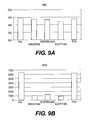
- FIG. 9 shows a comparison between the ability of different types of cotton fibers and PVA fibers to remove certain cell types from whole blood: (A) RBCs, (B) WBCs, (C) Plt, and (D) neutrophils and lymphocytes.
- Pre” samples indicate cell counts taken from blood samples prior to filtration.
- RBCs the Y axis measures number of cells ⁇ 10 4 per microliter of filtered blood.
- WBCs the Y axis measures number of cells per microliter of filtered blood.
- Pit the Y axis measures number of cells ⁇ 10 4 per microliter of filtered blood.
- neutrophils and lymphocytes the Y axis measures number of cells per microliter of filtered blood.
- FIG. 10 shows a comparison between the ability of different types of cotton fibers to remove neutrophils and lymphocytes from whole blood.
- FIG. 11 shows the ability of Egyptian cotton fibers, when at a density of 0.125 g/ml, to remove certain cell types from various volumes of whole blood: (A) RBCs, (B) WBCs, (C) Pit, and (D) neutrophils and lymphocytes. “Pre” samples indicate cell counts taken from blood samples prior to filtration. The Y axis of each figure measures the number of cells per microliter of filtered blood. RBCs and PIt were measured as the number of cells ⁇ 10 4 / ⁇ l. WBCs and neutrophils/lymphocytes were measured as the number of cells/ ⁇ l.
- FIG. 12 shows the ability of Egyptian cotton fibers, when at a density of 0.05 g/ml, to remove certain cell types from various volumes of whole blood: (A) RBCs, (B) WBCs, (C) Plt, and (D) neutrophils and lymphocytes. “Pre” samples indicate cell counts taken from blood samples prior to filtration. The Y axis of each figure measures the number of cells per microliter of filtered blood. RBCs and Plt were measured as the number of cells ⁇ 10 4 / ⁇ l. WBCs and neutrophils/lymphocytes were measured as the number of cells/ ⁇ l.
- FIG. 13 shows the effect of the density of Egyptian cotton fibers on their ability to remove certain cell types from various volumes of whole blood: (A) RBCs, (B) WBCs, (C) Plt, and (D) neutrophils and lymphocytes. “Pre” samples indicate cell counts taken from blood samples prior to filtration.
- FIG. 14 shows the effect of biolization on the ability of Egyptian cotton fibers to remove (A) RBCs and (B) WBCs from whole blood. Concentrations of 1%, 0.1%, and 0% gelatin were used for biolization. For the 0% sample, only gluteraldehyde was used. Nonbiolyzed samples were treated with water. “Pre” samples indicate cell counts taken from blood samples prior to filtration.
- FIG. 15 shows the effect of biolization on the ability of Egyptian cotton fibers to remove (A) Plts and (B) neutrophils and lymphocytes from whole blood. Concentrations of 1%, 0.1%, and 0% gelatin were used for biolization. For the 0% sample, only gluteraldehyde was used. Nonbiolyzed samples were treated with water. “Pre” samples indicate cell counts taken from blood samples prior to filtration.
- FIG. 16 shows the effect of biolization on the ability of silk fibers to remove (A) RBCs and (B) WBCs from whole blood. Concentrations of 1%, 0.1%, and 0% gelatin were used for biolization. For the 0% sample, only gluteraldehyde was used. Nonbiolyzed samples were treated with saline. “Pre” samples indicate cell counts taken from blood samples prior to filtration.
- FIG. 17 shows the effect of biolization on the ability of silk fibers to remove (A) Plts and (B) neutrophils and lymphocytes from whole blood. Concentrations of 1%, 0.1%, and 0% gelatin were used for biolization. For the 0% sample, only gluteraldehyde was used. Nonbiolyzed samples were treated with saline. “Pre” samples indicate cell counts taken from blood samples prior to filtration
- FIG. 18 shows the effect of biolization with weak ion-beam irradiation on the ability of Egyptian cotton fibers to remove (A) RBCs, (B) WBCs, (C) Plt, and (D) neutrophils and lymphocytes from whole blood.
- FIG. 19 shows the effect of biolization with strong ion-beam irradiation on the ability of Egyptian cotton fibers to remove (A) RBCs, (B) WBCs, (C) Plt, and (D) neutrophils and lymphocytes from whole blood.
- FIG. 20 shows the leukocytes analysis of pre and post perfusion of the blood.
- FIG. 21 shows the blood pressure and body temperature of dogs undergoing 6 hours of perfusion according to one embodiment of the invention.
- FIG. 22 shows the levels of WBCs, RBCs, Plt, and fibrinogen in dogs during and after perfusion according to one embodiment of the invention.
- FIG. 23 shows the kinetics of lymphocyte, neutrophil, and monocyte recovery in dogs during and after perfusion according to one embodiment of the invention.
- the Y axis measures the number of each cell type present in whole blood at each time point.
- FIG. 24 shows the kinetics of lymphocyte, neutrophil, and monocyte recovery in dogs during and after perfusion according to one embodiment of the invention.
- the Y axis indicates the presence of each cell type as a percentage of total cells in the dogs' whole blood at each time point.
- FIG. 25 shows the levels of tumor necrosis factor alpha (TNF ⁇ ) and interleukin 6 (IL-6) in the blood of dogs during and after perfusion according to one embodiment of the invention.
- the Y axis measures the amount of cytokine present in picograms per milliliter, while the X axis represents minutes after the start of perfusion.
- FIG. 26 shows a graphic of leukocyte kinetics in the blood of an apheresis recipient after treatment by the method of the invention.
- the X axis shows each phase in relevance to the amount of time that has passed since apheresis began.
- the Y axis shows the relevant units of leukocyte counts, with 1.0 constituting the level of leukocytes in the blood at the start of apheresis.
- the invention provides a safe, effective, and reproducible method of producing immunologically active state of the patient.
- the patient is a human patient.
- the patient is a non-human animal in which case the invention may be used in veterinary setting.
- Non-human animals include, but are not limited to, cats, dogs, cattle, and horses.
- blood purification apheresis
- Removal of the plasma factors and/or cellular factors responsible for maintaining the active state of a person's immune system state is possible by apheresis. See Nosé 1995. This method of blood purification was introduced for the treatment of autoimmune disease patients.
- treatment of a malignant tumor involves the enhancement of immunological function in a person to reach normal levels.
- the treated person's body regains the ability to recognize the cancer cells as abnormal cells and the malignant cells are destroyed by the person's normalized immune system.
- Such enhancement of immune function can be used to treat any disease associated with immune suppression including cancers and infectious diseases, such as bacterial, fungal, or viral infections.
- the cancer manifests as one or more solid tumors in the body.
- the cancer does not manifest as a solid tumor, for example in the case of leukemia.
- the viral infection can be a Human Immunodeficiency Virus (HIV) infection or a hepatitis virus infection such as Hepatitis A Virus (HAV), Hepatitis B Virus (HBV) or Hepatitis C Virus (HCV).
- HIV Human Immunodeficiency Virus
- HAV Hepatitis A Virus
- HBV Hepatitis B Virus
- HCV Hepatitis C Virus
- the invention provides a method of therapy by activating the suppressed immunological status of a patient using an apheresis technique.
- the Bacille Calmette Guerin (BCG) vaccine has been used to provide immunological activation against tuberculosis bacteria (Grange 2009).
- BCG Bacille Calmette Guerin
- the invention provides a more effective method of stimulating a patient's immune function making it more effective for the treatment of diseases associated with immunosuppression or infectious diseases.
- the apheresis technique of the invention uses a blood perfusion filter comprising a bioincompatible material.
- a bioincompatible material is a material that triggers a reaction from the body, whether it is an immunological reaction or a physiological reaction such as formation of an antibody as a result of exposure to the body either directly (e.g., introduction into the body) or indirectly (e.g., via extracorporeal circulation).
- Membrane-based blood purification systems had been developed for the treatment of autoimmune diseases. See Nosé 2000. For these blood purification apheresis systems, those in the art used filters made of blood compatible materials to avoid activation of the patient's immune system even though these systems removed cellular and molecular factors underlying autoimmune diseases.
- immunoactivation or “activation of the immune system” refers to an increase in the number and/or function of cells of the immune system, such as lymphocytes, and/or an increase in the humoral function of the immune system relating to B cells and antibody production together with cytokine generation.
- cytokine generation can be the production of TNF- ⁇ and/or IL-6.
- the invention provides a method of therapy by activating the suppressed immunological status of a patient using bioincompatible materials in an apheresis technique. While side effects, such as hypotension and hypoxia, may occur during this technique, the invention provides a way of activating the immune system in a safe and controlled manner. As noted above, techniques other than apheresis, such as the BCG vaccine, have been used to provide immunological activation against tuberculosis bacteria (Grange 2009). Unfortunately, effects of BCG were very small. The invention provides a more effective method of stimulating a patient's immune function making it more effective for the treatment of diseases associated with immunosuppression or infectious diseases.
- Another group has used Immugard R (Terumo Co., Japan) and a Cellsorba column by ASAHI Kasei Medical Co., Ltd. to treat ulcerative colitis. Compared with ASAHI filters, the Terumo filter demonstrated better clinical outcomes for the treatment of ulcerative colitis.
- the cellsorba column utilizes polyester fiber.
- the first column known as Imugard, which is produced by Terumo Co. of Japan, experimentally contained cotton fibers from the Gossypium barbadense plant (Amano 1996).
- Imugard which is produced by Terumo Co. of Japan
- mice experienced transient hypotension, a transient reduction of leukocytes, and activation of complement.
- transient hypotension occurred together with transient reduction of leukocytes.
- Complement activation also occurred quite substantially. Because of complications, the FDA did not approve the use of the column for clinical use.
- Terumo then tried to reduce the filter's bioincompatibility by switching the fiber from cotton fibers to synthetic fibers. In doing so, however, the column lost its effectiveness for the treatment of infection and for malignant tumors but still retained effectiveness for rheumatoid arthritis (Amano 1996). In essence, the Terumo column changed from an immunoactivation column into an immunosuppressive column when the filter's bioincompatibility was reduced.
- Terumo utilizes the polyurethane filter and their Immugard is primarily used for removal of leukocytes for blood transfusion.
- the second column contains purified Staphylococcus aureus Protein A, which has a high affinity for immune-complexed IgG antibodies.
- Staphylococcus aureus Protein A which has a high affinity for immune-complexed IgG antibodies.
- bioincompatible apheresis filters In cases where the immune system was activated by these bioincompatible apheresis filters, patients suffered many side effects including hypotension, respiratory failures, nausea, vomiting, excessive sweating, chills, and shiver. It was natural for government agencies, including the FDA in the U.S. and the Ministry of Health and Welfare in Japan, to prohibit clinical application of such apheresis filters. As a consequence, such bioincompatible apheresis filters to introduce effective immunoactivation for the treatment of malignant tumors were not clinically approved. In each case, however, when biocompatibilities were improved, the effects on malignant tumors disappeared.
- the Imugard column lost its effectiveness on tumor cells and instead remained effective for treating rheumatoid arthritis or ulcerative colitis.
- the Protein A column also lost its effectiveness against tumor cells when it was modified to reduce side effects instead becoming effective for treating rheumatoid arthritis. Both rheumatoid arthritis and ulcerative colitis are conditions associated with an overactive immune response, demonstrating that the modified columns acted to suppress the patient's immune system.
- an apheresis column when modified to reduce its bioincompatiblity, the resulting column acts to suppress the patient's immune system by removal of autoantibodies (especially of the IgG3 subtype), removal of immunocomplexes, and/or removal of immunostimulatory cytokines.
- apheresis columns containing bioincompatible material act to stimulate the immune system by increasing antibody production, increasing cytokine production, and skewing the leukocyte population in the patient's blood towards a lymphocyte dominant state.
- the invention induces a controlled shock state in patients.
- the invention provides a way of using bioincompatible materials in an apheresis column to induce a temporary but powerful stimulation of the immune system while at the same time controlling the side effects that can accompany such stimulation.
- the immune system's ability to kill tumor cells via apoptosis is reduced.
- the side effects associated with inducing immunological shock can be controlled by using general anesthesia. In this way, the invention provides a type of molecular surgery in which the patient's immune system is changed at the molecular level to hunt down and kill diseases-causing cells.
- the invention uses general anesthesia during two phases of physiological insults to the patient: (1) induction of immunoactivation by apheresis using a bioincompatible blood pheresis filter during the first hour and (2) safe maintenance of the transient lymphocyte-dominant immunoactive clinical stage during the subsequent 5 hours for tumor killing by cellular and humoral agents.
- Some experimental filters were bioincompatible and introduced such hemodynamic and respiratory changes, but there were no fatal incidents. However, these filters were not intended to enhance immunoactive state but instead aimed to suppress immunological activities.
- the method of the invention results in the immunological modulations shown in FIG. 1 after about one hour of apheresis using the apheresis column of the invention. All of these phenomena are due to the immunoactivation of not only humoral factors but also cellular factors. In other words, it is the induced immunological shock as the result of the direct contact of the patient's blood to the bioincompatible biomaterials and subsequent phase 1 modulation (discussed below) of autoimmunological cellular and humoral adjustment of the patient intra-corporeally during this period of times of 6 hours. In one embodiment, the immunoactivation process is completed in two weeks.
- FIG. 3 These physiological impacts introduced by the immunoactivating blood perfusion filter of the invention are shown in FIG. 3 .
- the primary cause of these physiological impacts comes from leukocyte trapping in the lung capillaries. Usually granulocytes are trapped transiently inside of the lung capillaries and subsequently induce transient lymphocyte dominant state in the patient. This is one of the most important signs of the immunoactivation of the patient. Thus, transient reduction of the circulating leukocytes together with hypotension help to facilitate the beneficial therapeutic effects of the invention.
- the leukocytes are trapped in the lung capillaries during the initial 30 minutes of extracorporeal circulation. Regardless of whether extracorporeal circulation is continued or not, after 30 minutes, release of these trapped leukocytes in the lung capillaries will be initiated.
- the so-called unsafe “side effects” of bioincompatible blood purification are actually physiological responses that lead to the therapeutic effects on malignant tumors.
- Transient increases in leukocytes after this 30 minute period of time can occur as shown in FIG. 4 .
- kinetics of leukocytes returned to circulation are different after these 30 minutes.
- Leukocyte trapping after extracorporeal circulation during the first 30 minutes is the same with different filters of different material, however, the rate of leukocyte return to the blood circulation are different due to the differing bioincompatiblility of the materials.
- the method of the invention involves performing the apheresis procedure under general anesthesia.
- the apheresis is performed with intratracheal intubation.
- proper blood pressure and proper blood gas levels are maintained throughout the procedure. Proper blood gas levels can be maintained using, for example, oxygen supplies administered via the nose, mouth, or trachea.
- oxygen is administered through a mask covering the patient's nose and mouth.
- oxygen is administered via an intratracheal tube.
- extracorporeal circulation through an apheresis column according to the invention for one hour with general anesthesia and maintenance of the anesthesia for an additional five hours can provide safe immunoactivation to patients.
- the initial rate of blood flow through the apheresis column can be about 100 to 200 ml/min, allowing the treatment of one blood volume in about sixty minutes.
- An extracorporeal circulation rate of approximately 100 ml/min can be achieved by veno-venous needle access.
- approximately 100 ml/min blood flows can be maintained during apheresis; If however, at the beginning of the extracorporeal circulation, blood pressure drops to an unsafe level, for example less than 50 mm Hg, then this drop in blood pressure should be rectified.
- the blood flow rate can be reduced by 25%. If hypotension continues, then the blood flow rate can be further reduced by 50% of the initial blood flow rate. Also, the oxygen content of the arterial blood can drop together with leukocyte counts, for example less than 20% of the total cell count, during this time. Immediate reduction of the blood flow rate by 25% to 50% can also remedy hypoxia and leukopenia. In one embodiment, the blood flow rate can be reduced from about 100 ml/min to about 75 ml/min or about 50 ml/min.
- the immunoactivation of cellular and molecular factors in the blood is completed in six hours (acute phase). During this period of time, the patient's cardiopulmonary function can be maintained under general anesthesia. Thus, patients do not feel any uncomfortable side effects of induced immunological shock, including dizziness, respiratory difficulties, nausea, vomiting, excessive sweating, fever, chills and shiver.
- the invention provides a method of treating a disease in a patient comprising:
- the invention provides a method of treating a disease in a patient comprising:
- the invention provides for the use of:
- the method of the invention results in significant changes in leukocyte population in the patient's blood and in a transient increase of some of cytokines.
- phase 1 which is less than 30 minutes after extracorporeal circulation is initiated, transient reduction of the leukocytes inside of the blood occurs primarily by trapping them inside of the lung capillaries.
- phase 2 which is less than 30 minutes after extracorporeal circulation is initiated, transient reduction of the leukocytes inside of the blood occurs primarily by trapping them inside of the lung capillaries.
- more reduction of granulocytes near 100%
- lymphocytes about 40%
- granulocytes are gradually released from the lung.
- one hour extracorporeal-circulation can generate sufficient immunoactivation effects.
- leukocytes are released from lung capillaries and leukocyte counts become normalized in approximately 6 hours. During these 6 hours, most granulocytes are released from the lung. This is also the lymphocyte dominant stage in which a massive transient increase in cytokines occurs, which in turn leads to the death of tumor cells in the patient's body. During these 6 hours, lung function may require assistance. The general anesthesia with ample supply of oxygen by intratracheal intubation can treat hypoxia during this period of time.
- lymphocyte counts increase substantially higher in spite of marked reduction of granulocytes during the initial six hours.
- Leukocytes including granulocytes and lymphocytes, can also increase also more than two fold over pre-apheresis levels.
- these increased monocytes and granulocytes should be able to eliminate the dead tumor cells. Meanwhile, lympohocytes are still present at high levels.
- phase 4 all of these abnormally increased leukocyte counts return to the pre-apheresis levels after two weeks.
- apheresis procedures to remove these cellular debris and immunoactive agents can be applied to maintain patient's safety.
- apheresis can be administered to the patient according to the invention once. In another embodiment, apheresis can be administered to the patient more than once. In this case, each apheresis treatment occurs every two weeks. In yet another embodiment, treatment is provided to the patient at least three times, with each treatment occurring every two weeks. In this embodiment, overall treatment of the patient would be completed in six weeks. In another embodiment, apheresis treatment according to the invention may be supplemented by conventional anticancer therapies to augment the invention's effects of immunostimulation.
- the patient's blood can travel directly from the apheresis column back into the patient's body.
- the patient's blood can flow into the apheresis column of the invention and then be collected for later administration to the patient.
- it is possible to establish immunoactivation by exposing the harvested blood from the patient to the filter with bioincompatible material column and reinjection to the patient.
- extracorporeal apheresis includes (1) achievement of extracorporeal circulation through a bioincompatible blood perfusion filter in about 60 minutes; (2) phase 2 immunoactivation can be completed in six hours during which cellular and cytokine activation occurs; and (3) physiological responses to treatment occur in about 30 minutes and include hypotension, leukopenia, and respiratory difficulties such as hypoxia. These physiological responses are safely and unpainfully accepted by the patient who is under general anesthesia.
- FIG. 5A provides one embodiment of an acrylic apheresis column that can be used in the method of the invention.
- a small acrylic chamber (3) holds approximately 1 g of washed fibers of about 5 ⁇ m or less. These fibers are supported in the small chamber by a non-woven mesh of PVA (2) which is about 2 mm thick.
- the non woven PVA mesh (VW100) was provided Kuraray Inc, Tokyo Japan.
- Each small chamber including the prepared fibers and the PVA mesh constitutes a filtering unit (4).
- Five to 10 filtering units can be stacked into the apheresis column (1). In one embodiment, 5 filtering units are used in which case the column's height is about 60 mm. In another embodiment, 10 filtering units are used in which case the column's height is about 120 mm.
- the filtering units may be packed inside the chamber in such a way as to allow approximately 400 ml of priming volume, the volume of priming solution that is run through the column before introduction of blood, in the chamber.
- the packing density of the filters can be up to approximately 10% (grams/volume).
- the fibers have a diameter of 5 ⁇ m or less. In another embodiment, the fibers have a diameter of 1-2 5 ⁇ m.
- Natural fibers that are useable with the apheresis column of the invention include, but are not limited to cotton and silk.
- silk fibers are the smallest in diameter. Among all silks, the smallest fiber sizes were demonstrated by “Kanton” silk and “Japanese” silk. Japanese silk is produced by Japanese manufacturers while Kanton silk is manufactured in China. Both types of silk can be obtained from Marubeni America Corporation, Houston, Tex. The smallest diameters of the silk fibers are almost analogous to the fiber diameters of Egyptian cotton. They were less than 5 ⁇ m, typically 1-2 ⁇ m.
- zoological fibers are more complex structures with multiple different bioincompatible components (particularly protein groups) over above the plant fibers. Thus they are more bioincompatible over above the synthetic fibers or the natural plant fibers.
- the natural fibers of the invention may be biolized.
- biolized refers to a crosslinking procedure that links homogenous proteins present in the fiber to produce a smooth, consistent surface on the fiber.
- Fibers may be biolized chemically or via irradiation techniques. Fibers may be biolized chemically by using chemical agents including, but not limited to, formaldehyde and glutaraldehyde. In one embodiment, fibers are soaked in 10% formaldehyde for at least 48 hours. In another embodiment, fibers can be stored in a 0.45% solution of glutaraldehyde longer than 2 weeks and as long as 5 years. If the bioincompatible material inside the apheresis column has been biolized, the column should be washed with normal saline to remove residual aldehyde prior to clinical usage.
- Types of cotton and silk were tested under varying conditions for their ability to filter out different cell types from human whole blood.
- a 5 ml syringe was packed a bioincompatible material to a 4 ml volume inside the syringe, resulting in a density of 0.125 g/ml. See FIG. 7 .
- Whole blood was obtained from a normal, healthy human and 1 unit of heparin sodium was added per milliter of blood to reduce clotting. Blood preparations were used in the experiments within 30 minutes of collection. During filtration at 22° C., the treated blood was poured into the top of the syringe, filtered through the bioincompatible material, and then collected from the bottom of the syringe. The resulting filtered blood was then analyzed for the presence of different cell types.
- the granulocyte removal rate of Egyptian cotton treated with a 0.5 N acid solution and a 0.5 N alkali solution was compared to the removal rate obtained with Egyptian cotton treated with saline (Baxter Corp. catalog no. 281324).
- the raw Egyptian cotton was obtained from Marubeni America Corporation and was prepared by successive soaking with 0.5N NaOH (S320-500 Fisher Scientific), water, 0.5N HCl (SA48-500, Fisher Scientific), water, and normal saline for about 30 minutes in each solution. Egyptian cotton was also soaked in normal saline alone.
- FIG. 9 As shown in FIG. 9 , almost 100% granulocyte removal was demonstrated by each of the cottons tested in comparison to the PVA negative control, with Egyptian cotton providing the best result. See FIG. 9D . See also FIG. 10 . In addition, RBCs were not filtered out by any of the cotton types while the majority of platelets were removed. See FIGS. 9A and 9C. Regarding lymphocytes, each type of cotton exhibited slightly different levels of lymphocyte removal, with Australian cotton removing the most lymphocytes.
- FIG. 12D shows that for the 0.05 g/ml density, the efficiency of WBC removal decreased with increasing blood volume.
- granulocyte removal was most optimal, while retaining the presence of lymphocytes in the filtered blood. In as little as the first 5 to 7 mls the mini column was able to remove granulocytes, as measured by neutrophil removal.
- FIG. 13 three fiber densities were tested for efficiency of granulocyte removal at a single blood volume
- Fiber densities of 0.05 g/ml, 0.125 g/ml, and 0.2 g/ml were compared.
- the 0.05 and 0.125 g/ml density mini columns were prepared as described above.
- For the 0.2 g/ml density column 0.2 grams of Egyptian cotton was packed into a 1 ml volume in a 4 ml syringe.
- 7 mls of whole heparin-treated blood was filtered through the column. The first 5 mls were discarded and the remaining 2 mls were collected for analysis.
- fiber densities of 0.125 g/ml and 0.2 g/ml worked best to remove granulocytes, as demonstrated by neutrophil counts, while allowing some lymphocytes to pass through the mini column.
- a fiber density of at least 0.125 g/ml worked well to remove granulocytes.
- Cotton fibers were biolized using three kinds of combination treatments: 0.4% glutaraldehyde crosslinkings followed by coating fibers with 1%, 0.1% or 0% gelatin. In the case of 0% gelatin, fibers were biolized with glutaraldehyde only. For biolization, fibers were soaked in glutaraldehyde for more than 24 hours. The fibers were then coated in a gelatin solution (Fisher Scientific; Cat No. G7-500). Cotton fibers were also soaked in water alone, and acted as a positive control. As shown in FIG. 14 , all three treatments resulted in comparable RBC removal among the samples.
- FIG. 14 shows that that all three treatments resulted in very good platelet removal and complete granulocyte removal while allowing some lymphocytes to pass through the mini column.
- FIGS. 16 and 17 show that
- Fibers were prepared by successive treatments with 0.5 N NaOH, 0.5 N HCl, 70% isopropyl alcohol. The fibers spent approximately 30 minutes in each solution with a rinsing step with water between solutions. The cartridge was washed and biolozed by running a 10% formaldehyde solution through the column for 48 hours. Before the experiment, the processed fibers were packed into the column and then the blood circuit was set up.
- the blood circuit comprised a roller pump, a tubing set, a heating unit, and an apheresis column. Before the start of an experiment the blood circuit was rinsed by normal saline until the residual formaldehyde concentration in rinsed solution became less than 5 ppm. The blood flow rate through the column was set at 3 ml/kg. Pressures of the inlet and outlet of the column were measured by the pressure gauges.
- the apheresis column was connected to the dogs to complete the entire circuit of the extracorporeal circulation by PVC (polyvinyl chloride) tubing together with a warmer bag and an air removal chamber.
- PVC polyvinyl chloride
- the extracorporeal apheresis column was disinfected by 4% formaldehyde overnight, while the rest of the circuit components were previously sterilized prior to use.
- the dogs Prior to extracorporeal apheresis, the dogs were anesthetized using a combination of Xylazin intramuscularly and Ketamine intramuscularly for induction of anesthesia followed by 2.5% isoflurane gas for maintenance. 3 L of oxygen, 5 L of air 2% isoflurane were added to the repiratory gas. As alternative anesthetic induction, Atropine was used. General anesthesia was administered within 60 minutes of beginning the apheresis treatment.
- the dogs' blood was heparanized by initially administering a 200 unit/kg bolus intravenously and then during apheresis, the dogs continued to receive 100 units/kg of heparain and then the dogs' blood circulated through the apheresis column for one hour at a flow rate of 3.3 ml/kg as noted above. Following the one hour of apheresis, the dogs remained under general anesthesia for an additional five hours, making a total duration of six hours of general anesthesia from the time that extracorporeal apheresis began.
- this treatment resulted in ex vivo removal of leukocytes.
- the darkest bar indicated the leukocytes (represented by neutrophils and lymphocytes in this figure) present in the inlet blood, the blood that was traveling into the apheresis column.
- the medium colored bar represents the number of leukocytes present in the blood exiting the apheresis column and traveling back into the dog.
- the lightest colored bar represents the number of leukocytes present in the residual blood trapped inside the column. Almost all granulocytes were removed while only 60% of lymphocytes were removed, thus resulting in a lymphocyte-dominant state in the dogs.
- blood pressures had a tendency to drop transiently approximately 30 minutes after the onset of the apheresis. However, the blood pressure later increased after these transient drops.
- blood gases were maintained while body temperatures tended to increase during the six hours of general anesthesia.
- FIG. 22 shows that WBC counts and fibrinogen levels initially decreased during perfusion, but later recovered within hours of apheresis. Platelet counts, however, also dropped during apheresis and remained at relatively low numbers for approximately four days. RBC counts stayed fairly constant during and after the one hour apheresis treatment.
- a transient state of lymphocyte dominance was established in the dogs. Specifically, the percentage of granulocytes and percentage of lymphocytes in the dogs' blood changed after 15 minutes of extracorporeal circulation. The percentage of neutrophils in the dogs' blood was only 20% while lymphocytes constituted 80% of the WBCs in the blood. In later time points, the percentage of neutrophils and monocytes returned to preoperative levels after 6 hours, but the percentage of lymphocytes remained low for 6 hours to 4 days after treatment ( FIG. 24 ).
- the immunological shock associated with apheresis using bioincompatible materials includes dizziness due to hypotension, respiratory difficulty, nausea and vomiting, excessive sweating, fever, chills, and shivers.
- the method of the invention is performed under general anesthesia. By doing so, the patient can undergo reproducible and effective immunoactivation apheresis therapy for the treatment of malignant tumors and infectious diseases. Unsafe and dangerous shock inducing immunoactivation apheresis therapy now becomes a safe, effective, and painless therapeutic molecular surgical procedure because of the invention.
- the effective and safe immunoactivation therapy of the invention can be described in the following eight features: (1) general anesthesia with intra-tracheal intubation; (2) careful monitoring of blood pressure and arterial blood oxygen contents; (3) sufficient supply of oxygen; (4) careful flow controls during extracorporeal circulation and automatic blood flow reduction whenever hypotension and hypoxia should occur more than expected levels; (5) extracorporeal circulation lasting approximately 60 minutes or less; (6) maintenance of intratracheal anesthesia for at least six hours, during which the patient receives physiological support while under general anesthesia; (7) after four days of immunoactivation, transient increase in leukocytes counts occur; and (8) follow up monitoring of cellular and humoral immunological responses for two weeks after completion of the apheresis treatment.
Abstract
Description
| TABLE 1 |
 |
| TABLE 2 | |||
| Classical | Molecular Surgery | ||
| 20th Century | (21st Century) | ||
| Removal tool | knife and scissors | membrane fibers and | ||
| granules | ||||
| Removal | malignant and/or | blood cellular and | ||
| objectives | damaged tissues | humoral molecules | ||
| Procedures | cut and suture | extracorporeal blood | ||
| therapy | ||||
| Outcome | regeneration of new | immunoactivation or | ||
| tissues | immunosuppression | |||
| Anesthesia | general or local | general | ||
Surgical procedures in the 20th century involve removing tissues by scissors and knife under general anesthesia. Blood purification procedures in the 21st century remove molecular and cellular components by blood purification filters (apheresis) under general anesthesia. For this molecular method of cancer therapy, there are no complications or side effects that are commonly associated with radiation therapy or chemotherapies currently used for cancer therapy. There is no need to damage normal cells of the patient but only destroy abnormal malignant tumor cells. At the same time, it is very difficult to detect small metastatic regions to apply effective radiation therapies. In short, at this time there aren't any effective therapeutic regimens for the treatment of malignant tumors. Thus, in the 21st century, the therapeutic procedures for cancer should be molecular surgery with general anesthesia.
| TABLE 3 | |||
| By Drugs | By Blood Therapy | ||
| Effects | indirect | direct | ||
| Control of | difficult | easy | ||
| effects | ||||
| Control of side | difficult | easy | ||
| effects | ||||
| Monitoring of | difficult | easy | ||
| effects or side | ||||
| effects | ||||
| Reversal or | difficult | easy | ||
| suppression of | ||||
| effects | ||||
Method
-
- (a) providing an apheresis column including a blood perfusion filter comprising at least one bioincompatible material;
- (b) connecting the patient's blood circulation with the apheresis system such that the patient's blood passes through the blood perfusion filter before reentering the patient's body;
- (c) placing the patient under general anesthesia and providing physiological support to the patient;
- (d) circulating the patient's blood through the apheresis system for about one hour; and
- (e) keeping the patient under general anesthesia for at least 5 hours after circulating the patient's blood through the apheresis system,
wherein circulating the patient's blood through the blood perfusion filter activates the patient's immune system thereby treating the disease.
-
- (a) providing an apheresis column including a blood perfusion filter comprising at least one bioincompatible material
- (b) providing general anesthesia with continuous arterial pressure monitoring and oxygen monitoring of arterial blood via endotracheal intubation;
- (c) administering an anticoagulant to the patient;
- (d) circulating the patient's blood through the apheresis column for about one hour via veno-venous perfusion; wherein the rate of blood flow through the apheresis column is about 100 to 200 mL/min;
- (e) monitoring the patient's blood pressure and blood oxygen levels;
- (f) keeping the patient under general anesthesia for at least 5 hours after circulating the patient's blood through the apheresis system while monitoring the patient for signs of immunoactivation and cardiopulmonary stability; and
- (g) removing the patient from general anesthesia after confirming the recovery of lung functions to pre-apheresis levels.
-
- (a) an apheresis system including a blood perfusion filter comprising at least one bioincompatible material, wherein the apheresis system is connected to a patient's blood circulation such that the patient's blood can pass through the blood perfusion filter before reentering the patient's body; and
- (b) a general anesthetic for anesthetizing the patient during use of the apheresis system and for at least 5 hours following that use
for treating a disease in a patient, wherein use of the apheresis system activates the patient's immune system thereby treating the disease.
- Ainsworth S. K., et al. Toxicity following protein A treatment of Metastic breast adenocarcinoma cancer 61:1495-1500,1988.
- Amano K. et al. Filter leucopheresis for patients with ulcerative colitis; clinical results and the possible mechanism. Therapeutic Apheresis 2(2) 97-100, 1998.
- Amano K. et al., Four year study of Leukapheresis with Gossypium barbadense cotton for Rheumatoid Arthritis Japanese Journal for Apheresis 15(1) 103-104, 1996.
- Grange J. M. et al., Immunotherapy for malignant melanomia—Tracing Ariadne's thread through the labyrinth, European Journal of Cancer 45(13), 2266-73, 2009.
- Levy J. et al., Correcting immune imbalance: The use of prosorba column treatment for immune disorders. Therapeutic Apheresis and Dialysis 7(2) 197-203. 2003.
- Messerschmidt G. L. et al., Protein A immunoadsorption in the treatment of malignant disease J. Clinical Oncology (12) 203-212,1988.
- Nosé Y., et al. Therapeutic Membrane Plasmapheresis. Therapeutic apheresis 4(1) 3-9, 2000 (originally published in 1981).
- Nosé Y. Blood purification procedures and their related short and long term effects on patients. Therapeutic apheresis 6(5), 333-347, 2002.
- Nosé Y. Congress presidential address: 5th WAA congress, Therapeutic Artificial Organs: 10 years after, Artificial Organs 1995.
- Tani, T. et al. Blood purification therapy in cancer treatment, Therapeutic apheresis 2(3) 182-184, 1998.
- Yonekawa M., granulocyte removal therapy for cancer Tissue Culture Engineering [Japanese Text] 23(12) 481-485, 1997.
- Yonekawa M. Granulocytapheresis in Cancer Tissue Culture Engineering (Japanese Text) 23 (12) 481-485, 1997.
- Yonekawa M., Kamii N., Onodera K. et al Basic Study of Extracorporeal Granulocyte/Lymphocyte Regulation System Therapeutic Plasmapheresis (X) ICAOT Press, Cleveland pp. 37-42, 1992.
Claims (19)
Priority Applications (1)
| Application Number | Priority Date | Filing Date | Title |
|---|---|---|---|
| US13/394,765 US8926543B2 (en) | 2009-10-08 | 2010-10-07 | Immunoactivation blood perfusion filter for the treatment of malignant tumors |
Applications Claiming Priority (3)
| Application Number | Priority Date | Filing Date | Title |
|---|---|---|---|
| US24986709P | 2009-10-08 | 2009-10-08 | |
| PCT/US2010/051832 WO2011044369A1 (en) | 2009-10-08 | 2010-10-07 | An immunoactivation blood perfusion filter for the treatment of malignant tumors |
| US13/394,765 US8926543B2 (en) | 2009-10-08 | 2010-10-07 | Immunoactivation blood perfusion filter for the treatment of malignant tumors |
Publications (2)
| Publication Number | Publication Date |
|---|---|
| US20130046225A1 US20130046225A1 (en) | 2013-02-21 |
| US8926543B2 true US8926543B2 (en) | 2015-01-06 |
Family
ID=43857149
Family Applications (1)
| Application Number | Title | Priority Date | Filing Date |
|---|---|---|---|
| US13/394,765 Expired - Fee Related US8926543B2 (en) | 2009-10-08 | 2010-10-07 | Immunoactivation blood perfusion filter for the treatment of malignant tumors |
Country Status (10)
| Country | Link |
|---|---|
| US (1) | US8926543B2 (en) |
| EP (1) | EP2485983B1 (en) |
| JP (1) | JP5822836B2 (en) |
| KR (1) | KR101747469B1 (en) |
| CN (1) | CN102712503A (en) |
| CA (1) | CA2777007C (en) |
| ES (1) | ES2628513T3 (en) |
| MY (1) | MY161192A (en) |
| TW (1) | TWI637755B (en) |
| WO (1) | WO2011044369A1 (en) |
Families Citing this family (13)
| Publication number | Priority date | Publication date | Assignee | Title |
|---|---|---|---|---|
| JP2014500735A (en) | 2010-10-15 | 2014-01-16 | サイトフェリックス インコーポレイテッド | Cytopheresis cartridge and its use |
| CA2852220A1 (en) | 2011-10-14 | 2013-07-18 | Cytopherx, Inc. | Cartridge and method for increasing myocardial function |
| EP3539982A3 (en) | 2011-12-23 | 2020-01-15 | Pfizer Inc | Engineered antibody constant regions for site-specific conjugation and methods and uses therefor |
| US10624924B2 (en) | 2012-03-12 | 2020-04-21 | Grifols, S.A. | Method and device for treating blood cholesterol disorders |
| TWI498273B (en) | 2012-04-02 | 2015-09-01 | Nat Applied Res Laboratories | Miniature sieve apparatus and manufacturing method thereof |
| TWI463129B (en) | 2012-05-07 | 2014-12-01 | Nat Applied Res Laboratories | Miniature sieve apparatus for microparticle detecting |
| JP6450767B2 (en) * | 2013-12-27 | 2019-01-09 | エリアス・セラピューティクス・インコーポレイテッドEliaz Therapeutics,Inc. | Plasma apheresis device |
| US10376627B2 (en) | 2014-03-24 | 2019-08-13 | Fenwal, Inc. | Flexible biological fluid filters |
| US9796166B2 (en) | 2014-03-24 | 2017-10-24 | Fenwal, Inc. | Flexible biological fluid filters |
| US10159778B2 (en) | 2014-03-24 | 2018-12-25 | Fenwal, Inc. | Biological fluid filters having flexible walls and methods for making such filters |
| US9782707B2 (en) | 2014-03-24 | 2017-10-10 | Fenwal, Inc. | Biological fluid filters having flexible walls and methods for making such filters |
| US9968738B2 (en) | 2014-03-24 | 2018-05-15 | Fenwal, Inc. | Biological fluid filters with molded frame and methods for making such filters |
| AU2015201496B2 (en) | 2014-06-03 | 2019-10-31 | Grifols Worldwide Operations Limited | Use of plasmapheresis to treat blood pressure disorders |
Citations (21)
| Publication number | Priority date | Publication date | Assignee | Title |
|---|---|---|---|---|
| JPS5359017A (en) | 1976-11-02 | 1978-05-27 | Terumo Corp | Fractionation and purification of red blood corpuscle |
| US4445500A (en) | 1982-03-03 | 1984-05-01 | Thomas Jefferson University | Stroke treatment utilizing extravascular circulation of oxygenated synthetic nutrients to treat tissue hypoxic and ischemic disorders |
| JPH01124382A (en) | 1987-06-30 | 1989-05-17 | Hapgood Cv | Animal cell having transduced antigel protein |
| US5476444A (en) * | 1992-09-04 | 1995-12-19 | Idt, Inc. | Specialized perfusion protocol for whole-body hyperthermia |
| US5498336A (en) * | 1991-02-22 | 1996-03-12 | Terumo Kabushiki Kaisha | Leukocyte-removing filter and leukocyte-removing apparatus furnished therewith |
| US6264680B1 (en) * | 1998-01-23 | 2001-07-24 | Viacirq, Inc. | Apparatuses and processes for whole-body hyperthermia |
| US20020033181A1 (en) * | 2000-01-14 | 2002-03-21 | Groth Karl Emil | Treatment of hepatitis C using hyperthermia |
| US6498007B1 (en) * | 1999-03-17 | 2002-12-24 | Japan Immunoresearch Laboratories Co., Ltd. | Methods of treatment of disease using adsorbent carriers |
| US20030062299A1 (en) | 1999-03-08 | 2003-04-03 | Whatman Hemasure, Inc. | Leukocyte reduction filtration media |
| US20040006214A1 (en) * | 1997-03-25 | 2004-01-08 | Kaneka Corporation | Adorbent for eliminating hepatitis C virus, adsorber, and adsorption method |
| US6827898B1 (en) * | 1999-05-25 | 2004-12-07 | Viacirq, Inc. | Hyperthermia method and apparatus |
| US20050014127A1 (en) * | 2001-10-16 | 2005-01-20 | Hirokazu Onodera | Method for selectively removing virus and leukocytes eliminating material and eliminating apparatus |
| US20050145573A1 (en) * | 1996-01-31 | 2005-07-07 | Toshiki Nanko | Adsorbent and method for adsorbing a chemokine in body fluid |
| US20050187508A1 (en) * | 2004-02-25 | 2005-08-25 | Gorsuch Reynolds G. | Structurally optimized hollow fiber membranes |
| JP2005304647A (en) | 2004-04-20 | 2005-11-04 | Haemonetics Corp | Apheresis device |
| WO2006073106A1 (en) | 2005-01-06 | 2006-07-13 | Asahi Kasei Medical Co., Ltd. | Method of removing leukocyte |
| US20070190050A1 (en) * | 2003-03-17 | 2007-08-16 | Hema Vation, Llc | Apparatus and method for down-regulating immune system mediators in blood |
| US20090060890A1 (en) * | 2007-08-31 | 2009-03-05 | The Regents Of The University Of Michigan | Selective cytopheresis devices and related methods thereof |
| EP2058018A1 (en) | 2006-08-31 | 2009-05-13 | Toray Industries, Inc. | Adsorption carrier containing composite fiber |
| US20090234266A1 (en) * | 2006-08-24 | 2009-09-17 | Fresenius Medical Care Holdings, Inc. | Device for removing fluid from blood in a patient |
| US20090275874A1 (en) * | 2005-03-31 | 2009-11-05 | Toray Industries, Inc. | Absorbent and Column for Extracorporeal Circulation |
Family Cites Families (3)
| Publication number | Priority date | Publication date | Assignee | Title |
|---|---|---|---|---|
| US4246107A (en) * | 1978-03-06 | 1981-01-20 | Asahi Kasei Kogyo Kabushiki Kaisha | Separation of lymphocytes from lymphocyte-containing suspension by filtration |
| US4708713A (en) * | 1984-11-16 | 1987-11-24 | Anisa Medical, Inc. | Method and system for removing immunosuppressive components from the blood of mammals |
| US20020086276A1 (en) * | 2000-12-28 | 2002-07-04 | Srivastava Pramod K. | Immunotherapeutic methods for extracorporeal modulation of CD36 and its ligands |
-
2010
- 2010-10-07 JP JP2012533319A patent/JP5822836B2/en not_active Expired - Fee Related
- 2010-10-07 ES ES10822700.0T patent/ES2628513T3/en active Active
- 2010-10-07 MY MYPI2012001547A patent/MY161192A/en unknown
- 2010-10-07 CN CN201080045456XA patent/CN102712503A/en active Pending
- 2010-10-07 EP EP10822700.0A patent/EP2485983B1/en not_active Not-in-force
- 2010-10-07 KR KR1020127011680A patent/KR101747469B1/en active IP Right Grant
- 2010-10-07 US US13/394,765 patent/US8926543B2/en not_active Expired - Fee Related
- 2010-10-07 CA CA2777007A patent/CA2777007C/en not_active Expired - Fee Related
- 2010-10-07 WO PCT/US2010/051832 patent/WO2011044369A1/en active Application Filing
- 2010-10-08 TW TW099134370A patent/TWI637755B/en not_active IP Right Cessation
Patent Citations (26)
| Publication number | Priority date | Publication date | Assignee | Title |
|---|---|---|---|---|
| JPS5359017A (en) | 1976-11-02 | 1978-05-27 | Terumo Corp | Fractionation and purification of red blood corpuscle |
| US4130642A (en) | 1976-11-02 | 1978-12-19 | Terumo Corporation | Method of separating blood cells components |
| US4445500A (en) | 1982-03-03 | 1984-05-01 | Thomas Jefferson University | Stroke treatment utilizing extravascular circulation of oxygenated synthetic nutrients to treat tissue hypoxic and ischemic disorders |
| JPH01124382A (en) | 1987-06-30 | 1989-05-17 | Hapgood Cv | Animal cell having transduced antigel protein |
| US5677176A (en) | 1987-06-30 | 1997-10-14 | Hapgood, C.V. | Animal derived cell with antigenic protein introduced therein |
| US5498336A (en) * | 1991-02-22 | 1996-03-12 | Terumo Kabushiki Kaisha | Leukocyte-removing filter and leukocyte-removing apparatus furnished therewith |
| US5476444A (en) * | 1992-09-04 | 1995-12-19 | Idt, Inc. | Specialized perfusion protocol for whole-body hyperthermia |
| US20050145573A1 (en) * | 1996-01-31 | 2005-07-07 | Toshiki Nanko | Adsorbent and method for adsorbing a chemokine in body fluid |
| US20040006214A1 (en) * | 1997-03-25 | 2004-01-08 | Kaneka Corporation | Adorbent for eliminating hepatitis C virus, adsorber, and adsorption method |
| US20010039441A1 (en) * | 1998-01-23 | 2001-11-08 | Viacirq, Inc. | Apparatuses and processes for whole-body hyperthermia |
| US6264680B1 (en) * | 1998-01-23 | 2001-07-24 | Viacirq, Inc. | Apparatuses and processes for whole-body hyperthermia |
| US20030062299A1 (en) | 1999-03-08 | 2003-04-03 | Whatman Hemasure, Inc. | Leukocyte reduction filtration media |
| US6498007B1 (en) * | 1999-03-17 | 2002-12-24 | Japan Immunoresearch Laboratories Co., Ltd. | Methods of treatment of disease using adsorbent carriers |
| US6827898B1 (en) * | 1999-05-25 | 2004-12-07 | Viacirq, Inc. | Hyperthermia method and apparatus |
| US20040055609A1 (en) * | 2000-01-14 | 2004-03-25 | First Circle Medical | Treatment of hepatitis C using hyperthermia |
| US20020033181A1 (en) * | 2000-01-14 | 2002-03-21 | Groth Karl Emil | Treatment of hepatitis C using hyperthermia |
| US20050014127A1 (en) * | 2001-10-16 | 2005-01-20 | Hirokazu Onodera | Method for selectively removing virus and leukocytes eliminating material and eliminating apparatus |
| US20070190050A1 (en) * | 2003-03-17 | 2007-08-16 | Hema Vation, Llc | Apparatus and method for down-regulating immune system mediators in blood |
| US20050187508A1 (en) * | 2004-02-25 | 2005-08-25 | Gorsuch Reynolds G. | Structurally optimized hollow fiber membranes |
| JP2005304647A (en) | 2004-04-20 | 2005-11-04 | Haemonetics Corp | Apheresis device |
| WO2006073106A1 (en) | 2005-01-06 | 2006-07-13 | Asahi Kasei Medical Co., Ltd. | Method of removing leukocyte |
| US8496833B2 (en) | 2005-01-06 | 2013-07-30 | Asahi Kasei Medical Co., Ltd. | Leukocyte removal method |
| US20090275874A1 (en) * | 2005-03-31 | 2009-11-05 | Toray Industries, Inc. | Absorbent and Column for Extracorporeal Circulation |
| US20090234266A1 (en) * | 2006-08-24 | 2009-09-17 | Fresenius Medical Care Holdings, Inc. | Device for removing fluid from blood in a patient |
| EP2058018A1 (en) | 2006-08-31 | 2009-05-13 | Toray Industries, Inc. | Adsorption carrier containing composite fiber |
| US20090060890A1 (en) * | 2007-08-31 | 2009-03-05 | The Regents Of The University Of Michigan | Selective cytopheresis devices and related methods thereof |
Non-Patent Citations (9)
| Title |
|---|
| "Membrane biocompatibility does not affect whole body protein metabolism during dialysis." Jorden M Veeneman et al. Blood Purification Feb. 2005; 23(3):211-8. DOI:10.1159/000084891 http://www.researchgate.net/publication/7925895-Membrane-biocompatibility-does-not-affect-whole-body-protein-metabolism-during-dialysis. accessed Jun. 12, 2013. * |
| "Selected Indicators of Food and Agriculture Development in Asia-Pacific Region 1992-2002." Food and Agriculture Organization of the United Nations Regional Office for Asia and the Pacific, Bangkok. Oct. 2003. ftp://ftp.fao.org/docrep/fao/004/ad452e/ accessed Jun. 20, 2013. * |
| International Search Report from the United States Patent and Trademark Office for International Application No. PCT/US2010/051832, mailed Dec. 1, 2010. |
| Mojcik et al.; "Aprotinin and the Systemic Inflammatory Response After Cardiopulmonary Bypass", The Annals of Thoracic Surgery, vol. 71, pp. 745-754, (2001). |
| Robinson, William R.; Nash, John J. "Half Lives." Visualization and Problem Solving for General Chemistry. http://www.chem.purdue.edu/gchelp/howtosolveit/Kinetics/Halflife.html Accessed Jan. 24, 2014. * |
| Written Opinion of the International Searching Authority from the United States Patent and Trademark Office for International Application No. PCT/US2010/051832, mailed Dec. 1, 2010. |
| Yonekawa et al.; "Extracorporeal Granulocytapheresis for Cancer and Rheumatoid Arthritis", Transfusion Science, vol. 17, No. 3, pp. 463-472, (1996). |
| Yukihiko Nosé. "Bioincompatible Biomaterials for Extracorporeal Immunomodulation." DOI: 10.1111/j.1525-1594.1988.tb02790.x Artificial Organs vol. 12, Issue 5, pp. 377-378, Oct. 1988. * |
| Yukihiko Noséet al. "Membrane Apheresis Technology: Historical Perspective and New Trends toward Bioincompatible Systems." DOI: 10.1111/j.1744-9987.1997.tb00005.x Therapeutic Apheresis vol. 1, Issue 1, pp. 5-12, Feb. 1997. * |
Also Published As
| Publication number | Publication date |
|---|---|
| KR101747469B1 (en) | 2017-06-14 |
| EP2485983B1 (en) | 2017-05-03 |
| KR20120098676A (en) | 2012-09-05 |
| WO2011044369A8 (en) | 2011-06-30 |
| TWI637755B (en) | 2018-10-11 |
| US20130046225A1 (en) | 2013-02-21 |
| CA2777007A1 (en) | 2011-04-14 |
| WO2011044369A1 (en) | 2011-04-14 |
| CN102712503A (en) | 2012-10-03 |
| TW201116308A (en) | 2011-05-16 |
| JP5822836B2 (en) | 2015-11-24 |
| CA2777007C (en) | 2019-08-20 |
| ES2628513T3 (en) | 2017-08-03 |
| EP2485983A4 (en) | 2015-02-11 |
| JP2013507379A (en) | 2013-03-04 |
| MY161192A (en) | 2017-04-14 |
| EP2485983A1 (en) | 2012-08-15 |
Similar Documents
| Publication | Publication Date | Title |
|---|---|---|
| US8926543B2 (en) | Immunoactivation blood perfusion filter for the treatment of malignant tumors | |
| Daugirdas et al. | First-use reactions during hemodialysis: a definition of subtypes. | |
| US7753869B2 (en) | Removal and deactivation of viruses from blood | |
| AU2011215942B8 (en) | Removal of virulence factors through extracorporeal therapy | |
| JP3881019B2 (en) | Extracorporeal blood treatment system for the treatment of inflammatory diseases | |
| JP2003299730A (en) | Membrane and method for separating protein bonding substance from protein-containing substance by dialysis | |
| US20040186412A1 (en) | Extracorporeal blood treatment system using ultraviolet light and filters | |
| UA113290C2 (en) | THE APPLICATION OF HEMOGLOBIN OR MYOGLOBIN, WHICH IS OWNED BY OXYGEN CARBON OXYGEN, FOR EXTERNAL CARBON | |
| US9265875B2 (en) | Methods for treating blood composition and function disorders wherein a patient's blood plasma is extracorporeally contacted with donor blood or modified donor blood | |
| US7229427B2 (en) | Irradiation and filter device for treatment of blood | |
| US20230036583A1 (en) | Devices and methods for reducing rejection of a transplanted organ in a recipient | |
| WO1997020570A1 (en) | Non-immunogenic and toleragenic platelet and red blood cell compositions | |
| JP5249737B2 (en) | System for removing viruses and cytokines from blood | |
| US20040186407A1 (en) | Concentrator and filter apparatus for treatment of blood | |
| US20110197628A1 (en) | Cryoaggregate filtration for the treatment of cardiomyopathy and other autoimmune diseases | |
| JP5592450B2 (en) | Self-propelled cell trap | |
| JP4201313B2 (en) | Toxic substance binding albumin removal system | |
| US20040185041A1 (en) | Method for extracorporeal treatment of blood | |
| US20040185426A1 (en) | Ultraviolet light and filter apparatus for treatment of blood | |
| JP4833443B2 (en) | How to collect hematopoietic stem cells | |
| Nosé et al. | Molecular surgery for the treatment of malignant tumors: bioincompatible material apheresis for cancer therapy? | |
| Meijersa et al. | Extracorporeal Techniques in Kidney Failure | |
| JP2005082567A (en) | Method for producing cellular composition for treatment by vascularization and system for producing the same | |
| JPH09220281A (en) | Sterilizing method for body fluid treating device and sterilized body fluid treating device | |
| JPH0423546B2 (en) |
Legal Events
| Date | Code | Title | Description |
|---|---|---|---|
| AS | Assignment |
Owner name: OTSUKA PHARMACEUTICAL CO., LTD., JAPAN Free format text: ASSIGNMENT OF ASSIGNORS INTEREST;ASSIGNOR:BAYLOR COLLEGE OF MEDICINE;REEL/FRAME:025348/0277 Effective date: 20100924 |
|
| AS | Assignment |
Owner name: BAYLOR COLLEGE OF MEDICINE, TEXAS Free format text: ASSIGNMENT OF ASSIGNORS INTEREST;ASSIGNORS:NOSE (DECEASED), YUKIHIKO;OHTA, KAZUHIDE;MIYAMOTO, HIROSHI;AND OTHERS;SIGNING DATES FROM 20120508 TO 20120818;REEL/FRAME:028837/0635 |
|
| AS | Assignment |
Owner name: OTSUKA PHARMACEUTICAL CO., LTD., JAPAN Free format text: ASSIGNMENT OF ASSIGNORS INTEREST;ASSIGNOR:BAYLOR COLLEGE OF MEDICINE;REEL/FRAME:029462/0237 Effective date: 20100924 |
|
| AS | Assignment |
Owner name: BAYLOR COLLEGE OF MEDICINE, TEXAS Free format text: CONFIRMATION OF ASSIGNMENT;ASSIGNOR:NOSE, YUKIHIKO (DECEASED) BY AKO NOSE (SOLE HEIR);REEL/FRAME:030572/0378 Effective date: 20120914 |
|
| STCF | Information on status: patent grant |
Free format text: PATENTED CASE |
|
| MAFP | Maintenance fee payment |
Free format text: PAYMENT OF MAINTENANCE FEE, 4TH YEAR, LARGE ENTITY (ORIGINAL EVENT CODE: M1551) Year of fee payment: 4 |
|
| FEPP | Fee payment procedure |
Free format text: MAINTENANCE FEE REMINDER MAILED (ORIGINAL EVENT CODE: REM.); ENTITY STATUS OF PATENT OWNER: LARGE ENTITY |
|
| LAPS | Lapse for failure to pay maintenance fees |
Free format text: PATENT EXPIRED FOR FAILURE TO PAY MAINTENANCE FEES (ORIGINAL EVENT CODE: EXP.); ENTITY STATUS OF PATENT OWNER: LARGE ENTITY |
|
| STCH | Information on status: patent discontinuation |
Free format text: PATENT EXPIRED DUE TO NONPAYMENT OF MAINTENANCE FEES UNDER 37 CFR 1.362 |
|
| FP | Lapsed due to failure to pay maintenance fee |
Effective date: 20230106 |