US8747833B2 - B7-H1 and methods of diagnosis, prognosis, and treatment of cancer - Google Patents
B7-H1 and methods of diagnosis, prognosis, and treatment of cancer Download PDFInfo
- Publication number
- US8747833B2 US8747833B2 US13/012,063 US201113012063A US8747833B2 US 8747833 B2 US8747833 B2 US 8747833B2 US 201113012063 A US201113012063 A US 201113012063A US 8747833 B2 US8747833 B2 US 8747833B2
- Authority
- US
- United States
- Prior art keywords
- cells
- cancer
- tumor
- expression
- subject
- Prior art date
- Legal status (The legal status is an assumption and is not a legal conclusion. Google has not performed a legal analysis and makes no representation as to the accuracy of the status listed.)
- Active
Links
- 206010028980 Neoplasm Diseases 0.000 title claims abstract description 198
- 101710094000 Programmed cell death 1 ligand 1 Proteins 0.000 title claims abstract description 152
- 201000011510 cancer Diseases 0.000 title claims abstract description 104
- 238000000034 method Methods 0.000 title claims abstract description 98
- 238000011282 treatment Methods 0.000 title claims abstract description 12
- 238000003745 diagnosis Methods 0.000 title abstract description 8
- 238000004393 prognosis Methods 0.000 title description 6
- 210000004027 cell Anatomy 0.000 claims description 172
- 210000000265 leukocyte Anatomy 0.000 claims description 64
- 208000006265 Renal cell carcinoma Diseases 0.000 claims description 59
- 238000012360 testing method Methods 0.000 claims description 52
- 108090000765 processed proteins & peptides Proteins 0.000 claims description 32
- 229920001184 polypeptide Polymers 0.000 claims description 30
- 102000004196 processed proteins & peptides Human genes 0.000 claims description 30
- 238000009169 immunotherapy Methods 0.000 claims description 22
- 102000000588 Interleukin-2 Human genes 0.000 claims description 3
- 108010002350 Interleukin-2 Proteins 0.000 claims description 3
- 230000003993 interaction Effects 0.000 abstract description 15
- 230000002401 inhibitory effect Effects 0.000 abstract description 14
- 230000008901 benefit Effects 0.000 abstract description 8
- 238000002619 cancer immunotherapy Methods 0.000 abstract 1
- 210000001519 tissue Anatomy 0.000 description 86
- 210000004881 tumor cell Anatomy 0.000 description 41
- 239000003795 chemical substances by application Substances 0.000 description 35
- 108020003175 receptors Proteins 0.000 description 33
- 102000005962 receptors Human genes 0.000 description 33
- 239000000523 sample Substances 0.000 description 29
- 108091030071 RNAI Proteins 0.000 description 25
- 230000009368 gene silencing by RNA Effects 0.000 description 25
- 239000012634 fragment Substances 0.000 description 21
- 108020004999 messenger RNA Proteins 0.000 description 20
- 108091034117 Oligonucleotide Proteins 0.000 description 19
- 239000000427 antigen Substances 0.000 description 19
- 108091007433 antigens Proteins 0.000 description 19
- 102000036639 antigens Human genes 0.000 description 19
- 230000034994 death Effects 0.000 description 19
- 230000002601 intratumoral effect Effects 0.000 description 16
- 210000001744 T-lymphocyte Anatomy 0.000 description 15
- 206010054094 Tumour necrosis Diseases 0.000 description 14
- 210000004443 dendritic cell Anatomy 0.000 description 14
- 230000002962 histologic effect Effects 0.000 description 14
- 230000004083 survival effect Effects 0.000 description 14
- 239000000074 antisense oligonucleotide Substances 0.000 description 13
- 238000012230 antisense oligonucleotides Methods 0.000 description 13
- 108090000623 proteins and genes Proteins 0.000 description 13
- 102000004127 Cytokines Human genes 0.000 description 12
- 108090000695 Cytokines Proteins 0.000 description 12
- 230000000692 anti-sense effect Effects 0.000 description 12
- 230000002163 immunogen Effects 0.000 description 12
- 210000001165 lymph node Anatomy 0.000 description 12
- 150000007523 nucleic acids Chemical class 0.000 description 12
- 206010027476 Metastases Diseases 0.000 description 10
- 210000000612 antigen-presenting cell Anatomy 0.000 description 10
- 208000037265 diseases, disorders, signs and symptoms Diseases 0.000 description 10
- 108010074328 Interferon-gamma Proteins 0.000 description 9
- 238000003556 assay Methods 0.000 description 9
- 238000013059 nephrectomy Methods 0.000 description 9
- 102000004169 proteins and genes Human genes 0.000 description 9
- 108010017213 Granulocyte-Macrophage Colony-Stimulating Factor Proteins 0.000 description 8
- 102100039620 Granulocyte-macrophage colony-stimulating factor Human genes 0.000 description 8
- 239000003814 drug Substances 0.000 description 8
- 239000013604 expression vector Substances 0.000 description 8
- 201000001441 melanoma Diseases 0.000 description 8
- 102000039446 nucleic acids Human genes 0.000 description 8
- 108020004707 nucleic acids Proteins 0.000 description 8
- 125000003729 nucleotide group Chemical group 0.000 description 8
- 108020004414 DNA Proteins 0.000 description 7
- 241000124008 Mammalia Species 0.000 description 7
- 108091008874 T cell receptors Proteins 0.000 description 7
- 102000016266 T-Cell Antigen Receptors Human genes 0.000 description 7
- 108700029229 Transcriptional Regulatory Elements Proteins 0.000 description 7
- 150000001875 compounds Chemical class 0.000 description 7
- 229940079593 drug Drugs 0.000 description 7
- 239000003102 growth factor Substances 0.000 description 7
- 230000002519 immonomodulatory effect Effects 0.000 description 7
- 210000002540 macrophage Anatomy 0.000 description 7
- 239000002773 nucleotide Substances 0.000 description 7
- 108091033319 polynucleotide Proteins 0.000 description 7
- 102000040430 polynucleotide Human genes 0.000 description 7
- 239000002157 polynucleotide Substances 0.000 description 7
- 235000018102 proteins Nutrition 0.000 description 7
- 102000008394 Immunoglobulin Fragments Human genes 0.000 description 6
- 108010021625 Immunoglobulin Fragments Proteins 0.000 description 6
- 102000015696 Interleukins Human genes 0.000 description 6
- 108010063738 Interleukins Proteins 0.000 description 6
- 108060008682 Tumor Necrosis Factor Proteins 0.000 description 6
- 102000000852 Tumor Necrosis Factor-alpha Human genes 0.000 description 6
- 238000004458 analytical method Methods 0.000 description 6
- 230000001772 anti-angiogenic effect Effects 0.000 description 6
- 210000003719 b-lymphocyte Anatomy 0.000 description 6
- 238000001514 detection method Methods 0.000 description 6
- 201000010099 disease Diseases 0.000 description 6
- 230000028993 immune response Effects 0.000 description 6
- 230000003259 immunoinhibitory effect Effects 0.000 description 6
- 238000001727 in vivo Methods 0.000 description 6
- 210000004072 lung Anatomy 0.000 description 6
- 230000001575 pathological effect Effects 0.000 description 6
- 210000000952 spleen Anatomy 0.000 description 6
- 208000024891 symptom Diseases 0.000 description 6
- 108091026890 Coding region Proteins 0.000 description 5
- 108010004889 Heat-Shock Proteins Proteins 0.000 description 5
- 102000002812 Heat-Shock Proteins Human genes 0.000 description 5
- 102100037850 Interferon gamma Human genes 0.000 description 5
- 102000006992 Interferon-alpha Human genes 0.000 description 5
- 108010047761 Interferon-alpha Proteins 0.000 description 5
- JLCPHMBAVCMARE-UHFFFAOYSA-N [3-[[3-[[3-[[3-[[3-[[3-[[3-[[3-[[3-[[3-[[3-[[5-(2-amino-6-oxo-1H-purin-9-yl)-3-[[3-[[3-[[3-[[3-[[3-[[5-(2-amino-6-oxo-1H-purin-9-yl)-3-[[5-(2-amino-6-oxo-1H-purin-9-yl)-3-hydroxyoxolan-2-yl]methoxy-hydroxyphosphoryl]oxyoxolan-2-yl]methoxy-hydroxyphosphoryl]oxy-5-(5-methyl-2,4-dioxopyrimidin-1-yl)oxolan-2-yl]methoxy-hydroxyphosphoryl]oxy-5-(6-aminopurin-9-yl)oxolan-2-yl]methoxy-hydroxyphosphoryl]oxy-5-(6-aminopurin-9-yl)oxolan-2-yl]methoxy-hydroxyphosphoryl]oxy-5-(6-aminopurin-9-yl)oxolan-2-yl]methoxy-hydroxyphosphoryl]oxy-5-(6-aminopurin-9-yl)oxolan-2-yl]methoxy-hydroxyphosphoryl]oxyoxolan-2-yl]methoxy-hydroxyphosphoryl]oxy-5-(5-methyl-2,4-dioxopyrimidin-1-yl)oxolan-2-yl]methoxy-hydroxyphosphoryl]oxy-5-(4-amino-2-oxopyrimidin-1-yl)oxolan-2-yl]methoxy-hydroxyphosphoryl]oxy-5-(5-methyl-2,4-dioxopyrimidin-1-yl)oxolan-2-yl]methoxy-hydroxyphosphoryl]oxy-5-(5-methyl-2,4-dioxopyrimidin-1-yl)oxolan-2-yl]methoxy-hydroxyphosphoryl]oxy-5-(6-aminopurin-9-yl)oxolan-2-yl]methoxy-hydroxyphosphoryl]oxy-5-(6-aminopurin-9-yl)oxolan-2-yl]methoxy-hydroxyphosphoryl]oxy-5-(4-amino-2-oxopyrimidin-1-yl)oxolan-2-yl]methoxy-hydroxyphosphoryl]oxy-5-(4-amino-2-oxopyrimidin-1-yl)oxolan-2-yl]methoxy-hydroxyphosphoryl]oxy-5-(4-amino-2-oxopyrimidin-1-yl)oxolan-2-yl]methoxy-hydroxyphosphoryl]oxy-5-(6-aminopurin-9-yl)oxolan-2-yl]methoxy-hydroxyphosphoryl]oxy-5-(4-amino-2-oxopyrimidin-1-yl)oxolan-2-yl]methyl [5-(6-aminopurin-9-yl)-2-(hydroxymethyl)oxolan-3-yl] hydrogen phosphate Polymers Cc1cn(C2CC(OP(O)(=O)OCC3OC(CC3OP(O)(=O)OCC3OC(CC3O)n3cnc4c3nc(N)[nH]c4=O)n3cnc4c3nc(N)[nH]c4=O)C(COP(O)(=O)OC3CC(OC3COP(O)(=O)OC3CC(OC3COP(O)(=O)OC3CC(OC3COP(O)(=O)OC3CC(OC3COP(O)(=O)OC3CC(OC3COP(O)(=O)OC3CC(OC3COP(O)(=O)OC3CC(OC3COP(O)(=O)OC3CC(OC3COP(O)(=O)OC3CC(OC3COP(O)(=O)OC3CC(OC3COP(O)(=O)OC3CC(OC3COP(O)(=O)OC3CC(OC3COP(O)(=O)OC3CC(OC3COP(O)(=O)OC3CC(OC3COP(O)(=O)OC3CC(OC3COP(O)(=O)OC3CC(OC3COP(O)(=O)OC3CC(OC3CO)n3cnc4c(N)ncnc34)n3ccc(N)nc3=O)n3cnc4c(N)ncnc34)n3ccc(N)nc3=O)n3ccc(N)nc3=O)n3ccc(N)nc3=O)n3cnc4c(N)ncnc34)n3cnc4c(N)ncnc34)n3cc(C)c(=O)[nH]c3=O)n3cc(C)c(=O)[nH]c3=O)n3ccc(N)nc3=O)n3cc(C)c(=O)[nH]c3=O)n3cnc4c3nc(N)[nH]c4=O)n3cnc4c(N)ncnc34)n3cnc4c(N)ncnc34)n3cnc4c(N)ncnc34)n3cnc4c(N)ncnc34)O2)c(=O)[nH]c1=O JLCPHMBAVCMARE-UHFFFAOYSA-N 0.000 description 5
- 150000001413 amino acids Chemical class 0.000 description 5
- 210000000481 breast Anatomy 0.000 description 5
- 230000008595 infiltration Effects 0.000 description 5
- 238000001764 infiltration Methods 0.000 description 5
- 239000006166 lysate Substances 0.000 description 5
- 210000001616 monocyte Anatomy 0.000 description 5
- 230000001537 neural effect Effects 0.000 description 5
- 230000002611 ovarian Effects 0.000 description 5
- 230000002381 testicular Effects 0.000 description 5
- YBJHBAHKTGYVGT-ZKWXMUAHSA-N (+)-Biotin Chemical compound N1C(=O)N[C@@H]2[C@H](CCCCC(=O)O)SC[C@@H]21 YBJHBAHKTGYVGT-ZKWXMUAHSA-N 0.000 description 4
- 108020000948 Antisense Oligonucleotides Proteins 0.000 description 4
- 241000283707 Capra Species 0.000 description 4
- 241000699800 Cricetinae Species 0.000 description 4
- 241000699694 Gerbillinae Species 0.000 description 4
- 102000003886 Glycoproteins Human genes 0.000 description 4
- 108090000288 Glycoproteins Proteins 0.000 description 4
- 108090000467 Interferon-beta Proteins 0.000 description 4
- 102000008070 Interferon-gamma Human genes 0.000 description 4
- 108010065805 Interleukin-12 Proteins 0.000 description 4
- 108091028043 Nucleic acid sequence Proteins 0.000 description 4
- 102100040678 Programmed cell death protein 1 Human genes 0.000 description 4
- 101710089372 Programmed cell death protein 1 Proteins 0.000 description 4
- 241000700159 Rattus Species 0.000 description 4
- 208000035475 disorder Diseases 0.000 description 4
- 239000003937 drug carrier Substances 0.000 description 4
- 230000006870 function Effects 0.000 description 4
- 230000002496 gastric effect Effects 0.000 description 4
- 238000000338 in vitro Methods 0.000 description 4
- 229960003130 interferon gamma Drugs 0.000 description 4
- 229940047122 interleukins Drugs 0.000 description 4
- 230000000968 intestinal effect Effects 0.000 description 4
- 210000003734 kidney Anatomy 0.000 description 4
- 230000001926 lymphatic effect Effects 0.000 description 4
- 239000000463 material Substances 0.000 description 4
- 239000000203 mixture Substances 0.000 description 4
- 210000003205 muscle Anatomy 0.000 description 4
- 210000000056 organ Anatomy 0.000 description 4
- 230000008488 polyadenylation Effects 0.000 description 4
- 210000002307 prostate Anatomy 0.000 description 4
- 238000001356 surgical procedure Methods 0.000 description 4
- 238000002560 therapeutic procedure Methods 0.000 description 4
- 230000002792 vascular Effects 0.000 description 4
- 239000013598 vector Substances 0.000 description 4
- 239000003981 vehicle Substances 0.000 description 4
- MZOFCQQQCNRIBI-VMXHOPILSA-N (3s)-4-[[(2s)-1-[[(2s)-1-[[(1s)-1-carboxy-2-hydroxyethyl]amino]-4-methyl-1-oxopentan-2-yl]amino]-5-(diaminomethylideneamino)-1-oxopentan-2-yl]amino]-3-[[2-[[(2s)-2,6-diaminohexanoyl]amino]acetyl]amino]-4-oxobutanoic acid Chemical compound OC[C@@H](C(O)=O)NC(=O)[C@H](CC(C)C)NC(=O)[C@H](CCCN=C(N)N)NC(=O)[C@H](CC(O)=O)NC(=O)CNC(=O)[C@@H](N)CCCCN MZOFCQQQCNRIBI-VMXHOPILSA-N 0.000 description 3
- CSCPPACGZOOCGX-UHFFFAOYSA-N Acetone Chemical compound CC(C)=O CSCPPACGZOOCGX-UHFFFAOYSA-N 0.000 description 3
- 108020005544 Antisense RNA Proteins 0.000 description 3
- 241000283690 Bos taurus Species 0.000 description 3
- 241000282693 Cercopithecidae Species 0.000 description 3
- 241000282326 Felis catus Species 0.000 description 3
- 241000282412 Homo Species 0.000 description 3
- 241000699666 Mus <mouse, genus> Species 0.000 description 3
- 241000283973 Oryctolagus cuniculus Species 0.000 description 3
- 206010061902 Pancreatic neoplasm Diseases 0.000 description 3
- 241001494479 Pecora Species 0.000 description 3
- 244000309464 bull Species 0.000 description 3
- 230000000295 complement effect Effects 0.000 description 3
- 239000002299 complementary DNA Substances 0.000 description 3
- 230000000694 effects Effects 0.000 description 3
- 239000003623 enhancer Substances 0.000 description 3
- 230000002489 hematologic effect Effects 0.000 description 3
- 210000000987 immune system Anatomy 0.000 description 3
- 208000015181 infectious disease Diseases 0.000 description 3
- 238000001802 infusion Methods 0.000 description 3
- 230000002452 interceptive effect Effects 0.000 description 3
- 238000001990 intravenous administration Methods 0.000 description 3
- 239000003446 ligand Substances 0.000 description 3
- 210000004698 lymphocyte Anatomy 0.000 description 3
- 208000015486 malignant pancreatic neoplasm Diseases 0.000 description 3
- OHDXDNUPVVYWOV-UHFFFAOYSA-N n-methyl-1-(2-naphthalen-1-ylsulfanylphenyl)methanamine Chemical compound CNCC1=CC=CC=C1SC1=CC=CC2=CC=CC=C12 OHDXDNUPVVYWOV-UHFFFAOYSA-N 0.000 description 3
- 210000000822 natural killer cell Anatomy 0.000 description 3
- 210000000440 neutrophil Anatomy 0.000 description 3
- 201000002528 pancreatic cancer Diseases 0.000 description 3
- 208000008443 pancreatic carcinoma Diseases 0.000 description 3
- 230000010399 physical interaction Effects 0.000 description 3
- 239000002504 physiological saline solution Substances 0.000 description 3
- 210000000512 proximal kidney tubule Anatomy 0.000 description 3
- 210000005084 renal tissue Anatomy 0.000 description 3
- 150000003384 small molecules Chemical class 0.000 description 3
- 238000010186 staining Methods 0.000 description 3
- 238000006467 substitution reaction Methods 0.000 description 3
- 235000000346 sugar Nutrition 0.000 description 3
- 230000008685 targeting Effects 0.000 description 3
- 102400000068 Angiostatin Human genes 0.000 description 2
- 108010079709 Angiostatins Proteins 0.000 description 2
- 206010005949 Bone cancer Diseases 0.000 description 2
- 208000018084 Bone neoplasm Diseases 0.000 description 2
- 241000283725 Bos Species 0.000 description 2
- 206010006187 Breast cancer Diseases 0.000 description 2
- 208000026310 Breast neoplasm Diseases 0.000 description 2
- 101710155857 C-C motif chemokine 2 Proteins 0.000 description 2
- 108010022366 Carcinoembryonic Antigen Proteins 0.000 description 2
- 102100025475 Carcinoembryonic antigen-related cell adhesion molecule 5 Human genes 0.000 description 2
- 241000700198 Cavia Species 0.000 description 2
- 102000000018 Chemokine CCL2 Human genes 0.000 description 2
- 102000019034 Chemokines Human genes 0.000 description 2
- 108010012236 Chemokines Proteins 0.000 description 2
- 239000003298 DNA probe Substances 0.000 description 2
- 102400001047 Endostatin Human genes 0.000 description 2
- 108010079505 Endostatins Proteins 0.000 description 2
- 102000004190 Enzymes Human genes 0.000 description 2
- 108090000790 Enzymes Proteins 0.000 description 2
- 108010008655 Epstein-Barr Virus Nuclear Antigens Proteins 0.000 description 2
- 206010017993 Gastrointestinal neoplasms Diseases 0.000 description 2
- 102100041003 Glutamate carboxypeptidase 2 Human genes 0.000 description 2
- DHMQDGOQFOQNFH-UHFFFAOYSA-N Glycine Chemical compound NCC(O)=O DHMQDGOQFOQNFH-UHFFFAOYSA-N 0.000 description 2
- 108010043121 Green Fluorescent Proteins Proteins 0.000 description 2
- 102000004144 Green Fluorescent Proteins Human genes 0.000 description 2
- WZUVPPKBWHMQCE-UHFFFAOYSA-N Haematoxylin Chemical compound C12=CC(O)=C(O)C=C2CC2(O)C1C1=CC=C(O)C(O)=C1OC2 WZUVPPKBWHMQCE-UHFFFAOYSA-N 0.000 description 2
- 101000892862 Homo sapiens Glutamate carboxypeptidase 2 Proteins 0.000 description 2
- 108010001336 Horseradish Peroxidase Proteins 0.000 description 2
- 241000701806 Human papillomavirus Species 0.000 description 2
- 102100026720 Interferon beta Human genes 0.000 description 2
- 102000003996 Interferon-beta Human genes 0.000 description 2
- 208000008839 Kidney Neoplasms Diseases 0.000 description 2
- 206010058467 Lung neoplasm malignant Diseases 0.000 description 2
- 102000009571 Macrophage Inflammatory Proteins Human genes 0.000 description 2
- 108010009474 Macrophage Inflammatory Proteins Proteins 0.000 description 2
- 108010063954 Mucins Proteins 0.000 description 2
- 241001529936 Murinae Species 0.000 description 2
- 241000699670 Mus sp. Species 0.000 description 2
- 206010028851 Necrosis Diseases 0.000 description 2
- 241000282579 Pan Species 0.000 description 2
- 241000282520 Papio Species 0.000 description 2
- 102000007066 Prostate-Specific Antigen Human genes 0.000 description 2
- 108010072866 Prostate-Specific Antigen Proteins 0.000 description 2
- 108010076504 Protein Sorting Signals Proteins 0.000 description 2
- 108020004518 RNA Probes Proteins 0.000 description 2
- 239000003391 RNA probe Substances 0.000 description 2
- 206010038389 Renal cancer Diseases 0.000 description 2
- 241000282887 Suidae Species 0.000 description 2
- 108060008245 Thrombospondin Proteins 0.000 description 2
- 102000002938 Thrombospondin Human genes 0.000 description 2
- 108700009124 Transcription Initiation Site Proteins 0.000 description 2
- 208000008385 Urogenital Neoplasms Diseases 0.000 description 2
- 241000700618 Vaccinia virus Species 0.000 description 2
- 230000001594 aberrant effect Effects 0.000 description 2
- 230000001919 adrenal effect Effects 0.000 description 2
- 230000000961 alloantigen Effects 0.000 description 2
- 150000001408 amides Chemical class 0.000 description 2
- 235000001014 amino acid Nutrition 0.000 description 2
- 229940024606 amino acid Drugs 0.000 description 2
- 230000000259 anti-tumor effect Effects 0.000 description 2
- 230000000890 antigenic effect Effects 0.000 description 2
- FZCSTZYAHCUGEM-UHFFFAOYSA-N aspergillomarasmine B Natural products OC(=O)CNC(C(O)=O)CNC(C(O)=O)CC(O)=O FZCSTZYAHCUGEM-UHFFFAOYSA-N 0.000 description 2
- 210000003651 basophil Anatomy 0.000 description 2
- 229960002685 biotin Drugs 0.000 description 2
- 235000020958 biotin Nutrition 0.000 description 2
- 239000011616 biotin Substances 0.000 description 2
- 108091005948 blue fluorescent proteins Proteins 0.000 description 2
- 210000004556 brain Anatomy 0.000 description 2
- 230000015556 catabolic process Effects 0.000 description 2
- 239000006285 cell suspension Substances 0.000 description 2
- 239000003184 complementary RNA Substances 0.000 description 2
- 230000001054 cortical effect Effects 0.000 description 2
- 238000006731 degradation reaction Methods 0.000 description 2
- 230000002500 effect on skin Effects 0.000 description 2
- 229940088598 enzyme Drugs 0.000 description 2
- 210000003979 eosinophil Anatomy 0.000 description 2
- 238000000684 flow cytometry Methods 0.000 description 2
- 230000003325 follicular Effects 0.000 description 2
- 210000000232 gallbladder Anatomy 0.000 description 2
- 108010008714 glucose-regulated protein 170 Proteins 0.000 description 2
- 239000005090 green fluorescent protein Substances 0.000 description 2
- 201000010536 head and neck cancer Diseases 0.000 description 2
- 208000014829 head and neck neoplasm Diseases 0.000 description 2
- 230000036541 health Effects 0.000 description 2
- 210000003701 histiocyte Anatomy 0.000 description 2
- 238000009396 hybridization Methods 0.000 description 2
- 239000012642 immune effector Substances 0.000 description 2
- 238000011532 immunohistochemical staining Methods 0.000 description 2
- 229940121354 immunomodulator Drugs 0.000 description 2
- 238000011534 incubation Methods 0.000 description 2
- 230000002458 infectious effect Effects 0.000 description 2
- 238000002347 injection Methods 0.000 description 2
- 239000007924 injection Substances 0.000 description 2
- 210000005133 interdigitating dendritic cell Anatomy 0.000 description 2
- 229960001388 interferon-beta Drugs 0.000 description 2
- 201000010982 kidney cancer Diseases 0.000 description 2
- 239000002502 liposome Substances 0.000 description 2
- 210000004185 liver Anatomy 0.000 description 2
- 201000007270 liver cancer Diseases 0.000 description 2
- 208000014018 liver neoplasm Diseases 0.000 description 2
- 201000005202 lung cancer Diseases 0.000 description 2
- 208000020816 lung neoplasm Diseases 0.000 description 2
- 108700025647 major vault Proteins 0.000 description 2
- 210000004962 mammalian cell Anatomy 0.000 description 2
- 244000005700 microbiome Species 0.000 description 2
- 239000011859 microparticle Substances 0.000 description 2
- 230000017074 necrotic cell death Effects 0.000 description 2
- 230000000926 neurological effect Effects 0.000 description 2
- 239000013612 plasmid Substances 0.000 description 2
- 229920000729 poly(L-lysine) polymer Polymers 0.000 description 2
- 229920000642 polymer Polymers 0.000 description 2
- 230000002265 prevention Effects 0.000 description 2
- 238000011321 prophylaxis Methods 0.000 description 2
- 238000011002 quantification Methods 0.000 description 2
- 230000004044 response Effects 0.000 description 2
- 238000003757 reverse transcription PCR Methods 0.000 description 2
- 238000007619 statistical method Methods 0.000 description 2
- 150000008163 sugars Chemical class 0.000 description 2
- 230000009885 systemic effect Effects 0.000 description 2
- 210000001685 thyroid gland Anatomy 0.000 description 2
- 238000013518 transcription Methods 0.000 description 2
- 230000035897 transcription Effects 0.000 description 2
- 238000013519 translation Methods 0.000 description 2
- 210000003932 urinary bladder Anatomy 0.000 description 2
- 201000011531 vascular cancer Diseases 0.000 description 2
- MTCFGRXMJLQNBG-REOHCLBHSA-N (2S)-2-Amino-3-hydroxypropansäure Chemical compound OC[C@H](N)C(O)=O MTCFGRXMJLQNBG-REOHCLBHSA-N 0.000 description 1
- BNIFSVVAHBLNTN-XKKUQSFHSA-N (2s)-4-amino-2-[[(2s)-2-[[(2s)-2-[[(2s)-2-[[(2s)-1-[(2s)-4-amino-2-[[2-[[(2s)-2-[[(2s)-2-[[(2s)-1-[(2s)-6-amino-2-[[(2s)-2-[[(2s)-2-[[(2s,3r)-2-amino-3-hydroxybutanoyl]amino]-4-methylsulfanylbutanoyl]amino]-5-(diaminomethylideneamino)pentanoyl]amino]hexan Chemical compound C[C@@H](O)[C@H](N)C(=O)N[C@@H](CCSC)C(=O)N[C@@H](CCCN=C(N)N)C(=O)N[C@@H](CCCCN)C(=O)N1CCC[C@H]1C(=O)N[C@@H](CCCN=C(N)N)C(=O)N[C@@H](CO)C(=O)NCC(=O)N[C@@H](CC(N)=O)C(=O)N1[C@H](C(=O)N[C@@H](CC(O)=O)C(=O)N[C@@H](C(C)C)C(=O)N[C@@H](C)C(=O)N[C@@H](CC(N)=O)C(O)=O)CCC1 BNIFSVVAHBLNTN-XKKUQSFHSA-N 0.000 description 1
- 108091032973 (ribonucleotides)n+m Proteins 0.000 description 1
- 102000040650 (ribonucleotides)n+m Human genes 0.000 description 1
- PIINGYXNCHTJTF-UHFFFAOYSA-N 2-(2-azaniumylethylamino)acetate Chemical group NCCNCC(O)=O PIINGYXNCHTJTF-UHFFFAOYSA-N 0.000 description 1
- 102000002260 Alkaline Phosphatase Human genes 0.000 description 1
- 108020004774 Alkaline Phosphatase Proteins 0.000 description 1
- 102100037982 Alpha-1,6-mannosylglycoprotein 6-beta-N-acetylglucosaminyltransferase A Human genes 0.000 description 1
- 102100021569 Apoptosis regulator Bcl-2 Human genes 0.000 description 1
- 239000004475 Arginine Substances 0.000 description 1
- DCXYFEDJOCDNAF-UHFFFAOYSA-N Asparagine Natural products OC(=O)C(N)CC(N)=O DCXYFEDJOCDNAF-UHFFFAOYSA-N 0.000 description 1
- 108090001008 Avidin Proteins 0.000 description 1
- 102100035526 B melanoma antigen 1 Human genes 0.000 description 1
- 108060000903 Beta-catenin Proteins 0.000 description 1
- 102000015735 Beta-catenin Human genes 0.000 description 1
- 101100124795 Caenorhabditis elegans hsp-110 gene Proteins 0.000 description 1
- 102100029968 Calreticulin Human genes 0.000 description 1
- 108090000549 Calreticulin Proteins 0.000 description 1
- 241000178270 Canarypox virus Species 0.000 description 1
- 241000700199 Cavia porcellus Species 0.000 description 1
- 102000001327 Chemokine CCL5 Human genes 0.000 description 1
- 108010055166 Chemokine CCL5 Proteins 0.000 description 1
- 239000003155 DNA primer Substances 0.000 description 1
- 241000702421 Dependoparvovirus Species 0.000 description 1
- 101100125027 Dictyostelium discoideum mhsp70 gene Proteins 0.000 description 1
- 238000002965 ELISA Methods 0.000 description 1
- 101150029707 ERBB2 gene Proteins 0.000 description 1
- 206010016654 Fibrosis Diseases 0.000 description 1
- 238000000729 Fisher's exact test Methods 0.000 description 1
- WHUUTDBJXJRKMK-UHFFFAOYSA-N Glutamic acid Natural products OC(=O)C(N)CCC(O)=O WHUUTDBJXJRKMK-UHFFFAOYSA-N 0.000 description 1
- 239000004471 Glycine Substances 0.000 description 1
- 101150031823 HSP70 gene Proteins 0.000 description 1
- 208000032843 Hemorrhage Diseases 0.000 description 1
- 101000971171 Homo sapiens Apoptosis regulator Bcl-2 Proteins 0.000 description 1
- 101000874316 Homo sapiens B melanoma antigen 1 Proteins 0.000 description 1
- 101001014223 Homo sapiens MAPK/MAK/MRK overlapping kinase Proteins 0.000 description 1
- 101001117317 Homo sapiens Programmed cell death 1 ligand 1 Proteins 0.000 description 1
- 101001012157 Homo sapiens Receptor tyrosine-protein kinase erbB-2 Proteins 0.000 description 1
- 102100020755 Hypoxia up-regulated protein 1 Human genes 0.000 description 1
- 206010061598 Immunodeficiency Diseases 0.000 description 1
- 108060003951 Immunoglobulin Proteins 0.000 description 1
- 102000001706 Immunoglobulin Fab Fragments Human genes 0.000 description 1
- 108010054477 Immunoglobulin Fab Fragments Proteins 0.000 description 1
- 108010002352 Interleukin-1 Proteins 0.000 description 1
- 108090000978 Interleukin-4 Proteins 0.000 description 1
- 108090001005 Interleukin-6 Proteins 0.000 description 1
- 108090001007 Interleukin-8 Proteins 0.000 description 1
- QNAYBMKLOCPYGJ-REOHCLBHSA-N L-alanine Chemical compound C[C@H](N)C(O)=O QNAYBMKLOCPYGJ-REOHCLBHSA-N 0.000 description 1
- ODKSFYDXXFIFQN-BYPYZUCNSA-P L-argininium(2+) Chemical compound NC(=[NH2+])NCCC[C@H]([NH3+])C(O)=O ODKSFYDXXFIFQN-BYPYZUCNSA-P 0.000 description 1
- DCXYFEDJOCDNAF-REOHCLBHSA-N L-asparagine Chemical compound OC(=O)[C@@H](N)CC(N)=O DCXYFEDJOCDNAF-REOHCLBHSA-N 0.000 description 1
- CKLJMWTZIZZHCS-REOHCLBHSA-N L-aspartic acid Chemical compound OC(=O)[C@@H](N)CC(O)=O CKLJMWTZIZZHCS-REOHCLBHSA-N 0.000 description 1
- WHUUTDBJXJRKMK-VKHMYHEASA-N L-glutamic acid Chemical compound OC(=O)[C@@H](N)CCC(O)=O WHUUTDBJXJRKMK-VKHMYHEASA-N 0.000 description 1
- ZDXPYRJPNDTMRX-VKHMYHEASA-N L-glutamine Chemical compound OC(=O)[C@@H](N)CCC(N)=O ZDXPYRJPNDTMRX-VKHMYHEASA-N 0.000 description 1
- HNDVDQJCIGZPNO-YFKPBYRVSA-N L-histidine Chemical compound OC(=O)[C@@H](N)CC1=CN=CN1 HNDVDQJCIGZPNO-YFKPBYRVSA-N 0.000 description 1
- AGPKZVBTJJNPAG-WHFBIAKZSA-N L-isoleucine Chemical compound CC[C@H](C)[C@H](N)C(O)=O AGPKZVBTJJNPAG-WHFBIAKZSA-N 0.000 description 1
- ROHFNLRQFUQHCH-YFKPBYRVSA-N L-leucine Chemical compound CC(C)C[C@H](N)C(O)=O ROHFNLRQFUQHCH-YFKPBYRVSA-N 0.000 description 1
- KDXKERNSBIXSRK-YFKPBYRVSA-N L-lysine Chemical compound NCCCC[C@H](N)C(O)=O KDXKERNSBIXSRK-YFKPBYRVSA-N 0.000 description 1
- FFEARJCKVFRZRR-BYPYZUCNSA-N L-methionine Chemical compound CSCC[C@H](N)C(O)=O FFEARJCKVFRZRR-BYPYZUCNSA-N 0.000 description 1
- COLNVLDHVKWLRT-QMMMGPOBSA-N L-phenylalanine Chemical compound OC(=O)[C@@H](N)CC1=CC=CC=C1 COLNVLDHVKWLRT-QMMMGPOBSA-N 0.000 description 1
- AYFVYJQAPQTCCC-GBXIJSLDSA-N L-threonine Chemical compound C[C@@H](O)[C@H](N)C(O)=O AYFVYJQAPQTCCC-GBXIJSLDSA-N 0.000 description 1
- OUYCCCASQSFEME-QMMMGPOBSA-N L-tyrosine Chemical compound OC(=O)[C@@H](N)CC1=CC=C(O)C=C1 OUYCCCASQSFEME-QMMMGPOBSA-N 0.000 description 1
- KZSNJWFQEVHDMF-BYPYZUCNSA-N L-valine Chemical compound CC(C)[C@H](N)C(O)=O KZSNJWFQEVHDMF-BYPYZUCNSA-N 0.000 description 1
- ROHFNLRQFUQHCH-UHFFFAOYSA-N Leucine Natural products CC(C)CC(N)C(O)=O ROHFNLRQFUQHCH-UHFFFAOYSA-N 0.000 description 1
- 108090001030 Lipoproteins Proteins 0.000 description 1
- 102000004895 Lipoproteins Human genes 0.000 description 1
- 108060001084 Luciferase Proteins 0.000 description 1
- 239000005089 Luciferase Substances 0.000 description 1
- 206010025323 Lymphomas Diseases 0.000 description 1
- KDXKERNSBIXSRK-UHFFFAOYSA-N Lysine Natural products NCCCCC(N)C(O)=O KDXKERNSBIXSRK-UHFFFAOYSA-N 0.000 description 1
- 239000004472 Lysine Substances 0.000 description 1
- 108700018351 Major Histocompatibility Complex Proteins 0.000 description 1
- 238000000585 Mann–Whitney U test Methods 0.000 description 1
- 241001465754 Metazoa Species 0.000 description 1
- 108010008707 Mucin-1 Proteins 0.000 description 1
- 102100034256 Mucin-1 Human genes 0.000 description 1
- 108010008705 Mucin-2 Proteins 0.000 description 1
- 102100034263 Mucin-2 Human genes 0.000 description 1
- BKAYIFDRRZZKNF-VIFPVBQESA-N N-acetylcarnosine Chemical compound CC(=O)NCCC(=O)N[C@H](C(O)=O)CC1=CN=CN1 BKAYIFDRRZZKNF-VIFPVBQESA-N 0.000 description 1
- 108091061960 Naked DNA Proteins 0.000 description 1
- 238000000636 Northern blotting Methods 0.000 description 1
- 101710163270 Nuclease Proteins 0.000 description 1
- 108020004711 Nucleic Acid Probes Proteins 0.000 description 1
- 102000036673 PRAME Human genes 0.000 description 1
- 108060006580 PRAME Proteins 0.000 description 1
- 108090000526 Papain Proteins 0.000 description 1
- 102000057297 Pepsin A Human genes 0.000 description 1
- 108090000284 Pepsin A Proteins 0.000 description 1
- 108010004729 Phycoerythrin Proteins 0.000 description 1
- 239000004365 Protease Substances 0.000 description 1
- CZPWVGJYEJSRLH-UHFFFAOYSA-N Pyrimidine Chemical compound C1=CN=CN=C1 CZPWVGJYEJSRLH-UHFFFAOYSA-N 0.000 description 1
- 238000010240 RT-PCR analysis Methods 0.000 description 1
- 102100030086 Receptor tyrosine-protein kinase erbB-2 Human genes 0.000 description 1
- 108020004511 Recombinant DNA Proteins 0.000 description 1
- MTCFGRXMJLQNBG-UHFFFAOYSA-N Serine Natural products OCC(N)C(O)=O MTCFGRXMJLQNBG-UHFFFAOYSA-N 0.000 description 1
- 230000006044 T cell activation Effects 0.000 description 1
- RYYWUUFWQRZTIU-UHFFFAOYSA-N Thiophosphoric acid Chemical group OP(O)(S)=O RYYWUUFWQRZTIU-UHFFFAOYSA-N 0.000 description 1
- AYFVYJQAPQTCCC-UHFFFAOYSA-N Threonine Natural products CC(O)C(N)C(O)=O AYFVYJQAPQTCCC-UHFFFAOYSA-N 0.000 description 1
- 239000004473 Threonine Substances 0.000 description 1
- 102000003425 Tyrosinase Human genes 0.000 description 1
- 108060008724 Tyrosinase Proteins 0.000 description 1
- KZSNJWFQEVHDMF-UHFFFAOYSA-N Valine Natural products CC(C)C(N)C(O)=O KZSNJWFQEVHDMF-UHFFFAOYSA-N 0.000 description 1
- 125000000641 acridinyl group Chemical group C1(=CC=CC2=NC3=CC=CC=C3C=C12)* 0.000 description 1
- 230000003213 activating effect Effects 0.000 description 1
- 230000004913 activation Effects 0.000 description 1
- 230000001270 agonistic effect Effects 0.000 description 1
- 235000004279 alanine Nutrition 0.000 description 1
- 108010034034 alpha-1,6-mannosylglycoprotein beta 1,6-N-acetylglucosaminyltransferase Proteins 0.000 description 1
- 125000000539 amino acid group Chemical group 0.000 description 1
- 239000002870 angiogenesis inducing agent Substances 0.000 description 1
- 238000000137 annealing Methods 0.000 description 1
- 239000003242 anti bacterial agent Substances 0.000 description 1
- 230000000845 anti-microbial effect Effects 0.000 description 1
- 229940088710 antibiotic agent Drugs 0.000 description 1
- 239000004599 antimicrobial Substances 0.000 description 1
- 230000005975 antitumor immune response Effects 0.000 description 1
- 230000008090 antitumoral immunity Effects 0.000 description 1
- 238000013459 approach Methods 0.000 description 1
- ODKSFYDXXFIFQN-UHFFFAOYSA-N arginine Natural products OC(=O)C(N)CCCNC(N)=N ODKSFYDXXFIFQN-UHFFFAOYSA-N 0.000 description 1
- 235000009582 asparagine Nutrition 0.000 description 1
- 229960001230 asparagine Drugs 0.000 description 1
- 235000003704 aspartic acid Nutrition 0.000 description 1
- 230000002238 attenuated effect Effects 0.000 description 1
- DZBUGLKDJFMEHC-UHFFFAOYSA-N benzoquinolinylidene Chemical group C1=CC=CC2=CC3=CC=CC=C3N=C21 DZBUGLKDJFMEHC-UHFFFAOYSA-N 0.000 description 1
- OQFSQFPPLPISGP-UHFFFAOYSA-N beta-carboxyaspartic acid Natural products OC(=O)C(N)C(C(O)=O)C(O)=O OQFSQFPPLPISGP-UHFFFAOYSA-N 0.000 description 1
- 210000003443 bladder cell Anatomy 0.000 description 1
- 210000004369 blood Anatomy 0.000 description 1
- 239000008280 blood Substances 0.000 description 1
- 210000000988 bone and bone Anatomy 0.000 description 1
- 210000002449 bone cell Anatomy 0.000 description 1
- 210000001185 bone marrow Anatomy 0.000 description 1
- 230000005907 cancer growth Effects 0.000 description 1
- 150000001720 carbohydrates Chemical class 0.000 description 1
- 235000014633 carbohydrates Nutrition 0.000 description 1
- 230000003915 cell function Effects 0.000 description 1
- 210000000170 cell membrane Anatomy 0.000 description 1
- 230000001413 cellular effect Effects 0.000 description 1
- 230000004700 cellular uptake Effects 0.000 description 1
- 238000006243 chemical reaction Methods 0.000 description 1
- 239000003153 chemical reaction reagent Substances 0.000 description 1
- 239000003638 chemical reducing agent Substances 0.000 description 1
- 238000002512 chemotherapy Methods 0.000 description 1
- 238000000546 chi-square test Methods 0.000 description 1
- 230000001112 coagulating effect Effects 0.000 description 1
- 210000001072 colon Anatomy 0.000 description 1
- 239000002131 composite material Substances 0.000 description 1
- 230000001276 controlling effect Effects 0.000 description 1
- 230000000139 costimulatory effect Effects 0.000 description 1
- 210000001151 cytotoxic T lymphocyte Anatomy 0.000 description 1
- 230000003247 decreasing effect Effects 0.000 description 1
- 230000002950 deficient Effects 0.000 description 1
- 230000006735 deficit Effects 0.000 description 1
- 230000005786 degenerative changes Effects 0.000 description 1
- 230000000368 destabilizing effect Effects 0.000 description 1
- 230000029087 digestion Effects 0.000 description 1
- 230000003467 diminishing effect Effects 0.000 description 1
- 101150052825 dnaK gene Proteins 0.000 description 1
- 238000005538 encapsulation Methods 0.000 description 1
- 230000002255 enzymatic effect Effects 0.000 description 1
- -1 etc.) Proteins 0.000 description 1
- 230000004761 fibrosis Effects 0.000 description 1
- GNBHRKFJIUUOQI-UHFFFAOYSA-N fluorescein Chemical compound O1C(=O)C2=CC=CC=C2C21C1=CC=C(O)C=C1OC1=CC(O)=CC=C21 GNBHRKFJIUUOQI-UHFFFAOYSA-N 0.000 description 1
- 238000009472 formulation Methods 0.000 description 1
- 238000001415 gene therapy Methods 0.000 description 1
- 102000054766 genetic haplotypes Human genes 0.000 description 1
- 235000013922 glutamic acid Nutrition 0.000 description 1
- 239000004220 glutamic acid Substances 0.000 description 1
- ZDXPYRJPNDTMRX-UHFFFAOYSA-N glutamine Natural products OC(=O)C(N)CCC(N)=O ZDXPYRJPNDTMRX-UHFFFAOYSA-N 0.000 description 1
- 210000003714 granulocyte Anatomy 0.000 description 1
- HNDVDQJCIGZPNO-UHFFFAOYSA-N histidine Natural products OC(=O)C(N)CC1=CN=CN1 HNDVDQJCIGZPNO-UHFFFAOYSA-N 0.000 description 1
- 102000048776 human CD274 Human genes 0.000 description 1
- 102000049409 human MOK Human genes 0.000 description 1
- 210000004754 hybrid cell Anatomy 0.000 description 1
- 230000009610 hypersensitivity Effects 0.000 description 1
- 102000018358 immunoglobulin Human genes 0.000 description 1
- 238000001114 immunoprecipitation Methods 0.000 description 1
- 238000012744 immunostaining Methods 0.000 description 1
- 238000007901 in situ hybridization Methods 0.000 description 1
- 238000012296 in situ hybridization assay Methods 0.000 description 1
- 239000003999 initiator Substances 0.000 description 1
- 230000000977 initiatory effect Effects 0.000 description 1
- 230000003834 intracellular effect Effects 0.000 description 1
- 238000007918 intramuscular administration Methods 0.000 description 1
- 238000010253 intravenous injection Methods 0.000 description 1
- 229960000310 isoleucine Drugs 0.000 description 1
- AGPKZVBTJJNPAG-UHFFFAOYSA-N isoleucine Natural products CCC(C)C(N)C(O)=O AGPKZVBTJJNPAG-UHFFFAOYSA-N 0.000 description 1
- 201000010205 kidney benign neoplasm Diseases 0.000 description 1
- 238000012332 laboratory investigation Methods 0.000 description 1
- 208000032839 leukemia Diseases 0.000 description 1
- 230000021633 leukocyte mediated immunity Effects 0.000 description 1
- 230000000670 limiting effect Effects 0.000 description 1
- 150000002632 lipids Chemical class 0.000 description 1
- 210000005229 liver cell Anatomy 0.000 description 1
- 210000005265 lung cell Anatomy 0.000 description 1
- 210000003563 lymphoid tissue Anatomy 0.000 description 1
- 210000003810 lymphokine-activated killer cell Anatomy 0.000 description 1
- 230000014759 maintenance of location Effects 0.000 description 1
- 238000007726 management method Methods 0.000 description 1
- 230000000873 masking effect Effects 0.000 description 1
- 230000001404 mediated effect Effects 0.000 description 1
- 210000002752 melanocyte Anatomy 0.000 description 1
- 230000009401 metastasis Effects 0.000 description 1
- 229930182817 methionine Natural products 0.000 description 1
- 238000012737 microarray-based gene expression Methods 0.000 description 1
- 239000003094 microcapsule Substances 0.000 description 1
- 230000004048 modification Effects 0.000 description 1
- 238000012986 modification Methods 0.000 description 1
- 238000012243 multiplex automated genomic engineering Methods 0.000 description 1
- 239000002105 nanoparticle Substances 0.000 description 1
- 239000002853 nucleic acid probe Substances 0.000 description 1
- 239000002777 nucleoside Substances 0.000 description 1
- 125000003835 nucleoside group Chemical group 0.000 description 1
- 238000005457 optimization Methods 0.000 description 1
- 229940055729 papain Drugs 0.000 description 1
- 235000019834 papain Nutrition 0.000 description 1
- 229940111202 pepsin Drugs 0.000 description 1
- 108010012038 peptide 78 Proteins 0.000 description 1
- 229940125863 peptide 78 Drugs 0.000 description 1
- 230000002093 peripheral effect Effects 0.000 description 1
- COLNVLDHVKWLRT-UHFFFAOYSA-N phenylalanine Natural products OC(=O)C(N)CC1=CC=CC=C1 COLNVLDHVKWLRT-UHFFFAOYSA-N 0.000 description 1
- 230000004962 physiological condition Effects 0.000 description 1
- 230000004481 post-translational protein modification Effects 0.000 description 1
- 239000002243 precursor Substances 0.000 description 1
- 230000037452 priming Effects 0.000 description 1
- 230000000770 proinflammatory effect Effects 0.000 description 1
- 230000035755 proliferation Effects 0.000 description 1
- 210000005267 prostate cell Anatomy 0.000 description 1
- 125000000561 purinyl group Chemical group N1=C(N=C2N=CNC2=C1)* 0.000 description 1
- 238000012207 quantitative assay Methods 0.000 description 1
- 239000002096 quantum dot Substances 0.000 description 1
- 238000001959 radiotherapy Methods 0.000 description 1
- 230000009257 reactivity Effects 0.000 description 1
- 230000019908 regulation of T cell activation Effects 0.000 description 1
- 230000000717 retained effect Effects 0.000 description 1
- 238000012552 review Methods 0.000 description 1
- PYWVYCXTNDRMGF-UHFFFAOYSA-N rhodamine B Chemical compound [Cl-].C=12C=CC(=[N+](CC)CC)C=C2OC2=CC(N(CC)CC)=CC=C2C=1C1=CC=CC=C1C(O)=O PYWVYCXTNDRMGF-UHFFFAOYSA-N 0.000 description 1
- 235000002020 sage Nutrition 0.000 description 1
- 230000035945 sensitivity Effects 0.000 description 1
- 241000894007 species Species 0.000 description 1
- 238000010561 standard procedure Methods 0.000 description 1
- 230000000638 stimulation Effects 0.000 description 1
- 210000002784 stomach Anatomy 0.000 description 1
- 238000007920 subcutaneous administration Methods 0.000 description 1
- 239000000126 substance Substances 0.000 description 1
- 239000000758 substrate Substances 0.000 description 1
- 230000020382 suppression by virus of host antigen processing and presentation of peptide antigen via MHC class I Effects 0.000 description 1
- 238000003786 synthesis reaction Methods 0.000 description 1
- 238000010809 targeting technique Methods 0.000 description 1
- 229940124597 therapeutic agent Drugs 0.000 description 1
- 230000001225 therapeutic effect Effects 0.000 description 1
- 230000002992 thymic effect Effects 0.000 description 1
- 230000014621 translational initiation Effects 0.000 description 1
- OUYCCCASQSFEME-UHFFFAOYSA-N tyrosine Natural products OC(=O)C(N)CC1=CC=C(O)C=C1 OUYCCCASQSFEME-UHFFFAOYSA-N 0.000 description 1
- 241000701161 unidentified adenovirus Species 0.000 description 1
- 241001529453 unidentified herpesvirus Species 0.000 description 1
- 241001430294 unidentified retrovirus Species 0.000 description 1
- 238000002255 vaccination Methods 0.000 description 1
- 239000004474 valine Substances 0.000 description 1
- 239000013603 viral vector Substances 0.000 description 1
- 238000001262 western blot Methods 0.000 description 1
Images


Classifications
-
- C—CHEMISTRY; METALLURGY
- C07—ORGANIC CHEMISTRY
- C07K—PEPTIDES
- C07K16/00—Immunoglobulins [IGs], e.g. monoclonal or polyclonal antibodies
- C07K16/18—Immunoglobulins [IGs], e.g. monoclonal or polyclonal antibodies against material from animals or humans
- C07K16/28—Immunoglobulins [IGs], e.g. monoclonal or polyclonal antibodies against material from animals or humans against receptors, cell surface antigens or cell surface determinants
- C07K16/2803—Immunoglobulins [IGs], e.g. monoclonal or polyclonal antibodies against material from animals or humans against receptors, cell surface antigens or cell surface determinants against the immunoglobulin superfamily
-
- A—HUMAN NECESSITIES
- A61—MEDICAL OR VETERINARY SCIENCE; HYGIENE
- A61K—PREPARATIONS FOR MEDICAL, DENTAL OR TOILETRY PURPOSES
- A61K38/00—Medicinal preparations containing peptides
- A61K38/16—Peptides having more than 20 amino acids; Gastrins; Somatostatins; Melanotropins; Derivatives thereof
- A61K38/17—Peptides having more than 20 amino acids; Gastrins; Somatostatins; Melanotropins; Derivatives thereof from animals; from humans
- A61K38/19—Cytokines; Lymphokines; Interferons
- A61K38/20—Interleukins [IL]
- A61K38/2013—IL-2
-
- A—HUMAN NECESSITIES
- A61—MEDICAL OR VETERINARY SCIENCE; HYGIENE
- A61K—PREPARATIONS FOR MEDICAL, DENTAL OR TOILETRY PURPOSES
- A61K38/00—Medicinal preparations containing peptides
- A61K38/16—Peptides having more than 20 amino acids; Gastrins; Somatostatins; Melanotropins; Derivatives thereof
- A61K38/17—Peptides having more than 20 amino acids; Gastrins; Somatostatins; Melanotropins; Derivatives thereof from animals; from humans
- A61K38/19—Cytokines; Lymphokines; Interferons
- A61K38/20—Interleukins [IL]
- A61K38/2026—IL-4
-
- A—HUMAN NECESSITIES
- A61—MEDICAL OR VETERINARY SCIENCE; HYGIENE
- A61K—PREPARATIONS FOR MEDICAL, DENTAL OR TOILETRY PURPOSES
- A61K38/00—Medicinal preparations containing peptides
- A61K38/16—Peptides having more than 20 amino acids; Gastrins; Somatostatins; Melanotropins; Derivatives thereof
- A61K38/17—Peptides having more than 20 amino acids; Gastrins; Somatostatins; Melanotropins; Derivatives thereof from animals; from humans
- A61K38/19—Cytokines; Lymphokines; Interferons
- A61K38/20—Interleukins [IL]
- A61K38/2066—IL-10
-
- A—HUMAN NECESSITIES
- A61—MEDICAL OR VETERINARY SCIENCE; HYGIENE
- A61K—PREPARATIONS FOR MEDICAL, DENTAL OR TOILETRY PURPOSES
- A61K38/00—Medicinal preparations containing peptides
- A61K38/16—Peptides having more than 20 amino acids; Gastrins; Somatostatins; Melanotropins; Derivatives thereof
- A61K38/17—Peptides having more than 20 amino acids; Gastrins; Somatostatins; Melanotropins; Derivatives thereof from animals; from humans
- A61K38/19—Cytokines; Lymphokines; Interferons
- A61K38/20—Interleukins [IL]
- A61K38/208—IL-12
-
- A—HUMAN NECESSITIES
- A61—MEDICAL OR VETERINARY SCIENCE; HYGIENE
- A61K—PREPARATIONS FOR MEDICAL, DENTAL OR TOILETRY PURPOSES
- A61K38/00—Medicinal preparations containing peptides
- A61K38/16—Peptides having more than 20 amino acids; Gastrins; Somatostatins; Melanotropins; Derivatives thereof
- A61K38/17—Peptides having more than 20 amino acids; Gastrins; Somatostatins; Melanotropins; Derivatives thereof from animals; from humans
- A61K38/19—Cytokines; Lymphokines; Interferons
- A61K38/20—Interleukins [IL]
- A61K38/2086—IL-13 to IL-16
-
- A—HUMAN NECESSITIES
- A61—MEDICAL OR VETERINARY SCIENCE; HYGIENE
- A61K—PREPARATIONS FOR MEDICAL, DENTAL OR TOILETRY PURPOSES
- A61K38/00—Medicinal preparations containing peptides
- A61K38/16—Peptides having more than 20 amino acids; Gastrins; Somatostatins; Melanotropins; Derivatives thereof
- A61K38/17—Peptides having more than 20 amino acids; Gastrins; Somatostatins; Melanotropins; Derivatives thereof from animals; from humans
- A61K38/19—Cytokines; Lymphokines; Interferons
- A61K38/21—Interferons [IFN]
- A61K38/212—IFN-alpha
-
- A—HUMAN NECESSITIES
- A61—MEDICAL OR VETERINARY SCIENCE; HYGIENE
- A61K—PREPARATIONS FOR MEDICAL, DENTAL OR TOILETRY PURPOSES
- A61K38/00—Medicinal preparations containing peptides
- A61K38/16—Peptides having more than 20 amino acids; Gastrins; Somatostatins; Melanotropins; Derivatives thereof
- A61K38/17—Peptides having more than 20 amino acids; Gastrins; Somatostatins; Melanotropins; Derivatives thereof from animals; from humans
- A61K38/19—Cytokines; Lymphokines; Interferons
- A61K38/21—Interferons [IFN]
- A61K38/215—IFN-beta
-
- A—HUMAN NECESSITIES
- A61—MEDICAL OR VETERINARY SCIENCE; HYGIENE
- A61K—PREPARATIONS FOR MEDICAL, DENTAL OR TOILETRY PURPOSES
- A61K38/00—Medicinal preparations containing peptides
- A61K38/16—Peptides having more than 20 amino acids; Gastrins; Somatostatins; Melanotropins; Derivatives thereof
- A61K38/17—Peptides having more than 20 amino acids; Gastrins; Somatostatins; Melanotropins; Derivatives thereof from animals; from humans
- A61K38/19—Cytokines; Lymphokines; Interferons
- A61K38/21—Interferons [IFN]
- A61K38/217—IFN-gamma
-
- A—HUMAN NECESSITIES
- A61—MEDICAL OR VETERINARY SCIENCE; HYGIENE
- A61P—SPECIFIC THERAPEUTIC ACTIVITY OF CHEMICAL COMPOUNDS OR MEDICINAL PREPARATIONS
- A61P35/00—Antineoplastic agents
-
- A—HUMAN NECESSITIES
- A61—MEDICAL OR VETERINARY SCIENCE; HYGIENE
- A61P—SPECIFIC THERAPEUTIC ACTIVITY OF CHEMICAL COMPOUNDS OR MEDICINAL PREPARATIONS
- A61P35/00—Antineoplastic agents
- A61P35/02—Antineoplastic agents specific for leukemia
-
- C—CHEMISTRY; METALLURGY
- C07—ORGANIC CHEMISTRY
- C07K—PEPTIDES
- C07K16/00—Immunoglobulins [IGs], e.g. monoclonal or polyclonal antibodies
- C07K16/18—Immunoglobulins [IGs], e.g. monoclonal or polyclonal antibodies against material from animals or humans
- C07K16/28—Immunoglobulins [IGs], e.g. monoclonal or polyclonal antibodies against material from animals or humans against receptors, cell surface antigens or cell surface determinants
- C07K16/2803—Immunoglobulins [IGs], e.g. monoclonal or polyclonal antibodies against material from animals or humans against receptors, cell surface antigens or cell surface determinants against the immunoglobulin superfamily
- C07K16/2827—Immunoglobulins [IGs], e.g. monoclonal or polyclonal antibodies against material from animals or humans against receptors, cell surface antigens or cell surface determinants against the immunoglobulin superfamily against B7 molecules, e.g. CD80, CD86
-
- C—CHEMISTRY; METALLURGY
- C07—ORGANIC CHEMISTRY
- C07K—PEPTIDES
- C07K16/00—Immunoglobulins [IGs], e.g. monoclonal or polyclonal antibodies
- C07K16/18—Immunoglobulins [IGs], e.g. monoclonal or polyclonal antibodies against material from animals or humans
- C07K16/28—Immunoglobulins [IGs], e.g. monoclonal or polyclonal antibodies against material from animals or humans against receptors, cell surface antigens or cell surface determinants
- C07K16/30—Immunoglobulins [IGs], e.g. monoclonal or polyclonal antibodies against material from animals or humans against receptors, cell surface antigens or cell surface determinants from tumour cells
-
- C—CHEMISTRY; METALLURGY
- C12—BIOCHEMISTRY; BEER; SPIRITS; WINE; VINEGAR; MICROBIOLOGY; ENZYMOLOGY; MUTATION OR GENETIC ENGINEERING
- C12N—MICROORGANISMS OR ENZYMES; COMPOSITIONS THEREOF; PROPAGATING, PRESERVING, OR MAINTAINING MICROORGANISMS; MUTATION OR GENETIC ENGINEERING; CULTURE MEDIA
- C12N15/00—Mutation or genetic engineering; DNA or RNA concerning genetic engineering, vectors, e.g. plasmids, or their isolation, preparation or purification; Use of hosts therefor
- C12N15/09—Recombinant DNA-technology
- C12N15/11—DNA or RNA fragments; Modified forms thereof; Non-coding nucleic acids having a biological activity
- C12N15/113—Non-coding nucleic acids modulating the expression of genes, e.g. antisense oligonucleotides; Antisense DNA or RNA; Triplex- forming oligonucleotides; Catalytic nucleic acids, e.g. ribozymes; Nucleic acids used in co-suppression or gene silencing
- C12N15/1138—Non-coding nucleic acids modulating the expression of genes, e.g. antisense oligonucleotides; Antisense DNA or RNA; Triplex- forming oligonucleotides; Catalytic nucleic acids, e.g. ribozymes; Nucleic acids used in co-suppression or gene silencing against receptors or cell surface proteins
-
- C—CHEMISTRY; METALLURGY
- C12—BIOCHEMISTRY; BEER; SPIRITS; WINE; VINEGAR; MICROBIOLOGY; ENZYMOLOGY; MUTATION OR GENETIC ENGINEERING
- C12Q—MEASURING OR TESTING PROCESSES INVOLVING ENZYMES, NUCLEIC ACIDS OR MICROORGANISMS; COMPOSITIONS OR TEST PAPERS THEREFOR; PROCESSES OF PREPARING SUCH COMPOSITIONS; CONDITION-RESPONSIVE CONTROL IN MICROBIOLOGICAL OR ENZYMOLOGICAL PROCESSES
- C12Q1/00—Measuring or testing processes involving enzymes, nucleic acids or microorganisms; Compositions therefor; Processes of preparing such compositions
- C12Q1/68—Measuring or testing processes involving enzymes, nucleic acids or microorganisms; Compositions therefor; Processes of preparing such compositions involving nucleic acids
- C12Q1/6876—Nucleic acid products used in the analysis of nucleic acids, e.g. primers or probes
- C12Q1/6883—Nucleic acid products used in the analysis of nucleic acids, e.g. primers or probes for diseases caused by alterations of genetic material
- C12Q1/6886—Nucleic acid products used in the analysis of nucleic acids, e.g. primers or probes for diseases caused by alterations of genetic material for cancer
-
- G—PHYSICS
- G01—MEASURING; TESTING
- G01N—INVESTIGATING OR ANALYSING MATERIALS BY DETERMINING THEIR CHEMICAL OR PHYSICAL PROPERTIES
- G01N33/00—Investigating or analysing materials by specific methods not covered by groups G01N1/00 - G01N31/00
- G01N33/48—Biological material, e.g. blood, urine; Haemocytometers
- G01N33/50—Chemical analysis of biological material, e.g. blood, urine; Testing involving biospecific ligand binding methods; Immunological testing
- G01N33/5005—Chemical analysis of biological material, e.g. blood, urine; Testing involving biospecific ligand binding methods; Immunological testing involving human or animal cells
- G01N33/5008—Chemical analysis of biological material, e.g. blood, urine; Testing involving biospecific ligand binding methods; Immunological testing involving human or animal cells for testing or evaluating the effect of chemical or biological compounds, e.g. drugs, cosmetics
- G01N33/5082—Supracellular entities, e.g. tissue, organisms
- G01N33/5088—Supracellular entities, e.g. tissue, organisms of vertebrates
-
- G—PHYSICS
- G01—MEASURING; TESTING
- G01N—INVESTIGATING OR ANALYSING MATERIALS BY DETERMINING THEIR CHEMICAL OR PHYSICAL PROPERTIES
- G01N33/00—Investigating or analysing materials by specific methods not covered by groups G01N1/00 - G01N31/00
- G01N33/48—Biological material, e.g. blood, urine; Haemocytometers
- G01N33/50—Chemical analysis of biological material, e.g. blood, urine; Testing involving biospecific ligand binding methods; Immunological testing
- G01N33/5005—Chemical analysis of biological material, e.g. blood, urine; Testing involving biospecific ligand binding methods; Immunological testing involving human or animal cells
- G01N33/5091—Chemical analysis of biological material, e.g. blood, urine; Testing involving biospecific ligand binding methods; Immunological testing involving human or animal cells for testing the pathological state of an organism
-
- G—PHYSICS
- G01—MEASURING; TESTING
- G01N—INVESTIGATING OR ANALYSING MATERIALS BY DETERMINING THEIR CHEMICAL OR PHYSICAL PROPERTIES
- G01N33/00—Investigating or analysing materials by specific methods not covered by groups G01N1/00 - G01N31/00
- G01N33/48—Biological material, e.g. blood, urine; Haemocytometers
- G01N33/50—Chemical analysis of biological material, e.g. blood, urine; Testing involving biospecific ligand binding methods; Immunological testing
- G01N33/53—Immunoassay; Biospecific binding assay; Materials therefor
- G01N33/569—Immunoassay; Biospecific binding assay; Materials therefor for microorganisms, e.g. protozoa, bacteria, viruses
- G01N33/56966—Animal cells
- G01N33/56972—White blood cells
-
- G—PHYSICS
- G01—MEASURING; TESTING
- G01N—INVESTIGATING OR ANALYSING MATERIALS BY DETERMINING THEIR CHEMICAL OR PHYSICAL PROPERTIES
- G01N33/00—Investigating or analysing materials by specific methods not covered by groups G01N1/00 - G01N31/00
- G01N33/48—Biological material, e.g. blood, urine; Haemocytometers
- G01N33/50—Chemical analysis of biological material, e.g. blood, urine; Testing involving biospecific ligand binding methods; Immunological testing
- G01N33/53—Immunoassay; Biospecific binding assay; Materials therefor
- G01N33/574—Immunoassay; Biospecific binding assay; Materials therefor for cancer
- G01N33/57407—Specifically defined cancers
- G01N33/57438—Specifically defined cancers of liver, pancreas or kidney
-
- G—PHYSICS
- G01—MEASURING; TESTING
- G01N—INVESTIGATING OR ANALYSING MATERIALS BY DETERMINING THEIR CHEMICAL OR PHYSICAL PROPERTIES
- G01N33/00—Investigating or analysing materials by specific methods not covered by groups G01N1/00 - G01N31/00
- G01N33/48—Biological material, e.g. blood, urine; Haemocytometers
- G01N33/50—Chemical analysis of biological material, e.g. blood, urine; Testing involving biospecific ligand binding methods; Immunological testing
- G01N33/53—Immunoassay; Biospecific binding assay; Materials therefor
- G01N33/574—Immunoassay; Biospecific binding assay; Materials therefor for cancer
- G01N33/57484—Immunoassay; Biospecific binding assay; Materials therefor for cancer involving compounds serving as markers for tumor, cancer, neoplasia, e.g. cellular determinants, receptors, heat shock/stress proteins, A-protein, oligosaccharides, metabolites
-
- A—HUMAN NECESSITIES
- A61—MEDICAL OR VETERINARY SCIENCE; HYGIENE
- A61K—PREPARATIONS FOR MEDICAL, DENTAL OR TOILETRY PURPOSES
- A61K39/00—Medicinal preparations containing antigens or antibodies
- A61K2039/505—Medicinal preparations containing antigens or antibodies comprising antibodies
-
- C—CHEMISTRY; METALLURGY
- C07—ORGANIC CHEMISTRY
- C07K—PEPTIDES
- C07K2317/00—Immunoglobulins specific features
- C07K2317/20—Immunoglobulins specific features characterized by taxonomic origin
- C07K2317/24—Immunoglobulins specific features characterized by taxonomic origin containing regions, domains or residues from different species, e.g. chimeric, humanized or veneered
-
- C—CHEMISTRY; METALLURGY
- C07—ORGANIC CHEMISTRY
- C07K—PEPTIDES
- C07K2317/00—Immunoglobulins specific features
- C07K2317/70—Immunoglobulins specific features characterized by effect upon binding to a cell or to an antigen
- C07K2317/76—Antagonist effect on antigen, e.g. neutralization or inhibition of binding
-
- C—CHEMISTRY; METALLURGY
- C12—BIOCHEMISTRY; BEER; SPIRITS; WINE; VINEGAR; MICROBIOLOGY; ENZYMOLOGY; MUTATION OR GENETIC ENGINEERING
- C12N—MICROORGANISMS OR ENZYMES; COMPOSITIONS THEREOF; PROPAGATING, PRESERVING, OR MAINTAINING MICROORGANISMS; MUTATION OR GENETIC ENGINEERING; CULTURE MEDIA
- C12N2310/00—Structure or type of the nucleic acid
- C12N2310/10—Type of nucleic acid
- C12N2310/11—Antisense
-
- C—CHEMISTRY; METALLURGY
- C12—BIOCHEMISTRY; BEER; SPIRITS; WINE; VINEGAR; MICROBIOLOGY; ENZYMOLOGY; MUTATION OR GENETIC ENGINEERING
- C12N—MICROORGANISMS OR ENZYMES; COMPOSITIONS THEREOF; PROPAGATING, PRESERVING, OR MAINTAINING MICROORGANISMS; MUTATION OR GENETIC ENGINEERING; CULTURE MEDIA
- C12N2310/00—Structure or type of the nucleic acid
- C12N2310/10—Type of nucleic acid
- C12N2310/14—Type of nucleic acid interfering N.A.
-
- C—CHEMISTRY; METALLURGY
- C12—BIOCHEMISTRY; BEER; SPIRITS; WINE; VINEGAR; MICROBIOLOGY; ENZYMOLOGY; MUTATION OR GENETIC ENGINEERING
- C12N—MICROORGANISMS OR ENZYMES; COMPOSITIONS THEREOF; PROPAGATING, PRESERVING, OR MAINTAINING MICROORGANISMS; MUTATION OR GENETIC ENGINEERING; CULTURE MEDIA
- C12N2320/00—Applications; Uses
- C12N2320/30—Special therapeutic applications
-
- C—CHEMISTRY; METALLURGY
- C12—BIOCHEMISTRY; BEER; SPIRITS; WINE; VINEGAR; MICROBIOLOGY; ENZYMOLOGY; MUTATION OR GENETIC ENGINEERING
- C12Q—MEASURING OR TESTING PROCESSES INVOLVING ENZYMES, NUCLEIC ACIDS OR MICROORGANISMS; COMPOSITIONS OR TEST PAPERS THEREFOR; PROCESSES OF PREPARING SUCH COMPOSITIONS; CONDITION-RESPONSIVE CONTROL IN MICROBIOLOGICAL OR ENZYMOLOGICAL PROCESSES
- C12Q2600/00—Oligonucleotides characterized by their use
- C12Q2600/106—Pharmacogenomics, i.e. genetic variability in individual responses to drugs and drug metabolism
-
- C—CHEMISTRY; METALLURGY
- C12—BIOCHEMISTRY; BEER; SPIRITS; WINE; VINEGAR; MICROBIOLOGY; ENZYMOLOGY; MUTATION OR GENETIC ENGINEERING
- C12Q—MEASURING OR TESTING PROCESSES INVOLVING ENZYMES, NUCLEIC ACIDS OR MICROORGANISMS; COMPOSITIONS OR TEST PAPERS THEREFOR; PROCESSES OF PREPARING SUCH COMPOSITIONS; CONDITION-RESPONSIVE CONTROL IN MICROBIOLOGICAL OR ENZYMOLOGICAL PROCESSES
- C12Q2600/00—Oligonucleotides characterized by their use
- C12Q2600/118—Prognosis of disease development
-
- C—CHEMISTRY; METALLURGY
- C12—BIOCHEMISTRY; BEER; SPIRITS; WINE; VINEGAR; MICROBIOLOGY; ENZYMOLOGY; MUTATION OR GENETIC ENGINEERING
- C12Q—MEASURING OR TESTING PROCESSES INVOLVING ENZYMES, NUCLEIC ACIDS OR MICROORGANISMS; COMPOSITIONS OR TEST PAPERS THEREFOR; PROCESSES OF PREPARING SUCH COMPOSITIONS; CONDITION-RESPONSIVE CONTROL IN MICROBIOLOGICAL OR ENZYMOLOGICAL PROCESSES
- C12Q2600/00—Oligonucleotides characterized by their use
- C12Q2600/158—Expression markers
Definitions
- This invention relates to immune molecules expressed in cancer tissue, and more particularly to evaluating the expression of immune molecules in tumor cells and tumor-infiltrating leukocytes.
- An important determinant for the initiation and progression of cancer is the ability of cancer cells to evade the host's immune system.
- the presence in cancer tissue of, for example, inadequate, inappropriate, or inhibitory immune molecules can restrict the host's ability to generate immune responses to the cancer.
- the invention is based in part on the finding that in renal cell carcinoma (RCC) patients the risk of death is proportional to the number of tumor cells, and/or leukocytes in the tumor, expressing the co-stimulatory human glycoprotein B7-H1.
- RRC renal cell carcinoma
- B7-H1 refers to B7-H1 from any mammalian species and the term “hB7-H1” refers to human B7-H1.
- B7-H1 polypeptides and nucleic acids are provided in U.S. Pat. No. 6,803,192 and co-pending U.S. application Ser. No. 09/649,108, the disclosures of which are incorporated herein by reference in their entirety.
- the invention provides methods of diagnosing subjects having, or that are likely to develop, cancer of a tissue based on the expression of B7-H1 by cells of the cancer tissue, methods of predicting success of immunotherapy, methods of prognosis, and methods of treatment.
- Leukocytes in a tumor are sometimes referred to herein as “tumor-infiltrating leukocytes” or “leukocytes infiltrating a/the tumor.”
- the invention provides a method of diagnosis of cancer in a subject.
- the method involves: (a) providing a tissue sample from a subject suspected of having, or likely to develop, cancer of the tissue, wherein the sample contains test cells, the test cells being cells of the tissue or leukocytes infiltrating the tissue; and (b) assessing whether the test cells express B7-H1, wherein expression by some or all of the test cells is an indication that the subject has cancer.
- B7-H1 expression can be performed by the detection of B7-H1 polypeptide or mRNA.
- B7-H1 polypeptide can be detected, for example, by contacting the tissue sample, or test cells contained in the tissue sample, with an antibody that binds to the B7-H1 polypeptide. Suitable methods for detection of B7-H1 polypeptide can include, without limitation, fluorescence flow cytometry (FFC) or immunohistology.
- FFC fluorescence flow cytometry
- B7-H1 mRNA can be detected, for example, by contacting the tissue sample with a nucleic acid probe that hybridizes to the B7-H1 mRNA (e.g., such by in situ hybridization) or by reverse transcriptase-polymerase chain reaction.
- the tissue can be tissue of any organ or anatomical system, and can include, without limitation, lung, epithelial, connective, vascular, muscle, neural, skeletal, lymphatic, prostate, cervical, breast, spleen, gastric, intestinal, oral, esophageal, uterine, ovarian, or testicular tissue.
- the tissue can also be renal tissue.
- the subject can be a mammal, such as, for example, a human.
- Another aspect of the invention is a method of identifying a candidate for immunotherapy. This method involves: (a) providing a tissue sample from a subject with cancer of the tissue, wherein the tissue sample contains test cells, the test cells being cancer cells or tumor-infiltrating leukocytes; and (b) assessing the level of test cells in the tissue sample that express B7-H1, wherein, if B7-H1 expression is not detected in the test cells or if less than an immuno-inhibitory threshold level of the test cells express B7-H1, the subject is more likely to benefit from immunotherapy.
- the level of B7-H1 can be assessed by detecting B7-H1 polypeptide or mRNA using, for example, any of the methods described above for method of diagnosis.
- the tissue can be tissue of any organ or anatomical system, and can include, without limitation, lung, epithelial, connective, vascular, muscle, neural, skeletal, lymphatic, prostate, cervical, breast, spleen, gastric, intestinal, oral, esophageal, uterine, ovarian, or testicular tissue.
- the tissue can also be renal tissue.
- the subject can be a mammal, such as, for example, a human.
- the cancer can be any cancer, and includes, for example, renal cell carcinoma.
- the invention features a method of determining the prognosis of a subject with cancer.
- This method involves: (a) providing a tissue sample from a subject with cancer of the tissue, wherein the tissue sample comprises test cells, the test cells being cancer cells or tumor-infiltrating leukocytes; and (b) assessing the level of test cells in the tissue sample that express B7-H1, wherein, if a prognostic level, or more than a prognostic level, of the test cells express B7-H1, the subject is more likely to die of the cancer than if less than a prognostic level of the test cells express B7-H1.
- the prognostic level is a predetermined value obtained by performing statistical clinical analyses known in the art, e.g., those described herein.
- the assessment of B7-H1 can be performed by detecting B7-H1 polypeptide or B7-H1 mRNA using any of a variety of methods known in the art, including, for example, those listed above for methods of diagnosis and method of immunotherapy.
- the tissue sample can be of any tissue, and can include, for example, any of those described above.
- the subject from which the tissue is provided can be a mammal, e.g., a human.
- Yet another aspect of the invention is a method of treatment.
- the method involves: (a) identifying a subject with cancer, wherein some or all cells of the cancer or some or all tumor-infiltrating leukocytes of the cancer express B7-H1; and (b) delivering to the subject an agent that interferes with an interaction between B7-H1 and a receptor for B7-H1.
- the agent can bind to B7-H1 or to a receptor for B7-H1, e.g., the PD-1 receptor.
- the agent can be an antibody or an antibody fragment (e.g., Fab′, F(ab′) 2 , or single chain Fv (scFv) fragment) that binds to B7-H1 or binds to a receptor for B7-H1; 30, soluble B7-H1 or a soluble functional fragment of B7-H1; a soluble receptor for B7-H1 or a soluble functional fragment thereof.
- the agent can be administered before, simultaneous with, or after administration of one or more immunomodulatory cytokines, growth factors, or antiangiogenic factors.
- immunomodulatory cytokines examples include, without limitation, any of interleukins (IL)-1 to 25, interferon- ⁇ (IFN- ⁇ ), interferon- ⁇ (IFN- ⁇ ), interferon- ⁇ (IFN- ⁇ ), interferon- ⁇ (IFN- ⁇ ), tumor necrosis factor- ⁇ (TNF- ⁇ ), granulocyte macrophage colony stimulating factor (GM-CSF), granulocyte macrophage colony stimulating factor (G-CSF), endostatin, angiostatin, and thrombospondin.
- IL-1 to 25 interferon- ⁇
- IFN- ⁇ interferon- ⁇
- IFN- ⁇ interferon- ⁇
- IFN- ⁇ interferon- ⁇
- IFN- ⁇ interferon- ⁇
- IFN- ⁇ tumor necrosis factor- ⁇
- GM-CSF granulocyte macrophage colony stimulating factor
- G-CSF granulocyte macrophage colony stimulating factor
- endostatin angiostatin
- Administrations of the agent and/or the one or more immunomodulatory cytokines, growth factors, or antiangiogenic factors can be systemic (e.g., intravenous) or local, e.g., during surgery by direct injection or infusion into the tissue that comprises the cells of the cancer and/or tumor-infiltrating leukocytes.
- the cancer can be, without limitation, hematological cancer, neurological cancer, melanoma, breast cancer, lung cancer, head and neck cancer, gastrointestinal cancer, liver cancer, pancreatic cancer, renal cancer, genitourinary cancer, bone cancer, or vascular cancer.
- Yet another aspect of the invention is a method of inhibiting the expression of B7-H1 in a tumor cell or a tumor-infiltrating leukocyte.
- the method involves: (a) identifying a subject with cancer, the cancer containing a target cell that expresses B7-H1, the target cell being a tumor cell or a tumor-infiltrating leukocyte; and (b) introducing into the target cell: (i) an antisense oligonucleotide that hybridizes to a B7-H1 transcript, wherein the antisense oligonucleotide inhibits the expression of B7-H1 in the cell; or (ii) a B7-H1 interference RNA (RNAi).
- RNAi B7-H1 interference RNA
- the introducing step can involve administration of the antisense oligonucleotide or the RNAi to the subject and uptake of the oligonucleotide or the RNAi by the target cell.
- the introducing step can involve administering to the subject, and uptake by the cell of, a nucleic acid comprising a transcriptional regulatory element (TRE) operably linked to a nucleotide sequence complementary to the antisense oligonucleotide, wherein transcription of the nucleotide sequence inside the cell produces the antisense oligonucleotide.
- TRE transcriptional regulatory element
- the introducing step can include administering to the subject, and uptake by the cell of, a nucleic acid: (a) from which sense and anti-sense strands of the RNAi can be transcribed under the direction of the TREs; or (b) from which both sense and anti-sense strands of the RNAi can be transcribed under the direction of a single TRE.
- the tissue sample can be lung, epithelial, connective, vascular, muscle, neural, skeletal, lymphatic, prostate, cervical, breast, spleen, gastric, intestinal, oral, esophageal, dermal, liver, bladder, thyroid, thymic, adrenal, brain, gallbladder, pancreatic, uterine, ovarian, or testicular tissue.
- the tissue can also be renal tissue.
- the cancer of the tissue can be any cancer and includes, e.g., renal cell carcinoma.
- the subject can be a mammal and includes, for example, a human, a non-human primate (e.g., a monkey), a horse, a cow (or an ox or bull), a pig, a sheep, a goat, a cat, a rabbit, a guinea pig, a hamster, a rat, or a gerbil.
- a non-human primate e.g., a monkey
- horse e.g., a cow (or an ox or bull)
- a pig e.g., a sheep, a goat, a cat, a rabbit, a guinea pig, a hamster, a rat, or a gerbil.
- “interferes with an interaction between B7-H1 and a receptor for B7-H1” means (a) completely blocks a physical interaction between B7-H1 molecule and a receptor for B7-H1 such that there is substantially no physical interaction between the B7-H1 molecule and the receptor; or (b) modifies the interaction between the B7-H1 molecule and the receptor such that the physical interaction either does not deliver a signal to the cell that comprises B7-H1, and/or the receptor for B7-H1, or delivers a signal that does not substantially affect the antitumoral activity of the cell.
- Polypeptide and “protein” are used interchangeably and mean any peptide-linked chain of amino acids, regardless of length or post-translational modification.
- Polypeptides useful for the invention include variant polypeptides that are identical to corresponding wild-type polypeptides but differ by not more than 50 (e.g., not more than: 45; 40; 35; 30; 25; 20; 19; 18; 17; 16; 15; 14; 13; 12; 11; 10; nine; eight; seven; six; five; four; three; two; or one) conservative substitution(s).
- variant polypeptide has at least 20% (e.g., at least: 25; 30%; 35%; 40%; 45%; 50%; 60%; 70%; 80%; 85%; 90%; 93%; 95%; 96%; 97%; 98%; 99%; 99.5%; 99.8%; 99.9%; or 100% or more) of the activity of the wild-type polypeptide.
- Conservative substitutions typically include substitutions within the following groups: glycine and alanine; valine, isoleucine, and leucine; aspartic acid and glutamic acid; asparagine, glutamine, serine, and threonine; lysine, histidine, and arginine; and phenylalanine and tyrosine.
- tumor-infiltrating leukocytes can be T lymphocytes (such as CD8 + T lymphocytes and/or CD4 + T lymphocytes), B lymphocytes, or other bone marrow-lineage cells including granulocytes (neutrophils, eosinophils, basophils), monocytes, macrophages, dendritic cells (i.e., interdigitating dendritic cells), histiocytes, and natural killer cells.
- T lymphocytes such as CD8 + T lymphocytes and/or CD4 + T lymphocytes
- B lymphocytes or other bone marrow-lineage cells including granulocytes (neutrophils, eosinophils, basophils), monocytes, macrophages, dendritic cells (i.e., interdigitating dendritic cells), histiocytes, and natural killer cells.
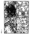
- FIG. 1 is a series of photomicrographs (at a magnification of 400 ⁇ ) showing immunostaining (with an antibody specific for hB7-H1) of: an RCC specimen with high tumor cell hB7-H1 expression ( FIG. 1A ); an RCC specimen with high leukocyte hB7-H1 expression ( FIG. 1B ); an RCC specimen with no detectable hB7-H1 expression in either tumor cells or leukocytes ( FIG. 1C ); and a normal kidney specimen with no detectable hB7-H1 expression in the proximal tubules ( FIG. 1D ).
- FIG. 2 is a series of line graphs showing the associations of hB7-H1 expression with death from RCC in 196 subjects from whom the clear cell RCC specimens were obtained for analysis.
- the cancer-specific survival rates (with standard error [SE] and number still at risk indicated in parentheses) at 1, 2, and 3 years following nephrectomy were: 87.8% (4.1%, 53), 72.3% (6.0%, 30), and 63.2% (7.2%, 11), respectively, for patients with specimens that had ⁇ 10% tumor hB7-H1 expression; compared with 93.6% (2.3%, 95), 88.4% (3.4%, 48), and 88.4% (3.4%, 19), respectively, for patients with specimens that had ⁇ 10% tumor hB7-H1 expression.
- FIG. 2B shows the association of adjusted score for leukocyte hB7-H1 expression with death from RCC (risk ratio 3.58; 95% CI 1.74-7.37; p ⁇ 0.001).
- the cancer-specific survival rates (SE, number still at risk) at 1, 2, and 3 years were: 83.5% (6.2%, 26), 63.9% (9.2%, 13), and 53.6% (10.2%, 5), respectively, for patients with specimens that had a leukocyte hB7-H1 expression score ⁇ 100; compared with 93.5% (2.1%, 122), 86.2% (3.3%, 65), and 84.8% (3.5%, 25), respectively, for patients with specimens that had scores ⁇ 100.
- FIG. 2C shows the association of high aggregate intratumoral hB7-H1 expression with death from RCC (risk ratio 4.53; 95% CI 1.94-10.56; p ⁇ 0.001).
- the cancer-specific survival rates (SE, number still at risk) at 1, 2, and 3 years were: 87.0% (3.8%, 61), 70.0% (5.8%, 32), and 61.9% (6.8%, 13), respectively, for patients with specimens that had high aggregate intratumoral hB7-H1 expression; compared with 94.9% (2.2%, 87), 91.9% (3.1%, 46), and 91.9% (3.1%, 17), respectively, for patients with specimens that had both ⁇ 10% tumor and ⁇ 100 leukocyte (low aggregate intratumoral expression) hB7-H1 expression.
- FIG. 3 is a depiction of the amino acid sequence (SEQ ID NO:1) of full-length, immature hB7-H1, i.e., hB7-H1 including a leader peptide of about 22 amino acids.
- FIG. 4 is a depiction of the nucleotide sequence (SEQ ID NO:2) of cDNA encoding full-length, immature hB7-H1.
- FIG. 5 is a depiction of the amino acid sequence (SEQ ID NO:3) of full-length, immature murine B7-H1.
- FIG. 6 is a depiction of the nucleotide sequence (SEQ ID NO:4) of cDNA encoding full-length, immature murine B7-H1.
- RCC renal cell carcinoma
- B7-H1 in normal, non-activated mammalian cells is largely, if not exclusively, limited to macrophage-lineage cells and provides a potential costimulatory signal source for regulation of T cell activation.
- aberrant expression of B7-H1 by tumor cells has been implicated in impairment of T cell function and survival, resulting in defective host antitumoral immunity.
- hB7-H1 human RCC tumors express hB7-H1.
- hB7-H1 was found to be expressed by both renal cell carcinoma (RCC) tumors and leukocytes infiltrating RCC tumors.
- RCC renal cell carcinoma
- the combination of increased tumor cell hB7-H1 and tumor-infiltrating leukocyte hB7-H1 (high aggregate intratumoral hB7-H1) was an even stronger predictor of patient outcome than either hB7-H1-expressing tumor cells or tumor-infiltrating leukocytes alone.
- High aggregate intratumoral hB7-H1 expression levels were also significantly associated with regional lymph node involvement, distant metastases, advanced nuclear grade, and the presence of histologic tumor necrosis.
- B7-H1 expressed by either tumor cells (e.g., RCC cells) or infiltrating leukocytes, can contribute to the immununosuppression that is commonly observed in subjects with cancer (e.g., RCC) and can serve as a critical determinant of the subjects' responses to immunotherapy for management of advanced cancer (e.g., IL-2, IL-12, IFN- ⁇ , vaccination or T-cell adoptive therapy).
- tumor cells e.g., RCC cells
- infiltrating leukocytes infiltrating leukocytes
- the invention provides a method of diagnosing cancer in a subject.
- the method involves: (a) providing a tissue sample from a subject suspected of having, or likely to develop, cancer of the tissue, the sample containing test cells, the test cells being cells of the tissue or leukocytes infiltrating the tissue; and (b) assessing whether the test cells express B7-H1. Expression by some or all of the test cells is an indication that the subject has cancer. Since a wide variety of cancer cells express B7-H1 on their surfaces, the methods of the invention are particularly useful for diagnosing any such cancer.
- Test cells can thus be, for example, breast cells, lung cells, colon cells, pancreatic cells, renal cells, stomach cells, liver cells, bone cells, hematological cells (e.g., lymphoid cells, granulocytic cells, monocytes or macrophages), neural tissue cells, melanocytes, ovarian cells, testicular cells, prostate cells, cervical cells, vaginal cells, bladder cells, or any other cells listed herein.
- test cells can be leukocytes present in relevant tissues containing any of the above-listed test cells. Leukocytes infiltrating the tissue can be T cells (CD4 + T cells and/or CD8 + T cells) or B lymphocytes.
- leukocytes can also be neutrophils, eosinophils, basophils, monocytes, macrophages, histiocytes, or natural killer cells.
- Subjects can be mammals and include, for example, humans, non-human primates (e.g., monkeys, baboons, or chimpanzees), horses, cows (or oxen or bulls), pigs, sheep, goats, cats, rabbits, guinea pigs, hamsters, rats, gerbils, or mice.
- the invention provides a number of diagnostic advantages and uses.
- the level of B7-H1 polypeptide and/or mRNA can be assessed.
- the level of B7-H1 is assessed in a tissue sample to diagnose, or to confirm, the presence of cancer in the subject from whom the tissue is obtained.
- antibodies that bind to an epitope specific for B7-H1 can be used to assess whether test cells from the tissue sample express B7-H1.
- Such antibodies can be monoclonal or polyclonal antibodies.
- the antibody itself, or a secondary antibody that binds to it can be detectably labeled.
- the antibody can be conjugated with biotin, and detectably labeled avidin (a polypeptide that binds to biotin) can be used to detect the presence of the biotinylated antibody.
- Multi-layer sandwich assays can be used to enhance the sensitivity of the methodologies.
- Some of these protein-detecting assays e.g., ELISA or Western blot
- ELISA or Western blot can be applied to lysates of cells
- others e.g., immunohistological methods or fluorescence flow cytometry
- histological sections or unlysed cell suspensions can be applied to histological sections or unlysed cell suspensions.
- the tissue sample can be, for example, lung, epithelial, connective, vascular, muscle, neural, skeletal, lymphatic, prostate, cervical, breast, spleen, gastric, intestinal, oral, esophageal, dermal, liver, kidney, bladder, thyroid, adrenal, brain, gallbladder, pancreatic, uterine, ovarian, or testicular tissue.
- RNA in a tissue sample can be lysed and an mRNA in the lysates or in RNA purified or semi-purified from the lysates can be detected by any of a variety of methods including, without limitation, hybridization assays using detectably labeled gene-specific DNA or RNA probes (e.g., Northern Blot assays) and quantitative or semi-quantitative RT-PCR methodologies using appropriate gene-specific oligonucleotide primers.
- hybridization assays using detectably labeled gene-specific DNA or RNA probes e.g., Northern Blot assays
- quantitative or semi-quantitative RT-PCR methodologies using appropriate gene-specific oligonucleotide primers.
- quantitative or semi-quantitative in situ hybridization assays can be carried out using, for example, tissue sections or unlysed cell suspensions, and detectably (e.g., fluorescently or enzyme) labeled DNA or RNA probes. Additional methods for quantifying mRNA include RNA protection assay (RPA) and SAGE.
- RPA RNA protection assay
- SAGE SAGE
- Methods of assessing the level of B7-H1 expression can be can be quantitative, semi-quantitative, or qualitative.
- the level of B7-H1 expression can be determined as a discrete value.
- the level of expression of B7-H1 mRNA can be measured as a numerical value by correlating the detection signal derived from the quantitative assay to the detection signal of a known concentration of: (a) B7-H1 nucleic acid sequence (e.g., B7-H1 cDNA or B7-H1 transcript); or (b) a mixture of RNA or DNA that contains a nucleic acid sequence encoding B7-H1.
- the level of B7-H1 expression can be assessed using any of a variety of semi-quantitative/qualitative systems known in the art.
- the level of expression of B7-H1 in a cell or tissue sample can be expressed as, for example, (a) one or more of “excellent”, “good”, “satisfactory”, “unsatisfactory”, and/or “poor”; (b) one or more of “very high”, “high”, “average”, “low”, and/or “very low”; or (c) one or more of “++++”, “+++”, “++”, “+”, “+/ ⁇ ”, and/or “ ⁇ ”.
- the level of expression of B7-H1 in tissue from a subject can be expressed relative to the expression of B7-H1 from (a) a tissue of a subject known not be cancerous (e.g., a contralateral kidney or lung, or an uninvolved lymph node); or (b) a corresponding tissue from one or more other subjects known not to have the cancer of interest, preferably known not to have any cancer.
- a tissue of a subject known not be cancerous e.g., a contralateral kidney or lung, or an uninvolved lymph node
- a corresponding tissue from one or more other subjects known not to have the cancer of interest preferably known not to have any cancer.
- label examples include, without limitation, radionuclides (e.g., 125 I, 131 I, 35 S, 3 H, or 32 P), enzymes (e.g., alkaline phosphatase, horseradish peroxidase, luciferase, or ⁇ -glactosidase), fluorescent moieties or proteins (e.g., fluorescein, rhodamine, phycoerythrin, green fluorescent protein (GFP), or blue fluorescent protein (BFP)), or luminescent moieties (e.g., QdotTM nanoparticles supplied by the Quantum Dot Corporation, Palo Alto, Calif.).
- enzymes e.g., alkaline phosphatase, horseradish peroxidase, luciferase, or ⁇ -glactosidase
- fluorescent moieties or proteins e.g., fluorescein, rhodamine, phycoerythrin, green fluorescent protein (GFP
- a subject is diagnosed as having cancer if the proportion of test cells from the subject that express B7-H1 is greater than a control value.
- the control value can be, for example: (a) the proportion of B7-H1-expressing cells in corresponding tissue of the subject known not be cancerous (e.g., a contralateral kidney or lung, or an uninvolved lymph node); or (b) the proportion of B7-H1 expressing cells in a corresponding tissue from one or more other subjects known not to have the cancer of interest, preferably known not to have any cancer.
- the method of the invention can be used on its own or in conjunction with other procedures to diagnose cancer.
- the level of B7-H1-expressing test cells in a tissue sample that is, or is suspected of being, cancerous can be assessed before, during, or after assessing the levels of other molecules that are useful diagnostic cancer markers.
- diagnostic markers can be, without limitation, tumor-associated antigens (TAA).
- TAA include, without limitation, carcinoembryonic antigen (CEA), MAGE (melanoma antigen) 1-4, 6, and 12, MUC (mucin) (e.g., MUC-1, MUC-2, etc.), tyrosinase, MART (melanoma antigen), Pmel 17 (gp100), GnT-V intron sequence (N-acetylglucosaminyltransferase V intron V sequence), PSA (prostate-specific antigen), PSMA (prostate-specific membrane antigen), PRAME (melanoma antigen), ⁇ -catenin, MUM-1-B (melanoma ubiquitous mutated gene product), GAGE (melanoma antigen) 1, BAGE (melanoma antigen) 2-10, c-ERB2 (HER2/neu), EBNA (Epstein-Barr Virus nuclear antigen) 1-6, gp75, human papilloma virus (HPV) E6 and E7, p
- Another aspect of the invention is a method of identifying a candidate for immunotherapy.
- This method involves providing a tissue sample from a subject with cancer of the tissue.
- the tissue sample contains test cells, the test cells being cancer cells or tumor-infiltrating leukocytes.
- the level of test cells in the tissue sample that express B7-H1 is assessed, such that if B7-H1 expression is not detected in the test cells, or less than an immuno-inhibitory threshold level of the test cells express B7-H1, the subject is more likely to benefit from immunotherapy.
- the immuno-inhibitory threshold level is a predetermined level of the relevant test cells expressing B7-H1. If the test cells from a cancer subject of interest contain a level of B7-H1-expressing cells that is less than the immuno-inhibitory threshold level of B7-H1-expressing cells (as predetermined for the relevant cancer), that subject is more likely to benefit from immunotherapy than another subject with the same cancer but whose corresponding test cells contain a level of B7-H1-expressing cells equal to, or greater, than the immuno-inhibitory threshold level.
- the immuno-inhibitory threshold level can be obtained by performing statistical clinical analyses known in the art, e.g., those described herein.
- Methods of assessing whether test cells express B7-H1 are the same as those described above for methods of diagnosis. Such methods, also as described above, can be qualitative, semi-quantitative, or qualitative.
- Immunotherapy can be active immunotherapy or passive immunotherapy.
- treatment relies on the in vivo stimulation of the endogenous host immune system to react against tumors with the administration of immune response-modifying agents.
- immune response-modifying agents are described below.
- immune effector cells include leukocytes, e.g., tumor-infiltrating leukocytes as discussed above, T lymphocytes (such as CD8 + cytotoxic T lymphocytes and/or CD4 + T-helper lymphocytes), killer cells (such as natural killer cells and lymphokine-activated killer cells), B cells and antigen-presenting cells (such as dendritic cells and macrophages).
- leukocytes e.g., tumor-infiltrating leukocytes as discussed above, T lymphocytes (such as CD8 + cytotoxic T lymphocytes and/or CD4 + T-helper lymphocytes), killer cells (such as natural killer cells and lymphokine-activated killer cells), B cells and antigen-presenting cells (such as dendritic cells and macrophages).
- Immunotherapy can also be one or more of the methods described below (in “Methods of Treatment” and “Methods of Inhibiting Expression of B7-H1).
- the invention features a method of determining the prognosis of a subject with cancer.
- This method involves: (a) providing a tissue sample from a subject with cancer of the tissue, the tissue sample containing test cells, the test cells being cancer cells or tumor infiltrating leukocytes; and (b) assessing the level of test cells in the tissue sample that expresses B7-H1. If a prognostic level, or more than a prognostic level, of the test cells express B7-H1, the subject is more likely to die of the cancer than if less than a prognostic level of the test cells express B7-H1.
- the prognostic level is a predetermined value obtained by performing statistical clinical analyses known in the art, e.g., those described herein.
- test cells from a cancer subject contain a significant level of B7-H1 expressing cells, but less than a prognostic level of B7-H1-expressing cells (as predetermined for the relevant cancer)
- the cancer subject will be no more likely to die of the cancer than a subject with the same cancer but whose corresponding test cells contain no detectable B7-H1-expressing cells.
- test cells from a cancer subject contain more than a prognostic level of B7-H1-expressing cells
- the cancer subject will be more likely to die of the cancer than a subject with the same cancer but whose corresponding test cells contain either no detectable B7-H1-expressing cells or a level of B7-H1-expressing cells lower than a prognostic level of B7-H1-expressing cells.
- the chances of dying from the cancer is likely to be proportional to the level of B7-H1-expressing cells in the test cell population.
- assessing whether test cells express B7-H1” or “assessing the level of test cells in the tissue sample that express B7-H1” can be determined by any of the methods described above. Methods of prognosis will generally be quantitative or semi-quantitative.
- Subjects can be any of those listed for “Methods of Diagnosis” and cancers can be any of the following: renal cancer, hematological cancer (e.g., leukemia or lymphoma), neurological cancer, melanoma, breast cancer, lung cancer, head and neck cancer, gastrointestinal cancer, liver cancer, pancreatic cancer, pancreatic cancer, genitourinary cancer, bone cancer, or vascular cancer
- hematological cancer e.g., leukemia or lymphoma
- neurological cancer melanoma
- melanoma e.g., breast cancer, lung cancer, head and neck cancer
- gastrointestinal cancer e.g., liver cancer, pancreatic cancer, pancreatic cancer, genitourinary cancer, bone cancer, or vascular cancer
- the invention also includes a method of treatment.
- the method can involve: (a) identifying a subject with cancer, some or all cells of the cancer or some or all tumor-infiltrating leukocytes of the cancer expressing B7-H1; and (b) delivering to the subject an agent that interferes with an interaction between B7-H1 and a receptor for B7-H1.
- the agent can be an antibody or an antibody fragment, such as, e.g., a Fab′, a F(ab′) 2 , or a scFv fragment that binds B7-H1.
- the agent can also be a soluble B7-H1 or a soluble functional fragment of B7-H1; a soluble receptor for B7-H1 or a soluble functional fragment thereof; an antibody, or an antibody fragment, that binds to a receptor for B7-H1, e.g., the PD-1 receptor.
- the PD-1 receptor is described in greater detail in U.S. Pat. No. 6,808,710, the disclosure of which is incorporated herein by reference in its entirety.
- the agent itself is administered to a subject.
- the agent will be suspended in a pharmaceutically-acceptable carrier (e.g., physiological saline) and administered orally or by intravenous (i.v.) infusion, or injected subcutaneously, intramuscularly, intrathecally, intraperitoneally, intrarectally, intravaginally, intranasally, intragastrically, intratracheally, or intrapulmonarily.
- a pharmaceutically-acceptable carrier e.g., physiological saline
- intravenous (i.v.) infusion injected subcutaneously, intramuscularly, intrathecally, intraperitoneally, intrarectally, intravaginally, intranasally, intragastrically, intratracheally, or intrapulmonarily.
- the agent can, for example, be delivered directly to a site of an immune response. e.g., a lymph node in the region of an affected tissue or organ or spleen.
- the dosage required depends on the choice of the route of administration; the nature of the formulation; the nature of the patient's illness; the subject's size, weight, surface area, age, and sex; other drugs being administered; and the judgment of the attending physician. Suitable dosages are in the range of 0.0001-100.0 mg/kg. Wide variations in the needed dosage are to be expected in view of the variety of compounds available and the differing efficiencies of various routes of administration. For example, oral administration would be expected to require higher dosages than administration by i.v. injection. Variations in these dosage levels can be adjusted using standard empirical routines for optimization as is well understood in the art.
- Administrations can be single or multiple (e.g., 2-, 3-, 4-, 6-, 8-, 10-, 20-, 50-, 100-, 150-, or more fold).
- Encapsulation of the compound in a suitable delivery vehicle e.g., polymeric microparticles or implantable devices
- a polynucleotide containing a nucleic acid sequence encoding the polypeptide can be delivered to appropriate cells in a mammal.
- Expression of the coding sequence can be directed to any cell in the body of the subject. However, expression will preferably be directed to cells in, or close to, lymphoid tissue draining an affected tissue or organ.
- Expression of the coding sequence can be directed, for example, to cells comprising the cancer tissue (e.g., tumor-infiltrating leukocytes and tumor cells) or immune-related cells, e.g., B cells, macrophages/monocytes, or interdigitating dendritic cells. This can be achieved by, for example, the use of polymeric, biodegradable microparticle or microcapsule delivery devices known in the art and/or tissue or cell-specific antibodies.
- liposomes Another way to achieve uptake of the nucleic acid is using liposomes, which can be prepared by standard methods.
- the vectors can be incorporated alone into these delivery vehicles or co-incorporated with tissue-specific antibodies.
- Poly-L-lysine binds to a ligand that can bind to a receptor on target cells [Cristiano et al. (1995), J. Mol. Med. 73:479].
- tissue specific targeting can be achieved by the use of tissue-specific transcriptional regulatory elements (TRE) which are known in the art. Delivery of “naked DNA” (i.e., without a delivery vehicle) to an intramuscular, intradermal, or subcutaneous site is another means to achieve in vivo expression.
- TRE tissue-specific transcriptional regulatory elements
- the nucleic acid sequence encoding the polypeptide of interest with an initiator methionine and optionally a targeting sequence is operatively linked to a promoter or enhancer-promoter combination.
- Short amino acid sequences can act as signals to direct proteins to specific intracellular compartments. Such signal sequences are described in detail in U.S. Pat. No. 5,827,516, the disclosure of which is incorporated herein by reference in its entirety.
- Enhancers provide expression specificity in terms of time, location, and level. Unlike a promoter, an enhancer can function when located at variable distances from the transcription initiation site, provided a promoter is present. An enhancer can also be located downstream of the transcription initiation site. To bring a coding sequence under the control of a promoter, it is necessary to position the translation initiation site of the translational reading frame of the peptide or polypeptide between one and about fifty nucleotides downstream (3′) of the promoter. The coding sequence of the expression vector is operatively linked to a transcription terminating region.
- Suitable expression vectors include plasmids and viral vectors such as herpes viruses, retroviruses, vaccinia viruses, attenuated vaccinia viruses, canary pox viruses, adenoviruses and adeno-associated viruses, among others.
- Polynucleotides can be administered in a pharmaceutically acceptable carrier.
- Pharmaceutically acceptable carriers are biologically compatible vehicles that are suitable for administration to a human, e.g., physiological saline or liposomes.
- a therapeutically effective amount is an amount of the polynucleotide that is capable of producing a medically desirable result (e.g., decreased proliferation of cancer cells) in a treated subject.
- the dosage for any one patient depends upon many factors, including the patient's size, body surface area, age, the particular compound to be administered, sex, time and route of administration, general health, and other drugs being administered concurrently.
- a preferred dosage for administration of polynucleotide is from approximately 10 6 to approximately 10 12 copies of the polynucleotide molecule. This dose can be repeatedly administered, as needed. Routes of administration can be any of those listed above.
- the method can be an ex vivo procedure that involves providing a recombinant cell which is, or is a progeny of a cell, obtained from a subject and has been transfected or transformed ex vivo with one or more nucleic acids encoding one or more agents that interfere with an interaction between B7-H1 and a receptor for B7-H1, so that the cell expresses the agent(s); and administering the cell to the subject.
- the cells can be cells obtained from a cancer tissue (e.g., tumor cells and/or tumor-infiltrating leukocytes) or from a non-cancerous tissue obtained preferably from a subject to whom these cells are to be administered or from another subject.
- the donor and recipient of the cells can have identical major histocompatibility complex (MHC; HLA in humans) haplotypes.
- MHC major histocompatibility complex
- the donor and recipient are homozygotic twins or are the same individual (i.e., are autologous).
- the recombinant cells can also be administered to recipients that have no, or only one, two, three, or four MHC molecules in common with the recombinant cells, e.g., in situations where the recipient is severely immunocompromised, where only mismatched cells are available, and/or where only short term survival of the recombinant cells is required or desirable.
- the agent can be tested for its ability, for example, to (a) inhibit the interaction between B7-H1 and a receptor for B7-H1, (b) inhibit growth of cancer cells, (c) induce death of cancer cells, or (d) render the cancer cells more susceptible to cell-mediated immune responses generated by leukocytes (e.g., lymphocytes and/or macrophages).
- leukocytes e.g., lymphocytes and/or macrophages.
- the agent can, for example, be injected into an animal (e.g., a mouse cancer model) and its effects on cancer are then assessed. Based on the results, an appropriate dosage range and administration route can be determined.
- the term “antibody” refers to a whole antibody (e.g., IgM, IgG, IgA, IgD, or IgE) molecule that is generated by any one of a variety of methods that are known in the art.
- the antibody can be a polyclonal or a monoclonal antibody.
- Also useful for the invention are chimeric antibodies and humanized antibodies made from non-human (e.g., mouse, rat, gerbil, or hamster) antibodies.
- the term “antibody fragment” refers to an antigen-binding fragment, e.g., Fab, F(ab′) 2 , Fv, and single chain Fv (scFv) fragments.
- An scFv fragment is a single polypeptide chain that includes both the heavy and light chain variable regions of the antibody from which the scFv is derived.
- Antibody fragments that contain the binding domain of the molecule can be generated by known techniques. For example: F(ab′) 2 fragments can be produced by pepsin digestion of antibody molecules; and Fab fragments can be generated by reducing the disulfide bridges of F(ab′) 2 fragments or by treating antibody molecules with papain and a reducing agent. See, e.g., National Institutes of Health, 1 Current Protocols In Immunology , Coligan et al., ed. 2.8, 2.10 (Wiley Interscience, 1991). scFv fragments can be produced, for example, as described in U.S. Pat. No. 4,642,334, which is incorporated herein by reference in its entirety.
- Chimeric and humanized monoclonal antibodies can be produced by recombinant DNA techniques known in the art, for example, using methods described in Robinson et al., International Patent Publication PCT/US86/02269; Akira et al., European Patent Application 184,187; Taniguchi, European Patent Application 171,496; Morrison et al., European Patent Application 173,494; Neuberger et al., PCT Application WO 86/01533; Cabilly et al., U.S. Pat. No. 4,816,567; Cabilly et al., European Patent Application 125,023; Better et al. (1988) Science 240, 1041-43; Liu et al. (1987) J.
- a “functional fragment” of a B7-H1 receptor means a fragment of a receptor for B7-H1 that is smaller than the wild-type mature B7-H1 receptor and has at least 10% (e.g., at least 10%, at least 20%, at least 30%, at least 40%, at least 50%, at least 60%, at least 70%, at least 80%, at least 90%, at least 95%, at least 98%, at least 99%, or 100% or more) of the ability of the wild-type mature receptor for B7-H1 to bind to B7-H1.
- at least 10% e.g., at least 10%, at least 20%, at least 30%, at least 40%, at least 50%, at least 60%, at least 70%, at least 80%, at least 90%, at least 95%, at least 98%, at least 99%, or 100% or more
- a “functional fragment” of B7-H1 means a fragment of the wild-type mature B7-H1 polypeptide that is smaller than the wild-type mature B7-H1 polypeptide and has at least 10% (e.g., at least 10%, at least 20%, at least 30%, at least 40%, at least 50%, at least 60%, at least 70%, at least 80%, at least 90%, at least 95%, at least 98%, at least 99%, or 100% or more) of the ability of the wild-type mature B7-H1 to bind to the B7-H1 receptor. Methods of testing and comparing the ability of molecules to bind to each other are known to those in the art.
- soluble distinguishes the receptors used in the present invention from their cell membrane-bound counterparts.
- a soluble receptor, or a soluble functional fragment of a receptor can contain, for example, an extracellular (ligand binding) domain, but lack the transmembrane region that causes retention of a receptor on the cell surface.
- Methods of producing soluble receptors or fragments thereof are known in the art and include, for example, expressing a DNA fragment encoding an extracellular domain of a receptor in a suitable host cell/expression vector system.
- treatment means administration of an agent to a subject, who has cancer (or is suspected of having cancer), with the purpose to cure, alleviate, relieve, remedy, prevent, or ameliorate the disorder, the symptom of the disorder, the disease state secondary to the disorder, or the predisposition toward the disorder.
- An “effective amount” of a therapeutic agent (or composition) is an amount of the agent (or composition) that is capable of producing a medically desirable result in a treated subject.
- the method of the invention can be performed alone or in conjunction with other drugs or therapy.
- prophylaxis can mean complete prevention of the symptoms of a disease, a delay in onset of the symptoms of a disease, or a lessening in the severity of subsequently developed disease symptoms.
- “therapy” can mean a complete abolishment of the symptoms of a disease or a decrease in the severity of the symptoms of the disease.
- Another aspect of the invention is a method of inhibiting the expression of B7-H1 in a tumor cell or a tumor-infiltrating leukocyte.
- the method involves: (a) identifying a subject with cancer, the cancer containing a target cell that expresses B7-H1, the target cell being a tumor cell or a tumor-infiltrating leukocyte; and (b) introducing into the target cell: (i) an antisense oligonucleotide that hybridizes to a B7-H1 transcript, the antisense oligonucleotide inhibiting the expression of B7-H1 in the cell; or (ii) a B7-H1 interference RNA (RNAi).
- RNAi B7-H1 interference RNA
- the method can be useful for therapy and/or prophylaxis of any cancer recited herein.
- the method can be used, for example, in the treatment of RCC.
- Antisense compounds are generally used to interfere with protein expression either by, for example, interfering directly with translation of a target mRNA molecule, by RNAse-H-mediated degradation of the target mRNA, by interference with 5′ capping of mRNA, by prevention of translation factor binding to the target mRNA by masking of the 5′ cap, or by inhibiting of mRNA polyadenylation.
- the interference with protein expression arises from the hybridization of the antisense compound with its target mRNA.
- a specific targeting site on a target mRNA of interest for interaction with an antisense compound is chosen.
- a preferred target site on an mRNA target is a polyadenylation signal or a polyadenylation site.
- oligonucleotides are chosen which are sufficiently complementary to the target site (i.e., hybridize sufficiently well under physiological conditions and with sufficient specificity) to give the desired effect.
- oligonucleotide refers to an oligomer or polymer of RNA, DNA, or a mimetic of either.
- the term includes oligonucleotides composed of naturally-occurring nucleobases, sugars, and covalent internucleoside (backbone) linkages.
- the normal linkage or backbone of RNA and DNA is a 3′ to 5′ phosphodiester bond.
- the term also refers however to oligonucleotides composed entirely of, or having portions containing, non-naturally occurring components which function in a similar manner to the oligonucleotides containing only naturally-occurring components.
- modified substituted oligonucleotides are often preferred over native forms because of desirable properties such as, for example, enhanced cellular uptake, enhanced affinity for target sequence, and increased stability in the presence of nucleases.
- the core base pyrimidine or purine
- the sugars are either modified or replaced with other components and/or (2) the inter-nucleobase linkages are modified.
- PNA protein nucleic acid
- the sugar backbone is replaced with an amide-containing backbone, in particular an aminoethylglycine backbone.
- the antisense oligomers to be used in the methods of the invention generally comprise about 8 to about 100 (e.g., about 14 to about 80 or about 14 to about 35) nucleobases (or nucleosides where the nucleobases are naturally occurring).
- the antisense oligonucleotides can themselves be introduced into a cell or an expression vector containing a nucleic sequence (operably linked to a TRE) encoding the antisense oligonucleotide can be introduced into the cell.
- the oligonucleotide produced by the expression vector is an RNA oligonucleotide and the RNA oligonucleotide will be composed entirely of naturally occurring components.
- antisense oligonucleotides per se are administered, they can be suspended in a pharmaceutically-acceptable carrier (e.g., physiological saline) and administered under the same conditions described above for agents that interfere with an interaction between B7-H1 and a receptor for B7-H1.
- a pharmaceutically-acceptable carrier e.g., physiological saline
- expression of the coding sequence can be directed to a tumor cell of tumor-infiltrating leukocyte in the body of the subject using any of the cell- or tissue-targeting techniques described above for vectors that express polypeptides that interfere with an interaction between B7-H1 and a receptor for B7-H1.
- Double-stranded interfering RNA (RNAi) homologous to B7-H1 DNA can also be used to reduce expression of B7-H1 in tumor cells and/or tumor-infiltrating leukocytes.
- RNAi double-stranded interfering RNA
- the sense and anti-sense RNA strands of RNAi can be individually constructed using chemical synthesis and enzymatic ligation reactions using procedures known in the art.
- each strand can be chemically synthesized using naturally occurring nucleotides or variously modified nucleotides designed to increase the biological stability of the molecule or to increase the physical stability of the duplex formed between the sense and anti-sense strands, e.g., phosphorothioate derivatives and acridine substituted nucleotides.
- the sense or anti-sense strand can also be produced biologically using an expression vector into which a target B7-H1 sequence (full-length or a fragment) has been subcloned in a sense or anti-sense orientation.
- the sense and anti-sense RNA strands can be annealed in vitro before delivery of the dsRNA to cells. Alternatively, annealing can occur in vivo after the sense and anti-sense strands are sequentially delivered to tumor cells and/or tumor-infiltrating leukocytes.
- Double-stranded RNAi interference can also be achieved by introducing into tumor cells and/or tumor-infiltrating leukocytes a polynucleotide from which sense and anti-sense RNAs can be transcribed under the direction of separate promoters, or a single RNA molecule containing both sense and anti-sense sequences can be transcribed under the direction of a single promoter.
- drugs and small molecules can also be used inhibit expression of B7-H1 in tumor cells and/or tumor-infiltrating leukocytes.
- RNAi, drug, and small molecule methods can be, as for the antisense methods described above, in vitro and in vivo. Moreover, methods and conditions of delivery are the same as those for antisense oligonucleotides.
- one or more agents e.g., two, three, four, five, six, seven, eight, nine, ten, 11, 12, 15, 18, 20, 25, 30, 40, 50, 60, 70, 80, 100, or more
- agents e.g., two, three, four, five, six, seven, eight, nine, ten, 11, 12, 15, 18, 20, 25, 30, 40, 50, 60, 70, 80, 100, or more
- inhibitory compounds e.g., two, three, four, five, six, seven, eight, nine, ten, 11, 12, 15, 18, 20, 25, 30, 40, 50, 60, 70, 80, 100, or more
- inhibitory compounds e.g., antisense oligonucleotides, RNAi, drugs, or small molecules (or vectors encoding them
- agents can be used together with one or more (e.g., two, three, four, five, six, seven, eight, nine, ten, 11, 12, 15, 18, 20, 25, 30, 40, 50, 60, 70, 80, 100, or more) supplementary agents, including immunomodulatory cytokines, growth factors, antiangiogenic factors, immunogenic stimuli, and/or antibodies specific for any of these.
- supplementary agents can administered before, simultaneous with, or after delivery of any of the above-listed agents.
- immunomodulatory cytokines, growth factors, and antiangiogenic factors include, without limitation, interleukin (IL)-1 to 25 (e.g., IL-2, IL-12, or IL-15), interferon- ⁇ (IFN- ⁇ ), interferon- ⁇ (IFN- ⁇ ), interferon- ⁇ (IFN- ⁇ ), tumor necrosis factor- ⁇ (TNF- ⁇ ), granulocyte macrophage colony stimulating factor (GM-CSF), granulocyte macrophage colony stimulating factor (G-CSF), endostatin, angiostatin, and thrombospondin.
- IL-1 to 25 e.g., IL-2, IL-12, or IL-15
- IFN- ⁇ interferon- ⁇
- IFN- ⁇ interferon- ⁇
- IFN- ⁇ interferon- ⁇
- IFN- ⁇ tumor necrosis factor- ⁇
- GM-CSF granulocyte macrophage colony stimulating factor
- G-CSF granulocyte macrophage colony stimulating
- Immunomodulatory cytokines, growth factors, antiangiogenic factors include substances that serve, for example, to inhibit infection (e.g., standard anti-microbial antibiotics), inhibit activation of T cells, or inhibit the consequences of T cell activation.
- a cytokine such as interleukin (IL)-4, IL-10, or IL-13 or an antibody specific for a cytokine such as IL-12 or interferon- ⁇ (IFN- ⁇ ) can be used.
- a cytokine such as IL-12 or IFN- ⁇ or an antibody specific for IL-4, IL-10, or IL-13 can be used as a supplementary agent.
- antibodies specific for proinflammatory cytokines and chemokines such as IL-1, IL-6, IL-8, tumor necrosis factor- ⁇ (TNF- ⁇ ), macrophage inflammatory protein (MIP)-1, MIP-3 ⁇ , monocyte chemoattractant protein-1 (MCP-1), epithelial neutrophil activating peptide-78 (ENA-78), interferon- ⁇ -inducible protein-10 (IP10), Rantes, and any other appropriate cytokine or chemokine recited herein.
- cytokines and chemokines such as IL-1, IL-6, IL-8, tumor necrosis factor- ⁇ (TNF- ⁇ ), macrophage inflammatory protein (MIP)-1, MIP-3 ⁇ , monocyte chemoattractant protein-1 (MCP-1), epithelial neutrophil activating peptide-78 (ENA-78), interferon- ⁇ -inducible protein-10 (IP10), Rantes, and any other appropriate cytokine or chemokine recited herein
- immune response-modifying agents include, in addition to any of the immunomodulatory cytokines, growth factors, and angiogenic factors listed above, immunogenic stimuli that can be delivered via the antigen-specific T cell receptor (TCR) expressed on the surface of the T cell. More commonly, but not necessarily, such a stimulus is provided in the form of an antigen for which the TCR is specific. While such antigens will generally be protein, they can also be carbohydrates, lipids, nucleic acids or hybrid molecules having components of two or more of these molecule types, e.g., glycoproteins or lipoproteins.
- TCR antigen-specific T cell receptor
- the immunogenic stimulus can also be provided by other agonistic TCR ligands such as antibodies specific for TCR components (e.g., TCR ⁇ -chain or ⁇ -chain variable regions) or antibodies specific for the TCR-associated CD3 complex.
- Antigens useful as immunogenic stimuli include alloantigens (e.g., a MHC alloantigen) on, for example, an antigen presenting cell (APC) (e.g., a dendritic cell (DC), a macrophage, a monocyte, or a B cell).
- APC antigen presenting cell
- DC of interest are interdigitating DC and not follicular DC; follicular DC present antigen to B cells.
- interdigitating DC are referred to herein as DC.
- immunogenic stimuli are polypeptide antigens and peptide-epitopes derived from them (see below). Unprocessed polypeptides are processed by APC into peptide-epitopes that are presented to responsive T cells in the form of molecular complexes with MHC molecules on the surface of the APC. Useful immunogenic stimuli also include a source of antigen such as a lysate of either tumor cells or cells infected with an infectious microorganism of interest.
- APC pre-exposed (e.g., by coculturing) to antigenic polypeptides, peptide-epitopes of such polypeptides or lysates of tumor (or infected cells) can also be used as immunogenic stimuli.
- Such APC can also be “primed” with antigen by culture with a cancer cell or infected cell of interest; the cancer or infected cells can optionally be irradiated or heated (e.g., boiled) prior to the priming culture.
- APC especially DC
- an immunogenic stimulus be provided in the form of cells (e.g., tumor cells or infected cells producing the antigen of interest).
- immunogenic stimuli can be provided in the form of cell hybrids formed by fusing APC (e.g., DC) with tumor cells [Gong et al. (2000) Proc. Natl. Acad. Sci. USA 97(6):2716-2718; Gong et al. (1997) Nature Medicine 3(5):558-561; Gong et al. (2000) J. Immunol. 165(3):1705-1711] or infected cells of interest.
- APC e.g., DC
- heat shock proteins bound to antigenic peptide-epitopes derived from antigens e.g., tumor-associated antigens or antigens produced by infectious microorganisms
- Heat shock proteins of interest include, without limitation, glycoprotein 96 (gp96), heat shock protein (hsp) 90, hsp70, hsp110, glucose-regulated protein 170 (grp170) and calreticulin.
- Immunogenic stimuli can include one or more (e.g., one, two, three, four, five, six, seven, eight, nine, ten, more) heat shock proteins isolated from tumor cells.
- Such tumor are preferably, but not necessarily, from the same subject (i) to whom the agent that interferes with the interaction between B7-H1 and a receptor for B7-H1 is to be delivered or (ii) in whose tumor cells or tumor infiltrating leukocytes the expression of B7-H1 is to be inhibited.
- the tumor cells can also be obtained, for example, from another individual having the same as the subject, or a related tumor-type.
- the heat shock protein can be isolated from mammalian cells expressing a transcriptosome prepared from tumor cells of interest.
- immunogenic stimuli useful in the invention can be any of a wide variety of tumor cells, APC “primed” with tumor cells, hybrid cells, or TAA (see above), peptide-epitopes of such TAA, and APC “primed” with TAA or peptide-epitopes of them.
- a “TAA” is a molecule (e.g., a protein molecule) that is expressed by a tumor cell and either (a) differs qualitatively from its counterpart expressed in normal cells, or (b) is expressed at a higher level in tumor cells than in normal cells.
- a TAA can differ (e.g., by one or more amino acid residues where the molecule is a protein) from, or it can be identical to, its counterpart expressed in normal cells. It is preferably not expressed by normal cells. Alternatively, it is expressed at a level at least two-fold higher (e.g., a two-fold, three-fold, five-fold, ten-fold, 20-fold, 40-fold, 100-fold, 500-fold, 1,000-fold, 5,000-fold, or 15,000-fold higher) in a tumor cell than in the tumor cell's normal counterpart.
- Relevant TAA include, without limitation, any of the TAAs listed above.
- Administrations of the agents and/or the one or more supplementary agents can be systemic (e.g., intravenous) or local, e.g., during surgery by direct injection or infusion into the tissue that comprises the cells of the cancer and/or tumor-infiltrating leukocytes.
- the administrations can also be by any of routes, doses, and schedules recited herein.
- the cancer can be any cancer recited herein and includes, e.g., renal cell carcinoma.
- Subjects can be mammals and include, for example, humans, non-human primates (e.g., monkeys, baboons, or chimpanzees), horses, cows (or oxen or bulls), pigs, sheep, goats, cats, rabbits, guinea pigs, hamsters, rats, gerbils, or mice.
- the pathologic features examined included histologic subtype, tumor size, primary tumor stage, regional lymph node involvement, and distant metastases at nephrectomy (2002 TNM), nuclear grade, and histologic tumor necrosis.
- the microscopic slides from all specimens were reviewed by a urologic pathologist without prior knowledge of patient outcome. Histologic subtype was classified according to the Union Internationale Contre le Cancer, American Joint Committee on Cancer, and Heidelberg guidelines [Storkel et al. (1997) Cancer 80:987-989; Kovacs et al. (1997) J. Pathol. 183:131-133]. Nuclear grade was assigned using standardized criteria [Lohse et al. (2002) Am. J. Clin. Pathol. 118:877-886]. Histologic tumor necrosis was defined as the presence of any microscopic coagulative tumor necrosis. Degenerative changes such as hyalinization, hemorrhage, and fibrosis were not considered necrosis.
- a horseradish peroxidase-conjugated secondary reagent (goat anti-mouse immunoglobulin) was then applied to the slides at room temperature for 15 minutes followed by incubation with chromogen-substrate for 10 minutes. Finally, sections were counter-stained for 3 minutes with modified Schmidt's Hematoxylin.
- the primary antibody used in this study was 5H1, a mouse anti-hB7-H1 monoclonal antibody [Dong et al. (2002) Nature Med; 8:793-800). Benign renal tumors and peripheral T cells were not stained in this study. Positive tissues controls for hB7-H1 staining were human tonsillar tissues. Irrelevant isotype-matched antibodies were used to control for non-specific staining.
- the percentages of tumor cells and leukocytes that stained positive for hB7-H1 were quantified in 5-10% increments by a urologic pathologist without prior knowledge of patient outcome.
- the extent of leukocytic infiltration was assessed and recorded as absent, focal (scattered lymphoid aggregates), moderate, or marked.
- the adjusted scores for leukocytic hB7-H1 expression are summarized in Table 2.
- the associations of leukocyte hB7-H1 expression with death from RCC are summarized in Table 3.
Abstract
The invention features methods of diagnosis by assessing B7-H1 expression in a tissue from a subject that has, or is suspected of having, cancer, methods of treatment with agents that interfere with B7-H1-receptor interaction, methods of selecting candidate subjects likely to benefit from cancer immunotherapy, and methods of inhibiting expression of B7-H1.
Description
This application is a continuation of U.S. patent application Ser. No. 11/245,713, now U.S. Pat. No. 7,892,540, which claims the benefit of U.S. Provisional Application No. 60/616,590, filed Oct. 6, 2004, and U.S. Provisional Application 60/642,794, filed Jan. 11, 2005. The disclosures of U.S. Provisional Application No. 60/616,590 and U.S. Provisional Application 60/642,794 are incorporated herein by reference in their entirety.
This invention relates to immune molecules expressed in cancer tissue, and more particularly to evaluating the expression of immune molecules in tumor cells and tumor-infiltrating leukocytes.
An important determinant for the initiation and progression of cancer is the ability of cancer cells to evade the host's immune system. The presence in cancer tissue of, for example, inadequate, inappropriate, or inhibitory immune molecules can restrict the host's ability to generate immune responses to the cancer.
The disclosures of U.S. Pat. No. 6,803,192 and co-pending U.S. application Ser. Nos. 09/649,108; 10/127,282; and 10/719,477; and International Application No. US/02/32364 are incorporated herein by reference in their entirety.
The invention is based in part on the finding that in renal cell carcinoma (RCC) patients the risk of death is proportional to the number of tumor cells, and/or leukocytes in the tumor, expressing the co-stimulatory human glycoprotein B7-H1. As used herein, the term “B7-H1” refers to B7-H1 from any mammalian species and the term “hB7-H1” refers to human B7-H1. Further details on B7-H1 polypeptides and nucleic acids are provided in U.S. Pat. No. 6,803,192 and co-pending U.S. application Ser. No. 09/649,108, the disclosures of which are incorporated herein by reference in their entirety.
The invention provides methods of diagnosing subjects having, or that are likely to develop, cancer of a tissue based on the expression of B7-H1 by cells of the cancer tissue, methods of predicting success of immunotherapy, methods of prognosis, and methods of treatment. Leukocytes in a tumor are sometimes referred to herein as “tumor-infiltrating leukocytes” or “leukocytes infiltrating a/the tumor.”
More specifically, the invention provides a method of diagnosis of cancer in a subject. The method involves: (a) providing a tissue sample from a subject suspected of having, or likely to develop, cancer of the tissue, wherein the sample contains test cells, the test cells being cells of the tissue or leukocytes infiltrating the tissue; and (b) assessing whether the test cells express B7-H1, wherein expression by some or all of the test cells is an indication that the subject has cancer.
The assessment of B7-H1 expression can be performed by the detection of B7-H1 polypeptide or mRNA. B7-H1 polypeptide can be detected, for example, by contacting the tissue sample, or test cells contained in the tissue sample, with an antibody that binds to the B7-H1 polypeptide. Suitable methods for detection of B7-H1 polypeptide can include, without limitation, fluorescence flow cytometry (FFC) or immunohistology. B7-H1 mRNA can be detected, for example, by contacting the tissue sample with a nucleic acid probe that hybridizes to the B7-H1 mRNA (e.g., such by in situ hybridization) or by reverse transcriptase-polymerase chain reaction. The tissue can be tissue of any organ or anatomical system, and can include, without limitation, lung, epithelial, connective, vascular, muscle, neural, skeletal, lymphatic, prostate, cervical, breast, spleen, gastric, intestinal, oral, esophageal, uterine, ovarian, or testicular tissue. The tissue can also be renal tissue. The subject can be a mammal, such as, for example, a human.
Another aspect of the invention is a method of identifying a candidate for immunotherapy. This method involves: (a) providing a tissue sample from a subject with cancer of the tissue, wherein the tissue sample contains test cells, the test cells being cancer cells or tumor-infiltrating leukocytes; and (b) assessing the level of test cells in the tissue sample that express B7-H1, wherein, if B7-H1 expression is not detected in the test cells or if less than an immuno-inhibitory threshold level of the test cells express B7-H1, the subject is more likely to benefit from immunotherapy.
The level of B7-H1 can be assessed by detecting B7-H1 polypeptide or mRNA using, for example, any of the methods described above for method of diagnosis. The tissue can be tissue of any organ or anatomical system, and can include, without limitation, lung, epithelial, connective, vascular, muscle, neural, skeletal, lymphatic, prostate, cervical, breast, spleen, gastric, intestinal, oral, esophageal, uterine, ovarian, or testicular tissue. The tissue can also be renal tissue. The subject can be a mammal, such as, for example, a human. The cancer can be any cancer, and includes, for example, renal cell carcinoma.
In another embodiment, the invention features a method of determining the prognosis of a subject with cancer. This method involves: (a) providing a tissue sample from a subject with cancer of the tissue, wherein the tissue sample comprises test cells, the test cells being cancer cells or tumor-infiltrating leukocytes; and (b) assessing the level of test cells in the tissue sample that express B7-H1, wherein, if a prognostic level, or more than a prognostic level, of the test cells express B7-H1, the subject is more likely to die of the cancer than if less than a prognostic level of the test cells express B7-H1. The prognostic level is a predetermined value obtained by performing statistical clinical analyses known in the art, e.g., those described herein. The assessment of B7-H1 can be performed by detecting B7-H1 polypeptide or B7-H1 mRNA using any of a variety of methods known in the art, including, for example, those listed above for methods of diagnosis and method of immunotherapy. The tissue sample can be of any tissue, and can include, for example, any of those described above. The subject from which the tissue is provided can be a mammal, e.g., a human.
Yet another aspect of the invention is a method of treatment. The method involves: (a) identifying a subject with cancer, wherein some or all cells of the cancer or some or all tumor-infiltrating leukocytes of the cancer express B7-H1; and (b) delivering to the subject an agent that interferes with an interaction between B7-H1 and a receptor for B7-H1. The agent can bind to B7-H1 or to a receptor for B7-H1, e.g., the PD-1 receptor. The agent can be an antibody or an antibody fragment (e.g., Fab′, F(ab′)2, or single chain Fv (scFv) fragment) that binds to B7-H1 or binds to a receptor for B7-H1; 30, soluble B7-H1 or a soluble functional fragment of B7-H1; a soluble receptor for B7-H1 or a soluble functional fragment thereof. Whenever it is desired, the agent can be administered before, simultaneous with, or after administration of one or more immunomodulatory cytokines, growth factors, or antiangiogenic factors. Examples of such immunomodulatory cytokines, growth factors, and antiangiogenic factors include, without limitation, any of interleukins (IL)-1 to 25, interferon-γ (IFN-γ), interferon-α (IFN-α), interferon-β (IFN-β), interferon-γ (IFN-γ), tumor necrosis factor-α (TNF-α), granulocyte macrophage colony stimulating factor (GM-CSF), granulocyte macrophage colony stimulating factor (G-CSF), endostatin, angiostatin, and thrombospondin. Administrations of the agent and/or the one or more immunomodulatory cytokines, growth factors, or antiangiogenic factors can be systemic (e.g., intravenous) or local, e.g., during surgery by direct injection or infusion into the tissue that comprises the cells of the cancer and/or tumor-infiltrating leukocytes. The cancer can be, without limitation, hematological cancer, neurological cancer, melanoma, breast cancer, lung cancer, head and neck cancer, gastrointestinal cancer, liver cancer, pancreatic cancer, renal cancer, genitourinary cancer, bone cancer, or vascular cancer.
Yet another aspect of the invention is a method of inhibiting the expression of B7-H1 in a tumor cell or a tumor-infiltrating leukocyte. The method involves: (a) identifying a subject with cancer, the cancer containing a target cell that expresses B7-H1, the target cell being a tumor cell or a tumor-infiltrating leukocyte; and (b) introducing into the target cell: (i) an antisense oligonucleotide that hybridizes to a B7-H1 transcript, wherein the antisense oligonucleotide inhibits the expression of B7-H1 in the cell; or (ii) a B7-H1 interference RNA (RNAi). The introducing step can involve administration of the antisense oligonucleotide or the RNAi to the subject and uptake of the oligonucleotide or the RNAi by the target cell. Alternatively, the introducing step can involve administering to the subject, and uptake by the cell of, a nucleic acid comprising a transcriptional regulatory element (TRE) operably linked to a nucleotide sequence complementary to the antisense oligonucleotide, wherein transcription of the nucleotide sequence inside the cell produces the antisense oligonucleotide. Moreover, the introducing step can include administering to the subject, and uptake by the cell of, a nucleic acid: (a) from which sense and anti-sense strands of the RNAi can be transcribed under the direction of the TREs; or (b) from which both sense and anti-sense strands of the RNAi can be transcribed under the direction of a single TRE.
The tissue sample can be lung, epithelial, connective, vascular, muscle, neural, skeletal, lymphatic, prostate, cervical, breast, spleen, gastric, intestinal, oral, esophageal, dermal, liver, bladder, thyroid, thymic, adrenal, brain, gallbladder, pancreatic, uterine, ovarian, or testicular tissue. The tissue can also be renal tissue. The cancer of the tissue can be any cancer and includes, e.g., renal cell carcinoma.
The subject can be a mammal and includes, for example, a human, a non-human primate (e.g., a monkey), a horse, a cow (or an ox or bull), a pig, a sheep, a goat, a cat, a rabbit, a guinea pig, a hamster, a rat, or a gerbil.
As used herein, “interferes with an interaction between B7-H1 and a receptor for B7-H1” means (a) completely blocks a physical interaction between B7-H1 molecule and a receptor for B7-H1 such that there is substantially no physical interaction between the B7-H1 molecule and the receptor; or (b) modifies the interaction between the B7-H1 molecule and the receptor such that the physical interaction either does not deliver a signal to the cell that comprises B7-H1, and/or the receptor for B7-H1, or delivers a signal that does not substantially affect the antitumoral activity of the cell.
“Polypeptide” and “protein” are used interchangeably and mean any peptide-linked chain of amino acids, regardless of length or post-translational modification. Polypeptides useful for the invention include variant polypeptides that are identical to corresponding wild-type polypeptides but differ by not more than 50 (e.g., not more than: 45; 40; 35; 30; 25; 20; 19; 18; 17; 16; 15; 14; 13; 12; 11; 10; nine; eight; seven; six; five; four; three; two; or one) conservative substitution(s). All that is required is that the variant polypeptide has at least 20% (e.g., at least: 25; 30%; 35%; 40%; 45%; 50%; 60%; 70%; 80%; 85%; 90%; 93%; 95%; 96%; 97%; 98%; 99%; 99.5%; 99.8%; 99.9%; or 100% or more) of the activity of the wild-type polypeptide. Conservative substitutions typically include substitutions within the following groups: glycine and alanine; valine, isoleucine, and leucine; aspartic acid and glutamic acid; asparagine, glutamine, serine, and threonine; lysine, histidine, and arginine; and phenylalanine and tyrosine.
As used herein, “tumor-infiltrating leukocytes” can be T lymphocytes (such as CD8+ T lymphocytes and/or CD4+ T lymphocytes), B lymphocytes, or other bone marrow-lineage cells including granulocytes (neutrophils, eosinophils, basophils), monocytes, macrophages, dendritic cells (i.e., interdigitating dendritic cells), histiocytes, and natural killer cells.
Unless otherwise defined, all technical and scientific terms used herein have the same meaning as commonly understood by one of ordinary skill in the art to which this invention pertains. In case of conflict, the present document, including definitions, will control. Preferred methods and materials are described below, although methods and materials similar or equivalent to those described herein can be used in the practice or testing of the present invention. All publications, patent applications, patents and other references mentioned herein are incorporated by reference in their entirety. The materials, methods, and examples disclosed herein are illustrative only and not intended to be limiting.
Other features and advantages of the invention will be apparent from the following description, from the drawings and from the claims.
The inventors have discovered that renal cell carcinoma (RCC) patients who have increased levels of tumor cells and/or tumor-infiltrating leukocytes expressing the co-stimulatory glycoprotein hB7-H1 are at an increased risk of death from the RCC. In addition, elevated levels of hB7-H1 expressing tumor cells and/or tumor-infiltrating leukocytes was associated with more aggressive tumors and this association persisted even after controlling for traditional predictors of RCC progression, including, for example, tumor, node, metastasis (TNM) stage; primary tumor size; nuclear grade; and histological tumor necrosis.
Expression of B7-H1 in normal, non-activated mammalian cells is largely, if not exclusively, limited to macrophage-lineage cells and provides a potential costimulatory signal source for regulation of T cell activation. In contrast, aberrant expression of B7-H1 by tumor cells has been implicated in impairment of T cell function and survival, resulting in defective host antitumoral immunity.
The inventors found that human RCC tumors express hB7-H1. In particular, hB7-H1 was found to be expressed by both renal cell carcinoma (RCC) tumors and leukocytes infiltrating RCC tumors. In contrast, proximal tubules of the renal cortex, from which clear cell tumors are believed to arise, failed to express hB7-H1.
Clinical specimens were obtained from 196 patients who were treated with radical nephrectomy or nephron-sparing surgery for unilateral, clear cell RCC between 2000 and 2002 from the Mayo Clinic Nephrectomy Registry. Immunohistological detection and quantification of hB7-H1 expression in the specimens revealed that patients whose tumor specimens exhibited high intratumoral expression levels of hB7-H1 (contributed by tumor cells alone, leukocytes alone, or tumor and/or leukocytes combined) had aggressive tumors and were at markedly increased risk of death from RCC.
The combination of increased tumor cell hB7-H1 and tumor-infiltrating leukocyte hB7-H1 (high aggregate intratumoral hB7-H1) was an even stronger predictor of patient outcome than either hB7-H1-expressing tumor cells or tumor-infiltrating leukocytes alone. High aggregate intratumoral hB7-H1 expression levels were also significantly associated with regional lymph node involvement, distant metastases, advanced nuclear grade, and the presence of histologic tumor necrosis.
Based on its ability to impair function and survival of activated tumor-specific T cells, B7-H1, expressed by either tumor cells (e.g., RCC cells) or infiltrating leukocytes, can contribute to the immununosuppression that is commonly observed in subjects with cancer (e.g., RCC) and can serve as a critical determinant of the subjects' responses to immunotherapy for management of advanced cancer (e.g., IL-2, IL-12, IFN-α, vaccination or T-cell adoptive therapy). This raises the possibility that administering to cancer patients agents that interfere with the interaction of B7-H1 with its receptor (e.g., PD-1) can serve as a method of immunotherapy, particularly in subjects whose high level of intratumoral B7-H1 expression previously rendered them unresponsive or nearly unresponsive to other modes of immunotherapy.
These findings provide support for the methods of the invention, which are described below.
Methods of Diagnosis
The invention provides a method of diagnosing cancer in a subject. The method involves: (a) providing a tissue sample from a subject suspected of having, or likely to develop, cancer of the tissue, the sample containing test cells, the test cells being cells of the tissue or leukocytes infiltrating the tissue; and (b) assessing whether the test cells express B7-H1. Expression by some or all of the test cells is an indication that the subject has cancer. Since a wide variety of cancer cells express B7-H1 on their surfaces, the methods of the invention are particularly useful for diagnosing any such cancer. Test cells can thus be, for example, breast cells, lung cells, colon cells, pancreatic cells, renal cells, stomach cells, liver cells, bone cells, hematological cells (e.g., lymphoid cells, granulocytic cells, monocytes or macrophages), neural tissue cells, melanocytes, ovarian cells, testicular cells, prostate cells, cervical cells, vaginal cells, bladder cells, or any other cells listed herein. Moreover, test cells can be leukocytes present in relevant tissues containing any of the above-listed test cells. Leukocytes infiltrating the tissue can be T cells (CD4+ T cells and/or CD8+ T cells) or B lymphocytes. Such leukocytes can also be neutrophils, eosinophils, basophils, monocytes, macrophages, histiocytes, or natural killer cells. Subjects can be mammals and include, for example, humans, non-human primates (e.g., monkeys, baboons, or chimpanzees), horses, cows (or oxen or bulls), pigs, sheep, goats, cats, rabbits, guinea pigs, hamsters, rats, gerbils, or mice.
As described herein, the invention provides a number of diagnostic advantages and uses. In the methods of the invention, the level of B7-H1 polypeptide and/or mRNA can be assessed. The level of B7-H1 is assessed in a tissue sample to diagnose, or to confirm, the presence of cancer in the subject from whom the tissue is obtained.
Methods of detecting a polypeptide in a tissue sample are known in the art. For example, antibodies (or fragments thereof) that bind to an epitope specific for B7-H1 can be used to assess whether test cells from the tissue sample express B7-H1. Such antibodies can be monoclonal or polyclonal antibodies. In such assays, the antibody itself, or a secondary antibody that binds to it, can be detectably labeled. Alternatively, the antibody can be conjugated with biotin, and detectably labeled avidin (a polypeptide that binds to biotin) can be used to detect the presence of the biotinylated antibody. Combinations of these approaches (including “multi-layer sandwich” assays) familiar to those in the art can be used to enhance the sensitivity of the methodologies. Some of these protein-detecting assays (e.g., ELISA or Western blot) can be applied to lysates of cells, and others (e.g., immunohistological methods or fluorescence flow cytometry) can be applied to histological sections or unlysed cell suspensions. The tissue sample can be, for example, lung, epithelial, connective, vascular, muscle, neural, skeletal, lymphatic, prostate, cervical, breast, spleen, gastric, intestinal, oral, esophageal, dermal, liver, kidney, bladder, thyroid, adrenal, brain, gallbladder, pancreatic, uterine, ovarian, or testicular tissue.
Methods of detecting an mRNA in a tissue sample are known in the art. For example, cells can be lysed and an mRNA in the lysates or in RNA purified or semi-purified from the lysates can be detected by any of a variety of methods including, without limitation, hybridization assays using detectably labeled gene-specific DNA or RNA probes (e.g., Northern Blot assays) and quantitative or semi-quantitative RT-PCR methodologies using appropriate gene-specific oligonucleotide primers. Alternatively, quantitative or semi-quantitative in situ hybridization assays can be carried out using, for example, tissue sections or unlysed cell suspensions, and detectably (e.g., fluorescently or enzyme) labeled DNA or RNA probes. Additional methods for quantifying mRNA include RNA protection assay (RPA) and SAGE.
Methods of assessing the level of B7-H1 expression (RNA and/or polypeptide) can be can be quantitative, semi-quantitative, or qualitative. Thus, for example, the level of B7-H1 expression can be determined as a discrete value. For example, where quantitative RT-PCR is used, the level of expression of B7-H1 mRNA can be measured as a numerical value by correlating the detection signal derived from the quantitative assay to the detection signal of a known concentration of: (a) B7-H1 nucleic acid sequence (e.g., B7-H1 cDNA or B7-H1 transcript); or (b) a mixture of RNA or DNA that contains a nucleic acid sequence encoding B7-H1. Alternatively, the level of B7-H1 expression can be assessed using any of a variety of semi-quantitative/qualitative systems known in the art. Thus, the level of expression of B7-H1 in a cell or tissue sample can be expressed as, for example, (a) one or more of “excellent”, “good”, “satisfactory”, “unsatisfactory”, and/or “poor”; (b) one or more of “very high”, “high”, “average”, “low”, and/or “very low”; or (c) one or more of “++++”, “+++”, “++”, “+”, “+/−”, and/or “−”. Where it is desired, the level of expression of B7-H1 in tissue from a subject can be expressed relative to the expression of B7-H1 from (a) a tissue of a subject known not be cancerous (e.g., a contralateral kidney or lung, or an uninvolved lymph node); or (b) a corresponding tissue from one or more other subjects known not to have the cancer of interest, preferably known not to have any cancer.
Methods of assessing the amount of label depend on the nature of the label and are well known in the art. Appropriate labels include, without limitation, radionuclides (e.g., 125I, 131I, 35S, 3H, or 32P), enzymes (e.g., alkaline phosphatase, horseradish peroxidase, luciferase, or β-glactosidase), fluorescent moieties or proteins (e.g., fluorescein, rhodamine, phycoerythrin, green fluorescent protein (GFP), or blue fluorescent protein (BFP)), or luminescent moieties (e.g., Qdot™ nanoparticles supplied by the Quantum Dot Corporation, Palo Alto, Calif.). Other applicable assays include quantitative immunoprecipitation or complement fixation assays.
In the diagnostic assays of the invention, a subject is diagnosed as having cancer if the proportion of test cells from the subject that express B7-H1 is greater than a control value. The control value, can be, for example: (a) the proportion of B7-H1-expressing cells in corresponding tissue of the subject known not be cancerous (e.g., a contralateral kidney or lung, or an uninvolved lymph node); or (b) the proportion of B7-H1 expressing cells in a corresponding tissue from one or more other subjects known not to have the cancer of interest, preferably known not to have any cancer.
The method of the invention can be used on its own or in conjunction with other procedures to diagnose cancer. For example, where it is desired or preferred, the level of B7-H1-expressing test cells in a tissue sample that is, or is suspected of being, cancerous can be assessed before, during, or after assessing the levels of other molecules that are useful diagnostic cancer markers. Such diagnostic markers can be, without limitation, tumor-associated antigens (TAA). Relevant TAA include, without limitation, carcinoembryonic antigen (CEA), MAGE (melanoma antigen) 1-4, 6, and 12, MUC (mucin) (e.g., MUC-1, MUC-2, etc.), tyrosinase, MART (melanoma antigen), Pmel 17 (gp100), GnT-V intron sequence (N-acetylglucosaminyltransferase V intron V sequence), PSA (prostate-specific antigen), PSMA (prostate-specific membrane antigen), PRAME (melanoma antigen), β-catenin, MUM-1-B (melanoma ubiquitous mutated gene product), GAGE (melanoma antigen) 1, BAGE (melanoma antigen) 2-10, c-ERB2 (HER2/neu), EBNA (Epstein-Barr Virus nuclear antigen) 1-6, gp75, human papilloma virus (HPV) E6 and E7, p53m lung resistance protein (LRP), Bcl-2, Ki-67, and VHL (von Hippel-Lindau) gene.
Method of Identifying Cancer Subjects Likely to Benefit from Immunotherapy
Another aspect of the invention is a method of identifying a candidate for immunotherapy. This method involves providing a tissue sample from a subject with cancer of the tissue. The tissue sample contains test cells, the test cells being cancer cells or tumor-infiltrating leukocytes. The level of test cells in the tissue sample that express B7-H1 is assessed, such that if B7-H1 expression is not detected in the test cells, or less than an immuno-inhibitory threshold level of the test cells express B7-H1, the subject is more likely to benefit from immunotherapy.
The immuno-inhibitory threshold level is a predetermined level of the relevant test cells expressing B7-H1. If the test cells from a cancer subject of interest contain a level of B7-H1-expressing cells that is less than the immuno-inhibitory threshold level of B7-H1-expressing cells (as predetermined for the relevant cancer), that subject is more likely to benefit from immunotherapy than another subject with the same cancer but whose corresponding test cells contain a level of B7-H1-expressing cells equal to, or greater, than the immuno-inhibitory threshold level. The immuno-inhibitory threshold level can be obtained by performing statistical clinical analyses known in the art, e.g., those described herein.
Methods of assessing whether test cells express B7-H1 are the same as those described above for methods of diagnosis. Such methods, also as described above, can be qualitative, semi-quantitative, or qualitative.
“Immunotherapy” can be active immunotherapy or passive immunotherapy. For active immunotherapy, treatment relies on the in vivo stimulation of the endogenous host immune system to react against tumors with the administration of immune response-modifying agents. These immune-response-modifying agents are described below.
For passive immunotherapy, treatment involves the delivery of agents with established tumor-immune reactivity (such as immune effector cells or antibodies) that can directly, or indirectly mediate, anti-tumor effects and do not necessarily depend on an intact host immune system. Examples of immune effector cells include leukocytes, e.g., tumor-infiltrating leukocytes as discussed above, T lymphocytes (such as CD8+ cytotoxic T lymphocytes and/or CD4+ T-helper lymphocytes), killer cells (such as natural killer cells and lymphokine-activated killer cells), B cells and antigen-presenting cells (such as dendritic cells and macrophages).
Immunotherapy can also be one or more of the methods described below (in “Methods of Treatment” and “Methods of Inhibiting Expression of B7-H1).
Method of Prognosis
In another embodiment, the invention features a method of determining the prognosis of a subject with cancer. This method involves: (a) providing a tissue sample from a subject with cancer of the tissue, the tissue sample containing test cells, the test cells being cancer cells or tumor infiltrating leukocytes; and (b) assessing the level of test cells in the tissue sample that expresses B7-H1. If a prognostic level, or more than a prognostic level, of the test cells express B7-H1, the subject is more likely to die of the cancer than if less than a prognostic level of the test cells express B7-H1. The prognostic level is a predetermined value obtained by performing statistical clinical analyses known in the art, e.g., those described herein.
Thus, for example, if test cells from a cancer subject contain a significant level of B7-H1 expressing cells, but less than a prognostic level of B7-H1-expressing cells (as predetermined for the relevant cancer), the cancer subject will be no more likely to die of the cancer than a subject with the same cancer but whose corresponding test cells contain no detectable B7-H1-expressing cells. On the other hand, if test cells from a cancer subject contain more than a prognostic level of B7-H1-expressing cells, the cancer subject will be more likely to die of the cancer than a subject with the same cancer but whose corresponding test cells contain either no detectable B7-H1-expressing cells or a level of B7-H1-expressing cells lower than a prognostic level of B7-H1-expressing cells. Moreover, for subjects with cancer having levels of B7-H1-expressing cells in appropriate test cell populations greater than prognostic levels, the chances of dying from the cancer is likely to be proportional to the level of B7-H1-expressing cells in the test cell population.
As used herein, “assessing whether test cells express B7-H1” or “assessing the level of test cells in the tissue sample that express B7-H1” can be determined by any of the methods described above. Methods of prognosis will generally be quantitative or semi-quantitative.
Subjects can be any of those listed for “Methods of Diagnosis” and cancers can be any of the following: renal cancer, hematological cancer (e.g., leukemia or lymphoma), neurological cancer, melanoma, breast cancer, lung cancer, head and neck cancer, gastrointestinal cancer, liver cancer, pancreatic cancer, pancreatic cancer, genitourinary cancer, bone cancer, or vascular cancer
Methods of Treatment
The invention also includes a method of treatment. The method can involve: (a) identifying a subject with cancer, some or all cells of the cancer or some or all tumor-infiltrating leukocytes of the cancer expressing B7-H1; and (b) delivering to the subject an agent that interferes with an interaction between B7-H1 and a receptor for B7-H1. These methods can be performed subsequent to, or without, performing any of the above-described methods. The agent can be an antibody or an antibody fragment, such as, e.g., a Fab′, a F(ab′)2, or a scFv fragment that binds B7-H1. The agent can also be a soluble B7-H1 or a soluble functional fragment of B7-H1; a soluble receptor for B7-H1 or a soluble functional fragment thereof; an antibody, or an antibody fragment, that binds to a receptor for B7-H1, e.g., the PD-1 receptor. The PD-1 receptor is described in greater detail in U.S. Pat. No. 6,808,710, the disclosure of which is incorporated herein by reference in its entirety.
In one embodiment, the agent itself is administered to a subject. Generally, the agent will be suspended in a pharmaceutically-acceptable carrier (e.g., physiological saline) and administered orally or by intravenous (i.v.) infusion, or injected subcutaneously, intramuscularly, intrathecally, intraperitoneally, intrarectally, intravaginally, intranasally, intragastrically, intratracheally, or intrapulmonarily. The agent can, for example, be delivered directly to a site of an immune response. e.g., a lymph node in the region of an affected tissue or organ or spleen. The dosage required depends on the choice of the route of administration; the nature of the formulation; the nature of the patient's illness; the subject's size, weight, surface area, age, and sex; other drugs being administered; and the judgment of the attending physician. Suitable dosages are in the range of 0.0001-100.0 mg/kg. Wide variations in the needed dosage are to be expected in view of the variety of compounds available and the differing efficiencies of various routes of administration. For example, oral administration would be expected to require higher dosages than administration by i.v. injection. Variations in these dosage levels can be adjusted using standard empirical routines for optimization as is well understood in the art. Administrations can be single or multiple (e.g., 2-, 3-, 4-, 6-, 8-, 10-, 20-, 50-, 100-, 150-, or more fold). Encapsulation of the compound in a suitable delivery vehicle (e.g., polymeric microparticles or implantable devices) may increase the efficiency of delivery, particularly for oral delivery.
Alternatively, where the agent is a polypeptide, a polynucleotide containing a nucleic acid sequence encoding the polypeptide can be delivered to appropriate cells in a mammal. Expression of the coding sequence can be directed to any cell in the body of the subject. However, expression will preferably be directed to cells in, or close to, lymphoid tissue draining an affected tissue or organ. Expression of the coding sequence can be directed, for example, to cells comprising the cancer tissue (e.g., tumor-infiltrating leukocytes and tumor cells) or immune-related cells, e.g., B cells, macrophages/monocytes, or interdigitating dendritic cells. This can be achieved by, for example, the use of polymeric, biodegradable microparticle or microcapsule delivery devices known in the art and/or tissue or cell-specific antibodies.
Another way to achieve uptake of the nucleic acid is using liposomes, which can be prepared by standard methods. The vectors can be incorporated alone into these delivery vehicles or co-incorporated with tissue-specific antibodies. Alternatively, one can prepare a molecular conjugate composed of a plasmid or other vector attached to poly-L-lysine by electrostatic or covalent forces. Poly-L-lysine binds to a ligand that can bind to a receptor on target cells [Cristiano et al. (1995), J. Mol. Med. 73:479]. Alternatively, tissue specific targeting can be achieved by the use of tissue-specific transcriptional regulatory elements (TRE) which are known in the art. Delivery of “naked DNA” (i.e., without a delivery vehicle) to an intramuscular, intradermal, or subcutaneous site is another means to achieve in vivo expression.
In the relevant polynucleotides (e.g., expression vectors), the nucleic acid sequence encoding the polypeptide of interest with an initiator methionine and optionally a targeting sequence is operatively linked to a promoter or enhancer-promoter combination. Short amino acid sequences can act as signals to direct proteins to specific intracellular compartments. Such signal sequences are described in detail in U.S. Pat. No. 5,827,516, the disclosure of which is incorporated herein by reference in its entirety.
Enhancers provide expression specificity in terms of time, location, and level. Unlike a promoter, an enhancer can function when located at variable distances from the transcription initiation site, provided a promoter is present. An enhancer can also be located downstream of the transcription initiation site. To bring a coding sequence under the control of a promoter, it is necessary to position the translation initiation site of the translational reading frame of the peptide or polypeptide between one and about fifty nucleotides downstream (3′) of the promoter. The coding sequence of the expression vector is operatively linked to a transcription terminating region.
Suitable expression vectors include plasmids and viral vectors such as herpes viruses, retroviruses, vaccinia viruses, attenuated vaccinia viruses, canary pox viruses, adenoviruses and adeno-associated viruses, among others.
Polynucleotides can be administered in a pharmaceutically acceptable carrier. Pharmaceutically acceptable carriers are biologically compatible vehicles that are suitable for administration to a human, e.g., physiological saline or liposomes. A therapeutically effective amount is an amount of the polynucleotide that is capable of producing a medically desirable result (e.g., decreased proliferation of cancer cells) in a treated subject. As is well known in the medical arts, the dosage for any one patient depends upon many factors, including the patient's size, body surface area, age, the particular compound to be administered, sex, time and route of administration, general health, and other drugs being administered concurrently. Dosages will vary, but a preferred dosage for administration of polynucleotide is from approximately 106 to approximately 1012 copies of the polynucleotide molecule. This dose can be repeatedly administered, as needed. Routes of administration can be any of those listed above.
In addition, the method can be an ex vivo procedure that involves providing a recombinant cell which is, or is a progeny of a cell, obtained from a subject and has been transfected or transformed ex vivo with one or more nucleic acids encoding one or more agents that interfere with an interaction between B7-H1 and a receptor for B7-H1, so that the cell expresses the agent(s); and administering the cell to the subject. The cells can be cells obtained from a cancer tissue (e.g., tumor cells and/or tumor-infiltrating leukocytes) or from a non-cancerous tissue obtained preferably from a subject to whom these cells are to be administered or from another subject. The donor and recipient of the cells can have identical major histocompatibility complex (MHC; HLA in humans) haplotypes. Optimally, the donor and recipient are homozygotic twins or are the same individual (i.e., are autologous). The recombinant cells can also be administered to recipients that have no, or only one, two, three, or four MHC molecules in common with the recombinant cells, e.g., in situations where the recipient is severely immunocompromised, where only mismatched cells are available, and/or where only short term survival of the recombinant cells is required or desirable.
The efficacy of the agent can be evaluated both in vitro and in vivo. Briefly, the agent can be tested for its ability, for example, to (a) inhibit the interaction between B7-H1 and a receptor for B7-H1, (b) inhibit growth of cancer cells, (c) induce death of cancer cells, or (d) render the cancer cells more susceptible to cell-mediated immune responses generated by leukocytes (e.g., lymphocytes and/or macrophages). For in vivo studies, the agent can, for example, be injected into an animal (e.g., a mouse cancer model) and its effects on cancer are then assessed. Based on the results, an appropriate dosage range and administration route can be determined.
As used throughout the present application, the term “antibody” refers to a whole antibody (e.g., IgM, IgG, IgA, IgD, or IgE) molecule that is generated by any one of a variety of methods that are known in the art. The antibody can be a polyclonal or a monoclonal antibody. Also useful for the invention are chimeric antibodies and humanized antibodies made from non-human (e.g., mouse, rat, gerbil, or hamster) antibodies. As used herein, the term “antibody fragment” refers to an antigen-binding fragment, e.g., Fab, F(ab′)2, Fv, and single chain Fv (scFv) fragments. An scFv fragment is a single polypeptide chain that includes both the heavy and light chain variable regions of the antibody from which the scFv is derived.
Antibody fragments that contain the binding domain of the molecule can be generated by known techniques. For example: F(ab′)2 fragments can be produced by pepsin digestion of antibody molecules; and Fab fragments can be generated by reducing the disulfide bridges of F(ab′)2 fragments or by treating antibody molecules with papain and a reducing agent. See, e.g., National Institutes of Health, 1 Current Protocols In Immunology, Coligan et al., ed. 2.8, 2.10 (Wiley Interscience, 1991). scFv fragments can be produced, for example, as described in U.S. Pat. No. 4,642,334, which is incorporated herein by reference in its entirety.
Chimeric and humanized monoclonal antibodies can be produced by recombinant DNA techniques known in the art, for example, using methods described in Robinson et al., International Patent Publication PCT/US86/02269; Akira et al., European Patent Application 184,187; Taniguchi, European Patent Application 171,496; Morrison et al., European Patent Application 173,494; Neuberger et al., PCT Application WO 86/01533; Cabilly et al., U.S. Pat. No. 4,816,567; Cabilly et al., European Patent Application 125,023; Better et al. (1988) Science 240, 1041-43; Liu et al. (1987) J. Immunol. 139, 3521-26; Sun et al. (1987) PNAS 84, 214-18; Nishimura et al. (1987) Canc. Res. 47, 999-1005; Wood et al. (1985) Nature 314, 446-49; Shaw et al. (1988) J. Natl. Cancer Inst. 80, 1553-59; Morrison, (1985) Science 229, 1202-07; Oi et al. (1986) BioTechniques 4, 214; Winter, U.S. Pat. No. 5,225,539; Jones et al. (1986) Nature 321, 552-25; Veroeyan et al. (1988) Science 239, 1534; and Beidler et al. (1988) J. Immunol. 141, 4053-60.
As used herein, a “functional fragment” of a B7-H1 receptor means a fragment of a receptor for B7-H1 that is smaller than the wild-type mature B7-H1 receptor and has at least 10% (e.g., at least 10%, at least 20%, at least 30%, at least 40%, at least 50%, at least 60%, at least 70%, at least 80%, at least 90%, at least 95%, at least 98%, at least 99%, or 100% or more) of the ability of the wild-type mature receptor for B7-H1 to bind to B7-H1. As used herein, a “functional fragment” of B7-H1 means a fragment of the wild-type mature B7-H1 polypeptide that is smaller than the wild-type mature B7-H1 polypeptide and has at least 10% (e.g., at least 10%, at least 20%, at least 30%, at least 40%, at least 50%, at least 60%, at least 70%, at least 80%, at least 90%, at least 95%, at least 98%, at least 99%, or 100% or more) of the ability of the wild-type mature B7-H1 to bind to the B7-H1 receptor. Methods of testing and comparing the ability of molecules to bind to each other are known to those in the art.
As used herein, the term “soluble” distinguishes the receptors used in the present invention from their cell membrane-bound counterparts. A soluble receptor, or a soluble functional fragment of a receptor can contain, for example, an extracellular (ligand binding) domain, but lack the transmembrane region that causes retention of a receptor on the cell surface. Methods of producing soluble receptors or fragments thereof are known in the art and include, for example, expressing a DNA fragment encoding an extracellular domain of a receptor in a suitable host cell/expression vector system.
The term “treatment”, as used herein, means administration of an agent to a subject, who has cancer (or is suspected of having cancer), with the purpose to cure, alleviate, relieve, remedy, prevent, or ameliorate the disorder, the symptom of the disorder, the disease state secondary to the disorder, or the predisposition toward the disorder. An “effective amount” of a therapeutic agent (or composition) is an amount of the agent (or composition) that is capable of producing a medically desirable result in a treated subject. The method of the invention can be performed alone or in conjunction with other drugs or therapy.
As used herein, “prophylaxis” can mean complete prevention of the symptoms of a disease, a delay in onset of the symptoms of a disease, or a lessening in the severity of subsequently developed disease symptoms. As used herein, “therapy” can mean a complete abolishment of the symptoms of a disease or a decrease in the severity of the symptoms of the disease.
Methods of Inhibiting Expression of B7-H1
Another aspect of the invention is a method of inhibiting the expression of B7-H1 in a tumor cell or a tumor-infiltrating leukocyte. The method involves: (a) identifying a subject with cancer, the cancer containing a target cell that expresses B7-H1, the target cell being a tumor cell or a tumor-infiltrating leukocyte; and (b) introducing into the target cell: (i) an antisense oligonucleotide that hybridizes to a B7-H1 transcript, the antisense oligonucleotide inhibiting the expression of B7-H1 in the cell; or (ii) a B7-H1 interference RNA (RNAi). These methods can be performed subsequent to, or without, performing any of the above-described methods.
Since, as noted above, aberrant B7-H1 expression impairs the function and survival of tumor-specific T cells, it is likely that by inhibiting the cellular expression of B7-H1, as well as by interfering with the interaction between B7-H1 and its receptor, the anti-tumor immune responses can be restored. Thus, the method can be useful for therapy and/or prophylaxis of any cancer recited herein. The method can be used, for example, in the treatment of RCC.
Antisense compounds are generally used to interfere with protein expression either by, for example, interfering directly with translation of a target mRNA molecule, by RNAse-H-mediated degradation of the target mRNA, by interference with 5′ capping of mRNA, by prevention of translation factor binding to the target mRNA by masking of the 5′ cap, or by inhibiting of mRNA polyadenylation. The interference with protein expression arises from the hybridization of the antisense compound with its target mRNA. A specific targeting site on a target mRNA of interest for interaction with an antisense compound is chosen. Thus, for example, for modulation of polyadenylation, a preferred target site on an mRNA target is a polyadenylation signal or a polyadenylation site. For diminishing mRNA stability or degradation, destabilizing sequences are preferred target sites. Once one or more target sites have been identified, oligonucleotides are chosen which are sufficiently complementary to the target site (i.e., hybridize sufficiently well under physiological conditions and with sufficient specificity) to give the desired effect.
With respect to this invention, the term “oligonucleotide” refers to an oligomer or polymer of RNA, DNA, or a mimetic of either. The term includes oligonucleotides composed of naturally-occurring nucleobases, sugars, and covalent internucleoside (backbone) linkages. The normal linkage or backbone of RNA and DNA is a 3′ to 5′ phosphodiester bond. The term also refers however to oligonucleotides composed entirely of, or having portions containing, non-naturally occurring components which function in a similar manner to the oligonucleotides containing only naturally-occurring components. Such modified substituted oligonucleotides are often preferred over native forms because of desirable properties such as, for example, enhanced cellular uptake, enhanced affinity for target sequence, and increased stability in the presence of nucleases. In the mimetics, the core base (pyrimidine or purine) structure is generally preserved but (1) the sugars are either modified or replaced with other components and/or (2) the inter-nucleobase linkages are modified. One class of nucleic acid mimetic that has proven to be very useful is referred to as protein nucleic acid (PNA). In PNA molecules the sugar backbone is replaced with an amide-containing backbone, in particular an aminoethylglycine backbone. The bases are retained and are bound directly to the aza nitrogen atoms of the amide portion of the backbone. PNA and other mimetics useful in the instant invention are described in detail in U.S. Pat. No. 6,210,289, the disclosure of which is incorporated herein by reference in its entirety.
The antisense oligomers to be used in the methods of the invention generally comprise about 8 to about 100 (e.g., about 14 to about 80 or about 14 to about 35) nucleobases (or nucleosides where the nucleobases are naturally occurring).
The antisense oligonucleotides can themselves be introduced into a cell or an expression vector containing a nucleic sequence (operably linked to a TRE) encoding the antisense oligonucleotide can be introduced into the cell. In the latter case, the oligonucleotide produced by the expression vector is an RNA oligonucleotide and the RNA oligonucleotide will be composed entirely of naturally occurring components.
Where antisense oligonucleotides per se are administered, they can be suspended in a pharmaceutically-acceptable carrier (e.g., physiological saline) and administered under the same conditions described above for agents that interfere with an interaction between B7-H1 and a receptor for B7-H1.
Where an expression vector containing a nucleic sequence (operably linked to a TRE) encoding the antisense oligonucleotide is administered to a subject, expression of the coding sequence can be directed to a tumor cell of tumor-infiltrating leukocyte in the body of the subject using any of the cell- or tissue-targeting techniques described above for vectors that express polypeptides that interfere with an interaction between B7-H1 and a receptor for B7-H1.
Double-stranded interfering RNA (RNAi) homologous to B7-H1 DNA can also be used to reduce expression of B7-H1 in tumor cells and/or tumor-infiltrating leukocytes. See, e.g., Fire et al. (1998) Nature 391:806-811; Romano and Masino (1992) Mol. Microbiol. 6:3343-3353; Cogoni et al. (1996) EMBO J. 15:3153-3163; Cogoni and Masino (1999) Nature 399:166-169; Misquitta and Paterson (1999) Proc. Natl. Acad. Sci. USA 96:1451-1456; and Kennerdell and Carthew (1998) Cell 95:1017-1026. The disclosures of all these articles are incorporated herein by reference in their entirety.
The sense and anti-sense RNA strands of RNAi can be individually constructed using chemical synthesis and enzymatic ligation reactions using procedures known in the art. For example, each strand can be chemically synthesized using naturally occurring nucleotides or variously modified nucleotides designed to increase the biological stability of the molecule or to increase the physical stability of the duplex formed between the sense and anti-sense strands, e.g., phosphorothioate derivatives and acridine substituted nucleotides. The sense or anti-sense strand can also be produced biologically using an expression vector into which a target B7-H1 sequence (full-length or a fragment) has been subcloned in a sense or anti-sense orientation. The sense and anti-sense RNA strands can be annealed in vitro before delivery of the dsRNA to cells. Alternatively, annealing can occur in vivo after the sense and anti-sense strands are sequentially delivered to tumor cells and/or tumor-infiltrating leukocytes.
Double-stranded RNAi interference can also be achieved by introducing into tumor cells and/or tumor-infiltrating leukocytes a polynucleotide from which sense and anti-sense RNAs can be transcribed under the direction of separate promoters, or a single RNA molecule containing both sense and anti-sense sequences can be transcribed under the direction of a single promoter.
It will be understood that certain drugs and small molecules can also be used inhibit expression of B7-H1 in tumor cells and/or tumor-infiltrating leukocytes.
One of skill in the art will appreciate that RNAi, drug, and small molecule methods can be, as for the antisense methods described above, in vitro and in vivo. Moreover, methods and conditions of delivery are the same as those for antisense oligonucleotides.
In any of the above methods of inhibiting the interaction between B7-H1 and a receptor for B7-H1 and of inhibiting expression of B7-H1, one or more agents (e.g., two, three, four, five, six, seven, eight, nine, ten, 11, 12, 15, 18, 20, 25, 30, 40, 50, 60, 70, 80, 100, or more) including, for example, inhibitory compounds, antisense oligonucleotides, RNAi, drugs, or small molecules (or vectors encoding them), can be used.
Moreover, such agents can be used together with one or more (e.g., two, three, four, five, six, seven, eight, nine, ten, 11, 12, 15, 18, 20, 25, 30, 40, 50, 60, 70, 80, 100, or more) supplementary agents, including immunomodulatory cytokines, growth factors, antiangiogenic factors, immunogenic stimuli, and/or antibodies specific for any of these. Such supplementary agents can administered before, simultaneous with, or after delivery of any of the above-listed agents.
Examples of immunomodulatory cytokines, growth factors, and antiangiogenic factors include, without limitation, interleukin (IL)-1 to 25 (e.g., IL-2, IL-12, or IL-15), interferon-γ (IFN-γ), interferon-α (IFN-α), interferon-β (IFN-β), tumor necrosis factor-α (TNF-α), granulocyte macrophage colony stimulating factor (GM-CSF), granulocyte macrophage colony stimulating factor (G-CSF), endostatin, angiostatin, and thrombospondin. Immunomodulatory cytokines, growth factors, antiangiogenic factors include substances that serve, for example, to inhibit infection (e.g., standard anti-microbial antibiotics), inhibit activation of T cells, or inhibit the consequences of T cell activation. For example, where it is desired to decrease a Th1-type immune response (e.g., in a DTH response), a cytokine such as interleukin (IL)-4, IL-10, or IL-13 or an antibody specific for a cytokine such as IL-12 or interferon-γ (IFN-γ) can be used. Alternatively, where it is desired to inhibit a Th2-type immune response (e.g., in an immediate type hypersensitivity response), a cytokine such as IL-12 or IFN-γ or an antibody specific for IL-4, IL-10, or IL-13 can be used as a supplementary agent. Also of interest are antibodies (or any of the above-described antibody fragments or derivatives) specific for proinflammatory cytokines and chemokines such as IL-1, IL-6, IL-8, tumor necrosis factor-α (TNF-α), macrophage inflammatory protein (MIP)-1, MIP-3α, monocyte chemoattractant protein-1 (MCP-1), epithelial neutrophil activating peptide-78 (ENA-78), interferon-γ-inducible protein-10 (IP10), Rantes, and any other appropriate cytokine or chemokine recited herein.
In some instances, it may be desired to increase the immune response in a subject by the administration of one or more immune response modifying-agents. Such immune response-modifying agents include, in addition to any of the immunomodulatory cytokines, growth factors, and angiogenic factors listed above, immunogenic stimuli that can be delivered via the antigen-specific T cell receptor (TCR) expressed on the surface of the T cell. More commonly, but not necessarily, such a stimulus is provided in the form of an antigen for which the TCR is specific. While such antigens will generally be protein, they can also be carbohydrates, lipids, nucleic acids or hybrid molecules having components of two or more of these molecule types, e.g., glycoproteins or lipoproteins. However, the immunogenic stimulus can also be provided by other agonistic TCR ligands such as antibodies specific for TCR components (e.g., TCR α-chain or β-chain variable regions) or antibodies specific for the TCR-associated CD3 complex. Antigens useful as immunogenic stimuli include alloantigens (e.g., a MHC alloantigen) on, for example, an antigen presenting cell (APC) (e.g., a dendritic cell (DC), a macrophage, a monocyte, or a B cell). DC of interest are interdigitating DC and not follicular DC; follicular DC present antigen to B cells. For convenience, interdigitating DC are referred to herein as DC. Methods of isolating DC from tissues such as blood, bone marrow, spleen, or lymph node are known in the art, as are methods of generating them in vitro from precursor cells in such tissues.
Also useful as immunogenic stimuli are polypeptide antigens and peptide-epitopes derived from them (see below). Unprocessed polypeptides are processed by APC into peptide-epitopes that are presented to responsive T cells in the form of molecular complexes with MHC molecules on the surface of the APC. Useful immunogenic stimuli also include a source of antigen such as a lysate of either tumor cells or cells infected with an infectious microorganism of interest. APC (e.g., DC) pre-exposed (e.g., by coculturing) to antigenic polypeptides, peptide-epitopes of such polypeptides or lysates of tumor (or infected cells) can also be used as immunogenic stimuli. Such APC can also be “primed” with antigen by culture with a cancer cell or infected cell of interest; the cancer or infected cells can optionally be irradiated or heated (e.g., boiled) prior to the priming culture. In addition, APC (especially DC) can be “primed” with either total RNA, mRNA, or isolated TAA-encoding RNA.
Alternatively, an immunogenic stimulus be provided in the form of cells (e.g., tumor cells or infected cells producing the antigen of interest). In addition, immunogenic stimuli can be provided in the form of cell hybrids formed by fusing APC (e.g., DC) with tumor cells [Gong et al. (2000) Proc. Natl. Acad. Sci. USA 97(6):2716-2718; Gong et al. (1997) Nature Medicine 3(5):558-561; Gong et al. (2000) J. Immunol. 165(3):1705-1711] or infected cells of interest.
Also useful as immunogenic stimuli are heat shock proteins bound to antigenic peptide-epitopes derived from antigens (e.g., tumor-associated antigens or antigens produced by infectious microorganisms) [Srivastava (2000) Nature Immunology 1(5):363-366]. Heat shock proteins of interest include, without limitation, glycoprotein 96 (gp96), heat shock protein (hsp) 90, hsp70, hsp110, glucose-regulated protein 170 (grp170) and calreticulin. Immunogenic stimuli can include one or more (e.g., one, two, three, four, five, six, seven, eight, nine, ten, more) heat shock proteins isolated from tumor cells. Such tumor are preferably, but not necessarily, from the same subject (i) to whom the agent that interferes with the interaction between B7-H1 and a receptor for B7-H1 is to be delivered or (ii) in whose tumor cells or tumor infiltrating leukocytes the expression of B7-H1 is to be inhibited. The tumor cells can also be obtained, for example, from another individual having the same as the subject, or a related tumor-type. Alternatively, the heat shock protein can be isolated from mammalian cells expressing a transcriptosome prepared from tumor cells of interest.
As indicated above, immunogenic stimuli useful in the invention can be any of a wide variety of tumor cells, APC “primed” with tumor cells, hybrid cells, or TAA (see above), peptide-epitopes of such TAA, and APC “primed” with TAA or peptide-epitopes of them. As used herein, a “TAA” is a molecule (e.g., a protein molecule) that is expressed by a tumor cell and either (a) differs qualitatively from its counterpart expressed in normal cells, or (b) is expressed at a higher level in tumor cells than in normal cells. Thus, a TAA can differ (e.g., by one or more amino acid residues where the molecule is a protein) from, or it can be identical to, its counterpart expressed in normal cells. It is preferably not expressed by normal cells. Alternatively, it is expressed at a level at least two-fold higher (e.g., a two-fold, three-fold, five-fold, ten-fold, 20-fold, 40-fold, 100-fold, 500-fold, 1,000-fold, 5,000-fold, or 15,000-fold higher) in a tumor cell than in the tumor cell's normal counterpart. Relevant TAA include, without limitation, any of the TAAs listed above.
Administrations of the agents and/or the one or more supplementary agents can be systemic (e.g., intravenous) or local, e.g., during surgery by direct injection or infusion into the tissue that comprises the cells of the cancer and/or tumor-infiltrating leukocytes. The administrations can also be by any of routes, doses, and schedules recited herein.
In addition, it is understood that the above-described methods can be used in combination with any one of a variety of other therapeutic modalities known in the art, such as, without limitation, chemotherapy, immunotherapy, radiotherapy, or gene therapy.
In both of the methods of inhibiting the interaction between B7-H1 and a receptor for B7-H1 and the methods of inhibiting expression of B7-H1, the cancer can be any cancer recited herein and includes, e.g., renal cell carcinoma. Subjects can be mammals and include, for example, humans, non-human primates (e.g., monkeys, baboons, or chimpanzees), horses, cows (or oxen or bulls), pigs, sheep, goats, cats, rabbits, guinea pigs, hamsters, rats, gerbils, or mice.
The following examples are meant to illustrate, not limit, the invention.
Patient Selection
Upon approval from the Mayo Clinic Institutional Review Board, 429 patients were identified from Mayo Clinic Nephrectomy Registry that were previously treated with radical nephrectomy or nephron-sparing surgery for unilateral, sporadic clear cell RCC between 2000 and 2002. Since pathologic features and patient outcome differ by RCC subtype, all analyses were restricted to patients treated with clear cell RCC only, the most common of the RCC subtypes [Cheville et al. (2003) Am. J. Surg. Pathol. 27:612-624]. Since the hB7-H1-specific monoclonal antibody, 5H1 (see below), can reproducibly stain fresh-frozen, but not paraffin-fixed, tissue [Dong et al. (2002) Nature Med. 8:793-800], patients were selected based on availability of fresh-frozen tissue.
Pathologic Features
The pathologic features examined included histologic subtype, tumor size, primary tumor stage, regional lymph node involvement, and distant metastases at nephrectomy (2002 TNM), nuclear grade, and histologic tumor necrosis. The microscopic slides from all specimens were reviewed by a urologic pathologist without prior knowledge of patient outcome. Histologic subtype was classified according to the Union Internationale Contre le Cancer, American Joint Committee on Cancer, and Heidelberg guidelines [Storkel et al. (1997) Cancer 80:987-989; Kovacs et al. (1997) J. Pathol. 183:131-133]. Nuclear grade was assigned using standardized criteria [Lohse et al. (2002) Am. J. Clin. Pathol. 118:877-886]. Histologic tumor necrosis was defined as the presence of any microscopic coagulative tumor necrosis. Degenerative changes such as hyalinization, hemorrhage, and fibrosis were not considered necrosis.
Immunohistochemical Staining of Tumor Specimens
Cryosections generated from RCC tumors and normal renal cortical specimens (5 μm thickness) were mounted on Superfrost Plus slides, air dried, and fixed in ice-cold acetone. Sections were stained using the Dako Autostainer and Dako Cytomation Labeled Polymer (EnVision+) BRP detection Kit™ (Dako; Carpinteria, Calif.). Slides were blocked with H2O2 for 10 minutes followed by incubation with the primary anti-B7-H1 antibody for 30 minutes at room temperature. A horseradish peroxidase-conjugated secondary reagent (goat anti-mouse immunoglobulin) was then applied to the slides at room temperature for 15 minutes followed by incubation with chromogen-substrate for 10 minutes. Finally, sections were counter-stained for 3 minutes with modified Schmidt's Hematoxylin. The primary antibody used in this study was 5H1, a mouse anti-hB7-H1 monoclonal antibody [Dong et al. (2002) Nature Med; 8:793-800). Benign renal tumors and peripheral T cells were not stained in this study. Positive tissues controls for hB7-H1 staining were human tonsillar tissues. Irrelevant isotype-matched antibodies were used to control for non-specific staining.
Quantification of hB7-H1 Expression
The percentages of tumor cells and leukocytes that stained positive for hB7-H1 were quantified in 5-10% increments by a urologic pathologist without prior knowledge of patient outcome. The extent of leukocytic infiltration was assessed and recorded as absent, focal (scattered lymphoid aggregates), moderate, or marked. An adjusted score representing leukocytic hB7-H1 expression was calculated as the percentage of leukocytes that stained positive for hB7-H1 multiplied by the extent of leukocytic infiltration (0=absent, 1=focal, 2=moderate, 3=marked).
Statistical Methods
Comparisons between the pathologic features and hB7-H1 expression were evaluated using chi-square, Fisher's exact and Wilcoxon rank sum tests. Cancer-specific survival was estimated using the Kaplan-Meier method. The duration of follow-up was calculated from the date of nephrectomy to the date of death or last follow-up. Cause of death was determined from the death certificate or physician correspondence. Scatter plots of the percentage of cells that stained positive for hB7-H1 versus the difference in observed survival and the survival expected from a Cox proportional hazards regression model (formally known as a Martingale residual) were used to identify potential cut-off points for hB7-H1 expression [Therneau et al. (2000) Modeling Survival Data: Extending the Cox Model, ed. 1 (Springer-Verlag, Ann Arbor), pp. 87-92]. The associations of these cut points with death from RCC were evaluated using Cox proportional hazards regression models univariately and after adjusting for primary tumor stage, regional lymph node involvement, distant metastases, tumor size, nuclear grade, and histologic tumor necrosis, one feature at a time. The association of hB7-H1 expression with death from RCC was also adjusted for the Mayo Clinic SSIGN (Stage, Size, Grade, and Necrosis) Score, a prognostic composite score specifically developed for patients with clear cell RCC [Frank et al. (2002) J. Urol. 168:2395-2400]. Statistical analyses were performed using the SAS software package (SAS Institute, Cary, N.C.) and p-values <0.05 were considered statistically significant.
Of the 429 patients eligible for the study, 196 (46%) had fresh-frozen tissue available for laboratory investigation. Patients with fresh-frozen tissues had larger tumors compared with those who did not (median tumor size 6.0 cm versus 5.0 cm; p=0.008). However, no other feature studied was significantly different between the two groups. Furthermore, there was not a statistically significant difference in cancer-specific survival between patients with and without fresh-frozen tissues (p=0.314).
At last follow-up, 39 of the 196 patients studied had died, including 30 patients who died from clear cell RCC at a median of 1.1 years following nephrectomy (range 0-2.5). Among the 157 patients who were still alive at last follow-up, the median duration of follow-up was 2.0 years (range 0-4.1). The estimated cancer-specific survival rates (standard error, number still at risk) at 1, 2, and 3 years following nephrectomy were 91.4% (2.1%, 148), 81.8% (3.3%, 78), and 77.9% (3.8%, 30), respectively.
Immunohistochemical staining of the 196 clear cell RCC specimens revealed either no hB7-H1 expression by RCC tumor cells, or varying degrees of hB7-H1 expressed by either RCC tumor cells and/or RCC tumor-infiltrating leukocytes (Tables 1 and 2 and FIG. 1 ). In addition, proximal tubules within the renal cortex, from which RCC tumors are believed to arise, exhibited no hB7-H1 expression among the 20 normal renal cortical specimens studied (FIG. 1 ).
The percentages of tumor cells that stained positive for H7-H1 for the 196 specimens studied are summarized in Table 1. A scatter plot of tumor hB7-H1 expression versus the expected risk of death for each patient suggested that a cut point of 10% would be appropriate for these data. There were 73 (37.2%) patients with specimens that had ≧10% tumor cell hB7-H1 expression.
| TABLE 1 |
| Percent Tumor hB7-H1 Expression |
| in 196 Clear Cell RCC Specimens |
| % hB7-H1 Expression | N (%) | ||
| 0 | 66 (33.7) | ||
| 5 | 57 (29.1) | ||
| 10 | 27 (13.8) | ||
| 15 | 4 (2.0) | ||
| 20 | 15 (7.7) | ||
| 25 | 3 (1.5) | ||
| 30 | 6 (3.1) | ||
| 40 | 2 (1.0) | ||
| 50 | 4 (2.0) | ||
| 60 | 3 (1.5) | ||
| 70 | 3 (1.5) | ||
| 80 | 2 (1.0) | ||
| 90 | 3 (1.5) | ||
| 100 | 1 (0.5) | ||
| TABLE 2 |
| Adjusted Score for Leukocyte hB7-H1 Expression |
| in 196 Clear Cell RCC Specimens |
| Leukocytic | % hB7-H1 | Adjusted | |||
| Infiltration* | Expression | Score | N (%) | ||
| 0 | 0 | 0 | 81 (41.3) | ||
| 1 | 5 | 5 | 4 (2.0) | ||
| 1 | 10 | 10 | 1 (0.5) | ||
| 1 | 30 | 30 | 2 (1.0) | ||
| 1 | 50 | 50 | 4 (2.0) | ||
| 1 | 60 | 60 | 3 (1.5) | ||
| 1 | 70 | 70 | 22 (11.2) | ||
| 1 | 80 | 80 | 12 (6.1) | ||
| 1 | 90 | 90 | 10 (5.1) | ||
| 2 | 5 | 10 | 3 (1.5) | ||
| 2 | 10 | 20 | 4 (2.0) | ||
| 2 | 20 | 40 | 2 (1.0) | ||
| 2 | 30 | 60 | 2 (1.0) | ||
| 2 | 50 | 100 | 6 (3.1) | ||
| 2 | 60 | 120 | 1 (0.5) | ||
| 2 | 70 | 140 | 9 (4.6) | ||
| 2 | 80 | 160 | 7 (3.6) | ||
| 2 | 90 | 180 | 8 (4.1) | ||
| 3 | 5 | 15 | 1 (0.5) | ||
| 3 | 20 | 60 | 1 (0.5) | ||
| 3 | 30 | 90 | 4 (2.0) | ||
| 3 | 70 | 210 | 2 (1.0) | ||
| 3 | 80 | 240 | 4 (2.0) | ||
| 3 | 90 | 270 | 2 (1.0) | ||
| 3 | 100 | 300 | 1 (0.5) | ||
| *The extent of leukocytic infiltration was recorded as 0 = absent, 1 = focally present, 2 = moderately present, or 3 = markedly present. | |||||
The associations of tumor hB7-H1 expression with death from RCC, both univariately and after adjusting for TNM stage, tumor size, nuclear grade, and histologic tumor necrosis are shown in Table 3. Univariately, patients with specimens that had ≧10% tumor hB7-H1 expression were close to 3 times more likely to die from RCC compared with patients with specimens that had <10% expression (risk ratio 2.91; 95% CI 1.39-6.13; p=0.005; FIG. 2A ). In multivariate analyses, patients with specimens that had ≧10% tumor hB7-H1 expression were significantly more likely to die from RCC, even after adjusting for primary tumor stage, distant metastases, or primary tumor size.
| TABLE 3 |
| Associations of hB7-H1 Expression with Death |
| from RCC in 196 Clear Cell RCC Specimens |
| Risk Ratio (95% CI)* | P-value | ||
| Tumor hB7-H1 Expression ≧10% | ||
| Univariate Model | 2.91 (1.39-6.13) | 0.005 |
| Adjusted for: | ||
| 2002 Primary Tumor Stage (T) | 2.83 (1.34-5.96) | 0.006 |
| Regional Lymph Node | 1.97 (0.87-4.45) | 0.103 |
| Involvement (N) | ||
| Distant Metastases (M) | 2.24 (1.06-4.73) | 0.035 |
| Primary Tumor Size | 2.88 (1.37-6.06) | 0.005 |
| Nuclear Grade | 1.96 (0.90-4.30) | 0.092 |
| Histologic Tumor Necrosis | 1.69 (0.78-3.65) | 0.183 |
| Leukocytic hB7-H1 | ||
| Expression ≧100 | ||
| Univariate Model | 3.58 (1.74-7.37) | <0.001 |
| Adjusted for: | ||
| 2002 Primary Tumor Stage (T) | 3.34 (1.62-6.90) | 0.001 |
| Regional Lymph Node | 3.59 (1.74-7.41) | <0.001 |
| Involvement (N) | ||
| Distant Metastases (M) | 2.16 (1.03-4.53) | 0.042 |
| Primary Tumor Size | 2.64 (1.27-5.46) | 0.009 |
| Nuclear Grade | 3.03 (1.46-6.29) | 0.003 |
| Histologic Tumor Necrosis | 2.87 (1.39-5.95) | 0.004 |
| High Aggregate Intratumoral | ||
| hB7-H1 Expression | ||
| Univariate Model | 4.53 (1.94-10.56) | <0.001 |
| Adjusted for: | ||
| 2002 Primary Tumor Stage (T) | 4.07 (1.74-9.51) | 0.001 |
| Regional Lymph Node | 3.36 (1.39-8.16) | 0.007 |
| Involvement (N) | ||
| Distant Metastases (M) | 3.12 (1.32-7.38) | 0.009 |
| Primary Tumor Size | 4.25 (1.82-9.91) | <0.001 |
| Nuclear Grade | 3.09 (1.28-7.50) | 0.012 |
| Histologic Tumor Necrosis | 2.68 (1.12-6.42) | 0.027 |
| *Risk ratios represent the risk of death from clear cell RCC for the feature listed, either univariately or after multivariate adjustment. For example, patients with specimens that had ≧10% tumor hB7-H1 expression were 2.9 times more likely to die from RCC compared with patients with specimens that had <10% tumor hB7-H1 expression, even after adjusting for primary tumor size (p = 0.005). | ||
The adjusted scores for leukocytic hB7-H1 expression are summarized in Table 2. There were 40 (20.4%) specimens with an adjusted leukocyte hB7-H1 score of 100 or greater (essentially moderate or marked leukocytic infiltration with at least 50% of the leukocytes staining positive for hB7-H1), which appeared to be a reasonable cut point to examine and illustrate the association of this feature with patient outcome. The associations of leukocyte hB7-H1 expression with death from RCC are summarized in Table 3. Univariately, patients with specimens that had an adjusted leukocyte hB7-H1 score ≧100 were 3.6 times more likely to die from RCC compared with patients that had specimens with scores <100 (risk ratio 3.58; 95% CI 1.74-7.37; p<0.001; FIG. 2B ). Patients with specimens that demonstrated high levels of leukocyte hB7-H1 expression were significantly more likely to die from RCC even after adjusting for TNM stage, primary tumor size, nuclear grade, or histologic tumor necrosis.
Since both tumor and leukocyte hB7-H1 expression were significantly associated with patient outcome both univariately and after multivariate adjustment, the combination of these two features were evaluated. There were 87 (44.4%) specimens that had either ≧10% tumor H7-H1 expression or an adjusted score for leukocyte hB7-H1 expression ≧100 (i.e., high aggregate intratumoral hB7-H1 expression). Twenty-six (13.3%) of these specimens had both features. Conversely, 109 (55.6%) specimens had <10% tumor hB7-H1 expression and <100 leukocyte hB7-H1 expression (i.e., low aggregate intratumoral hB7-H1 expression). The associations of this combined feature with death from RCC are summarized in Table 3. Univariately, patients with specimens that had high aggregate intratumoral hB7-H1 expression were 4.5 times more likely to die from RCC compared with patients with specimens that had both <10% tumor expression and <100 leukocyte expression (risk ratio 4.53; 95% CI 1.94-10.56; p<0.001). After adjusting for the Mayo Clinic SSIGN Score, patients with high aggregate intratumoral hB7-H1 expression remained over twice as likely to die from RCC compared with patients with low aggregate intratumoral hB7-H1, although this difference did not attain statistical significance (risk ratio 2.19; 95% CI 0.91-5.24; p=0.079). However, patients with specimens that had high aggregate intratumoral hB7-H1 expression were significantly more likely to die from RCC after adjusting for TNM stage, primary tumor size, nuclear grade, and histologic tumor necrosis, one feature at a time. The association of combined tumor and leukocyte hB7-H1 expression with the pathologic features under study were also investigated. High aggregate intratumoral hB7-H1 expression levels were significantly associated with regional lymph node involvement, distant metastases, advanced nuclear grade, and the presence of histologic tumor necrosis (Table 4).
| TABLE 4 |
| Associations of Tumor and Leukocyte hB7-H1 Expression with |
| Pathologic Features in 196 Clear Cell RCC Specimens |
| High Aggregate Intratumoral | ||
| hB7-H1 Expression |
| No | Yes | ||
| N = 109 | N = 87 |
| Feature | N (%) | P-value |
| 2002 Primary Tumor Stage | |||
| pT1 and pT2 | 88 (80.7) | 62 (71.3) | 0.120 |
| pT3 and pT4 | 21 (19.3) | 25 (28.7) | |
| Regional Lymph Node | |||
| Involvement | |||
| pNx and pN0 | 108 (99.1) | 76 (87.4) | <0.001 |
| pN1 and pN2 | 1 (0.9) | 11 (12.6) | |
| Distant Metastases | |||
| pM0 | 99 (90.8) | 69 (79.3) | 0.022 |
| pM1 | 10 (9.2) | 18 (20.7) | |
| Primary Tumor Size | |||
| <5 cm | 46 (42.2) | 25 (28.7) | 0.051 |
| ≧5 cm | 63 (57.8) | 62 (71.3) | |
| |
|||
| 1 and 2 | 69 (63.3) | 23 (26.4) | <0.001 |
| 3 | 36 (33.0) | 50 (57.5) | |
| 4 | 4 (3.7) | 14 (16.1) | |
| Histologic Tumor Necrosis | |||
| Absent | 94 (86.2) | 55 (63.2) | <0.001 |
| Present | 15 (13.8) | 32 (36.8) | |
A number of embodiments of the invention have been described. Nevertheless, it will be understood that various modifications may be made without departing from the spirit and scope of the invention. Accordingly, other embodiments are within the scope of the following claims.
Claims (3)
1. A method for the immunotherapy treatment of a subject with cancer, comprising:
(a) identifying whether the subject is suitable for immunotherapy, the identifying comprising providing a tissue sample from the subject, which contains test cells, wherein the test cells comprise cancer cells and/or tumor-infiltrating leukocytes, and assessing the proportion of test cells in the tissue sample that express B7-H1, the identification being made if <10% of the cancer cells express B7-H1 and/or if an adjusted expression score of the tumor-infiltrating leukocytes is <100; and
(b) administering an agent for immunotherapy to the subject,
wherein said cancer is renal cell carcinoma, and wherein said agent for immunotherapy is interleukin-2.
2. The method of claim 1 , wherein the subject is a human.
3. The method of claim 1 , wherein said assessing comprises contacting said tissue sample with an antibody that specifically binds to B7-H1 polypeptide.
Priority Applications (9)
| Application Number | Priority Date | Filing Date | Title |
|---|---|---|---|
| US13/012,063 US8747833B2 (en) | 2004-10-06 | 2011-01-24 | B7-H1 and methods of diagnosis, prognosis, and treatment of cancer |
| US14/264,568 US20150044165A1 (en) | 2004-10-06 | 2014-04-29 | Costimulatory b7-h1 in renal cell carcinoma patients: indicator of tumor aggressiveness and potential therapeutic target |
| US14/842,572 US20160264667A1 (en) | 2004-10-06 | 2015-09-01 | Costimulatory b7-h1 in renal cell carcinoma patients: indicator of tumor aggressiveness and potential therapeutic target |
| US15/047,445 US9803015B2 (en) | 2004-10-06 | 2016-02-18 | Costimulatory B7-H1 in renal cell carcinoma patients: indicator of tumor aggressiveness and potential therapeutic target |
| US15/069,258 US20160257953A1 (en) | 2004-10-06 | 2016-03-14 | Costimulatory b7-h1 in renal cell carcinoma patients: indicator of tumor aggressiveness and potential therapeutic target |
| US15/719,750 US20180100015A1 (en) | 2004-10-06 | 2017-09-29 | Costimulatory b7-h1 in renal cell carcinoma patients: indicator of tumor aggressiveness and potential therapeutic target |
| US15/890,048 US20180179281A1 (en) | 2004-10-06 | 2018-02-06 | Costimulatory b7-h1 in renal cell carcinoma patients: indicator of tumor aggressiveness and potential therapeutic target |
| US16/544,357 US11242387B2 (en) | 2004-10-06 | 2019-08-19 | Costimulatory B7-H1 in renal cell carcinoma patients: indicator of tumor aggressiveness and potential therapeutic target |
| US17/402,262 US11939378B2 (en) | 2004-10-06 | 2021-08-13 | Costimulatory B7-H1 in renal cell carcinoma patients: indicator of tumor aggressiveness and potential therapeutic target |
Applications Claiming Priority (4)
| Application Number | Priority Date | Filing Date | Title |
|---|---|---|---|
| US61659004P | 2004-10-06 | 2004-10-06 | |
| US64279405P | 2005-01-11 | 2005-01-11 | |
| US11/245,713 US7892540B2 (en) | 2004-10-06 | 2005-10-06 | B7-H1 and methods of diagnosis, prognosis, and treatment of cancer |
| US13/012,063 US8747833B2 (en) | 2004-10-06 | 2011-01-24 | B7-H1 and methods of diagnosis, prognosis, and treatment of cancer |
Related Parent Applications (2)
| Application Number | Title | Priority Date | Filing Date |
|---|---|---|---|
| US11/245,713 Continuation US7892540B2 (en) | 2004-10-06 | 2005-10-06 | B7-H1 and methods of diagnosis, prognosis, and treatment of cancer |
| US11/245,713 Continuation-In-Part US7892540B2 (en) | 2004-10-06 | 2005-10-06 | B7-H1 and methods of diagnosis, prognosis, and treatment of cancer |
Related Child Applications (1)
| Application Number | Title | Priority Date | Filing Date |
|---|---|---|---|
| US14/264,568 Continuation US20150044165A1 (en) | 2004-10-06 | 2014-04-29 | Costimulatory b7-h1 in renal cell carcinoma patients: indicator of tumor aggressiveness and potential therapeutic target |
Publications (2)
| Publication Number | Publication Date |
|---|---|
| US20110200620A1 US20110200620A1 (en) | 2011-08-18 |
| US8747833B2 true US8747833B2 (en) | 2014-06-10 |
Family
ID=36148995
Family Applications (10)
| Application Number | Title | Priority Date | Filing Date |
|---|---|---|---|
| US11/245,713 Active 2026-02-07 US7892540B2 (en) | 2004-10-06 | 2005-10-06 | B7-H1 and methods of diagnosis, prognosis, and treatment of cancer |
| US13/012,063 Active US8747833B2 (en) | 2004-10-06 | 2011-01-24 | B7-H1 and methods of diagnosis, prognosis, and treatment of cancer |
| US14/264,568 Abandoned US20150044165A1 (en) | 2004-10-06 | 2014-04-29 | Costimulatory b7-h1 in renal cell carcinoma patients: indicator of tumor aggressiveness and potential therapeutic target |
| US14/842,572 Abandoned US20160264667A1 (en) | 2004-10-06 | 2015-09-01 | Costimulatory b7-h1 in renal cell carcinoma patients: indicator of tumor aggressiveness and potential therapeutic target |
| US15/047,445 Active US9803015B2 (en) | 2004-10-06 | 2016-02-18 | Costimulatory B7-H1 in renal cell carcinoma patients: indicator of tumor aggressiveness and potential therapeutic target |
| US15/069,258 Abandoned US20160257953A1 (en) | 2004-10-06 | 2016-03-14 | Costimulatory b7-h1 in renal cell carcinoma patients: indicator of tumor aggressiveness and potential therapeutic target |
| US15/719,750 Abandoned US20180100015A1 (en) | 2004-10-06 | 2017-09-29 | Costimulatory b7-h1 in renal cell carcinoma patients: indicator of tumor aggressiveness and potential therapeutic target |
| US15/890,048 Abandoned US20180179281A1 (en) | 2004-10-06 | 2018-02-06 | Costimulatory b7-h1 in renal cell carcinoma patients: indicator of tumor aggressiveness and potential therapeutic target |
| US16/544,357 Active US11242387B2 (en) | 2004-10-06 | 2019-08-19 | Costimulatory B7-H1 in renal cell carcinoma patients: indicator of tumor aggressiveness and potential therapeutic target |
| US17/402,262 Active 2026-07-18 US11939378B2 (en) | 2004-10-06 | 2021-08-13 | Costimulatory B7-H1 in renal cell carcinoma patients: indicator of tumor aggressiveness and potential therapeutic target |
Family Applications Before (1)
| Application Number | Title | Priority Date | Filing Date |
|---|---|---|---|
| US11/245,713 Active 2026-02-07 US7892540B2 (en) | 2004-10-06 | 2005-10-06 | B7-H1 and methods of diagnosis, prognosis, and treatment of cancer |
Family Applications After (8)
| Application Number | Title | Priority Date | Filing Date |
|---|---|---|---|
| US14/264,568 Abandoned US20150044165A1 (en) | 2004-10-06 | 2014-04-29 | Costimulatory b7-h1 in renal cell carcinoma patients: indicator of tumor aggressiveness and potential therapeutic target |
| US14/842,572 Abandoned US20160264667A1 (en) | 2004-10-06 | 2015-09-01 | Costimulatory b7-h1 in renal cell carcinoma patients: indicator of tumor aggressiveness and potential therapeutic target |
| US15/047,445 Active US9803015B2 (en) | 2004-10-06 | 2016-02-18 | Costimulatory B7-H1 in renal cell carcinoma patients: indicator of tumor aggressiveness and potential therapeutic target |
| US15/069,258 Abandoned US20160257953A1 (en) | 2004-10-06 | 2016-03-14 | Costimulatory b7-h1 in renal cell carcinoma patients: indicator of tumor aggressiveness and potential therapeutic target |
| US15/719,750 Abandoned US20180100015A1 (en) | 2004-10-06 | 2017-09-29 | Costimulatory b7-h1 in renal cell carcinoma patients: indicator of tumor aggressiveness and potential therapeutic target |
| US15/890,048 Abandoned US20180179281A1 (en) | 2004-10-06 | 2018-02-06 | Costimulatory b7-h1 in renal cell carcinoma patients: indicator of tumor aggressiveness and potential therapeutic target |
| US16/544,357 Active US11242387B2 (en) | 2004-10-06 | 2019-08-19 | Costimulatory B7-H1 in renal cell carcinoma patients: indicator of tumor aggressiveness and potential therapeutic target |
| US17/402,262 Active 2026-07-18 US11939378B2 (en) | 2004-10-06 | 2021-08-13 | Costimulatory B7-H1 in renal cell carcinoma patients: indicator of tumor aggressiveness and potential therapeutic target |
Country Status (15)
| Country | Link |
|---|---|
| US (10) | US7892540B2 (en) |
| EP (2) | EP3428191A1 (en) |
| JP (2) | JP5303146B2 (en) |
| AU (1) | AU2005295038B2 (en) |
| CA (2) | CA2943949C (en) |
| CY (1) | CY1120423T1 (en) |
| DK (1) | DK1810026T3 (en) |
| ES (1) | ES2671893T3 (en) |
| HK (1) | HK1116249A1 (en) |
| HU (1) | HUE039237T2 (en) |
| MX (1) | MX2007004176A (en) |
| PL (1) | PL1810026T3 (en) |
| PT (1) | PT1810026T (en) |
| SI (1) | SI1810026T1 (en) |
| WO (1) | WO2006042237A2 (en) |
Cited By (13)
| Publication number | Priority date | Publication date | Assignee | Title |
|---|---|---|---|---|
| US20130045918A1 (en) * | 2008-08-25 | 2013-02-21 | Calregen Inc. | Methods for Treating Diabetic Wounds |
| US20150232533A1 (en) * | 1999-11-30 | 2015-08-20 | Mayo Foundation For Medical Education And Research | B7-h1, a novel immunoregulatory molecule |
| US9238064B2 (en) | 2008-03-03 | 2016-01-19 | University Of Miami | Allogeneic cancer cell-based immunotherapy |
| US20160257953A1 (en) * | 2004-10-06 | 2016-09-08 | Mayo Foundation For Medical Education And Research | Costimulatory b7-h1 in renal cell carcinoma patients: indicator of tumor aggressiveness and potential therapeutic target |
| WO2017070110A1 (en) | 2015-10-19 | 2017-04-27 | Cold Genesys, Inc. | Methods of treating solid or lymphatic tumors by combination therapy |
| US9724390B2 (en) | 2015-02-03 | 2017-08-08 | Oncomed Pharmaceuticals, Inc. | Tumor necrosis factor receptor soluble factor binding (TNFRSF-binding) agents |
| WO2017156349A1 (en) | 2016-03-10 | 2017-09-14 | Cold Genesys, Inc. | Methods of treating solid or lymphatic tumors by combination therapy |
| US10314888B2 (en) | 2010-06-17 | 2019-06-11 | New York University | Therapeutic and cosmetic uses and applications of calreticulin |
| US10336824B2 (en) | 2015-03-13 | 2019-07-02 | Cytomx Therapeutics, Inc. | Anti-PDL1 antibodies, activatable anti-PDL1 antibodies, and methods of thereof |
| US10513558B2 (en) | 2015-07-13 | 2019-12-24 | Cytomx Therapeutics, Inc. | Anti-PD1 antibodies, activatable anti-PD1 antibodies, and methods of use thereof |
| WO2020168244A1 (en) | 2019-02-15 | 2020-08-20 | Incelldx, Inc. | Assaying bladder-associated samples, identifying and treating bladder-associated neoplasia, and kits for use therein |
| US11008391B2 (en) | 2015-08-11 | 2021-05-18 | WuXi Biologics Ireland Limited | Anti-PD-1 antibodies |
| US11168144B2 (en) | 2017-06-01 | 2021-11-09 | Cytomx Therapeutics, Inc. | Activatable anti-PDL1 antibodies, and methods of use thereof |
Families Citing this family (87)
| Publication number | Priority date | Publication date | Assignee | Title |
|---|---|---|---|---|
| US7030219B2 (en) | 2000-04-28 | 2006-04-18 | Johns Hopkins University | B7-DC, Dendritic cell co-stimulatory molecules |
| MX2007013978A (en) | 2005-05-09 | 2008-02-22 | Ono Pharmaceutical Co | Human monoclonal antibodies to programmed death 1(pd-1) and methods for treating cancer using anti-pd-1 antibodies alone or in combination with other immunotherapeutics. |
| EP4242655A3 (en) | 2005-06-08 | 2024-02-21 | Dana-Farber Cancer Institute, Inc. | Methods and compositions for the treatment of persistent infections and cancer by inhibiting the programmed cell death 1 (pd-1)pathway |
| CA2612241C (en) * | 2005-07-01 | 2018-11-06 | Medarex, Inc. | Human monoclonal antibodies to programmed death ligand 1 (pd-l1) |
| EP1777523A1 (en) * | 2005-10-19 | 2007-04-25 | INSERM (Institut National de la Santé et de la Recherche Médicale) | An in vitro method for the prognosis of progression of a cancer and of the outcome in a patient and means for performing said method |
| US20100015642A1 (en) * | 2006-01-05 | 2010-01-21 | Kwon Eugene D | B7-h1 and survivin in cancer |
| US20090215084A1 (en) * | 2006-01-05 | 2009-08-27 | Mayo Foundation For Medical Education And Research | B7-h1 and b7-h4 in cancer |
| WO2007124361A2 (en) * | 2006-04-20 | 2007-11-01 | Mayo Foundation For Medical Education And Research | Soluble b7-h1 |
| EP2061504A4 (en) * | 2006-09-20 | 2010-01-27 | Univ Johns Hopkins | Combinatorieal therapy of cancer and infectious diseases with anti-b7-h1 antibodies |
| US8338109B2 (en) | 2006-11-02 | 2012-12-25 | Mayo Foundation For Medical Education And Research | Predicting cancer outcome |
| KR101523391B1 (en) * | 2006-12-27 | 2015-05-27 | 에모리 유니버시티 | Compositions and methods for the treatment of infections and tumors |
| JP5265140B2 (en) * | 2007-06-14 | 2013-08-14 | 学校法人日本医科大学 | Ovarian cancer detection method and detection kit |
| SI2170959T1 (en) | 2007-06-18 | 2014-04-30 | Merck Sharp & Dohme B.V. | Antibodies to human programmed death receptor pd-1 |
| US20100285039A1 (en) * | 2008-01-03 | 2010-11-11 | The Johns Hopkins University | B7-H1 (CD274) Antagonists Induce Apoptosis of Tumor Cells |
| US20110020325A1 (en) * | 2008-02-29 | 2011-01-27 | Kwon Eugene D | Methods for reducing granulomatous inflammation |
| US20110136683A1 (en) | 2008-05-28 | 2011-06-09 | Genomedx Biosciences, Inc. | Systems and Methods for Expression-Based Discrimination of Distinct Clinical Disease States in Prostate Cancer |
| US10407731B2 (en) | 2008-05-30 | 2019-09-10 | Mayo Foundation For Medical Education And Research | Biomarker panels for predicting prostate cancer outcomes |
| AR072999A1 (en) | 2008-08-11 | 2010-10-06 | Medarex Inc | HUMAN ANTIBODIES THAT JOIN GEN 3 OF LYMPHOCYTARY ACTIVATION (LAG-3) AND THE USES OF THESE |
| NZ591130A (en) * | 2008-08-25 | 2012-09-28 | Amplimmune Inc | Compositions comprising a PD-1 antagonists and cyclophosphamide and methods of use thereof |
| EP2328919A2 (en) * | 2008-08-25 | 2011-06-08 | Amplimmune, Inc. | Pd-i antagonists and methods for treating infectious disease |
| US10236078B2 (en) | 2008-11-17 | 2019-03-19 | Veracyte, Inc. | Methods for processing or analyzing a sample of thyroid tissue |
| US9495515B1 (en) | 2009-12-09 | 2016-11-15 | Veracyte, Inc. | Algorithms for disease diagnostics |
| GB2512153B (en) | 2008-11-17 | 2014-11-12 | Veracyte Inc | Methods and compositions of molecular profiling for disease diagnostics |
| JP5520961B2 (en) * | 2008-11-28 | 2014-06-11 | エモリー ユニバーシティ | Methods for treating infections and tumors |
| LT4209510T (en) * | 2008-12-09 | 2024-03-12 | F. Hoffmann-La Roche Ag | Anti-pd-l1 antibodies and their use to enhance t-cell function |
| US9074258B2 (en) | 2009-03-04 | 2015-07-07 | Genomedx Biosciences Inc. | Compositions and methods for classifying thyroid nodule disease |
| EP3360978A3 (en) | 2009-05-07 | 2018-09-26 | Veracyte, Inc. | Methods for diagnosis of thyroid conditions |
| US10446272B2 (en) | 2009-12-09 | 2019-10-15 | Veracyte, Inc. | Methods and compositions for classification of samples |
| CA2792561C (en) * | 2010-04-06 | 2021-10-26 | Alnylam Pharmaceuticals, Inc. | Compositions and methods for inhibiting expression of cd274/pd-l1 gene |
| US20110312520A1 (en) | 2010-05-11 | 2011-12-22 | Veracyte, Inc. | Methods and compositions for diagnosing conditions |
| SG189280A1 (en) * | 2010-10-07 | 2013-05-31 | Agency Science Tech & Res | Parp-1 inhibitors |
| AU2012352153B2 (en) | 2011-12-13 | 2018-07-26 | Veracyte, Inc. | Cancer diagnostics using non-coding transcripts |
| EA037351B8 (en) | 2012-05-15 | 2021-04-29 | Бристол-Майерс Сквибб Компани | Method of treating cancer using an anti-pd-1 and anti-ctla-4 antibody combination |
| AR091649A1 (en) | 2012-07-02 | 2015-02-18 | Bristol Myers Squibb Co | OPTIMIZATION OF ANTIBODIES THAT FIX THE LYMPHOCYTE ACTIVATION GEN 3 (LAG-3) AND ITS USES |
| CA2881627A1 (en) | 2012-08-16 | 2014-02-20 | Genomedx Biosciences Inc. | Cancer diagnostics using biomarkers |
| US9302005B2 (en) | 2013-03-14 | 2016-04-05 | Mayo Foundation For Medical Education And Research | Methods and materials for treating cancer |
| BR112015023120A2 (en) | 2013-03-15 | 2017-11-21 | Genentech Inc | method for identifying an individual with a disease or dysfunction, method for predicting the responsiveness of an individual with a disease or dysfunction, method for determining the likelihood that an individual with a disease or dysfunction will exhibit benefit from treatment, method for selecting a therapy, Uses of a pd-11 Axis Binding Antagonist, Assay to Identify an Individual with a Disease, Diagnostic Kit, Method to Evaluate a Treatment Response, and Method to Monitor the Response of a Treated Individual |
| SG11201506723XA (en) | 2013-03-15 | 2015-09-29 | Veracyte Inc | Methods and compositions for classification of samples |
| DK3508502T3 (en) | 2013-09-20 | 2023-07-31 | Bristol Myers Squibb Co | COMBINATION OF ANTI-LAG-3 ANTIBODIES AND ANTI-PD-1 ANTIBODIES FOR THE TREATMENT OF TUMORS |
| EP3049442A4 (en) | 2013-09-26 | 2017-06-28 | Costim Pharmaceuticals Inc. | Methods for treating hematologic cancers |
| ES2714708T3 (en) | 2013-10-01 | 2019-05-29 | Mayo Found Medical Education & Res | Procedures for the treatment of cancer in patients with elevated levels of Bim |
| JOP20200094A1 (en) | 2014-01-24 | 2017-06-16 | Dana Farber Cancer Inst Inc | Antibody molecules to pd-1 and uses thereof |
| JOP20200096A1 (en) | 2014-01-31 | 2017-06-16 | Children’S Medical Center Corp | Antibody molecules to tim-3 and uses thereof |
| GB201403775D0 (en) | 2014-03-04 | 2014-04-16 | Kymab Ltd | Antibodies, uses & methods |
| US10302653B2 (en) | 2014-05-22 | 2019-05-28 | Mayo Foundation For Medical Education And Research | Distinguishing antagonistic and agonistic anti B7-H1 antibodies |
| DK3149042T3 (en) | 2014-05-29 | 2019-11-04 | Spring Bioscience Corp | PD-L1 antibodies and uses thereof |
| DK3149481T3 (en) | 2014-05-30 | 2019-04-08 | Ventana Med Syst Inc | Multiplex Assay for Improved Classification of Tumor Tissues Stained for PD-L1 |
| KR102360693B1 (en) | 2014-07-11 | 2022-02-08 | 벤타나 메디컬 시스템즈, 인코포레이티드 | Anti-pd-l1 antibodies and diagnostic uses thereof |
| US10517875B2 (en) | 2014-07-23 | 2019-12-31 | Mayo Foundation for Medical Engineering and Research | Targeting DNA-PKcs and B7-H1 to treat cancer |
| US9993551B2 (en) | 2014-09-13 | 2018-06-12 | Novartis Ag | Combination therapies of EGFR inhibitors |
| UY36351A (en) | 2014-10-14 | 2016-06-01 | Novartis Ag | ANTIBODY MOLECULES THAT JOIN PD-L1 AND USES OF THE SAME |
| CN114606309A (en) | 2014-11-05 | 2022-06-10 | 威拉赛特公司 | Diagnostic system and method using machine learning and high-dimensional transcription data |
| SI3230320T1 (en) | 2014-12-09 | 2021-02-26 | Regeneron Pharmaceuticals, Inc. | Non-human animals having a humanized cluster of differentiation 274 gene |
| GB201500319D0 (en) * | 2015-01-09 | 2015-02-25 | Agency Science Tech & Res | Anti-PD-L1 antibodies |
| DK3254110T3 (en) | 2015-02-03 | 2020-05-18 | Ventana Med Syst Inc | Histochemical test to assess programmed death ligand 1 expression (pd-l1) |
| EP3271483B1 (en) | 2015-03-17 | 2021-07-21 | Mayo Foundation for Medical Education and Research | Methods and materials for assessing and treating cancer |
| WO2017024465A1 (en) | 2015-08-10 | 2017-02-16 | Innovent Biologics (Suzhou) Co., Ltd. | Pd-1 antibodies |
| AR105654A1 (en) | 2015-08-24 | 2017-10-25 | Lilly Co Eli | ANTIBODIES PD-L1 (LINKING 1 OF PROGRAMMED CELL DEATH) |
| WO2017040790A1 (en) | 2015-09-01 | 2017-03-09 | Agenus Inc. | Anti-pd-1 antibodies and methods of use thereof |
| CN114525280A (en) | 2015-09-02 | 2022-05-24 | 阿尔尼拉姆医药品有限公司 | iRNA compositions of programmed cell death 1 ligand 1(PD-L1) and methods of use thereof |
| CN108289953B (en) | 2015-09-29 | 2022-03-11 | 细胞基因公司 | PD-1 binding proteins and methods of use thereof |
| US10875923B2 (en) | 2015-10-30 | 2020-12-29 | Mayo Foundation For Medical Education And Research | Antibodies to B7-H1 |
| MY194912A (en) | 2016-03-14 | 2022-12-22 | Hoffmann La Roche | Oligonucleotides for Reduction of PD-L1 Expression |
| US9567399B1 (en) * | 2016-06-20 | 2017-02-14 | Kymab Limited | Antibodies and immunocytokines |
| EP3497128A2 (en) | 2016-08-09 | 2019-06-19 | Kymab Limited | Anti-icos antibodies |
| CN110506127B (en) | 2016-08-24 | 2024-01-12 | 维拉科特Sd公司 | Use of genomic tags to predict responsiveness of prostate cancer patients to post-operative radiation therapy |
| US11726089B2 (en) | 2016-09-06 | 2023-08-15 | Incelldx, Inc. | Methods of assaying neoplastic and neoplasia-related cells and uses thereof |
| EP3510378A4 (en) | 2016-09-06 | 2020-07-08 | IncellDx, Inc. | Methods of detecting per cell pd-l1 expression and uses thereof |
| US10766958B2 (en) | 2016-09-19 | 2020-09-08 | Celgene Corporation | Methods of treating vitiligo using PD-1 binding antibodies |
| MX2019002867A (en) | 2016-09-19 | 2019-11-12 | Celgene Corp | Methods of treating immune disorders using pd-1 binding proteins. |
| KR102519171B1 (en) | 2016-10-07 | 2023-04-07 | 세카나 파머씨티컬스 지엠비에이치 엔 씨오. 케이지 | A new approach to cancer treatment |
| US11779604B2 (en) | 2016-11-03 | 2023-10-10 | Kymab Limited | Antibodies, combinations comprising antibodies, biomarkers, uses and methods |
| EP3571322B9 (en) | 2017-01-20 | 2023-10-04 | VERACYTE SD, Inc. | Molecular subtyping, prognosis, and treatment of bladder cancer |
| US11873532B2 (en) | 2017-03-09 | 2024-01-16 | Decipher Biosciences, Inc. | Subtyping prostate cancer to predict response to hormone therapy |
| CA3062716A1 (en) | 2017-05-12 | 2018-11-15 | Decipher Biosciences, Inc. | Genetic signatures to predict prostate cancer metastasis and identify tumor agressiveness |
| WO2018222718A1 (en) | 2017-05-30 | 2018-12-06 | Bristol-Myers Squibb Company | Treatment of lag-3 positive tumors |
| AU2018275209A1 (en) | 2017-05-30 | 2019-10-17 | Bristol-Myers Squibb Company | Compositions comprising an anti-LAG-3 antibody or an anti-LAG-3 antibody and an anti-PD-1 or anti-PD-L1 antibody |
| GB201709808D0 (en) | 2017-06-20 | 2017-08-02 | Kymab Ltd | Antibodies |
| US11217329B1 (en) | 2017-06-23 | 2022-01-04 | Veracyte, Inc. | Methods and systems for determining biological sample integrity |
| WO2019122882A1 (en) | 2017-12-19 | 2019-06-27 | Kymab Limited | Bispecific antibody for icos and pd-l1 |
| KR20210011919A (en) | 2018-04-17 | 2021-02-02 | 셀덱스 쎄라퓨틱스, 인크. | Anti-CD27 antibody and anti-PD-L1 antibody and bispecific construct |
| WO2020028724A1 (en) | 2018-08-01 | 2020-02-06 | Stingray Therapeutics, Inc. | Substituted-3h-imidazo[4,5-c]pyridine and 1h-pyrrolo[2,3-c]pyridine series of novel ectonucleotide pyrophosphatase/phosphodiesterase-1 (enpp1) and stimulator for interferon genes (sting) modulators as cancer immunotherapeutics |
| WO2020081493A1 (en) | 2018-10-16 | 2020-04-23 | Molecular Templates, Inc. | Pd-l1 binding proteins |
| US20220177894A1 (en) | 2019-04-02 | 2022-06-09 | Proqr Therapeutics Ii B.V. | Antisense oligonucleotides for immunotherapy |
| JP2022531415A (en) | 2019-05-03 | 2022-07-06 | セカルナ・ファーマシューティカルズ・ゲーエムベーハー・ウント・コ・カーゲー | PD-L1 antisense oligonucleotide for use in tumor treatment |
| US11918649B2 (en) | 2019-09-18 | 2024-03-05 | Molecular Templates, Inc. | PD-L1-binding molecules comprising Shiga toxin a subunit scaffolds |
| EP4031576A1 (en) | 2019-09-18 | 2022-07-27 | Molecular Templates, Inc. | Pd-l1 binding molecules comprising shiga toxin a subunit scaffolds |
Citations (145)
| Publication number | Priority date | Publication date | Assignee | Title |
|---|---|---|---|---|
| US3687808A (en) | 1969-08-14 | 1972-08-29 | Univ Leland Stanford Junior | Synthetic polynucleotides |
| US4034074A (en) | 1974-09-19 | 1977-07-05 | The Board Of Trustees Of Leland Stanford Junior University | Universal reagent 2-site immunoradiometric assay using labelled anti (IgG) |
| US4036945A (en) | 1976-05-03 | 1977-07-19 | The Massachusetts General Hospital | Composition and method for determining the size and location of myocardial infarcts |
| US4098876A (en) | 1976-10-26 | 1978-07-04 | Corning Glass Works | Reverse sandwich immunoassay |
| US4233402A (en) | 1978-04-05 | 1980-11-11 | Syva Company | Reagents and method employing channeling |
| US4257774A (en) | 1979-07-16 | 1981-03-24 | Meloy Laboratories, Inc. | Intercalation inhibition assay for compounds that interact with DNA or RNA |
| US4272398A (en) | 1978-08-17 | 1981-06-09 | The United States Of America As Represented By The Secretary Of Agriculture | Microencapsulation process |
| US4331647A (en) | 1980-03-03 | 1982-05-25 | Goldenberg Milton David | Tumor localization and therapy with labeled antibody fragments specific to tumor-associated markers |
| US4376110A (en) | 1980-08-04 | 1983-03-08 | Hybritech, Incorporated | Immunometric assays using monoclonal antibodies |
| US4469863A (en) | 1980-11-12 | 1984-09-04 | Ts O Paul O P | Nonionic nucleic acid alkyl and aryl phosphonates and processes for manufacture and use thereof |
| US4650764A (en) | 1983-04-12 | 1987-03-17 | Wisconsin Alumni Research Foundation | Helper cell |
| US4769330A (en) | 1981-12-24 | 1988-09-06 | Health Research, Incorporated | Modified vaccinia virus and methods for making and using the same |
| US4861627A (en) | 1987-05-01 | 1989-08-29 | Massachusetts Institute Of Technology | Preparation of multiwall polymeric microcapsules |
| US4861719A (en) | 1986-04-25 | 1989-08-29 | Fred Hutchinson Cancer Research Center | DNA constructs for retrovirus packaging cell lines |
| US4925673A (en) | 1986-08-18 | 1990-05-15 | Clinical Technologies Associates, Inc. | Delivery systems for pharmacological agents encapsulated with proteinoids |
| WO1990007861A1 (en) | 1988-12-28 | 1990-07-26 | Protein Design Labs, Inc. | CHIMERIC IMMUNOGLOBULINS SPECIFIC FOR p55 TAC PROTEIN OF THE IL-2 RECEPTOR |
| US4946778A (en) | 1987-09-21 | 1990-08-07 | Genex Corporation | Single polypeptide chain binding molecules |
| US4980289A (en) | 1987-04-27 | 1990-12-25 | Wisconsin Alumni Research Foundation | Promoter deficient retroviral vector |
| US4987071A (en) | 1986-12-03 | 1991-01-22 | University Patents, Inc. | RNA ribozyme polymerases, dephosphorylases, restriction endoribonucleases and methods |
| US5013556A (en) | 1989-10-20 | 1991-05-07 | Liposome Technology, Inc. | Liposomes with enhanced circulation time |
| WO1991010741A1 (en) | 1990-01-12 | 1991-07-25 | Cell Genesys, Inc. | Generation of xenogeneic antibodies |
| WO1991011465A1 (en) | 1990-01-26 | 1991-08-08 | Immunomedics, Inc. | Vaccines against cancer and infectious diseases |
| WO1991017271A1 (en) | 1990-05-01 | 1991-11-14 | Affymax Technologies N.V. | Recombinant library screening methods |
| WO1992000092A1 (en) | 1990-07-02 | 1992-01-09 | Bristol-Myers Squibb Company | Ligand for cd28 receptor on b cells and methods |
| WO1992001047A1 (en) | 1990-07-10 | 1992-01-23 | Cambridge Antibody Technology Limited | Methods for producing members of specific binding pairs |
| US5124263A (en) | 1989-01-12 | 1992-06-23 | Wisconsin Alumni Research Foundation | Recombination resistant retroviral helper cell and products produced thereby |
| US5155020A (en) | 1989-03-08 | 1992-10-13 | Health Research Inc. | Recombinant poxvirus host range selection system |
| US5155027A (en) | 1988-01-22 | 1992-10-13 | Zymogenetics, Inc. | Method of producing secreted receptor analogs and biologically active peptide dimers |
| WO1992020791A1 (en) | 1990-07-10 | 1992-11-26 | Cambridge Antibody Technology Limited | Methods for producing members of specific binding pairs |
| US5175099A (en) | 1989-05-17 | 1992-12-29 | Research Corporation Technologies, Inc. | Retrovirus-mediated secretion of recombinant products |
| WO1993001222A1 (en) | 1991-07-03 | 1993-01-21 | Kanebo, Ltd. | Thermoplastic polyurethane elastomer, method and device for manufacture thereof, and elastic fiber made therefrom |
| US5204243A (en) | 1990-02-14 | 1993-04-20 | Health Research Incorporated | Recombinant poxvirus internal cores |
| US5214136A (en) | 1990-02-20 | 1993-05-25 | Gilead Sciences, Inc. | Anthraquinone-derivatives oligonucleotides |
| US5218105A (en) | 1990-07-27 | 1993-06-08 | Isis Pharmaceuticals | Polyamine conjugated oligonucleotides |
| US5225336A (en) | 1989-03-08 | 1993-07-06 | Health Research Incorporated | Recombinant poxvirus host range selection system |
| US5225538A (en) | 1989-02-23 | 1993-07-06 | Genentech, Inc. | Lymphocyte homing receptor/immunoglobulin fusion proteins |
| US5235033A (en) | 1985-03-15 | 1993-08-10 | Anti-Gene Development Group | Alpha-morpholino ribonucleoside derivatives and polymers thereof |
| US5240846A (en) | 1989-08-22 | 1993-08-31 | The Regents Of The University Of Michigan | Gene therapy vector for cystic fibrosis |
| US5254678A (en) | 1987-12-15 | 1993-10-19 | Gene Shears Pty. Limited | Ribozymes |
| US5278056A (en) | 1988-02-05 | 1994-01-11 | The Trustees Of Columbia University In The City Of New York | Retroviral packaging cell lines and process of using same |
| US5284656A (en) | 1991-03-15 | 1994-02-08 | Amgen Inc. | Pulmonary administration of granulocyte colony stimulating factor |
| US5296347A (en) | 1991-02-08 | 1994-03-22 | Ciba Corning Diagnostics Corp. | Bridge immunoassay |
| WO1995005464A1 (en) | 1993-08-16 | 1995-02-23 | Arch Development Corporation | B7-2: ctla4/cd28 counter receptor |
| WO1995007707A1 (en) | 1993-09-14 | 1995-03-23 | Cytel Corporation | Alteration of immune response using pan dr-binding peptides |
| US5451569A (en) | 1994-04-19 | 1995-09-19 | Hong Kong University Of Science And Technology R & D Corporation Limited | Pulmonary drug delivery system |
| US5521288A (en) | 1990-03-26 | 1996-05-28 | Bristol-Myers Squibb Company | CD28IG fusion protein |
| WO1996029348A1 (en) | 1995-03-23 | 1996-09-26 | Indiana University Foundation | Monoclonal antibody against human receptor protein 4-1bb and methods of its use for treatment of diseases |
| US5567584A (en) | 1988-01-22 | 1996-10-22 | Zymogenetics, Inc. | Methods of using biologically active dimerized polypeptide fusions to detect PDGF |
| US5596086A (en) | 1990-09-20 | 1997-01-21 | Gilead Sciences, Inc. | Modified internucleoside linkages having one nitrogen and two carbon atoms |
| WO1997017614A1 (en) | 1995-11-10 | 1997-05-15 | Elan Corporation, Plc | Peptides which enhance transport across tissues and methods of identifying and using the same |
| WO1997024447A1 (en) | 1996-01-02 | 1997-07-10 | Chiron Corporation | Immunostimulation mediated by gene-modified dendritic cells |
| US5674704A (en) | 1993-05-07 | 1997-10-07 | Immunex Corporation | Cytokine designated 4-IBB ligand |
| US5675848A (en) | 1995-10-18 | 1997-10-14 | Mallinckrodt Medical, Inc. | Inflatable blanket having perforations of different sizes |
| WO1998016249A1 (en) | 1996-10-11 | 1998-04-23 | Bristol-Myers Squibb Company | Methods and compositions for immunomodulation |
| US5750666A (en) | 1988-05-26 | 1998-05-12 | Competitve Technologies, Inc. | Polynucleotide phosphorodithioate compounds |
| US5750375A (en) | 1988-01-22 | 1998-05-12 | Zymogenetics, Inc. | Methods of producing secreted receptor analogs and biologically active dimerized polypeptide fusions |
| WO1998023635A1 (en) | 1996-11-29 | 1998-06-04 | The University Of Queensland | Novel promiscuous t helper cell epitopes |
| WO1998033914A1 (en) | 1997-01-31 | 1998-08-06 | University Of Rochester | Chimeric antibody fusion proteins for the recruitment and stimulation of an antitumor immune response |
| WO1998036096A1 (en) | 1997-02-14 | 1998-08-20 | E.I. Du Pont De Nemours And Company | Detection of double-stranded dna in a homogeneous solution |
| US5858776A (en) | 1993-11-03 | 1999-01-12 | Repligen Corporation | Tumor cells with increased immunogenicity and uses therefor |
| US5861310A (en) | 1993-11-03 | 1999-01-19 | Dana-Farber Cancer Institute | Tumor cells modified to express B7-2 with increased immunogenicity and uses therefor |
| US5874240A (en) | 1996-03-15 | 1999-02-23 | Human Genome Sciences, Inc. | Human 4-1BB receptor splicing variant |
| WO1999036093A1 (en) | 1998-01-14 | 1999-07-22 | Advanced Research And Technology Institute, Inc. | Methods of using human receptor protein 4-1bb |
| US5928893A (en) | 1995-04-08 | 1999-07-27 | Lg Chemical Ltd. | Monoclonal antibody specific for human 4-1BB and cell line producing same |
| US5942607A (en) | 1993-07-26 | 1999-08-24 | Dana-Farber Cancer Institute | B7-2: a CTLA4/CD28 ligand |
| WO1999064597A1 (en) | 1998-06-10 | 1999-12-16 | The Government Of The United States Of America, Represented By The Secretary, Department Of Health And Human Services | β2 MICROGLOBULIN FUSION PROTEINS AND HIGH AFFINITY VARIANTS |
| US6018026A (en) | 1988-01-22 | 2000-01-25 | Zymogenetics, Inc. | Biologically active dimerized and multimerized polypeptide fusions |
| WO2000029445A1 (en) | 1998-11-17 | 2000-05-25 | Lg Chemical Limited | Humanized antibody specific for human 4-1bb and pharmaceutical composition comprising same |
| WO2000029582A2 (en) | 1998-11-18 | 2000-05-25 | Pacific Northwest Research Institute | Cell surface receptor antigen vaccines |
| WO2000041508A2 (en) | 1999-01-15 | 2000-07-20 | Mount Sinai School Of Medicine Of The City University Of New York | Combination therapy of cancer by the activation of co-stimulatory molecules expressed by immune cells and cytokines |
| WO2000055375A1 (en) | 1999-03-17 | 2000-09-21 | Alphagene, Inc. | Secreted proteins and polynucleotides encoding them |
| WO2000061612A2 (en) | 1999-04-02 | 2000-10-19 | Corixa Corporation | Compounds and methods for therapy and diagnosis of lung cancer |
| WO2001007611A2 (en) | 1999-07-26 | 2001-02-01 | Genentech, Inc. | Novel polynucleotides and method for the use thereof |
| EP1074617A2 (en) | 1999-07-29 | 2001-02-07 | Helix Research Institute | Primers for synthesising full-length cDNA and their use |
| CA2383456A1 (en) | 1999-08-23 | 2001-03-01 | Clive Wood | Pd-1, a receptor for b7-4, and uses therefor |
| CA2383424A1 (en) | 1999-08-23 | 2001-03-01 | Gordon Freeman | Novel b7-4 molecules and uses therefor |
| CA2389722A1 (en) | 1999-11-09 | 2001-05-17 | Human Genome Sciences, Inc. | 15 human secreted proteins |
| WO2001034629A1 (en) | 1999-11-12 | 2001-05-17 | Human Genome Sciences, Inc. | 21 human secreted proteins |
| WO2001039722A2 (en) | 1999-11-30 | 2001-06-07 | Mayo Foundation For Medical Education And Research | B7-h1, a novel immunoregulatory molecule |
| WO2001062905A2 (en) | 2000-02-25 | 2001-08-30 | Immunex Corporation | Integrin antagonists |
| WO2001070979A2 (en) | 2000-03-21 | 2001-09-27 | Millennium Pharmaceuticals, Inc. | Genes, compositions, kits, and method for identification, assessment, prevention and therapy of ovarian cancer |
| US6297008B1 (en) | 1996-10-03 | 2001-10-02 | Canon Kabushiki Kaisha | Process for detecting target nucleic acid, process for quantifying the same, and pyrylium compound for chemiluminescence analysis |
| WO2001083750A2 (en) | 2000-04-28 | 2001-11-08 | The Johns Hopkins University | Dendritic cell co-stimulator molecules |
| WO2001094413A2 (en) | 2000-06-06 | 2001-12-13 | Bristol-Myers Squibb Company | B7-related nucleic acids and polypeptides and their uses for immunomodulation |
| WO2002000692A2 (en) | 2000-06-28 | 2002-01-03 | Genetics Institute, Llc. | Pd-l2 molecules: pd-1 ligands and uses therefor |
| WO2002002891A1 (en) | 2000-07-05 | 2002-01-10 | Vogue Pool Products | Above ground swimming pool support structure |
| WO2002002587A1 (en) | 2000-06-30 | 2002-01-10 | Human Genome Sciences, Inc. | B7-like polynucleotides, polypeptides, and antibodies |
| WO2002008279A2 (en) | 2000-07-20 | 2002-01-31 | Millenium Pharmaceuticals, Inc. | B7-h2 molecules, novel members of the b7 family and uses thereof |
| US6355779B1 (en) | 1993-05-07 | 2002-03-12 | Immunex Corporation | Cytokine designated 4-1BB ligand antibodies and human receptor that binds thereto |
| WO2002024891A2 (en) | 2000-09-20 | 2002-03-28 | Amgen, Inc. | B7-like molecules and uses thereof |
| US20020055139A1 (en) | 1998-12-30 | 2002-05-09 | Holtzman Douglas A. | Novel genes encoding proteins having prognostic, diagnostic, preventive, therapeutic, and other uses |
| WO2002046449A2 (en) | 2000-12-07 | 2002-06-13 | The Penn State Research Foundation | Selection of catalytic nucleic acids targeted to infectious agents |
| US6423885B1 (en) | 1999-08-13 | 2002-07-23 | Commonwealth Scientific And Industrial Research Organization (Csiro) | Methods for obtaining modified phenotypes in plant cells |
| WO2002057453A2 (en) | 2000-12-19 | 2002-07-25 | Curagen Corporation | Polypetides and nucleic acids encoding same |
| WO2002078731A1 (en) | 2001-04-02 | 2002-10-10 | Wyeth | Module of pd-1 interactions with its ligands |
| WO2002079474A2 (en) | 2001-01-08 | 2002-10-10 | Immunex Corporation | Human b7 polypeptides |
| WO2002081731A2 (en) | 2001-01-30 | 2002-10-17 | Hyseq, Inc. | Novel nucleic acids and polypeptides |
| WO2002086083A2 (en) | 2001-04-20 | 2002-10-31 | Mayo Foundation For Medical Education And Research | Methods of enhancing cell responsiveness |
| US20020168719A1 (en) | 1993-09-16 | 2002-11-14 | Indiana University Foundation | Receptor and related products and methods |
| WO2003006632A2 (en) | 2001-07-12 | 2003-01-23 | Canvac | Methods and compisitions for activation human t cells in vitro |
| WO2003008583A2 (en) | 2001-03-02 | 2003-01-30 | Sagres Discovery | Novel compositions and methods for cancer |
| US20030044768A1 (en) | 2001-04-02 | 2003-03-06 | Dana-Farber Cancer Institute, Inc. | PD-1, a receptor for B7-4, and uses therefor |
| WO2003049755A1 (en) | 2001-10-09 | 2003-06-19 | Mayo Foundation For Medical Education And Research | Enhancement of immune responses by 4-1bb-binding agents |
| US20030142359A1 (en) | 2002-01-29 | 2003-07-31 | Bean Heather N. | Method and apparatus for the automatic generation of image capture device control marks |
| US20030232323A1 (en) | 2001-11-13 | 2003-12-18 | Wyeth | Agents that modulate immune cell activation and methods of use thereof |
| US20040010134A1 (en) | 2000-04-12 | 2004-01-15 | Rosen Craig A. | Albumin fusion proteins |
| US6740493B1 (en) | 1994-08-12 | 2004-05-25 | The Regents Of The University Of Michigan | Bone precursor cells: compositions and methods |
| US20040109847A1 (en) | 2002-07-15 | 2004-06-10 | Lieping Chen | Treatment and prophylaxis with 4-1BB-binding agents |
| WO2004056875A1 (en) | 2002-12-23 | 2004-07-08 | Wyeth | Antibodies against pd-1 and uses therefor |
| US6803192B1 (en) | 1999-11-30 | 2004-10-12 | Mayo Foundation For Medical Education And Research | B7-H1, a novel immunoregulatory molecule |
| US20040247563A1 (en) | 2000-11-02 | 2004-12-09 | Lynch David H. | Method of enhancing lymphocyte-mediated immune responses |
| US6891030B2 (en) | 2000-07-27 | 2005-05-10 | Mayo Foundation For Medical Education And Research | T-cell immunoregulatory molecule |
| EP1537878A1 (en) | 2002-07-03 | 2005-06-08 | Ono Pharmaceutical Co., Ltd. | Immunopotentiating compositions |
| US6943150B1 (en) | 1996-11-20 | 2005-09-13 | Yale University | Survivin, a protein that inhibits cellular apoptosis and its modulation |
| US20050260716A1 (en) | 2004-03-08 | 2005-11-24 | Moore Margaret D | Dimeric fusion proteins and materials and methods for producing them |
| US20060084794A1 (en) | 2001-04-12 | 2006-04-20 | Human Genome Sciences, Inc. | Albumin fusion proteins |
| WO2006050172A2 (en) | 2004-10-29 | 2006-05-11 | University Of Southern California | Combination cancer immunotherapy with co-stimulatory molecules |
| US20060159685A1 (en) | 2000-06-06 | 2006-07-20 | Mikesell Glen E | B7-related nucleic acids and polypeptides useful for immunomodulation |
| US20060223088A1 (en) | 1997-03-07 | 2006-10-05 | Rosen Craig A | Human secreted proteins |
| US7122351B2 (en) | 2001-10-19 | 2006-10-17 | Zymogenetics, Inc. | Dimerized PDGF-D and materials and methods for producing it |
| US20070037206A1 (en) | 1997-03-07 | 2007-02-15 | Rosen Craig A | Human secreted proteins |
| US20070041963A1 (en) | 1997-03-07 | 2007-02-22 | Rosen Craig A | Human secreted proteins |
| US20070122378A1 (en) | 2005-06-08 | 2007-05-31 | Gordon Freeman | Methods and compositions for the treatment of persistent infections |
| US7258354B2 (en) | 2003-12-01 | 2007-08-21 | Hyundai Mobis, Co., Ltd. | Integrated steering gear and frame structure |
| US20070197904A1 (en) * | 2002-09-11 | 2007-08-23 | Duke University | MRI imageable liposomes for the evaluation of treatment efficacy, thermal distribution, and demonstration of dose painting |
| WO2007100098A1 (en) | 2006-03-03 | 2007-09-07 | Kyoto University | Multimer of extracellular domain of cell surface functional molecule |
| US20070224663A1 (en) | 1997-03-07 | 2007-09-27 | Human Genome Sciences, Inc. | Human Secreted Proteins |
| US20070231344A1 (en) | 2005-10-28 | 2007-10-04 | The Brigham And Women's Hospital, Inc. | Conjugate vaccines for non-proteinaceous antigens |
| US7285036B2 (en) | 2000-02-17 | 2007-10-23 | Applied Materials, Inc. | Pad assembly for electrochemical mechanical polishing |
| US20080025979A1 (en) | 2003-01-23 | 2008-01-31 | Tasuku Honjo | Substance Specific to Human Pd-1 |
| WO2008037080A1 (en) | 2006-09-29 | 2008-04-03 | Universite De Montreal | Methods and compositions for immune response modulation and uses thereof |
| WO2008083174A2 (en) | 2006-12-27 | 2008-07-10 | Emory University | Compositions and methods for the treatment of infections and tumors |
| US20080213778A1 (en) | 1998-12-30 | 2008-09-04 | Millennium Pharmaceuticals, Inc. | Novel genes encoding proteins having prognostic, diagnostic, preventive, therapeutic, and other uses |
| US7432351B1 (en) | 2002-10-04 | 2008-10-07 | Mayo Foundation For Medical Education And Research | B7-H1 variants |
| US7449300B2 (en) | 2002-11-21 | 2008-11-11 | Mayo Foundation For Medical Education And Research | Detection of antibodies specific for B7-H1 in subjects with diseases or pathological conditions mediated by activated T cells |
| US20090042292A1 (en) | 2007-07-13 | 2009-02-12 | The Johns Hopkins University | B7-DC Variants |
| WO2009023566A2 (en) | 2007-08-09 | 2009-02-19 | Genzyme Corporation | Method of treating autoimmune disease with mesenchymal stem cells |
| US20090176317A1 (en) | 2006-04-20 | 2009-07-09 | Mayo Foundation For Medical Education And Research | Soluble B7-H1 |
| US20090215084A1 (en) | 2006-01-05 | 2009-08-27 | Mayo Foundation For Medical Education And Research | B7-h1 and b7-h4 in cancer |
| WO2009114110A1 (en) | 2008-03-08 | 2009-09-17 | Immungene, Inc. | Engineered fusion molecules immunotherapy in cancer and inflammatory diseases |
| US20100015642A1 (en) | 2006-01-05 | 2010-01-21 | Kwon Eugene D | B7-h1 and survivin in cancer |
| WO2010027828A2 (en) | 2008-08-25 | 2010-03-11 | Amplimmune, Inc. | Pd-1 antagonists and methods of use thereof |
| WO2010027423A2 (en) | 2008-08-25 | 2010-03-11 | Amplimmune, Inc. | Compositions of pd-1 antagonists and methods of use |
| US20110020325A1 (en) | 2008-02-29 | 2011-01-27 | Kwon Eugene D | Methods for reducing granulomatous inflammation |
| WO2011066342A2 (en) | 2009-11-24 | 2011-06-03 | Amplimmune, Inc. | Simultaneous inhibition of pd-l1/pd-l2 |
Family Cites Families (21)
| Publication number | Priority date | Publication date | Assignee | Title |
|---|---|---|---|---|
| ATE64618T1 (en) | 1982-03-15 | 1991-07-15 | Schering Corp | HYBRID DNA, BINDING COMPOSITION MADE THEREOF AND METHOD THEREOF. |
| US4816567A (en) | 1983-04-08 | 1989-03-28 | Genentech, Inc. | Recombinant immunoglobin preparations |
| JPS6147500A (en) | 1984-08-15 | 1986-03-07 | Res Dev Corp Of Japan | Chimera monoclonal antibody and its preparation |
| EP0173494A3 (en) | 1984-08-27 | 1987-11-25 | The Board Of Trustees Of The Leland Stanford Junior University | Chimeric receptors by dna splicing and expression |
| GB8422238D0 (en) | 1984-09-03 | 1984-10-10 | Neuberger M S | Chimeric proteins |
| JPS61134325A (en) | 1984-12-04 | 1986-06-21 | Teijin Ltd | Expression of hybrid antibody gene |
| US5225539A (en) | 1986-03-27 | 1993-07-06 | Medical Research Council | Recombinant altered antibodies and methods of making altered antibodies |
| CA2067868A1 (en) * | 1991-10-24 | 1993-04-25 | Delbert D. Randolph | Hydrometer for determining coolant fluid antifreeze concentration |
| EP0671926B1 (en) | 1992-08-11 | 2002-11-13 | President And Fellows Of Harvard College | Immunomodulatory peptides |
| CA2143491C (en) | 1994-03-01 | 2011-02-22 | Yasumasa Ishida | A novel peptide related to human programmed cell death and dna encoding it |
| US6210892B1 (en) | 1998-10-07 | 2001-04-03 | Isis Pharmaceuticals, Inc. | Alteration of cellular behavior by antisense modulation of mRNA processing |
| US6210289B1 (en) | 1999-11-12 | 2001-04-03 | Labrake James | Golf grip hand alignment device in combination with a golf club grip |
| US7829084B2 (en) | 2001-01-17 | 2010-11-09 | Trubion Pharmaceuticals, Inc. | Binding constructs and methods for use thereof |
| US20030109434A1 (en) * | 2001-03-19 | 2003-06-12 | Corixa Corporation | Compositions and methods for the therapy and diagnosis of kidney cancer |
| US20060276422A1 (en) * | 2001-05-18 | 2006-12-07 | Nassim Usman | RNA interference mediated inhibition of B7-H1 gene expression using short interfering nucleic acid (siNA) |
| WO2005007855A2 (en) | 2003-07-14 | 2005-01-27 | Sirna Therapeutics, Inc. | RNA INTERFERENCE MEDIATED INHIBITION OF B7-H1 GENE EXPRESSION USING SHORT INTERFERING NUCLEIC ACID (siNA) |
| GB0303663D0 (en) | 2003-02-18 | 2003-03-19 | Lorantis Ltd | Assays and medical treatments |
| WO2004077060A2 (en) | 2003-02-27 | 2004-09-10 | Mount Sinai Hospital | Assay for detection of renal cell carcinoma |
| TWM248611U (en) | 2003-11-28 | 2004-11-01 | Lite On Technology Corp | Paper-feeding tray structure |
| ES2671893T3 (en) | 2004-10-06 | 2018-06-11 | Mayo Foundation For Medical Education And Research | B7-H1 and PD-1 in the treatment of renal cell carcinoma |
| IT1395574B1 (en) | 2009-09-14 | 2012-10-16 | Guala Dispensing Spa | DISTRIBUTION DEVICE |
-
2005
- 2005-10-06 ES ES05808659.6T patent/ES2671893T3/en active Active
- 2005-10-06 AU AU2005295038A patent/AU2005295038B2/en active Active
- 2005-10-06 PL PL05808659T patent/PL1810026T3/en unknown
- 2005-10-06 PT PT5808659T patent/PT1810026T/en unknown
- 2005-10-06 CA CA2943949A patent/CA2943949C/en active Active
- 2005-10-06 EP EP18167823.6A patent/EP3428191A1/en active Pending
- 2005-10-06 EP EP05808659.6A patent/EP1810026B1/en active Active
- 2005-10-06 WO PCT/US2005/036431 patent/WO2006042237A2/en active Application Filing
- 2005-10-06 DK DK05808659.6T patent/DK1810026T3/en active
- 2005-10-06 HU HUE05808659A patent/HUE039237T2/en unknown
- 2005-10-06 SI SI200532213T patent/SI1810026T1/en unknown
- 2005-10-06 JP JP2007535894A patent/JP5303146B2/en active Active
- 2005-10-06 CA CA2583257A patent/CA2583257C/en active Active
- 2005-10-06 US US11/245,713 patent/US7892540B2/en active Active
- 2005-10-06 MX MX2007004176A patent/MX2007004176A/en active IP Right Grant
-
2008
- 2008-05-19 HK HK08105496.0A patent/HK1116249A1/en unknown
-
2011
- 2011-01-24 US US13/012,063 patent/US8747833B2/en active Active
-
2013
- 2013-03-22 JP JP2013059449A patent/JP2013128495A/en not_active Withdrawn
-
2014
- 2014-04-29 US US14/264,568 patent/US20150044165A1/en not_active Abandoned
-
2015
- 2015-09-01 US US14/842,572 patent/US20160264667A1/en not_active Abandoned
-
2016
- 2016-02-18 US US15/047,445 patent/US9803015B2/en active Active
- 2016-03-14 US US15/069,258 patent/US20160257953A1/en not_active Abandoned
-
2017
- 2017-09-29 US US15/719,750 patent/US20180100015A1/en not_active Abandoned
-
2018
- 2018-02-06 US US15/890,048 patent/US20180179281A1/en not_active Abandoned
- 2018-06-14 CY CY20181100624T patent/CY1120423T1/en unknown
-
2019
- 2019-08-19 US US16/544,357 patent/US11242387B2/en active Active
-
2021
- 2021-08-13 US US17/402,262 patent/US11939378B2/en active Active
Patent Citations (226)
| Publication number | Priority date | Publication date | Assignee | Title |
|---|---|---|---|---|
| US3687808A (en) | 1969-08-14 | 1972-08-29 | Univ Leland Stanford Junior | Synthetic polynucleotides |
| US4034074A (en) | 1974-09-19 | 1977-07-05 | The Board Of Trustees Of Leland Stanford Junior University | Universal reagent 2-site immunoradiometric assay using labelled anti (IgG) |
| US4036945A (en) | 1976-05-03 | 1977-07-19 | The Massachusetts General Hospital | Composition and method for determining the size and location of myocardial infarcts |
| US4098876A (en) | 1976-10-26 | 1978-07-04 | Corning Glass Works | Reverse sandwich immunoassay |
| US4233402A (en) | 1978-04-05 | 1980-11-11 | Syva Company | Reagents and method employing channeling |
| US4272398A (en) | 1978-08-17 | 1981-06-09 | The United States Of America As Represented By The Secretary Of Agriculture | Microencapsulation process |
| US4257774A (en) | 1979-07-16 | 1981-03-24 | Meloy Laboratories, Inc. | Intercalation inhibition assay for compounds that interact with DNA or RNA |
| US4331647A (en) | 1980-03-03 | 1982-05-25 | Goldenberg Milton David | Tumor localization and therapy with labeled antibody fragments specific to tumor-associated markers |
| US4376110A (en) | 1980-08-04 | 1983-03-08 | Hybritech, Incorporated | Immunometric assays using monoclonal antibodies |
| US4469863A (en) | 1980-11-12 | 1984-09-04 | Ts O Paul O P | Nonionic nucleic acid alkyl and aryl phosphonates and processes for manufacture and use thereof |
| US4769330A (en) | 1981-12-24 | 1988-09-06 | Health Research, Incorporated | Modified vaccinia virus and methods for making and using the same |
| US4650764A (en) | 1983-04-12 | 1987-03-17 | Wisconsin Alumni Research Foundation | Helper cell |
| US5235033A (en) | 1985-03-15 | 1993-08-10 | Anti-Gene Development Group | Alpha-morpholino ribonucleoside derivatives and polymers thereof |
| US4861719A (en) | 1986-04-25 | 1989-08-29 | Fred Hutchinson Cancer Research Center | DNA constructs for retrovirus packaging cell lines |
| US4925673A (en) | 1986-08-18 | 1990-05-15 | Clinical Technologies Associates, Inc. | Delivery systems for pharmacological agents encapsulated with proteinoids |
| US4987071A (en) | 1986-12-03 | 1991-01-22 | University Patents, Inc. | RNA ribozyme polymerases, dephosphorylases, restriction endoribonucleases and methods |
| US4980289A (en) | 1987-04-27 | 1990-12-25 | Wisconsin Alumni Research Foundation | Promoter deficient retroviral vector |
| US4861627A (en) | 1987-05-01 | 1989-08-29 | Massachusetts Institute Of Technology | Preparation of multiwall polymeric microcapsules |
| US4946778A (en) | 1987-09-21 | 1990-08-07 | Genex Corporation | Single polypeptide chain binding molecules |
| US5254678A (en) | 1987-12-15 | 1993-10-19 | Gene Shears Pty. Limited | Ribozymes |
| US6300099B1 (en) | 1988-01-22 | 2001-10-09 | Zymogenetics, Inc. | Methods for producing secreted ligand-binding fusion proteins |
| US5843725A (en) | 1988-01-22 | 1998-12-01 | Zymogenetics, Inc. | Methods of producing secreted receptor analogs and biologically active dimerized polypeptide fusions |
| US5750375A (en) | 1988-01-22 | 1998-05-12 | Zymogenetics, Inc. | Methods of producing secreted receptor analogs and biologically active dimerized polypeptide fusions |
| US6018026A (en) | 1988-01-22 | 2000-01-25 | Zymogenetics, Inc. | Biologically active dimerized and multimerized polypeptide fusions |
| US5567584A (en) | 1988-01-22 | 1996-10-22 | Zymogenetics, Inc. | Methods of using biologically active dimerized polypeptide fusions to detect PDGF |
| US5155027A (en) | 1988-01-22 | 1992-10-13 | Zymogenetics, Inc. | Method of producing secreted receptor analogs and biologically active peptide dimers |
| US6291646B1 (en) | 1988-01-22 | 2001-09-18 | Zymogenetics, Inc. | Dimerized polypeptide fusions |
| US6323323B1 (en) | 1988-01-22 | 2001-11-27 | Zymogenetics, Inc. | Ligand-binding, dimerized polypeptide fusions |
| US6291212B1 (en) | 1988-01-22 | 2001-09-18 | Zymogenetics, Inc. | DNA constructs encoding ligand-binding fusion proteins |
| US5278056A (en) | 1988-02-05 | 1994-01-11 | The Trustees Of Columbia University In The City Of New York | Retroviral packaging cell lines and process of using same |
| US5750666A (en) | 1988-05-26 | 1998-05-12 | Competitve Technologies, Inc. | Polynucleotide phosphorodithioate compounds |
| WO1990007861A1 (en) | 1988-12-28 | 1990-07-26 | Protein Design Labs, Inc. | CHIMERIC IMMUNOGLOBULINS SPECIFIC FOR p55 TAC PROTEIN OF THE IL-2 RECEPTOR |
| US5124263A (en) | 1989-01-12 | 1992-06-23 | Wisconsin Alumni Research Foundation | Recombination resistant retroviral helper cell and products produced thereby |
| US5225538A (en) | 1989-02-23 | 1993-07-06 | Genentech, Inc. | Lymphocyte homing receptor/immunoglobulin fusion proteins |
| US5714147A (en) | 1989-02-23 | 1998-02-03 | Genentech Inc. | Hybrid immunoglobulins |
| US5225336A (en) | 1989-03-08 | 1993-07-06 | Health Research Incorporated | Recombinant poxvirus host range selection system |
| US5155020A (en) | 1989-03-08 | 1992-10-13 | Health Research Inc. | Recombinant poxvirus host range selection system |
| US5175099A (en) | 1989-05-17 | 1992-12-29 | Research Corporation Technologies, Inc. | Retrovirus-mediated secretion of recombinant products |
| US5240846A (en) | 1989-08-22 | 1993-08-31 | The Regents Of The University Of Michigan | Gene therapy vector for cystic fibrosis |
| US5013556A (en) | 1989-10-20 | 1991-05-07 | Liposome Technology, Inc. | Liposomes with enhanced circulation time |
| WO1991010741A1 (en) | 1990-01-12 | 1991-07-25 | Cell Genesys, Inc. | Generation of xenogeneic antibodies |
| WO1991011465A1 (en) | 1990-01-26 | 1991-08-08 | Immunomedics, Inc. | Vaccines against cancer and infectious diseases |
| US5204243A (en) | 1990-02-14 | 1993-04-20 | Health Research Incorporated | Recombinant poxvirus internal cores |
| US5214136A (en) | 1990-02-20 | 1993-05-25 | Gilead Sciences, Inc. | Anthraquinone-derivatives oligonucleotides |
| US5580756A (en) | 1990-03-26 | 1996-12-03 | Bristol-Myers Squibb Co. | B7Ig fusion protein |
| US5521288A (en) | 1990-03-26 | 1996-05-28 | Bristol-Myers Squibb Company | CD28IG fusion protein |
| WO1991017271A1 (en) | 1990-05-01 | 1991-11-14 | Affymax Technologies N.V. | Recombinant library screening methods |
| WO1992000092A1 (en) | 1990-07-02 | 1992-01-09 | Bristol-Myers Squibb Company | Ligand for cd28 receptor on b cells and methods |
| WO1992020791A1 (en) | 1990-07-10 | 1992-11-26 | Cambridge Antibody Technology Limited | Methods for producing members of specific binding pairs |
| WO1992001047A1 (en) | 1990-07-10 | 1992-01-23 | Cambridge Antibody Technology Limited | Methods for producing members of specific binding pairs |
| US5218105A (en) | 1990-07-27 | 1993-06-08 | Isis Pharmaceuticals | Polyamine conjugated oligonucleotides |
| US5596086A (en) | 1990-09-20 | 1997-01-21 | Gilead Sciences, Inc. | Modified internucleoside linkages having one nitrogen and two carbon atoms |
| US5296347A (en) | 1991-02-08 | 1994-03-22 | Ciba Corning Diagnostics Corp. | Bridge immunoassay |
| US5284656A (en) | 1991-03-15 | 1994-02-08 | Amgen Inc. | Pulmonary administration of granulocyte colony stimulating factor |
| WO1993001222A1 (en) | 1991-07-03 | 1993-01-21 | Kanebo, Ltd. | Thermoplastic polyurethane elastomer, method and device for manufacture thereof, and elastic fiber made therefrom |
| US6303121B1 (en) | 1992-07-30 | 2001-10-16 | Advanced Research And Technology | Method of using human receptor protein 4-1BB |
| US5674704A (en) | 1993-05-07 | 1997-10-07 | Immunex Corporation | Cytokine designated 4-IBB ligand |
| US6355779B1 (en) | 1993-05-07 | 2002-03-12 | Immunex Corporation | Cytokine designated 4-1BB ligand antibodies and human receptor that binds thereto |
| US5942607A (en) | 1993-07-26 | 1999-08-24 | Dana-Farber Cancer Institute | B7-2: a CTLA4/CD28 ligand |
| WO1995005464A1 (en) | 1993-08-16 | 1995-02-23 | Arch Development Corporation | B7-2: ctla4/cd28 counter receptor |
| WO1995007707A1 (en) | 1993-09-14 | 1995-03-23 | Cytel Corporation | Alteration of immune response using pan dr-binding peptides |
| US20020168719A1 (en) | 1993-09-16 | 2002-11-14 | Indiana University Foundation | Receptor and related products and methods |
| US5858776A (en) | 1993-11-03 | 1999-01-12 | Repligen Corporation | Tumor cells with increased immunogenicity and uses therefor |
| US5861310A (en) | 1993-11-03 | 1999-01-19 | Dana-Farber Cancer Institute | Tumor cells modified to express B7-2 with increased immunogenicity and uses therefor |
| US5451569A (en) | 1994-04-19 | 1995-09-19 | Hong Kong University Of Science And Technology R & D Corporation Limited | Pulmonary drug delivery system |
| US6740493B1 (en) | 1994-08-12 | 2004-05-25 | The Regents Of The University Of Michigan | Bone precursor cells: compositions and methods |
| WO1996029348A1 (en) | 1995-03-23 | 1996-09-26 | Indiana University Foundation | Monoclonal antibody against human receptor protein 4-1bb and methods of its use for treatment of diseases |
| US5928893A (en) | 1995-04-08 | 1999-07-27 | Lg Chemical Ltd. | Monoclonal antibody specific for human 4-1BB and cell line producing same |
| US5675848A (en) | 1995-10-18 | 1997-10-14 | Mallinckrodt Medical, Inc. | Inflatable blanket having perforations of different sizes |
| WO1997017614A1 (en) | 1995-11-10 | 1997-05-15 | Elan Corporation, Plc | Peptides which enhance transport across tissues and methods of identifying and using the same |
| WO1997017613A1 (en) | 1995-11-10 | 1997-05-15 | Elan Corporation, Plc | Peptides which enhance transport across tissues and methods of identifying and using the same |
| WO1997024447A1 (en) | 1996-01-02 | 1997-07-10 | Chiron Corporation | Immunostimulation mediated by gene-modified dendritic cells |
| US5874240A (en) | 1996-03-15 | 1999-02-23 | Human Genome Sciences, Inc. | Human 4-1BB receptor splicing variant |
| US6297008B1 (en) | 1996-10-03 | 2001-10-02 | Canon Kabushiki Kaisha | Process for detecting target nucleic acid, process for quantifying the same, and pyrylium compound for chemiluminescence analysis |
| WO1998016249A1 (en) | 1996-10-11 | 1998-04-23 | Bristol-Myers Squibb Company | Methods and compositions for immunomodulation |
| US6210669B1 (en) | 1996-10-11 | 2001-04-03 | Bristol-Myers Squibb Co. | Methods and compositions for immunomodulation |
| US6943150B1 (en) | 1996-11-20 | 2005-09-13 | Yale University | Survivin, a protein that inhibits cellular apoptosis and its modulation |
| WO1998023635A1 (en) | 1996-11-29 | 1998-06-04 | The University Of Queensland | Novel promiscuous t helper cell epitopes |
| US20030171551A1 (en) | 1997-01-31 | 2003-09-11 | Joseph D. Rosenblatt | Chimeric antibody fusion proteins for the recruitment and stimulation of an antitumor immune response |
| WO1998033914A1 (en) | 1997-01-31 | 1998-08-06 | University Of Rochester | Chimeric antibody fusion proteins for the recruitment and stimulation of an antitumor immune response |
| WO1998036096A1 (en) | 1997-02-14 | 1998-08-20 | E.I. Du Pont De Nemours And Company | Detection of double-stranded dna in a homogeneous solution |
| US20070037206A1 (en) | 1997-03-07 | 2007-02-15 | Rosen Craig A | Human secreted proteins |
| US7411051B2 (en) | 1997-03-07 | 2008-08-12 | Human Genome Sciences, Inc. | Antibodies to HDPPA04 polypeptide |
| US7368531B2 (en) | 1997-03-07 | 2008-05-06 | Human Genome Sciences, Inc. | Human secreted proteins |
| US20070224663A1 (en) | 1997-03-07 | 2007-09-27 | Human Genome Sciences, Inc. | Human Secreted Proteins |
| US20070041963A1 (en) | 1997-03-07 | 2007-02-22 | Rosen Craig A | Human secreted proteins |
| US20060223088A1 (en) | 1997-03-07 | 2006-10-05 | Rosen Craig A | Human secreted proteins |
| WO1999036093A1 (en) | 1998-01-14 | 1999-07-22 | Advanced Research And Technology Institute, Inc. | Methods of using human receptor protein 4-1bb |
| WO1999064597A1 (en) | 1998-06-10 | 1999-12-16 | The Government Of The United States Of America, Represented By The Secretary, Department Of Health And Human Services | β2 MICROGLOBULIN FUSION PROTEINS AND HIGH AFFINITY VARIANTS |
| US6458934B1 (en) | 1998-11-17 | 2002-10-01 | Lg Chemical Limited | Humanized antibody specific for human 4-1BB |
| WO2000029445A1 (en) | 1998-11-17 | 2000-05-25 | Lg Chemical Limited | Humanized antibody specific for human 4-1bb and pharmaceutical composition comprising same |
| WO2000029582A2 (en) | 1998-11-18 | 2000-05-25 | Pacific Northwest Research Institute | Cell surface receptor antigen vaccines |
| US7385036B2 (en) | 1998-12-30 | 2008-06-10 | Millennium Pharmaceuticals, Inc. | Human tango 509 polypeptides |
| US20020055139A1 (en) | 1998-12-30 | 2002-05-09 | Holtzman Douglas A. | Novel genes encoding proteins having prognostic, diagnostic, preventive, therapeutic, and other uses |
| US20080213778A1 (en) | 1998-12-30 | 2008-09-04 | Millennium Pharmaceuticals, Inc. | Novel genes encoding proteins having prognostic, diagnostic, preventive, therapeutic, and other uses |
| US7041474B2 (en) | 1998-12-30 | 2006-05-09 | Millennium Pharmaceuticals, Inc. | Nucleic acid encoding human tango 509 |
| WO2000041508A2 (en) | 1999-01-15 | 2000-07-20 | Mount Sinai School Of Medicine Of The City University Of New York | Combination therapy of cancer by the activation of co-stimulatory molecules expressed by immune cells and cytokines |
| WO2000055375A1 (en) | 1999-03-17 | 2000-09-21 | Alphagene, Inc. | Secreted proteins and polynucleotides encoding them |
| WO2000061612A2 (en) | 1999-04-02 | 2000-10-19 | Corixa Corporation | Compounds and methods for therapy and diagnosis of lung cancer |
| WO2001007611A2 (en) | 1999-07-26 | 2001-02-01 | Genentech, Inc. | Novel polynucleotides and method for the use thereof |
| EP1074617A2 (en) | 1999-07-29 | 2001-02-07 | Helix Research Institute | Primers for synthesising full-length cDNA and their use |
| US6423885B1 (en) | 1999-08-13 | 2002-07-23 | Commonwealth Scientific And Industrial Research Organization (Csiro) | Methods for obtaining modified phenotypes in plant cells |
| WO2001014557A1 (en) | 1999-08-23 | 2001-03-01 | Dana-Farber Cancer Institute, Inc. | Pd-1, a receptor for b7-4, and uses therefor |
| US7038013B2 (en) | 1999-08-23 | 2006-05-02 | Dana-Faber Cancer Institute, Inc. | B7-4 polypeptides and uses therefor |
| US6936704B1 (en) | 1999-08-23 | 2005-08-30 | Dana-Farber Cancer Institute, Inc. | Nucleic acids encoding costimulatory molecule B7-4 |
| US7101550B2 (en) | 1999-08-23 | 2006-09-05 | Dana-Farber Cancer Institute, Inc. | PD-1, a receptor for B7-4, and uses therefor |
| US6808710B1 (en) | 1999-08-23 | 2004-10-26 | Genetics Institute, Inc. | Downmodulating an immune response with multivalent antibodies to PD-1 |
| US7635757B2 (en) | 1999-08-23 | 2009-12-22 | Dana-Farber Cancer Institute, Inc. | B7-4 Antibodies and uses therefor |
| US20020102651A1 (en) | 1999-08-23 | 2002-08-01 | Dana Farber Cancer Institute, Inc. | Novel B7-4 molecules and uses therefor |
| CA2383456A1 (en) | 1999-08-23 | 2001-03-01 | Clive Wood | Pd-1, a receptor for b7-4, and uses therefor |
| US20060153841A1 (en) | 1999-08-23 | 2006-07-13 | Dana-Farber Cancer Institute, Inc. | B7-4 polypeptides and uses therefor |
| CA2383424A1 (en) | 1999-08-23 | 2001-03-01 | Gordon Freeman | Novel b7-4 molecules and uses therefor |
| CA2389722A1 (en) | 1999-11-09 | 2001-05-17 | Human Genome Sciences, Inc. | 15 human secreted proteins |
| WO2001034768A2 (en) | 1999-11-09 | 2001-05-17 | Human Genome Sciences, Inc. | 15 human secreted proteins |
| WO2001034629A1 (en) | 1999-11-12 | 2001-05-17 | Human Genome Sciences, Inc. | 21 human secreted proteins |
| US6803192B1 (en) | 1999-11-30 | 2004-10-12 | Mayo Foundation For Medical Education And Research | B7-H1, a novel immunoregulatory molecule |
| WO2001039722A2 (en) | 1999-11-30 | 2001-06-07 | Mayo Foundation For Medical Education And Research | B7-h1, a novel immunoregulatory molecule |
| US20090274666A1 (en) | 1999-11-30 | 2009-11-05 | Lieping Chen | B7-h1, a novel immunoregulatory molecule |
| US20090317368A1 (en) | 1999-11-30 | 2009-12-24 | Lieping Chen | B7-h1, a novel immunoregulatory molecule |
| US20050059051A1 (en) | 1999-11-30 | 2005-03-17 | Mayo Foundation For Medical Education And Research | B7-H1, a novel immunoregulatory molecule |
| US7285036B2 (en) | 2000-02-17 | 2007-10-23 | Applied Materials, Inc. | Pad assembly for electrochemical mechanical polishing |
| WO2001062905A2 (en) | 2000-02-25 | 2001-08-30 | Immunex Corporation | Integrin antagonists |
| WO2001070979A2 (en) | 2000-03-21 | 2001-09-27 | Millennium Pharmaceuticals, Inc. | Genes, compositions, kits, and method for identification, assessment, prevention and therapy of ovarian cancer |
| US20040010134A1 (en) | 2000-04-12 | 2004-01-15 | Rosen Craig A. | Albumin fusion proteins |
| US20080226662A1 (en) | 2000-04-28 | 2008-09-18 | The Johns Hopkins University | Dendritic cell co-stimulatory molecules |
| US20020091246A1 (en) | 2000-04-28 | 2002-07-11 | Pardoll Drew M. | Dendritic cell co-stimulatory molecules |
| US7030219B2 (en) | 2000-04-28 | 2006-04-18 | Johns Hopkins University | B7-DC, Dendritic cell co-stimulatory molecules |
| WO2001083750A2 (en) | 2000-04-28 | 2001-11-08 | The Johns Hopkins University | Dendritic cell co-stimulator molecules |
| US20120065374A1 (en) | 2000-04-28 | 2012-03-15 | The Johns Hopkins University | Dendritic cell co-stimulatory molecules |
| US7560540B2 (en) | 2000-04-28 | 2009-07-14 | The Johns Hopkins University | Nucleic acid encoding dendritic cell co-stimulatory molecules |
| US20120065385A1 (en) | 2000-04-28 | 2012-03-15 | The Johns Hopkins University | Dendritic cell co-stimulatory molecules |
| US8053414B2 (en) | 2000-04-28 | 2011-11-08 | The Johns Hopkins University | Methods of using B7-DC molecules to induce or enhance an immune response |
| US20080241175A1 (en) | 2000-04-28 | 2008-10-02 | The Johns Hopkins University | Dendritic Cell Co-Stimulatory Molecules |
| US8053558B2 (en) | 2000-04-28 | 2011-11-08 | The Johns Hopkins University | Dendritic cell co-stimulatory molecules |
| US7358354B2 (en) | 2000-06-06 | 2008-04-15 | Bristol-Myers Squibb Company | Polynucleotides encoding BSL3 |
| US20060159685A1 (en) | 2000-06-06 | 2006-07-20 | Mikesell Glen E | B7-related nucleic acids and polypeptides useful for immunomodulation |
| US7368554B2 (en) | 2000-06-06 | 2008-05-06 | Bristol-Myers Squibb Company | Polynucleotides encoding BSL2v1c2-Ig |
| US7723479B2 (en) | 2000-06-06 | 2010-05-25 | Bristol-Myers Squibb Company | BSL3 polypeptides |
| US7279567B2 (en) | 2000-06-06 | 2007-10-09 | Bristol-Myers Squibb Company | Polynucleotides-encoding B7-related polypeptide, BSL2-L165-35B |
| US6965018B2 (en) | 2000-06-06 | 2005-11-15 | Bristol-Myers Squibb Company | Antibodies directed to B7-related polypeptide, BSL-2 |
| US20020095024A1 (en) | 2000-06-06 | 2002-07-18 | Mikesell Glen E. | B7-related nucleic acids and polypeptides useful for immunomodulation |
| WO2001094413A2 (en) | 2000-06-06 | 2001-12-13 | Bristol-Myers Squibb Company | B7-related nucleic acids and polypeptides and their uses for immunomodulation |
| US20020110836A1 (en) | 2000-06-28 | 2002-08-15 | Gordon Freeman | PD-L2 molecules: novel PD-1 ligands and uses therefor |
| WO2002000692A2 (en) | 2000-06-28 | 2002-01-03 | Genetics Institute, Llc. | Pd-l2 molecules: pd-1 ligands and uses therefor |
| US7432059B2 (en) | 2000-06-28 | 2008-10-07 | Dana-Farber Cancer Institute, Inc. | Methods of identifying compounds that upmodulate T cell activation in the presence of a PD-1 mediated signal |
| US7709214B2 (en) | 2000-06-28 | 2010-05-04 | Dana-Farber Cancer Institute, Inc. | Methods for upregulating an immune response with agents that inhibit the intereaction between PD-L2 and PD-1 |
| US20080118511A1 (en) | 2000-06-28 | 2008-05-22 | Dana-Farber Cancer Institute, Inc. | PD-L2 Molecules: Novel PD-1 Ligands and Uses Therefor |
| US20020164600A1 (en) | 2000-06-28 | 2002-11-07 | Gordon Freeman | PD-L2 molecules: novel PD-1 ligands and uses therefor |
| US20030208058A1 (en) | 2000-06-30 | 2003-11-06 | Human Genome Sciences, Inc. | B7-like polypeptides and polynucleotides |
| WO2002002587A1 (en) | 2000-06-30 | 2002-01-10 | Human Genome Sciences, Inc. | B7-like polynucleotides, polypeptides, and antibodies |
| WO2002002891A1 (en) | 2000-07-05 | 2002-01-10 | Vogue Pool Products | Above ground swimming pool support structure |
| US6630575B2 (en) | 2000-07-20 | 2003-10-07 | Millennium Pharmaceuticals, Inc. | B7-H2 Polypeptides |
| US7432062B2 (en) | 2000-07-20 | 2008-10-07 | Millennium Pharmaceuticals, Inc. | Screening methods using B7-H2 molecules, members of the B7 family |
| US20020106730A1 (en) | 2000-07-20 | 2002-08-08 | Millennium Pharmaceuticals, Inc. | B7-H2 molecules, novel members of the B7 family and uses thereof |
| US6635750B1 (en) | 2000-07-20 | 2003-10-21 | Millennium Pharmaceuticals, Inc. | B7-H2 nucleic acids, members of the B7 family |
| US20090269783A1 (en) | 2000-07-20 | 2009-10-29 | Coyle Anthony J | B7-H2 molecules, novel members of the B7 family and uses thereof |
| WO2002008279A2 (en) | 2000-07-20 | 2002-01-31 | Millenium Pharmaceuticals, Inc. | B7-h2 molecules, novel members of the b7 family and uses thereof |
| US6891030B2 (en) | 2000-07-27 | 2005-05-10 | Mayo Foundation For Medical Education And Research | T-cell immunoregulatory molecule |
| US20050228170A1 (en) | 2000-09-20 | 2005-10-13 | Amgen Inc. | B7-like molecules and uses thereof |
| US20020107363A1 (en) | 2000-09-20 | 2002-08-08 | Amgen, Inc. | B7-Like molecules and uses thereof |
| US7414122B2 (en) | 2000-09-20 | 2008-08-19 | Amgen Inc. | Nucleic acids encoding B7-Like molecules and uses thereof |
| WO2002024891A2 (en) | 2000-09-20 | 2002-03-28 | Amgen, Inc. | B7-like molecules and uses thereof |
| US20040247563A1 (en) | 2000-11-02 | 2004-12-09 | Lynch David H. | Method of enhancing lymphocyte-mediated immune responses |
| WO2002046449A2 (en) | 2000-12-07 | 2002-06-13 | The Penn State Research Foundation | Selection of catalytic nucleic acids targeted to infectious agents |
| WO2002057453A2 (en) | 2000-12-19 | 2002-07-25 | Curagen Corporation | Polypetides and nucleic acids encoding same |
| WO2002079474A2 (en) | 2001-01-08 | 2002-10-10 | Immunex Corporation | Human b7 polypeptides |
| US6919193B2 (en) | 2001-01-30 | 2005-07-19 | Nuvelo, Inc. | Nucleic acids and polypeptides |
| US6743619B1 (en) | 2001-01-30 | 2004-06-01 | Nuvelo | Nucleic acids and polypeptides |
| WO2002081731A2 (en) | 2001-01-30 | 2002-10-17 | Hyseq, Inc. | Novel nucleic acids and polypeptides |
| WO2003008583A2 (en) | 2001-03-02 | 2003-01-30 | Sagres Discovery | Novel compositions and methods for cancer |
| US7105328B2 (en) | 2001-04-02 | 2006-09-12 | Dana-Farber Cancer Institute | Methods for screening for compounds that modulate pd-1 signaling |
| WO2002078731A1 (en) | 2001-04-02 | 2002-10-10 | Wyeth | Module of pd-1 interactions with its ligands |
| US20070092504A1 (en) | 2001-04-02 | 2007-04-26 | Wyeth | Use of agents that modulate the interaction between PD-1 and its ligands in the downmodulation of immune responses |
| US20060034826A1 (en) | 2001-04-02 | 2006-02-16 | Wyeth | Use of agents that modulate the interaction between pd-1 and its ligands in the downmodulation of immune responses |
| US20030044768A1 (en) | 2001-04-02 | 2003-03-06 | Dana-Farber Cancer Institute, Inc. | PD-1, a receptor for B7-4, and uses therefor |
| US7029674B2 (en) | 2001-04-02 | 2006-04-18 | Wyeth | Methods for downmodulating immune cells using an antibody to PD-1 |
| US20070099833A1 (en) | 2001-04-12 | 2007-05-03 | Human Genome Sciences, Inc. | Albumin fusion proteins |
| US20060084794A1 (en) | 2001-04-12 | 2006-04-20 | Human Genome Sciences, Inc. | Albumin fusion proteins |
| WO2002086083A2 (en) | 2001-04-20 | 2002-10-31 | Mayo Foundation For Medical Education And Research | Methods of enhancing cell responsiveness |
| US20030039653A1 (en) | 2001-04-20 | 2003-02-27 | Lieping Chen | Methods of enhancing T cell responsiveness |
| US7794710B2 (en) | 2001-04-20 | 2010-09-14 | Mayo Foundation For Medical Education And Research | Methods of enhancing T cell responsiveness |
| WO2003006632A2 (en) | 2001-07-12 | 2003-01-23 | Canvac | Methods and compisitions for activation human t cells in vitro |
| US7651686B2 (en) | 2001-10-09 | 2010-01-26 | Mayo Foundation For Medical Education And Research | Enhancement of immune responses by 4-1bb-binding agents |
| WO2003049755A1 (en) | 2001-10-09 | 2003-06-19 | Mayo Foundation For Medical Education And Research | Enhancement of immune responses by 4-1bb-binding agents |
| US20050013811A1 (en) | 2001-10-09 | 2005-01-20 | Lieping Chen | Enhancement of immune responses by 4-1bb-binding agents |
| US8163550B2 (en) | 2001-10-09 | 2012-04-24 | Mayo Foundation For Medical Education And Research | Enhancement of immune responses by 4-1BB-binding agents |
| US7122351B2 (en) | 2001-10-19 | 2006-10-17 | Zymogenetics, Inc. | Dimerized PDGF-D and materials and methods for producing it |
| US20030232323A1 (en) | 2001-11-13 | 2003-12-18 | Wyeth | Agents that modulate immune cell activation and methods of use thereof |
| EP1456652A2 (en) | 2001-11-13 | 2004-09-15 | Dana-Farber Cancer Institute, Inc. | Agents that modulate immune cell activation and methods of use thereof |
| US20070065427A1 (en) | 2001-11-13 | 2007-03-22 | Dana-Farber Cancer Institute, Inc. | Agents that modulate immune cell activation and methods of use thereof |
| US20030142359A1 (en) | 2002-01-29 | 2003-07-31 | Bean Heather N. | Method and apparatus for the automatic generation of image capture device control marks |
| US7595048B2 (en) | 2002-07-03 | 2009-09-29 | Ono Pharmaceutical Co., Ltd. | Method for treatment of cancer by inhibiting the immunosuppressive signal induced by PD-1 |
| EP1537878A1 (en) | 2002-07-03 | 2005-06-08 | Ono Pharmaceutical Co., Ltd. | Immunopotentiating compositions |
| US20060110383A1 (en) | 2002-07-03 | 2006-05-25 | Tasuku Honjo | Immunopotentiative composition |
| US20040109847A1 (en) | 2002-07-15 | 2004-06-10 | Lieping Chen | Treatment and prophylaxis with 4-1BB-binding agents |
| US20070197904A1 (en) * | 2002-09-11 | 2007-08-23 | Duke University | MRI imageable liposomes for the evaluation of treatment efficacy, thermal distribution, and demonstration of dose painting |
| US7432351B1 (en) | 2002-10-04 | 2008-10-07 | Mayo Foundation For Medical Education And Research | B7-H1 variants |
| US8039589B1 (en) | 2002-10-04 | 2011-10-18 | Mayo Foundation For Medical Education And Research | B7-DC variants |
| US7449300B2 (en) | 2002-11-21 | 2008-11-11 | Mayo Foundation For Medical Education And Research | Detection of antibodies specific for B7-H1 in subjects with diseases or pathological conditions mediated by activated T cells |
| WO2004056875A1 (en) | 2002-12-23 | 2004-07-08 | Wyeth | Antibodies against pd-1 and uses therefor |
| US7563869B2 (en) | 2003-01-23 | 2009-07-21 | Ono Pharmaceutical Co., Ltd. | Substance specific to human PD-1 |
| US20080025979A1 (en) | 2003-01-23 | 2008-01-31 | Tasuku Honjo | Substance Specific to Human Pd-1 |
| US7258354B2 (en) | 2003-12-01 | 2007-08-21 | Hyundai Mobis, Co., Ltd. | Integrated steering gear and frame structure |
| US20090075338A1 (en) | 2004-03-08 | 2009-03-19 | Zymogenetics, Inc | Dimeric fusion proteins and materials and methods for producing them |
| US20050260716A1 (en) | 2004-03-08 | 2005-11-24 | Moore Margaret D | Dimeric fusion proteins and materials and methods for producing them |
| US7381794B2 (en) | 2004-03-08 | 2008-06-03 | Zymogenetics, Inc. | Dimeric fusion proteins and materials and methods for producing them |
| WO2006050172A2 (en) | 2004-10-29 | 2006-05-11 | University Of Southern California | Combination cancer immunotherapy with co-stimulatory molecules |
| US20070122378A1 (en) | 2005-06-08 | 2007-05-31 | Gordon Freeman | Methods and compositions for the treatment of persistent infections |
| US20070231344A1 (en) | 2005-10-28 | 2007-10-04 | The Brigham And Women's Hospital, Inc. | Conjugate vaccines for non-proteinaceous antigens |
| US20100015642A1 (en) | 2006-01-05 | 2010-01-21 | Kwon Eugene D | B7-h1 and survivin in cancer |
| US20090215084A1 (en) | 2006-01-05 | 2009-08-27 | Mayo Foundation For Medical Education And Research | B7-h1 and b7-h4 in cancer |
| WO2007100098A1 (en) | 2006-03-03 | 2007-09-07 | Kyoto University | Multimer of extracellular domain of cell surface functional molecule |
| US20090176317A1 (en) | 2006-04-20 | 2009-07-09 | Mayo Foundation For Medical Education And Research | Soluble B7-H1 |
| WO2008037080A1 (en) | 2006-09-29 | 2008-04-03 | Universite De Montreal | Methods and compositions for immune response modulation and uses thereof |
| WO2008083174A2 (en) | 2006-12-27 | 2008-07-10 | Emory University | Compositions and methods for the treatment of infections and tumors |
| US20090042292A1 (en) | 2007-07-13 | 2009-02-12 | The Johns Hopkins University | B7-DC Variants |
| WO2009029342A2 (en) | 2007-07-13 | 2009-03-05 | The Johns Hopkins University | B7-dc variants |
| WO2009023566A2 (en) | 2007-08-09 | 2009-02-19 | Genzyme Corporation | Method of treating autoimmune disease with mesenchymal stem cells |
| US20110020325A1 (en) | 2008-02-29 | 2011-01-27 | Kwon Eugene D | Methods for reducing granulomatous inflammation |
| WO2009114110A1 (en) | 2008-03-08 | 2009-09-17 | Immungene, Inc. | Engineered fusion molecules immunotherapy in cancer and inflammatory diseases |
| WO2010027423A2 (en) | 2008-08-25 | 2010-03-11 | Amplimmune, Inc. | Compositions of pd-1 antagonists and methods of use |
| WO2010098788A2 (en) | 2008-08-25 | 2010-09-02 | Amplimmune, Inc. | Pd-i antagonists and methods for treating infectious disease |
| WO2010027827A2 (en) | 2008-08-25 | 2010-03-11 | Amplimmune, Inc. | Targeted costimulatory polypeptides and methods of use to treat cancer |
| US8114845B2 (en) | 2008-08-25 | 2012-02-14 | Amplimmune, Inc. | Compositions of PD-1 antagonists and methods of use |
| WO2010027828A2 (en) | 2008-08-25 | 2010-03-11 | Amplimmune, Inc. | Pd-1 antagonists and methods of use thereof |
| WO2011066342A2 (en) | 2009-11-24 | 2011-06-03 | Amplimmune, Inc. | Simultaneous inhibition of pd-l1/pd-l2 |
Non-Patent Citations (466)
Cited By (23)
| Publication number | Priority date | Publication date | Assignee | Title |
|---|---|---|---|---|
| US20150232533A1 (en) * | 1999-11-30 | 2015-08-20 | Mayo Foundation For Medical Education And Research | B7-h1, a novel immunoregulatory molecule |
| US20160096889A1 (en) * | 1999-11-30 | 2016-04-07 | Mayo Foundation For Medical Education And Research | B7-h1, a novel immunoregulatory molecule |
| US20160096890A1 (en) * | 1999-11-30 | 2016-04-07 | Mayo Foundation For Medical Education And Research | B7-h1, a novel immunoregulatory molecule |
| US20160257953A1 (en) * | 2004-10-06 | 2016-09-08 | Mayo Foundation For Medical Education And Research | Costimulatory b7-h1 in renal cell carcinoma patients: indicator of tumor aggressiveness and potential therapeutic target |
| US11939378B2 (en) | 2004-10-06 | 2024-03-26 | Mayo Foundation For Medical Education And Research | Costimulatory B7-H1 in renal cell carcinoma patients: indicator of tumor aggressiveness and potential therapeutic target |
| US9238064B2 (en) | 2008-03-03 | 2016-01-19 | University Of Miami | Allogeneic cancer cell-based immunotherapy |
| US9095550B2 (en) * | 2008-08-25 | 2015-08-04 | New York University | Methods for treating diabetic wounds |
| US20130045918A1 (en) * | 2008-08-25 | 2013-02-21 | Calregen Inc. | Methods for Treating Diabetic Wounds |
| US10314888B2 (en) | 2010-06-17 | 2019-06-11 | New York University | Therapeutic and cosmetic uses and applications of calreticulin |
| US11723954B2 (en) | 2010-06-17 | 2023-08-15 | New York University | Therapeutic and cosmetic uses and applications of calreticulin |
| US11045521B2 (en) | 2010-06-17 | 2021-06-29 | New York University | Therapeutic and cosmetic uses and applications of calreticulin |
| US10232017B2 (en) | 2015-02-03 | 2019-03-19 | Oncomed Pharmaceuticals, Inc. | Method of treating cancer by administering tumor necrosis factor receptor ligand superfamily (TNFRSF) single-chain polypeptides |
| US9724390B2 (en) | 2015-02-03 | 2017-08-08 | Oncomed Pharmaceuticals, Inc. | Tumor necrosis factor receptor soluble factor binding (TNFRSF-binding) agents |
| US10336824B2 (en) | 2015-03-13 | 2019-07-02 | Cytomx Therapeutics, Inc. | Anti-PDL1 antibodies, activatable anti-PDL1 antibodies, and methods of thereof |
| US10669339B2 (en) | 2015-03-13 | 2020-06-02 | Cytomx Therapeutics, Inc. | Anti-PDL1 antibodies, activatable anti-PDL1 antibodies, and methods of use thereof |
| US11174316B2 (en) | 2015-03-13 | 2021-11-16 | Cytomx Therapeutics, Inc. | Anti-PDL1 antibodies, activatable anti-PDL1 antibodies, and methods of use thereof |
| US10513558B2 (en) | 2015-07-13 | 2019-12-24 | Cytomx Therapeutics, Inc. | Anti-PD1 antibodies, activatable anti-PD1 antibodies, and methods of use thereof |
| US11008391B2 (en) | 2015-08-11 | 2021-05-18 | WuXi Biologics Ireland Limited | Anti-PD-1 antibodies |
| US11643465B2 (en) | 2015-08-11 | 2023-05-09 | WuXi Biologics Ireland Limited | Anti-PD-1 antibodies |
| WO2017070110A1 (en) | 2015-10-19 | 2017-04-27 | Cold Genesys, Inc. | Methods of treating solid or lymphatic tumors by combination therapy |
| WO2017156349A1 (en) | 2016-03-10 | 2017-09-14 | Cold Genesys, Inc. | Methods of treating solid or lymphatic tumors by combination therapy |
| US11168144B2 (en) | 2017-06-01 | 2021-11-09 | Cytomx Therapeutics, Inc. | Activatable anti-PDL1 antibodies, and methods of use thereof |
| WO2020168244A1 (en) | 2019-02-15 | 2020-08-20 | Incelldx, Inc. | Assaying bladder-associated samples, identifying and treating bladder-associated neoplasia, and kits for use therein |
Also Published As
Similar Documents
| Publication | Publication Date | Title |
|---|---|---|
| US11939378B2 (en) | Costimulatory B7-H1 in renal cell carcinoma patients: indicator of tumor aggressiveness and potential therapeutic target | |
| Ohkuri et al. | Intratumoral administration of cGAMP transiently accumulates potent macrophages for anti-tumor immunity at a mouse tumor site | |
| Gómez-Aleza et al. | Inhibition of RANK signaling in breast cancer induces an anti-tumor immune response orchestrated by CD8+ T cells | |
| Villanueva et al. | Netting neutrophils induce endothelial damage, infiltrate tissues, and expose immunostimulatory molecules in systemic lupus erythematosus | |
| Reinart et al. | Delayed development of chronic lymphocytic leukemia in the absence of macrophage migration inhibitory factor | |
| Padovan et al. | Interferon stimulated gene 15 constitutively produced by melanoma cells induces e-cadherin expression on human dendritic cells | |
| Zhang et al. | Expression of endocrine gland-derived vascular endothelial growth factor in ovarian carcinoma | |
| EP2412825A1 (en) | Leukemia stem cell markers | |
| US11505831B2 (en) | Compositions and methods comprising digital signatures to predict response and resistance to targeted therapy and immunotherapy | |
| Mahalingam et al. | LAP+ CD4+ T cells are suppressors accumulated in the tumor sites and associated with the progression of colorectal cancer | |
| US20160051675A1 (en) | B7-h4 expression on tumor vasculature | |
| Silva et al. | Dual role of CCL3/CCR1 in oral squamous cell carcinoma: implications in tumor metastasis and local host defense | |
| Kumar et al. | CXCL14 promotes a robust brain tumor-associated immune response in glioma | |
| CN101084438B (en) | B7-H1 and methods of diagnosis, prognosis, and treatment of cancer | |
| Rust et al. | Gene expression analysis of dendritic/Langerhans cells and Langerhans cell histiocytosis | |
| WO2003057159A9 (en) | Psoriasin expression by breast epithelial cells | |
| Walsh et al. | Adipose contributes to CXCL5 regulated immune evasion in pancreatic cancer |
Legal Events
| Date | Code | Title | Description |
|---|---|---|---|
| STCF | Information on status: patent grant |
Free format text: PATENTED CASE |
|
| MAFP | Maintenance fee payment |
Free format text: PAYMENT OF MAINTENANCE FEE, 4TH YEAR, LARGE ENTITY (ORIGINAL EVENT CODE: M1551) Year of fee payment: 4 |
|
| MAFP | Maintenance fee payment |
Free format text: PAYMENT OF MAINTENANCE FEE, 8TH YEAR, LARGE ENTITY (ORIGINAL EVENT CODE: M1552); ENTITY STATUS OF PATENT OWNER: LARGE ENTITY Year of fee payment: 8 |