US8709830B2 - Fluorescent dyes, fluorescent dye kits, and methods of preparing labeled molecules - Google Patents
Fluorescent dyes, fluorescent dye kits, and methods of preparing labeled molecules Download PDFInfo
- Publication number
- US8709830B2 US8709830B2 US13/423,092 US201213423092A US8709830B2 US 8709830 B2 US8709830 B2 US 8709830B2 US 201213423092 A US201213423092 A US 201213423092A US 8709830 B2 US8709830 B2 US 8709830B2
- Authority
- US
- United States
- Prior art keywords
- amine
- dye
- protein
- buffer
- labeled
- Prior art date
- Legal status (The legal status is an assumption and is not a legal conclusion. Google has not performed a legal analysis and makes no representation as to the accuracy of the status listed.)
- Active
Links
- 238000000034 method Methods 0.000 title claims abstract description 113
- 239000007850 fluorescent dye Substances 0.000 title description 16
- 102000004169 proteins and genes Human genes 0.000 claims description 253
- 108090000623 proteins and genes Proteins 0.000 claims description 253
- 239000000975 dye Substances 0.000 claims description 119
- 239000000872 buffer Substances 0.000 claims description 85
- -1 p-sulfo-tetrafluorophenol ester Chemical class 0.000 claims description 72
- 150000001412 amines Chemical class 0.000 claims description 46
- 238000006243 chemical reaction Methods 0.000 claims description 46
- 239000000243 solution Substances 0.000 claims description 40
- UIIMBOGNXHQVGW-UHFFFAOYSA-M Sodium bicarbonate Chemical compound [Na+].OC([O-])=O UIIMBOGNXHQVGW-UHFFFAOYSA-M 0.000 claims description 34
- LENZDBCJOHFCAS-UHFFFAOYSA-N tris Chemical compound OCC(N)(CO)CO LENZDBCJOHFCAS-UHFFFAOYSA-N 0.000 claims description 33
- PEDCQBHIVMGVHV-UHFFFAOYSA-N Glycerine Chemical compound OCC(O)CO PEDCQBHIVMGVHV-UHFFFAOYSA-N 0.000 claims description 30
- 239000007983 Tris buffer Substances 0.000 claims description 30
- 239000000985 reactive dye Substances 0.000 claims description 23
- 239000012488 sample solution Substances 0.000 claims description 23
- 239000012536 storage buffer Substances 0.000 claims description 19
- 229910000030 sodium bicarbonate Inorganic materials 0.000 claims description 17
- 238000000746 purification Methods 0.000 claims description 15
- 150000002148 esters Chemical class 0.000 claims description 14
- 238000005374 membrane filtration Methods 0.000 claims description 12
- 239000003381 stabilizer Substances 0.000 claims description 12
- 108091003079 Bovine Serum Albumin Proteins 0.000 claims description 11
- 229940098773 bovine serum albumin Drugs 0.000 claims description 11
- CDBYLPFSWZWCQE-UHFFFAOYSA-L Sodium Carbonate Chemical compound [Na+].[Na+].[O-]C([O-])=O CDBYLPFSWZWCQE-UHFFFAOYSA-L 0.000 claims description 10
- 239000000126 substance Substances 0.000 claims description 10
- 229920003169 water-soluble polymer Polymers 0.000 claims description 10
- YBJHBAHKTGYVGT-ZKWXMUAHSA-N (+)-Biotin Chemical compound N1C(=O)N[C@@H]2[C@H](CCCCC(=O)O)SC[C@@H]21 YBJHBAHKTGYVGT-ZKWXMUAHSA-N 0.000 claims description 8
- 235000017557 sodium bicarbonate Nutrition 0.000 claims description 8
- 239000001018 xanthene dye Substances 0.000 claims description 7
- 239000012099 Alexa Fluor family Substances 0.000 claims description 6
- 239000007995 HEPES buffer Substances 0.000 claims description 6
- 239000007993 MOPS buffer Substances 0.000 claims description 6
- ZYGHJZDHTFUPRJ-UHFFFAOYSA-N coumarin Chemical compound C1=CC=C2OC(=O)C=CC2=C1 ZYGHJZDHTFUPRJ-UHFFFAOYSA-N 0.000 claims description 6
- BBEAQIROQSPTKN-UHFFFAOYSA-N pyrene Chemical compound C1=CC=C2C=CC3=CC=CC4=CC=C1C2=C43 BBEAQIROQSPTKN-UHFFFAOYSA-N 0.000 claims description 6
- 238000001542 size-exclusion chromatography Methods 0.000 claims description 6
- JKMHFZQWWAIEOD-UHFFFAOYSA-N 2-[4-(2-hydroxyethyl)piperazin-1-yl]ethanesulfonic acid Chemical compound OCC[NH+]1CCN(CCS([O-])(=O)=O)CC1 JKMHFZQWWAIEOD-UHFFFAOYSA-N 0.000 claims description 5
- BTBUEUYNUDRHOZ-UHFFFAOYSA-N Borate Chemical compound [O-]B([O-])[O-] BTBUEUYNUDRHOZ-UHFFFAOYSA-N 0.000 claims description 5
- 108010010803 Gelatin Proteins 0.000 claims description 5
- 239000008273 gelatin Substances 0.000 claims description 5
- 229920000159 gelatin Polymers 0.000 claims description 5
- 235000019322 gelatine Nutrition 0.000 claims description 5
- 235000011852 gelatine desserts Nutrition 0.000 claims description 5
- 229910000029 sodium carbonate Inorganic materials 0.000 claims description 5
- QGKMIGUHVLGJBR-UHFFFAOYSA-M (4z)-1-(3-methylbutyl)-4-[[1-(3-methylbutyl)quinolin-1-ium-4-yl]methylidene]quinoline;iodide Chemical compound [I-].C12=CC=CC=C2N(CCC(C)C)C=CC1=CC1=CC=[N+](CCC(C)C)C2=CC=CC=C12 QGKMIGUHVLGJBR-UHFFFAOYSA-M 0.000 claims description 4
- LTMHDMANZUZIPE-AMTYYWEZSA-N Digoxin Natural products O([C@H]1[C@H](C)O[C@H](O[C@@H]2C[C@@H]3[C@@](C)([C@@H]4[C@H]([C@]5(O)[C@](C)([C@H](O)C4)[C@H](C4=CC(=O)OC4)CC5)CC3)CC2)C[C@@H]1O)[C@H]1O[C@H](C)[C@@H](O[C@H]2O[C@@H](C)[C@H](O)[C@@H](O)C2)[C@@H](O)C1 LTMHDMANZUZIPE-AMTYYWEZSA-N 0.000 claims description 4
- 102000008394 Immunoglobulin Fragments Human genes 0.000 claims description 4
- 108010021625 Immunoglobulin Fragments Proteins 0.000 claims description 4
- 239000002202 Polyethylene glycol Substances 0.000 claims description 4
- 239000011616 biotin Substances 0.000 claims description 4
- 229960002685 biotin Drugs 0.000 claims description 4
- 235000020958 biotin Nutrition 0.000 claims description 4
- LTMHDMANZUZIPE-PUGKRICDSA-N digoxin Chemical compound C1[C@H](O)[C@H](O)[C@@H](C)O[C@H]1O[C@@H]1[C@@H](C)O[C@@H](O[C@@H]2[C@H](O[C@@H](O[C@@H]3C[C@@H]4[C@]([C@@H]5[C@H]([C@]6(CC[C@@H]([C@@]6(C)[C@H](O)C5)C=5COC(=O)C=5)O)CC4)(C)CC3)C[C@@H]2O)C)C[C@@H]1O LTMHDMANZUZIPE-PUGKRICDSA-N 0.000 claims description 4
- 229960005156 digoxin Drugs 0.000 claims description 4
- LTMHDMANZUZIPE-UHFFFAOYSA-N digoxine Natural products C1C(O)C(O)C(C)OC1OC1C(C)OC(OC2C(OC(OC3CC4C(C5C(C6(CCC(C6(C)C(O)C5)C=5COC(=O)C=5)O)CC4)(C)CC3)CC2O)C)CC1O LTMHDMANZUZIPE-UHFFFAOYSA-N 0.000 claims description 4
- 229920001223 polyethylene glycol Polymers 0.000 claims description 4
- 125000001273 sulfonato group Chemical group [O-]S(*)(=O)=O 0.000 claims description 4
- 229960000956 coumarin Drugs 0.000 claims description 3
- 235000001671 coumarin Nutrition 0.000 claims description 3
- GVEPBJHOBDJJJI-UHFFFAOYSA-N fluoranthrene Natural products C1=CC(C2=CC=CC=C22)=C3C2=CC=CC3=C1 GVEPBJHOBDJJJI-UHFFFAOYSA-N 0.000 claims description 3
- 150000003139 primary aliphatic amines Chemical class 0.000 claims description 3
- 239000012460 protein solution Substances 0.000 claims description 3
- 238000004587 chromatography analysis Methods 0.000 claims description 2
- 230000027455 binding Effects 0.000 abstract description 55
- 238000001514 detection method Methods 0.000 abstract description 16
- 239000000203 mixture Substances 0.000 abstract description 11
- 235000018102 proteins Nutrition 0.000 description 141
- 210000004027 cell Anatomy 0.000 description 123
- 238000002372 labelling Methods 0.000 description 56
- 150000001875 compounds Chemical class 0.000 description 51
- 230000004913 activation Effects 0.000 description 47
- 239000004971 Cross linker Substances 0.000 description 38
- 0 C*S(CC1=CC(C)(C)*2=C3C1=CC1=C(c(c([*-])c4)ccc4S(O)(=O)=O)c(cc4c5c6CCCN5C(C)(C)C=C4C*)c6OC1=C3CCC2)(=O)=O Chemical compound C*S(CC1=CC(C)(C)*2=C3C1=CC1=C(c(c([*-])c4)ccc4S(O)(=O)=O)c(cc4c5c6CCCN5C(C)(C)C=C4C*)c6OC1=C3CCC2)(=O)=O 0.000 description 37
- 239000000523 sample Substances 0.000 description 37
- 230000007062 hydrolysis Effects 0.000 description 36
- 238000006460 hydrolysis reaction Methods 0.000 description 36
- 238000010186 staining Methods 0.000 description 35
- 125000000524 functional group Chemical group 0.000 description 27
- 239000011230 binding agent Substances 0.000 description 20
- 238000000684 flow cytometry Methods 0.000 description 20
- 230000021615 conjugation Effects 0.000 description 19
- 239000011535 reaction buffer Substances 0.000 description 18
- 125000002344 aminooxy group Chemical group [H]N([H])O[*] 0.000 description 16
- 239000003795 chemical substances by application Substances 0.000 description 16
- CSCPPACGZOOCGX-UHFFFAOYSA-N Acetone Chemical compound CC(C)=O CSCPPACGZOOCGX-UHFFFAOYSA-N 0.000 description 15
- ZMXDDKWLCZADIW-UHFFFAOYSA-N N,N-Dimethylformamide Chemical compound CN(C)C=O ZMXDDKWLCZADIW-UHFFFAOYSA-N 0.000 description 15
- PAYRUJLWNCNPSJ-UHFFFAOYSA-N Aniline Chemical compound NC1=CC=CC=C1 PAYRUJLWNCNPSJ-UHFFFAOYSA-N 0.000 description 14
- 108060003951 Immunoglobulin Proteins 0.000 description 13
- 150000001299 aldehydes Chemical class 0.000 description 13
- 230000015572 biosynthetic process Effects 0.000 description 13
- 125000000717 hydrazino group Chemical group [H]N([*])N([H])[H] 0.000 description 13
- 102000018358 immunoglobulin Human genes 0.000 description 13
- 108060006184 phycobiliprotein Proteins 0.000 description 13
- 241000283707 Capra Species 0.000 description 12
- PXIPVTKHYLBLMZ-UHFFFAOYSA-N Sodium azide Chemical compound [Na+].[N-]=[N+]=[N-] PXIPVTKHYLBLMZ-UHFFFAOYSA-N 0.000 description 12
- 235000001014 amino acid Nutrition 0.000 description 12
- 229940024606 amino acid Drugs 0.000 description 12
- 150000001413 amino acids Chemical class 0.000 description 12
- 230000001588 bifunctional effect Effects 0.000 description 12
- 230000005284 excitation Effects 0.000 description 12
- 238000005755 formation reaction Methods 0.000 description 12
- 108090000765 processed proteins & peptides Proteins 0.000 description 12
- 239000000427 antigen Substances 0.000 description 11
- 102000036639 antigens Human genes 0.000 description 11
- 108091007433 antigens Proteins 0.000 description 11
- 239000003153 chemical reaction reagent Substances 0.000 description 11
- LOKCTEFSRHRXRJ-UHFFFAOYSA-I dipotassium trisodium dihydrogen phosphate hydrogen phosphate dichloride Chemical compound P(=O)(O)(O)[O-].[K+].P(=O)(O)([O-])[O-].[Na+].[Na+].[Cl-].[K+].[Cl-].[Na+] LOKCTEFSRHRXRJ-UHFFFAOYSA-I 0.000 description 11
- 229940079593 drug Drugs 0.000 description 11
- 239000003814 drug Substances 0.000 description 11
- 229940127121 immunoconjugate Drugs 0.000 description 11
- 239000002953 phosphate buffered saline Substances 0.000 description 11
- 230000008569 process Effects 0.000 description 11
- 210000001519 tissue Anatomy 0.000 description 11
- 102000004190 Enzymes Human genes 0.000 description 10
- 108090000790 Enzymes Proteins 0.000 description 10
- 241000699666 Mus <mouse, genus> Species 0.000 description 10
- 238000009826 distribution Methods 0.000 description 10
- 229940088598 enzyme Drugs 0.000 description 10
- 238000001943 fluorescence-activated cell sorting Methods 0.000 description 10
- 230000003834 intracellular effect Effects 0.000 description 10
- 150000002923 oximes Chemical class 0.000 description 10
- 239000002245 particle Substances 0.000 description 10
- 102000004196 processed proteins & peptides Human genes 0.000 description 10
- 238000012360 testing method Methods 0.000 description 10
- 238000004458 analytical method Methods 0.000 description 9
- 239000012472 biological sample Substances 0.000 description 9
- 239000012634 fragment Substances 0.000 description 9
- 229920001184 polypeptide Polymers 0.000 description 9
- 150000003384 small molecules Chemical class 0.000 description 9
- 102000017420 CD3 protein, epsilon/gamma/delta subunit Human genes 0.000 description 8
- 108050005493 CD3 protein, epsilon/gamma/delta subunit Proteins 0.000 description 8
- 238000007792 addition Methods 0.000 description 8
- 230000000694 effects Effects 0.000 description 8
- 150000007857 hydrazones Chemical class 0.000 description 8
- 150000002576 ketones Chemical class 0.000 description 8
- 239000003755 preservative agent Substances 0.000 description 8
- 239000000758 substrate Substances 0.000 description 8
- 230000008685 targeting Effects 0.000 description 8
- 238000005406 washing Methods 0.000 description 8
- 239000012528 membrane Substances 0.000 description 7
- 102000040430 polynucleotide Human genes 0.000 description 7
- 108091033319 polynucleotide Proteins 0.000 description 7
- 239000002157 polynucleotide Substances 0.000 description 7
- 238000012800 visualization Methods 0.000 description 7
- 108091006146 Channels Proteins 0.000 description 6
- QOSSAOTZNIDXMA-UHFFFAOYSA-N Dicylcohexylcarbodiimide Chemical compound C1CCCCC1N=C=NC1CCCCC1 QOSSAOTZNIDXMA-UHFFFAOYSA-N 0.000 description 6
- 208000036815 beta tubulin Diseases 0.000 description 6
- 150000001720 carbohydrates Chemical class 0.000 description 6
- 235000014633 carbohydrates Nutrition 0.000 description 6
- 239000000470 constituent Substances 0.000 description 6
- 108091006047 fluorescent proteins Proteins 0.000 description 6
- 102000034287 fluorescent proteins Human genes 0.000 description 6
- 229940088597 hormone Drugs 0.000 description 6
- 239000005556 hormone Substances 0.000 description 6
- 238000011534 incubation Methods 0.000 description 6
- 238000010212 intracellular staining Methods 0.000 description 6
- 150000002632 lipids Chemical class 0.000 description 6
- 230000004048 modification Effects 0.000 description 6
- 238000012986 modification Methods 0.000 description 6
- 102000005962 receptors Human genes 0.000 description 6
- 108020003175 receptors Proteins 0.000 description 6
- 238000000926 separation method Methods 0.000 description 6
- 230000000087 stabilizing effect Effects 0.000 description 6
- 238000003860 storage Methods 0.000 description 6
- 150000003732 xanthenes Chemical class 0.000 description 6
- VOXZDWNPVJITMN-ZBRFXRBCSA-N 17β-estradiol Chemical compound OC1=CC=C2[C@H]3CC[C@](C)([C@H](CC4)O)[C@@H]4[C@@H]3CCC2=C1 VOXZDWNPVJITMN-ZBRFXRBCSA-N 0.000 description 5
- 125000003277 amino group Chemical group 0.000 description 5
- 239000002246 antineoplastic agent Substances 0.000 description 5
- 238000003556 assay Methods 0.000 description 5
- 230000000903 blocking effect Effects 0.000 description 5
- 239000007853 buffer solution Substances 0.000 description 5
- 230000004640 cellular pathway Effects 0.000 description 5
- 201000010099 disease Diseases 0.000 description 5
- 208000037265 diseases, disorders, signs and symptoms Diseases 0.000 description 5
- 239000012530 fluid Substances 0.000 description 5
- 238000000338 in vitro Methods 0.000 description 5
- 238000001727 in vivo Methods 0.000 description 5
- 230000003993 interaction Effects 0.000 description 5
- 230000034512 ubiquitination Effects 0.000 description 5
- 238000010798 ubiquitination Methods 0.000 description 5
- KWGRBVOPPLSCSI-WPRPVWTQSA-N (-)-ephedrine Chemical compound CN[C@@H](C)[C@H](O)C1=CC=CC=C1 KWGRBVOPPLSCSI-WPRPVWTQSA-N 0.000 description 4
- 102000002260 Alkaline Phosphatase Human genes 0.000 description 4
- 108020004774 Alkaline Phosphatase Proteins 0.000 description 4
- 102000004127 Cytokines Human genes 0.000 description 4
- 108090000695 Cytokines Proteins 0.000 description 4
- AOJJSUZBOXZQNB-TZSSRYMLSA-N Doxorubicin Chemical compound O([C@H]1C[C@@](O)(CC=2C(O)=C3C(=O)C=4C=CC=C(C=4C(=O)C3=C(O)C=21)OC)C(=O)CO)[C@H]1C[C@H](N)[C@H](O)[C@H](C)O1 AOJJSUZBOXZQNB-TZSSRYMLSA-N 0.000 description 4
- 108010001336 Horseradish Peroxidase Proteins 0.000 description 4
- OAKJQQAXSVQMHS-UHFFFAOYSA-N Hydrazine Chemical compound NN OAKJQQAXSVQMHS-UHFFFAOYSA-N 0.000 description 4
- 239000004472 Lysine Substances 0.000 description 4
- KDXKERNSBIXSRK-UHFFFAOYSA-N Lysine Natural products NCCCCC(N)C(O)=O KDXKERNSBIXSRK-UHFFFAOYSA-N 0.000 description 4
- XYFCBTPGUUZFHI-UHFFFAOYSA-N Phosphine Chemical compound P XYFCBTPGUUZFHI-UHFFFAOYSA-N 0.000 description 4
- NKANXQFJJICGDU-QPLCGJKRSA-N Tamoxifen Chemical compound C=1C=CC=CC=1C(/CC)=C(C=1C=CC(OCCN(C)C)=CC=1)/C1=CC=CC=C1 NKANXQFJJICGDU-QPLCGJKRSA-N 0.000 description 4
- RJURFGZVJUQBHK-UHFFFAOYSA-N actinomycin D Natural products CC1OC(=O)C(C(C)C)N(C)C(=O)CN(C)C(=O)C2CCCN2C(=O)C(C(C)C)NC(=O)C1NC(=O)C1=C(N)C(=O)C(C)=C2OC(C(C)=CC=C3C(=O)NC4C(=O)NC(C(N5CCCC5C(=O)N(C)CC(=O)N(C)C(C(C)C)C(=O)OC4C)=O)C(C)C)=C3N=C21 RJURFGZVJUQBHK-UHFFFAOYSA-N 0.000 description 4
- 150000001345 alkine derivatives Chemical class 0.000 description 4
- 239000004599 antimicrobial Substances 0.000 description 4
- BLFLLBZGZJTVJG-UHFFFAOYSA-N benzocaine Chemical compound CCOC(=O)C1=CC=C(N)C=C1 BLFLLBZGZJTVJG-UHFFFAOYSA-N 0.000 description 4
- 210000004369 blood Anatomy 0.000 description 4
- 239000008280 blood Substances 0.000 description 4
- 210000001124 body fluid Anatomy 0.000 description 4
- 239000010839 body fluid Substances 0.000 description 4
- 230000008859 change Effects 0.000 description 4
- OROGSEYTTFOCAN-DNJOTXNNSA-N codeine Chemical compound C([C@H]1[C@H](N(CC[C@@]112)C)C3)=C[C@H](O)[C@@H]1OC1=C2C3=CC=C1OC OROGSEYTTFOCAN-DNJOTXNNSA-N 0.000 description 4
- 238000007796 conventional method Methods 0.000 description 4
- 150000001993 dienes Chemical class 0.000 description 4
- VYFYYTLLBUKUHU-UHFFFAOYSA-N dopamine Chemical compound NCCC1=CC=C(O)C(O)=C1 VYFYYTLLBUKUHU-UHFFFAOYSA-N 0.000 description 4
- 239000003596 drug target Substances 0.000 description 4
- LNEPOXFFQSENCJ-UHFFFAOYSA-N haloperidol Chemical compound C1CC(O)(C=2C=CC(Cl)=CC=2)CCN1CCCC(=O)C1=CC=C(F)C=C1 LNEPOXFFQSENCJ-UHFFFAOYSA-N 0.000 description 4
- CGIGDMFJXJATDK-UHFFFAOYSA-N indomethacin Chemical compound CC1=C(CC(O)=O)C2=CC(OC)=CC=C2N1C(=O)C1=CC=C(Cl)C=C1 CGIGDMFJXJATDK-UHFFFAOYSA-N 0.000 description 4
- NOESYZHRGYRDHS-UHFFFAOYSA-N insulin Chemical compound N1C(=O)C(NC(=O)C(CCC(N)=O)NC(=O)C(CCC(O)=O)NC(=O)C(C(C)C)NC(=O)C(NC(=O)CN)C(C)CC)CSSCC(C(NC(CO)C(=O)NC(CC(C)C)C(=O)NC(CC=2C=CC(O)=CC=2)C(=O)NC(CCC(N)=O)C(=O)NC(CC(C)C)C(=O)NC(CCC(O)=O)C(=O)NC(CC(N)=O)C(=O)NC(CC=2C=CC(O)=CC=2)C(=O)NC(CSSCC(NC(=O)C(C(C)C)NC(=O)C(CC(C)C)NC(=O)C(CC=2C=CC(O)=CC=2)NC(=O)C(CC(C)C)NC(=O)C(C)NC(=O)C(CCC(O)=O)NC(=O)C(C(C)C)NC(=O)C(CC(C)C)NC(=O)C(CC=2NC=NC=2)NC(=O)C(CO)NC(=O)CNC2=O)C(=O)NCC(=O)NC(CCC(O)=O)C(=O)NC(CCCNC(N)=N)C(=O)NCC(=O)NC(CC=3C=CC=CC=3)C(=O)NC(CC=3C=CC=CC=3)C(=O)NC(CC=3C=CC(O)=CC=3)C(=O)NC(C(C)O)C(=O)N3C(CCC3)C(=O)NC(CCCCN)C(=O)NC(C)C(O)=O)C(=O)NC(CC(N)=O)C(O)=O)=O)NC(=O)C(C(C)CC)NC(=O)C(CO)NC(=O)C(C(C)O)NC(=O)C1CSSCC2NC(=O)C(CC(C)C)NC(=O)C(NC(=O)C(CCC(N)=O)NC(=O)C(CC(N)=O)NC(=O)C(NC(=O)C(N)CC=1C=CC=CC=1)C(C)C)CC1=CN=CN1 NOESYZHRGYRDHS-UHFFFAOYSA-N 0.000 description 4
- OSWPMRLSEDHDFF-UHFFFAOYSA-N methyl salicylate Chemical compound COC(=O)C1=CC=CC=C1O OSWPMRLSEDHDFF-UHFFFAOYSA-N 0.000 description 4
- 238000002156 mixing Methods 0.000 description 4
- 230000026731 phosphorylation Effects 0.000 description 4
- 238000006366 phosphorylation reaction Methods 0.000 description 4
- 229920000642 polymer Polymers 0.000 description 4
- 230000035484 reaction time Effects 0.000 description 4
- YGSDEFSMJLZEOE-UHFFFAOYSA-N salicylic acid Chemical compound OC(=O)C1=CC=CC=C1O YGSDEFSMJLZEOE-UHFFFAOYSA-N 0.000 description 4
- 150000005619 secondary aliphatic amines Chemical class 0.000 description 4
- 239000000979 synthetic dye Substances 0.000 description 4
- 238000001262 western blot Methods 0.000 description 4
- FPQQSJJWHUJYPU-UHFFFAOYSA-N 3-(dimethylamino)propyliminomethylidene-ethylazanium;chloride Chemical compound Cl.CCN=C=NCCCN(C)C FPQQSJJWHUJYPU-UHFFFAOYSA-N 0.000 description 3
- BSYNRYMUTXBXSQ-UHFFFAOYSA-N Aspirin Chemical compound CC(=O)OC1=CC=CC=C1C(O)=O BSYNRYMUTXBXSQ-UHFFFAOYSA-N 0.000 description 3
- 108020004414 DNA Proteins 0.000 description 3
- 102000008109 Mixed Function Oxygenases Human genes 0.000 description 3
- 108010074633 Mixed Function Oxygenases Proteins 0.000 description 3
- NWIBSHFKIJFRCO-WUDYKRTCSA-N Mytomycin Chemical compound C1N2C(C(C(C)=C(N)C3=O)=O)=C3[C@@H](COC(N)=O)[C@@]2(OC)[C@@H]2[C@H]1N2 NWIBSHFKIJFRCO-WUDYKRTCSA-N 0.000 description 3
- 102000045595 Phosphoprotein Phosphatases Human genes 0.000 description 3
- 108700019535 Phosphoprotein Phosphatases Proteins 0.000 description 3
- ZMANZCXQSJIPKH-UHFFFAOYSA-N Triethylamine Chemical compound CCN(CC)CC ZMANZCXQSJIPKH-UHFFFAOYSA-N 0.000 description 3
- 102000004243 Tubulin Human genes 0.000 description 3
- 108090000704 Tubulin Proteins 0.000 description 3
- 102000003990 Urokinase-type plasminogen activator Human genes 0.000 description 3
- 108090000435 Urokinase-type plasminogen activator Proteins 0.000 description 3
- 230000021736 acetylation Effects 0.000 description 3
- 238000006640 acetylation reaction Methods 0.000 description 3
- 229960001138 acetylsalicylic acid Drugs 0.000 description 3
- 239000002253 acid Substances 0.000 description 3
- POJWUDADGALRAB-UHFFFAOYSA-N allantoin Chemical compound NC(=O)NC1NC(=O)NC1=O POJWUDADGALRAB-UHFFFAOYSA-N 0.000 description 3
- 230000003388 anti-hormonal effect Effects 0.000 description 3
- 125000000852 azido group Chemical group *N=[N+]=[N-] 0.000 description 3
- 238000001574 biopsy Methods 0.000 description 3
- 238000006664 bond formation reaction Methods 0.000 description 3
- 210000001185 bone marrow Anatomy 0.000 description 3
- 125000002843 carboxylic acid group Chemical group 0.000 description 3
- 230000001413 cellular effect Effects 0.000 description 3
- 230000001268 conjugating effect Effects 0.000 description 3
- 230000002596 correlated effect Effects 0.000 description 3
- KWGRBVOPPLSCSI-UHFFFAOYSA-N d-ephedrine Natural products CNC(C)C(O)C1=CC=CC=C1 KWGRBVOPPLSCSI-UHFFFAOYSA-N 0.000 description 3
- 230000002708 enhancing effect Effects 0.000 description 3
- 210000002950 fibroblast Anatomy 0.000 description 3
- 239000011521 glass Substances 0.000 description 3
- 238000003384 imaging method Methods 0.000 description 3
- 229940072221 immunoglobulins Drugs 0.000 description 3
- 230000001965 increasing effect Effects 0.000 description 3
- 150000002500 ions Chemical group 0.000 description 3
- 238000002955 isolation Methods 0.000 description 3
- 230000011987 methylation Effects 0.000 description 3
- 238000007069 methylation reaction Methods 0.000 description 3
- 102000039446 nucleic acids Human genes 0.000 description 3
- 108020004707 nucleic acids Proteins 0.000 description 3
- 150000007523 nucleic acids Chemical class 0.000 description 3
- 239000002773 nucleotide Substances 0.000 description 3
- 125000003729 nucleotide group Chemical group 0.000 description 3
- 210000000056 organ Anatomy 0.000 description 3
- 230000037361 pathway Effects 0.000 description 3
- 239000008363 phosphate buffer Substances 0.000 description 3
- 239000011148 porous material Substances 0.000 description 3
- 125000002924 primary amino group Chemical group [H]N([H])* 0.000 description 3
- 238000001742 protein purification Methods 0.000 description 3
- 230000009257 reactivity Effects 0.000 description 3
- 230000000241 respiratory effect Effects 0.000 description 3
- 150000003335 secondary amines Chemical class 0.000 description 3
- 150000003431 steroids Chemical class 0.000 description 3
- 238000012546 transfer Methods 0.000 description 3
- XLYOFNOQVPJJNP-UHFFFAOYSA-N water Substances O XLYOFNOQVPJJNP-UHFFFAOYSA-N 0.000 description 3
- JWZZKOKVBUJMES-UHFFFAOYSA-N (+-)-Isoprenaline Chemical compound CC(C)NCC(O)C1=CC=C(O)C(O)=C1 JWZZKOKVBUJMES-UHFFFAOYSA-N 0.000 description 2
- VHYRHFNOWKMCHQ-UHFFFAOYSA-N (2,5-dioxopyrrolidin-1-yl) 4-formylbenzoate Chemical compound C1=CC(C=O)=CC=C1C(=O)ON1C(=O)CCC1=O VHYRHFNOWKMCHQ-UHFFFAOYSA-N 0.000 description 2
- RJMIEHBSYVWVIN-LLVKDONJSA-N (2r)-2-[4-(3-oxo-1h-isoindol-2-yl)phenyl]propanoic acid Chemical compound C1=CC([C@H](C(O)=O)C)=CC=C1N1C(=O)C2=CC=CC=C2C1 RJMIEHBSYVWVIN-LLVKDONJSA-N 0.000 description 2
- RDJGLLICXDHJDY-NSHDSACASA-N (2s)-2-(3-phenoxyphenyl)propanoic acid Chemical compound OC(=O)[C@@H](C)C1=CC=CC(OC=2C=CC=CC=2)=C1 RDJGLLICXDHJDY-NSHDSACASA-N 0.000 description 2
- KWTSXDURSIMDCE-QMMMGPOBSA-N (S)-amphetamine Chemical compound C[C@H](N)CC1=CC=CC=C1 KWTSXDURSIMDCE-QMMMGPOBSA-N 0.000 description 2
- 108091032973 (ribonucleotides)n+m Proteins 0.000 description 2
- BDNKZNFMNDZQMI-UHFFFAOYSA-N 1,3-diisopropylcarbodiimide Chemical compound CC(C)N=C=NC(C)C BDNKZNFMNDZQMI-UHFFFAOYSA-N 0.000 description 2
- CNIIGCLFLJGOGP-UHFFFAOYSA-N 2-(1-naphthalenylmethyl)-4,5-dihydro-1H-imidazole Chemical compound C=1C=CC2=CC=CC=C2C=1CC1=NCCN1 CNIIGCLFLJGOGP-UHFFFAOYSA-N 0.000 description 2
- SGTNSNPWRIOYBX-UHFFFAOYSA-N 2-(3,4-dimethoxyphenyl)-5-{[2-(3,4-dimethoxyphenyl)ethyl](methyl)amino}-2-(propan-2-yl)pentanenitrile Chemical compound C1=C(OC)C(OC)=CC=C1CCN(C)CCCC(C#N)(C(C)C)C1=CC=C(OC)C(OC)=C1 SGTNSNPWRIOYBX-UHFFFAOYSA-N 0.000 description 2
- GQWWGRUJOCIUKI-UHFFFAOYSA-N 2-[3-(2-methyl-1-oxopyrrolo[1,2-a]pyrazin-3-yl)propyl]guanidine Chemical compound O=C1N(C)C(CCCN=C(N)N)=CN2C=CC=C21 GQWWGRUJOCIUKI-UHFFFAOYSA-N 0.000 description 2
- LVYLCBNXHHHPSB-UHFFFAOYSA-N 2-hydroxyethyl salicylate Chemical compound OCCOC(=O)C1=CC=CC=C1O LVYLCBNXHHHPSB-UHFFFAOYSA-N 0.000 description 2
- HJBLUNHMOKFZQX-UHFFFAOYSA-N 3-hydroxy-1,2,3-benzotriazin-4-one Chemical compound C1=CC=C2C(=O)N(O)N=NC2=C1 HJBLUNHMOKFZQX-UHFFFAOYSA-N 0.000 description 2
- FWBHETKCLVMNFS-UHFFFAOYSA-N 4',6-Diamino-2-phenylindol Chemical compound C1=CC(C(=N)N)=CC=C1C1=CC2=CC=C(C(N)=N)C=C2N1 FWBHETKCLVMNFS-UHFFFAOYSA-N 0.000 description 2
- GIKNHHRFLCDOEU-UHFFFAOYSA-N 4-(2-aminopropyl)phenol Chemical compound CC(N)CC1=CC=C(O)C=C1 GIKNHHRFLCDOEU-UHFFFAOYSA-N 0.000 description 2
- GOUHYARYYWKXHS-UHFFFAOYSA-N 4-formylbenzoic acid Chemical compound OC(=O)C1=CC=C(C=O)C=C1 GOUHYARYYWKXHS-UHFFFAOYSA-N 0.000 description 2
- 229930000680 A04AD01 - Scopolamine Natural products 0.000 description 2
- RZVAJINKPMORJF-UHFFFAOYSA-N Acetaminophen Chemical compound CC(=O)NC1=CC=C(O)C=C1 RZVAJINKPMORJF-UHFFFAOYSA-N 0.000 description 2
- 239000012114 Alexa Fluor 647 Substances 0.000 description 2
- 102100024365 Arf-GAP domain and FG repeat-containing protein 1 Human genes 0.000 description 2
- XKRFYHLGVUSROY-UHFFFAOYSA-N Argon Chemical compound [Ar] XKRFYHLGVUSROY-UHFFFAOYSA-N 0.000 description 2
- 229930003347 Atropine Natural products 0.000 description 2
- ZOJVIYKZQMQJAR-UHFFFAOYSA-N CCC1=CC(C)(C)N(CCCCCC(=O)ON2C(=O)CCC2=O)C2=CC3=C(C=C12)C(C#N)=C1C=C2C(CS(=O)(=O)[O-])=CC(C)(C)[N+](CCCCCC(=O)NCCOCCOCCOCCOCCOCCOCCOCCOCCOCCOCCOCCOCCOCCOCCOCCOCCOCCOCCOCCOCCOCCOCCOCCOC)=C2C=C1O3 Chemical compound CCC1=CC(C)(C)N(CCCCCC(=O)ON2C(=O)CCC2=O)C2=CC3=C(C=C12)C(C#N)=C1C=C2C(CS(=O)(=O)[O-])=CC(C)(C)[N+](CCCCCC(=O)NCCOCCOCCOCCOCCOCCOCCOCCOCCOCCOCCOCCOCCOCCOCCOCCOCCOCCOCCOCCOCCOCCOCCOCCOC)=C2C=C1O3 ZOJVIYKZQMQJAR-UHFFFAOYSA-N 0.000 description 2
- HUZDHDMYLSPRAC-UHFFFAOYSA-O CCCC[N+]1=C(C2=C3C=C4C(CS(=O)(=O)[O-])=CC(C)(C)[N+]5=C4C(=C3OC3=C2/C=C2/C(CC)=CC(C)(C)N4CCCC3=C24)CCC5)N(CCCCCC(=O)O)C=C1 Chemical compound CCCC[N+]1=C(C2=C3C=C4C(CS(=O)(=O)[O-])=CC(C)(C)[N+]5=C4C(=C3OC3=C2/C=C2/C(CC)=CC(C)(C)N4CCCC3=C24)CCC5)N(CCCCCC(=O)O)C=C1 HUZDHDMYLSPRAC-UHFFFAOYSA-O 0.000 description 2
- 101001059929 Caenorhabditis elegans Forkhead box protein O Proteins 0.000 description 2
- 229940127291 Calcium channel antagonist Drugs 0.000 description 2
- 101710167800 Capsid assembly scaffolding protein Proteins 0.000 description 2
- 108010076667 Caspases Proteins 0.000 description 2
- 102000011727 Caspases Human genes 0.000 description 2
- GJSURZIOUXUGAL-UHFFFAOYSA-N Clonidine Chemical compound ClC1=CC=CC(Cl)=C1NC1=NCCN1 GJSURZIOUXUGAL-UHFFFAOYSA-N 0.000 description 2
- 108010092160 Dactinomycin Proteins 0.000 description 2
- 101100015729 Drosophila melanogaster drk gene Proteins 0.000 description 2
- 108010000518 Dual-Specificity Phosphatases Proteins 0.000 description 2
- 102000002266 Dual-Specificity Phosphatases Human genes 0.000 description 2
- 102000006770 Endosomal Sorting Complexes Required for Transport Human genes 0.000 description 2
- 108010086672 Endosomal Sorting Complexes Required for Transport Proteins 0.000 description 2
- ULGZDMOVFRHVEP-RWJQBGPGSA-N Erythromycin Chemical compound O([C@@H]1[C@@H](C)C(=O)O[C@@H]([C@@]([C@H](O)[C@@H](C)C(=O)[C@H](C)C[C@@](C)(O)[C@H](O[C@H]2[C@@H]([C@H](C[C@@H](C)O2)N(C)C)O)[C@H]1C)(C)O)CC)[C@H]1C[C@@](C)(OC)[C@@H](O)[C@H](C)O1 ULGZDMOVFRHVEP-RWJQBGPGSA-N 0.000 description 2
- 102000010579 Fas-Associated Death Domain Protein Human genes 0.000 description 2
- 108010077716 Fas-Associated Death Domain Protein Proteins 0.000 description 2
- GHASVSINZRGABV-UHFFFAOYSA-N Fluorouracil Chemical compound FC1=CNC(=O)NC1=O GHASVSINZRGABV-UHFFFAOYSA-N 0.000 description 2
- PLDUPXSUYLZYBN-UHFFFAOYSA-N Fluphenazine Chemical compound C1CN(CCO)CCN1CCCN1C2=CC(C(F)(F)F)=CC=C2SC2=CC=CC=C21 PLDUPXSUYLZYBN-UHFFFAOYSA-N 0.000 description 2
- 102000013446 GTP Phosphohydrolases Human genes 0.000 description 2
- 108091006109 GTPases Proteins 0.000 description 2
- DHMQDGOQFOQNFH-UHFFFAOYSA-N Glycine Chemical compound NCC(O)=O DHMQDGOQFOQNFH-UHFFFAOYSA-N 0.000 description 2
- 101000721661 Homo sapiens Cellular tumor antigen p53 Proteins 0.000 description 2
- 101000615488 Homo sapiens Methyl-CpG-binding domain protein 2 Proteins 0.000 description 2
- 101000864831 Homo sapiens Serine/threonine-protein kinase Sgk3 Proteins 0.000 description 2
- DOMWKUIIPQCAJU-LJHIYBGHSA-N Hydroxyprogesterone caproate Chemical compound C1CC2=CC(=O)CC[C@]2(C)[C@@H]2[C@@H]1[C@@H]1CC[C@@](C(C)=O)(OC(=O)CCCCC)[C@@]1(C)CC2 DOMWKUIIPQCAJU-LJHIYBGHSA-N 0.000 description 2
- RKUNBYITZUJHSG-UHFFFAOYSA-N Hyosciamin-hydrochlorid Natural products CN1C(C2)CCC1CC2OC(=O)C(CO)C1=CC=CC=C1 RKUNBYITZUJHSG-UHFFFAOYSA-N 0.000 description 2
- STECJAGHUSJQJN-GAUPFVANSA-N Hyoscine Natural products C1([C@H](CO)C(=O)OC2C[C@@H]3N([C@H](C2)[C@@H]2[C@H]3O2)C)=CC=CC=C1 STECJAGHUSJQJN-GAUPFVANSA-N 0.000 description 2
- HEFNNWSXXWATRW-UHFFFAOYSA-N Ibuprofen Chemical compound CC(C)CC1=CC=C(C(C)C(O)=O)C=C1 HEFNNWSXXWATRW-UHFFFAOYSA-N 0.000 description 2
- SIKJAQJRHWYJAI-UHFFFAOYSA-N Indole Chemical compound C1=CC=C2NC=CC2=C1 SIKJAQJRHWYJAI-UHFFFAOYSA-N 0.000 description 2
- 102000004877 Insulin Human genes 0.000 description 2
- 108090001061 Insulin Proteins 0.000 description 2
- 102100034170 Interferon-induced, double-stranded RNA-activated protein kinase Human genes 0.000 description 2
- FBOZXECLQNJBKD-ZDUSSCGKSA-N L-methotrexate Chemical compound C=1N=C2N=C(N)N=C(N)C2=NC=1CN(C)C1=CC=C(C(=O)N[C@@H](CCC(O)=O)C(O)=O)C=C1 FBOZXECLQNJBKD-ZDUSSCGKSA-N 0.000 description 2
- NNJVILVZKWQKPM-UHFFFAOYSA-N Lidocaine Chemical compound CCN(CC)CC(=O)NC1=C(C)C=CC=C1C NNJVILVZKWQKPM-UHFFFAOYSA-N 0.000 description 2
- 102100021299 Methyl-CpG-binding domain protein 2 Human genes 0.000 description 2
- 102000005431 Molecular Chaperones Human genes 0.000 description 2
- 108010006519 Molecular Chaperones Proteins 0.000 description 2
- 101100444898 Mus musculus Egr1 gene Proteins 0.000 description 2
- STECJAGHUSJQJN-UHFFFAOYSA-N N-Methyl-scopolamin Natural products C1C(C2C3O2)N(C)C3CC1OC(=O)C(CO)C1=CC=CC=C1 STECJAGHUSJQJN-UHFFFAOYSA-N 0.000 description 2
- 206010028980 Neoplasm Diseases 0.000 description 2
- 108010011536 PTEN Phosphohydrolase Proteins 0.000 description 2
- 102000014160 PTEN Phosphohydrolase Human genes 0.000 description 2
- 108091005804 Peptidases Chemical group 0.000 description 2
- 102000035195 Peptidases Human genes 0.000 description 2
- RGCVKNLCSQQDEP-UHFFFAOYSA-N Perphenazine Chemical compound C1CN(CCO)CCN1CCCN1C2=CC(Cl)=CC=C2SC2=CC=CC=C21 RGCVKNLCSQQDEP-UHFFFAOYSA-N 0.000 description 2
- 108090000430 Phosphatidylinositol 3-kinases Proteins 0.000 description 2
- 102000003993 Phosphatidylinositol 3-kinases Human genes 0.000 description 2
- 102000004160 Phosphoric Monoester Hydrolases Human genes 0.000 description 2
- 108090000608 Phosphoric Monoester Hydrolases Proteins 0.000 description 2
- 102100023712 Poly [ADP-ribose] polymerase 1 Human genes 0.000 description 2
- 229920000776 Poly(Adenosine diphosphate-ribose) polymerase Polymers 0.000 description 2
- 241000288906 Primates Species 0.000 description 2
- 101710130420 Probable capsid assembly scaffolding protein Proteins 0.000 description 2
- RJKFOVLPORLFTN-LEKSSAKUSA-N Progesterone Chemical compound C1CC2=CC(=O)CC[C@]2(C)[C@@H]2[C@@H]1[C@@H]1CC[C@H](C(=O)C)[C@@]1(C)CC2 RJKFOVLPORLFTN-LEKSSAKUSA-N 0.000 description 2
- 239000004365 Protease Chemical group 0.000 description 2
- 102000001253 Protein Kinase Human genes 0.000 description 2
- 102000002727 Protein Tyrosine Phosphatase Human genes 0.000 description 2
- 101710204410 Scaffold protein Proteins 0.000 description 2
- 229920005654 Sephadex Polymers 0.000 description 2
- 239000012507 Sephadex™ Substances 0.000 description 2
- 102100030071 Serine/threonine-protein kinase Sgk3 Human genes 0.000 description 2
- 239000000150 Sympathomimetic Substances 0.000 description 2
- MUMGGOZAMZWBJJ-DYKIIFRCSA-N Testostosterone Chemical compound O=C1CC[C@]2(C)[C@H]3CC[C@](C)([C@H](CC4)O)[C@@H]4[C@@H]3CCC2=C1 MUMGGOZAMZWBJJ-DYKIIFRCSA-N 0.000 description 2
- 241000700605 Viruses Species 0.000 description 2
- 230000002378 acidificating effect Effects 0.000 description 2
- RJURFGZVJUQBHK-IIXSONLDSA-N actinomycin D Chemical compound C[C@H]1OC(=O)[C@H](C(C)C)N(C)C(=O)CN(C)C(=O)[C@@H]2CCCN2C(=O)[C@@H](C(C)C)NC(=O)[C@H]1NC(=O)C1=C(N)C(=O)C(C)=C2OC(C(C)=CC=C3C(=O)N[C@@H]4C(=O)N[C@@H](C(N5CCC[C@H]5C(=O)N(C)CC(=O)N(C)[C@@H](C(C)C)C(=O)O[C@@H]4C)=O)C(C)C)=C3N=C21 RJURFGZVJUQBHK-IIXSONLDSA-N 0.000 description 2
- 230000009471 action Effects 0.000 description 2
- 108091005764 adaptor proteins Proteins 0.000 description 2
- 238000001042 affinity chromatography Methods 0.000 description 2
- 125000003172 aldehyde group Chemical group 0.000 description 2
- VIROVYVQCGLCII-UHFFFAOYSA-N amobarbital Chemical compound CC(C)CCC1(CC)C(=O)NC(=O)NC1=O VIROVYVQCGLCII-UHFFFAOYSA-N 0.000 description 2
- 230000001539 anorectic effect Effects 0.000 description 2
- 230000001773 anti-convulsant effect Effects 0.000 description 2
- 230000003178 anti-diabetic effect Effects 0.000 description 2
- 230000003276 anti-hypertensive effect Effects 0.000 description 2
- 229940121363 anti-inflammatory agent Drugs 0.000 description 2
- 239000002260 anti-inflammatory agent Substances 0.000 description 2
- 230000000078 anti-malarial effect Effects 0.000 description 2
- 230000001754 anti-pyretic effect Effects 0.000 description 2
- 230000002921 anti-spasmodic effect Effects 0.000 description 2
- 230000000767 anti-ulcer Effects 0.000 description 2
- 239000003416 antiarrhythmic agent Substances 0.000 description 2
- 229940125681 anticonvulsant agent Drugs 0.000 description 2
- 239000001961 anticonvulsive agent Substances 0.000 description 2
- 239000000935 antidepressant agent Substances 0.000 description 2
- 239000003429 antifungal agent Substances 0.000 description 2
- 229940030600 antihypertensive agent Drugs 0.000 description 2
- 239000002220 antihypertensive agent Substances 0.000 description 2
- 239000003430 antimalarial agent Substances 0.000 description 2
- 229940033495 antimalarials Drugs 0.000 description 2
- 239000000164 antipsychotic agent Substances 0.000 description 2
- 229940005529 antipsychotics Drugs 0.000 description 2
- 239000002221 antipyretic Substances 0.000 description 2
- 229940125716 antipyretic agent Drugs 0.000 description 2
- 229940124575 antispasmodic agent Drugs 0.000 description 2
- 239000003699 antiulcer agent Substances 0.000 description 2
- 239000003443 antiviral agent Substances 0.000 description 2
- 239000002249 anxiolytic agent Substances 0.000 description 2
- HJJPJSXJAXAIPN-UHFFFAOYSA-N arecoline Chemical compound COC(=O)C1=CCCN(C)C1 HJJPJSXJAXAIPN-UHFFFAOYSA-N 0.000 description 2
- 150000004982 aromatic amines Chemical class 0.000 description 2
- 210000003567 ascitic fluid Anatomy 0.000 description 2
- RKUNBYITZUJHSG-SPUOUPEWSA-N atropine Chemical compound O([C@H]1C[C@H]2CC[C@@H](C1)N2C)C(=O)C(CO)C1=CC=CC=C1 RKUNBYITZUJHSG-SPUOUPEWSA-N 0.000 description 2
- 229960000396 atropine Drugs 0.000 description 2
- 239000011324 bead Substances 0.000 description 2
- 230000008901 benefit Effects 0.000 description 2
- 229960005274 benzocaine Drugs 0.000 description 2
- 239000002876 beta blocker Substances 0.000 description 2
- 229940097320 beta blocking agent Drugs 0.000 description 2
- 239000011203 carbon fibre reinforced carbon Substances 0.000 description 2
- 150000001735 carboxylic acids Chemical class 0.000 description 2
- 239000000496 cardiotonic agent Substances 0.000 description 2
- 230000003177 cardiotonic effect Effects 0.000 description 2
- 239000003054 catalyst Substances 0.000 description 2
- 210000003169 central nervous system Anatomy 0.000 description 2
- 229960005091 chloramphenicol Drugs 0.000 description 2
- WIIZWVCIJKGZOK-RKDXNWHRSA-N chloramphenicol Chemical compound ClC(Cl)C(=O)N[C@H](CO)[C@H](O)C1=CC=C([N+]([O-])=O)C=C1 WIIZWVCIJKGZOK-RKDXNWHRSA-N 0.000 description 2
- 229960003291 chlorphenamine Drugs 0.000 description 2
- SOYKEARSMXGVTM-UHFFFAOYSA-N chlorphenamine Chemical compound C=1C=CC=NC=1C(CCN(C)C)C1=CC=C(Cl)C=C1 SOYKEARSMXGVTM-UHFFFAOYSA-N 0.000 description 2
- ZPEIMTDSQAKGNT-UHFFFAOYSA-N chlorpromazine Chemical compound C1=C(Cl)C=C2N(CCCN(C)C)C3=CC=CC=C3SC2=C1 ZPEIMTDSQAKGNT-UHFFFAOYSA-N 0.000 description 2
- 229960001076 chlorpromazine Drugs 0.000 description 2
- 229960001747 cinchocaine Drugs 0.000 description 2
- PUFQVTATUTYEAL-UHFFFAOYSA-N cinchocaine Chemical compound C1=CC=CC2=NC(OCCCC)=CC(C(=O)NCCN(CC)CC)=C21 PUFQVTATUTYEAL-UHFFFAOYSA-N 0.000 description 2
- 238000004140 cleaning Methods 0.000 description 2
- 229960002896 clonidine Drugs 0.000 description 2
- ZPUCINDJVBIVPJ-LJISPDSOSA-N cocaine Chemical compound O([C@H]1C[C@@H]2CC[C@@H](N2C)[C@H]1C(=O)OC)C(=O)C1=CC=CC=C1 ZPUCINDJVBIVPJ-LJISPDSOSA-N 0.000 description 2
- 229960004126 codeine Drugs 0.000 description 2
- 238000004440 column chromatography Methods 0.000 description 2
- 239000008139 complexing agent Substances 0.000 description 2
- 239000003218 coronary vasodilator agent Substances 0.000 description 2
- 230000000875 corresponding effect Effects 0.000 description 2
- 229940127089 cytotoxic agent Drugs 0.000 description 2
- 229960000640 dactinomycin Drugs 0.000 description 2
- 239000000850 decongestant Substances 0.000 description 2
- 229940124581 decongestants Drugs 0.000 description 2
- 206010061428 decreased appetite Diseases 0.000 description 2
- 238000003745 diagnosis Methods 0.000 description 2
- ZBCBWPMODOFKDW-UHFFFAOYSA-N diethanolamine Chemical compound OCCNCCO ZBCBWPMODOFKDW-UHFFFAOYSA-N 0.000 description 2
- HESHRHUZIWVEAJ-JGRZULCMSA-N dihydroergotamine Chemical compound C([C@H]1C(=O)N2CCC[C@H]2[C@]2(O)O[C@@](C(N21)=O)(C)NC(=O)[C@H]1CN([C@H]2[C@@H](C3=CC=CC4=NC=C([C]34)C2)C1)C)C1=CC=CC=C1 HESHRHUZIWVEAJ-JGRZULCMSA-N 0.000 description 2
- 229960004704 dihydroergotamine Drugs 0.000 description 2
- BGRWYRAHAFMIBJ-UHFFFAOYSA-N diisopropylcarbodiimide Natural products CC(C)NC(=O)NC(C)C BGRWYRAHAFMIBJ-UHFFFAOYSA-N 0.000 description 2
- OQALFHMKVSJFRR-UHFFFAOYSA-N dityrosine Chemical class OC(=O)C(N)CC1=CC=C(O)C(C=2C(=CC=C(CC(N)C(O)=O)C=2)O)=C1 OQALFHMKVSJFRR-UHFFFAOYSA-N 0.000 description 2
- 239000002934 diuretic Substances 0.000 description 2
- 229940030606 diuretics Drugs 0.000 description 2
- 208000002173 dizziness Diseases 0.000 description 2
- DLNKOYKMWOXYQA-UHFFFAOYSA-N dl-pseudophenylpropanolamine Natural products CC(N)C(O)C1=CC=CC=C1 DLNKOYKMWOXYQA-UHFFFAOYSA-N 0.000 description 2
- 229960003638 dopamine Drugs 0.000 description 2
- 229960002179 ephedrine Drugs 0.000 description 2
- 229960005309 estradiol Drugs 0.000 description 2
- 229940011871 estrogen Drugs 0.000 description 2
- 239000000262 estrogen Substances 0.000 description 2
- VJJPUSNTGOMMGY-MRVIYFEKSA-N etoposide Chemical compound COC1=C(O)C(OC)=CC([C@@H]2C3=CC=4OCOC=4C=C3[C@@H](O[C@H]3[C@@H]([C@@H](O)[C@@H]4O[C@H](C)OC[C@H]4O3)O)[C@@H]3[C@@H]2C(OC3)=O)=C1 VJJPUSNTGOMMGY-MRVIYFEKSA-N 0.000 description 2
- 230000007717 exclusion Effects 0.000 description 2
- 238000002474 experimental method Methods 0.000 description 2
- ZPAKPRAICRBAOD-UHFFFAOYSA-N fenbufen Chemical compound C1=CC(C(=O)CCC(=O)O)=CC=C1C1=CC=CC=C1 ZPAKPRAICRBAOD-UHFFFAOYSA-N 0.000 description 2
- 229960001395 fenbufen Drugs 0.000 description 2
- 229960001419 fenoprofen Drugs 0.000 description 2
- 238000001914 filtration Methods 0.000 description 2
- 238000002866 fluorescence resonance energy transfer Methods 0.000 description 2
- 229960002949 fluorouracil Drugs 0.000 description 2
- 229960002690 fluphenazine Drugs 0.000 description 2
- 229960002390 flurbiprofen Drugs 0.000 description 2
- SYTBZMRGLBWNTM-UHFFFAOYSA-N flurbiprofen Chemical compound FC1=CC(C(C(O)=O)C)=CC=C1C1=CC=CC=C1 SYTBZMRGLBWNTM-UHFFFAOYSA-N 0.000 description 2
- 125000002485 formyl group Chemical group [H]C(*)=O 0.000 description 2
- 230000006870 function Effects 0.000 description 2
- 230000013595 glycosylation Effects 0.000 description 2
- 238000006206 glycosylation reaction Methods 0.000 description 2
- 101150098203 grb2 gene Proteins 0.000 description 2
- 229960003878 haloperidol Drugs 0.000 description 2
- OROGSEYTTFOCAN-UHFFFAOYSA-N hydrocodone Natural products C1C(N(CCC234)C)C2C=CC(O)C3OC2=C4C1=CC=C2OC OROGSEYTTFOCAN-UHFFFAOYSA-N 0.000 description 2
- JYGXADMDTFJGBT-VWUMJDOOSA-N hydrocortisone Chemical compound O=C1CC[C@]2(C)[C@H]3[C@@H](O)C[C@](C)([C@@](CC4)(O)C(=O)CO)[C@@H]4[C@@H]3CCC2=C1 JYGXADMDTFJGBT-VWUMJDOOSA-N 0.000 description 2
- 229950000801 hydroxyprogesterone caproate Drugs 0.000 description 2
- 238000003364 immunohistochemistry Methods 0.000 description 2
- 238000012744 immunostaining Methods 0.000 description 2
- 229960000905 indomethacin Drugs 0.000 description 2
- 229960004187 indoprofen Drugs 0.000 description 2
- 229940125396 insulin Drugs 0.000 description 2
- 238000005304 joining Methods 0.000 description 2
- 125000000468 ketone group Chemical group 0.000 description 2
- DKYWVDODHFEZIM-UHFFFAOYSA-N ketoprofen Chemical compound OC(=O)C(C)C1=CC=CC(C(=O)C=2C=CC=CC=2)=C1 DKYWVDODHFEZIM-UHFFFAOYSA-N 0.000 description 2
- 229960000991 ketoprofen Drugs 0.000 description 2
- 210000003734 kidney Anatomy 0.000 description 2
- 229960004194 lidocaine Drugs 0.000 description 2
- 239000003589 local anesthetic agent Substances 0.000 description 2
- 229960005015 local anesthetics Drugs 0.000 description 2
- 210000002751 lymph Anatomy 0.000 description 2
- 229960003464 mefenamic acid Drugs 0.000 description 2
- HYYBABOKPJLUIN-UHFFFAOYSA-N mefenamic acid Chemical compound CC1=CC=CC(NC=2C(=CC=CC=2)C(O)=O)=C1C HYYBABOKPJLUIN-UHFFFAOYSA-N 0.000 description 2
- 229960000582 mepyramine Drugs 0.000 description 2
- YECBIJXISLIIDS-UHFFFAOYSA-N mepyramine Chemical compound C1=CC(OC)=CC=C1CN(CCN(C)C)C1=CC=CC=N1 YECBIJXISLIIDS-UHFFFAOYSA-N 0.000 description 2
- 230000002503 metabolic effect Effects 0.000 description 2
- 239000002207 metabolite Substances 0.000 description 2
- LMOINURANNBYCM-UHFFFAOYSA-N metaproterenol Chemical compound CC(C)NCC(O)C1=CC(O)=CC(O)=C1 LMOINURANNBYCM-UHFFFAOYSA-N 0.000 description 2
- 229960000485 methotrexate Drugs 0.000 description 2
- LZCOQTDXKCNBEE-IKIFYQGPSA-N methscopolamine Chemical compound C1([C@@H](CO)C(=O)O[C@H]2C[C@@H]3[N+]([C@H](C2)[C@@H]2[C@H]3O2)(C)C)=CC=CC=C1 LZCOQTDXKCNBEE-IKIFYQGPSA-N 0.000 description 2
- OJLOPKGSLYJEMD-URPKTTJQSA-N methyl 7-[(1r,2r,3r)-3-hydroxy-2-[(1e)-4-hydroxy-4-methyloct-1-en-1-yl]-5-oxocyclopentyl]heptanoate Chemical compound CCCCC(C)(O)C\C=C\[C@H]1[C@H](O)CC(=O)[C@@H]1CCCCCCC(=O)OC OJLOPKGSLYJEMD-URPKTTJQSA-N 0.000 description 2
- 229960001047 methyl salicylate Drugs 0.000 description 2
- 229960001383 methylscopolamine Drugs 0.000 description 2
- 238000002493 microarray Methods 0.000 description 2
- 238000007431 microscopic evaluation Methods 0.000 description 2
- 238000000386 microscopy Methods 0.000 description 2
- 229960005249 misoprostol Drugs 0.000 description 2
- 229960004857 mitomycin Drugs 0.000 description 2
- 229940035363 muscle relaxants Drugs 0.000 description 2
- 239000002637 mydriatic agent Substances 0.000 description 2
- 230000002911 mydriatic effect Effects 0.000 description 2
- 239000003158 myorelaxant agent Substances 0.000 description 2
- 230000003287 optical effect Effects 0.000 description 2
- 229960002657 orciprenaline Drugs 0.000 description 2
- FJKROLUGYXJWQN-UHFFFAOYSA-N papa-hydroxy-benzoic acid Natural products OC(=O)C1=CC=C(O)C=C1 FJKROLUGYXJWQN-UHFFFAOYSA-N 0.000 description 2
- WEXRUCMBJFQVBZ-UHFFFAOYSA-N pentobarbital Chemical compound CCCC(C)C1(CC)C(=O)NC(=O)NC1=O WEXRUCMBJFQVBZ-UHFFFAOYSA-N 0.000 description 2
- 229960000762 perphenazine Drugs 0.000 description 2
- DHHVAGZRUROJKS-UHFFFAOYSA-N phentermine Chemical compound CC(C)(N)CC1=CC=CC=C1 DHHVAGZRUROJKS-UHFFFAOYSA-N 0.000 description 2
- 229960002895 phenylbutazone Drugs 0.000 description 2
- VYMDGNCVAMGZFE-UHFFFAOYSA-N phenylbutazonum Chemical compound O=C1C(CCCC)C(=O)N(C=2C=CC=CC=2)N1C1=CC=CC=C1 VYMDGNCVAMGZFE-UHFFFAOYSA-N 0.000 description 2
- 229960001802 phenylephrine Drugs 0.000 description 2
- SONNWYBIRXJNDC-VIFPVBQESA-N phenylephrine Chemical compound CNC[C@H](O)C1=CC=CC(O)=C1 SONNWYBIRXJNDC-VIFPVBQESA-N 0.000 description 2
- 229960000395 phenylpropanolamine Drugs 0.000 description 2
- DLNKOYKMWOXYQA-APPZFPTMSA-N phenylpropanolamine Chemical compound C[C@@H](N)[C@H](O)C1=CC=CC=C1 DLNKOYKMWOXYQA-APPZFPTMSA-N 0.000 description 2
- 229910000073 phosphorus hydride Inorganic materials 0.000 description 2
- QYSPLQLAKJAUJT-UHFFFAOYSA-N piroxicam Chemical compound OC=1C2=CC=CC=C2S(=O)(=O)N(C)C=1C(=O)NC1=CC=CC=N1 QYSPLQLAKJAUJT-UHFFFAOYSA-N 0.000 description 2
- 229960002702 piroxicam Drugs 0.000 description 2
- IENZQIKPVFGBNW-UHFFFAOYSA-N prazosin Chemical compound N=1C(N)=C2C=C(OC)C(OC)=CC2=NC=1N(CC1)CCN1C(=O)C1=CC=CO1 IENZQIKPVFGBNW-UHFFFAOYSA-N 0.000 description 2
- 229960001289 prazosin Drugs 0.000 description 2
- 230000013823 prenylation Effects 0.000 description 2
- 238000002360 preparation method Methods 0.000 description 2
- MFDFERRIHVXMIY-UHFFFAOYSA-N procaine Chemical compound CCN(CC)CCOC(=O)C1=CC=C(N)C=C1 MFDFERRIHVXMIY-UHFFFAOYSA-N 0.000 description 2
- 229960004919 procaine Drugs 0.000 description 2
- 238000012545 processing Methods 0.000 description 2
- AQHHHDLHHXJYJD-UHFFFAOYSA-N propranolol Chemical compound C1=CC=C2C(OCC(O)CNC(C)C)=CC=CC2=C1 AQHHHDLHHXJYJD-UHFFFAOYSA-N 0.000 description 2
- 108020000494 protein-tyrosine phosphatase Proteins 0.000 description 2
- 230000006337 proteolytic cleavage Effects 0.000 description 2
- 230000003236 psychic effect Effects 0.000 description 2
- 238000010791 quenching Methods 0.000 description 2
- 230000000171 quenching effect Effects 0.000 description 2
- 230000005855 radiation Effects 0.000 description 2
- 239000011435 rock Substances 0.000 description 2
- 229960004889 salicylic acid Drugs 0.000 description 2
- 150000003839 salts Chemical class 0.000 description 2
- 239000012723 sample buffer Substances 0.000 description 2
- STECJAGHUSJQJN-FWXGHANASA-N scopolamine Chemical compound C1([C@@H](CO)C(=O)O[C@H]2C[C@@H]3N([C@H](C2)[C@@H]2[C@H]3O2)C)=CC=CC=C1 STECJAGHUSJQJN-FWXGHANASA-N 0.000 description 2
- 229960002646 scopolamine Drugs 0.000 description 2
- 230000035945 sensitivity Effects 0.000 description 2
- 210000002966 serum Anatomy 0.000 description 2
- 238000007086 side reaction Methods 0.000 description 2
- 230000011664 signaling Effects 0.000 description 2
- 239000007787 solid Substances 0.000 description 2
- MLKXDPUZXIRXEP-MFOYZWKCSA-N sulindac Chemical compound CC1=C(CC(O)=O)C2=CC(F)=CC=C2\C1=C/C1=CC=C(S(C)=O)C=C1 MLKXDPUZXIRXEP-MFOYZWKCSA-N 0.000 description 2
- 229960000894 sulindac Drugs 0.000 description 2
- 230000001975 sympathomimetic effect Effects 0.000 description 2
- 229940064707 sympathomimetics Drugs 0.000 description 2
- 229960001603 tamoxifen Drugs 0.000 description 2
- BYJAVTDNIXVSPW-UHFFFAOYSA-N tetryzoline Chemical compound N1CCN=C1C1C2=CC=CC=C2CCC1 BYJAVTDNIXVSPW-UHFFFAOYSA-N 0.000 description 2
- RTKIYNMVFMVABJ-UHFFFAOYSA-L thimerosal Chemical compound [Na+].CC[Hg]SC1=CC=CC=C1C([O-])=O RTKIYNMVFMVABJ-UHFFFAOYSA-L 0.000 description 2
- 229940033663 thimerosal Drugs 0.000 description 2
- 229960001017 tolmetin Drugs 0.000 description 2
- UPSPUYADGBWSHF-UHFFFAOYSA-N tolmetin Chemical compound C1=CC(C)=CC=C1C(=O)C1=CC=C(CC(O)=O)N1C UPSPUYADGBWSHF-UHFFFAOYSA-N 0.000 description 2
- 239000003204 tranquilizing agent Substances 0.000 description 2
- 230000002936 tranquilizing effect Effects 0.000 description 2
- 230000014616 translation Effects 0.000 description 2
- 238000000108 ultra-filtration Methods 0.000 description 2
- 210000002700 urine Anatomy 0.000 description 2
- 125000005500 uronium group Chemical group 0.000 description 2
- 239000005526 vasoconstrictor agent Substances 0.000 description 2
- 229960001722 verapamil Drugs 0.000 description 2
- 235000013343 vitamin Nutrition 0.000 description 2
- 239000011782 vitamin Substances 0.000 description 2
- 229940088594 vitamin Drugs 0.000 description 2
- 229930003231 vitamin Natural products 0.000 description 2
- WWYNJERNGUHSAO-XUDSTZEESA-N (+)-Norgestrel Chemical compound O=C1CC[C@@H]2[C@H]3CC[C@](CC)([C@](CC4)(O)C#C)[C@@H]4[C@@H]3CCC2=C1 WWYNJERNGUHSAO-XUDSTZEESA-N 0.000 description 1
- QCHFTSOMWOSFHM-WPRPVWTQSA-N (+)-Pilocarpine Chemical compound C1OC(=O)[C@@H](CC)[C@H]1CC1=CN=CN1C QCHFTSOMWOSFHM-WPRPVWTQSA-N 0.000 description 1
- DNXHEGUUPJUMQT-UHFFFAOYSA-N (+)-estrone Natural products OC1=CC=C2C3CCC(C)(C(CC4)=O)C4C3CCC2=C1 DNXHEGUUPJUMQT-UHFFFAOYSA-N 0.000 description 1
- DBGIVFWFUFKIQN-UHFFFAOYSA-N (+-)-Fenfluramine Chemical compound CCNC(C)CC1=CC=CC(C(F)(F)F)=C1 DBGIVFWFUFKIQN-UHFFFAOYSA-N 0.000 description 1
- XWTYSIMOBUGWOL-UHFFFAOYSA-N (+-)-Terbutaline Chemical compound CC(C)(C)NCC(O)C1=CC(O)=CC(O)=C1 XWTYSIMOBUGWOL-UHFFFAOYSA-N 0.000 description 1
- NOOLISFMXDJSKH-KXUCPTDWSA-N (-)-Menthol Chemical compound CC(C)[C@@H]1CC[C@@H](C)C[C@H]1O NOOLISFMXDJSKH-KXUCPTDWSA-N 0.000 description 1
- SNICXCGAKADSCV-JTQLQIEISA-N (-)-Nicotine Chemical compound CN1CCC[C@H]1C1=CC=CN=C1 SNICXCGAKADSCV-JTQLQIEISA-N 0.000 description 1
- SFLSHLFXELFNJZ-QMMMGPOBSA-N (-)-norepinephrine Chemical compound NC[C@H](O)C1=CC=C(O)C(O)=C1 SFLSHLFXELFNJZ-QMMMGPOBSA-N 0.000 description 1
- PROQIPRRNZUXQM-UHFFFAOYSA-N (16alpha,17betaOH)-Estra-1,3,5(10)-triene-3,16,17-triol Natural products OC1=CC=C2C3CCC(C)(C(C(O)C4)O)C4C3CCC2=C1 PROQIPRRNZUXQM-UHFFFAOYSA-N 0.000 description 1
- IGLYMJRIWWIQQE-QUOODJBBSA-N (1S,2R)-2-phenylcyclopropan-1-amine (1R,2S)-2-phenylcyclopropan-1-amine Chemical compound N[C@H]1C[C@@H]1C1=CC=CC=C1.N[C@@H]1C[C@H]1C1=CC=CC=C1 IGLYMJRIWWIQQE-QUOODJBBSA-N 0.000 description 1
- GUHPRPJDBZHYCJ-SECBINFHSA-N (2s)-2-(5-benzoylthiophen-2-yl)propanoic acid Chemical compound S1C([C@H](C(O)=O)C)=CC=C1C(=O)C1=CC=CC=C1 GUHPRPJDBZHYCJ-SECBINFHSA-N 0.000 description 1
- MDKGKXOCJGEUJW-VIFPVBQESA-N (2s)-2-[4-(thiophene-2-carbonyl)phenyl]propanoic acid Chemical compound C1=CC([C@@H](C(O)=O)C)=CC=C1C(=O)C1=CC=CS1 MDKGKXOCJGEUJW-VIFPVBQESA-N 0.000 description 1
- GIANIJCPTPUNBA-QMMMGPOBSA-N (2s)-3-(4-hydroxyphenyl)-2-nitramidopropanoic acid Chemical class [O-][N+](=O)N[C@H](C(=O)O)CC1=CC=C(O)C=C1 GIANIJCPTPUNBA-QMMMGPOBSA-N 0.000 description 1
- DNXIKVLOVZVMQF-UHFFFAOYSA-N (3beta,16beta,17alpha,18beta,20alpha)-17-hydroxy-11-methoxy-18-[(3,4,5-trimethoxybenzoyl)oxy]-yohimban-16-carboxylic acid, methyl ester Natural products C1C2CN3CCC(C4=CC=C(OC)C=C4N4)=C4C3CC2C(C(=O)OC)C(O)C1OC(=O)C1=CC(OC)=C(OC)C(OC)=C1 DNXIKVLOVZVMQF-UHFFFAOYSA-N 0.000 description 1
- DIWRORZWFLOCLC-HNNXBMFYSA-N (3s)-7-chloro-5-(2-chlorophenyl)-3-hydroxy-1,3-dihydro-1,4-benzodiazepin-2-one Chemical compound N([C@H](C(NC1=CC=C(Cl)C=C11)=O)O)=C1C1=CC=CC=C1Cl DIWRORZWFLOCLC-HNNXBMFYSA-N 0.000 description 1
- BFPYWIDHMRZLRN-SWBPCFCJSA-N (8r,9s,13s,14s,17s)-17-ethynyl-13-methyl-7,8,9,11,12,14,15,16-octahydro-6h-cyclopenta[a]phenanthrene-3,17-diol Chemical compound OC1=CC=C2[C@H]3CC[C@](C)([C@@](CC4)(O)C#C)[C@@H]4[C@@H]3CCC2=C1 BFPYWIDHMRZLRN-SWBPCFCJSA-N 0.000 description 1
- DSSYKIVIOFKYAU-XCBNKYQSSA-N (R)-camphor Chemical compound C1C[C@@]2(C)C(=O)C[C@@H]1C2(C)C DSSYKIVIOFKYAU-XCBNKYQSSA-N 0.000 description 1
- WHTVZRBIWZFKQO-AWEZNQCLSA-N (S)-chloroquine Chemical compound ClC1=CC=C2C(N[C@@H](C)CCCN(CC)CC)=CC=NC2=C1 WHTVZRBIWZFKQO-AWEZNQCLSA-N 0.000 description 1
- TWBNMYSKRDRHAT-RCWTXCDDSA-N (S)-timolol hemihydrate Chemical compound O.CC(C)(C)NC[C@H](O)COC1=NSN=C1N1CCOCC1.CC(C)(C)NC[C@H](O)COC1=NSN=C1N1CCOCC1 TWBNMYSKRDRHAT-RCWTXCDDSA-N 0.000 description 1
- BFUXUGOZJVHVMR-UHFFFAOYSA-N 1,1-dioxo-3,4-dihydro-2h-1$l^{6},2,4-benzothiadiazine-7-sulfonamide Chemical compound N1CNS(=O)(=O)C2=CC(S(=O)(=O)N)=CC=C21 BFUXUGOZJVHVMR-UHFFFAOYSA-N 0.000 description 1
- AKHXXQAIVSMYIS-UHFFFAOYSA-N 1,1-dioxo-3-pentyl-6-(trifluoromethyl)-3,4-dihydro-2h-1$l^{6},2,4-benzothiadiazine-7-sulfonamide Chemical compound FC(F)(F)C1=C(S(N)(=O)=O)C=C2S(=O)(=O)NC(CCCCC)NC2=C1 AKHXXQAIVSMYIS-UHFFFAOYSA-N 0.000 description 1
- SSMSBSWKLKKXGG-UHFFFAOYSA-N 1-(2-chlorophenyl)-2-isopropylaminoethanol Chemical compound CC(C)NCC(O)C1=CC=CC=C1Cl SSMSBSWKLKKXGG-UHFFFAOYSA-N 0.000 description 1
- 108010029190 1-Phosphatidylinositol 4-Kinase Proteins 0.000 description 1
- 102000001556 1-Phosphatidylinositol 4-Kinase Human genes 0.000 description 1
- WFNAKBGANONZEQ-UHFFFAOYSA-N 1-[(4-chlorophenyl)-phenylmethyl]-4-methylpiperazine Chemical compound C1CN(C)CCN1C(C=1C=CC(Cl)=CC=1)C1=CC=CC=C1 WFNAKBGANONZEQ-UHFFFAOYSA-N 0.000 description 1
- LEBVLXFERQHONN-UHFFFAOYSA-N 1-butyl-N-(2,6-dimethylphenyl)piperidine-2-carboxamide Chemical compound CCCCN1CCCCC1C(=O)NC1=C(C)C=CC=C1C LEBVLXFERQHONN-UHFFFAOYSA-N 0.000 description 1
- SZLZWPPUNLXJEA-UHFFFAOYSA-N 11,17-dimethoxy-18-[3-(3,4,5-trimethoxy-phenyl)-acryloyloxy]-yohimbane-16-carboxylic acid methyl ester Natural products C1C2CN3CCC(C4=CC=C(OC)C=C4N4)=C4C3CC2C(C(=O)OC)C(OC)C1OC(=O)C=CC1=CC(OC)=C(OC)C(OC)=C1 SZLZWPPUNLXJEA-UHFFFAOYSA-N 0.000 description 1
- FUFLCEKSBBHCMO-UHFFFAOYSA-N 11-dehydrocorticosterone Natural products O=C1CCC2(C)C3C(=O)CC(C)(C(CC4)C(=O)CO)C4C3CCC2=C1 FUFLCEKSBBHCMO-UHFFFAOYSA-N 0.000 description 1
- DBPWSSGDRRHUNT-UHFFFAOYSA-N 17alpha-hydroxy progesterone Natural products C1CC2=CC(=O)CCC2(C)C2C1C1CCC(C(=O)C)(O)C1(C)CC2 DBPWSSGDRRHUNT-UHFFFAOYSA-N 0.000 description 1
- GCKMFJBGXUYNAG-UHFFFAOYSA-N 17alpha-methyltestosterone Natural products C1CC2=CC(=O)CCC2(C)C2C1C1CCC(C)(O)C1(C)CC2 GCKMFJBGXUYNAG-UHFFFAOYSA-N 0.000 description 1
- NVUUMOOKVFONOM-GPBSYSOESA-N 19-Norprogesterone Chemical compound C1CC2=CC(=O)CC[C@@H]2[C@@H]2[C@@H]1[C@@H]1CC[C@H](C(=O)C)[C@@]1(C)CC2 NVUUMOOKVFONOM-GPBSYSOESA-N 0.000 description 1
- NHBKXEKEPDILRR-UHFFFAOYSA-N 2,3-bis(butanoylsulfanyl)propyl butanoate Chemical compound CCCC(=O)OCC(SC(=O)CCC)CSC(=O)CCC NHBKXEKEPDILRR-UHFFFAOYSA-N 0.000 description 1
- JIEKMACRVQTPRC-UHFFFAOYSA-N 2-[4-(4-chlorophenyl)-2-phenyl-5-thiazolyl]acetic acid Chemical compound OC(=O)CC=1SC(C=2C=CC=CC=2)=NC=1C1=CC=C(Cl)C=C1 JIEKMACRVQTPRC-UHFFFAOYSA-N 0.000 description 1
- YMJMZFPZRVMNCH-FMIVXFBMSA-N 2-[methyl-[(e)-3-phenylprop-2-enyl]amino]-1-phenylpropan-1-ol Chemical compound C=1C=CC=CC=1/C=C/CN(C)C(C)C(O)C1=CC=CC=C1 YMJMZFPZRVMNCH-FMIVXFBMSA-N 0.000 description 1
- SPCKHVPPRJWQRZ-UHFFFAOYSA-N 2-benzhydryloxy-n,n-dimethylethanamine;2-hydroxypropane-1,2,3-tricarboxylic acid Chemical compound OC(=O)CC(O)(C(O)=O)CC(O)=O.C=1C=CC=CC=1C(OCCN(C)C)C1=CC=CC=C1 SPCKHVPPRJWQRZ-UHFFFAOYSA-N 0.000 description 1
- QSAVEGSLJISCDF-UHFFFAOYSA-N 2-hydroxy-2-phenylacetic acid (1,2,2,6-tetramethyl-4-piperidinyl) ester Chemical compound C1C(C)(C)N(C)C(C)CC1OC(=O)C(O)C1=CC=CC=C1 QSAVEGSLJISCDF-UHFFFAOYSA-N 0.000 description 1
- FSKFPVLPFLJRQB-UHFFFAOYSA-N 2-methyl-1-(4-methylphenyl)-3-(1-piperidinyl)-1-propanone Chemical compound C=1C=C(C)C=CC=1C(=O)C(C)CN1CCCCC1 FSKFPVLPFLJRQB-UHFFFAOYSA-N 0.000 description 1
- PYSICVOJSJMFKP-UHFFFAOYSA-N 3,5-dibromo-2-chloropyridine Chemical compound ClC1=NC=C(Br)C=C1Br PYSICVOJSJMFKP-UHFFFAOYSA-N 0.000 description 1
- DVLFYONBTKHTER-UHFFFAOYSA-N 3-(N-morpholino)propanesulfonic acid Chemical compound OS(=O)(=O)CCCN1CCOCC1 DVLFYONBTKHTER-UHFFFAOYSA-N 0.000 description 1
- NPOAOTPXWNWTSH-UHFFFAOYSA-N 3-hydroxy-3-methylglutaric acid Chemical compound OC(=O)CC(O)(C)CC(O)=O NPOAOTPXWNWTSH-UHFFFAOYSA-N 0.000 description 1
- 102100037263 3-phosphoinositide-dependent protein kinase 1 Human genes 0.000 description 1
- UYNVMODNBIQBMV-UHFFFAOYSA-N 4-[1-hydroxy-2-[4-(phenylmethyl)-1-piperidinyl]propyl]phenol Chemical compound C1CC(CC=2C=CC=CC=2)CCN1C(C)C(O)C1=CC=C(O)C=C1 UYNVMODNBIQBMV-UHFFFAOYSA-N 0.000 description 1
- 150000005011 4-aminoquinolines Chemical class 0.000 description 1
- 102100036009 5'-AMP-activated protein kinase catalytic subunit alpha-2 Human genes 0.000 description 1
- LSLYOANBFKQKPT-DIFFPNOSSA-N 5-[(1r)-1-hydroxy-2-[[(2r)-1-(4-hydroxyphenyl)propan-2-yl]amino]ethyl]benzene-1,3-diol Chemical compound C([C@@H](C)NC[C@H](O)C=1C=C(O)C=C(O)C=1)C1=CC=C(O)C=C1 LSLYOANBFKQKPT-DIFFPNOSSA-N 0.000 description 1
- RSDQHEMTUCMUPQ-UHFFFAOYSA-N 5-[1-hydroxy-2-(propan-2-ylamino)ethyl]quinolin-8-ol Chemical compound C1=CC=C2C(C(O)CNC(C)C)=CC=C(O)C2=N1 RSDQHEMTUCMUPQ-UHFFFAOYSA-N 0.000 description 1
- XKFPYPQQHFEXRZ-UHFFFAOYSA-N 5-methyl-N'-(phenylmethyl)-3-isoxazolecarbohydrazide Chemical compound O1C(C)=CC(C(=O)NNCC=2C=CC=CC=2)=N1 XKFPYPQQHFEXRZ-UHFFFAOYSA-N 0.000 description 1
- STQGQHZAVUOBTE-UHFFFAOYSA-N 7-Cyan-hept-2t-en-4,6-diinsaeure Natural products C1=2C(O)=C3C(=O)C=4C(OC)=CC=CC=4C(=O)C3=C(O)C=2CC(O)(C(C)=O)CC1OC1CC(N)C(O)C(C)O1 STQGQHZAVUOBTE-UHFFFAOYSA-N 0.000 description 1
- 108010013238 70-kDa Ribosomal Protein S6 Kinases Proteins 0.000 description 1
- 101150107888 AKT2 gene Proteins 0.000 description 1
- 102100033793 ALK tyrosine kinase receptor Human genes 0.000 description 1
- 101150019464 ARAF gene Proteins 0.000 description 1
- 102000004146 ATP citrate synthases Human genes 0.000 description 1
- 108090000662 ATP citrate synthases Proteins 0.000 description 1
- HRPVXLWXLXDGHG-UHFFFAOYSA-N Acrylamide Chemical compound NC(=O)C=C HRPVXLWXLXDGHG-UHFFFAOYSA-N 0.000 description 1
- 108010085238 Actins Proteins 0.000 description 1
- 102000007469 Actins Human genes 0.000 description 1
- 101150051155 Akt3 gene Proteins 0.000 description 1
- POJWUDADGALRAB-PVQJCKRUSA-N Allantoin Natural products NC(=O)N[C@@H]1NC(=O)NC1=O POJWUDADGALRAB-PVQJCKRUSA-N 0.000 description 1
- 244000144927 Aloe barbadensis Species 0.000 description 1
- 235000002961 Aloe barbadensis Nutrition 0.000 description 1
- 229930183010 Amphotericin Natural products 0.000 description 1
- QGGFZZLFKABGNL-UHFFFAOYSA-N Amphotericin A Natural products OC1C(N)C(O)C(C)OC1OC1C=CC=CC=CC=CCCC=CC=CC(C)C(O)C(C)C(C)OC(=O)CC(O)CC(O)CCC(O)C(O)CC(O)CC(O)(CC(O)C2C(O)=O)OC2C1 QGGFZZLFKABGNL-UHFFFAOYSA-N 0.000 description 1
- 102000005446 Anaphase-Promoting Complex-Cyclosome Human genes 0.000 description 1
- 108010031677 Anaphase-Promoting Complex-Cyclosome Proteins 0.000 description 1
- 102100022014 Angiopoietin-1 receptor Human genes 0.000 description 1
- 102100021569 Apoptosis regulator Bcl-2 Human genes 0.000 description 1
- 101100181041 Arabidopsis thaliana KINUA gene Proteins 0.000 description 1
- 102000001381 Arachidonate 5-Lipoxygenase Human genes 0.000 description 1
- 108010093579 Arachidonate 5-lipoxygenase Proteins 0.000 description 1
- 206010003445 Ascites Diseases 0.000 description 1
- DCXYFEDJOCDNAF-UHFFFAOYSA-N Asparagine Natural products OC(=O)C(N)CC(N)=O DCXYFEDJOCDNAF-UHFFFAOYSA-N 0.000 description 1
- 102100035634 B-cell linker protein Human genes 0.000 description 1
- 108091007065 BIRCs Proteins 0.000 description 1
- 101100096476 Bacillus subtilis (strain 168) splB gene Proteins 0.000 description 1
- KPYSYYIEGFHWSV-UHFFFAOYSA-N Baclofen Chemical compound OC(=O)CC(CN)C1=CC=C(Cl)C=C1 KPYSYYIEGFHWSV-UHFFFAOYSA-N 0.000 description 1
- 108060000903 Beta-catenin Proteins 0.000 description 1
- 102000015735 Beta-catenin Human genes 0.000 description 1
- 101100478849 Bifidobacterium adolescentis (strain ATCC 15703 / DSM 20083 / NCTC 11814 / E194a) sucP gene Proteins 0.000 description 1
- QVZCXCJXTMIDME-UHFFFAOYSA-N Biopropazepan Trimethoxybenzoate Chemical compound COC1=C(OC)C(OC)=CC(C(=O)OCCCN2CCN(CCCOC(=O)C=3C=C(OC)C(OC)=C(OC)C=3)CCC2)=C1 QVZCXCJXTMIDME-UHFFFAOYSA-N 0.000 description 1
- 108010006654 Bleomycin Proteins 0.000 description 1
- 102000001893 Bone Morphogenetic Protein Receptors Human genes 0.000 description 1
- 108010040422 Bone Morphogenetic Protein Receptors Proteins 0.000 description 1
- 101000692466 Bos taurus Prostaglandin F synthase 2 Proteins 0.000 description 1
- RKLNONIVDFXQRX-UHFFFAOYSA-N Bromperidol Chemical compound C1CC(O)(C=2C=CC(Br)=CC=2)CCN1CCCC(=O)C1=CC=C(F)C=C1 RKLNONIVDFXQRX-UHFFFAOYSA-N 0.000 description 1
- BRXAYOLMWYEWKL-UHFFFAOYSA-N C#CCNC(=O)CC1=CC2=C(C=C1)N(CCCOCCOC)/C(=C/C=C/C=C/C1=[N+](CCOCCOC)C3=CC=C(C4=CC=C(C)C=C4)C=C3C1(C)C)C2(C)CCCS(=O)(=O)[O-] Chemical compound C#CCNC(=O)CC1=CC2=C(C=C1)N(CCCOCCOC)/C(=C/C=C/C=C/C1=[N+](CCOCCOC)C3=CC=C(C4=CC=C(C)C=C4)C=C3C1(C)C)C2(C)CCCS(=O)(=O)[O-] BRXAYOLMWYEWKL-UHFFFAOYSA-N 0.000 description 1
- BNYGTTJAMNVSJH-UHFFFAOYSA-M C#CCNC(=O)CCCCCNS(=O)(=O)C1=C2OC3=C(S(=O)(=O)[O-])C4=[NH+]C(C)(C)CC(C)C4=CC3=C(C3=CC=CC=C3C(=O)[O-])C2=CC2=C1CC(C)(C)CC2C Chemical compound C#CCNC(=O)CCCCCNS(=O)(=O)C1=C2OC3=C(S(=O)(=O)[O-])C4=[NH+]C(C)(C)CC(C)C4=CC3=C(C3=CC=CC=C3C(=O)[O-])C2=CC2=C1CC(C)(C)CC2C BNYGTTJAMNVSJH-UHFFFAOYSA-M 0.000 description 1
- KEOACKMWOMISGV-UHFFFAOYSA-L C.CC(=O)CCCCC[N+]1=C2C=C3OC4=C(C=C5C(=C4)N(CCCCCC(=O)ON4C(=O)CCC4=O)C(C)(C)CC5C)C(C4=CC=C(S(=O)(=O)[O-])C=C4S(=O)(=O)[O-])=C3C=C2C(C)CC1(C)C.CC(=O)N(C)CCCCCC(=O)ON1C(=O)CCC1=O.CCC1=CC(C)(C)N(CCCCCC(=O)ON2C(=O)CCC2=O)C2=CC3=C(C=C12)C(C1=C(Cl)C(COCCOCCOCCSCCOCCOCCOCCO)=C(Cl)C(Cl)=C1C(=O)[O-])=C1C=C2C(CS(=O)(=O)[O-])=CC(C)(C)[N+](CCCCCC(C)=O)=C2C=C1O3.CN(C)C1=CC2=C(C=C1)C(C1=CC=CC=C1C(=O)[O-])=C1C=CC(=[N+](C)C)C=C1O2 Chemical compound C.CC(=O)CCCCC[N+]1=C2C=C3OC4=C(C=C5C(=C4)N(CCCCCC(=O)ON4C(=O)CCC4=O)C(C)(C)CC5C)C(C4=CC=C(S(=O)(=O)[O-])C=C4S(=O)(=O)[O-])=C3C=C2C(C)CC1(C)C.CC(=O)N(C)CCCCCC(=O)ON1C(=O)CCC1=O.CCC1=CC(C)(C)N(CCCCCC(=O)ON2C(=O)CCC2=O)C2=CC3=C(C=C12)C(C1=C(Cl)C(COCCOCCOCCSCCOCCOCCOCCO)=C(Cl)C(Cl)=C1C(=O)[O-])=C1C=C2C(CS(=O)(=O)[O-])=CC(C)(C)[N+](CCCCCC(C)=O)=C2C=C1O3.CN(C)C1=CC2=C(C=C1)C(C1=CC=CC=C1C(=O)[O-])=C1C=CC(=[N+](C)C)C=C1O2 KEOACKMWOMISGV-UHFFFAOYSA-L 0.000 description 1
- JLDXLFOJIZAPCJ-UHFFFAOYSA-L C=C(CCCC[N+]1=C(/C=C/C=C/C=C2\N(C)C3=C(C4=C(C=C3)C(S(=O)(=O)[O-])=CC(S(=O)(=O)[O-])=C4)C2(C)CCCCCC(=O)ON2C(=O)CCC2=O)C(C)(C)C2=C3C=C(S(=O)(=O)[O-])C=C(C)C3=CC=C21)ONCCOC Chemical compound C=C(CCCC[N+]1=C(/C=C/C=C/C=C2\N(C)C3=C(C4=C(C=C3)C(S(=O)(=O)[O-])=CC(S(=O)(=O)[O-])=C4)C2(C)CCCCCC(=O)ON2C(=O)CCC2=O)C(C)(C)C2=C3C=C(S(=O)(=O)[O-])C=C(C)C3=CC=C21)ONCCOC JLDXLFOJIZAPCJ-UHFFFAOYSA-L 0.000 description 1
- FQSAOMJTBKRNGF-UHFFFAOYSA-I C=C1C=CC2=C(C3=CC=CC=C3C(=O)[O-])C3=CC=C(N)C(S(=O)(=O)NCCOCCOCCOC)=C3OC2=C1S(=O)(=O)[O-].C=C1C=CC2=C(C3=CC=CC=C3C(=O)[O-])C3=CC=C(N)C(S(=O)(=O)NCCOCCOCCOCCOC)=C3OC2=C1S(=O)(=O)[O-].C=C1C=CC2=C(C3=CC=CC=C3C(=O)[O-])C3=CC=C(N)C(S(N)(=O)=O)=C3OC2=C1S(=O)(=O)[O-].CC(=O)ON1C(=O)CCC1=O.CC(=O)ON1C(=O)CCC1=O.CC(=O)ON1C(=O)CCC1=O.COCCOCC[N+]1=C(C=CC=C2[NH+](CCCCCC(=O)ON3C(=O)CCC3=O)C3=C(C=C(C)C=C3)C2(C)C)C(C)(C)C2=CC(C)=CC=C21 Chemical compound C=C1C=CC2=C(C3=CC=CC=C3C(=O)[O-])C3=CC=C(N)C(S(=O)(=O)NCCOCCOCCOC)=C3OC2=C1S(=O)(=O)[O-].C=C1C=CC2=C(C3=CC=CC=C3C(=O)[O-])C3=CC=C(N)C(S(=O)(=O)NCCOCCOCCOCCOC)=C3OC2=C1S(=O)(=O)[O-].C=C1C=CC2=C(C3=CC=CC=C3C(=O)[O-])C3=CC=C(N)C(S(N)(=O)=O)=C3OC2=C1S(=O)(=O)[O-].CC(=O)ON1C(=O)CCC1=O.CC(=O)ON1C(=O)CCC1=O.CC(=O)ON1C(=O)CCC1=O.COCCOCC[N+]1=C(C=CC=C2[NH+](CCCCCC(=O)ON3C(=O)CCC3=O)C3=C(C=C(C)C=C3)C2(C)C)C(C)(C)C2=CC(C)=CC=C21 FQSAOMJTBKRNGF-UHFFFAOYSA-I 0.000 description 1
- CGUTXFBKGATMAR-UHFFFAOYSA-N C=C1CCC(=O)N1OC(=O)C1CCN(C(=O)CN2C=CN=C2C2=C3C=C4C(=[N+](CC)C(C)(C)CC4CS(=O)(=O)[O-])C=C3OC3=CC4=C(C=C32)C(CC)=CC(C)(C)N4CC)CC1 Chemical compound C=C1CCC(=O)N1OC(=O)C1CCN(C(=O)CN2C=CN=C2C2=C3C=C4C(=[N+](CC)C(C)(C)CC4CS(=O)(=O)[O-])C=C3OC3=CC4=C(C=C32)C(CC)=CC(C)(C)N4CC)CC1 CGUTXFBKGATMAR-UHFFFAOYSA-N 0.000 description 1
- ZKXNDRNRMRVQGR-UHFFFAOYSA-O C=C1CCC(=O)N1OC(=O)CC1=CC2=C(C=C1)N(CCCOCCOC)/C(=C/C=C/C=C/C1=[N+](CCOCCOC)C3=CC=CC=C3C1(C)CCOCCOC)C2(C)CCOCCOC.CCCCN1C(=CC=CC2=[N+](CCOCCOC)C3=CC=C(C4=CC=C(S(=O)(=O)NCCOCCOC)C=C4)C=C3C2)C(C)(C)C2=C1C=CC(S(=O)(=O)NCCCCCC(=O)ON1C(=O)CCC1=O)=C2.CCCC[N+]1=C(/C=C/C=C2\N(CCCCCC(=O)NC(CCCC(=O)ON3C(=O)CCC3=O)C(C)=O)C3=C(C=CC=C3)C2(C)C)C(C)(C)C2=CC=CC=C21.COCCOCCNS(=O)(=O)C1=CC=C(C2=CC=C3C(=C2)C2(CCCCC2)C(C=CC=C2N(CCCCCC(=O)CN4C(=O)CCC4=O)C4=C(C=C(S(=O)(=O)[O-])C=C4)C2(C)C)=[N+]3CCOCCOC)C=C1 Chemical compound C=C1CCC(=O)N1OC(=O)CC1=CC2=C(C=C1)N(CCCOCCOC)/C(=C/C=C/C=C/C1=[N+](CCOCCOC)C3=CC=CC=C3C1(C)CCOCCOC)C2(C)CCOCCOC.CCCCN1C(=CC=CC2=[N+](CCOCCOC)C3=CC=C(C4=CC=C(S(=O)(=O)NCCOCCOC)C=C4)C=C3C2)C(C)(C)C2=C1C=CC(S(=O)(=O)NCCCCCC(=O)ON1C(=O)CCC1=O)=C2.CCCC[N+]1=C(/C=C/C=C2\N(CCCCCC(=O)NC(CCCC(=O)ON3C(=O)CCC3=O)C(C)=O)C3=C(C=CC=C3)C2(C)C)C(C)(C)C2=CC=CC=C21.COCCOCCNS(=O)(=O)C1=CC=C(C2=CC=C3C(=C2)C2(CCCCC2)C(C=CC=C2N(CCCCCC(=O)CN4C(=O)CCC4=O)C4=C(C=C(S(=O)(=O)[O-])C=C4)C2(C)C)=[N+]3CCOCCOC)C=C1 ZKXNDRNRMRVQGR-UHFFFAOYSA-O 0.000 description 1
- KKTJDRQITNYETF-UHFFFAOYSA-P C=C1CCC(=O)N1OC(=O)CCCCCN(C)C(C)=O.C=C1CCC(=O)N1OC(=O)CCCCCN(C)C(C)=O.C=C1CCC(=O)N1OC(=O)CCCCCN(CCOCCOCCOCCOCCOCCOCCOCCOCCOCCOCCOCCOC)C(=O)C1=CC=C(S(C)(=O)=O)C=C1C1=C2C=CC(=[NH2+])C=C2OC2=C1C=CC(N)=C2.CS(=O)(=O)C1=CC=C(C(=O)N2CCC(C(=O)ON3C(=O)CCC3=O)CC2)C(C2=C3C=CC(=[NH2+])C=C3OC3=C2C=CC(N)=C3)=C1.NC1=CC2=C(C=C1)C(C1=CC=CC=C1C(=O)[O-])=C1C=CC(=[NH2+])C=C1O2.O=C1C=CC2=C(C3=CC=CC=C3C(=O)O)C3=C(C=C(O)C=C3)OC2=C1 Chemical compound C=C1CCC(=O)N1OC(=O)CCCCCN(C)C(C)=O.C=C1CCC(=O)N1OC(=O)CCCCCN(C)C(C)=O.C=C1CCC(=O)N1OC(=O)CCCCCN(CCOCCOCCOCCOCCOCCOCCOCCOCCOCCOCCOCCOC)C(=O)C1=CC=C(S(C)(=O)=O)C=C1C1=C2C=CC(=[NH2+])C=C2OC2=C1C=CC(N)=C2.CS(=O)(=O)C1=CC=C(C(=O)N2CCC(C(=O)ON3C(=O)CCC3=O)CC2)C(C2=C3C=CC(=[NH2+])C=C3OC3=C2C=CC(N)=C3)=C1.NC1=CC2=C(C=C1)C(C1=CC=CC=C1C(=O)[O-])=C1C=CC(=[NH2+])C=C1O2.O=C1C=CC2=C(C3=CC=CC=C3C(=O)O)C3=C(C=C(O)C=C3)OC2=C1 KKTJDRQITNYETF-UHFFFAOYSA-P 0.000 description 1
- KUIXGOBMPUBSDU-BFAZXCBASA-M C=C1CCC(=O)N1OC(=O)CCCCCN(C)C(C)=O.C=C1CCC(=O)N1OC(=O)CCCCCN(CCOCCOCCOCCOCCOCCOCCOCCOCCOCCOCCOCCOC)C(=O)C1=CC=C(S(C)(=O)=O)C=C1C1=C2C=C3C(=[NH+]C(C)C3(C)C)C(S(=O)(=O)[O-])=C2OC2=C1C=C1C(=C2S(=O)(=O)[O-])CC(C)C1(C)C.C=C1CCC(=O)N1OC(=O)CCCCCN(CCOCCOCCOCCOCCOCCOCCOCCOCCOCCOCCOCCOC)C(=O)C1=CC=C(S(C)(=O)=O)C=C1C1=C2C=C3C(=[NH+]C(C)C3(C)C)C=C2OC2=C1C=C1C(=C2)CC(C)C1(C)C.CCNC1=CC2=C(C=C1C)C(C1=CC=CC=C1C(=O)[O-])=C1C=C(C)/C(=[NH+]\CC)C=C1O2.CS(=O)(=O)C1=CC=C(C(=O)N2CCC(C(=O)ON3C(=O)CCC3=O)CC2)C(C2=C3C=CC(=[NH2+])C(S(=O)(=O)[O-])=C3OC3=C2C=CC(N)=C3S(=O)(=O)[O-])=C1 Chemical compound C=C1CCC(=O)N1OC(=O)CCCCCN(C)C(C)=O.C=C1CCC(=O)N1OC(=O)CCCCCN(CCOCCOCCOCCOCCOCCOCCOCCOCCOCCOCCOCCOC)C(=O)C1=CC=C(S(C)(=O)=O)C=C1C1=C2C=C3C(=[NH+]C(C)C3(C)C)C(S(=O)(=O)[O-])=C2OC2=C1C=C1C(=C2S(=O)(=O)[O-])CC(C)C1(C)C.C=C1CCC(=O)N1OC(=O)CCCCCN(CCOCCOCCOCCOCCOCCOCCOCCOCCOCCOCCOCCOC)C(=O)C1=CC=C(S(C)(=O)=O)C=C1C1=C2C=C3C(=[NH+]C(C)C3(C)C)C=C2OC2=C1C=C1C(=C2)CC(C)C1(C)C.CCNC1=CC2=C(C=C1C)C(C1=CC=CC=C1C(=O)[O-])=C1C=C(C)/C(=[NH+]\CC)C=C1O2.CS(=O)(=O)C1=CC=C(C(=O)N2CCC(C(=O)ON3C(=O)CCC3=O)CC2)C(C2=C3C=CC(=[NH2+])C(S(=O)(=O)[O-])=C3OC3=C2C=CC(N)=C3S(=O)(=O)[O-])=C1 KUIXGOBMPUBSDU-BFAZXCBASA-M 0.000 description 1
- WTSFIUNSBWTMFE-UHFFFAOYSA-O C=C1CCC(=O)N1OC(=O)CCCCCN1C=C[N+](CCCC)=C1C1=C2C=C3C(CS(=O)(=O)[O-])=CC(C)(C)[N+]4=C3C(=C2O/C2=C3\CCCN5C3=C(\C=C/12)C(CC)=CC5(C)C)CCC4.CC/C1=C/C(C)(C)N(CC)C2=C1C=C1C(=C2)OC2=CC3=[N+](CC)C(C)(C)CC(CS(=O)(=O)[O-])C3=CC2=C1C1=NC=CN1CC(=O)NCCOCCOCCOCCOCCOCCOCCOCCOCCOCCOCCOCCOCCOCCOCCOCCOCCOCCOCCOCCOCCOCCOCCOCCOCCC(=O)ON1C(=O)CCC1=O.CCCC[N+]1=C(C2=C3C=C4C(=CC(C)(C)C=C4CS(=O)(=O)[O-])C=C3OC3=CC4=C(C=C32)/C(CC)=C\C(C)(C)C4)N(CCCCCC(=O)ON2C(=O)CCC2=O)C=C1.CCCC[N+]1=C(C2=C3C=C4C(CS(=O)(=O)[O-])=CC(C)(C)[N+](CC)=C4C=C3OC3=CC4=C(C=C32)C(CC)=CC(C)(C)N4CC)N(CC(=O)N2CCC(C(=O)ON3C(=O)CCC3=O)CC2)C=C1.CCCC[N+]1=C(C2=C3C=C4C(CS(=O)(=O)[O-])=CC(C)(C)[N+]5=C4C(=C3O/C3=C4\CCCN6C4=C(\C=C/23)C(CC)=CC6(C)C)CCC5)N(CC(=O)N2CCC(C(=O)ON3C(=O)CCC3=O)CC2)C=C1.CC[NH+](CC)CC Chemical compound C=C1CCC(=O)N1OC(=O)CCCCCN1C=C[N+](CCCC)=C1C1=C2C=C3C(CS(=O)(=O)[O-])=CC(C)(C)[N+]4=C3C(=C2O/C2=C3\CCCN5C3=C(\C=C/12)C(CC)=CC5(C)C)CCC4.CC/C1=C/C(C)(C)N(CC)C2=C1C=C1C(=C2)OC2=CC3=[N+](CC)C(C)(C)CC(CS(=O)(=O)[O-])C3=CC2=C1C1=NC=CN1CC(=O)NCCOCCOCCOCCOCCOCCOCCOCCOCCOCCOCCOCCOCCOCCOCCOCCOCCOCCOCCOCCOCCOCCOCCOCCOCCC(=O)ON1C(=O)CCC1=O.CCCC[N+]1=C(C2=C3C=C4C(=CC(C)(C)C=C4CS(=O)(=O)[O-])C=C3OC3=CC4=C(C=C32)/C(CC)=C\C(C)(C)C4)N(CCCCCC(=O)ON2C(=O)CCC2=O)C=C1.CCCC[N+]1=C(C2=C3C=C4C(CS(=O)(=O)[O-])=CC(C)(C)[N+](CC)=C4C=C3OC3=CC4=C(C=C32)C(CC)=CC(C)(C)N4CC)N(CC(=O)N2CCC(C(=O)ON3C(=O)CCC3=O)CC2)C=C1.CCCC[N+]1=C(C2=C3C=C4C(CS(=O)(=O)[O-])=CC(C)(C)[N+]5=C4C(=C3O/C3=C4\CCCN6C4=C(\C=C/23)C(CC)=CC6(C)C)CCC5)N(CC(=O)N2CCC(C(=O)ON3C(=O)CCC3=O)CC2)C=C1.CC[NH+](CC)CC WTSFIUNSBWTMFE-UHFFFAOYSA-O 0.000 description 1
- OLWYPIMZDLRKSG-UHFFFAOYSA-I C=C1CCC(=O)N1OC(=O)CCOCCOCCOCCOCCNS(=O)(=O)C1=C2OC3=C(S(=O)(=O)[O-])C(=[NH2+])C=CC3=C(C3=CC=C(C(=O)NCCOCCOC)C=C3C(=O)[O-])C2=CC=C1N.C=C1CCC(=O)N1OC(=O)CCOCCOCCOCCOCCOCCOCCOCCOCCNS(=O)(=O)CC1=CC(C)(C)[N+]2=C3C1=CC1=C(C4=CC=C(S(=O)(=O)[O-])C=C4C(=O)[O-])C4=CC5=C6C(=C4OC1=C3CCC2)CCCN6C(C)(C)/C=C\5CC.CC1CC(C)(C)[NH+]=C2C1=CC1=C(C3=CC=CC=C3C(=O)[O-])C3=CC4=C(CC(C)(C)CC4C)C(S(=O)(=O)N(CCCCCC(=O)ON4C(=O)CCC4=O)CCCS(=O)(=O)[O-])=C3OC1=C2S(=O)(=O)[O-].CC1CC2=C(C=C3C(=C2S(=O)(=O)NCCOCCOCCOCCOCCOCCOCCOCCOCCC(=O)ON2C(=O)CCC2=O)OC2=C(S(=O)(=O)[O-])C4=[NH+]C(C)C(C)(C)C4=CC2=C3C2=CC=CC=C2C(=O)[O-])C1(C)C Chemical compound C=C1CCC(=O)N1OC(=O)CCOCCOCCOCCOCCNS(=O)(=O)C1=C2OC3=C(S(=O)(=O)[O-])C(=[NH2+])C=CC3=C(C3=CC=C(C(=O)NCCOCCOC)C=C3C(=O)[O-])C2=CC=C1N.C=C1CCC(=O)N1OC(=O)CCOCCOCCOCCOCCOCCOCCOCCOCCNS(=O)(=O)CC1=CC(C)(C)[N+]2=C3C1=CC1=C(C4=CC=C(S(=O)(=O)[O-])C=C4C(=O)[O-])C4=CC5=C6C(=C4OC1=C3CCC2)CCCN6C(C)(C)/C=C\5CC.CC1CC(C)(C)[NH+]=C2C1=CC1=C(C3=CC=CC=C3C(=O)[O-])C3=CC4=C(CC(C)(C)CC4C)C(S(=O)(=O)N(CCCCCC(=O)ON4C(=O)CCC4=O)CCCS(=O)(=O)[O-])=C3OC1=C2S(=O)(=O)[O-].CC1CC2=C(C=C3C(=C2S(=O)(=O)NCCOCCOCCOCCOCCOCCOCCOCCOCCC(=O)ON2C(=O)CCC2=O)OC2=C(S(=O)(=O)[O-])C4=[NH+]C(C)C(C)(C)C4=CC2=C3C2=CC=CC=C2C(=O)[O-])C1(C)C OLWYPIMZDLRKSG-UHFFFAOYSA-I 0.000 description 1
- SGYIUWUCUICNIC-UHFFFAOYSA-M C=C1CCC(=O)N1OC(=O)CCOCCOCCOCCOCCOCCOCCOCCOCCNS(=O)(=O)C1=C2OC3=C(S(=O)(=O)[O-])C(N)=CC=C3C(C3=CC=CC=C3C(=O)[O-])=C2C=CC1=[NH2+].CN(C)S(C)(=O)=O Chemical compound C=C1CCC(=O)N1OC(=O)CCOCCOCCOCCOCCOCCOCCOCCOCCNS(=O)(=O)C1=C2OC3=C(S(=O)(=O)[O-])C(N)=CC=C3C(C3=CC=CC=C3C(=O)[O-])=C2C=CC1=[NH2+].CN(C)S(C)(=O)=O SGYIUWUCUICNIC-UHFFFAOYSA-M 0.000 description 1
- UWSNSXMAMCNEQP-UHFFFAOYSA-N C=O.CCC1=CC(C)(C)N(CCCCCC(=O)N(CCCCCC(=O)ON2C(=O)CCC2=O)CCCOCCOCCOCCOCCOCCOCCOCCOCCOCCOCCOCCOCCOCCOCCOCCOCCOCCOCCOCCOCCOCCOCCOC)C2=C1C=C1C(=C2)OC(=O)C=C1C(F)(F)F Chemical compound C=O.CCC1=CC(C)(C)N(CCCCCC(=O)N(CCCCCC(=O)ON2C(=O)CCC2=O)CCCOCCOCCOCCOCCOCCOCCOCCOCCOCCOCCOCCOCCOCCOCCOCCOCCOCCOCCOCCOCCOCCOCCOC)C2=C1C=C1C(=C2)OC(=O)C=C1C(F)(F)F UWSNSXMAMCNEQP-UHFFFAOYSA-N 0.000 description 1
- DDUUCNAINFSTLQ-UHFFFAOYSA-N C=O.COCCOCCOCCOCCOCCOCCOCCOCCOCCOCCOCCCN(CCCCCC(=O)ON1C(=O)CCC1=O)C(=O)C1=CC2=CC(F)=C(O)C(F)=C2OC1=O Chemical compound C=O.COCCOCCOCCOCCOCCOCCOCCOCCOCCOCCOCCCN(CCCCCC(=O)ON1C(=O)CCC1=O)C(=O)C1=CC2=CC(F)=C(O)C(F)=C2OC1=O DDUUCNAINFSTLQ-UHFFFAOYSA-N 0.000 description 1
- CKPGQYPGNDMFLZ-UHFFFAOYSA-N C=O.COCCOCCOCCOCCOCCOCCOCCOCCOCCOCCOCCCN(CCCCCC(=O)ON1C(=O)CCC1=O)C(=O)CC1=C(C)C2=CC(C)=C(N)C=C2OC1=O Chemical compound C=O.COCCOCCOCCOCCOCCOCCOCCOCCOCCOCCOCCCN(CCCCCC(=O)ON1C(=O)CCC1=O)C(=O)CC1=C(C)C2=CC(C)=C(N)C=C2OC1=O CKPGQYPGNDMFLZ-UHFFFAOYSA-N 0.000 description 1
- YJEXNBWJTCUWCM-UHFFFAOYSA-N C=O.COCCOCCOCCOCCOCCOCCOCCOCCOCCOCCOCCCN(CCCCCC(=O)ON1C(=O)CCC1=O)C(=O)CC1=C(C)C2=CC=C(N)C=C2OC1=O Chemical compound C=O.COCCOCCOCCOCCOCCOCCOCCOCCOCCOCCOCCCN(CCCCCC(=O)ON1C(=O)CCC1=O)C(=O)CC1=C(C)C2=CC=C(N)C=C2OC1=O YJEXNBWJTCUWCM-UHFFFAOYSA-N 0.000 description 1
- POHQOUAJDYHKSB-UHFFFAOYSA-M C=O.COCCOCCOCCOCCOCCOCCOCCOCCOCCOCCOCCOCCOCCOCCOCCOCCOCCOCCOCCOCCOCCOCCOCCCN(CCCCCC(=O)ON1C(=O)CCC1=O)C(=O)COC1=CC(C)=C2C=C/C3=C(C)/C=C(S(=O)(=O)[O-])\C4=C\C=C\1C2C34 Chemical compound C=O.COCCOCCOCCOCCOCCOCCOCCOCCOCCOCCOCCOCCOCCOCCOCCOCCOCCOCCOCCOCCOCCOCCOCCCN(CCCCCC(=O)ON1C(=O)CCC1=O)C(=O)COC1=CC(C)=C2C=C/C3=C(C)/C=C(S(=O)(=O)[O-])\C4=C\C=C\1C2C34 POHQOUAJDYHKSB-UHFFFAOYSA-M 0.000 description 1
- UBQYXUUWYKEJHL-UHFFFAOYSA-N CC(=O)C(CCCC(=O)ON1C(=O)CCC1=O)NC(=O)CCCCCN1C2=C(C=C(S(=O)(=O)[O-])C=C2)C(C)(C)/C1=C/C=C/C=C/C=C/C1=[N+](C)C2=CC=C(C)C=C2C1(C)C Chemical compound CC(=O)C(CCCC(=O)ON1C(=O)CCC1=O)NC(=O)CCCCCN1C2=C(C=C(S(=O)(=O)[O-])C=C2)C(C)(C)/C1=C/C=C/C=C/C=C/C1=[N+](C)C2=CC=C(C)C=C2C1(C)C UBQYXUUWYKEJHL-UHFFFAOYSA-N 0.000 description 1
- AQJDSMLDEGATJC-UHFFFAOYSA-N CC(=O)C(CCCC(=O)ON1C(=O)CCC1=O)NC(=O)CCCCCN1C2=C(C=C(S(=O)(=O)[O-])C=C2)C(C)(C)/C1=C/C=C/C=C/C=C/C1=[N+](C)C2=CC=C(C)C=C2C1(C)C.COCCOCCN1C(=CC=C2CCC(C=CC3=[N+](CCOCCOC)C4=CC=C(C)C=C4C3(C)C)=C2N2CCC(C(=O)CCCCCCC(=O)ON3C(=O)CCC3=O)CC2)C(C)(C)C2=C1C=CC(C)=C2.COCCOCCN1C2=CC=C(C)C=C2C(C)(C)C1CC=C1CCCC(=CC=C2N(CCCCCC(=O)ON3C(=O)CCC3=O)C3=C(C=C(C)C=C3)C2(C)C)C1=O Chemical compound CC(=O)C(CCCC(=O)ON1C(=O)CCC1=O)NC(=O)CCCCCN1C2=C(C=C(S(=O)(=O)[O-])C=C2)C(C)(C)/C1=C/C=C/C=C/C=C/C1=[N+](C)C2=CC=C(C)C=C2C1(C)C.COCCOCCN1C(=CC=C2CCC(C=CC3=[N+](CCOCCOC)C4=CC=C(C)C=C4C3(C)C)=C2N2CCC(C(=O)CCCCCCC(=O)ON3C(=O)CCC3=O)CC2)C(C)(C)C2=C1C=CC(C)=C2.COCCOCCN1C2=CC=C(C)C=C2C(C)(C)C1CC=C1CCCC(=CC=C2N(CCCCCC(=O)ON3C(=O)CCC3=O)C3=C(C=C(C)C=C3)C2(C)C)C1=O AQJDSMLDEGATJC-UHFFFAOYSA-N 0.000 description 1
- IMZDIDNZUBEJAM-BKHQUBCMSA-N CC(=O)N(C)CCCCCC(=O)ON1C(=O)CCC1=O.CCNC1=CC2=C(C=C1C)C(C1=CC=CC=C1C(=O)[O-])=C1C=C(C)/C(=[NH+]\CC)C=C1O2 Chemical compound CC(=O)N(C)CCCCCC(=O)ON1C(=O)CCC1=O.CCNC1=CC2=C(C=C1C)C(C1=CC=CC=C1C(=O)[O-])=C1C=C(C)/C(=[NH+]\CC)C=C1O2 IMZDIDNZUBEJAM-BKHQUBCMSA-N 0.000 description 1
- SEBOGOIJDGEGLY-UHFFFAOYSA-N CC(=O)N(C)CCCCCC(=O)ON1C(=O)CCC1=O.CN(C)C1=CC2=C(C=C1)C(C1=CC=CC=C1C(=O)[O-])=C1C=CC(=[N+](C)C)C=C1O2 Chemical compound CC(=O)N(C)CCCCCC(=O)ON1C(=O)CCC1=O.CN(C)C1=CC2=C(C=C1)C(C1=CC=CC=C1C(=O)[O-])=C1C=CC(=[N+](C)C)C=C1O2 SEBOGOIJDGEGLY-UHFFFAOYSA-N 0.000 description 1
- WPYBFKIELDVOOW-UHFFFAOYSA-N CC(=O)N(C)CCCCCC(=O)ON1C(=O)CCC1=O.NC1=CC2=C(C=C1)C(C1=CC=CC=C1C(=O)[O-])=C1C=CC(=[NH2+])C=C1O2 Chemical compound CC(=O)N(C)CCCCCC(=O)ON1C(=O)CCC1=O.NC1=CC2=C(C=C1)C(C1=CC=CC=C1C(=O)[O-])=C1C=CC(=[NH2+])C=C1O2 WPYBFKIELDVOOW-UHFFFAOYSA-N 0.000 description 1
- WSIOZCDJEBBITH-UHFFFAOYSA-N CC(=O)N(C)CCCCCC(=O)ON1C(=O)CCC1=O.O=C1C=CC2=C(C3=CC=CC=C3C(=O)O)C3=C(C=C(O)C=C3)OC2=C1 Chemical compound CC(=O)N(C)CCCCCC(=O)ON1C(=O)CCC1=O.O=C1C=CC2=C(C3=CC=CC=C3C(=O)O)C3=C(C=C(O)C=C3)OC2=C1 WSIOZCDJEBBITH-UHFFFAOYSA-N 0.000 description 1
- HZPUJAIRDBGQGK-UHFFFAOYSA-M CC(=O)ON1C(=O)CCC1=O.COCCOCCOCCNS(=O)(=O)C1=C2OC3=C(S(=O)(=O)[O-])C(=[NH2+])C=CC3=C(C3=CC=CC=C3C(=O)[O-])C2=CC=C1N Chemical compound CC(=O)ON1C(=O)CCC1=O.COCCOCCOCCNS(=O)(=O)C1=C2OC3=C(S(=O)(=O)[O-])C(=[NH2+])C=CC3=C(C3=CC=CC=C3C(=O)[O-])C2=CC=C1N HZPUJAIRDBGQGK-UHFFFAOYSA-M 0.000 description 1
- DCVVEWOSFBRRRI-UHFFFAOYSA-M CC(=O)ON1C(=O)CCC1=O.COCCOCCOCCOCCNS(=O)(=O)C1=C2OC3=C(S(=O)(=O)[O-])C(=[NH2+])C=CC3=C(C3=CC=CC=C3C(=O)[O-])C2=CC=C1N Chemical compound CC(=O)ON1C(=O)CCC1=O.COCCOCCOCCOCCNS(=O)(=O)C1=C2OC3=C(S(=O)(=O)[O-])C(=[NH2+])C=CC3=C(C3=CC=CC=C3C(=O)[O-])C2=CC=C1N DCVVEWOSFBRRRI-UHFFFAOYSA-M 0.000 description 1
- NPNVCZHOOXVZLX-UHFFFAOYSA-M CC(=O)ON1C(=O)CCC1=O.NC1=CC=C2C(=C1S(N)(=O)=O)OC1=C(S(=O)(=O)[O-])C(=[NH2+])C=CC1=C2C1=CC=CC=C1C(=O)[O-] Chemical compound CC(=O)ON1C(=O)CCC1=O.NC1=CC=C2C(=C1S(N)(=O)=O)OC1=C(S(=O)(=O)[O-])C(=[NH2+])C=CC1=C2C1=CC=CC=C1C(=O)[O-] NPNVCZHOOXVZLX-UHFFFAOYSA-M 0.000 description 1
- BQYYAWFDPBGLLP-UHFFFAOYSA-O CC(C(C)(C)c1cc(C(c2ncc[n]2CCCCCC(ON(C(CC2)=O)C2=O)=O)=C2C=C3C4=[NH+]C(C)C3(C)C)c3OC2=C4S(O)(=O)=O)Nc1c3S(O)(=O)=O Chemical compound CC(C(C)(C)c1cc(C(c2ncc[n]2CCCCCC(ON(C(CC2)=O)C2=O)=O)=C2C=C3C4=[NH+]C(C)C3(C)C)c3OC2=C4S(O)(=O)=O)Nc1c3S(O)(=O)=O BQYYAWFDPBGLLP-UHFFFAOYSA-O 0.000 description 1
- CMPGIMOJRYPLIP-UHFFFAOYSA-O CC(C)(C=C(C[S](C)(O)(=O)=O)c1c2)Nc1cc(O1)c2C(c2[n+](CCCCCC(O)=O)cc[n]2CC(O)=O)=C2C1=CC1=NC(C)(C)C=C(CS(O)(=O)=O)C1=C2 Chemical compound CC(C)(C=C(C[S](C)(O)(=O)=O)c1c2)Nc1cc(O1)c2C(c2[n+](CCCCCC(O)=O)cc[n]2CC(O)=O)=C2C1=CC1=NC(C)(C)C=C(CS(O)(=O)=O)C1=C2 CMPGIMOJRYPLIP-UHFFFAOYSA-O 0.000 description 1
- HKJSFIDIUABNEE-UHFFFAOYSA-N CC(C)N(C(C)C)P(CCC#N)OCCCCCCN1C=CN=C1C1=C2C=CC(=O)C=C2OC2=CC(OC(=O)C(C)(C)C)=CC=C21 Chemical compound CC(C)N(C(C)C)P(CCC#N)OCCCCCCN1C=CN=C1C1=C2C=CC(=O)C=C2OC2=CC(OC(=O)C(C)(C)C)=CC=C21 HKJSFIDIUABNEE-UHFFFAOYSA-N 0.000 description 1
- KEIAEJQMEKKOGJ-UHFFFAOYSA-N CC(C)[IH]N(C(CC1)=O)C1=[U] Chemical compound CC(C)[IH]N(C(CC1)=O)C1=[U] KEIAEJQMEKKOGJ-UHFFFAOYSA-N 0.000 description 1
- QWOJMRHUQHTCJG-UHFFFAOYSA-N CC([CH2-])=O Chemical compound CC([CH2-])=O QWOJMRHUQHTCJG-UHFFFAOYSA-N 0.000 description 1
- ZYQSILICGUYATQ-UHFFFAOYSA-M CC/C1=C/C(C)(C)N2CCCC3=C4OC5=C(S(=O)(=O)[O-])C6=[NH+]C(C)(C)C=C(CS(=O)(=O)[O-])C6=CC5=C(C5=NC=CN5CCCCCC(=O)ON5C(=O)CCC5=O)C4=CC1=C32 Chemical compound CC/C1=C/C(C)(C)N2CCCC3=C4OC5=C(S(=O)(=O)[O-])C6=[NH+]C(C)(C)C=C(CS(=O)(=O)[O-])C6=CC5=C(C5=NC=CN5CCCCCC(=O)ON5C(=O)CCC5=O)C4=CC1=C32 ZYQSILICGUYATQ-UHFFFAOYSA-M 0.000 description 1
- OXSFBCRZEFEEJM-UHFFFAOYSA-M CC/C1=C/C(C)(C)N2CCCC3=C4OC5=C(S(=O)(=O)[O-])C6=[NH+]C(C)(C)C=C(CS(=O)(=O)[O-])C6=CC5=C(C5=NC=CN5CCCCN)C4=CC1=C32 Chemical compound CC/C1=C/C(C)(C)N2CCCC3=C4OC5=C(S(=O)(=O)[O-])C6=[NH+]C(C)(C)C=C(CS(=O)(=O)[O-])C6=CC5=C(C5=NC=CN5CCCCN)C4=CC1=C32 OXSFBCRZEFEEJM-UHFFFAOYSA-M 0.000 description 1
- XOYIJGNLWRCWIR-UHFFFAOYSA-M CC/C1=C/C(C)(C)N2CCCC3=C4OC5=C(S(=O)(=O)[O-])C6=[NH+]C(C)(C)C=C(CS(=O)(=O)[O-])C6=CC5=C(C5=NC=CN5CCCCNC(=O)CN=[N+]=[N-])C4=CC1=C32 Chemical compound CC/C1=C/C(C)(C)N2CCCC3=C4OC5=C(S(=O)(=O)[O-])C6=[NH+]C(C)(C)C=C(CS(=O)(=O)[O-])C6=CC5=C(C5=NC=CN5CCCCNC(=O)CN=[N+]=[N-])C4=CC1=C32 XOYIJGNLWRCWIR-UHFFFAOYSA-M 0.000 description 1
- UZRRZJRTGWOFOT-UHFFFAOYSA-M CC/C1=C/C(C)(C)N2CCCC3=C4OC5=C(S(=O)(=O)[O-])C6=[NH+]C(C)(C)C=C(CS(=O)(=O)[O-])C6=CC5=C(C5=NC=CN5CCCCNC(=O)CON)C4=CC1=C32 Chemical compound CC/C1=C/C(C)(C)N2CCCC3=C4OC5=C(S(=O)(=O)[O-])C6=[NH+]C(C)(C)C=C(CS(=O)(=O)[O-])C6=CC5=C(C5=NC=CN5CCCCNC(=O)CON)C4=CC1=C32 UZRRZJRTGWOFOT-UHFFFAOYSA-M 0.000 description 1
- JGPNINOKWFARPN-UHFFFAOYSA-M CC/C1=C/C(C)(C)N2CCCC3=C4OC5=C6CCC[N+]7=C6C(=CC5=C(C5=CC=C(S(=O)(=O)[O-])C=C5S(=O)(=O)[O-])C4=CC1=C32)/C(CS(=O)(=O)NCCOCCOCCOCCOCCOCCOCCOCCOCCC(=O)ON1C(=O)CCC1=O)=C\C7(C)C Chemical compound CC/C1=C/C(C)(C)N2CCCC3=C4OC5=C6CCC[N+]7=C6C(=CC5=C(C5=CC=C(S(=O)(=O)[O-])C=C5S(=O)(=O)[O-])C4=CC1=C32)/C(CS(=O)(=O)NCCOCCOCCOCCOCCOCCOCCOCCOCCC(=O)ON1C(=O)CCC1=O)=C\C7(C)C JGPNINOKWFARPN-UHFFFAOYSA-M 0.000 description 1
- KHOVHCCMSZQMMD-UHFFFAOYSA-N CC/C1=C/C(C)(C)N2CCCC3=C4OC5=C6CCC[N+]7=C6C(=CC5=C(C5=NC6=C(C=CC(C)=C6)N5CCCCCC(=O)ON5C(=O)CCC5=O)C4=CC1=C32)C(CS(=O)(=O)[O-])=CC7(C)C Chemical compound CC/C1=C/C(C)(C)N2CCCC3=C4OC5=C6CCC[N+]7=C6C(=CC5=C(C5=NC6=C(C=CC(C)=C6)N5CCCCCC(=O)ON5C(=O)CCC5=O)C4=CC1=C32)C(CS(=O)(=O)[O-])=CC7(C)C KHOVHCCMSZQMMD-UHFFFAOYSA-N 0.000 description 1
- LSYXNPVCGBMJPT-UHFFFAOYSA-N CC/C1=C/C(C)(C)N2CCCC3=C4OC5=C6CCC[N+]7=C6C(=CC5=C(C5=NC=CN5CCCCCC(=O)ON5C(=O)CCC5=O)C4=CC1=C32)C(CS(=O)(=O)[O-])=CC7(C)C Chemical compound CC/C1=C/C(C)(C)N2CCCC3=C4OC5=C6CCC[N+]7=C6C(=CC5=C(C5=NC=CN5CCCCCC(=O)ON5C(=O)CCC5=O)C4=CC1=C32)C(CS(=O)(=O)[O-])=CC7(C)C LSYXNPVCGBMJPT-UHFFFAOYSA-N 0.000 description 1
- MBRUOIWQODVYEF-UHFFFAOYSA-N CC1=C(C2=C3C=CC(=[NH2+])C(S(=O)(=O)NCCOCCOCCOCCOCCOCCOCCOCCOCCOCCOCCOCCOCCC(=O)O)=C3OC3=C(S(=O)(=O)[O-])C(N)=CC=C32)N=CC=N1 Chemical compound CC1=C(C2=C3C=CC(=[NH2+])C(S(=O)(=O)NCCOCCOCCOCCOCCOCCOCCOCCOCCOCCOCCOCCOCCC(=O)O)=C3OC3=C(S(=O)(=O)[O-])C(N)=CC=C32)N=CC=N1 MBRUOIWQODVYEF-UHFFFAOYSA-N 0.000 description 1
- VYDKPXGWCQRBNZ-UHFFFAOYSA-M CC1=C(CC(=O)NC(CS(=O)(=O)[O-])C(=O)N2CCC(C(=O)O)CC2)C(=O)OC2=C1C=CC(N)=C2 Chemical compound CC1=C(CC(=O)NC(CS(=O)(=O)[O-])C(=O)N2CCC(C(=O)O)CC2)C(=O)OC2=C1C=CC(N)=C2 VYDKPXGWCQRBNZ-UHFFFAOYSA-M 0.000 description 1
- BFGPQCRERKIPCB-UHFFFAOYSA-N CC1=C2NCCC2=C2OC3=C4CC[NH+]=C4C(S(=O)(=O)[O-])=CC3=C(C3=NC=CN3CCCCCC(=O)O)C2=C1 Chemical compound CC1=C2NCCC2=C2OC3=C4CC[NH+]=C4C(S(=O)(=O)[O-])=CC3=C(C3=NC=CN3CCCCCC(=O)O)C2=C1 BFGPQCRERKIPCB-UHFFFAOYSA-N 0.000 description 1
- LDFSWMYLDGNXII-UHFFFAOYSA-M CC1=CC(S(=O)(=O)[O-])=C2C=CC3=C4C(=C(C)C=C3OCCCCCC(=O)NCCOCCOCCOCCOCCOCCOCCOCCOCCOCCOCCOCCOCCC(=O)O)/C=C\C1=C24 Chemical compound CC1=CC(S(=O)(=O)[O-])=C2C=CC3=C4C(=C(C)C=C3OCCCCCC(=O)NCCOCCOCCOCCOCCOCCOCCOCCOCCOCCOCCOCCOCCC(=O)O)/C=C\C1=C24 LDFSWMYLDGNXII-UHFFFAOYSA-M 0.000 description 1
- FEHMXYSIIAXUIJ-UHFFFAOYSA-L CC1=CC2=C(C=C1)[N+](CCCCCC(=O)C(CS(=O)(=O)[O-])C(=O)O)=C(C=CC=C1N(CCCS(=O)(=O)[O-])C3=C(C=C(S(=O)(=O)[O-])C=C3)C1(C)CCCCCC(=O)O)C2(C)C Chemical compound CC1=CC2=C(C=C1)[N+](CCCCCC(=O)C(CS(=O)(=O)[O-])C(=O)O)=C(C=CC=C1N(CCCS(=O)(=O)[O-])C3=C(C=C(S(=O)(=O)[O-])C=C3)C1(C)CCCCCC(=O)O)C2(C)C FEHMXYSIIAXUIJ-UHFFFAOYSA-L 0.000 description 1
- DCOUXLLCCAYIFF-UHFFFAOYSA-L CC1=CC2=C(C=C1)[N+](CCCCCC(=O)C(CS(=O)(=O)[O-])C(=O)O)=C(C=CC=CC=C1N(CCCS(=O)(=O)[O-])C3=C(C=C(S(=O)(=O)[O-])C=C3)C1(C)CCCCCC(=O)O)C2(C)C Chemical compound CC1=CC2=C(C=C1)[N+](CCCCCC(=O)C(CS(=O)(=O)[O-])C(=O)O)=C(C=CC=CC=C1N(CCCS(=O)(=O)[O-])C3=C(C=C(S(=O)(=O)[O-])C=C3)C1(C)CCCCCC(=O)O)C2(C)C DCOUXLLCCAYIFF-UHFFFAOYSA-L 0.000 description 1
- PADRIHNVCKIABJ-UHFFFAOYSA-N CC1=CC=C(C2=C3C=C4C(C)=CC(C)(C)[N+]5=C4C(=C3OC3=C4CCCN6C4=C(C=C32)/C(C)=C\C6(C)C)CCC5)S1 Chemical compound CC1=CC=C(C2=C3C=C4C(C)=CC(C)(C)[N+]5=C4C(=C3OC3=C4CCCN6C4=C(C=C32)/C(C)=C\C6(C)C)CCC5)S1 PADRIHNVCKIABJ-UHFFFAOYSA-N 0.000 description 1
- MODQHGFAYRWDFS-UHFFFAOYSA-M CC1=CC=C2C(=C1)C(C)(C)C(C=CC1=C(N3CCCCC3)C(=CC=C3N(CCCS(=O)(=O)[O-])C4=C(C=C(S(=O)(=O)[O-])C=C4)C3(C)CCCCCC(=O)ON3C(=O)CCC3=O)CCC1)=[N+]2C Chemical compound CC1=CC=C2C(=C1)C(C)(C)C(C=CC1=C(N3CCCCC3)C(=CC=C3N(CCCS(=O)(=O)[O-])C4=C(C=C(S(=O)(=O)[O-])C=C4)C3(C)CCCCCC(=O)ON3C(=O)CCC3=O)CCC1)=[N+]2C MODQHGFAYRWDFS-UHFFFAOYSA-M 0.000 description 1
- RIUKYHMLBOXTRO-UHFFFAOYSA-K CC1CC(C)(C)C=C2C1=CC1=C(C3=C(C(=O)[O-])N=CN3)C3=CC4=C(CC(C)(C)CC4C)C(S(=O)(=O)[O-])=C3OC1=C2S(=O)(=O)[O-] Chemical compound CC1CC(C)(C)C=C2C1=CC1=C(C3=C(C(=O)[O-])N=CN3)C3=CC4=C(CC(C)(C)CC4C)C(S(=O)(=O)[O-])=C3OC1=C2S(=O)(=O)[O-] RIUKYHMLBOXTRO-UHFFFAOYSA-K 0.000 description 1
- HXIMQZBAFGLWQL-UHFFFAOYSA-L CC1CC(C)(C)C=C2C1=CC1=C(C3=NC=CN3CCCCCC(=O)O)C3=C(OC1=C2S(=O)(=O)[O-])C(S(=O)(=O)[O-])=C1CC(C)(C)CC(C)C1=C3 Chemical compound CC1CC(C)(C)C=C2C1=CC1=C(C3=NC=CN3CCCCCC(=O)O)C3=C(OC1=C2S(=O)(=O)[O-])C(S(=O)(=O)[O-])=C1CC(C)(C)CC(C)C1=C3 HXIMQZBAFGLWQL-UHFFFAOYSA-L 0.000 description 1
- CTJSEZNVFRSWLY-UHFFFAOYSA-L CC1CC(C)(C)C=C2C1=CC1=C(C3=NC=CN3CCCCCC(=O)ON3C(=O)CCC3=O)C3=CC4=C(CC(C)(C)CC4C)C(S(=O)(=O)[O-])=C3OC1=C2S(=O)(=O)[O-] Chemical compound CC1CC(C)(C)C=C2C1=CC1=C(C3=NC=CN3CCCCCC(=O)ON3C(=O)CCC3=O)C3=CC4=C(CC(C)(C)CC4C)C(S(=O)(=O)[O-])=C3OC1=C2S(=O)(=O)[O-] CTJSEZNVFRSWLY-UHFFFAOYSA-L 0.000 description 1
- GFYCCKHMTFPFJN-UHFFFAOYSA-J CC1CC(C)(C)C=C2C1=CC1=C(C3=NC=CN3CCCCCC(=O)ON3C(=O)CCC3=O)C3=CC4=C(CC(C)(C)CC4C)C(S(=O)(=O)[O-])=C3OC1=C2S(=O)(=O)[O-].CC1CC2=C(C=C3C(=C2S(=O)(=O)[O-])OC2=C(S(=O)(=O)[O-])C4=[NH+]C(C)C(C)(C)C4=CC2=C3C2=NC=CN2CCCCCC(=O)ON2C(=O)CCC2=O)C1(C)C.CCC1=CC(C)(C)CC2=C1C=C1C(=C2)OC2=CC3=CC(C)(C)C=C(CS(=O)(=O)[O-])C3=CC2=C1C1=NC=CN1CCCCCC(=O)ON1C(=O)CCC1=O.CCC1=CC(C)(C)N(CC)C2=C1C=C1C(=C2)OC2=CC3=[N+](CC)C(C)(C)CC(CS(=O)(=O)[O-])C3=CC2=C1C1=NC=CN1CCCCCC(=O)ON1C(=O)CCC1=O Chemical compound CC1CC(C)(C)C=C2C1=CC1=C(C3=NC=CN3CCCCCC(=O)ON3C(=O)CCC3=O)C3=CC4=C(CC(C)(C)CC4C)C(S(=O)(=O)[O-])=C3OC1=C2S(=O)(=O)[O-].CC1CC2=C(C=C3C(=C2S(=O)(=O)[O-])OC2=C(S(=O)(=O)[O-])C4=[NH+]C(C)C(C)(C)C4=CC2=C3C2=NC=CN2CCCCCC(=O)ON2C(=O)CCC2=O)C1(C)C.CCC1=CC(C)(C)CC2=C1C=C1C(=C2)OC2=CC3=CC(C)(C)C=C(CS(=O)(=O)[O-])C3=CC2=C1C1=NC=CN1CCCCCC(=O)ON1C(=O)CCC1=O.CCC1=CC(C)(C)N(CC)C2=C1C=C1C(=C2)OC2=CC3=[N+](CC)C(C)(C)CC(CS(=O)(=O)[O-])C3=CC2=C1C1=NC=CN1CCCCCC(=O)ON1C(=O)CCC1=O GFYCCKHMTFPFJN-UHFFFAOYSA-J 0.000 description 1
- DVGVEVGPGLMYPG-UHFFFAOYSA-M CC1CC(C)(C)C=C2C1=CC1=C(C3=[N+](C)C=CN3CCCCCC(=O)O)C3=CC4=C(CC(C)(C)CC4C)C(S(=O)(=O)[O-])=C3OC1=C2S(=O)(=O)[O-] Chemical compound CC1CC(C)(C)C=C2C1=CC1=C(C3=[N+](C)C=CN3CCCCCC(=O)O)C3=CC4=C(CC(C)(C)CC4C)C(S(=O)(=O)[O-])=C3OC1=C2S(=O)(=O)[O-] DVGVEVGPGLMYPG-UHFFFAOYSA-M 0.000 description 1
- QSWDUCRDPYMFTA-UHFFFAOYSA-M CC1CC(C)(C)C=C2C1=CC1=C(C3=[N+](C)C=CN3CCCCCC(=O)ON3C(=O)CCC3=O)C3=CC4=C(CC(C)(C)CC4C)C(S(=O)(=O)[O-])=C3OC1=C2S(=O)(=O)[O-] Chemical compound CC1CC(C)(C)C=C2C1=CC1=C(C3=[N+](C)C=CN3CCCCCC(=O)ON3C(=O)CCC3=O)C3=CC4=C(CC(C)(C)CC4C)C(S(=O)(=O)[O-])=C3OC1=C2S(=O)(=O)[O-] QSWDUCRDPYMFTA-UHFFFAOYSA-M 0.000 description 1
- MBMZPVXCCHJAFT-UHFFFAOYSA-L CC1CC(C)(C)C=C2C1=CC1=C(C3=[N+](C)C=CN3CCCCCC(=O)ON3C(=O)CCC3=O)C3=CC4=C(CC(C)(C)CC4C)C(S(=O)(=O)[O-])=C3OC1=C2S(=O)(=O)[O-].CCC1=CC(C)(C)N(CC)C2=C1C=C1C(=C2)OC2=C(S(=O)(=O)[O-])C3=[NH+]C(C)C(C)(C)C3=CC2=C1C1=NC=CN1CCCCCC(=O)ON1C(=O)CCC1=O.CCC1=CC(C)(C)N2CCC/C3=C4\OC5=C(S(=O)(=O)[O-])C6=[NH+]C(C)(C)C=C(CS(=O)(=O)[O-])C6=CC5=C(C5=NC=CN5CCCCCC(=O)ON5C(=O)CCC5=O)\C4=C\C1=C32.O=C1C=CC2=C(C3=NC=CN3CCCCCC(=O)ON3C(=O)CCC3=O)C3=CC=C(O)C=C3OC2=C1 Chemical compound CC1CC(C)(C)C=C2C1=CC1=C(C3=[N+](C)C=CN3CCCCCC(=O)ON3C(=O)CCC3=O)C3=CC4=C(CC(C)(C)CC4C)C(S(=O)(=O)[O-])=C3OC1=C2S(=O)(=O)[O-].CCC1=CC(C)(C)N(CC)C2=C1C=C1C(=C2)OC2=C(S(=O)(=O)[O-])C3=[NH+]C(C)C(C)(C)C3=CC2=C1C1=NC=CN1CCCCCC(=O)ON1C(=O)CCC1=O.CCC1=CC(C)(C)N2CCC/C3=C4\OC5=C(S(=O)(=O)[O-])C6=[NH+]C(C)(C)C=C(CS(=O)(=O)[O-])C6=CC5=C(C5=NC=CN5CCCCCC(=O)ON5C(=O)CCC5=O)\C4=C\C1=C32.O=C1C=CC2=C(C3=NC=CN3CCCCCC(=O)ON3C(=O)CCC3=O)C3=CC=C(O)C=C3OC2=C1 MBMZPVXCCHJAFT-UHFFFAOYSA-L 0.000 description 1
- IYSZXHAUVOIKQE-UHFFFAOYSA-L CC1CC(C)(C)[NH+]=C2C1=CC1=C(C3=CC=CC=C3C(=O)[O-])C3=CC4=C(CC(C)(C)CC4C)C(S(=O)(=O)N(CCCCCC(=O)ON4C(=O)CCC4=O)CCCS(=O)(=O)[O-])=C3OC1=C2S(=O)(=O)[O-] Chemical compound CC1CC(C)(C)[NH+]=C2C1=CC1=C(C3=CC=CC=C3C(=O)[O-])C3=CC4=C(CC(C)(C)CC4C)C(S(=O)(=O)N(CCCCCC(=O)ON4C(=O)CCC4=O)CCCS(=O)(=O)[O-])=C3OC1=C2S(=O)(=O)[O-] IYSZXHAUVOIKQE-UHFFFAOYSA-L 0.000 description 1
- WYSGVWPMIPKSFN-UHFFFAOYSA-O CC1CC(C)(C)[NH+]=C2C=C3OC4=CC5=C(C=C4C(C4=CC=C(S(=O)(=O)NCCCCCC(=O)O)S4)=C3C=C21)C(C)CC(C)(C)C5 Chemical compound CC1CC(C)(C)[NH+]=C2C=C3OC4=CC5=C(C=C4C(C4=CC=C(S(=O)(=O)NCCCCCC(=O)O)S4)=C3C=C21)C(C)CC(C)(C)C5 WYSGVWPMIPKSFN-UHFFFAOYSA-O 0.000 description 1
- JOAWYSHSYZULHL-UHFFFAOYSA-M CC1CC2=C(C=C3C(=C2S(=O)(=O)NCCOCCOCCOCCOCCOCCOCCOCCOCCC(=O)ON2C(=O)CCC2=O)OC2=C(S(=O)(=O)[O-])C4=[NH+]C(C)C(C)(C)C4=CC2=C3C2=CC=CC=C2C(=O)[O-])C1(C)C Chemical compound CC1CC2=C(C=C3C(=C2S(=O)(=O)NCCOCCOCCOCCOCCOCCOCCOCCOCCC(=O)ON2C(=O)CCC2=O)OC2=C(S(=O)(=O)[O-])C4=[NH+]C(C)C(C)(C)C4=CC2=C3C2=CC=CC=C2C(=O)[O-])C1(C)C JOAWYSHSYZULHL-UHFFFAOYSA-M 0.000 description 1
- GRHWLQVQNGXYCR-UHFFFAOYSA-M CC1CC2=C(C=C3C(=C2S(=O)(=O)NCCOCCOCCOCCOCCOCCOCCOCCOCCN)OC2=C(S(=O)(=O)[O-])C4=[NH+]C(C)C(C)(C)C4=CC2=C3C2=CC=CC=C2C(=O)[O-])C1(C)C Chemical compound CC1CC2=C(C=C3C(=C2S(=O)(=O)NCCOCCOCCOCCOCCOCCOCCOCCOCCN)OC2=C(S(=O)(=O)[O-])C4=[NH+]C(C)C(C)(C)C4=CC2=C3C2=CC=CC=C2C(=O)[O-])C1(C)C GRHWLQVQNGXYCR-UHFFFAOYSA-M 0.000 description 1
- JTLMFKNZRHAWKU-UHFFFAOYSA-M CC1CC2=C(C=C3C(=C2S(=O)(=O)[O-])OC2=C(S(=O)(=O)[O-])C4=[NH+]C(C)C(C)(C)C4=CC2=C3C2=NC=CN2CCCCCC(=O)ON2C(=O)CCC2=O)C1(C)C Chemical compound CC1CC2=C(C=C3C(=C2S(=O)(=O)[O-])OC2=C(S(=O)(=O)[O-])C4=[NH+]C(C)C(C)(C)C4=CC2=C3C2=NC=CN2CCCCCC(=O)ON2C(=O)CCC2=O)C1(C)C JTLMFKNZRHAWKU-UHFFFAOYSA-M 0.000 description 1
- UGEILMKHRCOSQD-UHFFFAOYSA-M CCC1=CC(C)(C)CC2=C1C=C1C(=C2)OC2=CC3=CC(C)(C)C=C(CS(=O)(=O)[O-])C3=CC2=C1C1=NC=CN1CCCCCC(=O)ON1C(=O)CCC1=O Chemical compound CCC1=CC(C)(C)CC2=C1C=C1C(=C2)OC2=CC3=CC(C)(C)C=C(CS(=O)(=O)[O-])C3=CC2=C1C1=NC=CN1CCCCCC(=O)ON1C(=O)CCC1=O UGEILMKHRCOSQD-UHFFFAOYSA-M 0.000 description 1
- KTJZBCISZIAIIQ-UHFFFAOYSA-M CCC1=CC(C)(C)CC2=C1C=C1C(=C2)OC2=CC3=CC(C)(C)C=C(CS(=O)(=O)[O-])C3=CC2=C1C1=[N+](CCCCCC(=O)O)C=CN1CC(=O)[O-] Chemical compound CCC1=CC(C)(C)CC2=C1C=C1C(=C2)OC2=CC3=CC(C)(C)C=C(CS(=O)(=O)[O-])C3=CC2=C1C1=[N+](CCCCCC(=O)O)C=CN1CC(=O)[O-] KTJZBCISZIAIIQ-UHFFFAOYSA-M 0.000 description 1
- SEEJAZLBCQCXJZ-UHFFFAOYSA-N CCC1=CC(C)(C)N(C)C2=CC3=C(C=C12)C(C1=NC=CN1CCCCCC(=O)O)=C1C=C2C(CS(=O)(=O)[O-])=CC(C)(C)[N+](C)=C2C=C1O3 Chemical compound CCC1=CC(C)(C)N(C)C2=CC3=C(C=C12)C(C1=NC=CN1CCCCCC(=O)O)=C1C=C2C(CS(=O)(=O)[O-])=CC(C)(C)[N+](C)=C2C=C1O3 SEEJAZLBCQCXJZ-UHFFFAOYSA-N 0.000 description 1
- DLSCCUZGIXEGBM-UHFFFAOYSA-N CCC1=CC(C)(C)N(CC)C2=C1C=C1C(=C2)OC2=C(S(=O)(=O)[O-])C3=[NH+]C(C)C(C)(C)C3=CC2=C1C1=NC=CN1CCCCCC(=O)ON1C(=O)CCC1=O Chemical compound CCC1=CC(C)(C)N(CC)C2=C1C=C1C(=C2)OC2=C(S(=O)(=O)[O-])C3=[NH+]C(C)C(C)(C)C3=CC2=C1C1=NC=CN1CCCCCC(=O)ON1C(=O)CCC1=O DLSCCUZGIXEGBM-UHFFFAOYSA-N 0.000 description 1
- GLMVEQQPCYPDCN-UHFFFAOYSA-N CCC1=CC(C)(C)N(CC)C2=C1C=C1C(=C2)OC2=CC3=[N+](C)(CC)C(C)(C)C=C(CS(=O)(=O)[O-])C3=CC2=C1C1=[N+](CCCCCC(=O)O)C=CS1 Chemical compound CCC1=CC(C)(C)N(CC)C2=C1C=C1C(=C2)OC2=CC3=[N+](C)(CC)C(C)(C)C=C(CS(=O)(=O)[O-])C3=CC2=C1C1=[N+](CCCCCC(=O)O)C=CS1 GLMVEQQPCYPDCN-UHFFFAOYSA-N 0.000 description 1
- JRHWIBVMJBRRPK-UHFFFAOYSA-N CCC1=CC(C)(C)N(CC)C2=C1C=C1C(=C2)OC2=CC3=[N+](CC)C(C)(C)C=C(CS(=O)(=O)[O-])C3=CC2=C1C1=NC=CN1CC(=O)N1CCC(C(=O)ON2C(=O)CCC2=O)CC1 Chemical compound CCC1=CC(C)(C)N(CC)C2=C1C=C1C(=C2)OC2=CC3=[N+](CC)C(C)(C)C=C(CS(=O)(=O)[O-])C3=CC2=C1C1=NC=CN1CC(=O)N1CCC(C(=O)ON2C(=O)CCC2=O)CC1 JRHWIBVMJBRRPK-UHFFFAOYSA-N 0.000 description 1
- GVZXLBBTIPKCGA-UHFFFAOYSA-O CCC1=CC(C)(C)N(CC)C2=C1C=C1C(=C2)OC2=CC3=[N+](CC)C(C)(C)C=C(CS(=O)(=O)[O-])C3=CC2=C1C1=NC=CN1CC(=O)NCCOCCOCCOCCOCCOCCOCCOCCOCCOCCOCCOCCOCCOCCOCCOCCOCCOCCOCCOCCOCCOCCOCCOCCOCCC(=O)ON1C(=O)CCC1=O.CC[NH+](CC)CC Chemical compound CCC1=CC(C)(C)N(CC)C2=C1C=C1C(=C2)OC2=CC3=[N+](CC)C(C)(C)C=C(CS(=O)(=O)[O-])C3=CC2=C1C1=NC=CN1CC(=O)NCCOCCOCCOCCOCCOCCOCCOCCOCCOCCOCCOCCOCCOCCOCCOCCOCCOCCOCCOCCOCCOCCOCCOCCOCCC(=O)ON1C(=O)CCC1=O.CC[NH+](CC)CC GVZXLBBTIPKCGA-UHFFFAOYSA-O 0.000 description 1
- JDWDBHQTGSTVOZ-UHFFFAOYSA-N CCC1=CC(C)(C)N(CC)C2=C1C=C1C(=C2)OC2=CC3=[N+](CC)C(C)(C)C=C(CS(=O)(=O)[O-])C3=CC2=C1C1=NC=CN1CCCCCC(=O)NCCOCCOCCOCCOCCOCCOCCOCCOCCOCCOCCOCCOC(=O)ON1C(=O)CCC1=O.CCC1=CC(C)(C)N(CC)C2=C1C=C1C(=C2)OC2=CC3=[N+](CCCS(=O)(=O)[O-])C(C)(C)C=C(CS(=O)(=O)[O-])C3=CC2=C1C1=NC=CN1CCCCCC(=O)ON1C(=O)CCC1=O.CCC1=CC(C)(C)N2CCC/C3=C4\OC5=C6CCC[N+]7=C6C(=CC5=C(C5=NC6=C(C=CC(C)=C6)N5CCCCCC(=O)ON5C(=O)CCC5=O)\C4=C\C1=C32)C(CS(=O)(=O)[O-])=CC7(C)C.CCC1=CC(C)(C)N2CCC/C3=C4\OC5=C6CCC[N+]7=C6C(=CC5=C(C5=NC=CN5CCCCCC(=O)ON5C(=O)CCC5=O)\C4=C\C1=C32)C(CS(=O)(=O)[O-])=CC7(C)C.CC[NH+](CC)CC Chemical compound CCC1=CC(C)(C)N(CC)C2=C1C=C1C(=C2)OC2=CC3=[N+](CC)C(C)(C)C=C(CS(=O)(=O)[O-])C3=CC2=C1C1=NC=CN1CCCCCC(=O)NCCOCCOCCOCCOCCOCCOCCOCCOCCOCCOCCOCCOC(=O)ON1C(=O)CCC1=O.CCC1=CC(C)(C)N(CC)C2=C1C=C1C(=C2)OC2=CC3=[N+](CCCS(=O)(=O)[O-])C(C)(C)C=C(CS(=O)(=O)[O-])C3=CC2=C1C1=NC=CN1CCCCCC(=O)ON1C(=O)CCC1=O.CCC1=CC(C)(C)N2CCC/C3=C4\OC5=C6CCC[N+]7=C6C(=CC5=C(C5=NC6=C(C=CC(C)=C6)N5CCCCCC(=O)ON5C(=O)CCC5=O)\C4=C\C1=C32)C(CS(=O)(=O)[O-])=CC7(C)C.CCC1=CC(C)(C)N2CCC/C3=C4\OC5=C6CCC[N+]7=C6C(=CC5=C(C5=NC=CN5CCCCCC(=O)ON5C(=O)CCC5=O)\C4=C\C1=C32)C(CS(=O)(=O)[O-])=CC7(C)C.CC[NH+](CC)CC JDWDBHQTGSTVOZ-UHFFFAOYSA-N 0.000 description 1
- UTLRJYFITKFXRP-UHFFFAOYSA-O CCC1=CC(C)(C)N(CC)C2=C1C=C1C(=C2)OC2=CC3=[N+](CC)C(C)(C)C=C(CS(=O)(=O)[O-])C3=CC2=C1C1=NC=CN1CCCCCC(=O)NCCOCCOCCOCCOCCOCCOCCOCCOCCOCCOCCOCCOC(=O)ON1C(=O)CCC1=O.CC[NH+](CC)CC Chemical compound CCC1=CC(C)(C)N(CC)C2=C1C=C1C(=C2)OC2=CC3=[N+](CC)C(C)(C)C=C(CS(=O)(=O)[O-])C3=CC2=C1C1=NC=CN1CCCCCC(=O)NCCOCCOCCOCCOCCOCCOCCOCCOCCOCCOCCOCCOC(=O)ON1C(=O)CCC1=O.CC[NH+](CC)CC UTLRJYFITKFXRP-UHFFFAOYSA-O 0.000 description 1
- VPLRHLJVBJUFNE-UHFFFAOYSA-N CCC1=CC(C)(C)N(CC)C2=C1C=C1C(=C2)OC2=CC3=[N+](CC)C(C)(C)C=C(CS(=O)(=O)[O-])C3=CC2=C1C1=NC=CN1CCCCCC(=O)ON1C(=O)CCC1=O Chemical compound CCC1=CC(C)(C)N(CC)C2=C1C=C1C(=C2)OC2=CC3=[N+](CC)C(C)(C)C=C(CS(=O)(=O)[O-])C3=CC2=C1C1=NC=CN1CCCCCC(=O)ON1C(=O)CCC1=O VPLRHLJVBJUFNE-UHFFFAOYSA-N 0.000 description 1
- HPBNDYQHZYORQO-UHFFFAOYSA-M CCC1=CC(C)(C)N(CC)C2=C1C=C1C(=C2)OC2=CC3=[N+](CCCS(=O)(=O)[O-])C(C)(C)C=C(CS(=O)(=O)[O-])C3=CC2=C1C1=NC=CN1CCCCCC(=O)ON1C(=O)CCC1=O Chemical compound CCC1=CC(C)(C)N(CC)C2=C1C=C1C(=C2)OC2=CC3=[N+](CCCS(=O)(=O)[O-])C(C)(C)C=C(CS(=O)(=O)[O-])C3=CC2=C1C1=NC=CN1CCCCCC(=O)ON1C(=O)CCC1=O HPBNDYQHZYORQO-UHFFFAOYSA-M 0.000 description 1
- MFLFPLKKVLGJTL-UHFFFAOYSA-N CCC1=CC(C)(C)N(CC)C2=CC3=C(C=C12)C(C1=NC=CN1CC(=O)N1CCC(C(=O)O)CC1)=C1C=C2C(CS(=O)(=O)[O-])=CC(C)(C)[N+](CC)=C2C=C1O3 Chemical compound CCC1=CC(C)(C)N(CC)C2=CC3=C(C=C12)C(C1=NC=CN1CC(=O)N1CCC(C(=O)O)CC1)=C1C=C2C(CS(=O)(=O)[O-])=CC(C)(C)[N+](CC)=C2C=C1O3 MFLFPLKKVLGJTL-UHFFFAOYSA-N 0.000 description 1
- KBLCKLYOEZNYDI-UHFFFAOYSA-M CCC1=CC(C)(C)N(CCCCCC(=O)ON2C(=O)CCC2=O)C2=CC3=C(C=C12)C(C1=C(Cl)C(COCCOCCOCCSCCOCCOCCOCCO)=C(Cl)C(Cl)=C1C(=O)[O-])=C1C=C2C(CS(=O)(=O)[O-])=CC(C)(C)[N+](CCCCCC(=O)NCCOCCOCCOCCOCCOCCOCCOCCOCCOCCOCCOCCOCCOCCOCCOCCOCCOCCOCCOCCOCCOCCOCCOCCOC)=C2C=C1O3 Chemical compound CCC1=CC(C)(C)N(CCCCCC(=O)ON2C(=O)CCC2=O)C2=CC3=C(C=C12)C(C1=C(Cl)C(COCCOCCOCCSCCOCCOCCOCCO)=C(Cl)C(Cl)=C1C(=O)[O-])=C1C=C2C(CS(=O)(=O)[O-])=CC(C)(C)[N+](CCCCCC(=O)NCCOCCOCCOCCOCCOCCOCCOCCOCCOCCOCCOCCOCCOCCOCCOCCOCCOCCOCCOCCOCCOCCOCCOCCOC)=C2C=C1O3 KBLCKLYOEZNYDI-UHFFFAOYSA-M 0.000 description 1
- CRMRUHFUXKADOS-UHFFFAOYSA-N CCCCN1C(=CC=CC2=[N+](CCOCCOC)C3=CC=C(C)C=C3C2(C)C)C(C)(C)C2=C1C=CC(S(=O)(=O)NCCCCCC(=O)ON1C(=O)CCC1=O)=C2 Chemical compound CCCCN1C(=CC=CC2=[N+](CCOCCOC)C3=CC=C(C)C=C3C2(C)C)C(C)(C)C2=C1C=CC(S(=O)(=O)NCCCCCC(=O)ON1C(=O)CCC1=O)=C2 CRMRUHFUXKADOS-UHFFFAOYSA-N 0.000 description 1
- LAQFKLVIXXPDTJ-UHFFFAOYSA-N CCCCN1C(=CC=CC2=[N+](CCOCCOC)C3=CC=C(C)C=C3C2(C)C)C(C)(C)C2=C1C=CC(S(=O)(=O)NCCCCCC(=O)ON1C(=O)CCC1=O)=C2.CCCCN1C(=CC=CC2=[N+](CCOCCOC)C3=CC=C(C)C=C3C2(C)CCOCCOC)C(C)(C)C2=C1C=CC(S(=O)(=O)NCCCCCC(=O)ON1C(=O)CCC1=O)=C2.CCCCN1C(=CC=CC2=[N+](CCOCCOC)C3=CC=C(S(=O)(=O)NCCOCCOC)C=C3C2(C)CCOCCOC)C(C)(CCCCCC(=O)ON2C(=O)CCC2=O)C2=C1C=CC(S(=O)(=O)[O-])=C2.COCCOCCNC(=O)CCCCC[N+]1=C(C=CC=C2N(CCCCCC(=O)ON3C(=O)CCC3=O)C3=C(C=C(S(=O)(=O)[O-])C=C3)C2(C)C)C(C)(C)C2=CC(C)=CC=C21 Chemical compound CCCCN1C(=CC=CC2=[N+](CCOCCOC)C3=CC=C(C)C=C3C2(C)C)C(C)(C)C2=C1C=CC(S(=O)(=O)NCCCCCC(=O)ON1C(=O)CCC1=O)=C2.CCCCN1C(=CC=CC2=[N+](CCOCCOC)C3=CC=C(C)C=C3C2(C)CCOCCOC)C(C)(C)C2=C1C=CC(S(=O)(=O)NCCCCCC(=O)ON1C(=O)CCC1=O)=C2.CCCCN1C(=CC=CC2=[N+](CCOCCOC)C3=CC=C(S(=O)(=O)NCCOCCOC)C=C3C2(C)CCOCCOC)C(C)(CCCCCC(=O)ON2C(=O)CCC2=O)C2=C1C=CC(S(=O)(=O)[O-])=C2.COCCOCCNC(=O)CCCCC[N+]1=C(C=CC=C2N(CCCCCC(=O)ON3C(=O)CCC3=O)C3=C(C=C(S(=O)(=O)[O-])C=C3)C2(C)C)C(C)(C)C2=CC(C)=CC=C21 LAQFKLVIXXPDTJ-UHFFFAOYSA-N 0.000 description 1
- VUUIHWNDVPWICI-UHFFFAOYSA-N CCCCN1C(=CC=CC2=[N+](CCOCCOC)C3=CC=C(C)C=C3C2(C)CCOCCOC)C(C)(C)C2=C1C=CC(S(=O)(=O)NCCCCCC(=O)ON1C(=O)CCC1=O)=C2 Chemical compound CCCCN1C(=CC=CC2=[N+](CCOCCOC)C3=CC=C(C)C=C3C2(C)CCOCCOC)C(C)(C)C2=C1C=CC(S(=O)(=O)NCCCCCC(=O)ON1C(=O)CCC1=O)=C2 VUUIHWNDVPWICI-UHFFFAOYSA-N 0.000 description 1
- KFSFOKKZGGAHBE-UHFFFAOYSA-N CCCCN1C(=CC=CC2=[N+](CCOCCOC)C3=CC=C(C4=CC=C(S(=O)(=O)CCCOCCOC)C=C4)C=C3C2(C)C)C(C)(C)C2=C1C=CC(S(=O)(=O)NC(C)CCCCC(=O)ON1C(=O)CCC1=O)=C2 Chemical compound CCCCN1C(=CC=CC2=[N+](CCOCCOC)C3=CC=C(C4=CC=C(S(=O)(=O)CCCOCCOC)C=C4)C=C3C2(C)C)C(C)(C)C2=C1C=CC(S(=O)(=O)NC(C)CCCCC(=O)ON1C(=O)CCC1=O)=C2 KFSFOKKZGGAHBE-UHFFFAOYSA-N 0.000 description 1
- BQYWFMDPRJHZCZ-UHFFFAOYSA-N CCCCN1C(=CC=CC2=[N+](CCOCCOC)C3=CC=C(S(=O)(=O)NCCCOCCOC)C=C3C2(C)CCOCCOC)C(C)(CCCCCC(=O)ON2C(=O)CCC2=O)C2=C1C=CC(S(=O)(=O)[O-])=C2 Chemical compound CCCCN1C(=CC=CC2=[N+](CCOCCOC)C3=CC=C(S(=O)(=O)NCCCOCCOC)C=C3C2(C)CCOCCOC)C(C)(CCCCCC(=O)ON2C(=O)CCC2=O)C2=C1C=CC(S(=O)(=O)[O-])=C2 BQYWFMDPRJHZCZ-UHFFFAOYSA-N 0.000 description 1
- GVDGVNDVHGOSJZ-UHFFFAOYSA-O CCCC[N+]1=C(/C=C/C=C2\N(CCCCCC(=O)NC(CCCC(=O)ON3C(=O)CCC3=O)C(C)=O)C3=C(C=CC=C3)C2(C)C)C(C)(C)C2=CC=CC=C21 Chemical compound CCCC[N+]1=C(/C=C/C=C2\N(CCCCCC(=O)NC(CCCC(=O)ON3C(=O)CCC3=O)C(C)=O)C3=C(C=CC=C3)C2(C)C)C(C)(C)C2=CC=CC=C21 GVDGVNDVHGOSJZ-UHFFFAOYSA-O 0.000 description 1
- SHFRCNHPGFZJCX-UHFFFAOYSA-N CCCC[N+]1=C(C2=C3C=C4C(=CC(C)(C)C=C4CS(=O)(=O)[O-])C=C3OC3=C2C=C2C(CC)=CC(C)(C)CC2=C3)N(CCCCCC(=O)O)C=C1 Chemical compound CCCC[N+]1=C(C2=C3C=C4C(=CC(C)(C)C=C4CS(=O)(=O)[O-])C=C3OC3=C2C=C2C(CC)=CC(C)(C)CC2=C3)N(CCCCCC(=O)O)C=C1 SHFRCNHPGFZJCX-UHFFFAOYSA-N 0.000 description 1
- SMMKOJXJWAHDNH-UHFFFAOYSA-N CCCC[N+]1=C(C2=C3C=C4C(=CC(C)(C)C=C4CS(=O)(=O)[O-])C=C3OC3=CC4=C(C=C32)C(CC)=CC(C)(C)C4)N(CCCCCC(=O)ON2C(=O)CCC2=O)C=C1 Chemical compound CCCC[N+]1=C(C2=C3C=C4C(=CC(C)(C)C=C4CS(=O)(=O)[O-])C=C3OC3=CC4=C(C=C32)C(CC)=CC(C)(C)C4)N(CCCCCC(=O)ON2C(=O)CCC2=O)C=C1 SMMKOJXJWAHDNH-UHFFFAOYSA-N 0.000 description 1
- XVFRLAQXJHZABV-UHFFFAOYSA-M CCCC[N+]1=C(C2=C3C=C4C(=CC(C)(C)CC4C)C(S(=O)(=O)[O-])=C3OC3=C(S(=O)(=O)[O-])C4=C(C=C32)C(C)CC(C)(C)C4)N(CCCCCC(=O)ON2C(=O)CCC2=O)C=C1 Chemical compound CCCC[N+]1=C(C2=C3C=C4C(=CC(C)(C)CC4C)C(S(=O)(=O)[O-])=C3OC3=C(S(=O)(=O)[O-])C4=C(C=C32)C(C)CC(C)(C)C4)N(CCCCCC(=O)ON2C(=O)CCC2=O)C=C1 XVFRLAQXJHZABV-UHFFFAOYSA-M 0.000 description 1
- DQITUANMQXXPHJ-UHFFFAOYSA-J CCCC[N+]1=C(C2=C3C=C4C(=CC(C)(C)CC4C)C(S(=O)(=O)[O-])=C3OC3=C(S(=O)(=O)[O-])C4=C(C=C32)C(C)CC(C)(C)C4)N(CCCCCC(=O)ON2C(=O)CCC2=O)C=C1.NC1=CC=C2C(=C1S(=O)(=O)N(CCCCCC(=O)ON1C(=O)CCC1=O)CCCS(=O)(=O)[O-])OC1=C(S(=O)(=O)[O-])C(=[NH2+])C=CC1=C2C1=CC=CC=C1C(=O)[O-].NC1=CC=C2C(=C1S(=O)(=O)NCCCCCC(=O)ON1C(=O)CCC1=O)OC1=CC(=[NH2+])C=CC1=C2C1=CC=CC=C1C(=O)[O-].NC1=CC=C2C(=C1S(=O)(=O)NCCOCCOCCOCCOCCOCCOCCOCCOCCC(=O)ON1C(=O)CCC1=O)OC1=C(S(=O)(=O)[O-])C(=[NH2+])C=CC1=C2C1=CC=CC=C1C(=O)[O-] Chemical compound CCCC[N+]1=C(C2=C3C=C4C(=CC(C)(C)CC4C)C(S(=O)(=O)[O-])=C3OC3=C(S(=O)(=O)[O-])C4=C(C=C32)C(C)CC(C)(C)C4)N(CCCCCC(=O)ON2C(=O)CCC2=O)C=C1.NC1=CC=C2C(=C1S(=O)(=O)N(CCCCCC(=O)ON1C(=O)CCC1=O)CCCS(=O)(=O)[O-])OC1=C(S(=O)(=O)[O-])C(=[NH2+])C=CC1=C2C1=CC=CC=C1C(=O)[O-].NC1=CC=C2C(=C1S(=O)(=O)NCCCCCC(=O)ON1C(=O)CCC1=O)OC1=CC(=[NH2+])C=CC1=C2C1=CC=CC=C1C(=O)[O-].NC1=CC=C2C(=C1S(=O)(=O)NCCOCCOCCOCCOCCOCCOCCOCCOCCC(=O)ON1C(=O)CCC1=O)OC1=C(S(=O)(=O)[O-])C(=[NH2+])C=CC1=C2C1=CC=CC=C1C(=O)[O-] DQITUANMQXXPHJ-UHFFFAOYSA-J 0.000 description 1
- YOSKCWYLZBOXIM-UHFFFAOYSA-O CCCC[N+]1=C(C2=C3C=C4C(CS(=O)(=O)[O-])=CC(C)(C)[N+](CC)=C4C=C3OC3=C2C=C2C(CC)=CC(C)(C)N(CC)C2=C3)N(CCCCCC(=O)O)C=C1 Chemical compound CCCC[N+]1=C(C2=C3C=C4C(CS(=O)(=O)[O-])=CC(C)(C)[N+](CC)=C4C=C3OC3=C2C=C2C(CC)=CC(C)(C)N(CC)C2=C3)N(CCCCCC(=O)O)C=C1 YOSKCWYLZBOXIM-UHFFFAOYSA-O 0.000 description 1
- JGKRHSRFMSEXRP-UHFFFAOYSA-N CCCC[N+]1=C(C2=C3C=C4C(CS(=O)(=O)[O-])=CC(C)(C)[N+](CC)=C4C=C3OC3=CC4=C(C=C32)C(CC)=CC(C)(C)N4CC)N(CC(=O)N2CCC(C(=O)ON3C(=O)CCC3=O)CC2)C=C1 Chemical compound CCCC[N+]1=C(C2=C3C=C4C(CS(=O)(=O)[O-])=CC(C)(C)[N+](CC)=C4C=C3OC3=CC4=C(C=C32)C(CC)=CC(C)(C)N4CC)N(CC(=O)N2CCC(C(=O)ON3C(=O)CCC3=O)CC2)C=C1 JGKRHSRFMSEXRP-UHFFFAOYSA-N 0.000 description 1
- RQOCMAAGIXUIAB-UHFFFAOYSA-N CCCC[N+]1=C(C2=C3C=C4C(CS(=O)(=O)[O-])=CC(C)(C)[N+]5=C4C(=C3OC3=C4CCCN6C4=C(C=C32)/C(CC)=C\C6(C)C)CCC5)N(CC(=O)N2CCC(C(=O)ON3C(=O)CCC3=O)CC2)C=C1 Chemical compound CCCC[N+]1=C(C2=C3C=C4C(CS(=O)(=O)[O-])=CC(C)(C)[N+]5=C4C(=C3OC3=C4CCCN6C4=C(C=C32)/C(CC)=C\C6(C)C)CCC5)N(CC(=O)N2CCC(C(=O)ON3C(=O)CCC3=O)CC2)C=C1 RQOCMAAGIXUIAB-UHFFFAOYSA-N 0.000 description 1
- KSJDKZBKCQEIBJ-UHFFFAOYSA-N CCCC[N+]1=C(C2=C3C=C4C(CS(=O)(=O)[O-])=CC(C)(C)[N+]5=C4C(=C3OC3=C4CCCN6C4=C(C=C32)/C(CC)=C\C6(C)C)CCC5)N(CCCCCC(=O)ON2C(=O)CCC2=O)C=C1 Chemical compound CCCC[N+]1=C(C2=C3C=C4C(CS(=O)(=O)[O-])=CC(C)(C)[N+]5=C4C(=C3OC3=C4CCCN6C4=C(C=C32)/C(CC)=C\C6(C)C)CCC5)N(CCCCCC(=O)ON2C(=O)CCC2=O)C=C1 KSJDKZBKCQEIBJ-UHFFFAOYSA-N 0.000 description 1
- KXTGGAMALUEFBH-UHFFFAOYSA-N CCN1C2=C(C=C3C(=C2)OC2=CC4=[N+](CC)C(C)(C)C=C(C)C4=CC2=C3C2=NC=CN2CC2=CC=C(CCl)C=C2)C(C)=CC1(C)C.[Cl-] Chemical compound CCN1C2=C(C=C3C(=C2)OC2=CC4=[N+](CC)C(C)(C)C=C(C)C4=CC2=C3C2=NC=CN2CC2=CC=C(CCl)C=C2)C(C)=CC1(C)C.[Cl-] KXTGGAMALUEFBH-UHFFFAOYSA-N 0.000 description 1
- BSQHTCMDZFKPGQ-UHFFFAOYSA-O CCN1C2=C(C=C3C(=C2)OC2=CC4=[N+](CCCCCC(=O)O)C(C)(C)C=C(C)C4=CC2=C3C2=C(C)N=CC=N2)C(C)=CC1(C)C Chemical compound CCN1C2=C(C=C3C(=C2)OC2=CC4=[N+](CCCCCC(=O)O)C(C)(C)C=C(C)C4=CC2=C3C2=C(C)N=CC=N2)C(C)=CC1(C)C BSQHTCMDZFKPGQ-UHFFFAOYSA-O 0.000 description 1
- GSSAPEIYOFVZJY-UHFFFAOYSA-N CCN1C2=C(C=C3C(=C2)OC2=CC4=[N+](CCCCCC(=O)O)C(C)(C)C=C(C)C4=CC2=C3C2=CC=NC=C2C(=O)[O-])C(C)=CC1(C)C Chemical compound CCN1C2=C(C=C3C(=C2)OC2=CC4=[N+](CCCCCC(=O)O)C(C)(C)C=C(C)C4=CC2=C3C2=CC=NC=C2C(=O)[O-])C(C)=CC1(C)C GSSAPEIYOFVZJY-UHFFFAOYSA-N 0.000 description 1
- 101150012716 CDK1 gene Proteins 0.000 description 1
- QSOBADKMMKYLRA-ZXVVBBHZSA-N CNC1=C(C)C=C2C(=C1)OC1=C/C(=[NH+]\C)C(S(=O)(=O)[O-])=CC1=C2C1=NC=CN1CCCCCC(=O)O Chemical compound CNC1=C(C)C=C2C(=C1)OC1=C/C(=[NH+]\C)C(S(=O)(=O)[O-])=CC1=C2C1=NC=CN1CCCCCC(=O)O QSOBADKMMKYLRA-ZXVVBBHZSA-N 0.000 description 1
- PIFAJSGTGMBNAY-UHFFFAOYSA-L COCCOCCCN1C2=C(C3=C(C=C2)C(C)=CC(S(=O)(=O)[O-])=C3)C(C)(CCCCCC(=O)ON2C(=O)CCC2=O)/C1=C\C=C\C=C\C=C\C1=[N+](CCOCCOC)C2=CC=C3C(=C2C1(C)C)/C=C(S(=O)(=O)[O-])\C=C/3C.COCCOCC[N+]1=C(/C=C/C=C/C=C/C=C2/N(CCCCCC(=O)ON3C(=O)CCC3=O)C3=C(C=C(S(=O)(=O)[O-])C=C3)C23CCCCC3)C2(CCCCC2)C2=CC(C)=CC=C21.COCCOCC[N+]1=C(/C=C/C=C/C=C2/N(CCCCCC(=O)ON3C(=O)CCC3=O)C3=C(C=C(S(=O)(=O)[O-])C=C3)C2(C)C)C(C)(C)C2=C3/C=C(S(=O)(=O)[O-])\C=C(\C)C3=CC=C21 Chemical compound COCCOCCCN1C2=C(C3=C(C=C2)C(C)=CC(S(=O)(=O)[O-])=C3)C(C)(CCCCCC(=O)ON2C(=O)CCC2=O)/C1=C\C=C\C=C\C=C\C1=[N+](CCOCCOC)C2=CC=C3C(=C2C1(C)C)/C=C(S(=O)(=O)[O-])\C=C/3C.COCCOCC[N+]1=C(/C=C/C=C/C=C/C=C2/N(CCCCCC(=O)ON3C(=O)CCC3=O)C3=C(C=C(S(=O)(=O)[O-])C=C3)C23CCCCC3)C2(CCCCC2)C2=CC(C)=CC=C21.COCCOCC[N+]1=C(/C=C/C=C/C=C2/N(CCCCCC(=O)ON3C(=O)CCC3=O)C3=C(C=C(S(=O)(=O)[O-])C=C3)C2(C)C)C(C)(C)C2=C3/C=C(S(=O)(=O)[O-])\C=C(\C)C3=CC=C21 PIFAJSGTGMBNAY-UHFFFAOYSA-L 0.000 description 1
- DBNXDTUPJFYCLN-UHFFFAOYSA-L COCCOCCCN1C2=C(C3=C(C=C2)C(S(=O)(=O)[O-])=CC(S(=O)(=O)[O-])=C3)C(C)(CCCCCC(=O)ON2C(=O)CCC2=O)/C1=C\C=C\C=C\C=C\C1=[N+](CCOCCOC)C2=CC=C3C(=C2C1(C)C)/C=C(S(=O)(=O)[O-])\C=C/3C Chemical compound COCCOCCCN1C2=C(C3=C(C=C2)C(S(=O)(=O)[O-])=CC(S(=O)(=O)[O-])=C3)C(C)(CCCCCC(=O)ON2C(=O)CCC2=O)/C1=C\C=C\C=C\C=C\C1=[N+](CCOCCOC)C2=CC=C3C(=C2C1(C)C)/C=C(S(=O)(=O)[O-])\C=C/3C DBNXDTUPJFYCLN-UHFFFAOYSA-L 0.000 description 1
- WIVCUTRXJFJYSS-UHFFFAOYSA-N COCCOCCCN1C2=C(C=C(C3=CC=C(S(=O)(=O)[O-])C=C3)C=C2)C(C)(CCCCCC(=O)NCCSS(C)(=O)=O)/C1=C\C=C\C=C\C=C\C1=[N+](CCOCCOC)C2=CC=C(C3=CC=C(C)C=C3)C=C2C1(C)C Chemical compound COCCOCCCN1C2=C(C=C(C3=CC=C(S(=O)(=O)[O-])C=C3)C=C2)C(C)(CCCCCC(=O)NCCSS(C)(=O)=O)/C1=C\C=C\C=C\C=C\C1=[N+](CCOCCOC)C2=CC=C(C3=CC=C(C)C=C3)C=C2C1(C)C WIVCUTRXJFJYSS-UHFFFAOYSA-N 0.000 description 1
- MWDBCWNAXKXFNP-UHFFFAOYSA-N COCCOCCCN1C2=C(C=C(CC(=O)CN3C(=O)CCC3=O)C=C2)C(C)(CCOCCOC)/C1=C\C=C\C=C\C1=[N+](CCOCCOC)C2=CC=CC=C2C1(C)CCOCCOC Chemical compound COCCOCCCN1C2=C(C=C(CC(=O)CN3C(=O)CCC3=O)C=C2)C(C)(CCOCCOC)/C1=C\C=C\C=C\C1=[N+](CCOCCOC)C2=CC=CC=C2C1(C)CCOCCOC MWDBCWNAXKXFNP-UHFFFAOYSA-N 0.000 description 1
- JHOPDFHNPZOBHQ-UHFFFAOYSA-N COCCOCCCS(=O)(=O)C1=CC=C(C2=CC=C3C(=C2)C2(CCCCC2)C(C=CC=C2N(CCCCCC(=O)ON4C(=O)CCC4=O)C4=C(C=C(S(=O)(=O)[O-])C=C4)C2(C)C)=[N+]3CCOCCOC)C=C1 Chemical compound COCCOCCCS(=O)(=O)C1=CC=C(C2=CC=C3C(=C2)C2(CCCCC2)C(C=CC=C2N(CCCCCC(=O)ON4C(=O)CCC4=O)C4=C(C=C(S(=O)(=O)[O-])C=C4)C2(C)C)=[N+]3CCOCCOC)C=C1 JHOPDFHNPZOBHQ-UHFFFAOYSA-N 0.000 description 1
- YWBZCRKPPZUCFH-UHFFFAOYSA-M COCCOCCN1C(=CC=C2C(=O)C(C=CC3=[N+](CCOCCOC)C4=CC=C(C)C=C4C3(C)C)=C2[O-])C(C)(CCCCCC(=O)ON2C(=O)CCC2=O)C2=C1C=CC(S(=O)(=O)[O-])=C2 Chemical compound COCCOCCN1C(=CC=C2C(=O)C(C=CC3=[N+](CCOCCOC)C4=CC=C(C)C=C4C3(C)C)=C2[O-])C(C)(CCCCCC(=O)ON2C(=O)CCC2=O)C2=C1C=CC(S(=O)(=O)[O-])=C2 YWBZCRKPPZUCFH-UHFFFAOYSA-M 0.000 description 1
- CKJXFXSDHDGTOR-UHFFFAOYSA-N COCCOCCN1C(=CC=C2CCC(C=CC3=[N+](CCOCCOC)C4=CC=C(C)C=C4C3(C)C)=C2N2CCC(C(=O)CCCCCCC(=O)ON3C(=O)CCC3=O)CC2)C(C)(C)C2=C1C=CC(S(=O)(=O)[O-])=C2 Chemical compound COCCOCCN1C(=CC=C2CCC(C=CC3=[N+](CCOCCOC)C4=CC=C(C)C=C4C3(C)C)=C2N2CCC(C(=O)CCCCCCC(=O)ON3C(=O)CCC3=O)CC2)C(C)(C)C2=C1C=CC(S(=O)(=O)[O-])=C2 CKJXFXSDHDGTOR-UHFFFAOYSA-N 0.000 description 1
- ALHAUDMERYEOMX-UHFFFAOYSA-N COCCOCCN1C(=CC=C2CCCC(C=CC3=[N+](CCOCCOC)C4=CC=C(C)C=C4C3(C)C)=C2OC2=CC=C(N=C=S)C=C2)C(C)(C)C2=C1C=CC(S(=O)(=O)[O-])=C2 Chemical compound COCCOCCN1C(=CC=C2CCCC(C=CC3=[N+](CCOCCOC)C4=CC=C(C)C=C4C3(C)C)=C2OC2=CC=C(N=C=S)C=C2)C(C)(C)C2=C1C=CC(S(=O)(=O)[O-])=C2 ALHAUDMERYEOMX-UHFFFAOYSA-N 0.000 description 1
- YWIZAUVHJQZGPH-UHFFFAOYSA-M COCCOCCN1C2=CC=C(C)C=C2C(C)(C)C1=CC=C1CCCC(=CC=C2N(CCCCCC(=O)ON3C(=O)CCC3=O)C3=C(C=C(S(=O)(=O)[O-])C=C3)C2(C)C)C1=O Chemical compound COCCOCCN1C2=CC=C(C)C=C2C(C)(C)C1=CC=C1CCCC(=CC=C2N(CCCCCC(=O)ON3C(=O)CCC3=O)C3=C(C=C(S(=O)(=O)[O-])C=C3)C2(C)C)C1=O YWIZAUVHJQZGPH-UHFFFAOYSA-M 0.000 description 1
- HWHWZBYKAXOLMI-UHFFFAOYSA-M COCCOCCNC(=O)C1=CC=C(C2=C3C=CC(=[NH2+])C(S(=O)(=O)[O-])=C3OC3=C(S(=O)(=O)NCCOCCOCCOCCOCCC(=O)ON4C(=O)CCC4=O)C(N)=CC=C32)C(C(=O)[O-])=C1 Chemical compound COCCOCCNC(=O)C1=CC=C(C2=C3C=CC(=[NH2+])C(S(=O)(=O)[O-])=C3OC3=C(S(=O)(=O)NCCOCCOCCOCCOCCC(=O)ON4C(=O)CCC4=O)C(N)=CC=C32)C(C(=O)[O-])=C1 HWHWZBYKAXOLMI-UHFFFAOYSA-M 0.000 description 1
- XCVFOIRJHCQFCP-UHFFFAOYSA-M COCCOCCNC(=O)CCCCC[N+]1=C(C=CC=C2N(CCCCCC(=O)NCCCCCN)C3=C(C=C(S(=O)(=O)[O-])C=C3)C2(C)C)N(CCCS(=O)(=O)[O-])C2=CC(Cl)=C(Cl)C=C21 Chemical compound COCCOCCNC(=O)CCCCC[N+]1=C(C=CC=C2N(CCCCCC(=O)NCCCCCN)C3=C(C=C(S(=O)(=O)[O-])C=C3)C2(C)C)N(CCCS(=O)(=O)[O-])C2=CC(Cl)=C(Cl)C=C21 XCVFOIRJHCQFCP-UHFFFAOYSA-M 0.000 description 1
- GCQMOJINZAGLSR-UHFFFAOYSA-N COCCOCCNC(=O)CCCCC[N+]1=C(C=CC=C2N(CCCCCC(=O)NCCCCCN3C(=O)C=CC3=O)C3=C(C=C(S(=O)(=O)[O-])C=C3)C2(C)C)C(C)(C)C2=CC(C)=CC=C21 Chemical compound COCCOCCNC(=O)CCCCC[N+]1=C(C=CC=C2N(CCCCCC(=O)NCCCCCN3C(=O)C=CC3=O)C3=C(C=C(S(=O)(=O)[O-])C=C3)C2(C)C)C(C)(C)C2=CC(C)=CC=C21 GCQMOJINZAGLSR-UHFFFAOYSA-N 0.000 description 1
- CZYOUTKFOLVETD-UHFFFAOYSA-N COCCOCCNC(=O)CCCCC[N+]1=C(C=CC=C2N(CCCCCC(=O)ON3C(=O)CCC3=O)C3=C(C=C(S(=O)(=O)[O-])C=C3)C2(C)C)C(C)(C)C2=CC(C)=CC=C21 Chemical compound COCCOCCNC(=O)CCCCC[N+]1=C(C=CC=C2N(CCCCCC(=O)ON3C(=O)CCC3=O)C3=C(C=C(S(=O)(=O)[O-])C=C3)C2(C)C)C(C)(C)C2=CC(C)=CC=C21 CZYOUTKFOLVETD-UHFFFAOYSA-N 0.000 description 1
- QNHGNZKBJKNHFK-UHFFFAOYSA-N COCCOCCOCCOCCOCCOCCOCCOCCNS(=O)(=O)C1=C/C(S(=O)(=O)COCCOCCOCCOCCNCCOCCOCCOCCO)=C2/C=CC3=C(S(=O)(=O)COCCOCCOCCOCCNCCOCCOCCOCCO)C=C(OCCCCCC(=O)NN)/C4=C\C=C\1C2C34 Chemical compound COCCOCCOCCOCCOCCOCCOCCOCCNS(=O)(=O)C1=C/C(S(=O)(=O)COCCOCCOCCOCCNCCOCCOCCOCCO)=C2/C=CC3=C(S(=O)(=O)COCCOCCOCCOCCNCCOCCOCCOCCO)C=C(OCCCCCC(=O)NN)/C4=C\C=C\1C2C34 QNHGNZKBJKNHFK-UHFFFAOYSA-N 0.000 description 1
- KEJYPZOMWNXQGW-UHFFFAOYSA-N COCCOCCOCCOCCOCCOCCOCCOCCNS(=O)(=O)C1=C/C(S(=O)(=O)COCCOCCOCCOCCNCCOCCOCCOCCO)=C2/C=CC3=C(S(=O)(=O)COCCOCCOCCOCCNCCOCCOCCOCCO)C=C(OCCCCCC(=O)O)/C4=C\C=C\1C2C34 Chemical compound COCCOCCOCCOCCOCCOCCOCCOCCNS(=O)(=O)C1=C/C(S(=O)(=O)COCCOCCOCCOCCNCCOCCOCCOCCO)=C2/C=CC3=C(S(=O)(=O)COCCOCCOCCOCCNCCOCCOCCOCCO)C=C(OCCCCCC(=O)O)/C4=C\C=C\1C2C34 KEJYPZOMWNXQGW-UHFFFAOYSA-N 0.000 description 1
- KHBZNDVAHQSAAM-UHFFFAOYSA-M COCCOCCOCCOCCOCCOCCOCCOCCOCCOCCOCCOCCN(CCCCCC(=O)ON1C(=O)CCC1=O)C(=O)C1=CC=C(S(C)(=O)=O)C=C1C1=C2C=C3C(=[NH+]C(C)C3(C)C)C(S(=O)(=O)[O-])=C2OC2=C1C=C1C(=C2S(=O)(=O)[O-])CC(C)C1(C)C Chemical compound COCCOCCOCCOCCOCCOCCOCCOCCOCCOCCOCCOCCN(CCCCCC(=O)ON1C(=O)CCC1=O)C(=O)C1=CC=C(S(C)(=O)=O)C=C1C1=C2C=C3C(=[NH+]C(C)C3(C)C)C(S(=O)(=O)[O-])=C2OC2=C1C=C1C(=C2S(=O)(=O)[O-])CC(C)C1(C)C KHBZNDVAHQSAAM-UHFFFAOYSA-M 0.000 description 1
- KRIBLFLFLJKCSN-UHFFFAOYSA-O COCCOCCOCCOCCOCCOCCOCCOCCOCCOCCOCCOCCN(CCCCCC(=O)ON1C(=O)CCC1=O)C(=O)C1=CC=C(S(C)(=O)=O)C=C1C1=C2C=C3C(=[NH+]C(C)C3(C)C)C=C2OC2=C1C=C1C(=C2)CC(C)C1(C)C Chemical compound COCCOCCOCCOCCOCCOCCOCCOCCOCCOCCOCCOCCN(CCCCCC(=O)ON1C(=O)CCC1=O)C(=O)C1=CC=C(S(C)(=O)=O)C=C1C1=C2C=C3C(=[NH+]C(C)C3(C)C)C=C2OC2=C1C=C1C(=C2)CC(C)C1(C)C KRIBLFLFLJKCSN-UHFFFAOYSA-O 0.000 description 1
- ZRUIAGXLWIMQBB-UHFFFAOYSA-O COCCOCCOCCOCCOCCOCCOCCOCCOCCOCCOCCOCCN(CCCCCC(=O)ON1C(=O)CCC1=O)C(=O)C1=CC=C(S(C)(=O)=O)C=C1C1=C2C=CC(=[NH2+])C=C2OC2=C1C=CC(N)=C2 Chemical compound COCCOCCOCCOCCOCCOCCOCCOCCOCCOCCOCCOCCN(CCCCCC(=O)ON1C(=O)CCC1=O)C(=O)C1=CC=C(S(C)(=O)=O)C=C1C1=C2C=CC(=[NH2+])C=C2OC2=C1C=CC(N)=C2 ZRUIAGXLWIMQBB-UHFFFAOYSA-O 0.000 description 1
- HCGZIYXWBVGNGQ-UHFFFAOYSA-N COCCOCCOCCOCCOCCOCCOCCOCCOCCOCCOCCOCCNS(=O)(=O)C1=CC2C(S1)C1=CC3=C(C4=C(F)C(SCC(=O)ON5C(=O)CCC5=O)=C(F)C(F)=C4S(=O)(=O)[O-])C4=C(C=C5C(=C4)C4SC(S(=O)(=O)COCCOCCOCCOCCOCCOCCNCCOCCOCCOCCOCCOCCO)=CC4C(C)(C)N5C)OC3=CC1=[N+](C)C2(C)C Chemical compound COCCOCCOCCOCCOCCOCCOCCOCCOCCOCCOCCOCCNS(=O)(=O)C1=CC2C(S1)C1=CC3=C(C4=C(F)C(SCC(=O)ON5C(=O)CCC5=O)=C(F)C(F)=C4S(=O)(=O)[O-])C4=C(C=C5C(=C4)C4SC(S(=O)(=O)COCCOCCOCCOCCOCCOCCNCCOCCOCCOCCOCCOCCO)=CC4C(C)(C)N5C)OC3=CC1=[N+](C)C2(C)C HCGZIYXWBVGNGQ-UHFFFAOYSA-N 0.000 description 1
- XRQSMYZAEVEXFE-UHFFFAOYSA-M COCCOCCOCCOCCOCCOCCOCCOCCOCCOCCOCCOCCOCCOCCOCCOCCOCCOCCOCCOCCOCCOCCOCCOCCNC(=O)CCCCCN1C(=CC=CC=CC2=[N+](CCCCCC(=O)NCCOCCOCCOCCOCCOCCOCCOCCOCCOCCOCCOCCOCCOCCOCCOCCOCCOCCOCCOCCOCCOCCOCCOCCOC)C3=C(C4=C(C=C3)C(C)=CC(S(=O)(=O)[O-])=C4)C2(C)C)C(C)(CCCCCC(=O)O)C2=C1C=CC(S(=O)(=O)[O-])=C2 Chemical compound COCCOCCOCCOCCOCCOCCOCCOCCOCCOCCOCCOCCOCCOCCOCCOCCOCCOCCOCCOCCOCCOCCOCCOCCNC(=O)CCCCCN1C(=CC=CC=CC2=[N+](CCCCCC(=O)NCCOCCOCCOCCOCCOCCOCCOCCOCCOCCOCCOCCOCCOCCOCCOCCOCCOCCOCCOCCOCCOCCOCCOCCOC)C3=C(C4=C(C=C3)C(C)=CC(S(=O)(=O)[O-])=C4)C2(C)C)C(C)(CCCCCC(=O)O)C2=C1C=CC(S(=O)(=O)[O-])=C2 XRQSMYZAEVEXFE-UHFFFAOYSA-M 0.000 description 1
- LKOHFJGMOBCTNX-UHFFFAOYSA-L COCCOCCOCCOCCOCCOCCOCCOCCOCCOCCOCCOCCOCCOCCOCCOCCOCCOCCOCCOCCOCCOCCOCCOCCNC(=O)CCCCCN1C(=CC=CC=CC2=[N+](CCCCCC(=O)NCCOCCOCCOCCOCCOCCOCCOCCOCCOCCOCCOCCOCCOCCOCCOCCOCCOCCOCCOCCOCCOCCOCCOCCOC)C3=C(C4=C(C=C3)C(C)=CC(S(=O)(=O)[O-])=C4)C2(C)C)C(C)(CCCCCC(=O)O)C2=C1C=CC1=C2C=C(S(=O)(=O)[O-])C=C1S(=O)(=O)[O-] Chemical compound COCCOCCOCCOCCOCCOCCOCCOCCOCCOCCOCCOCCOCCOCCOCCOCCOCCOCCOCCOCCOCCOCCOCCOCCNC(=O)CCCCCN1C(=CC=CC=CC2=[N+](CCCCCC(=O)NCCOCCOCCOCCOCCOCCOCCOCCOCCOCCOCCOCCOCCOCCOCCOCCOCCOCCOCCOCCOCCOCCOCCOCCOC)C3=C(C4=C(C=C3)C(C)=CC(S(=O)(=O)[O-])=C4)C2(C)C)C(C)(CCCCCC(=O)O)C2=C1C=CC1=C2C=C(S(=O)(=O)[O-])C=C1S(=O)(=O)[O-] LKOHFJGMOBCTNX-UHFFFAOYSA-L 0.000 description 1
- YDGPOTBXONECFN-UHFFFAOYSA-N COCCOCCOCCOCCOCCOCCOCCOCCOCCOCCOCCOCCOCCOCCOCCOCCOCCOCCOCCOCCOCCOCCOCCOCCNC(=O)CCCCCN1C(=CC=CC=CC=CC2=[N+](CCCCCC(=O)NCCOCCOCCOCCOCCOCCOCCOCCOCCOCCOCCOCCOCCOCCOCCOCCOCCOCCOCCOCCOCCOCCOCCOCCOC)C3=CC=C(C)C=C3C2(C)C)C(C)(CCCCCC(=O)O)C2=C1C=CC(S(=O)(=O)[O-])=C2 Chemical compound COCCOCCOCCOCCOCCOCCOCCOCCOCCOCCOCCOCCOCCOCCOCCOCCOCCOCCOCCOCCOCCOCCOCCOCCNC(=O)CCCCCN1C(=CC=CC=CC=CC2=[N+](CCCCCC(=O)NCCOCCOCCOCCOCCOCCOCCOCCOCCOCCOCCOCCOCCOCCOCCOCCOCCOCCOCCOCCOCCOCCOCCOCCOC)C3=CC=C(C)C=C3C2(C)C)C(C)(CCCCCC(=O)O)C2=C1C=CC(S(=O)(=O)[O-])=C2 YDGPOTBXONECFN-UHFFFAOYSA-N 0.000 description 1
- RKZWXJJNXOWAHC-UHFFFAOYSA-M COCCOCCOCCOCCOCCOCCOCCOCCOCCOCCOCCOCCOCCOCCOCCOCCOCCOCCOCCOCCOCCOCCOCCOCCNC(=O)CCCCCN1C(=CC=CC=CC=CC2=[N+](CCCCCC(=O)NCCOCCOCCOCCOCCOCCOCCOCCOCCOCCOCCOCCOCCOCCOCCOCCOCCOCCOCCOCCOCCOCCOCCOCCOC)C3=CC=C4C(C)=CC(S(=O)(=O)[O-])=CC4=C3C2(C)C)C(C)(CCCCCC(=O)O)C2=C1C=CC(S(=O)(=O)[O-])=C2 Chemical compound COCCOCCOCCOCCOCCOCCOCCOCCOCCOCCOCCOCCOCCOCCOCCOCCOCCOCCOCCOCCOCCOCCOCCOCCNC(=O)CCCCCN1C(=CC=CC=CC=CC2=[N+](CCCCCC(=O)NCCOCCOCCOCCOCCOCCOCCOCCOCCOCCOCCOCCOCCOCCOCCOCCOCCOCCOCCOCCOCCOCCOCCOCCOC)C3=CC=C4C(C)=CC(S(=O)(=O)[O-])=CC4=C3C2(C)C)C(C)(CCCCCC(=O)O)C2=C1C=CC(S(=O)(=O)[O-])=C2 RKZWXJJNXOWAHC-UHFFFAOYSA-M 0.000 description 1
- MMDBTUBVTKRTOM-UHFFFAOYSA-L COCCOCCOCCOCCOCCOCCOCCOCCOCCOCCOCCOCCOCCOCCOCCOCCOCCOCCOCCOCCOCCOCCOCCOCCNC(=O)CCCCCN1C(=CC=CC=CC=CC2=[N+](CCCCCC(=O)NCCOCCOCCOCCOCCOCCOCCOCCOCCOCCOCCOCCOCCOCCOCCOCCOCCOCCOCCOCCOCCOCCOCCOCCOC)C3=CC=C4C(C)=CC(S(=O)(=O)[O-])=CC4=C3C2(C)C)C(C)(CCCCCC(=O)O)C2=C1C=CC1=C2C=C(S(=O)(=O)[O-])C=C1S(=O)(=O)[O-] Chemical compound COCCOCCOCCOCCOCCOCCOCCOCCOCCOCCOCCOCCOCCOCCOCCOCCOCCOCCOCCOCCOCCOCCOCCOCCNC(=O)CCCCCN1C(=CC=CC=CC=CC2=[N+](CCCCCC(=O)NCCOCCOCCOCCOCCOCCOCCOCCOCCOCCOCCOCCOCCOCCOCCOCCOCCOCCOCCOCCOCCOCCOCCOCCOC)C3=CC=C4C(C)=CC(S(=O)(=O)[O-])=CC4=C3C2(C)C)C(C)(CCCCCC(=O)O)C2=C1C=CC1=C2C=C(S(=O)(=O)[O-])C=C1S(=O)(=O)[O-] MMDBTUBVTKRTOM-UHFFFAOYSA-L 0.000 description 1
- BRSPOYQHIDMGRS-UHFFFAOYSA-M COCCOCCOCCOCCOCCOCCOCCOCCOCCOCCOCCOCCOCCOCCOCCOCCOCCOCCOCCOCCOCCOCCOCCOCCNC(=O)CCCCC[N+]1=C2C=C3OC4=C(C=C5C(=C4)N(CCCCCC(=O)ON4C(=O)CCC4=O)C(C)(C)CC5C)C(C4=CC=C(S(=O)(=O)[O-])C=C4S(=O)(=O)[O-])=C3C=C2C(C)CC1(C)C Chemical compound COCCOCCOCCOCCOCCOCCOCCOCCOCCOCCOCCOCCOCCOCCOCCOCCOCCOCCOCCOCCOCCOCCOCCOCCNC(=O)CCCCC[N+]1=C2C=C3OC4=C(C=C5C(=C4)N(CCCCCC(=O)ON4C(=O)CCC4=O)C(C)(C)CC5C)C(C4=CC=C(S(=O)(=O)[O-])C=C4S(=O)(=O)[O-])=C3C=C2C(C)CC1(C)C BRSPOYQHIDMGRS-UHFFFAOYSA-M 0.000 description 1
- WXWNQWRMKROGEK-UHFFFAOYSA-N COCCOCC[N+]1=C(/C=C/C=C/C=C/C=C2/N(CCCCCC(=O)ON3C(=O)CCC3=O)C3=C(C=C(S(=O)(=O)[O-])C=C3)C23CCCCC3)C2(CCCCC2)C2=CC(C)=CC=C21 Chemical compound COCCOCC[N+]1=C(/C=C/C=C/C=C/C=C2/N(CCCCCC(=O)ON3C(=O)CCC3=O)C3=C(C=C(S(=O)(=O)[O-])C=C3)C23CCCCC3)C2(CCCCC2)C2=CC(C)=CC=C21 WXWNQWRMKROGEK-UHFFFAOYSA-N 0.000 description 1
- JZRLYKCCCHHLFY-UHFFFAOYSA-N COCCOCC[N+]1=C(/C=C/C=C/C=C2/N(CCCCCC(=O)NCCCC(=O)CN=[N+]=[N-])C3=C(C=C(S(=O)(=O)[O-])C=C3)C2(C)CCOCCOC)C(C)(C)C2=CC(C)=CC=C21 Chemical compound COCCOCC[N+]1=C(/C=C/C=C/C=C2/N(CCCCCC(=O)NCCCC(=O)CN=[N+]=[N-])C3=C(C=C(S(=O)(=O)[O-])C=C3)C2(C)CCOCCOC)C(C)(C)C2=CC(C)=CC=C21 JZRLYKCCCHHLFY-UHFFFAOYSA-N 0.000 description 1
- IUUUTBAPSZMCAS-UHFFFAOYSA-N COCCOCC[N+]1=C(/C=C/C=C/C=C2/N(CCCCCC(=O)NCCCC(=O)CON)C3=C(C=C(S(=O)(=O)[O-])C=C3)C2(C)CCOCCOC)C(C)(C)C2=CC(C)=CC=C21 Chemical compound COCCOCC[N+]1=C(/C=C/C=C/C=C2/N(CCCCCC(=O)NCCCC(=O)CON)C3=C(C=C(S(=O)(=O)[O-])C=C3)C2(C)CCOCCOC)C(C)(C)C2=CC(C)=CC=C21 IUUUTBAPSZMCAS-UHFFFAOYSA-N 0.000 description 1
- HGHHBEODCJZBQL-UHFFFAOYSA-M COCCOCC[N+]1=C(/C=C/C=C/C=C2/N(CCCCCC(=O)ON3C(=O)CCC3=O)C3=C(C=C(S(=O)(=O)[O-])C=C3)C2(C)C)C(C)(C)C2=C3/C=C(S(=O)(=O)[O-])\C=C(\C)C3=CC=C21 Chemical compound COCCOCC[N+]1=C(/C=C/C=C/C=C2/N(CCCCCC(=O)ON3C(=O)CCC3=O)C3=C(C=C(S(=O)(=O)[O-])C=C3)C2(C)C)C(C)(C)C2=C3/C=C(S(=O)(=O)[O-])\C=C(\C)C3=CC=C21 HGHHBEODCJZBQL-UHFFFAOYSA-M 0.000 description 1
- IYWAMTZHQCNLDQ-UHFFFAOYSA-N COCCOCC[N+]1=C(C=CC=C2N(CCCCCC(=O)ON3C(=O)CCC3=O)C3=C(C=C(S(=O)(=O)[O-])C=C3)C2(C)C)C(C)(C)C2=CC(C)=CC=C21 Chemical compound COCCOCC[N+]1=C(C=CC=C2N(CCCCCC(=O)ON3C(=O)CCC3=O)C3=C(C=C(S(=O)(=O)[O-])C=C3)C2(C)C)C(C)(C)C2=CC(C)=CC=C21 IYWAMTZHQCNLDQ-UHFFFAOYSA-N 0.000 description 1
- JFJCYWWMKKQCLS-UHFFFAOYSA-N COCCOCC[N+]1=C2N=C(/C=C/C=C/C=C3/N(CCCCCC(=O)O)C4=C(C=C(S(=O)(=O)[O-])C=C4)C3(C)CCOCCOC)C(C)(C)C2=CC(Br)=C1 Chemical compound COCCOCC[N+]1=C2N=C(/C=C/C=C/C=C3/N(CCCCCC(=O)O)C4=C(C=C(S(=O)(=O)[O-])C=C4)C3(C)CCOCCOC)C(C)(C)C2=CC(Br)=C1 JFJCYWWMKKQCLS-UHFFFAOYSA-N 0.000 description 1
- 101150050712 CRK gene Proteins 0.000 description 1
- SYHTVDXOEWTXDD-UHFFFAOYSA-M CS(=O)(=O)C1=CC=C(C(=O)N2CCC(C(=O)ON3C(=O)CCC3=O)CC2)C(C2=C3C=CC(=[NH2+])C(S(=O)(=O)[O-])=C3OC3=C2C=CC(N)=C3S(=O)(=O)[O-])=C1 Chemical compound CS(=O)(=O)C1=CC=C(C(=O)N2CCC(C(=O)ON3C(=O)CCC3=O)CC2)C(C2=C3C=CC(=[NH2+])C(S(=O)(=O)[O-])=C3OC3=C2C=CC(N)=C3S(=O)(=O)[O-])=C1 SYHTVDXOEWTXDD-UHFFFAOYSA-M 0.000 description 1
- DOZBJRHLVGZGDT-UHFFFAOYSA-O CS(=O)(=O)C1=CC=C(C(=O)N2CCC(C(=O)ON3C(=O)CCC3=O)CC2)C(C2=C3C=CC(=[NH2+])C=C3OC3=C2C=CC(N)=C3)=C1 Chemical compound CS(=O)(=O)C1=CC=C(C(=O)N2CCC(C(=O)ON3C(=O)CCC3=O)CC2)C(C2=C3C=CC(=[NH2+])C=C3OC3=C2C=CC(N)=C3)=C1 DOZBJRHLVGZGDT-UHFFFAOYSA-O 0.000 description 1
- 102000000905 Cadherin Human genes 0.000 description 1
- 108050007957 Cadherin Proteins 0.000 description 1
- 101000611262 Caenorhabditis elegans Probable protein phosphatase 2C T23F11.1 Proteins 0.000 description 1
- 101000741929 Caenorhabditis elegans Serine/threonine-protein phosphatase 2A catalytic subunit Proteins 0.000 description 1
- 101100026251 Caenorhabditis elegans atf-2 gene Proteins 0.000 description 1
- 101100439046 Caenorhabditis elegans cdk-2 gene Proteins 0.000 description 1
- 101100495324 Caenorhabditis elegans cdk-7 gene Proteins 0.000 description 1
- 101100220616 Caenorhabditis elegans chk-2 gene Proteins 0.000 description 1
- 101100400991 Caenorhabditis elegans mek-1 gene Proteins 0.000 description 1
- 101100400994 Caenorhabditis elegans mek-2 gene Proteins 0.000 description 1
- 101100520513 Caenorhabditis elegans wee-1.3 gene Proteins 0.000 description 1
- KLWPJMFMVPTNCC-UHFFFAOYSA-N Camptothecin Natural products CCC1(O)C(=O)OCC2=C1C=C3C4Nc5ccccc5C=C4CN3C2=O KLWPJMFMVPTNCC-UHFFFAOYSA-N 0.000 description 1
- 101100507655 Canis lupus familiaris HSPA1 gene Proteins 0.000 description 1
- 108010078791 Carrier Proteins Proteins 0.000 description 1
- 102000005403 Casein Kinases Human genes 0.000 description 1
- 108010031425 Casein Kinases Proteins 0.000 description 1
- 108090000397 Caspase 3 Proteins 0.000 description 1
- 102000004018 Caspase 6 Human genes 0.000 description 1
- 108090000425 Caspase 6 Proteins 0.000 description 1
- 108090000567 Caspase 7 Proteins 0.000 description 1
- 102000004046 Caspase-2 Human genes 0.000 description 1
- 108090000552 Caspase-2 Proteins 0.000 description 1
- 102100029855 Caspase-3 Human genes 0.000 description 1
- 102100038902 Caspase-7 Human genes 0.000 description 1
- 102100026548 Caspase-8 Human genes 0.000 description 1
- 108090000538 Caspase-8 Proteins 0.000 description 1
- 102100026550 Caspase-9 Human genes 0.000 description 1
- 108090000566 Caspase-9 Proteins 0.000 description 1
- 102000016362 Catenins Human genes 0.000 description 1
- 108010067316 Catenins Proteins 0.000 description 1
- 102000005600 Cathepsins Human genes 0.000 description 1
- 108010084457 Cathepsins Proteins 0.000 description 1
- 102000009193 Caveolin Human genes 0.000 description 1
- 108050000084 Caveolin Proteins 0.000 description 1
- 102000011068 Cdc42 Human genes 0.000 description 1
- 108050001278 Cdc42 Proteins 0.000 description 1
- 229930186147 Cephalosporin Natural products 0.000 description 1
- 241000282693 Cercopithecidae Species 0.000 description 1
- JZUFKLXOESDKRF-UHFFFAOYSA-N Chlorothiazide Chemical compound C1=C(Cl)C(S(=O)(=O)N)=CC2=C1NCNS2(=O)=O JZUFKLXOESDKRF-UHFFFAOYSA-N 0.000 description 1
- UDKCHVLMFQVBAA-UHFFFAOYSA-M Choline salicylate Chemical compound C[N+](C)(C)CCO.OC1=CC=CC=C1C([O-])=O UDKCHVLMFQVBAA-UHFFFAOYSA-M 0.000 description 1
- 241000723346 Cinnamomum camphora Species 0.000 description 1
- 102000006442 Class 2 Receptor-Like Protein Tyrosine Phosphatases Human genes 0.000 description 1
- 108010044260 Class 2 Receptor-Like Protein Tyrosine Phosphatases Proteins 0.000 description 1
- OIRAEJWYWSAQNG-UHFFFAOYSA-N Clidanac Chemical compound ClC=1C=C2C(C(=O)O)CCC2=CC=1C1CCCCC1 OIRAEJWYWSAQNG-UHFFFAOYSA-N 0.000 description 1
- 101100007328 Cocos nucifera COS-1 gene Proteins 0.000 description 1
- ACTIUHUUMQJHFO-UHFFFAOYSA-N Coenzym Q10 Natural products COC1=C(OC)C(=O)C(CC=C(C)CCC=C(C)CCC=C(C)CCC=C(C)CCC=C(C)CCC=C(C)CCC=C(C)CCC=C(C)CCC=C(C)CCC=C(C)C)=C(C)C1=O ACTIUHUUMQJHFO-UHFFFAOYSA-N 0.000 description 1
- 208000035473 Communicable disease Diseases 0.000 description 1
- 108010068426 Contractile Proteins Proteins 0.000 description 1
- 102000002585 Contractile Proteins Human genes 0.000 description 1
- 102100027587 Copper-transporting ATPase 1 Human genes 0.000 description 1
- OMFXVFTZEKFJBZ-UHFFFAOYSA-N Corticosterone Natural products O=C1CCC2(C)C3C(O)CC(C)(C(CC4)C(=O)CO)C4C3CCC2=C1 OMFXVFTZEKFJBZ-UHFFFAOYSA-N 0.000 description 1
- 102400000739 Corticotropin Human genes 0.000 description 1
- 101800000414 Corticotropin Proteins 0.000 description 1
- MFYSYFVPBJMHGN-ZPOLXVRWSA-N Cortisone Chemical compound O=C1CC[C@]2(C)[C@H]3C(=O)C[C@](C)([C@@](CC4)(O)C(=O)CO)[C@@H]4[C@@H]3CCC2=C1 MFYSYFVPBJMHGN-ZPOLXVRWSA-N 0.000 description 1
- MFYSYFVPBJMHGN-UHFFFAOYSA-N Cortisone Natural products O=C1CCC2(C)C3C(=O)CC(C)(C(CC4)(O)C(=O)CO)C4C3CCC2=C1 MFYSYFVPBJMHGN-UHFFFAOYSA-N 0.000 description 1
- 101150118364 Crkl gene Proteins 0.000 description 1
- 241000192700 Cyanobacteria Species 0.000 description 1
- 102000005636 Cyclic AMP Response Element-Binding Protein Human genes 0.000 description 1
- 108010045171 Cyclic AMP Response Element-Binding Protein Proteins 0.000 description 1
- 102100023033 Cyclic AMP-dependent transcription factor ATF-2 Human genes 0.000 description 1
- 108010068192 Cyclin A Proteins 0.000 description 1
- 102000002427 Cyclin B Human genes 0.000 description 1
- 108010068150 Cyclin B Proteins 0.000 description 1
- 102000003910 Cyclin D Human genes 0.000 description 1
- 108090000259 Cyclin D Proteins 0.000 description 1
- 102000003909 Cyclin E Human genes 0.000 description 1
- 108090000257 Cyclin E Proteins 0.000 description 1
- 102100025191 Cyclin-A2 Human genes 0.000 description 1
- 102000013701 Cyclin-Dependent Kinase 4 Human genes 0.000 description 1
- 108010025464 Cyclin-Dependent Kinase 4 Proteins 0.000 description 1
- CMSMOCZEIVJLDB-UHFFFAOYSA-N Cyclophosphamide Chemical compound ClCCN(CCCl)P1(=O)NCCCO1 CMSMOCZEIVJLDB-UHFFFAOYSA-N 0.000 description 1
- 102000010831 Cytoskeletal Proteins Human genes 0.000 description 1
- 108010037414 Cytoskeletal Proteins Proteins 0.000 description 1
- 230000004568 DNA-binding Effects 0.000 description 1
- 108090000626 DNA-directed RNA polymerases Proteins 0.000 description 1
- 102000004163 DNA-directed RNA polymerases Human genes 0.000 description 1
- HCYAFALTSJYZDH-UHFFFAOYSA-N Desimpramine Chemical compound C1CC2=CC=CC=C2N(CCCNC)C2=CC=CC=C21 HCYAFALTSJYZDH-UHFFFAOYSA-N 0.000 description 1
- 108010093668 Deubiquitinating Enzymes Proteins 0.000 description 1
- 102000001477 Deubiquitinating Enzymes Human genes 0.000 description 1
- FEWJPZIEWOKRBE-JCYAYHJZSA-N Dextrotartaric acid Chemical compound OC(=O)[C@H](O)[C@@H](O)C(O)=O FEWJPZIEWOKRBE-JCYAYHJZSA-N 0.000 description 1
- 102100033189 Diablo IAP-binding mitochondrial protein Human genes 0.000 description 1
- 101710101225 Diablo IAP-binding mitochondrial protein Proteins 0.000 description 1
- 101100477411 Dictyostelium discoideum set1 gene Proteins 0.000 description 1
- 238000005698 Diels-Alder reaction Methods 0.000 description 1
- 240000001879 Digitalis lutea Species 0.000 description 1
- BWGNESOTFCXPMA-UHFFFAOYSA-N Dihydrogen disulfide Chemical group SS BWGNESOTFCXPMA-UHFFFAOYSA-N 0.000 description 1
- LVHOURKCKUYIGK-RGUJTQARSA-N Dimethisterone Chemical compound C1([C@@H](C)C2)=CC(=O)CC[C@]1(C)[C@@H]1[C@@H]2[C@@H]2CC[C@](C#CC)(O)[C@@]2(C)CC1 LVHOURKCKUYIGK-RGUJTQARSA-N 0.000 description 1
- 101100339887 Drosophila melanogaster Hsp27 gene Proteins 0.000 description 1
- 101100421425 Drosophila melanogaster Sply gene Proteins 0.000 description 1
- 101100455529 Drosophila melanogaster Su(var)3-3 gene Proteins 0.000 description 1
- 101100484539 Drosophila melanogaster Vav gene Proteins 0.000 description 1
- 101100123600 Drosophila melanogaster hdm gene Proteins 0.000 description 1
- 102100023275 Dual specificity mitogen-activated protein kinase kinase 3 Human genes 0.000 description 1
- 102100023274 Dual specificity mitogen-activated protein kinase kinase 4 Human genes 0.000 description 1
- 102100040862 Dual specificity protein kinase CLK1 Human genes 0.000 description 1
- 102100037024 E3 ubiquitin-protein ligase XIAP Human genes 0.000 description 1
- 238000002965 ELISA Methods 0.000 description 1
- 101000944251 Emericella nidulans (strain FGSC A4 / ATCC 38163 / CBS 112.46 / NRRL 194 / M139) Calcium/calmodulin-dependent protein kinase cmkA Proteins 0.000 description 1
- RHAXSHUQNIEUEY-UHFFFAOYSA-N Epirizole Chemical compound COC1=CC(C)=NN1C1=NC(C)=CC(OC)=N1 RHAXSHUQNIEUEY-UHFFFAOYSA-N 0.000 description 1
- WKRLQDKEXYKHJB-UHFFFAOYSA-N Equilin Natural products OC1=CC=C2C3CCC(C)(C(CC4)=O)C4C3=CCC2=C1 WKRLQDKEXYKHJB-UHFFFAOYSA-N 0.000 description 1
- 108090000371 Esterases Proteins 0.000 description 1
- DNXHEGUUPJUMQT-CBZIJGRNSA-N Estrone Chemical compound OC1=CC=C2[C@H]3CC[C@](C)(C(CC4)=O)[C@@H]4[C@@H]3CCC2=C1 DNXHEGUUPJUMQT-CBZIJGRNSA-N 0.000 description 1
- VTUSIVBDOCDNHS-UHFFFAOYSA-N Etidocaine Chemical compound CCCN(CC)C(CC)C(=O)NC1=C(C)C=CC=C1C VTUSIVBDOCDNHS-UHFFFAOYSA-N 0.000 description 1
- 101710089384 Extracellular protease Proteins 0.000 description 1
- 108091008794 FGF receptors Proteins 0.000 description 1
- 102000044168 Fibroblast Growth Factor Receptor Human genes 0.000 description 1
- POPFMWWJOGLOIF-XWCQMRHXSA-N Flurandrenolide Chemical compound C1([C@@H](F)C2)=CC(=O)CC[C@]1(C)[C@@H]1[C@@H]2[C@@H]2C[C@H]3OC(C)(C)O[C@@]3(C(=O)CO)[C@@]2(C)C[C@@H]1O POPFMWWJOGLOIF-XWCQMRHXSA-N 0.000 description 1
- 102000016621 Focal Adhesion Protein-Tyrosine Kinases Human genes 0.000 description 1
- 108010067715 Focal Adhesion Protein-Tyrosine Kinases Proteins 0.000 description 1
- 102100037813 Focal adhesion kinase 1 Human genes 0.000 description 1
- 102100027541 GTP-binding protein Rheb Human genes 0.000 description 1
- 101710113436 GTPase KRas Proteins 0.000 description 1
- 102100039788 GTPase NRas Human genes 0.000 description 1
- 102000018898 GTPase-Activating Proteins Human genes 0.000 description 1
- 108091006094 GTPase-accelerating proteins Proteins 0.000 description 1
- 108091022930 Glutamate decarboxylase Proteins 0.000 description 1
- 239000004471 Glycine Substances 0.000 description 1
- 108010043121 Green Fluorescent Proteins Proteins 0.000 description 1
- 108010067218 Guanine Nucleotide Exchange Factors Proteins 0.000 description 1
- 102000016285 Guanine Nucleotide Exchange Factors Human genes 0.000 description 1
- 239000007821 HATU Substances 0.000 description 1
- 102000009012 HMGA Proteins Human genes 0.000 description 1
- 108010049069 HMGA Proteins Proteins 0.000 description 1
- 101150096895 HSPB1 gene Proteins 0.000 description 1
- WYCLKVQLVUQKNZ-UHFFFAOYSA-N Halazepam Chemical compound N=1CC(=O)N(CC(F)(F)F)C2=CC=C(Cl)C=C2C=1C1=CC=CC=C1 WYCLKVQLVUQKNZ-UHFFFAOYSA-N 0.000 description 1
- MUQNGPZZQDCDFT-JNQJZLCISA-N Halcinonide Chemical compound C1CC2=CC(=O)CC[C@]2(C)[C@]2(F)[C@@H]1[C@@H]1C[C@H]3OC(C)(C)O[C@@]3(C(=O)CCl)[C@@]1(C)C[C@@H]2O MUQNGPZZQDCDFT-JNQJZLCISA-N 0.000 description 1
- 208000028782 Hereditary disease Diseases 0.000 description 1
- 102000011787 Histone Methyltransferases Human genes 0.000 description 1
- 108010036115 Histone Methyltransferases Proteins 0.000 description 1
- 102000003893 Histone acetyltransferases Human genes 0.000 description 1
- 108090000246 Histone acetyltransferases Proteins 0.000 description 1
- 108090000353 Histone deacetylase Proteins 0.000 description 1
- 102000003964 Histone deacetylase Human genes 0.000 description 1
- ZTVIKZXZYLEVOL-MCOXGKPRSA-N Homatropine Chemical compound O([C@H]1C[C@H]2CC[C@@H](C1)N2C)C(=O)C(O)C1=CC=CC=C1 ZTVIKZXZYLEVOL-MCOXGKPRSA-N 0.000 description 1
- 101000600756 Homo sapiens 3-phosphoinositide-dependent protein kinase 1 Proteins 0.000 description 1
- 101000783681 Homo sapiens 5'-AMP-activated protein kinase catalytic subunit alpha-2 Proteins 0.000 description 1
- 101000779641 Homo sapiens ALK tyrosine kinase receptor Proteins 0.000 description 1
- 101000775498 Homo sapiens Adenylate cyclase type 10 Proteins 0.000 description 1
- 101000753291 Homo sapiens Angiopoietin-1 receptor Proteins 0.000 description 1
- 101000971171 Homo sapiens Apoptosis regulator Bcl-2 Proteins 0.000 description 1
- 101000833314 Homo sapiens Arf-GAP domain and FG repeat-containing protein 1 Proteins 0.000 description 1
- 101000803266 Homo sapiens B-cell linker protein Proteins 0.000 description 1
- 101000936277 Homo sapiens Copper-transporting ATPase 1 Proteins 0.000 description 1
- 101000974934 Homo sapiens Cyclic AMP-dependent transcription factor ATF-2 Proteins 0.000 description 1
- 101001115394 Homo sapiens Dual specificity mitogen-activated protein kinase kinase 3 Proteins 0.000 description 1
- 101001115395 Homo sapiens Dual specificity mitogen-activated protein kinase kinase 4 Proteins 0.000 description 1
- 101000878536 Homo sapiens Focal adhesion kinase 1 Proteins 0.000 description 1
- 101000744505 Homo sapiens GTPase NRas Proteins 0.000 description 1
- 101000997829 Homo sapiens Glial cell line-derived neurotrophic factor Proteins 0.000 description 1
- 101000843809 Homo sapiens Hydroxycarboxylic acid receptor 2 Proteins 0.000 description 1
- 101001056180 Homo sapiens Induced myeloid leukemia cell differentiation protein Mcl-1 Proteins 0.000 description 1
- 101001002695 Homo sapiens Integrin-linked protein kinase Proteins 0.000 description 1
- 101001047640 Homo sapiens Linker for activation of T-cells family member 1 Proteins 0.000 description 1
- 101001057159 Homo sapiens Melanoma-associated antigen C3 Proteins 0.000 description 1
- 101000950669 Homo sapiens Mitogen-activated protein kinase 9 Proteins 0.000 description 1
- 101001005609 Homo sapiens Mitogen-activated protein kinase kinase kinase 13 Proteins 0.000 description 1
- 101001018196 Homo sapiens Mitogen-activated protein kinase kinase kinase 5 Proteins 0.000 description 1
- 101000978937 Homo sapiens Nuclear receptor subfamily 0 group B member 2 Proteins 0.000 description 1
- 101000616502 Homo sapiens Phosphatidylinositol 3,4,5-trisphosphate 5-phosphatase 1 Proteins 0.000 description 1
- 101000702132 Homo sapiens Protein spinster homolog 1 Proteins 0.000 description 1
- 101000600434 Homo sapiens Putative uncharacterized protein encoded by MIR7-3HG Proteins 0.000 description 1
- 101000738771 Homo sapiens Receptor-type tyrosine-protein phosphatase C Proteins 0.000 description 1
- 101000984753 Homo sapiens Serine/threonine-protein kinase B-raf Proteins 0.000 description 1
- 101000864800 Homo sapiens Serine/threonine-protein kinase Sgk1 Proteins 0.000 description 1
- 101000864806 Homo sapiens Serine/threonine-protein kinase Sgk2 Proteins 0.000 description 1
- 101000713602 Homo sapiens T-box transcription factor TBX21 Proteins 0.000 description 1
- 101000850748 Homo sapiens Tumor necrosis factor receptor type 1-associated DEATH domain protein Proteins 0.000 description 1
- 101000818543 Homo sapiens Tyrosine-protein kinase ZAP-70 Proteins 0.000 description 1
- 101000753253 Homo sapiens Tyrosine-protein kinase receptor Tie-1 Proteins 0.000 description 1
- 101000621309 Homo sapiens Wilms tumor protein Proteins 0.000 description 1
- 101001074035 Homo sapiens Zinc finger protein GLI2 Proteins 0.000 description 1
- 101001117146 Homo sapiens [Pyruvate dehydrogenase (acetyl-transferring)] kinase isozyme 1, mitochondrial Proteins 0.000 description 1
- 102000004157 Hydrolases Human genes 0.000 description 1
- 108090000604 Hydrolases Proteins 0.000 description 1
- 102100030643 Hydroxycarboxylic acid receptor 2 Human genes 0.000 description 1
- XQFRJNBWHJMXHO-RRKCRQDMSA-N IDUR Chemical compound C1[C@H](O)[C@@H](CO)O[C@H]1N1C(=O)NC(=O)C(I)=C1 XQFRJNBWHJMXHO-RRKCRQDMSA-N 0.000 description 1
- 230000006133 ISGylation Effects 0.000 description 1
- 102000009786 Immunoglobulin Constant Regions Human genes 0.000 description 1
- 108010009817 Immunoglobulin Constant Regions Proteins 0.000 description 1
- 102000012745 Immunoglobulin Subunits Human genes 0.000 description 1
- 108010079585 Immunoglobulin Subunits Proteins 0.000 description 1
- 102100026539 Induced myeloid leukemia cell differentiation protein Mcl-1 Human genes 0.000 description 1
- 108010001127 Insulin Receptor Proteins 0.000 description 1
- 102000003746 Insulin Receptor Human genes 0.000 description 1
- 102100037852 Insulin-like growth factor I Human genes 0.000 description 1
- 102000003996 Interferon-beta Human genes 0.000 description 1
- 108090000467 Interferon-beta Proteins 0.000 description 1
- 102000008070 Interferon-gamma Human genes 0.000 description 1
- 108010074328 Interferon-gamma Proteins 0.000 description 1
- 102100036342 Interleukin-1 receptor-associated kinase 1 Human genes 0.000 description 1
- 101710199015 Interleukin-1 receptor-associated kinase 1 Proteins 0.000 description 1
- 108010002350 Interleukin-2 Proteins 0.000 description 1
- 108090000978 Interleukin-4 Proteins 0.000 description 1
- 108090001005 Interleukin-6 Proteins 0.000 description 1
- 108090001007 Interleukin-8 Proteins 0.000 description 1
- 102100024319 Intestinal-type alkaline phosphatase Human genes 0.000 description 1
- 241000713321 Intracisternal A-particles Species 0.000 description 1
- 108090000862 Ion Channels Proteins 0.000 description 1
- 102000004310 Ion Channels Human genes 0.000 description 1
- 102000004195 Isomerases Human genes 0.000 description 1
- 108090000769 Isomerases Proteins 0.000 description 1
- PWWVAXIEGOYWEE-UHFFFAOYSA-N Isophenergan Chemical compound C1=CC=C2N(CC(C)N(C)C)C3=CC=CC=C3SC2=C1 PWWVAXIEGOYWEE-UHFFFAOYSA-N 0.000 description 1
- 101150009057 JAK2 gene Proteins 0.000 description 1
- 101150069380 JAK3 gene Proteins 0.000 description 1
- 102000001399 Kallikrein Human genes 0.000 description 1
- 108060005987 Kallikrein Proteins 0.000 description 1
- 101150018389 Kdm3a gene Proteins 0.000 description 1
- DCXYFEDJOCDNAF-REOHCLBHSA-N L-asparagine Chemical compound OC(=O)[C@@H](N)CC(N)=O DCXYFEDJOCDNAF-REOHCLBHSA-N 0.000 description 1
- OUYCCCASQSFEME-QMMMGPOBSA-N L-tyrosine Chemical compound OC(=O)[C@@H](N)CC1=CC=C(O)C=C1 OUYCCCASQSFEME-QMMMGPOBSA-N 0.000 description 1
- MKXZASYAUGDDCJ-SZMVWBNQSA-N LSM-2525 Chemical compound C1CCC[C@H]2[C@@]3([H])N(C)CC[C@]21C1=CC(OC)=CC=C1C3 MKXZASYAUGDDCJ-SZMVWBNQSA-N 0.000 description 1
- 101000688229 Leishmania chagasi Protein phosphatase 2C Proteins 0.000 description 1
- 108090000364 Ligases Proteins 0.000 description 1
- 102000003960 Ligases Human genes 0.000 description 1
- YNVGQYHLRCDXFQ-XGXHKTLJSA-N Lynestrenol Chemical compound C1CC[C@@H]2[C@H]3CC[C@](C)([C@](CC4)(O)C#C)[C@@H]4[C@@H]3CCC2=C1 YNVGQYHLRCDXFQ-XGXHKTLJSA-N 0.000 description 1
- 102100040581 Lysine-specific demethylase 3A Human genes 0.000 description 1
- 102000001291 MAP Kinase Kinase Kinase Human genes 0.000 description 1
- 102000043136 MAP kinase family Human genes 0.000 description 1
- 108091054455 MAP kinase family Proteins 0.000 description 1
- 108060006687 MAP kinase kinase kinase Proteins 0.000 description 1
- PEEHTFAAVSWFBL-UHFFFAOYSA-N Maleimide Chemical group O=C1NC(=O)C=C1 PEEHTFAAVSWFBL-UHFFFAOYSA-N 0.000 description 1
- 102100024299 Maternal embryonic leucine zipper kinase Human genes 0.000 description 1
- 101710154611 Maternal embryonic leucine zipper kinase Proteins 0.000 description 1
- ZPXSCAKFGYXMGA-UHFFFAOYSA-N Mazindol Chemical compound N12CCN=C2C2=CC=CC=C2C1(O)C1=CC=C(Cl)C=C1 ZPXSCAKFGYXMGA-UHFFFAOYSA-N 0.000 description 1
- OCJYIGYOJCODJL-UHFFFAOYSA-N Meclizine Chemical compound CC1=CC=CC(CN2CCN(CC2)C(C=2C=CC=CC=2)C=2C=CC(Cl)=CC=2)=C1 OCJYIGYOJCODJL-UHFFFAOYSA-N 0.000 description 1
- GZENKSODFLBBHQ-ILSZZQPISA-N Medrysone Chemical compound C([C@@]12C)CC(=O)C=C1[C@@H](C)C[C@@H]1[C@@H]2[C@@H](O)C[C@]2(C)[C@@H](C(C)=O)CC[C@H]21 GZENKSODFLBBHQ-ILSZZQPISA-N 0.000 description 1
- UDKABVSQKJNZBH-DWNQPYOZSA-N Melengestrol acetate Chemical compound C1=C(C)C2=CC(=O)CC[C@]2(C)[C@@H]2[C@@H]1[C@@H]1CC(=C)[C@](OC(=O)C)(C(C)=O)[C@@]1(C)CC2 UDKABVSQKJNZBH-DWNQPYOZSA-N 0.000 description 1
- 102000018697 Membrane Proteins Human genes 0.000 description 1
- 108010052285 Membrane Proteins Proteins 0.000 description 1
- NPPQSCRMBWNHMW-UHFFFAOYSA-N Meprobamate Chemical compound NC(=O)OCC(C)(CCC)COC(N)=O NPPQSCRMBWNHMW-UHFFFAOYSA-N 0.000 description 1
- 102000005741 Metalloproteases Human genes 0.000 description 1
- 108010006035 Metalloproteases Proteins 0.000 description 1
- GZHFODJQISUKAY-UHFFFAOYSA-N Methantheline Chemical compound C1=CC=C2C(C(=O)OCC[N+](C)(CC)CC)C3=CC=CC=C3OC2=C1 GZHFODJQISUKAY-UHFFFAOYSA-N 0.000 description 1
- CESYKOGBSMNBPD-UHFFFAOYSA-N Methyclothiazide Chemical compound ClC1=C(S(N)(=O)=O)C=C2S(=O)(=O)N(C)C(CCl)NC2=C1 CESYKOGBSMNBPD-UHFFFAOYSA-N 0.000 description 1
- FQISKWAFAHGMGT-SGJOWKDISA-M Methylprednisolone sodium succinate Chemical compound [Na+].C([C@@]12C)=CC(=O)C=C1[C@@H](C)C[C@@H]1[C@@H]2[C@@H](O)C[C@]2(C)[C@@](O)(C(=O)COC(=O)CCC([O-])=O)CC[C@H]21 FQISKWAFAHGMGT-SGJOWKDISA-M 0.000 description 1
- GCKMFJBGXUYNAG-HLXURNFRSA-N Methyltestosterone Chemical compound C1CC2=CC(=O)CC[C@]2(C)[C@@H]2[C@@H]1[C@@H]1CC[C@](C)(O)[C@@]1(C)CC2 GCKMFJBGXUYNAG-HLXURNFRSA-N 0.000 description 1
- BYBLEWFAAKGYCD-UHFFFAOYSA-N Miconazole Chemical compound ClC1=CC(Cl)=CC=C1COC(C=1C(=CC(Cl)=CC=1)Cl)CN1C=NC=C1 BYBLEWFAAKGYCD-UHFFFAOYSA-N 0.000 description 1
- 102100037809 Mitogen-activated protein kinase 9 Human genes 0.000 description 1
- 102100025184 Mitogen-activated protein kinase kinase kinase 13 Human genes 0.000 description 1
- 102100033127 Mitogen-activated protein kinase kinase kinase 5 Human genes 0.000 description 1
- 229930192392 Mitomycin Natural products 0.000 description 1
- KLPWJLBORRMFGK-UHFFFAOYSA-N Molindone Chemical compound O=C1C=2C(CC)=C(C)NC=2CCC1CN1CCOCC1 KLPWJLBORRMFGK-UHFFFAOYSA-N 0.000 description 1
- 101150097381 Mtor gene Proteins 0.000 description 1
- 102000014842 Multidrug resistance proteins Human genes 0.000 description 1
- 102000007474 Multiprotein Complexes Human genes 0.000 description 1
- 108010085220 Multiprotein Complexes Proteins 0.000 description 1
- 102000016943 Muramidase Human genes 0.000 description 1
- 108010014251 Muramidase Proteins 0.000 description 1
- 101000981253 Mus musculus GPI-linked NAD(P)(+)-arginine ADP-ribosyltransferase 1 Proteins 0.000 description 1
- 101100297651 Mus musculus Pim2 gene Proteins 0.000 description 1
- 101100365690 Mus musculus Shc1 gene Proteins 0.000 description 1
- 108050003473 Muscarinic acetylcholine receptor Proteins 0.000 description 1
- 102000014415 Muscarinic acetylcholine receptor Human genes 0.000 description 1
- 108010077432 Myeloid Differentiation Factor 88 Proteins 0.000 description 1
- 102000010168 Myeloid Differentiation Factor 88 Human genes 0.000 description 1
- 108060008487 Myosin Proteins 0.000 description 1
- 102000003505 Myosin Human genes 0.000 description 1
- IJHNSHDBIRRJRN-UHFFFAOYSA-N N,N-dimethyl-3-phenyl-3-(2-pyridinyl)-1-propanamine Chemical compound C=1C=CC=NC=1C(CCN(C)C)C1=CC=CC=C1 IJHNSHDBIRRJRN-UHFFFAOYSA-N 0.000 description 1
- 108010062010 N-Acetylmuramoyl-L-alanine Amidase Proteins 0.000 description 1
- DFWOEQWZQORRCO-UHFFFAOYSA-L NC1=CC=C2C(=C1S(=O)(=O)N(CCCCCC(=O)NCCN1C(=O)C=CC1=O)CCCS(=O)(=O)[O-])OC1=C(S(=O)(=O)[O-])C(=[NH2+])C=CC1=C2C1=CC=CC=C1C(=O)[O-] Chemical compound NC1=CC=C2C(=C1S(=O)(=O)N(CCCCCC(=O)NCCN1C(=O)C=CC1=O)CCCS(=O)(=O)[O-])OC1=C(S(=O)(=O)[O-])C(=[NH2+])C=CC1=C2C1=CC=CC=C1C(=O)[O-] DFWOEQWZQORRCO-UHFFFAOYSA-L 0.000 description 1
- QXOYSSZQQCVUMO-UHFFFAOYSA-L NC1=CC=C2C(=C1S(=O)(=O)N(CCCCCC(=O)ON1C(=O)CCC1=O)CCCS(=O)(=O)[O-])OC1=C(S(=O)(=O)[O-])C(=[NH2+])C=CC1=C2C1=CC=CC=C1C(=O)[O-] Chemical compound NC1=CC=C2C(=C1S(=O)(=O)N(CCCCCC(=O)ON1C(=O)CCC1=O)CCCS(=O)(=O)[O-])OC1=C(S(=O)(=O)[O-])C(=[NH2+])C=CC1=C2C1=CC=CC=C1C(=O)[O-] QXOYSSZQQCVUMO-UHFFFAOYSA-L 0.000 description 1
- PBNASQSWOGKXMC-UHFFFAOYSA-N NC1=CC=C2C(=C1S(=O)(=O)NCCCCCC(=O)ON1C(=O)CCC1=O)OC1=CC(=[NH2+])C=CC1=C2C1=CC=CC=C1C(=O)[O-] Chemical compound NC1=CC=C2C(=C1S(=O)(=O)NCCCCCC(=O)ON1C(=O)CCC1=O)OC1=CC(=[NH2+])C=CC1=C2C1=CC=CC=C1C(=O)[O-] PBNASQSWOGKXMC-UHFFFAOYSA-N 0.000 description 1
- XKHRNFWDAAZGAU-UHFFFAOYSA-M NC1=CC=C2C(=C1S(=O)(=O)NCCOCCOCCOCCOCCOCCOCCOCCOCCC(=O)ON1C(=O)CCC1=O)OC1=C(S(=O)(=O)[O-])C(=[NH2+])C=CC1=C2C1=CC=CC=C1C(=O)[O-] Chemical compound NC1=CC=C2C(=C1S(=O)(=O)NCCOCCOCCOCCOCCOCCOCCOCCOCCC(=O)ON1C(=O)CCC1=O)OC1=C(S(=O)(=O)[O-])C(=[NH2+])C=CC1=C2C1=CC=CC=C1C(=O)[O-] XKHRNFWDAAZGAU-UHFFFAOYSA-M 0.000 description 1
- CGCVFLCRLIWECC-UHFFFAOYSA-N NC1=CC=C2C(=C1S(=O)(=O)[O-])OC1=C(S(=O)(=O)NCCOCCOCCOCCOCCOCCOCCOCCOCCC(=O)O)C(=[NH2+])C=CC1=C2C1=CC=CC=C1C(=O)O Chemical compound NC1=CC=C2C(=C1S(=O)(=O)[O-])OC1=C(S(=O)(=O)NCCOCCOCCOCCOCCOCCOCCOCCOCCC(=O)O)C(=[NH2+])C=CC1=C2C1=CC=CC=C1C(=O)O CGCVFLCRLIWECC-UHFFFAOYSA-N 0.000 description 1
- MNGAFIFPMHHWAA-UHFFFAOYSA-M NCCOCCOCCOCCOCCOCCOCCOCCNS(=O)(=O)C1=C2OC3=C(S(=O)(=O)[O-])C(=[NH2+])C=CC3=C(C3=CC=CC=C3C(=O)[O-])C2=CC=C1N Chemical compound NCCOCCOCCOCCOCCOCCOCCOCCNS(=O)(=O)C1=C2OC3=C(S(=O)(=O)[O-])C(=[NH2+])C=CC3=C(C3=CC=CC=C3C(=O)[O-])C2=CC=C1N MNGAFIFPMHHWAA-UHFFFAOYSA-M 0.000 description 1
- CMWTZPSULFXXJA-UHFFFAOYSA-N Naproxen Natural products C1=C(C(C)C(O)=O)C=CC2=CC(OC)=CC=C21 CMWTZPSULFXXJA-UHFFFAOYSA-N 0.000 description 1
- 206010028813 Nausea Diseases 0.000 description 1
- 102100032139 Neuroguidin Human genes 0.000 description 1
- 101710203741 Neuroguidin Proteins 0.000 description 1
- 101100058191 Neurospora crassa (strain ATCC 24698 / 74-OR23-1A / CBS 708.71 / DSM 1257 / FGSC 987) bcp-1 gene Proteins 0.000 description 1
- ZBBHBTPTTSWHBA-UHFFFAOYSA-N Nicardipine Chemical compound COC(=O)C1=C(C)NC(C)=C(C(=O)OCCN(C)CC=2C=CC=CC=2)C1C1=CC=CC([N+]([O-])=O)=C1 ZBBHBTPTTSWHBA-UHFFFAOYSA-N 0.000 description 1
- 102000008299 Nitric Oxide Synthase Human genes 0.000 description 1
- 108010021487 Nitric Oxide Synthase Proteins 0.000 description 1
- SNIOPGDIGTZGOP-UHFFFAOYSA-N Nitroglycerin Chemical compound [O-][N+](=O)OCC(O[N+]([O-])=O)CO[N+]([O-])=O SNIOPGDIGTZGOP-UHFFFAOYSA-N 0.000 description 1
- IMONTRJLAWHYGT-ZCPXKWAGSA-N Norethindrone Acetate Chemical compound C1CC2=CC(=O)CC[C@@H]2[C@@H]2[C@@H]1[C@@H]1CC[C@](C#C)(OC(=O)C)[C@@]1(C)CC2 IMONTRJLAWHYGT-ZCPXKWAGSA-N 0.000 description 1
- ICTXHFFSOAJUMG-SLHNCBLASA-N Norethynodrel Chemical compound C1CC(=O)CC2=C1[C@H]1CC[C@](C)([C@](CC3)(O)C#C)[C@@H]3[C@@H]1CC2 ICTXHFFSOAJUMG-SLHNCBLASA-N 0.000 description 1
- PHVGLTMQBUFIQQ-UHFFFAOYSA-N Nortryptiline Chemical compound C1CC2=CC=CC=C2C(=CCCNC)C2=CC=CC=C21 PHVGLTMQBUFIQQ-UHFFFAOYSA-N 0.000 description 1
- 102100023172 Nuclear receptor subfamily 0 group B member 2 Human genes 0.000 description 1
- 108091005461 Nucleic proteins Proteins 0.000 description 1
- 239000004677 Nylon Substances 0.000 description 1
- ZOPIUDMHIWOLCM-UHFFFAOYSA-M O=C(CC(CS(=O)(=O)[O-])C(=O)N1CCC(C(=O)O)CC1)C1=CC2=C(C=C(O)C(Cl)=C2)OC1=O Chemical compound O=C(CC(CS(=O)(=O)[O-])C(=O)N1CCC(C(=O)O)CC1)C1=CC2=C(C=C(O)C(Cl)=C2)OC1=O ZOPIUDMHIWOLCM-UHFFFAOYSA-M 0.000 description 1
- AYBADCBBPTYLNJ-UHFFFAOYSA-N O=C(O)CCCCCN1C=CN=C1C1=C2C=C(F)C(=O)C=C2OC2=CC(O)=C(F)C=C21 Chemical compound O=C(O)CCCCCN1C=CN=C1C1=C2C=C(F)C(=O)C=C2OC2=CC(O)=C(F)C=C21 AYBADCBBPTYLNJ-UHFFFAOYSA-N 0.000 description 1
- OZNLSGFHVXAOEI-QMRORBIVSA-O O=C(O)CCCCCN1C=CN=C1C1=C2C=C/C(=[NH+]/CC(F)(F)F)C=C2OC2=CC(NCC(F)(F)F)=CC=C21 Chemical compound O=C(O)CCCCCN1C=CN=C1C1=C2C=C/C(=[NH+]/CC(F)(F)F)C=C2OC2=CC(NCC(F)(F)F)=CC=C21 OZNLSGFHVXAOEI-QMRORBIVSA-O 0.000 description 1
- GLNPCCMCULYEAX-UHFFFAOYSA-N O=C1C=CC2=C(C3=NC=CN3CCCCCC(=O)ON3C(=O)CCC3=O)C3=CC=C(O)C=C3OC2=C1 Chemical compound O=C1C=CC2=C(C3=NC=CN3CCCCCC(=O)ON3C(=O)CCC3=O)C3=CC=C(O)C=C3OC2=C1 GLNPCCMCULYEAX-UHFFFAOYSA-N 0.000 description 1
- 241000283973 Oryctolagus cuniculus Species 0.000 description 1
- FCKLFGKATYPJPG-SSTBVEFVSA-N Oxendolone Chemical compound C1CC2=CC(=O)CC[C@@H]2[C@@H]2[C@@H]1[C@@H]1C[C@H](CC)[C@H](O)[C@@]1(C)CC2 FCKLFGKATYPJPG-SSTBVEFVSA-N 0.000 description 1
- 239000004100 Oxytetracycline Substances 0.000 description 1
- 102400000050 Oxytocin Human genes 0.000 description 1
- XNOPRXBHLZRZKH-UHFFFAOYSA-N Oxytocin Natural products N1C(=O)C(N)CSSCC(C(=O)N2C(CCC2)C(=O)NC(CC(C)C)C(=O)NCC(N)=O)NC(=O)C(CC(N)=O)NC(=O)C(CCC(N)=O)NC(=O)C(C(C)CC)NC(=O)C1CC1=CC=C(O)C=C1 XNOPRXBHLZRZKH-UHFFFAOYSA-N 0.000 description 1
- 101800000989 Oxytocin Proteins 0.000 description 1
- 101150054691 PIM3 gene Proteins 0.000 description 1
- 239000002033 PVDF binder Substances 0.000 description 1
- 229930012538 Paclitaxel Natural products 0.000 description 1
- 101001128814 Pandinus imperator Pandinin-1 Proteins 0.000 description 1
- MKPDWECBUAZOHP-AFYJWTTESA-N Paramethasone Chemical compound C1([C@@H](F)C2)=CC(=O)C=C[C@]1(C)[C@@H]1[C@@H]2[C@@H]2C[C@@H](C)[C@@](C(=O)CO)(O)[C@@]2(C)C[C@@H]1O MKPDWECBUAZOHP-AFYJWTTESA-N 0.000 description 1
- 102000018546 Paxillin Human genes 0.000 description 1
- ACNHBCIZLNNLRS-UHFFFAOYSA-N Paxilline 1 Natural products N1C2=CC=CC=C2C2=C1C1(C)C3(C)CCC4OC(C(C)(O)C)C(=O)C=C4C3(O)CCC1C2 ACNHBCIZLNNLRS-UHFFFAOYSA-N 0.000 description 1
- 229930182555 Penicillin Natural products 0.000 description 1
- TZRXHJWUDPFEEY-UHFFFAOYSA-N Pentaerythritol Tetranitrate Chemical compound [O-][N+](=O)OCC(CO[N+]([O-])=O)(CO[N+]([O-])=O)CO[N+]([O-])=O TZRXHJWUDPFEEY-UHFFFAOYSA-N 0.000 description 1
- 239000000026 Pentaerythritol tetranitrate Substances 0.000 description 1
- 108010044843 Peptide Initiation Factors Proteins 0.000 description 1
- 102000005877 Peptide Initiation Factors Human genes 0.000 description 1
- 108010020062 Peptidylprolyl Isomerase Proteins 0.000 description 1
- 102000009658 Peptidylprolyl Isomerase Human genes 0.000 description 1
- RMUCZJUITONUFY-UHFFFAOYSA-N Phenelzine Chemical compound NNCCC1=CC=CC=C1 RMUCZJUITONUFY-UHFFFAOYSA-N 0.000 description 1
- 102100021797 Phosphatidylinositol 3,4,5-trisphosphate 5-phosphatase 1 Human genes 0.000 description 1
- 108010064785 Phospholipases Proteins 0.000 description 1
- 102000015439 Phospholipases Human genes 0.000 description 1
- 101150056413 Pim1 gene Proteins 0.000 description 1
- 102000011653 Platelet-Derived Growth Factor Receptors Human genes 0.000 description 1
- 108010076289 Platelet-Derived Growth Factor Receptors Proteins 0.000 description 1
- 101710179684 Poly [ADP-ribose] polymerase Proteins 0.000 description 1
- 239000004698 Polyethylene Substances 0.000 description 1
- 102000004257 Potassium Channel Human genes 0.000 description 1
- TVQZAMVBTVNYLA-UHFFFAOYSA-N Pranoprofen Chemical compound C1=CC=C2CC3=CC(C(C(O)=O)C)=CC=C3OC2=N1 TVQZAMVBTVNYLA-UHFFFAOYSA-N 0.000 description 1
- MWQCHHACWWAQLJ-UHFFFAOYSA-N Prazepam Chemical compound O=C1CN=C(C=2C=CC=CC=2)C2=CC(Cl)=CC=C2N1CC1CC1 MWQCHHACWWAQLJ-UHFFFAOYSA-N 0.000 description 1
- 206010036790 Productive cough Diseases 0.000 description 1
- 102000004079 Prolyl Hydroxylases Human genes 0.000 description 1
- 108010043005 Prolyl Hydroxylases Proteins 0.000 description 1
- 108090000459 Prostaglandin-endoperoxide synthases Proteins 0.000 description 1
- 102000004005 Prostaglandin-endoperoxide synthases Human genes 0.000 description 1
- 102000003923 Protein Kinase C Human genes 0.000 description 1
- 108090000315 Protein Kinase C Proteins 0.000 description 1
- 102000055027 Protein Methyltransferases Human genes 0.000 description 1
- 108700040121 Protein Methyltransferases Proteins 0.000 description 1
- 102100037401 Putative uncharacterized protein encoded by MIR7-3HG Human genes 0.000 description 1
- 102100033479 RAF proto-oncogene serine/threonine-protein kinase Human genes 0.000 description 1
- 101710141955 RAF proto-oncogene serine/threonine-protein kinase Proteins 0.000 description 1
- 101150020518 RHEB gene Proteins 0.000 description 1
- 102000003901 Ras GTPase-activating proteins Human genes 0.000 description 1
- 108090000231 Ras GTPase-activating proteins Proteins 0.000 description 1
- 241000700159 Rattus Species 0.000 description 1
- 101100140980 Rattus norvegicus Dlc1 gene Proteins 0.000 description 1
- 102100037422 Receptor-type tyrosine-protein phosphatase C Human genes 0.000 description 1
- 108020004511 Recombinant DNA Proteins 0.000 description 1
- 102000007056 Recombinant Fusion Proteins Human genes 0.000 description 1
- 108010008281 Recombinant Fusion Proteins Proteins 0.000 description 1
- SZLZWPPUNLXJEA-FMCDHCOASA-N Rescinnamine Natural products O=C(O[C@H]1[C@@H](OC)[C@@H](C(=O)OC)[C@@H]2[C@H](C1)CN1[C@@H](c3[nH]c4c(c3CC1)ccc(OC)c4)C2)/C=C/c1cc(OC)c(OC)c(OC)c1 SZLZWPPUNLXJEA-FMCDHCOASA-N 0.000 description 1
- LCQMZZCPPSWADO-UHFFFAOYSA-N Reserpilin Natural products COC(=O)C1COCC2CN3CCc4c([nH]c5cc(OC)c(OC)cc45)C3CC12 LCQMZZCPPSWADO-UHFFFAOYSA-N 0.000 description 1
- QEVHRUUCFGRFIF-SFWBKIHZSA-N Reserpine Natural products O=C(OC)[C@@H]1[C@H](OC)[C@H](OC(=O)c2cc(OC)c(OC)c(OC)c2)C[C@H]2[C@@H]1C[C@H]1N(C2)CCc2c3c([nH]c12)cc(OC)cc3 QEVHRUUCFGRFIF-SFWBKIHZSA-N 0.000 description 1
- 101150077555 Ret gene Proteins 0.000 description 1
- 241000206572 Rhodophyta Species 0.000 description 1
- 108090000829 Ribosome Inactivating Proteins Proteins 0.000 description 1
- 241000283984 Rodentia Species 0.000 description 1
- 101150099060 SGPL1 gene Proteins 0.000 description 1
- QCHFTSOMWOSFHM-UHFFFAOYSA-N SJ000285536 Natural products C1OC(=O)C(CC)C1CC1=CN=CN1C QCHFTSOMWOSFHM-UHFFFAOYSA-N 0.000 description 1
- 108010044012 STAT1 Transcription Factor Proteins 0.000 description 1
- 101100170553 Saccharomyces cerevisiae (strain ATCC 204508 / S288c) DLD2 gene Proteins 0.000 description 1
- SKZKKFZAGNVIMN-UHFFFAOYSA-N Salicilamide Chemical compound NC(=O)C1=CC=CC=C1O SKZKKFZAGNVIMN-UHFFFAOYSA-N 0.000 description 1
- 101000746496 Schizosaccharomyces pombe (strain 972 / ATCC 24843) GTP-binding protein ypt3 Proteins 0.000 description 1
- 102000003800 Selectins Human genes 0.000 description 1
- 108090000184 Selectins Proteins 0.000 description 1
- 102000005734 Separase Human genes 0.000 description 1
- 108010031091 Separase Proteins 0.000 description 1
- MTCFGRXMJLQNBG-UHFFFAOYSA-N Serine Natural products OCC(N)C(O)=O MTCFGRXMJLQNBG-UHFFFAOYSA-N 0.000 description 1
- 102100027103 Serine/threonine-protein kinase B-raf Human genes 0.000 description 1
- 102100030070 Serine/threonine-protein kinase Sgk1 Human genes 0.000 description 1
- 108010042291 Serum Response Factor Proteins 0.000 description 1
- 102100029904 Signal transducer and activator of transcription 1-alpha/beta Human genes 0.000 description 1
- 102000011990 Sirtuin Human genes 0.000 description 1
- 108050002485 Sirtuin Proteins 0.000 description 1
- 108010052164 Sodium Channels Proteins 0.000 description 1
- 102000018674 Sodium Channels Human genes 0.000 description 1
- 108010061312 Sphingomyelin Phosphodiesterase Proteins 0.000 description 1
- 102000011971 Sphingomyelin Phosphodiesterase Human genes 0.000 description 1
- 238000003800 Staudinger reaction Methods 0.000 description 1
- 108010091105 Subfamily B ATP Binding Cassette Transporter Proteins 0.000 description 1
- NHUHCSRWZMLRLA-UHFFFAOYSA-N Sulfisoxazole Chemical compound CC1=NOC(NS(=O)(=O)C=2C=CC(N)=CC=2)=C1C NHUHCSRWZMLRLA-UHFFFAOYSA-N 0.000 description 1
- 101001082043 Sulfolobus acidocaldarius (strain ATCC 33909 / DSM 639 / JCM 8929 / NBRC 15157 / NCIMB 11770) Translation initiation factor 5A Proteins 0.000 description 1
- 101150110875 Syk gene Proteins 0.000 description 1
- ZCDNRPPFBQDQHR-SSYATKPKSA-N Syrosingopine Chemical compound C1=C(OC)C(OC(=O)OCC)=C(OC)C=C1C(=O)O[C@H]1[C@H](OC)[C@@H](C(=O)OC)[C@H]2C[C@@H]3C(NC=4C5=CC=C(OC)C=4)=C5CCN3C[C@H]2C1 ZCDNRPPFBQDQHR-SSYATKPKSA-N 0.000 description 1
- 102100036840 T-box transcription factor TBX21 Human genes 0.000 description 1
- 208000000389 T-cell leukemia Diseases 0.000 description 1
- 208000028530 T-cell lymphoblastic leukemia/lymphoma Diseases 0.000 description 1
- 239000012317 TBTU Substances 0.000 description 1
- 108091005735 TGF-beta receptors Proteins 0.000 description 1
- 108090000925 TNF receptor-associated factor 2 Proteins 0.000 description 1
- 102100034779 TRAF family member-associated NF-kappa-B activator Human genes 0.000 description 1
- GUGOEEXESWIERI-UHFFFAOYSA-N Terfenadine Chemical compound C1=CC(C(C)(C)C)=CC=C1C(O)CCCN1CCC(C(O)(C=2C=CC=CC=2)C=2C=CC=CC=2)CC1 GUGOEEXESWIERI-UHFFFAOYSA-N 0.000 description 1
- 239000004098 Tetracycline Substances 0.000 description 1
- 108700031954 Tgfb1i1/Leupaxin/TGFB1I1 Proteins 0.000 description 1
- KLBQZWRITKRQQV-UHFFFAOYSA-N Thioridazine Chemical compound C12=CC(SC)=CC=C2SC2=CC=CC=C2N1CCC1CCCCN1C KLBQZWRITKRQQV-UHFFFAOYSA-N 0.000 description 1
- GFBKORZTTCHDGY-UWVJOHFNSA-N Thiothixene Chemical compound C12=CC(S(=O)(=O)N(C)C)=CC=C2SC2=CC=CC=C2\C1=C\CCN1CCN(C)CC1 GFBKORZTTCHDGY-UWVJOHFNSA-N 0.000 description 1
- AUYYCJSJGJYCDS-LBPRGKRZSA-N Thyrolar Chemical class IC1=CC(C[C@H](N)C(O)=O)=CC(I)=C1OC1=CC=C(O)C(I)=C1 AUYYCJSJGJYCDS-LBPRGKRZSA-N 0.000 description 1
- 101710183280 Topoisomerase Proteins 0.000 description 1
- 108091023040 Transcription factor Proteins 0.000 description 1
- 102000040945 Transcription factor Human genes 0.000 description 1
- 108020004566 Transfer RNA Proteins 0.000 description 1
- 102000004357 Transferases Human genes 0.000 description 1
- 108090000992 Transferases Proteins 0.000 description 1
- 102000016715 Transforming Growth Factor beta Receptors Human genes 0.000 description 1
- GSNOZLZNQMLSKJ-UHFFFAOYSA-N Trapidil Chemical compound CCN(CC)C1=CC(C)=NC2=NC=NN12 GSNOZLZNQMLSKJ-UHFFFAOYSA-N 0.000 description 1
- UHWVSEOVJBQKBE-UHFFFAOYSA-N Trimetazidine Chemical compound COC1=C(OC)C(OC)=CC=C1CN1CCNCC1 UHWVSEOVJBQKBE-UHFFFAOYSA-N 0.000 description 1
- UFLGIAIHIAPJJC-UHFFFAOYSA-N Tripelennamine Chemical compound C=1C=CC=NC=1N(CCN(C)C)CC1=CC=CC=C1 UFLGIAIHIAPJJC-UHFFFAOYSA-N 0.000 description 1
- BGDKAVGWHJFAGW-UHFFFAOYSA-N Tropicamide Chemical compound C=1C=CC=CC=1C(CO)C(=O)N(CC)CC1=CC=NC=C1 BGDKAVGWHJFAGW-UHFFFAOYSA-N 0.000 description 1
- 102000044209 Tumor Suppressor Genes Human genes 0.000 description 1
- 108700025716 Tumor Suppressor Genes Proteins 0.000 description 1
- 102100033081 Tumor necrosis factor receptor type 1-associated DEATH domain protein Human genes 0.000 description 1
- 102100021125 Tyrosine-protein kinase ZAP-70 Human genes 0.000 description 1
- 102100022007 Tyrosine-protein kinase receptor Tie-1 Human genes 0.000 description 1
- 101150042678 VAV1 gene Proteins 0.000 description 1
- 108091008605 VEGF receptors Proteins 0.000 description 1
- 102000009484 Vascular Endothelial Growth Factor Receptors Human genes 0.000 description 1
- GXBMIBRIOWHPDT-UHFFFAOYSA-N Vasopressin Natural products N1C(=O)C(CC=2C=C(O)C=CC=2)NC(=O)C(N)CSSCC(C(=O)N2C(CCC2)C(=O)NC(CCCN=C(N)N)C(=O)NCC(N)=O)NC(=O)C(CC(N)=O)NC(=O)C(CCC(N)=O)NC(=O)C1CC1=CC=CC=C1 GXBMIBRIOWHPDT-UHFFFAOYSA-N 0.000 description 1
- 108010004977 Vasopressins Proteins 0.000 description 1
- 102000002852 Vasopressins Human genes 0.000 description 1
- 241000251539 Vertebrata <Metazoa> Species 0.000 description 1
- 108010079145 Vesicular Transport Proteins Proteins 0.000 description 1
- 102000014378 Vesicular Transport Proteins Human genes 0.000 description 1
- 206010047700 Vomiting Diseases 0.000 description 1
- 101150040313 Wee1 gene Proteins 0.000 description 1
- 108700031544 X-Linked Inhibitor of Apoptosis Proteins 0.000 description 1
- 101100288051 Xenopus laevis kif11-a gene Proteins 0.000 description 1
- 101100288052 Xenopus laevis kif11-b gene Proteins 0.000 description 1
- 101001038499 Yarrowia lipolytica (strain CLIB 122 / E 150) Lysine acetyltransferase Proteins 0.000 description 1
- 102100035558 Zinc finger protein GLI2 Human genes 0.000 description 1
- VORIUEAZEKLUSJ-UHFFFAOYSA-M [(6-chlorobenzotriazol-1-yl)oxy-(dimethylamino)methylidene]-dimethylazanium;trifluoroborane;fluoride Chemical compound [F-].FB(F)F.C1=C(Cl)C=C2N(OC(N(C)C)=[N+](C)C)N=NC2=C1 VORIUEAZEKLUSJ-UHFFFAOYSA-M 0.000 description 1
- CLZISMQKJZCZDN-UHFFFAOYSA-N [benzotriazol-1-yloxy(dimethylamino)methylidene]-dimethylazanium Chemical compound C1=CC=C2N(OC(N(C)C)=[N+](C)C)N=NC2=C1 CLZISMQKJZCZDN-UHFFFAOYSA-N 0.000 description 1
- AMZWNNKNOQSBOP-UHFFFAOYSA-M [n'-(2,5-dioxoimidazolidin-4-yl)carbamimidoyl]oxyaluminum;dihydrate Chemical compound O.O.NC(=O)NC1N=C(O[Al])NC1=O AMZWNNKNOQSBOP-UHFFFAOYSA-M 0.000 description 1
- 238000010521 absorption reaction Methods 0.000 description 1
- 238000000862 absorption spectrum Methods 0.000 description 1
- WNTYBHLDCKXEOT-UHFFFAOYSA-N acetophenazine Chemical compound C12=CC(C(=O)C)=CC=C2SC2=CC=CC=C2N1CCCN1CCN(CCO)CC1 WNTYBHLDCKXEOT-UHFFFAOYSA-N 0.000 description 1
- 229960000276 acetophenazine Drugs 0.000 description 1
- 239000012190 activator Substances 0.000 description 1
- 108060000200 adenylate cyclase Proteins 0.000 description 1
- 102000030621 adenylate cyclase Human genes 0.000 description 1
- 230000001919 adrenal effect Effects 0.000 description 1
- 239000000048 adrenergic agonist Substances 0.000 description 1
- 229940009456 adriamycin Drugs 0.000 description 1
- 101150045355 akt1 gene Proteins 0.000 description 1
- NDAUXUAQIAJITI-UHFFFAOYSA-N albuterol Chemical compound CC(C)(C)NCC(O)C1=CC=C(O)C(CO)=C1 NDAUXUAQIAJITI-UHFFFAOYSA-N 0.000 description 1
- ARHWPKZXBHOEEE-UHFFFAOYSA-N alclofenac Chemical compound OC(=O)CC1=CC=C(OCC=C)C(Cl)=C1 ARHWPKZXBHOEEE-UHFFFAOYSA-N 0.000 description 1
- 229960005142 alclofenac Drugs 0.000 description 1
- 229960004220 alcloxa Drugs 0.000 description 1
- 229940015825 aldioxa Drugs 0.000 description 1
- HAXFWIACAGNFHA-UHFFFAOYSA-N aldrithiol Chemical group C=1C=CC=NC=1SSC1=CC=CC=N1 HAXFWIACAGNFHA-UHFFFAOYSA-N 0.000 description 1
- 125000001931 aliphatic group Chemical group 0.000 description 1
- 229960000458 allantoin Drugs 0.000 description 1
- 229940125516 allosteric modulator Drugs 0.000 description 1
- 235000011399 aloe vera Nutrition 0.000 description 1
- CJCSPKMFHVPWAR-JTQLQIEISA-N alpha-methyl-L-dopa Chemical compound OC(=O)[C@](N)(C)CC1=CC=C(O)C(O)=C1 CJCSPKMFHVPWAR-JTQLQIEISA-N 0.000 description 1
- 229960004538 alprazolam Drugs 0.000 description 1
- VREFGVBLTWBCJP-UHFFFAOYSA-N alprazolam Chemical compound C12=CC(Cl)=CC=C2N2C(C)=NN=C2CN=C1C1=CC=CC=C1 VREFGVBLTWBCJP-UHFFFAOYSA-N 0.000 description 1
- 230000009435 amidation Effects 0.000 description 1
- 238000007112 amidation reaction Methods 0.000 description 1
- 238000010640 amide synthesis reaction Methods 0.000 description 1
- 150000001408 amides Chemical class 0.000 description 1
- 229940126575 aminoglycoside Drugs 0.000 description 1
- 229960000836 amitriptyline Drugs 0.000 description 1
- KRMDCWKBEZIMAB-UHFFFAOYSA-N amitriptyline Chemical compound C1CC2=CC=CC=C2C(=CCCN(C)C)C2=CC=CC=C21 KRMDCWKBEZIMAB-UHFFFAOYSA-N 0.000 description 1
- BFNBIHQBYMNNAN-UHFFFAOYSA-N ammonium sulfate Chemical compound N.N.OS(O)(=O)=O BFNBIHQBYMNNAN-UHFFFAOYSA-N 0.000 description 1
- 229910052921 ammonium sulfate Inorganic materials 0.000 description 1
- 235000011130 ammonium sulphate Nutrition 0.000 description 1
- 229960001301 amobarbital Drugs 0.000 description 1
- 229960002519 amoxapine Drugs 0.000 description 1
- QWGDMFLQWFTERH-UHFFFAOYSA-N amoxapine Chemical compound C12=CC(Cl)=CC=C2OC2=CC=CC=C2N=C1N1CCNCC1 QWGDMFLQWFTERH-UHFFFAOYSA-N 0.000 description 1
- 229940025084 amphetamine Drugs 0.000 description 1
- 229940009444 amphotericin Drugs 0.000 description 1
- APKFDSVGJQXUKY-INPOYWNPSA-N amphotericin B Chemical compound O[C@H]1[C@@H](N)[C@H](O)[C@@H](C)O[C@H]1O[C@H]1/C=C/C=C/C=C/C=C/C=C/C=C/C=C/[C@H](C)[C@@H](O)[C@@H](C)[C@H](C)OC(=O)C[C@H](O)C[C@H](O)CC[C@@H](O)[C@H](O)C[C@H](O)C[C@](O)(C[C@H](O)[C@H]2C(O)=O)O[C@H]2C1 APKFDSVGJQXUKY-INPOYWNPSA-N 0.000 description 1
- AVKUERGKIZMTKX-NJBDSQKTSA-N ampicillin Chemical compound C1([C@@H](N)C(=O)N[C@H]2[C@H]3SC([C@@H](N3C2=O)C(O)=O)(C)C)=CC=CC=C1 AVKUERGKIZMTKX-NJBDSQKTSA-N 0.000 description 1
- 229960000723 ampicillin Drugs 0.000 description 1
- 230000003321 amplification Effects 0.000 description 1
- 230000000202 analgesic effect Effects 0.000 description 1
- 229940005553 analgesics and anesthetics Drugs 0.000 description 1
- 230000001548 androgenic effect Effects 0.000 description 1
- 150000001448 anilines Chemical class 0.000 description 1
- 210000004102 animal cell Anatomy 0.000 description 1
- REYFJDPCWQRWAA-UHFFFAOYSA-N antazoline Chemical compound N=1CCNC=1CN(C=1C=CC=CC=1)CC1=CC=CC=C1 REYFJDPCWQRWAA-UHFFFAOYSA-N 0.000 description 1
- 229960002469 antazoline Drugs 0.000 description 1
- 239000003242 anti bacterial agent Substances 0.000 description 1
- 230000000049 anti-anxiety effect Effects 0.000 description 1
- 229940046836 anti-estrogen Drugs 0.000 description 1
- 230000001833 anti-estrogenic effect Effects 0.000 description 1
- 230000001387 anti-histamine Effects 0.000 description 1
- 230000002924 anti-infective effect Effects 0.000 description 1
- 230000001022 anti-muscarinic effect Effects 0.000 description 1
- 239000000043 antiallergic agent Substances 0.000 description 1
- 229940121375 antifungal agent Drugs 0.000 description 1
- 229940125715 antihistaminic agent Drugs 0.000 description 1
- 239000000739 antihistaminic agent Substances 0.000 description 1
- 229960005475 antiinfective agent Drugs 0.000 description 1
- 229940041181 antineoplastic drug Drugs 0.000 description 1
- 229940121357 antivirals Drugs 0.000 description 1
- 230000006907 apoptotic process Effects 0.000 description 1
- 238000013459 approach Methods 0.000 description 1
- 239000012062 aqueous buffer Substances 0.000 description 1
- 239000007864 aqueous solution Substances 0.000 description 1
- 230000010516 arginylation Effects 0.000 description 1
- KBZOIRJILGZLEJ-LGYYRGKSSA-N argipressin Chemical compound C([C@H]1C(=O)N[C@@H](CCC(N)=O)C(=O)N[C@@H](CC(N)=O)C(=O)N[C@@H](CSSC[C@@H](C(N[C@@H](CC=2C=CC(O)=CC=2)C(=O)N1)=O)N)C(=O)N1[C@@H](CCC1)C(=O)N[C@@H](CCCN=C(N)N)C(=O)NCC(N)=O)C1=CC=CC=C1 KBZOIRJILGZLEJ-LGYYRGKSSA-N 0.000 description 1
- 229910052786 argon Inorganic materials 0.000 description 1
- 229960001230 asparagine Drugs 0.000 description 1
- 235000009582 asparagine Nutrition 0.000 description 1
- 150000001540 azides Chemical class 0.000 description 1
- MBOBEQIPNVBHFP-UHFFFAOYSA-N azidophosphane Chemical compound PN=[N+]=[N-] MBOBEQIPNVBHFP-UHFFFAOYSA-N 0.000 description 1
- 210000003719 b-lymphocyte Anatomy 0.000 description 1
- 229960000794 baclofen Drugs 0.000 description 1
- 108700000711 bcl-X Proteins 0.000 description 1
- 102000055104 bcl-X Human genes 0.000 description 1
- FYJJXENSONZJRG-UHFFFAOYSA-N bencyclane Chemical compound C=1C=CC=CC=1CC1(OCCCN(C)C)CCCCCC1 FYJJXENSONZJRG-UHFFFAOYSA-N 0.000 description 1
- 229960000945 bencyclane Drugs 0.000 description 1
- 229960005149 bendazac Drugs 0.000 description 1
- BYFMCKSPFYVMOU-UHFFFAOYSA-N bendazac Chemical compound C12=CC=CC=C2C(OCC(=O)O)=NN1CC1=CC=CC=C1 BYFMCKSPFYVMOU-UHFFFAOYSA-N 0.000 description 1
- 229940049706 benzodiazepine Drugs 0.000 description 1
- 150000001557 benzodiazepines Chemical class 0.000 description 1
- 102000012740 beta Adrenergic Receptors Human genes 0.000 description 1
- 108010079452 beta Adrenergic Receptors Proteins 0.000 description 1
- UUQMNUMQCIQDMZ-UHFFFAOYSA-N betahistine Chemical compound CNCCC1=CC=CC=N1 UUQMNUMQCIQDMZ-UHFFFAOYSA-N 0.000 description 1
- 229960004536 betahistine Drugs 0.000 description 1
- 229960002537 betamethasone Drugs 0.000 description 1
- UREBDLICKHMUKA-DVTGEIKXSA-N betamethasone Chemical compound C1CC2=CC(=O)C=C[C@]2(C)[C@]2(F)[C@@H]1[C@@H]1C[C@H](C)[C@@](C(=O)CO)(O)[C@@]1(C)C[C@@H]2O UREBDLICKHMUKA-DVTGEIKXSA-N 0.000 description 1
- 230000003851 biochemical process Effects 0.000 description 1
- 239000006177 biological buffer Substances 0.000 description 1
- 230000004071 biological effect Effects 0.000 description 1
- 230000008033 biological extinction Effects 0.000 description 1
- 239000003535 biological staining Substances 0.000 description 1
- 238000005415 bioluminescence Methods 0.000 description 1
- 230000029918 bioluminescence Effects 0.000 description 1
- 230000006287 biotinylation Effects 0.000 description 1
- 238000007413 biotinylation Methods 0.000 description 1
- 229960001561 bleomycin Drugs 0.000 description 1
- OYVAGSVQBOHSSS-UAPAGMARSA-O bleomycin A2 Chemical compound N([C@H](C(=O)N[C@H](C)[C@@H](O)[C@H](C)C(=O)N[C@@H]([C@H](O)C)C(=O)NCCC=1SC=C(N=1)C=1SC=C(N=1)C(=O)NCCC[S+](C)C)[C@@H](O[C@H]1[C@H]([C@@H](O)[C@H](O)[C@H](CO)O1)O[C@@H]1[C@H]([C@@H](OC(N)=O)[C@H](O)[C@@H](CO)O1)O)C=1N=CNC=1)C(=O)C1=NC([C@H](CC(N)=O)NC[C@H](N)C(N)=O)=NC(N)=C1C OYVAGSVQBOHSSS-UAPAGMARSA-O 0.000 description 1
- 210000000601 blood cell Anatomy 0.000 description 1
- 210000000988 bone and bone Anatomy 0.000 description 1
- 210000004556 brain Anatomy 0.000 description 1
- 229960004037 bromperidol Drugs 0.000 description 1
- 229960000725 brompheniramine Drugs 0.000 description 1
- ZDIGNSYAACHWNL-UHFFFAOYSA-N brompheniramine Chemical compound C=1C=CC=NC=1C(CCN(C)C)C1=CC=C(Br)C=C1 ZDIGNSYAACHWNL-UHFFFAOYSA-N 0.000 description 1
- MXJWRABVEGLYDG-UHFFFAOYSA-N bufexamac Chemical compound CCCCOC1=CC=C(CC(=O)NO)C=C1 MXJWRABVEGLYDG-UHFFFAOYSA-N 0.000 description 1
- 229960000962 bufexamac Drugs 0.000 description 1
- 238000011095 buffer preparation Methods 0.000 description 1
- 229960003150 bupivacaine Drugs 0.000 description 1
- 229960001736 buprenorphine Drugs 0.000 description 1
- RMRJXGBAOAMLHD-IHFGGWKQSA-N buprenorphine Chemical compound C([C@]12[C@H]3OC=4C(O)=CC=C(C2=4)C[C@@H]2[C@]11CC[C@]3([C@H](C1)[C@](C)(O)C(C)(C)C)OC)CN2CC1CC1 RMRJXGBAOAMLHD-IHFGGWKQSA-N 0.000 description 1
- 229960000846 camphor Drugs 0.000 description 1
- 229930008380 camphor Natural products 0.000 description 1
- VSJKWCGYPAHWDS-FQEVSTJZSA-N camptothecin Chemical compound C1=CC=C2C=C(CN3C4=CC5=C(C3=O)COC(=O)[C@]5(O)CC)C4=NC2=C1 VSJKWCGYPAHWDS-FQEVSTJZSA-N 0.000 description 1
- 229940127093 camptothecin Drugs 0.000 description 1
- 201000011510 cancer Diseases 0.000 description 1
- OJFSXZCBGQGRNV-UHFFFAOYSA-N carbinoxamine Chemical compound C=1C=CC=NC=1C(OCCN(C)C)C1=CC=C(Cl)C=C1 OJFSXZCBGQGRNV-UHFFFAOYSA-N 0.000 description 1
- 229960000428 carbinoxamine Drugs 0.000 description 1
- 150000001718 carbodiimides Chemical class 0.000 description 1
- CREMABGTGYGIQB-UHFFFAOYSA-N carbon carbon Chemical compound C.C CREMABGTGYGIQB-UHFFFAOYSA-N 0.000 description 1
- 125000002915 carbonyl group Chemical group [*:2]C([*:1])=O 0.000 description 1
- YAYRGNWWLMLWJE-UHFFFAOYSA-L carboplatin Chemical compound O=C1O[Pt](N)(N)OC(=O)C11CCC1 YAYRGNWWLMLWJE-UHFFFAOYSA-L 0.000 description 1
- 229960004562 carboplatin Drugs 0.000 description 1
- 125000003178 carboxy group Chemical group [H]OC(*)=O 0.000 description 1
- 150000001733 carboxylic acid esters Chemical class 0.000 description 1
- 229960001386 carbuterol Drugs 0.000 description 1
- KEMXXQOFIRIICG-UHFFFAOYSA-N carbuterol Chemical compound CC(C)(C)NCC(O)C1=CC=C(O)C(NC(N)=O)=C1 KEMXXQOFIRIICG-UHFFFAOYSA-N 0.000 description 1
- 108010046616 cdc25 Phosphatases Proteins 0.000 description 1
- 102000007588 cdc25 Phosphatases Human genes 0.000 description 1
- XIURVHNZVLADCM-IUODEOHRSA-N cefalotin Chemical compound N([C@H]1[C@@H]2N(C1=O)C(=C(CS2)COC(=O)C)C(O)=O)C(=O)CC1=CC=CS1 XIURVHNZVLADCM-IUODEOHRSA-N 0.000 description 1
- 229960000603 cefalotin Drugs 0.000 description 1
- 238000004113 cell culture Methods 0.000 description 1
- 230000022131 cell cycle Effects 0.000 description 1
- 230000005754 cellular signaling Effects 0.000 description 1
- 229940124587 cephalosporin Drugs 0.000 description 1
- 150000001780 cephalosporins Chemical class 0.000 description 1
- 210000001175 cerebrospinal fluid Anatomy 0.000 description 1
- 101150113535 chek1 gene Proteins 0.000 description 1
- 239000002738 chelating agent Substances 0.000 description 1
- 125000003636 chemical group Chemical group 0.000 description 1
- JCKYGMPEJWAADB-UHFFFAOYSA-N chlorambucil Chemical compound OC(=O)CCCC1=CC=C(N(CCCl)CCCl)C=C1 JCKYGMPEJWAADB-UHFFFAOYSA-N 0.000 description 1
- 229960004630 chlorambucil Drugs 0.000 description 1
- 229960004831 chlorcyclizine Drugs 0.000 description 1
- 229960004782 chlordiazepoxide Drugs 0.000 description 1
- ANTSCNMPPGJYLG-UHFFFAOYSA-N chlordiazepoxide Chemical compound O=N=1CC(NC)=NC2=CC=C(Cl)C=C2C=1C1=CC=CC=C1 ANTSCNMPPGJYLG-UHFFFAOYSA-N 0.000 description 1
- 229960003996 chlormadinone Drugs 0.000 description 1
- VUHJZBBCZGVNDZ-TTYLFXKOSA-N chlormadinone Chemical compound C1=C(Cl)C2=CC(=O)CC[C@]2(C)[C@@H]2[C@@H]1[C@@H]1CC[C@@](C(=O)C)(O)[C@@]1(C)CC2 VUHJZBBCZGVNDZ-TTYLFXKOSA-N 0.000 description 1
- 229960003677 chloroquine Drugs 0.000 description 1
- WHTVZRBIWZFKQO-UHFFFAOYSA-N chloroquine Natural products ClC1=CC=C2C(NC(C)CCCN(CC)CC)=CC=NC2=C1 WHTVZRBIWZFKQO-UHFFFAOYSA-N 0.000 description 1
- 229960002155 chlorothiazide Drugs 0.000 description 1
- 229960002688 choline salicylate Drugs 0.000 description 1
- 239000000812 cholinergic antagonist Substances 0.000 description 1
- 229960001750 cinnamedrine Drugs 0.000 description 1
- DERZBLKQOCDDDZ-JLHYYAGUSA-N cinnarizine Chemical compound C1CN(C(C=2C=CC=CC=2)C=2C=CC=CC=2)CCN1C\C=C\C1=CC=CC=C1 DERZBLKQOCDDDZ-JLHYYAGUSA-N 0.000 description 1
- 229960000876 cinnarizine Drugs 0.000 description 1
- DQLATGHUWYMOKM-UHFFFAOYSA-L cisplatin Chemical compound N[Pt](N)(Cl)Cl DQLATGHUWYMOKM-UHFFFAOYSA-L 0.000 description 1
- 229960004316 cisplatin Drugs 0.000 description 1
- 230000006329 citrullination Effects 0.000 description 1
- AGOYDEPGAOXOCK-KCBOHYOISA-N clarithromycin Chemical compound O([C@@H]1[C@@H](C)C(=O)O[C@@H]([C@@]([C@H](O)[C@@H](C)C(=O)[C@H](C)C[C@](C)([C@H](O[C@H]2[C@@H]([C@H](C[C@@H](C)O2)N(C)C)O)[C@H]1C)OC)(C)O)CC)[C@H]1C[C@@](C)(OC)[C@@H](O)[C@H](C)O1 AGOYDEPGAOXOCK-KCBOHYOISA-N 0.000 description 1
- 229960002626 clarithromycin Drugs 0.000 description 1
- 229950010886 clidanac Drugs 0.000 description 1
- 229960003140 clofezone Drugs 0.000 description 1
- 229960003120 clonazepam Drugs 0.000 description 1
- DGBIGWXXNGSACT-UHFFFAOYSA-N clonazepam Chemical compound C12=CC([N+](=O)[O-])=CC=C2NC(=O)CN=C1C1=CC=CC=C1Cl DGBIGWXXNGSACT-UHFFFAOYSA-N 0.000 description 1
- 229950011462 clorprenaline Drugs 0.000 description 1
- 229960004022 clotrimazole Drugs 0.000 description 1
- VNFPBHJOKIVQEB-UHFFFAOYSA-N clotrimazole Chemical compound ClC1=CC=CC=C1C(N1C=NC=C1)(C=1C=CC=CC=1)C1=CC=CC=C1 VNFPBHJOKIVQEB-UHFFFAOYSA-N 0.000 description 1
- 229960003920 cocaine Drugs 0.000 description 1
- 235000017471 coenzyme Q10 Nutrition 0.000 description 1
- ACTIUHUUMQJHFO-UPTCCGCDSA-N coenzyme Q10 Chemical compound COC1=C(OC)C(=O)C(C\C=C(/C)CC\C=C(/C)CC\C=C(/C)CC\C=C(/C)CC\C=C(/C)CC\C=C(/C)CC\C=C(/C)CC\C=C(/C)CC\C=C(/C)CCC=C(C)C)=C(C)C1=O ACTIUHUUMQJHFO-UPTCCGCDSA-N 0.000 description 1
- 230000000295 complement effect Effects 0.000 description 1
- 229940035811 conjugated estrogen Drugs 0.000 description 1
- OMFXVFTZEKFJBZ-HJTSIMOOSA-N corticosterone Chemical compound O=C1CC[C@]2(C)[C@H]3[C@@H](O)C[C@](C)([C@H](CC4)C(=O)CO)[C@@H]4[C@@H]3CCC2=C1 OMFXVFTZEKFJBZ-HJTSIMOOSA-N 0.000 description 1
- IDLFZVILOHSSID-OVLDLUHVSA-N corticotropin Chemical compound C([C@@H](C(=O)N[C@@H](CO)C(=O)N[C@@H](CCSC)C(=O)N[C@@H](CCC(O)=O)C(=O)N[C@@H](CC=1NC=NC=1)C(=O)N[C@@H](CC=1C=CC=CC=1)C(=O)N[C@@H](CCCNC(N)=N)C(=O)N[C@@H](CC=1C2=CC=CC=C2NC=1)C(=O)NCC(=O)N[C@@H](CCCCN)C(=O)N1[C@@H](CCC1)C(=O)N[C@@H](C(C)C)C(=O)NCC(=O)N[C@@H](CCCCN)C(=O)N[C@@H](CCCCN)C(=O)N[C@@H](CCCNC(N)=N)C(=O)N[C@@H](CCCNC(N)=N)C(=O)N1[C@@H](CCC1)C(=O)N[C@@H](C(C)C)C(=O)N[C@@H](CCCCN)C(=O)N[C@@H](C(C)C)C(=O)N[C@@H](CC=1C=CC(O)=CC=1)C(=O)N1[C@@H](CCC1)C(=O)N[C@@H](CC(N)=O)C(=O)NCC(=O)N[C@@H](C)C(=O)N[C@@H](CCC(O)=O)C(=O)N[C@@H](CC(O)=O)C(=O)N[C@@H](CCC(O)=O)C(=O)N[C@@H](CO)C(=O)N[C@@H](C)C(=O)N[C@@H](CCC(O)=O)C(=O)N[C@@H](C)C(=O)N[C@@H](CC=1C=CC=CC=1)C(=O)N1[C@@H](CCC1)C(=O)N[C@@H](CC(C)C)C(=O)N[C@@H](CCC(O)=O)C(=O)N[C@@H](CC=1C=CC=CC=1)C(O)=O)NC(=O)[C@@H](N)CO)C1=CC=C(O)C=C1 IDLFZVILOHSSID-OVLDLUHVSA-N 0.000 description 1
- 229960000258 corticotropin Drugs 0.000 description 1
- 229960004544 cortisone Drugs 0.000 description 1
- 239000000287 crude extract Substances 0.000 description 1
- 210000004748 cultured cell Anatomy 0.000 description 1
- 125000004122 cyclic group Chemical group 0.000 description 1
- 229960003564 cyclizine Drugs 0.000 description 1
- UVKZSORBKUEBAZ-UHFFFAOYSA-N cyclizine Chemical compound C1CN(C)CCN1C(C=1C=CC=CC=1)C1=CC=CC=C1 UVKZSORBKUEBAZ-UHFFFAOYSA-N 0.000 description 1
- WTYGAUXICFETTC-UHFFFAOYSA-N cyclobarbital Chemical compound C=1CCCCC=1C1(CC)C(=O)NC(=O)NC1=O WTYGAUXICFETTC-UHFFFAOYSA-N 0.000 description 1
- 229960004138 cyclobarbital Drugs 0.000 description 1
- JURKNVYFZMSNLP-UHFFFAOYSA-N cyclobenzaprine Chemical compound C1=CC2=CC=CC=C2C(=CCCN(C)C)C2=CC=CC=C21 JURKNVYFZMSNLP-UHFFFAOYSA-N 0.000 description 1
- 229960003572 cyclobenzaprine Drugs 0.000 description 1
- SKYSRIRYMSLOIN-UHFFFAOYSA-N cyclopentolate Chemical compound C1CCCC1(O)C(C(=O)OCCN(C)C)C1=CC=CC=C1 SKYSRIRYMSLOIN-UHFFFAOYSA-N 0.000 description 1
- 229960001815 cyclopentolate Drugs 0.000 description 1
- 229960004397 cyclophosphamide Drugs 0.000 description 1
- 210000004395 cytoplasmic granule Anatomy 0.000 description 1
- 230000003436 cytoskeletal effect Effects 0.000 description 1
- 229960003710 dantrolene sodium Drugs 0.000 description 1
- LTWQNYPDAUSXBC-CDJGKPBYSA-L dantrolene sodium hemiheptahydrate Chemical compound O.O.O.O.O.O.O.[Na+].[Na+].C1=CC([N+](=O)[O-])=CC=C1C(O1)=CC=C1\C=N\N1C(=O)[N-]C(=O)C1.C1=CC([N+](=O)[O-])=CC=C1C(O1)=CC=C1\C=N\N1C(=O)[N-]C(=O)C1 LTWQNYPDAUSXBC-CDJGKPBYSA-L 0.000 description 1
- STQGQHZAVUOBTE-VGBVRHCVSA-N daunorubicin Chemical compound O([C@H]1C[C@@](O)(CC=2C(O)=C3C(=O)C=4C=CC=C(C=4C(=O)C3=C(O)C=21)OC)C(C)=O)[C@H]1C[C@H](N)[C@H](O)[C@H](C)O1 STQGQHZAVUOBTE-VGBVRHCVSA-N 0.000 description 1
- 229960000975 daunorubicin Drugs 0.000 description 1
- 229960001853 demegestone Drugs 0.000 description 1
- JWAHBTQSSMYISL-MHTWAQMVSA-N demegestone Chemical compound C1CC2=CC(=O)CCC2=C2[C@@H]1[C@@H]1CC[C@@](C(=O)C)(C)[C@@]1(C)CC2 JWAHBTQSSMYISL-MHTWAQMVSA-N 0.000 description 1
- 229960003914 desipramine Drugs 0.000 description 1
- 229960003957 dexamethasone Drugs 0.000 description 1
- UREBDLICKHMUKA-CXSFZGCWSA-N dexamethasone Chemical compound C1CC2=CC(=O)C=C[C@]2(C)[C@]2(F)[C@@H]1[C@@H]1C[C@@H](C)[C@@](C(=O)CO)(O)[C@@]1(C)C[C@@H]2O UREBDLICKHMUKA-CXSFZGCWSA-N 0.000 description 1
- 229960000632 dexamfetamine Drugs 0.000 description 1
- 229960001985 dextromethorphan Drugs 0.000 description 1
- 238000000502 dialysis Methods 0.000 description 1
- 229960003529 diazepam Drugs 0.000 description 1
- AAOVKJBEBIDNHE-UHFFFAOYSA-N diazepam Chemical compound N=1CC(=O)N(C)C2=CC=C(Cl)C=C2C=1C1=CC=CC=C1 AAOVKJBEBIDNHE-UHFFFAOYSA-N 0.000 description 1
- DCOPUUMXTXDBNB-UHFFFAOYSA-N diclofenac Chemical compound OC(=O)CC1=CC=CC=C1NC1=C(Cl)C=CC=C1Cl DCOPUUMXTXDBNB-UHFFFAOYSA-N 0.000 description 1
- 229960001259 diclofenac Drugs 0.000 description 1
- XXEPPPIWZFICOJ-UHFFFAOYSA-N diethylpropion Chemical compound CCN(CC)C(C)C(=O)C1=CC=CC=C1 XXEPPPIWZFICOJ-UHFFFAOYSA-N 0.000 description 1
- 229960004890 diethylpropion Drugs 0.000 description 1
- RGLYKWWBQGJZGM-ISLYRVAYSA-N diethylstilbestrol Chemical compound C=1C=C(O)C=CC=1C(/CC)=C(\CC)C1=CC=C(O)C=C1 RGLYKWWBQGJZGM-ISLYRVAYSA-N 0.000 description 1
- 229960000452 diethylstilbestrol Drugs 0.000 description 1
- 230000004069 differentiation Effects 0.000 description 1
- 229960000616 diflunisal Drugs 0.000 description 1
- HUPFGZXOMWLGNK-UHFFFAOYSA-N diflunisal Chemical compound C1=C(O)C(C(=O)O)=CC(C=2C(=CC(F)=CC=2)F)=C1 HUPFGZXOMWLGNK-UHFFFAOYSA-N 0.000 description 1
- ADYPXRFPBQGGAH-WVVAGBSPSA-N dihydroergotoxine Chemical compound CS(O)(=O)=O.C([C@H]1C(=O)N2CCC[C@H]2[C@]2(O)O[C@@](C(N21)=O)(C)NC(=O)[C@H]1CN([C@H]2C(C=3C=CC=C4NC=C(C=34)C2)C1)C)C1=CC=CC=C1 ADYPXRFPBQGGAH-WVVAGBSPSA-N 0.000 description 1
- 229940120500 dihydroergotoxine Drugs 0.000 description 1
- 229960001079 dilazep Drugs 0.000 description 1
- HSUGRBWQSSZJOP-RTWAWAEBSA-N diltiazem Chemical compound C1=CC(OC)=CC=C1[C@H]1[C@@H](OC(C)=O)C(=O)N(CCN(C)C)C2=CC=CC=C2S1 HSUGRBWQSSZJOP-RTWAWAEBSA-N 0.000 description 1
- 229960004166 diltiazem Drugs 0.000 description 1
- MZDOIJOUFRQXHC-UHFFFAOYSA-N dimenhydrinate Chemical compound O=C1N(C)C(=O)N(C)C2=NC(Cl)=N[C]21.C=1C=CC=CC=1C(OCCN(C)C)C1=CC=CC=C1 MZDOIJOUFRQXHC-UHFFFAOYSA-N 0.000 description 1
- 229960004993 dimenhydrinate Drugs 0.000 description 1
- 239000000539 dimer Substances 0.000 description 1
- 229950006690 dimethisterone Drugs 0.000 description 1
- 229960000520 diphenhydramine Drugs 0.000 description 1
- IZEKFCXSFNUWAM-UHFFFAOYSA-N dipyridamole Chemical compound C=12N=C(N(CCO)CCO)N=C(N3CCCCC3)C2=NC(N(CCO)CCO)=NC=1N1CCCCC1 IZEKFCXSFNUWAM-UHFFFAOYSA-N 0.000 description 1
- 229960002768 dipyridamole Drugs 0.000 description 1
- VSJKWCGYPAHWDS-UHFFFAOYSA-N dl-camptothecin Natural products C1=CC=C2C=C(CN3C4=CC5=C(C3=O)COC(=O)C5(O)CC)C4=NC2=C1 VSJKWCGYPAHWDS-UHFFFAOYSA-N 0.000 description 1
- 229960005426 doxepin Drugs 0.000 description 1
- ODQWQRRAPPTVAG-GZTJUZNOSA-N doxepin Chemical compound C1OC2=CC=CC=C2C(=C/CCN(C)C)/C2=CC=CC=C21 ODQWQRRAPPTVAG-GZTJUZNOSA-N 0.000 description 1
- 229960004679 doxorubicin Drugs 0.000 description 1
- BZEWSEKUUPWQDQ-UHFFFAOYSA-N dyclonine Chemical compound C1=CC(OCCCC)=CC=C1C(=O)CCN1CCCCC1 BZEWSEKUUPWQDQ-UHFFFAOYSA-N 0.000 description 1
- 229960000385 dyclonine Drugs 0.000 description 1
- 229960004913 dydrogesterone Drugs 0.000 description 1
- JGMOKGBVKVMRFX-HQZYFCCVSA-N dydrogesterone Chemical compound C1=CC2=CC(=O)CC[C@@]2(C)[C@H]2[C@@H]1[C@@H]1CC[C@H](C(=O)C)[C@@]1(C)CC2 JGMOKGBVKVMRFX-HQZYFCCVSA-N 0.000 description 1
- 239000012039 electrophile Substances 0.000 description 1
- 238000001962 electrophoresis Methods 0.000 description 1
- 238000000295 emission spectrum Methods 0.000 description 1
- 210000003372 endocrine gland Anatomy 0.000 description 1
- 238000005516 engineering process Methods 0.000 description 1
- 229960003559 enprostil Drugs 0.000 description 1
- YJGVMLPVUAXIQN-UHFFFAOYSA-N epipodophyllotoxin Natural products COC1=C(OC)C(OC)=CC(C2C3=CC=4OCOC=4C=C3C(O)C3C2C(OC3)=O)=C1 YJGVMLPVUAXIQN-UHFFFAOYSA-N 0.000 description 1
- 229950003801 epirizole Drugs 0.000 description 1
- WKRLQDKEXYKHJB-HFTRVMKXSA-N equilin Chemical compound OC1=CC=C2[C@H]3CC[C@](C)(C(CC4)=O)[C@@H]4C3=CCC2=C1 WKRLQDKEXYKHJB-HFTRVMKXSA-N 0.000 description 1
- SNFOERUNNSHUGP-ZXZARUISSA-N erythrityl tetranitrate Chemical compound [O-][N+](=O)OC[C@@H](O[N+]([O-])=O)[C@@H](O[N+]([O-])=O)CO[N+]([O-])=O SNFOERUNNSHUGP-ZXZARUISSA-N 0.000 description 1
- 210000003743 erythrocyte Anatomy 0.000 description 1
- 229960003276 erythromycin Drugs 0.000 description 1
- 229960001348 estriol Drugs 0.000 description 1
- PROQIPRRNZUXQM-ZXXIGWHRSA-N estriol Chemical compound OC1=CC=C2[C@H]3CC[C@](C)([C@H]([C@H](O)C4)O)[C@@H]4[C@@H]3CCC2=C1 PROQIPRRNZUXQM-ZXXIGWHRSA-N 0.000 description 1
- 239000000328 estrogen antagonist Substances 0.000 description 1
- 229960003399 estrone Drugs 0.000 description 1
- 229940081345 estropipate Drugs 0.000 description 1
- HZEQBCVBILBTEP-ZFINNJDLSA-N estropipate Chemical compound C1CNCCN1.OS(=O)(=O)OC1=CC=C2[C@H]3CC[C@](C)(C(CC4)=O)[C@@H]4[C@@H]3CCC2=C1 HZEQBCVBILBTEP-ZFINNJDLSA-N 0.000 description 1
- CHNXZKVNWQUJIB-CEGNMAFCSA-N ethisterone Chemical compound O=C1CC[C@]2(C)[C@H]3CC[C@](C)([C@](CC4)(O)C#C)[C@@H]4[C@@H]3CCC2=C1 CHNXZKVNWQUJIB-CEGNMAFCSA-N 0.000 description 1
- 229960000445 ethisterone Drugs 0.000 description 1
- ONKUMRGIYFNPJW-KIEAKMPYSA-N ethynodiol diacetate Chemical compound C1C[C@]2(C)[C@@](C#C)(OC(C)=O)CC[C@H]2[C@@H]2CCC3=C[C@@H](OC(=O)C)CC[C@@H]3[C@H]21 ONKUMRGIYFNPJW-KIEAKMPYSA-N 0.000 description 1
- 229940012028 ethynodiol diacetate Drugs 0.000 description 1
- 229960003976 etidocaine Drugs 0.000 description 1
- 229960005420 etoposide Drugs 0.000 description 1
- ZXUMUPVQYAFTLF-UHFFFAOYSA-N etryptamine Chemical compound C1=CC=C2C(CC(N)CC)=CNC2=C1 ZXUMUPVQYAFTLF-UHFFFAOYSA-N 0.000 description 1
- 229950002420 eucatropine Drugs 0.000 description 1
- 230000029142 excretion Effects 0.000 description 1
- 239000013604 expression vector Substances 0.000 description 1
- 229960001582 fenfluramine Drugs 0.000 description 1
- 229960001022 fenoterol Drugs 0.000 description 1
- 229960002428 fentanyl Drugs 0.000 description 1
- IVLVTNPOHDFFCJ-UHFFFAOYSA-N fentanyl citrate Chemical compound OC(=O)CC(O)(C(O)=O)CC(O)=O.C=1C=CC=CC=1N(C(=O)CC)C(CC1)CCN1CCC1=CC=CC=C1 IVLVTNPOHDFFCJ-UHFFFAOYSA-N 0.000 description 1
- 229960002679 fentiazac Drugs 0.000 description 1
- 230000001605 fetal effect Effects 0.000 description 1
- 238000007667 floating Methods 0.000 description 1
- AAXVEMMRQDVLJB-BULBTXNYSA-N fludrocortisone Chemical compound O=C1CC[C@]2(C)[C@@]3(F)[C@@H](O)C[C@](C)([C@@](CC4)(O)C(=O)CO)[C@@H]4[C@@H]3CCC2=C1 AAXVEMMRQDVLJB-BULBTXNYSA-N 0.000 description 1
- 229960002011 fludrocortisone Drugs 0.000 description 1
- 229960004511 fludroxycortide Drugs 0.000 description 1
- LPEPZBJOKDYZAD-UHFFFAOYSA-N flufenamic acid Chemical compound OC(=O)C1=CC=CC=C1NC1=CC=CC(C(F)(F)F)=C1 LPEPZBJOKDYZAD-UHFFFAOYSA-N 0.000 description 1
- 229960004369 flufenamic acid Drugs 0.000 description 1
- 229940043075 fluocinolone Drugs 0.000 description 1
- FEBLZLNTKCEFIT-VSXGLTOVSA-N fluocinolone acetonide Chemical compound C1([C@@H](F)C2)=CC(=O)C=C[C@]1(C)[C@]1(F)[C@@H]2[C@@H]2C[C@H]3OC(C)(C)O[C@@]3(C(=O)CO)[C@@]2(C)C[C@@H]1O FEBLZLNTKCEFIT-VSXGLTOVSA-N 0.000 description 1
- YLRFCQOZQXIBAB-RBZZARIASA-N fluoxymesterone Chemical compound C1CC2=CC(=O)CC[C@]2(C)[C@]2(F)[C@@H]1[C@@H]1CC[C@](C)(O)[C@@]1(C)C[C@@H]2O YLRFCQOZQXIBAB-RBZZARIASA-N 0.000 description 1
- 229960001751 fluoxymesterone Drugs 0.000 description 1
- SAADBVWGJQAEFS-UHFFFAOYSA-N flurazepam Chemical compound N=1CC(=O)N(CCN(CC)CC)C2=CC=C(Cl)C=C2C=1C1=CC=CC=C1F SAADBVWGJQAEFS-UHFFFAOYSA-N 0.000 description 1
- 229960003528 flurazepam Drugs 0.000 description 1
- 108010006620 fodrin Proteins 0.000 description 1
- PGBHMTALBVVCIT-VCIWKGPPSA-N framycetin Chemical compound N[C@@H]1[C@@H](O)[C@H](O)[C@H](CN)O[C@@H]1O[C@H]1[C@@H](O)[C@H](O[C@H]2[C@@H]([C@@H](N)C[C@@H](N)[C@@H]2O)O[C@@H]2[C@@H]([C@@H](O)[C@H](O)[C@@H](CN)O2)N)O[C@@H]1CO PGBHMTALBVVCIT-VCIWKGPPSA-N 0.000 description 1
- 238000007306 functionalization reaction Methods 0.000 description 1
- ZZUFCTLCJUWOSV-UHFFFAOYSA-N furosemide Chemical compound C1=C(Cl)C(S(=O)(=O)N)=CC(C(O)=O)=C1NCC1=CC=CO1 ZZUFCTLCJUWOSV-UHFFFAOYSA-N 0.000 description 1
- 229960003883 furosemide Drugs 0.000 description 1
- 239000000499 gel Substances 0.000 description 1
- 239000003193 general anesthetic agent Substances 0.000 description 1
- 230000006237 glutamylation Effects 0.000 description 1
- 229960003711 glyceryl trinitrate Drugs 0.000 description 1
- 229960002389 glycol salicylate Drugs 0.000 description 1
- 230000006238 glycylation Effects 0.000 description 1
- PCHJSUWPFVWCPO-UHFFFAOYSA-N gold Chemical compound [Au] PCHJSUWPFVWCPO-UHFFFAOYSA-N 0.000 description 1
- 239000010931 gold Substances 0.000 description 1
- 229910052737 gold Inorganic materials 0.000 description 1
- 239000003102 growth factor Substances 0.000 description 1
- 229960002158 halazepam Drugs 0.000 description 1
- 229960002383 halcinonide Drugs 0.000 description 1
- 238000003306 harvesting Methods 0.000 description 1
- 210000002216 heart Anatomy 0.000 description 1
- 102000034345 heterotrimeric G proteins Human genes 0.000 description 1
- 108091006093 heterotrimeric G proteins Proteins 0.000 description 1
- 229960000857 homatropine Drugs 0.000 description 1
- 210000005119 human aortic smooth muscle cell Anatomy 0.000 description 1
- 210000004408 hybridoma Anatomy 0.000 description 1
- 229960000890 hydrocortisone Drugs 0.000 description 1
- 229950005360 hydroxyamfetamine Drugs 0.000 description 1
- 230000033444 hydroxylation Effects 0.000 description 1
- 238000005805 hydroxylation reaction Methods 0.000 description 1
- 229960000930 hydroxyzine Drugs 0.000 description 1
- ZQDWXGKKHFNSQK-UHFFFAOYSA-N hydroxyzine Chemical compound C1CN(CCOCCO)CCN1C(C=1C=CC(Cl)=CC=1)C1=CC=CC=C1 ZQDWXGKKHFNSQK-UHFFFAOYSA-N 0.000 description 1
- 239000003326 hypnotic agent Substances 0.000 description 1
- 230000000147 hypnotic effect Effects 0.000 description 1
- 229960001680 ibuprofen Drugs 0.000 description 1
- 229960004716 idoxuridine Drugs 0.000 description 1
- 229960003998 ifenprodil Drugs 0.000 description 1
- HOMGKSMUEGBAAB-UHFFFAOYSA-N ifosfamide Chemical compound ClCCNP1(=O)OCCCN1CCCl HOMGKSMUEGBAAB-UHFFFAOYSA-N 0.000 description 1
- 229960001101 ifosfamide Drugs 0.000 description 1
- 229960004801 imipramine Drugs 0.000 description 1
- BCGWQEUPMDMJNV-UHFFFAOYSA-N imipramine Chemical compound C1CC2=CC=CC=C2N(CCCN(C)C)C2=CC=CC=C21 BCGWQEUPMDMJNV-UHFFFAOYSA-N 0.000 description 1
- 230000005847 immunogenicity Effects 0.000 description 1
- 238000002991 immunohistochemical analysis Methods 0.000 description 1
- 239000003547 immunosorbent Substances 0.000 description 1
- 230000006872 improvement Effects 0.000 description 1
- 238000010348 incorporation Methods 0.000 description 1
- PZOUSPYUWWUPPK-UHFFFAOYSA-N indole Natural products CC1=CC=CC2=C1C=CN2 PZOUSPYUWWUPPK-UHFFFAOYSA-N 0.000 description 1
- RKJUIXBNRJVNHR-UHFFFAOYSA-N indolenine Natural products C1=CC=C2CC=NC2=C1 RKJUIXBNRJVNHR-UHFFFAOYSA-N 0.000 description 1
- 239000003112 inhibitor Substances 0.000 description 1
- 102000006029 inositol monophosphatase Human genes 0.000 description 1
- 102000006495 integrins Human genes 0.000 description 1
- 108010044426 integrins Proteins 0.000 description 1
- 229960003130 interferon gamma Drugs 0.000 description 1
- 229960001388 interferon-beta Drugs 0.000 description 1
- 150000004694 iodide salts Chemical class 0.000 description 1
- 230000026045 iodination Effects 0.000 description 1
- 238000006192 iodination reaction Methods 0.000 description 1
- 229960001361 ipratropium bromide Drugs 0.000 description 1
- KEWHKYJURDBRMN-ZEODDXGYSA-M ipratropium bromide hydrate Chemical compound O.[Br-].O([C@H]1C[C@H]2CC[C@@H](C1)[N@@+]2(C)C(C)C)C(=O)C(CO)C1=CC=CC=C1 KEWHKYJURDBRMN-ZEODDXGYSA-M 0.000 description 1
- 229960002672 isocarboxazid Drugs 0.000 description 1
- 238000006317 isomerization reaction Methods 0.000 description 1
- 229960001317 isoprenaline Drugs 0.000 description 1
- 229940039009 isoproterenol Drugs 0.000 description 1
- 229960000318 kanamycin Drugs 0.000 description 1
- SBUJHOSQTJFQJX-NOAMYHISSA-N kanamycin Chemical compound O[C@@H]1[C@@H](O)[C@H](O)[C@@H](CN)O[C@@H]1O[C@H]1[C@H](O)[C@@H](O[C@@H]2[C@@H]([C@@H](N)[C@H](O)[C@@H](CO)O2)O)[C@H](N)C[C@@H]1N SBUJHOSQTJFQJX-NOAMYHISSA-N 0.000 description 1
- 229930027917 kanamycin Natural products 0.000 description 1
- 229930182823 kanamycin A Natural products 0.000 description 1
- 150000002596 lactones Chemical class 0.000 description 1
- 231100001231 less toxic Toxicity 0.000 description 1
- 210000000265 leukocyte Anatomy 0.000 description 1
- 229960004400 levonorgestrel Drugs 0.000 description 1
- 239000003446 ligand Substances 0.000 description 1
- 230000029226 lipidation Effects 0.000 description 1
- 230000006144 lipoylation Effects 0.000 description 1
- 210000004185 liver Anatomy 0.000 description 1
- 230000004807 localization Effects 0.000 description 1
- 229960004391 lorazepam Drugs 0.000 description 1
- 231100000053 low toxicity Toxicity 0.000 description 1
- 229960000423 loxapine Drugs 0.000 description 1
- YQZBAXDVDZTKEQ-UHFFFAOYSA-N loxapine succinate Chemical compound [H+].[H+].[O-]C(=O)CCC([O-])=O.C1CN(C)CCN1C1=NC2=CC=CC=C2OC2=CC=C(Cl)C=C12 YQZBAXDVDZTKEQ-UHFFFAOYSA-N 0.000 description 1
- 238000004020 luminiscence type Methods 0.000 description 1
- 210000004072 lung Anatomy 0.000 description 1
- 210000001165 lymph node Anatomy 0.000 description 1
- 229960000274 lysozyme Drugs 0.000 description 1
- 235000010335 lysozyme Nutrition 0.000 description 1
- 239000004325 lysozyme Substances 0.000 description 1
- 239000003120 macrolide antibiotic agent Substances 0.000 description 1
- 229940041033 macrolides Drugs 0.000 description 1
- 230000014759 maintenance of location Effects 0.000 description 1
- 210000001161 mammalian embryo Anatomy 0.000 description 1
- 238000004519 manufacturing process Methods 0.000 description 1
- 229960004090 maprotiline Drugs 0.000 description 1
- QSLMDECMDJKHMQ-GSXCWMCISA-N maprotiline Chemical compound C12=CC=CC=C2[C@@]2(CCCNC)C3=CC=CC=C3[C@@H]1CC2 QSLMDECMDJKHMQ-GSXCWMCISA-N 0.000 description 1
- 239000003550 marker Substances 0.000 description 1
- 239000000463 material Substances 0.000 description 1
- 229960000299 mazindol Drugs 0.000 description 1
- 238000005259 measurement Methods 0.000 description 1
- HAWPXGHAZFHHAD-UHFFFAOYSA-N mechlorethamine Chemical compound ClCCN(C)CCCl HAWPXGHAZFHHAD-UHFFFAOYSA-N 0.000 description 1
- 229960004961 mechlorethamine Drugs 0.000 description 1
- 229960001474 meclozine Drugs 0.000 description 1
- 229960005321 mecobalamin Drugs 0.000 description 1
- 230000001404 mediated effect Effects 0.000 description 1
- PSGAAPLEWMOORI-PEINSRQWSA-N medroxyprogesterone acetate Chemical compound C([C@@]12C)CC(=O)C=C1[C@@H](C)C[C@@H]1[C@@H]2CC[C@]2(C)[C@@](OC(C)=O)(C(C)=O)CC[C@H]21 PSGAAPLEWMOORI-PEINSRQWSA-N 0.000 description 1
- 229960002985 medroxyprogesterone acetate Drugs 0.000 description 1
- 229960001011 medrysone Drugs 0.000 description 1
- RQZAXGRLVPAYTJ-GQFGMJRRSA-N megestrol acetate Chemical compound C1=C(C)C2=CC(=O)CC[C@]2(C)[C@@H]2[C@@H]1[C@@H]1CC[C@@](C(C)=O)(OC(=O)C)[C@@]1(C)CC2 RQZAXGRLVPAYTJ-GQFGMJRRSA-N 0.000 description 1
- 229960004296 megestrol acetate Drugs 0.000 description 1
- 229960004805 melengestrol Drugs 0.000 description 1
- SGDBTWWWUNNDEQ-LBPRGKRZSA-N melphalan Chemical compound OC(=O)[C@@H](N)CC1=CC=C(N(CCCl)CCCl)C=C1 SGDBTWWWUNNDEQ-LBPRGKRZSA-N 0.000 description 1
- 229960001924 melphalan Drugs 0.000 description 1
- INWLQCZOYSRPNW-UHFFFAOYSA-N mepivacaine Chemical compound CN1CCCCC1C(=O)NC1=C(C)C=CC=C1C INWLQCZOYSRPNW-UHFFFAOYSA-N 0.000 description 1
- 229960002409 mepivacaine Drugs 0.000 description 1
- 229960004815 meprobamate Drugs 0.000 description 1
- QSHDDOUJBYECFT-UHFFFAOYSA-N mercury Chemical compound [Hg] QSHDDOUJBYECFT-UHFFFAOYSA-N 0.000 description 1
- 229910052753 mercury Inorganic materials 0.000 description 1
- SLVMESMUVMCQIY-UHFFFAOYSA-N mesoridazine Chemical compound CN1CCCCC1CCN1C2=CC(S(C)=O)=CC=C2SC2=CC=CC=C21 SLVMESMUVMCQIY-UHFFFAOYSA-N 0.000 description 1
- 229960000300 mesoridazine Drugs 0.000 description 1
- 108020004999 messenger RNA Proteins 0.000 description 1
- IMSSROKUHAOUJS-MJCUULBUSA-N mestranol Chemical compound C1C[C@]2(C)[C@@](C#C)(O)CC[C@H]2[C@@H]2CCC3=CC(OC)=CC=C3[C@H]21 IMSSROKUHAOUJS-MJCUULBUSA-N 0.000 description 1
- 229960001390 mestranol Drugs 0.000 description 1
- 229910052751 metal Inorganic materials 0.000 description 1
- 239000002184 metal Substances 0.000 description 1
- 229960001252 methamphetamine Drugs 0.000 description 1
- MYWUZJCMWCOHBA-VIFPVBQESA-N methamphetamine Chemical compound CN[C@@H](C)CC1=CC=CC=C1 MYWUZJCMWCOHBA-VIFPVBQESA-N 0.000 description 1
- WSFSSNUMVMOOMR-NJFSPNSNSA-N methanone Chemical compound O=[14CH2] WSFSSNUMVMOOMR-NJFSPNSNSA-N 0.000 description 1
- 229960001470 methantheline Drugs 0.000 description 1
- 229960001869 methapyrilene Drugs 0.000 description 1
- HNJJXZKZRAWDPF-UHFFFAOYSA-N methapyrilene Chemical compound C=1C=CC=NC=1N(CCN(C)C)CC1=CC=CS1 HNJJXZKZRAWDPF-UHFFFAOYSA-N 0.000 description 1
- 229960003739 methyclothiazide Drugs 0.000 description 1
- PTOJVMZPWPAXER-VFJVYMGBSA-N methyl 7-[(1r,2r,3r)-3-hydroxy-2-[(e,3r)-3-hydroxy-4-phenoxybut-1-enyl]-5-oxocyclopentyl]hepta-4,5-dienoate Chemical compound O[C@@H]1CC(=O)[C@H](CC=C=CCCC(=O)OC)[C@H]1\C=C\[C@@H](O)COC1=CC=CC=C1 PTOJVMZPWPAXER-VFJVYMGBSA-N 0.000 description 1
- JEWJRMKHSMTXPP-BYFNXCQMSA-M methylcobalamin Chemical compound C[Co+]N([C@]1([H])[C@H](CC(N)=O)[C@]\2(CCC(=O)NC[C@H](C)OP(O)(=O)OC3[C@H]([C@H](O[C@@H]3CO)N3C4=CC(C)=C(C)C=C4N=C3)O)C)C/2=C(C)\C([C@H](C/2(C)C)CCC(N)=O)=N\C\2=C\C([C@H]([C@@]/2(CC(N)=O)C)CCC(N)=O)=N\C\2=C(C)/C2=N[C@]1(C)[C@@](C)(CC(N)=O)[C@@H]2CCC(N)=O JEWJRMKHSMTXPP-BYFNXCQMSA-M 0.000 description 1
- 235000007672 methylcobalamin Nutrition 0.000 description 1
- 239000011585 methylcobalamin Substances 0.000 description 1
- 229960004584 methylprednisolone Drugs 0.000 description 1
- 229960001566 methyltestosterone Drugs 0.000 description 1
- AQCHWTWZEMGIFD-UHFFFAOYSA-N metolazone Chemical compound CC1NC2=CC(Cl)=C(S(N)(=O)=O)C=C2C(=O)N1C1=CC=CC=C1C AQCHWTWZEMGIFD-UHFFFAOYSA-N 0.000 description 1
- 229960002817 metolazone Drugs 0.000 description 1
- 229960002509 miconazole Drugs 0.000 description 1
- 239000011859 microparticle Substances 0.000 description 1
- 230000005012 migration Effects 0.000 description 1
- 238000013508 migration Methods 0.000 description 1
- 239000003604 miotic agent Substances 0.000 description 1
- 239000003226 mitogen Substances 0.000 description 1
- 108090001035 mitogen-activated protein kinase kinase kinase 12 Proteins 0.000 description 1
- 238000010369 molecular cloning Methods 0.000 description 1
- 229960004938 molindone Drugs 0.000 description 1
- XLFWDASMENKTKL-UHFFFAOYSA-N molsidomine Chemical compound O1C(N=C([O-])OCC)=C[N+](N2CCOCC2)=N1 XLFWDASMENKTKL-UHFFFAOYSA-N 0.000 description 1
- 229960004027 molsidomine Drugs 0.000 description 1
- 108010012064 mouse beta3 tubulin Proteins 0.000 description 1
- 230000003551 muscarinic effect Effects 0.000 description 1
- 230000035772 mutation Effects 0.000 description 1
- 108700003805 myo-inositol-1 (or 4)-monophosphatase Proteins 0.000 description 1
- 102000025599 myosin binding proteins Human genes 0.000 description 1
- 108091014719 myosin binding proteins Proteins 0.000 description 1
- 230000007498 myristoylation Effects 0.000 description 1
- HHRNQOGXBRYCHF-UHFFFAOYSA-N n-[2-hydroxy-5-[1-hydroxy-2-(propan-2-ylamino)ethyl]phenyl]methanesulfonamide Chemical compound CC(C)NCC(O)C1=CC=C(O)C(NS(C)(=O)=O)=C1 HHRNQOGXBRYCHF-UHFFFAOYSA-N 0.000 description 1
- 229960004127 naloxone Drugs 0.000 description 1
- UZHSEJADLWPNLE-GRGSLBFTSA-N naloxone Chemical compound O=C([C@@H]1O2)CC[C@@]3(O)[C@H]4CC5=CC=C(O)C2=C5[C@@]13CCN4CC=C UZHSEJADLWPNLE-GRGSLBFTSA-N 0.000 description 1
- 229960005016 naphazoline Drugs 0.000 description 1
- 229960002009 naproxen Drugs 0.000 description 1
- CMWTZPSULFXXJA-VIFPVBQESA-N naproxen Chemical compound C1=C([C@H](C)C(O)=O)C=CC2=CC(OC)=CC=C21 CMWTZPSULFXXJA-VIFPVBQESA-N 0.000 description 1
- CMWTZPSULFXXJA-VIFPVBQESA-M naproxen(1-) Chemical compound C1=C([C@H](C)C([O-])=O)C=CC2=CC(OC)=CC=C21 CMWTZPSULFXXJA-VIFPVBQESA-M 0.000 description 1
- 229920005615 natural polymer Polymers 0.000 description 1
- 229930014626 natural product Natural products 0.000 description 1
- 230000008693 nausea Effects 0.000 description 1
- 230000009527 neddylation Effects 0.000 description 1
- 238000013188 needle biopsy Methods 0.000 description 1
- 229960001783 nicardipine Drugs 0.000 description 1
- 229960002715 nicotine Drugs 0.000 description 1
- SNICXCGAKADSCV-UHFFFAOYSA-N nicotine Natural products CN1CCCC1C1=CC=CN=C1 SNICXCGAKADSCV-UHFFFAOYSA-N 0.000 description 1
- HYIMSNHJOBLJNT-UHFFFAOYSA-N nifedipine Chemical compound COC(=O)C1=C(C)NC(C)=C(C(=O)OC)C1C1=CC=CC=C1[N+]([O-])=O HYIMSNHJOBLJNT-UHFFFAOYSA-N 0.000 description 1
- 229960001597 nifedipine Drugs 0.000 description 1
- KJONHKAYOJNZEC-UHFFFAOYSA-N nitrazepam Chemical compound C12=CC([N+](=O)[O-])=CC=C2NC(=O)CN=C1C1=CC=CC=C1 KJONHKAYOJNZEC-UHFFFAOYSA-N 0.000 description 1
- 229960001454 nitrazepam Drugs 0.000 description 1
- IAIWVQXQOWNYOU-FPYGCLRLSA-N nitrofural Chemical compound NC(=O)N\N=C\C1=CC=C([N+]([O-])=O)O1 IAIWVQXQOWNYOU-FPYGCLRLSA-N 0.000 description 1
- 229960001907 nitrofurazone Drugs 0.000 description 1
- 230000009635 nitrosylation Effects 0.000 description 1
- 239000000041 non-steroidal anti-inflammatory agent Substances 0.000 description 1
- 229940021182 non-steroidal anti-inflammatory drug Drugs 0.000 description 1
- 229960002748 norepinephrine Drugs 0.000 description 1
- SFLSHLFXELFNJZ-UHFFFAOYSA-N norepinephrine Natural products NCC(O)C1=CC=C(O)C(O)=C1 SFLSHLFXELFNJZ-UHFFFAOYSA-N 0.000 description 1
- 229940053934 norethindrone Drugs 0.000 description 1
- 229960001652 norethindrone acetate Drugs 0.000 description 1
- VIKNJXKGJWUCNN-XGXHKTLJSA-N norethisterone Chemical compound O=C1CC[C@@H]2[C@H]3CC[C@](C)([C@](CC4)(O)C#C)[C@@H]4[C@@H]3CCC2=C1 VIKNJXKGJWUCNN-XGXHKTLJSA-N 0.000 description 1
- 229960001858 norethynodrel Drugs 0.000 description 1
- 229960001158 nortriptyline Drugs 0.000 description 1
- 231100001143 noxa Toxicity 0.000 description 1
- 238000003199 nucleic acid amplification method Methods 0.000 description 1
- 239000012038 nucleophile Substances 0.000 description 1
- 230000000269 nucleophilic effect Effects 0.000 description 1
- 102000037831 nucleoside transporters Human genes 0.000 description 1
- 108091006527 nucleoside transporters Proteins 0.000 description 1
- 229920001778 nylon Polymers 0.000 description 1
- 229960000988 nystatin Drugs 0.000 description 1
- VQOXZBDYSJBXMA-NQTDYLQESA-N nystatin A1 Chemical compound O[C@H]1[C@@H](N)[C@H](O)[C@@H](C)O[C@H]1O[C@H]1/C=C/C=C/C=C/C=C/CC/C=C/C=C/[C@H](C)[C@@H](O)[C@@H](C)[C@H](C)OC(=O)C[C@H](O)C[C@H](O)C[C@H](O)CC[C@@H](O)[C@H](O)C[C@](O)(C[C@H](O)[C@H]2C(O)=O)O[C@H]2C1 VQOXZBDYSJBXMA-NQTDYLQESA-N 0.000 description 1
- BEAAFDSVECBBLQ-UHFFFAOYSA-N o-(3-aminooxypropyl)hydroxylamine;dihydrochloride Chemical compound Cl.Cl.NOCCCON BEAAFDSVECBBLQ-UHFFFAOYSA-N 0.000 description 1
- SBOJXQVPLKSXOG-UHFFFAOYSA-N o-amino-hydroxylamine Chemical group NON SBOJXQVPLKSXOG-UHFFFAOYSA-N 0.000 description 1
- SBQLYHNEIUGQKH-UHFFFAOYSA-N omeprazole Chemical compound N1=C2[CH]C(OC)=CC=C2N=C1S(=O)CC1=NC=C(C)C(OC)=C1C SBQLYHNEIUGQKH-UHFFFAOYSA-N 0.000 description 1
- 229960000381 omeprazole Drugs 0.000 description 1
- 210000004789 organ system Anatomy 0.000 description 1
- 150000002894 organic compounds Chemical class 0.000 description 1
- 229940082615 organic nitrates used in cardiac disease Drugs 0.000 description 1
- 229960004535 oxazepam Drugs 0.000 description 1
- ADIMAYPTOBDMTL-UHFFFAOYSA-N oxazepam Chemical compound C12=CC(Cl)=CC=C2NC(=O)C(O)N=C1C1=CC=CC=C1 ADIMAYPTOBDMTL-UHFFFAOYSA-N 0.000 description 1
- 229950006827 oxendolone Drugs 0.000 description 1
- 230000003647 oxidation Effects 0.000 description 1
- 238000007254 oxidation reaction Methods 0.000 description 1
- 229960000649 oxyphenbutazone Drugs 0.000 description 1
- HFHZKZSRXITVMK-UHFFFAOYSA-N oxyphenbutazone Chemical compound O=C1C(CCCC)C(=O)N(C=2C=CC=CC=2)N1C1=CC=C(O)C=C1 HFHZKZSRXITVMK-UHFFFAOYSA-N 0.000 description 1
- 229960000625 oxytetracycline Drugs 0.000 description 1
- IWVCMVBTMGNXQD-PXOLEDIWSA-N oxytetracycline Chemical compound C1=CC=C2[C@](O)(C)[C@H]3[C@H](O)[C@H]4[C@H](N(C)C)C(O)=C(C(N)=O)C(=O)[C@@]4(O)C(O)=C3C(=O)C2=C1O IWVCMVBTMGNXQD-PXOLEDIWSA-N 0.000 description 1
- 235000019366 oxytetracycline Nutrition 0.000 description 1
- 229940094443 oxytocics prostaglandins Drugs 0.000 description 1
- XNOPRXBHLZRZKH-DSZYJQQASA-N oxytocin Chemical compound C([C@H]1C(=O)N[C@H](C(N[C@@H](CCC(N)=O)C(=O)N[C@@H](CC(N)=O)C(=O)N[C@@H](CSSC[C@H](N)C(=O)N1)C(=O)N1[C@@H](CCC1)C(=O)N[C@@H](CC(C)C)C(=O)NCC(N)=O)=O)[C@@H](C)CC)C1=CC=C(O)C=C1 XNOPRXBHLZRZKH-DSZYJQQASA-N 0.000 description 1
- 229960001723 oxytocin Drugs 0.000 description 1
- 125000000636 p-nitrophenyl group Chemical group [H]C1=C([H])C(=C([H])C([H])=C1*)[N+]([O-])=O 0.000 description 1
- 229960001592 paclitaxel Drugs 0.000 description 1
- 230000026792 palmitoylation Effects 0.000 description 1
- 229960005489 paracetamol Drugs 0.000 description 1
- 229960002858 paramethasone Drugs 0.000 description 1
- ACNHBCIZLNNLRS-UBGQALKQSA-N paxilline Chemical compound N1C2=CC=CC=C2C2=C1[C@]1(C)[C@@]3(C)CC[C@@H]4O[C@H](C(C)(O)C)C(=O)C=C4[C@]3(O)CC[C@H]1C2 ACNHBCIZLNNLRS-UBGQALKQSA-N 0.000 description 1
- 150000002960 penicillins Chemical class 0.000 description 1
- 229960004321 pentaerithrityl tetranitrate Drugs 0.000 description 1
- 125000000538 pentafluorophenyl group Chemical group FC1=C(F)C(F)=C(*)C(F)=C1F 0.000 description 1
- VOKSWYLNZZRQPF-GDIGMMSISA-N pentazocine Chemical compound C1C2=CC=C(O)C=C2[C@@]2(C)[C@@H](C)[C@@H]1N(CC=C(C)C)CC2 VOKSWYLNZZRQPF-GDIGMMSISA-N 0.000 description 1
- 229960005301 pentazocine Drugs 0.000 description 1
- 229960001412 pentobarbital Drugs 0.000 description 1
- 239000000816 peptidomimetic Substances 0.000 description 1
- 230000008823 permeabilization Effects 0.000 description 1
- 229960000964 phenelzine Drugs 0.000 description 1
- 229960001190 pheniramine Drugs 0.000 description 1
- DDBREPKUVSBGFI-UHFFFAOYSA-N phenobarbital Chemical compound C=1C=CC=CC=1C1(CC)C(=O)NC(=O)NC1=O DDBREPKUVSBGFI-UHFFFAOYSA-N 0.000 description 1
- 229960002695 phenobarbital Drugs 0.000 description 1
- 150000002990 phenothiazines Chemical class 0.000 description 1
- 229960003562 phentermine Drugs 0.000 description 1
- 150000003003 phosphines Chemical class 0.000 description 1
- XYFCBTPGUUZFHI-UHFFFAOYSA-O phosphonium Chemical compound [PH4+] XYFCBTPGUUZFHI-UHFFFAOYSA-O 0.000 description 1
- 150000004714 phosphonium salts Chemical class 0.000 description 1
- 230000005261 phosphopantetheinylation Effects 0.000 description 1
- 230000000704 physical effect Effects 0.000 description 1
- 229960001416 pilocarpine Drugs 0.000 description 1
- PHUTUTUABXHXLW-UHFFFAOYSA-N pindolol Chemical compound CC(C)NCC(O)COC1=CC=CC2=NC=C[C]12 PHUTUTUABXHXLW-UHFFFAOYSA-N 0.000 description 1
- 229960002508 pindolol Drugs 0.000 description 1
- 230000001817 pituitary effect Effects 0.000 description 1
- FIADGNVRKBPQEU-UHFFFAOYSA-N pizotifen Chemical compound C1CN(C)CCC1=C1C2=CC=CC=C2CCC2=C1C=CS2 FIADGNVRKBPQEU-UHFFFAOYSA-N 0.000 description 1
- 229960004572 pizotifen Drugs 0.000 description 1
- 210000004910 pleural fluid Anatomy 0.000 description 1
- 229960001237 podophyllotoxin Drugs 0.000 description 1
- YJGVMLPVUAXIQN-XVVDYKMHSA-N podophyllotoxin Chemical compound COC1=C(OC)C(OC)=CC([C@@H]2C3=CC=4OCOC=4C=C3[C@H](O)[C@@H]3[C@@H]2C(OC3)=O)=C1 YJGVMLPVUAXIQN-XVVDYKMHSA-N 0.000 description 1
- YVCVYCSAAZQOJI-UHFFFAOYSA-N podophyllotoxin Natural products COC1=C(O)C(OC)=CC(C2C3=CC=4OCOC=4C=C3C(O)C3C2C(OC3)=O)=C1 YVCVYCSAAZQOJI-UHFFFAOYSA-N 0.000 description 1
- 231100000572 poisoning Toxicity 0.000 description 1
- 230000000607 poisoning effect Effects 0.000 description 1
- 229920002981 polyvinylidene fluoride Polymers 0.000 description 1
- 108020001213 potassium channel Proteins 0.000 description 1
- 229960003101 pranoprofen Drugs 0.000 description 1
- 229960004856 prazepam Drugs 0.000 description 1
- 229960005205 prednisolone Drugs 0.000 description 1
- OIGNJSKKLXVSLS-VWUMJDOOSA-N prednisolone Chemical compound O=C1C=C[C@]2(C)[C@H]3[C@@H](O)C[C@](C)([C@@](CC4)(O)C(=O)CO)[C@@H]4[C@@H]3CCC2=C1 OIGNJSKKLXVSLS-VWUMJDOOSA-N 0.000 description 1
- XOFYZVNMUHMLCC-ZPOLXVRWSA-N prednisone Chemical compound O=C1C=C[C@]2(C)[C@H]3C(=O)C[C@](C)([C@@](CC4)(O)C(=O)CO)[C@@H]4[C@@H]3CCC2=C1 XOFYZVNMUHMLCC-ZPOLXVRWSA-N 0.000 description 1
- 229960004618 prednisone Drugs 0.000 description 1
- 230000002335 preservative effect Effects 0.000 description 1
- CPTBDICYNRMXFX-UHFFFAOYSA-N procarbazine Chemical compound CNNCC1=CC=C(C(=O)NC(C)C)C=C1 CPTBDICYNRMXFX-UHFFFAOYSA-N 0.000 description 1
- 229960000624 procarbazine Drugs 0.000 description 1
- 239000000186 progesterone Substances 0.000 description 1
- 229960003387 progesterone Drugs 0.000 description 1
- 239000000583 progesterone congener Substances 0.000 description 1
- 238000004393 prognosis Methods 0.000 description 1
- 229960001584 promegestone Drugs 0.000 description 1
- QFFCYTLOTYIJMR-XMGTWHOFSA-N promegestone Chemical compound C1CC2=CC(=O)CCC2=C2[C@@H]1[C@@H]1CC[C@@](C(=O)CC)(C)[C@@]1(C)CC2 QFFCYTLOTYIJMR-XMGTWHOFSA-N 0.000 description 1
- 229960003910 promethazine Drugs 0.000 description 1
- 229960003712 propranolol Drugs 0.000 description 1
- 229960000786 propylhexedrine Drugs 0.000 description 1
- JCRIVQIOJSSCQD-UHFFFAOYSA-N propylhexedrine Chemical compound CNC(C)CC1CCCCC1 JCRIVQIOJSSCQD-UHFFFAOYSA-N 0.000 description 1
- 150000003180 prostaglandins Chemical class 0.000 description 1
- 238000003498 protein array Methods 0.000 description 1
- 108060006633 protein kinase Proteins 0.000 description 1
- 230000002797 proteolythic effect Effects 0.000 description 1
- 229960002601 protriptyline Drugs 0.000 description 1
- BWPIARFWQZKAIA-UHFFFAOYSA-N protriptyline Chemical compound C1=CC2=CC=CC=C2C(CCCNC)C2=CC=CC=C21 BWPIARFWQZKAIA-UHFFFAOYSA-N 0.000 description 1
- 229960003908 pseudoephedrine Drugs 0.000 description 1
- KWGRBVOPPLSCSI-WCBMZHEXSA-N pseudoephedrine Chemical compound CN[C@@H](C)[C@@H](O)C1=CC=CC=C1 KWGRBVOPPLSCSI-WCBMZHEXSA-N 0.000 description 1
- WKSAUQYGYAYLPV-UHFFFAOYSA-N pyrimethamine Chemical compound CCC1=NC(N)=NC(N)=C1C1=CC=C(Cl)C=C1 WKSAUQYGYAYLPV-UHFFFAOYSA-N 0.000 description 1
- 229960000611 pyrimethamine Drugs 0.000 description 1
- 238000006862 quantum yield reaction Methods 0.000 description 1
- YREYEVIYCVEVJK-UHFFFAOYSA-N rabeprazole Chemical compound COCCCOC1=CC=NC(CS(=O)C=2NC3=CC=CC=C3N=2)=C1C YREYEVIYCVEVJK-UHFFFAOYSA-N 0.000 description 1
- 230000006340 racemization Effects 0.000 description 1
- 150000003254 radicals Chemical class 0.000 description 1
- 239000012857 radioactive material Substances 0.000 description 1
- 239000003642 reactive oxygen metabolite Chemical group 0.000 description 1
- 108091008600 receptor tyrosine phosphatases Proteins 0.000 description 1
- 230000009467 reduction Effects 0.000 description 1
- 238000006722 reduction reaction Methods 0.000 description 1
- 230000001105 regulatory effect Effects 0.000 description 1
- SMSAPZICLFYVJS-QEGASFHISA-N rescinnamine Chemical compound O([C@H]1[C@@H]([C@H]([C@H]2C[C@@H]3C4=C([C]5C=CC(OC)=CC5=N4)CCN3C[C@H]2C1)C(=O)OC)OC)C(=O)\C=C\C1=CC(OC)=C(OC)C(OC)=C1 SMSAPZICLFYVJS-QEGASFHISA-N 0.000 description 1
- 229960001965 rescinnamine Drugs 0.000 description 1
- 238000011160 research Methods 0.000 description 1
- BJOIZNZVOZKDIG-MDEJGZGSSA-N reserpine Chemical compound O([C@H]1[C@@H]([C@H]([C@H]2C[C@@H]3C4=C([C]5C=CC(OC)=CC5=N4)CCN3C[C@H]2C1)C(=O)OC)OC)C(=O)C1=CC(OC)=C(OC)C(OC)=C1 BJOIZNZVOZKDIG-MDEJGZGSSA-N 0.000 description 1
- 229960003147 reserpine Drugs 0.000 description 1
- 230000004044 response Effects 0.000 description 1
- 238000010839 reverse transcription Methods 0.000 description 1
- IYMMESGOJVNCKV-SKDRFNHKSA-N rimiterol Chemical compound C([C@@H]1[C@@H](O)C=2C=C(O)C(O)=CC=2)CCCN1 IYMMESGOJVNCKV-SKDRFNHKSA-N 0.000 description 1
- 229960001457 rimiterol Drugs 0.000 description 1
- IOVGROKTTNBUGK-SJCJKPOMSA-N ritodrine Chemical compound N([C@@H](C)[C@H](O)C=1C=CC(O)=CC=1)CCC1=CC=C(O)C=C1 IOVGROKTTNBUGK-SJCJKPOMSA-N 0.000 description 1
- 229960001634 ritodrine Drugs 0.000 description 1
- MDMGHDFNKNZPAU-UHFFFAOYSA-N roserpine Natural products C1C2CN3CCC(C4=CC=C(OC)C=C4N4)=C4C3CC2C(OC(C)=O)C(OC)C1OC(=O)C1=CC(OC)=C(OC)C(OC)=C1 MDMGHDFNKNZPAU-UHFFFAOYSA-N 0.000 description 1
- 229960002052 salbutamol Drugs 0.000 description 1
- 229960000581 salicylamide Drugs 0.000 description 1
- 238000007790 scraping Methods 0.000 description 1
- KQPKPCNLIDLUMF-UHFFFAOYSA-N secobarbital Chemical compound CCCC(C)C1(CC=C)C(=O)NC(=O)NC1=O KQPKPCNLIDLUMF-UHFFFAOYSA-N 0.000 description 1
- 229960002060 secobarbital Drugs 0.000 description 1
- 230000003248 secreting effect Effects 0.000 description 1
- 230000028327 secretion Effects 0.000 description 1
- 239000000932 sedative agent Substances 0.000 description 1
- 229940125723 sedative agent Drugs 0.000 description 1
- 101150012554 shc gene Proteins 0.000 description 1
- 229950010289 soterenol Drugs 0.000 description 1
- 241000894007 species Species 0.000 description 1
- 230000009870 specific binding Effects 0.000 description 1
- 108010035597 sphingosine kinase Proteins 0.000 description 1
- LXMSZDCAJNLERA-ZHYRCANASA-N spironolactone Chemical compound C([C@@H]1[C@]2(C)CC[C@@H]3[C@@]4(C)CCC(=O)C=C4C[C@H]([C@@H]13)SC(=O)C)C[C@@]21CCC(=O)O1 LXMSZDCAJNLERA-ZHYRCANASA-N 0.000 description 1
- 229960002256 spironolactone Drugs 0.000 description 1
- 210000000952 spleen Anatomy 0.000 description 1
- 210000003802 sputum Anatomy 0.000 description 1
- 208000024794 sputum Diseases 0.000 description 1
- 238000010561 standard procedure Methods 0.000 description 1
- 230000003637 steroidlike Effects 0.000 description 1
- 238000013517 stratification Methods 0.000 description 1
- 238000006467 substitution reaction Methods 0.000 description 1
- SKIVFJLNDNKQPD-UHFFFAOYSA-N sulfacetamide Chemical compound CC(=O)NS(=O)(=O)C1=CC=C(N)C=C1 SKIVFJLNDNKQPD-UHFFFAOYSA-N 0.000 description 1
- 229960002673 sulfacetamide Drugs 0.000 description 1
- SEEPANYCNGTZFQ-UHFFFAOYSA-N sulfadiazine Chemical compound C1=CC(N)=CC=C1S(=O)(=O)NC1=NC=CC=N1 SEEPANYCNGTZFQ-UHFFFAOYSA-N 0.000 description 1
- 229960004306 sulfadiazine Drugs 0.000 description 1
- 229960002135 sulfadimidine Drugs 0.000 description 1
- 229960000654 sulfafurazole Drugs 0.000 description 1
- QPPBRPIAZZHUNT-UHFFFAOYSA-N sulfamerazine Chemical compound CC1=CC=NC(NS(=O)(=O)C=2C=CC(N)=CC=2)=N1 QPPBRPIAZZHUNT-UHFFFAOYSA-N 0.000 description 1
- 229960002597 sulfamerazine Drugs 0.000 description 1
- ASWVTGNCAZCNNR-UHFFFAOYSA-N sulfamethazine Chemical compound CC1=CC(C)=NC(NS(=O)(=O)C=2C=CC(N)=CC=2)=N1 ASWVTGNCAZCNNR-UHFFFAOYSA-N 0.000 description 1
- 229960005158 sulfamethizole Drugs 0.000 description 1
- VACCAVUAMIDAGB-UHFFFAOYSA-N sulfamethizole Chemical compound S1C(C)=NN=C1NS(=O)(=O)C1=CC=C(N)C=C1 VACCAVUAMIDAGB-UHFFFAOYSA-N 0.000 description 1
- 230000019635 sulfation Effects 0.000 description 1
- 238000005670 sulfation reaction Methods 0.000 description 1
- 229940124530 sulfonamide Drugs 0.000 description 1
- 150000003456 sulfonamides Chemical class 0.000 description 1
- 150000003871 sulfonates Chemical class 0.000 description 1
- 238000006277 sulfonation reaction Methods 0.000 description 1
- 230000010741 sumoylation Effects 0.000 description 1
- 229960004492 suprofen Drugs 0.000 description 1
- 238000001356 surgical procedure Methods 0.000 description 1
- 239000000725 suspension Substances 0.000 description 1
- 238000003786 synthesis reaction Methods 0.000 description 1
- 229920001059 synthetic polymer Polymers 0.000 description 1
- 229950006534 syrosingopine Drugs 0.000 description 1
- RCINICONZNJXQF-MZXODVADSA-N taxol Chemical compound O([C@@H]1[C@@]2(C[C@@H](C(C)=C(C2(C)C)[C@H](C([C@]2(C)[C@@H](O)C[C@H]3OC[C@]3([C@H]21)OC(C)=O)=O)OC(=O)C)OC(=O)[C@H](O)[C@@H](NC(=O)C=1C=CC=CC=1)C=1C=CC=CC=1)O)C(=O)C1=CC=CC=C1 RCINICONZNJXQF-MZXODVADSA-N 0.000 description 1
- 230000002123 temporal effect Effects 0.000 description 1
- 229960000195 terbutaline Drugs 0.000 description 1
- 229960000351 terfenadine Drugs 0.000 description 1
- IWVCMVBTMGNXQD-UHFFFAOYSA-N terramycin dehydrate Natural products C1=CC=C2C(O)(C)C3C(O)C4C(N(C)C)C(O)=C(C(N)=O)C(=O)C4(O)C(O)=C3C(=O)C2=C1O IWVCMVBTMGNXQD-UHFFFAOYSA-N 0.000 description 1
- 229960003604 testosterone Drugs 0.000 description 1
- GKCBAIGFKIBETG-UHFFFAOYSA-N tetracaine Chemical compound CCCCNC1=CC=C(C(=O)OCCN(C)C)C=C1 GKCBAIGFKIBETG-UHFFFAOYSA-N 0.000 description 1
- 229960002372 tetracaine Drugs 0.000 description 1
- 235000019364 tetracycline Nutrition 0.000 description 1
- 150000003522 tetracyclines Chemical class 0.000 description 1
- 229940040944 tetracyclines Drugs 0.000 description 1
- 229960000337 tetryzoline Drugs 0.000 description 1
- 150000003573 thiols Chemical class 0.000 description 1
- AIUHRQHVWSUTGJ-UHFFFAOYSA-N thiopropazate Chemical compound C1CN(CCOC(=O)C)CCN1CCCN1C2=CC(Cl)=CC=C2SC2=CC=CC=C21 AIUHRQHVWSUTGJ-UHFFFAOYSA-N 0.000 description 1
- 229960004728 thiopropazate Drugs 0.000 description 1
- 229960002784 thioridazine Drugs 0.000 description 1
- 239000005495 thyroid hormone Substances 0.000 description 1
- 229940036555 thyroid hormone Drugs 0.000 description 1
- 229960001312 tiaprofenic acid Drugs 0.000 description 1
- 229960004605 timolol Drugs 0.000 description 1
- 229960005013 tiotixene Drugs 0.000 description 1
- 229960005334 tolperisone Drugs 0.000 description 1
- 239000003053 toxin Substances 0.000 description 1
- 231100000765 toxin Toxicity 0.000 description 1
- 101150099761 tpl gene Proteins 0.000 description 1
- 238000013518 transcription Methods 0.000 description 1
- 230000035897 transcription Effects 0.000 description 1
- 230000009261 transgenic effect Effects 0.000 description 1
- 238000013519 translation Methods 0.000 description 1
- 230000005945 translocation Effects 0.000 description 1
- 229960003741 tranylcypromine Drugs 0.000 description 1
- 229960000363 trapidil Drugs 0.000 description 1
- 229960003991 trazodone Drugs 0.000 description 1
- PHLBKPHSAVXXEF-UHFFFAOYSA-N trazodone Chemical compound ClC1=CC=CC(N2CCN(CCCN3C(N4C=CC=CC4=N3)=O)CC2)=C1 PHLBKPHSAVXXEF-UHFFFAOYSA-N 0.000 description 1
- 229960005294 triamcinolone Drugs 0.000 description 1
- GFNANZIMVAIWHM-OBYCQNJPSA-N triamcinolone Chemical compound O=C1C=C[C@]2(C)[C@@]3(F)[C@@H](O)C[C@](C)([C@@]([C@H](O)C4)(O)C(=O)CO)[C@@H]4[C@@H]3CCC2=C1 GFNANZIMVAIWHM-OBYCQNJPSA-N 0.000 description 1
- 229960003386 triazolam Drugs 0.000 description 1
- JOFWLTCLBGQGBO-UHFFFAOYSA-N triazolam Chemical compound C12=CC(Cl)=CC=C2N2C(C)=NN=C2CN=C1C1=CC=CC=C1Cl JOFWLTCLBGQGBO-UHFFFAOYSA-N 0.000 description 1
- LMJSLTNSBFUCMU-UHFFFAOYSA-N trichlormethiazide Chemical compound C1=C(Cl)C(S(=O)(=O)N)=CC2=C1NC(C(Cl)Cl)NS2(=O)=O LMJSLTNSBFUCMU-UHFFFAOYSA-N 0.000 description 1
- 229960004813 trichlormethiazide Drugs 0.000 description 1
- HFFLGKNGCAIQMO-UHFFFAOYSA-N trichloroacetaldehyde Chemical compound ClC(Cl)(Cl)C=O HFFLGKNGCAIQMO-UHFFFAOYSA-N 0.000 description 1
- ZEWQUBUPAILYHI-UHFFFAOYSA-N trifluoperazine Chemical compound C1CN(C)CCN1CCCN1C2=CC(C(F)(F)F)=CC=C2SC2=CC=CC=C21 ZEWQUBUPAILYHI-UHFFFAOYSA-N 0.000 description 1
- 229960002324 trifluoperazine Drugs 0.000 description 1
- XSCGXQMFQXDFCW-UHFFFAOYSA-N triflupromazine Chemical compound C1=C(C(F)(F)F)C=C2N(CCCN(C)C)C3=CC=CC=C3SC2=C1 XSCGXQMFQXDFCW-UHFFFAOYSA-N 0.000 description 1
- 229960003904 triflupromazine Drugs 0.000 description 1
- 229960001177 trimetazidine Drugs 0.000 description 1
- 229960002431 trimipramine Drugs 0.000 description 1
- ZSCDBOWYZJWBIY-UHFFFAOYSA-N trimipramine Chemical compound C1CC2=CC=CC=C2N(CC(CN(C)C)C)C2=CC=CC=C21 ZSCDBOWYZJWBIY-UHFFFAOYSA-N 0.000 description 1
- UHLOVGKIEARANS-QZHINBJYSA-N tripamide Chemical compound C1=C(Cl)C(S(=O)(=O)N)=CC(C(=O)NN2C[C@@H]3[C@H]4CC[C@H](C4)[C@@H]3C2)=C1 UHLOVGKIEARANS-QZHINBJYSA-N 0.000 description 1
- 229950004678 tripamide Drugs 0.000 description 1
- 229960003223 tripelennamine Drugs 0.000 description 1
- CBEQULMOCCWAQT-WOJGMQOQSA-N triprolidine Chemical compound C1=CC(C)=CC=C1C(\C=1N=CC=CC=1)=C/CN1CCCC1 CBEQULMOCCWAQT-WOJGMQOQSA-N 0.000 description 1
- 229960001128 triprolidine Drugs 0.000 description 1
- 229960004791 tropicamide Drugs 0.000 description 1
- 210000004881 tumor cell Anatomy 0.000 description 1
- 230000007306 turnover Effects 0.000 description 1
- OUYCCCASQSFEME-UHFFFAOYSA-N tyrosine Natural products OC(=O)C(N)CC1=CC=C(O)C=C1 OUYCCCASQSFEME-UHFFFAOYSA-N 0.000 description 1
- 229960004747 ubidecarenone Drugs 0.000 description 1
- 229940070710 valerate Drugs 0.000 description 1
- 230000002792 vascular Effects 0.000 description 1
- 229960003726 vasopressin Drugs 0.000 description 1
- 239000013598 vector Substances 0.000 description 1
- 210000003501 vero cell Anatomy 0.000 description 1
- JXLYSJRDGCGARV-CFWMRBGOSA-N vinblastine Chemical compound C([C@H](C[C@]1(C(=O)OC)C=2C(=CC3=C([C@]45[C@H]([C@@]([C@H](OC(C)=O)[C@]6(CC)C=CCN([C@H]56)CC4)(O)C(=O)OC)N3C)C=2)OC)C[C@@](C2)(O)CC)N2CCC2=C1NC1=CC=CC=C21 JXLYSJRDGCGARV-CFWMRBGOSA-N 0.000 description 1
- 229960003048 vinblastine Drugs 0.000 description 1
- OGWKCGZFUXNPDA-XQKSVPLYSA-N vincristine Chemical compound C([N@]1C[C@@H](C[C@]2(C(=O)OC)C=3C(=CC4=C([C@]56[C@H]([C@@]([C@H](OC(C)=O)[C@]7(CC)C=CCN([C@H]67)CC5)(O)C(=O)OC)N4C=O)C=3)OC)C[C@@](C1)(O)CC)CC1=C2NC2=CC=CC=C12 OGWKCGZFUXNPDA-XQKSVPLYSA-N 0.000 description 1
- 229960004528 vincristine Drugs 0.000 description 1
- OGWKCGZFUXNPDA-UHFFFAOYSA-N vincristine Natural products C1C(CC)(O)CC(CC2(C(=O)OC)C=3C(=CC4=C(C56C(C(C(OC(C)=O)C7(CC)C=CCN(C67)CC5)(O)C(=O)OC)N4C=O)C=3)OC)CN1CCC1=C2NC2=CC=CC=C12 OGWKCGZFUXNPDA-UHFFFAOYSA-N 0.000 description 1
- 235000019155 vitamin A Nutrition 0.000 description 1
- 239000011719 vitamin A Substances 0.000 description 1
- 235000019156 vitamin B Nutrition 0.000 description 1
- 239000011720 vitamin B Substances 0.000 description 1
- 235000019154 vitamin C Nutrition 0.000 description 1
- 239000011718 vitamin C Substances 0.000 description 1
- 235000019166 vitamin D Nutrition 0.000 description 1
- 239000011710 vitamin D Substances 0.000 description 1
- 235000001892 vitamin D2 Nutrition 0.000 description 1
- 150000003703 vitamin D2 derivatives Chemical class 0.000 description 1
- 235000019165 vitamin E Nutrition 0.000 description 1
- 239000011709 vitamin E Substances 0.000 description 1
- 235000019168 vitamin K Nutrition 0.000 description 1
- 239000011712 vitamin K Substances 0.000 description 1
- 230000008673 vomiting Effects 0.000 description 1
Images


Classifications
-
- G—PHYSICS
- G01—MEASURING; TESTING
- G01N—INVESTIGATING OR ANALYSING MATERIALS BY DETERMINING THEIR CHEMICAL OR PHYSICAL PROPERTIES
- G01N33/00—Investigating or analysing materials by specific methods not covered by groups G01N1/00 - G01N31/00
- G01N33/48—Biological material, e.g. blood, urine; Haemocytometers
- G01N33/50—Chemical analysis of biological material, e.g. blood, urine; Testing involving biospecific ligand binding methods; Immunological testing
- G01N33/53—Immunoassay; Biospecific binding assay; Materials therefor
- G01N33/531—Production of immunochemical test materials
- G01N33/532—Production of labelled immunochemicals
-
- G—PHYSICS
- G01—MEASURING; TESTING
- G01N—INVESTIGATING OR ANALYSING MATERIALS BY DETERMINING THEIR CHEMICAL OR PHYSICAL PROPERTIES
- G01N1/00—Sampling; Preparing specimens for investigation
- G01N1/28—Preparing specimens for investigation including physical details of (bio-)chemical methods covered elsewhere, e.g. G01N33/50, C12Q
- G01N1/30—Staining; Impregnating ; Fixation; Dehydration; Multistep processes for preparing samples of tissue, cell or nucleic acid material and the like for analysis
-
- C—CHEMISTRY; METALLURGY
- C09—DYES; PAINTS; POLISHES; NATURAL RESINS; ADHESIVES; COMPOSITIONS NOT OTHERWISE PROVIDED FOR; APPLICATIONS OF MATERIALS NOT OTHERWISE PROVIDED FOR
- C09B—ORGANIC DYES OR CLOSELY-RELATED COMPOUNDS FOR PRODUCING DYES, e.g. PIGMENTS; MORDANTS; LAKES
- C09B11/00—Diaryl- or thriarylmethane dyes
- C09B11/04—Diaryl- or thriarylmethane dyes derived from triarylmethanes, i.e. central C-atom is substituted by amino, cyano, alkyl
- C09B11/10—Amino derivatives of triarylmethanes
- C09B11/24—Phthaleins containing amino groups ; Phthalanes; Fluoranes; Phthalides; Rhodamine dyes; Phthaleins having heterocyclic aryl rings; Lactone or lactame forms of triarylmethane dyes
-
- C—CHEMISTRY; METALLURGY
- C09—DYES; PAINTS; POLISHES; NATURAL RESINS; ADHESIVES; COMPOSITIONS NOT OTHERWISE PROVIDED FOR; APPLICATIONS OF MATERIALS NOT OTHERWISE PROVIDED FOR
- C09B—ORGANIC DYES OR CLOSELY-RELATED COMPOUNDS FOR PRODUCING DYES, e.g. PIGMENTS; MORDANTS; LAKES
- C09B23/00—Methine or polymethine dyes, e.g. cyanine dyes
- C09B23/02—Methine or polymethine dyes, e.g. cyanine dyes the polymethine chain containing an odd number of >CH- or >C[alkyl]- groups
- C09B23/06—Methine or polymethine dyes, e.g. cyanine dyes the polymethine chain containing an odd number of >CH- or >C[alkyl]- groups three >CH- groups, e.g. carbocyanines
-
- C—CHEMISTRY; METALLURGY
- C09—DYES; PAINTS; POLISHES; NATURAL RESINS; ADHESIVES; COMPOSITIONS NOT OTHERWISE PROVIDED FOR; APPLICATIONS OF MATERIALS NOT OTHERWISE PROVIDED FOR
- C09B—ORGANIC DYES OR CLOSELY-RELATED COMPOUNDS FOR PRODUCING DYES, e.g. PIGMENTS; MORDANTS; LAKES
- C09B23/00—Methine or polymethine dyes, e.g. cyanine dyes
- C09B23/02—Methine or polymethine dyes, e.g. cyanine dyes the polymethine chain containing an odd number of >CH- or >C[alkyl]- groups
- C09B23/08—Methine or polymethine dyes, e.g. cyanine dyes the polymethine chain containing an odd number of >CH- or >C[alkyl]- groups more than three >CH- groups, e.g. polycarbocyanines
- C09B23/083—Methine or polymethine dyes, e.g. cyanine dyes the polymethine chain containing an odd number of >CH- or >C[alkyl]- groups more than three >CH- groups, e.g. polycarbocyanines five >CH- groups
-
- C—CHEMISTRY; METALLURGY
- C09—DYES; PAINTS; POLISHES; NATURAL RESINS; ADHESIVES; COMPOSITIONS NOT OTHERWISE PROVIDED FOR; APPLICATIONS OF MATERIALS NOT OTHERWISE PROVIDED FOR
- C09B—ORGANIC DYES OR CLOSELY-RELATED COMPOUNDS FOR PRODUCING DYES, e.g. PIGMENTS; MORDANTS; LAKES
- C09B57/00—Other synthetic dyes of known constitution
- C09B57/001—Pyrene dyes
-
- C—CHEMISTRY; METALLURGY
- C09—DYES; PAINTS; POLISHES; NATURAL RESINS; ADHESIVES; COMPOSITIONS NOT OTHERWISE PROVIDED FOR; APPLICATIONS OF MATERIALS NOT OTHERWISE PROVIDED FOR
- C09B—ORGANIC DYES OR CLOSELY-RELATED COMPOUNDS FOR PRODUCING DYES, e.g. PIGMENTS; MORDANTS; LAKES
- C09B57/00—Other synthetic dyes of known constitution
- C09B57/02—Coumarine dyes
-
- G—PHYSICS
- G01—MEASURING; TESTING
- G01N—INVESTIGATING OR ANALYSING MATERIALS BY DETERMINING THEIR CHEMICAL OR PHYSICAL PROPERTIES
- G01N33/00—Investigating or analysing materials by specific methods not covered by groups G01N1/00 - G01N31/00
- G01N33/48—Biological material, e.g. blood, urine; Haemocytometers
- G01N33/50—Chemical analysis of biological material, e.g. blood, urine; Testing involving biospecific ligand binding methods; Immunological testing
- G01N33/53—Immunoassay; Biospecific binding assay; Materials therefor
- G01N33/531—Production of immunochemical test materials
- G01N33/532—Production of labelled immunochemicals
- G01N33/533—Production of labelled immunochemicals with fluorescent label
-
- G—PHYSICS
- G01—MEASURING; TESTING
- G01N—INVESTIGATING OR ANALYSING MATERIALS BY DETERMINING THEIR CHEMICAL OR PHYSICAL PROPERTIES
- G01N33/00—Investigating or analysing materials by specific methods not covered by groups G01N1/00 - G01N31/00
- G01N33/48—Biological material, e.g. blood, urine; Haemocytometers
- G01N33/50—Chemical analysis of biological material, e.g. blood, urine; Testing involving biospecific ligand binding methods; Immunological testing
- G01N33/58—Chemical analysis of biological material, e.g. blood, urine; Testing involving biospecific ligand binding methods; Immunological testing involving labelled substances
- G01N33/582—Chemical analysis of biological material, e.g. blood, urine; Testing involving biospecific ligand binding methods; Immunological testing involving labelled substances with fluorescent label
-
- C—CHEMISTRY; METALLURGY
- C07—ORGANIC CHEMISTRY
- C07K—PEPTIDES
- C07K1/00—General methods for the preparation of peptides, i.e. processes for the organic chemical preparation of peptides or proteins of any length
- C07K1/13—Labelling of peptides
-
- C—CHEMISTRY; METALLURGY
- C07—ORGANIC CHEMISTRY
- C07K—PEPTIDES
- C07K17/00—Carrier-bound or immobilised peptides; Preparation thereof
- C07K17/02—Peptides being immobilised on, or in, an organic carrier
- C07K17/06—Peptides being immobilised on, or in, an organic carrier attached to the carrier via a bridging agent
-
- G—PHYSICS
- G01—MEASURING; TESTING
- G01N—INVESTIGATING OR ANALYSING MATERIALS BY DETERMINING THEIR CHEMICAL OR PHYSICAL PROPERTIES
- G01N33/00—Investigating or analysing materials by specific methods not covered by groups G01N1/00 - G01N31/00
- G01N33/48—Biological material, e.g. blood, urine; Haemocytometers
- G01N33/50—Chemical analysis of biological material, e.g. blood, urine; Testing involving biospecific ligand binding methods; Immunological testing
- G01N33/68—Chemical analysis of biological material, e.g. blood, urine; Testing involving biospecific ligand binding methods; Immunological testing involving proteins, peptides or amino acids
- G01N33/6854—Immunoglobulins
Definitions
- Fluorescent dyes are widely used in biological research and medical diagnostics. Fluorescent dyes are superior to conventional radioactive materials because fluorescent dyes are less expensive and less toxic, and can typically be detected with sufficient sensitivity. In particular, a diversity of fluorophores with a distinguishable color range has made it more practical to perform multiplexed assays capable of detecting multiple biological targets at the same time. The ability to visualize multiple targets in parallel is often required for delineating the spatial and temporal relationships amongst different biological targets in vitro and in vivo. In addition, the generation of a wide range of fluorescent dyes has opened a new avenue for conducting high-throughput and automated assays, thus dramatically reducing the unit cost per assay. Moreover, the low toxicity of fluorescent dyes provides ease of handling in vitro, and also renders it safer for imaging biological activities in vivo.
- Fluorescent dyes for applications as described above generally derive from joining a dye and an agent that is capable of binding to a given target or binding partner.
- Current processes for joining dyes to such binding agents typically require purification of the antibody away from standard buffer components, long reaction times, and purification of the labeled antibody once the reaction is complete.
- current processes consume valuable time and resources, and there is a need for improvements in the labeling of binding agents.
- a method for preparing a labeled protein comprises: (a) providing i) a reactive dye, such as an amine-reactive dye, or a reactive detectable label, such as an amine-reactive detectable label, and ii) a first amine; (b) combining the amine-reactive dye and the first amine, in a buffer, with a sample solution comprising a target protein, the target protein comprising a second amine, to form a combined solution; and (c) allowing the combined solution to react, thereby producing the labeled protein.
- the amine-reactive detectable label reacts more strongly with the second amine than the first amine.
- the combined solution reacts for less than 3, 2, 1, 0.5 or fewer hours. In various embodiments, the reaction is essentially complete in about 30 minutes or less.
- the reactive detectable label may be a fluorescent dye, biotin, digoxin, a hapten or an epitope. In some embodiments, the reactive detectable label is not a dye.
- the reactive group of the reactive detectable label may be an amine-reactive group. In some embodiments, the amine-reactive detectable dye or amine-reactive detectable label has a molecular weight of less than about 10000, 5000, 3000, 2000, or 1000 Da. For example, the amine-reactive detectable dye or amine-reactive detectable label has a molecular weight of less than about 5000 Da.
- the target protein is an antibody, for example an antibody fragment, a recombinant antibody, a non-human antibody, a chimeric antibody, a humanized antibody, or a fully human antibody.
- the buffer may be selected from the group consisting of a sodium carbonate buffer, a sodium bicarbonate buffer, a borate buffer, a Tris buffer, a MOPS buffer, a HEPES buffer, or a combination thereof.
- the buffer may be alkaline.
- the pH of the buffer may be in the range from 7 to 10 or the pH of the combined solution may be greater than 7.8.
- the first amine is a primary or secondary amine, such as an aliphatic amine capable of competing in the labeling reaction.
- the first amine may be tris(hydroxymethyl)aminomethane (Tris), a weak primary aliphatic amine.
- Tris tris(hydroxymethyl)aminomethane
- the amine, such as Tris may be present in the buffer at a concentration of greater than 20 mM, or may be formulated to be present in the combined solution at a concentration of greater than 1 mM, for example formulated to about 10 mM.
- the reactive dye may comprises one or more activated esters, such as a N-hydroxy succinimidyl ester, N-hydroxy sulfosuccinimidyl ester, p-sulfo-tetrafluorophenol ester, or a combination thereof.
- the reactive dye may comprise one or more sulfonate groups and/or one or more water-soluble polymer groups such as polyethylene glycol.
- the water-soluble polymer group has a molecular weight of 300-10000 Daltons, or 450-5000 Daltons.
- Reactive dyes may be selected from the group consisting of: a CF dye, an Alexa Fluor dye, a DyLight Dye, a Cy dye, an IRDye, a HiLyte dye, a sulfonated and/or pegylated coumarin dye, a sulfonated and/or pegylated xanthenes dye, a sulfonated or/pegylated cyanine dye, and a sulfonated and/or pegylated pyrene dye.
- the dye has a chemical structure according to any of Compounds 1-128.
- a fixed amount of reactive label is used to label a target protein in any amount within a certain range, resulting in a labeled target protein that avoids being either under labeled or over labeled.
- the sample solution comprises between about 1 ⁇ g and about 1000 ⁇ g, or between about 5 ⁇ g and about 200 ⁇ g, or between about 5 ⁇ g and about 20 ⁇ g, or between about 20 ⁇ g and about 50 ⁇ g, or between about 50 ⁇ g and about 100 ⁇ g, or between about 50 ⁇ g and about 200 ⁇ g, or between about 100 ⁇ g and about 200 ⁇ g of said target protein.
- the ratio of amount of protein in microgram ( ⁇ g) to the amount of reactive dye in nanomole (nmol) may be from 30:1 to 1:1.
- the sample solution may further comprise one or more non-target proteins.
- the ratio of the combined weight of target and non-target protein in microgram ( ⁇ g) to the amount of reactive dye in nanomole (nmol) may be from 30:1 to 1:1.
- the ratio of the weight in microgram ( ⁇ g) of target protein to non-target protein is from about 10:1 to about 1:10.
- a method of preparing a protein-protein conjugate comprises:
- step (a) providing i) an amine-reactive bifunctional crosslinker additionally comprising a hydrolysis-resistant functional group; and optionally ii) a first amine; (b) combining, in a first buffer, the amine-reactive bifunctional crosslinker and, if present, the first amine, with a sample solution comprising a target protein, the target protein comprising a second amine, to form a first combined solution; (c) allowing the combined solution to react, thereby producing a target protein functionalized with a hydrolysis-resistant functional group; (d) in a second buffer, combining the target protein functionalized with the hydrolysis-resistant functional group with a reporter protein comprising a hydrolysis-resistant reactive group, wherein said hydrolysis-resistant functional group reacts with said hydrolysis-resistant reactive group, thereby forming a protein-protein conjugate.
- the amine-reactive detectable label reacts more strongly with the second amine than the first amine; and step (d) is performed without purification of the combined solution from step (
- the first buffer is any aqueous buffer having a pH from about 7 to about 10.
- the buffer may comprise one or more primary or secondary aliphatic amine.
- the buffer may be a bicarbonate buffer comprising Tris.
- the amine-reactive group of the bifunctional crosslinker may be an activated ester, such as succinimidyl ester, and the hydrolysis-resistant functional group of the crosslinker may be selected from an aldehyde, a ketone, an azide, an alkyne, a phosphine participating in Staudinger conjugation, a diene, a dienophile, a protected hydrazino, a protected aminooxy.
- the crosslinker may be a bifunctional molecule comprising a succinimidyl ester and an aldehyde group.
- the ratio of amount of protein in microgram ( ⁇ g) to the amount of the crosslinker in nanomole (nmol) may be from 30:1 to 1:1.
- the sample solution may further comprise one or more non-target proteins.
- the ratio of the combined weight of target and non-target protein in microgram ( ⁇ g) to the amount of crosslinker in nanomole (nmol) may be from 30:1 to 1:1.
- the ratio of the weight in microgram ( ⁇ g) of target protein to non-target protein is from about 10:1 to about 1:10.
- the reporter protein is generally a protein molecule that can produce a detectable signal either by itself or on interacting with another molecule.
- protein tags may include but are not limited to fluorescent proteins, tandem dyes and enzymes.
- the fluorescent proteins are phycobiliproteins, such as R-phycoerythrine
- tandem dyes are fluorescent resonance energy transfer (FRET) dyes typically prepared by covalently labeling fluorescent proteins with one or more synthetic organic dyes, such as the ones mentioned above.
- the enzymes are horseradish peroxidase or alkaline phosphatase.
- the hydrolysis-resistant reactive group on the reporter protein is generally a reactive group capable of reacting with the hydrolysis-resistant functional group of the crosslinker to form a covalent bond.
- suitable hydrolysis-resistant reactive groups include an aldehyde, a ketone, an azido, an alkyne, a phosphine participating in Staudinger conjugation, a diene, a dienophile, a hydrazone and an oxime.
- the second buffer may be a buffer having a pH from about 4 to about 10.
- the second buffer may comprise one or more catalyst that facilitates the conjugation of the protein tag to the functionalized target protein.
- the second buffer may be an acidic buffer having a pH from about 4 to about 6.8 and comprising aniline or an aniline derivative for catalyzing an aminooxy to an aldehyde conjugation to form an oxime linkage.
- a pretreatment purification step is excluded.
- protein A-based chromatography, size-exclusion chromatography, and ultra membrane filtration are not performed.
- Such methods may comprise (a) preparing one or more labeled proteins according to the methods described herein, wherein each of said one or more labeled proteins comprises a targeting moiety that binds to a binding partner associated with one or more of said biological targets; and, (b) exposing said one or more biological targets to said one or more labeled proteins, such that said labeled protein binds to said binding partner thereby staining said one or more biological targets.
- methods are provided of staining a biological target comprising (a) preparing one or more labeled proteins according to the methods described herein, and (b) exposing said one or more biological targets to said one or more labeled proteins, such that said labeled protein binds to said one or more biological targets thereby staining said one or more biological targets.
- step (b) is performed without purification of said one or more labeled proteins from step (a).
- the use of a quencher is excluded. Staining may take place in vivo or in vitro, as desired.
- two or more labeled proteins comprise different dyes, such that said exposing step (b) renders said different binding partners optically distinguishable.
- the products of staining may be analyzed by, for example, flow cytometry, western blot, or by microscopy.
- the products may be analyzed using a fluorescence activated cell sorter.
- diagnosing a condition of a subject based on analysis of stained biological targets is performed.
- a method for evaluating the efficacy of a test compound is disclosed.
- a kit for preparing a labeled protein may comprise (a) a buffer comprising a first amine, for example a primary or secondary aliphatic amine; (b) one or more reactive dyes or reactive detectable labels for labeling one or more target proteins; (c) a storage buffer; and, (d) instructions in one or more than one language.
- the kit comprises components according to the methods described herein.
- one or more of dyes may be present as an amine reactive dye.
- Buffer may be selected from the group consisting of: a sodium carbonate buffer, a sodium bicarbonate buffer, a borate buffer, a Tris buffer, a MOPS buffer, a HEPES buffer, or a combination thereof.
- the buffer is alkaline, for example, with a pH in the range of 7 to 10.
- the amine may be tris(hydroxymethyl)aminomethane (Tris), present in a concentration, for example, of greater than 20 mM, or in a concentration such that upon addition to a protein, the concentration would be greater than 20 mM.
- Tris tris(hydroxymethyl)aminomethane
- Activated esters are encompassed, such as N-hydroxy succinimidyl ester, N-hydroxy sulfosuccinimidyl ester, p-sulfo-tetrafluorophenol ester, or a combination thereof.
- a kit for preparing reporter protein-labeled proteins may comprise (a) an alkaline buffer comprising a first amine, such as a primary or secondary aliphatic amine; (b) a bifunctional crosslinker comprising an amine-reactive group and a hydrolysis-resistant functional group; (c) a reporter protein functionalized with a pairing hydrolysis-resistant reactive group; (d) a second buffer suitable for the hydrolysis-resistant functional group and hydrolysis-resistant reactive group to react to form a covalent linkage; (e) a storage buffer; and (f) instructions in one or more than one language.
- a first amine such as a primary or secondary aliphatic amine
- a bifunctional crosslinker comprising an amine-reactive group and a hydrolysis-resistant functional group
- a reporter protein functionalized with a pairing hydrolysis-resistant reactive group a second buffer suitable for the hydrolysis-resistant functional group and hydrolysis-resistant reactive group to react to form a covalent linkage
- e a storage buffer
- buffer and dye are present in an amount sufficient to permit labeling of 5 ⁇ g-200 ⁇ g of a target protein.
- Storage buffer may be present, for example, with a stabilizer.
- stabilizers may include bovine serum albumin, gelatin, glycerol, sodium azide, tris, or a combination thereof.
- the kit includes a separation device for purifying a labeled target protein.
- the separation device may be a size exclusion chromatography column.
- a stain stabilizing reagent for enhancing dye fluorescence is present in the kit.
- FIG. 1 shows the results of a flow cytometry analysis of cells stained with secondary antibodies labeled according to methods of the invention.
- FIG. 2 shows the results of a flow cytometry analysis of cells stained with secondary antibodies labeled according to methods of the invention.
- FIG. 3 shows the results of a flow cytometry analysis of cells stained with secondary antibodies labeled according to methods of the invention.
- FIG. 4 shows the results of a flow cytometry analysis of cells stained with primary antibodies labeled according to methods of the invention.
- FIG. 5 shows the results of a flow cytometry analysis of cells stained with primary antibodies labeled according to methods of the invention.
- FIG. 6 shows images from a microscopic analysis of cells stained with primary antibodies labeled according to methods of the invention.
- FIG. 7 shows images from a microscopic analysis of cells stained with primary antibodies labeled according to methods of the invention, in addition to other stains.
- FIG. 8 shows the results of a flow cytometry analysis of cells stained with primary antibodies labeled according to methods of the invention.
- polypeptide “peptide” and “protein” are used interchangeably herein to refer to polymers of amino acids of any length.
- the polymer may be linear, cyclic, or branched.
- the polymer may comprise modified amino acids, and it may be interrupted by non-amino acids.
- amino acid polymers that have been modified, for example, via sulfonation, glycosylation, lipidation, acetylation, phosphorylation, iodination, methylation, oxidation, proteolytic processing, phosphorylation, prenylation, racemization, selenoylation, transfer-RNA mediated addition of amino acids to proteins such as arginylation, ubiquitination, or any other manipulation, such as conjugation with a labeling component.
- amino acid refers to either natural and/or unnatural or synthetic amino acids, including glycine and both the D or L optical isomers, and amino acid analogs and peptidomimetics.
- a “binding agent” is a molecule that exhibits binding selectivity towards a binding partner or a target molecule to which it binds.
- a binding agent may be a biomolecule such as a polypeptide such as an antibody or protein, polypeptide-based toxin, amino acid, nucleotide, polynucleotides including DNA and RNA, lipids, and carbohydrates, or a combination thereof.
- a binding agent may also be a hapten, drug, ion-complexing agent such as metal chelators, microparticles, synthetic or natural polymers, cells, viruses, or other fluorescent molecules including the dye molecule according to the invention.
- a “targeting moiety” is the portion of the binding agent that binds to a binding partner.
- a targeting moiety may be, without limitation, a nucleotide sequence within a polynucleotide that selectively binds to another polynucleotide or polypeptide.
- Another non-limiting example of a targeting moiety may be a polypeptide sequence within a larger polypeptide sequence which binds specifically to a polynucleotide sequence or a second polypeptide sequence.
- a targeting moiety may be a small molecule or structural motif which will bind to a protein receptor, another small molecule motif, or complexing agent, without limitation.
- the selective binding may be a specific binding event.
- binding partner is a molecule or particle which is bound by the targeting moiety. It can be a cell, virus, fragment of a cell, antibody, fragment of an antibody, peptide, protein, polynucleotide, antigen, small molecule, lipid, carbohydrate, or a combination thereof. It may be bound selectively or specifically by the binding agent. In general, binding is considered “specific” when binding between a binding partner and the target of the binding partner (a binding pair) is at least two times greater, such as more than 10, 1000, 1000, 10000 times greater, than background resulting from binding to non-target molecules in a sample.
- a functional group may also be called a reactive group;
- the terms “reactive group” and “functional group” refer to chemical groups that can undergo chemical reactions with other chemical moieties under certain conditions. When a first reactive group reacts with such a chemical moiety (for example a second reactive group), one or more covalent bonds is formed. In such cases, the second reactive group will generally be termed a “functional group”.
- the term “about” generally means within 10%, 5%, 1%, or 0.5% or a given value or range. Alternatively, the term “about” refers to an acceptable standard error of the mean, when considered by one of ordinary skill in the art.
- aliphatic refers to an organic compound or radical characterized by a straight chain or branched chain structure, or closed ring structure, any of which contain saturated carbon bonds and optionally one or more unconjugated carbon-carbon unsaturated bonds, such as a carbon-carbon double bond.
- the present invention provides methods for labeling target proteins with either a reactive dye, a reactive detectable label, or with a reporter protein.
- the resulting labeled target protein can be used, for example, in subsequent biological staining or other experiments. In some embodiments, the resulting labeled target protein is used without further purification.
- target proteins that are labeled by the methods of the invention are members of a binding pair, such as a binding agent and a target.
- the binding agent is an antibody and the target is an antigen.
- antibody refers to immunoglobulin molecules and immunologically active portions of immunoglobulin molecules, i.e., molecules that contain an antigen-binding site which specifically binds (“immunoreacts with”) an antigen.
- Non-limiting examples include an antibodies, antibody fragments, recombinant antibodies, non-human antibodies, chimeric antibodies, humanized antibodies, or fully human antibodies.
- the simplest naturally occurring antibody e.g., IgG
- the immunoglobulins represent a large family of molecules that include several types of molecules, such as IgD, IgG, IgA, IgM and IgE.
- the term “immunoglobulin molecule” includes, for example, hybrid antibodies, or altered antibodies, and fragments thereof. It has been shown that the antigen binding function of an antibody can be performed by fragments of a naturally-occurring antibody. These fragments are collectively termed “antigen-binding units”. Antigen binding units can be broadly divided into “single-chain” (“Sc”) and “non-single-chain” (“Nsc”) types based on their molecular structures.
- antibodies immunoglobulin molecules of a variety of species origins including invertebrates and vertebrates.
- human as applies to an antibody or an antigen binding unit refers to an immunoglobulin molecule expressed by a human gene or fragment thereof.
- humanized as applies to a non-human (e.g. rodent or primate) antibodies are hybrid immunoglobulins, immunoglobulin chains or fragments thereof which contain minimal sequence derived from non-human immunoglobulin.
- humanized antibodies are human immunoglobulins (recipient antibody) in which residues from a complementary determining region (CDR) of the recipient are replaced by residues from a CDR of a non-human species (donor antibody) such as mouse, rat, rabbit or primate having the desired specificity, affinity and capacity.
- CDR complementary determining region
- donor antibody such as mouse, rat, rabbit or primate having the desired specificity, affinity and capacity.
- Fv framework region (FR) residues of the human immunoglobulin are replaced by corresponding non-human residues.
- the humanized antibody may comprise residues which are found neither in the recipient antibody nor in the imported CDR or framework sequences.
- the humanized antibody will comprise substantially all of at least one, and typically two, variable domains, in which all or substantially all of the CDR regions correspond to those of a non-human immunoglobulin and all or substantially all of the FR regions are those of a human immunoglobulin sequence.
- the humanized antibody may also comprise at least a portion of an immunoglobulin constant region (Fc), typically that of a human immunoglobulin.
- Detectable labels such as amine-reactive detectable labels, are generally molecules that can directly or indirectly produce or result in a detectable signal either by themselves or upon interaction with another molecule.
- detectable labels have a molecular weight of less than about 5000 dalton.
- Non-limiting examples of detectable labels include labels comprising fluorescent dyes, biotin, digoxin, haptens and epitopes.
- a dye is a molecule, compound, or substance that can provide an optically detectable signal, such as a colorimetric, luminescent, bioluminescent, chemiluminescent, phosphorescent, or fluorescent signal.
- the dye is a fluorescent dye.
- Non-limiting examples of dyes, some of which are commercially available, include CF dyes (Biotium, Inc.), Alexa Fluor dyes (Invitrogen), DyLight dyes (Thermo Fisher), Cy dyes (GE Healthscience), IRDyes (Li-Cor Biosciences, Inc.), and HiLyte dyes (Anaspec, Inc.).
- dyes include any of Compounds 1-128, listed in Tables 1-4 and the Examples.
- the excitation and/or emission wavelengths of the dye are between 350 nm to 900 nm, or between 400 nm to 700 nm, or between 450-650 nm.
- the dye is water soluble. In some embodiments, the dye comprises about, less than about, or more than about 1, 2, 3, 4, 5, 6, 7, 8, 9, 10, or more sulfonate groups. In some embodiments, the dye comprises about, less than about, or more than about 1, 2, 3, 4, 5, 6, 7, 8, 9, 10, or more water-soluble polymer groups, such as polyethylene glycol (PEG).
- PEG polyethylene glycol
- a water soluble polymer group can have any suitable molecular weight, such as 300-10000 Daltons, including 450-5000 Daltons.
- a water soluble polymer group has a molecular weight of about, more than about, or less than about 200, 300, 400, 450, 500, 600, 700, 800, 900, 1000, 1500, 2000, 3000, 4000, 5000, 6000, 7000, 8000, 9000, 10000, 20000, 30000, 40000, 50000, or more Daltons, or any range between any two such weights.
- the dye is sulfonated and/or pegylated, such as in a sulfonated and/or pegylated coumarin dye, a sulfonated and/or pegylated xanthene dye, a sulfonated and/or pegylated cyanine dye, or a sulfonated and/or pegylated pyrene dye.
- the dye is an amine reactive dye, comprising one or more groups that react to form an amide bond with an amine.
- the amine reactive dye comprises about, less than about, or more than about 1, 2, or 3 amine-reactive groups.
- the amine reactive group is an activated ester.
- activated esters include, but are not limited to, N-hydroxy succinimidyl ester, N-hydroxy sulfosuccinimidyl ester, p-sulfo-tetrafluorophenol ester, pentafluorophenyl esters, tetrafluorophenyl esters, p-nitrophenyl esters, 2,4-dinitrophenyl ester, 4-nitrophenyl ester, 3-Hydroxy-3,4-dihydro-4-oxo-1,2,3-benzotriazine (HODhbt), carboxylic acids activated using common carbodiimides such as but not limited to diisopropylcarbodiimide (DIPCDI), N,N′-dicyclohexylcarbodiimide (DCC), 1-ethyl-3-(3′-dimethylaminopropyl)carbodiimide hydrochloride (EDC); and carboxylic acids activated with an ur
- Bifunctional crosslinkers for use in the invention generally comprise two reactive groups capable of reacting with target and reporter proteins.
- at least one of the two reactive groups is an amine-reactive group.
- a bifunctional crosslinker comprises an amine-reactive group and a hydrolysis-resistant functional group.
- the amine-reactive group can react with an amine group within a target protein, thereby conjugating the crosslinker to the amine group of the target protein via an amide bond.
- the amine-reactive group can be an activated ester as described herein. Any of the activated esters described as suitable for use with the amine-reactive dyes or amine-reactive detectable labels are also suitable for use with the amine-reactive group of the crosslinker.
- the activated ester may be an N-hydroxy succinimidyl ester.
- Hydrolysis-resistant functional groups can react with the reporter protein, thereby attaching the other end of the crosslinker to the reporter protein.
- the functional group is hydrolysis-resistant.
- a hydrolysis-resistant functional group or hydrolysis-resistant reactive group is at least 90% intact in a buffer, such as the first buffer of the methods of the invention, for at least 24 hours, or for at least 1 month, or for at least 3 months.
- hydrolysis-resistant functional groups are compatible with the amine-reactive group of the crosslinker such that that they do not react with each other. Since the amine-reactive group is an electrophile, the hydrolysis-resistant functional group of the crosslinker is generally not a nucleophile.
- Non-limiting examples of functional groups that are hydrolysis-resistant and non-nucleophilic include aldehydes, ketones, azido, alkyne, phosphines participating in Staudinger conjugation, dienes, dienophiles, protected hydrazine and protected aminooxy. Examples of pairs of hydrolysis-resistant functional groups and reactive groups that can react to form a covalent bond are listed below:
- the hydrolysis-resistant functional group excludes maleimide or disulfide linker-forming groups such as pyridyldisulfide.
- the bifunctional crosslinker comprises an N-hydroxy succinimidyl ester and an aldehyde functional group.
- Buffers useful for labeling the target protein with an amine-reactive dye, an amine-reactive detectable label, or with a bifunctional crosslinker (i.e., a first reaction buffer) in the methods of the invention include any that support a labeling reaction as described herein.
- the buffer is an alkaline buffer, having a pH greater than about 7, for example having a pH from about 7 to about 14, such as a pH of about, more than about 7, 7.1, 7.2, 7.3, 7.4, 7.5, 7.6, 7.7, 7.8, 7.9, 8.0, 8.5, 9, 9.5, 10, 10.5, 11, 11.5, 12, 12.5, 13, 13.5, or 14.
- the buffer has a pH of about, more than about, or less than about 4, 4.5, 5, 5.5, 6, or 6.5.
- useful buffers include sodium carbonate buffers, sodium bicarbonate buffers, borate buffers, tris buffers, MOPS buffers, HEPES buffers, and combinations thereof.
- the buffer is of a concentration such that the combined solution has a pH from about 6 to about 11, such as from about 7.9 to about 9.8, or from about 8 to about 9.
- the pH of the combined solution about, more than about, or less than about 6, 6.5, 7, 7.5, 7.6, 7.7, 7.8, 7.9, 8.0, 8.1, 8.2, 8.3, 8.4, 8.5, 8.6, 8.7, 8.8, 8.9, 9.0, 10, 10.5, 11, or more.
- the buffer has a starting concentration that is about, more than about, or less than about 2-fold, 5-fold, 10-fold, 20-fold, 30-fold, 40-fold, 50-fold, or more than the concentration of the buffer in the combined solution.
- a 10 ⁇ reaction buffer comprises 500 mM sodium bicarbonate and 100 mM Tris.
- the buffer used in the invention comprises a first amine, for example an aliphatic amine, capable of competing with a second amine group of the target protein in reacting with the reactive dye or crosslinker described above.
- the first amine is a primary or secondary amine, e.g. a primary or secondary aliphatic amine.
- a first amine is at least 50% less reactive than the ⁇ amine of lysine in an amide formation reaction with an activated carboxylic acid ester, such as 60%, 70%, 80%, 90%, or less reactive. Reactivity can be measured, for example, as a reaction rate in an aqueous solution.
- a non-limiting example of a first amine includes tris(hydroxymethyl)aminomethane (Tris).
- a first amine with the specified reactivity is a primary or secondary amine.
- the amine acts as a regulating agent of the labeling reaction such that the fraction of labeled to unlabeled amine groups in the target protein and/or the number of labels per target protein are kept within a desired range. For example, when the amount of antibody is relatively high, most of the dye, label, or crosslinker molecules react with the antibody molecules while a side-reaction between the first amine and the dye, label, or crosslinker is minimal, such that the antibody is sufficiently labeled. In another example, when the amount of target protein (e.g.
- each target protein in a sample is labeled.
- each target protein is labeled with about, or more than about 1, or with about, more than about, or less than about, 2, 3, 4, 5, 6, 7, 8, 9, 10, 15, or 20 dye, label or crosslinker molecules.
- 80%, 85%, 90%, 95%, 96%, 97%, 98%, 99%, 99.5%, or more of labeled target protein in a sample have the same number of dye molecules. While not wishing to be bound by theory, it is believed that in circumstances where a target protein is mixed with a non-target protein (such as an antibody stabilized with bovine serum albumin (BSA), and both the target protein and non-target protein are labeled equally in terms of labeling efficiency per amine basis on each protein, a staining process may still tolerate the more or less equivalent labeling.
- non-target protein may not have affinity for a subsequent binding target, allowing the non-target protein to be removed by washing during a subsequent step of binding to a biological target.
- a suitable dye is highly water-soluble by possessing water soluble groups, such as sulfonate group and/or a water-soluble polymer as defined herein. While not wishing to be bound by theory, it is believed that the labeling method can tolerate up to 4 to 5 times of a non-target protein (relative to the amount of target protein) as described herein. In various embodiments, up to 10 fold non-target protein may be present without sacrificing the performance of the labeled target protein.
- labeling comprises linking a dye to a target protein by way of covalent interactions. In some embodiments, labeling comprises formation of a covalent bond, such as an amide bond.
- the amount of target protein in a sample solution is between about 0.1 ⁇ g-10000 ⁇ g, such as between 1 ⁇ g-1000 ⁇ g, 5 ⁇ g-200 ⁇ g, 5 ⁇ g-20 ⁇ g, 20 ⁇ g-50 ⁇ g, or 50 ⁇ g-100 ⁇ g. In some embodiments, the amount of target protein in a sample is about, less than about, or more than about 0.001, 0.01, 0.1, 1, 2, 3, 4, 5, 6, 7, 8, 9, 10, 15, 20, 25, 50, 100, 150, 200, 250, 500, 1000, 1500, 2000, 3000, 4000, 5000, 6000, 7000, 8000, 9000, 10000, or more micrograms ( ⁇ g).
- m1 and m2 (both in ⁇ g) define the lower and upper limits of an amount of target protein in a sample labeling reaction.
- the relationship between m1 and m2 for a target protein to be labeled by a specified amount of reactive dye is expressed as m2/m1, where m2/m1 has a specified value or range of values.
- m1 is from about 0.1 ⁇ g to about 500 ⁇ g, and m2/m1 is from about 1 to about 20.
- m1 is from about 1 ⁇ g to about 200 ⁇ g, and m2/m1 is from about 2 to about 5.
- m1 is from about 5 ⁇ g to about 50 ⁇ g, and m2/m1 is from about 2 to about 4. In some embodiment, m1 is about 5 ⁇ g and m2/m1 is about 4. In some embodiment, m1 is about 20 ⁇ g and m2/m1 is about 2.5. In some embodiment, m1 is about 50 ⁇ g and m2/m1 is about 2.
- the target protein is dissolved in a sample buffer.
- a target protein dissolved in a sample buffer may have any concentration suitable for a labeling reaction, such as about, less than about, or more than about 0.001, 0.01, 0.1, 1, 2, 3, 4, 5, 6, 7, 8, 9, 10, 20, 30, 40, 50, or more mg/mL, or any range therebetween, such as from about 0.01-10 mg/mL, 0.1-5 mg/mL, or 0.5-1 mg/mL.
- a buffer for dissolving a target protein, such as an antibody may be any of a number of commonly used biological buffers, including but not limited to phosphate buffered saline (PBS), Tris, MOPS, HEPES, and combinations thereof.
- the concentration of Tris is less than about 1000 mM, 100 mM, 50 mM, 40 mM, 30 mM, 20 mM, 10 mM, 5 mM, or less.
- a sample solution comprising a target protein also comprises an amount of one or more stabilizers and/or one or more preservatives.
- Common stabilizers include, but are not limited to, bovine serum albumin (BSA), gelatin and glycerol.
- BSA bovine serum albumin
- the amount of BSA or gelatin is about, more than about, or less than about 50-fold, 40-fold, 30-fold, 20-fold, 10-fold, 9-fold, 8-fold, 7-fold, 6-fold, 5-fold, 4-fold, 3-fold, 2-fold, 1-fold, or less than the amount of the target protein by weight.
- the amount of glycerol is about, more than about, or less than about 50%, 25%, 20%, 15%, 10%, 5%, 4%, 3%, 2%, 1% or less of the buffer.
- Common preservatives include, but are not limited to, sodium azide, thimerosal, and other antimicrobial agents.
- a sample solution may or may not be pretreated to purify a target protein away from one or more other sample solution components. In some embodiments, the sample solution is not pretreated to purify the target protein away from one or more other components of the sample solution.
- Methods for removing one or more excess sample solution components are known in the art, non-limiting examples of which include protein A-based affinity chromatography, size-exclusion chromatography, and ultra membrane filtration.
- Commercial kits for protein purification such as antibody cleaning kits, may also be used for removing one or more excess sample solution components.
- Size-exclusion column chromatography and ultra membrane filtration are useful for removing stabilizers/preservatives of relatively small molecules, such as glycerol, Tris, and amino acids.
- Ultra membrane filtration may also be used to adjust target protein concentration to a desired value. Ultra membrane filtration can be conveniently and rapidly carried out on a centrifuge using a commercial ultra membrane filtration vial.
- MWCO Molecular Weight Cut-off sizes
- a membrane with a MWCO of about, less than about, or more than about 1, 5, 10, 15, 20, 25, 50, 100, or more kD is used to purify a target protein before labeling. Purification methods may be used to purify labeled protein.
- the labeling reaction with a dye, detectable label or crosslinker takes place at temperature of about, less than about, or more than about 4° C., 10° C., 15° C., 16° C., 17° C., 18° C., 19° C., 20° C., 21° C., 22° C., 23° C., 24° C., 25° C., 26° C., 27° C., 28° C., 29° C., 30° C., 37° C., 40° C., 50° C., or higher.
- the labeling reaction takes place a room temperature, such as between 15° C.-30° C.
- the labeling reaction is completed in less than about 5, 4, 3, 2, 1, or fewer hours; or less than about 60, 45, 30, 25, 20, 15, 10, 9, 8, 7, 6, 5, or fewer minutes.
- a completed labeling reaction containing a target protein labeled with a detectable label such as a dye is used, such as in a detection process, without further manipulation, such as without the addition of other reagents, such as a quenching agent, and/or without purification, isolation, or concentration of the labeled target protein.
- a completed labeling reaction containing a target protein labeled with a crosslinker can be used directly in a subsequent protein-protein conjugation reaction without any purification of the crosslinker-labeled target protein as described herein.
- a second reaction buffer used in the protein-protein conjugation method of the invention can be any buffer that facilitates the reaction between the hydrolysis-resistant functional group of the crosslinker and the pairing hydrolysis-resistant reactive group on the reporter protein described further in details below.
- the second buffer comprises a catalyst that can accelerate the conjugation reaction.
- the second reaction buffer is provided as a concentrated solution that is about, more than about, or less than about 2-fold, 5-fold, 10-fold, 20-fold, 30-fold, 40-fold, 50-fold, or more than the concentration of the buffer in the second combined solution.
- the second buffer is a buffer that facilities an oxime or hydrazone bond formation, such as the reaction between an aldehyde functional group and an aminooxy or hydrazino reactive group.
- Oxime and hydrazone formation generally prefers a relatively acidic buffer, such as a buffer with a pH from about 3 to about 7.
- aryl amines, such as aniline are known to catalyze oxime and hydrazone formation.
- the second buffer is a phosphate buffer with a pH between about 3 and about 4, further comprising between about 1 and about 1000, or about 10 and about 200, or about 50 and about 150, or about 75 and about 125 mM aniline.
- the second buffer comprises between about 95 and 105 mM aniline, for example about 100 mM aniline.
- reporter protein suitable for the protein-protein conjugation method is generally a protein that can produce a detectable signal either by itself or upon interaction with another molecule.
- reporter proteins are enzymes that upon interaction with a colorogenic or fluorogenic enzyme substrate produce a colored or fluorescent product.
- HRP horseradish peroxidase
- AP alkaline phosphatase
- reporter proteins can also interact with a substrate to produce detectable luminescence or bioluminescence.
- reporter proteins are fluorescent proteins, which are natural or recombinant protein molecules that bear one or more fluorophores.
- fluorescent proteins are phycobiliproteins.
- Phycobiliproteins are a family of fluorescent proteins found in cyanobacteria and eukaryotic red algae. In nature, these highly colored proteins function as light-harvesting antenna molecules to collect photo energy for biochemical synthesis. Phycobiliproteins are considered to be the brightest fluorescent dyes due to their large extinction coefficient (700,000-2,410,000) and high fluorescence quantum yield (0.68-0.98). As a result, phycobiliproteins are widely used as fluorescent labels for biological detection, offering sensitivity many times higher than small synthetic dyes. Phycobiliprotein-labeled probes are extensively used in bead-based detection, microarrays and flow cytometry.
- phycobiliproteins are R-phycerythrine (R-PE), allophycocianin (APC) and Peridinin-chlorophyll-protein (PerCP).
- R-PE and PerCP are both excitable by the common 488 nm argon laser, but emit at ⁇ 578 nm and ⁇ 675 nm, respectively.
- APC is excitable by the red He—Ne laser with emission at ⁇ 660 nm.
- the variety of emission wavelengths by phycobiliproteins can be further extended by the use of so-called tandem dyes.
- Tandem dyes are energy transfer dyes prepared by covalently labeling phycobiliproteins with a suitable synthetic dye that acts as energy receptor.
- the phycobiliprotein acts an antenna dye to efficiently absorb excitation light and then transfer its emission energy to the acceptor synthetic dye, which re-emits at the emission wavelength of the acceptor dye.
- acceptor dyes By using appropriate acceptor dyes, the original emission wavelengths of phycobiliproteins can be extended to virtually anywhere between about 580 nm and 800 nm.
- the reporter proteins are pre-functionalized with a hydrolysis-resistant reactive group capable of reacting with the hydrolysis-resistant functional group of the crosslinker-modified target protein to form a covalent linkage.
- the pre-functionalized reporter protein is usually purified such that it is substantially free of any unattached small molecule comprising the same reactive group that may interfere with the protein-protein conjugation reaction.
- the pre-functionalized reporter protein may be prepared by reacting any of the above mentioned reporter protein with a crosslinker comprising a first reactive group capable of reacting with an amine or thiol on the reporter protein to form a covalent linkage and a second hydrolysis-resistant reactive group capable of reacting with the functional group on the crosslinker-modified target protein.
- the functionalization reaction of the reporter protein may be carried out in any of the buffers mentioned previously herein.
- the functionalized reporter protein may be purified by size exclusion column, ultramembrane filtration or dialysis to remove any unattached crosslinker.
- the hydrolysis-resistant reactive group may not be a pairing reactive group to the hydrolysis-resistant functional group on the crosslinker-modified target protein; that is, the initial hydrolysis-resistant reactive group on the reporter protein may not react with the hydrolysis-resistant functional group on the target protein.
- the initially functionalized reporter protein is usually reacted with a second crosslinker which, upon reacting with the initially functionalized reporter protein, provides a suitable hydrolysis-resistant reactive group for conjugation to the target protein.
- the invention provides a reporter protein prefunctionalized with an aminooxy or hydrazino group, wherein the reporter protein comprises 1 to about 15 aminooxy or hydrazino groups and the wherein the functionalized reporter protein is purified to remove unreacted aminooxy or hydrazino crosslinker.
- the aminooxy or hydrazino groups herein are usually “free” reactive group, i.e. they are not in a protected form, such as aminooxy or hydrazino groups protected by acetone in the form of an oxime or hydrazone. Free aminooxy or hydrazino groups possess much higher reactivity toward aldehyde or ketone groups in conjugation reaction than their protected forms.
- the reporter protein comprises 1 to about 6 aminooxy or hydrazino groups.
- the pre-functionalized reporter proteins are aminooxy-functionalized reporter proteins selected from the list consisting of phycobiliproteins, tandem dyes, horseradish peroxidase and alkaline phosphatase.
- the functionalized reporter protein may be provided as lyophilized solid or as a solution.
- an aminooxy- or hydrazino-functionalized reporter protein comprising the steps of: (a) reacting a reporter protein with a bifunctional crosslinker comprising an amine-reactive group and an aldehyde or ketone group in a buffer for a time sufficient to form an aldehyde or ketone functionalized reporter protein; (b) reacting the aldehyde- or ketone-functionalized reporter protein with an excess of a bis-aminooxy, a bis-hydrazino or a aminooxy-hydrazino crosslinker in a buffer for a time sufficient to form an aminooxy- or hydrazino-functionalized reporter protein; and (c) purifying said functionalized reporter protein.
- the conjugation of a reporter protein to the crosslinker-modified target protein can be carried out at any temperature from above 0° C. to about 90° C., depending on the stability of the protein. More typically, the reaction is carried out from about 4° C. to about 45° C. In some embodiments, the reaction is carried out at room temperature, such as from about 15° C. to about 30° C.
- the reaction time may be longer than that for functionalizing the target protein with the crosslinker because of the relatively large size of the two proteins. When the reaction is carried out at room temperature, the reaction time may be greater than 15 minutes, or greater than 30 minutes. In some embodiments, the reaction time is about 1 hour at room temperature.
- a completed labeling reaction containing a target protein labeled with a reporter protein is used, such as in a detection process, without further manipulation, such as without the addition of other reagents, such as a quenching agent, and/or without purification, isolation, or concentration of the labeled target protein.
- one or more portions of an amount of labeled protein produced by the labeling reaction are used directly in one or more processes, and unused labeled protein is stored for later use.
- a storage buffer is added to a labeled protein to produce a stored protein solution.
- a storage buffer is effective in increasing the time following a labeling reaction during which a labeled protein remains stable.
- a labeled protein is considered “stable” when it retains its activity, or a substantial portion thereof, such as for use in a detection process.
- a stored protein is considered stable when about or more than about 30%, 40%, 50%, 60%, 70%, 80%, 85%, 90%, 95%, 97.5%, 99%, 99.5%, 99.9%, or more of the pre-storage activity remains.
- the stored labeled protein is stable for about or more than about 1, 2, 3, 4, 5, 6, 7, 8, or more weeks; 1, 2, 3, 4, 5, 6, 7, 8, 9, 10, 11, 12, or more months; or 1, 2, 3, or more years.
- a storage buffer comprises one or more stabilizers and/or one or more preservatives.
- stabilizers include bovine serum albumin, gelatin, and glycerol.
- Non-limiting examples of preservatives include sodium azide, thimerosal, and other antimicrobial agents.
- a storage buffer can have either a lower pH or higher pH than the buffer used in the labeling reaction, such that addition of the storage buffer lowers or raises the pH of the combined solution containing the labeled protein.
- the pH of a solution containing a stored, labeled protein is about, less than about, or more than about 5.5, 6, 6.5, 7, 7.1, 7.2, 7.3, 7.4, 7.5, 7.6, 7.7, 7.8, 7.9, 8, 8.5, 9, or higher.
- the pH of the stored protein solution is between about 6.0-9.0, such as 6.5-8.0, or 7.2-7.5.
- Labeled proteins of the present invention can be used as indicators of the presence, absence, relative abundance, and/or amount of a biological target, such as may be found in a biological sample or organism, for example by use in a detection process.
- the invention provides a method for staining one or more biological targets.
- the method comprises (a) preparing one or more labeled proteins according to a method of the present invention, wherein each of said one or more labeled proteins comprises a targeting moiety that binds to a binding partner associated with one or more of said biological targets; and, (b) exposing said one or more biological targets to said one or more labeled proteins, such that said labeled protein binds to said binding partner thereby staining said one or more biological targets.
- step (b) is performed without purification of the one or more labeled proteins.
- step (a) is not terminated by the addition of a quencher.
- a biological sample refers to any biological tissue or fluid.
- samples include, but are not limited to, cells in culture, bone marrow; blood; blood cells (e.g., white blood cells, red blood cells, etc.); ascites; tissue or fine needle biopsy samples; cell-containing body fluids; free floating nucleic acids; sputum; urine; cerebrospinal fluid, peritoneal fluid; pleural fluid; washings or lavages such as a ductal lavages or broncheoalveolar lavages; aspirates; scrapings; bone marrow specimens; tissue biopsy specimens; surgical specimens; other body fluids, secretions, and/or excretions; and/or cells therefrom.
- a sample comprises cells obtained from a patient.
- the cells may be, for example, from blood, bone marrow, and/or from tissue derived from solid organs, such as brain, spleen, bone, heart, vascular, lung, kidney, liver, pituitary, endocrine glands, lymph node, dispersed primary cells, tumor cells.
- Biological samples may include sections of tissues, including but not limited to frozen or fixed sections taken for histological purposes.
- a sample may be a body fluid, including, but not limited to, blood fluids, lymph, ascitic fluids, gynecological fluids, and urine.
- Samples may be obtained from a subject by any of a wide variety of methods known in the art, including without limitation biopsy (e.g., fine needle aspiration or tissue biopsy), surgery, and collection of body fluid (e.g., blood, lymph, etc.).
- Biological samples also include any material derived by processing any of the above samples.
- Derived samples may, for example, include nucleic acids or proteins extracted from the biological sample, or obtained by subjecting the sample to techniques such as amplification or reverse transcription of mRNA, or isolation and/or purification of certain components.
- cell lines for tissue culture are known in the art.
- Examples of cell lines include, but are not limited to, C8161, CCRF-CEM, MOLT, mIMCD-3, NHDF, HeLa-S3, Huh1, Huh4, Huh7, HUVEC, HASMC, HEKn, HEKa, MiaPaCell, Panc1, PC-3, TF1, CTLL-2, C1R, Rat6, CV1, RPTE, A10, T24, J82, A375, ARH-77, Calu1, SW480, SW620, SKOV3, SK-UT, CaCo2, P388D1, SEM-K2, WEHI-231, HB56, TIB55, Jurkat, J45.01, LRMB, Bcl-1, BC-3, IC21, DLD2, Raw264.7, NRK, NRK-52E, MRCS, MEF, Hep G2, HeLa B, HeLa T4, COS, COS-1, COS-6, COS-M6
- staining of the one or more biological target is performed in vivo or in vitro.
- Many methods for staining or otherwise detecting a biological target using binding partners are known in the art, and include without limitation immunohistochemistry, immunosorbent assays (e.g. ELISA, with or without a linked enzyme), flow cytometry, fluorescence activated cell sorting (FACS), microarray (e.g. protein array), and other binding partner assays whereby the presence of a binding partner is indicated by retention by an organism, sample, or portion thereof of the one or more labeled protein (e.g. Western blot).
- immunohistochemistry e.g. ELISA, with or without a linked enzyme
- FACS fluorescence activated cell sorting
- microarray e.g. protein array
- other binding partner assays whereby the presence of a binding partner is indicated by retention by an organism, sample, or portion thereof of the one or more labeled protein (e.g. Western blot).
- the label carried by the labeled protein is directly observable, such as in a fluorescent dye detectable by exposure to light of a particular frequency or range of frequencies.
- excess labeled protein is washed away prior to detection, such that labeled protein that remains is indicative of the presence, absence, relative abundance, and/or quantity of the biological target.
- biological targets include, but are not limited to, amino acids, polypeptides, nucleotides, polynucleotides (e.g. DNA or RNA), carbohydrates, lipids, metabolites, cell signaling molecules, cluster of differentiation proteins, hormones, cell surface proteins, intracellular proteins, fragments thereof, and combinations or complexes thereof.
- two or more different labeled proteins each recognizing a different binding partner or set of binding partners are used to detect two or more different biological targets in a single sample or organism.
- the different labeled proteins typically comprise different targeting moieties, and may or may not comprise the same dye.
- each of two or more different labeled proteins are labeled with different dyes, such that exposing two or more biological targets to the two or more differently labeled proteins renders the two or more biological targets optically distinguishable.
- one or more biological targets are exposed to a primary binding agent, such as an antibody, that is unlabeled, before exposure to a labeled protein that specifically binds the primary binding agent.
- stained biological targets are analyzed by flow cytometry.
- Methods and devices for carrying out flow cytometry on cells and other particles are known in the art.
- Flow cytometry is a technique for counting, examining, and/or sorting microscopic particles suspended in a stream of fluid. It allows simultaneous multiparametric analysis of the physical and/or chemical characteristics of single cells flowing through an optical/electronic detection apparatus.
- a description of a typical flow cytometry process follows.
- One or more beams of light, usually laser light, of one or more frequencies are directed onto a hydrodynamically focused stream of fluid.
- a number of detectors are aimed at the point where the stream passes through the light beam; one in line with the light beam (Forward Scatter or FSC) and several perpendicular to it (Side Scatter (SSC) and one or more fluorescent detectors.
- FSC Forward Scatter
- SSC Segment Scatter
- Each suspended particle passing through the beam scatters the light in some way, and fluorescent chemicals in the particle may be excited into emitting light at a lower frequency than the light source.
- This combination of scattered and fluorescent light is picked up by the detectors, and by analyzing fluctuations in brightness at each detector (one for each fluorescent emission peak), it is possible to deduce various facts about the physical and chemical structure of each individual particle.
- FSC correlates with the cell size and SSC depends on the inner complexity of the particle, such as shape of the nucleus, the amount and type of cytoplasmic granules or the membrane roughness.
- cells may be routed to a receptacle based on one or more characteristics measured by said detection so as to sort and physically group cells based on such characteristics, such as in fluorescence activated cell sorting (FACS).
- FACS fluorescence activated cell sorting
- Each of a plurality of receptacles may be designated to contain a different set of cells, each set having a different characteristic or set of characteristics.
- Receptacles can be any suitable container, including but not limited to wells of a multi-well plate; tubes; and ordered tubes, such as in a rack or an array.
- stained biological targets are analyzed by western blot.
- a separation medium such as an acrylamide gel
- a membrane e.g., nylon or PVDF
- Presence of the substrate is then detected by labeled binding partners specific for the substrate, such as antibodies, which may in turn be detected by a further binding reagent. Where this first binding partner is a labeled protein, use of further binding reagents may not be necessary.
- This method enables both quantitation of an amount of substrate and determination of its identity by a relative position on the membrane which is indicative of a migration distance in the separation medium (e.g. during electrophoresis).
- stained biological targets are analyzed by immunohistochemistry.
- Methods and systems for immunohistochemical analysis are known in the art.
- detections involving cells include sample fixation and permeabilization, sample blocking with a blocking solution, incubation of the sample with one or more labeled primary binding agents, washing of the stained sample, and detection.
- sections of a tissue sample are adhered to a microscope slide.
- the sample is then exposed to one or more binding agents, each with specificity for one or more targets.
- the one or more binding agents comprise a detectable label, such as a dye
- staining is evaluated by detecting the detectable label.
- the method further comprises visualizing fluorescence of the stained one or more biological targets.
- Visualization may be by eye, or may be performed by a device, such as a camera, whereby an image is produced.
- visualization is performed after a washing step to remove excess labeled protein.
- visualization is performed immediately after washing.
- visualization is performed at about, before about, or after about 1, 5, 10, 15, 20, 30, 45, 60, 90, 180, or more minutes after washing; 1, 2, 3, 4, 5, 6, 7, 8, 9, 10, 15, 20, 24, or more hours after washing; or 1, 2, 3, 4, 5, 6, 7, 8, 9, 10, 15, 20, 25, 30, 60, or more days after washing.
- a biological samples stained by the methods of the invention are stored for later visualization.
- visualization involves detecting the presence and optionally level of one or more dyes used to stain one or more biological targets in a biological sample, which in turn is indicative of the presence and optionally level of the one or more biological targets in a biological sample.
- the dye is fluorescent dye
- visualization may comprise exposure to an excitation frequency, following by detecting frequency and/or intensity of fluorescent light emitted by one or more dyes used to stain a biological sample.
- the wavelength of excitation for a given dye is shorter than the wavelength emitted by the dye.
- the excitation wavelength is about, less than about, or more than about 250 nm, 300 nm, 350 nm, 400 nm, 450 nm, 500 nm, 550 nm, 560 nm, 570 nm, 580 nm, 590 nm, 600 nm, 610 nm, 615, 620 nm, 625, 630 nm, 635, 640 nm, 645, 650 nm, 655, 660 nm, 665, 670 nm, 675, 680 nm, 685, 690 nm, 695, 700 nm, 705, 710 nm, 715, 720 nm, 725, 730 nm, 735, 740 nm, 745, 750 nm, 755, 760 nm, 765, 770 nm, 775, 780 nm, 790 nm, 800 nm, 810
- the emission wavelength is about, less than about, or more than about 250 nm, 300 nm, 350 nm, 400 nm, 450 nm, 500 nm, 550 nm, 560 nm, 570 nm, 580 nm, 590 nm, 600 nm, 610 nm, 615, 620 nm, 625, 630 nm, 635, 640 nm, 645, 650 nm, 655, 660 nm, 665, 670 nm, 675, 680 nm, 685, 690 nm, 695, 700 nm, 705, 710 nm, 715, 720 nm, 725, 730 nm, 735, 740 nm, 745, 750 nm, 755, 760 nm, 765, 770 nm, 775, 780 nm, 790 nm, 800 nm, 810
- a diagnosis of a condition in a subject is made based on the results of the staining process.
- Conditions that may be diagnosed according to these methods include any that are associated with a detectable marker or characteristic that can be detected by exposure to a labeled protein of the present invention.
- a large number of antibodies against various disease targets including but not limited to tumor-associated antigens, have been deposited at the American Type Culture Collection (ATCC) and/or have published variable region sequences and are available for use in the claimed methods and compositions.
- Antibody sequences or antibody-secreting hybridomas against almost any disease-associated antigen may be obtained by a simple search of the ATCC, NCBI and/or USPTO databases for antibodies against a selected disease-associated target of interest.
- the antigen binding domains of the cloned antibodies may be amplified, excised, ligated into an expression vector, transfected into an adapted host cell and used for protein production, using standard techniques well known in the art.
- diseases and conditions include, but are not limited to, inherited diseases, infectious diseases, diseases arising from genetic mutation, poisoning, and cancer.
- the invention provides a method for evaluating the efficacy of a test compound.
- the method comprises (a) exposing a system comprising a drug target to a test compound; (b) staining one or more biological targets in said system according to a method of the invention; and evaluating the efficacy of said test compound based on the results of step (b).
- the drug target and the biological target may be the same or different.
- efficacy is measured by comparing the degree of an effect in a system receiving the test compound with the degree of an effect in a system receiving no compound or a reference compound.
- an effect is measured by detecting the stained biological target, such as by visualizing a fluorescent dye.
- Examples of effects on biological targets as may be detected using a labeled protein of the invention include, but are not limited to, changes in absolute abundance, changes in relative abundance, changes in localization (such as within a cell, within a tissue, within an organ, or within an organ system), and any effect that can be observed or quantified by visualizing the labeled protein.
- Systems that can be exposed to a drug target include in vitro systems and in vivo systems, such as reactions in solution, cultured cells, and whole organisms. Staining may be performed on the system itself, or on a sample therefrom.
- a test compound may be delivered to a subject, such as a human, and an effect on a tissue of the subject may be evaluated in a sample of the tissue removed from the subject by staining the sample according to the methods of the invention.
- test compounds include, but are not limited to, drugs, small molecules, activators, inhibitors, antitumor agents, anti-miotics, steroids, sympathomimetics, local anesthetics, antimicrobial agents, antihypertensive agents, antihypertensive diuretics, cardiotonics, coronary vasodilators, vasoconstrictors, ⁇ -blockers, antiarrhythmic agents, calcium antagonists, anti-convulsants, agents for dizziness, tranquilizers, antipsychotics, muscle relaxants, respiratory agents, non-steroidal hormones, antihormones, vitamins, herb medicines, antimuscarinic, muscarinic cholinergic blocking agents, mydriatics, psychic energizers, humoral agents, antispasmodics, antidepressant drugs, anti-diabetics, anorectic drugs, anti-allergenics, decongestants, antipyretics, antimigrane, anti-malarials, anti-ulcerative, peptid
- test compounds may be found and/or isolated from a variety of custom and commercially available combinatorial libraries.
- the pool of test compounds may include libraries of antitumor agents such as, chemotherapeutic agent is selected from the group consisting of adriamycin, 5-fluorouracil (5FU), etoposide (VP-16), camptothecin, actinomycin-D, mitomycin C, cisplatin (CDDP), doxorubicin, etoposide, verapamil, podophyllotoxin, carboplatin, procarbazine, mechlorethamine, cyclophosphamide, ifosfamide, melphalan, chlorambucil, bisulfan, nitrosurea, dactinomycin, daunorubicin, bleomycin, plicomycin, mitomycin, tamoxifen, taxol, transplatinum, vincristin, vinblastin, methotrexate, pilocar
- Analgesic anti-inflammatory agents such as, acetaminophen, aspirin, salicylic acid, methyl salicylate, choline salicylate, glycol salicylate, 1-menthol, camphor, mefenamic acid, fluphenamic acid, indomethacin, diclofenac, alclofenac, ibuprofen, ketoprofen, naproxene, pranoprofen, fenoprofen, sulindac, fenbufen, clidanac, flurbiprofen, indoprofen, protizidic acid, fentiazac, tolmetin, tiaprofenic acid, bendazac, bufexamac, piroxicam, phenylbutazone, oxyphenbutazone, clofezone, pentazocine, mepirizole, and the like.
- Drugs having an action on the central nervous system for example sedatives, hypnotics, antianxiety agents, analgesics and anesthetics, such as, chloral, buprenorphine, naloxone, haloperidol, fluphenazine, pentobarbital, phenobarbital, secobarbital, amobarbital, cyclobarbital, codeine, lidocaine, tetracaine, dyclonine, dibucaine, cocaine, procaine, mepivacaine, bupivacaine, etidocaine, prilocalne, benzocaine, fentanyl, nicotine, and the like.
- Local anesthetics such as, benzocaine, procaine, dibucaine, lidocaine, and the like.
- Antihistaminics or antiallergic agents such as, diphenhydramine, dimenhydrinate, perphenazine, triprolidine, pyrilamine, chlorcyclizine, promethazine, carbinoxamine, tripelennamine, brompheniramine, hydroxyzine, cyclizine, meclizine, clorprenaline, terfenadine, chlorpheniramine, and the like.
- Anti-allergenics such as, antazoline, methapyrilene, chlorpheniramine, pyrilamine, pheniramine, and the like. Decongestants such as, phenylephrine, ephedrine, naphazoline, tetrahydrozoline, and the like.
- Antipyretics such as, aspirin, salicylamide, non-steroidal anti-inflammatory agents, and the like.
- Antimigrane agents such as, dihydroergotamine, pizotyline, and the like.
- Acetonide anti-inflammatory agents such as hydrocortisone, cortisone, dexamethasone, fluocinolone, triamcinolone, medrysone, prednisolone, flurandrenolide, prednisone, halcinonide, methylprednisolone, fludrocortisone, corticosterone, paramethasone, betamethasone, ibuprophen, naproxen, fenoprofen, fenbufen, flurbiprofen, indoprofen, ketoprofen, suprofen, indomethacin, piroxicam, aspirin, salicylic acid, diflunisal, methyl salicylate, phenylbutazone, su
- Steroids such as, androgenic steroids, such as, testosterone, methyltestosterone, fluoxymesterone, estrogens such as, conjugated estrogens, esterified estrogens, estropipate, 17- ⁇ estradiol, 17- ⁇ estradiol valerate, equilin, mestranol, estrone, estriol, 17 ⁇ ethinyl estradiol, diethylstilbestrol, progestational agents, such as, progesterone, 19-norprogesterone, norethindrone, norethindrone acetate, melengestrol, chlormadinone, ethisterone, medroxyprogesterone acetate, hydroxyprogesterone caproate, ethynodiol diacetate, norethynodrel, 17- ⁇ hydroxyprogesterone, dydrogesterone, dimethisterone, ethinylestrenol, norgestrel, dem
- Respiratory agents such as, theophilline and ⁇ 2-adrenergic agonists, such as, albuterol, terbutaline, metaproterenol, ritodrine, carbuterol, fenoterol, quinterenol, rimiterol, solmefamol, soterenol, tetroquinol, and the like.
- Sympathomimetics such as, dopamine, norepinephrine, phenylpropanolamine, phenylephrine, pseudoephedrine, amphetamine, propylhexedrine, arecoline, and the like.
- Antimicrobial agents including antibacterial agents, antifungal agents, antimycotic agents and antiviral agents; tetracyclines such as, oxytetracycline, penicillins, such as, ampicillin, cephalosporins such as, cefalotin, aminoglycosides, such as, kanamycin, macrolides such as, erythromycin, chloramphenicol, iodides, nitrocryptoin, nystatin, amphotericin, fradiomycin, sulfonamides, purrolnitrin, clotrimazole, miconazole chloramphenicol, sulfacetamide, sulfamethazine, sulfadiazine, sulfamerazine, sulfamethizole and sulfisoxazole; antivirals, including idoxuridine; clarithromycin; and other anti-infectives including nitrofurazone, and the like.
- Antihypertensive agents such as, clonidine, ⁇ -methyldopa, reserpine, syrosingopine, rescinnamine, cinnarizine, hydrazine, prazosin, and the like.
- Antihypertensive diuretics such as, chlorothiazide, hydrochlorothrazide, bendoflumethazide, trichlormethiazide, furosemide, tripamide, methylclothiazide, penfluzide, hydrothiazide, spironolactone, metolazone, and the like.
- Cardiotonics such as, digitalis, ubidecarenone, dopamine, and the like.
- Coronary vasodilators such as, organic nitrates such as, nitroglycerine, isosorbitol dinitrate, erythritol tetranitrate, and pentaerythritol tetranitrate, dipyridamole, dilazep, trapidil, trimetazidine, and the like.
- Vasoconstrictors such as, dihydroergotamine, dihydroergotoxine, and the like.
- ⁇ -blockers or antiarrhythmic agents such as, timolol pindolol, propranolol, and the like.
- Humoral agents such as, the prostaglandins, natural and synthetic, for example PGE 1, PGE 2 ⁇ , and PGF 2 ⁇ , and the PGE 1 analog misoprostol.
- Antispasmodics such as, atropine, methantheline, papavenne, cinnamedrine, methscopolamine, and the like.
- Calcium antagonists and other circulatory organ agents such as, aptopril, diltiazem, nifedipine, nicardipine, verapamil, bencyclane, ifenprodil tartarate, molsidomine, clonidine, prazosin, and the like.
- Anti-convulsants such as, nitrazepam, meprobamate, phenyloin, and the like.
- Agents for dizziness such as, isoprenaline, betahistine, scopolamine, and the like.
- Tranquilizers such as, reserprine, chlorpromazine, and antianxiety benzodiazepines such as, alprazolam, chlordiazepoxide, clorazeptate, halazepam, oxazepam, prazepam, clonazepam, flurazepam, triazolam, lorazepam, diazepam, and the like.
- Antipsychotics such as, phenothiazines including thiopropazate, chlorpromazine, triflupromazine, mesoridazine, piperracetazine, thioridazine, acetophenazine, fluphenazine, perphenazine, trifluoperazine, and other major tranqulizers such as, chlorprathixene, thiothixene, haloperidol, bromperidol, loxapine, and molindone, as well as, those agents used at lower doses in the treatment of nausea, vomiting, and the like.
- phenothiazines including thiopropazate, chlorpromazine, triflupromazine, mesoridazine, piperracetazine, thioridazine, acetophenazine, fluphenazine, perphenazine, trifluoperazine, and other major tranqulizers such as, chlorprathixene, thi
- Respiratory agents such as, codeine, ephedrine, isoproterenol, dextromethorphan, orciprenaline, ipratropium bromide, cromglycic acid, and the like.
- Non-steroidal hormones or antihormones such as, corticotropin, oxytocin, vasopressin, salivary hormone, thyroid hormone, adrenal hormone, kallikrein, insulin, oxendolone, and the like.
- Vitamins such as, vitamins A, B, C, D, E and K and derivatives thereof, calciferols, mecobalamin, and the like for dermatologically use.
- Enzymes such as, lysozyme, urokinaze, and the like.
- Herb medicines or crude extracts such as, Aloe vera, and the like.
- Mydriatics such as, atropine, cyclopentolate, homatropine, scopolamine, tropicamide, eucatropine, hydroxyamphetamine, and the like.
- Psychic energizers such as 3-(2-aminopropy)indole, 3-(2-aminobutyl)indole, and the like.
- Antidepressant drugs such as, isocarboxazid, phenelzine, tranylcypromine, imipramine, amitriptyline, trimipramine, doxepin, desipramine, nortriptyline, protriptyline, amoxapine, maprotiline, trazodone, and the like.
- Anti-diabetics such as, insulin, and the like and anticancer drugs such as, tamoxifen, methotrexate, and the like.
- Anorectic drugs such as, dextroamphetamine, methamphetamine, phenylpropanolamine, fenfluramine, diethylpropion, mazindol, phentermine, and the like.
- Anti-malarials such as, the 4-aminoquinolines, alphaminoquinolines, chloroquine, pyrimethamine, and the like.
- Anti-ulcerative agents such as, misoprostol, omeprazole, enprostil, and the like.
- Antiulcer agents such as, allantoin, aldioxa, alcloxa, N-methylscopolamine methylsuflate, and the like.
- the drugs mentioned above may be used in combination as required.
- the above drugs may be used either in the free form or, if capable of forming salts, in the form of a salt with a suitable acid or base. If the drugs have a carboxyl group, their esters may be employed.
- a biological target is any target comprising a binding partner to which a labeled protein of the current invention specifically binds.
- the methods and compositions of the invention may be employed to examine and profile the status of any biological target in a cellular pathway, or collections of such biological targets. Single or multiple distinct pathways may be profiled (sequentially or simultaneously), or subsets of biological targets within a single pathway or across multiple pathways may be examined (again, sequentially or simultaneously).
- a wide variety of activation events can be detected by labeled proteins of the present invention.
- the basic requirement is that the change in activation results in a change in the biological target that is detectable by altered binding to a labeled protein.
- What is important is to differentiate, using a labeled protein that specifically binds to a binding partner that is indicative of an activation state, thus distinguishing between two or more activation states (e.g. “off” and “on”).
- the binding partner is specific for an activation state of an activatable element.
- the activation state of an individual activatable element is either in the on or off state.
- an individual phosphorylatable site on a protein can activate or deactivate the protein.
- the terms “on” and “off,” when applied to an activatable element that is a part of a biological target, are used here to describe the state of the activatable element, and not necessarily the overall state of the biological target of which it is a part.
- a cell possesses a plurality of a particular protein or other constituent with an activatable element and this plurality of proteins or constituents usually has some proteins or constituents whose individual activatable element is in the on state and other proteins or constituents whose individual activatable element is in the off state. Since the activation state of each activatable element is measured through the use of a labeled protein that recognizes a specific activation state, only those activatable elements in the specific activation state recognized by the binding element, representing some fraction of the total number of activatable elements, will be bound by the labeled protein to generate a measurable signal.
- the measurable signal corresponding to the summation of individual activatable elements of a particular type that are activated in a sample is the “activation level” for that activatable element in that sample.
- activation level is measured at the level of single cells, such as a plurality of cells in a sample.
- Activation levels for a particular activatable element may vary among individual cells so that when a plurality of cells is analyzed, the activation levels follow a distribution.
- the distribution may be a normal distribution, also known as a Gaussian distribution, or it may be of another type. Different populations of cells may have different distributions of activation levels that can then serve to distinguish between the populations.
- the basis for classifying cells is that the distribution of activation levels for one or more specific activatable elements will differ among different phenotypes.
- a certain activation level, or more typically a range of activation levels for one or more activatable elements seen in a cell or a population of cells, is indicative that that cell or population of cells belongs to a distinctive phenotype.
- cellular levels e.g., expression levels
- biomolecules that may not contain activatable elements may also be used to classify cells in addition to activation levels of activatable elements; it will be appreciated that these levels also will follow a distribution, similar to activatable elements.
- the activation level or levels of one or more activatable elements optionally in conjunction with levels of one or more levels of biomolecules that may not contain activatable elements, of a cell or a population of cells may be used to classify a cell or a population of cells into a class.
- the activation level of intracellular activatable elements of individual single cells is known they can be placed into one or more classes, e.g., a class that corresponds to a phenotype.
- a class encompasses a class of cells wherein every cell has the same or substantially the same known activation level, or range of activation levels, of one or more intracellular activatable elements, or other binding partners detected by the labeled proteins of the invention. For example, if the activation levels of five intracellular activatable elements are analyzed, predefined classes that encompass one or more of the intracellular activatable elements can be constructed based on the activation level, or ranges of the activation levels, of each of these five elements. It is understood that activation levels can exist as a distribution and that an activation level of a particular element used to classify a cell may be a particular point on the distribution but more typically may be a portion of the distribution.
- expression levels of intracellular or extracellular binding partners can be used alone or in combination with activation states of activatable elements and/or expression levels to classify cells.
- other characteristics that affect the status of a biological target may also be used to classify a cell. Examples include the translocation of binding partners or changes in their turnover rates and the formation and disassociation of complexes of comprising a binding partner.
- Such complexes can include multi-protein complexes, multi-lipid complexes, homo- or hetero-dimers or oligomers, and combinations thereof.
- Other characteristics include proteolytic cleavage, e.g. from exposure of a cell to an extracellular protease or from the intracellular proteolytic cleavage of a binding partner.
- the physiological status of one or more cells is determined by examining and profiling the activation level of one or more activatable elements in a cellular pathway. In some embodiments, a cell is classified according to the activation level of a plurality of activatable elements. In some embodiments, the activation level of one or more activatable elements in a sample is correlated with a condition. In some embodiments, the activation level of one or more activatable elements in single cells within the sample is determined Cellular constituents that may include activatable elements include without limitation, proteins, carbohydrates, lipids, nucleic acids and metabolites.
- a change occurs to the activatable element, such as covalent modification of the activatable element (e.g., binding of a molecule or group to the activatable element, including but not limited to, phosphorylation, acetylation, methylation, ubiquitination) or a conformational change.
- covalent modification of the activatable element e.g., binding of a molecule or group to the activatable element, including but not limited to, phosphorylation, acetylation, methylation, ubiquitination
- a conformational change e.g., phosphorylation, acetylation, methylation, ubiquitination
- Such changes generally contribute to changes in particular biological, biochemical, or physical properties of the biological target that contains the activatable element.
- the state of the biological target that contains the activatable element is often determined to some degree, though not necessarily completely, by the state of activation of a particular activatable element of the biological target.
- a complex may comprise a protein having multiple activation sites, and the particular activation states of these sites may overall determine the activation state of the protein and/or complex. Additional factors, such as the binding of other proteins, pH, ion concentration, interaction with other cellular constituents, and the like, can also affect the state of the biological target.
- the activation levels of a plurality of intracellular activatable elements in single cells are determined. In some embodiments, at least about 2, 3, 4, 5, 6, 7, 8, 9, 10 or more than 10 intracellular activatable elements are determined.
- Activation states of activatable elements may result from chemical additions or modifications of biomolecules and include biochemical processes such as glycosylation, phosphorylation, acetylation, methylation, biotinylation, glutamylation, glycylation, hydroxylation, isomerization, prenylation, myristoylation, lipoylation, phosphopantetheinylation, sulfation, ISGylation, nitrosylation, palmitoylation, SUMOylation, ubiquitination, neddylation, citrullination, amidation, and disulfide bond formation, disulfide bond reduction.
- biochemical processes such as glycosylation, phosphorylation, acetylation, methylation, biotinylation, glutamylation, glycylation, hydroxylation, isomerization, prenylation, myristoylation, lipoylation, phosphopantetheinylation, sulfation, ISGylation,
- biomolecules include the formation of protein carbonyls, direct modifications of protein side chains, such as o-tyrosine, chloro-, nitrotyrosine, and dityrosine, and protein adducts derived from reactions with carbohydrate and lipid derivatives.
- Other modifications may be non-covalent, such as binding of a ligand or binding of an allosteric modulator.
- the activation level is determined by detecting binding of a labeled protein that is specific for either the modified activatable element or the unmodified activatable element, such as a labeled antibody specific to a protein phosphorylated at a particular site, which antibody does not specifically bind to the unphosphorylated protein.
- proteins that may include activatable elements include, but are not limited to kinases, phosphatases, lipid signaling molecules, adaptor/scaffold proteins, cytokines, cytokine regulators, ubiquitination enzymes, adhesion molecules, cytoskeletal/contractile proteins, heterotrimeric G proteins, small molecular weight GTPases, guanine nucleotide exchange factors, GTPase activating proteins, caspases, proteins involved in apoptosis (e.g.
- PARP cell cycle regulators
- molecular chaperones metabolic enzymes
- vesicular transport proteins hydroxylases
- isomerases deacetylases
- methylases demethylases
- tumor suppressor genes proteases
- ion channels molecular transporters
- transcription factors/DNA binding factors regulators of transcription, and regulators of translation.
- activatable elements, activation states and methods of determining the activation level of activatable elements are described in US Publication Number 20060073474 entitled “Methods and compositions for detecting the activation state of multiple proteins in single cells” and U.S. Pat. No. 7,393,656 entitled “Methods and compositions for risk stratification” the content of which are incorporate here by reference.
- the binding partner is selected from the group consisting of HER receptors, PDGF receptors, Kit receptor, FGF receptors, Eph receptors, Trk receptors, IGF receptors, Insulin receptor, Met receptor, Ret, VEGF receptors, TIE1, TIE2, FAK, Jak1, Jak2, Jak3, Tyk2, Src, Lyn, Fyn, Lck, Fgr, Yes, Csk, Abl, Btk, ZAP70, Syk, IRAKs, cRaf, ARaf, BRAF, Mos, Lim kinase, ILK, Tpl, ALK, TGF ⁇ receptors, BMP receptors, MEKKs, ASK, MLKs, DLK, PAKs, Mek 1, Mek 2, MKK3/6, MKK4/7, ASK1, Cot, NIK, Bub, Myt 1, Wee1, Casein kinases, PDK1, SGK1, SGK2, SGK3, Akt1, Akt2,
- the classification of a cell according to activation level of an activatable element, e.g., in a cellular pathway comprises classifying the cell as a cell that is correlated with a clinical outcome.
- the clinical outcome is the prognosis and/or diagnosis of a condition.
- methods and compositions are provided for the classification of a cell according to the activation level of an activatable element, e.g., in a cellular pathway wherein the classification comprises classifying a cell as a cell that is correlated to a patient response to a treatment.
- methods and compositions are provided for the classification of a cell according to the activation level of an activatable element, e.g., in a cellular pathway wherein the classification comprises selecting a method of treatment.
- kits containing any one or more of the elements disclosed in the above methods and compositions comprising one or more elements for carrying out a method of the invention, the elements in one or more containers.
- a kit for labeling a target protein with a dye or detectable label comprises a buffer comprising an amine; one or more reactive dye or detectable label for labeling one or more target proteins; a storage buffer; and instruction in one or more languages, for example in more than one language.
- the reactive dye or detectable label may be a reactive dye, a reactive biotin, a reactive digoxin or a reactive epitope.
- the dye can be any of the dyes described herein, including but not limited to an amine reactive dye.
- the buffer can be any of the buffers described herein, including but not limited to a sodium carbonate buffer, a sodium bicarbonate buffer, a borate buffer, a Tris buffer, a MOPS buffer, a HEPES buffer, and combinations thereof.
- the buffer is alkaline.
- the buffer has a pH from about 7 to about 10.
- the amine can be any amine described herein, including but not limited to Tris.
- the dye comprises one or more activated esters, one or more sulfonates, and/or one or more water-soluble polymers, as described herein.
- the buffer and dye are sufficient in amount to permit labeling of 5 ⁇ g to 200 ⁇ g of a target protein, such as more than about 75%, 80%, 85%, 90%, 95%, 96%, 97%, 98%, 99%, 99.5%, or more of the target protein in a sample is labeled.
- the storage buffer can be any of the storage buffers described herein, and may comprise a stabilizer and/or a preservative as described herein.
- a kit for conjugating a reporter protein to a target protein comprising a first buffer comprising a first amine as described herein; a second buffer comprising an aryl amine; a bifunctional crosslinker comprising an amine-reactive group and a hydrolysis-resistant functional group; one or more reporter proteins functionalized with a pairing hydrolysis-resistant reactive group for labeling one or more target proteins; a storage buffer; and instruction in one or more languages, for example in more than one language.
- the bifunctional crosslinker comprises a succinimidyl ester and an aldehyde group, and the one or more reporter proteins are functionalized with aminooxy groups.
- the kit further comprises a separation device for purifying a target protein before labeling, and/or for purifying a labeled target protein.
- Methods for purifying protein are known in the art, non-limiting examples of which include protein A-based affinity chromatography, size-exclusion chromatography, and ultra membrane filtration.
- Commercial kits for protein purification such as antibody cleaning kits, may also be used for target protein purification.
- Size-exclusion column chromatography and ultra membrane filtration are useful for removing stabilizers/preservatives of relatively small molecules, such as glycerol, Tris, and amino acids. Ultra membrane filtration may also be used to adjust target protein concentration to a desired value.
- Ultra membrane filtration can be conveniently and rapidly carried out on a centrifuge using a commercial ultra membrane filtration vial.
- the membranes in such ultra filtration devices typically have different pore sizes, or so-called Molecular Weight Cut-off sizes (MWCO), permitting relatively small molecules to go through while retaining bigger molecules, such as proteins.
- a membrane with a MWCO of about, less than about, or more than about 1, 5, 10, 15, 20, 25, 50, 100, or more kD is provided to purify a target protein before and/or after labeling.
- Columns for size exclusion chromatography typically comprise particles or beads having pores of a particular MWCO. Particles larger than the MWCO pass through the column faster than particles at or below the MWCO.
- a column with a MWCO of about, less than about, or more than about 1, 2, 10, 15, 20, 25, 50, 100, or more kD is provided to purify a target protein before and/or after labeling.
- the kit further comprises a stain stabilizing reagent for enhancing dye fluorescence, such as by enhancing fluorescent intensity or reducing a rate of decrease in fluorescent intensity.
- Stain stabilizing reagents are known in the art, non-limiting examples of which include EverBrite (Biotium), Vectashield (Vector Laboratories), and SlowFade Gold (Invitrogen).
- fluorescent intensity of a dye in the presence of the stain stabilizing reagent is increased by at least about 10%, 20%, 30%, 40%, 50%, 60%, 70%, 80%, 90%, 100%, 150%, 200%, 300%, 400%, 500% or more above the intensity of the dye in the absence of the stain stabilizing reagent.
- fluorescent intensity of a dye in the presence of the stain stabilizing reagent is maintained above a threshold level for a time that is at least about 0.5, 1, 2, 3, 4, 5, 6, 7, 8, 9, 10, 15, 20, 25, 30, 35, 40, 45, 50, 55, 60, or more minutes.
- the threshold level is at least about 10%, 20%, 30%, 40%, 50%, 60%, 70%, 75%, 80%, 85%, 90%, 95%, 96%, 97%, 98%, 99%, 99.5%, or more of the starting fluorescent intensity.
- Tables 1-4 provide examples of dyes useful in the methods, compositions, and kits of the present invention.
- This example describes the labeling of an antibody with a green fluorescent sulfonated xanthene dye, and the use of such antibodies for intracellular staining and flow cytometry analysis.
- the dye used in this example Compound No. 126, absorbs light at about 491 nm and emits light at about 525 nm.
- the structure of Compound No. 126 is as follows:
- Solutions of goat anti-mouse (GAM) labeled with Compound No. 126 were prepared using different dye-to-antibody ratios and different antibody concentrations in a reaction buffer comprising 10 mM Tris and 50 mM NaHCO 3 (pH 8.3). Detailed labeling reaction conditions are listed in Table 5 below. Labeling reactions were carried out by combining antibody and dye in reaction buffer followed by 30 minutes of incubation. Reaction buffer comprised 10 mM Tris and 50 mM NaHCO 3 with pH ⁇ 8.3.
- Each labeled GAM solution was diluted in a storage buffer and then used in cell staining Storage buffer ( ⁇ 20 mL) was prepared by mixing 10 mL 75 mM Tris pH7 buffer, 200 mg BSA, 150 uL 5% NaN 3 , 10 mL glycerol and 10 mg lysine.
- Purified dye-GAM IgG conjugate was also prepared for cell staining as a control.
- the purified antibody conjugate was prepared using a standard antibody conjugation method. Briefly, Compound No. 126 (180 nmoles) in 50 uL dimethylformamide (DMF) was added to 1 mg of goat anti-mouse at 1 mg/mL in 0.1 mM NaHCO 3 (pH 8.3), followed by incubation at room temperature for 1 hour. The reaction solution was subject to G-25 Sephadex column equilibrated in and eluted with 1 ⁇ PBS to result in the purified antibody conjugate. This is referred to as “purified Dye No. 1-GAM” in FIG. 1 .
- FIG. 1 also demonstrates a lack of correlation between relative fluorescence and the staining conditions tested, indicating that cell staining is independent of the conditions under which the labeled antibodies are prepared (i.e., the antibody-to-dye ratio and antibody concentration).
- This example describes labeling of an antibody with a far-red fluorescent sulfonated xanthene dye (Compound No. 42), and the use of such antibodies for intracellular staining and flow cytometry analysis.
- Each labeled GAM solution was diluted in a storage buffer and then used in cell staining
- a typical storage buffer ( ⁇ 20 mL) was prepared by mixing 10 mL 75 mM Tris pH7 buffer, 200 mg BSA, 150 uL 5% NaN 3 , 10 mL glycerol and 10 mg lysine.
- Purified Dye-GAM IgG conjugate was also prepared for cell staining as a control.
- the purified antibody conjugate was prepared using standard antibody conjugation method. Briefly, Compound No. 42 (130 nmoles) in 50 uL DMF was added to 1 mg of goat anti-mouse at 1 mg/mL in 0.1 mM NaHCO 3 , followed by incubation at room temperature for 1 hour. The reaction solution was subject to G-25 Sephadex column equilibrated in and eluted with 1 ⁇ PBS to result in the purified antibody conjugate. This is referred to as “purified Dye No. 2-GAM conjugate” in FIG. 2 .
- Example 2 Cell staining was performed as in Example 1. Stained samples were analyzed on BD FACS Calibur using 635 nm excitation and FL4 detection channel. Relative fluorescence signals are shown in FIG. 2 . The data show that cell immunostaining according to the invention and that according to the conventional method using a purified antibody conjugate produced comparable results.
- This example describes labeling of an antibody with either a near-IR fluorescent sulfonated xanthene dye (Compound No. 45) or a near-IR sulfonated cyanine dye (Compound No. 127), and the use of such antibodies for intracellular staining and flow cytometry analysis.
- Compound No. 127 absorbs light at about 680 nm and emits light at about 698 nm.
- the structure of Compound No. 127 is as follows:
- Each labeled GAM solution was diluted in a storage buffer and then used in cell staining
- a typical storage buffer ( ⁇ 20 mL) was prepared by mixing 10 mL 75 mM Tris pH7 buffer, 200 mg BSA, 150 uL 5% NaN 3 , 10 mL glycerol, and 10 mg lysine.
- Purified GAM IgG conjugates to either Compound No. 54 or Compound No. 127 were also prepared for cell staining as a control.
- the purified antibody conjugates were prepared using standard antibody conjugation method as described in Example 2.
- Purified conjugates with Compound No. 54 are referred to as “purified Dye No. 3-GAM” in FIG. 3 .
- Purified conjugates with Compound No. 127 are referred to as “purified Dye No. 4-GAM” in FIG. 4 .
- Compound No. 128 This example describes labeling of an antibody with a far-red fluorescent sulfonated xanthene dye (Compound No. 128), and the use of such antibodies for intracellular staining and flow cytometry analysis.
- Compound No. 128 absorbs light at about 630 nm and emits light at about 650 nm.
- the structure of Compound No. 128 is as follows:
- results show that cells stained with antibodies labeled according to the invention show fluorescence signals comparable to the commercially available control, and the cell staining intensity is substantially independent of the conditions under which the labeled antibodies are prepared, such as the antibody-to-dye ratio and antibody concentration.
- This example describes labeling of an anti- ⁇ -tubulin antibody with a far-red fluorescent sulfonated xanthene dye (Compound No. 42), and the use of such antibodies for intracellular staining and flow cytometry analysis.
- Mouse anti- ⁇ -tubulin IgM primary antibody (BD Biosciences) was labeled with Compound No. 42 according to the conditions listed in Table 9 below. Reaction procedure and storage buffer preparation are as described in Example 1.
- results show that cells stained with antibodies labeled according to the invention show improved fluorescence signal over the commercially available control, and the cell staining intensity is substantially independent of the conditions under which the labeled antibodies are prepared, such as the antibody-to-dye ratio and antibody concentration.
- Example 5 This examples describes the use of antibodies as prepared in Example 5 for cell staining of ⁇ -tubulin and analysis under a microscope, as compared to a commercially available antibody.
- Hela cells were cultured on glass coverslips, fixed with formaldehyde, permeabilized and blocked with 2% serum/PBS (blocking buffer). Cells were subsequently stained with solution #37 of Table 9 or with a purified mouse ⁇ -tubulin IgM primary antibody labeled with Alexa Fluor® 647 purchased from BD Biosciences. Each coverslip was labeled with 1 ⁇ g antibody conjugates in 200 uL blocking buffer for 1 hr. Cells were extensively washed with 1 ⁇ PBS and mounted onto glass slides.
- the results show that cells stained with antibody conjugate of this invention are comparable to the purified IgM primary antibody labeled with Alexa Fluor® 647.
- Cells stained with Compound No. 42 showed comparable specific staining and intensity compared to cells stained with commercially purchased B-tubulin antibody labeled with Alexa Fluor® 647.
- the labeled antibodies of the invention demonstrate an improved resistance to photobleaching compared to the commercially available control.
- Hela cells were cultured on glass coverslips, fixed and stained with Compound No. 42 conjugated with anti- ⁇ -tubulin IgM as in Example 5, CF488A-phalloidin, and DAPI.
- the cells were stained with 1 ⁇ g IgM antibody conjugate in 200 uL serum blocking buffer.
- Stainings with CF488A-phalloidin and DAPI were carried according to the manufacture's product information sheets (Biotium).
- the images were taken on a Zeiss LSM 510 Meta Confocal system. Example images are shown in FIG. 7 in greyscale. The images show the dye-antibody labeling technology is compatible with multi-color imaging with other fluorescent probes.
- R-PE R-phycoerythrin
- the resulting R-PE solution was combined with 0.32 mL of a pH 8.4 reaction buffer comprising 0.5 M sodium bicarbonate and 0.1 M Tris and 78 uL (0.46 umole) of 4-formylbenzoic acid succinimidyl ester in DMF at 6 mM (prepared by mixing equal amount of 4-formylbenzoic acid, triethylamine and O-succinimido-N,N,N′,N′-tetramethyluronium tetrafluoroborate (TSTU) and incubing the combined solution for 15 minutes). The combined solution was incubated at room temperature for about 1 hour. The aldehyde-functionalized R-PE was purified with a G-25 column eluting with PBS.
- a pH 8.4 reaction buffer comprising 0.5 M sodium bicarbonate and 0.1 M Tris and 78 uL (0.46 umole) of 4-formylbenzoic acid succinimidyl ester in DMF at 6 mM (
- the purified protein was concentrated to about 6 mg/mL
- the functionalized R-PE (3.4 mL, 85 nmoles), 300 uL (8.4 umoles) of 1,3-bis(aminooxy)propane dihydrochloride dissolved in water and 370 uL of a pH 4 phosphate buffer comprising 100 mM aniline were mixed.
- the combined solution was incubated at room temperature for about 1 hour.
- the resulting solution was run through a G-25 size exclusion column eluting with PBS buffer.
- the purified aminooxy-functioned R-PE was concentrated to about 5 mg/mL.
Abstract
Description
| Functional Group | Reactive Group | Bond formation reaction |
| aldehyde | aminooxy | oxime formation |
| aldehyde | hydrazino | hydrazone formation |
| ketone | aminooxy | oxime formation |
| ketone | hydrazino | hydrazone |
| aldehyde | acetone protected aminooxy | oxime formation |
| aldehyde | acetone protected hydrazino | hydrazone formation |
| ketone | acetone protected aminooxy | oxime formation |
| ketone | acetone protected hydrazino | hydrazone formation |
| azido | alkyne | Click chemistry |
| diene | dienophile | Diels-Alder reaction |
| Azido | phosphine | Staudinger reaction |
| TABLE 1 | |
| Compound | |
| No. | Structure* |
| 1 |
 |
| 2 |
 |
| 3 |
 |
| 4 |
 |
| 5 |
 |
| 6 |
 |
| 7 |
 |
| 8 |
 |
| 9 |
 |
| 10 |
 |
| 11 |
 |
| 12 |
 |
| 13 |
 |
| 14 |
 |
| 15 |
 |
| 16 |
 |
| 17 |
 |
| 18 |
 |
| *Wherein when a dye comprise a carboxylic acid group, it is understood that the carboxylic acid group can be readily converted to an | |
| activated ester via the use of any of a number of reagents, such as DCC, EDC, and TSTU, so that the carboxylic acid group can react with | |
| amine group to form an amide linkage. | |
| TABLE 2 |
| Exemplary compounds of the invention are shown. |
| For simplicity, counter ions are not shown in some cases. |
| Absorption/Emission spectra were measured in PBS unless otherwise indicated. |
| Absorption/ | ||
| Emission | ||
| Compound | Wavelengths | |
| No. | Structure | (nm) |
| 19 |
 |
548/571 |
| 20 |
 |
559/579 |
| 21 |
 |
529/548 |
| 22 |
 |
603/626 |
| 23 |
 |
622/649 |
| 24 |
 |
622/649 |
| 25 |
 |
638/660 |
| 26 |
 |
|
| 27 |
 |
622/648 |
| 28 |
 |
622/649 |
| 29 |
 |
|
| 30 |
 |
|
| 31 |
 |
|
| 32 |
 |
|
| 33 |
 |
|
| 34 |
 |
|
| 35 |
 |
|
| 36 |
 |
|
| 37 |
 |
|
| 38 |
 |
599/~620 |
| 39 |
 |
|
| 40 |
 |
592/615 |
| 41 |
 |
627/650 |
| 42 |
 |
642/662 |
| 43 |
 |
660/681 |
| 44 |
 |
683/700 |
| 45 |
 |
680/700 |
| 46 |
 |
593/614 |
| 47 |
 |
578/ |
| 48 |
 |
|
| 49 |
 |
|
| 50 |
 |
|
| 51 |
 |
533/550 |
| 52 |
 |
|
| 53 |
 |
642/662 |
| 54 |
 |
|
| TABLE 3 | ||
| λabs/λem | ||
| Compound | (nm) | |
| No. | Structure | (in pH 7.4 PBS) |
| 55 |
 |
488/516 |
| 56 |
 |
488/516 |
| 57 |
 |
488/516 |
| 58 |
 |
488/516 |
| 59 |
 |
488/516 |
| 60 |
 |
488/516 |
| 61 |
 |
|
| 62 |
 |
|
| 63 |
 |
529/ |
| 64 |
 |
|
| 65 |
 |
|
| 66 |
 |
490/520 |
| 67 |
 |
490/520 |
| 68 |
 |
490/520 |
| TABLE 4 | ||
| Com- | λabs/λem | |
| pound | (nm) | |
| No. | Structure | (H2O) |
| 69 |
 |
457/ |
| 70 |
 |
550/570 |
| 71 |
 |
550/570 |
| 72 |
 |
550/570 |
| 73 |
 |
550/570 |
| 74 |
 |
550/570 |
| 75 |
 |
550/570 |
| 76 |
 |
550/570 |
| 77 |
 |
650/665 |
| 78 |
 |
650/665 |
| 79 |
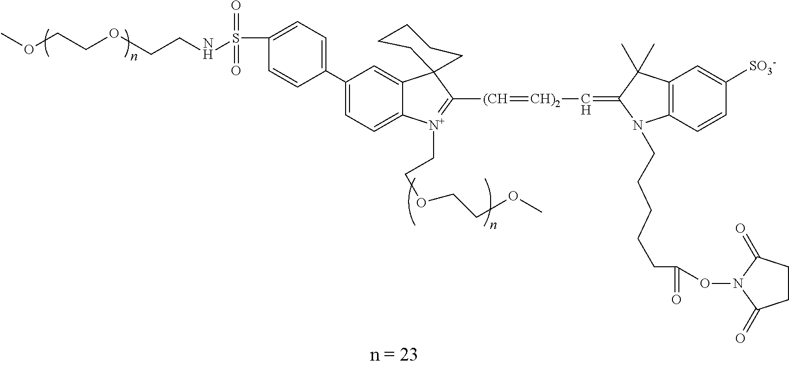 |
650/665 |
| 80 |
 |
650/665 |
| 81 |
 |
650/665 |
| 82 |
 |
660/680 |
| 83 |
 |
650/665 |
| 84 |
 |
663/690 |
| 85 |
 |
752/778 |
| 86 |
 |
750/775 |
| 87 |
 |
675/694 |
| 88 |
 |
750/775 |
| 89 |
 |
|
| 90 |
 |
768/788 |
| 91 |
 |
|
| 92 |
 |
635/642 |
| 93 |
 |
|
| 94 |
 |
497/513 |
| 95 |
 |
555/565 |
| 96 |
 |
650/665 |
| 97 |
 |
750/770 |
| 98 |
 |
660/675 |
| 99 |
 |
770/790 |
| 100 |
 |
680/700 |
| 101 |
 |
790/810 |
| 102 |
 |
494/520 |
| 103 |
 |
|
| 104 |
 |
501/524 |
| 105 |
 |
|
| 106 |
 |
|
| 107 |
 |
520/546 |
| 108 |
 |
|
| 109 |
 |
|
| 110 |
 |
540/565 |
| 111 |
 |
|
| 112 |
 |
|
| 113 |
 |
|
| 114 |
 |
|
| 115 |
 |
|
| 116 |
 |
488/515 |
| 117 |
 |
494/520 |
| 118 |
 |
353/442 |
| 119 |
 |
346/442 |
| 120 |
 |
416/465 |
| 121 |
 |
430/545 |
| 122 |
 |
350/440 |
| 123 |
 |
|
| 124 |
 |
|
| 125 |
 |
400/424 |

| TABLE 5 | |||
| Labeling condition | |||
| Antibody- | Amount | ||||
| Labeled | Antibody | to-dye | of | ||
| GAM IgG | Amount of | concentration | Amount | ratio | storage |
| antibody | GAM IgG | in reaction | of dye | (ug/ | buffer |
| solution | (μg) | (mg/mL) | (nmoles) | nmole) | (uL) |
| #1 | 100 | 1 | 11.25 | 8.9:1 | 300 |
| #2 | 50 | 2 | 11.25 | 4.4:1 | 300 |
| #3 | 20 | 2 | 5.6 | 3.6:1 | 150 |
| #4 | 20 | 1 | 2.8 | 7.2:1 | 60 |
| #5 | 5 | 1 | 2.8 | 1.8:1 | 60 |
| #6 | 100 | 1 | 9.4 | 10.6:1 | 300 |
| #7 | 50 | 2 | 9.4 | 5.3:1 | 300 |
| #8 | 100 | 1 | 7.5 | 13.3:1 | 300 |
| #9 | 50 | 2 | 7.5 | 6.7:1 | 30 |
| TABLE 6 | |||
| Labeling condition | |||
| Antibody- | Amount | ||||
| Labeled | Antibody | to-dye | of | ||
| GAM IgG | Amount of | concentration | Amount | ratio | storage |
| antibody | GAM IgG | in reaction | of dye | (ug/ | buffer |
| solution | (μg) | (mg/mL) | (nmoles) | nmole) | (uL) |
| #1 | 100 | 1 | 11.25 | 8.9:1 | 300 |
| #2 | 50 | 2 | 11.25 | 4.4:1 | 300 |
| #3 | 20 | 2 | 5.6 | 3.6:1 | 150 |
| #4 | 20 | 1 | 2.8 | 7.2:1 | 60 |
| #5 | 5 | 1 | 2.8 | 1.8:1 | 60 |
| #6 | 100 | 1 | 9.4 | 10.6:1 | 300 |
| #7 | 50 | 2 | 9.4 | 5.3:1 | 300 |
| #8 | 100 | 1 | 7.5 | 13.3:1 | 300 |
| #9 | 50 | 2 | 7.5 | 6.7:1 | 30 |

| TABLE 7 | |||
| Labeled | Labeling condition | ||
| GAM | Antibody | Amount | ||||
| IgG | Amount | concentration | Amount of | Antibody-to- | of storage | |
| antibody | Compound | of GAM | in reaction | dye | dye ratio | buffer |
| solution | No. | IgG (μg) | (mg/mL) | (nmoles) | (ug/nmole) | (uL) |
| #16 | 45 | 50 | 1 | 3.8 | 13.2:1 | 300 |
| #17 | 45 | 100 | 0.5 | 3.8 | 26.3:1 | 300 |
| #18 | 45 | 50 | 1 | 5.6 | 8.9:1 | 300 |
| #19 | 45 | 100 | 0.5 | 5.6 | 17.9:1 | 300 |
| #20 | 45 | 50 | 1 | 7.5 | 6.7:1 | 300 |
| #21 | 45 | 100 | 0.5 | 7.5 | 13.3:1 | 300 |
| #22 | 45 | 50 | 1 | 9.4 | 5.3:1 | 300 |
| #23 | 45 | 100 | 0.5 | 9.4 | 10.6:1 | 300 |
| #24 | 127 | 50 | 1 | 3.8 | 13.2:1 | 300 |
| #25 | 127 | 100 | 0.5 | 3.8 | 26.3:1 | 300 |
| #26 | 127 | 50 | 1 | 5.6 | 8.9:1 | 300 |
| #27 | 127 | 100 | 0.5 | 5.6 | 17.9:1 | 300 |
| #28 | 127 | 50 | 1 | 7.5 | 6.7:1 | 300 |
| #29 | 127 | 100 | 0.5 | 7.5 | 13.3:1 | 300 |
| #30 | 127 | 50 | 1 | 9.4 | 5.3:1 | 300 |
| #31 | 127 | 100 | 0.5 | 9.4 | 10.6:1 | 300 |

| TABLE 8 | ||
| Labeling condition | ||
| Labeled mouse | Amount | Antibody | ||
| anti-human | of | concentration | Amount | Antibody-to- |
| CD3 antibody | antibody | in reaction | of dye | dye ratio |
| solution | (μg) | (mg/mL) | (nmoles) | (ug/nmole) |
| #32 | 100 | 0.5 | 5.6 | 17.8:1 |
| #33 | 50 | 0.5 | 5.6 | 8.9:1 |
| #34 | 20 | 0.5 | 2.8 | 7.2:1 |
| #35 | 20 | 0.5 | 1.4 | 14.3:1 |
| #36 | 5 | 0.5 | 1.4 | 3.6:1 |
| TABLE 9 | |||
| Labeling condition | |||
| Labeled b- | Antibody- | Amount | |||
| tubulin | Antibody | to-dye | of | ||
| IgM | Amount | concentration | Amount | ratio | storage |
| antibody | of GAM | in reaction | of dye | (ug/ | buffer |
| solution | IgG (μg) | (mg/mL) | (nmoles) | nmole) | (uL) |
| #37 | 10 | 0.5 | 0.56 | 17.9:1 | 60 |
| #38 | 10 | 0.5 | 1.3 | 7.7:1 | 60 |
| #39 | 10 | 0.5 | 1.4 | 7.2:1 | 60 |
| #40 | 10 | 0.5 | 0.75 | 13.4:1 | 60 |
| #41 | 10 | 0.5 | 1.5 | 6.7:1 | 60 |
| #42 | 10 | 0.5 | 1.88 | 5.3:1 | 60 |
Claims (34)


Priority Applications (4)
| Application Number | Priority Date | Filing Date | Title |
|---|---|---|---|
| US13/423,092 US8709830B2 (en) | 2011-03-18 | 2012-03-16 | Fluorescent dyes, fluorescent dye kits, and methods of preparing labeled molecules |
| US14/218,439 US20150056632A1 (en) | 2011-03-18 | 2014-03-18 | Fluorescent dyes, fluorescent dye kits, and methods of preparing labeled molecules |
| US15/815,035 US10837881B2 (en) | 2011-03-18 | 2017-11-16 | Fluorescent dyes, fluorescent dye kits, and methods of preparing labeled molecules |
| US17/067,569 US20210102873A1 (en) | 2011-03-18 | 2020-10-09 | Fluorescent Dyes, Fluorescent Dye Kits, and Methods of Preparing Labeled Molecules |
Applications Claiming Priority (2)
| Application Number | Priority Date | Filing Date | Title |
|---|---|---|---|
| US201161454484P | 2011-03-18 | 2011-03-18 | |
| US13/423,092 US8709830B2 (en) | 2011-03-18 | 2012-03-16 | Fluorescent dyes, fluorescent dye kits, and methods of preparing labeled molecules |
Related Child Applications (1)
| Application Number | Title | Priority Date | Filing Date |
|---|---|---|---|
| US14/218,439 Continuation US20150056632A1 (en) | 2011-03-18 | 2014-03-18 | Fluorescent dyes, fluorescent dye kits, and methods of preparing labeled molecules |
Publications (2)
| Publication Number | Publication Date |
|---|---|
| US20120329068A1 US20120329068A1 (en) | 2012-12-27 |
| US8709830B2 true US8709830B2 (en) | 2014-04-29 |
Family
ID=46879699
Family Applications (4)
| Application Number | Title | Priority Date | Filing Date |
|---|---|---|---|
| US13/423,092 Active US8709830B2 (en) | 2011-03-18 | 2012-03-16 | Fluorescent dyes, fluorescent dye kits, and methods of preparing labeled molecules |
| US14/218,439 Abandoned US20150056632A1 (en) | 2011-03-18 | 2014-03-18 | Fluorescent dyes, fluorescent dye kits, and methods of preparing labeled molecules |
| US15/815,035 Active US10837881B2 (en) | 2011-03-18 | 2017-11-16 | Fluorescent dyes, fluorescent dye kits, and methods of preparing labeled molecules |
| US17/067,569 Pending US20210102873A1 (en) | 2011-03-18 | 2020-10-09 | Fluorescent Dyes, Fluorescent Dye Kits, and Methods of Preparing Labeled Molecules |
Family Applications After (3)
| Application Number | Title | Priority Date | Filing Date |
|---|---|---|---|
| US14/218,439 Abandoned US20150056632A1 (en) | 2011-03-18 | 2014-03-18 | Fluorescent dyes, fluorescent dye kits, and methods of preparing labeled molecules |
| US15/815,035 Active US10837881B2 (en) | 2011-03-18 | 2017-11-16 | Fluorescent dyes, fluorescent dye kits, and methods of preparing labeled molecules |
| US17/067,569 Pending US20210102873A1 (en) | 2011-03-18 | 2020-10-09 | Fluorescent Dyes, Fluorescent Dye Kits, and Methods of Preparing Labeled Molecules |
Country Status (3)
| Country | Link |
|---|---|
| US (4) | US8709830B2 (en) |
| EP (1) | EP2686680B1 (en) |
| WO (1) | WO2012129128A1 (en) |
Cited By (3)
| Publication number | Priority date | Publication date | Assignee | Title |
|---|---|---|---|---|
| US20110136201A1 (en) * | 2009-12-04 | 2011-06-09 | Biotium, Inc. | Heterocycle-substituted xanthene dyes |
| US10837881B2 (en) | 2011-03-18 | 2020-11-17 | Biotium, Inc. | Fluorescent dyes, fluorescent dye kits, and methods of preparing labeled molecules |
| US10976309B2 (en) * | 2012-09-18 | 2021-04-13 | Biotium, Inc. | Methods, compositions and kits for labeling of proteins |
Families Citing this family (20)
| Publication number | Priority date | Publication date | Assignee | Title |
|---|---|---|---|---|
| US10053447B2 (en) | 2010-12-21 | 2018-08-21 | Pierce Biotechnology, Inc | Fluorescent compounds |
| US8889884B1 (en) | 2011-07-14 | 2014-11-18 | Pierce Biotechnology, Inc. | Phosphine derivatives of fluorescent compounds |
| US9249307B2 (en) | 2011-08-16 | 2016-02-02 | Pierce Biotechnology, Inc. | Benzocyanine compounds |
| US9751868B2 (en) | 2012-02-28 | 2017-09-05 | Pierce Biotechnology, Inc. | Benzocyanine compounds |
| WO2013130761A1 (en) | 2012-03-02 | 2013-09-06 | Pierce Biotechnology, Inc. | Indole derivatives as labeling dye for biomolecule |
| EP2850078B1 (en) | 2012-08-28 | 2018-04-25 | Pierce Biotechnology, Inc. | Benzopyrylium compounds |
| EP2912464B1 (en) | 2012-10-24 | 2017-04-26 | Becton Dickinson and Company | Hydroxamate substituted azaindoline-cyanine dyes and bioconjugates of the same |
| WO2015038579A1 (en) * | 2013-09-11 | 2015-03-19 | Equip, Llc | Discrete peg based dyes |
| JP6700750B2 (en) * | 2014-12-09 | 2020-05-27 | キヤノン株式会社 | Compound and contrast agent for optical imaging containing the compound |
| CN107406872B (en) * | 2015-01-22 | 2023-03-28 | 里兰斯坦福初级大学理事会 | Compounds, compositions and uses for marking animal tissue |
| JP6555625B2 (en) * | 2015-06-17 | 2019-08-07 | エルジー・ケム・リミテッド | Xanthene compound and photosensitive resin composition containing the same |
| US9862682B2 (en) | 2016-01-08 | 2018-01-09 | BroadPharm | Functionalized pegylated cyanine compounds, pharmaceutical compositions, and methods of use thereof |
| US11828752B2 (en) | 2017-03-30 | 2023-11-28 | The Board Of Trustees Of The Leland Stanford Junior University | Protease-activated contrast agents for in vivo imaging |
| KR101921662B1 (en) | 2017-10-19 | 2018-11-23 | (주)바이오액츠 | Fluorescence Compounds and Preparation Method Therof |
| CN108017928A (en) * | 2017-12-15 | 2018-05-11 | 淮海工学院 | A kind of Pegylation indole cyanine dyes for biomolecular labeling and preparation method thereof |
| JP7425576B2 (en) * | 2018-12-27 | 2024-01-31 | 保土谷化学工業株式会社 | Xanthene pigments, coloring compositions containing the pigments, coloring agents for color filters, and color filters |
| KR102340869B1 (en) * | 2019-05-09 | 2021-12-20 | (주)크레용테크놀러지즈 | Electrophoretic dyeing method and dyeing device for biological samples using ion conductive film |
| US11835527B2 (en) | 2020-01-21 | 2023-12-05 | Regeneron Pharmaceuticals, Inc. | Deglycosylation methods for electrophoresis of glycosylated proteins |
| CN113340965B (en) * | 2020-03-03 | 2022-12-16 | 北京中医药大学 | Artificial intelligence biosensor detection method for detecting chlorpheniramine |
| WO2021215514A1 (en) * | 2020-04-24 | 2021-10-28 | 富士フイルム株式会社 | Compound and labeled biomaterial in which same is used |
Citations (10)
| Publication number | Priority date | Publication date | Assignee | Title |
|---|---|---|---|---|
| US4845039A (en) | 1984-11-15 | 1989-07-04 | Academisch Ziekenhuis Leiden | Method of labeling amine-containing compounds with a metal |
| US5397699A (en) | 1992-08-31 | 1995-03-14 | E. I. Du Pont De Nemours And Company | Method for stabilizing an alkanol amine buffer used in optical determinations of enzyme activity |
| US5403750A (en) | 1991-03-06 | 1995-04-04 | W. R. Grace & Co.-Conn. | Biocompatible, low protein adsorption affinity matrix |
| US5527686A (en) | 1991-07-29 | 1996-06-18 | Serex, Inc. | Differential binding affinities and dissociation assays based thereon |
| US5710009A (en) | 1994-02-17 | 1998-01-20 | Serex, Inc. | Receptor:release ligand (reland) complexes and release assays using said reland and methods and kits based thereon |
| US5772699A (en) | 1995-03-13 | 1998-06-30 | Crompton & Knowles Corporation | Stable aqueous reactive dye composition and method for stabilizing an aqueous reactive dye composition |
| US20040106153A1 (en) | 2002-07-18 | 2004-06-03 | Montana State University | Novel zwitterionic fluorescent dyes for labeling in proteomic and other biological analyses |
| WO2007030521A1 (en) | 2005-09-06 | 2007-03-15 | Invitrogen Corporation | Control of chemical modification |
| US20080160535A1 (en) | 1997-12-15 | 2008-07-03 | Somalogic, Inc. | Methods and Reagents for Detecting Target Binding by Nucleic Acid Ligands |
| WO2011149415A1 (en) | 2010-05-27 | 2011-12-01 | Ge Healthcare Bio-Sciences Ab | Method and kit for protein labeling |
Family Cites Families (5)
| Publication number | Priority date | Publication date | Assignee | Title |
|---|---|---|---|---|
| US6218160B1 (en) * | 1997-10-31 | 2001-04-17 | Roche Diagnostics Corporation | Site-specific conjugation of glycoproteins |
| US6964854B1 (en) * | 1999-07-13 | 2005-11-15 | Science & Technology Corporation | Compositions and methods useful for the diagnosis and treatment of heparin induced thrombocytopenia/thrombosis |
| EP1540000A2 (en) | 2002-09-20 | 2005-06-15 | Bristol-Myers Squibb Company | Assay for ppar ligand dependent gene modulation |
| US7405281B2 (en) * | 2005-09-29 | 2008-07-29 | Pacific Biosciences Of California, Inc. | Fluorescent nucleotide analogs and uses therefor |
| US8709830B2 (en) * | 2011-03-18 | 2014-04-29 | Biotium, Inc. | Fluorescent dyes, fluorescent dye kits, and methods of preparing labeled molecules |
-
2012
- 2012-03-16 US US13/423,092 patent/US8709830B2/en active Active
- 2012-03-16 EP EP12761364.4A patent/EP2686680B1/en active Active
- 2012-03-16 WO PCT/US2012/029564 patent/WO2012129128A1/en active Application Filing
-
2014
- 2014-03-18 US US14/218,439 patent/US20150056632A1/en not_active Abandoned
-
2017
- 2017-11-16 US US15/815,035 patent/US10837881B2/en active Active
-
2020
- 2020-10-09 US US17/067,569 patent/US20210102873A1/en active Pending
Patent Citations (13)
| Publication number | Priority date | Publication date | Assignee | Title |
|---|---|---|---|---|
| US4845039A (en) | 1984-11-15 | 1989-07-04 | Academisch Ziekenhuis Leiden | Method of labeling amine-containing compounds with a metal |
| US5403750A (en) | 1991-03-06 | 1995-04-04 | W. R. Grace & Co.-Conn. | Biocompatible, low protein adsorption affinity matrix |
| US5527686A (en) | 1991-07-29 | 1996-06-18 | Serex, Inc. | Differential binding affinities and dissociation assays based thereon |
| US5397699A (en) | 1992-08-31 | 1995-03-14 | E. I. Du Pont De Nemours And Company | Method for stabilizing an alkanol amine buffer used in optical determinations of enzyme activity |
| US5710009A (en) | 1994-02-17 | 1998-01-20 | Serex, Inc. | Receptor:release ligand (reland) complexes and release assays using said reland and methods and kits based thereon |
| US5772699A (en) | 1995-03-13 | 1998-06-30 | Crompton & Knowles Corporation | Stable aqueous reactive dye composition and method for stabilizing an aqueous reactive dye composition |
| US20080160535A1 (en) | 1997-12-15 | 2008-07-03 | Somalogic, Inc. | Methods and Reagents for Detecting Target Binding by Nucleic Acid Ligands |
| US20040106153A1 (en) | 2002-07-18 | 2004-06-03 | Montana State University | Novel zwitterionic fluorescent dyes for labeling in proteomic and other biological analyses |
| WO2007030521A1 (en) | 2005-09-06 | 2007-03-15 | Invitrogen Corporation | Control of chemical modification |
| US20070134685A1 (en) * | 2005-09-06 | 2007-06-14 | Invitrogen Corporation | Control of chemical modification |
| US20080176213A1 (en) | 2005-09-06 | 2008-07-24 | Invitrogen Corporation | Control of chemical modification |
| US20100009342A1 (en) | 2005-09-06 | 2010-01-14 | Life Technologies Corporation | Control of chemical modification |
| WO2011149415A1 (en) | 2010-05-27 | 2011-12-01 | Ge Healthcare Bio-Sciences Ab | Method and kit for protein labeling |
Non-Patent Citations (1)
| Title |
|---|
| International search report and written opinion dated Jul. 27, 2012 for PCT Application No. US12/29564. |
Cited By (6)
| Publication number | Priority date | Publication date | Assignee | Title |
|---|---|---|---|---|
| US20110136201A1 (en) * | 2009-12-04 | 2011-06-09 | Biotium, Inc. | Heterocycle-substituted xanthene dyes |
| US9579402B2 (en) * | 2009-12-04 | 2017-02-28 | Biotium, Inc. | Heterocycle-substituted xanthene dyes |
| US10053484B2 (en) | 2009-12-04 | 2018-08-21 | Biotium, Inc. | Heterocycle-substituted xanthene dyes |
| US10837881B2 (en) | 2011-03-18 | 2020-11-17 | Biotium, Inc. | Fluorescent dyes, fluorescent dye kits, and methods of preparing labeled molecules |
| US20210102873A1 (en) * | 2011-03-18 | 2021-04-08 | Biotium, Inc. | Fluorescent Dyes, Fluorescent Dye Kits, and Methods of Preparing Labeled Molecules |
| US10976309B2 (en) * | 2012-09-18 | 2021-04-13 | Biotium, Inc. | Methods, compositions and kits for labeling of proteins |
Also Published As
| Publication number | Publication date |
|---|---|
| EP2686680A1 (en) | 2014-01-22 |
| US10837881B2 (en) | 2020-11-17 |
| WO2012129128A1 (en) | 2012-09-27 |
| US20180143113A1 (en) | 2018-05-24 |
| US20150056632A1 (en) | 2015-02-26 |
| US20210102873A1 (en) | 2021-04-08 |
| EP2686680B1 (en) | 2018-06-13 |
| EP2686680A4 (en) | 2014-12-10 |
| US20120329068A1 (en) | 2012-12-27 |
Similar Documents
| Publication | Publication Date | Title |
|---|---|---|
| US20210102873A1 (en) | Fluorescent Dyes, Fluorescent Dye Kits, and Methods of Preparing Labeled Molecules | |
| CA2718707C (en) | Method for detection of liver cancer cell using anti-glypican-3 antibody | |
| JP4874234B2 (en) | Non-small cell lung cancer-related gene ANLN and its interaction with RhoA | |
| US20200327962A1 (en) | Statistical ai for advanced deep learning and probabilistic programing in the biosciences | |
| JP6153946B2 (en) | SLIT-ROBO signaling for diagnosis and treatment of kidney disease | |
| US7811774B2 (en) | Reagents and methods for use in cancer diagnosis, classification and therapy | |
| JP2010506939A (en) | DNA damage repair inhibitors and methods for treating cancer | |
| Su et al. | Knockdown of contactin-1 expression suppresses invasion and metastasis of lung adenocarcinoma | |
| MX2011005007A (en) | Anti-cxcr1 compositions and methods. | |
| WO2006132683A2 (en) | Reagents and methods for use in cancer diagnosis, classification and therapy | |
| CN106461681B (en) | Biomarker and application thereof for diagnosing vascular diseases | |
| ES2568753T3 (en) | A prognostic and therapeutic signature for malignant melanoma | |
| JP5265140B2 (en) | Ovarian cancer detection method and detection kit | |
| KR20190106809A (en) | Srm/mrm assays for profiling tumor tissue | |
| WO2006119264A2 (en) | Antibodies against histone modifications for clinical diagnosis and prognosis of cancer | |
| US20140227799A1 (en) | Method for measuring anti-wt1 antibody | |
| CN114591418A (en) | Threonine 166 th phosphorylation modification of PPAR gamma protein and application thereof | |
| JP2007511471A (en) | SHC proteins as therapeutic targets in proliferative diseases | |
| Mollnes et al. | Activation-dependent epitopes in the terminal complement pathway | |
| WO2009117004A1 (en) | Reagents and methods for use in head and neck cancer diagnosis, classification and therapy | |
| WO2008047947A1 (en) | Method for determining effect of cisplatin administration and kit for determining effect of cisplatin administration | |
| WO2023173094A2 (en) | Lysine-free ubiquibody variants for long-lived intracellular protein silencing | |
| US20080131916A1 (en) | Reagents and Methods For Use In Cancer Diagnosis, Classification and Therapy | |
| US20100143947A1 (en) | Reagents and methods for use in cancer diagnosis, classification and therapy | |
| Hartert | Overcoming TCF4-Driven BCR Signaling in Diffuse Large B-Cell Lymphoma |
Legal Events
| Date | Code | Title | Description |
|---|---|---|---|
| AS | Assignment |
Owner name: BIOTIUM, INC., CALIFORNIA Free format text: ASSIGNMENT OF ASSIGNORS INTEREST;ASSIGNORS:MAO, FEI;LEUNG, WAI-YEE;CHUENG, CHING-YING;AND OTHERS;SIGNING DATES FROM 20120321 TO 20120326;REEL/FRAME:027963/0481 |
|
| AS | Assignment |
Owner name: BIOTIUM, INC., CALIFORNIA Free format text: ASSIGNMENT OF ASSIGNORS INTEREST;ASSIGNORS:MAO, FEI;LEUNG, WAI-YEE;CHEUNG, CHING-YING;AND OTHERS;SIGNING DATES FROM 20120321 TO 20120326;REEL/FRAME:028259/0287 |
|
| STCF | Information on status: patent grant |
Free format text: PATENTED CASE |
|
| MAFP | Maintenance fee payment |
Free format text: PAYMENT OF MAINTENANCE FEE, 4TH YEAR, LARGE ENTITY (ORIGINAL EVENT CODE: M1551) Year of fee payment: 4 |
|
| CC | Certificate of correction | ||
| MAFP | Maintenance fee payment |
Free format text: PAYMENT OF MAINTENANCE FEE, 8TH YEAR, LARGE ENTITY (ORIGINAL EVENT CODE: M1552); ENTITY STATUS OF PATENT OWNER: LARGE ENTITY Year of fee payment: 8 |