US8608725B2 - Eluting coils and methods of deploying and retrieving - Google Patents
Eluting coils and methods of deploying and retrieving Download PDFInfo
- Publication number
- US8608725B2 US8608725B2 US13/610,733 US201213610733A US8608725B2 US 8608725 B2 US8608725 B2 US 8608725B2 US 201213610733 A US201213610733 A US 201213610733A US 8608725 B2 US8608725 B2 US 8608725B2
- Authority
- US
- United States
- Prior art keywords
- eluting
- coil
- elongate element
- resilient elongate
- configuration
- Prior art date
- Legal status (The legal status is an assumption and is not a legal conclusion. Google has not performed a legal analysis and makes no representation as to the accuracy of the status listed.)
- Active
Links
Images


Classifications
-
- A—HUMAN NECESSITIES
- A61—MEDICAL OR VETERINARY SCIENCE; HYGIENE
- A61M—DEVICES FOR INTRODUCING MEDIA INTO, OR ONTO, THE BODY; DEVICES FOR TRANSDUCING BODY MEDIA OR FOR TAKING MEDIA FROM THE BODY; DEVICES FOR PRODUCING OR ENDING SLEEP OR STUPOR
- A61M31/00—Devices for introducing or retaining media, e.g. remedies, in cavities of the body
- A61M31/002—Devices for releasing a drug at a continuous and controlled rate for a prolonged period of time
-
- A—HUMAN NECESSITIES
- A61—MEDICAL OR VETERINARY SCIENCE; HYGIENE
- A61F—FILTERS IMPLANTABLE INTO BLOOD VESSELS; PROSTHESES; DEVICES PROVIDING PATENCY TO, OR PREVENTING COLLAPSING OF, TUBULAR STRUCTURES OF THE BODY, e.g. STENTS; ORTHOPAEDIC, NURSING OR CONTRACEPTIVE DEVICES; FOMENTATION; TREATMENT OR PROTECTION OF EYES OR EARS; BANDAGES, DRESSINGS OR ABSORBENT PADS; FIRST-AID KITS
- A61F2250/00—Special features of prostheses classified in groups A61F2/00 - A61F2/26 or A61F2/82 or A61F9/00 or A61F11/00 or subgroups thereof
- A61F2250/0058—Additional features; Implant or prostheses properties not otherwise provided for
- A61F2250/0067—Means for introducing or releasing pharmaceutical products into the body
- A61F2250/0068—Means for introducing or releasing pharmaceutical products into the body the pharmaceutical product being in a reservoir
Abstract
Embodiments are directed to eluting coils having a relaxed coiled state and a straightened state that may be deployed at a fixed location within a patient's body and may accurately dispense and distribute fluids and or dissolvable substances at site specific locations of the body. Some embodiments of eluting elements are configured to be subsequently retrieved from a delivery site.
Description
This application is a continuation of U.S. patent application Ser. No. 12/765,743, titled “Eluting Coils and Methods of Deploying and Retrieving”, filed Apr. 22, 2010, by John L. Wardle, which is a continuation of U.S. patent application Ser. No. 11/328,884, titled “Eluting Coils and Methods of Deploying and Retrieving”, filed Jan. 9, 2006, by John L. Wardle, now U.S. Pat. No. 7,731,705, issued on Jun. 8, 2010, which claims priority under 35 U.S.C. section 119(e) from U.S. Provisional Application Ser. No. 60/642,892, titled “Eluting Coils and Methods of Deploying and Retrieving”, filed Jan. 10, 2005, by John L. Wardle, which are all incorporated by reference herein in their entirety. This application is also related to U.S. patent application Ser. No. 10/386,260, filed Mar. 10, 2003, by John L. Wardle, titled “Surgical Coils and Methods of Deploying” which is also incorporated by reference herein in its entirety.
Systemic drug delivery is often ill-suited to the treatment of conditions occurring at one or more discrete sites within a patient's body, because it involves the delivery of the medication to sites other than the target site. Systemic agent delivery also requires the infusion of large doses of the medication to assure the delivery of a therapeutic dose to the target site, thereby creating the possibility of deleterious effects. Another problem of systemic administration is the inevitable fluctuations of drug concentrations that it produces. The dosage that can be delivered to the target site may be limited by the need to minimize unwanted effects in other parts of a patient's body. Furthermore, systemic delivery exposes the medication to possible degradation and elimination by the action of other bodily organs.
Conventional methods of drug therapy, as discussed above, often result in blood levels of the cytotoxic agent that are dangerous for the patient. Even with local administration of these agents, one must consider that blood flow of vessels as well as other transport mechanisms may dilute the local concentration of the therapeutic agent by a wash-out effect. The need remains, therefore, for systems and methods for localized delivery of therapeutic agents, including toxic therapeutic agents, which may be concentrated and localized intramurally within the affected tissue or vessel.
There is general need in many branches of medicine for improved localized internal delivery of substances including therapeutic agents and drugs and diagnostic agents into the walls of ducts, organs and vessels. In particular, there is need for effective systems and methods of delivery into tissue and into cells themselves within organs, ducts, tracts and vessels of the body via percutaneous and luminal access. Problems remain however in the exact method by which the local administration of drugs or therapeutic agents can be achieved. The problem is further complicated where it is desirable to deliver drug in relatively small amounts and the delivery device must be sufficiently small, biocompatible, impermeable and drug non-reactive. There is also a need in some circumstances for retrieval systems and methods of delivery devices which may be implemented at the conclusion of treatment embodiments or portions thereof.
Some embodiments include eluting coils that can accurately dispense and distribute fluids and or dissolvable substances in site specific locations of a patient's body. Some embodiments of eluting coils may be configured to be subsequently retrieved from a delivery site.
Some embodiments include an eluting coil, having an elongate element with a longitudinal axis formed into a coiled enclosed configuration with an overlapped portion. Sections of the elongate element make contact with adjacent sections of the elongate element in the overlapped portion. The overlapped portion has a circumferential overlap of at least 300 degrees and has a pre-stressed configuration to ensure surface contact between overlapped portions of the elongate element. Some of these embodiments include a pre-stress of the pre-stressed configuration which is substantially constant around the coil. Some of these embodiments include a surface of the elongate element that has at least one reservoir with a channel. Some of these embodiments include stand-off members on at least a portion of the surface of the elongate element that are configured to produce a controlled eluting gap between coils segments when the elongate element is in a coiled configuration. Some of these embodiments include an elongate element having an interlocking configuration that may include a longitudinal groove in a first surface of the elongate element and a longitudinal ridge, configured to mate with the longitudinal groove on a second surface opposite the first surface. The interlocking configuration may be configured to create an eluting gap between adjacent coils of overlapped portions the elongate element in a coiled configuration. Some of these embodiments may include a tail member for removal after deployment.
Some embodiments of eluting coils for deployment within a body of a patient include a resilient elongate element having an inside surface, an outside surface, a pre-stressed non-coiled configuration in a restrained state and a coiled configuration in a relaxed state with the outside surface disposed adjacent the inside surface in an overlapped portion thereof. At least one dissolvable agent reservoir is disposed on the elongate element. In addition, at least one conduit having a pre-determined cross section is disposed in fluid communication between the dissolvable agent reservoir and an outside portion of the coiled configuration of the resilient elongate element in the relaxed state. Some of these embodiments include a resilient elongate element with a pre-stress of a substantially constant radius of curvature along a length of the resilient elongate element. Some of these embodiments include at least one stand-off member on a surface of the resilient elongate element and the at least one conduit comprises a controlled gap formed by the stand-off member between the inside surface and outside surface of the resilient elongate element in overlapped portions of the coiled configuration in the relaxed state. Some of these embodiments include a conduit formed from at least one channel disposed on a surface of the resilient elongate element.
Some embodiments of a method of deploying an eluting coil include providing an eluting coil for deployment within a body of a patient. The eluting coil includes a resilient elongate element having an inside surface, an outside surface, a pre-stressed non-coiled configuration in a restrained state and a coiled configuration in a relaxed state with the outside surface disposed adjacent the inside surface in an overlapped portion thereof. At least one dissolvable agent reservoir is disposed on the elongate element. At least one conduit having a pre-determined cross section is disposed in fluid communication between the dissolvable agent reservoir and an outside portion of the coiled configuration of the resilient elongate element in the relaxed state. The resilient elongate element of the eluting coil is disposed in a restrained state. A distal end of an elongate delivery member is disposed adjacent a target site within a patient's body and the resilient elongate element is moved along the elongate delivery member. The resilient elongate element is deployed from a distal end of the elongate delivery member and allowed to achieve a relaxed coiled configuration.
Some embodiments of a method of retrieving an eluting coil include providing an eluting coil deployed within a body of a patient in a relaxed coiled configuration. The eluting coil includes a resilient elongate element having an inside surface, an outside surface, a pre-stressed non-coiled configuration in a restrained state and a coiled configuration in a relaxed state with the outside surface disposed adjacent the inside surface in an overlapped portion thereof. At least one dissolvable agent reservoir is disposed on the elongate element. At least one conduit having a pre-determined cross section is disposed in fluid communication between the dissolvable agent reservoir and an outside portion of the coiled configuration of the resilient elongate element in the relaxed state. A tail member is disposed at an end of the resilient elongate element configured to facilitate removal of the eluting coil after deployment thereof. A retrieval device is advanced into the patient's body until a distal end of the retrieval device is disposed adjacent the eluting coil. A retraction element of the retrieval device is coupled to the tail member and the resilient elongate element is withdrawn into the retrieval device.
These features of embodiments will become more apparent from the following detailed description when taken in conjunction with the accompanying exemplary drawings.
The eluting coil 10 may be delivered to a target site in a patient's body (not shown) by methods disclosed in the incorporated application U.S. patent application Ser. No. 10/386,260, filed Mar. 10, 2003, by John L. Wardle, titled “Surgical Coils and Methods of Deploying” ('260 Application). FIGS. 25-30 and the accompanying description of the '260 application describe embodiments of a delivery device having a delivery member or sheath that may be used to deploy the eluting coil 10 to a target site within a patient's body. Other embodiments of the '260 application may also be used.
The resilient material of the resilient elongate element 12 is configured to resist deformation from the relaxed coiled configuration shown in FIGS. 1 and 2 and spring back to the relaxed coiled configuration when released from a restrained configuration, such as a straightened configuration, as shown in FIG. 3 . The resilient elongate element 12 of eluting coil 10 and all eluting coil embodiments discussed herein may be made from a variety of materials including those that exhibit either great elasticity or shape memory properties. Suitable materials for fabrication include but are not limited to nickel titanium alloys (Nitinol), stainless steel, Elgiloy, MP35N or other high strength biocompatible materials. In addition to these materials eluting coils can be made from absorbable materials such as magnesium alloy which besides being absorbable has the added advantage that it does not interfere with magnetic resonance imaging (MRI).
The surfaces 14 and 16 may be configured so as to provide a three dimensional drug eluting surface or reservoir, the modification could be as simple as a bead blast texture or more sophisticated techniques can be used as described in the following embodiments. FIGS. 3-4 illustrate an embodiment of resilient elongate element 12 of eluting coil 10 in a non-coiled restrained state with a straightened configuration. The elongate element 12 is shown with the distal end 20 extended opposite the proximal end 24. A plurality of axially consecutive longitudinal dissolvable agent reservoirs 30 and channels 32 are cut into the outside surface 16 elongate element 12. The reservoirs 30 and channels 32 are shown covering a substantial portion of outer surface 16, but any suitable portion of outer surface 16 or inner surface 14 could be used for such reservoirs 30 or channels 32. When the resilient elongate element 12 shown in FIGS. 3 and 4 is allowed to assume a relaxed coiled configuration, the reservoirs 30 and channels 32 cut into the outer surface 16 are pressed against the inner surface 14 such that the reservoirs 30 are sealed from an outside portion of the eluting coil 10 and a top portion of the channels are sealed to form a conduit in fluid communication between the respective reservoirs 30 and outside portion of the eluting coil 10. As shown in more detail in FIG. 4 , the channels 32 may extend across a plurality of reservoirs 30 to a lateral edge 34 of the resilient elongate element 12. For some embodiments of eluting coils 10, some of the reservoirs or dissolvable agent depots 30 may be exposed when the resilient elongate element 12 of the eluting coil 10 is in a coiled configuration in a relaxed state. The exposure of such reservoirs 30 allows for initial delivery of a high dose of agent over a short period upon initial deployment of the eluting coil 10. The non-exposed or encapsulated reservoirs 30 will deliver agent from a dissolvable matrix over a course of time determined by the transverse cross section of the channels or conduits 32 and capacity of reservoirs 30.
Eluting coils 10 may be configured to be delivered to a target site in a patient's body with a wide range of agents, such as bioactive agents including drugs, antibiotic agents, growth factors, anti-inflammatory agents and the like disposed in a dissolvable matrix within reservoirs 30, channels 32 or both. Dissolvable matrix components may include, but are not limited to, lipid materials, gelatins and the like. FIG. 4 shows a dissolvable agent reservoir 30 with a dissolvable agent 36 disposed therein. Generally, some or all of the reservoirs 30 may have some dissolvable agent 36 disposed within them prior to deployment of the eluting coil 10. When the resilient elongate element 12 of the eluting coil 10 is ejected from the delivery device the overlapping surfaces will encapsulate and contain the reservoirs 30 and channels 32 as discussed above. Thereafter, the dissolvable agent will come in contact with body fluids via the conduits formed by the channels and the dissolvable agent will be delivered from the reservoirs 30 to a region outside the eluting coil 10 through the conduits formed by the encapsulated channels 32.
In addition, different zones or portions of the resilient elongate element 12 of the eluting coil 10 may be configured with varying reservoir 30 and channel 32 profiles. Different surfaces 14 and 16, including reservoirs 30 or channels 32 thereof, of the resilient elongate element 12 of the eluting coil 12 may also be loaded with different agents disposed within a dissolvable agent matrix. Also, two or more components of a multi-component drug or other agent that need to be combined in order to react or otherwise be activated may be placed on opposite surfaces 14 and 16 of the resilient elongate element 12 of the eluting coil 10. In such a configuration, the two or more components will then be combined or otherwise communicated with each other and activated when the eluting coil 10 is deployed and the opposing surfaces 14 and 16 make contact.
Reservoirs, such as reservoirs 30 discussed above may be disposed on one or both of the surfaces 14 and 16 of the resilient elongate element 44 with the controllable gap providing a conduit between the reservoirs and an outside portion of the coil 10. Also, one or more surfaces 14 and 16 of the resilient elongate element 44 may be coated with the bioactive agent in a dissolvable matrix with the space or gap between surfaces of the resilient elongate element 44 providing a controlled leak path or conduit for the bioactive agent to an outside portion of the eluting coil 10 in a coiled relaxed state. Although the embodiment shown in FIGS. 7 and 8 includes one raised longitudinal ridge 46 and one longitudinal groove 48 on an opposite surface of the resilient elongate element 44, the resilient elongate element 44 may include multiple raised longitudinal ridge elements and mating longitudinal grooves.
In some embodiments of methods of retrieval of a deployed eluting coil 50, the retrieval process begins with identifying or locating the proximal end 60 of the tail extension 52. Once located, the hooked distal end 88 of a retrieval wire 84 it placed through the hole 58 (or a loop 72 of an embodiment of an eluting coil 62 as shown in FIGS. 11-13 ) as shown in FIG. 15 . The proximal end of the retrieval wire 84 is the back loaded or withdrawn into the cannula 82. The proximal end of the retrieval wire 84 may held stationary while the distal end 86 of the cannula 82 is advanced distally until the distal end 86 contacts the resilient elongate element 54 of the eluting coil 50. The cannula 82 may then be held stationary and the retrieval wire 84 is pulled or withdrawn proximally, as indicated by arrow 90, shown in FIG. 16 , which in turn pulls the resilient elongate element 54 of the eluting coil 50 into the cannula 82 and uncoils the eluting coil 50 and imparts a restrained non-coiled configuration on the resilient elongate element 54 as the resilient elongate element 54 is withdrawn into the cannula 82.
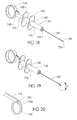
Suitable metals for construction of the delivery sheath 142 may include stainless steel, Nitinol, MP35N and the like. The delivery actuator 148 has an elongate cylindrically shaped body portion 156 with a proximal flange 158 and a distal flange 160. The body portion 156 has an internal bore 162 that is sized to accept a cylindrical actuator 164 in sliding relation to the body portion 156. A thumb ring 166 is disposed at a proximal end 168 of the cylindrical actuator 164 to facilitate the grip of an operator of the delivery device 138. The body portion 156 and cylindrical actuator 164 can be made from a variety of suitable medical grade materials, including metals, composites and polymers. Specifically, polymers such as ABS plastic, PVC, polycarbonate and the like may be used.
An eluting coil 140 being deployed from a distal end 146 of the delivery sheath 142 into tissue 170 of a target tissue site is shown in FIGS. 21-23 . The resilient elongate element 154 of eluting coil 140 may include the pre-stressed self-forming embodiment shown, wherein the resilient elongate element 154 returns to a relaxed coiled configuration, that is the configuration in a relaxed state, as the resilient elongate element 154 exits the distal end 146 of the delivery sheath 142 and the constraint of the delivery sheath 142 is removed. The eluting coil 140 is shown mechanically capturing a portion of tissue 172 of the target tissue site as it encircles the tissue.
Embodiments of delivery device 138 shown in FIG. 18 may use low profile delivery sheaths in the form of hollow needles with sharpened distal ends to deliver eluting coils to a target tissue site. The delivery sheath 142 is a straight tube with a distal tissue penetrating point 150 and is stiffer than an eluting coil 140 to be delivered therethrough. The geometry of the distal point of the delivery sheath 142 can be important in some embodiments. The distal point 150 needs to easily penetrate tissue while also providing clearance for the surgical coil 140 as it is being delivered without substantial restriction to assume the relaxed geometry of the surgical coil 140. For some delivery sheath 142 embodiments, the distal point can have an angle of about 25 degrees.
In one embodiment of use, the distal end 146 of the delivery sheath 142 is placed at a target site, a thumb ring 166 of the cylindrical actuator 164 is then moved distally as shown in FIG. 19 which pushes an advancing ribbon (not shown) which in turn pushes the most proximal eluting coil which then ejects the most distal eluting coil from the device 138 as shown in FIG. 20 .
There are varieties of techniques that can be employed with these low profile delivery devices 138 to access target sites. The delivery sheath 142 can be used in the same manner as a hypodermic needle is for drug delivery (direct incision). Alternatively they can be placed within the working channel of an endoscope or cannula. All methods allow the physician to completely or partially implant a coil in tissue at an anterior or posterior location.
With regard to the above detailed description, like reference numerals used therein refer to like elements that may have the same or similar dimensions, materials and configurations. While particular forms of embodiments have been illustrated and described, it will be apparent that various modifications can be made without departing from the spirit and scope of the embodiments of the invention. Alternate embodiments may include the combination of various features of different embodiments. For example, some or all of the features of the embodiments shown in FIG. 4 may be combined with some or all of the features of the embodiments shown in FIG. 8 . Accordingly, it is not intended that the invention be limited by the forgoing detailed description.
Claims (12)
1. An eluting coil for deployment within a body of a patient, comprising:
a resilient elongate element including a flattened ribbon-like configuration, a first surface, a second surface, a pre-stressed non-coiled configuration in a restrained state that ensures contact between the first surface and the second surface in adjacent sections of the elongate element when the elongate element is in a coiled configuration in a relaxed state with the first surface disposed adjacent the second surface in an overlapped portion of the elongate element; and
a tail extension which is disposed at a proximal end of the resilient elongate element, which has a proximal end configured to facilitate coupling thereto and which is configured for pulling on the elongate element for removal of the eluting coil after deployment.
2. The eluting coil of claim 1 wherein the resilient elongate element comprises magnesium.
3. The eluting coil of claim 1 wherein the resilient elongate element comprises NiTi.
4. The eluting coil of claim 1 wherein the pre-stress of the pre-stressed non-coiled configuration of the resilient elongate element comprises a substantially constant radius of curvature along a length of the resilient elongate element sufficient to generate forces that ensure contact between adjacent sections of the elongate element when in the coiled configuration.
5. The eluting coil of claim 1 further comprising a longitudinal groove disposed on a second surface of the elongate element configured to mate with a longitudinal ridge on the first surface and whereby the coiled configuration of the elongate element comprises an interlocking configuration.
6. The eluting coil of claim 1 wherein the overlapped portion comprises a circumferential overlap of the resilient elongate element of at least about 300 degrees.
7. The eluting coil of claim 1 wherein the coiled configuration of the relaxed state is substantially circular.
8. The eluting coil of claim 1 wherein the coiled configuration comprises an outer transverse dimension of less than about 1 cm.
9. The eluting coil of claim 1 wherein the resilient elongate element further comprises a sharpened end configured to penetrate tissue upon axial advancement of the resilient elongate element during deployment of the eluting coil.
10. The eluting coil of claim 1 wherein the tail extension has a substantially straight configuration in a relaxed state that extends away from the coiled configuration of the elongate element.
11. The eluting coil of claim 1 wherein the tail extension comprises an integrally formed extension of the elongate element.
12. The eluting coil of claim 1 wherein the tail extension comprises a loop of flexible element.
Priority Applications (1)
| Application Number | Priority Date | Filing Date | Title |
|---|---|---|---|
| US13/610,733 US8608725B2 (en) | 2005-01-10 | 2012-09-11 | Eluting coils and methods of deploying and retrieving |
Applications Claiming Priority (4)
| Application Number | Priority Date | Filing Date | Title |
|---|---|---|---|
| US64289205P | 2005-01-10 | 2005-01-10 | |
| US11/328,884 US7731705B2 (en) | 2005-01-10 | 2006-01-09 | Eluting coils and methods of deploying and retrieving |
| US12/765,743 US8287553B2 (en) | 2005-01-10 | 2010-04-22 | Eluting coils and methods of deploying and retrieving |
| US13/610,733 US8608725B2 (en) | 2005-01-10 | 2012-09-11 | Eluting coils and methods of deploying and retrieving |
Related Parent Applications (1)
| Application Number | Title | Priority Date | Filing Date |
|---|---|---|---|
| US12/765,743 Continuation US8287553B2 (en) | 2005-01-10 | 2010-04-22 | Eluting coils and methods of deploying and retrieving |
Publications (2)
| Publication Number | Publication Date |
|---|---|
| US20130006201A1 US20130006201A1 (en) | 2013-01-03 |
| US8608725B2 true US8608725B2 (en) | 2013-12-17 |
Family
ID=36652245
Family Applications (3)
| Application Number | Title | Priority Date | Filing Date |
|---|---|---|---|
| US11/328,884 Active 2029-02-08 US7731705B2 (en) | 2005-01-10 | 2006-01-09 | Eluting coils and methods of deploying and retrieving |
| US12/765,743 Active US8287553B2 (en) | 2005-01-10 | 2010-04-22 | Eluting coils and methods of deploying and retrieving |
| US13/610,733 Active US8608725B2 (en) | 2005-01-10 | 2012-09-11 | Eluting coils and methods of deploying and retrieving |
Family Applications Before (2)
| Application Number | Title | Priority Date | Filing Date |
|---|---|---|---|
| US11/328,884 Active 2029-02-08 US7731705B2 (en) | 2005-01-10 | 2006-01-09 | Eluting coils and methods of deploying and retrieving |
| US12/765,743 Active US8287553B2 (en) | 2005-01-10 | 2010-04-22 | Eluting coils and methods of deploying and retrieving |
Country Status (1)
| Country | Link |
|---|---|
| US (3) | US7731705B2 (en) |
Cited By (1)
| Publication number | Priority date | Publication date | Assignee | Title |
|---|---|---|---|---|
| US9301756B2 (en) | 2002-03-11 | 2016-04-05 | John L. Wardle | Surgical coils and methods of deploying |
Families Citing this family (8)
| Publication number | Priority date | Publication date | Assignee | Title |
|---|---|---|---|---|
| US7731705B2 (en) * | 2005-01-10 | 2010-06-08 | Wardle John L | Eluting coils and methods of deploying and retrieving |
| US20130165949A1 (en) * | 2010-03-19 | 2013-06-27 | The Regents of the University of Colorado , a body corporate | Soft tissue coaptor and device for deploying same |
| DK2600848T3 (en) * | 2010-08-05 | 2019-06-24 | Taris Biomedical Llc | Implantable drug delivery devices for urogenital positions |
| NZ725610A (en) * | 2014-04-18 | 2022-01-28 | Becton Dickinson Co | Needle capture safety interlock for catheter |
| US11833034B2 (en) | 2016-01-13 | 2023-12-05 | Shifamed Holdings, Llc | Prosthetic cardiac valve devices, systems, and methods |
| US20180078328A1 (en) * | 2016-09-16 | 2018-03-22 | Regents Of The University Of Minnesota | Pre-surgical pulmonary nodule localization systems and methods |
| JP2022504241A (en) | 2018-10-05 | 2022-01-13 | シファメド・ホールディングス・エルエルシー | Artificial heart valve device, system, and method |
| EP3941391A4 (en) | 2019-03-19 | 2022-11-23 | Shifamed Holdings, LLC | Prosthetic cardiac valve devices, systems, and methods |
Citations (84)
| Publication number | Priority date | Publication date | Assignee | Title |
|---|---|---|---|---|
| US4548202A (en) | 1983-06-20 | 1985-10-22 | Ethicon, Inc. | Mesh tissue fasteners |
| US4616656A (en) | 1985-03-19 | 1986-10-14 | Nicholson James E | Self-actuating breast lesion probe and method of using |
| US4632100A (en) | 1985-08-29 | 1986-12-30 | Marlowe E. Goble | Suture anchor assembly |
| US4774948A (en) | 1986-11-24 | 1988-10-04 | Markham Charles W | Marking and retraction needle having retrievable stylet |
| US4843651A (en) | 1988-01-11 | 1989-07-04 | Hatch Imports, Inc. | Wrist support glove |
| US4883070A (en) | 1988-03-02 | 1989-11-28 | Hanson Ralph E | Endocardial pacing lead |
| US4899744A (en) | 1988-12-15 | 1990-02-13 | Tatsuo Fujitsuka | Apparatus for anastomosing digestive tract |
| US4924865A (en) | 1986-05-20 | 1990-05-15 | Concept, Inc. | Repair tack for bodily tissue |
| US4931059A (en) | 1986-11-24 | 1990-06-05 | Markham Charles W | Needle/stylet combination |
| US4935027A (en) | 1989-08-21 | 1990-06-19 | Inbae Yoon | Surgical suture instrument with remotely controllable suture material advancement |
| US4938760A (en) | 1989-03-29 | 1990-07-03 | American Medical Systems, Inc. | Female suspension procedure |
| US5002548A (en) | 1986-10-06 | 1991-03-26 | Bio Medic Data Systems, Inc. | Animal marker implanting system |
| US5007921A (en) | 1989-10-26 | 1991-04-16 | Brown Alan W | Surgical staple |
| US5013316A (en) | 1990-03-26 | 1991-05-07 | Marlowe Goble E | Soft tissue anchor system |
| US5018530A (en) | 1989-06-15 | 1991-05-28 | Research Corporation Technologies, Inc. | Helical-tipped lesion localization needle device and method of using the same |
| US5030224A (en) | 1989-09-25 | 1991-07-09 | Pioneering Technologies, Inc. | Coronary artery retraction clip |
| US5104399A (en) | 1986-12-10 | 1992-04-14 | Endovascular Technologies, Inc. | Artificial graft and implantation method |
| US5123906A (en) | 1991-06-20 | 1992-06-23 | Kelman Charles D | Surgical toroidal snare |
| US5127916A (en) | 1991-01-22 | 1992-07-07 | Medical Device Technologies, Inc. | Localization needle assembly |
| US5129906A (en) | 1989-09-08 | 1992-07-14 | Linvatec Corporation | Bioabsorbable tack for joining bodily tissue and in vivo method and apparatus for deploying same |
| US5141520A (en) | 1991-10-29 | 1992-08-25 | Marlowe Goble E | Harpoon suture anchor |
| US5179962A (en) | 1991-06-20 | 1993-01-19 | Possis Medical, Inc. | Cardiac lead with retractible fixators |
| US5186922A (en) | 1985-03-15 | 1993-02-16 | See/Shell Biotechnology, Inc. | Use of biodegradable microspheres labeled with imaging energy constrast materials |
| US5219358A (en) | 1991-08-29 | 1993-06-15 | Ethicon, Inc. | Shape memory effect surgical needles |
| US5221269A (en) * | 1990-10-15 | 1993-06-22 | Cook Incorporated | Guide for localizing a nonpalpable breast lesion |
| US5231989A (en) | 1991-02-15 | 1993-08-03 | Raychem Corporation | Steerable cannula |
| US5486183A (en) | 1990-10-09 | 1996-01-23 | Raychem Corporation | Device or apparatus for manipulating matter |
| US5486187A (en) | 1994-01-04 | 1996-01-23 | Schenck; Robert R. | Anastomosis device and method |
| US5489295A (en) | 1991-04-11 | 1996-02-06 | Endovascular Technologies, Inc. | Endovascular graft having bifurcation and apparatus and method for deploying the same |
| US5522820A (en) | 1993-01-15 | 1996-06-04 | Arthrotech | Method and apparatus for suturing tissue |
| US5527323A (en) | 1993-06-02 | 1996-06-18 | General Surgical Innovations, Inc. | Surgical instrument for tying a knot in a length of suture at a remote location |
| US5556410A (en) | 1993-09-27 | 1996-09-17 | M3 Systems, Inc. | Surgical needle with stress-relocation means |
| US5571285A (en) | 1991-02-19 | 1996-11-05 | Ethicon, Inc. | Surgical staple for insertion into tissue |
| US5571117A (en) | 1995-03-06 | 1996-11-05 | Emory University | Method of endoscopic stapling and suturing and instrument therefor |
| US5573543A (en) | 1992-05-08 | 1996-11-12 | Ethicon, Inc. | Endoscopic surgical instrument and staples for applying purse string sutures |
| US5597378A (en) | 1983-10-14 | 1997-01-28 | Raychem Corporation | Medical devices incorporating SIM alloy elements |
| US5601572A (en) | 1989-08-16 | 1997-02-11 | Raychem Corporation | Device or apparatus for manipulating matter having a elastic ring clip |
| US5628783A (en) | 1991-04-11 | 1997-05-13 | Endovascular Technologies, Inc. | Bifurcated multicapsule intraluminal grafting system and method |
| US5645567A (en) | 1994-07-27 | 1997-07-08 | Crainich; Lawrence | Surgical staple and stapler device therefor |
| US5662683A (en) | 1995-08-22 | 1997-09-02 | Ortho Helix Limited | Open helical organic tissue anchor and method of facilitating healing |
| US5693083A (en) | 1983-12-09 | 1997-12-02 | Endovascular Technologies, Inc. | Thoracic graft and delivery catheter |
| US5738474A (en) | 1995-05-24 | 1998-04-14 | Blewett; Jeffrey J. | Surgical staple and staple drive member |
| US5810851A (en) | 1996-03-05 | 1998-09-22 | Yoon; Inbae | Suture spring device |
| US5816258A (en) | 1996-06-13 | 1998-10-06 | General Surgical Innovations, Inc. | Bladder neck suspension method |
| US5853366A (en) | 1996-07-08 | 1998-12-29 | Kelsey, Inc. | Marker element for interstitial treatment and localizing device and method using same |
| US5902310A (en) | 1996-08-12 | 1999-05-11 | Ethicon Endo-Surgery, Inc. | Apparatus and method for marking tissue |
| US6042607A (en) | 1996-02-23 | 2000-03-28 | Cardiovascular Technologies Llc | Means and method of replacing a heart valve in a minimally invasive manner |
| US6165183A (en) | 1998-07-15 | 2000-12-26 | St. Jude Medical, Inc. | Mitral and tricuspid valve repair |
| US6168598B1 (en) | 1997-06-02 | 2001-01-02 | Jeannette Martello | Soft tissue securing anchor |
| US6171338B1 (en) | 1988-11-10 | 2001-01-09 | Biocon, Oy | Biodegradable surgical implants and devices |
| US6188932B1 (en) | 1996-11-13 | 2001-02-13 | Pacesetter Ab | Implantable electrode lead |
| US6220248B1 (en) | 1998-10-21 | 2001-04-24 | Ethicon Endo-Surgery, Inc. | Method for implanting a biopsy marker |
| US6228055B1 (en) | 1994-09-16 | 2001-05-08 | Ethicon Endo-Surgery, Inc. | Devices for marking and defining particular locations in body tissue |
| US6235051B1 (en) | 1997-12-16 | 2001-05-22 | Timothy P. Murphy | Method of stent-graft system delivery |
| US6234177B1 (en) | 1999-08-12 | 2001-05-22 | Thomas Barsch | Apparatus and method for deploying an expandable biopsy marker |
| US6256543B1 (en) | 1999-05-17 | 2001-07-03 | Paul A. Spence | Temporary pacemaker lead |
| US6261243B1 (en) | 1998-10-13 | 2001-07-17 | Emx, Inc. | Biopsy marker assembly and method of use |
| US6261302B1 (en) | 1998-06-26 | 2001-07-17 | Ethicon Endo-Surgery, Inc. | Applier for implantable surgical marker |
| US6270464B1 (en) | 1998-06-22 | 2001-08-07 | Artemis Medical, Inc. | Biopsy localization method and device |
| US6287339B1 (en) | 1999-05-27 | 2001-09-11 | Sulzer Carbomedics Inc. | Sutureless heart valve prosthesis |
| US6312429B1 (en) | 1998-09-01 | 2001-11-06 | Senorx, Inc. | Electrosurgical lesion location device |
| US6312447B1 (en) | 1999-10-13 | 2001-11-06 | The General Hospital Corporation | Devices and methods for percutaneous mitral valve repair |
| US6325816B1 (en) | 1998-08-19 | 2001-12-04 | Artemis Medical, Inc. | Target tissue localization method |
| US6332864B1 (en) | 1997-01-02 | 2001-12-25 | Myocor, Inc. | Heart wall tension reduction apparatus |
| US6332893B1 (en) | 1997-12-17 | 2001-12-25 | Myocor, Inc. | Valve to myocardium tension members device and method |
| US6413274B1 (en) | 1998-01-27 | 2002-07-02 | United States Surgical Corporation | Stapling apparatus and method for heart valve replacement |
| US6432064B1 (en) | 2001-04-09 | 2002-08-13 | Ethicon Endo-Surgery, Inc. | Biopsy instrument with tissue marking element |
| US6447523B1 (en) | 1989-08-16 | 2002-09-10 | Medtronic, Inc. | Method of manipulating matter in a mammalian body |
| US6508829B1 (en) | 1999-06-11 | 2003-01-21 | Melvin E. Levinson | Shaped suture clip, appliance and method therefor |
| US6514263B1 (en) | 2000-08-30 | 2003-02-04 | Ethicon Endo-Surgery, Inc. | Helical needle and suture combination having a strain relief element |
| US6520974B2 (en) | 1997-06-30 | 2003-02-18 | Eva Corporation | Surgical fastener |
| US6520980B1 (en) | 2000-11-02 | 2003-02-18 | Opus Medical, Inc. | Method and apparatus for attaching connective tissues to bone using a self-locking knotless suture anchoring device |
| US6564806B1 (en) | 2000-02-18 | 2003-05-20 | Thomas J. Fogarty | Device for accurately marking tissue |
| US6607541B1 (en) | 1998-06-03 | 2003-08-19 | Coalescent Surgical, Inc. | Tissue connector apparatus and methods |
| WO2003077730A2 (en) | 2002-03-11 | 2003-09-25 | Wardle John L | Surgical coils and methods of deploying |
| US20040034357A1 (en) * | 1999-08-03 | 2004-02-19 | University Of Massachusetts, A Massachusetts Corporation | Controlled release implantable devices |
| US6752154B2 (en) * | 2000-02-18 | 2004-06-22 | Thomas J. Fogarty | Device for accurately marking tissue |
| US6766186B1 (en) | 1999-06-16 | 2004-07-20 | C. R. Bard, Inc. | Post biospy tissue marker and method of use |
| US20040193151A1 (en) | 2001-06-06 | 2004-09-30 | Oratec Interventions, Inc. | Intervertebral disc device employing looped probe |
| US20050163821A1 (en) | 2002-08-02 | 2005-07-28 | Hsing-Wen Sung | Drug-eluting Biodegradable Stent and Delivery Means |
| US20050182390A1 (en) | 2004-02-13 | 2005-08-18 | Conor Medsystems, Inc. | Implantable drug delivery device including wire filaments |
| US6981983B1 (en) | 1999-03-31 | 2006-01-03 | Rosenblatt Peter L | System and methods for soft tissue reconstruction |
| US20060151460A1 (en) * | 2005-01-10 | 2006-07-13 | Wardle John L | Eluting coils and methods of deploying and retrieving |
| US7959648B2 (en) | 2008-04-22 | 2011-06-14 | Medtronic Vascular, Inc. | Device and method for effecting hemostasis about a puncture |
Family Cites Families (1)
| Publication number | Priority date | Publication date | Assignee | Title |
|---|---|---|---|---|
| JP3146336B2 (en) * | 1995-01-20 | 2001-03-12 | キヤノン株式会社 | Sheet storage device, sheet storage method, and image forming apparatus |
-
2006
- 2006-01-09 US US11/328,884 patent/US7731705B2/en active Active
-
2010
- 2010-04-22 US US12/765,743 patent/US8287553B2/en active Active
-
2012
- 2012-09-11 US US13/610,733 patent/US8608725B2/en active Active
Patent Citations (91)
| Publication number | Priority date | Publication date | Assignee | Title |
|---|---|---|---|---|
| US4548202A (en) | 1983-06-20 | 1985-10-22 | Ethicon, Inc. | Mesh tissue fasteners |
| US5597378A (en) | 1983-10-14 | 1997-01-28 | Raychem Corporation | Medical devices incorporating SIM alloy elements |
| US5693083A (en) | 1983-12-09 | 1997-12-02 | Endovascular Technologies, Inc. | Thoracic graft and delivery catheter |
| US5186922A (en) | 1985-03-15 | 1993-02-16 | See/Shell Biotechnology, Inc. | Use of biodegradable microspheres labeled with imaging energy constrast materials |
| US4616656A (en) | 1985-03-19 | 1986-10-14 | Nicholson James E | Self-actuating breast lesion probe and method of using |
| US4632100A (en) | 1985-08-29 | 1986-12-30 | Marlowe E. Goble | Suture anchor assembly |
| US4924865A (en) | 1986-05-20 | 1990-05-15 | Concept, Inc. | Repair tack for bodily tissue |
| US5002548A (en) | 1986-10-06 | 1991-03-26 | Bio Medic Data Systems, Inc. | Animal marker implanting system |
| US4774948A (en) | 1986-11-24 | 1988-10-04 | Markham Charles W | Marking and retraction needle having retrievable stylet |
| US4931059A (en) | 1986-11-24 | 1990-06-05 | Markham Charles W | Needle/stylet combination |
| US5104399A (en) | 1986-12-10 | 1992-04-14 | Endovascular Technologies, Inc. | Artificial graft and implantation method |
| US4843651A (en) | 1988-01-11 | 1989-07-04 | Hatch Imports, Inc. | Wrist support glove |
| US4883070A (en) | 1988-03-02 | 1989-11-28 | Hanson Ralph E | Endocardial pacing lead |
| US6171338B1 (en) | 1988-11-10 | 2001-01-09 | Biocon, Oy | Biodegradable surgical implants and devices |
| US4899744A (en) | 1988-12-15 | 1990-02-13 | Tatsuo Fujitsuka | Apparatus for anastomosing digestive tract |
| US4938760A (en) | 1989-03-29 | 1990-07-03 | American Medical Systems, Inc. | Female suspension procedure |
| US5018530A (en) | 1989-06-15 | 1991-05-28 | Research Corporation Technologies, Inc. | Helical-tipped lesion localization needle device and method of using the same |
| US6447523B1 (en) | 1989-08-16 | 2002-09-10 | Medtronic, Inc. | Method of manipulating matter in a mammalian body |
| US5601572A (en) | 1989-08-16 | 1997-02-11 | Raychem Corporation | Device or apparatus for manipulating matter having a elastic ring clip |
| US4935027A (en) | 1989-08-21 | 1990-06-19 | Inbae Yoon | Surgical suture instrument with remotely controllable suture material advancement |
| US5129906A (en) | 1989-09-08 | 1992-07-14 | Linvatec Corporation | Bioabsorbable tack for joining bodily tissue and in vivo method and apparatus for deploying same |
| US5030224A (en) | 1989-09-25 | 1991-07-09 | Pioneering Technologies, Inc. | Coronary artery retraction clip |
| US5007921A (en) | 1989-10-26 | 1991-04-16 | Brown Alan W | Surgical staple |
| US5013316A (en) | 1990-03-26 | 1991-05-07 | Marlowe Goble E | Soft tissue anchor system |
| US5486183A (en) | 1990-10-09 | 1996-01-23 | Raychem Corporation | Device or apparatus for manipulating matter |
| US5221269A (en) * | 1990-10-15 | 1993-06-22 | Cook Incorporated | Guide for localizing a nonpalpable breast lesion |
| US5127916A (en) | 1991-01-22 | 1992-07-07 | Medical Device Technologies, Inc. | Localization needle assembly |
| US5231989A (en) | 1991-02-15 | 1993-08-03 | Raychem Corporation | Steerable cannula |
| US5571285A (en) | 1991-02-19 | 1996-11-05 | Ethicon, Inc. | Surgical staple for insertion into tissue |
| US5489295A (en) | 1991-04-11 | 1996-02-06 | Endovascular Technologies, Inc. | Endovascular graft having bifurcation and apparatus and method for deploying the same |
| US5628783A (en) | 1991-04-11 | 1997-05-13 | Endovascular Technologies, Inc. | Bifurcated multicapsule intraluminal grafting system and method |
| US5123906A (en) | 1991-06-20 | 1992-06-23 | Kelman Charles D | Surgical toroidal snare |
| US5179962A (en) | 1991-06-20 | 1993-01-19 | Possis Medical, Inc. | Cardiac lead with retractible fixators |
| US5219358A (en) | 1991-08-29 | 1993-06-15 | Ethicon, Inc. | Shape memory effect surgical needles |
| US5141520A (en) | 1991-10-29 | 1992-08-25 | Marlowe Goble E | Harpoon suture anchor |
| US5573543A (en) | 1992-05-08 | 1996-11-12 | Ethicon, Inc. | Endoscopic surgical instrument and staples for applying purse string sutures |
| US5522820A (en) | 1993-01-15 | 1996-06-04 | Arthrotech | Method and apparatus for suturing tissue |
| US5527323A (en) | 1993-06-02 | 1996-06-18 | General Surgical Innovations, Inc. | Surgical instrument for tying a knot in a length of suture at a remote location |
| US5556410A (en) | 1993-09-27 | 1996-09-17 | M3 Systems, Inc. | Surgical needle with stress-relocation means |
| US5486187A (en) | 1994-01-04 | 1996-01-23 | Schenck; Robert R. | Anastomosis device and method |
| US5645567A (en) | 1994-07-27 | 1997-07-08 | Crainich; Lawrence | Surgical staple and stapler device therefor |
| US6228055B1 (en) | 1994-09-16 | 2001-05-08 | Ethicon Endo-Surgery, Inc. | Devices for marking and defining particular locations in body tissue |
| US5571117A (en) | 1995-03-06 | 1996-11-05 | Emory University | Method of endoscopic stapling and suturing and instrument therefor |
| US5738474A (en) | 1995-05-24 | 1998-04-14 | Blewett; Jeffrey J. | Surgical staple and staple drive member |
| US5662683A (en) | 1995-08-22 | 1997-09-02 | Ortho Helix Limited | Open helical organic tissue anchor and method of facilitating healing |
| US6042607A (en) | 1996-02-23 | 2000-03-28 | Cardiovascular Technologies Llc | Means and method of replacing a heart valve in a minimally invasive manner |
| US5810851A (en) | 1996-03-05 | 1998-09-22 | Yoon; Inbae | Suture spring device |
| US5816258A (en) | 1996-06-13 | 1998-10-06 | General Surgical Innovations, Inc. | Bladder neck suspension method |
| US5853366A (en) | 1996-07-08 | 1998-12-29 | Kelsey, Inc. | Marker element for interstitial treatment and localizing device and method using same |
| US5902310A (en) | 1996-08-12 | 1999-05-11 | Ethicon Endo-Surgery, Inc. | Apparatus and method for marking tissue |
| US6188932B1 (en) | 1996-11-13 | 2001-02-13 | Pacesetter Ab | Implantable electrode lead |
| US6332864B1 (en) | 1997-01-02 | 2001-12-25 | Myocor, Inc. | Heart wall tension reduction apparatus |
| US6168598B1 (en) | 1997-06-02 | 2001-01-02 | Jeannette Martello | Soft tissue securing anchor |
| US6520974B2 (en) | 1997-06-30 | 2003-02-18 | Eva Corporation | Surgical fastener |
| US6235051B1 (en) | 1997-12-16 | 2001-05-22 | Timothy P. Murphy | Method of stent-graft system delivery |
| US6332893B1 (en) | 1997-12-17 | 2001-12-25 | Myocor, Inc. | Valve to myocardium tension members device and method |
| US6413274B1 (en) | 1998-01-27 | 2002-07-02 | United States Surgical Corporation | Stapling apparatus and method for heart valve replacement |
| US6607541B1 (en) | 1998-06-03 | 2003-08-19 | Coalescent Surgical, Inc. | Tissue connector apparatus and methods |
| US6270464B1 (en) | 1998-06-22 | 2001-08-07 | Artemis Medical, Inc. | Biopsy localization method and device |
| US6261302B1 (en) | 1998-06-26 | 2001-07-17 | Ethicon Endo-Surgery, Inc. | Applier for implantable surgical marker |
| US6165183A (en) | 1998-07-15 | 2000-12-26 | St. Jude Medical, Inc. | Mitral and tricuspid valve repair |
| US6325816B1 (en) | 1998-08-19 | 2001-12-04 | Artemis Medical, Inc. | Target tissue localization method |
| US6312429B1 (en) | 1998-09-01 | 2001-11-06 | Senorx, Inc. | Electrosurgical lesion location device |
| US6261243B1 (en) | 1998-10-13 | 2001-07-17 | Emx, Inc. | Biopsy marker assembly and method of use |
| US6220248B1 (en) | 1998-10-21 | 2001-04-24 | Ethicon Endo-Surgery, Inc. | Method for implanting a biopsy marker |
| US6981983B1 (en) | 1999-03-31 | 2006-01-03 | Rosenblatt Peter L | System and methods for soft tissue reconstruction |
| US6256543B1 (en) | 1999-05-17 | 2001-07-03 | Paul A. Spence | Temporary pacemaker lead |
| US6287339B1 (en) | 1999-05-27 | 2001-09-11 | Sulzer Carbomedics Inc. | Sutureless heart valve prosthesis |
| US6508829B1 (en) | 1999-06-11 | 2003-01-21 | Melvin E. Levinson | Shaped suture clip, appliance and method therefor |
| US6766186B1 (en) | 1999-06-16 | 2004-07-20 | C. R. Bard, Inc. | Post biospy tissue marker and method of use |
| US20040034357A1 (en) * | 1999-08-03 | 2004-02-19 | University Of Massachusetts, A Massachusetts Corporation | Controlled release implantable devices |
| US6234177B1 (en) | 1999-08-12 | 2001-05-22 | Thomas Barsch | Apparatus and method for deploying an expandable biopsy marker |
| US6312447B1 (en) | 1999-10-13 | 2001-11-06 | The General Hospital Corporation | Devices and methods for percutaneous mitral valve repair |
| US6564806B1 (en) | 2000-02-18 | 2003-05-20 | Thomas J. Fogarty | Device for accurately marking tissue |
| US6752154B2 (en) * | 2000-02-18 | 2004-06-22 | Thomas J. Fogarty | Device for accurately marking tissue |
| US6514263B1 (en) | 2000-08-30 | 2003-02-04 | Ethicon Endo-Surgery, Inc. | Helical needle and suture combination having a strain relief element |
| US6520980B1 (en) | 2000-11-02 | 2003-02-18 | Opus Medical, Inc. | Method and apparatus for attaching connective tissues to bone using a self-locking knotless suture anchoring device |
| US6432064B1 (en) | 2001-04-09 | 2002-08-13 | Ethicon Endo-Surgery, Inc. | Biopsy instrument with tissue marking element |
| US20040193151A1 (en) | 2001-06-06 | 2004-09-30 | Oratec Interventions, Inc. | Intervertebral disc device employing looped probe |
| US20030225420A1 (en) * | 2002-03-11 | 2003-12-04 | Wardle John L. | Surgical coils and methods of deploying |
| WO2003077730A2 (en) | 2002-03-11 | 2003-09-25 | Wardle John L | Surgical coils and methods of deploying |
| US20080195146A1 (en) * | 2002-03-11 | 2008-08-14 | Wardle John L | Surgical coils and methods of deploying |
| US8128641B2 (en) * | 2002-03-11 | 2012-03-06 | Wardle John L | Surgical coils and methods of deploying |
| US20120123469A1 (en) | 2002-03-11 | 2012-05-17 | Wardle John L | Surgical coils and methods of deploying |
| US20050163821A1 (en) | 2002-08-02 | 2005-07-28 | Hsing-Wen Sung | Drug-eluting Biodegradable Stent and Delivery Means |
| US20050182390A1 (en) | 2004-02-13 | 2005-08-18 | Conor Medsystems, Inc. | Implantable drug delivery device including wire filaments |
| US20060151460A1 (en) * | 2005-01-10 | 2006-07-13 | Wardle John L | Eluting coils and methods of deploying and retrieving |
| US7731705B2 (en) * | 2005-01-10 | 2010-06-08 | Wardle John L | Eluting coils and methods of deploying and retrieving |
| US20100204709A1 (en) * | 2005-01-10 | 2010-08-12 | Wardle John L | Eluting coils and methods of deploying and retrieving |
| US8287553B2 (en) * | 2005-01-10 | 2012-10-16 | Wardle John L | Eluting coils and methods of deploying and retrieving |
| US7959648B2 (en) | 2008-04-22 | 2011-06-14 | Medtronic Vascular, Inc. | Device and method for effecting hemostasis about a puncture |
Non-Patent Citations (14)
| Title |
|---|
| International Search Report mailed on Oct. 1, 2003 in International Application No. PCT/US2003/07608 filed on: Mar. 11, 2003 and published as WO 03/077730 on Sep. 1, 2003. |
| Office Action mailed on: Aug. 25, 2010 in U.S. Appl. No. 12/109,291, filed Apr. 24, 2008 and published as: US-2008/0195146 on: Aug. 14, 2008, now issued as: 8,128,641 on Mar. 6, 2012. |
| Office Action mailed on: Dec. 22, 2010 in U.S. Appl. No. 12/109,291, filed Apr. 24, 2008 and published as: US-2008/0195146 on: Aug. 14, 2008, now issued as: 8,128,641 on Mar. 6, 2012. |
| Office Action mailed on: Dec. 27, 2011 in U.S. Appl. No. 12/765,743, filed Apr. 22, 2010 and published as: US-2010/0204709 on: Aug. 12, 2010. |
| Office Action mailed on: Jul. 2, 2012 in U.S. Appl. No. 12/765,743, filed Apr. 22, 2010 and published as: US-2010/0204709 on: Aug. 12, 2010. |
| Office Action mailed on: Mar. 29, 2012 in U.S. Appl. No. 12/765,743, filed Apr. 22, 2010 and published as: US-2010/0204709 on: Aug. 12, 2010. |
| Office Action mailed on: Mar. 30, 2011 in U.S. Appl. No. 12/765,743, filed Apr. 22, 2010 and published as: US-2010/0204709 on: Aug. 12, 2010. |
| Office Action mailed on: Nov. 3, 2011 in U.S. Appl. No. 12/109,291, filed Apr. 24, 2008 and published as: US-2008/0195146 on: Aug. 14, 2008, now issued as: 8,128,641 on Mar. 6, 2012. |
| Office Action mailed on:Apr. 17, 2006 in U.S. Appl. No. 10/386,260, filed Mar. 10, 2003 and published as: US-2006-0151460 on: Jul. 13, 2006. |
| Office Action mailed on:Aug. 5, 2009 in U.S. Appl. No. 11/328,884, filed Jan. 9, 2006and published as: US-2006-0151460 on: Jul. 13, 2006, and issued as: 7,731,705 on Jun. 8, 2010. |
| Office Action mailed on:Feb. 19, 2010 in U.S. Appl. No. 11/328,884, filed Jan. 9, 2006and published as: US-2006-0151460 on: Jul. 13, 2006, and issued as: 7,731,705 on Jun. 8, 2010. |
| Office Action mailed on:Jan. 25, 2008 in U.S. Appl. No. 10/386,260, filed Mar. 10, 2003 and published as: US-2006-0151460 on: Jul. 13, 2006. |
| Office Action mailed on:May 8, 2007 in U.S. Appl. No. 10/386,260, filed Mar. 10, 2003 and published as: US-2006-0151460 on: Jul. 13, 2006. |
| Office Action mailed on:Sep. 26, 2007 in U.S. Appl. No. 10/386,260, filed Mar. 10, 2003 and published as: US-2006-0151460 on: Jul. 13, 2006. |
Cited By (1)
| Publication number | Priority date | Publication date | Assignee | Title |
|---|---|---|---|---|
| US9301756B2 (en) | 2002-03-11 | 2016-04-05 | John L. Wardle | Surgical coils and methods of deploying |
Also Published As
| Publication number | Publication date |
|---|---|
| US8287553B2 (en) | 2012-10-16 |
| US20130006201A1 (en) | 2013-01-03 |
| US20060151460A1 (en) | 2006-07-13 |
| US20100204709A1 (en) | 2010-08-12 |
| US7731705B2 (en) | 2010-06-08 |
Similar Documents
| Publication | Publication Date | Title |
|---|---|---|
| US8608725B2 (en) | Eluting coils and methods of deploying and retrieving | |
| JP2660101B2 (en) | Esophageal stent and delivery device | |
| EP1656070B1 (en) | Surgical implant | |
| US8409167B2 (en) | Devices for delivering substances through an extra-anatomic opening created in an airway | |
| AU2008210504B2 (en) | Sutureless lead retention features | |
| US8876854B2 (en) | Implant release mechanism | |
| EP3266414A1 (en) | Implant delivery system with interlocked rx port orientation | |
| EP2540241B1 (en) | Fiducial deployment needle system | |
| US7806837B2 (en) | Guide wire for catheter | |
| US10953212B2 (en) | Fiducial deployment mechanisms, and related methods of use | |
| EP2968969B1 (en) | Brachytherapy seed insertion and fixation system | |
| JP2002543941A (en) | Agent supply system | |
| US20220378406A1 (en) | Closure devices and methods for sealing biologic tissue membranes | |
| EP3600522B1 (en) | Retrievable access valve | |
| WO2018144971A1 (en) | Implantable medical device system and methods of use |
Legal Events
| Date | Code | Title | Description |
|---|---|---|---|
| STCF | Information on status: patent grant |
Free format text: PATENTED CASE |
|
| FPAY | Fee payment |
Year of fee payment: 4 |
|
| MAFP | Maintenance fee payment |
Free format text: PAYMENT OF MAINTENANCE FEE, 8TH YR, SMALL ENTITY (ORIGINAL EVENT CODE: M2552); ENTITY STATUS OF PATENT OWNER: SMALL ENTITY Year of fee payment: 8 |