US8551767B2 - Sensor for detection of nucleic acid - Google Patents
Sensor for detection of nucleic acid Download PDFInfo
- Publication number
- US8551767B2 US8551767B2 US13/125,302 US200813125302A US8551767B2 US 8551767 B2 US8551767 B2 US 8551767B2 US 200813125302 A US200813125302 A US 200813125302A US 8551767 B2 US8551767 B2 US 8551767B2
- Authority
- US
- United States
- Prior art keywords
- nucleic acid
- target nucleic
- polymer
- semiconductor
- detection probe
- Prior art date
- Legal status (The legal status is an assumption and is not a legal conclusion. Google has not performed a legal analysis and makes no representation as to the accuracy of the status listed.)
- Expired - Fee Related
Links
- CQEPHVQCFLJOEE-UHFFFAOYSA-N CCCN(CC(=O)NCCN(CC(=O)NCCN(CC(N)=O)C(=O)CC)C(=O)CC)C(=O)CC Chemical compound CCCN(CC(=O)NCCN(CC(=O)NCCN(CC(N)=O)C(=O)CC)C(=O)CC)C(=O)CC CQEPHVQCFLJOEE-UHFFFAOYSA-N 0.000 description 1
- JIPSXMQYJFBVQT-UHFFFAOYSA-N [H]N([H])P(=O)(O)N1CCOC(COP(=O)(N([H])[H])N2CCOC(COP(=O)(N([H])[H])N3CCOC(CC)C3)C2)C1 Chemical compound [H]N([H])P(=O)(O)N1CCOC(COP(=O)(N([H])[H])N2CCOC(COP(=O)(N([H])[H])N3CCOC(CC)C3)C2)C1 JIPSXMQYJFBVQT-UHFFFAOYSA-N 0.000 description 1
Images


Classifications
-
- C—CHEMISTRY; METALLURGY
- C12—BIOCHEMISTRY; BEER; SPIRITS; WINE; VINEGAR; MICROBIOLOGY; ENZYMOLOGY; MUTATION OR GENETIC ENGINEERING
- C12Q—MEASURING OR TESTING PROCESSES INVOLVING ENZYMES, NUCLEIC ACIDS OR MICROORGANISMS; COMPOSITIONS OR TEST PAPERS THEREFOR; PROCESSES OF PREPARING SUCH COMPOSITIONS; CONDITION-RESPONSIVE CONTROL IN MICROBIOLOGICAL OR ENZYMOLOGICAL PROCESSES
- C12Q1/00—Measuring or testing processes involving enzymes, nucleic acids or microorganisms; Compositions therefor; Processes of preparing such compositions
- C12Q1/68—Measuring or testing processes involving enzymes, nucleic acids or microorganisms; Compositions therefor; Processes of preparing such compositions involving nucleic acids
- C12Q1/6813—Hybridisation assays
- C12Q1/6816—Hybridisation assays characterised by the detection means
- C12Q1/6825—Nucleic acid detection involving sensors
-
- G—PHYSICS
- G01—MEASURING; TESTING
- G01N—INVESTIGATING OR ANALYSING MATERIALS BY DETERMINING THEIR CHEMICAL OR PHYSICAL PROPERTIES
- G01N27/00—Investigating or analysing materials by the use of electric, electrochemical, or magnetic means
- G01N27/26—Investigating or analysing materials by the use of electric, electrochemical, or magnetic means by investigating electrochemical variables; by using electrolysis or electrophoresis
- G01N27/403—Cells and electrode assemblies
- G01N27/414—Ion-sensitive or chemical field-effect transistors, i.e. ISFETS or CHEMFETS
- G01N27/4145—Ion-sensitive or chemical field-effect transistors, i.e. ISFETS or CHEMFETS specially adapted for biomolecules, e.g. gate electrode with immobilised receptors
-
- G—PHYSICS
- G01—MEASURING; TESTING
- G01N—INVESTIGATING OR ANALYSING MATERIALS BY DETERMINING THEIR CHEMICAL OR PHYSICAL PROPERTIES
- G01N27/00—Investigating or analysing materials by the use of electric, electrochemical, or magnetic means
- G01N27/26—Investigating or analysing materials by the use of electric, electrochemical, or magnetic means by investigating electrochemical variables; by using electrolysis or electrophoresis
- G01N27/403—Cells and electrode assemblies
- G01N27/414—Ion-sensitive or chemical field-effect transistors, i.e. ISFETS or CHEMFETS
- G01N27/4146—Ion-sensitive or chemical field-effect transistors, i.e. ISFETS or CHEMFETS involving nanosized elements, e.g. nanotubes, nanowires
Definitions
- the present invention relates to a sensor for the detection of nucleic acid, such as DNA and RNA.
- the invention also relates to a method of detecting nucleic acid and to a system and kit for detecting nucleic acid.
- DNA microarrays used as gene chips are high-throughput devices used to determine the expression profiles of many genes simultaneously.
- DNA microarrays are used in the fields of molecular biology and medicine for basic research, disease diagnostics and drug discovery.
- the basis of microarray detection is the ability of a given nucleic acid target molecule to bind specifically to, or hybridize with, a nuclear acid probe having a complementary nucleic acid via Watson/Crick base pairing.
- nucleic acid probes each being complementary to a particular target nucleic acid encoding particular information, are immobilized in an arrayed manner to a specific designated position on a substrate.
- the presence of a target nucleic acid in a sample will result in the hybridization of the target nucleic acid with the immobilized nucleic acid probe, which has a complementary nucleic acid sequence to the target nucleic acid.
- the specificity of nucleic acid hybridization is highly dependent on the level of complementary bases between the probes and target nucleic acids, detection of a specific target nucleic acid requires the immobilization of the specific probe, which is optimal for the capture of the target nucleic acids.
- Hybridization conditions such as buffer, temperature and incubation time can also affect the specificity of the hybridization.
- probes can bind to target nucleic acids that are less than 100% complementary.
- hybridization conditions need to be tailored to individual sets of DNA probes.
- the detection of a successful hybridization is usually undertaken optically by the detection of fluorescence from the sample nucleic acids, which have been pre-labeled with a fluorophore bound to the immobilized nucleic acid probe.
- a rather complex laser optical detection system is required for detection of the fluorescence, which is a major drawback.
- DNA microarray chips are generally considered as a single use product. However, some DNA microarray chips have been be re-used although their stability does decrease with reuse. To reuse a microarray, the arrays can be treated with heat or base-pH treatment to denature the duplex between the probe and target nucleic acid. The stripped array can then be reused for another re-hybridization with sample nucleic acids. Unfortunately, depending on the type of surface attachment used to immobilize the probe, the signal generally reduces by about 30%-50% after three or more rounds of stripping and re-hybridization.
- the information that can be derived from the chip is limited only to the information that the immobilized probes are meant to capture.
- DNA arrays used for detection of insulin expression cannot be used for detection of actin expression.
- This limited reusability of the DNA microarray chip, inflexibility of probe spotted array chips and expensive detection equipment gives rise to high costs for using microarrays, especially in the diagnostic sector where a large number of samples need to be tested.
- SiNW are silicon wires with diameters constrained to tens of nanometers or less and which exhibit unique properties such as super conductance. SiNW are ultra-sensitive FET biosensors that allow a small localization of charge to become a significant gate to the flow of current in the FET.
- DNA has been used in nucleic acid probes for capture of target nucleic acids in microarrays.
- DNA being negatively charged, does suffer from drawbacks when used in nucleic acid probes for SiNW arrays. This is because the natural negative charges of the DNA probes results in the formation of a densely charged layer at the surface of the silicon substrate which in turn induces a strong electrical field at the SiNW surface. This in turn increases the background noise of the baseline signal generated by the SiNW. Therefore, to induce significant charge variation in the SiNW FET, a significant amount of target nucleic acids are required to be bound to the DNA probes in order to overcome the high noise baseline. This makes the detection of small amounts of target nucleic acids difficult.
- DNA being a natural polymer, is also easily degraded by enzymes found in the samples or the environment. This in turn reduces the reusability of the SiNW biosensor.
- a sensor for detection of target nucleic acid comprising:
- nucleic acid detection probe immobilized on said semiconductor nanostructure capable of hybridizing with said target nucleic acid, said detection probe comprising a polymer with a substantially non-ionic backbone.
- a sensor for detection of target nucleic acid comprising:
- PNA peptide nucleic acid
- the semiconductor nanostructure may be configured to form a gate insulator of an electric field transistor to induce changes in an electric field when the target nucleic acid hybridizes with the polymer.
- the polymer of the nucleic acid detection probe may have a target sequence moiety for hybridizing to a target nucleic acid.
- the target sequence moiety may be a sequence of defined nucleic acid bases that are complementary to the sequence of the target nucleic acid.
- the sensor may have a high affinity and selectivity for the target nucleic acid.
- the nucleic acid detection probe may be covalently attached onto the semiconductor nanostructure.
- the sensor due to the strong covalent bonding between the nucleic acid detection probe and the semiconductor nanostructure, the sensor can be re-used without any substantial loss in the ability of the nucleic acid detection probe to re-hybridize with a target nucleic acid after denaturing from target nucleic acid.
- the use of a polymer with a substantially non-ionic backbone as the nucleic acid detection probe may result in minimal background noise during target nucleic acid detection due to the non-ionic charge of the polymer.
- a sensor for detection of target nucleic acid comprising:
- a nucleic acid detection probe immobilized on said semiconductor nanostructure capable of hybridizing with said target nucleic acid said detection probe comprising a first polymer having a substantially non-ionic backbone and a binding moiety that binds to a binding moiety of a second polymer, said second polymer having a substantially non-ionic backbone and having a target sequence moiety that is capable of hybridizing with said target nucleic acid.
- the target sequence moiety of the second polymer may be selected to target a specific target nucleic acid. This allows the possibility of tailoring the specificity of the second polymer to target a particular target nucleic acid.
- the second polymer can be replaced with another type of second polymer that is specific for a different nucleic acid sequence.
- the second polymer is not immobilized to the semiconductor nanostructure and hence the second polymer when denatured from the first polymer can be replaced by another second polymer having a different target sequence moiety.
- the second polymer due to the possibility of replacing or changing the target sequence moiety of the second polymer, different types of nucleic acid with different sequences can bind to the respective second polymers. This leads to an increase in the flexibility of the sensor such that different types of nucleic acids can bind to the sensor.
- a nucleic acid microarray sensor for detection of plural target nucleic acid sequences comprising:
- each detection probe comprising:
- first polymer having a substantially non-ionic backbone and a binding moiety that binds to a binding moiety of a second polymer, said second polymer having a substantially non-ionic backbone and having a target sequence moiety that is capable of hybridizing with said nucleic acid;
- nucleic acid detection probes of said array are configured to hybridize with specific nucleic acid sequences to thereby differentiate between said plural nucleic acid sequences.
- the ability to differentiate between said plural nucleic acid sequences is because of the different target sequence moieties of the second polymer of the array of detection probes. More advantageously, nucleic acids having different sequences can be detected by the nucleic acid microarray sensor.
- a system for detection of target nucleic acid comprising:
- nucleic acid detection probe immobilized on said semiconductor nanostructure capable of hybridizing with said target nucleic acid, said detection probe comprising a first polymer having a substantially non-ionic backbone and a binding moiety;
- a second polymer with a substantially non-ionic backbone a binding moiety selected to bind to said binding moiety of said first polymer and a target sequence moiety that is capable of hybridizing with said target nucleic acid
- nucleic acid detection probe comprising a first polymer having a substantially non-ionic backbone and a binding moiety
- the first polymer can be covalently attached to the semiconductor nanostructure.
- the semiconductor is a silicon nanostructure that has been functionalized with an amine to confer amine functionality to the surface of the silicon nanostructure.
- a cross-linker may then be used to bind the amine group attached to the silicon nanostructure to the first polymer. Accordingly, because the first polymer is not coupled to the silicon nanostructure by an oxygen atom (hereafter “oxide-free” coupling), the sensor may have improved detection sensitivity when the target nucleic acid binds to the nucleic acid detection probe.
- the method may further comprise the step of blocking the unreacted or exposed surface of the semiconductor nanostructure that does not have a detection probe attached thereon.
- this may prevent non-specific hybridizing between the target nucleic acid and the semiconductor nanostructure.
- a kit for detection of target nucleic acid comprising:
- nucleic acid detection probe immobilized on said semiconductor nanostructure capable of hybridizing with said target nucleic acid, said detection probe comprising a polymer with a substantially non-ionic backbone;
- target nucleic acid means and includes any nucleic acid or gene the quantification, quantitative determination, qualitative detection, or mere detection of which is intended, irrespective whether it is in a purified form or not and further irrespective of its concentration. Nucleic acids other than the target nucleic acid may also coexist with the target nucleic acid. An assay system may contain one or more types of target nucleic acids.
- the target nucleic acid may be, for example, at least one specific nucleic acid to be determined in a co-cultivation system of microorganisms (a mixed system containing RNAs or genetic DNAs of plural microorganisms) or in a symbiotic cultivation system of microorganisms (a mixed system containing RNAs or genetic DNAs of plural animals or plants and/or of plural microorganisms).
- the target nucleic acid can be purified, if needed, according to a conventionally known method. For example, it can be purified using a commercially available purification kit.
- the nucleic acids include DNAs, RNAs and chemically modified products of these nucleic acids.
- Target nucleic acids can range from as few as about 20-30 nt to as many as millions of nucleotides (nt) or base-pairs (bp), depending on the particular application.
- nucleic acid probe refers to a compound having at least one moiety, which is capable of hybridizing to a target nucleic acid of interest.
- binding moiety is defined as a portion of a complete structure of a polymer that includes functional groups and/or discreet bonded residues that are present in the macromolecule of a polymer.
- a “binding moiety” refers to those parts of the polymer which have the function of “binding” to other polymers such as, for example, a combination of nucleic acid bases that respectively form the side chains of the polymer, these nucleic acid bases being specifically arranged to promote binding between two polymers via complementary base pairing.
- a “target sequence moiety” refers to that part of the polymer, which comprises nucleic acid bases that respectively form the side chains of the polymer, these nucleic acid bases being specifically arranged to thereby hybridize to a target nucleic acid having complementary base pairs thereon.
- non-ionic backbone when referring to a polymer, is to be interpreted broadly to include any polymer that does not have any substantially charged moieties along the polymer backbone.
- the non-ionic backbone comprises repeating morpholine rings linked by phosphorodiamidate bonds.
- the non-ionic backbone comprises repeating N-(2-aminoethyl)-glycine units linked by peptide bonds.
- complementary refers to the precise paring of purine and pyrimidine bases between two single strands of DNA, and sometimes RNA.
- complementary base pairing refers to the binding between adenine (A) and thymine (T) (or Uracil (U), in the case of RNA), as well as between guanine (G) and cytosine (C).
- hybridization refers to the process in which two single-stranded polynucleotides bind non-covalently to form a stable double-stranded polynucleotide.
- the resulting double-stranded polynucleotide is a “hybrid.”
- Hybridization conditions will typically include salt concentrations of less than about 1M, more usually less than about 500 mM and less than about 200 mM.
- Hybridization temperatures can be as is low as 5° C., but are typically greater than 22° C., more typically greater than about 30° C., and preferably in excess of about 37° C.
- Hybridizations are usually performed under stringent conditions, i.e.
- stringent conditions are sequence-dependent and are different under different circumstances. Longer fragments may require higher hybridization temperatures for specific hybridization. As other factors may affect the stringency of hybridization, including base composition and length of the complementary strands, presence of organic solvents and extent of base mismatching, the combination of parameters is more important than the absolute measure of any one alone.
- stringent conditions are selected to be about 5° C. lower than the thermal melting point (Tm) for the specific sequence at a defined ionic strength and pH.
- Tm is the temperature (under defined ionic strength, pH and nucleic acid composition) at which 50% of the probes complementary to the target sequence hybridize to the target sequence at equilibrium.
- stringent conditions include salt concentration of at least 0.01 M to no more than 1 M Na ion concentration (or other salts) at a pH 7.0 to 8.3 and a temperature of at least 25° C.
- salt concentration of at least 0.01 M to no more than 1 M Na ion concentration (or other salts) at a pH 7.0 to 8.3 and a temperature of at least 25° C.
- 5 ⁇ SSPE 750 mM NaCl, 50 mM NaPhosphate, 5 mM EDTA, pH 7.4
- a temperature of 25-30° C. are suitable for allele-specific probe hybridizations.
- stringent conditions see for example, Sambrook, Fritsche and Maniatis. “Molecular Cloning A laboratory Manual” 2nd Ed. Cold Spring Harbor Press (1989) and Anderson “Nucleic Acid Hybridization” 1st Ed., BIOS Scientific Publishers Limited (1999), which are hereby incorporated by reference in their entireties for all purposes above.
- sensor and “biosensor” are to be used interchangeably and are to be interpreted broadly to refer to an analytical device that is capable of converting a biological response into an electrical signal.
- the sensor or biosensor is capable of registering a change in an electrical property when a target nucleic acid binds or hybridizes to a nucleic acid detection probe immobilized on the surface of a semiconductor nanostructure making up the sensor or biosensor.
- gene chip and related term “microarray”, are to be interpreted broadly to refer to a device that comprises the sensor disclosed herein together with auxiliary components such as electrical source and electrodes for conducting electricity through the gene chip such that a change in the electric field is indicative of hybridization between target nucleic acid and the nucleic acid detection probe.
- the gene chip may further require a use of an electrical detector for detecting this change in the electric field.
- This “change in the electric field” can be seen as the presence of an electric field though the gene chip as the target nucleic acid binds to the nucleic acid detection probe or can be seen as a decrease in the electric field due to increased electrical resistance that occurs due to hybridization of the target nucleic acid to the nucleic acid detection probe.
- the term “about”, in the context of concentrations of components of the formulations, typically means +/ ⁇ 5% of the stated value, more typically +/ ⁇ 4% of the stated value, more typically +/ ⁇ 3% of the stated value, more typically, +/ ⁇ 2% of the stated value, even more typically +/ ⁇ 1% of the stated value, and even more typically +/ ⁇ 0.5% of the stated value.
- range format is merely for convenience and brevity and should not be construed as an inflexible limitation on the scope of the disclosed ranges. Accordingly, the description of a range should be considered to have specifically disclosed all the possible sub-ranges as well as individual numerical values within that range. For example, description of a range such as from 1 to 6 should be considered to have specifically disclosed sub-ranges such as from 1 to 3, from 1 to 4, from 1 to 5, from 2 to 4, from 2 to 6, from 3 to 6 etc., as well as individual numbers within that range, for example, 1, 2, 3, 4, 5, and 6. This applies regardless of the breadth of the range.
- the sensor comprises a semiconductor nanostructure and a nucleic acid detection probe immobilized on the nanostructure that is capable of hybridizing with target nucleic acid, the detection probe comprising a polymer with a substantially non-ionic backbone.
- the polymer comprises a non-ionic backbone with the proviso that peptide nucleic acid (PNA) is excluded.
- PNA peptide nucleic acid
- the polymer of the nucleic acid detection probe excludes DNA and RNA.
- the polymer of the nucleic acid detection probe may exclude non-ionic analogs of nucleic acid.
- the polymer of the nucleic acid detection probe may be peptide nucleic acid (PNA).
- PNA may have the structure as shown below.
- the backbone is made up of repeating N-(2-aminoethyl)-glycine linked by peptide bond.
- the polymer of the nucleic acid detection probe may be a morpholino oligomer.
- the morpholino oligomer may have the structure as shown below.
- the morpholino backbone is made up of repeating ribonucleoside derived morpholine subunits linked by phosphorodiamidate bond.
- Morpholino oligomers may be short chains of about 25 morpholine subunits.
- Each morpholine subunit is made up of a nucleic acid base, a morpholine ring and a non-ionic phosphodiamidate intersubunit linkage.
- Morpholinos and its subunits used is in their assembly are covered by U.S. Pat. Nos. 5,142,047 and 5,185,444, herein incorporated by reference in its entirety, which also disclose methods for imparting specific target sequence moieties (“base pairing moieties”) to the Morpholino.
- the polymer of the nucleic acid detection probe may have a target sequence moiety for hybridizing to a target nucleic acid.
- the target sequence moiety may be a sequence of defined nucleic acid bases that are complementary to the sequence of the target nucleic acid.
- the nucleic acid bases include purine such as cytosine and guanidine as well as pyrimidine such as adenine and thymine. Due to the complementary base pairing of the target sequence moiety with the target nucleic acid, the sensor may have high affinity and selectivity for the target nucleic acid.
- the semiconductor nanostructure may be a semiconductor selected from Group IIIA, IVA, VA or VIA of the Periodic Table of Elements.
- the semiconductor may be selected from the group consisting of silicon, germanium, tin, selenium, tellurium, boron, carbon, diamond carbon and phosphorous.
- the semiconductor nanostructure may be a Group IVA-Group IVA semiconductor, a Group IIIA-Group VA semiconductor, a Group IIB-Group VIA semiconductor, a Group IIA-Group VIA semiconductor, a Group IVA-Group VIA semiconductor, a Group IB-VIIA semiconductor, alloys or combinations thereof.
- the semiconductor may be selected from the group consisting of SiC, BN, BP, Bas, AlN, AlP, AlAs, AlSb, GaN, GaP, GaAs, GaSb, InN, InP, InAs, InSb, ZnO, ZnS, ZnSe, ZnTe, CdS, CdSe, CdTe, HgS, HgSe, HgTe, BeS, BeSe, BeTe, MgS, MgSe, GeS, GeSe, GeTe, SnS, SnSe, SnTe, PbO, PbS, PbSe, PbTe, CuF, CuCl, CuBr, CuI, AgF, AgCl, AgBr, AgI, alloys or combinations thereof.
- the semiconductor may be suitably doped to form a p-type semiconductor or a n-type semiconductor. It is to be appreciated that a person skilled in the art would know the type of dopants to use in order to form the p-type or n-type semiconductor.
- the semiconductor nanostructure may be a semiconductor nanowire, a semiconductor nanotube or a semiconductor nanorod.
- the semiconductor nanowire is a silicon nanowire.
- the semiconductor nanotube is a carbon nanotube.
- the diameter of the nanowire may be selected from the group consisting of about 30 nm to about 100 nm, about 50 nm to about 100 nm, about 70 nm to about 90 nm, about 30 nm to about 50 nm, about 30 nm to about 70 nm, about 30 nm to about 90 nm, about 50 nm to about 70 nm and about 60 nm to about 70 nm. In one embodiment, the diameter of the nanowire is about 50 nm to 60 nm.
- the length of the nanowire may be selected from the group consisting of about 1 ⁇ m to about 250 ⁇ m, about 1 ⁇ m to about 200 ⁇ m, about 1 ⁇ m to about 150 ⁇ m, about 1 ⁇ m to about 100 ⁇ m, about 1 ⁇ m to about 50 ⁇ m, about 50 ⁇ m to about 250 ⁇ m, about 50 ⁇ m to about 200 ⁇ m, about 50 ⁇ m to about 150 ⁇ m, about 50 ⁇ m to about 100 ⁇ m, about 100 ⁇ m to about 250 ⁇ m, about 150 ⁇ m to about 250 ⁇ m, about 200 ⁇ m to about 250 ⁇ m, about 100 ⁇ m to about 150 ⁇ m, about 150 ⁇ m to about 200 ⁇ m.
- the length of the nanowire is about 100 ⁇ m to about 200 ⁇ m.
- the sensor may comprise an array of more than one semiconductor nanostructure.
- the sensor may have at least 100 nanowires positioned in an array format.
- the nucleic acid detection probe may be attached or immobilized onto the semiconductor nanostructure via a cross-linker.
- the nucleic acid detection probe may be covalently attached to the semiconductor nanostructure.
- the cross-linker may be an organic compound that may be capable of covalently binding the polymer to the semiconductor nanostructure.
- the cross-linker may be an organic compound whereby both of the terminal ends are of an oxo (that is, —C ⁇ O) group.
- the cross-linker may be a dialdehyde or a diisocyanate group.
- the cross-linker may be glutaraldehyde or phenylene diisocyanate.
- the semiconductor nanostructure may be treated to promote binding of the cross-linker to the semiconductor nanostructure. This may be achieved by introducing a functionalizing agent such as an amino-group containing compound.
- the amino-group containing compound may be an aminoalkene having from 2 to 15 carbon atoms.
- Exemplary aminoalkene may be selected from the group consisting of aminoethene, aminopropene, aminobutene, aminopentene, aminohexene, aminoheptene, aminooctene, aminononene, aminodecene, aminoundecene, aminododecene, aminotridecene, aminotetradecene and aminopentadecene.
- the functionalizing agent may be 10-aminodec-1-ene.
- the functionalizing agent may be protected with a protecting group so as to prevent undesired reaction of the amino group. Accordingly, the functionalizing agent may be 10-N-Boc-aminodec-1-ene.
- the amino-group containing compound may be aminopropylethoxysilane or polylysine.
- the nucleic acid detection probe may be attached or immobilized onto the semiconductor nanostructure via affinity binding.
- affinity binding compounds are linked to each other or to a surface of an article through specific biological binding (e.g. biotin/streptavidin or glutathione/glutathione-S-transfersase).
- specific biological binding e.g. biotin/streptavidin or glutathione/glutathione-S-transfersase.
- this is achieved by chemically modifying the nucleic acid detection probe to contain biotin and the semiconductor nanostructure surface to contain streptavidin.
- Application of the biotin-modified nucleic acid probe to the streptavidin-modified surface will result in specific affinity immobilization of the nucleic acid probe to the semiconductor surface.
- the non-exposed surface of the semiconductor nanostructure may be blocked in order to reduce non-specific hybridization between the nucleic acid and the semiconductor nanostructure.
- the blocking agent used is not particularly limited as long as it is capable of blocking the hybridization of the nucleic acid to the semiconductor nanostructure.
- An exemplary blocking agent may be serum or bovine serum albumin.
- the type of nucleic acid that can be detected by the disclosed sensor may be single stranded DNA and/or single stranded RNA.
- the target nucleic acid may be labeled with a fluorescent marker or a radioactive marker such that any nucleic acid that has been bound to the nucleic acid detection probe can be detected using fluorescence imaging or radiation, respectively.
- the nucleic acid detection probe may comprise a first polymer and a second polymer.
- the first and second polymer may have respective binding moieties with respective substantially non-ionic backbones.
- the binding moieties of the first and second polymer may bind to each other as a result of complementary base-pairing based on the specific sequence of the nucleic acid bases present in the respective binding moieties.
- the second polymer may have a binding moiety as described above at one end and a target sequence moiety for hybridization to a target nucleic acid at the other end. Similar to that described above, the target sequence moiety of the second polymer may comprise a sequence of defined nucleic acid bases that are complementary to the sequence of the target nucleic acid.
- the second polymer may have a different number of monomer units relative to that of the first polymer.
- the second polymer may have more monomer units than those in the first polymer such that the second polymer is capable of binding to both the first polymer and hybridize with the target nucleic acid.
- the second polymer may have lesser monomer units than those in the first polymer and is capable of binding to both the first polymer and hybridize with the target nucleic acid.
- the second polymer may have the same number of monomer units as compared to that of the first polymer.
- the second polymer may at least bind to a portion of the first polymer.
- the second polymer may at least bind to a portion of the target nucleic acid.
- the length of the first polymer may be selected from the group consisting of about 10 to about 30 nucleotides, about 15 to about 25 nucleotides, about 17 to about 25 nucleotides, about 20 to about 25 nucleotides, about 23 to about 25 nucleotides, about 15 to about 23 nucleotides, about 15 to about 20 nucleotides, about 15 to about 17 nucleotides, about 18 to about 23 nucleotides and about 20 to about 23 nucleotides.
- the length of the second polymer may be selected from the group consisting of about 25 to about 55 nucleotides, about 30 to about 50 nucleotides, about 35 to about 50 nucleotides, about 40 to about 50 nucleotides, about 45 to about 50 nucleotides, about 30 to about 45 nucleotides, about 30 to about 40 nucleotides, about 30 to about 35 nucleotides, about 35 to about 40 nucleotides and about 35 to about 45 nucleotides.
- the means for detecting the hybridization between said target nucleic acid and said detection probe may be any one of a known detection system for detecting hybridization between the target nucleic acid and the detection probe, such as for example, a fluorescence reader, a ammeter, a voltmeter and an ohm meter.
- the resistance of the sensor is measured by two terminal electrodes.
- the presence of the negatively-charged nucleic acid molecule results in an increase in the resistance between the two terminal electrodes.
- the means for detection is the measurement of this resistance change before and after hybridization, which is indicative of hybridization between the nucleic acid target and the polymer.
- FIG. 1 is a schematic diagram of the process involved in immobilizing a polymer, such as morpholino, onto the surface of a semiconductor nanostructure.
- FIG. 2 is a schematic diagram of a first and second polymer immobilized on a semiconductor nanostructure, the second polymer being attached to the first polymer and hybridized to the target nucleic acid.
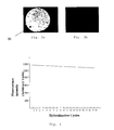
- FIG. 3 a and FIG. 3 b are fluorescence images of a semiconductor nanostructure having immobilized morpholinos thereon.
- FIG. 3 a is the fluorescence image after the morpholinos have hybridized with one or more target nucleic acids that had been labelled with a Cy-3 fluorescent marker.
- FIG. 3 b is the fluorescence image after the target nucleic acids of FIG. 3 a have been denatured and hence removed from the morpholinos.
- FIG. 4 is a graph showing the stability of the morpholinos-immobilized silicon surface after 20 cycles of hybridization and denaturation.
- FIG. 5 is a graph showing the PNA modified SiNW biosensor response to complementary, one-base pair mismatch and non-complementary PNA-DNA hybridization.
- FIG. 1 there is shown a schematic diagram of the process involved in immobilizing a polymer such as morpholino into the surface of a semiconductor nanostructure.
- This process is carried out in the absence of an oxide film on the surface of the semiconductor nanostructure.
- this process can be termed as an “oxide-free” process.
- morpholino is immobilized onto the surface of a silicon nanostructure.
- the process involves the steps of functionalizing the silicon surface with a functionalizing agent such as an amine-containing compound; covalently attaching one terminal end of a cross-linker to the exposed amine group of the functionalizing agent; and covalently attaching the morpholino to the exposed terminal end of the cross-linker.
- a functionalizing agent such as an amine-containing compound
- the silicon oxide coating of the silicon nanostructure is etched away by exposing the silicon nanostructure to BOE (a solution containing high concentration of HF acid) such that the silicon atoms present on the silicon surface form hydrogen-terminated silicon.
- BOE a solution containing high concentration of HF acid
- the hydrogen-terminated silicon is subsequently and almost immediately treated with 10-aminodec-1-ene functionalizing agent by covering the hydrogen-terminated silicon with a thin film of t-BOC protected 10-aminodec-1-ene and quickly transferring it to a nitrogen-purged Teflon reaction cell.
- the silicon nanostructures are illuminated with UV light. After UV illumination, the silicon nanostructures are rinsed and washed.
- the silicon nanostructure was treated to remove the protecting group from the functionalizing agent and to expose the amine group. Due to the presence of the exposed amine group, a cross-linker such as glutaraldehyde can bind to the functionalizing agent. The other end of the cross-linker is exposed to bind to the first polymer such as a morpholino.
- a cross-linker such as glutaraldehyde can bind to the functionalizing agent.
- the other end of the cross-linker is exposed to bind to the first polymer such as a morpholino.
- FIG. 2 there is shown a schematic diagram of a first polymer such as morpholino 12 and a second polymer such as morpholino 14 immobilized on a semiconductor nanostructure such as silicon nanowire 18 .
- Morpholino 14 is attached to the morpholino 12 and hybridized to a target nucleic acid 16 .
- morpholino 12 is capable of binding to morpholino 14 due to the presence of binding moieties on both morpholino 12 and morpholino 14 .
- the binding moieties of morpholino and morpholino 14 are depicted in FIG. 2 by dotted rectangles and represented by 12 a and 14 a , respectively.
- the binding moieties ( 12 a , 14 a ) have defined nucleic acid sequences that are complementary to each other such that morpholino 12 and morpholino 14 bind to each other via complemetary base pairing.
- the target sequence moiety 14 b has a specific nucleic acid sequence that is complementary to the nucleic acid sequence of target nucleic acid 16 such that morpholino 14 and target nucleic acid 16 hybridizes with each other via complementary base pairing.
- a change in the electrical current between electrodes ( 20 a , 20 b ) can be detected by the detector 22 .
- the hybridization of the target nucleic acid 16 to the morpholino 14 closes the electrical circuit between electrodes ( 20 a , 20 b ) such that an electric current can flow between the electrodes ( 20 a , 20 b ). This is attributed to the negatively charged nucleic acid, which closes the electrical circuit between the electrodes ( 20 a , 20 b ).
- the presence of an electric is current can be detected using a detector 22 which may be in the form of a voltmeter or an ammeter.
- an electrical current is generated between electrodes ( 20 a , 20 b ) before introduction of the target nucleic acid 16 .
- the negatively charged nucleic acid 16 acts as a resistance to the electrical flow between the electrodes ( 20 a , 20 b ).
- the electrical current between the electrodes ( 20 a , 20 b ) before and after hybridization of the target nucleic acid 16 to the morpholino 14 can be detected by the detector 22 , such as an ammeter or an ohm meter.
- the magnitude of the change in the electrical current before and after hybridization can be related to the amount of hybridized nucleic acid 16 to morpholino 14 .
- the target nucleic acid 16 can be labelled with a fluorescent marker (not shown in FIG. 2 ) such that the hybridized nucleic acid can be detected via fluorescent imaging.
- the nanostructures were then rinsed in chloroform (Sigma-Aldrich) for 15 minutes at 50° C. and then in methanol (Sigma-Aldrich) twice at room temperature (such as 25° C.) for 5 minutes each.
- the deprotection of t-BOC protected amine was carried out by immersing the nanostructure surface in 25% trifluoroacetic acid (TFA) (Sigma-Aldrich) in methylene chloride (Sigma-Aldrich) for 2 hours and subsequently treating the nanostructure surface with 10% aqueous ammonium hydroxide (Sigma-Aldrich) for 5 minutes followed by a rinse in pure H 2 O.
- TFA trifluoroacetic acid
- methylene chloride Sigma-Aldrich
- the nanostructures were treated with 1% glutaraldehyde (Sigma-Aldrich) in H 2 O for 1 hour and rinsed with pure H 2 O thereafter.
- 10 ⁇ M morpholino (each having a base sequence of 5′-NH 2 -AACCACACAACCTACTACCTCA-3′ (SEQ ID NO:1)) (Gene Tools, LLC (Philomath, Oreg., USA)) in 1 ⁇ Saline-Sodium Citrate (SSC) buffer was incubated with the surface in a humid atmosphere at room temperature overnight. After immobilization of the morpholino, the chips were washed three times with 1 ⁇ SSC buffer for 5 minutes during each wash. 1% bovine serum albumin (BSA) (Sigma-Aldrich) in phosphate buffer saline (PBS) was employed to block the aldehyde-activated surface outside the morpholino spot.
- BSA bovine serum albumin
- PBS phosphate buffer saline
- FIG. 3 a is the fluorescence image of the silicon nanostructure after the morpholinos have hybridized with the target DNA molecules that had been labeled with a Cy-3 fluorescent marker. As observed in FIG.
- FIG. 3 b is the fluorescence image after the target DNA molecules of FIG. 3 a have been denatured and hence removed from the morpholinos.
- FIG. 3 b shows that there are hardly any fluorescence spots in the image, demonstrating the efficient removal of the target DNA molecules from the silicon nanostructure and hence, the possibility of reusing the silicon nanostructure for further hybridization after the hybridized target nucleic acids had been removed.
- the hybridization of the DNA molecules to the silicon nanostructure such as silicon nanowire (SiNW) can be detected via measuring the change in resistance between two terminal electrodes of the silicon nanostructure before and after hybridization.
- the SiNW resistances are measured between the two terminals, source (S) and drain (D) electrodes with Alessi REL-6100 probe station (Cascade Microtech, Beaverton, Oreg.).
- the resistance change before and after hybridization of the DNA molecules to the morpholino probes is caused by the introduction of negatively charged DNA since the morpholino probe has a substantially non-ionic backbone and is hence, neutral.
- Hybridization was done in 1 ⁇ SSC buffer and the electrical measurement was carried out in 0.01 ⁇ SSC buffer.
- An acrylic reservoir is used to allow for fluid exchange for hybridization and measurement experiments. Fluid supply and return were carried out by Tygon tubes.
- a different SiNW chip was used for each individual measurement. At least 15 wires were measured each time and the mean of the resistance is utilized for data analysis.
- FIG. 4 there is shown a graph of the stability of the morpholinos-immobilized silicon surface after 20 cycles of hybridization and denaturation.
- FIG. 4 demonstrates that repeated hybridization-denaturation cycles do not substantially affect the sensitivity of the immobilized morpholino probes.
- the fluorescence intensity after 20 cycles is ⁇ 90% of the initial value, corresponding to a loss of 0.5% per cycle, indicating the high reusability potential of a Morpholino-based nucleic acid microarray sensor.
- PNA probe having a base sequence of 5′-NH 2 -AACCACACAACCTACTACCTCA-3′ (SEQ ID NO:3) (Eurogentec (Seraing, Belgium) is immobilized to SiNW using the method of Example 1.
- the SiNW resistances upon binding of target DNA to probe PNA are measured between the two terminals, source (S) and drain (D) electrodes with Alessi REL-6100 probe station (Cascade Microtech, Beaverton, Oreg.).
- S source
- D drain
- the resistance change before and after hybridization of the DNA molecules to the PNA probes is caused by the introduction of negatively charged DNA since the PNA probe has a substantially non-ionic backbone and is hence, neutral.
- Hybridization was done in 1 ⁇ SSC buffer and the electrical measurement was carried out in 0.01 ⁇ SSC buffer.
- An acrylic reservoir is used to allow for fluid exchange for hybridization and measurement experiments. Fluid supply and return were carried out by Tygon tubes.
- a different SiNW chip was used for each individual measurement. At least 15 wires were measured each time and the mean of the resistance is utilized for data analysis.
- FIG. 5 there is shown a graph depicting the change in resistance of electrical current flowing between the PNA-modified SiNW upon incubation with either fully matched, or one-based mismatched or non-complementary DNA.
- Incubation of the fully matched DNA to the PNA-modified SiNW results in a 51% increase in resistance change while incubation of non-complementary or one-base mismatched DNA results in negligible change in resistance.
- the results indicating the strong potential of PNA to be used as neutral charge nucleic acid probe for SiNW biosensor applications.
- the disclosed sensor can be used in a DNA microarray or in a gene chip such as a “lab-on-a chip” to determine the expression profiles of many genes at the same time.
- the disclosed sensor can be used in the fields of molecular biology and medicine for basic research, disease diagnostics and drug discovery.
- the disclosed sensor can detect the hybridization of target nucleic acids using a fluorescence detection method and/or an electrical detection method that does not include optical detection (thereby saving on complex optical detection systems). Accordingly, the disclosed sensor may be more versatile than conventional sensors, which use fluorescence detection methods because the disclosed sensor can employ more than one detection method to determine hybridization of target nucleic acids.
- the disclosed sensor or nucleic acid microarray can be re-used and can go through a number of hybridization-denaturation cycles without a substantial decrease in the sensitivity of the nucleic acid detection probes. This is compared to conventional microarrays where the fluorescence signal of the hybridized nucleic acids decreases by around 30% to 50% after three or more rounds of denaturation and re-hybridization, indicating a loss in the sensitivity of the detection probes.
- the disclosed sensor can be used to detect a number of nucleic acid sequences that are different from each other.
- the target sequence moiety of the second polymer can be changed as required in order to hybridize target nucleic acids having different nucleic acid sequences.
- the sensor can be used for a number of applications. This permits the development of a universal hybridization assay, which can be used for testing samples under similar experimental conditions. Therefore, this may permit the comparison of data from different experimental sets, leading to increased reliability because the different experimental sets are carried out under similar experimental conditions.
- the disclosed sensor does not require the use of DNA analogs as the detection probe. Instead, a polymer having a substantially non-ionic backbone is used as the detection probe in the sensor.
- a polymer having a substantially non-ionic backbone is used as the detection probe in the sensor.
- the morpholino imparts certain properties such as stability, nuclease-resistance, high efficacy, long-term activity, low-toxicity and high level of specificity to the sensor. Morpholinos are also cost-effective, which will reduce the cost involved with producing the disclosed sensor, particularly compared to sensors, which employ PNA.
- rigid backbone of morpholino allows easy access of target molecules compared with flexible backbone of DNA and PNA, which does not stand vertically on a surface, thus hampering the approach of target molecules, thereby reducing hybridization efficiency.
Abstract
Description


Claims (19)
Applications Claiming Priority (1)
| Application Number | Priority Date | Filing Date | Title |
|---|---|---|---|
| PCT/SG2008/000441 WO2010059126A1 (en) | 2008-11-20 | 2008-11-20 | Sensor for detection of nucleic acid |
Publications (2)
| Publication Number | Publication Date |
|---|---|
| US20120129709A1 US20120129709A1 (en) | 2012-05-24 |
| US8551767B2 true US8551767B2 (en) | 2013-10-08 |
Family
ID=42198370
Family Applications (1)
| Application Number | Title | Priority Date | Filing Date |
|---|---|---|---|
| US13/125,302 Expired - Fee Related US8551767B2 (en) | 2008-11-20 | 2008-11-20 | Sensor for detection of nucleic acid |
Country Status (3)
| Country | Link |
|---|---|
| US (1) | US8551767B2 (en) |
| GB (1) | GB2478446B8 (en) |
| WO (1) | WO2010059126A1 (en) |
Families Citing this family (5)
| Publication number | Priority date | Publication date | Assignee | Title |
|---|---|---|---|---|
| US9921182B2 (en) | 2014-10-06 | 2018-03-20 | ALVEO Technologies Inc. | System and method for detection of mercury |
| US10352899B2 (en) * | 2014-10-06 | 2019-07-16 | ALVEO Technologies Inc. | System and method for detection of silver |
| US10196678B2 (en) | 2014-10-06 | 2019-02-05 | ALVEO Technologies Inc. | System and method for detection of nucleic acids |
| US10627358B2 (en) * | 2014-10-06 | 2020-04-21 | Alveo Technologies, Inc. | Method for detection of analytes |
| WO2018057647A1 (en) | 2016-09-23 | 2018-03-29 | Alveo Technologies, Inc. | Methods and compositions for detecting analytes |
Citations (5)
| Publication number | Priority date | Publication date | Assignee | Title |
|---|---|---|---|---|
| US5378841A (en) | 1989-12-20 | 1995-01-03 | Antivirals Inc. | Alpha-morpholino ribonucleoside derivatives and polymers thereof |
| US6391558B1 (en) * | 1997-03-18 | 2002-05-21 | Andcare, Inc. | Electrochemical detection of nucleic acid sequences |
| US6451588B1 (en) * | 2000-06-30 | 2002-09-17 | Pe Corporation (Ny) | Multipartite high-affinity nucleic acid probes |
| US20060205013A1 (en) | 2005-01-20 | 2006-09-14 | Shim Jeo-Young | FET-type biosensor with surface modification |
| US20100035248A1 (en) * | 2007-06-15 | 2010-02-11 | Rastislav Levicky | Surface-based nucleic acid assays employing morpholinos |
-
2008
- 2008-11-20 WO PCT/SG2008/000441 patent/WO2010059126A1/en active Application Filing
- 2008-11-20 GB GB201107930A patent/GB2478446B8/en not_active Expired - Fee Related
- 2008-11-20 US US13/125,302 patent/US8551767B2/en not_active Expired - Fee Related
Patent Citations (5)
| Publication number | Priority date | Publication date | Assignee | Title |
|---|---|---|---|---|
| US5378841A (en) | 1989-12-20 | 1995-01-03 | Antivirals Inc. | Alpha-morpholino ribonucleoside derivatives and polymers thereof |
| US6391558B1 (en) * | 1997-03-18 | 2002-05-21 | Andcare, Inc. | Electrochemical detection of nucleic acid sequences |
| US6451588B1 (en) * | 2000-06-30 | 2002-09-17 | Pe Corporation (Ny) | Multipartite high-affinity nucleic acid probes |
| US20060205013A1 (en) | 2005-01-20 | 2006-09-14 | Shim Jeo-Young | FET-type biosensor with surface modification |
| US20100035248A1 (en) * | 2007-06-15 | 2010-02-11 | Rastislav Levicky | Surface-based nucleic acid assays employing morpholinos |
Non-Patent Citations (3)
| Title |
|---|
| Gao, Z. et al. "Silicon nanowire arrays for label-free detection of DNA," Analytical Chemistry, 2007, pp. 3291-3297, Vo. 79, No. 9. |
| Li, Z, et al., "Silicon nanowires for sequence-specific DNA sensing: device fabrication and simulation," Applied Physics A: Materials Science & Processing, 2005, pp. 1257-1263, vol. 80, No. 6. |
| Mackenzie, Susan, International Search Report, PCT/SG2008/000441, Australian Patent Office, Feb. 3, 2009. |
Also Published As
| Publication number | Publication date |
|---|---|
| GB201107930D0 (en) | 2011-06-22 |
| WO2010059126A1 (en) | 2010-05-27 |
| US20120129709A1 (en) | 2012-05-24 |
| GB2478446A8 (en) | 2013-08-21 |
| GB2478446B8 (en) | 2013-08-21 |
| GB2478446B (en) | 2013-06-26 |
| GB2478446A (en) | 2011-09-07 |
Similar Documents
| Publication | Publication Date | Title |
|---|---|---|
| Zhang et al. | Label-free direct detection of MiRNAs with silicon nanowire biosensors | |
| US20180217082A1 (en) | Versatile and sensitive biosensor | |
| US8283936B2 (en) | Nano-scale biosensors | |
| US20100194409A1 (en) | Method of electrically detecting a biological analyte molecule | |
| US8465926B2 (en) | Method and system for real time quantification and monitoring of nucleic acid amplification using electroconductive or electrochemically active labels | |
| US20090036315A1 (en) | Device and methods for detecting and quantifying one or more target agents | |
| US8551767B2 (en) | Sensor for detection of nucleic acid | |
| US20120058547A1 (en) | Method and system for nucleic acid detection using electroconductive or electrochemically active labels | |
| Zhang | Silicon nanowire biosensor for ultrasensitive and label-free direct detection of miRNAs | |
| EP2477031A2 (en) | Method for detecting and quantifying a target substance using a biochip | |
| Aoki et al. | 384-Channel electrochemical sensor array chips based on hybridization-triggered switching for simultaneous oligonucleotide detection | |
| JP2003090815A (en) | Method for electrochemically detecting gene, and nucleic acid tip | |
| JP2017060461A (en) | Partially neutral single-stranded oligonucleotide | |
| EP3262194B1 (en) | Device and method for detecting target molecules | |
| US8975025B2 (en) | Method and system for nucleic acid detection using electroconductive or electrochemically active labels | |
| WO2017123857A1 (en) | Single-particle bridge assay for amplification-free electrical detection of ultralow-concentration biomolecules and non-biological molecules | |
| Lin et al. | Recovery based nanowire field-effect transistor detection of pathogenic avian influenza DNA | |
| JP4626377B2 (en) | Target molecule detection method, gene polymorphism detection method, and substrate and kit used in these detection methods | |
| Luo et al. | Real time electrochemical monitoring of DNA/PNA dissociation by melting curve analysis | |
| EP3327147A1 (en) | Method for quantitatively profiling nucleic acids | |
| EP4148143A1 (en) | Polypurine reverse hoogsteen hairpins and parallel clamps and their use as biosensors | |
| US20040096859A1 (en) | Method for detecting and/or quantifying an analyte | |
| KR20230065838A (en) | Amplification or Detection of Target Material using Nanoparticles | |
| Bronder | Label-free detection of tuberculosis DNA with capacitive field-effect biosensors | |
| KR20090102021A (en) | Label-free methods and devices to detect target nucleic acids |
Legal Events
| Date | Code | Title | Description |
|---|---|---|---|
| AS | Assignment |
Owner name: AGENCY OF SCIENCE, TECHNOLOGY AND RESEARCH, SINGAP Free format text: ASSIGNMENT OF ASSIGNORS INTEREST;ASSIGNORS:ZHANG, GUOJUN;CHUA, HUIYI JAY;CHEE, RU ERN;AND OTHERS;REEL/FRAME:026730/0980 Effective date: 20080513 Owner name: BCI (BIOCHIP INNOVATIONS), AUSTRALIA Free format text: ASSIGNMENT OF ASSIGNORS INTEREST;ASSIGNOR:BARNARD, ROSS;REEL/FRAME:026731/0011 Effective date: 20090509 Owner name: SIMEMS PTE LTD, SINGAPORE Free format text: ASSIGNMENT OF ASSIGNORS INTEREST;ASSIGNOR:RAGHAVAN, UPPILI;REEL/FRAME:026731/0079 Effective date: 20080512 |
|
| REMI | Maintenance fee reminder mailed | ||
| LAPS | Lapse for failure to pay maintenance fees |
Free format text: PATENT EXPIRED FOR FAILURE TO PAY MAINTENANCE FEES (ORIGINAL EVENT CODE: EXP.) |
|
| STCH | Information on status: patent discontinuation |
Free format text: PATENT EXPIRED DUE TO NONPAYMENT OF MAINTENANCE FEES UNDER 37 CFR 1.362 |
|
| FP | Lapsed due to failure to pay maintenance fee |
Effective date: 20171008 |