US8420053B2 - Gastrin releasing peptide compounds - Google Patents
Gastrin releasing peptide compounds Download PDFInfo
- Publication number
- US8420053B2 US8420053B2 US12/551,394 US55139409A US8420053B2 US 8420053 B2 US8420053 B2 US 8420053B2 US 55139409 A US55139409 A US 55139409A US 8420053 B2 US8420053 B2 US 8420053B2
- Authority
- US
- United States
- Prior art keywords
- seq
- amino
- acid
- do3a
- monoamide
- Prior art date
- Legal status (The legal status is an assumption and is not a legal conclusion. Google has not performed a legal analysis and makes no representation as to the accuracy of the status listed.)
- Expired - Fee Related, expires
Links
- 0 CC(C)CC(C(*C(CCSC)C(N*)=O)=O)NC(C)C(CC1=C*C=N1)NC(CNC([C@](C(C)C)NC(C(C)NC([C@](CC1=C*C2=C1CCC=C2)NC(C(CCC(N)=O)NC(CCCCCCCNC(CCCCCCCN)=O)=O)=O)=O)=O)=O)=O Chemical compound CC(C)CC(C(*C(CCSC)C(N*)=O)=O)NC(C)C(CC1=C*C=N1)NC(CNC([C@](C(C)C)NC(C(C)NC([C@](CC1=C*C2=C1CCC=C2)NC(C(CCC(N)=O)NC(CCCCCCCNC(CCCCCCCN)=O)=O)=O)=O)=O)=O)=O 0.000 description 6
- GIOISYXDMIJBFE-NXSAUDPISA-N C.C.C.CC(C)N(C)C(CCC(N)=O)C(=O)C[C@@H](CC1=CNC2=C1C=CC=C2)C(=O)C(C)(C)C.CC(C)NC(CCC(N)=O)C(=O)C[C@@H](CC1=CNC2=C1C=CC=C2)C(=O)C(C)(C)C.CC(C)NC(CCC(N)=O)C(=S)C[C@@H](CC1=CNC2=C1C=CC=C2)C(=O)C(C)(C)C.CC(C)NC(CCC(N)=O)CC[C@@H](CC1=CNC2=C1C=CC=C2)C(=O)C(C)(C)C.CNC.[W].[W].[W].[W] Chemical compound C.C.C.CC(C)N(C)C(CCC(N)=O)C(=O)C[C@@H](CC1=CNC2=C1C=CC=C2)C(=O)C(C)(C)C.CC(C)NC(CCC(N)=O)C(=O)C[C@@H](CC1=CNC2=C1C=CC=C2)C(=O)C(C)(C)C.CC(C)NC(CCC(N)=O)C(=S)C[C@@H](CC1=CNC2=C1C=CC=C2)C(=O)C(C)(C)C.CC(C)NC(CCC(N)=O)CC[C@@H](CC1=CNC2=C1C=CC=C2)C(=O)C(C)(C)C.CNC.[W].[W].[W].[W] GIOISYXDMIJBFE-NXSAUDPISA-N 0.000 description 1
- CZJWHAKIXQGXPG-DYIPZYPMSA-N C.C=C.CC(C)NC(/C=C/[C@@H](CC1=CNC2=C1C=CC=C2)C(=O)C(C)(C)C)CCC(N)=O.CC(C)N[C@@](C)(CCC(N)=O)C(=O)C[C@@H](CC1=CNC2=C1C=CC=C2)C(=O)C(C)(C)C.[W].[W] Chemical compound C.C=C.CC(C)NC(/C=C/[C@@H](CC1=CNC2=C1C=CC=C2)C(=O)C(C)(C)C)CCC(N)=O.CC(C)N[C@@](C)(CCC(N)=O)C(=O)C[C@@H](CC1=CNC2=C1C=CC=C2)C(=O)C(C)(C)C.[W].[W] CZJWHAKIXQGXPG-DYIPZYPMSA-N 0.000 description 1
- ZUISORPFMVOYFA-XYJOYHRVSA-N C/C(=N\O)C(C)(C)NCC(CNC(C)(C)/C(C)=N/O)[Y]CC(C)C.C/C(=N\O)C(C)(C)NCCCNC(C)(C)/C(=N/O)[Y]CC(C)C.CC(=O)NCSC[C@H](NC(=O)[C@H](CO)NC(=O)CN(C)C)C(=O)C(C)(C)C.CN(C)CC(=O)NC(C(=O)NC(CS)C(=O)C(C)(C)C)C(C)(C)C Chemical compound C/C(=N\O)C(C)(C)NCC(CNC(C)(C)/C(C)=N/O)[Y]CC(C)C.C/C(=N\O)C(C)(C)NCCCNC(C)(C)/C(=N/O)[Y]CC(C)C.CC(=O)NCSC[C@H](NC(=O)[C@H](CO)NC(=O)CN(C)C)C(=O)C(C)(C)C.CN(C)CC(=O)NC(C(=O)NC(CS)C(=O)C(C)(C)C)C(C)(C)C ZUISORPFMVOYFA-XYJOYHRVSA-N 0.000 description 1
- QBKNJHMYMUFIMG-UHFFFAOYSA-N CC1CN1.CC1CO1.CN1C(=O)C=CC1=O Chemical compound CC1CN1.CC1CO1.CN1C(=O)C=CC1=O QBKNJHMYMUFIMG-UHFFFAOYSA-N 0.000 description 1
- VPYZCJCIIKLFPU-UHFFFAOYSA-N CN1C=CN=C1.CN1C=NC=N1.CN1C=NN=N1.COC(=O)OC(C)C.COC1=C(Cl)C(Cl)=C(Cl)C(Cl)=C1Cl.COC1=C(F)C(F)=C(F)C(F)=C1F.COC1=CC=C([N+](=O)[O-])C=C1.COC1=CC=C([N+](=O)[O-])C=C1[N+](=O)[O-].CON1C(=O)CCC1=O.CON1N=NC2=C(C=CC=C2)C1=O.CON1N=NC2=C1C=CC=C2.CON1N=NC2=C1N=CC=C2.CSC(C)(C)C.CSC1=C(F)C(F)=C(F)C(F)=C1F.CSC1=NC2=C(C=CC=C2)O1.CSC1=NC2=C(C=CC=C2)S1.CSC1=NC=CC=C1.CSC1=NN=C(C)S1.C[N-][N+]#N.F.[C-]#[N+]C1=C(C)C=C(C)N=C1SC Chemical compound CN1C=CN=C1.CN1C=NC=N1.CN1C=NN=N1.COC(=O)OC(C)C.COC1=C(Cl)C(Cl)=C(Cl)C(Cl)=C1Cl.COC1=C(F)C(F)=C(F)C(F)=C1F.COC1=CC=C([N+](=O)[O-])C=C1.COC1=CC=C([N+](=O)[O-])C=C1[N+](=O)[O-].CON1C(=O)CCC1=O.CON1N=NC2=C(C=CC=C2)C1=O.CON1N=NC2=C1C=CC=C2.CON1N=NC2=C1N=CC=C2.CSC(C)(C)C.CSC1=C(F)C(F)=C(F)C(F)=C1F.CSC1=NC2=C(C=CC=C2)O1.CSC1=NC2=C(C=CC=C2)S1.CSC1=NC=CC=C1.CSC1=NN=C(C)S1.C[N-][N+]#N.F.[C-]#[N+]C1=C(C)C=C(C)N=C1SC VPYZCJCIIKLFPU-UHFFFAOYSA-N 0.000 description 1
Images


Classifications
-
- C—CHEMISTRY; METALLURGY
- C07—ORGANIC CHEMISTRY
- C07K—PEPTIDES
- C07K7/00—Peptides having 5 to 20 amino acids in a fully defined sequence; Derivatives thereof
- C07K7/04—Linear peptides containing only normal peptide links
- C07K7/08—Linear peptides containing only normal peptide links having 12 to 20 amino acids
- C07K7/086—Bombesin; Related peptides
-
- A—HUMAN NECESSITIES
- A61—MEDICAL OR VETERINARY SCIENCE; HYGIENE
- A61K—PREPARATIONS FOR MEDICAL, DENTAL OR TOILETRY PURPOSES
- A61K51/00—Preparations containing radioactive substances for use in therapy or testing in vivo
- A61K51/02—Preparations containing radioactive substances for use in therapy or testing in vivo characterised by the carrier, i.e. characterised by the agent or material covalently linked or complexing the radioactive nucleus
- A61K51/04—Organic compounds
- A61K51/08—Peptides, e.g. proteins, carriers being peptides, polyamino acids, proteins
- A61K51/088—Peptides, e.g. proteins, carriers being peptides, polyamino acids, proteins conjugates with carriers being peptides, polyamino acids or proteins
-
- A—HUMAN NECESSITIES
- A61—MEDICAL OR VETERINARY SCIENCE; HYGIENE
- A61P—SPECIFIC THERAPEUTIC ACTIVITY OF CHEMICAL COMPOUNDS OR MEDICINAL PREPARATIONS
- A61P35/00—Antineoplastic agents
Definitions
- This invention relates to novel gastrin releasing peptide (GRP) compounds which are useful as diagnostic imaging agents or radiotherapeutic agents.
- GRP compounds are labeled with radionuclides or labels detectable by in vivo light imaging and include the use of novel linkers between the label and the targeting peptide, which provides for improved pharmacokinetics.
- radiopharmaceuticals e.g., diagnostic imaging agents, radiotherapeutic agents
- diagnostic imaging agents e.g., diagnostic imaging agents, radiotherapeutic agents
- radiotherapeutic agents e.g., diagnostic imaging agents, radiotherapeutic agents
- These newer radiopharmaceutical agents typically consist of a targeting agent connected to a metal chelator, which can be chelated to (e.g., complexed with) a diagnostic metal radionuclide such as, for example, technetium or indium, or a therapeutic metal radionuclide such as, for example, lutetium, yttrium, or rhenium.
- a diagnostic metal radionuclide such as, for example, technetium or indium
- a therapeutic metal radionuclide such as, for example, lutetium, yttrium, or rhenium.
- the role of the metal chelator is to hold (i.e., chelate) the metal radionuclide as the radiopharmaceutical agent is delivered to the desired site.
- a metal chelator which does not bind strongly to the metal radionuclide would render the radiopharmaceutical agent ineffective for its desired use since the metal radionuclide would therefore not reach its desired site.
- the role of the targeting agent is to direct the diagnostic agent, such as a radiopharmaceutical agent containing the metal radionuclide, to the desired site for detection or treatment.
- the targeting agent may include a protein, a peptide, or other macromolecule which exhibits a specific affinity for a given receptor.
- Other known targeting agents include monoclonal antibodies (MAbs), antibody fragments (F ab 's and (F ab ) 2 's), and receptor-avid peptides.
- MAbs monoclonal antibodies
- F ab 's and (F ab ) 2 's antibody fragments
- receptor-avid peptides receptor-avid peptides.
- GRP gastrin releasing peptide
- GRP-R gastrin releasing peptide receptors
- BBN bombesin
- tetradecapeptide 14 amino acid peptide isolated from frog skin which is an analogue of human GRP and which binds to GRP receptors with high specificity and with an affinity similar to GRP.
- Bombesin and GRP analogues may take the form of agonists or antagonists. Binding of GRP or BBN agonists to the GRP receptor increases the rate of cell division of these cancer cells and such agonists are internalized by the cell, while binding of GRP or BBN antagonists generally does not result in either internalization by the cell or increased rates of cell division. Such antagonists are designed to competitively inhibit endogenous GRP binding to GRP receptors and reduce the rate of cancer cell proliferation. See, e.g., Hoffken, K.; Peptides in Oncology II, Somatostatin Analogues and Bombesin Antagonists (1993), pp. 87-112.
- BBN or GRP analogues that are antagonists.
- Davis et al. Metabolic Stability and Tumor Inhibition of Bombesin/GRP Receptor Antagonists, Peptides, vol. 13, pp. 401-407, 1992.
- the drug In designing an effective compound for use as a diagnostic or therapeutic agent for cancer, it is important that the drug have appropriate in vivo targeting and pharmacokinetic properties. For example, it is preferable that for a radiopharmaceutical, the radiolabeled peptide have high specific uptake by the cancer cells (e.g., via GRP receptors). In addition, it is also preferred that once the radionuclide localizes at a cancer site, it remains there for a desired amount of time to deliver a highly localized radiation dose to the site.
- radiolabeled peptides that are cleared efficiently from normal tissues is also an important factor for radiopharmaceutical agents.
- biomolecules e.g., MAb, F ab or peptides
- metallic radionuclides via a chelate conjugation
- a large percentage of the metallic radionuclide in some chemical form can become “trapped” in either the kidney or liver parenchyma (i.e., is not excreted into the urine or bile).
- New and improved radiopharmaceutical and other diagnostic compounds which have improved pharmacokinetics and improved kidney excretion (i.e., lower retention of the radioactive metal in the kidney) have now been found for diagnostic imaging and therapeutic uses.
- diagnostic imaging rapid renal excretion and low retained levels of radioactivity are critical for improved images.
- radiotherapeutic use slower blood clearance to allow for higher tumor uptake and better tumor targeting with low kidney retention are critical.
- new and improved compounds for use in diagnostic imaging or radiotherapy include a chemical moiety capable of complexing a medically useful metal ion or radionuclide (metal chelator) attached to a GRP receptor targeting peptide by a linker or spacer group.
- these compounds include an optical label (e.g. a photolabel or other label detectable by light imaging, optoacoustical imaging or photoluminescence) attached to a GRP receptor targeting peptide by a linker or spacer group.
- compounds of the present invention may have the formula: M-N-O-P-G wherein M is the metal chelator (in the form complexed with a metal radionuclide or not), or the optical label, N-O-P is the linker, and G is the GRP receptor targeting peptide.
- the metal chelator M may be any of the metal chelators known in the art for complexing with a medically useful metal ion or radionuclide.
- Preferred chelators include DTPA, DOTA, DO3A, HP-DO3A, EDTA, TETA, EHPG, HBED, NOTA, DOTMA, TETMA, PDTA, TTHA, LICAM, MECAM, or peptide chelators, such as, for example, those discussed herein.
- the metal chelator may or may not be complexed with a metal radionuclide, and may include an optional spacer such as a single amino acid.
- Preferred metal radionuclides for scintigraphy or radiotherapy include 99m Tc, 51 Cr, 67 Ga, 68 Ga, 47 Sc, 51 Cr, 167 Tm, 141 Ce, 111 In, 168 Yb, 175 Yb, 140 La, 90 Y, 88 Y, 153 Sm, 166 Ho, 165 Dy, 166 Dy, 62 Cu, 64 Cu, 67 Cu, 97 Ru, 103 Ru, 186 Re, 188 Re, 203 Pb, 211 Bi, 212 Bi, 213 Bi, 214 Bi, 225 Ac, 105 Rh, 109 Pd, 117m Sn, 149 Pm, 161 Tb, 177 Lu, 198 Au and 199 Au.
- the preferred radionuclides include 64 Cu, 67 Ga, 68 Ga, 99m Tc, and 111 In, with 99m Tc, and 111 In being particularly preferred.
- the preferred radionuclides include 64 CU, 90 Y, 105 Rh, 111 In, 117m Sn, 149 Pm, 153 Sm, 161 Tb, 166 Dy, 166 Ho, 175 Yb, 177 Lu, 186/188 Re, and 199 Au, with 177 Lu and 90 Y being particularly preferred.
- a most preferred chelator used in compounds of the invention is 1-substituted 4,7,10-tricarboxymethyl 1,4,7,10 tetraazacyclododecane triacetic acid (DO3A).
- the optical label M may be any of various optical labels known in the art.
- Preferred labels include, without limitation, optical dyes, including organic chromophores or fluorophores, such as cyanine dyes light absorbing compounds, light reflecting and scattering compounds, and bioluminescent molecules.
- the linker N-O-P contains at least one non-alpha amino acid.
- the linker N-O-P contains at least one substituted bile acid.
- the linker N-O-P contains at least one non-alpha amino acid with a cyclic group.
- the GRP receptor targeting peptide may be GRP, bombesin or any derivatives or analogues thereof.
- the GRP receptor targeting peptide is a GRP or bombesin analogue which acts as an agonist.
- the GRP receptor targeting peptide is a bombesin agonist binding moiety disclosed in U.S. Pat. No. 6,200,546 and US 2002/0054855, incorporated herein by reference.
- a single or multi-vial kit that contains all of the components needed to prepare the diagnostic or therapeutic agents of the invention is provided in an exemplary embodiment of the present invention.
- a novel method for preparing a diagnostic imaging agent comprising the step of adding to an injectable imaging medium a substance containing the compounds of the present invention.
- a novel method of radiotherapy using the compounds of the invention is also provided, as is a novel method for preparing a radiotherapeutic agent comprising the step of adding to an injectable therapeutic medium a substance comprising a compound of the invention.
- FIG. 1A is a graphical representation of a series of chemical reactions for the synthesis of intermediate C ((3 ⁇ ,5 ⁇ )-3-(9H-Fluoren-9-ylmethoxy)aminocholan-24-oic acid), from A (Methyl-(3 ⁇ ,5 ⁇ )-3-aminocholan-24-ate) and B ((3 ⁇ ,5 ⁇ )-3-aminocholan-24-oic acid), as described in Example I.
- FIG. 1B is a graphical representation of the sequential reaction for the synthesis of N-[(3 ⁇ ,5 ⁇ )-3-[[[[[[4,7,10-Tris(carboxymethyl)-1,4,7,10-tetraazacyclododec-1-yl]acetyl]amino]acetyl]amino]cholan-24-yl]-L-glutaminyl-L-tryptophyl-L-alanyl-L-valyl-glycyl-L-histidyl-L-leucyl-L-methioninamide (L62), as described in Example I.
- FIG. 2A is a graphical representation of the sequential reaction for the synthesis of N-[4-[[[[[[4,7,10-Tris(carboxymethyl)-1,4,7,10-tetraazacyclododec-1-yl]acetyl]amino]acetyl]amino]benzoyl]-L-glutaminyl-L-tryptophyl-L-alanyl-L-valyl-glycyl-L-histidyl-L-leucyl-L-methioninamide (L70), as described in Example II.
- FIG. 2B is a general graphical representation of the sequential reaction for the synthesis of N-[4-[2-[[[4,7,10-Tris(carboxymethyl)-1,4,7,10-tetraazacyclododec-1-yl]acetyl]amino]ethoxy]benzoyl]-L-glutaminyl-L-tryptophyl-L-alanyl-L-valyl-glycyl-L-histidyl-L-leucyl-L-methioninamide (L73), N-[3-[[[[[4,7,10-Tris(carboxymethyl)-1,4,7,10-tetraazacyclododec-1-yl]acetyl]amino]methyl]benzoyl]-L-glutaminyl-L-tryptophyl-L-alanyl-L-valyl-glycyl-L-histidyl-L-le
- FIG. 2C is a chemical structure of the linker used in the synthesis reaction of FIG. 2B for synthesis of N-[4-[2-[[[4,7,10-Tris(carboxymethyl)-1,4,7,10-tetraazacyclododec-1-yl]acetyl]amino]ethoxy]benzoyl]-L-glutaminyl-L-tryptophyl-L-alanyl-L-valyl-glycyl-L-histidyl-L-leucyl-L-methioninamide (L73), as described in Example II.
- FIG. 2D is a chemical structure of the linker used in the synthesis reaction of FIG. 2B for synthesis of N-[3-[[[[4,7,10-Tris(carboxymethyl)-1,4,7,10-tetraazacyclododec-1-yl]acetyl]amino]methyl]benzoyl]-L-glutaminyl-L-tryptophyl-L-alanyl-L-valyl-glycyl-L-histidyl-L-leucyl-L-methioninamide (L115), as described in Example II.
- FIG. 2E is a chemical structure of the linker used in the synthesis reaction of FIG. 2B for synthesis of N-[4-[[[[4,7,10-Tris(carboxymethyl)-1,4,7,10-tetraazacyclododec-1-yl]acetyl]amino]methyl]phenylacetyl]-L-glutaminyl-L-tryptophyl-L-alanyl-L-valyl-glycyl-L-histidyl-L-leucyl-L-methioninamide (L116), as described in Example II.
- FIG. 2F is a graphical representation of the sequential reaction for the synthesis of N-[[4,7,10-Tris(carboxymethyl)-1,4,7,10-tetraazacyclododec-1-yl]acetyl]glycyl-4-piperidinecarbonyl-L-glutaminyl-L-tryptophyl-L-alanyl-L-valyl-glycyl-L-histidyl-L-leucyl-L-methioninamide (L74), as described in Example II.
- FIG. 3A is a graphical representation of a series of chemical reactions for the synthesis of intermediate (3 ⁇ ,5 ⁇ )-3-[[(9H-Fluoren-9-ylmethoxy)amino]acetyl]amino-12-oxocholan-24-oic acid (C), as described in Example III.
- FIG. 3B is a graphical representation of the sequential reaction for the synthesis of N-[(3 ⁇ ,5 ⁇ )-3-[[[[[[4,7,10-Tris(carboxymethyl)-1,4,7,10-tetraazacyclododec-1-yl]acetyl]amino]acetyl]amino]-12,24-dioxocholan-24-yl]-L-glutaminyl-L-tryptophyl-L-alanyl-L-valyl-glycyl-L-histidyl-L-leucyl-L-methioninamide (L67), as described in Example III.
- FIG. 3C is a chemical structure of (3 ⁇ ,5 ⁇ )-3-Amino-12-oxocholan-24-oic acid (B), as described in Example III.
- FIG. 3D is a chemical structure of (3 ⁇ ,5 ⁇ )-3-[[(9H-Fluoren-9-ylmethoxy)amino]acetyl]amino-12-oxocholan-24-oic acid (C), as described in Example III.
- FIG. 3E is a chemical structure of N-[(3 ⁇ ,5 ⁇ )-3-[[[[[[4,7,10-Tris(carboxymethyl)-1,4,7,10-tetraazacyclododec-1-yl]acetyl]amino]acetyl]amino]-12,24-dioxocholan-24-yl]-L-glutaminyl-L-tryptophyl-L-alanyl-L-valyl-glycyl-L-histidyl-L-leucyl-L-methioninamide (L67), as described in Example III.
- FIG. 4A is a graphical representation of a sequence of reactions to obtain intermediates (3 ⁇ ,5 ⁇ ,12 ⁇ )-3-[[(9H-Fluoren-9-ylmethoxy)amino]acetyl]amino-12-hydroxycholan-24-oic acid (3a) and (3 ⁇ ,5 ⁇ ,7 ⁇ ,12 ⁇ )-3-[[(9H-Fluoren-9-ylmethoxy)amino]acetyl]amino-7,12-dihydroxycholan-24-oic acid (3b), as described in Example IV.
- FIG. 4B is a graphical representation of the sequential reaction for the synthesis of N-[(3 ⁇ ,5 ⁇ ,12 ⁇ )-3-[[[[[4,7,10-Tris(carboxymethyl)-1,4,7,10-tetraazacyclododec-1-yl]acetyl]amino]acetyl]amino]-12-hydroxy-24-oxocholan-24-yl]-L-glutaminyl-L-tryptophyl-L-alanyl-L-valyl-glycyl-L-histidyl-L-leucyl-L-methioninamide (L63), as described in Example IV.
- FIG. 4C is a graphical representation of the sequential reaction for the synthesis of N-[(3 ⁇ ,5 ⁇ ,7 ⁇ ,12 ⁇ )-3-[[[[[4,7,10-Tris(carboxymethyl)-1,4,7,10-tetraazacyclododec-1-yl]acetyl]amino]acetyl]amino]-7,12-dihydroxy-24-oxocholan-24-yl]-L-glutaminyl-L-tryptophyl-L-alanyl-L-valyl-glycyl-L-histidyl-L-leucyl-L-methioninamide (L64), as described in Example IV.
- FIG. 4D is a chemical structure of (3 ⁇ ,5 ⁇ ,7 ⁇ ,12 ⁇ )-3-amino-7,12-dihydroxycholan-24-oic acid (2b), as described in Example IV.
- FIG. 4E is a chemical structure of (3 ⁇ ,5 ⁇ ,12 ⁇ )-3-[[(9H-Fluoren-9-ylmethoxy)amino]acetyl]amino-12-hydroxycholan-24-oic acid (3a), as described in Example IV;
- FIG. 4F is a chemical structure of (3 ⁇ ,5 ⁇ ,7 ⁇ ,12 ⁇ )-3-[[(9H-Fluoren-9-ylmethoxy)amino]acetyl]amino-7,12-dihydroxycholan-24-oic acid (3b), as described in Example IV.
- FIG. 4G is a chemical structure of N-[(3 ⁇ ,5 ⁇ ,12 ⁇ )-3-[[[[[[4,7,10-Tris(carboxymethyl)-1,4,7,10-tetraazacyclododec-1-yl]acetyl]amino]acetyl]amino]-12-hydroxy-24-oxocholan-24-yl]-L-glutaminyl-L-tryptophyl-L-alanyl-L-valyl-glycyl-L-histidyl-L-leucyl-L-methioninamide (L63), as described in Example IV.
- FIG. 4H is a chemical structure of N-[(3 ⁇ ,5 ⁇ ,7 ⁇ ,12 ⁇ )-3-[[[[[4,7,10-Tris(carboxymethyl)-1,4,7,10-tetraazacyclododec-1-yl]acetyl]amino]acetyl]amino]-7,12-dihydroxy-24-oxocholan-24-yl]-L-glutaminyl-L-tryptophyl-L-alanyl-L-valyl-glycyl-L-histidyl-L-leucyl-L-methioninamide (L64), as described in Example IV.
- FIG. 5A is a general graphical representation of the sequential reaction for the synthesis of 4-[[[[4,7,10-Tris(carboxymethyl)-1,4,7,10-tetraazacyclododec-1-yl]acetyl]amino]methyl]benzoyl-L-glutaminyl-L-tryptophyl-L-alanyl-L-valyl-glycyl-L-histidyl-L-leucyl-L-methioninamide (L71); and Trans-4-[[[[[4,7,10-tris(carboxymethyl)-1,4,7,10-tetraazacyclododec-1-yl]acetyl]amino]methyl]cyclohexylcarbonyl-L-glutaminyl-L-tryptophyl-L-alanyl-L-valyl-glycyl-L-histidyl-L-leucyl-
- FIG. 5B is a chemical structure of the linker used in compound L71 as shown in FIG. 5A and as described in Example V.
- FIG. 5C is a chemical structure of the linker used in compound L72 as shown in FIG. 5A and as described in Example V.
- FIG. 5D is a chemical structure of Rink amide resin functionalised with bombesin[7-14] (B), as described in Example V.
- FIG. 5E is a chemical structure of Trans-4-[[[(9H-fluoren-9-ylmethoxy)carbonyl]amino]methyl]cyclohexanecarboxylic acid (D), as described in Example V;
- FIG. 6A is a graphical representation of a sequence of reactions for the synthesis of intermediate linker 2-[[[9H-Fluoren-9-ylmethoxy)carbonyl]amino]methyl]benzoic acid (E), as described in Example VI.
- FIG. 6B is a graphical representation of a sequence of reactions for the synthesis of intermediate linker 4-[[[9H-Fluoren-9-ylmethoxy)carbonyl]amino]methyl]-3-nitrobenzoic acid (H), as described in Example VI.
- FIG. 6C is a graphical representation of the synthesis of N-[2-[[[[4,7,10-Tris(carboxymethyl)-1,4,7,10-tetraazacyclododec-1-yl]acetyl]amino]methyl]benzoyl]-L-glutaminyl-L-tryptophyl-L-alanyl-L-valyl-glycyl-L-histidyl-L-leucyl-L-methioninamide (L75), as described in Example VI.
- FIG. 6D is a graphical representation of the synthesis of N-[4-[[[[4,7,10-Tris(carboxymethyl)-1,4,7,10-tetraazacyclododec-1-yl]acetyl]amino]methyl]-3-nitrobenzoyl]-L-glutaminyl-L-tryptophyl-L-alanyl-L-valyl-glycyl-L-histidyl-L-leucyl-L-methioninamide (L76), as described in Example VI.
- FIG. 7A is a graphical representation of a sequence of reactions for the synthesis of intermediate linker [4-[[[9H-Fluoren-9-ylmethoxy)carbonyl]amino]methyl]phenoxy]acetic acid (E), as described in Example VII.
- FIG. 7B is a graphical representation of the synthesis of N-[[4-[[[[4,7,10-Tris(carboxymethyl)-1,4,7,10-tetraazacyclododec-1-yl]acetyl]amino]methyl]phenoxy]acetyl]-L-glutaminyl-L-tryptophyl-L-alanyl-L-valyl-glycyl-L-histidyl-L-leucyl-L-methioninamide (L124), as described in Example VII.
- FIG. 7C is a chemical structure of N-[[4-[[[[4,7,10-Tris(carboxymethyl)-1,4,7,10-tetraazacyclododec-1-yl]acetyl]amino]methyl]phenoxy]acetyl]-L-glutaminyl-L-tryptophyl-L-alanyl-L-valyl-glycyl-L-histidyl-L-leucyl-L-methioninamide (L124), as described in Example VII.
- FIG. 8A is a graphical representation of a sequence of reactions for the synthesis of intermediate 4-[[[9H-Fluoren-9-ylmethoxy)carbonyl]amino]methyl]-3-methoxybenzoic acid (E), as described in Example VIII.
- FIG. 8B is a graphical representation of the synthesis of N-[4-[[[[4,7,10-Tris(carboxymethyl)-1,4,7,10-tetraazacyclododec-1-yl]acetyl]amino]methyl]-3-methoxybenzoyl]-L-glutaminyl-L-tryptophyl-L-alanyl-L-valyl-glycyl-L-histidyl-L-leucyl-L-methioninamide, (L125), as described in Example VIII.
- FIG. 8C is a chemical structure of N-[4-[[[[4,7,10-Tris(carboxymethyl)-1,4,7,10-tetraazacyclododec-1-yl]acetyl]amino]methyl]-3-methoxybenzoyl]-L-glutaminyl-L-tryptophyl-L-alanyl-L-valyl-glycyl-L-histidyl-L-leucyl-L-methioninamide, (L125), as described in Example VIII.
- FIG. 9A is a graphical representation of a reaction for the synthesis of 3-[[[(9H-Fluoren-9-ylmethoxy)carbonyl]amino]acetyl]aminobenzoic acid, (B), as described in Example IX.
- FIG. 9B is a graphical representation of a reaction for the synthesis of 6-[[[(9H-Fluoren-9-ylmethoxy)carbonyl]amino]acetyl]aminonaphthoic acid (C), as described in Example IX.
- FIG. 9C is a graphical representation of a reaction for the synthesis of 4-[[[[[(9H-Fluoren-9-ylmethoxy)carbonyl]amino]acetyl]methylamino]benzoic acid, (D), as described in Example IX.
- FIG. 9D is a graphical representation of a reaction for the synthesis of N-[4-[[[[[[4,7,10-Tris(carboxymethyl)-1,4,7,10-tetraazacyclododec-1-yl]acetyl]amino]acetyl]amino]phenylacetyl]-L-glutaminyl-L-tryptophyl-L-alanyl-L-valyl-glycyl-L-histidyl-L-leucyl-L-methioninamide, (L146); N-[3-[[[[[[4,7,10-Tris(carboxymethyl)-1,4,7,10-tetraazacyclododec-1-yl]acetyl]amino]acetyl]amino]benzoyl]-L-glutaminyl-L-tryptophyl-L-alanyl-L-valyl-gly
- FIG. 10A is a graphical representation of a reaction for the synthesis of 7-[[Bis(1,1-dimethylethoxy)phosphinyl]methyl]-1,4,7,10-tetraazacyclododecane-1,4,10-triacetic acid 4,10-bis(1,1-dimethylethyl) ester H, as described in Example X.
- FIG. 10B is a graphical representation of a reaction for the synthesis of N-[4-[[[[[[4,10-Bis(carboxymethyl)-7-(dihydroxyphosphinyl)methyl-1,4,7,10-tetraazacyclododec-1-yl]acetyl]amino]acetyl]amino]benzoyl]-L-glutaminyl-L-tryptophyl-L-alanyl-L-valyl-glycyl-L-histidyl-L-leucil-L-methioninamide, (L237), as described in Example X.
- FIG. 11A is a graphical representation of a reaction for the synthesis of N,N-Dimethylglycyl-L-serinyl-[S-[(acetylamino)methyl]]-L-cysteinyl-glycyl-4-aminobenzoyl-L-glutaminyl-L-tryptophyl-L-alanyl-L-valyl-glycyl-L-histidyl-L-leucyl-L-methioninamide (L238), as described in Example XI.
- FIG. 11B is a graphical representation of a reaction for the synthesis of N,N-Dimethylglycyl-L-serinyl-[S-[(acetylamino)methyl]]-L-cysteinyl-glycyl-(3 ⁇ ,5 ⁇ ,7 ⁇ ,12 ⁇ )-3-amino-7,12-dihydroxy-24-oxocholan-24-yl-L-glutaminyl-L-tryptophyl-L-alanyl-L-valyl-glycyl-L-histidyl-L-leucyl-L-methioninamide, (L239), as described in Example XI.
- FIG. 12A is a graphical representation of a reaction for the synthesis of 4-[[[(9H-Fluoren-9-ylmethoxy)carbonyl]amino]acetyl]amino-3-methoxybenzoic acid (A), as described in Example XII.
- FIG. 12B is a graphical representation of a reaction for the synthesis of 4-[[[(9H-Fluoren-9-ylmethoxy)carbonyl]amino]acetyl]amino-3-chlorobenzoic acid, (D), as described in Example XII.
- FIG. 12C is a graphical representation of a reaction for the synthesis of 4-[[[(9H-Fluoren-9-ylmethoxy)carbonyl]amino]acetyl]amino-3-methylbenzoic acid (E), as described in Example XII.
- FIG. 12D is a chemical structure of N-[4-[[[[4,7,10-Tris(carboxymethyl)-1,4,7,10-tetraazacyclododec-1-yl]acetyl]glycyl]amino]-3-methoxybenzoyl]-L-glutaminyl-L-tryptophyl-1-alanyl-L-valyl-glycyl-L-histidyl-L-leucyl-L-methioninamide (L240) as described in Example XII.
- FIG. 12E is a chemical structure of compound N-[4-[[[[4,7,10-Tris(carboxymethyl)-1,4,7,10tetraazacyclododec-1-yl]acetyl]glycyl]amino]3-chlorobenzoyl]L-glutaminyl-L-tryptophyl-1-alanyl-L-valyl-glycyl-L-histidyl-L-leucyl-L-methioninamide, (L241) as described in Example XII.
- FIG. 12F is a chemical structure of N-[4-[[[[4,7,10-Tris(carboxymethyl)-1,4,7,10tetraazacyclododec-1-yl]acetyl]glycyl]amino]3-methylbenzoyl]L-glutaminyl-L-tryptophyl-1-alanyl-L-valyl-glycyl-L-histidyl-leucyl-L-methioninamide (L242), as described in Example XII.
- FIG. 13A is a graphical representation of a reaction for the synthesis of 4-[N,N′-Bis[2-[(9-H-fluoren-9-ylmethoxy)carbonyl]aminoethyl]amino]-4-oxobutanoic acid, (D), as described in Example XIII.
- FIG. 13B is a graphical representation of a reaction for the synthesis of N-[4-[[4-[Bis[2-[[[4,7,10-tris(carboxymethyl)-1,4,7,10-tetraazacyclododec-1-yl]acetyl]amino]ethyl]amino-1,4-dioxobutyl]amino]benzoyl]-L-glutaminyl-L-tryptophyl-L-alanyl-L-valyl-glycyl-L-histidyl-L-leucyl-L-methioninamide, (L244), as described in Example XIII.
- FIG. 13C is a chemical structure of compound L244, as described in Example XIII.
- FIG. 14A and FIG. 14B are graphical representations of the binding and competition curves described in Example XLIII.
- FIG. 15A is a graphical representation of the results of radiotherapy experiments described in Example LV.
- FIG. 15B is a graphical representation of the results of other radiotherapy experiments described in Example LV.
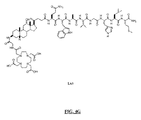
- FIG. 16 is a chemical structure of N-[4-[[[[4,7,10-Tris(carboxymethyl)-1,4,7,10 tetraazacyclododec-1-yl]acetyl]glycyl]amino]-L-Lysinyl-(3,6,9)-trioxaundecane-1,11-dicarboxylic acid-3,7-dideoxy-3-aminocholic acid)-L-arginyl-L-glutaminyl-L-triptophyl-L-alanyl-L-valyl-glycyl-L-histidyl-L-leucyl-L-methioninamide (L65).
- FIG. 17 is a chemical structure of N-[2-S-[[[[[[12 ⁇ -Hydroxy-17 ⁇ -(1-methyl-3-carboxypropyl)etiocholan-3 ⁇ -carbamoylmethoxyethoxyethoxyacetyl]-amino-6-[4,7,10-tris(carboxymethyl)-1,4,7,10-tetraazacyclododec-1-yl]acetyl]amino]acetyl]amino]hexanoyl-L-glutaminyl-L-tryptophyl-L-alanyl-L-valyl-glycyl-L-histidyl-L-leucyl-L-methioninamide (L66).
- FIG. 18A is a chemical structure of N-[4-[[[[[[4,7,10-Tris(carboxymethyl)-1,4,7,10-tetraazacyclododec-1-yl]acetyl]amino]acetyl]amino]benzoyl]-L-glutaminyl-L-tryptophyl-L-alanyl-L-valyl-glycyl-L-histidyl-L-leucyl-L-methioninamide (L70).
- FIG. 18B is a chemical structure N-[4-[[[[[[4,7,10-Tris(carboxymethyl)-1,4,7,10-tetraazacyclododec-1-yl]-3-carboxypropionyl]amino]acetyl]amino]benzoyl]-L-glutaminyl-L-tryptophyl-L-alanyl-L-valyl-glycyl-L-histidyl-L-leucyl-L-methioninamide (L114).
- FIG. 18C is a chemical structure N-[4-[[4,7,10-Tris(carboxymethyl)-1,4,7,10-tetraazacyclododec-1-yl]-2-hydroxy-3-propoxy]benzoyl]-L-glutaminyl-L-tryptophyl-L-alanyl-L-valyl-glycyl-L-histidyl-L-leucyl-L-methioninamide (L144).
- FIG. 18D is a chemical structure N-[(3 ⁇ ,5 ⁇ ,7 ⁇ ,12 ⁇ )-3-[[[[[[[4,7,10-Tris(carboxymethyl)-1,4,7,10-tetraazacyclododec-1-yl]acetyl]amino]ethoxyethoxy]acetyl]amino]-7,12-dihydroxycholan-24-yl]-L-glutaminyl-L-tryptophyl-L-alanyl-L-valyl-glycyl-L-histidyl-L-leucyl-L-methioninamine (L69).
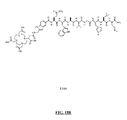
- FIG. 18E is a chemical structure of N-[4-[[[[[[4,7,10-Tris(carboxymethyl)-1,4,7,10-tetraazacyclododec-1-yl]acetyl]amino]acetyl]amino]phenylacetyl]-L-glutaminyl-L-tryptophyl-L-alanyl-L-valyl-glycyl-L-histidyl-L-leucyl-L-methioninamide (L146).
- FIG. 19 discloses chemical structures of intermediates which may be used to prepare compounds L64 and L70 as described in Example LVI.
- FIG. 20 is a graphical representation of the preparation of L64 using segment coupling as described in Example LVI.
- FIG. 21 is a graphical representation of the preparation of (1R)-1-(Bis ⁇ 2-[bis(carboxymethyl)amino]ethyl ⁇ amino)propane-3-carboxylic acid-1-carboxyl-glycyl-4-aminobenzoyl-L-glutaminyl-L-tryptophyl-L-alanyl-L-valyl-glycyl-L-histidyl-L-leucyl-L-methioninamide (L201).
- FIG. 22A is a graphical representation of chemical structure of chemical intermediates used to prepare L202.
- FIG. 22B is a graphical representation of the preparation of N-[(3 ⁇ ,5 ⁇ ,12 ⁇ )-3-[[[[[[4,7,10-Tris(carboxymethyl)-1,4,7,10-tetraazacyclododec-1-yl]acetyl]amino]acetyl]amino]-4-hydrazinobenzoyl-L-glutaminyl-L-tryptophyl-L-alanyl-L-valyl-glycyl-L-histidyl-L-leucyl-L-methioninamide (L202).
- FIG. 23A is a graphical representation of chemical structure of chemical intermediates used to prepare L203.
- FIG. 23B is a graphical representation of the preparation of N-[(3 ⁇ ,5 ⁇ ,12 ⁇ )-3-[[[4,7,10-Tris(carboxymethyl)-1,4,7,10-tetraazacyclododec-1-yl]acetyl]amino]-4-aminobenzoyl-L-glutaminyl-L-tryptophyl-L-alanyl-L-valyl-glycyl-L-histidyl-L-leucyl-L-methioninamide (L203).
- FIG. 24 is a graphical representation of the preparation of N-[(3 ⁇ ,5 ⁇ ,12 ⁇ )-3-[[[4,7,10-Tris(carboxymethyl)-1,4,7,10-tetraazacyclododec-1-yl]acetyl]amino]-4-aminobenzoyl-glycyl-L-glutaminyl-L-tryptophyl-L-alanyl-L-valyl-glycyl-L-histidyl-L-leucyl-L-methioninamide (L204).
- FIG. 25 is a graphical representation of the preparation of N-[(3 ⁇ ,5 ⁇ ,12 ⁇ )-3-[[[4,7,10-Tris(carboxymethyl)-1,4,7,10-tetraazacyclododec-1-yl]acetyl]amino]-4-aminobenzoyl-glycyl-L-glutaminyl-L-tryptophyl-L-alanyl-L-valyl-glycyl-L-histidyl-L-leucyl-L-methioninamide (L205).
- FIG. 26A is a graphical representation of chemical structures of chemical intermediates used to prepare L206.
- FIG. 26B is a graphical representation of the preparation of N-[(3 ⁇ ,5 ⁇ ,12 ⁇ )-3-[[[[[4,7,10-Tris(carboxymethyl)-1,4,7,10-tetraazacyclododec-1-yl]acetyl]amino]acetyl]amino]-[4′-Amino-2′-methyl biphenyl-4-carboxyl]-L-glutaminyl-L-tryptophyl-L-alanyl-L-valyl-glycyl-L-histidyl-L-leucyl-L-methioninamide (L206).
- FIG. 27A is a graphical representation of chemical structures of chemical intermediates used to prepare L207.
- FIG. 27B is a graphical representation of the preparation of N-[(3 ⁇ ,5 ⁇ ,12 ⁇ )-3-[[[[[4,7,10-Tris(carboxymethyl)-1,4,7,10-tetraazacyclododec-1-yl]acetyl]amino]acetyl]amino]-[3′-amino-biphenyl-3-carboxyl]-L-glutaminyl-L-tryptophyl-L-alanyl-L-valyl-glycyl-L-histidyl-L-leucyl-L-methioninamide (L207).
- FIG. 28 is a graphical representation of the preparation of N-[(3 ⁇ ,5 ⁇ ,12 ⁇ )-3-[[[[[4,7,10-Tris(carboxymethyl)-1,4,7,10-tetraazacyclododec-1-yl]acetyl]amino]acetyl]amino]-[1,2-d]aminoethyl-terephthalyl]-L-glutaminyl-L-tryptophyl-L-alanyl-L-valyl-glycyl-L-histidyl-L-leucyl-L-methioninamide (L208).
- FIG. 29A is a graphical representation of chemical structures of chemical intermediates used to prepare L209.
- FIG. 29B is a graphical representation of the preparation of L209.
- FIG. 30A is a graphical representation of chemical structures of chemical intermediates used to prepare L210.
- FIG. 30B is a chemical structure of L210.
- FIG. 31 is a chemical structure of N-[(3 ⁇ ,5 ⁇ ,12 ⁇ )-3-[[[4,7,10-Tris(carboxymethyl)-1,4,7,10-tetraazacyclododec-1-yl]acetyl]amino]-glycyl-glycyl-4-aminobenzoyl-L-glutaminyl-L-tryptophyl-L-alanyl-L-valyl-glycyl-L-histidyl-L-leucyl-L-methioninamide L211.
- FIG. 32 is a chemical structure of N-[(3 ⁇ ,5 ⁇ ,12 ⁇ )-3-[[[4,7,10-Tris(carboxymethyl)-1,4,7,10-tetraazacyclododec-1-yl]acetyl]amino]-glycyl-4-aminobenzoyl-L-glutamyl-L-tryptophyl-L-alanyl-L-valyl-glycyl-L-histidyl-L-leucyl-L-methioninamide L212.
- FIG. 33 is a chemical structure of N-[(3 ⁇ ,5 ⁇ ,12 ⁇ )-3-[[[4,7,10-Tris(carboxymethyl)-1,4,7,10-tetraazacyclododec-1-yl]acetyl]amino]-glycyl-4-aminobenzoyl-L-glutaminyl-L-tryptophyl-L-alanyl-L-valyl-glycyl-L-histidyl-L-leucyl-L-methionine carboxylate L213.
- FIG. 34 is a chemical structure of N-[(3 ⁇ ,5 ⁇ ,12 ⁇ )-3-[[[4,7,10-Tris(carboxymethyl)-1,4,7,10-tetraazacyclododec-1-yl]acetyl]amino]-glycyl-4-aminobenzoyl-D-phenylalanyl-L-glutaminyl-L-tryptophyl-L-alanyl-L-valyl-glycyl-L-histidyl-L-leucyl-L-methioninamide L214.
- FIG. 35 is a chemical structure of N-[(3 ⁇ ,5 ⁇ ,12 ⁇ )-3-[[[4,7,10-Tris(carboxymethyl)-1,4,7,10-tetraazacyclododec-1-yl]acetyl]amino]-glycyl-4-aminobenzoyl-L-glutaminyl-L-arginyl-L-leucyl-glycyl-L-asparginyl-L-glutaminyl-L-tryptophyl-L-alanyl-L-valyl-glycyl-L-histidyl-L-leucyl-L-methioninamide L215.
- FIG. 36 is a chemical structure of N-[(3 ⁇ ,5 ⁇ ,12 ⁇ )-3-[[[4,7,10-Tris(carboxymethyl)-1,4,7,10-tetraazacyclododec-1-yl]acetyl]amino]-glycyl-4-aminobenzoyl-L-glutaminyl-arginyl-L-tyrosinyl-glycyl-L-asparginyl-L-glutaminyl-L-tryptophyl-L-alanyl-L-valyl-glycyl-L-histidyl-L-leucyl-L-methioninamide L216.
- FIG. 37 is a chemical structure of N-[(3 ⁇ ,5 ⁇ ,12 ⁇ )-3-[[[4,7,10-Tris(carboxymethyl)-1,4,7,10-tetraazacyclododec-1-yl]acetyl]amino]-glycyl-4-aminobenzoyl-L-glutaminyl-L-lysyl-L-tyrosinyl-glycyl-L-glutaminyl-L-tryptophyl-L-alanyl-L-valyl-glycyl-L-histidyl-L-leucyl-L-methioninamide L217.
- FIG. 38 is a chemical structure of L218.
- FIG. 39 is a chemical structure of N-[(3 ⁇ ,5 ⁇ ,12 ⁇ )-3-[[[4,7,10-Tris(carboxymethyl)-1,4,7,10-tetraazacyclododec-1-yl]acetyl]amino]-glycyl-4-aminobenzoyl-D-phenylalanyl-L-glutaminyl-L-tryptophyl-L-alanyl-L-valyl-glycyl-L-histidyl-L-leucyl-aminopentyl, L219.
- FIG. 40 is a chemical structure of N-[(3 ⁇ ,5 ⁇ ,12 ⁇ )-3-[[[4,7,10-Tris(carboxymethyl)-1,4,7,10-tetraazacyclododec-1-yl]acetyl]amino]-glycyl-4-aminobenzoyl-L-glutaminyl-L-tryptophyl-L-serinyl-L-valyl-D-alanyl-L-histidyl-L-leucyl-L-methioninamide, L220.
- FIG. 41 is a chemical structure of N-[(3 ⁇ ,5 ⁇ ,12 ⁇ )-3-[[[4,7,10-Tris(carboxymethyl)-1,4,7,10-tetraazacyclododec-1-yl]acetyl]amino]-glycyl-4-aminobenzoyl-D-phenylalanyl-L-glutaminyl-L-tryptophyl-L-alanyl-L-valyl-glycyl-L-histidyl-L-leucyl-L-leucinamide, L221.
- FIG. 42 is a chemical structure of N-[(3 ⁇ ,5 ⁇ ,12 ⁇ )-3-[[[4,7,10-Tris(carboxymethyl)-1,4,7,10-tetraazacyclododec-1-yl]acetyl]amino]-glycyl-4-aminobenzoyl-D-tyrosinyl-L-glutaminyl-L-tryptophyl-L-alanyl-L-valyl-betaalanyl-L-histidyl-L-phenylalanyl-L-norleucinamide, L222.
- FIG. 43 is a chemical structure of N-[(3 ⁇ ,5 ⁇ ,12 ⁇ )-3-[[[4,7,10-Tris(carboxymethyl)-1,4,7,10-tetraazacyclododec-1-yl]acetyl]amino]-glycyl-4-aminobenzoyl-L-phenylalanyl-L-glutaminyl-L-tryptophyl-L-alanyl-L-valyl-betaalanyl-L-histidyl-L-phenylalanyl-L-norleucinamide, L223.
- FIG. 44 is a chemical structure of N-[(3 ⁇ ,5 ⁇ ,12 ⁇ )-3-[[[4,7,10-Tris(carboxymethyl)-1,4,7,10-tetraazacyclododec-1-yl]acetyl]amino]-glycyl-4-aminobenzoyl-L-glutaminyl-L-tryptophyl-L-alanyl-glycyl-L-histidyl-L-phenylalanyl-L-leucinamide, L224.
- FIG. 45 is a chemical structure of N-[(3 ⁇ ,5 ⁇ ,12 ⁇ )-3-[[[4,7,10-Tris(carboxymethyl)-1,4,7,10-tetraazacyclododec-1-yl]acetyl]amino]-glycyl-4-aminobenzoyl-L-leucyl-L-tryptophyl-L-alanyl-L-valinyl-glycyl-L-serinyl-L-phenylalanyl-L-methioninamide, L225.
- FIG. 46 is a chemical structure of N-[(3 ⁇ ,5 ⁇ ,12 ⁇ )-3-[[[4,7,10-Tris(carboxymethyl)-1,4,7,10-tetraazacyclododec-1-yl]acetyl]amino]-glycyl-4-aminobenzoyl-L-histidyl-L-tryptophyl-L-alanyl-L-valyl-glycyl-L-histidyl-L-leucyl-L-methioninamide, L226.
- FIG. 47 is a chemical structure of N-[(3 ⁇ ,5 ⁇ ,12 ⁇ )-3-[[[4,7,10-Tris(carboxymethyl)-1,4,7,10-tetraazacyclododec-1-yl]acetyl]amino]-glycyl-4-aminobenzoyl-L-leucyl-L-tryptophyl-L-alanyl-L-valyl-glycyl-L-serinyl-L-phenylalanyl-L-methioninamide L227.
- FIG. 48 is a chemical structure of N-[(3 ⁇ ,5 ⁇ ,12 ⁇ )-3-[[[4,7,10-Tris(carboxymethyl)-1,4,7,10-tetraazacyclododec-1-yl]acetyl]amino]-glycyl-4-aminobenzoyl-L-glutaminyl-L-tryptophyl-L-alanyl-L-valyl-glycyl-L-histidyl-L-phenylalanyl-L-methioninamide, L228.
- FIG. 49A is a graphical representation of a reaction for the synthesis of (3 ⁇ ,5 ⁇ ,7 ⁇ ,12 ⁇ )-3-(9H-Fluoren-9-ylmethoxy)amino-7,12-dihydroxycholan-24-oic acid (B) as described in Example LVII.
- FIG. 49B is a graphical representation of a reaction for the synthesis of N-[3 ⁇ ,5 ⁇ ,7 ⁇ ,12 ⁇ )-3-[[[2-[2-[[[4,7,10-Tris(carboxymethyl)-1,4,7,10-tetraazacyclododec-1-yl]acetyl]amino]ethoxy]ethoxy]acetyl]amino]-7,12-dihydroxy-24-oxocholan-24-yl]-L-glutaminyl-L-tryptophyl-L-alanyl-L-valyl-glycyl-L-histidyl-L-leucyl-L-methioninamide, (L69), as described in Example LVII.
- FIG. 50 is a graphical representation of a reaction for the synthesis of N-[4-[2-Hydroxy-3-[4,7,10-tris(carboxymethyl)-1,4,7,10-tetraazacyclododec-1-yl]propoxy]benzoyl]-L-glutaminyl-L-tryptophyl-L-alanyl-L-valyl-glycyl-L-histidyl-L-leucyl-L-methioninamide (L144), as described in Example LVIII.
- FIG. 51 is a chemical structure of L300.
- FIG. 52 is a TOCSY spectrum of Lu-L70 in DMSO-d 6 at 25° C.
- FIG. 53 is a COSY spectrum of Lu-L70 in DMSO-d 6 at 25° C.
- FIG. 54 is a NOESY spectrum of Lu-L70 in DMSO-d 6 at 25° C.
- FIG. 55 is a gHSQC spectrum of Lu-L70 in DMSO-d 6 at 25° C.
- FIG. 56 is a gHMBC spectrum of Lu-L70 in DMSO-d 6 at 25° C.
- FIG. 57 is a gHSQCTOCSY spectrum of Lu-L70 in DMSO-d 6 at 25° C.
- FIG. 58 is a Regular 1H-NMR (bottom) and selective homo-decoupling of the water peak at 3.5 ppm of Lu-L70 in DMSO-d 6 at 15° C.
- FIG. 59 is a TOCSY Spectrum of 175 Lu-DO3A-monoamide-Aoc-QWAVGHLM-NH 2 (SEQ ID NO: 1) in DMSO-d 6 at 25° C.
- FIG. 60 is a chemical structure of L70.
- FIG. 61 is a chemical structure of 175 Lu-DO3A-monoamide-Aoc-QWAVGHLM-NH 2 (SEQ ID NO: 1)
- FIG. 62 is a chemical structure of 175 Lu-L70 with a bound water molecule.
- FIG. 63 is a chemical structure of L301.
- Aoc- 8-aminooctanoic acid Apa3- 3-aminopropionic acid Abu4- 4-aminobutanoic acid Adca3- (3 ⁇ ,5 ⁇ 7 ⁇ ,12 ⁇ )-3-amino-7,12-dihydroxycholan- 24-oic acid or 3-Amino-3-deoxycholic acid Ah12ca- (3 ⁇ ,5 ⁇ ,12 ⁇ )-3-amino-12-hydroxycholan-24-oic acid
- Akca- (3 ⁇ ,5 ⁇ ,7 ⁇ ,12 ⁇ )-3-amino-12-oxacholan-24-oic acid Cha- L-Cyclohexylalanine Na11- L-1-Naphthylalanine Bip- L-Biphenylalanine Mo3abz4- 3-Methoxy-4-aminobenzoic acid or 4-aminomethyl-3- methoxybenzoic acid
- the compounds include an optical label or a chemical moiety capable of complexing a medically useful metal ion or radionuclide (metal chelator) attached to a GRP receptor targeting peptide by a linker or spacer group.
- metal ion or radionuclide metal chelator
- compounds of the present invention may have the formula: M-N-O-P-G wherein M is the metal chelator (in the form complexed with a metal radionuclide or not), or an optical label, N-O-P is the linker, and G is the GRP receptor targeting peptide.
- M is the metal chelator (in the form complexed with a metal radionuclide or not), or an optical label
- N-O-P is the linker
- G is the GRP receptor targeting peptide.
- linkers of the present invention may have the formula: N-O-P wherein each of N, O and P are defined throughout the specification.
- metal chelator refers to a molecule that forms a complex with a metal atom, wherein said complex is stable under physiological conditions. That is, the metal will remain complexed to the chelator backbone in vivo. More particularly, a metal chelator is a molecule that complexes to a radionuclide metal to form a metal complex that is stable under physiological conditions and which also has at least one reactive functional group for conjugation with the linker N-O-P.
- the metal chelator M may be any of the metal chelators known in the art for complexing a medically useful metal ion or radionuclide. The metal chelator may or may not be complexed with a metal radionuclide.
- the metal chelator can include an optional spacer such as, for example, a single amino acid (e.g., Gly) which does not complex with the metal, but which creates a physical separation between the metal chelator and the linker.
- the metal chelators of the invention may include, for example, linear, macrocyclic, terpyridine, and N 3 S, N 2 S 2 , or N 4 chelators (see also, U.S. Pat. Nos. 5,367,080, 5,364,613, 5,021,556, 5,075,099, 5,886,142, the disclosures of which are incorporated by reference in their entirety), and other chelators known in the art including, but not limited to, HYNIC, DTPA, EDTA, DOTA, TETA, and bisamino bisthiol (BAT) chelators (see also U.S. Pat. No. 5,720,934).
- N 4 chelators are described in U.S. Pat. Nos.
- N 3 S chelators are described in PCT/CA94/00395, PCT/CA94/00479, PCT/CA95/00249 and in U.S. Pat. Nos. 5,662,885; 5,976,495; and 5,780,006, the disclosures of which are incorporated by reference in their entirety.
- the chelator may also include derivatives of the chelating ligand mercapto-acetyl-glycyl-glycyl-glycine (MAG3), which contains an N 3 S, and N 2 S 2 systems such as MAMA (monoamidemonoaminedithiols), DADS (N2S diaminedithiols), CODADS and the like.
- MAG3 chelating ligand mercapto-acetyl-glycyl-glycyl-glycine
- MAMA monoamidemonoaminedithiols
- DADS N2S diaminedithiols
- CODADS CODADS
- the metal chelator may also include complexes containing ligand atoms that are not donated to the metal in a tetradentate array. These include the boronic acid adducts of technetium and rhenium dioximes, such as those described in U.S. Pat. Nos. 5,183,653; 5,387,409; and 5,118,797, the disclosures of which are incorporated by reference in their entirety.
- chelators include, but are not limited to, diethylenetriamine pentaacetic acid (DTPA), 1,4,7,10-tetraazacyclotetradecane-1,4,7,10-tetraacetic acid (DOTA), 1-substituted 1,4,7,-tricarboxymethyl 1,4,7,10-tetraazacyclododecane triacetic acid (DO3A), ethylenediaminetetraacetic acid (EDTA), 4-carbonylmethyl-10-phosphonomethyl-1,4,7,10-Tetraazacyclododecane-1,7-diacetic acid (Cm4pm10d2a); and 1,4,8,11-tetraazacyclotetradecane-1,4,8,11-tetraacetic acid (TETA).
- DTPA diethylenetriamine pentaacetic acid
- DOTA 1,4,7,10-tetraazacyclotetradecane-1,4,7,10-tetraacetic acid
- DO3A 1-substi
- Additional chelating ligands are ethylenebis-(2-hydroxy-phenylglycine) (EHPG), and derivatives thereof, including 5-Cl-EHPG, 5-Br-EHPG, 5-Me-EHPG, 5-t-Bu-EHPG, and 5-sec-Bu-EHPG; benzodiethylenetriamine pentaacetic acid (benzo-DTPA) and derivatives thereof, including dibenzo-DTPA, phenyl-DTPA, diphenyl-DTPA, benzyl-DTPA, and dibenzyl-DTPA; bis-2 (hydroxybenzyl)-ethylene-diaminediacetic acid (HBED) and derivatives thereof; the class of macrocyclic compounds which contain at least 3 carbon atoms, more preferably at least 6, and at least two heteroatoms (O and/or N), which macrocyclic compounds can consist of one ring, or two or three rings joined together at the hetero ring elements, e.g., benzo-DOTA, dibenzo-DOTA,
- Particularly preferred metal chelators include those of Formula 1, 2 and 3 (for 111 In and radioactive lanthanides, such as, for example 177 Lu, 90 Y, 153 Sm, and 166 Ho) and those of Formula 4, 5 and 6 (for radioactive 99m Tc, 186 Re, and 188 Re) set forth below.
- These and other metal chelating groups are described in U.S. Pat. Nos. 6,093,382 and 5,608,110, which are incorporated by reference in their entirety. Additionally, the chelating group of formula 3 is described in, for example, U.S. Pat. No. 6,143,274; the chelating group of formula 5 is described in, for example, U.S. Pat. Nos.
- spacers which do not actually complex with the metal radionuclide such as an extra single amino acid Gly may be attached to these metal chelators (e.g., N,N-dimethylGly-Ser-Cys-Gly; N,N-dimethylGly-Thr-Cys-Gly; N,N-diethylGly-Ser-Cys-Gly; N,N-dibenzylGly-Ser-Cys-Gly).
- metal chelators e.g., N,N-dimethylGly-Ser-Cys-Gly; N,N-dimethylGly-Thr-Cys-Gly; N,N-diethylGly-Ser-Cys-Gly; N,N-dibenzylGly-Ser-Cys-Gly.
- Other useful metal chelators such as all of those disclosed in U.S. Pat. No.
- sulfur protecting groups such as Acm (acetamidomethyl), trityl or other known alkyl, aryl, acyl, alkanoyl, aryloyl, mercaptoacyl and organothiol groups may be attached to the cysteine amino acid of these metal chelators.
- R is alkyl, preferably methyl.
- X is either CH 2 or O;
- Y is C 1 -C 10 branched or unbranched alkyl; aryl, aryloxy, arylamino, arylaminoacyl; arylalkyl—where the alkyl group or groups attached to the aryl group are C 1 -C 10 branched or unbranched alkyl groups, C 1 -C 10 branched or unbranched hydroxy or polyhydroxyalkyl groups or polyalkoxyalkyl or polyhydroxy-polyalkoxyalkyl groups;
- J is optional, but if present is C( ⁇ O)—, OC( ⁇ O)—, SO 2 —, NC( ⁇ O)—, NC( ⁇ S)—, N(Y), NC( ⁇ NCH 3 )—, NC( ⁇ NH)—, N ⁇ N—, homopolyamides or heteropolyamines derived from synthetic or naturally occurring amino
- the metal chelator includes cyclic or acyclic polyaminocarboxylic acids such as DOTA (1,4,7,10-tetraazacyclododecane-1,4,7,10-tetraacetic acid), DTPA (diethylenetriaminepentaacetic acid), DTPA-bismethylamide, DTPA-bismorpholineamide, Cm4pm10d2a (1,4-carbonylmethyl-10-phosphonomethyl-1,4,7,10-Tetraazacyclododecane-1,7-diacetic acid), DO3A N-[[4,7,10-Tris(carboxymethyl)-1,4,7,10-tetraazacyclododec-1-yl]acetyl, HP-DO3A, DO3A-monoamide and derivatives thereof.
- DOTA 1,4,7,10-tetraazacyclododecane-1,4,7,10-tetraacetic acid
- DTPA diethylenetriaminepenta
- Preferred metal radionuclides for scintigraphy or radiotherapy include 99m Tc, 51 Cr, 67 Ga, 68 Ga, 47 Sc, 51 Cr, 167 Tm, 141 Ce, 111 In, 168 Yb, 175 Yb, 140 La, 90 Y, 88 Y, 153 Sm, 166 Ho, 165 Dy, 166 Dy, 62 Cu, 64 Cu, 67 Cu, 97 Ru, 103 Ru, 186 Re, 188 Re, 203 Pb, 211 Bi, 212 Bi, 213 Bi, 214 Bi, 105 Rh, 109 Pd, 117m Sn, 149 Pm, 161 Tb, 177 Lu, 198 Au and 199 Au and oxides or nitrides thereof.
- the choice of metal will be determined based on the desired therapeutic or diagnostic application.
- the preferred radionuclides include 64 Cu, 67 Ga, 68 Ga, 99m Tc, and 111 In, with 99m Tc and 111 In being especially preferred.
- the preferred radionuclides include 64 Cu, 90 Y, 105 Rh, 111 In, 117m Sn, 149 Pm, 153 Sm, 161 Tb, 166 Dy, 166 Ho, 175 Yb, 177 Lu, 186/188 Re, and 199 Au, with 177 Lu and 90 Y being particularly preferred.
- 99m Tc is particularly useful and is a preferred for diagnostic radionuclide because of its low cost, availability, imaging properties, and high specific activity. The nuclear and radioactive properties of 99m Tc make this isotope an ideal scintigraphic imaging agent.
- This isotope has a single photon energy of 140 keV and a radioactive half-life of about 6 hours, and is readily available from a 99 Mo- 99m Tc generator.
- the 99m Tc labeled peptide can be used to diagnose and monitor therapeutic progress in primary tumors and metastases.
- Peptides labeled with 177 Lu, 90 Y or other therapeutic radionuclides can be used to provide radiotherapy for primary tumors and metastasis related to cancers of the prostate, breast, lung, etc.
- the compounds of the invention may be conjugated with photolabels, such as optical dyes, including organic chromophores or fluorophores, having extensive delocalized ring systems and having absorption or emission maxima in the range of 400-1500 nm.
- the compounds of the invention may alternatively be derivatized with a bioluminescent molecule.
- the preferred range of absorption maxima for photolabels is between 600 and 1000 nm to minimize interference with the signal from hemoglobin.
- photoabsorption labels have large molar absorptivities, e.g. >10 5 cm ⁇ 1 M ⁇ 1 , while fluorescent optical dyes will have high quantum yields.
- optical dyes include, but are not limited to those described in WO 98/18497, WO 98/18496, WO 98/18495, WO 98/18498, WO 98/53857, WO 96/17628, WO 97/18841, WO 96/23524, WO 98/47538, and references cited therein.
- the photolabels may be covalently linked directly to compounds of the invention, such as, for example, compounds comprised of GRP receptor targeting peptides and linkers of the invention.
- indocyanine green which absorbs and emits in the NIR region has been used for monitoring cardiac output, hepatic functions, and liver blood flow and its functionalized derivatives have been used to conjugate biomolecules for diagnostic purposes
- the linker N-O-P contains at least one non-alpha amino acid.
- the linker N-O-P contains at least one non-alpha amino acid.
- Alpha amino acids are well known in the art, and include naturally occurring and synthetic amino acids.
- Non-alpha amino acids are also known in the art and include those which are naturally occurring or synthetic.
- Preferred non-alpha amino acids include:
- HPLC RT refers to the retention time of the compound in the HPLC.
- MS refers to mass spectra where molecular weight is calculated from mass/unit charge (m/e).
- IC 50 refers to the concentration of compound to inhibit 50% binding of iodinated bombesin to a GRP receptor on cells.
- the linker N-O-P contains at least one substituted bile acid.
- Bile acids are found in bile (a secretion of the liver) and are steroids having a hydroxyl group and a five carbon atom side chain terminating in a carboxyl group.
- substituted bile acids at least one atom such as a hydrogen atom of the bile acid is substituted with another atom, molecule or chemical group.
- substituted bile acids include those having a 3-amino, 24-carboxyl function optionally substituted at positions 7 and 12 with hydrogen, hydroxyl or keto functionality.
- substituted bile acids in the present invention include substituted cholic acids and derivatives thereof.
- Specific substituted cholic acid derivatives include:
- HPLC method refers to the 10 minute time for the HPLC gradient.
- HPLC RT refers to the retention time of the compound in the HPLC.
- MS refers to mass spectra where molecular weight is calculated from mass/unit charge (m/e).
- IC 50 refers to the concentration of compound to inhibit 50% binding of iodinated bombesin to a GRP receptor on cells.
- the linker N-O-P contains at least one non-alpha amino acid with a cyclic group.
- Non-alpha amino acids with a cyclic group include substituted phenyl, biphenyl, cyclohexyl or other amine and carboxyl containing cyclic aliphatic or heterocyclic moieties. Examples of such include:
- HPLC method refers to the 10 minute time for the HPLC gradient.
- HPLC RT refers to the retention time of the compound in the HPLC.
- MS refers to mass spectra where molecular weight is calculated from mass/unit charge (m/e).
- IC 50 refers to the concentration of compound to inhibit 50% binding of iodinated bombesin to a GRP receptor on cells.
- linking groups which may be used within the linker N-O-P include a chemical group that serves to couple the GRP receptor targeting peptide to the metal chelator or optical label while not adversely affecting either the targeting function of the GRP receptor targeting peptide or the metal complexing function of the metal chelator or the detectability of the optical label.
- Suitable other linking groups include peptides (i.e., amino acids linked together) alone, a non-peptide group (e.g., hydrocarbon chain) or a combination of an amino acid sequence and a non-peptide spacer.
- linking groups for use within the linker N-O-P include L-glutamine and hydrocarbon chains, or a combination thereof.
- linking groups for use within the linker N-O-P include a pure peptide linking group consisting of a series of amino acids (e.g., diglycine, triglycine, gly-gly-glu, gly-ser-gly, etc.), in which the total number of atoms between the N-terminal residue of the GRP receptor targeting peptide and the metal chelator or the optical label in the polymeric chain is ⁇ 12 atoms.
- R 1 is a group (e.g., H 2 N—, HS—, —COOH) that can be used as a site for covalently linking the ligand backbone or the preformed metal chelator or metal complexing backbone or optical label
- R 2 is a group that is used for covalent
- linking groups for use within the linker N-O-P may be formed from linker precursors having electrophiles or nucleophiles as set forth below:
- the preferred nucleophiles Nu1/Nu2 include —OH, —NH, —NR, —SH, —HN—NH 2 , —RN—NH 2 , and —RN—NHR′, in which R′ and R are independently selected from the definitions for R given above, but for R′ is not H.
- the preferred electrophiles E1/E2 include —COOH, —CH ⁇ O (aldehyde), —CR ⁇ OR′ (ketone), —RN—C ⁇ S, —RN—C ⁇ O, —S—S-2-pyridyl, —SO 2 —Y, —CH 2 C( ⁇ O)Y, and
- the GRP receptor targeting peptide (i.e., G in the formula M-N-O-P-G) is any peptide, equivalent, derivative or analogue thereof which has a binding affinity for the GRP receptor family.
- the GRP receptor targeting peptide may take the form of an agonist or an antagonist.
- a GRP receptor targeting peptide agonist is known to “activate” the cell following binding with high affinity and may be internalized by the cell.
- GRP receptor targeting peptide antagonists are known to bind only to the GRP receptor on the cell without being internalized by the cell and without “activating” the cell.
- the GRP receptor targeting peptide is an agonist.
- the GRP agonist is a bombesin (BBN) analogue and/or a derivative thereof.
- BBN bombesin
- the BBN derivative or analog thereof preferably contains either the same primary structure of the BBN binding region (i.e., BBN(7-14) [SEQ ID NO:1]) or similar primary structures, with specific amino acid substitutions that will specifically bind to GRP receptors with better or similar binding affinities as BBN alone (i.e., Kd ⁇ 25 nM).
- Suitable compounds include peptides, peptidomimetics and analogues and derivatives thereof.
- Analogues of BBN receptor targeting peptides include molecules that target the GRP receptors with avidity that is greater than or equal to BBN, as well as muteins, retropeptides and retro-inverso-peptides of GRP or BBN.
- these analogues may also contain modifications which include substitutions, and/or deletions and/or additions of one or several amino acids, insofar that these modifications do not negatively alter the biological activity of the peptides described therein. These substitutions may be carried out by replacing one or more amino acids by their synonymous amino acids.
- Synonymous amino acids within a group are defined as amino acids that have sufficient physicochemical properties to allow substitution between members of a group in order to preserve the biological function of the molecule.
- Deletions or insertions of amino acids may also be introduced into the defined sequences provided they do not alter the biological functions of said sequences. Preferentially such insertions or deletions should be limited to 1, 2, 3, 4 or 5 amino acids and should not remove or physically disturb or displace amino acids which are critical to the functional conformation.
- Muteins of the GRP receptor targeting peptides described herein may have a sequence homologous to the sequence disclosed in the present specification in which amino acid substitutions, deletions, or insertions are present at one or more amino acid positions. Muteins may have a biological activity that is at least 40%, preferably at least 50%, more preferably 60-70%, most preferably 80-90% of the peptides described herein.
- Analogues of GRP receptor targeting peptides also include peptidomimetics or pseudopeptides incorporating changes to the amide bonds of the peptide backbone, including thioamides, methylene amines, and E-olefins. Also peptides based on the structure of GRP, BBN or their peptide analogues with amino acids replaced by N-substituted hydrazine carbonyl compounds (also known as aza amino acids) are included in the term analogues as used herein.
- the GRP receptor targeting peptide can be prepared by various methods depending upon the selected chelator.
- the peptide can generally be most conveniently prepared by techniques generally established and known in the art of peptide synthesis, such as the solid-phase peptide synthesis (SPPS) approach.
- SPPS solid-phase peptide synthesis
- SPPS involves the stepwise addition of amino acid residues to a growing peptide chain that is linked to an insoluble support or matrix, such as polystyrene.
- the C-terminal residue of the peptide is first anchored to a commercially available support with its amino group protected with an N-protecting agent such as a t-butyloxycarbonyl group (Boc) or a fluorenylmethoxycarbonyl (Fmoc) group.
- Boc t-butyloxycarbonyl group
- Fmoc fluorenylmethoxycarbonyl
- the amino protecting group is removed with suitable deprotecting agents such as TFA in the case of Boc or piperidine for Fmoc and the next amino acid residue (in N-protected form) is added with a coupling agent such as N,N′-dicyclohexylcarbodiimide (DCC), or N,N′-diisopropylcarbodiimide (DIC) or 2-(1H-benzotriazol-1-yl)-1,1,3,3-tetramethyluronium hexafluorophosphate (HBTU).
- DCC N,N′-dicyclohexylcarbodiimide
- DIC N,N′-diisopropylcarbodiimide
- a suitable reagent such as trifluoroacetic acid (TFA) or hydrogen fluoride (HF).
- the linker may then be coupled to form a conjugate by reacting the free amino group of the Trp 8 residue of the GRP receptor targeting peptide with an appropriate functional group of the linker.
- the entire construct of chelator, linker and targeting moiety discussed above may also be assembled on resin and then cleaved by agency of suitable reagents such as trifluoroacetic acid or HF, as well.
- Bombesin (7-14) is subject to proteolytic cleavage in vitro and in vivo, which shortens the half-life of the peptide.
- the amide bond of the backbone of the polypeptide may be substituted and retain activity, while resisting proteolytic cleavage.
- amide bond-changes for any amide bond connecting two amino acids (e.g., amide bonds in the targeting moiety, linker, chelator, etc.) with the expectation that the resulting peptides could have the same or improved activity: insertion of alpha-N-methylamides or backbone thioamides, removal of the carbonyl to produce the cognate secondary amines, replacement of one amino acid with an aza-aminoacid to produce semicarbazone derivatives, and use of E-olefins and substituted E-olefins as amide bond surrogates.
- the hydrolysis can also be prevented by incorporation of a D-amino acid of one of the amino acids of the labile amide bond, or by alpha-methyl aminoacid derivatives.
- Backbone amide bonds have also been replaced by heterocycles such as oxazoles, pyrrolidinones, imidazoles, as well as ketomethylenes and fluoroolefins.
- Incorporation of the metal within the radiopharmaceutical conjugates can be achieved by various methods commonly known in the art of coordination chemistry.
- the metal is 99m Tc, a preferred radionuclide for diagnostic imaging
- the following general procedure can be used to form a technetium complex.
- a peptide-chelator conjugate solution is formed by initially dissolving the conjugate in water, dilute acid, or in an aqueous solution of an alcohol such as ethanol. The solution is then optionally degassed to remove dissolved oxygen.
- a thiol protecting group such as Acm (acetamidomethyl), trityl or other thiol protecting group may optionally be used to protect the thiol from oxidation.
- the thiol protecting group(s) are removed with a suitable reagent, for example with sodium hydroxide, and are then neutralized with an organic acid such as acetic acid (pH 6.0-6.5). Alternatively, the thiol protecting group can be removed in situ during technetium chelation.
- a suitable reagent for example with sodium hydroxide
- an organic acid such as acetic acid (pH 6.0-6.5)
- the thiol protecting group can be removed in situ during technetium chelation.
- sodium pertechnetate obtained from a molybdenum generator is added to a solution of the conjugate with a sufficient amount of a reducing agent, such as stannous chloride, to reduce technetium and is either allowed to stand at room temperature or is heated.
- the labeled conjugate can be separated from the contaminants 99m TcO 4 ⁇ and colloidal 99m TcO 2 chromatographically, for example with a C-18 Sep Pak cartridge [Millipore Corporation, Waters Chromatography Division, 34 Maple Street, Milford, Mass. 01757] or by HPLC using methods known to those skilled in the art.
- the labeling can be accomplished by a transchelation reaction.
- the technetium source is a solution of technetium that is reduced and complexed with labile ligands prior to reaction with the selected chelator, thus facilitating ligand exchange with the selected chelator.
- suitable ligands for transchelation includes tartrate, citrate, gluconate, and heptagluconate.
- the conjugate can be labeled using the techniques described above, or alternatively, the chelator itself may be labeled and subsequently coupled to the peptide to form the conjugate; a process referred to as the “prelabeled chelate” method.
- Re and Tc are both in row VIIB of the Periodic Table and they are chemical congeners.
- the complexation chemistry of these two metals with ligand frameworks that exhibit high in vitro and in vivo stabilities are the same [Eckelman, 1995] and similar chelators and procedures can be used to label with Re.
- This oxidation state makes it possible to selectively place 99m Tc- or 186/188 Re into ligand frameworks already conjugated to the biomolecule, constructed from a variety of 99m Tc(V) and/or 186/188 Re(V) weak chelates (e.g., 99m Tc- glucoheptonate, citrate, gluconate, etc.) [Eckelman, 1995; Lister-James et al., 1997; Pollak et al., 1996]. These references are hereby incorporated by reference in their entirety.
- compounds of the present invention can be used to treat and/or detect any pathology involving overexpression of GRP receptors (or NMB receptors) by procedures established in the art of radiodiagnostics, radiotherapeutics and optical imaging.
- optical dyes include, but are not limited to those described in WO 98/18497, WO 98/18496, WO 98/18495, WO 98/18498, WO 98/53857, WO 96/17628, WO 97/18841, WO 96/23524, WO 98/47538, and references cited therein, hereby incorporated by reference in their entirety.
- GRP-R expression is highly upregulated in a variety of human tumors. See e.g., WO 99/62563.
- compounds of the invention may be widely useful in treating and diagnosing cancers, including prostate cancer (primary and metastatic), breast cancer (primary and metastatic), colon cancer, gastric cancer, pancreatic cancer, non small cell lung cancer, small cell lung cancer, gastrinomas, melanomas, glioblastomas, neuroblastomas, uterus leiomyosarcoma tumors, prostatic intraepithelial neoplasias [PIN], and ovarian cancer. Additionally, compounds of the invention may be useful to distinguish between conditions in which GRP receptors are upregulated and those in which they are not (e.g. chronic pancreatitis and ductal pancreatic carcinoma, respectively
- the compounds of the invention which, as explained in more detail in the Examples, show greater specificity and higher uptake in tumors in vivo than compounds without the novel linkers disclosed herein, exhibit an improved ability to target GRP receptor-expressing tumors and thus to image or deliver radiotherapy to these tissues. Indeed, as shown in the Examples, radiotherapy is more effective (and survival time increased) using compounds of the invention.
- the diagnostic application of these compounds can be as a first line diagnostic screen for the presence of neoplastic cells using scintigraphic, optical, sonoluminescence or photoacoustic imaging, as an agent for targeting neoplastic tissue using hand-held radiation detection instrumentation in the field of radioimmuno guided surgery (RIGS), as a means to obtain dosimetry data prior to administration of the matched pair radiotherapeutic compound, and as a means to assess GRP receptor population as a function of treatment over time.
- RIGS radioimmuno guided surgery
- the therapeutic application of these compounds can be defined as an agent that will be used as a first line therapy in the treatment of cancer, as combination therapy where these agents could be utilized in conjunction with adjuvant chemotherapy, and/or as a matched pair therapeutic agent.
- the matched pair concept refers to a single unmetallated compound which can serve as both a diagnostic and a therapeutic agent depending on the radiometal that has been selected for binding to the appropriate chelate. If the chelator cannot accommodate the desired metals, appropriate substitutions can be made to accommodate the different metal while maintaining the pharmacology such that the behavior of the diagnostic compound in vivo can be used to predict the behavior of the radiotherapeutic compound.
- antineoplastic agents such as platinum compounds (e.g., spiroplatin, cisplatin, and carboplatin), methotrexate, adriamycin, mitomycin, ansamitocin, bleomycin, cytosine, arabinoside, arabinosyl adenine, mercaptopolylysine, vincristine, busulfan, chlorambucil, melphalan (e.g., PAM, a, L-PAM or phennylalanine mustard), mercaptopurine, mitotane, procarbazine hydrochloride, dactinomycin (actinomycin D), daunorubcin hydrochloride, doxorubicin hydrochloride, taxol, mitomycin, plicamycin (mithramycin), aminoglutethimide, estramustine phosphate sodium,
- platinum compounds e.g., spiroplatin, cisplatin, and carbo
- a conjugate labeled with a radionuclide metal such as 99m Tc
- a radionuclide metal such as 99m Tc
- a pharmaceutically acceptable carrier and/or solution such as salt solutions like isotonic saline.
- Radiolabeled scintigraphic imaging agents provided by the present invention are provided having a suitable amount of radioactivity.
- 99m Tc radioactive complexes it is generally preferred to form radioactive complexes in solutions containing radioactivity at concentrations of from about 0.01 millicurie (mCi) to 100 mCi per mL.
- the unit dose to be administered has a radioactivity of about 0.01 mCi to about 100 mCi, preferably 1 mCi to 30 mCi.
- the solution to be injected at unit dosage is from about 0.01 mL to about 10 mL.
- the amount of labeled conjugate appropriate for administration is dependent upon the distribution profile of the chosen conjugate in the sense that a rapidly cleared conjugate may need to be administered in higher doses than one that clears less rapidly. In vivo distribution and localization can be tracked by standard scintigraphic techniques at an appropriate time subsequent to administration; typically between thirty minutes and 180 minutes depending upon the rate of accumulation at the target site with respect to the rate of clearance at non-target tissue.
- a gamma camera calibrated for the gamma ray energy of the nuclide incorporated in the imaging agent can be used to image areas of uptake of the agent and quantify the amount of radioactivity present in the site. Imaging of the site in vivo can take place in a few minutes. However, imaging can take place, if desired, hours or even longer, after the radiolabeled peptide is injected into a patient. In most instances, a sufficient amount of the administered dose will accumulate in the area to be imaged within about 0.1 hour to permit the taking of scintiphotos.
- the compounds of the present invention can be administered to a patient alone or as part of a composition that contains other components such as excipients, diluents, radical scavengers, stabilizers, and carriers, all of which are well-known in the art.
- the compounds can be administered to patients either intravenously or intraperitoneally.
- the compounds made in accordance with the present invention form stable, well-defined 99m Tc or 186/188 Re labeled compounds. Similar compounds of the invention can also be made by using appropriate chelator frameworks for the respective radiometals, to form stable, well-defined products labeled with 153 Sm, 90 Y, 166 Ho, 105 Rh, 199 Au, 149 Pm, 177 Lu, 111 In or other radiometals.
- the radiolabeled GRP receptor targeting peptides selectively bind to neoplastic cells expressing GRP receptors, and if an agonist is used, become internalized, and are retained in the tumor cells for extended time periods.
- the radioactive material that does not reach (i.e., does not bind) the cancer cells is preferentially excreted efficiently into the urine with minimal retention of the radiometal in the kidneys.
- a number of optical parameters may be employed to determine the location of a target with in vivo light imaging after injection of the subject with an optically-labeled compound of the invention.
- Optical parameters to be detected in the preparation of an image may include transmitted radiation, absorption, fluorescent or phosphorescent emission, light reflection, changes in absorbance amplitude or maxima, and elastically scattered radiation.
- biological tissue is relatively translucent to light in the near infrared (NIR) wavelength range of 650-1000 nm. NIR radiation can penetrate tissue up to several centimeters, permitting the use of compounds of the present invention to image target-containing tissue in vivo.
- NIR near infrared
- the use of visible and near-infrared (NIR) light in clinical practice is growing rapidly.
- Compounds absorbing or emitting in the visible, NIR, or long-wavelength (UV-A, >350 nm) region of the electromagnetic spectrum are potentially useful for optical tomographic imaging, endoscopic visualization, and phototherapy.
- the compounds of the invention may be conjugated with photolabels, such as optical dyes, including organic chromophores or fluorophores, having extensive delocalized ring systems and having absorption or emission maxima in the range of 400-1500 nm.
- the compounds of the invention may alternatively be derivatized with a bioluminescent molecule.
- the preferred range of absorption maxima for photolabels is between 600 and 1000 nm to minimize interference with the signal from hemoglobin.
- photoabsorption labels have large molar absorptivities, e.g. >10 5 cm ⁇ 1 M ⁇ 1 , while fluorescent optical dyes will have high quantum yields. Examples of optical dyes include, but are not limited to those described in U.S.
- the photolabels may be covalently linked directly to compounds of the invention, such as, for example, compounds comprised of GRP receptor targeting peptides and linkers of the invention.
- indocyanine green which absorbs and emits in the NIR region has been used for monitoring cardiac output, hepatic functions, and liver blood flow
- indocyanine green which absorbs and emits in the NIR region has been used for monitoring cardiac output, hepatic functions, and liver blood flow
- Y-L. He, H. Tanigami, H. Ueyama, T. Mashimo, and I. Yoshiya Measurement of blood volume using indocyanine green measured with pulse-spectrometry: Its reproducibility and reliability.
- Critical Care Medicine 1998, 26(8), 1446-1451; J. Caesar, S. Shaldon, L. Chiandussi, et al., The use of Indocyanine green in the measurement of hepatic blood flow and as a test of hepatic function. Clin. Sci.
- Optical diagnostic agents for the diagnosis of neurodegenerative diseases by means of near infra-red radiation WO 98/22146; Kai Licha, et al. “In-vivo diagnostic process by near infrared radiation”, WO 96/17628; Robert A. Snow, et al., Compounds, WO 98/48838, U.S. Pat. No. 6,641,798. All of these listed references are hereby incorporated by reference in their entirety.
- the patient is scanned with one or more light sources (e.g., a laser) in the wavelength range appropriate for the photolabel employed in the agent.
- the light used may be monochromatic or polychromatic and continuous or pulsed. Transmitted, scattered, or reflected light is detected via a photodetector tuned to one or multiple wavelengths to determine the location of target-containing tissue (e.g., tissue containing GRP) in the subject. Changes in the optical parameter may be monitored over time to detect accumulation of the optically-labeled reagent at the target site (e.g. the tumor or other site with GRP receptors). Standard image processing and detecting devices may be used in conjunction with the optical imaging reagents of the present invention.
- optical imaging reagents described above may also be used for acousto-optical or sonoluminescent imaging performed with optically-labeled imaging agents (see, U.S. Pat. No. 5,171,298, WO 98/57666, and references therein).
- acousto-optical imaging ultrasound radiation is applied to the subject and affects the optical parameters of the transmitted, emitted, or reflected light.
- sonoluminescent imaging the applied ultrasound actually generates the light detected. Suitable imaging methods using such techniques are described in WO 98/57666.
- Radioisotope therapy involves the administration of a radiolabeled compound in sufficient quantity to damage or destroy the targeted tissue.
- the radiolabeled pharmaceutical localizes preferentially at the disease site (in this instance, tumor tissue or other tissue that expresses the pertinent GRP receptor). Once localized, the radiolabeled compound then damages or destroys the diseased tissue with the energy that is released during the radioactive decay of the isotope that is administered.
- the invention also encompasses use of radiotherapy in combination with adjuvant chemotherapy (or in combination with any other appropriate therapeutic agent).
- the present invention provides radiotherapeutic agents that satisfy all three of the above criteria, through proper selection of targeting group, radionuclide, metal chelate and linker.
- Radiotherapeutic agents may contain a chelated 3+ metal ion from the class of elements known as the lanthanides (elements of atomic number 57-71) and their analogs (i.e. M 3+ metals such as yttrium and indium).
- Typical radioactive metals in this class include the isotopes 90-Yttrium, 111-Indium, 149-Promethium, 153-Samarium, 166-Dysprosium, 166-Holmium, 175-Ytterbium, and 177 -Lutetium.
- chelating ligands encapsulate the radiometal by binding to it via multiple nitrogen and oxygen atoms, thus preventing the release of free (unbound) radiometal into the body. This is important, as in vivo dissociation of 3 + radiometals from their chelate can result in uptake of the radiometal in the liver, bone and spleen [Brechbiel M W, Gansow O A, “Backbone-substituted DTPA ligands for 90 Y radioimmunotherapy”, Bioconj. Chem.
- any of the chelators for therapeutic radionuclides disclosed herein may be used.
- forms of the DOTA chelate [Tweedle M F, Gaughan G T, Hagan J T, “1-Substituted-1,4,7-triscarboxymethyl-1,4,7,10-tetraazacyclododecane and analogs.” U.S. Pat. No. 4,885,363, Dec. 5, 1989] are particularly preferred, as the DOTA chelate is expected to de-chelate less in the body than DTPA or other linear chelates.
- Compounds L64 and L70 are particularly preferred for radiotherapy.
- Radionuclide should have a physical half-life between about 0.5 and 8 days.
- Radionuclides that are particle emitters (such as alpha emitters, beta emitters and Auger electron emitters) are particularly useful, as they emit highly energetic particles that deposit their energy over short distances, thereby producing highly localized damage.
- Beta emitting radionuclides are particularly preferred, as the energy from beta particle emissions from these isotopes is deposited within 5 to about 150 cell diameters.
- Radiotherapeutic agents prepared from these nuclides are capable of killing diseased cells that are relatively close to their site of localization, but cannot travel long distances to damage adjacent normal tissue such as bone marrow.
- Radioactivity per mass of the radionuclide i.e. radioactivity per mass of the radionuclide
- Radionuclides that have high specific activity e.g. generator produced 90-Y, 111-In, 177-Lu
- the specific activity of a radionuclide is determined by its method of production, the particular target that is used to produce it, and the properties of the isotope in question.
- lanthanides and lanthanoids include radioisotopes that have nuclear properties that make them suitable for use as radiotherapeutic agents, as they emit beta particles. Some of these are listed in the table below.
- Radiotherapeutic derivatives of the invention containing beta-emitting isotopes of rhenium are also particularly preferred.
- Proper dose schedules for the compounds of the present invention are known to those skilled in the art.
- the compounds can be administered using many methods which include, but are not limited to, a single or multiple IV or IP injections.
- a quantity of radioactivity that is sufficient to permit imaging or, in the case of radiotherapy, to cause damage or ablation of the targeted GRP-R bearing tissue, but not so much that substantive damage is caused to non-target (normal tissue).
- the quantity and dose required for scintigraphic imaging is discussed supra.
- the quantity and dose required for radiotherapy is also different for different constructs, depending on the energy and half-life of the isotope used, the degree of uptake and clearance of the agent from the body and the mass of the tumor. In general, doses can range from a single dose of about 30-50 mCi to a cumulative dose of up to about 3 Curies.
- dosages sufficient to achieve the desired image enhancement are known to those skilled in the art and may vary widely depending on the dye or other compound used, the organ or tissue to be imaged, the imaging equipment used, etc.
- compositions of the invention can include physiologically acceptable buffers, and can require radiation stabilizers to prevent radiolytic damage to the compound prior to injection.
- Radiation stabilizers are known to those skilled in the art, and may include, for example, para-aminobenzoic acid, ascorbic acid, gentistic acid and the like.
- kits that contains all of the components needed to prepare the diagnostic or therapeutic agents of this invention is an integral part of this invention.
- such kits will often include all necessary ingredients except the radionuclide.
- a single-vial kit for preparing a radiopharmaceutical of the invention preferably contains a chelator/linker/targeting peptide conjugate of the formula M-N-O-P-G, a source of stannous salt (if reduction is required, e.g., when using technetium), or other pharmaceutically acceptable reducing agent, and is appropriately buffered with pharmaceutically acceptable acid or base to adjust the pH to a value of about 3 to about 9.
- the quantity and type of reducing agent used will depend highly on the nature of the exchange complex to be formed. The proper conditions are well known to those that are skilled in the art. It is preferred that the kit contents be in lyophilized form.
- Such a single vial kit may optionally contain labile or exchange ligands such as glucoheptonate, gluconate, mannitol, malate, citric or tartaric acid and can also contain reaction modifiers such as diethylenetriamine-pentaacetic acid (DPTA), ethylenediamine tetraacetic acid (EDTA), or ⁇ , ⁇ , or ⁇ -cyclodextrin that serve to improve the radiochemical purity and stability of the final product.
- the kit may also contain stabilizers, bulking agents such as mannitol, that are designed to aid in the freeze-drying process, and other additives known to those skilled in the art.
- a multi-vial kit preferably contains the same general components but employs more than one vial in reconstituting the radiopharmaceutical.
- one vial may contain all of the ingredients that are required to form a labile Tc(V) complex on addition of pertechnetate (e.g. the stannous source or other reducing agent).
- pertechnetate e.g. the stannous source or other reducing agent.
- Pertechnetate is added to this vial, and after waiting an appropriate period of time, the contents of this vial are added to a second vial that contains the chelator and targeting peptide, as well as buffers appropriate to adjust the pH to its optimal value. After a reaction time of about 5 to 60 minutes, the complexes of the present invention are formed. It is advantageous that the contents of both vials of this multi-vial kit be lyophilized.
- reaction modifiers, exchange ligands, stabilizers, bulking agents, etc. may be present in either or both vials.
- the compounds of the present invention can be prepared by various methods depending upon the selected chelator.
- the peptide portion of the compound can be most conveniently prepared by techniques generally established and known in the art of peptide synthesis, such as the solid-phase peptide synthesis (SPPS) approach. Because it is amenable to solid phase synthesis, employing alternating FMOC protection and deprotection is the preferred method of making short peptides. Recombinant DNA technology is preferred for producing proteins and long fragments thereof.
- SPPS solid-phase peptide synthesis
- Solid-phase peptide synthesis involves the stepwise addition of amino acid residues to a growing peptide chain that is linked to an insoluble support or matrix, such as polystyrene.
- the C-terminal residue of the peptide is first anchored to a commercially available support with its amino group protected with an N-protecting agent such as a t-butyloxycarbonyl group (Boc) or a fluorenylmethoxycarbonyl (Fmoc) group.
- Boc t-butyloxycarbonyl group
- Fmoc fluorenylmethoxycarbonyl
- the amino protecting group is removed with suitable deprotecting agents such as TFA in the case of Boc or piperidine for Fmoc and the next amino acid residue (in N-protected form) is added with a coupling agent such as diisopropylcarbodiimide (DIC).
- DIC diisopropylcarbodiimide
- the reagents are washed from the support.
- the peptide is cleaved from the support with a suitable reagent such as trifluoroacetic acid (TFA) or hydrogen fluoride (HF).
- the compounds of the invention may also be prepared by the process known in the art as segment coupling or fragment condensation (Barlos, K. and Gatos, D.; 2002 “Convergent Peptide Synthesis” in Fmoc Solid Phase Synthesis—A Practical Approach ; Eds. Chan, W. C. and White, P. D.; Oxford University Press, New York; Chap. 9, pp 215-228).
- segments of the peptide usually in side-chain protected form are prepared separately by either solution phase synthesis or solid phase synthesis or a combination of the two methods.
- the choice of segments is crucial and is made using a division strategy that can provide a manageable number of segments whose C-terminal residues and N-terminal residues are projected to provide the cleanest coupling in peptide synthesis.
- the C-terminal residues of the best segments are either devoid of chiral alpha carbons (glycine or other moieties achiral at the carbon ⁇ to the carboxyl group to be activated in the coupling step) or are compromised of amino acids whose propensity to racemization during activation and coupling is lowest of the possible choices.
- the choice of N-terminal amino acid for each segment is based on the ease of coupling of an activated acyl intermediate to the amino group. Once the division strategy is selected the method of coupling of each of the segments is chosen based on the synthetic accessibility of the required intermediates and the relative ease of manipulation and purification of the resulting products (if needed).
- the segments are then coupled together, both in solution, or one on solid phase and the other in solution to prepare the final structure in fully or partially protected form.
- each segment can be purified separately, allowing the removal of side products such as deletion sequences resulting from incomplete couplings or those derived from reactions such as side-chain amide dehydration during coupling steps, or internal cyclization of side-chains (such as that of Gln) to the alpha amino group during deprotection of Fmoc groups.
- side products such as deletion sequences resulting from incomplete couplings or those derived from reactions such as side-chain amide dehydration during coupling steps, or internal cyclization of side-chains (such as that of Gln) to the alpha amino group during deprotection of Fmoc groups.
- Such side products would all be present in the final product of a conventional resin-based ‘straight through’ peptide chain assembly whereas removal of these materials can be performed, if needed, at many stages in a segment coupling strategy.
- segment coupling strategy Another important advantage of the segment coupling strategy is that different solvents, reagents and conditions can be applied to optimize the synthesis of each of the segments to high purity and yield resulting in improved purity and yield of the final product. Other advantages realized are decreased consumption of reagents and lower costs.
- the Fmoc-protected amino acids used were purchased from Nova-Biochem (San Diego, Calif., USA), Advanced Chem Tech (Louisville, Ky., USA), Chem-Impex International (Wood Dale Ill., USA), and Multiple Peptide Systems (San Diego, Calif., USA).
- Other chemicals, reagents and adsorbents required for the syntheses were procured from Aldrich Chemical Co. (Milwaukee, Wis., USA) and VWR Scientific Products (Bridgeport, N.J., USA).
- Solvents for peptide synthesis were obtained from Pharmco Co. (Brookfield Conn., USA). Columns for HPLC analysis and purification were obtained from Waters Co. (Milford, Mass., USA). Experimental details are given below for those that were not commercially available.
- Peptides were prepared using an Advanced ChemTech 496 ⁇ MOS synthesizer, an Advanced ChemTech 357 FBS synthesizer and/or by manual peptide synthesis. However the protocols for iterative deprotection and chain extension employed were the same for all.
- Analytical HPLC was performed using a Shimadzu-LC-10A dual pump gradient analytical LC system employing Shimadzu-ClassVP software version 4.1 for system control, data acquisition, and post run processing.
- Mass spectra were acquired on a Hewlett-Packard Series 1100 MSD mass spectrometer interfaced with a Hewlett-Packard Series 1100 dual pump gradient HPLC system fitted with an Agilent Technologies 1100 series autosampler fitted for either direct flow injection or injection onto a Waters Associates XTerra MS C18 column (4.6 mm ⁇ 50 mm, 5 ⁇ particle, 120 ⁇ pore).
- the instrument was driven by a HP Kayak workstation using ‘MSD convinced’ software for sample submission and HP Chemstation software for instrument control and data acquisition.
- Micro analysis data was obtained from Quantitative Technologies Inc., Whitehouse, N.J.
- Preparative HPLC was performed on a Shimadzu-LC-8A dual pump gradient preparative HPLC system employing Shimadzu-ClassVP software version 4.3 for system control, data acquisition, fraction collection and post run processing.
- the first amino acid was loaded onto 0.1 g of the resin (0.06 mmol).
- the appropriate Fmoc-amino acid in NMP (0.25M solution; 0.960 mL was added to the resin followed by N-hydroxybenzotriazole (0.5M in NMP; 0.48 mL) and the reaction block (in the case of automated peptide synthesis) or individual reaction vessel (in the case of manual peptide synthesis) was shaken for about 2 min.
- diisopropylcarbodiimide 0.5M in NMP; 0.48 mL
- the reaction mixture was shaken for 4 h at ambient temperature.
- the reaction block or the individual reaction vessel was purged of reactants by application of a positive pressure of dry nitrogen.
- the resin bearing the Fmoc-protected amino acid was treated with 1.5 mL of 20% piperidine in DMF (v/v) and the reaction block or individual manual synthesis vessel was shaken for 15 min. The solution was drained from the resin. This procedure was repeated once and the resin was washed employing the washing procedure described above.
- the N-terminal amino group of the resin bound peptide linker construct was deblocked and the resin was washed.
- a 0.25M solution of the desired ligand and HBTU in NMP was made, and was treated with a two-fold equivalency of DIEA.
- the resulting solution of activated ligand was added to the resin (1.972 mL; 0.48 mmol) and the reaction mixture was shaken at ambient temperature for 24-30 h. The solution was drained and the resin was washed. The final wash of the resin was conducted with 1.5 mL dichloromethane (3 ⁇ ).
- Reagent B (2 mL; 88:5:5:2—TFA:phenol:water:TIPS) was added to the resin and the reaction block or individual vessel was shaken for 4.5 h at ambient temperature. The resulting solution containing the deprotected peptide was drained into a vial. This procedure was repeated two more times with 1 mL of Reagent B. The combined filtrate was concentrated under reduced pressure using a Genevac HT-12 series II centrifugal concentrator. The residue in each vial was then triturated with 2 mL of Et 2 O and the supernatant was decanted. This procedure was repeated twice to provide the peptides as colorless solids.
- the crude peptides were dissolved in water/acetonitrile and purified using either a Waters XTerra MS C18 preparative HPLC column (50 mm ⁇ 19 mm, 5 micron particle size, 120 ⁇ pore size) or a Waters-YMC C18 ODS column (250 mm ⁇ 30 mm i.d., 10 micron particle size. 120 ⁇ pore size).
- the product-containing fractions were collected and analyzed by HPLC. The fractions with >95% purity were pooled and the peptides isolated by lyophilization.
- FIGS. 1 A-B are identical to FIGS. 1 A-B.
- L62 was prepared using the following steps: Hydrolysis of (3 ⁇ ,5 ⁇ )-3-aminocholan-24-oic acid methyl ester A with NaOH gave the corresponding acid B, which was then reacted with Fmoc-Cl to give intermediate C.
- Rink amide resin functionalised with the octapeptide Gln-Trp-Ala-Val-Gly-His-Leu-Met-NH 2 (BBN[7-14] [SEQ ID NO:1]) was sequentially reacted with C, Fmoc-glycine and DOTA tri-t-butyl ester. After cleavage and deprotection with Reagent B the crude was purified by preparative HPLC to give L62. Overall yield: 2.5%. More details are provided below:
- the resin was shaken with 25% piperidine in DMF (45 mL) for 4 min, the solution was emptied and fresh 25% piperidine in DMF (45 mL) was added. The suspension was shaken for 10 min, then the solution was emptied and the resin was washed with DMF (5 ⁇ 45 mL).
- N- ⁇ -Fmoc-N- ⁇ -trityl-L-glutamine (14.6 g; 24 mmol), HOBt (3.67 g; 24 mmol), and DIC (3.75 mL; 24 mmol) were added to the resin in DMF (45 mL).
- the mixture was shaken for 3 h at room temperature, the solution was emptied and the resin was washed with DMF (5 ⁇ 45 mL), CH 2 Cl 2 (5 ⁇ 45 mL) and vacuum dried.
- FIGS. 2 A-F are identical to FIGS. 2 A-F.
- the products were obtained by coupling of the octapeptide Gln-Trp-Ala-Val-Gly-His-Leu-Met-NH 2 (BBN[7-14] [SEQ ID NO:1]) (with appropriate side chain protection) on the Rink amide resin with different linkers, followed by functionalization with DOTA tri-t-butyl ester. After cleavage and deprotection with Reagent B the final products were purified by preparative HPLC. Overall yields 3-9%.
- (3 ⁇ ,5 ⁇ ,7 ⁇ ,12 ⁇ )-3-[[(9H-Fluoren-9-ylmethoxy)amino]acetyl]amino-7,12-dihydroxycholan-24-oic acid, (3b) can be prepared as follows:
- the products were obtained in two steps.
- the first step was the solid phase synthesis of the octapeptide Gln-Trp-Ala-Val-Gly-His-Leu-Met-NH 2 (BBN[7-14] [SEQ ID NO:1]) (with appropriate side chain protecting groups) on the Rink amide resin discussed supra.
- the second step was the coupling with different linkers followed by functionalization with DOTA tri-t-butyl ester. After cleavage and deprotection with Reagent B the final products were purified by preparative HPLC. Overall yields 3-9%.
- the two products were obtained by coupling of the octapeptide Gln-Trp-Ala-Val-Gly-His-Leu-Met-NH 2 (BBN[7-14] [SEQ ID NO:1]) (A) on the Rink amide resin with the two linkers E and H, followed by functionalization with DOTA tri-t-butyl ester. After cleavage and deprotection with Reagent B the final products were purified by preparative HPLC. Overall yields: 8.5% (L75) and 5.6% (L76).
- FIGS. 9 A- 9 D are identical to FIGS. 9 A- 9 D.
- Rink amide resin functionalized with the octapeptide Gln-Trp-Ala-Val-Gly-His-Leu-Met-NH 2 (BBN[7-14]) (A) was sequentially reacted with Fmoc-4-aminobenzoic acid, Fmoc-glycine and H. After cleavage and deprotection with Reagent B the crude was purified by preparative HPLC to give L237. Overall yield 0.21%.
- FIGS. 12 A-F are identical to FIGS. 12 A-F.
- the product was obtained in several steps starting from the octapeptide Gln-Trp-Ala-Val-Gly-His-Leu-Met-NH 2 (BBN[7-14]) on the Rink amide resin (A).
- the final coupling step with DOTA tri-t-butyl ester was done in solution phase after cleavage and deprotection with Reagent B of Linker-BBN [7-14].
- the crude was purified by preparative HPLC to give L244. Overall yield: 0.4%.
- FIGS. 22 A and 22 B are identical to FIGS. 22 A and 22 B.
- FIGS. 23 A and 23 B are identical to FIGS. 23 A and 23 B.
- Fmoc-Q(Trt)-W(Boc)-A-V-G-H(Trt)-L-M-resin (0.5 g, 0.2 mmol) (Resin A) was used.
- Fmoc-6-aminonicotinic acid 1 was prepared as described in the literature (“Synthesis of diacylhydrazine compounds for therapeutic use”. Hoelzemann, G.; Goodman, S. (Merck Patent G.m.b.H., Germany). Ger.Offen. 2000, 16 pp.
- FIGS. 26 A and 26 B are identical to FIGS. 26 A and 26 B.
- 3′-Fmoc-amino-biphenyl-3-carboxylic acid was prepared from the corresponding amine using the procedure described above. See “Synthesis of 3′-methyl-4′-nitrobiphenylcarboxylic acids by the reaction of 3-methyl-4-nitrobenzenenediazonium acetate with methyl benzoate”, Boyland, E. and Gorrod, J., J. Chem. Soc., Abstracts (1962), 2209-11. 0.7G of the amine yielded 0.81 g of the Fmoc-derivative (58%) (Compound B, FIG. 27A ).
- Fmoc-Q(Trt)-W(Boc)-A-V-G-H(Trt)-L-M-resin (0.2 g, 0.08 mmol) A was deblocked and coupled to terephthalic acid employing HATU as the coupling agent. The resulting acid on the resin was activated with DIC and NHS and then coupled to ethylenediamine. DOTA-mono acid was finally coupled to the amine on the resin.
- FIGS. 29 A-B are identical to FIGS. 29 A-B.
- Fmoc-Q(Trt)-W(Boc)-A-V-G-H(Trt)-L-M-resin A (0.2 g, 0.08 mmol) was used.
- Fmoc-Lys(ivDde) was employed for the introduction of lysine.
- the protecting group of the lysine was removed using 10% hydrazine in DMF (2 ⁇ 10 mL; 10 min each and then washed). The rest of the amino acids were then introduced using procedures described in the “general” section to complete the required peptide sequence.
- L218 in FIG. 38 as obtained in a yield of 40.0 mg (23.2%).
- 4-Sulfamylbutyryl AM Novagel resin was used (1.1 mmol/g; 0.5 g; 0.55 mmol). The first amino acid was loaded on to this resin at ⁇ 20° C. for 20 h. The rest of the sequence was completed utilizing normal coupling procedures. After washing, the resin was alkylated with 20.0 eq. of iodoacetonitrile and 10.0 equivalents of DIEA for 20 h. The resin was then drained of the liquids and washed and then cleaved with 2.0 eq. of pentylamine in 5.0 mL of THF for 20 h. The resin was then washed with 2 ⁇ 5.0 mL of THF and all the filtrates were combined.
- Methyl-4-azidomethyl-4-biphenylcaroxylate 0.245 g (46%); m. p.—106-108° C.
- an in vitro assay that identifies compounds with high affinity for GRP-R was used. Since the PC3 cell line, derived from human prostate cancer, is known to exhibit high expression of GRP-R on the cell surface, a radio ligand binding assay in a 96-well plate format was developed and validated to measure the binding of 125 I-BBN to GRP-R positive PC3 cells and the ability of the compounds of the invention to inhibit this binding. This assay was used to measure the IC 50 for RP527 ligand, DO3A-monoamide-Aoc-QWAVGHLM-NH 2 (SEQ ID NO: 1) (controls) and compounds of the invention which inhibit the binding of 125 I-BBN to GRP-R.
- the Radioligand Binding Plate Assay was validated for BBN and BBN analogues (including commercially available BBN and L1) and also using 99m Tc RP527 as the radioligand.
- FIGS. 14A-B Competition binding curves for RP527 (control) and L70, a compound of the invention can be found in FIGS. 14A-B . These data show that the IC50 of the RP527 control is 2.5 nM and that of L70, a compound of this invention is 5 nM.
- the IC50 of the DO3A-monoamide-Aoc-QWAVGHLM-NH 2 (SEQ ID NO: 1) control was 5 nM.
- IC50 values for those compounds of the invention tested can be found in Tables 1-3, supra, and show that they are comparable to that of the controls and thus would be expected to have sufficient affinity for the receptor to allow uptake by receptor bearing cells in vivo.
- Peptide solutions of compounds of the invention identified in Table 6 were prepared at a concentration of 1 mg/mL in 0.1% aqueous TFA.
- a stannous chloride solution was prepared by dissolving SnCl 2 .2H 2 O (20 mg/mL) in 1 N HCl.
- Stannous gluconate solutions containing 20 ⁇ g of SnCl 2 .2H 2 O/100 ⁇ L were prepared by adding an aliquot of the SnCl 2 solution (10 ⁇ L) to a sodium gluconate solution prepared by dissolving 13 mg of sodium gluconate in water.
- a hydroxypropyl gamma cyclodextrin [HP- ⁇ -CD] solution was prepared by dissolving 50 mg of HP- ⁇ -CD in 1 mL of water.
- the 99m Tc labeled compounds identified below were prepared by mixing 20 ⁇ L of solution of the unlabeled compounds (20 ⁇ g), 50 ⁇ L of HP- ⁇ -CD solution, 100 ⁇ L of Sn-gluconate solution and 20 to 50 ⁇ L of 99m Tc pertechnetate (5 to 8 mCi, Syncor). The final volume was around 200 ⁇ L and final pH was 4.5-5. The reaction mixture was heated at 100° C. for 15 to 20 min. and then analyzed by reversed phase HPLC to determine radiochemical purity (RCP).
- RCP radiochemical purity
- the desired product peaks were isolated by HPLC, collected into a stabilizing buffer containing 5 mg/mL ascorbic acid, 16 mg/mL HP- ⁇ -CD and 50 mM phosphate buffer, pH 4.5, and concentrated using a speed vacuum to remove acetonitrile.
- the HPLC system used for analysis and purification was as follows: C18 Vydac column, 4.6 ⁇ 250 mm, aqueous phase: 0.1% TFA in water, organic phase: 0.085% TFA in acetonitrile. Flow rate: 1 mL/min. Isocratic elution at 20%-25% acetonitrile/0.085% TFA was used, depending on the nature of individual peptide.
- This compound was synthesized by incubating 10 ⁇ g L64 ligand (10 ⁇ L of a 1 mg/mL solution in water), 100 ⁇ L ammonium acetate buffer (0.2M, pH 5.2) and ⁇ 1-2 mCi of 177 LuCl 3 in 0.05N HCl (MURR) at 90° C. for 15 min. Free 177 Lu was scavenged by adding 20 ⁇ L of a 1% Na 2 EDTA.2H 2 O (Aldrich) solution in water. The resulting radiochemical purity (RCP) was ⁇ 95%.
- the radiolabeled product was separated from unlabeled ligand and other impurities by HPLC, using a YMC Basic C8 column [4.6 ⁇ 150 mm], a column temperature of 30° C. and a flow rate of 1 mL/min, with a gradient of 68% A/32% B to 66% A/34% B over 30 min., where A is citrate buffer (0.02M, pH 3.0), and B is 80% CH 3 CN/20% CH 3 OH.
- the isolated compound had an RCP of ⁇ 100% and an HPLC retention time of 23.4 minutes.
- Samples for biodistribution and cell binding studies were prepared by collecting the desired HPLC peak into 1000 ⁇ L of citrate buffer (0.05 M, pH 5.3, containing 1% ascorbic acid, and 0.1% HSA). The organic eluent in the collected eluate was removed by centrifugal concentration for 30 min.
- the purified sample was diluted with cell-binding media to a concentration of 1.5 ⁇ Ci/mL within 30 minutes of the in vitro study.
- the sample was diluted with citrate buffer (0.05 M, pH 5.3, containing 1% sodium ascorbic acid and 0.1% HSA) to a final concentration of 50 ⁇ Ci/mL within 30 minutes of the in vivo study.
- This compound was synthesized by incubating 70 ⁇ L64 ligand (70 ⁇ L of a 1 mg/mL solution in water), 200 ⁇ L ammonium acetate buffer (0.2M, pH 5.2) and ⁇ 30-40 mCi of 177 LuCl 3 in 0.05N HCl (MURR) at 85° C. for 10 min. After cooling to room temperature, free 177 Lu was scavenged by adding 20 ⁇ L of a 2% Na 2 EDTA.2H 2 O (Aldrich) solution in water. The resulting radiochemical purity (RCP) was ⁇ 95%.
- MURR 0.05N HCl
- the radiolabeled product was separated from unlabeled ligand and other impurities by HPLC, using a 300VHP Anion Exchange column (7.5 ⁇ 50 mm) (Vydac) that was sequentially eluted at a flow rate of 1 mL/min with water, 50% acetonitrile/water and then 1 g/L aqueous ammonium acetate solution.
- the desired compound was eluted from the column with 50% CH 3 CN and mixed with ⁇ 1 mL of citrate buffer (0.05 M, pH 5.3) containing 5% ascorbic acid, 0.2% HSA, and 0.9% (v:v) benzyl alcohol.
- the organic part of the isolated fraction was removed by spin vacuum for 40 min, and the concentrated solution ( ⁇ 20-25 mCi) was adjusted within 30 minutes of the in vivo study to a concentration of 7.5 mCi/mL using citrate buffer (0.05 M, pH 5.3) containing 5% ascorbic acid, 0.2% HSA, and 0.9% (v:v) benzyl alcohol.
- the resulting compound had an RCP of >95%.
- This compound was synthesized by incubating 10 ⁇ L64 ligand (5 ⁇ L of a 2 mg/mL solution in 0.01 N HCl), 60 ⁇ L ethanol, 1.12 mCi of 111 InCl 3 in 0.05N HCl (80 ⁇ L) and 155 ⁇ L sodium acetate buffer (0.5M, pH 4.5) at 85° C. for 30 min. Free 111 In was scavenged by adding 20 ⁇ L of a 1% Na 2 EDTA.2H 2 O (Aldrich) solution in water. The resulting radiochemical purity (RCP) was 87%.
- the radiolabeled product was separated from unlabeled ligand and other impurities by HPLC, using a Vydac C18 column, [4.6 ⁇ 250 mm], a column temperature of 50° C. and a flow rate of 1.5 mL/min. with a gradient of 75% A/25% B to 65% A/35% B over 20 min where A is 0.1% TFA in water, B is 0.085% TFA in acetonitrile. With this system, the retention time for 111 In-L64 is 15.7 min. The isolated compound had an RCP of 96.7%.
- a stock solution of peptide was prepared by dissolving DO3A-monoamide-Aoc-QWAVGHLM-NH 2 (SEQ ID NO: 1) ligand (prepared as described in US Application Publication No. 2002/0054855 and WO 02/87637, both incorporated by reference) in 0.01 N HCl to a concentration of 1 mg/mL.
- ligand prepared as described in US Application Publication No. 2002/0054855 and WO 02/87637, both incorporated by reference
- Lu-DO3A-monoamide-Aoc-QWAVGHLM-NH 2 was prepared by mixing the following reagents in the order shown.
- the reaction mixture was incubated at 85° C. for 10 min. After cooling down to room temperature in a water bath, 20 ⁇ l of a 1% EDTA solution and 20 ⁇ l of EtOH were added.
- the compound was analyzed by HPLC using a C18 column (VYDAC Cat # 218TP54) that was eluted at flow rate of 1 mL/min with a gradient of 21 to 25% B over 20 min, where A is 0.1% TFA/H 2 O and B is 0.1% TFA/CH 3 CN).
- 177 Lu-DO3A-monoamide-Aoc-QWAVGHLM-NH 2 was formed in 97.1% yield (RCP) and had a retention time of ⁇ 16.1 min on this system.
- This compound was prepared as described for 177 Lu-DO3A-monoamide-Aoc-QWAVGHLM-NH 2 (SEQ ID NO: 1).
- the compound was analyzed by HPLC using a C18 column (VYDAC Cat #218TP54) that was eluted at flow rate of 1 mL/min with a gradient of 30-34% B over 20 min (where solvent is A. 0.1% TFA/H 2 O and B is 0.1% TFA/CH 3 CN).
- the 177 Lu-L63 that formed had an RCP of 97.8% and a retention time of ⁇ 14.2 min on this system.
- This compound was prepared following the procedures described above, but substituting L70 (the ligand of Example II). Purification was performed using a YMC Basic C8 column (4.6 ⁇ 150 mm), a column temperature of 30° C. and a flow rate of 1 mL/min. with a gradient of 80% A/20% B to 75% A/25% B over 40 min., where A is citrate buffer (0.02M, pH 4.5), and B is 80% CH 3 CN/20% CH 3 OH. The isolated compound had an RCP of 100% and an HPLC retention time of 25.4 min.
- This compound was prepared as described above for L64.
- This compound was synthesized by incubating 10 ⁇ L70 ligand (10 ⁇ L of a 1 mg/mL solution in 0.01 N HCl), 180 ⁇ L ammonium acetate buffer (0.2M, pH 5.3), 1.1 mCi of 111 InCl 3 in 0.05N HCl (61 ⁇ L, Mallinckrodt) and 50 ⁇ L of saline at 85° C. for 30 min. Free 111 In was scavenged by adding 20 ⁇ L of a 1% Na 2 EDTA.2H 2 O (Aldrich) solution in water. The resulting radiochemical purity (RCP) was 86%.
- the radiolabeled product was separated from unlabeled ligand and other impurities by HPLC, using a Waters XTerra C18 cartridge linked to a Vydac strong anion exchange column [7.5 ⁇ 50 mm], a column temperature of 30° C. and a flow rate of 1 mL/min. with the gradient listed in the Table below, where A is 0.1 mM NaOH in water, pH 10.0, B is 1 g/L ammonium acetate in water, pH 6.7 and C is acetonitrile. With this system, the retention time for 111 In-L70 is 15 min while the retention time for L70 ligand is 27 to 28 min. The isolated compound had an RCP of 96%.
- Samples for biodistribution and cell binding studies were prepared by collecting the desired HPLC peak into 500 ⁇ L of citrate buffer (0.05 M, pH 5.3, containing 5% ascorbic acid, 1 mg/mL L-methionine and 0.2% HSA). The organic part of the collection was removed by spin vacuum for 30 min.
- the purified, concentrated sample was used within 30 minutes of the in vitro study.
- the sample was diluted with citrate buffer (0.05 M, pH 5.3, containing 5% sodium ascorbic acid and 0.2% HSA) to a final concentration of 10 ⁇ Ci/mL within 30 minutes of the in vivo study.
- GRP-R GRP-preferring receptor
- NMB-R neuromedin-B preferring receptor
- BB3-R bombesin receptor subtype 3
- BB4-R bombesin receptor subtype 4
- the subtype specificity of the Lutetium complex of L70 was determined by in vitro receptor autoradiography using the procedure described in Reubi et al., “Bombesin Receptor Subtypes in Human Cancers: Detection with the Universal Radioligand 125 I-[D-Tyr 6 , beta-Ala, Phe 13 , Nle 14 ]”, Clin. Cancer Res. 8:1139-1146 (2002) and tissue samples that had been previously found to express only one subtype of GRP receptor, as well as non-neoplastic tissues including normal pancreas and colon, as well as chronic pancreatitis (shown below in Table 8a).
- Human ileal carcinoid tissue was used as a source for NMB-R, human prostate carcinoma for GRP-R and human bronchial carcinoid for BB3-R subtype receptors.
- receptor autoradiography was also performed with other bombesin radioligands, such as 125 I-Tyr 4 -bombesin or a compound known as the Universal ligand, 125 I-[DTyr 6 , ⁇ Ala 11 , Phe 13 , Nle 14 ]-BBN(6-14), which binds to all three subsets of GRP-R, on adjacent tissue sections.
- GRP-R-expressing tumors such as prostatic, mammary and renal cell carcinomas, identified as such with established radioligands, were also visualized in vitro with 177 Lu-L70. Due to a better sensitivity, selected tumors with low levels of GRP-R could be identified with 177 Lu-L70, but not with 125 I-Tyr 4 -BBN, as shown in Table 8a. All NMB-R-expressing tumors identified with established radioligands were also visualized with 177 Lu-L70. Conversely, none of the BB3 tumors were detected with 177 Lu-L70.
- GRP-R pancreatic tissues that are pathologically altered (chronic pancreatitis)
- 177 Lu-L70 identifies these GRP-R in chronic pancreatitis with greater sensitivity than 125 I-Tyr 4 -BBN.
- pancreatic cancers had measurable amounts of GRP-R
- a few colon carcinomas showed a low density of heterogeneously distributed GRP receptors measured with 177 Lu-L70 (Table 8a). It should further be noticed that the smooth muscles of the colon express GRP-R and were detected in vitro with 177 Lu-L70 as well as with the established bombesin ligands.
- the cold labeled 175 Lu-L70 had a very high affinity for human GRP and NMB receptors expressed in human tissues while it had only low affinity for BB3 receptors.
- These experiments used 125 I-[DTyr 6 , ⁇ Ala 11 , Phe 13 , Nle 14 ]-BBN(6-14) as radiotracer.
- 177 Lu-labeled L70 was hereby confirmed and extended. All GRP-R-expressing human cancers were very strongly labeled with 177 Lu-L70. The same was true for all NMB-R-positive tumors. Conversely, tumors with BB3 were not visualized.
- 177 Lu-L70 seems better than that of 125 I-Tyr 4 -BBN or the 125 I-labeled universal bombesin analog. Therefore, a few tumors expressing a low density of GRP-R can be readily identified with 177 Lu-L70, while they are not positive with 125 I-Tyr 4 -BBN. The binding characteristics of 177 Lu-L70 could also be confirmed in non-neoplastic tissues. While the mouse pancreas, as control, was shown to express a very high density of GRP-R, the normal human pancreatic acini were devoid of GRP-R.
- GRP-R in conditions of chronic pancreatitis GRP-R could be identified in acini, as reported previously in Fleischmann et al., “Bombesin Receptors in Distinct Tissue Compartments of Human Pancreatic Diseases”, Lab. Invest. 80:1807-1817 (2000) and tissue, again with better sensitivity by using 177 Lu-L70 than by using 125 I-Tyr 4 -BBN.
- the BB 3 -expressing islets were not detected with 177 Lu-L70, while they were strongly labeled with the universal ligand, as reported previously in Fleischmann et al., “Bombesin Receptors in Distinct Tissue Compartments of Human Pancreatic Diseases”, Lab. Invest. 80:1807-1817 (2000). While a minority of colon carcinomas had GRP-R, usually in very low density and heterogeneously distributed, the normal colonic smooth muscles expressed a high density of GRP-R.
- Tables 8a and 8b indicate that Lu labeled L70 derivatives are expected to bind well to human prostate carcinoma, which primarily expresses GRP-R. They also indicate that Lu labeled L70 derivatives are not expected to bind well to normal human pancreas (which primarily expresses the BB3-R receptor), or to cancers which primarily express the BB3-R receptor subtype.
- Compounds L64 and L70 can be prepared employing the collection of intermediates generally represented by A-D ( FIG. 19 ), which themselves are prepared by standard methods known in the art of solid and solution phase peptide synthesis (Synthetic Peptides—A User's Guide 1992, Grant, G., Ed. WH. Freeman Co., NY, Chap 3 and Chap 4 pp 77-258; Chan, W. C. and White, P. D. Basic Procedures in Fmoc Solid Phase Peptide Synthesis—A Practical Approach 2002, Chan, W. C. and White, P. D. Eds Oxford University Press, New York, Chap. 3 pp 41-76; Barlos, K. and Gatos, G.
- FIG. 20 This is amply illustrated in FIG. 20 which outlines the approach. Substitution of the intermediate C2 in place of C1 shown in the synthesis of L64, provides L70 when the same synthetic strategies are applied.
- FIGS. 49 A and 49 B are identical to FIGS. 49 A and 49 B.
- L300 (0.033 g, 17%) was obtained after preparative column chromatography. The retention time was 6.66 minutes. The molecular formula is C 72 H 99 N 19 O 18 . The calculated molecular weight is 1518.71; 1519.6 observed.
- the sequence is DO3A-Gly-Abz4-Gln-Trp-Ala-Val-Gly-His-Phe-Leu-NH 2 (SEQ ID NO: 10).
- SEQ ID NO: 10 The structure of L300 is shown in FIG. 51 .
- NMB-R and GRP-R Two cell lines, C6, an NMB-R expressing rodent glioblastoma cell line and PC3, a GRP-R expressing human prostate cancer cell line, were used in this assay.
- the affinity of various unlabeled compounds for each receptor subtype was determined indirectly by measuring its ability to compete with the binding of 125 I-NMB or 125 I-BBN to its corresponding receptors in C6 and PC3 cells.
- the binding parameters obtained from the studies were analyzed using a one-site competition non-linear regression analysis with GraphPad Prism.
- the relative affinity of various compounds for NMB-R in C6 cells were compared with those obtained using commercially available [Tyr 4 ]-BBN and [Tyr 0 ]-NMB.
- IC 50 values obtained for each compound was compared with those obtained from [Tyr 0 ]-BBN with 125 ]-NMB on C6 cells. The cut off point between the two classes of compounds was taken as 10 ⁇ the IC 50 of [Tyr 4 ]-BBN.
- 8 compounds preferentially bind to GRP-R (as shown in Table 10) while 32 compounds bind to both GRP-R and NMB-R with similar affinity, and two show preference for NMB-R.
- the receptor binding region alone did not show any preference to GRP-R or NMB-R.
- the addition of a chelator alone to the receptor binding region did not contribute to the affinity of the peptide to GRP-R or NMB-R (DO3A-monoamide-QWAVGHLM-NH2 (SEQ ID NO: 1)).
- Coupling the chelator to the peptide through a linker did contribute to the affinity of the peptide towards the receptor. However, depending on the type of linker this affinity varied from being dual (preference for both NMB-R and GRP-R) to GRP-R (preferring GRP-R).
- the ⁇ -Aminoalkanoic acids tested (8-Aminooctanoic acid in 175 Lu-DO3A-monoamide-Aoc-QWAVGHLM-NH2 (SEQ ID NO: 1) and DO3A-monoamide-Aoc-QWAVGHL-Nle-NH 2 (SEQ ID NO: 1), 3-aminopropionic acid in DO3A-monoamide-Apa3-QWAVGHLM-NH2 (SEQ ID NO: 1) and 4-aminobutanoic acid in DO3A-monoamide-Abu4-QWAVGHLM-NH2 (SEQ ID NO: 1)) as linkers, conferred the peptide with dual affinity for both GRP-R and NMB-R. Replacement of ‘Met’ in 175 Lu-DOTA-Aoc-QWAVGHLM-NH2 (SEQ ID NO: 1) by ‘Nle’ did not change this dual affinity of the peptide.
- Cholic acid containing linkers (3-aminocholic acid in L64, 3-amino-12-hydroxycholanic in L63 and 3-amino-12-ketocholanic in L67 conferred the peptides with dual affinity for both GRP-R and NMB-R.
- Cycloalkyl and aromatic substituted alanine containing linkers (3-cyclohexylalanine in DO3A-monoamide-Cha-Cha-QWAVGHLM-NH2 (SEQ ID NO: 1), 1-Naphthylalanine in DO3A-monoamide-Cha-Na11-QWAVGHLM-NH2 (SEQ ID NO: 1), 4-Benzoylphenylalanine in DO3A-monoamide-Na11-Bpa4-QWAVGHLM-NH2 (SEQ ID NO: 1) and Biphenylalanine in DO3A-monoamide-Na11-Bip-QWAVGHLM-NH2 (SEQ ID NO: 1)) imparted the peptides with selective affinity towards GRP-R.
- a linker containing only 4-(2-Aminoethylpiperazine)-1 also contributed to the peptides with GRP-R selectivity (L89).
- G-4-amino benzoic acid linker conferred the compound with an affinity to GRP-R in addition to its inherent NMB-R affinity (L227 vs GNLWATGHFM-NH 2 ) (SEQ ID NO: 20). Shifting the position of Gly around the linker altered the affinity of L70 from its dual affinity to a selective affinity to NMB-R (L204). 3-methoxy substitution in 4-aminobenzoic acid in L70 (as in L240) changed the dual affinity to a selective affinity to GRP-R.
- BBN Modified Bombesin
- peptides modified in the targeting portion were made and assayed for competitive binding to GRP-R in human prostate cancer (PC3) cells. All these peptides have a common linker conjugated to a metal chelating moiety (DOTA-Gly-Abz-4-).
- the binding data (IC 50 nM) are given below in Table 13.
- Neuromedin analogs are unable to compete for the GRP-R except when conjugated to DO3A-monoamide-G-Abz4 (L227). They are, however, effective NMB competitors. This is similar to the requirement for derivatisation of the amino end of the bombesin sequence as reflected in QWAVGHLM-NH 2 (SEQ ID NO: 1), DO3A-monoamide-QWAVGHLM-NH 2 (SEQ ID NO: 1) & L70. Replacement of the histidine (L225) reduces competition at the GRP-R.
- F 13 M 14 to F 13 L 14 substitution in L228 produces a compound (L300) with high activity at the GRP-R.
- the removal of the methionine has advantages as it is prone to oxidation. This benefit does not occur if the L 13 F substitution is not also performed (L221). Removal of V 10 resulted in complete loss of binding as seen in L224.
- L70 and 175 Lu-DOTA-Aoc-QWAVGHLM are both bombesin analogues (see FIGS. 60 and 61 ), differing only in the linker between the chelating group and the targeting peptide.
- L70 there is a glycyl-4-aminobenzoyl group
- Lu-DOTA-Aoc-QWAVGHLM SEQ ID NO: 1
- 8-aminooctanoyl group there is an 8-aminooctanoyl group.
- the biological data of these two compounds is strikingly different. Detailed NMR studies were performed to explain this difference.
- the proton chemical shifts of 175 Lu-L70 were assigned as follows. A quick survey of the methyl region (0.5 to 2.5 ppm) in the 1D spectrum allowed the identification of a sharp singlet at 2.02 ppm as the methyl peak of methionine. In the same region of the TOCSY spectrum, the chemical shift at 1.16 ppm which correlates to only one peak at 4.32 ppm indicates that they belong to alanine. The methyl peaks at 0.84 and 0.85 ppm which correlate to two peaks at 1.98 and 4.12 ppm must belong to valine. The remaining methyl peaks at 0.84 and 0.88 ppm which correlate to peaks at 1.60, 1.48, and 4.23 ppm belong to leucine.
- FIG. 62 A proposed chemical structure of 175 Lu-L70 with a water molecule can be seen in FIG. 62 .
- a water molecule occupies a ninth coordination site by capping the square plane described by the coordinated oxygens. This has other precedents. Coordination of water at the ninth site of Lu in Na[Lu(DOTA)H 2 O)].4H 2 O was observed in an x-ray structure, as shown by Aime et al, Inorg. Chim. Acta 1996, 246, 423-429, which is incorporated by reference.
Abstract
Improved compounds for use in diagnostic imaging or therapy having the formula M-N-O-P-G, wherein M is an optical label or a metal chelator (in the form complexed with a metal radionuclide or not), N-O-P is the linker, and G is the GRP receptor targeting peptide. Methods for imaging a patient and/or providing radiotherapy or phototherapy to a patient using the compounds of the invention are also provided. Methods and kits for preparing a diagnostic imaging agent from the compound are further provided. Methods and kits for preparing a radiotherapeutic agent are further provided.
Description
This application is a divisional of U.S. application Ser. No. 10/828,925 which is a continuation-in-part application of International Application PCT/US/2003/041328, filed Dec. 24, 2003, which claims priority to U.S. application Ser. No. 10/341,577 filed Jan. 13, 2003. All of these applications are hereby incorporated by reference in their entirety.
This invention relates to novel gastrin releasing peptide (GRP) compounds which are useful as diagnostic imaging agents or radiotherapeutic agents. These GRP compounds are labeled with radionuclides or labels detectable by in vivo light imaging and include the use of novel linkers between the label and the targeting peptide, which provides for improved pharmacokinetics.
The use of radiopharmaceuticals (e.g., diagnostic imaging agents, radiotherapeutic agents) to detect and treat cancer is well known. In more recent years, the discovery of site-directed radiopharmaceuticals for cancer detection and/or treatment has gained popularity and continues to grow as the medical profession better appreciates the specificity, efficacy and utility of such compounds.
These newer radiopharmaceutical agents typically consist of a targeting agent connected to a metal chelator, which can be chelated to (e.g., complexed with) a diagnostic metal radionuclide such as, for example, technetium or indium, or a therapeutic metal radionuclide such as, for example, lutetium, yttrium, or rhenium. The role of the metal chelator is to hold (i.e., chelate) the metal radionuclide as the radiopharmaceutical agent is delivered to the desired site. A metal chelator which does not bind strongly to the metal radionuclide would render the radiopharmaceutical agent ineffective for its desired use since the metal radionuclide would therefore not reach its desired site. Thus, further research and development led to the discovery of metal chelators, such as that reported in U.S. Pat. No. 5,662,885 to Pollak et. al., hereby incorporated by reference, which exhibited strong binding affinity for metal radionuclides and the ability to conjugate with the targeting agent. Subsequently, the concept of using a “spacer” to create a physical separation between the metal chelator and the targeting agent was further introduced, for example in U.S. Pat. No. 5,976,495 to Pollak et. al., hereby incorporated by reference.
The role of the targeting agent, by virtue of its affinity for certain binding sites, is to direct the diagnostic agent, such as a radiopharmaceutical agent containing the metal radionuclide, to the desired site for detection or treatment. Typically, the targeting agent may include a protein, a peptide, or other macromolecule which exhibits a specific affinity for a given receptor. Other known targeting agents include monoclonal antibodies (MAbs), antibody fragments (Fab's and (Fab)2's), and receptor-avid peptides. Donald J. Buchsbaum, “Cancer Therapy with Radiolabeled Antibodies; Pharmacokinetics of Antibodies and Their Radiolabels; Experimental Radioimmunotherapy and Methods to Increase Therapeutic Efficacy,” CRC Press, Boca Raton, Chapter 10, pp. 115-140, (1995); Fischman, et al. “A Ticket to Ride: Peptide Radiopharmaceuticals,” The Journal of Nuclear Medicine, vol. 34, No. 12, (December 1993). These references are hereby incorporated by reference in their entirety.
In recent years, it has been learned that some cancer cells contain gastrin releasing peptide (GRP) receptors (GRP-R) of which there are a number of subtypes. In particular, it has been shown that several types of cancer cells have over-expressed or uniquely expressed GRP receptors. For this reason, much research and study have been done on GRP and GRP analogues which bind to the GRP receptor family. One such analogue is bombesin (BBN), a 14 amino acid peptide (i.e., tetradecapeptide) isolated from frog skin which is an analogue of human GRP and which binds to GRP receptors with high specificity and with an affinity similar to GRP.
Bombesin and GRP analogues may take the form of agonists or antagonists. Binding of GRP or BBN agonists to the GRP receptor increases the rate of cell division of these cancer cells and such agonists are internalized by the cell, while binding of GRP or BBN antagonists generally does not result in either internalization by the cell or increased rates of cell division. Such antagonists are designed to competitively inhibit endogenous GRP binding to GRP receptors and reduce the rate of cancer cell proliferation. See, e.g., Hoffken, K.; Peptides in Oncology II, Somatostatin Analogues and Bombesin Antagonists (1993), pp. 87-112. For this reason, a great deal of work has been, and is being pursued to develop BBN or GRP analogues that are antagonists. E.g., Davis et al., Metabolic Stability and Tumor Inhibition of Bombesin/GRP Receptor Antagonists, Peptides, vol. 13, pp. 401-407, 1992.
In designing an effective compound for use as a diagnostic or therapeutic agent for cancer, it is important that the drug have appropriate in vivo targeting and pharmacokinetic properties. For example, it is preferable that for a radiopharmaceutical, the radiolabeled peptide have high specific uptake by the cancer cells (e.g., via GRP receptors). In addition, it is also preferred that once the radionuclide localizes at a cancer site, it remains there for a desired amount of time to deliver a highly localized radiation dose to the site.
Moreover, developing radiolabeled peptides that are cleared efficiently from normal tissues is also an important factor for radiopharmaceutical agents. When biomolecules (e.g., MAb, Fab or peptides) labeled with metallic radionuclides (via a chelate conjugation), are administered to an animal such as a human, a large percentage of the metallic radionuclide (in some chemical form) can become “trapped” in either the kidney or liver parenchyma (i.e., is not excreted into the urine or bile). Duncan et al.; Indium-111-Diethylenetriaminepentaacetic Acid-Octreotide Is Delivered in Vivo to Pancreatic, Tumor Cell, Renal, and Hepatocyte Lysosomes, Cancer Research 57, pp. 659-671, (Feb. 15, 1997). For the smaller radiolabeled biomolecules (i.e., peptides or Fab), the major route of clearance of activity is through the kidneys which can also retain high levels of the radioactive metal (i.e., normally >10-15% of the injected dose). Retention of metal radionuclides in the kidney or liver is clearly undesirable. Conversely, clearance of the radiopharmaceutical from the blood stream too quickly by the kidney is also undesirable if longer diagnostic imaging or high tumor uptake for radiotherapy is needed.
Subsequent work, such as that in U.S. Pat. No. 6,200,546 and US 2002/0054855 to Hoffman, et. al, hereby incorporated by reference in their entirety, have attempted to overcome this problem by forming a compound having the general formula X-Y-B wherein X is a group capable of complexing a metal, Y is a covalent bond on a spacer group and B is a bombesin agonist binding moiety. Such compounds were reported to have high binding affinities to GRP receptors, and the radioactivity was retained inside of the cells for extended time periods. In addition, in vivo studies in normal mice have shown that retention of the radioactive metal in the kidneys was lower than that known in the art, with the majority of the radioactivity excreted into the urine.
New and improved radiopharmaceutical and other diagnostic compounds which have improved pharmacokinetics and improved kidney excretion (i.e., lower retention of the radioactive metal in the kidney) have now been found for diagnostic imaging and therapeutic uses. For diagnostic imaging, rapid renal excretion and low retained levels of radioactivity are critical for improved images. For radiotherapeutic use, slower blood clearance to allow for higher tumor uptake and better tumor targeting with low kidney retention are critical.
In an embodiment of the present invention, there is provided new and improved compounds for use in diagnostic imaging or radiotherapy. The compounds include a chemical moiety capable of complexing a medically useful metal ion or radionuclide (metal chelator) attached to a GRP receptor targeting peptide by a linker or spacer group. In another embodiment, these compounds include an optical label (e.g. a photolabel or other label detectable by light imaging, optoacoustical imaging or photoluminescence) attached to a GRP receptor targeting peptide by a linker or spacer group.
In general, compounds of the present invention may have the formula:
M-N-O-P-G
wherein M is the metal chelator (in the form complexed with a metal radionuclide or not), or the optical label, N-O-P is the linker, and G is the GRP receptor targeting peptide.
M-N-O-P-G
wherein M is the metal chelator (in the form complexed with a metal radionuclide or not), or the optical label, N-O-P is the linker, and G is the GRP receptor targeting peptide.
The metal chelator M may be any of the metal chelators known in the art for complexing with a medically useful metal ion or radionuclide. Preferred chelators include DTPA, DOTA, DO3A, HP-DO3A, EDTA, TETA, EHPG, HBED, NOTA, DOTMA, TETMA, PDTA, TTHA, LICAM, MECAM, or peptide chelators, such as, for example, those discussed herein. The metal chelator may or may not be complexed with a metal radionuclide, and may include an optional spacer such as a single amino acid. Preferred metal radionuclides for scintigraphy or radiotherapy include 99mTc, 51Cr, 67Ga, 68Ga, 47Sc, 51Cr, 167Tm, 141Ce, 111In, 168Yb, 175Yb, 140La, 90Y, 88Y, 153Sm, 166Ho, 165Dy, 166Dy, 62Cu, 64Cu, 67Cu, 97Ru, 103Ru, 186Re, 188Re, 203Pb, 211Bi, 212Bi, 213Bi, 214Bi, 225Ac, 105Rh, 109Pd, 117mSn, 149Pm, 161Tb, 177Lu, 198Au and 199Au. The choice of metal will be determined based on the desired therapeutic or diagnostic application. For example, for diagnostic purposes the preferred radionuclides include 64Cu, 67Ga, 68Ga, 99mTc, and 111In, with 99mTc, and 111In being particularly preferred. For therapeutic purposes, the preferred radionuclides include 64CU, 90Y, 105Rh, 111In, 117mSn, 149Pm, 153Sm, 161Tb, 166Dy, 166Ho, 175Yb, 177Lu, 186/188Re, and 199Au, with 177Lu and 90Y being particularly preferred. A most preferred chelator used in compounds of the invention is 1-substituted 4,7,10- tricarboxymethyl 1,4,7,10 tetraazacyclododecane triacetic acid (DO3A).
The optical label M may be any of various optical labels known in the art. Preferred labels include, without limitation, optical dyes, including organic chromophores or fluorophores, such as cyanine dyes light absorbing compounds, light reflecting and scattering compounds, and bioluminescent molecules.
In one embodiment, the linker N-O-P contains at least one non-alpha amino acid.
In another embodiment, the linker N-O-P contains at least one substituted bile acid.
In yet another embodiment, the linker N-O-P contains at least one non-alpha amino acid with a cyclic group.
The GRP receptor targeting peptide may be GRP, bombesin or any derivatives or analogues thereof. In a preferred embodiment, the GRP receptor targeting peptide is a GRP or bombesin analogue which acts as an agonist. In a particularly preferred embodiment, the GRP receptor targeting peptide is a bombesin agonist binding moiety disclosed in U.S. Pat. No. 6,200,546 and US 2002/0054855, incorporated herein by reference.
There is also provided a novel method of imaging using the compounds of the present invention.
A single or multi-vial kit that contains all of the components needed to prepare the diagnostic or therapeutic agents of the invention is provided in an exemplary embodiment of the present invention.
There is further provided a novel method for preparing a diagnostic imaging agent comprising the step of adding to an injectable imaging medium a substance containing the compounds of the present invention.
A novel method of radiotherapy using the compounds of the invention is also provided, as is a novel method for preparing a radiotherapeutic agent comprising the step of adding to an injectable therapeutic medium a substance comprising a compound of the invention.
| Aoc- | 8-aminooctanoic acid |
| Apa3- | 3-aminopropionic acid |
| Abu4- | 4-aminobutanoic acid |
| Adca3- | (3β,5β 7α,12α)-3-amino-7,12-dihydroxycholan- |
| 24-oic acid or 3-Amino-3-deoxycholic acid | |
| Ah12ca- | (3β,5β,12α)-3-amino-12-hydroxycholan-24-oic acid |
| Akca- | (3β,5β,7α,12α)-3-amino-12-oxacholan-24-oic acid |
| Cha- | L-Cyclohexylalanine |
| Na11- | L-1-Naphthylalanine |
| Bip- | L-Biphenylalanine |
| Mo3abz4- | 3-Methoxy-4-aminobenzoic acid or 4-aminomethyl-3- |
| methoxybenzoic acid | |
| Bpa4- | 4-benzoylphenylalanine |
| Cl3abz4- | 3-Chloro-4-aminobenzoic acid |
| M3abz4- | 3-methyl-4-aminobenzoic acid |
| Ho3abz4- | 3-hydroxy-4-aminobenzoic acid |
| Hybz4- | 4-hydrazinobenzoic acid |
| Nmabz4- | 4-methylaminobenzoic acid |
| Mo3amb4- | 3-methoxy-4-aminobenzoic acid |
| Amb4- | 4-aminomethylbenzoic acid |
| Aeb4- | 4-(2-aminoethoxy)benzoic acid |
| Dae- | 1,2-diaminoethyl |
| Tpa- | Terephthalic acid |
| A4m2biphc4- | 4′-Amino-2′-methyl biphenyl-4-carboxylic acid |
| A3biphc3- | 3-amino-3′-biphenylcarboxylic acid |
| Amc4- | trans-4-aminomethylcyclohexane carboxylic acid |
| Aepa4- | N-4-aminoethyl-N-1-piperazine-acetic acid |
| Inp- | Isonipecotic acid |
| Pia1- | N-1-piperazineacetic acid |
| Ckbp- | 4-(3-Carboxymethyl-2-keto-1-benzimidazoyl)- |
| piperidine | |
| Abz3 | 3-Aminobenzoic acid |
| Abz4 | 4-Aminobenzoic acid |
| J | 8-amino-3,6-dioxaoctanoic acid |
| Ava5 | 5-Aminovaleric acid |
| f | (D)-Phe |
| y | (D)-Tyr |
| Ala2 (also Bala) | Beta-alanine |
In the following description, various aspects of the present invention will be further elaborated. For purposes of explanation, specific configurations and details are set forth in order to provide a thorough understanding of the present invention. However, it will also be apparent to one skilled in the art that the present invention may be practiced without the specific details. Furthermore, well known features may be omitted or simplified in order not to obscure the present invention.
In an embodiment of the present invention, there are provided new and improved compounds for use in diagnostic imaging or radiotherapy. The compounds include an optical label or a chemical moiety capable of complexing a medically useful metal ion or radionuclide (metal chelator) attached to a GRP receptor targeting peptide by a linker or spacer group.
In general, compounds of the present invention may have the formula:
M-N-O-P-G
wherein M is the metal chelator (in the form complexed with a metal radionuclide or not), or an optical label, N-O-P is the linker, and G is the GRP receptor targeting peptide. Each of the metal chelator, optical label, linker, and GRP receptor targeting peptide is described in the discussion that follow.
In another embodiment of the present invention, there is provided a new and improved linker or spacer group which is capable of linking an optical label or a metal chelator to a GRP receptor targeting peptide. In general, linkers of the present invention may have the formula:
N-O-P
wherein each of N, O and P are defined throughout the specification.
N-O-P
wherein each of N, O and P are defined throughout the specification.
Compounds meeting the criteria defined herein were discovered to have improved pharmacokinetic properties compared to other GRP receptor targeting peptide conjugates known in the art. For example, compounds containing the linkers of the present invention were retained in the bloodstream longer, and thus had a longer half life than prior known compounds. The longer half life was medically beneficial because it permitted better tumor targeting which is useful for diagnostic imaging, and especially for therapeutic uses, where the cancerous cells and tumors receive greater amounts of the radiolabeled peptides. Additionally, compounds of the present invention had improved tissue receptor specificity compared to prior art compounds.
The term “metal chelator” refers to a molecule that forms a complex with a metal atom, wherein said complex is stable under physiological conditions. That is, the metal will remain complexed to the chelator backbone in vivo. More particularly, a metal chelator is a molecule that complexes to a radionuclide metal to form a metal complex that is stable under physiological conditions and which also has at least one reactive functional group for conjugation with the linker N-O-P. The metal chelator M may be any of the metal chelators known in the art for complexing a medically useful metal ion or radionuclide. The metal chelator may or may not be complexed with a metal radionuclide. Furthermore, the metal chelator can include an optional spacer such as, for example, a single amino acid (e.g., Gly) which does not complex with the metal, but which creates a physical separation between the metal chelator and the linker.
The metal chelators of the invention may include, for example, linear, macrocyclic, terpyridine, and N3S, N2S2, or N4 chelators (see also, U.S. Pat. Nos. 5,367,080, 5,364,613, 5,021,556, 5,075,099, 5,886,142, the disclosures of which are incorporated by reference in their entirety), and other chelators known in the art including, but not limited to, HYNIC, DTPA, EDTA, DOTA, TETA, and bisamino bisthiol (BAT) chelators (see also U.S. Pat. No. 5,720,934). For example, N4 chelators are described in U.S. Pat. Nos. 6,143,274; 6,093,382; 5,608,110; 5,665,329; 5,656,254; and 5,688,487, the disclosures of which are incorporated by reference in their entirety. Certain N3S chelators are described in PCT/CA94/00395, PCT/CA94/00479, PCT/CA95/00249 and in U.S. Pat. Nos. 5,662,885; 5,976,495; and 5,780,006, the disclosures of which are incorporated by reference in their entirety. The chelator may also include derivatives of the chelating ligand mercapto-acetyl-glycyl-glycyl-glycine (MAG3), which contains an N3S, and N2S2 systems such as MAMA (monoamidemonoaminedithiols), DADS (N2S diaminedithiols), CODADS and the like. These ligand systems and a variety of others are described in Liu and Edwards, Chem. Rev. 1999, 99, 2235-2268 and references therein, the disclosures of which are incorporated by reference in their entirety.
The metal chelator may also include complexes containing ligand atoms that are not donated to the metal in a tetradentate array. These include the boronic acid adducts of technetium and rhenium dioximes, such as those described in U.S. Pat. Nos. 5,183,653; 5,387,409; and 5,118,797, the disclosures of which are incorporated by reference in their entirety.
Examples of preferred chelators include, but are not limited to, diethylenetriamine pentaacetic acid (DTPA), 1,4,7,10-tetraazacyclotetradecane-1,4,7,10-tetraacetic acid (DOTA), 1-substituted 1,4,7,- tricarboxymethyl 1,4,7,10-tetraazacyclododecane triacetic acid (DO3A), ethylenediaminetetraacetic acid (EDTA), 4-carbonylmethyl-10-phosphonomethyl-1,4,7,10-Tetraazacyclododecane-1,7-diacetic acid (Cm4pm10d2a); and 1,4,8,11-tetraazacyclotetradecane-1,4,8,11-tetraacetic acid (TETA). Additional chelating ligands are ethylenebis-(2-hydroxy-phenylglycine) (EHPG), and derivatives thereof, including 5-Cl-EHPG, 5-Br-EHPG, 5-Me-EHPG, 5-t-Bu-EHPG, and 5-sec-Bu-EHPG; benzodiethylenetriamine pentaacetic acid (benzo-DTPA) and derivatives thereof, including dibenzo-DTPA, phenyl-DTPA, diphenyl-DTPA, benzyl-DTPA, and dibenzyl-DTPA; bis-2 (hydroxybenzyl)-ethylene-diaminediacetic acid (HBED) and derivatives thereof; the class of macrocyclic compounds which contain at least 3 carbon atoms, more preferably at least 6, and at least two heteroatoms (O and/or N), which macrocyclic compounds can consist of one ring, or two or three rings joined together at the hetero ring elements, e.g., benzo-DOTA, dibenzo-DOTA, and benzo-NOTA, where NOTA is 1,4,7-triazacyclononane N,N′,N″-triacetic acid, benzo-TETA, benzo-DOTMA, where DOTMA is 1,4,7,10-tetraazacyclotetradecane-1,4,7,10-tetra(methyl tetraacetic acid), and benzo-TETMA, where TETMA is 1,4,8,11-tetraazacyclotetradecane-1,4,8,11-(methyl tetraacetic acid); derivatives of 1,3-propylenediaminetetraacetic acid (PDTA) and triethylenetetraaminehexaacetic acid (TTHA); derivatives of 1,5,10-N,N′,N″-tris(2,3-dihydroxybenzoyl)-tricatecholate (LICAM) and 1,3,5-N,N′,N″-tris(2,3-dihydroxybenzoyl)aminomethylbenzene (MECAM). Examples of representative chelators and chelating groups contemplated by the present invention are described in WO 98/18496, WO 86/06605, WO 91/03200, WO 95/28179, WO 96/23526, WO 97/36619, PCT/US98/01473, PCT/US98/20182, and U.S. Pat. Nos. 4,899,755, 5,474,756, 5,846,519 and 6,143,274, each of which is hereby incorporated by reference in its entirety.
Particularly preferred metal chelators include those of Formula 1, 2 and 3 (for 111In and radioactive lanthanides, such as, for example 177Lu, 90Y, 153Sm, and 166Ho) and those of Formula 4, 5 and 6 (for radioactive 99mTc, 186Re, and 188Re) set forth below. These and other metal chelating groups are described in U.S. Pat. Nos. 6,093,382 and 5,608,110, which are incorporated by reference in their entirety. Additionally, the chelating group of formula 3 is described in, for example, U.S. Pat. No. 6,143,274; the chelating group of formula 5 is described in, for example, U.S. Pat. Nos. 5,627,286 and 6,093,382, and the chelating group of formula 6 is described in, for example, U.S. Pat. Nos. 5,662,885; 5,780,006; and 5,976,495, all of which are incorporated by reference. Specific metal chelators of formula 6 include N,N-dimethylGly-Ser-Cys; N,N-dimethylGly-Thr-Cys; N,N-diethylGly-Ser-Cys; N,N-dibenzylGly-Ser-Cys; and other variations thereof. For example, spacers which do not actually complex with the metal radionuclide such as an extra single amino acid Gly, may be attached to these metal chelators (e.g., N,N-dimethylGly-Ser-Cys-Gly; N,N-dimethylGly-Thr-Cys-Gly; N,N-diethylGly-Ser-Cys-Gly; N,N-dibenzylGly-Ser-Cys-Gly). Other useful metal chelators such as all of those disclosed in U.S. Pat. No. 6,334,996, also incorporated by reference (e.g., Dimethylgly-L-t-Butylgly-L-Cys-Gly; Dimethylgly-D-t-Butylgly-L-Cys-Gly; Dimethylgly-L-t-Butylgly-L-Cys, etc.)
Furthermore, sulfur protecting groups such as Acm (acetamidomethyl), trityl or other known alkyl, aryl, acyl, alkanoyl, aryloyl, mercaptoacyl and organothiol groups may be attached to the cysteine amino acid of these metal chelators.
Additionally, other useful metal chelators include:


In the above Formulas 1 and 2, R is alkyl, preferably methyl. In the above Formulas 5a and 5b, X is either CH2 or O; Y is C1-C10 branched or unbranched alkyl; aryl, aryloxy, arylamino, arylaminoacyl; arylalkyl—where the alkyl group or groups attached to the aryl group are C1-C10 branched or unbranched alkyl groups, C1-C10 branched or unbranched hydroxy or polyhydroxyalkyl groups or polyalkoxyalkyl or polyhydroxy-polyalkoxyalkyl groups; J is optional, but if present is C(═O)—, OC(═O)—, SO2—, NC(═O)—, NC(═S)—, N(Y), NC(═NCH3)—, NC(═NH)—, N═N—, homopolyamides or heteropolyamines derived from synthetic or naturally occurring amino acids; all where n is 1-100. Other variants of these structures are described, for example, in U.S. Pat. No. 6,093,382. In Formula 6, the group S—NHCOCH3 may be replaced with SH or S—Z wherein Z is any of the known sulfur protecting groups such as those described above. Formula 7 illustrates one embodiment of t-butyl compounds useful as a metal chelator. The disclosures of each of the foregoing patents, applications and references are incorporated by reference in their entirety.
In a preferred embodiment, the metal chelator includes cyclic or acyclic polyaminocarboxylic acids such as DOTA (1,4,7,10-tetraazacyclododecane-1,4,7,10-tetraacetic acid), DTPA (diethylenetriaminepentaacetic acid), DTPA-bismethylamide, DTPA-bismorpholineamide, Cm4pm10d2a (1,4-carbonylmethyl-10-phosphonomethyl-1,4,7,10-Tetraazacyclododecane-1,7-diacetic acid), DO3A N-[[4,7,10-Tris(carboxymethyl)-1,4,7,10-tetraazacyclododec-1-yl]acetyl, HP-DO3A, DO3A-monoamide and derivatives thereof.
Preferred metal radionuclides for scintigraphy or radiotherapy include 99mTc, 51Cr, 67Ga, 68Ga, 47Sc, 51Cr, 167Tm, 141Ce, 111In, 168Yb, 175Yb, 140La, 90Y, 88Y, 153Sm, 166Ho, 165Dy, 166Dy, 62Cu, 64Cu, 67Cu, 97Ru, 103Ru, 186Re, 188Re, 203Pb, 211Bi, 212Bi, 213Bi, 214Bi, 105Rh, 109Pd, 117mSn, 149Pm, 161Tb, 177Lu, 198Au and 199Au and oxides or nitrides thereof. The choice of metal will be determined based on the desired therapeutic or diagnostic application. For example, for diagnostic purposes (e.g., to diagnose and monitor therapeutic progress in primary tumors and metastases), the preferred radionuclides include 64Cu, 67Ga, 68Ga, 99mTc, and 111In, with 99mTc and 111In being especially preferred. For therapeutic purposes (e.g., to provide radiotherapy for primary tumors and metastasis related to cancers of the prostate, breast, lung, etc.), the preferred radionuclides include 64Cu, 90Y, 105Rh, 111In, 117mSn, 149Pm, 153Sm, 161Tb, 166Dy, 166Ho, 175Yb, 177Lu, 186/188Re, and 199Au, with 177Lu and 90Y being particularly preferred. 99mTc is particularly useful and is a preferred for diagnostic radionuclide because of its low cost, availability, imaging properties, and high specific activity. The nuclear and radioactive properties of 99mTc make this isotope an ideal scintigraphic imaging agent. This isotope has a single photon energy of 140 keV and a radioactive half-life of about 6 hours, and is readily available from a 99Mo- 99mTc generator. For example, the 99mTc labeled peptide can be used to diagnose and monitor therapeutic progress in primary tumors and metastases. Peptides labeled with 177Lu, 90Y or other therapeutic radionuclides can be used to provide radiotherapy for primary tumors and metastasis related to cancers of the prostate, breast, lung, etc.
In an exemplary embodiment, the compounds of the invention may be conjugated with photolabels, such as optical dyes, including organic chromophores or fluorophores, having extensive delocalized ring systems and having absorption or emission maxima in the range of 400-1500 nm. The compounds of the invention may alternatively be derivatized with a bioluminescent molecule. The preferred range of absorption maxima for photolabels is between 600 and 1000 nm to minimize interference with the signal from hemoglobin. Preferably, photoabsorption labels have large molar absorptivities, e.g. >105 cm−1 M−1, while fluorescent optical dyes will have high quantum yields. Examples of optical dyes include, but are not limited to those described in WO 98/18497, WO 98/18496, WO 98/18495, WO 98/18498, WO 98/53857, WO 96/17628, WO 97/18841, WO 96/23524, WO 98/47538, and references cited therein. For example, the photolabels may be covalently linked directly to compounds of the invention, such as, for example, compounds comprised of GRP receptor targeting peptides and linkers of the invention. Several dyes that absorb and emit light in the visible and near-infrared region of electromagnetic spectrum are currently being used for various biomedical applications due to their biocompatibility, high molar absorptivity, and/or high fluorescence quantum yields. The high sensitivity of the optical modality in conjunction with dyes as contrast agents parallels that of nuclear medicine, and permits visualization of organs and tissues without the undesirable effect of ionizing radiation. Cyanine dyes with intense absorption and emission in the near-infrared (NIR) region are particularly useful because biological tissues are optically transparent in this region. For example, indocyanine green, which absorbs and emits in the NIR region has been used for monitoring cardiac output, hepatic functions, and liver blood flow and its functionalized derivatives have been used to conjugate biomolecules for diagnostic purposes (R. B. Mujumdar, L. A. Ernst, S. R. Mujumdar, et al., Cyanine dye labeling reagents: Sulfoindocyanine succinimidyl esters. Bioconjugate Chemistry, 1993, 4(2), 105-111; Linda G. Lee and Sam L. Woo. “N-Heteroaromatic ion and iminium ion substituted cyanine dyes for use as fluorescent labels”, U.S. Pat. No. 5,453,505; Eric Hohenschuh, et al. “Light imaging contrast agents”, WO 98/48846; Jonathan Turner, et al. “Optical diagnostic agents for the diagnosis of neurodegenerative diseases by means of near infra-red radiation”, WO 98/22146; Kai Licha, et al. “In-vivo diagnostic process by near infrared radiation”, WO 96/17628; Robert A. Snow, et al., Compounds, WO 98/48838. Various imaging techniques and reagents are described in U.S. Pat. Nos. 6,663,847, 6,656,451, 6,641,798, 6,485,704, 6,423,547, 6,395,257, 6,280,703, 6,277,841, 6,264,920, 6,264,919, 6,228,344, 6,217,848, 6,190,641, 6,183,726, 6,180,087, 6,180,086, 6,180,085, 6,013,243, and published U.S. Patent Applications 2003185756, 20031656432, 2003158127, 2003152577, 2003143159, 2003105300, 2003105299, 2003072763, 2003036538, 2003031627, 2003017164, 2002169107, 2002164287, and 2002156117. All of the above references are incorporated by reference in their entirety.
In one embodiment of the invention, the linker N-O-P contains at least one non-alpha amino acid. Thus, in this embodiment of the linker N-O-P,
-
- N is 0 (where 0 means it is absent), an alpha or non-alpha amino acid or other linking group;
- O is an alpha or non-alpha amino acid; and
- P is 0, an alpha or non-alpha amino acid or other linking group,
- wherein at least one of N, O or P is a non-alpha amino acid.
Thus, in one example, N=Gly, O=a non-alpha amino acid, and P=0.
Alpha amino acids are well known in the art, and include naturally occurring and synthetic amino acids.
Non-alpha amino acids are also known in the art and include those which are naturally occurring or synthetic. Preferred non-alpha amino acids include:
-
- 8-amino-3,6-dioxaoctanoic acid;
- N-4-aminoethyl-N-1-acetic acid; and
- polyethylene glycol derivatives having the formula NH2—(CH2CH2O)n-CH2CO2H or NH2—(CH2CH2O)n-CH2CH2CO2H where n=2 to 100.
Examples of compounds having the formula M-N-O-P-G which contain linkers with at least one non-alpha amino acid are listed in Table 1. These compounds may be prepared using the methods disclosed herein, particularly in the Examples, as well as by similar methods known to one skilled in the art.
| TABLE 1 |
| Table 1 - Compounds Containing Linkers With At Least One Non-alpha Amino Acid |
| Compound | HPLC method1 | HPLC RT2 | MS3 | IC505 | M | N | O | P | G* |
| L1 | 10-40% B | 5.43 | 1616.6 | 5 | N,N- | Lys | 8-amino- | none | BBN(7-14) | |
| dimethylglycine-Ser- | 3,6- | |||||||||
| Cys(Acm)-Gly | dioxaoctanoic | |||||||||
| acid | ||||||||||
| L2 | 10-40% B | 5.47 | 1644.7 | 3 | N,N- | Arg | 8-amino- | none | BBN(7-14) | |
| dimethylglycine-Ser- | 3,6- | |||||||||
| Cys(Acm)-Gly | dioxaoctanoic | |||||||||
| acid | ||||||||||
| L3 | 10-40% B | 5.97 | 1604.6 | >50 | N,N- | Asp | 8-amino- | none | BBN(7-14) | |
| dimethylglycine-Ser- | 3,6- | |||||||||
| Cys(Acm)-Gly | dioxaoctanoic | |||||||||
| acid | ||||||||||
| L4 | 10-40% B | 5.92 | 1575.5 | 4 | N,N- | Ser | 8-amino- | none | BBN(7-14) | |
| dimethylglycine-Ser- | 3,6- | |||||||||
| Cys(Acm)-Gly | dioxaoctanoic | |||||||||
| acid | ||||||||||
| L5 | 10-40% B | 5.94 | 1545.5 | 9 | N,N- | Gly | 8-amino- | none | BBN(7-14) | |
| dimethylglycine-Ser- | 3,6- | |||||||||
| Cys(Acm)-Gly | dioxaoctanoic | |||||||||
| acid | ||||||||||
| L6 | 10-30% B | 7.82 | 1639 (M + Na) | >50 | N,N- | Glu | 8-amino- | none | BBN(7-14) | |
| dimethylglycine-Ser- | 3,6- | |||||||||
| Cys(Acm)-Gly | dioxaoctanoic | |||||||||
| acid | ||||||||||
| L7 | 10-30% B | 8.47 | 1581 (M + Na) | 7 | N,N- | Dala | 8-amino- | none | BBN(7-14) | |
| dimethylglycine-Ser- | 3,6- | |||||||||
| Cys(Acm)-Gly | dioxaoctanoic | |||||||||
| acid | ||||||||||
| L8 | 10-30% B | 6.72 | 1639 (M + Na) | 4 | N,N- | 8-amino-3,6- | Lys | none | BBN(7-14) | |
| dimethylglycine-Ser- | dioxaoctanoic | |||||||||
| Cys(Acm)-Gly | acid | |||||||||
| L9 | 10-30% B | 7.28 | 823.3 (M + 2/2) | 6 | N,N- | 8-amino-3,6- | Arg | none | BBN(7-14) | |
| dimethylglycine-Ser- | dioxaoctanoic | |||||||||
| Cys(Acm)-Gly | acid | |||||||||
| L10 | 10-30% B | 7.94 | 1625.6 (M + Na) | >50 | N,N- | 8-amino-3,6- | Asp | none | BBN(7-14) | |
| dimethylglycine-Ser- | dioxaoctanoic | |||||||||
| Cys(Acm)-Gly | acid | |||||||||
| L11 | 10-30% B | 7.59 | 1575.6 | 36 | N,N- | 8-amino-3,6- | Ser | none | BBN(7-14) | |
| dimethylglycine-Ser- | dioxaoctanoic | |||||||||
| Cys(Acm)-Gly | acid | |||||||||
| L12 | 10-30% B | 7.65 | 1567.5 (M + Na) | >50 | N,N- | 8-amino-3,6- | Gly | none | BBN(7-14) | |
| dimethylglycine-Ser- | dioxaoctanoic | |||||||||
| Cys(Acm)-Gly | acid | |||||||||
| L13 | 10-30% B | 7.86 | 1617.7 | >50 | N,N- | 8-amino-3,6- | Glu | none | BBN(7-14) | |
| dimethylglycine-Ser- | dioxaoctanoic | |||||||||
| Cys(Acm)-Gly | acid | |||||||||
| L14 | 10-30% B | 7.9 | 1581.7 (M + Na) | 11 | N,N- | 8-amino-3,6- | Dala | none | BBN(7-14) | |
| dimethylglycine-Ser- | dioxaoctanoic | |||||||||
| Cys(Acm)-Gly | acid | |||||||||
| L15 | 10-30% B | 7.84 | 1656.8 (M + Na) | 11.5 | N,N- | 8-amino-3,6- | 8-amino- | none | BBN(7-14) | |
| dimethylglycine-Ser- | dioxaoctanoic | 3,6- | ||||||||
| Cys(Acm)-Gly | acid | dioxaoctanoic | ||||||||
| acid | ||||||||||
| L16 | 10-30% B | 6.65 | 1597.4 (M + Na) | 17 | N,N- | 8-amino-3,6- | 2,3- | none | BBN(7-14) | |
| dimethylglycine-Ser- | dioxaoctanoic | diaminopropionic | ||||||||
| Cys(Acm)-Gly | acid | acid | ||||||||
| L17 | 10-30% B | 7.6 | 1488.6 | 8 | N,N- | none | 8-amino- | none | BBN(7-14) | |
| dimethylglycine-Ser- | 3,6- | |||||||||
| Cys(Acm)-Gly | dioxaoctanoic | |||||||||
| acid | ||||||||||
| L18 | 10-30% B | 7.03 | 1574.6 | 7.8 | N,N- | 2,3- | 8-amino- | none | BBN(7-14) | |
| dimethylglycine-Ser- | diaminopropionic | 3,6- | ||||||||
| Cys(Acm)-Gly | acid | dioxaoctanoic | ||||||||
| acid | ||||||||||
| L19 | 10-35% B | 5.13 | 1603.6 | >50 | N,N- | Asp | 8-amino- | Gly | BBN(7-14) | |
| dimethylglycine-Ser- | 3,6- | |||||||||
| Cys(Acm)-Gly | dioxaoctanoic | |||||||||
| acid | ||||||||||
| L20 | 10-35% B | 5.19 | 1603.6 | 37 | N,N- | 8-amino-3,6- | Asp | Gly | BBN(7-14) | |
| dimethylglycine-Ser- | dioxaoctanoic | |||||||||
| Cys(Acm)-Gly | acid | |||||||||
| L21 | 10-35% B | 5.04 | 1575.7 | 46 | N,N- | 8-amino-3,6- | Ser | Gly | BBN(7-14) | |
| dimethylglycine-Ser- | dioxaoctanoic | |||||||||
| Cys(Acm)-Gly | acid | |||||||||
| L22 | 10-35% B | 4.37 | 1644.7 | 36 | N,N- | 8-amino-3,6- | Arg | Gly | BBN(7-14) | |
| dimethylglycine-Ser- | dioxaoctanoic | |||||||||
| Cys(Acm)-Gly | acid | |||||||||
| L23 | 10-35% B | 5.32 | 1633.7 | >50 | N,N- | 8-amino-3,6- | 8-amino- | Gly | BBN(7-14) | |
| dimethylglycine-Ser- | dioxaoctanoic | 3,6- | ||||||||
| Cys(Acm)-Gly | acid | dioxaoctanoic | ||||||||
| acid | ||||||||||
| L24 | 10-35% B | 4.18 | 1574.6 | 38 | N,N- | 8-amino-3,6- | 2,3- | Gly | BBN(7-14) | |
| dimethylglycine-Ser- | dioxaoctanoic | diaminopropionic | ||||||||
| Cys(Acm)-Gly | acid | acid | ||||||||
| L25 | 10-35% B | 4.24 | 1616.6 | 26 | N,N- | 8-amino-3,6- | Lys | Gly | BBN(7-14) | |
| dimethylglycine-Ser- | dioxaoctanoic | |||||||||
| Cys(Acm)-Gly | acid | |||||||||
| L26 | 10-35% B | 4.45 | 1574.6 | 30 | N,N- | 2,3- | 8-amino- | Gly | BBN(7-14) | |
| dimethylglycine-Ser- | diaminopropionic | 3,6- | ||||||||
| Cys(Acm)-Gly | acid | dioxaoctanoic | ||||||||
| acid | ||||||||||
| L27 | 10-35% B | 4.38 | 1627.3 | >50 | N,N- | N-4- | Asp | none | BBN(7-14) | |
| dimethylglycine-Ser- | aminoethyl- | |||||||||
| Cys(Acm)-Gly | N-1- | |||||||||
| piperazineacetic | ||||||||||
| acid | ||||||||||
| L28 | 10-35% B | 4.1 | 1600.3 | 25 | N,N- | N-4- | Ser | none | BBN(7-14) | |
| dimethylglycine-Ser- | aminoethyl- | |||||||||
| Cys(Acm)-Gly | N-1- | |||||||||
| piperazineacetic | ||||||||||
| acid | ||||||||||
| L29 | 10-35% B | 3.71 | 1669.4 | 36 | N,N- | N-4- | Arg | none | BBN(7-14) | |
| dimethylglycine-Ser- | aminoethyl- | |||||||||
| Cys(Acm)-Gly | N-1- | |||||||||
| piperazineacetic | ||||||||||
| acid | ||||||||||
| L30 | 10-35% B | 4.57 | 1657.2 | 36 | N,N- | N-4- | 8-amino- | none | BBN(7-14) | |
| dimethylglycine-Ser- | aminoethyl- | 3,6- | ||||||||
| Cys(Acm)-Gly | N-1- | dioxaoctanoic | ||||||||
| piperazineacetic | acid | |||||||||
| acid | ||||||||||
| L31 | 10-35% B | 3.69 | 1598.3 | >50 | N,N- | N-4- | 2,3- | none | BBN(7-14) | |
| dimethylglycine-Ser- | aminoethyl- | diaminopropionic | ||||||||
| Cys(Acm)-Gly | N-1- | acid | ||||||||
| piperazineacetic | ||||||||||
| acid | ||||||||||
| L32 | 10-35% B | 3.51 | 1640.3 | 34 | N,N- | N-4- | Lys | none | BBN(7-14) | |
| dimethylglycine-Ser- | aminoethyl- | |||||||||
| Cys(Acm)-Gly | N-1- | |||||||||
| piperazineacetic | ||||||||||
| acid | ||||||||||
| L33 | 10-35% B | 4.29 | 1584.5 | >50 | N,N- | N-1- | Asp | none | BBN(7-14) | |
| dimethylglycine-Ser- | piperazineacetic | |||||||||
| Cys(Acm)-Gly | acid | |||||||||
| L34 | 10-35% B | 4.07 | 1578.7 (M + Na) | 38 | N,N- | N-1- | Ser | none | BBN(7-14) | |
| dimethylglycine-Ser- | piperazineacetic | |||||||||
| Cys(Acm)-Gly | acid | |||||||||
| L35 | 10-35% B | 3.65 | 1625.6 | 26 | N,N- | N-1- | Arg | none | BBN(7-14) | |
| dimethylglycine-Ser- | piperazineacetic | |||||||||
| Cys(Acm)-Gly | acid | |||||||||
| L36 | 10-35% B | 4.43 | 1636.6 | 7 | N,N- | N-1- | 8-amino- | none | BBN(7-14) | |
| dimethylglycine-Ser- | piperazineacetic | 3,6- | ||||||||
| Cys(Acm)-Gly | acid | dioxaoctanoic | ||||||||
| acid | ||||||||||
| L37 | 10-35% B | 3.66 | 1555.7 | 23 | N,N- | N-1- | 2,3- | none | BBN(7-14) | |
| dimethylglycine-Ser- | piperazineacetic | diaminopropionic | ||||||||
| Cys(Acm)-Gly | acid | acid | ||||||||
| L38 | 10-35% B | 3.44 | 1619.6 | 7 | N,N- | N-1- | Lys | none | BBN(7-14) | |
| dimethylglycine-Ser- | piperazineacetic | |||||||||
| Cys(Acm)-Gly | acid | |||||||||
| L42 | 30-50% B | 5.65 | 1601.6 | 25 | N,N- | 4- | 8-amino- | none | BBN(7-14) | |
| dimethylglycine-Ser- | Hydroxyproline | 3,6- | ||||||||
| Cys(Acm)-Gly | dioxaoctanoic | |||||||||
| acid | ||||||||||
| L48 | 30-50% B | 4.47 | 1600.5 | 40 | N,N- | 4- | 8-amino- | none | BBN(7-14) | |
| dimethylglycine-Ser- | aminoproline | 3,6- | ||||||||
| Cys(Acm)-Gly | dioxaoctanoic | |||||||||
| acid | ||||||||||
| L51 | 15-35% B | 5.14 | 1673.7 | 49 | N,N- | Lys | 8-amino- | Gly | BBN(7-14) | |
| dimethylglycine-Ser- | 3,6- | |||||||||
| Cys(Acm)-Gly | dioxaoctanoic | |||||||||
| acid | ||||||||||
| L52 | 15-35% B | 6.08 | 1701.6 | 14 | N,N- | Arg | 8-amino- | Gly | BBN(7-14) | |
| dimethylglycine-Ser- | 3,6- | |||||||||
| Cys(Acm)-Gly | dioxaoctanoic | |||||||||
| acid | ||||||||||
| L53 | 15-35% B | 4.16 | 1632.6 | 10 | N,N- | Ser | 8-amino- | Gly | BBN(7-14) | |
| dimethylglycine-Ser- | 3,6- | |||||||||
| Cys(Acm)-Gly | dioxaoctanoic | |||||||||
| acid | ||||||||||
| L54 | 15-35% B | 4.88 | 1661.6 | >50 | N,N- | Asp | 8-amino- | Gly | BBN(7-14) | |
| dimethylglycine-Ser- | 3,6- | |||||||||
| Cys(Acm)-Gly | dioxaoctanoic | |||||||||
| acid | ||||||||||
| L55 | 15-35% B | 4.83 | 1683.4 (M + Na) | 43 | N,N- | 8-amino-3,6- | Asp | Gly | BBN(7-14) | |
| dimethylglycine-Ser- | dioxaoctanoic | |||||||||
| Cys(Acm)-Gly | acid | |||||||||
| L56 | 15-35% B | 4.65 | 1655.7 (M + Na) | 4 | N,N- | 8-amino-3,6- | Ser | Gly | BBN(7-14) | |
| dimethylglycine-Ser- | dioxaoctanoic | |||||||||
| Cys(Acm)-Gly | acid | |||||||||
| L57 | 15-35% B | 4.9 | 1701.8 | 50 | N,N- | 8-amino-3,6- | Arg | Gly | BBN(7-14) | |
| dimethylglycine-Ser- | dioxaoctanoic | |||||||||
| Cys(Acm)-Gly | acid | |||||||||
| L58 | 15-35% B | 4.22 | 846.4 (M + H/2) | >50 | N,N- | 8-amino-3,6- | 8-amino- | Gly | BBN(7-14) | |
| dimethylglycine-Ser- | dioxaoctanoic | 3,6- | ||||||||
| Cys(Acm)-Gly | acid | dioxaoctanoic | ||||||||
| acid | ||||||||||
| L59 | 15-35% B | 4.03 | 1635.5 | 42 | N,N- | 8-amino-3,6- | 2,3- | Gly | BBN(7-14) | |
| dimethylglycine-Ser- | dioxaoctanoic | diaminopropionic | ||||||||
| Cys(Acm)-Gly | acid | acid | ||||||||
| L60 | 15-35% B | 4.11 | 1696.6 (M + Na) | 20 | N,N- | 8-amino-3,6- | Lys | Gly | BBN(7-14) | |
| dimethylglycine-Ser- | dioxaoctanoic | |||||||||
| Cys(Acm)-Gly | acid | |||||||||
| L61 | 15-35% B | 4.32 | 1631.4 | 43 | N,N- | 2,3- | 8-amino- | Gly | BBN(7-14) | |
| dimethylglycine-Ser- | diaminopropionic | 3,6- | ||||||||
| Cys(Acm)-Gly | acid | dioxaoctanoic | ||||||||
| acid | ||||||||||
| L78 | 20-40% B | 6.13 | 1691.4 (M + Na) | 35 | DO3A-monoamide | 8-amino-3,6- | Diaminopropionic | none | BBN(7-14) | |
| dioxaoctanoic | acid | |||||||||
| acid | ||||||||||
| L79 | 20-40% B | 7.72 | 1716.8 (M + Na) | 42 | DO3A-monoamide | 8-amino-3,6- | Biphenylalanine | none | BBN(7-14) | |
| dioxaoctanoic | ||||||||||
| acid | ||||||||||
| L80 | 20-40% B | 7.78 | 1695.9 | >50 | DO3A-monoamide | 8-amino-3,6- | Diphenylalanine | none | BBN(7-14) | |
| dioxaoctanoic | ||||||||||
| acid | ||||||||||
| L81 | 20-40% B | 7.57 | 1513.6 | 37.5 | DO3A-monoamide | 8-amino-3,6- | 4-Benzoylphenylalanine | none | BBN(7-14) | |
| dioxaoctanoic acid | ||||||||||
| L92 | 15-30% B | 5.63 | 1571.6 | 5 | DO3A-monoamide | 5- | 8-amino- | none | BBN(7-14) | |
| aminopentanoic | 3,6-dioxaoctanoic | |||||||||
| acid | acid | |||||||||
| L94 | 20-36% B | 4.19 | 1640.8 (M + Na) | 6.2 | DO3A-monoamide | 8-amino-3,6- | D- | none | BBN(7-14) | |
| dioxaoctanoic | Phenylalanine | |||||||||
| acid | ||||||||||
| L110 | 15-45% B | 5.06 | 1612.7 | 36 | DO3A-monoamide | 8- | 8-amino- | none | BBN(7-14) | |
| aminooctanoic | 3,6- | |||||||||
| acid | dioxaoctanoic | |||||||||
| acid | ||||||||||
| L209 | 20-40% B | 4.62 | 3072.54 | 37 | DO3A-monoamide | E(G8-amino- | 8- | 8- | BBN(7-14) | |
| over 6 | 3,6- | aminooctanoic | aminooctanoic | |||||||
| minutes | dioxaoctanoic | acid | acid | |||||||
| acid-8- | ||||||||||
| amino-3,6- | ||||||||||
| dioxaoctanoic | ||||||||||
| acid | ||||||||||
| QWAVGHL | ||||||||||
| M-NH2) | ||||||||||
| (SEQ ID NO: 1) | ||||||||||
| L210 | 20-50% B | 6.18 | 3056.76 | 11 | DO3A-monoamide | E(G-Aoa- | 8-aminooctanoic | 8-aminooctanoic | BBN(7-14) | |
| over 10 | Aoa- | acid | acid | |||||||
| minutes | QWAVGHL | |||||||||
| M-NH2) | ||||||||||
| (SEQ ID NO: 1) | ||||||||||
| *BBN(7-14) is [SEQ ID NO: 1] | ||||||||||
| 1HPLC method refers to the 10 minute time for the HPLC gradient. | ||||||||||
| 2HPLC RT refers to the retention time of the compound in the HPLC. | ||||||||||
| 3MS refers to mass spectra where molecular weight is calculated from mass/unit charge (m/e). | ||||||||||
| 4IC50 refers to the concentration of compound to inhibit 50% binding of iodinated bombesin to a GRP receptor on cells. | ||||||||||
In another embodiment of the present invention, the linker N-O-P contains at least one substituted bile acid. Thus, in this embodiment of the linker N-O-P,
-
- N is 0 (where 0 means it is absent), an alpha amino acid, a substituted bile acid or other linking group;
- O is an alpha amino acid or a substituted bile acid; and
- P is 0, an alpha amino acid, a substituted bile acid or other linking group, wherein at least one of N, O or P is a substituted acid.
Bile acids are found in bile (a secretion of the liver) and are steroids having a hydroxyl group and a five carbon atom side chain terminating in a carboxyl group. In substituted bile acids, at least one atom such as a hydrogen atom of the bile acid is substituted with another atom, molecule or chemical group. For example, substituted bile acids include those having a 3-amino, 24-carboxyl function optionally substituted at positions 7 and 12 with hydrogen, hydroxyl or keto functionality.
Other useful substituted bile acids in the present invention include substituted cholic acids and derivatives thereof. Specific substituted cholic acid derivatives include:
-
- (3β,5β)-3-aminocholan-24-oic acid;
- (3β,5β,12α)-3-amino-12-hydroxycholan-24-oic acid;
- (3β,5β,7α,12α)-3-amino-7,12-dihydroxycholan-24-oic acid;
- Lys-(3,6,9)-trioxaundecane-1,11-dicarbonyl-3,7-dideoxy-3-aminocholic acid);
- (3β,5β,7α)-3-amino-7-hydroxy-12-oxocholan-24-oic acid; and
- (3β,5β,7α)-3-amino-7-hydroxycholan-24-oic acid.
Examples of compounds having the formula M-N-O-P-G which contain linkers with at least one substituted bile acid are listed in Table 2. These compounds may be prepared using the methods disclosed herein, particularly in the Examples, as well as by similar methods known to one skilled in the art.
| TABLE 2 |
| Table 2 - Compounds Containing Linkers With At Least One Substituted Bile Acid |
| HPLC | HPLC | |||||||||
| Compound | method1 | RT2 | MS3 | IC505 | M | N | O | P | G* | |
| L62 | 20-80% B | 3.79 | 1741.2 | >50 | DO3A-monoamide | Gly | (3β,5β)-3- | none | BBN(7-14) | |
| aminocholan-24- | ||||||||||
| oic acid | ||||||||||
| L63 | 20-80% B | 3.47 | 1757.0 | 23 | DO3A-monoamide | Gly | (3β,5β,12α)-3- | none | BBN(7-14) | |
| amino-12- | ||||||||||
| hydroxycholan- | ||||||||||
| 24-oic acid | ||||||||||
| L64 | 20-50% B | 5.31 | 1773.7 | 8.5 | DO3A-monoamide | Gly | (3β,5β,7α,12α)- | none | BBN(7-14) | |
| 3-amino-7,12- | ||||||||||
| dihydroxycholan- | ||||||||||
| 24-oic acid | ||||||||||
| L65 | 20-80% B | 3.57 | 2246.2 | >50 | DO3A-monoamide | Gly | Lys-(3,6,9- | Arg | BBN(7-14) | |
| trioxaundecane- | ||||||||||
| 1,11-dicarbonyl- | ||||||||||
| 3,7- | ||||||||||
| dideoxy-3- | ||||||||||
| aminocholic | ||||||||||
| acid) | ||||||||||
| L66 | 20-80% | 3.79 | 2245.8 | >50 | DO3A-monoamide | Gly | Lys- | Arg | BBN(7-14) | |
| (3β,5β,7α,12α)- | ||||||||||
| 3-amino-7,12- | ||||||||||
| dihydroxycholan- | ||||||||||
| 24-oic acid- | ||||||||||
| 3,6,9- | ||||||||||
| trioxaundecane- | ||||||||||
| 1,11-dicarbonyl | ||||||||||
| L67 | 20-80% | 3.25 | 1756.9 | 4.5 | DO3A-monoamide | Gly | (3β,5β,7α,12α)- | none | BBN(7-14) | |
| 3-amino-12- | ||||||||||
| oxacholan-24- | ||||||||||
| oic acid | ||||||||||
| L69 | 20-80% | 3.25 | 1861.27 | 8 | DO3A-monoamide | 1- | (3β,5β,7α,12α)- | none | BBN(7-14) | |
| amino- | 3-amino-7,12- | |||||||||
| 3,6- | dihydroxycholan- | |||||||||
| dioxaoctanoic | 24-oic acid | |||||||||
| acid | ||||||||||
| L280 | — | — | — | — | DO3A-monoamide | Gly | 3β,5β 7α,12α)- | none | Q-W-A-V-a- | |
| 3-amino-7,12- | H-L-M-NH2 | |||||||||
| dihydroxycholan- | (SEQ ID | |||||||||
| 24-oic acid | NO: 15) | |||||||||
| L281 | — | — | — | — | DO3A-monoamide | Gly | 3β,5β 7α,12α)- | f | Q-W-A-V- | |
| 3-amino-7,12- | G-H-L-M- | |||||||||
| dihydroxycholan- | NH2 (SEQ | |||||||||
| 24-oic acid | ID NO: 1) | |||||||||
| L282 | — | — | — | — | DO3A-monoamide | Gly | 3β,5β 7α,12α)- | f | Q-W-A-V- | |
| 3-amino-7,12- | G-H-L-L- | |||||||||
| dihydroxycholan- | NH2 (SEQ | |||||||||
| 24-oic acid | ID NO: 8) | |||||||||
| L283 | — | — | — | — | DO3A-monoamide | Gly | 3β,5β 7α,12α)- | f | Q-W-A-V- | |
| 3-amino-7,12- | G-H-L-NH- | |||||||||
| dihydroxycholan- | pentyl (SEQ | |||||||||
| 24-oic acid | ID NO: 6) | |||||||||
| L284 | — | — | — | — | DO3A-monoamide | Gly | 3β,5β 7α,12α)- | y | QWAVBala- | |
| 3-amino-7,12- | HFNle-NH2 | |||||||||
| dihydroxycholan- | (SEQ ID | |||||||||
| 24-oic acid | NO: 9) | |||||||||
| L285 | — | — | — | — | DO3A-monoamide | Gly | 3β,5β 7α,12α)- | f | Q-W-A-V- | |
| 3-amino-7,12- | Bala-H-F- | |||||||||
| dihydroxycholan- | Nle-NH2 | |||||||||
| 24-oic acid | (SEQ ID | |||||||||
| NO: 9) | ||||||||||
| L286 | — | — | — | — | DO3A-monoamide | Gly | 3β,5β 7α,12α)- | none | QWAVGHF | |
| 3-amino-7,12- | L-NH2 (SEQ | |||||||||
| dihydroxycholan- | ID NO: 11) | |||||||||
| 24-oic acid | ||||||||||
| L287 | — | — | — | — | DO3A-monoamide | Gly | 3β,5β 7α,12α)- | none | QWAVGN | |
| 3-amino-7,12- | MeHis-LM- | |||||||||
| dihydroxycholan- | NH2 (SEQ | |||||||||
| 24-oic acid | ID NO: 16) | |||||||||
| L288 | — | — | — | — | DO3A-monoamide | Gly | 3β,5β 7α,12α)- | none | LWAVGSF- | |
| 3-amino-7,12- | M-NH2 | |||||||||
| dihydroxycholan- | (SEQ ID | |||||||||
| 24-oic acid | NO: 12) | |||||||||
| L289 | — | — | — | — | DO3A-monoamide | Gly | 3β,5β 7α,12α)- | none | HWAVGHL- | |
| 3-amino-7,12- | M-NH2 | |||||||||
| dihydroxycholan- | (SEQ ID | |||||||||
| 24-oic acid | NO: 13) | |||||||||
| L290 | — | — | — | — | DO3A-monoamide | Gly | 3β,5β 7α,12α)- | none | LWATGH- | |
| 3-amino-7,12- | F-M-NH2 | |||||||||
| dihydroxycholan- | (SEQ ID | |||||||||
| 24-oic acid | NO: 17) | |||||||||
| L291 | — | — | — | — | DO3A-monoamide | Gly | 3β,5β 7α,12α)- | none | QWAVGH- | |
| 3-amino-7,12- | FMNH2 | |||||||||
| dihydroxycholan- | (SEQ ID | |||||||||
| 24-oic acid | NO: 14) | |||||||||
| L292 | — | — | — | — | DO3A-monoamide | Gly | 3β,5β 7α,12α)- | QRLGN | QWAVGHL | |
| 3-amino-7,12- | M-NH2 | |||||||||
| dihydroxycholan- | (SEQ ID | |||||||||
| 24-oic acid | NO: 1) | |||||||||
| L293 | — | — | — | — | DO3A-monoamide | Gly | 3β,5β 7α,12α)- | QRYGN | QWAVGHL | |
| 3-amino-7,12- | M-NH2 | |||||||||
| dihydroxycholan- | (SEQ ID | |||||||||
| 24-oic acid | NO: 1) | |||||||||
| L294 | — | — | — | — | DO3A-monoamide | Gly | 3β,5β 7α,12α)- | QKYGN | QWAVGHL | |
| 3-amino-7,12- | M-NH2 | |||||||||
| dihydroxycholan- | (SEQ ID | |||||||||
| 24-oic acid | NO: 1) | |||||||||
| L295 | — | — | — | — | Pglu-Q-Lys (DO3A- | Gly | 3β,5β 7α,12α)- | LG-N | QWAVGHL | |
| monoamide) | 3-amino-7,12- | M-NH2 | ||||||||
| dihydroxycholan- | (SEQ ID | |||||||||
| 24-oic acid | NO: 1) | |||||||||
| L303 | — | — | — | — | DO3A-monoamide | Gly | 3-amino-3- | none | QRLGNQW | |
| deoxycholic acid | AVGHLM- | |||||||||
| NH2 (SEQ | ||||||||||
| ID NO: 3) | ||||||||||
| L304 | — | — | — | — | DO3A-monoamide | Gly | 3-amino-3- | none | QRYGNQW | |
| deoxycholic acid | AVGHLM- | |||||||||
| NH2 (SEQ | ||||||||||
| ID NO: 4) | ||||||||||
| L305 | — | — | — | — | DO3A-monoamide | Gly | 3-amino-3- | none | QKYGNQW | |
| deoxycholic acid | AVGHLM- | |||||||||
| NH2 (SEQ | ||||||||||
| ID NO: 5) | ||||||||||
| L306 | — | — | — | — | DO3A-monoamide | Gly | 3-amino-3- | none | See FIG. 38 | |
| deoxycholic acid | for structure | |||||||||
| of targeting | ||||||||||
| peptide | ||||||||||
| *BBN(7-14) is [SEQ ID NO: 1] | ||||||||||
| 1HPLC method refers to the 10 minute time for the HPLC gradient. | ||||||||||
| 2HPLC RT refers to the retention time of the compound in the HPLC. | ||||||||||
| 3MS refers to mass spectra where molecular weight is calculated from mass/unit charge (m/e). | ||||||||||
| 4IC50 refers to the concentration of compound to inhibit 50% binding of iodinated bombesin to a GRP receptor on cells. | ||||||||||
In yet another embodiment of the present invention, the linker N-O-P contains at least one non-alpha amino acid with a cyclic group. Thus, in this embodiment of the linker N—O—P,
-
- N is 0 (where 0 means it is absent), an alpha amino acid, a non-alpha amino acid with a cyclic group or other linking group;
- O is an alpha amino acid or a non-alpha amino acid with a cyclic group; and
- P is 0, an alpha amino acid, a non-alpha amino acid with a cyclic group, or other linking group,
- wherein at least one of N, O or P is a non-alpha amino acid with a cyclic group.
Non-alpha amino acids with a cyclic group include substituted phenyl, biphenyl, cyclohexyl or other amine and carboxyl containing cyclic aliphatic or heterocyclic moieties. Examples of such include:
-
- 4-aminobenzoic acid (hereinafter referred to as “Abz4 in the specification”)
- 3-aminobenzoic acid
- 4-aminomethyl benzoic acid
- 8-aminooctanoic acid
- trans-4-aminomethylcyclohexane carboxylic acid
- 4-(2-aminoethoxy)benzoic acid
- isonipecotic acid
- 2-aminomethylbenzoic acid
- 4-amino-3-nitrobenzoic acid
- 4-(3-carboxymethyl-2-keto-1-benzimidazolyl-piperidine
- 6-(piperazin-1-yl)-4-(3H)-quinazolinone-3-acetic acid
- (2S,5S)-5-amino-1,2,4,5,6,7-hexahydro-azepino[3,21-hi]indole-4-one-2-carboxylic acid
- (4S,7R)-4-amino-6-aza-5-oxo-9-thiabicyclo[4.3.0]nonane-7-carboxylic acid
- 3-carboxymethyl-1-phenyl-1,3,8-triazaspiro[4.5]decan-4-one N1-piperazineacetic acid
- N-4-aminoethyl-N-1-piperazineacetic acid
- (3S)-3-amino-1-carboxymethylcaprolactam
- (2S,6S,9)-6-amino-2-carboxymethyl-3,8-diazabicyclo-[4,3,0]-nonane-1,4-dione
- 3-amino-3-deoxycholic acid
- 4-hydroxybenzoic acid
- 4-aminophenylacetic acid
- 3-hydroxy-4-aminobenzoic acid
- 3-methyl-4-aminobenzoic acid
- 3-chloro-4-aminobenzoic acid
- 3-methoxy-4-aminobenzoic acid
- 6-aminonaphthoic acid
- N,N′-Bis(2-aminoethyl)-succinamic acid
Examples of compounds having the formula M-N-O-P-G which contain linkers with at least one alpha amino acid with a cyclic group are listed in Table 3. These compounds may be prepared using the methods disclosed herein, particularly in the Examples, as well as by similar methods known to one skilled in the art.
| TABLE 3 | |
| Compounds Containing Linkers Related To Amino-(Phenyl, Biphenyl, Cycloalkyl Or | |
| Heterocyclic) Carboxylates | |
| Compound | HPLC method1 | HPLC RT2 | MS3 | IC505 | M | N | O | P | G* |
| L70 | 10-40% B | 6.15 | 1502.6 | 5 | DO3A- | Gly | 4-aminobenzoic | none | BBN(7-14) | |
| monoamide | acid | |||||||||
| L71 | 20-50% | 14.14 | 59.68 | 7 | DO3A- | none | 4-aminomethyl | none | BBN(7-14) | |
| over 30 | (M + Na) | monoamide | benzoic acid | |||||||
| minutes | ||||||||||
| L72 | 20-50% | 13.64 | 65.73 | 8 | DO3A- | none | trans-4- | none | BBN(7-14) | |
| over 30 | (M + K) | monoamide | aminomethylcyclohexyl | |||||||
| minutes | carboxylic acid | |||||||||
| L73 | 5-35% | 7.01 | 1489.8 | 5 | DO3A- | none | 4-(2- | none | BBN(7-14) | |
| monoamide | aminoethoxy)benzoic | |||||||||
| acid | ||||||||||
| L74 | 5-35% | 6.49 | 1494.8 | 7 | DO3A- | Gly | isonipecotic | none | BBN(7-14) | |
| monoamide | acid | |||||||||
| L75 | 5-35% | 6.96 | 1458.0 | 23 | DO3A- | none | 2- | none | BBN(7-14) | |
| monoamide | aminomethylbenzoic | |||||||||
| acid | ||||||||||
| L76 | 5-35% | 7.20] | 1502.7 | 4 | DO3A- | none | 4-aminomethyl- | none | BBN(7-14) | |
| monoamide | 3-nitrobenzoic | |||||||||
| acid | ||||||||||
| L77 | 20-40% B | 6.17 | 1691.8 | 17.5 | DO3A- | 8-amino- | 1- | none | BBN(7-14) | |
| (M + Na) | monoamide | 3,6- | Naphthylalanine | |||||||
| dioxaoctanoic | ||||||||||
| acid | ||||||||||
| L82 | 20-40% B | 6.18 | 1584.6 | 8 | DO3A- | none | 4-(3- | none | BBN(7-14) | |
| monoamide | carboxymethyl- | |||||||||
| 2-keto-1- | ||||||||||
| benzimidazolyl- | ||||||||||
| piperidine | ||||||||||
| L83 | 20-40% B | 5.66 | 1597.5 | >50 | DO3A- | none | 6-(piperazin-1- | none | BBN(7-14) | |
| monoamide | yl)-4-(3H)- | |||||||||
| quinazolinone- | ||||||||||
| 3-acetic acid | ||||||||||
| L84 | 20-40% B | 6.31 | 1555.5 | >50 | DO3A- | none | (2S,5S)-5- | none | BBN(7-14) | |
| monoamide | amino- | |||||||||
| 1,2,4,5,6,7- | ||||||||||
| hexahydro- | ||||||||||
| azepino[3,21- | ||||||||||
| hi]indole- | ||||||||||
| 4-one-2- | ||||||||||
| carboxylic acid | ||||||||||
| L85 | 20-40% B | 5.92 | 1525.5 | >50 | DO3A- | none | (4S,7R)-4- | none | BBN(7-14) | |
| monoamide | amino-6-aza-5- | |||||||||
| oxo-9- | ||||||||||
| thiabicyclo[4.3. | ||||||||||
| 0]nonane-7- | ||||||||||
| carboxylic acid | ||||||||||
| L86 | 20-40% B | 6.46 | 1598.6 | >50 | DO3A- | none | N,N- | none | BBN(7-14) | |
| monoamide | dimethylglycine | |||||||||
| L87 | 20-40% B | 5.47 | 1593.8 | >50 | DO3A- | none | 3- | none | BBN(7-14) | |
| (M + Na) | monoamide | carboxymethyl- | ||||||||
| 1-phenyl-1,3,8- | ||||||||||
| triazaspiro[4.5] | ||||||||||
| decan-4-one | ||||||||||
| L88 | 20-40% B | 3.84 | 1452.7 | >50 | DO3A- | none | N1- | none | BBN(7-14) | |
| monoamide | piperazineacetic | |||||||||
| acid | ||||||||||
| L89 | 20-40% B | 5.68 | 1518.5 | 23 | DO3A- | none | N-4- | none | BBN(7-14) | |
| (M + Na) | monoamide | aminoethyl-N- | ||||||||
| 1-piperazine- | ||||||||||
| acetic acid | ||||||||||
| L90 | 20-40% B | 7.95 | 1495.4 | 50 | DO3A- | none | (3S)-3-amino- | none | BBN(7-14) | |
| monoamide | 1- | |||||||||
| carboxymethylcaprolactam | ||||||||||
| L91 | 20-40% B | 3.97 | 1535.7 | >50 | DO3A- | none | (2S,6S,9)-6- | none | BBN(7-14) | |
| monoamide | amino-2- | |||||||||
| carboxymethyl- | ||||||||||
| 3,8- | ||||||||||
| diazabicyclo- | ||||||||||
| [4,3,0]-nonane- | ||||||||||
| 1,4-dione | ||||||||||
| L93 | 15-30% B | 7.57 | 1564.7 | 5.8 | DO3A- | 5- | trans-4- | none | BBN(7-14) | |
| monoamide | aminopentanoic | aminomethylcyclohexane- | ||||||||
| acid | 1- | |||||||||
| carboxylic acid | ||||||||||
| L95 | 15-35% B | 5.41 | 1604.6 | 14 | DO3A- | trans-4- | D- | none | BBN(7-14) | |
| monoamide | aminomethylcyclohexane- | Phenylalanine | ||||||||
| 1- | ||||||||||
| carboxylic | ||||||||||
| acid | ||||||||||
| L96 | 20-36% B | 4.75 | 1612.7 | 35 | DO3A- | 4- | 8-amino-3,6- | none | BBN(7-14) | |
| monoamide | aminomethylbenzoic | dioxaoctanoic | ||||||||
| acid | acid | |||||||||
| L97 | 15-35% B | 5.86 | 1598.8 | 4.5 | DO3A- | 4-benzoyl- | trans-4- | none | BBN(7-14) | |
| monoamide | (L)- | aminomethylcyclohexane- | ||||||||
| phenylalanine | 1- | |||||||||
| carboxylic acid | ||||||||||
| L98 | 15-35% B | 4.26 | 1622.7 | 16 | DO3A- | trans-4- | Arg | none | BBN(7-14) | |
| monoamide | aminomethylcyclohexane- | |||||||||
| 1- | ||||||||||
| carboxylic | ||||||||||
| acid | ||||||||||
| L99 | 15-35% B | 4.1 | 1594.7 | 22 | DO3A- | trans-4- | Lys | none | BBN(7-14) | |
| monoamide | aminomethylcyclohexane- | |||||||||
| 1- | ||||||||||
| carboxylic | ||||||||||
| acid | ||||||||||
| L100 | 15-35% B | 4.18 | 1613.6 | 10 | DO3A- | trans-4- | Diphenylalanine | none | BBN(7-14) | |
| monoamide | aminomethylcyclohexane- | |||||||||
| 1- | ||||||||||
| carboxylic | ||||||||||
| acid | ||||||||||
| L101 | 15-35% B | 5.25 | 1536.7 | 25 | DO3A- | trans-4- | 1- | none | BBN(7-14) | |
| monoamide | aminomethylcyclohexane- | Naphthylalanine | ||||||||
| 1- | ||||||||||
| carboxylic | ||||||||||
| acid | ||||||||||
| L102 | 15-35% B | 5.28 | 1610.8 | 9.5 | DO3A- | trans-4- | 8-amino-3,6- | none | BBN(7-14) | |
| monoamide | aminomethylcyclohexane- | dioxaoctanoic | ||||||||
| 1- | acid | |||||||||
| carboxylic | ||||||||||
| acid | ||||||||||
| L103 | 15-35% B | 4.75 | 1552.7 | 24 | DO3A- | trans-4- | Ser | none | BBN(7-14) | |
| monoamide | aminomethylcyclohexane- | |||||||||
| 1- | ||||||||||
| carboxylic | ||||||||||
| acid | ||||||||||
| L104 | 15-35% B | 3.91 | 1551.7 | 32 | DO3A- | trans-4- | 2,3- | none | BBN(7-14) | |
| monoamide | aminomethylcyclohexane- | diaminopropionic | ||||||||
| 1- | acid | |||||||||
| carboxylic | ||||||||||
| acid | ||||||||||
| L105 | 20-45% B | 7.68 | 1689.7 | 3.5 | DO3A- | trans-4- | Biphenylalanine | none | BBN(7-14) | |
| monoamide | aminomethylcyclohexane- | |||||||||
| 1- | ||||||||||
| carboxylic | ||||||||||
| acid | ||||||||||
| L106 | 20-45% B | 6.97 | 1662.7 | 3.8 | DO3A- | trans-4- | (2S,5S)-5- | none | BBN(7-14) | |
| monoamide | aminomethylcyclohexane- | amino- | ||||||||
| 1- | 1,2,4,5,6,7- | |||||||||
| carboxylic | hexahydro- | |||||||||
| acid | azepino[3,21- | |||||||||
| hi]indole- | ||||||||||
| 4-one-2- | ||||||||||
| carboxylic acid | ||||||||||
| L107 | 15-35% B | 5.79 | 1604.7 | 5 | DO3A- | trans-4- | trans-4- | none | BBN(7-14) | |
| monoamide | aminomethylcyclohexane- | aminomethylcyclohexane- | ||||||||
| 1- | 1- | |||||||||
| carboxylic | carboxylic acid | |||||||||
| acid | ||||||||||
| L108 | 15-45% B | 6.38 | 1618.7 | 10 | DO3A- | 8-amino- | Phenylalanine | none | BBN(7-14) | |
| monoamide | 3,6- | |||||||||
| dioxaoctanoic | ||||||||||
| acid | ||||||||||
| L109 | 15-45% B | 6.85 | 1612.7 | 6 | DO3A- | trans-4- | Phenylalanine | none | BBN(7-14) | |
| monoamide | aminomethylcyclohexane- | |||||||||
| 1- | ||||||||||
| carboxylic | ||||||||||
| acid | ||||||||||
| L111 | 20-45% B | 3.75 | 1628.6 | 8 | DO3A- | 8- | trans-4- | none | BBN(7-14) | |
| monoamide | aminooctanoic | aminomethylcyclohexane- | ||||||||
| acid | 1- | |||||||||
| carboxylic acid | ||||||||||
| L112 | 20-47% B in | 3.6 | 1536.5 | 4.5 | DO3A- | none | 4′- | none | BBN(7-14) | |
| 9 min | monoamide | aminomethyl- | ||||||||
| biphenyl-1- | ||||||||||
| carboxylic acid | ||||||||||
| L113 | 20-47% B in | 3.88 | 1558.6 | 5 | DO3A- | none | 3′- | none | BBN(7-14) | |
| 9 min | (M + Na) | monoamide | aminomethyl- | |||||||
| biphenyl-3- | ||||||||||
| carboxylic acid | ||||||||||
| L114 | 10-40% B | 5.47 | 1582.8 | 4.5 | CMDOTA | Gly | 4-aminobenzoic | none | BBN(7-14) | |
| acid | ||||||||||
| L124 | 5-35% B | 7.04 | 1489.9 | 8.0 | DO3A- | none | 4- | none | BBN(7-14) | |
| monoamide | aminomethylphenoxyacetic | |||||||||
| acid | ||||||||||
| L143 | 5-35% B | 6.85 | 1516.8 | 11 | DO3A- | Gly | 4- | none | BBN(7-14) | |
| monoamide | aminophenylacetic | |||||||||
| acid | ||||||||||
| L144 | 5-35% B | 6.85 | 1462.7 | 9 | HPDO3A | none | 4-phenoxy | none | BBN(7-14) | |
| L145 | 20-80% B | 1.58 | 1459.8 | 5 | DO3A- | none | 3- | none | BBN(7-14) | |
| monoamide | aminomethylbenzoic | |||||||||
| acid | ||||||||||
| L146 | 20-80% B | 1.53 | 1473.7 | 9 | DO3A- | none | 4- | none | BBN(7-14) | |
| monoamide | aminomethylphenylacetic | |||||||||
| acid | ||||||||||
| L147 | 20-80% B | 1.68 | 1489.7 | 3.5 | DO3A- | none | 4-aminomethyl- | none | BBN(7-14) | |
| monoamide | 3- | |||||||||
| methoxybenzoic | ||||||||||
| acid | ||||||||||
| L201 | 10-46% B | 5.77 | 1563.7 | 36 | Boa*** | none | Gly | 4- | BBN(7-14) | |
| over 12 | aminobenzoic | |||||||||
| minutes | acid | |||||||||
| L202 | 10-46% B | 5.68 | 1517.74 | 13 | DO3A- | none | Gly | 4- | BBN(7-14) | |
| over 12 | monoamide | hydrazinobenzoyl | ||||||||
| minutes | ||||||||||
| L203 | 10-46% B | 5.98 | 1444.69 | 9 | DO3A- | none | none | 4- | BBN(7-14) | |
| over 12 | monoamide | aminobenzoic | ||||||||
| minutes | acid | |||||||||
| L204 | 10-46% B | 5.82 | 1502.73 | 50 | DO3A- | none | 4-aminobenzoic | Gly | BBN(7-14) | |
| over 12 | monoamide | acid | ||||||||
| minutes | ||||||||||
| L205 | 10-46% B | 5.36 | 1503.72 | 45 | DO3A- | Gly | 6- | none | BBN(7-14) | |
| over 12 | monoamide | Aminonicotinic | ||||||||
| minutes | acid | |||||||||
| L206 | 10-46% B | 7.08 | 1592.85 | 4.5 | DO3A- | Gly | 4′-Amino-2′- | none | BBN(7-14) | |
| over 12 | monoamide | methyl | ||||||||
| minutes | biphenyl-4- | |||||||||
| carboxylic acid | ||||||||||
| L207 | 10-46% B | 7.59 | 1578.83 | 2.5 | DO3A- | Gly | 3′- | none | BBN(7-14) | |
| over 12 | monoamide | Aminobiphenyl- | ||||||||
| minutes | 3-carboxylic | |||||||||
| acid | ||||||||||
| L208 | 10-46% B | 5.9 | 1516.75 | 7.5 | DO3A- | Gly | 1,2- | Terephthalic | BBN(7-14) | |
| over 12 | monoamide | diaminoethyl | acid | |||||||
| minutes | ||||||||||
| L211 | 10-46% B | 5.76 | 1560.77 | 4 | DO3A- | Gly | Gly | 4- | BBN(7-14) | |
| over 12 | monoamide | aminobenzoic | ||||||||
| minutes | acid | |||||||||
| L212 | 10-46% B | 6.05 | 1503.71 | NT** | DO3A- | none | Gly | 4- | EWAVGH | |
| over 12 | monoamide | aminobenzoic | LM-NH2 | |||||||
| minutes | acid | (SEQ ID | ||||||||
| NO: 2) | ||||||||||
| L213 | 10-46% B | 5.93 | 1503.71 | NT** | DO3A- | Gly | 4-aminobenzoic | none | QWAVGH | |
| over 12 | monoamide | acid | LM-OH | |||||||
| minutes | (SEQ ID | |||||||||
| NO: 1) | ||||||||||
| L214 | 10-46% B | 7.36 | 1649.91 | NT** | DO3A- | Gly | 4-aminobenzoic | (D)- | BBN(7-14) | |
| over 12 | monoamide | acid | Phe | |||||||
| minutes | ||||||||||
| L215 | 10-46% B | 5.08 | 2071.37 | NT** | DO3A- | Gly | 4-aminobenzoic | none | QRLGNQ | |
| over 12 | monoamide | acid | WAVGHL | |||||||
| minutes | M-NH2 | |||||||||
| (SEQ ID | ||||||||||
| NO: 3) | ||||||||||
| L216 | 10-46% B | 4.94 | 2121.38 | NT** | DO3A- | Gly | 4-aminobenzoic | none | QRYGNQ | |
| over 12 | monoamide | acid | WAVGHL | |||||||
| minutes | M-NH2 | |||||||||
| (SEQ ID | ||||||||||
| NO: 4) | ||||||||||
| L217 | 10-46% B | 4.38 | 2093.37 | NT** | DO3A- | Gly | 4-aminobenzoic | none | QKYGNQ | |
| over 12 | monoamide | acid | WAVGHL | |||||||
| minutes | M-NH2 | |||||||||
| (SEQ ID | ||||||||||
| NO: 5) | ||||||||||
| L218 | 10-46% B | 6.13 | 2154.45 | NT** | DO3A- | Gly | 4-aminobenzoic | none | See FIG. | |
| over 12 | monoamide | acid | 38 for | |||||||
| minutes | structure of | |||||||||
| targeting | ||||||||||
| peptide | ||||||||||
| L219 | 10-46% B | 8.61 | 1588.84 | NT** | DO3A- | Gly | 4-aminobenzoic | (D)- | QWAVGH | |
| over 12 | monoamide | acid | Phe | L-NH- | ||||||
| minutes | Pentyl | |||||||||
| (SEQ ID | ||||||||||
| NO: 6) | ||||||||||
| L220 | 10-46% B | 5.96 | 1516.75 | NT** | DO3A- | Gly | 4-aminobenzoic | none | QWSVaH | |
| over 12 | monoamide | acid | LM-NH2 | |||||||
| minutes | (SEQ ID | |||||||||
| NO: 7) | ||||||||||
| L221 | 10-46% B | 7.96 | 1631.87 | NT** | DO3A- | Gly | 4-aminobenzoic | (D)- | QWAVGH | |
| over 12 | monoamide | acid | Phe | LL-NH2 | ||||||
| minutes | (SEQ ID | |||||||||
| NO: 8) | ||||||||||
| L222 | 10-46% B | 6.61 | 1695.91 | NT** | DO3A- | Gly | 4-aminobenzoic | (D)- | QWAV- | |
| over 12 | monoamide | acid | Tyr | Bala-HF- | ||||||
| minutes | Nle-NH2 | |||||||||
| (SEQ ID | ||||||||||
| NO: 9) | ||||||||||
| L223 | 10-46% B | 7.48 | 1679.91 | NT** | DO3A- | Gly | 4-aminobenzoic | Phe | QWAV- | |
| over 12 | monoamide | acid | Bala-HF- | |||||||
| minutes | Nle-NH2 | |||||||||
| (SEQ ID | ||||||||||
| NO: 9) | ||||||||||
| L224 | 10-46% B | 5.40 | 1419.57 | NT** | DO3A- | Gly | 4-aminobenzoic | none | QWAGHF | |
| over 12 | monoamide | acid | L-NH2 | |||||||
| minutes | (SEQ ID | |||||||||
| NO: 10) | ||||||||||
| L225 | 10-46% B | 8.27 | 1471.71 | NT** | DO3A- | Gly | 4-aminobenzoic | none | LWAVGS | |
| over 12 | monoamide | acid | FM-NH2 | |||||||
| minutes | (SEQ ID | |||||||||
| NO: 12) | ||||||||||
| L226 | 10-46% B | 5.12 | 1523.75 | NT** | DO3A- | Gly | 4-aminobenzoic | none | HWAVGH | |
| over 12 | monoamide | acid | LM-NH2 | |||||||
| minutes | (SEQ ID | |||||||||
| NO: 13) | ||||||||||
| L227 | 10-46% B | 6.61 | 1523.75 | NT** | DO3A- | Gly | 4-aminobenzoic | none | LWAVGS | |
| over 12 | monoamide | acid | FM-NH2 | |||||||
| minutes | (SEQ ID | |||||||||
| NO: 12) | ||||||||||
| L228 | 10-46% B | 5.77 | 1511 | NT** | DO3A- | Gly | 4-aminobenzoic | none | QWAVGH | |
| over 12 | monoamide | acid | FM-NH2 | |||||||
| minutes | (SEQ ID | |||||||||
| NO: 14) | ||||||||||
| L233 | 5-35% B | 7.04 | 1502.71 | 4.8 | DO3A- | Gly | 3-aminobenzoic | none | BBN(7-14) | |
| over 10 min | monoamide | acid | ||||||||
| L234 | 20-80% | 1.95 | 1552.76 | 3 | DO3A- | Gly | 6- | none | BBN(7-14) | |
| over 10 | monoamide | aminonaphthoic | ||||||||
| minutes | acid | |||||||||
| L235 | 20-80% | 1.95 | 1515.72 | 7 | DO3A- | Gly | 4- | none | BBN(7-14) | |
| over 10 | monoamide | methylaminobenzoic | ||||||||
| minutes | acid | |||||||||
| L237 | 20-80% | 1.52 | 1538.68 | 5 | Cm4pm10d2a | Gly | 4-aminobenzoic | none | BBN(7-14) | |
| over 10 | acid | |||||||||
| minutes | ||||||||||
| L238 | 5-35% B | 7.17 | 1462.70 | 1.5 | N,N- | Gly | 4-aminobenzoic | none | BBN(7-14) | |
| over 10 min | dimethylglycine- | acid | ||||||||
| Ser- | ||||||||||
| Cys(Acm)- | ||||||||||
| Gly | ||||||||||
| L239 | 20-80% | 3.36 | 1733.16 | 4.5 | N,N- | Gly | 3-amino-3- | none | BBN(7-14) | |
| over 10 | dimethylglycine- | deoxycholic | ||||||||
| minutes | Ser- | acid | ||||||||
| Cys(Acm)- | ||||||||||
| Gly | ||||||||||
| L240 | 20-80% | 1.55 | 1532.73 | 4 | DO3A- | Gly | 3-methoxy-4- | none | BBN(7-14) | |
| over 10 | monoamide | aminobenzoic | ||||||||
| minutes | acid | |||||||||
| L241 | 20-80% | 1.63 | 1535.68 | 4 | DO3A- | Gly | 3-chloro-4- | none | BBN(7-14) | |
| over 10 | monoamide | aminobenzoic | ||||||||
| minutes | acid | |||||||||
| L242 | 20-80% | 1.55 | 1516.75 | 5 | DO3A- | Gly | 3-methyl-4- | none | BBN(7-14) | |
| over 10 | monoamide | aminobenzoic | ||||||||
| minutes | acid | |||||||||
| L243 | 20-80% | 1.57 | 1518.70 | 14 | DO3A- | Gly | 3-hydroxy-4- | none | BBN(7-14) | |
| over 10 | monoamide | aminobenzoic | ||||||||
| minutes | acid | |||||||||
| L244 | 5-50% over | 4.61 | 1898.16 | >50 | (DO3A- | N,N′- | none | none | BBN(7-14) | |
| 10 minutes | monoamide)2 | Bis(2- | ||||||||
| aminoethyl)- | ||||||||||
| succinamic | ||||||||||
| acid | ||||||||||
| L300 | 10-46% | — | — | — | DO3A- | Gly | 4-aminobenzoic | none | QWAVGH | |
| over 10 | monoamide | acid | FL-NH2 | |||||||
| minutes | (SEQ ID | |||||||||
| NO: 11) | ||||||||||
| L301 | 20-45% | 7.18 | — | — | DO3A- | none | 4- | L-1- | BBN(7-14) | |
| over 15 | monoamide | aminomethylbenzoic | Naphthylalanine | |||||||
| minutes | acid | |||||||||
| L302 | — | — | — | — | DO3A- | Gly | 4-aminobenzoic | none | QWAVGN | |
| monoamide | acid | MeHis-L- | ||||||||
| M-NH2 | ||||||||||
| (SEQ ID | ||||||||||
| NO: 16) | ||||||||||
| *BBN(7-14) is [SEQ ID NO: 1] | ||||||||||
| **NT is defined as “not tested.” | ||||||||||
| ***BOA is defined as (1R)-1-(Bis{2-[bis(carboxymethyl)amino]ethyl}amino)propane-1,3-dicarboxylic acid. | ||||||||||
| 1HPLC method refers to the 10 minute time for the HPLC gradient. | ||||||||||
| 2HPLC RT refers to the retention time of the compound in the HPLC. | ||||||||||
| 3MS refers to mass spectra where molecular weight is calculated from mass/unit charge (m/e). | ||||||||||
| 4IC50 refers to the concentration of compound to inhibit 50% binding of iodinated bombesin to a GRP receptor on cells. | ||||||||||
A subset of compounds containing preferred linkers and various GRP receptor targeting peptides are set forth in Table 4. These compounds may be prepared using the methods disclosed herein, particularly in the Examples, as well as by similar methods known to one skilled in the art.
| TABLE 4 |
| Compounds Containing Linkers of the Invention With Various GRP-R Targeting Moities |
| HPLC | HPLC | |||||||||
| Compound | method1 | RT2 | MS3 | IC505 | M | N | O | P | G* | |
| L214 | 10-46% B over | 7.36 | 1649.91 | NT** | DO3A- | Gly | 4-aminobenzoic | (D)-Phe | BBN(7-14) | |
| 12 minutes | monoamide | acid | ||||||||
| L215 | 10-46% B over | 5.08 | 2071.37 | NT** | DO3A- | Gly | 4-aminobenzoic | none | QRLGNQ | |
| 12 minutes | monoamide | acid | WAVGHL | |||||||
| M-NH2 | ||||||||||
| (SEQ ID | ||||||||||
| NO: 3) | ||||||||||
| L216 | 10-46% B over | 4.94 | 2121.38 | NT** | DO3A- | Gly | 4-aminobenzoic | none | QRYGNQ | |
| 12 minutes | monoamide | acid | WAVGHL | |||||||
| M-NH2 | ||||||||||
| (SEQ ID | ||||||||||
| NO: 4) | ||||||||||
| L217 | 10-46% B over | 4.38 | 2093.37 | NT** | DO3A- | Gly | 4-aminobenzoic | none | QKYGNQ | |
| 12 minutes | monoamide | acid | WAVGHL | |||||||
| M-NH2 | ||||||||||
| (SEQ ID | ||||||||||
| NO: 5) | ||||||||||
| L218 | 10-46% B over | 6.13 | 2154.45 | NT** | DO3A- | Gly | 4-aminobenzoic | none | See FIG. 38 | |
| 12 minutes | monoamide | acid | for structure | |||||||
| of targeting | ||||||||||
| peptide | ||||||||||
| L219 | 10-46% B over | 8.61 | 1588.84 | NT** | DO3A- | Gly | 4-aminobenzoic | (D)-Phe | QWAVGH | |
| 12 minutes | monoamide | acid | L-NH- | |||||||
| Pentyl | ||||||||||
| (SEQ ID | ||||||||||
| NO: 6) | ||||||||||
| L220 | 10-46% B over | 5.96 | 1516.75 | NT** | DO3A- | Gly | 4-aminobenzoic | none | QWAVaHL | |
| 12 minutes | monoamide | acid | M-NH2 | |||||||
| (SEQ ID | ||||||||||
| NO: 15) | ||||||||||
| L221 | 10-46% B over | 7.96 | 1631.87 | NT** | DO3A- | Gly | 4-aminobenzoic | (D)-Phe | QWAVGH | |
| 12 minutes | monoamide | acid | LL-NH2 | |||||||
| (SEQ ID | ||||||||||
| NO: 8) | ||||||||||
| L222 | 10-46% B over | 6.61 | 1695.91 | NT** | DO3A- | Gly | 4-aminobenzoic | (D)-Tyr | QWAV- | |
| 12 minutes | monoamide | acid | Bala-HF- | |||||||
| Nle-NH2 | ||||||||||
| (SEQ ID | ||||||||||
| NO: 9) | ||||||||||
| L223 | 10-46% B over | 7.48 | 1679.91 | NT** | DO3A- | Gly | 4-aminobenzoic | Phe | QWAV- | |
| 12 minutes | monoamide | acid | Bala-HF- | |||||||
| Nle-NH2 | ||||||||||
| (SEQ ID | ||||||||||
| NO: 9) | ||||||||||
| L224 | 10-46% B over | 5.40 | 1419.57 | NT** | DO3A- | Gly | 4-aminobenzoic | none | QWAGHFL- | |
| 12 minutes | monoamide | acid | NH2 (SEQ | |||||||
| ID NO: 10) | ||||||||||
| L225 | 10-46% B over | 8.27 | 1471.71 | NT** | DO3A- | Gly | 4-aminobenzoic | none | LWAVGSF | |
| 12 minutes | monoamide | acid | M-NH2 | |||||||
| (SEQ ID | ||||||||||
| NO: 12) | ||||||||||
| L226 | 10-46% B over | 5.12 | 1523.75 | NT** | DO3A- | Gly | 4-aminobenzoic | none | HWAVGH | |
| 12 minutes | monoamide | acid | LM-NH2 | |||||||
| (SEQ ID | ||||||||||
| NO: 13) | ||||||||||
| L227 | 10-46% B over | 6.61 | 1523.75 | NT** | DO3A- | Gly | 4-aminobenzoic | none | LWATGHF | |
| 12 minutes | monoamide | acid | M-NH2 | |||||||
| (SEQ ID | ||||||||||
| NO: 17) | ||||||||||
| L228 | 10-46% B over | 5.77 | 1511 | NT** | DO3A- | Gly | 4-aminobenzoic | none | QWAVGH | |
| 12 minutes | monoamide | acid | FM-NH2 | |||||||
| (SEQ ID | ||||||||||
| NO: 14) | ||||||||||
| L280 | — | — | — | — | DO3A- | Gly | (3β,5β 7a,12a)- | none | QWAVaHL | |
| monoamide | 3-amino-7,12- | M-NH2 | ||||||||
| dihydroxycholan- | (SEQ ID | |||||||||
| 24-oic acid | NO: 15) | |||||||||
| L281 | — | — | — | — | DO3A- | Gly | (3β,5β 7a,12a)- | f | QWAVGH- | |
| monoamide | 3-amino-7,12- | LM-NH2 | ||||||||
| dihydroxycholan- | (SEQ ID | |||||||||
| 24-oic acid | NO: 1) | |||||||||
| L282 | — | — | — | — | DO3A- | Gly | (3β,5β 7a,12a)- | f | QWAVGH | |
| monoamide | 3-amino-7,12- | LL-NH2 | ||||||||
| dihydroxycholan- | (SEQ ID | |||||||||
| 24-oic acid | NO: 8) | |||||||||
| L283 | — | — | — | — | DO3A- | Gly | (3β,5β 7a,12a)- | f | QWAVGH | |
| monoamide | 3-amino-7,12- | L-NH- | ||||||||
| dihydroxycholan- | pentyl | |||||||||
| 24-oic acid | (SEQ ID | |||||||||
| NO: 6) | ||||||||||
| L284 | — | — | — | — | DO3A- | Gly | (3β,5β 7a,12a)- | y | QWAVBala | |
| monoamide | 3-amino-7,12- | HF-Nle- | ||||||||
| dihydroxycholan- | NH2 (SEQ | |||||||||
| 24-oic acid | ID NO: 9) | |||||||||
| L285 | — | — | — | — | DO3A- | Gly | (3β,5β 7a,12a)- | f | QWAVBala- | |
| monoamide | 3-amino-7,12- | HF-Nle- | ||||||||
| dihydroxycholan- | NH2 (SEQ | |||||||||
| 24-oic acid | ID NO: 9) | |||||||||
| L286 | — | — | — | — | DO3A- | Gly | (3β,5β 7a,12a)- | none | QWAVGH | |
| monoamide | 3-amino-7,12- | FL-NH2 | ||||||||
| dihydroxycholan- | (SEQ ID | |||||||||
| 24-oic acid | NO: 11) | |||||||||
| L287 | — | — | — | — | DO3A- | Gly | (3β,5β 7a,12a)- | none | QWAVGN | |
| monoamide | 3-amino-7,12- | MeHis-L- | ||||||||
| dihydroxycholan- | M-NH2 | |||||||||
| 24-oic acid | (SEQ ID | |||||||||
| NO: 16) | ||||||||||
| L288 | — | — | — | — | DO3A- | Gly | (3β,5β 7a,12a)- | none | LWAVGSF | |
| monoamide | 3-amino-7,12- | M-NH2 | ||||||||
| dihydroxycholan- | (SEQ ID | |||||||||
| 24-oic acid | NO: 12) | |||||||||
| L289 | — | — | — | — | DO3A- | Gly | (3β,5β 7a,12a)- | none | HWAVGH | |
| monoamide | 3-amino-7,12- | LM-NH2 | ||||||||
| dihydroxycholan- | (SEQ ID | |||||||||
| 24-oic acid | NO: 13) | |||||||||
| L290 | — | — | — | — | DO3A- | Gly | (3β,5β 7a,12a)- | none | LWATGHF | |
| monoamide | 3-amino-7,12- | M-NH2 | ||||||||
| dihydroxycholan- | (SEQ ID | |||||||||
| 24-oic acid | NO: 17) | |||||||||
| L291 | — | — | — | — | DO3A- | Gly | (3β,5β 7a,12a)- | none | QWAVGH | |
| monoamide | 3-amino-7,12- | FM-NH2 | ||||||||
| dihydroxycholan- | (SEQ ID | |||||||||
| 24-oic acid | NO: 14) | |||||||||
| L292 | — | — | — | — | DO3A- | Gly | 3β,5β 7α,12α)- | QRLGN | QWAVGH | |
| monoamide | 3-amino-7,12- | LM-NH2 | ||||||||
| dihydroxycholan- | (SEQ ID | |||||||||
| 24-oic acid | NO: 1) | |||||||||
| L293 | — | — | — | — | DO3A- | Gly | 3β,5β 7α,12α)- | QRYGN | QWAVGH | |
| monoamide | 3-amino-7,12- | LM-NH2 | ||||||||
| dihydroxycholan- | (SEQ ID | |||||||||
| 24-oic acid | NO: 1) | |||||||||
| L294 | — | — | — | — | DO3A- | Gly | 3β,5β 7α,12α)- | QKYGN | QWAVGH | |
| monoamide | 3-amino-7,12- | LM-NH2 | ||||||||
| dihydroxycholan- | (SEQ ID | |||||||||
| 24-oic acid | NO: 1) | |||||||||
| L295 | — | — | — | — | Pglu-Q-Lys | Gly | 3β,5β 7α,12α)- | LG-N | QWAVGH | |
| (DO3A- | 3-amino-7,12- | LM-NH2 | ||||||||
| monoamide) | dihydroxycholan- | (SEQ ID | ||||||||
| 24-oic acid | NO: 1) | |||||||||
| L304 | — | — | — | — | DO3A- | Gly | 3-amino-3- | none | QRYGNQ | |
| monoamide | deoxycholic | WAVGHL | ||||||||
| acid | M-NH2 | |||||||||
| (SEQ ID | ||||||||||
| NO: 4) | ||||||||||
| L305 | — | — | — | — | DO3A- | Gly | 3-amino-3- | none | QKYGNQ | |
| monoamide | deoxycholic | WAVGHL | ||||||||
| acid | M-NH2 | |||||||||
| (SEQ ID | ||||||||||
| NO: 5) | ||||||||||
| L306 | DO3A- | Gly | 3-amino-3- | none | See FIG. 38 | |||||
| monoamide | deoxycholic | for structure | ||||||||
| acid | of targeting | |||||||||
| peptide | ||||||||||
Other linking groups which may be used within the linker N-O-P include a chemical group that serves to couple the GRP receptor targeting peptide to the metal chelator or optical label while not adversely affecting either the targeting function of the GRP receptor targeting peptide or the metal complexing function of the metal chelator or the detectability of the optical label. Suitable other linking groups include peptides (i.e., amino acids linked together) alone, a non-peptide group (e.g., hydrocarbon chain) or a combination of an amino acid sequence and a non-peptide spacer.
In one embodiment, other linking groups for use within the linker N-O-P include L-glutamine and hydrocarbon chains, or a combination thereof.
In another embodiment, other linking groups for use within the linker N-O-P include a pure peptide linking group consisting of a series of amino acids (e.g., diglycine, triglycine, gly-gly-glu, gly-ser-gly, etc.), in which the total number of atoms between the N-terminal residue of the GRP receptor targeting peptide and the metal chelator or the optical label in the polymeric chain is ≦12 atoms.
In yet a further embodiment, other linking groups for use within the linker N-O-P can also include a hydrocarbon chain [i.e., R1—(CH2)n—R2] wherein n is 0-10, preferably n=3 to 9, R1 is a group (e.g., H2N—, HS—, —COOH) that can be used as a site for covalently linking the ligand backbone or the preformed metal chelator or metal complexing backbone or optical label; and R2 is a group that is used for covalent coupling to the N-terminal NH2-group of the GRP receptor targeting peptide (e.g., R2 is an activated COOH group). Several chemical methods for conjugating ligands (i.e., chelators) or preferred metal chelates to biomolecules have been well described in the literature [Wilbur, 1992; Parker, 1990; Hermanson, 1996; Frizberg et al., 1995]. One or more of these methods could be used to link either the uncomplexed ligand (chelator) or the radiometal chelate or optical label to the linker or to link the linker to the GRP receptor targeting peptides. These methods include the formation of acid anhydrides, aldehydes, arylisothiocyanates, activated esters, or N-hydroxysuccinimides [Wilbur, 1992; Parker, 1990; Hermanson, 1996; Frizberg et al., 1995].
In a preferred embodiment, other linking groups for use within the linker N-O-P may be formed from linker precursors having electrophiles or nucleophiles as set forth below:
-
- LP1: a linker precursor having on at least two locations of the linker the same electrophile E1 or the same nucleophile Nu1;
- LP2: a linker precursor having an electrophile E1 and on another location of the linker a different electrophile E2;
- LP3: a linker precursor having a nucleophile Nu1 and on another location of the linker a different nucleophile Nu2; or
- LP4: a linker precursor having one end functionalized with an electrophile E1 and the other with a nucleophile Nu1.
The preferred nucleophiles Nu1/Nu2 include —OH, —NH, —NR, —SH, —HN—NH2, —RN—NH2, and —RN—NHR′, in which R′ and R are independently selected from the definitions for R given above, but for R′ is not H.
The preferred electrophiles E1/E2 include —COOH, —CH═O (aldehyde), —CR═OR′ (ketone), —RN—C═S, —RN—C═O, —S—S-2-pyridyl, —SO2—Y, —CH2C(═O)Y, and

-
- wherein Y can be selected from the following groups:

The GRP receptor targeting peptide (i.e., G in the formula M-N-O-P-G) is any peptide, equivalent, derivative or analogue thereof which has a binding affinity for the GRP receptor family.
The GRP receptor targeting peptide may take the form of an agonist or an antagonist. A GRP receptor targeting peptide agonist is known to “activate” the cell following binding with high affinity and may be internalized by the cell. Conversely, GRP receptor targeting peptide antagonists are known to bind only to the GRP receptor on the cell without being internalized by the cell and without “activating” the cell. In a preferred embodiment, the GRP receptor targeting peptide is an agonist.
In a more preferred embodiment of the present invention, the GRP agonist is a bombesin (BBN) analogue and/or a derivative thereof. The BBN derivative or analog thereof preferably contains either the same primary structure of the BBN binding region (i.e., BBN(7-14) [SEQ ID NO:1]) or similar primary structures, with specific amino acid substitutions that will specifically bind to GRP receptors with better or similar binding affinities as BBN alone (i.e., Kd<25 nM). Suitable compounds include peptides, peptidomimetics and analogues and derivatives thereof. The presence of L-methionine (Met) at position BBN-14 will generally confer agonistic properties while the absence of this residue at BBN-14 generally confers antagonistic properties [Hoffken, 1994]. Some useful bombesin analogues are disclosed in U.S. Patent Pub. 2003/0224998, incorporated here in its entirety.
It is well documented in the art that there are a few and selective number of specific amino acid substitutions in the BBN (8-14) binding region (e.g., D-Ala11 for L-Gly11 or D-Trp8 for L-Trp8), which can be made without decreasing binding affinity [Leban et al., 1994; Qin et al., 1994; Jensen et al., 1993]. In addition, attachment of some amino acid chains or other groups to the N-terminal amine group at position BBN-8 (i.e., the Trp8 residue) can dramatically decrease the binding affinity of BBN analogues to GRP receptors [Davis et al., 1992; Hoffken, 1994; Moody et al., 1996; Coy, et al., 1988; Cai et al., 1994]. In a few cases, it is possible to append additional amino acids or chemical moieties without decreasing binding affinity.
Analogues of BBN receptor targeting peptides include molecules that target the GRP receptors with avidity that is greater than or equal to BBN, as well as muteins, retropeptides and retro-inverso-peptides of GRP or BBN. One of ordinary skill will appreciate that these analogues may also contain modifications which include substitutions, and/or deletions and/or additions of one or several amino acids, insofar that these modifications do not negatively alter the biological activity of the peptides described therein. These substitutions may be carried out by replacing one or more amino acids by their synonymous amino acids. Synonymous amino acids within a group are defined as amino acids that have sufficient physicochemical properties to allow substitution between members of a group in order to preserve the biological function of the molecule.
Deletions or insertions of amino acids may also be introduced into the defined sequences provided they do not alter the biological functions of said sequences. Preferentially such insertions or deletions should be limited to 1, 2, 3, 4 or 5 amino acids and should not remove or physically disturb or displace amino acids which are critical to the functional conformation. Muteins of the GRP receptor targeting peptides described herein may have a sequence homologous to the sequence disclosed in the present specification in which amino acid substitutions, deletions, or insertions are present at one or more amino acid positions. Muteins may have a biological activity that is at least 40%, preferably at least 50%, more preferably 60-70%, most preferably 80-90% of the peptides described herein. However, they may also have a biological activity greater than the peptides specifically exemplified, and thus do not necessarily have to be identical to the biological function of the exemplified peptides. Analogues of GRP receptor targeting peptides also include peptidomimetics or pseudopeptides incorporating changes to the amide bonds of the peptide backbone, including thioamides, methylene amines, and E-olefins. Also peptides based on the structure of GRP, BBN or their peptide analogues with amino acids replaced by N-substituted hydrazine carbonyl compounds (also known as aza amino acids) are included in the term analogues as used herein.
The GRP receptor targeting peptide can be prepared by various methods depending upon the selected chelator. The peptide can generally be most conveniently prepared by techniques generally established and known in the art of peptide synthesis, such as the solid-phase peptide synthesis (SPPS) approach. Solid-phase peptide synthesis (SPPS) involves the stepwise addition of amino acid residues to a growing peptide chain that is linked to an insoluble support or matrix, such as polystyrene. The C-terminal residue of the peptide is first anchored to a commercially available support with its amino group protected with an N-protecting agent such as a t-butyloxycarbonyl group (Boc) or a fluorenylmethoxycarbonyl (Fmoc) group. The amino protecting group is removed with suitable deprotecting agents such as TFA in the case of Boc or piperidine for Fmoc and the next amino acid residue (in N-protected form) is added with a coupling agent such as N,N′-dicyclohexylcarbodiimide (DCC), or N,N′-diisopropylcarbodiimide (DIC) or 2-(1H-benzotriazol-1-yl)-1,1,3,3-tetramethyluronium hexafluorophosphate (HBTU). Upon formation of a peptide bond, the reagents are washed from the support. After addition of the final residue, the peptide is cleaved from the support with a suitable reagent such as trifluoroacetic acid (TFA) or hydrogen fluoride (HF).
The linker may then be coupled to form a conjugate by reacting the free amino group of the Trp8 residue of the GRP receptor targeting peptide with an appropriate functional group of the linker. The entire construct of chelator, linker and targeting moiety discussed above may also be assembled on resin and then cleaved by agency of suitable reagents such as trifluoroacetic acid or HF, as well.
Bombesin (7-14) is subject to proteolytic cleavage in vitro and in vivo, which shortens the half-life of the peptide. It is well known in the literature that the amide bond of the backbone of the polypeptide may be substituted and retain activity, while resisting proteolytic cleavage. For example, to reduce or eliminate undesired proteolysis, or other degradation pathways that diminish serum stability, resulting in reduced or abolished bioactivity, or to restrict or increase conformational flexibility, it is common to substitute amide bonds within the backbone of the peptides with functionality that mimics the existing conformation or alters the conformation in the manner desired. Such modifications may produce increased binding affinity or improved pharmacokinetic behavior. It is understood that those knowledgeable in the art of peptide synthesis can make the following amide bond-changes for any amide bond connecting two amino acids (e.g., amide bonds in the targeting moiety, linker, chelator, etc.) with the expectation that the resulting peptides could have the same or improved activity: insertion of alpha-N-methylamides or backbone thioamides, removal of the carbonyl to produce the cognate secondary amines, replacement of one amino acid with an aza-aminoacid to produce semicarbazone derivatives, and use of E-olefins and substituted E-olefins as amide bond surrogates. The hydrolysis can also be prevented by incorporation of a D-amino acid of one of the amino acids of the labile amide bond, or by alpha-methyl aminoacid derivatives. Backbone amide bonds have also been replaced by heterocycles such as oxazoles, pyrrolidinones, imidazoles, as well as ketomethylenes and fluoroolefins.
Some specific compounds including such amide bond modifications are listed in Table 4a. The abbreviations used in Table 4a for the various amide bond modifications are exemplified below:


| TABLE 4A |
| Table 4A - Preferred Amide Bond Modified Analogs |
| Compound | M-N-O-P | BBN Analogue |
| L401 | DO3A-monoamide- | Nme-Q | W | A | V | G | H | L | M-NH2 |
| G-Abz4 | |||||||||
| L402 | DO3A-monoamide- | Q-Ψ[CSNH] | W | A | V | G | H | L | M-NH2 |
| G-Abz4 | |||||||||
| L403 | DO3A-monoamide- | Q-Ψ[CH2NH] | W | A | V | G | H | L | M-NH2 |
| G-Abz4 | |||||||||
| L404 | DO3A-monoamide- | Q-Ψ[CH═CH] | W | A | V | G | H | L | M-NH2 |
| G-Abz4 | |||||||||
| L405 | DO3A-monoamide- | α-MeQ | W | A | V | G | H | L | M-NH2 |
| G-Abz4 | |||||||||
| L406 | DO3A-monoamide- | Q | Nme-W | A | V | G | H | L | M-NH2 |
| G-Abz4 | |||||||||
| L407 | DO3A-monoamide- | Q | W-Ψ[CSNH] | A | V | G | H | L | M-NH2 |
| G-Abz4 | |||||||||
| L408 | DO3A-monoamide- | Q | W-Ψ[CH2NH] | A | V | G | H | L | M-NH2 |
| G-Abz4 | |||||||||
| L409 | DO3A-monoamide- | Q | W-Ψ[CH═CH] | A | V | G | H | L | M-NH2 |
| G-Abz4 | |||||||||
| L410 | DO3A-monoamide- | Q | α-MeW | A | V | G | H | L | M-NH2 |
| G-Abz4 | |||||||||
| L411 | DO3A-monoamide- | Q | W | Nme-A | V | G | H | L | M-NH2 |
| G-Abz4 | |||||||||
| L412 | DO3A-monoamide- | Q | W | A-Ψ[CSNH] | V | G | H | L | M-NH2 |
| G-Abz4 | |||||||||
| L413 | DO3A-monoamide- | Q | W | A-Ψ[CH2NH] | V | G | H | L | M-NH2 |
| G-Abz4 | |||||||||
| L414 | DO3A-monoamide- | Q | W | Aib | V | G | H | L | M-NH2 |
| G-Abz4 | |||||||||
| L415 | DO3A-monoamide- | Q | W | A | V | Sar | H | L | M-NH2 |
| G-Abz4 | |||||||||
| L416 | DO3A-monoamide- | Q | W | A | V | G-Ψ[CSNH] | H | L | M-NH2 |
| G-Abz4 | |||||||||
| L417 | DO3A-monoamide- | Q | W | A | V | G-Ψ[CH═CH] | H | L | M-NH2 |
| G-Abz4 | |||||||||
| L418 | DO3A-monoamide-G- | Q | W | A | V | Dala | H | L | M-NH2 |
| Abz4 | |||||||||
| L419 | DO3A-monoamide- | Q | W | A | V | G | Nme-His | L | M-NH2 |
| G-Abz4 | |||||||||
| L420 | DO3A-monoamide- | Q | W | A | V | G | H-Ψ[CSNH] | L | M-NH2 |
| G-Abz4 | |||||||||
| L421 | DO3A-monoamide- | Q | W | A | V | G | H-Ψ[CH2NH] | L | M-NH2 |
| G-Abz4 | |||||||||
| L422 | DO3A-monoamide- | Q | W | A | V | G | H-Ψ[CH═CH] | L | M-NH2 |
| G-Abz4 | |||||||||
| L423 | DO3A-monoamide- | Q | W | A | V | G | α-MeH | L | M-NH2 |
| G-Abz4 | |||||||||
| L424 | DO3A-monoamide- | Q | W | A | V | G | H | Nme-L | M-NH2 |
| G-Abz4 | |||||||||
| L425 | DO3A-monoamide-G- | Q | W | A | V | G | H | α-MeL | M-NH2 |
| Abz4 | |||||||||
| L300 | DO3A-monoamide- | Q | W | A | V | G | H | F-L | NH2 |
| G-ABz4 | |||||||||
Incorporation of the metal within the radiopharmaceutical conjugates can be achieved by various methods commonly known in the art of coordination chemistry. When the metal is 99mTc, a preferred radionuclide for diagnostic imaging, the following general procedure can be used to form a technetium complex. A peptide-chelator conjugate solution is formed by initially dissolving the conjugate in water, dilute acid, or in an aqueous solution of an alcohol such as ethanol. The solution is then optionally degassed to remove dissolved oxygen. When an —SH group is present in the peptide, a thiol protecting group such as Acm (acetamidomethyl), trityl or other thiol protecting group may optionally be used to protect the thiol from oxidation. The thiol protecting group(s) are removed with a suitable reagent, for example with sodium hydroxide, and are then neutralized with an organic acid such as acetic acid (pH 6.0-6.5). Alternatively, the thiol protecting group can be removed in situ during technetium chelation. In the labeling step, sodium pertechnetate obtained from a molybdenum generator is added to a solution of the conjugate with a sufficient amount of a reducing agent, such as stannous chloride, to reduce technetium and is either allowed to stand at room temperature or is heated. The labeled conjugate can be separated from the contaminants 99mTcO4 − and colloidal 99mTcO2 chromatographically, for example with a C-18 Sep Pak cartridge [Millipore Corporation, Waters Chromatography Division, 34 Maple Street, Milford, Mass. 01757] or by HPLC using methods known to those skilled in the art.
In an alternative method, the labeling can be accomplished by a transchelation reaction. In this method, the technetium source is a solution of technetium that is reduced and complexed with labile ligands prior to reaction with the selected chelator, thus facilitating ligand exchange with the selected chelator. Examples of suitable ligands for transchelation includes tartrate, citrate, gluconate, and heptagluconate. It will be appreciated that the conjugate can be labeled using the techniques described above, or alternatively, the chelator itself may be labeled and subsequently coupled to the peptide to form the conjugate; a process referred to as the “prelabeled chelate” method. Re and Tc are both in row VIIB of the Periodic Table and they are chemical congeners. Thus, for the most part, the complexation chemistry of these two metals with ligand frameworks that exhibit high in vitro and in vivo stabilities are the same [Eckelman, 1995] and similar chelators and procedures can be used to label with Re. Many 99mTc or 186/188Re complexes, which are employed to form stable radiometal complexes with peptides and proteins, chelate these metals in their +5 oxidation state [Lister-James et al., 1997]. This oxidation state makes it possible to selectively place 99mTc- or 186/188Re into ligand frameworks already conjugated to the biomolecule, constructed from a variety of 99mTc(V) and/or 186/188Re(V) weak chelates (e.g., 99mTc- glucoheptonate, citrate, gluconate, etc.) [Eckelman, 1995; Lister-James et al., 1997; Pollak et al., 1996]. These references are hereby incorporated by reference in their entirety.
When labeled with diagnostically and/or therapeutically useful metals or optical labels, compounds of the present invention can be used to treat and/or detect any pathology involving overexpression of GRP receptors (or NMB receptors) by procedures established in the art of radiodiagnostics, radiotherapeutics and optical imaging. [See, e.g., Bushbaum, 1995; Fischman et al., 1993; Schubiger et al., 1996; Lowbertz et al., 1994; Krenning et al., 1994; examples of optical dyes include, but are not limited to those described in WO 98/18497, WO 98/18496, WO 98/18495, WO 98/18498, WO 98/53857, WO 96/17628, WO 97/18841, WO 96/23524, WO 98/47538, and references cited therein, hereby incorporated by reference in their entirety.]
GRP-R expression is highly upregulated in a variety of human tumors. See e.g., WO 99/62563. Thus, compounds of the invention may be widely useful in treating and diagnosing cancers, including prostate cancer (primary and metastatic), breast cancer (primary and metastatic), colon cancer, gastric cancer, pancreatic cancer, non small cell lung cancer, small cell lung cancer, gastrinomas, melanomas, glioblastomas, neuroblastomas, uterus leiomyosarcoma tumors, prostatic intraepithelial neoplasias [PIN], and ovarian cancer. Additionally, compounds of the invention may be useful to distinguish between conditions in which GRP receptors are upregulated and those in which they are not (e.g. chronic pancreatitis and ductal pancreatic carcinoma, respectively
The compounds of the invention, which, as explained in more detail in the Examples, show greater specificity and higher uptake in tumors in vivo than compounds without the novel linkers disclosed herein, exhibit an improved ability to target GRP receptor-expressing tumors and thus to image or deliver radiotherapy to these tissues. Indeed, as shown in the Examples, radiotherapy is more effective (and survival time increased) using compounds of the invention.
The diagnostic application of these compounds can be as a first line diagnostic screen for the presence of neoplastic cells using scintigraphic, optical, sonoluminescence or photoacoustic imaging, as an agent for targeting neoplastic tissue using hand-held radiation detection instrumentation in the field of radioimmuno guided surgery (RIGS), as a means to obtain dosimetry data prior to administration of the matched pair radiotherapeutic compound, and as a means to assess GRP receptor population as a function of treatment over time.
The therapeutic application of these compounds can be defined as an agent that will be used as a first line therapy in the treatment of cancer, as combination therapy where these agents could be utilized in conjunction with adjuvant chemotherapy, and/or as a matched pair therapeutic agent. The matched pair concept refers to a single unmetallated compound which can serve as both a diagnostic and a therapeutic agent depending on the radiometal that has been selected for binding to the appropriate chelate. If the chelator cannot accommodate the desired metals, appropriate substitutions can be made to accommodate the different metal while maintaining the pharmacology such that the behavior of the diagnostic compound in vivo can be used to predict the behavior of the radiotherapeutic compound. When utilized in conjunction with adjuvant chemotherapy any suitable chemotherapeutic may be used, including for example, antineoplastic agents, such as platinum compounds (e.g., spiroplatin, cisplatin, and carboplatin), methotrexate, adriamycin, mitomycin, ansamitocin, bleomycin, cytosine, arabinoside, arabinosyl adenine, mercaptopolylysine, vincristine, busulfan, chlorambucil, melphalan (e.g., PAM, a, L-PAM or phennylalanine mustard), mercaptopurine, mitotane, procarbazine hydrochloride, dactinomycin (actinomycin D), daunorubcin hydrochloride, doxorubicin hydrochloride, taxol, mitomycin, plicamycin (mithramycin), aminoglutethimide, estramustine phosphate sodium, flutamide, leuprolide acetate, megestrol acetate, tamoxifen citrate, testolactone, trilostane, amsacrine (m-AMSA), asparaginase (L-asparaginase) Erwina aparaginase, etoposide-(VP-16), interferon α-2a, interferon α-2b, teniposide (VM-26), vinblastine sulfate (VLB), and arabinosyl. In certain embodiments, the therapeutic may be monoclonal antibody, such as a monoclonal antibody capable of binding to melanoma antigen.
A conjugate labeled with a radionuclide metal, such as 99mTc, can be administered to a mammal, including human patients or subjects, by, for example, intravenous, subcutaneous or intraperitoneal injection in a pharmaceutically acceptable carrier and/or solution such as salt solutions like isotonic saline. Radiolabeled scintigraphic imaging agents provided by the present invention are provided having a suitable amount of radioactivity. In forming 99mTc radioactive complexes, it is generally preferred to form radioactive complexes in solutions containing radioactivity at concentrations of from about 0.01 millicurie (mCi) to 100 mCi per mL. Generally, the unit dose to be administered has a radioactivity of about 0.01 mCi to about 100 mCi, preferably 1 mCi to 30 mCi. The solution to be injected at unit dosage is from about 0.01 mL to about 10 mL. The amount of labeled conjugate appropriate for administration is dependent upon the distribution profile of the chosen conjugate in the sense that a rapidly cleared conjugate may need to be administered in higher doses than one that clears less rapidly. In vivo distribution and localization can be tracked by standard scintigraphic techniques at an appropriate time subsequent to administration; typically between thirty minutes and 180 minutes depending upon the rate of accumulation at the target site with respect to the rate of clearance at non-target tissue. For example, after injection of the diagnostic radionuclide-labeled compounds of the invention into the patient, a gamma camera calibrated for the gamma ray energy of the nuclide incorporated in the imaging agent can be used to image areas of uptake of the agent and quantify the amount of radioactivity present in the site. Imaging of the site in vivo can take place in a few minutes. However, imaging can take place, if desired, hours or even longer, after the radiolabeled peptide is injected into a patient. In most instances, a sufficient amount of the administered dose will accumulate in the area to be imaged within about 0.1 hour to permit the taking of scintiphotos.
The compounds of the present invention can be administered to a patient alone or as part of a composition that contains other components such as excipients, diluents, radical scavengers, stabilizers, and carriers, all of which are well-known in the art. The compounds can be administered to patients either intravenously or intraperitoneally.
There are numerous advantages associated with the present invention. The compounds made in accordance with the present invention form stable, well-defined 99mTc or 186/188Re labeled compounds. Similar compounds of the invention can also be made by using appropriate chelator frameworks for the respective radiometals, to form stable, well-defined products labeled with 153Sm, 90Y, 166Ho, 105Rh, 199Au, 149Pm, 177Lu, 111In or other radiometals. The radiolabeled GRP receptor targeting peptides selectively bind to neoplastic cells expressing GRP receptors, and if an agonist is used, become internalized, and are retained in the tumor cells for extended time periods. The radioactive material that does not reach (i.e., does not bind) the cancer cells is preferentially excreted efficiently into the urine with minimal retention of the radiometal in the kidneys.
In accordance with the present invention, a number of optical parameters may be employed to determine the location of a target with in vivo light imaging after injection of the subject with an optically-labeled compound of the invention. Optical parameters to be detected in the preparation of an image may include transmitted radiation, absorption, fluorescent or phosphorescent emission, light reflection, changes in absorbance amplitude or maxima, and elastically scattered radiation. For example, biological tissue is relatively translucent to light in the near infrared (NIR) wavelength range of 650-1000 nm. NIR radiation can penetrate tissue up to several centimeters, permitting the use of compounds of the present invention to image target-containing tissue in vivo. The use of visible and near-infrared (NIR) light in clinical practice is growing rapidly. Compounds absorbing or emitting in the visible, NIR, or long-wavelength (UV-A, >350 nm) region of the electromagnetic spectrum are potentially useful for optical tomographic imaging, endoscopic visualization, and phototherapy.
A major advantage of biomedical optics lies in its therapeutic potential. Phototherapy has been demonstrated to be a safe and effective procedure for the treatment of various surface lesions, both external and internal. Dyes are important to enhance signal detection and/or photosensitizing of tissues in optical imaging and phototherapy. Previous studies have shown that certain dyes can localize in tumors and serve as a powerful probe for the detection and treatment of small cancers (D. A. Bellnier et al., Murine pharmacokinetics and antitumor efficacy of the photodynamic sensitizer 2-[1-hexyloxyethyl]-2-devinyl pyropheophorbide-a, J. Photochem. Photobiol., 1993, 20, pp. 55-61; G. A. Wagnieres et al., In vivo fluorescence spectroscopy and imaging for oncological applications, Photochem. Photobiol., 1998, 68, pp. 603-632; J. S. Reynolds et al., Imaging of spontaneous canine mammary tumors using fluorescent contrast agents, Photochem. Photobiol., 1999, 70, pp. 87-94). All of these listed references are hereby incorporated by reference in their entirety. However, these dyes do not localize preferentially in malignant tissues.
In an exemplary embodiment, the compounds of the invention may be conjugated with photolabels, such as optical dyes, including organic chromophores or fluorophores, having extensive delocalized ring systems and having absorption or emission maxima in the range of 400-1500 nm. The compounds of the invention may alternatively be derivatized with a bioluminescent molecule. The preferred range of absorption maxima for photolabels is between 600 and 1000 nm to minimize interference with the signal from hemoglobin. Preferably, photoabsorption labels have large molar absorptivities, e.g. >105 cm−1 M−1, while fluorescent optical dyes will have high quantum yields. Examples of optical dyes include, but are not limited to those described in U.S. Pat. No. 6,641,798, WO 98/18497, WO 98/18496, WO 98/18495, WO 98/18498, WO 98/53857, WO 96/17628, WO 97/18841, WO 96/23524, WO 98/47538, and references cited therein, all hereby incorporated by reference in their entirety. For example, the photolabels may be covalently linked directly to compounds of the invention, such as, for example, compounds comprised of GRP receptor targeting peptides and linkers of the invention. Several dyes that absorb and emit light in the visible and near-infrared region of electromagnetic spectrum are currently being used for various biomedical applications due to their biocompatibility, high molar absorptivity, and/or high fluorescence quantum yields. The high sensitivity of the optical modality in conjunction with dyes as contrast agents parallels that of nuclear medicine, and permits visualization of organs and tissues without the undesirable effect of ionizing radiation. Cyanine dyes with intense absorption and emission in the near-infrared (NIR) region are particularly useful because biological tissues are optically transparent in this region (B. C. Wilson, Optical properties of tissues. Encyclopedia of Human Biology, 1991, 5, 587-597). For example, indocyanine green, which absorbs and emits in the NIR region has been used for monitoring cardiac output, hepatic functions, and liver blood flow (Y-L. He, H. Tanigami, H. Ueyama, T. Mashimo, and I. Yoshiya, Measurement of blood volume using indocyanine green measured with pulse-spectrometry: Its reproducibility and reliability. Critical Care Medicine, 1998, 26(8), 1446-1451; J. Caesar, S. Shaldon, L. Chiandussi, et al., The use of Indocyanine green in the measurement of hepatic blood flow and as a test of hepatic function. Clin. Sci. 1961, 21, 43-57) and its functionalized derivatives have been used to conjugate biomolecules for diagnostic purposes (R. B. Mujumdar, L. A. Ernst, S. R. Mujumdar, et al., Cyanine dye labeling reagents: Sulfoindocyanine succinimidyl esters. Bioconjugate Chemistry, 1993, 4(2), 105-111; Linda G. Lee and Sam L. Woo. “N-Heteroaromatic ion and iminium ion substituted cyanine dyes for use as fluorescent labels”, U.S. Pat. No. 5,453,505; Eric Hohenschuh, et al. “Light imaging contrast agents”, WO 98/48846; Jonathan Turner, et al. “Optical diagnostic agents for the diagnosis of neurodegenerative diseases by means of near infra-red radiation”, WO 98/22146; Kai Licha, et al. “In-vivo diagnostic process by near infrared radiation”, WO 96/17628; Robert A. Snow, et al., Compounds, WO 98/48838, U.S. Pat. No. 6,641,798. All of these listed references are hereby incorporated by reference in their entirety.
After injection of the optically-labeled compound, the patient is scanned with one or more light sources (e.g., a laser) in the wavelength range appropriate for the photolabel employed in the agent. The light used may be monochromatic or polychromatic and continuous or pulsed. Transmitted, scattered, or reflected light is detected via a photodetector tuned to one or multiple wavelengths to determine the location of target-containing tissue (e.g., tissue containing GRP) in the subject. Changes in the optical parameter may be monitored over time to detect accumulation of the optically-labeled reagent at the target site (e.g. the tumor or other site with GRP receptors). Standard image processing and detecting devices may be used in conjunction with the optical imaging reagents of the present invention.
The optical imaging reagents described above may also be used for acousto-optical or sonoluminescent imaging performed with optically-labeled imaging agents (see, U.S. Pat. No. 5,171,298, WO 98/57666, and references therein). In acousto-optical imaging, ultrasound radiation is applied to the subject and affects the optical parameters of the transmitted, emitted, or reflected light. In sonoluminescent imaging, the applied ultrasound actually generates the light detected. Suitable imaging methods using such techniques are described in WO 98/57666.
Various imaging techniques and reagents are described in U.S. Pat. Nos. 6,663,847, 6,656,451, 6,641,798, 6,485,704, 6,423,547, 6,395,257, 6,280,703, 6,277,841, 6,264,920, 6,264,919, 6,228,344, 6,217,848, 6,190,641, 6,183,726, 6,180,087, 6,180,086, 6,180,085, 6,013,243, and published U.S. Patent Applications 2003185756, 20031656432, 2003158127, 2003152577, 2003143159, 2003105300, 2003105299, 2003072763, 2003036538, 2003031627, 2003017164, 2002169107, 2002164287, and 2002156117, all of which are hereby incorporated by reference.
Radioisotope therapy involves the administration of a radiolabeled compound in sufficient quantity to damage or destroy the targeted tissue. After administration of the compound (by e.g., intravenous, subcutaneous, or intraperitonal injection), the radiolabeled pharmaceutical localizes preferentially at the disease site (in this instance, tumor tissue or other tissue that expresses the pertinent GRP receptor). Once localized, the radiolabeled compound then damages or destroys the diseased tissue with the energy that is released during the radioactive decay of the isotope that is administered. As discussed herein, the invention also encompasses use of radiotherapy in combination with adjuvant chemotherapy (or in combination with any other appropriate therapeutic agent).
The design of a successful radiotherapeutic involves several critical factors:
-
- 1. selection of an appropriate targeting group to deliver the radioactivity to the disease site;
- 2. selection of an appropriate radionuclide that releases sufficient energy to damage that disease site, without substantially damaging adjacent normal tissues; and
- 3. selection of an appropriate combination of the targeting group and the radionuclide without adversely affecting the ability of this conjugate to localize at the disease site. For radiometals, this often involves a chelating group that coordinates tightly to the radionuclide, combined with a linker that couples said chelate to the targeting group, and that affects the overall biodistribution of the compound to maximize uptake in target tissues and minimize uptake in normal, non-target organs.
The present invention provides radiotherapeutic agents that satisfy all three of the above criteria, through proper selection of targeting group, radionuclide, metal chelate and linker.
Radiotherapeutic agents may contain a chelated 3+ metal ion from the class of elements known as the lanthanides (elements of atomic number 57-71) and their analogs (i.e. M3+ metals such as yttrium and indium). Typical radioactive metals in this class include the isotopes 90-Yttrium, 111-Indium, 149-Promethium, 153-Samarium, 166-Dysprosium, 166-Holmium, 175-Ytterbium, and 177-Lutetium. All of these metals (and others in the lanthanide series) have very similar chemistries, in that they remain in the +3 oxidation state, and prefer to chelate to ligands that bear hard (oxygen/nitrogen) donor atoms, as typified by derivatives of the well known chelate DTPA (diethylenetriaminepentaacetic acid) and polyaza-polycarboxylate macrocycles such as DOTA (1,4,7,10-tetrazacyclododecane-N,N′,N″,N′″-tetraacetic acid and its close analogs. The structures of these chelating ligands, in their fully deprotonated form are shown below.

These chelating ligands encapsulate the radiometal by binding to it via multiple nitrogen and oxygen atoms, thus preventing the release of free (unbound) radiometal into the body. This is important, as in vivo dissociation of 3+ radiometals from their chelate can result in uptake of the radiometal in the liver, bone and spleen [Brechbiel M W, Gansow O A, “Backbone-substituted DTPA ligands for 90Y radioimmunotherapy”, Bioconj. Chem. 1991; 2: 187-194; Li, W P, Ma D S, Higginbotham C, Hoffman T, Ketring A R, Cutler C S, Jurisson, S S, “Development of an in vitro model for assessing the in vivo stability of lanthanide chelates.” Nucl. Med. Biol. 2001; 28(2): 145-154; Kasokat T, Urich K. Arzneim.-Forsch, “Quantification of dechelation of gadopentetate dimeglumine in rats”. 1992; 42(6): 869-763. Unless one is specifically targeting these organs, such non-specific uptake is highly undesirable, as it leads to non-specific irradiation of non-target tissues, which can lead to such problems as hematopoietic suppression due to irradiation of bone marrow.
For radiotherapy applications any of the chelators for therapeutic radionuclides disclosed herein may be used. However, forms of the DOTA chelate [Tweedle M F, Gaughan G T, Hagan J T, “1-Substituted-1,4,7-triscarboxymethyl-1,4,7,10-tetraazacyclododecane and analogs.” U.S. Pat. No. 4,885,363, Dec. 5, 1989] are particularly preferred, as the DOTA chelate is expected to de-chelate less in the body than DTPA or other linear chelates. Compounds L64 and L70 (when labeled with an appropriate therapeutic radionuclide) are particularly preferred for radiotherapy.
General methods for coupling DOTA-type macrocycles to targeting groups through a linker (e.g. by activation of one of the carboxylates of the DOTA to form an active ester, which is then reacted with an amino group on the linker to form a stable amide bond), are known to those skilled in the art. (See e.g. Tweedle et al. U.S. Pat. No. 4,885,363). Coupling can also be performed on DOTA-type macrocycles that are modified on the backbone of the polyaza ring.
The selection of a proper nuclide for use in a particular radiotherapeutic application depends on many factors, including:
a. Physical half-life—This should be long enough to allow synthesis and purification of the radiotherapeutic construct from radiometal and conjugate, and delivery of said construct to the site of injection, without significant radioactive decay prior to injection. Preferably, the radionuclide should have a physical half-life between about 0.5 and 8 days.
b. Energy of the emission(s) from the radionuclide—Radionuclides that are particle emitters (such as alpha emitters, beta emitters and Auger electron emitters) are particularly useful, as they emit highly energetic particles that deposit their energy over short distances, thereby producing highly localized damage. Beta emitting radionuclides are particularly preferred, as the energy from beta particle emissions from these isotopes is deposited within 5 to about 150 cell diameters. Radiotherapeutic agents prepared from these nuclides are capable of killing diseased cells that are relatively close to their site of localization, but cannot travel long distances to damage adjacent normal tissue such as bone marrow.
c. Specific activity (i.e. radioactivity per mass of the radionuclide)-Radionuclides that have high specific activity (e.g. generator produced 90-Y, 111-In, 177-Lu) are particularly preferred. The specific activity of a radionuclide is determined by its method of production, the particular target that is used to produce it, and the properties of the isotope in question.
Many of the lanthanides and lanthanoids include radioisotopes that have nuclear properties that make them suitable for use as radiotherapeutic agents, as they emit beta particles. Some of these are listed in the table below.
| Approximate | ||||
| range of b- | ||||
| Half-Life | Max b-energy | Gamma energy | particle | |
| Isotope | (days) | (MeV) | (keV) | (cell diameters) |
| 149-Pm | 2.21 | 1.1 | 286 | 60 |
| 153-Sm | 1.93 | 0.69 | 103 | 30 |
| 166-Dy | 3.40 | 0.40 | 82.5 | 15 |
| 166-Ho | 1.12 | 1.8 | 80.6 | 117 |
| 175-Yb | 4.19 | 0.47 | 396 | 17 |
| 177-Lu | 6.71 | 0.50 | 208 | 20 |
| 90-Y | 2.67 | 2.28 | — | 150 |
| 111-In | 2.810 | Auger electron | 173, 247 | <5 μm |
| emitter | ||||
| Pm: Promethium, | ||||
| Sm: Samarium, | ||||
| Dy: Dysprosium, | ||||
| Ho: Holmium, | ||||
| Yb: Ytterbium, | ||||
| Lu: Lutetium, | ||||
| Y: Yttrium, | ||||
| In: Indium | ||||
Methods for the preparation of radiometals such as beta-emitting lanthanide radioisotopes are known to those skilled in the art, and have been described elsewhere [e.g., Cutler C S, Smith C J, Ehrhardt G J.; Tyler T T, Jurisson S S, Deutsch E. “Current and potential therapeutic uses of lanthanide radioisotopes.” Cancer Biother. Radiopharm. 2000; 15(6): 531-545]. Many of these isotopes can be produced in high yield for relatively low cost, and many (e.g. 90—Y, 149—Pm, 177—Lu) can be produced at close to carrier-free specific activities (i.e. the vast majority of atoms are radioactive). Since non-radioactive atoms can compete with their radioactive analogs for binding to receptors on the target tissue, the use of high specific activity radioisotope is important, to allow delivery of as high a dose of radioactivity to the target tissue as possible.
Radiotherapeutic derivatives of the invention containing beta-emitting isotopes of rhenium (186-Re and 188-Re) are also particularly preferred.
Proper dose schedules for the compounds of the present invention are known to those skilled in the art. The compounds can be administered using many methods which include, but are not limited to, a single or multiple IV or IP injections. For radiopharmaceuticals, one administers a quantity of radioactivity that is sufficient to permit imaging or, in the case of radiotherapy, to cause damage or ablation of the targeted GRP-R bearing tissue, but not so much that substantive damage is caused to non-target (normal tissue). The quantity and dose required for scintigraphic imaging is discussed supra. The quantity and dose required for radiotherapy is also different for different constructs, depending on the energy and half-life of the isotope used, the degree of uptake and clearance of the agent from the body and the mass of the tumor. In general, doses can range from a single dose of about 30-50 mCi to a cumulative dose of up to about 3 Curies.
For optical imaging compounds, dosages sufficient to achieve the desired image enhancement are known to those skilled in the art and may vary widely depending on the dye or other compound used, the organ or tissue to be imaged, the imaging equipment used, etc.
The compositions of the invention can include physiologically acceptable buffers, and can require radiation stabilizers to prevent radiolytic damage to the compound prior to injection. Radiation stabilizers are known to those skilled in the art, and may include, for example, para-aminobenzoic acid, ascorbic acid, gentistic acid and the like.
A single, or multi-vial kit that contains all of the components needed to prepare the diagnostic or therapeutic agents of this invention is an integral part of this invention. In the case of radiopharmaceuticals, such kits will often include all necessary ingredients except the radionuclide.
For example, a single-vial kit for preparing a radiopharmaceutical of the invention preferably contains a chelator/linker/targeting peptide conjugate of the formula M-N-O-P-G, a source of stannous salt (if reduction is required, e.g., when using technetium), or other pharmaceutically acceptable reducing agent, and is appropriately buffered with pharmaceutically acceptable acid or base to adjust the pH to a value of about 3 to about 9. The quantity and type of reducing agent used will depend highly on the nature of the exchange complex to be formed. The proper conditions are well known to those that are skilled in the art. It is preferred that the kit contents be in lyophilized form. Such a single vial kit may optionally contain labile or exchange ligands such as glucoheptonate, gluconate, mannitol, malate, citric or tartaric acid and can also contain reaction modifiers such as diethylenetriamine-pentaacetic acid (DPTA), ethylenediamine tetraacetic acid (EDTA), or α, β, or γ-cyclodextrin that serve to improve the radiochemical purity and stability of the final product. The kit may also contain stabilizers, bulking agents such as mannitol, that are designed to aid in the freeze-drying process, and other additives known to those skilled in the art.
A multi-vial kit preferably contains the same general components but employs more than one vial in reconstituting the radiopharmaceutical. For example, one vial may contain all of the ingredients that are required to form a labile Tc(V) complex on addition of pertechnetate (e.g. the stannous source or other reducing agent). Pertechnetate is added to this vial, and after waiting an appropriate period of time, the contents of this vial are added to a second vial that contains the chelator and targeting peptide, as well as buffers appropriate to adjust the pH to its optimal value. After a reaction time of about 5 to 60 minutes, the complexes of the present invention are formed. It is advantageous that the contents of both vials of this multi-vial kit be lyophilized. As above, reaction modifiers, exchange ligands, stabilizers, bulking agents, etc. may be present in either or both vials.
The compounds of the present invention can be prepared by various methods depending upon the selected chelator. The peptide portion of the compound can be most conveniently prepared by techniques generally established and known in the art of peptide synthesis, such as the solid-phase peptide synthesis (SPPS) approach. Because it is amenable to solid phase synthesis, employing alternating FMOC protection and deprotection is the preferred method of making short peptides. Recombinant DNA technology is preferred for producing proteins and long fragments thereof.
Solid-phase peptide synthesis (SPPS) involves the stepwise addition of amino acid residues to a growing peptide chain that is linked to an insoluble support or matrix, such as polystyrene. The C-terminal residue of the peptide is first anchored to a commercially available support with its amino group protected with an N-protecting agent such as a t-butyloxycarbonyl group (Boc) or a fluorenylmethoxycarbonyl (Fmoc) group. The amino protecting group is removed with suitable deprotecting agents such as TFA in the case of Boc or piperidine for Fmoc and the next amino acid residue (in N-protected form) is added with a coupling agent such as diisopropylcarbodiimide (DIC). Upon formation of a peptide bond, the reagents are washed from the support. After addition of the final residue, the peptide is cleaved from the support with a suitable reagent such as trifluoroacetic acid (TFA) or hydrogen fluoride (HF).
The compounds of the invention may also be prepared by the process known in the art as segment coupling or fragment condensation (Barlos, K. and Gatos, D.; 2002 “Convergent Peptide Synthesis” in Fmoc Solid Phase Synthesis—A Practical Approach; Eds. Chan, W. C. and White, P. D.; Oxford University Press, New York; Chap. 9, pp 215-228). In this method segments of the peptide usually in side-chain protected form, are prepared separately by either solution phase synthesis or solid phase synthesis or a combination of the two methods. The choice of segments is crucial and is made using a division strategy that can provide a manageable number of segments whose C-terminal residues and N-terminal residues are projected to provide the cleanest coupling in peptide synthesis. The C-terminal residues of the best segments are either devoid of chiral alpha carbons (glycine or other moieties achiral at the carbon ∝ to the carboxyl group to be activated in the coupling step) or are compromised of amino acids whose propensity to racemization during activation and coupling is lowest of the possible choices. The choice of N-terminal amino acid for each segment is based on the ease of coupling of an activated acyl intermediate to the amino group. Once the division strategy is selected the method of coupling of each of the segments is chosen based on the synthetic accessibility of the required intermediates and the relative ease of manipulation and purification of the resulting products (if needed). The segments are then coupled together, both in solution, or one on solid phase and the other in solution to prepare the final structure in fully or partially protected form.
The protected target compound is then subjected to removal of protecting groups, purified and isolated to give the final desired compound. Advantages of the segment coupling approach are that each segment can be purified separately, allowing the removal of side products such as deletion sequences resulting from incomplete couplings or those derived from reactions such as side-chain amide dehydration during coupling steps, or internal cyclization of side-chains (such as that of Gln) to the alpha amino group during deprotection of Fmoc groups. Such side products would all be present in the final product of a conventional resin-based ‘straight through’ peptide chain assembly whereas removal of these materials can be performed, if needed, at many stages in a segment coupling strategy. Another important advantage of the segment coupling strategy is that different solvents, reagents and conditions can be applied to optimize the synthesis of each of the segments to high purity and yield resulting in improved purity and yield of the final product. Other advantages realized are decreased consumption of reagents and lower costs.
The following examples are provided as examples of different methods which can be used to prepare various compounds of the present invention. Within each example, there are compounds identified in single bold capital letter (e.g., A, B, C), which correlate to the same labeled corresponding compounds in the drawings identified.
A. Definitions of Additional Abbreviations Used
The following common abbreviations are used throughout this specification:
-
- 1,1-dimethylethoxycarbonyl (Boc or Boc);
- 9-fluorenylmethyloxycarbonyl (Fmoc);
- allyloxycarbonyl (Aloc);
- 1-hydroxybenozotriazole (HOBt or HOBT);
- N,N′-diisopropylcarbodiimide (DIC);
- N-methylpyrrolidinone (NMP);
- acetic anhydride (Ac2O);
- (4,4-dimethyl-2,6-dioxocyclohex-1-ylidene)-3-methylbutyl (iv-Dde);
- trifluoroacetic acid (TFA);
- Reagent B (TFA:H2O:phenol:triisopropylsilane, 88:5:5:2);
- diisopropylethylamine (DIEA);
- O-(1H-benzotriazole-1-yl)-N,N,N′,N′-tetramethyluronium hexafluorophosphate (HBTU);
- O-(7-azabenzotriazol-1-yl)-1,1,3,3-tetramethyluronium hexafluorphosphate (HATU);
- N-hydroxysuccinimide (NHS);
- solid phase peptide synthesis (SPPS);
- dimethylsulfoxide (DMSO);
- dichloromethane (DCM);
- dimethylformamide (DMF);
- dimethylacetamide (DMA);
- 1,4,7,10-tetraazacyclotetradecane-1,4,7,10-tetraacetic acid (DOTA);
- Triisopropylsilane (TIPS);
- 1,4,7,10-tetraazacyclotetradecane-1,4,7,10-tetraacetic acid (DOTA)
- (1R)-1-[1,4,7,10-tetraaza-4,7,10-tris(carboxymethyl)cyclododecyl]ethane-1,2-dicarboxylic acid (CMDOTA);
- fetal bovine serum (FBS);
- human serum albumin (HSA);
- human prostate cancer cell line (PC3);
- isobutylchloroformate (IBCF);
- tributyl amine (TBA);
- radiochemical purity (RCP); and
- high performance liquid chromatography (HPLC).
B. Materials
The Fmoc-protected amino acids used were purchased from Nova-Biochem (San Diego, Calif., USA), Advanced Chem Tech (Louisville, Ky., USA), Chem-Impex International (Wood Dale Ill., USA), and Multiple Peptide Systems (San Diego, Calif., USA). Other chemicals, reagents and adsorbents required for the syntheses were procured from Aldrich Chemical Co. (Milwaukee, Wis., USA) and VWR Scientific Products (Bridgeport, N.J., USA). Solvents for peptide synthesis were obtained from Pharmco Co. (Brookfield Conn., USA). Columns for HPLC analysis and purification were obtained from Waters Co. (Milford, Mass., USA). Experimental details are given below for those that were not commercially available.
C. Instrumentation for Peptide Synthesis
Peptides were prepared using an Advanced ChemTech 496Ω MOS synthesizer, an Advanced ChemTech 357 FBS synthesizer and/or by manual peptide synthesis. However the protocols for iterative deprotection and chain extension employed were the same for all.
D. Automated Synthesis with the Symphony Instrument (Made by Rainin)
The synthesis was run with Symphony Software (Version 3) supplied by Protein Technologies Inc. Novagel TGR resin, with a substitution of 0.25 mmol/g, was used, and each well contained 0.2 g of the resin (50 μmol). The amino acids were dissolved in NMP and the concentration was 0.25M. A 0.25M solution of HBTU and N-Methylmorpholine in DMF was prepared and used for the coupling. All the couplings were carried out for 2.0 h. The cleavage was done outside the machine by transferring the resin to another reaction vessel and using Reagent B as in the manual synthesis
E. Instrumentation Employed for Analysis and Purification
Analytical HPLC was performed using a Shimadzu-LC-10A dual pump gradient analytical LC system employing Shimadzu-ClassVP software version 4.1 for system control, data acquisition, and post run processing. Mass spectra were acquired on a Hewlett-Packard Series 1100 MSD mass spectrometer interfaced with a Hewlett-Packard Series 1100 dual pump gradient HPLC system fitted with an Agilent Technologies 1100 series autosampler fitted for either direct flow injection or injection onto a Waters Associates XTerra MS C18 column (4.6 mm×50 mm, 5μ particle, 120 Å pore). The instrument was driven by a HP Kayak workstation using ‘MSD Anyone’ software for sample submission and HP Chemstation software for instrument control and data acquisition. In most cases the samples were introduced via direct injection using a 5 μL injection of sample solution at a concentration of 1 mg/mL and analyzed using positive ion electrospray to obtain m/e and m/z (multiply charged) ions for confirmation of structure. 1H-NMR spectra were obtained on a Varian Innova spectrometer at 500 MHz. 13C-NMR spectra were obtained on the same instrument at 125.73 MHz. Generally the residual 1H absorption, or in the case of 13C-NMR, the 13C absorption of the solvent employed, was used as an internal reference; in other cases tetramethylsilane (δ=0.00 ppm) was employed. Resonance values are given in δ units. Micro analysis data was obtained from Quantitative Technologies Inc., Whitehouse, N.J. Preparative HPLC was performed on a Shimadzu-LC-8A dual pump gradient preparative HPLC system employing Shimadzu-ClassVP software version 4.3 for system control, data acquisition, fraction collection and post run processing.
F. General Procedures for Peptide Synthesis
Rink Amide-Novagel HL resin (0.6 mmol/g) was used as the solid support.
G. Coupling Procedure
In a typical experiment, the first amino acid was loaded onto 0.1 g of the resin (0.06 mmol). The appropriate Fmoc-amino acid in NMP (0.25M solution; 0.960 mL was added to the resin followed by N-hydroxybenzotriazole (0.5M in NMP; 0.48 mL) and the reaction block (in the case of automated peptide synthesis) or individual reaction vessel (in the case of manual peptide synthesis) was shaken for about 2 min. To the above mixture, diisopropylcarbodiimide (0.5M in NMP; 0.48 mL) was added and the reaction mixture was shaken for 4 h at ambient temperature. Then the reaction block or the individual reaction vessel was purged of reactants by application of a positive pressure of dry nitrogen.
H. Washing Procedure
Each well of the reaction block was filled with 1.2 mL of NMP and the block was shaken for 5 min. The solution was drained under positive pressure of nitrogen. This procedure was repeated three times. The same procedure was used, with an appropriate volume of NMP, in the case of manual synthesis using individual vessels.
I. Removal of Fmoc Protecting Group
The resin bearing the Fmoc-protected amino acid was treated with 1.5 mL of 20% piperidine in DMF (v/v) and the reaction block or individual manual synthesis vessel was shaken for 15 min. The solution was drained from the resin. This procedure was repeated once and the resin was washed employing the washing procedure described above.
J. Final Coupling of Ligand (DOTA and CMDOTA)
The N-terminal amino group of the resin bound peptide linker construct was deblocked and the resin was washed. A 0.25M solution of the desired ligand and HBTU in NMP was made, and was treated with a two-fold equivalency of DIEA. The resulting solution of activated ligand was added to the resin (1.972 mL; 0.48 mmol) and the reaction mixture was shaken at ambient temperature for 24-30 h. The solution was drained and the resin was washed. The final wash of the resin was conducted with 1.5 mL dichloromethane (3×).
K. Deprotection and Purification of the Final Peptide
A solution of Reagent B (2 mL; 88:5:5:2—TFA:phenol:water:TIPS) was added to the resin and the reaction block or individual vessel was shaken for 4.5 h at ambient temperature. The resulting solution containing the deprotected peptide was drained into a vial. This procedure was repeated two more times with 1 mL of Reagent B. The combined filtrate was concentrated under reduced pressure using a Genevac HT-12 series II centrifugal concentrator. The residue in each vial was then triturated with 2 mL of Et2O and the supernatant was decanted. This procedure was repeated twice to provide the peptides as colorless solids. The crude peptides were dissolved in water/acetonitrile and purified using either a Waters XTerra MS C18 preparative HPLC column (50 mm×19 mm, 5 micron particle size, 120 Å pore size) or a Waters-YMC C18 ODS column (250 mm×30 mm i.d., 10 micron particle size. 120 Å pore size). The product-containing fractions were collected and analyzed by HPLC. The fractions with >95% purity were pooled and the peptides isolated by lyophilization.
Conditions for Preparative HPLC (Waters XTerra Column):
-
- Elution rate: 50 mL/min
- Detection: UV, λ=220 nm
- Eluent A: 0.1% aq. TFA; Eluent B: Acetonitrile (0.1% TFA).
- Conditions for HPLC Analysis:
- Column: Waters XTerra (Waters Co.; 4.6×50 mm; MS C18; 5 micron particle, 120 Å pore).
- Elution rate: 3 mL/min; Detection: UV, λ=220 nm.
- Eluent A:0.1% aq. TFA; Eluent B: Acetonitrile (0.1% TFA).
Summary: As shown in FIGS. 1A-B , L62 was prepared using the following steps: Hydrolysis of (3β,5β)-3-aminocholan-24-oic acid methyl ester A with NaOH gave the corresponding acid B, which was then reacted with Fmoc-Cl to give intermediate C. Rink amide resin functionalised with the octapeptide Gln-Trp-Ala-Val-Gly-His-Leu-Met-NH2 (BBN[7-14] [SEQ ID NO:1]) was sequentially reacted with C, Fmoc-glycine and DOTA tri-t-butyl ester. After cleavage and deprotection with Reagent B the crude was purified by preparative HPLC to give L62. Overall yield: 2.5%. More details are provided below:
In a solid phase peptide synthesis vessel (see enclosure No. 1) Fmoc-aminoacid (24 mmol), N-hydroxybenzotriazole (HOBt) (3.67 g; 24 mmol), and N,N′-diisopropylcarbodiimide (DIC) (3.75 mL; 24 mmol) were added sequentially to a suspension of Rink amide NovaGel™ resin (10 g; 6.0 mmol) A in DMF (45 mL). The mixture was shaken for 3 h at room temperature using a bench top shaker, then the solution was emptied and the resin was washed with DMF (5×45 mL). The resin was shaken with 25% piperidine in DMF (45 mL) for 4 min, the solution was emptied and fresh 25% piperidine in DMF (45 mL) was added. The suspension was shaken for 10 min, then the solution was emptied and the resin was washed with DMF (5×45 mL).
This procedure was applied sequentially for the following amino acids: N-α-Fmoc-L-methionine, N-α-Fmoc-L-leucine, N-α-Fmoc-Nim-trityl-L-histidine, N-α-Fmoc-glycine, N-α-Fmoc-L-valine, N-α-Fmoc-L-alanine, N-α-Fmoc-Nin-Boc-L-tryptophan.
In the last coupling reaction N-α-Fmoc-N-γ-trityl-L-glutamine (14.6 g; 24 mmol), HOBt (3.67 g; 24 mmol), and DIC (3.75 mL; 24 mmol) were added to the resin in DMF (45 mL). The mixture was shaken for 3 h at room temperature, the solution was emptied and the resin was washed with DMF (5×45 mL), CH2Cl2 (5×45 mL) and vacuum dried.
-
- A 1 M solution of NaOH (16.6 mL; 16.6 mmol) was added dropwise to a solution of (3β,5β)-3-aminocholan-24-oic acid methyl ester (5.0 g; 12.8 mmol) in MeOH (65 mL) at 45° C. After 3 h stirring at 45° C., the mixture was concentrated to 25 mL and H2O (40 mL) and 1 M HCl (22 mL) were added. The precipitated solid was filtered, washed with H2O (2×50 mL) and vacuum dried to give B as a white solid (5.0 g; 13.3 mmol).
Yield 80%.
- A 1 M solution of NaOH (16.6 mL; 16.6 mmol) was added dropwise to a solution of (3β,5β)-3-aminocholan-24-oic acid methyl ester (5.0 g; 12.8 mmol) in MeOH (65 mL) at 45° C. After 3 h stirring at 45° C., the mixture was concentrated to 25 mL and H2O (40 mL) and 1 M HCl (22 mL) were added. The precipitated solid was filtered, washed with H2O (2×50 mL) and vacuum dried to give B as a white solid (5.0 g; 13.3 mmol).
-
- A solution of 9-fluorenylmethoxycarbonyl chloride (0.76 g; 2.93 mmol) in 1,4-dioxane (9 mL) was added dropwise to a suspension of (3β,5β)-3-aminocholan-24-oic acid B (1.0 g; 2.66 mmol) in 10% aq. Na2CO3 (16 mL) and 1,4-dioxane (9 mL) stirred at 0° C. After 6 h stirring at room temperature H2O (90 mL) was added, the aqueous phase washed with Et2O (2×90 mL) and then 2 M HCl (15 mL) was added (final pH: 1.5). The aqueous phase was extracted with EtOAc (2×100 mL), the organic phase dried over Na2SO4 and evaporated. The crude was purified by flash chromatography to give C as a white solid (1.2 g; 2.0 mmol). Yield 69%.
-
- Resin A (0.5 g; 0.3 mmol) was shaken in a solid phase peptide synthesis vessel with 50% morpholine in DMA (7 mL) for 10 min, the solution was emptied and fresh 50% morpholine in DMA (7 mL) was added. The suspension was shaken for 20 min then the solution was emptied and the resin washed with DMA (5×7 mL). (3β,5β)-3-(9H-Fluoren-9-ylmethoxy)aminocholan-24-oic acid C (0.72 g; 1.2 mmol), N-hydroxybenzotriazole (HOBt) (0.18 g; 1.2 mmol), N,N′-diisopropylcarbodiimide (DIC) (0.19 mL; 1.2 mmol) and DMA (7 mL) were added to the resin, the mixture shaken for 24 h at room temperature, and the solution was emptied and the resin washed with DMA (5×7 mL). The resin was then shaken with 50% morpholine in DMA (7 mL) for 10 min, the solution was emptied, fresh 50% morpholine in DMA (7 mL) was added and the mixture shaken for another 20 min. The solution was emptied and the resin washed with DMA (5×7 mL). N-α-Fmoc-glycine (0.79 g; 1.2 mmol), HOBt (0.18 g; 1.2 mmol), DIC (0.19 mL: 1.2 mmol) and DMA (7 mL) were added to the resin. The mixture was shaken for 3 h at room temperature, the solution was emptied and the resin washed with DMA (5×7 mL). The resin was then shaken with 50% morpholine in DMA (7 mL) for 10 min, the solution was emptied, fresh 50% morpholine in DMA (7 mL) was added and the mixture shaken for another 20 min. The solution was emptied and the resin washed with DMA (5×7 mL) followed by addition of 1,4,7,10-tetraazacyclododecane-1,4,7,10-tetraacetic acid tris(1,1-dimethylethyl) ester adduct with NaCl (0.79 g; 1.2 mmol), HOBt (0.18 g; 1.2 mmol), DIC (0.19 mL: 1.2 mmol), DIEA (0.40 mL; 2.4 mmol) and DMA (7 mL) to the resin. The mixture was shaken for 24 h at room temperature, the solution was emptied and the resin washed with DMA (5×7 mL), CH2Cl2 (5×7 mL) and vacuum dried. The resin was shaken in a flask with Reagent B (25 mL) for 4.5 h. The resin was filtered and the solution was evaporated under reduced pressure to afford an oily crude which was triturated with Et2O (20 mL) gave a precipitate. The precipitate was collected by centrifugation and washed with Et2O (3×20 mL), then analysed by HPLC and purified by preparative HPLC. The fractions containing the product were lyophilised to give L62 (6.6 mg; 3.8×10⁻³ mmol) as a white solid. Yield 4.5%.
Summary: The products were obtained by coupling of the octapeptide Gln-Trp-Ala-Val-Gly-His-Leu-Met-NH2 (BBN[7-14] [SEQ ID NO:1]) (with appropriate side chain protection) on the Rink amide resin with different linkers, followed by functionalization with DOTA tri-t-butyl ester. After cleavage and deprotection with Reagent B the final products were purified by preparative HPLC. Overall yields 3-9%.
-
- Resin A (0.5 g; 0.3 mmol) was shaken in a solid phase peptide synthesis vessel with 50% morpholine in DMA (7 mL) for 10 min, the solution was emptied and fresh 50% morpholine in DMA (7 mL) was added. The suspension was stirred for 20 min then the solution was emptied and the resin washed with DMA (5×7 mL). Fmoc-4-aminobenzoic acid (0.43 g; 1.2 mmol), HOBt (0.18 g; 1.2 mmol), DIC (0.19 mL; 1.2 mmol) and DMA (7 mL) were added to the resin, the mixture shaken for 3 h at room temperature, the solution was emptied and the resin washed with DMA (5×7 mL). The resin was then shaken with 50% morpholine in DMA (7 mL) for 10 min, the solution was emptied, fresh 50% morpholine in DMA (7 mL) was added and the mixture was shaken for 20 min. The solution was emptied and the resin washed with DMA (5×7 mL). Fmoc-glycine (0.36 g; 1.2 mmol) HATU (0.46 g; 1.2 mmol) and DIEA (0.40 mL; 2.4 mmol) were stirred for 15 min in DMA (7 mL) then the solution was added to the resin, the mixture shaken for 2 h at room temperature, the solution was emptied and the resin washed with DMA (5×7 mL). The resin was then shaken with 50% morpholine in DMA (7 mL) for 10 min, the solution was emptied, fresh 50% morpholine in DMA (7 mL) was added and the mixture shaken for 20 min. The solution was emptied and the resin washed with DMA (5×7 mL). 1,4,7,10-Tetraazacyclododecane-1,4,7,10-tetraacetic acid tris(1,1-dimethylethyl) ester adduct with NaCl (0.79 g; 1.2 mmol), HOBt (0.18 g; 1.2 mmol), DIC (0.19 mL: 1.2 mmol), DIEA (0.40 mL; 2.4 mmol) and DMA (7 mL) were added to the resin. The mixture was shaken for 24 h at room temperature, the solution was emptied and the resin washed with DMA (5×7 mL), CH2Cl2 (5×7 mL) and vacuum dried. The resin was shaken in a flask with Reagent B (25 mL) for 4 h. The resin was filtered and the filtrate solution was evaporated under reduced pressure to afford an oily crude that was triturated with Et2O (5 mL). The precipitate was collected by centrifugation and washed with Et2O (5×5 mL), then analysed by HPLC and purified by preparative HPLC. The fractions containing the product were lyophilised to give L70 as a white fluffy solid (6.8 mg; 0.005 mmol).
Yield 3%.
- Resin A (0.5 g; 0.3 mmol) was shaken in a solid phase peptide synthesis vessel with 50% morpholine in DMA (7 mL) for 10 min, the solution was emptied and fresh 50% morpholine in DMA (7 mL) was added. The suspension was stirred for 20 min then the solution was emptied and the resin washed with DMA (5×7 mL). Fmoc-4-aminobenzoic acid (0.43 g; 1.2 mmol), HOBt (0.18 g; 1.2 mmol), DIC (0.19 mL; 1.2 mmol) and DMA (7 mL) were added to the resin, the mixture shaken for 3 h at room temperature, the solution was emptied and the resin washed with DMA (5×7 mL). The resin was then shaken with 50% morpholine in DMA (7 mL) for 10 min, the solution was emptied, fresh 50% morpholine in DMA (7 mL) was added and the mixture was shaken for 20 min. The solution was emptied and the resin washed with DMA (5×7 mL). Fmoc-glycine (0.36 g; 1.2 mmol) HATU (0.46 g; 1.2 mmol) and DIEA (0.40 mL; 2.4 mmol) were stirred for 15 min in DMA (7 mL) then the solution was added to the resin, the mixture shaken for 2 h at room temperature, the solution was emptied and the resin washed with DMA (5×7 mL). The resin was then shaken with 50% morpholine in DMA (7 mL) for 10 min, the solution was emptied, fresh 50% morpholine in DMA (7 mL) was added and the mixture shaken for 20 min. The solution was emptied and the resin washed with DMA (5×7 mL). 1,4,7,10-Tetraazacyclododecane-1,4,7,10-tetraacetic acid tris(1,1-dimethylethyl) ester adduct with NaCl (0.79 g; 1.2 mmol), HOBt (0.18 g; 1.2 mmol), DIC (0.19 mL: 1.2 mmol), DIEA (0.40 mL; 2.4 mmol) and DMA (7 mL) were added to the resin. The mixture was shaken for 24 h at room temperature, the solution was emptied and the resin washed with DMA (5×7 mL), CH2Cl2 (5×7 mL) and vacuum dried. The resin was shaken in a flask with Reagent B (25 mL) for 4 h. The resin was filtered and the filtrate solution was evaporated under reduced pressure to afford an oily crude that was triturated with Et2O (5 mL). The precipitate was collected by centrifugation and washed with Et2O (5×5 mL), then analysed by HPLC and purified by preparative HPLC. The fractions containing the product were lyophilised to give L70 as a white fluffy solid (6.8 mg; 0.005 mmol).
-
- Resin A (0.5 g; 0.3 mmol) was shaken in a solid phase peptide synthesis vessel with 50% morpholine in DMA (7 mL) for 10 min, the solution was emptied and fresh 50% morpholine in DMA (7 mL) was added. The suspension was stirred for 20 min then the solution was emptied and the resin washed with DMA (5×7 mL). Fmoc-linker-OH (1.2 mmol), HOBt (0.18 g; 1.2 mmol), DIC (0.19 mL; 1.2 mmol) and DMA (7 mL) were added to the resin, the mixture was shaken for 3 h at room temperature, the solution was emptied and the resin was washed with DMA (5×7 mL). The resin was shaken with 50% morpholine in DMA (7 mL) for 10 min, the solution was emptied, fresh 50% morpholine in DMA (7 mL) was added and the mixture was shaken for 20 min. The solution was emptied and the resin washed with DMA (5×7 mL). 1,4,7,10-Tetraazacyclododecane-1,4,7,10-tetraacetic acid tris(1,1-dimethylethyl) ester adduct with NaCl (0.79 g; 1.2 mmol), HOBt (0.18 g; 1.2 mmol), DIC (0.19 mL: 1.2 mmol), DIEA (0.40 mL; 2.4 mmol) and DMA (7 mL) were added to the resin. The mixture was shaken for 24 h at room temperature, the solution was emptied and the resin washed with DMA (5×7 mL), CH2Cl2 (5×7 mL) and vacuum dried. The resin was shaken in a flask with Reagent B (25 mL) for 4 h. The resin was filtered and the solution was evaporated under reduced pressure to afford an oily crude that was triturated with Et2O (5 mL). The precipitate was collected by centrifugation and washed with Et2O (5×5 mL), then analysed by HPLC and purified by preparative HPLC. The fractions containing the product were lyophilised.
-
- Resin A (0.5 g; 0.3 mmol) was shaken in a solid phase peptide synthesis vessel with 50% morpholine in DMA (7 mL) for 10 min, the solution was emptied and fresh 50% morpholine in DMA (7 mL) was added. The suspension was stirred for 20 min then the solution was emptied and the resin was washed with DMA (5×7 mL). Fmoc-isonipecotic acid (0.42 g; 1.2 mmol), HOBt (0.18 g; 1.2 mmol), DIC (0.19 mL; 1.2 mmol) and DMA (7 mL) were added to the resin, the mixture was shaken for 3 h at room temperature, the solution was emptied and the resin was washed with DMA (5×7 mL). The resin was shaken with 50% morpholine in DMA (7 mL) for 10 min, the solution was emptied, fresh 50% morpholine in DMA (7 mL) was added and the mixture was shaken for 20 min. The solution was emptied and the resin was washed with DMA (5×7 mL). Fmoc-glycine (0.36 g; 1.2 mmol), HOBt (0.18 g; 1.2 mmol), DIC (0.19 mL; 1.2 mmol) and DMA (7 mL) were added to the resin, the mixture was shaken for 3 h at room temperature, the solution was emptied and the resin washed with DMA (5×7 mL). The resin was then shaken with 50% morpholine in DMA (7 mL) for 10 min, the solution was emptied, fresh 50% morpholine in DMA (7 mL) was added and the mixture shaken for 20 minutes. The solution was emptied and the resin was washed with DMA (5×7 mL). 1,4,7,10-Tetraazacyclododecane-1,4,7,10-tetraacetic acid tris(1,1-dimethylethyl) ester adduct with NaCl (0.79 g; 1.2 mmol), HOBt (0.18 g; 1.2 mmol), DIC (0.19 mL: 1.2 mmol), DIEA (0.40 mL; 2.4 mmol) and DMA (7 mL) were added to the resin. The mixture was shaken for 24 h at room temperature, the solution was emptied and the resin was washed with DMA (5×7 mL), CH2Cl2 (5×7 mL) and vacuum dried. The resin was shaken in a flask with Reagent B (25 mL) for 4 h. The resin was filtered and the solution was evaporated under reduced pressure to afford an oily crude that was triturated with Et2O (5 mL). The precipitate was collected by centrifugation and washed with Et2O (5×5 mL), then analysed by HPLC and purified by HPPLC. The fractions containing the product were lyophilised to give L74 as a white fluffy solid (18.0 mg; 0.012 mmol).
Yield 8%.
- Resin A (0.5 g; 0.3 mmol) was shaken in a solid phase peptide synthesis vessel with 50% morpholine in DMA (7 mL) for 10 min, the solution was emptied and fresh 50% morpholine in DMA (7 mL) was added. The suspension was stirred for 20 min then the solution was emptied and the resin was washed with DMA (5×7 mL). Fmoc-isonipecotic acid (0.42 g; 1.2 mmol), HOBt (0.18 g; 1.2 mmol), DIC (0.19 mL; 1.2 mmol) and DMA (7 mL) were added to the resin, the mixture was shaken for 3 h at room temperature, the solution was emptied and the resin was washed with DMA (5×7 mL). The resin was shaken with 50% morpholine in DMA (7 mL) for 10 min, the solution was emptied, fresh 50% morpholine in DMA (7 mL) was added and the mixture was shaken for 20 min. The solution was emptied and the resin was washed with DMA (5×7 mL). Fmoc-glycine (0.36 g; 1.2 mmol), HOBt (0.18 g; 1.2 mmol), DIC (0.19 mL; 1.2 mmol) and DMA (7 mL) were added to the resin, the mixture was shaken for 3 h at room temperature, the solution was emptied and the resin washed with DMA (5×7 mL). The resin was then shaken with 50% morpholine in DMA (7 mL) for 10 min, the solution was emptied, fresh 50% morpholine in DMA (7 mL) was added and the mixture shaken for 20 minutes. The solution was emptied and the resin was washed with DMA (5×7 mL). 1,4,7,10-Tetraazacyclododecane-1,4,7,10-tetraacetic acid tris(1,1-dimethylethyl) ester adduct with NaCl (0.79 g; 1.2 mmol), HOBt (0.18 g; 1.2 mmol), DIC (0.19 mL: 1.2 mmol), DIEA (0.40 mL; 2.4 mmol) and DMA (7 mL) were added to the resin. The mixture was shaken for 24 h at room temperature, the solution was emptied and the resin was washed with DMA (5×7 mL), CH2Cl2 (5×7 mL) and vacuum dried. The resin was shaken in a flask with Reagent B (25 mL) for 4 h. The resin was filtered and the solution was evaporated under reduced pressure to afford an oily crude that was triturated with Et2O (5 mL). The precipitate was collected by centrifugation and washed with Et2O (5×5 mL), then analysed by HPLC and purified by HPPLC. The fractions containing the product were lyophilised to give L74 as a white fluffy solid (18.0 mg; 0.012 mmol).
Summary: Hydrolysis of (3β,5β)-3-amino-12-oxocholan-24-oic acid methyl ester A with NaOH gave the corresponding acid B, which was then reacted with Fmoc-Glycine to give intermediate C. Rink amide resin functionalised with the octapeptide Gln-Trp-Ala-Val-Gly-His-Leu-Met-NH2 (BBN[7-14] [SEQ ID NO:1]) was sequentially reacted with C, and DOTA tri-t-butyl ester. After cleavage and deprotection with Reagent B the crude was purified by preparative HPLC to give L67. Overall yield: 5.2%.
-
- A 1 M solution of NaOH (6.6 mL; 6.6 mmol) was added dropwise to a solution of (3β,5β)-3-amino-12-oxocholan-24-oic acid methyl ester A (2.1 g; 5.1 mmol) in MeOH (15 mL) at 45° C. After 3 h stirring at 45° C., the mixture was concentrated to 25 mL then H2O (25 mL) and 1 M HCl (8 mL) were added. The precipitated solid was filtered, washed with H2O (2×30 mL) and vacuum dried to give B as a white solid (1.7 g; 4.4 mmol). Yield 88%.
-
- Tributylamine (0.7 mL; 3.1 mmol) was added dropwise to a solution of N-α-Fmoc-glycine (0.9 g; 3.1 mmol) in THF (25 mL) stirred at 0° C. Isobutyl chloroformate (0.4 mL; 3.1 mmol) was subsequently added and, after 10 min, a suspension of tributylamine (0.6 mL; 2.6 mmol) and (3β,5β)-3-amino-12-oxocholan-24-oic acid B (1.0 g; 2.6 mmol) in DMF (30 mL) was added dropwise, over 1 h, into the cooled solution. The mixture was allowed to warm up and after 6 h the solution was concentrated to 40 mL, then H2O (50 mL) and 1 N HCl (10 mL) were added (final pH: 1.5). The precipitated solid was filtered, washed with H2O (2×50 mL), vacuum dried and purified by flash chromatography to give C as a white solid (1.1 g; 1.7 mmol). Yield 66%.
-
- Resin D (0.5 g; 0.3 mmol) was shaken in a solid phase peptide synthesis vessel with 50% morpholine in DMA (7 mL) for 10 min, the solution was emptied and fresh 50% morpholine in DMA (7 mL) was added. The suspension was stirred for 20 min then the solution was emptied and the resin was washed with DMA (5×7 mL). (3β,5β)-3-[[(9H-Fluoren-9-ylmethoxy)amino]acetyl]amino]-12-oxocholan-24-oic acid C (0.80 g; 1.2 mmol), HOBt (0.18 g; 1.2 mmol), DIC (0.19 mL; 1.2 mmol) and DMA (7 mL) were added to the resin, the mixture was shaken for 24 h at room temperature, the solution was emptied and the resin was washed with DMA (5×7 mL). The resin was shaken with 50% morpholine in DMA (7 mL) for 10 min, the solution was emptied, fresh 50% morpholine in DMA (7 mL) was added and the mixture was shaken for 20 min. The solution was emptied and the resin was washed with DMA (5×7 mL). 1,4,7,10-Tetraazacyclododecane-1,4,7,10-tetraacetic acid tris(1,1-dimethylethyl) ester adduct with NaCl (0.79 g; 1.2 mmol), HOBt (0.18 g; 1.2 mmol), DIC (0.19 mL: 1.2 mmol), DIEA (0.40 mL; 2.4 mmol) and DMA (7 mL) were added to the resin. The mixture was shaken for 24 h at room temperature, the solution was emptied and the resin was washed with DMA (5×7 mL), CH2Cl2 (5×7 mL) and vacuum dried. The resin was shaken in a flask with Reagent B (25 mL) for 4.5 h. The resin was filtered and the solution was evaporated under reduced pressure to afford an oily crude that was triturated with Et2O (20 mL).
Summary: Hydrolysis of (3β,5β,7α,12α)-3-amino-7,12-dihydroxycholan-24-oic acid methyl ester 1b with NaOH gave the intermediate 2b, which was then reacted with Fmoc-glycine to give 3b. Rink amide resin functionalised with the octapeptide Gln-Trp-Ala-Val-Gly-His-Leu-Met-NH2 (BBN[7-14] [SEQ ID NO:1]) was reacted with 3b and then with DOTA tri-t-butyl ester. After cleavage and deprotection with Reagent B the crude was purified by preparative HPLC to give L64. The same procedure was repeated starting from intermediate 2a, already available, to give L63. Overall yields: 9 and 4%, respectively.
-
- A 1 M solution of NaOH (130 mL; 0.13 mol) was added dropwise to a solution of (3β,5β,7α,12α)-3-amino-7,12-dihydroxycholan-24-oic
acid methyl ester 1b (42.1 g; 0.10 mol) in MeOH (300 mL) heated at 45° C. After 3 h stirring at 45° C., the mixture was concentrated to 150 mL and H2O (350 mL) was added. After extraction with CH2Cl2 (2×100 mL) the aqueous solution was concentrated to 200 mL and 1 M HCl (150 mL) was added. The precipitated solid was filtered, washed with H2O (2×100 mL) and vacuum dried to give 2b as a white solid (34.8 g; 0.08 mol).Yield 80%.
- A 1 M solution of NaOH (130 mL; 0.13 mol) was added dropwise to a solution of (3β,5β,7α,12α)-3-amino-7,12-dihydroxycholan-24-oic
-
- Tributylamine (4.8 mL; 20.2 mmol) was added dropwise to a solution of N-α-Fmoc-glycine (6.0 g; 20.2 mmol) in THF (120 mL) stirred at 0° C. Isobutyl chloroformate (2.6 mL; 20.2 mmol) was subsequently added and, after 10 min, a suspension of tributylamine (3.9 mL; 16.8 mmol) and (3β,5β,12α)-3-amino-12-hydroxycholan-24-
oic acid 2a (6.6 g; 16.8 mmol) in DMF (120 mL) was added dropwise, over 1 h, into the cooled solution. The mixture was allowed to warm up and after 6 h the solution was concentrated to 150 mL, then H2O (250 mL) and 1 N HCl (40 mL) were added (final pH: 1.5). The precipitated solid was filtered, washed with H2O (2×100 mL), vacuum dried and purified by flash chromatography to give 3a as a white solid (3.5 g; 5.2 mmol). Yield 31%.
- Tributylamine (4.8 mL; 20.2 mmol) was added dropwise to a solution of N-α-Fmoc-glycine (6.0 g; 20.2 mmol) in THF (120 mL) stirred at 0° C. Isobutyl chloroformate (2.6 mL; 20.2 mmol) was subsequently added and, after 10 min, a suspension of tributylamine (3.9 mL; 16.8 mmol) and (3β,5β,12α)-3-amino-12-hydroxycholan-24-
-
- Tributylamine (3.2 mL; 13.5 mmol) was added dropwise to a solution of N-α-Fmoc-glycine (4.0 g; 13.5 mmol) in THF (80 mL) stirred at 0° C. Isobutyl chloroformate (1.7 mL; 13.5 mmol) was subsequently added and, after 10 min, a suspension of tributylamine (2.6 mL; 11.2 mmol) and (3β,5β,7α,12α)-3-amino-7,12-dihydroxycholan-24-
oic acid 3a (4.5 g; 11.2 mmol) in DMF (80 mL) was added dropwise, over 1 h, into the cooled solution. The mixture was allowed to warm up and after 6 h the solution was concentrated to 120 mL, then H2O (180 mL) and 1 N HCl (30 mL) were added (final pH: 1.5). The precipitated solid was filtered, washed with H2O (2×100 mL), vacuum dried and purified by flash chromatography to give 3a as a white solid (1.9 g; 2.8 mmol).Yield 25%.
- Tributylamine (3.2 mL; 13.5 mmol) was added dropwise to a solution of N-α-Fmoc-glycine (4.0 g; 13.5 mmol) in THF (80 mL) stirred at 0° C. Isobutyl chloroformate (1.7 mL; 13.5 mmol) was subsequently added and, after 10 min, a suspension of tributylamine (2.6 mL; 11.2 mmol) and (3β,5β,7α,12α)-3-amino-7,12-dihydroxycholan-24-
In an alternative method, (3β,5β,7α,12α)-3-[[(9H-Fluoren-9-ylmethoxy)amino]acetyl]amino-7,12-dihydroxycholan-24-oic acid, (3b) can be prepared as follows:
-
- N-Hydroxysuccinimide (1.70 g, 14.77 mmol) and DIC (1.87 g, 14.77 mmol) were added sequentially to a stirred solution of Fmoc-Gly-OH (4.0 g, 13.45 mmol) in dichloromethane (15 mL); the resulting mixture was stirred at room temperature for 4 h. The N,N′-diisopropylurea formed was removed by filtration and the solid was washed with ether (20 mL). The volatiles were removed and the solid Fmoc-Gly-succinimidyl ester formed was washed with ether (3×20 mL). Fmoc-Gly-succinimidyl ester was then redissolved in dry DMF (15 mL) and 3-aminodeoxycholic acid (5.21 g, 12.78 mmol) was added to the clear solution. The reaction mixture was stirred at room temperature for 4 h, water (200 mL) was added and the precipitated solid was filtered, washed with water, dried and purified by silica gel chromatography (TLC (silica): (Rf: 0.50, silica gel, CH2Cl2/CH3OH, 9:1) (eluant: CH2Cl2/CH3OH (9:1)) to give (3β,5β,7α,12α)-3-[[(9H-Fluoren-9-ylmethoxy)amino]acetyl]amino-7,12-dihydroxycholan-24-oic acid as a colorless solid. Yield: 7.46 g (85%).
-
- Resin A (0.5 g; 0.3 mmol) was shaken in a solid phase peptide synthesis vessel with 50% morpholine in DMA (7 mL) for 10 min, the solution was emptied and fresh 50% morpholine in DMA (7 mL) was added. The suspension was stirred for 20 min then the solution was emptied and the resin washed with DMA (5×7 mL). (3β,5β,12α)-3-[[(9H-Fluoren-9-ylmethoxy)amino]acetyl]amino-12-hydroxycholan-24-
oic acid 3a (0.82 g; 1.2 mmol), HOBt (0.18 g; 1.2 mmol), DIC (0.19 mL; 1.2 mmol) and DMA (7 mL) were added to the resin, the mixture was shaken for 24 h at room temperature, the solution was emptied and the resin was washed with DMA (5×7 mL). The resin was then shaken with 50% morpholine in DMA (7 mL) for 10 min, the solution was emptied, fresh 50% morpholine in DMA (7 mL) was added and the mixture was shaken for 20 min. The solution was emptied and the resin washed with DMA (5×7 mL). 1,4,7,10-Tetraazacyclododecane-1,4,7,10-tetraacetic acid tris(1,1-dimethylethyl) ester adduct with NaCl (0.79 g; 1.2 mmol), HOBt (0.18 g; 1.2 mmol), DIC (0.19 mL: 1.2 mmol), DIEA (0.40 mL; 2.4 mmol) and DMA (7 mL) were added to the resin. The mixture was shaken for 24 h at room temperature, the solution was emptied and the resin washed with DMA (5×7 mL), CH2Cl2 (5×7 mL) and vacuum dried. The resin was shaken in a flask with Reagent B (25 mL) for 4 h. The resin was filtered and the solution was evaporated under reduced pressure to afford an oily crude that after treatment with Et2O (5 mL) gave a precipitate. The precipitate was collected by centrifugation and washed with Et2O (5×5 mL), then analysed and purified by HPLC. The fractions containing the product were lyophilised to give L63 as a white fluffy solid (12.8 mg; 0.0073 mmol).Yield 9%.
- Resin A (0.5 g; 0.3 mmol) was shaken in a solid phase peptide synthesis vessel with 50% morpholine in DMA (7 mL) for 10 min, the solution was emptied and fresh 50% morpholine in DMA (7 mL) was added. The suspension was stirred for 20 min then the solution was emptied and the resin washed with DMA (5×7 mL). (3β,5β,12α)-3-[[(9H-Fluoren-9-ylmethoxy)amino]acetyl]amino-12-hydroxycholan-24-
-
- Resin A (0.5 g; 0.3 mmol) was shaken in a solid phase peptide synthesis vessel with 50% morpholine in DMA (7 mL) for 10 min, the solution was emptied and fresh 50% morpholine in DMA (7 mL) was added. The suspension was stirred for 20 min, the solution was emptied and the resin was washed with DMA (5×7 mL). (3β,5β,7α,12α)-3-[[(9H-Fluoren-9-ylmethoxy)amino]acetyl]amino-7,12-dihydroxycholan-24-
oic acid 3b (0.81 g; 1.2 mmol), HOBt (0.18 g; 1.2 mmol), DIC (0.19 mL; 1.2 mmol) and DMA (7 mL) were added to the resin, the mixture was shaken for 24 h at room temperature, the solution was emptied and the resin was washed with DMA (5×7 mL). The resin was shaken with 50% morpholine in DMA (7 mL) for 10 min, the solution was emptied, fresh 50% morpholine in DMA (7 mL) was added and the mixture was shaken for 20 min. The solution was emptied and the resin was washed with DMA (5×7 mL). 1,4,7,10-Tetraazacyclododecane-1,4,7,10-tetraacetic acid tris(1,1-dimethylethyl) ester adduct with NaCl (0.79 g; 1.2 mmol), HOBt (0.18 g; 1.2 mmol), DIC (0.19 mL: 1.2 mmol), DIEA (0.40 mL; 2.4 mmol) and DMA (7 mL) were added to the resin. The mixture was shaken for 24 h at room temperature, the solution was emptied and the resin washed with DMA (5×7 mL), CH2Cl2 (5×7 mL) and vacuum dried. The resin was shaken in a flask with Reagent B (25 mL) for 4 h. The resin was filtered and the solution was evaporated under reduced pressure to afford an oily crude that was triturated with Et2O (5 mL). The precipitate was collected by centrifugation and washed with Et2O (5×5 mL). Then it was dissolved in H2O (20 mL), and Na2CO3 (0.10 g; 0.70 mmol) was added; the resulting mixture was stirred 4 h at room temperature. This solution was purified by HPLC, the fractions containing the product lyophilised to give L64 as a white fluffy solid (3.6 mg; 0.0021 mmol).Yield 4%.
- Resin A (0.5 g; 0.3 mmol) was shaken in a solid phase peptide synthesis vessel with 50% morpholine in DMA (7 mL) for 10 min, the solution was emptied and fresh 50% morpholine in DMA (7 mL) was added. The suspension was stirred for 20 min, the solution was emptied and the resin was washed with DMA (5×7 mL). (3β,5β,7α,12α)-3-[[(9H-Fluoren-9-ylmethoxy)amino]acetyl]amino-7,12-dihydroxycholan-24-
Summary: The products were obtained in two steps. The first step was the solid phase synthesis of the octapeptide Gln-Trp-Ala-Val-Gly-His-Leu-Met-NH2 (BBN[7-14] [SEQ ID NO:1]) (with appropriate side chain protecting groups) on the Rink amide resin discussed supra. The second step was the coupling with different linkers followed by functionalization with DOTA tri-t-butyl ester. After cleavage and deprotection with Reagent B the final products were purified by preparative HPLC. Overall yields 3-9%.
-
- The resin B (0.5 g; 0.3 mmol) was shaken in a solid phase peptide synthesis vessel with 50% morpholine in DMA (7 mL) for 10 min, the solution was emptied and fresh 50% morpholine in DMA (7 mL) was added. The suspension was stirred for 20 min then the solution was emptied and the resin was washed with DMA (5×7 mL). The Fmoc-linker-OH (1.2 mmol), HOBt (0.18 g; 1.2 mmol), DIC (0.19 mL; 1.2 mmol) and DMA (7 mL) were added to the resin. The mixture was shaken for 3 h at room temperature, the solution was emptied and the resin washed with DMA (5×7 mL). The resin was then shaken with 50% morpholine in DMA (7 mL) for 10 min, the solution was emptied, fresh 50% morpholine in DMA (7 mL) was added and the mixture was shaken for 20 min. The solution was emptied and the resin was washed with DMA (5×7 mL). 1,4,7,10-Tetraazacyclododecane-1,4,7,10-tetraacetic acid tris(1,1-dimethylethyl) ester adduct with NaCl C (0.79 g; 1.2 mmol), HOBt (0.18 g; 1.2 mmol), DIC (0.19 mL: 1.2 mmol), DIEA (0.40 mL; 2.4 mmol) and DMA (7 mL) were added to the resin. The mixture was shaken for 24 h at room temperature. The solution was emptied and the resin washed with DMA (5×7 mL), CH2Cl2 (5×7 mL) and vacuum dried. The resin was shaken in a flask with Reagent B (25 mL) for 4 h. The resin was filtered and the filtrate was evaporated under reduced pressure to afford an oily crude that was triturated with ether (5 mL). The precipitate was collected by centrifugation and washed with ether (5×5 mL), then analyzed by analytical HPLC and purified by preparative HPLC. The fractions containing the product were lyophilized.
-
- The product was obtained as a white fluffy solid (7.3 mg; 0.005 mmol). Yield 7.5%.
-
- The product was obtained as a white fluffy solid (7.0 mg; 0.005 mmol). Yield 4.8%.
-
- A solution of N-(9-fluorenylmethoxycarbonyloxy)succinimide (4.4 g; 14.0 mmol) in 1,4-dioxane (40 mL) was added dropwise to a solution of trans-4-(aminomethyl)cyclohexanecarboxylic acid (2.0 g; 12.7 mmol) in 10% Na2CO3 (30 mL) cooled to 0° C. The mixture was then allowed to warm to ambient temperature and after 1 h stirring at room temperature was treated with 1 N HCl (32 mL) until the final pH was 2. The resulting solution was extracted with n-BuOH (100 mL); the volatiles were removed and the crude residue was purified by flash chromatography to give D as a white solid (1.6 g; 4.2 mmol). Yield 33%.
Summary: The two products were obtained by coupling of the octapeptide Gln-Trp-Ala-Val-Gly-His-Leu-Met-NH2 (BBN[7-14] [SEQ ID NO:1]) (A) on the Rink amide resin with the two linkers E and H, followed by functionalization with DOTA tri-t-butyl ester. After cleavage and deprotection with Reagent B the final products were purified by preparative HPLC. Overall yields: 8.5% (L75) and 5.6% (L76).
-
- The product was synthesized following the procedure reported in the literature (Bornstein, J; Drummon, P. E.; Bedell, S. F. Org. Synth. Coll. Vol. IV 1963, 810-812).
-
- A 40% solution of methylamine (6.14 mL; 7.1 mmol) was added to 2-[(1,3-dihydro-1,3-dioxo-2H-isoindol-2-yl)methyl]benzoic acid C (2 g; 7.1 mmol) and then EtOH (30 mL) was added. After 5 minutes stirring at room temperature the reaction mixture was heated at 50° C. After 2.5 h, the mixture was cooled and the solvent was evaporated under reduced pressure. The crude product was suspended in 50 mL of absolute ethanol and the suspension was stirred at room temperature for 1 h. The solid was filtered and washed with EtOH to afford 2-(aminomethyl)benzoic acid D (0.87 g; 5.8 mmol). Yield 81%.
-
- The product was synthesized following the procedure reported in the literature (Sun, J-H.; Deneker, W. F. Synth. Commun. 1998, 28, 4525-4530).
-
- 4-(Bromomethyl)-3-nitrobenzoic acid (3.2 g; 12.3 mmol) was dissolved in 8% NH3 in EtOH (300 mL) and the resulting solution was stirred at room temperature. After 22 h the solution was evaporated and the residue suspended in H2O (70 mL). The suspension was stirred for 15 min and filtered. The collected solid was suspended in H2O (40 mL) and dissolved by the addition of few drops of 25% aq. NH4OH (pH. 12), then the pH of the solution was adjusted to 6 by addition of 6 N HCl. The precipitated solid was filtered, and washed sequentially with MeOH (3×5 mL), and Et2O (10 mL) and was vacuum dried (1.3 kPa; P2O5) to give 4-(aminomethyl)-3-nitrobenzoic acid as a pale brown solid (1.65 g; 8.4 mmol). Yield 68%.
-
- 4-(Aminomethyl)-3-nitrobenzoic acid G (0.8 g; 4 mmol) was dissolved in 10% aq. Na2CO3 (25 mL) and 1,4-dioxane (10 mL) and the solution was cooled to 0° C. A solution of 9-fluorenylmethyl chloroformate (Fmoc-Cl) (1.06 g; 4 mmol) in 1,4-dioxane (10 mL) was added dropwise for 20 min. After 2 h at 0-5° C. and 1 h at 10° C. the reaction mixture was filtered and the solution was acidified to
pH 5 by addition of 1 N HCl. The precipitate was filtered, washed with H2O (2×2 mL) dried under vacuum (1.3 kPa; P2O5) to give 4-[[[9H-fluoren-9-ylmethoxy)carbonyl]amino]methyl]-3-nitrobenzoic acid as a white solid (1.6 g; 3.7 mmol). Yield 92%.
- 4-(Aminomethyl)-3-nitrobenzoic acid G (0.8 g; 4 mmol) was dissolved in 10% aq. Na2CO3 (25 mL) and 1,4-dioxane (10 mL) and the solution was cooled to 0° C. A solution of 9-fluorenylmethyl chloroformate (Fmoc-Cl) (1.06 g; 4 mmol) in 1,4-dioxane (10 mL) was added dropwise for 20 min. After 2 h at 0-5° C. and 1 h at 10° C. the reaction mixture was filtered and the solution was acidified to
-
- Resin A (0.5 g; 0.3 mmol) was shaken in a solid phase peptide synthesis vessel with 50% morpholine in DMA (7 mL) for 10 min, the solution was emptied and fresh 50% morpholine in DMA (7 mL) was added. The suspension was stirred for 20 min then the solution was emptied and the resin washed with DMA (5×7 mL). 2-[[[9H-Fluoren-9-ylmethoxy)carbonyl]amino]methyl]benzoic acid, E (0.45 g; 1.2 mmol), N-hydroxybenzotriazole (HOBt) (0.18 g; 1.2 mmol), N,N′-diisopropylcarbodiimide (DIC) (0.19 mL; 1.2 mmol) and DMA (7 mL) were added to the resin, the mixture shaken for 24 h at room temperature, the solution was emptied and the resin was washed with DMA (5×7 mL). The resin was then shaken with 50% morpholine in DMA (7 mL) for 10 min, the solution was emptied, fresh 50% morpholine in DMA (7 mL) was added and the mixture shaken for 20 min. The solution was emptied and the resin washed with DMA (5×7 mL). 1,4,7,10-Tetraazacyclododecane-1,4,7,10-tetraacetic acid tris(1,1-dimethylethyl) ester adduct with NaCl (DOTA tri-t-butyl ester) (0.79 g; 1.2 mmol), HOBt (0.18 g; 1.2 mmol), DIC (0.19 mL: 1.2 mmol), DIEA (0.40 mL; 2.4 mmol) and DMA (7 mL) were added to the resin. The mixture was shaken for 24 h at room temperature, the solution was emptied and the resin was washed with DMA (5×7 mL), CH2Cl2 (5×7 mL) and vacuum dried. The resin was shaken in a flask with Reagent B (25 mL) for 4.5 h. The resin was filtered and the filtrate was evaporated under reduced pressure to afford an oily crude that after treatment with Et2O (20 mL) gave a precipitate. The resulting precipitate was collected by centrifugation and was washed with Et2O (3×20 mL) to give L75 (190 mg; 0.13 mmol) as a white solid. Yield 44%.
-
- Resin A (0.5 g; 0.3 mmol) was shaken in a solid phase peptide synthesis vessel with 50% morpholine in DMA (7 mL) for 10 min, the solution was emptied and fresh 50% morpholine in DMA (7 mL) was added. The suspension was stirred for 20 min then the solution was emptied and the resin was washed with DMA (5×7 mL). 4-[[[9H-Fluoren-9-ylmethoxy)carbonyl]amino]methyl]-3-nitrobenzoic acid, H (0.50 g; 1.2 mmol), HOBt (0.18 g; 1.2 mmol), DIC (0.19 mL; 1.2 mmol) and DMA (7 mL) were added to the resin, the mixture was shaken for 24 h at room temperature, the solution was emptied and the resin was washed with DMA (5×7 mL). The resin was then shaken with 50% morpholine in DMA (7 mL) for 10 min, the solution was emptied, fresh 50% morpholine in DMA (7 mL) was added and the mixture was shaken for 20 min. The solution was emptied and the resin was washed with DMA (5×7 mL). DOTA tri-t-butyl ester (0.79 g; 1.2 mmol), HOBt (0.18 g; 1.2 mmol), DIC (0.19 mL: 1.2 mmol), DIEA (0.40 mL; 2.4 mmol) and DMA (7 mL) were added to the resin. The mixture was shaken for 24 h at room temperature, the solution was emptied and the resin was washed with DMA (5×7 mL), CH2Cl2 (5×7 mL) and vacuum dried. The resin was shaken in a flask with Reagent B (25 mL) for 4.5 h. The resin was filtered and the solution was evaporated under reduced pressure to afford an oily crude that was triturated with Et2O (20 mL). The precipitate was collected by centrifugation and was washed with Et2O (3×20 mL) to give a solid (141 mg) which was analysed by HPLC. A 37 mg portion of the crude was purified by preparative HPLC. The fractions containing the product were lyophilised to give L76 (10.8 mg; 7.2×10⁻³ mmol) as a white solid.
Yield 9%.
- Resin A (0.5 g; 0.3 mmol) was shaken in a solid phase peptide synthesis vessel with 50% morpholine in DMA (7 mL) for 10 min, the solution was emptied and fresh 50% morpholine in DMA (7 mL) was added. The suspension was stirred for 20 min then the solution was emptied and the resin was washed with DMA (5×7 mL). 4-[[[9H-Fluoren-9-ylmethoxy)carbonyl]amino]methyl]-3-nitrobenzoic acid, H (0.50 g; 1.2 mmol), HOBt (0.18 g; 1.2 mmol), DIC (0.19 mL; 1.2 mmol) and DMA (7 mL) were added to the resin, the mixture was shaken for 24 h at room temperature, the solution was emptied and the resin was washed with DMA (5×7 mL). The resin was then shaken with 50% morpholine in DMA (7 mL) for 10 min, the solution was emptied, fresh 50% morpholine in DMA (7 mL) was added and the mixture was shaken for 20 min. The solution was emptied and the resin was washed with DMA (5×7 mL). DOTA tri-t-butyl ester (0.79 g; 1.2 mmol), HOBt (0.18 g; 1.2 mmol), DIC (0.19 mL: 1.2 mmol), DIEA (0.40 mL; 2.4 mmol) and DMA (7 mL) were added to the resin. The mixture was shaken for 24 h at room temperature, the solution was emptied and the resin was washed with DMA (5×7 mL), CH2Cl2 (5×7 mL) and vacuum dried. The resin was shaken in a flask with Reagent B (25 mL) for 4.5 h. The resin was filtered and the solution was evaporated under reduced pressure to afford an oily crude that was triturated with Et2O (20 mL). The precipitate was collected by centrifugation and was washed with Et2O (3×20 mL) to give a solid (141 mg) which was analysed by HPLC. A 37 mg portion of the crude was purified by preparative HPLC. The fractions containing the product were lyophilised to give L76 (10.8 mg; 7.2×10⁻³ mmol) as a white solid.
Summary: 4-Cyanophenol A was reacted with ethyl bromoacetate and K2CO3 in acetone to give the intermediate B, which was hydrolysed with NaOH to the corresponding acid C. Successive hydrogenation of C with H2 and PtO2 at 355 kPa in EtOH/CHCl3 gave the corresponding aminoacid D, which was directly protected with FmocOSu to give E. Rink amide resin functionalised with the octapeptide Gln-Trp-Ala-Val-Gly-His-Leu-Met-NH2 (BBN[7-14] [SEQ ID NO:1]) was reacted with E and then with DOTA tri-t-butyl ester. After cleavage and deprotection with Reagent B the crude was purified by preparative HPLC to give L124. Overall yield: 1.3%
-
- The product was synthesized following the procedure reported in the literature (Archimbault, P.; LeClerc, G.; Strosberg, A. D.; Pietri-Rouxel, F. PCT Int. Appl. WO 980005, 1998).
-
- A 1 N solution of NaOH (7.6 mL; 7.6 mmol) was added dropwise to a solution of (4-cyanophenoxy)acetic acid ethyl ester B (1.55 g; 7.6 mmol) in MeOH (15 mL). After 1 h the solution was acidified with 1 N HCl (7.6 mL; 7.6 mmol) and evaporated. The residue was taken up with water (20 mL) and extracted with CHCl3 (2×30 mL). The organic phases were evaporated and the crude was purified by flash chromatography to give (4-cyanophenoxy)acetic acid C (0.97 g; 5.5 mmol) as a white solid. Yield 72%.
-
- PtO2 (150 mg) was added to a solution of (4-cyanophenoxy)acetic acid C (1.05 g; 5.9 mmol) in EtOH (147 mL) and CHCl3 (3 mL). The suspension was stirred 30 h under a hydrogen atmosphere (355 kPa; 20° C.). The mixture was filtered through a Celite® pad and the solution evaporated under vacuum. The residue was purified by flash chromatography to give acid D (0.7 g) which was dissolved in H2O (10 mL), MeCN (2 mL) and Et3N (0.6 mL) at 0° C., then a solution of N-(9-fluorenylmethoxycarbonyloxy)succinimide (1.3 g; 3.9 mmol) in MeCN (22 mL) was added dropwise. After stirring 16 h at room temperature the reaction mixture was filtered and the volatiles were removed under vacuum. The residue was treated with 1 N HCl (10 mL) and the precipitated solid was filtered and purified by flash chromatography to give [4-[[[9H-fluoren-9-ylmethoxy)carbonyl]amino]methyl]phenoxy]acetic acid E (0.56 g; 1.4 mmol) as a white solid. Overall yield 24%.
-
- Resin A (480 mg; 0.29 mmol) was shaken in a solid phase peptide synthesis vessel with 50% morpholine in DMA (7 mL) for 10 min, the solution was emptied and fresh 50% morpholine in DMA (7 mL) was added. The suspension was stirred for 20 min, the solution was emptied and the resin was washed with DMA (5×7 mL). [4-[[[9H-Fluoren-9-ylmethoxy)carbonyl]amino]methyl]phenoxy]acetic acid E (480 mg; 1.19 mmol), N-hydroxybenzotriazole (HOBt) (182 mg; 1.19 mmol), N,N′-diisopropylcarbodiimide (DIC) (185 μL; 1.19 mmol) and DMA (7 mL) were added to the resin, the mixture was shaken for 24 h at room temperature, the solution was emptied and the resin was washed with DMA (5×7 mL). The resin was then shaken with 50% morpholine in DMA (6 mL) for 10 min, the solution was emptied, fresh 50% morpholine in DMA (6 mL) was added and the mixture was shaken for 20 min. The solution was emptied and the resin was washed with DMA (5×7 mL). 1,4,7,10-Tetraazacyclododecane-1,4,7,10-tetraacetic acid tris(1,1-dimethylethyl) ester adduct with NaCl (750 mg; 1.19 mmol), HOBt (182 mg; 1.19 mmol), DIEA (404 μL; 2.36 mmol), DIC (185 μL; 1.19 mmol) and DMA (6 mL) were added to the resin. The mixture was shaken for 24 h at room temperature, the solution was emptied, the resin was washed with DMA (2×7 mL), CH2Cl2 (5×7 mL) and vacuum dried. The resin was shaken in a flask with Reagent B (25 mL) for 4 h. The resin was filtered and the filtrate was evaporated under reduced pressure to afford an oily crude that was triturated with Et2O (5 mL). The precipitate was collected by centrifugation and washed with Et2O (5×5 mL) to give a solid (148 mg) which was analysed by HPLC. A 65 mg portion of the crude was purified by preparative HPLC. The fractions containing the product were lyophilised to give L124 (
FIG. 7C ) as a white solid (15 mg; 0.01 mmol). Yield 7.9%.
- Resin A (480 mg; 0.29 mmol) was shaken in a solid phase peptide synthesis vessel with 50% morpholine in DMA (7 mL) for 10 min, the solution was emptied and fresh 50% morpholine in DMA (7 mL) was added. The suspension was stirred for 20 min, the solution was emptied and the resin was washed with DMA (5×7 mL). [4-[[[9H-Fluoren-9-ylmethoxy)carbonyl]amino]methyl]phenoxy]acetic acid E (480 mg; 1.19 mmol), N-hydroxybenzotriazole (HOBt) (182 mg; 1.19 mmol), N,N′-diisopropylcarbodiimide (DIC) (185 μL; 1.19 mmol) and DMA (7 mL) were added to the resin, the mixture was shaken for 24 h at room temperature, the solution was emptied and the resin was washed with DMA (5×7 mL). The resin was then shaken with 50% morpholine in DMA (6 mL) for 10 min, the solution was emptied, fresh 50% morpholine in DMA (6 mL) was added and the mixture was shaken for 20 min. The solution was emptied and the resin was washed with DMA (5×7 mL). 1,4,7,10-Tetraazacyclododecane-1,4,7,10-tetraacetic acid tris(1,1-dimethylethyl) ester adduct with NaCl (750 mg; 1.19 mmol), HOBt (182 mg; 1.19 mmol), DIEA (404 μL; 2.36 mmol), DIC (185 μL; 1.19 mmol) and DMA (6 mL) were added to the resin. The mixture was shaken for 24 h at room temperature, the solution was emptied, the resin was washed with DMA (2×7 mL), CH2Cl2 (5×7 mL) and vacuum dried. The resin was shaken in a flask with Reagent B (25 mL) for 4 h. The resin was filtered and the filtrate was evaporated under reduced pressure to afford an oily crude that was triturated with Et2O (5 mL). The precipitate was collected by centrifugation and washed with Et2O (5×5 mL) to give a solid (148 mg) which was analysed by HPLC. A 65 mg portion of the crude was purified by preparative HPLC. The fractions containing the product were lyophilised to give L124 (
Summary: 4-(Bromomethyl)-3-methoxybenzoic acid methyl ester A was reacted with NaN3 in DMF to give the intermediate azide B, which was then reduced with Ph3P and H2O to amine C. Hydrolysis of C with NaOH gave acid D, which was directly protected with FmocOSu to give E. Rink amide resin functionalised with the octapeptide Gln-Trp-Ala-Val-Gly-His-Leu-Met-NH2 (BBN[7-14] [SEQ ID NO:1]) (A) was reacted with E and then with DOTA tri-t-butyl ester. After cleavage and deprotection with Reagent B the crude was purified by preparative HPLC to give L125. Overall yield: 0.2%.
-
- A solution of 4-(bromomethyl)-3-methoxybenzoic acid methyl ester (8 g; 31 mmol) and NaN3 (2 g; 31 mmol) in DMF (90 mL) was stirred overnight at room temperature. The volatiles were removed under vacuum and the crude product was dissolved in EtOAc (50 mL). The solution was washed with water (2×50 mL) and dried. The volatiles were evaporated to provide 4-(azidomethyl)-3-methoxybenzoic acid methyl ester (6.68 g; 30 mmol). Yield 97%.
-
- Triphenylphosphine (6.06 g; 23 mmol) was added to a solution of (4-azidomethyl)-3-methoxybenzoic acid methyl ester B (5 g; 22 mmol) in THF (50 mL): hydrogen evolution and formation of a white solid was observed. The mixture was stirred under nitrogen at room temperature. After 24 h more triphenylphosphine (0.6 g; 2.3 mmol) was added. After 24 h the azide was consumed and H2O (10 mL) was added. After 4 h the white solid disappeared. The mixture was heated at 45° C. for 3 h and was stirred overnight at room temperature. The solution was evaporated to dryness and the crude was purified by flash chromatography to give 4-(aminomethyl)-3-methoxybenzoic acid methyl ester C (1.2 g; 6.1 mmol). Yield 28%.
-
- A 1 N solution of NaOH (6.15 mL; 6.14 mmol) was added dropwise to a solution of 4-(aminomethyl)-3-methoxybenzoic acid methyl ester C (1.2 g; 6.14 mmol) in MeOH (25 mL) heated at 40° C. After stirring 8 h at 45° C. the solution was stirred over night at room temperature. A 1 N solution of NaOH (0.6 mL; 0.6 mmol) was added and the mixture heated at 40° C. for 4 h. The solution was concentrated, acidified with 1 N HCl (8 mL; 8 mmol), extracted with EtOAc (2×10 mL) then the aqueous layer was concentrated to 15 mL. This solution (pH 4.5) was cooled at 0° C. and Et3N (936 μL; 6.75 mmol) was added (pH 11). A solution of N-(9-fluorenylmethoxycarbonyloxy)succinimide (3.04 g; 9 mmol) in MeCN (30 mL) was added dropwise (final pH 9) and a white solid precipitated. After stirring 1 h at room temperature the solid was filtered, suspended in 1N HCl (15 mL) and the suspension was stirred for 30 min. The solid was filtered to provide 4-[[[9H-fluoren-9-ylmethoxy)carbonyl]amino]methyl]-3-methoxybenzoic acid E as a white solid (275 mg; 0.7 mmol).
- The filtrate was evaporated under vacuum and the resulting white residue was suspended in 1N HCl (20 mL) and stirred for 30 minutes. The solid was filtered and purified by flash chromatography to give more acid E (198 mg; 0.5 mmol). Overall yield 20%.
-
- Resin A (410 mg; 0.24 mmol) was shaken in a solid phase peptide synthesis vessel with 50% morpholine in DMA (7 mL) for 10 min, the solution was emptied and fresh 50% morpholine in DMA (7 mL) was added. The suspension was stirred for 20 min then the solution was emptied and the resin was washed with DMA (5×7 mL). 4-[[[9H-Fluoren-9-ylmethoxy)carbonyl]amino]methyl]-3-methoxybenzoic acid E (398 mg; 0.98 mmol), HOBt (151 mg; 0.98 mmol), DIC (154 μL; 0.98 mmol) and DMA (6 mL) were added to the resin; the mixture was shaken for 24 h at room temperature, the solution was emptied and the resin was washed with DMA (5×7 mL). The resin was then shaken with 50% morpholine in DMA (6 mL) for 10 min, the solution was emptied, fresh 50% morpholine in DMA (6 mL) was added and the mixture was shaken for 20 min. The solution was emptied and the resin washed with DMA (5×7 mL). 1,4,7,10-Tetraazacyclododecane-1,4,7,10-tetraacetic acid tris(1,1-dimethylethyl) ester adduct with NaCl (618 mg; 0.98 mmol), HOBt (151 mg; 0.98 mmol), DIC (154 μL; 0.98 mmol), DIEA (333 μL; 1.96 mmol) and DMA (6 mL) were added to the resin. The mixture was shaken for 24 h at room temperature, the solution was emptied and the resin was washed with DMA (5×7 mL), CH2Cl2 (5×7 mL) and vacuum dried. The resin was shaken in a flask with Reagent B (25 mL) for 4 h. The resin was filtered and the solution was evaporated under reduced pressure to afford an oily crude that was triturated with Et2O (5 mL). The resulting precipitate was collected by centrifugation, was washed with Et2O (5×5 mL), was analysed by HPLC and purified by preparative HPLC. The fractions containing the product were lyophilised to give L125 (
FIG. 8C ) as a white solid (15.8 mg; 0.011 mmol). Yield 4.4%.
- Resin A (410 mg; 0.24 mmol) was shaken in a solid phase peptide synthesis vessel with 50% morpholine in DMA (7 mL) for 10 min, the solution was emptied and fresh 50% morpholine in DMA (7 mL) was added. The suspension was stirred for 20 min then the solution was emptied and the resin was washed with DMA (5×7 mL). 4-[[[9H-Fluoren-9-ylmethoxy)carbonyl]amino]methyl]-3-methoxybenzoic acid E (398 mg; 0.98 mmol), HOBt (151 mg; 0.98 mmol), DIC (154 μL; 0.98 mmol) and DMA (6 mL) were added to the resin; the mixture was shaken for 24 h at room temperature, the solution was emptied and the resin was washed with DMA (5×7 mL). The resin was then shaken with 50% morpholine in DMA (6 mL) for 10 min, the solution was emptied, fresh 50% morpholine in DMA (6 mL) was added and the mixture was shaken for 20 min. The solution was emptied and the resin washed with DMA (5×7 mL). 1,4,7,10-Tetraazacyclododecane-1,4,7,10-tetraacetic acid tris(1,1-dimethylethyl) ester adduct with NaCl (618 mg; 0.98 mmol), HOBt (151 mg; 0.98 mmol), DIC (154 μL; 0.98 mmol), DIEA (333 μL; 1.96 mmol) and DMA (6 mL) were added to the resin. The mixture was shaken for 24 h at room temperature, the solution was emptied and the resin was washed with DMA (5×7 mL), CH2Cl2 (5×7 mL) and vacuum dried. The resin was shaken in a flask with Reagent B (25 mL) for 4 h. The resin was filtered and the solution was evaporated under reduced pressure to afford an oily crude that was triturated with Et2O (5 mL). The resulting precipitate was collected by centrifugation, was washed with Et2O (5×5 mL), was analysed by HPLC and purified by preparative HPLC. The fractions containing the product were lyophilised to give L125 (
Summary: The products were obtained in several steps starting from the octapeptide Gln-Trp-Ala-Val-Gly-His-Leu-Met-NH2(BBN[7-14]) (SEQ ID NO: 1) (A) on the Rink amide resin. After final cleavage and deprotection with Reagent B the crudes were purified by preparative HPLC to give L146, L233, L234 and L235. Overall yields: 10%, 11%, 4.5%, 5.7% respectively.
-
- A solution of 3-aminobenzoic acid (0.5 g; 3.8 mmol) and N-ethyldiisopropylamine (DIEA) (0.64 mL; 3.8 mmol) in THF (20 mL) was added dropwise to a solution of Fmoc-glycine chloride (1.2 g; 4.0 mmol) (3) in THF (10 mL) and CH2Cl2 (10 mL). After 24 h stirring at room temperature 1 M HCl (50 mL) was added (final pH: 1.5). The precipitate was filtered, washed with H2O (2×100 mL), vacuum dried and crystallised from CHCl3/CH3OH (1:1) to give B as a white solid (0.7 g; 1.6 mmol). Yield 43%.
-
- Resin A (0.5 g; 0.3 mmol) was shaken in a solid phase peptide synthesis vessel with 50% morpholine in DMA (7 mL) for 10 min, the solution emptied and fresh 50% morpholine in DMA (7 mL) was added.
- The suspension was stirred for another 20 min then the solution was emptied and the resin washed with DMA (5×7 mL). 3-[[[(9H-Fluoren-9-ylmethoxy)carbonyl]amino]acetyl]aminobenzoic acid, B (0.50 g; 1.2 mmol), HOBt (0.18 g; 1.2 mmol), DIC (0.19 mL; 1.2 mmol) and DMA (7 mL) were added to the resin, the mixture shaken for 6 h at room temperature, emptied and the resin washed with DMA (5×7 mL). The resin was then shaken with 50% morpholine in DMA (7 mL) for 10 min, the solution emptied, fresh 50% morpholine in DMA (7 mL) was added and the mixture shaken for another 20 min. The solution was emptied and the resin washed with DMA (5×7 mL). DOTA tri-t-butyl ester adduct with NaCl2 (0.79 g; 1.2 mmol) (5), HOBt (0.18 g; 1.2 mmol), DIC (0.19 mL: 1.2 mmol), DIEA (0.40 mL; 2.4 mmol) and DMA (7 mL) were added to the resin. The mixture was shaken for 24 h at room temperature, emptied and the resin washed with DMA (5×7 mL), CH2Cl2 (5×7 mL) and vacuum dried. The resin was shaken in a flask with Reagent B (25 mL) for 4.5 h. The resin was filtered and the solution was evaporated under reduced pressure to afford an oily crude that after treatment with Et2O (20 mL) gave a precipitate. The precipitate was collected by centrifugation and washed with Et2O (3×20 mL) to give a solid (152 mg) which was analysed by HPLC. An amount of crude (50 mg) was purified by preparative HPLC. The fractions containing the product were lyophilised to give L233 (17.0 mg; 11.3×10⁻³ mmol) as a white solid.
Yield 11%.
-
- Resin A (0.5 g; 0.3 mmol) was shaken in a solid phase peptide synthesis vessel with 50% morpholine in DMA (7 mL) for 10 min, the solution filtered and fresh 50% morpholine in DMA (7 mL) was added. The suspension was stirred for another 20 min then the solution was filtered and the resin washed with DMA (5×7 mL). Fmoc-4-aminophenylacetic acid (0.45 g; 1.2 mmol), HOBt (0.18 g; 1.2 mmol), DIC (0.19 mL; 1.2 mmol) and DMA (7 mL) were added to the resin, the mixture shaken for 6 h at room temperature, filtered and the resin washed with DMA (5×7 mL). The resin was then shaken with 50% morpholine in DMA (7 mL) for 10 min, the solution filtered, fresh 50% morpholine in DMA (7 mL) was added and the mixture shaken for another 20 min. The solution was filtered and the resin washed with DMA (5×7 mL). Fmoc-glycine (0.36 g; 1.2 mmol), HATU (0.46 g; 1.2 mmol) and DIEA (0.40 mL; 2.4 mmol) were stirred for 15 min in DMA (7 mL) then the solution was added to the resin, the mixture shaken for 2 h at room temperature, filtered and the resin washed with DMA (5×7 mL). The resin was then shaken with 50% morpholine in DMA (7 mL) for 10 min, the solution filtered, fresh 50% morpholine in DMA (7 mL) was added and the mixture shaken for another 20 min. The solution was filtered and the resin washed with DMA (5×7 mL). DOTA tri-t-butyl ester adduct with NaCl (0.79 g; 1.2 mmol), HOBt (0.18 g; 1.2 mmol), DIC (0.19 mL; 1.2 mmol), DIEA (0.40 mL; 2.4 mmol) and DMA (7 mL) were added to the resin. The mixture was shaken for 24 h at room temperature, filtered and the resin washed with DMA (5×7 mL), CH2Cl2 (5×7 mL) and vacuum dried. The resin was shaken in a flask with Reagent B (25 mL) for 4.5 h. The resin was filtered and the solution was evaporated under reduced pressure to afford an oily crude that after treatment with Et2O (20 mL) gave a precipitate. The precipitate was collected by centrifugation and washed with Et2O (3×20 mL) to give a solid (203 mg) which was analysed by HPLC. An amount of crude (50 mg) was purified by preparative HPLC. The fractions containing the product were lyophilised to give L146 (11.2 mg; 7.4×10⁻³ mmol) as a white solid.
Yield 10%.
- Resin A (0.5 g; 0.3 mmol) was shaken in a solid phase peptide synthesis vessel with 50% morpholine in DMA (7 mL) for 10 min, the solution filtered and fresh 50% morpholine in DMA (7 mL) was added. The suspension was stirred for another 20 min then the solution was filtered and the resin washed with DMA (5×7 mL). Fmoc-4-aminophenylacetic acid (0.45 g; 1.2 mmol), HOBt (0.18 g; 1.2 mmol), DIC (0.19 mL; 1.2 mmol) and DMA (7 mL) were added to the resin, the mixture shaken for 6 h at room temperature, filtered and the resin washed with DMA (5×7 mL). The resin was then shaken with 50% morpholine in DMA (7 mL) for 10 min, the solution filtered, fresh 50% morpholine in DMA (7 mL) was added and the mixture shaken for another 20 min. The solution was filtered and the resin washed with DMA (5×7 mL). Fmoc-glycine (0.36 g; 1.2 mmol), HATU (0.46 g; 1.2 mmol) and DIEA (0.40 mL; 2.4 mmol) were stirred for 15 min in DMA (7 mL) then the solution was added to the resin, the mixture shaken for 2 h at room temperature, filtered and the resin washed with DMA (5×7 mL). The resin was then shaken with 50% morpholine in DMA (7 mL) for 10 min, the solution filtered, fresh 50% morpholine in DMA (7 mL) was added and the mixture shaken for another 20 min. The solution was filtered and the resin washed with DMA (5×7 mL). DOTA tri-t-butyl ester adduct with NaCl (0.79 g; 1.2 mmol), HOBt (0.18 g; 1.2 mmol), DIC (0.19 mL; 1.2 mmol), DIEA (0.40 mL; 2.4 mmol) and DMA (7 mL) were added to the resin. The mixture was shaken for 24 h at room temperature, filtered and the resin washed with DMA (5×7 mL), CH2Cl2 (5×7 mL) and vacuum dried. The resin was shaken in a flask with Reagent B (25 mL) for 4.5 h. The resin was filtered and the solution was evaporated under reduced pressure to afford an oily crude that after treatment with Et2O (20 mL) gave a precipitate. The precipitate was collected by centrifugation and washed with Et2O (3×20 mL) to give a solid (203 mg) which was analysed by HPLC. An amount of crude (50 mg) was purified by preparative HPLC. The fractions containing the product were lyophilised to give L146 (11.2 mg; 7.4×10⁻³ mmol) as a white solid.
-
- A solution of 6-aminonaphthoic acid (500 mg; 2.41 mmol); and DIEA (410 μL 2.41 mmol) in THF (20 mL) was added dropwise to a solution of Fmoc-glycine chloride (760 mg; 2.41 mmol) in CH2Cl2/THF 1:1 (10 mL) and stirred at room temperature. After 24 h the solvent was evaporated under vacuum. The residue was taken up with 0.5 N HCl (50 mL) and stirred for 1 h. The white solid precipitated was filtered and dried. The white solid was suspended in methanol (30 mL) and boiled for 5 min, then was filtered to give product C (690 mg; 1.48 mmol). Yield 62%.
-
- Resin A (500 mg; 0.3 mmol) was shaken in a solid phase peptide synthesis vessel with 50% morpholine in DMA (7 mL) for 10 min, the solution emptied and fresh 50% morpholine in DMA (7 mL) was added. The suspension was stirred for another 20 min then the solution was emptied and the resin washed with DMA (5 ×7 mL). 6-[[[(9H-Fluoren-9-ylmethoxy)carbonyl]amino]acetyl]aminonaphthoic acid C (560 mg; 1.2 mmol), HOBt (184 mg; 1.2 mmol), DIC (187 μL; 1.2 mmol) and DMA (7 mL) were added to the resin, the mixture shaken for 6 h at room temperature, emptied and the resin washed with DMA (5×7 mL). The resin was then shaken with 50% morpholine in DMA (6 L) for 10 min, the solution emptied, fresh 50% morpholine in DMA (7 mL) was added and the mixture shaken for another 20 min. The solution was emptied and the resin washed with DMA (5×7 mL). DOTA tri-t-butyl ester adduct with NaCl (757 mg; 1.2 mmol), HOBt (184 mg; 1.2 mmol), DIC (187 μL; 1.2 mmol), and DIEA (537 μL; 2.4 mmol) and DMA (7 mL) were added to the resin. The mixture was shaken in a flask, emptied and the resin washed with DMA (2×7 mL), CH2Cl2 (5×7 mL) and vacuum dried. The resin was shaken in a flask with Reagent B (25 mL) for 4.5 h. The resin was filtrated and the solution was evaporated under reduced pressure to afford an oil crude that after treatment with Et2O (20 mL) gave a precipitate. The precipitate was collected by centrifugation and washed with Et2O (3×20 mL) to give a solid (144 mg) which was analysed by HPLC. An amount of crude (54 mg) was purified by preparative HPLC. The fractions containing the product were lyophilised to give L234 (8 mg; 5.1×10⁻³ mmol) as a white solid. Yield 4.5%.
-
- A solution of 4-N-methylaminonaphthoic acid (500 mg; 3.3 mmol) and DIEA (562 μL 3.3 mmol) in THF (20 mL) was added to a solution of Fmoc-glycine chloride (1.04 g; 3.3 mmol) in CH2Cl2/THF 1:1 (10 mL) and stirred at room temperature. After 24 h the solvent was evaporated under vacuum. The residue was taken up with 0.5 N HCl (30 mL) and was stirred for 3 h at 0° C. The white solid precipitated was filtered and dried. The crude was purified by flash chromatography to give Compound D (350 mg; 0.81 mmol).
Yield 25%.
- A solution of 4-N-methylaminonaphthoic acid (500 mg; 3.3 mmol) and DIEA (562 μL 3.3 mmol) in THF (20 mL) was added to a solution of Fmoc-glycine chloride (1.04 g; 3.3 mmol) in CH2Cl2/THF 1:1 (10 mL) and stirred at room temperature. After 24 h the solvent was evaporated under vacuum. The residue was taken up with 0.5 N HCl (30 mL) and was stirred for 3 h at 0° C. The white solid precipitated was filtered and dried. The crude was purified by flash chromatography to give Compound D (350 mg; 0.81 mmol).
-
- Resin A (500 mg; 0.3 mmol) was shaken in a solid phase peptide synthesis vessel with 50% morpholine in DMA (7 mL) for 10 min, the solution emptied and fresh 50% morpholine in DMA (7 mL) was added. The suspension was stirred for another 20 min then the solution was emptied and the resin washed with DMA (5×7 mL). 4-[[[[9H-Fluoren-9-ylmethoxy)carbonyl]amino]acetyl]-N-methyl]amino-benzoic acid D (510 mg; 1.2 mmol), HOBt (184 mg; 1.2 mmol), DIC (187 μL; 1.2 mmol) and DMA (7 mL) were added to the resin, the mixture shaken for 6 h at room temperature, emptied and the resin washed with DMA (5×7 mL). The resin was then shaken with 50% morpholine in DMA (7 mL) for 10 min, the solution emptied, fresh 50% morpholine in DMA (7 mL) was added and the mixture shaken for another 20 min. The solution was emptied and the resin washed with DMA (5×7 mL). DOTA tri-t-butyl ester adduct with NaCl (757 mg; 1.2 mmol), HOBt (184 mg; 1.2 mmol), DIC (187 μL; 1.2 mmol), and DIEA (537 μL; 2.4 mmol) and DMA (7 mL) were added to the resin. The mixture was shaken in a flask, emptied and the resin washed with DMA (2×7 mL), CH2Cl2 (5×7 mL) and vacuum dried. The resin was shaken in a flask with Reagent B (25 mL) for 4.5 h. The resin was filtrated and the solution was evaporated under reduced pressure to afford an oil crude that after treatment with Et2O (20 mL) gave a precipitate.
- The precipitate was collected by centrifugation and washed with Et2O (3×20 mL) to give a solid (126 mg) which was analysed by HPLC. An amount of crude (53 mg) was purified by preparative HPLC. The fractions containing the product were lyophilised to give L235 (11 mg; 7.2×10⁻³ mmol) as a white solid. Yield 5.7%.
Summary: 1-Formyl-1,4,7,10-tetraazacyclododecane (A) was selectively protected with benzyl chloroformate at pH 3 to give B, which was alkylated with t-butyl bromoacetate and deformylated with hydroxylamine hydrochloride to give D. Reaction with P(OtBu)3 and paraformaldehyde gave E, which was deprotected by hydrogenation and alkylated with benzyl bromoacetate to give G, which was finally hydrogenated to H. Rink amide resin functionalized with the octapeptide Gln-Trp-Ala-Val-Gly-His-Leu-Met-NH2 (BBN[7-14]) (A) was sequentially reacted with Fmoc-4-aminobenzoic acid, Fmoc-glycine and H. After cleavage and deprotection with Reagent B the crude was purified by preparative HPLC to give L237. Overall yield 0.21%.
-
- 1-Formyl-1,4,7,10-tetraazacyclododecane A (14 g; 69.9 mmol) was dissolved in H2O (100 mL) and 12 N HCl (11 mL) was added until
pH 3 then 1,4-dioxane (220 mL) was added. A solution of benzyl chloroformate (13.8 g; 77 mmol) in 1,4-dioxane (15 mL) was slowly added dropwise in 3.5 h, constantly maintaining the reaction mixture atpH 3 by continuous addition of 2 N NaOH (68.4 mL) with a pHstat apparatus. At the end of the addition the reaction was stirred for 1 h then washed with n-hexane (4×100 mL) and iPr2O (4×100 mL). The aqueous phase was brought topH 13 by addition of 10 N NaOH (6.1 mL) and extracted with CHCl3 (4×100 mL). The organic phase was washed with brine (100 mL), dried (Na2SO4), filtered and evaporated. The oily residue was dissolved in acetone (200 mL) and 6 N HCl (26 mL) was added. The solid precipitated was filtered, washed with acetone (2×50 mL) and dried under vacuum to give compound B (23.6 g; 58 mmol) as a white crystalline solid. Yield 83%.
- 1-Formyl-1,4,7,10-tetraazacyclododecane A (14 g; 69.9 mmol) was dissolved in H2O (100 mL) and 12 N HCl (11 mL) was added until
-
- A solution of B (14.4 g; 35.3 mmol) in H2O (450 mL) and 1 N NaOH (74 mL; 74 mmol) was stirred for 20 min then extracted with CHCl3 (4×200 mL). The organic layer was evaporated to obtain an oily residue (12.3 g) which was dissolved in CH3CN (180 mL) and N-ethyldiisopropylamine (DIEA) (15 mL; 88.25 mmol). A solution of t-butyl bromoacetate (16.8 g; 86.1 mmol) in CH3CN (15 mL) was added dropwise to the previous solution in 2.5 h. After 20 h at room temperature the solvent was evaporated and the oily residue was dissolved in CHCl3 (150 mL) and washed with H2O (5×100 mL). The organic layer was dried (Na2SO4), filtered and evaporated to dryness to give C as a yellow oil. Crude C (22 g) was dissolved in EtOH (250 mL), NH2OH.HCl (2.93 g; 42.2 mmol) was added and the solution heated to reflux. After 48 h the solvent was evaporated and the residue dissolved in CH2Cl2 (250 mL), washed with H2O (3×250 mL) then with brine (3×250 mL). The organic layer was dried (Na2SO4), filtered and evaporated. The oily residue (18.85 g) was purified by flash chromatography. The fractions containing the product were collected and evaporated to obtain a glassy white solid (17.62 g) which was dissolved in H2O (600 mL) and 1 N NaOH (90 mL; 90 mmol) and extracted with CHCl3 (3×250 ml). The organic layer was dried (Na2SO4) and evaporated to dryness to give D (16.6 g; 31 mmol) as an oil. Yield 88%.
-
- A mixture of Compound D (13.87 g; 26 mmol), P(OtBu)3 (7.6 g; 28.6 mmol) (10) and paraformaldeyde (0.9 g; 30 mmol) was heated at 60° C. After 16 h more P(OtBu)3 (1 g; 3.76 mmol) and paraformaldeyde (0.1 g; 3.33 mmol) were added. The reaction was heated at 60° C. for another 20 h then at 80° C. for 8 h under vacuum to eliminate the volatile impurities. The crude was purified by flash chromatography to give E (9.33 g; 8 mmol) as an oil. Yield 31%.
-
- To the solution of E (6.5 g; 5.53 mmol) in CH3OH (160 mL) 5% Pd/C (1 g; 0.52 mmol) was added and the mixture was stirred under hydrogen atmosphere at room temperature. After 4 h (consumed H2 165 mL; 6.7 mmol) the mixture was filtered through a Millipore® filter (FT 0.45 μm) and the solution evaporated under reduced pressure. The crude (5.9 g) was purified by flash chromatography to give F (4.2 g) as an oil. Benzyl bromoacetate (1.9 g; 8.3 mmol) dissolved in CH3CN (8 mL) was added dropwise in 1 h to a solution of F (4.2 g) in CH3CN (40 mL) and DIEA (1.5 mL; 8.72 mmol). After 36 h at room temperature the solvent was evaporated and the residue (5.76 g) dissolved in CHCl3 (100 mL), washed with H2O (2×100 mL) then with brine (2×70 mL). The organic layer was dried (Na2SO4), filtered and evaporated. The crude (5.5 g) was purified twice by flash chromatography, the fractions were collected and evaporated to dryness to afford G (1.12 g; 1.48 mmol) as an oil. Yield 27%.
-
- 5% Pd/C (0.2 g; 0.087 mmol) was added to a solution of G (1.12 g; 1.48 mmol) in CH3OH (27 mL) and the mixture was stirred under hydrogen atmosphere at room temperature. After 2 h (consumed H2 35 mL; 1.43 mmol) the mixture was filtered through a Millipore® filter (FT 0.45 μm) and the solution evaporated to dryness to give H (0.94 g; 1.41 mmol) as a pale yellow oil. Yield 97%.
-
- Resin A (330 mg; 0.20 mmol) (17) was shaken in a solid phase peptide synthesis vessel with 50% morpholine in DMA (5 mL) for 10 min, the solution emptied and fresh 50% morpholine in DMA (5 mL) was added. The suspension was stirred for another 20 min then the solution was emptied and the resin washed with DMA (5×5 mL). Fmoc-4-aminobenzoic acid (290 mg; 0.80 mmol), HOBt (120 mg; 0.80 mmol), DIC (130 μL; 0.80 mmol) and DMA (5 mL) were added to the resin, the mixture shaken for 3 h at room temperature, emptied and the resin washed with DMA (5×5 mL). The resin was then shaken with 50% morpholine in DMA (5 mL) for 10 min, the solution emptied, fresh 50% morpholine in DMA (5 mL) was added and the mixture shaken for another 20 min. The solution was emptied and the resin washed with DMA (5×5 mL). Fmoc-glycine (240 mg; 0.8 mmol), HATU (310 mg; 0.8 mmol) and DIEA (260 μL; 1.6 mmol) were stirred for 15 min in DMA (5 mL) then the solution was added to the resin, the mixture shaken for 2 h at room temperature, emptied and the resin washed with DMA (5×5 mL). The resin was then shaken with 50% morpholine in DMA (5 mL) for 10 min, the solution emptied, fresh 50% morpholine in DMA (5 mL) was added and the mixture shaken for another 20 min. The solution was emptied and the resin washed with DMA (5×5 mL). H (532 mg; 0.80 mmol), HOBt (120 mg; 0.80 mmol), DIC (130 μL; 0.80 mmol), and DIEA (260 μL; 1.6 mmol) and DMA (5 mL) were added to the resin. The mixture was shaken in a flask for 40 h at room temperature, emptied and the resin washed with DMA (5×5 mL), CH2Cl2 (5×5 mL) and vacuum dried. The resin was shaken in a flask with Reagent B (25 mL) for 4 h. The resin was filtered and the solution was evaporated under reduced pressure to afford an oily crude that after treatment with Et2O (20 mL) gave a precipitate. The precipitate was collected by centrifugation and washed with Et2O (3×20 mL) to give a solid (90 mg) which was analysed by HPLC. An amount of crude (50 mg) was purified by preparative HPLC. The fractions containing the product were lyophilised to give L237 (6 mg; 3.9×10⁻³ mmol) as a white solid. Yield 3.5%.
Summary: The products were obtained in several steps starting from the octapeptide Gln-Trp-Ala-Val-Gly-His-Leu-Met-NH2 (BBN[7-14]) (A) on the Rink amide resin. After cleavage and deprotection with Reagent B the crude was purified by preparative HPLC to give L238 and L239. Overall yields: 14 and 9%, respectively.
-
- Resin A (0.5 g; 0.3 mmol) was shaken in a solid phase peptide synthesis vessel with 50% morpholine in DMA (7 mL) for 10 min, the solution emptied and fresh 50% morpholine in DMA (7 mL) was added. The suspension was stirred for another 20 min then the solution was emptied and the resin washed with DMA (5×7 mL). Fmoc-4-aminobenzoic acid (0.43 g; 1.2 mmol), HOBt (0.18 g; 1.2 mmol), DIC (0.19 mL; 1.2 mmol) and DMA (7 mL) were added to the resin, the mixture shaken for 3 h at room temperature, emptied and the resin washed with DMA (5×7 mL). The resin was then shaken with 50% morpholine in DMA (7 mL) for 10 min, the solution emptied, fresh 50% morpholine in DMA (7 mL) was added and the mixture shaken for another 20 min. The solution was emptied and the resin washed with DMA (5×7 mL). Fmoc-glycine (0.36 g; 1.2 mmol), HATU (0.46 g; 1.2 mmol) and N-ethyldiisopropylamine (0.40 mL; 2.4 mmol) were stirred for 15 min in DMA (7 mL) then the solution was added to the resin, the mixture shaken for 2 h at room temperature, emptied and the resin washed with DMA (5×7 mL). The resin was then shaken with 50% morpholine in DMA (7 mL) for 10 min, the solution emptied, fresh 50% morpholine in DMA (7 mL) was added and the mixture shaken for another 20 min. The solution was emptied and the resin washed with DMA (5×7 mL). N-α-Fmoc-S-acetamidomethyl-L-cysteine (0.50 g; 1.2 mmol), HOBt (0.18 g; 1.2 mmol), DIC (0.19 mL; 1.2 mmol) and DMA (7 mL) were added to the resin, the mixture shaken for 3 h at room temperature, emptied and the resin washed with DMA (5×7 mL). The resin was then shaken with 50% morpholine in DMA (7 mL) for 10 min, the solution emptied, fresh 50% morpholine in DMA (7 mL) was added and the mixture shaken for another 20 min. The solution was emptied and the resin washed with DMA (5×7 mL). N-α-Fmoc-O-t-butyl-L-serine (0.46 g; 1.2 mmol), HOBt (0.18 g; 1.2 mmol), DIC (0.19 mL: 1.2 mmol), and DMA (7 mL) were added to the resin, the mixture was shaken for 3 h at room temperature, emptied and the resin washed with DMA (5×7 mL). The resin was then shaken with 50% morpholine in DMA (7 mL) for 10 min, the solution emptied, fresh 50% morpholine in DMA (7 mL) was added and the mixture shaken for another 20 min.
- The solution was emptied and the resin washed with DMA (5×7 mL). N,N-Dimethylglycine (0.12 g; 1.2 mmol), HATU (0.46 g; 1.2 mmol) and N-ethyldiisopropylamine (0.40 mL; 2.4 mmol) were stirred for 15 min in DMA (7 mL) then the solution was added to the resin. The mixture was shaken for 2 h at room temperature, emptied and the resin washed with DMA (5×7 mL), CH2Cl2 (5×7 mL) and vacuum dried. The resin was shaken in a flask with Reagent B (25 mL) for 4.5 h. The resin was filtered and the solution was evaporated under reduced pressure to afford an oily crude that after treatment with Et2O (20 mL) gave a precipitate. The precipitate was collected by centrifugation and washed with Et2O (3×20 mL) to give a solid (169 mg) which was analysed by HPLC. An amount of crude (60 mg) was purified by preparative HPLC. The fractions containing the product were lyophilised to give L238 (22.0 mg; 0.015 mmol) as a white solid.
Yield 14%.
-
- Resin A (0.5 g; 0.3 mmol) was shaken in a solid phase peptide synthesis vessel with 50% morpholine in DMA (7 mL) for 10 min, the solution emptied and fresh 50% morpholine in DMA (7 mL) was added. The suspension was stirred for another 20 min then the solution was emptied and the resin washed with DMA (5×7 mL). (3β,5β,7α,12α)-3-[[(9H-Fluoren-9-ylmethoxy)amino]acetyl]amino-7,12-dihydroxycholan-24-oic acid B (0.82 g; 1.2 mmol) (7), HOBt (0.18 g; 1.2 mmol), DIC (0.19 mL; 1.2 mmol) and DMA (7 mL) were added to the resin, the mixture shaken for 24 h at room temperature, emptied and the resin washed with DMA (5×7 mL). The resin was then shaken with 50% morpholine in DMA (7 mL) for 10 min, the solution emptied, fresh 50% morpholine in DMA (7 mL) was added and the mixture shaken for another 20 min. The solution was emptied and the resin washed with DMA (5×7 mL). N-α-Fmoc-S-acetamidomethyl-L-cysteine (0.50 g; 1.2 mmol), HOBt (0.18 g; 1.2 mmol), DIC (0.19 mL; 1.2 mmol) and DMA (7 mL) were added to the resin, the mixture was shaken for 3 h at room temperature, emptied and the resin washed with DMA (5×7 mL). The resin was then shaken with 50% morpholine in DMA (7 mL) for 10 min, the solution emptied, fresh 50% morpholine in DMA (7 mL) was added and the mixture shaken for another 20 min. The solution was emptied and the resin washed with DMA (5×7 mL). N-α-Fmoc-O-t-butyl-L-serine (0.46 g; 1.2 mmol), HOBt (0.18 g; 1.2 mmol), DIC (0.19 mL: 1.2 mmol), and DMA (7 mL) were added to the resin, the mixture was shaken for 3 h at room temperature, emptied and the resin washed with DMA (5×7 mL). The resin was then shaken with 50% morpholine in DMA (7 mL) for 10 min, the solution emptied, fresh 50% morpholine in DMA (7 mL) was added and the mixture shaken for another 20 min. The solution was emptied and the resin washed with DMA (5×7 mL). N,N-Dimethylglycine (0.12 g; 1.2 mmol), HATU (0.46 g; 1.2 mmol) and N-ethyldiisopropylamine (0.40 mL; 2.4 mmol) were stirred for 15 min in DMA (7 mL) then the solution was added to the resin.
- Resin A (0.5 g; 0.3 mmol) was shaken in a solid phase peptide synthesis vessel with 50% morpholine in DMA (7 mL) for 10 min, the solution emptied and fresh 50% morpholine in DMA (7 mL) was added. The suspension was stirred for another 20 min then the solution was emptied and the resin washed with DMA (5×7 mL). (3β,5β,7α,12α)-3-[[(9H-Fluoren-9-ylmethoxy)amino]acetyl]amino-7,12-dihydroxycholan-24-oic acid B (0.82 g; 1.2 mmol) HOBt (0.18 g; 1.2 mmol), DIC (0.19 mL; 1.2 mmol) and DMA (7 mL) were added to the resin, the mixture shaken for 24 h at room temperature, emptied and the resin washed with DMA (5×7 mL). The resin was then shaken with 50% morpholine in DMA (7 mL) for 10 min, the solution emptied, fresh 50% morpholine in DMA (7 mL) was added and the mixture shaken for another 20 min. The solution was emptied and the resin washed with DMA (5×7 mL). N-α-Fmoc-S-acetamidomethyl-L-cysteine (0.50 g; 1.2 mmol), HOBt (0.18 g; 1.2 mmol), DIC (0.19 mL; 1.2 mmol) and DMA (7 mL) were added to the resin, the mixture was shaken for 3 h at room temperature, emptied and the resin washed with DMA (5×7 mL). The resin was then shaken with 50% morpholine in DMA (7 mL) for 10 min, the solution emptied, fresh 50% morpholine in DMA (7 mL) was added and the mixture shaken for another 20 min. The solution was emptied and the resin washed with DMA (5×7 mL). N-α-Fmoc-O-t-butyl-L-serine (0.46 g; 1.2 mmol), HOBt (0.18 g; 1.2 mmol), DIC (0.19 mL: 1.2 mmol), and DMA (7 mL) were added to the resin, the mixture was shaken for 3 h at room temperature, emptied and the resin washed with DMA (5×7 mL). The resin was then shaken with 50% morpholine in DMA (7 mL) for 10 min, the solution emptied, fresh 50% morpholine in DMA (7 mL) was added and the mixture shaken for another 20 min. The solution was emptied and the resin washed with DMA (5×7 mL). N,N-Dimethylglycine (0.12 g; 1.2 mmol), HATU (0.46 g; 1.2 mmol) and N-ethyldiisopropylamine (0.40 mL; 2.4 mmol) were stirred for 15 min in DMA (7 mL) then the solution was added to the resin.
Summary: The products were obtained in several steps starting from the octapeptide Gln-Trp-Ala-Val-Gly-His-Leu-Met-NH2 (BBN[7-14]) (A) on the Rink amide resin. After cleavage and deprotection with Reagent B the crudes were purified by preparative HPLC to give L240, L241, and L242. Overall yields: 7.4, 3.2, 1.3% respectively.
-
- A solution of 4-amino-3-methoxybenzoic acid (1.0 g; 5.9 mmol); and N-ethyldiisopropylamine (1.02 mL 5.9 mmol) in THF (20 mL) was added dropwise to a solution of Fmoc-glycylchloride (1.88 g; 5.9 mmol) in CH2Cl2/THF 1:1 (20 mL) and stirred at room temperature under N2. After 3 h the solvent was evaporated under vacuum. The residue was taken up with 0.5 N HCl (50 mL), was stirred for 1 h at 0° C. then filtered and dried. The white solid was suspended in MeOH (30 mL) and stirred for 1 h, then was filtered and suspended in a solution of CHCl3/hexane 1:4 (75 mL). The suspension was filtered to give compound A as a with solid (1.02 g; 2.28 mmol). Yield 39%.
-
- Resin A (0.5 g; 0.3 mmol) was shaken in a solid phase peptide synthesis vessel with 50% morpholine in DMA (7 mL) for 10 min, the solution emptied and fresh 50% morpholine in DMA (7 mL) was added. The suspension was stirred for another 20 min then the solution was emptied and the resin washed with DMA (5×7 mL). 4-[[[(9H-Fluoren-9-ylmethoxy)carbonyl]amino]acetyl]amino-3-methoxybenzoic acid, A (0.50 g; 1.2 mmol), HOBt (0.18 g; 1.2 mmol), DIC (0.19 mL; 1.2 mmol) and DMA (7 mL) were added to the resin, the mixture shaken for 5 h at room temperature, emptied and the resin washed with DMA (5×7 mL). The resin was then shaken with 50% morpholine in DMA (7 mL) for 10 min, the solution emptied, fresh 50% morpholine in DMA (7 mL) was added and the mixture shaken for another 20 min. The solution was emptied and the resin washed with DMA (5×7 mL). 1,4,7,10-Tetraazacyclododecane-1,4,7,10-tetraacetic acid tris(1,1-dimethylethyl) ester adduct with NaCl (0.79 g; 1.2 mmol), HOBt (0.18 g; 1.2 mmol), DIC (0.19 mL: 1.2 mmol), N-ethyldiisopropylamine (0.40 mL; 2.4 mmol) and DMA (7 mL) were added to the resin. The mixture was shaken for 24 h at room temperature, emptied and the resin washed with DMA (5×7 mL), CH2Cl2 (5×7 mL) and vacuum dried. The resin was shaken in a flask with Reagent B (25 mL) for 4.5 h. The resin was filtered and the solution was evaporated under reduced pressure to afford an oil crude that after treatment with Et2O (20 mL) gave a precipitate. The precipitate was collected by centrifugation and washed with Et2O (5×20 mL) to give a solid (152 mg) which was analysed by HPLC. An amount of crude (52 mg) was purified by preparative HPLC. The fractions containing the product were lyophilised to give L240 (12.0 mg; 7.8×10⁻³ mmol) as a white solid. Yield 7.4%.
-
- 1 N NaOH (11 mL; 11 mmol) was added to a solution of methyl 4-amino-3-chlorobenzoate (2 g; 10.8 mmol) in MeOH (20 mL) at 45° C. The reaction mixture was stirred for 5 h at 45° C. and overnight at room temperature. More 1N NaOH was added (5 mL; 5 mmol) and the reaction was stirred at 45° C. for 2 h. After concentration of solvent was added 1N HCl (16 ml). The solid precipitate was filtered and dried to give 4-amino-3-chlorobenzoic acid, C, as a with solid (1.75 g; 10.2 mmol). Yield 94.6%.
-
- A solution of 4-amino-3-chlorobenzoic acid (1.5 g; 8.75 mmol) and N-ethyldiisopropylamine (1.46 mL 8.75 mmol) in THF (50 mL) was added dropwise to a solution of Fmoc-glycylchloride (2.76 g; 8.75 mmol) in CH2Cl2/THF 1:1 (30 mL) and stirred at room temperature under N2. After 3 h the solvent was evaporated under vacuum. The residue was taken up with 0.5N HCl (50 mL), filtered and dried.
- The white solid was suspended in MeOH (30 mL) and stirred for 1 h, then was filtered and dried to give 4-[[[(9H-fluoren-9-ylmethoxy)carbonyl]amino]acetyl]amino-3-chlorobenzoic acid (2.95 g; 6.5 mmol). Yield 75%.
-
- Resin A (0.5 g; 0.3 mmol) was shaken in a solid phase peptide synthesis vessel with 50% morpholine in DMA (7 mL) for 10 min, the solution emptied and fresh 50% morpholine in DMA (7 mL) was added. The suspension was stirred for another 20 min then the solution was emptied and the resin washed with DMA (5×7 mL). 4-[[[(9H-Fluoren-9-ylmethoxy)carbonyl]amino]acetyl]amino-3-chlorobenzoic acid, D (0.54 g; 1.2 mmol), HOBt (0.18 g; 1.2 mmol), DIC (0.19 mL; 1.2 mmol) and DMA (7 mL) were added to the resin, the mixture shaken for 5 h at room temperature, emptied and the resin washed with DMA (5×7 mL).
- The resin was then shaken with 50% morpholine in DMA (7 mL) for 10 min, the solution emptied, fresh 50% morpholine in DMA (7 mL) was added and the mixture shaken for another 20 min. The solution was emptied and the resin washed with DMA (5×7 mL). 1,4,7,10-Tetraazacyclododecane-1,4,7,10-tetraacetic acid tris(1,1-dimethylethyl) ester adduct with NaCl (0.79 g; 1.2 mmol), HOBt (0.18 g; 1.2 mmol), DIC (0.19 mL: 1.2 mmol), N-ethyldiisopropylamine (0.40 mL; 2.4 mmol) and DMA (7 mL) were added to the resin. The mixture was shaken for 40 h at room temperature, emptied and the resin washed with DMA (5×7 mL), CH2Cl2 (5×7 mL) and vacuum dried. The resin was shaken in a flask with Reagent B (25 mL) for 4.5 h. The resin was filtered and the solution was evaporated under reduced pressure to afford an oil crude that after treatment with Et2O (20 mL) gave a precipitate. The precipitate was collected by centrifugation and washed with Et2O (5×20 mL) to give a solid (151 mg) which was analysed by HPLC. An amount of crude (56 mg) was purified by preparative HPLC. The fractions containing the product were lyophilised to give L241 (5.6 mg; 3.6×10⁻³ mmol) as a white solid. Yield 3.2%.
-
- A solution of 4-amino-3-methylbenzoic acid (0.81 g; 5.35 mmol) and N-ethyldiisopropylamine (0.9 mL 5.35 mmol) in THF (30 mL) was added dropwise to a solution of Fmoc-glycylchloride (1.69 g; 5.35 mmol) in CH2Cl2/THF 1:1 (20 mL) and stirred at room temperature under N2. After 3 h the solvent was evaporated under vacuum. The residue was taken up with HCl 0.5 N (50 mL) and was stirred for 3 h at 0° C. then was filtered and dried. The white solid was suspended in MeOH (50 mL) and stirred for 1 h, then filtered and dried to give Compound E (1.69 g; 3.9 mmol). Yield 73%.
-
- Resin A (0.5 g; 0.3 mmol) was shaken in a solid phase peptide synthesis vessel with 50% morpholine in DMA (7 mL) for 10 min, the solution emptied and fresh 50% morpholine in DMA (7 mL) was added. The suspension was stirred for another 20 min then the solution was emptied and the resin washed with DMA (5×7 mL). 4-[[(9H-Fluoren-9-ylmethoxy)amino]acetyl]amino-3-methylbenzoic acid, E (0.52 g; 1.2 mmol), HOBt (0.18 g; 1.2 mmol), DIC (0.19 mL; 1.2 mmol) and DMA (7 mL) were added to the resin, the mixture shaken for 5 h at room temperature, emptied and the resin washed with DMA (5×7 mL). The resin was then shaken with 50% morpholine in DMA (7 mL) for 10 min, the solution emptied, fresh 50% morpholine in DMA (7 mL) was added and the mixture shaken for another 20 min. The solution was emptied and the resin washed with DMA (5×7 mL). 1,4,7,10-Tetraazacyclododecane-1,4,7,10-tetraacetic acid tris(1,1-dimethylethyl) ester adduct with NaCl (0.76 g; 1.2 mmol), HOBt (0.18 g; 1.2 mmol), DIC (0.19 mL; 1.2 mmol), N-ethyldiisopropylamine (0.40 mL; 2.4 mmol) and DMA (7 mL) were added to the resin.
- The mixture was shaken for 40 h at room temperature, emptied and the resin washed with DMA (5×7 mL), CH2Cl2 (5×7 mL) and vacuum dried. The resin was shaken in a flask with Reagent B (25 mL) for 4.5 h. The resin was filtered and the solution was evaporated under reduced pressure to afford an oil crude that after treatment with Et2O (20 mL) gave a precipitate. The precipitate was collected by centrifugation and washed with Et2O (5×20 mL) to give a solid (134 mg) which was analysed by HPLC. An amount of crude (103 mg) was purified by preparative HPLC. The fractions containing the product were lyophilised to give L242 (4.5 mg; 2.9×10⁻³ mmol) as a white solid. Yield 1.3%.
Summary: The product was obtained in several steps starting from the octapeptide Gln-Trp-Ala-Val-Gly-His-Leu-Met-NH2 (BBN[7-14]) on the Rink amide resin (A). The final coupling step with DOTA tri-t-butyl ester was done in solution phase after cleavage and deprotection with Reagent B of Linker-BBN [7-14]. The crude was purified by preparative HPLC to give L244. Overall yield: 0.4%.
-
- Trifluoroacetic acid ethyl ester (50 g; 0.35 mol) was dropped into a solution of diethylenetriamine (18 g; 0.175 mol) in THF (180 mL) at 0° C. in 1 h. After 20 h at room temperature, the mixture was evaporated to an oily residue (54 g). The oil was crystallized from Et2O (50 mL), filtered, washed with cooled Et2O (2×30 mL) and dried to obtain A as a white solid (46 g; 0.156 mol). Yield 89%.
-
- Succinic anhydride (0.34 g; 3.4 mmol) was added in a solution of A (1 g; 3.4 mmol) in THF (5 mL) at room temperature. After 28 h the crude was concentrated to residue (1.59 g), washed with EtOAc (2×10 mL) and 1 N HCl (2×15 mL). The organic layer was dried on Na2SO4, filtered and evaporated to give an oily residue (1.3 g) that was purified by flash chromatography (5) to afford B as an oil (0.85 g; 2.15 mmol). Yield 63%.
-
- Succinic anhydride (2 g; 20 mmol) was added in a solution of A (5 g; 16.94 mmol) in THF (25 mL) at room temperature. After 28 h the crude was concentrated to residue (7 g), washed in ethyl acetate (100 mL) and in 1 N HCl (2×50 mL). The organic layer was dried on Na2SO4, filtered and evaporated to give crude B as an oily residue (6.53 g). 2 N NaOH (25 mL) was added to suspension of crude B (5 g) in EtOH (35 mL) obtaining a complete solution after 1 h at room temperature. After 20 h the solvent was evaporated to obtain C as an oil (8.48 g). A solution of 9-fluorenylmethyl chloroformate (6.54 g, 25.3 mmol) in 1,4-dioxane (30 mL), was dropped in the solution of C in 10% aq. Na2CO3 (30 mL) in 1 h at 0° C. After 20 h at r.t. a gelatinous suspension was obtained and filtered to give a white solid (3.5 g) and a yellow solution. The solution was evaporated and the remaining aqueous solution was diluted in H2O (150 mL) and extracted with EtOAc (70 mL). Fresh EtOAc (200 mL) was added to aqueous phase, obtaining a suspension which was cooled to 0° C. and acidified to
pH 2 with conc. HCl. The organic layer was washed with H2O (5×200 mL) until neutral pH, then dried to give a glassy solid (6.16 g). The compound was suspended in boiling n-Hexane (60 mL) for 1 h, filtered to give D as a white solid (5.53 g, 8.54 mmol). Overall yield 50%.
- Succinic anhydride (2 g; 20 mmol) was added in a solution of A (5 g; 16.94 mmol) in THF (25 mL) at room temperature. After 28 h the crude was concentrated to residue (7 g), washed in ethyl acetate (100 mL) and in 1 N HCl (2×50 mL). The organic layer was dried on Na2SO4, filtered and evaporated to give crude B as an oily residue (6.53 g). 2 N NaOH (25 mL) was added to suspension of crude B (5 g) in EtOH (35 mL) obtaining a complete solution after 1 h at room temperature. After 20 h the solvent was evaporated to obtain C as an oil (8.48 g). A solution of 9-fluorenylmethyl chloroformate (6.54 g, 25.3 mmol) in 1,4-dioxane (30 mL), was dropped in the solution of C in 10% aq. Na2CO3 (30 mL) in 1 h at 0° C. After 20 h at r.t. a gelatinous suspension was obtained and filtered to give a white solid (3.5 g) and a yellow solution. The solution was evaporated and the remaining aqueous solution was diluted in H2O (150 mL) and extracted with EtOAc (70 mL). Fresh EtOAc (200 mL) was added to aqueous phase, obtaining a suspension which was cooled to 0° C. and acidified to
-
- Resin A (0.5 g; 0.3 mmol) was shaken in a solid phase peptide synthesis vessel with 50% morpholine in DMA (7 mL) for 10 min, the solution emptied and fresh 50% morpholine in DMA (7 mL) was added. The suspension was stirred for another 20 min then the solution was emptied and the resin washed with DMA (5×7 mL). 4-[N,N′-Bis[2-[(9-H-fluoren-9-yl methoxy)carbonyl]aminoethyl]amino]-4-oxo butanoic acid (777.3 mg; 1.2 mmol), HOBt (184 mg; 1.2 mmol), DIC (187 μL; 1.2 mmol) and DMA (7 mL) were added to the resin, the mixture shaken for 40 h at room temperature, emptied and the resin washed with DMA (5×7 mL). The resin was then shaken with 50% morpholine in DMA (7 mL) for 10 min, the solution emptied, fresh 50% morpholine in DMA (7 mL) was added and the mixture shaken for 20 min. The solution was emptied and the resin washed with DMA (2×7 mL) and with CH2Cl2 (5×7 mL) then it was shaken in a flask with Reagent B (25 mL) for 4.5 h. The resin was filtered and the solution was evaporated under reduced pressure to afford an oily crude that after treatment with Et2O (20 mL) gave a precipitate. The precipitate was collected by centrifugation and washed with Et2O (5×20 mL) to give F as a white solid (140 mg). DOTA tri-t-butyl ester (112 mg; 0.178 mmol) HATU (70 mg; 0.178 mmol) and DIEA (60 μL; 0.356 mmol) were added to a solution of F (50 mg; 0.0445 mmol) in DMA (3 mL) and CH2Cl2 (2 mL) and stirred for 24 h at room temperature. The crude was evaporated to reduced volume (1 mL) and shaken with Reagent B (25 mL) for 4.5 h. After evaporation of the solvent, the residue was treated with Et2O (20 mL) to give a precipitate. The precipitate was collected by centrifugation and washed with Et2O (5×20 mL) to afford a beige solid (132 mg) that was analyzed by HPLC. An amount of crude (100 mg) was purified by preparative HPLC. The fractions containing the product were lyophilized to give L244 (
FIG. 13C ) (3.5 mg; 1.84×10⁻³ mmol) as a white solid. Yield 0.8%.
- Resin A (0.5 g; 0.3 mmol) was shaken in a solid phase peptide synthesis vessel with 50% morpholine in DMA (7 mL) for 10 min, the solution emptied and fresh 50% morpholine in DMA (7 mL) was added. The suspension was stirred for another 20 min then the solution was emptied and the resin washed with DMA (5×7 mL). 4-[N,N′-Bis[2-[(9-H-fluoren-9-yl methoxy)carbonyl]aminoethyl]amino]-4-oxo butanoic acid (777.3 mg; 1.2 mmol), HOBt (184 mg; 1.2 mmol), DIC (187 μL; 1.2 mmol) and DMA (7 mL) were added to the resin, the mixture shaken for 40 h at room temperature, emptied and the resin washed with DMA (5×7 mL). The resin was then shaken with 50% morpholine in DMA (7 mL) for 10 min, the solution emptied, fresh 50% morpholine in DMA (7 mL) was added and the mixture shaken for 20 min. The solution was emptied and the resin washed with DMA (2×7 mL) and with CH2Cl2 (5×7 mL) then it was shaken in a flask with Reagent B (25 mL) for 4.5 h. The resin was filtered and the solution was evaporated under reduced pressure to afford an oily crude that after treatment with Et2O (20 mL) gave a precipitate. The precipitate was collected by centrifugation and washed with Et2O (5×20 mL) to give F as a white solid (140 mg). DOTA tri-t-butyl ester (112 mg; 0.178 mmol) HATU (70 mg; 0.178 mmol) and DIEA (60 μL; 0.356 mmol) were added to a solution of F (50 mg; 0.0445 mmol) in DMA (3 mL) and CH2Cl2 (2 mL) and stirred for 24 h at room temperature. The crude was evaporated to reduced volume (1 mL) and shaken with Reagent B (25 mL) for 4.5 h. After evaporation of the solvent, the residue was treated with Et2O (20 mL) to give a precipitate. The precipitate was collected by centrifugation and washed with Et2O (5×20 mL) to afford a beige solid (132 mg) that was analyzed by HPLC. An amount of crude (100 mg) was purified by preparative HPLC. The fractions containing the product were lyophilized to give L244 (
A. Manual Couplings
-
- 6.0 equivalents of the appropriately protected amino acid was treated with 6.0 equivalents each of HOBt and DIC and activated outside the reaction vessel. This activated carboxylic acid in NMP was then transferred to the resin containing the amine and the reaction was carried out for 4-6 h and then the resin was drained and washed.
B. Special Coupling of Fmoc-Gly-OH to 4-Aminobenzoic Acid and Aminobiphenylcarboxylic Acid Amides:
-
- Fmoc-Gly-OH (10.0 equiv.) was treated with HATU (10.0 equiv.) and DIEA (20.0 equiv.) in NMP (10 mL of NMP was used for one gram of the amino acid by weight) and the solution was stirred for 10-15 min at RT before transferring to the vessel containing the amine loaded resin. The volume of the solution was made to 15.0 ml for every gram of the resin. The coupling was continued for 20 h at RT and the resin was drained of all the reactants. This procedure was repeated one more time and then washed with NMP before moving on to the next step.
C. Preparation of D03A Monoamide:
-
- 8.0 equivalents of DOTA mono acid was dissolved in NMP and treated with 8.0 equivalents of HBTU and 16.0 equivalents of DIEA. This solution was stirred for 15 min at RT and then transferred to the amine on the resin and the coupling was continued for 24 h at RT. The resin was then drained, washed and then the peptide was cleaved and purified.
D. Cleavage of the Crude Peptides from the Resin and Purification:
-
- The resin was suspended in Reagent B (15.0 ml/g) and shaken for 4 h at RT. The resin was then drained and washed with 2×5 mL of Reagent B again and combined with the previous filtrate. The filtrate was then concentrated under reduced pressure to a paste/liquid at RT and triturated with 25.0 mL of anhydrous ether (for every gram of the resin used). The suspension was then centrifuged and the ether layer was decanted. This procedure was repeated two more times and the colorless precipitate after ether wash was purified by preparative HPLC.
0.5 g of the Fmoc-Q(Trt)-W(Boc)-A-V-G-H(Trt)-M-Resin (0.4 mmol/g, 0.5 g, 0.2 mmol) (Resin A) was used. The rest of the amino acid units were added as described in the general procedure to prepare (1R)-1-(Bis {2-[bis(carboxymethyl)amino]ethyl}amino)propane-3-carboxylic acid-1-carboxyl-glycyl-4-aminobenzoyl-L-glutaminyl-L-tryptophyl-L-alanyl-L-valyl-glycyl-L-histidyl-L-leucyl-L-methioninamide (L201), Yield: 17.0 mg (5.4%)
-
- A suspension of 4-hydrazinobenzoic acid (5.0 g, 32.9 mmol) in water (100 ml) was treated with cesium carbonate (21.5 g, 66.0 mmol). Fmoc-Cl (9.1 g, 35.0 mmol) in THF (25 mL) was added dropwise to the above solution with stirring over a period of 1 h. The solution was stirred for 4 h more after the addition and the reaction mixture was concentrated to about 75 mL and extracted with ether (2×100 mL). The ether layer was discarded and the aqueous layer was acidified with 2N HCl. The separated solid was filtered, washed with water (5×100 mL) and then recrystallized from acetonitrile to yield the product (compound B) as a colorless solid. Yield: 11.0 g (89%). 1H NMR (DMSO-d6): δ 4.5 (m, 1H, Ar—CH 2—CH), 4.45 (m, 2H, Ar—CH 2), 6.6 (bs, 1H, Ar—H), 7.4-7.9 (m, 9, Ar—H and Ar—CH 2), 8.3 (s, 2H, Ar—H), 9.6 (s, 2H, Ar—H). M. S.—m/z 373.2 [M−H]
- 0.5 g of the Fmoc-Q(Trt)-W(Boc)-A-V-G-H(Trt)-M-Resin (0.4 mmol/g, 0.5 g, 0.2 mmol) (Resin A) was used. The amino acid units were added as described in the general procedure, including Compound B to prepare N-[(3β,5β,12α)-3-[[[[[4,7,10-Tris(carboxymethyl)-1,4,7,10-tetraazacyclododec-1-yl]acetyl]amino]acetyl]amino]-4-hydrazinobenzoyl-L-glutaminyl-L-tryptophyl-L-alanyl-L-valyl-glycyl-L-histidyl-L-leucyl-L-methioninamide (L202) (
FIG. 22B ), Yield: 25.0 mg (8.3%)
-
- A suspension of 4-boc-aminobenzoic acid (0.95 g, 4.0 mmol) in dry acetonitrile (10.0 mL) was treated with powdered cesium carbonate (1.3 g, 4.0 mmol) and stirred vigorously under nitrogen. Benzyl bromide (0.75 g, 4.4 mmol) was added and the reaction mixture was refluxed for 20 h under nitrogen. The reaction mixture was then poured into ice cold water (200 mL) and the solid separated was filtered and washed with water (5×50 mL). The crude material was then recrystallized from aqueous methanol to yield the product as a colorless solid (Compound B). Yield: 0.8 g (61%). 1H NMR (CDCl3): δ 1.5 (s, 9H, Tertiary methyls), 5.4 (s, 2H, Ar—CH 2), 7.4 (m, 7H, Ar—H) and 8.0 (m, 2H, Ar—H). M. S.—m/z 326.1 [M+H].
-
- 4-Boc-aminobenzyl benzoate (0.8 g, 2.5 mmol) was dissolved in DCM (20 mL) containing TFA (25% by volume) and stirred for 2 h at RT. The reaction mixture was poured into 100.0 g of crushed ice and neutralized with saturated sodium bicarbonate solution until the pH reached about 8.5. The organic layer was separated and the aqueous layer was extracted with DCM (3×20 mL) and all the organic layers were combined. The DCM layer was then washed with 1×50 mL of saturated sodium bicarbonate, water (2×50 mL) and dried (sodium sulfate). Removal of the solvent yielded a colorless solid (Compound C) that was taken to the next step without further purification. Yield: 0.51 g (91%). 1H NMR (CDCl3): δ 5.3 (s, 2H, Ar—CH 2), 6.6 (d, 2H, Ar—H, j=1.0 Hz), 7.4 (m, 5H, Ar—H, J=1.0 Hz) and 7.9 (d, 2H, Ar—H, J=1.0 Hz).
-
- The amine (0.51 g, 2.2 mmol) was dissolved in dry dimethylacetamide (5.0 mL) and cooled in ice. Chloroacetyl chloride (0.28 g, 2.5 mmol) was added dropwise via a syringe and the solution was allowed to come to RT and stirred for 2 h. An additional, 2.5 mmol of chloroacetyl chloride was added and stirring was continued for 2 h more. The reaction mixture was then poured into ice cold water (100 mL). The precipitated solid was filtered and washed with water and then recrystallized from hexane/ether to yield a colorless solid (Compound D). Yield: 0.38 g (56%). 1H NMR (CDCl3): δ 4.25 (s, 2H, CH 2—Cl), 5.4 (s, 2H, Ar—H), 7.4 (m, 5H, Ar—H), 7.6 (d, 2H, Ar—H), 8.2 (d, 2H, Ar—H) and 8.4 (s, 1H, —CONH).
-
- DO3A-tri-t-butyl ester.HCl (5.24 g, 9.5 mmol) was suspended in 30.0 mL of dry acetonitrile and anhydrous potassium carbonate (2.76 g, 20 mmol) was added and stirred for 30 min. The chloroacetamide D (2.8 g, 9.2 mmol) in dry acetonitrile (20.0 mL) was then added dropwise to the above mixture for 10 min. The reaction mixture was then stirred overnight. The solution was filtered and then concentrated under reduced pressure to a paste. The paste was dissolved in about 200.0 mL of water and extracted with 5×50 mL of ethyl acetate. The combined organic layer was washed with water (2×100 mL) and dried (sodium sulfate). The solution was filtered and evaporated under reduced pressure to a paste and the paste was chromatographed over flash silica gel (600.0 g). Elution with 5% methanol in DCM eluted the product. All the fractions that were homogeneous on TLC were pooled and evaporated to yield a colorless gum. The gum was recrystallized from isopropylether and DCM to prepare Compound E. Yield: 4.1 g (55%). 1H NMR (CDCl3): δ 1.5 (s, 27H, methyls), 2.0-3.75 (m, 24H, NCH 2s), 5.25 (d, 2H, Ar—CH 2), 7.3 (m, 5H, Ar—H), 7.8 (d, 2H, Ar—H) and 7.95(d, 2H, Ar—H). M. S.—m/z 804.3 [M+H].
-
- The benzyl ester E from above (1.0 g, 1.24 mmol) was dissolved in methanol-water mixture (10.0 mL, 95:5) and palladium on carbon was added (10%, 0.2 g). The solution was then hydrogenated using a Parr apparatus at 50.0 psi for 8 h. The solution was filtered off the catalyst and then concentrated under reduced pressure to yield a colorless fluffy solid F. It was not purified further and was taken to the next step immediately. MS: m/z 714.3 [M+Na].
-
- The above acid F was coupled to the amine on the resin [H-Q(Trt)-W(Boc)-A-V-G-H(Trt)-L-M-Resin] Resin A and F from above using standard coupling procedures described above. 0.5 g (0.2 mmol) of the resin yielded 31.5 mg of the final purified peptide (10.9%) N-[(3β,5β,12α)-3-[[[4,7,10-Tris(carboxymethyl)-1,4,7,10-tetraazacyclododec-1-yl]acetyl]amino]-4-aminobenzoyl-L-glutaminyl-L-tryptophyl-L-alanyl-L-valyl-glycyl-L-histidyl-L-leucyl-L-methioninamide (L203) (
FIG. 23B ).
- The above acid F was coupled to the amine on the resin [H-Q(Trt)-W(Boc)-A-V-G-H(Trt)-L-M-Resin] Resin A and F from above using standard coupling procedures described above. 0.5 g (0.2 mmol) of the resin yielded 31.5 mg of the final purified peptide (10.9%) N-[(3β,5β,12α)-3-[[[4,7,10-Tris(carboxymethyl)-1,4,7,10-tetraazacyclododec-1-yl]acetyl]amino]-4-aminobenzoyl-L-glutaminyl-L-tryptophyl-L-alanyl-L-valyl-glycyl-L-histidyl-L-leucyl-L-methioninamide (L203) (
Fmoc-Q(Trt)-W(Boc)-A-V-G-H(Trt)-L-M-resin (0.5 g, 0.2 mmol) (Resin A) was used. Fmoc-Gly-OH was loaded first followed by F from the above procedure (FIG. 23A ) employing standard coupling conditions. Yield: 24.5 mg (8.16%) of N-[(3β,5β,12α)-3-[[[4,7,10-Tris(carboxymethyl)-1,4,7,10-tetraazacyclododec-1-yl]acetyl]amino]-4-aminobenzoyl-glycyl-L-glutaminyl-L-tryptophyl-L-alanyl-L-valyl-glycyl-L-histidyl-L-leucyl-L-methioninamide (L204) (FIG. 24 ).
Fmoc-6-aminonicotinic acid1 was prepared as described in the literature (“Synthesis of diacylhydrazine compounds for therapeutic use”. Hoelzemann, G.; Goodman, S. (Merck Patent G.m.b.H., Germany). Ger.Offen. 2000, 16 pp. CODEN: GWXXBX DE 19831710 A1 20000120) and coupled with preloaded Fmoc-Q(Trt)-W(Boc)-A-V-G-H(Trt)-L-M-resin (0.5 g, 0.2 mmol) Resin A, followed by the other amino groups as above to prepare N-[(3β,5β,12α)-3-[[[4,7,10-Tris(carboxymethyl)-1,4,7,10-tetraazacyclododec-1-yl]acetyl]amino]-4-aminobenzoyl-glycyl-L-glutaminyl-L-tryptophyl-L-alanyl-L-valyl-glycyl-L-histidyl-L-leucyl-L-methioninamide (L205) Yield: 1.28 mg (0.4%).
-
- The amino acid (0.41 g, 1.8 mmol) was dissolved in a solution of cesium carbonate (0.98 g, 3.0 mmol) in 10.0 mL of water. See “Rational Design of Diflunisal Analogues with Reduced Affinity for Human Serum Albumin” Mao, H. et al J. Am. Chem. Soc., 2001, 123(43), 10429-10435. This solution was cooled in an ice bath and a solution of Fmoc-Cl (0.52 g, 2.0 mmol) in THF (10.0 mL) was added dropwise with vigorous stirring. After the addition, the reaction mixture was stirred at RT for 20 h. The solution was then acidified with 2N HCl. The precipitated solid was filtered and washed with water (3×20 mL) and air dried. The crude solid was then recrystallized from acetonitrile to yield a colorless fluffy solid B (
FIG. 26A ). Yield: 0.66 g (75%). 1H NMR (DMSO-d6): δ 2.2 (s, Ar—Me), 4.25 (t, 1H, Ar—CH, j=5 Hz), 4.5 (d, 2H, O—CH2, j=5.0 Hz), 7.1 (bs, 1H, CONH), 7.4-8.0 (m, 8H, Ar—H) and 9.75 (bs, 1H, —COOH). M. S.: m/z 472.0 [M−H]. - The acid B from above was coupled to Fmoc-Q(Trt)-W(Boc)-A-V-G-H(Trt)-L-M-resin (0.2 g, 0.08 mmol) resin A with the standard coupling conditions. Additional groups were added as above to prepare N-[(3β,5β,12α)-3-[[[[[4,7,10-Tris(carboxymethyl)-1,4,7,10-tetraazacyclododec-1-yl]acetyl]amino]acetyl]amino]-[4′-Amino-2′-methyl biphenyl-4-carboxyl]-L-glutaminyl-L-tryptophyl-L-alanyl-L-valyl-glycyl-L-histidyl-L-leucyl-L-methioninamide (L206). Yield: 30.5 mg (24%).
- The amino acid (0.41 g, 1.8 mmol) was dissolved in a solution of cesium carbonate (0.98 g, 3.0 mmol) in 10.0 mL of water. See “Rational Design of Diflunisal Analogues with Reduced Affinity for Human Serum Albumin” Mao, H. et al J. Am. Chem. Soc., 2001, 123(43), 10429-10435. This solution was cooled in an ice bath and a solution of Fmoc-Cl (0.52 g, 2.0 mmol) in THF (10.0 mL) was added dropwise with vigorous stirring. After the addition, the reaction mixture was stirred at RT for 20 h. The solution was then acidified with 2N HCl. The precipitated solid was filtered and washed with water (3×20 mL) and air dried. The crude solid was then recrystallized from acetonitrile to yield a colorless fluffy solid B (
3′-Fmoc-amino-biphenyl-3-carboxylic acid was prepared from the corresponding amine using the procedure described above. See “Synthesis of 3′-methyl-4′-nitrobiphenylcarboxylic acids by the reaction of 3-methyl-4-nitrobenzenenediazonium acetate with methyl benzoate”, Boyland, E. and Gorrod, J., J. Chem. Soc., Abstracts (1962), 2209-11. 0.7G of the amine yielded 0.81 g of the Fmoc-derivative (58%) (Compound B, FIG. 27A ). 1H NMR (DMSO-d6): δ 4.3 (t, 1H, Ar—CH), 4.5 (d, 2H, O—CH 2), 7.25-8.25 (m, 16H, Ar—H) and 9.9 (s, 1H, —COOH). M. S.—m/z 434 [M−H]
Fmoc-Q(Trt)-W(Boc)-A-V-G-H(Trt)-L-M-resin (0.2 g, 0.08 mmol) resin A was coupled to the above acid B and additional groups as above (FIG. 27B ). 29.0 mg of N-[(3β,5β,12α)-3-[[[[[4,7,10-Tris(carboxymethyl)-1,4,7,10-tetraazacyclododec-1-yl]acetyl]amino]acetyl]amino]-[3′-amino-biphenyl-3-carboxyl]-L-glutaminyl-L-tryptophyl-L-alanyl-L-valyl-glycyl-L-histidyl-L-leucyl-L-methioninamide (L207) was prepared (23%).
Fmoc-Q(Trt)-W(Boc)-A-V-G-H(Trt)-L-M-resin (0.2 g, 0.08 mmol) A was deblocked and coupled to terephthalic acid employing HATU as the coupling agent. The resulting acid on the resin was activated with DIC and NHS and then coupled to ethylenediamine. DOTA-mono acid was finally coupled to the amine on the resin. N-[(3β,5β,12α)-3-[[[[[4,7,10-Tris(carboxymethyl)-1,4,7,10-tetraazacyclododec-1-yl]acetyl]amino]acetyl]amino]-[1,2-d]aminoethyl-terephthalyl]-L-glutaminyl-L-tryptophyl-L-alanyl-L-valyl-glycyl-L-histidyl-L-leucyl-L-methioninamide (L208) was prepared for a yield of 17.5 mg (14%)
-
- Boc-Glutamic acid (5.0 g, 20.2 mol) was dissolved in THF (50.0 mL) and cooled to 0° C. in an ice bath. HATU (15.61 g, 41.0 mmol) was added followed by DIEA (6.5 g, 50.0 mmol). The reaction mixture was stirred at 0° C. for 30 min. Benzyl ester of glycine [8.45 g, 50 mmol, generated from neutralizing benzyl glycine hydrochloride with sodium carbonate and by extraction with DCM and solvent removal] was added in THF (25.0 mL). The reaction mixture was allowed to come to RT and stirred for 20 h at RT. All the volatiles were removed under reduced pressure. The residue was treated with saturated sodium carbonate solution (100 mL) and extracted with ethyl acetate (3×100 mL). The organic layers were combined and washed with 1N HCl (2×100 mL) and water (2×100 mL) and dried (sodium sulfate). The solution was filtered and solvent was removed under reduced pressure to yield a paste that was chromatographed over flash silica gel (500.0 g). Elution with 2% methanol in DCM yielded the product as a colorless paste (Compound B,
FIG. 29A ). Yield: 8.5 g (74.5%). 1H NMR (CDCl3): δ 1.4 (s, 9H, —CH 3s), 2.0-2.5 (m, 4H, —CH—CH 2 and CO—CH), 4.2 (m, 5H, N—CH 2—CO), 5.15 (s, 4H, Ar—CH 2), 5.45 (bs, 1H, Boc-NH), 7.3 (m, 10H, Ar—H) and 7.6 (2bs, 2H, CONH). M. S.—m/z 564.1 [M+H]. Analytical HPLC retention time—8.29 min (>97% pure, 20-65% B over 15 min).
- Boc-Glutamic acid (5.0 g, 20.2 mol) was dissolved in THF (50.0 mL) and cooled to 0° C. in an ice bath. HATU (15.61 g, 41.0 mmol) was added followed by DIEA (6.5 g, 50.0 mmol). The reaction mixture was stirred at 0° C. for 30 min. Benzyl ester of glycine [8.45 g, 50 mmol, generated from neutralizing benzyl glycine hydrochloride with sodium carbonate and by extraction with DCM and solvent removal] was added in THF (25.0 mL). The reaction mixture was allowed to come to RT and stirred for 20 h at RT. All the volatiles were removed under reduced pressure. The residue was treated with saturated sodium carbonate solution (100 mL) and extracted with ethyl acetate (3×100 mL). The organic layers were combined and washed with 1N HCl (2×100 mL) and water (2×100 mL) and dried (sodium sulfate). The solution was filtered and solvent was removed under reduced pressure to yield a paste that was chromatographed over flash silica gel (500.0 g). Elution with 2% methanol in DCM yielded the product as a colorless paste (Compound B,
-
- The fully protected glutamic acid derivative (1.7 g, 3.2 mmol) B from above was dissolved in DCM/TFA (4:1, 20 mL) and stirred until the starting material disappeared on TLC (2 h). The reaction mixture was poured into ice cold saturated sodium bicarbonate solution (200 mL) and the organic layer was separated and the aqueous layer was extracted with 2×50 mL of DCM and combined with the organic layer. The DCM layer was washed with saturated sodium bicarbonate (2×100 mL), water (2×100 mL) and dried (sodium sulfate). The solution was filtered and evaporated under reduced pressure and the residue was dried under vacuum to yield a glass (Compound C,
FIG. 29A ) that was taken to the next step without further purification. Yield: 0.72 g (95%). M. S.—m/z 442.2 [M+H].
- The fully protected glutamic acid derivative (1.7 g, 3.2 mmol) B from above was dissolved in DCM/TFA (4:1, 20 mL) and stirred until the starting material disappeared on TLC (2 h). The reaction mixture was poured into ice cold saturated sodium bicarbonate solution (200 mL) and the organic layer was separated and the aqueous layer was extracted with 2×50 mL of DCM and combined with the organic layer. The DCM layer was washed with saturated sodium bicarbonate (2×100 mL), water (2×100 mL) and dried (sodium sulfate). The solution was filtered and evaporated under reduced pressure and the residue was dried under vacuum to yield a glass (Compound C,
-
- The amine C from above (1.33 g, 3 mmol) in anhydrous DCM (10.0 mL) was added to an activated solution of DOTA-tri-t-butyl ester [2.27 g, 3.6 mmol was treated with HBTU, 1.36 g, 3.6 mmol and DIEA 1.04 g, 8 mmol and stirred for 30 min at RT in 25 mL of dry DCM] and stirred at RT for 20 h]. The reaction mixture was diluted with 200 mL of DCM and washed with saturated sodium carbonate (2×150 mL) and dried (sodium sulfate). The solution was filtered and solvent was removed under reduced pressure to yield a brown paste. The crude product was chromatographed over flash silica gel (500.0 g). Elution with 2% methanol in DCM furnished the product as a colorless gum (Compound D,
FIG. 29A ). Yield: 1.7 g (56.8%). 1H NMR (CDCl3): δ 1.3 and 1.4 (2s, 9H, three methyls each from the free base and the sodium adduct of DOTA), 2.0-3.5 (m, 20H, N—CH 2s and —CH—CH 2 and CO—CH 2), 3.75-4.5 (m, 13H, N—CH 2—CO), 5.2 (m, 4H, Ar—CH 2) and 7.25 (m, 10H, Ar—H). M. S. m/z—1018.3 [M+Na] and 996.5 [M+H] and 546.3 [M+Na+H]/2. HPLC—Retention Time: 11.24 min (>90%, 20-80% B over 30 min).
- The amine C from above (1.33 g, 3 mmol) in anhydrous DCM (10.0 mL) was added to an activated solution of DOTA-tri-t-butyl ester [2.27 g, 3.6 mmol was treated with HBTU, 1.36 g, 3.6 mmol and DIEA 1.04 g, 8 mmol and stirred for 30 min at RT in 25 mL of dry DCM] and stirred at RT for 20 h]. The reaction mixture was diluted with 200 mL of DCM and washed with saturated sodium carbonate (2×150 mL) and dried (sodium sulfate). The solution was filtered and solvent was removed under reduced pressure to yield a brown paste. The crude product was chromatographed over flash silica gel (500.0 g). Elution with 2% methanol in DCM furnished the product as a colorless gum (Compound D,
-
- The bis benzyl ester (0.2 g, 0.2 mmol) D from above was dissolved in methanol-water (20 mL, 9:1) and hydrogenated at 50 psi in the presence of 10% Pd/C catalyst (0.4 g, 50% by wt. water). After the starting material disappeared on HPLC and TLC (4 h), the solution was filtered off the catalyst and the solvent was removed under reduced pressure and the residue was dried under high vacuum for about 20 h (<0.1 mm) to yield the product as a colorless foam (Compound E,
FIG. 29A ). Yield: 0.12 g (73.5%). 1H NMR (DMSO-d6): δ 1.3 and 1.4 (2s, 9H corresponding to methyls of free base and the sodium adduct of DOTA), 1.8-4.7 (m, 33H, NCH 2s, COCH 2 and CH—CH2 and NH—CH—CO), 8.1, 8.2 and 8.4 (3bs, NHCO). M. S.: m/z—816.3 [M+H] and 838.3 [M+Na]. HPLC Retention Time: 3.52 min (20-80% B over 30 min, >95% pure).
- The bis benzyl ester (0.2 g, 0.2 mmol) D from above was dissolved in methanol-water (20 mL, 9:1) and hydrogenated at 50 psi in the presence of 10% Pd/C catalyst (0.4 g, 50% by wt. water). After the starting material disappeared on HPLC and TLC (4 h), the solution was filtered off the catalyst and the solvent was removed under reduced pressure and the residue was dried under high vacuum for about 20 h (<0.1 mm) to yield the product as a colorless foam (Compound E,
-
- Fmoc-Q(Trt)-W(Boc)-A-V-G-H(Trt)-L-M-resin (0.5 g, 0.2 mmol) A was deblocked and coupled twice sequentially to 8-amino-3,6-dioxaoctanoic acid to yield the above deprotected peptide (Compound F,
FIG. 29B ) after preparative HPLC purification. Yield: 91.0 mg (37%).
- Fmoc-Q(Trt)-W(Boc)-A-V-G-H(Trt)-L-M-resin (0.5 g, 0.2 mmol) A was deblocked and coupled twice sequentially to 8-amino-3,6-dioxaoctanoic acid to yield the above deprotected peptide (Compound F,
HPLC Retention Time: 8.98 min (>95% purity, 10-40% B in over 10 min). M. S.: m/z—1230.6 [M+H], 615.9 [M+2H]/2.
-
- The bis-acid (13.5 mg, 0.0166 mmol) E was dissolved in 100 μL of dry acetonitrile and treated with NHS (4.0 mg, 0.035 mmol) and DIC (5.05 mg, 0.04 mmol) and stirred for 24 h at RT. To the above activated acid, the free amine F (51.0 mg, 0.41 mmol)[generated from the TFA salt by treatment with saturated sodium bicarbonate and freeze drying the solution to yield the amine as a fluffy solid] was added followed by 100 μL of NMP and the stirring was continued for 40 h more at RT. The solution was diluted with anhydrous ether (10 mL) and the precipitate was collected by centrifugation and washed with 2×10 mL of anhydrous ether again. The crude solid was then purified by preparative HPLC to yield the product as a colorless fluffy solid L209 as in
FIG. 29B with a yield of 7.5 mg (14.7%).
- The bis-acid (13.5 mg, 0.0166 mmol) E was dissolved in 100 μL of dry acetonitrile and treated with NHS (4.0 mg, 0.035 mmol) and DIC (5.05 mg, 0.04 mmol) and stirred for 24 h at RT. To the above activated acid, the free amine F (51.0 mg, 0.41 mmol)[generated from the TFA salt by treatment with saturated sodium bicarbonate and freeze drying the solution to yield the amine as a fluffy solid] was added followed by 100 μL of NMP and the stirring was continued for 40 h more at RT. The solution was diluted with anhydrous ether (10 mL) and the precipitate was collected by centrifugation and washed with 2×10 mL of anhydrous ether again. The crude solid was then purified by preparative HPLC to yield the product as a colorless fluffy solid L209 as in
-
- This was also prepared exactly the same way as in the case of Compound F (
FIG. 29B ), but using 1-aminooctanoic acid and the amine (Compound B,FIG. 30A ) was purified by preparative HPLC. Yield: 95.0 mg (38.9%). HPLC Retention Time: 7.49 min (>95% purity; 10-40% B over 10.0 min). M. S.: m/z—1222.7 [M+H], 611.8 [M+2H]/2. - (DOTA-tri-t-butyl)-Glu-(G-OH)-G-OH (0.0163 g, 0.02 mmol) was converted to its bis-NHS ester as in the case of L209 in 100 μL of acetonitrile and treated with the free base, Compound B (60.0 mg, 0.05 mmol) in 100 μL of NMP and the reaction was continued for 40 h and then worked up and purified as above to prepare L210 (
FIG. 30B ) for a yield of 11.0 mg (18%).
- This was also prepared exactly the same way as in the case of Compound F (
Prepared from 0.2 g Of the Fmoc-Q(Trt)-W(Boc)-A-V-G-H(Trt)-L-M-resin (0.08 mmol) using standard protocols. N-[(3β,5β,12α)-3-[[[4,7,10-Tris(carboxymethyl)-1,4,7,10-tetraazacyclododec-1-yl]acetyl]amino]-glycyl-glycyl-4-aminobenzoyl-L-glutaminyl-L-tryptophyl-L-alanyl-L-valyl-glycyl-L-histidyl-L-leucyl-L-methioninamide L211 was prepared in a yield of 4.7 mg (3.7%) (FIG. 31 ).
Prepared from Rink Amide Novagel resin (0.47 mmol/g, 0.2 g, 0.094 mmol) by building the sequence on the resin by standard protocols. N-[(3β,5β,12α)-3-[[[4,7,10-Tris(carboxymethyl)-1,4,7,10-tetraazacyclododec-1-yl]acetyl]amino]-glycyl-4-aminobenzoyl-L-glutamyl-L-tryptophyl-L-alanyl-L-valyl-glycyl-L-histidyl-L-leucyl-L-methioninamide L212 was prepared for a yield of 25.0 mg (17.7%) (FIG. 32 ).
Prepared from Fmoc-Met-2-chlorotrityl chloride resin (NovaBioChem, 0.78 mmol/g, 0.26 g, 0.2 mmol) and the rest of the sequence were built using standard methodology. N-[(3β,5β,12α)-3-[[[4,7,10-Tris(carboxymethyl)-1,4,7,10-tetraazacyclododec-1-yl]acetyl]amino]-glycyl-4-aminobenzoyl-L-glutaminyl-L-tryptophyl-L-alanyl-L-valyl-glycyl-L-histidyl-L-leucyl-L-methionine L213 was prepared for a yield of 49.05 mg (16.4%) (FIG. 33 ).
Fmoc-Q(Trt)-W(Boc)-A-V-G-H(Trt)-L-M-resin (0.2 g, 0.08 mmol) A was used to prepare N-[(3β,5β,12α)-3-[[[4,7,10-Tris(carboxymethyl)-1,4,7,10-tetraazacyclododec-1-yl]acetyl]amino]-glycyl-4-aminobenzoyl-D-phenylalanyl-L-glutaminyl-L-tryptophyl-L-alanyl-L-valyl-glycyl-L-histidyl-L-leucyl-L-methioninamide L214 using standard conditions. 8.5 mg of the product (6.4%) was obtained (FIG. 34 ).
Fmoc-Q(Trt)-W(Boc)-A-V-G-H(Trt)-L-M-resin (0.2 g, 0.08 mmol) A was used to prepare N-[(3β,5β,12α)-3-[[[4,7,10-Tris(carboxymethyl)-1,4,7,10-tetraazacyclododec-1-yl]acetyl]amino]-glycyl-4-aminobenzoyl-L-glutaminyl-L-arginyl-L-leucyl-glycyl-L-asparginyl-L-glutaminyl-L-tryptophyl-L-alanyl-L-valyl-glycyl-L-histidyl-L-leucyl-L-methioninamide L215. 9.2 mg (5.5%) was obtained (FIG. 35 ).
Fmoc-Q(Trt)-W(Boc)-A-V-G-H(Trt)-L-M-resin (0.2 g, 0.08 mmol) A was used to prepare N-[(3β,5β,12α)-3-[[[4,7,10-Tris(carboxymethyl)-1,4,7,10-tetraazacyclododec-1-yl]acetyl]amino]-glycyl-4-aminobenzoyl-L-glutaminyl-arginyl-L-tyrosinyl-glycyl-L-asparginyl-L-glutaminyl-L-tryptophyl-L-alanyl-L-valyl-glycyl-L-histidyl-L-leucyl-L-methioninamide L216. 25.0 mg (14.7%) was obtained (FIG. 36 ).
Fmoc-Q(Trt)-W(Boc)-A-V-G-H(Trt)-L-M-resin A (0.2 g, 0.08 mmol) was used to prepare N-[(3β,5β,12α)-3-[[[4,7,10-Tris(carboxymethyl)-1,4,7,10-tetraazacyclododec-1-yl]acetyl]amino]-glycyl-4-aminobenzoyl-L-glutaminyl-L-lysyl-L-tyrosinyl-glycyl-L-glutaminyl-L-tryptophyl-L-alanyl-L-valyl-glycyl-L-histidyl-L-leucyl-L-methioninamide L217. 58.0 mg (34.7%) was obtained (FIG. 37 ).
Fmoc-Q(Trt)-W(Boc)-A-V-G-H(Trt)-L-M-resin A (0.2 g, 0.08 mmol) was used. Fmoc-Lys(ivDde) was employed for the introduction of lysine. After the linear sequence was completed, the protecting group of the lysine was removed using 10% hydrazine in DMF (2×10 mL; 10 min each and then washed). The rest of the amino acids were then introduced using procedures described in the “general” section to complete the required peptide sequence. L218 in FIG. 38 as obtained in a yield of 40.0 mg (23.2%).
4-Sulfamylbutyryl AM Novagel resin was used (1.1 mmol/g; 0.5 g; 0.55 mmol). The first amino acid was loaded on to this resin at −20° C. for 20 h. The rest of the sequence was completed utilizing normal coupling procedures. After washing, the resin was alkylated with 20.0 eq. of iodoacetonitrile and 10.0 equivalents of DIEA for 20 h. The resin was then drained of the liquids and washed and then cleaved with 2.0 eq. of pentylamine in 5.0 mL of THF for 20 h. The resin was then washed with 2×5.0 mL of THF and all the filtrates were combined. THF was then evaporated under reduced pressure and the residue was then deblocked with 10.0 mL of Reagent B and the peptide N-[(3β,5β,12α)-3-[[[4,7,10-Tris(carboxymethyl)-1,4,7,10-tetraazacyclododec-1-yl]acetyl]amino]-glycyl-4-aminobenzoyl-D-phenylalanyl-L-glutaminyl-L-tryptophyl-L-alanyl-L-valyl-glycyl-L-histidyl-L-leucyl-aminopentyl, L219 was purified as previously described. 28.0 mg (2.8%) was obtained (FIG. 39 ).
NovaSyn TGR (0.25 mmol/g; 0.15 g, 0.05 mmol) resin A was used to prepare N-[(3β,5β,12α)-3-[[[4,7,10-Tris(carboxymethyl)-1,4,7,10-tetraazacyclododec-1-yl]acetyl]amino]-glycyl-4-aminobenzoyl-L-glutaminyl-L-tryptophyl-L-alanyl-L-valyl-D-alanyl-L-histidyl-L-leucyl-L-methioninamide, L220. 31.5 mg (41.4%) was obtained (FIG. 40 ).
NovaSyn TGR (0.25 mmol/g; 0.15 g, 0.05 mmol) resin A was used to prepare N-[(3β,5β,12α)-3-[[[4,7,10-Tris(carboxymethyl)-1,4,7,10-tetraazacyclododec-1-yl]acetyl]amino]-glycyl-4-aminobenzoyl-D-phenylalanyl-L-glutaminyl-L-tryptophyl-L-alanyl-L-valyl-glycyl-L-histidyl-L-leucyl-L-leucinamide, L221. 28.0 mg (34.3%) was obtained (FIG. 41 ).
NovaSyn TGR (0.25 mmol/g; 0.15 g, 0.05 mmol) resin A was used to prepare N-[(3β,5β,12α)-3-[[[4,7,10-Tris(carboxymethyl)-1,4,7,10-tetraazacyclododec-1-yl]acetyl]amino]-glycyl-4-aminobenzoyl-D-tyrosinyl-L-glutaminyl-L-tryptophyl-L-alanyl-L-valyl-betaalanyl-L-histidyl-L-phenylalanyl-L-norleucinamide, L222. 34.0 mg (40.0%) was obtained (FIG. 42 ).
NovaSyn TGR (0.25 mmol/g; 0.15 g, 0.05 mmol) resin A was used to prepare N-[(3β,5β,12α)-3-[[[4,7,10-Tris(carboxymethyl)-1,4,7,10-tetraazacyclododec-1-yl]acetyl]amino]-glycyl-4-aminobenzoyl-L-phenylalanyl-L-glutaminyl-L-tryptophyl-L-alanyl-L-valyl-betaalanyl-L-histidyl-L-phenylalanyl-L-norleucinamide, L223. 31.2 mg (37.1%) was obtained (FIG. 43 ).
NovaSyn TGR (0.25 mmol/g; 0.15 g, 0.05 mmol) resin A was used to prepare N-[(3β,5β,12α)-3-[[[4,7,10-Tris(carboxymethyl)-1,4,7,10-tetraazacyclododec-1-yl]acetyl]amino]-glycyl-4-aminobenzoyl-L-glutaminyl-L-tryptophyl-L-alanyl-glycyl-L-histidyl-L-phenylalanyl-L-leucinamide, L224. 30.0 mg (42.2%) was obtained (FIG. 44 ).
NovaSyn TGR (0.25 mmol/g; 0.15 g, 0.05 mmol) resin A was used to prepare N-[(3β,5β,12α)-3-[[[4,7,10-Tris(carboxymethyl)-1,4,7,10-tetraazacyclododec-1-yl]acetyl]amino]-glycyl-4-aminobenzoyl-L-leucyl-L-tryptophyl-L-alanyl-L-valinyl-glycyl-L-serinyl-L-phenylalanyl-L-methioninamide, L225. 15.0 mg (20.4%) was obtained (FIG. 45 ).
NovaSyn TGR (0.25 mmol/g; 0.15 g, 0.05 mmol) resin A was used to prepare N-[(3β,5β,12α)-3-[[[4,7,10-Tris(carboxymethyl)-1,4,7,10-tetraazacyclododec-1-yl]acetyl]amino]-glycyl-4-aminobenzoyl-L-histidyl-L-tryptophyl-L-alanyl-L-valyl-glycyl-L-histidyl-L-leucyl-L-methioninamide, L226. 40.0 mg (52.9%) was obtained (FIG. 46 ).
NovaSyn TGR (0.25 mmol/g; 0.15 g, 0.05 mmol) resin A was used to prepare N-[(3β,5β,12α)-3-[[[4,7,10-Tris(carboxymethyl)-1,4,7,10-tetraazacyclododec-1-yl]acetyl]amino]-glycyl-4-aminobenzoyl-L-leucyl-L-tryptophyl-L-alanyl-L-threonyll-glycyl-L-histidyl-L-phenylalanyl-L-methioninamide L227. 28.0 mg (36.7%) was obtained (FIG. 47 ).
NovaSyn TGR (0.25 mmol/g; 0.15 g, 0.05 mmol) resin A was used to prepare N-[(3β,5β,12α)-3-[[[4,7,10-Tris(carboxymethyl)-1,4,7,10-tetraazacyclododec-1-yl]acetyl]amino]-glycyl-4-aminobenzoyl-L-glutaminyl-L-tryptophyl-L-alanyl-L-valyl-glycyl-L-histidyl-L-phenylalanyl-L-methioninamide, L228. 26.0 mg (33.8%) was obtained (FIG. 48 ).
-
- Commercially available (Aldrich Chemical Co.) 4-hydroxymethylphenylboric acid or 3-hydroxymethylphenylboric acid (1.0 g, 6.58 mmol) was stirred with isopropanol (10 mL) and 2M sodium carbonate (16 mL) until the solution became homogeneous. The solution was degassed by passing nitrogen through the solution and then treated with solid methyl-3-bromobenzoate, or methyl-4-bromobenzoate (1.35 g, 6.3 mmol) followed by the Pd (0) catalyst {[(C6H5)3P]4Pd; 0.023 g, 0.003 mmol}. The reaction mixture was kept at reflux under nitrogen until the starting bromobenzoate was consumed as determined by TLC analysis (2-3 h). The reaction mixture was then diluted with 250 mL of water and extracted with ethyl acetate (3×50 mL). The organic layers were combined and washed with saturated sodium bicarbonate solution (2×50 mL) and dried (Na2SO4). The solvent was removed under reduced pressure and the residue was chromatographed over flash silica gel (100 g). Elution with 40% ethyl acetate in hexanes yielded the product either as a solid or oil.
Yield:
B2—0.45 g (31%); m. p.—170-171° C.
B3—0.69 g (62%); oil.
1H NMR (CDCl3) δ B2—3.94 (s, 3H, —COOCH3), 4.73 (s, 2H, —CH2-Ph), 7.475 (d, 2H, J=5 Hz), 7.6 (d, 2H, J=10 Hz), 7.65 (d, 2H, J=5 Hz) and 8.09 (d, 2H, J=10 Hz).
M. S.—m/e—243.0 [M+H]
B3—3.94 (s, 3H, —COOCH3), 4.76 (s, 2H, —CH2-Ph), 7.50 (m, 4H), 7.62 (s, 1H), 7.77 (s, 1H), 8.00 (s, 1H) and 8.27 (s, 1H).
M. S.—m/e—243.2 [M+H]
-
- The above biphenyl alcohols (2.0 mmol) in dry dichloromethane (10 mL) were cooled in ice and treated with diphenylphosphoryl azide (2.2 mol) and DBU (2.0 mmol) and stirred under nitrogen for 24 h. The reaction mixture was diluted with water and extracted with ethyl acetate (2×25 mL). The organic layers were combined and washed successfully with 0.5 M citric acid solution (2×25 mL), water (2×25 mL) and dried (Na2SO4). The solution was filtered and evaporated under reduced pressure to yield the crude product. The 4,4′-isomer was crystallized from hexane/ether and the 3,3′-isomer was triturated with isopropyl ether to remove all the impurities; the product was homogeneous as determined on TLC analysis and further purification was not required.
Yield:
Methyl-4-azidomethyl-4-biphenylcaroxylate—0.245 g (46%); m. p.—106-108° C.
Methyl-4-azidomethyl-4-biphenylcaroxylate—0.36 g (59%, oil)
1H NMR (CDCl3) δ—4,4′-isomer—3.95 (s, 3H, —COOCH3), 4.41 (s, 2H, —CH2N3), 7.42 (d, 2H, J=5 Hz), 7.66 (m, 4H) and 8.11 (d, 2H, J=5 Hz)
3,3′-Isomer—3.94 (s, 3H, —COOCH3), 4.41 (s, 2H, —CH2N3), 7.26-7.6 (m, 5H), 7.76 (d, 1H, J=10 Hz), 8.02 (d, 1H, J=5 Hz) and 8.27 (s, 1H).
-
- About 4 mmol of the methyl esters were treated with 20 mL of 2M lithium hydroxide solution and stirred until the solution was homogeneous (20-24 h). The aqueous layer was extracted with 2×50 mL of ether and the organic layer was discarded. The aqueous layer was then acidified with 0.5 M citric acid and the precipitated solid was filtered and dried. No other purification was necessary and the acids were taken to the next step.
Yield:
4,4′-isomer—0.87 g of methyl ester yielded 0.754 g of the acid (86.6%); m. p.—205-210° C.
3,3′-isomer—0.48 g of the methyl ester furnished 0.34 g of the acid (63.6%); m. p.—102-105° C.
1H NMR (DMSO-d6) δ: 4,4′-isomer—4.52 (s, 2H, —CH2N3), 7.50 (d, 2H, J=5 Hz), 7.9 (m, 4H), and 8.03 (d, 2H, J=10 Hz)
3,3′-isomer—4.54 (s, 2H, —CH2N3), 7.4 (d, 1H, J=10 Hz), 7.5-7.7 (m, 4H), 7.92 (ABq, 2H) and 8.19 (s, 1H).
-
- This was carried out on the solid phase and the amine was never isolated. The azidocarboxylic acid was loaded on the resin using the standard peptide coupling protocols. After washing, the resin containing the azide was shaken with 20 equivalents of triphenylphosphine in THF/water (95:5) for 24 h. The solution was drained under a positive pressure of nitrogen and then washed with the standard washing procedure. The resulting amine was employed in the next coupling.
-
- Tributylamine (3.2 mL); 13.5 mmol) was added dropwise to a solution of Fmoc-glycine (4.0 g, 13.5 mmol) in THF (80 mL) stirred at 0° C. Isobutylchloroformate (1.7 mL; 13.5 mmol) was subsequently added and, after 10 min, a suspension of tributylamine (2.6 mL; 11.2 mmol) and (3β,5β,7α,12α)-3-amino-7,12-dihydroxycholan-24-oic acid (4.5 g; 11.2 mmol) in DMF (80 mL) was added dropwise, over 1 h, into the cooled solution. The mixture was allowed to warm up to ambient temperature and after 6 h, the solution was concentrated to 120 mL, then water (180 mL) and 1N HCl (30 mL) were added (final pH 1.5). The precipitated solid was filtered, washed with water (2×100 mL), vacuum dried and purified by flash chromatography. Elution with chloroform/methanol (8:2) yielded the product as a colorless solid.
Yield: 1.9 g (25%). TLC: Rf 0.30 (CHCl3/MeOH/NH4OH—6:3:1).
To identify potential lead compounds, an in vitro assay that identifies compounds with high affinity for GRP-R was used. Since the PC3 cell line, derived from human prostate cancer, is known to exhibit high expression of GRP-R on the cell surface, a radio ligand binding assay in a 96-well plate format was developed and validated to measure the binding of 125I-BBN to GRP-R positive PC3 cells and the ability of the compounds of the invention to inhibit this binding. This assay was used to measure the IC50 for RP527 ligand, DO3A-monoamide-Aoc-QWAVGHLM-NH2 (SEQ ID NO: 1) (controls) and compounds of the invention which inhibit the binding of 125I-BBN to GRP-R. (RP527=N,N-dimethylglycine-Ser-Cys(Acm)-Gly-5-aminopentanoic acid-BBN (7-14) [SEQ. ID. NO: 1], which has MS=1442.6 and IC50-0.84). Van de Wiele C, Dumont F et al., Technetium-99m RP527, a GRP analogue for visualization of GRP receptor-expressing malignancies: a feasibility study. Eur. J. Nucl. Med., (2000) 27; 1694-1699.; DO3A-monoamide-Aoc-QWAVGHLM-NH2 (SEQ ID NO: 1) is also referred to as DO3A-monoamide-8-amino-octanoic acid-BBN (7-14) [SEQ. ID. NO: 1], and has MS=1467.0. DO3A monoamide-aminooctanyl-BBN[7-14]
The Radioligand Binding Plate Assay was validated for BBN and BBN analogues (including commercially available BBN and L1) and also using 99mTc RP527 as the radioligand.
A. Materials and Methods:
1. Cell Culture:
-
- PC3 (human prostate cancer cell line) were obtained from the American Type Culture Collection and cultured in RPMI 1640 (ATCC) in tissue culture flasks (Corning). This growth medium was supplemented with 10% heat inactivated FBS (Hyclone, SH30070.03), 10 mM HEPES (GibcoBRL, 15630-080), and antibiotic/antimycotic (GibcoBRL, 15240-062) for a final concentration of penicillin-streptomycin (100 units/mL), and fungizone (0.25 μg/mL). All cultures were maintained in a humidified atmosphere containing 5% CO2/95% air at 37° C., and passaged routinely using 0.05% trypsin/EDTA (GibcoBRL 25300-054) where indicated. Cells for experiments were plated at a concentration of 2.0×10⁴/well either in 96-well white/clear bottom microtiter plates (Falcon Optilux-I) or 96 well black/clear collagen I cellware plates (Beckton Dickinson Biocoat). Plates were used for binding studies on
day
- PC3 (human prostate cancer cell line) were obtained from the American Type Culture Collection and cultured in RPMI 1640 (ATCC) in tissue culture flasks (Corning). This growth medium was supplemented with 10% heat inactivated FBS (Hyclone, SH30070.03), 10 mM HEPES (GibcoBRL, 15630-080), and antibiotic/antimycotic (GibcoBRL, 15240-062) for a final concentration of penicillin-streptomycin (100 units/mL), and fungizone (0.25 μg/mL). All cultures were maintained in a humidified atmosphere containing 5% CO2/95% air at 37° C., and passaged routinely using 0.05% trypsin/EDTA (GibcoBRL 25300-054) where indicated. Cells for experiments were plated at a concentration of 2.0×10⁴/well either in 96-well white/clear bottom microtiter plates (Falcon Optilux-I) or 96 well black/clear collagen I cellware plates (Beckton Dickinson Biocoat). Plates were used for binding studies on
2. Binding Buffer:
-
- RPMI 1640 (ATCC) supplemented with 20 mM HEPES, 0.1% BSA (w/v), 0.5 mM PMSF (AEBSF), bacitracin (50 mg/500 ml), pH 7.4. 125I-BBN (carrier free, 2200 Ci/mmole) was obtained from Perkin-Elmer.
B. Competition Assay with 125I-BBN for GRP-R in PC3 Cells:
-
- A 96-well plate assay was used to determine the IC50 of various compounds of the invention to inhibit binding of 125I-BBN to human GRP-R. The following general procedure was followed:
- All compounds tested were dissolved in binding buffer and appropriate dilutions were also done in binding buffer. PC3 cells (human prostate cancer cell line) for assay were plated at a concentration of 2.0×10⁴/well either in 96-well white/clear bottomed microtiter plates (Falcon Optilux-I) or 96 well black/clear collagen I cellware plates (Beckton Dickinson Biocoat). Plates were used for binding studies on
day
Competition binding curves for RP527 (control) and L70, a compound of the invention can be found in FIGS. 14A-B . These data show that the IC50 of the RP527 control is 2.5 nM and that of L70, a compound of this invention is 5 nM. The IC50 of the DO3A-monoamide-Aoc-QWAVGHLM-NH2 (SEQ ID NO: 1) control was 5 nM. IC50 values for those compounds of the invention tested can be found in Tables 1-3, supra, and show that they are comparable to that of the controls and thus would be expected to have sufficient affinity for the receptor to allow uptake by receptor bearing cells in vivo.
C. Internalization & Efflux Assay:
-
- These studies were conducted in a 96-well plate. After washing to remove serum proteins, PC3 cells were incubated with 125I-BBN, 177Lu-DO3A-monoamide-Aoc-QWAVGHLM-NH2 (SEQ ID NO: 1) or radiolabeled compounds of this invention for 40 min, at 37° C. Incubations were stopped by the addition of 200 μl of ice-cold binding buffer. Plates were washed twice with binding buffer. To remove surface-bound radioligand, the cells were incubated with 0.2M acetic acid (in saline), pH 2.8 for 2 min. Plates were centrifuged and the acid wash media were collected to determine the amount of radioactivity which was not internalized. The cells were collected to determine the amount of internalized 125I-BBN, and all samples were analyzed in the gamma counter. Data for the internalization assay was normalized by comparing counts obtained at the various time points with the counts obtained at the final time point (T40 min).
- For the efflux studies, after loading the PC3 cells with 125I-BBN or radiolabeled compounds of the invention for 40 min at 37° C., the unbound material was filtered, and the % of internalization was determined as above. The cells were then resuspended in binding buffer at 37° C. for up to 3 h. At 0.5, 1, 2, or 3 h, the amount remaining internalized relative to the initial loading level was determined as above and used to calculate the percent efflux recorded in Table 5.
| TABLE 5 |
| Internalisation and efflux of 125I-BBN and the |
| Lu-177 complexes of DO3A-monoamide-Aoc-QWAVGHLM- |
| NH2 (SEQ ID NO: 1) (control) and |
| compounds of this invention |
| DO3A- | |||||
| monoamide- | |||||
| Aoc- | |||||
| QWAVGHLM- | |||||
| NH2 (SEQ ID | |||||
| I-BBN | NO: 1) (control) | L63 | L64 | L70 | |
| Internalisation | 59 | 89 | 64 | 69 | 70 |
| (40 minutes) | |||||
| Efflux (2 h) | 35 | 28 | 0 | 20 | 12 |
These data show that the compounds of this invention are internalized and retained by the PC3 cells to a similar extent to the controls.
Peptide solutions of compounds of the invention identified in Table 6 were prepared at a concentration of 1 mg/mL in 0.1% aqueous TFA. A stannous chloride solution was prepared by dissolving SnCl2.2H2O (20 mg/mL) in 1 N HCl. Stannous gluconate solutions containing 20 μg of SnCl2.2H2O/100 μL were prepared by adding an aliquot of the SnCl2 solution (10 μL) to a sodium gluconate solution prepared by dissolving 13 mg of sodium gluconate in water. A hydroxypropyl gamma cyclodextrin [HP-γ-CD] solution was prepared by dissolving 50 mg of HP-γ-CD in 1 mL of water.
The 99mTc labeled compounds identified below were prepared by mixing 20 μL of solution of the unlabeled compounds (20 μg), 50 μL of HP-γ-CD solution, 100 μL of Sn-gluconate solution and 20 to 50 μL of 99mTc pertechnetate (5 to 8 mCi, Syncor). The final volume was around 200 μL and final pH was 4.5-5. The reaction mixture was heated at 100° C. for 15 to 20 min. and then analyzed by reversed phase HPLC to determine radiochemical purity (RCP). The desired product peaks were isolated by HPLC, collected into a stabilizing buffer containing 5 mg/mL ascorbic acid, 16 mg/mL HP-γ-CD and 50 mM phosphate buffer, pH 4.5, and concentrated using a speed vacuum to remove acetonitrile. The HPLC system used for analysis and purification was as follows: C18 Vydac column, 4.6×250 mm, aqueous phase: 0.1% TFA in water, organic phase: 0.085% TFA in acetonitrile. Flow rate: 1 mL/min. Isocratic elution at 20%-25% acetonitrile/0.085% TFA was used, depending on the nature of individual peptide.
Labeling results are summarized in Table 6.
| TABLE 6 | |||||
| HPLC | |||||
| retention | Initial | RCP4 (%) | |||
| time | RCP3 | immediately following | |||
| Compound1 | Sequence2 | (min) | (%) | purification | |
| L2 | -RJQWAVGHLM- | 5.47 | 89.9 | 95.6 | |
| NH2 | |||||
| L4 | -SJQWAVGHLM- | 5.92 | 65 | 97 | |
| NH2 | |||||
| L8 | -JKQWAVGHLM- | 6.72 | 86 | 94 | |
| NH2 | |||||
| L1 | -KJQWAVGHLM- | 5.43 | 88.2 | 92.6 | |
| NH2 | |||||
| L9 | -JRQWAVGHLM- | 7.28 | 91.7 | 96.2 | |
| NH2 | |||||
| L7 | -aJQWAVGHLM- | 8.47 | 88.6 | 95.9 | |
| NH2 | |||||
| n.d. = not detected | |||||
| 1All compounds were conjugated with an N,N′-dimethylglycyl-Ser-Cys-Gly metal chelator. The Acm protected form of the ligand was used. Hence, the ligand used to prepare the 99mTc complex of L2 was N,N′-dimethylglycyl-Ser-Cys (Acm)-Gly-RJQWAVGHLM-NH2 (SEQ ID NO: 1). The Acm group was removed during chelation to Tc. | |||||
| 2In the Sequence, “J” refers to 8-amino-3,6-dioxaoctanoic acid and “a” refers to D-alanine and QWAVGHLM is SEQ ID NO: 1. | |||||
| 3Initial RCP measurement taken immediately after heating and prior to HPL purification. | |||||
| 4RCP determined following HPLC isolation and acetonitrile removal via speed vacuum. | |||||
This compound was synthesized by incubating 10 μg L64 ligand (10 μL of a 1 mg/mL solution in water), 100 μL ammonium acetate buffer (0.2M, pH 5.2) and ˜1-2 mCi of 177LuCl3 in 0.05N HCl (MURR) at 90° C. for 15 min. Free 177Lu was scavenged by adding 20 μL of a 1% Na2EDTA.2H2O (Aldrich) solution in water. The resulting radiochemical purity (RCP) was ˜95%. The radiolabeled product was separated from unlabeled ligand and other impurities by HPLC, using a YMC Basic C8 column [4.6×150 mm], a column temperature of 30° C. and a flow rate of 1 mL/min, with a gradient of 68% A/32% B to 66% A/34% B over 30 min., where A is citrate buffer (0.02M, pH 3.0), and B is 80% CH3CN/20% CH3OH. The isolated compound had an RCP of ˜100% and an HPLC retention time of 23.4 minutes.
Samples for biodistribution and cell binding studies were prepared by collecting the desired HPLC peak into 1000 μL of citrate buffer (0.05 M, pH 5.3, containing 1% ascorbic acid, and 0.1% HSA). The organic eluent in the collected eluate was removed by centrifugal concentration for 30 min. For cell binding studies, the purified sample was diluted with cell-binding media to a concentration of 1.5 μCi/mL within 30 minutes of the in vitro study. For biodistribution studies, the sample was diluted with citrate buffer (0.05 M, pH 5.3, containing 1% sodium ascorbic acid and 0.1% HSA) to a final concentration of 50 μCi/mL within 30 minutes of the in vivo study.
This compound was synthesized by incubating 70 μL64 ligand (70 μL of a 1 mg/mL solution in water), 200 μL ammonium acetate buffer (0.2M, pH 5.2) and ˜30-40 mCi of 177LuCl3 in 0.05N HCl (MURR) at 85° C. for 10 min. After cooling to room temperature, free 177Lu was scavenged by adding 20 μL of a 2% Na2EDTA.2H2O (Aldrich) solution in water. The resulting radiochemical purity (RCP) was ˜95%. The radiolabeled product was separated from unlabeled ligand and other impurities by HPLC, using a 300VHP Anion Exchange column (7.5×50 mm) (Vydac) that was sequentially eluted at a flow rate of 1 mL/min with water, 50% acetonitrile/water and then 1 g/L aqueous ammonium acetate solution. The desired compound was eluted from the column with 50% CH3CN and mixed with ˜1 mL of citrate buffer (0.05 M, pH 5.3) containing 5% ascorbic acid, 0.2% HSA, and 0.9% (v:v) benzyl alcohol. The organic part of the isolated fraction was removed by spin vacuum for 40 min, and the concentrated solution (˜20-25 mCi) was adjusted within 30 minutes of the in vivo study to a concentration of 7.5 mCi/mL using citrate buffer (0.05 M, pH 5.3) containing 5% ascorbic acid, 0.2% HSA, and 0.9% (v:v) benzyl alcohol. The resulting compound had an RCP of >95%.
This compound was synthesized by incubating 10 μL64 ligand (5 μL of a 2 mg/mL solution in 0.01 N HCl), 60 μL ethanol, 1.12 mCi of 111InCl3 in 0.05N HCl (80 μL) and 155 μL sodium acetate buffer (0.5M, pH 4.5) at 85° C. for 30 min. Free 111In was scavenged by adding 20 μL of a 1% Na2EDTA.2H2O (Aldrich) solution in water. The resulting radiochemical purity (RCP) was 87%. The radiolabeled product was separated from unlabeled ligand and other impurities by HPLC, using a Vydac C18 column, [4.6×250 mm], a column temperature of 50° C. and a flow rate of 1.5 mL/min. with a gradient of 75% A/25% B to 65% A/35% B over 20 min where A is 0.1% TFA in water, B is 0.085% TFA in acetonitrile. With this system, the retention time for 111In-L64 is 15.7 min. The isolated compound had an RCP of 96.7%.
A stock solution of peptide was prepared by dissolving DO3A-monoamide-Aoc-QWAVGHLM-NH2 (SEQ ID NO: 1) ligand (prepared as described in US Application Publication No. 2002/0054855 and WO 02/87637, both incorporated by reference) in 0.01 N HCl to a concentration of 1 mg/mL. 177Lu-DO3A-monoamide-Aoc-QWAVGHLM-NH2 (SEQ ID NO: 1) was prepared by mixing the following reagents in the order shown.
| 0.2 M NH4OAc, pH 6.8 | 100 | μl | ||
| Peptide stock, 1 mg/mL, in 0.01 |
5 | μl | ||
| 177LuCl3 (MURR) in 0.05M HCl | 1.2 | μl (1.4 mCi) | ||
The reaction mixture was incubated at 85° C. for 10 min. After cooling down to room temperature in a water bath, 20 μl of a 1% EDTA solution and 20 μl of EtOH were added. The compound was analyzed by HPLC using a C18 column (VYDAC Cat # 218TP54) that was eluted at flow rate of 1 mL/min with a gradient of 21 to 25% B over 20 min, where A is 0.1% TFA/H2O and B is 0.1% TFA/CH3CN). 177Lu-DO3A-monoamide-Aoc-QWAVGHLM-NH2 (SEQ ID NO: 1) was formed in 97.1% yield (RCP) and had a retention time of ˜16.1 min on this system.
This compound was prepared as described for 177Lu-DO3A-monoamide-Aoc-QWAVGHLM-NH2 (SEQ ID NO: 1). The compound was analyzed by HPLC using a C18 column (VYDAC Cat #218TP54) that was eluted at flow rate of 1 mL/min with a gradient of 30-34% B over 20 min (where solvent is A. 0.1% TFA/H2O and B is 0.1% TFA/CH3CN). The 177Lu-L63 that formed had an RCP of 97.8% and a retention time of ˜14.2 min on this system.
This compound was prepared following the procedures described above, but substituting L70 (the ligand of Example II). Purification was performed using a YMC Basic C8 column (4.6×150 mm), a column temperature of 30° C. and a flow rate of 1 mL/min. with a gradient of 80% A/20% B to 75% A/25% B over 40 min., where A is citrate buffer (0.02M, pH 4.5), and B is 80% CH3CN/20% CH3OH. The isolated compound had an RCP of 100% and an HPLC retention time of 25.4 min.
This compound was prepared as described above for L64.
This compound was synthesized by incubating 10 μL70 ligand (10 μL of a 1 mg/mL solution in 0.01 N HCl), 180 μL ammonium acetate buffer (0.2M, pH 5.3), 1.1 mCi of 111InCl3 in 0.05N HCl (61 μL, Mallinckrodt) and 50 μL of saline at 85° C. for 30 min. Free 111In was scavenged by adding 20 μL of a 1% Na2EDTA.2H2O (Aldrich) solution in water. The resulting radiochemical purity (RCP) was 86%. The radiolabeled product was separated from unlabeled ligand and other impurities by HPLC, using a Waters XTerra C18 cartridge linked to a Vydac strong anion exchange column [7.5×50 mm], a column temperature of 30° C. and a flow rate of 1 mL/min. with the gradient listed in the Table below, where A is 0.1 mM NaOH in water, pH 10.0, B is 1 g/L ammonium acetate in water, pH 6.7 and C is acetonitrile. With this system, the retention time for 111In-L70 is 15 min while the retention time for L70 ligand is 27 to 28 min. The isolated compound had an RCP of 96%.
Samples for biodistribution and cell binding studies were prepared by collecting the desired HPLC peak into 500 μL of citrate buffer (0.05 M, pH 5.3, containing 5% ascorbic acid, 1 mg/mL L-methionine and 0.2% HSA). The organic part of the collection was removed by spin vacuum for 30 min. For cell binding studies, the purified, concentrated sample was used within 30 minutes of the in vitro study. For biodistribution studies, the sample was diluted with citrate buffer (0.05 M, pH 5.3, containing 5% sodium ascorbic acid and 0.2% HSA) to a final concentration of 10 μCi/mL within 30 minutes of the in vivo study.
| Time, min | A | B | C | ||
| 0-10 | 100% | ||||
| 10-11 | 100-50% | 0-50% | |||
| 11-21 | 50% | 50% | |||
| 21-22 | 50-0% | 0-50% | 50% | ||
| 22-32 | 50% | 50% | |||
A. Tracer Dose Biodistribution:
-
- Low dose pharmacokinetic studies (e.g., biodistribution studies) were performed using the below-identified compounds of the invention in xenografted, PC3 tumor-bearing nude mice ([Ncr]-Foxn1<nu>). In all studies, mice were administered 100 μL of 177Lu-labeled test compound at 200 μCi/kg, i.v., with a residence time of 1 and 24 h per group (n=3-4). Tissues were analyzed in an LKB 1282 CompuGamma counter with appropriate standards.
| TABLE 7 |
| Pharmacokinetic comparison at 1 and 24 h in PC3 |
| tumor-bearing nude mice (200 μCi/kg; |
| values as % ID/g) of 177Lu-177 labeled |
| compounds of this invention compared to control |
| DO3A- | |||||
| monoamide- | |||||
| Aoc- | |||||
| QWAVGHL | |||||
| M-NH2 | |||||
| (SEQ ID NO: | |||||
| 1) control | L63 | | L70 |
| Tissue |
| 1 |
24 |
1 |
24 |
1 |
24 |
1 |
24 hr | |
| Blood | 0.44 | 0.03 | 7.54 | 0.05 | 1.87 | 0.02 | 0.33 | 0.03 | |
| Liver | 0.38 | 0.04 | 12.15 | 0.20 | 2.89 | 0.21 | 0.77 | 0.10 | |
| Kidneys | 7.65 | 1.03 | 7.22 | 0.84 | 10.95 | 1.45 | 6.01 | 2.31 | |
| Tumor | 3.66 | 1.52 | 9.49 | 2.27 | 9.83 | 3.60 | 6.42 | 3.50 | |
| Pancreas | 28.60 | 1.01 | 54.04 | 1.62 | 77.78 | 6.56 | 42.34 | 40.24 | |
Whereas the distribution of radioactivity in the blood, liver and kidneys after injection of L64 and L70 is similar to that of the control compound, DO3A-monoamide-Aoc-QWAVGHLM-NH2 (SEQ ID NO: 1)), the uptake in the tumor is much higher at 1 and 24 h for both L64 and L70. L63 also shows high tumour uptake although with increased blood and liver values at early times. Uptake in the mouse pancreas, a normal organ known to have GRP receptors is much higher for L64, L70 and L63 than for the control compound DO3A-monoamide-Aoc-QWAVGHLM-NH2 (SEQ ID NO: 1).
Currently, four mammalian members of the GRP receptor family are known: the GRP-preferring receptor (GRP-R), neuromedin-B preferring receptor (NMB-R), the bombesin receptor subtype 3 (BB3-R) and the bombesin receptor subtype 4 (BB4-R). The receptor subtype specificity of 177Lu-L70 was investigated. The results indicate 177Lu-L70 binds specifically to GRP-R and NMB-R, and has little affinity for BB3-R.
The subtype specificity of the Lutetium complex of L70 (here, 177Lu-L70) (prepared as described supra) was determined by in vitro receptor autoradiography using the procedure described in Reubi et al., “Bombesin Receptor Subtypes in Human Cancers: Detection with the Universal Radioligand 125I-[D-Tyr6, beta-Ala, Phe13, Nle14]”, Clin. Cancer Res. 8:1139-1146 (2002) and tissue samples that had been previously found to express only one subtype of GRP receptor, as well as non-neoplastic tissues including normal pancreas and colon, as well as chronic pancreatitis (shown below in Table 8a). Human ileal carcinoid tissue was used as a source for NMB-R, human prostate carcinoma for GRP-R and human bronchial carcinoid for BB3-R subtype receptors. For comparison, receptor autoradiography was also performed with other bombesin radioligands, such as 125I-Tyr4-bombesin or a compound known as the Universal ligand, 125I-[DTyr6, βAla11, Phe13, Nle14]-BBN(6-14), which binds to all three subsets of GRP-R, on adjacent tissue sections. For further discussion, see Fleischmann et al., “Bombesin Receptors in Distinct Tissue Compartments of Human Pancreatic Diseases,” Lab. Invest. 80:1807-1817 (2000); Markwalder et al., “Gastin-Releasing Peptide Receptors in the Human Prostate: Relation to Neoplastic Transformation,” Cancer Res. 59:1152-1159 (1999); Gugger et al., “GRP Receptors in Non-Neoplastic and Neoplastic Human Breast,” Am. J. Pathol. 155:2067-2076 (1999).
| TABLE 8A |
| Detection of bombesin receptor subtypes in various |
| human tissues using different radioligands. |
| Receptor autoradiography | Receptor autoradiography | |
| using 177Lu-L70 | using standard BN radioligands* |
| Tumor | n | GRP-R | NMB-R | BB3 | GRP-R | NMB-R | BB3 |
| Mammary Ca | 8 | 8/8 | 0/8 | 0/8 | 8/8 | 0/8 | 0/8 |
| Prostate Ca | 4 | 4/4 | 0/4 | 0/4 | 4/4 | 0/4 | 0/4 |
| Renal Ca | 6 | 5/6 | 0/6 | 0/6 | 4/6 | 0/6 | 0/6 |
| Ileal carcinoid | 8 | 0/8 | 8/8 | 0/8 | 0/8 | 8/8 | 0/8 |
| Bronchial carcinoid | 6 | 2/6 (weak) | 0/6 | 0/6 | 2/6 (weak) | 0/6 | 6/6 |
| Colon Ca | |||||||
| tumor | 7 | 3/7 (weak) | 0/7 | 0/7 | 3/7 (weak) | 0/7 | 0/7 |
| smooth muscle | 7 | 7/7 | 0/7 | 0/7 | 7/7 | 0/7 | 0/7 |
| Pancreas Ca | 4 | 0/4 | 0/4 | 0/4 | 0/4 | 0/4 | 0/4 |
| Chronic | 5 | 5/5 | 0/5 | 0/5 | 5/5 | 0/5 | 0/5 |
| pancreatitis (acini) | |||||||
| Human pancreas (acini) | 7 | 1/7 (weak) | 0/7 | 0/7 | 0/7 | 0/7 | 0/7 |
| Mouse pancreas (acini) | 4 | 4/4 | 0/4 | 0/4 | 4/4 | 0/4 | 0/4 |
| *125I-[DTyr6,βAla11,Phe13,Nle14]-BBN(6-14) and 125I-Tyr4-BBN. | |||||||
A seen from Table 8a, all GRP-R-expressing tumors such as prostatic, mammary and renal cell carcinomas, identified as such with established radioligands, were also visualized in vitro with 177Lu-L70. Due to a better sensitivity, selected tumors with low levels of GRP-R could be identified with 177Lu-L70, but not with 125I-Tyr4-BBN, as shown in Table 8a. All NMB-R-expressing tumors identified with established radioligands were also visualized with 177Lu-L70. Conversely, none of the BB3 tumors were detected with 177Lu-L70. One should not make any conclusion on the natural incidence of the receptor expression in the various types of tumors listed in Table 8a, as the tested cases were chosen as receptor-positive in the majority of cases, with only a few selected negative controls. The normal human pancreas is not labeled with 177Lu-L70, whereas the mouse pancreas is strongly labeled under identical conditions. Although the normal pancreas is a very rapidly degradable tissue and one can never completely exclude degradation of protein, including receptors, factors suggesting that the human pancreas data are truly negative include the positive control of the mouse pancreas under similar condition and the strongly labeled BB3 found in the islets of the respective human pancreas, which represent a positive control for the quality of the investigated human pancreas. Furthermore, the detection of GRP-R in pancreatic tissues that are pathologically altered (chronic pancreatitis) indicate that GRP-R, when present, can be identified under the chosen experimental conditions in this tissue. In fact, 177Lu-L70 identifies these GRP-R in chronic pancreatitis with greater sensitivity than 125I-Tyr4-BBN. While none of the pancreatic cancers had measurable amounts of GRP-R, a few colon carcinomas showed a low density of heterogeneously distributed GRP receptors measured with 177Lu-L70 (Table 8a). It should further be noticed that the smooth muscles of the colon express GRP-R and were detected in vitro with 177Lu-L70 as well as with the established bombesin ligands.
| TABLE 8B |
| Binding affinity of 175Lu-L70 to the 3 bombesin receptor subtypes |
| expressed in human cancers. Data are expressed as IC50 in nM |
| (mean ± SEM. n = number of experiments in parentheses). |
| Compound | B. NMB-R | C. GRP-R | BB3 |
| Universal ligand | 0.8 ± 0.1 (3) | 0.7 ± 0.1 (3) | 1.1 ± 0.1 (3) |
| 175Lu-L70 | 0.9 ± 0.1 (4) | 0.8 ± 0.1 (5) | >1,000 (3) |
As shown in Table 8b, the cold labeled 175Lu-L70 had a very high affinity for human GRP and NMB receptors expressed in human tissues while it had only low affinity for BB3 receptors. These experiments used 125I-[DTyr6, βAla11, Phe13, Nle14]-BBN(6-14) as radiotracer. Using the 177Lu-labeled L70 as radiotracer, the above mentioned data are hereby confirmed and extended. All GRP-R-expressing human cancers were very strongly labeled with 177Lu-L70. The same was true for all NMB-R-positive tumors. Conversely, tumors with BB3 were not visualized. The sensitivity of 177Lu-L70 seems better than that of 125I-Tyr4-BBN or the 125I-labeled universal bombesin analog. Therefore, a few tumors expressing a low density of GRP-R can be readily identified with 177Lu-L70, while they are not positive with 125I-Tyr4-BBN. The binding characteristics of 177Lu-L70 could also be confirmed in non-neoplastic tissues. While the mouse pancreas, as control, was shown to express a very high density of GRP-R, the normal human pancreatic acini were devoid of GRP-R. However, in conditions of chronic pancreatitis GRP-R could be identified in acini, as reported previously in Fleischmann et al., “Bombesin Receptors in Distinct Tissue Compartments of Human Pancreatic Diseases”, Lab. Invest. 80:1807-1817 (2000) and tissue, again with better sensitivity by using 177Lu-L70 than by using 125I-Tyr4-BBN. Conversely, the BB3-expressing islets were not detected with 177Lu-L70, while they were strongly labeled with the universal ligand, as reported previously in Fleischmann et al., “Bombesin Receptors in Distinct Tissue Compartments of Human Pancreatic Diseases”, Lab. Invest. 80:1807-1817 (2000). While a minority of colon carcinomas had GRP-R, usually in very low density and heterogeneously distributed, the normal colonic smooth muscles expressed a high density of GRP-R.
The results in Tables 8a and 8b indicate that Lu labeled L70 derivatives are expected to bind well to human prostate carcinoma, which primarily expresses GRP-R. They also indicate that Lu labeled L70 derivatives are not expected to bind well to normal human pancreas (which primarily expresses the BB3-R receptor), or to cancers which primarily express the BB3-R receptor subtype.
A. Efficacy Studies:
-
- Radiotherapy studies were performed using the PC3 tumor-bearing nude mouse model. In Short Term Efficacy Studies, 177Lu labeled compounds of the invention L64, L70, L63 and the treatment control compound DO3A-monoamide-Aoc-QWAVGHLM-NH2 (SEQ ID NO: 1) were compared to an untreated control group. (n=12 for each treatment group for up to 30 days, and n=36 for the pooled untreated control group for up to 31 days). For all efficacy studies, mice were administered 100 μL of 177Lu-labeled compound of the invention at 30 mCi/kg, i.v, or s.c. under sterile conditions. The subjects were housed in a barrier environment for the duration of the study. Body weight and tumor size (by caliper measurement) were collected on each subject 3 times per week for the duration of the study. Criteria for early termination included: death; loss of total body weight (TBW) equal to or greater than 20%; tumor size equal to or greater than 2 cm3. Results of the Short Term Efficacy Study are displayed in
FIG. 15A . These results show that animals treated with L70, L64 or L63 have increased survival over the control animals given no treatment and over those animals given the same dose of the DO3A-monoamide-Aoc-QWAVGHLM-NH2 (SEQ ID NO: 1) control. - Long Term Efficacy Studies were performed with L64 and L70 using the same dose as before but using more animals per compound (n=46) and following them for up to 120 days. The results of the Long Term Efficacy Study are displayed in
FIG. 15B . Relative to the same controls as before (n=36), both L64 and L70 treatment gave significantly increased survival (p<0.0001) with L70 being better than L64, although not statistically different from each other (p<0.067).
- Radiotherapy studies were performed using the PC3 tumor-bearing nude mouse model. In Short Term Efficacy Studies, 177Lu labeled compounds of the invention L64, L70, L63 and the treatment control compound DO3A-monoamide-Aoc-QWAVGHLM-NH2 (SEQ ID NO: 1) were compared to an untreated control group. (n=12 for each treatment group for up to 30 days, and n=36 for the pooled untreated control group for up to 31 days). For all efficacy studies, mice were administered 100 μL of 177Lu-labeled compound of the invention at 30 mCi/kg, i.v, or s.c. under sterile conditions. The subjects were housed in a barrier environment for the duration of the study. Body weight and tumor size (by caliper measurement) were collected on each subject 3 times per week for the duration of the study. Criteria for early termination included: death; loss of total body weight (TBW) equal to or greater than 20%; tumor size equal to or greater than 2 cm3. Results of the Short Term Efficacy Study are displayed in
Compounds L64 and L70 can be prepared employing the collection of intermediates generally represented by A-D (FIG. 19 ), which themselves are prepared by standard methods known in the art of solid and solution phase peptide synthesis (Synthetic Peptides—A User's Guide 1992, Grant, G., Ed. WH. Freeman Co., NY, Chap 3 and Chap 4 pp 77-258; Chan, W. C. and White, P. D. Basic Procedures in Fmoc Solid Phase Peptide Synthesis—A Practical Approach 2002, Chan, W. C. and White, P. D. Eds Oxford University Press, New York, Chap. 3 pp 41-76; Barlos, K. and Gatos, G. Convergent Peptide Synthesis in Fmoc Solid Phase Peptide Synthesis—A Practical Approach 2002, Chan, W. C. and White, P. D. Eds Oxford University Press, New York, Chap. 9 pp 216-228) which are incorporated herein by reference.
These methods include Aloc, Boc, Fmoc or benzyloxycarbonyl-based peptide synthesis strategies or judiciously chosen combinations of those methods on solid phase or in solution. The intermediates to be employed for a given step are chosen based on the selection of appropriate protecting groups for each position in the molecule, which may be selected from the list of groups shown in FIG. 1 . Those of ordinary skill in the art will also understand that intermediates, compatible with peptide synthesis methodology, comprised of alternative protecting groups can also be employed and that the listed options for protecting groups shown above serves as illustrative and not inclusive, and that such alternatives are well known in the art.
This is amply illustrated in FIG. 20 which outlines the approach. Substitution of the intermediate C2 in place of C1 shown in the synthesis of L64, provides L70 when the same synthetic strategies are applied.
Summary: Reaction of (3β,5β,7α,12α)-3-amino-7,12-dihydroxycholan-24-oic acid A with Fmoc-Cl gave intermediate B. Rink amide resin functionalised with the octapeptide Gln-Trp-Ala-Val-Gly-His-Leu-Met-NH2 (BBN[7-14]) (SEQ ID NO: 1) (A), was sequentially reacted with B, Fmoc-8-amino-3,6-dioxaoctanoic acid and DOTA tri-t-butyl ester. After cleavage and deprotection with Reagent B the crude was purified by preparative HPLC to give L230. Overall yield: 4.2%.
-
- A solution of 9-fluorenylmethoxycarbonyl chloride (1.4 g; 5.4 mmol) in 1,4-dioxane (18 mL) was added dropwise to a suspension of (3β,5β,7α,12α)-3-amino-7,12-dihydroxycholan-24-oic acid A (2.0 g; 4.9 mmol) (3) in 10% aq. Na2CO3 (30 mL) and 1,4-dioxane (18 mL) stirred at 0° C. After 6 h stirring at room temperature H2O (100 mL) was added, the aqueous phase washed with Et2O (2×90 mL) and then 2 M HCl (15 mL) was added (final pH: 1.5). The precipitated solid was filtered, washed with H2O (3×100 mL), vacuum dried and then purified by flash chromatography to give B as a white solid (2.2 g; 3.5 mmol). Yield 71%.
-
- Resin A (0.5 g; 0.3 mmol) was shaken in a solid phase peptide synthesis vessel with 50% morpholine in DMA (7 mL) for 10 min, the solution filtered and fresh 50% morpholine in DMA (7 mL) was added. The suspension was stirred for another 20 min then the solution was filtered and the resin washed with DMA (5×7 mL). (3β,5β,7α,12α)-3-(9H-Fluoren-9-ylmethoxy)amino-7,12-dihydroxycholan-24-oic acid B (0.75 g; 1.2 mmol), N-hydroxybenzotriazole (HOBt) (0.18 g; 1.2 mmol), N,N′-diisopropylcarbodiimide (DIC) (0.19 mL; 1.2 mmol) and DMA (7 mL) were added to the resin, the mixture shaken for 24 h at room temperature, emptied and the resin washed with DMA (5×7 mL). The resin was then shaken with 50% morpholine in DMA (7 mL) for 10 min, the solution emptied, fresh 50% morpholine in DMA (7 mL) was added and the mixture shaken for another 20 min. The solution was emptied and the resin washed with DMA (5×7 mL). Fmoc-8-amino-3,6-dioxaoctanoic acid (0.79 g; 1.2 mmol), HOBt (0.18 g; 1.2 mmol), DIC (0.19 mL; 1.2 mmol) and DMA (7 mL) were added to the resin. The mixture was shaken for 3 h at room temperature, emptied and the resin washed with DMA (5×7 mL). The resin was then shaken with 50% morpholine in DMA (7 mL) for 10 min, the solution filtered, fresh 50% morpholine in DMA (7 mL) was added and the mixture shaken for another 20 min. The solution was filtered and the resin washed with DMA (5×7 mL) 1,4,7,10-Tetraazacyclododecane-1,4,7,10-tetraacetic acid tris(1,1-dimethylethyl) ester adduct with NaCl (0.79 g; 1.2 mmol), HOBt (0.18 g; 1.2 mmol), DIC (0.19 mL: 1.2 mmol), N-ethyldiisopropylamine (0.40 mL; 2.4 mmol) and DMA (7 mL) were added to the resin. The mixture was shaken for 24 h at room temperature, filtered and the resin washed with DMA (5×7 mL), CH2Cl2 (5×7 mL) and vacuum dried. The resin was shaken in a flask with Reagent B (25 mL) (2) for 4.5 h. The resin was filtered and the solution was evaporated under reduced pressure to afford an oily crude that after treatment with Et2O (20 mL) gave a precipitate. The precipitate was collected by centrifugation and washed with Et2O (3×20 mL) to give a solid (248 mg) which was analysed by HPLC. An amount of crude (50 mg) was purified by preparative HPLC. The fractions containing the product were lyophilised to give L69 (6.5 mg; 3.5×10⁻³ mmol) (
FIG. 49B ) as a white solid. Yield 5.8%.
- Resin A (0.5 g; 0.3 mmol) was shaken in a solid phase peptide synthesis vessel with 50% morpholine in DMA (7 mL) for 10 min, the solution filtered and fresh 50% morpholine in DMA (7 mL) was added. The suspension was stirred for another 20 min then the solution was filtered and the resin washed with DMA (5×7 mL). (3β,5β,7α,12α)-3-(9H-Fluoren-9-ylmethoxy)amino-7,12-dihydroxycholan-24-oic acid B (0.75 g; 1.2 mmol), N-hydroxybenzotriazole (HOBt) (0.18 g; 1.2 mmol), N,N′-diisopropylcarbodiimide (DIC) (0.19 mL; 1.2 mmol) and DMA (7 mL) were added to the resin, the mixture shaken for 24 h at room temperature, emptied and the resin washed with DMA (5×7 mL). The resin was then shaken with 50% morpholine in DMA (7 mL) for 10 min, the solution emptied, fresh 50% morpholine in DMA (7 mL) was added and the mixture shaken for another 20 min. The solution was emptied and the resin washed with DMA (5×7 mL). Fmoc-8-amino-3,6-dioxaoctanoic acid (0.79 g; 1.2 mmol), HOBt (0.18 g; 1.2 mmol), DIC (0.19 mL; 1.2 mmol) and DMA (7 mL) were added to the resin. The mixture was shaken for 3 h at room temperature, emptied and the resin washed with DMA (5×7 mL). The resin was then shaken with 50% morpholine in DMA (7 mL) for 10 min, the solution filtered, fresh 50% morpholine in DMA (7 mL) was added and the mixture shaken for another 20 min. The solution was filtered and the resin washed with DMA (5×7 mL) 1,4,7,10-Tetraazacyclododecane-1,4,7,10-tetraacetic acid tris(1,1-dimethylethyl) ester adduct with NaCl (0.79 g; 1.2 mmol), HOBt (0.18 g; 1.2 mmol), DIC (0.19 mL: 1.2 mmol), N-ethyldiisopropylamine (0.40 mL; 2.4 mmol) and DMA (7 mL) were added to the resin. The mixture was shaken for 24 h at room temperature, filtered and the resin washed with DMA (5×7 mL), CH2Cl2 (5×7 mL) and vacuum dried. The resin was shaken in a flask with Reagent B (25 mL) (2) for 4.5 h. The resin was filtered and the solution was evaporated under reduced pressure to afford an oily crude that after treatment with Et2O (20 mL) gave a precipitate. The precipitate was collected by centrifugation and washed with Et2O (3×20 mL) to give a solid (248 mg) which was analysed by HPLC. An amount of crude (50 mg) was purified by preparative HPLC. The fractions containing the product were lyophilised to give L69 (6.5 mg; 3.5×10⁻³ mmol) (
Summary: Rink amide resin functionalised with the octapeptide Gln-Trp-Ala-Val-Gly-His-Leu-Met-NH2 (BBN[7-14]) (SEQ ID NO: 1) (A) was reacted with 4-[2-hydroxy-3-[4,7,10-tris[2-(1,1-dimethylethoxy)-2-oxoethyl]-1,4,7,10-tetrazacyclododec-1-yl]propoxy]benzoic acid. After cleavage and deprotection with Reagent B (2) the crude was purified by preparative HPLC to give L144. Overall yield: 12%.
-
- Resin A (0.4 g; 0.24 mmol) was shaken in a solid phase peptide synthesis vessel with 50% morpholine in DMA (7 mL) for 10 min, the solution filtered and fresh 50% morpholine in DMA (7 mL) was added. The suspension was stirred for another 20 min then the solution was filtered and the resin washed with DMA (5×7 mL). 4-[2-Hydroxy-3-[4,7,10-tris[2-(1,1-dimethylethoxy)-2-oxoethyl]-1,4,7,10-tetrazacyclododec-1-yl]propoxy]benzoic acid B (0.5 g; 0.7 mmol), HOBt (0.11 g; 0.7 mmol), DIC (0.11 mL; 0.7 mmol)), N-ethyldiisopropylamine (0.24 mL; 1.4 mmol) and DMA (7 mL) were added to the resin. The mixture was shaken for 24 h at room temperature, emptied and the resin washed with DMA (5×7 mL), CH2Cl2 (5×7 mL) and vacuum dried. The resin was shaken in a flask with Reagent B (25 mL) (2) for 4.5 h. The resin was filtered and the solution was evaporated under reduced pressure to afford an oily crude that after treatment with Et2O (20 mL) gave a precipitate. The precipitate was collected by centrifugation and washed with Et2O (3×20 mL) to give a solid (240 mg) which was analysed by HPLC. An amount of crude (60 mg) was purified by preparative HPLC. The fractions containing the product were lyophilised to give L144 (10.5 mg; 7.2×10⁻³ mmol) as a white solid.
Yield 12%.
- Resin A (0.4 g; 0.24 mmol) was shaken in a solid phase peptide synthesis vessel with 50% morpholine in DMA (7 mL) for 10 min, the solution filtered and fresh 50% morpholine in DMA (7 mL) was added. The suspension was stirred for another 20 min then the solution was filtered and the resin washed with DMA (5×7 mL). 4-[2-Hydroxy-3-[4,7,10-tris[2-(1,1-dimethylethoxy)-2-oxoethyl]-1,4,7,10-tetrazacyclododec-1-yl]propoxy]benzoic acid B (0.5 g; 0.7 mmol), HOBt (0.11 g; 0.7 mmol), DIC (0.11 mL; 0.7 mmol)), N-ethyldiisopropylamine (0.24 mL; 1.4 mmol) and DMA (7 mL) were added to the resin. The mixture was shaken for 24 h at room temperature, emptied and the resin washed with DMA (5×7 mL), CH2Cl2 (5×7 mL) and vacuum dried. The resin was shaken in a flask with Reagent B (25 mL) (2) for 4.5 h. The resin was filtered and the solution was evaporated under reduced pressure to afford an oily crude that after treatment with Et2O (20 mL) gave a precipitate. The precipitate was collected by centrifugation and washed with Et2O (3×20 mL) to give a solid (240 mg) which was analysed by HPLC. An amount of crude (60 mg) was purified by preparative HPLC. The fractions containing the product were lyophilised to give L144 (10.5 mg; 7.2×10⁻³ mmol) as a white solid.
From 0.2 g of Rink amide Novagel resin (0.63 mmol/g, 0.126 mmol), L300 (0.033 g, 17%) was obtained after preparative column chromatography. The retention time was 6.66 minutes. The molecular formula is C72H99N19O18. The calculated molecular weight is 1518.71; 1519.6 observed. The sequence is DO3A-Gly-Abz4-Gln-Trp-Ala-Val-Gly-His-Phe-Leu-NH2 (SEQ ID NO: 10). The structure of L300 is shown in FIG. 51 .
L300 (13.9 μg in 13.9 μL of 0.2M pH 4.8 sodium acetate buffer) was mixed with 150 μL of 0.2M pH 4.8 sodium acetate buffer and 4 μL of 177LuCl3 (1.136 mCi, Missouri Research Reactor). After 10 min at 100° C., the radiochemical purity (RCP) was 95%. The product was purified on a Vydac C18 peptide column (4.6×250 mm, 5 um pore size) eluted at a flow rate of 1 mL/min using an aqueous/organic gradient of 0.1% TFA in water (A) and 0.085% TFA in acetonitrile (B). The following gradient was used: isocratic 22% B for 30 min, to 60% B in 5 min, hold at 60% B for 5 min. The compound, which eluted at a retention time of 18.8 min., was collected into 1 mL of an 0.8% human serum albumin solution that was prepared by adding HSA to a 9:1 mixture of normal saline and Ascorbic Acid, Injection. Acetonitrile was removed using a Speed Vacuum (Savant). After purification, the compound had an RCP of 100%.
Two cell lines, C6, an NMB-R expressing rodent glioblastoma cell line and PC3, a GRP-R expressing human prostate cancer cell line, were used in this assay. The affinity of various unlabeled compounds for each receptor subtype (NMB-R and GRP-R) was determined indirectly by measuring its ability to compete with the binding of 125I-NMB or 125I-BBN to its corresponding receptors in C6 and PC3 cells.
A. Materials and Methods:
1. Cell Culture:
-
- C6 cells were obtained from ATCC(CCL-107) and cultured in F12K media (ATCC) supplemented with 2 mM L-glutamine, 1.5 g/L Sodium bicarbonate, 15% horse serum and 2.5% FBS. Cells for the assays were plated at a concentration of 9.6×10⁴/well in 48 well poly-lysine coated plates (Beckton Dickinson Biocoat). PC3 were obtained from ATCC(CRL-1435) and cultured in RPMI 1640 (ATCC) supplemented with 2 mM L-glutamine, 1.5 g/L Sodium bicarbonate, 10 mM HEPES and 10% FBS. Both cultures were maintained in a humidified atmosphere containing 5% CO2/95% air at 37° C. PC3 cells for the assays were plated at a concentration of 2.0×10⁴ cells/well in 96-well white/clear bottom plates (Falcon Optilux-I). Plates were used for the assays on
day 2 of the post-plating.
- C6 cells were obtained from ATCC(CCL-107) and cultured in F12K media (ATCC) supplemented with 2 mM L-glutamine, 1.5 g/L Sodium bicarbonate, 15% horse serum and 2.5% FBS. Cells for the assays were plated at a concentration of 9.6×10⁴/well in 48 well poly-lysine coated plates (Beckton Dickinson Biocoat). PC3 were obtained from ATCC(CRL-1435) and cultured in RPMI 1640 (ATCC) supplemented with 2 mM L-glutamine, 1.5 g/L Sodium bicarbonate, 10 mM HEPES and 10% FBS. Both cultures were maintained in a humidified atmosphere containing 5% CO2/95% air at 37° C. PC3 cells for the assays were plated at a concentration of 2.0×10⁴ cells/well in 96-well white/clear bottom plates (Falcon Optilux-I). Plates were used for the assays on
2. Binding Buffer, and Radio-Ligands:
-
- RPMI 1640 (ATCC) containing 25 mM HEPES, 0.2% BSA fraction V, 1.0 mMAEBSF (CAS # 3087-99-7) and 0.1% Bacitracin (CAS # 1405-87-4), pH 7.4.
- Custom made 125I-[Tyr0]NMB, >2.0 Ci/μmole (Amersham Life Science) [125I-NMB] and commercially available 125I-[Tyr4]BBN, >2.0 Ci/μmole (Perkin Elmer Life Science) [125I-BBN] were used as radioligands.
- RPMI 1640 (ATCC) containing 25 mM HEPES, 0.2% BSA fraction V, 1.0 mMAEBSF (CAS # 3087-99-7) and 0.1% Bacitracin (CAS # 1405-87-4), pH 7.4.
B. In Vitro Assay:
-
- Using a 48-well plate assay system (for C6 study) competition experiments were performed using 125I-NMB. All of the PC3 studies were performed as described in Example XLIII using 125I-BBN. Selection of compounds for the assay was based on linker subtype. Results are shown in Table 9.
| TABLE 9 |
| Number of selected compounds for the assay and their linkers |
| NUMBER OF | |
| LINKER TYPE | COMPOUNDS |
| Neutral, Basic or combination of neutral, basic & acidic | 8 |
| Linear aliphatic (ω-aminoalkanoic & ω- |
4 |
| acid | |
| Bile acids (cholic acids) | 3 |
| Substituted alanine (cycloalkyl, aromatic and | 5 |
| heteroaromatic) | |
| Aromatic (aminobenzoic acid and aminoalkyl benzoic | 12 |
| acid, biphenyl) | |
| |
5 |
| Heterocyclic (aromatic and non-aromatic) | 5 |
| Miscellaneous (DOTA-NMB, DOTA-G-Abz4-NMB, | 6 |
| DOTA-Abz4-G-NMB, BBN7-14, BBN8-14, | |
| DOTA-BBN7-14) | |
The binding parameters obtained from the studies were analyzed using a one-site competition non-linear regression analysis with GraphPad Prism. The relative affinity of various compounds for NMB-R in C6 cells were compared with those obtained using commercially available [Tyr4]-BBN and [Tyr0]-NMB. To distinguish the GRP-R preferring compounds from NMB-R plus GRP-R preferring compounds, IC50 values obtained for each compound was compared with those obtained from [Tyr0]-BBN with 125]-NMB on C6 cells. The cut off point between the two classes of compounds was taken as 10× the IC50 of [Tyr4]-BBN. Among the compounds tested, 8 compounds preferentially bind to GRP-R (as shown in Table 10) while 32 compounds bind to both GRP-R and NMB-R with similar affinity, and two show preference for NMB-R.
| TABLE 10 |
| The IC50 values obtained from competition experiments using 125I-NMB and 125I-BBN |
| IC50 (nM) |
| 125I-BBN/ | 125I-NMB/ | GRP-R & | |||
| L# | COMPOUND | PC3 | C6 | GRP-R | NMB-R |
| na | N,N-dimethylglycine-Ser-Cys(Acm)-Gly- | 10 | 10.4 | — | yes |
| SS-QWAVGHLM-NH2 (SEQ ID NO: 1) | |||||
| na | N,N-dimethylglycine-Ser-Cys(Acm)-Gly- | 25 | 7.9 | — | yes |
| G-QWAVGHLM-NH2 (SEQ ID NO: 1) | |||||
| na | N,N-dimethylglycine-Ser-Cys(Acm)-Gly- | 48 | 20.2 | — | yes |
| GG-QWAVGHLM-NH2 (SEQ ID NO: 1) | |||||
| na | N,N-dimethylglycine-Ser-Cys(Acm)-Gly- | 13 | 6.4 | — | yes |
| KK-QWAVGHLM-NH2 (SEQ ID NO: 1) | |||||
| na | N,N-dimethylglycine-Ser-Cys(Acm)-Gly- | 2 | 2.2 | — | yes |
| SK-QWAVGHLM-NH2 (SEQ ID NO: 1) | |||||
| na | N,N-dimethylglycine-Ser-Cys(Acm)-Gly- | 1.9 | 2.0 | — | yes |
| SR-QWAVGHLM-NH2 (SEQ ID NO: 1) | |||||
| na | N,N-dimethylglycine-Ser-Cys(Acm)-Gly- | 7.5 | 24.1 | yes | — |
| KS-QWAVGHLM-NH2 (SEQ ID NO: 1) | |||||
| na | N,N-dimethylglycine-Ser-Cys(Acm)-Gly- | 32 | 60.0 | yes | — |
| KE-QWAVGHLM-NH2 (SEQ ID NO: 1) | |||||
| na | DO3A-monoamide-Aoc-QWAVGHLM- | 3.4 | 3.1 | — | yes |
| NH2 (SEQ ID NO: 1) | |||||
| na | DO3A-monoamide-Apa3- | 36 | 18. 9 | — | yes |
| QWAVGHLM-NH2 (SEQ ID NO: 1) | |||||
| na | DO3A-monoamide-Abu4- | 19.8 | 5.2 | — | yes |
| QWAVGHLM-NH2 (SEQ ID NO: 1) | |||||
| L3 | N,N-dimethylglycine-Ser-Cys(Acm)-Gly- | 70 | 33 | — | yes |
| DJ-QWAVGHLM-NH2 (SEQ ID NO: 1) | |||||
| L64 | DO3A-monoamide-G-Adca3- | 8.5 | 3.3 | — | yes |
| QWAVGHLM-NH2 (SEQ ID NO: 1) | |||||
| L63 | DO3A-monoamide-G-Ah12ca- | 23 | 3.8 | — | yes |
| QWAVGHLM-NH2 (SEQ ID NO: 1) | |||||
| L67 | DO3A-monoamide-G-Akca- | 5.5 | 2.3 | — | yes |
| QWAVGHLM-NH2 (SEQ ID NO: 1) | |||||
| na | DO3A-monoamide-Cha-Cha- | 22 | 77 | yes | — |
| QWAVGHLM-NH2 (SEQ ID NO: 1) | |||||
| na | DO3A-monoamide-Nal1-Bip- | 30 | 210.9 | yes | — |
| QWAVGHLM-NH2 (SEQ ID NO: 1) | |||||
| na | DO3A-monoamide-Cha-Nal1- | 8 | 66.5 | yes | — |
| QWAVGHLM-NH2 (SEQ ID NO: 1) | |||||
| na | DO3A-monoamide-Nal1-Bpa4- | 17 | 89.9 | yes | — |
| QWAVGHLM-NH2 (SEQ ID NO: 1) | |||||
| L301 | DO3A-monoamide-Amb4-Nal1- | 10 | 6.8 | yes | |
| QWAVGHLM-NH2 (SEQ ID NO: 1) | |||||
| L147 | DO3A-monoamide-G-Mo3abz4- | 4 | 32 | yes | — |
| QWAVGHLM-NH2 (SEQ ID NO: 1) | |||||
| L241 | DO3A-monoamide-G- | 4 | 0.8 | — | yes |
| C13abz4QWAVGHLM-NH2 (SEQ ID NO: 1) | |||||
| L242 | DO3A-monoamide-G-M3abz4- | 5 | 2.2 | — | yes |
| QWAVGHLM-NH2 (SEQ ID NO: 1) | |||||
| L243 | DO3A-monoamide-G-Ho3abz4- | 14 | 9.9 | — | yes |
| QWAVGHLM-NH2 (SEQ ID NO: 1) | |||||
| L202 | DO3A-monoamide-G-Hybz4- | 13 | 2.7 | — | yes |
| QWAVGHLM-NH2 (SEQ ID NO: 1) | |||||
| L204 | DO3A-monoamide-Abz4-G- | 50 | 1.2 | — | yes |
| QWAVGHLM-NH2 (SEQ ID NO: 1) | |||||
| L233 | DO3A-monoamide-G-Abz3- | 4.8 | 1.6 | — | yes |
| QWAVGHLM-NH2 (SEQ ID NO: 1) | |||||
| L235 | DO3A-monoamide-G-Nmabz4- | 7 | 1.5 | — | yes |
| QWAVGHLM-NH2 (SEQ ID NO: 1) | |||||
| L147 | DO3A-monoamide-Mo3amb4- | 3.5 | 1.2 | — | yes |
| QWAVGHLM-NH2 (SEQ ID NO: 1) | |||||
| L71 | DO3A-monoamide-Amb4- | 7.2 | 0.2 | — | yes |
| QWAVGHLM-NH2 (SEQ ID NO: 1) | |||||
| L73 | DO3A-monoamide-Aeb4- | 5 | 1.8 | — | yes |
| QWAVGHLM-NH2 (SEQ ID NO: 1) | |||||
| L208 | DO3A-monoamide-Dae-Tpa- | 8 | 0.9 | — | yes |
| QWAVGHLM-NH2 (SEQ ID NO: 1) | |||||
| L206 | DO3A-monoamide-G-A4m2biphc4- | 5 | 1.3 | — | yes |
| QWAVGHLM-NH2 (SEQ ID NO: 1) | |||||
| L207 | DO3A-monoamide-G-A3biphe3- | 3 | 15.1 | — | yes |
| QWAVGHLM-NH2 (SEQ ID NO: 1) | |||||
| L72 | DO3A-monoamide-Amc4- | 8.2 | 2.6 | — | yes |
| QWAVGHLM-NH2 (SEQ ID NO: 1) | |||||
| L107 | DO3A-monoamide-Amc4-Amc4- | 5 | 0.3 | — | yes |
| QWAVGHLM-NH2 (SEQ ID NO: 1) | |||||
| L89 | DO3A-monoamide-Aepa4- | 23 | 114 | yes | — |
| QWAVGHLM-NH2 (SEQ ID NO: 1) | |||||
| L28 | N,N-dimethylglycine-Ser-Cys(Acm)-Gly- | 25 | 13 | — | yes |
| Aepa4-S-QWAVGHLM-NH2 (SEQ ID NO: 1) | |||||
| L74 | DO3A-monoamide-G-Inp- | 6.5 | 3.4 | — | yes |
| QWAVGHLM-NH2 (SEQ ID NO: 1) | |||||
| L36 | N,N-dimethylglycine-Ser-Cys(Acm)-Gly- | 7 | 12.1 | — | yes |
| Pial-J-QWAVGHLM-NH2 (SEQ ID NO: 1) | |||||
| L82 | DO3A-monoamide-Ckbp- | 8 | 1.7 | — | yes |
| QWAVGHLM-NH2 (SEQ ID NO: 1) | |||||
| na | DO3A-monoamide-Aoc- | 11 | 14 | — | yes |
| QWAVGHL-Nle-NH2 (SEQ ID NO: 1) | |||||
| L70 |
|
4.5 | 1.5 | — | yes |
| na | DO3A-monoamide-QWAVGHLM-NH2 | 366 | >250 | No selective |
| (SEQ ID NO: 1) | preference | |||
| na | QWAVGHLM-NH2 (SEQ ID NO: 1) | 369 | 754 | No selective |
| preference | ||||
| na | WAVGHLM-NH2 (SEQ ID NO: 19) | >800 | >800 | No selective |
| preference | ||||
| L204 | DO3A-monoamide-Abz4-G- | >50 | 1.2 | preference to NMB-R |
| QWAVGHLM-NH2 (SEQ ID NO: 1) |
| na | GNLWATGHFM-NH2 (SEQ ID NO: 20) | >500 | 0.7 | preference to NMB-R |
| L227 | DO3A-monoamide-G-Abz4- | 28 | 0.8 | — | Yes |
| LWATGHFM-NH2 (SEQ ID NO: 17) | |||||
| In the above Table “na” indicates “not applicable” (e.g. the compound does not contain a linker of the invention and thus was not assigned an L#). | |||||
Based on the above, several results were observed. The receptor binding region alone (BBN7-14 or BBN8-14) did not show any preference to GRP-R or NMB-R. The addition of a chelator alone to the receptor binding region did not contribute to the affinity of the peptide to GRP-R or NMB-R (DO3A-monoamide-QWAVGHLM-NH2 (SEQ ID NO: 1)). Coupling the chelator to the peptide through a linker did contribute to the affinity of the peptide towards the receptor. However, depending on the type of linker this affinity varied from being dual (preference for both NMB-R and GRP-R) to GRP-R (preferring GRP-R).
The ω-Aminoalkanoic acids tested (8-Aminooctanoic acid in 175Lu-DO3A-monoamide-Aoc-QWAVGHLM-NH2 (SEQ ID NO: 1) and DO3A-monoamide-Aoc-QWAVGHL-Nle-NH2 (SEQ ID NO: 1), 3-aminopropionic acid in DO3A-monoamide-Apa3-QWAVGHLM-NH2 (SEQ ID NO: 1) and 4-aminobutanoic acid in DO3A-monoamide-Abu4-QWAVGHLM-NH2 (SEQ ID NO: 1)) as linkers, conferred the peptide with dual affinity for both GRP-R and NMB-R. Replacement of ‘Met’ in 175Lu-DOTA-Aoc-QWAVGHLM-NH2 (SEQ ID NO: 1) by ‘Nle’ did not change this dual affinity of the peptide.
Cholic acid containing linkers (3-aminocholic acid in L64, 3-amino-12-hydroxycholanic in L63 and 3-amino-12-ketocholanic in L67 conferred the peptides with dual affinity for both GRP-R and NMB-R. Cycloalkyl and aromatic substituted alanine containing linkers (3-cyclohexylalanine in DO3A-monoamide-Cha-Cha-QWAVGHLM-NH2 (SEQ ID NO: 1), 1-Naphthylalanine in DO3A-monoamide-Cha-Na11-QWAVGHLM-NH2 (SEQ ID NO: 1), 4-Benzoylphenylalanine in DO3A-monoamide-Na11-Bpa4-QWAVGHLM-NH2 (SEQ ID NO: 1) and Biphenylalanine in DO3A-monoamide-Na11-Bip-QWAVGHLM-NH2 (SEQ ID NO: 1)) imparted the peptides with selective affinity towards GRP-R. A linker containing only 4-(2-Aminoethylpiperazine)-1 also contributed to the peptides with GRP-R selectivity (L89).
Introduction of G-4-amino benzoic acid linker to NMB sequence conferred the compound with an affinity to GRP-R in addition to its inherent NMB-R affinity (L227 vs GNLWATGHFM-NH2) (SEQ ID NO: 20). Shifting the position of Gly around the linker altered the affinity of L70 from its dual affinity to a selective affinity to NMB-R (L204). 3-methoxy substitution in 4-aminobenzoic acid in L70 (as in L240) changed the dual affinity to a selective affinity to GRP-R.
It is apparent from the preceding data that the linker has a significant effect on the receptor subtype specificity. Three groups of compounds can be identified:
-
- Those that are active at the GRP-R
- These compounds provide information specific to this receptor in vitro and in vivo, which can be used for diagnostic purposes. When these compounds are radiolabeled with a therapeutic radionuclide, therapy can be performed on tissues containing only this receptor, sparing those that contain the NMB-R Those that are active at the NMB-R
- Those that are active at the NMB-R
- These compounds provide information specific to this receptor in vitro and in vivo, which can be used for diagnostic purposes. When radiolabeled with a therapeutic radionuclide, therapy can be performed on tissues containing only this receptor, sparing those that contain the GRP-R
- Those that are active at both the GRP-R and the NMB-R
- These compounds provide information on the combined presence of these two receptor subtypes in vitro and in vivo, that can be used for diagnostic purposes. Targeting both receptors may increase the sensitivity of the examination at the expense of specificity. When these compounds are radiolabeled with a therapeutic radionuclide, therapy can be performed on tissues containing both receptors, which may increase the dose delivered to the desired tissues.
To determine the effect of replacing certain amino acids in the BBN 7-14 analogs, peptides modified in the targeting portion were made and assayed for competitive binding to GRP-R in human prostate cancer (PC3) cells. All these peptides have a common linker conjugated to a metal chelating moiety (DOTA-Gly-Abz-4-). The binding data (IC50 nM) are given below in Table 13.
A. Materials and Methods:
1. Cell Culture:
-
- PC3 cell lines were obtained from ATCC(CRL-1435) and cultured in RPMI 1640 (ATCC) supplemented with 2 mM L-glutamine, 1.5 g/L Sodium bicarbonate, 10 mM HEPES and 10% FBS. Cultures were maintained in a humidified atmosphere containing 5% CO2/95% air at 37° C. PC3 cells for the assays were plated at a concentration of 2.0×10⁴ cells/well in a 96-well white/clear bottom plates (Falcon Optilux-I). Plates were used for the assays on
day 2 of the post-plating.
- PC3 cell lines were obtained from ATCC(CRL-1435) and cultured in RPMI 1640 (ATCC) supplemented with 2 mM L-glutamine, 1.5 g/L Sodium bicarbonate, 10 mM HEPES and 10% FBS. Cultures were maintained in a humidified atmosphere containing 5% CO2/95% air at 37° C. PC3 cells for the assays were plated at a concentration of 2.0×10⁴ cells/well in a 96-well white/clear bottom plates (Falcon Optilux-I). Plates were used for the assays on
2. Binding Buffer:
-
- RPMI 1640 (ATCC) containing 25 mM HEPES, 0.2% BSA fraction V, 1.0 mM AEBSF (CAS # 3087-99-7) and 0.1% Bacitracin (CAS #1405-87-4), pH 7.4.
3. 125I-Tyr4-Bombesin [125I-BBN]
-
- 125I-BBN (Cat # NEX258) was obtained from PerkinElmer Life Sciences.
C. In Vitro Assay:
-
- Competition assay with 125I-BBN for GRP-R in PC3 cells:
- All compounds tested were dissolved in binding buffer and appropriate dilutions were also done in binding buffer. PC3 cells for assay were seeded at a concentration of 2.0×10⁴/well either in 96-well black/clear collagen I cellware plates (Beckton Dickinson Biocoat). Plates were used for binding studies on
day 2 post-plating. The plates were checked for confluency (>90% confluent) prior to assay. For competition assay, N,N-dimethylglycyl-Ser-Cys(Acm)-Gly-Ava5-QWAVGHLM-NH2 (SEQ ID NO: 1) (control) or other competitors at concentrations ranging from 1.25×10⁻⁹M to 500×10−9 M, was co-incubated with 125I-BBN (25,000 cpm/well). The studies were conducted with an assay volume of 75 μl per well. Triplicate wells were used for each data point. After the addition of the appropriate solutions, plates were incubated for 1 hour at 4° C. Incubation was ended by the addition of 200 uL of ice-cold incubation buffer. Plates were washed 5 times and blotted dry. Radioactivity was detected using either a LKB CompuGamma counter or a microplate scintillation counter. The bound radioactivity of 125I-BBN was plotted against the inhibition concentrations of the competitors, and the concentration at which 125I-BBN binding was inhibited by 50% (IC50) was obtained from the binding curve.
| TABLE 13 |
| Competition studies with 125I-BBN for GRP-R in PC3 cells |
| IC50 | ||||
| L # | PEPTIDES | [nM] | ||
| Ref | na | N,N-dimethylglycyl-Ser-Cys(Acm)-Gly-Ava5-QWAVGHLM-NH2 | 2.5 | |
| (SEQ ID NO: 1) | ||||
| 1 | L70 | DO3A-monoamide-G-Abz4-QWAVGHLM-NH2 (SEQ ID NO: 1) | 4.5 | |
| 2 | L214 | DO3A-monoamide-G-Abz4-fQWAVGHLM-NH2 (SEQ ID NO: 1) | 18 | |
| 3 | L215 | DO3A-monoamide-G-Abz4-QRLGNQWAVGHLM-NH2 (SEQ ID | 6 | |
| NO: 3) | ||||
| 4 | L216 | DO3A-monoamide-G-Abz4-QRYGNQWAVGHLM-NH2 (SEQ ID | 4.5 | |
| NO: 4) | ||||
| 5 | L217 | DO3A-monoamide-G-Abz4-QKYGNQWAVGHLM-NH2 (SEQ | 10 | |
| ID NO: 5) | ||||
| 6 | L218 | >EQ-[K(DO3A-monoamide-G-Abz4)-LGNQWAVGHLM-NH2 | 53 | |
| (SEQ ID NO: 18) | ||||
| 7 | L219 | DO3A-monoamide-G-Abz4-fQWAVGHLM-NH-C5H12 (SEQ ID | 75 | |
| NO: 1) | ||||
| 8 | L220 | DO3A-monoamide-G-Abz4-QWAVaHLM-NH2 (SEQ ID NO: 15) | 13 | |
| 9 | L221 | DO3A-monoamide-G-Abz4-fQWAVGHLL-NH2 | 340 | |
| (SEQ ID NO: 8) | ||||
| 10 | L222 | DO3A-monoamide-G-Abz4-yQWAV-Ala2-HF-Nle-NH2 (SEQ ID | 46 | |
| NO: 10) | ||||
| 11 | L223 | DO3A-monoamide-G-Abz4-FQWAV-Ala2-HF-Nle-NH2 (SEQ ID | 52 | |
| NO: 21) | ||||
| 12 | L224 | DO3A-monoamide-G-Abz4-QWAGHFL-NH2 (SEQ ID NO: 10) | >500 | |
| 13 | L225 | DO3A-monoamide-G-Abz4-LWAVGSFM-NH2 (SEQ ID NO: 12) | 240 | |
| 14 | L226 | DO3A-monoamide-G-Abz4-HWAVGHLM-NH2 (SEQ ID NO: 13) | 5.5 | |
| 15 | L227 | DO3A-monoamide-G-Abz4-LWATGHFM-NH2 (SEQ ID NO: 17) | 39 | |
| 16 | L228 | DO3A-monoamide-G-Abz4-QWAVGHFM-NH2 (SEQ ID NO: 14) | 5.5 | |
| 17 | na | GNLWATGHFM-NH2 (SEQ ID NO: 20) | >500 | |
| 18 | na | yGNLWATGHFM-NH2 (SEQ ID NO: 20) | 450 | |
| 19 | L300 | DO3A-monoamide-G-Abz4-QWAVGHFL-NH2 (SEQ ID NO: 11) | 2.5 | |
Results/Conclusions: Analysis of the binding results of various peptides modified in the targeting portion indicated the following:
Neuromedin analogs (GNLWATGHFM-NH2 (SEQ ID NO: 26), yGNLWATGHFM-NH2 (SEQ ID NO: 26)) are unable to compete for the GRP-R except when conjugated to DO3A-monoamide-G-Abz4 (L227). They are, however, effective NMB competitors. This is similar to the requirement for derivatisation of the amino end of the bombesin sequence as reflected in QWAVGHLM-NH2 (SEQ ID NO: 1), DO3A-monoamide-QWAVGHLM-NH2 (SEQ ID NO: 1) & L70. Replacement of the histidine (L225) reduces competition at the GRP-R.
Reversal of the two linker components in L70 to give L204 changes the subtype specificity to favor the NMB subtype. L13F substitution in the bombesin sequence maintains GRP-R activity. (L228).
| TABLE 14 | ||
| IC50 | ||
| L | C6/ | PC3/ | |
| Number | Sequence | NMB-R | GRP-R |
| na | GNLWATGHFM-NH2 (SEQ ID NO: 20) | 0.69 | >500 |
| na | yGNLWATGHFM-NH2 (SEQ ID NO: 20) | 0.16 | 884.6 |
| L227 | DO3A-monoamide-G-Abz4-LWATGHFM- | 0.07 | 28.0 |
| NH2 (SEQ ID NO: 17) | |||
| L225 | DO3A-monoamide-G-Abz4-LWAVGSFM- | — | 240 |
| NH2 (SEQ ID NO: 12) | |||
| na | WAVGHLM-NH2 (SEQ ID NO: 19) | >800 | >800 |
| na | QWAVGHLM-NH2 (SEQ ID NO: 1) | 369 | 754 |
| na | DO3A-monoamide-QWAVGHLM-NH2 | 161 | 366 |
| (SEQ ID NO: 1) | |||
| L70 | DO3A-monoamide-G-Abz4-QWAVGHLM- | 4.5 | 1.5 |
| NH2 (SEQ ID NO: 1) | |||
| L204 | DO3A-monoamide-Abz4-GQWAVGHLM- | 1.19 | >50 |
| NH2 (SEQ ID NO: 22) | |||
| L228 | DO3A-monoamide-G-Abz4-QWAVGHFM- | — | 5.5 |
| NH2 (SEQ ID NO: 14) | |||
As seen here, F13M14 to F13L14 substitution in L228 produces a compound (L300) with high activity at the GRP-R. The removal of the methionine has advantages as it is prone to oxidation. This benefit does not occur if the L13F substitution is not also performed (L221). Removal of V10 resulted in complete loss of binding as seen in L224.
| TABLE 15 | ||
| IC50 | ||
| C6/ | PC3/ | ||
| Number | Sequence | NMB-R | GRP-R |
| L300 | DO3A-monoamide-G-Abz4-QWAVGHFL- | — | 2.5 |
| NH2 (SEQ ID NO: 11) | |||
| L221 | DO3A-monoamide--G-Abz4- | — | 340 |
| fQWAVGHLL-NH2 (SEQ ID NO: 8) | |||
| L224 | DO3A-monoamide-G-Abz4-QWA GHFL- | — | >500 |
| NH2 (SEQ ID NO: 10) | |||
| TABLE 16 |
| As seen in Table 16, various substitutions are |
| allowed in the BBN2-6 region (L214-L217, L226) |
| IC50 |
| C6/ | PC3/ | ||
| Number | Sequence | NMB-R | GRP-R |
| na | pEQRYGNQWAVGHLM-NH2 (SEQ ID NO: | 3.36 | 2.2 |
| 23) | |||
| L214 | DO3A-monoamide-G-Abz4- | — | 18 |
| fQWAVGHLM-NH2 (SEQ ID NO: 1) | |||
| L215 | DO3A-monoamide-G-Abz4- | — | 6 |
| QRLGNQWAVGHLM-NH2 | |||
| (SEQ ID NO: 3) | |||
| L216 | DO3A-monoamide-G-Abz4- | — | 4.5 |
| QRYGNQWAVGHLM-NH2 | |||
| (SEQ ID NO: 4) | |||
| L217 | DO3A-monoamide-G-Abz4- | — | 10 |
| QKYGNQWAVGHLM-NH2 | |||
| (SEQ ID NO: 5) | |||
| L226 | DO3A-monoamide-G-Abz4-HWAVGHLM- | — | 5.5 |
| NH2 (SEQ ID NO: 13) | |||
| TABLE 17 |
| As expected, results from Table 17 show that |
| the universal agonists (L222 & L223) compete |
| reasonably well at ~ 50 nM level. |
| IC50 |
| C6/ | PC3/ | |||
| Name | Number | Sequence | NMB-R | GRP-R |
| Universal | L222 | DO3A-monoamide-G- | — | 46 |
| agonist | Abz4-yQWAV-Ala2-HF- | |||
| Nle-NH2 (SEQ ID NO: | ||||
| 9) | ||||
| Universal | L224 | DO3A-monoamide-G- | — | 52 |
| agonist | Abz4-FQWAV-Ala2-HF- | |||
| Nle-NH2 (SEQ ID | ||||
| NO: 21) | ||||
The purpose of this NMR study was to provide complete structural characterization of Lu-L70 and compare it to the structure of 175Lu-DOTA-Aoc-QWAVGHLM (SEQ ID NO: 1). L70 and 175Lu-DOTA-Aoc-QWAVGHLM (SEQ ID NO: 1) are both bombesin analogues (see FIGS. 60 and 61 ), differing only in the linker between the chelating group and the targeting peptide. In L70 there is a glycyl-4-aminobenzoyl group, whereas in 175Lu-DOTA-Aoc-QWAVGHLM (SEQ ID NO: 1) there is an 8-aminooctanoyl group. However, the biological data of these two compounds is strikingly different. Detailed NMR studies were performed to explain this difference.
A. Experimental
1. Materials
5 mg of 175Lu-DO3A-monoamide-Aoc-QWAVGHLM-NH2 (SEQ ID NO: 1) was dissolved in 225 uL of DMSO-d6 (Aldrich 100% atom % D).
5 mg of 175Lu-L70 was dissolved in 225 uL of DMSO-d6 (Aldrich 100% atom % D).
2. Acquisition of NMR Data
All NMR experiments were performed on a Varian Inova-500 Fourier Transform NMR spectrometer equipped with a 3 mm broad-band inverse (z-axis gradient) probe. The chemical shifts were referenced to the residual CH peaks of DMSO-d6 at 2.50 ppm for the proton and 40.19 ppm for 13C. The sample temperatures were controlled by a Varian digital temperature controller. The data were processed using NMRPipe, VNMR, PROSA, and VNMRJ software on the Sun Blade 2000 Unix computer and analyzed using NMRView and SPARKY software on the Linux computer. The modeling of the peptides was performed employing CYANA software on the Linux computer and further analyzed using MOLMOL software on a Compaq Deskpro Workstation.
B. Results and Discussion
The proton chemical shifts of 175Lu-L70 were assigned as follows. A quick survey of the methyl region (0.5 to 2.5 ppm) in the 1D spectrum allowed the identification of a sharp singlet at 2.02 ppm as the methyl peak of methionine. In the same region of the TOCSY spectrum, the chemical shift at 1.16 ppm which correlates to only one peak at 4.32 ppm indicates that they belong to alanine. The methyl peaks at 0.84 and 0.85 ppm which correlate to two peaks at 1.98 and 4.12 ppm must belong to valine. The remaining methyl peaks at 0.84 and 0.88 ppm which correlate to peaks at 1.60, 1.48, and 4.23 ppm belong to leucine. These chemical shifts and the chemical shifts of other amino acids are also present in the “fingerprint” region (see Wuthrich, K. “NMR of Proteins and Nucleic Acids”, John Wiley & Sons, 1986)—the backbone NH-αH region of the TOCSY spectrum (see FIG. 52 ). All the chemical shifts belonging to a spin system of an amino acid will align themselves vertically. After a careful examination of the spectrum, all chemical shifts were assigned. The chemical shifts were further verified by reviewing other spectra such as COSY (see FIG. 53 ) and NOESY (see FIG. 54 ). After the proton chemical shifts were assigned, their carbon chemical shifts were identified through the gHSQC spectrum (see FIG. 55 ) and further verified by reviewing the gHMBC (see FIG. 56 ) and gHSQCTOCSY (see FIG. 57 ) spectra. The chemical shifts of Lu-L70 are listed in Table 19 (the atom numbers are referenced to FIG. 60 ).
Interestingly, in the TOCSY spectrum of 175Lu-L70, the chemical shift of the NH proton at 14.15 ppm shows strong correlations to two other peaks of the histidine ring, and also to a water molecule. This water molecule is not freely exchanging and is clearly seen in the NMR timeframe. To see which proton of the histidine interacts more strongly with the water molecule, a selective homo-decoupling experiment was performed on the 175Lu-L70 at 15° C. When the water peak was selectively saturated with a low power, the intensities of the NH peaks of histidine at 14.16 and 14.23 ppm were dramatically reduced while the intensities of the two remaining peaks of histidine at 7.32 and 8.90 ppm were partially reduced (see FIG. 58 ). The observation of the water protons on the NMR time scale suggests a rigid confirmation.
A proposed chemical structure of 175Lu-L70 with a water molecule can be seen in FIG. 62 . A water molecule occupies a ninth coordination site by capping the square plane described by the coordinated oxygens. This has other precedents. Coordination of water at the ninth site of Lu in Na[Lu(DOTA)H2O)].4H2O was observed in an x-ray structure, as shown by Aime et al, Inorg. Chim. Acta 1996, 246, 423-429, which is incorporated by reference.
In contrast, in the TOCSY spectrum of 175Lu-DO3A-monoamide-Aoc-QWAVGHLM-NH2 (SEQ ID NO: 1), the chemical shift of the NH proton only shows strong correlations to two other peaks of the histidine ring, but not to the water molecule (see FIG. 59 ). This indicates that there is no water molecule simultaneously coordinating both the 175Lu and the His-NH in 175Lu-DO3A-monoamide-Aoc-QWAVGHLM-NH2 (SEQ ID NO: 1). Thus, the difference between the two molecules is significant. In the 175Lu-L70 a secondary structure of the peptide is stabilized via the bound water molecule, and this may be responsible for increased in vivo stability.
| TABLE 19 |
| Chemical Shifts (ppm) of 175Lu-L70 in DMSO-d6 at 25° C. |
| Position | Chemical Shift | ||
| Assignment | Proton (Carbon) | ||
| 2/12 | — | ||
| 3/11 | — | ||
| 5/9 | — | ||
| 6/8 | — | ||
| 13 | — | ||
| 15 | — | ||
| 20 | — | ||
| 17 | 3.69/3.62 | ||
| 22 | 9.95/9.73 | ||
| 23 | 4.04/4.16 (43.57) | ||
| 26 | 10.47 | ||
| 28/32 | 7.62 (118.9) | ||
| 29/31 | 7.79 (128.7) | ||
| 35a | 8.54 | ||
| 36 | 4.29 (54.26) | ||
| 39 | 1.83/1.91 (27.26) | ||
| 40 | 2.16 (32.08) | ||
| 47 | 6.84/7.30 | ||
| 43 | 7.97 | ||
| 44 | 4.54 (53.37) | ||
| 48 | 2.98/3.12 (27.74) | ||
| 50 | 7.12 (123.9) | ||
| 51 | 10.79 | ||
| 53 | 7.53 (118.7) | ||
| 54 | 6.93 (118.6) | ||
| 55 | 7.03 (121.3) | ||
| 56 | 7.28 (111.7) | ||
| 58 | 8.09 | ||
| 59 | 4.32 (48.71) | ||
| 62 | 1.16 (17.86) | ||
| 63 | 7.65 | ||
| 64 | 4.12 (58.28) | ||
| 67 | 1.98 (30.96) | ||
| 68/73 | 0.84 (18.42) | ||
| 0.85 (19.52) | |||
| 69 | 8.19 | ||
| 70 | 3.70/3.74 (42.45) | ||
| 74 | 8.10 | ||
| 75 | 4.60 (51.85) | ||
| 78 | 2.95/3.08 (27.50) | ||
| 80 | 14.15 | ||
| 81 | 8.91 | ||
| 83 | 7.32 | ||
| 84 | 8.14 | ||
| 85 | 4.23 (51.93) | ||
| 86 | 1.48 (40.6) | ||
| 87 | 1.60 (24.61) | ||
| 88/91 | 0.84 (21.8) | ||
| 0.88 (23.41) | |||
| 92 | 8.04 | ||
| 93 | 4.25 (52.25) | ||
| 96 | 1.76/1.92 (32.16) | ||
| 97 | 2.41 (29.91) | ||
| 99 | 2.02 (15.13) | ||
Claims (28)
1. A compound of the general formula:
M-N-O-P-G
M-N-O-P-G
wherein
M is an optical label or a metal chelator, optionally complexed with a radionuclide;
N is absent, an alpha amino acid, a substituted bile acid or other linking group;
O is an alpha amino acid or a substituted bile acid; and
P is absent, an alpha amino acid, a substituted bile acid or other linking group; and
G is a GRP receptor targeting peptide selected from the group consisting of QWAVGHLM-OH (SEQ ID NO: 1), QWAVGHLM-NH2 (SEQ ID NO: 1), QWAVGHFL-NH2 (SEQ ID NO: 11),QRLGNQWAVGHLM-NH2(SEQ ID NO: 3), QRYGNQWAVGHLM-NH2 (SEQ ID NO: 4), QKYGNQWAVGHLM-NH2 (SEQ ID NO: 5), QWAVGHL-NH-Pentyl (SEQ ID NO: 6), QWSVaHLM-NH2 (SEQ ID NO: 7), QWAVGHLL-NH2 (SEQ ID NO: 8), QWAV-Bala-HF-Nle-NH2 (SEQ ID NO: 9), QWAGHFL-NH2 (SEQ ID NO: 10), LWAVGSFM-NH2 (SEQ ID NO: 12), HWAVGHLM-NH2 (SEQ ID NO: 13), LWATGHFM-NH2 (SEQ ID NO: 17), LWAVGSFM-NH2 (SEQ ID NO: 12), EWAVGHLM-NH2 (SEQ ID NO: 2), QWAVaHLM-NH2 (SEQ ID NO: 15), QWAVGHFM-NH2 (SEQ ID NO: 14), Nme-QWAVGHLM-NH2 (SEQ ID NO: 1), Q-Ψ[CSNH]WAVGHLM-NH2 (SEQ ID NO: 1), Q-Ψ[CH2NH]-WAVGHLM-NH2(SEQ ID NO: 1), Q-Ψ[CH═CH]WAVGHLM-NH2 (SEQ ID NO: 1), α-MeQWAVGHLM-NH2(SEQ ID NO: 24), QNme-WAVGHLM-NH2 (SEQ ID NO: 29), QW-Ψ[CSNH]-AVGHLM-NH2 (SEQ ID NO: 1), QW-Ψ[CH2NH]-AVGHLM-NH2(SEQ ID NO: 1), QW-Ψ[CH═CH]-AVGHLM-NH2 (SEQ ID NO: 1), Q-α-Me-WAVGHLM-NH2 (SEQ ID NO: 30), QW-Nme-AVGHLM-NH2(SEQ ID No: 31), QWA=Ψ[CSNH]-VGHLM-NH2(SEQ ID NO: 1), QWA-Ψ[CH2NH]-VGHLM-NH2(SEQ ID NO: 1), QW-Aib-VGHLM-NH2 (SEQ ID NO: 1), QWAV-Sar-HLM-NH2 (SEQ ID No: 32), QWAVG-Ψ[CSNH]-HLM-NH2(SEQ ID NO: 1), QWAVG-Ψ[CH═CH]-HLM-NH2(SEQ ID NO: 1), QWAV-Dala-HLM-NH2(SEQ ID NO: 15), QWAVG-Nme-His-LM-NH2 (SEQ ID NO: 33), QWAVG-H-Ψ[CSNH]-L-M-NH2 (SEQ ID NO: 1), QWAVG-H-Ψ[CH2NH]-LM-NH2(SEQ ID NO: 1), QWAVGH-Ψ[CH═CH]-LM-NH2(SEQ ID NO: 1), QWAVG-α-Me-HLM-NH2 (SEQ ID NO: 34), QWAVGH-Nme-LM-NH2 (SEQ ID NO: 35), and QWAVGH-α-MeLM-NH2 (SEQ ID NO: 28),
wherein at least one of N, O or P is a substituted bile acid and wherein the other linking group of N or P is selected from the group consisting of one or more amino acids, a hydrocarbon chain of the formula R1—(CH2)n—R2 or a combination thereof, wherein n is 0-10, R1 is selected from the group consisting of H2N—, HS—and —COOH; and R2 is COOH.
2. The compound of claim 1 , wherein the substituted bile acid is selected from the group consisting of:
3β-amino-3-deoxycholic acid;
(3β,5β)-3-aminocholan-24-oic acid;
(3β,5β,12α)-3-amino-12-hydroxycholan-24-oic acid;
(3β,5β,7α,12α)-3-amino-7,12-dihydroxycholan-24-oic acid;
Lys-(3,6,9)-trioxaundecane-1,11-dicarbonyl-3,7-dideoxy-3-aminocholic acid);
(3β,5β,7α,12α)-3-amino-7-hydroxy-12-oxocholan-24-oic acid; and
(3β,5β,7α)-3-amino-7-hydroxycholan-24-oic acid.
3. The compound of claim 1 , wherein M is selected from the group consisting of: DTPA, DOTA, DO3A, HPDO3A, EDTA, TETA and CMDOTA.
4. The compound of claim 1 , wherein M is selected from the group consisting of EHPG and derivatives thereof.
5. The compound of claim 1 , wherein M is selected from the group consisting of 5-Cl-EHPG, 5-Br-EHPG, 5-Me-EHPG, 5-t-Bu-EHPG, and 5-sec-Bu-EHPG.
6. The compound of claim 1 , wherein M is selected from the group consisting of benzodiethylenetriamine pentaacetic acid (benzo-DTPA) and derivatives thereof.
7. The compound of claim 1 , wherein M is selected from the group consisting of dibenzo-DTPA, phenyl-DTPA, diphenyl-DTPA, benzyl-DTPA, and dibenzyl DTPA.
8. The compound of claim 1 , wherein M is selected from the group consisting of HBED and derivatives thereof.
9. The compound of claim 1 , wherein M is a macrocyclic compound which contains at least 3 carbon atoms and at least two heteroatoms (O and/or N), which macrocyclic compounds can consist of one ring, or two or three rings joined together at the hetero ring elements.
10. The compound of claim 1 , wherein M is selected from the group consisting of benzo-DOTA, dibenzo-DOTA, and benzo-NOTA, benzo-TSTA, benzo-DOTMA, and benzo-TETMA.
11. The compound of claim 1 , wherein M is selected from the group consisting of 1,3-propylenediaminetetraacetic acid (PDTA) and triethylenetetraaminehexaacetic acid (TTHA); 1,5,10-N,N′,N″-tris(2,3-dihydroxybenzoyl)-tricatecholate (LICAM) and 1,3,5-N,N′,N″-tris(2,3-dihydroxybenzoyl) aminomethylbenzene (MECAM).
12. A compound of claim 1 selected from the group consisting of:
DO3A-monoamide-Gly-(3β,5β7α,12α)-3-amino-7,12-dihydroxycholan-24-oic acid-QWAVaHLM-NH2 (SEQ ID NO: 15);
DO3A-monoamide-Gly-(3β,5β7α,12α)-3-amino-7,12-dihydroxycholan-24-oic acid-f-QWAVGHLM-NH2 (SEQ ID NO: 1);
DO3A-monoamide-Gly-(3β,5β7α,12α)-3-amino-7,12-dihydroxycholan-24-oic acid-f-WAVGHLL-NH2 (SEQ ID NO: 25);
DO3A-monoamide-Gly-(3β,5β7α,12α)-3-amino-7,12-dihydroxycholan-24-oic acid-f-QWAVGHL-NH-pentyl (SEQ ID NO: 6);
DO3A-monoamide-Gly-(3β,5β7α,12α)-3-amino-7,12-dihydroxycholan-24-oic acid-y-QWAV-Bala-H-F-Nle-NH2(SEQ ID NO: 9)
DO3A-monoamide-Gly-(3β,5β7α,12α)-3-amino-7,12-dihydroxycholan-24-oic acid-f-QWAV-Bala-H-F-Nle-NH2 (SEQ ID NO: 9);
DO3A-monoamide-Gly-(3β,5β7α,12α)-3-amino-7,12-dihydroxycholan-24-oic acid-QWAVGHFL-NH2 (SEQ ID NO: 11)
DO3A-monoamide-Gly-(3β,5β7α,12α)-3-amino-7,12-dihydroxycholan-24-oic acid-QWAVGNMeH-L-M-NH2 (SEQ ID NO: 16);
DO3A-monoamide-Gly-(3β,5β7α,12α)-3-amino-7,12-dihydroxycholan-24-oic acid-LWAVGSF-M-NH2 (SEQ ID NO: 12);
DO3A-monoamide-Gly-(3β,5β7α,12α)-3-amino-7,12-dihydroxycholan-24-oic acid-HWAVGHLM-NH2 (SEQ ID NO: 13);
DO3A-monoamide-Gly-(3β,5β7α,12α)-3-amino-7,12-dihydroxycholan-24-oic acid-LWAGHFM-NH2 (SEQ ID NO: 26)
DO3A-monoamide-Gly-(3β,5β7α,12α)-3-amino-7,12-dihydroxycholan-24-oic acid-QWAVGHFM-NH2 (SEQ ID NO: 14);
DO3A-monoamide-Gly-(3β,5β7α,12α)-3-amino-7,12-dihydroxycholan-24-oic acid-QRLGNQWAVGHLM-NH2 (SEQ ID NO: 3);
DO3A-monoamide-Gly-(3β,5β7α,12α)-3-amino-7,12-dihydroxycholan-24-oic acid-QRYGNQWAVGHLM-NH2 (SEQ ID NO: 4);
DO3A-monoamide-Gly-(3β,5β7α,12α)-3-amino-7,12-dihydroxycholan-24-oic acid-QKYGNQWAVGHLM-NH2 (SEQ ID NO: 5);
Pglu-Q-Lys (DO3A-monoamide)-Gly-(3β,5β7α,12α)-3-amino-7,12-dihydroxycholan-24-oic acid-LGNQWAVGHLM-NH2 (SEQ ID NO: 18);
DO3A-monoamide-G-3-amino-3-deoxycholic acid-QWAVaHLM-NH2 (SEQ ID NO: 15),
DO3A-monoamide-G-3-amino-3-deoxycholic acid-fQWAVGHLM-NH2 (SEQ ID NO: 1),
DO3A-monoamide-G-3-amino-3-deoxycholic acid-fQWAVGHLL-NH2 (SEQ ID NO: 8),
DO3A-monoamide-G-3-amino-3-deoxycholic acid-fQWAVGHL-NH-pentyl (SEQ ID NO: 6),
DO3A-monoamide-G-3-amino-3-deoxycholic acid-yQWAV-Bala-HFNle-NH2 (SEQ ID NO: 9),
DO3A-monoamide-G-3-amino-3-deoxycholic acid-fQWAV-Bala-HFNle-NH2(SEQ ID NO: 9),
DO3A-monoamide-G-3-amino-3-deoxycholic acid-QWAVGHFL-NH2 (SEQ ID NO: 11),
DO3A-monoamide-G-3-amino-3-deoxycholic acid-QWAVGNMeHLMNH2 (SEQ ID NO: 16),
DO3A-monoamide-G-3-amino-3-deoxycholic acid-LWAVGSFM-NH2 (SEQ ID NO: 12),
DO3A-monoamide-G-3-amino-3-deoxycholic acid-HWAVGHLM-NH2 (SEQ ID NO: 13),
DO3A-monoamide-G-3-amino-3-deoxycholic acid-LWATGHFM-NH2 (SEQ ID NO: 17),
DO3A-monoamide-G-3-amino-3-deoxycholic acid-QWAVGHFM-NH2 (SEQ ID NO: 14),
DO3A-monoamide-Gly-3-amino-3-deoxycholic acid- QRLGNQWAVGHLM-NH2 (SEQ ID NO: 3);
DO3A-monoamide-Gly-3-amino-3-deoxycholic acid- QRYGNQWAVGHLM-NH2 (SEQ ID NO: 4);
DO3A-monoamide-Gly-3-amino-3-deoxycholic acid- QKYGNQWAVGHLM-NH2 (SEQ ID NO: 5);
Pglu-Q-Lys(DO3A-monoamide-G-3-amino-3-deoxycholic acid)-LGNQWAVGHLM-NH2. (SEQ ID NO: 18).
13. The compound of any one of claims 1 or 2 wherein the optical label is selected from the group consisting of organic chromophores, organic fluorophores, light absorbing compounds, light-reflecting compounds, light-scattering, and bioluminescent molecules.
14. A method of imaging GRP-R expressing tissue comprising the steps of:
administering to a patient a diagnostic imaging agent comprising the compound of claim 1 wherein M is a metal chelator complexed with a diagnostic radionuclide, and
imaging said patient.
15. A method of imaging GRP-R expressing tissue comprising the steps of:
administering to a patient a diagnostic imaging agent comprising the compound of claim 12 , and imaging said patient.
16. A method of imaging GRP-R expressing tissue comprising the steps of:
administering to a patient a diagnostic imaging agent comprising the compound of claim 1 wherein M is an optical label, and imaging said patient.
17. A method of imaging GRP-R expressing tissue comprising the steps of:
administering to a patient a diagnostic imaging agent comprising the compound of claim 13 , and imaging said patient.
18. A method for preparing a diagnostic imaging agent comprising the step of adding to an injectable medium a substance comprising the compound of claim 1 .
19. A method of treating a patient with a tumor or a metastasis comprising the step of administering to the patient a radiotherapeutic agent comprising the compound of claim 1 complexed with a therapeutic radionuclide.
20. A method of preparing a radiotherapeutic agent comprising the step of adding to an injectable medium a substance comprising the compound of claim 1 .
21. A compound of the general formula:
M-N-O-P-G
M-N-O-P-G
wherein
M is DO3A, optionally complexed with a radionuclide;
N is absent, an alpha amino acid, a substituted bile acid or other linking group;
O is an alpha amino acid or a substituted bile acid; and
P is absent, an alpha amino acid, a substituted bile acid or other linking group,
and G is a GRP receptor targeting peptide selected from the group consisting of QWAVGHLM-OH (SEQ ID NO: 1), QWAVGHLM-NH2 (SEQ ID NO: 1), QWAVGHFL-NH2 (SEQ ID NO: 11),QRLGNQWAVGHLM-NH2(SEQ ID NO: 3), QRYGNQWAVGHLM-NH2 (SEQ ID NO: 4), QKYGNQWAVGHLM-NH2 (SEQ ID NO: 5), QWAVGHL-NH-Pentyl (SEQ ID NO: 6), QWSVaHLM-NH2 (SEQ ID NO: 7), QWAVGHLL-NH2 (SEQ ID NO: 8), QWAV-Bala-HF-Nle-NH2 (SEQ ID NO: 9), QWAGHFL-NH2 (SEQ ID NO: 10), LWAVGSFM-NH2(SEQ ID NO: 12), HWAVGHLM-NH2(SEQ ID NO: 13), LWATGHFM-NH2 (SEQ ID NO: 17), LWAVGSFM-NH2 (SEQ ID NO: 12), EWAVGHLM-NH2 (SEQ ID NO: 2), QWAVaHLM-NH2 (SEQ ID NO: 15), QWAVGHFM-NH2 (SEQ ID NO: 14), Nme-QWAVGHLM-NH2 (SEQ ID NO: 1), Q-Ψ[CSNH]WAVGHLM-NH2 (SEQ ID NO: 1), Q-Ψ[CH2NH]-WAVGHLM-NH2(SEQ ID NO: 1), Q-Ψ[CH═CH]WAVGHLM-NH2(SEQ ID NO: 1), α-MeQWAVGHLM-NH2(SEQ ID NO: 24), QNme-WAVGHLM-NH2(SEQ ID NO: 29), QW-Ψ[CSNH]-AVGHLM-NH2 (SEQ ID NO: 1), QW-Ψ[CH2NH]-AVGHLM-NH2(SEQ ID NO: 1), QW-Ψ[CH═CH]-AVGHLM-NH2 (SEQ ID NO: 1), Q-α-Me-WAVGHLM-NH2 (SEQ ID NO: 30), QW-Nme-AVGHLM-NH2 (SEQ ID No: 31), QWA=Ψ[CSNH]-VGHLM-NH2 (SEQ ID NO: 1), QWA-Ψ[CH2NH]-VGHLM-NH2(SEQ ID No: 1), QW-Aib-VGHLM-NH2 (SEQ ID NO: 1), QWAV-Sar-HLM-NH2 (SEQ ID No: 32), QWAVG-Ψ[CSNH]-HLM-NH2 (SEQ ID NO: 1), QWAVG-Ψ[CH═CH]-HLM-NH2 (SEQ ID NO: 1), QWAV-Dala-HLM-NH2 (SEQ ID NO: 15), QWAVG-Nme-His-LM-NH2 (SEQ ID NO: 33), QWAVG-H-Ψ[CSNH]-L-M-NH2 (SEQ ID NO: 1), QWAVG-H-Ψ[CH2NH]-LM-NH2(SEQ ID NO: 1), QWAVGH-Ψ[CH═CH]-LM-NH2(SEQ ID NO: 1), QWAVG-α-Me-HLM-NH2(SEQ ID NO: 34), QWAVGH-Nme-LM-NH2(SEQ ID NO: 35), and QWAVGH-α-MeLM-NH2 (SEQ ID NO: 28),
wherein at least one of N, O or P is (3β,5β,12α)-3-amino-12-hydroxycholan-24-oic acid and wherein the other linking group of N or P is selected from the group consisting of one or more amino acids, a hydrocarbon chain of the formula R1—(CH2)n—R2 or a combination thereof, wherein n is 0-10, R1 is selected from the group consisting of H2N—, HS—and —COOH; and R2 is COON.
22. A method of phototherapy comprising administering to a patient a compound of claim 1 wherein M is an optical label useful in phototherapy.
23. A method of targeting the GRP-R and the NMB-R, said method comprising administering a compound of the general formula:
M-N-O-P-G
M-N-O-P-G
wherein
M is an optical label or a metal chelator, optionally complexed with a radionuclide;
N is absent, an alpha amino acid, a substituted bile acid or other linking group;
O is an alpha amino acid or a substituted bile acid; and
P is absent, an alpha amino acid, a substituted bile acid or other linking group; and
G is a GRP receptor targeting peptide,
wherein the compound is selected from the group consisting of
(N-[(3β,5β,12α)-3-[[[[[4,7,10-Tris(carboxymethyl)-1,4,7,10-tetraazacyclododec-1-yl]acetyl]amino]acetyl]amino]-12-hydroxy-24-oxocholan-24-yl]-L-glutaminyl-L-tryptophyl-L-alanyl-L-valyl-glycyl-L-histidyl-L-leucyl-L-methioninamide),
(N- [(3β,5β,7α,12α)-3-[[[[[4,7,10-Tris(carboxymethyl-)-1,4,7,10-tetraazacyclododec-1-yl]acetyl]amino]acetyl]amino]-7,12-dihydroxy-24-oxocholan-24-yl]-L-glutaminyl-L-tryptophyl-L-alanyl-L-valyl-glycyl-L-histidyl-L-leucyl-L-methioninamide) and
(N-[(3β,5β)-3- [[[[[4,7,10-Tris(carboxymethyl)-1,4,7,10-tetraazacyclododec-1-yl]acetyl]amino]acetyl]amino]-12,24-dioxocholan-24-yl]-L-glutaminyl-L-tryptotphyl-alanyl-L-valyl-glycyl-L-histidyl-L-leucyl-L-methioninamide).
24. The method of claim 23 , wherein the compound is
(N-[(3β,5β,7α,12α)-3-[[[[[4,7,10-Tris(carboxymethyl-)-1,4,7,10-tetraazacyclododec-1-yl]acetyl]amino]acetyl]amino]-7,12-dihydroxy-24-oxocholan-24-yl]-L-glutaminyl-L-tryptophyl-L-alanyl-L-valyl-glycyl-L-histidyl-L-leucyl-L-methioninamide).
25. A method of conferring specificity for the GRP-R and/or the NMB-R on a compound comprising an optical label or metal chelator optionally complexed with a radionuclide and a GRP-R targeting peptide, comprising including in such compound a linker of the general formula:
N-O-P
N-O-P
wherein
N is absent, an alpha amino acid, a substituted bile acid or other linking group;
O is an alpha amino acid or a substituted bile acid; and
P is absent, an alpha amino acid, a substituted bile acid or other linking group;
wherein at least one of N, O or P is a substituted bile acid and wherein the linker is selected from the group consisting of Gly-(3β,5β,12α)-3-amino-12-hydroxycholan-24-oic acid; Gly-(3β,5β,7α,12α)-3-amino-7,12-dihydroxycholan-24-oic acid; and Gly-(3β,5β)-3-amino-12-oxacholan-24-oic acid.
26. A method of improving the in vivo activity of a compound comprising an optical label or metal chelator optionally complexed with a radionuclide and a GRP-R targeting peptide, comprising including in such compound a linker of the general formula:
N-O-P
N-O-P
wherein
N is absent, an alpha amino acid, a substituted bile acid or other linking group;
O is an alpha amino acid or a substituted bile acid; and
P is absent, an alpha amino acid, a substituted bile acid or other linking group;
wherein at least one of N, O or P is a substituted bile acid and wherein the linker is selected from the group consisting of Gly-(3β,5β,12α)-3-amino-12-hydroxycholan-24-oic acid and Gly-(3β,5β,7α12α)-3-amino-7,12-dihydroxycholan-24-oic acid.
27. The method of any one of claim 25 or 26 , wherein the GRP-R targeting moiety is selected from the group consisting of:
28. The method of claim 19 further comprising administering a chemotherapeutic or a monoclonal antibody.
Priority Applications (1)
| Application Number | Priority Date | Filing Date | Title |
|---|---|---|---|
| US12/551,394 US8420053B2 (en) | 2003-01-13 | 2009-08-31 | Gastrin releasing peptide compounds |
Applications Claiming Priority (4)
| Application Number | Priority Date | Filing Date | Title |
|---|---|---|---|
| US10/341,577 US7226577B2 (en) | 2003-01-13 | 2003-01-13 | Gastrin releasing peptide compounds |
| PCT/US2003/041328 WO2004065407A2 (en) | 2003-01-13 | 2003-12-24 | Improved gastrin releasing peptide compounds |
| US10/828,925 US7611692B2 (en) | 2003-01-13 | 2004-04-20 | Gastrin releasing peptide compounds |
| US12/551,394 US8420053B2 (en) | 2003-01-13 | 2009-08-31 | Gastrin releasing peptide compounds |
Related Parent Applications (1)
| Application Number | Title | Priority Date | Filing Date |
|---|---|---|---|
| US10/828,925 Division US7611692B2 (en) | 2003-01-13 | 2004-04-20 | Gastrin releasing peptide compounds |
Publications (2)
| Publication Number | Publication Date |
|---|---|
| US20110052491A1 US20110052491A1 (en) | 2011-03-03 |
| US8420053B2 true US8420053B2 (en) | 2013-04-16 |
Family
ID=34797958
Family Applications (2)
| Application Number | Title | Priority Date | Filing Date |
|---|---|---|---|
| US10/828,925 Expired - Fee Related US7611692B2 (en) | 2003-01-13 | 2004-04-20 | Gastrin releasing peptide compounds |
| US12/551,394 Expired - Fee Related US8420053B2 (en) | 2003-01-13 | 2009-08-31 | Gastrin releasing peptide compounds |
Family Applications Before (1)
| Application Number | Title | Priority Date | Filing Date |
|---|---|---|---|
| US10/828,925 Expired - Fee Related US7611692B2 (en) | 2003-01-13 | 2004-04-20 | Gastrin releasing peptide compounds |
Country Status (1)
| Country | Link |
|---|---|
| US (2) | US7611692B2 (en) |
Cited By (1)
| Publication number | Priority date | Publication date | Assignee | Title |
|---|---|---|---|---|
| WO2021121734A1 (en) * | 2019-12-19 | 2021-06-24 | Technische Universität München | Modified grpr antagonist peptides for imaging and therapy of cancer |
Families Citing this family (15)
| Publication number | Priority date | Publication date | Assignee | Title |
|---|---|---|---|---|
| US7922998B2 (en) * | 2003-01-13 | 2011-04-12 | Bracco Imaging S.P.A. | Gastrin releasing peptide compounds |
| US7611692B2 (en) * | 2003-01-13 | 2009-11-03 | Bracco Imaging S.P.A. | Gastrin releasing peptide compounds |
| US20110250133A1 (en) * | 2003-01-13 | 2011-10-13 | Bracco Imaging S.P.A. | Gastrin releasing peptide compounds |
| US7850947B2 (en) * | 2003-01-13 | 2010-12-14 | Bracco Imaging S.P.A. | Gastrin releasing peptide compounds |
| US7226577B2 (en) | 2003-01-13 | 2007-06-05 | Bracco Imaging, S. P. A. | Gastrin releasing peptide compounds |
| US8420050B2 (en) * | 2003-01-13 | 2013-04-16 | Bracco Imaging S.P.A. | Gastrin releasing peptide compounds |
| CN102115486B (en) * | 2009-12-30 | 2015-06-03 | 上海特化医药科技有限公司 | Preparation method of 3-beta-peanut amide-7alpha, 12alpha, 5beta-cholane-24-carboxylic acid |
| GB201008267D0 (en) * | 2010-05-18 | 2010-06-30 | Univ Edinburgh | Cationic lipids |
| CA2907082C (en) | 2013-03-15 | 2021-05-04 | Brigham Young University | Methods for treating inflammation, autoimmune disorders and pain |
| US11524015B2 (en) | 2013-03-15 | 2022-12-13 | Brigham Young University | Methods for treating inflammation, autoimmune disorders and pain |
| US11690855B2 (en) | 2013-10-17 | 2023-07-04 | Brigham Young University | Methods for treating lung infections and inflammation |
| US20150203527A1 (en) | 2014-01-23 | 2015-07-23 | Brigham Young University | Cationic steroidal antimicrobials |
| US10227376B2 (en) * | 2014-08-22 | 2019-03-12 | Brigham Young University | Radiolabeled cationic steroid antimicrobials and diagnostic methods |
| US10226550B2 (en) | 2016-03-11 | 2019-03-12 | Brigham Young University | Cationic steroidal antimicrobial compositions for the treatment of dermal tissue |
| US10959433B2 (en) | 2017-03-21 | 2021-03-30 | Brigham Young University | Use of cationic steroidal antimicrobials for sporicidal activity |
Citations (57)
| Publication number | Priority date | Publication date | Assignee | Title |
|---|---|---|---|---|
| EP0309297A2 (en) | 1987-09-24 | 1989-03-29 | The Administrators of The Tulane Educational Fund | Therapeutic peptides |
| US4885363A (en) | 1987-04-24 | 1989-12-05 | E. R. Squibb & Sons, Inc. | 1-substituted-1,4,7-triscarboxymethyl-1,4,7,10-tetraazacyclododecane and analogs |
| WO1990003980A1 (en) | 1988-10-14 | 1990-04-19 | Administrators Of The Tulane Educational Fund | Therapeutic peptides |
| US4988496A (en) | 1988-05-31 | 1991-01-29 | Neorx Corporation | Metal radionuclide chelating compounds for improved chelation kinetics |
| WO1991001144A1 (en) | 1989-07-20 | 1991-02-07 | Sandoz Ltd | Labeled polypeptide derivatives |
| WO1991002746A1 (en) | 1989-08-21 | 1991-03-07 | Biomeasure, Inc. | Therapeutic peptides |
| US5068222A (en) | 1987-11-02 | 1991-11-26 | Imperial Chemical Industries Plc | Bombesin antagonists with deletion of net-residue at the c-terminus |
| US5135736A (en) * | 1988-08-15 | 1992-08-04 | Neorx Corporation | Covalently-linked complexes and methods for enhanced cytotoxicity and imaging |
| US5217955A (en) | 1989-09-15 | 1993-06-08 | Biomeasure, Inc. | Treatment of cancer with peptide analog of bombesin, grp, litorin or neuromedin |
| US5219556A (en) | 1990-07-09 | 1993-06-15 | Mallinckrodt Medical, Inc. | Stabilized therapeutic radiopharmaceutical complexes |
| US5244883A (en) | 1990-11-29 | 1993-09-14 | The Administrators Of The Tulane Educational Fund | Nonapeptide bombesin antagonists |
| US5369094A (en) | 1990-11-29 | 1994-11-29 | The Administrators Of The Tulane Educational Fund | Polypeptide bombesin antagonists |
| US5393512A (en) | 1985-01-14 | 1995-02-28 | Vanderheyden; Jean-Luc | Stable therapeutic radionuclide compositions and methods for preparation thereof |
| US5410018A (en) | 1994-02-25 | 1995-04-25 | Oregon Regional Primate Research Center | Bombesin-related peptides |
| US5428018A (en) | 1992-02-07 | 1995-06-27 | Merrell Dow Pharmaceuticals Inc. | Phenylalanine analogs of bombesin |
| US5428019A (en) | 1991-05-23 | 1995-06-27 | Merrell Dow Pharmaceuticals Inc. | Bombesin analogs |
| WO1995024220A1 (en) | 1994-03-07 | 1995-09-14 | Medarex, Inc. | Bispecific molecules having clinical utilities |
| WO1995032741A1 (en) | 1994-05-26 | 1995-12-07 | Bracco S.P.A. | Bile acid conjugates, derivatives thereof with metal complexes and related uses |
| US5474756A (en) | 1986-01-23 | 1995-12-12 | Bracco International B.V. | Method for imaging mammalian tissue using 1-substituted-1,4,7-tricarboxymethyl-1,4,7,10-tetraazacyclododecane and analogs |
| WO1996003427A1 (en) | 1994-07-22 | 1996-02-08 | Resolution Pharmaceuticals Inc. | Peptide derived radionuclide chelators |
| US5534497A (en) | 1986-05-28 | 1996-07-09 | Mallinckrodt Medical, Inc. | Technetium chelates to be used for determining the renal function |
| US5620959A (en) | 1990-07-31 | 1997-04-15 | Glaxo Wellcome Inc. | Bombesin antagonists |
| US5620955A (en) | 1993-06-18 | 1997-04-15 | Peptide Technologies Corporation | Bombesin receptor antagonists and uses thereof |
| US5686410A (en) | 1989-07-20 | 1997-11-11 | Novartis Ag | Polypeptide derivatives |
| US5723578A (en) | 1987-09-24 | 1998-03-03 | The Administrators Of Tulane Educational Fund | Peptide analogs of bombesin |
| WO1998047524A1 (en) | 1997-04-22 | 1998-10-29 | Curators Of The University Of Missouri | Gastrin receptor-avid peptide conjugates |
| US5834433A (en) | 1990-07-26 | 1998-11-10 | Merrell Pharmaceuticals Inc. | Compounds and pharmaceutical uses of peptides of bombesin and GRP |
| US5965595A (en) | 1993-07-01 | 1999-10-12 | The Procter & Gamble Company | 2-Imidazolinylamino heterocyclic compounds useful as alpha-2 adrenoceptor agonists |
| US5965695A (en) | 1990-05-15 | 1999-10-12 | Chiron Corporation | Modified peptide and peptide libraries with protease resistance, derivatives thereof and methods of producing and screening such |
| US5981504A (en) | 1996-10-09 | 1999-11-09 | Uab Research Foundation | Genetic induction of receptors for targeted radiotherapy |
| WO1999062563A2 (en) | 1998-06-05 | 1999-12-09 | Mallinckrodt Inc. | Radiolabeled peptides for the diagnosis and treatment of breast and prostate tumors and metastases of such tumors |
| WO2000038738A1 (en) | 1998-12-23 | 2000-07-06 | Bracco Imaging S.P.A. | Blood pool agents for nuclear magnetic resonance diagnostics |
| WO2000050059A1 (en) | 1999-02-24 | 2000-08-31 | The Uab Research Foundation | Taxane derivatives for targeted therapy of cancer |
| WO2001009163A2 (en) | 1999-07-30 | 2001-02-08 | The University Court Of The University Of Glasgow | Improvement of peptide transport by conjugation with bile acids |
| WO2001052900A2 (en) | 2000-01-21 | 2001-07-26 | Mallinckrodt Inc. | Novel orthogonally protected amino acid chelators for biomedical applications |
| WO2001062777A1 (en) | 2000-02-24 | 2001-08-30 | Dabur Research Foundation | Bombesin analogs for treatment of cancer |
| US6307017B1 (en) | 1987-09-24 | 2001-10-23 | Biomeasure, Incorporated | Octapeptide bombesin analogs |
| EP1181936A1 (en) | 2000-05-25 | 2002-02-27 | N.C.S.R. "DEMOKRITOS"-Institute of Radioisotopes & Radiodiagnostic Products | Synthesis, biological and preclinical evaluation of (D-Phe6,Leu-NHEt13,des-Met14)bombesin(6-14) analogs modified with 1,4,8,11-tetraazaundecane derivatives and labeled with technetium-99m for application in oncology |
| US20020054855A1 (en) | 1997-04-22 | 2002-05-09 | Hoffman Timothy J. | Gastrin receptor-avid peptide conjugates |
| US20020164287A1 (en) | 2001-05-04 | 2002-11-07 | Mallinckrodt Inc. | Azo compounds for type i phototherapy |
| WO2003008390A1 (en) | 2001-07-17 | 2003-01-30 | Bracco Imaging S.P.A. | Multidentate aza ligands able to complex metal ions and the use thereof in diagnostics and therapy |
| WO2003072754A2 (en) | 2002-02-27 | 2003-09-04 | The Government Of The United States Of America, Represented By The Secretary, Department Of Health And Human Services | Conjugates of ligand, linker and cytotoxic agent and related compositions and methods of use |
| US20030171561A1 (en) | 1991-08-01 | 2003-09-11 | Pillai Radhakrishna K. | Aminocarboxylate ligands having substituted aromatic amide moieties |
| WO2003092743A1 (en) | 2002-05-03 | 2003-11-13 | Bracco Imaging S.P.A. | Radiopharmaceutical formulations |
| US20030224998A1 (en) | 1998-06-05 | 2003-12-04 | Mallinckrodt Inc. | Radiolabeled peptides for the diagnosis and treatment of breast and prostate tumors and metastases of such tumors |
| WO2004006257A1 (en) | 2002-07-08 | 2004-01-15 | Koninklijke Philips Electronics N.V. | Information carrier provided with a transitional region between the clambing area and the information area |
| US20040136906A1 (en) | 2003-01-13 | 2004-07-15 | Enrico Cappelletti | Gastrin releasing peptide compounds |
| WO2004062574A2 (en) | 2003-01-13 | 2004-07-29 | Bracco Imaging S.P.A. | Improved linkers for radiopharmaceutical compounds |
| US6803030B2 (en) * | 2000-02-29 | 2004-10-12 | Bracco Imaging S.P.A. | Bile acids conjugates with metal ion chelates and the use thereof |
| US20040253225A1 (en) | 2003-01-13 | 2004-12-16 | Enrico Cappelleti | Gastrin releasing peptide compounds |
| WO2005009393A2 (en) | 2003-07-24 | 2005-02-03 | Bracco Imaging S.P.A. | Stable radiopharmaceutical compositions and methods for preparation |
| US20060067886A1 (en) | 1997-04-22 | 2006-03-30 | Hoffman Timothy J | Gastrin receptor-avid peptide conjugates |
| US20070161047A1 (en) | 2006-01-09 | 2007-07-12 | Xiaotian Zhong | Methods of labeling transiently expressed proteins in large-scale eukaryotic cell cultures |
| US20070270575A1 (en) | 1999-10-05 | 2007-11-22 | Agensys, Inc. | Protein showing enhanced expression in cancer cells |
| US20080008649A1 (en) | 2003-01-13 | 2008-01-10 | Bracco Imaging S.P.A. | Gastrin Releasing Peptide Compounds |
| US7850947B2 (en) | 2003-01-13 | 2010-12-14 | Bracco Imaging S.P.A. | Gastrin releasing peptide compounds |
| US7922998B2 (en) | 2003-01-13 | 2011-04-12 | Bracco Imaging S.P.A. | Gastrin releasing peptide compounds |
-
2004
- 2004-04-20 US US10/828,925 patent/US7611692B2/en not_active Expired - Fee Related
-
2009
- 2009-08-31 US US12/551,394 patent/US8420053B2/en not_active Expired - Fee Related
Patent Citations (86)
| Publication number | Priority date | Publication date | Assignee | Title |
|---|---|---|---|---|
| US5393512A (en) | 1985-01-14 | 1995-02-28 | Vanderheyden; Jean-Luc | Stable therapeutic radionuclide compositions and methods for preparation thereof |
| US5474756A (en) | 1986-01-23 | 1995-12-12 | Bracco International B.V. | Method for imaging mammalian tissue using 1-substituted-1,4,7-tricarboxymethyl-1,4,7,10-tetraazacyclododecane and analogs |
| US5534497A (en) | 1986-05-28 | 1996-07-09 | Mallinckrodt Medical, Inc. | Technetium chelates to be used for determining the renal function |
| US4885363A (en) | 1987-04-24 | 1989-12-05 | E. R. Squibb & Sons, Inc. | 1-substituted-1,4,7-triscarboxymethyl-1,4,7,10-tetraazacyclododecane and analogs |
| EP0309297A2 (en) | 1987-09-24 | 1989-03-29 | The Administrators of The Tulane Educational Fund | Therapeutic peptides |
| WO1989002897A1 (en) | 1987-09-24 | 1989-04-06 | The Administrators Of The Tulane Educational Fund | Therapeutic peptides |
| US6307017B1 (en) | 1987-09-24 | 2001-10-23 | Biomeasure, Incorporated | Octapeptide bombesin analogs |
| US5723578A (en) | 1987-09-24 | 1998-03-03 | The Administrators Of Tulane Educational Fund | Peptide analogs of bombesin |
| US20030050436A1 (en) | 1987-09-24 | 2003-03-13 | Biomeasure Inc., Massachusetts Corporation | Octapeptide bombesin analogs |
| US5068222A (en) | 1987-11-02 | 1991-11-26 | Imperial Chemical Industries Plc | Bombesin antagonists with deletion of net-residue at the c-terminus |
| US4988496A (en) | 1988-05-31 | 1991-01-29 | Neorx Corporation | Metal radionuclide chelating compounds for improved chelation kinetics |
| US5135736A (en) * | 1988-08-15 | 1992-08-04 | Neorx Corporation | Covalently-linked complexes and methods for enhanced cytotoxicity and imaging |
| EP0438519A1 (en) | 1988-10-14 | 1991-07-31 | The Administrators of The Tulane Educational Fund | Therapeutic peptides |
| WO1990003980A1 (en) | 1988-10-14 | 1990-04-19 | Administrators Of The Tulane Educational Fund | Therapeutic peptides |
| WO1991001144A1 (en) | 1989-07-20 | 1991-02-07 | Sandoz Ltd | Labeled polypeptide derivatives |
| US5686410A (en) | 1989-07-20 | 1997-11-11 | Novartis Ag | Polypeptide derivatives |
| EP0436005A1 (en) | 1989-07-20 | 1991-07-10 | Sandoz Ltd | Labeled polypeptide derivatives. |
| WO1991002746A1 (en) | 1989-08-21 | 1991-03-07 | Biomeasure, Inc. | Therapeutic peptides |
| EP0489089A1 (en) | 1989-08-21 | 1992-06-10 | Biomeasure Inc | Therapeutic peptides. |
| US5084555A (en) | 1989-08-21 | 1992-01-28 | The Administrators Of The Tulane Educational Fund | An octapeptide bombesin analog |
| US5217955A (en) | 1989-09-15 | 1993-06-08 | Biomeasure, Inc. | Treatment of cancer with peptide analog of bombesin, grp, litorin or neuromedin |
| US5965695A (en) | 1990-05-15 | 1999-10-12 | Chiron Corporation | Modified peptide and peptide libraries with protease resistance, derivatives thereof and methods of producing and screening such |
| US6075121A (en) | 1990-05-15 | 2000-06-13 | Chiron Corporation | Modified peptide and peptide libraries with protease resistance, derivatives thereof and methods of producing and screening such |
| US5219556A (en) | 1990-07-09 | 1993-06-15 | Mallinckrodt Medical, Inc. | Stabilized therapeutic radiopharmaceutical complexes |
| US5834433A (en) | 1990-07-26 | 1998-11-10 | Merrell Pharmaceuticals Inc. | Compounds and pharmaceutical uses of peptides of bombesin and GRP |
| US5620959A (en) | 1990-07-31 | 1997-04-15 | Glaxo Wellcome Inc. | Bombesin antagonists |
| US5244883A (en) | 1990-11-29 | 1993-09-14 | The Administrators Of The Tulane Educational Fund | Nonapeptide bombesin antagonists |
| US5369094A (en) | 1990-11-29 | 1994-11-29 | The Administrators Of The Tulane Educational Fund | Polypeptide bombesin antagonists |
| US5428019A (en) | 1991-05-23 | 1995-06-27 | Merrell Dow Pharmaceuticals Inc. | Bombesin analogs |
| US20030171561A1 (en) | 1991-08-01 | 2003-09-11 | Pillai Radhakrishna K. | Aminocarboxylate ligands having substituted aromatic amide moieties |
| US5428018A (en) | 1992-02-07 | 1995-06-27 | Merrell Dow Pharmaceuticals Inc. | Phenylalanine analogs of bombesin |
| US5620955A (en) | 1993-06-18 | 1997-04-15 | Peptide Technologies Corporation | Bombesin receptor antagonists and uses thereof |
| US5965595A (en) | 1993-07-01 | 1999-10-12 | The Procter & Gamble Company | 2-Imidazolinylamino heterocyclic compounds useful as alpha-2 adrenoceptor agonists |
| US5410018A (en) | 1994-02-25 | 1995-04-25 | Oregon Regional Primate Research Center | Bombesin-related peptides |
| US5833985A (en) | 1994-03-07 | 1998-11-10 | Medarex, Inc. | Bispecific molecules for use in inducing antibody dependent effector cell-mediated cytotoxicity |
| EP0749325A1 (en) | 1994-03-07 | 1996-12-27 | Medarex, Inc. | Bispecific molecules having clinical utilities |
| WO1995024220A1 (en) | 1994-03-07 | 1995-09-14 | Medarex, Inc. | Bispecific molecules having clinical utilities |
| WO1995032741A1 (en) | 1994-05-26 | 1995-12-07 | Bracco S.P.A. | Bile acid conjugates, derivatives thereof with metal complexes and related uses |
| US5649537A (en) | 1994-05-26 | 1997-07-22 | Dibra S.P.A. | Paramagnetic metal icon chelates and use thereof as contrast agents in magnetic resonance imaging |
| WO1996003427A1 (en) | 1994-07-22 | 1996-02-08 | Resolution Pharmaceuticals Inc. | Peptide derived radionuclide chelators |
| US5981504A (en) | 1996-10-09 | 1999-11-09 | Uab Research Foundation | Genetic induction of receptors for targeted radiotherapy |
| US20020054855A1 (en) | 1997-04-22 | 2002-05-09 | Hoffman Timothy J. | Gastrin receptor-avid peptide conjugates |
| WO1998047524A1 (en) | 1997-04-22 | 1998-10-29 | Curators Of The University Of Missouri | Gastrin receptor-avid peptide conjugates |
| US7147838B2 (en) | 1997-04-22 | 2006-12-12 | Curators Of The University Of Missouri | Gastrin receptor-avid peptide conjugates |
| US7060247B2 (en) | 1997-04-22 | 2006-06-13 | The Curators Of The University Of Missouri | Gastrin receptor-avid peptide conjugates |
| US6200546B1 (en) | 1997-04-22 | 2001-03-13 | The Curators Of The University Of Missouri | Gastrin receptor-avid peptide conjugates |
| US20060067886A1 (en) | 1997-04-22 | 2006-03-30 | Hoffman Timothy J | Gastrin receptor-avid peptide conjugates |
| US20050163710A1 (en) | 1997-04-22 | 2005-07-28 | Hoffman Timothy J. | Gastrin receptor-avid peptide conjugates |
| US20020176819A1 (en) | 1997-04-22 | 2002-11-28 | Hoffman Timothy J. | Gastrin receptor-avid peptide conjugates |
| US6921526B2 (en) | 1997-04-22 | 2005-07-26 | Curators Of The University Of Missouri | Gastrin receptor-avid peptide conjugates |
| EP1001977A2 (en) | 1998-06-05 | 2000-05-24 | Mallinckrodt Inc. | Radiolabeled peptides for the diagnosis and treatment of breast and prostate tumors and metastases of such tumors |
| US6866837B2 (en) | 1998-06-05 | 2005-03-15 | Mallinckrodt Inc. | Radiolabeled peptides for the diagnosis and treatment of breast and prostate tumors and metastases of such tumors |
| US20030224998A1 (en) | 1998-06-05 | 2003-12-04 | Mallinckrodt Inc. | Radiolabeled peptides for the diagnosis and treatment of breast and prostate tumors and metastases of such tumors |
| WO1999062563A2 (en) | 1998-06-05 | 1999-12-09 | Mallinckrodt Inc. | Radiolabeled peptides for the diagnosis and treatment of breast and prostate tumors and metastases of such tumors |
| US6461588B1 (en) | 1998-12-23 | 2002-10-08 | Bracco Imaging S.P.A. | Use of bile acid conjugates, derivatives thereof with metal complexes as blood pool agents for nuclear magnetic resonance diagnostics |
| WO2000038738A1 (en) | 1998-12-23 | 2000-07-06 | Bracco Imaging S.P.A. | Blood pool agents for nuclear magnetic resonance diagnostics |
| WO2000050059A1 (en) | 1999-02-24 | 2000-08-31 | The Uab Research Foundation | Taxane derivatives for targeted therapy of cancer |
| US6191290B1 (en) | 1999-02-24 | 2001-02-20 | Uab Research Foundation | Taxane derivatives for targeted therapy of cancer |
| WO2001009163A2 (en) | 1999-07-30 | 2001-02-08 | The University Court Of The University Of Glasgow | Improvement of peptide transport by conjugation with bile acids |
| US20070270575A1 (en) | 1999-10-05 | 2007-11-22 | Agensys, Inc. | Protein showing enhanced expression in cancer cells |
| WO2001052900A2 (en) | 2000-01-21 | 2001-07-26 | Mallinckrodt Inc. | Novel orthogonally protected amino acid chelators for biomedical applications |
| WO2001062777A1 (en) | 2000-02-24 | 2001-08-30 | Dabur Research Foundation | Bombesin analogs for treatment of cancer |
| US6803030B2 (en) * | 2000-02-29 | 2004-10-12 | Bracco Imaging S.P.A. | Bile acids conjugates with metal ion chelates and the use thereof |
| EP1181936A1 (en) | 2000-05-25 | 2002-02-27 | N.C.S.R. "DEMOKRITOS"-Institute of Radioisotopes & Radiodiagnostic Products | Synthesis, biological and preclinical evaluation of (D-Phe6,Leu-NHEt13,des-Met14)bombesin(6-14) analogs modified with 1,4,8,11-tetraazaundecane derivatives and labeled with technetium-99m for application in oncology |
| WO2002087631A1 (en) | 2001-05-02 | 2002-11-07 | University Of Missouri System | Gastrin-receptor-avid peptide conjugates |
| US20020164287A1 (en) | 2001-05-04 | 2002-11-07 | Mallinckrodt Inc. | Azo compounds for type i phototherapy |
| WO2003008390A1 (en) | 2001-07-17 | 2003-01-30 | Bracco Imaging S.P.A. | Multidentate aza ligands able to complex metal ions and the use thereof in diagnostics and therapy |
| US20050171014A1 (en) | 2002-02-27 | 2005-08-04 | Government Of The United States Of America, Represented By The Secretary, Department Of Health | Conjugates of ligand linker and cytotoxic agent and related composition and methods of use |
| WO2003072754A2 (en) | 2002-02-27 | 2003-09-04 | The Government Of The United States Of America, Represented By The Secretary, Department Of Health And Human Services | Conjugates of ligand, linker and cytotoxic agent and related compositions and methods of use |
| WO2003092743A1 (en) | 2002-05-03 | 2003-11-13 | Bracco Imaging S.P.A. | Radiopharmaceutical formulations |
| WO2004006257A1 (en) | 2002-07-08 | 2004-01-15 | Koninklijke Philips Electronics N.V. | Information carrier provided with a transitional region between the clambing area and the information area |
| US20080247946A1 (en) | 2003-01-13 | 2008-10-09 | Bracco Imaging S.P.A. | Gastrin Releasing Peptide Compounds |
| US20040253225A1 (en) | 2003-01-13 | 2004-12-16 | Enrico Cappelleti | Gastrin releasing peptide compounds |
| US20040136906A1 (en) | 2003-01-13 | 2004-07-15 | Enrico Cappelletti | Gastrin releasing peptide compounds |
| WO2004065407A2 (en) | 2003-01-13 | 2004-08-05 | Bracco Imaging S.P.A. | Improved gastrin releasing peptide compounds |
| US7226577B2 (en) * | 2003-01-13 | 2007-06-05 | Bracco Imaging, S. P. A. | Gastrin releasing peptide compounds |
| US7989417B2 (en) | 2003-01-13 | 2011-08-02 | Bracco Imaging S.P.A. | Linkers for radiopharmaceutical compounds |
| US20070231257A1 (en) | 2003-01-13 | 2007-10-04 | Bracco Imaging S.P.A. | Gastrin Releasing Peptide Compounds |
| WO2004062574A2 (en) | 2003-01-13 | 2004-07-29 | Bracco Imaging S.P.A. | Improved linkers for radiopharmaceutical compounds |
| US20080008649A1 (en) | 2003-01-13 | 2008-01-10 | Bracco Imaging S.P.A. | Gastrin Releasing Peptide Compounds |
| US7922998B2 (en) | 2003-01-13 | 2011-04-12 | Bracco Imaging S.P.A. | Gastrin releasing peptide compounds |
| US7611692B2 (en) * | 2003-01-13 | 2009-11-03 | Bracco Imaging S.P.A. | Gastrin releasing peptide compounds |
| US7850947B2 (en) | 2003-01-13 | 2010-12-14 | Bracco Imaging S.P.A. | Gastrin releasing peptide compounds |
| US20110052491A1 (en) | 2003-01-13 | 2011-03-03 | Bracco Imaging S.P.A. | Gastrin releasing peptide compounds |
| WO2005009393A2 (en) | 2003-07-24 | 2005-02-03 | Bracco Imaging S.P.A. | Stable radiopharmaceutical compositions and methods for preparation |
| US20070161047A1 (en) | 2006-01-09 | 2007-07-12 | Xiaotian Zhong | Methods of labeling transiently expressed proteins in large-scale eukaryotic cell cultures |
Non-Patent Citations (134)
| Title |
|---|
| Achilefu et al. "J. Med. Chem." 2002, vol. 454, pp. 2003-2015. |
| Aime S et al., "[Gd-AAZTA]—A New Structural Entry for an Improved Generation of MRI Contrast Agents", Inorganic Chemistry, American Chemical Society, vol. 43, No. 24, Jan. 1, 2004, pp. 7588-7590. |
| Benya, et al., Gastrin-releasing peptide receptor-induced internalization, down-regulation, desensitization, and growth: possible role for cyclic AMP, Mol Pharmacol. Aug. 1994;46(2):235-45. |
| Bijsterbosch, MK; Selective Drug Delivery by Means of Receptor-Mediated Endocytosis, The Quarterly Journal of Nuclear Medicine, vol. 39, No. 1, pp. 4-19, Mar. 1995. |
| Cai et al., Potent Bombesin antagonists with C-Terminal Leu-(CH2-N)-Tac-NH2 or its Derivatives, (1994) Proc. Natl. Acad. Sci., 91:12664-12668. |
| Cai et al.; Pseudononapeptide Bombesin Antagonists Containing C-Terminal Tip or Tpi, Peptides, Mar.-Apr. 1992;13(2):267-71. |
| Coy et al, "Short Chain Pseudopeptide bombesin receptor antagonists with enhanced binding affinities for pancreatic Acinar and swiss 3T3 cells display strong Antimitotic activity" J. Biol. Chem., vol. 264, No. 25, pp. 14691-14697, 1989. |
| Coy et al.; Probing peptide backbone function in bombesin. A reduced peptide bond analogue with potent and specific receptor antagonist activity, J Biol Chem. Apr. 15, 1988;263(11):5056-60. |
| Coy, et al., Short-chain pseudopeptide bombesin receptor antagonists with enhanced binding affinities for pancreatic acinar and Swiss 3T3 cells display strong antimitotic activity, J Biol Chem. Sep. 5, 1989;264(25):14691-7. |
| D. Block et al., "Peptide radiopharmaceuticals in nuclear medicine", European Journal of Nuclear Medicine, vol. 26, No. 11, Nov. 1999. |
| Davis, et al.; Metabolic Stability and Tumor Inhibition of Bombesin/GRP Receptor Antagonists, Peptides. Mar.-Apr. 1992;13(2):401-7. |
| De Jong et al.; Yttrium-90 and indium-111 labeling, receptor binding and biodistribution of [DOTA0, d-Phe1,Tyr3]octreotide, a promising somatostatin analogue for radionuclide therapy, Eur J Nucl Med. Apr. 1997;24(4):368-71. |
| Donald J. Buchsbaum; Cancer Therapy with Radiolabelled Antibodies; Pharmacokinetics of Antibodies and their Radiolabels; Experimental Radioimmunotherapy and Methods to Increase Therapeutic Efficacy; CRC Press, Boca Raton, Chapter 10, pp. 115-140, 1995. |
| Duncan et al.; Indium-111-Diethylenetriaminepentaacetic Acid-Octreoide is Delivered in Vivo to Pancreatic, Tumor Cell, Renal, and Hepatocyte Lysosomes, Cancer Res. Feb. 15, 1997;57(4):659-71. |
| Eckelman, William C, Gibson, Raymond E., (1993) The Design of Site-Directed Radiopharmaceuticals for Use in Drug Discovery, Nuclear Imaging in Drug Discovery, Development, and Approval (eds) H.D. Burns et al., Birkauser Publ. Inc., Boston, Ma. |
| Eckelman, William c.; Radiolabeling with technetium-99m to study high-capacity and lo-capacity biochemical systems, Eur J Nucl Med. Mar. 1995;22(3):249-63. |
| Extended European Search Report issued Feb. 27, 2012 in connection with European Patent Application No. 10178206. |
| Extended European Search Report issued in connection with European Patent Application No. 12 159 175.4 on Aug. 20, 2012. |
| Extended European Search Report issued Jul. 12, 2012 in connection with co-pending European Application No. 06 78 5507. |
| Fischman, et al.; A ticket to ride: peptide radiopharmaceuticals, J Nucl Med. Dec. 1993;34(12):2253-63. |
| Foster, B., and Volkert, W.A.; In-111/90Y Radiolabelled Peptides for targeting Prostate Cancer; A Matched Pair Gastrin releasing Peptide (GRP) Receptor Localizing Radiopharmaceutical, 48th Annual Meeting—Society of Nuclear Medicine, Toronto, Ontario, Canada, Jun. 2001. (Accepted). |
| Fritzberg et al.; Targeted proteins for diagnostic imaging: does chemistry make a difference?; J Nucl Med. Mar. 1992;33(3):394-7. |
| Fritzberg, et al., Radiolabeling of Antibodies for Targeted Diagnostics, Handbook of Targeted Delivery of Imaging Agents (ed) V.P. Torchilin, CRC Press, Boca Raton, Florida, Chapter 6, pp. 83-101, 1995. |
| Gali, et al., Synthesis, characterization, and labeling with 99mTc/188Re of peptide conjugates containing a dithia-bisphosphine chelating agent, Bioconjugate Chem. May-Jun. 2001;12(3):354-63. |
| Gali, et al.; In Vitro and in Vivo Evaluation of 111ln-Labeled DOTA-8-Aoc-BBN[7-14]NH2 Conjugate for Specific Targeting of Tumors Expressing Gastrin Releasing Peptide (GRP) Receptors, 47th Annual Meeting-Society of Nuclear Medicine, St. Louis, MO, J. Nucl. Med., 41(5), 119P, #471, 2000. |
| Gali, et al.; Influence of the Radiometal on the In Vivo Pharmacokinetic Properties of a Radiometal-labeled DOTA-Conjugated Peptide, 222nd American Chemical Society National Meeting, Chicago, Il, Aug. 2001 (Accepted). |
| Giblin et al. (1998), Proc. Natl. Aca. Sci., USA, vol. 95, pp. 12814-12818. |
| Hajri et al., Gastrin-releasing peptide: in vivo and in vitro growth effects on an acinar pancreatic carcinoma, Cancer Res. Jul. 1, 1992;52(13):3726-32. |
| Halmos et al., Characterization of bombesin/gastrin-releasing peptide receptors in human breast cancer and their relationship to steroid receptor expression, Cancer Res. Jan. 15, 1995;55(2):280-7. |
| Halmos, et al., Characterization of bombesin/gastrin-releasing peptide receptors in membranes of MKN45 human gastric cancer, Cancer Lett. Sep. 30, 1994;85(1):111-8. |
| Halmos, T., Wittliff, J.L., and Schally, A.V., Characterization of bombesin gastrin releasing peptide receptors on human breast cancer and their Relationship to steroid receptor expression, Cancer Research 55:280-287, 1995. |
| Heinz-Erian, et al., [D-Phe12]bombesin analogues: a new class of bombesin receptor antagonists, Am J Physiol. Mar. 1987;252(3 Pt 1):G439-42. |
| Heppeler et al., Receptor Targeting for Tumor Localization and Therapy with Radiopeptides. Current Medicinal Chemistry 2000, 7:971-994. |
| Hermanson; Bioconjugate Techniques, Academic Press, Functional Targets, Chapter 1, pp. 3-136 (1996). |
| Hoffken, K.; Peptides in Oncology II, Somatostatin Analogues and Bombesin Antagonists (1993), Springer-Verlag, Berlin-Heidleberg, pp. 87-112. |
| Hoffken, K.; Peptides in Oncology II, Somatostatin Analogues: Mechanisms of Action, (1994), Springer-Verlag, Berlin-Heidleberg, pp. 1-136. |
| Hoffman et al. "Radiometallated receptor-avid peptide conjugates for specific in vivo targeting of cancer cells" Nuclear Medicine & Biology (2001), vol. 28, pp. 527-539. |
| Hoffman et al., "Novel Series of 111In-Labeled Bombesin Analogs as Potential Radiopharmaceuticals for Specific targeting of Gastin-Releasing Peptide Receptors Expressed on Human Prostate Cancer Cells" Journal of Nuclear Medicine (2003), vol. 44, pp. 823-831. |
| Hoffman et al.; 111ln/90Y Radiolabelled Peptides for Targeting Prostate Cancer; A Matched Pair Gastrin Releasing Peptide (GRP) Receptor Localizing Radiopharmaceutical, 48th Annual Meting—Society of Nuclear Medicine, #1149, Toronto, Ontario, Canada, Jun. 2001. (Accepted). |
| Hoffman et al.; Accumulation and Retention of 99mTc-RP527 by GRP Receptor Expressing Tumors in SCID Mice, 46th Annual Meting—Society of Nuclear Medicine, Jun. 9, 1999, Los Angeles, CA, J. Nucl. Med., 40(5), 104P, #419, 1999. |
| Hoffman et al.; Accumulation and Retention of 99mTc-RP591 by GRP Receptor Expressing Tumors in SCID Mice, Congress of the European Association of Nuclear Medicine, Barcelona, Spain, Eur. J. Nucl. Med., 26(9), 1157, #PS-416, Sep. 1999. |
| Hoffman et al.; Development of a Diagnostic Radiopharmaceutical for Visualization of Primary and Metastic Breast Cancer, 48th Annual Meting—Society of Nuclear Medicine, #1149, Toronto, Ontario, Canada, Jun. 2001, J. Nucl. Med., 45(5):245P, #1067. |
| Hoffman et al.; Iodinated Bombesin Analogs: Effect of N-Terminal Chain Iodine Attachment on BBN/GRP Receptor Binding, 43rd Annual Meeting—Society of Nuclear Medicine, May 1996; J. Nucl. Med., 37(5), p. 185P, #850, 1996. |
| Hoffman et al.; Radiometallated receptor-avid peptide conjugates for specific in vivo targeting of cancer cells, Nucl Med Biol. Jul. 2001;28(5):527-39. |
| Hoffman et al.; Targeting Gastrin Releasing Peptide (GRP-R) Expression in Prostate and Pancreatic Cancer Using Radiolabelled GRP Agonist Peptide Vectors, American Association for Cancer Research Annual Meeting, San Francisco, CA, Proceedings of the American Association for Cancer Research, vol. 41, 529, #3374, Apr. 2000. |
| Hoffman et al.; Targeting Small Cell Lung Cancer Using Iodinated Peptide Analogs, 11th International Symposium on Radiopharmaceutical Chemistry, Aug. 1995; J. Label. Comp'd Radiopharm., 37:321-323, 1995. |
| Hoffman et al.; Vitro and in Vivo Evaluation of 111ln/90Y Radiolabelled Peptides for Specific Targeting of Tumors Expressing Gastrin Releasing Peptide (GRP) Receptors, 92nd Annual Meting of the American Association for Cancer Research, New Orleans, LA. Proceedings of the American Association for Cancer Research, vol. 42, 773, #4148, Mar. 2001. |
| Hoffman T.J., Smith, C.J., Simpson, S.D., Siekman, G., Higginbotham, C., Jiminez, H., Eshima, D., Thornback, J.R. and Volkert, W.A.; Optimizing Pharmacokinetics of Tc-99m-GRP Receptor Targeting Peptides Using Multi-Amino Acid Linking groups, 47th Annual Meting—Society of Nuclear Medicine, St. Louis, MO, J. Nucl. Med., 41(5), 228), #1013, 2000. |
| Hoffman, et al.; Development and Characterization of a Receptor-Avid 111ln-Labeled Peptide for Site Specific Targeting of Colon Cancer, 92nd Annual Meeting of the American Association for Cancer Research, New Orleans, LA, Proceedings of the American Association for Cancer Research, vol. 42, 139, #746, Mar. 2001. |
| Hoffman, et al.; Rh-105 Bombesin Analogs: Selective In Vivo Targeting of Prostate Cancer with a therapeutic Radionuclide, 45th Annual Meeting-Society of Nuclear Medicine, Jun. 1998;J.Nucl. Med., 39(5), #982, 222P. |
| Hoffman, et al.; Specific Uptake and retention of Rh-105 Labeled Bombesin Analogues in GRP-Receptor Expressing Cells, European Society of Nuclear Medicine, Aug. 1997; Eur. J. Nucl. Med., 24(8), 901, 1997. |
| Hoffman, et al.; Synthesis and Characterization of Rh-105 Labeled Bombesin Analogues: Enhancement of GRP Receptor Binding Affinity Utilizing Aliphatic Carbon Chain Linkers, 12th International Symposium on Radiopharmaceutical Chemistry, Jun. 1997. |
| Hoffman, et al.; Uptake in retention of a Rh-105 Labeled Bombesin Analogue in GRP Receptor Expressing Neoplasms: An In-Vitro Study, 44th Annual Meeting-Society of Nuclear Medicine, Jun. 1997;J.Nucl. Med., 38(5), #808, 188P, 1997. |
| Hu et al. "Nuclear Medicine and Biology," 2002, vol. 29, pp. 423-430. |
| Hu et al., "PM-149 DOTA Bombesin Analogs for Potential Radiotherapy in Vivo Comparison with SM-153 and Lu-177 labeled DO3A-amide-betaA1a-BBN(7-14)NH2", Nuclear Medicine and Biology 29 (2002) 423-430. |
| Hu et al., "PM-149 DOTA Bombesin Analogs for Potential Radiotherapy in Vivo Comparison with SM-153 and Lu-177 labeled DO3A-amide-βA1a-BBN(7-14)NH2", Nuclear Medicine and Biology 29 (2002) 423-430. |
| International Preliminary Examination Report for PCT/US09/44447; mailed Jul. 20, 2010. |
| International Search Report and Written Opinion for International Application No. PCT/US07/61813 mailed Mar. 5, 2008. |
| International Search Report, PCT/US09/44447. Oct. 6, 2009, 3 pages. |
| Jensen, R.T., Mrozinski, J.E., and Coy, D.H., "Bombesin receptor antagonists: Different classes and cellular basis of action", Recent Results in Cancer Research 129:87-113. |
| Jurrison, S., Cutler, C., Hu, F., Hoffman, T.J., Volkert, W.A.; DOTA Bombesin Complexes with Sm-153 and NCA PM-149, The International Chemical Congress of Pacific Basin Societies, Pacifichem 2000, Honolulu, HI, Dec. 2000. |
| Kane, Auguayo, Portanova, Ross, Holley, Kelly, Miller, Isolation of the bombesin/gastrin-releasing peptide receptor from human small cell lung carcinoma NCI-H345 cells, J Biol Chem. May 25, 1991;266(15):9486-93. |
| Kara, S.R., Schibli, R., Gali, H., Katti, K.V., Hoffman, T.J., Higginbotham, C., Siekman, G., Volkert, W.A.; 99mTc-labeling and in vivo studies of a bombesin analogue with a novel water-soluble dithiadiphosphine-based bifunctional chelating agent, Bioconjug Chem. Mar.-Apr. 1999;10(2):254-60. |
| Katti, K.V., Gali, H., Schibli, R., Hoffman, T.J., and Volkert, W.A.; 99mTc/Re Coordination Chemistry and Biomolecule Conjugation Strategy of a Novel Water Soluble Phosphine-Based Bifunctional Chelating Agent, In Technetium, Rhenium and Other Metals in Chemistry and Nuclear Medicine (5), Ed by M. Nicolini and U. Mazzi, Servizi Grafici Editoriali, Padova, pp. 93-100, 1999. |
| Kelly, K., Kane, M.A., and Bunn, P.A., "Growth factors in lung cancer: Possible etiologic role and clinical target", Medical and Pediatric Oncology 19:449-458, 1991. |
| Kothari, K.K. Katti, K.V., Prabhu, K.R., Gali, H., Pillarsetty, N.K., Hoffman, T.J., Owen, N.K., and Volkert, W.A.; Development of a Diamido-Diphosphine (N2P2)-BFCA for Labeling Cancer Seeking Peptides via the 99mTc(1)(CO)3(H2O)3 Intermediate, 47th Annual Meeting—Society of Nuclear Medicine, St. Louis, MO, J. Nucl. Med., 41(5), 244P, #1079, 2000. |
| Krenning, et al.; Essentials of Peptide Receptor Scintigraphy with Emphasis on the Somatostatin Analog Octreotide, Seminars in Oncology, vol. 21, No. 5, Suppl. 13 (Oct. 1994), pp. 6-14. |
| Kull, Jr., et al., Conveyance of partial agonism/antagonism to bombesin/gastrin-releasing peptide analogues on Swiss 3T3 cells by a carboxyl-terminal leucine insertion, J Biol Chem. Oct. 15, 1992;267(29):21132-8. |
| Lamberts, S.W.J., Reubi, J.C., Krenning, E.P.; Somatostatin and the Concept of Peptide Receptor Scintigraphy in Oncology, Seminars in Oncology, vol. 21, No. 5, Suppl. 13 Oct. 1994, pp. 1-5. |
| Leban, et al., Development of Potent Bombesin/Gastrin-Releasing Peptide antagonists Having a D-Pro-(CH2NH)-Phe-NH2 C Terminus, Proc Natl Acad Sci U S A. Mar. 1, 1993;90(5):1922-6. |
| Leban, et al., Potent gastrin-releasing peptide (GRP) antagonists derived from GRP (19-27) with a C-terminal DPro psi [CH2NH]Phe-NH2 and N-terminal aromatic residues, J Med Chem. Feb. 18, 1994;37(4):439-45. |
| Li, et al.; Comparisons of Rh(III) Chloride Complexation with [14]aneNS, [14]aneN2S2 and [14]aneN4 Macrocycles in Aqueous Solution, Radiochemica Acta 75, pp. 83-95 (1996). |
| Li, et al.; Development of an in vitro model for assessing the in vivo stability of lanthanide chelates, Nucl Med Biol. Feb. 2001;28(2):145-54. |
| Li, et al.; In-Vivo and In-Vitro Characterization of a Rh-105-Tetrathiamacrocycle Conjugate of a Bombesin Analogue, Proceedings of the 43rd Annual Meeting, vol. 37, No. 5, May 1996 Supplement, p. 61. |
| Lister-James, et al.; Pharmacokinetic considerations in the development of peptide-based imaging agents, The Quarterly Journal of Nuclear Medicine, vol. 41, No. 2, pp. 111-118. |
| Mahmoud, et al., "[Psi 13, 14] Bombesin Analogues Inhibit Growth of Small Cell Lung Cancer in Vitro and in Vivo", Cancer Res. Apr. 1, 1991;51(7):1798-802. |
| Mattes, M.J.; Pharmacokinetics of Antibodies and their Radiolabels. In: Cancer Therapy with Radiolabelled Antibodies (ed) D.M. Goldenberg, CRC Press, Boca Raton, Florida, Chapter 8, pp. 89-99. |
| Moody, et al.; A GRP Receptor Antagonist Which Inhibits Small-Cell Lung Cancer Growth; Life Sciences, vol. 56, No. 7, pp. 521-529, 1995. |
| Moody, et al.; BW1023U90: A new GRP Receptor Antagonist for Small-Cell Lung Cancer Cells, Peptides vol. 17, No. 8, pp. 1337-1343, 1996. |
| Mulshine et al.; Autocrine growth factors as therapeutic targets in lung cancer, Chest. Jul. 1989;96(1 Suppl):31S-34S. Review. |
| Parker, David; Tumor Targeting with Radiolabelled Macrocycle-Antibody Conjugates, Chemical Society Reviews, vol. 19, No. 3, Sep. 1990, pp. 271-291. |
| PCT International Preliminary Report on Patentability mailed Jan. 14, 2010; Bracco, PCT/US07/61813 (Aug. 2, 2007). |
| Qin, et al., Antagonists of bombesin/gastrin-releasing peptide inhibit growth of SW-1990 human pancreatic adenocarcinoma and production of cyclic AMP, Int J Cancer. Oct. 9, 1995;63(2):257-62. |
| Qin, et al., Inhibitory effect of bombesin receptor antagonist RC-3095 on the growth of human pancreatic cancer cells in vivo and in vitro, Cancer Res. Feb. 15, 1994;54(4):1035-41. |
| Qin, et al.; Bombesin antagonists inhibit in vitro and in vivo growth of human gastric cancer and binding of bombesin to its receptors, J Cancer Res Clin Oncol., 1994; 120(9): 519-28. |
| Radulovic et al., Inhibitory effects of antagonists of bombesin/gastrin releasing peptide (GRP) and somatostatin analog (RC-160) on growth of HT-29 human colon cancers in nude mice, Acta Oncol. 1994;33(6):693-701. |
| Radulovic SS, Milovanovic SR, Cai Rz, Schally AV. The binding of bombesin and somatostatin and their analogs to human colon cancers. Proc Soc Exp Biol Med. Jul. 1992;200(3):394-401. |
| Radulovic, et al., Biological effects and receptor binding affinities of new pseudononapeptide bombesin/GRP receptor antagonists with N-terminal D-Trp or D-Tpi, Int J Pept Protein Res. Dec. 1991;38(6):593-600. |
| Reile, H., Armatis, P.E., and Schally, A.V., Characterization of high-affinity receptors for bombesin/gastrin releasing peptide on the human prostate cancer cell lines PC-3 and DU-145: internalization of receptor bound 125I-(Tyr4) bombesin by tumor cells, Prostate, Jul. 1994;25(1):29-38. |
| Rogers et al., "In Vitro and in Vivo Evaluation of a 64Cu-Labeled Polyethylene Glycol-Bombesin Conjugate" Cancer Biotherapy & Radiopharmaceuticals, (2004), vol. 19, No. 1, pp. 25-34. |
| Safavy et al. "Paclitaxel Derivatives for Targeted Therapy of Cancer: Toward the Development of Smart Taxanes", J. of Med. Chem., vol. 42, Jan. 1, 1999, pp. 4919-4924. |
| Safavy et al., "Paclitaxel Derivatives for Targeted Therapy of Cancer: Toward the Development of Smart Taxanes", J. Med. Chem. 1999, 42, 4919-4924. |
| Schibli, R., Karra, S.R., Gali, H., Katti, K.V., Higginbotham, C., Smith, C.J., Hoffman, T.J., and Volkert, W.A.; Conjugation of Small Biomolecules and Peptides with Water-Soluble Dithio-Bis-Hydroxymethylphosphine Ligands, Center Radiological Research, University of Missouri, Columbia, MO, USA. Book of Abstracts, 215th ACS National Meeting, Dallas, Mar. 29-Apr. 2, 1998, NUCL-062. |
| Schibli, R., Karra, S.R., Katti, K.V., Gali, H., Higginbotham, C., Siekman, G., Hoffman, T.J., and Volkert, W.A.; A Tc-99m-Dithia-Di(Bis-Hydroxy-methylene) Phosphine Conjugate of Bombesin: In Vitro and In Vivo Studies, 45th Annual Meeting—Society of Nuclear Medicine, May 1998; J. Nucl. Med., 39(5), 225P, #997. |
| Schuller, Receptor-mediated mitogenic signals and lung cancer, Cancer Cells. Dec. 1991;3(12):496-503. |
| Schumbiger, et al.; Vehicles, Chelators, and Radionuclides: Choosing the "Building Blocks" of an Effective Therapeutic Radioimmunoconjugate; Bioconjugate Chemistry, vol. 7, No. 2, pp. 165-179, Mar./Apr. 1996. |
| Schutte, J., and Seeber, S., "Bombesin antagonists: Experimental and clinical results", Recent Results in Cancer Research 129:115-129, 1993. |
| Seifert, et al.; No. Carrier Added Preparations of '3+1' Mixed-ligand 99mTc Complexes, Appl. Radiat. Isot. vol. 49, Nos. 1-2, pp. 5-11, 1998. |
| Sethi et al., Growth of small cell lung cancer cells: stimulation by multiple neuropeptides and inhibition by broad spectrum antagonists in vitro and in vivo, Cancer Res. May 1, 1992;52(9 Suppl):2737s-2742s. |
| Singh, et al., A novel bombesin receptor antagonist (2258U89), potently inhibits bombesin evoked release of gastrointestinal hormones from rats and dogs, in vitro and in vivo, Regul Pep. Jul. 2, 1992;40(1):75-86. |
| Smith et al. "Radiochemical investigation of 177 Lu-DOT-8-Aoc-BBN[7-14]NH2: an invitro/invivo assessment of the targeting ability of this new radiopharmaceutical for PC-3 human prostate cancer cells" Nuclear Medicine & Biology, vol. 30, 2003, pp. 101-109. |
| Smith et al. "Radiochemical Investigations of 177Lu-DOTA-8-Aoc-BBN[7-14]NH2: A New Gastrin Releasing Peptide Receptor (GRPr) Targeting Radiopharmaceutical", J. Labelled Cpd. Radiopharm, 44 Suppl. I (2001). |
| Smith et al., "Gastrin releasing peptide (GRP) receptor targeted radiopharmaceuticals: A concise update" Nuclear Medicine and Biology (2003), vol. 30, pp. 861-868. |
| Smith, C.J., Hoffman, T.J., Gali, H., Hayes, D.L., Owen, N.K., Siekman, G.L. and Volkert, W.A.; Radiochemical investigations of 177Lu-DOTA-8-Aoc-BBN[7-14]NH2: A New Gastrin Releasing Peptide Receptor (GRPr) Targeting Radiopharmaceutical, J. Labeled Compounds and Radiopharmaceutical, J. Labeled Cpd. Radipharm., 44 (51):5706-5708, 2001. |
| Smith, et al.; In Vivo and In Vitro Characterization of Novel Water-Soluble Dithio-Bisphosphine 99mTc Complexes, Nuclear Medicine & Biology, vol. 24, pp. 685-691, 1977. |
| Smythe, et al.; The Mechanism of Receptor-Mediated Endocytosis; Eur. J. Biochem. 202, pp. 689-699, 1991. |
| Staley, et al., [Des-Met14]bombesin analogues function as small cell lung cancer bombesin receptor antagonists, Peptides. Jan.-Feb. 1991;12(1):145-9. |
| Supplementary European Search Report for EP 03800223.4 dated Jun. 18, 2009. |
| Supplementary European Search Report for European Application No. EP04777906, Mailed on Oct. 28, 2008. |
| T.J. Hoffman et al., "Radiometallated receptor-avid peptide conjugates for specific in vivo targeting of cancer cells", Nuclear Medicine and Biology, 28 (2001) 527-539. |
| Taylor JE (1993) Identification and characterization of somatostatin (SRIF) gastrin releasing peptide (GRP), and neuromedin B (NMB) receptors on established tumors and tumor cell lines. in Growth Factors, Peptides and Receptors, ed Moody T. W. (Plenum Press, New York), pp. 181-188. |
| Thomas et al., Antitumoral Activity of Bombesin Analogues on Small Cell Lung Cancer Xenografts: Relationship with Bombesin Receptor Expression, Cancer Res. Sep. 15, 1992;52(18):4872-7. |
| Troutner, David E., Chemical and Physical Properties of Radionuclides, Nucl. Med. Biol. vol. 14, No. 3, pp. 171-176, 1987. |
| Vallabhajosula, et al., Preclinical evaluation of technetium-99m-labeled somatostatin receptor-binding peptides, J Nucl Med. Jun. 1996;37(6):1016-22. |
| Van De Wiele et al., "Technetium-99m RP527, GRP analogue for visualisation of GRP receptor-expressing malignancies: a feasibility study", European Journal of Nuclear Medicine vol. 27, No. 11 (Nov. 2000). |
| Volkert, W.A., and Hoffman, T.J., Design and Development of Receptor-avid Peptide Conjugates for In Vivo Targeting of Cancer, Part of the SPIE Conference of Molecular Imaging: Reporters-Dyes, Markers and Instrumentation, SPIE vol. 3600:86-98, Jan. 1999. |
| Volkert, W.A., Gali, H., Hoffman, T.J., Owen, N. K., Sieman, G.L., and Smith, C.J.; In-111/90Y Labeled GRP Analogs: A Structure-Activity Relationship, The International Chemical Congress of Pacific Basin Societies, Pacifichem 2000, Honolulu, HI, Dec. 2000. |
| Volkert, W.A.; Rh-105 Bombesin Analogs: Selective In Vivo Targeting of Prostate Cancer with a Therapeutic Radionuclide, 45th Annual Meeting—Society of Nuclear Medicine, Jun. 1998; J. Nucl. Med., 39(5):222P, #59, 1998. |
| Von Schrenck, et al., "Potent Bombesin Receptor Antagonists distinguish Receptor Subtypes", Amer. J. Physiol., vol. 259, pp. G468-G473, 1990. |
| Walsh et al., Western Journal of Medicine, vol. 155, pp. 152-163, 1991. |
| Walsh, J.H., Karnes, W.E., Cuttitta, F., and Walker, A., "Autocrine growth factors and solid tumor malignancy", Western Journal of Medicine 155: 152-163, 1991. |
| Wang et al., Desmethionine alkylamide bombesin analogues: a new class of bombesin receptor antagonists with potent antisecretory activity in pancreatic acini and antimitotic activity in Swiss 3T3 cells, Biochemistry. Jan. 23, 1990;29(3):616-22. |
| Wang, et al., des-Met carboxyl-terminally modified analogues of bombesin function as potent bombesin receptor antagonists, partial agonists, or agonists, J Biol Chem. Sep. 15, 1990;265(26):15695-703. |
| Wang, Knezetic, Schally, Pour, Adrian; Bombesin May Stimulate Proliferation of Human Pancreatic Cancer Cells Through an Autocrine Pathway, Int. J. Cancer: 68, 528-534 (1996). |
| Wen et al. "Poly(ethylen glycol)-conjgated anti-EGF receptor antibody C225 with radiometal chelator attached to the termini of polymer chains". Bioconjugate Chemistry, vol. 12, No. 4, Jul. 1, 2001, pp. 545-553. |
| Wen et al., "Poly(ethylene glycol)-Conjugated Anti-EGF Receptor Antibody C225 with Radiometal Chelator Attached to the Termini of Polymer Chains", Bioconjugate Chem. 2001; 12:545-553. |
| Wilbur, D. Scott; Radiohalogenation of Proteins: An Overview of Radionuclides, Labeling Methods, and Reagents for Conjugate Labeling, Bioconjugate Chem. Nov.-Dec. 1992;3(6):433-70. |
| Woll, et al., [D-Argl,D-Phe5,D-Trp7,9,Leu11]substance P, a potent bombesin antagonist in murine Swiss 3T3 cells, inhibits the growth of human small cell lung cancer cells in vitro, Proc Natl Acad Sci U S A. Mar. 1988;85(6):1859-63. |
| Wong, et al.; Rhenium(V), and Technetium(V) Oxo Complexes of an N2N's Peptide Chelator: Evidence of Inter-conversion between the Syn and Anti Conformations; Inorg. Chem., 1997, 36, pp. 5799-5808. |
| Xuwan Liu et al., "Gastrin-releasing peptide activates Akt through the epidermal growth factor receptor pathway and abrogates the effect of gefitnib", Experimental Cell Research, Academic Press, US, vol. 133, No. 7, Apr. 3, 2007, pp. 1361-1372. |
| Yano, et al., Inhibitory effect of bombesin/gastrin-releasing peptide antagonist RC-3095 and luteinizing hormone-releasing hormone antagonist SB-75 on the growth of MCF-7 MIII human breast cancer xenografts in athymic nude mice, Cancer. Feb. 15, 1994;73(4):1229-38. |
| Yano, Pinski, Groot, Schally, Stimulation by bombesin and inhibition by bombesin/gastrin-releasing peptide antagonist RC-3095 of growth of human breast cancer cell lines, Cancer Res. Aug. 15, 1992;52(16):4545-7. |
| Zhu et al.; Binding, internalization, and processing of bombesin by rat pancreatic acini, Am J Physiol. Jul. 1991;261(1 Pt 1):G57-64. |
| Zhu, et al.; Binding, internalization, and processing of bombesin by rat pancreatic acini, Bombesin Binding and Internalization, 1991, pp. G57-G64. |
Cited By (1)
| Publication number | Priority date | Publication date | Assignee | Title |
|---|---|---|---|---|
| WO2021121734A1 (en) * | 2019-12-19 | 2021-06-24 | Technische Universität München | Modified grpr antagonist peptides for imaging and therapy of cancer |
Also Published As
| Publication number | Publication date |
|---|---|
| US20040253225A1 (en) | 2004-12-16 |
| US20110052491A1 (en) | 2011-03-03 |
| US7611692B2 (en) | 2009-11-03 |
Similar Documents
| Publication | Publication Date | Title |
|---|---|---|
| US7922998B2 (en) | Gastrin releasing peptide compounds | |
| US8420053B2 (en) | Gastrin releasing peptide compounds | |
| US7850947B2 (en) | Gastrin releasing peptide compounds | |
| RU2330859C2 (en) | Improved peptide gastrin-releasing compounds | |
| US20080008649A1 (en) | Gastrin Releasing Peptide Compounds | |
| US8420050B2 (en) | Gastrin releasing peptide compounds | |
| US20130136691A1 (en) | Gastrin releasing peptide compounds | |
| US20110250133A1 (en) | Gastrin releasing peptide compounds | |
| US20060239923A1 (en) | Gastrin releasing peptide compounds | |
| AU2004314112A1 (en) | Improved gastrin releasing peptide compounds | |
| MXPA06007322A (en) | Improved gastrin releasing peptide compounds |
Legal Events
| Date | Code | Title | Description |
|---|---|---|---|
| AS | Assignment |
Owner name: BRACCO IMAGING S.P.A, ITALY Free format text: ASSIGNMENT OF ASSIGNORS INTEREST;ASSIGNORS:CAPPELLETTI, ENRICO;LATTUADA, LUCIANO;LINDER, KAREN E.;AND OTHERS;SIGNING DATES FROM 20050727 TO 20050808;REEL/FRAME:023197/0558 |
|
| FEPP | Fee payment procedure |
Free format text: PAYOR NUMBER ASSIGNED (ORIGINAL EVENT CODE: ASPN); ENTITY STATUS OF PATENT OWNER: LARGE ENTITY |
|
| REMI | Maintenance fee reminder mailed | ||
| LAPS | Lapse for failure to pay maintenance fees | ||
| STCH | Information on status: patent discontinuation |
Free format text: PATENT EXPIRED DUE TO NONPAYMENT OF MAINTENANCE FEES UNDER 37 CFR 1.362 |
|
| FP | Lapsed due to failure to pay maintenance fee |
Effective date: 20170416 |