US8058239B2 - HMGB1 protein inhibitorsand/or antagonists for the treatment of vascular diseases - Google Patents
HMGB1 protein inhibitorsand/or antagonists for the treatment of vascular diseases Download PDFInfo
- Publication number
- US8058239B2 US8058239B2 US11/968,506 US96850608A US8058239B2 US 8058239 B2 US8058239 B2 US 8058239B2 US 96850608 A US96850608 A US 96850608A US 8058239 B2 US8058239 B2 US 8058239B2
- Authority
- US
- United States
- Prior art keywords
- hmgb1
- cells
- rsmc
- box
- rage
- Prior art date
- Legal status (The legal status is an assumption and is not a legal conclusion. Google has not performed a legal analysis and makes no representation as to the accuracy of the status listed.)
- Expired - Fee Related, expires
Links
Images


Classifications
-
- C—CHEMISTRY; METALLURGY
- C07—ORGANIC CHEMISTRY
- C07K—PEPTIDES
- C07K16/00—Immunoglobulins [IGs], e.g. monoclonal or polyclonal antibodies
- C07K16/18—Immunoglobulins [IGs], e.g. monoclonal or polyclonal antibodies against material from animals or humans
- C07K16/24—Immunoglobulins [IGs], e.g. monoclonal or polyclonal antibodies against material from animals or humans against cytokines, lymphokines or interferons
-
- A—HUMAN NECESSITIES
- A61—MEDICAL OR VETERINARY SCIENCE; HYGIENE
- A61P—SPECIFIC THERAPEUTIC ACTIVITY OF CHEMICAL COMPOUNDS OR MEDICINAL PREPARATIONS
- A61P17/00—Drugs for dermatological disorders
- A61P17/02—Drugs for dermatological disorders for treating wounds, ulcers, burns, scars, keloids, or the like
-
- A—HUMAN NECESSITIES
- A61—MEDICAL OR VETERINARY SCIENCE; HYGIENE
- A61P—SPECIFIC THERAPEUTIC ACTIVITY OF CHEMICAL COMPOUNDS OR MEDICINAL PREPARATIONS
- A61P43/00—Drugs for specific purposes, not provided for in groups A61P1/00-A61P41/00
-
- A—HUMAN NECESSITIES
- A61—MEDICAL OR VETERINARY SCIENCE; HYGIENE
- A61P—SPECIFIC THERAPEUTIC ACTIVITY OF CHEMICAL COMPOUNDS OR MEDICINAL PREPARATIONS
- A61P9/00—Drugs for disorders of the cardiovascular system
-
- A—HUMAN NECESSITIES
- A61—MEDICAL OR VETERINARY SCIENCE; HYGIENE
- A61P—SPECIFIC THERAPEUTIC ACTIVITY OF CHEMICAL COMPOUNDS OR MEDICINAL PREPARATIONS
- A61P9/00—Drugs for disorders of the cardiovascular system
- A61P9/08—Vasodilators for multiple indications
-
- A—HUMAN NECESSITIES
- A61—MEDICAL OR VETERINARY SCIENCE; HYGIENE
- A61P—SPECIFIC THERAPEUTIC ACTIVITY OF CHEMICAL COMPOUNDS OR MEDICINAL PREPARATIONS
- A61P9/00—Drugs for disorders of the cardiovascular system
- A61P9/10—Drugs for disorders of the cardiovascular system for treating ischaemic or atherosclerotic diseases, e.g. antianginal drugs, coronary vasodilators, drugs for myocardial infarction, retinopathy, cerebrovascula insufficiency, renal arteriosclerosis
-
- C—CHEMISTRY; METALLURGY
- C07—ORGANIC CHEMISTRY
- C07K—PEPTIDES
- C07K16/00—Immunoglobulins [IGs], e.g. monoclonal or polyclonal antibodies
- C07K16/18—Immunoglobulins [IGs], e.g. monoclonal or polyclonal antibodies against material from animals or humans
- C07K16/28—Immunoglobulins [IGs], e.g. monoclonal or polyclonal antibodies against material from animals or humans against receptors, cell surface antigens or cell surface determinants
- C07K16/2803—Immunoglobulins [IGs], e.g. monoclonal or polyclonal antibodies against material from animals or humans against receptors, cell surface antigens or cell surface determinants against the immunoglobulin superfamily
-
- A—HUMAN NECESSITIES
- A61—MEDICAL OR VETERINARY SCIENCE; HYGIENE
- A61K—PREPARATIONS FOR MEDICAL, DENTAL OR TOILETRY PURPOSES
- A61K39/00—Medicinal preparations containing antigens or antibodies
- A61K2039/505—Medicinal preparations containing antigens or antibodies comprising antibodies
Definitions
- the present invention concerns the field of molecular biology and more particularly HMGB1protein inhibitors and HMGB1 antagonists to be used for the treatment of vascular diseases, including those due to angioplasty.
- HMGB1 protein (known, before 2001, as HMG; Bustin, 2001, Trends Biochem. Sci., 26, 152-153) is the archetypal protein of the HMG-box family, which is characterised by the presence of DNA binding domains, called HMG boxes.
- HMG1 is a small 25-kD protein, of 215 amino acids, with a highly conserved sequence among mammals.
- the HMGB1 molecule is organised into three domains: two DNA binding domains, HMG Box A and BoxB, which are followed by an acidic COOH terminus composed of 30 glutamic and aspartic residues.
- the two HMG boxes, box A and boxB are 80 amino acid segments(29% identical, 65% similar), having an L-shaped tridimensional structure (Hardman et al., 1995, Biochemistry, 34 :16596-16607 Read et al., 1993, Nucleic Acids Res., 21: 3427-3436; Weir et al., 1993, EMBO J., 12: 1311-1319).
- HMGB1 has originally been identified as a ubiquitously expressed, abundant nuclear protein. It is present in more than 1 million copies per single nucleus and binds double stranded DNA without sequence specificity.
- HMGB1 binds with high affinity to specific DNA structures like kinked or bent DNA and four-way junctions.
- HMGB1 can be recruited to double stranded DNA by interaction with several different DNA binding proteins. When bound to double stranded DNA, it induces structure distortion, allowing the formation of nucleoprotein complexes where several DNA-binding proteins can contact each other while bound to their respective DNA cognate sites (Muller et al., 2001, EMBO J., 16: 4337-4340 and other reference cited herewithin).
- the phenotype of HMGB1-/-mice is in agreement with this mode I(Calogero et al., 1999, Nat. Genet., 22: 276-280).
- HMGB1 works as late mediator of endotoxin-induced lethality as well as acute lung inflammation in mice; as well the elevated serum level of HMGB1 in septic patients is a prognosis marker (international patent application No. WO 00/47104).
- HMGB1 can be secreted by macrophages and pituicytes in culture in response to cytokines and bacterial endotoxin (Abraham et al., 2000, J. Immunol., 165:2950-2954; Wang et al., 1999, Surgery (St. Luis), 126 :389-392; Wang et al., 1999, Science, 285 : 248-251).
- HMGB1 The release of HMGB1 from murine erythroleukemia cells is correlated with cell differentiation and the protein can be found in a plasma membrane-associated form in these cells (Passalacqua et al., 1997, FEBS Lett., 400 : 275-279; Sparatore et al. 1996, Biochem. J., 320: 253-256).
- a protein called amphoterin, identical in sequence to HMGB1 has been described in the brain, where it is found in the nucleus and cytoplasm of neuronal cells as well as in the extracellular space.
- HMGB1 mediates outgrowth of neurites, and laminin-dependent migration of neuroblastoma and glioma cells is inhibited by antibodies against HMGB1 (Fages et al., 2000, J. Cell Sci., 113: 611-620; Merenmies et al., 1991, J. Biol. Chem., 266 : 16722-16729; Parkkinen et al., 1993, J. Biol. Chem., 268: 19726: 19738; Rauvala et al., 1988, J. Cell Biol., 107: 2293-2305).
- HMGB1 Interactions between HMGB1 and the plasminogen activation system, in particular t-PA (tissue-type plasminogen activator), results in enhanced plasmin formation (Parkkinen and Rauvala, 1991, J. Biol. Chem., 266: 16730-16735). Degradation of extracellular matrix proteins is an important step in the cell migration process, and HMGB1-promoted increase of extracellular protease activity might enable the cells to migrate.
- t-PA tissue-type plasminogen activator
- HMGB1 has been identified as one of the ligands binding to the RAGE receptor (Receptor for advanced glycation endproducts)(Hori et al., 1995, J. Biol. Chem., 270:25752-25761).
- RAGE is a multiligand receptor of the immunoglobulin super family and is expressed in many cell types, including endothelial cells, smooth muscle cells, mononuclear phagocytes, and neurons (Brett et al., 1993, Am. J. Phathol., 143 : 1699-1712; Neeper et al., 1992, J. Biol. Chem., 267: 14998-15004).
- tumour growth and metastasis is observed preventing the interactions between HMGB1 and RAGE; moreover, inhibition of this interaction suppresses activation of mitogen-activated protein (MAP) kinases and the expression of matrix metalloproteinases, molecules importantly linked to tumour proliferation and invasion (Taguchi et al., 2000, Nature, 405: 354-360).
- MAP mitogen-activated protein
- HMGB1 has a potent biological effect on smooth muscle cells (SMC), one of the cell types where RAGE is expressed on the surface.
- SMC smooth muscle cells
- vascular SMC cells are the most predominant cells of the larger blood vessels; they are located in the tunica media where are embedded in the extracellular matrix. In intact vessels, SMC cells are in a contractile state and show a phenotype characterised by the absence of cell division and migration responsible for vessel wall rigidity and elasticity maintenance and blood pressure control.
- SMC cells When the endothelium is damaged, either after mechanical or inflammatory injuries, SMC cells switch to a synthetic phenotype and undergo cell division and cell migration.
- SMC cells In the synthetic state, SMC cells also produce higher amounts of extracellular proteinases, growth factors, and cytokines and secrete a fibrous extracellular matrix.
- SMC cells After vessel wall injury, the release of several growth factors and/or chemoattractants either by circulating monocytes, macrophages and platelets, or by damaged endothelial cells can induce SMC cells switch from the contractile to the synthetic phenotypes and it can direct the migration of SMC cells towards the vessel intima.
- bFGF appears to be one of the most important, but however, SMC cells can also start migration in response to angiogenic stimuli (Schwartz, 1997, J. Clin. Invest., 99: 2814-2816; Van Leeuwen, 1996, Fibrinolysis, 10:59-74).
- HMGB1 is a strong chemoattractant and it induces their cell shape changes, and cytoskeleton reorganisation. These events are inhibited by addition of an anti-RAGE antibody and by pertussis toxin, i underlining that both RAGE and aGi/o protein might be involved in the pathway. Furthermore, the evidence that HMGB1 promotes the translocation of phosphorylated ERK 1 and 2 proteins into the nucleus, indicates the involvement of the MAP kinase pathway. Then, it has been demonstrated that HMGB1 is released by damage or necrosis of a variety of cell types, including endothelial cells. Therefore, HMGB1 has all the hallmarks of a molecule that can promote atherosclerosis and restenosis after vascular damage.
- HMGB1 fragments corresponding to HMG boxes, are more efficacious than the entire full-length molecule and even HMG box domains of other proteins of the HMG-box family can induce the same effects.
- every kind of molecules able to block the interaction between HMGB1 and its RAGE receptor i. e. all the molecules belonging to the inhibitors class: antibodies or antibodies fragments, fourway DNA; and all the molecules belonging to the HMG box antagonist class: HMGB1 fragments molecules containing the HMG box domain
- HMGB1 fragments molecules containing the HMG box domain can efficiently be used for the production of pharmacological preparation in order to avoid, retard or inhibit atherosclerosis and restenosis after vascular epithelium damage even due to angioplasty.
- HMGB1-binding molecules or HMGB1 inhibitors can be injected or released by instruments used for angioplastic surgery, or said molecules can be bound to the instruments' surface.
- Object of the present invention is the use of molecules able to block the interaction between HMGB1 and RAGE for the preparation of therapeutic agents for the treatment of vascular diseases.
- said molecules are released by catheters, surgical instruments or stents for angioplasty, during or after said operation.
- FIG. 1-A shows concentration-dependent migratory response of RSMC to HMGB1 purified from calf thymus.
- FIG. 1-B shows the comparison of the chemotactic effect of HMGB1 proteins, either purified from calf thymus or expressed in yeast, with those of chemoattractants fMLP and bFGF.
- FIG. 1-A shows concentration-
- FIG. 1-C shows the effect of anti-HMGB1 antibodies on fMLP—and HMGB1—induced migration.
- FIG. 1-D shows the concentration-dependent migratory response of RSMC to HMGB1 expressed in yeast ( Pichia pastoris ).
- FIG. 2 shows the effect of HMGB1 on RSMC morphology and cytoskeleton organization.
- FIG. 2-A shows the effect of HMGB1 purified from calf thymus or expressed in yeast or in E. coli on subconfluent cultures of RSMC. Actin filaments were visualised using TRIC-phalloidin.
- 2-B shows how anti-HMGB1 rabbit antibodies inhibit HMGB1-stimulated cytoskeleton reorganization.
- Resting cells exhibit numerous stress fibers.
- Nonresting cells show a reorganization of actin cytoskeleton.
- FIG. 3 shows the chemotactic response of RSMC to the HMG box domains of HMGB1.
- FIG. 3-A shows the concentration-dependent response to Box A e Box B, both expressed in E. coli . Random cell migration is referred to as 100% migration.
- FIG. 3-B shows the effects of full-length HMGB1 expressed in E. coli , Box A+B, Box A or Box B on actin cytoskeleton organization. Actin filaments were visualized using TRIC-phalloidin.
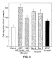
- FIG. 4 shows the effects of HMGB1 and its HMG boxes on RSMC migration into a wound.
- the value of 100% corresponds to the number of cells migrating in the absence of any stimulator (basal migration).
- Statistical significance is 0.05 ⁇ p ⁇ 0.01 for the treatment with bFGF and full length bacteria-made HMGB1, 0.01 ⁇ p ⁇ 0.001 for the treatment with Box A and Box B and 0.001 ⁇ p ⁇ 0.0001 for the treatment with calf thymus HMGB1.
- FIG. 5 shows how HMGB1 binds to the surface of RSMC and stimulates cell motility through RAGE.
- FIG. 5-A shows that large amounts of HMGB1 bind to the surface of RSMC.
- FIG. 5-B RSMC expressing RAGE are shown.
- FIG. 5-C shows how anti-RAGE antibody inhibits HMGB1-induced RSMC migration. Statistical significance is 0.001 ⁇ p ⁇ 0.0001 for treatment with HMGB1 and HMGB1 plus unspecific antibody.
- FIG. 6 shows how pertussis toxin (PT) inhibits HMGB1-induced RSMC migration and actin cytoskeleton reorganization.
- PT pertussis toxin
- FIG. 6-A chemiotaxis assays performed using modified Boyden chambers are shown. The value of 100% corresponds to basal cell migration in the absence of any stimulator; the data represent the mean+SD.
- FIG. 6-B shows evident cytoskeleton reorganization, actin filaments were visualised using conjugated TRITC-phalloidin.
- FIG. 7 demonstrates that the MAP kinase pathway is involved in HMGB1 signaling.
- Cells are stained with specific antibody against phosphorylated ERK1/2 and DAPI, and a separate sample of cells is stained with TRITC-phalloidin to visualize the reorganization of the cytoskeleton.
- FIG. 8 shows that HMGB1 is released by necrotic and damaged cells.
- FIG. 8-A shows the results of Western-blot analysis of proteins released by necrotic, or permeabilized HeLa; HMGB1 presence is evident in line 1 and line 3 .
- FIG. 8-B shows the results of immunofluorescence assays performed on necrotic and living HeLa.
- FIG. 9 shows that HMGB1 is present in the nuclei of endothelial cells, but not in those of vascular SMC.
- FIG. 9-A and in FIG. 9-B it is shown that HMGB1 is present in the nuclei of endothelial cells but it is not detectable in the nuclei of vascular smooth muscle cell of a section of human pancreatic artery stained with anti-HMGB1 antibody and counterstained with ematoxylin, at low (A) and high (B) magnification.
- the red frames indicate the location of the area shown in FIG. B and the arrows point to the nuclei of SMC.
- FIG. 9-C Western blot analysis shows expression level of HMGB1 in RSMC in comparison to HeLa cells.
- FIG. 10 shows the chemotactic effect of HMGB1 on mouse embryonic fibroblasts in chemotaxis assays performed using modified Boyden chambers, in the presence or in the absence of anti-RAGE antibodies (1000 ng/ml).
- the value of 100 corresponds to the number of cells migrating in the absence of any stimulator (random cell migration).
- HMGB1 In the first step, it has been necessary to express and purify HMGB1 and derivates. Expression of full-length HMGB1 was performed in E. coli transformed with pT7-7-rHMGlcm plasmid (kind gift of Prof. J. O. Thomas, Cambridge University) and purification was performed following a well-know protocol (Muller et al., 2001, Biochemistry, 40: 10254 0261).
- HMGB1-induced cell migration and morphological changes i. e. actin fibers reorganization, cell elongation and cell's shape polarization
- Chemotaxis assays were performed using well-known protocols (Degryse et al., 1999, Blood, 94: 649-662).
- Modified Boyden chambers were used with filters having 0.5 Am pore size (Corning, Acton, Mass.) and treated with collagenI (100 Ug/ml in 0.5 M acetic acid) e fibronectin (10 llg/ml) (Roche).
- RSMC cells kind gift of Dr. Marco Bertulli, Bayer Research Laboratories, Milan
- DMEM serum-free fetal calf serum-free cells
- the molecules to be tested were diluted in the same serum-free medium and added to the lower well.
- HMGB1 purified from calf thymus kind gift of J. Bernues, C.S.I.C., Barcelona, Spain
- E. coli expressed recomb anti-HMGB1 E. coli expressed recomb anti-HMGB1
- a lightly modified HMGB1 form containing EAEAYVEF aminoacids bound to the N terminus
- yeast Pichia pastoris yeast Pichia pastoris
- the polyclonal rabbit anti-HMGB1 (Pharmingen B D, Torrey Pines, Calif.), the pertussis toxin (PT) from Bordetella pertussis (kind gift of Dr. M. G. Pizza, I. R. I. S., Siena) or the inhibitors were added in both wells.
- PT pertussis toxin
- Results are the mean+SD of the number of cells counted in 10 high power fields per filter and expressed as fold over control. To random cell migration (i. e., migration in the absence of chemoattractant) was given the arbitrary value of 100%.
- HMGB1 from calf thymus stimulates migration of RSMC in a concentration-dependent manner, starting at doses as low as 0.1 ng/ml and with a 2.5-fold maximal response at 100 ng/ml ( FIG. 1-A ).
- the effect of HMGB1 is, comparable in amplitude to the effects of the well characterised attractants fMLP and bFGF ( FIG. 1-B ).
- RSMC RSMC
- HMGB1 100 ng/ml for increasing time intervals, from 5 to 120 minutes at 37 C. After stimulation, RSMC were fixed for 20 minutes at room temperature with a solution of 3% paraformaldehyde, 2% sucrose in PBS, pH 7.5, followed by three washes with PBS-BSA 0.2%.
- Cells were permeabilized with 20 mM Hepes pH 7.4, 300 mM saccharose, 50 mM NaCl, 3 mM MgCl2, 0.5% (v/v) Triton X-100 for 3 minutes at 4 C, and washed again three times with PBS-BSA 0.2%. Then, RSMC were incubated with PBS-BSA 2% for 15 minutes at 37 C, with primary antibodies for 30 minutes at 37 C, washed three times with PBS-BSA 0.2%, and further incubated with PBS-BSA 2% for 15 minutes.
- DAPI 4,′,6-diamidino-2-phenilindolo, Roche
- coverslips were washed three times with PBS-BSA 0.2%, twice with distilled water, mounted with 20% Mowiol in PBS and analysed on a Axiophot microscope (Carl Zeiss).
- FIG. 2-A Low magnification pictures, in FIG. 2-A , show that stress fibers content, cell shape and size, and cytoskeleton organization change within 30 minutes, but reverse after 120 minutes.
- Higher magnification pictures FIG. 2-B ) show that before stimulation are well-visible numerous stress fibers and the cell shape is anonpolarized.
- RSMC show an elongated, polarized morphology that reflected the spatial rearrangement of the actin cytoskeleton.
- the effects of HMGB1 slowly decrease : After 1-2 hours, the stress fiber content increases back to the initial level and cell morphology returns similar to that of unstimulated control cells.
- Quantification of the migration was made by taking photographs at lower magnification and by counting the number of cells that had migrated into the cell-free space.
- the data represent the mean+SD and the value 100 t corresponds to the number of cells migrating in the absence of any stimulator (basal migration).
- HMGB1 stimulation increases the number of migrating cells by 5-2-fold. Box A and Box B (10 ng/ml) were also tested and both stimulate cell migration 1.8-fold. Finally, the comparison with bFGF (50 ng/ml) underlines that the above mentioned molecules are more effective. It is possible to assume that wound healing is based on the same signaling pathway of chemotaxis and chemokynesis.
- HMGB1 To act as a migratory signal, HMGB1 must arrive to the membrane of responsive cells and bind to a receptor. To test whether HMGB1 binds to the surface of RSMC, one million cells were trypsinized and incubated for 20 minutes at 4 C in PBS containing 800 ng of the Box A+B peptide and 5 pg BSA.
- the Box Al-Box B polypeptide is slightly smaller than the endogenous full-length HMGB1 and can thus be distinguished easily on SDS-PAGE gels.
- FIG. 5-A a SDS-PAGE gel is shown, from which the amount of Box A+B recovered in the cell pellet and in the supernatant can be calculated, and it can be estimated that more than 500,000 Box A+B molecules bind to a single RSMC.
- This result demonstrates that extracellular HMGB1 can bind to RSMC, but most likely does not reflect the actual receptor number. Indeed, HMGB1 has already been shown to bind to heparin and proteoglycans (Bianchi, 1988, EMBO J., 7: 843-849; Nair and Jungalwala, 1997, J.
- HMGB1 might also be associated with the extracellular matrix produced by RSMC, as already demonstrated by the inventors in HeLa, where only small amounts of HMGB1 bind to cells because these cells produce little extracellular matrix.
- HMGB1 has been reported to bind to RAGE that is expressed by a vast range of cell types.
- SDS-PAGE sample buffer 50 mM Tris pH 6.8, 2% 2-mercaptoethanol, 4% SDS, 12% glicerol, 0.05% bromophenol blue
- SDS-PAGE sample buffer 50 mM Tris pH 6.8, 2% 2-mercaptoethanol, 4% SDS, 12% glicerol, 0.05% bromophenol blue
- Separated proteins were blotted on Immobilon (Millipore) membrane using a tankblot system 25 mM Tris pH 7.5, 0.192 M glycine, 20% methanol.
- the blot was blocked for one hour at room temperature in 5% skim milk/TBST (20 mM Tris, pH 7.5, 137 mM NaCl, 0.1% Tween 20), three time washed in TBST, and incubated with anti-HMGB1 antibody in TBST-0.01% BSA. Incubation with secondary antibody was conducted after washing with TBST-0.01% BSA. Proteins were detected with ECL system (Amersham). The presence of RAGE was detected using anti-RAGE antibody (kind gift of Dr. A. M. Schmidt, Columbia University, N.Y.). Results shown in FIG. 5-B demonstrate that RAGE is present on RSMC.
- HMGB1-induced chemotaxis is not only inhibited by anti HMGB1 antibodies but also by anti-RAGE antibodies, as shown in FIG. 5-C .
- Anti-RAGE antibodies block cytoskeletal reorganization and morphological changes of RSMC in response to HMGB1 migratory signal; irrelevant antibodies are not able to block cytoskeleton reorganization.
- PT pertussis toxin
- mPT an inactive mutant of PT, was used as a control.
- RSMC were pre-treated with PT or with mPT (50 ng/ml) for 6 hours, thus stimulated with HMGB1 (100 ng/ml), Box A or Box B (10 ng/ml) for 30 minutes.
- Chemotaxis assays were performed as previously described. The data represent the mean+SD and the value of 100% corresponds to basal migration in the absence of any stimulator. In FIG. 6-A the inhibitory effect of PT on HMGB1-induced chemotaxis is shown.
- HMGB1-induced signaling involves the MAP kinase pathway
- RSMC were pretreated with PD98059 (50 mM) for one hour or were not pre-treated, stimulated for 30 minutes with HMGB1 from calf thymus (100 ng/ml) and stained with specific antibodies against phosphorylated ERK1/2 (New England Biolabs, Beverly, Mass.) and DAPI.
- a separate sample of cells was stained with TRITC-phalloidin to visualize the reorganization of cytoskeleton.
- HMGB1 stimulation induces the activation of ERK1/2 proteins in RSMC and induces their nuclear translocation; in contrast, phosphorylated ERK proteins are hardly detectable and located in the cytoplasm, in unstimulated RSMC.
- PD98059 the selective inhibitor of MEK, the upstream regulator of ERK, inhibits HMGB1-induced ERK phosphorylation and nuclear translocation, as well as RSMC migration and citoskeleton reorganization.
- HeLa cells and HUVEC were induced to undergo necrosis by treatment with 5 UM ionomycin (Sigma) and 20 AM CCCP, or mM deoxyglucose and 10 mM sodium azide. After 16 hours at a 37 C, the number of cells undergoing necrosis was scored morphologically, and when it approached 50% the supernatant was collected.
- the medium from treated and untreated cells was collected and concentrated 50 fold using AmiconUltrafree-MC filters the cells were dissolved in the SDS-PAGE sample buffer.
- the cells were fixed with 4% PFA, incubated with an anti-HMGB1 antibody, and stained with secondary antibody and DAPI.
- the permeabilization of cells was performed with using 0.1 tNP-40 in PBS.
- FIG. 8-A Western-blot analysis of protein in supernatants (S) and cell pellets (P) is represented.
- HMGB1 was recovered in the supernatant of both necrotic cells and damaged cells.
- FIG. 8-B immunofluorescence assays performed on single living and necrotic HeLa is shown, HMGB1 is not associated to the remnants of necrotic cells.
- FIG. 9 the results of immunohistochemistry assays are shown, these data confirm that HMGB1 is contained in the nuclei of endothelial cells that line human arteries but not in the nuclei of RSMC ( FIG. 9A low magnification FIG. 9-B high magnification), in fact, most nuclei of smooth muscle cells contain undetectable amounts of HMGB1 (frame in FIG. 9-B ).
- FIG. 9-C Western-blot analysis shows the expression level of HMGB1 in RSMC in comparison to HeLa cells, and it demonstrates that in vitro cultures of RSMC contain low amounts of HMGB1 in comparison to HeLa cells.
- nuclear HMGB1 protein is a strong mediator of vascular remodeling occurring after mechanical damage and/or inflammation and can be passively released by damaged or necrotic cells.
- HMGB1 Act as a Chemoattractant
- HMGB1 is a potent chemoattractant as bFGF or fMLP in chemotaxis assays and wounding assays, and promotes changes of cell shape and of cytoskeleton organization similar to those observed with pro-urokinase these effects are specifically due to HMGB1 and not to potential contaminants.
- antibodies directed against HMGB1 inhibit its effects on cell migration, whereas non specific control antibodies are unable to do so.
- MAP kinases are involved in HMGB1-induced cell migration of RSMC, since ERK1/2 are phosphorylated and translocated to the cell nucleus upon HMGB1 stimulation, and the MES inhibitor PD98059 is able to block; HMGB1-induced cell migration.
- Data also indicate that aGi/o protein is involved in the process which is activated by HMGB1, since HMGB1-induced cell migration can be blocked by Bordetella pertussis toxin. G protein are usually associated to seven-transmembrane-elix receptors (7 TM), but so far no direct association between RAGE and G protein has been described.
- TM seven-transmembrane-elix receptors
- HMGB1 is released in a unregulated manner, which means upon stimulation with cytolcines or lipopolysaccharide, when cells are mechanically damaged or undergo necrosis. Thus, HMGB1 can signal the damage or destruction of an individual cell to the neighbouring cell in a paracrine manner.
- the cells that respond to extracellular HMGB1 appear to contain very little HMGB1 themselves, and almost none in the nucleus.
- RSMC contain very little HMGB1 compared with HeLa cells or endothelial cells, and what little HMGB1 they contain is mainly located in the cytoplasm. Migrating RSMC tend to concentrate HMGB1 on their surface at the leading edge of the cell.
- HMGB1-responsive cells could contain little HMGB1 to reduce the chance of inappropriate responses to their own HMGB1.
- Concentration of HMGB1 at the leading edge of migrating cells might evoke HMGB1-induced responses in neighbouring cells relocation of molecules involved in cell migration, such as integrins, the urokinase receptor, or c-Src, is a feature of motile RSMC.
- Migration also involves the activation of extracellular proteases, and the interaction between HMGB1 and the plasminogen activation system might facilitate cell migration within the extracellular matrix.
- HMGB1 antagonists HMGB1 fragments, HMG box analogues, which can be more effective than the entire full-length molecule, and proteins containing HMG box domains, the last two are both able to bind to RAGE receptor.
- HMGB1 inhibitors molecules, as antibodies or antibody fragments and four-way DNA, which bind to HMG box domain and avoid HMGB1 binding to RAGE.
- These molecules are advantageously used for pharmacological preparation which prevent, retard or minimise atherosclerosis and/or restenosis after vascular epithelium damages, including those events that occur after angioplasty.
- HMGB1 has a strong biological effect on mouse embryonic fibroblasts. It is well known that fibroblasts are the main cellular components of connective tissues and they are responsible for the synthesis and upkeeping of the connective extracellular matrix. More particularly, HMGB1 acts in vitro as a potent chemoattractant for fibroblasts and anti-RAGE antibodies block said effect.
- every kind of molecules having homology with HMGB1 can be used, as the entire fulllength protein, for the preparation of pharmacological agents which positively regulate, thus facilitate and/or induce cellular migration of fibroblasts.
- every kind of molecules able to block the interaction between HMGB1 and its RAGE receptor i. e. all the molecules belonging to the inhibitors group: antibodies or antibodies fragments, four-way DNA; and all the molecules belonging to the HMG box-antagonists group: HMGB1 fragments, molecules containing the HMG box domain
- An additional aim of the present invention is the use of HMGB1, HMGB1 fragments corresponding to HMG box, HMG box domains of other proteins belonging to the HMG box family and other proteins of the HMG-box family, for the preparation of therapeutic agents which facilitate and/or induce fibroblasts migration and consequently positively regulate connective tissues regeneration.
- Chemotaxis assays were performed using well-know protocols (Degryse et al., 1999, Blood, 94:64. 9-662). Modified Boyden chambers were used with filters having 0.5 ym pore size (Corning, Acton, Mass.) and treated with collagen l (100 pg/ml in 0.5 M acetic acid) and fibronectin (10 llg/ml) (Roche). Mouse embryonic fibroblasts were cultured following well-known protocols (Calogero et al., 1999, Nat. Genet., 22: 276280) and after 24 hours of serum starvation, a sample of 20,000-40,000 cells was added to the upper well of Boyden chamber. E.
- coli expressed recombinant-HMGB1 was diluted in the same serum-free medium and added to the lower well.
- Anti-RAGE antibodies 1000 ng/ml (kind gift of Dr. A. M. Schimdt, Columbia University, N.Y.) were added in both wells.
- Results as shown in FIG. 10 are the mean W SD of the number of cells counted in 10 high power fields per filter and expressed as fold over untreated control. To random cell migration(i. e. migration in absence of chemoattractant) was given the arbitrary value of 100%.
- E. coli expressed recombinant-HMGB1 stimulates fibroblasts migration in a concentration-dependent manner, starting at doses as low as 0.1 ng/ml and with a maximal response at 100 ng/ml, at higher doses (1000 ng/ml) the response is lower than the control.
- Anti RAGE antibodies 1000 ng/ml totally block the migratory response (right side of the graphic of FIG. 10 ) showing that this is specifically due to HMGB1.
- HMGB1 The responsiveness of fibroblasts to HMGB1 points out to a possible role of HMGB1 during connective tissues remodelling occurring after damages due to traumatic events or surgery. Moreover, the fact that anti-RAGE antibodies block said response demonstrates that the interaction between HMGB1 and RAGE receptor on cellular surface is the basic event leading to fibroblast sensitiveness to HMGB1.
Abstract
Description
-
-
state 1, where cells show the appearance typical of unstimulated cells characterized by a high number of stress fibers and a non polarized cell shape; -
state 2, where RSMC exhibiting a low stress fibers content, membrane ruffling, actin semi-rings, or an elongated shape.
-
-
- HMGB1 and/or HMGB1 fragments corresponding to HMG box, HMG box domains of other proteins belonging to HMG-box family and other proteins of the HMG-box family are advantageously used for pharmacological preparations which positively regulate, i.e. facilitate and/or induce e connective tissues regeneration.
- every kind of molecules able to inhibit the interaction between HMGB1 and RAGE, belonging to the antagonists group, (able to bond to RAGE receptor), and belonging to the inhibitors group, (i.e. able to bound the HMG box domain blocking HMGB1 bounding to RAGE receptor) are advantageously used for pharmacological preparations which negatively regulate, i.e. block, retard or reduce connective tissues regeneration.
Claims (3)
Priority Applications (1)
| Application Number | Priority Date | Filing Date | Title |
|---|---|---|---|
| US11/968,506 US8058239B2 (en) | 2001-03-16 | 2008-01-02 | HMGB1 protein inhibitorsand/or antagonists for the treatment of vascular diseases |
Applications Claiming Priority (5)
| Application Number | Priority Date | Filing Date | Title |
|---|---|---|---|
| ITMI2001A0562 | 2001-03-16 | ||
| IT2001MI000562A ITMI20010562A1 (en) | 2001-03-16 | 2001-03-16 | HMG1 PROTEIN INHIBITORS OR ANTAGONISTS FOR THE TREATMENT OF VASCULAR DISORDERS |
| ITMI2001A000562 | 2001-03-16 | ||
| US10/471,641 US7754217B2 (en) | 2001-03-16 | 2001-03-16 | HMGB1 protein inhibitors and/or antagonists for the treatment of vascular diseases |
| US11/968,506 US8058239B2 (en) | 2001-03-16 | 2008-01-02 | HMGB1 protein inhibitorsand/or antagonists for the treatment of vascular diseases |
Related Parent Applications (1)
| Application Number | Title | Priority Date | Filing Date |
|---|---|---|---|
| US10/471,641 Division US7754217B2 (en) | 2001-03-16 | 2001-03-16 | HMGB1 protein inhibitors and/or antagonists for the treatment of vascular diseases |
Publications (2)
| Publication Number | Publication Date |
|---|---|
| US20080171052A1 US20080171052A1 (en) | 2008-07-17 |
| US8058239B2 true US8058239B2 (en) | 2011-11-15 |
Family
ID=11447291
Family Applications (4)
| Application Number | Title | Priority Date | Filing Date |
|---|---|---|---|
| US10/471,641 Expired - Fee Related US7754217B2 (en) | 2001-03-16 | 2001-03-16 | HMGB1 protein inhibitors and/or antagonists for the treatment of vascular diseases |
| US11/968,506 Expired - Fee Related US8058239B2 (en) | 2001-03-16 | 2008-01-02 | HMGB1 protein inhibitorsand/or antagonists for the treatment of vascular diseases |
| US12/707,264 Abandoned US20100297107A1 (en) | 2001-03-16 | 2010-02-17 | Hmgb1 protein inhibitors and/or antagonists for the treatment of vascular diseases |
| US12/707,222 Abandoned US20100172896A1 (en) | 2001-03-16 | 2010-02-17 | Hmgb1 protein inhibitors and/or antagonists for the treatment of vascular diseases |
Family Applications Before (1)
| Application Number | Title | Priority Date | Filing Date |
|---|---|---|---|
| US10/471,641 Expired - Fee Related US7754217B2 (en) | 2001-03-16 | 2001-03-16 | HMGB1 protein inhibitors and/or antagonists for the treatment of vascular diseases |
Family Applications After (2)
| Application Number | Title | Priority Date | Filing Date |
|---|---|---|---|
| US12/707,264 Abandoned US20100297107A1 (en) | 2001-03-16 | 2010-02-17 | Hmgb1 protein inhibitors and/or antagonists for the treatment of vascular diseases |
| US12/707,222 Abandoned US20100172896A1 (en) | 2001-03-16 | 2010-02-17 | Hmgb1 protein inhibitors and/or antagonists for the treatment of vascular diseases |
Country Status (15)
| Country | Link |
|---|---|
| US (4) | US7754217B2 (en) |
| EP (1) | EP1368060B1 (en) |
| JP (1) | JP4822654B2 (en) |
| CN (2) | CN1537014B (en) |
| AT (1) | ATE468137T1 (en) |
| AU (1) | AU2002247977C1 (en) |
| CA (1) | CA2439530C (en) |
| DE (1) | DE60236413D1 (en) |
| DK (1) | DK1368060T3 (en) |
| ES (1) | ES2346408T3 (en) |
| HK (1) | HK1069316A1 (en) |
| IT (1) | ITMI20010562A1 (en) |
| MX (1) | MXPA03008364A (en) |
| PT (1) | PT1368060E (en) |
| WO (1) | WO2002074337A1 (en) |
Families Citing this family (42)
| Publication number | Priority date | Publication date | Assignee | Title |
|---|---|---|---|---|
| US7151082B2 (en) | 1999-02-11 | 2006-12-19 | The Feinstein Institute For Medical Research | Antagonists of HMG1 for treating inflammatory conditions |
| US6303321B1 (en) | 1999-02-11 | 2001-10-16 | North Shore-Long Island Jewish Research Institute | Methods for diagnosing sepsis |
| ITMI20010562A1 (en) * | 2001-03-16 | 2002-09-16 | Marco E Bianchi | HMG1 PROTEIN INHIBITORS OR ANTAGONISTS FOR THE TREATMENT OF VASCULAR DISORDERS |
| CN100447154C (en) * | 2001-05-15 | 2008-12-31 | 费因斯坦医学研究学院 | Use of HMG fragments as anti-inflammatory agents |
| US7304034B2 (en) | 2001-05-15 | 2007-12-04 | The Feinstein Institute For Medical Research | Use of HMGB fragments as anti-inflammatory agents |
| US7220723B2 (en) | 2001-05-15 | 2007-05-22 | The Feinstein Institute For Medical Research | Inhibitors of the interaction between HMGB polypeptides and toll-like receptor 2 as anti-inflammatory agents |
| CA2491321A1 (en) | 2002-07-03 | 2004-01-15 | Fondazione Centro San Raffaele Del Monte Tabor | Use of hmgb1 in the treatment of tissue damage and/or to promote tissue repair |
| US20070154529A1 (en) * | 2003-01-03 | 2007-07-05 | Alcedo Biotech Gmbh | Uses of dna binding proteins |
| US7696169B2 (en) | 2003-06-06 | 2010-04-13 | The Feinstein Institute For Medical Research | Inhibitors of the interaction between HMGB polypeptides and toll-like receptor 2 as anti-inflammatory agents |
| CA2538763C (en) | 2003-09-11 | 2015-05-05 | Critical Therapeutics, Inc. | Monoclonal antibodies against hmgb1 |
| ITRM20040058A1 (en) * | 2004-02-03 | 2004-05-03 | Marco E Bianchi | HMGB1 INHIBITORS AND ANTAGONISTS ABLE TO REGULATE THE PROLIFERATION OF SMOOTH AND ENDOTELIAL MUSCLE CELLS. |
| EP1768677B1 (en) * | 2004-07-02 | 2008-06-25 | Creabilis Therapeutics S.P.A. | Nucleic acids for the treatment of hmgb1-related pathologies |
| EP1768693A1 (en) | 2004-07-20 | 2007-04-04 | Provincia Italiana Della Congregazione Dei Figli Dell'Immacolata Concezione - Istituto Dermopatico Dell'Immacolata | Use of hmgb1 for wound healing |
| MX2007001155A (en) * | 2004-07-29 | 2007-08-14 | Creabilis Therapeutics Spa | Methods, systems, and computer program products for providing presence gateway functionality in a telecommunications network. |
| BRPI0514835A (en) * | 2004-09-03 | 2008-06-24 | Creabilis Therapeutics Spa | high affinity binding domain box polypeptide variant of human and / or non-human hmbg1 or biologically active box-a fragment of hmgb1, nucleic acid molecule, use, pharmaceutical composition and medical device |
| US8129130B2 (en) | 2004-10-22 | 2012-03-06 | The Feinstein Institute For Medical Research | High affinity antibodies against HMGB1 and methods of use thereof |
| EP1812065A4 (en) | 2004-10-22 | 2009-09-02 | Medimmune Inc | High affinity antibodies against hmgb1 and methods of use thereof |
| ITRM20050032A1 (en) * | 2005-01-21 | 2006-07-22 | Marco E Bianchi | INHIBITORS AND ANTAGONISTS OF HMGB1 ABLE TO INHIBIT PATHOGENESIS AND PROGRESSION OF ATEROSCLEROTIC DISEASE. |
| WO2006114805A2 (en) * | 2005-04-28 | 2006-11-02 | Fondazione Centro San Raffaele Del Monte Tabor | Use of hmgb2 and hmgb3 proteins for medical applications |
| EP1909834A2 (en) | 2005-07-18 | 2008-04-16 | Critical Therapeutics, Inc. | Use of hmgb1 antagonists for the treatment of inflammatory skin conditions |
| JP5366548B2 (en) | 2005-08-25 | 2013-12-11 | クレアビリス・セラピューティクス・エスピーエー | Polymer conjugate of K-252A and its derivatives |
| WO2007031100A1 (en) * | 2005-09-14 | 2007-03-22 | Ostini, Marco | Active immunotherapy of life-threatening systemic inflammation |
| AU2006330807A1 (en) | 2005-11-28 | 2007-07-05 | Medimmune, Llc | Antagonists of HMBG1 and/or rage and methods of use thereof |
| WO2007130725A2 (en) * | 2006-02-06 | 2007-11-15 | University Of Pittsburgh Of The Commonwealth System Of Higher Education | Use of hmgb1 for protection against ischemia reperfusion injury |
| EP2068935B8 (en) | 2006-09-15 | 2011-09-14 | Creabilis Therapeutics s.r.l. | Polymer conjugates of box-a of hmgb1 and box-a variants of hmgb1 |
| JP5225109B2 (en) | 2007-02-15 | 2013-07-03 | 国立大学法人 熊本大学 | A therapeutic agent comprising as an active ingredient an antibody that specifically binds to human HMGB-1 |
| CN102083962B (en) | 2008-04-30 | 2013-03-27 | 吉诺米克斯股份有限公司 | Method for collecting functional cells in vivo with high efficiency |
| CA2722852A1 (en) | 2008-04-30 | 2009-11-05 | Genomix Co., Ltd. | Agent for recruitment of bone-marrow-derived pluripotent stem cell into peripheral circulation |
| WO2011007876A1 (en) | 2009-07-16 | 2011-01-20 | Necソフト株式会社 | Nucleic acid molecule capable of binding to hmgb1, and use thereof |
| JP5467313B2 (en) * | 2009-09-28 | 2014-04-09 | 国立大学法人 岡山大学 | Atherosclerosis inhibitor |
| CN102711777B (en) | 2009-10-28 | 2015-04-15 | 吉诺米克斯股份有限公司 | Tissue-regeneration promoter using recruitment of bone marrow mesenchymal stem cells and/or pluripotent stem cells in blood |
| JP6004494B2 (en) | 2010-10-30 | 2016-10-12 | オックスフォード ユニバーシティ イノベーション リミテッド | Treatment of Dupuytren's disease |
| WO2012147470A1 (en) | 2011-04-26 | 2012-11-01 | 株式会社ジェノミックス | Peptide for inducing regeneration of tissue and use thereof |
| US9244074B2 (en) * | 2011-06-07 | 2016-01-26 | University Of Hawaii | Biomarker of asbestos exposure and mesothelioma |
| US9561274B2 (en) | 2011-06-07 | 2017-02-07 | University Of Hawaii | Treatment and prevention of cancer with HMGB1 antagonists |
| AU2013335684B2 (en) | 2012-10-25 | 2017-06-29 | Osaka University | Novel method for treating cardiac infarction using HMGB1 fragment |
| MX365899B (en) | 2012-10-25 | 2019-06-19 | Univ Osaka | Novel method for treating spinal cord injury using hmgb1 fragment. |
| AU2013375015A1 (en) | 2013-01-28 | 2015-08-13 | Evec Inc. | Humanized anti-HMGB1 antibody or antigen-binding fragment thereof |
| IL242807A0 (en) | 2015-11-26 | 2016-04-21 | Novamed Ltd | Assay device |
| WO2017098051A2 (en) | 2015-12-11 | 2017-06-15 | Ruprecht-Karls-Universität Heidelberg | Combined preparations of pkm2 modulators and hmgb1 |
| EP3718561A4 (en) | 2017-12-01 | 2021-07-21 | Stemrim Inc. | Therapeutic agent for inflammatory bowel disease |
| AU2020449867A1 (en) * | 2020-04-22 | 2022-11-03 | Chulalongkorn University | A composition and a method of rejuvenating DNA and preventing DNA damage |
Citations (7)
| Publication number | Priority date | Publication date | Assignee | Title |
|---|---|---|---|---|
| WO1997026913A1 (en) | 1996-01-26 | 1997-07-31 | The Trustees Of Columbia University In The City Of New York | A POLYPEPTIDE FROM LUNG EXTRACT WHICH BINDS AMYLOID-β PEPTIDE |
| WO1997039121A1 (en) | 1996-04-16 | 1997-10-23 | Schering Aktiengesellschaft | Advanced glycosylation end-product receptor peptides and uses therefor |
| WO1997039125A1 (en) | 1996-04-16 | 1997-10-23 | Schering Aktiengesellschaft Patente | Antibodies against the advanced glycosylation end-product receptor and uses thereof |
| WO1998022138A1 (en) | 1996-11-22 | 1998-05-28 | The Trustees Of Columbia University In The City Of New York | Method for treating symptoms of diabetes |
| US6054122A (en) * | 1990-11-27 | 2000-04-25 | The American National Red Cross | Supplemented and unsupplemented tissue sealants, methods of their production and use |
| WO2000047104A2 (en) | 1999-02-11 | 2000-08-17 | North Shore-Long Island Jewish Research Institute | Antagonists of hmg1 for treating inflammatory conditions |
| WO2006008779A1 (en) * | 2004-07-20 | 2006-01-26 | Provincia Italiana Della Congregazione Dei Figli Dell'immacolata Concezione-Istituto Dermopatico Dell'immacolata | Use of hmgb1 for wound healing |
Family Cites Families (6)
| Publication number | Priority date | Publication date | Assignee | Title |
|---|---|---|---|---|
| US5470307A (en) * | 1994-03-16 | 1995-11-28 | Lindall; Arnold W. | Catheter system for controllably releasing a therapeutic agent at a remote tissue site |
| US6398808B1 (en) * | 1999-06-15 | 2002-06-04 | Scimed Life Systems, Inc. | Localized delivery of genetic information from biostable materials |
| ITMI20010562A1 (en) * | 2001-03-16 | 2002-09-16 | Marco E Bianchi | HMG1 PROTEIN INHIBITORS OR ANTAGONISTS FOR THE TREATMENT OF VASCULAR DISORDERS |
| CN100447154C (en) * | 2001-05-15 | 2008-12-31 | 费因斯坦医学研究学院 | Use of HMG fragments as anti-inflammatory agents |
| ITRM20040058A1 (en) * | 2004-02-03 | 2004-05-03 | Marco E Bianchi | HMGB1 INHIBITORS AND ANTAGONISTS ABLE TO REGULATE THE PROLIFERATION OF SMOOTH AND ENDOTELIAL MUSCLE CELLS. |
| WO2008137552A2 (en) * | 2007-05-02 | 2008-11-13 | Medimmune, Llc | Anti-rage antibodies and methods of use thereof |
-
2001
- 2001-03-16 IT IT2001MI000562A patent/ITMI20010562A1/en unknown
- 2001-03-16 US US10/471,641 patent/US7754217B2/en not_active Expired - Fee Related
-
2002
- 2002-03-12 CA CA2439530A patent/CA2439530C/en not_active Expired - Fee Related
- 2002-03-12 DK DK02717057.0T patent/DK1368060T3/en active
- 2002-03-12 PT PT02717057T patent/PT1368060E/en unknown
- 2002-03-12 JP JP2002573044A patent/JP4822654B2/en not_active Expired - Fee Related
- 2002-03-12 ES ES02717057T patent/ES2346408T3/en not_active Expired - Lifetime
- 2002-03-12 AT AT02717057T patent/ATE468137T1/en active
- 2002-03-12 CN CN028065670A patent/CN1537014B/en not_active Expired - Fee Related
- 2002-03-12 WO PCT/IT2002/000153 patent/WO2002074337A1/en active Application Filing
- 2002-03-12 EP EP02717057A patent/EP1368060B1/en not_active Expired - Lifetime
- 2002-03-12 MX MXPA03008364A patent/MXPA03008364A/en active IP Right Grant
- 2002-03-12 DE DE60236413T patent/DE60236413D1/en not_active Expired - Lifetime
- 2002-03-12 CN CN200910252323A patent/CN101773669A/en active Pending
- 2002-03-12 AU AU2002247977A patent/AU2002247977C1/en not_active Ceased
-
2005
- 2005-03-02 HK HK05101837.0A patent/HK1069316A1/en not_active IP Right Cessation
-
2008
- 2008-01-02 US US11/968,506 patent/US8058239B2/en not_active Expired - Fee Related
-
2010
- 2010-02-17 US US12/707,264 patent/US20100297107A1/en not_active Abandoned
- 2010-02-17 US US12/707,222 patent/US20100172896A1/en not_active Abandoned
Patent Citations (7)
| Publication number | Priority date | Publication date | Assignee | Title |
|---|---|---|---|---|
| US6054122A (en) * | 1990-11-27 | 2000-04-25 | The American National Red Cross | Supplemented and unsupplemented tissue sealants, methods of their production and use |
| WO1997026913A1 (en) | 1996-01-26 | 1997-07-31 | The Trustees Of Columbia University In The City Of New York | A POLYPEPTIDE FROM LUNG EXTRACT WHICH BINDS AMYLOID-β PEPTIDE |
| WO1997039121A1 (en) | 1996-04-16 | 1997-10-23 | Schering Aktiengesellschaft | Advanced glycosylation end-product receptor peptides and uses therefor |
| WO1997039125A1 (en) | 1996-04-16 | 1997-10-23 | Schering Aktiengesellschaft Patente | Antibodies against the advanced glycosylation end-product receptor and uses thereof |
| WO1998022138A1 (en) | 1996-11-22 | 1998-05-28 | The Trustees Of Columbia University In The City Of New York | Method for treating symptoms of diabetes |
| WO2000047104A2 (en) | 1999-02-11 | 2000-08-17 | North Shore-Long Island Jewish Research Institute | Antagonists of hmg1 for treating inflammatory conditions |
| WO2006008779A1 (en) * | 2004-07-20 | 2006-01-26 | Provincia Italiana Della Congregazione Dei Figli Dell'immacolata Concezione-Istituto Dermopatico Dell'immacolata | Use of hmgb1 for wound healing |
Non-Patent Citations (21)
| Title |
|---|
| Andersson et al., 2000 J. Exp. Med., vol. 192 565-70. |
| Bear et al., Cell, vol. 101, 717-728, Jun. 23, 2000. * |
| Bork et al., 1996, Trends in Genetics 12:425-427. * |
| Bork, 2000, Genome Research 10:398-400. * |
| Brenner, 1999, Trends in Genetics 15:132-133. * |
| Chen et al., 2004. J Interferon and Cylokine Res. vol. 24: 329-33. |
| Degryse, B et al. The High Mobility Group HMG Boxes of the Nuclear Protein HMG1 Induce Chemotaxis and Cytoskeleton Reorganization in Rat Smooth Muscle Cells. J Cell Biol. 152, 1197-1206 (2001). |
| Doerks et al., 1998, Trends in Genetics 14:248-250. * |
| Fawcett et al., The Journal of Cell Biology, vol. 128, 1995, pp. 1229-1241. * |
| Hertel L et al. Decreased Expression of the High Mobility Group Protein T160 by Antisense RNA Impairs the Grwoth of Mouse Fibroblasts. Biochimie 79, 717-723 (1997). |
| Ngo et al., 1994, The Protein Folding Problem and Tertiary Structure Prediction, pp. 492-495. * |
| Parkkinen et al., J Biol Chem. Sep. 15, 1993;268(26):19726-38. * |
| PCT/IT02/00153, Intr Search Rpt. |
| Pins et al., J Invest Dermatol. Apr. 2000;114(4):647-53. * |
| Rauvala et al., Matrix Biol. Sep. 2000;19(5):377-87. * |
| Ronfani L et al Reduced Fertility and Spermatogenesis Defedts in Mice Lacking Chromosomal Protein HMGB2 Development 128, 1265-1273 (2001). |
| Skolnick et al., 2000, Trends in Biotech. 18(1):34-39, especially p. 36 at Box 2. * |
| Smith et al., 1997, Nature Biotechnology 15:1222-1223. * |
| Stros M et al., A Role of Basic Residues and the Putative Intercalating Phenylalanine of the HMG1 Box B in DNA Supoercoiling and Binding to Four Way DNA Junctions. J Biol Chem 275(46), 35699-35707 (2000). |
| Wells, 1990, Biochemistry 29:8509-8517. * |
| Yamazaki, F et al Repression of Cell Cycle Progression by Antigsense HMG2 RNA, Biochem. Biophys. Res. Comm 210(2), 1045-1051(1995). |
Also Published As
| Publication number | Publication date |
|---|---|
| HK1069316A1 (en) | 2005-05-20 |
| MXPA03008364A (en) | 2004-11-12 |
| US20080171052A1 (en) | 2008-07-17 |
| JP2004523579A (en) | 2004-08-05 |
| US20040136979A1 (en) | 2004-07-15 |
| AU2002247977B8 (en) | 2007-03-15 |
| WO2002074337A1 (en) | 2002-09-26 |
| US20100297107A1 (en) | 2010-11-25 |
| EP1368060A1 (en) | 2003-12-10 |
| DK1368060T3 (en) | 2010-08-30 |
| EP1368060B1 (en) | 2010-05-19 |
| CN101773669A (en) | 2010-07-14 |
| AU2002247977A2 (en) | 2004-02-26 |
| AU2002247977C1 (en) | 2008-09-18 |
| CN1537014B (en) | 2012-04-25 |
| US20100172896A1 (en) | 2010-07-08 |
| ES2346408T3 (en) | 2010-10-15 |
| JP4822654B2 (en) | 2011-11-24 |
| WO2002074337A8 (en) | 2003-08-28 |
| CA2439530A1 (en) | 2002-09-26 |
| AU2002247977B2 (en) | 2006-12-14 |
| PT1368060E (en) | 2010-08-24 |
| US7754217B2 (en) | 2010-07-13 |
| CA2439530C (en) | 2016-11-08 |
| CN1537014A (en) | 2004-10-13 |
| ATE468137T1 (en) | 2010-06-15 |
| DE60236413D1 (en) | 2010-07-01 |
| AU2002247977A1 (en) | 2002-10-03 |
| ITMI20010562A1 (en) | 2002-09-16 |
Similar Documents
| Publication | Publication Date | Title |
|---|---|---|
| US8058239B2 (en) | HMGB1 protein inhibitorsand/or antagonists for the treatment of vascular diseases | |
| Degryse et al. | The high mobility group (HMG) boxes of the nuclear protein HMG1 induce chemotaxis and cytoskeleton reorganization in rat smooth muscle cells | |
| Prater et al. | The properdin-like type I repeats of human thrombospondin contain a cell attachment site. | |
| Franke et al. | The desmosomal plaque and the cytoskeleton | |
| Chen et al. | Interleukin-17 induces angiogenesis in human choroidal endothelial cells in vitro | |
| EP1817327A1 (en) | Antigenic epitopes of interleukin-21, related antibodies and their use in medical field | |
| Agrez et al. | Arg-Gly-Asp-containing peptides expose novel collagen receptors on fibroblasts: implications for wound healing. | |
| WO2006099620A9 (en) | Rage/diaphanous interaction and related compositions and methods | |
| Ambort et al. | Specific processing of tenascin-C by the metalloprotease meprinβ neutralizes its inhibition of cell spreading | |
| CA2074361A1 (en) | Merosin, nucleic acids encoding, fragments and uses thereof | |
| US10544231B2 (en) | Antibodies for the prevention or the treatment of bleeding episodes | |
| US7517654B2 (en) | Peptide and a derivative thereof promoting cell adhesion and spreading | |
| Gao et al. | Fibronectin-binding peptides. I. Isolation and characterization of two unique fibronectin-binding peptides from gelatin | |
| US7824868B2 (en) | Formation of superfibronectin by BBK32 and uses therefor | |
| US10370450B2 (en) | Anti-DR6 antibodies and methods of immune regulation | |
| Lambert et al. | The interaction of Tamm-Horsfall protein with the extracellular matrix. | |
| US9382322B2 (en) | Tissue repair by modulation of beta-1 integrin biological function | |
| Das et al. | Evidence for binding of the ectodomain of amyloid precursor protein 695 and activated high molecular weight kininogen | |
| Zhu et al. | Comparison of immunogenic potentials of bovine thrombin preparations | |
| Ingerslev et al. | Applications of immunoperoxidase techniques in specificity testing of monoclonal antibodies (Mabs) against von Willebrand factor (vWf) | |
| ES2362655T3 (en) | METHODS AND COMPOSITIONS TO SUPPRESS THE DIFFERENTIATION OF FIBROCITS. | |
| JP2015168665A (en) | Inhibitor for exacerbation of bleeding and/or inflammation | |
| Brown | The effects of KTS disintegrins on endothelial cells | |
| Khunkaewla | Doktorln der Medizinischen Wissenschaft | |
| JPH08511941A (en) | Human cell adhesion protein AAMP-1 and use thereof |
Legal Events
| Date | Code | Title | Description |
|---|---|---|---|
| AS | Assignment |
Owner name: BIO3 RESEARCH SRL, ITALY Free format text: ASSIGNMENT OF ASSIGNORS INTEREST;ASSIGNORS:BIANCHI, MARCO;BONALDI, TIZIANA;SCAFFIDI, PAOLA;AND OTHERS;REEL/FRAME:020768/0500;SIGNING DATES FROM 20031029 TO 20031112 Owner name: BIANCHI, MARCO E., ITALY Free format text: ASSIGNMENT OF ASSIGNORS INTEREST;ASSIGNORS:BIANCHI, MARCO;BONALDI, TIZIANA;SCAFFIDI, PAOLA;AND OTHERS;REEL/FRAME:020768/0500;SIGNING DATES FROM 20031029 TO 20031112 Owner name: BIO3 RESEARCH SRL, ITALY Free format text: ASSIGNMENT OF ASSIGNORS INTEREST;ASSIGNORS:BIANCHI, MARCO;BONALDI, TIZIANA;SCAFFIDI, PAOLA;AND OTHERS;SIGNING DATES FROM 20031029 TO 20031112;REEL/FRAME:020768/0500 Owner name: BIANCHI, MARCO E., ITALY Free format text: ASSIGNMENT OF ASSIGNORS INTEREST;ASSIGNORS:BIANCHI, MARCO;BONALDI, TIZIANA;SCAFFIDI, PAOLA;AND OTHERS;SIGNING DATES FROM 20031029 TO 20031112;REEL/FRAME:020768/0500 |
|
| AS | Assignment |
Owner name: BIO3 RESEARCH SRL,ITALY Free format text: ASSIGNMENT OF ASSIGNORS INTEREST;ASSIGNOR:BIANCHI, MARCO E.;REEL/FRAME:024015/0946 Effective date: 20100129 Owner name: BIO3 RESEARCH SRL, ITALY Free format text: ASSIGNMENT OF ASSIGNORS INTEREST;ASSIGNOR:BIANCHI, MARCO E.;REEL/FRAME:024015/0946 Effective date: 20100129 |
|
| STCF | Information on status: patent grant |
Free format text: PATENTED CASE |
|
| FPAY | Fee payment |
Year of fee payment: 4 |
|
| FEPP | Fee payment procedure |
Free format text: MAINTENANCE FEE REMINDER MAILED (ORIGINAL EVENT CODE: REM.); ENTITY STATUS OF PATENT OWNER: SMALL ENTITY |
|
| LAPS | Lapse for failure to pay maintenance fees |
Free format text: PATENT EXPIRED FOR FAILURE TO PAY MAINTENANCE FEES (ORIGINAL EVENT CODE: EXP.); ENTITY STATUS OF PATENT OWNER: SMALL ENTITY |
|
| STCH | Information on status: patent discontinuation |
Free format text: PATENT EXPIRED DUE TO NONPAYMENT OF MAINTENANCE FEES UNDER 37 CFR 1.362 |
|
| FP | Lapsed due to failure to pay maintenance fee |
Effective date: 20191115 |