US8053198B2 - Diagnostic methods - Google Patents
Diagnostic methods Download PDFInfo
- Publication number
- US8053198B2 US8053198B2 US12/209,701 US20970108A US8053198B2 US 8053198 B2 US8053198 B2 US 8053198B2 US 20970108 A US20970108 A US 20970108A US 8053198 B2 US8053198 B2 US 8053198B2
- Authority
- US
- United States
- Prior art keywords
- level
- biomarker
- sample
- patient
- ovarian cancer
- Prior art date
- Legal status (The legal status is an assumption and is not a legal conclusion. Google has not performed a legal analysis and makes no representation as to the accuracy of the status listed.)
- Active
Links
Images


Classifications
-
- G—PHYSICS
- G01—MEASURING; TESTING
- G01N—INVESTIGATING OR ANALYSING MATERIALS BY DETERMINING THEIR CHEMICAL OR PHYSICAL PROPERTIES
- G01N33/00—Investigating or analysing materials by specific methods not covered by groups G01N1/00 - G01N31/00
- G01N33/48—Biological material, e.g. blood, urine; Haemocytometers
- G01N33/50—Chemical analysis of biological material, e.g. blood, urine; Testing involving biospecific ligand binding methods; Immunological testing
- G01N33/53—Immunoassay; Biospecific binding assay; Materials therefor
- G01N33/574—Immunoassay; Biospecific binding assay; Materials therefor for cancer
- G01N33/57407—Specifically defined cancers
- G01N33/57442—Specifically defined cancers of the uterus and endometrial
-
- G—PHYSICS
- G01—MEASURING; TESTING
- G01N—INVESTIGATING OR ANALYSING MATERIALS BY DETERMINING THEIR CHEMICAL OR PHYSICAL PROPERTIES
- G01N33/00—Investigating or analysing materials by specific methods not covered by groups G01N1/00 - G01N31/00
- G01N33/48—Biological material, e.g. blood, urine; Haemocytometers
- G01N33/50—Chemical analysis of biological material, e.g. blood, urine; Testing involving biospecific ligand binding methods; Immunological testing
- G01N33/53—Immunoassay; Biospecific binding assay; Materials therefor
- G01N33/574—Immunoassay; Biospecific binding assay; Materials therefor for cancer
- G01N33/57407—Specifically defined cancers
- G01N33/57449—Specifically defined cancers of ovaries
-
- G—PHYSICS
- G01—MEASURING; TESTING
- G01N—INVESTIGATING OR ANALYSING MATERIALS BY DETERMINING THEIR CHEMICAL OR PHYSICAL PROPERTIES
- G01N2333/00—Assays involving biological materials from specific organisms or of a specific nature
- G01N2333/435—Assays involving biological materials from specific organisms or of a specific nature from animals; from humans
- G01N2333/475—Assays involving growth factors
-
- G—PHYSICS
- G01—MEASURING; TESTING
- G01N—INVESTIGATING OR ANALYSING MATERIALS BY DETERMINING THEIR CHEMICAL OR PHYSICAL PROPERTIES
- G01N2333/00—Assays involving biological materials from specific organisms or of a specific nature
- G01N2333/435—Assays involving biological materials from specific organisms or of a specific nature from animals; from humans
- G01N2333/705—Assays involving receptors, cell surface antigens or cell surface determinants
- G01N2333/71—Assays involving receptors, cell surface antigens or cell surface determinants for growth factors; for growth regulators
-
- G—PHYSICS
- G01—MEASURING; TESTING
- G01N—INVESTIGATING OR ANALYSING MATERIALS BY DETERMINING THEIR CHEMICAL OR PHYSICAL PROPERTIES
- G01N2333/00—Assays involving biological materials from specific organisms or of a specific nature
- G01N2333/90—Enzymes; Proenzymes
- G01N2333/91—Transferases (2.)
- G01N2333/912—Transferases (2.) transferring phosphorus containing groups, e.g. kinases (2.7)
- G01N2333/91205—Phosphotransferases in general
- G01N2333/9121—Phosphotransferases in general with an alcohol group as acceptor (2.7.1), e.g. general tyrosine, serine or threonine kinases
- G01N2333/91215—Phosphotransferases in general with an alcohol group as acceptor (2.7.1), e.g. general tyrosine, serine or threonine kinases with a definite EC number (2.7.1.-)
-
- G—PHYSICS
- G01—MEASURING; TESTING
- G01N—INVESTIGATING OR ANALYSING MATERIALS BY DETERMINING THEIR CHEMICAL OR PHYSICAL PROPERTIES
- G01N2333/00—Assays involving biological materials from specific organisms or of a specific nature
- G01N2333/90—Enzymes; Proenzymes
- G01N2333/914—Hydrolases (3)
- G01N2333/948—Hydrolases (3) acting on peptide bonds (3.4)
- G01N2333/95—Proteinases, i.e. endopeptidases (3.4.21-3.4.99)
- G01N2333/964—Proteinases, i.e. endopeptidases (3.4.21-3.4.99) derived from animal tissue
- G01N2333/96402—Proteinases, i.e. endopeptidases (3.4.21-3.4.99) derived from animal tissue from non-mammals
- G01N2333/96405—Proteinases, i.e. endopeptidases (3.4.21-3.4.99) derived from animal tissue from non-mammals in general
- G01N2333/96408—Proteinases, i.e. endopeptidases (3.4.21-3.4.99) derived from animal tissue from non-mammals in general with EC number
- G01N2333/96419—Metalloendopeptidases (3.4.24)
-
- G—PHYSICS
- G01—MEASURING; TESTING
- G01N—INVESTIGATING OR ANALYSING MATERIALS BY DETERMINING THEIR CHEMICAL OR PHYSICAL PROPERTIES
- G01N2800/00—Detection or diagnosis of diseases
- G01N2800/60—Complex ways of combining multiple protein biomarkers for diagnosis
Definitions
- This application relates to assay methods, modules and kits for conducting diagnostic assays useful in the detection and treatment of cancerous conditions.
- the present invention provides a method for diagnosing a cancerous condition in a patient comprising (a) measuring a level of a first biomarker in a test sample obtained from a patient, wherein said first biomarker is selected from the group consisting of Flt1, MMP-10, PIGF, and combinations thereof; (b) diagnosing from said measuring step the presence or absence of said cancerous condition in said patient.
- the method further comprises measuring a level of at least one additional biomarker in said sample and determining from said level of said first biomarker and said level of said at least one additional biomarker the presence or absence of said cancerous condition in said patient.
- the method further comprises comparing said level of said first biomarker in said sample to a level of said first biomarker in a normal control sample and diagnosing the presence or absence of said cancerous condition in said patient based on said comparison.
- the method further comprises comparing said level of said first biomarker and said at least one additional biomarker(s) in said sample to a level of said first biomarker and said at least one additional biomarker(s) in a normal control sample and diagnosing the presence or absence of said cancerous condition in said patient based on said comparison.
- the at least one additional biomarker is selected from the group consisting of CA125, IL-6, CRP, SAA, IL-8, IL-1 ⁇ , IL-2, P-Cadherin, Eotaxin-3, TNFR1, OPN, Prolactin, and combinations thereof.
- the methods of the present invention may be used to diagnose and/or treat a cancerous condition selected from the group consisting of ovarian, breast, endometrial cancers, and combinations thereof.
- the patient may be a pre- or postmenopausal woman.
- the diagnosing step comprises comparing said level of said first biomarker to a detection cut-off level, wherein said first biomarker level above (or, alternatively, below) said detection cut-off level is indicative of said cancerous condition.
- the diagnosing step comprises comparing said level of said at least one additional biomarker to a detection cut-off level, wherein said at least one additional biomarker level above (or, alternatively, below) said detection cut-off level is indicative of said cancerous condition.
- the invention provides a method for monitoring the progression of a cancerous condition in a patient, said method comprising (a) measuring the level(s) of a first biomarker in samples obtained, at different times, from said patient, wherein said first biomarker is selected from the group consisting of Flt-1, MMP-10, PIF, and combinations thereof; and (b) determining from said level(s) of said first biomarker the progression or efficacy of treatment of said cancerous condition.
- the invention provides a method for evaluating the efficacy of a cancer therapeutic agent or treatment regimen in a patient that has or is suspected to have a cancerous condition, said method comprising (a) measuring the level of a first biomarker in a sample obtained from said patient, wherein said first biomarker is selected from the group consisting of Flt-1, MMP-10, PIGF, and combinations thereof; (b) measuring the level of said first biomarker in a sample of a tumor model that has been exposed to said agent or treatment regimen; and (c) comparing the levels measured in steps (a) and (b) to determine the efficacy of said agent or treatment regimen.
- the invention provides an assay method wherein said measuring step is conducted on a single sample.
- the measuring step may also be conducted in a single assay chamber.
- the assay chamber may be a single well of an assay plate or a cartridge.
- the biomarker(s) level is measured using an immunoassay.
- the patient sample is selected from the group consisting of blood, serum and plasma.
- the sample may also be selected from a group consisting of biopsy tissue, intestinal mucosa and urine.
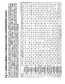
- FIG. 1 is a table that summarizes the biomarkers that were analyzed in the assays of the present invention.
- FIGS. 2A and 2B reflect the assay ranges and levels used in one embodiment of the assays of the present invention.
- FIG. 3 shows the results obtained from one embodiment of the assays of the present invention using CA 125 as the reference biomarker.
- FIG. 4 shows the results obtained from one embodiment of the assays of the present invention using IL-6 as the reference biomarker.
- FIG. 5 shows the results obtained from one embodiment of the assays of the present invention using VEGFR-1 (Flt-1) as the reference biomarker.
- FIG. 6 shows additional markers of interest to ovarian and endometrial cancers.
- FIG. 7 shows the results obtained from one embodiment of the assays of the present invention using PIGF as a reference marker.
- FIG. 8A to 8P provides evidence that specific combinations of biomarkers can be used to increase the confidence of cancer diagnosis.
- the present invention provides a method for diagnosing a cancerous condition in a patient comprising (a) measuring a level of a first biomarker in a test sample obtained from a patient, wherein said first biomarker is selected from the group consisting of Flt1, MMP-10, PIGF, and combinations thereof; and (b) diagnosing from said measuring step the presence or absence of said cancerous condition in said patient
- the invention provides a method of diagnosing a cancerous condition in a patient by measuring a level of Flt-1 and diagnosing the patient for the presence or absence of the cancerous condition by identifying the level of Flt-1 in the patient sample.
- the invention provides embodiments in which the level of MMP-10, and/or PIGF is measured in the patient sample and based on this biomarker level, the patient is diagnosed with the cancerous condition.
- Flt-1 and MMP-10 are analyzed in a single assay
- Flt-1 and PIGF are analyzed in a single assay
- MMP-10 and PIGF are analyzed in a single assay
- Flt-1, MMP-10 and PIGF are analyzed in a single assay.
- the invention also contemplates a diagnostic method as described above, wherein one or more of Flt-1, MMP-10, and/or PIGF are analyzed in a single assay, and the level of at least one additional biomarker is measured in the sample. Therefore, the levels of the first biomarker and the additional biomarker(s) in the test sample may be used to diagnose a cancerous condition in a patient
- the additional biomarker is selected from CA125, IL-6, CRP, SAA, IL-8, IL-1 ⁇ , IL-2, PC-Cadherin, Eotaxin-3, TNFR1, OPN, Prolactin, and combinations thereof.
- biomarkers are known and may be used in combination with the methods of the present invention, including and not limited to VEGF, PDGF, focal adhesion kinase, AKT, erb-B, HER2/neu, Trk, other matrix metalloproteinases, and combinations thereof.
- the level of the first biomarker and/or the level of the additional biomarkers in the test sample are compared to the levels of these biomarkers in a corresponding normal control sample.
- the difference between the normal control sample biomarker levels and that of the test sample may be the basis for diagnosing a cancerous condition in a patient.
- the method of the invention contemplates a comparison of the level of the first biomarker to a detection cut-off level, wherein the first biomarker level above or below the detection cut-off level is indicative of the cancerous condition.
- the diagnostic methods of the invention also contemplate comparing the level of the at least one additional biomarker to a detection cut-off level, wherein the at least one additional biomarker level above or below the detection cut-off level is indicative of the cancerous condition.
- the diagnostic methods of the present invention may be used to diagnose a variety of cancerous conditions, including but not limited to ovarian, breast, endometrial cancers, cervical cancers, uterine cancers and combinations thereof.
- the diagnostic methods of the present invention may be used to identify endometriosis. These methods may be used to identify cancerous conditions in a patient, e.g., in a pre- or post-menopausal woman.
- cancer is intended to mean a class of diseases characterized by the uncontrolled growth of aberrant cells, including all known cancers, and neoplastic conditions, whether characterized as malignant, benign, soft tissue or solid tumor.
- the assay may have diagnostic value irrespective of the menopausal state of a female patient.
- the diagnostic value is higher (or lower) in selected subpopulations, e.g., pre- or post-menopausal women.
- the methods of the present invention may also be used to monitor the progression of a cancerous condition in a patient by (a) measuring the level(s) of a first biomarker in samples obtained, at different times, from said patient, wherein said first biomarker is selected from the group consisting of Flt-1, MMP-10, PIGF, and combinations thereof; and (b) determining from said level(s) of said first biomarker the progression or efficacy of treatment of said cancerous condition.
- the method of the present invention may be used to differentiate between the presence or absence of different cancer subtypes in a sample taken from a patient.
- the methods of the present invention may be used to evaluate the efficacy of a cancer therapeutic agent or treatment regimen in a patient that has or is suspected to have a cancerous condition, said method comprising (a) measuring the level of a first biomarker in a sample obtained from said patient, wherein said first biomarker is selected from the group consisting of Flt-1, MMP-10, PIGF, and combinations thereof, (b) measuring the level of said first biomarker in a sample of a tumor model that has been exposed to said agent or treatment regimen; and (c) comparing the levels measured in steps (a) and (b) to determine the efficacy of said agent or treatment regimen.
- the methods of the present invention may also be used to select a treatment regimen or to adjust the dose of one or more components in a therapeutic treatment regimen. It may also be used to evaluate whether any supportive or palliative care therapies should be included in a treatment regimen, and in that regard, the skilled artisan may include additional biomarkers in the methods of the invention.
- the assays of the present invention may be conducted by any suitable method.
- the measuring step is conducted on a single sample, and it may also be conducted in a single assay chamber, including but not limited to a single well of an assay plate.
- the assay chamber may also be an assay chamber of a cartridge.
- sample is intended to mean any biological fluid, cell, tissue, organ or combinations or portions thereof, which includes or potentially includes a biomarker of a disease of interest
- a sample can be a histologic section of a specimen obtained by biopsy, or cells that are placed in or adapted to tissue culture.
- a sample further can be a subcellular fraction or extract, or a crude or substantially pure nucleic acid molecule or protein preparation.
- the samples that may be analyzed in the assays of the present invention include but are not limited to blood or blood fractions such as, serum and plasma.
- the sample may also include biopsy tissue, intestinal mucosa and urine.
- the level is measured using an immunoassay.
- a “biomarker” is a substance that is associated with a particular disease.
- a change in the expression levels of a biomarker may correlate with the risk or progression of a disease or with the susceptibility of the disease to a given treatment
- a biomarker may be useful in the diagnosis of disease risk or the presence of disease in an individual, or to tailor treatments for the disease in an individual (choices of drug treatment or administration regimes).
- a biomarker may be used as a surrogate for a natural endpoint such as survival or irreversible morbidity.
- a sample that is assayed in the diagnostic methods of the present invention may be obtained from any suitable patient, including but not limited to a patient suspected of having cancer or a patient having a predisposition to a cancerous condition.
- the patient may or may not exhibit symptoms associated with a cancerous condition.
- the term “level” refers to mean the amount, concentration, accumulation or rate of a biomarker molecule.
- a level can be represented, for example, by the amount or synthesis rate of messenger RNA (mRNA) encoded by a gene, the amount or synthesis rate of polypeptide corresponding to a given amino acid sequence encoded by a gene, or the amount or synthesis rate of a biochemical form of a molecule accumulated in a cell, including, for example, the amount of particular post-synthetic modifications of a molecule such as a polypeptide, nucleic acid or small molecule.
- mRNA messenger RNA
- the term can be used to refer to an absolute amount of a molecule in a sample or to a relative amount of the molecule, including amount or concentration determined under steady-state or non-steady-state conditions. Level may also refer to an assay signal that correlates with the amount, concentration, accumulation or rate of change of a biomarker molecule.
- the expression level of a molecule can be determined relative to a control molecule in a sample.
- the levels or levels of biomarker(s) are measured in the samples collected from individuals clinically diagnosed with or suspected of or at risk of developing cancer or a cancerous or pre-cancerous condition using conventional methods, e.g., biopsy or other conventional diagnostic methods, as well as from healthy individuals. It may also be used to screen for disease in a broad population of asymptomatic individuals. For example, specific biomarkers valuable in distinguishing between normal and diseased patients could be identified by visual inspection of the data, for example, data plotted on a one-dimensional or multidimensional graph, or using methods of statistical analysis, such as a statistically weighted difference between control individuals and diseased patients and/or Receiver Operating Characteristic (ROC) curve analysis.
- ROC Receiver Operating Characteristic
- diagnostically valuable biomarkers may be first identified using a statistically weighted difference between control individuals and diseased patients, calculated as
- D is the median level of a biomarker in patients diagnosed as having, for example, breast cancer or ovarian cancer
- N is the median of the control individuals
- ⁇ D is the standard deviation of D
- ⁇ N is the standard deviation of N. The larger the magnitude, the greater the statistical difference between the diseased and normal populations.
- biomarkers resulting in a statistically weighted difference between control individuals and diseased patients of greater than, e.g., 1, 1.5, 2, 2.5 or 3 could be identified as diagnostically valuable markers.
- Another method of statistical analysis for identifying biomarkers is the use of z-scores, e.g., as described in Skates et al (2007) Cancer Epidemiol Biomarkers Prev. 16(2):334-341.
- ROC curve analysis Another method of statistical analysis that can be useful in the inventive methods of the invention for determining the efficacy of particular candidate analytes, such as particular biomarkers, for acting as diagnostic marker(s) is ROC curve analysis.
- An ROC curve is a graphical approach to looking at the effect of a cut-off criterion, e.g., a cut-off value for a diagnostic indicator such as an assay signal or the level of an analyte in a sample, on the ability of a diagnostic to correctly identify positive or negative samples or subjects.
- TPR true positive rate
- FNR false negative rate
- TNR true negative rate
- the ROC curve is generated using assay results for a population of samples/subjects by varying the diagnostic cut-off value used to identify samples/subjects as positive or negative and plotting calculated values of TPR or FNR and TNR or FPR for each cut-off value.
- the area under the curve (referred to herein as the ROC area) is one indication of the ability of the diagnostic to separate positive and negative samples/subjects.
- Diagnostic indicators analyzed by ROC curve analysis may be a level of an analyte, e.g., a biomarker, or an assay signal.
- the diagnostic indicator may be a function of multiple measured values, for example, a function of the level/assay signal of a plurality of analytes, e.g., a plurality of biomarkers, or a function that combines the level or level or assay signal of one or more analytes with a patients scoring value that is determined based on visual, radiological and/or histological evaluation of a patient.
- the multi-parameter analysis may provide more accurate diagnosis relative to analysis of a single marker.
- Candidates for a multi-analyte panel could be selected by using criteria such as individual analyte ROC areas, median difference between groups normalized by geometric interquartile range (IQR) etc.
- the objective is to partition the analyte space to improve separation between groups (for example, normal and disease populations) or to minimize the misclassification rate.
- One approach is to define a panel response as a weighted combination of individual analytes and then compute an objective function like ROC area, product of sensitivity and specificity, etc. See e.g., WO 2004/058055, as well as US2006/0205012, the disclosures of which are incorporated herein by reference in their entireties.
- Biomarker levels may be measured using any of a number of techniques available to the person of ordinary skill in the art, e.g., direct physical measurements (e.g., mass spectrometry) or binding assays (e.g., immunoassays, agglutination assays and immunochromatographic assays).
- the method may also comprise measuring a signal that results from a chemical reactions, e.g., a change in optical absorbance, a change in fluorescence, the generation of chemiluminescence or electrochemiluminescence, a change in reflectivity, refractive index or light scattering, the accumulation or release of detectable labels from the surface, the oxidation or reduction or redox species, an electrical current or potential, changes in magnetic fields, etc.
- Suitable detection techniques may detect binding events by measuring the participation of labeled binding reagents through the measurement of the labels via their photoluminescence (e.g., via measurement of fluorescence, time-resolved fluorescence, evanescent wave fluorescence, up-converting phosphors, multi-photon fluorescence, etc.), chemiluminescence, electrochemiluminescence, light scattering, optical absorbance, radioactivity, magnetic fields, enzymatic activity (e.g., by measuring enzyme activity through enzymatic reactions that cause changes in optical absorbance or fluorescence or cause the emission of chemiluminescence).
- photoluminescence e.g., via measurement of fluorescence, time-resolved fluorescence, evanescent wave fluorescence, up-converting phosphors, multi-photon fluorescence, etc.
- chemiluminescence e.g., via measurement of fluorescence, time-resolved fluorescence, evanescent wave fluorescence, up-
- detection techniques may be used that do not require the use of labels, e.g., techniques based on measuring mass (e.g., surface acoustic wave measurements), refractive index (e.g., surface plasmon resonance measurements), or the inherent luminescence of an analyte.
- Binding assays for measuring biomarker levels may use solid phase or homogenous formats. Suitable assay methods include sandwich or competitive binding assays. Examples of sandwich immunoassays are described in U.S. Pat. Nos. 4,168,146 and 4,366,241, both of which are incorporated herein by reference in their entireties. Examples of competitive immunoassays include those disclosed in U.S. Pat. Nos. 4,235,601; 4,442,204 and 5,208,535, each of which are incorporated herein by reference in their entireties.
- Multiple biomarkers may be measured using a multiplexed assay format, e.g., multiplexing through the use of binding reagent arrays, multiplexing using spectral discrimination of labels, multiplexing of flow cytometric analysis of binding assays carried out on particles, e.g., using the Luminex® system.
- Suitable multiplexing methods include array based binding assays using patterned arrays of immobilized antibodies directed against the biomarkers of interest.
- WO 9926067 which describes the use of magnetic particles that vary in size to assay multiple analytes; particles belonging to different distinct size ranges are used to assay different analytes.
- the particles are designed to be distinguished and individually interrogated by flow cytometry. Vignali has described a multiplex biding assay in which 64 different bead sets of microparticles are employed, each having a uniform and distinct proportion of two dyes (Vignali, D. A A, “Multiplexed Particle-Based Flow Cytometric Assays” J. Immunol. Meth. (2000) 243: 243-55).
- a diagnostic test may be conducted in a single assay chamber, such as a single well of an assay plate or an assay chamber that is an assay chamber of a cartridge.
- the assay modules e.g., assay plates or cartridges or multi-well assay plates
- methods and apparatuses for conducting assay measurements suitable for the present invention are described for example, in US 20040022677; US 20050052646; US 20050142033; US 20040189311, each of which is incorporated herein by reference in their entireties.
- Assay plates and plate readers are now commercially available (MULTISPOT® and MULTI-ARRAY® plates and SECTOR® instruments, Meso Scale Discovery®, a division of Meso Scale Diagnostics, LLC, Gaithersburg, Md.).
- Premenopausal women with late stage, non-mucinous ovarian cancer (35) 2. Postmenopausal women with late stage, non-mucinous ovarian cancer (39) 3. Postmenopausal women with early stage, non-mucinous ovarian cancer (35) 4. Pre/postmenopausal women with mucinous ovarian cancer (35) 5. Pre/postmenopausal women with endometrial cancer (12) 6. Premenopausal women with endometriosis (38) 7. Postmenopausal women with benign serous ovarian tumors (35) 8. Premenopausal women with invasive breast cancer (43) 9. Postmenopausal women with estrogen receptor positive invasive breast cancer (36) 10. Pre/postmenopausal women with DCIS (43) 11. Premenopausal women with benign breast disease (45) 12. Postrnenopausal women with benign breast disease (45)
- a Multi-Spot® assay plate e.g., a ⁇ 24, ⁇ 96, or ⁇ 384 well multi-spot plate was blocked for 1 hour using a suitable blocking solution, and subsequently washed using a washing buffer. Twenty five ul assay diluent were added to each well, followed by 25 ul calibrator or sample (undiluted or diluted) to each well of the multi-spot assay plate. The plate was incubated with shaking for about 2 hours, and washed. Twenty five ul labeled antibody solution was added to each well and the plate was incubated with shaking for 1 to 2 hour, and subsequently washed. One hundred fifty ul reading buffer was added to each well and the plate was read using an MSD plate reader.
- a Multi-Spot® assay plate e.g., a ⁇ 24, ⁇ 96, or ⁇ 384 well multi-spot plate was blocked for 1 hour using a suitable blocking solution, and subsequently washed using a washing buffer. Twenty five ul assay
- FIGS. 2A and 2B compare the dynamic range of each immunoassay to the range of concentrations measured in the sample set.
- CA125 results obtained from analysis of the classical ovarian cancer serum biomarker, CA125 are shown in FIG. 3 .
- the CA125 assay produced the expected profile, with CA125 being a strong biomarker for pelvic diseases.
- IL-6 results obtained from analysis of ovarian cancer serum biomarker, IL-6 are shown in FIG. 4 .
- IL-6 has been suggested as a prognostic indicator for ovarian cancer, though it is not believed to be as sensitive as CA125 (Chan et al. NACB 2006 Draft Guidelines Practice Guidelines for Use of Tumor Markers in the Clinic: Ovarian Cancer) Specific detection of non-mucinous ovarian cancer at early and late stages was observed with the IL-6 assay format described herein.
- Results for VEGFR-1 (Flt-1) are shown in FIG. 5 .
- the VEGFR-1/Flt-1 assay exhibited better performance in detecting mucinous ovarian cancer than did CA125 and shows it is a significant cancer biomarker, especially for invasive breast cancer in premenopausal women.
- the results obtained by screening additional markers of interest to ovarian and endometrial cancers are summarized in FIG. 6 .
- the performances of other markers demonstrating specific detection of pelvic conditions are summarized below in terms of Z scores, comparing to values for CA125 and IL-6.
- the additional markers of interest include several cytokines/chemokines (IL-1 ⁇ , IL-2, IL-8, eotaxin 3), a cytokine receptor (TNFR1), inflammation markers (CRP, SM, IL-1 ⁇ , IL-8), a matrix metalloproteinase (MMP-10), an angiogenesis regulatory factor (Fit-1), and a cell adhesion molecule (P-Cadherin).
- the PLGF assay was able to specifically differentiate invasive breast cancer in premenopausal women from all other conditions and controls and shows it is a useful breast cancer biomarker. The results are shown in FIG. 7 .
Abstract
Description
wherein D is the median level of a biomarker in patients diagnosed as having, for example, breast cancer or ovarian cancer, N is the median of the control individuals, σD is the standard deviation of D and (σN is the standard deviation of N. The larger the magnitude, the greater the statistical difference between the diseased and normal populations.
| 1. | Premenopausal women with late |
| stage, non-mucinous ovarian cancer | |
| (35) | |
| 2. | Postmenopausal women with late |
| stage, non-mucinous ovarian cancer | |
| (39) | |
| 3. | Postmenopausal women with early |
| stage, non-mucinous ovarian cancer | |
| (35) | |
| 4. | Pre/postmenopausal women with |
| mucinous ovarian cancer (35) | |
| 5. | Pre/postmenopausal women with |
| endometrial cancer (12) | |
| 6. | Premenopausal women with |
| endometriosis (38) | |
| 7. | Postmenopausal women with benign |
| serous ovarian tumors (35) | |
| 8. | Premenopausal women with invasive |
| breast cancer (43) | |
| 9. | Postmenopausal women with |
| estrogen receptor positive invasive | |
| breast cancer (36) | |
| 10. | Pre/postmenopausal women with |
| DCIS (43) | |
| 11. | Premenopausal women with benign |
| breast disease (45) | |
| 12. | Postrnenopausal women with |
| benign breast disease (45) | |
Claims (26)
Priority Applications (1)
| Application Number | Priority Date | Filing Date | Title |
|---|---|---|---|
| US12/209,701 US8053198B2 (en) | 2007-09-14 | 2008-09-12 | Diagnostic methods |
Applications Claiming Priority (2)
| Application Number | Priority Date | Filing Date | Title |
|---|---|---|---|
| US99386407P | 2007-09-14 | 2007-09-14 | |
| US12/209,701 US8053198B2 (en) | 2007-09-14 | 2008-09-12 | Diagnostic methods |
Publications (2)
| Publication Number | Publication Date |
|---|---|
| US20090075299A1 US20090075299A1 (en) | 2009-03-19 |
| US8053198B2 true US8053198B2 (en) | 2011-11-08 |
Family
ID=40454901
Family Applications (1)
| Application Number | Title | Priority Date | Filing Date |
|---|---|---|---|
| US12/209,701 Active US8053198B2 (en) | 2007-09-14 | 2008-09-12 | Diagnostic methods |
Country Status (1)
| Country | Link |
|---|---|
| US (1) | US8053198B2 (en) |
Families Citing this family (6)
| Publication number | Priority date | Publication date | Assignee | Title |
|---|---|---|---|---|
| US9046522B2 (en) * | 2009-07-24 | 2015-06-02 | Geadic Biotec, Aie | Markers for endometrial cancer |
| WO2013119279A2 (en) * | 2012-02-07 | 2013-08-15 | Quest Diagnostics Investments Incorporated | Assays and methods for the diagnosis of ovarian cancer |
| US9759725B2 (en) | 2012-08-02 | 2017-09-12 | Fred Hutchinson Cancer Research Center | Treatment-induced damage to the tumor micro-environment promotes cancer therapy resistance through extracellular proteins |
| US20150038365A1 (en) * | 2013-08-01 | 2015-02-05 | Meso Scale Technologies, Llc | Lung cancer biomarkers |
| WO2015117955A1 (en) | 2014-02-04 | 2015-08-13 | Celltrend Gmbh | Diagnosis of cancer by detecting auto-antibodies against vascular endothelialgrowth factor receptor (vegfr) |
| CN113234830B (en) * | 2021-06-29 | 2022-03-08 | 浙江医院 | Product for lung cancer diagnosis and application |
Citations (13)
| Publication number | Priority date | Publication date | Assignee | Title |
|---|---|---|---|---|
| US4168146A (en) | 1975-01-27 | 1979-09-18 | Ab Kabi | Immunoassay with test strip having antibodies bound thereto |
| US4235601A (en) | 1979-01-12 | 1980-11-25 | Thyroid Diagnostics, Inc. | Test device and method for its use |
| US4366241A (en) | 1980-08-07 | 1982-12-28 | Syva Company | Concentrating zone method in heterogeneous immunoassays |
| US4442204A (en) | 1981-04-10 | 1984-04-10 | Miles Laboratories, Inc. | Homogeneous specific binding assay device and preformed complex method |
| US5208535A (en) | 1990-12-28 | 1993-05-04 | Research Development Corporation Of Japan | Mr position detecting device |
| US5807522A (en) | 1994-06-17 | 1998-09-15 | The Board Of Trustees Of The Leland Stanford Junior University | Methods for fabricating microarrays of biological samples |
| WO1999026067A1 (en) | 1997-11-18 | 1999-05-27 | Bio-Rad Laboratories, Inc. | Multiplex flow immunoassays with magnetic particles as solid phase |
| US20030113713A1 (en) | 2001-09-10 | 2003-06-19 | Meso Scale Technologies, Llc | Methods and apparatus for conducting multiple measurements on a sample |
| US20040022677A1 (en) | 2001-06-29 | 2004-02-05 | Favor Of Meso Scale Technologies, Llc | Assay plates, reader systems and methods for luminescence test measurements |
| WO2004058055A2 (en) | 2002-12-24 | 2004-07-15 | Biosite Incorporated | Method and system for disease detection using marker combinations |
| US20040189311A1 (en) | 2002-12-26 | 2004-09-30 | Glezer Eli N. | Assay cartridges and methods of using the same |
| US20050142033A1 (en) | 2003-11-04 | 2005-06-30 | Meso Scale Technologies, Llc. | Modular assay plates, reader systems and methods for test measurements |
| US20060205012A1 (en) | 2004-12-09 | 2006-09-14 | Meso Scale Technologies, Llc | Diagnostic test |
-
2008
- 2008-09-12 US US12/209,701 patent/US8053198B2/en active Active
Patent Citations (17)
| Publication number | Priority date | Publication date | Assignee | Title |
|---|---|---|---|---|
| US4168146A (en) | 1975-01-27 | 1979-09-18 | Ab Kabi | Immunoassay with test strip having antibodies bound thereto |
| US4235601A (en) | 1979-01-12 | 1980-11-25 | Thyroid Diagnostics, Inc. | Test device and method for its use |
| US4366241A (en) | 1980-08-07 | 1982-12-28 | Syva Company | Concentrating zone method in heterogeneous immunoassays |
| US4366241B1 (en) | 1980-08-07 | 1988-10-18 | ||
| US4442204A (en) | 1981-04-10 | 1984-04-10 | Miles Laboratories, Inc. | Homogeneous specific binding assay device and preformed complex method |
| US5208535A (en) | 1990-12-28 | 1993-05-04 | Research Development Corporation Of Japan | Mr position detecting device |
| US6110426A (en) | 1994-06-17 | 2000-08-29 | The Board Of Trustees Of The Leland Stanford Junior University | Methods for fabricating microarrays of biological samples |
| US5807522A (en) | 1994-06-17 | 1998-09-15 | The Board Of Trustees Of The Leland Stanford Junior University | Methods for fabricating microarrays of biological samples |
| WO1999026067A1 (en) | 1997-11-18 | 1999-05-27 | Bio-Rad Laboratories, Inc. | Multiplex flow immunoassays with magnetic particles as solid phase |
| US20040022677A1 (en) | 2001-06-29 | 2004-02-05 | Favor Of Meso Scale Technologies, Llc | Assay plates, reader systems and methods for luminescence test measurements |
| US20050052646A1 (en) | 2001-06-29 | 2005-03-10 | Meso Scale Technologies, Llc. | Assay plates, reader systems and methods for luminescence test measurements |
| US20030113713A1 (en) | 2001-09-10 | 2003-06-19 | Meso Scale Technologies, Llc | Methods and apparatus for conducting multiple measurements on a sample |
| US20030207290A1 (en) | 2001-09-10 | 2003-11-06 | Kenten John H. | Methods, reagents, kits and apparatus for protein function analysis |
| WO2004058055A2 (en) | 2002-12-24 | 2004-07-15 | Biosite Incorporated | Method and system for disease detection using marker combinations |
| US20040189311A1 (en) | 2002-12-26 | 2004-09-30 | Glezer Eli N. | Assay cartridges and methods of using the same |
| US20050142033A1 (en) | 2003-11-04 | 2005-06-30 | Meso Scale Technologies, Llc. | Modular assay plates, reader systems and methods for test measurements |
| US20060205012A1 (en) | 2004-12-09 | 2006-09-14 | Meso Scale Technologies, Llc | Diagnostic test |
Non-Patent Citations (78)
Also Published As
| Publication number | Publication date |
|---|---|
| US20090075299A1 (en) | 2009-03-19 |
Similar Documents
| Publication | Publication Date | Title |
|---|---|---|
| US20210364505A1 (en) | Biomarkers of therapeutic responsiveness | |
| US20200158731A1 (en) | Lung cancer biomarkers | |
| US11802877B2 (en) | Lung cancer biomarkers | |
| US8053198B2 (en) | Diagnostic methods | |
| JP2024024128A (en) | Methods and kits for identifying, evaluating, preventing, and treating lung diseases, including identifying, evaluating, preventing, and treating gender-based diseases | |
| US20230358764A1 (en) | Diagnostic methods for liver disorders | |
| US20240053364A1 (en) | Kidney function biomarkers | |
| US20200057070A1 (en) | Diagnostic methods for liver disorders | |
| US20220390469A1 (en) | Diagnostic methods for inflammatory disorders | |
| US20220283157A1 (en) | Multiplexed assay kits for evaluation of systemic lupus erythematosus | |
| CN106680515A (en) | Polymolecular marker composition used for lung cancer diagnosis | |
| US20220299514A1 (en) | Biomarkers of therapeutic responsiveness | |
| WO2020099895A1 (en) | Detection of bladder cancer | |
| WO2023230584A1 (en) | Methods and systems for risk stratification and management of bladder cancer |
Legal Events
| Date | Code | Title | Description |
|---|---|---|---|
| AS | Assignment |
Owner name: MESO SCALE TECHNOLOGIES, LLC, MARYLAND Free format text: ASSIGNMENT OF ASSIGNORS INTEREST;ASSIGNORS:MATHEW, ANU;STENGELIN, MARTIN;GLEZER, ELI;REEL/FRAME:021524/0412;SIGNING DATES FROM 20080910 TO 20080911 Owner name: MESO SCALE TECHNOLOGIES, LLC, MARYLAND Free format text: ASSIGNMENT OF ASSIGNORS INTEREST;ASSIGNORS:MATHEW, ANU;STENGELIN, MARTIN;GLEZER, ELI;SIGNING DATES FROM 20080910 TO 20080911;REEL/FRAME:021524/0412 |
|
| STCF | Information on status: patent grant |
Free format text: PATENTED CASE |
|
| FEPP | Fee payment procedure |
Free format text: PAT HOLDER NO LONGER CLAIMS SMALL ENTITY STATUS, ENTITY STATUS SET TO UNDISCOUNTED (ORIGINAL EVENT CODE: STOL); ENTITY STATUS OF PATENT OWNER: LARGE ENTITY |
|
| AS | Assignment |
Owner name: MESO SCALE TECHNOLOGIES, LLC, MARYLAND Free format text: CHANGE OF ADDRESS;ASSIGNOR:MESO SCALE TECHNOLOGIES, LLC;REEL/FRAME:034764/0640 Effective date: 20150113 |
|
| FPAY | Fee payment |
Year of fee payment: 4 |
|
| MAFP | Maintenance fee payment |
Free format text: PAYMENT OF MAINTENANCE FEE, 8TH YEAR, LARGE ENTITY (ORIGINAL EVENT CODE: M1552); ENTITY STATUS OF PATENT OWNER: LARGE ENTITY Year of fee payment: 8 |
|
| MAFP | Maintenance fee payment |
Free format text: PAYMENT OF MAINTENANCE FEE, 12TH YEAR, LARGE ENTITY (ORIGINAL EVENT CODE: M1553); ENTITY STATUS OF PATENT OWNER: LARGE ENTITY Year of fee payment: 12 |