FIELD OF INVENTION
The present invention relates to antibodies that are specific for vascular endothelial growth factor receptor-1 (VEGFR-I) and methods of treating angiogenesis-associated diseases and tumors with antibodies to VEGFR-I.
BACKGROUND OF THE INVENTION
Angiogenesis, which refers to the formation of capillaries from pre-existing vessels in the embryo and adult organism, is known to be a key element in tumor growth, survival and metastasis. Growth factors and their receptors, including epidermal growth factor (EGF), transforming growth factor-α (TGF-α), transforming growth factor-δ (TGF-/3), acidic and basic fibroblast growth factor (aFGF and bFGF), platelet derived growth factor (PDGF), and vascular endothelial growth factor (VEGF), are thought to play a role in tumor angiogenesis. See Klagsbrun & D'Amore, Annual Rev. Physiol., 53: 217-239 (1991). Binding of these growth factors to their cell surface receptors induces receptor activation, which initiates and modifies signal transduction pathways and leads to cell proliferation and differentiation. VEGF, an endothelial cell-specific mitogen, is distinct among these factors in that it acts as an angiogenesis inducer by specifically promoting the proliferation of endothelial cells.
The biological response of VEGF is mediated through its high affinity receptors, which are selectively expressed on endothelial cells during embryogenesis (Millauer, Cell, 72: 835-846 (1993)) and during tumor formation. VEGF receptors (VEGFRs) typically are class III receptor-type tyrosine kinases characterized by having several, typically 5 or 7, immunoglobulin-like loops in their amino-terminal extracellular receptor ligand-binding domains (Kaipainen et ah, J. Exp. Med., 178:2077-2088 (1993)). The other two regions include a transmembrane region and a carboxy-terminal intracellular catalytic domain interrupted by an insertion of hydrophilic interldnase sequences of variable lengths, called the kinase insert domain (Terman et al., Oncogene, 6:1677-1683 (1991)). VEGFRs mclude>z,s-like tyrosine kinase receptor (flt-1), or VEGFR-I, sequenced by Shibuya et al., Oncogene, 5: 519-524 (1990), kinase insert domain-containing receptor/fetal liver kinase (KDR/fik-1), or VEGFR-2, described in WO 92/14248, filed Feb. 20, 1992, and Terman et al, Oncogene, 6: 1677-1683 (1991) and sequenced by Matthews et al, Proc. Natl. Acad. Sd. USA, 88: 9026-9030 (1991), although other receptors, such as neuropilin-1 and -2, can also bind VEGF. Another tyrosine kinase receptor, VEGFR-3 (flt-4), binds the VEGF homologues VEGF-C and VEGF-D and is more important in the development of lymphatic vessels.
The importance of VEGFR-I in regulation of pathological angiogenesis has been shown in in vivo experimental models. Deficiency of VEGFR-I tyrosine kinase domain results in decreased blood vessel formation in tumors, indicating a significant role of VEGFR-I tyrosine kinase in pathological angiogenesis (Hiratsuka et al., Cancer Research, 61:1207-1213 (2001)). VEGFR-I tyrosine kinase domain is also required for promotion of tumor pathogenesis and metastasis by induction of matrix metalloprotease-9 (MMP-9) in endothelial cells and macrophages (Hiratsuka et al., Cancer Cell, 2:289-300 (2002)). In addition, VEGFR-I has been shown to mediate mobilization and differentiation of P1GF responsive BM-derived precursors (Hattori et al, Nature Medicine, 8:841-849 (2002)). Inhibition of VEGFR-I by an anti-VEGFR-I antibody led to reduction of tumor angiogenesis by preventing recruitment of bone marrow-derived endothelial and monocyte progenitor cells from vascularization in tumors (Lyden et al., Nature Medicine, 7:1194-1201 (2001)). Treatment with an anti-VEGFR-I antibody also effectively inhibited pathological angiogenesis in tumors and ischemic retina in animal models (Lutten et al., Nature Medicine, 8:831-840 (2002)).
hi addition to the role of VEGFR-I in angiogenesis, co-expression of VEGF and its receptors is also frequently found in hematological malignant cells and certain solid tumor cells (Bellamy, Cancer Research, 59:728-733 (1999); Ferrer et al., Urology, 54:567-572 (1999); Price et al, Cell Growth Differ., 12:129-135 (2001)). VEGF has been shown to directly induce proliferation, survival, and invasion of VEGF receptor expressing leukemia cells by activation of downstream intracellular signaling pathways through a ligand stimulated autocrine loop (Dias et al, Proc Natl Acad Sd USA, 98:10857-10862 (2001); Gerber et al, J MoI Med, 81:20-31 (2003)). VEGF stimulation also results in an increased invasiveness of the VEGFR-I expressing breast cancer cells by inducing the activation of ERK1/2 and PI 3/Akt-kinase signaling pathways (Price et al, Cell Growth Differ., 12:129-135 (2001)).
VEGFR-I and its ligands have also been shown to play and important role in inflammatory disorders. VEGF-B deficiency resulted in the reduction of inflammation-associated vessel density and synovial inflammation in models of arthritis (Mould et al, Arthritis Rheum., 48:2660-2669 (2003)). P1GF also plays a critical tole in the control of cutaneous inflammation by mediating vascular enlargement, inflammatory cells and monocytes/macrophages, and has been shown to contribute to modulation of atherosclerosis and rheumatoid arthritis in animal models (Luttun et al, Nature Medicine, 8:831-840 (2002); Autiero & Thromb Haemost, 1:1356-1370 (2003)). Treatment with a neutralizing anti-VEGFR-1 antibody suppressed inflammatory joint destruction in arthritis, reduced atherosclerotic plaque growth and vulnerability. The anti-inflammatory effects of the anti-VEGFR-1 antibody were attributable to a reduced mobilization of bone marrow-derived myeloid progenitors into the peripheral blood, a defective activation of myeloid cells, and an impaired differentiation and infiltration of VEGFR-I-expressing leukocytes in inflamed tissues. Thus, VEGFR-I may also be therapeutic target for treatment of inflammation-related disorders.
There remains a need for agents which inhibit VEGF receptor activity, such as fully human monoclonal antibodies (mAbs) specific for VEGFR-I. The anti-VEGFR-1 antibodies may be a useful, novel therapeutic antagonist for treatment of angiogenesis-associated diseases and cancer.
BRIEF SUMMARY OF THE INVENTION
In an embodiment, the present invention provides a monoclonal antibody or fragment thereof that specifically bind to VEGFR-I and comprises a light chain complementarity determining region-2 (CDR2) of SEQ ID NO: 2 and a light chain complementarity region-3 (CDR3) of SEQ ID NO: 3.
In another embodiment, the present invention provides a monoclonal antibody or fragment thereof that specifically binds to VEGFR-I and is at least 70% homologous to the amino acid sequence of an antibody or fragment thereof that comprises a light chain complementarity determining region-2 (CDR2) of SEQ ID NO: 2 and a light chain complementarity region-3 (CDR3) of SEQ ID NO: 3.
In another embodiment, the present invention provides an isolated polynucleotide comprising a nucleotide sequence selected from the group consisting of SEQ ID NO: 21, SEQ ID NO: 22, SEQ ID NO: 23, SEQ ID NO: 24, SEQ ID NO: 25, SEQ ID NO: 26, and SEQ ID NO: 27. The nucleotide sequence encodes an antibody or fragment thereof that specifically binds to VEGFR-I.
In another embodiment, the present invention provides an isolated polynucleotide comprising a nucleotide sequence that encodes an antibody or fragment thereof that specifically binds to VEGFR-I and that is at least 70% homologous to the nucleotide sequence selected from the group consisting of SEQ ID NO: 21, SEQ ID NO: 22, SEQ ID NO: 23, SEQ ID NO: 24, SEQ ID NO: 25, SEQ ID NO: 26, and SEQ ID NO: 27.
In another embodiment, the present invention provides a method of inhibiting angiogenesis or reducing tumor growth by administering a therapeutically effective amount of an antibody or fragment thereof that specifically bind to VEGFR-I and comprises a light chain complementarity determining region-2 (CDR2) of SEQ ID NO: 2 and a light chain complementarity region-3 (CDR3) of SEQ ID NO: 3.
BRIEF DESCRIPTION OF THE FIGURES
FIG. 1 is the amino acid sequences of the light chain variable region and the heavy chain variable region of embodiments of anti-VEGFR-I antibodies of the present invention.
FIG. 2 is the nucleotide sequences of the light chain variable region and the heavy chain variable region of embodiments of anti-VEGFR-I antibodies of the present invention.
FIG. 3 is a chart depicting the results of an ELISA-based binding assay measuring in vitro binding activity of embodiments of anti-VEGFR-I antibodies of the present invention to VEGFR-I.
FIG. 4 is a chart depicting the results of an ELISA-based blocking assay measuring in vitro competition of embodiments of anti-VEGFR-I antibodies of the present invention with P1GF for VEGFR-I binding.
FIG. 5 is a chart depicting the results of an ELISA-based blocking assay measuring in vitro competition of embodiments of anti-VEGFR-I antibodies of the present invention with VEGF for VEGFR-I binding.
FIG. 6A-D are charts depicting the results of specificity of anti-VEGFR-I antibody 18F1 of the present invention with binding of human VEGFR-I (FIG. 6A), but not mouse VEGFR-I (FIG. 6B), human VEGFR-2 (FIG. 6C), or mouse VEGFR-2 (FIG. 6D).
FIG. 7A-E are results of flow cytometry analysis showing binding reactivity of embodiments of anti-VEGFR-I antibodies of the present invention with VEGFR-I expressing porcine aorta endothelial cells.
FIG. 8A-B is results of flow cytometry analysis showing binding reactivity of anti-VEGFR-1 antibody 18F1 of the present invention with VEGFR-I expressing porcine endothelial cells (FIG. 8A) and DU4475 human breast carcinoma cells (FIG. 8B).
FIG. 9 is a chart depicting results of a cell-based blocking assay measuring in vitro competition of anti-VEGFR-1 antibody 18F1 of the present invention with VEGF binding to VEGFR-I on endothelial cells.
FIG. 10 is a Western blot analysis demonstrating the reduction of P1GF-stimulated phosphorylation of VEGFR-I by treatment with anti-VEGFR-I antibody 18F1 of the present invention in porcine aorta endothelial VEGFR-I expressing cells.
FIG. 11 is a Western blot analysis demonstrating inhibition of P1GF or VEGF-stimulated phosphorylation of VEGFR-I by treatment with anti-VEGFR-I antibody 18F1 of the present invention in BT474 breast cancer cells.
FIG. 12 is a Western blot analysis demonstrating inhibition of P1GF induced activation of ERK1/2 downstream signaling by embodiments of anti-VEGFR-1 antibodies of the present invention in porcine aorta endothelial VEGFR-I expressing cells.
FIG. 13 is a Western blot analysis demonstrating the inhibition of VEGF induced activation of ERK1/2 downstream signaling by embodiments of anti-VEGFR-1 antibodies of the present invention in porcine aorta endothelial VEGFR-I expressing cells.
FIGS. 14A-B is a Western blot analysis demonstrating the inhibition of PIGF (FIG. 14A) or VEGF (FIG. 14B)-induced activation of ERK 1/2 downstream signaling by anti-VEGFR-I antibody 18F1 of the present invention in VEGFR-I expressing porcine aorta endothelial cells.
FIG. 15 is a Western blot analysis demonstrating that the anti-VEGFR-I antibody 18F1 of the present invention blocked P1GF or VEGF-stimulated phosphorylation of Akt in BT474 breast cancer cells.
FIG. 16 is a dose response curve showing the inhibition of VEGF stimulated cell proliferation in DU4475 breast carcinoma cells treated with embodiments of anti-VEGFR-I antibodies of the present invention in a dose response manner.
FIG. 17 is a dose response curve showing the inhibition of P1GF stimulated cell proliferation in DU4475 breast carcinoma cells treated with embodiments of anti-VEGFR-I antibodies of the present invention in a dose response manner.
FIG. 18A-B is a dose response curve showing the inhibition of PIGF (FIG. 18A) or VEGF (FIG. 18B)-stimulated cell proliferation in DU4475 breast carcinoma cells treated with anti-VEGFR-I antibody 18F1 of the present invention in a dose response manner.
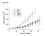
FIGS. 19A and 19B are charts plotting tumor growth of DU4475 breast tumors versus days after treatment with embodiments of anti-VEGFR-I antibodies of the present invention.
FIG. 20A-C is a chart plotting tumor growth of DU4475 (FIG. 20A), MDA-MB-231 (FIG. 20B) and MDA-MB-435 (FIG. 20C) breast tumors versus days after treatment with anti-VEGFR-I antibody 18F1 of the present invention.
FIG. 21A-B is a chart plotting tumor growth of DU4475 (FIG. 21A) and MDA-MB-231 (FIG. 21B) breast tumors versus days after treatment with anti-human VEGFR-I antibody 18F1 of the present invention and anti-mouse VEGFR-I antibody MF1.
FIG. 22 is a chart of the number of colon cancer cell colonies present after treatment with anti-human VEGFR-I antibody 18F1 in the presence of VEGF-A and VEGF-B.
FIG. 23A is a chart of the number of migrated tumor cells after treatment with anti-human VEGFR-I antibody 18F1 in the presence of VEGF-A and VEGF-B.
FIG. 23B are photomicrographs of stained migrated cells after treatment with anti-human VEGFR-I antibody 18F1 in the presence of VEGF-A and VEGF-B.
FIG. 24A is a chart of the number of tumor cells that migrated across a layer of MATRIGEL™ after treatment with anti-human VEGFR-I antibody 18F1 in the presence of VEGF-A or VEGF-B.
FIG. 24B are photomicrographs of stained migrated cells after treatment with anti-human VEGFR-I antibody 18F1 in the presence of VEGF-A and VEGF-B.
FIG. 25 is a chart plotting tumor growth of DU4475 (FIG. 25A) and MDA-MB-435 (FIG. 25B) breast tumors versus days after treatment with anti-VEGFR-I antibodies 18F1, 6F9 and 15F11.
FIG. 26 is a chart plotting growth of HT-29 (FIG. 26A), DLD-I (FIG. 26B) and GEO (FIG. 26C) colon cancer cells versus days after treatment with particular doses of anti-human VEGFR-I antibody 18F1.
FIG. 27 are photomicrographs of MDS-MB-231 xenograft tumors after treatment with anti-human VEGFR-I antibody 18F1.
FIG. 28 is a chart plotting tumor growth versus days after treatment with particular doses of anti-human anti-VEGFR-I antibody 18F1, anti-mouse anti-VEGFR-I antibody MF1, or both in MDA-MB-231 (FIG. 28A) and DU4475 (FIG. 28B) xenografts.
FIG. 29 is a chart plotting tumor growth versus days after treatment with anti-human anti-VEGFR-I antibody 18F1 and anti-mouse anti-VEGFR-I antibody MF1 in combination with cyclophosphamide in MDS-MB-231 xenografts.
FIGS. 30A and B are charts plotting tumor growth versus days after treatment with 5-FU/LV or doxorubicin in combination with anti-human anti-VEGFR-I antibody 18F1 and anti-mouse anti-VEGFR-1 antibody MF1 in MDA-MB-231 xenografts.
FIG. 31 is a chart of total tumor cell count versus antibody concentration of various amounts of 18F1 in the presence of VEGF-A (FIG. 31A) or PIGF (FIG. 31B) following treatment with desferoxamine.
FIGS. 32A, B, and C are charts depicting the specificity of anti-human anti-VEGFR-1 antibody 18F1 and anti-mouse anti-VEGFR-1 antibody MF1.
DETAILED DESCRIPTION OF THE INVENTION
In an embodiment, the present invention provides a monoclonal antibodies and fragments thereof that specifically bind to VEGFR-I (such antibodies and fragments thereof referred to herein as “anti-VEGFR-1 antibodies” unless otherwise indicated). Anti-VEGFR-1 antibodies of the present invention comprise a light chain complementarity determining region-2 (CDR2) of SEQ ID NO: 2 and a light chain complementarity region-3 (CDR3) of SEQ ID NO: 3. Alternatively and preferably, anti-VEGFR-1 antibodies of the present invention comprise a light chain complementarity region-1 (CDR1) having the following sequence:
- RASQSX 1 SSSYLA, where X1 is V or G (SEQ ID NO: 1 or 4). Alternatively and preferably, anti-VEGFR-I antibodies of the present invention comprise a heavy chain CDR1 having the following sequence: GFX2FSSYGMH, where X2 is T or A (SEQ ID NO: 5 or 11). Alternatively and preferably, anti-VEGFR-I antibodies of the present invention comprise a heavy chain CDR2 having the following sequence:
- VIWX 3 DGSNKYYADSVX 4 G, where X3 is Y or F and X4 is K or R (SEQ ID NO: 6, 9, or 12). Alternatively and also preferably, anti-VEGFR-1 antibodies of the present invention comprise a heavy chain CDR3 having the following sequence:
- DHX 5 GSGX 6 HX7 YX8 YYGX 9 DV, where X5 is F or Y; X6 is A or V; X7 is Y, S, or H; X8 is Y or F; and X9 is M or L (SEQ ID NO: 7, 8, 10, 13). The amino acid sequences of the CDRs of preferred anti-VEGFR-1 antibodies (designated as clones “6F9,” “13G12,” “15F11,” and “18F1” (or “MC-18F1”)) are set forth below in Table 1.
| TABLE 1 |
| |
| CDR sequence of anti-VEGFR-1 antibodies |
|
| 6F9 |
RASQSGSSSYLA |
GASSRAT |
QQYGSSPLT |
|
| |
(SEQ ID NO: 1) |
(SEQ ID NO: 2) |
(SEQ ID NO: 3) |
| |
| 13G12 |
RASQSGSSSYLA |
GASSRAT |
QQYGSSPLT |
| |
(SEQ ID NO: 1) |
(SEQ ID NO: 2) |
(SEQ ID NO: 3) |
| |
| 15F11 |
RASQSVSSSYLA |
GASSRAT |
QQYGSSPLT |
| |
(SEQ ID NO: 4) |
(SEQ ID NO: 2) |
(SEQ ID NO: 3) |
| |
| 18F1 |
RASQSVSSSYLA |
GASSRAT |
QQYGSSPLT |
| |
(SEQ ID NO: 4) |
(SEQ ID NO: 2) |
(SEQ ID NO: 3) |
| |
| 6F9 |
GFTFSSYGMH |
VIWYDGSNKYYADSVKG |
DHFGSGAHYYYYYGMDV |
|
| |
(SEQ ID NO: 5) |
(SEQ ID NO: 6) |
(SEQ ID NO: 7) |
| |
| 13G12 |
GFTFSSYGMH |
VIWYDGSNKYYADSVKG |
DHYGSGAHYYYYYGMDV |
| |
(SEQ ID NO: 5) |
(SEQ ID NO: 6) |
(SEQ ID NO: 8) |
| |
| 15F11 |
GFTFSSYGMH |
VIWFDGSNKYYADSVKG |
DHYGSGAHSYYYYGLDV |
| |
(SEQ ID NO: 5) |
(SEQ ID NO: 9) |
(SEQ ID NO: 10) |
| |
| 18F1 |
GFAFSSYGMH |
VIWYDGSNKYYADSVRG |
DHYGSGVHHYFYYGLDV |
| |
(SEQ ID NO: 11) |
(SEQ ID NO: 12) |
(SEQ ID NO: 13) |
| |
In another embodiment, anti-VEGFR-I antibodies of the present invention have a light chain variable region (VL) of SEQ ID NO: 14, 15, or 16 and/or a heavy chain variable region (VH) of SEQ ID NO: 17, 18, 19, or 20. The amino acid sequences of the light and heavy chain variable regions of preferred anti-VEGFR-I antibodies of the present invention are set forth below in Table 2.
| TABLE 2 |
| |
| Variable region sequence of anti-VEGFR-1 anti- |
|
| bodies (underlined portions represent CDRs) |
| |
Light Chain |
| 6F9 |
EIVLTQSPGTLSLSPGERATLSCRASQSGSSSYLAWYQQKPGQAPRLLIYGASS |
| |
RATGIPDRFSGSGSGTDFTLTISRLEPEDFAVYYCQQYGSSPLTFGGGTKVEIK |
| |
RTVAAPSVFIFP |
| |
SEQ ID NO: 14 |
| |
| 13G12 |
EIVLTQSPGTLSLSPGERATLSCRASQSGSSSYLAWYQQKPGQAPRLLIYGASS |
| |
RATGIPDRFSGSGSGTDFTLTISRLEPEDFAVYYCQQYGSSPLTFGGGTKVEIK |
| |
RTVAAPSVFIFP |
| |
SEQ ID NO: 14 |
| |
| 15F11 |
EIVLTQSPGTLSLSPGERATLSCRASQSVSSSYLAWYQQKPGQAPRLLIYGASS |
| |
RATGIPDRFSGSGSGTDFTLTISRLEPEDFAVYYCQQYGSSPLTFGQGTRLEIKR |
| |
TVAAPSVFIFP |
| |
SEQ ID NO: 15 |
| |
| 18F1 |
EIVLTQSPGTLSLSPGERATLSCRASQSVSSSYLAWYQQKPGQAPRLLIYGASS |
| |
RATGIPDRFSGSGSGTDFTLTISRLEPEDFAVYYCQQYGSSPLTFGGGTKVEIK |
| |
RTVAAPSVFIFP |
| |
SEQ ID NO: 16 |
| |
| |
Heavy Chain |
| 6F9 |
QVQLVESGGGVVQPGRSLRLSCAASGFTFSSYGMHWVRQAPGKGLEWVAVI |
| |
WYDGSNKYYADSVKGRFTISRDNSKNTVYLQMNSLRAEDTAVYHCTRDHFG |
| |
SGAHYYYYYGMDVWGQGTTVTVSS |
| |
SEQ ID NO: 17 |
| |
| 13G12 |
QVQLVESGGGVVQPGRSLRLSCAASGFTFSSYGMHWVRQAPGKGLEWVAVI |
| |
WYDGSNKYYADSVKGRFTISRDNSKNILYLQMNSLRAEDTAVYYCARDHY |
| |
GSGAHYYYYYGMDVWGQGTTVTVSS |
| |
SEQ ID NO: 18 |
| |
| 15F11 |
QVQLVESGGGVVQPGRSLRLSCAASGFTFSSYGMHWVRQAPGKGLEWVAVI |
| |
WFDGSNKYYADSVKGRFTISRDNSKNTLYLQMNSLRAEDTAVYYCARDHYG |
| |
SGAHSYYYYGLDVWGQGTSVTVSS |
| |
SEQ ID NO: 19 |
| |
| 18F1 |
QAQVVESGGGVVQSGRSLRLSCAASGFAFSSYGMHWVRQAPGKGLEWVAVI |
| |
WYDGSNKYYADSVRGRFTISRDNSENTLYLQMNSLRAEDTAVYYCARDHYG |
| |
SGVHHYFYYGLDVWGQGTTVTVSS |
| |
SEQ ID NO: 20 |
| |
In a preferred embodiment, the anti-VEGFR-I antibodies of the present invention are human antibodies.
Anti-VEGFR-1 antibodies of the present invention include whole antibodies and antibody fragments that specifically bind to VEGFR-I. Non-limiting examples of types of antibodies according to the present invention include naturally occurring antibodies; single chain antibodies; multivalent single chain antibodies such as diabodies and tribodies; monovalent fragments such as Fab (Fragment, antigen binding), bivalent fragments such as (FaV)2; Fv (fragment variable) fragments or derivatives thereof such as single chain Fv (scFv) fragments; and single domain antibodies that bind specifically to VEGFR-I.
Naturally occurring antibodies typically have two identical heavy chains and two identical light chains, with each light chain covalently linked to a heavy chain by an interchain disulfide bond and multiple disulfide bonds further linking the two heavy chains to one another. Individual chains can fold into domains having similar sizes (110-125 amino acids) and structures, but different functions. The light chain can comprise one VL and one constant domain (CL). The heavy chain can also comprise one VH and/or depending on the class or isotope of antibody, three or four constant domains (CHI, CH2, C H3, and CH4). In humans, the isotypes are IgA, IgD, IgE, IgG, and IgM, with IgA and IgG further subdivided into subclasses or subtypes (IgA1-2 and IgG1-4).
Single chain antibodies lack some or all of the constant domains of the whole antibodt from which they are derived. The peptide linkers used to produce the single chain antibodies may be flexible peptides selected to assure that the proper three-dimensional folding of the VL and VH domains occurs. Generally, the carboxyl terminus of the VL or VH sequence may be covalently linked by such a peptide linker to the amino acid terminus of a complementary VH or VL sequence. The linker is generally 10 to 50 amino acid residues, preferably 10 to 30 amino acid residues, more preferably 12 to 30 amino acid residues, and most preferably 15 to 25 amino acid residues. An example of such linker peptides include (Gly-Gly-Gly-Gly-Ser)3 (SEQ ID NO: 28).
Multiple single chain antibodies, each single chain having one VH and one VL domain covalently linked by a first peptide linker, can be covalently linked by at least one or more peptide linkers to form a multivalent single chain antibody, which can be monospecific or multispecific. Each chain of a mulivalent single chain antibody includes a variable light chain fragment and a variable heavy chain fragment, and is linked by a peptide linker to at least one other chain.
Two single chain antibodies can be combined to form a diabody, also known as a trivalent dimer. Diabodies have two chains and two binding sites and can be monospecific or bispecific. Each chain of the diabody includes a VH domain connected to a VL domain. The domains are connected with linkers that are short enough to prevent pairing between domains on the same chain, thus driving the pairing between complementary domains on different chains to recreate the two antigen-binding sites.
Three single chain antibodies can be combined to form triabodies, also known as trivalent trimers. Triabodies are constructed with the amino acid terminus of a VL or VH domain directly fused to the carboxyl terminus of a VL or VH domain, i.e., without any linker sequence. The triabody has three Fv heads with the polypeptides arranged in a cyclic, head-to-tail fashion. A possible conformation of the triabody is planar with the three binding sites located in a plane at an angle of 120 degrees from one another. Triabodies can be monospecific, bispecific or trispecific.
Fab fragments refer to fragments of the antibody consisting of VLCLVHCHI domains. Those generated by papain digestion are referred to as “Fab” and do not retain the heavy chain hinge region. Those generated by pepsin digestion are referred to either as “(Fab′)2,” in which case the interchain disulfide bonds are intact, or as Fab′, in which case the disulfide bonds are not retained. Bivalent (Fab′)2 fragments have higher avidity for antigen than that of monovalent Fab fragments.
Fv fragments are the portion of an antibody consisting of the VL and VH domains and constitute the antigen-binding site. scFv is an antibody fragment containing a VL domain and VH domain on one polypeptide chain, wherein the N terminus of one domain and the C terminus of the other domain are joined by a flexible linker to allows the two fragments to associate to form a functional antigen binding site (see, for example U.S. Pat. No. 4,946,778 (Ladner et al), WO 88/09344, (Huston et al.), both of which are incorporated by reference herein). WO 92/01047 (McCafferty et al.), which is incorporated by reference herein, describes the display of scFv fragments on the surface of soluble recombinant genetic display packages, such as bacteriophage.
Single domain antibodies have a single variable domain that is capable of efficiently binding antigen. Examples of antibodies wherein binding affinity and specificity are contributed primarily by one or the other variable domain are known in the art. See, e.g., Jeffrey, P. D. et al., Proc. Natl Acad. ScL USA 90:10310-4 (1993), which is incorporated by reference herein and which discloses an anti-digoxin antibody which binds to digoxin primarily by the antibody heavy chain. Accordingly, single antibody domains can be identified that bind well to VEGF receptors. It is understood that, to make a single domain antibody from an antibody comprising a VH and a VL domain, certain amino acid substitutions outside the CDR regions may be desired to enhance binding, expression or solubility. For example, it may be desirable to modify amino acid residues that would otherwise be buried in the VH-VL interface.
Each domain of anti-VEGFR-1 antibodies of the present invention may be a complete antibody heavy or light chain variable domain, or it may be a functional equivalent or a mutant or derivative of a naturally occuring domain, or a synthetic domain constructed, for example, in vitro using a technique such as one described in WO 93/11236 (Griffiths et al.). For instance, it is possible to join together domains corresponding to antibody variable domains which are missing at least one amino acid. The important characterizing feature is the ability of each domain to associate with a complementary domain to form an antigen binding site. Accordingly, the terms “variable heavy/light chain fragment” should not be construed to exclude variants which do not have a material effect on VEGFR-I binding specificity.
As used herein, an “anti-VEGFR-I antibody” include modifications of an anti-VEGFR-I antibody of the present invention that retain specificity for VEGFR-I. Such modifications include, but are not limited to, conjugation to an effector molecule such as a chemotherapeutic agent (e.g., cisplatin, taxol, doxorubicin) or cytotoxin (e.g., a protein, or a non-protein organic chemotherapeutic agent). Modifications further include, but are not limited to conjugation to detectable reporter moieties. Modifications that extend antibody half-life (e.g., pegylation) are also included.
Proteins and non-protein agents may be conjugated to the antibodies by methods that are known in the art. Conjugation methods include direct linkage, linkage via covalently attached linkers, and specific binding pair members (e.g., avidin-biotin). Such methods include, for example, that described by Greenfield et al., Cancer Research 50, 6600-6607 (1990), which is incorporated by reference herein, for the conjugation of doxorubicin and those described by Amon et al., Adv. Exp. Med. Biol. 303, 79-90 (1991) and by Kiseleva et al, MoI. Biol. (USSR)25, 508-514 (1991), both of which are incorporated by reference herein, for the conjugation of platinum compounds.
Anti-VEGFR-I antibodies of the present invention also include those for which binding characteristics have been improved by direct mutation, methods of affinity maturation, phage display, or chain shuffling. Affinity and specificity may be modified or improved by mutating any of the CDRs of the antibodies of the present invention and screening for antigen binding sites having the desired characteristics (see, e.g., Yang et al., J. MoI. Biol, 254: 392-403 (1995), which is incorporated by reference herein). The CDRs may be mutated in a variety of ways that are known to one of skill in the art. For example, one way is to randomize individual residues or combinations of residues so that in a population of otherwise identical antigen binding sites, all twenty amino acids are found at particular positions. Alternatively, mutations are induced over a range of CDR residues by error prone PCR methods (see, e.g., Hawkins et al., J. MoI. Biol, 226: 889-896 (1992), which is incorporated by reference herein). For example, phage display vectors containing heavy and light chain variable region genes may be propagated in mutator strains of E. coli (see, e.g., Low et al., J. MoI Biol, 250: 359-368 (1996), which is incorporated by reference herein).
Anti-VEGFR-I antibodies also include functional equivalents that include polypeptides with amino acid sequences substantially the same as the amino acid sequence of the variable or hypervariable regions of the antibodies of the present invention. “Substantially the same” amino acid sequence includes an amino acid sequence with at least 70%, preferably at least 80%, and more preferably at least 90% identity to another amino acid sequence when the amino acids of the two sequences are optimally aligned and compared to determine exact matches of amino acids between the two sequences. “Substantially the same” amino acid sequence also includes an amino acid sequence with at least 70%, preferably at least 80%, and more preferably at least 90% homology to another amino acid sequence, as determined by the FASTA search method in accordance with Pearson and Lipman, Proc. Natl. Acad. Sci. USA 85, 2444-8 (1988).
As stated earlier, anti-VEGFR-I antibodies of the present invention specifically bind to VEGFR-I. Such antibodies can be monospecific or bispecific so long as one antigen-binding site is specific for VEGFR-I. Antibody specificity, which refers to selective recognition of an antibody for a particular epitope of an antigen, of antibodies for VEGFR-I can be determined based on affinity and/or avidity. Affinity, represented by the equilibrium constant for the dissociation of an antigen with an antibody (Kd), measures the binding strength between an antigenic determinant (epitope) and an antibody binding site. Avidity is the measure of the strength of binding between an antibody with its antigen. Antibodies typically bind with a Kd of 10−5 to 10−11 liters/mole. Any Kd less than 10−4 liters/mole is generally considered to indicate non-specific binding. The lesser the value of the Kd, the stronger the binding strength between an antigenic determinant and the antibody binding site.
Anti-VEGFR-I antibodies of the present invention specifically bind to the extracellular region of VEGFR-I and preferably neutralize activation of VEGFR-I by preventing binding of a ligand of VEGFR-I to the receptor. In such preferable embodiments, the antibody binds VEGFR-I at least as strongly as the natural ligands of VEGFR-I (including VEGF(A), VEGF-B and PIGF).
Neutralizing activation of VEGFR-I includes diminishing, inhibiting, inactivating, and/or disrupting one or more of the activities associated with signal transduction. Such activities include receptor dimerization, autophosphorylation of VEGFR-I, activation of VEGFR-I's internal cytoplasmic tyrosine kinase domain, and initiation of multiple signal transduction and transactivation pathways involved in regulation of DNA synthesis (gene activation) and cell cycle progression or division. One measure of VEGFR-I neutralization is inhibition of the tyrosine kinase activity VEGFR-I. Tyrosine kinase inhibition can be determined using well-known methods such as phosphorylation assays which measuring the autophosphorylation level of recombinant kinase receptor, and/or phosphorylation of natural or synthetic substrates. Phosphorylation can be detected, for example, using an antibody specific for phosphotyrosine in an ELISA assay or on a western blot. Some assays for tyrosine kinase activity are described in Panek et al., J. Pharmacol. Exp. Them., 283: 1433-44 (1997) and Batley et al, Life ScL, 62: 143-50 (1998), both of which are incorporated by reference.
In addition, methods for detection of protein expression can be utilized to determine whether an antibody neutralizes activation of VEGFR-I, wherein the proteins being measured are regulated by VEGFR-I tyrosine kinase activity. These methods include immunohistochemistry (IHC) for detection of protein expression, fluorescence in situ hybridization (FISH) for detection of gene amplification, competitive radioligand binding assays, solid matrix blotting techniques, such as Northern and Southern blots, reverse transcriptase polymerase chain reaction (RT-PCR) and ELISA. See, e.g., Grandis et al., Cancer, 78:1284-92. (1996); Shimizu et al., Japan J. Cancer Res., 85:567-71 (1994); Sauteret al., Am. J. Path., 148:1047-53 (1996); Collins, Glia, 15:289-96 (1995); Radinsky et al., Clin. Cancer Res., 1:19-31 (1995); Petrides et al., Cancer Res., 50:3934-39 (1990); Hoffmann et al., Anticancer Res., 17:4419-26 (1997); Wikstrand et al., Cancer Res., 55:3140-48 (1995), all of which are incorporated by reference.
In vivo assays can also be utilized to detect VEGFR-I neutralization. For example, receptor tyrosine kinase inhibition can be observed by mitogenic assays using cell lines stimulated with receptor ligand in the presence and absence of inhibitor. For example, HUVEC cells (ATCC) stimulated with VEGF(A) or VEGF-B can be used to assay VEGFR-I inhibition. Another method involves testing for inhibition of growth of VEGF-expressing tumor cells, using for example, human tumor cells injected into a mouse. See e.g., U.S. Pat. No. 6,365,157 (Rockwell et al.), which is incorporated by reference herein.
Of course, the present invention is not limited by any particular mechanism of VEGFR-I neutralization. Anti-VEGFR-I antibodies of the present invention can, for example, bind externally to VEGFR-I, block binding of ligand to VEGFR-I and subsequent signal transduction mediated via receptor-associated tyrosine kinase, and prevent phosphorylation of VEGFR-I and other downstream proteins in the signal transduction cascade. The receptor-antibody complex can also be internalized and degraded, resulting in receptor cell surface down-regulation. Matrix metalloproteinases, which function is tumor cell invasion and metastasis, can also be down-regulated by anti-VEGFR-I antibodies of the present invention.
Human anti-VEGFR-I antibodies can be obtained from naturally occurring antibodies, or Fab or scFv phage display libraries constructed, for example, from human heavy chain and light chain variable region genes and the CDR sequences of the anti-VEGFR-I antibodies of the present invention can be inserted into such human anti-VEGFR-I antibodies.
Human anti-VEGFR-I antibodies can be produced by methods well known to one of skill in the art. Such methods include the hybridoma method using transgenic mice described by Kohler and Milstein, Nature, 256: 495-497 (1975) and Campbell, Monoclonal Antibody Technology, The Production and Characterization of Rodent and Human Hybridomas, Burdon et ah, Eds., Laboratory Techniques in Biochemistry and Molecular Biology, Volume 13, Elsevier Science Publishers, Amsterdam (1985), all of which are incorporated by reference herein; as well as by the recombinant DNA method described by Huse et al., Science, 246, 1275-1281 (1989), which is incorporated by reference herein.
Antibody fragments can be produced by cleaving a whole antibody, or by expressing DNA that encodes the fragment. Fragments of antibodies may be prepared by methods described by Lamoyi et al., J. Immunol. Methods, 56: 235-243 (1983) and by Parham, J. Immunol. 131: 2895-2902 (1983), both of which are incorporated by reference herein. Such fragments may contain one or both Fab fragments or the F(ab′)2 fragment. Such fragments may also contain single-chain fragment variable region antibodies, i.e. scFv, diabodies, or other antibody fragments. Methods of producing such antibodies are disclosed in PCT Application WO 93/21319, European Patent Application No. 239,400; PCT Application WO 89/09622; European Patent Application 338,745; and European Patent Application EP 332,424, all of which are incorporated by reference herein.
in another embodiment, the present invention provides polynucleotides encoding the anti-VEGFR-I antibodies of the present invention. Such polynucleotides encode the light chain CDR2 of SEQ ID NO.: 2, the light chain CDR3 of SEQ ID NO: 3, and, preferably, one or more of the other CDRs listed in Table 1. Table 3 sets forth the nucleic acid sequences of preferred anti-VEGFR-I antibodies.
| TABLE 3 |
| |
| Nucleotide sequence of anti-VEGFR-1 antibodies |
| |
Light Chain |
| 6F9 |
GAAATTGTGTTGACGCAGTCTCCAGGCACCCTGTCCTTGTCTCCAGGGGAA |
| |
AGAGCCACCCTCTCCTGCAGGGCCAGTCAGAGTGGTAGCAGCAGCTACTT |
| |
AGCCTGGTACCAGCAGAAACCTGGCCAGGCTCCCAGGCTCCTCATCTATG |
| |
GTGCATCCAGCAGGGCCACTGGCATCCCAGACAGGTTCAGTGGCAGTGGG |
| |
TCTGGGACAGACTTCACTCTCACCATCAGCAGACTGGAGCCTGAAGATTTT |
| |
GCAGTGTATTACTGTCAGCAGTATGGTAGCTCACCGCTCACTTTCGGCGGA |
| |
GGGACCAAGGTGGAGATCAAACGAACTGTGGCTGCACCATCTGTCTTCAT |
| |
CTTCCCG |
| |
SEQ ID NO: 21 |
| |
| 13G12 |
GAAATTGTGTTGACGCAGTCTCCAGGCACCCTGTCCTTGTCTCCAGGGGAA |
| |
AGAGCCACCCTCTCCTGCAGGGCCAGTCAGAGTGGTAGCAGCAGCTACTT |
| |
AGCCTGGTACCAGCAGAAACCTGGCCAGGCTCCCAGGCTCCTCATCTATG |
| |
GTGCATCCAGCAGGGCCACTGGCATCCCAGACAGGTTCAGTGGCAGTGGG |
| |
TCTGGGACAGACTTCACTCTCACCATCAGCAGACTGGAGCCTGAAGATTTT |
| |
GCAGTGTATTACTGTCAGCAGTATGGTAGCTCACCGCTCACTTTCGGCGGA |
| |
GGGACCAAGGTGGAGATCAAACGAACTGTGGCTGCACCATCTGTCTTCAT |
| |
CTTCCCG |
| |
SEQ ID NO: 21 |
| |
| 15F11 |
GAAATTGTGTTGACGCAGTCTCCAGGCACCCTGTCTTTGTCTCCAGGGGAA |
| |
AGAGCCACCCTCTCCTGCAGGGCCAGTCAGAGTGTTAGCAGCAGCTACTT |
| |
AGCCTGGTACCAGCAGAAACCTGGCCAGGCTCCCAGGCTCCTCATCTATG |
| |
GTGCATCCAGCAGGGCCACTGGCATCCCAGACAGGTTCAGTGGCAGTGGG |
| |
TCTGGGACAGACTTCACTCTCACCATCAGCAGACTGGAGCCTGAAGATTTT |
| |
GCAGTGTATTACTGTCAGCAGTATGGTAGCTCACCTCTCACCTTCGGCCAA |
| |
GGGACACGACTGGAGATTAAACGAACTGTGGCTGCACCATCTGTCTTCAT |
| |
CTTCCCG |
| |
SEQ ID NO: 22 |
| |
| 18F1 |
GAAATTGTGTTGACGCAGTCTCCAGGCACCCTGTCTTTGTCTCCAGGGGAA |
| |
AGAGCCACCCTCTCCTGCAGGGCCAGTCAGAGTGTTAGCAGCAGCTACTT |
| |
AGCCTGGTACCAGCAGAAACCTGGCCAGGCTCCCAGGCTCCTCATCTATG |
| |
GTGCATCCAGCAGGGCCACTGGCATCCCAGACAGGTTCAGTGGCAGTGGG |
| |
TCTGGGACAGACTTCACTCTCACCATCAGCAGACTGGAGCCTGAAGATTTT |
| |
GCAGTGTATTACTGTCAGCAGTATGGTAGCTCACCGCTCACTTTCGGCGGA |
| |
GGGACCAAGGTGGAGATCAAACGAACTGTGGCTGCACCATCTGTCTTCAT |
| |
CTTTCCG |
| |
SEQ ID NO: 23 |
| |
| |
Heavy Chain |
| 6F9 |
CAGGTGCAGCTGGTGGAGTCTGGGGGAGGCGTGGTCCAGCCTGGGAGGTC |
| |
CCTGAGACTCTCCTGTGCAGCGTCTGGATTCACCTTCAGTAGTTATGGCAT |
| |
GCACTGGGTCCGCCAGGCTCCAGGCAAGGGGCTGGAGTGGGTGGCAGTTA |
| |
TATGGTATGATGGAAGTAATAAATACTATGCAGACTCCGTGAAGGGCCGA |
| |
TTCACCATCTCCAGAGACAATTCCAAGAACACGGTGTATCTGCAAATGAA |
| |
CAGCCTGAGAGCCGAGGACACGGCTGTGTATCACTGTACGAGAGATCACT |
| |
TTGGTTCGGGGGCTCACTACTACTACTACTACGGTATGGACGTCTGGGGCC |
| |
AAGGGACCACGGTCACCGTCTCCTCA |
| |
SEQ ID NO: 24 |
| |
| 13G12 |
CAGGTGCAGCTGGTGGAGTCTGGGGGAGGCGTGGTCCAGCCTGGGAGGTC |
| |
CCTGAGACTCTCCTGTGCAGCGTCTGGATTCACCTTCAGTAGCTATGGCAT |
| |
GCACTGGGTCCGCCAGGCTCCAGGCAAGGGGCTGGAGTGGGTGGCAGTTA |
| |
TATGGTATGATGGAAGTAATAAATACTATGCAGACTCCGTGAAGGGCCGA |
| |
TTCACCATCTCCAGAGACAATTCCAAGAACACGCTGTATCTGCAAATGAA |
| |
CAGCCTGAGAGCCGAGGACACGGCTGTGTATTACTGTGCGAGAGATCACT |
| |
ATGGTTCGGGGGCTCACTACTACTACTACTACGGTATGGACGTCTGGGGC |
| |
CAAGGGACCACGGTCACCGTCTCCTCA |
| |
SEQ ID NO: 25 |
| |
| 15F11 |
CAGGTGCAGCTGGTGGAGTCTGGGGGAGGCGTGGTCCAGCCTGGGAGGTC |
| |
CCTGAGACTCTCCTGTGCAGCGTCTGGATTCACCTTCAGTAGCTATGGCAT |
| |
GCACTGGGTCCGCCAGGCTCCAGGCAAGGGGCTGGAGTGGGTGGCAGTTA |
| |
TATGGTTTGATGGAAGTAATAAATACTATGCAGACTCCGTGAAGGGCCGA |
| |
TTCACCATCTCCAGAGACAATTCCAAGAACACGCTGTATCTGCAAATGAA |
| |
CAGCCTGAGAGCCGAGGACACGGCTGTGTATTACTGTGCGAGAGATCACT |
| |
ATGGTTCGGGGGCTCACTCCTACTACTACTACGGTTTGGACGTTTGGGGCC |
| |
AAGGGACCTCGGTCACCGTCTCCTCA |
| |
SEQ ID NO: 26 |
| |
| 18F1 |
CAGGCGCAGGTGGTGGAGTCTGGGGGAGGCGTGGTCCAGTCTGGGAGGTC |
| |
CCTGAGACTCTCCTGTGCAGCGTCTGGATTCGCCTTCAGTAGCTACGGCAT |
| |
GCACTGGGTCCGCCAGGCTCCAGGCAAGGGGCTGGAGTGGGTGGCAGTTA |
| |
TATGGTATGATGGAAGTAATAAATACTATGCAGACTCCGTGAGGGGCCGA |
| |
TTCACCATCTCCAGAGACAATTCCGAGAACACGCTGTATCTGCAAATGAA |
| |
CAGCCTGAGAGCCGAGGACACCGCTGTGTATTACTGTGCCAGAGATCACT |
| |
ATGGTTCGGGGGTGCACCACTATTTCTACTACGGTCTGGACGTCTGGGGCC |
| |
AAGGGACCACGGTCACCGTCTCCTCA |
| |
SEQ ID NO: 27 |
| |
DNA encoding human antibodies can be prepared by recombining DNA encoding human constant regions and variable regions, other than the CDRs, derived substantially or exclusively from the corresponding human antibody regions and DNA encoding CDRs derived from a human (SEQ ID NOs: 1-4 for the light chain variable domain CDRs and SEQ ID Nos: 5-13 for the heavy chain variable domain CDRs.
Polynucleotides encoding anti-VEGFR-I antibodies of the present invention include polynucleotides with nucleic acid sequences that are substantially the same as the nucleic acid sequences of the polynucleotides of the present invention. “Substantially the same” nucleic acid sequence is defined herein as a sequence with at least 70%, preferably at least 80%, and more preferably at least 90% identity to another nucleic acid sequence when the two sequences are optimally aligned (with appropriate nucleotide insertions or deletions) and compared to determine exact matches of nucleotides between the two sequences.
Suitable sources of DNAs that encode fragments of antibodies include any cell, such as hybridomas and spleen cells, that express the full-length antibody. The fragments may be used by themselves as antibody equivalents, or may be recombined into equivalents, as described above. The DNA deletions and recombinations described in this section may be carried out by known methods, such as those described in the published patent applications listed above in the section entitled “Functional Equivalents of Antibodies” and/or other standard recombinant DNA techniques, such as those described below. Another source of DNAs are single chain antibodies produced from a phage display library, as is known in the art.
Additionally, the present invention provides expression vectors containing the polynucleotide sequences previously described operably linked to an expression sequence, a promoter and an enhancer sequence. A variety of expression vectors for the efficient synthesis of antibody polypeptide in prokaryotic, such as bacteria and eukaryotic systems, including but not limited to yeast and mammalian cell culture systems have been developed. The vectors of the present invention can comprise segments of chromosomal, non-chromosomal and synthetic DNA sequences.
Any suitable expression vector can be used. For example, prokaryotic cloning vectors include plasmids from E. coli, such as colEl,pCRl,pBR322,pMB9,pUC, pKSM, and RP4. Prokaryotic vectors also include derivatives of phage DNA such as Ml3 and other filamentous single-stranded DNA phages. An example of a vector useful in yeast is the 2μ plasmid. Suitable vectors for expression in mammalian cells include well-known derivatives of SV-40, adenovirus, retrovirus-derived DNA sequences and shuttle vectors derived from combination of functional mammalian vectors, such as those described above, and functional plasmids and phage DNA.
Additional eukaryotic expression vectors are known in the art {e.g., P J. Southern & P. Berg, J. MoI. Appl. Genet, 1:327-341 (1982); Subramani et al, MoI. Cell. Biol, 1: 854-864 (1981); Kaufinann & Sharp, “Amplification And Expression of Sequences Cotransfected with a Modular Dihydrofolate Reductase Complementary DNA Gene,” J. MoI. Biol, 159:601-621 (1982); Kaufhiann & Sharp, MoI. Cell. Biol, 159:601-664 (1982); Scahill et al., “Expression And Characterization Of The Product Of A Human Immune Interferon DNA Gene In Chinese Hamster Ovary Cells,” Proc. Nat'l Acad. ScL USA, 80:4654-4659 (1983); Urlaub & Chasin, Proc. Nat'l Acad. ScL USA, 77:4216-4220, (1980), all of which are incorporated by reference herein).
The expression vectors useful in the present invention contain at least one expression control sequence that is operatively linked to the DNA sequence or fragment to be expressed. The control sequence is inserted in the vector in order to control and to regulate the expression of the cloned DNA sequence. Examples of useful expression control sequences are the lac system, the trp system, the tac system, the trc system, major operator and promoter regions of phage lambda, the control region of fd coat protein, the glycolytic promoters of yeast, e.g., the promoter for 3-phosphoglycerate kinase, the promoters of yeast acid phosphatase, e.g., Pho5, the promoters of the yeast alpha-mating factors, and promoters derived from polyoma, adenovirus, retrovirus, and simian virus, e.g., the early and late promoters or SV40, and other sequences known to control the expression of genes of prokaryotic or eukaryotic cells and their viruses or combinations thereof.
The present invention also provides recombinant host cells containing the expression vectors previously described. Anti-VEGFR-I antibodies of the present invention can be expressed in cell lines other than in hybridomas. Nucleic acids, which comprise a sequence encoding a polypeptide according to the invention, can be used for transformation of a suitable mammalian host cell.
Cell lines of particular preference are selected based on high level of expression, constitutive expression of protein of interest and minimal contamination from host proteins. Mammalian cell lines available as hosts for expression are well known in the art and include many immortalized cell lines, such as but not limited to, Chinese Hamster Ovary (CHO) cells, Baby Hamster Kidney (BHK) cells and many others. Suitable additional eukaryotic cells include yeast and other fungi. Useful prokaryotic hosts include, for example, E. coli, such as E. coli SG-936, E. coli HB 101, E. coli W3110, E. coli X1776, E. coli X2282, E. coli DHI, and E. coli MRC1, Pseudomonas, Bacillus, such as Bacillus subtilis, and Streptomyces.
These present recombinant host cells can be used to produce an antibody by culturing the cells under conditions permitting expression of the antibody and purifying the antibody from the host cell or medium surrounding the host cell. Targeting of the expressed antibody for secretion in the recombinant host cells can be facilitated by inserting a signal or secretory leader peptide-encoding sequence {See, Shokri et al, (2003) Appl Microbiol Biotechnol. 60(6): 654-664, Nielsen et al, Prot. Eng., 10:1-6 (1997); von Heinje et al., Nucl. Acids Res., 14:4683-4690 (1986), all of which are incorporated by reference herein) at the 5′ end of the antibody-encoding gene of interest. These secretory leader peptide elements can be derived from either prokaryotic or eukaryotic sequences. Accordingly suitably, secretory leader peptides are used, being amino acids joined to the N-terminal end of a polypeptide to direct movement of the polypeptide out of the host cell cytosol and secretion into the medium.
The anti-VEGFR-1 antibodies of the present invention can be fused to additional amino acid residues. Such amino acid residues can be a peptide tag to facilitate isolation, for example. Other amino acid residues for homing of the antibodies to specific organs or tissues are also contemplated.
In another embodiment, the present invention provides methods of treating a medical condition by administering a therapeutically effective amount of an anti-VEGFR-I antibody according to the present invention to a mammal in need thereof. Therapeutically effective means an amount effective to produce the desired therapeutic effect, such as inhibiting tyrosine kinase activity.
In a preferred embodiment, the present invention provides a method of reducing tumor growth or inhibiting angiogenesis by administering a therapeutically effective amount of an anti-VEGFR-I antibody of the present invention to a mammal in need thereof. While not intended to be bound to a particular mechanism, the conditions that may be treated by the present methods include, for example, those in which tumor growth or pathogenic angiogenesis is stimulated through a VEGFR paracrine and/or autocrine loop.
With respect to reducing tumor growth, such tumors include primary tumors and metastatic tumors, as well as refractory tumors. Refractory tumors include tumors that fail to respond or are resistant to other forms of treatment such as treatment with chemotherapeutic agents alone, antibodies alone, radiation alone or combinations thereof. Refractory tumors also encompass tumors that appear to be inhibited by treatment with such agents, but recur up to five years, sometimes up to ten years or longer after treatment is discontinued.
Anti-VEGFR-I antibodies of the present invention are useful for treating tumors that express VEGFR-I. Such tumors are characteristically sensitive to VEGF present in their environment, and may further produce and be stimulated by VEGF in an autocrine stimulatory loop. The method is therefore effective for treating a solid or non-solid tumor that is not vascularized, or is not yet substantially vascularized.
Examples of solid tumors which may be accordingly treated include breast carcinoma, lung carcinoma, colorectal carcinoma, pancreatic carcinoma, glioma and lymphoma. Some examples of such tumors include epidermoid tumors, squamous tumors, such as head and neck tumors, colorectal tumors, prostate tumors, breast tumors, lung tumors, including small cell and non-small cell lung tumors, pancreatic tumors, thyroid tumors, ovarian tumors, and liver tumors. Other examples include Kaposi's sarcoma, CNS neoplasms, neuroblastomas, capillary hemangioblastomas, meningiomas and cerebral metastases, melanoma, gastrointestinal and renal carcinomas and sarcomas, rhabdomyosarcoma, glioblastoma, preferably glioblastoma multiforme, and leiomyosarcoma. Examples of vascularized skin cancers for which anti-VEGFR-1 antibodies of the present invention are effective include squamous cell carcinoma, basal cell carcinoma and skin cancers that can be treated by suppressing the growth of malignant keratinocytes, such as human malignant keratinocytes.
Examples of non-solid tumors include leukemia, multiple myeloma and lymphoma. Some examples of leukemias include acute myelogenous leukemia (AML), chronic myelogenous leukemia (CML), acute lymphocytic leukemia (ALL), chronic lymphocytic leukemia (CLL), erythrocytic leukemia or monocytic leukemia. Some examples of lymphomas include Hodgkin's and non-Hodgkin's lymphoma.
With respect to inhibiting aiigiogenesis, anti-VEGFR-I antibodies of the present invention are effective for treating subjects with vascularized tumors or neoplasms, or angiogenic diseases characterized by excessive angiogenesis. Such tumors and neoplasms include, for example, malignant tumors and neoplasms, such as blastomas, carcinomas or sarcomas, and highly vascular tumors and neoplasms. Cancers that may be treated by the methods of the present invention include, for example, cancers of the brain, genitourinary tract, lymphatic system, stomach, renal, colon, larynx and lung and bone. Non-limiting examples further include epidermoid tumors, squamous tumors, such as head and neck tumors, colorectal tumors, prostate tumors, breast tumors, lung tumors, including lung adenocarcinoma and small cell and non-small cell lung tumors, pancreatic tumors, thyroid tumors, ovarian tumors, and liver tumors. The method is also used for treatment of vascularized skin cancers, including squamous cell carcinoma, basal cell carcinoma, and skin cancers that can be treated by suppressing the growth of malignant keratinocytes, such as human malignant keratinocytes. Other cancers that can be treated include Kaposi's sarcoma, CNS neoplasms (neuroblastomas, capillary hemangioblastomas, meningiomas and cerebral metastases), melanoma, gastrointestinal and renal carcinomas and sarcomas, rhabdomyosarcoma, glioblastoma, including glioblastoma multiforme, and leiomyosarcoma.
Non-limiting examples of pathological angiogenic conditions characterized by excessive angiogenesis involving, for example inflammation and/or vascularization include atherosclerosis, rheumatoid arthritis (RA), neovascular glaucoma, proliferative retinopathy including proliferative diabetic retinopathy, macular degeneration, hemangiomas, angiofibromas, and psoriasis. Other non-limiting examples of non-neoplastic angiogenic disease are retinopathy of prematurity (retrolental fibroplastic), corneal graft rejection, insulin-dependent diabetes mellitus, multiple sclerosis, myasthenia gravis, Crohn's disease, autoimmune nephritis, primary biliary cirrhosis, psoriasis, acute pancreatitis, allograph rejection, allergic inflammation, contact dermatitis and delayed hypersensitivity reactions, inflammatory bowel disease, septic shock, osteoporosis, osteoarthritis, cognition defects induced by neuronal inflammation, Osier-Weber syndrome, restinosis, and fungal, parasitic and viral infections, including cytomegaloviral infections.
The identification of medical conditions treatable by anti-VEGFR-I antibodies of the present invention is well within the ability and knowledge of one skilled in the art. For example, human individuals who are either suffering from a clinically significant neoplastic or angiogenic disease or who are at risk of developing clinically significant symptoms are suitable for administration of the present VEGF receptor antibodies. A clinician skilled in the art can readily determine, for example, by the use of clinical tests, physical examination and medical/family history, if an individual is a candidate for such treatment.
Anti-VEGFR-1 antibodies of the present invention can be administered for therapeutic treatments to a patient suffering from a tumor or angiogenesis associated pathologic condition in an amount sufficient to prevent, inhibit, or reduce the progression of the tumor or pathologic condition. Progression includes, e.g, the growth, invasiveness, metastases and/or recurrence of the tumor or pathologic condition. Amounts effective for this use will depend upon the severity of the disease and the general state of the patient's own immune system. Dosing schedules will also vary with the disease state and status of the patient, and will typically range from a single bolus dosage or continuous infusion to multiple administrations per day (e.g., every 4-6 hours), or as indicated by the treating physician and the patient's condition. It should be noted, however, that the present invention is not limited to any particular dose.
hi another embodiment, the present invention provides a method of treating a medical condition by administering an anti-VEGFR-I antibody of the present invention in combination with one or more other agents. For example, an embodiment of the present invention provides a method of treating a medical condition by administering an anti-VEGFR-I antibody of the present invention with an antineoplastic or antiangiogenic agent. The anti-VEGFR-I antibody can be chemically or biosynthetically linked to one or more of the antineoplastic or antiangiogenic agents.
Any suitable antineoplastic agent can be used, such as a chemotherapeutic agent or radiation. Examples of chemotherapeutic agents include, but are not limited to, cisplatin, doxorubicin, cyclophosphamide, paclitaxel, irinotecan (CPT-II), topotecan or a combination thereof. When the antineoplastic agent is radiation, the source of the radiation can be either external (external beam radiation therapy—EBRT) or internal (brachytherapy—BT) to the patient being treated.
Further, anti-VEGFR-I antibodies of the present invention maybe administered with antibodies that neutralize other receptors involved in tumor growth or angiogenesis. One example of such a receptor is VEGFR-2/KDR. In an embodiment, an anti-VEGR-I antibody of the present invention is used in combination with a receptor antagonist that binds specifically to VEGFR-2. Particularly preferred are antigen-binding proteins that bind to the extracellular domain of VEGFR-2 and block binding by any one of its ligands, such as VEGF(A), VEGF-C, VEGF-D, or VEGF-E.
Another example of such a receptor is EGFR. hi an embodiment of the present invention, an anti-VEGFR-I antibody is used in combination with an EGFR antagonist. An EGFR antagonist can be an antibody that binds to EGFR or a ligand of EGFR and inhibits binding of EGFR to its ligand. Ligands for EGFR include, for example, EGF, TGF-ce amphiregulin, heparin-binding EGF (HB-EGF) and betarecullulin. EGF and TGF-α are thought to be the main endogenous ligands that result in EGFR-mediated stimulation, although TGF-α has been shown to be more potent in promoting angiogenesis. It should be appreciated that the EGFR antagonist can bind externally to the extracellular portion of EGFR, which may or may not inhibit binding of the ligand, or internally to the tyrosine kinase domain. Examples of EGFR antagonists that bind EGFR include, without limitation, biological molecules, such as antibodies (and functional equivalents thereof) specific for EGFR, and small molecules, such as synthetic kinase inhibitors that act directly on the cytoplasmic domain of EGFR.
Other examples of growth factor receptors involved in tumorigenesis are the receptors for platelet-derived growth factor (PDGFR), insulin-like growth factor (IGFR), nerve growth factor (NGFR), and fibroblast growth factor (FGFR).
In an additional alternative embodiment, the present invention provides a method of treating a medical condition by administering an anti-VEGFR-I antibody of the present invention in combination with one or more suitable adjuvants, such as, for example, cytokines (IL-IO and IL-1 3, for example) or other immune stimulators. See, e.g., Larrivee et al, supra.
In a combination therapy, the anti-VEGFR-I antibody can be administered before, during, or after commencing therapy with another agent, as well as any combination thereof, i.e., before and during, before and after, during and after, or before, during and after commencing the antineoplastic agent therapy. For example, an anti-VEGFR-I antibody of the present invention may be administered between 1 and 30 days, preferably 3 and 20 days, more preferably between 5 and 12 days before commencing radiation therapy. The present invention, however is not limited to any particular administration schedule. The dose of the other agent administered depends on numerous factors, including, for example, the type of agent, the type and severity of the medical condition being treated and the route of administration of the agent. The present invention, however, is not limited to any particular dose.
Any suitable method or route can be used to administer an anti-VEGFR-I antibody of the present invention, and optionally, to coadminister antineoplastic agents and/or antagonists of other receptors. Routes of administration include, for example, oral, intravenous, intraperitoneal, subcutaneous, or intramuscular administration. It should be emphasized, however, that the present invention is not limited to any particular method or route of administration.
It is noted that an anti-VEGFR-I antibody of the present invention can be administered as a conjugate, which binds specifically to the receptor and delivers a toxic, lethal payload following ligand-toxin internalization.
It is understood that anti-VEGFR-I antibodies of the invention, where used in a mammal for the purpose of prophylaxis or treatment, will be administered in the form of a composition additionally comprising a pharmaceutically acceptable carrier. Suitable pharmaceutically acceptable carriers include, for example, one or more of water, saline, phosphate buffered saline, dextrose, glycerol, ethanol and the like, as well as combinations thereof. Pharmaceutically acceptable carriers may further comprise minor amounts of auxiliary substances such as wetting or emulsifying agents, preservatives or buffers, which enhance the shelf life or effectiveness of the binding proteins. The compositions of the injection may, as is well known in the art, be formulated so as to provide quick, sustained or delayed release of the active ingredient after administration to the mammal.
Although human antibodies of the invention are particularly useful for administration to humans, they may be administered to other mammals as well. The term “mammal” as used herein is intended to include, but is not limited to, humans, laboratory animals, domestic pets and farm animals.
The present invention also includes kits for inhibiting tumor growth and/or angiogenesis comprising a therapeutically effective amount of an anti-VEGFR-I antibody of the present invention. The kits can further contain any suitable antagonist of, for example, another growth factor receptor involved in tumorigenesis or angiogenesis (e.g., VEGFR-2/FKDR, EGFR, PDGFR, IGFR, NGFR, FGFR, etc, as described above). Alternatively, or in addition, the kits of the present invention can further comprise an antineoplastic agent. Examples of suitable antineoplastic agents in the context of the present invention have been described herein. The kits of the present invention can further comprise an adjuvant, examples of which have also been described above.
In another embodiment, the present invention provides investigative or diagnostic methods using anti-VEGFR-I antibodies of the present invention in vivo or in vitro. In such methods, anti-VEGFR-I antibodies can be linked to target or reporter moieties.
EXAMPLES
The following examples do not include detailed descriptions of conventional methods, such as those employed in the construction of vectors and plasmids, the insertion of genes encoding polypeptides into such vectors and plasmids, or the introduction of plasmids into host cells. Such methods are well known to those of ordinary skill in the art and are described in numerous publications including Sambrook, J., Fritsch, E. F. and Maniatis, T. (1989), Molecular Cloning: A Laboratory Manual, 2nd edition, Cold Spring Harbor Laboratory Press, which is incorporated by reference herein.
Materials
AU reagents and chemicals were purchased from Sigma (St. Louis, Mo.) unless otherwise noted. Human VEGF165 and soluble recombinant human VEGFR-I alkaline phosphatase (rhuVEGFR-I AP) proteins were expressed in stably transfected cells and purified from cell culture supernatant following the procedures known to one skilled in the art (Tessler, J Biol. Chem., 269:12456-12461 (1994), which is incorporated by reference herein). PIGF and soluble recombinant VEGFR-I Fc (rhuVEGFR-1 Fc) proteins were purchased from (R&D Systems Inc. Minneapolis, Minn.). Cell cultureware and assay plates were purchased from (BD Biosciences, Bedford, Mass.).
Cell Lines
The human breast cancer cell lines DU4475, MDA-MB-231, MDA-MB-435, and mouse myeloma cell lines P3-X63-Ag8.653 and NSO were obtained from American Type Tissue Culture Collection (Manassas, Va.). P3-X63-Ag8.653 Bcl/2 transfectant cell line was created in house as previously described (Ray S, Diamond B. Proc Natl Acad Sci USA. 91:5548-51, 1994). The tumor cells were maintained in RPMII 640 medium (Invitrogen/Life Technologies, Inc., Rockville, Md.) containing 10% FCS (Hyclone, Logan, Utah). Porcine aorta endothelial VEGFR-I expressing cell line was provided by Dr. L. Claesson-Welsh, Uppsala University, and cultured in F12 medium (Invitrogen/Life Technologies, Inc., Rockville, Md.) containing 10% FCS (Hyclone, Logan, Utah). All cells were maintained at 37° C. in a humidified, 5% CO2 atmosphere.
Example 1
Generation of Anti-VEGFR 1 Antibodies
Human anti-VEGFR-I monoclonal antibodies (referred to herein as “anti-VEGFR-I antibodies”) were generated by a standard hybridoma technology (Harlow & Lane, ed., Antibodies: A Laboratory Manual, Cold Spring Harbor, 211-213 (1998), which is incorporated by reference herein) using KM transgenic mice (Medarex, San Jose, Calif.), which produce human immunoglobulin gamma heavy and kappa light chains. KM mice were immunized subcutaneously (s.c.) with VEGFR-I fragment crystallization (Fc) in complete Freund's adjuvant. Animals were intraperitoneally (i.p.) boosted three times with the same VEGFR-I protein in incomplete Freund's adjuvant before fusion. The animals were rested for a month before they received the final i.p. boost of 25 micrograms of VEGFR-I protein in phosphate buffer solution (PBS). Four days later, splenocytes were harvested from the immunized mouse and fused with P3-X63-Ag8.653 Bcl-2 transfectant plasmacytoma cells using polyethylene glycol (PEG, MW: 1450 KD). After fusion, the cells were resuspended in HAT (hypoxanthine, arninopterin, thymidine) medium supplemented with 10% fetal bovine serum (FBS) and distributed to 96 well plates at a density of 200 microliters per well for establishment of hybridoma cells. At day 6 post-fusion, 100 microliters of medium was aspirated and replaced with 100 microliters of fresh medium.
Example 2A
Anti-VEGFR-I Antibodies from Example 1 Bind to VEGFR-I and Inhibit VEGFR-I Binding to its Ligands
a. VEGFR-I Binding and Blocking Assays
At day 10-12 post-fusion, the hybridomas were screened for antibody production and specific binding activity of culture supernatant with rhuVEGFR-1 protein in ELISA-based binding and blocking assays. The positive hybridomas were subcloned three times by a limiting dilution culture for establishment of monoclonal hybridomas.
Specifically, hybridoma supernatants or purified antibodies were diluted in PBS with 5% FBS and 0.05% Tween 20 (ELISA buffer) and incubated in rhuVEGFR-I AP or AP coated 96-well microtiter plates for 30 minutes. Plates were washed with the ELISA buffer and incubated with goat anti-mouse IgG-horseradish peroxidase (HRP) conjugate (BioSource International, Camarillo, Calif.) for 30 minutes. TMB (3,3′,5,5′-tetra-methylbenzidine) substrate (Kierkegaard and Perry Lab, Inc., Gaithersburg, Md.) was used for color development following the manufacturer's instruction. The absorbance at 450 nanometers (nm) was read for quantification of binding activity of antibodies. For identification of the hybridomas producing anti-VEGFR-I antibodies, hybridoma supernatants were preincubated with VEGFR-I AP for 1 hour. The mixtures were incubated with the ELISA buffer in VEGF or P1GF coated 96-well microtiter plates for 1 hour. PNPP (p-nitrophenyl phosphate) substrate for AP was used for color development following the manufacturer's instruction. The absorbance at 405 nm was read for quantification of VEGFR-I binding to VEGF or P1GF. Optical density (OD) values were read on a microtiter plate reader (Molecular Devices Corp., Sunnyvale, Calif.). ED50 and IC50 of the antibodies were analyzed using GraphPad Prism 3 software (GraphPad Software, Inc., San Diego, Calif.).
FIG. 3 shows the binding activity of purified antibodies produced from hybridomas designated “6F9,” “13G12,” “15F11,” and “18F1.” These antibodies exhibited a binding activity with ED50 of 0.1-0.3 nM in ELISA-based binding assay. FIGS. 4 and 5 show respectively that clones 6F9, 13G12, 15F11, 18F1 effectively blocked PIGF binding to VEGFR-I with IC50 of 0.4-0.8 nM and VEGF binding to VEGFR-I with IC50 of 0.7-0.8 nM. The binding and blocking characteristics of the antibodies are summarized in Table 4.
| TABLE 4 |
| |
| Binding and Blocking Characteristics of anti-VEGFR-1 antibodies |
| |
|
Binding Activity |
Blocking Activity |
| |
Clone |
(ED50) |
(IC50) |
| |
| |
6F9 |
0.1 nM |
0.86 nM: PlGF |
| |
|
|
0.82 nM: VEGF |
| |
13G12 |
0.3 nM |
0.82 nM: PlGF |
| |
|
|
0.70 nM: VEGF |
| |
15F11 |
0.3 nM |
0.49 nM: PlGF |
| |
|
|
0.73 nM: VEGF |
| |
18F1 |
0.1 nM |
0.55 nM: PlGF |
| |
|
|
0.84 nM: VEGF |
| |
b. Measurement of Affinity of Anti-VEGFR-1 Antibodies
Affinities of anti-VEGFR-1 antibody clones 6F9, 13 G12, 15F11, 18F1 were determined by plasmon resonance technology using BIAcore 2000 (Pharmacia, Piscataway, N.J.) according to the procedures provided by the manufacturer. Kinetic analyses of the antibodies were performed by immobilization of recombinant extracellular domain of VEGFR-I onto a sensor surface at a low density. The (kon) and dissociation (ko ff ) rates were determined using the BIAevaluation 2.1 software provided by the manufacturer.
Anti-VEGFR-1 antibody clones 6F9, 13G12, FI1, and 18F1 exhibited a high affinity with a KD value of 69, 121, 70, and 54 pM, respectively. The kinetics of the antibodies are summarized in Table 5.
| TABLE 5 |
| |
| Kinetics of human anti-VEGFR-1 antibodies |
| |
Clone |
Kon |
Koff |
KD |
| |
| |
6F9 |
1.01e6 M |
7.38e−5 M |
69 pM |
| |
13G12 |
0.95e6 M |
10.9e−5 M |
121 pM |
| |
15F11 |
1.02e6 M |
7.16e−5 M |
70 pM |
| |
18F1 |
0.81e6 M |
4.27e−5 M |
54 pM |
| |
c. Evaluation of Specificity Anti-VEGFR-1 Antibody
To determine the specificity of an anti-VEGFR-1 monoclonal antibody to human VEGFR-I, purified antibodies 18F1 were tested in an ELISA-based assay. One μg/ml of recombinant human VEGFR-I Fc, mouse VEGFR-I Fc, mouse VEGFR-2 Fc, or human VEGFR-2 alkaline phosphatase was coated with PBS in a 96-well microtiter plates at 4° C. over night. After wash, the receptor coated plates were blocked with PBS containing 5% Dry Milk and 0.05% Tween 20. Serial dilutions of primary antibody 18F1 to human VEGFR-I, MF1 to mouse VEGFR-I, IC11 to human VEGFR-2, or DC1O1 to mouse VEGFR-2 were incubated in the receptor-coated plates for 30 minutes. After wash secondary anti-primary HRP conjugate antibodies was incubated in the plates for 30 minutes. Plates were washed and incubated with the substrate TMB (3,3′,5,5′-tetra-methylbenzidine) for color development. The absorbance at 450 nm was read as OD values for quantification of binding activity of antibodies. Data were analyzed using a GraphPad Prism Software.
FIGS. 6A-D show the specificity of monoclonal antibody 18F1 to human VEGFR-I (FIG. 6A), and that the antibody has no cross reactivity with mouse VEGFR-I (FIG. 6B), human VEGFR-2 (FIG. 6C) and mouse VEGFR-2 (FIG. 6D). The results indicate that the anti-human VEGFR-I antibody 18F1 has a strict binding specificity with its respective receptor.
d. Western Blot
Confluent porcine aorta endothelial VEGFR-I expressing (PAE-VEGFR-I) cells and BT474 human breast carcinoma cells were cultured in serum-depleted F12 medium for 48 hours. The cells were then preincubated with anti-VEGFR-I antibody clone 18F1 at concentrations ranging from 0.1 to 30 μg/ml for 1 hour followed by stimulating with VEGF or P1GF for 5 minutes at 37° C. The cells were then rinsed with ice-cold PBS and lysed in lysis buffer (50 mM HEPES, 150 mM NaCl, 1% Triton X-100, and 10% glycerol containing 1 mM phenylmethylsulfonyl fluoride, 10 μg/ml aprotinin, 10 μg/ml leupeptin, and 1 mM sodium vanadate). Cell lysates were subjected to SDS-PAGE and transferred onto Immobilon membranes (Millipore Corp. Billerica, Mass.). After transfer, blots were incubated with the blocking solution and probed with antiphosphotyrosine antibody (PY20, Santa Cruz Biotechnology, Santa Cruz, Calif.) followed by washing. The protein contents were visualized using horseradish peroxidase-conjugated secondary antibodies followed by enhanced chemiluminescence (Amersham Pharmacia Biotech, Piscataway, N.J.). An anti-VEGFR-I specific antibody (Oncogene Research Products, San Diego, Calif.) was used for re-blot of VEGFR-I.
All anti-VEGFR-1 antibodies recognized a 180 KD molecule of VEGFR-I recombinant protein.
Example 2B
Anti-Human Anti-VEGFR-1 Antibody is Specific for Human VEGFR-I
HuVEGFR-I-Fc, mouse VEGFR-I-AP (hnClone Systems) or huVEGFR-2-AP (ImClone Systems) (100 ng/well) was coated on 96 strip-well plates and blocked with 5% milk/PBS. The binding of 18F1 and other anti-human VEGFR-I antibodies or a rat anti-mouse VEGFR-I antibody, MF1 (ImClone Systems, ref. 18), to plate bound VEGFR-I or VEGFR-2 was evaluated as described for the hybridoma supernatant screening above, except that bound antibody was detected with a goat anti-human kappa-HRP antibody (BioSource International, Camarillo, Calif.) for 18F1 and anti-human VEGFR-2 antibody IC11, or a goat anti-rat IgG-HRP antibody (BioSource International) for MF1.
8F1 showed a specific reactivity with human VEGFR-I (FIG. 32A) but no cross reactivity with mouse VEGFR-I (FIG. 32B) and human VEGFR-2 (FIG. 32C). The anti-mouse VEGFR-I blocking antibody MF1 was also demonstrated to be species specific, binding mouse (FIG. 32B). but not human VEGFR-I (FIG. 32A).
Example 3
Anti-VEGFR-I Antibodies Bind to Native VEGFR-I on VEGFR-I Expressing Cells
a. Flow Cytometry Analysis
Aliquots of 106PAE-VEGFR-I cells were harvested from subconfluent cultures and incubated with anti-VEGFR-I antibody clones 6F9, 13G12, FI1, and 18F1 in PBS with 1% bovine serum albumin (BSA) and 0.02% sodium azide (staining buffer) for one hour on ice. Aliquots of 106DU4475 human breast carcinoma cells were harvested from subconfluent cultures and incubated with anti-VEGFR-1 antibody clone 18F1 in PBS with 1% bovine serum albumin (BSA) and 0.02% sodium azide (staining buffer) for one hour on ice. A matched IgG isotype (Jackson ImmunoResearch, West Grove, Pa.) was used as a negative control. Cells were washed twice with flow buffer and then incubated with a fluorescein isothiocyanate (FITC)-labeled goat anti-human IgG antibody (BioSource International, Camarillo, Calif.) in staining buffer for 30 minutes on ice. Cells were washed as above and analyzed on an Epics XL flow cytometer (Beckman-Coulter, Hialeah, Fla.). Dead cells and debris were eliminated from the analysis on the basis of forward and sideways light scatter. The mean fluorescent intensity units (MFRJ) were calculated as the mean log fluorescence multiplied by the percentage of positive population.
FIG. 7 shows binding reactivity of clones 6F9, 13G12, 15F11 and 18F1 with the PAE-VEGFR-I expressing cells. FIGS. 8A and 8B show binding reactivity of clone 18F1 with PAE-VEGFR-I expressing cells and DU4475 human breast carcinoma, respectively. These results indicate that the human anti-VEGFR-1 antibodies bind to native VEGFR-I expressed in cell surface.
b. Surface VEGFR-I Blocking Assay
The binding of 125I-VEGF to VEGFR-I on cell surface was performed using PAE-VEGFR-I expressing cells. Cells were grown on non-coated plastic cell culture plates, which were found to decrease nonspecific binding without affecting the specific binding of 125I-VEGF. Confluent cells were incubated in serum- and growth supplement-free Dulbecco's Modified Eagle Medium (DMEM)ZF-12 medium (Invitrogen, Carlsbad, Calif.) for 24 hours. Cells were rinsed once with ice-cold DMEMZF-12 medium containing 0.025 M HEPES and 1 mgZml bovine serum albumin (BSA). A serial dilution of anti-VEGFR-I antibody 18F1 or cold VEGF at the concentration of a 200-fold molar excess of labeled VEGF was added to each well in the plate and incubated at 4° C. for 1 hour. After wash, 125I-VEGF was added at the concentration of 2 ngZml and was incubated at 4° C. for 2 hours on a platform shaker. The cells were washed three times with PBS containing 1 mgZml BSA and 0.25 mM CaCl2, and were incubated for 5 minutes in the presence of 1% Triton X-100, 1 mgZml BSA, and 0.16% NaN3 to remove bound VEGF. The soluble content of each well was counted in a gamma counter. The assays were performed in triplicate in at least three independent experiments and the data were analyzed using Prism GraphPad software 3.03.
FIG. 9 shows the strong blocking activity of the anti-VEGFR-I antibody 18F1 that dramatically prevents the native VEGFR-I from binding to the 125I-VEGF on the porcine aorta endothelial cells.
Example 4
Anti-VEGFR-I Antibodies Inhibit Autophosphorylation of VEGFR-I and Activation of MAPK and Akt in Response to VEGF and PIGF
a. VEGFR-I Phosphorylation Assay
Autophosphorylation of the VEGFR-I induced by its ligands and resulting activation of a classical MAPK, extracellular signal-regulated protein kinases 1Z2 (ERK1Z2) and the PBK/Atk downstrean signaling pathways mediate cellular biological responses such as proliferation, motility, survival, and differentiation. The ability of an anti-VEGFR-1 antibody to inhibit phosphorylation of VEGFR-I and activation of ERK1 Z2 and the Akt kinases downstream signaling were determined by using the PAE-VEGFR-I transfectant and BT474 breast carcinoma cells.
PAE-VEGFR-I and BT474 cells were seeded at a density of 5×10⁵/well in 100 or 150 mm2 plates and cultured in serum-free medium for 18-48 hours. After replacing the culture medium, the cells were treated at 37° C. with anti-VEGFR-I antibody clones 6F9, 15F11, and 18F1 or isotype control for 1 hour and then incubated with 50 ng/ml of VEGF or 100 ng/ml of P1GF for 10 minutes. After treatments, total cell protein extracts were isolated with lysis buffer [20 mM HEPES (pH 7.4), 10 mM MgCl2, 2 mM, MnCl2, 0.05% Triton X-100, and 1 mM DTT], and immunoprecipitated with anti-VEGFR-I antibody (C-17, Santa Cruz Biotechnology, Santa Cruz, Calif.). Western blot of phosphorylated VEGFR-I was detected using anti-phospho-kinase antibody (PY-20, Santa Cruz Biotechnology, Santa Cruz, Calif.). Proteins were detected using and electrogenerated chemiluminescence system (ECL)(Amersham Pharmacia Biotech, Piscataway, N.J.), and quantified by densitometry using NIH Image (National Institute of Mental Health, Bethesda, Md.).
b. In vitro Kinase Assay
For evaluation of MAPK and Akt phosphorylation, BT474 cells were seeded at a density of 5×10⁵/well in 12 well plates in serum-free conditions for 18 hours. Cells were treated at 37° C. with anti-VEGFR-1 antibody clone 18F1 or isotype control for 1 hour and then incubated with 50 ng/ml of VEGF or 100 ng/ml of P1GF for 5-10 minutes. Cell lysis, protein isolation and electroblotting were performed. Membranes were incubated with antibodies against phosphorylated p44/p42 MAP kinases (Thr202/Tyr204, Santa Cruz Biotechnology, Santa Cruz, Calif.) or phosphorylated Akt (Ser473, Cell Signaling Technology, Beverly, Mass.), at a concentration of 1 μg/ml, followed by incubation with a secondary IgG-HRP (1:5000). To ensure equal loading of samples, membranes were stripped and reprobed with anti-p44/p42 (Santa Cruz Biotechnology, Santa Cruz, Calif.) or anti-Akt antibodies (Cell Signaling Technology, Beverly, Mass.).
c. Results
As shown in FIGS. 10-14 a significant phosphorylation of VEGFR-I and activation of ERK1/2 and Akt signaling in the PAE-VEGFR-I transfectant and BT474 breast cancer cells was induced by VEGF and P1GF stimulation, suggesting the intrinsic activity of the VEGFR-I and the receptor-associated downstream kinase signaling pathways in both breast cancer and endothelial cells. As shown in FIGS. 10 and 11, respectively, treatment with anti-VEGFR-1 antibody 18F1 significantly reduced PIGF or VEGF-stimulated phosphorylation of VEGFR-I compared to untreated control in PAE-VEGFR-I transfectant and BT474 breast cancer cells. As shown in FIGS. 12 and 13 respectively, treatment with anti-VEGFR-I antibodies 15F11 and 6F9 also dramatically inhibited P1GF and VEGF induced activation of ERK1/2 downstream signaling induced by PIGF and VEGF in PAE-VEGFR-I transfectant cells. Activation of Akt protein kinase is an important intracellular signaling event mediating cell survival in breast cancer. As shown in FIGS. 14A and B, respectively, treatment with anti-VEGFR-I antibody 18F1 dramatically inhibited PIGF or VEGF-induced activation of ERK 1A downstream signaling induced by PIGF and VEGF in PAE-VEGFR-I transfectant cells.
As shown in FIG. 15, the anti-VEGFR-I antibody 18F1 significantly blocked P1GF-stimulated phosphorylation of Akt in BT474 breast cancer cells. These results demonstrated that treatment with the anti-VEGFR-I antibodies is effective to inhibit activation of the VEGFR-I and downstream signaling kinase pathways in both breast cancer and endothelial cells.
Example 5
Anti-VEGFR-I Antibodies Blocks In Vitro Growth of Breast Tumor Cells
Tumor hypoxia is associated with enhancement of malignant progression, increase of aggressiveness and chemotherapeutic drug resistance. Hypoxic tumor cells undergo biological responses that activate signaling pathways for survival and proliferation by upregulation of a variety of gene expression including the VEGFR-I (Harris A L. Nat Rev Cancer. 2:38-47, 2002).
Cell Growth Assay
DU4475 carcinoma cells were seeded at a density of 5×10³/well into 96-well plates in serum-free conditions for 18 hours, and in some case followed by treatment with 100 nM of desferoxamine for additional 5 hours. Inhibitory effect of anti-VEGFR-I antibody on tumor cell growth was determined by incubation of cells with anti-VEGFR-I antibody clones 6F9, 13G12, 15F11, and 18F1 at doses of 3, 10, and 30 μg/ml in the presence of 50 ng/ml of VEGF or 200 ng/ml of P1GF for 48 hours. Viable cells were then counted in triplicate using a Coulter cytometer (Coulter Electronics Ltd. Luton, Beds, England). Each experiment was done in triplicate.
The growth rate of the hypoxia-mimic agent desferoxamine pre-treated DU4475 tumor cells was increased by approximately 2 fold in response to either VEGF or P1GF stimulation. As shown in FIGS. 16 and 17, respectively, treatment with the anti-VEGFR-I antibodies effectively reduced VEGF and PIGF stimulated proliferation of DU4475 breast carcinoma cells in a dose response manner. FIGS. 18A and B separately plots the antibody concentration of antibody clone 18F1 versus cell count of VEGF and PIGF stimulated proliferation of DU4475 breast carcinoma cells. The inhibition of PIGF-induced DU4475 cell growth in vitro by the anti-VEGFR-I antibodies as represented in IC50 values is summarized in Table 6.
| TABLE 6 |
| |
| Inhibition of PlGF-induced DU4475 cell growth in vitro |
| |
Clone |
in vitro cell growth |
| |
| |
6F9 |
IC50: 43 nM |
| |
13G12 |
IC50: 66 nM |
| |
15F11 |
IC50: 44 nM |
| |
18F1 |
IC50: 24 [nM] |
| |
Example 6A
Anti-VEGFR-I Antibodies Suppress Growth of Breast Tumor Xenografts
Treatment of Human Breast Carcinoma Xenografts
Antitumor efficacy of the human anti-VEGFR-I antibodies was tested in the human xenograft breast tumor models.
Athymic nude mice (Charles River Laboratories, Wilmington, Mass.) were injected subcutaneously in the left flank area with 2×10⁶ of DU4475 cells or 5×10⁶ of MDA-MB-231 and MDA-MB-435 cells mixed in Matrigel (Collaborative Research Biochemicals, Bedford, Mass.). In the DU4475 and MDA-MB-231 models, tumors were allowed to reach approximately 200 mm3 in size and then mice were randomized into groups of 12-16 animals per group. Animals received i.p. administration of the anti-VEGFR-I antibody clones 6F9, 15F11, or 18F1 at a dose of 0.5 mg (MDA-MB-231) or 1 mg (DU4475) three times each week In the MDA-MB-435 model, the tumor cells were implanted subcutaneously into mammary fat pad area in the mouse. After tumors grew to reach approximately 200 mm3 in size, mice were randomized into groups of 15 animals per group and intraperitoneally administered with 0.5 mg per dose of 18F1 antibody three times each week. Mice in control groups received an equal volume of saline solution. Treatment of animals was continued for the duration of the experiment. Tumors were measured twice each week with calipers. Tumor volumes were calculated using the formula [τr/6 (w1×w2×w2)], where “w1” represents the largest tumor diameter and “w2” represents the smallest tumor diameter.
As shown in FIGS. 19A and 19B 5 systemic administration of anti-VEGFR-I antibodies 6F9, 15F11, 13G12 and 18F1 at a dose of 1 mg per dose three times each week led to a statistically significant suppression of tumor growth of the DU4475 xenograft (p<0.05). As shown in FIGS. 20A, B, and C respectively, systemic administration of anti-VEGFR-I antibody 18F1 at a dose of 0.5 or 1 mg per dose three times each week led to a statistically significant suppression of tumor growth of the DU4475, MDA-MB-231, MDA-MB-435 xenografts (ANOVA p<0.05). As shown in FIGS. 21A and B, treatment with antibody clone 18F1 against human VEGFR-I for inhibiting cancer cell growth and clone MF1 against mouse VEGFR-I for inhibiting tumor angiogenesis at a dose of 20 or 40 mg/kg twice each week resulted in a stronger inhibition of tumor growth in the DU4475 and MDA-MB-231 xenograft models (P<0.05) when compared to either antibody alone. These results demonstrate that blockade of the in vivo function of VEGFR-I in directly promoting cancer cell growth and modulating tumor vascularization by the anti-VEGFR-I antibody is effective to suppress growth of VEGFR-I positive breast tumors in xenograft models.
Example 6B
Anti-Human Anti-VEGFR-I Antibody Blocks In Vitro Growth of Breast Cancer Cells
DU4475 carcinoma cells (2×10⁴ per well) were seeded into 24-well plates in serum-free conditions for 18 hours and then treated with hypoxia-mimic agent desferrioxamine (Sigma) for an additional 6 hours. A serial dilution of anti-human VEGFR-I antibody 18F1 was added to the plates in triplicate and incubated in the presence of 50 ng/niL of VEGF-A (R&D Systems) or 200 ng/mL of P1GF for 48 hours. Total cell number (bound and in suspension) was determined for each well using a Coulter cell counter (Coulter Electronics Ltd., England).
Treatment of IMC-18F1 significantly blocked VEGF-A and P1GF stimulated proliferation of DU4475 breast carcinoma cells (FIGS. 31A and 31B, respectively; estimated IC50:30-50 nM). The isotype control antibody had no effect on cell proliferation. Thus, 18F1 inhibited VEGFR-I ligand induced promotion of tumor cell proliferation/survival.
Example 7
Anti-VEGFR-I Antibody Inhibits VEGF-A and VEGF-B Stimulated Colony Formation of Colon Cancer Cells
One mL DMEM medium containing 10% FBS and 1% agarose (Cambrex Corporation, East Rutherford, N.J.) was plated in each well of six well plates. HT-29 human colon carcinoma cells in serum free medium were treated with 66 nM 18F1 or control IgG for 1 hour and followed by treatment with 10 ng/mL VEGF-A or 50 ng/mL VEGF-B for additional 4 hours. The treated cells were mixed with 1 mL 10% FBS DMEM containing 0.5% agarose and the appropriate antibodies and/or ligands. One mL of this suspension, containing 250 cells, was plated in each well on top of the 1% agarose base layer. After 2 days, additional medium containing antibodies and/or ligands was added to the wells to keep the agarose hydrated. Cells were allowed to grow for 14 days at 37° C. Afterwards, colonies larger than 50 μm in diameter were counted using a dissecting microscope. Statistical analysis was performed using hiStat Statistical Software (V2.03, GraphPad Software, San Diego, Calif.)
The number and size of colonies were significantly increased in the wells where cells were treated with VEGF-A or VEGF-B compared to untreated cells in complete medium only. As shown in FIG. 22, treatment with 18F1 completely suppressed ligand-induced colony formation compared to basal activity in the absence of stimulation with ligands (p<0.03) (FIG. 22). Thus, for both adherent and non-adherent cells, 18F1 has the capability of suppressing the survival and growth of tumor cells.
Example 8
Anti-VEGFR-I Antibody Inhibits VEGF-A and VEGF-B Induced Migration and Invasion of Colon Cancer Cells
HT-29 cells (2.5×10⁴) or SW480 cells (1.5×10⁴) were incubated in medium containing 1% FBS with the anti-VEGFR-1 antibody 18F1 (66 nM) in the upper surface of a 24 well MATRIGEL™ coated (HT-29) or uncoated (SW480) 8.0-μm pore size membrane insert (Becton Dickinson Labware, Bedford, Mass.). The inserts were placed into lower chambers containing 10 ng/mL VEGF-A (R&D Systems) or 50 ng/mL VEGF-B (R&D Systems) for 48 hours. Cells remaining in the top chamber of the inserts were removed with a cotton swab. Cells migrating to the underside of the inserts were stained with Diff-Quik (Harleco, Gibbstown, N.J.) and counted in ten random fields at 100× magnification. Statistical analysis was performed using InStat Statistical Software (V2.03, GraphPad Software, San Diego, Calif.).
As shown in FIGS. 23A and 23B, VEGF-A or VEGF-B induced migration of HT-29 cells towards ligand through an uncoated membrane. As shown in FIGS. 24A and 24B, these ligands also induced invasion of SW480 cells through a MATRIGEL™ coated membrane. 18F1 completely blocked VEGFR-I ligand induced migration and invasion compared to basal activity in the absence of stimulation with ligands (p<0.05, FIGS. 23 and 24). Thus, in addition to negative effects on tumor cell proliferation and survival, 18F1 may provide a means to inhibit the invasion and subsequent metastasis of tumor cells.
Example 9
Treatment with Anti-VEGFR-1 Specific Antibody Suppresses In Vivo Growth of VEGFR-I Expressing Human Xenograft Tumors
Female athymic nu/nu mice, 6-8 weeks of age, were injected subcutaneously on the lateral dorsal surface with 0.4 mL volume of a suspension containing a human tumor cell line in media, diluted 1:1 with MATRIGEL™ (BD Biosciences). The cell lines used in xenograft models, with the cell doses indicated in parenthesis (106 cells/mouse), were: human colon carcinoma cell lines DLD-I (5), GEO (5) and HT-29 (5); human breast carcinoma cell lines DU4475 (2), MDA-MB-231 (5), MDA-MB-435 (5), and BT474 (5). When tumors reached approximately 200-300 mm3, mice were randomized by tumor size and divided into treatment groups. Tumor growth was evaluated approximately twice weekly, with tumor volume calculated as π/6*(Length*Width2), where Length=longest diameter and Width=diameter perpendicular to Length. Tumor dimensions were measured with calipers. T/C % was calculated as 100*(Final Treatment Tumor Volume/Initial Treatment Tumor Volume)/(Final Control Tumor Volume/Initial Control Tumor Volume).
18F1 was diluted in 0.9% USP saline (Braun) or phosphate buffered saline (PBS) and administered intraperitoneally in a volume of 0.5 mL per mouse. The effect of treatment on tumor growth was analyzed using repeated measures analysis of variance (RM ANOVA), p<0.05 was considered significant.
As shown in FIG. 25, administration of intraperitoneal 18F1 significantly (p<0.05) suppressed the growth of DU4475 (FIG. 25A), MDA-MB-231, and MDA-MB-435 (FIG. 25B) xenograft tumors. As shown in FIG. 26, a significant antitumor effect of 18F1 monotherapy was also observed against HT-29 (FIG. 26A), DLD-I (FIG. 26B), and GEO (FIG. 26C) colon cancer xenografts. These results demonstrate that blockade of human VEGFR-I effectively suppresses the growth of xenograft tumors established with VEGFR-I expressing human tumor cell lines.
Example 10
Anti-Human VEGFR-I Treatment Inhibits In Vivo Signaling of Proliferation and Survival Pathways and Induced Tumor Cell Apoptosis
Paraffin-embedded MDA-MB-231 xenografts were evaluated immunohistochemically for markers of tumor cell proliferation, survival, and apoptosis. Markers of proliferation and survival included Ki-67 (rabbit pAb; Lab Vision Corporation, Fremont, Calif.), phospho-specific p44/42 MAPK (Thr202/Tyr204) (rabbit pAb; Cell Signaling Technology) and phospho-specific Akt (Ser473) (rabbit pAb; Cell Signaling Technology). The EnVision+ System for rabbit antibodies (DAKO Cytomation, Carpenteria, Calif.) was used with 3,3′ diaminobenzidine (DAB) as the chromagen, per kit instructions. After brief counterstaining in Mayer's hematoxylin all sections were dehydrated, cleared and coverslipped using a permanent mounting medium. Tumor apoptosis was assessed by TUNEL assay using ApopTag® Peroxidase In Situ Apoptosis Detection Kit (Chemicon, Temecula, Calif.) per kit instructions. Stained sections were coverslipped with Gelmount (Biomeda, Foster City, Calif.). Positive immunostaining and TUNEL positive immunofluorescence were analyzed and imaged using an Axioskop light microscope with an Axiocam digital camera (Carl Zeiss, Germany).
As shown in FIG. 27, a marker for proliferating cells (Ki-67) was significantly reduced after 14 days of treatment with 18F1 at 20 mg/kg (about 0.5 mg/dose with female nu/nu athymic mice), 2×/week (Study Number 3067-04). In addition, 18F1 treatment resulted in a marked decrease in the activation of MAPK at this time point (FIG. 27). An increase in apoptosis (FIG. 27) as measured by TUNEL positive events and a significant decrease in Akt phosphorylation were also detected in MDA-MB-231 xenograft tumors after 1 week of treatment with 18F1 (0.5 mg/dose, M-W-F).
Example 11
In Vivo Blockage of Both Human and Murine VEGFR-I Leads to Greater Antitumor Activity Against Human Breast Carcinoma Xenografts
18F1 was used in combination with an antibody to mouse VEGFR-I, MF1. 18F1 was diluted in 0.9% USP saline (Braun) or phosphate buffered saline (PBS) and administered intraperitoneally in a volume of 0.5 niL per mouse. The effect of treatment on tumor growth was analyzed using repeated measures analysis of variance (RM ANOVA), p<0.05 was considered significant As shown in FIG. 28, in both the MDA-MB-231 (FIG. 28A) and DU4475 (FIG. 28B) xenograft models, inhibition of tumor expressed human VEGFR-I with 18F1 and endogenous mouse VEGFR-I with MF1, resulted in significant tumor growth inhibition (p<0.05). MF1 has previously been shown to inhibit tumor growth through a reduction in tumor angiogenesis. The combination of 18F1 and MF1 resulted in significantly more tumor growth inhibition than the monotherapies (p<0.05). 18F1+MF1 combination therapy was not associated with body weight loss. These data support dual inhibition of tumor vascularization and tumor cell proliferation and survival with 18F1 treatment in patients.
Example 12
Anti-VEGFR-I Antibody in Combination with Chemotherapeutics
18F1+MF1 was combined with cytotoxic therapies, 5-flourouracil, leucovorin, and paclitaxel in the MDA-MB-231 model. 18F1 was diluted in 0.9% USP saline (Braun) or phosphate buffered saline (PBS). Antibody treatments administered at a constant dose per mouse were administered in a volume of 0.5 mL per mouse. Antibody and cytotoxic treatments administered at a dose proportional to body weight were given in a volume of 10 μL per gram body weight. 5-Fluorouracil and leucovorin (5-FU/LV) were diluted separately in USP saline and dosed separately. Paclitaxel was either made in 5% benzyl alcohol (Sigma), 5% Cremophor EL (Sigma), and 90% USP saline or in 5% ethyl alcohol (Sigma), 5% Cremophor EL, and 90% USP saline. Cyclophosphamide and Doxorubicin were dissolved in USP saline for dosing. AU treatments were administered i.p. The effect of treatments on tumor growth were analyzed using repeated measures analysis of variance (RM ANOVA), p<0.05 was considered significant.
As shown in FIG. 29, in the MDA-MB-231 model, adding 18F1+MF1 to an active dose of cyclophosphamide therapy significantly increased the antitumor effect. As shown in FIG. 30, when 5-FU/LV and doxorubicin chemotherapy were administered at certain dose levels, 18F1+MF1 increased the antitumor effects of these two chemotherapies.
hi the DU4475 xenograft model, there was a trend for increased activity (lower T/C %) when IMC-18F1+MF1 was combined with 5-FU/LV, doxorubicin and paclitaxel, although the effect did not reach statistical significance compared to the IMC-18F1+MF1 alone, or cytotoxic agent monotherapy. In MDA-MB-231 this was again the case for doxorubicin, although for 5-FU/LV and paclitaxel there was no trend for increased activity with the combination. The lack of additivity may be due to the minimal effects of 5-FU/LV and paclitaxel as monotherapies at the selected dose levels. The combination with cyclophosphamide also had increased activity in the MDA-MB-435 model (T/C %=51) compared to IMC-18F1+MF1 alone (T/C %=60) or cyclophosphamide monotherapy (T/C %=65), although these differences did not reach statistical significance. This was also the case for doxorubicin and paclitaxel in the same study. Similar to the MDA-MB-231 and MDA-MB-435 data above, a combination of BVIC-18F1, MF1, and cyclophosphamide exhibited increased antitumor activity compared to antibody or cytotoxic therapy alone in a DU4475 xenograft model, although the trend did not reach statistical significance.
Statistical Analysis
Tumor volumes and analysis of in vitro tumor cell growth were analyzed using Student's t test using the SigmaStat statistical package (v. 2.03; Jandel Scientific, San Rafael, Calif.). Differences of p<0.05 were considered statistically significant.
Example 13
Cloning and Sequencing of VH/VL Regions of Anti-VEGFR-I Antibodies
Poly (A+) mRNA was isolated from hybridoma cells producing clones 6F9, 13G12, 15F11, and 18F1 derived from VEGFR-I immunized KM mice using a Fast-Track kit (Invitrogen, Carlsbad, Calif.). The generation of random primed cDNA was followed by polymerase chain reaction (PCR) using a Clontech kit. Primers (forward: [SEQ ID NO. 53]5′-ATGGAGTTTGGGCTGAGCTG and reverse: [SEQ ID NO. 54]3′-TGCCAGGGGGAAGACCGATGG) and (forward: [SEQ ID. NO. 55]5′-ATG GAA ACC CCA GCG CAG CTT CTC and reverse: [SEQ ID NO. 56]3′-CGGGAAGATGAAGACAGATG) were used for binding to variable regions of heavy and kappa light chains, respectively. Sequences of human immunoglobulin-derived heavy and kappa chain transcripts from hybridomas were obtained by direct sequencing of PCR products generated from poly (A+) RNA using the primers described above. PCR products were also cloned into pCR2.1 using a TA cloning kit (Invitrogen, Carlsbad, Calif.) and both strands were sequenced using Prism dye-terminator sequencing kits and an ABI 3730 Sequencer (GENEWIZ, North Brunswick, N.J.). All sequences were analyzed by alignments to the Kataman antibody sequence program using the DNASTAR software.
Table 2, above, shows amino acid sequences of the light and heavy chain variable regions of anti-VEGFR-I antibody clones 6F9, 13G 12, 15F11, and 18F1. The sequences of CDR1, CDR2, and CDR3 domains are indicated by underlining. Table 3, above, shows nucleotide sequences of the cDNA encoding the heavy and light chain variable regions of clones 6F9, 13G12, 15F11, and 18F1
Example 14
Engineering and Expression of Human IgG1 Anti-VEGFR-I Antibodies
The DNA sequences encoding the heavy and light chain variable regions of the anti-VEGFR-1 antibody clones 6F9, 13G12, 15F11, and 18F1 were amplified by PCR for cloning into expression vectors. The heavy chain variable regions were fused in frame to the human immunoglobulin heavy chain gammal constant region in vector pEEó.1 (Lonza Biologies pic, Slough, Berkshire, UK). The entire human light chain cDNA was cloned directly into vector pEE12.1 (Lonza Biologies PLC, Slough, Berkshire, UK). Engineered immunoglobulin expression vectors were stably transfected in NSO myeloma cells by electroporation and selected in glutamine synthetase selection medium. Stable clones were screened for antibody expression by anti-Fc and VEGFR-I specific binding ELISA. Positive clones were expanded into serum-free medium culture for antibody production in spinner flasks or bioreactors for a period of up to two weeks. Full length IgG1 antibody was purified by protein A affinity chromatography (Poros A, PerSeptive Biosystems Inc., Foster City, Calif.) and eluted into a neutral buffered saline solution.
The foregoing description and examples have been set forth merely to illustrate the invention and are not intended as being limiting. Each of the disclosed aspects and embodiments of the present invention may be considered individually or in combination with other aspects, embodiments, and variations of the invention. In addition, unless otherwise specified, none of the steps of the methods of the present invention are confined to any particular order of performance. Modifications of the disclosed embodiments incorporating the spirit and substance of the invention may occur to persons skilled in the art and such modifications are within the scope of the present invention. Furthermore, all references cited herein are incorporated by reference in their entirety.