US7945331B2 - Combination electrical stimulating and infusion medical device and method - Google Patents
Combination electrical stimulating and infusion medical device and method Download PDFInfo
- Publication number
- US7945331B2 US7945331B2 US11/678,516 US67851607A US7945331B2 US 7945331 B2 US7945331 B2 US 7945331B2 US 67851607 A US67851607 A US 67851607A US 7945331 B2 US7945331 B2 US 7945331B2
- Authority
- US
- United States
- Prior art keywords
- stimulation
- lead
- stimulation lead
- infusion
- introducer needle
- Prior art date
- Legal status (The legal status is an assumption and is not a legal conclusion. Google has not performed a legal analysis and makes no representation as to the accuracy of the status listed.)
- Active, expires
Links
- 238000000034 method Methods 0.000 title claims description 52
- 238000001802 infusion Methods 0.000 title abstract description 78
- 230000004936 stimulating effect Effects 0.000 title description 5
- 230000000638 stimulation Effects 0.000 claims abstract description 381
- 239000000126 substance Substances 0.000 claims abstract description 37
- 235000015097 nutrients Nutrition 0.000 claims abstract description 26
- 210000001519 tissue Anatomy 0.000 claims description 62
- 210000003131 sacroiliac joint Anatomy 0.000 claims description 25
- 238000003780 insertion Methods 0.000 claims description 12
- 230000037431 insertion Effects 0.000 claims description 12
- 208000006735 Periostitis Diseases 0.000 claims description 2
- 210000003460 periosteum Anatomy 0.000 claims description 2
- 230000003213 activating effect Effects 0.000 claims 2
- 208000006820 Arthralgia Diseases 0.000 claims 1
- 230000003444 anaesthetic effect Effects 0.000 claims 1
- 238000011282 treatment Methods 0.000 abstract description 47
- 239000011159 matrix material Substances 0.000 abstract description 14
- 210000000653 nervous system Anatomy 0.000 abstract description 2
- 239000000463 material Substances 0.000 description 29
- 229940079593 drug Drugs 0.000 description 19
- 239000003814 drug Substances 0.000 description 19
- 239000004020 conductor Substances 0.000 description 12
- 230000005684 electric field Effects 0.000 description 12
- 239000012530 fluid Substances 0.000 description 12
- 238000002483 medication Methods 0.000 description 10
- 208000002193 Pain Diseases 0.000 description 9
- 238000002679 ablation Methods 0.000 description 9
- 230000003902 lesion Effects 0.000 description 8
- 239000012528 membrane Substances 0.000 description 8
- 210000005036 nerve Anatomy 0.000 description 8
- 102000008186 Collagen Human genes 0.000 description 7
- 108010035532 Collagen Proteins 0.000 description 7
- 229920001436 collagen Polymers 0.000 description 7
- 210000000944 nerve tissue Anatomy 0.000 description 7
- 230000001537 neural effect Effects 0.000 description 7
- 230000006378 damage Effects 0.000 description 6
- 208000027418 Wounds and injury Diseases 0.000 description 5
- 210000000988 bone and bone Anatomy 0.000 description 5
- 239000013043 chemical agent Substances 0.000 description 5
- 230000002638 denervation Effects 0.000 description 5
- 210000000278 spinal cord Anatomy 0.000 description 5
- 230000005540 biological transmission Effects 0.000 description 4
- 239000003795 chemical substances by application Substances 0.000 description 4
- 238000009792 diffusion process Methods 0.000 description 4
- 238000006073 displacement reaction Methods 0.000 description 4
- 230000005672 electromagnetic field Effects 0.000 description 4
- 230000004927 fusion Effects 0.000 description 4
- 230000004048 modification Effects 0.000 description 4
- 238000012986 modification Methods 0.000 description 4
- 239000000523 sample Substances 0.000 description 4
- 206010016654 Fibrosis Diseases 0.000 description 3
- WQZGKKKJIJFFOK-GASJEMHNSA-N Glucose Natural products OC[C@H]1OC(O)[C@H](O)[C@@H](O)[C@@H]1O WQZGKKKJIJFFOK-GASJEMHNSA-N 0.000 description 3
- 230000004913 activation Effects 0.000 description 3
- 230000008901 benefit Effects 0.000 description 3
- 230000004761 fibrosis Effects 0.000 description 3
- 239000003102 growth factor Substances 0.000 description 3
- 238000010438 heat treatment Methods 0.000 description 3
- 208000014674 injury Diseases 0.000 description 3
- 230000030214 innervation Effects 0.000 description 3
- 230000007246 mechanism Effects 0.000 description 3
- 230000001225 therapeutic effect Effects 0.000 description 3
- 208000036487 Arthropathies Diseases 0.000 description 2
- 208000008035 Back Pain Diseases 0.000 description 2
- LFQSCWFLJHTTHZ-UHFFFAOYSA-N Ethanol Chemical compound CCO LFQSCWFLJHTTHZ-UHFFFAOYSA-N 0.000 description 2
- PEDCQBHIVMGVHV-UHFFFAOYSA-N Glycerine Chemical compound OCC(O)CO PEDCQBHIVMGVHV-UHFFFAOYSA-N 0.000 description 2
- 101000599951 Homo sapiens Insulin-like growth factor I Proteins 0.000 description 2
- 102100037852 Insulin-like growth factor I Human genes 0.000 description 2
- ISWSIDIOOBJBQZ-UHFFFAOYSA-N Phenol Chemical compound OC1=CC=CC=C1 ISWSIDIOOBJBQZ-UHFFFAOYSA-N 0.000 description 2
- 102000004887 Transforming Growth Factor beta Human genes 0.000 description 2
- 108090001012 Transforming Growth Factor beta Proteins 0.000 description 2
- WQZGKKKJIJFFOK-VFUOTHLCSA-N beta-D-glucose Chemical compound OC[C@H]1O[C@@H](O)[C@H](O)[C@@H](O)[C@@H]1O WQZGKKKJIJFFOK-VFUOTHLCSA-N 0.000 description 2
- 210000004556 brain Anatomy 0.000 description 2
- 210000004027 cell Anatomy 0.000 description 2
- 230000001413 cellular effect Effects 0.000 description 2
- 238000005345 coagulation Methods 0.000 description 2
- 230000015271 coagulation Effects 0.000 description 2
- 238000010276 construction Methods 0.000 description 2
- 230000007850 degeneration Effects 0.000 description 2
- 239000008121 dextrose Substances 0.000 description 2
- 208000037265 diseases, disorders, signs and symptoms Diseases 0.000 description 2
- 208000035475 disorder Diseases 0.000 description 2
- 230000009977 dual effect Effects 0.000 description 2
- 230000004064 dysfunction Effects 0.000 description 2
- 230000035876 healing Effects 0.000 description 2
- 210000000929 nociceptor Anatomy 0.000 description 2
- 108091008700 nociceptors Proteins 0.000 description 2
- 239000000902 placebo Substances 0.000 description 2
- 229940068196 placebo Drugs 0.000 description 2
- 230000001172 regenerating effect Effects 0.000 description 2
- 230000008439 repair process Effects 0.000 description 2
- 238000011160 research Methods 0.000 description 2
- 238000001356 surgical procedure Methods 0.000 description 2
- ZRKFYGHZFMAOKI-QMGMOQQFSA-N tgfbeta Chemical compound C([C@H](NC(=O)[C@H](C(C)C)NC(=O)CNC(=O)[C@H](CCC(O)=O)NC(=O)[C@H](CCCNC(N)=N)NC(=O)[C@H](CC(N)=O)NC(=O)[C@H](CC(C)C)NC(=O)[C@H]([C@@H](C)O)NC(=O)[C@H](CCC(O)=O)NC(=O)[C@H]([C@@H](C)O)NC(=O)[C@H](CC(C)C)NC(=O)CNC(=O)[C@H](C)NC(=O)[C@H](CO)NC(=O)[C@H](CCC(N)=O)NC(=O)[C@@H](NC(=O)[C@H](C)NC(=O)[C@H](C)NC(=O)[C@@H](NC(=O)[C@H](CC(C)C)NC(=O)[C@@H](N)CCSC)C(C)C)[C@@H](C)CC)C(=O)N[C@@H]([C@@H](C)O)C(=O)N[C@@H](C(C)C)C(=O)N[C@@H](CC=1C=CC=CC=1)C(=O)N[C@@H](C)C(=O)N1[C@@H](CCC1)C(=O)N[C@@H]([C@@H](C)O)C(=O)N[C@@H](CC(N)=O)C(=O)N[C@@H](CCC(O)=O)C(=O)N[C@@H](C)C(=O)N[C@@H](CC=1C=CC=CC=1)C(=O)N[C@@H](CCCNC(N)=N)C(=O)N[C@@H](C)C(=O)N[C@@H](CC(C)C)C(=O)N1[C@@H](CCC1)C(=O)N1[C@@H](CCC1)C(=O)N[C@@H](CCCNC(N)=N)C(=O)N[C@@H](CCC(O)=O)C(=O)N[C@@H](CCCNC(N)=N)C(=O)N[C@@H](CO)C(=O)N[C@@H](CCCNC(N)=N)C(=O)N[C@@H](CC(C)C)C(=O)N[C@@H](CC(C)C)C(O)=O)C1=CC=C(O)C=C1 ZRKFYGHZFMAOKI-QMGMOQQFSA-N 0.000 description 2
- 238000012546 transfer Methods 0.000 description 2
- MSWZFWKMSRAUBD-IVMDWMLBSA-N 2-amino-2-deoxy-D-glucopyranose Chemical compound N[C@H]1C(O)O[C@H](CO)[C@@H](O)[C@@H]1O MSWZFWKMSRAUBD-IVMDWMLBSA-N 0.000 description 1
- 229920002567 Chondroitin Polymers 0.000 description 1
- 208000000094 Chronic Pain Diseases 0.000 description 1
- 241001269524 Dura Species 0.000 description 1
- 108010014172 Factor V Proteins 0.000 description 1
- 208000003098 Ganglion Cysts Diseases 0.000 description 1
- 208000003618 Intervertebral Disc Displacement Diseases 0.000 description 1
- 208000008930 Low Back Pain Diseases 0.000 description 1
- 206010033425 Pain in extremity Diseases 0.000 description 1
- 208000005400 Synovial Cyst Diseases 0.000 description 1
- 230000005856 abnormality Effects 0.000 description 1
- 238000010521 absorption reaction Methods 0.000 description 1
- 230000009471 action Effects 0.000 description 1
- 229940035676 analgesics Drugs 0.000 description 1
- 210000003484 anatomy Anatomy 0.000 description 1
- 239000000730 antalgic agent Substances 0.000 description 1
- 230000000844 anti-bacterial effect Effects 0.000 description 1
- 239000002260 anti-inflammatory agent Substances 0.000 description 1
- 229940121363 anti-inflammatory agent Drugs 0.000 description 1
- 239000003963 antioxidant agent Substances 0.000 description 1
- 235000006708 antioxidants Nutrition 0.000 description 1
- 238000013459 approach Methods 0.000 description 1
- QVGXLLKOCUKJST-UHFFFAOYSA-N atomic oxygen Chemical compound [O] QVGXLLKOCUKJST-UHFFFAOYSA-N 0.000 description 1
- MSWZFWKMSRAUBD-UHFFFAOYSA-N beta-D-galactosamine Natural products NC1C(O)OC(CO)C(O)C1O MSWZFWKMSRAUBD-UHFFFAOYSA-N 0.000 description 1
- 230000008859 change Effects 0.000 description 1
- DLGJWSVWTWEWBJ-HGGSSLSASA-N chondroitin Chemical compound CC(O)=N[C@@H]1[C@H](O)O[C@H](CO)[C@H](O)[C@@H]1OC1[C@H](O)[C@H](O)C=C(C(O)=O)O1 DLGJWSVWTWEWBJ-HGGSSLSASA-N 0.000 description 1
- 230000001684 chronic effect Effects 0.000 description 1
- 239000011248 coating agent Substances 0.000 description 1
- 238000000576 coating method Methods 0.000 description 1
- 230000006835 compression Effects 0.000 description 1
- 238000007906 compression Methods 0.000 description 1
- 239000000470 constituent Substances 0.000 description 1
- 230000003412 degenerative effect Effects 0.000 description 1
- 238000013461 design Methods 0.000 description 1
- 239000006185 dispersion Substances 0.000 description 1
- 239000013536 elastomeric material Substances 0.000 description 1
- 230000003203 everyday effect Effects 0.000 description 1
- 210000002950 fibroblast Anatomy 0.000 description 1
- 229960002442 glucosamine Drugs 0.000 description 1
- 239000008103 glucose Substances 0.000 description 1
- 235000011187 glycerol Nutrition 0.000 description 1
- 230000012010 growth Effects 0.000 description 1
- 210000000194 hypogastric plexus Anatomy 0.000 description 1
- 239000007943 implant Substances 0.000 description 1
- 238000010348 incorporation Methods 0.000 description 1
- 230000006698 induction Effects 0.000 description 1
- 208000015181 infectious disease Diseases 0.000 description 1
- 238000002347 injection Methods 0.000 description 1
- 239000007924 injection Substances 0.000 description 1
- 239000003589 local anesthetic agent Substances 0.000 description 1
- 210000004446 longitudinal ligament Anatomy 0.000 description 1
- 230000010534 mechanism of action Effects 0.000 description 1
- 239000002184 metal Substances 0.000 description 1
- 239000007769 metal material Substances 0.000 description 1
- 238000002324 minimally invasive surgery Methods 0.000 description 1
- 238000000465 moulding Methods 0.000 description 1
- 210000003205 muscle Anatomy 0.000 description 1
- 210000001640 nerve ending Anatomy 0.000 description 1
- 235000016709 nutrition Nutrition 0.000 description 1
- 230000035764 nutrition Effects 0.000 description 1
- 238000011369 optimal treatment Methods 0.000 description 1
- 230000003204 osmotic effect Effects 0.000 description 1
- 229910052760 oxygen Inorganic materials 0.000 description 1
- 239000001301 oxygen Substances 0.000 description 1
- 230000001706 oxygenating effect Effects 0.000 description 1
- 239000006174 pH buffer Substances 0.000 description 1
- 230000037361 pathway Effects 0.000 description 1
- 230000000149 penetrating effect Effects 0.000 description 1
- 230000035515 penetration Effects 0.000 description 1
- 238000000554 physical therapy Methods 0.000 description 1
- 230000002028 premature Effects 0.000 description 1
- 230000008569 process Effects 0.000 description 1
- 230000001737 promoting effect Effects 0.000 description 1
- 230000009467 reduction Effects 0.000 description 1
- 230000009719 regenerative response Effects 0.000 description 1
- 230000004044 response Effects 0.000 description 1
- 238000012552 review Methods 0.000 description 1
- 238000000926 separation method Methods 0.000 description 1
- 238000010008 shearing Methods 0.000 description 1
- 230000035939 shock Effects 0.000 description 1
- 239000007787 solid Substances 0.000 description 1
- 150000003431 steroids Chemical class 0.000 description 1
- 238000005728 strengthening Methods 0.000 description 1
- 230000002889 sympathetic effect Effects 0.000 description 1
- 229920003051 synthetic elastomer Polymers 0.000 description 1
- 239000005061 synthetic rubber Substances 0.000 description 1
- 238000002560 therapeutic procedure Methods 0.000 description 1
- 229920001169 thermoplastic Polymers 0.000 description 1
- 239000012815 thermoplastic material Substances 0.000 description 1
- 239000004416 thermosoftening plastic Substances 0.000 description 1
- 230000000451 tissue damage Effects 0.000 description 1
- 231100000827 tissue damage Toxicity 0.000 description 1
- 230000017423 tissue regeneration Effects 0.000 description 1
- 230000037317 transdermal delivery Effects 0.000 description 1
- 238000011144 upstream manufacturing Methods 0.000 description 1
Images


Classifications
-
- A—HUMAN NECESSITIES
- A61—MEDICAL OR VETERINARY SCIENCE; HYGIENE
- A61M—DEVICES FOR INTRODUCING MEDIA INTO, OR ONTO, THE BODY; DEVICES FOR TRANSDUCING BODY MEDIA OR FOR TAKING MEDIA FROM THE BODY; DEVICES FOR PRODUCING OR ENDING SLEEP OR STUPOR
- A61M5/00—Devices for bringing media into the body in a subcutaneous, intra-vascular or intramuscular way; Accessories therefor, e.g. filling or cleaning devices, arm-rests
- A61M5/14—Infusion devices, e.g. infusing by gravity; Blood infusion; Accessories therefor
- A61M5/168—Means for controlling media flow to the body or for metering media to the body, e.g. drip meters, counters ; Monitoring media flow to the body
- A61M5/172—Means for controlling media flow to the body or for metering media to the body, e.g. drip meters, counters ; Monitoring media flow to the body electrical or electronic
-
- A—HUMAN NECESSITIES
- A61—MEDICAL OR VETERINARY SCIENCE; HYGIENE
- A61M—DEVICES FOR INTRODUCING MEDIA INTO, OR ONTO, THE BODY; DEVICES FOR TRANSDUCING BODY MEDIA OR FOR TAKING MEDIA FROM THE BODY; DEVICES FOR PRODUCING OR ENDING SLEEP OR STUPOR
- A61M5/00—Devices for bringing media into the body in a subcutaneous, intra-vascular or intramuscular way; Accessories therefor, e.g. filling or cleaning devices, arm-rests
- A61M5/14—Infusion devices, e.g. infusing by gravity; Blood infusion; Accessories therefor
- A61M5/142—Pressure infusion, e.g. using pumps
- A61M5/14244—Pressure infusion, e.g. using pumps adapted to be carried by the patient, e.g. portable on the body
- A61M5/14276—Pressure infusion, e.g. using pumps adapted to be carried by the patient, e.g. portable on the body specially adapted for implantation
-
- A—HUMAN NECESSITIES
- A61—MEDICAL OR VETERINARY SCIENCE; HYGIENE
- A61N—ELECTROTHERAPY; MAGNETOTHERAPY; RADIATION THERAPY; ULTRASOUND THERAPY
- A61N1/00—Electrotherapy; Circuits therefor
- A61N1/02—Details
- A61N1/04—Electrodes
- A61N1/05—Electrodes for implantation or insertion into the body, e.g. heart electrode
- A61N1/0551—Spinal or peripheral nerve electrodes
- A61N1/0558—Anchoring or fixation means therefor
-
- A—HUMAN NECESSITIES
- A61—MEDICAL OR VETERINARY SCIENCE; HYGIENE
- A61N—ELECTROTHERAPY; MAGNETOTHERAPY; RADIATION THERAPY; ULTRASOUND THERAPY
- A61N1/00—Electrotherapy; Circuits therefor
- A61N1/18—Applying electric currents by contact electrodes
- A61N1/32—Applying electric currents by contact electrodes alternating or intermittent currents
- A61N1/36—Applying electric currents by contact electrodes alternating or intermittent currents for stimulation
- A61N1/3605—Implantable neurostimulators for stimulating central or peripheral nerve system
- A61N1/3606—Implantable neurostimulators for stimulating central or peripheral nerve system adapted for a particular treatment
- A61N1/36071—Pain
-
- A—HUMAN NECESSITIES
- A61—MEDICAL OR VETERINARY SCIENCE; HYGIENE
- A61N—ELECTROTHERAPY; MAGNETOTHERAPY; RADIATION THERAPY; ULTRASOUND THERAPY
- A61N1/00—Electrotherapy; Circuits therefor
- A61N1/40—Applying electric fields by inductive or capacitive coupling ; Applying radio-frequency signals
- A61N1/403—Applying electric fields by inductive or capacitive coupling ; Applying radio-frequency signals for thermotherapy, e.g. hyperthermia
-
- A—HUMAN NECESSITIES
- A61—MEDICAL OR VETERINARY SCIENCE; HYGIENE
- A61M—DEVICES FOR INTRODUCING MEDIA INTO, OR ONTO, THE BODY; DEVICES FOR TRANSDUCING BODY MEDIA OR FOR TAKING MEDIA FROM THE BODY; DEVICES FOR PRODUCING OR ENDING SLEEP OR STUPOR
- A61M2205/00—General characteristics of the apparatus
- A61M2205/05—General characteristics of the apparatus combined with other kinds of therapy
- A61M2205/054—General characteristics of the apparatus combined with other kinds of therapy with electrotherapy
-
- A—HUMAN NECESSITIES
- A61—MEDICAL OR VETERINARY SCIENCE; HYGIENE
- A61M—DEVICES FOR INTRODUCING MEDIA INTO, OR ONTO, THE BODY; DEVICES FOR TRANSDUCING BODY MEDIA OR FOR TAKING MEDIA FROM THE BODY; DEVICES FOR PRODUCING OR ENDING SLEEP OR STUPOR
- A61M2205/00—General characteristics of the apparatus
- A61M2205/35—Communication
- A61M2205/3507—Communication with implanted devices, e.g. external control
- A61M2205/3523—Communication with implanted devices, e.g. external control using telemetric means
-
- A—HUMAN NECESSITIES
- A61—MEDICAL OR VETERINARY SCIENCE; HYGIENE
- A61M—DEVICES FOR INTRODUCING MEDIA INTO, OR ONTO, THE BODY; DEVICES FOR TRANSDUCING BODY MEDIA OR FOR TAKING MEDIA FROM THE BODY; DEVICES FOR PRODUCING OR ENDING SLEEP OR STUPOR
- A61M2210/00—Anatomical parts of the body
- A61M2210/10—Trunk
- A61M2210/1003—Spinal column
-
- A—HUMAN NECESSITIES
- A61—MEDICAL OR VETERINARY SCIENCE; HYGIENE
- A61M—DEVICES FOR INTRODUCING MEDIA INTO, OR ONTO, THE BODY; DEVICES FOR TRANSDUCING BODY MEDIA OR FOR TAKING MEDIA FROM THE BODY; DEVICES FOR PRODUCING OR ENDING SLEEP OR STUPOR
- A61M25/00—Catheters; Hollow probes
- A61M25/0067—Catheters; Hollow probes characterised by the distal end, e.g. tips
- A61M25/0068—Static characteristics of the catheter tip, e.g. shape, atraumatic tip, curved tip or tip structure
- A61M25/007—Side holes, e.g. their profiles or arrangements; Provisions to keep side holes unblocked
-
- A—HUMAN NECESSITIES
- A61—MEDICAL OR VETERINARY SCIENCE; HYGIENE
- A61N—ELECTROTHERAPY; MAGNETOTHERAPY; RADIATION THERAPY; ULTRASOUND THERAPY
- A61N1/00—Electrotherapy; Circuits therefor
- A61N1/18—Applying electric currents by contact electrodes
- A61N1/32—Applying electric currents by contact electrodes alternating or intermittent currents
- A61N1/36—Applying electric currents by contact electrodes alternating or intermittent currents for stimulation
- A61N1/36014—External stimulators, e.g. with patch electrodes
- A61N1/36017—External stimulators, e.g. with patch electrodes with leads or electrodes penetrating the skin
Landscapes
- Health & Medical Sciences (AREA)
- Engineering & Computer Science (AREA)
- Biomedical Technology (AREA)
- Life Sciences & Earth Sciences (AREA)
- Animal Behavior & Ethology (AREA)
- General Health & Medical Sciences (AREA)
- Public Health (AREA)
- Veterinary Medicine (AREA)
- Heart & Thoracic Surgery (AREA)
- Nuclear Medicine, Radiotherapy & Molecular Imaging (AREA)
- Radiology & Medical Imaging (AREA)
- Vascular Medicine (AREA)
- Anesthesiology (AREA)
- Hematology (AREA)
- Neurology (AREA)
- Neurosurgery (AREA)
- Orthopedic Medicine & Surgery (AREA)
- Cardiology (AREA)
- Pain & Pain Management (AREA)
- Electrotherapy Devices (AREA)
Abstract
A combined electrical and chemical stimulation lead is especially adapted for providing treatment to the spine and nervous system. The stimulation lead includes electrodes that may be selectively positioned along various portions of the stimulation lead in order to precisely direct electrical energy to ablate or electrically stimulate the target tissue. Embodiments of the stimulation lead include single or multiple lead elements. The multiple lead element embodiments can be selectively deployed to cover a targeted area. The lead may also includes central infusion passageway(s) or lumen(s) that communicates with various infusion ports spaced at selected locations along the lead to thereby direct the infusion of nutrients/chemicals to the target tissue. One embodiment utilizes a dissolvable matrix for infusion as opposed to remote delivery through an infusion pump.
Description
This application is a continuation-in-part of application Ser. No. 11/107,553, filed on Apr. 14, 2005, entitled “COMBINATION ELECTRICAL STIMULATING AND INFUSION MEDICAL DEVICE ”, now abandoned, which is a continuation-in-part of application Ser. No. 11/033,591, filed on Jan. 11, 2005, entitled “COMBINATION ELECTRICAL STIMULATING AND INFUSION MEDICAL DEVICE ”, now issued as U.S. Pat. No. 7,386,350, the disclosures of which are hereby incorporated by reference herein.
The present invention relates generally to electrical stimulation leads and chemical infusion catheters for treatment of medical conditions, and more particularly, to a system, method and device for providing combined electrical stimulation and chemical/drug infusion for treatment of targeted tissue such as intervertebral discs, SI joints, various vertebral structures, and various nerve groups along the spine to include the spinal cord.
It is known that immersing certain cell types within an electrical field will cause these cells to proliferate thus facilitating tissue repair. One known use of an electrical field for such repair is “in bone” stimulators that are implanted in fractures and/or spinal fusions. Another type of treatment has recently been developed for spinal conditions wherein target tissue is stimulated by an electrical lead using radio-frequency energy to induce a thermal lesion in the target tissue. In this type of procedure, the therapeutic benefit is intended to derive from heating the target tissue and not from immersing the tissue in an electric field. Thus, the electrical lead in this treatment is strictly for use in heating the tissue, and there is no therapeutic electrical field generated. Chemical treatment of target tissues has also been developed by use of various types of infusion catheters.
For both electrical and thermal stimulation, an electrical current generator, commonly referred to as a pulse generator, may be used to transmit a pulse of electrical current to an implanted stimulation lead that has been precisely placed to transmit the electrical or thermal energy from the electrodes to the target tissue in order to treat the particular condition. For chemical stimulation, one or more drugs or nutrients are delivered by a pump that transfers a desired quantity and frequency of the drug/nutrient through an infusion port of the catheter to the target tissue. For chemical stimulation as well as electrical/thermal stimulation, implanted pumps and generators can be used to deliver the electrical and chemical stimulation as opposed to transdermal delivery devices. More particularly, implanted pulse generators (IPG) as well as implanted drug dispensers (IDP) are commonly used so that patients do not have to return to a medical facility each time treatment is to be conducted.
The intervertebral disc (IVD) provides separation, shock absorption, and controlled motion between vertebral bodies. The disc is comprised of a central nucleus of a semi-fluid mass of mucoid material, (nucleus pulposus), an outer more dense collagen ring (annulus fibrosis), and a thin, metabolically active cellular layer separating the nucleus and the outer collagen ring, referred to as the annular nuclear interface/transitional zone. Disc nutrition is tenuous at best and is provided by diffusion through the vertebral end plate in contact with the outer surface of the disc. As a result, a disc has limited ability to heal or regenerate. Due to age, injury or other conditions, cracks or fissures may develop in the wall of invertebral discs causing a chronic source of pain in many patients. Additionally, the inner disc tissue (nucleus) will frequently cause the disc to bulge or herniate into the fissures in the outer region of the disc, thus causing nerve tissue therein to generate pain signals.
Current treatment for such disc disorders include analgesics, physical therapy and epidural steroid injections. Success with these treatments is frequently disappointing and the patient will all too often have to undergo spinal fusion. Spinal fusion is a very invasive, bio-mechanically altering, and marginally effective treatment.
One relatively new procedure has been developed to treat such disc ailments and general discogenic back pain. As an alternative to other surgical procedures for patients who suffer from back pain caused by certain types of disc disorders, this new procedure is made possible by use of thermal stimulation leads that provide precise temperature control in the delivery of thermal energy to target tissue. This procedure, commonly referred to as intradiscal electrothermal annuloplasty (IDET) was initially believed to function by cauterizing nerve endings within the disc wall to assist in reduction of pain, and the heat produced by the stimulation leads would also thicken the collagen of the disc wall thereby promoting healing of the damaged disc. IDET has proven in some cases to be a minimally invasive procedure to treat these types of disc ailments. However, recent research, and clinical experience has cast doubt as to the exact method of action. More specifically, for percutaneous treatments like IDET, the general operating premise in these procedures, is to heat, either through conduction or induction, causing collagen restructuring and nociceptor coagulation within the disc that would stabilize the structure, and dennervate the painful discs while retaining the motion segment and thus reduce the need for fusion. While these procedures have proven more effective than placebo, the results are far from acceptable. Research has demonstrated that collagen modulation and nociceptor coagulation is unlikely to be the mechanism of action, and that these devices may simply create injury patterns, that in a small subset of patients, stimulates a regenerative response, thereby accounting for the better than placebo results.
Combination electrical stimulators and chemical infusion catheters are known for purposes of treating various spine and brain ailments. One reference that discloses such a combination device is the invention in U.S. Publication No. US2004/0243206. This reference specifically discloses a combination electrical and stimulation lead for stimulation of a person's nerve tissue in the brain. One or more electrodes are located along the lead body and are adapted to be positioned proximate the target nerve tissue and to deliver electrical stimulation pulses transmitted through the lead to the target nerve tissue. One or more infusion ports located along the lead body are adapted for placement proximate the target nerve tissue and to deliver chemical stimulation pulses transmitted through the lead to the target nerve tissue.
While combination electrical and stimulation leads may be known, special considerations must be made for use of such devices for intervertebral disc treatment.
Placement of a stimulation lead within a disc can be quite difficult. Because a disc does not have a uniform density, known stimulation leads can be quite difficult to place and may require the attending physician to make multiple attempts for proper placement or abandon the procedure. Of course, multiple placement attempts greatly increase the invasive nature of the procedure and therefore create unnecessary tissue damage and increased risk. Inability to perform the procedure denies the patient a therapeutic option. Improper placement of the stimulation lead can also result in the undesirable damage of nerve tissue that is not contributing to the chronic pain or other ailments. Because of the overall metabolically inactive nature of the disc, it is also important that chemical infusion be precisely targeted to contact the damaged area of the disc with the delivered chemicals/nutrients, otherwise inaccurate delivery to non-damaged portions of the disc can reduce the effectiveness of the procedure. Thus, there is a need for a combination electrical and chemical stimulation lead that can be precisely placed with a high rate of success on a first attempt.
The IVD is also a motion segment of the body that is subjected to many flexion/extension/rotation cycles every day. In some procedures, it may be necessary to keep the stimulation lead emplaced for long periods of time, such as weeks or perhaps months. Thus, it is desirable to have a stimulation lead that maintains a small profile, yet is resilient enough to withstand the risk of permanent deformation or shearing during treatment and removal of the stimulation lead after treatment.
Many complaints of lower back and leg pain have been attributed to herniated disk related injuries to the spinal column. Extensive therapy and treatment is often unsuccessful in alleviating such pain since some of these problems are actually associated with symptomatic sacroiliac dysfunction or instability. Other terms to describe sacroiliac ailments include sacroiliac joint complex, sacroiliac joint dysfunction, and others. One reference that discloses the use of a bone implant to provide stability and compression for immobilization of the SI joint is the U.S. Pat. No. 6,053,916. One reference that discloses methods for treatment of pain caused by an SI joint dysfunction includes US Pat. App. Publication No. US 2006/0217705. This reference discloses a number of electro-surgical devices in which energy is directed to a targeted region of tissue. A probe is inserted into the target site within the sacroiliac region of the patient's body and energy is delivered to the probe. At the location of the probe, the tissue is ablated thereby creating lesions. In the case of contact of the probe with neural tissues, denervation is achieved which therefore can reduce or eliminate pain associated with the particular dysfunction being treated.
With respect to neural ablation to alleviate symptomatic pain associated with numerous types of spine ailments, current stimulation leads are limited in the provision of ablative heat based on the size of the electrodes, their spacing along the lead, and their particular positioning relative to the targeted nerve group. In many instances, it may be necessary to move the stimulation lead during a procedure to cover all of the targeted tissue and repeatedly apply electrical energy to the lead. In other circumstances, it may be necessary for the introducer needle to be completely removed and reinserted in an adjacent position to then reposition the stimulation lead in order to cover the targeted tissue. Multiple lead position changes during a procedure of course increases the invasive nature of the procedure and also introduces additional risk that multiple needle insertions will damage non-targeted tissue.
While the prior art may disclose various devices and methods for treatment of targeted tissue throughout the body, there is still a need for improved devices and methods for treatment, to include devices and methods wherein electrical stimulation as well as chemical infusion may be provided with the same stimulation device. Additionally, there is a need for an electrical stimulation device that has the capability to provide various types of electrical stimulation and ablative patterns thereby increasing the chances that a procedure will be successful since the patterns can be selected to cover targeted tissue based on the condition of the particular patient and the ailment to be treated.
In accordance with the present invention, a combined electrical and chemical stimulation device is provided that is especially adapted for treatment of various types of ailments associated with the spine and nervous system.
With respect to treatment of an intervertebral disc, the stimulation device is in the form of a stimulation lead designed to be placed in the disc percutaneously through an introducer needle using an extra-pedicular approach; however, micro-surgical or open-surgical techniques may also be utilized. More specifically, the device of the present invention is specifically designed to facilitate placement proximate to the metabolically active cellular, nuclear, annular interface layer by use of one or more selected embodiments including a straight, curved or bent tip, as well as a variable stiffness tip. Selection of one of these embodiments allows the physician to precisely navigate the lead through the nucleus of the disc. In yet another embodiment of the present invention, the stimulation lead may be placed directly into the nuclear annular interface by use of an introducer needle having a bent tip, and use of a stimulation lead having a straight tip that can take a substantially linear path to reach the target tissue.
With respect to treatment of an SI joint, the same type of stimulation device used for treating the intervertebral disc can be used. Generally, the procedure for treatment of the SI joint involves first the placement of an introducer needle along the curvature of the sacrum with the needle tip ultimately advanced to the superior edge of the sacrum lateral to the sacral foramen and medial to the SI joint. The stimulation lead may be then placed through the introducer needle and advanced to the tip of the introducer needle. The introducer needle is then withdrawn along a specified length of the stimulation lead to expose the active number of contacts necessary to denervate the targeted sacral nerve lateral branches.
The structure of the stimulation lead of the present invention in some embodiments is characterized by an elongate and tubular shaped body including one or more electrodes located along selected portions of the lead body and adapted for positioning proximate the target tissue to deliver electrical stimulation pulses transmitted through the lead. In some embodiments, the electrodes extend circumferentially around a selected length or portion of the lead since it is difficult to orient a specific lateral side of the lead against target tissue. One or more infusion ports may also be located along the lead body and are adapted to be positioned proximate the target tissue to deliver selected chemicals/nutrients. In other embodiments, one large continuous electrode may cover the entire distal portion of the stimulation lead, and this type of lead is especially adapted for ablation procedures.
In some embodiments of the present invention, instead of a single tubular shaped body, the stimulation lead may have a plurality of lead elements with a common base, and the stimulation elements may be selectively deployed at the targeted tissue site. The separate stimulation elements may be deployed by a number of deployment mechanisms to include spring elements, hydraulic force, and selected materials with elastomeric and resilient characteristics that expand the lead elements in the desired configuration once it is freed from within an introducer needle or sheath. In yet other embodiments of the present invention, the stimulation elements may be flat or planar as opposed to tubular shaped. In some of the embodiments, a central stylet can be used to help guide the stimulation lead and to provide some additional rigidity to prevent inadvertent buckling or displacement of the lead.
Once the stimulation lead is correctly positioned, the lead is then connected to a pulse generator for delivery of electrical energy to the electrodes located on the distal portion of the stimulation lead. The electrical circuit can be completed by either use of a grounding pad placed on the patient or by the stimulation lead itself where the electrodes are provided in various combinations of anodes and cathodes. For those embodiments that include infusion ports, the lead may also be connected to an infusion pump that provides a controlled delivery of chemicals/nutrients through the lead to the target tissue. Preferably, the electrical pulse generator and infusion pump are implanted medical devices. These pulse generator and infusion pump devices are also preferably rechargeable and refillable. Another generally desirable characteristic of pulse generators includes those having a capability to produce either constant or variable current. It is also desirable to provide electrical contacts/electrodes that are linked in series, parallel, or combinations thereof which allow selective activation of all or a selected group of the electrodes. Other desirable general characteristics for an infusion pump are those pumps which (i) control infusion material at either a constant or variable rate, and at a constant or variable pressure, or constant or variable volume, (ii) provide automatic compensation for varying infusion pressures, and (iii) have anti-back flow capability to prevent backflow of infusion material through the stimulation lead, as well as pressure safety valves to compensate for overpressure situations. Furthermore, the pump, pulse generator and stimulation lead may be coated with an antibacterial coating to decrease the risk of infection. The pulse generator and pump may also incorporate appropriate alarms to notify of infusion and stimulation failure/abnormality.
Particular embodiments of the present invention provide one or more advantages in terms of navigation of the stimulation lead, as well as placement of the infusion ports and electrodes for effectively delivering electrical and chemical treatment. More specifically, the particular shape of the stimulation lead, as well as the particular placement of the electrodes and infusion ports are especially adapted for delivering the electrical stimulation and chemical infusion to target tissue. A stiffening or support element may be incorporated in the wall of the stimulation lead to ensure the lead does not prematurely shear or otherwise structurally fail during use and removal. The stiffening element is preferably in the form of an elongate support that extends longitudinally within the wall of the stimulation lead and terminating near the distal tip of the lead.
Further advantages and features of the present invention will become apparent from a review of the following detailed description, taken in conjunction with the drawings.
Reference is now made to the following detailed description taken in conjunction with the accompanying drawings in order for a more thorough understanding of the present invention.
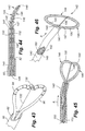
Referring to FIGS. 1 and 2 , the system 10 of the present invention is shown that includes a combination electrical and chemical stimulation device 12, a stimulation source 14 that communicates with the stimulation device 12 for delivering electrical energy and chemicals to the stimulation device, and an interventional device such as an introducer needle 32 that allows introduction of the stimulation lead. The stimulation device 12 is shown as inserted within an intervertebral disc D. The combination device 12 more particularly includes a percutaneous electrical and chemical stimulation lead 16 in the form of an elongate tubular member having a desired length and diameter allowing the lead 16 to be placed within the intervertebral disc of the patient to be treated. The working distal portion 20 of the stimulation lead 16 provides the desired stimulation through a plurality of electrodes 22 which are selectively positioned on the distal portion 20, along with a plurality of infusion ports 30 which allow delivery of chemicals/nutrients to target tissue. The proximal portion of the stimulation device 12 can be referred to as a lead extension 18 that connects to the stimulation source 14. The lead extension 18 can be made of the same type and diameter material as the stimulation lead 16, or may be made of a different type of material and diameter.
Referring specifically to FIG. 2 , in a first embodiment of the stimulation lead, a plurality of circumferentially extending electrodes 22 are positioned at the distal portion 20. The electrodes 22 are also spaced longitudinally along the distal portion 20. The electrodes produce an array of electrical field energy, and the target tissue is immersed in the electrical field. One or more electrical conductors 23 extend through the interior of the stimulation lead 16 in order to transmit the electrical impulses to the electrodes 22. It is preferable to utilize a single conductor 23 along the major length of the lead, and then provide branch conductors (not shown) at the distal portion 20 that then extend to contact the various electrodes. The branch conductors could be a linearly arranged set of wire extensions extending between each electrode, or any other advantageous combination of wire conductors to interconnect the electrodes. Use of a single conductor is a more robust design as opposed to multiple smaller conductors that are more prone to breakage as a result of the motion cycles of the IVD. It is also contemplated that the electrode could be a single electrode wound in a helical pattern about the distal portion 20. Thus in this helical pattern, only one conductor 23 would be required with no additional branch conductors. In order to generate the desired intensity and size electrical field, the electrodes 22 can be disposed on the distal portion in a pattern or arrangement that best suits the electrical field to be generated. For example, in the helical pattern, the electrode could be wound with a tighter pattern to generate a more intense field, while a looser more spaced pattern would generate a less intense field. Of course, the particular signal or impulse current provided to the electrodes also determines the intensity of the field generated.
In order to provide chemical infusion, a central lumen or passageway 24 is formed through the stimulation lead. The central lumen 24 may extend completely through the lead thereby forming a distal opening 28 in the stimulation lead and providing one infusion port that is directed distally of the stimulation lead.
The stimulation lead 16 may be made of a homogeneous material, or may be made of differing materials that cause the stimulation lead to have either a more progressively stiff or more progressively flexible characteristic as the lead extends in the distal direction. Depending upon the manner in which the stimulation lead is to be emplaced, it may be desirable to use either the more progressively stiff or more progressively flexible arrangement.
In accordance with the method of the present invention, a stylet (not shown) is first inserted through the introducer needle 32. The introducer needle 32 is emplaced by penetrating the skin and muscle tissue, and ultimately into the disc D. When the introducer needle has penetrated the disc, the stylet is removed and the stimulation lead 16 is then inserted through the lumen of the introducer needle.
Referring again to FIG. 1 , the stimulation lead 16 is illustrated as being emplaced within the disc D. This disc D is shown in cross section along with an adjacent vertebra V. The stimulation lead 16 is shown as taking an arcuate or curved path through the disc nucleus N in order to be precisely positioned at the area of the disc to be treated, illustrated as a fissure F which has developed adjacent the spinal fluid sac (not shown). The other primary features of the disk D are also illustrated including the annulus fibrosis A and the thin layer L defining the annular nuclear interface/transitional zone.
The stimulation source 14 is preferably an implantable medical device 34 including both an IPG (implantable pulse generator) 36 and an IDP (implantable drug dispenser) 38. The implantable device 34 could be contained within a single structural housing, or two separate housings, one for the IPG 36, and one for the IDP 38. The IPG and IDP can both be self-contained devices with internal control for preset delivery of electrical and chemical pulses. Alternatively, an external controller 44 could be used to modify the desired treatment protocol by use of RF transmission wherein an implantable RF receiver 40 is integrated with the IPG 36 and IDP 38. The RF receiver 40 could also be housed within the same implantable medical device 34, or could be a separate implanted device. An external RF transmitter 42 transmits RF signals to control the delivery of electrical stimulation and chemicals to the stimulation lead 16. A controller 44 provides the specific instruction set for transmission by the RF transmitter 42.
In accordance with the apparatus and method of the present invention, there are a number of nutrients and medications that can be delivered by the stimulation lead. For nutrients, this list includes, but is not limited to, glucose, glucosamine, chondroitin, oxygen and oxygenating agents, anti-oxidants, anti-glycosylating agents, and pH buffers. For medications, these may include, without limitation, anti-inflammatory agents and growth factors, such as growth and differentiating factor-5 (GDF-5), transforming growth factor-beta (TGF-β), insulin-like growth factor-1 (IGF-1), and basic fibroblasts growth factor (bFGF). In terms of the types of electrical impulses provided to the electrodes 22, these electrical impulses may be continuous or variable over time, and may vary based upon voltage, amperage, and alternate current frequency.
Referring to FIG. 3 , a different arrangement is illustrated with respect to the location of the electrodes 22, and the single infusion port at distal opening 28 is supplemented with a plurality of additional infusion ports 30. In this embodiment, fewer electrodes are incorporated, yet additional infusion ports 30 are provided that are spaced longitudinally along the length of the lead 16 and placed between the electrodes 22.
Referring to FIG. 5 , another embodiment of the stimulation lead is illustrated wherein the lead has a progressively narrowing diameter towards the distal end thereof. With this type of stimulation lead, travel of the lead through the more dense annulus tissue is facilitated because the distal tip has a smaller frontal profile and is more easily controlled.
Referring to FIG. 6 , yet another embodiment of the stimulation lead is illustrated wherein the electrodes 22 are not formed circumferentially around the distal portion 20, but are formed more linearly along one side of the stimulation lead. Additionally, the infusion ports 30 may have more of an oval shape and be larger in size that facilitates greater volumetric infusion. This embodiment may be preferred when it is desired to more precisely direct the array of electrical energy to the target tissue. The electrical energy array that is created by circumferentially arranged electrodes result in transmission patterns having a radial or circular pattern extending away from the stimulation lead. Thus, a plurality of circumferentially arranged electrodes transmit energy in all directions to tissue that surrounds the stimulation lead. On the contrary, locating the electrodes only along one side or edge of the stimulation lead results in transmission of energy in a more linear and less radial pattern, and directed primarily orthogonal or perpendicular to the axis of the stimulation lead. The embodiment of FIG. 6 also illustrates the distal end as being bent at a desired angle.
Referring to FIG. 10 , yet another embodiment of the invention is shown wherein an introducer needle 46 is not placed within the disc nucleus, but rather is placed only into the disc annulus, and then the stimulation lead 16 extends through the disc annulus to the target tissue, also shown as a fissure F. In this embodiment, it is preferable that the stimulation lead 16 exits the introducer needle through a bent distal portion 48 so that the lead travels in a more parallel fashion within the annulus and along a more linear path to the target tissue. Accordingly, a stimulation lead having a straight tip like shown in FIGS. 2 , 3 and 5, would be more suitable according to this embodiment. In the event the distal opening 28 of the lead 16 is of a size which could allow nuclear tissue to clog or block the distal opening 28, a guide wire 26 (see FIG. 12 ) may be inserted through the lumen 24 of the lead 16, and the distal tip 27 of the guide wire could be placed flush with the distal opening 28 in order to prevent clogging of the distal opening 28, as well as to provide additional rigidity for placement of the stimulation lead 16. If the guide wire 26 is used, then the guide wire 26 is removed prior to connecting the stimulation lead 16 to an IDP and/or IPG. Also, the central lumen may terminate before passing through the distal tip of the lead. Thus, all of the infusion ports 30 would be arranged on the lead to direct chemicals/nutrients in a perpendicular direction away from the axis of the lead.
During treatment, it may be desirable to administer nutrients and/or medications to different parts of the disc being treated. Furthermore, it may be desirable to provide the nutrients/medications to these different locations within the disc at differing flow rates and at differing times and frequencies. With the provision of a dual set of lumens, a physician has the ability to selectively control infusion to two distinct areas within the disc, and can vary the treatment protocol between the two areas of the disc by selecting the particular dosing, frequency, and makeup of the infusion material to the distinct locations within the disc. This selective treatment capability may be advantageous where, for example, the distal end of the stimulation lead may be placed near the interface/transitional zone, and the tissue extending therealong together with the annulus fibrosis may have particular needs in terms of the required type of nutrients and/or medication, while the tissue within the nucleus may have slightly different needs. Thus, the embodiment at FIG. 14 provides the treating physician with additional options in providing effective treatment.
The particular sizes of the lumens, as well as the sizes and spacing of the openings 35 and 37 may be configured for optimal delivery of various types of infusion material. For example, assuming that the desired nutrient/medication to be delivered to the distal end of the stimulation lead was fairly viscous, it may be advantageous to provide the lumen 24 with a larger cross-sectional size, as well as to provide the infusion openings 35 of an increased size to accommodate the higher viscosity. As a further example, if the lumen 41 was to deliver a less viscous nutrient/medication, then the lumen 41 would preferably have a smaller cross-sectional area, and the openings 37 would preferably be smaller than the openings 35. Thus, one convenient way in which to control infusion is to advantageously arrange the particular size, number, and spacing of the infusion openings as well as the size of the lumens which deliver the infusion material through the openings.
It is further contemplated within the present invention to also provide non-uniform lumens, as well as infusion openings that vary in size within the same supplying lumen. As discussed above, the IDP 38 may be programmed for preset delivery of chemical “pulses”. The IDP 38 is typically programmed to be in an “on” or “off” state to generate delivery of a set amount of fluid over a specific period of time. However, once the infusion material is released from the IDP, the IDP itself does not have control over the way in which the infusion material is dispersed through the stimulation lead. Assuming that a lumen of a stimulation lead has a uniform diameter with infusion openings also being of a uniform diameter, then the infusion ports located at the more proximal end of the device will most likely deliver a greater amount of material to the disc as opposed to the infusion ports located at the distal end of the device because there will be an inherent loss in the amount of fluid delivered downstream based on frictional losses within the lumen and the upstream openings communicating with the lumen. Therefore, in order to ensure equal distribution of infused material, it may be desirable to provide a lumen having a diameter that progressively enlarges as it extends towards the distal end of the device. Alternatively or in combination with the progressively changing lumen size, it may be desirable to provide infusion ports toward the proximal end of the device that are slightly smaller than the infusion ports located towards the distal end of the device to further help compensate for any frictional line losses.
Referring to FIG. 15 , yet another embodiment of the present invention is provided which further includes an inflatable portion 50 in the form of a bladder or balloon that is selectively inflated or deflated by an inflation line 52 extending conterminously with the central lumen. The inflatable portion is mounted to the exterior surface of the stimulation lead, and the inflation line 52 extends through an opening (not shown) in the sidewall of the lead that is covered by the inflatable portion 50. The inflation line 52 communicates with a source of compressed fluid (not shown), and the physician may inflate the inflatable portion 50 to a desired size. As also shown, the inflatable portion 50 is preferably placed along a location of the stimulation lead that does not cover or block any infusion ports 30, as well as any electrodes 22.
In some instances, the stimulation lead may reside within a patient for an extended period of time. As time passes, the stimulation lead may have a tendency to migrate or drift within the disc. Drifting of the stimulation lead can be problematic for a number of reasons, to include causing damage to the disc by penetration of the distal tip of the stimulation lead completely through the disc, as well as drifting of the stimulation lead so that it is no longer centered around/along the desired area of the disc to be treated. To maintain the stimulation lead in its desired position after the stimulation has been emplaced, the inflatable portion 50 may be inflated to the desired size, thereby serving as an anchor to help prevent drifting of the stimulation lead within the disc. In most instances, it is desirable to place the inflatable portion 50 near the distal tip of the stimulation lead to best prevent undesired drift of the stimulation lead; however, it is also contemplated within the present invention that the inflatable portion 50 may be selectively placed along other areas of the stimulation lead to best serve as an anchor. For example, as shown in FIG. 16 , the inflatable portion is located at the proximal end of the stimulation lead. Furthermore, it may be desirable to incorporate both a distally located inflation portion 50, and another inflation portion located at the proximal end of the device that would further help to prevent the stimulation lead from drifting or from being inadvertently removed.
Some disc tissue may have a tendency to adhere to a stimulation lead that has been emplaced within the disc for a long period of time, and/or the disc tissue may have a tendency to harden around the emplaced stimulation lead thereby making it more difficult to remove the stimulation lead. Thus, it is also contemplated within the present invention that the inflatable portion 50 could be provided to extend along a much greater distance of the stimulation lead, and the inflatable portion 50 could be inflated to a desired level prior to the stimulation lead being emplaced within a disc. When it is then desired to remove the stimulation lead, the inflatable portion could be deflated which would create a small gap or space between the surrounding disc tissue and the stimulation lead thereby easing removal of the stimulation lead.
Thus, the inflatable portion 50 can be used either as an anchor to maintain positioning of the stimulation lead within the disc, or the inflatable portion 50 can be used in a reverse role by enlarging the overall size of the stimulation lead once emplaced, but then reducing the overall size of the stimulation lead by deflating the inflatable portion when it is desired to remove the stimulation lead.
Referring to FIG. 17 , a stimulation lead is shown emplaced within a disc D, the stimulation lead generally corresponding to the embodiment shown in FIG. 14 . Two oval shaped areas 40 and 42 are shown surrounding the distal and proximal sections of the stimulation lead, respectively. These areas 40 and 42 may generally represent targeted treatment areas within the disc. In accordance with the embodiment of FIG. 14 , the physician has the option of applying different infusion materials through the separate sets of infusion ports 35 and 37 to specifically target the tissue located within the areas 40 and 42. Such treatment could be simultaneous, sequential, or any combination thereof. Furthermore, as mentioned above, selected sets of electrodes could be energized to provide treatment. For example, the electrodes may be wired so that the physician has the ability to energize two primary sets of electrodes, one set providing an electromagnetic field generated to cover area 40, and the other set providing an electromagnetic field to cover area 42. The electrodes may be wired and configured to provide generation of electromagnetic fields in any desired pattern along the length of the lead.
Referring now to FIGS. 18-20 , yet another embodiment of the present invention is illustrated in the form of stimulation lead 60. For some treatments, it may be necessary to leave the stimulation lead emplaced within the invertebral disc for an extended period of time; however, for various reasons, it may not be possible to keep the stimulation lead emplaced for the amount of time to provide optimal treatment. In order to solve this particular problem, the embodiment of FIG. 18 contemplates the use of various chemical agents/medications and nutrients incorporated within a dissolvable matrix that forms the body 62 of the stimulation lead 60. The electrodes 64 as well as the conductor(s) 66 could be formed with the dissolvable matrix in a molding process whereby a particular shape and size stimulation lead could be produced. The electrodes 64 could function the same as the electrodes 22 discussed above and could be produced in any desired pattern and wiring arrangement. The dissolvable matrix can be made of a material that is biomedically acceptable for enabling a time release of the chemical agents/medications and nutrients mixed within the matrix. The matrix is preferably a solid yet flexible material, allowing the stimulation lead to be steered with the use of an insertable stylet 56 which could be provided through the central lumen 68. However, it shall be understood that this central lumen 68 is optional, and the matrix may be manufactured of a material which is durable yet flexible enough allowing the practitioner to steer the stimulation lead without the use of a stylet. Accordingly, FIG. 19 illustrates another embodiment wherein there is no lumen present, and a predetermined bend angle is formed in the stimulation lead enabling the lead to take the desired path through the disc when emplaced. Once inserted into the disc, the matrix would dissolve and the regenerating chemicals/medications and nutrients would slowly diffuse into the surrounding disc tissue leaving only the electrodes 64 and conducting wire(s) 66 to be removed at some later time.
With the embodiment shown in FIGS. 18 and 19 , an infusion pump would not be required, and would thereby also allow for the subcutaneously placed pulse generator (IPG) to be significantly smaller. Similar to the combined pump/pulse generator device described above, this much smaller pulse generator could be rechargeable, or be powered by a battery source as desired.
In a modification to the embodiment of FIG. 18 , it is also contemplated within the scope of the present invention that a stimulation lead can simply comprise a dissolvable matrix having a desired combination of chemical agents/medications and nutrients, and no electrodes incorporated within the lead. In some cases, stimulation by an electromagnetic field may be unnecessary to achieve the desired regenerative and/or pain relieving disc response.
Referring now to FIG. 21 , in another aspect of the present invention, a stimulation device may be used to treat SI joint ailments. FIG. 21 specifically illustrates a posterior view of the sacroiliac region with an introducer needle positioned for insertion along the sacroiliac region to a targeted area adjacent the SI joint J. Referring also to FIG. 22 , an enlarged posterior view of the sacrum bone B is shown wherein the introducer needle 46 has been fully inserted. In accordance with a method of the present invention for treatment of the SI joint, the introducer needle 46 is first inserted through the skin below and slightly medial to the inferior aspect to the SI joint and directed towards the inferior lateral edge of the sacrum (Brad show on Fig). The introducer needle 46 is advanced to contact the dorsal aspect of the sacrum at the posterolateral edge. As shown, the needle 46 may have a slight curvature near the distal end thereof, shown as curve or bend 48, and the curvature of the bend 48 is then utilized to advance the needle lateral to the sacral foramen and medial through the dorsal aspect of the SI joint. The needle 46 remains in contact with the periosteum along the entire curvature of the sacrum. The needle tip ultimately advances to the superior edge of the sacrum lateral to the sacral foramen and medial to the SI joint. Appropriate positioning of the introducing needle is confirmed preferably both on Antero-posterior (AP) as well as lateral views. The stimulation lead 16 is then inserted through the introducer needle 46 until reaching the distal tip 48 of the introducer needle. The stimulation lead 16 is held in place by maintaining pressure on the lead. Referring now to FIG. 23 , the introducer needle 16 is withdrawn along a selected length of the stimulation lead 46 to expose the active number of electrodes 22 necessary to denervate the sacral nerve innervation to the SI joint. The dotted lines shown in FIG. 23 for lead 16 represent the initial position of the lead after the needle 46 is withdrawn. After the lead 16 is exposed, local anesthetic and/or neurolytic agents and/or proliferant agents such as, but not limited to, phenol or alcohol, or Dextrose respectively could be injected through one or more of the infusion ports. The electrodes 22 may then be activated to ablate the surrounding neural tissue. The dotted lines for needle 46 in FIG. 23 represent the position of the needle after it has been withdrawn and the lead is ready for activation. The solid lines in FIG. 23 represent the next position of the lead 16 and needle 46 wherein both have been further withdrawn for purposes of conducting another activation to further denervate tissue, such as a circumstance when the initial ablation did not effectively cover the desired area of tissue.
With respect to the specific construction of the stimulation lead for use in a method of treating the SI joint, it may be constructed in the same manner as disclosed with respect to the prior description for treatment of a disc. More specifically, a stimulation lead may be selected having the most desirable arrangement of electrodes for the most optimal denervation of the targeted neural tissues.
The sacral nerves illustrated in FIG. 23 include the lateral branches S1, S2, S3 and S4. In order to denervate each of the lateral branches, it may be required to sequentially apply energy to the stimulation lead as the introducer needle is repeatedly withdrawn along the path of insertion. Because of the variation of sacral anatomy, successful denervation may require two or more separate needle insertion angles in order to denervate the S1-S4 lateral branches. However, as discussed below with respect to the embodiments having multiple lead elements, it may be possible to avoid such multiple needle insertions. In addition to denervation of the sacral lateral branches, it may also be advantageous to denervate the L5 dorsal ramus as well as the L4 medial branch since there is some innervation to the SI joint from both of these additional nerve structures.
Although the figures show treatment along one side of the sacrum, it shall be understood that the same procedure may be repeated for treatment of the other side of the sacrum, by placement of the introducer needle in a symmetrical fashion on the corresponding opposite or contralateral side of the sacrum. In addition to electrical stimulation, it is also contemplated with respect to the method of treatment of the SI joint to also provide infusion in a combined electrical stimulation and chemical/drug infusion device. For example, infusion of collagen proliferants could be included in the method of treatment by use of a selected device including any one of the above-disclosed embodiments. Infusion of collagen proliferants such as dextrose, growth factors, and other nutrients may accelerate the healing process for some SI joint ailments. Depending upon the diagnosed ailment, infusion alone may be appropriate for the treatment, or in combination with some neural tissue ablation or stimulation. It is also contemplated in the method of the present invention to enhance neurolytic lesion size by infusion of substances such as Phenol, alcohol, and glycerin.
With respect to the specific construction and material used for spring elements 102, it is contemplated that the spring elements can be made from either metallic or thermoplastic materials. The particular material chosen should have elastomeric/resilient characteristics which allows the spring elements to expand or open when the stimulation elements are freed from the insertion needle, but also allows the spring elements to collapse without undue force when the stimulation lead is withdrawn and placed back within the introducer needle. As shown in FIG. 28 , the particular spread of the stimulation elements allows the stimulation lead to cover a larger area than would be possible if just a single stimulation element were present.
In order to stiffen or otherwise control any of the stimulation leads 90 shown in the embodiments of FIGS. 33 , 35, and 38, a stylet (not shown) may also be used in the same manner as the stylet 112 shown and described with reference to FIG. 29 . Accordingly, a stylet captured within the needle or sheath adjacent the stimulation lead 90 allows the stylet to assist in precise placement of the stimulation lead.
Based upon the foregoing, the present invention provides a combination electrical and chemical stimulation lead especially adapted for treatment of many types of ailments to include, disc ailments SI joint ailments, and other spine ailments to include treatment of structures that have large and diffuse innervations such as, but not limited to, the superior hypogastric plexus, sympathetic chain, ganglion impar, and others.
The various embodiments provide a treating physician with stimulation leads of various configurations, which optimizes a physician's ability to precisely position the stimulation lead, as well as to precisely direct both electrical and chemical stimulation.
While the above description and drawings disclose and illustrate embodiments of the present invention, it should be understood that the invention is not limited to these embodiments. Those skilled in the art may make other modifications and changes employing the principles of the present invention, particularly considering the foregoing teachings. Therefore, by the appended claims, the applicant intends to cover such modifications and other embodiments.
Claims (3)
1. A method of managing SI joint pain in a sacrum of a patient, said method comprising the steps of:
providing a stimulation lead having at least one electrode selectively placed along said stimulation lead;
providing an introducer needle having a curvature along its length and a bent distal end;
inserting the introducer needle along a path of insertion into a patient defined by aligning the introducer needle in a position medial to an inferior aspect of the SI joint and directed towards an inferior lateral edge of the sacrum;
advancing the introducer needle along a curve of the sacrum in close proximity to a periosteum, lateral to a sacral foramen and medial to the SI joint until reaching a superior aspect of the sacrum;
inserting the stimulation lead in the introducer needle;
placing pressure on the stimulation lead to maintain the lead in a first position;
withdrawing the introducer needle along the path of insertion and along a selected length of the stimulation lead to expose said at least one electrode to denervate targeted tissue;
activating said at least one electrode in order to denervate tissue surrounding the stimulation lead;
withdrawing the stimulation lead to a second position and aligned along the path of insertion within the introducer needle;
placing pressure on the stimulation lead to maintain the lead in the second position;
withdrawing the introducer needle aligned along the path of insertion thereby exposing said at least one electrode on the stimulation lead; and
further activating said at least one electrode in order to denervate tissue surrounding the stimulation lead.
2. A method, as claimed in claim 1 , further including the step of:
infusing through said stimulation lead at least one chemical selected from the group consisting of an anesthetic, a neurolytic, a proliferant, and a nutrient.
3. A method, as claimed in claim 1 , wherein:
said at least one electrode includes a plurality of electrodes spaced along a length of said stimulation lead.
Priority Applications (7)
| Application Number | Priority Date | Filing Date | Title |
|---|---|---|---|
| US11/678,516 US7945331B2 (en) | 2005-01-11 | 2007-02-23 | Combination electrical stimulating and infusion medical device and method |
| US11/771,757 US20080009927A1 (en) | 2005-01-11 | 2007-06-29 | Combination Electrical Stimulating and Infusion Medical Device and Method |
| US12/033,232 US8066702B2 (en) | 2005-01-11 | 2008-02-19 | Combination electrical stimulating and infusion medical device and method |
| EP11169796.7A EP2386260B1 (en) | 2007-02-23 | 2008-02-21 | Combination electrical stimulating and infusion medical device |
| EP08101856A EP1961394A3 (en) | 2007-02-23 | 2008-02-21 | Combination electrical stimulating and infusion medical device and method |
| US13/088,558 US20110196361A1 (en) | 2005-01-11 | 2011-04-18 | Combination Electrical Stimulating and Infusion Medical Device and Method |
| US13/271,312 US8323277B2 (en) | 2005-01-11 | 2011-10-12 | Combination electrical stimulating and infusion method |
Applications Claiming Priority (3)
| Application Number | Priority Date | Filing Date | Title |
|---|---|---|---|
| US11/033,591 US7386350B2 (en) | 2005-01-11 | 2005-01-11 | Combination electrical stimulating and infusion medical device |
| US11/107,553 US20060155343A1 (en) | 2005-01-11 | 2005-04-14 | Combination electrical stimulating and infusion medical device and method |
| US11/678,516 US7945331B2 (en) | 2005-01-11 | 2007-02-23 | Combination electrical stimulating and infusion medical device and method |
Related Parent Applications (2)
| Application Number | Title | Priority Date | Filing Date |
|---|---|---|---|
| US11/033,591 Continuation-In-Part US7386350B2 (en) | 2005-01-11 | 2005-01-11 | Combination electrical stimulating and infusion medical device |
| US11/107,553 Continuation-In-Part US20060155343A1 (en) | 2005-01-11 | 2005-04-14 | Combination electrical stimulating and infusion medical device and method |
Related Child Applications (3)
| Application Number | Title | Priority Date | Filing Date |
|---|---|---|---|
| US11/771,757 Continuation-In-Part US20080009927A1 (en) | 2005-01-11 | 2007-06-29 | Combination Electrical Stimulating and Infusion Medical Device and Method |
| US12/033,232 Continuation-In-Part US8066702B2 (en) | 2005-01-11 | 2008-02-19 | Combination electrical stimulating and infusion medical device and method |
| US13/088,558 Division US20110196361A1 (en) | 2005-01-11 | 2011-04-18 | Combination Electrical Stimulating and Infusion Medical Device and Method |
Publications (2)
| Publication Number | Publication Date |
|---|---|
| US20070135881A1 US20070135881A1 (en) | 2007-06-14 |
| US7945331B2 true US7945331B2 (en) | 2011-05-17 |
Family
ID=46327365
Family Applications (2)
| Application Number | Title | Priority Date | Filing Date |
|---|---|---|---|
| US11/678,516 Active 2027-01-13 US7945331B2 (en) | 2005-01-11 | 2007-02-23 | Combination electrical stimulating and infusion medical device and method |
| US13/088,558 Abandoned US20110196361A1 (en) | 2005-01-11 | 2011-04-18 | Combination Electrical Stimulating and Infusion Medical Device and Method |
Family Applications After (1)
| Application Number | Title | Priority Date | Filing Date |
|---|---|---|---|
| US13/088,558 Abandoned US20110196361A1 (en) | 2005-01-11 | 2011-04-18 | Combination Electrical Stimulating and Infusion Medical Device and Method |
Country Status (1)
| Country | Link |
|---|---|
| US (2) | US7945331B2 (en) |
Cited By (20)
| Publication number | Priority date | Publication date | Assignee | Title |
|---|---|---|---|---|
| US20110196361A1 (en) * | 2005-01-11 | 2011-08-11 | Vilims Bradley D | Combination Electrical Stimulating and Infusion Medical Device and Method |
| US8323277B2 (en) | 2005-01-11 | 2012-12-04 | Vilims Bradley D | Combination electrical stimulating and infusion method |
| US8361067B2 (en) | 2002-09-30 | 2013-01-29 | Relievant Medsystems, Inc. | Methods of therapeutically heating a vertebral body to treat back pain |
| US8414571B2 (en) | 2010-01-07 | 2013-04-09 | Relievant Medsystems, Inc. | Vertebral bone navigation systems |
| US8419730B2 (en) | 2008-09-26 | 2013-04-16 | Relievant Medsystems, Inc. | Systems and methods for navigating an instrument through bone |
| US8425507B2 (en) | 2002-09-30 | 2013-04-23 | Relievant Medsystems, Inc. | Basivertebral nerve denervation |
| US8880189B2 (en) | 2011-02-23 | 2014-11-04 | John D. LIPANI | System and method for electrical stimulation of the lumbar vertebral column |
| US8882764B2 (en) | 2003-03-28 | 2014-11-11 | Relievant Medsystems, Inc. | Thermal denervation devices |
| US20150174366A1 (en) * | 2013-09-24 | 2015-06-25 | Truminim International Corporation | Secured and Self Contained Spinal Cord Stimulator Leads and Catheters |
| USRE46356E1 (en) | 2002-09-30 | 2017-04-04 | Relievant Medsystems, Inc. | Method of treating an intraosseous nerve |
| US9630011B2 (en) | 2011-02-23 | 2017-04-25 | John D Lipani | System and methods for diagnosis and treatment of discogenic lower back pain |
| US9724151B2 (en) | 2013-08-08 | 2017-08-08 | Relievant Medsystems, Inc. | Modulating nerves within bone using bone fasteners |
| US9724107B2 (en) | 2008-09-26 | 2017-08-08 | Relievant Medsystems, Inc. | Nerve modulation systems |
| US9775627B2 (en) | 2012-11-05 | 2017-10-03 | Relievant Medsystems, Inc. | Systems and methods for creating curved paths through bone and modulating nerves within the bone |
| US10076384B2 (en) | 2013-03-08 | 2018-09-18 | Symple Surgical, Inc. | Balloon catheter apparatus with microwave emitter |
| US10390877B2 (en) | 2011-12-30 | 2019-08-27 | Relievant Medsystems, Inc. | Systems and methods for treating back pain |
| US10588691B2 (en) | 2012-09-12 | 2020-03-17 | Relievant Medsystems, Inc. | Radiofrequency ablation of tissue within a vertebral body |
| US10675085B2 (en) | 2015-11-23 | 2020-06-09 | Boston Scientific Scimed, Inc. | Devices and methods for enhanced denervation procedures |
| US20200390496A1 (en) * | 2019-06-14 | 2020-12-17 | Avolt, Llc | Electromagnetic radiation ablation tips made of magnetic materials |
| US11007010B2 (en) | 2019-09-12 | 2021-05-18 | Relevant Medsysterns, Inc. | Curved bone access systems |
Families Citing this family (21)
| Publication number | Priority date | Publication date | Assignee | Title |
|---|---|---|---|---|
| US20060155343A1 (en) * | 2005-01-11 | 2006-07-13 | Vilims Bradley D | Combination electrical stimulating and infusion medical device and method |
| US7797054B2 (en) * | 2007-01-12 | 2010-09-14 | Medtronic, Inc. | Expandable systems for medical electrical stimulation |
| US8090450B2 (en) * | 2007-06-27 | 2012-01-03 | Greatbatch Ltd. | Percutaneous electrode array and system |
| US8326439B2 (en) * | 2008-04-16 | 2012-12-04 | Nevro Corporation | Treatment devices with delivery-activated inflatable members, and associated systems and methods for treating the spinal cord and other tissues |
| US8805519B2 (en) | 2010-09-30 | 2014-08-12 | Nevro Corporation | Systems and methods for detecting intrathecal penetration |
| US9345537B2 (en) * | 2010-12-30 | 2016-05-24 | Avent, Inc. | Electrosurgical tissue treatment method |
| DE102011110667B4 (en) * | 2011-08-19 | 2018-11-15 | Omar Omar-Pasha | Apparatus for applying a pulsed radiofrequency therapy in the vascular system or other body cavities or tissue of the human or animal body, as well as a catheter, a probe and an insertion aid for such a device |
| EP2763743A4 (en) | 2011-10-04 | 2015-08-05 | Nevro Corp | Modeling positions of implanted devices in a patient |
| WO2013112920A1 (en) | 2012-01-25 | 2013-08-01 | Nevro Corporation | Lead anchors and associated systems and methods |
| US20130245739A1 (en) * | 2012-03-14 | 2013-09-19 | Anatoly Arber | Self-anchored stimulator lead and method of insertion |
| WO2014016762A1 (en) * | 2012-07-23 | 2014-01-30 | Koninklijke Philips N.V. | Combined brachytherapy and electrochemotherapy catheters |
| US9308022B2 (en) | 2012-12-10 | 2016-04-12 | Nevro Corporation | Lead insertion devices and associated systems and methods |
| US20140200639A1 (en) * | 2013-01-16 | 2014-07-17 | Advanced Neuromodulation Systems, Inc. | Self-expanding neurostimulation leads having broad multi-electrode arrays |
| US20140276755A1 (en) * | 2013-03-12 | 2014-09-18 | Boston Scientific Scimed, Inc. | Medical systems and methods for modulating nerves |
| EP2777761B1 (en) * | 2013-03-14 | 2017-03-29 | BIOTRONIK SE & Co. KG | Implantable electrode |
| ITMO20130080A1 (en) * | 2013-03-28 | 2014-09-29 | Claudio Reverberi | STIMULATING ELECTRODE, PARTICULARLY FOR ELECTRIC STIMULATION TREATMENTS. |
| US9265935B2 (en) | 2013-06-28 | 2016-02-23 | Nevro Corporation | Neurological stimulation lead anchors and associated systems and methods |
| US20190060518A1 (en) * | 2016-02-26 | 2019-02-28 | Neuronano Ab | Method of providing an implantation site in soft tissue |
| US10980999B2 (en) | 2017-03-09 | 2021-04-20 | Nevro Corp. | Paddle leads and delivery tools, and associated systems and methods |
| US11420045B2 (en) | 2018-03-29 | 2022-08-23 | Nevro Corp. | Leads having sidewall openings, and associated systems and methods |
| WO2020141997A1 (en) * | 2019-01-04 | 2020-07-09 | Neuronano Ab | Probe for implantation into nervous tissue comprising a microelectrode or a set of microelectrodes |
Citations (67)
| Publication number | Priority date | Publication date | Assignee | Title |
|---|---|---|---|---|
| WO1981003272A1 (en) | 1980-05-13 | 1981-11-26 | American Hospital Supply Corp | A multipolar electrosurgical device |
| US4612934A (en) | 1981-06-30 | 1986-09-23 | Borkan William N | Non-invasive multiprogrammable tissue stimulator |
| US5255691A (en) | 1991-11-13 | 1993-10-26 | Medtronic, Inc. | Percutaneous epidural lead introducing system and method |
| WO1994002077A2 (en) | 1992-07-15 | 1994-02-03 | Angelase, Inc. | Ablation catheter system |
| US5342357A (en) | 1992-11-13 | 1994-08-30 | American Cardiac Ablation Co., Inc. | Fluid cooled electrosurgical cauterization system |
| US5397338A (en) | 1993-03-29 | 1995-03-14 | Maven Labs, Inc. | Electrotherapy device |
| WO1995010320A1 (en) | 1993-10-15 | 1995-04-20 | Ep Technologies, Inc. | Device for lengthening cardiac conduction pathways |
| US5423877A (en) | 1992-05-04 | 1995-06-13 | David C. Mackey | Method and device for acute pain management by simultaneous spinal cord electrical stimulation and drug infusion |
| WO1998019613A1 (en) | 1996-11-08 | 1998-05-14 | Surx, Inc. | Devices, methods and systems for shrinking tissues |
| WO1999043263A1 (en) | 1998-02-27 | 1999-09-02 | Conway-Stuart Medical, Inc. | Apparatus to electrosurgically treat esophageal sphincters |
| US5951546A (en) | 1994-12-13 | 1999-09-14 | Lorentzen; Torben | Electrosurgical instrument for tissue ablation, an apparatus, and a method for providing a lesion in damaged and diseased tissue from a mammal |
| US6002964A (en) | 1998-07-15 | 1999-12-14 | Feler; Claudio A. | Epidural nerve root stimulation |
| US6053916A (en) | 1999-02-17 | 2000-04-25 | Moore; Michael R. | Sacroiliac implant |
| US6104957A (en) | 1998-08-21 | 2000-08-15 | Alo; Kenneth M. | Epidural nerve root stimulation with lead placement method |
| US6136021A (en) | 1999-03-23 | 2000-10-24 | Cardiac Pacemakers, Inc. | Expandable electrode for coronary venous leads |
| US6146380A (en) | 1998-01-09 | 2000-11-14 | Radionics, Inc. | Bent tip electrical surgical probe |
| US6148222A (en) | 1998-07-10 | 2000-11-14 | Cardiocommand, Inc. | Esophageal catheters and method of use |
| US6161047A (en) | 1998-04-30 | 2000-12-12 | Medtronic Inc. | Apparatus and method for expanding a stimulation lead body in situ |
| US6263225B1 (en) | 1994-02-09 | 2001-07-17 | University Of Iowa Research Foundation | Stereotactic electrode assembly |
| US6277112B1 (en) | 1996-07-16 | 2001-08-21 | Arthrocare Corporation | Methods for electrosurgical spine surgery |
| US6379349B1 (en) | 1995-11-08 | 2002-04-30 | Celon Ag Medical Instruments | Arrangement for electrothermal treatment of the human or animal body |
| US6562033B2 (en) | 2001-04-09 | 2003-05-13 | Baylis Medical Co. | Intradiscal lesioning apparatus |
| US20030181964A1 (en) | 1996-08-13 | 2003-09-25 | Oratec Interventions, Inc. a Delaware corporation | Method and apparatus for treating annular fissures in intervertebral discs |
| US20030195518A1 (en) | 2000-02-16 | 2003-10-16 | Cragg Andrew H. | Methods and apparatus for performing therapeutic procedures in the spine |
| US20030195509A1 (en) | 1998-01-14 | 2003-10-16 | Curon Medical, Inc. | GERB treatment apparatus and method |
| US6656174B1 (en) | 2000-07-20 | 2003-12-02 | Scimed Life Systems, Inc. | Devices and methods for creating lesions in blood vessels without obstructing blood flow |
| US20030225331A1 (en) | 2002-01-23 | 2003-12-04 | The Regents Of The University Of California | Implantable thermal treatment method and apparatus |
| US6662055B1 (en) | 1999-12-17 | 2003-12-09 | Impulse Dynamics Nv | Multi-electrode intravascular lead |
| US20040015202A1 (en) | 2002-06-14 | 2004-01-22 | Chandler Gilbert S. | Combination epidural infusion/stimulation method and system |
| US20040039425A1 (en) | 2002-05-29 | 2004-02-26 | Beverley Greenwood-Van Meerveld | Spinal cord stimulation as treatment for functional bowel disorders |
| US6735474B1 (en) | 1998-07-06 | 2004-05-11 | Advanced Bionics Corporation | Implantable stimulator system and method for treatment of incontinence and pain |
| US20040127965A1 (en) | 1999-07-21 | 2004-07-01 | Borkan William N. | Method of forming a ball grid array package |
| US20040230273A1 (en) | 2003-04-11 | 2004-11-18 | Cates Adam W. | Subcutaneous electrode and lead with temporary pharmacological agents |
| US20040236388A1 (en) | 1998-04-30 | 2004-11-25 | Medtronic, Inc. | Techniques for positioning therapy delivery elements within a spinal cord or brain |
| US20040243206A1 (en) | 2003-01-03 | 2004-12-02 | Tadlock Charles H. | System, method, and combined electrical and chemical stimulation lead for stimulation of a person's nerve tissue |
| US6827716B2 (en) | 2002-09-30 | 2004-12-07 | Depuy Spine, Inc. | Method of identifying and treating a pathologic region of an intervertebral disc |
| US6832115B2 (en) | 2000-08-17 | 2004-12-14 | William N. Borkan | Catheter leads for the intrathecal space and method of use |
| US20040260277A1 (en) | 1999-03-19 | 2004-12-23 | Maguire Mark A. | Circumferential ablation device assembly and methods of use and manufacture providing an ablative circumferential band along an expandable member |
| US6845267B2 (en) | 2000-09-28 | 2005-01-18 | Advanced Bionics Corporation | Systems and methods for modulation of circulatory perfusion by electrical and/or drug stimulation |
| US6847849B2 (en) | 2000-11-15 | 2005-01-25 | Medtronic, Inc. | Minimally invasive apparatus for implanting a sacral stimulation lead |
| US20050096718A1 (en) | 2003-10-31 | 2005-05-05 | Medtronic, Inc. | Implantable stimulation lead with fixation mechanism |
| US6902547B2 (en) | 2001-08-02 | 2005-06-07 | Teodulo Aves | Medical needle |
| US6921403B2 (en) | 2000-02-16 | 2005-07-26 | Trans1 Inc. | Method and apparatus for spinal distraction and fusion |
| US20050177211A1 (en) | 2002-03-05 | 2005-08-11 | Baylis Medical Company Inc. | Electrosurgical device for treatment of tissue |
| US20050177209A1 (en) | 2002-03-05 | 2005-08-11 | Baylis Medical Company Inc. | Bipolar tissue treatment system |
| US6936024B1 (en) | 1995-01-23 | 2005-08-30 | Russell A. Houser | Percutaneous transmyocardial revascularization (PTMR) system |
| US20050277918A1 (en) | 2003-03-07 | 2005-12-15 | Baylis Medical Company Inc. | Electrosurgical cannula |
| US20060020297A1 (en) | 2004-07-20 | 2006-01-26 | Gerber Martin T | Neurostimulation system with distributed stimulators |
| US7014633B2 (en) | 2000-02-16 | 2006-03-21 | Trans1, Inc. | Methods of performing procedures in the spine |
| US20060106376A1 (en) | 2004-11-15 | 2006-05-18 | Baylis Medical Company Inc. | Methods of treating the sacroilac region of a patient's body |
| US7065412B2 (en) | 2003-04-25 | 2006-06-20 | Medtronic, Inc. | Implantable trial neurostimulation device |
| US7069087B2 (en) | 2000-02-25 | 2006-06-27 | Oratec Interventions, Inc. | Apparatus and method for accessing and performing a function within an intervertebral disc |
| US7069094B2 (en) | 2001-03-23 | 2006-06-27 | Honda Giken Kogyo Kabushiki Kaisha | Computer-aided product design system |
| US20060155343A1 (en) | 2005-01-11 | 2006-07-13 | Vilims Bradley D | Combination electrical stimulating and infusion medical device and method |
| US20060155342A1 (en) | 2005-01-11 | 2006-07-13 | Vilims Bradley D | Combination electrical stimulating and infusion medical device |
| US20060259026A1 (en) | 2005-05-05 | 2006-11-16 | Baylis Medical Company Inc. | Electrosurgical treatment method and device |
| US7169146B2 (en) | 2003-02-14 | 2007-01-30 | Surgrx, Inc. | Electrosurgical probe and method of use |
| US20070156136A1 (en) | 2002-03-05 | 2007-07-05 | Neil Godara | Methods of treating the sacroiliac region of a patient's body |
| US7276063B2 (en) | 1998-08-11 | 2007-10-02 | Arthrocare Corporation | Instrument for electrosurgical tissue treatment |
| US20080009927A1 (en) | 2005-01-11 | 2008-01-10 | Vilims Bradley D | Combination Electrical Stimulating and Infusion Medical Device and Method |
| US7369894B2 (en) | 2002-09-06 | 2008-05-06 | Medtronic, Inc. | Method, system and device for treating disorders of the pelvic floor by electrical stimulation of the sacral and/or pudendal nerves |
| US20080172117A1 (en) | 2007-01-12 | 2008-07-17 | Skubitz Sean P | Expandable systems for medical electrical stimulation |
| US20080200972A1 (en) | 2005-01-11 | 2008-08-21 | Rittman William J | Combination electrical stimulating and infusion medical device and method |
| US7422586B2 (en) | 2001-02-28 | 2008-09-09 | Angiodynamics, Inc. | Tissue surface treatment apparatus and method |
| US7449019B2 (en) | 1999-01-25 | 2008-11-11 | Smith & Nephew, Inc. | Intervertebral decompression |
| US7462178B2 (en) | 2000-05-12 | 2008-12-09 | Arthrocare Corporation | Systems and methods for electrosurgical spine surgery |
| US7771422B2 (en) | 2002-06-06 | 2010-08-10 | Nuortho Surgical, Inc. | Methods and devices for electrosurgery |
Family Cites Families (16)
| Publication number | Priority date | Publication date | Assignee | Title |
|---|---|---|---|---|
| US4612943A (en) * | 1983-08-05 | 1986-09-23 | Meinkowsky Larry J | Dispenser for snuff and the like |
| US5447533A (en) * | 1992-09-03 | 1995-09-05 | Pacesetter, Inc. | Implantable stimulation lead having an advanceable therapeutic drug delivery system |
| US5730127A (en) * | 1993-12-03 | 1998-03-24 | Avitall; Boaz | Mapping and ablation catheter system |
| US5496360A (en) * | 1994-04-12 | 1996-03-05 | Ventritex, Inc. | Implantable cardiac electrode with rate controlled drug delivery |
| US5925038A (en) * | 1996-01-19 | 1999-07-20 | Ep Technologies, Inc. | Expandable-collapsible electrode structures for capacitive coupling to tissue |
| DE69833665T2 (en) * | 1997-08-08 | 2006-11-09 | Duke University | COMPOSITIONS FOR SIMPLIFYING SURGICAL PROCEDURES |
| US6175769B1 (en) * | 1999-06-14 | 2001-01-16 | Electro Core Technologies, Llc | Spinal cord electrode assembly having laterally extending portions |
| US6733500B2 (en) * | 2000-03-31 | 2004-05-11 | Medtronic, Inc. | Method and system for delivering a medical electrical lead within a venous system |
| WO2002005868A2 (en) * | 2000-07-13 | 2002-01-24 | Transurgical, Inc. | Thermal treatment methods and apparatus with focused energy application |
| US20030009095A1 (en) * | 2001-05-21 | 2003-01-09 | Skarda James R. | Malleable elongated medical device |
| US7099718B1 (en) * | 2001-05-29 | 2006-08-29 | Advanced Bionics Corporation | Neural stimulation lead fixation |
| US6817849B1 (en) * | 2001-12-05 | 2004-11-16 | B. R. Lee Industries, Inc. | System for forming a curb and other structures |
| US7309668B2 (en) * | 2003-12-03 | 2007-12-18 | Elk Premium Building Products, Inc. | Multiple layer directionally oriented nonwoven fiber material and methods of manufacturing same |
| US7945331B2 (en) * | 2005-01-11 | 2011-05-17 | Bradley D. Vilims | Combination electrical stimulating and infusion medical device and method |
| US7623899B2 (en) * | 2005-09-16 | 2009-11-24 | Biosense Webster, Inc. | Catheter with flexible pre-shaped tip section |
| WO2008079387A1 (en) * | 2006-12-21 | 2008-07-03 | Xenoport, Inc. | Catechol protected levodopa diester prodrugs, compositions, and methods of use |
-
2007
- 2007-02-23 US US11/678,516 patent/US7945331B2/en active Active
-
2011
- 2011-04-18 US US13/088,558 patent/US20110196361A1/en not_active Abandoned
Patent Citations (73)
| Publication number | Priority date | Publication date | Assignee | Title |
|---|---|---|---|---|
| WO1981003272A1 (en) | 1980-05-13 | 1981-11-26 | American Hospital Supply Corp | A multipolar electrosurgical device |
| US4612934A (en) | 1981-06-30 | 1986-09-23 | Borkan William N | Non-invasive multiprogrammable tissue stimulator |
| US5255691A (en) | 1991-11-13 | 1993-10-26 | Medtronic, Inc. | Percutaneous epidural lead introducing system and method |
| US5423877A (en) | 1992-05-04 | 1995-06-13 | David C. Mackey | Method and device for acute pain management by simultaneous spinal cord electrical stimulation and drug infusion |
| WO1994002077A2 (en) | 1992-07-15 | 1994-02-03 | Angelase, Inc. | Ablation catheter system |
| US5342357A (en) | 1992-11-13 | 1994-08-30 | American Cardiac Ablation Co., Inc. | Fluid cooled electrosurgical cauterization system |
| US5397338A (en) | 1993-03-29 | 1995-03-14 | Maven Labs, Inc. | Electrotherapy device |
| WO1995010320A1 (en) | 1993-10-15 | 1995-04-20 | Ep Technologies, Inc. | Device for lengthening cardiac conduction pathways |
| US6263225B1 (en) | 1994-02-09 | 2001-07-17 | University Of Iowa Research Foundation | Stereotactic electrode assembly |
| US5951546A (en) | 1994-12-13 | 1999-09-14 | Lorentzen; Torben | Electrosurgical instrument for tissue ablation, an apparatus, and a method for providing a lesion in damaged and diseased tissue from a mammal |
| US6936024B1 (en) | 1995-01-23 | 2005-08-30 | Russell A. Houser | Percutaneous transmyocardial revascularization (PTMR) system |
| US6379349B1 (en) | 1995-11-08 | 2002-04-30 | Celon Ag Medical Instruments | Arrangement for electrothermal treatment of the human or animal body |
| US6277112B1 (en) | 1996-07-16 | 2001-08-21 | Arthrocare Corporation | Methods for electrosurgical spine surgery |
| US20030181964A1 (en) | 1996-08-13 | 2003-09-25 | Oratec Interventions, Inc. a Delaware corporation | Method and apparatus for treating annular fissures in intervertebral discs |
| WO1998019613A1 (en) | 1996-11-08 | 1998-05-14 | Surx, Inc. | Devices, methods and systems for shrinking tissues |
| US20030195604A1 (en) | 1996-11-08 | 2003-10-16 | Surx, Inc. | Devices, methods, and sytems for shrinking tissues |
| US6146380A (en) | 1998-01-09 | 2000-11-14 | Radionics, Inc. | Bent tip electrical surgical probe |
| US20030195509A1 (en) | 1998-01-14 | 2003-10-16 | Curon Medical, Inc. | GERB treatment apparatus and method |
| WO1999043263A1 (en) | 1998-02-27 | 1999-09-02 | Conway-Stuart Medical, Inc. | Apparatus to electrosurgically treat esophageal sphincters |
| US6161047A (en) | 1998-04-30 | 2000-12-12 | Medtronic Inc. | Apparatus and method for expanding a stimulation lead body in situ |
| US20040236388A1 (en) | 1998-04-30 | 2004-11-25 | Medtronic, Inc. | Techniques for positioning therapy delivery elements within a spinal cord or brain |
| US6735474B1 (en) | 1998-07-06 | 2004-05-11 | Advanced Bionics Corporation | Implantable stimulator system and method for treatment of incontinence and pain |
| US6148222A (en) | 1998-07-10 | 2000-11-14 | Cardiocommand, Inc. | Esophageal catheters and method of use |
| US6002964A (en) | 1998-07-15 | 1999-12-14 | Feler; Claudio A. | Epidural nerve root stimulation |
| US7276063B2 (en) | 1998-08-11 | 2007-10-02 | Arthrocare Corporation | Instrument for electrosurgical tissue treatment |
| US6104957A (en) | 1998-08-21 | 2000-08-15 | Alo; Kenneth M. | Epidural nerve root stimulation with lead placement method |
| US7449019B2 (en) | 1999-01-25 | 2008-11-11 | Smith & Nephew, Inc. | Intervertebral decompression |
| US6053916A (en) | 1999-02-17 | 2000-04-25 | Moore; Michael R. | Sacroiliac implant |
| US20040260277A1 (en) | 1999-03-19 | 2004-12-23 | Maguire Mark A. | Circumferential ablation device assembly and methods of use and manufacture providing an ablative circumferential band along an expandable member |
| US6136021A (en) | 1999-03-23 | 2000-10-24 | Cardiac Pacemakers, Inc. | Expandable electrode for coronary venous leads |
| US20040127965A1 (en) | 1999-07-21 | 2004-07-01 | Borkan William N. | Method of forming a ball grid array package |
| US6662055B1 (en) | 1999-12-17 | 2003-12-09 | Impulse Dynamics Nv | Multi-electrode intravascular lead |
| US6921403B2 (en) | 2000-02-16 | 2005-07-26 | Trans1 Inc. | Method and apparatus for spinal distraction and fusion |
| US20030195518A1 (en) | 2000-02-16 | 2003-10-16 | Cragg Andrew H. | Methods and apparatus for performing therapeutic procedures in the spine |
| US7014633B2 (en) | 2000-02-16 | 2006-03-21 | Trans1, Inc. | Methods of performing procedures in the spine |
| US7069087B2 (en) | 2000-02-25 | 2006-06-27 | Oratec Interventions, Inc. | Apparatus and method for accessing and performing a function within an intervertebral disc |
| US7462178B2 (en) | 2000-05-12 | 2008-12-09 | Arthrocare Corporation | Systems and methods for electrosurgical spine surgery |
| US6656174B1 (en) | 2000-07-20 | 2003-12-02 | Scimed Life Systems, Inc. | Devices and methods for creating lesions in blood vessels without obstructing blood flow |
| US6832115B2 (en) | 2000-08-17 | 2004-12-14 | William N. Borkan | Catheter leads for the intrathecal space and method of use |
| US6845267B2 (en) | 2000-09-28 | 2005-01-18 | Advanced Bionics Corporation | Systems and methods for modulation of circulatory perfusion by electrical and/or drug stimulation |
| US6847849B2 (en) | 2000-11-15 | 2005-01-25 | Medtronic, Inc. | Minimally invasive apparatus for implanting a sacral stimulation lead |
| US20050240238A1 (en) | 2000-11-15 | 2005-10-27 | Medtronic, Inc. | Minimally invasive apparatus for implanting a sacral stimulation lead |
| US7422586B2 (en) | 2001-02-28 | 2008-09-09 | Angiodynamics, Inc. | Tissue surface treatment apparatus and method |
| US7069094B2 (en) | 2001-03-23 | 2006-06-27 | Honda Giken Kogyo Kabushiki Kaisha | Computer-aided product design system |
| US6562033B2 (en) | 2001-04-09 | 2003-05-13 | Baylis Medical Co. | Intradiscal lesioning apparatus |
| US6902547B2 (en) | 2001-08-02 | 2005-06-07 | Teodulo Aves | Medical needle |
| US20030225331A1 (en) | 2002-01-23 | 2003-12-04 | The Regents Of The University Of California | Implantable thermal treatment method and apparatus |
| US20050177209A1 (en) | 2002-03-05 | 2005-08-11 | Baylis Medical Company Inc. | Bipolar tissue treatment system |
| US20070156136A1 (en) | 2002-03-05 | 2007-07-05 | Neil Godara | Methods of treating the sacroiliac region of a patient's body |
| US20050177211A1 (en) | 2002-03-05 | 2005-08-11 | Baylis Medical Company Inc. | Electrosurgical device for treatment of tissue |
| US20040039425A1 (en) | 2002-05-29 | 2004-02-26 | Beverley Greenwood-Van Meerveld | Spinal cord stimulation as treatment for functional bowel disorders |
| US7771422B2 (en) | 2002-06-06 | 2010-08-10 | Nuortho Surgical, Inc. | Methods and devices for electrosurgery |
| US20040015202A1 (en) | 2002-06-14 | 2004-01-22 | Chandler Gilbert S. | Combination epidural infusion/stimulation method and system |
| US7369894B2 (en) | 2002-09-06 | 2008-05-06 | Medtronic, Inc. | Method, system and device for treating disorders of the pelvic floor by electrical stimulation of the sacral and/or pudendal nerves |
| US6827716B2 (en) | 2002-09-30 | 2004-12-07 | Depuy Spine, Inc. | Method of identifying and treating a pathologic region of an intervertebral disc |
| US20040243206A1 (en) | 2003-01-03 | 2004-12-02 | Tadlock Charles H. | System, method, and combined electrical and chemical stimulation lead for stimulation of a person's nerve tissue |
| US7169146B2 (en) | 2003-02-14 | 2007-01-30 | Surgrx, Inc. | Electrosurgical probe and method of use |
| US20050277918A1 (en) | 2003-03-07 | 2005-12-15 | Baylis Medical Company Inc. | Electrosurgical cannula |
| US20040230273A1 (en) | 2003-04-11 | 2004-11-18 | Cates Adam W. | Subcutaneous electrode and lead with temporary pharmacological agents |
| US7065412B2 (en) | 2003-04-25 | 2006-06-20 | Medtronic, Inc. | Implantable trial neurostimulation device |
| US20050096718A1 (en) | 2003-10-31 | 2005-05-05 | Medtronic, Inc. | Implantable stimulation lead with fixation mechanism |
| US20060020297A1 (en) | 2004-07-20 | 2006-01-26 | Gerber Martin T | Neurostimulation system with distributed stimulators |
| US20060106376A1 (en) | 2004-11-15 | 2006-05-18 | Baylis Medical Company Inc. | Methods of treating the sacroilac region of a patient's body |
| US20070055316A1 (en) | 2004-11-15 | 2007-03-08 | Baylis Medical Company Inc. | Methods of treating the sacroiliac region of a patient's body |
| US7819869B2 (en) | 2004-11-15 | 2010-10-26 | Kimberly-Clark Inc. | Methods of treating the sacroilac region of a patient's body |
| US20070179536A1 (en) | 2005-01-11 | 2007-08-02 | Vilims Bradley D | Combination Electrical Stimulating and Infusion Medical Device |
| US20080009927A1 (en) | 2005-01-11 | 2008-01-10 | Vilims Bradley D | Combination Electrical Stimulating and Infusion Medical Device and Method |
| US20080200972A1 (en) | 2005-01-11 | 2008-08-21 | Rittman William J | Combination electrical stimulating and infusion medical device and method |
| US20060155342A1 (en) | 2005-01-11 | 2006-07-13 | Vilims Bradley D | Combination electrical stimulating and infusion medical device |
| US20060155343A1 (en) | 2005-01-11 | 2006-07-13 | Vilims Bradley D | Combination electrical stimulating and infusion medical device and method |
| US20060217705A1 (en) | 2005-02-17 | 2006-09-28 | Baylis Medical Company Inc. | Electrosurgical device with discontinuous flow density |
| US20060259026A1 (en) | 2005-05-05 | 2006-11-16 | Baylis Medical Company Inc. | Electrosurgical treatment method and device |
| US20080172117A1 (en) | 2007-01-12 | 2008-07-17 | Skubitz Sean P | Expandable systems for medical electrical stimulation |
Non-Patent Citations (63)
| Title |
|---|
| "Pain Management Slnergy System"; Baylis Medical Company Inc., 2006, 2 pages. |
| Ahadian, "Pulsed radiofrequency neurotomy: advances in pain medicine," Current Pain and Headache Reports, 2004, vol. 8, pp. 34-40. |
| Akatov et al., "Percutaneous radiofrequency destruction of the obturator nerve for treatment of pain caused by coxarthrosis," Stereotact Funct Neurosurg, 1997, vol. 69(1-4 Pt 2), pp. 278-280 (Abstract only). |
| An et al.; "Biological Repair of Intervertebral Disc"; SPINE, 2003, vol. 28, No. 15S; pp. S86-S92. |
| An et al.; "Intradiscal Administration of Osteogenic Protein-1 Increases . . . " SPINE, vol. 30, No. 1, pp. 25-32, 2004. |
| Anis et al., "Use of radio-frequency ablation for the palliative treatment of sacral chordoma," Am. J. Neuronadiol, 2004, vol. 25, pp. 1589-1591. |
| Atlihan et al., "Anatomy of the Anterior Sacroiliac Joint With Reference to Lumbosacral Nerves," Clinical Orthopaedics and Related Research, 2000, No. 376, pp. 236-241. |
| Ben-Yishay; News & Highlights-IDET-a new procedure for discogenic back pain; http://www.neurospinewi.com/newsletters/IDET.html; printed Dec. 22, 2004; 3 pages. |
| Buijs et al., "Radiofrequency treatment of sacroiliac joint-related pain aimed at the first three sacral dorsal rami: a minimal approach," The Pain Clinic, 2004, vol. 16(2), pp. 139-146. |
| Calvillo et al., "Anatomy and Pathophysiology of the Sacroiliac Joint," Current Review of Pain, 2000, vol. 4, pp. 356-361. |
| Cohen et al., "Lateral Branch Blocks as a Treatment for Sacroiliac Joint Pain: A Pilot Study," Regional Anesthesia and Pain Medicine, 2003, vol. 28(2), pp. 113-119. |
| Cohen et al., "Pulsed radiofrequency as a treatment for groin pain and orchialgia," Urology, 2003, vol. 61(3), pp. 645xxi-xxiii. |
| Cohen, "Sacroiliac Joint Pain: A Comprehensive Review of Anatomy, Diagnosis, and Treatment," Anesthesia & Analgesia, 2005, vol. 101(5), pp. 1440-1453. |
| Cole et al., "The Sacroiliac Joint: A Functional Approach," Critical Reviews in Physical and Rehabilitation Medicine, 1996, vol. 8(1&2), pp. 125-152. |
| Conaghan et al., "Sacral nerve stimulation can be successful in patients with ultrasound evidence of external anal sphincter disruption," Diseases of the Colon and Rectum, 2005, vol. 48(8), pp. 1610-1614. |
| Davis et al., "Radiofrequency Treatment in the United States," Pain Practice, 2002, vol. 2(3), pp. 192-194. |
| De Mattei et al.; "Effects of Pulsed Electromagnetic Fields on Human Articular Chondrocyte Proliferation"; Connect Tissue Res., 2001, vol. 42, No. 4, pp. 269-279. |
| Deer, "Chapter 6: Injections for the Diagnosis and Treatment of Spinal Pain," American Society of Anesthesiologists, Inc., 2004, vol. 32, pp. 53-69. |
| Dreyfuss et al., "Chapter 32: Radiofrequency Neurotomy of the Zygapophyseal and Sacroiliac Joints," Pain Procedures, 2000, pp. 395-420. |
| Ebraheim et al., "Anatomic Considerations for Posterior Approach to the Sacroiliac Joint," Spine, 1996, vol. 21(23), pp. 2709-2712. |
| Extended European Search Report for European Patent Application No. 08151981.1, dated Dec. 9, 2008. |
| Ferrante et al., "Radiofrequency sacroiliac joint denervation for sacroiliac syndrome," Regional Anesthesia and Pain Medicine, 2001, vol. 26(2), pp. 137-142. |
| Fortin et al., "Sacroiliac Joint Innervation and Pain," The American Journal of Orthopedics, 1999, pp. 687-690. |
| Fortin et al., "Three Pathways between the Sacroiliac Joint and Neural Structures," Am. J. Neruoradiol., 1999, vol. 20, pp. 1429-1434. |
| Freeman, et al., "Does intradiscal electrothermal therapy denervate and repair experimentally induced posterolateral annular tears in an animal model?", Spine, Dec. 1, 2003, vol. 28, No. 23, pp. 2602-2608. |
| Fukui et al., "Successful relief of hip joint pain by percutaneous radiofrequency nerve thermocoagulation in a patient with contraindications for hip arthroplasty," Journal of Anesthesia, 2001, vol. 15, pp. 173-175. |
| Ganey et al.; "Disc Chondrocyte Transplantation in a Canine Model: A Treatment for Degenerated or Damaged Intervertebral Disc"; SPINE, 2003, vol. 28, No. 23; pp. 2609-2620. |
| Gevargez et al., "CT-guided percutaneous radiofrequency denervation of the sacroiliac joint," Eur Radiol., 2002, vol. 12, pp. 1360-1365. |
| Gopalani et al., "A novel technique for treating nonsurgical hip pain with radiofrequency lesioning of the sensory branches of the obturator and femoral nerves: a case report," Archives of Physical Medicine and Rehabilitation, Poster 102, 2003, vol. 84, A23. |
| Gruber et al., "Biologic Strategies for the Therapy of Intervertebral Disc Degeneration"; Expert Opinion on Biological Therapy, 2003, vol. 3, No. 8, pp. 1209-1214. |
| IDET Procedure; http://www.thepainrelief.com/idet-idet.shtml; printed Dec. 22, 2004; 2 pages. |
| International Preliminary Report on Patentability for International (PCT) Patent Application No. PCT/US06/00312, issued Jul. 17, 2007. |
| International Search Report for International (PCT) Patent Application No. PCT/US06/00312, mailed Jul. 19, 2006. |
| Intradiscal Electrothermal Therapy; http://www.emedicine.com/neuro/topic707.htm; printed Dec. 22, 2004; 8 pages. |
| Jiang et al., "Comparison between radiofrequency coagulation plus small needle knife and single method in treatment of sacrolumbar pain," Chinese Journal of Clinical Rehabilitation, 2003, vol. 7(20), pp. 2844-2845 (Abstract only). |
| Kawaguchi et al., "Percutaneous radiofrequency lesioning of sensory branches of the obturator and femoral nerves for the treatment of hip joint pain," Regional Anesthesia and Pain Medicine, 2001, vol. 26(6), pp. 576-581. |
| Kirkham et al., "Neuromodulation through sacral nerve roots 2 to 4 with a Finetech-Brindley sacral posterior and anterior root stimulator," Spinal Cord, 2002, vol. 40(6), pp. 272-281. |
| Kirsch et al., "Proton radiotherapy for Hodgkin's disease in the sacrum," Lancet Oncology, 2005, vol. 6, pp. 532-533. |
| Kline et al., "Chapter 19: Radiofrequency techniques in clinical practice," Interventional Pain Management, 2001, pp. 243-290 (2001). |
| Leng et al., "How sacral nerve stimulation neuromodulation works," Urol. Clin. North. Am., 2005, vol. 32(1), pp. 11-18 (Abstract only). |
| Liguoro et al., "The Posterior Sacral Foramina: An Anatomical Study," 1999, J. Anat., vol. 195, pp. 301-304. |
| Mathews et al.; "Treatment of mechanical and chemical lumbar discopathy by dextrose 25%"; J Minim Invasive Spinal Tech, vol. 1, Inaugural 2001; pp. 58-61. |
| Murata et al., "Origin and pathway of sensory nerve fibers to the ventral and dorsal sides of the sacroiliac joint in rats," Journal of Orthopaedic Research, 2001, vol. 19, pp. 379-383. |
| O'Neill et al.; "Percutaneous Plasma Decompression Alters Cytokine Expression in Injured Porcine Intervertebral Discs"; Spine J., 2004, vol. 4, No. 1, pp. 88-98. |
| Partial European Search Report for European Patent Application No. 08101856.6, mailed Jul. 3, 2008. |
| Partial European Search Report for European Patent Application No. 08101856.6, mailed Oct. 28, 2008. |
| Partial European Search Report for European Patent Application No. 08151981, mailed Jul. 16, 2008. |
| Pino et al., "Morphologic analysis of bipolar radiofrequency lesions: implications for treatment of the sacroiliac joint," Regional Anesthesia and Pain Medicine, 2005, vol. 30(4), pp. 335-338. |
| Plancarte et al., "Radiofrequency Procedures for Sacral and Pelvic Region Pain," Pain Practice, 2002, vol. 2(3), pp. 248-249. |
| Raj et al., "Executive Summary: The Current Status of the Practice of Radiofrequency in the World," Pain Practice, 2002, vol. 2(3), pp. 176-179. |
| Simon, "Chapter 50: Sacroiliac joint injection and low back pain," Interventional Pain Management, 2001, pp. 535-539. |
| Slipman et al., "Sacroiliac Joint Syndrome," Pain Physician, 2001, vol. 4(2), pp. 143-152. |
| SpineCath Intradiscal Electrothermal Therapy; http://www.spineuniverse.com/displayarticle.php/article217.html; printed Dec. 22, 2004; 3 pages. |
| Supplementary European Search Report for European Patent Application No. 06717501, mailed Aug. 20, 2008. |
| Supplementary European Search Report for European Patent Application No. 06717501, mailed Mar. 31, 2008. |
| U.S. Appl. No. 12/340,092, filed Dec. 19, 2008, Vilims. |
| Valleylab-RF Pain Management System, Sep. 16, 2004, available at internet archives, http://www.valleylab.com/static/pain/products-generator.html, 2 pages. |
| Van Zundert et al. "Application of Radiofrequency Treatment in Practical Pain Management: State of the art," Pain Practice, 2002, vol. 2(3), pp. 269-278. |
| Walsh et al.; "In Vivo Growth Factor Treatment of Degenerated Intervertebral Discs"; SPINE, vol. 29, No. 2, pp. 156-163, 2004. |
| Wang et al.; "Up-regulation of Chondrocyte Matrix Genes and Products by Electric Fields"; Clinical Ortho. & Related Research; Oct. 2004, 427S: S163-S173. |
| Written Opinion for International (PCT) Patent Application No. PCT/US06/00312, mailed Jul. 19, 2006. |
| Yin et al., "Sensory stimulation-guided sacroiliac joint radiofrequency neurotomy: technique based on neuroanatomy of the dorsal sacral plexus," Spine, 2003, vol. 28(20), pp. 2419-2425. |
| Yoon et al.; "The Effect of Bone Morphogenetic Protein-2 on Rat Intervertebral Disc Cells in Vitro"; SPINE, vol. 28, No. 16, pp. 1773-1780, 2003. |
Cited By (67)
| Publication number | Priority date | Publication date | Assignee | Title |
|---|---|---|---|---|
| US8992522B2 (en) | 2002-09-30 | 2015-03-31 | Relievant Medsystems, Inc. | Back pain treatment methods |
| US9421064B2 (en) | 2002-09-30 | 2016-08-23 | Relievant Medsystems, Inc. | Nerve modulation systems |
| US8361067B2 (en) | 2002-09-30 | 2013-01-29 | Relievant Medsystems, Inc. | Methods of therapeutically heating a vertebral body to treat back pain |
| US9017325B2 (en) | 2002-09-30 | 2015-04-28 | Relievant Medsystems, Inc. | Nerve modulation systems |
| US10111704B2 (en) | 2002-09-30 | 2018-10-30 | Relievant Medsystems, Inc. | Intraosseous nerve treatment |
| US9023038B2 (en) | 2002-09-30 | 2015-05-05 | Relievant Medsystems, Inc. | Denervation methods |
| US8425507B2 (en) | 2002-09-30 | 2013-04-23 | Relievant Medsystems, Inc. | Basivertebral nerve denervation |
| US10478246B2 (en) | 2002-09-30 | 2019-11-19 | Relievant Medsystems, Inc. | Ablation of tissue within vertebral body involving internal cooling |
| US8613744B2 (en) | 2002-09-30 | 2013-12-24 | Relievant Medsystems, Inc. | Systems and methods for navigating an instrument through bone |
| US8623014B2 (en) | 2002-09-30 | 2014-01-07 | Relievant Medsystems, Inc. | Systems for denervation of basivertebral nerves |
| US8628528B2 (en) | 2002-09-30 | 2014-01-14 | Relievant Medsystems, Inc. | Vertebral denervation |
| USRE46356E1 (en) | 2002-09-30 | 2017-04-04 | Relievant Medsystems, Inc. | Method of treating an intraosseous nerve |
| US11596468B2 (en) | 2002-09-30 | 2023-03-07 | Relievant Medsystems, Inc. | Intraosseous nerve treatment |
| US9486279B2 (en) | 2002-09-30 | 2016-11-08 | Relievant Medsystems, Inc. | Intraosseous nerve treatment |
| US9173676B2 (en) | 2002-09-30 | 2015-11-03 | Relievant Medsystems, Inc. | Nerve modulation methods |
| US8992523B2 (en) | 2002-09-30 | 2015-03-31 | Relievant Medsystems, Inc. | Vertebral treatment |
| US9848944B2 (en) | 2002-09-30 | 2017-12-26 | Relievant Medsystems, Inc. | Thermal denervation devices and methods |
| USRE48460E1 (en) | 2002-09-30 | 2021-03-09 | Relievant Medsystems, Inc. | Method of treating an intraosseous nerve |
| US8419731B2 (en) | 2002-09-30 | 2013-04-16 | Relievant Medsystems, Inc. | Methods of treating back pain |
| US8882764B2 (en) | 2003-03-28 | 2014-11-11 | Relievant Medsystems, Inc. | Thermal denervation devices |
| US10463423B2 (en) | 2003-03-28 | 2019-11-05 | Relievant Medsystems, Inc. | Thermal denervation devices and methods |
| US8323277B2 (en) | 2005-01-11 | 2012-12-04 | Vilims Bradley D | Combination electrical stimulating and infusion method |
| US20110196361A1 (en) * | 2005-01-11 | 2011-08-11 | Vilims Bradley D | Combination Electrical Stimulating and Infusion Medical Device and Method |
| US10905440B2 (en) | 2008-09-26 | 2021-02-02 | Relievant Medsystems, Inc. | Nerve modulation systems |
| US9265522B2 (en) | 2008-09-26 | 2016-02-23 | Relievant Medsystems, Inc. | Methods for navigating an instrument through bone |
| US9039701B2 (en) | 2008-09-26 | 2015-05-26 | Relievant Medsystems, Inc. | Channeling paths into bone |
| US9259241B2 (en) | 2008-09-26 | 2016-02-16 | Relievant Medsystems, Inc. | Methods of treating nerves within bone using fluid |
| US8808284B2 (en) | 2008-09-26 | 2014-08-19 | Relievant Medsystems, Inc. | Systems for navigating an instrument through bone |
| US9724107B2 (en) | 2008-09-26 | 2017-08-08 | Relievant Medsystems, Inc. | Nerve modulation systems |
| US10265099B2 (en) | 2008-09-26 | 2019-04-23 | Relievant Medsystems, Inc. | Systems for accessing nerves within bone |
| US11471171B2 (en) | 2008-09-26 | 2022-10-18 | Relievant Medsystems, Inc. | Bipolar radiofrequency ablation systems for treatment within bone |
| US10028753B2 (en) | 2008-09-26 | 2018-07-24 | Relievant Medsystems, Inc. | Spine treatment kits |
| US8419730B2 (en) | 2008-09-26 | 2013-04-16 | Relievant Medsystems, Inc. | Systems and methods for navigating an instrument through bone |
| US8535309B2 (en) | 2010-01-07 | 2013-09-17 | Relievant Medsystems, Inc. | Vertebral bone channeling systems |
| US8414571B2 (en) | 2010-01-07 | 2013-04-09 | Relievant Medsystems, Inc. | Vertebral bone navigation systems |
| US9789313B2 (en) | 2011-02-23 | 2017-10-17 | John D. LIPANI | System and methods for diagnosis and treatment of discogenic lower back pain |
| US8880189B2 (en) | 2011-02-23 | 2014-11-04 | John D. LIPANI | System and method for electrical stimulation of the lumbar vertebral column |
| US8892215B2 (en) | 2011-02-23 | 2014-11-18 | John D. LIPANI | System and method for electrical stimulation of the lumbar vertebral column |
| US9950164B2 (en) | 2011-02-23 | 2018-04-24 | John D Lipani | System and methods for diagnosis and treatment of discogenic lower back pain |
| US9630011B2 (en) | 2011-02-23 | 2017-04-25 | John D Lipani | System and methods for diagnosis and treatment of discogenic lower back pain |
| US11471210B2 (en) | 2011-12-30 | 2022-10-18 | Relievant Medsystems, Inc. | Methods of denervating vertebral body using external energy source |
| US10390877B2 (en) | 2011-12-30 | 2019-08-27 | Relievant Medsystems, Inc. | Systems and methods for treating back pain |
| US11737814B2 (en) | 2012-09-12 | 2023-08-29 | Relievant Medsystems, Inc. | Cryotherapy treatment for back pain |
| US10588691B2 (en) | 2012-09-12 | 2020-03-17 | Relievant Medsystems, Inc. | Radiofrequency ablation of tissue within a vertebral body |
| US11690667B2 (en) | 2012-09-12 | 2023-07-04 | Relievant Medsystems, Inc. | Radiofrequency ablation of tissue within a vertebral body |
| US11701168B2 (en) | 2012-09-12 | 2023-07-18 | Relievant Medsystems, Inc. | Radiofrequency ablation of tissue within a vertebral body |
| US9775627B2 (en) | 2012-11-05 | 2017-10-03 | Relievant Medsystems, Inc. | Systems and methods for creating curved paths through bone and modulating nerves within the bone |
| US10517611B2 (en) | 2012-11-05 | 2019-12-31 | Relievant Medsystems, Inc. | Systems for navigation and treatment within a vertebral body |
| US11160563B2 (en) | 2012-11-05 | 2021-11-02 | Relievant Medsystems, Inc. | Systems for navigation and treatment within a vertebral body |
| US10357258B2 (en) | 2012-11-05 | 2019-07-23 | Relievant Medsystems, Inc. | Systems and methods for creating curved paths through bone |
| US11291502B2 (en) | 2012-11-05 | 2022-04-05 | Relievant Medsystems, Inc. | Methods of navigation and treatment within a vertebral body |
| US11234764B1 (en) | 2012-11-05 | 2022-02-01 | Relievant Medsystems, Inc. | Systems for navigation and treatment within a vertebral body |
| US10076384B2 (en) | 2013-03-08 | 2018-09-18 | Symple Surgical, Inc. | Balloon catheter apparatus with microwave emitter |
| US9724151B2 (en) | 2013-08-08 | 2017-08-08 | Relievant Medsystems, Inc. | Modulating nerves within bone using bone fasteners |
| US11065046B2 (en) | 2013-08-08 | 2021-07-20 | Relievant Medsystems, Inc. | Modulating nerves within bone |
| US10456187B2 (en) | 2013-08-08 | 2019-10-29 | Relievant Medsystems, Inc. | Modulating nerves within bone using bone fasteners |
| US20150174366A1 (en) * | 2013-09-24 | 2015-06-25 | Truminim International Corporation | Secured and Self Contained Spinal Cord Stimulator Leads and Catheters |
| WO2015048133A3 (en) * | 2013-09-24 | 2015-11-05 | Truminim International Corporation | Secured spinal cord stimulator leads and catheters |
| US10105513B2 (en) * | 2013-09-24 | 2018-10-23 | Spineloop, Llc | Secured and self contained spinal cord stimulator leads and catheters |
| US11540874B2 (en) | 2015-11-23 | 2023-01-03 | Boston Scientific Scimed, Inc. | Devices and methods for enhanced denervation procedures |
| US10675085B2 (en) | 2015-11-23 | 2020-06-09 | Boston Scientific Scimed, Inc. | Devices and methods for enhanced denervation procedures |
| US20200390496A1 (en) * | 2019-06-14 | 2020-12-17 | Avolt, Llc | Electromagnetic radiation ablation tips made of magnetic materials |
| US11207100B2 (en) | 2019-09-12 | 2021-12-28 | Relievant Medsystems, Inc. | Methods of detecting and treating back pain |
| US11007010B2 (en) | 2019-09-12 | 2021-05-18 | Relevant Medsysterns, Inc. | Curved bone access systems |
| US11426199B2 (en) | 2019-09-12 | 2022-08-30 | Relievant Medsystems, Inc. | Methods of treating a vertebral body |
| US11123103B2 (en) | 2019-09-12 | 2021-09-21 | Relievant Medsystems, Inc. | Introducer systems for bone access |
| US11202655B2 (en) | 2019-09-12 | 2021-12-21 | Relievant Medsystems, Inc. | Accessing and treating tissue within a vertebral body |
Also Published As
| Publication number | Publication date |
|---|---|
| US20110196361A1 (en) | 2011-08-11 |
| US20070135881A1 (en) | 2007-06-14 |
Similar Documents
| Publication | Publication Date | Title |
|---|---|---|
| US7945331B2 (en) | Combination electrical stimulating and infusion medical device and method | |
| EP2386260B1 (en) | Combination electrical stimulating and infusion medical device | |
| US8195307B2 (en) | Combination electrical stimulating and infusion device and method | |
| US8323277B2 (en) | Combination electrical stimulating and infusion method | |
| US7660634B2 (en) | Combination electrical stimulating and infusion medical device | |
| US9345538B2 (en) | Systems and methods for neuromodulation for treatment of disorders associated with nerve conduction | |
| US7099718B1 (en) | Neural stimulation lead fixation | |
| US8285397B2 (en) | System and method for lead fixation | |
| EP2664355A1 (en) | Apparatus for pulsed electric field neuromodulation via an intra-to-extravascular approach | |
| WO2008070808A2 (en) | Expandable stimulation leads and methods of use | |
| US20070253998A1 (en) | Drug delivery to iliohypogastric nerve to alleviate chronic pelvic pain | |
| US20070253997A1 (en) | Drug delivery to alleviate chronic pelvic pain | |
| EP2265204B1 (en) | Device comprising an enclosure and an interior electrode for delivering energy to a mammal | |
| US9993639B2 (en) | Implantable medical elongated member including a tissue receiving fixation cavity | |
| Gaunt | Electrical stimulation technique to restore bladder and sphincter control after spinal cord injury | |
| US20070255324A1 (en) | Drug delivery to ilioinguinal nerve to alleviate chronic pelvic pain |
Legal Events
| Date | Code | Title | Description |
|---|---|---|---|
| STCF | Information on status: patent grant |
Free format text: PATENTED CASE |
|
| FPAY | Fee payment |
Year of fee payment: 4 |
|
| MAFP | Maintenance fee payment |
Free format text: PAYMENT OF MAINTENANCE FEE, 8TH YR, SMALL ENTITY (ORIGINAL EVENT CODE: M2552); ENTITY STATUS OF PATENT OWNER: SMALL ENTITY Year of fee payment: 8 |
|
| MAFP | Maintenance fee payment |
Free format text: PAYMENT OF MAINTENANCE FEE, 12TH YR, SMALL ENTITY (ORIGINAL EVENT CODE: M2553); ENTITY STATUS OF PATENT OWNER: SMALL ENTITY Year of fee payment: 12 |