US7829348B2 - Raman-active reagents and the use thereof - Google Patents
Raman-active reagents and the use thereof Download PDFInfo
- Publication number
- US7829348B2 US7829348B2 US10/931,142 US93114204A US7829348B2 US 7829348 B2 US7829348 B2 US 7829348B2 US 93114204 A US93114204 A US 93114204A US 7829348 B2 US7829348 B2 US 7829348B2
- Authority
- US
- United States
- Prior art keywords
- raman
- active
- molecule
- reagent
- gold
- Prior art date
- Legal status (The legal status is an assumption and is not a legal conclusion. Google has not performed a legal analysis and makes no representation as to the accuracy of the status listed.)
- Expired - Fee Related, expires
Links
Images


Classifications
-
- C—CHEMISTRY; METALLURGY
- C07—ORGANIC CHEMISTRY
- C07H—SUGARS; DERIVATIVES THEREOF; NUCLEOSIDES; NUCLEOTIDES; NUCLEIC ACIDS
- C07H21/00—Compounds containing two or more mononucleotide units having separate phosphate or polyphosphate groups linked by saccharide radicals of nucleoside groups, e.g. nucleic acids
- C07H21/02—Compounds containing two or more mononucleotide units having separate phosphate or polyphosphate groups linked by saccharide radicals of nucleoside groups, e.g. nucleic acids with ribosyl as saccharide radical
-
- G—PHYSICS
- G01—MEASURING; TESTING
- G01N—INVESTIGATING OR ANALYSING MATERIALS BY DETERMINING THEIR CHEMICAL OR PHYSICAL PROPERTIES
- G01N21/00—Investigating or analysing materials by the use of optical means, i.e. using sub-millimetre waves, infrared, visible or ultraviolet light
- G01N21/62—Systems in which the material investigated is excited whereby it emits light or causes a change in wavelength of the incident light
- G01N21/63—Systems in which the material investigated is excited whereby it emits light or causes a change in wavelength of the incident light optically excited
- G01N21/65—Raman scattering
- G01N21/658—Raman scattering enhancement Raman, e.g. surface plasmons
-
- G—PHYSICS
- G01—MEASURING; TESTING
- G01N—INVESTIGATING OR ANALYSING MATERIALS BY DETERMINING THEIR CHEMICAL OR PHYSICAL PROPERTIES
- G01N33/00—Investigating or analysing materials by specific methods not covered by groups G01N1/00 - G01N31/00
- G01N33/48—Biological material, e.g. blood, urine; Haemocytometers
- G01N33/50—Chemical analysis of biological material, e.g. blood, urine; Testing involving biospecific ligand binding methods; Immunological testing
- G01N33/53—Immunoassay; Biospecific binding assay; Materials therefor
- G01N33/531—Production of immunochemical test materials
- G01N33/532—Production of labelled immunochemicals
-
- G—PHYSICS
- G01—MEASURING; TESTING
- G01N—INVESTIGATING OR ANALYSING MATERIALS BY DETERMINING THEIR CHEMICAL OR PHYSICAL PROPERTIES
- G01N33/00—Investigating or analysing materials by specific methods not covered by groups G01N1/00 - G01N31/00
- G01N33/48—Biological material, e.g. blood, urine; Haemocytometers
- G01N33/50—Chemical analysis of biological material, e.g. blood, urine; Testing involving biospecific ligand binding methods; Immunological testing
- G01N33/53—Immunoassay; Biospecific binding assay; Materials therefor
- G01N33/543—Immunoassay; Biospecific binding assay; Materials therefor with an insoluble carrier for immobilising immunochemicals
- G01N33/54366—Apparatus specially adapted for solid-phase testing
- G01N33/54373—Apparatus specially adapted for solid-phase testing involving physiochemical end-point determination, e.g. wave-guides, FETS, gratings
-
- G—PHYSICS
- G01—MEASURING; TESTING
- G01N—INVESTIGATING OR ANALYSING MATERIALS BY DETERMINING THEIR CHEMICAL OR PHYSICAL PROPERTIES
- G01N33/00—Investigating or analysing materials by specific methods not covered by groups G01N1/00 - G01N31/00
- G01N33/48—Biological material, e.g. blood, urine; Haemocytometers
- G01N33/50—Chemical analysis of biological material, e.g. blood, urine; Testing involving biospecific ligand binding methods; Immunological testing
- G01N33/53—Immunoassay; Biospecific binding assay; Materials therefor
- G01N33/543—Immunoassay; Biospecific binding assay; Materials therefor with an insoluble carrier for immobilising immunochemicals
- G01N33/551—Immunoassay; Biospecific binding assay; Materials therefor with an insoluble carrier for immobilising immunochemicals the carrier being inorganic
- G01N33/553—Metal or metal coated
-
- G—PHYSICS
- G01—MEASURING; TESTING
- G01N—INVESTIGATING OR ANALYSING MATERIALS BY DETERMINING THEIR CHEMICAL OR PHYSICAL PROPERTIES
- G01N33/00—Investigating or analysing materials by specific methods not covered by groups G01N1/00 - G01N31/00
- G01N33/48—Biological material, e.g. blood, urine; Haemocytometers
- G01N33/50—Chemical analysis of biological material, e.g. blood, urine; Testing involving biospecific ligand binding methods; Immunological testing
- G01N33/58—Chemical analysis of biological material, e.g. blood, urine; Testing involving biospecific ligand binding methods; Immunological testing involving labelled substances
- G01N33/585—Chemical analysis of biological material, e.g. blood, urine; Testing involving biospecific ligand binding methods; Immunological testing involving labelled substances with a particulate label, e.g. coloured latex
- G01N33/587—Nanoparticles
Definitions
- assays exist for detecting and measuring analytes of small quantity in the presence of a large volume of other substances. Such assays typically make use of the high binding affinity between the analyte (the substance to be detected or measured) and a second molecule having a high degree of specificity for binding to that analyte. These assays are often referred to as ligand-binding assays.
- Immunoassays typically employ an antigen and an antibody which specifically binds to the antigen to form an antibody/antigen complex.
- one member of the complex is generally labeled or tagged with a traceable substance.
- the presence of the traceable substance, and hence the presence of the antibody or antigen to which it is attached, may then be detected or measured using a variety of different techniques depending upon the unique characteristics of the label employed. These techniques may include scintillation counting, fluorescence, absorption, electrochemistry, chemiluminescence, Rayleigh scattering and Raman scattering. Of these techniques, fluorescence spectroscopy has been one of the most widely used readout methods, primarily because of its high sensitivity.
- fluorescence spectroscopy has seen substantial use in scientific research and clinical diagnostics, there are disadvantages in using fluorescence spectroscopy.
- the different types of fluorescent molecules used in fluorescence spectroscopy typically require excitation with photons of differing wavelengths. Therefore, if the detection of multiple fluorescent molecules is desired in a single sample, multiple light sources may be required. Even so, the spectral overlap between the emission of the different fluorescent molecules often limits reliable individual and quantitative detection of multiple analytes in a single sample.
- Raman spectroscopy measures the level of Raman scattering induced by the application of a radiation source, i.e. light source, on an analyte.
- a radiation source i.e. light source
- the light incident on the analyte is scattered due to excitation of electrons in the analyte.
- “Raman” scattering occurs when the excited electron returns to an energy level other than that from which it came, resulting in a change in the wavelength of the scattered light and giving rise to a series of spectral lines at both higher and lower frequencies than that of the incident light.
- the series of spectral lines is generally called the Raman spectrum.
- Raman spectroscopy usually lacks sufficient sensitivity for use as a readout method for immunoassays. Raman spectroscopy is also unsuccessful for fluorescent materials because the broad fluorescence emission bands tend to swamp the weaker Raman bands.
- WO 99/44065 employs an immunoassay based on the displacement of SERS and surface enhanced resonance Raman (SERRS) active analyte analogs which are modified so as to have particular SERS and SERRS surface seeking properties.
- SERRS surface enhanced resonance Raman
- the analyte analogs are displaced by the analyte of interest in the sample and exposed to a SERS or SERRS surface, such as an etched or roughened surface, a metal sol or an aggregatation of metal colloid particles.
- Raman spectroscopy is then performed to detect the displaced analyte analog associated with the SERS or SERRS surface to determine the presence or quantity of the analyte in the sample.
- a major barrier that prohibits using SERS for the direct detection of biological samples is that the surface enhancement effect diminishes rapidly with increasing distance from the metallic surfaces. In other words, strong SERS signals are observed only if the scattering centers are brought into close proximity ( ⁇ 100 nm) to the surface.
- Raman spectra of biomolecules can be obtained on silver surfaces when coupling SERS and resonance enhanced scattering, the spectra are usually lacking of sufficient chemical content and/or signal amplitude to be used for immunoassay purposes.
- the SERS or SERRS surface and the Raman-active molecule are not integrated with each other, but are merely placed in close proximity to each other by the combination of an analyte sandwiched between an antibody immobilized on the enhancing surface and an antibody attached to a Raman-active molecule, or the combination of the SERS or SERRS surface with a particular SERS or SERRS surface seeking group coupled to an analyte analog and a Raman-active molecule, after exposure to the sample.
- the present invention is summarized as a novel class of Raman-active reagents for use in biological and other applications, as well as methods and kits for their use and manufacture.
- the Raman-active reagents each include a Raman-active reporter molecule, a binding molecule, and a surface enhancing particle capable of causing surface enhanced Raman scattering.
- the Raman-active reporter molecule and the binding molecule are operably linked to the particle to give both a strong surface enhanced Raman scattering (SERS) signal and to provide biological functionality, e.g. antigen or drug recognition.
- SERS surface enhanced Raman scattering
- the Raman-active reporter molecule and the binding molecule may be either directly linked to the surface enhancing particle or indirectly linked to the surface enhancing particle by way of a linker molecule.
- the Raman-active reporter molecule and the binding molecule are each independently linked to the surface enhancing particle.
- the binding molecule is operably linked to the Raman-active reporter molecule, which is operably linked to the surface enhancing particle.
- Other variations are possible.
- the Raman-active reagents may be employed to determine the presence or amount of a target analyte in a test sample by use of the binding specificity of the binding molecule for the target analyte, or a portion thereof, and the generation and measurement of SERS signal induced by the application of a radiation onto the reagent/analyte complex.
- the Raman-active reagents may be used, for example, in clinical, forensic, and water quality testing labs for the detection of drugs, pesticides, bacteria, viruses, microbial toxins, hormones and biologically important proteins, industrial chemicals, explosives, trace metals, etc.
- the Raman-active reagents may be manufactured in the lab or provided to the user in the form of a kit.
- the kit may include a previously prepared Raman-active reagent, or the ingredients for manufacturing the Raman-active reagents in the lab.
- the kit may also include ingredients that minimize nonspecific binding and ingredients that stabilize the reagent to extend its shelf life.
- the kit may include a capture substrate covered with binding molecules to immobilize analytes for subsequent detection with the Raman-active reagent.
- the kit may also include unreactive spacer molecules, such as molecules terminated with ethylene glycol units, for interspersing amongst the binding molecules so as to minimize steric interferences as well as to resist nonspecific adsorption.
- the Raman-active reagents are manufactured by coimmobilizing the Raman-active reporter molecules and the binding molecules to metal colloid particles.
- the reporter molecules can be covalently linked to the particle through thiol functionalities on the reporter molecules.
- Binding molecules for example, antibodies, will spontaneously associate with unreacted areas of the particle to form a Raman-active reagent for antigen detection.
- the Raman-active reagents are manufactured by covalently linking the Raman-active reporter molecules to the surface enhancing particle, and by covalently linking the binding molecules to the Raman-active reporter molecules.
- the Raman-active reagents of the present invention serve as an alternative to fluorescence-labeled reagents, with advantages in detection including signal stability, sensitivity, and the ability to simultaneously detect several analytes in a single test sample. Because of the ability to simultaneously detect several analytes in a single test sample, faster analysis speeds and reduced labor costs may be obtained.
- the invention provides a novel reagent for low-level detection in immunoadsorbent assays.
- the reagent consists of gold nanoparticles modified with succinimide ester derivatives such as, for example 5,5′-dithiobis (succinimidyl-2 nitrobenzoate) to integrate bioselective species (e.g., antibodies) with molecular labels to generate SERS responses.
- the reagent is constructed by coating gold nanoparticles (30 nm) with a monolayer of an intrinsically strong Raman scatterer. These monolayer-level labels are bifunctional by design and contain disulfides for chemisorption to the nanoparticle surface and succinimides (i.e., reactive group) for coupling to the bioselective species.
- An object of this embodiment is to provide a label design that both minimizes the separation between label and particle surface and maximizes the number of labels on each particle.
- Another object of this embodiment is to provide a novel approach to SERS-based labeling with the following advantages: narrow spectral bandwidth, resistance to photobleaching and quenching, and long-wavelength excitation of multiple labels with a single excitation source.
- Yet another aspect of this embodiment is that it enables the detection of antigens, for example, free prostate-specific antigen (PSA) using a sandwich assay format based on monoclonal antibodies.
- antigens for example, free prostate-specific antigen (PSA) using a sandwich assay format based on monoclonal antibodies.
- PSA prostate-specific antigen
- this embodiment provides detection limits of approximately 1 pg/mL in human serum and approximately 4 pg/mL in bovine serum albumin at a spectrometer readout time of 60 s.
- this embodiment may be used in conjunction with multianalyte assays to simultaneously determine many complexed forms of antigens, such as for example, PSA.
- FIG. 1 illustrates two possible methods for preparing Raman-active immunogold reagents.
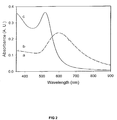
- FIG. 2 is a graph illustrating the UV-Vis spectra for SERS substrate characterization.
- FIG. 3 is a graph illustrating the SERS spectra of dithiobisbenzonic acid (DBA) and 4,4′-dithiobis(succinimidylbenzoate) (DSB) Raman-active reporter molecules adsorbed on gold island film substrates: (a) DBA, (b) DSB.
- DBA dithiobisbenzonic acid
- DSB 4,4′-dithiobis(succinimidylbenzoate)
- FIG. 4 is a graph illustrating the dose-response curves of the intensity of the strongest SERS band (1075 cm ⁇ 1 ) versus the rat IgG concentration: (a) using the colloidal detection reagent prepared via the co-immobilized approach, (b) using the colloidal detection reagent prepared via the covalent linking approach.
- FIG. 5 depicts an illustrative setup for SERS measurements.
- P polarization rotator
- PB 1 and PB2 Pellin Brocha prisms
- M mirror
- A aperture
- L1 cylindrical lens
- S sample slide
- L2 collection lens
- NF notch filter.
- FIG. 6 illustrates an example of a sandwich assay employing Raman-active reagents of the present invention.
- FIG. 7 is a schematic that comparatively illustrates two methods for preparing Raman-active immunogold reagents: (a) tracer nanoparticles are prepared by the physisorption of antibodies on gold colloids that had been previously coated with a partial monolayer of RRMs (Raman reporter molecules) based on aromatic thiols; (b) produces particles coated with a thiolate-based monolayer that has a terminal succinimide group (i.e., a terminal reactive functional group). This schematic (b) shows that the terminal succinimide group can then react with the amines of a protein to form an amide linkage.
- RRMs Raman reporter molecules
- FIG. 8 is schematic illustrating the process for preparing a Raman reporter-labeled immunogold colloid.
- FIG. 9 illustrates the experimental setup for measuring PSA levels in human serum using surface-enhanced Raman spectroscopy.
- FIG. 10 shows an infrared reflection spectra of DSU-derived monolayer on gold before (spectrum A) and after (spectrum B) exposure to the anti-free PSA capture antibody.
- FIG. 11 shows an infrared reflection spectra of a DSNB-derived monolayer on gold before (spectrum A) and after (spectrum B) exposure in the anti-PSA tracer antibody.
- FIG. 12 shows a Raman spectra of the reporter compound: (A) spectrum of DSNB powder; (B) SERS spectrum of gold nanoparticles following reaction with DSNB.
- FIG. 13 illustrates a SERS-based free PSA immunoassay: (A) SERS spectra, offset for clarify, acquired at various PSA concentrations; (B) dose-response curve for free PSA in human serum. The dose-response curve was constructed by calculating the average reading of the response for 6-8 different locations on the surface of each sample, which typically varied by 10% as described in the detailed description below.
- the present invention provides a novel class of Raman-active reagents for use in determining the presence or amount of a target analyte in a test sample. Also provided are particular methods and kits for using the Raman-active reagents of the present invention, as well as certain novel, preferred, methods for their manufacture.
- the Raman-active reagents according to the present invention comprise a Raman-active reporter molecule, a binding molecule, and a surface enhancing particle capable of causing surface enhanced Raman scattering.
- the Raman-active reporter molecule and the biological binder are operably linked, either directly or indirectly, to the surface enhancing particle to give both a strong surface enhanced Raman scattering (SERS) signal and specific binding affinity to a target analyte.
- the Raman-active reporter molecule and the binding molecule may be either directly linked to the surface enhancing particle or indirectly linked to the surface enhancing particle by way of a linker molecule.
- the Raman-active reporter molecule and the binding molecule may be independently linked to the surface enhancing particle, or the binding molecule may be operably linked to the Raman-active reporter molecule, which is operably linked to the surface enhancing particle.
- surface enhancing particle is defined herein to include particles capable of causing surface enhanced Raman scattering.
- Particles capable of causing surface enhanced Raman scattering are well known in the art and generally include, without limitation, particles of metallic materials such as gold, silver, copper, platinum, aluminum, gallium, indium, zinc, cadmium, lithium and sodium.
- the particles may also include, without limitation, other inert support structures of silica, plastic, glass, carbon, ceramics, or other materials, including magnetic materials, coated with a metallic material capable of causing surface enhanced Raman scattering, such as the metallic materials listed above.
- the particles used in the present invention are colloid particles.
- the colloid particles are preferably of a uniform and desired size and shape and stabilized against possible self-aggregation. Processes for preparing unaggregated colloids are well known in the art and typically involve, for example, the reduction of a metal salt (e.g., silver nitrate) with a reducing agent (e.g., citrate) to form a stable microcrystalline suspension. Stabilization may also be realized by the use of thin films or monolayers of various organic compounds.
- the colloid particles can be of any size as long as they give rise to an SERS signal. For example, the colloid particles may be less than 1000 nm in diameter, and preferably less than 100 nm in diameter.
- the surface enhancing particles are metallic nanoparticles.
- the large surface enhancement observed on metallic nanoparticles results in SERS intensities that can be comparable to or even exceeding those for fluorescence.
- Such a level of enhancement which may lead to a high detection sensitivity, together with the ease of handling, make metallic nanoparticles more promising than most other types of SERS substrates for use in ligand-binding assay applications.
- nonmetallic particles encapsulated with an enhancing material may also be of value in some applications.
- gold colloids is preferred over silver colloids, despite the fact that silver colloids provide larger enhancements than gold. This is because the greatest Raman enhancements with gold particles are produced with longer wavelength excitation light. This makes it possible to minimize the generation of sample fluorescence, which may interfere with the measurement of the Raman scattering.
- Raman-active reporter molecule is defined to include any one of a large number of molecules with distinctive Raman scattering patterns.
- molecules with distinctive Raman scattering patterns are well known in the art. Examples of such molecules include, but is not limited to, dithiobisbenzonic acid (DBA), 4-mercaptobenzoic acid (MBA), 2-naphthalenethiol (NT), thiophenol (TP), direct red 81, Chicago sky blue, 4,4′-dithiobis(succinimidylbenzoate) (DSB), p-dimethylaminoazobenzene, 1,5-difluoro-2,4-dinitrobenzene, 4-(4-aminophenylazo)phenylarsonic acid monosodium salt, arsenazo I, basic fuchsin, disperse orange 3, HABA (2-(4-hydroxyphenylazo)-benzoic acid, erythrosine B, trypan blue, ponceau S, ponceau SS, 5,
- the reporter molecule 5,5′-dithiobis(2-nitrobenzoic acid) may be converted to a corresponding succinimide ester derivative by treatment with the reactive compound, N-hydroxy succinimide (NHS), resulting in formation of 5,5′-dithiobis (succinimidyl-2-nitrobenzoate (DSNB).
- NHS N-hydroxy succinimide
- DSNB succinimidyl-2-nitrobenzoate
- DSNB can then be coupled to the primary amine group of a tracer antibody through formation of an amide linkage.
- reactive compound is defined to include any molecule having a reactive group including but not limited to succinimides, maleimides, isothiocyanates, isocyanates, acyl azides, sulfonyl chlorides, aldehydes, glyoxals, epoxides, oxiranes carbodiimides, carbonates, arylating agents, acryloyl derivatives, diazoalkanes, diazoacetyl compounds, anhydrides, aziridines, imidoesters, carbonyldiimidazole, or other groups that may be amine reactive, thiol reactive, or nucleophile reactive, such as for example, N-hydroxysuccinimide.
- binding molecule is defined to include any molecule having a binding specificity and avidity for a molecular component of a target analyte, or which is associated with a target analyte.
- binding molecules are known to those skilled in the art and typically include, without limitation, lectins (including fragments or derivatives thereof which retain binding function), monoclonal and polyclonal antibodies (including immunoreactive fragments or derivatives derived therefrom, which fragments retain all or a portion of the binding function of the antibody), peptides, haptens, aptamers, and nucleic acid molecules (including single stranded RNA, single-stranded DNA, or single-stranded nucleic acid hybrids), and any fragments and derivatives thereof. Crown ethers, cyclodextrins, cryptands, calixarenes, and many other types of ligands could also be used.
- target analyte is defined to include a molecule of an organic or inorganic nature, the presence and/or quantity of which is being tested for, which contains a molecular component (e.g., ligand or sequence or epitope or domain or portion or chemical group or reactive functionality or determinant) for which a binding molecule has binding specificity.
- a molecular component e.g., ligand or sequence or epitope or domain or portion or chemical group or reactive functionality or determinant
- the molecule may include, but is not limited to, a nucleic acid molecule, protein, glycoprotein, eukaryotic cell, prokaryotic cell, lipoprotein, peptide, carbohydrate, lipid, phospholipid, aminoglycans, chemical messenger, biological receptor, structural component, metabolic product, enzyme, antigen, antibody, drug, therapeutic, toxin, inorganic chemical, organic chemical, a substrate and the like.
- test sample means a sample to be tested for the presence or amount of a target analyte.
- the sample may include a target analyte or be free from the presence of the target analyte.
- operably linked is defined to mean a linkage between two different molecules, or a molecule and a particle, of sufficient stability for the purposes of signal enhancement and detection according to the present invention.
- two or more molecules, or a molecule and a particle may be operably linked utilizing reactive functionalities.
- Reactive functionalities include, but are not limited to, bifunctional reagents, linker molecules, biotin, avidin, free chemical groups (e.g., thiol, or carboxyl, hydroxyl, amino, amine, sulfo, phosphine, selenide, etc.), and reactive chemical groups (reactive with free chemical groups).
- linker is defined to refer to a compound or moiety that acts as a molecular bridge to operably link two different molecules, or a molecule and a particle, wherein one portion of the linker is operably linked to a first molecule, and wherein another portion of the linker is operably linked to a second molecule or particle.
- the two different molecules, or the molecule and particle may be linked to the linker in a step-wise manner.
- Linkers are known to those skilled in the art to include, but are not limited to, chemical chains, chemical compounds, carbohydrate chains, peptides, haptens, and the like.
- the linkers may include, but are not limited to, homobifunctional linkers and heterobifunctional linkers.
- a linker may comprise a carboxylic acid that has been activated by conversion to its acid chloride to react with an amino acid (e.g., lysine) residue of a binding molecule comprising a monoclonal antibody, and a thiol reactive group to link with the particle or the Raman-active reporter molecule.
- Heterobifunctional linkers are well known to those skilled in the art and generally contain a functionality on one end that binds to a target (e.g., a molecule or surface), and an opposite end having a second reactive functionality to specifically link to a different target.
- Heterobiofunctional photo-reactive linkers e.g., phenylazides containing a cleavable disulfide bond
- linkers in accordance with the present invention.
- a sulfosuccinimidyl-2-(p-azido salicylamido)ethyl-1,3′-dithiopropionate contains a N-hydroxy-succinimidyl azide (upon photolysis) reacts with any amino acid.
- the linker may further comprise a protective group which blocks reactivity with a functional group on the linker which is used to react with and bind to a molecule or particle to be linked.
- a deprotection reaction may involve contacting the linker to one or more conditions and/or reagents which remove the protective group, thereby exposing the function group to interact with the molecule to be linked.
- deprotection can be achieved by various methods known in the art, including, but not limited to, photolysis, acidolysis, hydrolysis, and the like.
- the linker may vary in length and composition for optimizing such properties as flexibility, stability, and resistance to certain chemical and/or temperature parameters.
- short linkers of sufficient flexibility include, without limitation, linkers having from 2 to 10 carbon atoms (see, e.g., U.S. Pat. No. 5,817,795).
- Any two molecules having an affinity for each other may comprise the reagent/analyte complex according to the present invention.
- ligand-binding systems include: antibodies and antigens; hormones and their receptors; lectins and the complex carbohydrates to which they bind; effector molecules and their receptors; complimentary nucleotide sequences; binding molecules designed through molecular modeling and synthesized specifically to bind another molecule, and molecules with mutual affinity to each other, such as avidin and biotin.
- the Raman-active reporter molecule and the binding molecule are each independently linked to the surface-enhancing particle.
- the Raman-active reporter molecule and the binding molecule may be either directly linked to the surface enhancing particle or indirectly linked to the surface enhancing particle by way of a linker molecule.
- the binding molecule is operably linked to the Raman-active reporter molecule, which is operably linked to the surface enhancing particle.
- the Raman-active reporter molecule and the binding molecule may be either directly linked to the surface enhancing particle or indirectly linked to the surface enhancing particle by way of a linker molecule.
- the Raman-active reagents of the present invention may be manufactured in the lab or provided to the user in the form of a kit.
- the kit may include a previously prepared Raman-active reagent, or the ingredients for manufacturing the Raman-active reagents as described above.
- the kit may also include ingredients that minimize nonspecific binding (nonspecific binding ingredient) and ingredients that stabilize the reagent (stabilizing ingredient) to extend its shelf life.
- nonspecific binding ingredients and stabilizing ingredients effective in use with the present invention are well known in the art.
- the kit may include a capture substrate covered with binding molecules to immobilize analytes for subsequent detection with the Raman-active reagent.
- the kit may also include unreactive spacer molecules, such as molecules terminated with ethylene glycol units, for interspersing amongst the binding molecules so as to minimize steric interferences as well as to resist nonspecific adsorption.
- unreactive spacer molecules such as molecules terminated with ethylene glycol units
- surfactants, blocking agents, and buffers may be added.
- Raman reporter-labeled immunogold probes can be prepared in many different ways. For example, as depicted in FIG. 1( a ) (the co-immobilization approach), an uncoated gold nanoparticle is labeled with Raman-active reporter molecules through the spontaneous adsorption of thiol-containing reporter molecules on gold, and then integrated with antibodies. The amount of thiol is chosen to coat only a portion of the nanoparticle surface and to leave exposed portions of the nanoparticle surface available for antibody immobilization. The antibodies are subsequently immobilized on the uncoated portion of the reporter-labeled nanoparticle through a combination of ionic and hydrophobic interactions. Immobilization can, of course, also be achieved by simply adsorbing an antibody directly on a coating of Raman-active reporter molecules.
- an uncoated gold nanoparticle is labeled with Raman-active reporter molecules, which are then covalently linked to antibodies.
- the Raman-active reporter molecule not only carries thiol or disulfide groups for immobilization on the gold nanoparticle, it also contains a succinimide ester functional group (i.e., a coupling reagent) for the covalent linking of an antibody.
- the covalent linker approach enhances the Raman reporter coverage and ultimately its sensitivity. Because the antibodies are covalently linked to the nanoparticles, the exchange of antibodies between nanoparticles with different Raman reporter molecules is reduced, and hence the probe specificity in the multi-analyte application is improved.
- the Raman-active reagents of the present invention determine the presence or amount of a target analyte, if present in a test sample, by the binding specificity of the binding molecule for the target analyte, or a portion thereof, and the generation and measurement of a SERS signal induced by the application of electromagnetic radiation onto the reagent/analyte combination.
- the Raman-active reagents may be used in clinics or forensic labs for the detection of drugs, pesticides, microbial toxins, hormones and biologically important proteins, industrial chemicals, explosives, pesticides, chlorophenols and other pollutants in soils, water, air, biological materials and other matrices. Such analysis may include in-situ testing methods (i.e., those not requiring any separation of the analytes from the sample prior to either their analysis or detection), as well as other in vivo, in vitro, or ex vivo methods.
- the detection or measurement of target analytes using the Raman-active reagents according to the present invention may be performed using any one of a number of assaying techniques known in the art.
- a test sample is placed in contact with a Raman-active reagent of the present invention under suitable conditions to allow the binding molecule to specifically bind to the target analyte, thus forming a reagent/analyte complex.
- the sample containing the reagent/analyte complex is then exposed to an excitation source (e.g., light source) that is suitable for exciting the Raman-active reporter molecule to induce surface enhanced Raman scattering.
- the intensity of the Raman scattering signal can then be measured to determine the presence or amount of the target analyte in the test sample. Absence of a Raman scattering signal is indicative of the absence of the target analyte in the test sample.
- Raman scattering Techniques for detecting Raman scattering are well known in the art.
- the primary measurement is one of light scattering intensity at particular wavelengths. Neither the angle of the incident beam nor the position of the detector is critical. With colloidal suspensions, detection is often at an angle of 90° to the incident beam.
- the intensity of the Raman scattering signals must be measured against an intense background from the excitation source. As such, the use of Raman-active report molecules with large Stokes shifts is preferred.
- SERS signals include fiber-optic waveguides, wavelength selective mirrors, and holographic optical elements for scattered light detection.
- the choice of the detector will largely depend on the sensitivity of detection required to carry out a particular assay.
- the intensity of the signal may be measured using a silicon photodiode, a charge coupled device (CCD), photographic film, or photomultiplier tubes arranged either singly or in series for cascade amplification of the signal.
- Photon counting electronics can also be used for sensitive detection.
- Raman signals consist of a series of distinct spectral lines of varying intensity. The frequencies and relative intensities of these spectral lines are specific to each Raman-active reporter molecule being detected such that each Raman-active reporter has a distinct “fingerprint”. The manner in which this fingerprint is analyzed will depend primarily on the purpose of the detection. If a SERS analyzer is being used to selectively detect one or more analytes out of a test sample containing multiple analytes, then an analysis of the entire fingerprint for each reporter molecule may be necessary to make a reliable identification. However, if the analyzer is being used to quantify the detection of one or several labels, each of which has a unique spectral line, then an analysis of only the unique spectral line may be necessary.
- the excitation source may be any source capable of exciting the Raman-active reporter molecule to induce Raman scattering. Typically, excitation will be carried out using incident light from a laser having a frequency in the visible spectrum. However, it is possible to envision situations in which other frequencies might be used, for example, in the ultraviolet or near-infrared ranges. The selection and tuning of the excitation source, with the appropriate frequency and power, will be well within the capabilities of one skilled in the art and will depend on the reporter molecule, surface enhancing particle and target analyte employed.
- a laser serves as the excitation source.
- the laser may be an inexpensive type such as a helium-neon or diode laser.
- a diode laser is used at or near the IR spectrum, minimizing fluorescence interference. Lamps may also be used as the excitation source. Direct illumination of the surface or by evanescent waves from a waveguide beneath the plasmon-active surface may also employed to induce a SERS affect.
- FIG. 5 An illustration of a typical SERS measurement system is depicted in FIG. 5 .
- the test sample may be placed in contact with a capture substrate covered with binding molecules that selectively immobilize analytes for subsequent detection with the Raman-active reagent.
- This substrate is then treated with the Raman-active reagent under suitable conditions to allow the Raman-active reagent binding molecule to specifically bind to the target analyte, forming the reagent/analyte complex.
- FIG. 3 illustrates one type of assay employing such a method.
- a different Raman-active reporter molecule is associated with a different antibody as different probes, with the presence of different antigens detected by the characteristic Raman bands of the reporters.
- test sample may be contacted with the Raman-active reagent under suitable conditions to allow the Raman-active reagent binding molecule to specifically bind to the target analyte, forming the reagent/analyte complex, prior to its capture by the substrate.
- the substrate may take the form of a generally flat surfaces (e.g., strips, slides, gene chips, etc.) or inert support structures of silica, carbon, plastic, glass, paper or other materials which may be in the form of macroscopically flat or textured pieces, slides, strips, spheroids or fibers capable of supporting the reagent/analyte complex.
- a generally flat surfaces e.g., strips, slides, gene chips, etc.
- inert support structures of silica, carbon, plastic, glass, paper or other materials which may be in the form of macroscopically flat or textured pieces, slides, strips, spheroids or fibers capable of supporting the reagent/analyte complex.
- Analytes and/or the reagent/analyte complex may bind to the substrate by direct adsorption, adsorption through a linker covalently attached to either the particle or the reporter molecule, by covalent attachment of the particle or reporter molecule to the substrate directly or through a linker or by intercalation of the distal portion of the linker into the substrate surface, by magnetic attraction to the substrate, or by specifically binding a second binding molecule affixed to the substrate to the target analyte or a molecule operably linked to the particle and having a specific affinity for the second binding molecule.
- the substrate may include a binding molecule identical to that found on the Raman-active reagent that binds the target analyte, such as in a sandwich assay.
- a binding molecule identical to that found on the Raman-active reagent that binds the target analyte, such as in a sandwich assay.
- Identification and quantification of the analytes would be accomplished through the measurement of the distinctive spectral fingerprints of the Raman-active labels provided for each analyte.
- the substrate may contain address locations for the various analytes with the specific binding molecule identified by the address location rather than by its spectral fingerprint. This system may also be employed for separating the target analyte from the test sample.
- the method may further comprise exposing the test sample to a magnetic force that separates the reagent/analyte complex from the test sample.
- a magnetic force that separates the reagent/analyte complex from the test sample.
- the surface-enhancing particle comprises of a material that is responsive to a magnetic force.
- the magnetic-responsive material is likely to be coated with a metallic material capable of emitting a SERS signal.
- the magnetic force may be applied to cause the reagent/analyte complex to be separated from the test sample.
- different Raman-active reagents having different Raman-active reporter molecules and binding molecules may be employed to allow for the sorting of multiple target analytes using magnetic forces.
- the assay kit comprises a Raman-active reagent in accordance with the present invention, wherein the Raman-active reagent includes at least one binding molecule having an affinity for a known target analyte.
- a substrate capable of binding the analyte and/or the reagent/analyte complex is provided.
- the kit may also include unreactive spacer molecules, such as molecules terminated with ethylene glycol units, for interspersing amongst the binding molecules so as to minimize steric interferences as well as to resist nonspecific adsorption.
- surfactants, blocking agents, and buffers may be added.
- One aspect of the present invention is that it allows multiple target analytes to be detected from a single test sample.
- simultaneous detection may be achieved by the use of multiple binding molecules, each specific to a target analyte or a class of target analytes and each associated with a different Raman-active reporter molecule.
- Raman-active vibrational modes usually yield bands one to two orders of magnitude narrower than most fluorescence bands, it is now possible to distinguish a much large number of different Raman labels as compared to fluorescent labels.
- a single Raman-active reporter molecule may also be employed with identification based on an address location on a substrate, such as a gene chip or screening slide, as is well known in the art.
- Raman scattering is not affected by oxygen and other quenchers, thus simplifying its use in many different experimental environments, it has potential advantages as a broadly applicable readout method in comparison to the widely used fluorescence detection schemes.
- SERS signal is less subject to photobleaching, lower detection limits can be obtained by increasing the signal integration time.
- Raman-active vibrational modes also usually yield bands one to two orders of magnitude narrower than most fluorescence bands, indicating the possibility of distinguishing a much large number of different Raman-active labels than likely with fluorescent labels, and minimizing the need to use spacial locations for analyte identification.
- Raman-active reagent used as a detection reagent in immunoassays. It is envisioned that similar concepts can be developed for other types of assays, target analytes and Raman-active reagents developed in accordance with the present invention, as well as infrared-active colloidal reagents, and in some cases, reagents for fluorescence or electrochemical based assays.
- the Raman-active reporter molecule 4,4′-dithiobis(succinimidylbenzoate) (DSB) was synthesized following a procedure similar to that used for preparing dithio-bis(succinimidylundecanoate) as described in Wagner et al., Biophys. J. 1996, 70, 2052, which is incorporated herein by reference.
- Raman-active immunogold colloidal reagents were prepared using the co-immobilization approach depicted in FIG. 1( a ).
- 25 ⁇ L of ethanolic Raman reporter solution (0.5 mM DBA) was added to 10 mL of a suspension of uncoated gold colloids ( ⁇ 30 nm diameter, 2 ⁇ 10 11 particles/mL) (Ted Pella, Inc.). The mixture was allowed to react for 5 hours at room temperature. During this step, the reporter molecules bound via self-assembly onto the colloid surface through the formation of sulfur-gold linkages.
- this amount of reporter based on an estimation of the colloidal surface area, will only partially cover the colloid, leaving portions of the uncoated colloidal surface available for protein immobilization.
- the loosely packed, red-colored sediment was resuspended in 10 mL of borate buffer (2 mM, pH 9).
- the Raman-active colloids were next immuno-labeled by adding 230 ⁇ g of goat anti-rat IgG to 10 mL of the above suspension. The mixture was incubated at 4° C. for 12 hours, during which the IgG protein adsorbed directly onto the exposed colloidal surface through a combination of ionic and hydrophobic interactions. The incubation was followed by centrifugation at 14,000 g for 5 minutes, and the loose sediment of reporter-labeled immunogold was rinsed by resuspending in 2 mM borate buffer and collected after a second centrifugation.
- the labeled colloids were suspended in 10 mM tris(hydroxymethyl) aminomethane (Tris)-buffered saline (Tris/HCl, NaCl 10 mM, pH 7.6) giving a concentration of approximately 2 ⁇ 10 11 particles/mL.
- Tween 80 (1%) (Aldrich) was also added to the suspension to minimize nonspecific adsorption in the assays.
- the suspensions usually remained uniformly dispersed for 2-3 days when stored at 4° C.
- Raman-active immunogold colloidal reagents were prepared using the covalent linking approach depicted in FIG. 1( b ).
- the DSB molecules were used as both Raman reporters and antibody linkers.
- the succinimide ester group of the DSB molecule can readily react with the primary amine group of an amino acid, such as the lysine, present in antibodies such as IgG to form a covalent bond.
- the preparation of the covalently-linked colloidal reagent follows a process very similar to that used for the co-immobilized reagents.
- the antibodies indirectly attached to the colloid through the reporter molecules rather than directly adsorbed onto the colloidal surface.
- reporter-linker solution 5 mM DSB in CHCl 3
- bare gold suspension 30 nm
- succinimide end groups available for protein immobilization. It is noted that this amount of the reporter-linker is estimated to be more than enough to cover the entire colloidal surface.
- the reporter-linker labeled colloids were centrifuged, and resuspended in the aforementioned borate buffer.
- Gold island films were used as SERS-active substrates to examine the scattering properties of the acid-terminated DBA and succinimide-terminated DSB reporters discussed above.
- Gold films were deposited onto clean glass microscope slides by resistive evaporation at a pressure of less than 1.3 ⁇ 10 ⁇ 4 Pa.
- Smooth gold films were prepared by first coating a glass substrate with 15 nm of chromium followed by 300 nm of gold. These substrates were used to prepare capture antibody substrates for the immunoassay experiment described below.
- FIG. 4 shows the spectra of two such films (spectra a and b) before immersion in the reporter molecule solution.
- spectrum c was collected from 5 nm colloidal gold suspended in aqueous solution. Both island films exhibited a plasmon resonance band with a maximum of 597 nm, while that for colloidal gold was at 519 nm.
- the plasmon bands from the island films were also wider than that observed from uniformly dispersed 5 nm colloidal gold.
- the DBA and DSB reporter molecules were analyzed to determine the difference in the reporter scattering properties as a result of altering the terminal functional groups in the reporter molecule.
- the experimental setup for the SERS measurements is shown in FIG. 2 .
- the signal was excited with a diode laser (Hitachi HL7851G, Thorlabs) operated at 20° C. and 120 mA. These conditions produced 50 mW of output power at the sample with a wavelength of 785.13 nm.
- a polarization rotator adjusted the polarization direction of the laser to minimize reflection losses at the Pellin-Brocha prisms. The prisms were used to remove background laser emission.
- the laser beam was then directed by a mirror through an aperture and focused by a 50-mm focal length cylindrical lens to a 3 mm by 0.25 mm line on the sample surface.
- the laser beam irradiated the sample at an angle of approximately 60° with respect to the surface normal, and the scattered light was collected and focused onto the entrance slit of the monochromator with a ⁇ /2 lens.
- a holographic notch filter (HSPF-785.0, Kaiser Optical Systems) was used to block the Rayleigh scattered light, while the Raman scattered light passed through the entrance slit (200 ⁇ m slit width) of a 300 f/4 spectrograph (SpectraPro 300i, Acton Research Corp.) and illuminated onto a 1200 grooves/mm grating.
- the grating was blazed for 750 nm and produced a nominal dispersion of 2.7 nm/mm.
- a thinned, back-illuminated, liquid nitrogen-cooled CCD (LN/CCD-1100PB, Princeton Instruments) was controlled by a PC for spectra acquisition.
- the positions of the reporter molecule Raman bands were determined by calibration using the known band positions of solid naphthalene.
- SERS spectra (10 second integration time) of self-assembled monolayers of DBA and DSB on the gold island films are shown in FIG. 3 .
- Several strong aromatic vibrational bands from the benzene ring are present within this spectral region.
- the strongest band at 1075 cm ⁇ 1 is from the aromatic C—H in-plane bending, and another major band at 1585 cm ⁇ 1 is from the C ⁇ C ring stretching.
- signals at 1075 cm ⁇ 1 were used as readout in both the DBA and DSB-based immunoassays.
- the similar intensities of the bands in the two spectra of FIG. 3 was consistent with what was expected based on the similarity in the molecular structures of the two types of reporters. It was also noted that the intensity ratios of the peak at 1075 cm ⁇ 1 to the peak 1585 cm ⁇ 1 in the two spectra were slightly different, possibly reflecting the orientation difference of the two types molecules when adsorbed on the surface.
- the immobilization of the IgG proteins was accomplished by first immersing the monolayer-modified substrates into 1% (w/w) 1-ethyl-3-[3-(dimethylamino)propyl] carbodiimide (EDC) (Aldrich) in anhydrous acetonitrile for 5 hours. This step activates the free carboxyl groups of thioctic acid by forming on O-acylurea intermediate with the EDC. The activated surface was then modified with capture antibody by pipetting 100 ⁇ L of goat anti-rat IgG (100 ⁇ g/mL, 0.1 M borate buffer, pH 9) (Pierce) onto approximately 1-cm 2 of the activated substrate. This reaction was allowed to progress at 4° C. for 12 hours. Finally, the antibody-coated substrates were rinsed with deionized water, and quickly dried under a stream of argon. All assays were conducted using freshly prepared substrates.
- EDC 1-ethyl-3-[3-(dimethyl
- Dose-response curves were constructed based on the results of a set of sandwich assays.
- a 100 ⁇ L aliquot of each sample solution was pipetted onto the separate capture antibody substrates described above, and allowed to react for 1 hour at room temperature. After rinsing with copious amounts of water, the substrates were then exposed to 100 ⁇ L of reporter-labeled immunogold solution for 3 hours. All substrates were rinsed with deionized water and dried under argon before SERS characterization.
- the detection approach relied solely on the SERS effect by utilizing the immunogold colloids labeled with Raman-active reporter molecules as detection reagents.
- gold colloids were labeled with both antibodies for bio-recognition and Raman-active reporter molecules for signal transduction.
- a key feature of this concept is that the scattering center of the label is positioned in close proximity to the colloid surface, which strongly enhances the signal. The presence of the antigen was therefore recognized by its detection antibody, and the SERS signal of the co-immobilized Raman active species reported the ligation of the antibody with the antigen.
- FIG. 4 plots the intensity of this band versus the concentration of the antigen, rat IgG, with either the co-immobilized ( 4 a ) or the covalently-linked colloids ( 4 b ) as detection reagent.
- SERS signals show proportional response to the antigen concentration almost over the entire tested concentration range, representing a dynamic range of nearly 8 orders of magnitude.
- the solid lines represent the curve fitting of the immunoassay data based the four-parameter logistic model, a common regression model used for describing sandwich type immunoassays. The slope of the curve suggests how the readout signals quantify samples of different concentration; the larger the slope, the easier the distinction. Two important parameters obtained from the curve fitting will be discussed with more details in the later sections. One is the expected signal at zero dose, which is also called the negative control signal, the other is the expected signal at infinitely high or saturation dose, which is also called the positive control signal.
- LOD LOD was largely due to the different SERS intensities observed for the two negative control samples, which were obtained through the same assay procedure, using samples at a concentration of zero (i.e., buffer only). Indeed, the difference in the negative control signals, which reflect different extents of nonspecific binding, is a major difference between the two sets of results.
- the colloidal reagent prepared using the co-immobilization approach seemed to yield a more pronounced nonspecific binding, and therefore, a higher Raman signal (145 counts) for the negative control sample (from curve fitting, S is 164 counts at zero concentration).
- the colloids modified via the covalent linking approach yielded a much lower signal (22 counts) from the negative control (32 counts based on curve fitting). Because of the lack of a fluorescence background and lack of photobeaching of the Raman-active reagent, LOD values could be lowered by increasing the signal integration times.
- the colloidal suspension prepared from the covalent linking approach was also more stable in solution and less susceptible to aggregation. These observations explain the lower run-to-run variation observed when using the covalently-linked reagent ( ⁇ 10%) compared to that when using the co-immobilized reagent (>20%). It was also noted that when starting with a new batch of reporter-labeled immunogold reagent, the batch-to-batch variation was even more significant and sometimes up to 100% when using the co-immobilized reagent. It is suspected that this difference represents the importance of the first step in the colloid modification. It is less critical in the covalent linking approach because DSB was always added at a level to ensure the exhaustive coverage of the reporters on every colloid. However, it is very critical in the co-immobilization approach since the dosage of DBA determined the reporter coverage on each colloid and hence the signal intensity per colloid.
- Suspensions of unconjugated colloidal gold (32.2+4.4-nm diameter, 2 ⁇ 1011 particles/ml) were purchased from Ted Pella, Inc.
- the matched pair of monoclonal antibodies utilized for the sandwich assay was obtained from research Diagnostics. The pair consisted of mouse anti-human free PSA clone PSA-F65, which was used as the capture antibody after immobilization on gold-coated glass chips, and mouse anti-human PSA clone PSA-66, which was employed as the tracer antibody after conjugation to the gold particles as described below.
- Serum PSA (10-30% free PSA) was purchased from Bios Pacific, and buffer packs and ImmunoPure normal human serum were acquired from Pierce Biotechnology.
- N-hydrosysuccinimide (NHS), 1,3-dicyclohexylcarbodiimide (DCCD), Tween 80, 5,5′-dithiobis(2-nitrobenzoid acid) (DNBA), and bovine serum albumin (BSA) were obtained from Aldrich.
- DNBA 1,3-dicyclohexylcarbodiimide
- BSA bovine serum albumin
- the Raman-active reporter molecule 5,5′-Dithiobis(succinimidyl-2-nitrobenzoate) was synthesized following a procedure similar to that described herein above and in Porter et al., Anal. Chem.; 2003, 1;75(21):5936-43 (incorporated by reference herein in its entirety). Briefly, to 50 mL of dry tetrahydrofuran was added 0.50 g of DNBA (1.3 mmol), 0.52 g of DCCD (2.5 mmol), and 0.29 G of NHS (2.5 mmol) in a 100-mL round-bottom flask equipped with a drying tube. The mixture was magnetically stirred at 25° C.
- Raman Reporter-labeled immunogold colloids were prepared using various derivatives of dithiobis (benzoic acid), which could easily be converted to the corresponding succinimide ester with NHS.
- DSNB is a particularly attractive example because of the strong scattering cross section of its symmetric NO 2 stretch.
- treatment of colloidal gold with this derivative yields a coating of the thiolate of DSNB, which can couple to the primary amines of a tracer antibody by formation of an amide linkage.
- this design strategy minimizes the distance between the gold surface and label scattering center. This minimization is particularly significant because, according to a simplified electromagnetic model, enhancement varies inversely with the 12th power of the separation distance between the scatterer and the metal particle center.
- step one 100 ⁇ L of a 2.5 mM DSNB solution in acetonitrile was added to 1 mL of the unconjugated colloidal gold suspension and the mixture reacted for 3-5 h.
- the reporter-labeled colloids were then separated from solution by centrifugation at 10000 g for 7 min. The clear supernatant was discarded, and the loose red sediment was resuspended in 1 mL of borate buffer (2 mM, pH 9).
- step two mouse anti-PSA was coupled to the gold particles via the succinimidyl terminus of the DSNB-derived coating.
- 35 ⁇ g of detection antibody (7 ⁇ L of 5 mg/mL PSA-66 solution) was added to the 1-mL suspension of the reporter-labeled colloid. The mixture was then incubated at room temperature for 1 h. After centrifugation at 10000 g for 7 min and removal of the supernatant, the red sediment was resuspended in 1 mL of 2 mM Tris buffer (Tris-HCl (pH 7.6), 1% BSA).
- Tris buffer Tris-HCl (pH 7.6), 1% BSA.
- the use of BSA, Tween 80, or both in all of the preparative steps and in the assay protocol is part of a general procedure designed to minimize complications from nonspecific adsorption.
- Capture antibody substrates were prepared by first cleaning glass slides in an ultrasonic bath under dilute surfactant solution (Micro, Cole-Parmer), deionized water, and methanol, each for 30 min. The slides were then loaded into an Edwards 306A metal evaporator and coated with 15 nm of chromium and 300 nm of gold at 0.2 nm/s at pressures less than 5 ⁇ 10 ⁇ 6 Torr. Next, the gold substrates were removed from the evaporator and exposed for ⁇ 30 s to an octadecanethiol (ODT)-soaked poly(dimethylsiloxane) stamp, which had a 5-mm-diameter hole cut in its center.
- ODT octadecanethiol
- This step “inks” the outer portion of the gold substrate with a monolayer of ODT.
- the substrates were rinsed with ethanol, dried under a stream of nitrogen, and immersed in a 1 mM ethanolic solution of DSU for 6-12 h. Upon removal from solution, the substrates were rinsed again with ethanol and dried under a stream of nitrogen.
- the result is a 5-mm-diameter domain of the succinimide ester-terminated monolayer on each substrate, surrounded by a hydrophobic ODT coating.
- the ODT coating serves as a hydrophobic barrier that localizes aqueous protein solutions when pipetted onto the area of the substrate defined by the DSU-derived monolayer.
- PSA-65 Anti-free PSA antibodies (PSA-65) were immobilized by pipetting 40 ⁇ L of the protein solution (100 ⁇ g/mL in 0.05 M borate buffer (pH 9) and 1% Tween 80) onto the localized domain of the DSU-modified monolayer. The reaction was allowed to progress overnight at room temperature. After rinsing three times with buffer 1 (0.01 M borate buffer (pH 9), 30 mM NaCl, 0.5% Tween 80), 40 ⁇ L of blocking buffer (5% BSA in 0.05 M borate buffer (pH 9) was pipetted onto the surface and incubated for 1 h. The substrates were then rinsed three times with buffer 1.
- PSA dose-response curves were constructed using matrixes consisting of normal human serum, 10 mM phosphate-buffered saline (PBS, KH2PO4/K2HPO4 (pH 7.5), 150 mM NaCl, 0.1% BSA, 0.5% Tween 80, 0.02% NaN3), and a 1:1 mixture of human serum and PBS, following the typical procedure for a sandwich-type assay. For each matrix, 40 ⁇ L aliquots of PSA solutions of various concentrations were pipetted onto a capture antibody-coated substrate and allowed to react for 3 h at room temperature.
- the system consists of three major subassemblies: laser light source, spectrograph, and fiber-optic probe.
- FIG. 9 shows the spectroscopic setup
- the light source is a 30-mW, 632.8-nm HeNe laser, while the spectrograph consists of an f/2.0 Czerny-Turner imaging spectrometer (6-8-cm ⁇ 1 resolution, no moving parts) and a thermoelectrically cooled (0° C.) Kodak 0401E CCD.
- the fiber-optic probe (1.75 ⁇ 2.5 ⁇ 6 in) utilizes band-pass and long-pass filters for laser light (OD 6) and fiber background (OD 4) rejection.
- the probe objective provides a numerical aperture of 0.65 while maintaining a relatively long working distance of 3 mm.
- the laser spot size on the sample surface is ⁇ 22 ⁇ m in diameter.
- a Windows-based Visual Basic program controls the system. All spectra were collected with a 60-s integration time. The positions of the Raman bands were determined by comparisons to the known positions of bands for solid naphthalene.
- Infrared reflection spectra were acquired with a Nicolet 850 FT-IR spectrometer, purged with liquid N 2 boil-off, and equipped with a liquid N 2 -cooled HgCdTe detector. Spectra were obtained using p-polarized light incident at 80° with respect to the surface normal. The spectra were recorded as—log(R/Ro), where R is the sample reflectance and Ro is the reflectance of an octadecanethiolate-d 37 monolayer-coated Au reference. The spectra are an average of 512 sample and reference scans, taken at 4 cm ⁇ 1 resolution with Happ-Genzel apodization.
- X-ray photoelectron spectra were acquired at room temperate with a Physical Electronics Industries 5500 multitechnique surface analysis system. This system is equipped with a hemispherical analyzer, a toroidal monochromator, a multichannel detector at 45°, and monochromatic Al K ⁇ excitation radiation (1486.6 eV, 250 W). A pass energy of 29.35 eV was used, giving a half-width of the Au(4f 7/2 ) peak of ⁇ 0.8 eV.
- the capture antibody substrate consisted of anti-free PSA bound to a gold-coated glass chip via the DSU-derived coupling agent.
- DSU chemisorbs to gold through cleavage of the sulfur-sulfur bond, and the formation of the resulting gold-bond thiolate and its subsequent coupling to anti-free PSA can be readily confirmed by infrared reflection spectroscopy (IRS) and XPS.
- the IRS results are presented in FIG. 10 .
- the three bands around 1800 cm ⁇ 1 in the spectrum of the layer formed from DSU FIG.
- IRS was also used to confirm the covalent binding of anti-free PSA to the terminal group of the gold-bound coupling layer ( FIG. 10B ). Since the acyl carbon of the succinimidyl ester group is strongly susceptible to nucleophilic attack, reaction with the sterically accessible amines in the protein should immobilize anti-free PSA via amide linkages. As evident in FIG. 2B , treatment of the DSU-modified substrate with anti-free PSA causes a marked decrease in the magnitude of the bands for the succinimidyl group (e.g., 1750, 1219, and 1078 cm ⁇ 1 ).
- the C (1s) region was composed of a lower energy band (284.4 eV), attributed to the alkyl chain structure of the coating, and a higher energy band (289.0 eV), assigned to the different types of carbonyl carbon.
- Two bands were also observed in the O(1s) and S(2p) regions.
- the band at 534.9 eV is ascribed to the oxygen of the ester linkage and that at 532.5 eV is assigned to the remaining carbonyl oxygens.
- FIG. 11 shows the IRS spectra for a monolayer of DSNB spontaneously adsorbed on gold-coated glass before and after exposure to anti-PSA.
- the as-formed layer has carbonyl stretches at 1812, 1789, and 1748 cm ⁇ 1 and strong symmetric and asymmetric nitro stretches at 1343 and 1533 cm ⁇ 1 , respectively.
- the spectrum for the DSNB-derived monolayer undergoes a similar set of changes following exposure to anti-PSA.
- the XPS characterizations (Table 1) also strongly mimic those for the DSU-based layer. In this case, however, there are two N(1s) bands for each sample. For the as-formed layer, bands at 401.2 and 405.5 eV are indicative of the succinimidyl nitrogen and nitro nitrogen on the aromatic ring, respectively. After exposure to anti-PSA, the band at 401.2 eV disappears and one at 400.0 eV appears. This change again parallels that for the DSU-derived coating.
- Raman spectra for the Ramen reporter molecule DSNB are shown in FIG. 12 before and after coupling to the gold nanoparticles.
- the nanoparticle sample was prepared by drop casting a small amount of the labeled colloid solution onto a gold-coated glass slide and evaporating the water-based solvent.
- the powder spectrum ( FIG. 12A ) is dominated by the symmetric nitro stretch at 1342 cm ⁇ 1 , and we attribute the band at 851 cm ⁇ 1 to the nitro scissoring vibration.
- the band at 1566 cm ⁇ 1 is assigned to an aromatic ring mode (8a), and the large band at 1079 cm ⁇ 1 is probably a succinimidyl N—C—O stretch overlapping with aromatic ring modes.
- FIG. 13 The results of our SERS-based determinations for free PSA in normal human serum are shown in FIG. 13 .
- Test solutions were made by serial dilution in human serum of a 1 mg/mL PSA standard to cover the range from 1 ⁇ g/mL (30 nM) to pg/mL (30 fM).
- the spectra in FIG. 13A were obtained using 60-s integrations after completion of the immunoassay protocol outlined above.
- the features diagnostic of the DSNB-labeled nanoparticles exhibit a strong increase as the PSA level increases. These changes span more than 6 order of magnitude, this encompassing concentration levels critical to prostate cancer diagnosis.
- FIG. 13B A more detailed treatment of applicants' findings is presented by the dose-response curve in FIG. 13B .
- This curve was constructed by plotting the scattering intensity of the symmetric nitro stretch (1338 cm ⁇ 1 , full width at half-maximum of 22 cm ⁇ 1 ). Each data point represents the average of six to eight readings across the sample surface. Variations in signal strength across the surface of each chip were typically ⁇ 10%. However, signal strengths up to twice as large as those represented in the plot were observed ⁇ 20% of the time. The does-response curve was constructed by omitting the data for these “hot spots”.
- hot spots could possibly reflect the presence of domains where there are higher localized concentrations of binding sites and, therefore, higher particle densities are reasonably homogenous over areas irradiating by the laser source.
- AFM imaging revealed a small number of particle aggregates that could account for the hot spots.
- a third possibility arises from the existence of “hot particles”. Recent studies have shown that enhancement factors are strongly dependent on particle size, shape, and excitation wavelength and that a small fraction of particles exhibit markedly larger enhancements.
- the first avenue uses labels that undergo both resonance and surface enhancement. With resonance enhancement, intensities can be 2-6 orders of magnitude greater than those based on normal Raman scattering.
- the second avenue takes advantage of recent reports that have shown that the surface enhancement for slightly larger gold particles (e.g., 60 nm for our excitation wavelength) is greater than that for 30-nm particles.
- the ability to detect the binding of a single antigen appears to be well within reach and should be of immense value in the ultra-low-level detection of a wide range of biomarkers used in early disease diagnosis and other assay applications.
- Low-level detection becomes even more important as the degree of multiplexing increases, e.g., in instances where screening for multiple analytes at a single location is of interest.
- this embodiment of the present invention enables the detection of biomarkers for early cancer diagnosis in serum samples at very low concentrations by a SERS-based readout method.
- This strategy is capable of encompassing a wide range of applications, especially in view of the opportunities to multiplex through the judicious design of more labeled nanoparticles. As such, multiple analytes could be concurrently identified through the position of a characteristic feature of the Raman label and then quantified by its intensity.
- assays could be developed for the high-sensitivity, simultaneous screening of a battery of cancer markers using a single serum sample, saving time, reducing assay costs, and potentially leading to earlier diagnosis.
Abstract
Description
| TABLE 1 |
| Binding Energies (eV) and Compositional Assignments for |
| XPS Spectra of DSU and DSNB Monolayers on Gold before |
| and after Antibody Derivitization |
| Anti-free | Anti-PSA/ | |||
| Core level | DSU/Au | PSA/DSU/Au | DSNB/Au | DSNB/Au |
| Au(4f7/2) | 83.9 | 83.9 | 83.9 | 83.9 |
| S(2p3/2) | 161.8 | 161.8 | 162.2 | 162.2 |
| S(2p1/2) | 162.9 | 162.9 | nda | nd |
| C(1s) | 289.0 | 288.5 | 288.4 | 288.1 |
| C(1s) | 284.4 | 284.9, 286.4 (sh) | 284.6 | 284.8, 285.9 (sh) |
| N(1s) | 401.9 | 400.5 | 405.6, | 405.2, 400.0 |
| 401.2 | ||||
| O(1s) | 532.5, | 432.1 | 532.2 | 532.2 |
| 534.9 | ||||
| and, not detected. | ||||
Claims (23)
Priority Applications (2)
| Application Number | Priority Date | Filing Date | Title |
|---|---|---|---|
| US10/931,142 US7829348B2 (en) | 2000-09-22 | 2004-08-31 | Raman-active reagents and the use thereof |
| US12/894,810 US20110070662A1 (en) | 2000-09-22 | 2010-09-30 | Raman-active reagents and the use thereof |
Applications Claiming Priority (3)
| Application Number | Priority Date | Filing Date | Title |
|---|---|---|---|
| US23460800P | 2000-09-22 | 2000-09-22 | |
| US09/961,628 US7824926B1 (en) | 2000-09-22 | 2001-09-24 | Raman-active reagents and the use thereof |
| US10/931,142 US7829348B2 (en) | 2000-09-22 | 2004-08-31 | Raman-active reagents and the use thereof |
Related Parent Applications (1)
| Application Number | Title | Priority Date | Filing Date |
|---|---|---|---|
| US09/961,628 Continuation-In-Part US7824926B1 (en) | 2000-09-22 | 2001-09-24 | Raman-active reagents and the use thereof |
Related Child Applications (1)
| Application Number | Title | Priority Date | Filing Date |
|---|---|---|---|
| US12/894,810 Division US20110070662A1 (en) | 2000-09-22 | 2010-09-30 | Raman-active reagents and the use thereof |
Publications (2)
| Publication Number | Publication Date |
|---|---|
| US20050089901A1 US20050089901A1 (en) | 2005-04-28 |
| US7829348B2 true US7829348B2 (en) | 2010-11-09 |
Family
ID=46302705
Family Applications (2)
| Application Number | Title | Priority Date | Filing Date |
|---|---|---|---|
| US10/931,142 Expired - Fee Related US7829348B2 (en) | 2000-09-22 | 2004-08-31 | Raman-active reagents and the use thereof |
| US12/894,810 Abandoned US20110070662A1 (en) | 2000-09-22 | 2010-09-30 | Raman-active reagents and the use thereof |
Family Applications After (1)
| Application Number | Title | Priority Date | Filing Date |
|---|---|---|---|
| US12/894,810 Abandoned US20110070662A1 (en) | 2000-09-22 | 2010-09-30 | Raman-active reagents and the use thereof |
Country Status (1)
| Country | Link |
|---|---|
| US (2) | US7829348B2 (en) |
Cited By (1)
| Publication number | Priority date | Publication date | Assignee | Title |
|---|---|---|---|---|
| US9638639B2 (en) | 2013-06-04 | 2017-05-02 | Board Of Regents, The University Of Texas System | Plasmonic-magnetic bifunctional nanotubes for biological applications |
Families Citing this family (58)
| Publication number | Priority date | Publication date | Assignee | Title |
|---|---|---|---|---|
| US20030211488A1 (en) * | 2002-05-07 | 2003-11-13 | Northwestern University | Nanoparticle probs with Raman spectrocopic fingerprints for analyte detection |
| NZ528323A (en) * | 2003-09-18 | 2006-05-26 | Horticulture & Food Res Inst | Immunoassay |
| US20050147963A1 (en) * | 2003-12-29 | 2005-07-07 | Intel Corporation | Composite organic-inorganic nanoparticles and methods for use thereof |
| US20080076119A9 (en) * | 2003-12-29 | 2008-03-27 | Lei Sun | Composite organic inorganic nanoclusters |
| US7361410B2 (en) * | 2003-12-29 | 2008-04-22 | Intel Corporation | External modification of composite organic inorganic nanoclusters comprising raman active organic compound |
| US20050191665A1 (en) * | 2003-12-29 | 2005-09-01 | Xing Su | Composite organic-inorganic nanoclusters |
| US7226794B2 (en) * | 2004-04-14 | 2007-06-05 | Agilent Technologies, Inc. | Surface-enhanced Raman spectroscopy for biosensor systems and methods for determining the presence of biomolecules |
| US20070048797A1 (en) * | 2004-08-11 | 2007-03-01 | Xing Su | Composite organic inorganic nanoclusters as carriers and identifiers of tester molecules |
| US7776547B2 (en) * | 2004-08-26 | 2010-08-17 | Intel Corporation | Cellular analysis using Raman surface scanning |
| US20060046311A1 (en) * | 2004-08-26 | 2006-03-02 | Intel Corporation | Biomolecule analysis using Raman surface scanning |
| US7301624B2 (en) * | 2004-09-07 | 2007-11-27 | Lawrence Livermore National Security, Llc | Nanosensors based on functionalized nanoparticles and surface enhanced raman scattering |
| US20060147941A1 (en) * | 2004-12-30 | 2006-07-06 | Intel Corporation | Methods and apparatus for SERS assay of biological analytes |
| US20060216697A1 (en) * | 2005-03-24 | 2006-09-28 | General Electric Company | Method of separating unattached Raman-active tag from bioassay or other reaction mixture |
| US20060216835A1 (en) * | 2005-03-24 | 2006-09-28 | General Electric Company | Method of separating unattached Raman-active tag from bioassay or other reaction mixture |
| US7518721B2 (en) | 2005-09-09 | 2009-04-14 | Ge Homeland Protection, Inc. | Raman-active lateral flow device and methods of detection |
| US7355703B2 (en) * | 2005-09-09 | 2008-04-08 | Ge Homeland Protection, Inc. | Raman-active lateral flow device and methods of detection and making |
| KR100650522B1 (en) * | 2005-09-15 | 2006-11-27 | 재단법인서울대학교산학협력재단 | A new label-free high throughput screening method by using sers spectroscopic encoded bead and dielectrophoresis |
| US20070155022A1 (en) * | 2005-12-30 | 2007-07-05 | Mineo Yamakawa | Degenerate binding detection and protein identification using Raman spectroscopy nanoparticle labels |
| DE102006000775A1 (en) * | 2006-01-04 | 2007-07-12 | Julius-Maximilians-Universität Würzburg | Diagnostic imaging techniques, useful for representing distribution of substances in immobilized cell, comprises displacing immobilized cell with a substance and bonding a marker for surface enhanced vibrational spectroscopy on substance |
| EP2155065A4 (en) * | 2007-05-14 | 2014-09-10 | Univ Johns Hopkins | Methods for in vivo imaging of cells |
| EP2040075A1 (en) * | 2007-09-24 | 2009-03-25 | Julius-Maximilians-Universität Würzburg | Compounds and markers for surface-enhanced raman scattering |
| WO2009086509A2 (en) | 2007-12-27 | 2009-07-09 | Purdue Research Foundation | Reagents for biomolecular labeling, detection and quantification employing raman spectroscopy |
| US8076162B2 (en) * | 2008-04-07 | 2011-12-13 | Life Bioscience, Inc. | Method of providing particles having biological-binding areas for biological applications |
| WO2009142604A1 (en) * | 2008-05-23 | 2009-11-26 | Nanyang Technological University | Polymer encapsulated particles as surface enhanced raman scattering probes |
| WO2011078794A1 (en) | 2009-12-22 | 2011-06-30 | Agency For Science, Technology And Research | Sers-based analyte detection |
| CN102004096B (en) * | 2010-09-28 | 2012-06-27 | 吉林大学 | Surface enhanced Raman detection method for polycyclic aromatic hydrocarbon and substitute thereof |
| US8897856B2 (en) * | 2011-11-26 | 2014-11-25 | Angelo Gaitas | Atherosclerosis therapy via delivery and localized heating of micro size particles |
| CN102628807B (en) * | 2012-03-31 | 2015-12-02 | 中国科学院合肥物质科学研究院 | Based on the method for Surface enhanced raman spectroscopy detection trace biomolecular ionising radiation decomposition reaction |
| KR102116497B1 (en) * | 2013-09-25 | 2020-05-29 | 삼성디스플레이 주식회사 | Organic light emitting display apparatus and method for manufacturing the same |
| KR101940981B1 (en) | 2014-05-05 | 2019-01-23 | 3디 글래스 솔루션즈 인코포레이티드 | 2d and 3d inductors antenna and transformers fabricating photoactive substrates |
| US10071141B2 (en) | 2015-05-08 | 2018-09-11 | Spectral Platforms, Inc. | Albumin-based non-covalent complexes and methods of use thereof |
| WO2017007291A1 (en) * | 2015-07-09 | 2017-01-12 | 서울대학교산학협력단 | Method for detecting nucleic acid molecule using surface enhancement raman scattering particles |
| US10070533B2 (en) | 2015-09-30 | 2018-09-04 | 3D Glass Solutions, Inc. | Photo-definable glass with integrated electronics and ground plane |
| US11264167B2 (en) | 2016-02-25 | 2022-03-01 | 3D Glass Solutions, Inc. | 3D capacitor and capacitor array fabricating photoactive substrates |
| WO2017177171A1 (en) | 2016-04-08 | 2017-10-12 | 3D Glass Solutions, Inc. | Methods of fabricating photosensitive substrates suitable for optical coupler |
| US10132808B2 (en) * | 2016-09-23 | 2018-11-20 | The United States Of America As Represented By Secretary Of The Navy | Method to detect/identify bacterial species using flow cytometry and surface enhanced Raman scattering |
| CN106525814B (en) * | 2016-11-07 | 2018-12-25 | 华南师范大学 | It is a kind of based on magnetic core-gold satellite assembly PSA detection method |
| CA2949634C (en) * | 2016-11-25 | 2024-02-13 | Medmira Inc. | Analyte detection using raman spectroscopy |
| CN106706910B (en) * | 2017-01-05 | 2018-10-09 | 江苏大学 | A kind of food-borne pathogens detection method for modifying gold nanorods based on p-Mercaptoaniline |
| EP3593137A1 (en) | 2017-03-10 | 2020-01-15 | Universität Duisburg-Essen | Efficient ligand exchange of a detergent bilayer on the surface of metal nanoparticles for molecular functionalization and assembly, corresponding functionalized nanoparticles and nanoparticle assemblies, and their use in plasmonic applications including surface-enhanced raman spectroscopy |
| WO2018175346A1 (en) * | 2017-03-20 | 2018-09-27 | Spectral Platforms, Inc. | Spectroscopic methods to detect and characterize microorganisms |
| KR102273624B1 (en) | 2017-04-28 | 2021-07-07 | 3디 글래스 솔루션즈 인코포레이티드 | Rf Circulator |
| JP6995891B2 (en) | 2017-07-07 | 2022-01-17 | スリーディー グラス ソリューションズ,インク | 2D and 3D RF centralized device for RF systems in packaged photoactive glass substrates |
| EP3724946B1 (en) | 2017-12-15 | 2024-04-17 | 3D Glass Solutions, Inc. | Coupled transmission line resonate rf filter |
| AU2018399638B2 (en) | 2018-01-04 | 2021-09-02 | 3D Glass Solutions, Inc. | Impedance matching conductive structure for high efficiency RF circuits |
| KR102626372B1 (en) | 2018-04-10 | 2024-01-16 | 3디 글래스 솔루션즈 인코포레이티드 | Rf integrated power condition capacitor |
| US10903545B2 (en) | 2018-05-29 | 2021-01-26 | 3D Glass Solutions, Inc. | Method of making a mechanically stabilized radio frequency transmission line device |
| KR102518025B1 (en) | 2018-09-17 | 2023-04-06 | 3디 글래스 솔루션즈 인코포레이티드 | High efficiency compact slotted antenna with a ground plane |
| KR102392858B1 (en) | 2018-12-28 | 2022-05-03 | 3디 글래스 솔루션즈 인코포레이티드 | Toroidal Capacitor RF, Microwave, and Mm Wave Systems |
| WO2020139951A1 (en) | 2018-12-28 | 2020-07-02 | 3D Glass Solutions, Inc. | Heterogenous integration for rf, microwave and mm wave systems in photoactive glass substrates |
| KR20210147040A (en) | 2019-04-05 | 2021-12-06 | 3디 글래스 솔루션즈 인코포레이티드 | Glass-Based Blank Substrate Integrated Waveguide Device |
| CA3136642C (en) | 2019-04-18 | 2023-01-03 | 3D Glass Solutions, Inc. | High efficiency die dicing and release |
| EP4121988A4 (en) | 2020-04-17 | 2023-08-30 | 3D Glass Solutions, Inc. | Broadband induction |
| CN111441022A (en) * | 2020-04-20 | 2020-07-24 | 上海纳米技术及应用国家工程研究中心有限公司 | Preparation method of SERS-enhanced new coronavirus detection chip, product and application thereof |
| CN111624186B (en) * | 2020-06-24 | 2021-03-16 | 江南大学 | Enterotoxin spectral analysis method based on fluorescence and Raman double-signal enhancement |
| CN112938979A (en) * | 2021-01-29 | 2021-06-11 | 中山大学 | MXene composite material with SERS effect and preparation method and application thereof |
| CN113189078B (en) * | 2021-03-04 | 2024-04-16 | 吉林大学 | High-throughput screening method of targeted drugs |
| CN113834777B (en) * | 2021-07-14 | 2023-12-08 | 四川轻化工大学 | Preparation and application methods of SERS substrate for detecting acidic pigment |
Citations (8)
| Publication number | Priority date | Publication date | Assignee | Title |
|---|---|---|---|---|
| US5252459A (en) * | 1988-09-23 | 1993-10-12 | Abbott Laboratories | Indicator reagents, diagnostic assays and test kits employing organic polymer latex particles |
| US5266498A (en) | 1989-10-27 | 1993-11-30 | Abbott Laboratories | Ligand binding assay for an analyte using surface-enhanced scattering (SERS) signal |
| US5380833A (en) * | 1984-10-16 | 1995-01-10 | Chiron Corporation | Polynucleotide reagents containing selectable cleavage sites |
| US5567628A (en) | 1989-10-27 | 1996-10-22 | Abbott Laboratories | Surface-enhanced raman spectroscopy immunoassay method, composition and kit |
| US5817795A (en) | 1993-07-02 | 1998-10-06 | Lynx Therapeutics, Inc. | Oligonucleotide clamps having diagnostic and therapeutic applications |
| WO1999044065A1 (en) | 1998-02-26 | 1999-09-02 | University Of Strathclyde | Immunoassays involving surface enhanced raman scattering |
| US6319670B1 (en) * | 1995-05-09 | 2001-11-20 | Meso Scale Technology Llp | Methods and apparatus for improved luminescence assays using microparticles |
| US6770488B1 (en) * | 1999-03-19 | 2004-08-03 | The University Of Wyoming | Practical method and apparatus for analyte detection with colloidal particles |
Family Cites Families (1)
| Publication number | Priority date | Publication date | Assignee | Title |
|---|---|---|---|---|
| US6174677B1 (en) * | 1995-10-13 | 2001-01-16 | Ut-Battelle, Llc | Advanced surface-enhanced Raman gene probe systems and methods thereof |
-
2004
- 2004-08-31 US US10/931,142 patent/US7829348B2/en not_active Expired - Fee Related
-
2010
- 2010-09-30 US US12/894,810 patent/US20110070662A1/en not_active Abandoned
Patent Citations (9)
| Publication number | Priority date | Publication date | Assignee | Title |
|---|---|---|---|---|
| US5380833A (en) * | 1984-10-16 | 1995-01-10 | Chiron Corporation | Polynucleotide reagents containing selectable cleavage sites |
| US5252459A (en) * | 1988-09-23 | 1993-10-12 | Abbott Laboratories | Indicator reagents, diagnostic assays and test kits employing organic polymer latex particles |
| US5266498A (en) | 1989-10-27 | 1993-11-30 | Abbott Laboratories | Ligand binding assay for an analyte using surface-enhanced scattering (SERS) signal |
| US5445972A (en) | 1989-10-27 | 1995-08-29 | Abbott Laboratories | Raman label and its conjugate in a ligand-binding assay for a test sample analyte |
| US5567628A (en) | 1989-10-27 | 1996-10-22 | Abbott Laboratories | Surface-enhanced raman spectroscopy immunoassay method, composition and kit |
| US5817795A (en) | 1993-07-02 | 1998-10-06 | Lynx Therapeutics, Inc. | Oligonucleotide clamps having diagnostic and therapeutic applications |
| US6319670B1 (en) * | 1995-05-09 | 2001-11-20 | Meso Scale Technology Llp | Methods and apparatus for improved luminescence assays using microparticles |
| WO1999044065A1 (en) | 1998-02-26 | 1999-09-02 | University Of Strathclyde | Immunoassays involving surface enhanced raman scattering |
| US6770488B1 (en) * | 1999-03-19 | 2004-08-03 | The University Of Wyoming | Practical method and apparatus for analyte detection with colloidal particles |
Non-Patent Citations (5)
| Title |
|---|
| Dou, X., et al., "Enzyme Immunoassay Utilizing Surface-Enhanced Raman Scattering of the Enzyme Reaction Product," Anal. Chem. 69:1492-1495 (1997). |
| Martin, C.R., et al., "Nanomaterials in Analytical Chemistry," Analytical Chemistry News & Features, 322-327 (May 1998). |
| Nie, S. et al., "Probing Single Molecules and Single Nanoparticles by Surface-Enhanced Raman Scattering," Science 275:1102-1106 (1997). |
| Rohr, T.E., et al., "Immunoassay Employing Surface-Enhanced Raman Spectroscopy," Anal. Biochem. 182:388-398 (1989). |
| Wagner, P., et al., "Covalent Immobilization of Native Biomolcules onto Au(111) via N-Hydroxysuccinimide Ester Functionalized Self-Assembled Monolayers . . . ," Biophys. J. 70:2052-2066 (1996). |
Cited By (2)
| Publication number | Priority date | Publication date | Assignee | Title |
|---|---|---|---|---|
| US9638639B2 (en) | 2013-06-04 | 2017-05-02 | Board Of Regents, The University Of Texas System | Plasmonic-magnetic bifunctional nanotubes for biological applications |
| US10330600B2 (en) | 2013-06-04 | 2019-06-25 | Board Of Regents, The University Of Texas System | Plasmonic-magnetic bifunctional nanotubes for biological applications |
Also Published As
| Publication number | Publication date |
|---|---|
| US20050089901A1 (en) | 2005-04-28 |
| US20110070662A1 (en) | 2011-03-24 |
Similar Documents
| Publication | Publication Date | Title |
|---|---|---|
| US7829348B2 (en) | Raman-active reagents and the use thereof | |
| Esteve-Turrillas et al. | Applications of quantum dots as probes in immunosensing of small-sized analytes | |
| AU636562B2 (en) | Monomeric phthalocyanine reagents | |
| US9297801B2 (en) | Gold nanoparticle aggregates and their applications | |
| Tang et al. | Highly sensitive homogenous immunoassay of cancer biomarker using silver nanoparticles enhanced fluorescence correlation spectroscopy | |
| US6750065B1 (en) | Immunoassays involving surface enhanced Raman scattering | |
| Sanles-Sobrido et al. | Label-free SERS detection of relevant bioanalytes on silver-coated carbon nanotubes: the case of cocaine | |
| US8304256B2 (en) | Method and apparatus for detecting an analyte | |
| US20110070661A1 (en) | Raman-active reagents and the use thereof | |
| US7727776B2 (en) | Core-shell nanoparticles for detection based on SERS | |
| US7989213B2 (en) | Surface enhanced resonance raman scattering spectroscopy (SERRS) nanoparticle probes and methods of use | |
| EP2855360B1 (en) | Surface enhanced raman spectroscopy (sers) marker conjugates and methods of their preparation | |
| Szmacinski et al. | Metal-enhanced fluorescence of tryptophan residues in proteins: Application toward label-free bioassays | |
| US20100035335A1 (en) | Metal-enhanced fluorescence for the label-free detection of interacting biomolecules | |
| KR20100107648A (en) | Combinational surface-enhanced raman scattering probe and method for detecting target substance by using the same | |
| US20070134805A1 (en) | Molecular detector arrangement | |
| António et al. | Gold nanoparticles-based assays for biodetection in urine | |
| Zhang et al. | Multi-targeting single fiber-optic biosensor based on evanescent wave and quantum dots | |
| US20090027667A1 (en) | Biosensor labelling groups | |
| Majdinasab et al. | Label-free SERS for rapid identification of interleukin 6 based on intrinsic SERS fingerprint of antibody‑gold nanoparticles conjugate | |
| Nsiah et al. | Nanoparticle-based surface enhanced Raman spectroscopic imaging of biological arrays | |
| Pellas et al. | Silica-coated gold nanorods biofunctionalization for localized surface plasmon resonance (LSPR) biosensing | |
| KR100853104B1 (en) | Method for Detecting Proteins Using the SPR Chip on Which Dendrimer is Attached | |
| Noestheden et al. | Synthesis and characterization of CN-modified protein analogues as potential vibrational contrast agents | |
| US20090017562A1 (en) | Raman-active reagents |
Legal Events
| Date | Code | Title | Description |
|---|---|---|---|
| AS | Assignment |
Owner name: IOWA STATE UNIVERSITY RESEARCH FOUNDATION, INC., I Free format text: ASSIGNMENT OF ASSIGNORS INTEREST;ASSIGNORS:PORTER, MARC D.;LIPERT, ROBERT J.;NI, JING;AND OTHERS;SIGNING DATES FROM 20011011 TO 20011107;REEL/FRAME:017918/0960 Owner name: IOWA STATE UNIVERSITY RESEARCH FOUNDATION, INC., I Free format text: ASSIGNMENT OF ASSIGNORS INTEREST;ASSIGNORS:PORTER, MARC D.;LIPERT, ROBERT J.;NI, JING;AND OTHERS;REEL/FRAME:017918/0960;SIGNING DATES FROM 20011011 TO 20011107 |
|
| FEPP | Fee payment procedure |
Free format text: PAYOR NUMBER ASSIGNED (ORIGINAL EVENT CODE: ASPN); ENTITY STATUS OF PATENT OWNER: SMALL ENTITY |
|
| STCF | Information on status: patent grant |
Free format text: PATENTED CASE |
|
| FEPP | Fee payment procedure |
Free format text: PAT HOLDER CLAIMS SMALL ENTITY STATUS, ENTITY STATUS SET TO SMALL (ORIGINAL EVENT CODE: LTOS); ENTITY STATUS OF PATENT OWNER: SMALL ENTITY |
|
| REMI | Maintenance fee reminder mailed | ||
| FPAY | Fee payment |
Year of fee payment: 4 |
|
| SULP | Surcharge for late payment | ||
| AS | Assignment |
Owner name: PORTER, MARC D., UTAH Free format text: ASSIGNMENT OF ASSIGNORS INTEREST;ASSIGNOR:IOWA STATE UNIVERSITY RESEARCH FOUNDATION;REEL/FRAME:040041/0755 Effective date: 20130225 |
|
| AS | Assignment |
Owner name: DAWSON, BRENT, CALIFORNIA Free format text: ASSIGNMENT OF ASSIGNORS INTEREST;ASSIGNOR:IOWA STATE UNIVERSITY RESEARCH FOUNDATION, INC.;REEL/FRAME:042186/0430 Effective date: 20130225 Owner name: PORTER, MARC D., UTAH Free format text: ASSIGNMENT OF ASSIGNORS INTEREST;ASSIGNORS:NI, JING;LIPERT, ROBERT J.;DAWSON, G. BRENT;SIGNING DATES FROM 20170217 TO 20170406;REEL/FRAME:042186/0660 Owner name: LIPERT, ROBERT J., IOWA Free format text: ASSIGNMENT OF ASSIGNORS INTEREST;ASSIGNOR:IOWA STATE UNIVERSITY RESEARCH FOUNDATION, INC.;REEL/FRAME:042186/0408 Effective date: 20130225 Owner name: NI, JING, CALIFORNIA Free format text: ASSIGNMENT OF ASSIGNORS INTEREST;ASSIGNOR:IOWA STATE UNIVERSITY RESEARCH FOUNDATION, INC.;REEL/FRAME:042186/0201 Effective date: 20130225 |
|
| MAFP | Maintenance fee payment |
Free format text: PAYMENT OF MAINTENANCE FEE, 8TH YR, SMALL ENTITY (ORIGINAL EVENT CODE: M2552) Year of fee payment: 8 |
|
| FEPP | Fee payment procedure |
Free format text: MAINTENANCE FEE REMINDER MAILED (ORIGINAL EVENT CODE: REM.); ENTITY STATUS OF PATENT OWNER: SMALL ENTITY |
|
| LAPS | Lapse for failure to pay maintenance fees |
Free format text: PATENT EXPIRED FOR FAILURE TO PAY MAINTENANCE FEES (ORIGINAL EVENT CODE: EXP.); ENTITY STATUS OF PATENT OWNER: SMALL ENTITY |
|
| STCH | Information on status: patent discontinuation |
Free format text: PATENT EXPIRED DUE TO NONPAYMENT OF MAINTENANCE FEES UNDER 37 CFR 1.362 |
|
| FP | Lapsed due to failure to pay maintenance fee |
Effective date: 20221109 |