CROSS REFERENCE TO RELATED APPLICATIONS
This application claims priority to U.S. Provisional Patent Application No. 60/772,804, filed on Feb. 13, 2006, the entirety of which is incorporated by reference herein.
STATEMENT OF GOVERNMENT SUPPORT
This invention was made in part with government support under grant number MCB-0444577 from the National Science Foundation and FA9550-04-1-0211 from the Air Force Office of Scientific Research. The government has certain rights in this invention.
BACKGROUND OF THE INVENTION
1. Field of the Invention
The present invention relates to a luciferase gene, polypeptide, and mutants thereof. More particularly, the present invention relates to a luciferase gene, polypeptide, and mutants thereof from the firefly Luciola italica.
2. Brief Description of Art
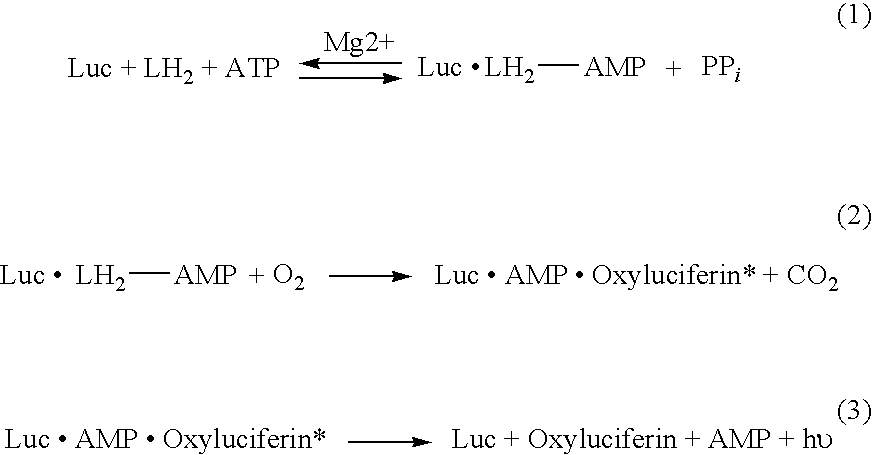
Bioluminescence is the emission of light from an organic molecule, such as luciferin, which has been oxidized by oxygen or one of its metabolites. The reaction is catalyzed by the luciferase protein (Luc), a naturally occurring protein that is found in beetles, fireflies and other living organisms, to form luciferyl-adenylate (LH2-AMP) from substrates luciferin (LH2) and ATP (eq. 1). Through a multi-step oxidative process, LH2-AMP is converted to excited state oxyluciferin, the light-emitting product (eq. 2-3) (H. H. Seliger et al., Arch. Biochem. Biophys. 88 (1960) 136-141; E. H. White et al., Bioorg. Chem. 1 (1971) 92-122; M. DeLuca; Adv. Enzymol. 44 (1976) 37-68; J.-Y. Koo et al., Proc. Natl. Acad. Sci. USA 75 (1978) 30-33; J. W. Hastings; Bioluminescence, in: Sperelakis, N., (Ed.), Cell Physiology Source Book, Academic Press, New York, 1995, pp. 665-681; K. V. Wood; Photochem. Photobiol. 62 (1995) 662-673).
The generation of light from LH2 is highly efficient (Seliger et al., supra) affording great sensitivity for the detection of the luciferase protein using available light measuring technology. Thus, the luciferase gene is extremely suitable for reporter gene applications (S. J. Gould et al., Anal. Biochem. 175 (1988) 5-13) and in vivo bioluminescence imaging (C. H. Contag et al., Annu. Rev. Biomed. Eng. 4 (2002) 235-260). Luciferases have been used to study gene delivery (Y. Taniyama et al., Gene Ther. 9 (2002) 372-380), gene silencing (P. J. Paddison et al., Proc. Natl. Acad. Sci. USA 99 (2002) 1443-1448) and real-time imaging of luciferase expression in live animals (L. F. Greer, III, et al., Luminescence 17 (2002) 43-74).
Currently, luciferase genes from various species, including Photinus pyralis (common North American firefly), Pyrophorus plagiophathalamus (Jamaican click beetle), Luciola mingrelica (European beetle), and Lampyris noctiluca (glow worm), are used to generate luminescent reporter moieties.
Typically, these reporter moieties emit light in the blue to yellow-green range (400-560 nm) or the green to orange range (510-590 nm). However, emission of light at other wavelengths is useful in many applications. For example, light emitted at a wavelength closer to the red range (“red shifted”) is known to be transmitted through live tissue more efficiently than other wavelengths of visible light. Similarly, light emitted at a wavelength closer to the blue range (“blue shifted”) provides increased sensitivity to standard photomultiplier tubes and is important for dual color assays where it is important to maximize the wavelength separation of the signals. By shifting the bioluminescence emission toward the red or blue wavelengths, it is possible to enhance the utility of luciferase genes in in vivo monitoring and gene expression assays.
There are many uses of luciferase known in the art. Luciferase, from various sources, has been used in various assays and reporting capacities. For example, U.S. Pat. No. 6,387,675 discloses the use of the luciferase gene of the elaterid beetle, P. plagiophthalamus, in eukaryotic cells for biosensing. U.S. Pat. No. 6,812,012 discloses a method of using luciferase to assay intracellular ATP, while a method of using a luciferase gene as a reporter gene is disclosed in U.S. Pat. No. 6,495,355.
Expression of luciferase genes has also been shown in the art. For example, U.S. Pat. No. 6,132,983 discloses the expression of luciferase genes in cells of various host organisms, while U.S. Pat. No. 4,968,613 discloses production of luciferase by incorporating a luciferase gene into a vector inserted into E. coli. U.S. Pat. No. 5,229,285 discloses the expression of a thermostable luciferase of a firefly.
Alteration of the bioluminescence emission of currently used luciferases can be obtained by amino acid substitutions and other mutations in the active sites of the luciferase genes. Although a number of such luciferases are available commercially, additional luciferase derivatives with altered spectral properties would be desirable.
SUMMARY OF THE INVENTION
In one aspect, the present invention is directed to an isolated polypeptide comprising the sequence of SEQ ID NO:2, wherein said polypeptide produces a bioluminescence emission maximum of approximately 566 nm. Specific mutations of the polypeptide of SEQ ID NO:2 are also encompassed in the claimed invention, as well as the nucleic acid sequences that correspond to the polypeptide of SEQ ID NO:2 and the specific mutants thereof.
In another aspect, the present invention is directed to a cell transformed with a vector containing one of the aforementioned nucleic acid sequences.
In another aspect, the present invention is directed to a method for detecting the presence of a L. italica luciferase in a cell, the method comprising: introducing one of the aforementioned vectors into the cell; contacting said cell with a luciferase substrate; and detecting a bioluminescence emission at a wavelength between about 551 to about 612 nm.
In another aspect, the present invention is directed to a method for assaying the presence of ATP in a sample, the method comprising the steps of: combining a sample with one of the aforementioned polypeptides; measuring a bioluminescence emission of said sample at a selected wavelength that corresponds to a bioluminescence emission max of said isolated polypeptide; and correlating said bioluminescence emission max with the presence of ATP.
In another aspect, the present invention is directed to an article of manufacture comprising a container containing any of the isolated polypeptides described above.
These and other aspects will become apparent upon reading the following detailed description of the invention.
BRIEF DESCRIPTION OF THE DRAWINGS
The invention will be more fully understood from the following detailed description taken in conjunction with the accompanying drawings in which:
FIG. 1 shows a nucleic acid sequence of the Italian firefly, L. italica (SEQ ID NO: 1) and the translated polypeptide sequence (SEQ ID NO: 2);
FIG. 2 shows a table comparing the L. italica luciferase protein to the known beetle luciferases;
FIG. 3 shows glowing colonies of E. coli infused with luciferin on nitrocellulose filters;
FIG. 4 shows decay of bioluminescence emission of Lit WT and Ppy WT luciferases;
FIG. 5 shows bioluminescence emission spectra for L. italica (Panel A) and P. pyralis (Panel B);
FIG. 6 shows bioluminescence activity of Lit WT and mutants thereof;
FIG. 7 shows normalized bioluminescence emission spectra produced by luciferases from Lit WT, LitGF-G-10, and LitS-1-11 mutants at 25° C. (Panel 7A) and 37° C. (Panel 7B); and
FIG. 8 shows the thermostability of Lit WT and LitS-S-2 and LitGF-G-11.
DETAILED DESCRIPTION OF THE INVENTION
Isolated nucleic acid and polypeptide compositions encoding a L. italica firefly luciferase are disclosed herein, as well as mutants of these isolated nucleic acid and polypeptide sequences. It has been found that the wild type L. italica (Lit WT) luciferase has a bioluminescence emission maximum that is red-shifted and 566 nm with a shoulder at 607 nm. Further, various substitution mutants of Lit WT exhibit distinct emission spectra that are red- or blue-shifted depending on the specific mutation(s). As defined herein, the term “red-shifted” means that the bioluminescence emission has been shifted toward the red wavelength range as compared to other luciferases known in the art. Similarly, “blue-shifted” means that the bioluminescence emission has been shifted toward the blue wavelength range as compared to other luciferases known in the art.
Lit WT is encoded by a nucleic acid having a sequence of SEQ. ID. NO.: 1 as shown in FIG. 1. The nucleic acid of SEQ ID NO: 1 was isolated from the Italian firefly, L. italica and is available in GenBank at Accession No. DQ138966. After isolation of SEQ ID NO: 1, the nucleic acid was amplified by amplification techniques known in the art, such as PCR. The nucleic acid was then inserted into an appropriate vector, such as a pGEX-6p-2 plasmid, and transformed into competent cells and subsequently plated. DNA sequencing revealed a 1647 bp open reading frame corresponding to a polypeptide containing 548 amino acids, having a sequence of SEQ ID NO: 2, also shown in FIG. 1. The Lit WT protein was found to have a molecular mass of 60,908±6 Da, as determined by electrospray ionization mass spectrometry. This value corresponds to the calculated mass of 60,907±6 Da of Lit WT, which contains the additional N-terminal peptide GlyProLeuGlySer-.
As shown in FIG. 2, SEQ ID NO:2 is highly homologous to other luciferase amino acid sequences, including H. unmunsana (95.8%), H. parvula (95.6%) and L. mingrelica (95.3%). The luciferases least homologous to L. italica were the orange and yellow emitting isozymes of the click beetle P. plagiophthalamis (45.5%). Generally higher homology to the L. italica luciferase was found with true fireflies. The homologies were determined by sequence pair distances using the Clustal V method (Higgins et al., Computer Applications in the Biosciences, vol. 5, p. 151-153 (1989)).
To analyze the bioluminescence emission of L. Italica luciferase, proteins were expressed as a GST-fusion protein in bacterial colonies. Expression of the luciferase was demonstrated by the observation of bioluminescent colonies of E. coli infused with a luciferase substrate, such as luciferin, on nitrocellulose fibers as shown in FIG. 3. The bioluminescence emission of Lit WT was measured, using methods known in the art, and determined to be approximately 566 nm with a shoulder at 607 nm.
Preliminary characterization showed that Lit WT demonstrates bioluminescence emission similar in intensity to P. pyralis (Ppy WT). Specifically, it was determined that the relative flash height specific activity of Lit WT, which relates to the maximum achievable overall reaction rate, was found to be approximately 95% of that of Ppy WT. However, compared to Ppy WT, Lit WT has an extended light emission decay and is red-shifted by 9 nm. As shown in FIG. 4, Ppy WT and Lit WT decay to approximately 20% of their maximum flash height in about 0.2 minutes. Ppy WT continues to rapidly decay and reaches 10% of its initial activity in about 0.35 minute. In contrast, Lit WT reaches 10% of its flash height at about 7 minutes, illustrating its extended light emission decay.
In addition to its extended bioluminescence emission decay as compared to other luciferases, the Lit WT has been red-shifted. As shown in FIG. 5, bioluminescence emission spectra at pH 7.8 showed an emission maximum of 566 nm with a shoulder at 607 nm for Lit WT (FIG. 5, Panel A). This represents a 9 nm shift from Ppy WT, which has an emission maximum of 557 nm (FIG. 5, Panel B). At a pH of 6.0, Lit WT was found to be very sensitive, shifting to 614 nm, thereby producing a spectrum very similar to Ppy WT at this pH.
Further, it has also been found that mutants of Lit WT produce bioluminescence emissions that are red-shifted or blue-shifted, and have extended bioluminescence emission decay. As shown in FIG. 6, substitutions of various amino acids in Lit WT result in bioluminescence emissions of different wavelengths. In addition, as shown in FIG. 6, several of the disclosed mutants display excellent thermostability at 37° C., a property of great importance for many applications, including but not limited to in vivo imaging of tumors, biosensor applications, reporter gene applications in mammalian cell lines, and the like. As shown in FIG. 6, the half-lives of LitWT, LitS-S-2 and LitGF-G-11 were measured. In the first mutant, Lit-Gly 248 Ala+Phe 252Ser, Gly 248 was changed to alanine and Phe 252 was changed to serine. This mutant produced an emission maximum of about 563 nm (a “blue-shift” relative to the emission maximum of 566 nm in Lit WT). This mutant was not thermostable at 37° C., having a half-life of 0.06 hr at this temperature.
In the second mutant, Lit GF-G-4, Gly 216 was changed to alanine, Thr 217 was changed to leucine, Ser 234 was changed to alanine, Gly 248 was changed to alanine and Phe 252 was changed to serine. This mutant has an emission maximum of approximately 551 nm (a blue-shift relative to the emission maximum of 566 nm in Lit WT), and was thermostable at 37° C.
In the third mutant, Lit GF-G-5, Gly 216 was changed to alanine, Thr 217 was changed to leucine, Ser 234 was changed to alanine, Gly 248 was changed to alanine, Phe 252 was changed to serine and Glu 356 was changed to lysine. This mutant has an emission maximum of approximately 563 nm (a blue shift relative to the emission maximum of 566 nm in Lit WT), and was thermostable at 37° C.
In the fourth mutant, Lit GF-G-10, Gly 216 was changed to alanine, Thr 217 was changed to leucine, Ser 234 was changed to alanine, Gly 248 was changed to alanine, Phe 252 was changed to serine, and Val 241 was changed to isoleucine. This mutant had an emission maximum of approximately 553 nm (a blue shift relative to the emission maximum of 566 nm in Lit WT), and was thermostable at 37° C.
In the fifth mutant, Lit-Ser 286 Thr, Ser 286 was changed to threonine. This mutant had an emission maximum of approximately 611 nm (a “red-shift” relative to the emission maximum of 566 nm in Lit WT), but was not thermostable at 37° C.
In the sixth mutant, Lit S-S-2, Gly 216 was changed to alanine, Thr 217 was changed to leucine, Ser 234 was changed to alanine, Ser 286 was changed to threonine and Glu 356 was changed to lysine. This mutant had an emission maximum of approximately 611 nm (a red-shift relative to the emission maximum of 566 nm in Lit WT), and was thermostable at 37° C., having a half-life of 2.3 hours at this temperature compared to 0.06 hr for Lit WT.
In the seventh mutant, Lit S-S-10, Gly 216 was changed to alanine, Thr 217 was changed to leucine, Ser 286 was changed to threonine, and Glu 356 was changed to lysine. This mutant had an emission maximum of approximately 612 nm (a red-shift relative to the emission maximum of 566 nm in Lit WT), and was thermostable at 37° C.
In the eighth mutant, LitS-S-11, Gly 216 was changed to alanine, Thr 217 was changed to leucine, Ser 234 was changed to alanine, Ser 286 was changed to threonine, Glu 356 was changed to lysine, Lys 547 was changed to glycine and Met 548 was changed to glycine. This mutant had an emission maximum of approximately 609 nm (a red-shift relative to the emission maximum of 566 nm in Lit WT), and was thermostable at 37° C. The changes at positions 547 and 548 removed a signal sequence that targets the enzyme for export to the peroxisome.
In the ninth mutant, LitGF-G-11, Gly 216 was changed to alanine, Thr 217 was changed to leucine, Ser 234 was changed to alanine, Val 243 was changed to isoleucine, Gly 248 was changed to alanine, Phe 252 was changed to serine, Glu 356 was changed to lysine, Lys 547 was changed to glycine and Met 548 was changed to glycine. This mutant had an emission maximum of approximately 554 nm (a blue-shift relative to the emission maximum of approximately 566 nm in Lit WT), and was thermostable at 37° C., having a half-life of 2.0 hours at this temperature as compared to 0.06 hr for Lit WT. The changes at positions 547 and 548 removed a signal sequence that targets the enzyme for export to the peroxisome.
The bioluminescence emission of the Lit GF-G-11 and Lit S-S-11 mutants was compared to the emission of Lit WT. As shown in FIG. 7A, at 25° C., the emission maximum of the LitGF-G-11 mutant was approximately 554, while the LitS-S-11 mutant was approximately 609 nm, thus demonstrating the blue- and red-shifts shown with these mutants relative to Lit WT. As shown in FIG. 7B, at 37° C., all the spectra are red-shifted. It is noted that the red-shifting of red emitting luciferases is favorable for in vivo imaging.
The mutants of Lit WT as shown in FIG. 6 and described in more detail above were made through processes known in the art, such as site directed mutagenesis and multi-site directed mutagenesis. These processes are discussed in more detail in the Examples that follow.
Further modifications and changes beyond those specifically disclosed herein may be made to the nucleic acid sequence or polypeptide sequence of the L. italica luciferase to obtain a molecule having the desired bioluminescence emission and extended light decay. For example, certain amino acids may be substituted for other amino acids without any loss of function. So long as the mutation or change maintains a red-shifted luciferase, the resulting protein will be considered a biologically functional equivalent for the purposes of the present invention.
Additionally, the nucleic acid sequence of Lit WT may be modified chemically or by genetic engineering to enable the luciferase to be targeted into a specific subcellular compartment. For example, L. italica luciferase contains the peptide Ala-Lys-Met at the C-terminus, which likely directs the luciferase to the peroxisomes. However, a suitable sequence at the N-terminus will locate the luciferase in the mitochondria while other peptide sequences will direct the luciferase to the endoplasmic reticulum. Direction of the luciferase to certain subcellular compartments may have applicability in different assays and tests known in the art.
Transformation, transduction or transfection of a cell with nucleic acid segments encoding Lit WT or any mutants thereof, can be used to express the luciferase protein in various eukaryotic and prokaryotic cells. The nucleic acid can be inserted into vectors such as bacteriophages, cosmids, or plasmids which can then be used for transformation of prokaryotic or eukaryotic cells according to methods known in the art.
After transforming, transfecting or transducing a host cell with a vector containing Lit WT or a variant thereof, the presence of the L. italica luciferase in a host cell can be determined by contacting the host cell with a luciferase substrate. Luciferase substrates include luciferin and ATP. Through a multi-step oxidative process, the luciferase is converted to an excited state oxyluciferin, which is the bioluminescent product. In the present invention, the bioluminescence emission of the L. italica luciferase is between about 563 nm to about 612 nm. The bioluminescence emission is detected and calculated by processes known to those with ordinary skill in the art.
Alternatively, the luciferase of the present invention may be used to determine the presence and quantify the amount of ATP present in a sample. To do so, a sample that contains, or is thought to contain, ATP is combined with Lit WT or a mutant thereof. The Lit WT or mutant thereof may be in combination with other materials, such as a carrier. Upon contact of the luciferase with the sample, bioluminescence emission is measured to determine whether ATP is present in the sample, and if so, how much ATP is present. Examples of such assays are known in the art (Lundin, A. et al., Meth. Enzymol., Vol. 305, pp. 346-370 (2000), Academic Press, NY; Stanley, P. Journal of Bioluminescence and Chemiluminescence, vol. 4(1), pp. 375-80, (1989); Leach, F., J. Appl. Biochem., vol. 3(6), pp. 473-517 (1989)).
For applicability in assays, tests, methods and techniques known in the art, and those subsequently discovered, the isolated polypeptides of the present invention may be manufactured and place into kits, testing products, or other articles of manufacture. Typically the isolated polypeptides are placed in a container with or without a carrier, such as a buffer. In most instances the article of manufacture contains an instruction booklet.
The wild type and mutant species of the luciferases derived from L. italica as disclosed herein are useful in many applications. For example, the present invention is useful in biotechnological applications, including reporter genes, dual-reporter systems, bioluminescence resonance energy transfer (BRET), microarrays, in vivo and ex vivo bioluminescence imaging, tumor research, whole animal imaging, infectious disease monitoring, biosensors for pollutants and biological disease markers, immunoassays, drug development and bioprocessing. (Roda et al., Trends in Biotechnology, vol. 22, No. 6, 295-303, 2004). The present invention is also useful in any application based on the monitoring of ATP levels either directly or through coupled enzyme reactions, such as microbiological tests; assays of enzymes, substrates and cofactors; monitoring of bacterial contamination of food; DNA probes assays; protein blotting and photographic assays (see, for example, L. J. Kricka, Anal. Biochem., vol. 175, pp. 14-21 (1988)). The present invention is also useful in any application based on the emission of light for devices which provide illumination without heat, spark or flame (for example, Cyalume technology). The present invention is also useful in any application in which light emission is used to create a novelty item, e.g. a toy or device that can be worn as jewelry. The present invention is also useful in any materials that could be used in tagging applications or anti-tampering applications.
The thermostable mutant luciferase enzyme of the present invention can also be used as a label for biospecific assays including immunoassays, nucleic acid hybridization both in vitro or microplate plate formats or for imaging purposes (immunohystochemistry or in situ hybridization). The luciferase enzyme of the present invention may possess a histidine tail that may be used for specific coupling with biomolecules, proteins, peptides, nucleic acids and in general organic molecules with suitable reactive groups. The red emitting thermostable luciferase mutant will enhance the performance of assays thanks to its relative high stability up to 42° C. associated with an high turnover of the enzyme and to the possibility to orient the coupling of this enzyme with histidine tail, leaving the acitive site free for substrate access. In addition, different thermostable mutants with different colours can be simultaneously used for multiplexed formats assays in which the reaction is triggered by only one substrate and the signal is selectively recorded at different wavelength.
The following examples are intended to illustrate, but in no way limit the scope of the present invention. All parts and percentages are by weight, and temperatures are in degrees Celsius unless explicitly stated otherwise.
EXAMPLES
Example 1
Isolation of L. Italica Luciferase Gene
Collection and RNA Extraction
L. italica fireflies were collected from the Bologna-Paderno region of Italy and flash-frozen alive in liquid nitrogen and stored at −80° C. Sixteen lanterns were removed from the fireflies, and were refrozen in liquid nitrogen. The lanterns were ground to a powder using a mortar and pestle cooled with liquid nitrogen. A total of 18 μg of RNA was extracted from the ground lanterns using an RNeasy mini kit (Quiagen) and following the manufacturer's enclosed instructions.
RT-PCR
First strand cDNA synthesis was carried out by utilizing approximately 5 μg of RNA and Oligo (dT) 20 primers and the Superscript TM III First-Strand Synthesis System for RT-PCR (Invitrogen). The following primers based on the luciferase coding sequence of L. Mingrelica, were used to amplify the cDNA:
| |
5′ - GTC CCT AAA CGG TAG AGG AAA A G-3′ |
| |
|
| |
5′ - GTC TTC TTA TGA GTA GTT TAG TTA C-3′ |
To amplify the cDNA, polymerase chain reaction (PCR) was used. The initial denaturation cycle was at 94° C. for 5 min. A 35 cycle amplification was then carried out at 94° C. for 30 sec.; 52° C. for 45 sec.; and 68° C. for 1.5 min. A final extension was carried out at 68° C. for 5 min.
The PCR products were then analyzed on a 1.0% agarose gel containing ethidium bromide. The samples corresponding to the expected size were purified using a QIA quick PCR purification kit (Qiagen) and following the manufacturer's instructions. Finally, the amplified cDNA was sequenced using a capillary array sequencer CEQ2000XL.
Insertion of cDNA into a Vector
To insert the luciferase cDNA into a vector, a primer set was used to introduce SmaI and XhoI restriction endonuclease sites at the 5′ and 3′ ends, respectively, of the PCR-amplified cDNA. The following primer set was used:
| 5′ - TTT AAT CCC GGG GTC CCT AAA CGG TAG - 3′ |
| |
| 5′ - CTA AGC CTC GAG TCT TCT TAT GAG TAG TT - 3′ |
PCR amplification and purification were performed as described above.
The PCR product was then digested with SmaI and XhoI restriction endonucleases and ligated into the corresponding cloning sites on a pGEX-6P-2 plasmid. The ligation reaction was transformed into Escherichia coli XL-10 Gold ultra competent cells and plated on Luria-Bertani (LB) plates containing 100 μg/mL ampicillin.
Ten colonies of the E. coli were selected randomly and plasmid DNA was purified and screened by agarose gel electrophoresis. Several plasmid DNA samples of the expected size were sequenced. One sample containing the entire L. italica cDNA was identified.
Alignment of the cDNA Reading Frame
Since the pGEX expression plasmid is designed to produce proteins as N-terminal glutathione-S-transferase (GST)-fusion products, the reading frame of the luciferase gene was realigned so the corresponding protein would contain the same N-terminal polypeptide as Ppy WT. A Quik Change® Site-Directed Mutagenesis kit (Stratagene), was used to realign the reading frame of the L. italica cDNA in the pGEX-6P-2 plasmid.
First, the primer, 5′-GA TTC TCA CAC GCT AAG GAC CCA ATT TAC GGA AAC CAA GTT TC-3′ (SEQ ID NO:7) and its reverse complement, were used to remove the Bam HI restriction endonuclease site within the L. italica luciferase gene. Next, the primer 5′-CG GTA GAG GAA AAG TTT GGA TCC ATG GAA ACG GAA AGG GAG G-3′ (SEQ ID NO:8) and its respective reverse complement, were used to introduce a Bam HI site immediately preceding the start codon of the L. italica luciferase gene. Finally, the product was digested with Bam HI and XhoI restriction endonucleases and ligated into corresponding cloning sites on the pGEX-6P-2 plasmid.
Expression of L. Italica Luciferase as a GST-Fusion Protein in Bacterial Colonies
The ligated plasmid DNA was transformed into E. coli, which were plated on nitrocellulose filters placed on LB plates containing 100 μg/mL ampicillin. The transformation was done by adding the plasmid DNA to the bacteria at ice temperature, heat shocking the mixture at 42° C., cooling on ice for 2 minutes and recovering for 1 hour at 37° C. in SOC media. The E. coli were screened for bioluminescence emission as described in Branchini et al., “Rational and random mutagenesis of firefly luciferase to identify an efficient emitter of red bioluminescence,” Proceedings of SPIE, Genetically Engineered and Optical Probes for Biomedical Applications II 5329 (2004), 170-177 (Alexander P. Savitsky et al., eds.).
Colonies having bioluminescence emission were identified by visual screening in a darkroom. Plasmid DNA was isolated and purified therefrom by using a GenElute™ Plasmid Miniprep kit (Sigma), and following the manufacturer's instructions. A single plasmid was selected and the entire L. italica luciferase gene was sequenced. The sequence of the L. italica luciferase gene in the pGEX plasmid was compared to the sequence obtained from the original cDNA. The comparison showed no differences in the sequence, thereby insuring no additional mutations had been introduced.
Example 2
Preparation of Lit WT Mutants
Nine mutants of Lit WT, shown in FIG. 6 and discussed in more detail above, were prepared using the following methods:
Site Directed Mutagenesis
The Lit-S286T and Lit-G248A and F252S mutants were created by using a QuikChange® Site-Directed Mutagenesis kit (Stratagene). Site-directed mutagenesis was carried out according to the manufacturer's instructions using the L. italica wild-type DNA sequence in the pGEX-6P-2 vector as a template. The following primers and their respective reverse complements were used:
| 5′ - GAT TAT AAG TGT ACC ACT GTT ATT CTG GTA CCA |
| ACG TTA TTT GC - 3′ (Kpn1) |
| |
| Lit-G248A & F252S: |
| 5′ - CCG TTC CAT CAC GCG TTT GGA ATG TCT ACC ACT |
| TTA GGA TAC - 3′ (Mlu1) |
The LitS-S-11 mutant was created by using the QuikChange® Site-Directed Mutagenesis kit (Stratagene). Site-directed mutagenesis was carried out according to the manufacture's instructions using the LitS-S-2 mutant as the template. The 546AlaLysMet548 signal sequence was changed to 546AlaGlyGly548. The following primer and its respective reverse compliment was used to accomplish this:
| 5′- AG AAA CCA CAA GCC GGG GGG TAA ATC GGT CAA |
| AAT G -3′ [BsaB1] |
The LitGF-G-11 mutant was created by using the QuikChange® Site-Directed Mutagenesis kit (Stratagene). Site-directed mutagenesis was carried out according to the manufacture's instructions using the LitGF-G-10 mutant as the template. First, Glu356Lys was introduced into the template. Once this mutation was confirmed, the 546AlaLysMet548 signal sequence was changed to 546AlaGlyGly548. The following primers and their respective reverse compliments were used to accomplish this:
| 5′- GCA TTT ATT ATT ACC CCA AAA GGT GAT GAT AAA |
| CCT GG -3′ |
| |
| Lys547Gly & Met548Gly: |
| 5′- AG AAA CCA CAA GCC GGG GGG TAA ATC GGT CAA |
| AAT G -3′ [BsaB1] |
Underlining represents silent changes creating a unique screening endonuclease site. Bolded codons represent the mutated codons. Brackets indicate the screening endonuclease.
Multi-Site Directed Mutagenesis
The QuikChange® MultiSite-Directed Mutagenesis kit (Stratagene) was used to create the remaining mutants identified as entries 2 through 8 in FIG. 6. Mutagenesis was carried out according to the manufacturer's instructions for using 1 to 4 primers simultaneously. The Lit-S286T and the Lit-G248A and F252L mutants in the pGEX-6P-2 vector were used as templates with the following primers:
| 5′- GCA TTT ATT ATT ACC CCA AAA GGT GAT GAT AAA |
| CCT GG-3′ |
| |
| Gly216Ala & T217L |
| 5′ - GAG ATT ACC CAC GAA GCA CTA GTT ACA AGA TTC |
| TCA CAC G - 3′ |
| |
| Ser 234 Ala |
| 5′ - TAC GGA AAC CAA GTT GCA CCT GGT ACT GC - 3′ |
| |
| Val243Ile |
| 5′ - TA ACT GTC ATT CCG TTC CAT CAC GCG TTT GGA |
| ATG - 3′ |
The primer for V2431 also includes the G248A mutation (bold and italics) for correct annealing. This primer was only used with the Lit-G248A and F252L template.
Transformation and Screening of Lit WT Mutants
The above-described mutants were transformed into E. coli XL-10 Gold ultracompetent cells. Duplicate sets of transformed cells were plated on nitrocellulose filters on LB plates that contained 100 μg/ml ampicillin. The plates were incubated at 37° C. for 18 hrs. Isopropyl B-D-1-thiogalactopyranoside (IPTG) was used to induce transcription.
For each set of transformed mutagenesis product-containing cells, one plate was incubated at room temperature and the other at 37° C. for at least 2 hours. The plates grown at 37° C. were then placed on a plate warmer set to 37° C. (Barnstead Lab Line) and those at room temperature were kept at 22° C.
The nitrocellulose filters were soaked in 1 mM of luciferin in 100 mM NaCitrate buffer (pH 5.5) to induce bioluminescence. Colonies for sequencing were selected on the intensity and color of the emitted light. Plasmid DNA of selected clones was isolated by a standard mini-prep procedure. The DNA was sequenced at Yale University.
Example 3
Protein Purification
Glutathione-S-transferase (GST) fusion constructs of L. italica wild-type and mutants thereof selected for sequencing were expressed in XL 10-Gold ultracompetent cells. 7 mL cultures pf the cells were grown in 10 mL culture tubes at 37° C. in LB medium supplemented with 100 μg/mL ampicillin.
These cultures were grown from starter cultures prepared from the picked colonies to mid log phase (A600=0.4-0.5) with vigorous shaking, induced with 0.1 mM IPTG, and incubated at 22° C. for 8-10 h. The cells were harvested by centrifugation and placed at −80° C. for no less than 30 min. Cell pellets were resuspended in 0.5 mL of phosphate-buffered saline (PBS), pH 7.3 containing 0.1 mM phenylmethylsulfonyl fluoride (PMSF) and 0.5 mM dithiothreitol (DTT). The resuspended cells were mixed with lysozyme (0.05 volume of 10 mg/mL) and incubated on ice 30 min. The lysated cells were treated with Rnase (10 μg/mL) and Dnase 1 (5 μg/mL). Triton X-100 (2% final volume) was added to the lysate and the whole-cell extract was isolated by centrifugation at 20,000×g for 45 min at 4° C. The supernatant was collected and aliquots were used to determine molecular mass, bioluminescence activity, bioluminescence emission spectra, protein content and GST-activity. Lit WT, LitGF-G-11, LitS-S-2 and LitS-S-11 enzymes were purified to homogeneity as described in Branchini, B. R., et al., “Rational and random mutagenesis of firefly luciferase to identify an efficient emitter of red bioluminescence,” Proceedings of SPIE, Genetically Engineered and Optical Probes for Biomedical Applications II, 5329 (2004), 170-177 (Alexander P. Savitsky et al., eds.).
Molecular Mass of Lit WT Protein
The Lit WT protein was found to have a molecular mass of 60 908±6 Da. This value corresponds to the calculated mass of 60 907±6 Da of Lit WT, which contains the additional N-terminal peptide GlyProLeuGlySer-. The mass was determined by Electrospray Ionization Mass Spectrometry.
Glutathione-S-Transferase (GST) Activity Assay to Quantitate GST-Fusion Proteins
GST-fusion proteins were quantitated by enzymatic assay of glutathione-S-transferase (GST) activity using the GST substrate 1-chloro-2,4-dinitrobenzene (CDNB). GST catalyzes the conjugation of reduced glutathione (GSH) and CDNB to yield a dinitrophenolthioether, a chromogenic substance with λλmax=340 nm. A dual beam PerkinElmer Lambda 25 UV/Vis spectrophotometer was used to perform these studies.
500 μL of 1 mM reduced glutathione (GSH) and 1 mM 1-chloro-2,4-dinitrobenzene (CDNB) in 0.1M potassium phosphate buffer (KPB), pH 6.5 were placed into 0.7 mL UV transparent sample and reference cuvettes.
50 μL of KPB was added to the reference cuvette. 5 μL of bacterial cell lysate containing the GST-luciferase fusion protein and 45 μL of KPB was added to the sample cuvette. The samples were well mixed and the absorbance at 340 nm was monitored using the Time Drive program of the Lambda 25 spectrophotometer. Data was recorded for 4 min, and the velocity of the reaction, the rate of change in absorbance per minute, was determined. The concentration of GST-fusion protein is proportional to the velocity of the reaction.
Bioluminescence Emission Spectra of Partially Purified Proteins
Bioluminescence emission spectra were obtained using a PerkinElmer LS55 luminescence spectrometer operated in the “bioluminescence” mode. Data was collected over the wavelength range 480-680 nm in a 1 mL optical glass cuvette. Gate and delay times, detector voltage, scan rate, and slit width were adjusted to optimize instrument response. Reaction mixtures containing partially purified L. italica wild type and its mutants (0.005 mL) in luciferin and Mg-ATP were brought to a final volume of 0.5 mL with 25 mM glycylglycine, pH 7.8. The spectral data reported in FIG. 6 were measured with purified proteins from Lit WT, LitGF-G-11, LitS-S-2 and LitS-S-11 enzymes.
Measurement of L. Italica Thermostability
In a 0.2 mL PCR tube, solutions of 0.1 mg luciferase in 10 μL CBA (50 mM Tris, pH 7.0, 150 mM NaCl, 1 mM EDTA, 1 mM DTT, 0.8M (NH4)2 SO4, 2.0% glycerol) and 200 μL 25 mM glycyl-glycine buffer, pH 7.8, were prepared. An aliquot (50 μL) of the solution was reserved on ice. Initial activity values were obtained by flash height activity assays performed on 2-10 μL samples of enzyme. The sample was placed in a thermocycler set at 37° C. Aliquots were withdrawn over an 8 hour period and compared to a controls kept in ice. Thermostability was evaluated by plotting residual flash height activity at 37° C. as a function of time. Half-lives were calculated from the first order rate constant obtained by plotting the In residual flash height activity versus time. The half-lives of LitWT, LitS-S-2 and LitGF-G-11 were determined as shown in FIGS. 6 and 8.
While the invention has been described above with reference to specific embodiments thereof, it is apparent that many changes, modifications, and variations can be made without departing from the inventive concept disclosed herein. Accordingly, it is intended to embrace all such changes, modifications, and variations that fall within the spirit and broad scope of the appended claims. All patent applications, patents, and other publications cited herein are incorporated by reference in their entireties.