CROSS-REFERENCE TO RELATED APPLICATIONS
This application is a divisional application of U.S. Non-provisional application Ser. No. 11/152,974, filed on Jun. 15, 2005, now issued U.S. Pat. No: 7,572,766, which claims the benefit of U.S. Provisional Application No. 60/580,019, filed Jun. 16, 2004; U.S. Provisional Application No. 60/651,338, filed Feb. 9, 2005; and U.S. Provisional Application No. 60/651,747, filed Feb. 10, 2005, each of which is hereby incorporated in its entirety by reference herein.
FIELD OF THE INVENTION
The present invention provides materials and methods for coating surfaces of medical devices with interfacial biomaterials that promote the specific recognition and attachment of the target analyte to the surface of the device.
BACKGROUND OF THE INVENTION
Orthopedic implants are used for a variety of joint replacements and to promote bone repair in humans and animals. According to medical industry analysts, there are now over 800,000 hip and knee joint replacements performed in human patients each year in the U.S. In addition, hundreds of thousands of human patients undergo surgical procedures in which orthopedic implants are used, for example, to treat various types of bone fractures or to relieve severe back pain.
With all of these procedures, there is a need for controlled, directed, rapid healing. Individuals undergoing joint replacement often experience uncomplicated healing and restoration of function. Unfortunately, there is a high rate of complications, including “late failures.” The revision surgery rate for human total joint replacement varies between 10 to 20% (Malchau et al. (2002) “Prognosis of total hip replacement: Update of results and risk-ratio analysis for revision and re-revision from the Swedish National Hip Arthroplasty Registry, 1979-2000,” scientific exhibition at the 69th Annual Meeting of the American Academy of Orthopaedic Surgeons, Dallas, Tex., Feb. 13-17, 2002; Fitzpatrick et al. (1998) Health Technol. Assess. 2:1-64; Mahomed et al. (2003) J. Bone Joint Surg. Am. 85-A:27-32)). The majority of these revision surgeries are made necessary by failure at the implant-bone interface.
Orthopedic implants are made of materials which are relatively inert (“alloplastic” materials), typically metallic, ceramic, or plastic materials. Previous approaches to improve the outcomes of orthopedic implant surgeries have mainly focused on physical changes to the implant surface that result in increased bone formation. These approaches include using implants with porous metallic surfaces to promote bone ingrowth and spraying implants with hydroxyapatite plasma. Approaches using dental implants have also included the use of topographically-enhanced titanium surfaces in which surface roughness is imparted by a method such as grit blasting, acid etching, or oxidation. While these techniques have improved the outcomes of orthopedic implant surgeries, there is still considerable room for further improvement.
Tissue response to an alloplastic material is known to be influenced by cell adhesion to the material's surface, and much research has been directed to improving cell adhesion to alloplastic materials. Cell adhesion between cells in vivo is known to be controlled primarily by the binding of short, exposed protein domains in the extracellular matrix to cell surface receptors (LeBaron & Athanasiou (2000) Tissue Eng. 6: 85-103; Yamada (1997) Matrix Biol. 16: 137-141). Notably, a class of receptors known as integrins has been implicated in cell adhesion to implant surfaces. Integrins and their target ligands have been shown to stimulate osteoblast adhesion and proliferation as well as bone formation (see, e.g., Kantlehner et al. (2000) ChemBioChem 1: 107-114; Sarmento et al. (2004) J. Biomed. Mater. Res. 69A: 351-358; Hayashibara et al. (2004) J. Bone Mineral Res. 19: 455-462. Integrins may be useful in targeting cell adhesion to implants and in this manner may improve integration of implants into adjacent bone.
Other research has shown that the local expression of growth factors and cytokines can enhance tissue reactions at alloplastic implant surfaces. For example, Cole et. al. ((1997) Clin. Orthop. 345: 219-228) have shown that growth factors can promote the integration of an implant into adjacent bone (“osteointegration”) as well as increase the rate of bone formation next to the implant surface. See also U.S. Pat. No. 5,344,654. Growth factors that stimulate new bone production (“osteoinductive proteins”) include, but are not limited to, platelet-derived growth factor (PDGF), insulin-like growth factors 1 and 2 (IGF-1 and IGF-2), vascular endothelial growth factor (VEGF), fibroblast growth factor (FGF), transforming growth factor (TGF-β), bone morphogenic proteins (BMP), and associated family members.
The most effective osteoinductive proteins are the bone morphogenetic proteins (BMPs). The BMPs are members of the TGF-β superfamily that share a set of conserved cysteine residues and a high level of sequence identity overall. Over 15 different BMPs have been identified, and most BMPs stimulate the cascade of events that lead to new bone formation (see U.S. Pat. Nos. 5,013,649; 5,635,373; 5,652,118; and 5,714,589; also reviewed by Reddi and Cunningham (1993) J. Bone Miner. Res. 8 Supp. 2: S499-S502; Issack and DiCesare (2003) Am. J. Orthop. 32: 429-436; and Sykaras & Opperman (2003) J. Oral Sci. 45: 57-73). This cascade of events that leads to new bone formation includes the migration of mesenchymal stem cells, the deposition of osteoconductive matrix, the proliferation of osteoprogenitor cells, and the differentiation of progenitor cells into bone-producing cells. Much research has been directed to the use of BMPs on or near implants in order to promote osteointegration of the implants (see, e.g.: Friedlander et al. (2001) J. Bone Joint Surg. Am. 83-A Suppl. 1 (Pt. 2): S151-58; Einhorn (2003) J. Bone Joint Surg. Am. 85-A Suppl.3: 82-88; Burkus et al. (2002) J. Spinal Disord. Tech. 15(5): 337-49). However, one of the critical issues that remains unresolved is the method of grafting or immobilizing an active BMP or other active biomolecule onto the surface of an implant.
It has been shown that the presentation of BMPs is critical for producing desired bone formation next to an implant device. Approaches to improving implants have been modeled in view of the natural process of bone formation. In human bone, collagen serves both as a scaffold for bone formation and as a natural carrier for BMPs. Demineralized bone has been used successfully as a bone graft material; the main components of demineralized bone are collagen and BMPs (see U.S. Pat. No. 5,236,456). Many matrix systems have been developed that are designed to encourage bone formation by steadily releasing growth factors and other bioactive molecules as the matrix degrades. The efficiency of BMP release from polymer matrixes depends on matrix characteristics such as the affinity of BMP for the matrix, resorbtion rate, density, and pore size. Materials used in such matrix systems include organic polymers which readily hydrolyze in the body into inert monomers. Such organic polymers include polylactides, polyglycolides, polyanhydrides, and polyorthoesters (see U.S. Pat. Nos. 4,563,489; 5,629,009; and 4,526,909). Other materials described as being useful in BMP-containing matrices include polylactic and polyglycolic acid copolymers, alginate, poly(ethylene glycol), polyoxyethylene oxide, carboxyvinyl polymer, and poly(vinyl alcohol) (see U.S. Pat. No. 5,597,897). Natural matrix proteins have also been used to deliver BMPs to bone areas; these natural proteins include collagen, glycosaminoglycans, and hyaluronic acid, which are enzymatically digested in the body (see U.S. Pat. Nos. 4,394,320; 4,472,840; 5,366,509; 5,606,019; 5,645,591; and 5,683,459).
Even with the use of a polymer matrix to retain BMP at the site of repair, it has been found that supraphysiological levels of BMP are required in order to promote healing due to the rapid diffusion of growth factors out of the matrix. For example, with a collagen sponge delivery system, only 50% of the BMP added to the sponge is retained after two days (Geiger et al. (2003) Adv. Drug Del. Rev. 55: 1613-1629). The high initial dose of BMPs required to maintain physiological levels of BMP for the necessary period of time makes BMP treatment more expensive and may lead to detrimental side effects such as ectopic bone formation or allergic reactions, or the formation of neutralizing antibodies.
Similar problems exist with other implants such as tendon and ligament replacements, skin replacements, vascular prostheses, heart pacemakers, artificial heart valves, breast implants, penile implants, stents, catheters, shunts, nerve growth guides, intraocular lenses, wound dressings, and tissue sealants. As with orthopedic implants, surgery involving these implants often gives rise to similar problems with the slow healing of wounds and, where desirable, improper integration of the implant into surrounding tissue.
Thus, there remains a need for the development of cost-effective methods for grafting active biomolecules to the surface of materials used as implants or in conjunction with implants in order to promote post-surgical healing and, where desirable, integration of the implant into surrounding tissues, such as, for example, adjacent bone.
SUMMARY OF THE INVENTION
The present invention provides an improved coating for surfaces of medical implants. The coating comprises at least one interfacial biomaterial (IFBM) which is comprised of at least one binding module that binds to the surface of an implant or implant-related material (“implant module”) and at least one binding module that binds to a target analyte or that is designed to have a desired effect (“analyte module”). The modules are connected by a linker. In some embodiments, the IFBM coating acts to promote the recognition and attachment of target analytes to surface of the device. The IFBM coating improves the performance of implanted medical devices by promoting osteointegration of the implant, accelerating healing, and/or reducing inflammation at the site of the implant.
BRIEF DESCRIPTION OF THE DRAWINGS
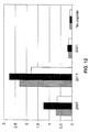
FIG. 1 shows a comparison of the binding of phage that display representative titanium-binding peptides to titanium beads (see Example 1). Signal of assay for binding to titanium beads (vertical axis) is shown for various phage (horizontal axis).
FIG. 2 shows a comparison of the binding of peptides with a C-terminal biotin residue to titanium (see Example 1). Absorbance (vertical axis) is shown as a function of peptide concentration (μM, on the horizontal axis).
FIG. 3 shows a comparison of binding to titanium of two peptides (see Example 2). A 405 nm signal (vertical axis) is shown as a function of peptide concentration (μM, on the horizontal axis). The lines shown on the graph from top to bottom join data points for peptides AFF6007 and AFF6010, respectively.
FIG. 4 shows a comparison of binding of various peptides to BMP-2 (see Example 3). Signal (rate AP) is shown for various peptides (identified on the horizontal axis).
FIG. 5 shows the effect of BMP on the binding of IFBMs to a collagen sponge (see Example 4). Signal (vertical axis) is shown as a function of BMP concentration in nM (horizontal axis).
FIGS. 6A, 6B, 6C, and 6D show the results of an experiment described in Example 4 which demonstrates that the binding of BMP to collagen via an IFBM is dependent on both the amount of BMP put into the sponge and also on the amount of IFBM present. Absorbance (vertical axis) is shown as a function of BMP concentration (horizontal axis).
FIG. 7 shows the results of an analysis of all the peptide sequences from Tables 3 and 4 that bind BMP-2 and contain Motif 1 (see Example 3). The figure shows for each analyzed position the number of times each amino acid was found in that position in the peptide sequences analyzed; for example, “G2” in position 1 means that Glycine was found two times in that position.
FIG. 8 shows the oligonucleotide cassette which was designed to express a peptide (SEQ ID NO: 74) containing the core binding Motif 1a in the context of a peptide sequence which also contained consensus residues identified for other positions in the sequence (see Example 3). The nucleotide sequences shown in the figure are also set forth in SEQ ID NO: 75 and SEQ ID NO: 76.
FIG. 9 shows results from a conventional ELISA performed to evaluate the relative affinity of BMP binding peptides (see Example 3). The signal from the ELISA (A 405 nm reading) is presented on the vertical axis as a function of microliters of phage on the horizontal axis. At the data points corresponding to 0.10 microliters of phage, the lines shown on the graph from top to bottom join data points for: APO2-61, APO2-40, APO2-41, APO2-26, APO2-35, APO2-59, APO2-44, mAEK, and the no-phage control, respectively.
FIG. 10 shows the results of an analysis of all the peptide sequences from Tables 3 and 5 that bind BMP-2 and contain Motif 2. The figure shows for each analyzed position the number of times each amino acid was found in that position in the peptide sequences analyzed; for example, “G7” in position 1 means that Glycine was found seven times in that position. Also shown are a consensus sequence derived from an alignment of the peptides from Tables 3 and 5 that contain Motif 2 (SEQ ID NO: 93). This sequence represents the predominant amino acid found at each position after all the peptides are aligned. Among the sequences examined, the most conserved amino acids form a core binding motif designated “Motif 2a” (SEQ ID NO: 94).
FIG. 11 shows representative results from an alternate assay for BMP-binding activity in which binding occurs in the solution phase (see Example 3). Absorbance at 405 nm (vertical axis) is shown as a function of picomoles of BMP (horizontal axis). These results were used to calculate the affinity of each BMP-binding peptide for BMP-2 (see Table 6). At the data point corresponding to one picomole of BMP, the lines shown on the graph from top to bottom join data points for: 2006, 2007, 2008, 2009, 2011, and 2012, respectively.
FIG. 12 shows results from an assay in which several peptides were tested for their ability to bind to BMP-2, BMP-4, and BMP-7 (see Example 3). The 2007 and 2011 peptides were originally identified as BMP-2 binding peptides, while the 9001 peptide was originally identified as binding to an unrelated target.
DETAILED DESCRIPTION OF THE INVENTION
The present invention provides an improved coating for surfaces of medical devices to promote the attachment of peptides, proteins, drugs, or cells to the device. The coating is an interfacial biomaterial (IFBM) that comprises multiple binding modules that are linked. The IFBM comprises at least one binding module which binds to the surface of the implant (“implant module”) and at least one binding module that binds to a target analyte or has a desired effect (“analyte module”). Exemplary binding modules comprise the peptide sequences provided, for example, in the sequence listing (SEQ ID NOs: 1-74 and 77-558). The modules are connected by a linker. In some embodiments, the binding of the binding module of an IFBM to the surface of an implant is non-covalent. Similarly, in some embodiments, the binding of an analyte module to a target analyte is non-covalent. According to one embodiment, the implant module and the analyte module comprise two separate peptide molecules such that the implant module binds to an implant material and the analyte module binds specifically to a growth factor or cell. In some embodiments, the implant module and the analyte module are linked by a central macromolecule. These binding modules typically bind non-covalently to the implant material or target analyte, respectively. In embodiments where the analyte module does not bind to a target analyte but rather has a desired effect, the analyte module may, for example, simulate the action of a growth factor by acting to recruit cells to the location of the implant. The IFBM selection method and structure are described in U.S. patent aplication Ser. No. 10/300,694, filed Nov. 20, 2002 and published on Oct. 2, 2003 as publication number 20030185870, which is herein incorporated by reference.
By “binds specifically” or “specific binding” is intended that the implant module or analyte module binds to a selected implant material or to a selected analyte. In some embodiments, a module that binds specifically to a particular implant material or analyte binds to that material or analyte at least 10%, 20%, 30%, 40%, 50%, 60%, 70%, 80%, 90%, 100%, 200%, 300%, 400%, 500%, or a higher percentage more than the module binds to an appropriate control such as, for example, a different material that is used in implants, a material that is not used in implants, or a protein typically used for such purposes such as bovine serum albumin. By “analyte” is intended any substance or moiety that improves osteointegration of an implant or promotes or accelerates healing of the surrounding tissues following implant surgery. Suitable analytes which are binding targets for analyte modules include, but are not limited to, growth factors such as bone morphogenic proteins (BMPs, such as, for example, BMP-7 and BMP-2), vascular endothelial growth factor (VEGF), platelet-derived growth factor (PDGF), transforming growth factor-β (TGF-β), insulin growth factor-1 (IGF-1), insulin growth factor-2 (IGF-2), fibroblast growth factor (FGF), nerve growth factor (NGF), and placental growth factor. Suitable analytes also include hormones, enzymes, cytokines, and other bioactive substances or moieties which are useful in obtaining the goals of the invention; that is, to promote osteointegration of an implant and/or to improve healing of surrounding tissues following implant surgery. Suitable analytes also include cells, for example, osteoblasts, chondrocytes, stem cells, progenitor cells, platelets, and other cells which perform roles in osteointegration and healing. In some embodiments, analyte modules can comprise peptide sequences that bind cells or have bioactivity through binding to cells or receptors such as, for example, the peptide sequences RGD, YIGSR, and IKVAV, which are known in the art to have particular biological activities. See, e.g., Hersel et al. (2003) Biomaterials 24:4385-4415; Grant et al. (1990) Ann. N.Y. Acad. Sci. 588:61-72; Hosokawa et al. (1999) Dev. Growth Differ. 41: 207-216. In some embodiments, analyte modules comprise peptide sequences which bind to and/or mimic the effect of BMP-2, such as the exemplary sequences set forth in SEQ ID NOs: 11-28, 44-74, or 77-94. An analyte module that binds to cells can comprise a peptide that comprises a general cell attachment sequence that binds to many different cell types, or it can comprise a peptide that binds to a specific cell type such as an osteoblast, a chondrocyte, an osteoprogenitor cell, or a stem cell.
The term “implant” generally refers to a structure that is introduced into a human or animal body to restore a function of a damaged tissue or to provide a new function. An implant device can be created using any biocompatible material to which binding agents can specifically bind as disclosed herein. Representative implants include but are not limited to: hip endoprostheses, artificial joints, jaw or facial implants, tendon and ligament replacements, skin replacements, bone replacements and artificial bone screws, bone graft devices, vascular prostheses, heart pacemakers, artificial heart valves, breast implants, penile implants, stents, catheters, shunts, nerve growth guides, intraocular lenses, wound dressings, and tissue sealants. Implants are made of a variety of materials that are known in the art and include but are not limited to: a polymer or a mixture of polymers including, for example, polylactic acid, polyglycolic acid, polylactic acid-polyglycolic acid copolymers, polyanhidrides, polyorthoesters, polystyrene, polycarbonate, nylon, PVC, collagen (including, for example, processed collagen such as cross-linked collagen), glycosaminoglycans, hyaluronic acid, alginate, silk, fibrin, cellulose, and rubber; plastics such as polyethylene (including, for example, high-density polyethylene (HDPE)), PEEK (polyetheretherketone), and polytetrafluoroethylene; metals such as titanium, titanium alloy, stainless steel, and cobalt chromium alloy; metal oxides; non-metal oxides; silicone; bioactive glass; ceramic material such as, for example, aluminum oxide, zirconium oxide, and calcium phosphate; other suitable materials such as demineralized bone matrix; and combinations thereof. The term “polymer” as used herein refers to any of numerous natural and synthetic compounds of usually high molecular weight consisting of up to millions of repeated linked units, each a relatively simple molecule. The term “implant” as used herein includes implant-related materials that are associated with the implant and are also introduced into a human or animal body in conjunction with the implant.
In one embodiment of the invention, an IFBM creates a binding interface that mediates the attachment of growth factors to the surface of an implant. In some embodiments, implants prepared according to the methods of the invention will have growth factors specifically attached to the surface of the implant; the rate of diffusion of the growth factor away from the site of the implant can vary depending on the affinity of the analyte module for the growth factor in question and thus implants can be prepared with varying rates of diffusion of growth factors. In embodiments involving the attachment of growth factors to the surface of an implant, the growth factor will have a positive effect such as, for example, accelerating the healing process, reducing the amount of growth factor required for healing, and minimizing the side effects caused by using supraphysiological doses of the growth factor. Growth factors of particular interest either as analyte modules or as factors that bind to analyte modules include, for example, BMP-2, BMP-7, PDGF, FGF, and TGFβ.
Thus, the present invention provides methods for preparing an implant to be surgically placed into a patient wherein the device is coated with a layer comprising at least one IFBM. In some embodiments, the method comprises the steps of: (a) applying an IFBM coating to the implant, wherein the IFBM comprises an implant module that specifically binds to the implant and an analyte module that specifically binds a growth factor; (b) applying the growth factor to the surface of the implant by dipping, spraying, or brushing a solution containing the growth factor onto the implant; (c) placing the implant into a subject using appropriate surgical techniques which will be known to those of skill in the art.
Alternatively, a method for coating an implant so that the implanted device promotes growth factor attachment comprises the steps of: (a) applying an IFBM coating to the implant, wherein the IFBM comprises an implant module that specifically binds the implant and an analyte module that specifically binds growth factor at an implant site; and (b) placing the implant in a subject at the implant site; whereby growth factor produced in the host binds to the implant via the IFBM. The enhanced presence of growth factor at the implant site enhances healing of adjacent tissue and integration of the implant into the adjacent tissue.
In one embodiment of the invention, an IFBM mediates cell attachment to the surface of an implant. By enhancing cell adhesion and tissue integration, the IFBMs of the invention can accelerate healing and improve the function of the implanted device. Thus, in accordance with the present invention, a method for preparing an implant to be surgically placed into a patient can comprise: (a) applying an IFBM coating to the implant, wherein the IFBM comprises at least one implant module that specifically binds the implant and at least one analyte module that specifically binds to at least one type of cell; and (b) placing the implant in a subject at the implant site, whereby cells bind to the IFBM coating on the implant.
In some embodiments, a method for preparing an implant comprises: (a) applying an IFBM coating to the implant, wherein the IFBM comprises at least one implant module that specifically binds the implant and at least one analyte module that specifically binds at least one type of cell; and (b) applying cells to the surface of the implant, for example, by dipping the implant into a solution containing the cells or brushing a solution containing the cells onto the implant. The implant may then be placed into a subject (i.e., a human patient or an animal patient). By “patient” as used herein is intended either a human or an animal patient.
In another embodiment of the invention, an implant is coated with more than one type of IFBM in order to provide a coating with multiple functionalities. For example, an implant coating can comprise a first IFBM having an analyte module that binds a cell and a second IFBM having an analyte module that binds a growth factor. A coating comprising these IFBMs would bind both cells and growth factor to the surface of the implant. In some embodiments, these IFBMs would be intermingled in the coating so that the bound growth factor is in close proximity to the bound cells. In one embodiment, a coating comprises an IFBM that binds to mesenchymal stem cells and an IFBM that binds to the growth factor BMP-2; the BMP-2 would trigger the differentiation of the stem cells into osteoblasts. In other embodiments, an implant coating can comprise a mixture of at least two different IFBMs which differ in either or both their implant module and their analyte module. In another embodiment, a coating comprises a multi-functional IFBM which has two analyte modules, one of which binds to a cell and one of which binds to a growth factor.
Binding modules (i.e., implant modules and/or analyte modules) may be peptides, antibodies or antibody fragments, polynucleotides, oligonucleotides, complexes comprising any of these, or various molecules and/or compounds. Binding modules which are peptides may be identified as described in pending U.S. patent application Ser. No. 10/300,694, filed Nov. 20, 2002 and published on Oct. 2, 2003 as publication number 20030185870. In some embodiments, binding modules may be identified by screening phage display libraries for binding to materials including biocompatible materials (i.e., “biomaterials”) such as titanium, stainless steel, cobalt-chrome alloy, polyurethane, polyethylene or silicone.
In some embodiments of the invention, the analyte module is a bioactive peptide or binds to a bioactive peptide. These bioactive peptides may be fragments of native proteins that retain the biological effect of the native protein, as is well-known in the art. For example, TP508 is a synthetic peptide derived from thrombin which represents amino acids 183-200 of human thrombin and has been shown to accelerate fracture healing (see, e.g., Wang et al. (2002) Trans ORS 27: 234). TP508 function is believed to be mediated by an RGD sequence within the peptide that binds to integrins present on the cell surface (see, e.g., Tsopanoglou et al. (2004) Thromb Haemost. 92(4):846-57.) Similarly, P-15 is a 15 amino acid peptide derived from Type I collagen that represents the cell-binding domain of collagen (see, e.g., Yang et al. (2004) Tissue Eng. 10(7-8): 1148-59). P-15 has been shown to enhance new bone formation (see, e.g., Scarano et al. (2003). Implant Dent. 12(4): 318-24.). Bioactive peptides can also be fragments of growth factors. For example, Saito et al. (J Biomed Mater Res A. 2005 72A(1): 77-82) have shown that a synthetic peptide representing amino acids 73-92 of BMP-2 retains BMP-2 biological activities including binding to a BMP-2 receptor, activating gene expression and inducing ectopic bone formation.
Any implant module may be combined with any analyte module to create an IFBM of the invention so long as the desired activity is provided; that is, so long as the IFBM specifically binds to a suitable implant and has a suitable effect conferred by the analyte module, i.e., the ability to bind to BMP-2. One of skill in the art will appreciate that a variety of types and numbers of implant modules may be combined with a variety of types and numbers of analyte modules to create an IFBM of the invention. Thus, for example, one or more implant modules may be linked with one or more analyte modules to create an IFBM. One of skill will be able to select suitable implant module(s) and analyte module(s) depending on the material of which an implant is made and the desired activity to be conferred by the analyte module(s).
The term “antibody” as used herein includes single chain antibodies. Thus, an antibody useful as a binding module may be a single chain variable fragment antibody (scFv). A single chain antibody is an antibody comprising a variable heavy and a variable light chain that are joined together, either directly or via a peptide linker, to form a continuous polypeptide. The term “single chain antibody” as used herein encompasses an immunoglobulin protein or a functional portion thereof, including but not limited to a monoclonal antibody, a chimeric antibody, a hybrid antibody, a mutagenized antibody, a humanized antibody, and antibody fragments that comprise an antigen binding site (e.g., Fab and Fv antibody fragments).
Phage display technology is well-known in the art. Using phage display, a library of diverse peptides can be presented to a target substrate, and peptides that specifically bind to the substrate can be selected for use as binding modules. Multiple serial rounds of selection, called “panning,” may be used. As is known in the art, any one of a variety of libraries and panning methods can be employed to identify a binding module that is useful in the methods of the invention. For example, libraries of antibodies or antibody fragments may be used to identify antibodies or fragments that bind to particular cell populations or to viruses (see, e.g., U.S. Pat. Nos. 6,174,708; 6,057,098; 5,922,254; 5,840,479; 5,780,225; 5,702,892; and 5,667,988). Panning methods can include, for example, solution phase screening, solid phase screening, or cell-based screening. Once a candidate binding module is identified, directed or random mutagenesis of the sequence may be used to optimize the binding properties of the binding module. The terms “bacteriophage” and “phage” are synonymous and are used herein interchangeably.
A library can comprise a random collection of molecules. Alternatively, a library can comprise a collection of molecules having a bias for a particular sequence, structure, or conformation. See, e.g., U.S. Pat. Nos. 5,264,563 and 5,824,483. Methods for preparing libraries containing diverse populations of various types of molecules are known in the art, and numerous libraries are also commercially available. Methods for preparing phage libraries can be found, for example, in Kay et al. (1996) Phage Display of Peptides and Proteins (San Diego, Academic Press); Barbas (2001) Phage Display: A Laboratory Manual (Cold Spring Harbor Laboratory Press, Cold Spring Harbor, N.Y.)
A binding module (i.e., implant module or analyte module) that is a peptide comprises about 3, 4, 5, 6, 7, 8, 9, 10, 11, 12, 13, 14, 15, 16, 17, 18, 19, 20, 21, 22, 23, 24, 25, 26, 27, 28, 29, 30, 31, 32, 33, 34, 35, 36, 37, 38, 39, 40, 50, 60, 70, 80, 90, 100, 200, or up to 300 amino acids. Peptides useful as a binding module can be linear, branched, or cyclic, and can include non-peptidyl moieties. The term “peptide” broadly refers to an amino acid chain that includes naturally occurring amino acids, synthetic amino acids, genetically encoded amino acids, non-genetically encoded amino acids, and combinations thereof. Peptides can include both L-form and D-form amino acids.
A peptide useful as a binding module can be subject to various changes, substitutions, insertions, and deletions where such changes provide for certain advantages in its use. Thus, the term “peptide” encompasses any of a variety of forms of peptide derivatives including, for example, amides, conjugates with proteins, cyclone peptides, polymerized peptides, conservatively substituted variants, analogs, fragments, chemically modified peptides, and peptide mimetics. Any peptide that has desired binding characteristics can be used in the practice of the present invention.
Representative non-genetically encoded amino acids include but are not limited to 2-aminoadipic acid; 3-aminoadipic acid; β-aminopropionic acid; 2-aminobutyric acid; 4-aminobutyric acid (piperidinic acid); 6-aminocaproic acid; 2-aminoheptanoic acid; 2-aminoisobutyric acid; 3-aminoisobutyric acid; 2-aminopimelic acid; 2,4-diaminobutyric acid; desmosine; 2,2′-diaminopimelic acid; 2,3-diaminopropionic acid; N-ethylglycine; N-ethylasparagine; hydroxylysine; allo-hydroxylysine; 3-hydroxyproline; 4-hydroxyproline; isodesmosine; allo-isoleucine; N-methylglycine(sarcosine); N-methylisoleucine; N-methylvaline; norvaline; norleucine; and ornithine.
Representative derivatized amino acids include, for example, those molecules in which free amino groups have been derivatized to form amine hydrochlorides, p-toluene sulfonyl groups, carbobenzoxy groups, t-butyloxycarbonyl groups, chloroacetyl groups or formyl groups. Free carboxyl groups can be derivatized to form salts, methyl and ethyl esters or other types of esters or hydrazides. Free hydroxyl groups can be derivatized to form O-acyl or O-alkyl derivatives. The imidazole nitrogen of histidine can be derivatized to form N-im-benzylhistidine.
The term “conservatively substituted variant” refers to a peptide having an amino acid residue sequence substantially identical to a sequence of a reference peptide in which one or more residues have been conservatively substituted with a functionally similar residue such that the “conservatively substituted variant” will bind to the same binding partner with substantially the same affinity as the parental variant and will prevent binding of the parental variant. In one embodiment, a conservatively substituted variant displays a similar binding specificity when compared to the reference peptide. The phrase “conservatively substituted variant” also includes peptides wherein a residue is replaced with a chemically derivatized residue.
Examples of conservative substitutions include the substitution of one non-polar (hydrophobic) residue such as isoleucine, valine, leucine or methionine for another; the substitution of one aromatic residue such as tryptophan, tyrosine, or phenylalanine for another; the substitution of one polar (hydrophilic) residue for another such as between arginine and lysine, between glutamine and asparagine, between glycine, alanine, threonine and serine; the substitution of one basic residue such as lysine, arginine or histidine for another; or the substitution of one acidic residue such as aspartic acid or glutamic acid for another.
While exemplary peptide sequences for use as binding modules in IFBMs of the invention are disclosed herein (e.g., in the sequence listing in SEQ ID NOs: 1-74 and 77-558), one of skill will appreciate that the binding or other properties conferred by those sequences may be attributable to only some of the amino acids comprised by the sequences. Peptides which are binding modules of the present invention also include peptides having one or more substitutions, additions and/or deletions of residues relative to the sequence of an exemplary peptide sequence as disclosed herein, so long as the desired binding properties of the binding module are retained. Thus, binding modules of the invention include peptides that differ from the exemplary sequences disclosed herein by about 1, 2, 3,4, 5, 6, 7, 8, 9, 10, 11, 12, 13, 14, 15, 16, 17, 18, 19, or 20 amino acids, but that retain the ability of the corresponding exemplary sequence to bind to a particular material or to act as an analyte module. A binding module of the invention that differs from an exemplary sequence disclosed herein will retain at least 25%, 50%, 75%, or 100% of the activity of a binding module comprising an entire exemplary sequence disclosed herein as measured using an appropriate assay.
That is, binding modules of the invention include peptides that share sequence identity with the exemplary sequences disclosed herein of at least 70%, 75%, 80%, 81%, 82%, 83%, 84%, 85%, 86%, 87%, 88%, 89%, 90%, 91%, 92%, 93%, 94%, 95%, 96%, 97%, 98%, 99%, or greater sequence identity. Sequence identity may be calculated manually or it may be calculated using a computer implementation of a mathematical algorithm, for example, GAP, BESTFIT, BLAST, FASTA, and TFASTA in the Wisconsin Genetics Software Package of Genetics Computer Group, Version 10 (available from Accelrys, 9685 Scranton Road, San Diego, Calif., 92121, USA). The scoring matrix used in Version 10 of the Wisconsin Genetics Software Package is BLOSUM62 (see Henikoff and Henikoff (1989) Proc. Nat'l. Acad. Sci. USA 89: 10915). Alignments using these programs can be performed using the default parameters.
A peptide can be modified, for example, by terminal-NH2 acylation (e.g., acetylation, or thioglycolic acid amidation) or by terminal-carboxylamidation (e.g., with ammonia or methylamine). Terminal modifications are useful to reduce susceptibility by proteinase digestion, and to therefore prolong a half-life of peptides in solutions, particularly in biological fluids where proteases can be present.
Peptide cyclization is also a useful modification because of the stable structures formed by cyclization and in view of the biological activities observed for such cyclic peptides. Methods for cyclizing peptides are described, for example, by Schneider & Eberle (1993) Peptides. 1992: Proceedings of the Twenty-Second European Peptide Symposium Sep. 13-19, 1992, Interlaken Switzerland, Escom, Leiden, The Netherlands.
Optionally, a binding module peptide can comprise one or more amino acids that have been modified to contain one or more halogens, such as fluorine, bromine, or iodine, to facilitate linking to a linker molecule. As used herein, the term “peptide” also encompasses a peptide wherein one or more of the peptide bonds are replaced by pseudopeptide bonds including but not limited to a carba bond (CH2—CH2), a depsi bond (CO—O), a hydroxyethylene bond (CHOH—CH2), a ketomethylene bond (CO—CH2), a methylene-oxy bond (CH2—O), a reduced bond (CH2—NH), a thiomethylene bond (CH2—S), an N-modified bond (—NRCO—), and a thiopeptide bond (CS—NH). See e.g., Garbay-Jaureguiberry et al. (1992) Int. J. Pept. Protein Res. 39: 523-527; Tung et al. (1992) Pept. Res. 5: 115-118; Urge et al. (1992) Carbohydr. Res. 235: 83-93; Corringer et al. (1993) J. Med. Chem. 36: 166-172; Pavone et al. (1993) Int. J. Pept. Protein Res. 41: 15-20.
Representative peptides that specifically bind to surfaces of interest (including titanium, stainless steel, collagen, and poly glycolic acid (PGA)) and therefore are suitable for use as binding modules in IFBMs of the invention are set forth in the sequence listing and are further described herein below. While exemplary peptide sequences are disclosed herein, one of skill will appreciate that the binding properties conferred by those sequences may be attributable to only some of the amino acids comprised by the sequences. Thus, a sequence which comprises only a portion of an exemplary sequence disclosed herein may have substantially the same binding properties as the full-length exemplary sequence. Thus, also useful as binding modules are sequences that comprise only 3, 4, 5, 6, 7, 8, 9, 10, 11, or 12 of the amino acids in a particular exemplary sequence, and such amino acids may be contiguous or non-contiguous in the exemplary sequence. Such amino acids may be concentrated at the amino-terminal end of the exemplary peptide (for example, 4 amino acids may be concentrated in the first 5, 6, 7, 8, 9, 10, 11, or 12 amino acids of the peptide) or they may be dispersed throughout the exemplary peptide but nevertheless be responsible for the binding properties of the peptide. For example, a peptide that specifically binds to BMP-2 may comprise all or part of a sequence motif such as that described in Example 3 and set forth in SEQ ID NO:27 or 28. Thus, a peptide that specifically binds to BMP-2 may have a sequence that conforms to each requirement of the sequence motif as set forth in SEQ ID NO:27 or 28, or it may have a sequence that conforms to 1, 2, 3, 4, 5, 6, 7, 8, 9, or 10 of the requirements of the sequence motif. The sequence motif set forth in SEQ ID NO:27 can be described as having four “requirements” which limit the amino acids that are present at positions 1, 4, 6, and 7. A peptide that specifically binds to BMP-2 may have a sequence as set forth in SEQ ID NO:11 which conforms to all four of those requirements, or it may have a sequence as set forth in SEQ ID NO:21 which conforms to three of those four requirements. Both of these types of sequences are provided by the present invention.
In some embodiments, the IFBM has been constructed so as to mimic the biological effects of protein growth factors. In these embodiments, the analyte module comprises a peptide which comprises an amino acid sequence which binds to the BMP receptor BMPRI and also comprises an amino acid sequence which binds to the BMP receptor BMPRII (see, for example, Example 6). These receptors are well-known in the art and are also commercially available (for example, from R&D Systems, Minneapolis, Minn., Cat. Nos. 315-BR and 811-BR). In these embodiments, the analyte module has BMP activity as measured, for example, by techniques known in the art and described in Example 6. While the invention is not bound by any particular mechanism of operation, it is believed that by binding to each of BMPRI and BMPRII, the analyte module will encourage the heterodimerization of these receptors, thereby triggering signaling via the BMP-SMAD pathway. In this manner, an IFBM could be constructed and used to coat the surface of an implant so as to trigger signaling via the BMP-SMAD pathway without the addition of BMP itself. Generally, in the native BMP-SMAD pathway, heterodimerization of the BMP type I and type II receptors is required for signaling (see, e.g., Chen et al. (2004) Growth Factors 22: 233-241). Dimerization brings the cytoplasmic domains of the type I and type II receptors into proximity, allowing the constitutively active type II receptor kinase to phosphorylate the type I receptor. The phosphorylation of the cytoplasmic domain of the type I receptor activates its latent kinase activity which in turn activates Smad proteins. After release from the receptor, the phosphorylated Smad proteins associate with Smad4 and this complex is translocated into the nucleus to function with other proteins as transcription factors and regulate responsive genes (Chen et al. (2004) Growth Factors 22: 233-241). Collectively, this can be referred to as the downstream Smad or BMP-SMAD signal transduction pathway and genes activated thereby. Proteins produced as a result of activation of the Smad or BMP-SMAD pathway can be referred to as Smad-activated downstream protein products.
Binding modules of the present invention that are peptides can be synthesized by any of the techniques that are known to those skilled in the art of peptide synthesis. Representative techniques can be found, for example, in Stewart & Young (1969) Solid Phase Peptide Synthesis, (Freeman, San Francisco, Calif.); Merrifield (1969) Adv. Enzymol. Relat. Areas Mol. Biol. 32: 221-296; Fields & Noble (1990) Int. J. Pept. Protein Res. 35: 161-214; and Bodanszky (1993) Principles of Peptide Synthesis, 2nd Rev. Ed. (Springer-Verlag, Berlin). Representative solid phase synthesis techniques can be found in Andersson et al. (2000) Biopolymers 55: 227-250, references cited therein, and in U.S. Pat. Nos. 6,015,561; 6,015,881; 6,031,071; and 4,244,946. Peptide synthesis in solution is described in Schröder & Lübke (1965) The Peptides (Academic Press, New York, N.Y.). Appropriate protective groups useful for peptide synthesis are described in the above texts and in McOmie (1973) Protective Groups in Organic Chemistry (Plenum Press, London). Peptides, including peptides comprising non-genetically encoded amino acids, can also be produced in a cell-free translation system, such as the system described by Shimizu et al. (2001) Nat Biotechnol 19: 751-755. In addition, peptides having a specified amino acid sequence can be purchased from commercial sources (e.g., Biopeptide Co., LLC of San Diego, Calif.), and PeptidoGenics of Livermore, Calif.).
The binding modules are connected by at least one linker to form an IFBM of the invention. In some embodiments, IFBMs consisting of binding modules which are peptides are synthesized as a single continuous peptide; in these embodiments, the linker is simply one of the bonds in the peptide. In other embodiments of the invention, a linker can comprise a polymer, including a synthetic polymer or a natural polymer. Representative synthetic polymers which are useful as linkers include but are not limited to: polyethers (e.g., polyethylene glycol; PEG), polyesters (e.g., polylactic acid (PLA) and polyglycolic acid (PGA), polyamides (e.g., nylon), polyamines, polyacrylic acids, polyurethanes, polystyrenes, and other synthetic polymers having a molecular weight of about 200 daltons to about 1000 kilodaltons. Representative natural polymers which are useful as linkers include but are not limited to: hyaluronic acid, alginate, chondroitin sulfate, fibrinogen, fibronectin, albumin, collagen, and other natural polymers having a molecular weight of about 200 daltons to about 20,000 kilodaltons. Polymeric linkers can comprise a diblock polymer, a multi-block copolymer, a comb polymer, a star polymer, a dendritic polymer, a hybrid linear-dendritic polymer, or a random copolymer.
A linker can also comprise a mercapto(amido)carboxylic acid, an acrylamidocarboxylic acid, an acrlyamido-amidotriethylene glycolic acid, and derivatives thereof. See, for example, U.S. Pat. No. 6,280,760. Where a linker comprises a peptide, the peptide can include sequences known to have particular biological functions, such as YGD and GSR.
Methods for linking a linker molecule to a binding domain will vary according to the reactive groups present on each molecule. Protocols for linking using reactive groups and molecules are known to one of skill in the art. See, e.g., Goldman et al. (1997) Cancer Res. 57: 1447-1451; Cheng (1996) Hum. Gene Therapy 7: 275-282; Neri et al. (1997) Nat. Biotechnol. 19: 958-961; Nabel (1997) Current Protocols in Human Genetics, vol. on CD-ROM (John Wiley & Sons, New York); Park et al. (1997) Adv. Pharmacol. 40: 399-435; Pasqualini et al. (1997) Nat. Biotechnol. 15: 542-546; Bauminger & Wilchek (1980) Meth. Enzymol. 70: 151-159; U.S. Pat. Nos. 6,280,760 and 6,071,890; and European Patent Nos. 0 439 095 and 0 712 621.
The surfaces of medical devices are coated by any suitable method, for example, by dipping, spraying, or brushing the IFBM onto the device. The coating may be stabilized, for example, by air drying or by lyophilization. However, these treatments are not exclusive, and other coating and stabilization methods may be employed. Suitable methods are known in the art. See, e.g., Harris et al. (2004) Biomaterials 25: 4135-4148 and U.S. patent application Ser. No. 10/644,703, filed Aug. 19, 2003 and published on May 6, 2004 with Publication No. 20040087505.
All publications and patent applications mentioned in the specification are indicative of the level of those skilled in the art to which this invention pertains. All publications and patent applications are herein incorporated by reference to the same extent as if each individual publication or patent application was specifically and individually indicated to be incorporated by reference.
Many modifications and other embodiments of the inventions set forth herein will come to mind to one skilled in the art to which these inventions pertain having the benefit of the teachings presented in the foregoing descriptions and the associated drawings. Therefore, it is to be understood that the inventions are not to be limited to the specific embodiments disclosed and that modifications and other embodiments are intended to be included within the scope of the appended claims. Although specific terms are employed herein, they are used in a generic and descriptive sense only and not for purposes of limitation.
EXPERIMENTAL
Example 1
Isolation of Peptides that Bind Titanium
Ten different phage display libraries were screened for binding to titanium beads. Titanium (Ti6Al4V) beads of approximately 5/32 of an inch diameter were washed with 70% ethanol, 40% nitric acid, distilled water, 70% ethanol, and acetone to remove any surface contaminants. One titanium bead was placed per well of 96-well polypropylene plate (Nunc).
Nonspecific binding sites on the titanium and the surface of the polypropylene were blocked with 1% bovine serum albumin (BSA) in phosphate-buffered saline (PBS; Sigma Chemical Co., St. Louis, Mo., Cat. #P-3813). The plate was incubated for 1 hour at room temperature with shaking at 50 rpm. The wells were then washed 5 times with 300 μl of PBS. Each library was diluted in PBS+1% BSA and was added at a concentration of 1010 pfu/ml in a total volume of 250 μl. After a 3-hour incubation at room temperature and shaking at 50 rpm, unbound phage were removed by washing 3 time with 300 μl of Phosphate Buffered Saline-Tween™ 20 (PBS-T; Sigma Chemical Co., St. Louis, Mo., Cat. #P-3563). To recover the phage bound to the titanium beads, bound phage were released by treating with 50 mM glycine, pH 2 for 10 minutes followed by a 10 minute treatment with 100 mM ethanolamine, pH 12. The eluted phage were pooled, neutralized with 200 μl of 200 mM NaPO4 pH 7. The eluted phage and the beads were added directly to E. coli DH5αF′ cells in 2× YT media. The mixture was incubated overnight in a 37° C. shaker at 210 rpm. Phage supernatant was then harvested after spinning at 8500×g for 10 minutes. Second and third rounds of selection were performed in a similar manner to that of the first round, using the 50 μl of amplified phage from the previous round as input diluted with 200 μl of PBS+1% BSA. The fourth round of selection was carried out in a similar fashion; however, the washes were modified. After a 4 hour binding reaction, the beads were washed five times with PBS-T (Sigma Chemical Co., St. Louis, Mo., Cat. #P-3563), the beads were moved to a clean polypropylene plate with 2 ml wells, 1 ml of PBS+1% BSA was added to each well and the washing was incubated overnight at room temperature with shaking at 50 rpm. The next morning the phage were eluted and amplified in the same manner described for rounds 1-3. Individual clonal phage were then isolated and tested by plating out dilutions of phage pools to obtain single plaques.
To detect phage that specifically bound to titanium, conventional ELISAs were performed using an anti-M13 phage antibody conjugated to HRP, followed by the addition of chromogenic agent ABTS. Relative binding strengths of the phage were determined by testing serial dilutions of the phage for binding to titanium in an ELISA.
The DNA sequence encoding peptides that specifically bound titanium was determined. The sequence encoding the peptide insert was located in the phage genome and translated to yield the corresponding amino acid sequence displayed on the phage surface.
Representative peptides that specifically bind titanium are listed in Table 1 and are set forth as SEQ ID NOs:1-8. The binding of phage displaying these peptides to titanium beads is shown in FIG. 1.
| TABLE 1 |
| |
| Titanium Binding Peptides |
|
| Clone |
Synthetic Peptide |
|
SEQ. ID. |
|
| Number |
Number |
Displayed Peptide |
NO. |
| |
| AP06-22 |
AFF-6002 |
SSHKHPVTPRFFVVESR |
1 |
|
| |
| AP06-23 |
AFF-6003 |
SSCNCYVTPNLLKHKCYKICSR |
2 |
| |
| AP06-24 |
AFF-6004 |
SSCSHNHHKLTAKHQVAHKCSR |
3 |
| |
| AP06-25 |
AFF-6005 |
SSCDQNDIFYTSKKSHKSHCSR |
4 |
| |
| AP06-26 |
AFF-6006 |
SSSSDVYLVSHKHHLTRHNSSR |
5 |
| |
| AP06-27 |
AFF-6007 |
SSSDKCHKHWYCYESKYGGSSR |
6 |
| |
| AP06-28 |
|
HHKLKHQMLHLNGG |
7 |
| |
| AP06-29 |
|
GHHHKKDQLPQLGG |
8 |
| |
The displayed peptides were then synthesized with a C-terminal biotin residue and tested for binding to titanium. Results are shown in FIG. 2. Briefly, peptide stock solutions were made by dissolving the powder in 100% DMSO to make a 1 mM solution of peptide. Serial dilutions of the peptide were made in PBS-T. Titanium beads blocked with 1% non-fat dry milk in PBS were incubated with various concentrations of peptide for 1 hour at room temperature with shaking. The beads were washed 3 times with PBS-T. Streptavidin-alkaline phosphatase (SA-AP) from USB (United States Biochemical, catalog #11687) was added (1:1000 in PBS-T) and incubated 1 hour at room temperature with shaking. The beads were washed 3 times with PBS-T and the amount of peptide:SA-AP was determined by adding PNPP (Sigma-Aldrich, Inc., SigmaFast tablets, catalog #N1891) and allowing the color to develop for about 10 minutes. Quantitation was carried out by transferring the solution to a clear microtiter plate and reading the absorbance at 405 nm on a Molecular Dynamics Plate Reader. The peptide “9003” is known in the art. This peptide was identified by phage display as binding to the enzyme hexokinase; it serves as a negative control for this experiment (see, e.g., Hyde-DeRuyscher et al. (2000) Chem. Biol. 7: 17-25).
Example 2
Role of Cysteine Residues in Titanium-Binding Peptide 6007
To explore the role of the cysteine residues and disulfide formation in the binding of peptide 6007 to titanium, a peptide was synthesized AFF6010 (Table 2) in which the cysteine residues present in the titanium-binding peptide AFF6007 were changed to serine residues. The sequence of peptide AFF6010 (SSSDKSHKHWYSYESKYGGSGSSGK) is set forth in SEQ ID NO:9, while the sequence of peptide AFF6007 (SSSDKCHKHWYCYESKYGGSGSSGK) is set forth in SEQ ID NO:10. The peptides AFF6007 and AFF6010 were then conjugated to biotin and compared for binding to titanium beads as follows.
Titanium beads were blocked with 1% BSA in PBS for 30 minutes at room temperature. Stock solutions of peptide AFF6007 and AFF6010 were prepared by dissolving 1-2 mg peptide in water. The final concentration of each peptide was determined using the optical density at 280 nm and the extinction coefficient of each peptide. AFF6007 and AFF6010 were prepared at 200 μM. A dilution series was then prepared for each peptide sample. Each peptide underwent a threefold dilution in 1% BSA in PBS.
The peptides were incubated with the titanium beads for 1 hour at room temperature. Beads were then washed two times with PBS/Tween™ 20. Streptavidin-alkaline phosphatase was then added to the beads at 1:500 for 30 minutes at room temperature. Beads were washed two times with PBS/Tween™ 20. PNPP was used to develop the assay and the absorbance was recorded at 405 nm.
The results, which are shown in FIG. 3, demonstrate that peptides AFF6007 and AFF6010 both bind to titanium. An estimate of the relative affinity of a peptide for titanium can be made by determining the concentration of peptide that gives one-half the maximal signal (Table 2). The complete elimination of the cysteine residues in AFF6007 decreases the affinity of the peptide for titanium by about 10-fold but does not eliminate it (Table 2). Therefore, the cysteine residues are not required for binding to titanium but do increase the affinity of the peptide for titanium.
| TABLE 2 |
| |
| Relative Affinity of Titanium-binding Peptides |
| |
|
[peptide] |
| |
Sample |
½ maximal signal |
| |
|
| |
AFF6007 |
0.35 μM |
| |
AFF6010 |
| |
3 μM |
| |
|
Example 3
Peptides that Specifically Bind to Bone Morphogenic Protein 2 (“BMP-2”)
Isolation and Analysis of Peptides
Ten different phage display libraries were screened for binding to BMP-2. BMP-2 (Medtronic) was biotinylated with NHS-biotin (Pierce) to produce a labeled protein with an average of one biotin per protein molecule. This protein was immobilized on streptavidin (SA) coated plates and used as target for phage display. As an alternative method to display the protein, BMP-2 was also linked to sepharose beads using NHS-succinimide chemistry according to the instructions of the manufacturer (Amersham-Pharmacia, Ref. No. 18-1022-29, entitled “Coupling through the Primary Amine of a Ligand to NHS-activated Sepharose 4 Fast Flow,” pp. 105-108) and the beads were used as a solid phase to separate free from unbound phage. After 3 rounds of selection, individual clones from each format were tested for binding to BMP-2 on SA coated plates utilizing a conventional ELISA using an anti-M13 phage antibody conjugated to HRP, followed by the addition of chromogenic agent ABTS.
The DNA sequence encoding peptides that specifically bound to BMP-2 was determined. The sequence encoding the peptide insert was located in the phage genome and translated to yield the corresponding amino acid sequence displayed on the phage surface. Representative peptides that specifically bind BMP-2 are listed in Table 3 and are set forth as SEQ ID NOs:11-26. In some embodiments, an exemplary binding module of the invention comprises only that portion of the sequence shown in uppercase letters.
| TABLE 3 |
| |
| Peptides that Specifically Bind to BMP-2 |
The peptides that were identified fall into 2 different “sequence clusters”. Each sequence cluster contains a common sequence motif. For the first sequence cluster of BMP-binding peptides, the common motif (designated “Motif 1” and set forth in SEQ ID NO:27) is Aromatic-X-X-Phe-X-“Small”-Leu (Aromatic=Trp, Phe, or Tyr; X=any amino acid; “Small”=Ser, Thr, Ala, or Gly). Motif 1 is at least partially found in SEQ ID NOs:11-24 as shown in Table 3 above. The second sequence cluster motif (also set forth in SEQ ID NO:28) comprises the sequence (Leu or Val)-X-Phe-Pro-Leu-(Lys or Arg)-Gly. This motif, designated Motif 2, is found in SEQ ID NOs:25 and 26 as shown in Table 3 above. Exemplary binding modules also comprise sequences which meet the requirements of this or other sequence motifs identified herein (i.e., which contain a sequence which falls within these motifs).
Additional experiments were conducted to determine additional characteristics of sequences that bind to BMP-2. Specifically, in order to determine whether there were additional preferred amino acids surrounding these motifs, further screening was conducted. Focused libraries were designed and cloned into the mAEK phage display vector and the resultant phage were screened for binding to BMP-2, as further discussed below. The focused library for Motif 1 was designed to express peptides containing the following sequence: X-X-X-X-X-(W/L/C/Y/F/S)-X-X-(W/L/C/Y/F/S)-X-(A/G/N/S/T)-(L/F/I/M/V)-X-X-X-X-X, where X represents any of the 20 naturally occurring amino acids and positions in parentheses are restricted to the amino acids listed within the parentheses. These peptides were encoded by oligonucleotides comprising the sequence 5′-GATCCTCGAGNNNKNNKNNKNNKNNKTNBNNKNNKTNBNNKRSYNTKNNKN NKNNKNNKNNKTCTAGAGCGCTACG 3′ (where “N” is any of the 4 nucleotides A, G, C, or T; “K” is G or T; “R” is A or G; “S” is C or G; “B” is C, G, or T; and “Y” is C or T). The focused library for Motif 2 was designed to express peptides containing the following sequence: X-X-X-(L/F/I/M/V)-X-(W/L/C/Y/F/S)-(P/S/T/A)-(L/F/I/M/V)-(I/M/T/N/K/S/R)-X-X-X-X-X-X-X-X. These peptides were encoded by oligonucleotides comprising the sequence 5′-GATCCTCGANNNKNNKNNKNTKNNKTNBNCKNTKANKNNKNNKNNKNNKNN KNNKNNKNNKTCTAGAGCGCTACG 3′.
The following is provided as an exemplary library construction scheme for the Motif 1 focused library. As will be appreciated by one of skill in the art, a similar strategy can be used for other libraries. To produce the focused library for Motif 1, an oligonucleotide comprising the sequence above flanked by appropriate restriction enzyme sites was synthesized. This oligonucleotide contained the sequence 5′-GATCCTCGAGNNNKNNKNNKNNKNNKTNBNNKNNKTNBNNKRSYNTKNNKNNKN NKNNKNNKTCTAGAGCGCTACG-3′. In this sequence, the underlined sequences CTCGAG and TCTAGA represent the XhoI and XbaI restriction enzyme sites used to clone the library into the phage vector. A short primer is annealed to the oligonucleotide and the complementary strand synthesized using a DNA polymerase. The resulting double-stranded DNA molecule is digested with XhoI and XbaI and cloned into the phage display vector. The ligated DNA is transformed into an appropriate bacterial host and amplified to generate the phage library.
The focused libraries for Motif 1 and Motif 2 were screened for binding to BMP-2 using biotinylated BMP-2 immobilized on streptavidin-coated plates as described above. After two rounds of selection on BMP-2, the libraries had been enriched for phage displaying peptides that bind to BMP-2. The pools of enriched phage were plated onto a lawn of bacterial cells to isolate individual phage. Individual phage clones were tested for binding to BMP-2 using an ELISA-type assay and an anti-M13 phage antibody conjugated to HRP (Amersham Biosciences #27-9421-01), followed by addition of the chromogenic reagent ABTS (Sigma Chemical Co., St. Louis, Mo., Cat. #A3219).
The DNA sequence encoding peptides that specifically bound to BMP-2 was determined. The sequence encoding the peptide insert was located in the phage genome and translated to yield the corresponding amino acid sequence displayed on the phage surface.
Representative peptides from the motif-based focused libraries that specifically bind BMP-2 are listed in Tables 4 and 5 and are set forth as SEQ ID NOs:44-71 and 77-92. In some embodiments, an exemplary binding module of the invention comprises only that portion of the sequence shown in uppercase letters, or comprises only a sequence falling within a motif or a consensus sequence identified based on these sequences (i.e., comprises a sequence falling within the scope of Motif 1, Motif 1 a, or Motif 2, or comprises the consensus sequence identified in SEQ ID NO:72, 74, or 93).
| TABLE 4 |
| |
| BMP Binding Peptides from Motif 1 Focused Library |
|
|
| |
The results of an analysis of all the peptide sequences from Tables 3 and 6 that bind BMP-2 and contain Motif 1 was generated and is shown in FIG. 7. From an alignment of the 40 BMP-binding sequences that contain Motif 1, a consensus sequence can be derived (Gly-Gly-Gly-Ala-Trp-Glu-Ala-Phe-Ser-Ser-Leu-Ser-Gly-Ser-Arg-Val; SEQ ID NO: 72) that represents the predominant amino acid found at each position after all the peptides are aligned. Among the 40 sequences, the most conserved amino acids form a core binding motif which represents a subset of all sequences containing Motif 1. This motif, designated “Motif 1a,” has the sequence Trp-X-X-Phe-X-X-Leu (SEQ ID NO: 73). While the invention is not bound by any particular mechanism of action, it is believed that in this motif, the Trp, Phe, and Leu residues on the peptide participate in specific interactions with the BMP-2 protein that are responsible for the binding of the peptide to BMP. On this basis, it was hypothesized that other peptides that contain this core binding motif will also bind to BMP.
To test this idea, an oligonucleotide cassette was designed to express a peptide which contained this core binding Motif 1a in the context of a peptide sequence which also contained consensus residues identified for other positions in the sequence that flanked the core binding motif (see FIG. 8; SEQ ID NO: 74). Incidentally, none of the BMP-binding peptides previously isolated by phage display actually contain this exact sequence (see, e.g., Table 4). This oligonucleotide cassette was cloned into the mAEK phage display vector and the resulting phage, designated AP02-6 1, was tested for binding to BMP-2 and compared to other phage displaying BMP-binding peptides (results for some phage are shown in FIG. 9). At least one phage tested (designated AP02-37) showed binding at a level equivalent to or below that of the display vector MAEK. In some embodiments, an exemplary binding module of the invention comprises only that portion of the sequence shown in uppercase letters.
| TABLE 5 |
| |
| BMP-Binding Peptides from Motif-2 Focused Library |
| |
|
|
SEQ. ID. |
|
| |
Clone ID |
Sequence |
NO.: |
| |
|
| |
AP02-28 |
|
77 |
| |
AP02-29 |
|
78 |
| |
AP02-30 |
|
79 |
| |
AP02-31 |
|
80 |
| |
AP02-32 |
|
81 |
| |
AP02-33 |
|
82 |
| |
AP02-34 |
|
83 |
| |
AP02-35 |
|
84 |
| |
AP02-36 |
|
85 |
| |
AP02-37 |
|
86 |
| |
AP02-38 |
|
87 |
| |
AP02-39 |
|
88 |
| |
AP02-40 |
|
89 |
| |
AP02-41 |
|
90 |
| |
AP02-42 |
|
91 |
| |
AP02-43 |
|
92 |
| |
|
From an alignment of the peptides from Tables 3 and 5 that contain Motif 2, a consensus sequence can be derived (Gly-Gly-Ala-Leu-Gly-Phe-Pro-Leu-Lys-Gly-Glu-Val-Val-Glu-Gly-Trp-Ala; SEQ ID NO: 93; see FIG. 10) that represents the predominant amino acid found at each position after all the peptides are aligned. Among the sequences examined, the most conserved amino acids form a core binding motif designated “Motif 2a,” which has the sequence Leu-X-Phe-Pro-Leu-Lys-Gly (SEQ ID NO: 94).
Motif 2 appears to be more restricted in sequence than Motif 1 in that Motif 2 imposes requirements on six positions whereas Motif 1 only imposes requirements on three positions. The Pro and Gly residues in Motif 2 appear to be required for binding since every Motif 2-containing BMP-binding peptide contains the Pro and Gly residues found in the core binding motif. Using the consensus sequence information for Motif 2, BMP-binding peptides can be designed by incorporating the Motif 2 core binding motif into the peptide sequence.
Production of Synthetic Peptides and BMP-2 Binding Assays
A representative set of the displayed peptides were then synthesized with a C-terminal biotin residue and tested for binding to BMP-2. Results are shown in FIG. 4. Briefly, peptide stock solutions were made by dissolving the powder in 100% DMSO to make a 10 mM solution of peptide, water was then added for a final stock concentration of peptide of 1 mM in 10% DMSO. Serial dilutions of the peptide were made in PBS-T. A dilution series of BMP-2 with concentrations ranging from 100 nM to 0.1 nM was immobilized onto the wells of microtiter plates (Immulon-4® HBX from Dynex Technologies, Chantilly, Va.) and blocked with 1% BSA. These plates were incubated with various concentrations of peptide for 1 hour at room temperature with shaking. The beads were washed 3 times with PBS-T. Streptavidin-alkaline phosphatase (SA-AP) from USB (United States Biochemical, catalog #11687) was added (1:1000 in PBS-T) and incubated 1 hour at room temperature with shaking. The plates were washed 3 times with PBS-T and the amount of peptide:SA-AP was determined by adding PNPP (Sigma-Aldrich, Inc., SigmaFast tablets, catalog #N1891) and allowing the color to develop for about 10 minutes. Quantitation was carried out by reading the absorbance at 405 nm on a Molecular Dynamics Plate Reader. The results are summarized in FIG. 4.
To confirm these BMP binding results, the peptides were also tested in an alternate assay format in which the peptide and BMP2 were allowed to bind in solution and then assayed. Briefly, the peptides were synthesized with a biotin group attached to the ε amino group of a lysine residue at the C-terminus of the peptide. The biotinylated peptides (0-12 pmoles) were mixed with BMP-2 (0-25 pmoles) in solution and allowed to incubate at 37° C. for 30 minutes in a polypropylene plate. The solutions were transferred to a streptavidin-coated plate and incubated for 1 hour at 37° C. to capture the biotinylated peptides. Plates were washed in TBS-Tween™ 20 and then incubated with an anti-BMP antibody (1:1000 dilution; R&D systems) for 1 hour at RT. After washing, an alkaline phosphatase-labeled secondary antibody was then added to the plate and incubated at RT for 30 minutes. The plates were washed with TBS-Tween™ 20 and the antibody binding was detected using the chromogenic AP substrate pNPP. Representative results are shown in FIG. 11. From this data, the affinity of each BMP-binding peptide for BMP-2 was calculated (Table 6).
| TABLE 6 |
| |
| Estimated Affinity of BMP-binding Peptides for BMP-2 |
| | | Estimated |
| | Peptide | Affinity (nM) |
| | |
| | 2012 | 9 |
| | 2009 | 10 |
| | 2006 | 21 |
| | 2011 | 55 |
| | 2007 | 79 |
| | 2008 | 99 |
| | |
BMP-2 Binding Peptides Bind to other BMP Proteins
Bone Morphogenetic Proteins (BMPs) are members of the TGF-beta superfamily which includes BMPs, Transforming Growth Factor-beta (TGF-β) and Growth/Differentiation factors (GDFs). The proteins in the TGF-β superfamily are very similar structurally. The folded structure of the protein backbone is almost identical among all the members of the family. Based on the similarity in structure between the BMPs, we tested the ability of some of the BMP-2-binding peptides to bind BMP-4 and BMP-7. Biotinylated peptides 2007 and 2011 were tested for binding to BMP-2, BMP-4, and BMP-7 as described above. Both 2007 and 2011 bound to all three BMPs while a peptide that binds to an unrelated target (AFF-9001) did not bind to any of the BMPs (FIG. 12).
Example 4
Generation of an IFBM that Immobilizes BMP-2 onto Collagen
To design a molecule with collagen and BMP-2 binding properties, an IFBM was created that comprised a peptide that binds to collagen and a peptide that binds to BMP-2. Examples of this “hybrid peptide” IFBM are shown in Table 7.
| TABLE 7 |
| |
| IFBMs that bind to Collagen and BMP-2 |
|
| IFBM |
|
|
SEQ ID |
|
| # |
Peptides |
Peptide Sequence |
NO: |
| |
| AFF |
2009- |
SSFEPLRFPLKGVPVSRGSSGKDVNSIWMSRVIEWTYDS-NH2 |
29 |
|
| 7005 |
0016 |
| |
| AFF |
0016- |
DVNSIWMSRVIEWTYDSGSSGKSSFEPLRFPLKGVPVSR-NH2 |
30 |
| 7006 |
2009 |
| |
| AFF |
2006- |
SRSSDSAFSSFSALFGSVVSRGSSGKDVNSIWMSRVIEWTYDS-NH2 |
31 |
| 7007 |
0016 |
| |
| AFF |
0016- |
DVNSIWMSRVIEWTYDSGSSGKSRSSDSAFSSFSALFGSVVSR-NH2 |
32 |
| 7008 |
2006 |
| |
| AFF |
2012- |
SSSVDLYFPLKGDVVSRGSSGKDVNSIWMSRVIEWTYDS-NH2 |
33 |
| 7009 |
0016 |
| |
| AFF |
0016- |
DVNSIWMSRVIEWTYDSGSSGKSSSVDLYFPLKGDVVSR-NH2 |
34 |
| 7010 |
2012 |
| |
| AFF |
2007- |
SRGGEAAAGAWVSFSALESSRGSSGKDVNSIWMSRVIEWTYDS-NH2 |
35 |
| 7014 |
0016 |
| |
| AFF |
0016- |
DVNSIWMSRVIEWTYDSGSSGKSRGGEAAAGAWVSFSALESSR-NH2 |
36 |
| 7015 |
2007 |
| |
| AFF |
2011- |
SSDWGVVASAWDAFEALDASRGSSGKDVNSIWMSRVIEWTYDS-NH2 |
37 |
| 7016 |
0016 |
| |
| AFF |
0016- |
DVNSIWMSRVIEWTYDSGSSGKSSDWGVVASAWDAFEALDASR-NH2 |
38 |
| 7017 |
2011 |
| |
As shown in Table 7, each IFBM contains the collagen binding domain from AFF0016 followed by a short linker sequence which is then linked to a BMP binding sequence from the above example in a “hybrid peptide.” These molecules were synthesized in both orientations to assess the effect of N- or C-terminal locations on the ability of the IFBM to bind to collagen or BMP-2.
To determine if these IFBM's increased the amount of BMP retained by a collagen sponge, we mixed the IFBM with BMP, added the mixture to a sponge, allowed them to bind for 1.5 hours, washed the sponge and detected the bound BMP with anti-BMP antibodies. Briefly, stock IFBM solutions were prepared by weighing 1-2 mg peptide and solubilizing in water. The final peptide concentration was determined by analyzing the peptide absorbance at 280 nm and the extinction coefficient. For each row, 20 μL of peptide were added to each well of a polypropylene microtiter plate. BMP was then added to each of these wells in a threefold dilution series, starting with 32 μM BMP. The IFBM and BMP were allowed to mix at room temperature for 30 minutes.
To each well, a 2/16” diameter collagen sponge (Medtronic) was added. The collagen and peptide solutions were allowed to incubate for 1.5 hours at room temperature. Sponges were then rinsed three times with 200 μL Medtronic buffer at 2200 rpm for 1 minute. To each sponge, a primary antibody directed at BMP (diluted 1:1000; R&D Systems #MAB3552) was added for 1 hour at room temperature. A secondary antibody conjugated to alkaline phosphatase (1:5000) was then incubated in the system for 0.5 hour at room temperature. PNPP was used to develop the system and absorbances were read at 405 nm. Results are shown in FIG. 5.
The results shown in FIG. 5 demonstrate that IFBM AFF7010 retains more BMP on the sponge than the sponges without IFBM. IFBM AFF7008 and AFF7017 increase the amount of BMP on the sponge when compared to no IFBM, but to a lesser extent than AFF7010. The increased retention of BMP to the sponge is not seen by adding AFF2006, a BMP-binding peptide that does not contain a collagen-binding sequence.
To show that this effect is dose dependent not only on the amount of BMP put onto the sponge but also on the amount of IFBM present, a series of two-dimensional dose response curves was obtained in which the concentrations of both the IFBM and BMP were varied. These results are shown in the FIGS. 6A-6D and demonstrate that the binding of BMP to the collagen sponge is dependent on both BMP concentration and IFBM concentration. Increasing the concentration of the IFBM (AFF7005, AFF7006, AFF7009, or AFF7010) leads to a larger amount of BMP-2 that is retained on the collagen.
Example 5
Peptides that Bind to Stainless Steel
Selection of stainless steel-binding peptides was performed as described above for the titanium-binding peptides except that 5/32 inch stainless steel beads were used instead of titanium beads. The stainless steel binding peptides that were isolated are shown in Table 8. In some embodiments, an exemplary binding module of the invention comprises only that portion of the sequence shown in uppercase letters.
| TABLE 8 |
| |
| Stainless Steel Binding Peptides |
|
| |
Phage |
|
SEQ ID |
|
| |
Designation |
Peptide Sequence |
NO: |
| |
|
| |
AP08-03 |
ssSSYFNLGLVKHNHVRHHDSsr |
39 |
|
| |
|
| |
AP08-02 |
ssCHDHSNKYLKSWKHQQNCsr |
40 |
| |
|
| |
AP08-01 |
ssSCKHDSEFIKKHVHAVKKCsr |
41 |
| |
|
| |
AP08-04 |
ssSCHHLKHNTHKESKMHHECsr |
42 |
| |
|
| |
AP08-06 |
ssVNKMNRLWEPLsr |
43 |
| |
|
Example 6
Peptides that Bind to Teflon
Selection of Teflon (GoreTex®; polytetrafuorethylene (PTFE))-binding peptides was performed as described above for the titanium-binding peptides except that sections of GoreTex fabric were used instead of titanium beads. The Teflon-binding peptides that were isolated are shown in Table 9. In some embodiments, an exemplary binding module of the invention comprises only that portion of the sequence shown in uppercase letters.
| TABLE 9 |
| |
| Teflon Binding Peptides |
|
| |
Clone |
|
SEQ. ID. |
|
| |
Number |
Peptide Sequence |
NO.: |
| |
|
| |
AP16-01 |
ssCWSRFRLFMLFCMFYLVSsr |
95 |
|
| |
|
| |
AP16-02 |
srCIKYPFLYCCLLSLFLFSsr |
96 |
| |
|
Example 7
Isolation of Peptides that Specifically Bind to BMPRI and/or BMPRII
Identification of peptides that bind to BMPRI and/or BMPRII: In order to identify peptides that specifically bind to Bone Morphogenic Protein Receptor I (BMPRIA) and/or Bone Morphogenic Protein Receptor II (“BMPRII”), phage display libraries are screened to identify phage encoding peptides that bind to the extracellular domains of each receptor. The extracellular domains of these receptors are known in the art (Rosenweig et al. (1995) Proc. Nat'l. Acad. Sci. USA 92: 7632-7636; Ten Dijke et al. (1994) J. Biol. Chem. 269: 16985-16988). Various phage libraries are screened. Where appropriate, a phage library can be selected that is designed around a specific amino acid motif or that was made with a particular amino acid bias. BMPRIA and BMPRII (R&D Systems, Cat. Nos. 315-BR/CF and 811-BR) are dissolved in carbonate coating buffer (100 mM NaHCO3, pH 9.6); 100 μl of this solution is added to the wells of a 96-well Immulon®-4 microtiter plate (Dynex Technologies, Chantilly, Va.). The plate is incubated overnight at 4° C. and then the nonspecific binding sites on the surface of the polystyrene are blocked with 1% Bovine Serum Albumin (BSA) in carbonate coating buffer. The plate is then incubated for an hour at room temperature with shaking at 50 rpm. The wells are then washed 5 times with 300 μl of PBS-T (Sigma Chemical Co., St. Louis, Mo., Cat. #P-3563). Each library is diluted in PBS-T and added at a concentration of 1010 pfu/ml in a total volume of 100 ul. The plates are then incubated at room temperature with shaking at 50 rpm for 3 hours; unbound phage is then removed with 5 washes of PBS-T. Bound phage are recovered by denaturation with 0.1 M glycine buffer pH 2.2 (see Phage Display of Peptides and Proteins: A Laboratory Manual, 1996, eds. Kay et al. (Academic Press, San Diego, Calif.)). Eluted phage are neutralized with phosphate buffer and then added to E. coli DH5α cells in 2× YT media. This mixture is incubated overnight at 37° C. in a shaker at 210 rpm. Phage supernatant is harvested by centrifuging at 8500×g for 10 minutes. Second and third rounds of selection are performed similarly to the first round of selection using the phage from the previous round of selection as the input phage. Phage display techniques are well known in the art, for example, as described in Sparks et al. (1996) “Screening phage-displayed random peptide libraries,” pp. 227-253 in Phage Display of Peptides and Proteins: A Laboratory Manual, eds. Kay et al. (Academic Press, San Diego, Calif.).
To identify phage that specifically bind to BMPRIA or BMPRII, conventional ELISAs are performed using an anti-M13 phage antibody conjugated to horseradish peroxidase (HRP), followed by the addition of chromogenic agent ABTS (Sigma Chemical Co., St. Louis, Mo., Cat. #A3219). Relative binding strengths of the phage are determined by testing serial dilutions of the phage for binding to BMP receptors in an ELISA. The DNA encoding each selected peptide is isolated and sequenced to determine the amino acid sequence of the selected peptide.
These peptides are then linked together to create an analyte module that will bind to each of BMPRI and BMPRII, forming a heterodimer of these two receptors so as to induce signaling. Candidate peptides are synthesized and biotinylated and their binding to the BMP receptors confirmed. Briefly, the biotinylated peptides are synthesized with a linker between the BMP receptor binding sequence and the attached biotin moiety. This linker has the amino acid sequence GSSGK, which serves to separate the biotin moiety from the receptor binding portion of the peptide and which is flexible. Peptides are synthesized using solid-phase peptide synthetic techniques on a Rainin Symphony Peptide Synthesizer (Rainin Instrument Co., Emeryville, Calif.) using standard Fmoc chemistry. N-a-Fmoc-amino acids (with orthogonal side chain protecting groups) can be purchased from Novabiochem (Calbiochem-Novabiochem, Laufelfingen, Switzerland). After all residues are coupled, simultaneous cleavage and side chain deprotection will be achieved by treatment of a trifluoroacetic acid (TFA) cocktail. Crude peptide is precipitated with cold diethyl ether and purified by high-performance liquid chromatography on a Shimadzu Analytical/Semi-preparative HPLC unit on a Vydac C18 silica column (preparative 10 μm, 250 mm×22 mm; Grace Vydac Co., Hesperia, Calif.) using a linear gradient of water/acetonitrile containing 0.1% TFA. Homogeneity of the synthetic peptides is evaluated by analytical RP-HPLC (Vydac C18 silica column, 10 μm, 250 mm×4.6 mm) and the identity of the peptides is confirmed with MALDI-TOF-MS, for example, as performed commercially at the UNC-CH Proteomics Core Facility.
Generation of peptides that bind to BMPRI and/or BMPRII with high affinity: Peptides that are initially identified as binding to BMPRI and/or BMPRII may have low binding affinities, e.g., in the mid- to low-μM range, whereas it may be preferable that peptides for use in an IFBM have higher binding affinities, e.g., in the nM range. To identify such peptides, libraries of variants of the initially identified peptides are constructed and screened by affinity selection against BMPRI and/or BMPRII.
Determination of binding affinity is evaluated using procedures known in the art. For example, BMPRI, BMPRII, and appropriate control proteins are dissolved in carbonate coating buffer (100 mM NaHCO3, pH 9.6) and added to the wells of a 96-well polypropylene plate. After incubation overnight at 4° C., the wells are blocked with 1% BSA in PBS-T. Each receptor and control is tested for binding over a range of peptide concentrations from 0 to 200 μM in sterile PBS (pH 7.2). The wells are then washed to remove unbound peptide and a streptavidin-alkaline phosphatase conjugate solution (SA-AP) from USB (United States Biochemical #11687) is added to each well to quantify the amount of bound peptide. Streptavidin-alkaline phosphatase activity is measured using the chromogenic reagent p-nitrophenyl phosphate reagent (Sigma-Aldrich, Inc., SigmaFast tablets, catalog #N1891) and measuring absorbance at 405 nm. To determine a binding curve and rough KD, absorbance is plotted as a function of the concentration for each peptide. The impact of other factors on binding can be assessed, such as for example, pH, temperature, salt concentration, buffer components, and incubation time.
To create and identify peptides that bind to BMPRI and/or BMPRII with higher affinity, phage libraries are created based on an amino acid motif identified among the initial peptides isolated as binding to BMPRI and/or BMPRII and screened further for peptides with improved binding properties. Such techniques are known in the art (see, for example, Hyde-DeRuyscher et al. (2000) Chem Biol. 7: 17-25; Dalby et al. (2000) Protein Sci. 9: 2366-2376).
Characterization of agonist activity of hybrid peptides comprising BMPRI-binding peptides and BMPRII-binding peptides: Synthetic peptides are chemically synthesized that comprise both a BMPRI-binding peptide and a BMPRII-binding peptide connected with a flexible linker (e.g., a linker having the sequence GSSGSSG). Alternatively, the two receptor-binding peptides may be linked through the α and ε amino groups of a lysine (e.g., as in Cwirla et al. (1997) Science 276: 1696-1699 or in Wrighton et al. (1997) Nat. Biotechnol. 15: 1261-1265). These peptides are about 40 amino acids in length and are readily synthesized and purified.
These peptides are then assayed for BMP activity such as, for example, the induction of alkaline phosphatase activity in mouse mesenchymal C3H10T1/2 cells as known in the art and described, for example, by Cheng et al. (2003) J. Bone Joint Surg. Am. 85-A: 1544-1552 and Ruppert et al. (1996) Eur. J. Biochem. 237: 295-302. Briefly, C3H10T1/2 cells are added to a 96-well plate (3×10⁴ cells per well in a volume of 200 μl) in Gibco® MEM/EBSS medium (Invitrogen Corp., Carlsbad, Calif., Cat #11095-080) with 10% FBS and appropriate antibiotics and antimycotics. Cells are permitted to adhere to the plate for at least 3 hours by incubating at 37° C. in a 5% CO2 atmosphere. Media is then aseptically aspirated and BMP-2 or peptides are added at various concentrations in high-glucose Gibco® DMEM (Invitrogen Corp., Carlsbad, Calif., Cat. #11965-092) plus 2% FBS. Cells are incubated with the tested compounds for three days, at which time the media is aspirated and the cells are washed three times with 300 μl of PBS (Gibco® PBS, Cat. #14190-144, Invitrogen Corp., Carlsbad, Calif.). 100 μl of pNPP (p-Nitrophenyl Phosphate Sigma Fast Tablet Set Cat #N-1891) in H2O is added to each well and the color is allowed to develop for up to 18 hours at 37° C. before absorbance is read at 405 nm.
EC50 values are then determined using methods known in the art. Typical EC50 values for this assay for BMP-2 range between 1 μg/ml and 10 μg/ml (see, e.g., Wiemann et al. (2002) J. Biomed. Mater. Res. 62: 119-127). It is known in the art that BMP-2 isolated from different sources can show different levels of activity, and one of skill in the art can adjust procedures accordingly to take these differences into account to achieve the desired result. For example, it is known in the art that recombinant human BMP-2 (“rhBMP-2”) prepared using CHO cells has activity which differs 5-10 fold from the activity of recombinant human BMP-2 prepared using E. coli (see, e.g., Zhao and Chen (2002), “Expression of rhBMP-2 in Escherichia coli and Its Activity in Inducing Bone Formation,” in Advances in Skeletal Reconstruction Using Bone Morphogenic Proteins, ed. T. S. Lindholm).
Immobilization of hybrid peptides onto collagen: Hybrid peptides that show BMP activity are synthetically linked to a peptide that binds to collagen. Briefly, a peptide containing the collagen binding module and the BMPRI-binding module is synthesized with an orthogonal protecting group on an amino acid in the linker between the modules, such as Fmoc-Lys(Dde)-OH. The Dde protecting group on the ε amino group of the lysine side chain can be selectively removed and a BMPRII-binding peptide coupled to the ε amino group. Alternatively, a linear peptide can be synthesized that comprises the collagen-binding module, the BMPRI-binding module, and the BMPRII-binding module.
The collagen-bound hybrid peptide is then tested for its BMP activity, such as by assaying for the induction of alkaline phosphatase activity in mouse mesenchymal C3H10T1/2 cells while the hybrid peptide is bound to a collagen matrix. Briefly, 5-mm disks of collagen are washed with PBS and added to the cell-based BMP activity assay.
Example 8
Sterilization of Surfaces Coated with IFBMs
IFBM-coated surfaces were treated with electron-beam sterilization procedures and gamma sterilization procedures. The binding performance of the coated surfaces was assessed before and after the sterilization procedures. Assays were performed on polystyrene and titanium surfaces. For the polystyrene assay, a binding module (“AFF-0002-PS”) was biotinylated and relative binding was assessed by exposing the binding module to streptavidin-conjugated alkaline phosphatase. The results showed that the amount of biotinylated peptide that was bound to the polystyrene surface was essentially identical before and after the sterilization procedures. Similar results were obtained for an assay of a binding module (“AFF-0006-Ti”) on titanium; in this assay, the performance of the coated surface before sterilization was approximately equal to its performance after sterilization.
Example 9
Preliminary Toxicity Testing
A PEGylated polystyrene-binding peptide was coated onto various polystyrene surfaces and tested as follows for adverse effects including cytotoxicity, hemolysis, and coagulation. The procedures were performed in Albino Swiss Mice (Mus musculus). As further discussed below, none of the IFBMs tested showed any signs of toxicity.
To assay for acute systemic toxicity, polystyrene squares (each square 4×4 cm; a total of 60 cm2) were incubated for 70-74 hours at 37° C. in 20 mL of one of two vehicles: 0.9% USP normal saline or cotton seed oil (National Formulary). Five mice were each injected systemically with either vehicle or vehicle-extract at a dose rate of 50 mL extract per kg body weight. Mice were observed for signs of toxicity immediately after injection and at 4, 24, 48, and 72 hours post-injection. None of the animals injected with the vehicle-extract showed a greater biological reaction than those that received vehicle alone.
Coated surfaces were assayed for partial thromboblastin time according to ISO procedure 10993-4 (International Organization for Standardization, Geneva, Switzerland). Briefly, fresh whole human blood was drawn into vacutainer tubes containing sodium citrate and were spun down to isolate plasma, which was stored on ice until use. Coated polystyrene squares (as described above) were then incubated in the plasma at a ratio of 4 cm2 per 1 mL for 15 minutes at 37° C. in polypropylene tubes and agitated at 60 rpm. The plasma extract was then separated, placed on ice, and tested on a Cascade® M-4 manual hemostasis analyzer (Helena Laboratories, Beaumont, Tex.). Clotting time was not significantly different than that observed for pure plasma or the standard reference control.
Cytotoxicity was assayed in L-929 Mouse Fibroblast Cells as specified in ISO 10993-5. Briefly, 60.8 cm2 of polystyrene-coated squares was extracted into 20.3 mL of Eagle's Minimum Essential Medium +5% FBS at 37° C. for 24 hours. Positive, negative and intermediate cell-line test dishes were incubated at 37° C. in a humidified 5% CO2 atmosphere. Cultures were evaluated for cytotoxic effects by microscopic observation at 24, 48, and 72 hours. The positive control showed a strong cytotoxic reaction score of “4” while test cells maintained a healthy (“0” score) appearance across all time points (score of “0”). Intermediate control cells scored as “2” across all time points.
Hemolysis testing measures the ability of a material or material extract to cause red blood cells to rupture. The test performed was ASTM F-756 Direct Contact Method. Saline was used to extract leachable substances. Coated polystyrene surface was extracted and then added to citrated rabbit blood (3.2%, diluted with PBS to obtain a total blood hemoglobin concentration of 10 mg/ml). A score of 0.4% was observed which falls into the passing category of 0-2%. The negative control returned a score of 0.1% and the positive control returned a score of 12.2%.