US7637927B2 - Transosseous spine core approach method implant and instrumentation - Google Patents
Transosseous spine core approach method implant and instrumentation Download PDFInfo
- Publication number
- US7637927B2 US7637927B2 US11/089,896 US8989605A US7637927B2 US 7637927 B2 US7637927 B2 US 7637927B2 US 8989605 A US8989605 A US 8989605A US 7637927 B2 US7637927 B2 US 7637927B2
- Authority
- US
- United States
- Prior art keywords
- bone
- aperture
- core
- components
- cutting
- Prior art date
- Legal status (The legal status is an assumption and is not a legal conclusion. Google has not performed a legal analysis and makes no representation as to the accuracy of the status listed.)
- Expired - Fee Related, expires
Links
Images


Classifications
-
- A—HUMAN NECESSITIES
- A61—MEDICAL OR VETERINARY SCIENCE; HYGIENE
- A61F—FILTERS IMPLANTABLE INTO BLOOD VESSELS; PROSTHESES; DEVICES PROVIDING PATENCY TO, OR PREVENTING COLLAPSING OF, TUBULAR STRUCTURES OF THE BODY, e.g. STENTS; ORTHOPAEDIC, NURSING OR CONTRACEPTIVE DEVICES; FOMENTATION; TREATMENT OR PROTECTION OF EYES OR EARS; BANDAGES, DRESSINGS OR ABSORBENT PADS; FIRST-AID KITS
- A61F2/00—Filters implantable into blood vessels; Prostheses, i.e. artificial substitutes or replacements for parts of the body; Appliances for connecting them with the body; Devices providing patency to, or preventing collapsing of, tubular structures of the body, e.g. stents
- A61F2/02—Prostheses implantable into the body
- A61F2/30—Joints
- A61F2/44—Joints for the spine, e.g. vertebrae, spinal discs
- A61F2/442—Intervertebral or spinal discs, e.g. resilient
- A61F2/4425—Intervertebral or spinal discs, e.g. resilient made of articulated components
-
- A—HUMAN NECESSITIES
- A61—MEDICAL OR VETERINARY SCIENCE; HYGIENE
- A61B—DIAGNOSIS; SURGERY; IDENTIFICATION
- A61B17/00—Surgical instruments, devices or methods, e.g. tourniquets
- A61B17/56—Surgical instruments or methods for treatment of bones or joints; Devices specially adapted therefor
- A61B17/58—Surgical instruments or methods for treatment of bones or joints; Devices specially adapted therefor for osteosynthesis, e.g. bone plates, screws, setting implements or the like
- A61B17/68—Internal fixation devices, including fasteners and spinal fixators, even if a part thereof projects from the skin
- A61B17/70—Spinal positioners or stabilisers ; Bone stabilisers comprising fluid filler in an implant
-
- A—HUMAN NECESSITIES
- A61—MEDICAL OR VETERINARY SCIENCE; HYGIENE
- A61B—DIAGNOSIS; SURGERY; IDENTIFICATION
- A61B17/00—Surgical instruments, devices or methods, e.g. tourniquets
- A61B17/56—Surgical instruments or methods for treatment of bones or joints; Devices specially adapted therefor
- A61B17/58—Surgical instruments or methods for treatment of bones or joints; Devices specially adapted therefor for osteosynthesis, e.g. bone plates, screws, setting implements or the like
- A61B17/68—Internal fixation devices, including fasteners and spinal fixators, even if a part thereof projects from the skin
- A61B17/84—Fasteners therefor or fasteners being internal fixation devices
- A61B17/86—Pins or screws or threaded wires; nuts therefor
-
- A—HUMAN NECESSITIES
- A61—MEDICAL OR VETERINARY SCIENCE; HYGIENE
- A61F—FILTERS IMPLANTABLE INTO BLOOD VESSELS; PROSTHESES; DEVICES PROVIDING PATENCY TO, OR PREVENTING COLLAPSING OF, TUBULAR STRUCTURES OF THE BODY, e.g. STENTS; ORTHOPAEDIC, NURSING OR CONTRACEPTIVE DEVICES; FOMENTATION; TREATMENT OR PROTECTION OF EYES OR EARS; BANDAGES, DRESSINGS OR ABSORBENT PADS; FIRST-AID KITS
- A61F2/00—Filters implantable into blood vessels; Prostheses, i.e. artificial substitutes or replacements for parts of the body; Appliances for connecting them with the body; Devices providing patency to, or preventing collapsing of, tubular structures of the body, e.g. stents
- A61F2/02—Prostheses implantable into the body
- A61F2/30—Joints
- A61F2/44—Joints for the spine, e.g. vertebrae, spinal discs
- A61F2/4455—Joints for the spine, e.g. vertebrae, spinal discs for the fusion of spinal bodies, e.g. intervertebral fusion of adjacent spinal bodies, e.g. fusion cages
-
- A—HUMAN NECESSITIES
- A61—MEDICAL OR VETERINARY SCIENCE; HYGIENE
- A61F—FILTERS IMPLANTABLE INTO BLOOD VESSELS; PROSTHESES; DEVICES PROVIDING PATENCY TO, OR PREVENTING COLLAPSING OF, TUBULAR STRUCTURES OF THE BODY, e.g. STENTS; ORTHOPAEDIC, NURSING OR CONTRACEPTIVE DEVICES; FOMENTATION; TREATMENT OR PROTECTION OF EYES OR EARS; BANDAGES, DRESSINGS OR ABSORBENT PADS; FIRST-AID KITS
- A61F2/00—Filters implantable into blood vessels; Prostheses, i.e. artificial substitutes or replacements for parts of the body; Appliances for connecting them with the body; Devices providing patency to, or preventing collapsing of, tubular structures of the body, e.g. stents
- A61F2/02—Prostheses implantable into the body
- A61F2/30—Joints
- A61F2002/30001—Additional features of subject-matter classified in A61F2/28, A61F2/30 and subgroups thereof
- A61F2002/30003—Material related properties of the prosthesis or of a coating on the prosthesis
- A61F2002/3006—Properties of materials and coating materials
- A61F2002/30079—Properties of materials and coating materials magnetic
-
- A—HUMAN NECESSITIES
- A61—MEDICAL OR VETERINARY SCIENCE; HYGIENE
- A61F—FILTERS IMPLANTABLE INTO BLOOD VESSELS; PROSTHESES; DEVICES PROVIDING PATENCY TO, OR PREVENTING COLLAPSING OF, TUBULAR STRUCTURES OF THE BODY, e.g. STENTS; ORTHOPAEDIC, NURSING OR CONTRACEPTIVE DEVICES; FOMENTATION; TREATMENT OR PROTECTION OF EYES OR EARS; BANDAGES, DRESSINGS OR ABSORBENT PADS; FIRST-AID KITS
- A61F2/00—Filters implantable into blood vessels; Prostheses, i.e. artificial substitutes or replacements for parts of the body; Appliances for connecting them with the body; Devices providing patency to, or preventing collapsing of, tubular structures of the body, e.g. stents
- A61F2/02—Prostheses implantable into the body
- A61F2/30—Joints
- A61F2002/30001—Additional features of subject-matter classified in A61F2/28, A61F2/30 and subgroups thereof
- A61F2002/30003—Material related properties of the prosthesis or of a coating on the prosthesis
- A61F2002/3006—Properties of materials and coating materials
- A61F2002/30092—Properties of materials and coating materials using shape memory or superelastic materials, e.g. nitinol
-
- A—HUMAN NECESSITIES
- A61—MEDICAL OR VETERINARY SCIENCE; HYGIENE
- A61F—FILTERS IMPLANTABLE INTO BLOOD VESSELS; PROSTHESES; DEVICES PROVIDING PATENCY TO, OR PREVENTING COLLAPSING OF, TUBULAR STRUCTURES OF THE BODY, e.g. STENTS; ORTHOPAEDIC, NURSING OR CONTRACEPTIVE DEVICES; FOMENTATION; TREATMENT OR PROTECTION OF EYES OR EARS; BANDAGES, DRESSINGS OR ABSORBENT PADS; FIRST-AID KITS
- A61F2/00—Filters implantable into blood vessels; Prostheses, i.e. artificial substitutes or replacements for parts of the body; Appliances for connecting them with the body; Devices providing patency to, or preventing collapsing of, tubular structures of the body, e.g. stents
- A61F2/02—Prostheses implantable into the body
- A61F2/30—Joints
- A61F2002/30001—Additional features of subject-matter classified in A61F2/28, A61F2/30 and subgroups thereof
- A61F2002/30316—The prosthesis having different structural features at different locations within the same prosthesis; Connections between prosthetic parts; Special structural features of bone or joint prostheses not otherwise provided for
- A61F2002/30535—Special structural features of bone or joint prostheses not otherwise provided for
- A61F2002/30574—Special structural features of bone or joint prostheses not otherwise provided for with an integral complete or partial collar or flange
-
- A—HUMAN NECESSITIES
- A61—MEDICAL OR VETERINARY SCIENCE; HYGIENE
- A61F—FILTERS IMPLANTABLE INTO BLOOD VESSELS; PROSTHESES; DEVICES PROVIDING PATENCY TO, OR PREVENTING COLLAPSING OF, TUBULAR STRUCTURES OF THE BODY, e.g. STENTS; ORTHOPAEDIC, NURSING OR CONTRACEPTIVE DEVICES; FOMENTATION; TREATMENT OR PROTECTION OF EYES OR EARS; BANDAGES, DRESSINGS OR ABSORBENT PADS; FIRST-AID KITS
- A61F2/00—Filters implantable into blood vessels; Prostheses, i.e. artificial substitutes or replacements for parts of the body; Appliances for connecting them with the body; Devices providing patency to, or preventing collapsing of, tubular structures of the body, e.g. stents
- A61F2/02—Prostheses implantable into the body
- A61F2/30—Joints
- A61F2002/30001—Additional features of subject-matter classified in A61F2/28, A61F2/30 and subgroups thereof
- A61F2002/30667—Features concerning an interaction with the environment or a particular use of the prosthesis
- A61F2002/30682—Means for preventing migration of particles released by the joint, e.g. wear debris or cement particles
- A61F2002/30683—Means for collecting wear particles in a hollow cavity inside the prosthesis
-
- A—HUMAN NECESSITIES
- A61—MEDICAL OR VETERINARY SCIENCE; HYGIENE
- A61F—FILTERS IMPLANTABLE INTO BLOOD VESSELS; PROSTHESES; DEVICES PROVIDING PATENCY TO, OR PREVENTING COLLAPSING OF, TUBULAR STRUCTURES OF THE BODY, e.g. STENTS; ORTHOPAEDIC, NURSING OR CONTRACEPTIVE DEVICES; FOMENTATION; TREATMENT OR PROTECTION OF EYES OR EARS; BANDAGES, DRESSINGS OR ABSORBENT PADS; FIRST-AID KITS
- A61F2/00—Filters implantable into blood vessels; Prostheses, i.e. artificial substitutes or replacements for parts of the body; Appliances for connecting them with the body; Devices providing patency to, or preventing collapsing of, tubular structures of the body, e.g. stents
- A61F2/02—Prostheses implantable into the body
- A61F2/30—Joints
- A61F2/30767—Special external or bone-contacting surface, e.g. coating for improving bone ingrowth
- A61F2/30771—Special external or bone-contacting surface, e.g. coating for improving bone ingrowth applied in original prostheses, e.g. holes or grooves
- A61F2002/30772—Apertures or holes, e.g. of circular cross section
-
- A—HUMAN NECESSITIES
- A61—MEDICAL OR VETERINARY SCIENCE; HYGIENE
- A61F—FILTERS IMPLANTABLE INTO BLOOD VESSELS; PROSTHESES; DEVICES PROVIDING PATENCY TO, OR PREVENTING COLLAPSING OF, TUBULAR STRUCTURES OF THE BODY, e.g. STENTS; ORTHOPAEDIC, NURSING OR CONTRACEPTIVE DEVICES; FOMENTATION; TREATMENT OR PROTECTION OF EYES OR EARS; BANDAGES, DRESSINGS OR ABSORBENT PADS; FIRST-AID KITS
- A61F2/00—Filters implantable into blood vessels; Prostheses, i.e. artificial substitutes or replacements for parts of the body; Appliances for connecting them with the body; Devices providing patency to, or preventing collapsing of, tubular structures of the body, e.g. stents
- A61F2/02—Prostheses implantable into the body
- A61F2/30—Joints
- A61F2/30767—Special external or bone-contacting surface, e.g. coating for improving bone ingrowth
- A61F2/30771—Special external or bone-contacting surface, e.g. coating for improving bone ingrowth applied in original prostheses, e.g. holes or grooves
- A61F2002/3085—Special external or bone-contacting surface, e.g. coating for improving bone ingrowth applied in original prostheses, e.g. holes or grooves with a threaded, e.g. self-tapping, bone-engaging surface, e.g. external surface
-
- A—HUMAN NECESSITIES
- A61—MEDICAL OR VETERINARY SCIENCE; HYGIENE
- A61F—FILTERS IMPLANTABLE INTO BLOOD VESSELS; PROSTHESES; DEVICES PROVIDING PATENCY TO, OR PREVENTING COLLAPSING OF, TUBULAR STRUCTURES OF THE BODY, e.g. STENTS; ORTHOPAEDIC, NURSING OR CONTRACEPTIVE DEVICES; FOMENTATION; TREATMENT OR PROTECTION OF EYES OR EARS; BANDAGES, DRESSINGS OR ABSORBENT PADS; FIRST-AID KITS
- A61F2/00—Filters implantable into blood vessels; Prostheses, i.e. artificial substitutes or replacements for parts of the body; Appliances for connecting them with the body; Devices providing patency to, or preventing collapsing of, tubular structures of the body, e.g. stents
- A61F2/02—Prostheses implantable into the body
- A61F2/30—Joints
- A61F2/30767—Special external or bone-contacting surface, e.g. coating for improving bone ingrowth
- A61F2002/30934—Special articulating surfaces
- A61F2002/30937—Special articulating surfaces with cut-outs
-
- A—HUMAN NECESSITIES
- A61—MEDICAL OR VETERINARY SCIENCE; HYGIENE
- A61F—FILTERS IMPLANTABLE INTO BLOOD VESSELS; PROSTHESES; DEVICES PROVIDING PATENCY TO, OR PREVENTING COLLAPSING OF, TUBULAR STRUCTURES OF THE BODY, e.g. STENTS; ORTHOPAEDIC, NURSING OR CONTRACEPTIVE DEVICES; FOMENTATION; TREATMENT OR PROTECTION OF EYES OR EARS; BANDAGES, DRESSINGS OR ABSORBENT PADS; FIRST-AID KITS
- A61F2/00—Filters implantable into blood vessels; Prostheses, i.e. artificial substitutes or replacements for parts of the body; Appliances for connecting them with the body; Devices providing patency to, or preventing collapsing of, tubular structures of the body, e.g. stents
- A61F2/02—Prostheses implantable into the body
- A61F2/30—Joints
- A61F2/44—Joints for the spine, e.g. vertebrae, spinal discs
- A61F2/442—Intervertebral or spinal discs, e.g. resilient
- A61F2/4425—Intervertebral or spinal discs, e.g. resilient made of articulated components
- A61F2002/443—Intervertebral or spinal discs, e.g. resilient made of articulated components having two transversal endplates and at least one intermediate component
-
- A—HUMAN NECESSITIES
- A61—MEDICAL OR VETERINARY SCIENCE; HYGIENE
- A61F—FILTERS IMPLANTABLE INTO BLOOD VESSELS; PROSTHESES; DEVICES PROVIDING PATENCY TO, OR PREVENTING COLLAPSING OF, TUBULAR STRUCTURES OF THE BODY, e.g. STENTS; ORTHOPAEDIC, NURSING OR CONTRACEPTIVE DEVICES; FOMENTATION; TREATMENT OR PROTECTION OF EYES OR EARS; BANDAGES, DRESSINGS OR ABSORBENT PADS; FIRST-AID KITS
- A61F2/00—Filters implantable into blood vessels; Prostheses, i.e. artificial substitutes or replacements for parts of the body; Appliances for connecting them with the body; Devices providing patency to, or preventing collapsing of, tubular structures of the body, e.g. stents
- A61F2/02—Prostheses implantable into the body
- A61F2/30—Joints
- A61F2/44—Joints for the spine, e.g. vertebrae, spinal discs
- A61F2/442—Intervertebral or spinal discs, e.g. resilient
- A61F2002/444—Intervertebral or spinal discs, e.g. resilient for replacing the nucleus pulposus
-
- A—HUMAN NECESSITIES
- A61—MEDICAL OR VETERINARY SCIENCE; HYGIENE
- A61F—FILTERS IMPLANTABLE INTO BLOOD VESSELS; PROSTHESES; DEVICES PROVIDING PATENCY TO, OR PREVENTING COLLAPSING OF, TUBULAR STRUCTURES OF THE BODY, e.g. STENTS; ORTHOPAEDIC, NURSING OR CONTRACEPTIVE DEVICES; FOMENTATION; TREATMENT OR PROTECTION OF EYES OR EARS; BANDAGES, DRESSINGS OR ABSORBENT PADS; FIRST-AID KITS
- A61F2/00—Filters implantable into blood vessels; Prostheses, i.e. artificial substitutes or replacements for parts of the body; Appliances for connecting them with the body; Devices providing patency to, or preventing collapsing of, tubular structures of the body, e.g. stents
- A61F2/02—Prostheses implantable into the body
- A61F2/30—Joints
- A61F2/46—Special tools or methods for implanting or extracting artificial joints, accessories, bone grafts or substitutes, or particular adaptations therefor
- A61F2002/4631—Special tools or methods for implanting or extracting artificial joints, accessories, bone grafts or substitutes, or particular adaptations therefor the prosthesis being specially adapted for being cemented
-
- A—HUMAN NECESSITIES
- A61—MEDICAL OR VETERINARY SCIENCE; HYGIENE
- A61F—FILTERS IMPLANTABLE INTO BLOOD VESSELS; PROSTHESES; DEVICES PROVIDING PATENCY TO, OR PREVENTING COLLAPSING OF, TUBULAR STRUCTURES OF THE BODY, e.g. STENTS; ORTHOPAEDIC, NURSING OR CONTRACEPTIVE DEVICES; FOMENTATION; TREATMENT OR PROTECTION OF EYES OR EARS; BANDAGES, DRESSINGS OR ABSORBENT PADS; FIRST-AID KITS
- A61F2210/00—Particular material properties of prostheses classified in groups A61F2/00 - A61F2/26 or A61F2/82 or A61F9/00 or A61F11/00 or subgroups thereof
- A61F2210/0014—Particular material properties of prostheses classified in groups A61F2/00 - A61F2/26 or A61F2/82 or A61F9/00 or A61F11/00 or subgroups thereof using shape memory or superelastic materials, e.g. nitinol
-
- A—HUMAN NECESSITIES
- A61—MEDICAL OR VETERINARY SCIENCE; HYGIENE
- A61F—FILTERS IMPLANTABLE INTO BLOOD VESSELS; PROSTHESES; DEVICES PROVIDING PATENCY TO, OR PREVENTING COLLAPSING OF, TUBULAR STRUCTURES OF THE BODY, e.g. STENTS; ORTHOPAEDIC, NURSING OR CONTRACEPTIVE DEVICES; FOMENTATION; TREATMENT OR PROTECTION OF EYES OR EARS; BANDAGES, DRESSINGS OR ABSORBENT PADS; FIRST-AID KITS
- A61F2210/00—Particular material properties of prostheses classified in groups A61F2/00 - A61F2/26 or A61F2/82 or A61F9/00 or A61F11/00 or subgroups thereof
- A61F2210/009—Particular material properties of prostheses classified in groups A61F2/00 - A61F2/26 or A61F2/82 or A61F9/00 or A61F11/00 or subgroups thereof magnetic
-
- A—HUMAN NECESSITIES
- A61—MEDICAL OR VETERINARY SCIENCE; HYGIENE
- A61F—FILTERS IMPLANTABLE INTO BLOOD VESSELS; PROSTHESES; DEVICES PROVIDING PATENCY TO, OR PREVENTING COLLAPSING OF, TUBULAR STRUCTURES OF THE BODY, e.g. STENTS; ORTHOPAEDIC, NURSING OR CONTRACEPTIVE DEVICES; FOMENTATION; TREATMENT OR PROTECTION OF EYES OR EARS; BANDAGES, DRESSINGS OR ABSORBENT PADS; FIRST-AID KITS
- A61F2310/00—Prostheses classified in A61F2/28 or A61F2/30 - A61F2/44 being constructed from or coated with a particular material
- A61F2310/00005—The prosthesis being constructed from a particular material
- A61F2310/00011—Metals or alloys
-
- A—HUMAN NECESSITIES
- A61—MEDICAL OR VETERINARY SCIENCE; HYGIENE
- A61F—FILTERS IMPLANTABLE INTO BLOOD VESSELS; PROSTHESES; DEVICES PROVIDING PATENCY TO, OR PREVENTING COLLAPSING OF, TUBULAR STRUCTURES OF THE BODY, e.g. STENTS; ORTHOPAEDIC, NURSING OR CONTRACEPTIVE DEVICES; FOMENTATION; TREATMENT OR PROTECTION OF EYES OR EARS; BANDAGES, DRESSINGS OR ABSORBENT PADS; FIRST-AID KITS
- A61F2310/00—Prostheses classified in A61F2/28 or A61F2/30 - A61F2/44 being constructed from or coated with a particular material
- A61F2310/00005—The prosthesis being constructed from a particular material
- A61F2310/00161—Carbon; Graphite
-
- A—HUMAN NECESSITIES
- A61—MEDICAL OR VETERINARY SCIENCE; HYGIENE
- A61F—FILTERS IMPLANTABLE INTO BLOOD VESSELS; PROSTHESES; DEVICES PROVIDING PATENCY TO, OR PREVENTING COLLAPSING OF, TUBULAR STRUCTURES OF THE BODY, e.g. STENTS; ORTHOPAEDIC, NURSING OR CONTRACEPTIVE DEVICES; FOMENTATION; TREATMENT OR PROTECTION OF EYES OR EARS; BANDAGES, DRESSINGS OR ABSORBENT PADS; FIRST-AID KITS
- A61F2310/00—Prostheses classified in A61F2/28 or A61F2/30 - A61F2/44 being constructed from or coated with a particular material
- A61F2310/00005—The prosthesis being constructed from a particular material
- A61F2310/00179—Ceramics or ceramic-like structures
-
- A—HUMAN NECESSITIES
- A61—MEDICAL OR VETERINARY SCIENCE; HYGIENE
- A61F—FILTERS IMPLANTABLE INTO BLOOD VESSELS; PROSTHESES; DEVICES PROVIDING PATENCY TO, OR PREVENTING COLLAPSING OF, TUBULAR STRUCTURES OF THE BODY, e.g. STENTS; ORTHOPAEDIC, NURSING OR CONTRACEPTIVE DEVICES; FOMENTATION; TREATMENT OR PROTECTION OF EYES OR EARS; BANDAGES, DRESSINGS OR ABSORBENT PADS; FIRST-AID KITS
- A61F2310/00—Prostheses classified in A61F2/28 or A61F2/30 - A61F2/44 being constructed from or coated with a particular material
- A61F2310/00389—The prosthesis being coated or covered with a particular material
- A61F2310/00976—Coating or prosthesis-covering structure made of proteins or of polypeptides, e.g. of bone morphogenic proteins BMP or of transforming growth factors TGF
Landscapes
- Health & Medical Sciences (AREA)
- Orthopedic Medicine & Surgery (AREA)
- Engineering & Computer Science (AREA)
- Biomedical Technology (AREA)
- Neurology (AREA)
- Life Sciences & Earth Sciences (AREA)
- General Health & Medical Sciences (AREA)
- Surgery (AREA)
- Veterinary Medicine (AREA)
- Heart & Thoracic Surgery (AREA)
- Public Health (AREA)
- Animal Behavior & Ethology (AREA)
- Cardiology (AREA)
- Vascular Medicine (AREA)
- Transplantation (AREA)
- Nuclear Medicine, Radiotherapy & Molecular Imaging (AREA)
- Oral & Maxillofacial Surgery (AREA)
- Medical Informatics (AREA)
- Molecular Biology (AREA)
- Prostheses (AREA)
- Surgical Instruments (AREA)
Abstract
The transosseous spinal core approach (TOSCA) represents a novel approach to the interior of the spine or disc space by removing a core from a first bone and performing a procedure, and/or making further enlargements and cuts in the first bone and performing a procedure, and/or making another cut into an adjacent disc space from the first bone hole and performing a procedure and/or continuing by cutting into another second bone and performing a procedure. The process can be further extended into additional spine levels by extending the cutting process. A core can be made at more than one level. The preferred surgical approach is a posterior lateral approach. An anterior surgical approach can be used as well. Any practical surgical approach or any combination of surgical approaches can be utilized to gain access to the first bone. Once the surgical soft tissue access to the first bone is completed TOSCA can be used to gain access to the interior of a vertebral body or disc space. After the procedure is completed in a vertebral body or disc space, usually at least a portion of the bone core is replaced to fill in the core hole.
Description
This application claims benefit of provisional application No. 60/521,281, filed Mar. 25, 2004.
The present invention provides a method, instrumentation and implants to treat pathology of the spine, typically degenerative disc disease or degenerative joint disease. The methods and instrumentation can be used to treat the spine for other pathologies such as fractures, tumor, etc., with or without implants. This invention is related to the disclosure in U.S. Pat. No. 6,589,281, which is fully incorporated by reference herein.
Degenerative spine pathology has been recently treated with some method of fusion of parts of the spine to stop motion and reduce pain. There are many known methods of spine fusion. Pedicle screws have been favored by some surgeons to increase the success of the fusion surgery which relies on the formation of a success of the fusion surgery which relies on the formation of a solid bone mass from the bone of the vertebral bodies and bone graft. Spinal cages have also had some success in improving fusion rates.
Fusion sacrifices motion for pain. Stresses that are usually accommodated at the fused level are transmitted or moved to adjacent vertebrae and are felt to accelerate degenerative joint disease at levels above and below the fusion site.
Spinal disc replacements have been recently developed, using UHMWPE and cobalt-chrome (CoCr). Other material combinations are also used. Currently spinal disc replacements are investigational devices in the U.S. There are disc designs for cervical and lumbar pathology. The different regions of the spine require different engineering and surgical considerations. This has led to multiple spine disc products that vary in forms, applications and materials. The surgical approach for the cervical and lumbar spine disc replacements has been typically via an anterior approach that can be complicated and hazardous. The anterior approach removes additional portions of the disc annulus fibrosus to provide exposure to insert the artificial disc, compromising the structural integrity of the spine disc mechanics. Some approaches for spine disc replacement require the assistance of a general surgeon to help with the exposure.
The posterior lateral approach is a preferred method for using the transosseous spine core approach (TOSCA) of the invention and will be described in detail. The basic technique elements are followed once the vertebra is exposed. There are regional variations in anatomy at and within various levels of the spine requiring associated modifications of methods, instruments and implants. TOSCA can be used to access any vertebra from any direction that a surgeon feels gives him a more desirable access. The anterior approach or any other approach can be used to take the core if desired.
A minimally invasive posterior-lateral approach for the spine will be described in detail as a preferred example, which is a variant of the well described posterior, lateral or trans-psoas approaches. Different steps for the approaches might be needed for cervical, thoracic or other lumber levels than L4-L5.
Surgical Approach
Example: Posterior-Lateral Access to Lumbar L4-L5
The patient is typically placed in the right lateral decubitus position. This will vary depending on the anatomical area and surgeon preferences.
An approach to treat L4-5 disc pathology will be used to illustrate one of the TOSCA approaches.
A small incision is made lateral to the midline and to the right, centered over the pedicle or transverse process of the L4 vertebral body. The incision position and depth depends on the patient size, habitus and surgeon preference. Soft tissue dissection is carried down to the junction of the transverse process and the L4 vertebra. The transverse process is divided from the vertebra body at its base and reflected out of the field. Further dissection is carried out next to the bone or sub-periosteal to expose the lateral surface of the L-4 vertebral body. Hemostasis is performed in the usual fashion.
A guide pin is inserted into mid body of L4 guided by an instrument that aids placement of the guide pin at the preferred angle to the sagittal plane. The guide pin is placed at an angle of substantially 50 degrees (40-75 degrees) from a point in the center of the L4 body directly above and centered on the footprint of the nucleus pulposa below (L4-5) and also on a line centered in the L4 body on a line through the center of the L4 body and spinous process in the sagittal plane. This angle is for the specific posterior lateral approach described. This angle can be varied as necessary. A first vertebral body can be entered at any angle preferred by the surgeon. The guide pin is simultaneously placed parallel to the disc plane (L4-5) and centered in the L-4 body. The guide pin is advanced and the position is checked using x-rays, ultrasound, computer navigation systems and/or any combination and/or any other available methods. Once the pin is in place and centered, a bone core substantially 0.8″. in diameter is cut from the body of L4 using the TOSCA core cutting instrument. The diameter of the core is based on the largest practical diameter that will allow exposure without compromising the endplates of the vertebra. It can be smaller or larger than 0.8″ in diameter. This size restriction is not an absolute and the endplates can be cut if so desired. The bone core is then removed using a specialized method and core transecting instrument. The cavity can be expanded using curettes, burrs or any other cutting instruments and the bone is saved for possible later implantation. More than one vertebra can be accessed via a core cut during a procedure. Two or more adjacent levels can be approached i.e. L4 & L5 or levels spaced apart (i.e. L2 & L5).
The axial reamer is then introduced through the bone core hole and the position is verified using on board ultrasound sensors (if so equipped), computed navigation systems, etc., second bone cut is made substantially perpendicular to the inferior vertebral endplate of L4 centered substantially over the L4-5 nucleus pulposa footprint. The cut is made through the L4 endplate and into the space of the L4-5 nucleus pulposa. Some of the annulus fibrosus, nucleus pulposa or disc remnants may also be removed at this time. The procedure can be terminated at this point after the pathology has been treated and the bone and bone core replaced.
Next the post cutter is introduced through the bone core hole and into the L4-5 disc space. The post cutter makes the cut through the upper surface of L5 and into the cancellous bone of L5. The depth is variable depending on the implant being used. This cut is continued until the proper depth for the implant has been reached. Some L5 implant embodiments rest on the inner surface of the inferior endplate, so they require the cut to be made through all of the cancellous bone of L5.
This axial cut can be extended through to the endplate of L5 and into the L5-S1 disc space or further. It can be extended through as many vertebral segments as desired. Likewise axial cuts can be made into vertebra above L4 proceeding in the cephalad direction. The core can be taken at another lumbar level such as L3, etc., or it can be a cervical or thoracic vertebral body. The direction of implantation can be reversed (i.e. Taking the bone core from L5 to treat L4-5 disc pathology) from the described cephalad to caudad direction. The operative field is prepped for bone grafting or cement and further prepared for the implants.
The L5 implant is introduced into L4 via the aperture made by the bone core removal, centered and then implanted into the L5 vertebral body. The position is verified. The implant can be cemented or press fit with or without bone graft. Accessory fixation such as screws, pins and the like can be used.
The interposed bearing surface articular element (AE) is placed on/into the L4 or L5 component. These can be rigid, plastic, elastic, viscoelastic, etc. The articular bearing surface (AS) can be part of another component (i.e. the L4 component or the L5 component). The AE can be any substantially curvilinear shape. It can be an elongation of a component (i.e. L4) that articulates with a depression in another component (i.e. L5). Typically it is a separate part substantially in the shape of an ellipsoid, toroid, etc. It can be one or more than one part. For example, it can be a ellipsoid and a toroid in combination such as a toroid surrounding an ellipsoid or many ellipsoid or other shape pieces arranged in a toroidel pattern. There can be more than one interposed AE. The multiple AEs can be made of different materials and have different shapes. The interface can in another embodiment be generated by only magnetic fields or by magnetic fields in combination with AEs or other mechanical surfaces. The magnetic material can be in/on the interface, in/on the flanges, in/on the fixation or at any place in the components. They can also be placed separately from the components in the bone, or adjacent to the bone. The interface can likewise include additional flanges or other stabilizing structures.
The L4 component is then inserted into L4. L4 and L5 can be modular, for example a fixation module, an articulation module and/or a stabilization module. Once it is in position it is advanced until it engages the interposed AE or component. Appropriate pressure is applied with a torque driver or other apparatus to ensure proper pressure between the components. The bone core is replaced and can be additionally secured if necessary with a screw, staple, pin or the like. Flexion is checked and verified. Standard closure is accomplished.
This embodiment is a simple preferred embodiment to simplify the procedure and reduce surgical time. Additional components can be added to effect better fixation, stability and the like. Flanges that contact or rest on the upper surface of L5 in the L4-5 disc space and contact or rest on the inferior surface of L4 in the L4-5 disc space can be added.
Screws, pins rods and the like can be used to add additional fixation. Some of specific implant embodiments will be discussed in detail.
Surgeons can implant the spine implants through optional approaches other than the posterior approach such as the anterior approach currently used in spinal disc replacement. Surgeons can treat other pathologies using TOSCA from other approaches or combinations of approaches such as a combination of right and left posterior, posterior and anterior or any combination of any approaches. These and other objects, advantages and features of the invention will be apparent from the following description of a preferred embodiments, considered along with the accompanying drawings.
The preferred TOSCA method and surgical approach will be described in detail with selected embodiments of methods, instruments and implants. TOSCA is very adaptable to variations in surgical approach, location of entry into a bone, level of entry, procedures, instruments, and implants. Multiple levels can be addressed. A core can be made in more than one bone to facilitate a procedure.
Combined surgical approaches can be used (anterior and posterior, right-left, posterior lateral, etc.). Cutting methods and instruments can be adapted for specific clinical problems. The types of procedures that can utilize TOSCA are limited only by a surgeon's imagination. Implants are substantially designed for each application with variations for vertebra in the cervical, thoracic and lumbar regions. There are also substantially different implant and instrumentation designs for different vertebra in the same region (i.e. C3 vs. C6).
The guide pin is preferably positioned and centered in anterior/posterior, medial lateral and axial planes. It is preferably placed parallel to the endplates of L4. It is small in diameter, drilled into places precisely. The surgeon may elect to vary the position of the guide pin as necessary for each particular case. Also other approaches and combinations of approaches will require variations of guide pin placement. Placement of the pin is verified in more than one plane by x-rays, ultrasound (US), computer navigation, etc.
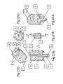
The bodies of the L4 (3) and L5 (1) components can have an articulation surface [AS]. The AS can match the contour of an AE (2) or there can be a difference in the shape and contact surfaces of the AS and the AE. The shape of the AS can be different in the L4 and L5 components. The AS can additionally have one or many recesses or wells to provide areas where there is no contact with the AE. This embodiment shows a single recess or well at the point of maximal axial contact. The AE (2) with an AS with a central recess or well will then rest on a ring or larger surface area than a point when the surfaces of the AS and the AE are not congruent.
The well can also be used to trap wear debris (patent pending). The trap can also be filled with a material that accepts and sequesters the wear debris as it is forced into the material during motion or load such as PTFE (teflon). PTFE is softer than UHMWPE and also acts as a coating to the surface that glides over the PTFE in the well. The PTFE is displaced up and out of the well as the volume of the well is filled with wear debris of any type. Any other suitable material can be used to be used in the well to trap the UHMWPE and/or metal, etc., wear debris. Magnetic material can be used in a well to trap metal ions from metal-metal articulations especially, Fe, Co, Ni and Cr and the like that can be influenced by magnetic fields.
The AE is placed through the bone core hole after the L5 component is fixed to the bone. The AE (2) in this embodiment is substantially ellipsoid or discoid. It can be made of any biocompatible material that can accept the stresses applied. It can have symmetric or asymmetric surfaces such as upper and lower. It can be made of a substantially hard material such as metal, ceramic, plastic, carbon based material and the like. It can be made of a viscoelastic material, an elastic material or any other biocompatible or biologic material with desired physical properties (i.e. a hydrogel). The AE can be solid or have one or more voids. The voids can be filled with other materials such as solids, liquids or gases. The AE can be made of any appropriate material, preferably a metal. It can be made of NiTi or any other memory or smart material. It can be a plastic, a ceramic, a carbon-based material or any other biocompatible material. The size of the AE is dependent on the amount of flexion desired. Larger dimension in the axial direction allows more flexion by greater separation of the components. The shape can be such that it controls or limits any motion in six degrees of motion by its interaction with the contour of the articulating surface of L4 and L5. (i.e. anterior/posterior, medial/lateral, radial tilt, etc.). The AE can be placed in any appropriate position between/on or in the other components. There can be one or more than one AE disposed at the interface. The multiple AE elements can be different size, shape and/or material. The embodiments with more than one AE can have the AEs distributed in any fashion or pattern.
The L4 component is placed through the bone core hole to engage the AE After assembly the L4 component is adjusted to the proper depth and pressure with a torque driver or the like. Flexion of the spine at the treated level is tested before closure.
Specifically FIG. 24 shows a L5 component (1) (see detail of FIG. 26 ) that utilizes the crown and post fixation. There are two anti-rotational elements on the body of the component. The L5 component has an axial guide/support. The L5 component is fenestrated. The L5 implant can be made of any appropriate material, preferably a metal. It can be made of NiTi or any other memory or smart material. It can be a plastic, a ceramic, a carbon-based material or any other biocompatible material. The L5 component has two fixation screws (2, 3). The L5 articulating surface (4) attaches to the L5 FE by Morris taper and anti-rotational elongations in the AS. The AS of the L4 and L5 has a well or wells to collect wear debris and increase the contact area with the AE. The AS contour is not a line fit with the AE. The AE in this embodiment has an offset surface (OS) (patent pending). An OS has a larger surface radius (R1) than the center of rotation (R2). The R1 of the AE substantially matches at least a portion the R1 of the AS. This allows less constraint and greater contact area.
The L4 and L5 AS can additionally have an array of magnetic material (Hyde, U.S. Pat. No. 6,387,096) disposed in the flanges that interact substantially in repulsion, attraction or both. The magnetic arrays provide a dynamic interface to absorb energy in flexion and axial loads. The magnetic arrays also can provide stability. The AE can be magnetized independently or indirectly by contact with a magnetic material.
The articulating surface (6) of this L4 implant has an elongation (7) that keys into the body of L4 (8). The elongation (7) can be modular from the articulating surface (6). There are two fixation screws (9, 10). One (9) interlocks 7 with 8 and fixes the L4 body to the bone. Screw (10) fixes the body (8) to the bone.
It is understood by those familiar with the art that a more complicated implant requires more steps and presents greater difficulty for the surgeon and requires more time for implantation. Likewise it is understood that the fixation, stability, structural integrity, articulation motion and durability are important. Design elements will be chosen for particular embodiments that balance these factors depending on the demand and spinal level (i.e. cervical, thoracic and lumbar).
The methods for treating spinal pathology according to the invention include not only artificial disc implants but also nucleus pulposa implant, fusion with a special implant, tumor treatment, vertebralplasty and other procedures in which the treatment region is advantageously reached by the described transosseous core approach.
It is to be understood that, while various embodiments of the invention have been described in conjunction with the detailed description thereof, the foregoing is intended only to illustrate and not to limit the scope of the present invention, which is defined by the scope of the specification and by future claims. Other equivalent embodiments, aspects, advantages, and modifications are within the scope of the specification and claims to be made.
Claims (36)
1. A transosseous method for treating the spine, comprising:
making an incision through tissue to expose one or more vertebrae,
positioning a guide pin laterally in the vertebral body spaced from posterior features of a vertebra containing at least in part a site of interest for intervention;
advancing a rounded cutting instrument over the guide pin directly into the vertebral body of the vertebra containing the site of interest, the cutting instrument having a diameter extending substantially between the end plates of said vertebra;
cutting a bone aperture, the aperture having a rounded opening with a dimension extending substantially between the vertebral end plates into the bone of a single vertebra to serve as an access window, and removing bone material, to reach the site of interest through the bone aperture,
performing an intervention at the site of interest, and
reimplementing at least a portion of the bone material in the rounded opening.
2. The method of claim 1 , wherein the bone aperture is made in a vertebra in the lumbar region of the spine.
3. The method of claim 2 , wherein the bone aperture is approximately 0.8″ in diameter.
4. The method of claim 1 , wherein the incision is made and the bone aperture is cut from a generally posterior side of the patient.
5. The method of claim 1 , further including, following provision of the bone hole aperture, introducing a second cutter into the vertebra through the bone aperture, and, using the second cutter, making a second cut in the vertebra substantially along a different axis relative to the axis of the bone aperture.
6. The method of claim 5 , wherein the different axis is generally perpendicular to the axis of the bone aperture and generally axial to the spine.
7. The method of claim 5 , further including continuing the second cut into the disc space between two vertebrae.
8. The method of claim 7 , further including removing loose material produced by the culling.
9. The method of claim 8 , wherein the step of removing loose material includes removal of at least part of a disc between the vertebrae.
10. The method of claim 1 , wherein the step of cutting a bone aperture comprises cutting into the vertebra at an angle of about 40° to 70° to the patient's sagittal plane.
11. The method of claim 1 , wherein the cuffing of the bone aperture is made with an instrument capable of cutting a bone core and wherein the step of removing the bone material includes removing a bone core.
12. The method of claim 1 , wherein the cutting of the bone hole is made with an instrument capable of cutting a bone core and wherein the step of removing the bone material includes removing a bone core.
13. The method of claim 1 , wherein the intervention comprises implanting a spine prosthesis, and including attaching the spine prosthesis to the vertebrae in which the bone aperture is formed.
14. The method of claim 13 , wherein the spine prosthesis is substantially modular and includes at least two components, and the method includes fixing at least one of the components to the bone of at least one of two adjacent vertebrae, and including retaining the articular element between the two components.
15. The method of claim 14 , wherein the step of fixing the components comprises cementing the components to at least one of the two adjacent vertebrae.
16. The method of claim 14 , wherein the at least one of the components has bumps, flutes, spines or elongations for engaging against sides of a bone hole, and the step of fixing includes engaging the bumps, flutes, spines or elongations tightly against or into the bone at the bone hole.
17. The method of claim 14 , wherein the fixing step includes fastening at least one of the components to the bone by engaging screws, pins or rods between a component and a vertebral bone.
18. The method of claim 14 , wherein at least one of the components is secured to at least one of the vertebrae by press fit.
19. The method of claim 13 , wherein the spine prosthesis is a fusion device, a partial disc replacement, a disc replacement or includes an articular surface.
20. The method of claim 13 , wherein the spine prosthesis is substantially modular and includes at least two components and the method includes fixing at least one component to the bone of at least one of two adjacent vertebrae.
21. The method of claim 20 , wherein at least one of the components has screw threads which are screwed into the bone of at least one of the adjacent vertebrae.
22. The method of claim 20 , wherein the method includes connecting two of the components.
23. The method of claim 20 , wherein the spine prosthesis is substantially modular and includes at least two components and the method includes connecting two of the components by at least one connector component.
24. The method of claim 20 , wherein the components have surface textures for engaging the bone of a vertebral body, and the step of fixing includes engaging the surface textures against the bone.
25. The method of claim 20 , wherein the components have surface elements for engaging the bone of a vertebral body, and the step of fixing includes engaging the surface elements tightly against or into the bone.
26. The method of claim 20 , wherein the fixing step includes fastening at least one of the components to the bone.
27. The transosseous core method of claim 20 , wherein at least one of the components is secured to at least one of the vertebrae by a crown-post fixation technique.
28. The method of claim 1 , wherein positioning the guide pin comprises orienting the guide pin along an axis extending through the center of the vertebral body in a transverse plane at an angle of 40-75 degrees from a line through the center of the vertebral body and the spinous process.
29. A transosseous method for implanting a spinal prosthesis, comprising:
forming a first aperture through a vertebra in the vertebral body spaced from posterior features, at an oblique angle to the sagittal plane of the patient, and including cutting substantially to the center of the vertebral body,
removing material from the vertebral body to provide the first bone aperture,
forming a second hole generally along a different axis to the orientation of the bone aperture, and including cutting through the endplate of at least one vertebra into a space of nucleus pulposa between two adjacent vertebrae,
removing loose material left after the cutting,
inserting an implant through the first bone aperture and then into a substantially axial orientation and fixing the implant to two adjacent vertebrae, substantially in the second hole formed with the second cutter, and
closing the first bone aperture.
30. The method of claim 29 , wherein said first aperture is formed by entering the vertebra through the vertebral body between the endplates.
31. The method of claim 29 , wherein the first aperture is formed along a first axis and said different axis is a second axis.
32. The method of claim 31 , wherein the first aperture along the first axis directly intersects the second hole along the second axis.
33. The method of claim 32 , wherein the said cuffing into the space of the nucleus pulposa comprises cutting into the nucleus pulposa of the L4-5 disc space.
34. The method of claim 29 , wherein closing the first bone aperture comprises replacing material removed to provide the first aperture.
35. The method of claim 34 , wherein forming the first aperture comprises removing a bone core and closing the first aperture comprises replacing the bone core.
36. The method of claim 29 , wherein positioning the guide pin comprises orienting the guide pin along an axis extending through the center of the vertebral body in a transverse plane at an angle of 40-75 degrees from a line through the center of the vertebral body and the spinous process.
Priority Applications (2)
| Application Number | Priority Date | Filing Date | Title |
|---|---|---|---|
| US11/089,896 US7637927B2 (en) | 2004-03-25 | 2005-03-25 | Transosseous spine core approach method implant and instrumentation |
| PCT/US2006/010030 WO2006104746A2 (en) | 2005-03-25 | 2006-03-17 | Transosseous spinal core approach method implant and instrumentation |
Applications Claiming Priority (2)
| Application Number | Priority Date | Filing Date | Title |
|---|---|---|---|
| US52128104P | 2004-03-25 | 2004-03-25 | |
| US11/089,896 US7637927B2 (en) | 2004-03-25 | 2005-03-25 | Transosseous spine core approach method implant and instrumentation |
Publications (2)
| Publication Number | Publication Date |
|---|---|
| US20070118219A1 US20070118219A1 (en) | 2007-05-24 |
| US7637927B2 true US7637927B2 (en) | 2009-12-29 |
Family
ID=37053889
Family Applications (1)
| Application Number | Title | Priority Date | Filing Date |
|---|---|---|---|
| US11/089,896 Expired - Fee Related US7637927B2 (en) | 2004-03-25 | 2005-03-25 | Transosseous spine core approach method implant and instrumentation |
Country Status (2)
| Country | Link |
|---|---|
| US (1) | US7637927B2 (en) |
| WO (1) | WO2006104746A2 (en) |
Cited By (5)
| Publication number | Priority date | Publication date | Assignee | Title |
|---|---|---|---|---|
| US20090076555A1 (en) * | 2007-09-13 | 2009-03-19 | David Lowry | Transcorporeal spinal decompression and repair system and related method |
| US20140025122A1 (en) * | 2012-07-17 | 2014-01-23 | Fellowship of Orthopaedic Reseachers, LLC | Magnetically levitated spinous process implants and methods thereof |
| US10080571B2 (en) | 2015-03-06 | 2018-09-25 | Warsaw Orthopedic, Inc. | Surgical instrument and method |
| US11678995B2 (en) | 2018-07-20 | 2023-06-20 | Fellowship Of Orthopaedic Researchers, Inc. | Magnetic intervertebral disc replacement devices and methods thereof |
| US11793599B2 (en) | 2020-08-04 | 2023-10-24 | Mazor Robotics Ltd. | Surgical cleaning tool, systems, and methods |
Families Citing this family (12)
| Publication number | Priority date | Publication date | Assignee | Title |
|---|---|---|---|---|
| US8430882B2 (en) | 2007-09-13 | 2013-04-30 | Transcorp, Inc. | Transcorporeal spinal decompression and repair systems and related methods |
| US8163021B2 (en) * | 2007-11-27 | 2012-04-24 | Transcorp, Inc. | Methods and systems for repairing an intervertebral disc using a transcorporal approach |
| US8470043B2 (en) * | 2008-12-23 | 2013-06-25 | Benvenue Medical, Inc. | Tissue removal tools and methods of use |
| US9161773B2 (en) | 2008-12-23 | 2015-10-20 | Benvenue Medical, Inc. | Tissue removal tools and methods of use |
| US20100241231A1 (en) * | 2009-02-20 | 2010-09-23 | Marino James F | Intervertebral fixation device |
| EP2461771A4 (en) * | 2009-08-07 | 2014-01-29 | Ebi Llc | Toroid-shaped spinal disc |
| EP3540495A1 (en) | 2010-10-28 | 2019-09-18 | EndoChoice Innovation Center Ltd. | Optical systems for multi-sensor endoscopes |
| US10314605B2 (en) | 2014-07-08 | 2019-06-11 | Benvenue Medical, Inc. | Apparatus and methods for disrupting intervertebral disc tissue |
| US10022243B2 (en) | 2015-02-06 | 2018-07-17 | Benvenue Medical, Inc. | Graft material injector system and method |
| US10758286B2 (en) | 2017-03-22 | 2020-09-01 | Benvenue Medical, Inc. | Minimal impact access system to disc space |
| US11583327B2 (en) | 2018-01-29 | 2023-02-21 | Spinal Elements, Inc. | Minimally invasive interbody fusion |
| WO2019178575A1 (en) | 2018-03-16 | 2019-09-19 | Benvenue Medical, Inc. | Articulated instrumentation and methods of using the same |
Citations (15)
| Publication number | Priority date | Publication date | Assignee | Title |
|---|---|---|---|---|
| US4059115A (en) * | 1976-06-14 | 1977-11-22 | Georgy Stepanovich Jumashev | Surgical instrument for operation of anterior fenestrated spondylodessis in vertebral osteochondrosis |
| US5108404A (en) * | 1989-02-09 | 1992-04-28 | Arie Scholten | Surgical protocol for fixation of bone using inflatable device |
| US5827328A (en) | 1996-11-22 | 1998-10-27 | Buttermann; Glenn R. | Intervertebral prosthetic device |
| US5888226A (en) * | 1997-11-12 | 1999-03-30 | Rogozinski; Chaim | Intervertebral prosthetic disc |
| US5893889A (en) * | 1997-06-20 | 1999-04-13 | Harrington; Michael | Artificial disc |
| US20030105527A1 (en) * | 2001-12-03 | 2003-06-05 | Surgical Dynamics, Inc. | Apparatus for fusing adjacent bone structures |
| US6589281B2 (en) | 2001-01-16 | 2003-07-08 | Edward R. Hyde, Jr. | Transosseous core approach and instrumentation for joint replacement and repair |
| US20030199879A1 (en) * | 2002-04-22 | 2003-10-23 | Spranza Joseph John | Hardware for cutting bone cores |
| US20030204189A1 (en) * | 2000-02-16 | 2003-10-30 | Cragg Andrew H. | Axial spinal implant and method and apparatus for implanting an axial spinal implant within the vertebrae of the spine |
| US6761723B2 (en) | 2002-01-14 | 2004-07-13 | Dynamic Spine, Inc. | Apparatus and method for performing spinal surgery |
| US20050038514A1 (en) * | 1999-05-07 | 2005-02-17 | Helm Gregory A. | Method and system for fusing a spinal region |
| US20050113919A1 (en) * | 2000-02-16 | 2005-05-26 | Cragg Andrew H. | Prosthetic nucleus apparatus |
| US20050261684A1 (en) * | 2002-11-08 | 2005-11-24 | Shaolian Samuel M | Transpedicular intervertebral disk access methods and devices |
| US20060079898A1 (en) * | 2003-10-23 | 2006-04-13 | Trans1 Inc. | Spinal motion preservation assemblies |
| US7114501B2 (en) * | 2000-08-14 | 2006-10-03 | Spine Wave, Inc. | Transverse cavity device and method |
Family Cites Families (2)
| Publication number | Priority date | Publication date | Assignee | Title |
|---|---|---|---|---|
| US5895426A (en) * | 1996-09-06 | 1999-04-20 | Osteotech, Inc. | Fusion implant device and method of use |
| US7938836B2 (en) * | 2003-10-23 | 2011-05-10 | Trans1, Inc. | Driver assembly for simultaneous axial delivery of spinal implants |
-
2005
- 2005-03-25 US US11/089,896 patent/US7637927B2/en not_active Expired - Fee Related
-
2006
- 2006-03-17 WO PCT/US2006/010030 patent/WO2006104746A2/en active Application Filing
Patent Citations (16)
| Publication number | Priority date | Publication date | Assignee | Title |
|---|---|---|---|---|
| US4059115A (en) * | 1976-06-14 | 1977-11-22 | Georgy Stepanovich Jumashev | Surgical instrument for operation of anterior fenestrated spondylodessis in vertebral osteochondrosis |
| US5108404A (en) * | 1989-02-09 | 1992-04-28 | Arie Scholten | Surgical protocol for fixation of bone using inflatable device |
| US5827328A (en) | 1996-11-22 | 1998-10-27 | Buttermann; Glenn R. | Intervertebral prosthetic device |
| US5893889A (en) * | 1997-06-20 | 1999-04-13 | Harrington; Michael | Artificial disc |
| US5888226A (en) * | 1997-11-12 | 1999-03-30 | Rogozinski; Chaim | Intervertebral prosthetic disc |
| US20050038514A1 (en) * | 1999-05-07 | 2005-02-17 | Helm Gregory A. | Method and system for fusing a spinal region |
| US20030204189A1 (en) * | 2000-02-16 | 2003-10-30 | Cragg Andrew H. | Axial spinal implant and method and apparatus for implanting an axial spinal implant within the vertebrae of the spine |
| US20050113919A1 (en) * | 2000-02-16 | 2005-05-26 | Cragg Andrew H. | Prosthetic nucleus apparatus |
| US7114501B2 (en) * | 2000-08-14 | 2006-10-03 | Spine Wave, Inc. | Transverse cavity device and method |
| US6589281B2 (en) | 2001-01-16 | 2003-07-08 | Edward R. Hyde, Jr. | Transosseous core approach and instrumentation for joint replacement and repair |
| US20030105527A1 (en) * | 2001-12-03 | 2003-06-05 | Surgical Dynamics, Inc. | Apparatus for fusing adjacent bone structures |
| US6761723B2 (en) | 2002-01-14 | 2004-07-13 | Dynamic Spine, Inc. | Apparatus and method for performing spinal surgery |
| US7303565B2 (en) | 2002-01-14 | 2007-12-04 | Dynamic Spine, Inc. | Apparatus and method for performing spinal surgery |
| US20030199879A1 (en) * | 2002-04-22 | 2003-10-23 | Spranza Joseph John | Hardware for cutting bone cores |
| US20050261684A1 (en) * | 2002-11-08 | 2005-11-24 | Shaolian Samuel M | Transpedicular intervertebral disk access methods and devices |
| US20060079898A1 (en) * | 2003-10-23 | 2006-04-13 | Trans1 Inc. | Spinal motion preservation assemblies |
Cited By (9)
| Publication number | Priority date | Publication date | Assignee | Title |
|---|---|---|---|---|
| US20090076555A1 (en) * | 2007-09-13 | 2009-03-19 | David Lowry | Transcorporeal spinal decompression and repair system and related method |
| US8323320B2 (en) * | 2007-09-13 | 2012-12-04 | Transcorp, Inc. | Transcorporeal spinal decompression and repair system and related method |
| US20140025122A1 (en) * | 2012-07-17 | 2014-01-23 | Fellowship of Orthopaedic Reseachers, LLC | Magnetically levitated spinous process implants and methods thereof |
| US10660674B2 (en) * | 2012-07-17 | 2020-05-26 | Gomboc, LLC | Magnetically levitated spinous process implants and methods thereof |
| US10080571B2 (en) | 2015-03-06 | 2018-09-25 | Warsaw Orthopedic, Inc. | Surgical instrument and method |
| US10667827B2 (en) | 2015-03-06 | 2020-06-02 | Warsaw Orthopedic, Inc. | Surgical instrument and method |
| US11653934B2 (en) | 2015-03-06 | 2023-05-23 | Warsaw Orthopedic, Inc. | Surgical instrument and method |
| US11678995B2 (en) | 2018-07-20 | 2023-06-20 | Fellowship Of Orthopaedic Researchers, Inc. | Magnetic intervertebral disc replacement devices and methods thereof |
| US11793599B2 (en) | 2020-08-04 | 2023-10-24 | Mazor Robotics Ltd. | Surgical cleaning tool, systems, and methods |
Also Published As
| Publication number | Publication date |
|---|---|
| US20070118219A1 (en) | 2007-05-24 |
| WO2006104746A2 (en) | 2006-10-05 |
| WO2006104746A3 (en) | 2007-07-05 |
Similar Documents
| Publication | Publication Date | Title |
|---|---|---|
| US7637927B2 (en) | Transosseous spine core approach method implant and instrumentation | |
| JP5913727B2 (en) | Interbody spine prosthetic orthopedic fixation device using self-expanding anchors | |
| US6042582A (en) | Instrumentation and method for facilitating insertion of spinal implant | |
| JP5602739B2 (en) | Intervertebral implant having blades for connection to adjacent vertebral bodies | |
| US7963991B2 (en) | Spinal implants and methods of providing dynamic stability to the spine | |
| AU761014B2 (en) | Method and apparatus for spinal implant insertion | |
| US7794465B2 (en) | Artificial spinal discs and associated implantation instruments and methods | |
| US7766914B2 (en) | Adjustable drill guide | |
| US20130245765A1 (en) | Transcorporeal spinal decompression and repair systems and related methods | |
| US20070260320A1 (en) | Spinal system and method including lateral approach | |
| EP1370181A1 (en) | Instrumentation for implant insertion | |
| JP2009534130A (en) | Monorail system | |
| JP2005523100A (en) | Method and apparatus for placing an instrument in a disc | |
| US9211193B2 (en) | Prosthesis, system and method | |
| US20070244562A1 (en) | Spinal implants and methods of providing dynamic stability to the spine | |
| AU2016245772B2 (en) | Spinal stabilization device, system, and method of use | |
| CN110831548B (en) | System and method for performing a disc replacement procedure through a lateral approach | |
| US11826258B2 (en) | Transcorporeal spinal decompression and repair systems and related methods | |
| RU2324437C1 (en) | Method of surgical treatment of lumbar spine in osteochondrosis with segmental instability or in mild spondylolisthesis | |
| US20200146697A1 (en) | Surgical tools | |
| KR20230122791A (en) | Bone fixation device for spinal fusion and surgical tool set including the same | |
| EA001334B1 (en) | Fusion implant device and method of use |
Legal Events
| Date | Code | Title | Description |
|---|---|---|---|
| FPAY | Fee payment |
Year of fee payment: 4 |
|
| REMI | Maintenance fee reminder mailed | ||
| LAPS | Lapse for failure to pay maintenance fees |
Free format text: PATENT EXPIRED FOR FAILURE TO PAY MAINTENANCE FEES (ORIGINAL EVENT CODE: EXP.) |
|
| STCH | Information on status: patent discontinuation |
Free format text: PATENT EXPIRED DUE TO NONPAYMENT OF MAINTENANCE FEES UNDER 37 CFR 1.362 |
|
| FP | Lapsed due to failure to pay maintenance fee |
Effective date: 20171229 |