US7280670B2 - Image confirming method - Google Patents
Image confirming method Download PDFInfo
- Publication number
- US7280670B2 US7280670B2 US10/899,064 US89906404A US7280670B2 US 7280670 B2 US7280670 B2 US 7280670B2 US 89906404 A US89906404 A US 89906404A US 7280670 B2 US7280670 B2 US 7280670B2
- Authority
- US
- United States
- Prior art keywords
- image
- images
- group
- image data
- workstation
- Prior art date
- Legal status (The legal status is an assumption and is not a legal conclusion. Google has not performed a legal analysis and makes no representation as to the accuracy of the status listed.)
- Expired - Fee Related, expires
Links
Images


Classifications
-
- G—PHYSICS
- G06—COMPUTING; CALCULATING OR COUNTING
- G06T—IMAGE DATA PROCESSING OR GENERATION, IN GENERAL
- G06T7/00—Image analysis
- G06T7/0002—Inspection of images, e.g. flaw detection
- G06T7/0012—Biomedical image inspection
-
- H—ELECTRICITY
- H04—ELECTRIC COMMUNICATION TECHNIQUE
- H04N—PICTORIAL COMMUNICATION, e.g. TELEVISION
- H04N1/00—Scanning, transmission or reproduction of documents or the like, e.g. facsimile transmission; Details thereof
- H04N1/00912—Arrangements for controlling a still picture apparatus or components thereof not otherwise provided for
- H04N1/00957—Compiling jobs, e.g. for batch processing
-
- H—ELECTRICITY
- H04—ELECTRIC COMMUNICATION TECHNIQUE
- H04N—PICTORIAL COMMUNICATION, e.g. TELEVISION
- H04N1/00—Scanning, transmission or reproduction of documents or the like, e.g. facsimile transmission; Details thereof
- H04N1/32—Circuits or arrangements for control or supervision between transmitter and receiver or between image input and image output device, e.g. between a still-image camera and its memory or between a still-image camera and a printer device
- H04N1/32358—Circuits or arrangements for control or supervision between transmitter and receiver or between image input and image output device, e.g. between a still-image camera and its memory or between a still-image camera and a printer device using picture signal storage, e.g. at transmitter
- H04N1/324—Circuits or arrangements for control or supervision between transmitter and receiver or between image input and image output device, e.g. between a still-image camera and its memory or between a still-image camera and a printer device using picture signal storage, e.g. at transmitter intermediate the transmitter and receiver terminals, e.g. at an exchange
-
- G—PHYSICS
- G06—COMPUTING; CALCULATING OR COUNTING
- G06T—IMAGE DATA PROCESSING OR GENERATION, IN GENERAL
- G06T2207/00—Indexing scheme for image analysis or image enhancement
- G06T2207/10—Image acquisition modality
- G06T2207/10072—Tomographic images
-
- G—PHYSICS
- G06—COMPUTING; CALCULATING OR COUNTING
- G06T—IMAGE DATA PROCESSING OR GENERATION, IN GENERAL
- G06T2207/00—Indexing scheme for image analysis or image enhancement
- G06T2207/10—Image acquisition modality
- G06T2207/10116—X-ray image
-
- G—PHYSICS
- G06—COMPUTING; CALCULATING OR COUNTING
- G06T—IMAGE DATA PROCESSING OR GENERATION, IN GENERAL
- G06T2207/00—Indexing scheme for image analysis or image enhancement
- G06T2207/30—Subject of image; Context of image processing
- G06T2207/30004—Biomedical image processing
Definitions
- the present invention relates to a method of confirming an image based on image data prior to transfer of the image data to a predetermined destination.
- CTR Computer Radiography
- CT Computer Tomography
- MRI Magnetic Resonance Imaging
- An image (image data) generated by a modality of such a kind is used in a medical facility for diagnosis of a lesion or injury, or the degree thereof, by being displayed on a CRT display (hereinafter simply called “CRT”) or output on a film by an LP (Laser Printer).
- CRT CRT display
- LP Laser Printer
- a CR apparatus herein referred to means a radiation image reading recording apparatus.
- a CR apparatus uses a stimulable phosphor which emits light upon exposure to a stimulating ray such as visible light or an infrared ray in accordance with radiation energy stored in a stimulable phosphor sheet. Radiation image information recorded in the stimulable phosphor sheet is obtained by scanning the sheet with a stimulating ray to cause the sheet to emit light, and the emitted light is read photoelectrically as an image signal by the CR apparatus.
- a stimulating ray such as visible light or an infrared ray
- a modality is set as an image data acquiring apparatus, and an LP is connected to the image data acquiring apparatus as an output apparatus.
- a diagnostic image reading terminal for displaying an image for diagnostic purposes is placed as an apparatus to receive the image in a room remote from the location of the image data acquiring apparatus.
- the image data acquiring apparatus and the image reading terminal are connected via a network so that medical doctors can carry out diagnosis at a location remote from the image data acquiring apparatus.
- image data obtained by the image data acquiring apparatus are transferred automatically to the image reading terminal connected to the network, and image confirmation based on the obtained image data prior to the transfer has rarely been carried out. Therefore, even when there is a mistake in photographing, or photographing has not been carried out at an appropriate density, for example, image data having such a problem are all transferred to the image reading terminal, and the problem is found only at the time of diagnosis, which causes a problem with the diagnosis.
- An object of the present invention is to provide an image confirming method wherein the quality of an image is confirmed based on image data, and not only image quality judgment which cannot be obtained through confirmation of each image upon transfer of the image data to an image reading terminal but also easy specification, transfer, and display of confirmed image data are possible.
- An image confirming method of the present invention comprises:
- a grouping step wherein a plurality of images are classified into image groups each of which has a common characteristic
- a confirmation step wherein, regarding each image group including a plurality of images and classified in the grouping step, quality of the images in the image group is confirmed by referring to other images in the image group, and is characterized by the fact that predetermined processing can be carried out on the image group having been confirmed.
- the “group classified in the grouping step” includes a group comprising only one image classified in the grouping step and having no other images with common characteristics.
- all images can be included in one group or only one image group may have a plurality of images.
- the case where image quality is judged only regarding the only image group is included as a case of judgment “regarding each image group”.
- Another image confirming method of the present invention comprises:
- a first grouping step wherein, regarding image classes each of which includes a plurality of images, the images included in each image class are grouped into image subgroups each of which has a common characteristic;
- a second grouping step wherein, regarding the image subgroups grouped in the first grouping step, image subgroups having the same characteristic of the images included therein are grouped together to form an image group, and an image subgroup having no other image subgroups with common characteristics remains as it is;
- a confirmation step wherein, regarding each image group grouped in the second grouping step and having a plurality of images, quality of each image therein is confirmed by referring to other images in the image group; and is characterized by the fact that
- predetermined processing can be carried out on each image group or subgroup grouped in the second grouping step.
- the “image subgroups grouped in the first grouping step” includes an image subgroup classified in the first grouping step comprising only one image and having no other images with common characteristics.
- the “characteristic” in the above means the kind of image such as a patient's name, an object of examination (a class medical check-up, or a close examination or the like), an examined body portion (chest, stomach, for example), and the like.
- the groups grouped in the grouping step are the groups including images of the same patient name, or the same examination object, or the same body portion.
- To confirm image quality by “referring to other images in the image group” means to carry out image quality confirmation which cannot be carried out by observation of a single image. For example, it means to confirm whether or not all images in the image group have desired image quality by observing all the images in the image group or by comparison between each of the images (for example, judgment as to whether one image needs sharper contrast or as to whether the density of all images needs to be higher), or to confirm whether or not all the images are sufficient for diagnosis (for example, judgment as to whether images of different body portions or at different angles are necessary).
- the “predetermined processing” can be any processing as long as the processing is appropriate to process all images in the image group.
- the processing means processing such that, by specifying a group including a plurality of images classified by the name of a patient, an object of examination, a body portion to be examined or the like, all images can be selected, displayed, or transferred if image selection, image display, or image transfer is instructed.
- image data obtained by image data acquiring apparatus are classified into image groups each of which has a common characteristic (such as the name of a patient or an object of examination) and when a plurality of images exist in an image group, the images are confirmed by overall observation or comparison between each other. Therefore, image confirmation which cannot be obtained by observation of a single image can be carried out. Furthermore, it is possible to deal with all confirmed images collectively as a group. Therefore, image specification or transfer can be carried out in a unit of image group, and it becomes possible to specify, transfer, or display the confirmed image data easily.
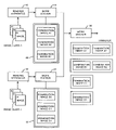
- FIG. 1(A) is a diagram showing a configuration of a medical network system adopting an image confirming method of the present invention
- FIG. 1(B) is a diagram showing a workstation and its peripherals explaining the image confirming method
- FIG. 2 is a diagram showing workstations and their peripherals explaining another embodiment of the image confirming method.
- FIG. 3 is a diagram showing workstations and their peripherals explaining still another embodiment of the image confirming method.
- FIG. 1(A) is a diagram showing an outline configuration of a medical network system adopting a first embodiment of the image confirming method of the present invention.
- the medical network system comprises an image data acquiring apparatus (reading apparatus) 10 which obtains image data representing an image, an image reading terminal (workstation) 20 which is connected to the reading apparatus 10 and displays the image based on the image data input from the reading apparatus, and a transfer destination apparatus 40 which is connected to the workstation 20 via the network.
- the workstation 20 and the transfer destination apparatus 40 are connected by connecting a connection point x of the workstation 20 and a connection point y of the transfer destination apparatus 40 .
- the image confirming method of the present invention is applicable not only to a network having a connection of such a kind but also to a network having a direct connection, not via a network, between the workstation 20 and the transfer destination terminal 40 .
- FIG. 1(A) depicts the state wherein only one apparatus is connected as the reading apparatus 10 . However, a plurality of apparatuses of different kinds or for different objects of examination are actually connected.
- a recording apparatus 50 for temporarily storing image data input from the reading apparatus 10 is connected to the workstation 20 .
- the recording apparatus 50 may be an image server such as a web server connected via the network.
- the reading apparatus 10 obtains image data carrying a subject image by photographing the subject.
- the reading apparatus 10 has an image processing function and carries out image processing using a predetermined image processing condition on the obtained image data.
- the image data after the image processing are input to the workstation 20 .
- the workstation 20 temporarily stores the image data input from the reading apparatus 10 in the recording apparatus 50 .
- necessary image data are read from the recording apparatus 50 , and the image based on the image data is displayed on a CRT (not shown).
- An operator confirms whether or not the displayed image has predetermined quality while viewing the image (the detail will be explained later), and only image data having no problem are transferred to the transfer destination apparatus 40 via the network.
- the recording apparatus 50 stores multitudes of image data obtained by the reading apparatus 10 .
- 3 images A 1 , A 2 , and B 1 are photographed by the reading apparatus 10 and image data carrying the images are stored in the recording apparatus 50 .
- the images are obtained from one patient.
- the image A 1 is a first image of stomach fluoroscopy (examination A) and the image A 2 is a second image of the same examination (photographed at a position different from the position of the first image).
- the image B 1 is a first image of a chest X-ray examination (examination B).
- the workstation 20 classifies the image data carrying the 3 images A 1 , A 2 and B 1 stored in the recording apparatus 50 into a group of the examination A (group A) and a group of the examination B (group B).
- This process corresponds to a step of grouping according to the “examination objects” as the “common characteristic”.
- header information whereby the object of the examination (examination A or B) can be understood, has been added to each image data.
- the grouping of the image data having been read is carried out by referring to the header information.
- a database may be generated in advance so that a location of each image can be searched for and the grouping is carried out by referring to the database. Reading the image data at the time of display thereof may be carried out based on the search of the database.
- the image confirming method of the present invention it becomes possible to identify a problem which cannot be identified by observing the image A 1 or A 2 respectively, and images enabling more precise diagnosis can be transferred to the transfer destination apparatus 40 .
- the group B has only the one image B 1 , and the image confirming method of the present invention is not carried out thereon. However, it is needless to say that confirmation of a single image which has been carried out conventionally is carried out on that one image.
- the medical network system in the above configuration has only one connection between the reading apparatus 10 and the workstation 20 . It is also possible to have a plurality of connections between reading apparatuses and workstations for carrying out image confirmation.
- FIG. 2 shows this aspect.
- a medical network system in FIG. 2 comprises reading apparatuses 10 and 12 for obtaining image data representing images, a workstation 20 which is connected to the reading apparatus 10 and displays the images based on the image data input from the reading apparatus 10 , a workstation 22 which is connected to the reading apparatus 12 and displays the images based on the image data input from the reading apparatus 12 , a workstation 30 which displays the images based on the image data input from both workstations 20 and 22 , and a transfer destination apparatus 40 connected to the workstation 30 (the connection mode is the same as in FIG. 1(A) and not shown in FIG. 2 ).
- the reading apparatuses 10 and 12 respectively photograph subjects and acquire image data carrying the subject images.
- the reading apparatus 10 carries out predetermined image processing using a predetermined image processing condition on the obtained image data and inputs the image data after the image processing to the workstation 20 .
- the reading apparatus 12 inputs the image data after the image processing to the workstation 22 .
- the workstation 20 temporarily stores the image data input from the reading apparatus 10 in a recording apparatus 50 connected to the workstation 20 .
- the workstation 22 temporarily stores the image data input from the reading apparatus 12 in a recording apparatus 52 connected to the workstation 22 .
- 3 images A 1 , A 2 , and B 1 are photographed by the reading apparatus 10 and image data representing the images are stored in the recording apparatus 50 .
- 3 images B 2 , C 1 , and D 1 (an image class 2) are photographed by the reading apparatus 12 and image data representing the images are stored in the recording apparatus 52 .
- the image A 1 is a first image of stomach fluoroscopy (examination A) and the image A 2 is a second image of the same examination (photographed at a position different from the position of the first image).
- the image B 1 is a first image of a chest X-ray examination (examination B).
- the image B 2 is a second image of the chest X-ray examination (examination B) and the image C 1 is a first image of an examination C (different from the examinations A and B).
- the image D 1 is a first image of an examination D (different from the examinations A, B and C).
- the workstation 30 reads, via the workstation 20 , image data representing the 3 images A 1 , A 2 and B 1 of the image class 1 stored in the recording apparatus 50 , and reads, via the workstation 22 , image data representing the 3 images B 2 , C 1 and D 1 of the image class 2 stored in the recording apparatus 52 . All image data having been read are grouped into an examination-A group (an image group A), an examination-B group (an image group B), an examination-C group (an image group C) and an examination-D group (an image group D). This process corresponds to a step of grouping according to the “objects of examination” as the “common characteristic”.
- image confirmation is carried out on the groups A and B each of which includes a plurality of images.
- the images B 1 and B 2 were photographed in different environments (for example, by different photographing apparatuses or on a different photographing date)
- confirmation of respective image B 1 or B 2 is carried out by comparison between the images B 1 and B 2 .
- image confirmation is carried out in the same manner as has been described above, and the images A 1 and A 2 having been confirmed are collectively transferred to the transfer destination apparatus 40 .
- the transfer destination apparatus 40 having received the images in the image groups A and B can obtain the images A 1 and A 2 in the image group A or images B 1 and B 2 in the image group B by simply specifying the image group A or B. It is not necessary for the transfer destination apparatus 40 to respectively specify the images A 1 and A 2 or the images B 1 and B 2 .
- FIG. 3 shows an outline of peripherals of workstations in a medical network system adopting the second embodiment of image confirming method.
- the medical network system comprises reading apparatuses 10 and 12 for obtaining image data carrying images, a workstation 20 which is connected to the reading apparatus 10 and displays the image based on the image data input from the reading apparatus 10 , a workstation 22 which is connected to the reading apparatus 12 and displays the image based on the image data input from the reading apparatus 12 , a workstation 30 which displays images based on the image data input from both workstations 20 and 22 , and a transfer destination apparatus 40 connected to the workstation 30 via the network (the connection mode is the same as in FIG. 1(A) and not shown in FIG. 3 ).
- the reading apparatuses 10 and 12 respectively photograph subjects and acquire image data representing the subject images.
- the reading apparatus 10 carries out predetermined image processing using a predetermined image processing condition on the obtained image data and inputs the image data after the image processing to the workstation 20 .
- the reading apparatus 12 inputs the image data after the image processing to the workstation 22 .
- the workstation 20 temporarily stores the image data input from the reading apparatus 10 in a recording apparatus 50 connected to the workstation 20 .
- the workstation 22 temporarily stores the image data input from the reading apparatus 12 in a recording apparatus 52 connected to the workstation 22 .
- 3 images A 1 , A 2 , and B 1 are photographed by the reading apparatus 10 and image data representing the images are stored in the recording apparatus 50 .
- 3 images B 2 , C 1 , and D 1 (an image class 2) are photographed by the reading apparatus 12 and image data representing the images are stored in the recording apparatus 52 .
- the image A 1 is a first image of stomach fluoroscopy (examination A) and the image A 2 is a second image of the same examination (photographed at a position different from the position of the first image).
- the image B 1 is a first image of a chest X-ray examination (examination B).
- the image B 2 is a second image of the chest X-ray examination (examination B) and the image C 1 is a first image of an examination C (different from the examinations A and B).
- the image D 1 is a first image of an examination D (different from the examinations A, B and C).
- the workstation 20 classifies the image data representing the 3 images A 1 , A 2 , and B 1 in the image class 1 stored in the recording apparatus 50 into an examination-A group (a subgroup A) and an examination-B group (a subgroup B).
- the workstation 22 classifies the image data representing the 3 images B 2 , C 1 , and D 1 in the image class 2 stored in the recording apparatus 52 into an examination-B group (a subgroup B), an examination-C group (a subgroup C), and an examination-D group (a subgroup D). This process corresponds to the first grouping step of each image class using the “object of examination” as the “common characteristic”.
- the image data classified into the subgroups are input to the workstation 30 as the image subgroups and stored in a recording apparatus which is not shown in FIG. 3 .
- the image data may be input to the workstation 30 after the confirmation of the images grouped in the first grouping step, in the same manner as in the first embodiment.
- the workstation 30 classifies the subgroups A and B in the class 1 and the subgroups B, C, and D in the class 2 classified at the first grouping step into groups of images having a common characteristic by integrating the above subgroups, and leaves the subgroups as they are when the subgroups have no other images of common characteristic in other subgroups (this process corresponds to the second grouping step). Therefore, since there are no subgroups having images of the examination A in the class 2, the subgroup A in the class 1 stays as it is, having images A 1 and A 2 .
- the subgroup B including the image B 1 in the class 1 and the subgroup B including the image B 2 in the class 2 have the images of the examination B. Therefore, they are integrated as a group B.
- the subgroups C and D in the class 2 remain as they are, having one image each (C 1 or D 1 ) in the subgroups.
- information showing the object of the examination may be recorded as header information or the like corresponding to each subgroup, for example.
- a database may be generated.
- image quality confirmation is carried out on all images in the groups A and B including a plurality of images (images A 1 and A 2 , and images B 1 and B 2 ).
- the images B 1 and B 2 were photographed in different environments (by different photographing apparatuses or on different date for example), the images B 1 and B 2 are confirmed not only by observation of each image but also by comparison between both images so that a problem which cannot be identified by a simple observation of each image can be identified. Only when the image data having been obtained are judged to be appropriate, the images B 1 and B 2 having been confirmed are transferred collectively as the images in the image group B to the transfer destination apparatus 40 .
- the images are confirmed by the same method as in the first embodiment, and the images A 1 and A 2 having been confirmed are transferred collectively as the images in the image group A to the transfer destination apparatus 40 .
- the transfer destination apparatus 40 having received the images of groups A and B can obtain images A 1 and A 2 included in the group A or images B 1 and B 2 included in the group B, only by specifying the group A or B. It is thus not necessary for the transfer destination apparatus 40 to respectively specify the images A 1 and A 2 or images B 1 and B 2 .
- the groups C and D have only one image each (image C 1 or D 1 ), and the image confirming method of the present invention is not applicable thereto. However, it is needless to say that a conventional confirmation of one image is carried out on those single images.
Abstract
Description
Claims (7)
Priority Applications (1)
| Application Number | Priority Date | Filing Date | Title |
|---|---|---|---|
| US10/899,064 US7280670B2 (en) | 1998-03-31 | 2004-07-27 | Image confirming method |
Applications Claiming Priority (4)
| Application Number | Priority Date | Filing Date | Title |
|---|---|---|---|
| JP8714498 | 1998-03-31 | ||
| JP87144/1998 | 1998-03-31 | ||
| US09/281,965 US6813365B1 (en) | 1998-03-31 | 1999-03-31 | Image confirming method |
| US10/899,064 US7280670B2 (en) | 1998-03-31 | 2004-07-27 | Image confirming method |
Related Parent Applications (1)
| Application Number | Title | Priority Date | Filing Date |
|---|---|---|---|
| US09/281,965 Continuation US6813365B1 (en) | 1998-03-31 | 1999-03-31 | Image confirming method |
Publications (2)
| Publication Number | Publication Date |
|---|---|
| US20040264738A1 US20040264738A1 (en) | 2004-12-30 |
| US7280670B2 true US7280670B2 (en) | 2007-10-09 |
Family
ID=33251574
Family Applications (2)
| Application Number | Title | Priority Date | Filing Date |
|---|---|---|---|
| US09/281,965 Expired - Lifetime US6813365B1 (en) | 1998-03-31 | 1999-03-31 | Image confirming method |
| US10/899,064 Expired - Fee Related US7280670B2 (en) | 1998-03-31 | 2004-07-27 | Image confirming method |
Family Applications Before (1)
| Application Number | Title | Priority Date | Filing Date |
|---|---|---|---|
| US09/281,965 Expired - Lifetime US6813365B1 (en) | 1998-03-31 | 1999-03-31 | Image confirming method |
Country Status (1)
| Country | Link |
|---|---|
| US (2) | US6813365B1 (en) |
Families Citing this family (5)
| Publication number | Priority date | Publication date | Assignee | Title |
|---|---|---|---|---|
| JP2002281210A (en) * | 2001-03-15 | 2002-09-27 | Konica Corp | Medical image processor and medical network system |
| DE102004031681A1 (en) * | 2004-06-30 | 2006-01-26 | Siemens Ag | Method and device for user-specific parameterization of a roentgen device |
| US20080120140A1 (en) * | 2006-11-22 | 2008-05-22 | General Electric Company | Managing medical imaging data |
| US8588534B2 (en) | 2011-05-06 | 2013-11-19 | Microsoft Corporation | Staged element classification |
| EP3073401A1 (en) * | 2015-03-27 | 2016-09-28 | Fujifilm Corporation | Failed image management apparatus, operation method of failed image management apparatus, and failed image management system |
Citations (16)
| Publication number | Priority date | Publication date | Assignee | Title |
|---|---|---|---|---|
| JPS62188580A (en) | 1986-02-14 | 1987-08-18 | Toshiba Corp | Automatic multiimage photographing device |
| US4767205A (en) | 1986-01-28 | 1988-08-30 | Flow Cytometry Standards Corporation | Composition and method for hidden identification |
| JPH0283615A (en) | 1988-09-20 | 1990-03-23 | Fuji Photo Film Co Ltd | Image output device |
| JPH02220629A (en) | 1989-02-22 | 1990-09-03 | Toshiba Corp | Reconstituted image taking system |
| US5019975A (en) | 1986-08-08 | 1991-05-28 | Fuji Photo Film Co., Ltd. | Method for constructing a data base in a medical image control system |
| US5235510A (en) * | 1990-11-22 | 1993-08-10 | Kabushiki Kaisha Toshiba | Computer-aided diagnosis system for medical use |
| JPH07136128A (en) | 1993-11-12 | 1995-05-30 | Konica Corp | Display apparatus for controller |
| US5464410A (en) | 1991-05-03 | 1995-11-07 | University Of Pittsburgh | Imaging fixation and localization system with respiratory feedback device |
| US5949752A (en) * | 1997-10-30 | 1999-09-07 | Wea Manufacturing Inc. | Recording media and methods for display of graphic data, text, and images |
| US5950045A (en) | 1997-06-20 | 1999-09-07 | Sharp Kabushiki Kaisha | Input device |
| US5995097A (en) | 1994-08-19 | 1999-11-30 | Fujitsu Limited | Method and apparatus for confirming matching of data in a distributed processing system |
| US6101263A (en) * | 1982-10-05 | 2000-08-08 | Canon Kabushiki Kaisha | Image processing apparatus |
| US6125194A (en) * | 1996-02-06 | 2000-09-26 | Caelum Research Corporation | Method and system for re-screening nodules in radiological images using multi-resolution processing, neural network, and image processing |
| US6188782B1 (en) | 1997-11-28 | 2001-02-13 | Eastman Kodak Company | Automatic editing method for a digital medical imaging unit |
| US6198837B1 (en) * | 1996-08-23 | 2001-03-06 | Konica Corporation | Method of storing medical image information recorded in sheet shaped recording medium |
| US6212299B1 (en) * | 1992-12-11 | 2001-04-03 | Matsushita Electric Industrial Co., Ltd. | Method and apparatus for recognizing a character |
-
1999
- 1999-03-31 US US09/281,965 patent/US6813365B1/en not_active Expired - Lifetime
-
2004
- 2004-07-27 US US10/899,064 patent/US7280670B2/en not_active Expired - Fee Related
Patent Citations (16)
| Publication number | Priority date | Publication date | Assignee | Title |
|---|---|---|---|---|
| US6101263A (en) * | 1982-10-05 | 2000-08-08 | Canon Kabushiki Kaisha | Image processing apparatus |
| US4767205A (en) | 1986-01-28 | 1988-08-30 | Flow Cytometry Standards Corporation | Composition and method for hidden identification |
| JPS62188580A (en) | 1986-02-14 | 1987-08-18 | Toshiba Corp | Automatic multiimage photographing device |
| US5019975A (en) | 1986-08-08 | 1991-05-28 | Fuji Photo Film Co., Ltd. | Method for constructing a data base in a medical image control system |
| JPH0283615A (en) | 1988-09-20 | 1990-03-23 | Fuji Photo Film Co Ltd | Image output device |
| JPH02220629A (en) | 1989-02-22 | 1990-09-03 | Toshiba Corp | Reconstituted image taking system |
| US5235510A (en) * | 1990-11-22 | 1993-08-10 | Kabushiki Kaisha Toshiba | Computer-aided diagnosis system for medical use |
| US5464410A (en) | 1991-05-03 | 1995-11-07 | University Of Pittsburgh | Imaging fixation and localization system with respiratory feedback device |
| US6212299B1 (en) * | 1992-12-11 | 2001-04-03 | Matsushita Electric Industrial Co., Ltd. | Method and apparatus for recognizing a character |
| JPH07136128A (en) | 1993-11-12 | 1995-05-30 | Konica Corp | Display apparatus for controller |
| US5995097A (en) | 1994-08-19 | 1999-11-30 | Fujitsu Limited | Method and apparatus for confirming matching of data in a distributed processing system |
| US6125194A (en) * | 1996-02-06 | 2000-09-26 | Caelum Research Corporation | Method and system for re-screening nodules in radiological images using multi-resolution processing, neural network, and image processing |
| US6198837B1 (en) * | 1996-08-23 | 2001-03-06 | Konica Corporation | Method of storing medical image information recorded in sheet shaped recording medium |
| US5950045A (en) | 1997-06-20 | 1999-09-07 | Sharp Kabushiki Kaisha | Input device |
| US5949752A (en) * | 1997-10-30 | 1999-09-07 | Wea Manufacturing Inc. | Recording media and methods for display of graphic data, text, and images |
| US6188782B1 (en) | 1997-11-28 | 2001-02-13 | Eastman Kodak Company | Automatic editing method for a digital medical imaging unit |
Also Published As
| Publication number | Publication date |
|---|---|
| US6813365B1 (en) | 2004-11-02 |
| US20040264738A1 (en) | 2004-12-30 |
Similar Documents
| Publication | Publication Date | Title |
|---|---|---|
| JP5459423B2 (en) | Diagnostic system | |
| CN102711603B (en) | Image diagnosing system, image diagnosing method, medical imaging server and medical imaging store method | |
| JP4408555B2 (en) | Image and information processing apparatus | |
| JP3506746B2 (en) | X-ray image signal processing and routing method | |
| US20100171682A1 (en) | Medical image display apparatus and computer readable medium | |
| US7853063B2 (en) | Method for producing a cropped medical image | |
| JPH06217966A (en) | Digital x-ray image quality control work- station which is operatable by manual mode and pass through mode | |
| Mattoon | Digital radiography | |
| US20030110178A1 (en) | Method and system of tracking medical films and associated digital images for computer-aided and diagnostic analysis | |
| JP2003248723A (en) | Medical image transfer device, hospital system provided with same device, and method for transferring medical image | |
| JP2004097651A (en) | Image information processor, network system for medical use, and program for the processor | |
| US7280670B2 (en) | Image confirming method | |
| JP2005056065A (en) | Medical image information processor and medical image information processing system | |
| JP3485339B2 (en) | X-ray image signal processing method | |
| JP2001285858A (en) | Method and instrument for photographing image | |
| US6431440B1 (en) | Radiation image reading method and radiation image reading apparatus | |
| JP3594250B2 (en) | PACS | |
| JP2000148894A (en) | Medical image information management mechanism | |
| JP2002200063A (en) | X-ray imaging device, device and method for control thereof, and x-ray imaging system | |
| JPH11232161A (en) | Image data transfer system | |
| US7024029B2 (en) | Medical image processing method and apparatus | |
| JP2008220482A (en) | Diagnosis support system | |
| US6381348B2 (en) | Network system for medical images | |
| JP3471247B2 (en) | Parameter management method and parameter management system | |
| US8156210B2 (en) | Workflow for computer aided detection |
Legal Events
| Date | Code | Title | Description |
|---|---|---|---|
| AS | Assignment |
Owner name: FUJIFILM CORPORATION, JAPAN Free format text: ASSIGNMENT OF ASSIGNORS INTEREST;ASSIGNOR:FUJIFILM HOLDINGS CORPORATION (FORMERLY FUJI PHOTO FILM CO., LTD.);REEL/FRAME:018904/0001 Effective date: 20070130 Owner name: FUJIFILM CORPORATION,JAPAN Free format text: ASSIGNMENT OF ASSIGNORS INTEREST;ASSIGNOR:FUJIFILM HOLDINGS CORPORATION (FORMERLY FUJI PHOTO FILM CO., LTD.);REEL/FRAME:018904/0001 Effective date: 20070130 |
|
| STCF | Information on status: patent grant |
Free format text: PATENTED CASE |
|
| FEPP | Fee payment procedure |
Free format text: PAYOR NUMBER ASSIGNED (ORIGINAL EVENT CODE: ASPN); ENTITY STATUS OF PATENT OWNER: LARGE ENTITY |
|
| FPAY | Fee payment |
Year of fee payment: 4 |
|
| FPAY | Fee payment |
Year of fee payment: 8 |
|
| FEPP | Fee payment procedure |
Free format text: MAINTENANCE FEE REMINDER MAILED (ORIGINAL EVENT CODE: REM.); ENTITY STATUS OF PATENT OWNER: LARGE ENTITY |
|
| LAPS | Lapse for failure to pay maintenance fees |
Free format text: PATENT EXPIRED FOR FAILURE TO PAY MAINTENANCE FEES (ORIGINAL EVENT CODE: EXP.); ENTITY STATUS OF PATENT OWNER: LARGE ENTITY |
|
| STCH | Information on status: patent discontinuation |
Free format text: PATENT EXPIRED DUE TO NONPAYMENT OF MAINTENANCE FEES UNDER 37 CFR 1.362 |
|
| FP | Lapsed due to failure to pay maintenance fee |
Effective date: 20191009 |